EP2853267A1 - Targeted interferon demonstrates potent apoptotic and anti-tumor activities - Google Patents
Targeted interferon demonstrates potent apoptotic and anti-tumor activities Download PDFInfo
- Publication number
- EP2853267A1 EP2853267A1 EP20140180412 EP14180412A EP2853267A1 EP 2853267 A1 EP2853267 A1 EP 2853267A1 EP 20140180412 EP20140180412 EP 20140180412 EP 14180412 A EP14180412 A EP 14180412A EP 2853267 A1 EP2853267 A1 EP 2853267A1
- Authority
- EP
- European Patent Office
- Prior art keywords
- ifn
- her2
- igg3
- interferon
- cancer
- Prior art date
- Legal status (The legal status is an assumption and is not a legal conclusion. Google has not performed a legal analysis and makes no representation as to the accuracy of the status listed.)
- Granted
Links
- 229940079322 interferon Drugs 0.000 title claims abstract description 86
- 102000014150 Interferons Human genes 0.000 title claims abstract description 85
- 108010050904 Interferons Proteins 0.000 title claims abstract description 85
- 230000000259 anti-tumor effect Effects 0.000 title description 32
- 230000003389 potentiating effect Effects 0.000 title description 14
- 230000001640 apoptogenic effect Effects 0.000 title description 7
- 206010028980 Neoplasm Diseases 0.000 claims abstract description 250
- 108010047761 Interferon-alpha Proteins 0.000 claims abstract description 176
- 102000006992 Interferon-alpha Human genes 0.000 claims abstract description 175
- 201000011510 cancer Diseases 0.000 claims description 123
- 210000004881 tumor cell Anatomy 0.000 claims description 56
- 238000011282 treatment Methods 0.000 claims description 52
- 108090000765 processed proteins & peptides Proteins 0.000 claims description 47
- 102100022005 B-lymphocyte antigen CD20 Human genes 0.000 claims description 35
- 101000897405 Homo sapiens B-lymphocyte antigen CD20 Proteins 0.000 claims description 35
- 229960004641 rituximab Drugs 0.000 claims description 31
- 239000000203 mixture Substances 0.000 claims description 29
- 230000035755 proliferation Effects 0.000 claims description 28
- 101150029707 ERBB2 gene Proteins 0.000 claims description 24
- 125000003275 alpha amino acid group Chemical group 0.000 claims description 23
- 238000009472 formulation Methods 0.000 claims description 15
- 108060003951 Immunoglobulin Proteins 0.000 claims description 14
- 102000018358 immunoglobulin Human genes 0.000 claims description 14
- 238000007920 subcutaneous administration Methods 0.000 claims description 13
- 230000002147 killing effect Effects 0.000 claims description 9
- 230000017854 proteolysis Effects 0.000 claims description 9
- 108090000467 Interferon-beta Proteins 0.000 claims description 8
- 239000008194 pharmaceutical composition Substances 0.000 claims description 8
- 239000000546 pharmaceutical excipient Substances 0.000 claims description 7
- 102000003996 Interferon-beta Human genes 0.000 claims description 6
- 229960001388 interferon-beta Drugs 0.000 claims description 6
- 238000001990 intravenous administration Methods 0.000 claims description 6
- 230000017066 negative regulation of growth Effects 0.000 claims description 5
- 238000007918 intramuscular administration Methods 0.000 claims description 3
- 238000007911 parenteral administration Methods 0.000 claims description 3
- OHDXDNUPVVYWOV-UHFFFAOYSA-N n-methyl-1-(2-naphthalen-1-ylsulfanylphenyl)methanamine Chemical compound CNCC1=CC=CC=C1SC1=CC=CC2=CC=CC=C12 OHDXDNUPVVYWOV-UHFFFAOYSA-N 0.000 abstract description 167
- 108020001507 fusion proteins Proteins 0.000 abstract description 138
- 102000037865 fusion proteins Human genes 0.000 abstract description 137
- 230000008685 targeting Effects 0.000 abstract description 73
- 239000000439 tumor marker Substances 0.000 abstract description 7
- 210000004027 cell Anatomy 0.000 description 193
- 108090000623 proteins and genes Proteins 0.000 description 81
- 101001012157 Homo sapiens Receptor tyrosine-protein kinase erbB-2 Proteins 0.000 description 80
- 102000004169 proteins and genes Human genes 0.000 description 72
- 235000018102 proteins Nutrition 0.000 description 71
- 102100030086 Receptor tyrosine-protein kinase erbB-2 Human genes 0.000 description 69
- 241000699670 Mus sp. Species 0.000 description 57
- 238000000034 method Methods 0.000 description 57
- 101710100963 Receptor tyrosine-protein kinase erbB-4 Proteins 0.000 description 56
- 102100029981 Receptor tyrosine-protein kinase erbB-4 Human genes 0.000 description 55
- 230000000694 effects Effects 0.000 description 36
- 230000006907 apoptotic process Effects 0.000 description 32
- 150000007523 nucleic acids Chemical class 0.000 description 31
- 101710100969 Receptor tyrosine-protein kinase erbB-3 Proteins 0.000 description 29
- 102000039446 nucleic acids Human genes 0.000 description 29
- 108020004707 nucleic acids Proteins 0.000 description 29
- 102100029986 Receptor tyrosine-protein kinase erbB-3 Human genes 0.000 description 28
- 241000282414 Homo sapiens Species 0.000 description 27
- 102000001301 EGF receptor Human genes 0.000 description 26
- 108060006698 EGF receptor Proteins 0.000 description 26
- 230000027455 binding Effects 0.000 description 26
- 235000001014 amino acid Nutrition 0.000 description 24
- 239000000427 antigen Substances 0.000 description 24
- 102000036639 antigens Human genes 0.000 description 24
- 108091007433 antigens Proteins 0.000 description 24
- 230000012010 growth Effects 0.000 description 24
- 208000003950 B-cell lymphoma Diseases 0.000 description 23
- 206010025323 Lymphomas Diseases 0.000 description 23
- 241001529936 Murinae Species 0.000 description 21
- 150000001413 amino acids Chemical class 0.000 description 21
- 230000004614 tumor growth Effects 0.000 description 20
- 239000013543 active substance Substances 0.000 description 19
- 102000004196 processed proteins & peptides Human genes 0.000 description 19
- 239000003550 marker Substances 0.000 description 18
- 238000000684 flow cytometry Methods 0.000 description 17
- 230000014509 gene expression Effects 0.000 description 17
- 238000001727 in vivo Methods 0.000 description 17
- 230000004927 fusion Effects 0.000 description 16
- 239000012634 fragment Substances 0.000 description 15
- 229920001184 polypeptide Polymers 0.000 description 15
- -1 EGF-R Proteins 0.000 description 14
- 239000003814 drug Substances 0.000 description 14
- 102000051957 human ERBB2 Human genes 0.000 description 14
- 238000004519 manufacturing process Methods 0.000 description 14
- XJMOSONTPMZWPB-UHFFFAOYSA-M propidium iodide Chemical compound [I-].[I-].C12=CC(N)=CC=C2C2=CC=C(N)C=C2[N+](CCC[N+](C)(CC)CC)=C1C1=CC=CC=C1 XJMOSONTPMZWPB-UHFFFAOYSA-M 0.000 description 14
- 108090000672 Annexin A5 Proteins 0.000 description 13
- 102000004121 Annexin A5 Human genes 0.000 description 13
- 238000004458 analytical method Methods 0.000 description 13
- 230000001965 increasing effect Effects 0.000 description 13
- 230000002401 inhibitory effect Effects 0.000 description 13
- 230000004083 survival effect Effects 0.000 description 13
- 230000001225 therapeutic effect Effects 0.000 description 13
- 102000004127 Cytokines Human genes 0.000 description 12
- 108090000695 Cytokines Proteins 0.000 description 12
- 241001465754 Metazoa Species 0.000 description 12
- 206010006187 Breast cancer Diseases 0.000 description 10
- 208000026310 Breast neoplasm Diseases 0.000 description 10
- 108020004414 DNA Proteins 0.000 description 10
- 241000699666 Mus <mouse, genus> Species 0.000 description 10
- 108010044012 STAT1 Transcription Factor Proteins 0.000 description 10
- 102100029904 Signal transducer and activator of transcription 1-alpha/beta Human genes 0.000 description 10
- 229940079593 drug Drugs 0.000 description 10
- 239000000463 material Substances 0.000 description 10
- 229920000642 polymer Polymers 0.000 description 10
- 238000002415 sodium dodecyl sulfate polyacrylamide gel electrophoresis Methods 0.000 description 10
- LFQSCWFLJHTTHZ-UHFFFAOYSA-N Ethanol Chemical compound CCO LFQSCWFLJHTTHZ-UHFFFAOYSA-N 0.000 description 9
- OKKJLVBELUTLKV-UHFFFAOYSA-N Methanol Chemical compound OC OKKJLVBELUTLKV-UHFFFAOYSA-N 0.000 description 9
- 241000711975 Vesicular stomatitis virus Species 0.000 description 9
- 239000002253 acid Substances 0.000 description 9
- 230000004913 activation Effects 0.000 description 9
- 238000007912 intraperitoneal administration Methods 0.000 description 9
- 201000001441 melanoma Diseases 0.000 description 9
- 238000003752 polymerase chain reaction Methods 0.000 description 9
- 239000013598 vector Substances 0.000 description 9
- 102100024222 B-lymphocyte antigen CD19 Human genes 0.000 description 8
- 101000980825 Homo sapiens B-lymphocyte antigen CD19 Proteins 0.000 description 8
- 102100034256 Mucin-1 Human genes 0.000 description 8
- 125000003178 carboxy group Chemical group [H]OC(*)=O 0.000 description 8
- 238000010276 construction Methods 0.000 description 8
- 208000037265 diseases, disorders, signs and symptoms Diseases 0.000 description 8
- 230000028993 immune response Effects 0.000 description 8
- 150000003839 salts Chemical class 0.000 description 8
- 210000003491 skin Anatomy 0.000 description 8
- XLYOFNOQVPJJNP-UHFFFAOYSA-N water Chemical compound O XLYOFNOQVPJJNP-UHFFFAOYSA-N 0.000 description 8
- 108010021625 Immunoglobulin Fragments Proteins 0.000 description 7
- 229920002684 Sepharose Polymers 0.000 description 7
- 230000000840 anti-viral effect Effects 0.000 description 7
- 238000003556 assay Methods 0.000 description 7
- 239000003795 chemical substances by application Substances 0.000 description 7
- 230000003013 cytotoxicity Effects 0.000 description 7
- 231100000135 cytotoxicity Toxicity 0.000 description 7
- 150000002148 esters Chemical class 0.000 description 7
- 230000009036 growth inhibition Effects 0.000 description 7
- 230000001976 improved effect Effects 0.000 description 7
- 238000000338 in vitro Methods 0.000 description 7
- 230000004048 modification Effects 0.000 description 7
- 238000012986 modification Methods 0.000 description 7
- 210000002966 serum Anatomy 0.000 description 7
- 239000000243 solution Substances 0.000 description 7
- 238000010186 staining Methods 0.000 description 7
- 239000000126 substance Substances 0.000 description 7
- AOJJSUZBOXZQNB-TZSSRYMLSA-N Doxorubicin Chemical compound O([C@H]1C[C@@](O)(CC=2C(O)=C3C(=O)C=4C=CC=C(C=4C(=O)C3=C(O)C=21)OC)C(=O)CO)[C@H]1C[C@H](N)[C@H](O)[C@H](C)O1 AOJJSUZBOXZQNB-TZSSRYMLSA-N 0.000 description 6
- HTTJABKRGRZYRN-UHFFFAOYSA-N Heparin Chemical compound OC1C(NC(=O)C)C(O)OC(COS(O)(=O)=O)C1OC1C(OS(O)(=O)=O)C(O)C(OC2C(C(OS(O)(=O)=O)C(OC3C(C(O)C(O)C(O3)C(O)=O)OS(O)(=O)=O)C(CO)O2)NS(O)(=O)=O)C(C(O)=O)O1 HTTJABKRGRZYRN-UHFFFAOYSA-N 0.000 description 6
- 101000959820 Homo sapiens Interferon alpha-1/13 Proteins 0.000 description 6
- 101000934338 Homo sapiens Myeloid cell surface antigen CD33 Proteins 0.000 description 6
- 102000008394 Immunoglobulin Fragments Human genes 0.000 description 6
- 102100040019 Interferon alpha-1/13 Human genes 0.000 description 6
- 102100025243 Myeloid cell surface antigen CD33 Human genes 0.000 description 6
- 108091028043 Nucleic acid sequence Proteins 0.000 description 6
- 241000283973 Oryctolagus cuniculus Species 0.000 description 6
- 230000003833 cell viability Effects 0.000 description 6
- 150000001875 compounds Chemical class 0.000 description 6
- 229920000669 heparin Polymers 0.000 description 6
- 229960002897 heparin Drugs 0.000 description 6
- 229940022353 herceptin Drugs 0.000 description 6
- 238000002360 preparation method Methods 0.000 description 6
- 241000283690 Bos taurus Species 0.000 description 5
- 102000002227 Interferon Type I Human genes 0.000 description 5
- 108010014726 Interferon Type I Proteins 0.000 description 5
- 108010074328 Interferon-gamma Proteins 0.000 description 5
- FBOZXECLQNJBKD-ZDUSSCGKSA-N L-methotrexate Chemical compound C=1N=C2N=C(N)N=C(N)C2=NC=1CN(C)C1=CC=C(C(=O)N[C@@H](CCC(O)=O)C(O)=O)C=C1 FBOZXECLQNJBKD-ZDUSSCGKSA-N 0.000 description 5
- 102100022430 Melanocyte protein PMEL Human genes 0.000 description 5
- 208000000236 Prostatic Neoplasms Diseases 0.000 description 5
- 108010008281 Recombinant Fusion Proteins Proteins 0.000 description 5
- FAPWRFPIFSIZLT-UHFFFAOYSA-M Sodium chloride Chemical compound [Na+].[Cl-] FAPWRFPIFSIZLT-UHFFFAOYSA-M 0.000 description 5
- RJURFGZVJUQBHK-UHFFFAOYSA-N actinomycin D Natural products CC1OC(=O)C(C(C)C)N(C)C(=O)CN(C)C(=O)C2CCCN2C(=O)C(C(C)C)NC(=O)C1NC(=O)C1=C(N)C(=O)C(C)=C2OC(C(C)=CC=C3C(=O)NC4C(=O)NC(C(N5CCCC5C(=O)N(C)CC(=O)N(C)C(C(C)C)C(=O)OC4C)=O)C(C)C)=C3N=C21 RJURFGZVJUQBHK-UHFFFAOYSA-N 0.000 description 5
- 150000001408 amides Chemical class 0.000 description 5
- 230000001028 anti-proliverative effect Effects 0.000 description 5
- 238000013459 approach Methods 0.000 description 5
- 244000309466 calf Species 0.000 description 5
- 238000012512 characterization method Methods 0.000 description 5
- 238000010367 cloning Methods 0.000 description 5
- 230000001472 cytotoxic effect Effects 0.000 description 5
- 208000035475 disorder Diseases 0.000 description 5
- 238000002474 experimental method Methods 0.000 description 5
- 230000006870 function Effects 0.000 description 5
- 230000036541 health Effects 0.000 description 5
- 230000005917 in vivo anti-tumor Effects 0.000 description 5
- 230000000670 limiting effect Effects 0.000 description 5
- 230000001404 mediated effect Effects 0.000 description 5
- 239000002609 medium Substances 0.000 description 5
- 230000026731 phosphorylation Effects 0.000 description 5
- 238000006366 phosphorylation reaction Methods 0.000 description 5
- 239000013612 plasmid Substances 0.000 description 5
- 125000002924 primary amino group Chemical group [H]N([H])* 0.000 description 5
- 229940002612 prodrug Drugs 0.000 description 5
- 239000000651 prodrug Substances 0.000 description 5
- 230000011664 signaling Effects 0.000 description 5
- 238000003786 synthesis reaction Methods 0.000 description 5
- 231100000057 systemic toxicity Toxicity 0.000 description 5
- 101150047061 tag-72 gene Proteins 0.000 description 5
- 210000001519 tissue Anatomy 0.000 description 5
- 230000010474 transient expression Effects 0.000 description 5
- UZOVYGYOLBIAJR-UHFFFAOYSA-N 4-isocyanato-4'-methyldiphenylmethane Chemical compound C1=CC(C)=CC=C1CC1=CC=C(N=C=O)C=C1 UZOVYGYOLBIAJR-UHFFFAOYSA-N 0.000 description 4
- 102100031585 ADP-ribosyl cyclase/cyclic ADP-ribose hydrolase 1 Human genes 0.000 description 4
- CIWBSHSKHKDKBQ-JLAZNSOCSA-N Ascorbic acid Chemical compound OC[C@H](O)[C@H]1OC(=O)C(O)=C1O CIWBSHSKHKDKBQ-JLAZNSOCSA-N 0.000 description 4
- 208000010839 B-cell chronic lymphocytic leukemia Diseases 0.000 description 4
- 208000001333 Colorectal Neoplasms Diseases 0.000 description 4
- 102100032768 Complement receptor type 2 Human genes 0.000 description 4
- 238000002965 ELISA Methods 0.000 description 4
- GHASVSINZRGABV-UHFFFAOYSA-N Fluorouracil Chemical compound FC1=CNC(=O)NC1=O GHASVSINZRGABV-UHFFFAOYSA-N 0.000 description 4
- 102000006354 HLA-DR Antigens Human genes 0.000 description 4
- 108010058597 HLA-DR Antigens Proteins 0.000 description 4
- 101000777636 Homo sapiens ADP-ribosyl cyclase/cyclic ADP-ribose hydrolase 1 Proteins 0.000 description 4
- 101000941929 Homo sapiens Complement receptor type 2 Proteins 0.000 description 4
- 101001133056 Homo sapiens Mucin-1 Proteins 0.000 description 4
- 108700005091 Immunoglobulin Genes Proteins 0.000 description 4
- 206010058467 Lung neoplasm malignant Diseases 0.000 description 4
- 208000031422 Lymphocytic Chronic B-Cell Leukemia Diseases 0.000 description 4
- 238000000719 MTS assay Methods 0.000 description 4
- 231100000070 MTS assay Toxicity 0.000 description 4
- 108050008953 Melanoma-associated antigen Proteins 0.000 description 4
- 102000000440 Melanoma-associated antigen Human genes 0.000 description 4
- 108010008707 Mucin-1 Proteins 0.000 description 4
- NWIBSHFKIJFRCO-WUDYKRTCSA-N Mytomycin Chemical compound C1N2C(C(C(C)=C(N)C3=O)=O)=C3[C@@H](COC(N)=O)[C@@]2(OC)[C@@H]2[C@H]1N2 NWIBSHFKIJFRCO-WUDYKRTCSA-N 0.000 description 4
- 108700020796 Oncogene Proteins 0.000 description 4
- 239000004698 Polyethylene Substances 0.000 description 4
- 229920002873 Polyethylenimine Polymers 0.000 description 4
- 206010060862 Prostate cancer Diseases 0.000 description 4
- 241000700159 Rattus Species 0.000 description 4
- 108020004511 Recombinant DNA Proteins 0.000 description 4
- 102000007056 Recombinant Fusion Proteins Human genes 0.000 description 4
- 206010039491 Sarcoma Diseases 0.000 description 4
- 101800001271 Surface protein Proteins 0.000 description 4
- 108060008724 Tyrosinase Proteins 0.000 description 4
- 102000003425 Tyrosinase Human genes 0.000 description 4
- 230000033289 adaptive immune response Effects 0.000 description 4
- 230000004721 adaptive immunity Effects 0.000 description 4
- 239000002585 base Substances 0.000 description 4
- 230000004071 biological effect Effects 0.000 description 4
- 238000006243 chemical reaction Methods 0.000 description 4
- 208000032852 chronic lymphocytic leukemia Diseases 0.000 description 4
- 239000013078 crystal Substances 0.000 description 4
- 239000012228 culture supernatant Substances 0.000 description 4
- 231100000433 cytotoxic Toxicity 0.000 description 4
- 239000000975 dye Substances 0.000 description 4
- 238000005538 encapsulation Methods 0.000 description 4
- 238000005516 engineering process Methods 0.000 description 4
- VJJPUSNTGOMMGY-MRVIYFEKSA-N etoposide Chemical compound COC1=C(O)C(OC)=CC([C@@H]2C3=CC=4OCOC=4C=C3[C@@H](O[C@H]3[C@@H]([C@@H](O)[C@@H]4O[C@H](C)OC[C@H]4O3)O)[C@@H]3[C@@H]2C(OC3)=O)=C1 VJJPUSNTGOMMGY-MRVIYFEKSA-N 0.000 description 4
- 125000000524 functional group Chemical group 0.000 description 4
- 210000005260 human cell Anatomy 0.000 description 4
- 230000001939 inductive effect Effects 0.000 description 4
- 208000015181 infectious disease Diseases 0.000 description 4
- 238000002347 injection Methods 0.000 description 4
- 239000007924 injection Substances 0.000 description 4
- 208000014018 liver neoplasm Diseases 0.000 description 4
- 201000005202 lung cancer Diseases 0.000 description 4
- 208000020816 lung neoplasm Diseases 0.000 description 4
- 239000011159 matrix material Substances 0.000 description 4
- 230000007246 mechanism Effects 0.000 description 4
- 239000004005 microsphere Substances 0.000 description 4
- 239000000178 monomer Substances 0.000 description 4
- 210000000056 organ Anatomy 0.000 description 4
- 238000002823 phage display Methods 0.000 description 4
- 239000000843 powder Substances 0.000 description 4
- 230000008569 process Effects 0.000 description 4
- 239000000047 product Substances 0.000 description 4
- 230000001681 protective effect Effects 0.000 description 4
- 238000000746 purification Methods 0.000 description 4
- 230000009467 reduction Effects 0.000 description 4
- 230000002829 reductive effect Effects 0.000 description 4
- 239000007787 solid Substances 0.000 description 4
- 125000006850 spacer group Chemical group 0.000 description 4
- 241000894007 species Species 0.000 description 4
- 238000010561 standard procedure Methods 0.000 description 4
- 238000012360 testing method Methods 0.000 description 4
- WYWHKKSPHMUBEB-UHFFFAOYSA-N tioguanine Chemical compound N1C(N)=NC(=S)C2=C1N=CN2 WYWHKKSPHMUBEB-UHFFFAOYSA-N 0.000 description 4
- 230000001988 toxicity Effects 0.000 description 4
- 231100000419 toxicity Toxicity 0.000 description 4
- AQTQHPDCURKLKT-PNYVAJAMSA-N vincristine sulfate Chemical compound OS(O)(=O)=O.C([C@H](C[C@]1(C(=O)OC)C=2C(=CC3=C([C@]45[C@H]([C@@]([C@H](OC(C)=O)[C@]6(CC)C=CCN([C@H]56)CC4)(O)C(=O)OC)N3C=O)C=2)OC)C[C@@](C2)(O)CC)N2CCC2=C1NC1=CC=CC=C21 AQTQHPDCURKLKT-PNYVAJAMSA-N 0.000 description 4
- 238000001262 western blot Methods 0.000 description 4
- TZCPCKNHXULUIY-RGULYWFUSA-N 1,2-distearoyl-sn-glycero-3-phosphoserine Chemical compound CCCCCCCCCCCCCCCCCC(=O)OC[C@H](COP(O)(=O)OC[C@H](N)C(O)=O)OC(=O)CCCCCCCCCCCCCCCCC TZCPCKNHXULUIY-RGULYWFUSA-N 0.000 description 3
- QTBSBXVTEAMEQO-UHFFFAOYSA-N Acetic acid Chemical compound CC(O)=O QTBSBXVTEAMEQO-UHFFFAOYSA-N 0.000 description 3
- 208000024893 Acute lymphoblastic leukemia Diseases 0.000 description 3
- 208000014697 Acute lymphocytic leukaemia Diseases 0.000 description 3
- 208000031261 Acute myeloid leukaemia Diseases 0.000 description 3
- 102100038080 B-cell receptor CD22 Human genes 0.000 description 3
- DLGOEMSEDOSKAD-UHFFFAOYSA-N Carmustine Chemical compound ClCCNC(=O)N(N=O)CCCl DLGOEMSEDOSKAD-UHFFFAOYSA-N 0.000 description 3
- 102000000844 Cell Surface Receptors Human genes 0.000 description 3
- 108010001857 Cell Surface Receptors Proteins 0.000 description 3
- 206010008342 Cervix carcinoma Diseases 0.000 description 3
- 206010009944 Colon cancer Diseases 0.000 description 3
- 108010047041 Complementarity Determining Regions Proteins 0.000 description 3
- 239000004821 Contact adhesive Substances 0.000 description 3
- CMSMOCZEIVJLDB-UHFFFAOYSA-N Cyclophosphamide Chemical compound ClCCN(CCCl)P1(=O)NCCCO1 CMSMOCZEIVJLDB-UHFFFAOYSA-N 0.000 description 3
- UHDGCWIWMRVCDJ-CCXZUQQUSA-N Cytarabine Chemical compound O=C1N=C(N)C=CN1[C@H]1[C@@H](O)[C@H](O)[C@@H](CO)O1 UHDGCWIWMRVCDJ-CCXZUQQUSA-N 0.000 description 3
- 108010092160 Dactinomycin Proteins 0.000 description 3
- BWGNESOTFCXPMA-UHFFFAOYSA-N Dihydrogen disulfide Chemical compound SS BWGNESOTFCXPMA-UHFFFAOYSA-N 0.000 description 3
- 206010014733 Endometrial cancer Diseases 0.000 description 3
- 206010014759 Endometrial neoplasm Diseases 0.000 description 3
- 241000588724 Escherichia coli Species 0.000 description 3
- 208000000461 Esophageal Neoplasms Diseases 0.000 description 3
- ZWZWYGMENQVNFU-UHFFFAOYSA-N Glycerophosphorylserin Natural products OC(=O)C(N)COP(O)(=O)OCC(O)CO ZWZWYGMENQVNFU-UHFFFAOYSA-N 0.000 description 3
- 108010017213 Granulocyte-Macrophage Colony-Stimulating Factor Proteins 0.000 description 3
- 102100039620 Granulocyte-macrophage colony-stimulating factor Human genes 0.000 description 3
- 208000017604 Hodgkin disease Diseases 0.000 description 3
- 208000010747 Hodgkins lymphoma Diseases 0.000 description 3
- 101001057504 Homo sapiens Interferon-stimulated gene 20 kDa protein Proteins 0.000 description 3
- 101001055144 Homo sapiens Interleukin-2 receptor subunit alpha Proteins 0.000 description 3
- 101100495232 Homo sapiens MS4A1 gene Proteins 0.000 description 3
- 101000914496 Homo sapiens T-cell antigen CD7 Proteins 0.000 description 3
- 102000008070 Interferon-gamma Human genes 0.000 description 3
- 108010002350 Interleukin-2 Proteins 0.000 description 3
- 102000000588 Interleukin-2 Human genes 0.000 description 3
- 102100026878 Interleukin-2 receptor subunit alpha Human genes 0.000 description 3
- 208000008839 Kidney Neoplasms Diseases 0.000 description 3
- 208000006644 Malignant Fibrous Histiocytoma Diseases 0.000 description 3
- OFOBLEOULBTSOW-UHFFFAOYSA-N Malonic acid Chemical compound OC(=O)CC(O)=O OFOBLEOULBTSOW-UHFFFAOYSA-N 0.000 description 3
- 241000124008 Mammalia Species 0.000 description 3
- 108090000015 Mesothelin Proteins 0.000 description 3
- 102000003735 Mesothelin Human genes 0.000 description 3
- 108010063954 Mucins Proteins 0.000 description 3
- 102000015728 Mucins Human genes 0.000 description 3
- 208000033776 Myeloid Acute Leukemia Diseases 0.000 description 3
- 208000015914 Non-Hodgkin lymphomas Diseases 0.000 description 3
- 206010033128 Ovarian cancer Diseases 0.000 description 3
- 206010061535 Ovarian neoplasm Diseases 0.000 description 3
- MUBZPKHOEPUJKR-UHFFFAOYSA-N Oxalic acid Chemical compound OC(=O)C(O)=O MUBZPKHOEPUJKR-UHFFFAOYSA-N 0.000 description 3
- 206010061902 Pancreatic neoplasm Diseases 0.000 description 3
- 206010035226 Plasma cell myeloma Diseases 0.000 description 3
- KWYUFKZDYYNOTN-UHFFFAOYSA-M Potassium hydroxide Chemical compound [OH-].[K+] KWYUFKZDYYNOTN-UHFFFAOYSA-M 0.000 description 3
- 208000006664 Precursor Cell Lymphoblastic Leukemia-Lymphoma Diseases 0.000 description 3
- 208000004337 Salivary Gland Neoplasms Diseases 0.000 description 3
- HEMHJVSKTPXQMS-UHFFFAOYSA-M Sodium hydroxide Chemical compound [OH-].[Na+] HEMHJVSKTPXQMS-UHFFFAOYSA-M 0.000 description 3
- 208000005718 Stomach Neoplasms Diseases 0.000 description 3
- 102100027208 T-cell antigen CD7 Human genes 0.000 description 3
- 101710120037 Toxin CcdB Proteins 0.000 description 3
- 208000015778 Undifferentiated pleomorphic sarcoma Diseases 0.000 description 3
- 208000007097 Urinary Bladder Neoplasms Diseases 0.000 description 3
- 208000006105 Uterine Cervical Neoplasms Diseases 0.000 description 3
- RJURFGZVJUQBHK-IIXSONLDSA-N actinomycin D Chemical compound C[C@H]1OC(=O)[C@H](C(C)C)N(C)C(=O)CN(C)C(=O)[C@@H]2CCCN2C(=O)[C@@H](C(C)C)NC(=O)[C@H]1NC(=O)C1=C(N)C(=O)C(C)=C2OC(C(C)=CC=C3C(=O)N[C@@H]4C(=O)N[C@@H](C(N5CCC[C@H]5C(=O)N(C)CC(=O)N(C)[C@@H](C(C)C)C(=O)O[C@@H]4C)=O)C(C)C)=C3N=C21 RJURFGZVJUQBHK-IIXSONLDSA-N 0.000 description 3
- 230000003213 activating effect Effects 0.000 description 3
- 125000000539 amino acid group Chemical group 0.000 description 3
- 230000005809 anti-tumor immunity Effects 0.000 description 3
- 210000003719 b-lymphocyte Anatomy 0.000 description 3
- 210000004369 blood Anatomy 0.000 description 3
- 239000008280 blood Substances 0.000 description 3
- 210000004899 c-terminal region Anatomy 0.000 description 3
- 229910052799 carbon Inorganic materials 0.000 description 3
- 230000030833 cell death Effects 0.000 description 3
- 230000004663 cell proliferation Effects 0.000 description 3
- 201000010881 cervical cancer Diseases 0.000 description 3
- KRKNYBCHXYNGOX-UHFFFAOYSA-N citric acid Chemical compound OC(=O)CC(O)(C(O)=O)CC(O)=O KRKNYBCHXYNGOX-UHFFFAOYSA-N 0.000 description 3
- 238000003776 cleavage reaction Methods 0.000 description 3
- 230000000295 complement effect Effects 0.000 description 3
- 239000000470 constituent Substances 0.000 description 3
- STQGQHZAVUOBTE-VGBVRHCVSA-N daunorubicin Chemical compound O([C@H]1C[C@@](O)(CC=2C(O)=C3C(=O)C=4C=CC=C(C=4C(=O)C3=C(O)C=21)OC)C(C)=O)[C@H]1C[C@H](N)[C@H](O)[C@H](C)O1 STQGQHZAVUOBTE-VGBVRHCVSA-N 0.000 description 3
- 230000003111 delayed effect Effects 0.000 description 3
- 210000004443 dendritic cell Anatomy 0.000 description 3
- 238000001514 detection method Methods 0.000 description 3
- 230000004069 differentiation Effects 0.000 description 3
- 239000000539 dimer Substances 0.000 description 3
- 201000010099 disease Diseases 0.000 description 3
- 239000003937 drug carrier Substances 0.000 description 3
- 230000002900 effect on cell Effects 0.000 description 3
- 239000012091 fetal bovine serum Substances 0.000 description 3
- 210000002950 fibroblast Anatomy 0.000 description 3
- ODKNJVUHOIMIIZ-RRKCRQDMSA-N floxuridine Chemical compound C1[C@H](O)[C@@H](CO)O[C@H]1N1C(=O)NC(=O)C(F)=C1 ODKNJVUHOIMIIZ-RRKCRQDMSA-N 0.000 description 3
- 229960000961 floxuridine Drugs 0.000 description 3
- 201000009277 hairy cell leukemia Diseases 0.000 description 3
- 230000003463 hyperproliferative effect Effects 0.000 description 3
- 230000005847 immunogenicity Effects 0.000 description 3
- 229940072221 immunoglobulins Drugs 0.000 description 3
- 238000011534 incubation Methods 0.000 description 3
- 239000003112 inhibitor Substances 0.000 description 3
- 210000005007 innate immune system Anatomy 0.000 description 3
- 229960003130 interferon gamma Drugs 0.000 description 3
- 230000007774 longterm Effects 0.000 description 3
- 230000003211 malignant effect Effects 0.000 description 3
- 239000012528 membrane Substances 0.000 description 3
- GLVAUDGFNGKCSF-UHFFFAOYSA-N mercaptopurine Chemical compound S=C1NC=NC2=C1NC=N2 GLVAUDGFNGKCSF-UHFFFAOYSA-N 0.000 description 3
- 238000012737 microarray-based gene expression Methods 0.000 description 3
- CFCUWKMKBJTWLW-BKHRDMLASA-N mithramycin Chemical compound O([C@@H]1C[C@@H](O[C@H](C)[C@H]1O)OC=1C=C2C=C3C[C@H]([C@@H](C(=O)C3=C(O)C2=C(O)C=1C)O[C@@H]1O[C@H](C)[C@@H](O)[C@H](O[C@@H]2O[C@H](C)[C@H](O)[C@H](O[C@@H]3O[C@H](C)[C@@H](O)[C@@](C)(O)C3)C2)C1)[C@H](OC)C(=O)[C@@H](O)[C@@H](C)O)[C@H]1C[C@@H](O)[C@H](O)[C@@H](C)O1 CFCUWKMKBJTWLW-BKHRDMLASA-N 0.000 description 3
- 239000003068 molecular probe Substances 0.000 description 3
- 238000012243 multiplex automated genomic engineering Methods 0.000 description 3
- 201000008968 osteosarcoma Diseases 0.000 description 3
- 239000002245 particle Substances 0.000 description 3
- 230000007170 pathology Effects 0.000 description 3
- FPVKHBSQESCIEP-JQCXWYLXSA-N pentostatin Chemical compound C1[C@H](O)[C@@H](CO)O[C@H]1N1C(N=CNC[C@H]2O)=C2N=C1 FPVKHBSQESCIEP-JQCXWYLXSA-N 0.000 description 3
- 229960003171 plicamycin Drugs 0.000 description 3
- 102000005962 receptors Human genes 0.000 description 3
- 108020003175 receptors Proteins 0.000 description 3
- 230000001850 reproductive effect Effects 0.000 description 3
- 108091008146 restriction endonucleases Proteins 0.000 description 3
- 230000007017 scission Effects 0.000 description 3
- 238000013207 serial dilution Methods 0.000 description 3
- 238000013268 sustained release Methods 0.000 description 3
- 239000012730 sustained-release form Substances 0.000 description 3
- NRUKOCRGYNPUPR-QBPJDGROSA-N teniposide Chemical compound COC1=C(O)C(OC)=CC([C@@H]2C3=CC=4OCOC=4C=C3[C@@H](O[C@H]3[C@@H]([C@@H](O)[C@@H]4O[C@@H](OC[C@H]4O3)C=3SC=CC=3)O)[C@@H]3[C@@H]2C(OC3)=O)=C1 NRUKOCRGYNPUPR-QBPJDGROSA-N 0.000 description 3
- 238000001890 transfection Methods 0.000 description 3
- 238000012546 transfer Methods 0.000 description 3
- 229960000575 trastuzumab Drugs 0.000 description 3
- FOIAQXXUVRINCI-LBAQZLPGSA-N (2S)-2-amino-6-[[4-[2-[bis(carboxymethyl)amino]-3-[2-[bis(carboxymethyl)amino]ethyl-(carboxymethyl)amino]propyl]phenyl]carbamothioylamino]hexanoic acid Chemical compound N[C@@H](CCCCNC(=S)Nc1ccc(CC(CN(CCN(CC(O)=O)CC(O)=O)CC(O)=O)N(CC(O)=O)CC(O)=O)cc1)C(O)=O FOIAQXXUVRINCI-LBAQZLPGSA-N 0.000 description 2
- XMAYWYJOQHXEEK-ZEQKJWHPSA-N (2S,4R)-ketoconazole Chemical compound C1CN(C(=O)C)CCN1C(C=C1)=CC=C1OC[C@H]1O[C@](CN2C=NC=C2)(C=2C(=CC(Cl)=CC=2)Cl)OC1 XMAYWYJOQHXEEK-ZEQKJWHPSA-N 0.000 description 2
- MZOFCQQQCNRIBI-VMXHOPILSA-N (3s)-4-[[(2s)-1-[[(2s)-1-[[(1s)-1-carboxy-2-hydroxyethyl]amino]-4-methyl-1-oxopentan-2-yl]amino]-5-(diaminomethylideneamino)-1-oxopentan-2-yl]amino]-3-[[2-[[(2s)-2,6-diaminohexanoyl]amino]acetyl]amino]-4-oxobutanoic acid Chemical compound OC[C@@H](C(O)=O)NC(=O)[C@H](CC(C)C)NC(=O)[C@H](CCCN=C(N)N)NC(=O)[C@H](CC(O)=O)NC(=O)CNC(=O)[C@@H](N)CCCCN MZOFCQQQCNRIBI-VMXHOPILSA-N 0.000 description 2
- VOXZDWNPVJITMN-ZBRFXRBCSA-N 17β-estradiol Chemical compound OC1=CC=C2[C@H]3CC[C@](C)([C@H](CC4)O)[C@@H]4[C@@H]3CCC2=C1 VOXZDWNPVJITMN-ZBRFXRBCSA-N 0.000 description 2
- RTQWWZBSTRGEAV-PKHIMPSTSA-N 2-[[(2s)-2-[bis(carboxymethyl)amino]-3-[4-(methylcarbamoylamino)phenyl]propyl]-[2-[bis(carboxymethyl)amino]propyl]amino]acetic acid Chemical compound CNC(=O)NC1=CC=C(C[C@@H](CN(CC(C)N(CC(O)=O)CC(O)=O)CC(O)=O)N(CC(O)=O)CC(O)=O)C=C1 RTQWWZBSTRGEAV-PKHIMPSTSA-N 0.000 description 2
- QKNYBSVHEMOAJP-UHFFFAOYSA-N 2-amino-2-(hydroxymethyl)propane-1,3-diol;hydron;chloride Chemical compound Cl.OCC(N)(CO)CO QKNYBSVHEMOAJP-UHFFFAOYSA-N 0.000 description 2
- GOJUJUVQIVIZAV-UHFFFAOYSA-N 2-amino-4,6-dichloropyrimidine-5-carbaldehyde Chemical group NC1=NC(Cl)=C(C=O)C(Cl)=N1 GOJUJUVQIVIZAV-UHFFFAOYSA-N 0.000 description 2
- STQGQHZAVUOBTE-UHFFFAOYSA-N 7-Cyan-hept-2t-en-4,6-diinsaeure Natural products C1=2C(O)=C3C(=O)C=4C(OC)=CC=CC=4C(=O)C3=C(O)C=2CC(O)(C(C)=O)CC1OC1CC(N)C(O)C(C)O1 STQGQHZAVUOBTE-UHFFFAOYSA-N 0.000 description 2
- 208000030507 AIDS Diseases 0.000 description 2
- 102100035248 Alpha-(1,3)-fucosyltransferase 4 Human genes 0.000 description 2
- 102100022749 Aminopeptidase N Human genes 0.000 description 2
- QGZKDVFQNNGYKY-UHFFFAOYSA-N Ammonia Chemical compound N QGZKDVFQNNGYKY-UHFFFAOYSA-N 0.000 description 2
- 206010073360 Appendix cancer Diseases 0.000 description 2
- 108010024976 Asparaginase Proteins 0.000 description 2
- 102000015790 Asparaginase Human genes 0.000 description 2
- 206010003571 Astrocytoma Diseases 0.000 description 2
- IJGRMHOSHXDMSA-UHFFFAOYSA-N Atomic nitrogen Chemical compound N#N IJGRMHOSHXDMSA-UHFFFAOYSA-N 0.000 description 2
- 208000032791 BCR-ABL1 positive chronic myelogenous leukemia Diseases 0.000 description 2
- 108010006654 Bleomycin Proteins 0.000 description 2
- 102100024217 CAMPATH-1 antigen Human genes 0.000 description 2
- 102000017420 CD3 protein, epsilon/gamma/delta subunit Human genes 0.000 description 2
- 108050005493 CD3 protein, epsilon/gamma/delta subunit Proteins 0.000 description 2
- 108010065524 CD52 Antigen Proteins 0.000 description 2
- 102100022002 CD59 glycoprotein Human genes 0.000 description 2
- 101100314454 Caenorhabditis elegans tra-1 gene Proteins 0.000 description 2
- 102100024423 Carbonic anhydrase 9 Human genes 0.000 description 2
- 108090000538 Caspase-8 Proteins 0.000 description 2
- 208000005243 Chondrosarcoma Diseases 0.000 description 2
- 208000010833 Chronic myeloid leukaemia Diseases 0.000 description 2
- PTOAARAWEBMLNO-KVQBGUIXSA-N Cladribine Chemical compound C1=NC=2C(N)=NC(Cl)=NC=2N1[C@H]1C[C@H](O)[C@@H](CO)O1 PTOAARAWEBMLNO-KVQBGUIXSA-N 0.000 description 2
- 102100025680 Complement decay-accelerating factor Human genes 0.000 description 2
- RTZKZFJDLAIYFH-UHFFFAOYSA-N Diethyl ether Chemical compound CCOCC RTZKZFJDLAIYFH-UHFFFAOYSA-N 0.000 description 2
- 241000283073 Equus caballus Species 0.000 description 2
- VZCYOOQTPOCHFL-OWOJBTEDSA-N Fumaric acid Chemical compound OC(=O)\C=C\C(O)=O VZCYOOQTPOCHFL-OWOJBTEDSA-N 0.000 description 2
- 102400000921 Gastrin Human genes 0.000 description 2
- AEMRFAOFKBGASW-UHFFFAOYSA-N Glycolic acid Chemical compound OCC(O)=O AEMRFAOFKBGASW-UHFFFAOYSA-N 0.000 description 2
- 102000003886 Glycoproteins Human genes 0.000 description 2
- 108090000288 Glycoproteins Proteins 0.000 description 2
- BLCLNMBMMGCOAS-URPVMXJPSA-N Goserelin Chemical compound C([C@@H](C(=O)N[C@H](COC(C)(C)C)C(=O)N[C@@H](CC(C)C)C(=O)N[C@@H](CCCN=C(N)N)C(=O)N1[C@@H](CCC1)C(=O)NNC(N)=O)NC(=O)[C@H](CO)NC(=O)[C@H](CC=1C2=CC=CC=C2NC=1)NC(=O)[C@H](CC=1NC=NC=1)NC(=O)[C@H]1NC(=O)CC1)C1=CC=C(O)C=C1 BLCLNMBMMGCOAS-URPVMXJPSA-N 0.000 description 2
- 108010069236 Goserelin Proteins 0.000 description 2
- 102100031573 Hematopoietic progenitor cell antigen CD34 Human genes 0.000 description 2
- 241000282412 Homo Species 0.000 description 2
- 101001022185 Homo sapiens Alpha-(1,3)-fucosyltransferase 4 Proteins 0.000 description 2
- 101000757160 Homo sapiens Aminopeptidase N Proteins 0.000 description 2
- 101000884305 Homo sapiens B-cell receptor CD22 Proteins 0.000 description 2
- 101100165850 Homo sapiens CA9 gene Proteins 0.000 description 2
- 101000897400 Homo sapiens CD59 glycoprotein Proteins 0.000 description 2
- 101000856022 Homo sapiens Complement decay-accelerating factor Proteins 0.000 description 2
- 101000777663 Homo sapiens Hematopoietic progenitor cell antigen CD34 Proteins 0.000 description 2
- 101000878605 Homo sapiens Low affinity immunoglobulin epsilon Fc receptor Proteins 0.000 description 2
- 101000961414 Homo sapiens Membrane cofactor protein Proteins 0.000 description 2
- 101000946889 Homo sapiens Monocyte differentiation antigen CD14 Proteins 0.000 description 2
- 101000934346 Homo sapiens T-cell surface antigen CD2 Proteins 0.000 description 2
- 101000980827 Homo sapiens T-cell surface glycoprotein CD1a Proteins 0.000 description 2
- 101000716149 Homo sapiens T-cell surface glycoprotein CD1b Proteins 0.000 description 2
- 101000716124 Homo sapiens T-cell surface glycoprotein CD1c Proteins 0.000 description 2
- 101000934341 Homo sapiens T-cell surface glycoprotein CD5 Proteins 0.000 description 2
- VEXZGXHMUGYJMC-UHFFFAOYSA-N Hydrochloric acid Chemical compound Cl VEXZGXHMUGYJMC-UHFFFAOYSA-N 0.000 description 2
- VSNHCAURESNICA-UHFFFAOYSA-N Hydroxyurea Chemical compound NC(=O)NO VSNHCAURESNICA-UHFFFAOYSA-N 0.000 description 2
- XDXDZDZNSLXDNA-TZNDIEGXSA-N Idarubicin Chemical compound C1[C@H](N)[C@H](O)[C@H](C)O[C@H]1O[C@@H]1C2=C(O)C(C(=O)C3=CC=CC=C3C3=O)=C3C(O)=C2C[C@@](O)(C(C)=O)C1 XDXDZDZNSLXDNA-TZNDIEGXSA-N 0.000 description 2
- 102100026720 Interferon beta Human genes 0.000 description 2
- 102100037850 Interferon gamma Human genes 0.000 description 2
- 108010002352 Interleukin-1 Proteins 0.000 description 2
- 102000000589 Interleukin-1 Human genes 0.000 description 2
- 208000007766 Kaposi sarcoma Diseases 0.000 description 2
- HLFSDGLLUJUHTE-SNVBAGLBSA-N Levamisole Chemical compound C1([C@H]2CN3CCSC3=N2)=CC=CC=C1 HLFSDGLLUJUHTE-SNVBAGLBSA-N 0.000 description 2
- 102100038007 Low affinity immunoglobulin epsilon Fc receptor Human genes 0.000 description 2
- 208000000172 Medulloblastoma Diseases 0.000 description 2
- 102100028389 Melanoma antigen recognized by T-cells 1 Human genes 0.000 description 2
- 102100039373 Membrane cofactor protein Human genes 0.000 description 2
- AFVFQIVMOAPDHO-UHFFFAOYSA-N Methanesulfonic acid Chemical compound CS(O)(=O)=O AFVFQIVMOAPDHO-UHFFFAOYSA-N 0.000 description 2
- 102100035877 Monocyte differentiation antigen CD14 Human genes 0.000 description 2
- 208000033761 Myelogenous Chronic BCR-ABL Positive Leukemia Diseases 0.000 description 2
- 206010029260 Neuroblastoma Diseases 0.000 description 2
- 206010030155 Oesophageal carcinoma Diseases 0.000 description 2
- ISWSIDIOOBJBQZ-UHFFFAOYSA-N Phenol Chemical compound OC1=CC=CC=C1 ISWSIDIOOBJBQZ-UHFFFAOYSA-N 0.000 description 2
- NBIIXXVUZAFLBC-UHFFFAOYSA-N Phosphoric acid Chemical compound OP(O)(O)=O NBIIXXVUZAFLBC-UHFFFAOYSA-N 0.000 description 2
- 102100033237 Pro-epidermal growth factor Human genes 0.000 description 2
- LCTONWCANYUPML-UHFFFAOYSA-N Pyruvic acid Chemical compound CC(=O)C(O)=O LCTONWCANYUPML-UHFFFAOYSA-N 0.000 description 2
- 206010038389 Renal cancer Diseases 0.000 description 2
- 241000283984 Rodentia Species 0.000 description 2
- 206010061934 Salivary gland cancer Diseases 0.000 description 2
- 208000000453 Skin Neoplasms Diseases 0.000 description 2
- 108010090804 Streptavidin Proteins 0.000 description 2
- QAOWNCQODCNURD-UHFFFAOYSA-L Sulfate Chemical compound [O-]S([O-])(=O)=O QAOWNCQODCNURD-UHFFFAOYSA-L 0.000 description 2
- QAOWNCQODCNURD-UHFFFAOYSA-N Sulfuric acid Chemical compound OS(O)(=O)=O QAOWNCQODCNURD-UHFFFAOYSA-N 0.000 description 2
- 102100025237 T-cell surface antigen CD2 Human genes 0.000 description 2
- 102100024219 T-cell surface glycoprotein CD1a Human genes 0.000 description 2
- 102100025244 T-cell surface glycoprotein CD5 Human genes 0.000 description 2
- 108700042805 TRU-015 Proteins 0.000 description 2
- 108060008682 Tumor Necrosis Factor Proteins 0.000 description 2
- 102000000852 Tumor Necrosis Factor-alpha Human genes 0.000 description 2
- 208000002495 Uterine Neoplasms Diseases 0.000 description 2
- 150000007513 acids Chemical class 0.000 description 2
- 239000004480 active ingredient Substances 0.000 description 2
- 230000003044 adaptive effect Effects 0.000 description 2
- 239000000853 adhesive Substances 0.000 description 2
- 230000001070 adhesive effect Effects 0.000 description 2
- 201000005188 adrenal gland cancer Diseases 0.000 description 2
- 208000024447 adrenal gland neoplasm Diseases 0.000 description 2
- 108700025316 aldesleukin Proteins 0.000 description 2
- 150000001447 alkali salts Chemical class 0.000 description 2
- 150000001412 amines Chemical class 0.000 description 2
- YBBLVLTVTVSKRW-UHFFFAOYSA-N anastrozole Chemical compound N#CC(C)(C)C1=CC(C(C)(C#N)C)=CC(CN2N=CN=C2)=C1 YBBLVLTVTVSKRW-UHFFFAOYSA-N 0.000 description 2
- 239000003242 anti bacterial agent Substances 0.000 description 2
- 230000001064 anti-interferon Effects 0.000 description 2
- 229940088710 antibiotic agent Drugs 0.000 description 2
- 230000005975 antitumor immune response Effects 0.000 description 2
- 208000021780 appendiceal neoplasm Diseases 0.000 description 2
- 229960005070 ascorbic acid Drugs 0.000 description 2
- 235000010323 ascorbic acid Nutrition 0.000 description 2
- 239000011668 ascorbic acid Substances 0.000 description 2
- 239000012298 atmosphere Substances 0.000 description 2
- VSRXQHXAPYXROS-UHFFFAOYSA-N azanide;cyclobutane-1,1-dicarboxylic acid;platinum(2+) Chemical compound [NH2-].[NH2-].[Pt+2].OC(=O)C1(C(O)=O)CCC1 VSRXQHXAPYXROS-UHFFFAOYSA-N 0.000 description 2
- 230000001580 bacterial effect Effects 0.000 description 2
- 230000008901 benefit Effects 0.000 description 2
- WPYMKLBDIGXBTP-UHFFFAOYSA-N benzoic acid Chemical compound OC(=O)C1=CC=CC=C1 WPYMKLBDIGXBTP-UHFFFAOYSA-N 0.000 description 2
- 201000009036 biliary tract cancer Diseases 0.000 description 2
- 208000020790 biliary tract neoplasm Diseases 0.000 description 2
- 201000000220 brain stem cancer Diseases 0.000 description 2
- 201000005200 bronchus cancer Diseases 0.000 description 2
- 102200002501 c.98G>A Human genes 0.000 description 2
- 239000002775 capsule Substances 0.000 description 2
- 150000001720 carbohydrates Chemical class 0.000 description 2
- 235000014633 carbohydrates Nutrition 0.000 description 2
- 230000010261 cell growth Effects 0.000 description 2
- 239000013592 cell lysate Substances 0.000 description 2
- 210000000170 cell membrane Anatomy 0.000 description 2
- 238000001516 cell proliferation assay Methods 0.000 description 2
- 239000002458 cell surface marker Substances 0.000 description 2
- 230000005754 cellular signaling Effects 0.000 description 2
- 201000007455 central nervous system cancer Diseases 0.000 description 2
- 239000003638 chemical reducing agent Substances 0.000 description 2
- 208000006990 cholangiocarcinoma Diseases 0.000 description 2
- 229960002436 cladribine Drugs 0.000 description 2
- 208000029742 colonic neoplasm Diseases 0.000 description 2
- 230000001268 conjugating effect Effects 0.000 description 2
- 229960000640 dactinomycin Drugs 0.000 description 2
- 230000007423 decrease Effects 0.000 description 2
- 238000004925 denaturation Methods 0.000 description 2
- 230000036425 denaturation Effects 0.000 description 2
- 108010017271 denileukin diftitox Proteins 0.000 description 2
- 238000001212 derivatisation Methods 0.000 description 2
- 238000011161 development Methods 0.000 description 2
- 230000018109 developmental process Effects 0.000 description 2
- 238000010586 diagram Methods 0.000 description 2
- 230000029087 digestion Effects 0.000 description 2
- 238000010790 dilution Methods 0.000 description 2
- 239000012895 dilution Substances 0.000 description 2
- XBDQKXXYIPTUBI-UHFFFAOYSA-N dimethylselenoniopropionate Natural products CCC(O)=O XBDQKXXYIPTUBI-UHFFFAOYSA-N 0.000 description 2
- 239000002552 dosage form Substances 0.000 description 2
- 230000003828 downregulation Effects 0.000 description 2
- 238000012377 drug delivery Methods 0.000 description 2
- 238000004520 electroporation Methods 0.000 description 2
- 230000008030 elimination Effects 0.000 description 2
- 238000003379 elimination reaction Methods 0.000 description 2
- 229940000733 emcyt Drugs 0.000 description 2
- 239000003623 enhancer Substances 0.000 description 2
- 201000004101 esophageal cancer Diseases 0.000 description 2
- 229960001842 estramustine Drugs 0.000 description 2
- FRPJXPJMRWBBIH-RBRWEJTLSA-N estramustine Chemical compound ClCCN(CCCl)C(=O)OC1=CC=C2[C@H]3CC[C@](C)([C@H](CC4)O)[C@@H]4[C@@H]3CCC2=C1 FRPJXPJMRWBBIH-RBRWEJTLSA-N 0.000 description 2
- IIUMCNJTGSMNRO-VVSKJQCTSA-L estramustine sodium phosphate Chemical compound [Na+].[Na+].ClCCN(CCCl)C(=O)OC1=CC=C2[C@H]3CC[C@](C)([C@H](CC4)OP([O-])([O-])=O)[C@@H]4[C@@H]3CCC2=C1 IIUMCNJTGSMNRO-VVSKJQCTSA-L 0.000 description 2
- 229940011871 estrogen Drugs 0.000 description 2
- 239000000262 estrogen Substances 0.000 description 2
- 229960005420 etoposide Drugs 0.000 description 2
- 210000003527 eukaryotic cell Anatomy 0.000 description 2
- 239000013604 expression vector Substances 0.000 description 2
- GIUYCYHIANZCFB-FJFJXFQQSA-N fludarabine phosphate Chemical compound C1=NC=2C(N)=NC(F)=NC=2N1[C@@H]1O[C@H](COP(O)(O)=O)[C@@H](O)[C@@H]1O GIUYCYHIANZCFB-FJFJXFQQSA-N 0.000 description 2
- 108020005243 folate receptor Proteins 0.000 description 2
- 102000006815 folate receptor Human genes 0.000 description 2
- VVIAGPKUTFNRDU-ABLWVSNPSA-N folinic acid Chemical compound C1NC=2NC(N)=NC(=O)C=2N(C=O)C1CNC1=CC=C(C(=O)N[C@@H](CCC(O)=O)C(O)=O)C=C1 VVIAGPKUTFNRDU-ABLWVSNPSA-N 0.000 description 2
- 239000012458 free base Substances 0.000 description 2
- 206010017758 gastric cancer Diseases 0.000 description 2
- 210000001035 gastrointestinal tract Anatomy 0.000 description 2
- 239000000499 gel Substances 0.000 description 2
- 238000002523 gelfiltration Methods 0.000 description 2
- 238000001415 gene therapy Methods 0.000 description 2
- RWSXRVCMGQZWBV-WDSKDSINSA-N glutathione Chemical compound OC(=O)[C@@H](N)CCC(=O)N[C@@H](CS)C(=O)NCC(O)=O RWSXRVCMGQZWBV-WDSKDSINSA-N 0.000 description 2
- 206010073071 hepatocellular carcinoma Diseases 0.000 description 2
- 230000007062 hydrolysis Effects 0.000 description 2
- 238000006460 hydrolysis reaction Methods 0.000 description 2
- 229960001001 ibritumomab tiuxetan Drugs 0.000 description 2
- HOMGKSMUEGBAAB-UHFFFAOYSA-N ifosfamide Chemical compound ClCCNP1(=O)OCCCN1CCCl HOMGKSMUEGBAAB-UHFFFAOYSA-N 0.000 description 2
- 230000001900 immune effect Effects 0.000 description 2
- 230000036039 immunity Effects 0.000 description 2
- 230000003308 immunostimulating effect Effects 0.000 description 2
- 239000002596 immunotoxin Substances 0.000 description 2
- 238000010348 incorporation Methods 0.000 description 2
- 230000005764 inhibitory process Effects 0.000 description 2
- 230000000977 initiatory effect Effects 0.000 description 2
- 230000015788 innate immune response Effects 0.000 description 2
- 238000011081 inoculation Methods 0.000 description 2
- 238000003780 insertion Methods 0.000 description 2
- 230000037431 insertion Effects 0.000 description 2
- 230000003993 interaction Effects 0.000 description 2
- 229940047124 interferons Drugs 0.000 description 2
- UWKQSNNFCGGAFS-XIFFEERXSA-N irinotecan Chemical compound C1=C2C(CC)=C3CN(C(C4=C([C@@](C(=O)OC4)(O)CC)C=4)=O)C=4C3=NC2=CC=C1OC(=O)N(CC1)CCC1N1CCCCC1 UWKQSNNFCGGAFS-XIFFEERXSA-N 0.000 description 2
- 201000010982 kidney cancer Diseases 0.000 description 2
- HPJKCIUCZWXJDR-UHFFFAOYSA-N letrozole Chemical compound C1=CC(C#N)=CC=C1C(N1N=CN=C1)C1=CC=C(C#N)C=C1 HPJKCIUCZWXJDR-UHFFFAOYSA-N 0.000 description 2
- 229960001691 leucovorin Drugs 0.000 description 2
- 208000032839 leukemia Diseases 0.000 description 2
- 210000000265 leukocyte Anatomy 0.000 description 2
- 239000003446 ligand Substances 0.000 description 2
- 150000002632 lipids Chemical class 0.000 description 2
- 206010024627 liposarcoma Diseases 0.000 description 2
- 239000007788 liquid Substances 0.000 description 2
- 201000007270 liver cancer Diseases 0.000 description 2
- 208000015486 malignant pancreatic neoplasm Diseases 0.000 description 2
- 238000005259 measurement Methods 0.000 description 2
- HAWPXGHAZFHHAD-UHFFFAOYSA-N mechlorethamine Chemical compound ClCCN(C)CCCl HAWPXGHAZFHHAD-UHFFFAOYSA-N 0.000 description 2
- RQZAXGRLVPAYTJ-GQFGMJRRSA-N megestrol acetate Chemical compound C1=C(C)C2=CC(=O)CC[C@]2(C)[C@@H]2[C@@H]1[C@@H]1CC[C@@](C(C)=O)(OC(=O)C)[C@@]1(C)CC2 RQZAXGRLVPAYTJ-GQFGMJRRSA-N 0.000 description 2
- SGDBTWWWUNNDEQ-LBPRGKRZSA-N melphalan Chemical compound OC(=O)[C@@H](N)CC1=CC=C(N(CCCl)CCCl)C=C1 SGDBTWWWUNNDEQ-LBPRGKRZSA-N 0.000 description 2
- 230000001394 metastastic effect Effects 0.000 description 2
- 206010061289 metastatic neoplasm Diseases 0.000 description 2
- MYWUZJCMWCOHBA-VIFPVBQESA-N methamphetamine Chemical compound CN[C@@H](C)CC1=CC=CC=C1 MYWUZJCMWCOHBA-VIFPVBQESA-N 0.000 description 2
- 229960000485 methotrexate Drugs 0.000 description 2
- 229960004857 mitomycin Drugs 0.000 description 2
- KKZJGLLVHKMTCM-UHFFFAOYSA-N mitoxantrone Chemical compound O=C1C2=C(O)C=CC(O)=C2C(=O)C2=C1C(NCCNCCO)=CC=C2NCCNCCO KKZJGLLVHKMTCM-UHFFFAOYSA-N 0.000 description 2
- 210000000214 mouth Anatomy 0.000 description 2
- 201000000050 myeloid neoplasm Diseases 0.000 description 2
- XWXYUMMDTVBTOU-UHFFFAOYSA-N nilutamide Chemical compound O=C1C(C)(C)NC(=O)N1C1=CC=C([N+]([O-])=O)C(C(F)(F)F)=C1 XWXYUMMDTVBTOU-UHFFFAOYSA-N 0.000 description 2
- 229950005751 ocrelizumab Drugs 0.000 description 2
- 229960002450 ofatumumab Drugs 0.000 description 2
- 208000008443 pancreatic carcinoma Diseases 0.000 description 2
- 229960002340 pentostatin Drugs 0.000 description 2
- 208000029255 peripheral nervous system cancer Diseases 0.000 description 2
- 210000003800 pharynx Anatomy 0.000 description 2
- YBYRMVIVWMBXKQ-UHFFFAOYSA-N phenylmethanesulfonyl fluoride Chemical compound FS(=O)(=O)CC1=CC=CC=C1 YBYRMVIVWMBXKQ-UHFFFAOYSA-N 0.000 description 2
- 229940063179 platinol Drugs 0.000 description 2
- 229920001606 poly(lactic acid-co-glycolic acid) Polymers 0.000 description 2
- 239000003755 preservative agent Substances 0.000 description 2
- CPTBDICYNRMXFX-UHFFFAOYSA-N procarbazine Chemical compound CNNCC1=CC=C(C(=O)NC(C)C)C=C1 CPTBDICYNRMXFX-UHFFFAOYSA-N 0.000 description 2
- 230000000770 proinflammatory effect Effects 0.000 description 2
- 210000002307 prostate Anatomy 0.000 description 2
- 239000000376 reactant Substances 0.000 description 2
- 230000001105 regulatory effect Effects 0.000 description 2
- 210000002345 respiratory system Anatomy 0.000 description 2
- YGSDEFSMJLZEOE-UHFFFAOYSA-N salicylic acid Chemical compound OC(=O)C1=CC=CC=C1O YGSDEFSMJLZEOE-UHFFFAOYSA-N 0.000 description 2
- 208000000587 small cell lung carcinoma Diseases 0.000 description 2
- 239000011780 sodium chloride Substances 0.000 description 2
- 239000002904 solvent Substances 0.000 description 2
- 238000007619 statistical method Methods 0.000 description 2
- 201000011549 stomach cancer Diseases 0.000 description 2
- 238000003860 storage Methods 0.000 description 2
- UCSJYZPVAKXKNQ-HZYVHMACSA-N streptomycin Chemical compound CN[C@H]1[C@H](O)[C@@H](O)[C@H](CO)O[C@H]1O[C@@H]1[C@](C=O)(O)[C@H](C)O[C@H]1O[C@@H]1[C@@H](NC(N)=N)[C@H](O)[C@@H](NC(N)=N)[C@H](O)[C@H]1O UCSJYZPVAKXKNQ-HZYVHMACSA-N 0.000 description 2
- ZSJLQEPLLKMAKR-GKHCUFPYSA-N streptozocin Chemical compound O=NN(C)C(=O)N[C@H]1[C@@H](O)O[C@H](CO)[C@@H](O)[C@@H]1O ZSJLQEPLLKMAKR-GKHCUFPYSA-N 0.000 description 2
- 239000006228 supernatant Substances 0.000 description 2
- 208000024891 symptom Diseases 0.000 description 2
- 239000003826 tablet Substances 0.000 description 2
- 210000001550 testis Anatomy 0.000 description 2
- 229940124597 therapeutic agent Drugs 0.000 description 2
- 231100001274 therapeutic index Toxicity 0.000 description 2
- 238000002560 therapeutic procedure Methods 0.000 description 2
- 201000002510 thyroid cancer Diseases 0.000 description 2
- 208000013066 thyroid gland cancer Diseases 0.000 description 2
- 229960003087 tioguanine Drugs 0.000 description 2
- JOXIMZWYDAKGHI-UHFFFAOYSA-N toluene-4-sulfonic acid Chemical compound CC1=CC=C(S(O)(=O)=O)C=C1 JOXIMZWYDAKGHI-UHFFFAOYSA-N 0.000 description 2
- UCFGDBYHRUNTLO-QHCPKHFHSA-N topotecan Chemical compound C1=C(O)C(CN(C)C)=C2C=C(CN3C4=CC5=C(C3=O)COC(=O)[C@]5(O)CC)C4=NC2=C1 UCFGDBYHRUNTLO-QHCPKHFHSA-N 0.000 description 2
- 229960005267 tositumomab Drugs 0.000 description 2
- VZCYOOQTPOCHFL-UHFFFAOYSA-N trans-butenedioic acid Natural products OC(=O)C=CC(O)=O VZCYOOQTPOCHFL-UHFFFAOYSA-N 0.000 description 2
- GETQZCLCWQTVFV-UHFFFAOYSA-N trimethylamine Chemical compound CN(C)C GETQZCLCWQTVFV-UHFFFAOYSA-N 0.000 description 2
- 201000005112 urinary bladder cancer Diseases 0.000 description 2
- 210000001635 urinary tract Anatomy 0.000 description 2
- 206010046766 uterine cancer Diseases 0.000 description 2
- ZOCKGBMQLCSHFP-KQRAQHLDSA-N valrubicin Chemical compound O([C@H]1C[C@](CC2=C(O)C=3C(=O)C4=CC=CC(OC)=C4C(=O)C=3C(O)=C21)(O)C(=O)COC(=O)CCCC)[C@H]1C[C@H](NC(=O)C(F)(F)F)[C@H](O)[C@H](C)O1 ZOCKGBMQLCSHFP-KQRAQHLDSA-N 0.000 description 2
- 230000035899 viability Effects 0.000 description 2
- JXLYSJRDGCGARV-CFWMRBGOSA-N vinblastine Chemical compound C([C@H](C[C@]1(C(=O)OC)C=2C(=CC3=C([C@]45[C@H]([C@@]([C@H](OC(C)=O)[C@]6(CC)C=CCN([C@H]56)CC4)(O)C(=O)OC)N3C)C=2)OC)C[C@@](C2)(O)CC)N2CCC2=C1NC1=CC=CC=C21 JXLYSJRDGCGARV-CFWMRBGOSA-N 0.000 description 2
- QBYIENPQHBMVBV-HFEGYEGKSA-N (2R)-2-hydroxy-2-phenylacetic acid Chemical compound O[C@@H](C(O)=O)c1ccccc1.O[C@@H](C(O)=O)c1ccccc1 QBYIENPQHBMVBV-HFEGYEGKSA-N 0.000 description 1
- HKZAAJSTFUZYTO-LURJTMIESA-N (2s)-2-[[2-[[2-[[2-[(2-aminoacetyl)amino]acetyl]amino]acetyl]amino]acetyl]amino]-3-hydroxypropanoic acid Chemical compound NCC(=O)NCC(=O)NCC(=O)NCC(=O)N[C@@H](CO)C(O)=O HKZAAJSTFUZYTO-LURJTMIESA-N 0.000 description 1
- FPVKHBSQESCIEP-UHFFFAOYSA-N (8S)-3-(2-deoxy-beta-D-erythro-pentofuranosyl)-3,6,7,8-tetrahydroimidazo[4,5-d][1,3]diazepin-8-ol Natural products C1C(O)C(CO)OC1N1C(NC=NCC2O)=C2N=C1 FPVKHBSQESCIEP-UHFFFAOYSA-N 0.000 description 1
- FDKXTQMXEQVLRF-ZHACJKMWSA-N (E)-dacarbazine Chemical compound CN(C)\N=N\c1[nH]cnc1C(N)=O FDKXTQMXEQVLRF-ZHACJKMWSA-N 0.000 description 1
- BJEPYKJPYRNKOW-REOHCLBHSA-N (S)-malic acid Chemical compound OC(=O)[C@@H](O)CC(O)=O BJEPYKJPYRNKOW-REOHCLBHSA-N 0.000 description 1
- WBYWAXJHAXSJNI-VOTSOKGWSA-M .beta-Phenylacrylic acid Natural products [O-]C(=O)\C=C\C1=CC=CC=C1 WBYWAXJHAXSJNI-VOTSOKGWSA-M 0.000 description 1
- GCKMFJBGXUYNAG-UHFFFAOYSA-N 17alpha-methyltestosterone Natural products C1CC2=CC(=O)CCC2(C)C2C1C1CCC(C)(O)C1(C)CC2 GCKMFJBGXUYNAG-UHFFFAOYSA-N 0.000 description 1
- DBPWSSGDRRHUNT-CEGNMAFCSA-N 17α-hydroxyprogesterone Chemical compound C1CC2=CC(=O)CC[C@]2(C)[C@@H]2[C@@H]1[C@@H]1CC[C@@](C(=O)C)(O)[C@@]1(C)CC2 DBPWSSGDRRHUNT-CEGNMAFCSA-N 0.000 description 1
- 101150028074 2 gene Proteins 0.000 description 1
- NGNBDVOYPDDBFK-UHFFFAOYSA-N 2-[2,4-di(pentan-2-yl)phenoxy]acetyl chloride Chemical compound CCCC(C)C1=CC=C(OCC(Cl)=O)C(C(C)CCC)=C1 NGNBDVOYPDDBFK-UHFFFAOYSA-N 0.000 description 1
- JKMHFZQWWAIEOD-UHFFFAOYSA-N 2-[4-(2-hydroxyethyl)piperazin-1-yl]ethanesulfonic acid Chemical compound OCC[NH+]1CCN(CCS([O-])(=O)=O)CC1 JKMHFZQWWAIEOD-UHFFFAOYSA-N 0.000 description 1
- LKKMLIBUAXYLOY-UHFFFAOYSA-N 3-Amino-1-methyl-5H-pyrido[4,3-b]indole Chemical compound N1C2=CC=CC=C2C2=C1C=C(N)N=C2C LKKMLIBUAXYLOY-UHFFFAOYSA-N 0.000 description 1
- WUIABRMSWOKTOF-OYALTWQYSA-N 3-[[2-[2-[2-[[(2s,3r)-2-[[(2s,3s,4r)-4-[[(2s,3r)-2-[[6-amino-2-[(1s)-3-amino-1-[[(2s)-2,3-diamino-3-oxopropyl]amino]-3-oxopropyl]-5-methylpyrimidine-4-carbonyl]amino]-3-[(2r,3s,4s,5s,6s)-3-[(2r,3s,4s,5r,6r)-4-carbamoyloxy-3,5-dihydroxy-6-(hydroxymethyl)ox Chemical compound OS([O-])(=O)=O.N([C@H](C(=O)N[C@H](C)[C@@H](O)[C@H](C)C(=O)N[C@@H]([C@H](O)C)C(=O)NCCC=1SC=C(N=1)C=1SC=C(N=1)C(=O)NCCC[S+](C)C)[C@@H](O[C@H]1[C@H]([C@@H](O)[C@H](O)[C@H](CO)O1)O[C@@H]1[C@H]([C@@H](OC(N)=O)[C@H](O)[C@@H](CO)O1)O)C=1NC=NC=1)C(=O)C1=NC([C@H](CC(N)=O)NC[C@H](N)C(N)=O)=NC(N)=C1C WUIABRMSWOKTOF-OYALTWQYSA-N 0.000 description 1
- BMYNFMYTOJXKLE-UHFFFAOYSA-N 3-azaniumyl-2-hydroxypropanoate Chemical compound NCC(O)C(O)=O BMYNFMYTOJXKLE-UHFFFAOYSA-N 0.000 description 1
- 102100030310 5,6-dihydroxyindole-2-carboxylic acid oxidase Human genes 0.000 description 1
- 101710163881 5,6-dihydroxyindole-2-carboxylic acid oxidase Proteins 0.000 description 1
- IDPUKCWIGUEADI-UHFFFAOYSA-N 5-[bis(2-chloroethyl)amino]uracil Chemical compound ClCCN(CCCl)C1=CNC(=O)NC1=O IDPUKCWIGUEADI-UHFFFAOYSA-N 0.000 description 1
- VVIAGPKUTFNRDU-UHFFFAOYSA-N 6S-folinic acid Natural products C1NC=2NC(N)=NC(=O)C=2N(C=O)C1CNC1=CC=C(C(=O)NC(CCC(O)=O)C(O)=O)C=C1 VVIAGPKUTFNRDU-UHFFFAOYSA-N 0.000 description 1
- 102100026802 72 kDa type IV collagenase Human genes 0.000 description 1
- 208000002008 AIDS-Related Lymphoma Diseases 0.000 description 1
- 102100021305 Acyl-CoA:lysophosphatidylglycerol acyltransferase 1 Human genes 0.000 description 1
- 239000012103 Alexa Fluor 488 Substances 0.000 description 1
- VHUUQVKOLVNVRT-UHFFFAOYSA-N Ammonium hydroxide Chemical compound [NH4+].[OH-] VHUUQVKOLVNVRT-UHFFFAOYSA-N 0.000 description 1
- 102000052587 Anaphase-Promoting Complex-Cyclosome Apc3 Subunit Human genes 0.000 description 1
- 108700004606 Anaphase-Promoting Complex-Cyclosome Apc3 Subunit Proteins 0.000 description 1
- 102000000412 Annexin Human genes 0.000 description 1
- 108050008874 Annexin Proteins 0.000 description 1
- 108010032595 Antibody Binding Sites Proteins 0.000 description 1
- 208000007860 Anus Neoplasms Diseases 0.000 description 1
- 108010039627 Aprotinin Proteins 0.000 description 1
- 206010060971 Astrocytoma malignant Diseases 0.000 description 1
- 201000001320 Atherosclerosis Diseases 0.000 description 1
- 102100035526 B melanoma antigen 1 Human genes 0.000 description 1
- 239000005552 B01AC04 - Clopidogrel Substances 0.000 description 1
- 238000011725 BALB/c mouse Methods 0.000 description 1
- 239000005711 Benzoic acid Substances 0.000 description 1
- 102000015735 Beta-catenin Human genes 0.000 description 1
- 108060000903 Beta-catenin Proteins 0.000 description 1
- 108010051479 Bombesin Proteins 0.000 description 1
- 102000013585 Bombesin Human genes 0.000 description 1
- 102100037086 Bone marrow stromal antigen 2 Human genes 0.000 description 1
- 108091003079 Bovine Serum Albumin Proteins 0.000 description 1
- 208000003174 Brain Neoplasms Diseases 0.000 description 1
- 206010055113 Breast cancer metastatic Diseases 0.000 description 1
- 208000011691 Burkitt lymphomas Diseases 0.000 description 1
- COVZYZSDYWQREU-UHFFFAOYSA-N Busulfan Chemical compound CS(=O)(=O)OCCCCOS(C)(=O)=O COVZYZSDYWQREU-UHFFFAOYSA-N 0.000 description 1
- QYOVMAREBTZLBT-KTKRTIGZSA-N CCCCCCCC\C=C/CCCCCCCCOCCOCCOCCOCCOCCOCCOCCOCCOCCOCCO Chemical compound CCCCCCCC\C=C/CCCCCCCCOCCOCCOCCOCCOCCOCCOCCOCCOCCOCCO QYOVMAREBTZLBT-KTKRTIGZSA-N 0.000 description 1
- 229940124292 CD20 monoclonal antibody Drugs 0.000 description 1
- 108010029697 CD40 Ligand Proteins 0.000 description 1
- 102100032937 CD40 ligand Human genes 0.000 description 1
- 102100032912 CD44 antigen Human genes 0.000 description 1
- 210000001266 CD8-positive T-lymphocyte Anatomy 0.000 description 1
- 101150108242 CDC27 gene Proteins 0.000 description 1
- 101150071146 COX2 gene Proteins 0.000 description 1
- 102000008203 CTLA-4 Antigen Human genes 0.000 description 1
- 108010021064 CTLA-4 Antigen Proteins 0.000 description 1
- 229940045513 CTLA4 antagonist Drugs 0.000 description 1
- 101100114534 Caenorhabditis elegans ctc-2 gene Proteins 0.000 description 1
- UXVMQQNJUSDDNG-UHFFFAOYSA-L Calcium chloride Chemical compound [Cl-].[Cl-].[Ca+2] UXVMQQNJUSDDNG-UHFFFAOYSA-L 0.000 description 1
- 102100025570 Cancer/testis antigen 1 Human genes 0.000 description 1
- 241000282465 Canis Species 0.000 description 1
- 101100507655 Canis lupus familiaris HSPA1 gene Proteins 0.000 description 1
- GAGWJHPBXLXJQN-UORFTKCHSA-N Capecitabine Chemical compound C1=C(F)C(NC(=O)OCCCCC)=NC(=O)N1[C@H]1[C@H](O)[C@H](O)[C@@H](C)O1 GAGWJHPBXLXJQN-UORFTKCHSA-N 0.000 description 1
- 241000283707 Capra Species 0.000 description 1
- 201000009030 Carcinoma Diseases 0.000 description 1
- 108090000397 Caspase 3 Proteins 0.000 description 1
- 102100029855 Caspase-3 Human genes 0.000 description 1
- 102000004091 Caspase-8 Human genes 0.000 description 1
- 102100026548 Caspase-8 Human genes 0.000 description 1
- 102000005600 Cathepsins Human genes 0.000 description 1
- 108010084457 Cathepsins Proteins 0.000 description 1
- 241000282693 Cercopithecidae Species 0.000 description 1
- 229940123150 Chelating agent Drugs 0.000 description 1
- JWBOIMRXGHLCPP-UHFFFAOYSA-N Chloditan Chemical compound C=1C=CC=C(Cl)C=1C(C(Cl)Cl)C1=CC=C(Cl)C=C1 JWBOIMRXGHLCPP-UHFFFAOYSA-N 0.000 description 1
- 108010066551 Cholestenone 5 alpha-Reductase Proteins 0.000 description 1
- 108010077544 Chromatin Proteins 0.000 description 1
- WBYWAXJHAXSJNI-SREVYHEPSA-N Cinnamic acid Chemical compound OC(=O)\C=C/C1=CC=CC=C1 WBYWAXJHAXSJNI-SREVYHEPSA-N 0.000 description 1
- 102100030886 Complement receptor type 1 Human genes 0.000 description 1
- 238000011537 Coomassie blue staining Methods 0.000 description 1
- 241000557626 Corvus corax Species 0.000 description 1
- 102000036364 Cullin Ring E3 Ligases Human genes 0.000 description 1
- 108010025464 Cyclin-Dependent Kinase 4 Proteins 0.000 description 1
- 102000009508 Cyclin-Dependent Kinase Inhibitor p16 Human genes 0.000 description 1
- 108010009392 Cyclin-Dependent Kinase Inhibitor p16 Proteins 0.000 description 1
- 102100036252 Cyclin-dependent kinase 4 Human genes 0.000 description 1
- 241000701022 Cytomegalovirus Species 0.000 description 1
- 150000008574 D-amino acids Chemical class 0.000 description 1
- 102000053602 DNA Human genes 0.000 description 1
- 230000004544 DNA amplification Effects 0.000 description 1
- 102000016928 DNA-directed DNA polymerase Human genes 0.000 description 1
- 108010014303 DNA-directed DNA polymerase Proteins 0.000 description 1
- WEAHRLBPCANXCN-UHFFFAOYSA-N Daunomycin Natural products CCC1(O)CC(OC2CC(N)C(O)C(C)O2)c3cc4C(=O)c5c(OC)cccc5C(=O)c4c(O)c3C1 WEAHRLBPCANXCN-UHFFFAOYSA-N 0.000 description 1
- 229920002307 Dextran Polymers 0.000 description 1
- FEWJPZIEWOKRBE-JCYAYHJZSA-N Dextrotartaric acid Chemical compound OC(=O)[C@H](O)[C@@H](O)C(O)=O FEWJPZIEWOKRBE-JCYAYHJZSA-N 0.000 description 1
- 101100216227 Dictyostelium discoideum anapc3 gene Proteins 0.000 description 1
- 206010061818 Disease progression Diseases 0.000 description 1
- 239000006144 Dulbecco’s modified Eagle's medium Substances 0.000 description 1
- KCXVZYZYPLLWCC-UHFFFAOYSA-N EDTA Chemical compound OC(=O)CN(CC(O)=O)CCN(CC(O)=O)CC(O)=O KCXVZYZYPLLWCC-UHFFFAOYSA-N 0.000 description 1
- 101150039808 Egfr gene Proteins 0.000 description 1
- 102400000102 Eosinophil granule major basic protein Human genes 0.000 description 1
- 206010014967 Ependymoma Diseases 0.000 description 1
- 101800003838 Epidermal growth factor Proteins 0.000 description 1
- 241000283074 Equus asinus Species 0.000 description 1
- 208000014061 Extranodal Extension Diseases 0.000 description 1
- 108010087819 Fc receptors Proteins 0.000 description 1
- 102000009109 Fc receptors Human genes 0.000 description 1
- 241000282324 Felis Species 0.000 description 1
- 241000282326 Felis catus Species 0.000 description 1
- 108091072337 GAGE family Proteins 0.000 description 1
- 102000040452 GAGE family Human genes 0.000 description 1
- 101710113436 GTPase KRas Proteins 0.000 description 1
- 102100039788 GTPase NRas Human genes 0.000 description 1
- 208000022072 Gallbladder Neoplasms Diseases 0.000 description 1
- 108010052343 Gastrins Proteins 0.000 description 1
- 208000032612 Glial tumor Diseases 0.000 description 1
- 206010018338 Glioma Diseases 0.000 description 1
- WQZGKKKJIJFFOK-GASJEMHNSA-N Glucose Natural products OC[C@H]1OC(O)[C@H](O)[C@@H](O)[C@@H]1O WQZGKKKJIJFFOK-GASJEMHNSA-N 0.000 description 1
- 102100041003 Glutamate carboxypeptidase 2 Human genes 0.000 description 1
- 108010024636 Glutathione Proteins 0.000 description 1
- 108010053070 Glutathione Disulfide Proteins 0.000 description 1
- 108010070675 Glutathione transferase Proteins 0.000 description 1
- 102000005720 Glutathione transferase Human genes 0.000 description 1
- 102100031181 Glyceraldehyde-3-phosphate dehydrogenase Human genes 0.000 description 1
- NMJREATYWWNIKX-UHFFFAOYSA-N GnRH Chemical compound C1CCC(C(=O)NCC(N)=O)N1C(=O)C(CC(C)C)NC(=O)C(CC=1C2=CC=CC=C2NC=1)NC(=O)CNC(=O)C(NC(=O)C(CO)NC(=O)C(CC=1C2=CC=CC=C2NC=1)NC(=O)C(CC=1NC=NC=1)NC(=O)C1NC(=O)CC1)CC1=CC=C(O)C=C1 NMJREATYWWNIKX-UHFFFAOYSA-N 0.000 description 1
- 102100024025 Heparanase Human genes 0.000 description 1
- 206010019695 Hepatic neoplasm Diseases 0.000 description 1
- 102100026122 High affinity immunoglobulin gamma Fc receptor I Human genes 0.000 description 1
- 208000021519 Hodgkin lymphoma Diseases 0.000 description 1
- 101000627872 Homo sapiens 72 kDa type IV collagenase Proteins 0.000 description 1
- 101001042227 Homo sapiens Acyl-CoA:lysophosphatidylglycerol acyltransferase 1 Proteins 0.000 description 1
- 101000874316 Homo sapiens B melanoma antigen 1 Proteins 0.000 description 1
- 101000868273 Homo sapiens CD44 antigen Proteins 0.000 description 1
- 101000856237 Homo sapiens Cancer/testis antigen 1 Proteins 0.000 description 1
- 101000914324 Homo sapiens Carcinoembryonic antigen-related cell adhesion molecule 5 Proteins 0.000 description 1
- 101000914321 Homo sapiens Carcinoembryonic antigen-related cell adhesion molecule 7 Proteins 0.000 description 1
- 101000727061 Homo sapiens Complement receptor type 1 Proteins 0.000 description 1
- 101100066427 Homo sapiens FCGR1A gene Proteins 0.000 description 1
- 101000744505 Homo sapiens GTPase NRas Proteins 0.000 description 1
- 101000892862 Homo sapiens Glutamate carboxypeptidase 2 Proteins 0.000 description 1
- 101001014223 Homo sapiens MAPK/MAK/MRK overlapping kinase Proteins 0.000 description 1
- 101000990912 Homo sapiens Matrilysin Proteins 0.000 description 1
- 101000990902 Homo sapiens Matrix metalloproteinase-9 Proteins 0.000 description 1
- 101000620359 Homo sapiens Melanocyte protein PMEL Proteins 0.000 description 1
- 101000578784 Homo sapiens Melanoma antigen recognized by T-cells 1 Proteins 0.000 description 1
- 101001005728 Homo sapiens Melanoma-associated antigen 1 Proteins 0.000 description 1
- 101001005719 Homo sapiens Melanoma-associated antigen 3 Proteins 0.000 description 1
- 101001057131 Homo sapiens Melanoma-associated antigen D4 Proteins 0.000 description 1
- 101000623901 Homo sapiens Mucin-16 Proteins 0.000 description 1
- 101000623904 Homo sapiens Mucin-17 Proteins 0.000 description 1
- 101000617725 Homo sapiens Pregnancy-specific beta-1-glycoprotein 2 Proteins 0.000 description 1
- 101001062222 Homo sapiens Receptor-binding cancer antigen expressed on SiSo cells Proteins 0.000 description 1
- 101000738771 Homo sapiens Receptor-type tyrosine-protein phosphatase C Proteins 0.000 description 1
- 101000990915 Homo sapiens Stromelysin-1 Proteins 0.000 description 1
- 101100369992 Homo sapiens TNFSF10 gene Proteins 0.000 description 1
- 101000597785 Homo sapiens Tumor necrosis factor receptor superfamily member 6B Proteins 0.000 description 1
- 241000681881 Human mammary tumor virus Species 0.000 description 1
- CPELXLSAUQHCOX-UHFFFAOYSA-N Hydrogen bromide Chemical class Br CPELXLSAUQHCOX-UHFFFAOYSA-N 0.000 description 1
- XDXDZDZNSLXDNA-UHFFFAOYSA-N Idarubicin Natural products C1C(N)C(O)C(C)OC1OC1C2=C(O)C(C(=O)C3=CC=CC=C3C3=O)=C3C(O)=C2CC(O)(C(C)=O)C1 XDXDZDZNSLXDNA-UHFFFAOYSA-N 0.000 description 1
- 108010067060 Immunoglobulin Variable Region Proteins 0.000 description 1
- 108010078049 Interferon alpha-2 Proteins 0.000 description 1
- 102100020793 Interleukin-13 receptor subunit alpha-2 Human genes 0.000 description 1
- 108090001005 Interleukin-6 Proteins 0.000 description 1
- 208000037396 Intraductal Noninfiltrating Carcinoma Diseases 0.000 description 1
- 206010073094 Intraductal proliferative breast lesion Diseases 0.000 description 1
- 206010061252 Intraocular melanoma Diseases 0.000 description 1
- ODKSFYDXXFIFQN-BYPYZUCNSA-N L-arginine Chemical compound OC(=O)[C@@H](N)CCCN=C(N)N ODKSFYDXXFIFQN-BYPYZUCNSA-N 0.000 description 1
- 229930064664 L-arginine Natural products 0.000 description 1
- 235000014852 L-arginine Nutrition 0.000 description 1
- 102100031413 L-dopachrome tautomerase Human genes 0.000 description 1
- 101710093778 L-dopachrome tautomerase Proteins 0.000 description 1
- ZDXPYRJPNDTMRX-VKHMYHEASA-N L-glutamine Chemical compound OC(=O)[C@@H](N)CCC(N)=O ZDXPYRJPNDTMRX-VKHMYHEASA-N 0.000 description 1
- 229930182816 L-glutamine Natural products 0.000 description 1
- FFEARJCKVFRZRR-BYPYZUCNSA-N L-methionine Chemical compound CSCC[C@H](N)C(O)=O FFEARJCKVFRZRR-BYPYZUCNSA-N 0.000 description 1
- 241000283953 Lagomorpha Species 0.000 description 1
- GDBQQVLCIARPGH-UHFFFAOYSA-N Leupeptin Natural products CC(C)CC(NC(C)=O)C(=O)NC(CC(C)C)C(=O)NC(C=O)CCCN=C(N)N GDBQQVLCIARPGH-UHFFFAOYSA-N 0.000 description 1
- 108010000817 Leuprolide Proteins 0.000 description 1
- 206010073099 Lobular breast carcinoma in situ Diseases 0.000 description 1
- GQYIWUVLTXOXAJ-UHFFFAOYSA-N Lomustine Chemical compound ClCCN(N=O)C(=O)NC1CCCCC1 GQYIWUVLTXOXAJ-UHFFFAOYSA-N 0.000 description 1
- 206010025312 Lymphoma AIDS related Diseases 0.000 description 1
- 102100031520 MAPK/MAK/MRK overlapping kinase Human genes 0.000 description 1
- 108010010995 MART-1 Antigen Proteins 0.000 description 1
- 102100030417 Matrilysin Human genes 0.000 description 1
- 108010076557 Matrix Metalloproteinase 14 Proteins 0.000 description 1
- 102100030412 Matrix metalloproteinase-9 Human genes 0.000 description 1
- 102100025050 Melanoma-associated antigen 1 Human genes 0.000 description 1
- 102100025082 Melanoma-associated antigen 3 Human genes 0.000 description 1
- 102000051089 Melanotransferrin Human genes 0.000 description 1
- 108700038051 Melanotransferrin Proteins 0.000 description 1
- 108010047230 Member 1 Subfamily B ATP Binding Cassette Transporter Proteins 0.000 description 1
- 206010027476 Metastases Diseases 0.000 description 1
- GCKMFJBGXUYNAG-HLXURNFRSA-N Methyltestosterone Chemical compound C1CC2=CC(=O)CC[C@]2(C)[C@@H]2[C@@H]1[C@@H]1CC[C@](C)(O)[C@@]1(C)CC2 GCKMFJBGXUYNAG-HLXURNFRSA-N 0.000 description 1
- 206010027761 Mixed hepatocellular cholangiocarcinoma Diseases 0.000 description 1
- 102100023123 Mucin-16 Human genes 0.000 description 1
- 102100023125 Mucin-17 Human genes 0.000 description 1
- 102100034263 Mucin-2 Human genes 0.000 description 1
- 108010008705 Mucin-2 Proteins 0.000 description 1
- 108010008699 Mucin-4 Proteins 0.000 description 1
- 102100022693 Mucin-4 Human genes 0.000 description 1
- 208000034578 Multiple myelomas Diseases 0.000 description 1
- 101100022875 Mus musculus Meox1 gene Proteins 0.000 description 1
- 102100038895 Myc proto-oncogene protein Human genes 0.000 description 1
- 101710135898 Myc proto-oncogene protein Proteins 0.000 description 1
- 241001467552 Mycobacterium bovis BCG Species 0.000 description 1
- LKJPYSCBVHEWIU-UHFFFAOYSA-N N-[4-cyano-3-(trifluoromethyl)phenyl]-3-[(4-fluorophenyl)sulfonyl]-2-hydroxy-2-methylpropanamide Chemical compound C=1C=C(C#N)C(C(F)(F)F)=CC=1NC(=O)C(O)(C)CS(=O)(=O)C1=CC=C(F)C=C1 LKJPYSCBVHEWIU-UHFFFAOYSA-N 0.000 description 1
- 102000003945 NF-kappa B Human genes 0.000 description 1
- 108010057466 NF-kappa B Proteins 0.000 description 1
- 229910020700 Na3VO4 Inorganic materials 0.000 description 1
- 208000001894 Nasopharyngeal Neoplasms Diseases 0.000 description 1
- 206010061306 Nasopharyngeal cancer Diseases 0.000 description 1
- 208000009277 Neuroectodermal Tumors Diseases 0.000 description 1
- 206010029379 Neutrophilia Diseases 0.000 description 1
- GRYLNZFGIOXLOG-UHFFFAOYSA-N Nitric acid Chemical compound O[N+]([O-])=O GRYLNZFGIOXLOG-UHFFFAOYSA-N 0.000 description 1
- 102100029438 Nitric oxide synthase, inducible Human genes 0.000 description 1
- 101710089543 Nitric oxide synthase, inducible Proteins 0.000 description 1
- 108091034117 Oligonucleotide Proteins 0.000 description 1
- 206010031096 Oropharyngeal cancer Diseases 0.000 description 1
- 206010057444 Oropharyngeal neoplasm Diseases 0.000 description 1
- 108010077077 Osteonectin Proteins 0.000 description 1
- 102000009890 Osteonectin Human genes 0.000 description 1
- 108060006580 PRAME Proteins 0.000 description 1
- 102000036673 PRAME Human genes 0.000 description 1
- 101150000187 PTGS2 gene Proteins 0.000 description 1
- 239000002033 PVDF binder Substances 0.000 description 1
- 102100034640 PWWP domain-containing DNA repair factor 3A Human genes 0.000 description 1
- 108050007154 PWWP domain-containing DNA repair factor 3A Proteins 0.000 description 1
- 241001494479 Pecora Species 0.000 description 1
- 229930182555 Penicillin Natural products 0.000 description 1
- JGSARLDLIJGVTE-MBNYWOFBSA-N Penicillin G Chemical compound N([C@H]1[C@H]2SC([C@@H](N2C1=O)C(O)=O)(C)C)C(=O)CC1=CC=CC=C1 JGSARLDLIJGVTE-MBNYWOFBSA-N 0.000 description 1
- 208000002471 Penile Neoplasms Diseases 0.000 description 1
- 102000057297 Pepsin A Human genes 0.000 description 1
- 108090000284 Pepsin A Proteins 0.000 description 1
- 108091005804 Peptidases Proteins 0.000 description 1
- 102000035195 Peptidases Human genes 0.000 description 1
- 241000009328 Perro Species 0.000 description 1
- 108010089430 Phosphoproteins Proteins 0.000 description 1
- 102000007982 Phosphoproteins Human genes 0.000 description 1
- 208000007641 Pinealoma Diseases 0.000 description 1
- 102000013566 Plasminogen Human genes 0.000 description 1
- 108010051456 Plasminogen Proteins 0.000 description 1
- 108010022233 Plasminogen Activator Inhibitor 1 Proteins 0.000 description 1
- 102100039418 Plasminogen activator inhibitor 1 Human genes 0.000 description 1
- 201000008199 Pleuropulmonary blastoma Diseases 0.000 description 1
- 208000008601 Polycythemia Diseases 0.000 description 1
- 229920002367 Polyisobutene Polymers 0.000 description 1
- 229920001213 Polysorbate 20 Polymers 0.000 description 1
- 101710098940 Pro-epidermal growth factor Proteins 0.000 description 1
- 201000004681 Psoriasis Diseases 0.000 description 1
- IWYDHOAUDWTVEP-UHFFFAOYSA-N R-2-phenyl-2-hydroxyacetic acid Natural products OC(=O)C(O)C1=CC=CC=C1 IWYDHOAUDWTVEP-UHFFFAOYSA-N 0.000 description 1
- 101710100968 Receptor tyrosine-protein kinase erbB-2 Proteins 0.000 description 1
- 102100029165 Receptor-binding cancer antigen expressed on SiSo cells Human genes 0.000 description 1
- 102100037422 Receptor-type tyrosine-protein phosphatase C Human genes 0.000 description 1
- 208000015634 Rectal Neoplasms Diseases 0.000 description 1
- 208000006265 Renal cell carcinoma Diseases 0.000 description 1
- 208000007660 Residual Neoplasm Diseases 0.000 description 1
- 201000000582 Retinoblastoma Diseases 0.000 description 1
- 238000011579 SCID mouse model Methods 0.000 description 1
- 108010017324 STAT3 Transcription Factor Proteins 0.000 description 1
- 240000004808 Saccharomyces cerevisiae Species 0.000 description 1
- 101710173693 Short transient receptor potential channel 1 Proteins 0.000 description 1
- 101710173694 Short transient receptor potential channel 2 Proteins 0.000 description 1
- 108010029157 Sialic Acid Binding Ig-like Lectin 2 Proteins 0.000 description 1
- 102100024040 Signal transducer and activator of transcription 3 Human genes 0.000 description 1
- 108010003723 Single-Domain Antibodies Proteins 0.000 description 1
- 206010041067 Small cell lung cancer Diseases 0.000 description 1
- 208000021712 Soft tissue sarcoma Diseases 0.000 description 1
- 101000857870 Squalus acanthias Gonadoliberin Proteins 0.000 description 1
- 108010088160 Staphylococcal Protein A Proteins 0.000 description 1
- 102100030416 Stromelysin-1 Human genes 0.000 description 1
- 238000000692 Student's t-test Methods 0.000 description 1
- KDYFGRWQOYBRFD-UHFFFAOYSA-N Succinic acid Natural products OC(=O)CCC(O)=O KDYFGRWQOYBRFD-UHFFFAOYSA-N 0.000 description 1
- 229930006000 Sucrose Natural products 0.000 description 1
- CZMRCDWAGMRECN-UGDNZRGBSA-N Sucrose Chemical compound O[C@H]1[C@H](O)[C@@H](CO)O[C@@]1(CO)O[C@@H]1[C@H](O)[C@@H](O)[C@H](O)[C@@H](CO)O1 CZMRCDWAGMRECN-UGDNZRGBSA-N 0.000 description 1
- 241000282898 Sus scrofa Species 0.000 description 1
- 208000031673 T-Cell Cutaneous Lymphoma Diseases 0.000 description 1
- 102100030306 TBC1 domain family member 9 Human genes 0.000 description 1
- 102000002259 TNF-Related Apoptosis-Inducing Ligand Receptors Human genes 0.000 description 1
- 108010000449 TNF-Related Apoptosis-Inducing Ligand Receptors Proteins 0.000 description 1
- 108700012411 TNFSF10 Proteins 0.000 description 1
- 101710109927 Tail assembly protein GT Proteins 0.000 description 1
- FEWJPZIEWOKRBE-UHFFFAOYSA-N Tartaric acid Natural products [H+].[H+].[O-]C(=O)C(O)C(O)C([O-])=O FEWJPZIEWOKRBE-UHFFFAOYSA-N 0.000 description 1
- 108010017842 Telomerase Proteins 0.000 description 1
- 108020005038 Terminator Codon Proteins 0.000 description 1
- 208000024313 Testicular Neoplasms Diseases 0.000 description 1
- 206010057644 Testis cancer Diseases 0.000 description 1
- 210000000447 Th1 cell Anatomy 0.000 description 1
- FOCVUCIESVLUNU-UHFFFAOYSA-N Thiotepa Chemical compound C1CN1P(N1CC1)(=S)N1CC1 FOCVUCIESVLUNU-UHFFFAOYSA-N 0.000 description 1
- 208000005485 Thrombocytosis Diseases 0.000 description 1
- 101710145873 Thymosin beta Proteins 0.000 description 1
- IWEQQRMGNVVKQW-OQKDUQJOSA-N Toremifene citrate Chemical compound OC(=O)CC(O)(C(O)=O)CC(O)=O.C1=CC(OCCN(C)C)=CC=C1C(\C=1C=CC=CC=1)=C(\CCCl)C1=CC=CC=C1 IWEQQRMGNVVKQW-OQKDUQJOSA-N 0.000 description 1
- 102000000887 Transcription factor STAT Human genes 0.000 description 1
- 108050007918 Transcription factor STAT Proteins 0.000 description 1
- 101710150448 Transcriptional regulator Myc Proteins 0.000 description 1
- 102000004887 Transforming Growth Factor beta Human genes 0.000 description 1
- 108090001012 Transforming Growth Factor beta Proteins 0.000 description 1
- 102400001320 Transforming growth factor alpha Human genes 0.000 description 1
- 101800004564 Transforming growth factor alpha Proteins 0.000 description 1
- LVTKHGUGBGNBPL-UHFFFAOYSA-N Trp-P-1 Chemical compound N1C2=CC=CC=C2C2=C1C(C)=C(N)N=C2C LVTKHGUGBGNBPL-UHFFFAOYSA-N 0.000 description 1
- 108700025716 Tumor Suppressor Genes Proteins 0.000 description 1
- 102000044209 Tumor Suppressor Genes Human genes 0.000 description 1
- 102100024598 Tumor necrosis factor ligand superfamily member 10 Human genes 0.000 description 1
- 102100035284 Tumor necrosis factor receptor superfamily member 6B Human genes 0.000 description 1
- 102100027244 U4/U6.U5 tri-snRNP-associated protein 1 Human genes 0.000 description 1
- 101710155955 U4/U6.U5 tri-snRNP-associated protein 1 Proteins 0.000 description 1
- 208000023915 Ureteral Neoplasms Diseases 0.000 description 1
- 206010046458 Urethral neoplasms Diseases 0.000 description 1
- 201000005969 Uveal melanoma Diseases 0.000 description 1
- 108010053099 Vascular Endothelial Growth Factor Receptor-2 Proteins 0.000 description 1
- 102000005789 Vascular Endothelial Growth Factors Human genes 0.000 description 1
- 108010019530 Vascular Endothelial Growth Factors Proteins 0.000 description 1
- 102100033177 Vascular endothelial growth factor receptor 2 Human genes 0.000 description 1
- 206010047115 Vasculitis Diseases 0.000 description 1
- 208000036142 Viral infection Diseases 0.000 description 1
- 241000700605 Viruses Species 0.000 description 1
- 206010047741 Vulval cancer Diseases 0.000 description 1
- 208000004354 Vulvar Neoplasms Diseases 0.000 description 1
- 108700020467 WT1 Proteins 0.000 description 1
- 108700025700 Wilms Tumor Genes Proteins 0.000 description 1
- 102100022748 Wilms tumor protein Human genes 0.000 description 1
- SWPYNTWPIAZGLT-UHFFFAOYSA-N [amino(ethoxy)phosphanyl]oxyethane Chemical compound CCOP(N)OCC SWPYNTWPIAZGLT-UHFFFAOYSA-N 0.000 description 1
- 238000010521 absorption reaction Methods 0.000 description 1
- 230000009471 action Effects 0.000 description 1
- 230000001464 adherent effect Effects 0.000 description 1
- 239000002671 adjuvant Substances 0.000 description 1
- 229940009456 adriamycin Drugs 0.000 description 1
- 229940064305 adrucil Drugs 0.000 description 1
- 239000000443 aerosol Substances 0.000 description 1
- 238000003314 affinity selection Methods 0.000 description 1
- 125000003158 alcohol group Chemical group 0.000 description 1
- 229960005310 aldesleukin Drugs 0.000 description 1
- 229910052783 alkali metal Inorganic materials 0.000 description 1
- 229940098174 alkeran Drugs 0.000 description 1
- 150000003973 alkyl amines Chemical class 0.000 description 1
- 125000000217 alkyl group Chemical group 0.000 description 1
- 229940100198 alkylating agent Drugs 0.000 description 1
- 239000002168 alkylating agent Substances 0.000 description 1
- SHGAZHPCJJPHSC-YCNIQYBTSA-N all-trans-retinoic acid Chemical compound OC(=O)\C=C(/C)\C=C\C=C(/C)\C=C\C1=C(C)CCCC1(C)C SHGAZHPCJJPHSC-YCNIQYBTSA-N 0.000 description 1
- 102000013529 alpha-Fetoproteins Human genes 0.000 description 1
- 108010026331 alpha-Fetoproteins Proteins 0.000 description 1
- BJEPYKJPYRNKOW-UHFFFAOYSA-N alpha-hydroxysuccinic acid Natural products OC(=O)C(O)CC(O)=O BJEPYKJPYRNKOW-UHFFFAOYSA-N 0.000 description 1
- 229910000147 aluminium phosphate Inorganic materials 0.000 description 1
- 125000003277 amino group Chemical group 0.000 description 1
- ROBVIMPUHSLWNV-UHFFFAOYSA-N aminoglutethimide Chemical compound C=1C=C(N)C=CC=1C1(CC)CCC(=O)NC1=O ROBVIMPUHSLWNV-UHFFFAOYSA-N 0.000 description 1
- 229910021529 ammonia Inorganic materials 0.000 description 1
- 239000000908 ammonium hydroxide Substances 0.000 description 1
- 238000012870 ammonium sulfate precipitation Methods 0.000 description 1
- 229960002932 anastrozole Drugs 0.000 description 1
- 230000033115 angiogenesis Effects 0.000 description 1
- 150000008064 anhydrides Chemical class 0.000 description 1
- 238000010171 animal model Methods 0.000 description 1
- 230000001093 anti-cancer Effects 0.000 description 1
- 230000003466 anti-cipated effect Effects 0.000 description 1
- 230000000690 anti-lymphoma Effects 0.000 description 1
- 230000000340 anti-metabolite Effects 0.000 description 1
- 230000000692 anti-sense effect Effects 0.000 description 1
- 238000002832 anti-viral assay Methods 0.000 description 1
- 229940100197 antimetabolite Drugs 0.000 description 1
- 239000002256 antimetabolite Substances 0.000 description 1
- 239000002246 antineoplastic agent Substances 0.000 description 1
- 229940041181 antineoplastic drug Drugs 0.000 description 1
- 239000003963 antioxidant agent Substances 0.000 description 1
- 235000006708 antioxidants Nutrition 0.000 description 1
- 238000003782 apoptosis assay Methods 0.000 description 1
- 229960004405 aprotinin Drugs 0.000 description 1
- 229940078010 arimidex Drugs 0.000 description 1
- 238000000889 atomisation Methods 0.000 description 1
- 230000005784 autoimmunity Effects 0.000 description 1
- 229960000190 bacillus calmette–guérin vaccine Drugs 0.000 description 1
- 238000002819 bacterial display Methods 0.000 description 1
- 239000011324 bead Substances 0.000 description 1
- 235000010233 benzoic acid Nutrition 0.000 description 1
- WQZGKKKJIJFFOK-VFUOTHLCSA-N beta-D-glucose Chemical compound OC[C@H]1O[C@@H](O)[C@H](O)[C@@H](O)[C@@H]1O WQZGKKKJIJFFOK-VFUOTHLCSA-N 0.000 description 1
- 229940108502 bicnu Drugs 0.000 description 1
- 230000001588 bifunctional effect Effects 0.000 description 1
- 239000003124 biologic agent Substances 0.000 description 1
- 239000000090 biomarker Substances 0.000 description 1
- 229960000074 biopharmaceutical Drugs 0.000 description 1
- 230000015572 biosynthetic process Effects 0.000 description 1
- 229960001561 bleomycin Drugs 0.000 description 1
- OYVAGSVQBOHSSS-UAPAGMARSA-O bleomycin A2 Chemical compound N([C@H](C(=O)N[C@H](C)[C@@H](O)[C@H](C)C(=O)N[C@@H]([C@H](O)C)C(=O)NCCC=1SC=C(N=1)C=1SC=C(N=1)C(=O)NCCC[S+](C)C)[C@@H](O[C@H]1[C@H]([C@@H](O)[C@H](O)[C@H](CO)O1)O[C@@H]1[C@H]([C@@H](OC(N)=O)[C@H](O)[C@@H](CO)O1)O)C=1N=CNC=1)C(=O)C1=NC([C@H](CC(N)=O)NC[C@H](N)C(N)=O)=NC(N)=C1C OYVAGSVQBOHSSS-UAPAGMARSA-O 0.000 description 1
- 230000000903 blocking effect Effects 0.000 description 1
- 230000008499 blood brain barrier function Effects 0.000 description 1
- 210000001218 blood-brain barrier Anatomy 0.000 description 1
- 230000037396 body weight Effects 0.000 description 1
- DNDCVAGJPBKION-DOPDSADYSA-N bombesin Chemical compound C([C@@H](C(=O)N[C@@H](CC(C)C)C(=O)N[C@@H](CCSC)C(N)=O)NC(=O)CNC(=O)[C@@H](NC(=O)[C@H](C)NC(=O)[C@H](CC=1NC2=CC=CC=C2C=1)NC(=O)[C@H](CCC(N)=O)NC(=O)[C@H](CC(N)=O)NC(=O)CNC(=O)[C@H](CC(C)C)NC(=O)[C@H](CCCNC(N)=N)NC(=O)[C@H](CCC(N)=O)NC(=O)[C@H]1NC(=O)CC1)C(C)C)C1=CN=CN1 DNDCVAGJPBKION-DOPDSADYSA-N 0.000 description 1
- 210000004556 brain Anatomy 0.000 description 1
- 210000000133 brain stem Anatomy 0.000 description 1
- 210000000481 breast Anatomy 0.000 description 1
- 201000005389 breast carcinoma in situ Diseases 0.000 description 1
- 201000002143 bronchus adenoma Diseases 0.000 description 1
- 239000000872 buffer Substances 0.000 description 1
- 239000007975 buffered saline Substances 0.000 description 1
- 229960002092 busulfan Drugs 0.000 description 1
- KDYFGRWQOYBRFD-NUQCWPJISA-N butanedioic acid Chemical compound O[14C](=O)CC[14C](O)=O KDYFGRWQOYBRFD-NUQCWPJISA-N 0.000 description 1
- 239000001110 calcium chloride Substances 0.000 description 1
- 229910001628 calcium chloride Inorganic materials 0.000 description 1
- AXCZMVOFGPJBDE-UHFFFAOYSA-L calcium dihydroxide Chemical compound [OH-].[OH-].[Ca+2] AXCZMVOFGPJBDE-UHFFFAOYSA-L 0.000 description 1
- 239000000920 calcium hydroxide Substances 0.000 description 1
- 229910001861 calcium hydroxide Inorganic materials 0.000 description 1
- 239000001506 calcium phosphate Substances 0.000 description 1
- 229910000389 calcium phosphate Inorganic materials 0.000 description 1
- 235000011010 calcium phosphates Nutrition 0.000 description 1
- 229940088954 camptosar Drugs 0.000 description 1
- 238000002619 cancer immunotherapy Methods 0.000 description 1
- 239000012830 cancer therapeutic Substances 0.000 description 1
- 229960004562 carboplatin Drugs 0.000 description 1
- 150000001732 carboxylic acid derivatives Chemical class 0.000 description 1
- 150000001735 carboxylic acids Chemical class 0.000 description 1
- 229960005243 carmustine Drugs 0.000 description 1
- 230000011712 cell development Effects 0.000 description 1
- 230000022534 cell killing Effects 0.000 description 1
- 238000002737 cell proliferation kit Methods 0.000 description 1
- 210000003169 central nervous system Anatomy 0.000 description 1
- 201000007335 cerebellar astrocytoma Diseases 0.000 description 1
- 208000030239 cerebral astrocytoma Diseases 0.000 description 1
- 230000002490 cerebral effect Effects 0.000 description 1
- 239000002738 chelating agent Substances 0.000 description 1
- AOXOCDRNSPFDPE-UKEONUMOSA-N chembl413654 Chemical compound C([C@H](C(=O)NCC(=O)N[C@H](CC=1C2=CC=CC=C2NC=1)C(=O)N[C@H](CCSC)C(=O)N[C@H](CC(O)=O)C(=O)N[C@H](CC=1C=CC=CC=1)C(N)=O)NC(=O)[C@@H](C)NC(=O)[C@@H](CCC(O)=O)NC(=O)[C@@H](CCC(O)=O)NC(=O)[C@@H](CCC(O)=O)NC(=O)[C@H](CCC(O)=O)NC(=O)[C@H](CCC(O)=O)NC(=O)[C@H](CC(C)C)NC(=O)[C@H](CC=1C2=CC=CC=C2NC=1)NC(=O)[C@H]1N(CCC1)C(=O)CNC(=O)[C@@H](N)CCC(O)=O)C1=CC=C(O)C=C1 AOXOCDRNSPFDPE-UKEONUMOSA-N 0.000 description 1
- 238000001311 chemical methods and process Methods 0.000 description 1
- JCKYGMPEJWAADB-UHFFFAOYSA-N chlorambucil Chemical compound OC(=O)CCCC1=CC=C(N(CCCl)CCCl)C=C1 JCKYGMPEJWAADB-UHFFFAOYSA-N 0.000 description 1
- 210000003483 chromatin Anatomy 0.000 description 1
- 235000013985 cinnamic acid Nutrition 0.000 description 1
- 229930016911 cinnamic acid Natural products 0.000 description 1
- DQLATGHUWYMOKM-UHFFFAOYSA-L cisplatin Chemical compound N[Pt](N)(Cl)Cl DQLATGHUWYMOKM-UHFFFAOYSA-L 0.000 description 1
- 229960004316 cisplatin Drugs 0.000 description 1
- 235000015165 citric acid Nutrition 0.000 description 1
- GKTWGGQPFAXNFI-HNNXBMFYSA-N clopidogrel Chemical compound C1([C@H](N2CC=3C=CSC=3CC2)C(=O)OC)=CC=CC=C1Cl GKTWGGQPFAXNFI-HNNXBMFYSA-N 0.000 description 1
- 210000001072 colon Anatomy 0.000 description 1
- 201000010897 colon adenocarcinoma Diseases 0.000 description 1
- 230000000112 colonic effect Effects 0.000 description 1
- 238000004440 column chromatography Methods 0.000 description 1
- 208000011588 combined hepatocellular carcinoma and cholangiocarcinoma Diseases 0.000 description 1
- 239000012141 concentrate Substances 0.000 description 1
- 230000021615 conjugation Effects 0.000 description 1
- 150000001879 copper Chemical class 0.000 description 1
- 229940088547 cosmegen Drugs 0.000 description 1
- 230000000139 costimulatory effect Effects 0.000 description 1
- 201000007241 cutaneous T cell lymphoma Diseases 0.000 description 1
- 208000035250 cutaneous malignant susceptibility to 1 melanoma Diseases 0.000 description 1
- 229960004397 cyclophosphamide Drugs 0.000 description 1
- XUJNEKJLAYXESH-UHFFFAOYSA-N cysteine Natural products SCC(N)C(O)=O XUJNEKJLAYXESH-UHFFFAOYSA-N 0.000 description 1
- 235000018417 cysteine Nutrition 0.000 description 1
- 229960000975 daunorubicin Drugs 0.000 description 1
- 230000034994 death Effects 0.000 description 1
- 230000003247 decreasing effect Effects 0.000 description 1
- 238000006731 degradation reaction Methods 0.000 description 1
- 238000012217 deletion Methods 0.000 description 1
- 230000037430 deletion Effects 0.000 description 1
- 229960002923 denileukin diftitox Drugs 0.000 description 1
- CFCUWKMKBJTWLW-UHFFFAOYSA-N deoliosyl-3C-alpha-L-digitoxosyl-MTM Natural products CC=1C(O)=C2C(O)=C3C(=O)C(OC4OC(C)C(O)C(OC5OC(C)C(O)C(OC6OC(C)C(O)C(C)(O)C6)C5)C4)C(C(OC)C(=O)C(O)C(C)O)CC3=CC2=CC=1OC(OC(C)C1O)CC1OC1CC(O)C(O)C(C)O1 CFCUWKMKBJTWLW-UHFFFAOYSA-N 0.000 description 1
- 230000001419 dependent effect Effects 0.000 description 1
- 238000013461 design Methods 0.000 description 1
- 230000000249 desinfective effect Effects 0.000 description 1
- RGLYKWWBQGJZGM-ISLYRVAYSA-N diethylstilbestrol Chemical compound C=1C=C(O)C=CC=1C(/CC)=C(\CC)C1=CC=C(O)C=C1 RGLYKWWBQGJZGM-ISLYRVAYSA-N 0.000 description 1
- NLORYLAYLIXTID-ISLYRVAYSA-N diethylstilbestrol diphosphate Chemical compound C=1C=C(OP(O)(O)=O)C=CC=1C(/CC)=C(\CC)C1=CC=C(OP(O)(O)=O)C=C1 NLORYLAYLIXTID-ISLYRVAYSA-N 0.000 description 1
- 230000003467 diminishing effect Effects 0.000 description 1
- 230000005750 disease progression Effects 0.000 description 1
- 239000002270 dispersing agent Substances 0.000 description 1
- 229940115080 doxil Drugs 0.000 description 1
- 229960004679 doxorubicin Drugs 0.000 description 1
- 208000028715 ductal breast carcinoma in situ Diseases 0.000 description 1
- 201000007273 ductal carcinoma in situ Diseases 0.000 description 1
- GVGYEFKIHJTNQZ-RFQIPJPRSA-N ecgonine benzoate Chemical compound O([C@@H]1[C@@H]([C@H]2CC[C@@H](C1)N2C)C(O)=O)C(=O)C1=CC=CC=C1 GVGYEFKIHJTNQZ-RFQIPJPRSA-N 0.000 description 1
- 229940099302 efudex Drugs 0.000 description 1
- 229940073038 elspar Drugs 0.000 description 1
- 238000010828 elution Methods 0.000 description 1
- 239000003995 emulsifying agent Substances 0.000 description 1
- 230000002357 endometrial effect Effects 0.000 description 1
- 230000002708 enhancing effect Effects 0.000 description 1
- 229940116977 epidermal growth factor Drugs 0.000 description 1
- QTTMOCOWZLSYSV-QWAPEVOJSA-M equilin sodium sulfate Chemical compound [Na+].[O-]S(=O)(=O)OC1=CC=C2[C@H]3CC[C@](C)(C(CC4)=O)[C@@H]4C3=CCC2=C1 QTTMOCOWZLSYSV-QWAPEVOJSA-M 0.000 description 1
- 230000008029 eradication Effects 0.000 description 1
- 229960005309 estradiol Drugs 0.000 description 1
- 229930182833 estradiol Natural products 0.000 description 1
- 108010048134 estramustine-binding protein Proteins 0.000 description 1
- CCIVGXIOQKPBKL-UHFFFAOYSA-M ethanesulfonate Chemical compound CCS([O-])(=O)=O CCIVGXIOQKPBKL-UHFFFAOYSA-M 0.000 description 1
- 231100000910 evident toxicity Toxicity 0.000 description 1
- 238000000605 extraction Methods 0.000 description 1
- 210000001508 eye Anatomy 0.000 description 1
- 208000024519 eye neoplasm Diseases 0.000 description 1
- 229940043168 fareston Drugs 0.000 description 1
- 229940087476 femara Drugs 0.000 description 1
- 230000001605 fetal effect Effects 0.000 description 1
- 238000001914 filtration Methods 0.000 description 1
- 229960000390 fludarabine Drugs 0.000 description 1
- 229960005304 fludarabine phosphate Drugs 0.000 description 1
- 239000012530 fluid Substances 0.000 description 1
- 229940064300 fluoroplex Drugs 0.000 description 1
- 229960002949 fluorouracil Drugs 0.000 description 1
- YLRFCQOZQXIBAB-RBZZARIASA-N fluoxymesterone Chemical compound C1CC2=CC(=O)CC[C@]2(C)[C@]2(F)[C@@H]1[C@@H]1CC[C@](C)(O)[C@@]1(C)C[C@@H]2O YLRFCQOZQXIBAB-RBZZARIASA-N 0.000 description 1
- MKXKFYHWDHIYRV-UHFFFAOYSA-N flutamide Chemical compound CC(C)C(=O)NC1=CC=C([N+]([O-])=O)C(C(F)(F)F)=C1 MKXKFYHWDHIYRV-UHFFFAOYSA-N 0.000 description 1
- 235000008191 folinic acid Nutrition 0.000 description 1
- 239000011672 folinic acid Substances 0.000 description 1
- 235000013305 food Nutrition 0.000 description 1
- 229960000297 fosfestrol Drugs 0.000 description 1
- 238000004108 freeze drying Methods 0.000 description 1
- 239000012737 fresh medium Substances 0.000 description 1
- 239000001530 fumaric acid Substances 0.000 description 1
- 235000011087 fumaric acid Nutrition 0.000 description 1
- 238000007306 functionalization reaction Methods 0.000 description 1
- 210000000232 gallbladder Anatomy 0.000 description 1
- 230000002496 gastric effect Effects 0.000 description 1
- 108010066264 gastrin 17 Proteins 0.000 description 1
- GKDWRERMBNGKCZ-RNXBIMIWSA-N gastrin-17 Chemical compound C([C@@H](C(=O)NCC(=O)N[C@@H](CC=1C2=CC=CC=C2NC=1)C(=O)N[C@@H](CCSC)C(=O)N[C@@H](CC(O)=O)C(=O)N[C@@H](CC=1C=CC=CC=1)C(N)=O)NC(=O)[C@H](C)NC(=O)[C@H](CCC(O)=O)NC(=O)[C@H](CCC(O)=O)NC(=O)[C@H](CCC(O)=O)NC(=O)[C@H](CCC(O)=O)NC(=O)[C@H](CCC(O)=O)NC(=O)[C@H](CC(C)C)NC(=O)[C@H](CC=1C2=CC=CC=C2NC=1)NC(=O)[C@H]1N(CCC1)C(=O)CNC(=O)[C@H]1NC(=O)CC1)C1=CC=C(O)C=C1 GKDWRERMBNGKCZ-RNXBIMIWSA-N 0.000 description 1
- 201000007028 gastrointestinal neuroendocrine tumor Diseases 0.000 description 1
- 238000001502 gel electrophoresis Methods 0.000 description 1
- SDUQYLNIPVEERB-QPPQHZFASA-N gemcitabine Chemical compound O=C1N=C(N)C=CN1[C@H]1C(F)(F)[C@H](O)[C@@H](CO)O1 SDUQYLNIPVEERB-QPPQHZFASA-N 0.000 description 1
- 238000010363 gene targeting Methods 0.000 description 1
- 239000008103 glucose Substances 0.000 description 1
- 229960003180 glutathione Drugs 0.000 description 1
- YPZRWBKMTBYPTK-BJDJZHNGSA-N glutathione disulfide Chemical compound OC(=O)[C@@H](N)CCC(=O)N[C@H](C(=O)NCC(O)=O)CSSC[C@@H](C(=O)NCC(O)=O)NC(=O)CC[C@H](N)C(O)=O YPZRWBKMTBYPTK-BJDJZHNGSA-N 0.000 description 1
- 108020004445 glyceraldehyde-3-phosphate dehydrogenase Proteins 0.000 description 1
- 150000002339 glycosphingolipids Chemical class 0.000 description 1
- 108010033706 glycylserine Proteins 0.000 description 1
- 229960002913 goserelin Drugs 0.000 description 1
- 229940093915 gynecological organic acid Drugs 0.000 description 1
- 229940083461 halotestin Drugs 0.000 description 1
- 210000003128 head Anatomy 0.000 description 1
- 201000010536 head and neck cancer Diseases 0.000 description 1
- 208000014829 head and neck neoplasm Diseases 0.000 description 1
- 108010037536 heparanase Proteins 0.000 description 1
- 231100000844 hepatocellular carcinoma Toxicity 0.000 description 1
- 239000000833 heterodimer Substances 0.000 description 1
- UUVWYPNAQBNQJQ-UHFFFAOYSA-N hexamethylmelamine Chemical compound CN(C)C1=NC(N(C)C)=NC(N(C)C)=N1 UUVWYPNAQBNQJQ-UHFFFAOYSA-N 0.000 description 1
- 238000000265 homogenisation Methods 0.000 description 1
- 229940088597 hormone Drugs 0.000 description 1
- 239000005556 hormone Substances 0.000 description 1
- 239000003652 hormone inhibitor Substances 0.000 description 1
- 238000009396 hybridization Methods 0.000 description 1
- 229940088013 hycamtin Drugs 0.000 description 1
- 229940096120 hydrea Drugs 0.000 description 1
- 239000000017 hydrogel Substances 0.000 description 1
- 238000007327 hydrogenolysis reaction Methods 0.000 description 1
- 125000002887 hydroxy group Chemical group [H]O* 0.000 description 1
- 229960001330 hydroxycarbamide Drugs 0.000 description 1
- 229940099279 idamycin Drugs 0.000 description 1
- 229960000908 idarubicin Drugs 0.000 description 1
- 229940090411 ifex Drugs 0.000 description 1
- 229960001101 ifosfamide Drugs 0.000 description 1
- 125000001841 imino group Chemical group [H]N=* 0.000 description 1
- 230000002519 immonomodulatory effect Effects 0.000 description 1
- 210000000987 immune system Anatomy 0.000 description 1
- 230000002998 immunogenetic effect Effects 0.000 description 1
- 239000002955 immunomodulating agent Substances 0.000 description 1
- 229940121354 immunomodulator Drugs 0.000 description 1
- 230000001024 immunotherapeutic effect Effects 0.000 description 1
- 238000009169 immunotherapy Methods 0.000 description 1
- 230000002637 immunotoxin Effects 0.000 description 1
- 229940051026 immunotoxin Drugs 0.000 description 1
- 231100000608 immunotoxin Toxicity 0.000 description 1
- 230000006872 improvement Effects 0.000 description 1
- 238000000099 in vitro assay Methods 0.000 description 1
- 210000003000 inclusion body Anatomy 0.000 description 1
- 230000006698 induction Effects 0.000 description 1
- 230000006882 induction of apoptosis Effects 0.000 description 1
- 230000002458 infectious effect Effects 0.000 description 1
- ZPNFWUPYTFPOJU-LPYSRVMUSA-N iniprol Chemical compound C([C@H]1C(=O)NCC(=O)NCC(=O)N[C@H]2CSSC[C@H]3C(=O)N[C@@H](CCCCN)C(=O)N[C@@H](C)C(=O)N[C@@H](CCCNC(N)=N)C(=O)N[C@H](C(N[C@H](C(=O)N[C@@H](CCCNC(N)=N)C(=O)N[C@@H](CC=4C=CC(O)=CC=4)C(=O)N[C@@H](CC=4C=CC=CC=4)C(=O)N[C@@H](CC=4C=CC(O)=CC=4)C(=O)N[C@@H](CC(N)=O)C(=O)N[C@@H](C)C(=O)N[C@@H](CCCCN)C(=O)N[C@@H](C)C(=O)NCC(=O)N[C@@H](CC(C)C)C(=O)N[C@@H](CSSC[C@H](NC(=O)[C@H](CC(O)=O)NC(=O)[C@H](CCC(O)=O)NC(=O)[C@H](C)NC(=O)[C@H](CO)NC(=O)[C@H](CCCCN)NC(=O)[C@H](CC=4C=CC=CC=4)NC(=O)[C@H](CC(N)=O)NC(=O)[C@H](CC(N)=O)NC(=O)[C@H](CCCNC(N)=N)NC(=O)[C@H](CCCCN)NC(=O)[C@H](C)NC(=O)[C@H](CCCNC(N)=N)NC2=O)C(=O)N[C@@H](CCSC)C(=O)N[C@@H](CCCNC(N)=N)C(=O)N[C@@H]([C@@H](C)O)C(=O)N[C@@H](CSSC[C@H](NC(=O)[C@H](CC=2C=CC=CC=2)NC(=O)[C@H](CC(O)=O)NC(=O)[C@H]2N(CCC2)C(=O)[C@@H](N)CCCNC(N)=N)C(=O)N[C@@H](CC(C)C)C(=O)N[C@@H](CCC(O)=O)C(=O)N2[C@@H](CCC2)C(=O)N2[C@@H](CCC2)C(=O)N[C@@H](CC=2C=CC(O)=CC=2)C(=O)N[C@@H]([C@@H](C)O)C(=O)NCC(=O)N2[C@@H](CCC2)C(=O)N3)C(=O)NCC(=O)NCC(=O)N[C@@H](C)C(O)=O)C(=O)N[C@@H](CCC(N)=O)C(=O)N[C@H](C(=O)N[C@@H](CC=2C=CC=CC=2)C(=O)N[C@H](C(=O)N1)C(C)C)[C@@H](C)O)[C@@H](C)CC)=O)[C@@H](C)CC)C1=CC=C(O)C=C1 ZPNFWUPYTFPOJU-LPYSRVMUSA-N 0.000 description 1
- 108040003607 interleukin-13 receptor activity proteins Proteins 0.000 description 1
- 201000007450 intrahepatic cholangiocarcinoma Diseases 0.000 description 1
- 201000008893 intraocular retinoblastoma Diseases 0.000 description 1
- 229940065638 intron a Drugs 0.000 description 1
- 206010073095 invasive ductal breast carcinoma Diseases 0.000 description 1
- 201000010985 invasive ductal carcinoma Diseases 0.000 description 1
- 206010073096 invasive lobular breast carcinoma Diseases 0.000 description 1
- 229960004768 irinotecan Drugs 0.000 description 1
- 238000002955 isolation Methods 0.000 description 1
- 238000005304 joining Methods 0.000 description 1
- 229960004125 ketoconazole Drugs 0.000 description 1
- 210000003734 kidney Anatomy 0.000 description 1
- 210000000244 kidney pelvis Anatomy 0.000 description 1
- 238000002372 labelling Methods 0.000 description 1
- 229960003881 letrozole Drugs 0.000 description 1
- 229940063725 leukeran Drugs 0.000 description 1
- GDBQQVLCIARPGH-ULQDDVLXSA-N leupeptin Chemical compound CC(C)C[C@H](NC(C)=O)C(=O)N[C@@H](CC(C)C)C(=O)N[C@H](C=O)CCCN=C(N)N GDBQQVLCIARPGH-ULQDDVLXSA-N 0.000 description 1
- 108010052968 leupeptin Proteins 0.000 description 1
- GFIJNRVAKGFPGQ-LIJARHBVSA-N leuprolide Chemical compound CCNC(=O)[C@@H]1CCCN1C(=O)[C@H](CCCNC(N)=N)NC(=O)[C@H](CC(C)C)NC(=O)[C@@H](CC(C)C)NC(=O)[C@@H](NC(=O)[C@H](CO)NC(=O)[C@H](CC=1C2=CC=CC=C2NC=1)NC(=O)[C@H](CC=1N=CNC=1)NC(=O)[C@H]1NC(=O)CC1)CC1=CC=C(O)C=C1 GFIJNRVAKGFPGQ-LIJARHBVSA-N 0.000 description 1
- 229960004338 leuprorelin Drugs 0.000 description 1
- 229960001614 levamisole Drugs 0.000 description 1
- 208000012987 lip and oral cavity carcinoma Diseases 0.000 description 1
- 238000004811 liquid chromatography Methods 0.000 description 1
- 210000004185 liver Anatomy 0.000 description 1
- 238000011068 loading method Methods 0.000 description 1
- 201000011059 lobular neoplasia Diseases 0.000 description 1
- 239000007937 lozenge Substances 0.000 description 1
- 208000025036 lymphosarcoma Diseases 0.000 description 1
- 239000012139 lysis buffer Substances 0.000 description 1
- VZCYOOQTPOCHFL-UPHRSURJSA-N maleic acid Chemical compound OC(=O)\C=C/C(O)=O VZCYOOQTPOCHFL-UPHRSURJSA-N 0.000 description 1
- 239000011976 maleic acid Substances 0.000 description 1
- 239000001630 malic acid Substances 0.000 description 1
- 235000011090 malic acid Nutrition 0.000 description 1
- 208000030883 malignant astrocytoma Diseases 0.000 description 1
- 208000020984 malignant renal pelvis neoplasm Diseases 0.000 description 1
- 210000004962 mammalian cell Anatomy 0.000 description 1
- 229960002510 mandelic acid Drugs 0.000 description 1
- 229940087732 matulane Drugs 0.000 description 1
- 230000035800 maturation Effects 0.000 description 1
- 210000003519 mature b lymphocyte Anatomy 0.000 description 1
- 229960004961 mechlorethamine Drugs 0.000 description 1
- 229960004616 medroxyprogesterone Drugs 0.000 description 1
- FRQMUZJSZHZSGN-HBNHAYAOSA-N medroxyprogesterone Chemical compound C([C@@]12C)CC(=O)C=C1[C@@H](C)C[C@@H]1[C@@H]2CC[C@]2(C)[C@@](O)(C(C)=O)CC[C@H]21 FRQMUZJSZHZSGN-HBNHAYAOSA-N 0.000 description 1
- 229940090004 megace Drugs 0.000 description 1
- 229960004296 megestrol acetate Drugs 0.000 description 1
- 229960001924 melphalan Drugs 0.000 description 1
- 229940101513 menest Drugs 0.000 description 1
- 229960001428 mercaptopurine Drugs 0.000 description 1
- 210000000716 merkel cell Anatomy 0.000 description 1
- 230000002503 metabolic effect Effects 0.000 description 1
- 229910052751 metal Inorganic materials 0.000 description 1
- 239000002184 metal Substances 0.000 description 1
- 208000037819 metastatic cancer Diseases 0.000 description 1
- 208000011575 metastatic malignant neoplasm Diseases 0.000 description 1
- 229940098779 methanesulfonic acid Drugs 0.000 description 1
- 229930182817 methionine Natural products 0.000 description 1
- WBYWAXJHAXSJNI-UHFFFAOYSA-N methyl p-hydroxycinnamate Natural products OC(=O)C=CC1=CC=CC=C1 WBYWAXJHAXSJNI-UHFFFAOYSA-N 0.000 description 1
- 229960001566 methyltestosterone Drugs 0.000 description 1
- 244000005700 microbiome Species 0.000 description 1
- 150000007522 mineralic acids Chemical class 0.000 description 1
- 230000000394 mitotic effect Effects 0.000 description 1
- 229960001156 mitoxantrone Drugs 0.000 description 1
- 238000002715 modification method Methods 0.000 description 1
- ZDZOTLJHXYCWBA-BSEPLHNVSA-N molport-006-823-826 Chemical compound O([C@H]1[C@H]2[C@@](C([C@H](O)C3=C(C)[C@@H](OC(=O)[C@H](O)[C@@H](NC(=O)OC(C)(C)C)C=4C=CC=CC=4)C[C@@]1(O)C3(C)C)=O)(C)[C@@H](O)C[C@H]1OC[C@]12OC(=O)C)C(=O)C1=CC=CC=C1 ZDZOTLJHXYCWBA-BSEPLHNVSA-N 0.000 description 1
- 238000002625 monoclonal antibody therapy Methods 0.000 description 1
- 210000001616 monocyte Anatomy 0.000 description 1
- 238000010172 mouse model Methods 0.000 description 1
- 229940051875 mucins Drugs 0.000 description 1
- 201000006417 multiple sclerosis Diseases 0.000 description 1
- 229940087004 mustargen Drugs 0.000 description 1
- 230000035772 mutation Effects 0.000 description 1
- 201000005962 mycosis fungoides Diseases 0.000 description 1
- 239000007922 nasal spray Substances 0.000 description 1
- 229940086322 navelbine Drugs 0.000 description 1
- 210000003739 neck Anatomy 0.000 description 1
- 230000035407 negative regulation of cell proliferation Effects 0.000 description 1
- 210000002569 neuron Anatomy 0.000 description 1
- 229940099637 nilandron Drugs 0.000 description 1
- 229960002653 nilutamide Drugs 0.000 description 1
- 229940109551 nipent Drugs 0.000 description 1
- 229910017604 nitric acid Inorganic materials 0.000 description 1
- 229910052757 nitrogen Inorganic materials 0.000 description 1
- 229940064438 nizoral Drugs 0.000 description 1
- 208000002154 non-small cell lung carcinoma Diseases 0.000 description 1
- 239000002773 nucleotide Substances 0.000 description 1
- 125000003729 nucleotide group Chemical group 0.000 description 1
- 201000002575 ocular melanoma Diseases 0.000 description 1
- 229940099216 oncaspar Drugs 0.000 description 1
- 238000011275 oncology therapy Methods 0.000 description 1
- 229940100027 ontak Drugs 0.000 description 1
- 230000003287 optical effect Effects 0.000 description 1
- 150000007524 organic acids Chemical class 0.000 description 1
- 235000005985 organic acids Nutrition 0.000 description 1
- 201000006958 oropharynx cancer Diseases 0.000 description 1
- 230000002611 ovarian Effects 0.000 description 1
- 235000006408 oxalic acid Nutrition 0.000 description 1
- YPZRWBKMTBYPTK-UHFFFAOYSA-N oxidized gamma-L-glutamyl-L-cysteinylglycine Natural products OC(=O)C(N)CCC(=O)NC(C(=O)NCC(O)=O)CSSCC(C(=O)NCC(O)=O)NC(=O)CCC(N)C(O)=O YPZRWBKMTBYPTK-UHFFFAOYSA-N 0.000 description 1
- 238000004806 packaging method and process Methods 0.000 description 1
- FJKROLUGYXJWQN-UHFFFAOYSA-N papa-hydroxy-benzoic acid Natural products OC(=O)C1=CC=C(O)C=C1 FJKROLUGYXJWQN-UHFFFAOYSA-N 0.000 description 1
- 230000000849 parathyroid Effects 0.000 description 1
- 244000052769 pathogen Species 0.000 description 1
- 230000037361 pathway Effects 0.000 description 1
- 108010001564 pegaspargase Proteins 0.000 description 1
- 230000006320 pegylation Effects 0.000 description 1
- 229940049954 penicillin Drugs 0.000 description 1
- 229940111202 pepsin Drugs 0.000 description 1
- 239000008196 pharmacological composition Substances 0.000 description 1
- 238000009520 phase I clinical trial Methods 0.000 description 1
- 150000004713 phosphodiesters Chemical class 0.000 description 1
- 229940109328 photofrin Drugs 0.000 description 1
- 239000006187 pill Substances 0.000 description 1
- 208000024724 pineal body neoplasm Diseases 0.000 description 1
- 229910052697 platinum Inorganic materials 0.000 description 1
- BASFCYQUMIYNBI-UHFFFAOYSA-N platinum Substances [Pt] BASFCYQUMIYNBI-UHFFFAOYSA-N 0.000 description 1
- 239000003495 polar organic solvent Substances 0.000 description 1
- 239000002798 polar solvent Substances 0.000 description 1
- 229920001481 poly(stearyl methacrylate) Polymers 0.000 description 1
- 229920000058 polyacrylate Polymers 0.000 description 1
- 230000008488 polyadenylation Effects 0.000 description 1
- 239000004417 polycarbonate Substances 0.000 description 1
- 229920000515 polycarbonate Polymers 0.000 description 1
- 229920000573 polyethylene Polymers 0.000 description 1
- 238000006116 polymerization reaction Methods 0.000 description 1
- 239000000256 polyoxyethylene sorbitan monolaurate Substances 0.000 description 1
- 235000010486 polyoxyethylene sorbitan monolaurate Nutrition 0.000 description 1
- 229920001296 polysiloxane Polymers 0.000 description 1
- 229920002635 polyurethane Polymers 0.000 description 1
- 239000004814 polyurethane Substances 0.000 description 1
- 229920002981 polyvinylidene fluoride Polymers 0.000 description 1
- 239000002244 precipitate Substances 0.000 description 1
- 230000002265 prevention Effects 0.000 description 1
- 208000025638 primary cutaneous T-cell non-Hodgkin lymphoma Diseases 0.000 description 1
- 208000029340 primitive neuroectodermal tumor Diseases 0.000 description 1
- 108010042121 probasin Proteins 0.000 description 1
- 229960000624 procarbazine Drugs 0.000 description 1
- 229940087463 proleukin Drugs 0.000 description 1
- 230000002035 prolonged effect Effects 0.000 description 1
- 230000000069 prophylactic effect Effects 0.000 description 1
- 235000019260 propionic acid Nutrition 0.000 description 1
- 235000019833 protease Nutrition 0.000 description 1
- 108020001580 protein domains Proteins 0.000 description 1
- 238000002818 protein evolution Methods 0.000 description 1
- 230000006337 proteolytic cleavage Effects 0.000 description 1
- 230000005180 public health Effects 0.000 description 1
- 229940117820 purinethol Drugs 0.000 description 1
- JFINOWIINSTUNY-UHFFFAOYSA-N pyrrolidin-3-ylmethanesulfonamide Chemical compound NS(=O)(=O)CC1CCNC1 JFINOWIINSTUNY-UHFFFAOYSA-N 0.000 description 1
- 229940107700 pyruvic acid Drugs 0.000 description 1
- IUVKMZGDUIUOCP-BTNSXGMBSA-N quinbolone Chemical compound O([C@H]1CC[C@H]2[C@H]3[C@@H]([C@]4(C=CC(=O)C=C4CC3)C)CC[C@@]21C)C1=CCCC1 IUVKMZGDUIUOCP-BTNSXGMBSA-N 0.000 description 1
- 230000009257 reactivity Effects 0.000 description 1
- 230000000306 recurrent effect Effects 0.000 description 1
- 239000006176 redox buffer Substances 0.000 description 1
- 239000003488 releasing hormone Substances 0.000 description 1
- 201000007444 renal pelvis carcinoma Diseases 0.000 description 1
- 230000008439 repair process Effects 0.000 description 1
- 230000004044 response Effects 0.000 description 1
- 230000004043 responsiveness Effects 0.000 description 1
- 230000000717 retained effect Effects 0.000 description 1
- 230000001177 retroviral effect Effects 0.000 description 1
- 230000002441 reversible effect Effects 0.000 description 1
- 201000009410 rhabdomyosarcoma Diseases 0.000 description 1
- 229940061969 rheumatrex Drugs 0.000 description 1
- 229960004889 salicylic acid Drugs 0.000 description 1
- 239000012723 sample buffer Substances 0.000 description 1
- 238000012216 screening Methods 0.000 description 1
- 238000000926 separation method Methods 0.000 description 1
- 238000012163 sequencing technique Methods 0.000 description 1
- 238000004904 shortening Methods 0.000 description 1
- 238000005549 size reduction Methods 0.000 description 1
- 201000000849 skin cancer Diseases 0.000 description 1
- 201000008261 skin carcinoma Diseases 0.000 description 1
- 201000008864 small cell osteogenic sarcoma Diseases 0.000 description 1
- 210000000813 small intestine Anatomy 0.000 description 1
- 201000002314 small intestine cancer Diseases 0.000 description 1
- 159000000000 sodium salts Chemical class 0.000 description 1
- 238000000527 sonication Methods 0.000 description 1
- 238000001228 spectrum Methods 0.000 description 1
- 230000002269 spontaneous effect Effects 0.000 description 1
- 239000007921 spray Substances 0.000 description 1
- 206010041823 squamous cell carcinoma Diseases 0.000 description 1
- 239000003381 stabilizer Substances 0.000 description 1
- 230000000087 stabilizing effect Effects 0.000 description 1
- 210000000130 stem cell Anatomy 0.000 description 1
- 239000008223 sterile water Substances 0.000 description 1
- 230000001954 sterilising effect Effects 0.000 description 1
- 238000004659 sterilization and disinfection Methods 0.000 description 1
- 230000000638 stimulation Effects 0.000 description 1
- 229960005322 streptomycin Drugs 0.000 description 1
- 229960001052 streptozocin Drugs 0.000 description 1
- 238000006467 substitution reaction Methods 0.000 description 1
- 239000005720 sucrose Substances 0.000 description 1
- 239000000829 suppository Substances 0.000 description 1
- 238000001356 surgical procedure Methods 0.000 description 1
- 239000000725 suspension Substances 0.000 description 1
- 230000009885 systemic effect Effects 0.000 description 1
- FQZYTYWMLGAPFJ-OQKDUQJOSA-N tamoxifen citrate Chemical compound [H+].[H+].[H+].[O-]C(=O)CC(O)(CC([O-])=O)C([O-])=O.C=1C=CC=CC=1C(/CC)=C(C=1C=CC(OCCN(C)C)=CC=1)/C1=CC=CC=C1 FQZYTYWMLGAPFJ-OQKDUQJOSA-N 0.000 description 1
- 239000011975 tartaric acid Substances 0.000 description 1
- 235000002906 tartaric acid Nutrition 0.000 description 1
- RCINICONZNJXQF-MZXODVADSA-N taxol Chemical compound O([C@@H]1[C@@]2(C[C@@H](C(C)=C(C2(C)C)[C@H](C([C@]2(C)[C@@H](O)C[C@H]3OC[C@]3([C@H]21)OC(C)=O)=O)OC(=O)C)OC(=O)[C@H](O)[C@@H](NC(=O)C=1C=CC=CC=1)C=1C=CC=CC=1)O)C(=O)C1=CC=CC=C1 RCINICONZNJXQF-MZXODVADSA-N 0.000 description 1
- 229960001278 teniposide Drugs 0.000 description 1
- 201000003120 testicular cancer Diseases 0.000 description 1
- ZRKFYGHZFMAOKI-QMGMOQQFSA-N tgfbeta Chemical compound C([C@H](NC(=O)[C@H](C(C)C)NC(=O)CNC(=O)[C@H](CCC(O)=O)NC(=O)[C@H](CCCNC(N)=N)NC(=O)[C@H](CC(N)=O)NC(=O)[C@H](CC(C)C)NC(=O)[C@H]([C@@H](C)O)NC(=O)[C@H](CCC(O)=O)NC(=O)[C@H]([C@@H](C)O)NC(=O)[C@H](CC(C)C)NC(=O)CNC(=O)[C@H](C)NC(=O)[C@H](CO)NC(=O)[C@H](CCC(N)=O)NC(=O)[C@@H](NC(=O)[C@H](C)NC(=O)[C@H](C)NC(=O)[C@@H](NC(=O)[C@H](CC(C)C)NC(=O)[C@@H](N)CCSC)C(C)C)[C@@H](C)CC)C(=O)N[C@@H]([C@@H](C)O)C(=O)N[C@@H](C(C)C)C(=O)N[C@@H](CC=1C=CC=CC=1)C(=O)N[C@@H](C)C(=O)N1[C@@H](CCC1)C(=O)N[C@@H]([C@@H](C)O)C(=O)N[C@@H](CC(N)=O)C(=O)N[C@@H](CCC(O)=O)C(=O)N[C@@H](C)C(=O)N[C@@H](CC=1C=CC=CC=1)C(=O)N[C@@H](CCCNC(N)=N)C(=O)N[C@@H](C)C(=O)N[C@@H](CC(C)C)C(=O)N1[C@@H](CCC1)C(=O)N1[C@@H](CCC1)C(=O)N[C@@H](CCCNC(N)=N)C(=O)N[C@@H](CCC(O)=O)C(=O)N[C@@H](CCCNC(N)=N)C(=O)N[C@@H](CO)C(=O)N[C@@H](CCCNC(N)=N)C(=O)N[C@@H](CC(C)C)C(=O)N[C@@H](CC(C)C)C(O)=O)C1=CC=C(O)C=C1 ZRKFYGHZFMAOKI-QMGMOQQFSA-N 0.000 description 1
- 229940002004 the magic bullet Drugs 0.000 description 1
- 229940110675 theracys Drugs 0.000 description 1
- 238000011285 therapeutic regimen Methods 0.000 description 1
- 230000004797 therapeutic response Effects 0.000 description 1
- 125000003396 thiol group Chemical group [H]S* 0.000 description 1
- 210000001685 thyroid gland Anatomy 0.000 description 1
- 229940111100 tice bcg Drugs 0.000 description 1
- 229940035289 tobi Drugs 0.000 description 1
- 230000000699 topical effect Effects 0.000 description 1
- 229940035307 toposar Drugs 0.000 description 1
- 229960000303 topotecan Drugs 0.000 description 1
- XFCLJVABOIYOMF-QPLCGJKRSA-N toremifene Chemical compound C1=CC(OCCN(C)C)=CC=C1C(\C=1C=CC=CC=1)=C(\CCCl)C1=CC=CC=C1 XFCLJVABOIYOMF-QPLCGJKRSA-N 0.000 description 1
- 229960005026 toremifene Drugs 0.000 description 1
- 239000003053 toxin Substances 0.000 description 1
- 231100000765 toxin Toxicity 0.000 description 1
- 108700012359 toxins Proteins 0.000 description 1
- 230000005030 transcription termination Effects 0.000 description 1
- 238000013271 transdermal drug delivery Methods 0.000 description 1
- 238000010361 transduction Methods 0.000 description 1
- 230000026683 transduction Effects 0.000 description 1
- 230000009466 transformation Effects 0.000 description 1
- 230000005945 translocation Effects 0.000 description 1
- 102000027257 transmembrane receptors Human genes 0.000 description 1
- 108091008578 transmembrane receptors Proteins 0.000 description 1
- 229960001727 tretinoin Drugs 0.000 description 1
- QORWJWZARLRLPR-UHFFFAOYSA-H tricalcium bis(phosphate) Chemical compound [Ca+2].[Ca+2].[Ca+2].[O-]P([O-])([O-])=O.[O-]P([O-])([O-])=O QORWJWZARLRLPR-UHFFFAOYSA-H 0.000 description 1
- 239000013638 trimer Substances 0.000 description 1
- IHIXIJGXTJIKRB-UHFFFAOYSA-N trisodium vanadate Chemical compound [Na+].[Na+].[Na+].[O-][V]([O-])([O-])=O IHIXIJGXTJIKRB-UHFFFAOYSA-N 0.000 description 1
- 230000003827 upregulation Effects 0.000 description 1
- 238000011144 upstream manufacturing Methods 0.000 description 1
- 229960001055 uracil mustard Drugs 0.000 description 1
- 210000000626 ureter Anatomy 0.000 description 1
- 210000003932 urinary bladder Anatomy 0.000 description 1
- VBEQCZHXXJYVRD-GACYYNSASA-N uroanthelone Chemical compound C([C@@H](C(=O)N[C@H](C(=O)N[C@@H](CS)C(=O)N[C@@H](CC(N)=O)C(=O)N[C@@H](CS)C(=O)N[C@H](C(=O)N[C@@H]([C@@H](C)CC)C(=O)NCC(=O)N[C@@H](CC=1C=CC(O)=CC=1)C(=O)N[C@@H](CO)C(=O)NCC(=O)N[C@@H](CC(O)=O)C(=O)N[C@@H](CCCNC(N)=N)C(=O)N[C@@H](CS)C(=O)N[C@@H](CCC(N)=O)C(=O)N[C@@H]([C@@H](C)O)C(=O)N[C@@H](CCCNC(N)=N)C(=O)N[C@@H](CC(O)=O)C(=O)N[C@@H](CC(C)C)C(=O)N[C@@H](CCCNC(N)=N)C(=O)N[C@@H](CC=1C2=CC=CC=C2NC=1)C(=O)N[C@@H](CC=1C2=CC=CC=C2NC=1)C(=O)N[C@@H](CCC(O)=O)C(=O)N[C@@H](CC(C)C)C(=O)N[C@@H](CCCNC(N)=N)C(O)=O)C(C)C)[C@@H](C)O)NC(=O)[C@H](CO)NC(=O)[C@H](CC(O)=O)NC(=O)[C@H](CC(C)C)NC(=O)[C@H](CO)NC(=O)[C@H](CCC(O)=O)NC(=O)[C@@H](NC(=O)[C@H](CC=1NC=NC=1)NC(=O)[C@H](CCSC)NC(=O)[C@H](CS)NC(=O)[C@@H](NC(=O)CNC(=O)CNC(=O)[C@H](CC(N)=O)NC(=O)[C@H](CC(C)C)NC(=O)[C@H](CS)NC(=O)[C@H](CC=1C=CC(O)=CC=1)NC(=O)CNC(=O)[C@H](CC(O)=O)NC(=O)[C@H](CC=1C=CC(O)=CC=1)NC(=O)[C@H](CO)NC(=O)[C@H](CO)NC(=O)[C@H]1N(CCC1)C(=O)[C@H](CS)NC(=O)CNC(=O)[C@H]1N(CCC1)C(=O)[C@H](CC=1C=CC(O)=CC=1)NC(=O)[C@H](CO)NC(=O)[C@@H](N)CC(N)=O)C(C)C)[C@@H](C)CC)C1=CC=C(O)C=C1 VBEQCZHXXJYVRD-GACYYNSASA-N 0.000 description 1
- 210000004291 uterus Anatomy 0.000 description 1
- 229960005486 vaccine Drugs 0.000 description 1
- 238000001291 vacuum drying Methods 0.000 description 1
- 206010046885 vaginal cancer Diseases 0.000 description 1
- 208000013139 vaginal neoplasm Diseases 0.000 description 1
- 229960000653 valrubicin Drugs 0.000 description 1
- 229940054937 valstar Drugs 0.000 description 1
- KDQAABAKXDWYSZ-PNYVAJAMSA-N vinblastine sulfate Chemical compound OS(O)(=O)=O.C([C@H](C[C@]1(C(=O)OC)C=2C(=CC3=C([C@]45[C@H]([C@@]([C@H](OC(C)=O)[C@]6(CC)C=CCN([C@H]56)CC4)(O)C(=O)OC)N3C)C=2)OC)C[C@@](C2)(O)CC)N2CCC2=C1NC1=CC=CC=C21 KDQAABAKXDWYSZ-PNYVAJAMSA-N 0.000 description 1
- 229960004528 vincristine Drugs 0.000 description 1
- OGWKCGZFUXNPDA-UHFFFAOYSA-N vincristine Natural products C1C(CC)(O)CC(CC2(C(=O)OC)C=3C(=CC4=C(C56C(C(C(OC(C)=O)C7(CC)C=CCN(C67)CC5)(O)C(=O)OC)N4C=O)C=3)OC)CN1CCC1=C2NC2=CC=CC=C12 OGWKCGZFUXNPDA-UHFFFAOYSA-N 0.000 description 1
- 229960002110 vincristine sulfate Drugs 0.000 description 1
- GBABOYUKABKIAF-IELIFDKJSA-N vinorelbine Chemical compound C1N(CC=2C3=CC=CC=C3NC=22)CC(CC)=C[C@H]1C[C@]2(C(=O)OC)C1=CC([C@]23[C@H]([C@@]([C@H](OC(C)=O)[C@]4(CC)C=CCN([C@H]34)CC2)(O)C(=O)OC)N2C)=C2C=C1OC GBABOYUKABKIAF-IELIFDKJSA-N 0.000 description 1
- 229960002066 vinorelbine Drugs 0.000 description 1
- CILBMBUYJCWATM-PYGJLNRPSA-N vinorelbine ditartrate Chemical compound OC(=O)[C@H](O)[C@@H](O)C(O)=O.OC(=O)[C@H](O)[C@@H](O)C(O)=O.C1N(CC=2C3=CC=CC=C3NC=22)CC(CC)=C[C@H]1C[C@]2(C(=O)OC)C1=CC([C@]23[C@H]([C@@]([C@H](OC(C)=O)[C@]4(CC)C=CCN([C@H]34)CC2)(O)C(=O)OC)N2C)=C2C=C1OC CILBMBUYJCWATM-PYGJLNRPSA-N 0.000 description 1
- 230000009385 viral infection Effects 0.000 description 1
- 201000005102 vulva cancer Diseases 0.000 description 1
- 230000003442 weekly effect Effects 0.000 description 1
- 239000000080 wetting agent Substances 0.000 description 1
- 229940053890 zanosar Drugs 0.000 description 1
- HBOMLICNUCNMMY-XLPZGREQSA-N zidovudine Chemical compound O=C1NC(=O)C(C)=CN1[C@@H]1O[C@H](CO)[C@@H](N=[N+]=[N-])C1 HBOMLICNUCNMMY-XLPZGREQSA-N 0.000 description 1
- 229960002555 zidovudine Drugs 0.000 description 1
- 229940033942 zoladex Drugs 0.000 description 1
Images


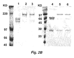


Classifications
-
- A—HUMAN NECESSITIES
- A61—MEDICAL OR VETERINARY SCIENCE; HYGIENE
- A61K—PREPARATIONS FOR MEDICAL, DENTAL OR TOILETRY PURPOSES
- A61K9/00—Medicinal preparations characterised by special physical form
- A61K9/0012—Galenical forms characterised by the site of application
- A61K9/0019—Injectable compositions; Intramuscular, intravenous, arterial, subcutaneous administration; Compositions to be administered through the skin in an invasive manner
-
- A—HUMAN NECESSITIES
- A61—MEDICAL OR VETERINARY SCIENCE; HYGIENE
- A61K—PREPARATIONS FOR MEDICAL, DENTAL OR TOILETRY PURPOSES
- A61K38/00—Medicinal preparations containing peptides
- A61K38/16—Peptides having more than 20 amino acids; Gastrins; Somatostatins; Melanotropins; Derivatives thereof
- A61K38/17—Peptides having more than 20 amino acids; Gastrins; Somatostatins; Melanotropins; Derivatives thereof from animals; from humans
- A61K38/19—Cytokines; Lymphokines; Interferons
- A61K38/21—Interferons [IFN]
- A61K38/212—IFN-alpha
-
- A—HUMAN NECESSITIES
- A61—MEDICAL OR VETERINARY SCIENCE; HYGIENE
- A61K—PREPARATIONS FOR MEDICAL, DENTAL OR TOILETRY PURPOSES
- A61K38/00—Medicinal preparations containing peptides
- A61K38/16—Peptides having more than 20 amino acids; Gastrins; Somatostatins; Melanotropins; Derivatives thereof
- A61K38/17—Peptides having more than 20 amino acids; Gastrins; Somatostatins; Melanotropins; Derivatives thereof from animals; from humans
- A61K38/19—Cytokines; Lymphokines; Interferons
- A61K38/21—Interferons [IFN]
- A61K38/215—IFN-beta
-
- A—HUMAN NECESSITIES
- A61—MEDICAL OR VETERINARY SCIENCE; HYGIENE
- A61K—PREPARATIONS FOR MEDICAL, DENTAL OR TOILETRY PURPOSES
- A61K39/00—Medicinal preparations containing antigens or antibodies
- A61K39/395—Antibodies; Immunoglobulins; Immune serum, e.g. antilymphocytic serum
-
- A—HUMAN NECESSITIES
- A61—MEDICAL OR VETERINARY SCIENCE; HYGIENE
- A61K—PREPARATIONS FOR MEDICAL, DENTAL OR TOILETRY PURPOSES
- A61K47/00—Medicinal preparations characterised by the non-active ingredients used, e.g. carriers or inert additives; Targeting or modifying agents chemically bound to the active ingredient
- A61K47/50—Medicinal preparations characterised by the non-active ingredients used, e.g. carriers or inert additives; Targeting or modifying agents chemically bound to the active ingredient the non-active ingredient being chemically bound to the active ingredient, e.g. polymer-drug conjugates
- A61K47/51—Medicinal preparations characterised by the non-active ingredients used, e.g. carriers or inert additives; Targeting or modifying agents chemically bound to the active ingredient the non-active ingredient being chemically bound to the active ingredient, e.g. polymer-drug conjugates the non-active ingredient being a modifying agent
- A61K47/68—Medicinal preparations characterised by the non-active ingredients used, e.g. carriers or inert additives; Targeting or modifying agents chemically bound to the active ingredient the non-active ingredient being chemically bound to the active ingredient, e.g. polymer-drug conjugates the non-active ingredient being a modifying agent the modifying agent being an antibody, an immunoglobulin or a fragment thereof, e.g. an Fc-fragment
- A61K47/6801—Drug-antibody or immunoglobulin conjugates defined by the pharmacologically or therapeutically active agent
- A61K47/6803—Drugs conjugated to an antibody or immunoglobulin, e.g. cisplatin-antibody conjugates
- A61K47/6811—Drugs conjugated to an antibody or immunoglobulin, e.g. cisplatin-antibody conjugates the drug being a protein or peptide, e.g. transferrin or bleomycin
- A61K47/6813—Drugs conjugated to an antibody or immunoglobulin, e.g. cisplatin-antibody conjugates the drug being a protein or peptide, e.g. transferrin or bleomycin the drug being a peptidic cytokine, e.g. an interleukin or interferon
-
- A—HUMAN NECESSITIES
- A61—MEDICAL OR VETERINARY SCIENCE; HYGIENE
- A61K—PREPARATIONS FOR MEDICAL, DENTAL OR TOILETRY PURPOSES
- A61K47/00—Medicinal preparations characterised by the non-active ingredients used, e.g. carriers or inert additives; Targeting or modifying agents chemically bound to the active ingredient
- A61K47/50—Medicinal preparations characterised by the non-active ingredients used, e.g. carriers or inert additives; Targeting or modifying agents chemically bound to the active ingredient the non-active ingredient being chemically bound to the active ingredient, e.g. polymer-drug conjugates
- A61K47/51—Medicinal preparations characterised by the non-active ingredients used, e.g. carriers or inert additives; Targeting or modifying agents chemically bound to the active ingredient the non-active ingredient being chemically bound to the active ingredient, e.g. polymer-drug conjugates the non-active ingredient being a modifying agent
- A61K47/68—Medicinal preparations characterised by the non-active ingredients used, e.g. carriers or inert additives; Targeting or modifying agents chemically bound to the active ingredient the non-active ingredient being chemically bound to the active ingredient, e.g. polymer-drug conjugates the non-active ingredient being a modifying agent the modifying agent being an antibody, an immunoglobulin or a fragment thereof, e.g. an Fc-fragment
- A61K47/6835—Medicinal preparations characterised by the non-active ingredients used, e.g. carriers or inert additives; Targeting or modifying agents chemically bound to the active ingredient the non-active ingredient being chemically bound to the active ingredient, e.g. polymer-drug conjugates the non-active ingredient being a modifying agent the modifying agent being an antibody, an immunoglobulin or a fragment thereof, e.g. an Fc-fragment the modifying agent being an antibody or an immunoglobulin bearing at least one antigen-binding site
- A61K47/6851—Medicinal preparations characterised by the non-active ingredients used, e.g. carriers or inert additives; Targeting or modifying agents chemically bound to the active ingredient the non-active ingredient being chemically bound to the active ingredient, e.g. polymer-drug conjugates the non-active ingredient being a modifying agent the modifying agent being an antibody, an immunoglobulin or a fragment thereof, e.g. an Fc-fragment the modifying agent being an antibody or an immunoglobulin bearing at least one antigen-binding site the antibody targeting a determinant of a tumour cell
- A61K47/6855—Medicinal preparations characterised by the non-active ingredients used, e.g. carriers or inert additives; Targeting or modifying agents chemically bound to the active ingredient the non-active ingredient being chemically bound to the active ingredient, e.g. polymer-drug conjugates the non-active ingredient being a modifying agent the modifying agent being an antibody, an immunoglobulin or a fragment thereof, e.g. an Fc-fragment the modifying agent being an antibody or an immunoglobulin bearing at least one antigen-binding site the antibody targeting a determinant of a tumour cell the tumour determinant being from breast cancer cell
-
- A—HUMAN NECESSITIES
- A61—MEDICAL OR VETERINARY SCIENCE; HYGIENE
- A61K—PREPARATIONS FOR MEDICAL, DENTAL OR TOILETRY PURPOSES
- A61K47/00—Medicinal preparations characterised by the non-active ingredients used, e.g. carriers or inert additives; Targeting or modifying agents chemically bound to the active ingredient
- A61K47/50—Medicinal preparations characterised by the non-active ingredients used, e.g. carriers or inert additives; Targeting or modifying agents chemically bound to the active ingredient the non-active ingredient being chemically bound to the active ingredient, e.g. polymer-drug conjugates
- A61K47/51—Medicinal preparations characterised by the non-active ingredients used, e.g. carriers or inert additives; Targeting or modifying agents chemically bound to the active ingredient the non-active ingredient being chemically bound to the active ingredient, e.g. polymer-drug conjugates the non-active ingredient being a modifying agent
- A61K47/68—Medicinal preparations characterised by the non-active ingredients used, e.g. carriers or inert additives; Targeting or modifying agents chemically bound to the active ingredient the non-active ingredient being chemically bound to the active ingredient, e.g. polymer-drug conjugates the non-active ingredient being a modifying agent the modifying agent being an antibody, an immunoglobulin or a fragment thereof, e.g. an Fc-fragment
- A61K47/6835—Medicinal preparations characterised by the non-active ingredients used, e.g. carriers or inert additives; Targeting or modifying agents chemically bound to the active ingredient the non-active ingredient being chemically bound to the active ingredient, e.g. polymer-drug conjugates the non-active ingredient being a modifying agent the modifying agent being an antibody, an immunoglobulin or a fragment thereof, e.g. an Fc-fragment the modifying agent being an antibody or an immunoglobulin bearing at least one antigen-binding site
- A61K47/6875—Medicinal preparations characterised by the non-active ingredients used, e.g. carriers or inert additives; Targeting or modifying agents chemically bound to the active ingredient the non-active ingredient being chemically bound to the active ingredient, e.g. polymer-drug conjugates the non-active ingredient being a modifying agent the modifying agent being an antibody, an immunoglobulin or a fragment thereof, e.g. an Fc-fragment the modifying agent being an antibody or an immunoglobulin bearing at least one antigen-binding site the antibody being a hybrid immunoglobulin
-
- A—HUMAN NECESSITIES
- A61—MEDICAL OR VETERINARY SCIENCE; HYGIENE
- A61P—SPECIFIC THERAPEUTIC ACTIVITY OF CHEMICAL COMPOUNDS OR MEDICINAL PREPARATIONS
- A61P35/00—Antineoplastic agents
-
- A—HUMAN NECESSITIES
- A61—MEDICAL OR VETERINARY SCIENCE; HYGIENE
- A61P—SPECIFIC THERAPEUTIC ACTIVITY OF CHEMICAL COMPOUNDS OR MEDICINAL PREPARATIONS
- A61P35/00—Antineoplastic agents
- A61P35/02—Antineoplastic agents specific for leukemia
-
- A—HUMAN NECESSITIES
- A61—MEDICAL OR VETERINARY SCIENCE; HYGIENE
- A61P—SPECIFIC THERAPEUTIC ACTIVITY OF CHEMICAL COMPOUNDS OR MEDICINAL PREPARATIONS
- A61P35/00—Antineoplastic agents
- A61P35/04—Antineoplastic agents specific for metastasis
-
- C—CHEMISTRY; METALLURGY
- C07—ORGANIC CHEMISTRY
- C07K—PEPTIDES
- C07K14/00—Peptides having more than 20 amino acids; Gastrins; Somatostatins; Melanotropins; Derivatives thereof
- C07K14/435—Peptides having more than 20 amino acids; Gastrins; Somatostatins; Melanotropins; Derivatives thereof from animals; from humans
- C07K14/52—Cytokines; Lymphokines; Interferons
- C07K14/555—Interferons [IFN]
- C07K14/56—IFN-alpha
-
- C—CHEMISTRY; METALLURGY
- C07—ORGANIC CHEMISTRY
- C07K—PEPTIDES
- C07K14/00—Peptides having more than 20 amino acids; Gastrins; Somatostatins; Melanotropins; Derivatives thereof
- C07K14/435—Peptides having more than 20 amino acids; Gastrins; Somatostatins; Melanotropins; Derivatives thereof from animals; from humans
- C07K14/52—Cytokines; Lymphokines; Interferons
- C07K14/555—Interferons [IFN]
- C07K14/565—IFN-beta
-
- C—CHEMISTRY; METALLURGY
- C07—ORGANIC CHEMISTRY
- C07K—PEPTIDES
- C07K16/00—Immunoglobulins [IGs], e.g. monoclonal or polyclonal antibodies
- C07K16/18—Immunoglobulins [IGs], e.g. monoclonal or polyclonal antibodies against material from animals or humans
- C07K16/28—Immunoglobulins [IGs], e.g. monoclonal or polyclonal antibodies against material from animals or humans against receptors, cell surface antigens or cell surface determinants
- C07K16/2863—Immunoglobulins [IGs], e.g. monoclonal or polyclonal antibodies against material from animals or humans against receptors, cell surface antigens or cell surface determinants against receptors for growth factors, growth regulators
-
- C—CHEMISTRY; METALLURGY
- C07—ORGANIC CHEMISTRY
- C07K—PEPTIDES
- C07K16/00—Immunoglobulins [IGs], e.g. monoclonal or polyclonal antibodies
- C07K16/18—Immunoglobulins [IGs], e.g. monoclonal or polyclonal antibodies against material from animals or humans
- C07K16/28—Immunoglobulins [IGs], e.g. monoclonal or polyclonal antibodies against material from animals or humans against receptors, cell surface antigens or cell surface determinants
- C07K16/2887—Immunoglobulins [IGs], e.g. monoclonal or polyclonal antibodies against material from animals or humans against receptors, cell surface antigens or cell surface determinants against CD20
-
- C—CHEMISTRY; METALLURGY
- C07—ORGANIC CHEMISTRY
- C07K—PEPTIDES
- C07K16/00—Immunoglobulins [IGs], e.g. monoclonal or polyclonal antibodies
- C07K16/18—Immunoglobulins [IGs], e.g. monoclonal or polyclonal antibodies against material from animals or humans
- C07K16/32—Immunoglobulins [IGs], e.g. monoclonal or polyclonal antibodies against material from animals or humans against translation products of oncogenes
-
- A—HUMAN NECESSITIES
- A61—MEDICAL OR VETERINARY SCIENCE; HYGIENE
- A61K—PREPARATIONS FOR MEDICAL, DENTAL OR TOILETRY PURPOSES
- A61K39/00—Medicinal preparations containing antigens or antibodies
- A61K2039/505—Medicinal preparations containing antigens or antibodies comprising antibodies
-
- A—HUMAN NECESSITIES
- A61—MEDICAL OR VETERINARY SCIENCE; HYGIENE
- A61K—PREPARATIONS FOR MEDICAL, DENTAL OR TOILETRY PURPOSES
- A61K38/00—Medicinal preparations containing peptides
-
- C—CHEMISTRY; METALLURGY
- C07—ORGANIC CHEMISTRY
- C07K—PEPTIDES
- C07K2317/00—Immunoglobulins specific features
- C07K2317/20—Immunoglobulins specific features characterized by taxonomic origin
- C07K2317/24—Immunoglobulins specific features characterized by taxonomic origin containing regions, domains or residues from different species, e.g. chimeric, humanized or veneered
-
- C—CHEMISTRY; METALLURGY
- C07—ORGANIC CHEMISTRY
- C07K—PEPTIDES
- C07K2317/00—Immunoglobulins specific features
- C07K2317/60—Immunoglobulins specific features characterized by non-natural combinations of immunoglobulin fragments
- C07K2317/62—Immunoglobulins specific features characterized by non-natural combinations of immunoglobulin fragments comprising only variable region components
- C07K2317/622—Single chain antibody (scFv)
-
- C—CHEMISTRY; METALLURGY
- C07—ORGANIC CHEMISTRY
- C07K—PEPTIDES
- C07K2319/00—Fusion polypeptide
- C07K2319/33—Fusion polypeptide fusions for targeting to specific cell types, e.g. tissue specific targeting, targeting of a bacterial subspecies
Definitions
- This invention pertains to the field of oncology. Chimeric constructs are provided that have significant anti-cancer activity.
- Both the innate and the adaptive immune responses are essential for providing protection against infectious pathogens and tumors.
- the cross-talk between innate and adaptive immunity is regulated by interactions between cells and cytokines.
- Cytokines produced by cells of the innate immune system can, directly or indirectly, activate the cells of the adaptive immune response and can play an important role in eliciting protective antitumor immunity ( Belardelli and Ferrantini (2002) Trends Immunol. 23: 201-208 ).
- Central to the activation of the innate immune system is the detection of bacterial products or "danger" signals that lead to the release of proinflammatory cytokines, such as IFN- ⁇ , TNF- ⁇ , and IL-1.
- IFN- ⁇ is a proinflammatory cytokine with potent antiviral and immunomodulatory activities and is a stimulator of differentiation and activity of dendritic cells (DCs) ( Santini et al. (2000) J. Exp. Med. 191: 1777-1788 ).
- Type I IFNs IFN- ⁇ and IFN- ⁇ have multiple effects on the immune response ( Theofilopoulos et al. (2005) Annu. Rev. Immunol. 23: 307-336 ).
- IFN- ⁇ plays a role in the differentiation of Th1 cells ( Finkelman et al. (1991) J. Exp. Med. 174: 1179-1188 ) and the long-term survival of CD8+ T cells in response to specific antigens ( Tough et al. (1996) Science 272: 1947-1950 ).
- IFNs are also capable of exerting antitumor effects in both animal models ( Ferrantini et al. (1994) J. Immunol. 153: 4604-4615 ) and cancer patients (14 . Gutterman et al. (1980) Ann. Intern. Med. 93: 399-406 ).
- IFN- ⁇ can increase expression of the tumor suppressor gene P53 ( Takaoka et al. (2003) Nature 424: 516-523 ), inhibit angiogenesis ( Sidky and Borden (1987) Cancer Res. 47: 5155-5161 ), and prime apoptosis ( Rodriguez-Villanueva and McDonnell (1995) Int. J. Cancer 61: 110 -11417 ) in tumor cells.
- P53 Takaoka et al. (2003) Nature 424: 516-523
- angiogenesis Sidky and Borden (1987) Cancer Res. 47: 5155-5161
- prime apoptosis Rodriguez-Villanueva and McDonnell
- this invention pertains to the discovery that attaching an interferon to a targeting moiety (e.g., a molecule that specifically and/or preferentially binds a marker on or associated with a cell) substantially improves the therapeutic efficacy of the interferon and appears to reduce systemic toxicity. Accordingly, in various embodiments, this invention provides constructs comprising an interferon attached to a targeting moiety and uses of such constructs to specifically and/or preferentially inhibit the growth or proliferation or even to kill certain target cells (e.g., cancer cells).
- a targeting moiety e.g., a molecule that specifically and/or preferentially binds a marker on or associated with a cell
- a chimeric construct comprising an interferon (e.g ., interferon-alpha, interferon-beta, interferon-gamma, etc .) attached to a targeting moiety that binds to a tumor associated antigen (TAA), where the construct when contacted to a tumor cell results in the killing or inhibition of growth or proliferation of the tumor cell.
- TAA tumor associated antigen
- a chimeric construct is provided where the construct comprises an interferon attached to a targeting moiety that binds to a cell surface marker or a cell-associated marker, where the targeting is not attached to the interferon by a (Gly 4 Ser) 3 (SEQ ID NO:31) linker.
- the interferon is a type 1 interferon. In various embodiments the interferon is a type 2 interferon. In various embodiments the is an interferon alpha, an interferon-beta, or an interferon-gamma.
- the targeting moiety is an antibody that binds a tumor associated antigen. In certain embodiments the targeting moiety is chemically coupled to the interferon. In certain embodiments the targeting moiety is joined to the interferon with a peptide linker.
- the peptide linker is fewer than 15, fewer than 14, fewer than 12, fewer than 11, fewer than 10, fewer than 9, fewer than 8, fewer than 7, fewer than 6, fewer than 5, fewer than 4, fewer than 3, or fewer than 2 amino acids in length. In certain embodiments the linker is 15, 14, 13, 12, 11, 10, 9, 8, 7, 6, 5, 4, 3, 2, or 1 amino acid in length. In certain embodiments the linker is not (Gly 4 Ser) 3 (SEQ ID NO:31). In certain embodiments the linker is a linker that is resistant or substantially resistant to schooleolysis. In certain embodiments the peptide linker is Gly 4 Ser (SEQ ID NO:32). In certain embodiments the linker comprises or consists of an amino acid sequence found in Table 2.
- the construct is a recombinantly expressed fusion protein.
- the antibody specifically binds a marker selected from the group consisting of EGFR, HER4, HER3, HER2/ neu, MUC-1, G250, mesothelin, gp100, tyrosinase, and MAGE.
- the targeting moiety is an antibody that binds CD20.
- the targeting moiety is a single chain antibody that comprises the CDRS and/or the variable regions from an antibody selected from the group consisting of anti-CD20 (Rituximab), Ibritumomab tiuxetan, tositumomab, AME-133v, Ocrelizumab, Ofatumumab, TRU-015, IMMU-106, and the like.
- the targeting moiety is an antibody that binds HER2.
- the antibody is a C6 antibody.
- the antibody comprises the VH and VL CDRs or VH and VL domains of C6MH3-B1.
- the antibody is an IgG (e.g., IgG1, IgG3, etc .), an IgE, a single chain Fv (scFv), a FAB , a (Fab') 2 , an (ScFv) 2 , and the like.
- the antibody is an antibody selected form the group consisting ofRituxan, IF5, B1, 1H4, CD19, B4, B43 ,FVS191, hLL2, LL2, RFB4, M195, HuM195, AT13/5, HERCEPTIN®, 4D5, HuCC49, HUCC39 ⁇ CH2 B72.3, 12C10, IG5, H23, BM-2, BM-7, 12H12, MAM-6, and HMFG-1.
- the antibody is an antibody that binds a member of the EGF receptor family.
- the antibody is selected from the group consisting of C6.5, C6ML3-9, C6MH3-B1, C6-B1D2, F5, HER3.A5, HER3.F4, HER3.H1, HER3.H3, HER3.E12, HER3.B12, EGFR.E12, EGFR.C10, EGFR.B11, EGFR.E8, HER4.B4, HER4.G4, HER4.F4, HER4.A8, HER4.B6, HER4.D4, HER4.D7, HER4.D11, HER4.D12, HER4.E3, HER4.E7, HER4.F8 and HER4.C7.
- the construct comprises an anti-HER2 IgG1 antibody attached to an interferon.
- the formulations comprise a chimeric construct comprising an interferon attached to a targeting moiety.
- the chimeric construct comprises a construct as described above (and/or herein below) (e.g., an anti-CD20-Interferon, and anti-HER2-interferon, etc .).
- the formulation is a unit dosage formulation.
- the formulation is a formulated for parenteral administration.
- the formulation is a formulated for administration via a route selected from the group consisting of oral administration, intravenous administration, intramuscular administration, direct tumor administration, inhalation, rectal administration, vaginal administration, transdermal administration, and subcutaneous depot administration.
- methods are provided for inhibiting growth and/or proliferation of a cancer cell.
- the methods typically involve contacting the cancer cell with a chimeric construct as described herein.
- the cancer cell is a metastatic cell, and/or a cell is in a solid tumor.
- the cancer cell is a breast cancer cell.
- the cancer cell is a B cell lymphoma.
- the cancer cell is cell produced by a cancer selected from the group consisting of a B cell lymphoma, lung cancer, a bronchus cancer, a colorectal cancer, a prostate cancer, a breast cancer, a pancreas cancer, a stomach cancer, an ovarian cancer, a urinary bladder cancer, a brain or central nervous system cancer, a peripheral nervous system cancer, an esophageal cancer, a cervical cancer, a melanoma, a uterine or endometrial cancer, a cancer of the oral cavity or pharynx, a liver cancer, a kidney cancer, a biliary tract cancer, a small bowel or appendix cancer, a salivary gland cancer, a thyroid gland cancer, a adrenal gland cancer, an osteosarcoma, a chondrosarcoma, a liposarcoma, a testes cancer, and a malignant fibrous histiocytoma.
- a cancer selected from the group consisting of
- the contacting comprises systemically administering the chimeric moiety to a mammal. In certain embodiments the contacting comprises administering the chimeric moiety directly into a tumor site. In certain embodiments the contacting comprises intravenous administration of the chimeric moiety.
- the cancer cell is a cancer cell in a human or in a non-human mammal.
- nucleic acids are provided that encode the chimeric constructs described herein.
- the nucleic acid encodes a fusion protein comprising an interferon attached to an anti-EGFR family member antibody, an anti-HER2 antibody, an anti-C6 single-chain antibody, or to an anti-CD20 single chain antibody.
- the interferon encoded by the nucleic acid is a type I interferon.
- the interferon is IFN- ⁇ or interferon- ⁇ .
- nucleic encodes an antibody that comprises the VH and VL CDRs of C6MH3-B1.
- nucleic acid encodes a peptide linker (e.g., as described herein) attaching the antibody to the interferon.
- nucleic acid encodes the CDRs and/or the variable regions for anti-CD20 (Rituximab).
- this invention provides the use of a chimeric construct as described herein in the manufacture of a medicament to inhibit the growth and/or proliferation of a cancer cell.
- the methods and constructs of this invention specifically exclude constructs using any of the antibodies disclosed in U.S. Patent Publication No: US 2002/0193569 A1 .
- the methods and constructs of this invention specifically exclude constructs incorporating an anti-CD20 antibody.
- the methods and constructs of this invention specifically exclude constructs incorporating antibodies that bind to any of the following targets: CD19, CD20, CD22, CD33, CD38, EGF-R, HM1.24, phosphatidyl serine antigen, HER-2, TAG-72, and/or MUC-1.
- the constructs described herein can be used in the treatment of pathologies such as multiple sclerosis, HCV mediated vasculitis, and the like.
- polypeptide polypeptide
- peptide protein
- protein polymer of amino acid residues.
- the terms apply to amino acid polymers in which one or more amino acid residue is an artificial chemical analogue of a corresponding naturally occurring amino acid, as well as to naturally occurring amino acid polymers.
- the term also includes variants on the traditional peptide linkage joining the amino acids making up the polypeptide.
- Preferred “peptides”, “polypeptides”, and “proteins” are chains of amino acids whose alpha carbons are linked through peptide bonds.
- amino terminus refers to the free ⁇ -amino group on an amino acid at the amino terminal of a peptide or to the ⁇ -amino group (imino group when participating in a peptide bond) of an amino acid at any other location within the peptide.
- carboxy terminus refers to the free carboxyl group on the carboxy terminus of a peptide or the carboxyl group of an amino acid at any other location within the peptide.
- Peptides also include essentially any polyamino acid including, but not limited to peptide mimetics such as amino acids joined by an ether as opposed to an amide bond.
- an “antibody” refers to a protein consisting of one or more polypeptides substantially encoded by immunoglobulin genes or fragments of immunoglobulin genes.
- the recognized immunoglobulin genes include the kappa, lambda, alpha, gamma, delta, epsilon and mu constant region genes, as well as myriad immunoglobulin variable region genes.
- Light chains are classified as either kappa or lambda.
- Heavy chains are classified as gamma, mu, alpha, delta, or epsilon, which in turn define the immunoglobulin classes, IgG, IgM, IgA, IgD and IgE, respectively.
- a typical immunoglobulin (antibody) structural unit is known to comprise a tetramer.
- Each tetramer is composed of two identical pairs of polypeptide chains, each pair having one "light” (about 25 kD) and one "heavy” chain (about 50-70 kD).
- the N-terminus of each chain defines a variable region of about 100 to 110 or more amino acids primarily responsible for antigen recognition.
- the terms variable light chain (V L ) and variable heavy chain (V H ) refer to these regions of the light and heavy chains respectively.
- Antibodies exist as intact immunoglobulins or as a number of well characterized fragments produced by digestion with various peptidases or expressed de novo.
- pepsin digests an antibody below the disulfide linkages in the hinge region to produce F(ab)' 2 , a dimer of Fab which itself is a light chain joined to V H -C H 1 by a disulfide bond.
- the F(ab)' 2 may be reduced under mild conditions to break the disulfide linkage in the hinge region thereby converting the (Fab') 2 dimer into an Fab' monomer.
- the Fab' monomer is essentially an Fab with part of the hinge region ( see, Fundamental Immunology, W.E.
- antibody fragments are defined in terms of the digestion of an intact antibody, one of skill will appreciate that such Fab' fragments may be synthesized de novo either chemically or by utilizing recombinant DNA methodology.
- the term antibody as used herein also includes antibody fragments either produced by the modification of whole antibodies or synthesized de novo using recombinant DNA methodologies, including, but are not limited to, Fab' 2 , IgG, IgM, IgA, IgE, scFv, dAb, nanobodies, unibodies, and diabodies.
- preferred antibodies include, but are not limited to Fab' 2 , IgG, IgM, IgA, IgE, and single chain antibodies, more preferably single chain Fv (scFv) antibodies in which a variable heavy and a variable light chain are joined together (directly or through a peptide linker) to form a continuous polypeptide.
- scFv single chain Fv
- antibodies and fragments used in the constructs of the present invention can be bispecific.
- Bispecific antibodies or fragments can be of several configurations.
- bispecific antibodies may resemble single antibodies (or antibody fragments) but have two different antigen binding sites (variable regions).
- bispecific antibodies can be produced by chemical techniques ( Kranz et al. (1981)Proc. Natl. Acad. Sci., USA, 78: 5807 ), by "polydoma” techniques ( see, e.g., U.S. Pat. No. 4,474,893 ), or by recombinant DNA techniques.
- bispecific antibodies of the present invention can have binding specificities for at least two different epitopes at least one of which is a tumor associate antigen.
- the antibodies and fragments can also be heteroantibodies.
- Heteroantibodies are two or more antibodies, or antibody binding fragments (e.g. , Fab) linked together, each antibody or fragment having a different specificity.
- an “antigen-binding site” or “binding portion” refers to the part of an immunoglobulin molecule that participates in antigen binding.
- the antigen binding site is formed by amino acid residues of the N-terminal variable ("V") regions of the heavy ("H") and light (“L”) chains.
- V N-terminal variable
- H heavy
- L light
- Three highly divergent stretches within the V regions of the heavy and light chains are referred to as “hypervariable regions” which are interposed between more conserved flanking stretches known as “framework regions” or "FRs".
- FR refers to amino acid sequences that are naturally found between and adjacent to hypervariable regions in immunoglobulins.
- the three hypervariable regions of a light chain and the three hypervariable regions of a heavy chain are disposed relative to each other in three dimensional space to form an antigen binding "surface". This surface mediates recognition and binding of the target antigen.
- the three hypervariable regions of each of the heavy and light chains are referred to as "complementarity determining regions" or "CDRs" and are characterized, for example by Kabat et al. Sequences of proteins of immunological interest, 4th ed. U.S. Dept. Health and Human Services, Public Health Services, Bethesda, MD (1987 ).
- interferon refers to a full-length interferon or to an interferon fragment (truncated interferon) or interferon mutant, that substantially retains the biological activity of the full length wild-type interferon (e.g ., retains at least 80%, preferably at least 90%, more preferably at least 95%, 98%, or 99% of the full-length antibody).
- Interferons include type I interferons (e.g ., interferon-alpha and interferon-beta) as well as type II inteferons (e.g., interferon-gamma).
- the interferon e.g., IFN- ⁇
- IFN- ⁇ can be from essentially any mammalian species.
- the interferon is from a species selected from the group consisting of human, equine, bovine, rodent, porcine, lagomorph, feline, canine, murine, caprine, ovine, a non-human primate, and the like.
- the mutated interferon comprises one or more amino acid substitutions, insertions, and/or deletions.
- An anti-HER2/ neu antibody is an antibody that specifically or preferentially binds a HER2/ neu receptor.
- the term "subject” refers to a human or non-human animal, including, but not limited to, a cat, dog, horse, pig, cow, sheep, goat, rabbit, mouse, rat, or monkey.
- C6 antibody refers to antibodies derived from C6.5 whose sequence is expressly provided, for example, in U.S. Patents 6,512,097 and 5,977,322 , and in PCT Publication WO 97/00271 .
- C6 antibodies preferably have a binding affinity of about 1.6 x 10 -8 or better for HER2/ neu .
- C6 antibodies are derived by screening (for affinity to c-erbB-2 / HER2/ neu ) a phage display library in which a known C6 variable heavy (V H ) chain is expressed in combination with a multiplicity of variable light (V L ) chains or conversely a known C6 variable light chain is expressed in combination with a multiplicity of variable heavy (V H ) chains.
- C6 antibodies also include those antibodies produced by the introduction of mutations into the variable heavy or variable light complementarity determining regions (CDR1, CDR2 or CDR3), e.g., as described in U.S. Patents 6,512,097 and 5,977,322 , and in PCT Publication WO 97/00271 .
- C6 antibodies include those antibodies produced by any combination of these modification methods as applied to C6.5 and its derivatives.
- an "anti-EGFR family antibody” refers to an antibody that specifically binds to a member of the epidermal growth factor receptor family (e.g., an antibody that binds to ErbB-1, also named epidermal growth factor receptor (EGFR), ErbB-2, also named HER2 in humans and neu in rodents, ErbB-3, also named HER3, and/or to ErbB-4, also named HER4).
- EGFR epidermal growth factor receptor
- ErbB-2 also named HER2 in humans and neu in rodents
- ErbB-3 also named HER3, and/or to ErbB-4, also named HER4
- Illustrative anti-EGFR family antibodies include, but are not limited to antibodies such as C6.5, C6ML3-9, C6MH3-B1, C6-B1D2, F5, HER3.A5, HER3.F4, HER3.H1, HER3.H3, HER3.E12, HER3.B12, EGFR.E12, EGFR.C10, EGFR.B11, EGFR.E8, HER4.B4, HER4.G4, HER4.F4, HER4.A8, HER4.B6, HER4.D4, HER4.D7, HER4.D11, HER4.D12, HER4.E3, HER4.E7, HER4.F8 and HER4.C7 and the like ( see , e.g., U.S. Patent publications US 2006/0099205 A1 and US 2004/0071696 A1 which are incorporated herein by reference).
- a single chain Fv (“sFv” or "scFv”) polypeptide is a covalently linked V H :V L heterodimer which, in certain embodiments, may be expressed from a nucleic acid including V H - and V L - encoding sequences either joined directly or joined by a peptide-encoding linker.
- CD20 is a non-glycosylated phosphoprotein expressed on the surface of mature B-cells ( see , e.g., Cragg et al. (2005) Curr. Dir. Autoimmun., 8: 140-174 ). It is also found on B-cell lymphomas, hairy cell leukemia, B-cell chronic lymphocytic leukemia. on skin/melanoma cancer stem cells, and the like.
- the phrase "inhibition of growth and/or proliferation" of a cancer cell refers to decrease in the growth rate and/or proliferation rate of a cancer cell. In certain embodiments this includes death of a cancer cell (e.g. via apoptosis). In certain embodiments this term also refers to inhibiting the growth and/or proliferation of a solid tumor and/or inducing tumor size reduction or elimination of the tumor.
- cancer marker refers to biomolecules such as proteins, carbohydrates, glycoproteins, and the like that are exclusively or preferentially or differentially expressed on a cancer cell and/or are found in association with a cancer cell and thereby provide targets preferential or specific to the cancer.
- the preferential expression can be preferential expression as compared to any other cell in the organism, or preferential expression within a particular area of the organism (e.g. within a particular organ or tissue).
- Interferon alpha is an important cytokine in initiating the innate immune response and also demonstrates a wide spectrum of anti-tumor activities.
- the clinical use of interferon e.g. , IFN- ⁇
- this invention pertains to the discovery that the therapeutic index of interferon can be improved by attaching the interferon to a targeting moiety that specifically/preferentially binds a marker on or associated with the target cell (e.g., a tumor cell). This permits the deliver of higher doses of interferon to the target site with fewer systemic complications.
- a fusion protein consisting of an anti-HER2/neu IgG3 and IFN- ⁇ (anti-HER2/neu-IgG3-IFN- ⁇ ) and in another embodiment by the construction and use of an anti-CD20-IFN- ⁇ fusion protein.
- the efficacy of the HER2/neu-IgG3-IFN- ⁇ constructs was tested on a murine B-cell lymphoma, 38C13, transduced with human HER2/neu.
- the anti-HER2/ neu- IgG3-IFN- ⁇ fusion protein exhibited a potent effect in inhibiting the 38C13/HER2 tumor growth in vivo, and even administration of 1 ⁇ g anti-HER2/neu IgG3-IFN- ⁇ resulted in 88% of long-term survivors after tumor challenge.
- Anti-HER2/neu IgG3-IFN- ⁇ demonstrated a potent activity against established 38C13/HER2 tumors, and complete tumor remission was observed in 88% treated mice. This dramatic anti-tumor activity was mediated by IFN- ⁇ induced apoptosis and targeting IFN- ⁇ to 38C13/HER2 tumor cells by the anti-HER2/neu IgG3 antibody was essential to potentiate these effects.
- this invention provides constructs (e.g.. chimeric moieties) comprising an interferon (e.g ., IFN- ⁇ ) attached to a targeting moiety (e.g., to an antibody that specifically binds a cancer specific marker on a cancer cell).
- the constructs include chemical conjugates as well as fusion proteins.
- nucleic acids encoding the fusion proteins as well as cells transfected with the nucleic acids to express the fusion proteins.
- methods of inhibiting growth and proliferation of cancer cells as well as kits comprising, e.g. the chimeric moieties described herein, for the treatment of various cancers.
- chimeric constructs comprising a targeting moiety (e.g ., an anti-tumor marker antibody) attached to a native (wildtype) or modified IFN (e.g., IFN- ⁇ ) can be effectively used to inhibit the growth and/or proliferation of target cancer cells expressing or associated with the marker to which the targeting moiety is directed.
- a targeting moiety e.g ., an anti-tumor marker antibody
- IFN e.g., IFN- ⁇
- the targeting moieties are chemically conjugated to the interferon, while in other embodiments, the targeting moiety is expressed as a fusion protein with the IFN- ⁇ .
- the targeting moiety (e.g., antibody) component can be directly fused to the IFN- ⁇ or attached by means of a peptide linker (e.g., a (Gly 4 Ser) 3 (SEQ ID NO:31) linker, a GlyGlyGlyGlySer (SEQ ID NO:32) linker, a AEAAAKEAAAKA (SEQ ID NO:33), and the like.
- a peptide linker e.g., a (Gly 4 Ser) 3 (SEQ ID NO:31) linker, a GlyGlyGlyGlySer (SEQ ID NO:32) linker, a AEAAAKEAAAKA (SEQ ID NO:33), and the like.
- the targeting moiety is a molecule that specifically or preferentially binds a marker expressed by (e.g., on the surface of) or associated with the target cell(s). While essentially any cell can be targeted, certain preferred cells include those associated with a pathology characterized by hyperproliferation of a cell (i.e ., a hyperproliferative disorder). Illustrative hyperproliferative disorders include, but are not limited to psoriasis, neutrophilia, polycythemia, thrombocytosis, and cancer.
- Hyperproliferative disorders characterized as cancer include but are not limited to solid tumors, such as cancers of the breast, respiratory tract, brain, reproductive organs, digestive tract, urinary tract, eye, liver, skin, head and neck, thyroid, parathyroid and their distant metastases. These disorders also include lymphomas, sarcomas, and leukemias.
- breast cancer include, but are not limited to invasive ductal carcinoma, invasive lobular carcinoma, ductal carcinoma in situ, and lobular carcinoma in situ.
- cancers of the respiratory tract include, but are not limited to small-cell and non-small-cell lung carcinoma, as well as bronchial adenoma and pleuropulmonary blastoma.
- brain cancers include, but are not limited to brain stem and hypophtalmic glioma, cerebellar and cerebral astrocytoma, medulloblastoma, ependymoma, as well as neuroectodermal and pineal tumor.
- Tumors of the male reproductive organs include, but are not limited to prostate and testicular cancer.
- Tumors of the female reproductive organs include, but are not limited to endometrial, cervical, ovarian, vaginal, and vulvar cancer, as well as sarcoma of the uterus.
- Tumors of the digestive tract include, but are not limited to anal, colon, colorectal, esophageal, gallbladder, gastric, pancreatic, rectal, small-intestine, and salivary gland cancers.
- Tumors of the urinary tract include, but are not limited to bladder, penile, kidney, renal pelvis, ureter, and urethral cancers.
- Eye cancers include, but are not limited to intraocular melanoma and retinoblastoma.
- liver cancers include, but are not limited to hepatocellular carcinoma (liver cell carcinomas with or without fibrolamellar variant), cholangiocarcinoma (intrahepatic bile duct carcinoma), and mixed hepatocellular cholangiocarcinoma.
- Skin cancers include, but are not limited to squamous cell carcinoma, Kaposi's sarcoma, malignant melanoma, Merkel cell skin cancer, and non-melanoma skin cancer.
- Head-and-neck cancers include, but are not limited to laryngeal/hypopharyngeal/nasopharyngeal/oropharyngeal cancer, and lip and oral cavity cancer.
- Lymphomas include, but are not limited to AIDS-related lymphoma, non-Hodgkin's lymphoma, cutaneous T-cell lymphoma, Hodgkin's disease, and lymphoma of the central nervous system.
- Sarcomas include, but are not limited to sarcoma of the soft tissue, osteosarcoma, malignant fibrous histiocytoma, lymphosarcoma, and rhabdomyosarcoma.
- Leukemias include, but are not limited to acute myeloid leukemia, acute lymphoblastic leukemia, chronic lymphocytic leukemia, chronic myelogenous leukemia, and hairy cell leukemia.
- the targeting moiety is a moiety that binds a cancer marker (e.g., a tumor associated antigen).
- a cancer marker e.g., a tumor associated antigen
- the markers need not be unique to cancer cells, but can also be effective where the expression of the marker is elevated in a cancer cell (as compared to normal healthy cells) or where the marker is not present at comparable levels in surrounding tissues (especially where the chimeric moiety is delivered locally).
- Illustrative cancer markers include, for example, the tumor marker recognized by the ND4 monoclonal antibody. This marker is found on poorly differentiated colorectal cancer, as well as gastrointestinal neuroendocrine tumors ( see , e.g., Tobi et al. (1998) Cancer Detection and Prevention, 22(2): 147-152 ).
- Other important targets for cancer immunotherapy are membrane bound complement regulatory glycoprotein: CD46, CD55 and CD59, which have been found to be expressed on most tumor cells in vivo and in vitro.
- Human mucins e.g. MUC1
- MUC1 are known tumor markers as are gp100, tyrosinase, and MAGE, which are found in melanoma. Wild-type Wilms' tumor gene WT1 is expressed at high levels not only in most of acute myelocytic, acute lymphocytic, and chronic myelocytic leukemia, but also in various types of solid tumors including lung cancer.
- Acute lymphocytic leukemia has been characterized by the TAAs HLA-Dr, CD1, CD2, CD5, CD7, CD19, and CD20.
- Acute myelogenous leukemia has been characterized by the TAAs HLA-Dr, CD7, CD13, CD14, CD15, CD33, and CD34.
- Breast cancer has been characterized by the markers EGFR, HER2, MUC1, Tag-72.
- Various carcinomas have been characterized by the markers MUC1, TAG-72, and CEA.
- Chronic lymphocytic leukemia has been characterized by the markers CD3, CD19, CD20, CD21, CD25, and HLA-DR.
- Hairy cell leukemia has been characterized by the markers CD 19, CD20, CD21, CD25.
- Hodgkin's disease has been characterized by the Leu-M1 marker.
- Various melanomas have been characterized by the HMB 45 marker.
- Non-hodgkins lymphomas have been characterized by the CD20, CD19, and Ia marker.
- various prostate cancers have been characterized by the PSMA and SE10 markers.
- tumor cells display unusual antigens that are either inappropriate for the cell type and/or its environment, or are only normally present during the organisms' development (e.g. fetal antigens).
- antigens include the glycosphingolipid GD2, a disialoganglioside that is normally only expressed at a significant level on the outer surface membranes of neuronal cells, where its exposure to the immune system is limited by the blood-brain barrier.
- GD2 is expressed on the surfaces of a wide range of tumor cells including neuroblastoma, medulloblastomas, astrocytomas, melanomas, small-cell lung cancer, osteosarcomas and other soft tissue sarcomas. GD2 is thus a convenient tumor-specific target for immunotherapies.
- tumor cells display cell surface receptors that are rare or absent on the surfaces of healthy cells, and which are responsible for activating cellular signaling pathways that cause the unregulated growth and division of the tumor cell.
- Examples include (ErbB2).
- HER2/ neu a constitutively active cell surface receptor that is produced at abnormally high levels on the surface of breast cancer tumor cells.
- CD20 CD52
- CD33 epidermal growth factor receptor
- tumor markers An illustrative, but not limiting list of suitable tumor markers is provided in Table 1.
- Antibodies to these and other cancer markers are known to those of skill in the art and can be obtained commercially or readily produced, e.g. using phage-display technology.
- the target markers include, but are not limited to members of the epidermal growth factor family (e.g ., HER2, HER3, EGF, HER4), CD1, CD2, CD3, CD5, CD7, CD13, CD14, CD15, CD19, CD20, CD21, CD23, CD25, CD33, CD34, CD38, 5E10, CEA, HLA-DR, HM 1.24, HMB 45, 1a, Leu-M1, MUC1, PMSA, TAG-72, phosphatidyl serine antigen, and the like.
- the epidermal growth factor family e.g HER2, HER3, EGF, HER4
- tumor associated antigens are intended to be illustrative and not limiting.
- Other tumor associated antigens will be known to those of skill in the art.
- ligand to that receptor can function as targeting moieties.
- mimetics of such ligands can also be used as targeting moieties.
- the targeting moieties can comprise antibodies, unibodies, or affybodies that specifically or preferentially bind the tumor marker.
- Antibodies that specifically or preferentially bind tumor markers are well known to those of skill in the art.
- antibodies that bind the CD22 antigen expressed on human B cells include HD6, RFB4, UV22-2, To15, 4KB128, a humanized anti-CD22 antibody (hLL2) ( see, e.g., Li et al. (1989) Cell. Immunol. 111: 85-99 ; Mason et al. (1987) Blood 69: 836-40 ; Behr et al. (1999) Clin. Cancer Res. 5: 3304s-3314s ; Bonardi et al. (1993) Cancer Res. 53: 3015-3021 ).
- Antibodies to CD33 include for example, HuM195 ( see, e.g., Kossman et al. (1999) Clin. Cancer Res. 5: 2748-2755 ), CMA-676 ( see, e.g., Sievers et al., (1999) Blood 93: 3678-3684 .
- Antibodies to CD38 include for example, AT13/5 ( see, e.g., Ellis et al. (1995) J. Immunol. 155: 925-937 ), HB7, and the like.
- the targeting moiety comprises an anti-HER2 antibody.
- the ergB 2 gene more commonly known as (Her-2/neu), is an oncogene encoding a transmembrane receptor.
- trastuzumab e.g., HERCEPTIN®.; Fornier et al. (1999) Ontology (Huntingt) 13: 647-58
- TAB-250 Rosenblum et al. (1999) Clin. Cancer Res. 5: 865-874
- BACH-250 Id .
- TA1 Maier et al. (1991) Cancer Res. 51: 5361-5369
- mAbs described in U.S. Pat. Nos. 5,772,997 ; 5,770,195 mAb 4D5; ATCC CRL 10463
- Illustrative anti-MUC-1 antibodies include, but are not limited to Mc5 ( see, e.g., Peterson et al. (1997) Cancer Res. 57: 1103-1108 ; Ozzello et al. (1993) Breast Cancer Res. Treat. 25: 265-276 ), and hCTMO1 ( see, e.g., Van Hof et al. (1996) Cancer Res. 56: 5179-5185 ).
- Illustrative anti-TAG-72 antibodies include, but are not limited to CC49 (see, e.g., Pavlinkova et al. (1999) Clin. Cancer Res. 5: 2613-2619 ), B72.3 (see, e.g., Divgi et al. (1994) Nucl. Med. Biol. 21: 9-15 ), and those disclosed in U.S. Patent No: 5,976,531 .
- Illustrative anti-HM124 antibodies include, but are not limited to a mouse monoclonal anti-HM1.24 IgG 2a / ⁇ and a a humanized anti-HM1.24 IgG 1 / ⁇ . antibody ( see, e.g., Ono et al. (1999) Mol. Immuno. 36: 387-395 ).
- trastuzumab e.g., HERCEPTIN®, Fornier et al. (1999) Oncology (Huntingt) 13: 647-658
- TAB-250 Rosenblum et al.(1999) Clin. Cancer Res. 5: 865-874
- BACH-250 Id.
- TA1 see, e.g,. Maier et al. (1991) Cancer Res. 51: 5361-5369
- C6 antibodies such as C6.5, DPL5, G98A, C6MH3-B1, B1D2, C6VLB, C6VLD, C6VLE, C6VLF, C6MH3-D7, C6MH3-D6, C6MH3-D5, C6MH3-D3, C6MH3-D2, C6MH3-D1, C6MH3-C4, C6MH3-C3, C6MH3-B9, C6MH3-B5, C6MH3-B48, C6MH3-B47, C6MH3-B46, C6MH3-B43, C6MH3-B41, C6MH3-B39, C6MH3-B34, C6MH3-B33, C6MH3-B31, C6MH3-B27, C6MH3-B25, C6MH3-B21, C6MH3-B20, C6MH
- antibodies directed to various members of the epidermal growth factor receptor family are well suited for use as targeting moieties in the constructs of the present invention.
- Such antibodies include, but are not limited to anti-EGF-R antibodies as described in U.S. Patent Nos: 5,844,093 and 5,558,864 , and in European Patent No. 706,799A . ).
- anti-EGFR family antibodies include, but are not limited to antibodies such as C6.5, C6ML3-9, C6MH3-B1, C6-B1D2, F5, HER3.A5, HER3.F4, HER3.H1, HER3.H3, HER3.E12, HER3.B12, EGFR.E12, EGFR.C10, EGFR.B11, EGFR.E8, HER4.B4, HER4.G4, HER4.F4, HER4.A8, HER4.B6, HER4.D4, HER4.D7, HER4.D11, HER4.D12, HER4.E3, HER4.E7, HER4.F8 and HER4.C7 and the like ( see, e.g., U.S. Patent publications US 2006/0099205 A1 and US 2004/0071696 A1 which are incorporated herein by reference).
- the targeting moity comprises an antibody that specifically or preferentially binds CD20.
- Anti-CD20 antibodies are well known to those of skill and include, but are not limited to rituximab, Ibritumomab tiuxetan, and tositumomab, AME-133v (Applied Molecular Evolution), Ocrelizumab (Roche), Ofatumumab (Genmab), TRU-015 (Trubion) and IMMU-106 (Immunomedics).
- the invention need not be limited to the use of the antibodies described above, and other such antibodies as they are known to those of skill in the art can be used in the compositions and methods described herein.
- UniBody are antibody technology that produces a stable, smaller antibody format with an anticipated longer therapeutic window than certain small antibody formats.
- unibodies are produced from IgG4 antibodies by eliminating the hinge region of the antibody. Unlike the full size IgG4 antibody, the half molecule fragment is very stable and is termed a uniBody. Halving the IgG4 molecule left only one area on the UniBody that can bind to a target. Methods of producing unibodies are described in detail in PCT Publication WO2007/059782 , which is incorporated herein by reference in its entirety ( see, also, Kolfschoten et al. (2007) Science 317: 1554-1557 ).
- Affibody molecules are class of affinity proteins based on a 58-amino acid residue protein domain, derived from one of the IgG-binding domains of staphylococcal protein A. This three helix bundle domain has been used as a scaffold for the construction of combinatorial phagemid libraries, from which Affibody variants that target the desired molecules can be selected using phage display technology (see, e.g,. Nord et al. (1997) Nat. Biotechnol. 15: 772-777 ; Ronmark et al. (2002) Eur. J. Biochem., 269: 2647-2655 .). Details of Affibodies and methods of production are known to those of skill ( see, e.g., US Patent No 5,831,012 which is incorporated herein by reference in its entirety).
- the antibodies described above can be provided as whole intact antibodies (e.g., IgG), antibody fragments, or single chain antibodies, using methods well known to those of skill in the art.
- the antibody can be from essentially any mammalian species, to reduce immunogenicity, it is desirable to use an antibody that is of the species in which the construct (e.g., anti-HER2/ neu -IFN- ⁇ chimera) is to be used.
- the construct e.g., anti-HER2/ neu -IFN- ⁇ chimera
- chimeric moieties of this invention comprise an interferon (e.g., IFN- ⁇ ) joined to the targeting moiety (e.g., anti-HER2/ neu antibody).
- the interferon can be a full length wild-type interferon (e.g. IFN- ⁇ , IFN- ⁇ , IFN- ⁇ , etc .) an interferon fragment (e.g., an IFN- ⁇ fragment), and/or a mutated interferon.
- the interferon fragment is one that possesses the endogenous activity of preferably at a level of at least 80%, more preferably at least 90% or 95%, most preferably at least 98%, 99%, 100%, or a level greater than the wild-type interferon.
- Means of identifying such modified interferon molecules are routine to those of skill in the art.
- a library of truncated and/or mutated IFN- ⁇ is produced and screened for IFN- ⁇ activity.
- Methods of producing libraries of polypeptide variants are well known to those of skill in the art.
- error-prone PCR can be used to create a library of mutant and/or truncated IFN- ⁇ (see, e.g., U.S. Patent 6,365,408 ).
- IFN- ⁇ activity can be assayed by measuring antiviral activity against a particular test virus.
- Kits for assaying for IFN- ⁇ activity are commercially available (see, e.g., iLiteTM alphabeta kit by Neutekbio, Ireland).
- modified interferons e.g., modified IFN- ⁇ , IFN- ⁇ , IFN- ⁇ , etc.
- the targeting moiety e.g., an anti-HER2/ neu antibody, and anti-CD20 antibody, etc .
- the antibody can be joined to either the amino or carboxy terminal of the interferon.
- the antibody can also be joined to an internal region of the interferon, or conversely, the interferon can be joined to an internal location or to any terminus of the antibody, as long as the attachment does not interfere with binding of the antibody to that target marker (e.g., the HER2/ neu receptor).
- the antibody e.g., a C6 anti-HER2/ neu
- the interferon e.g., IFN- ⁇
- the interferon is conjugated, either directly or through a linker (spacer), to the antibody.
- the targeting moiety e.g., an anti-HER2/ neu antibody such as C6.5, C6MH3-B1, G98A, ML3-9, H3B1, B1D2, etc .
- the interferon e.g., IFN- ⁇
- Means of chemically conjugating molecules are well known to those of skill.
- Polypeptides typically contain variety of functional groups; e.g ., carboxylic acid (COOH) or free amine (-NH 2 ) groups, that are available for reaction with a suitable functional group on the other peptide, or on a linker to join the molecules thereto.
- functional groups e.g ., carboxylic acid (COOH) or free amine (-NH 2 ) groups, that are available for reaction with a suitable functional group on the other peptide, or on a linker to join the molecules thereto.
- the antibody and/or the IFN- ⁇ can be derivatized to expose or attach additional reactive functional groups.
- the derivatization can involve attachment of any of a number of linker molecules such as those available from Pierce Chemical Company, Rockford Illinois.
- a “linker”, as used herein, typically refers to a molecule that is used to join the antibody to the IFN- ⁇ .
- the linker is capable of forming covalent bonds to both the antibody and to the IFN- ⁇ .
- Suitable linkers are well known to those of skill in the art and include, but are not limited to, straight or branched-chain carbon linkers, heterocyclic carbon linkers, or peptide linkers.
- the linker(s) can be joined to the constituent amino acids of the antibody and/or the IFN- ⁇ through their side groups (e.g., through a disulfide linkage to cysteine).
- the linkers are joined to the alpha carbon amino and/or carboxyl groups of the terminal amino acids of the antibody and/or the IFN- ⁇ .
- a bifunctional linker having one functional group reactive with a group on the antibody and another group reactive on the IFN- ⁇ can be used to form the desired conjugate.
- derivatization can involve chemical treatment of the targeting moiety.
- Procedures for generation of, for example, free sulfhydryl groups on polypeptides, such as antibodies or antibody fragments, are known (See U.S. Patent No: 4,659,839 ).
- a chimeric targeting moiety-interferon fusion protein is synthesized using recombinant DNA methodology. Generally this involves creating a DNA sequence that encodes the fusion protein, placing the DNA in an expression cassette under the control of a particular promoter, expressing the protein in a host, isolating the expressed protein and, if required, renaturing the protein.
- DNA encoding the fusion proteins can be prepared by any suitable method, including, for example, cloning and restriction of appropriate sequences or direct chemical synthesis by methods such as the phosphotriester method of Narang et al. (1979) Meth. Enzymol. 68: 90-99 ; the phosphodiester method of Brown et al. (1979) Meth. Enzymol. 68: 109-151 ; the diethylphosphoramidite method of Beaucage et al. (1981) Tetra. Lett., 22: 1859-1862 ); the solid support method of U.S. Patent No. 4,458,066 , and the like.
- Chemical synthesis produces a single stranded oligonucleotide. This can be converted into double stranded DNA by hybridization with a complementary sequence, or by polymerization with a DNA polymerase using the single strand as a template.
- a complementary sequence or by polymerization with a DNA polymerase using the single strand as a template.
- One of skill would recognize that while chemical synthesis of DNA is limited to sequences of about 100 bases, longer sequences may be obtained by the ligation of shorter sequences.
- subsequences can be cloned and the appropriate subsequences cleaved using appropriate restriction enzymes. The fragments can then be ligated to produce the desired DNA sequence.
- DNA encoding fusion proteins of the present invention can be cloned using DNA amplification methods such as polymerase chain reaction (PCR).
- PCR polymerase chain reaction
- the gene for IFN- ⁇ is PCR amplified, using a sense primer containing the restriction site for, e.g., NdeI and an antisense primer containing the restriction site for HindIII.
- This can produce a nucleic acid encoding the mature IFN- ⁇ sequence and having terminal restriction sites.
- An antibody having "complementary" restriction sites can similarly be cloned and then ligated to the IFN- ⁇ and/or to a linker attached to the IFN- ⁇ . Ligation of the nucleic acid sequences and insertion into a vector produces a vector encoding IFN- ⁇ joined to the anti-HER2/ neu antibody.
- the two molecules can be directly joined together, one of skill will appreciate that the molecules can be separated by a peptide spacer consisting of one or more amino acids. Generally the spacer will have no specific biological activity other than to join the proteins or to preserve some minimum distance or other spatial relationship between them. In certain embodiments, however, the constituent amino acids of the spacer can be selected to influence some property of the molecule such as the folding, net charge, or hydrophobicity.
- linkers are unsuitable for preparation of fusion proteins of the present invention.
- the (Gly 4 Ser) 3 (SEQ ID NO:31) linker was not well suited for the production of an anti-CD20-IFN- ⁇ construct.
- the interferon was being removed from the fusion protein by proteolysis.
- linker that is resistant to proteolysis.
- Certain preferred linkers are linkers that are not the (Gly 4 Ser) 3 (SEQ ID NO:31) linker.
- Certain preferred linkers are linkers shorter than 15 amino acids, or linkers shorter than 14, 13, 12, 11, 10, 9, 8, 7, 6, 5, 4, 3, or 2 amino acids in length.
- the linker is an alpha helical linker ranging in length up to about 12 or 13 or 14 amino acids in length.
- the nucleic acid sequences encoding the fusion proteins can be expressed in a variety of host cells, including E. coli, other bacterial hosts, yeast, and various higher eukaryotic cells such as the COS, CHO and HeLa cells lines and myeloma cell lines.
- the recombinant protein gene is typically operably linked to appropriate expression control sequences for each host.
- this includes a promoter such as the T7, trp, or lambda promoters, a ribosome binding site and preferably a transcription termination signal.
- control sequences will include a promoter and preferably an enhancer derived from immunoglobulin genes, SV40, cytomegalovirus, etc., and a polyadenylation sequence, and may include splice donor and acceptor sequences.
- the plasmids of the invention can be transferred into the chosen host cell by well-known methods such as calcium chloride transformation for E. coli and calcium phosphate treatment or electroporation for mammalian cells.
- Cells transformed by the plasmids can be selected by resistance to antibiotics conferred by genes contained on the plasmids, such as the amp, gpt, neo and hyg genes.
- the recombinant fusion proteins can be purified according to standard procedures of the art, including ammonium sulfate precipitation, affinity columns, column chromatography, gel electrophoresis and the like (see, generally, R. Scopes (1982) Proteins Purification, Springer-Verlag, N.Y .: Deutscher (1990) Methods in Enzymology Vol. 182: Guide to Proteins Purification., Academic Press, Inc. N.Y ., and the like). Substantially pure compositions of at least about 90 to 95% homogeneity are preferred, and 98 to 99% or more homogeneity are most preferred for pharmaceutical uses. Once purified, partially or to homogeneity as desired, the polypeptides may then be used therapeutically.
- the fusion protein e.g., anti-HER2/ neu -IFN- ⁇ , anti-CD20-IFN- ⁇ , etc.
- the fusion protein may possess a conformation substantially different than the native conformations of the constituent polypeptides. In this case, it may be necessary to denature and reduce the polypeptide and then to cause the polypeptide to re-fold into the preferred conformation.
- Methods of reducing and denaturing proteins and inducing re-folding are well known to those of skill in the art ( see, e.g., Debinski et al. (1993) J. Biol.
- a transient expression system can be used to express the chimeric constructs described herein.
- one cell line that works well for transient expression is 293T.
- 9 million cells in 25 ml are seeded for each 150 mm tissue culture plate.
- a 1 mg/ml of PEI Polyethylenimine
- 25 ⁇ g each of H and L 50 ug total
- a volume of 5 ml is used for transfection of each 150 mm plate.
- the DNA is mixed with DMEM, the PEI is then added and the mixture is incubated at room temperature for 10 mins.
- PEI 1.75 ⁇ g PEI is used for each ug of DNA.
- the old medium is removed, discarded and replaced with 20 ml of fresh medium (Iscoves + 5% calf serum).
- the transfection mix is added and the plate is swirled.
- the medium is replaced with 30 ml of Iscoves medium containing 1% FBS(fetal bovine serum) to minimize the amount of bovine Ig present.
- Supernatants are collected from the cells on Days 4,6 and 13 by removing the medium and replacing it with 30 ml of fresh Iscover containing 1% FBS.
- Example 1 The cloning and expression of an anti-HER2/ neu -IFN- ⁇ fusion protein is illustrated herein in Example 1, while the cloning and expression of an anti-CD20-IFN- ⁇ fusion protein is shown in Example 2.
- modifications can be made to increase serum half-life and/or bioavailability.
- modifications include, but are not limited to the incorporation of D amino acids (especially in the linker), the use of non-naturally occurring amino acids, pegylation of the fusion protein, and the like.
- this invention contemplates the use of multivalent, preferably trivalent, quadravalent, pentavalent or greater targeting moieties (e.g., anti-HER2/ neu antibodies, anti-CD20 antibodies, etc .) to target the interferon to a target cell.
- multivalent, preferably trivalent, quadravalent, pentavalent or greater targeting moieties e.g., anti-HER2/ neu antibodies, anti-CD20 antibodies, etc .
- multivalent anti-HER2/ neu moieties can be produced by any of a number of methods.
- linkers having three, four, or more reactive sites can be reacted with anti-HER2/ neu antibodies to form a trimer or greater conjugate.
- phage display, yeast display, bacterial display, or other display systems can be used to express and display multiple copies (e.g., at least 3, at least 4, at least 5, at least 6 copies, etc .) of a targeting (e.g., anti-HER2/ neu , anti-CD20, etc .) antibody and thereby effectively provide a multivalent targeting moiety.
- a targeting e.g., anti-HER2/ neu , anti-CD20, etc .
- the chimeric constructs of this invention are useful for inhibiting the growth and/or proliferation of target cells (e.g., cancer cells).
- target cells e.g., cancer cells
- the chimeric moieties can be used to inhibit disease progression, to shrink tumor size, and/or to stabilize regression/remission.
- compositions and methods of the invention may also include additional therapeutic and/or pharmacologically acceptable agents.
- the compositions or methods may involve other agents for the treatment of cancer.
- agents include, but are not limited to alkylating agents (e.g., mechlorethamine (Mustargen), cyclophosphamide (Cytoxan, Neosar), ifosfamide (Ifex), phenylalanine mustard; melphalen (Alkeran), chlorambucol (Leukeran), uracil mustard, estramustine (Emcyt), thiotepa (Thioplex), busulfan (Myerlan), lomustine (CeeNU), carmustine (BiCNU, BCNU), streptozocin (Zanosar), dacarbazine (DTIC-Dome), cis-platinum, cisplatin (Platinol, Platinol AQ), carboplatin (Para)
- alkylating agents
- methotrexate Amethopterin, Folex, Mexate, Rheumatrex
- 5-fluoruracil Adrucil, Efudex, Fluoroplex
- floxuridine 5-fluorodeoxyuridine (FUDR)
- capecitabine Xeloda
- fludarabine: Fludara
- cytosine arabinoside Cytaribine, Cytosar, ARA-C
- 6-mercaptopurine Purinethol
- 6-thioguanine Thioguanine
- gemcitabine Gemcitabine
- cladribine Leustatin
- deoxycoformycin pentostatin (Nipent), etc .
- antibiotics e.g.
- doxorubicin (Adriamycin, Rubex, Doxil, Daunoxome- liposomal preparation), daunorubicin (Daunomycin, Cerubidine), idarubicin (Idamycin), valrubicin (Valstar), mitoxantrone (Novantrone), dactinomycin (Actinomycin D, Cosmegen), mithramycin, plicamycin (Mithracin), mitomycin C (Mutamycin), bleomycin (Blenoxane), procarbazine (Matulane), etc .), mitotic inhibitors (e.g.
- paclitaxel Taxol
- docetaxel Taxotere
- vinblatine sulfate Velban, Velsar, VLB
- vincristine sulfate Oncovin, Vincasar PFS, Vincrex
- vinorelbine sulfate Navelbine
- chromatin function inhibitors e.g., topotecan (Camptosar), irinotecan (Hycamtin), etoposide (VP-16, VePesid, Toposar), teniposide (VM-26, Vumon), etc.
- hormones and hormone inhibitors e.g .
- diethylstilbesterol (Stilbesterol, Stilphostrol), estradiol, estrogen, esterified estrogens (Estratab, Menest), estramustine (Emcyt), tamoxifen (Nolvadex), toremifene (Fareston) anastrozole (Arimidex), letrozole (Femara), 17-OH-progesterone, medroxyprogesterone, megestrol acetate (Megace), goserelin (Zoladex), leuprolide (Leupron), testosteraone, methyltestosterone, fluoxmesterone (Android-F, Halotestin), flutamide (Eulexin), bicalutamide (Casodex), nilutamide (Nilandron), etc .) INHIBITORS OF SYNTHESIS (e.g., aminoglutethimide (Cytadren), ketoconazole (Nizoral
- one or more active agents (chimeric moieties) of this invention are administered, e.g. to an individual diagnosed as having a cancer.
- the active agent(s) can be administered in the "native" form or, if desired, in the form of salts, esters, amides, prodrugs, derivatives, and the like, provided the salt, ester, amide, prodrug or derivative is suitable pharmacologically, i.e., effective in the present method.
- Salts, esters, amides, prodrugs and other derivatives of the active agents can be prepared using standard procedures known to those skilled in the art of synthetic organic chemistry and described, for example, by March (1992) Advanced Organic Chemistiy; Reactions, Mechanisms and Structure, 4th Ed. N.Y. Wiley-Interscience .
- acid addition salts are prepared from the free base using conventional methodology, that typically involves reaction with a suitable acid.
- a suitable acid for example, the base form of the drug is dissolved in a polar organic solvent such as methanol or ethanol and the acid is added thereto.
- the resulting salt either precipitates or can be brought out of solution by addition of a less polar solvent.
- Suitable acids for preparing acid addition salts include both organic acids, e.g., acetic acid, propionic acid, glycolic acid, pyruvic acid, oxalic acid, malic acid, malonic acid, succinic acid, maleic acid, fumaric acid, tartaric acid, citric acid, benzoic acid, cinnamic acid, mandelic acid, methanesulfonic acid, ethanesulfonic acid, p-toluenesulfonic acid, salicylic acid, and the like, as well as inorganic acids, e.g., hydrochloric acid, hydrobromic acid, sulfuric acid, nitric acid, phosphoric acid, and the like.
- organic acids e.g., acetic acid, propionic acid, glycolic acid, pyruvic acid, oxalic acid, malic acid, malonic acid, succinic acid, maleic acid, fumaric acid, tartaric acid, citric acid, benzoic
- An acid addition salt may be reconverted to the free base by treatment with a suitable base.
- Particularly preferred acid addition salts of the active agents herein are halide salts, such as may be prepared using hydrochloric or hydrobromic acids.
- preparation of basic salts of the active agents of this invention are prepared in a similar manner using a pharmaceutically acceptable base such as sodium hydroxide, potassium hydroxide, ammonium hydroxide, calcium hydroxide, trimethylamine, or the like.
- Particularly preferred basic salts include alkali metal salts, e.g., the sodium salt, and copper salts.
- esters typically involves functionalization of hydroxyl and/or carboxyl groups which may be present within the molecular structure of the drug.
- the esters are typically acyl-substituted derivatives of free alcohol groups, i.e., moieties that are derived from carboxylic acids of the formula RCOOH where R is alky, and preferably is lower alkyl.
- Esters can be reconverted to the free acids, if desired, by using conventional hydrogenolysis or hydrolysis procedures.
- Amides and prodrugs can also be prepared using techniques known to those skilled in the art or described in the pertinent literature.
- amides may be prepared from esters, using suitable amine reactants, or they may be prepared from an anhydride or an acid chloride by reaction with ammonia or a lower alkyl amine.
- Prodrugs are typically prepared by covalent attachment of a moiety that results in a compound that is therapeutically inactive until modified by an individual's metabolic system.
- the active agents identified herein are useful for parenteral, topical, oral, nasal (or otherwise inhaled), rectal, or local administration, such as by aerosol or transdermally, for prophylactic and/or therapeutic treatment of one or more of the pathologies/indications described herein (e.g., atherosclerosis and/or symptoms thereof).
- the pharmaceutical compositions can be administered in a variety of unit dosage forms depending upon the method of administration. Suitable unit dosage forms, include, but are not limited to powders, tablets, pills, capsules, lozenges, suppositories, patches, nasal sprays, injectables, implantable sustained-release formulations, lipid complexes, etc.
- the active agents of this invention are typically combined with a pharmaceutically acceptable carrier (excipient) to form a pharmacological composition.
- Pharmaceutically acceptable carriers can contain one or more physiologically acceptable compound(s) that act, for example, to stabilize the composition or to increase or decrease the absorption of the active agent(s).
- Physiologically acceptable compounds can include, for example, carbohydrates, such as glucose, sucrose, or dextrans, antioxidants, such as ascorbic acid or glutathione, chelating agents, low molecular weight proteins, protection and uptake enhancers such as lipids, compositions that reduce the clearance or hydrolysis of the active agents, or excipients or other stabilizers and/or buffers.
- physiologically acceptable compounds include wetting agents, emulsifying agents, dispersing agents or preservatives that are particularly useful for preventing the growth or action of microorganisms.
- Various preservatives are well known and include, for example, phenol and ascorbic acid.
- pharmaceutically acceptable carrier(s) including a physiologically acceptable compound depends, for example, on the route of administration of the active agent(s) and on the particular physio-chemical characteristics of the active agent(s).
- the excipients are preferably sterile and generally free of undesirable matter. These compositions may be sterilized by conventional, well-known sterilization techniques.
- compositions of this invention are administered to a patient suffering e.g. from a cancer, or at risk of cancer (e.g. after surgical removal of a primary tumor) in an amount sufficient to prevent and/or cure and/or or at least partially prevent or arrest the disease and/or its complications.
- An amount adequate to accomplish this is defined as a "therapeutically effective dose.” Amounts effective for this use will depend upon the severity of the disease and the general state of the patient's health. Single or multiple administrations of the compositions may be administered depending on the dosage and frequency as required and tolerated by the patient. In any event, the composition should provide a sufficient quantity of the active agents of the formulations of this invention to effectively treat (ameliorate one or more symptoms) the patient.
- the concentration of active agent(s) can vary widely, and will be selected primarily based on fluid volumes, viscosities, body weight and the like in accordance with the particular mode of administration selected and the patient's needs. Concentrations, however, will typically be selected to provide dosages ranging from about 0.1 or 1 mg/kg/day to about 50 mg/kg/day and sometimes higher. Typical dosages range from about 3 mg/kg/day to about 3.5 mg/kg/day, preferably from about 3.5 mg/kg/day to about 7.2 mg/kg/day, more preferably from about 7.2 mg/kg/day to about 11.0 mg/kg/day, and most preferably from about 11.0 mg/kg/day to about 15.0 mg/kg/day.
- dosages range from about 10 mg/kg/day to about 50 mg/kg/day. In certain embodiments, dosages range from about 20 mg to about 50 mg given orally twice daily. It will be appreciated that such dosages may be varied to optimize a therapeutic regimen in a particular subject or group of subjects.
- the active agents of this invention are administered orally (e.g. via a tablet) or as an injectable in accordance with standard methods well known to those of skill in the art.
- the peptides may also be delivered through the skin using conventional transdermal drug delivery systems, i.e., transdermal "patches" wherein the active agent(s) are typically contained within a laminated structure that serves as a drug delivery device to be affixed to the skin.
- the drug composition is typically contained in a layer, or "reservoir,” underlying an upper backing layer.
- the term “reservoir” in this context refers to a quantity of "active ingredient(s)" that is ultimately available for delivery to the surface of the skin.
- the “reservoir” may include the active ingredient(s) in an adhesive on a backing layer of the patch, or in any of a variety of different matrix formulations known to those of skill in the art.
- the patch may contain a single reservoir, or it may contain multiple reservoirs.
- the reservoir comprises a polymeric matrix of a pharmaceutically acceptable contact adhesive material that serves to affix the system to the skin during drug delivery.
- suitable skin contact adhesive materials include, but are not limited to, polyethylenes, polysiloxanes, polyisobutylenes, polyacrylates, polyurethanes, and the like.
- the drug-containing reservoir and skin contact adhesive are present as separate and distinct layers, with the adhesive underlying the reservoir which, in this case, may be either a polymeric matrix as described above, or it may be a liquid or hydrogel reservoir, or may take some other form.
- the backing layer in these laminates, which serves as the upper surface of the device, preferably functions as a primary structural element of the "patch" and provides the device with much of its flexibility.
- the material selected for the backing layer is preferably substantially impermeable to the active agent(s) and any other materials that are present.
- elevated serum half-life can be maintained by the use of sustained-release protein "packaging" systems.
- sustained release systems are well known to those of skill in the art.
- the ProLeaseTM biodegradable microsphere delivery system for proteins and peptides see, e.g., Tracy (1998) Biotechnol. Prog. 14: 108 ; Johnson et al. (1996), Nature Med. 2: 795 ; Herbert et al. (1998), Pharmaceut. Res. 15, 357 ) a dry powder composed of biodegradable polymeric microspheres containing the active agent in a polymer matrix that can be compounded as a dry formulation with or without other agents.
- the ProLeaseTM microsphere fabrication process was specifically designed to achieve a high encapsulation efficiency while maintaining integrity of the active agent.
- the process consists of (i) preparation of freeze-dried drug particles from bulk by spray freeze-drying the drug solution with stabilizing excipients, (ii) preparation of a drug-polymer suspension followed by sonication or homogenization to reduce the drug particle size, (iii) production of frozen drug-polymer microspheres by atomization into liquid nitrogen, (iv) extraction of the polymer solvent with ethanol, and (v) filtration and vacuum drying to produce the final dry-powder product.
- the resulting powder contains the solid form of the active agents, which is homogeneously and rigidly dispersed within porous polymer particles.
- the polymer most commonly used in the process poly(lactide-co-glycolide) (PLG), is both biocompatible and biodegradable.
- Encapsulation can be achieved at low temperatures (e.g., -40°C).
- the protein is maintained in the solid state in the absence of water, thus minimizing water-induced conformational mobility of the protein, preventing protein degradation reactions that include water as a reactant, and avoiding organic-aqueous interfaces where proteins may undergo denaturation.
- a preferred process uses solvents in which most proteins are insoluble, thus yielding high encapsulation efficiencies ( e.g., greater than 95%).
- one or more components of the solution can be provided as a "concentrate”, e.g., in a storage container (e.g., in a premeasured volume) ready for dilution, or in a soluble capsule ready for addition to a volume of water.
- kits for the treatment a primary cancer and/or in an adjunct therapy typically comprise a container containing a chimeric moiety of the present invention (e.g., anti-HER2/ neu -IFN- ⁇ , anti-CD20-IFN- ⁇ , etc.).
- the chimeric moiety can be present in a pharmacologically acceptable excipient.
- kits can optionally include instructional materials disclosing means of use of the chimeric moiety (e.g. to treat a cancer and/or as an adjunct therapeutic).
- the instructional materials may also, optionally, teach preferred dosages, counter-indications, and the like.
- kits can also include additional components to facilitate the particular application for which the kit is designed.
- additional components can also include additional components to facilitate the particular application for which the kit is designed.
- additional components for example, and additionally comprise means for disinfecting a wound, for reducing pain, for attachment of a dressing, and the like.
- instructional materials typically comprise written or printed materials they are not limited to such. Any medium capable of storing such instructions and communicating them to an end user is contemplated by this invention. Such media include, but are not limited to electronic storage media (e.g., magnetic discs, tapes, cartridges, chips), optical media (e.g., CD ROM), and the like. Such media may include addresses to internet sites that provide such instructional materials. Further aspects of the invention are as follows:
- Anti-HER2/ neu -IgG3-IFN- ⁇ inhibited the growth of and induced apoptosis in 38C13/HER2 cells. These results indicate that fusion of IFN- ⁇ to a tumor-specific Ab results in an agent effective for the treatment of B cell lymphoma.
- 38C13 is a highly malignant murine B cell lymphoma derived from C3H/ HeN mice.
- the construction and characterization of 38C13 expressing human HER2/neu (38C13/HER2) has been previously described (6).
- Both 38C13 and 38C13/HER2 were cultured in IMDM (Irvine Scientific) supplemented with 2 mM L-glutamine, 10 U/ml penicillin, 10 microg/ml streptomycin (GPS; Sigma-Aldrich) and 10% calf serum (Atlanta Biologicals).
- Murine myeloma P3X63Ag8.653 (American Type Culture Collection) and its derivatives expressing anti-HER2 IgG3-IFN- ⁇ or IgG3-IFN- ⁇ were grown in IMDM supplemented with 10% calf serum and GPS.
- L929 fibroblasts (American Type Culture Collection) were cultured in IMDM with 5% calf serum and GPS.
- CT26/HER2 a murine colon adenocarcinoma cell line overexpressing human HER2/neu, has been previously described (6).
- CT26/HER2 was cultured in IMDM with 5% calf serum and GPS.
- H and L chain variable regions of C6MH3-B1, an anti-human HER2/neu scFv were inserted into the human ⁇ 3 H chain (pAH4802) and ⁇ Lchain (pAG4622) expression vectors, respectively (22), and used to produce chimeric IgG3 of this specificity.
- PCR was first used to introduce a Bam H1 restriction enzyme site upstream and Xba I restriction enzyme site downstream of the mature murine IFN- ⁇ gene amplified by PCR from genomic DNA of BALB/c mice with the forward primer 5'-CGC GGA TCC TGT GAC CTG CCT CAG ACT C-3 (SEQ ID NO:55) and the reverse primer 5'-GCT CTA GAT CAT TTC TCT TCT CTC AGT CTT C-3 (SEQ ID NO:56). The final PCR product was ligated into a TA vector.
- the resulting vector after sequencing, was digested with Bam H1 and Xba I to release the DNA fragment which was inserted into the vector pAH9612 containing the IgG3 constant region with the C6MH3-B1 H chain variable region and a GGGGSGGGGSGGGGS (SEQ ID NO:57) peptide linker at the end of C H 3.
- the final PCR product, pAH9616 contained anti-HER2/neu-IgG3 followed by a GGGGSGGGGSGGGGS (SEQ ID NO:57) peptide linker and murine IFN- ⁇ .
- Plasmid encoding the IgG3 H chain with the C6MH3-B1 variable region fused to IFN- ⁇ was transfected into P3X63Ag8.653 cells expressing either L chain with the C6MH3-B1 variable region (23) to produce anti-HER2/ neu -IgG3-IFN- ⁇ or nonspecific L chain (4D5; Genentech) (6) to produce IgG3-IFN- ⁇ by electroporation with a pulse of 960 ⁇ Fd capacitance and 0.2 V.
- Transfectants producing anti-HER2/neu(C6MH3-B1)-IgG3, anti-HER2/neu(C6MH3-B1)-IgG3-IFN- ⁇ , or IgG3-IFN- ⁇ were selected and characterized as previously described (6).
- Anti-HER2/neu(C6MH3-B1)-IgG3 was purified from culture supernatants using protein G immobilized on Sepharose 4B fast flow (Sigma-Aldrich), and anti-HER2/neu(C6MH3-B1)-IgG3-IFN- ⁇ and IgG3-IFN- ⁇ were purified from culture supernatants using protein A immobilized on Sepharose 4B fast flow (Sigma-Aldrich).
- L-929 fibroblast cell line sensitive to the vesicular stomatitis virus (VSV) infection was used to quantify the biological activity of IFN- ⁇ .
- L-929 cells were plated in a 96-well tissue culture plate (Falcon; BD Biosciences) at a density of 4 x 10 4 cells/well and incubated overnight at 37°C in a 5% CO2 atmosphere. Afterward, serial dilutions of different IFN- ⁇ fusion proteins or standard IFN- ⁇ (international reference standard for mouse IFN- ⁇ ; National Institutes of Health, Bethesda, MD) were added and the plate was incubated at 37°C for 24 h.
- IFN- ⁇ fusion proteins or standard IFN- ⁇ international reference standard for mouse IFN- ⁇ ; National Institutes of Health, Bethesda, MD
- 38C13 or 38C13/HER2 cells were plated in a 96-well tissue culture plate at a density of 1.25 x 10 4 cells/well and serial dilutions of different fusion proteins were added. The plates were then incubated for 48 h at 37°C in a 5% CO2 atmosphere. Plates were developed by addition of 20 ⁇ l of MTS solution (Promega) and analyzed at 490 nm using an ELISA reader. Inhibition of proliferation (percent) was calculated as: 100 x [(ODexp - ODblank)/(ODmedium - ODblank)] x 100.
- annexin V/propidium iodide (PI) assay was conducted following procedures suggested by the manufacturer using the Vybrant Apoptosis Assay Kit 2 (Molecular Probes).
- mice 6-8 wk of age obtained from Taconic Farms were used. Animals were housed in a facility using autoclaved polycarbonate cages containing wood-shaving bedding. The animals received food and water ad libitum. Artificial light was provided under a 12/12-h light/dark cycle. The temperature of the facility was 20°C with 10-15 air exchanges per hour.
- Murine rIFN- ⁇ (PBL Biomedical Laboratories), IgG3-IFN- ⁇ , and anti-HER2/ neu -IgG3-IFN- ⁇ were iodinated to 10 ⁇ Ci/ ⁇ g with 125 I using Iodo-Beads (Pierce) according to the manufacturer's protocol. Mice were injected i.p. with 66 ⁇ Ci of 125 I-labeled proteins. At various intervals after injection of 125 I-labeled rIFN- ⁇ , IgG3-IFN- ⁇ , or anti-HER2/ neu -IgG3-IFN- ⁇ , residual radioactivity was measured using a mouse whole body counter (Wm. B. Johnson and Associates).
- 38C13/HER2 cells were treated with different fusion proteins for the indicated times, washed with ice-cold PBS, and lysed on ice for 10 min in lysis buffer (0.125% Nonidet P-40, 0.875% Brij 97, 10 mM Tris-HCl (pH 7.5), 2 mM EDTA, 0.15 M NaCl, 0.4 mM Na3VO4, 0.4 mM NaF, 1 mM PMSF, 2.5 ⁇ M leupeptin, and 2.5 ⁇ M aprotinin). Cell lysates were clarified at 10,000 x g for 10 min at 4°C.
- Protein samples were then boiled in sample buffer before separation on 8% SDS-PAGE gels and transferred onto polyvinylidene fluoride microporous membranes (Millipore). After blocking with 3% BSA in 150 mM NaCl, 50 mM Tris-HCl (pH 7.6; TBS) for 1 h at room temperature, blots were probed with the indicated primary Abs overnight at 4°C. The blots were then washed three times at room temperature with 0.05% Tween 20 in TBS, incubated with the appropriate secondary Abs conjugated with HRP, and detected by a peroxidase-catalyzed ECL detection system (ECL; Pierce). Polyclonal rabbit antiphosphoSTAT1 was obtained from Cell Signaling Technology. Polyclonal HRP-conjugated donkey anti-rabbit IgG was obtained from Amersham Biosciences. Polyclonal rabbit anti-GAPDH was obtained from Abcam.
- anti-HER2/ neu -IgG3 with the C6MH3-B1 (20) variable region has been described previously (23).
- the amino-terminal end of mature murine IFN- ⁇ was fused to the carboxyl-terminal end of anti-HER2/ neu -IgG3 separated by a flexible [(Gly 4 )Ser] 3 (SEQ ID NO:31) linker ( Fig. 2A ).
- An identical fusion protein, IgG3-IFN- ⁇ , lacking HER2/ neu specificity was constructed by replacing the C6MH3-B1 L chain with the 4D5 (rhuMab HER2, herceptin; Genentech) L chain.
- the proteins purified from culture supernatants using protein G were analyzed by SDS-PAGE under nonreducing and reducing conditions ( Fig. 2B ).
- anti-HER2/ neu -IgG3 migrates with a molecular mass of 170 kDa
- anti-HER2/ neu -IgG3-IFN- ⁇ Fig. 2B , lane 2
- IgG3-IFN- ⁇ Fig. 2B , lane 3
- mice were inoculated s.c. with 1 x 10 3 38C13/HER2 tumor cells and treated on days 1, 3, and 5 after tumor challenge by i.p. administration of different doses of protein ( Fig. 3A-3B ).
- Mice treated with 2.5 ⁇ g of IgG3-IFN- ⁇ showed some regression of tumor growth, with one (13%) of eight mice alive after 50 days ( Fig. 3A ).
- in vivo targeting of IFN- ⁇ to tumors using a tumor-specific Ab dramatically improved its antitumor effect. All mice treated with 2.5 ⁇ g ( Fig.
- both anti-HER2/ neu -IgG3-IFN- ⁇ and IgG3-IFN- ⁇ were effective in preventing tumor growth (data not shown) undoubtedly reflecting the fact that 38C13 cells are sensitive to IFN- ⁇ treatment (21, 27, 28).
- Tumor growth in mice treated with 5 ⁇ g of anti-HER2/ neu -IgG3 Ab was the same as the PBS control, suggesting that Ab alone has no antitumor effect in vivo (data not shown).
- IFN- ⁇ fused to an Ab results in improved antitumor activity compared with free IFN- ⁇
- IFN- ⁇ has a very short in vivo half-life (29). In previous study, fusion of Abs to cytokines has been shown to increase their halflife (6).
- the clearance of 125 I-labeled rIFN- ⁇ , IgG3-IFN- ⁇ , or anti-HER2/ neu -IgG3-IFN- ⁇ was examined in C3H/HeN mice. Mice were injected i.p. with 66 ⁇ Ci of 125 I-labeled proteins and the residual radioactivity was measured using a mouse whole body counter. rIFN- ⁇ was cleared rapidly with 50% eliminated by ⁇ 2.5 h ( Fig. 4B ).
- both anti-HER2/ neu -IgG3-IFN- ⁇ and IgG3-IFN- ⁇ exhibited significantly increased in vivo half-life with ⁇ 8 h required for elimination of 50% of the injected radioactivity. This increased half-life may contribute to the antitumor efficacy of the IFN- ⁇ fusion proteins.
- fusion of an IgG3 Ab to IFN- ⁇ can significantly improve its in vivo antitumor activity.
- this antitumor activity can be further improved by targeting the IFN- ⁇ to the tumor, making it effective at lower doses.
- Anti-HER2/neu-IgG3-IFN- ⁇ inhibited proliferation of tumor cells in vitro
- IFN- ⁇ has multiple activities including activation of the immune response and direct cytotoxicity against tumors.
- the eight mice remaining tumor free were challenged with 1 x 10 3 38C13/ HER2 tumor cells. Surprisingly, all mice resembled untreated mice and quickly developed bulky tumors (data not shown).
- the 38C13/HER2 or parental 38C13 tumor cells were incubated with different proteins for 48 h and cell proliferation measured using the MTS assay. Treatment with anti-HER2/ neu -IgG3 did not significantly inhibit the proliferation of either 38C13/HER2 or parental 38C 13 tumor cells ( Fig. 5A and 5B ).
- anti-HER2/ neu -IgG3-IFN- ⁇ and IgG3-IFN- ⁇ inhibited the proliferation of 38C13/HER2 tumor cells
- anti-HER2/ neu- IgG3-IFN- ⁇ was more effective than IgG3-IFN- ⁇ with IP50 values of 10 and 100 pM for anti-HER2/ neu -IgG3-IFN- ⁇ and IgG3-IFN- ⁇ , respectively ( Fig. 5A ).
- anti-HER2/ neu -IgG3-IFN- ⁇ and IgG3-IFN- ⁇ exhibited similar antiproliferative activity against parental 38C13 tumor cells.
- IFN- ⁇ signaling can induce apoptosis in some tumor cell lines.
- 38C13/HER2 cells treated with different proteins were assayed for the translocation of phosphatidylserine from the inner to the outer leaflet of the plasma membrane using the annexin V-affinity assay (30).
- Dead cells were stained by PI, which enters cells with a disrupted plasma membrane and binds to DNA.
- annexin V/PI bright 2%) or early apoptotic cells (annexin V bright, 3%) following treatment with anti-HER2/ neu -IgG3 ( Fig. 5C ).
- IFN- ⁇ can directly inhibit the proliferation of tumor cells (31).
- CFSE-labeled 38C13/HER2 cells were treated with different proteins for 48 h, the live cells were gated, and the level of CFSE was determined by flow cytometry.
- the CFSE signal in anti-HER2/ neu -IgG3-treated cells overlapped with the PBS-treated cells and was significantly less than that of cells fixed immediately after CFSE labeling ( Fig. 5D , dotted line), indicating that anti-HER2/ neu -IgG3 did not inhibit the proliferation of the 38C13/HER2.
- IgG3-IFN- ⁇ significantly inhibited the proliferation of the surviving 38C13/HER2 cells ( FIG. 5D , thick line), and targeting IFN- ⁇ to 38C13/HER2 cells by anti-HER2/ neu -IgG3-IFN- ⁇ potentiated this effect ( Fig. 5D , black area).
- IFN- ⁇ fusion proteins induce STAT1 activation in tumor cells
- both IgG3-IFN- ⁇ and anti-HER2/ neu -IgG3-IFN- ⁇ showed antitumor activity but anti-HER2/ neu -IgG3-IFN- ⁇ was more effective in delaying tumor growth and complete tumor remission was observed only in mice treated with anti-HER2/ neu -IgG3-IFN- ⁇ .
- the treatment dose was increased to 10 ⁇ g of the fusion proteins, almost all mice treated with either anti-HER2/ neu -IgG3-IFN- ⁇ or IgG3-IFN- ⁇ had complete tumor regression and remained tumor free after 50 days.
- mice that remained tumor free following treatment with three doses of 10 ⁇ g of fusion proteins were rechallenged with 1 x 10 3 38C13/HER2 tumor cells on day 50. All mice remained tumor free (data not shown).
- anti-HER2/ neu- IgG3-IFN- ⁇ was effective against established tumors ( Fig. 7 ) while anti-Id IgG1-IL-2 had little antitumor activity when treatment was begun either 3 or 7 days after tumor challenge (34).
- IFN- ⁇ is a more powerful therapeutic agent than GM-CSF (35), CTLA-4 (36), or CD40 ligand (37) fused to the Id Ag since none of these vaccine strategies was effective against established tumors. Therefore, targeting IFN- ⁇ to tumor cells could be a promising approach for treating B cell lymphoma.
- Targeting IFN- ⁇ to tumor cells with a tumor-specific Ab increases the antitumor efficacy of IFN- ⁇ .
- Anti-HER2/ neu -IgG3-IFN- ⁇ is more effective in inhibiting proliferation and inducing apoptosis ( Fig. 5A-5D ) in 38C13/HER2 than IgG3-IFN- ⁇ and treatment with either 2.5 or 1 ⁇ g of anti-HER2/ neu -IgG3-IFN- ⁇ was more effective in inhibiting growth of small tumors in vivo than the same doses of IgG3-IFN- ⁇ ( Fig. 3A and 3B ).
- IgG3-IFN- ⁇ exhibits a more potent antitumor activity than rIFN- ⁇ ( Fig. 4A ).
- rIFN- ⁇ is effective in treatment of a variety of tumors (38-40)
- prolonged treatment with high doses is required to see effective antitumor activity in part because of the very short half-life of the cytokine.
- fusion of an IgG3 Ab to IFN- ⁇ significantly increased its half-life ( Fig. 4B ), and this increased half-life may contribute to the increased in vivo antitumor activity of the fusion protein ( Fig. 4A ).
- the Fc region of the IgG3-IFN- ⁇ may help to target IFN- ⁇ to the Fc receptors present on B lymphoma cells and consequently increase the antitumor activity. Therefore, fusion of IFN- ⁇ to an IgG3 Ab may provide multiple advantages in improving the antitumor efficacy of IFN- ⁇ .
- IFN- ⁇ has multiple activities, including activation of the immune response, it appears that direct cytotoxicity plays an important role in the potent antitumor activity of anti-HER2/ neu -IgG3-IFN- ⁇ .
- Both IFN- ⁇ fusion proteins exhibited apoptotic and antiproliferative activities against 38C13/HER2 with tumor targeting significantly increasing these effects ( Fig. 5A-5D ).
- the IFN- ⁇ fusion proteins were very effective in treating small tumors ( Fig. 3A and 3B ), none of the survivors developed an immune response that protected against second tumor challenge, suggesting that the direct cytotoxicity of the IFN- ⁇ fusion proteins was very effective in killing the tumor cells and that the adaptive immunity did not play a role when there was a small tumor burden.
- 38C13 is an extremely malignant B lymphoma cell line and mice injected with as few as 200 cells can develop bulky tumors within 20 days (36), the IFN- ⁇ fusion proteins must be very effective in killing most of the inoculated tumor cells to result in long-term survivors.
- Multiple mechanisms, including down-regulation of NF- ⁇ B (41), induction of apoptosis by activating caspase-3 (42), and up-regulation of both TRAIL and TRAIL receptors (43), have been shown to be involved in IFN- ⁇ -mediated cytotoxicity against tumor cells, and we would expect these mechanisms to contribute to the direct cytotoxicity against tumor cells seen with Ab-IFN- ⁇ fusion proteins. Consistent with this, we observed STAT1 activation following treatment of tumor cells with the fusion proteins ( Fig. 6A-6C ).
- IFN- ⁇ fusion proteins failed to initiate a memory immune response when mice were treated beginning 1 day after tumor inoculation, IFN- ⁇ fusion proteins initiated an immune response that protected against second tumor challenge when mice were treated beginning 12 days after tumor inoculation. Therefore, IFN- ⁇ fusion proteins can activate protective adaptive immunity in the presence of a sizable tumor burden. Because IFN- ⁇ is capable of activating adaptive immunity via stimulation of DC differentiation and maturation (9), it is possible that the established tumors provide more TAAs for DC activation in the presence of IFN- ⁇ . In addition, the foreign Ag human HER2/ neu may contribute to the antitumor immunity by increasing the immunogenicity of the tumor cells in this model.
- CD20 an Ag expressed by B cells, is expressed in most B cell lymphomas (44), and anti-CD20 (rituximab, Genentech;) is one of the most successful cancer therapeutics, having tremendous efficacy against lymphoma with little toxicity (45).
- anti-HER2/ neu IgG3-IFN- ⁇ is very effective against 38C13/HER2
- HER2/ neu is not normally expressed in lymphoma cells and therefore, it probably has limited therapeutic application in the treatment of lymphoma but should be effective in the treatments of cancers that express HER2/ neu .
- IFN- ⁇ may further up-regulate CD20 expression as was seen in patients with B cell lymphoma following IFN- ⁇ treatment (46).
- IFN- ⁇ e.g., IFN- ⁇ fused to a tumor-specific antibody shows promise for the treatment of B cell lymphoma.
- variable regions for anti-CD20 were amplified and cloned into expression vectors for the production of chimeric antibodies with human kappa light chains and gamma 3 heavy chains. Protein was produced and its ability to recognize CD20 examined using flow-cytometry and the human B-cell line Daudi. As shown in Figure 8 , the recombinant protein binds as well as Rituximab a recombinant IgG1.
- the fusion protein vector After verifying that the fusion protein vector had the correct nucleotide sequence, it was transfected with the chimeric anti-CD20 light chain into NS0 cells. Transfectants were screened by ELISA for the production of IgG. The clone giving the highest signal was expanded and following sub-cloning was grown in roller bottles. Supernatants were then passed through protein A Sepharose columns, and the bound proteins eluted and analyzed by SDS-PAGE both unreduced and following reduction ( see , Figure 10 ). Although the isolated protein was assembled into H 2 L 2 molecules, most of the isolated protein was smaller than expected. Following reduction, most of the heavy chains were smaller than expected and ran at the same position as a gamma-3 heavy chain lacking a fusion protein. It appeared that the interferon was being removed from the fusion protein by proteolysis. Western blot analysis using anti-Fc and anti-interferon, confirmed that both of the upper bands were heavy chains, but only the largest contained interferon.
- the human cell line Daudi which expresses CD20 was incubated with Rituxan, anti-DNS/IgG3-hu-IFN- ⁇ or anti-CD20/IgG3-hu-IFN- ⁇ .
- the anti-CD20/IgG3-hu-IFN- ⁇ bound better than Rituxan ( Figure 12 ).
- the anti-DNS/IgG3-hu-IFN- ⁇ fusion also showed some binding, although less than either CD20 specific protein.
- the Timmerman laboratory has produced a transfectant of the murine lymphoma 38C13 that expresses human CD20. Both Rituxan and anti-CD20/ IgG3-mu-IFN- ⁇ bound the transfectant. Anti-DNS/IgG3-mu-IFN- ⁇ showed no binding ( Figure 13 ).
- HeLa cells were seeded at 2x10 5 cells/ml and treated with two-fold serial dilutions of fusion protein or Roferon (recombinant human interferon 2a) for 24 hrs. Cells were then infected with VSV (vesicular stomatitis virus) at a concentration of 4000pfu/100 ⁇ l. After 72hrs, cells were stained with 0.1% crystal violet. Protection against viral infection was determined either by quantitating the cells surviving the infection by staining with 0.1% crystal violet and determing the amount of dye in each well using a a spot densitometer of by counting the number of plaques. In both assays the fusion protein had significant IFN- ⁇ activity but was about 100-fold reduced in activity compared to Roferon.
- VSV vesicular stomatitis virus
- Anti-CD20/IgG3 + Roferon did not show enhanced activity compared to Roferon alone.
- Anti-DNS/ IgG3-hIFN- ⁇ , Rituxan and anti-CD20/IgG3 only showed some growth inhibition at the highest concentration used. It should be noted that fusion protein was more active than Rituxan in preventing cell growth in this assay.
- Daudi cells were incubated with various concentrations of IFN- ⁇ , antibody or fusion protein for 72 hrs and then stained with Annexin V and propidium iodide (PI) analyzed by FLOW cytometry. Shown in Figure 15 are the results obtained whne 10 pM of the various proteins was used. Cells in the early phases of apoptosis are Annexin V + PI - ; late apoptotic and dead cells are Annexin V + PI + .
- Anti-CD20/IgG3-hIFN ⁇ is more effective than anti-DNS/IgG3-hIFN ⁇ , again demonstrating the contribution of cell targeting to cell killing.
- Roferon and anti-CD20/IgG3-hIFN ⁇ exhibit similar activity causing cell death at concentrations as low as 1 pM (data not shown).
- CD20/IgG3-hIFN ⁇ will target and accumulate at the site of the tumor while Roferon will exhibit its activity throughout the body.
- 38C13-CD20 a murine lymphoma, 38C13-CD20 , that expresses human CD20 and will grow in syngenic C3H/HeJ mice.
- the availability of this cell line makes it possible to examine the in vivo efficacy of our fusion proteins.
- 38C13-CD20 cells were incubated for 48 hours with various antibodies and fusion proteins. Killing and apoptosis were then determined by staining cells with Annexin V and PI and examining them using FLOW cytometry.
- recombinant mIFN- ⁇ and anti-CD20/IgG3-mIFN- ⁇ continued to be effective in causing apoptosis, with anti-CD20/IgG3-mIFN- ⁇ more effective that recombinant mIFN- ⁇ . Only a small amount of apoptosis was seen following treatment with anti-DNS-IgG3-mIFN- ⁇ indicating that targeting of IFN- ⁇ using anti-CD20-IgG3-mIFN- ⁇ resulted in a more effective therapeutic agent.
- 38C13-CD20 cells were treated with the various proteins at different concentrations and inhibition of growth monitored using the MTS assay ( Figure 17 ).
- Anti-CD20/IgG3-mIFN- ⁇ was most effective in inhibiting cell growth, followed by recombinant mIFN- ⁇ . Some growth inhibition was observed with anti-DNS/IgG3-mIFN- ⁇ .
- Anti-CD20/IgG3 and Rituxan had little effect on cell growth. Thus, the results obtained in this assay mirrored what was observed when apoptosis was monitored.
- the initial proteins were made with IFN- ⁇ fused to a human IgG3 backbone.
- Rituxan is an IgG1.
- fusion proteins with m-IFN- ⁇ and hu-IFN- ⁇ fused to IgG1 have now been produced. They were of the expected molecular weight.
- Anti-CD20/IgG1-mIFN ⁇ was evaluated for its ability to induce apoptosis of 38C13-CD20 ( Figure 18 ). The studies showed it to be effective, possibly even more effective than the IgG3 fusion protein.
- Anti-CD20/IgG1-hIFN ⁇ was evaluated for its ability to induce apoptosis of Daudi cells. The studies showed it exhibit activity similar to anti-CD20/IgG3-hIFN ⁇ ( Fig. 19
- the fusion proteins were evaluated for their ability to inhibit the growth of Daudi cells as shown in Figure 20 .
- IgG1 fusions with both murine and human IFN ⁇ resembled the IgG3 fusions in their ability to inhibit the growth of Daudi cells.
- Fusion proteins with IFN- ⁇ joined to the IgG backbone with an alpha helical linker b. Fusion proteins with IFN- ⁇ joined to the IgG backbone with an alpha helical linker.
- Fusion proteins were produced in which the GlySer linker was replaced with linker with the sequence A(EAAAK) 2 A (SEQ ID NO:33). This sequence is proposed to fold as an alpha helix.
- Protein was produced by transient expression in 293T cells and evaluated by SDS-PAGE. The protein assembled and was of the expected molecular weight. No cleavage of the linker was observed.
- 38C13 lymphoma that had been transduced by the Timmerman laboratory to express human CD20 was used for these studies.
- 38C13 is an aggressive lymphoma that grows in syngenic C3H/HeJ mice.
- the transductant, 38C13-CD20 exhibits the same growth characteristic. Thus it is possible to investigate fusion protein mediated protection in immune competent animals.
- mice [ Mice (groups of 4) were injected subcutaneously with 5000 38C13-CD20 cells on day zero. On days 1, 2 and 3 they were treated intravenously with hepes buffered saline solution (HBSS) or 0.4 ⁇ g, 2 ⁇ g, or 10 ⁇ g of anti-CD20-m-IFN- ⁇ and tumor growth monitored. By day 20 all of the animals treated with HBSS had large tumors and had to be sacrificed. In contrast, no tumor growth was seen in animals treated with 10 ⁇ g of the fusion protein; after day 20 tumors began to grow in 3 of the four animals treated with 0.4 ⁇ g of the fusion protein and 1 of the mice treated with 2 ⁇ g. The results showed that the anti-CD20/IFN- ⁇ fusion proteins are very effective in inhibiting in vivo tumor growth and in increasing survival ( see, e.g., Figure 22 ).
- C3H/HeJ mice were inoculated with 5000 38C13-CD20 cells on day 0. On days 5, 6 and 7 they were treated with HBSS or 10 ⁇ g of anti-CD20-IgG1 (produced in 293T cells), anti-CD20-IgG3, Rituximab or anti-CD20-IgG3-mIFN ⁇ . They were monitored for tumor growth and survival ( see , e.g., Figure 23 ). Anti-CD20/IgG3-mIFN ⁇ was much more effective than Rituximab, anti-CD20/IgG3 or anti-CD20/IgG1 in preventing the growth of moderate sized tumors.
- the tumor targeting ability of the fusion protein significantly enhances its efficacy in vivo.
- C3H/H3J mice were inoculated with 5000 38C13-CD20 cells on day 0 and treated on days 5, 6 and 7 with 10 ⁇ g of anti-CD20-IgG3, 10 ⁇ g of anti-CD20-IgG3 + mIFN- ⁇ (dose chosen to be same moles as in fusion protein), anti-DNS-IgG3-IFN ⁇ , or anti-CD20-IgG3-mIFN ⁇ and followed for tumor growth and survival ( see , e.g., Figure 24 ).
- Anti-CD20-IgG3-IFN ⁇ significantly delayed tumor growth and promoted survival indicating that targeting the IFN ⁇ to the tumor using the antibody combining site makes it a more effective therapeutic than either a fusion protein that does not target the fused IFN ⁇ (anti-DNS-IgG3-IFN ⁇ ) or the injection of anti-CD20 along with IFN ⁇ that is not covalently associated (anti-CD20-IgG3 + mIFN- ⁇ ).
- mice Groups of eight C3H/HeJ mice were inoculated with 5000 38C13-CD20 cells and treated on days 8, 9 and 10 with 100 ⁇ g of anti-CD20-mIFN ⁇ or HBSS. Mice were monitored for tumor growth (see Figure 25 ) and survival (see, Figure 26 ).. Mice inoculated with anti-CD20-mIFN ⁇ shows improved survival ( Figure 26 ).
Abstract
Description
- This application claims priority to and benefit of
USSN 60/994,717, filed on September 21, 2007 - This invention was made with Government support under Grant No. CA87990, awarded by the National Institutes of Health.. The Government has rights in this invention.
- This invention pertains to the field of oncology. Chimeric constructs are provided that have significant anti-cancer activity.
- Although spontaneous immune responses against tumor-associated antigens (TAAs) (Hrouda et al. (1999) Semin. Oncol. 26: 455-471) can be detected (Disis et al. (1997) J. Clin. Oncol. 15: 3363-3367), malignant cells causing disease fail to elicit an immune response that leads to rejection. Many studies have demonstrated that it is possible to enhance the immunogenicity of tumor cells by introducing immunostimulatory molecules such as cytokines and costimulatory molecules into them (Dranoff and Mulligan (1995) Adv. Immunol. 58: 417-454; Hrouda et al. (1999) Semin. Oncol. 26: 455-471; Hurford et al. (1995) Nat. Genet. 10: 430- 435); however, effective gene transfer still remains a challenge. In addition, eradication of residual cancer cells may require the targeting of widely scattered micrometastatic tumor deposits that are not accessible to direct gene transfer.
- Both the innate and the adaptive immune responses are essential for providing protection against infectious pathogens and tumors. The cross-talk between innate and adaptive immunity is regulated by interactions between cells and cytokines. Cytokines produced by cells of the innate immune system can, directly or indirectly, activate the cells of the adaptive immune response and can play an important role in eliciting protective antitumor immunity (Belardelli and Ferrantini (2002) Trends Immunol. 23: 201-208). Central to the activation of the innate immune system is the detection of bacterial products or "danger" signals that lead to the release of proinflammatory cytokines, such as IFN-α, TNF-α, and IL-1.
- IFN-α is a proinflammatory cytokine with potent antiviral and immunomodulatory activities and is a stimulator of differentiation and activity of dendritic cells (DCs) (Santini et al. (2000) J. Exp. Med. 191: 1777-1788). Type I IFNs (IFN-α and IFN-β) have multiple effects on the immune response (Theofilopoulos et al. (2005) Annu. Rev. Immunol. 23: 307-336). IFN-α plays a role in the differentiation of Th1 cells (Finkelman et al. (1991) J. Exp. Med. 174: 1179-1188) and the long-term survival of CD8+ T cells in response to specific antigens (Tough et al. (1996) Science 272: 1947-1950).
- Multiple studies have shown that IFNs are also capable of exerting antitumor effects in both animal models (Ferrantini et al. (1994) J. Immunol. 153: 4604-4615) and cancer patients (14. Gutterman et al. (1980) Ann. Intern. Med. 93: 399-406). In addition to enhancing the adaptive antitumor immune response, IFN-α can increase expression of the tumor suppressor gene P53 (Takaoka et al. (2003) Nature 424: 516-523), inhibit angiogenesis (Sidky and Borden (1987) Cancer Res. 47: 5155-5161), and prime apoptosis (Rodriguez-Villanueva and McDonnell (1995) Int. J. Cancer 61: 110 -11417) in tumor cells. Although these properties suggest that IFN-α should be an effective therapeutic for the treatment of cancer, its short half-life and systemic toxicity have limited its usage.
- In various embodiments this invention pertains to the discovery that attaching an interferon to a targeting moiety (e.g., a molecule that specifically and/or preferentially binds a marker on or associated with a cell) substantially improves the therapeutic efficacy of the interferon and appears to reduce systemic toxicity. Accordingly, in various embodiments, this invention provides constructs comprising an interferon attached to a targeting moiety and uses of such constructs to specifically and/or preferentially inhibit the growth or proliferation or even to kill certain target cells (e.g., cancer cells).
- Accordingly, in certain embodiments, a chimeric construct is provided where the construct comprises an interferon (e.g., interferon-alpha, interferon-beta, interferon-gamma, etc.) attached to a targeting moiety that binds to a tumor associated antigen (TAA), where the construct when contacted to a tumor cell results in the killing or inhibition of growth or proliferation of the tumor cell. In certain embodiments a chimeric construct is provided where the construct comprises an interferon attached to a targeting moiety that binds to a cell surface marker or a cell-associated marker, where the targeting is not attached to the interferon by a (Gly4Ser)3 (SEQ ID NO:31) linker. In various embodiments the interferon is a
type 1 interferon. In various embodiments the interferon is atype 2 interferon. In various embodiments the is an interferon alpha, an interferon-beta, or an interferon-gamma.. In certain embodiments the targeting moiety is an antibody that binds a tumor associated antigen. In certain embodiments the targeting moiety is chemically coupled to the interferon. In certain embodiments the targeting moiety is joined to the interferon with a peptide linker. In certain embodiments the peptide linker is fewer than 15, fewer than 14, fewer than 12, fewer than 11, fewer than 10, fewer than 9, fewer than 8, fewer than 7, fewer than 6, fewer than 5, fewer than 4, fewer than 3, or fewer than 2 amino acids in length. In certain embodiments the linker is 15, 14, 13, 12, 11, 10, 9, 8, 7, 6, 5, 4, 3, 2, or 1 amino acid in length. In certain embodiments the linker is not (Gly4Ser)3 (SEQ ID NO:31). In certain embodiments the linker is a linker that is resistant or substantially resistant to preoteolysis. In certain embodiments the peptide linker is Gly4Ser (SEQ ID NO:32). In certain embodiments the linker comprises or consists of an amino acid sequence found in Table 2. In certain embodiments the construct is a recombinantly expressed fusion protein. In certain embodiments the antibody specifically binds a marker selected from the group consisting of EGFR, HER4, HER3, HER2/neu, MUC-1, G250, mesothelin, gp100, tyrosinase, and MAGE. In certain embodiments the targeting moiety is an antibody that binds CD20. In certain embodiments the targeting moiety is a single chain antibody that comprises the CDRS and/or the variable regions from an antibody selected from the group consisting of anti-CD20 (Rituximab), Ibritumomab tiuxetan, tositumomab, AME-133v, Ocrelizumab, Ofatumumab, TRU-015, IMMU-106, and the like. In various embodiments the targeting moiety is an antibody that binds HER2. In certain embodiments the antibody is a C6 antibody. In certain embodiments the antibody comprises the VH and VL CDRs or VH and VL domains of C6MH3-B1. In various embodiments the antibody is an IgG (e.g., IgG1, IgG3, etc.), an IgE, a single chain Fv (scFv), a FAB , a (Fab')2, an (ScFv)2, and the like. In certain embodiments the antibody is an antibody selected form the group consisting ofRituxan, IF5, B1, 1H4, CD19, B4, B43 ,FVS191, hLL2, LL2, RFB4, M195, HuM195, AT13/5, HERCEPTIN®, 4D5, HuCC49, HUCC39ΔCH2 B72.3, 12C10, IG5, H23, BM-2, BM-7, 12H12, MAM-6, and HMFG-1. In certain embodiments the antibody is an antibody that binds a member of the EGF receptor family. In certain embodiments the antibody is selected from the group consisting of C6.5, C6ML3-9, C6MH3-B1, C6-B1D2, F5, HER3.A5, HER3.F4, HER3.H1, HER3.H3, HER3.E12, HER3.B12, EGFR.E12, EGFR.C10, EGFR.B11, EGFR.E8, HER4.B4, HER4.G4, HER4.F4, HER4.A8, HER4.B6, HER4.D4, HER4.D7, HER4.D11, HER4.D12, HER4.E3, HER4.E7, HER4.F8 and HER4.C7. In certain embodiments the construct comprises an anti-HER2 IgG1 antibody attached to an interferon. - Also provided are pharmaceutical formulations. In various embodiments the formulations comprise a chimeric construct comprising an interferon attached to a targeting moiety. In certain embodiments the chimeric construct comprises a construct as described above (and/or herein below) (e.g., an anti-CD20-Interferon, and anti-HER2-interferon, etc.). In certain embodiments the formulation is a unit dosage formulation. In certain embodiments the formulation is a formulated for parenteral administration. In certain embodiments the formulation is a formulated for administration via a route selected from the group consisting of oral administration, intravenous administration, intramuscular administration, direct tumor administration, inhalation, rectal administration, vaginal administration, transdermal administration, and subcutaneous depot administration.
- In various embodiments methods are provided for inhibiting growth and/or proliferation of a cancer cell. The methods typically involve contacting the cancer cell with a chimeric construct as described herein. In certain embodiments the cancer cell is a metastatic cell, and/or a cell is in a solid tumor. In certain embodiments the cancer cell is a breast cancer cell. In certain embodiments the cancer cell is a B cell lymphoma. In certain embodiments the cancer cell is cell produced by a cancer selected from the group consisting of a B cell lymphoma, lung cancer, a bronchus cancer, a colorectal cancer, a prostate cancer, a breast cancer, a pancreas cancer, a stomach cancer, an ovarian cancer, a urinary bladder cancer, a brain or central nervous system cancer, a peripheral nervous system cancer, an esophageal cancer, a cervical cancer, a melanoma, a uterine or endometrial cancer, a cancer of the oral cavity or pharynx, a liver cancer, a kidney cancer, a biliary tract cancer, a small bowel or appendix cancer, a salivary gland cancer, a thyroid gland cancer, a adrenal gland cancer, an osteosarcoma, a chondrosarcoma, a liposarcoma, a testes cancer, and a malignant fibrous histiocytoma. In various embodiments the contacting comprises systemically administering the chimeric moiety to a mammal. In certain embodiments the contacting comprises administering the chimeric moiety directly into a tumor site. In certain embodiments the contacting comprises intravenous administration of the chimeric moiety. In certain embodiments the cancer cell is a cancer cell in a human or in a non-human mammal.
- In certain embodiments nucleic acids are provided that encode the chimeric constructs described herein. In various embodiments the nucleic acid encodes a fusion protein comprising an interferon attached to an anti-EGFR family member antibody, an anti-HER2 antibody, an anti-C6 single-chain antibody, or to an anti-CD20 single chain antibody. In various embodiments the interferon encoded by the nucleic acid is a type I interferon. In certain embodiments the interferon is IFN-α or interferon-β. In various embodiments the nucleic encodes an antibody that comprises the VH and VL CDRs of C6MH3-B1. In various embodiments nucleic acid encodes a peptide linker (e.g., as described herein) attaching the antibody to the interferon. In certain embodiments the nucleic acid encodes the CDRs and/or the variable regions for anti-CD20 (Rituximab).
- Also provided is a cell comprising a nucleic as described above, that encodes a chimeric construct. In certain embodiments the cell expresses the chimeric construct.
- In various embodiments this invention provides the use of a chimeric construct as described herein in the manufacture of a medicament to inhibit the growth and/or proliferation of a cancer cell.
- In certain embodiments, the methods and constructs of this invention specifically exclude constructs using any of the antibodies disclosed in U.S. Patent Publication No:
US 2002/0193569 A1 . In certain embodiments the methods and constructs of this invention specifically exclude constructs incorporating an anti-CD20 antibody. In certain embodiments the methods and constructs of this invention specifically exclude constructs incorporating antibodies that bind to any of the following targets: CD19, CD20, CD22, CD33, CD38, EGF-R, HM1.24, phosphatidyl serine antigen, HER-2, TAG-72, and/or MUC-1. In certain embodiments the constructs described herein can be used in the treatment of pathologies such as multiple sclerosis, HCV mediated vasculitis, and the like. - The terms "polypeptide", "peptide" and "protein" are used interchangeably herein to refer to a polymer of amino acid residues. The terms apply to amino acid polymers in which one or more amino acid residue is an artificial chemical analogue of a corresponding naturally occurring amino acid, as well as to naturally occurring amino acid polymers. The term also includes variants on the traditional peptide linkage joining the amino acids making up the polypeptide. Preferred "peptides", "polypeptides", and "proteins" are chains of amino acids whose alpha carbons are linked through peptide bonds. The terminal amino acid at one end of the chain (amino terminal) therefore has a free amino group, while the terminal amino acid at the other end of the chain (carboxy terminal) has a free carboxyl group. As used herein, the term "amino terminus" (abbreviated N-terminus) refers to the free α-amino group on an amino acid at the amino terminal of a peptide or to the α-amino group (imino group when participating in a peptide bond) of an amino acid at any other location within the peptide. Similarly, the term "carboxy terminus" refers to the free carboxyl group on the carboxy terminus of a peptide or the carboxyl group of an amino acid at any other location within the peptide. Peptides also include essentially any polyamino acid including, but not limited to peptide mimetics such as amino acids joined by an ether as opposed to an amide bond.
- As used herein, an "antibody" refers to a protein consisting of one or more polypeptides substantially encoded by immunoglobulin genes or fragments of immunoglobulin genes. The recognized immunoglobulin genes include the kappa, lambda, alpha, gamma, delta, epsilon and mu constant region genes, as well as myriad immunoglobulin variable region genes. Light chains are classified as either kappa or lambda. Heavy chains are classified as gamma, mu, alpha, delta, or epsilon, which in turn define the immunoglobulin classes, IgG, IgM, IgA, IgD and IgE, respectively.
- A typical immunoglobulin (antibody) structural unit is known to comprise a tetramer. Each tetramer is composed of two identical pairs of polypeptide chains, each pair having one "light" (about 25 kD) and one "heavy" chain (about 50-70 kD). The N-terminus of each chain defines a variable region of about 100 to 110 or more amino acids primarily responsible for antigen recognition. The terms variable light chain (VL) and variable heavy chain (VH) refer to these regions of the light and heavy chains respectively.
- Antibodies exist as intact immunoglobulins or as a number of well characterized fragments produced by digestion with various peptidases or expressed de novo. Thus, for example, pepsin digests an antibody below the disulfide linkages in the hinge region to produce F(ab)'2, a dimer of Fab which itself is a light chain joined to VH-
C H1 by a disulfide bond. The F(ab)'2 may be reduced under mild conditions to break the disulfide linkage in the hinge region thereby converting the (Fab')2 dimer into an Fab' monomer. The Fab' monomer is essentially an Fab with part of the hinge region (see, Fundamental Immunology, W.E. Paul, ed., Raven Press, N.Y. (1993), for a more detailed description of other antibody fragments). While various antibody fragments are defined in terms of the digestion of an intact antibody, one of skill will appreciate that such Fab' fragments may be synthesized de novo either chemically or by utilizing recombinant DNA methodology. Thus, the term antibody, as used herein also includes antibody fragments either produced by the modification of whole antibodies or synthesized de novo using recombinant DNA methodologies, including, but are not limited to, Fab'2, IgG, IgM, IgA, IgE, scFv, dAb, nanobodies, unibodies, and diabodies. In various embodiments preferred antibodies include, but are not limited to Fab'2, IgG, IgM, IgA, IgE, and single chain antibodies, more preferably single chain Fv (scFv) antibodies in which a variable heavy and a variable light chain are joined together (directly or through a peptide linker) to form a continuous polypeptide. - In certain embodiments antibodies and fragments used in the constructs of the present invention can be bispecific. Bispecific antibodies or fragments can be of several configurations. For example, bispecific antibodies may resemble single antibodies (or antibody fragments) but have two different antigen binding sites (variable regions). In various embodiments bispecific antibodies can be produced by chemical techniques (Kranz et al. (1981)Proc. Natl. Acad. Sci., USA, 78: 5807), by "polydoma" techniques (see, e.g.,
U.S. Pat. No. 4,474,893 ), or by recombinant DNA techniques. In certain embodiments bispecific antibodies of the present invention can have binding specificities for at least two different epitopes at least one of which is a tumor associate antigen. In various embodiments the antibodies and fragments can also be heteroantibodies. Heteroantibodies are two or more antibodies, or antibody binding fragments (e.g., Fab) linked together, each antibody or fragment having a different specificity. - An "antigen-binding site" or "binding portion" refers to the part of an immunoglobulin molecule that participates in antigen binding. The antigen binding site is formed by amino acid residues of the N-terminal variable ("V") regions of the heavy ("H") and light ("L") chains. Three highly divergent stretches within the V regions of the heavy and light chains are referred to as "hypervariable regions" which are interposed between more conserved flanking stretches known as "framework regions" or "FRs". Thus, the term "FR" refers to amino acid sequences that are naturally found between and adjacent to hypervariable regions in immunoglobulins. In an antibody molecule, the three hypervariable regions of a light chain and the three hypervariable regions of a heavy chain are disposed relative to each other in three dimensional space to form an antigen binding "surface". This surface mediates recognition and binding of the target antigen. The three hypervariable regions of each of the heavy and light chains are referred to as "complementarity determining regions" or "CDRs" and are characterized, for example by Kabat et al. Sequences of proteins of immunological interest, 4th ed. U.S. Dept. Health and Human Services, Public Health Services, Bethesda, MD (1987).
- The term "interferon" refers to a full-length interferon or to an interferon fragment (truncated interferon) or interferon mutant, that substantially retains the biological activity of the full length wild-type interferon (e.g., retains at least 80%, preferably at least 90%, more preferably at least 95%, 98%, or 99% of the full-length antibody). Interferons include type I interferons (e.g., interferon-alpha and interferon-beta) as well as type II inteferons (e.g., interferon-gamma). The interferon (e.g., IFN-α) can be from essentially any mammalian species. In certain preferred embodiments, the interferon is from a species selected from the group consisting of human, equine, bovine, rodent, porcine, lagomorph, feline, canine, murine, caprine, ovine, a non-human primate, and the like. In various embodiments the mutated interferon comprises one or more amino acid substitutions, insertions, and/or deletions.
- An anti-HER2/neu antibody is an antibody that specifically or preferentially binds a HER2/neu receptor.
- As used herein, the term "subject" refers to a human or non-human animal, including, but not limited to, a cat, dog, horse, pig, cow, sheep, goat, rabbit, mouse, rat, or monkey.
- The term "C6 antibody", as used herein refers to antibodies derived from C6.5 whose sequence is expressly provided, for example, in
U.S. Patents 6,512,097 and5,977,322 , and inPCT Publication WO 97/00271 U.S. Patents 6,512,097 and5,977,322 , and inPCT Publication WO 97/00271 - An "anti-EGFR family antibody" refers to an antibody that specifically binds to a member of the epidermal growth factor receptor family (e.g., an antibody that binds to ErbB-1, also named epidermal growth factor receptor (EGFR), ErbB-2, also named HER2 in humans and neu in rodents, ErbB-3, also named HER3, and/or to ErbB-4, also named HER4). Illustrative anti-EGFR family antibodies include, but are not limited to antibodies such as C6.5, C6ML3-9, C6MH3-B1, C6-B1D2, F5, HER3.A5, HER3.F4, HER3.H1, HER3.H3, HER3.E12, HER3.B12, EGFR.E12, EGFR.C10, EGFR.B11, EGFR.E8, HER4.B4, HER4.G4, HER4.F4, HER4.A8, HER4.B6, HER4.D4, HER4.D7, HER4.D11, HER4.D12, HER4.E3, HER4.E7, HER4.F8 and HER4.C7 and the like (see, e.g., U.S. Patent publications
US 2006/0099205 A1 andUS 2004/0071696 A1 which are incorporated herein by reference). - A single chain Fv ("sFv" or "scFv") polypeptide is a covalently linked VH:VL heterodimer which, in certain embodiments, may be expressed from a nucleic acid including VH- and VL- encoding sequences either joined directly or joined by a peptide-encoding linker. Huston, et al. Proc. Nat. Acad. Sci. USA, 85: 5879-5883 (1988). A number of structures for converting the naturally aggregated, but chemically separated light and heavy polypeptide chains from an antibody V region into an sFv molecule that will fold into a three dimensional structure substantially similar to the structure of an antigen-binding site. See, e.g.
U.S. Patent Nos. 5, 091,513 and5,132,405 , and4,956,778 . - "CD20" is a non-glycosylated phosphoprotein expressed on the surface of mature B-cells (see, e.g., Cragg et al. (2005) Curr. Dir. Autoimmun., 8: 140-174). It is also found on B-cell lymphomas, hairy cell leukemia, B-cell chronic lymphocytic leukemia. on skin/melanoma cancer stem cells, and the like.
- The phrase "inhibition of growth and/or proliferation" of a cancer cell refers to decrease in the growth rate and/or proliferation rate of a cancer cell. In certain embodiments this includes death of a cancer cell (e.g. via apoptosis). In certain embodiments this term also refers to inhibiting the growth and/or proliferation of a solid tumor and/or inducing tumor size reduction or elimination of the tumor.
- The term "cancer marker" refers to biomolecules such as proteins, carbohydrates, glycoproteins, and the like that are exclusively or preferentially or differentially expressed on a cancer cell and/or are found in association with a cancer cell and thereby provide targets preferential or specific to the cancer. In various embodiments the preferential expression can be preferential expression as compared to any other cell in the organism, or preferential expression within a particular area of the organism (e.g. within a particular organ or tissue).
-
-
Figures 1A-1o show the nucleic acid and amino acid sequences for various constructs described herein.Fig1A shows amino acid sequences for anti-HER2/neu IgG3heavy chain-IFN-α (SEQ ID NO:1) and anti-HER2/neu IgG3 light chain (SEQ ID NO:2). Single underline is linker, double underline is murine IFN-α, no underline is anti-HER2/neu.Fig1B : αCD20 light chain, nucleic acid (SEQ ID NO:3), amino acid sequence (SEQ ID NO:4);Fig1C : αCD20-IgG3-muIFNα Gly4Ser linker, nucleic acid (SEQ ID NO:5), amino acid sequence (SEQ ID NO:6);Fig1D : αCD20-IgG3-muIFNα alpha helical linker, nucleic acid (SEQ ID NO:7), amino acid sequence (SEQ ID NO:8);Fig1E : αCD20-IgG3-huIFNα Gly4Ser linker, nucleic acid (SEQ ID NO:9), amino acid sequence (SEQ ID NO:10);Fig1F : αCD20-IgG3-huIFNα alpha helical linker, nucleic acid (SEQ ID NO:11), amino acid sequence (SEQ ID NO:12);Fig1G : αCD20-IgG1-muIFNα Gly4Ser linker, nucleic acid (SEQ ID NO:13), amino acid sequence (SEQ ID NO:14);Fig1H : αCD20-IgG1-muIFNα alpha helical linker, nucleic acid (SEQ ID NO:15), amino acid sequence (SEQ ID NO:16);Fig1I : αCD20-IgG1-huIFNα Gly4Ser linker, nucleic acid (SEQ ID NO:17), amino acid sequence (SEQ ID NO:18);Fig1J : αCD20-IgG1-huIFNα alpha helical linker, nucleic acid (SEQ ID NO:19), amino acid sequence (SEQ ID NO:20);Fig1K : αHer2/neu light chain nucleic acid (SEQ ID NO:21), amino acid sequence (SEQ ID NO:22);Fig1L : αHer2/neu-IgG1-muIFNα glyser linker nucleic acid sequence (SEQ ID NO:23), amino acid sequence (SEQ ID NO:24);Fig1M : αHer2/neu-IgG1-muIFNα alpha helical linker nucleic acid sequence (SEQ ID NO:25), amino acid sequence (SEQ ID NO:26);Fig1N : αHer2/neu-IgG1-huIFNα glyser linker nucleic acid sequence (SEQ ID NO:27), amino acid sequence (SEQ ID NO:28); Figlo: αHer2/neu-IgG1-huIFNα alpha helical linker nucleic acid sequence (SEQ ID NO:29), amino acid sequence (SEQ ID NO:30). It will be appreciated that while the constructs in this figure are shown with particular linkers, in certain embodiments other linkers can be substituted therefore as described herein. -
Figures 2A ,2B ,2C , and2D illustrate the construction and characterization of anti-HER2/neu IgG3- IFN-α.Figure 2A : Schematic diagram of anti-HER2/neu-IgG3-IFN-α. Solid areas represent anti-HER2/neu variable regions. Open areas represent human IgG3 and κ constant regions. White circle regions represent murine IFN-α.Figure 2B : SDS-PAGE of purified anti-HER2/neu-IgG3 (lanes 1 and 4), IgG3- IFN-α (lanes 2 and 5), and anti-HER2/neu-IgG3-IFN-α (lanes 3 and 6) under nonreducing (lanes 1-3) or reducing (lanes 4-6) conditions. The molecular mass marker proteins are shown at the left of each gel.Figure 2C : Anti- HER2/neu-IgG3 and anti-HER2/neu-IgG3-IFN-α bind HER2/neu. CT26/ HER2, a murine colonic cell line expressing high levels of human HER2/ neu, was reacted with anti-HER2/neu-IgG3, IgG3-IFN-α, or anti-HER2/ neu-IgG3-IFN-α with or without heparin followed by PE-labeled rabbit anti-human IgG. Dashed lines represent signal from cells without addition of recombinant protein.Figure 2D : The protective activity of the IFN-α standard and different IFN-α fusion proteins against VSV. Dilutions of 1 U of IFN-α standard, 0.21 ng (10 pM) of anti-HER2/neu-IgG3-IFN-α, 0.21 ng (10 pM) of IgG3-IFN-α, or 0.17 ng (10 pM) of anti-HER2/neu-IgG3 in 100 µl were prepared and added to L-929 cells. After a 24-h incubation, 4000 PFU of VSV were added. Forty-eight hours later, viable cells were stained with crystal violet dye, dissolved by methanol, and solubilized dye was detected using an ELISA reader at 570 nm. -
Figures 3A and 3B show in vivo antitumor activity of different IFN-α fusion proteins and rIFN-α. C3H/HeN mice were s.c. challenged with 1 x 10³ 38C13/ HER2 cells and i.p. treated with either 2.5 µg (Fig. 3A ) or1 µg (Fig. 3B ) of the indicated proteins atdays -
Figures 4A and 4B show that fusion of IgG3 to IFN-α improved its antitumor activity and increased its in vivo half-life.Figure 4A : Mice were treated with 9600 U of rIFN-α or 9600 U (4 µg) of IgG3-IFN-α atdays Figure 4B : Groups of three C3H/HeN mice were injected i.p. with 66 µCi of 125I-labeled rIFN-α, IgG3-IFN-α or, anti-HER2/neu-IgG3-IFN-α. At various intervals after injection of the 125I-labeled proteins, residual radioactivity was measured using a mouse whole body counter. The results represent the mean of three mice. Bars, SD. -
Figures 5A, 5B ,5C, and 5D show that IFN-α fusion proteins inhibited cell proliferation and induced apoptosis in 38C13/HER2 cells in vitro. IFN-α fusion proteins inhibited tumor cell proliferation. After incubation for 48 h with different doses of the different fusion proteins, viable 38C13/HER2 (Fig. 5A ) or 38C13 (Fig. 5B ) cells were measured using the MTS assay. These experiments were performed three times in triplicate; error bars, SD of the measurements.Figure 5C : IFN-α fusion proteins induce apoptosis in 38C13/HER2 cells. In brief, 1 x 10⁶ 38C13/HER2 cells were incubated with 1 nM of the indicated proteins for 72 h. The cells were then washed, stained with Alexa Fluor 488, annexin V, and PI and were analyzed by flow cytometry. The percentage of cells located in each quadrant is indicated at the corner.Figure 5D : IFN-α fusion proteins inhibited proliferation of surviving 38C13/HER2 cells. In brief, 1 x 10⁶ 38C13/HER2 cells were labeled with 2.5 µM CFSE and immediately fixed (dash line), or treated with PBS (thin black line), or 1 nM of either anti-HER2/neu IgG3 (thin black line, overlaps with PBS control), IgG3-IFN-α (thick black line), or anti-HER2/neu-IgG3-IFN-α (black area) for 48 h. The cells were then washed and analyzed by flow cytometry. The histogram was obtained by gating on the population of live cells. -
Figures 6A, 6B, and 6C show that IFN-α fusion proteins induced STAT1 activation in 38C13/ HER2 cells. In brief, 1 x 10⁷ 38C13/HER2 cells were treated with 1000 U/ml of either anti-HER2/neu-IgG3-IFN-α (Fig. 6A ) or IgG3-IFN-α (Fig. 6B ) for the indicated times. The cell lysates were separated by SDS-PAGE and analyzed by Western blot using a polyclonal rabbit anti-phosphoSTAT1. To confirm equal loading of protein samples, blots were probed with a HRPconjugated rabbit polyclonal Ab against GAPDH.Figure 6C : The intensity of antiphosphoSTAT1 was normalized with the intensity of anti-GAPDH for each indicated time point, and the values obtained were divided by the value attime 0 to obtain the fold activation for STAT1. These experiments were performed twice; error bars, SD of the measurements., Only point where the two groups differ with a p < 0.05.
-
Figure 7 IFN-α fusion proteins inhibit the growth of established tumor. C3H/HeN mice were injected s.c. with 1 x 10³ 38C13/HER2 cells. After 12 days, mice were treated i.p. with 5 µg of the indicated protein for 3 consecutive days. The tumor volume of each mouse is measured. Animals were sacrificed when the diameter of the s.c. tumor reached 15 mm. -
Figure 8 shows binding of recombinant antibodies to human cells expressing CD20. Daudi cells were incubated with either recombinant IgG3 or Rituximab followed bybiotinylated rat anti-human IgG and PE-labeled strepavidin and analyzed by flow-cytometry. A, cells with only the secondary antibody; B, cells with recombinant IgG3; C, cells with Rituximab -
Figure 9 shows a diagram of the heavy chain of the antibody- IFN-α fusion protein. In particular, the figure illustrates shortening of the (Gly4Ser)3 (SEQ ID NO:31) to a Gly4Ser (SEQ ID NO:32) linker enables production of full-length αCD20-IgG3-mIFNα. -
Figure 10 shows SDS-PAGE analysis of fractions eluted from protein A Sepharose. Culture supernatants from cells expressing anti-CD-20-IgG3-IFNα with the (Gly4Ser)3 (SEQ ID NO:31) linker were passed through the protein A Sepharose and the fusion protein bound prior to elution. A. Proteins were run without reduction.Lane 1, IgG3; Lanes 2-6, fractions eluted from protein A Sepharose. B. Proteins were reduced prior to analysis.Lane 2, IgG3; Lanes 3-7, fractions eluted from protein A Sepharose. -
Figure 11 shows SDS-PAGE analysis of proteins made by transient expression in HEK293T cells.Lane 1, anti-CD20-IgG3- huIFNα□ with extended (Gly4Ser)3 (SEQ ID NO:31) linker;Lane 2, anti-CD20-IgG3 huIFNα□ with shortened Gly4Ser (SEQ ID NO:32) linker;Lane 3, anti-CD20-IgG3-muIFNα □ with extended (Gly4Ser)3 (SEQ ID NO:31) linker;Lane 4, anti-CD20-IgG3-muIFNα □ with shortened Gly3Ser linker;Lane 5, anti-CD20 IgG3. -
Figure 12 was shows an analysis of protein binding to Daudi cells using FLOW cytometry. 1 x 10⁶ Daudi cells were stained with 1 µg of fusion protein containing human IFN-α or Rituxan. -
Figure 13 shows an analysis of protein binding to 38C13/CD20 by FLOW cytometry. -
Figure 14 . Daudi cells were incubated with various concentrations of IFN-α, antibody or fusion protein for 72 hrs. Growth inhibition was assessed using the CellTiter 96 AQueous cell proliferation assay. -
Figure 15 . Daudi cells were treated with 10 pM of the indicated proteins for 72 hors. Cell viability and apoptosis was determined following staining with Annexin V and PI and analysis by FLOW cytometry. -
Figure 16 . 38C13/CD20 cells were treated with 10 pM of the indicated proteins for 48 hours. Cell viability and apoptosis was determined following staining with Annexin V and PI and analysis by FLOW cytometry. -
Figure 17 . Inhibition of cell proliferation following treatment with different proteins at varying concentrations. 38C13-CD20 cells were treated with the indicated proteins at varying concentrations for 48 hours. After treatment the extent of proliferation was monitored using the MTS assay. -
Figure 18 . 38C13/CD20 cells were treated with the different concentrations of the indicated proteins for 48 hours. Cell viability and apoptosis was determined following staining with Annexin V and PI and analysis by FLOW cytometry. -
Figure 19 . Daudi cells were incubated for 72 hours with different concentrations of the fusion protein. Cell viability and apoptosis was determined following staining with Annexin V and PI and analysis by FLOW cytometry. -
Figure 20 . Daudi cells were treated for 72 hours with various concentrations of fusion proteins. MTS solution was added to quantitate cell viability. -
Figure 21 . Daudi cells were incubated with 72 hours with 1 pM of anti-CD20-IgG3-hIFNα with the Gly4Ser linker (32) (Gly-Ser Linker) or with 1 pM of anti-CD20-IgG3-hIFNα with the alpha helical linker (Alpha helix Linker). Cell viability and apoptosis was determined following staining with Annexin V and PI and analysis by FLOW cytometry. -
Figure 22 shows survival of mice inoculated with 5000 38C13-CD20 cells and treated ondays -
Figure 23 shows survival of mice inoculated with 5000 38C13-CD20 cells and treated ondays -
Figure 24 . Survival of mice inoculated with 5000 38C13-CD20 cells and treated ondays -
Figure 25 . Groups of eight mice were injected with 5000 38C13-CD20 cells ondays 0. Onedays -
Figure 26 . Groups of eight mice were injected with 5000 38C13-CD20 cells ondays 0. Onedays - Interferon alpha (IFN-α) is an important cytokine in initiating the innate immune response and also demonstrates a wide spectrum of anti-tumor activities. The clinical use of interferon (e.g., IFN-α) as an anticancer drug, however, is hampered by its short half-life, which significantly compromises its therapeutic effect. In certain embodiments this invention pertains to the discovery that the therapeutic index of interferon can be improved by attaching the interferon to a targeting moiety that specifically/preferentially binds a marker on or associated with the target cell (e.g., a tumor cell). This permits the deliver of higher doses of interferon to the target site with fewer systemic complications. This was illustrated, in one embodiment, by the construction and use of a fusion protein consisting of an anti-HER2/neu IgG3 and IFN-α (anti-HER2/neu-IgG3-IFN-α) and in another embodiment by the construction and use of an anti-CD20-IFN-α fusion protein.
- The efficacy of the HER2/neu-IgG3-IFN-α constructs was tested on a murine B-cell lymphoma, 38C13, transduced with human HER2/neu. The anti-HER2/neu-IgG3-IFN-α fusion protein exhibited a potent effect in inhibiting the 38C13/HER2 tumor growth in vivo, and even administration of 1 µg anti-HER2/neu IgG3-IFN-α resulted in 88% of long-term survivors after tumor challenge.
- Remarkably, Anti-HER2/neu IgG3-IFN-α demonstrated a potent activity against established 38C13/HER2 tumors, and complete tumor remission was observed in 88% treated mice. This dramatic anti-tumor activity was mediated by IFN-α induced apoptosis and targeting IFN-α to 38C13/HER2 tumor cells by the anti-HER2/neu IgG3 antibody was essential to potentiate these effects.
- Similar results were observed for the anti-CD20-IgG3-IFN-α construct (see, Example 2). These results indicate that attachment (e.g., fusion) of an interferon (e.g., IFN-α) to a targeting moiety (e.g., to a tumor specific antibody) produces an effective therapeutic that can be used to inhibit the growth and/or proliferation or even to kill target cell(s). Thus, for example, the exemplary constructs described herein can readily be used for treatment of B cell lymphoma and other cancers in clinic.
- Thus, in certain embodiments, this invention provides constructs (e.g.. chimeric moieties) comprising an interferon (e.g., IFN-α) attached to a targeting moiety (e.g., to an antibody that specifically binds a cancer specific marker on a cancer cell). The constructs include chemical conjugates as well as fusion proteins. Also provided are nucleic acids encoding the fusion proteins as well as cells transfected with the nucleic acids to express the fusion proteins. Also provided are methods of inhibiting growth and proliferation of cancer cells as well as kits comprising, e.g. the chimeric moieties described herein, for the treatment of various cancers.
- It was a surprising discovery that chimeric constructs comprising a targeting moiety (e.g., an anti-tumor marker antibody) attached to a native (wildtype) or modified IFN (e.g., IFN-α) can be effectively used to inhibit the growth and/or proliferation of target cancer cells expressing or associated with the marker to which the targeting moiety is directed. In certain embodiments the targeting moieties are chemically conjugated to the interferon, while in other embodiments, the targeting moiety is expressed as a fusion protein with the IFN-α. When produced as a fusion protein the targeting moiety (e.g., antibody) component can be directly fused to the IFN-α or attached by means of a peptide linker (e.g., a (Gly4Ser)3 (SEQ ID NO:31) linker, a GlyGlyGlyGlySer (SEQ ID NO:32) linker, a AEAAAKEAAAKA (SEQ ID NO:33), and the like.
- In various embodiments, the targeting moiety is a molecule that specifically or preferentially binds a marker expressed by (e.g., on the surface of) or associated with the target cell(s). While essentially any cell can be targeted, certain preferred cells include those associated with a pathology characterized by hyperproliferation of a cell (i.e., a hyperproliferative disorder). Illustrative hyperproliferative disorders include, but are not limited to psoriasis, neutrophilia, polycythemia, thrombocytosis, and cancer.
- Hyperproliferative disorders characterized as cancer include but are not limited to solid tumors, such as cancers of the breast, respiratory tract, brain, reproductive organs, digestive tract, urinary tract, eye, liver, skin, head and neck, thyroid, parathyroid and their distant metastases. These disorders also include lymphomas, sarcomas, and leukemias. Examples of breast cancer include, but are not limited to invasive ductal carcinoma, invasive lobular carcinoma, ductal carcinoma in situ, and lobular carcinoma in situ. Examples of cancers of the respiratory tract include, but are not limited to small-cell and non-small-cell lung carcinoma, as well as bronchial adenoma and pleuropulmonary blastoma. Examples of brain cancers include, but are not limited to brain stem and hypophtalmic glioma, cerebellar and cerebral astrocytoma, medulloblastoma, ependymoma, as well as neuroectodermal and pineal tumor. Tumors of the male reproductive organs include, but are not limited to prostate and testicular cancer. Tumors of the female reproductive organs include, but are not limited to endometrial, cervical, ovarian, vaginal, and vulvar cancer, as well as sarcoma of the uterus. Tumors of the digestive tract include, but are not limited to anal, colon, colorectal, esophageal, gallbladder, gastric, pancreatic, rectal, small-intestine, and salivary gland cancers. Tumors of the urinary tract include, but are not limited to bladder, penile, kidney, renal pelvis, ureter, and urethral cancers. Eye cancers include, but are not limited to intraocular melanoma and retinoblastoma. Examples of liver cancers include, but are not limited to hepatocellular carcinoma (liver cell carcinomas with or without fibrolamellar variant), cholangiocarcinoma (intrahepatic bile duct carcinoma), and mixed hepatocellular cholangiocarcinoma. Skin cancers include, but are not limited to squamous cell carcinoma, Kaposi's sarcoma, malignant melanoma, Merkel cell skin cancer, and non-melanoma skin cancer. Head-and-neck cancers include, but are not limited to laryngeal/hypopharyngeal/nasopharyngeal/oropharyngeal cancer, and lip and oral cavity cancer. Lymphomas include, but are not limited to AIDS-related lymphoma, non-Hodgkin's lymphoma, cutaneous T-cell lymphoma, Hodgkin's disease, and lymphoma of the central nervous system. Sarcomas include, but are not limited to sarcoma of the soft tissue, osteosarcoma, malignant fibrous histiocytoma, lymphosarcoma, and rhabdomyosarcoma. Leukemias include, but are not limited to acute myeloid leukemia, acute lymphoblastic leukemia, chronic lymphocytic leukemia, chronic myelogenous leukemia, and hairy cell leukemia.
- These disorders have been well characterized in humans, but also exist with a similar etiology in other mammals, and can be treated by administering pharmaceutical compositions of the present invention.
- In certain embodiments, the targeting moiety is a moiety that binds a cancer marker (e.g., a tumor associated antigen). A wide variety of cancer markers are known to those of skill in the art. The markers need not be unique to cancer cells, but can also be effective where the expression of the marker is elevated in a cancer cell (as compared to normal healthy cells) or where the marker is not present at comparable levels in surrounding tissues (especially where the chimeric moiety is delivered locally).
- Illustrative cancer markers include, for example, the tumor marker recognized by the ND4 monoclonal antibody. This marker is found on poorly differentiated colorectal cancer, as well as gastrointestinal neuroendocrine tumors (see, e.g., Tobi et al. (1998) Cancer Detection and Prevention, 22(2): 147-152). Other important targets for cancer immunotherapy are membrane bound complement regulatory glycoprotein: CD46, CD55 and CD59, which have been found to be expressed on most tumor cells in vivo and in vitro. Human mucins (e.g. MUC1) are known tumor markers as are gp100, tyrosinase, and MAGE, which are found in melanoma. Wild-type Wilms' tumor gene WT1 is expressed at high levels not only in most of acute myelocytic, acute lymphocytic, and chronic myelocytic leukemia, but also in various types of solid tumors including lung cancer.
- Acute lymphocytic leukemia has been characterized by the TAAs HLA-Dr, CD1, CD2, CD5, CD7, CD19, and CD20. Acute myelogenous leukemia has been characterized by the TAAs HLA-Dr, CD7, CD13, CD14, CD15, CD33, and CD34. Breast cancer has been characterized by the markers EGFR, HER2, MUC1, Tag-72. Various carcinomas have been characterized by the markers MUC1, TAG-72, and CEA. Chronic lymphocytic leukemia has been characterized by the markers CD3, CD19, CD20, CD21, CD25, and HLA-DR. Hairy cell leukemia has been characterized by the
markers CD 19, CD20, CD21, CD25. Hodgkin's disease has been characterized by the Leu-M1 marker. Various melanomas have been characterized by theHMB 45 marker. Non-hodgkins lymphomas have been characterized by the CD20, CD19, and Ia marker. And various prostate cancers have been characterized by the PSMA and SE10 markers. - In addition, many kinds of tumor cells display unusual antigens that are either inappropriate for the cell type and/or its environment, or are only normally present during the organisms' development (e.g. fetal antigens). Examples of such antigens include the glycosphingolipid GD2, a disialoganglioside that is normally only expressed at a significant level on the outer surface membranes of neuronal cells, where its exposure to the immune system is limited by the blood-brain barrier. GD2 is expressed on the surfaces of a wide range of tumor cells including neuroblastoma, medulloblastomas, astrocytomas, melanomas, small-cell lung cancer, osteosarcomas and other soft tissue sarcomas. GD2 is thus a convenient tumor-specific target for immunotherapies.
- Other kinds of tumor cells display cell surface receptors that are rare or absent on the surfaces of healthy cells, and which are responsible for activating cellular signaling pathways that cause the unregulated growth and division of the tumor cell. Examples include (ErbB2). HER2/neu, a constitutively active cell surface receptor that is produced at abnormally high levels on the surface of breast cancer tumor cells.
- Other useful targets include, but are not limited to CD20, CD52, CD33, epidermal growth factor receptor and the like.
- An illustrative, but not limiting list of suitable tumor markers is provided in Table 1. Antibodies to these and other cancer markers are known to those of skill in the art and can be obtained commercially or readily produced, e.g. using phage-display technology.
- Table 1. Illustrative cancer markers and associated references, all of which are incorporated herein by reference for the purpose of identifying the referenced tumor markers.
Marker Reference 5 alpha reductase Délos et al. (1998) Int J Cancer, 75:6 840-846 α-fetoprotein Esteban et al. (1996) Tumour Biol., 17(5): 299-305 AM-1 Harada et al. (1996) Tohoku JExp Med., 180(3): 273-288 APC Dihlmannet al. (1997) Oncol Res., 9(3) 119-127 APRIL Sordat et al. ('998) J Exp Med., 188(6): 1185-1190 BAGE Böel et al. (1995) Impunity, 2: 167-175. β-catenin Hugh et al. (1999) Int J Cancer, 82(4): 504-11 Bc12 Koty et al. (1999) Lung Cancer, 23(2): 115-127 bcr-ab1 (b3a2) Verfaillie et al.('996) Blood, 87(11): 4770-4779 CA-125 Bast et al. ('998) Int J Biol Markers, 13(4): 179-187 CASP-8/FLICE Mandruzzato et al. (1997) J Exp Med., 186(5): 785-793. Cathepsins Thomssen et al. (1995) Clin Cancer Res., 1(7): 741-746 CD19 Scheuermann et al. (1995) Leuk Lymphoma, 18(5-6): 385-397 CD20 Knox et al. (1996) Clin Cancer Res., 2(3): 457-470 CD21, CD23 Shubinsky et al. (1997) Leuk Lymphoma, 25(5-6): 521-530 CD22, CD38 French et al. (1995) Br J Cancer, 71(5): 986-994 CD33 Nakase et al. (1996) Am J Clin Pathol., 105(6): 761-768 CD35 Yamakawa et al. Cancer, 73(11): 2808-2817 CD44 Naot et al. (1997) Adv Cancer Res., 71: 241-319 CD45 Buzzi et al. (1992) Cancer Res., 52(14): 4027-4035 CD46 Yamakawa et al. (1994) Cancer, 73(11): 2808-2817 CD5 Stein et al. (1991) Clin Exp Immunol., 85(3): 418-423 CD52 Ginaldi et al. (1998) Leuk Res., 22(2):185-191 CD55 Spendlove et al. (1999) Cancer Res., 59: 2282-2286. CD59 (791Tgp72) Jarvis et al. (1997) Int J Cancer, 71(6): 1049-1055 CDC27 Wang et al. (1999) Science, 284(5418): 1351-1354 CDK4 Wölfel et al. (1995) Science, 269(5228): 1281-1284 CEA Kass et al. (1999) Cancer Res., 59(3): 676-683 c-myc Watson et al. (1991) Cancer Res., 51(15): 3996-4000 Cox-2 Tsujii et al. (1998) Cell, 93: 705-716 DCC Gotley et al. (1996) Oncogene, 13(4): 787-795 DcR3 Pitti et al. (1998) Nature, 396: 699-703 E6/E7 Steller et al. (1996) Cancer Res., 56(21): 5087-5091 EGFR Yang et al. (1999) Cancer Res., 59(6): 1236-1243. EMBP Shiina et al. (1996) Prostate, 29(3): 169-176. Ena78 Arenberg et al. (1998) J. Clin. Invest., 102: 465-472. FGF8b and FGF8a Dorkin et al. (1999) Oncogene, 18(17): 2755-2761 FLK-1/KDR Annie and Fong (1999) Cancer Res., 59: 99-106 Folic Acid Receptor Dixon et al. (1992) J Biol Chem., 267(33): 24140-72414 G250 Divgi et al. (1998) Clin Cancer Res., 4(11): 2729-2739 GAGE-Family De Backer et al. (1999) Cancer Res., 59(13): 3157-3165 gastrin 17 Watson et al. (1995) Int J Cancer, 61(2): 233-240 Gastrin-releasing hormone (bombesin) Wang et al. (1996) Int J Cancer, 68(4): 528-534 GD2/GD3/GM2 Wiesner and Sweeley (1995) Int J Cancer, 60(3): 294-299 GnRH Bahk et al. (1998) Urol Res., 26(4): 259-264 GnTV Hengstler et al. (1998) Recent Results Cancer Res., 154: 47-85 gp100/Pmel17 Wagner et al. (1997) Cancer Immunol Immunother., 44(4): 239-247 gp-100-in4 Kirkin et al. (1998) APMIS, 106(7): 665-679 gp15 Maeurer et al.(1996) Melanoma Res., 6(1): 11-24 gp75/TRP-1 Lewis et al.(1995) Semin Cancer Biol., 6(6): 321-327 hCG Hoermann et al. (1992) Cancer Res., 52(6): 1520-1524 Heparanase Vlodavsky et al. (1999) Nat Med., 5(7): 793-802 Her2/neu Lewis et al. (1995) Semin Cancer Biol., 6(6): 321-327 Her3 HMTV Kahl et al. (1991) Br J Cancer, 63(4): 534-540 Hsp70 Jaattela et al. (1998) EMBO J., 17(21): 6124-6134 hTERT (telomerase) Vonderheide et al. (1999) Immunity, 10: 673-679. 1999. IGFR1 Ellis et al. (1998) Breast Cancer Res. Treat., 52: 175-184 IL-13R Murata et al. (1997) Biochem Biophys Res Commun., 238(1): 90-94 iNOS Klotz et al. (1998) Cancer, 82(10): 1897-1903 Ki 67Gerdes et al. (1983) Int J Cancer, 31: 13-20 KIAA0205 Guéguen et al. (1998) JImmunol., 160(12): 6188-6194 K-ras, H-ras, N-ras Abrams et al. (1996) Semin Oncol., 23(1): 118-134 KSA (CO17-1A) Zhang et al. (1998) Clin Cancer Res., 4(2): 295-302 LDLR-FUT Caruso et al. (1998) Oncol Rep., 5(4): 927-930 MAGE Family (MAGE1, MAGE3, etc.) Marchand et al. (1999) Int J Cancer, 80(2): 219-230 Mammaglobin Watson et al. (1999) Cancer Res., 59: 13 3028-3031 MAP 17 Kocher et al. (1996) Am J Pathol., 149(2): 493-500 Melan-A/ MART-1 Lewis and Houghton (1995) Semin Cancer Biol., 6(6): 321-327 mesothelin Chang et al. (1996) Proc. Natl. Acad. Sci., USA, 93(1): 136-140 MIC A/B Groh et al.(1998) Science, 279: 1737-1740 MT-MMP's, such as MMP2, MMP3, MMP7, MMP9 Sato and Seiki (1996) J Biochem (Tokyo), 119(2): 209-215 Mox1 Candia et al. (1992) Development, 116(4): 1123-1136 Mucin, such as MUC-1, MUC-2, MUC-3, and MUC-4 Lewis and Houghton (1995) Semin Cancer Biol., 6(6): 321-327 MUM-1 Kirkin et al. (1998) APMIS, 106(7): 665-679 NY-ESO-1 Jager et al. (1998) J. Exp. Med., 187: 265-270 Osteonectin Graham et al. (1997) Eur J Cancer, 33(10): 1654-1660 p15 Yoshida et al. (1995) Cancer Res., 55(13): 2756-2760 P170/MDR1 Trock et al. (1997) J Natl Cancer Inst., 89(13): 917-931 p53 Roth et al. (1996) Proc. Natl. Acad. Sci., USA, 93(10): 4781-4786. p97/melanotransferrin Furukawa et al. (1989) J Exp Med., 169(2): 585-590 PAI-1 Grondahl-Hansen et al. (1993) Cancer Res., 53(11): 2513-2521 PDGF Vassbotn et al. (1993) Mol Cell Biol., 13(7): 4066-4076 Plasminogen (uPA) Naitoh et al. (1995) Jpn J Cancer Res., 86(1): 48-56 PRAME Kirkin et al. (1998) APMIS, 106(7): 665-679 Probasin Matuo et al. (1985) Biochem Biophys Res Commun., 130(1): 293-300 Progenipoietin ----- PSA Sanda et al. (1999) Urology, 53(2): 260-266. PSM Kawakami et al.(1997) Cancer Res., 57(12): 2321-2324 RAGE-1 Gaugler et al.(1996) Immunogenetics, 44(5): 323-330 Rb Dosaka-Akita et al. (1997) Cancer, 79(7): 1329-1337 RCAS1 Sonoda et al.(1996) Cancer, 77(8): 1501-1509. SART-1 Kikuchi et al.(1999) Int J Cancer, 81(3): 459-466 SSX gene family Gure et al. (1997) Int J Cancer, 72(6): 965-971 STAT3 Bromberg et al. (1999) Cell, 98(3): 295-303 STn (mucin assoc.) Sandmaier et al. (1999) J Immunother., 22(1): 54-66 TAG-72 Kuroki et al. (1990)Cancer Res., 50(16): 4872-4879 TGF-α Imanishi et al. (1989) Br J Cancer, 59(5): 761-765 TGF-β Picon et al. (1998) Cancer Epidemiol Biomarkers Prey, 7(6): 497-504 Thymosin β 15 Bao et al. (1996) Nature Medicine. 2(12), 1322-1328 IFN-α Moradi et al. (1993) Cancer, 72(8): 2433-2440 TPA Maulard et al. (1994) Cancer, 73(2): 394-398 TPI Nishida et al.(1984) Cancer Res 44(8): 3324-9 TRP-2 Parkhurst et al. (1998) Cancer Res., 58(21) 4895-4901 Tyrosinase Kirkin et al. (1998) APMIS, 106(7): 665-679 VEGF Hyodo et al. (1998) Eur J Cancer, 34(13): 2041-2045 ZAG Sanchez et al. (1999) Science, 283(5409): 1914-1919 p16INK4 Quelle et al. (1995) Oncogene Aug. 17, 1995; 11(4): 635-645 Glutathione S-transferase Hengstler (1998) et al. Recent Results Cancer Res., 154: 47-85 - Any of the foregoing markers can be used as targets for the targeting moieties comprising the interferon-targeting moiety constructs of this invention. In certain embodiments the target markers include, but are not limited to members of the epidermal growth factor family (e.g., HER2, HER3, EGF, HER4), CD1, CD2, CD3, CD5, CD7, CD13, CD14, CD15, CD19, CD20, CD21, CD23, CD25, CD33, CD34, CD38, 5E10, CEA, HLA-DR, HM 1.24,
HMB 45, 1a, Leu-M1, MUC1, PMSA, TAG-72, phosphatidyl serine antigen, and the like. - The foregoing markers are intended to be illustrative and not limiting. Other tumor associated antigens will be known to those of skill in the art.
- Where the tumor marker is a cell surface receptor, ligand to that receptor can function as targeting moieties. Similarly mimetics of such ligands can also be used as targeting moieties.
- In certain embodiments, the targeting moieties can comprise antibodies, unibodies, or affybodies that specifically or preferentially bind the tumor marker. Antibodies that specifically or preferentially bind tumor markers are well known to those of skill in the art. Thus, for example, antibodies that bind the CD22 antigen expressed on human B cells include HD6, RFB4, UV22-2, To15, 4KB128, a humanized anti-CD22 antibody (hLL2) (see, e.g., Li et al. (1989) Cell. Immunol. 111: 85-99; Mason et al. (1987) Blood 69: 836-40; Behr et al. (1999) Clin. Cancer Res. 5: 3304s-3314s; Bonardi et al. (1993) Cancer Res. 53: 3015-3021).
- Antibodies to CD33 include for example, HuM195 (see, e.g., Kossman et al. (1999) Clin. Cancer Res. 5: 2748-2755), CMA-676 (see, e.g., Sievers et al., (1999) Blood 93: 3678-3684.
- Antibodies to CD38 include for example, AT13/5 (see, e.g., Ellis et al. (1995) J. Immunol. 155: 925-937), HB7, and the like.
- In certain embodiments the targeting moiety comprises an anti-HER2 antibody. The
ergB 2 gene, more commonly known as (Her-2/neu), is an oncogene encoding a transmembrane receptor. Several antibodies have been developed against Her-2/neu, including trastuzumab (e.g., HERCEPTIN®.; Fornier et al. (1999) Ontology (Huntingt) 13: 647-58), TAB-250 (Rosenblum et al. (1999) Clin. Cancer Res. 5: 865-874), BACH-250 (Id.), TA1 (Maier et al. (1991) Cancer Res. 51: 5361-5369), and the mAbs described inU.S. Pat. Nos. 5,772,997 ;5,770,195 (mAb 4D5; ATCC CRL 10463); andU.S. Pat. No. 5,677,171 - Illustrative anti-MUC-1 antibodies include, but are not limited to Mc5 (see, e.g., Peterson et al. (1997) Cancer Res. 57: 1103-1108; Ozzello et al. (1993) Breast Cancer Res. Treat. 25: 265-276), and hCTMO1 (see, e.g., Van Hof et al. (1996) Cancer Res. 56: 5179-5185).
- Illustrative anti-TAG-72 antibodies include, but are not limited to CC49 (see, e.g., Pavlinkova et al. (1999) Clin. Cancer Res. 5: 2613-2619), B72.3 (see, e.g., Divgi et al. (1994) Nucl. Med. Biol. 21: 9-15), and those disclosed in
U.S. Patent No: 5,976,531 . - Illustrative anti-HM124 antibodies include, but are not limited to a mouse monoclonal anti-HM1.24 IgG2a/κ and a a humanized anti-HM1.24 IgG1/κ. antibody (see, e.g., Ono et al. (1999) Mol. Immuno. 36: 387-395).
- A number of antibodies have been developed that specifically bind HER2 and some are in clinical use. These include, for example, trastuzumab (e.g., HERCEPTIN®, Fornier et al. (1999) Oncology (Huntingt) 13: 647-658), TAB-250 (Rosenblum et al.(1999) Clin. Cancer Res. 5: 865-874), BACH-250 (Id.), TA1 (see, e.g,. Maier et al. (1991) Cancer Res. 51: 5361-5369), and the antibodies described in
U.S. Patent Nos: 5,772,997 ;5,770,195 , and5,677,171 . - Other fully human anti-HER2/neu antibodies are well known to those of skill in the art. Such antibodies include, but are not limited to the C6 antibodies such as C6.5, DPL5, G98A, C6MH3-B1, B1D2, C6VLB, C6VLD, C6VLE, C6VLF, C6MH3-D7, C6MH3-D6, C6MH3-D5, C6MH3-D3, C6MH3-D2, C6MH3-D1, C6MH3-C4, C6MH3-C3, C6MH3-B9, C6MH3-B5, C6MH3-B48, C6MH3-B47, C6MH3-B46, C6MH3-B43, C6MH3-B41, C6MH3-B39, C6MH3-B34, C6MH3-B33, C6MH3-B31, C6MH3-B27, C6MH3-B25, C6MH3-B21, C6MH3-B20, C6MH3-B2, C6MH3-B16, C6MH3-B15, C6MH3-B11, C6MH3-B1, C6MH3-A3, C6MH3-A2, and C6ML3-9. These and other anti-HER2/neu antibodies are described in
U.S. Patents 6,512,097 and5,977,322 , inPCT Publication WO 97/00271 - More generally, antibodies directed to various members of the epidermal growth factor receptor family are well suited for use as targeting moieties in the constructs of the present invention. Such antibodies include, but are not limited to anti-EGF-R antibodies as described in
U.S. Patent Nos: 5,844,093 and5,558,864 , and in European Patent No.706,799A US 2006/0099205 A1 andUS 2004/0071696 A1 which are incorporated herein by reference). - As described in
U.S. Patents 6,512,097 and5,977,322 other anti-EGFR family member antibodies can readily be produced by shuffling light and/or heavy chains followed by one or more rounds of affinity selection. Thus in certain embodiments, this invention contemplates the use of one, two, or three CDRs in the VL and/or VH region that are CDRs described in the above-identified antibodies and/or the above identified publications. - In various embodiments the targeting moity comprises an antibody that specifically or preferentially binds CD20. Anti-CD20 antibodies are well known to those of skill and include, but are not limited to rituximab, Ibritumomab tiuxetan, and tositumomab, AME-133v (Applied Molecular Evolution), Ocrelizumab (Roche), Ofatumumab (Genmab), TRU-015 (Trubion) and IMMU-106 (Immunomedics).
- The invention need not be limited to the use of the antibodies described above, and other such antibodies as they are known to those of skill in the art can be used in the compositions and methods described herein.
- While the above discussion pertains to antibodies, it will be recognized that affybodies and/or unibodies can be used instead of antibodies.
- UniBody are antibody technology that produces a stable, smaller antibody format with an anticipated longer therapeutic window than certain small antibody formats. In certain embodiments unibodies are produced from IgG4 antibodies by eliminating the hinge region of the antibody. Unlike the full size IgG4 antibody, the half molecule fragment is very stable and is termed a uniBody. Halving the IgG4 molecule left only one area on the UniBody that can bind to a target. Methods of producing unibodies are described in detail in
PCT Publication WO2007/059782 , which is incorporated herein by reference in its entirety (see, also, Kolfschoten et al. (2007) Science 317: 1554-1557). - Affibody molecules are class of affinity proteins based on a 58-amino acid residue protein domain, derived from one of the IgG-binding domains of staphylococcal protein A. This three helix bundle domain has been used as a scaffold for the construction of combinatorial phagemid libraries, from which Affibody variants that target the desired molecules can be selected using phage display technology (see, e.g,. Nord et al. (1997) Nat. Biotechnol. 15: 772-777; Ronmark et al. (2002) Eur. J. Biochem., 269: 2647-2655.). Details of Affibodies and methods of production are known to those of skill (see, e.g.,
US Patent No 5,831,012 which is incorporated herein by reference in its entirety). - It will be recognized that the antibodies described above can be provided as whole intact antibodies (e.g., IgG), antibody fragments, or single chain antibodies, using methods well known to those of skill in the art. In addition, while the antibody can be from essentially any mammalian species, to reduce immunogenicity, it is desirable to use an antibody that is of the species in which the construct (e.g., anti-HER2/neu-IFN-α chimera) is to be used. In other words, for use in a human, it is desirable to use a human, humanized, or chimeric human antibody.
- In various embodiments chimeric moieties of this invention comprise an interferon (e.g., IFN-α) joined to the targeting moiety (e.g., anti-HER2/neu antibody). The interferon can be a full length wild-type interferon (e.g. IFN-α, IFN-α, IFN-γ, etc.) an interferon fragment (e.g., an IFN-α fragment), and/or a mutated interferon. Typically the interferon fragment is one that possesses the endogenous activity of preferably at a level of at least 80%, more preferably at least 90% or 95%, most preferably at least 98%, 99%, 100%, or a level greater than the wild-type interferon.
- Means of identifying such modified interferon molecules are routine to those of skill in the art. In one illustrative approach, a library of truncated and/or mutated IFN-α is produced and screened for IFN-α activity. Methods of producing libraries of polypeptide variants are well known to those of skill in the art. Thus, for example error-prone PCR can be used to create a library of mutant and/or truncated IFN-α (see, e.g.,
U.S. Patent 6,365,408 ). - The resulting library members can then be screened according to standard methods know to those of skill in the art. Thus, for example, IFN-α activity can be assayed by measuring antiviral activity against a particular test virus. Kits for assaying for IFN-α activity are commercially available (see, e.g., iLite™ alphabeta kit by Neutekbio, Ireland).
- These methods are intended to be illustrative and not limiting. Using the teaching provided herein, other suitable modified interferons (e.g., modified IFN-α, IFN-β, IFN-γ, etc.) can readily be identified and produced.
- Generally speaking, the targeting moiety (e.g., an anti-HER2/neu antibody, and anti-CD20 antibody, etc.) can be joined together in any order. Thus, for example, the antibody can be joined to either the amino or carboxy terminal of the interferon. The antibody can also be joined to an internal region of the interferon, or conversely, the interferon can be joined to an internal location or to any terminus of the antibody, as long as the attachment does not interfere with binding of the antibody to that target marker (e.g., the HER2/neu receptor).
- The antibody (e.g., a C6 anti-HER2/neu) and the interferon (e.g., IFN-α) can be attached by any of a number of means well known to those of skill in the art. In certain embodiments, the interferon is conjugated, either directly or through a linker (spacer), to the antibody. In certain embodiments, however, it is preferable to recombinantly express the chimeric moiety as a fusion protein.
- In certain embodiments, the targeting moiety (e.g., an anti-HER2/neu antibody such as C6.5, C6MH3-B1, G98A, ML3-9, H3B1, B1D2, etc.) is chemically conjugated to the interferon (e.g., IFN-α) molecule. Means of chemically conjugating molecules are well known to those of skill.
- The procedure for conjugating two molecules varies according to the chemical structure of the agent. Polypeptides typically contain variety of functional groups; e.g., carboxylic acid (COOH) or free amine (-NH2) groups, that are available for reaction with a suitable functional group on the other peptide, or on a linker to join the molecules thereto.
- Alternatively, the antibody and/or the IFN-α can be derivatized to expose or attach additional reactive functional groups. The derivatization can involve attachment of any of a number of linker molecules such as those available from Pierce Chemical Company, Rockford Illinois.
- A "linker", as used herein, typically refers to a molecule that is used to join the antibody to the IFN-α. In various embodiments, the linker is capable of forming covalent bonds to both the antibody and to the IFN-α. Suitable linkers are well known to those of skill in the art and include, but are not limited to, straight or branched-chain carbon linkers, heterocyclic carbon linkers, or peptide linkers. In certain embodiments, the linker(s) can be joined to the constituent amino acids of the antibody and/or the IFN-α through their side groups (e.g., through a disulfide linkage to cysteine). In certain preferred embodiments, the linkers are joined to the alpha carbon amino and/or carboxyl groups of the terminal amino acids of the antibody and/or the IFN-α.
- A bifunctional linker having one functional group reactive with a group on the antibody and another group reactive on the IFN-α, can be used to form the desired conjugate. Alternatively, derivatization can involve chemical treatment of the targeting moiety. Procedures for generation of, for example, free sulfhydryl groups on polypeptides, such as antibodies or antibody fragments, are known (See
U.S. Patent No: 4,659,839 ). - Many procedures and linker molecules for attachment of various compounds including radionuclide metal chelates, toxins and drugs to proteins such as antibodies are known. See, for example, European Patent Application No.
188,256 U.S. Patent Nos. 4,671,958 ,4,659,839 ,4,414,148 ,4,699,784 ;4,680,338 ;4,569,789 ; and4,589,071 ; and Borlinghaus et al. (1987) Cancer Res. 47: 4071-4075. In particular, production of various immunotoxins is well-known within the art and can be found, for example in "Monoclonal Antibody-Toxin Conjugates: Aiming the Magic Bullet," Thorpe et al., Monoclonal Antibodies in Clinical Medicine, Academic Press, pp. 168-190 (1982); Waldmann (1991) Science, 252: 1657;U.S. Patent Nos. 4,545,985 and4,894,443 , and the like. - In certain embodiments, a chimeric targeting moiety-interferon fusion protein is synthesized using recombinant DNA methodology. Generally this involves creating a DNA sequence that encodes the fusion protein, placing the DNA in an expression cassette under the control of a particular promoter, expressing the protein in a host, isolating the expressed protein and, if required, renaturing the protein.
- DNA encoding the fusion proteins (e.g. anti-HER2/neu-IFN-α, anti-CD20-IFN-α, etc.) of this invention can be prepared by any suitable method, including, for example, cloning and restriction of appropriate sequences or direct chemical synthesis by methods such as the phosphotriester method of Narang et al. (1979) Meth. Enzymol. 68: 90-99; the phosphodiester method of Brown et al. (1979) Meth. Enzymol. 68: 109-151; the diethylphosphoramidite method of Beaucage et al. (1981) Tetra. Lett., 22: 1859-1862); the solid support method of
U.S. Patent No. 4,458,066 , and the like. - Chemical synthesis produces a single stranded oligonucleotide. This can be converted into double stranded DNA by hybridization with a complementary sequence, or by polymerization with a DNA polymerase using the single strand as a template. One of skill would recognize that while chemical synthesis of DNA is limited to sequences of about 100 bases, longer sequences may be obtained by the ligation of shorter sequences.
- Alternatively, subsequences can be cloned and the appropriate subsequences cleaved using appropriate restriction enzymes. The fragments can then be ligated to produce the desired DNA sequence.
- In certain embodiments, DNA encoding fusion proteins of the present invention can be cloned using DNA amplification methods such as polymerase chain reaction (PCR). Thus, for example, the gene for IFN-α is PCR amplified, using a sense primer containing the restriction site for, e.g., NdeI and an antisense primer containing the restriction site for HindIII. This can produce a nucleic acid encoding the mature IFN-α sequence and having terminal restriction sites. An antibody having "complementary" restriction sites can similarly be cloned and then ligated to the IFN-α and/or to a linker attached to the IFN-α. Ligation of the nucleic acid sequences and insertion into a vector produces a vector encoding IFN-α joined to the anti-HER2/neu antibody.
- While the two molecules can be directly joined together, one of skill will appreciate that the molecules can be separated by a peptide spacer consisting of one or more amino acids. Generally the spacer will have no specific biological activity other than to join the proteins or to preserve some minimum distance or other spatial relationship between them. In certain embodiments, however, the constituent amino acids of the spacer can be selected to influence some property of the molecule such as the folding, net charge, or hydrophobicity.
- It was a surprising discovery, however, that certain linkers are unsuitable for preparation of fusion proteins of the present invention. Thus, for example, the (Gly4Ser)3 (SEQ ID NO:31) linker was not well suited for the production of an anti-CD20-IFN-α construct. Without being bound to a particular theory, it is believed the interferon was being removed from the fusion protein by proteolysis. Western blot analysis using anti-Fc and anti-interferon, confirmed that both of the upper bands were heavy chains, but only the largest contained interferon.
- Accordingly, in certain preferred embodiments, it is desirable to use a linker that is resistant to proteolysis. Certain preferred linkers are linkers that are not the (Gly4Ser)3 (SEQ ID NO:31) linker. Certain preferred linkers are linkers shorter than 15 amino acids, or linkers shorter than 14, 13, 12, 11, 10, 9, 8, 7, 6, 5, 4, 3, or 2 amino acids in length. In certain embodiments the linker is an alpha helical linker ranging in length up to about 12 or 13 or 14 amino acids in length.
- Certain illustrative proteolysis -resistant linkers well suited for use in the constructs of this invention are shown in Table 2.
Table 2. Illustrative proteolysis-resistant linkers. Linker Seq SEQ ID NO GGGGS 32 AEAAAKEAAAKA 33 A(EAAAK)nA where n = 1, 2, 3, 4, or 5 34 GGGGG 35 GGGGGGGG 36 GGAGG 37 GAGAGAGAGA 38 RPLSYRPPFPFGFPSVRP 39 YPRSIYIRRRHPSPSLTT 40 TPSHLSHILPSFGLPTFN 41 RPVSPFTFPRLSNSWLPA 42 SPAAHFPRSIPRPGPIRT 43 APGPSAPSHRSLPSRAFG 44 PRNSIHFLHPLLVAPLGA 45 MPSLSGVLQVRYLSPPDL 46 SPQYPSPLTLTLPPHPSL 47 NPSLNPPSYLHRAPSRIS 48 LPWRTSLLPSLPLRRRP 49 PPLFAKGPVGLLSRSFPP 50 VPPAPVVSLRSAHARPPY 51 LRPTPPRVRSYTCCPTP 52 PNVAHVLPLLTVPWDNLR 53 CNPLLPLCARSPAVRTFP 54 - The nucleic acid sequences encoding the fusion proteins can be expressed in a variety of host cells, including E. coli, other bacterial hosts, yeast, and various higher eukaryotic cells such as the COS, CHO and HeLa cells lines and myeloma cell lines. The recombinant protein gene is typically operably linked to appropriate expression control sequences for each host. For E. coli this includes a promoter such as the T7, trp, or lambda promoters, a ribosome binding site and preferably a transcription termination signal. For eukaryotic cells, the control sequences will include a promoter and preferably an enhancer derived from immunoglobulin genes, SV40, cytomegalovirus, etc., and a polyadenylation sequence, and may include splice donor and acceptor sequences.
- The plasmids of the invention can be transferred into the chosen host cell by well-known methods such as calcium chloride transformation for E. coli and calcium phosphate treatment or electroporation for mammalian cells. Cells transformed by the plasmids can be selected by resistance to antibiotics conferred by genes contained on the plasmids, such as the amp, gpt, neo and hyg genes.
- Once expressed, the recombinant fusion proteins can be purified according to standard procedures of the art, including ammonium sulfate precipitation, affinity columns, column chromatography, gel electrophoresis and the like (see, generally, R. Scopes (1982) Proteins Purification, Springer-Verlag, N.Y.: Deutscher (1990) Methods in Enzymology Vol. 182: Guide to Proteins Purification., Academic Press, Inc. N.Y., and the like). Substantially pure compositions of at least about 90 to 95% homogeneity are preferred, and 98 to 99% or more homogeneity are most preferred for pharmaceutical uses. Once purified, partially or to homogeneity as desired, the polypeptides may then be used therapeutically.
- One of skill in the art would recognize that after chemical synthesis, biological expression, or purification, the fusion protein (e.g., anti-HER2/neu-IFN-α, anti-CD20-IFN-α, etc.) may possess a conformation substantially different than the native conformations of the constituent polypeptides. In this case, it may be necessary to denature and reduce the polypeptide and then to cause the polypeptide to re-fold into the preferred conformation. Methods of reducing and denaturing proteins and inducing re-folding are well known to those of skill in the art (see, e.g., Debinski et al. (1993) J. Biol. Chem., 268: 14065-14070; Kreitman and Pastan (1993) Bioconjug. Chem., 4: 581-585; and Buchner, et al. (1992) Anal. Biochem., 205: 263-270). Debinski et al., for example, describe the denaturation and reduction of inclusion body proteins in guanidine-DTE. The protein is then refolded in a redox buffer containing oxidized glutathione and L-arginine.
- In certain embodiments a transient expression system can be used to express the chimeric constructs described herein. Although many cell lines potentially can be used, one cell line that works well for transient expression is 293T. For transient expression of 293T on
Day 0, 9 million cells in 25 ml are seeded for each 150 mm tissue culture plate. A 1 mg/ml of PEI (Polyethylenimine) is made using sterile water. For the expression of a complete antibody or antibody fusion protein, 25 µg each of H and L (50 ug total) is used per plate. A volume of 5 ml is used for transfection of each 150 mm plate. The DNA is mixed with DMEM, the PEI is then added and the mixture is incubated at room temperature for 10 mins. 1.75 µg PEI is used for each ug of DNA. For transfection, the old medium is removed, discarded and replaced with 20 ml of fresh medium (Iscoves + 5% calf serum). The transfection mix is added and the plate is swirled. OnDay 2, the medium is replaced with 30 ml of Iscoves medium containing 1% FBS(fetal bovine serum) to minimize the amount of bovine Ig present. Supernatants are collected from the cells onDays - The cloning and expression of an anti-HER2/neu-IFN-α fusion protein is illustrated herein in Example 1, while the cloning and expression of an anti-CD20-IFN-α fusion protein is shown in Example 2.
- One of skill would recognize these expression methods are illustrative and not limiting. Modifications can be made to the fusion proteins described herein without diminishing their activity/efficacy. Some modifications may be made to facilitate the cloning, expression, or incorporation of the targeting molecule into a fusion protein. Such modifications are well known to those of skill in the art and include, for example, a methionine added at the amino terminus to provide an initiation site, or additional amino acids placed on either terminus to create conveniently located restriction sites or termination codons.
- Other modifications can be made to increase serum half-life and/or bioavailability. Such modifications include, but are not limited to the incorporation of D amino acids (especially in the linker), the use of non-naturally occurring amino acids, pegylation of the fusion protein, and the like.
- In certain embodiments this invention contemplates the use of multivalent, preferably trivalent, quadravalent, pentavalent or greater targeting moieties (e.g., anti-HER2/neu antibodies, anti-CD20 antibodies, etc.) to target the interferon to a target cell.
- For example, multivalent anti-HER2/neu moieties can be produced by any of a number of methods. For example, linkers having three, four, or more reactive sites can be reacted with anti-HER2/neu antibodies to form a trimer or greater conjugate.
- In certain embodiments, phage display, yeast display, bacterial display, or other display systems can be used to express and display multiple copies (e.g., at least 3, at least 4, at least 5, at least 6 copies, etc.) of a targeting (e.g., anti-HER2/neu, anti-CD20, etc.) antibody and thereby effectively provide a multivalent targeting moiety.
- The chimeric constructs of this invention are useful for inhibiting the growth and/or proliferation of target cells (e.g., cancer cells). In various embodiments the chimeric moieties can be used to inhibit disease progression, to shrink tumor size, and/or to stabilize regression/remission.
- Particularly in the treatment of cancer, the compositions and methods of the invention may also include additional therapeutic and/or pharmacologically acceptable agents. For instance, the compositions or methods may involve other agents for the treatment of cancer. Such agents include, but are not limited to alkylating agents (e.g., mechlorethamine (Mustargen), cyclophosphamide (Cytoxan, Neosar), ifosfamide (Ifex), phenylalanine mustard; melphalen (Alkeran), chlorambucol (Leukeran), uracil mustard, estramustine (Emcyt), thiotepa (Thioplex), busulfan (Myerlan), lomustine (CeeNU), carmustine (BiCNU, BCNU), streptozocin (Zanosar), dacarbazine (DTIC-Dome), cis-platinum, cisplatin (Platinol, Platinol AQ), carboplatin (Paraplatin), altretamine (Hexalen), etc.), antimetabolites (e.g. methotrexate (Amethopterin, Folex, Mexate, Rheumatrex), 5-fluoruracil (Adrucil, Efudex, Fluoroplex), floxuridine, 5-fluorodeoxyuridine (FUDR), capecitabine (Xeloda), fludarabine: (Fludara), cytosine arabinoside (Cytaribine, Cytosar, ARA-C), 6-mercaptopurine (Purinethol), 6-thioguanine (Thioguanine), gemcitabine (Gemzar), cladribine (Leustatin), deoxycoformycin; pentostatin (Nipent), etc.), antibiotics (e.g. doxorubicin (Adriamycin, Rubex, Doxil, Daunoxome- liposomal preparation), daunorubicin (Daunomycin, Cerubidine), idarubicin (Idamycin), valrubicin (Valstar), mitoxantrone (Novantrone), dactinomycin (Actinomycin D, Cosmegen), mithramycin, plicamycin (Mithracin), mitomycin C (Mutamycin), bleomycin (Blenoxane), procarbazine (Matulane), etc.), mitotic inhibitors (e.g. paclitaxel (Taxol), docetaxel (Taxotere), vinblatine sulfate (Velban, Velsar, VLB), vincristine sulfate (Oncovin, Vincasar PFS, Vincrex), vinorelbine sulfate (Navelbine), etc.), chromatin function inhibitors (e.g., topotecan (Camptosar), irinotecan (Hycamtin), etoposide (VP-16, VePesid, Toposar), teniposide (VM-26, Vumon), etc.), hormones and hormone inhibitors (e.g. diethylstilbesterol (Stilbesterol, Stilphostrol), estradiol, estrogen, esterified estrogens (Estratab, Menest), estramustine (Emcyt), tamoxifen (Nolvadex), toremifene (Fareston) anastrozole (Arimidex), letrozole (Femara), 17-OH-progesterone, medroxyprogesterone, megestrol acetate (Megace), goserelin (Zoladex), leuprolide (Leupron), testosteraone, methyltestosterone, fluoxmesterone (Android-F, Halotestin), flutamide (Eulexin), bicalutamide (Casodex), nilutamide (Nilandron), etc.) INHIBITORS OF SYNTHESIS (e.g., aminoglutethimide (Cytadren), ketoconazole (Nizoral), etc.), immunomodulators (e.g., rituximab (Rituxan), trastuzumab (Herceptin), denileukin diftitox (Ontak), levamisole (Ergamisol), bacillus Calmette-Guerin, BCG (TheraCys, TICE BCG), interferon alpha-2a, alpha 2b (Roferon-A, Intron A), interleukin-2, aldesleukin (ProLeukin), etc.) and other agents such as 1-aspariginase (Elspar, Kidrolase), pegaspasgase (Oncaspar), hydroxyurea (Hydrea, Doxia), leucovorin (Wellcovorin), mitotane (Lysodren), porfimer (Photofrin), tretinoin (Veasnoid), and the like.
- In order to carry out the methods of the invention, one or more active agents (chimeric moieties) of this invention are administered, e.g. to an individual diagnosed as having a cancer. The active agent(s) can be administered in the "native" form or, if desired, in the form of salts, esters, amides, prodrugs, derivatives, and the like, provided the salt, ester, amide, prodrug or derivative is suitable pharmacologically, i.e., effective in the present method. Salts, esters, amides, prodrugs and other derivatives of the active agents can be prepared using standard procedures known to those skilled in the art of synthetic organic chemistry and described, for example, by March (1992) Advanced Organic Chemistiy; Reactions, Mechanisms and Structure, 4th Ed. N.Y. Wiley-Interscience.
- For example, acid addition salts are prepared from the free base using conventional methodology, that typically involves reaction with a suitable acid. Generally, the base form of the drug is dissolved in a polar organic solvent such as methanol or ethanol and the acid is added thereto. The resulting salt either precipitates or can be brought out of solution by addition of a less polar solvent. Suitable acids for preparing acid addition salts include both organic acids, e.g., acetic acid, propionic acid, glycolic acid, pyruvic acid, oxalic acid, malic acid, malonic acid, succinic acid, maleic acid, fumaric acid, tartaric acid, citric acid, benzoic acid, cinnamic acid, mandelic acid, methanesulfonic acid, ethanesulfonic acid, p-toluenesulfonic acid, salicylic acid, and the like, as well as inorganic acids, e.g., hydrochloric acid, hydrobromic acid, sulfuric acid, nitric acid, phosphoric acid, and the like. An acid addition salt may be reconverted to the free base by treatment with a suitable base. Particularly preferred acid addition salts of the active agents herein are halide salts, such as may be prepared using hydrochloric or hydrobromic acids. Conversely, preparation of basic salts of the active agents of this invention are prepared in a similar manner using a pharmaceutically acceptable base such as sodium hydroxide, potassium hydroxide, ammonium hydroxide, calcium hydroxide, trimethylamine, or the like. Particularly preferred basic salts include alkali metal salts, e.g., the sodium salt, and copper salts.
- Preparation of esters typically involves functionalization of hydroxyl and/or carboxyl groups which may be present within the molecular structure of the drug. The esters are typically acyl-substituted derivatives of free alcohol groups, i.e., moieties that are derived from carboxylic acids of the formula RCOOH where R is alky, and preferably is lower alkyl. Esters can be reconverted to the free acids, if desired, by using conventional hydrogenolysis or hydrolysis procedures.
- Amides and prodrugs can also be prepared using techniques known to those skilled in the art or described in the pertinent literature. For example, amides may be prepared from esters, using suitable amine reactants, or they may be prepared from an anhydride or an acid chloride by reaction with ammonia or a lower alkyl amine. Prodrugs are typically prepared by covalent attachment of a moiety that results in a compound that is therapeutically inactive until modified by an individual's metabolic system.
- The active agents identified herein are useful for parenteral, topical, oral, nasal (or otherwise inhaled), rectal, or local administration, such as by aerosol or transdermally, for prophylactic and/or therapeutic treatment of one or more of the pathologies/indications described herein (e.g., atherosclerosis and/or symptoms thereof). The pharmaceutical compositions can be administered in a variety of unit dosage forms depending upon the method of administration. Suitable unit dosage forms, include, but are not limited to powders, tablets, pills, capsules, lozenges, suppositories, patches, nasal sprays, injectables, implantable sustained-release formulations, lipid complexes, etc.
- The active agents of this invention are typically combined with a pharmaceutically acceptable carrier (excipient) to form a pharmacological composition. Pharmaceutically acceptable carriers can contain one or more physiologically acceptable compound(s) that act, for example, to stabilize the composition or to increase or decrease the absorption of the active agent(s). Physiologically acceptable compounds can include, for example, carbohydrates, such as glucose, sucrose, or dextrans, antioxidants, such as ascorbic acid or glutathione, chelating agents, low molecular weight proteins, protection and uptake enhancers such as lipids, compositions that reduce the clearance or hydrolysis of the active agents, or excipients or other stabilizers and/or buffers.
- Other physiologically acceptable compounds include wetting agents, emulsifying agents, dispersing agents or preservatives that are particularly useful for preventing the growth or action of microorganisms. Various preservatives are well known and include, for example, phenol and ascorbic acid. One skilled in the art would appreciate that the choice of pharmaceutically acceptable carrier(s), including a physiologically acceptable compound depends, for example, on the route of administration of the active agent(s) and on the particular physio-chemical characteristics of the active agent(s).
- The excipients are preferably sterile and generally free of undesirable matter. These compositions may be sterilized by conventional, well-known sterilization techniques.
- In therapeutic applications, the compositions of this invention are administered to a patient suffering e.g. from a cancer, or at risk of cancer (e.g. after surgical removal of a primary tumor) in an amount sufficient to prevent and/or cure and/or or at least partially prevent or arrest the disease and/or its complications. An amount adequate to accomplish this is defined as a "therapeutically effective dose." Amounts effective for this use will depend upon the severity of the disease and the general state of the patient's health. Single or multiple administrations of the compositions may be administered depending on the dosage and frequency as required and tolerated by the patient. In any event, the composition should provide a sufficient quantity of the active agents of the formulations of this invention to effectively treat (ameliorate one or more symptoms) the patient.
- The concentration of active agent(s) can vary widely, and will be selected primarily based on fluid volumes, viscosities, body weight and the like in accordance with the particular mode of administration selected and the patient's needs. Concentrations, however, will typically be selected to provide dosages ranging from about 0.1 or 1 mg/kg/day to about 50 mg/kg/day and sometimes higher. Typical dosages range from about 3 mg/kg/day to about 3.5 mg/kg/day, preferably from about 3.5 mg/kg/day to about 7.2 mg/kg/day, more preferably from about 7.2 mg/kg/day to about 11.0 mg/kg/day, and most preferably from about 11.0 mg/kg/day to about 15.0 mg/kg/day. In certain preferred embodiments, dosages range from about 10 mg/kg/day to about 50 mg/kg/day. In certain embodiments, dosages range from about 20 mg to about 50 mg given orally twice daily. It will be appreciated that such dosages may be varied to optimize a therapeutic regimen in a particular subject or group of subjects.
- In certain preferred embodiments, the active agents of this invention are administered orally (e.g. via a tablet) or as an injectable in accordance with standard methods well known to those of skill in the art. In other preferred embodiments, the peptides, may also be delivered through the skin using conventional transdermal drug delivery systems, i.e., transdermal "patches" wherein the active agent(s) are typically contained within a laminated structure that serves as a drug delivery device to be affixed to the skin. In such a structure, the drug composition is typically contained in a layer, or "reservoir," underlying an upper backing layer. It will be appreciated that the term "reservoir" in this context refers to a quantity of "active ingredient(s)" that is ultimately available for delivery to the surface of the skin. Thus, for example, the "reservoir" may include the active ingredient(s) in an adhesive on a backing layer of the patch, or in any of a variety of different matrix formulations known to those of skill in the art. The patch may contain a single reservoir, or it may contain multiple reservoirs.
- In one embodiment, the reservoir comprises a polymeric matrix of a pharmaceutically acceptable contact adhesive material that serves to affix the system to the skin during drug delivery. Examples of suitable skin contact adhesive materials include, but are not limited to, polyethylenes, polysiloxanes, polyisobutylenes, polyacrylates, polyurethanes, and the like. Alternatively, the drug-containing reservoir and skin contact adhesive are present as separate and distinct layers, with the adhesive underlying the reservoir which, in this case, may be either a polymeric matrix as described above, or it may be a liquid or hydrogel reservoir, or may take some other form. The backing layer in these laminates, which serves as the upper surface of the device, preferably functions as a primary structural element of the "patch" and provides the device with much of its flexibility. The material selected for the backing layer is preferably substantially impermeable to the active agent(s) and any other materials that are present.
- In certain embodiments elevated serum half-life can be maintained by the use of sustained-release protein "packaging" systems. Such sustained release systems are well known to those of skill in the art. In one preferred embodiment, the ProLease™ biodegradable microsphere delivery system for proteins and peptides (see, e.g., Tracy (1998) Biotechnol. Prog. 14: 108; Johnson et al. (1996), Nature Med. 2: 795; Herbert et al. (1998), Pharmaceut. Res. 15, 357) a dry powder composed of biodegradable polymeric microspheres containing the active agent in a polymer matrix that can be compounded as a dry formulation with or without other agents.
- The ProLease™ microsphere fabrication process was specifically designed to achieve a high encapsulation efficiency while maintaining integrity of the active agent. The process consists of (i) preparation of freeze-dried drug particles from bulk by spray freeze-drying the drug solution with stabilizing excipients, (ii) preparation of a drug-polymer suspension followed by sonication or homogenization to reduce the drug particle size, (iii) production of frozen drug-polymer microspheres by atomization into liquid nitrogen, (iv) extraction of the polymer solvent with ethanol, and (v) filtration and vacuum drying to produce the final dry-powder product. The resulting powder contains the solid form of the active agents, which is homogeneously and rigidly dispersed within porous polymer particles. The polymer most commonly used in the process, poly(lactide-co-glycolide) (PLG), is both biocompatible and biodegradable.
- Encapsulation can be achieved at low temperatures (e.g., -40°C). During encapsulation, the protein is maintained in the solid state in the absence of water, thus minimizing water-induced conformational mobility of the protein, preventing protein degradation reactions that include water as a reactant, and avoiding organic-aqueous interfaces where proteins may undergo denaturation. A preferred process uses solvents in which most proteins are insoluble, thus yielding high encapsulation efficiencies (e.g., greater than 95%).
- In another embodiment, one or more components of the solution can be provided as a "concentrate", e.g., in a storage container (e.g., in a premeasured volume) ready for dilution, or in a soluble capsule ready for addition to a volume of water.
- The foregoing formulations and administration methods are intended to be illustrative and not limiting. It will be appreciated that, using the teaching provided herein, other suitable formulations and modes of administration can be readily devised.
- In certain embodiments, this invention provides for kits for the treatment a primary cancer and/or in an adjunct therapy. Kits typically comprise a container containing a chimeric moiety of the present invention (e.g., anti-HER2/neu-IFN-α, anti-CD20-IFN-α, etc.). The chimeric moiety can be present in a pharmacologically acceptable excipient.
- In addition the kits can optionally include instructional materials disclosing means of use of the chimeric moiety (e.g. to treat a cancer and/or as an adjunct therapeutic). The instructional materials may also, optionally, teach preferred dosages, counter-indications, and the like.
- The kits can also include additional components to facilitate the particular application for which the kit is designed. Thus, for example, and additionally comprise means for disinfecting a wound, for reducing pain, for attachment of a dressing, and the like.
- While the instructional materials typically comprise written or printed materials they are not limited to such. Any medium capable of storing such instructions and communicating them to an end user is contemplated by this invention. Such media include, but are not limited to electronic storage media (e.g., magnetic discs, tapes, cartridges, chips), optical media (e.g., CD ROM), and the like. Such media may include addresses to internet sites that provide such instructional materials.
Further aspects of the invention are as follows: - 1. A chimeric construct comprising an interferon attached to a targeting moiety that binds to tumor associated antigen, where said construct when contacted to a tumor cell results in the killing or inhibition of growth or proliferation of said tumor cell.
- 2. A chimeric construct comprising an interferon attached to a targeting moiety that binds to a cell surface marker or a cell-associated marker, where said targeting is not attached to said interferon by a (Gly4Ser)3 (SEQ ID NO:31) linker.
- 3. The construct according to
aspects type 1 interferon, for example an interferon alpha or an interferon beta. - 4. The construct according to
aspects type 2 interferon. - 5. The construct according to any of aspects 1-4, wherein said targeting moiety is an antibody that binds a tumor associated antigen.
- 6. The construct according to any of aspects 1-5, wherein said targeting moiety is chemically coupled to said interferon.
- 7. The construct according to any of aspects 1-5, wherein said targeting moiety is joined to said interferon with a peptide linker.
- 8. The construct of
aspect 7, wherein said peptide linker is fewer than 15 amino acids in length. - 9. The construct of
aspect 7, wherein said peptide linker is not (Gly4Ser)3. - 10. The construct of
aspect 7, wherein said peptide linker is Gly4Ser. - 11. The construct of any of aspects 7-10, wherein said construct is a recombinantly expressed fusion protein.
- 12. The construct of
aspect 5, wherein said antibody specifically binds a marker selected from the group consisting of HER3, HER2/neu, MUC-1, G250, mesothelin, gp100, tyrosinase, and MAGE. - 13. The construct of aspect 12, wherein said targeting moiety is an antibody that binds CD20.
- 14. The construct of aspect 13, wherein said targeting moiety is a single chain antibody that comprises the variable regions for anti-CD20 (Rituximab).
- 15. The construct of aspect 12, wherein said targeting moiety is an antibody that binds HER2.
- 16. The construct of
aspect 15, wherein said antibody is a C6 antibody. - 17. The construct of
aspect 15, wherein said antibody comprises the VH and VL CDRs of C6MH3-B1. - 18. The construct of
aspect 15, wherein said antibody comprises the VH and VL domains of C6MH3-B1. - 19. The construct of
aspect 5, wherein said antibody is an antibody selected from the group consisting of is a single chain Fv (scFv), a FAB , a (Fab')2, an (ScFv)2, and a full IgG. - 20. The construct of
aspect 5, wherein said antibody is an antibody selected form the group consisting ofRituxan, IF5, B1, 1H4, CD19, B4, B43 ,FVS191, hLL2, LL2, RFB4, M195, HuM195, AT13/5, Herceptin, 4D5, HuCC49, HUCC39ΔCH2 B72.3, 12C10, IG5, H23, BM-2, BM-7, 12H12, MAM-6, HMFG-1. - 21. The construct of
aspect 5, wherein said antibody is an antibody that binds a member of the EGF receptor family. - 22. The construct of
aspect 21, wherein said antibody is selected from the group consisting of C6.5, C6ML3-9, C6MH3-B1, C6-B1D2, F5, HER3.A5, HER3.F4, HER3.H1, HER3.H3, HER3.E12, HER3.B12, EGFR.E12, EGFR.C10, EGFR.B11, EGFR.E8, HER4.B4, HER4.G4, HER4.F4, HER4.A8, HER4.B6, HER4.D4, HER4.D7, HER4.D11, HER4.D12, HER4.E3, HER4.E7, HER4.F8 and HER4.C7. - 23. A pharmaceutical formulation comprising a construct according to any of aspects 1-19 in a pharmaceutically acceptable excipient.
- 24. The pharmaceutical formulation according to aspect 23, wherein said formulation is a unit dosage formulation, or is formulated for parenteral administration, or is formulated for administration via a route selected from the group consisting of oral administration, intravenous administration, intramuscular administration, direct tumor administration, inhalation, rectal administration, vaginal administration, transdermal administration, and subcutaneous depot administration.
- 25. A method of inhibiting growth and/or proliferation of a cancer cell, said method comprising contacting said cancer cell with a chimeric construct according to any of aspects 1-19.
- 26. The method of
aspect 25, wherein said cancer cell is a metastatic cell, or is in a solid tumor, or is a breast cancer cell, or is a B cell lymphoma. - 27. The method of
aspect 25, wherein said cancer cell is cell produced by a cancer selected from the group consisting of a B cell lymphoma, lung cancer, a bronchus cancer, a colorectal cancer, a prostate cancer, a breast cancer, a pancreas cancer, a stomach cancer, an ovarian cancer, a urinary bladder cancer, a brain or central nervous system cancer, a peripheral nervous system cancer, an esophageal cancer, a cervical cancer, a melanoma, a uterine or endometrial cancer, a cancer of the oral cavity or pharynx, a liver cancer, a kidney cancer, a biliary tract cancer, a small bowel or appendix cancer, a salivary gland cancer, a thyroid gland cancer, a adrenal gland cancer, an osteosarcoma, a chondrosarcoma, a liposarcoma, a testes cancer, and a malignant fibrous histiocytoma. - 28. The method of
aspect 25, wherein said contacting comprises systemically administering said chimeric moiety to a mammal, or comprises administering said chimeric moiety directly into a tumor site, or comprises intravenous administration of said chimeric moiety. - 29. The method of
aspect 25, wherein said cancer cell is a cancer cell in a human, or in a non-human mammal. - 30. A nucleic acid that encodes a fusion protein, said fusion protein comprising an interferon attached to a C6 single-chain antibody or to an anti-CD20 single chain antibody.
- 31. The nucleic acid of
aspect 30, wherein said interferon is a type I interferon, or is IFN-α. - 32. The nucleic acid according to
aspect - 33. The nucleic acid of aspect 32, wherein said antibody is attached to said IFN-α by a peptide linker.
- 34. The nucleic acid according to
aspect 30, wherein said antibody comprises the VH and VL domains of C6MH3-B1. - 35. The nucleic acid according to any of aspects 30-34, wherein said antibody comprises the variable regions for anti-CD20 (Rituximab).
- 36. The nucleic acid of aspect 35, wherein said antibody is attached to said IFN-α by a peptide linker that is 15 amino acids or shorter, or wherein said antibody is attached to said IFN-α by a Gly4Ser peptide linker.
- 37. A cell comprising a nucleic acid that expresses a fusion protein, said cell comprising a nucleic acid according to any of aspects 30-36.
- 38. The use of a construct according to any of aspects 1-22 in the manufacture of a medicament to inhibit the growth and/or proliferation of a cancer cell.
- The following examples are offered to illustrate, but not to limit the claimed invention.
- In the present study, we constructed a fusion protein consisting of anti-HER2/neu-IgG3 with the variable region of C6MH3-B1 (20) and IFN-α, and investigated its effect on a murine B cell lymphoma, 38C13, expressing human HER2/neu (38C13/HER2). We chose to evaluate IFN-α targeting to tumor in this model given the responsiveness of this B cell lymphoma to IFN-α (21). Fusion of IFN-α to an Ab significantly increased its in vivo half-life. Anti-HER2/neu-IgG3-IFN-α was found to be efficient in inhibiting the growth in vivo of both small and established 38C13/HER2 tumors with no signs of systemic toxicity observed at effective doses. Anti-HER2/neu-IgG3-IFN-α inhibited the growth of and induced apoptosis in 38C13/HER2 cells. These results indicate that fusion of IFN-α to a tumor-specific Ab results in an agent effective for the treatment of B cell lymphoma.
- 38C13 is a highly malignant murine B cell lymphoma derived from C3H/ HeN mice. The construction and characterization of 38C13 expressing human HER2/neu (38C13/HER2) has been previously described (6). Both 38C13 and 38C13/HER2 were cultured in IMDM (Irvine Scientific) supplemented with 2 mM L-glutamine, 10 U/ml penicillin, 10 microg/ml streptomycin (GPS; Sigma-Aldrich) and 10% calf serum (Atlanta Biologicals). Murine myeloma P3X63Ag8.653 (American Type Culture Collection) and its derivatives expressing anti-HER2 IgG3-IFN-α or IgG3-IFN-α were grown in IMDM supplemented with 10% calf serum and GPS. L929 fibroblasts (American Type Culture Collection) were cultured in IMDM with 5% calf serum and GPS. The construction and characterization of CT26/HER2, a murine colon adenocarcinoma cell line overexpressing human HER2/neu, has been previously described (6). CT26/HER2 was cultured in IMDM with 5% calf serum and GPS.
- The H and L chain variable regions of C6MH3-B1, an anti-human HER2/neu scFv were inserted into the human γ3 H chain (pAH4802) and κLchain (pAG4622) expression vectors, respectively (22), and used to produce chimeric IgG3 of this specificity. To construct the anti-human HER2/neu-IgG3(C6MH3-B1)-IFN-α fusion protein, PCR was first used to introduce a BamH1 restriction enzyme site upstream and XbaI restriction enzyme site downstream of the mature murine IFN-α gene amplified by PCR from genomic DNA of BALB/c mice with the forward primer 5'-CGC GGA TCC TGT GAC CTG CCT CAG ACT C-3 (SEQ ID NO:55) and the reverse primer 5'-GCT CTA GAT CAT TTC TCT TCT CTC AGT CTT C-3 (SEQ ID NO:56). The final PCR product was ligated into a TA vector. The resulting vector, after sequencing, was digested with BamH1 and XbaI to release the DNA fragment which was inserted into the vector pAH9612 containing the IgG3 constant region with the C6MH3-B1 H chain variable region and a GGGGSGGGGSGGGGS (SEQ ID NO:57) peptide linker at the end of
C H3. The final PCR product, pAH9616, contained anti-HER2/neu-IgG3 followed by a GGGGSGGGGSGGGGS (SEQ ID NO:57) peptide linker and murine IFN-α. - Plasmid encoding the IgG3 H chain with the C6MH3-B1 variable region fused to IFN-α was transfected into P3X63Ag8.653 cells expressing either L chain with the C6MH3-B1 variable region (23) to produce anti-HER2/neu-IgG3-IFN-α or nonspecific L chain (4D5; Genentech) (6) to produce IgG3-IFN-α by electroporation with a pulse of 960 µFd capacitance and 0.2 V. Transfectants producing anti-HER2/neu(C6MH3-B1)-IgG3, anti-HER2/neu(C6MH3-B1)-IgG3-IFN-α, or IgG3-IFN-α were selected and characterized as previously described (6). Anti-HER2/neu(C6MH3-B1)-IgG3 was purified from culture supernatants using protein G immobilized on Sepharose 4B fast flow (Sigma-Aldrich), and anti-HER2/neu(C6MH3-B1)-IgG3-IFN-α and IgG3-IFN-α were purified from culture supernatants using protein A immobilized on Sepharose 4B fast flow (Sigma-Aldrich). Purity and integrity were assessed by Coomassie blue staining of proteins separated by SDS-PAGE. The international reference standard for mouse IFN-α provided by the National Institutes of Health was used to determine IFN activity of the fusion proteins. rIFN-α was obtained from PBL Biomedical Laboratories.
- To determine whether the fusion protein exists as monomer and/or polymers in solution, 100 µg of IgG3-IFN-α mixed with 400 µg of OVA to provide an internal control was analyzed by gel filtration on a 30 x 1.5-
cm Superose 6 column attached in a fast protein liquid chromatography (FPLC) using PBS and 0.5 ml/min flow rate. Gel filtration on the same column of IgA2m that exists predominantly as dimer Ab with a molecular mass of 350 kDa and a mixture of Miles IgG ofmolecular mass 150 kDa and OVA ofmolecular mass 45 kDa were used to provide molecular mass standards. - To detect the reactivity of various anti-HER2/neu fusion proteins with CT26/HER2 cells, 1 x 10⁶ cells were incubated at 4°C for 1 h with 10 pM of the fusion protein. For some experiments, the fusion proteins were preincubated with 900 U of heparin at 4°C for 17 h before incubation with CT26/HER2 cells. Cells were then reacted with biotinylated rat anti-human IgG (BD Biosciences) diluted 1/100. The bound biotinylated Abs were detected with PE-labeled streptavidin (BD Biosciences) diluted 1/1500 and cells were analyzed by flow cytometry using a FACScan (BD Biosciences).
- The L-929 fibroblast cell line sensitive to the vesicular stomatitis virus (VSV) infection was used to quantify the biological activity of IFN-α. L-929 cells were plated in a 96-well tissue culture plate (Falcon; BD Biosciences) at a density of 4 x 10⁴ cells/well and incubated overnight at 37°C in a 5% CO2 atmosphere. Afterward, serial dilutions of different IFN-α fusion proteins or standard IFN-α (international reference standard for mouse IFN-α; National Institutes of Health, Bethesda, MD) were added and the plate was incubated at 37°C for 24 h. Four thousand PFU of VSV was then added to each well and incubated at 37°C for another 48 h. Surviving adherent cells were stained with 50 µl of crystal violet (0.05% in 20% ethanol) for 10 min. The plates were washed with water and the remaining dye was solubilized by the addition of 100 µl of 100% methanol. Plates were read using an ELISA reader at 595 nm.
- In brief, 38C13 or 38C13/HER2 cells were plated in a 96-well tissue culture plate at a density of 1.25 x 10⁴ cells/well and serial dilutions of different fusion proteins were added. The plates were then incubated for 48 h at 37°C in a 5% CO2 atmosphere. Plates were developed by addition of 20 µl of MTS solution (Promega) and analyzed at 490 nm using an ELISA reader. Inhibition of proliferation (percent) was calculated as: 100 x [(ODexp - ODblank)/(ODmedium - ODblank)] x 100.
- In brief, 1 x 10⁶ cells were treated with different fusion proteins for 72 h. The cells were then washed with ice-cold PBS. The annexin V/propidium iodide (PI) assay was conducted following procedures suggested by the manufacturer using the Vybrant Apoptosis Assay Kit 2 (Molecular Probes).
- In brief, 1 x 10⁶ cells were incubated with 2.5 µM CFSE (Molecular Probes) for 10 min at 37°C. Cells were then treated with 1 nM of different fusion proteins for 48 h and analyzed by flow cytometry following procedures suggested by the manufacturer using the CellTrace CFSE Cell Proliferation Kit (Molecular Probes).
- Female C3H/HeN mice 6-8 wk of age obtained from Taconic Farms were used. Animals were housed in a facility using autoclaved polycarbonate cages containing wood-shaving bedding. The animals received food and water ad libitum. Artificial light was provided under a 12/12-h light/dark cycle. The temperature of the facility was 20°C with 10-15 air exchanges per hour.
- Murine rIFN-α (PBL Biomedical Laboratories), IgG3-IFN-α, and anti-HER2/neu-IgG3-IFN-α were iodinated to 10 µCi/µg with 125I using Iodo-Beads (Pierce) according to the manufacturer's protocol. Mice were injected i.p. with 66 µCi of 125I-labeled proteins. At various intervals after injection of 125I-labeled rIFN-α, IgG3-IFN-α, or anti-HER2/neu-IgG3-IFN-α, residual radioactivity was measured using a mouse whole body counter (Wm. B. Johnson and Associates).
- C3H/HeN mice received 1000 38C13/HER2 tumor cells s.c. Treatment was given by i.p. injection either 1, 3, and 5 days or 12, 13, and 14 days after tumor challenge. Tumors were measured every other day, and the tumor volume (in cubic millimeters) was approximated using the following formula: [length (mm) x width (mm) x width (mm)]/2 (24). Animals were observed until the length of the s.c. tumor reached 15 mm or until any mouse was observed to be suffering or appeared to be moribund. Animals under these conditions were euthanized humanely according to institutional policy.
- In brief, 38C13/HER2 cells were treated with different fusion proteins for the indicated times, washed with ice-cold PBS, and lysed on ice for 10 min in lysis buffer (0.125% Nonidet P-40, 0.875
% Brij % Tween 20 in TBS, incubated with the appropriate secondary Abs conjugated with HRP, and detected by a peroxidase-catalyzed ECL detection system (ECL; Pierce). Polyclonal rabbit antiphosphoSTAT1 was obtained from Cell Signaling Technology. Polyclonal HRP-conjugated donkey anti-rabbit IgG was obtained from Amersham Biosciences. Polyclonal rabbit anti-GAPDH was obtained from Abcam. - Statistical analyses were performed using a two-tailed Student's t test for in vitro studies and log-rank (Mantel-Cox) analysis for animal survival curves.
- The construction and expression of anti-HER2/neu-IgG3 with the C6MH3-B1 (20) variable region has been described previously (23). The amino-terminal end of mature murine IFN-α was fused to the carboxyl-terminal end of anti-HER2/neu-IgG3 separated by a flexible [(Gly4)Ser]3 (SEQ ID NO:31) linker (
Fig. 2A ). An identical fusion protein, IgG3-IFN-α, lacking HER2/neu specificity was constructed by replacing the C6MH3-B1 L chain with the 4D5 (rhuMab HER2, herceptin; Genentech) L chain. The proteins purified from culture supernatants using protein G were analyzed by SDS-PAGE under nonreducing and reducing conditions (Fig. 2B ). In the absence of reducing agents, anti-HER2/neu-IgG3 (Fig. 2B , lane 1) migrates with a molecular mass of 170 kDa, whereas anti-HER2/neu-IgG3-IFN-α (Fig. 2B , lane 2) and IgG3-IFN-α (Fig. 2B , lane 3) are 210 kDa, the size expected for a complete IgG3 with two molecules of murine IFN-α attached (Fig. 2A ). After treatment with the reducing agent, L chains migrating with a molecular mass of 25 kDa are seen for these proteins (Fig. 2B , lanes 4-6). However, the anti-HER2/neu-IgG3 has an H chain with a molecular mass of 60 kDa (Fig. 2B , lane 4), whereas IgG3-IFN-α (Fig. 2B , lane 5) and anti-HER2/neu-IgG3-IFN-α (Fig. 2B , lane 6) have an H chain with a molecular mass of 80 kDa as expected. The lower band in lane 1 (Fig. 2B ) is bovine IgG which also bound to the protein G column; the bovine H and L chains are also seen in lane 4 (Fig. 2B ) and to a lesser degree inlanes 5 and 6 (Fig. 2B ). FPLC analysis showed that the IgG3-IFN-α fusion protein existed as a monomer in solution (data not shown). - Both anti-HER2/neu-IgG3 and anti-HER2/neu-IgG3-IFN-α bound CT26/HER2 cells, which express high levels of human HER2/neu, while IgG3-IFN-α bound CT26/HER2 weakly (
Fig. 2C ). Many cytokines including IL-1, IL-2, IL-6 (25) and IFN-α (26) have been shown to interact with heparin. To determine whether the weak interaction between IgG3-IFN-α and CT26/HER2 is due to the heparin binding, proteins were incubated with heparin before the addition to CT26/HER2. Heparin inhibited the binding of IgG3-IFN-α to CT26/HER2 cells but did not inhibit the binding of anti-HER2/neu-IgG3 and anti-HER2/neu-IgG3-IFN-α (Fig. 2C ). - These results demonstrated that anti-HER2/neu-IgG3-IFN-α retained its ability to bind Ag and IgG3-IFN-α does not recognize HER2/neu. The L-929 fibroblast cell line sensitive to VSV infection was used to quantify the IFN-α biological activity of the fusion proteins in comparison to an IFN-α standard. Both anti-HER2/neu-IgG3-IFN-α and IgG3-IFN-α exhibited -2400 U of IFN-α activity/µg activity against VSV-induced cytotoxicity in L-929 cells, while anti-HER2/neu-IgG3 exhibited no anti-viral activity (
Fig. 2D ). - To determine the in vivo antitumor activity of anti-HER2/neu-IgG3-IFN-α, syngeneic mice were inoculated s.c. with 1 x 10³ 38C13/HER2 tumor cells and treated on
days Fig. 3A-3B ). Mice treated with 2.5 µg of IgG3-IFN-α showed some regression of tumor growth, with one (13%) of eight mice alive after 50 days (Fig. 3A ). However, in vivo targeting of IFN-α to tumors using a tumor-specific Ab dramatically improved its antitumor effect. All mice treated with 2.5 µg (Fig. 3A ) of anti-HER2/ neu-IgG3-IFN-α remained tumor free 50 days after tumor challenge (p = 0.0048 compared with PBS control), and none of the treated mice showed evidence of toxicity. Thus, targeting of IFN-α to the tumor cell surface resulted in significant antitumor activity compared with IFN-α linked to a nonspecific Ab (p = 0.007). Targeted anti-HER2/neu-IgG3-IFN-α continued to show potent antitumor activity when a lower dose was used. Seven (88%) of eight mice treated with 1 µg (Fig. 3B ) of anti-HER2/neu-IgG3-IFN-α remained tumor free after 50 days. In marked contrast, at this lower dose mice treated with IgG3-IFN-α showed tumor growth similar to mice treated with PBS (p = 0.183) and only one (13%) of eight mice survived. When the treatment was increased to three doses of 5 µg, both anti-HER2/neu-IgG3-IFN-α and IgG3-IFN-α were effective in preventing tumor growth (data not shown) undoubtedly reflecting the fact that 38C13 cells are sensitive to IFN-α treatment (21, 27, 28). Tumor growth in mice treated with 5 µg of anti-HER2/neu-IgG3 Ab was the same as the PBS control, suggesting that Ab alone has no antitumor effect in vivo (data not shown). These results indicated that targeting of IFN-α to the tumor cells by a tumor-specific Ab can dramatically potentiate its effectiveness which was most clearly seen when low doses were administered. Importantly, this antitumor activity can be achieved without any evident toxicity. - As described above, we found that IFN-α fused to a nontumorspecific Ab exhibited antitumor activity. To compare its antitumor activity with that of soluble rIFN-α, mice were inoculated s.c. with 1 x 10³ 38C13/HER2 tumor cells and treated 1 and 3 days after tumor challenge by i.p. administration of 9600 U (4 µg) of IgG3-IFN-α or 9600 U of rIFN-α (
Fig. 4A ). All mice treated with 9600 U of IgG3-IFN-α showed delayed tumor growth and 75% of the mice remained tumor free 50 days after tumor challenge (p = 0.027). In contrast, mice treated with the same number of units of rIFN-α were not statistically different from PBS controls in their tumor growth pattern. - IFN-α has a very short in vivo half-life (29). In previous study, fusion of Abs to cytokines has been shown to increase their halflife (6). The clearance of 125I-labeled rIFN-α, IgG3-IFN-α, or anti-HER2/neu-IgG3-IFN-α was examined in C3H/HeN mice. Mice were injected i.p. with 66 µCi of 125I-labeled proteins and the residual radioactivity was measured using a mouse whole body counter. rIFN-α was cleared rapidly with 50% eliminated by ∼2.5 h (
Fig. 4B ). In contrast, both anti-HER2/neu-IgG3-IFN-α and IgG3-IFN-α exhibited significantly increased in vivo half-life with ∼8 h required for elimination of 50% of the injected radioactivity. This increased half-life may contribute to the antitumor efficacy of the IFN-α fusion proteins. Thus, fusion of an IgG3 Ab to IFN-α can significantly improve its in vivo antitumor activity. However, this antitumor activity can be further improved by targeting the IFN-α to the tumor, making it effective at lower doses. - IFN-α has multiple activities including activation of the immune response and direct cytotoxicity against tumors. To investigate potential mechanisms of the antitumor effects seen using either anti-HER2/neu-IgG3-IFN-α or IgG3-IFN-α, the eight mice remaining tumor free (see
Fig. 3A ) were challenged with 1 x 10³ 38C13/ HER2 tumor cells. Surprisingly, all mice resembled untreated mice and quickly developed bulky tumors (data not shown). These results imply that under these experimental conditions of low tumor burden the IFN-α fusion proteins did not initiate a protective adaptive immune response, but instead the potent antitumor activity of the IFN-α fusion proteins is mediated either by the innate immune system or by a direct cytotoxic effect on tumor cells. - To determine whether IFN-α fusion proteins are directly cytotoxic to tumor cells, the 38C13/HER2 or parental 38C13 tumor cells were incubated with different proteins for 48 h and cell proliferation measured using the MTS assay. Treatment with anti-HER2/neu-IgG3 did not significantly inhibit the proliferation of either 38C13/HER2 or parental 38C 13 tumor cells (
Fig. 5A and 5B ). Although both anti-HER2/neu-IgG3-IFN-α and IgG3-IFN-α inhibited the proliferation of 38C13/HER2 tumor cells, anti-HER2/ neu-IgG3-IFN-α was more effective than IgG3-IFN-α with IP50 values of 10 and 100 pM for anti-HER2/neu-IgG3-IFN-α and IgG3-IFN-α, respectively (Fig. 5A ). In contrast, anti-HER2/neu-IgG3-IFN-α and IgG3-IFN-α exhibited similar antiproliferative activity against parental 38C13 tumor cells. These results provided evidence that IFN-α fusion proteins can directly inhibit the proliferation of the B cell lymphoma 38C13, and targeting IFN-α to tumor cells potentiated this effect. - IFN-α signaling can induce apoptosis in some tumor cell lines. To determine whether apoptosis contributed to the antiproliferative effect we observed, 38C13/HER2 cells treated with different proteins were assayed for the translocation of phosphatidylserine from the inner to the outer leaflet of the plasma membrane using the annexin V-affinity assay (30). Dead cells were stained by PI, which enters cells with a disrupted plasma membrane and binds to DNA. Compared with the PBS control, there was no increase in the number of dead cells (annexin V/PI bright, 2%) or early apoptotic cells (annexin V bright, 3%) following treatment with anti-HER2/neu-IgG3 (
Fig. 5C ). In contrast, when cells were treated with IgG3-IFN-α, there was a significant increase in the number of dead cells (21%) and early apoptotic cells (6%). Treatment with anti-HER2/ neu-IgG3-IFN-α resulted in a further increase in both the number of dead cells (33%) and early apoptotic cells (16%). These results indicated that IFN-α can induce apoptosis in 38C13/HER2 tumor cells, and that targeting IFN-α to tumor cells can markedly increase this effect. - In addition to inducing apoptosis, IFN-α can directly inhibit the proliferation of tumor cells (31). To determine whether both inhibition of proliferation and apoptosis were taking place in treated tumor cells, CFSE-labeled 38C13/HER2 cells were treated with different proteins for 48 h, the live cells were gated, and the level of CFSE was determined by flow cytometry. The CFSE signal in anti-HER2/neu-IgG3-treated cells (
Fig. 5D , thin line) overlapped with the PBS-treated cells and was significantly less than that of cells fixed immediately after CFSE labeling (Fig. 5D , dotted line), indicating that anti-HER2/neu-IgG3 did not inhibit the proliferation of the 38C13/HER2. In contrast, IgG3-IFN-α significantly inhibited the proliferation of the surviving 38C13/HER2 cells (Fig. 5D , thick line), and targeting IFN-α to 38C13/HER2 cells by anti-HER2/neu-IgG3-IFN-α potentiated this effect (Fig. 5D , black area). These results indicated that although anti-HER2/neu-IgG3-IFN-α treatment did not result in complete cell death by 48 h, the surviving cells had a reduced ability to proliferate. - Although engagement of the IFN-α receptor can initiate activation of multiple STAT proteins, STAT1 plays an obligate role in mediating IFN-α-dependent signaling (32). To investigate whether IFN-α fusion proteins initiate IFN-α signaling in 38C13/HER2 and that targeting IFN-α to tumor cells augments this effect, the phosphorylation of STAT1 following treatment was examined. As shown in
Fig. 6A-6C , both anti-HER2/neu-IgG3-IFN-α and IgG3-IFN-α initiated robust STAT1 phosphorylation in 38C13/HER2 with STAT1 phosphorylation increasing 8-fold by 10 min. However, the phosphorylation of STAT1 induced by anti-HER2/neu-IgG3-IFN-α persisted for a longer period of time and greater STAT1 phosphorylation was seen at 30, 60, and 90 min in cells treated with anti-HER2/neu-IgG3-IFN-α. These results indicated that IFN-α fusion proteins can induce IFN-α signaling in 38C13 lymphoma cells and targeting IFN-α to tumor cells augments this effect. - Because anti-HER2/neu-IgG3-IFN-α exhibited potent cytotoxicity against 38C13/HER2 tumor cells, we investigated whether anti-HER2/neu-IgG3-IFN-α would be effective against established 38C13/HER2 tumors. Syngeneic mice were inoculated s.c. with 1 x 10³ 38C13/HER2 tumor cells and i.p. treated with 5 µg (
Fig. 7 ) of the indicated proteins ondays 12, 13, and 14 after tumor challenge. The average tumor size on day 12 is 100 mm3 and treatment with PBS or 10 µg of anti-HER2/neu-IgG3 (data not shown) did not inhibit tumor growth. Treatment with 5 µg of IgG3-IFN-α showed some effect in inhibiting tumor growth; however, all mice developed bulky tumors and none of them survived 32 days after tumor challenge. In contrast all mice treated with 5 µg of anti-HER2/neu-IgG3-IFN-α had delayed tumor growth, and three of eight mice had complete tumor regression and remained tumor free 50 days after tumor challenge (anti-HER2/neu-IgG3-IFN-α vs PBS, p = 0.0001; anti-HER2/neu-IgG3-IFN-α vs IgG3-IFN-α, p = 0.063). Thus, both IgG3-IFN-α and anti-HER2/neu-IgG3-IFN-α showed antitumor activity but anti-HER2/neu-IgG3-IFN-α was more effective in delaying tumor growth and complete tumor remission was observed only in mice treated with anti-HER2/neu-IgG3-IFN-α. When the treatment dose was increased to 10 µg of the fusion proteins, almost all mice treated with either anti-HER2/neu-IgG3-IFN-α or IgG3-IFN-α had complete tumor regression and remained tumor free after 50 days. - The mice that remained tumor free following treatment with three doses of 10 µg of fusion proteins were rechallenged with 1 x 10³ 38C13/HER2 tumor cells on
day 50. All mice remained tumor free (data not shown). These results suggest that an adaptive immune response with immunologic memory is initiated when larger, established tumors are treated with IFN-α fused to an Ab. - Although rIFN-α has shown activity against B cell lymphoma and multiple myeloma, inconsistent efficacy and systemic toxicity have limited its usefulness (33). The present work demonstrates that fusing IFN-α to an Ab improves its efficacy against tumors with further improvement seen when IFN-α is targeted to tumor cells by a tumor-specific Ab. This antitumor efficacy is seen without any apparent toxicity. These studies suggest that fusion of IFN-α with tumor-specific Ab may yield an effective biologic agent for the treatment of B cell lymphoma.
- To test the hypothesis that directing IFN-α to tumor sites with Ab would result in improved efficacy, we chose a well-characterized murine B cell lymphoma engineered to express a common TAA, HER2/neu, to which Abs are available. Anti-HER2/neu-IgG3-IFN-α appears to be more effective in the treatment of the 38C13 B cell lymphoma than previously described immunotherapeutics, although in the present study a foreign Ag introduced by gene transduction was the target. Treatment with three 1 µg doses of anti-HER2/neu-IgG3-IFN-
α beginning 1 day after tumor challenge appeared to be as effective in inhibiting tumor growth as treatment with 10 µg of anti-Id IgG1-IL-2 fusion protein for 5 days beginning 1 day after tumor challenge (34). In addition, anti-HER2/neu-IgG3-IFN-α was effective against established tumors (Fig. 7 ) while anti-Id IgG1-IL-2 had little antitumor activity when treatment was begun either 3 or 7 days after tumor challenge (34). The ability to cure established tumors also suggests that Ab-targeted IFN-α is a more powerful therapeutic agent than GM-CSF (35), CTLA-4 (36), or CD40 ligand (37) fused to the Id Ag since none of these vaccine strategies was effective against established tumors. Therefore, targeting IFN-α to tumor cells could be a promising approach for treating B cell lymphoma. - Targeting IFN-α to tumor cells with a tumor-specific Ab increases the antitumor efficacy of IFN-α. Anti-HER2/neu-IgG3-IFN-α is more effective in inhibiting proliferation and inducing apoptosis (
Fig. 5A-5D ) in 38C13/HER2 than IgG3-IFN-α and treatment with either 2.5 or 1 µg of anti-HER2/neu-IgG3-IFN-α was more effective in inhibiting growth of small tumors in vivo than the same doses of IgG3-IFN-α (Fig. 3A and 3B ). These results suggest that the tumor-specific Ab directs IFN-α to the tumor, thereby improving its therapeutic index with decreased systemic toxicity. - Remarkably, IgG3-IFN-α exhibits a more potent antitumor activity than rIFN-α (
Fig. 4A ). Although rIFN-α is effective in treatment of a variety of tumors (38-40), prolonged treatment with high doses is required to see effective antitumor activity in part because of the very short half-life of the cytokine. In this study, we demonstrated that fusion of an IgG3 Ab to IFN-α significantly increased its half-life (Fig. 4B ), and this increased half-life may contribute to the increased in vivo antitumor activity of the fusion protein (Fig. 4A ). In addition, the Fc region of the IgG3-IFN-α may help to target IFN-α to the Fc receptors present on B lymphoma cells and consequently increase the antitumor activity. Therefore, fusion of IFN-α to an IgG3 Ab may provide multiple advantages in improving the antitumor efficacy of IFN-α. - Although IFN-α has multiple activities, including activation of the immune response, it appears that direct cytotoxicity plays an important role in the potent antitumor activity of anti-HER2/neu-IgG3-IFN-α. Both IFN-α fusion proteins exhibited apoptotic and antiproliferative activities against 38C13/HER2 with tumor targeting significantly increasing these effects (
Fig. 5A-5D ). Although the IFN-α fusion proteins were very effective in treating small tumors (Fig. 3A and 3B ), none of the survivors developed an immune response that protected against second tumor challenge, suggesting that the direct cytotoxicity of the IFN-α fusion proteins was very effective in killing the tumor cells and that the adaptive immunity did not play a role when there was a small tumor burden. Because 38C13 is an extremely malignant B lymphoma cell line and mice injected with as few as 200 cells can develop bulky tumors within 20 days (36), the IFN-α fusion proteins must be very effective in killing most of the inoculated tumor cells to result in long-term survivors. Multiple mechanisms, including down-regulation of NF-κB (41), induction of apoptosis by activating caspase-3 (42), and up-regulation of both TRAIL and TRAIL receptors (43), have been shown to be involved in IFN-α-mediated cytotoxicity against tumor cells, and we would expect these mechanisms to contribute to the direct cytotoxicity against tumor cells seen with Ab-IFN-α fusion proteins. Consistent with this, we observed STAT1 activation following treatment of tumor cells with the fusion proteins (Fig. 6A-6C ). - Although IFN-α fusion proteins failed to initiate a memory immune response when mice were treated beginning 1 day after tumor inoculation, IFN-α fusion proteins initiated an immune response that protected against second tumor challenge when mice were treated beginning 12 days after tumor inoculation. Therefore, IFN-α fusion proteins can activate protective adaptive immunity in the presence of a sizable tumor burden. Because IFN-α is capable of activating adaptive immunity via stimulation of DC differentiation and maturation (9), it is possible that the established tumors provide more TAAs for DC activation in the presence of IFN-α. In addition, the foreign Ag human HER2/neu may contribute to the antitumor immunity by increasing the immunogenicity of the tumor cells in this model.
- CD20, an Ag expressed by B cells, is expressed in most B cell lymphomas (44), and anti-CD20 (rituximab, Genentech;) is one of the most successful cancer therapeutics, having tremendous efficacy against lymphoma with little toxicity (45). Although anti-HER2/neu IgG3-IFN-α is very effective against 38C13/HER2, HER2/neu is not normally expressed in lymphoma cells and therefore, it probably has limited therapeutic application in the treatment of lymphoma but should be effective in the treatments of cancers that express HER2/neu. In contrast, fusion of IFN-α to anti-CD20 is expected to yield a fusion protein effective against lymphoma with even greater antitumor activity by combining the antilymphoma activity of anti-CD20 and the potent immunostimulatory and cytotoxic activity of IFN-α in one protein. Additionally, IFN-α may further up-regulate CD20 expression as was seen in patients with B cell lymphoma following IFN-α treatment (46). We are currently studying the effects of anti-CD20-IFN-α fusion proteins in murine models of B cell lymphoma.
- In summary, we have constructed and characterized a novel fusion protein in which IFN-α was linked to an antibody recognizing a TAA. Our results indicate that fusion of IFN-α to a tumor-specific antibody can dramatically increase the efficacy of IFN-α with antitumor activity observed without any apparent toxicity. Remarkably, the Ab-IFN-α fusion protein was effective against established tumors. Therefore, IFN (e.g., IFN-α) fused to a tumor-specific antibody shows promise for the treatment of B cell lymphoma.
-
- 1. Disis, M. L., S. M. Pupa, J. R. Gralow, R. Dittadi, S. Menard, and M. A. Cheever. 1997. High-titer HER-2/neu protein-specific antibody can be detected in patients with early-stage breast cancer. J. Clin. Oncol. 15: 3363-3367.
- 2. Dranoff, G., and R. C. Mulligan. 1995. Gene transfer as cancer therapy. Adv. Immunol. 58: 417-454.
- 3. Hrouda, D., M. Perry, and A. G. Dalgleish. 1999. Gene therapy for prostate cancer. Semin. Oncol. 26: 455-471.
- 4. Hurford, R. K., Jr., G. Dranoff, R. C. Mulligan, and R. I. Tepper. 1995. Gene therapy of metastatic cancer by in vivo retroviral gene targeting. Nat. Genet. 10: 430-435.
- 5. Peng, L. S., M. L. Penichet, and S. L. Morrison. 1999. A single-chain IL-12 IgG3 antibody fusion protein retains antibody specificity and IL-12 bioactivity and demonstrates antitumor activity. J. Immunol. 163: 250-258.
- 6. Dela Cruz, J. S., K. R. Trinh, S. L. Morrison, and M. L. Penichet. 2000. Recombinant anti-human HER2/neu IgG3-(GM-CSF) fusion protein retains antigen specificity and cytokine function and demonstrates antitumor activity. J. Immunol. 165: 5112-5121.
- 7. Osenga, K. L., J. A. Hank, M. R. Albertini, J. Gan, A. G. Sternberg, J. Eickhoff, R. C. Seeger, K. K. Matthay, C. P. Reynolds, C. Twist, et al. 2006. A phase I clinical trial of the hu14.18-IL2 (EMD 273063) as a treatment for children with refractory or recurrent neuroblastoma and melanoma: a study of the Children's Oncology Group. Clin. Cancer Res. 12: 1750-1759.
- 8. Belardelli, F., and M. Ferrantini. 2002. Cytokines as a link between innate and adaptive antitumor immunity. Trends Immunol. 23: 201-208.
- 9. Santini, S. M., C. Lapenta, M. Logozzi, S. Parlato, M. Spada, T. Di Pucchio, and F. Belardelli. 2000. Type I interferon as a powerful adjuvant for monocyte-derived dendritic cell development and activity in vitro and in Hu-PBL-SCID mice. J. Exp. Med. 191: 1777-1788.
- 10. Theofilopoulos, A. N., R. Baccala, B. Beutler, and D. H. Kono. 2005. Type I interferons (α/β) in immunity and autoimmunity. Annu. Rev. Immunol. 23: 307-336.
- 11. Finkelman, F. D., A. Svetic, I. Gresser, C. Snapper, J. Holmes, P. P. Trotta, I. M. Katona, and W. C. Gause. 1991. Regulation by interferon _ of immunoglobulin isotype selection and lymphokine production in mice. J. Exp. Med. 174: 1179-1188.
- 12. Tough, D. F., P. Borrow, and J. Sprent. 1996. Induction of bystander T cell proliferation by viruses and type I interferon in vivo. Science 272: 1947-1950.
- 13. Ferrantini, M., M. Giovarelli, A. Modesti, P. Musiani, A. Modica, M. Venditti, E. Peretti, P. L. Lollini, P. Nanni, G. Forni, et al. 1994. IFN-α1 gene expression into a metastatic murine adenocarcinoma (TS/A) results in CD8_ T cell-mediated tumor rejection and development of antitumor immunity: comparative studies with IFN-α-producing TS/A cells. J. Immunol. 153: 4604-4615.
- 14. Gutterman, J. U., G. R. Blumenschein, R. Alexanian, H. Y. Yap, A. U. Buzdar, F. Cabanillas, G. N. Hortobagyi, E. M. Hersh, S. L. Rasmussen, M. Harmon, et al. 1980. Leukocyte interferon-induced tumor regression in human metastatic breast cancer, multiple myeloma, and malignant lymphoma. Ann. Intern. Med. 93: 399-406.
- 15. Takaoka, A., S. Hayakawa, H. Yanai, D. Stoiber, H. Negishi, H. Kikuchi, S. Sasaki, K. Imai, T. Shibue, K. Honda, and T. Taniguchi. 2003. Integration of interferon-α/β signalling to p53 responses in tumour suppression and antiviral defence. Nature 424: 516-523.
- 16. Sidky, Y. A., and E. C. Borden. 1987. Inhibition of angiogenesis by interferons: effects on tumor- and lymphocyte-induced vascular responses. Cancer Res. 47: 5155-5161.
- 17. Rodriguez-Villanueva, J., and T. J. McDonnell. 1995. Induction of apoptotic cell death in non-melanoma skin cancer by interferon-_. Int. J. Cancer 61: 110-114.
- 18. Akiyama, T., C. Sudo, H. Ogawara, K. Toyoshima, and T. Yamamoto. 1986. The product of the human c-erbB-2 gene: a 185-kilodalton glycoprotein with tyrosine kinase activity. Science 232: 1644-1646.
- 19. Baselga, J., D. Tripathy, J. Mendelsohn, S. Baughman, C. C. Benz, L. Dantis, N. T. Sklarin, A. D. Seidman, C. A. Hudis, J. Moore, et al. 1996. Phase II study of weekly intravenous recombinant humanized anti-p185HER2 monoclonal antibody in patients with HER2/neu-overexpressing metastatic breast cancer. J. Clin. Oncol. 14: 737-744.
- 20. Schier, R., A. McCall, G. P. Adams, K. W. Marshall, H. Merritt, M. Yim, R. S. Crawford, L. M. Weiner, C. Marks, and J. D. Marks. 1996. Isolation of picomolar affinity anti-c-erbB-2 single-chain Fv by molecular evolution of the complementarity determining regions in the center of the antibody binding site. J. Mol. Biol. 263: 551-567.
- 21. Reid, T. R., E. R. Race, B. H. Wolff, R. M. Friedman, T. C. Merigan, and T. Y. Basham. 1989. Enhanced in vivo therapeutic response to interferon in mice with an in vitro interferon-resistant B-cell lymphoma. Cancer Res. 49: 4163-4169.
- 22. Coloma, M. J., A. Hastings, L. A. Wims, and S. L. Morrison. 1992. Novel vectors for the expression of antibody molecules using variable regions generated by polymerase chain reaction. J. Immunol. Methods 152: 89-104.
- 23. Huang, T. H., and S. L. Morrison. 2005. A trimeric anti-HER2/neu ScFv and TNF-α fusion protein induces HER2/neu signaling and facilitates repair of injured epithelia. J. Pharmacol. Exp. Ther. 316: 983-991.
- 24. Carlsson, G., B. Gullberg, and L. Hafstrom. 1983. Estimation of liver tumor volume using different formulas: an experimental study in rats. J. Cancer Res. Clin. Oncol. 105: 20-23.
- 25. Ramsden, L., and C. C. Rider. 1992. Selective and differential binding of interleukin (IL)-1α, IL-1β, IL-2 and IL-6 to glycosaminoglycans. Eur. J. Immunol. 22: 3027-3031.
- 26. Fernandez-Botran, R., J. Yan, and D. E. Justus. 1999. Binding of interferon α by glycosaminoglycans: a strategy for localization and/or inhibition of its activity. Cytokine 11: 313-325.
- 27. Basham, T. Y., M. S. Kaminski, K. Kitamura, R. Levy, and T. C. Merigan. 1986. Synergistic antitumor effect of interferon and anti-idiotype monoclonal antibody in murine lymphoma. J. Immunol. 137: 3019-3024.
- 28. Basham, T. Y., E. R. Race, M. J. Campbell, T. R. Reid, R. Levy, and T. C. Merigan. 1988. Synergistic antitumor activity with IFN and monoclonal anti-idiotype for murine B cell lymphoma. Mechanism of action. J. Immunol. 141: 2855-2860.
- 29. Bailon, P., A. Palleroni, C. A. Schaffer, C. L. Spence, W. J. Fung, J. E. Porter, G. K. Ehrlich, W. Pan, Z. X. Xu, M. W. Modi, et al. 2001. Rational design of a potent, long-lasting form of interferon: a 40 kDa branched polyethylene glycolconjugated interferon α-2a for the treatment of hepatitis C. Bioconjugate Chem. 12: 195-202.
- 30. Koopman, G., C. P. Reutelingsperger, G. A. Kuijten, R. M. Keehnen, S. T. Pals, and M. H. van Oers. 1994. Annexin V for flow cytometric detection of phosphatidylserine expression on B cells undergoing apoptosis. Blood 84: 1415-1420.
- 31. Tiefenbrun, N., D. Melamed, N. Levy, D. Resnitzky, I. Hoffman, S. I. Reed, and A. Kimchi. 1996. _ Interferon suppresses the cyclin D3 and cdc25A genes, leading to a reversible GO-like arrest. Mol. Cell. Biol. 16: 3934-3944.
- 32. Meraz, M. A., J. M. White, K. C. Sheehan, E. A. Bach, S. J. Rodig, A. S. Dighe, D. H. Kaplan, J. K. Riley, A. C. Greenlund, D. Campbell, et al. 1996. Targeted disruption of the Stat1 gene in mice reveals unexpected physiologic specificity in the JAK-STAT signaling pathway. Cell 84: 431-442.
- 33. Oken, M. M. 1992. New agents for the treatment of multiple myeloma and non-Hodgkin lymphoma. Cancer 70: 946-948.
- 34. Liu, S. J., Y. P. Sher, C. C. Ting, K. W. Liao, C. P. Yu, and M. H. Tao. 1998. Treatment of B-cell lymphoma with chimeric IgG and single-chain Fv antibodyinterleukin-2 fusion proteins. Blood 92: 2103-2112.
- 35. Tao, M. H., and R. Levy. 1993. Idiotype/granulocyte-macrophage colony-stimulating factor fusion protein as a vaccine for B-cell lymphoma. Nature 362: 755-758.
- 36. Huang, T. H., P. Y. Wu, C. N. Lee, H. I. Huang, S. L. Hsieh, J. Kung, and M. H. Tao. 2000. Enhanced antitumor immunity by fusion of CTLA-4 to a self umor antigen. Blood 96: 3663-3670.
- 37. Huang, H. I., P. Y. Wu, C. Y. Teo, M. N. Chen, Y. C. Chen, D. Silin, and M. H. Tao. 2004. Improved immunogenicity of a self tumor antigen by covalent linkage to CD40 ligand. Int. J. Cancer 108: 696-703.
- 38. Gastl, G., H. Denz, C. Abbrederis, H. Huber, J. Troppmair, J. Wiegele, D. Niederwieser, R. Flener, and C. Huber. 1985. Treatment with low dose human recombinant interferon-_-2-ARG induces complete remission in patients with hairy cell leukemia. Onkologie 8: 143-144.
- 39. Atzpodien, J., H. Poliwoda, and H. Kirchner. 1991. -Interferon and interleukin-2 in renal cell carcinoma: studies in nonhospitalized patients. Semin Oncol. 18: 108-112.
- 40. Krown, S. E., J. Paredes, D. Bundow, B. Polsky, J. W. Gold, and N. Flomenberg. 1992. Interferon-_, zidovudine, and granulocyte-macrophage colony-stimulating factor: a phase I AIDS clinical trials group study in patients with Kaposi's sarcoma associated with AIDS. J. Clin. Oncol. 10: 1344-1351.
- 41. Rath, P. C., and B. B. Aggarwal. 2001. Antiproliferative effects of IFN-α correlate with the downregulation of nuclear factor-_B in human Burkitt lymphoma Daudi cells. J. Interferon Cytokine Res. 21: 523-528.
- 42. Yanase, N., K. Ohshima, H. Ikegami, and J. Mizuguchi. 2000. Cytochrome c release, mitochondrial membrane depolarization, caspase-3 activation, and Bax-α cleavage during IFN-α-induced apoptosis in Daudi B lymphoma cells. J. Interferon Cytokine Res. 20: 1121-1129.
- 43. Oshima, K., N. Yanase, C. Ibukiyama, A. Yamashina, N. Kayagaki, H. Yagita, and J. Mizuguchi. 2001. Involvement of TRAIL/TRAIL-R interaction in IFN-α-induced apoptosis of Daudi B lymphoma cells. Cytokine 14: 193-201.
- 44. Riley, J. K., and M. X. Sliwkowski. 2000. CD20: a gene in search of a function. Semin. Oncol. 27: 17-24.
- 45. McLaughlin, P., A. J. Grillo-Lopez, B. K. Link, R. Levy, M. S. Czuczman, M. E. Williams, M. R. Heyman, I. Bence-Bruckler, C. A. White, F. Cabanillas, et al. 1998. Rituximab chimeric anti-CD20 monoclonal antibody therapy for relapsed indolent lymphoma: half of patients respond to a four-dose treatment program. J. Clin. Oncol. 16: 2825-2833.
- 46. Sivaraman, S., P. Venugopal, R. Ranganathan, C. G. Deshpande, X. Huang, A. Jajeh, S. A. Gregory, T. O'Brien, and H. D. Preisler. 2000. Effect of interferon-α on CD20 antigen expression of B-cell chronic lymphocytic leukemia. Cytokines Cell Mol. Ther. 6: 81-87.
- Out initials studies had indicated that a fusion protein with anti-HER2/neu joined to IFN-α was an effective therapeutic for the treatment of HER2/neu expressing lymphoma. We sought to extend these studies to show that fusion of IFN-α with anti-CD20 would be an effective therapeutic for treating CD20 expressing lymphomas. CD20 is present on virtually all lymphomas. However, it should be noted that HER2/neu is expressed on many cancers and it would be expected that the anti-HER2/neu fusion protein would be effective in treating these. In the anti-CD20 fusion protein, we would expect the IFN-α in the fusion protein to both exert a direct cytotoxic effect against the tumor cells and to help elicit an anti-tumor immune response.
- The variable regions for anti-CD20 (Rituximab) were amplified and cloned into expression vectors for the production of chimeric antibodies with human kappa light chains and
gamma 3 heavy chains. Protein was produced and its ability to recognize CD20 examined using flow-cytometry and the human B-cell line Daudi. As shown inFigure 8 , the recombinant protein binds as well as Rituximab a recombinant IgG1. - In our initial attempt to make a fusion protein we joined IFN-α to the carboxy-terminus of the human IgG3 gene using a flexible glycine-serine linker consisting of (Gly4Ser)3 (SEQ ID NO:31). The heavy chain is shown diagrammatically in
Figure 9 . - After verifying that the fusion protein vector had the correct nucleotide sequence, it was transfected with the chimeric anti-CD20 light chain into NS0 cells. Transfectants were screened by ELISA for the production of IgG. The clone giving the highest signal was expanded and following sub-cloning was grown in roller bottles. Supernatants were then passed through protein A Sepharose columns, and the bound proteins eluted and analyzed by SDS-PAGE both unreduced and following reduction (see,
Figure 10 ). Although the isolated protein was assembled into H2L2 molecules, most of the isolated protein was smaller than expected. Following reduction, most of the heavy chains were smaller than expected and ran at the same position as a gamma-3 heavy chain lacking a fusion protein. It appeared that the interferon was being removed from the fusion protein by proteolysis. Western blot analysis using anti-Fc and anti-interferon, confirmed that both of the upper bands were heavy chains, but only the largest contained interferon. - Flexible linkers can be a target of proteolytic cleavage. Therefore, we shortened the linker to only one copy of Gly4Ser (SEQ ID NO:32). These vectors and vectors with the extended linker were transiently transfected along with the appropriate light chain into HEK293T-cells. Cells were radiolabeled by growth in 35S-methionine, immunoglobulins precipitated with protein A and analyzed by SDS-PAGE (
Figure 11 ). Whereas cleavage of fusion proteins with extended linkers is readily apparent, cleavage does not take place when the linker consists of only one Gly4Ser (SEQ ID NO:32). Therefore, the linker used to produce the fusion protein is important and can influence its stability. - To determine if the fusion protein recognizes CD20, the human cell line Daudi which expresses CD20 was incubated with Rituxan, anti-DNS/IgG3-hu-IFN-α or anti-CD20/IgG3-hu-IFN-α. The anti-CD20/IgG3-hu-IFN-α bound better than Rituxan (
Figure 12 ). The anti-DNS/IgG3-hu-IFN-α fusion also showed some binding, although less than either CD20 specific protein. We hypothesize that the binding of the anti-DNS/IgG3-hu-IFN-α and the enhanced binding of anti-CD20/IgG3-hu-IFN-α compared to Rituxan is because the hu-IFN-α moiety binds to IFN receptors expressed on the Daudi cells - The Timmerman laboratory has produced a transfectant of the murine lymphoma 38C13 that expresses human CD20. Both Rituxan and anti-CD20/ IgG3-mu-IFN- α bound the transfectant. Anti-DNS/IgG3-mu-IFN-α showed no binding (
Figure 13 ). - To assess the anti-viral activity of the hu-IFN-α fusion proteins, HeLa cells were seeded at 2x10⁵ cells/ml and treated with two-fold serial dilutions of fusion protein or Roferon (recombinant human interferon 2a) for 24 hrs. Cells were then infected with VSV (vesicular stomatitis virus) at a concentration of 4000pfu/100 µl. After 72hrs, cells were stained with 0.1% crystal violet. Protection against viral infection was determined either by quantitating the cells surviving the infection by staining with 0.1% crystal violet and determing the amount of dye in each well using a a spot densitometer of by counting the number of plaques. In both assays the fusion protein had significant IFN-α activity but was about 100-fold reduced in activity compared to Roferon.
- Two methods were used to assess the growth inhibition/killing of lymphoma cells expressing CD20 by the fusion proteins. It should be noted that for these experiments a human cell line, Daudi, that naturally expresses CD20 was used. In the first approach Daudi cells were incubated with various concentrations of IFN-α, antibody or fusion protein for 72 hrs and growth inhibition assessed using the CellTiter 96 AQueous cell proliferation assay (
Figure 14 ). Although showing less IFN-α activity in the anti-viral assay, anti-CD20/IgG3-hu-IFN-α and Roferon showed a similar ability to inhibit lymphoma growth suggesting that targeting the IFN-α enhances its cytotoxic effect. Anti-CD20/IgG3 + Roferon did not show enhanced activity compared to Roferon alone. Anti-DNS/ IgG3-hIFN-α, Rituxan and anti-CD20/IgG3 only showed some growth inhibition at the highest concentration used. It should be noted that fusion protein was more active than Rituxan in preventing cell growth in this assay. - In the second approach, Daudi cells were incubated with various concentrations of IFN-α, antibody or fusion protein for 72 hrs and then stained with Annexin V and propidium iodide (PI) analyzed by FLOW cytometry. Shown in
Figure 15 are the results obtained whne 10 pM of the various proteins was used. Cells in the early phases of apoptosis are Annexin V+PI-; late apoptotic and dead cells are Annexin V+PI+. - These experiments demonstrate several things. Rituxan and anti-CD20/IgG3 both induce little to no apoptosis, even at the highest concentrations tested. As would be expected, murine IFN-α is less effective against the human cell line than is human recombinant IFN-α (Roferon) and anti-DNS/IgG3-mIFN α which would not target the tumor cells is approximately as effective as recombinant murine IFN-α. However, targeting murine IFN-α to tumor cells using anti-CD20/IgG3-mIFNα results in effective induction of cell death. Anti-CD20/IgG3-hIFNα is more effective than anti-DNS/IgG3-hIFN α, again demonstrating the contribution of cell targeting to cell killing. In this in vitro assay, Roferon and anti-CD20/IgG3-hIFNα exhibit similar activity causing cell death at concentrations as low as 1 pM (data not shown). However, it should be pointed out that in vivo CD20/IgG3-hIFNα will target and accumulate at the site of the tumor while Roferon will exhibit its activity throughout the body.
- As briefly mentioned above, the laboratory of Dr. John Timmerman has developed a murine lymphoma, 38C13-CD20 , that expresses human CD20 and will grow in syngenic C3H/HeJ mice. The availability of this cell line makes it possible to examine the in vivo efficacy of our fusion proteins. 38C13-CD20 cells were incubated for 48 hours with various antibodies and fusion proteins. Killing and apoptosis were then determined by staining cells with Annexin V and PI and examining them using FLOW cytometry. When proteins were used at a concentration of 100 µM (data not shown) both recombinant mIFN-α and anti-CD20-IgG3-mIFN-α were very effective in causing apoptosis, with anti-CD20-IgG3-mIFN-α somewhat more effective that recombinant mIFN-α. Some apoptosis was induced by treating 38C13-CD20 cells with anti-DNS-IgG3-mIFN-α or Rituxan. Treatment with anti-CD20/IgG3 at this concentration had no effect on cell viability. When the treatment concentration was lowered to 10 pM (
Fig. 16 ), recombinant mIFN-α and anti-CD20/IgG3-mIFN-α continued to be effective in causing apoptosis, with anti-CD20/IgG3-mIFN-α more effective that recombinant mIFN-α. Only a small amount of apoptosis was seen following treatment with anti-DNS-IgG3-mIFN-α indicating that targeting of IFN- α using anti-CD20-IgG3-mIFN-α resulted in a more effective therapeutic agent. At this concentation Rituxan caused little apoptosis, indicating the superiority of the anti-CD20-IgG3/mIFN-α fusion protein over the unfused anti-CD20 antibody. Again, treatment with anti-CD20/IgG3 had no effect on cell viability. At a treatment dose of 1 pM, only anti-CD20-IgG3-mIFN-α induced apoptosis in 38C13-CD20 (data not shown). At a dose of 0.1 pM, none of the treatments induced apoptosis (data not shown). - As an alternative approach, 38C13-CD20 cells were treated with the various proteins at different concentrations and inhibition of growth monitored using the MTS assay (
Figure 17 ). Anti-CD20/IgG3-mIFN-α was most effective in inhibiting cell growth, followed by recombinant mIFN- α. Some growth inhibition was observed with anti-DNS/IgG3-mIFN- α. Anti-CD20/IgG3 and Rituxan had little effect on cell growth. Thus, the results obtained in this assay mirrored what was observed when apoptosis was monitored. - The initial proteins were made with IFN-α fused to a human IgG3 backbone. Rituxan is an IgG1. To determine if the immunoglobulin backbone influenced the properties of the fusion proteins, fusion proteins with m-IFN-α and hu-IFN-α fused to IgG1 have now been produced. They were of the expected molecular weight.
- Anti-CD20/IgG1-mIFNα was evaluated for its ability to induce apoptosis of 38C13-CD20 (
Figure 18 ). The studies showed it to be effective, possibly even more effective than the IgG3 fusion protein. - Anti-CD20/IgG1-hIFNα was evaluated for its ability to induce apoptosis of Daudi cells. The studies showed it exhibit activity similar to anti-CD20/IgG3-hIFNα (
Fig. 19 - The fusion proteins were evaluated for their ability to inhibit the growth of Daudi cells as shown in
Figure 20 . IgG1 fusions with both murine and human IFNα resembled the IgG3 fusions in their ability to inhibit the growth of Daudi cells. - Fusion proteins were produced in which the GlySer linker was replaced with linker with the sequence A(EAAAK)2A (SEQ ID NO:33). This sequence is proposed to fold as an alpha helix.
- Protein was produced by transient expression in 293T cells and evaluated by SDS-PAGE. The protein assembled and was of the expected molecular weight. No cleavage of the linker was observed.
- The fusion protein, anti-CD20-IgG3-hIFNα (α-helical linker) when used at the same concentration as the fusion protein with the Gly4Ser (SEQ ID NO:32) linker, was found to effectively induce apoptosis of Daudi cells (
Fig. 21 ). - The 38C13 lymphoma that had been transduced by the Timmerman laboratory to express human CD20 was used for these studies. 38C13 is an aggressive lymphoma that grows in syngenic C3H/HeJ mice. The transductant, 38C13-CD20, exhibits the same growth characteristic. Thus it is possible to investigate fusion protein mediated protection in immune competent animals.
- [ Mice (groups of 4) were injected subcutaneously with 5000 38C13-CD20 cells on day zero. On
days day 20 all of the animals treated with HBSS had large tumors and had to be sacrificed. In contrast, no tumor growth was seen in animals treated with 10 µg of the fusion protein; afterday 20 tumors began to grow in 3 of the four animals treated with 0.4 µg of the fusion protein and 1 of the mice treated with 2 µg. The results showed that the anti-CD20/IFN-α fusion proteins are very effective in inhibiting in vivo tumor growth and in increasing survival (see, e.g.,Figure 22 ). - C3H/HeJ mice were inoculated with 5000 38C13-CD20 cells on
day 0. Ondays Figure 23 ). Anti-CD20/IgG3-mIFNα was much more effective than Rituximab, anti-CD20/IgG3 or anti-CD20/IgG1 in preventing the growth of moderate sized tumors. - C3H/H3J mice were inoculated with 5000 38C13-CD20 cells on
day 0 and treated ondays Figure 24 ). Anti-CD20-IgG3-IFNα significantly delayed tumor growth and promoted survival indicating that targeting the IFNα to the tumor using the antibody combining site makes it a more effective therapeutic than either a fusion protein that does not target the fused IFNα (anti-DNS-IgG3-IFNα) or the injection of anti-CD20 along with IFNα that is not covalently associated (anti-CD20-IgG3 + mIFN-α). - Groups of eight C3H/HeJ mice were inoculated with 5000 38C13-CD20 cells and treated on
days Figure 25 ) and survival (see,Figure 26 ).. Mice inoculated with anti-CD20-mIFNα shows improved survival (Figure 26 ). - It is understood that the examples and embodiments described herein are for illustrative purposes only and that various modifications or changes in light thereof will be suggested to persons skilled in the art and are to be included within the spirit and purview of this application and scope of the appended claims. All publications, patents, and patent applications cited herein are hereby incorporated by reference in their entirety for all purposes.


Claims (12)
- A chimeric construct comprising an interferon attached to an antibody that is an intact immunoglobulin that specifically binds to CD20 or to Her2, wherein:the immunoglobulin that binds to CD20 or to Her2 is attached to the interferon by a proteolysis resistant peptide linker; andwherein the construct when contacted to a tumor cell results in the killing or inhibition of growth or proliferation of the tumor cell.
- The construct of claim 1, wherein said construct comprises an immunoglobulin that binds to CD20 attached to the interferon by a proteolysis resistant peptide linker having the amino acid sequence AEAAAKEAAAKAGS (SEQ ID NO:59).
- The construct of claim 1, wherein said construct comprises an immunoglobulin that binds to Her2 attached to the interferon by a proteolysis resistant peptide linker having the amino acid sequence SGGGGS (SEQ ID NO:58) or AEAAAKEAAAKAGS (SEQ ID NO:59).
- The construct according to any one of claims 1-3, wherein said interferon is an interferon alpha.
- The construct according to any one of claims 1-3, wherein said interferon is an interferon beta.
- The construct according to any one of claims 1-3, wherein said interferon is a type 1 interferon or a type 2 interferon.
- The construct according to any one of claims 1-6, wherein said immunoglobulin is an IgG.
- The construct according to any one of claims 1, 2, and 4-7, wherein said immunoglobulin comprises the variable regions of rituximab.
- The construct of claim 8, wherein said immunoglobulin is rituximab.
- A pharmaceutical formulation comprising a construct according to any of claims 1-9 in a pharmaceutically acceptable excipient.
- The pharmaceutical formulation according to claim 10, wherein said formulation is formulated for administration via a route selected from the group consisting of parenteral administration, oral administration, intravenous administration, intramuscular administration, direct tumor administration, inhalation, rectal administration, vaginal administration, transdermal administration, and subcutaneous depot administration.
- A construct according to any one of claims 1-9 or a formulation according to claims 10 or 11 for use in the treatment of cancer.
Priority Applications (1)
| Application Number | Priority Date | Filing Date | Title |
|---|---|---|---|
| EP16195608.1A EP3150218B1 (en) | 2007-09-21 | 2008-09-19 | Targeted interferon demonstrates potent apoptotic and anti-tumor activities |
Applications Claiming Priority (2)
| Application Number | Priority Date | Filing Date | Title |
|---|---|---|---|
| US99471707P | 2007-09-21 | 2007-09-21 | |
| EP20080831632 EP2200634B1 (en) | 2007-09-21 | 2008-09-19 | Targeted interferon demonstrates potent apoptotic and anti-tumor activities |
Related Parent Applications (2)
| Application Number | Title | Priority Date | Filing Date |
|---|---|---|---|
| EP20080831632 Division EP2200634B1 (en) | 2007-09-21 | 2008-09-19 | Targeted interferon demonstrates potent apoptotic and anti-tumor activities |
| EP20080831632 Division-Into EP2200634B1 (en) | 2007-09-21 | 2008-09-19 | Targeted interferon demonstrates potent apoptotic and anti-tumor activities |
Related Child Applications (2)
| Application Number | Title | Priority Date | Filing Date |
|---|---|---|---|
| EP16195608.1A Division EP3150218B1 (en) | 2007-09-21 | 2008-09-19 | Targeted interferon demonstrates potent apoptotic and anti-tumor activities |
| EP16195608.1A Division-Into EP3150218B1 (en) | 2007-09-21 | 2008-09-19 | Targeted interferon demonstrates potent apoptotic and anti-tumor activities |
Publications (2)
| Publication Number | Publication Date |
|---|---|
| EP2853267A1 true EP2853267A1 (en) | 2015-04-01 |
| EP2853267B1 EP2853267B1 (en) | 2016-12-07 |
Family
ID=40468388
Family Applications (3)
| Application Number | Title | Priority Date | Filing Date |
|---|---|---|---|
| EP20080831632 Active EP2200634B1 (en) | 2007-09-21 | 2008-09-19 | Targeted interferon demonstrates potent apoptotic and anti-tumor activities |
| EP14180412.0A Active EP2853267B1 (en) | 2007-09-21 | 2008-09-19 | Targeted interferon demonstrates potent apoptotic and anti-tumor activities |
| EP16195608.1A Active EP3150218B1 (en) | 2007-09-21 | 2008-09-19 | Targeted interferon demonstrates potent apoptotic and anti-tumor activities |
Family Applications Before (1)
| Application Number | Title | Priority Date | Filing Date |
|---|---|---|---|
| EP20080831632 Active EP2200634B1 (en) | 2007-09-21 | 2008-09-19 | Targeted interferon demonstrates potent apoptotic and anti-tumor activities |
Family Applications After (1)
| Application Number | Title | Priority Date | Filing Date |
|---|---|---|---|
| EP16195608.1A Active EP3150218B1 (en) | 2007-09-21 | 2008-09-19 | Targeted interferon demonstrates potent apoptotic and anti-tumor activities |
Country Status (13)
| Country | Link |
|---|---|
| US (5) | US9139634B2 (en) |
| EP (3) | EP2200634B1 (en) |
| JP (3) | JP5591701B2 (en) |
| KR (2) | KR101759460B1 (en) |
| CN (3) | CN103880965B (en) |
| AU (1) | AU2008302111B2 (en) |
| BR (1) | BRPI0817108B8 (en) |
| CA (1) | CA2699944C (en) |
| ES (1) | ES2536772T3 (en) |
| HK (1) | HK1199043A1 (en) |
| IL (2) | IL204644A (en) |
| MX (2) | MX349306B (en) |
| WO (1) | WO2009039409A1 (en) |
Families Citing this family (78)
| Publication number | Priority date | Publication date | Assignee | Title |
|---|---|---|---|---|
| KR101580938B1 (en) | 2007-06-01 | 2015-12-30 | 유니버시티 오브 매릴랜드, 발티모어 | Immunoglobulin constant region Fc receptor binding agents |
| AU2008289441A1 (en) | 2007-08-22 | 2009-02-26 | Cytomx Therapeutics, Inc. | Activatable binding polypeptides and methods of identification and use thereof |
| EP2200634B1 (en) * | 2007-09-21 | 2015-02-11 | The Regents of The University of California | Targeted interferon demonstrates potent apoptotic and anti-tumor activities |
| CN106995495A (en) | 2009-01-12 | 2017-08-01 | 希托马克斯医疗有限责任公司 | Modified antibodies composition and its preparation and application |
| RU2011138951A (en) | 2009-02-23 | 2013-03-27 | Сайтомкс Терапьютикс, Инк. | PROTEINS AND WAYS OF THEIR APPLICATION |
| US8722615B2 (en) * | 2009-12-02 | 2014-05-13 | Acceleron Pharma, Inc. | Compositions and methods for increasing serum half-life |
| CN104689314B (en) * | 2010-06-16 | 2018-02-02 | 高等教育联邦系统-匹兹堡大学 | Antibody of endoplasmin and application thereof |
| DK2598533T3 (en) | 2010-07-28 | 2019-04-23 | Gliknik Inc | Fusion proteins of natural human protein fragments to create in an orderly fashion multimerized immunoglobulin FC compositions |
| TW201302793A (en) | 2010-09-03 | 2013-01-16 | Glaxo Group Ltd | Novel antigen binding proteins |
| PT2663579T (en) | 2011-01-14 | 2017-07-28 | Univ California | Therapeutic antibodies against ror-1 protein and methods for use of same |
| EP2718457A4 (en) | 2011-06-06 | 2014-12-24 | Immungene Inc | Engineered antibody-tnfsf member ligand fusion molecules |
| EP2718328A4 (en) * | 2011-06-08 | 2014-12-24 | Acceleron Pharma Inc | Compositions and methods for increasing serum half-life |
| CN104203982B (en) | 2011-10-28 | 2018-08-31 | 特瓦制药澳大利亚私人有限公司 | Polypeptide construct and application thereof |
| US8961971B2 (en) | 2011-12-22 | 2015-02-24 | Development Center For Biotechnology | Bispecific T-cell activator antibody |
| ES2694180T3 (en) | 2012-01-20 | 2018-12-18 | Vib Vzw | Alpha-helical directed cytokines directed mutants |
| IN2014DN08236A (en) * | 2012-03-03 | 2015-05-15 | Immungene Inc | |
| DK3489254T3 (en) * | 2012-04-30 | 2022-11-07 | Biocon Ltd | TARGETED/IMMUNOMODULATORY FUSION PROTEINS AND METHODS OF MANUFACTURE THEREOF |
| US20150166654A1 (en) | 2012-05-30 | 2015-06-18 | Chugai Seiyaku Kabushiki Kaisha | Target tissue-specific antigen-binding molecule |
| CN103505722B (en) * | 2012-06-19 | 2016-04-13 | 苏州丁孚靶点生物技术有限公司 | Interferon has the purposes in the tumor of resistance and relevant product and method to conventional anticancer therapy treating/prevent |
| WO2014028502A1 (en) * | 2012-08-13 | 2014-02-20 | ImmunGene Inc. | Engineered antibody-interferon fusion molecules for treatment of autoimmune diseases |
| AU2013305885B2 (en) * | 2012-08-20 | 2017-12-21 | Gliknik Inc. | Molecules with antigen binding and polyvalent Fc gamma receptor binding activity |
| WO2014089354A1 (en) | 2012-12-07 | 2014-06-12 | The Regents Of The University Of California | Cd138-targeted interferon demonstrates potent apoptotic and anti-tumor activities |
| NZ711445A (en) | 2013-03-12 | 2018-06-29 | Biocon Ltd | Fusion immunomodulatory proteins and methods for making same |
| SG11201508923VA (en) | 2013-04-29 | 2015-11-27 | Teva Pharmaceuticals Australia Pty Ltd | Anti-cd38 antibodies and fusions to attenuated interferon alpha-2b |
| US11117975B2 (en) | 2013-04-29 | 2021-09-14 | Teva Pharmaceuticals Australia Pty Ltd | Anti-CD38 antibodies and fusions to attenuated interferon alpha-2B |
| WO2014194100A1 (en) | 2013-05-29 | 2014-12-04 | The Regents Of The University Of California | Anti-cspg4 fusions with interferon for the treatment of malignancy |
| US10683353B2 (en) * | 2013-07-11 | 2020-06-16 | The Scripps Research Institute | Coiled coil immunoglobulin fusion proteins and compositions thereof |
| DK3299466T3 (en) | 2013-07-18 | 2019-11-18 | Vib Vzw | FUSOKINES INVOLVING CYTOKINES WITH POWERFULLY REDUCED RECEPTOR BINDING EFFINITIES |
| US9732135B2 (en) * | 2013-07-19 | 2017-08-15 | Vib Vzw | Targeting of human interferon antagonists |
| ES2686668T3 (en) | 2013-07-19 | 2018-10-19 | Vib Vzw | Targeted modified TNF family members |
| KR102275090B1 (en) | 2013-07-19 | 2021-07-09 | 브이아이비 브이지더블유 | Targeted modified il-1 family members |
| EP3030268B1 (en) * | 2013-08-09 | 2022-07-27 | The Trustees Of The University Of Pennsylvania | Combination of ifn-gamma with anti-erbb antibody for the treatment of cancers |
| US11446516B2 (en) | 2013-08-09 | 2022-09-20 | The Trustees Of The University Of Pennsylvania | Methods of increasing response to cancer radiation therapy |
| CN104193828B (en) * | 2013-09-12 | 2017-04-05 | 北京韩美药品有限公司 | The recombination fusion protein of HER2 and VEGFR signal paths is blocked simultaneously |
| EP3057994B1 (en) | 2013-10-15 | 2020-09-23 | The Scripps Research Institute | Peptidic chimeric antigen receptor t cell switches and uses thereof |
| CN103536917B (en) * | 2013-10-30 | 2015-03-11 | 苏州丁孚靶点生物技术有限公司 | Use of interferon in treatment of tumor, and related product and method |
| DK3078744T3 (en) | 2013-12-04 | 2020-09-28 | Chugai Pharmaceutical Co Ltd | ANTIGEN BINDING MOLECULES, THE ANTIGEN BINDING ACTIVITY OF WHICH VARIES ACCORDING TO THE CONCENTRATION OF COMPOUNDS, AND LIBRARIES OF THE MOLECULES |
| US10308697B2 (en) | 2014-04-30 | 2019-06-04 | President And Fellows Of Harvard College | Fusion proteins for treating cancer and related methods |
| UA119352C2 (en) | 2014-05-01 | 2019-06-10 | Тева Фармасьютикалз Острейліа Пті Лтд | Combination of lenalidomide or pomalidomide and cd38 antibody-attenuated interferon-alpha constructs, and the use thereof |
| KR102639037B1 (en) | 2014-10-29 | 2024-02-20 | 테바 파마슈티컬즈 오스트레일리아 피티와이 엘티디 | INTERFERON α2b VARIANTS |
| WO2016081601A1 (en) * | 2014-11-19 | 2016-05-26 | Memorial Sloan-Kettering Cancer Center | Methods and compositions for cancer treating conditions relating to over expressions of epha2 |
| CN104403004B (en) * | 2014-11-24 | 2017-10-13 | 苏州丁孚靶点生物技术有限公司 | The preparation and use of antibody interferon heterodimer |
| US20170002057A1 (en) * | 2015-02-26 | 2017-01-05 | Nal Pharmaceutical Group Limited | Compositions containing fusion protein of albumin and analogs thereof, methods for making and using the same |
| KR101838919B1 (en) * | 2015-03-03 | 2018-03-15 | 에이비온 주식회사 | Immunocytokine conjugated with human interferon beta-mutein and method for preparing thereof |
| US20210009720A1 (en) * | 2015-03-03 | 2021-01-14 | Genopharm Inc. | Human interferon-beta variant conjugated immunocytokine and method for preparing same |
| US10800828B2 (en) | 2015-03-26 | 2020-10-13 | The Scripps Research Institute | Switchable non-scFv chimeric receptors, switches, and methods of use thereof to treat cancer |
| EP3283113A4 (en) | 2015-04-15 | 2018-12-05 | The California Institute for Biomedical Research | Optimized pne-based chimeric receptor t cell switches and uses thereof |
| AU2016274897B2 (en) * | 2015-06-12 | 2021-02-25 | Immungene, Inc. | Focused interferon immunotherapy for treatment of cancer |
| JP6452577B2 (en) * | 2015-08-27 | 2019-01-16 | 株式会社沖データ | Image forming apparatus |
| US11702477B2 (en) | 2015-11-06 | 2023-07-18 | Orionis Biosciences BV | Bi-functional chimeric proteins and uses thereof |
| US10919950B2 (en) | 2016-01-14 | 2021-02-16 | Seattle Children's Hospital | Tumor-specific IFNA secretion by car T-cells to reprogram the solid tumor microenvironment |
| US11661455B2 (en) | 2016-02-05 | 2023-05-30 | Orionis Biosciences BV | Chimeric protein comprising an interferon alpha 2mutant and a PD-L1 binding moiety |
| CN109071632B (en) | 2016-02-05 | 2022-12-30 | 奥里尼斯生物科学私人有限公司 | Targeted therapeutic agents and uses thereof |
| CA3016849A1 (en) | 2016-03-07 | 2017-09-14 | Vib Vzw | Cd20 binding single domain antibodies |
| WO2017180764A1 (en) * | 2016-04-12 | 2017-10-19 | Immungene, Inc. | Focused interferon immunotherapy |
| WO2017194782A2 (en) | 2016-05-13 | 2017-11-16 | Orionis Biosciences Nv | Therapeutic targeting of non-cellular structures |
| US11236141B2 (en) | 2016-05-13 | 2022-02-01 | Orionis Biosciences BV | Targeted mutant interferon-beta and uses thereof |
| CA3026420A1 (en) | 2016-06-07 | 2017-12-14 | Gliknik Inc. | Cysteine-optimized stradomers |
| EP3487522A4 (en) | 2016-07-19 | 2020-04-01 | Teva Pharmaceuticals Australia Pty Ltd | Anti-cd47 combination therapy |
| EP3529268A1 (en) | 2016-10-19 | 2019-08-28 | The Scripps Research Institute | Chimeric antigen receptor effector cell switches with humanized targeting moieties and/or optimized chimeric antigen receptor interacting domains and uses thereof |
| CN110114368A (en) | 2016-10-24 | 2019-08-09 | 奥睿尼斯生物科学公司 | Target mutation disturbance element-γ and application thereof |
| EP3551647A4 (en) | 2016-12-09 | 2021-01-13 | Gliknik Inc. | Manufacturing optimization of gl-2045, a multimerizing stradomer |
| EP3559045A4 (en) * | 2016-12-23 | 2020-08-19 | REMD Biotherapeutics, Inc. | Immunotherapy using antibodies that bind programmed death ligand-1 (pd-l1) |
| EP3577133A1 (en) | 2017-02-06 | 2019-12-11 | Orionis Biosciences NV | Targeted chimeric proteins and uses thereof |
| JP2020505955A (en) | 2017-02-06 | 2020-02-27 | オリオンズ バイオサイエンス インコーポレイテッド | Targeted modified interferon and uses thereof |
| EP3580230A1 (en) | 2017-02-07 | 2019-12-18 | VIB vzw | Immune-cell targeted bispecific chimeric proteins and uses thereof |
| US11566072B2 (en) | 2017-08-09 | 2023-01-31 | Orionis Biosciences, Inc. | CD8 binding agents |
| CA3069994A1 (en) | 2017-08-09 | 2019-02-14 | Orionis Biosciences Inc. | Pd-1 and pd-l1 binding agents |
| WO2019051204A1 (en) | 2017-09-08 | 2019-03-14 | The United States Of America, As Represented By The Secretary, Department Of Health And Human Services | Synergistic combination of il4 receptor targeted agents, interferon gamma, and interferon alpha for use in treating ovarian cancer |
| WO2019182996A1 (en) * | 2018-03-19 | 2019-09-26 | The Regents Of The University Of California | Antibody-interferon fusion proteins for enhancing adoptive t cell therapies for the treatment of cancer |
| WO2020181235A1 (en) * | 2019-03-06 | 2020-09-10 | Deka Biosciences, Inc. | Il-10 variant molecules and methods of treating inflammatory disease and oncology |
| WO2020198654A1 (en) * | 2019-03-28 | 2020-10-01 | Orionis Biosciences, Inc. | Therapeutic interferon alpha 1 proteins |
| CA3136602A1 (en) | 2019-04-15 | 2020-10-22 | Qwixel Therapeutics Llc | Fusion protein composition(s) comprising targeted masked type i interferons (ifna and ifnb) and an antibody against tumor antigen, for use in the treatment of cancer |
| KR20210021861A (en) * | 2019-08-19 | 2021-03-02 | (주)제노팜 | Use of Interferon-beta mutein Immunocytokines for Treatment of Human Epidermal Growth Factor Receptor 2 Positive Cancer and Methods of Patient Screening |
| US20230310581A1 (en) * | 2020-04-27 | 2023-10-05 | Ohio State Innovation Foundation | A live attenuated measles virus vectored vaccine for sars-cov-2 |
| KR20240015037A (en) * | 2022-07-25 | 2024-02-02 | 충남대학교산학협력단 | Recombinant Expression Vector for Secretion of VC1 and Attenuated Salmonella Transformed Therewith |
| WO2024040247A1 (en) | 2022-08-18 | 2024-02-22 | Regeneron Pharmaceuticals, Inc. | Interferon proproteins and uses thereof |
| US20240067691A1 (en) | 2022-08-18 | 2024-02-29 | Regeneron Pharmaceuticals, Inc. | Interferon receptor agonists and uses thereof |
Citations (35)
| Publication number | Priority date | Publication date | Assignee | Title |
|---|---|---|---|---|
| US4414148A (en) | 1981-04-15 | 1983-11-08 | Sanofi | Anti-cancer drugs for the treatment of melanomas and method for preparing thereof |
| US4458066A (en) | 1980-02-29 | 1984-07-03 | University Patents, Inc. | Process for preparing polynucleotides |
| US4474893A (en) | 1981-07-01 | 1984-10-02 | The University of Texas System Cancer Center | Recombinant monoclonal antibodies |
| US4545985A (en) | 1984-01-26 | 1985-10-08 | The United States Of America As Represented By The Secretary, Dept. Of Health And Human Services | Pseudomonas exotoxin conjugate immunotoxins |
| US4569789A (en) | 1984-08-29 | 1986-02-11 | Dana-Farber Cancer Institute, Inc. | Acid-cleavable compound, use in protein conjugates and drug delivery systems |
| US4589071A (en) | 1982-04-19 | 1986-05-13 | Nissan Motor Co., Ltd. | Method and apparatus for controlling reduction ratio of continuously variable transmission with acceleration compensation |
| EP0188256A2 (en) | 1985-01-14 | 1986-07-23 | NeoRx | Metal radionuclide labeled proteins for diagnosis and therapy |
| US4659839A (en) | 1984-10-10 | 1987-04-21 | Mallinckrodt, Inc. | Coupling agents for radiolabeled antibody fragments |
| US4671958A (en) | 1982-03-09 | 1987-06-09 | Cytogen Corporation | Antibody conjugates for the delivery of compounds to target sites |
| US4680338A (en) | 1985-10-17 | 1987-07-14 | Immunomedics, Inc. | Bifunctional linker |
| US4699784A (en) | 1986-02-25 | 1987-10-13 | Center For Molecular Medicine & Immunology | Tumoricidal methotrexate-antibody conjugate |
| US4894443A (en) | 1984-02-08 | 1990-01-16 | Cetus Corporation | Toxin conjugates |
| US4956778A (en) | 1987-07-02 | 1990-09-11 | Mitsubishi Denki Kabushiki Kaisha | Constant speed holding device |
| US5091513A (en) | 1987-05-21 | 1992-02-25 | Creative Biomolecules, Inc. | Biosynthetic antibody binding sites |
| US5132405A (en) | 1987-05-21 | 1992-07-21 | Creative Biomolecules, Inc. | Biosynthetic antibody binding sites |
| EP0706799A2 (en) | 1994-09-16 | 1996-04-17 | MERCK PATENT GmbH | Immunoconjugates II |
| US5558864A (en) | 1991-03-06 | 1996-09-24 | Merck Patent Gesellschaft Mit Beschrankter Haftung | Humanized and chimeric anti-epidermal growth factor receptor monoclonal antibodies |
| WO1997000271A1 (en) | 1995-06-14 | 1997-01-03 | The Regents Of The University Of California | Novel high affinity human antibodies to tumor antigens |
| US5677171A (en) | 1988-01-12 | 1997-10-14 | Genentech, Inc. | Monoclonal antibodies directed to the HER2 receptor |
| US5831012A (en) | 1994-01-14 | 1998-11-03 | Pharmacia & Upjohn Aktiebolag | Bacterial receptor structures |
| US5844093A (en) | 1994-03-17 | 1998-12-01 | Merck Patent Gesellschaft Mit Beschrankter Haftung | Anti-EGFR single-chain Fvs and anti-EGFR antibodies |
| US5976531A (en) | 1990-04-19 | 1999-11-02 | The Dow Chemical Company | Composite antibodies of human subgroup IV light chain capable of binding to tag-72 |
| WO2001097844A1 (en) * | 2000-06-22 | 2001-12-27 | Idec Pharmaceuticals Corporation | Bispecific fusion protein and method of use for enhancing effector cell killing of target cells |
| US6365408B1 (en) | 1998-06-19 | 2002-04-02 | Maxygen, Inc. | Methods of evolving a polynucleotides by mutagenesis and recombination |
| US6428788B1 (en) * | 1995-03-15 | 2002-08-06 | Penn State University | Compositions and methods for specifically targeting tumors |
| US20020193569A1 (en) | 2001-06-04 | 2002-12-19 | Idec Pharmaceuticals Corporation | Bispecific fusion protein and method of use for enhancing effector cell killing of target cells |
| WO2003080106A1 (en) * | 2002-03-21 | 2003-10-02 | The Regents Of The University Of California | Antibody fusion proteins: effective adjuvants of protein vaccination |
| US20040005647A1 (en) * | 2001-03-30 | 2004-01-08 | The Regents Of The University Of California | Anti-MUC-1 single chain antibodies for tumor targeting |
| US20040071696A1 (en) | 2002-04-05 | 2004-04-15 | The Regents Of The University Of California | Bispecific single chain Fv antibody molecules and methods of use thereof |
| US20050008649A1 (en) * | 2003-06-02 | 2005-01-13 | University Of Miami | Chimeric molecules and methods of use |
| US20050079154A1 (en) * | 1998-03-02 | 2005-04-14 | Yissum Research And Development Company | Chimeric proteins with cell-targeting specificity and apoptosis-inducing activities |
| US20050232931A1 (en) * | 2003-06-13 | 2005-10-20 | Oncomax Acquisition Corp. | Preparation and application of anti-tumor bifunctional fusion proteins |
| US20060099205A1 (en) | 2002-04-05 | 2006-05-11 | The Regents Of The University Of California | Bispecific single chain FV antibody molecules and methods of use thereof |
| US20060263368A1 (en) * | 2005-01-10 | 2006-11-23 | Research Development Foundation | Targeted chimeric molecules for cancer therapy |
| WO2007059782A1 (en) | 2005-11-28 | 2007-05-31 | Genmab A/S | Recombinant monovalent antibodies and methods for production thereof |
Family Cites Families (35)
| Publication number | Priority date | Publication date | Assignee | Title |
|---|---|---|---|---|
| US4892827A (en) * | 1986-09-24 | 1990-01-09 | The United States Of America As Represented By The Department Of Health And Human Services | Recombinant pseudomonas exotoxins: construction of an active immunotoxin with low side effects |
| US6893625B1 (en) * | 1986-10-27 | 2005-05-17 | Royalty Pharma Finance Trust | Chimeric antibody with specificity to human B cell surface antigen |
| JPH11513669A (en) * | 1995-10-13 | 1999-11-24 | アメリカ合衆国 | Immunotoxins containing disulfide-stabilized antibody fragments |
| US5723125A (en) | 1995-12-28 | 1998-03-03 | Tanox Biosystems, Inc. | Hybrid with interferon-alpha and an immunoglobulin Fc linked through a non-immunogenic peptide |
| ES2241040T3 (en) * | 1996-02-20 | 2005-10-16 | Applied Research Systems Ars Holding N.V. | HYBRID PROTEINS THAT FORM HETERODIMEROS. |
| EP1117421B2 (en) * | 1998-10-05 | 2014-03-19 | Bavarian Nordic Inc. | Methods for therapeutic vaccination |
| PT1121382E (en) * | 1998-10-16 | 2006-10-31 | Biogen Idec Inc | BETA INTERFERENCE FUSE PROTEINS AND THEIR RESPECTIVE USES |
| NZ517184A (en) * | 1999-07-13 | 2004-02-27 | Bolder Biotechnology Inc | Immunoglobulin fusion proteins that are cytokines or growth factors joined to an Immunoglobulin domain |
| EP1274720A4 (en) * | 2000-04-12 | 2004-08-18 | Human Genome Sciences Inc | Albumin fusion proteins |
| PT1355942E (en) | 2000-12-07 | 2008-11-21 | Lilly Co Eli | Glp-1 fusion proteins |
| CN100522999C (en) | 2002-02-14 | 2009-08-05 | 免疫医疗公司 | Anti-CD20 antibodies and fusion proteins thereof and methods of use |
| US20070003514A1 (en) | 2002-04-05 | 2007-01-04 | The Regents Of The University Of California | Mono-and bi-functional antibody conjugates as effective adjuvants of protein vaccination |
| JP2004093527A (en) * | 2002-09-04 | 2004-03-25 | Center For Advanced Science & Technology Incubation Ltd | Method of analyzing protein using fluorescence resonance energy transfer |
| EP1594965A2 (en) * | 2003-02-18 | 2005-11-16 | MERCK PATENT GmbH | Fusion proteins of interferon alpha muteins with improved properties |
| US7670595B2 (en) * | 2004-06-28 | 2010-03-02 | Merck Patent Gmbh | Fc-interferon-beta fusion proteins |
| CN101014619B (en) * | 2004-07-15 | 2010-11-03 | 赞科股份有限公司 | Optimized fc variants |
| KR100891509B1 (en) * | 2004-07-26 | 2009-04-06 | 아스테리온 리미티드 | Linkers |
| US20120237442A1 (en) | 2005-04-06 | 2012-09-20 | Ibc Pharmaceuticals, Inc. | Design and Construction of Novel Multivalent Antibodies |
| AU2006232920B2 (en) * | 2005-04-06 | 2011-09-29 | Ibc Pharmaceuticals, Inc. | Methods for generating stably linked complexes composed of homodimers, homotetramers or dimers of dimers and uses |
| KR20080021614A (en) * | 2005-05-26 | 2008-03-07 | 쉐링 코포레이션 | Interferon-igg fusion |
| CN101304758B (en) * | 2005-06-29 | 2013-08-21 | 维兹曼科学研究所耶达研究与发展有限公司 | Recombinant interferon Alpha2 (IFNAlpha2) mutants |
| NZ542111A (en) * | 2005-08-30 | 2008-04-30 | Otago Innovation Ltd | Novel assay |
| CA2625998C (en) | 2005-10-06 | 2015-12-01 | Xencor, Inc. | Optimized anti-cd30 antibodies |
| EP2200634B1 (en) | 2007-09-21 | 2015-02-11 | The Regents of The University of California | Targeted interferon demonstrates potent apoptotic and anti-tumor activities |
| US20100047164A1 (en) | 2008-04-11 | 2010-02-25 | Duke University | Anti-Tumor Antibodies |
| WO2009134870A1 (en) | 2008-04-30 | 2009-11-05 | Immunogen, Inc. | Potent cell-binding agent drug conjugates |
| EP2338055A4 (en) | 2008-09-19 | 2012-10-31 | Univ Pittsburgh | Monoclonal antibodies for cspg4 for the diagnosis and treatment of basal breast carcinoma |
| CN102573916B (en) | 2009-05-06 | 2017-05-17 | 生物测试股份公司 | Uses of immunoconjugates targeting cd138 |
| US20110165122A1 (en) * | 2009-11-10 | 2011-07-07 | The Regents Of The University Of California | Method for targeted and sustained antiviral therapy |
| WO2012075324A1 (en) | 2010-12-02 | 2012-06-07 | University Of Pittsburgh - Of The Commonwealth System Of Higher Education | Methods for treating a tumor using an antibody that specifically binds hmw-maa |
| BR112013019083A2 (en) | 2011-02-10 | 2017-04-04 | Roche Glycart Ag | combination of (a) an immunoconjugate, pharmaceutical composition, use of (a) an immunoconjugate, method of treating a disease in an individual, method of stimulating cellular function in an individual, and kit for treating a disease. |
| EP3275902A1 (en) | 2011-10-04 | 2018-01-31 | IGEM Therapeutics Limited | Ige anti-hmw-maa antibody |
| IN2014DN08236A (en) * | 2012-03-03 | 2015-05-15 | Immungene Inc | |
| WO2014089354A1 (en) | 2012-12-07 | 2014-06-12 | The Regents Of The University Of California | Cd138-targeted interferon demonstrates potent apoptotic and anti-tumor activities |
| WO2014194100A1 (en) | 2013-05-29 | 2014-12-04 | The Regents Of The University Of California | Anti-cspg4 fusions with interferon for the treatment of malignancy |
-
2008
- 2008-09-19 EP EP20080831632 patent/EP2200634B1/en active Active
- 2008-09-19 CN CN201410160383.9A patent/CN103880965B/en active Active
- 2008-09-19 MX MX2014010567A patent/MX349306B/en unknown
- 2008-09-19 KR KR1020167014105A patent/KR101759460B1/en active IP Right Grant
- 2008-09-19 EP EP14180412.0A patent/EP2853267B1/en active Active
- 2008-09-19 EP EP16195608.1A patent/EP3150218B1/en active Active
- 2008-09-19 CN CN200880117225A patent/CN101868246A/en active Pending
- 2008-09-19 JP JP2010526011A patent/JP5591701B2/en active Active
- 2008-09-19 US US12/678,981 patent/US9139634B2/en active Active
- 2008-09-19 WO PCT/US2008/077074 patent/WO2009039409A1/en active Application Filing
- 2008-09-19 CA CA2699944A patent/CA2699944C/en active Active
- 2008-09-19 CN CN201810058393.XA patent/CN108129573B/en active Active
- 2008-09-19 BR BRPI0817108A patent/BRPI0817108B8/en active IP Right Grant
- 2008-09-19 MX MX2010003099A patent/MX2010003099A/en active IP Right Grant
- 2008-09-19 KR KR1020107008737A patent/KR101661770B1/en active IP Right Grant
- 2008-09-19 ES ES08831632.8T patent/ES2536772T3/en active Active
- 2008-09-19 AU AU2008302111A patent/AU2008302111B2/en active Active
-
2009
- 2009-12-30 US US12/650,329 patent/US8258263B2/en active Active
-
2010
- 2010-03-21 IL IL20464410A patent/IL204644A/en active IP Right Grant
-
2011
- 2011-01-05 US US12/985,122 patent/US8563692B2/en active Active
-
2013
- 2013-08-30 US US14/015,838 patent/US9534033B2/en active Active
-
2014
- 2014-06-22 IL IL233305A patent/IL233305B/en active IP Right Grant
- 2014-07-30 JP JP2014154820A patent/JP6087871B2/en active Active
- 2014-12-16 HK HK14112593.0A patent/HK1199043A1/en unknown
-
2016
- 2016-11-02 JP JP2016215048A patent/JP2017031215A/en active Pending
- 2016-11-22 US US15/359,456 patent/US10182984B2/en active Active
Patent Citations (40)
| Publication number | Priority date | Publication date | Assignee | Title |
|---|---|---|---|---|
| US4458066A (en) | 1980-02-29 | 1984-07-03 | University Patents, Inc. | Process for preparing polynucleotides |
| US4414148A (en) | 1981-04-15 | 1983-11-08 | Sanofi | Anti-cancer drugs for the treatment of melanomas and method for preparing thereof |
| US4474893A (en) | 1981-07-01 | 1984-10-02 | The University of Texas System Cancer Center | Recombinant monoclonal antibodies |
| US4671958A (en) | 1982-03-09 | 1987-06-09 | Cytogen Corporation | Antibody conjugates for the delivery of compounds to target sites |
| US4589071A (en) | 1982-04-19 | 1986-05-13 | Nissan Motor Co., Ltd. | Method and apparatus for controlling reduction ratio of continuously variable transmission with acceleration compensation |
| US4545985A (en) | 1984-01-26 | 1985-10-08 | The United States Of America As Represented By The Secretary, Dept. Of Health And Human Services | Pseudomonas exotoxin conjugate immunotoxins |
| US4894443A (en) | 1984-02-08 | 1990-01-16 | Cetus Corporation | Toxin conjugates |
| US4569789A (en) | 1984-08-29 | 1986-02-11 | Dana-Farber Cancer Institute, Inc. | Acid-cleavable compound, use in protein conjugates and drug delivery systems |
| US4659839A (en) | 1984-10-10 | 1987-04-21 | Mallinckrodt, Inc. | Coupling agents for radiolabeled antibody fragments |
| EP0188256A2 (en) | 1985-01-14 | 1986-07-23 | NeoRx | Metal radionuclide labeled proteins for diagnosis and therapy |
| US4680338A (en) | 1985-10-17 | 1987-07-14 | Immunomedics, Inc. | Bifunctional linker |
| US4699784A (en) | 1986-02-25 | 1987-10-13 | Center For Molecular Medicine & Immunology | Tumoricidal methotrexate-antibody conjugate |
| US5091513A (en) | 1987-05-21 | 1992-02-25 | Creative Biomolecules, Inc. | Biosynthetic antibody binding sites |
| US5132405A (en) | 1987-05-21 | 1992-07-21 | Creative Biomolecules, Inc. | Biosynthetic antibody binding sites |
| US4956778A (en) | 1987-07-02 | 1990-09-11 | Mitsubishi Denki Kabushiki Kaisha | Constant speed holding device |
| US5772997A (en) | 1988-01-12 | 1998-06-30 | Genentech, Inc. | Monoclonal antibodies directed to the HER2 receptor |
| US5677171A (en) | 1988-01-12 | 1997-10-14 | Genentech, Inc. | Monoclonal antibodies directed to the HER2 receptor |
| US5770195A (en) | 1988-01-12 | 1998-06-23 | Genentech, Inc. | Monoclonal antibodies directed to the her2 receptor |
| US5976531A (en) | 1990-04-19 | 1999-11-02 | The Dow Chemical Company | Composite antibodies of human subgroup IV light chain capable of binding to tag-72 |
| US5558864A (en) | 1991-03-06 | 1996-09-24 | Merck Patent Gesellschaft Mit Beschrankter Haftung | Humanized and chimeric anti-epidermal growth factor receptor monoclonal antibodies |
| US5831012A (en) | 1994-01-14 | 1998-11-03 | Pharmacia & Upjohn Aktiebolag | Bacterial receptor structures |
| US5844093A (en) | 1994-03-17 | 1998-12-01 | Merck Patent Gesellschaft Mit Beschrankter Haftung | Anti-EGFR single-chain Fvs and anti-EGFR antibodies |
| EP0706799A2 (en) | 1994-09-16 | 1996-04-17 | MERCK PATENT GmbH | Immunoconjugates II |
| US6428788B1 (en) * | 1995-03-15 | 2002-08-06 | Penn State University | Compositions and methods for specifically targeting tumors |
| US6512097B1 (en) | 1995-06-14 | 2003-01-28 | The Regents Of The University Of California | High affinity human antibodies to tumor antigens |
| WO1997000271A1 (en) | 1995-06-14 | 1997-01-03 | The Regents Of The University Of California | Novel high affinity human antibodies to tumor antigens |
| US5977322A (en) | 1995-06-14 | 1999-11-02 | The Regents Of The University Of California | High affinity human antibodies to tumor antigens |
| US20060287509A1 (en) * | 1995-06-14 | 2006-12-21 | The Regents Of The University Of California Office Of Technology Transfer | Novel high affinity human antibodies to tumor antigens |
| US20050079154A1 (en) * | 1998-03-02 | 2005-04-14 | Yissum Research And Development Company | Chimeric proteins with cell-targeting specificity and apoptosis-inducing activities |
| US6365408B1 (en) | 1998-06-19 | 2002-04-02 | Maxygen, Inc. | Methods of evolving a polynucleotides by mutagenesis and recombination |
| WO2001097844A1 (en) * | 2000-06-22 | 2001-12-27 | Idec Pharmaceuticals Corporation | Bispecific fusion protein and method of use for enhancing effector cell killing of target cells |
| US20040005647A1 (en) * | 2001-03-30 | 2004-01-08 | The Regents Of The University Of California | Anti-MUC-1 single chain antibodies for tumor targeting |
| US20020193569A1 (en) | 2001-06-04 | 2002-12-19 | Idec Pharmaceuticals Corporation | Bispecific fusion protein and method of use for enhancing effector cell killing of target cells |
| WO2003080106A1 (en) * | 2002-03-21 | 2003-10-02 | The Regents Of The University Of California | Antibody fusion proteins: effective adjuvants of protein vaccination |
| US20040071696A1 (en) | 2002-04-05 | 2004-04-15 | The Regents Of The University Of California | Bispecific single chain Fv antibody molecules and methods of use thereof |
| US20060099205A1 (en) | 2002-04-05 | 2006-05-11 | The Regents Of The University Of California | Bispecific single chain FV antibody molecules and methods of use thereof |
| US20050008649A1 (en) * | 2003-06-02 | 2005-01-13 | University Of Miami | Chimeric molecules and methods of use |
| US20050232931A1 (en) * | 2003-06-13 | 2005-10-20 | Oncomax Acquisition Corp. | Preparation and application of anti-tumor bifunctional fusion proteins |
| US20060263368A1 (en) * | 2005-01-10 | 2006-11-23 | Research Development Foundation | Targeted chimeric molecules for cancer therapy |
| WO2007059782A1 (en) | 2005-11-28 | 2007-05-31 | Genmab A/S | Recombinant monovalent antibodies and methods for production thereof |
Non-Patent Citations (112)
| Title |
|---|
| AKIYAMA, T.; C. SUDO; H. OGAWARA; K. TOYOSHIMA; T. YAMAMOTO: "The product of the human c-erbB-2 gene: a 185-kilodalton glycoprotein with tyrosine kinase activity", SCIENCE, vol. 232, 1986, pages 1644 - 1646 |
| ATZPODIEN, J.; H. POLIWODA; H. KIRCHNER.: "Interferon and interleukin-2 in renal cell carcinoma: studies in nonhospitalized patients", SEMIN ONCOL., vol. 18, 1991, pages 108 - 112 |
| BAILON, P.; A. PALLERONI; C. A. SCHAFFER; C. L. SPENCE; W. J. FUNG; J. E. PORTER; G. K. EHRLICH; W. PAN; Z. X. XU; M. W. MODI ET A: "Rational design of a potent, long-lasting form of interferon: a 40 kDa branched polyethylene glycolconjugated interferon a-2a for the treatment of hepatitis C", BIOCONJUGATE CHEM., vol. 12, 2001, pages 195 - 202, XP002264676, DOI: doi:10.1021/bc000082g |
| BASELGA, J.; D. TRIPATHY; J. MENDELSOHN; S. BAUGHMAN; C. C. BENZ; L. DANTIS; N. T. SKLARIN; A. D. SEIDMAN; C. A. HUDIS; J. MOORE E: "Phase II study of weekly intravenous recombinant humanized anti-pl85HER2 monoclonal antibody in patients with HER2/neu-overexpressing metastatic breast cancer", J. CLIN. ONCOL., vol. 14, 1996, pages 737 - 744 |
| BASHAM, T. Y.; E. R. RACE; M. J. CAMPBELL; T. R. REID; R. LEVY; T. C. MERIGAN.: "Synergistic antitumor activity with IFN and monoclonal anti-idiotype for murine B cell lymphoma. Mechanism of action", J. IMMUNOL., vol. 141, 1988, pages 2855 - 2860 |
| BASHAM, T. Y.; M. S. KAMINSKI; K. KITAMURA; R. LEVY; T. C. MERIGAN: "Synergistic antitumor effect of interferon and anti-idiotype monoclonal antibody in murine lymphoma", J. IMMUNOL., vol. 137, 1986, pages 3019 - 3024 |
| BEAUCAGE ET AL., TETRA. L, vol. 22, 1981, pages 1859 - 1862 |
| BEHR ET AL., CLIN. CANCER RES., vol. 5, 1999, pages 3304S - 3314S |
| BELARDELLI, F.; M. FERRANTINI: "Cytokines as a link between innate and adaptive antitumor immunity", TRENDS IMMUNOL., vol. 23, 2002, pages 201 - 208, XP004345055, DOI: doi:10.1016/S1471-4906(02)02195-6 |
| BELARDELLI; FERRANTINI, TRENDS IMMUNOL., vol. 23, 2002, pages 201 - 208 |
| BIRD R E ET AL: "SINGLE-CHAIN ANTIGEN-BINDING PROTEINS", SCIENCE, AMERICAN ASSOCIATION FOR THE ADVANCEMENT OF SCIENCE, US, vol. 242, 21 October 1988 (1988-10-21), pages 423 - 426, XP000575094, ISSN: 0036-8075, DOI: 10.1126/SCIENCE.3140379 * |
| BONARDI ET AL., CANCER RES., vol. 53, 1993, pages 3015 - 3021 |
| BORLINGHAUS ET AL., CANCER RES., vol. 47, 1987, pages 4071 - 4075 |
| BOSLY A: "Place de l association de l anticorps monoclonal anti-CD20 avec les agents immunomodulateurs = Role of anti-CD20 monoclonal antibody in association with immunomodulatory agents", PATHOLOGIE ET BIOLOGIE, L'EXPANSION SCIENTIFIQUE FRANCAISE, PARIS, FR, vol. 52, no. 1, 1 January 2004 (2004-01-01), pages 39 - 42, XP009158287, ISSN: 0369-8114, [retrieved on 20040128], DOI: 10.1016/S0369-8114(03)00051-8 * |
| BROWN ET AL., METH. ENZYMOL., vol. 68, 1979, pages 109 - 151 |
| BUCHNER ET AL., ANAL. BIOCHEM., vol. 205, 1992, pages 263 - 270 |
| CANCER DETECTION AND PREVENTION, vol. 22, no. 2, 1998, pages 147 - 152 |
| CARLSSON, G.; B. GULLBERG; L. HAFSTROM: "Estimation of liver tumor volume using different formulas: an experimental study in rats", CANCER RES. CLIN. ONCOL., vol. 105, 1983, pages 20 - 23 |
| COLOMA, M. J.; A. HASTINGS; L. A. WIMS; S. L. MORRISON: "Novel vectors for the expression of antibody molecules using variable regions generated by polymerase chain reaction", J. IMMUNOL. METHODS, vol. 152, 1992, pages 89 - 104, XP023657543, DOI: doi:10.1016/0022-1759(92)90092-8 |
| CRAGG ET AL., CURR. DIR. AUTOIMMUN., vol. 8, 2005, pages 140 - 174 |
| DEBINSKI ET AL., J. BIOL. CHEM., vol. 268, 1993, pages 14065 - 14070 |
| DELA CRUZ, J. S.; K. R. TRINH; S. L. MORRISON; M. L. PENICHET: "Recombinant anti-human HER2/neu IgG3-(GM-CSF) fusion protein retains antigen specificity and cytokine function and demonstrates antitumor activity", J IMMUNOL., vol. 165, 2000, pages 5112 - 5121, XP002967833 |
| DEUTSCHER: "Methods in Enzymology Vol. 182: Guide to Protein Purification.", vol. 182, 1990, ACADEMIC PRESS, INC. |
| DISIS ET AL., J. CLIN. ONCOL., vol. 15, 1997, pages 3363 - 3367 |
| DISIS, M. L.; S. M. PUPA; J. R. GRALOW; R. DITTADI; S. MENARD; M. A. CHEEVER: "High-titer HER-2/neu protein-specific antibody can be detected in patients with early-stage breast cancer", J. CLIN. ONCOL., vol. 15, 1997, pages 3363 - 3367, XP008075953 |
| DIVGI ET AL., NUCL. MED. BIOL., vol. 21, 1994, pages 9 - 15 |
| DRANOFF, G.; R. C. MULLIGAN: "Gene transfer as cancer therapy", ADV. IMMUNOL., vol. 58, 1995, pages 417 - 454 |
| DRANOFF; MULLIGAN, ADV. IMMUNOL., vol. 58, 1995, pages 417 - 454 |
| ELLIS ET AL., J. IMMUNOL., vol. 155, 1995, pages 925 - 937 |
| FERNANDEZ-BOTRAN, R.; J. YAN; D. E. JUSTUS: "Binding of interferon a by glycosaminoglycans: a strategy for localization and/or inhibition of its activity", CYTOKINE, vol. 11, 1999, pages 313 - 325 |
| FERRANTINI ET AL., J. IMMUNOL., vol. 153, 1994, pages 4604 - 4615 |
| FERRANTINI, M.; M. GIOVARELLI; A. MODESTI; P. MUSIANI; A. MODICA; M. VENDITTI; E. PERETTI; P. L. LOLLINI; P. NANNI; G. FORNI ET AL: "IFN-a gene expression into a metastatic murine adenocarcinoma (TS/A) results in CD8 T cell-mediated tumor rejection and development of antitumor immunity: comparative studies with IFN-a-producing TS/A cells", J. IMMUNOL., vol. 153, 1994, pages 4604 - 4615 |
| FINKELMAN ET AL., J. EXP. MED., vol. 174, 1991, pages 1179 - 1188 |
| FINKELMAN, F. D.; A. SVETIC; 1. GRESSER; C. SNAPPER; J. HOLMES; P. P. TROTTA; 1. M. KATONA; W. C. GAUSE: "Regulation by interferon - of immunoglobulin isotype selection and lymphokine production in mice", J. EXP. MED., vol. 174, 1991, pages 1179 - 1188 |
| FORNIER ET AL., ONCOLOGY (HUNTINGT, vol. 13, 1999, pages 647 - 58 |
| FORNIER ET AL., ONCOLOGY (HUNTINGT, vol. 13, 1999, pages 647 - 658 |
| GASTL, G.; H. DENZ; C. ABBREDERIS; H. HUBER; J. TROPPMAIR; J. WIEGELE; D. NIEDERWIESER; R. FLENER; C. HUBER: "Treatment with low dose human recombinant interferon-_-2-ARG induces complete remission in patients with hairy cell leukemia", ONKOLOGIE, vol. 8, 1985, pages 143 - 144 |
| GUTTERMAN ET AL., ANN. INTERN. MED, vol. 93, 1980, pages 399 - 406 |
| GUTTERMAN, J. U.; G. R. BLUMENSCHEIN; R. ALEXANIAN; H. Y. YAP; A. U. BUZDAR; F. CABANILLAS; G. N. HORTOBAGYI; E. M. HERSH; S. L. R: "Leukocyte interferon-induced tumor regression in human metastatic breast cancer, multiple myeloma, and malignant lymphoma", ANN. INTERN. MED., vol. 93, 1980, pages 399 - 406 |
| HELGUERA GUSTAVO ET AL: "Cytokines fused to antibodies and their combinations as therapeutic agents against different peritoneal HER2/neu expressing tumors", MOLECULAR CANCER THERAPEUTICS, AMERICAN ASSOCIATION OF CANCER RESEARCH, US, vol. 5, no. 4, 1 April 2006 (2006-04-01), pages 1029 - 1040, XP002490883, ISSN: 1535-7163, DOI: 10.1158/1535-7163.MCT-05-0488 * |
| HERBERT ET AL., PHARMACEUT. RES., vol. 15, 1998, pages 357 |
| HROUDA ET AL., SEMIN. ONCOL., vol. 26, 1999, pages 455 - 471 |
| HROUDA, D.; M. PERRY; A. G. DALGLEISH: "Gene therapy for prostate cancer", SEMIN. ONCOL., vol. 26, 1999, pages 455 - 471 |
| HUANG ET AL: "Fusion of anti-HER2/neu with inflammatory cytokines IFN-alpha and TNF-alpha results in molecules that elicit an anti-tumor response or potentiate wound healing", DISSERTATION,, 1 January 2006 (2006-01-01), pages 1 - 120, XP009158273 * |
| HUANG TZU-HSUAN ET AL: "Targeting IFN-alpha to B cell lymphoma by a tumor-specific antibody elicits potent antitumor activities.", JOURNAL OF IMMUNOLOGY (BALTIMORE, MD. : 1950) 15 NOV 2007 LNKD- PUBMED:17982079, vol. 179, no. 10, 15 November 2007 (2007-11-15), pages 6881 - 6888, XP009158288, ISSN: 0022-1767 * |
| HUANG, H. I.; P. Y. WU; C. Y. TEO; M. N. CHEN; Y. C. CHEN; D. SILIN; M. H. TAO: "Improved immunogenicity of a self tumor antigen by covalent linkage to CD40 ligand", INT. J. CANCER, vol. 108, 2004, pages 696 - 703, XP002979404, DOI: doi:10.1002/ijc.11612 |
| HUANG, T. H.; P. Y. WU; C. N. LEE; H. 1. HUANG; S. L. HSIEH; J. KUNG; M. H. TAO: "Enhanced antitumor immunity by fusion of CTLA-4 to a self umor antigen", BLOOD, vol. 96, 2000, pages 3663 - 3670, XP002955727 |
| HUANG, T. H.; S. L. MORRISON: "A trimeric anti-HER2/neu ScFv and TNF-a fusion protein induces HER2/neu signaling and facilitates repair of injured epithelia", J. PHARMACOL. EXP. THER., vol. 316, 2005, pages 983 - 991 |
| HURFORD ET AL., NAT. GENET., vol. 10, 1995, pages 430 - 435 |
| HURFORD, R. K., JR.; G. DRANOFF; R. C. MULLIGAN; R. I. TEPPER: "Gene therapy of metastatic cancer by in vivo retroviral gene targeting", NAT. GENET., vol. 10, 1995, pages 430 - 435 |
| HUSTON ET AL., PROC. NAT. ACAD. SCI. USA, vol. 85, 1988, pages 5879 - 5883 |
| HUSTON J S ET AL: "Protein engineering of Antibody binding sites : recovery of specific activity in anti-digoxin single-chain Fv analogue produced in Escherichia coli", PROCEEDINGS OF THE NATIONAL ACADEMY OF SCIENCES, NATIONAL ACADEMY OF SCIENCES, US, vol. 85, 1 August 1988 (1988-08-01), pages 5879 - 5883, XP002006553, ISSN: 0027-8424, DOI: 10.1073/PNAS.85.16.5879 * |
| JAIN ET AL: "Engineering antibodies for clinical applications", TRENDS IN BIOTECHNOLOGY, ELSEVIER PUBLICATIONS, CAMBRIDGE, GB, vol. 25, no. 7, 1 July 2007 (2007-07-01), pages 307 - 316, XP022116341, ISSN: 0167-7799 * |
| JOHNSON ET AL., NATURE MED., vol. 2, 1996, pages 795 |
| KABAT ET AL.: "Sequences ofproteins of immunological interest, 4th ed.", 1987, PUBLIC HEALTH SERVICES |
| KOLFSCHOTEN ET AL., SCIENCE, vol. 317, 2007, pages 1554 - 1557 |
| KOOPMAN, G.; C. P. REUTELINGSPERGER; G. A. KUIJTEN; R. M. KEEHNEN; S. T. PALS; M. H. VAN OERS: "Annexin V for flow cytometric detection of phosphatidylserine expression on B cells undergoing apoptosis", BLOOD, vol. 84, 1994, pages 1415 - 1420, XP002923358 |
| KOSSMAN ET AL., CLIN. CANCER RES., vol. 5, 1999, pages 2748 - 2755 |
| KRANZ ET AL., PROC. NATL. ACAD. SCI., USA, vol. 78, 1981, pages 5807 |
| KREITMAN; PASTAN, BIOCONJUG. CHEM., vol. 4, 1993, pages 581 - 585 |
| KROWN, S. E.; J. PAREDES; D. BUNDOW; B. POLSKY; J. W. GOLD; N. FLOMENBERG: "Interferon--, zidovudine, and granulocyte-macrophage colony-stimulating factor: a phase I AIDS clinical trials group study in patients with Kaposi's sarcoma associated with AIDS", J. CLIN. ONCOL., vol. 10, 1992, pages 1344 - 1351 |
| LI ET AL., CELL. IMMUNOL., vol. 111, 1989, pages 85 - 99 |
| LIU, S. J.; Y. P. SHER; C. C. TING; K. W. LIAO; C. P. YU; M. H. TAO.: "Treatment of B-cell lymphoma with chimeric IgG and single-chain Fv antibodyinterleukin-2 fusion proteins", BLOOD, vol. 92, 1998, pages 2103 - 2112 |
| MAIER ET AL., CANCER RES., vol. 51, 1991, pages 5361 - 5369 |
| MARCH: "Advanced Organic Chemistry; Reactions, Mechanisms and Structure, 4th ed.", 1992, WILEY-INTERSCIENCE |
| MASON ET AL., BLOOD, vol. 69, 1987, pages 836 - 40 |
| MCLAUGHLIN, P.; A. J. GRILLO-LOPEZ; B. K. LINK; R. LEVY; M. S. CZUCZMAN; M. E. WILLIAMS; M. R. HEYMAN; 1. BENCE-BRUCKLER; C. A. WH: "Rituximab chimeric anti-CD20 monoclonal antibody therapy for relapsed indolent lymphoma: half of patients respond to a four-dose treatment program", J. CLIN. ONCOL., vol. 16, 1998, pages 2825 - 2833 |
| MERAZ, M. A.; J. M. WHITE; K. C. SHEEHAN; E. A. BACH; S. J. RODIG; A. S. DIGHE; D. H. KAPLAN; J. K. RILEY; A. C. GREENLUND; D. CAM: "Targeted disruption of the Stat1 gene in mice reveals unexpected physiologic specificity in the JAK-STAT signaling pathway", CELL, vol. 84, 1996, pages 431 - 442, XP000941704, DOI: doi:10.1016/S0092-8674(00)81288-X |
| NARANG ET AL., METH. ENZYMOL., vol. 68, 1979, pages 90 - 99 |
| NORD ET AL., NAT. BIOTECHNOL., vol. 15, 1997, pages 772 - 777 |
| OKEN, M. M: "New agents for the treatment of multiple myeloma and non-Hodgkin lymphoma", CANCER, vol. 70, 1992, pages 946 - 948, XP003030846 |
| ONO ET AL., MOL. IMMUNO., vol. 36, 1999, pages 387 - 395 |
| OSENGA, K. L.; J. A. HANK; M. R. ALBERTINI; J. GAN; A. G. STERNBERG; J. EICKHOFF; R. C. SEEGER; K. K. MATTHAY; C. P. REYNOLDS; C.: "A phase I clinical trial of the hul4.18-IL2 (EMD 273063) as a treatment for children with refractory or recurrent neuroblastoma and melanoma: a study of the Children's Oncology Group", CLIN. CANCER RES., vol. 12, 2006, pages 1750 - 1759, XP002457802, DOI: doi:10.1158/1078-0432.CCR-05-2000 |
| OSHIMA, K.; N. YANASE; C. IBUKIYAMA; A. YAMASHINA; N. KAYAGAKI; H. YAGITA; J. MIZUGUCHI: "Involvement of TRAIL/TRAIL-R interaction in IFN-a-induced apoptosis of Daudi B lymphoma cells", CYTOKINE, vol. 14, 2001, pages 193 - 201, XP009056286, DOI: doi:10.1006/cyto.2001.0873 |
| OZZELLO ET AL., BREAST CANCER RES. TREAT., vol. 25, 1993, pages 265 - 276 |
| PAVLINKOVA ET AL., CLIN. CANCER RES., vol. 5, 1999, pages 2613 - 2619 |
| PENG, L. S.; M. L. PENICHET; S. L. MORRISON: "A single-chain IL-12 IgG3 antibody fusion protein retains antibody specificity and IL-12 bioactivity and demonstrates antitumor activity", J. IMMUNOL., vol. 163, 1999, pages 250 - 258, XP002967832 |
| PETERSON ET AL., CANCER RES., vol. 57, 1997, pages 1103 - 1108 |
| R. SCOPES: "Protein Purification", 1982, SPRINGER-VERLAG |
| RAMSDEN, L.; C. C. RIDER: "Selective and differential binding of interleukin (IL)-la, IL-1?, IL-2 and IL-6 to glycosaminoglycans", EUR. J. IMMUNOL., vol. 22, 1992, pages 3027 - 3031 |
| RATH, P. C.; B. B. AGGARWAL: "Antiproliferative effects ofIFN-a correlate with the downregulation of nuclear factor- B in human Burkitt lymphoma Daudi cells", J. INTERFERON CYTOKINE RES., vol. 21, 2001, pages 523 - 528 |
| REID, T. R.; E. R. RACE; B. H. WOLFF; R. M. FRIEDMAN; T. C. MERIGAN; T. Y. BASHAM: "Enhanced in vivo therapeutic response to interferon in mice with an in vitro interferon-resistant B-cell lymphoma", CANCER RES., vol. 49, 1989, pages 4163 - 4169 |
| RILEY, J. K.; M. X. SLIWKOWSKI: "CD20: a gene in search of a function", SEMIN. ONCOL., vol. 27, 2000, pages 17 - 24, XP008060680 |
| RODRIGUEZ; VILLANUEVA; MCDONNELL, INT. J. CANCER, vol. 61, 1995, pages 110 - 11417 |
| RODRIGUEZ-VILLANUEVA, J.; T. J. MCDONNELL: "Induction of apoptotic cell death in non-melanoma skin cancer by interferon", INT. J. CANCER, vol. 61, 1995, pages 110 - 114 |
| RONMARK ET AL., EUR. J. BIOCHEM., vol. 269, 2002, pages 2647 - 2655 |
| ROSENBLUM ET AL., CLIN. CANCER RES., vol. 5, 1999, pages 865 - 874 |
| SANTINI ET AL., J. EXP. MED., vol. 191, 2000, pages 1777 - 1788 |
| SANTINI, S. M.; C. LAPENTA; M. LOGOZZI; S. PARLATO; M. SPADA; T. DI PUCCHIO; F. BELARDELLI: "Type I interferon as a powerful adjuvant for monocyte-derived dendritic cell development and activity in vitro and in Hu-PBL-SCID mice", J. EXP. MED., vol. 191, 2000, pages 1777 - 1788, XP002205963, DOI: doi:10.1084/jem.191.10.1777 |
| SCHIER ET AL., JMOL BIOL, vol. 255, 1996, pages 28 - 43 |
| SCHIER ET AL., JMOL BIOL, vol. 263, 1996, pages 551 - 567 |
| SCHIER, R.; A. MCCALL; G. P. ADAMS; K. W. MARSHALL; H. MERRITT; M. YIM; R. S. CRAWFORD; L. M. WEINER; C. MARKS; J. D. MARKS: "Isolation of picomolar affinity anti-c-erbB-2 single-chain Fv by molecular evolution of the complementarity determining regions in the center of the antibody binding site", J. MOL. BIOL., vol. 263, 1996, pages 551 - 567, XP000942434, DOI: doi:10.1006/jmbi.1996.0598 |
| SCHRAMA D ET AL: "Antibody targeted drugs as cancer therapeutics", NATURE REVIEWS. DRUG DISCOVERY, NATURE PUBLISHING GROUP, GB, vol. 5, no. 2, 1 February 2006 (2006-02-01), pages 147 - 159, XP002566323, ISSN: 1474-1776, [retrieved on 20060120], DOI: 10.1038/NRD1957 * |
| SIDKY, Y. A.; E. C. BORDEN: "Inhibition of angiogenesis by interferons: effects on tumor- and lymphocyte-induced vascular responses", CANCER RES., vol. 47, 1987, pages 5155 - 5161, XP002698837 |
| SIDKY; BORDEN, CANCER RES., vol. 47, 1987, pages 5155 - 5161 |
| SIEVERS ET AL., BLOOD, vol. 93, 1999, pages 3678 - 3684 |
| SIVARAMAN, S.; P. VENUGOPAL; R. RANGANATHAN; C. G. DESHPANDE; X. HUANG; A. JAJEH; S. A. GREGORY; T. O'BRIEN; H. D. PREISLER: "Effect of interferon-a on CD20 antigen expression of B-cell chronic lymphocytic leukemia. Cytokines", CELL MOL. THER., vol. 6, 2000, pages 81 - 87 |
| TAKAOKA ET AL., NATURE, vol. 424, 2003, pages 516 - 523 |
| TAKAOKA, A.; S. HAYAKAWA; H. YANAI; D. STOIBER; H. NEGISHI; H. KIKUCHI; S. SASAKI; K. IMAI; T. SHIBUE; K. HONDA: "Integration of interferon-?/? signalling to p53 responses in tumour suppression and antiviral defence", NATURE, vol. 424, 2003, pages 516 - 523 |
| TAO, M. H.; R. LEVY: "Idiotype/granulocyte-macrophage colony-stimulating factor fusion protein as a vaccine for B-cell lymphoma", NATURE, vol. 362, 1993, pages 755 - 758, XP002031411, DOI: doi:10.1038/362755a0 |
| THEOFILOPOULOS ET AL., ANNU. REV. IMMUNOL., vol. 23, 2005, pages 307 - 336 |
| THEOFILOPOULOS, A. N.; R. BACCALA; B. BEUTLER; D. H. KONO: "Type I interferons (a/p) in immunity and autoimmunity", ANNU. REV. IMMUNOL., vol. 23, 2005, pages 307 - 336 |
| THORPE ET AL.: "Monoclonal Antibodies in Clinical Medicine", 1982, ACADEMIC PRESS, article "Monoclonal Antibody-Toxin Conjugates: Aiming the Magic Bullet", pages: 168 - 190 |
| TIEFENBRUN, N.; D. MELAMED; N. LEVY; D. RESNITZKY; 1. HOFFMAN; S. I. REED; A. KIMCHI: "Interferon suppresses the cyclin D3 and cdc25A genes, leading to a reversible GO-like arrest", MOL. CELL. BIOL., vol. 16, 1996, pages 3934 - 3944 |
| TOUGH ET AL., SCIENCE, vol. 272, 1996, pages 1947 - 1950 |
| TOUGH, D. F.; P. BORROW; J. SPRENT: "Induction of bystander T cell proliferation by viruses and type I interferon in vivo", SCIENCE, vol. 272, 1996, pages 1947 - 1950 |
| TRACY, BIOTECHNOL. PROG., vol. 14, 1998, pages 108 |
| VAN HOF ET AL., CANCER RES., vol. 56, 1996, pages 5179 - 5185 |
| W.E. PAUL: "Fundamental Immunology", 1993, RAVEN PRESS |
| WALDMANN, SCIENCE, vol. 252, 1991, pages 1657 |
| XUAN CAIYUN ET AL: "Targeted delivery of interferon-alpha via fusion to anti-CD20 results in potent antitumor activity against B-cell lymphoma.", BLOOD 8 APR 2010 LNKD- PUBMED:20139095, vol. 115, no. 14, 8 April 2010 (2010-04-08), pages 2864 - 2871, XP002673645, ISSN: 1528-0020 * |
| YANASE, N.; K. OHSHIMA; H. IKEGAMI; J. MIZUGUCHI: "Cytochrome c release, mitochondrial membrane depolarization, caspase-3 activation, and Bax-a cleavage during IFN-a-induced apoptosis in Daudi B lymphoma cells", J. INTERFERON CYTOKINE RES., vol. 20, 2000, pages 1121 - 1129 |
Also Published As
Similar Documents
| Publication | Publication Date | Title |
|---|---|---|
| US10182984B2 (en) | Targeted interferons demonstrate potent apoptotic and anti-tumor activities | |
| US10822427B2 (en) | Anti-CSPG4 fusions with interferon for the treatment of malignancy | |
| US10975158B2 (en) | CD138-targeted interferon demonstrates potent apoptotic and anti-tumor activities | |
| Huang et al. | Targeting IFN-α to B cell lymphoma by a tumor-specific antibody elicits potent antitumor activities | |
| US20220177598A1 (en) | Targeted delivery of ifn gamma using antibody fusion proteins | |
| CA3232777A1 (en) | Novel interferon variants and bifunctional fusion molecules thereof | |
| AU2022328512A1 (en) | Novel interferon variants and bifunctional fusion molecules thereof |
Legal Events
| Date | Code | Title | Description |
|---|---|---|---|
| PUAI | Public reference made under article 153(3) epc to a published international application that has entered the european phase |
Free format text: ORIGINAL CODE: 0009012 |
|
| 17P | Request for examination filed |
Effective date: 20140808 |
|
| AC | Divisional application: reference to earlier application |
Ref document number: 2200634 Country of ref document: EP Kind code of ref document: P |
|
| AK | Designated contracting states |
Kind code of ref document: A1 Designated state(s): AT BE BG CH CY CZ DE DK EE ES FI FR GB GR HR HU IE IS IT LI LT LU LV MC MT NL NO PL PT RO SE SI SK TR |
|
| RIN1 | Information on inventor provided before grant (corrected) |
Inventor name: HUANG, TZU-HSUAN Inventor name: MORRISON, SHERIE L. Inventor name: XUAN, CAIYUN |
|
| RIN1 | Information on inventor provided before grant (corrected) |
Inventor name: MORRISON, SHERIE L. Inventor name: XUAN, CAIYUN Inventor name: HUANG, TZU-HSUAN |
|
| RAP1 | Party data changed (applicant data changed or rights of an application transferred) |
Owner name: THE REGENTS OF THE UNIVERSITY OF CALIFORNIA |
|
| R17P | Request for examination filed (corrected) |
Effective date: 20151001 |
|
| RBV | Designated contracting states (corrected) |
Designated state(s): AT BE BG CH CY CZ DE DK EE ES FI FR GB GR HR HU IE IS IT LI LT LU LV MC MT NL NO PL PT RO SE SI SK TR |
|
| 17Q | First examination report despatched |
Effective date: 20151211 |
|
| GRAP | Despatch of communication of intention to grant a patent |
Free format text: ORIGINAL CODE: EPIDOSNIGR1 |
|
| INTG | Intention to grant announced |
Effective date: 20160624 |
|
| RIN1 | Information on inventor provided before grant (corrected) |
Inventor name: HUANG, TZU-HSUAN Inventor name: XUAN, CAIYUN Inventor name: MORRISON, SHERIE L. |
|
| GRAS | Grant fee paid |
Free format text: ORIGINAL CODE: EPIDOSNIGR3 |
|
| GRAA | (expected) grant |
Free format text: ORIGINAL CODE: 0009210 |
|
| STAA | Information on the status of an ep patent application or granted ep patent |
Free format text: STATUS: THE PATENT HAS BEEN GRANTED |
|
| AC | Divisional application: reference to earlier application |
Ref document number: 2200634 Country of ref document: EP Kind code of ref document: P |
|
| AK | Designated contracting states |
Kind code of ref document: B1 Designated state(s): AT BE BG CH CY CZ DE DK EE ES FI FR GB GR HR HU IE IS IT LI LT LU LV MC MT NL NO PL PT RO SE SI SK TR |
|
| REG | Reference to a national code |
Ref country code: GB Ref legal event code: FG4D |
|
| REG | Reference to a national code |
Ref country code: CH Ref legal event code: EP Ref country code: AT Ref legal event code: REF Ref document number: 851155 Country of ref document: AT Kind code of ref document: T Effective date: 20161215 |
|
| REG | Reference to a national code |
Ref country code: IE Ref legal event code: FG4D |
|
| REG | Reference to a national code |
Ref country code: DE Ref legal event code: R096 Ref document number: 602008047831 Country of ref document: DE |
|
| PG25 | Lapsed in a contracting state [announced via postgrant information from national office to epo] |
Ref country code: LV Free format text: LAPSE BECAUSE OF FAILURE TO SUBMIT A TRANSLATION OF THE DESCRIPTION OR TO PAY THE FEE WITHIN THE PRESCRIBED TIME-LIMIT Effective date: 20161207 |
|
| REG | Reference to a national code |
Ref country code: SE Ref legal event code: TRGR |
|
| REG | Reference to a national code |
Ref country code: LT Ref legal event code: MG4D |
|
| REG | Reference to a national code |
Ref country code: NL Ref legal event code: MP Effective date: 20161207 |
|
| PG25 | Lapsed in a contracting state [announced via postgrant information from national office to epo] |
Ref country code: NO Free format text: LAPSE BECAUSE OF FAILURE TO SUBMIT A TRANSLATION OF THE DESCRIPTION OR TO PAY THE FEE WITHIN THE PRESCRIBED TIME-LIMIT Effective date: 20170307 Ref country code: GR Free format text: LAPSE BECAUSE OF FAILURE TO SUBMIT A TRANSLATION OF THE DESCRIPTION OR TO PAY THE FEE WITHIN THE PRESCRIBED TIME-LIMIT Effective date: 20170308 Ref country code: LT Free format text: LAPSE BECAUSE OF FAILURE TO SUBMIT A TRANSLATION OF THE DESCRIPTION OR TO PAY THE FEE WITHIN THE PRESCRIBED TIME-LIMIT Effective date: 20161207 |
|
| REG | Reference to a national code |
Ref country code: CH Ref legal event code: NV Representative=s name: LATSCHA SCHOELLHORN PARTNER AG, CH |
|
| REG | Reference to a national code |
Ref country code: AT Ref legal event code: MK05 Ref document number: 851155 Country of ref document: AT Kind code of ref document: T Effective date: 20161207 |
|
| PG25 | Lapsed in a contracting state [announced via postgrant information from national office to epo] |
Ref country code: FI Free format text: LAPSE BECAUSE OF FAILURE TO SUBMIT A TRANSLATION OF THE DESCRIPTION OR TO PAY THE FEE WITHIN THE PRESCRIBED TIME-LIMIT Effective date: 20161207 Ref country code: ES Free format text: LAPSE BECAUSE OF FAILURE TO SUBMIT A TRANSLATION OF THE DESCRIPTION OR TO PAY THE FEE WITHIN THE PRESCRIBED TIME-LIMIT Effective date: 20161207 Ref country code: HR Free format text: LAPSE BECAUSE OF FAILURE TO SUBMIT A TRANSLATION OF THE DESCRIPTION OR TO PAY THE FEE WITHIN THE PRESCRIBED TIME-LIMIT Effective date: 20161207 |
|
| PG25 | Lapsed in a contracting state [announced via postgrant information from national office to epo] |
Ref country code: NL Free format text: LAPSE BECAUSE OF FAILURE TO SUBMIT A TRANSLATION OF THE DESCRIPTION OR TO PAY THE FEE WITHIN THE PRESCRIBED TIME-LIMIT Effective date: 20161207 |
|
| PG25 | Lapsed in a contracting state [announced via postgrant information from national office to epo] |
Ref country code: SK Free format text: LAPSE BECAUSE OF FAILURE TO SUBMIT A TRANSLATION OF THE DESCRIPTION OR TO PAY THE FEE WITHIN THE PRESCRIBED TIME-LIMIT Effective date: 20161207 Ref country code: RO Free format text: LAPSE BECAUSE OF FAILURE TO SUBMIT A TRANSLATION OF THE DESCRIPTION OR TO PAY THE FEE WITHIN THE PRESCRIBED TIME-LIMIT Effective date: 20161207 Ref country code: CZ Free format text: LAPSE BECAUSE OF FAILURE TO SUBMIT A TRANSLATION OF THE DESCRIPTION OR TO PAY THE FEE WITHIN THE PRESCRIBED TIME-LIMIT Effective date: 20161207 Ref country code: IS Free format text: LAPSE BECAUSE OF FAILURE TO SUBMIT A TRANSLATION OF THE DESCRIPTION OR TO PAY THE FEE WITHIN THE PRESCRIBED TIME-LIMIT Effective date: 20170407 Ref country code: EE Free format text: LAPSE BECAUSE OF FAILURE TO SUBMIT A TRANSLATION OF THE DESCRIPTION OR TO PAY THE FEE WITHIN THE PRESCRIBED TIME-LIMIT Effective date: 20161207 |
|
| PG25 | Lapsed in a contracting state [announced via postgrant information from national office to epo] |
Ref country code: PT Free format text: LAPSE BECAUSE OF FAILURE TO SUBMIT A TRANSLATION OF THE DESCRIPTION OR TO PAY THE FEE WITHIN THE PRESCRIBED TIME-LIMIT Effective date: 20170407 Ref country code: AT Free format text: LAPSE BECAUSE OF FAILURE TO SUBMIT A TRANSLATION OF THE DESCRIPTION OR TO PAY THE FEE WITHIN THE PRESCRIBED TIME-LIMIT Effective date: 20161207 Ref country code: BG Free format text: LAPSE BECAUSE OF FAILURE TO SUBMIT A TRANSLATION OF THE DESCRIPTION OR TO PAY THE FEE WITHIN THE PRESCRIBED TIME-LIMIT Effective date: 20170307 Ref country code: IT Free format text: LAPSE BECAUSE OF FAILURE TO SUBMIT A TRANSLATION OF THE DESCRIPTION OR TO PAY THE FEE WITHIN THE PRESCRIBED TIME-LIMIT Effective date: 20161207 Ref country code: BE Free format text: LAPSE BECAUSE OF FAILURE TO SUBMIT A TRANSLATION OF THE DESCRIPTION OR TO PAY THE FEE WITHIN THE PRESCRIBED TIME-LIMIT Effective date: 20161207 Ref country code: PL Free format text: LAPSE BECAUSE OF FAILURE TO SUBMIT A TRANSLATION OF THE DESCRIPTION OR TO PAY THE FEE WITHIN THE PRESCRIBED TIME-LIMIT Effective date: 20161207 |
|
| REG | Reference to a national code |
Ref country code: DE Ref legal event code: R097 Ref document number: 602008047831 Country of ref document: DE |
|
| REG | Reference to a national code |
Ref country code: FR Ref legal event code: PLFP Year of fee payment: 10 |
|
| PLBE | No opposition filed within time limit |
Free format text: ORIGINAL CODE: 0009261 |
|
| STAA | Information on the status of an ep patent application or granted ep patent |
Free format text: STATUS: NO OPPOSITION FILED WITHIN TIME LIMIT |
|
| 26N | No opposition filed |
Effective date: 20170908 |
|
| PG25 | Lapsed in a contracting state [announced via postgrant information from national office to epo] |
Ref country code: SI Free format text: LAPSE BECAUSE OF FAILURE TO SUBMIT A TRANSLATION OF THE DESCRIPTION OR TO PAY THE FEE WITHIN THE PRESCRIBED TIME-LIMIT Effective date: 20161207 Ref country code: DK Free format text: LAPSE BECAUSE OF FAILURE TO SUBMIT A TRANSLATION OF THE DESCRIPTION OR TO PAY THE FEE WITHIN THE PRESCRIBED TIME-LIMIT Effective date: 20161207 |
|
| PG25 | Lapsed in a contracting state [announced via postgrant information from national office to epo] |
Ref country code: MC Free format text: LAPSE BECAUSE OF FAILURE TO SUBMIT A TRANSLATION OF THE DESCRIPTION OR TO PAY THE FEE WITHIN THE PRESCRIBED TIME-LIMIT Effective date: 20161207 |
|
| PG25 | Lapsed in a contracting state [announced via postgrant information from national office to epo] |
Ref country code: LU Free format text: LAPSE BECAUSE OF NON-PAYMENT OF DUE FEES Effective date: 20170919 |
|
| REG | Reference to a national code |
Ref country code: FR Ref legal event code: PLFP Year of fee payment: 11 |
|
| PG25 | Lapsed in a contracting state [announced via postgrant information from national office to epo] |
Ref country code: MT Free format text: LAPSE BECAUSE OF NON-PAYMENT OF DUE FEES Effective date: 20170919 |
|
| REG | Reference to a national code |
Ref country code: CH Ref legal event code: PCAR Free format text: NEW ADDRESS: GRELLINGERSTRASSE 60, 4052 BASEL (CH) |
|
| PG25 | Lapsed in a contracting state [announced via postgrant information from national office to epo] |
Ref country code: HU Free format text: LAPSE BECAUSE OF FAILURE TO SUBMIT A TRANSLATION OF THE DESCRIPTION OR TO PAY THE FEE WITHIN THE PRESCRIBED TIME-LIMIT; INVALID AB INITIO Effective date: 20080919 |
|
| PG25 | Lapsed in a contracting state [announced via postgrant information from national office to epo] |
Ref country code: CY Free format text: LAPSE BECAUSE OF FAILURE TO SUBMIT A TRANSLATION OF THE DESCRIPTION OR TO PAY THE FEE WITHIN THE PRESCRIBED TIME-LIMIT Effective date: 20161207 |
|
| PG25 | Lapsed in a contracting state [announced via postgrant information from national office to epo] |
Ref country code: TR Free format text: LAPSE BECAUSE OF FAILURE TO SUBMIT A TRANSLATION OF THE DESCRIPTION OR TO PAY THE FEE WITHIN THE PRESCRIBED TIME-LIMIT Effective date: 20161207 |
|
| P01 | Opt-out of the competence of the unified patent court (upc) registered |
Effective date: 20230523 |
|
| REG | Reference to a national code |
Ref country code: DE Ref legal event code: R082 Ref document number: 602008047831 Country of ref document: DE Representative=s name: MAIWALD GMBH, DE |
|
| PGFP | Annual fee paid to national office [announced via postgrant information from national office to epo] |
Ref country code: IE Payment date: 20230927 Year of fee payment: 16 Ref country code: GB Payment date: 20230927 Year of fee payment: 16 |
|
| PGFP | Annual fee paid to national office [announced via postgrant information from national office to epo] |
Ref country code: SE Payment date: 20230927 Year of fee payment: 16 Ref country code: FR Payment date: 20230925 Year of fee payment: 16 Ref country code: DE Payment date: 20230927 Year of fee payment: 16 |
|
| PGFP | Annual fee paid to national office [announced via postgrant information from national office to epo] |
Ref country code: CH Payment date: 20231003 Year of fee payment: 16 |