EP2891722A1 - Methods and compositions for diagnosing, prognosing, and treating endometriosis - Google Patents
Methods and compositions for diagnosing, prognosing, and treating endometriosis Download PDFInfo
- Publication number
- EP2891722A1 EP2891722A1 EP14192587.5A EP14192587A EP2891722A1 EP 2891722 A1 EP2891722 A1 EP 2891722A1 EP 14192587 A EP14192587 A EP 14192587A EP 2891722 A1 EP2891722 A1 EP 2891722A1
- Authority
- EP
- European Patent Office
- Prior art keywords
- nucleic acid
- genetic
- subject
- endometriosis
- gene
- Prior art date
- Legal status (The legal status is an assumption and is not a legal conclusion. Google has not performed a legal analysis and makes no representation as to the accuracy of the status listed.)
- Granted
Links
Images


Classifications
-
- C—CHEMISTRY; METALLURGY
- C12—BIOCHEMISTRY; BEER; SPIRITS; WINE; VINEGAR; MICROBIOLOGY; ENZYMOLOGY; MUTATION OR GENETIC ENGINEERING
- C12Q—MEASURING OR TESTING PROCESSES INVOLVING ENZYMES, NUCLEIC ACIDS OR MICROORGANISMS; COMPOSITIONS OR TEST PAPERS THEREFOR; PROCESSES OF PREPARING SUCH COMPOSITIONS; CONDITION-RESPONSIVE CONTROL IN MICROBIOLOGICAL OR ENZYMOLOGICAL PROCESSES
- C12Q1/00—Measuring or testing processes involving enzymes, nucleic acids or microorganisms; Compositions therefor; Processes of preparing such compositions
- C12Q1/68—Measuring or testing processes involving enzymes, nucleic acids or microorganisms; Compositions therefor; Processes of preparing such compositions involving nucleic acids
- C12Q1/6876—Nucleic acid products used in the analysis of nucleic acids, e.g. primers or probes
- C12Q1/6883—Nucleic acid products used in the analysis of nucleic acids, e.g. primers or probes for diseases caused by alterations of genetic material
-
- C—CHEMISTRY; METALLURGY
- C12—BIOCHEMISTRY; BEER; SPIRITS; WINE; VINEGAR; MICROBIOLOGY; ENZYMOLOGY; MUTATION OR GENETIC ENGINEERING
- C12Q—MEASURING OR TESTING PROCESSES INVOLVING ENZYMES, NUCLEIC ACIDS OR MICROORGANISMS; COMPOSITIONS OR TEST PAPERS THEREFOR; PROCESSES OF PREPARING SUCH COMPOSITIONS; CONDITION-RESPONSIVE CONTROL IN MICROBIOLOGICAL OR ENZYMOLOGICAL PROCESSES
- C12Q2600/00—Oligonucleotides characterized by their use
- C12Q2600/112—Disease subtyping, staging or classification
-
- C—CHEMISTRY; METALLURGY
- C12—BIOCHEMISTRY; BEER; SPIRITS; WINE; VINEGAR; MICROBIOLOGY; ENZYMOLOGY; MUTATION OR GENETIC ENGINEERING
- C12Q—MEASURING OR TESTING PROCESSES INVOLVING ENZYMES, NUCLEIC ACIDS OR MICROORGANISMS; COMPOSITIONS OR TEST PAPERS THEREFOR; PROCESSES OF PREPARING SUCH COMPOSITIONS; CONDITION-RESPONSIVE CONTROL IN MICROBIOLOGICAL OR ENZYMOLOGICAL PROCESSES
- C12Q2600/00—Oligonucleotides characterized by their use
- C12Q2600/142—Toxicological screening, e.g. expression profiles which identify toxicity
-
- C—CHEMISTRY; METALLURGY
- C12—BIOCHEMISTRY; BEER; SPIRITS; WINE; VINEGAR; MICROBIOLOGY; ENZYMOLOGY; MUTATION OR GENETIC ENGINEERING
- C12Q—MEASURING OR TESTING PROCESSES INVOLVING ENZYMES, NUCLEIC ACIDS OR MICROORGANISMS; COMPOSITIONS OR TEST PAPERS THEREFOR; PROCESSES OF PREPARING SUCH COMPOSITIONS; CONDITION-RESPONSIVE CONTROL IN MICROBIOLOGICAL OR ENZYMOLOGICAL PROCESSES
- C12Q2600/00—Oligonucleotides characterized by their use
- C12Q2600/156—Polymorphic or mutational markers
-
- C—CHEMISTRY; METALLURGY
- C12—BIOCHEMISTRY; BEER; SPIRITS; WINE; VINEGAR; MICROBIOLOGY; ENZYMOLOGY; MUTATION OR GENETIC ENGINEERING
- C12Q—MEASURING OR TESTING PROCESSES INVOLVING ENZYMES, NUCLEIC ACIDS OR MICROORGANISMS; COMPOSITIONS OR TEST PAPERS THEREFOR; PROCESSES OF PREPARING SUCH COMPOSITIONS; CONDITION-RESPONSIVE CONTROL IN MICROBIOLOGICAL OR ENZYMOLOGICAL PROCESSES
- C12Q2600/00—Oligonucleotides characterized by their use
- C12Q2600/158—Expression markers
Definitions
- the instant application includes a file identified as follows: "3886.001EP1 sequence listing text", which is 22,601,728 bytes in size. This file contains tabulated sequence information in non-delineated format. The aforementioned file was created on November 3, 2014 and is hereby incorporated by reference in its entirety.
- Endometriosis is an estrogen-dependent gynecologic disorder, defined as the presence of endometrial-like tissue outside the uterine cavity, which affects 6% to 10% women of reproductive age from all ethnic and social groups.
- the degree of endometriosis is staged according to the classification system of the American Society of Reproductive Medicine ( Fertil. Steril., 67:81721 (1997 )) into minimal, mild, moderate, and severe disease.
- the gold standard for diagnosis of endometriosis is laparoscopic inspection with histologic confirmation after retrieval of lesions.
- endometriosis can be progressive in up to 50% of women, early noninvasive diagnosis has the potential to offer early treatment and prevent progression.
- a noninvasive test for endometriosis would be also useful for women with pelvic pain and/or subfertility with normal ultrasound results. The goal of a non-invasive test is that no women with endometriosis or other significant pelvic pathology are missed who might benefit from medical therapy or surgery.
- biomarkers that maybe useful for early or noninvasive detection. For example, Fossbinder et al. (Fertil.
- cancer antigen 125 (CA-125) is the most used peripheral biomarker of endometriosis, and that out of 28 biomarkers, multivariate analysis of plasma samples, showed that annexin V, vascular endothelial growth factor (VEGF), CA-125, and soluble intercellular adhesion molecule-1 (sICAM-1)/or glycodelinin enabled the diagnosis of endometriosis in women who had disease undetected by ultrasound. Surprisingly, inflammatory molecules did not emerge as biomarkers in a panel with the best diagnostic performers.
- VEGF vascular endothelial growth factor
- CA-125 vascular endothelial growth factor
- sICAM-1 soluble intercellular adhesion molecule-1
- cytokines including IL-1 ⁇ , IL-IR type II, IL-6, IL-8, IL-13, IL-15, tumor necrosis factor (TNF)- ⁇ , (MCP-1); macrophage-stimulating protein (RANTES); steroids and hormones, e.g., aromatase and hydroxysteroid dehydrogenase enzymes; growth factors; e.g., TGF ⁇ family, insulin-like growth (IGF) factors, hepatocyte growth factor (HGF) and its receptor, annexin-1; cell adhesion and extracellular matrix molecules, e.g., the r33 integrin subunit ⁇ 3 ⁇ 1 integrin, ⁇ v ⁇ 5 and ⁇ v ⁇ 5 integrins, E-cadherin; extracellular matrix molecules (ECM), e.g., ICAM-1, focal adhesion kinase (FAK); tissue remodeling molecules,
- ECM extracellular matrix molecules
- the invention provides a method of screening one or more female subjects for those having endometriosis (EN), those with altered susceptibility to developing EN or those at risk of developing EN.
- the method comprises assaying at least one genetic sample of one or more subjects, nucleic acid sequence information from the one or more subjects, or providing that information, for at least one genetic variation in one or more loci associated with EN, e.g., gene variations associated with one or more genes in Figure 3 .
- the presence in the genetic sample of the at least one genetic variation is used to determine whether the one or more subjects have EN, have an altered susceptibility to EN or are at risk of EN.
- determining whether the one or more subjects have EN, are at risk of EN or have an altered susceptibility to EN includes a gynecological examination and/or medical history analysis of the one or more subjects, e.g., in addition to the nucleic acid sequence information.
- at least one genetic sample is collected from blood, e.g., peripheral blood mononuclear cells (PBMC) or peripheral blood lymphocytes (PBL), saliva, urine, serum, tears, skin, tissue, or hair from at least one subject.
- assaying the at least one genetic sample of one or more subjects includes purifying the at least one genetic sample.
- assaying the at least one genetic sample of the one or more subjects includes amplifying at least one nucleotide or a specific region of one or more chromosomes in the at least one genetic sample. In some embodiments, assaying the at least one genetic sample of the one or more subjects includes assaying an unamplified sample for at least one nucleotide or a specific region of one or more chromosomes in the at least one genetic sample. In some embodiments, assaying the at least one genetic sample for at least one genetic variation includes a microarray analysis of the at least one sample. In some embodiments, the microarray analysis comprises a comparative genomic hybridization (CGH) array analysis.
- CGH comparative genomic hybridization
- the invention provides a method of diagnosing a susceptibility to endometriosis in a female subject.
- the method includes providing nucleic acid sequence information from the female subject on the presence or absence of at least genetic variation in one or more genes or regions in Figure 1 or Figure 2 .
- a susceptibility to endometriosis in the subject is diagnosed if the subject has at least one genetic variation in the one or more genes or regions in Figure 1 , wherein the at least genetic variation occurs in the gene or region more frequently in a population of female subjects that have endometriosis than in a population of female subjects that does not have endometriosis.
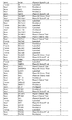
- the nucleic acid sequence information is for one or more genes or regions that include but are not limited to one or more of TGFBR3, FUT9, PDE1C, IMPK, GIGYF2, HMGB3, ZFP14, ACCS, or DPP6. In one embodiment, the nucleic acid sequence information is for one or more genes or regions that include but are not limited to one or more of MYO1B, MIR3675, NBPF1, or GPR111.
- the nucleic acid sequence information is for one or more genes or regions that include but are not limited to one or more of TGFB1I1, PTK2, PGRMC2, LEPROT, LEPR, MUC4, MAGEA11, BOK, BOK-AS1, TSHR, MSN, MYADML, CYP17A1, RXFP1, CRHR2, PLA2G4C, NCOA1, BNC2, or MKRN1.
- the nucleic acid sequence information is for one or more genes or regions that include but are not limited to one or more of ARMC5, C16orf58, SLC5A2, ZNF843, LARP1B, DNAJC6, AS3MT, C10orf32, AS3MT-C10orf32, CNNM2, or RAB 19.
- the invention provides a method of diagnosing a susceptibility to endometriosis in a female subject.
- the method includes detecting in a sample of the subject nucleic acid sequence information on the presence or absence of at least one genetic variation in one or more regions in Figure 2 .
- a susceptibility to endometriosis is diagnosed in the subject if the subject has at least one genetic variation in the one or more genes or regions in Figure 1 or Figure 2 , wherein the at least genetic variation occurs in the gene or region more frequently in a population of female subjects that have endometriosis than in a population of female subjects that does not have endometriosis.
- the nucleic acid sequence information is for one or more genes or regions that include but are not limited to one or more of TGFBR3, FUT9, PDE1C, IMPK, GIGYF2, HMGB3, ZFP14, ACCS, or DPP6. In one embodiment, the nucleic acid sequence information is for one or more genes or regions that include but are not limited to one or more of MYO1B, MIR3675, NBPF1, or GPR111.
- the nucleic acid sequence information is for one or more genes or regions that include but are not limited to one or more of TGFB1I1, PTK2, PGRMC2, LEPROT, LEPR, MUC4, MAGEA11, BOK, BOK-AS1, TSHR, MSN, MYADML, CYP17A1, RXFP1, CRHR2, PLA2G4C, NCOA1, BNC2, or MKRN1.
- the nucleic acid sequence information is for one or more genes or regions that include but are not limited to one or more of ARMC5, C16orf58, SLC5A2, ZNF843, LARP1B, DNAJC6, AS3MT, C10orf32, AS3MT-C10orf32, CNNM2, or RAB 19.
- the invention provides a method that includes providing nucleic acid sequence information from a female subject on the presence or absence of at least genetic variation in one or more genes or regions in Figure 2 .
- a susceptibility to endometriosis in the subject is diagnosed if the subject has at least one genetic variation in the one or more genes or regions in Figure 2 , wherein the at least genetic variation occurs in the gene or region more frequently in a population of female subjects that have endometriosis than in a population of female subjects that does not have endometriosis.
- the nucleic acid sequence information is for one or more genes or regions that include but are not limited to one or more of TGFBR3, FUT9, PDE1C, IMPK, GIGYF2, HMGB3, ZFP14, ACCS, or DPP6. In one embodiment, the nucleic acid sequence information is for one or more genes or regions that include but are not limited to one or more of MYO1B, MIR3675, NBPF1, or GPR111.
- the nucleic acid sequence information is for one or more genes or regions that include but are not limited to one or more of TGFB1I1, PTK2, PGRMC2, LEPROT, LEPR, MUC4, MAGEA11, BOK, BOK-AS1, TSHR, MSN, MYADML, CYP17A1, RXFP1, CRHR2, PLA2G4C, NCOA1, BNC2, or MKRN1.
- the nucleic acid sequence information is for one or more genes or regions that include but are not limited to one or more of ARMC5, C16orf58, SLC5A2, ZNF843, LARP1B, DNAJC6, AS3MT, C10orf32, AS3MT-C10orf32, CNNM2, or RAB19.
- the method further comprises designing or preparing an array, e.g., a CGH array, to measure or detect one or more genetic variations in the regions shown in Figure 1 or Figure 2 .
- the method further comprises providing such a CGH array for the measuring or detecting of one or more genetic variations.
- assaying at least one genetic sample comprises obtaining nucleic acid sequence information.
- obtaining the nucleic acid sequence information is accomplished by one or more methods including but not limited to PCR, sequencing, Northern blots, multiplex ligation-dependent probe amplification (MLPA), molecular beacon, array Comparative Genomic Hybridization, Invader assay, ligase chain reaction (LCR), fluorescence in situ hybridization, or any combination thereof.
- sequencing comprises one or more high-throughput sequencing methods.
- determining whether one or more test subjects have EN, are at risk of EN or have an altered susceptibility to EN includes comparing the nucleic acid sequence information of the one or more test subjects, the at least one genetic variation identified in the one or more test subjects, or a combination thereof, to those of one or more control subjects, e.g., subjects that do not have EN, are not at risk of EN or do not have an enhanced susceptibility to EN.
- the one more control subjects include one or more subjects not suspected of having EN and the one or more test subjects include one or more subjects suspected of having EN.
- the one or more test subjects include one or more subjects with EN, and the one or more control subjects include one or more subjects without EN.
- the one or more test subjects include one or more subjects who are symptomatic for EN, and the one or more control subjects include one or more subjects who are asymptomatic for EN. In some embodiments, the one or more test subjects include one or more subjects that do not present with pain as a major symptom. In some embodiemtn, the one or more test subjects have infertility issues (e.g., up to about 40% of women with EN have infertility issues but have no or little pain related to their EN lesions). In some embodiments, the one or more control subjects include one or more subjects that have increased or decreased susceptibility to EN. In some embodiments, the one or more control subjects include one or more subjects associated or unassociated with a treatment, therapeutic regimen, or any combination thereof.
- determining whether the one or more test subjects have EN, are at risk of EN or have an altered susceptibility to EN includes comparing a gynecological examination, a medical history analysis, or a combination thereof, of the one or more test subjects to the nucleic acid sequence information of the one or more test subjects, at least one genetic variation identified in the one or more test subjects, the nucleic acid sequence information of one or more control subjects, at least one genetic variation identified in the one or more control subjects, or a combination thereof.
- the at least one genetic variation comprises one or more point mutations, single nucleotide polymorphisms (SNPs), single nucleotide variants (SNVs), polymorphisms, translocations, insertions, deletions, amplifications, inversions, microsatellites, interstitial deletions, copy number variations (CNVs), loss of heterozygosity, or any combination thereof.
- the at least one genetic variation includes one or more genetic variations, e.g., CNVs in the genes listed in Figure 1 , e.g., genes having any one of SEQ ID NOs:1-47 or one or more genetic variations in CNV subregions listed in Figure 2 .
- the genetic variation includes one or more genetic variations, e.g., CNVs, that disrupt or modulate one or more genes listed in Figure 3 .
- the at least one genetic variation includes variations such as one or more CNVs that disrupt or modulate the expression or function of one or more RNA transcripts in Figure 4 , e.g., those having any one of SEQ ID NOs:48-149
- the invention provides a method for screening for a therapeutic agent useful for preventing, inhibiting or treating EN.
- the method includes identifying an agent that modulates the function or expression of one or more genes listed in Figure 3 or expression products therefrom, or one or more RNA transcripts mentioned in Figure 4 or expression products thereof.
- the expression products include one or more proteins expressed from a gene listed in Figure 3 or encoded by one or more transcripts mentioned in Figure 4 .
- modulating the function or activity of one or more RNA transcripts or proteins results in an increase in expression.
- modulating the function or activity of one or more RNA transcripts or proteins results in a decrease in expression.
- a method of preventing, inhibiting or treating EN in a subject includes administering one or more agents effective to modulate the function of one or more genes listed in Figure 3 , or expression products therefrom, or one or more RNA transcripts mentioned in Figure 4 , or expression products thereof, thereby preventing, inhibiting or treating the EN.
- the expression products are one or more proteins expressed from a gene listed in Figure 3 , or encoded by one or more RNA transcripts mentioned in Figure 4 or genes in the same pathway (see, e.g., Figure 9 ).
- the one or more agents include but are not limited to a protein, e.g., an antibody, a drug, a combination of drugs, a compound, a combination of compounds, radiation, a genetic sequence, a combination of genetic sequences, heat or cryogenics, or a combination of two or more of any combination thereof.
- a protein e.g., an antibody, a drug, a combination of drugs, a compound, a combination of compounds, radiation, a genetic sequence, a combination of genetic sequences, heat or cryogenics, or a combination of two or more of any combination thereof.
- a method for screening for a therapeutic agent useful for treating EN includes identifying an agent that modulates the function or expression of one or more genes listed in Figure 3 or expression products therefrom.
- the expression products include one or more RNA transcripts in Figure 4 .
- the expression products include one or more proteins expressed from a gene listed in Figure 3 or encoded by one or more RNA transcripts in Figure 4 .
- modulating the function or activity of one or more RNA transcripts or proteins includes an increase in expression.
- modulating the function or activity of one or more RNA transcripts or proteins includes a decrease in expression.
- screening the one or more subjects also includes selecting one or more therapies based on the presence or absence of the one or more genetic variations, e.g., the presence of a genetic variation in at least one gene listed in Figure 3 .
- a method of treating a subject for EN includes administering one or more agents effective to modulate the function of one or more genes listed in Figure 3 , or expression products therefrom, thereby treating EN.
- the expression products include one or more RNA transcripts in Figure 4 .
- the expression products include one or more proteins expressed from a gene in Figure 3 , or encoded by one or more RNA transcripts in Figure 4 .
- the agent may be an antibody, a compound, a combination of compounds, radiation, a genetic sequence, a combination of genetic sequences, heat, cryogenics, and a combination of two or more of any combination thereof.
- TGFBR3 genetic variations e.g., the TGFBR3 CNV or others described herein, may be used in an assay that would facilitate rapid and low cost screening of endometriosis cohorts for the presence of the genetic variation, e.g., deletion, in order to obtain better estimates for the frequency of the variation in such cohorts and diagnose the cause of endometriosis in those who carry the variation.
- the method comprises assaying at least one genetic sample of one or more subjects, nucleic acid sequence information from the one or more subjects, or providing that information, for at least one genetic variation impacting or encompassing TGFBR3.
- the presence in the genetic sample of the at least one genetic variation is used to determine whether the one or more subjects have EN, have an altered susceptibility to EN or are at risk of EN.
- determining whether the one or more subjects have EN, are at risk of EN or have an altered susceptibility to EN includes a gynecological examination and/or medical history analysis of the one or more subjects, e.g., in addition to the nucleic acid sequence information.
- At least one genetic sample is collected from blood, e.g., peripheral blood mononuclear cells (PBMC) or peripheral blood lymphocytes (PBL), saliva, urine, serum, tears, skin, tissue, or hair from at least one subject.
- assaying the at least one genetic sample of one or more subjects includes purifying the at least one genetic sample.
- assaying the at least one genetic sample of the one or more subjects includes amplifying at least one nucleotide or a specific region of one or more chromosomes in the at least one genetic sample.
- assaying the at least one genetic sample of the one or more subjects includes assaying an unamplified sample for at least one nucleotide or a specific region of one or more chromosomes in the at least one genetic sample.
- assaying the at least one genetic sample for at least one genetic variation includes a microarray analysis of the at least one sample.
- the microarray analysis comprises a comparative genomic hybridization (CGH) array analysis.
- the method includes detecting a deletion in TGFBR3, e.g., using a multiplex ligation-dependent probe amplification (MLPA), molecular beacon, aCGH, Invader assay, ligase chain reaction (LCR), or fluorescence in situ hybridization.
- MLPA multiplex ligation-dependent probe amplification
- aCGH molecular beacon
- Invader assay ligase chain reaction (LCR)
- fluorescence in situ hybridization e.g., fluorescence in situ hybridization.
- the invention provides a kit for screening for EN in a subject.
- the kit includes at least one component for assaying a genetic sample from the subject for the presence of at least one genetic variation in the genes listed in Figure 1 or in Figure 2 associated with EN.
- a kit to screen for the TGFBR3 deletion contains PCR primers such as Example 4 OUTER_FWD and OUTER_REV primers or similar primer pairs (see below) that yield a specific amplification product in genetic samples that contain the TGFBR3 deletion but do not yield an amplification product in genetic samples without the TGFBR3 deletion.
- a kit to screen for the TGFBR3 deletion contains an Invader oligonucleotide and primary probe that target the specific junction fragment of DNA sequence resulting from the deletion and produce a signal in genetic samples that contain the TGFBR3 deletion but do not produce a signal in genetic samples without the TGFBR3 deletion.
- the at least one genetic variation is associated with a disruption or aberration of one or more RNA transcripts mentioned in Figure 4 .
- the at least one genetic variation is associated with a disruption or aberration of one or more proteins expressed from one or more genes listed in Figure 3 , or encoded by one or more RNA transcripts mentioned in Figure 4 .
- kits for screening for endometriosis in one or more female subjects comprising reagents for assaying a genetic sample from the one or more subjects for the presence or absence of at least one genetic variation in one or more genes or regions in Figures 1 or 3 , or a combination thereof.
- screening the one or more subjects further comprises selecting one or more therapies based on the presence or absence of the one or more genetic variations.
- the nucleic acid sequencing information is obtained for the whole genome or whole exome from the one or more subjects. In some embodiments, the nucleic acid sequencing information has already been obtained for the whole genome or whole exome from the one or more individuals and the nucleic acid information is obtained from in silico analysis. In other embodiments, the nucleic acid sequencing information is obtained for a selected portion of the whole genome or whole exome.
- SNPs single nucleotide polymorphisms
- SNPs can be located, on average, every 500-1000 base pairs in the human genome. Additional genetic polymorphisms in a human genome can be caused by duplication, insertion, deletion, translocation and/or inversion, of short and/or long stretches of DNA.
- genetic variability among individuals occurs on many scales, ranging from single nucleotide changes, to gross changes in chromosome structure and function.
- CNVs copy number variations
- kits for screening a sample from a subject to detect or determine a risk of or susceptibility to EN are also encompassed by the disclosure.
- Genomic sequences within populations exhibit variability between individuals at many locations in the genome.
- the human genome exhibits sequence variations, which occur on average every 500 base pairs.
- Such genetic variations in nucleic acid sequences are commonly referred to as polymorphisms or polymorphic sites.
- these genetic variations can be found to be associated with EN using the methods disclosed herein.
- these genetic variations comprise point mutations, e.g., single nucleotide polymorphisms (SNPs) or single nucleotide variants (SNVs), polymorphisms, translocations, insertions, deletions, amplifications, inversions, interstitial deletions, copy number variations (CNVs), loss of heterozygosity, or any combination thereof.
- SNPs single nucleotide polymorphisms
- SNVs single nucleotide variants
- CNVs copy number variations
- polymorphisms can comprise any nucleotide position at which two or more sequences are possible in a subject population.
- each version of a nucleotide sequence with respect to the polymorphism can represent a specific allele of the polymorphism.
- genomic DNA from a subject can contain two alleles for any given polymorphic marker, representative of each copy of the marker on each chromosome.
- an allele can be a nucleotide sequence of a given location on a chromosome.
- Polymorphisms can comprise any number of specific alleles.
- a polymorphism can be characterized by the presence of two or more alleles in a population. In some embodiments, the polymorphism can be characterized by the presence of three or more alleles. In some embodiments, the polymorphism can be characterized by four or more alleles, five or more alleles, six or more alleles, seven or more alleles, nine or more alleles, or ten or more alleles. In some embodiments an allele can be associated with one or more diseases or disorders. In some embodiments, genetic variations and alleles can be used to associate an inherited phenotype, for example, susceptibility EN, with a responsible genotype.
- an allele e.g., a risk allele
- genetic variations can be of any measurable frequency in the population, for example, a frequency higher than 10%, a frequency between 5-10%, a frequency between 1-5%, or frequency below 1 %.
- variant alleles can be alleles that differ from a reference allele.
- a variant can be a segment of DNA that differs from the reference DNA, such as a genetic variation.
- genetic variations can be used to track the inheritance of a gene that has not yet been identified, but whose approximate location is known.
- a haplotype can be information regarding the presence or absence of one or more genetic markers in a given chromosomal region in a subject.
- a haplotype can be a segment of DNA characterized by one or more alleles arranged along the segment, for example, a haplotype can comprise one member of the pair of alleles for each genetic variation or locus.
- the haplotype can comprise two or more alleles, three or more alleles, four or more alleles, five or more alleles, or any combination thereof, wherein, each allele can comprise one or more genetic variations along the segment.
- a genetic variation can be a functional aberration that can alter gene function, gene expression, protein expression, protein function, or any combination thereof.
- a genetic variation can be a loss-of-function mutation, gain-of-function mutation, dominant negative mutation, or reversion.
- a genetic variation can be part of a gene's coding region or regulatory regions. Regulatory regions can control gene expression and thus protein expression.
- a regulatory region can be a segment of DNA wherein regulatory proteins, for example, transcription factors, can bind.
- a regulatory region can be positioned near the gene being regulated, for example, positions upstream of the gene being regulated.
- a regulatory region e.g., enhancer element
- variants can include changes that affect a polypeptide or protein, such as a change in expression level, sequence, function, localization, binding partners, or any combination thereof.
- a genetic variation can be a frameshift mutation, nonsense mutation, missense mutation, neutral mutation, or silent mutation.
- sequence differences when compared to a reference nucleotide sequence, can include the insertion or deletion of a single nucleotide, or of more than one nucleotide, resulting in a frame shift; the change of at least one nucleotide, resulting in a change in the encoded amino acid; the change of at least one nucleotide, resulting in the generation of a premature stop codon; the deletion of several nucleotides, resulting in a deletion of one or more amino acids encoded by the nucleotides; the insertion of one or several nucleotides, such as by unequal recombination or gene conversion, resulting in an interruption of the coding sequence of a reading frame; duplication of all or a part of a sequence; transposition; or a rearrangement of a nucleotide sequence.
- sequence changes can alter the polypeptide encoded by the nucleic acid, for example, if the change in the nucleic acid sequence causes a frame shift, the frame shift can result in a change in the encoded amino acids, and/or can result in the generation of a premature stop codon, causing generation of a truncated polypeptide.
- a genetic variation associated with EN can be a synonymous change in one or more nucleotides, for example, a change that does not result in a change in the amino acid sequence.
- Such a polymorphism can, for example, alter splice sites, affect the stability or transport of mRNA, or otherwise affect the transcription or translation of an encoded polypeptide.
- a synonymous mutation can result in the protein product having an altered structure due to rare codon usage that impacts protein folding during translation, which in some cases may alter its function and/or drug binding properties if it is a drug target.
- the changes that can alter DNA increase the possibility that structural changes, such as amplifications or deletions, occur at the somatic level.
- a polypeptide encoded by the reference nucleotide sequence can be a reference polypeptide with a particular reference amino acid sequence, and polypeptides encoded by variant nucleotide sequences can be variant polypeptides with variant amino acid sequences.
- one or more variant polypeptides or proteins can be associated with EN.
- variant polypeptides and changes in expression, localization, and interaction partners thereof can be used to associate an inherited phenotype, EN, with a responsible genotype.
- an EN associated variant polypeptide can be statistically associated with a diagnosis, prognosis, or theranosis of EN.
- sequence variants comprise base variations at a single base position in the genome, and such sequence variants, or polymorphisms, are commonly called single nucleotide polymorphisms (SNPs) or single nucleotide variants (SNVs).
- SNPs single nucleotide polymorphisms
- SNVs single nucleotide variants
- a SNP represents a genetic variant present at greater than or equal to 1% occurrence in a population and in some embodiments a SNP can represent a genetic variant present at any frequency level in a population.
- a SNP can be a nucleotide sequence variation occurring when a single nucleotide at a location in the genome differs between members of a species or between paired chromosomes in a subject.
- SNPs can include variants of a single nucleotide, for example, at a given nucleotide position, some subjects can have a 'G', while others can have a 'C'. SNPs can occur in a single mutational event, and therefore there can be two possible alleles possible at each SNP site; the original allele and the mutated allele. SNPs that are found to have two different bases in a single nucleotide position are referred to as biallelic SNPs, those with three are referred to as triallelic, and those with all four bases represented in the population are quadallelic. In some embodiments, SNPs can be considered neutral. In some embodiments SNPs can affect susceptibility to EN.
- SNP polymorphisms can have two alleles, for example, a subject can be homozygous for one allele of the polymorphism wherein both chromosomal copies of the individual have the same nucleotide at the SNP location, or a subject can be heterozygous wherein the two sister chromosomes of the subject contain different nucleotides.
- the SNP nomenclature as reported herein is be the official Reference SNP (rs) ID identification tag as assigned to each unique SNP by the National Center for Biotechnological Information (NCBI).
- CNVs can be copy number variations/variants
- CNVs can be alterations of the DNA of a genome that results in an abnormal number of copies of one or more sections of DNA.
- CNVs can be inherited or caused by de novo mutation and can be responsible for a substantial amount of human phenotypic variability, behavioral traits, and disease susceptibility.
- CNVs of the current disclosure can be associated with risk of or susceptibility to EN.
- CNVs can impact a single gene or include a contiguous set of genes.
- CNVs can be caused by structural rearrangements of the genome, for example, translocations, insertions, deletions, amplifications, inversions, and interstitial deletions.
- these structural rearrangements occur on one or more chromosomes.
- LCRs Low copy repeats
- CNVs CNVs
- Factors such as size, orientation, percentage similarity and the distance between the copies can influence the susceptibility of LCRs to mediate genomic rearrangement.
- CNVs can account for genetic variation affecting a substantial proportion of the human genome, for example, known CNVs can cover over 15% of the human genome sequence (Estivill and Armengol, supra ).
- CNVs can affect gene expression, phenotypic variation and adaptation by disrupting a gene or altering gene dosage, and can cause disease, for example, microdeletion and microduplication disorders, and can confer susceptibility to diseases and disorders.
- Updated information about the location, type, and size of known CNVs can be found in one or more databases, for example, the Database of Genomic Variants (projects.tcag.ca/variation/), which currently contains data for over 100,000 CNVs.
- microsatellites can be found in the human genome and can be associated with a disease or disorder, including but not limited to, microsatellites.
- Microsatellite markers are stable, polymorphic, easily analyzed, and can occur regularly throughout the genome, making them especially suitable for genetic analysis.
- a polymorphic microsatellite can comprise multiple small repeats of bases, for example, CA repeats, at a particular site wherein the number of repeat lengths varies in a population.
- microsatellites for example, variable number of tandem repeats (VNTRs)
- VNTRs variable number of tandem repeats
- changes in microsatellites can occur during genetic recombination of sexual reproduction, increasing or decreasing the number of repeats found at an allele, or changing allele length.
- a subject can be an individual of any age from whom a sample containing nucleotides is obtained for analysis, e.g., by one or more methods described herein, so as to obtain genetic data, for example, a female adult, child, newborn, or fetus.
- a subject can be any target of therapeutic administration.
- a subject can be a test subject or a reference subject.
- a subject can be associated with EN, asymptomatic or symptomatic, have increased or decreased susceptibility to EN, be associated or unassociated with a treatment or treatment regimen, or any combination thereof.
- a cohort can represent an ethnic group, a patient group, a particular age group, a group not associated with EN, a group associated with EN, a group of asymptomatic female subjects, a group of symptomatic female subjects, or a group or subgroup of female subjects associated with a particular response to a treatment regimen or clinical trial.
- a patient can be a subject afflicted with EN.
- a patient can be a subject not afflicted with EN.
- a female subject can be a test female subject, a female patient or a female candidate for a therapeutic, wherein genomic DNA from the female subject, female patient, or female candidate is obtained for analysis by one or more methods of the present disclosure herein, so as to obtain genetic variation information of the subject, patient or candidate.
- the sample can be obtained prenatally from a female fetus or embryo or from the mother, for example, from fetal or embryonic cells in the maternal circulation.
- the sample can be obtained with the assistance of a health care provider, for example, to draw blood.
- the sample can be obtained without the assistance of a health care provider, for example, where the sample is obtained non-invasively, such as a saliva sample, or a sample comprising buccal cells that is obtained using a buccal swab or brush, or a mouthwash sample.
- the present disclosure also provides methods for assessing genetic variations in female subjects who are members of a target population.
- a target population is in some embodiments a population or group of subjects at risk of developing EN, based on, for example, other genetic factors, biomarkers, biophysical parameters, family history of EN, previous screening or medical history, or any combination thereof.
- female subjects can be from specific age subgroups, such as those over the age of 1, over the age of 2, over the age of 3, over the age of 4, over the age of 5, over the age of 6, over the age of 7, over the age of 8, over the age of 9, over the age of 10, over the age of 15, over the age of 20, over the age of 25, over the age of 30, over the age of 35, over the age of 40, over the age of 45, over the age of 50, over the age of 55, over the age of 60, over the age of 65, over the age of 70, over the age of 75, over the age of 80, or over the age of 85.
- specific age subgroups such as those over the age of 1, over the age of 2, over the age of 3, over the age of 4, over the age of 5, over the age of 6, over the age of 7, over the age of 8, over the age of 9, over the age of 10, over the age of 15, over the age of 20, over the age of 25, over the age of 30, over the age of 35, over the age of 40, over the age of 45, over the age of 50
- Other embodiments of the disclosure pertain to other age groups, such as subjects aged less than 85, such as less than age 80, less than age 75, less than age 70, less than age 65, less than age 60, less than age 55, less than age 50, less than age 45, less than age 40, less than age 35, less than age 30, less than age 25, less than age 20, less than age 15, less than age 10, less than age 9, less than age 8, less than age 6, less than age 5, less than age 4, less than age 3, less than age 2, or less than age 1.
- Other embodiments relate to female subjects with age at onset of the disease in any of particular age or age ranges defined by the numerical values described in the above or other numerical values bridging these numbers.
- a range of ages can be relevant in certain embodiments, such as age at onset at more than age 15 but less than age 20.
- Other age ranges are however also contemplated, including all age ranges bracketed by the age values listed in the above.
- Such embodiments relate to female subjects that are from one or more human populations including, but not limited to, Caucasian, European, American, Ashkenazi Jewish, Sephardi Jewish, Eurasian, Asian, Central/South Asian, East Asian, Middle Eastern, African, Hispanic, and Oceanic populations.
- European populations include, but are not limited to, Swedish, Norwegian, Finnish, Russian, Danish, Icelandic, Irish, Kelt, English, Scottish, Dutch, Belgian, French, German, Spanish, Portuguese, Italian, Polish, Bulgarian, Slavic, Serbian, Laun, Czech, Greek and Turkish populations.
- the racial contribution in female subjects can also be determined by genetic analysis, for example, genetic analysis of ancestry can be carried out using unlinked microsatellite markers such as those set out in Smith et al. (Am. J. Hum. Genet., 74:1001 (2004 )).
- Samples that are suitable for use in the methods described herein can be from a subject and can contain genetic or proteinaceous material, for example, genomic DNA (gDNA).
- Genetic material can be extracted from one or more biological samples including but not limited to, blood, saliva, urine, mucosal scrapings of the lining of the mouth, expectorant, serum, tears, skin, tissue, or hair.
- the sample can comprise cells or tissue, for example, cell lines.
- a blood cell such as a B lymphocyte, T lymphocyte, leukocyte, erythrocyte, macrophage, or neutrophil
- a muscle cell such as a skeletal cell,
- a cell from which gDNA is obtained can be at a particular developmental level including, for example, a hematopoietic stem cell or a cell that arises from a hematopoietic stem cell such as a red blood cell, B lymphocyte, T lymphocyte, natural killer cell, neutrophil, basophil, eosinophil, monocyte, macrophage, or platelet.
- a hematopoietic stem cell such as a red blood cell, B lymphocyte, T lymphocyte, natural killer cell, neutrophil, basophil, eosinophil, monocyte, macrophage, or platelet.
- stem cell can be used including, without limitation, an embryonic stem cell, adult stem cell, an induced pluripotent stem cell created from an adult cell type such as fibroblasts derived from skin or pluripotent stem cell.
- a sample can be processed for DNA isolation, for example, DNA in a cell or tissue sample can be separated from other components of the sample.
- Cells can be harvested from a biological sample using standard techniques known in the art, for example, by centrifuging a cell sample and resuspending the pelleted cells, for example, in a buffered solution, for example, phosphate-buffered saline (PBS).
- PBS phosphate-buffered saline
- the cells after centrifuging the cell suspension to obtain a cell pellet, the cells can be lysed to extract DNA.
- the sample can be concentrated and/or purified to isolate DNA. All samples obtained from a female subject, including those subjected to any sort of further processing, are considered to be obtained from the subject.
- standard techniques and kits known in the art can be used to extract genomic DNA from a biological sample, including, for example, phenol extraction, a QIAamp® Tissue Kit (Qiagen, Chatsworth, Calif.), a Wizard® Genomic DNA purification kit (Promega), or a Qiagen Autopure method using Puregene chemistry, which can enable purification of highly stable DNA well-suited for archiving.
- determining the identity of an allele or determining copy number can, but need not, include obtaining a sample comprising DNA from a subject, and/or assessing the identity, copy number, presence or absence of one or more genetic variations and their chromosomal locations in the sample.
- the individual or organization that performs the determination need not actually carry out the physical analysis of a sample from a subject.
- the methods can include using information obtained by analysis of the sample by a third party.
- the methods can include steps that occur at more than one site. For example, a sample can be obtained from a subject at a first site, such as at a health care provider or at the subject's home in the case of a self-testing kit. The sample can be analyzed at the same or a second site, for example, at a laboratory or other testing facility.
- screening a subject may include diagnosing or determining, theranosing, or determining the risk of or susceptibility to developing (prognosing) EN.
- the disclosure is a method of determining the presence of, a risk of developing or a susceptibility to, EN, by detecting at least one genetic variation in a sample from a subject as described herein.
- detection of particular alleles, markers, variations, or haplotypes is indicative of the presence of or susceptibility to EN.
- Susceptibility e.g., being at-risk
- Susceptibility assessment can involve detecting particular genetic variations in the genome of individuals undergoing assessment. Particular genetic variations are found more frequently in individuals with EN, than in individuals without EN. Therefore, these genetic variations have predictive value for detecting EN, risk of developing EN, or a susceptibility to EN, in an individual.
- a genetic variation described herein to be associated with susceptibility of EN represent functional variants predisposing to the disease.
- a genetic variation can confer a susceptibility of the condition, for example, carriers of the genetic variation are at a different risk of the condition than non-carriers.
- the presence of a genetic variation is indicative of increased susceptibility to or the presence of EN.
- Screening can be performed using any method.
- screening can be performed using Polymerase Chain Reaction (PCR).
- PCR Polymerase Chain Reaction
- ACGH Array Comparative Genomic Hybridization
- the genetic variation information as it relates to the current disclosure can be used in conjunction with any symptomatic screening tests.
- information from any of the above screening methods can be used to define a subject as a test subject or reference subject.
- information from any of the above screening methods can be used to associate a subject with a test or reference population, for example, a subject in a population.
- an association with EN can be determined by the statistical likelihood of the presence of a genetic variation in a subject with EN, for example, an unrelated individual or a first or second-degree relation of the subject. In some embodiments, an association with EN can be determined by determining the statistical likelihood of the absence of a genetic variation in an unaffected reference subject, for example, an unrelated individual or a first or second-degree relation of the subject.
- the methods described herein can include obtaining and analyzing a sample from one or more suitable reference subjects.
- susceptibility can be proneness of a subject towards the development of EN, or towards resisting development of EN, than one or more control subjects.
- susceptibility can encompass increased susceptibility.
- particular nucleic acid variations of the disclosure as described herein can be characteristic of increased susceptibility to development of EN.
- susceptibility can encompass decreased susceptibility, for example, particular nucleic variations of the disclosure as described herein can be characteristic of decreased susceptibility to development of EN.
- a subject at risk of developing EN has a greater chance of developing EN relative to the general population or to one or more subjects without a specific genetic variation.
- a genetic variation predictive of susceptibility to or presence of EN can be one where the particular genetic variation is more frequently present in a subject with the condition (affected), compared to the frequency of its presence in a reference group (control), such that the presence of the genetic variation is indicative of susceptibility to or presence of EN.
- the reference group can be a population sample, for example, a random sample from the general population or a mixture of two or more samples from a population.
- disease-free controls can be characterized by the absence of one or more specific EN-associated symptoms, for example, individuals who have not experienced symptoms associated with EN.
- the disease-free control group is characterized by the absence of one or more EN-specific risk factors, for example, at least one genetic and/or environmental risk factor.
- a reference sequence can be referred to for a particular site of genetic variation.
- a reference allele can be a wild-type allele and can be chosen as either the first sequenced allele or as the allele from a control individual.
- one or more reference subjects can be characteristically matched with one or more affected subjects, for example, with matched aged, gender or ethnicity.
- a person skilled in the art can appreciate that for genetic variations with two or more alleles present in the population being studied, and wherein one allele can found in increased frequency in a group of individuals with EN in the population, compared with controls, the other allele(s) of the marker can be found in decreased frequency in the group of individuals with the trait or disease, compared with controls.
- one allele of the marker for example, the allele found in increased frequency in individuals with EN, can be the at-risk allele, while the other allele(s) can be neutral or even protective.
- a genetic variant associated with EN can be used to predict the susceptibility of EN for a given genotype.
- there can be one or more possible genotypes for example, homozygote for the at-risk variant (e.g., in autosomal recessive disorders), heterozygote, and non-carrier of the at-risk variant.
- susceptibility associated with variants at multiple loci can be used to estimate overall susceptibility.
- there can be k (k 3 ⁇ n * 2 ⁇ P) possible genotypes; wherein n can be the number of autosomal loci and p can be the number of gonosomal (sex chromosomal) loci.
- the overall susceptibility associated with a particular genotype combination can be the product of the susceptibility values for the genotype at each locus. If the susceptibility presented is the relative susceptibility for a person, or a specific genotype for a person, compared to a reference population, then the combined susceptibility can be the product of the locus specific susceptibility values and can correspond to an overall susceptibility estimate compared with a population.
- the combined susceptibility can correspond to an estimate that compares the person with a given combination of genotypes at all loci to a group of individuals who do not carry at-risk variants at any of those loci.
- the group of non-carriers of any at-risk variant can have the lowest estimated susceptibility and can have a combined susceptibility, compared with itself, for example, non-carriers, of 1.0, but can have an overall susceptibility, compared with the population, of less than 1.0.
- Genetic variations described herein can form the basis of risk analysis that combines other genetic variations known to increase risk of EN, or other genetic risk variants for EN.
- a plurality of variants can be used for overall risk assessment. These variants are in some embodiments selected from the genetic variations as disclosed herein.
- Other embodiments include the use of the variants of the present disclosure in combination with other variants known to be useful for screening for EN or a susceptibility to EN.
- the genotype status of a plurality of genetic variations, markers and/or haplotypes is determined in an individual, and the status of the individual compared with the population frequency of the associated variants, or the frequency of the variants in clinically healthy subjects, such as age-matched and sex-matched subjects.
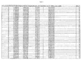
- a threshold log ratio value can be used to determine losses and gains.
- DNA Analytics a log2ratio cutoff of 0.25 and -0.25 to classify CNV gains and losses respectively may be used.
- DNAcopy a log2ratio cutoff of 0.35 and -0.35 to classify CNV gains and losses respectively may be used.
- the information and calls from two or more of the methods described herein can be compared to each other to identify significant genetic variations more or less stringently.
- CNV calls generated by both DNA Analytics and DNAcopy algorithms may be defined as stringent CNVs.
- significant or stringent genetic variations can be tagged as identified or called if it can be found to have a minimal reciprocal overlap to a genetic variation detected by one or more platforms and/or methods described herein.
- a minimum of 50% reciprocal overlap can be used to tag the CNVs as identified or called.
- multivariate analyses or joint risk analyses can subsequently be used to determine the overall risk conferred based on the genotype status at the multiple loci.
- a multiplicative model for example, assuming that the risk of individual risk variants multiply to establish the overall effect, allows for a straight-forward calculation of the overall risk for multiple markers.
- the multiplicative model is a parsimonious model that usually fits the data of complex traits reasonably well. Deviations from multiplicity have been rarely described in the context of common variants for common diseases, and if reported are usually only suggestive since very large sample sizes can be required to be able to demonstrate statistical interactions between loci. Assessment of risk based on such analysis can subsequently be used in the methods, uses and kits of the disclosure, as described herein.
- the significance of increased or decreased susceptibility can be measured by a percentage.
- a significant increased susceptibility can be measured as a relative susceptibility of at least 1.2, including but not limited to: at least 1.2, at least 1.3, at least 1.4, at least 1.5, at least 1.6, at least 1.7, 1.8, at least 1.9, at least 2.0, at least 2.5, at least 3.0, at least 4.0, at least 5.0, at least 6.0, at least 7.0, at least 8.0, at least 9.0, at least 10.0, and at least 15.0.
- a relative susceptibility of at least 2.0, at least 3.0, at least 4.0, at least, 5.0, at least 6.0, or at least 10.0 is significant.
- a significant increase in susceptibility is at least about 20%, including but not limited to about 25%, 30%, 35%, 40%, 45%, 50%, 55%, 60%, 65%, 70%, 75%, 80%, 85%, 90%, 95%, 100%, 150%, 200%, 300%, 400%, 500%, 600%, 700%, 800%, 900%, 1000%, and 1500%. In one particular embodiment, a significant increase in susceptibility is at least 100%.
- a significant increase in susceptibility is at least 200%, at least 300%, at least 400%, at least 500%, at least 700%, at least 800%, at least 900% and at least 1000%.
- Other cutoffs or ranges as deemed suitable by the person skilled in the art to characterize the disclosure are also contemplated, and those are also within scope of the present disclosure.
- a significant increase in susceptibility is characterized by a p-value, such as a p-value of less than 0.5, less than 0.4, less than 0.3, less than 0.2, less than 0.1, less than 0.05, less than 0.01, less than 0.001, less than 0.0001, less than 0.00001, less than 0.000001, less than 0.0000001, less than 0.00000001, or less than 0.000000001.
- a p-value such as a p-value of less than 0.5, less than 0.4, less than 0.3, less than 0.2, less than 0.1, less than 0.05, less than 0.01, less than 0.001, less than 0.0001, less than 0.00001, less than 0.000001, less than 0.0000001, less than 0.00000001, or less than 0.000000001.
- an individual who is at a decreased susceptibility for or the lack of presence of EN can be an individual in whom at least one genetic variation, conferring decreased susceptibility for or the lack of presence of EN is identified.
- the genetic variations conferring decreased susceptibility are also protective.
- the genetic variations can confer a significant decreased susceptibility of or lack of presence of EN.
- significant decreased susceptibility can be measured as a relative susceptibility of less than 0.9, including but not limited to less than 0.9, less than 0.8, less than 0.7, less than 0,6, less than 0.5, less than 0.4, less than 0.3, less than 0.2 and less than 0.1.
- the decrease in susceptibility is at least 20%, including but not limited to at least 25%, at least 30%, at least 35%, at least 40%, at least 45%, at least 50%, at least 55%, at least 60%, at least 65%, at least 70%, at least 75%, at least 80%, at least 85%, at least 90%, at least 95% and at least 98%.
- a significant decrease in susceptibility is characterized by a p-value, such as a p-value of less than 0.05, less than 0.01, less than 0.001, less than 0.0001, less than 0.00001, less than 0.000001, less than 0.0000001, less than 0.00000001, or less than 0.000000001.
- a p-value such as a p-value of less than 0.05, less than 0.01, less than 0.001, less than 0.0001, less than 0.00001, less than 0.000001, less than 0.0000001, less than 0.00000001, or less than 0.000000001.
- Other tests for significance can be used, for example, a Fisher's exact test.
- Other statistical tests of significance known to the skilled person are also contemplated and are also within scope of the disclosure.
- the significance of increased or decreased susceptibility can be determined according to the ratio of measurements from a test subject to a reference subject.
- losses or gains of one or more CNVs can be determined according to a threshold log 2 ratio determined by these measurements.
- a log 2 ratio value greater than 0.35 is indicative of a gain of one or more CNVs.
- a log 2 ratio value less than -0.35 is indicative of a loss of one or more CNVs.
- the combined or overall susceptibility associated with a plurality of variants associated with EN can also be assessed, for example, the genetic variations described herein to be associated with susceptibility to EN can be combined with other common genetic risk factors. Combined risk for such genetic variants can be estimated in an analogous fashion to the methods described herein.
- Calculating risk conferred by a particular genotype for the individual can be based on comparing the genotype of the individual to previously determined risk expressed, for example, as a relative risk (RR) or an odds ratio (OR), for the genotype, for example, for a heterozygous carrier of an at-risk variant for EN.
- An odds ratio can be a statistical measure used as a metric of causality. For example, in genetic disease research it can be used to convey the significance of a variant in a disease cohort relative to an unaffected/normal cohort.
- the calculated risk for the individual can be the relative risk for a subject, or for a specific genotype of a subject, compared to the average population.
- the average population risk can be expressed as a weighted average of the risks of different genotypes, using results from a reference population, and the appropriate calculations to calculate the risk of a genotype group relative to the population can then be performed.
- the risk for an individual can be based on a comparison of particular genotypes, for example, heterozygous carriers of an at-risk allele of a marker compared with non-carriers of the at-risk allele.
- Using the population average can, in certain embodiments, be more convenient, since it provides a measure which can be easy to interpret for the user, for example, a measure that gives the risk for the individual, based on his/her genotype, compared with the average in the population.
- a genetic variation is correlated to EN by referencing genetic variation data to a look-up table that comprises correlations between the genetic variation and EN.
- the genetic variation in certain embodiments comprises at least one indication of the genetic variation.
- the table comprises a correlation for one genetic variation.
- the table comprises a correlation for a plurality of genetic variations. In both scenarios, by referencing to a look-up table that gives an indication of a correlation between a genetic variation and EN, a risk for EN, or a susceptibility to EN, can be identified in the individual from whom the sample is derived.
- the screening applications of EN-associated genetic variations can, for example, be performed by an individual, a health professional, or a third party, for example, a service provider who interprets genotype information from the subject.
- a medical professional can initiate or modify treatment after receiving information regarding a subject's screening for EN, for example.
- a medical professional can recommend a change in therapy.
- a medical professional can enroll a subject in a clinical trial for, by way of example, detecting correlations between a haplotype as described herein and any measurable or quantifiable parameter relating to the outcome of the treatment as described above.
- databases that include a list of genetic variations as described herein, and wherein the list can be largely or entirely limited to genetic variations identified as useful for screening EN as described herein.
- the list can be stored, for example, on a flat file or computer-readable medium.
- the databases can further include information regarding one or more subjects, for example, whether a subject is affected or unaffected, clinical information such as endophenotype, age of onset of symptoms, any treatments administered and outcomes, for example, data relevant to pharmacogenomics, diagnostics, prognostics or theranostics, and other details, for example, data about the disorder in the subject, or environmental or other genetic factors.
- the databases can be used to detect correlations between a particular haplotype and the information regarding the subject.
- the methods described herein can also include the generation of reports for use, for example, by a subject, care giver, or researcher, that include information regarding a subject's genetic variations, and optionally further information such as treatments administered, treatment history, medical history, predicted response, and actual response.
- the reports can be recorded in a tangible medium, e.g., a computer-readable disk, a solid state memory device, or an optical storage device.
- screening of EN can be made by examining or comparing changes in expression, localization, binding partners, and composition of a polypeptide encoded by a nucleic acid associated with EN, for example, in those instances where the genetic variations of the present disclosure results in a change in the composition or expression of the polypeptide and/or RNA, for example, mRNAs, miRNAs, and other noncoding RNAs (ncRNAs).
- RNA for example, mRNAs, miRNAs, and other noncoding RNAs (ncRNAs).
- screening of EN can be made by examining expression and/or composition of one of these polypeptides and/or RNA, or another polypeptide and/or RNA encoded by a nucleic acid associated with EN, in those instances where the genetic variation of the present disclosure results in a change in the expression, localization, binding partners, and/or composition of the polypeptide and/or RNA.
- screening can comprise diagnosing a subject.
- screening can comprise determining a prognosis of a subject, for example, determining the susceptibility of developing EN.
- screening can comprise theranosing a subject.
- the genetic variations described herein that show association to EN can play a role through their effect on one or more of these nearby genes.
- a deletion of a chromosomal segment comprising a particular gene, or a fragment of a gene can either result in an altered composition or expression, or both, of the encoded protein and/or mRNA.
- duplications, or high number copy number variations are in general expected to result in increased expression of encoded polypeptide and/or RNA if the duplication encompasses the whole gene.
- segments of DNA can be duplicated, triplicated, quadruplicated, or amplified many times and result in increasingly higher levels of expression of the gene if it is encompassed by these multiplicated segments of DNA.
- breakpoints of a duplication or other level of amplification can disrupt a gene and thus result in loss of function, such as the expressed protein encoded by the transcript is truncated.
- an amplified segment of DNA can occur in tandem (e.g., multiple gene copies adjacent to each other on the chromosome) or can insert into a site far away from the original chromosomal location or even on another chromosome.
- a gene not contained within the amplified segment of DNA is impacted by the chromosomal rearrangement.
- Such complex rearrangements can be mapped, for example, by fluorescence in situ hybridization (FISH) methods.
- FISH fluorescence in situ hybridization
- Other possible mechanisms affecting genes within or near a genetic variation region include, for example, effects on transcription, effects on RNA splicing, alterations in relative amounts of alternative splice forms of mRNA, effects on RNA stability, effects on transport from the nucleus to cytoplasm, and effects on the efficiency and accuracy of translation.
- DNA variations can be detected directly, using the subjects unamplified or amplified genomic DNA, or indirectly, using RNA or DNA obtained from the subject's tissue(s) that are present in an aberrant form or expression level as a result of the genetic variations of the disclosure showing association to EN.
- the genetic variations of the disclosure showing association to EN can affect the expression of a gene within the genetic variation region.
- Certain genetic variation regions can have flanking duplicated segments, and genes within such segments can have altered expression and/or composition as a result of such genomic alterations.
- regulatory elements affecting gene expression can be located far away, even as far as tens or hundreds of kilobases away, from the promoter region of a gene.
- regulatory elements for genes that are located outside the genetic variation region can be located within the genetic variation, and thus affect the expression of genes located outside the genetic variation. It is thus contemplated that the detection of the genetic variations described herein, can be used for assessing expression for one or more of associated genes.
- genetic variations of the disclosure showing association to EN can affect protein expression at the translational level. It can be appreciated by those skilled in the art that this can occur by increased or decreased expression of one or more microRNAs (miRNAs) that regulates expression of a protein known to be important, or implicated, in the cause, onset, or progression of EN. Increased or decreased expression of the one or more miRNAs can result from gain or loss of the whole miRNA gene, disruption of a portion of the gene (e.g., by an indel or CNV), or even a single base change (SNP or SNV) that produces an altered, non-functional or aberrant functioning miRNA sequence.
- miRNAs microRNAs
- protein for example, one known to cause EN by increased or decreased expression
- expression of protein can result due to a genetic variation that results in alteration of an existing miRNA binding site within the protein's mRNA transcript, or even creates a new miRNA binding site that leads to aberrant protein expression.
- a variety of methods can be used for detecting protein composition and/or expression levels, including but not limited to enzyme linked immunosorbent assays (ELISA), Western blots, spectroscopy, mass spectrometry, peptide arrays, colorimetry, electrophoresis, isoelectric focusing, immunoprecipitations, immunoassays, and immunofluorescence and other methods well-known in the art.
- ELISA enzyme linked immunosorbent assays
- Western blots Western blots
- spectroscopy spectroscopy
- mass spectrometry mass spectrometry
- peptide arrays peptide arrays
- colorimetry electrophoresis
- isoelectric focusing isoelectric focusing
- immunoprecipitations immunoassays
- immunofluorescence and other methods well-known in the art can be assessed for the presence of an alteration in the expression and/or an alteration in composition of the polypeptide encoded by a nucleic acid associated with EN.
- alteration in the polypeptide expression or composition refers to an alteration in expression or composition in a test sample, as compared to the expression or composition of the polypeptide in a control sample.
- Such alteration can, for example, be an alteration in the quantitative polypeptide expression or can be an alteration in the qualitative polypeptide expression, for example, expression of a mutant polypeptide or of a different splicing variant, or a combination thereof
- screening for EN can be made by detecting a particular splicing variant encoded by a nucleic acid associated with EN, or a particular pattern of splicing variants.
- Antibodies can be polyclonal or monoclonal and can be labeled or unlabeled. An intact antibody or a fragment thereof can be used.
- the term "labeled", with regard to the probe or antibody, is intended to encompass direct labeling of the probe or antibody by coupling a detectable substance to the probe or antibody, as well as indirect labeling of the probe or antibody by reactivity with another reagent that is directly labeled as previously described herein.
- Other non-limiting examples of indirect labeling include detection of a primary antibody using a labeled secondary antibody, for example, a fluorescently-labeled secondary antibody and end-labeling of a DNA probe with biotin such that it can be detected with fluorescently-labeled streptavidin.
- Detecting specific genetic variations for example, polymorphic markers and/or haplotypes, copy number, absence or presence of an allele, or genotype associated with EN as described herein, can be accomplished by methods known in the art for analyzing nucleic acids and/or detecting sequences at polymorphic or genetically variable sites, for example, amplification techniques, hybridization techniques, sequencing, arrays, or any combination thereof.
- methods known in the art for analyzing nucleic acids and/or detecting sequences at polymorphic or genetically variable sites for example, amplification techniques, hybridization techniques, sequencing, arrays, or any combination thereof.
- one or more alleles at polymorphic markers including microsatellites, SNPs, CNVs, or other types of genetic variations, can be identified in a sample obtained from a subject.
- nucleic acids and polypeptides described herein can be used in methods and kits of the present disclosure.
- aptamers that specifically bind the nucleic acids and polypeptides described herein can be used in methods and kits of the present disclosure.
- a nucleic acid can comprise a deoxyribonucleotide (DNA) or ribonucleotide (RNA), whether singular or in polymers, naturally occurring or non-naturally occurring, double-stranded or single-stranded, coding, for example, a translated gene, or non-coding, for example, a regulatory region, or any fragments, derivatives, mimetics or complements thereof
- nucleic acids can comprise oligonucleotides, nucleotides, polynucleotides, nucleic acid sequences, genomic sequences, antisense nucleic acids, DNA regions, probes, primers, genes, regulatory regions, introns, exons, open-reading frames, binding sites, target nucle
- isolated nucleic acids are separated from nucleic acids that normally flank the gene or nucleotide sequence (as in genomic sequences) and/or has been completely or partially purified from other transcribed sequences (e.g., as in an RNA library).
- isolated nucleic acids of the disclosure can be substantially isolated with respect to the complex cellular milieu in which it naturally occurs, or culture medium when produced by recombinant techniques, or chemical precursors or other chemicals when chemically synthesized.
- the isolated material can form part of a composition, for example, a crude extract containing other substances, buffer system or reagent mix.
- the material can be purified to essential homogeneity using methods known in the art, for example, by polyacrylamide gel electrophoresis (PAGE) or column chromatography (e.g., HPLC).
- PAGE polyacrylamide gel electrophoresis
- HPLC column chromatography
- isolated also can refer to nucleic acids that are separated from the chromosome with which the genomic DNA is naturally associated.
- the isolated nucleic acid molecule can contain less than about 250 kb, 200 kb, 150 kb, 100 kb, 75 kb, 50 kb, 25 kb, 10 kb, 5 kb, 4 kb, 3 kb, 2kb, 1 kb, 0.5 kb or 0.1 kb of the nucleotides that flank the nucleic acid molecule in the gDNA of the cell from which the nucleic acid molecule is derived.
- Nucleic acids can be fused to other coding or regulatory sequences can be considered isolated.
- recombinant DNA contained in a vector is included in the definition of "isolated” as used herein.
- isolated nucleic acids can include recombinant DNA molecules in heterologous host cells or heterologous organisms, as well as partially or substantially purified DNA molecules in solution. Isolated nucleic acids also encompass in vivo and in vitro RNA transcripts of the DNA molecules of the present disclosure.
- An isolated nucleic acid molecule or nucleotide sequence can be synthesized chemically or by recombinant means.
- nucleotide sequences can be useful, for example, in the manufacture of the encoded polypeptide, as probes for isolating homologous sequences (e.g., from other mammalian species), for gene mapping (e.g., by in situ hybridization with chromosomes), or for detecting expression of the gene, in tissue (e.g., human tissue), such as by Northern blot analysis or other hybridization techniques disclosed herein.
- tissue e.g., human tissue
- the disclosure also pertains to nucleic acid sequences that hybridize under high stringency hybridization conditions, such as for selective hybridization, to a nucleotide sequence described herein
- Such nucleic acid sequences can be detected and/or isolated by allele- or sequence-specific hybridization (e.g., under high stringency conditions).
- the percent identity between the two sequences is a function of the number of identical positions shared by the sequences, taking into account the number of gaps, and the length of each gap, which need to be introduced for optimal alignment of the two sequences.
- the length of a sequence aligned for comparison purposes is at least 30%, at least 40%, at least 50%, at least 60%, at least 70%, at least 80%, at least 90%, or at least 95%, of the length of the reference sequence.
- the actual comparison of the two sequences can be accomplished by well-known methods, for example, using a mathematical algorithm.
- a non-limiting example of such a mathematical algorithm is described in Karlin, and Altschul, Proc. Natl. Acad. Sci. USA, 90:5873 (1993 ). Such an algorithm is incorporated into the NBLAST and XBLAST programs (version 2.0), as described in Altschul et al., Nucleic Acids Res., 25:3389 (1997 ).
- any relevant parameters of the respective programs can be used.
- Other examples include the algorithm of Myers and Miller, CABIOS (1989 ), ADVANCE, ADAM, BLAT, and FASTA.
- the percent identity between two amino acid sequences can be accomplished using, for example, the GAP program in the GCG software package (Accelrys, Cambridge, UK).
- Probes can be oligonucleotides that hybridize in a base-specific manner to a complementary strand of a nucleic acid molecule.
- Probes can include primers, which can be a single-stranded oligonucleotide probe that can act as a point of initiation of template-directed DNA synthesis using methods including but not limited to, polymerase chain reaction (PCR) and ligase chain reaction (LCR) for amplification of a target sequence.
- PCR polymerase chain reaction
- LCR ligase chain reaction
- probes for detection of amplified or unamplified nucleic acid molecules can also include an Invader oligonucleotide and probe pair.
- Oligonucleotides can include segments or fragments of nucleic acid sequences, or their complements.
- DNA segments can be between 5 and 10,000 contiguous bases, and can range from 5, 10, 12, 15, 20, or 25 nucleotides to 10, 15, 20, 25, 30, 40, 50, 100, 200, 500, 1000 or 10,000 nucleotides.
- probes and primers can include polypeptide nucleic acids (PNA), as described in Nielsen, P. et al., Science 254: 1497-1500 (1991 ).
- a probe or primer can comprise a region of nucleotide sequence that hybridizes to at least about 15, typically about 20-25, and in certain embodiments about 40, 50 or 75, consecutive nucleotides of a nucleic acid molecule.
- the present disclosure also provides isolated nucleic acids, for example, probes or primers, that contain a fragment or portion that can selectively hybridize to a nucleic acid that comprises, or consists of, a nucleotide sequence, wherein the nucleotide sequence can comprise at least one polymorphism or polymorphic allele contained in the genetic variations described herein or the wild-type nucleotide that is located at the same position, or the compliments thereof
- the probe or primer can be at least 70% identical, at least 80% identical, at least 85% identical, at least 90% identical, or at least 95% identical, to the contiguous nucleotide sequence or to the complement of the contiguous nucleotide sequence.
- a nucleic acid probe can be an oligonucleotide capable of hybridizing with a complementary region of a gene associated with EN containing a genetic variation described herein.
- the nucleic acid fragments of the disclosure can be used as probes or primers in assays such as those described herein.
- DNA can be amplified and/or can be labeled (e.g., radiolabeled, fluorescently labeled) and used as a probe for screening, for example, a cDNA library derived from an organism.
- cDNA can be derived from mRNA and can be contained in a suitable vector.
- corresponding clones can be isolated, DNA obtained fallowing in vivo excision, and the cloned insert can be sequenced in either or both orientations by art-recognized methods to identify the correct reading frame encoding a polypeptide of the appropriate molecular weight.
- the polypeptide and the DNA encoding the polypeptide can be isolated, sequenced and further characterized.
- nucleic acid can comprise one or more polymorphisms, variations, or mutations, for example, single nucleotide polymorphisms (SNPs), copy number variations (CNVs), for example, insertions, deletions, inversions, and translocations.
- SNPs single nucleotide polymorphisms
- CNVs copy number variations
- nucleic acids can comprise analogs, for example, phosphorothioates, phosphoramidates, methyl phosphonate, chiral methyl phosphonates, 2-0-methyl ribonucleotides, or modified nucleic acids, for example, modified backbone residues or linkages, or nucleic acids combined with carbohydrates, lipids, protein or other materials, or peptide nucleic acids (PNAs), for example, chromatin, ribosomes, and transcriptosomes.
- nucleic acids can comprise nucleic acids in various structures, for example, A DNA, B DNA, Z-form DNA, siRNA, tRNA, and ribozymes.
- the nucleic acid may be naturally or non-naturally polymorphic, for example, having one or more sequence differences, for example, additions, deletions and/or substitutions, as compared to a reference sequence.
- a reference sequence can be based on publicly available information, for example, the U.C. Santa Cruz Human Genome Browser Gateway (genome.ucsc.edu/cgi-bin/hgGateway) or the NCBI website (www.ncbi.nlm.nih.gov).
- a reference sequence can be determined by a practitioner of the present disclosure using methods well known in the art, for example, by sequencing a reference nucleic acid.
- a probe can hybridize to an allele, SNP, or CNV as described herein. In some embodiments, the probe can bind to another marker sequence associated with EN as described herein.
- Control probes can also be used, for example, a probe that binds a less variable sequence, for example, a repetitive DNA associated with a centromere of a chromosome, can be used as a control.
- probes can be obtained from commercial sources.
- probes can be synthesized, for example, chemically or in vitro, or made from chromosomal or genomic DNA through standard techniques.
- sources of DNA that can be used include genomic DNA, cloned DNA sequences, somatic cell hybrids that contain one, or a part of one, human chromosome along with the normal chromosome complement of the host, and chromosomes purified by flow cytometry or microdissection.
- the region of interest can be isolated through cloning, or by site-specific amplification using PCR.
- a detectable label can comprise any label capable of detection by a physical, chemical, or a biological process for example, a radioactive label, such as 32 P or 3 H, a fluorescent label, such as FITC, a chromophore label, an affinity-ligand label, an enzyme label, such as alkaline phosphatase, horseradish peroxidase, or I2 galactosidase, an enzyme cofactor label, a hapten conjugate label, such as digoxigenin or dinitrophenyl, a Raman signal generating label, a magnetic label, a spin label, an epitope label, such as the FLAG or HA epitope, a luminescent label, a heavy atom label, a nanoparticle label, an electrochemical label, a light scattering label, a spherical shell label, semiconductor nanocry
- a nucleotide can be directly incorporated into a probe with standard techniques, for example, nick translation, random priming, and PCR labeling.
- Non-limiting examples of label moieties useful for detection include, without limitation, suitable enzymes such as horseradish peroxidase, alkaline phosphatase, beta-galactosidase, or acetylcholinesterase; members of a binding pair that are capable of forming complexes such as streptavidin/biotin, avidin/biotin or an antigen/antibody complex including, for example, rabbit IgG and anti-rabbit IgG; fluorophores such as umbelliferone, fluorescein, fluorescein isothiocyanate, rhodamine, tetramethyl rhodamine, eosin, green fluorescent protein, erythrosin, coumarin, methyl coumarin, pyrene, malachite green, stilbene, lucifer yellow, Cascade Blue, Texas Red, dichlorotriazinylamine fluorescein, dansyl chloride, phycoerythrin, fluorescent lanthanide complexe
- Lakowicz Editor
- Plenum Pub Corp 2nd edition (July 1999 ) and the 6th Edition of the Molecular Probes Handbook by Richard P. Hoagland
- a luminescent material such as luminol
- light scattering or plasmon resonant materials such as gold or silver particles or quantum dots
- radioactive material include 14 C, 123 I, 124 I, 125 I, Tc99m, 32 P, 33 P, 35 S or 3 H.
- Backbone labels comprise nucleic acid stains that bind nucleic acids in a sequence independent manner.
- Non-limiting examples include intercalating dyes such as phenanthridines and acridines (e.g., ethidium bromide, propidium iodide, hexidium iodide, dihydroethidium, ethidium homodimer-1 and -2, ethidium monoazide, and ACMA); some minor grove binders such as indoles and imidazoles (e.g., Hoechst 33258, Hoechst 33342, Hoechst 34580 and DAPI); and miscellaneous nucleic acid stains such as acridine orange (also capable of intercalating), 7-AAD, actinomycin D, LDS751, and hydroxystilbamidine.
- intercalating dyes such as phenanthridines and acridines (e.g., ethidium bromide,
- nucleic acid stains are commercially available from suppliers such as Molecular Probes, Inc. Still other examples of nucleic acid stains include the following dyes from Molecular Probes: cyanine dyes such as SYTOX Blue, SYTOX Green, SYTOX Orange, POPO-1, POPO-3, YOYO-1, YOYO-3, TOTO-1, TOTO-3, JOJO-1, LOLO-1, BOBO-1, BOBO-3, PO-PRO-1, PO-PRO-3, BO-PRO-1, BO-PRO-3, TO-PRO-1, TO-PRO-3, TO-PRO-5, JO-PRO-1, LO-PRO-1, YO-PRO-1, YO-PRO-3, PicoGreen, OliGreen, RiboGreen, SYBR Gold, SYBR Green I, SYBR Green II, SYBR DX, SYTO-40, - 41, -42, -43, -44, -45 (blue), SYTO-13, -16, -24,
- fluorophores of different colors can be chosen, for example, 7-amino-4-methylcoumarin-3-acetic acid (AMCA), 5-(and-6)-carboxy-X-rhodamine, lissamine rhodamine B, 5-(and-6)-carboxyfluorescein, fluorescein-5-isothiocyanate (FITC), 7-diethylaminocoumarin-3-carboxylic acid, tetramethylrhodamine-5-(and-6)-isothiocyanate, 5-(and-6)-carboxytetramethylrhodamine, 7-hydroxycoumarin-3-carboxylic acid, 6-[fluorescein 5-(and-6)-carboxamido]hexanoic acid, N-(4,4-difluoro-5,7-dimethyl-4-bora-3a,4a diaza-3-indacenepropionic acid, eosin-5-isothiocyanate, ery
- fluorescently labeled probes can be viewed with a fluorescence microscope and an appropriate filter for each fluorophore, or by using dual or triple band-pass filter sets to observe multiple fluorophores.
- techniques such as flow cytometry can be used to examine the hybridization pattern of the probes.
- the probes can be indirectly labeled, for example, with biotin or digoxygenin, or labeled with radioactive isotopes such as 32 P and/or 3 H.
- a probe indirectly labeled with biotin can be detected by avidin conjugated to a detectable marker.
- avidin can be conjugated to an enzymatic marker such as alkaline phosphatase or horseradish peroxidase.
- enzymatic markers can be detected using colorimetric reactions using a substrate and/or a catalyst for the enzyme.
- catalysts for alkaline phosphatase can be used, for example, 5-bromo-4-chloro-3-indolylphosphate and nitro blue tetrazolium.
- a catalyst can be used for horseradish peroxidase, for example, diaminobenzoate.
- amplification can be used.
- Amplification of nucleic acids can be accomplished using methods known in the art.
- sequence information from the region of interest can be used to design oligonucleotide primers that can be identical or similar in sequence to opposite strands of a template to be amplified.
- amplification methods can include but are not limited to, fluorescence-based techniques utilizing PCR, for example, ligase chain reaction (LCR), Nested PCR, transcription amplification, self-sustained sequence replication, nucleic acid based sequence amplification (NASBA), and multiplex ligation-dependent probe amplification (MLPA).
- LCR ligase chain reaction
- NASBA nucleic acid based sequence amplification
- MLPA multiplex ligation-dependent probe amplification
- a computer program can be used to design primers, for example, Oligo (National Biosciences, Inc., Plymouth Minn.), MacVector (Kodak/IBI), and GCG suite of sequence analysis programs.
- commercial methodologies available for genotyping can be used, but are not limited to, TaqMan genotyping assays (Applied Biosystems), SNPlex platforms (Applied Biosystems), gel electrophoresis, capillary electrophoresis, size exclusion chromatography, mass spectrometry, for example, MassARRAY system (Sequenom), minisequencing methods, real-time Polymerase Chain Reaction (PCR), Bio-Plex system (BioRad), CEQ and SNPstream systems (Beckman), array hybridization technology, for example, Affymetrix GeneChip (Perlegen), BeadArray Technologies, for example, Illumina GoldenGate and Infinium assays, array tag technology, Multiplex Ligation-dependent Probe Amplification (MLPA), and endonuclease-based fluorescence hybridization technology (Invader; Third Wave/Hologic).
- TaqMan genotyping assays Applied Biosystems
- SNPlex platforms Applied Biosystems
- gel electrophoresis capillary electrophores
- PCR can be a procedure in which target nucleic acid is amplified in a manner similar to that described in U.S. Pat. No. 4,683,195 and subsequent modifications of the procedure described therein.
- real-time quantitative PCR can be used to determine genetic variations, wherein quantitative PCR can permit both detection and quantification of a DNA sequence in a sample, for example, as an absolute number of copies or as a relative amount when normalized to DNA input or other normalizing genes.
- methods of quantification can include the use of fluorescent dyes that can intercalate with double-stranded DNA, and modified DNA oligonucleotide probes that can fluoresce when hybridized with a complementary DNA.
- a sample containing genomic DNA obtained from the subject can be collected and PCR can used to amplify a fragment of nucleic acid that comprises one or more genetic variations that can be indicative of a susceptibility to EN.
- detection of genetic variations can be accomplished by expression analysis, for example, by using quantitative PCR.
- this technique can assess the presence of an alteration in the expression or composition of one or more polypeptides or splicing variants encoded by a nucleic acid associated with EN.
- the DNA template of a sample from a subject containing a SNP can be amplified by PCR prior to detection with a probe.
- the amplified DNA serves as the template for a detection probe and, in some embodiments, an enhancer probe.
- Certain embodiments of the detection probe, the enhancer probe, and/or the primers used for amplification of the template by PCR can comprise the use of modified bases, for example, modified A, T, C, G, and U, wherein the use of modified bases can be useful for adjusting the melting temperature of the nucleotide probe and/or primer to the template DNA.
- modified bases are used in the design of the detection nucleotide probe. Any modified base known to the skilled person can be selected in these methods, and the selection of suitable bases is well within the scope of the skilled person based on the teachings herein and known bases available from commercial sources as known to the skilled person.
- identification of genetic variations can be accomplished using hybridization methods.
- the presence of a specific marker allele or a particular genomic segment comprising a genetic variation, or representative of a genetic variation can be indicated by sequence-specific hybridization of a nucleic acid probe specific for the particular allele or the genetic variation in a nucleic acid containing sample that has or has not been amplified but methods described herein.
- the presence of more than one specific marker allele or several genetic variations can be indicated by using two or more sequence-specific nucleic acid probes, wherein each is specific for a particular allele and/or genetic variation.
- Hybridization can be performed by methods well known to the person skilled in the art, for example, hybridization techniques such as fluorescent in situ hybridization (FISH), Southern analysis, Northern analysis, or in situ hybridization.
- hybridization refers to specific hybridization, wherein hybridization can be performed with no mismatches.
- Specific hybridization if present, can be using standard methods.
- specific hybridization if specific hybridization occurs between a nucleic acid probe and the nucleic acid in the sample, the sample can contain a sequence that can be complementary to a nucleotide present in the nucleic acid probe.
- a nucleic acid probe can contain a particular allele of a polymorphic marker, or particular alleles for a plurality of markers, specific hybridization is indicative of the nucleic acid being completely complementary to the nucleic acid probe, including the particular alleles at polymorphic markers within the probe.
- a probe can contain more than one marker allele of a particular haplotype, for example, a probe can contain alleles complementary to 2, 3, 4, 5 or all of the markers that make up a particular haplotype.
- detection of one or more particular markers of the haplotype in the sample is indicative that the source of the sample has the particular haplotype.
- PCR conditions and primers can be developed that amplify a product only when the variant allele is present or only when the wild type allele is present, for example, allele-specific PCR.
- allele-specific PCR a method utilizing a detection oligonucleotide probe comprising a fluorescent moiety or group at its 3' terminus and a quencher at its 5' terminus, and an enhancer oligonucleotide, can be employed, as described by Kutyavin et al. (Nucleic Acid Res., 34:e128 (2006 )).
- An allele-specific primer/probe can be an oligonucleotide that is specific for particular a polymorphism can be prepared using standard methods.
- allele-specific oligonucleotide probes can specifically hybridize to a nucleic acid region that contains a genetic variation.
- hybridization conditions can be selected such that a nucleic acid probe can specifically bind to the sequence of interest, for example, the variant nucleic acid sequence.
- allele-specific restriction digest analysis can be used to detect the existence of a polymorphic variant of a polymorphism, if alternate polymorphic variants of the polymorphism can result in the creation or elimination of a restriction site. Allele-specific restriction digests can be performed, for example, with the particular restriction enzyme that can differentiate the alleles.
- PCR can be used to amplify a region comprising the polymorphic site, and restriction fragment length polymorphism analysis can be conducted.
- mutagenic primers can be designed that can introduce one or more restriction sites when the variant allele is present or when the wild type allele is present.
- fluorescence polarization template-directed dye-terminator incorporation can be used to determine which of multiple polymorphic variants of a polymorphism can be present in a subject.
- this method can employ primers that can terminate adjacent to a polymorphic site, so that extension of the primer by a single nucleotide can result in incorporation of a nucleotide complementary to the polymorphic variant at the polymorphic site.
- DNA containing an amplified portion can be dot-blotted, using standard methods and the blot contacted with the oligonucleotide probe. The presence of specific hybridization of the probe to the DNA can then be detected.
- the methods can include determining the genotype of a subject with respect to both copies of the polymorphic site present in the genome, wherein if multiple polymorphic variants exist at a site, this can be appropriately indicated by specifying which variants are present in a subject. Any of the detection means described herein can be used to determine the genotype of a subject with respect to one or both copies of the polymorphism present in the subject's genome.
- a peptide nucleic acid (PNA) probe can be used in addition to, or instead of, a nucleic acid probe in the methods described herein.
- a PNA can be a DNA mimic having a peptide-like, inorganic backbone, for example, N-(2-aminoethyl) glycine units with an organic base (A, G, C, T or U) attached to the glycine nitrogen via a methylene carbonyl linker.
- Nucleic acid sequence analysis can also be used to detect genetic variations, for example, genetic variations can be detected by sequencing exons, introns, 5' untranslated sequences, or 3' untranslated sequences.
- One or more methods of nucleic acid analysis that are available to those skilled in the art can be used to detect genetic variations, including but not limited to, direct manual sequencing, automated fluorescent sequencing, single-stranded conformation polymorphism assays (SSCP); clamped denaturing gel electrophoresis (CDGE); denaturing gradient gel electrophoresis (DGGE), two-dimensional gel electrophoresis (2DGE or TDGE); conformational sensitive gel electrophoresis (CSGE); denaturing high performance liquid chromatography (DHPLC), infrared matrix-assisted laser desorption/ionization (IR-MALDI) mass spectrometry, mobility shift analysis, quantitative real-time PCR, restriction enzyme analysis, heteroduplex analysis; chemical mismatch cleavage (CMC), RNase protection assays, use
- Sequencing can be accomplished through classic Sanger sequencing methods, which are known in the art.
- sequencing can be performed using high-throughput sequencing methods some of which allow detection of a sequenced nucleotide immediately after or upon its incorporation into a growing strand, for example, detection of sequence in substantially real time or real time.
- high throughput sequencing generates at least 1,000, at least 5,000, at least 10,000, at least 20,000, at least 30,000, at least 40,000, at least 50,000, at least 100,000 or at least 500,000 sequence reads per hour; with each read being at least 50, at least 60, at least 70, at least 80, at least 90, at least 100, at least 120 or at least 150 bases per read (or 500 - 1,000 bases per read for 454).
- High-throughput sequencing methods can include but are not limited to, Massively Parallel Signature Sequencing (MPSS, Lynx Therapeutics), Polony sequencing, 454 pyrosequencing, Illumina (Solexa) sequencing, SOLiD sequencing, on semiconductor sequencing, DNA nanoball sequencing, Helioscope TM single molecule sequencing, Single Molecule SMRT TM sequencing, Single Molecule real time (RNAP) sequencing, Nanopore DNA sequencing, and/or sequencing by hybridization, for example, a non-enzymatic method that uses a DNA microarray, or microfluidic Sanger sequencing.
- MPSS Massively Parallel Signature Sequencing
- Polony sequencing 454 pyrosequencing
- Illumina (Solexa) sequencing SOLiD sequencing
- SOLiD sequencing on semiconductor sequencing
- DNA nanoball sequencing Helioscope TM single molecule sequencing
- Single Molecule SMRT TM sequencing Single Molecule real time (RNAP) sequencing
- Nanopore DNA sequencing and/or sequencing by hybridization, for example
- high-throughput sequencing can involve the use of technology available by Helicos BioSciences Corporation (Cambridge, Mass.) such as the Single Molecule Sequencing by Synthesis (SMSS) method.
- SMSS is unique because it allows for sequencing the entire human genome in up to 24 hours. This fast sequencing method also allows for detection of a SNP/nucleotide in a sequence in substantially real time or real time.
- SMSS is powerful because, like the MIP technology, it does not use a pre-amplification step prior to hybridization. SMSS does not use any amplification. SMSS is described in US Publication Application Nos. 20060024711 ; 20060024678 ; 20060012793 ; 20060012784 ; and 20050100932 .
- high-throughput sequencing involves the use of technology available by 454 Life Sciences, Inc. (a Roche company, Branford, Conn.) such as the PicoTiterPlate device which includes a fiber optic plate that transmits chemiluminescent signal generated by the sequencing reaction to be recorded by a CCD camera in the instrument.
- This use of fiber optics allows for the detection of a minimum of 20 million base pairs in 4.5 hours.
- PCR-amplified single-strand nucleic acid can be hybridized to a primer and incubated with a polymerase, ATP sulfurylase, luciferase, apyrase, and the substrates luciferin and adenosine 5' phosphosulfate.
- deoxynucleotide triphosphates corresponding to the bases A, C, G, and T (U) can be added sequentially.
- a base incorporation can be accompanied by release of pyrophosphate, which can be converted to ATP by sulfurylase, which can drive synthesis of oxyluciferin and the release of visible light.
- pyrosequencing can be utilized to analyze amplicons to determine whether breakpoints are present.
- pyrosequencing can map surrounding sequences as an internal quality control.
- Sequence analysis can include a four-color sequencing by ligation scheme (degenerate ligation), which involves hybridizing an anchor primer to one of four positions. Then an enzymatic ligation reaction of the anchor primer to a population of degenerate nonamers that are labeled with fluorescent dyes can be performed. At any given cycle, the population of nonamers that is used can be structured such that the identity of one of its positions can be correlated with the identity of the fluorophore attached to that nonamer. To the extent that the ligase discriminates for complementarily at that queried position, the fluorescent signal can allow the inference of the identity of the base. After performing the ligation and four-color imaging, the anchor primer: nonamer complexes can be stripped and a new cycle begins. Methods to image sequence information after performing ligation are known in the art.
- analysis by restriction enzyme digestion can be used to detect a particular genetic variation if the genetic variation results in creation or elimination of one or more restriction sites relative to a reference sequence.
- restriction fragment length polymorphism (RFLP) analysis can be conducted, wherein the digestion pattern of the relevant DNA fragment indicates the presence or absence of the particular genetic variation in the sample.
- arrays of oligonucleotide probes that can be complementary to target nucleic acid sequence segments from a subject can be used to identify genetic variations.
- an array of oligonucleotide probes comprises an oligonucleotide array, for example, a microarray.
- the present disclosure features arrays that include a substrate having a plurality of addressable areas, and methods of using them.
- At least one area of the plurality includes a nucleic acid probe that binds specifically to a sequence comprising a genetic variation, and can be used to detect the absence or presence of the genetic variation, for example, one or more SNPs, microsatellites, or CNVs, as described herein, to determine or identify an allele or genotype.
- the array can include one or more nucleic acid probes that can be used to detect a genetic variation associated with a gene and/or product of a gene listed in Figure 3 .
- the array can further comprise at least one area that includes a nucleic acid probe that can be used to specifically detect another marker associated with EN, as described herein.
- Microarray hybridization can be performed by hybridizing a nucleic acid of interest, for example, a nucleic acid encompassing a genetic variation, with the array and detecting hybridization using nucleic acid probes.
- the nucleic acid of interest is amplified prior to hybridization.
- Hybridization and detecting can be carried out according to standard methods described in Published PCT Applications: WO 92/10092 and WO 95/11995 , and U.S. Pat. No. 5,424,186 .
- an array can be scanned to determine the position on the array to which the nucleic acid hybridizes.
- the hybridization data obtained from the scan can be, for example, in the form of fluorescence intensities as a function of location on the array.
- Arrays can be formed on substrates fabricated with materials such as paper; glass; plastic, for example, polypropylene, nylon, or polystyrene; polyacrylamide; nitrocellulose; silicon; optical fiber; or any other suitable solid or semisolid support; and can be configured in a planar, for example, glass plates or silicon chips); or three dimensional, for example, pins, fibers, beads, particles, microtiter wells, and capillaries, configuration.
- Methods for generating arrays are known in the art and can include for example; photolithographic methods ( U.S. Pat. Nos. 5,143,854 , 5,510,270 and 5,527,681 ); mechanical methods, for example, directed-flow methods ( U.S. Pat. No. 5,384,261 ); pin-based methods ( U.S. Pat. No. 5,288,514 ); bead-based techniques ( PCT US/93/04145 ); solid phase oligonucleotide synthesis methods; or by other methods known to a person skilled in the art (see, e.g., Bier et al., Adv. Biochem. Eng. Biotechnol., 109:433-53 (2008 ); Hoheisel, Nat. Rev.
- oligonucleotide probes forming an array can be attached to a substrate by any number of techniques, including, but not limited to, in situ synthesis, for example, high-density oligonucleotide arrays, using photolithographic techniques; spotting/printing a medium to low density on glass, nylon, or nitrocellulose; by masking; and by dot-blotting on a nylon or nitrocellulose hybridization membrane.
- oligonucleotides can be immobilized via a linker, including but not limited to, by covalent, ionic, or physical linkage.
- oligonucleotides can be non-covalently immobilized on a substrate by hybridization to anchors, by means of magnetic beads, or in a fluid phase, for example, in wells or capillaries.
- An array can comprise oligonucleotide hybridization probes capable of specifically hybridizing to different genetic variations.
- oligonucleotide arrays can comprise a plurality of different oligonucleotide probes coupled to a surface of a substrate in different known locations.
- oligonucleotide probes can exhibit differential or selective binding to polymorphic sites, and can be readily be designed by one of ordinary skill in the art, for example, an oligonucleotide that is perfectly complementary to a sequence that encompasses a polymorphic site, for example, a sequence that includes the polymorphic site, within it, or at one end, can hybridize preferentially to a nucleic acid comprising that sequence, as opposed to a nucleic acid comprising an alternate polymorphic variant.
- arrays can include multiple detection blocks, for example, multiple groups of probes designed for detection of particular polymorphisms. In some embodiments, these arrays can be used to analyze multiple different polymorphisms. In some embodiments, detection blocks can be grouped within a single array or in multiple, separate arrays, wherein varying conditions, for example, conditions optimized for particular polymorphisms, can be used during hybridization.
- varying conditions for example, conditions optimized for particular polymorphisms
- cDNA arrays can be used similarly in certain embodiments.
- the methods described herein can include but are not limited to providing an array as described herein; contacting the array with a sample, and detecting binding of a nucleic acid from the sample to the array.
- the method can comprise amplifying nucleic acid from the sample, for example, a region associated with EN or a region that includes another region associated with EN.
- the methods described herein can include using an array that can identify differential expression patterns or copy numbers of one or more genes in samples from control and affected individuals. For example, arrays of probes to a marker described herein can be used to identify genetic variations between DNA from an affected subject, and control DNA obtained from an individual that does not have EN. Since the nucleotides on the array can contain sequence tags, their positions on the array can be accurately known relative to the genomic sequence.
- these methods can comprise oligonucleotide arrays and other methods, including methods in which reactions, for example, amplification and hybridization, can be performed in individual vessels, for example, within individual wells of a multi-well plate or other vessel.
- Determining the identity of a genetic variation can also include or consist of reviewing a subject's medical history, where the medical history includes information regarding the identity, copy number, presence or absence of one or more alleles or SNPs in the subject, e.g., results of a genetic test.
- Genetic variations can also be identified using any of a number of methods well known in the art. For example, genetic variations available in public databases, which can be searched using methods and custom algorithms or algorithms known in the art, can be used.
- a reference sequence can be from, for example, the human draft genome sequence, publicly available in various databases, or a sequence deposited in a database such as GenBank.
- Detection of genetic variations can be accomplished by one or more suitable techniques described herein. Generally, techniques that can selectively determine whether a particular chromosomal segment is present or absent in an individual can be used for genotyping CNVs. Identification of novel copy number variations can be done by methods for assessing genomic copy number changes.
- methods include but are not limited to, methods that can quantitatively estimate the number of copies of a particular genomic segment, but can also include methods that indicate whether a particular segment is present in a sample or not.
- the technique to be used can quantify the amount of segment present, for example, determining whether a DNA segment is deleted, duplicated, or triplicated in subject, for example, Fluorescent In Situ Hybridization (FISH) techniques, and other methods described herein.
- FISH Fluorescent In Situ Hybridization
- genotyping technologies can be used for detection of CNVs, including but not limited to, karyotype analysis, Molecular Inversion Probe array technology, for example, Affymetrix SNP Array 6.0, and BeadArray Technologies, for example, Illumina GoldenGate and Infinium assays, as can other platforms such as NimbleGen HD2.1 or HD4.2, High-Definition Comparative Genomic Hybridization (CGH) arrays (Agilent Technologies), tiling array technology (Affymetrix), multiplex ligation-dependent probe amplification (MLPA), Invader assay, qPCR, or fluorescence in situ hybridization.
- CGH High-Definition Comparative Genomic Hybridization
- aCGH Array Comparative Genomic Hybridization
- karyotype analysis can be a method to determine the content and structure of chromosomes in a sample.
- karyotyping can be used, in lieu of aCGH, to detect translocations, which can be copy number neutral (balanced translocations), and therefore, not detectable by aCGH.
- Information about amplitude of particular probes, which can be representative of particular alleles can provide quantitative dosage information for the particular allele, and by consequence, dosage information about the CNV in question, since the marker can be selected as a marker representative of the CNV and can be located within the CNV.
- the absence of particular marker allele is representative of the deletion.
- the signal intensity representative of the allele correlating with the CNV can represent the copy number.
- PCR assays can be utilized to detect CNVs and can provide an alternative to array analysis.
- PCR assays can enable detection of precise boundaries of gene/chromosome variants, at the molecular level, and which boundaries are identical in different individuals.
- PCR assays can be based on the amplification of a junction fragment present only in individuals that carry a deletion. This assay can convert the detection of a loss by array CGH to one of a gain by PCR.
- PCR techniques that can be used in the present disclosure include, but are not limited to quantitative PCR, real-time quantitative PCR (qPCR), quantitative fluorescent PCR (QF-PCR), multiplex fluorescent PCR (MF-PCR), real time PCR (RT-PCR), single cell PCR, PCR-RFLP/RT-PCR-RFLP, hot start PCR and Nested PCR.
- LCR ligase chain reaction
- LM-PCR ligation mediated PCR
- DOP-PCR degenerate oligonucleotide probe PCR
- transcription amplification self-sustained sequence replication
- selective amplification of target polynucleotide sequences consensus sequence primed polymerase chain reaction (CP-PCR), arbitrarily primed polymerase chain reaction (AP-PCR) and nucleic acid based sequence amplification (NABSA).
- CP-PCR consensus sequence primed polymerase chain reaction
- AP-PCR arbitrarily primed polymerase chain reaction
- NABSA nucleic acid based sequence amplification
- the amplified piece of DNA can be bound to beads using the sequencing element of the nucleic acid tag under conditions that favor a single amplified piece of DNA molecule to bind a different bead and amplification occurs on each bead. In some embodiments, such amplification can occur by PCR.
- Each bead can be placed in a separate well, which can be a picoliter-sized well.
- each bead is captured within a droplet of a PCR-reaction-mixture-in-oil-emulsion and PCR amplification occurs within each droplet. The amplification on the bead results in each bead carrying at least one million, at least 5 million, or at least 10 million copies of the single amplified piece of DNA molecule.
- the emulsion droplets are broken, the DNA is denatured and the beads carrying single-stranded nucleic acids clones are deposited into a well, such as a picoliter-sized well, for further analysis according to the methods described herein.
- amplification methods allow for the analysis of genomic DNA regions. Methods for using bead amplification followed by fiber optics detection are described in Margulies et al., Nature, 15:437(7057):376-80 (2005 ), and as well as in US Publication Application Nos. 20020012930 ; 20030068629 ; 20030100102 ; 20030148344 ; 20040248161 ; 20050079510 , 20050124022 ; and 20060078909 .
- hybridization signal intensities that are obtained from the oligonucleotides employed on Affymetrix SNP arrays or in Illumina Bead Arrays.
- hybridization intensities are compared with average values that are derived from controls, such that deviations from these averages indicate a change in copy number.
- SNP arrays have the added advantage of providing genotype information. For example, they can reveal loss of heterozygosity, which could provide supporting evidence for the presence of a deletion, or might indicate segmental uniparental disomy (which can recapitulate the effects of structural variation in some genomic regions - Prader-Willi and Angelman syndromes, for example).
- microarray-based genome profiling Many of the basic procedures followed in microarray-based genome profiling are similar, if not identical, to those followed in expression profiling and SNP analysis, including the use of specialized microarray equipment and data-analysis tools. Since microarray-based expression profiling has been well established in the last decade, much can be learned from the technical advances made in this area. Examples of the use of microarrays in nucleic acid analysis that can be used are described in U.S. Pat. No. 6,300,063 , U.S. Pat. No. 5,837,832 , U.S. Pat. No. 6,969,589 , U.S. Pat. No. 6,040,138 , U.S. Pat. No. 6,858,412 , U.S. application Ser. No.
- the genetic variations detected comprise CNVs and may be detected using array CGH.
- array CGH can be been implemented using a wide variety of techniques. The initial approaches used arrays produced from large-insert genomic clones such as bacterial artificial chromosomes (BACs). Producing sufficient BAC DNA of adequate purity to make arrays is arduous, so several techniques to amplify small amounts of starting material have been employed. These techniques include ligation-mediated PCR ( Snijders et al, Nat. Genet., 29:263-64 ), degenerate primer PCR using one or several sets of primers, and rolling circle amplification. BAC arrays that provide complete genome tiling paths are also available.
- Arrays made from less complex nucleic acids such as cDNAs, selected PCR products, and oligonucleotides can also be used. Although most CGH procedures employ hybridization with total genomic DNA, it is possible to use reduced complexity representations of the genome produced by PCR techniques. Computational analysis of the genome sequence can be used to design array elements complementary to the sequences contained in the representation. Various SNP genotyping platforms, some of which use reduced complexity genomic representations, can be useful for their ability to determine both DNA copy number and allelic content across the genome. In some embodiments, small amounts of genomic DNA can be amplified with a variety of whole genome amplification methods prior to CGH analysis of the sample.
- array CGH The data provided by array CGH are quantitative measures of DNA sequence dosage.
- Array CGH provides high-resolution estimates of copy number aberrations, and can be performed efficiently on many samples.
- the advent of array CGH technology makes it possible to monitor DNA copy number changes on a genomic scale and many projects have been launched for studying the genome in specific diseases.
- whole genome array-based comparative genome hybridization (array CGH) analysis can be used to efficiently interrogate human genomes for genomic imbalances at multiple loci within a single assay.
- comparative genomic hybridization CGH
- Array CGH is a breakthrough technique in human genetics, which is attracting interest from clinicians working in fields as diverse as cancer and IVF ( In Vitro Fertilization).
- CGH microarrays in the clinic holds great promise for identifying regions of genomic imbalance associated with disease. Advances from identifying chromosomal critical regions associated with specific phenotypes to identifying the specific dosage sensitive genes can lead to therapeutic opportunities of benefit to patients.
- Array CGH is a specific, sensitive and rapid technique that can enable the screening of the whole genome in a single test. It can facilitate and accelerate the screening process in human genetics and is expected to have a profound impact on the screening and counseling of patients with genetic disorders. It is now possible to identify the exact location on the chromosome where an aberration has occurred and it is possible to map these changes directly onto the genomic sequence.
- An array CGH approach provides a robust method for carrying out a genome-wide scan to find novel copy number variants (CNVs).
- the array CGH methods can use labeled fragments from a genome of interest, which can be competitively hybridized with a second differentially labeled genome to arrays that are spotted with cloned DNA fragments, revealing copy-number differences between the two genomes.
- Genomic clones for example, BACs
- cDNAs for example, cDNAs, PCR products and oligonucleotides
- array targets for example, BACs
- the use of array CGH with BACs was one of the earliest employed methods and is popular, owing to the extensive coverage of the genome it provides, the availability of reliable mapping data and ready access to clones. The last of these factors is important both for the array experiments themselves, and for confirmatory FISH experiments.
- total genomic DNA is isolated from test and reference subjects, differentially labeled, and hybridized to a representation of the genome that allows the binding of sequences at different genomic locations to be distinguished. More than two genomes can be compared simultaneously with suitable labels. Hybridization of highly repetitive sequences is typically suppressed by the inclusion of unlabeled Cot-1 DNA in the reaction.
- the relative hybridization intensity of the test and reference signals at a given location can be proportional to the relative copy number of those sequences in the test and reference genomes. If the reference genome is normal then increases and decreases in signal intensity ratios directly indicate DNA copy number variation within the test genome.
- Data are typically normalized so that the modal ratio for the genome is set to some standard value, typically 1.0 on a linear scale or 0.0 on a logarithmic scale. Additional measurements such as FISH or flow cytometry can be used to determine the actual copy number associated with a ratio level.
- an array CGH procedure can include the following steps. First, large-insert clones, for example, BACs can be obtained from a supplier of clone libraries. Then, small amounts of clone DNA can be amplified, for example, by degenerate oligonucleotide-primed (DOP) PCR or ligation-mediated PCR in order to obtain sufficient quantities needed for spotting. Next, PCR products can be spotted onto glass slides using, for example, microarray robots equipped with high-precision printing pins. Depending on the number of clones to be spotted and the space available on the microarray slide, clones can either be spotted once per array or in replicate.
- DOP degenerate oligonucleotide-primed
- Subject and control DNAs can be labeled, for example, with either Cy3 or Cy5-dUTP using random priming and can be subsequently hybridized onto the microarray in a solution containing an excess of Cot1-DNA to block repetitive sequences.
- Hybridizations can either be performed manually under a coverslip, in a gasket with gentle rocking or, automatically using commercially available hybridization stations. These automated hybridization stations can allow for an active hybridization process, thereby improving the reproducibility as well as reducing the actual hybridization time, which increases throughput.
- the hybridized DNAs can detected through the two different fluorochromes using standard microarray scanning equipment with either a scanning confocal laser or a charge coupled device (CCD) camera-based reader, followed by spot identification using commercially or freely available software packages.
- CCD charge coupled device
- CGH with arrays that comprise long oligonucleotides (60-100 bp) can improve the detection resolution (in some embodiments, as small as about 3-5 kb sized CNVs on arrays designed for interrogation of human whole genomes) over that achieved using BACs (limited to 50-100 kb or larger sized CNVs due to the large size of BAC clones).
- the resolution of oligonucleotide CGH arrays is achieved via in situ synthesis of 1-4 million unique features/probes per microarray, which can include microarrays available from Roche NimbleGen and Agilent Technologies.
- other embodiments for partial or whole genome analysis of CNVs within a genome include, but are not limited to, use of SNP genotyping microarrays and sequencing methods.
- oligonucleotide microarray analysis a method for copy number detection that uses oligonucleotides can be representational oligonucleotide microarray analysis (ROMA). It is similar to that applied in the use of BAC and CGH arrays, but to increase the signal-to-noise ratio, the 'complexity' of the input DNA is reduced by a method called representation or whole-genome sampling.
- the DNA that is to be hybridized to the array can be treated by restriction digestion and then ligated to adapters, which results in the PCR-based amplification of fragments in a specific size-range.
- the amplified DNA can make up a fraction of the entire genomic sequence - that is, it is a representation of the input DNA that has significantly reduced complexity, which can lead to a reduction in background noise.
- Other suitable methods available to the skilled person can also be used, and are within scope of the present disclosure.
- a comparison of one or more genomes relative to one or more other genomes with array CGH, or a variety of other CNV detection methods can reveal the set of CNVs between two genomes, between one genome in comparison to multiple genomes, or between one set of genomes in comparison to another set of genomes.
- an array CGH experiment can be performed by hybridizing a single test genome against a pooled sample of two or more genomes, which can result in minimizing the detection of higher frequency variants in the experiment.
- a test genome can be hybridized alone (e.g., one-color detection) to a microarray, for example, using array CGH or SNP genotyping methods, and the comparison step to one or more reference genomes can be performed in silico to reveal the set of CNVs in the test genome relative to the one or more reference genomes.
- a single test genome is compared to a single reference genome in a 2-color experiment wherein both genomes are cohybridized to the microarray.
- Array CGH can be used to identify genes that are causative or associated with a particular phenotype, condition, or disease by comparing the set of CNVs found in the affected cohort to the set of CNVs found in an unaffected cohort.
- An unaffected cohort may consist of any individual unaffected by the phenotype, condition, or disease of interest, but in one embodiment is comprised of individuals or subjects that are apparently healthy (normal). Methods employed for such analyses are described in U.S. Pat. Nos.: 7,702,468 , 7,957,913 and 8,862,410 .
- candidate genes that are causative or associated (i.e., potentially serving as a biomarker) with a phenotype, condition, or disease will be identified by CNVs that occur in the affected cohort but not in the unaffected cohort, or present at much lower frequency in the unaffected cohort as compared to the affected cohort.
- one or more CNVs may be present at much higher frequency in the unaffected cohort as compared to the affected cohort and thus may be indicative of protection for development of the disease or condition present in the affected cohort.
- candidate genes that are causative or associated (i.e., potentially serving as a biomarker) with a phenotype, condition, or disease will be identified by CNVs that occur at a statistically significant higher frequency in the affected cohort as compared their frequency in the unaffected cohort.
- CNVs detected in the affected cohort as compared to the unaffected cohort can serve as beacons of genes that are causative or associated with a particular phenotype, condition, or disease.
- CNV detection and comparison methods can result in direct identification of the gene that is causative or associated with phenotype, condition, or disease if the CNVs are found to overlap with or encompass the gene(s).
- CNV detection and comparison methods can result in identification of regulatory regions of the genome (e.g., promoters, enhancers, transcription factor binding sites) that regulate the expression of one or more genes that are causative or associated with the phenotype, condition, or disease of interest.
- regulatory regions of the genome e.g., promoters, enhancers, transcription factor binding sites
- one embodiment is to reduce the genetic variation search space by interrogating only CNVs, as opposed to the full set of genetic variants that can be identified in an individual's genome or exome.
- the set of CNVs that occur only, or at a statistically higher frequency, in the affected cohort as compared to the unaffected cohort can then be further investigated in targeted sequencing experiments to reveal the full set of genetic variants (of any size or type) that are causative or associated (e.g., potentially serving as a biomarker) with a phenotype, condition, or disease.
- the targeted sequencing experiments can be performed in both the affected and unaffected cohorts in order to identify the genetic variants (e.g., SNVs and indels) that occur only, or at a statistically significant higher frequency, in the affected individual or cohort as compared to the unaffected cohort.
- the targeted sequencing experiments can be performed on the affected cohort and the variations found can be compared to public or private databases containing sequence variants present in unaffected subjects, or in some embodiments, the general population.
- EN candidate genes (or regulatory sequences) identified via CNV (or other variant types) detection methods may increase or decrease when additional EN cohorts are analyzed.
- the number of EN candidate genes (or regulatory sequences), for example, identified via CNV (or other variant types) detection methods may increase or decrease when additional unaffected cohorts are used to interpret the affected cohort CNVs (or other variat types).
- CNVs For very rare CNVs (e.g., ⁇ 0.1% frequency in the general population), only a single case may be observed in a given EN cohort (e.g., 100 cases) but further statistical significance or evidence for the gene (or regulatory sequence/locus in the genome) can be established by: 1) CNV analysis of additional EN cohorts, 2) CNV analysis of additional Normal cohorts, 3) targeted gene sequencing of both EN and Normal cohorts, and/or 4) functional characterization of the EN candidate gene (e.g., in silico analysis of the predicted impact of the candidate mutation on the gene product, RNAi knockdown experiments, biochemical assays on EN patient tissue, gene expression analysis of disease-relevant tissues or of induced pluripotent stem cells (iPSCs) created from the EN patient(s) harboring the candidate EN-causing genetic variant).
- iPSCs induced pluripotent stem cells
- a candidate gene may validate as causative of the phenotype, condition, or disease (e.g., EN), which may, for example, be confirmed via mechanism of action experiments, or it may serve as a biomarker of the phenotype, condition, or disease.
- EN e.g., a malignant neoplasm originating from a phenotype, condition, or disease.
- the EN -specific gene may be a biomarker of age-of-onset for EN and disease severity, and thus have diagnostic utility for monitoring patients known to be at risk for EN or as a general screening test in the population for early diagnosis of the disease.
- the EN-specific gene/biomarker may be an indicator of drug response (e.g., a particular subtype of EN may respond best to a therapeutic targeting a particular phenotype, causative gene, or other gene in the same pathway as the causative gene) and thus have utility during drug development in clinical trials.
- a therapeutic that targets a EN genetic subtype comprising only 10% of all patients exhibiting symptoms of EN can be designed to comprise only those 10% of patients with a specific genotype(s) in order to reduce the time and cost of such clinical trials (e.g., smaller number of patients in the clinical trial).
- patient stratification methods i.e., specific genotypes correlated with the disease or drug response
- drugs in development or approved to treat, for example, cancer may have utility in being repurposed to treat EN.
- patient stratification methods can also be utilized to develop a companion diagnostic test (e.g., comprising the specific genes/genotypes found in patients that are indicative of drug response) for a particular drug, either concurrently during the clinical trials for the drug or after drug approval (e.g., as a new indication or for the physician to use in guiding medical decisions for the patient).
- Further links to EN pathology may be established via pathway analysis of the genes, which may take into consideration binding interactions (e.g., via yeast 2-hybrid screen) and molecular events (e.g., kinase activity or other enzymatic processes) if such information is available gene(s) of interest (e.g., specified in the analysis).
- binding interactions e.g., via yeast 2-hybrid screen
- molecular events e.g., kinase activity or other enzymatic processes
- open source software e.g., String: string-db.org/
- analyses can be performed for the set of candidate EN genes independently or against known causative EN genes singly or as a group. For example, see Figure 9 .
- a method of screening a subject for a disease or disorder can comprise assaying a nucleic acid sample from the subject to detect sequence information for more than one genetic loci and comparing the sequence information to a panel of nucleic acid biomarkers and screening the subject for the presence or absence of EN if one or more of low frequency biomarkers in the panel are present in the sequence information.
- the panel may comprise at least one nucleic acid biomarker for each of the more than one genetic loci.
- the panel can comprise 2, 3, 4, 5, 6, 7, 8, 9, 10, 15, 20, 25, 30, 35, 40, 45, 50, 75, 100, 150, 200 or more nucleic acid biomarkers for each of the more than one genetic loci.
- the panel may comprise at least 25 low frequency biomarkers.
- the panel can comprise at least 30, 35, 40, 45, 50, 55, 60, 65, 70, 75, 80, 85, 90, 95, 100, 135, 150, 175, 200, 250, 500, or 1000 or more low frequency biomarkers.
- a low frequency biomarker can occur at a frequency of 0.1% or less in a population of subjects without a diagnosis of the disease or disorder.
- a low frequency biomarker can occur at a frequency of 0.05%, 0.01%, 0.005%, 0.001%, 0.0005%, 0.0001%, 0.00005%, or 0.00001% or less in a population of subjects without a diagnosis of the disease or disorder.
- the presence or absence of EN in the subject can be determined with at least 50% confidence.
- the presence or absence of the disease or disorder in the subject can be determined with at least 50%, 55%, 60%, 65%, 70%, 75%, 80%, 85%, 90%, 95%, 98%, 99%, or 100% confidence.
- EN candidate CNV subregions and genes associated with these regions may be determined or identified by comparing genetic data from a cohort of normal individuals (NVE) to that of a cohort of individuals known to have, or be susceptible to EN.
- NVE normal individuals
- genomic DNA samples from individuals within a NVE cohort and/or a EN cohort can be considered test subject DNA samples and hybridized against one or more, sex-matched reference DNA samples from individuals.
- reference DNA samples can be labeled with a fluorophore such as Cy5, using methods described herein, and test subject DNA samples can be labeled with a different fluorophore, such as Cy3.
- samples can be combined and can be co-hybridized to a microarray and analyzed using any of the methods described herein, such as aCGH. Arrays can then be scanned and the data can be analyzed with software. Genetic alterations, such as CNVs, can be called using any of the methods described herein.
- a list of the genetic alterations, such as CNVs, can be generated for each cohort.
- the list of CNVs can be used to generate a master list of non-redundant CNVs and/or CNV subregions for each cohort.
- the list can be based on the presence or absence of the CNV subregion in individuals within the cohort. In this manner, the master list can contain a number of distinct CNV subregions, some of which are uniquely present in a single individual and some of which are present in multiple individuals.
- CNV subregions of interest may be obtained by annotation of each CNV subregion with relevant information, such as overlap with known genes and/or exons.
- the number of individuals in any given cohort may be at least about 10, 50, 100, 200, 300, 400, 500, 600, 700, 800, 900, 1000, 2500, 5000, 7500, 10,000, 100,000, or more.
- a CNV subregion/gene can be of interest if the CNV subregion overlaps a known gene, and is associated with an OR of at least 6, e.g., at least 35.
- a CNV subregion/gene can be of interest if the CNV subregion overlaps a known gene, and is associated with an OR of at least 7, 8, 9, 10, 12, 14, 16, 18, 20, 25, 30, 35, 40, 45, 50, or more.
- a CNV subregion/gene can be of interest if the CNV subregion overlaps a known gene, and is associated with an OR from about 6-100, 6-50, 6-40, 6-30, 6-20, 6-10, 6-9, 6-8, 6-7, 8-100, 8-50, 8-40, 8-30, 8-20, 8-10, 10-100, 10-50, 10-40, 10-30, 10-20, 20-100, 20-50, 20-40, 20-30, 30-100, 30-50, 30-40, 40-100, 40-50, 50-100, or 5-7.
- the CNV subregion/gene can be an exonic or intronic part of the gene, or both.
- a CNV subregion/gene can be of interest if the CNV subregion does not overlap a known gene (e.g., is non-genic or intergenic) and is associated with an OR of at least 4 or higher.
- a CNV subregion/gene can be of interest if the CNV subregion does not overlap a known gene (e.g., is non-genic or intergenic) and is associated with an OR of at least 5, 6, 7, 9, 9, 10, 12, 14, 16, 18, 20, 25, 30, 35, 40, 45, 50, or more.
- a CNV subregion/gene can be of interest if the CNV subregion does not overlap a known gene (e.g., is non-genic or intergenic) and is associated with an OR from about 5-100, 5-50, 5-40, 5-30, 5-20, 20-100, 20-50, 20-40, 20-30, 30-100, 30-50, 30-40, 40-100, 40-50, 50-100, or 9-11.
- a known gene e.g., is non-genic or intergenic
- a CNV subregion/gene can be of interest based on the OR associated with the sum of EN cases and the sum ofNVE cases affecting the same gene (including distinct CNV subregions).
- a CNV subregion/gene can be of interest if the OR associated with the sum of EN cases and the sum ofNVE cases affecting the same gene (including distinct CNV subregions) is at least 4, 5, 6, 7, 8, 9, 10, 12, 14, 16, 18, 20, 25, 30, 35, 40, 45, 50, or more.
- a CNV subregion/gene can be of interest if the OR associated with the sum of EN cases and the sum ofNVE cases affecting the same gene (including distinct CNV subregions) is from about 4-100, 4-50, 4-40, 4-30, 4-20, 4-10, 4-9, 4-8, 4-7, 8-100, 8-50, 8-40, 8-30, 8-20, 8-10, 10-100, 10-50, 10-40, 10-30, 10-20, 20-100, 20-50, 20-40, 20-30, 30-100, 30-50, 30-40, 40-100, 40-50, 50-100, or 5-7.
- CNVs copy number variants
- NVE human Mar. 2006
- CNV detection and analysis methods are employed that enable comparison of CNV subregions to facilitate identification of genes (or regulatory loci) that are causative or associated with the phenotype, condition, or disease being investigated (or detected for diagnostic purposes).
- Figure 1 lists exemplary CNVs associated with EN, obtained as described in Example 1.
- the chromosome for the purpose of algorithms and databases used in the analyses, chromosome X is designated as chromosome 23
- original CNV start and stop positions are listed, along with original CNV size, CNV type (loss or gain), EN case ID, gene annotation (for the CNV subregion not the original CNV) and corresponding SEQ ID numbers (Nos.).
- Figure 2 shows the actual CNV subregions found to be unique or significantly different between the EN and NVE cohorts.
- the chromosome for the purpose of algorithms and databases used in the analyses, chromosome X is designated as chromosome 23
- CNV subregion start and stop positions are listed, along with CNV subregion size, CNV type (loss or gain), EN case ID, gene annotation, whether a genic CNV subregion of interest overlaps an exon or not, the number of NVE subjects and the number of EN subjects that harbor the relevant CNV subregion, the Fisher's 2 tailed Exact Test (FET), Odds ratio (OR), and the category under which the CNV subregion was identified.
- FET Fisher's 2 tailed Exact Test
- OR Odds ratio
- Figure 3 represents a non-redundant list for all genes listed in Figure 2 (namely, those relevant to CNV subregions of interest), and includes RefSeq Gene Symbol, Exon overlap (intronic, exonic or both), NCBI Gene ID (DNA accession number), Gene Description (brief gene description), and RefSeq Summary (summary of gene function).
- Figure 4 represents a non-redundant list for all genes listed in Figure 2 (namely, those relevant to CNV subregions of interest) and includes RefSeq Gene Symbol, Exon overlap (intronic, exonic or both), RefSeq Accession Number (may be multiple entries per gene), mRNA Description (brief description of mRNA), and corresponding SEQ ID Nos.
- RNA product e.g., alternatively spliced mRNA transcripts and non-coding RNAs
- Figure 4 lists all presently known transcript variants (and their RNA accession numbers) but new variants may be found when further studies are completed and that generation of these additional transcript variants (and ultimately protein and/or regulatory RNA products) may also be impacted by one or more CNVs or CNV subregions listed in Figures 1 and 2 , respectively.
- the transcripts listed in Figure 4 can be expression products of the same gene biomarker.
- the gene biomarker can comprise genomic DNA encoding the gene, including exons, introns, and/or regulatory binding regions (such as enhancers, promoters, silencers, and/or response elements).
- Point mutations, polymorphisms, translocations, insertions, deletions, amplifications, inversions, microsatellites, interstitial deletions, CNVs, loss of heterozygosity, or any other aberrations which affect the structure or function of one or more gene biomarkers and/or expression products thereof, can be associated with EN as described herein.
- the methods and information described herein can be implemented, in all or in part, as computer executable instructions on known computer readable media.
- the methods described herein can be implemented in hardware.
- the method can be implemented in software stored in, for example, one or more memories or other computer readable medium and implemented on one or more processors.
- the processors can be associated with one or more controllers, calculation units and/or other units of a computer system, or implanted in firmware as desired.
- the routines can be stored in any computer readable memory such as in RAM, ROM, flash memory, a magnetic disk, a laser disk, or other storage medium, as is also known.
- this software can be delivered to a computing device via any known delivery method including, for example, over a communication channel such as a telephone line, the Internet, a wireless connection, etc., or via a transportable medium, such as a computer readable disk, flash drive, etc.
- a communication channel such as a telephone line, the Internet, a wireless connection, etc.
- a transportable medium such as a computer readable disk, flash drive, etc.
- the various steps described above can be implemented as various blocks, operations, tools, modules and techniques which, in turn, can be implemented in hardware, firmware, software, or any combination of hardware, firmware, and/or software.
- some or all of the blocks, operations, techniques, etc. can be implemented in, for example, a custom integrated circuit (IC), an application specific integrated circuit (ASIC), a field programmable logic array (FPGA), a programmable logic array (PLA), etc.
- Results from such genotyping can be stored in a data storage unit, such as a data carrier, including computer databases, data storage disks, or by other convenient data storage means.
- a data storage unit such as a data carrier, including computer databases, data storage disks, or by other convenient data storage means.
- the computer database is an object database, a relational database or a post- relational database. Data can be retrieved from the data storage unit using any convenient data query method.
- the software can be stored in any known computer readable medium such as on a magnetic disk, an optical disk, or other storage medium, in a RAM or ROM or flash memory of a computer, processor, hard disk drive, optical disk drive, tape drive, etc.
- the software can be delivered to a user or a computing system via any known delivery method including, for example, on a computer readable disk or other transportable computer storage mechanism.
- the steps of the claimed methods can be operational with numerous other general purpose or special purpose computing system environments or configurations.
- Examples of well-known computing systems, environments, and/or configurations that can be suitable for use with the methods or system of the claims include, but are not limited to, personal computers, server computers, hand-held or laptop devices, multiprocessor systems, microprocessor-based systems, set top boxes, programmable consumer electronics, network PCs, minicomputers, mainframe computers, distributed computing environments that include any of the above systems or devices, and the like.
- program modules include routines, programs, objects, components, and/or data structures that perform particular tasks or implement particular abstract data types.
- the methods and apparatus can also be practiced in distributed computing environments where tasks are performed by remote processing devices that are linked through a communications network.
- program modules can be located in both local and remote computer storage media including memory storage devices. Numerous alternative embodiments could be implemented, using either current technology or technology developed after the filing date of this application, which would still fall within the scope of the claims defining the disclosure.
- the risk evaluation system and method, and other elements have been described as being implemented in software, they can be implemented in hardware, firmware, etc., and can be implemented by any other processor.
- the elements described herein can be implemented in a standard multi-purpose CPU or on specifically designed hardware or firmware such as an application-specific integrated circuit (ASIC) or other hard-wired device as desired.
- ASIC application-specific integrated circuit
- the software routine can be stored in any computer readable memory such as on a magnetic disk, a laser disk, or other storage medium, in a RAM or ROM of a computer or processor, in any database, etc.
- this software can be delivered to a user or a screening system via any known or desired delivery method including, for example, on a computer readable disk or other transportable computer storage mechanism or over a communication channel, for example a telephone line, the internet, or wireless communication. Modifications and variations can be made in the techniques and structures described and illustrated herein without departing from the spirit and scope of the present disclosure.
- Medications and surgery can provide relief from the symptoms of EN.
- the main families of drugs useful for treating EN are NSAIDS, GnRH agonists and antagonists, oral contraceptives, progestin, danazol, and aromatase inhibitors.
- the stage of the disease may determine which therapy is most useful.
- patient stratification methods e.g., specific genotypes correlated with the disease or drug response
- drugs in development or approved to treat, for example, cancer may have utility in being repurposed to treat EN.
- patient stratification methods can also be utilized to develop a companion diagnostic test (e.g., for the specific genes/genotypes found in patients that are indicative of drug response) for a particular drug, either concurrently during the clinical trials for the drug or after drug approval (e.g., as a new indication or for the physician to use in guiding medical decisions patient).
- the gene product of one or more EN-associated genes listed in Figures 1-3 may be useful as a drug target for development of a therapeutic to treat a patient with an EN-associated genetic variant.
- the protein product of a gene that is known to contain an EN-associated variant such as an SNV or CNV
- an EN-associated genetic variant may result in gain of function for the protein product expressed from the mutated gene, such as an activating mutation of the protein's function (e.g., kinase activity is aberrantly increased) or over-expression of the protein due to a CNV gain of the whole gene.
- Drugs can be developed to specifically target a gene that is found to cause a particular disease (e.g., EN) due to the presence of a genetic variant or to another gene in a particular biochemical/cellular pathway (e.g., Figure 9 ).
- EN a particular disease
- Figure 9 a particular biochemical/cellular pathway
- drug development can be pursued on the basis that a gene is a target for drugs in development or already approved for use in one or more conditions, such as those genes listed in Figure 10 .
- EN-associated genes in Figure 3 that are known drug targets, according to Figure 10 include CYP17A1, LEPR, PTK2, RXFP1, and TSHR.
- a gene family member of a known drug target may have a higher probability of success in yielding an approved drug than a target with no prior efforts attempted for drug development.
- EN-associated genes in Figure 3 that are in the same gene family of known drug targets include DPP6 ( Figure 10 , DPP4), GPR111 ( Figure 10 , 4 GPR genes), HMGB3 ( Figure 10 , HMGB1), MAGEA11 ( Figure 10 , MAGEA3), MUC4 ( Figure 10 , MUC1 and MUC16), MYO1B ( Figure 10 , MYO7A), PDE1C ( Figure 10 , 6 PDE genes), PGRMC2 ( Figure 10 , PGR), PLA2G4C ( Figure 10 , PLA2G4A), TGFBR3 ( Figure 10 , TGFBR1 and TGFBR2), and TGFB1I1 ( Figure 10 , TGFB signaling genes TGFB1, TGFB2, TGFBR1, TGFBR2).
- DPP6 Figure 10 , DPP4
- GPR111 Figure 10 , 4 GPR genes
- HMGB3 Figure 10 , HMGB1
- MAGEA11 Figure 10 , MAGEA3
- a drug approved or in development for one disease area may have therapeutic use in a related, or even unrelated, disease area.
- the EN-associated gene RXFP1 is a known drug target ( Figure 10 ) and multiple pharmaceutical companies are developing drugs that target its gene product.
- the RXFP1 protein is a G-protein-coupled receptor and the endogenous hormone relaxin activates this receptor (Xiao et al., Probe Reports from the NIH Molecular Libraries Program [Internet] Bethesda (MD): National Center for Biotechnology Information (US), 2010-2012 (2013, updated); Xiao et al., Nat Commun. 4:1953 (2013 )).
- Recombinant relaxin hormone is in development for treatment of heart failure.
- RXFP1 has been reported to have lower expression in ectopic endometriotic tissues (see Table 1) and a deletion in the RXFP1 gene is associated with EN ( Figure 2 ), recombinant relaxin, relaxin analogs or RXFP1 small molecule agonists may have therapeutic value in certain EN patients.
- relaxin via its RXFP1 receptor, regulates cellular processes such as extracellular matrix remodeling, cell invasiveness, and proliferation, which are also known to be involved in the pathology of endometriosis.
- therapeutics developed to target a specific gene can be administered on the basis of a patient testing positive for a particular genetic variant that impacts the gene target or another gene in the same molecular pathway.
- a genetic test is developed as a companion diagnostic test for a specific therapeutic.
- nucleic acids and/or variants of the disclosure, or nucleic acids comprising their complementary sequence can be used as antisense constructs to control gene expression in cells, tissues or organs.
- the methodology associated with antisense techniques is well known to the skilled artisan, and is described and reviewed in Antisense Drug Technology: Principles, Strategies, and Applications, Crooke, Marcel Dekker Inc., New York (2001 )
- antisense nucleic acids are designed to be complementary to a region of mRNA expressed by a gene, so that the antisense molecule hybridizes to the mRNA, thus blocking translation of the mRNA into protein
- Several classes of antisense oligonucleotide are known to those skilled in the art, including cleavers and blockers.
- the former bind to target RNA sites, activate intracellular nucleases (e.g., Rnase H or Rnase L) that cleave the target RNA.
- Blockers bind to target RNA, inhibit protein translation by steric hindrance of the ribosomes. Examples of blockers include nucleic acids, morpholino compounds, locked nucleic acids and methylphosphonates ( Thompson, Drug Discovery Today, 7:912-917 (2002 ))
- Antisense oligonucleotides are useful directly as therapeutic agents, and are also useful for determining and validating gene function, for example by gene knock-out or gene knock-down experiments. Antisense technology is further described in Lavery et al., Curr. Opin. Drug Discov.
- the variants described herein can be used for the selection and design of antisense reagents that are specific for particular variants (e.g., particular genetic variations or polymorphic markers in linkage disequilibrium with particular genetic variations).
- antisense oligonucleotides or other antisense molecules that specifically target mRNA molecules that contain one or more variants of the disclosure can be designed. In this manner, expression of mRNA molecules that contain one or more variants of the present disclosure (markers and/or haplotypes) can be inhibited or blocked.
- the antisense molecules are designed to specifically bind a particular allelic form (i.e., one or several variants (alleles and/or haplotypes)) of the target nucleic acid, thereby inhibiting translation of a product originating from this specific allele or haplotype, but which do not bind other or alternate variants at the specific polymorphic sites of the target nucleic acid molecule.
- allelic form i.e., one or several variants (alleles and/or haplotypes)
- antisense molecules can be used to inactivate mRNA so as to inhibit gene expression, and thus protein expression, the molecules can be used to treat EN.
- the methodology can involve cleavage by means of ribozymes containing nucleotide sequences complementary to one or more regions in the mRNA that attenuate the ability of the mRNA to be translated.
- mRNA regions include, for example, protein-coding regions, in particular protein-coding regions corresponding to catalytic activity, substrate and/or ligand binding sites, or other functional domains of a protein.
- RNA interference also called gene silencing, is based on using double-stranded RNA molecules (dsRNA) to turn off specific genes.
- dsRNA double-stranded RNA molecules
- siRNA small interfering RNA
- the siRNA guide the targeting of a protein-RNA complex to specific sites on a target mRNA, leading to cleavage of the mRNA ( Thompson, Drug Discovery Today, 7:912-917 (2002 )).
- the siRNA molecules are typically about 20, 21, 22 or 23 nucleotides in length.
- one aspect of the disclosure relates to isolated nucleic acid sequences, and the use of those molecules for RNA interference, for example as small interfering RNA molecules (siRNA).
- the isolated nucleic acid sequences can be 18-26 nucleotides in length, or 19-25 nucleotides in length, or 20-24 nucleotides in length, or 21, 22 or 23 nucleotides in length.
- RNAi-mediated gene silencing originates in endogenously encoded primary microRNA (pn-miRNA) transcripts, which are processed in the cell to generate precursor miRNA (pre-miRNA). These miRNA molecules are exported from the nucleus to the cytoplasm, where they undergo processing to generate mature miRNA molecules (miRNA), which direct translational inhibition by recognizing target sites in the 3' untranslated regions of mRNAs, and subsequent mRNA degradation by processing P-bodies (reviewed in Kim & Rossi, Nature Rev. Genet., 8:173-204 (2007 )).
- miRNA miRNA
- RNAi Clinical applications of RNAi include the incorporation of synthetic siRNA duplexes, which are approximately 20-23 nucleotides in size, and may have 3' overlaps of 2 nucleotides. Knockdown of gene expression is established by sequence-specific design for the target mRNA. Several commercial sites for optimal design and synthesis of such molecules are known to those skilled in the art.
- siRNA molecules typically 25-30 nucleotides in length, such as about 27 nucleotides
- shRNAs small hairpin RNAs
- the latter are naturally expressed, as described in Amarzguioui et al. (FEBS Lett., 579:5974-81 (2005 )).
- Chemically synthetic siRNAs and shRNAs are substrates for in vivo processing, and in some cases provide more potent gene-silencing than shorter designs ( Kim et al., Nature Biotechnol., 23:222-226 (2005 ); Siola et al., Nature Biotechnol., 23:227-231 (2005 )).
- siRNAs provide for transient silencing of gene expression, because their intracellular concentration is diluted by subsequent cell divisions.
- expressed shRNAs mediate long-term, stable knockdown of target transcripts, for as long as transcription of the shRNA takes place ( Marques et al., Nature Biotechnol., 23:559-565 (2006 ), Brummelkamp et al., Science, 296:550-553 (2002 )).
- RNAi molecules including siRNA, miRNA and shRNA, act in a sequence-dependent manner
- variants described herein can be used to design RNAi reagents that recognize specific nucleic acids comprising specific genetic variations, alleles and/or haplotypes, while not recognizing nucleic acid sequences not comprising the genetic variation, or comprising other alleles or haplotypes. These RNAi reagents can thus recognize and destroy the target nucleic acid sequences.
- RNAi reagents can be useful as therapeutic agents (i.e., for turning off disease- associated genes or disease-associated gene variants), but can also be useful for characterizing and validating gene function (e.g., by gene knock-out or gene knock-down experiments).
- RNAi can be performed by a range of methodologies known to those skilled in the art. Methods utilizing non-viral delivery include cholesterol, stable nucleic acid-lipid particle (SNALP), heavy-chain antibody fragment (Fab), aptamers and nanoparticles Viral delivery methods include use of lentivirus, adenovirus and adeno-associated virus
- SNALP stable nucleic acid-lipid particle
- Fab heavy-chain antibody fragment
- Viral delivery methods include use of lentivirus, adenovirus and adeno-associated virus
- the siRNA molecules are in some embodiments chemically modified to increase their stability. This can include modifications at the 2' position of the ribose, including 2'-O-methylpunnes and 2'-fluoropyrimidmes, which provide resistance to RNase activity. Other chemical modifications are possible and known to those skilled in the art.
- a genetic defect leading to increased predisposition or risk for development of EN or a defect causing the disease can be corrected permanently by administering to a subject carrying the defect a nucleic acid fragment that incorporates a repair sequence that supplies the normal/wild-type nucleotide(s) at the site of the genetic defect.
- site-specific repair sequence can encompass an RNA/DNA oligonucleotide that operates to promote endogenous repair of a subject's genomic DNA.
- the administration of the repair sequence can be performed by an appropriate vehicle, such as a complex with polyethelamine, encapsulated in anionic liposomes, a viral vector such as an adenovirus vector, or other pharmaceutical compositions suitable for promoting intracellular uptake of the administered nucleic acid
- an appropriate vehicle such as a complex with polyethelamine, encapsulated in anionic liposomes, a viral vector such as an adenovirus vector, or other pharmaceutical compositions suitable for promoting intracellular uptake of the administered nucleic acid
- the genetic defect can then be overcome, since the chimeric oligonucleotides induce the incorporation of the normal sequence into the genome of the subject, leading to expression of the normal/wild-type gene product.
- the replacement is propagated, thus rendering a permanent repair and alleviation of the symptoms associated with the disease or condition.
- Double stranded oligonucleotides are formed by the assembly of two distinct oligonucleotide sequences where the oligonucleotide sequence of one strand is complementary to the oligonucleotide sequence of the second strand; such double stranded oligonucleotides are generally assembled from two separate oligonucleotides (e.g., siRNA), or from a single molecule that folds on itself to form a double stranded structure (e.g., shRNA or short hairpin RNA).
- siRNA oligonucleotides
- each strand of the duplex has a distinct nucleotide sequence, wherein only one nucleotide sequence region (guide sequence or the antisense sequence) has complementarity to a target nucleic acid sequence and the other strand (sense sequence) comprises nucleotide sequence that is homologous to the target nucleic acid sequence.
- Double stranded RNA induced gene silencing can occur on at least three different levels: (i) transcription inactivation, which refers to RNA guided DNA or histone methylation; (ii) siRNA induced mRNA degradation; and (iii) mRNA induced transcriptional attenuation. It is generally considered that the major mechanism of RNA induced silencing (RNA interference, or RNAi) in mammalian cells is mRNA degradation.
- RNA interference is a mechanism that inhibits gene expression at the stage of translation or by hindering the transcription of specific genes.
- RNAi pathway proteins are guided by the dsRNA to the targeted messenger RNA (mRNA), where they "cleave" the target, breaking it down into smaller portions that can no longer be translated into protein.
- mRNA messenger RNA
- Initial attempts to use RNAi in mammalian cells focused on the use of long strands of dsRNA. However, these attempts to induce RNAi met with limited success, due in part to the induction of the interferon response, which results in a general, as opposed to a target-specific, inhibition of protein synthesis. Thus, long dsRNA is not a viable option for RNAi in mammalian systems.
- Another outcome is epigenetic changes to a gene - histone modification and DNA methylation - affecting the degree the gene is transcribed.
- RNA duplexes when short (18-30 bp) RNA duplexes are introduced into mammalian cells in culture, sequence-specific inhibition of target mRNA can be realized without inducing an interferon response.
- Certain of these short dsRNAs referred to as small inhibitory RNAs ("siRNAs"), can act catalytically at sub-molar concentrations to cleave greater than 95% of the target mRNA in the cell.
- Dicer a Type III endonuclease known as Dicer. Sharp, RNA interference--2001, Genes Dev., 15:485 (2001 ). Dicer, a ribonuclease-III-like enzyme, processes the dsRNA into 19-23 base pair short interfering RNAs with characteristic two base 3' overhangs. Bernstein, Caudy, Hammond, & Hannon, Nature, 409:363 (2001 ).
- siRNAs are then incorporated into an RNA-induced silencing complex (RISC) where one or more helicases unwind the siRNA duplex, enabling the complementary antisense strand to guide target recognition ( Nykanen, Haley, & Zamore, Cell, 107:309 (2001 )).
- RISC RNA-induced silencing complex
- one or more endonucleases within the RISC cleaves the target to induce silencing.
- Elbashir, Lendeckel, & Tusch1 Genes Dev., 15:188 (2001 ).
- the antisense sequence is retained in the active RISC complex and guides the RISC to the target nucleotide sequence by means of complementary base-pairing of the antisense sequence with the target sequence for mediating sequence-specific RNA interference. It is known in the art that in some cell culture systems, certain types of unmodified siRNAs can exhibit "off target" effects. It is hypothesized that this off-target effect involves the participation of the sense sequence instead of the antisense sequence of the siRNA in the RISC complex (see for example, Schwarz et al., Cell, 115:199 (2003 )).
- the sense sequence is believed to direct the RISC complex to a sequence (off-target sequence) that is distinct from the intended target sequence, resulting in the inhibition of the off-target sequence.
- each strand is complementary to a distinct target nucleic acid sequence.
- the off-targets that are affected by these dsRNAs are not entirely predictable and are non-specific.
- siRNA refers to small inhibitory RNA duplexes that induce the RNA interference (RNAi) pathway. These molecules can vary in length (generally between 18-30 basepairs) and contain varying degrees of complementarity to their target mRNA in the antisense strand. Some, but not all, siRNA have unpaired overhanging bases on the 5' or 3' end of the sense strand and/or the antisense strand.
- siRNA includes duplexes of two separate strands, as well as single strands that can form hairpin structures comprising a duplex region.
- small interfering RNA siRNA
- siRNA sometimes known as short interfering RNA or silencing RNA, are a class of 20-25 nucleotide-long double-stranded RNA molecules that play a variety of roles in biology.
- the strands should be sufficiently complementary to hybridize to form a duplex structure.
- the complementary RNA strand can be less than 30 nucleotides, less than 25 nucleotides in length, about 19 to 24 nucleotides in length, or 20-23 nucleotides in length, including 22 nucleotides in length.
- the dsRNA of the present disclosure can further comprise at least one single-stranded nucleotide overhang.
- the dsRNA of the present disclosure can further comprise a substituted or chemically modified nucleotide. As discussed in detail below, the dsRNA can be synthesized by standard methods known in the art.
- siRNA can be divided into five (5) groups including non-functional, semi-functional, functional, highly functional, and hyper-functional based on the level or degree of silencing that they induce in cultured cell lines. As used herein, these definitions are based on a set of conditions where the siRNA is transfected into the cell line at a concentration of 100 nM and the level of silencing is tested at a time of roughly 24 hours after transfection, and not exceeding 72 hours after transfection. In this context, “non-functional siRNA” are defined as those siRNA that induce less than 50% ( ⁇ 50%) target silencing. "Semi-functional siRNA” induce 50-79% target silencing. “Functional siRNA” are molecules that induce 80-95% gene silencing.
- Highly-functional siRNA are molecules that induce greater than 95% gene silencing.
- "Hyperfunctional siRNA” are a special class of molecules. For purposes of this document, hyperfunctional siRNA are defined as those molecules that: (1) induce greater than 95% silencing of a specific target when they are transfected at subnanomolar concentrations (i.e., less than one nanomolar); and/or (2) induce functional (or better) levels of silencing for greater than 96 hours. These relative functionalities (though not intended to be absolutes) can be used to compare siRNAs to a particular target for applications such as functional genomics, target identification and therapeutics.
- miRNAs are single-stranded RNA molecules of about 21-23 nucleotides in length, which regulate gene expression. miRNAs are encoded by genes that are transcribed from DNA but not translated into protein (non-coding RNA); instead they are processed from primary transcripts known as pri-miRNA to short stem-loop structures called pre-miRNA and finally to functional miRNA. Mature miRNA molecules are partially complementary to one or more messenger RNA (mRNA) molecules, and their main function is to downregulate gene expression.
- mRNA messenger RNA
- the present disclosure embodies agents that modulate a peptide sequence or RNA expressed from a gene associated with EN.
- biomarker as used herein, can comprise a genetic variation of the present disclosure or a gene product, for example, RNA and polypeptides, of any one of the genes listed in Figures 1-4 .
- Such modulating agents include, but are not limited to, proteins, peptides, peptidomimetics, peptoids, or any other forms of a molecule, which bind to, and alter the signaling or function associated with the EN associated biomarker, have an inhibitory or stimulatory effect on the EN associated biomarkers, or have a stimulatory or inhibitory effect on the expression or activity of the EN associated biomarkers' ligands, for example, polyclonal antibodies and/or monoclonal antibodies that specifically bind one form of the gene product but not to the other form of the gene product are also provided, or which bind a portion of either the variant or the reference gene product that contains the polymorphic site or sites.
- the present disclosure provides antibody-based agents targeting EN associated biomarkers.
- the antibody-based agents in any suitable form of an antibody, e.g., monoclonal, polyclonal, or synthetic, can be utilized in the therapeutic methods disclosed herein.
- the antibody-based agents include any target-binding fragment of an antibody and also peptibodies, which are engineered therapeutic molecules that can bind to human drug targets and contain peptides linked to the constant domains of antibodies.
- the antibodies used for targeting EN associated biomarkers are humanized antibodies. Methods for humanizing antibodies are well known in the art.
- the therapeutic antibodies comprise an antibody generated against EN associated biomarkers described in the present disclosure, wherein the antibodies are conjugated to another agent or agents, for example, a cytotoxic agent or agents.
- antibody refers to immunoglobulin molecules and immunologically active portions of immunoglobulin molecules, i.e., molecules that contain antigen-binding sites that specifically bind an antigen.
- a molecule that specifically binds to a polypeptide of the disclosure is a molecule that binds to that polypeptide or a fragment thereof, but does not substantially bind other molecules in a sample, e.g., a biological sample, which naturally contains the polypeptide.
- immunologically active portions of immunoglobulin molecules include F(ab) and F(ab')2 fragments which can be generated by treating the antibody with an enzyme such as pepsin.
- the disclosure provides polyclonal and monoclonal antibodies that bind to a polypeptide of the disclosure.
- a monoclonal antibody composition thus typically displays a single binding affinity for a particular polypeptide of the disclosure with which it immunoreacts.
- Polyclonal antibodies can be prepared as described above by immunizing a suitable subject with a desired immunogen, e.g., polypeptide of the disclosure or a fragment thereof.
- a desired immunogen e.g., polypeptide of the disclosure or a fragment thereof.
- the antibody titer in the immunized subject can be monitored over time by standard techniques, such as with an enzyme linked immunosorbent assay (ELISA) using immobilized polypeptide.
- ELISA enzyme linked immunosorbent assay
- the antibody molecules directed against the polypeptide can be isolated from the mammal (e.g., from the blood) and further purified by well-known techniques, such as protein A chromatography to obtain the IgG fraction.
- antibody-producing cells can be obtained from the subject and used to prepare monoclonal antibodies by standard techniques, such as the hybridoma technique originally described by Kohler and Milstein, Nature, 256:495 (1975 ), the human B cell hybridoma technique ( Kozbor et al., Immunol. Today, 4:72 (1983 )), the EBV-hybridoma technique ( Cole et al., Monoclonal Antibodies and Cancer Therapy, Alan R. Liss (1985) Inc., pp. 77-96 ) or trioma techniques.
- standard techniques such as the hybridoma technique originally described by Kohler and Milstein, Nature, 256:495 (1975 ), the human B cell hybridoma technique ( Kozbor et al., Immunol. Today, 4:72 (1983 )), the EBV-hybridoma technique ( Cole et al., Monoclonal Antibodies and Cancer Therapy, Alan R. Liss (1985) Inc., pp. 77-96
- hybridomas The technology for producing hybridomas is well known (see generally Current Protocols in Immunology (1994) Coligan et al., (eds.) John Wiley & Sons, Inc., New York, NY ). Briefly, an immortal cell line (typically a myeloma) is fused to lymphocytes (typically splenocytes) from a mammal immunized with an immunogen as described above, and the culture supernatants of the resulting hybridoma cells are screened to identify a hybridoma producing a monoclonal antibody that binds a polypeptide of the disclosure.
- lymphocytes typically splenocytes
- a monoclonal antibody to a polypeptide of the disclosure can be identified and isolated by screening a recombinant combinatorial immunoglobulin library (e.g., an antibody phage display library) with the polypeptide to thereby isolate immunoglobulin library members that bind the polypeptide.
- Kits for generating and screening phage display libraries are commercially available (e.g., the Pharmacia Recombinant Phage Antibody System, Catalog No. 27-9400-01; and the Stratagene SurfZAP a Phage Display Kit, Catalog No. 240612). Additionally, examples of methods and reagents particularly amenable for use in generating and screening antibody display library can be found in, for example, U.S.
- Patent No. 5,223,409 PCT Publication Nos. WO 92/18619 , WO 91/17271 , WO 92/20791 , WO 92/15679 ; WO 93/01288 , WO 92/01047 , WO 92/09690 , and WO 90/02809 ; Fuchs et al., Bio/Technology, 9:1370 (1991 ); Hay et al., Hum. Antibod. Hybndomas, 3:81 (1992 ); Huse et al., Science, 246:1275 (1989 ); and Griffiths et al., EMBO J., 12:725 (1993 ).
- recombinant antibodies such as chimeric and humanized monoclonal antibodies, comprising both human and non-human portions, which can be made using standard recombinant DNA techniques, are within the scope of the disclosure.
- chimeric and humanized monoclonal antibodies can be produced by recombinant DNA techniques known in the art.
- antibodies of the disclosure can be used to isolate a polypeptide of the disclosure by standard techniques, such as affinity chromatography or immunoprecipitation.
- a polypeptide-specific antibody can facilitate the purification of natural polypeptide from cells and of recombinants produced polypeptide expressed in host cells
- an antibody specific for a polypeptide of the disclosure can be used to detect the polypeptide (e.g., in a cellular lysate, cell supernatant, or tissue sample) in order to evaluate the abundance and pattern of expression of the polypeptide.
- Antibodies can be used diagnostically, prognostically, or theranostically to monitor protein levels in tissue as part of a clinical testing procedure, e.g., to, for example, determine the efficacy of a given treatment regimen.
- the antibody can be coupled to a detectable substance to facilitate its detection. Examples of detectable substances include various enzymes, prosthetic groups, fluorescent materials, luminescent materials, bioluminescent materials, and radioactive materials.
- suitable enzymes include horseradish peroxidase, alkaline phosphatase, beta-galactosidase, or acetylcholinesterase;
- suitable prosthetic group complexes include streptavidin/biotin and avidin/biotin;
- suitable fluorescent materials include umbelliferone, fluorescein, fluorescein isothiocyanate, rhodamine, dichlorotnazinylamine fluorescein, dansyl chloride or phycoerythnn;
- an example of a luminescent material includes luminol;
- examples of bioluminescent materials include luciferase, luciferin, and aequorin, and examples of suitable radioactive material include 125 I, 131 I, 35 S or 3 H.
- Antibodies can also be useful in pharmacogenomic analysis.
- antibodies against variant proteins encoded by nucleic acids according to the disclosure such as variant proteins that are encoded by nucleic acids that contain at least one genetic variation of the disclosure, can be used to identify individuals that can benefit from modified treatment modalities.
- Antibodies can furthermore be useful for assessing expression of variant proteins in disease states, such as in active stages of a disease, or in an individual with a predisposition to a disease related to the function of the protein, in particular EN.
- Antibodies specific for a variant protein of the present disclosure that is encoded by a nucleic acid that comprises at least one polymorphic marker or haplotype as described herein can be used to screen for the presence of the variant protein, for example to screen for a predisposition to EN as indicated by the presence of the variant protein.
- Antibodies can be used in other methods. Thus, antibodies are useful as screening tools for evaluating proteins, such as variant proteins of the disclosure, in conjunction with analysis by electrophoretic mobility, isoelectric point, tryptic or other protease digest, or for use in other physical assays known to those skilled in the art. Antibodies can also be used in tissue typing. In one such embodiment, a specific variant protein has been correlated with expression in a specific tissue type, and antibodies specific for the variant protein can then be used to identify the specific tissue type.
- Subcellular localization of proteins can also be determined using antibodies, and can be applied to assess aberrant subcellular localization of the protein in cells in various tissues. Such use can be applied in genetic testing, but also in monitoring a particular treatment modality. In the case where treatment is aimed at correcting the expression level or presence of the variant protein or aberrant tissue distribution or, for instance, endometrial or blood cell expression of the variant protein, antibodies specific for the variant protein or fragments thereof can be used to monitor therapeutic efficacy.
- Antibodies are further useful for inhibiting variant protein function, for example by blocking the binding of a variant protein to a binding molecule or partner. Such uses can also be applied in a therapeutic context in which treatment involves inhibiting a variant protein's function.
- An antibody can be for example be used to block or competitively inhibit binding, thereby modulating (i.e., agonizing or antagonizing) the activity of the protein.
- Antibodies can be prepared against specific protein fragments containing sites for specific function or against an intact protein that is associated with a cell or cell membrane.
- the present disclosure also embodies the use of any pharmacologic agent that can be conjugated to an antibody or an antibody binding fragment, and delivered in active form.
- agents include cytotoxins, radioisotopes, hormones such as a steroid, anti-metabolites such as cytosines, and chemotherapeutic agents.
- Other embodiments can include agents such as a coagulant, a cytokine, growth factor, bacterial endotoxin or a moiety of bacterial endotoxin.
- the targeting antibody-based agent directs the toxin to, and thereby selectively modulates the cell expressing the targeted surface receptor.
- therapeutic antibodies employ cross-linkers that provide high in vivo stability ( Thorpe et al., Cancer Res., 48:6396 (1988 )).
- agents such as these can, if desired, be successfully conjugated to antibodies or antibody binding fragments, in a manner that can allow their targeting, internalization, release or presentation at the site of the targeted cells expressing the EN associated biomarkers using known conjugation technology.
- an antibody can be linked with an additional therapeutic payload, such as radionuclide, an enzyme, an immunogenic epitope, or a cytotoxic agent, including bacterial toxins (diphtheria or plant toxins, such as ricin).
- an additional therapeutic payload such as radionuclide, an enzyme, an immunogenic epitope, or a cytotoxic agent, including bacterial toxins (diphtheria or plant toxins, such as ricin).
- the in vivo half-life of an antibody or a fragment thereof can be increased by pegylation through conjugation to polyethylene glycol.
- One embodiment of the present disclosure relates to methods of using pharmaceutical compositions and kits comprising agents that can reduce or increase the function and/or activity of polypeptides and/or nucleic acids that are associated with EN to inhibit or decrease EN progression.
- Another embodiment of the present disclosure provides methods, pharmaceutical compositions, and kits for the treatment of animal subjects.
- the term "animal subject” as used herein includes humans as well as other mammals.
- the term “treating” as used herein includes achieving a therapeutic benefit and/or a prophylactic benefit.
- therapeutic benefit is meant eradication or amelioration of the underlying cause of EN.
- a therapeutic benefit is achieved with the eradication or amelioration of one or more of the physiological symptoms associated EN such that an improvement is observed in the animal subject, notwithstanding the fact that the animal subject can still be afflicted with EN.
- a pharmaceutical composition of the disclosure can be administered to a subject at risk of developing EN, or to a subject reporting one or more of the physiological symptoms of EN, even though a screening of the condition cannot have been made. Administration can prevent EN from developing, or it can reduce, lessen, shorten and/or otherwise ameliorate the progression of EN, or symptoms that develop.
- the pharmaceutical composition can modulate a target EN associated biomarker. Wherein, the term modulate includes inhibition of EN associated biomarkers or alternatively activation of EN associated biomarkers.
- Reducing the activity and/or function of polypeptides and/or nucleic acids found to be associated with EN is also referred to as “inhibiting” the polypeptides and/or nucleic acids.
- the term “inhibits” and its grammatical conjugations, such as “inhibitory,” do not require complete inhibition, but refer to a reduction in EN associated biomarkers' activities. In some embodiments, such reduction is by at least 5%, at least 10%, at least 20%, at least 30%, at least 40%, at least 50%, at least 75%, at least 90%, and can be by at least 95% of the activity of the enzyme in the absence of the inhibitory effect, e.g., in the absence of an inhibitor.
- the phrase “does not inhibit” and its grammatical conjugations refer to situations where there is less than 20%, less than 10%, and can be less than 5%, of reduction in enzyme activity in the presence of the agent. Further the phrase “does not substantially inhibit” and its grammatical conjugations refer to situations where there is less than 30%, less than 20%, and in some embodiments less than 10% of reduction in enzyme activity in the presence of the agent.
- Increasing the activity and/or function of polypeptides and/or nucleic acids found to be associated with EN is also referred to as “activating" the polypeptides and/or nucleic acids.
- the term “activated” and its grammatical conjugations, such as “activating,” do not require complete activation, but refer to an increase in EN associated biomarkers' activities. In some embodiments such increase is by at least 5%, at least 10%, at least 20%, at least 30%, at least 40%, at least 50%, at least 60%, at least 70%, at least 75%, at least 80%, at least 85%, at least 90%, and can be by at least 95% of the activity of the enzyme in the absence of the activation effect, e.g., in the absence of an activator.
- the phrase “does not activate” and its grammatical conjugations refer to situations where there can be less than 20%, less than 10%, and less than 5%, of an increase in enzyme activity in the presence of the agent. Further the phrase “does not substantially activate” and its grammatical conjugations refer to situations where there is less than 30%, less than 20%, and in some embodiments less than 10% of an increase in enzyme activity in the presence of the agent.
- the ability to reduce enzyme activity is a measure of the potency or the activity of an agent, or combination of agents, towards or against the enzyme. Potency can be measured by cell free, whole cell and/or in vivo assays in terms of IC50, Ki and/or ED50 values.
- An IC50 value represents the concentration of an agent to inhibit enzyme activity by half (50%) under a given set of conditions.
- a Ki value represents the equilibrium affinity constant for the binding of an inhibiting agent to the enzyme.
- An ED50 value represents the dose of an agent to affect a half-maximal response in a biological assay. Further details of these measures will be appreciated by those of ordinary skill in the art, and can be found in standard texts on biochemistry, enzymology, and the like.
- kits that can be used to treat EN.
- kits comprise an agent or combination of agents that inhibits an EN associated biomarker and in some embodiments instructions teaching the use of the kit according to the various methods and approaches described herein.
- kits can also include information, such as scientific literature references, package insert materials, clinical trial results, and/or summaries of these and the like, which indicate or establish the activities and/or advantages of the agent. Such information can be based on the results of various studies, for example, studies using experimental animals involving in vivo models and studies based on human clinical trials. Kits described herein can be provided, marketed and/or promoted to health providers, including physicians, nurses, pharmacists, formulary officials, and the like.
- Kits useful in the methods of the disclosure comprise components useful in any of the methods described herein, including for example, primers for nucleic acid amplification, hybridization probes for detecting genetic variation, or other marker detection, restriction enzymes, nucleic acid probes, optionally labeled with suitable labels, allele-specific oligonucleotides, antibodies that bind to an altered polypeptide encoded by a nucleic acid of the disclosure as described herein or to a wild type polypeptide encoded by a nucleic acid of the disclosure as described herein, means for amplification of genetic variations or fragments thereof, means for analyzing the nucleic acid sequence of nucleic acids comprising genetic variations as described herein, means for analyzing the amino acid sequence of a polypeptide encoded by a genetic variation, or a nucleic acid associated with a genetic variation, etc.
- kits can for example include necessary buffers, nucleic acid primers for amplifying nucleic acids, and reagents for allele-specific detection of the fragments amplified using such primers and necessary enzymes (e.g., DNA polymerase). Additionally, kits can provide reagents for assays to be used in combination with the methods of the present disclosure, for example reagents for use with other screening assays for EN.
- the disclosure pertains to a kit for assaying a sample from a subject to detect the presence of a genetic variation, wherein the kit comprises reagents necessary for selectively detecting at least one particular genetic variation in the genome of the individual.
- the disclosure pertains to a kit for assaying a sample from a subject to detect the presence of at least particular allele of at least one polymorphism associated with a genetic variation in the genome of the subject.
- the reagents comprise at least one contiguous oligonucleotide that hybridizes to a fragment of the genome of the individual comprising at least genetic variation.
- the reagents comprise at least one pair of oligonucleotides that hybridize to opposite strands of a genomic segment obtained from a subject, wherein each oligonucleotide primer pair is designed to selectively amplify a fragment of the genome of the individual that includes at least one genetic variation, or a fragment of a genetic variation.
- oligonucleotides or nucleic acids can be designed using the methods described herein.
- the kit comprises one or more labeled nucleic acids capable of allele-specific detection of one or more specific polymorphic markers or haplotypes with a genetic variation, and reagents for detection of the label.
- a kit for detecting SNP markers can comprise a detection oligonucleotide probe, that hybridizes to a segment of template DNA containing a SNP polymorphisms to be detected, an enhancer oligonucleotide probe, detection probe, primer and/or an endonuclease, for example as described by Kutyavin et al. (Nucleic Acid Res., 34:e128 (2006 )).
- the DNA template is amplified by any means of the present disclosure, prior to assessment for the presence of specific genetic variations as described herein. Standard methods well known to the skilled person for performing these methods can be utilized, and are within scope of the disclosure. In one such embodiment, reagents for performing these methods can be included in the reagent kit.
- a pharmaceutical pack comprising a therapeutic agent and a set of instructions for administration of the therapeutic agent to humans screened for one or more variants of the present disclosure, as disclosed herein.
- the therapeutic agent can be a small molecule drug, an antibody, a peptide, an antisense or RNAi molecule, or other therapeutic molecules as described herein.
- an individual identified as a carrier of at least one variant of the present disclosure is instructed to take a prescribed dose of the therapeutic agent.
- an individual identified as a carrier of at least one variant of the present disclosure is instructed to take a prescribed dose of the therapeutic agent.
- an individual identified as a non-carrier of at least one variant of the present disclosure is instructed to take a prescribed dose of the therapeutic agent.
- kits can include one or more other elements including: instructions for use; and other reagents such as a label or an agent useful for attaching a label to the probe.
- Instructions for use can include instructions for screening applications of the probe for making a diagnosis, prognosis, or theranosis to EN in a method described herein.
- kits can include instructions for attaching a label to the probe, instructions for performing in situ analysis with the probe, and/or instructions for obtaining a sample to be analyzed from a subject.
- the kit can include a labeled probe that hybridizes to a region of human chromosome as described herein.
- the kit can also include one or more additional reference or control probes that hybridize to the same chromosome or another chromosome or portion thereof that can have an abnormality associated with a particular endophenotype.
- a kit that includes additional probes can further include labels, e.g., one or more of the same or different labels for the probes.
- the additional probe or probes provided with the kit can be a labeled probe or probes.
- the kit can further provide instructions for the use of the additional probe or probes. Kits for use in self-testing can also be provided.
- test kits can include devices and instructions that a subject can use to obtain a biological sample (e.g., buccal cells, blood) without the aid of a health care provider.
- a biological sample e.g., buccal cells, blood
- buccal cells can be obtained using a buccal swab or brush, or using mouthwash.
- Kits as provided herein can also include a mailer (e.g., a postage paid envelope or mailing pack) that can be used to return the sample for analysis, e.g., to a laboratory.
- the kit can include one or more containers for the sample, or the sample can be in a standard blood collection vial.
- the kit can also include one or more of an informed consent form, a test requisition form, and instructions on how to use the kit in a method described herein. Methods for using such kits are also included herein.
- One or more of the forms (e.g., the test requisition form) and the container holding the sample can be coded, for example, with a bar code for identifying the subject who provided the sample.
- an in vitro screening test can comprise one or more devices, tools, and equipment configured to collect a genetic sample from an individual.
- tools to collect a genetic sample can include one or more of a swab, a scalpel, a syringe, a scraper, a container, and other devices and reagents designed to facilitate the collection, storage, and transport of a genetic sample.
- an in vitro screening test can include reagents or solutions for collecting, stabilizing, storing, and processing a genetic sample.
- an in vitro screening test as disclosed herein can comprise a microarray apparatus and reagents, a flow cell apparatus and reagents, a multiplex nucleotide sequencer and reagents, and additional hardware and software necessary to assay a genetic sample for certain genetic markers and to detect and visualize certain genetic markers.
- kits for using antibodies in the methods described herein This includes, but is not limited to, kits for detecting the presence of a variant protein in a test sample.
- kits for detecting the presence of a variant protein in a test sample comprises antibodies such as a labeled or labelable antibody and a compound or agent for detecting variant proteins in a biological sample, means for determining the amount or the presence and/or absence of variant protein in the sample, and means for comparing the amount of variant protein in the sample with a standard, as well as instructions for use of the kit.
- the kit further comprises a set of instructions for using the reagents comprising the kit.
- CPMB Current in Molecular Biology
- CPPS Current in Protein Science
- CPI Current in Immunology
- CPCB Current Protocols in Cell Biology
- molecules, materials, compositions, and components that can be used for, can be used in conjunction with, can be used in preparation for, or are products of methods and compositions disclosed herein. It is understood that when combinations, subsets, interactions, groups, etc. of these materials are disclosed and while specific reference of each various individual and collective combinations and permutation of these molecules and compounds cannot be explicitly disclosed, each is specifically contemplated and described herein.
- nucleotide or nucleic acid is disclosed and discussed and a number of modifications that can be made to a number of molecules including the nucleotide or nucleic acid are discussed, each and every combination and permutation of nucleotide or nucleic acid and the modifications that are possible are specifically contemplated unless specifically indicated to the contrary.
- This concept applies to all aspects of this application including, but not limited to, steps in methods of making and using the disclosed molecules and compositions.
- steps in methods of making and using the disclosed molecules and compositions are if there are a variety of additional steps that can be performed it is understood that each of these additional steps can be performed with any specific embodiment or combination of embodiments of the disclosed methods, and that each such combination is specifically contemplated and should be considered disclosed.
- nucleotide includes a plurality of such nucleotides; reference to “the nucleotide” is a reference to one or more nucleotides and equivalents thereof known to those skilled in the art, and so forth.
- the present invention is not based on finding common variants that increase the risk for or cause disease, but rather rare variants.
- 'rare' refers to variants that are present/observed in disease cohorts at a certain frequency but never or almost never in normal (non-disease or unaffected) subjects.
- 'Rare' refers to the frequency in normal cohorts, but not necessarily in disease cohorts. Thus, a variant may actually be 'common' in the disease cohort but if absent or present at low frequency in a normal cohort, it would be classed as a 'rare' variant.
- the present invention is based on a discovery method that differs from traditional 'common variant' studies which, by design, typically identify weak associations between variants and disease (e.g., with low odds ratios of ⁇ 1.5), since the variants being studied are known, in advance, to be present at an appreciable frequency in normal/unaffected subjects.
- traditional 'common variant' studies which, by design, typically identify weak associations between variants and disease (e.g., with low odds ratios of ⁇ 1.5), since the variants being studied are known, in advance, to be present at an appreciable frequency in normal/unaffected subjects.
- the present method identifies single highly causal genes and rare gene variations including intragenic regions or nongenic (intergenic) regions. Data was generated on the basis of a comparison of copy number variants (CNVs) identified in 2 cohorts: 1,005 Normal individuals (Normal Variation Engine - NVE); and 100 endometriosis (EN) cases.
- CNVs copy number variants
- This procedure generates a variety of output files, containing about 1,000,000 rows of data, each corresponding to a specific feature on the array.
- This file was used to perform CNV calling using DNAcopy, an open source software package implemented in R via BioConductor (http://www.bioconductor.org/packages/release/bioc/html/DNAcopy.html).
- a minimum probe threshold for CNV-calling was set at 2 (2 consecutive probes were sufficient to call a CNV). A CNV list was thus generated for each individual in the 2 cohorts.
- NVE-master a master list of non-redundant CNV subregions, according to the presence or absence of the CNV subregion in individuals within the cohort.
- NVE-master list has 14,693 distinct CNV subregions, some of which are uniquely present in a single individual and some of which are present in multiple individuals. For example, consider 3 individuals within the NVE cohort with the following hypothetical CNVs:
- CNV subregion 1 Chr1:1-10,000 Patients A C CNV subregion 2 Chr1:10,001-89,999 Patients A, B, C CNV subregion 3 Chr1 :90,000-100,000 Patients A, B
- CNVs there were a total of 16,577 CNVs in the EN cohort of 100 individuals (an average of 166 CNVs per individual). These CNVs (many of which appeared in multiple individuals) were 'merged' into a master list (EN-master) of non-redundant CNV subregions, according to the presence or absence of the CNV subregion in individuals within the cohort. Using this approach, the EN-master list has 3,408 distinct CNV subregions, some of which are uniquely present in a single individual and some of which are present in multiple individuals.
- EN candidate CNV subregions may increase or decrease as additional EN cohorts are analyzed.
- Figure 1 lists exemplary CNVs of interest.
- Figure 2 shows the CNV coordinates for the actual CNV subregions found to be unique or significantly different between the disease and normal cohorts, as opposed to Figure 1 , which lists the original CNV coordinates.
- Figure 2 also details whether genic CNV subregions of interest overlap an exon or not and the number of normal subjects and the number of disease subjects that harbor the relevant CNV subregion, reports Fisher's 2-tailed Exact Test (FET) and odds ratio (OR), and provides the category under which the CNV subregion falls with respect to statistical and/or biological significance as discussed above.
- Figure 3 represents a non-redundant list for all genes listed in Figure 2 .
- sequence file 3886.001PRV_ST25.txt contains genomic sequence information for (in the following order): All distinct CNVs listed in Figure 1 (SEQ ID NOs: 1-47) and the full genomic extent of the transcripts listed in Figure 4 (SEQ ID NOs: 48-149).
- Pathway analysis software may be used to identify whether the candidate gene is a drug target, which may be FDA-approved, in clinical trials or amenable for development of a new therapeutic. Such information will assist in the design of clinical trials (e.g., patient stratification for genetic subtypes) or will be used to facilitate clinical trials that are in progress, thereby reducing the attrition rate (failure to receive FDA approval) and reducing the time and cost of drug development.
- a candidate EN gene is identified as a known drug target of an FDA-approved therapeutic, the drug can be repurposed and approved for use in a new indication (e.g., a cancer or anti-inflammatory agent may be beneficial to EN patients as well).
- Phase II and III failures may be rescued with additional clinical trial data that accounts for genetic subtypes, particularly when the drug fails for lack of efficacy.
- a drug will be designed or established to target a particular gene defect (e.g., use of an RNAi therapeutic to decrease aberrant overexpression of the gene that is caused by a CNV or other type of genetic variant), it will be expected that only EN patients with that particular genetic subtype will benefit from the targeted therapy.
- probes and/or primers can be prepared to further characterize those variations in test subjects.
- Targeted sequencing of CNV-identified regions/genes that are associated with EN ( Figure 2 ) and/or found to have EN-relevant biology (Table I) is a preferred embodiment for further characterization of the genetic findings.
- Targeted sequencing enables ascertainment of the mutational spectrum in EN patients that are associated with EN.
- Targeted sequencing can be performed using one or more methods known to those skilled in the art such as, but not limited to, Sanger sequencing of PCR amplified regions, high-throughput sequencing of specific regions of interest, exome sequencing, or whole genome sequencing.
- SNPs/SNVs/indels Known and novel variants identified in sequencing experiments and/or data can be interpreted (mainly for exonic variants, but some adjacent intronic sequence variant data is also available) using NCBI's dbSNP, the Exome Variant Server (EVS) hosted by a website at the University of Washington (evs.gs.washington.edu/EVS/), or the Exome Aggregation Consortium (ExAC) browser hosted by the Broad Institute (http://exac.broadinstitute.org/) to assess their frequency in the general population.
- EVS Exome Variant Server
- ExAC Exome Aggregation Consortium
- interpretation of EN-associated regions/genes identified on the basis of CNVs can be performed on prexisting exome or whole genome sequence data (i.e., targeted interpretation).
- TGFBR3 is one of several TGF beta superfamily members involved in endometrial and placental function, which includes activin/inhibin regulation.
- TGFBR3 is a proteoglycan that functions as a co-receptor for inhibins (INHA, INHBs) and TGF beta isoforms (TGFBs).
- INHA inhibins
- TGFBs TGF beta isoforms
- TGFBR3 and INHA are down-regulated in endometrial carcinoma, and TGFBR3 is down-regulated in 90% breast cancers (via LOH or at the protein level).
- Restoring TGFBR3 expression inhibits tumor invasiveness, angiogenesis, and metastasis.
- TGFBR3 regulates integrin ⁇ 5 ⁇ 1 (ITGA5/ITGB1) trafficking and focal adhesion formation, which are processes implicated in endometriosis.
- PDE1C has high affinity for cAMP and cGMP and is a calmodulin-dependent phosphdiesterase. PDE1C regulates collagen homeostasis and is a key regulator of pathological vascular remodeling. PDE1C (HG18; chr7:31759157-32304908) maps to a linkage peak found in familial endometriosis.
- Example 1 revealed the presence of a 4-probe spanning heterozygous TGFBR3 deletion in 3 individuals with endometriosis ( Figure 5 ).
- the deletion is within an intron of TGFBR3 and was predicted, on the basis of the array data, to be at least 6.8 kb in size. Since the deletion was observed in 3 unrelated females, it was suspected that either the deletion occurs recurrently in different families; or the deletion represents an individual mutation that occurred some time ago and is present in descendants of the individual in whom it originally occurred (founder effect).
- This data may be used to generate an even more efficient assay of the presence of the deletion in endometriosis cohorts and will be used as both a screening tool and a diagnostic test.
- 1739, 2732 and 3697 refer to anonymized identifiers for the 3 endometriosis cases.
- the first part of the sequence maps to: chr1:92240239-92240474 (hg19)
- the second part of the sequence maps to: chr1:92248545-92249312 (hg19)
- the outer primers defined above would be expected to generate a product of 9,499 bp (chr1:92,240,188-92,249,686) from a normal, wild-type genomic sequence, which does not harbor the deletion.
- the deletion is present heterozygously, so a wild-type, non-deleted allele is also present.
- a 9,499 bp band was not observed in Figure 7 because amplification of PCR products in this size range requires specific, special conditions (long extension times, use of special polymerases etc). In standard PCR, products of 9,499 bp will not amplify efficiently and will remain unobserved on DNA gels.
- PCR across a deletion can be used as a screening tool.
- the primers can be designed to generate a small PCR product only when a deletion is present, a product that cannot be generated from wild-type sequences, in which the primers are too far apart. Note that, in Figure 7 , the approximate size of the PCR product is 1.5 kb, which is consistent with the sequencing results:
- the outer primer sequences described represent a screening tool that can be used diagnostically in endometriosis.
- the presence of an approximately 1.5 kb band confirms the presence of the deletion.
- the deletion includes a transcription factor binding site.
- primers may be employed, e.g., as a pair or in a nested set of primers, to allow for a smaller product to be generated (for more rapid PCR).
- the primer pair is capable of generating a diagnostic product even when pooled samples are tested (e.g., the presence of 1/10 individuals with a deletion will result in generation of a product, therefore reporting the presence of at least one deletion within the 10 individuals being tested).
- This will be a useful screening test, which will allow thousands of individuals to be screened very rapidly (for example, 1,000 individuals can be screened using 100 PCR reactions, each containing DNA from 10 individuals). Any pool that is positive can be 'split' and the individual cases tested singly, in order to identify the individual(s) from whom the band originates. Pools that are negative would not need to be 'split' in this way for further analysis.
- PCR may be performed using a pair of primers designed to generate a product if and only if at least one of the individual's TGFBR3 genes possesses a deletion, for instance, using the primers that yielded the product in Figure 7 . If the band is present, the patient has the deletion, indicating EN or a predisposition or susceptibility thereto. If the band is absent, the patient does not have the deletion and so does not have this marker for EN. In a further refinement of the method to detect the deletion, a new set of primers was designed.
- the 460 bp 'control' band can be easily differentiated from the 362 bp product created when the deletion is present.
- the relevant intron(s)/exon(s) sequence for the CNV was obtained from the consensus HG18 sequence.
- the sequences in the files are for complete introns/exons, rather than the specific component relevant to the CNV.
- CNVs that encompass consecutive introns and exons contain multiple features reported per CNV.
Abstract
Description
- The instant application includes a file identified as follows: "3886.001EP1 sequence listing text", which is 22,601,728 bytes in size. This file contains tabulated sequence information in non-delineated format. The aforementioned file was created on November 3, 2014 and is hereby incorporated by reference in its entirety.
- Endometriosis is an estrogen-dependent gynecologic disorder, defined as the presence of endometrial-like tissue outside the uterine cavity, which affects 6% to 10% women of reproductive age from all ethnic and social groups.
- The degree of endometriosis is staged according to the classification system of the American Society of Reproductive Medicine (Fertil. Steril., 67:81721 (1997)) into minimal, mild, moderate, and severe disease. At present, the gold standard for diagnosis of endometriosis is laparoscopic inspection with histologic confirmation after retrieval of lesions.
- As endometriosis can be progressive in up to 50% of women, early noninvasive diagnosis has the potential to offer early treatment and prevent progression. A noninvasive test for endometriosis would be also useful for women with pelvic pain and/or subfertility with normal ultrasound results. The goal of a non-invasive test is that no women with endometriosis or other significant pelvic pathology are missed who might benefit from medical therapy or surgery. There are reports of biomarkers that maybe useful for early or noninvasive detection. For example, Fossbinder et al. (Fertil. Steril., 99:1135-1145 (2013)) point out that cancer antigen 125 (CA-125) is the most used peripheral biomarker of endometriosis, and that out of 28 biomarkers, multivariate analysis of plasma samples, showed that annexin V, vascular endothelial growth factor (VEGF), CA-125, and soluble intercellular adhesion molecule-1 (sICAM-1)/or glycodelinin enabled the diagnosis of endometriosis in women who had disease undetected by ultrasound. Surprisingly, inflammatory molecules did not emerge as biomarkers in a panel with the best diagnostic performers.
- May et al. (Hum. Reprod. Update, 17:637-53 (2011)) conducted a systematic review of published results that assessed over 200 potential biomarkers for endometriosis, including hormones and their receptors, cytokines, and factors identified using proteomics, and analyzed histological results from endometrial tissue. Some of those putative biomarkers related to nerve fiber growth or cell cycle control, while others were cytokines including IL-1β, IL-IR type II, IL-6, IL-8, IL-13, IL-15, tumor necrosis factor (TNF)-α, (MCP-1); macrophage-stimulating protein (RANTES); steroids and hormones, e.g., aromatase and hydroxysteroid dehydrogenase enzymes; growth factors; e.g., TGFβ family, insulin-like growth (IGF) factors, hepatocyte growth factor (HGF) and its receptor, annexin-1; cell adhesion and extracellular matrix molecules, e.g., the r33 integrin subunit α3β1 integrin, αvβ5 and αvβ5 integrins, E-cadherin; extracellular matrix molecules (ECM), e.g., ICAM-1, focal adhesion kinase (FAK); tissue remodeling molecules, e.g., matrix metalloproteinase (MMP) family, tissue inhibitors of metalloproteinases (TIMPs), urokinase; angiogenesis, e.g., vascular endothelial growth factor (VEGF), angiopoeitin-1 and -2 molecules; associated with apoptosis and cell cycle control, e.g.,
calpain 5, MCL-1, Bak, Ki67, telomerase activity, proliferating cell nuclear antigen, Pak-1, phosphorylated ERK I 12, c-myc, survivin; reactive oxygen and nitrogen species, e.g., endothelial xanthine oxidase, WT-1, CCL16, CCL21, HOXA10 and COX-2. - The invention provides a method of screening one or more female subjects for those having endometriosis (EN), those with altered susceptibility to developing EN or those at risk of developing EN. The method comprises assaying at least one genetic sample of one or more subjects, nucleic acid sequence information from the one or more subjects, or providing that information, for at least one genetic variation in one or more loci associated with EN, e.g., gene variations associated with one or more genes in
Figure 3 . The presence in the genetic sample of the at least one genetic variation is used to determine whether the one or more subjects have EN, have an altered susceptibility to EN or are at risk of EN. In some embodiments, determining whether the one or more subjects have EN, are at risk of EN or have an altered susceptibility to EN includes a gynecological examination and/or medical history analysis of the one or more subjects, e.g., in addition to the nucleic acid sequence information. In some embodiments, at least one genetic sample is collected from blood, e.g., peripheral blood mononuclear cells (PBMC) or peripheral blood lymphocytes (PBL), saliva, urine, serum, tears, skin, tissue, or hair from at least one subject. In some embodiments, assaying the at least one genetic sample of one or more subjects includes purifying the at least one genetic sample. In some embodiments, assaying the at least one genetic sample of the one or more subjects includes amplifying at least one nucleotide or a specific region of one or more chromosomes in the at least one genetic sample. In some embodiments, assaying the at least one genetic sample of the one or more subjects includes assaying an unamplified sample for at least one nucleotide or a specific region of one or more chromosomes in the at least one genetic sample. In some embodiments, assaying the at least one genetic sample for at least one genetic variation includes a microarray analysis of the at least one sample. In some embodiments, the microarray analysis comprises a comparative genomic hybridization (CGH) array analysis. - In one embodiment, the invention provides a method of diagnosing a susceptibility to endometriosis in a female subject. The method includes providing nucleic acid sequence information from the female subject on the presence or absence of at least genetic variation in one or more genes or regions in
Figure 1 orFigure 2 . A susceptibility to endometriosis in the subject is diagnosed if the subject has at least one genetic variation in the one or more genes or regions inFigure 1 , wherein the at least genetic variation occurs in the gene or region more frequently in a population of female subjects that have endometriosis than in a population of female subjects that does not have endometriosis. In one embodiment, the nucleic acid sequence information is for one or more genes or regions that include but are not limited to one or more of TGFBR3, FUT9, PDE1C, IMPK, GIGYF2, HMGB3, ZFP14, ACCS, or DPP6. In one embodiment, the nucleic acid sequence information is for one or more genes or regions that include but are not limited to one or more of MYO1B, MIR3675, NBPF1, or GPR111. In one embodiment, the nucleic acid sequence information is for one or more genes or regions that include but are not limited to one or more of TGFB1I1, PTK2, PGRMC2, LEPROT, LEPR, MUC4, MAGEA11, BOK, BOK-AS1, TSHR, MSN, MYADML, CYP17A1, RXFP1, CRHR2, PLA2G4C, NCOA1, BNC2, or MKRN1. In one embodiment, the nucleic acid sequence information is for one or more genes or regions that include but are not limited to one or more of ARMC5, C16orf58, SLC5A2, ZNF843, LARP1B, DNAJC6, AS3MT, C10orf32, AS3MT-C10orf32, CNNM2, orRAB 19. - In one embodiment, the invention provides a method of diagnosing a susceptibility to endometriosis in a female subject. The method includes detecting in a sample of the subject nucleic acid sequence information on the presence or absence of at least one genetic variation in one or more regions in
Figure 2 . A susceptibility to endometriosis is diagnosed in the subject if the subject has at least one genetic variation in the one or more genes or regions inFigure 1 orFigure 2 , wherein the at least genetic variation occurs in the gene or region more frequently in a population of female subjects that have endometriosis than in a population of female subjects that does not have endometriosis. In one embodiment, the nucleic acid sequence information is for one or more genes or regions that include but are not limited to one or more of TGFBR3, FUT9, PDE1C, IMPK, GIGYF2, HMGB3, ZFP14, ACCS, or DPP6. In one embodiment, the nucleic acid sequence information is for one or more genes or regions that include but are not limited to one or more of MYO1B, MIR3675, NBPF1, or GPR111. In one embodiment, the nucleic acid sequence information is for one or more genes or regions that include but are not limited to one or more of TGFB1I1, PTK2, PGRMC2, LEPROT, LEPR, MUC4, MAGEA11, BOK, BOK-AS1, TSHR, MSN, MYADML, CYP17A1, RXFP1, CRHR2, PLA2G4C, NCOA1, BNC2, or MKRN1. In one embodiment, the nucleic acid sequence information is for one or more genes or regions that include but are not limited to one or more of ARMC5, C16orf58, SLC5A2, ZNF843, LARP1B, DNAJC6, AS3MT, C10orf32, AS3MT-C10orf32, CNNM2, orRAB 19. - In one embodiment, the invention provides a method that includes providing nucleic acid sequence information from a female subject on the presence or absence of at least genetic variation in one or more genes or regions in
Figure 2 . A susceptibility to endometriosis in the subject is diagnosed if the subject has at least one genetic variation in the one or more genes or regions inFigure 2 , wherein the at least genetic variation occurs in the gene or region more frequently in a population of female subjects that have endometriosis than in a population of female subjects that does not have endometriosis. In one embodiment, the nucleic acid sequence information is for one or more genes or regions that include but are not limited to one or more of TGFBR3, FUT9, PDE1C, IMPK, GIGYF2, HMGB3, ZFP14, ACCS, or DPP6. In one embodiment, the nucleic acid sequence information is for one or more genes or regions that include but are not limited to one or more of MYO1B, MIR3675, NBPF1, or GPR111. In one embodiment, the nucleic acid sequence information is for one or more genes or regions that include but are not limited to one or more of TGFB1I1, PTK2, PGRMC2, LEPROT, LEPR, MUC4, MAGEA11, BOK, BOK-AS1, TSHR, MSN, MYADML, CYP17A1, RXFP1, CRHR2, PLA2G4C, NCOA1, BNC2, or MKRN1. In one embodiment, the nucleic acid sequence information is for one or more genes or regions that include but are not limited to one or more of ARMC5, C16orf58, SLC5A2, ZNF843, LARP1B, DNAJC6, AS3MT, C10orf32, AS3MT-C10orf32, CNNM2, or RAB19. - In some embodiments, the method further comprises designing or preparing an array, e.g., a CGH array, to measure or detect one or more genetic variations in the regions shown in
Figure 1 orFigure 2 . In some embodiments, the method further comprises providing such a CGH array for the measuring or detecting of one or more genetic variations. In some embodiments, assaying at least one genetic sample comprises obtaining nucleic acid sequence information. In some embodiments, obtaining the nucleic acid sequence information is accomplished by one or more methods including but not limited to PCR, sequencing, Northern blots, multiplex ligation-dependent probe amplification (MLPA), molecular beacon, array Comparative Genomic Hybridization, Invader assay, ligase chain reaction (LCR), fluorescence in situ hybridization, or any combination thereof. In some embodiments, sequencing comprises one or more high-throughput sequencing methods. - In some embodiments, determining whether one or more test subjects have EN, are at risk of EN or have an altered susceptibility to EN includes comparing the nucleic acid sequence information of the one or more test subjects, the at least one genetic variation identified in the one or more test subjects, or a combination thereof, to those of one or more control subjects, e.g., subjects that do not have EN, are not at risk of EN or do not have an enhanced susceptibility to EN. In some embodiments, the one more control subjects include one or more subjects not suspected of having EN and the one or more test subjects include one or more subjects suspected of having EN. In some embodiments, the one or more test subjects include one or more subjects with EN, and the one or more control subjects include one or more subjects without EN. In some embodiments, the one or more test subjects include one or more subjects who are symptomatic for EN, and the one or more control subjects include one or more subjects who are asymptomatic for EN. In some embodiments, the one or more test subjects include one or more subjects that do not present with pain as a major symptom. In some embodiemtn, the one or more test subjects have infertility issues (e.g., up to about 40% of women with EN have infertility issues but have no or little pain related to their EN lesions). In some embodiments, the one or more control subjects include one or more subjects that have increased or decreased susceptibility to EN. In some embodiments, the one or more control subjects include one or more subjects associated or unassociated with a treatment, therapeutic regimen, or any combination thereof.
- In some embodiments, determining whether the one or more test subjects have EN, are at risk of EN or have an altered susceptibility to EN includes comparing a gynecological examination, a medical history analysis, or a combination thereof, of the one or more test subjects to the nucleic acid sequence information of the one or more test subjects, at least one genetic variation identified in the one or more test subjects, the nucleic acid sequence information of one or more control subjects, at least one genetic variation identified in the one or more control subjects, or a combination thereof.
- In some embodiments, the at least one genetic variation comprises one or more point mutations, single nucleotide polymorphisms (SNPs), single nucleotide variants (SNVs), polymorphisms, translocations, insertions, deletions, amplifications, inversions, microsatellites, interstitial deletions, copy number variations (CNVs), loss of heterozygosity, or any combination thereof. For example, in some embodiments, the at least one genetic variation includes one or more genetic variations, e.g., CNVs in the genes listed in
Figure 1 , e.g., genes having any one of SEQ ID NOs:1-47 or one or more genetic variations in CNV subregions listed inFigure 2 . In some embodiments, the genetic variation includes one or more genetic variations, e.g., CNVs, that disrupt or modulate one or more genes listed inFigure 3 . In some embodiments, the at least one genetic variation includes variations such as one or more CNVs that disrupt or modulate the expression or function of one or more RNA transcripts inFigure 4 , e.g., those having any one of SEQ ID NOs:48-149 - In one aspect, the invention provides a method for screening for a therapeutic agent useful for preventing, inhibiting or treating EN. The method includes identifying an agent that modulates the function or expression of one or more genes listed in
Figure 3 or expression products therefrom, or one or more RNA transcripts mentioned inFigure 4 or expression products thereof. In some embodiments, the expression products include one or more proteins expressed from a gene listed inFigure 3 or encoded by one or more transcripts mentioned inFigure 4 . In some embodiments, modulating the function or activity of one or more RNA transcripts or proteins results in an increase in expression. In some embodiments, modulating the function or activity of one or more RNA transcripts or proteins results in a decrease in expression. - In one aspect, a method of preventing, inhibiting or treating EN in a subject is provided. The method includes administering one or more agents effective to modulate the function of one or more genes listed in
Figure 3 , or expression products therefrom, or one or more RNA transcripts mentioned inFigure 4 , or expression products thereof, thereby preventing, inhibiting or treating the EN. In some embodiments, the expression products are one or more proteins expressed from a gene listed inFigure 3 , or encoded by one or more RNA transcripts mentioned inFigure 4 or genes in the same pathway (see, e.g.,Figure 9 ). In some embodiments, the one or more agents include but are not limited to a protein, e.g., an antibody, a drug, a combination of drugs, a compound, a combination of compounds, radiation, a genetic sequence, a combination of genetic sequences, heat or cryogenics, or a combination of two or more of any combination thereof. - In one aspect, a method for screening for a therapeutic agent useful for treating EN is provided. The method includes identifying an agent that modulates the function or expression of one or more genes listed in
Figure 3 or expression products therefrom. In some embodiments, the expression products include one or more RNA transcripts inFigure 4 . In some embodiments, the expression products include one or more proteins expressed from a gene listed inFigure 3 or encoded by one or more RNA transcripts inFigure 4 . In some embodiments, modulating the function or activity of one or more RNA transcripts or proteins includes an increase in expression. In some embodiments, modulating the function or activity of one or more RNA transcripts or proteins includes a decrease in expression. In some embodiments, screening the one or more subjects also includes selecting one or more therapies based on the presence or absence of the one or more genetic variations, e.g., the presence of a genetic variation in at least one gene listed inFigure 3 . - In one aspect, a method of treating a subject for EN is provided. The method includes administering one or more agents effective to modulate the function of one or more genes listed in
Figure 3 , or expression products therefrom, thereby treating EN. In some embodiments, the expression products include one or more RNA transcripts inFigure 4 . In some embodiments, the expression products include one or more proteins expressed from a gene inFigure 3 , or encoded by one or more RNA transcripts inFigure 4 . In some embodiments, the agent may be an antibody, a compound, a combination of compounds, radiation, a genetic sequence, a combination of genetic sequences, heat, cryogenics, and a combination of two or more of any combination thereof. - As described in Examples 3 and 4, CNV analysis using methods described in Example 1 revealed the presence of a 4-probe spanning heterozygous TGFBR3 deletion in 3 individuals with endometriosis. Primer pairs specific for that region successfully generated a product of the expected size in the deletion carriers but not in either normal DNA or DNA from an endometriosis patient without the deletion. Thus, TGFBR3 genetic variations, e.g., the TGFBR3 CNV or others described herein, may be used in an assay that would facilitate rapid and low cost screening of endometriosis cohorts for the presence of the genetic variation, e.g., deletion, in order to obtain better estimates for the frequency of the variation in such cohorts and diagnose the cause of endometriosis in those who carry the variation.
- The method comprises assaying at least one genetic sample of one or more subjects, nucleic acid sequence information from the one or more subjects, or providing that information, for at least one genetic variation impacting or encompassing TGFBR3. The presence in the genetic sample of the at least one genetic variation is used to determine whether the one or more subjects have EN, have an altered susceptibility to EN or are at risk of EN. In some embodiments, determining whether the one or more subjects have EN, are at risk of EN or have an altered susceptibility to EN includes a gynecological examination and/or medical history analysis of the one or more subjects, e.g., in addition to the nucleic acid sequence information. In some embodiments, at least one genetic sample is collected from blood, e.g., peripheral blood mononuclear cells (PBMC) or peripheral blood lymphocytes (PBL), saliva, urine, serum, tears, skin, tissue, or hair from at least one subject. In some embodiments, assaying the at least one genetic sample of one or more subjects includes purifying the at least one genetic sample. In some embodiments, assaying the at least one genetic sample of the one or more subjects includes amplifying at least one nucleotide or a specific region of one or more chromosomes in the at least one genetic sample. In some embodiments, assaying the at least one genetic sample of the one or more subjects includes assaying an unamplified sample for at least one nucleotide or a specific region of one or more chromosomes in the at least one genetic sample. In some embodiments, assaying the at least one genetic sample for at least one genetic variation includes a microarray analysis of the at least one sample. In some embodiments, the microarray analysis comprises a comparative genomic hybridization (CGH) array analysis. In one embodiment, the method includes detecting a deletion in TGFBR3, e.g., using a multiplex ligation-dependent probe amplification (MLPA), molecular beacon, aCGH, Invader assay, ligase chain reaction (LCR), or fluorescence in situ hybridization.
- In one aspect, the invention provides a kit for screening for EN in a subject. The kit includes at least one component for assaying a genetic sample from the subject for the presence of at least one genetic variation in the genes listed in
Figure 1 or inFigure 2 associated with EN. In one embodiment, a kit to screen for the TGFBR3 deletion, as described in Example 4, contains PCR primers such as Example 4 OUTER_FWD and OUTER_REV primers or similar primer pairs (see below) that yield a specific amplification product in genetic samples that contain the TGFBR3 deletion but do not yield an amplification product in genetic samples without the TGFBR3 deletion. In another embodiment, a kit to screen for the TGFBR3 deletion, as described in Example 4, contains an Invader oligonucleotide and primary probe that target the specific junction fragment of DNA sequence resulting from the deletion and produce a signal in genetic samples that contain the TGFBR3 deletion but do not produce a signal in genetic samples without the TGFBR3 deletion. In some embodiments, the at least one genetic variation is associated with a disruption or aberration of one or more RNA transcripts mentioned inFigure 4 . In some embodiments, the at least one genetic variation is associated with a disruption or aberration of one or more proteins expressed from one or more genes listed inFigure 3 , or encoded by one or more RNA transcripts mentioned inFigure 4 . One embodiment provides a kit for screening for endometriosis in one or more female subjects, the kit comprising reagents for assaying a genetic sample from the one or more subjects for the presence or absence of at least one genetic variation in one or more genes or regions inFigures 1 or3 , or a combination thereof. - In some embodiments, screening the one or more subjects further comprises selecting one or more therapies based on the presence or absence of the one or more genetic variations. In some embodiments, the nucleic acid sequencing information is obtained for the whole genome or whole exome from the one or more subjects. In some embodiments, the nucleic acid sequencing information has already been obtained for the whole genome or whole exome from the one or more individuals and the nucleic acid information is obtained from in silico analysis. In other embodiments, the nucleic acid sequencing information is obtained for a selected portion of the whole genome or whole exome.
-
-
Figure 1 shows exemplary regions with genetic variations that are associated with EN. For each variation in each EN subject, the following are provided: chromosome for the variation, start and stop locations for original CNV; original CNV size; CNV type; subject case identifier (ID); gene symbol; and SEQ ID number corresponding to that region. -
Figure 2 shows exemplary CNV subregions with genetic variations that are associated with EN. For each variation in each EN case, the following are provided: chromosome for the variation; start and stop location of CNV subregion; CNV subregion size; CNV type; subject case ID(s); gene symbol; whether the CNV subregion overlaps an exon; the number of control (NVE) subjects with a CNV in that subregion, the number of EN cases with a CNV in that subregion; Fisher 2-tailed Exact Test (FET); odds ratio (OR); and whether the CNV subregion is of interest due to FET or OR, or a biological association with EN. -
Figure 3 summarizes the characteristics of genes in the regions associated with EN. -
Figure 4 summarizes transcripts in the regions associated with EN and corresponding SEQ ID Nos. -
Figure 5 schematic of deletion inchromosome 1 within TGFBR3 that is found in 3 EN cases and 0 controls. -
Figure 6 schematic of insertion inchromosome 7 within PDE1C that is found in 3 EN cases and 0 controls. -
Figure 7 shows amplification products from the use of a primer set to detect a deletion in TGFRB3. M=marker (Bioline Hyperladder I - 200 bp, 400 bp, 600 bp, 800 bp, 1000 bp (bold), 1.5 kb, 2 kb, 2.5 kb, 3 kb, 4 kb, 5 kb, 6 kb, 8 kb, and 10 kb). N=No template control. A= D-001739 template (30 ng). Contains the deletion. B= D-002732 template (30 ng). Contains the deletion. C= D-003697 template (30 ng). Contains the deletion. D= D-001832 template (30 ng). Does not contain the deletion. P= Bioline pooled placental genomic DNA (30ng). -
Figure 8 is a BLAT analysis of Fwd sequence from 3 unrelated endometriosis cases with an approximately 8 kb deletion in an intron of TGFBR3, demonstrating the identical nature of the deletion in all 3 cases and the presence of 4 probes (as observed in the CNV array data herein, seeFigure 5 wherein the 4 probes define a minimal deletion size of approximately 6.8 kb) within the deletion. -
Figure 9 shows molecular interactions on the basis of biochemical and in silico evidence generated with the String 9.05 algorithm (http://string-db.org/). Four genes (PDE1C, PTK2, TGFBR3, TBFB1I1) that were identified on the basis of CNVs present in 1-3 EN patients and 0 controls are indicated along with OR values. Genes with EN-relevant biology are encircled. -
Figure 10 provides a list of genes that are known drug targets as reported in Agarwal et al., Nat. Rev. Drug Discov.12(8):575-6 (2013). Category defines the target as 'proven' if at least one drug selective for its protein product is approved and 'novel' if no drug has been approved yet. Gene lists the gene symbol for the drug target, latest phase lists the latest stage of drug development for the target, and competition lists the number of pharmaceutical companies that have a drug in development or launched for a given drug target. -
Figure 11 represents an example of a typical result in a heterozygous deletion carrier and a non-carrier. - Genetic risk can be conferred by subtle differences in individual genomes within a population. Genes can differ between individuals due to genomic variability, and the most frequent differences are due to single nucleotide polymorphisms (SNPs). SNPs can be located, on average, every 500-1000 base pairs in the human genome. Additional genetic polymorphisms in a human genome can be caused by duplication, insertion, deletion, translocation and/or inversion, of short and/or long stretches of DNA. Thus, in general, genetic variability among individuals occurs on many scales, ranging from single nucleotide changes, to gross changes in chromosome structure and function. Many copy number variations (CNVs) of DNA segments, including deletions, insertions, duplications and complex multi-site variants, ranging in length from kilobases to megabases in size, have been discovered (Redon et al., Nature, 444:444-54 (2006) and Estivill & Armengol, PLoS Genetics, 3:1787 (2007)). Known CNVs account for over 15% of the assembled human genome (Estivill & Armengol, supra). However, a majority of these variants are extremely rare and cover a small percentage of a human genome of any particular individual.
- Described herein are methods of identifying variations in nucleic acids and genes associated with EN and their use in diagnosis, prognosis and theranosis. Also described herein are methods of screening for determining a subject's risk of or susceptibility to developing EN, or methods of diagnosing EN, based on identification and detection, or detection, of genetic nucleic acid variations. Also described herein are methods and compositions for treating, inhibiting and/or preventing EN using a therapeutic modality. The present disclosure further encompasses methods of assessing an individual for probability of response to a therapeutic agent for EN, methods for predicting the effectiveness of a therapeutic agent for EN, nucleic acids, polypeptides and antibodies useful in methods or kits, and computer-implemented functions. Kits for screening a sample from a subject to detect or determine a risk of or susceptibility to EN are also encompassed by the disclosure.
- Genomic sequences within populations exhibit variability between individuals at many locations in the genome. For example, the human genome exhibits sequence variations, which occur on average every 500 base pairs. Such genetic variations in nucleic acid sequences are commonly referred to as polymorphisms or polymorphic sites. In some embodiments, these genetic variations can be found to be associated with EN using the methods disclosed herein. In some embodiments, these genetic variations comprise point mutations, e.g., single nucleotide polymorphisms (SNPs) or single nucleotide variants (SNVs), polymorphisms, translocations, insertions, deletions, amplifications, inversions, interstitial deletions, copy number variations (CNVs), loss of heterozygosity, or any combination thereof. In some embodiments, polymorphisms (e.g., polymorphic markers, genetic variations, or genetic variants) can comprise any nucleotide position at which two or more sequences are possible in a subject population. In some embodiments, each version of a nucleotide sequence with respect to the polymorphism can represent a specific allele of the polymorphism. In some embodiments, genomic DNA from a subject can contain two alleles for any given polymorphic marker, representative of each copy of the marker on each chromosome. In some embodiments, an allele can be a nucleotide sequence of a given location on a chromosome. Polymorphisms can comprise any number of specific alleles. In some embodiments of the disclosure, a polymorphism can be characterized by the presence of two or more alleles in a population. In some embodiments, the polymorphism can be characterized by the presence of three or more alleles. In some embodiments, the polymorphism can be characterized by four or more alleles, five or more alleles, six or more alleles, seven or more alleles, nine or more alleles, or ten or more alleles. In some embodiments an allele can be associated with one or more diseases or disorders. In some embodiments, genetic variations and alleles can be used to associate an inherited phenotype, for example, susceptibility EN, with a responsible genotype. In some embodiments, an allele, e.g., a risk allele, can be a variant allele that is statistically associated with EN, a risk of developing EN, or an increase susceptibility to EN. In some embodiments, genetic variations can be of any measurable frequency in the population, for example, a frequency higher than 10%, a frequency between 5-10%, a frequency between 1-5%, or frequency below 1 %. As used herein, variant alleles can be alleles that differ from a reference allele. As used herein, a variant can be a segment of DNA that differs from the reference DNA, such as a genetic variation. In some embodiments, genetic variations can be used to track the inheritance of a gene that has not yet been identified, but whose approximate location is known.
- As used herein, a haplotype can be information regarding the presence or absence of one or more genetic markers in a given chromosomal region in a subject. In some embodiments, a haplotype can be a segment of DNA characterized by one or more alleles arranged along the segment, for example, a haplotype can comprise one member of the pair of alleles for each genetic variation or locus. In some embodiments, the haplotype can comprise two or more alleles, three or more alleles, four or more alleles, five or more alleles, or any combination thereof, wherein, each allele can comprise one or more genetic variations along the segment.
- In some embodiments, a genetic variation can be a functional aberration that can alter gene function, gene expression, protein expression, protein function, or any combination thereof. In some embodiments, a genetic variation can be a loss-of-function mutation, gain-of-function mutation, dominant negative mutation, or reversion. In some embodiments, a genetic variation can be part of a gene's coding region or regulatory regions. Regulatory regions can control gene expression and thus protein expression. In some embodiments, a regulatory region can be a segment of DNA wherein regulatory proteins, for example, transcription factors, can bind. In some embodiments a regulatory region can be positioned near the gene being regulated, for example, positions upstream of the gene being regulated. In some embodiments, a regulatory region (e.g., enhancer element) can be several thousands of base pairs upstream or downstream of a gene.
- In some embodiments, variants can include changes that affect a polypeptide or protein, such as a change in expression level, sequence, function, localization, binding partners, or any combination thereof. In some embodiments, a genetic variation can be a frameshift mutation, nonsense mutation, missense mutation, neutral mutation, or silent mutation. For example, sequence differences, when compared to a reference nucleotide sequence, can include the insertion or deletion of a single nucleotide, or of more than one nucleotide, resulting in a frame shift; the change of at least one nucleotide, resulting in a change in the encoded amino acid; the change of at least one nucleotide, resulting in the generation of a premature stop codon; the deletion of several nucleotides, resulting in a deletion of one or more amino acids encoded by the nucleotides; the insertion of one or several nucleotides, such as by unequal recombination or gene conversion, resulting in an interruption of the coding sequence of a reading frame; duplication of all or a part of a sequence; transposition; or a rearrangement of a nucleotide sequence. Such sequence changes can alter the polypeptide encoded by the nucleic acid, for example, if the change in the nucleic acid sequence causes a frame shift, the frame shift can result in a change in the encoded amino acids, and/or can result in the generation of a premature stop codon, causing generation of a truncated polypeptide. In some embodiments, a genetic variation associated with EN can be a synonymous change in one or more nucleotides, for example, a change that does not result in a change in the amino acid sequence. Such a polymorphism can, for example, alter splice sites, affect the stability or transport of mRNA, or otherwise affect the transcription or translation of an encoded polypeptide. In some embodiments, a synonymous mutation can result in the protein product having an altered structure due to rare codon usage that impacts protein folding during translation, which in some cases may alter its function and/or drug binding properties if it is a drug target. In some embodiments, the changes that can alter DNA increase the possibility that structural changes, such as amplifications or deletions, occur at the somatic level. A polypeptide encoded by the reference nucleotide sequence can be a reference polypeptide with a particular reference amino acid sequence, and polypeptides encoded by variant nucleotide sequences can be variant polypeptides with variant amino acid sequences.
- In some embodiments, one or more variant polypeptides or proteins can be associated with EN. In some embodiments, variant polypeptides and changes in expression, localization, and interaction partners thereof, can be used to associate an inherited phenotype, EN, with a responsible genotype. In some embodiments, an EN associated variant polypeptide can be statistically associated with a diagnosis, prognosis, or theranosis of EN.
- The most common sequence variants comprise base variations at a single base position in the genome, and such sequence variants, or polymorphisms, are commonly called single nucleotide polymorphisms (SNPs) or single nucleotide variants (SNVs). In some embodiments, a SNP represents a genetic variant present at greater than or equal to 1% occurrence in a population and in some embodiments a SNP can represent a genetic variant present at any frequency level in a population. A SNP can be a nucleotide sequence variation occurring when a single nucleotide at a location in the genome differs between members of a species or between paired chromosomes in a subject. SNPs can include variants of a single nucleotide, for example, at a given nucleotide position, some subjects can have a 'G', while others can have a 'C'. SNPs can occur in a single mutational event, and therefore there can be two possible alleles possible at each SNP site; the original allele and the mutated allele. SNPs that are found to have two different bases in a single nucleotide position are referred to as biallelic SNPs, those with three are referred to as triallelic, and those with all four bases represented in the population are quadallelic. In some embodiments, SNPs can be considered neutral. In some embodiments SNPs can affect susceptibility to EN. SNP polymorphisms can have two alleles, for example, a subject can be homozygous for one allele of the polymorphism wherein both chromosomal copies of the individual have the same nucleotide at the SNP location, or a subject can be heterozygous wherein the two sister chromosomes of the subject contain different nucleotides. The SNP nomenclature as reported herein is be the official Reference SNP (rs) ID identification tag as assigned to each unique SNP by the National Center for Biotechnological Information (NCBI).
- Another genetic variation of the disclosure can be copy number variations/variants (CNVs). CNVs can be alterations of the DNA of a genome that results in an abnormal number of copies of one or more sections of DNA. CNVs can be inherited or caused by de novo mutation and can be responsible for a substantial amount of human phenotypic variability, behavioral traits, and disease susceptibility. In one embodiment, CNVs of the current disclosure can be associated with risk of or susceptibility to EN. In some embodiments, CNVs can impact a single gene or include a contiguous set of genes. In some embodiments, CNVs can be caused by structural rearrangements of the genome, for example, translocations, insertions, deletions, amplifications, inversions, and interstitial deletions. In some embodiments, these structural rearrangements occur on one or more chromosomes. Low copy repeats (LCRs), which are region-specific repeat sequences, can be susceptible to these structural rearrangements, resulting in CNVs. Factors such as size, orientation, percentage similarity and the distance between the copies can influence the susceptibility of LCRs to mediate genomic rearrangement.
- CNVs can account for genetic variation affecting a substantial proportion of the human genome, for example, known CNVs can cover over 15% of the human genome sequence (Estivill and Armengol, supra). CNVs can affect gene expression, phenotypic variation and adaptation by disrupting a gene or altering gene dosage, and can cause disease, for example, microdeletion and microduplication disorders, and can confer susceptibility to diseases and disorders. Updated information about the location, type, and size of known CNVs can be found in one or more databases, for example, the Database of Genomic Variants (projects.tcag.ca/variation/), which currently contains data for over 100,000 CNVs.
- Other types of sequence variants can be found in the human genome and can be associated with a disease or disorder, including but not limited to, microsatellites. Microsatellite markers are stable, polymorphic, easily analyzed, and can occur regularly throughout the genome, making them especially suitable for genetic analysis. A polymorphic microsatellite can comprise multiple small repeats of bases, for example, CA repeats, at a particular site wherein the number of repeat lengths varies in a population. In some embodiments, microsatellites, for example, variable number of tandem repeats (VNTRs), can be short segments of DNA that have one or more repeated sequences, for example, about 2 to 5 nucleotides long, that can occur in non-coding DNA. In some embodiments, changes in microsatellites can occur during genetic recombination of sexual reproduction, increasing or decreasing the number of repeats found at an allele, or changing allele length.
- A subject, as used herein, can be an individual of any age from whom a sample containing nucleotides is obtained for analysis, e.g., by one or more methods described herein, so as to obtain genetic data, for example, a female adult, child, newborn, or fetus. In some embodiments, a subject can be any target of therapeutic administration. In some embodiments, a subject can be a test subject or a reference subject. In some embodiments, a subject can be associated with EN, asymptomatic or symptomatic, have increased or decreased susceptibility to EN, be associated or unassociated with a treatment or treatment regimen, or any combination thereof. As used in the present disclosure a cohort can represent an ethnic group, a patient group, a particular age group, a group not associated with EN, a group associated with EN, a group of asymptomatic female subjects, a group of symptomatic female subjects, or a group or subgroup of female subjects associated with a particular response to a treatment regimen or clinical trial. In some embodiments, a patient can be a subject afflicted with EN. In some embodiments, a patient can be a subject not afflicted with EN. In some embodiments, a female subject can be a test female subject, a female patient or a female candidate for a therapeutic, wherein genomic DNA from the female subject, female patient, or female candidate is obtained for analysis by one or more methods of the present disclosure herein, so as to obtain genetic variation information of the subject, patient or candidate.
- In some embodiments, the sample can be obtained prenatally from a female fetus or embryo or from the mother, for example, from fetal or embryonic cells in the maternal circulation. In some embodiments, the sample can be obtained with the assistance of a health care provider, for example, to draw blood. In some embodiments, the sample can be obtained without the assistance of a health care provider, for example, where the sample is obtained non-invasively, such as a saliva sample, or a sample comprising buccal cells that is obtained using a buccal swab or brush, or a mouthwash sample.
- The present disclosure also provides methods for assessing genetic variations in female subjects who are members of a target population. Such a target population is in some embodiments a population or group of subjects at risk of developing EN, based on, for example, other genetic factors, biomarkers, biophysical parameters, family history of EN, previous screening or medical history, or any combination thereof.
- In some embodiments, female subjects can be from specific age subgroups, such as those over the age of 1, over the age of 2, over the age of 3, over the age of 4, over the age of 5, over the age of 6, over the age of 7, over the age of 8, over the age of 9, over the age of 10, over the age of 15, over the age of 20, over the age of 25, over the age of 30, over the age of 35, over the age of 40, over the age of 45, over the age of 50, over the age of 55, over the age of 60, over the age of 65, over the age of 70, over the age of 75, over the age of 80, or over the age of 85. Other embodiments of the disclosure pertain to other age groups, such as subjects aged less than 85, such as less than
age 80, less thanage 75, less thanage 70, less thanage 65, less thanage 60, less thanage 55, less thanage 50, less thanage 45, less thanage 40, less thanage 35, less thanage 30, less thanage 25, less thanage 20, less thanage 15, less thanage 10, less thanage 9, less thanage 8, less thanage 6, less thanage 5, less thanage 4, less thanage 3, less thanage 2, or less thanage 1. Other embodiments relate to female subjects with age at onset of the disease in any of particular age or age ranges defined by the numerical values described in the above or other numerical values bridging these numbers. It is also contemplated that a range of ages can be relevant in certain embodiments, such as age at onset at more thanage 15 but less thanage 20. Other age ranges are however also contemplated, including all age ranges bracketed by the age values listed in the above. - The genetic variations of the present disclosure found to be associated with EN can show similar association in other female populations. Particular embodiments comprising subject female populations are thus also contemplated and within the scope of the disclosure. Such embodiments relate to female subjects that are from one or more human populations including, but not limited to, Caucasian, European, American, Ashkenazi Jewish, Sephardi Jewish, Eurasian, Asian, Central/South Asian, East Asian, Middle Eastern, African, Hispanic, and Oceanic populations. European populations include, but are not limited to, Swedish, Norwegian, Finnish, Russian, Danish, Icelandic, Irish, Kelt, English, Scottish, Dutch, Belgian, French, German, Spanish, Portuguese, Italian, Polish, Bulgarian, Slavic, Serbian, Bosnian, Czech, Greek and Turkish populations. The racial contribution in female subjects can also be determined by genetic analysis, for example, genetic analysis of ancestry can be carried out using unlinked microsatellite markers such as those set out in Smith et al. (Am. J. Hum. Genet., 74:1001 (2004)).
- It is also well known to the person skilled in the art that certain genetic variations have different population frequencies in different populations, or are polymorphic in one population but not in another. A person skilled in the art can however apply the methods available and as thought herein to practice the present disclosure in any given human population. This can include assessment of genetic variations of the present disclosure, so as to identify those markers that give strongest association within the specific population. Thus, the at-risk variants of the present disclosure can reside on different haplotype background and in different frequencies in various human populations.
- Samples that are suitable for use in the methods described herein can be from a subject and can contain genetic or proteinaceous material, for example, genomic DNA (gDNA). Genetic material can be extracted from one or more biological samples including but not limited to, blood, saliva, urine, mucosal scrapings of the lining of the mouth, expectorant, serum, tears, skin, tissue, or hair.
- In some embodiments, the sample can comprise cells or tissue, for example, cell lines. Exemplary cell types from which genetic material can be obtained using the methods described herein and include but are not limited to, a blood cell; such as a B lymphocyte, T lymphocyte, leukocyte, erythrocyte, macrophage, or neutrophil; a muscle cell such as a skeletal cell, smooth muscle cell or cardiac muscle cell; a germ cell, such as a sperm or egg; an epithelial cell; a connective tissue cell, such as an adipocyte, chondrocyte; fibroblast or osteoblast; a neuron; an astrocyte; a stromal cell; an organ specific cell, such as a kidney cell, pancreatic cell, liver cell, or a keratinocyte; a stem cell; or any cell that develops there from. A cell from which gDNA is obtained can be at a particular developmental level including, for example, a hematopoietic stem cell or a cell that arises from a hematopoietic stem cell such as a red blood cell, B lymphocyte, T lymphocyte, natural killer cell, neutrophil, basophil, eosinophil, monocyte, macrophage, or platelet. Generally any type of stem cell can be used including, without limitation, an embryonic stem cell, adult stem cell, an induced pluripotent stem cell created from an adult cell type such as fibroblasts derived from skin or pluripotent stem cell.
- In some embodiments, a sample can be processed for DNA isolation, for example, DNA in a cell or tissue sample can be separated from other components of the sample. Cells can be harvested from a biological sample using standard techniques known in the art, for example, by centrifuging a cell sample and resuspending the pelleted cells, for example, in a buffered solution, for example, phosphate-buffered saline (PBS). In some embodiments, after centrifuging the cell suspension to obtain a cell pellet, the cells can be lysed to extract DNA. In some embodiments, the sample can be concentrated and/or purified to isolate DNA. All samples obtained from a female subject, including those subjected to any sort of further processing, are considered to be obtained from the subject. In some embodiments, standard techniques and kits known in the art can be used to extract genomic DNA from a biological sample, including, for example, phenol extraction, a QIAamp® Tissue Kit (Qiagen, Chatsworth, Calif.), a Wizard® Genomic DNA purification kit (Promega), or a Qiagen Autopure method using Puregene chemistry, which can enable purification of highly stable DNA well-suited for archiving.
- In some embodiments, determining the identity of an allele or determining copy number can, but need not, include obtaining a sample comprising DNA from a subject, and/or assessing the identity, copy number, presence or absence of one or more genetic variations and their chromosomal locations in the sample. The individual or organization that performs the determination need not actually carry out the physical analysis of a sample from a subject. In some embodiments, the methods can include using information obtained by analysis of the sample by a third party. In some embodiments, the methods can include steps that occur at more than one site. For example, a sample can be obtained from a subject at a first site, such as at a health care provider or at the subject's home in the case of a self-testing kit. The sample can be analyzed at the same or a second site, for example, at a laboratory or other testing facility.
- As used herein, screening a subject may include diagnosing or determining, theranosing, or determining the risk of or susceptibility to developing (prognosing) EN. In particular embodiments, the disclosure is a method of determining the presence of, a risk of developing or a susceptibility to, EN, by detecting at least one genetic variation in a sample from a subject as described herein. In some embodiments, detection of particular alleles, markers, variations, or haplotypes is indicative of the presence of or susceptibility to EN.
- Within any given population, there can be an absolute susceptibility of developing a disease or trait, defined as the chance of a person developing the specific disease or trait over a specified time-period. Susceptibility (e.g., being at-risk) is typically measured by looking at very large numbers of people, rather than at a particular individual. As described herein, certain copy number variations (genetic variations) are found to be useful for susceptibility assessment of EN. Susceptibility assessment can involve detecting particular genetic variations in the genome of individuals undergoing assessment. Particular genetic variations are found more frequently in individuals with EN, than in individuals without EN. Therefore, these genetic variations have predictive value for detecting EN, risk of developing EN, or a susceptibility to EN, in an individual. Without intending to be limited by theory, it is believed that the genetic variations described herein to be associated with susceptibility of EN represent functional variants predisposing to the disease. In some embodiments, a genetic variation can confer a susceptibility of the condition, for example, carriers of the genetic variation are at a different risk of the condition than non-carriers. In one embodiment, the presence of a genetic variation is indicative of increased susceptibility to or the presence of EN.
- Screening can be performed using any method. In some embodiments, screening can be performed using Polymerase Chain Reaction (PCR). In one embodiment, screening can be performed using Array Comparative Genomic Hybridization (aCGH). In some embodiments, the genetic variation information as it relates to the current disclosure can be used in conjunction with any symptomatic screening tests.
- In some embodiments, information from any of the above screening methods (e.g., specific symptoms or genetic variation data) can be used to define a subject as a test subject or reference subject. In some embodiments, information from any of the above screening methods can be used to associate a subject with a test or reference population, for example, a subject in a population.
- In one embodiment, an association with EN can be determined by the statistical likelihood of the presence of a genetic variation in a subject with EN, for example, an unrelated individual or a first or second-degree relation of the subject. In some embodiments, an association with EN can be determined by determining the statistical likelihood of the absence of a genetic variation in an unaffected reference subject, for example, an unrelated individual or a first or second-degree relation of the subject. The methods described herein can include obtaining and analyzing a sample from one or more suitable reference subjects.
- As used herein, susceptibility can be proneness of a subject towards the development of EN, or towards resisting development of EN, than one or more control subjects. In some embodiments, susceptibility can encompass increased susceptibility. For example, particular nucleic acid variations of the disclosure as described herein can be characteristic of increased susceptibility to development of EN. In some embodiments, susceptibility can encompass decreased susceptibility, for example, particular nucleic variations of the disclosure as described herein can be characteristic of decreased susceptibility to development of EN. As used herein, a subject at risk of developing EN has a greater chance of developing EN relative to the general population or to one or more subjects without a specific genetic variation.
- As described herein, a genetic variation predictive of susceptibility to or presence of EN can be one where the particular genetic variation is more frequently present in a subject with the condition (affected), compared to the frequency of its presence in a reference group (control), such that the presence of the genetic variation is indicative of susceptibility to or presence of EN. In some embodiments, the reference group can be a population sample, for example, a random sample from the general population or a mixture of two or more samples from a population. In some embodiments, disease-free controls can be characterized by the absence of one or more specific EN-associated symptoms, for example, individuals who have not experienced symptoms associated with EN. In some embodiments, the disease-free control group is characterized by the absence of one or more EN-specific risk factors, for example, at least one genetic and/or environmental risk factor. In some embodiments, a reference sequence can be referred to for a particular site of genetic variation. In some embodiments, a reference allele can be a wild-type allele and can be chosen as either the first sequenced allele or as the allele from a control individual. In some embodiments, one or more reference subjects can be characteristically matched with one or more affected subjects, for example, with matched aged, gender or ethnicity.
- A person skilled in the art can appreciate that for genetic variations with two or more alleles present in the population being studied, and wherein one allele can found in increased frequency in a group of individuals with EN in the population, compared with controls, the other allele(s) of the marker can be found in decreased frequency in the group of individuals with the trait or disease, compared with controls. In such a case, one allele of the marker, for example, the allele found in increased frequency in individuals with EN, can be the at-risk allele, while the other allele(s) can be neutral or even protective.
- A genetic variant associated with EN can be used to predict the susceptibility of EN for a given genotype. For any genetic variation, there can be one or more possible genotypes, for example, homozygote for the at-risk variant (e.g., in autosomal recessive disorders), heterozygote, and non-carrier of the at-risk variant. In some embodiments, susceptibility associated with variants at multiple loci can be used to estimate overall susceptibility. For multiple genetic variants, there can be k (k = 3^n * 2^P) possible genotypes; wherein n can be the number of autosomal loci and p can be the number of gonosomal (sex chromosomal) loci. Overall susceptibility assessment calculations can assume that the relative susceptibilities of different genetic variants multiply, for example, the overall susceptibility associated with a particular genotype combination can be the product of the susceptibility values for the genotype at each locus. If the susceptibility presented is the relative susceptibility for a person, or a specific genotype for a person, compared to a reference population, then the combined susceptibility can be the product of the locus specific susceptibility values and can correspond to an overall susceptibility estimate compared with a population. If the susceptibility for a person is based on a comparison to non-carriers of the at-risk allele, then the combined susceptibility can correspond to an estimate that compares the person with a given combination of genotypes at all loci to a group of individuals who do not carry at-risk variants at any of those loci. The group of non-carriers of any at-risk variant can have the lowest estimated susceptibility and can have a combined susceptibility, compared with itself, for example, non-carriers, of 1.0, but can have an overall susceptibility, compared with the population, of less than 1.0.
- Overall risk for multiple risk variants can be performed using standard methodology. Genetic variations described herein can form the basis of risk analysis that combines other genetic variations known to increase risk of EN, or other genetic risk variants for EN. In certain embodiments of the disclosure, a plurality of variants (genetic variations, variant alleles, and/or haplotypes) can be used for overall risk assessment. These variants are in some embodiments selected from the genetic variations as disclosed herein. Other embodiments include the use of the variants of the present disclosure in combination with other variants known to be useful for screening for EN or a susceptibility to EN. In such embodiments, the genotype status of a plurality of genetic variations, markers and/or haplotypes is determined in an individual, and the status of the individual compared with the population frequency of the associated variants, or the frequency of the variants in clinically healthy subjects, such as age-matched and sex-matched subjects.
- Methods known in the art, such as the use of available algorithms and software can be used to identify, or call, significant genetic variations, including but not limited to, algorithms of DNA Analytics or DNAcopy, iPattern and/or QuantiSNP. In some embodiments, a threshold log ratio value can be used to determine losses and gains. For example, using DNA Analytics, a log2ratio cutoff of 0.25 and -0.25 to classify CNV gains and losses respectively may be used. As a further example, using DNAcopy, a log2ratio cutoff of 0.35 and -0.35 to classify CNV gains and losses respectively may be used. In some embodiments, the information and calls from two or more of the methods described herein can be compared to each other to identify significant genetic variations more or less stringently. For example, CNV calls generated by both DNA Analytics and DNAcopy algorithms may be defined as stringent CNVs. In some embodiments, significant or stringent genetic variations can be tagged as identified or called if it can be found to have a minimal reciprocal overlap to a genetic variation detected by one or more platforms and/or methods described herein. For example, a minimum of 50% reciprocal overlap can be used to tag the CNVs as identified or called.
- In some embodiments, multivariate analyses or joint risk analyses, including the use of multiplicative model for overall risk assessment, and can subsequently be used to determine the overall risk conferred based on the genotype status at the multiple loci. Use of a multiplicative model, for example, assuming that the risk of individual risk variants multiply to establish the overall effect, allows for a straight-forward calculation of the overall risk for multiple markers. The multiplicative model is a parsimonious model that usually fits the data of complex traits reasonably well. Deviations from multiplicity have been rarely described in the context of common variants for common diseases, and if reported are usually only suggestive since very large sample sizes can be required to be able to demonstrate statistical interactions between loci. Assessment of risk based on such analysis can subsequently be used in the methods, uses and kits of the disclosure, as described herein.
- In some embodiments, the significance of increased or decreased susceptibility can be measured by a percentage. In some embodiments, a significant increased susceptibility can be measured as a relative susceptibility of at least 1.2, including but not limited to: at least 1.2, at least 1.3, at least 1.4, at least 1.5, at least 1.6, at least 1.7, 1.8, at least 1.9, at least 2.0, at least 2.5, at least 3.0, at least 4.0, at least 5.0, at least 6.0, at least 7.0, at least 8.0, at least 9.0, at least 10.0, and at least 15.0. In some embodiments, a relative susceptibility of at least 2.0, at least 3.0, at least 4.0, at least, 5.0, at least 6.0, or at least 10.0 is significant. Other values for significant susceptibility are also contemplated, for example, at least 2.5, 3.5, 4.5, 5.5, or any suitable other numerical values, wherein the values are also within scope of the present disclosure. In some embodiments, a significant increase in susceptibility is at least about 20%, including but not limited to about 25%, 30%, 35%, 40%, 45%, 50%, 55%, 60%, 65%, 70%, 75%, 80%, 85%, 90%, 95%, 100%, 150%, 200%, 300%, 400%, 500%, 600%, 700%, 800%, 900%, 1000%, and 1500%. In one particular embodiment, a significant increase in susceptibility is at least 100%. In other embodiments, a significant increase in susceptibility is at least 200%, at least 300%, at least 400%, at least 500%, at least 700%, at least 800%, at least 900% and at least 1000%. Other cutoffs or ranges as deemed suitable by the person skilled in the art to characterize the disclosure are also contemplated, and those are also within scope of the present disclosure. In certain embodiments, a significant increase in susceptibility is characterized by a p-value, such as a p-value of less than 0.5, less than 0.4, less than 0.3, less than 0.2, less than 0.1, less than 0.05, less than 0.01, less than 0.001, less than 0.0001, less than 0.00001, less than 0.000001, less than 0.0000001, less than 0.00000001, or less than 0.000000001.
- In some embodiments, an individual who is at a decreased susceptibility for or the lack of presence of EN can be an individual in whom at least one genetic variation, conferring decreased susceptibility for or the lack of presence of EN is identified. In some embodiments, the genetic variations conferring decreased susceptibility are also protective. In one aspect, the genetic variations can confer a significant decreased susceptibility of or lack of presence of EN.
- In some embodiments, significant decreased susceptibility can be measured as a relative susceptibility of less than 0.9, including but not limited to less than 0.9, less than 0.8, less than 0.7, less than 0,6, less than 0.5, less than 0.4, less than 0.3, less than 0.2 and less than 0.1. In some embodiments, the decrease in susceptibility is at least 20%, including but not limited to at least 25%, at least 30%, at least 35%, at least 40%, at least 45%, at least 50%, at least 55%, at least 60%, at least 65%, at least 70%, at least 75%, at least 80%, at least 85%, at least 90%, at least 95% and at least 98%. Other cutoffs or ranges as deemed suitable by the person skilled in the art to characterize the disclosure are however also contemplated, and those are also within scope of the present disclosure. In certain embodiments, a significant decrease in susceptibility is characterized by a p-value, such as a p-value of less than 0.05, less than 0.01, less than 0.001, less than 0.0001, less than 0.00001, less than 0.000001, less than 0.0000001, less than 0.00000001, or less than 0.000000001. Other tests for significance can be used, for example, a Fisher's exact test. Other statistical tests of significance known to the skilled person are also contemplated and are also within scope of the disclosure.
- In some embodiments, the significance of increased or decreased susceptibility can be determined according to the ratio of measurements from a test subject to a reference subject. In one embodiment, losses or gains of one or more CNVs can be determined according to a threshold log2 ratio determined by these measurements. In some embodiments, a log2 ratio value greater than 0.35 is indicative of a gain of one or more CNVs. In some embodiments, a log2 ratio value less than -0.35 is indicative of a loss of one or more CNVs.
- In some embodiments, the combined or overall susceptibility associated with a plurality of variants associated with EN can also be assessed, for example, the genetic variations described herein to be associated with susceptibility to EN can be combined with other common genetic risk factors. Combined risk for such genetic variants can be estimated in an analogous fashion to the methods described herein.
- Calculating risk conferred by a particular genotype for the individual can be based on comparing the genotype of the individual to previously determined risk expressed, for example, as a relative risk (RR) or an odds ratio (OR), for the genotype, for example, for a heterozygous carrier of an at-risk variant for EN. An odds ratio can be a statistical measure used as a metric of causality. For example, in genetic disease research it can be used to convey the significance of a variant in a disease cohort relative to an unaffected/normal cohort. The calculated risk for the individual can be the relative risk for a subject, or for a specific genotype of a subject, compared to the average population. The average population risk can be expressed as a weighted average of the risks of different genotypes, using results from a reference population, and the appropriate calculations to calculate the risk of a genotype group relative to the population can then be performed. Alternatively, the risk for an individual can be based on a comparison of particular genotypes, for example, heterozygous carriers of an at-risk allele of a marker compared with non-carriers of the at-risk allele. Using the population average can, in certain embodiments, be more convenient, since it provides a measure which can be easy to interpret for the user, for example, a measure that gives the risk for the individual, based on his/her genotype, compared with the average in the population.
- In certain embodiments of the disclosure, a genetic variation is correlated to EN by referencing genetic variation data to a look-up table that comprises correlations between the genetic variation and EN. The genetic variation in certain embodiments comprises at least one indication of the genetic variation. In some embodiments, the table comprises a correlation for one genetic variation. In other embodiments, the table comprises a correlation for a plurality of genetic variations. In both scenarios, by referencing to a look-up table that gives an indication of a correlation between a genetic variation and EN, a risk for EN, or a susceptibility to EN, can be identified in the individual from whom the sample is derived.
- The screening applications of EN-associated genetic variations, as described herein, can, for example, be performed by an individual, a health professional, or a third party, for example, a service provider who interprets genotype information from the subject.
- A medical professional can initiate or modify treatment after receiving information regarding a subject's screening for EN, for example. In some embodiments, a medical professional can recommend a change in therapy. In some embodiments, a medical professional can enroll a subject in a clinical trial for, by way of example, detecting correlations between a haplotype as described herein and any measurable or quantifiable parameter relating to the outcome of the treatment as described above.
- Also provided herein are databases that include a list of genetic variations as described herein, and wherein the list can be largely or entirely limited to genetic variations identified as useful for screening EN as described herein. The list can be stored, for example, on a flat file or computer-readable medium. The databases can further include information regarding one or more subjects, for example, whether a subject is affected or unaffected, clinical information such as endophenotype, age of onset of symptoms, any treatments administered and outcomes, for example, data relevant to pharmacogenomics, diagnostics, prognostics or theranostics, and other details, for example, data about the disorder in the subject, or environmental or other genetic factors. The databases can be used to detect correlations between a particular haplotype and the information regarding the subject.
- The methods described herein can also include the generation of reports for use, for example, by a subject, care giver, or researcher, that include information regarding a subject's genetic variations, and optionally further information such as treatments administered, treatment history, medical history, predicted response, and actual response. The reports can be recorded in a tangible medium, e.g., a computer-readable disk, a solid state memory device, or an optical storage device.
- In some embodiments of the disclosure, screening of EN can be made by examining or comparing changes in expression, localization, binding partners, and composition of a polypeptide encoded by a nucleic acid associated with EN, for example, in those instances where the genetic variations of the present disclosure results in a change in the composition or expression of the polypeptide and/or RNA, for example, mRNAs, miRNAs, and other noncoding RNAs (ncRNAs). Thus, screening of EN can be made by examining expression and/or composition of one of these polypeptides and/or RNA, or another polypeptide and/or RNA encoded by a nucleic acid associated with EN, in those instances where the genetic variation of the present disclosure results in a change in the expression, localization, binding partners, and/or composition of the polypeptide and/or RNA. In some embodiments, screening can comprise diagnosing a subject. In some embodiments, screening can comprise determining a prognosis of a subject, for example, determining the susceptibility of developing EN. In some embodiments, screening can comprise theranosing a subject.
- The genetic variations described herein that show association to EN can play a role through their effect on one or more of these nearby genes. For example, while not intending to be limited by theory, it is generally expected that a deletion of a chromosomal segment comprising a particular gene, or a fragment of a gene, can either result in an altered composition or expression, or both, of the encoded protein and/or mRNA. Likewise, duplications, or high number copy number variations, are in general expected to result in increased expression of encoded polypeptide and/or RNA if the duplication encompasses the whole gene. It is also known to those skilled in the art that segments of DNA can be duplicated, triplicated, quadruplicated, or amplified many times and result in increasingly higher levels of expression of the gene if it is encompassed by these multiplicated segments of DNA. Those skilled in the art also know that one or both breakpoints of a duplication or other level of amplification can disrupt a gene and thus result in loss of function, such as the expressed protein encoded by the transcript is truncated. Further, those skilled in the art anticipate that an amplified segment of DNA can occur in tandem (e.g., multiple gene copies adjacent to each other on the chromosome) or can insert into a site far away from the original chromosomal location or even on another chromosome. Thus, in some cases a gene not contained within the amplified segment of DNA is impacted by the chromosomal rearrangement. Such complex rearrangements can be mapped, for example, by fluorescence in situ hybridization (FISH) methods. Other possible mechanisms affecting genes within or near a genetic variation region include, for example, effects on transcription, effects on RNA splicing, alterations in relative amounts of alternative splice forms of mRNA, effects on RNA stability, effects on transport from the nucleus to cytoplasm, and effects on the efficiency and accuracy of translation. Thus, DNA variations can be detected directly, using the subjects unamplified or amplified genomic DNA, or indirectly, using RNA or DNA obtained from the subject's tissue(s) that are present in an aberrant form or expression level as a result of the genetic variations of the disclosure showing association to EN.
- In some embodiments, the genetic variations of the disclosure showing association to EN can affect the expression of a gene within the genetic variation region. Certain genetic variation regions can have flanking duplicated segments, and genes within such segments can have altered expression and/or composition as a result of such genomic alterations. It is also well known that regulatory elements affecting gene expression can be located far away, even as far as tens or hundreds of kilobases away, from the promoter region of a gene. Thus, regulatory elements for genes that are located outside the genetic variation region can be located within the genetic variation, and thus affect the expression of genes located outside the genetic variation. It is thus contemplated that the detection of the genetic variations described herein, can be used for assessing expression for one or more of associated genes.
- In some embodiments, genetic variations of the disclosure showing association to EN can affect protein expression at the translational level. It can be appreciated by those skilled in the art that this can occur by increased or decreased expression of one or more microRNAs (miRNAs) that regulates expression of a protein known to be important, or implicated, in the cause, onset, or progression of EN. Increased or decreased expression of the one or more miRNAs can result from gain or loss of the whole miRNA gene, disruption of a portion of the gene (e.g., by an indel or CNV), or even a single base change (SNP or SNV) that produces an altered, non-functional or aberrant functioning miRNA sequence. It can also be appreciated by those skilled in the art that the expression of protein, for example, one known to cause EN by increased or decreased expression, can result due to a genetic variation that results in alteration of an existing miRNA binding site within the protein's mRNA transcript, or even creates a new miRNA binding site that leads to aberrant protein expression.
- A variety of methods can be used for detecting protein composition and/or expression levels, including but not limited to enzyme linked immunosorbent assays (ELISA), Western blots, spectroscopy, mass spectrometry, peptide arrays, colorimetry, electrophoresis, isoelectric focusing, immunoprecipitations, immunoassays, and immunofluorescence and other methods well-known in the art. A test sample from a subject can be assessed for the presence of an alteration in the expression and/or an alteration in composition of the polypeptide encoded by a nucleic acid associated with EN. An "alteration" in the polypeptide expression or composition, as used herein, refers to an alteration in expression or composition in a test sample, as compared to the expression or composition of the polypeptide in a control sample. Such alteration can, for example, be an alteration in the quantitative polypeptide expression or can be an alteration in the qualitative polypeptide expression, for example, expression of a mutant polypeptide or of a different splicing variant, or a combination thereof In some embodiments, screening for EN can be made by detecting a particular splicing variant encoded by a nucleic acid associated with EN, or a particular pattern of splicing variants.
- Antibodies can be polyclonal or monoclonal and can be labeled or unlabeled. An intact antibody or a fragment thereof can be used. The term "labeled", with regard to the probe or antibody, is intended to encompass direct labeling of the probe or antibody by coupling a detectable substance to the probe or antibody, as well as indirect labeling of the probe or antibody by reactivity with another reagent that is directly labeled as previously described herein. Other non-limiting examples of indirect labeling include detection of a primary antibody using a labeled secondary antibody, for example, a fluorescently-labeled secondary antibody and end-labeling of a DNA probe with biotin such that it can be detected with fluorescently-labeled streptavidin.
- Described herein, are methods that can be used to detect genetic variations. Detecting specific genetic variations, for example, polymorphic markers and/or haplotypes, copy number, absence or presence of an allele, or genotype associated with EN as described herein, can be accomplished by methods known in the art for analyzing nucleic acids and/or detecting sequences at polymorphic or genetically variable sites, for example, amplification techniques, hybridization techniques, sequencing, arrays, or any combination thereof. Thus, by use of these methods disclosed herein or other methods available to the person skilled in the art, one or more alleles at polymorphic markers, including microsatellites, SNPs, CNVs, or other types of genetic variations, can be identified in a sample obtained from a subject.
- The nucleic acids and polypeptides described herein can be used in methods and kits of the present disclosure. In some embodiments, aptamers that specifically bind the nucleic acids and polypeptides described herein can be used in methods and kits of the present disclosure. As used herein, a nucleic acid can comprise a deoxyribonucleotide (DNA) or ribonucleotide (RNA), whether singular or in polymers, naturally occurring or non-naturally occurring, double-stranded or single-stranded, coding, for example, a translated gene, or non-coding, for example, a regulatory region, or any fragments, derivatives, mimetics or complements thereof In some embodiments, nucleic acids can comprise oligonucleotides, nucleotides, polynucleotides, nucleic acid sequences, genomic sequences, antisense nucleic acids, DNA regions, probes, primers, genes, regulatory regions, introns, exons, open-reading frames, binding sites, target nucleic acids and allele-specific nucleic acids.
- "Isolated" nucleic acids, as used herein, are separated from nucleic acids that normally flank the gene or nucleotide sequence (as in genomic sequences) and/or has been completely or partially purified from other transcribed sequences (e.g., as in an RNA library). For example, isolated nucleic acids of the disclosure can be substantially isolated with respect to the complex cellular milieu in which it naturally occurs, or culture medium when produced by recombinant techniques, or chemical precursors or other chemicals when chemically synthesized. In some instances, the isolated material can form part of a composition, for example, a crude extract containing other substances, buffer system or reagent mix. In some embodiments, the material can be purified to essential homogeneity using methods known in the art, for example, by polyacrylamide gel electrophoresis (PAGE) or column chromatography (e.g., HPLC). With regard to genomic DNA (gDNA), the term "isolated" also can refer to nucleic acids that are separated from the chromosome with which the genomic DNA is naturally associated. For example, the isolated nucleic acid molecule can contain less than about 250 kb, 200 kb, 150 kb, 100 kb, 75 kb, 50 kb, 25 kb, 10 kb, 5 kb, 4 kb, 3 kb, 2kb, 1 kb, 0.5 kb or 0.1 kb of the nucleotides that flank the nucleic acid molecule in the gDNA of the cell from which the nucleic acid molecule is derived.
- Nucleic acids can be fused to other coding or regulatory sequences can be considered isolated. For example, recombinant DNA contained in a vector is included in the definition of "isolated" as used herein. In some embodiments, isolated nucleic acids can include recombinant DNA molecules in heterologous host cells or heterologous organisms, as well as partially or substantially purified DNA molecules in solution. Isolated nucleic acids also encompass in vivo and in vitro RNA transcripts of the DNA molecules of the present disclosure. An isolated nucleic acid molecule or nucleotide sequence can be synthesized chemically or by recombinant means. Such isolated nucleotide sequences can be useful, for example, in the manufacture of the encoded polypeptide, as probes for isolating homologous sequences (e.g., from other mammalian species), for gene mapping (e.g., by in situ hybridization with chromosomes), or for detecting expression of the gene, in tissue (e.g., human tissue), such as by Northern blot analysis or other hybridization techniques disclosed herein. The disclosure also pertains to nucleic acid sequences that hybridize under high stringency hybridization conditions, such as for selective hybridization, to a nucleotide sequence described herein Such nucleic acid sequences can be detected and/or isolated by allele- or sequence-specific hybridization (e.g., under high stringency conditions). Stringency conditions and methods for nucleic acid hybridizations are well known to the skilled person (see, e.g., Current Protocols in Molecular Biology, Ausubel, F. et al., John Wiley & Sons, (1998), and Kraus, M. and Aaronson, S., Methods Enzymol, 200:546-556 (1991), the entire teachings of which are incorporated by reference herein.
- Calculations of "identity" or "percent identity" between two or more nucleotide or amino acid sequences can be determined by aligning the sequences for optimal comparison purposes (e.g., gaps can be introduced in the sequence of a first sequence). The nucleotides at corresponding positions are then compared, and the percent identity between the two sequences is a function of the number of identical positions shared by the sequences (i.e., % identity = # of identical positions/total # of positions x 100). For example, a position in the first sequence is occupied by the same nucleotide as the corresponding position in the second sequence, then the molecules are identical at that position. The percent identity between the two sequences is a function of the number of identical positions shared by the sequences, taking into account the number of gaps, and the length of each gap, which need to be introduced for optimal alignment of the two sequences.
- In some embodiments, the length of a sequence aligned for comparison purposes is at least 30%, at least 40%, at least 50%, at least 60%, at least 70%, at least 80%, at least 90%, or at least 95%, of the length of the reference sequence. The actual comparison of the two sequences can be accomplished by well-known methods, for example, using a mathematical algorithm. A non-limiting example of such a mathematical algorithm is described in Karlin, and Altschul, Proc. Natl. Acad. Sci. USA, 90:5873 (1993). Such an algorithm is incorporated into the NBLAST and XBLAST programs (version 2.0), as described in Altschul et al., Nucleic Acids Res., 25:3389 (1997). When utilizing BLAST and Gapped BLAST programs, any relevant parameters of the respective programs (e.g., NBLAST) can be used. For example, parameters for sequence comparison can be set at score = 100, word length = 12, or can be varied (e.g., W=5 or W=20). Other examples include the algorithm of Myers and Miller, CABIOS (1989), ADVANCE, ADAM, BLAT, and FASTA. In some embodiments, the percent identity between two amino acid sequences can be accomplished using, for example, the GAP program in the GCG software package (Accelrys, Cambridge, UK).
- "Probes" or "primers" can be oligonucleotides that hybridize in a base-specific manner to a complementary strand of a nucleic acid molecule. Probes can include primers, which can be a single-stranded oligonucleotide probe that can act as a point of initiation of template-directed DNA synthesis using methods including but not limited to, polymerase chain reaction (PCR) and ligase chain reaction (LCR) for amplification of a target sequence. It can be appreciated by those skilled in the art that probes for detection of amplified or unamplified nucleic acid molecules can also include an Invader oligonucleotide and probe pair. Oligonucleotides, as described herein, can include segments or fragments of nucleic acid sequences, or their complements. In some embodiments, DNA segments can be between 5 and 10,000 contiguous bases, and can range from 5, 10, 12, 15, 20, or 25 nucleotides to 10, 15, 20, 25, 30, 40, 50, 100, 200, 500, 1000 or 10,000 nucleotides. In addition to DNA and RNA, probes and primers can include polypeptide nucleic acids (PNA), as described in Nielsen, P. et al., Science 254: 1497-1500 (1991). A probe or primer can comprise a region of nucleotide sequence that hybridizes to at least about 15, typically about 20-25, and in certain embodiments about 40, 50 or 75, consecutive nucleotides of a nucleic acid molecule.
- The present disclosure also provides isolated nucleic acids, for example, probes or primers, that contain a fragment or portion that can selectively hybridize to a nucleic acid that comprises, or consists of, a nucleotide sequence, wherein the nucleotide sequence can comprise at least one polymorphism or polymorphic allele contained in the genetic variations described herein or the wild-type nucleotide that is located at the same position, or the compliments thereof In some embodiments, the probe or primer can be at least 70% identical, at least 80% identical, at least 85% identical, at least 90% identical, or at least 95% identical, to the contiguous nucleotide sequence or to the complement of the contiguous nucleotide sequence.
- In one embodiment, a nucleic acid probe can be an oligonucleotide capable of hybridizing with a complementary region of a gene associated with EN containing a genetic variation described herein. The nucleic acid fragments of the disclosure can be used as probes or primers in assays such as those described herein.
- The nucleic acids of the disclosure, such as those described above, can be identified and isolated using standard molecular biology techniques well known to the skilled person. In some embodiments, DNA can be amplified and/or can be labeled (e.g., radiolabeled, fluorescently labeled) and used as a probe for screening, for example, a cDNA library derived from an organism. cDNA can be derived from mRNA and can be contained in a suitable vector. For example, corresponding clones can be isolated, DNA obtained fallowing in vivo excision, and the cloned insert can be sequenced in either or both orientations by art-recognized methods to identify the correct reading frame encoding a polypeptide of the appropriate molecular weight. Using these or similar methods, the polypeptide and the DNA encoding the polypeptide can be isolated, sequenced and further characterized.
- In some embodiments, nucleic acid can comprise one or more polymorphisms, variations, or mutations, for example, single nucleotide polymorphisms (SNPs), copy number variations (CNVs), for example, insertions, deletions, inversions, and translocations. In some embodiments, nucleic acids can comprise analogs, for example, phosphorothioates, phosphoramidates, methyl phosphonate, chiral methyl phosphonates, 2-0-methyl ribonucleotides, or modified nucleic acids, for example, modified backbone residues or linkages, or nucleic acids combined with carbohydrates, lipids, protein or other materials, or peptide nucleic acids (PNAs), for example, chromatin, ribosomes, and transcriptosomes. In some embodiments nucleic acids can comprise nucleic acids in various structures, for example, A DNA, B DNA, Z-form DNA, siRNA, tRNA, and ribozymes. In some embodiments, the nucleic acid may be naturally or non-naturally polymorphic, for example, having one or more sequence differences, for example, additions, deletions and/or substitutions, as compared to a reference sequence. In some embodiments, a reference sequence can be based on publicly available information, for example, the U.C. Santa Cruz Human Genome Browser Gateway (genome.ucsc.edu/cgi-bin/hgGateway) or the NCBI website (www.ncbi.nlm.nih.gov). In some embodiments, a reference sequence can be determined by a practitioner of the present disclosure using methods well known in the art, for example, by sequencing a reference nucleic acid.
- In some embodiments, a probe can hybridize to an allele, SNP, or CNV as described herein. In some embodiments, the probe can bind to another marker sequence associated with EN as described herein.
- One of skill in the art would know how to design a probe so that sequence specific hybridization can occur only if a particular allele is present in a genomic sequence from a test sample. The disclosure can also be reduced to practice using any convenient genotyping method, including commercially available technologies and methods for genotyping particular genetic variations
- Control probes can also be used, for example, a probe that binds a less variable sequence, for example, a repetitive DNA associated with a centromere of a chromosome, can be used as a control. In some embodiments, probes can be obtained from commercial sources. In some embodiments, probes can be synthesized, for example, chemically or in vitro, or made from chromosomal or genomic DNA through standard techniques. In some embodiments sources of DNA that can be used include genomic DNA, cloned DNA sequences, somatic cell hybrids that contain one, or a part of one, human chromosome along with the normal chromosome complement of the host, and chromosomes purified by flow cytometry or microdissection. The region of interest can be isolated through cloning, or by site-specific amplification using PCR.
- One or more nucleic acids for example, a probe or primer, can also be labeled, for example, by direct labeling, to comprise a detectable label. A detectable label can comprise any label capable of detection by a physical, chemical, or a biological process for example, a radioactive label, such as 32P or 3H, a fluorescent label, such as FITC, a chromophore label, an affinity-ligand label, an enzyme label, such as alkaline phosphatase, horseradish peroxidase, or I2 galactosidase, an enzyme cofactor label, a hapten conjugate label, such as digoxigenin or dinitrophenyl, a Raman signal generating label, a magnetic label, a spin label, an epitope label, such as the FLAG or HA epitope, a luminescent label, a heavy atom label, a nanoparticle label, an electrochemical label, a light scattering label, a spherical shell label, semiconductor nanocrystal label, such as quantum dots (described in
U.S. Pat. No. 6,207,392 ), and probes labeled with any other signal generating label known to those of skill in the art, wherein a label can allow the probe to be visualized with or without a secondary detection molecule. A nucleotide can be directly incorporated into a probe with standard techniques, for example, nick translation, random priming, and PCR labeling. - Non-limiting examples of label moieties useful for detection include, without limitation, suitable enzymes such as horseradish peroxidase, alkaline phosphatase, beta-galactosidase, or acetylcholinesterase; members of a binding pair that are capable of forming complexes such as streptavidin/biotin, avidin/biotin or an antigen/antibody complex including, for example, rabbit IgG and anti-rabbit IgG; fluorophores such as umbelliferone, fluorescein, fluorescein isothiocyanate, rhodamine, tetramethyl rhodamine, eosin, green fluorescent protein, erythrosin, coumarin, methyl coumarin, pyrene, malachite green, stilbene, lucifer yellow, Cascade Blue, Texas Red, dichlorotriazinylamine fluorescein, dansyl chloride, phycoerythrin, fluorescent lanthanide complexes such as those including Europium and Terbium, cyanine dye family members, such as Cy3 and Cy5, molecular beacons and fluorescent derivatives thereof, as well as others known in the art as described, for example, in Principles of Fluorescence Spectroscopy, Joseph R. Lakowicz (Editor), Plenum Pub Corp, 2nd edition (July 1999) and the 6th Edition of the Molecular Probes Handbook by Richard P. Hoagland; a luminescent material such as luminol; light scattering or plasmon resonant materials such as gold or silver particles or quantum dots; or radioactive material include 14C, 123I, 124I, 125I, Tc99m, 32P, 33P, 35S or 3H.
- Other labels can also be used in the methods of the present disclosure, for example, backbone labels. Backbone labels comprise nucleic acid stains that bind nucleic acids in a sequence independent manner. Non-limiting examples include intercalating dyes such as phenanthridines and acridines (e.g., ethidium bromide, propidium iodide, hexidium iodide, dihydroethidium, ethidium homodimer-1 and -2, ethidium monoazide, and ACMA); some minor grove binders such as indoles and imidazoles (e.g., Hoechst 33258, Hoechst 33342, Hoechst 34580 and DAPI); and miscellaneous nucleic acid stains such as acridine orange (also capable of intercalating), 7-AAD, actinomycin D, LDS751, and hydroxystilbamidine. All of the aforementioned nucleic acid stains are commercially available from suppliers such as Molecular Probes, Inc. Still other examples of nucleic acid stains include the following dyes from Molecular Probes: cyanine dyes such as SYTOX Blue, SYTOX Green, SYTOX Orange, POPO-1, POPO-3, YOYO-1, YOYO-3, TOTO-1, TOTO-3, JOJO-1, LOLO-1, BOBO-1, BOBO-3, PO-PRO-1, PO-PRO-3, BO-PRO-1, BO-PRO-3, TO-PRO-1, TO-PRO-3, TO-PRO-5, JO-PRO-1, LO-PRO-1, YO-PRO-1, YO-PRO-3, PicoGreen, OliGreen, RiboGreen, SYBR Gold, SYBR Green I, SYBR Green II, SYBR DX, SYTO-40, - 41, -42, -43, -44, -45 (blue), SYTO-13, -16, -24, -21, -23, -12, -11, -20, -22, -15, - 14, -25 (green), SYTO-81, -80, -82, -83, -84, -85 (orange), SYTO-64, -17, -59, -61, -62, -60, -63 (red).
- In some embodiments, fluorophores of different colors can be chosen, for example, 7-amino-4-methylcoumarin-3-acetic acid (AMCA), 5-(and-6)-carboxy-X-rhodamine, lissamine rhodamine B, 5-(and-6)-carboxyfluorescein, fluorescein-5-isothiocyanate (FITC), 7-diethylaminocoumarin-3-carboxylic acid, tetramethylrhodamine-5-(and-6)-isothiocyanate, 5-(and-6)-carboxytetramethylrhodamine, 7-hydroxycoumarin-3-carboxylic acid, 6-[fluorescein 5-(and-6)-carboxamido]hexanoic acid, N-(4,4-difluoro-5,7-dimethyl-4-bora-3a,4a diaza-3-indacenepropionic acid, eosin-5-isothiocyanate, erythrosin-5-isothiocyanate, TRITC, rhodamine, tetramethylrhodamine, R-phycoerythrin, Cy-3, Cy-5, Cy-7, Texas Red, Phar-Red, allophycocyanin (APC), and CASCADE blue acetylazide, such that each probe in or not in a set can be distinctly visualized. In some embodiments, fluorescently labeled probes can be viewed with a fluorescence microscope and an appropriate filter for each fluorophore, or by using dual or triple band-pass filter sets to observe multiple fluorophores. In some embodiments, techniques such as flow cytometry can be used to examine the hybridization pattern of the probes.
- In other embodiments, the probes can be indirectly labeled, for example, with biotin or digoxygenin, or labeled with radioactive isotopes such as 32P and/or 3H. As a non-limiting example, a probe indirectly labeled with biotin can be detected by avidin conjugated to a detectable marker. For example, avidin can be conjugated to an enzymatic marker such as alkaline phosphatase or horseradish peroxidase. In some embodiments, enzymatic markers can be detected using colorimetric reactions using a substrate and/or a catalyst for the enzyme. In some embodiments, catalysts for alkaline phosphatase can be used, for example, 5-bromo-4-chloro-3-indolylphosphate and nitro blue tetrazolium. In some embodiments, a catalyst can be used for horseradish peroxidase, for example, diaminobenzoate.
- In some embodiments, standard techniques for genotyping for the presence genetic variations, for example, amplification, can be used. Amplification of nucleic acids can be accomplished using methods known in the art. Generally, sequence information from the region of interest can be used to design oligonucleotide primers that can be identical or similar in sequence to opposite strands of a template to be amplified. In some embodiments, amplification methods can include but are not limited to, fluorescence-based techniques utilizing PCR, for example, ligase chain reaction (LCR), Nested PCR, transcription amplification, self-sustained sequence replication, nucleic acid based sequence amplification (NASBA), and multiplex ligation-dependent probe amplification (MLPA). Guidelines for selecting primers for PCR amplification are well known in the art. In some embodiments, a computer program can be used to design primers, for example, Oligo (National Biosciences, Inc., Plymouth Minn.), MacVector (Kodak/IBI), and GCG suite of sequence analysis programs.
- In some embodiments, commercial methodologies available for genotyping, for example, SNP genotyping, can be used, but are not limited to, TaqMan genotyping assays (Applied Biosystems), SNPlex platforms (Applied Biosystems), gel electrophoresis, capillary electrophoresis, size exclusion chromatography, mass spectrometry, for example, MassARRAY system (Sequenom), minisequencing methods, real-time Polymerase Chain Reaction (PCR), Bio-Plex system (BioRad), CEQ and SNPstream systems (Beckman), array hybridization technology, for example, Affymetrix GeneChip (Perlegen), BeadArray Technologies, for example, Illumina GoldenGate and Infinium assays, array tag technology, Multiplex Ligation-dependent Probe Amplification (MLPA), and endonuclease-based fluorescence hybridization technology (Invader; Third Wave/Hologic). PCR can be a procedure in which target nucleic acid is amplified in a manner similar to that described in
U.S. Pat. No. 4,683,195 and subsequent modifications of the procedure described therein. In some embodiments, real-time quantitative PCR can be used to determine genetic variations, wherein quantitative PCR can permit both detection and quantification of a DNA sequence in a sample, for example, as an absolute number of copies or as a relative amount when normalized to DNA input or other normalizing genes. In some embodiments, methods of quantification can include the use of fluorescent dyes that can intercalate with double-stranded DNA, and modified DNA oligonucleotide probes that can fluoresce when hybridized with a complementary DNA. - In some embodiments of the disclosure, a sample containing genomic DNA obtained from the subject can be collected and PCR can used to amplify a fragment of nucleic acid that comprises one or more genetic variations that can be indicative of a susceptibility to EN. In some embodiments, detection of genetic variations can be accomplished by expression analysis, for example, by using quantitative PCR. In some embodiments, this technique can assess the presence of an alteration in the expression or composition of one or more polypeptides or splicing variants encoded by a nucleic acid associated with EN.
- In one embodiment, the DNA template of a sample from a subject containing a SNP can be amplified by PCR prior to detection with a probe. In such an embodiment, the amplified DNA serves as the template for a detection probe and, in some embodiments, an enhancer probe. Certain embodiments of the detection probe, the enhancer probe, and/or the primers used for amplification of the template by PCR can comprise the use of modified bases, for example, modified A, T, C, G, and U, wherein the use of modified bases can be useful for adjusting the melting temperature of the nucleotide probe and/or primer to the template DNA. In one embodiment, modified bases are used in the design of the detection nucleotide probe. Any modified base known to the skilled person can be selected in these methods, and the selection of suitable bases is well within the scope of the skilled person based on the teachings herein and known bases available from commercial sources as known to the skilled person.
- In some embodiments, identification of genetic variations can be accomplished using hybridization methods. The presence of a specific marker allele or a particular genomic segment comprising a genetic variation, or representative of a genetic variation, can be indicated by sequence-specific hybridization of a nucleic acid probe specific for the particular allele or the genetic variation in a nucleic acid containing sample that has or has not been amplified but methods described herein. The presence of more than one specific marker allele or several genetic variations can be indicated by using two or more sequence-specific nucleic acid probes, wherein each is specific for a particular allele and/or genetic variation.
- Hybridization can be performed by methods well known to the person skilled in the art, for example, hybridization techniques such as fluorescent in situ hybridization (FISH), Southern analysis, Northern analysis, or in situ hybridization. In some embodiments, hybridization refers to specific hybridization, wherein hybridization can be performed with no mismatches. Specific hybridization, if present, can be using standard methods. In some embodiments, if specific hybridization occurs between a nucleic acid probe and the nucleic acid in the sample, the sample can contain a sequence that can be complementary to a nucleotide present in the nucleic acid probe. In some embodiments, if a nucleic acid probe can contain a particular allele of a polymorphic marker, or particular alleles for a plurality of markers, specific hybridization is indicative of the nucleic acid being completely complementary to the nucleic acid probe, including the particular alleles at polymorphic markers within the probe. In some embodiments a probe can contain more than one marker allele of a particular haplotype, for example, a probe can contain alleles complementary to 2, 3, 4, 5 or all of the markers that make up a particular haplotype. In some embodiments detection of one or more particular markers of the haplotype in the sample is indicative that the source of the sample has the particular haplotype.
- In some embodiments, PCR conditions and primers can be developed that amplify a product only when the variant allele is present or only when the wild type allele is present, for example, allele-specific PCR. In some embodiments of allele-specific PCR, a method utilizing a detection oligonucleotide probe comprising a fluorescent moiety or group at its 3' terminus and a quencher at its 5' terminus, and an enhancer oligonucleotide, can be employed, as described by Kutyavin et al. (Nucleic Acid Res., 34:e128 (2006)).
- An allele-specific primer/probe can be an oligonucleotide that is specific for particular a polymorphism can be prepared using standard methods. In some embodiments, allele-specific oligonucleotide probes can specifically hybridize to a nucleic acid region that contains a genetic variation. In some embodiments, hybridization conditions can be selected such that a nucleic acid probe can specifically bind to the sequence of interest, for example, the variant nucleic acid sequence.
- In some embodiments, allele-specific restriction digest analysis can be used to detect the existence of a polymorphic variant of a polymorphism, if alternate polymorphic variants of the polymorphism can result in the creation or elimination of a restriction site. Allele-specific restriction digests can be performed, for example, with the particular restriction enzyme that can differentiate the alleles. In some embodiments, PCR can be used to amplify a region comprising the polymorphic site, and restriction fragment length polymorphism analysis can be conducted. In some embodiments, for sequence variants that do not alter a common restriction site, mutagenic primers can be designed that can introduce one or more restriction sites when the variant allele is present or when the wild type allele is present.
- In some embodiments, fluorescence polarization template-directed dye-terminator incorporation (FP-TDI) can be used to determine which of multiple polymorphic variants of a polymorphism can be present in a subject. Unlike the use of allele-specific probes or primers, this method can employ primers that can terminate adjacent to a polymorphic site, so that extension of the primer by a single nucleotide can result in incorporation of a nucleotide complementary to the polymorphic variant at the polymorphic site.
- In some embodiments, DNA containing an amplified portion can be dot-blotted, using standard methods and the blot contacted with the oligonucleotide probe. The presence of specific hybridization of the probe to the DNA can then be detected. The methods can include determining the genotype of a subject with respect to both copies of the polymorphic site present in the genome, wherein if multiple polymorphic variants exist at a site, this can be appropriately indicated by specifying which variants are present in a subject. Any of the detection means described herein can be used to determine the genotype of a subject with respect to one or both copies of the polymorphism present in the subject's genome.
- In some embodiments, a peptide nucleic acid (PNA) probe can be used in addition to, or instead of, a nucleic acid probe in the methods described herein. A PNA can be a DNA mimic having a peptide-like, inorganic backbone, for example, N-(2-aminoethyl) glycine units with an organic base (A, G, C, T or U) attached to the glycine nitrogen via a methylene carbonyl linker.
- Nucleic acid sequence analysis can also be used to detect genetic variations, for example, genetic variations can be detected by sequencing exons, introns, 5' untranslated sequences, or 3' untranslated sequences. One or more methods of nucleic acid analysis that are available to those skilled in the art can be used to detect genetic variations, including but not limited to, direct manual sequencing, automated fluorescent sequencing, single-stranded conformation polymorphism assays (SSCP); clamped denaturing gel electrophoresis (CDGE); denaturing gradient gel electrophoresis (DGGE), two-dimensional gel electrophoresis (2DGE or TDGE); conformational sensitive gel electrophoresis (CSGE); denaturing high performance liquid chromatography (DHPLC), infrared matrix-assisted laser desorption/ionization (IR-MALDI) mass spectrometry, mobility shift analysis, quantitative real-time PCR, restriction enzyme analysis, heteroduplex analysis; chemical mismatch cleavage (CMC), RNase protection assays, use of polypeptides that recognize nucleotide mismatches, allele-specific PCR, real-time pyrophosphate DNA sequencing, PCR amplification in combination with denaturing high performance liquid chromatography (dHPLC), and combinations of such methods.
- Sequencing can be accomplished through classic Sanger sequencing methods, which are known in the art. In one embodiment sequencing can be performed using high-throughput sequencing methods some of which allow detection of a sequenced nucleotide immediately after or upon its incorporation into a growing strand, for example, detection of sequence in substantially real time or real time. In some cases, high throughput sequencing generates at least 1,000, at least 5,000, at least 10,000, at least 20,000, at least 30,000, at least 40,000, at least 50,000, at least 100,000 or at least 500,000 sequence reads per hour; with each read being at least 50, at least 60, at least 70, at least 80, at least 90, at least 100, at least 120 or at least 150 bases per read (or 500 - 1,000 bases per read for 454).
- High-throughput sequencing methods can include but are not limited to, Massively Parallel Signature Sequencing (MPSS, Lynx Therapeutics), Polony sequencing, 454 pyrosequencing, Illumina (Solexa) sequencing, SOLiD sequencing, on semiconductor sequencing, DNA nanoball sequencing, Helioscope™ single molecule sequencing, Single Molecule SMRT™ sequencing, Single Molecule real time (RNAP) sequencing, Nanopore DNA sequencing, and/or sequencing by hybridization, for example, a non-enzymatic method that uses a DNA microarray, or microfluidic Sanger sequencing.
- In some embodiments, high-throughput sequencing can involve the use of technology available by Helicos BioSciences Corporation (Cambridge, Mass.) such as the Single Molecule Sequencing by Synthesis (SMSS) method. SMSS is unique because it allows for sequencing the entire human genome in up to 24 hours. This fast sequencing method also allows for detection of a SNP/nucleotide in a sequence in substantially real time or real time. Finally, SMSS is powerful because, like the MIP technology, it does not use a pre-amplification step prior to hybridization. SMSS does not use any amplification. SMSS is described in
US Publication Application Nos. 20060024711 ;20060024678 ;20060012793 ;20060012784 ; and20050100932 . In some embodiments, high-throughput sequencing involves the use of technology available by 454 Life Sciences, Inc. (a Roche company, Branford, Conn.) such as the PicoTiterPlate device which includes a fiber optic plate that transmits chemiluminescent signal generated by the sequencing reaction to be recorded by a CCD camera in the instrument. This use of fiber optics allows for the detection of a minimum of 20 million base pairs in 4.5 hours. - In some embodiments, PCR-amplified single-strand nucleic acid can be hybridized to a primer and incubated with a polymerase, ATP sulfurylase, luciferase, apyrase, and the substrates luciferin and adenosine 5' phosphosulfate. Next, deoxynucleotide triphosphates corresponding to the bases A, C, G, and T (U) can be added sequentially. A base incorporation can be accompanied by release of pyrophosphate, which can be converted to ATP by sulfurylase, which can drive synthesis of oxyluciferin and the release of visible light. Since pyrophosphate release can be equimolar with the number of incorporated bases, the light given off can be proportional to the number of nucleotides adding in any one step. The process can repeat until the entire sequence can be determined. In some embodiments, pyrosequencing can be utilized to analyze amplicons to determine whether breakpoints are present. In some embodiments, pyrosequencing can map surrounding sequences as an internal quality control.
- Pyrosequencing analysis methods are known in the art. Sequence analysis can include a four-color sequencing by ligation scheme (degenerate ligation), which involves hybridizing an anchor primer to one of four positions. Then an enzymatic ligation reaction of the anchor primer to a population of degenerate nonamers that are labeled with fluorescent dyes can be performed. At any given cycle, the population of nonamers that is used can be structured such that the identity of one of its positions can be correlated with the identity of the fluorophore attached to that nonamer. To the extent that the ligase discriminates for complementarily at that queried position, the fluorescent signal can allow the inference of the identity of the base. After performing the ligation and four-color imaging, the anchor primer: nonamer complexes can be stripped and a new cycle begins. Methods to image sequence information after performing ligation are known in the art.
- In some embodiments, analysis by restriction enzyme digestion can be used to detect a particular genetic variation if the genetic variation results in creation or elimination of one or more restriction sites relative to a reference sequence. In some embodiments, restriction fragment length polymorphism (RFLP) analysis can be conducted, wherein the digestion pattern of the relevant DNA fragment indicates the presence or absence of the particular genetic variation in the sample.
- In some embodiments, arrays of oligonucleotide probes that can be complementary to target nucleic acid sequence segments from a subject can be used to identify genetic variations. In some embodiments, an array of oligonucleotide probes comprises an oligonucleotide array, for example, a microarray. In some embodiments, the present disclosure features arrays that include a substrate having a plurality of addressable areas, and methods of using them. At least one area of the plurality includes a nucleic acid probe that binds specifically to a sequence comprising a genetic variation, and can be used to detect the absence or presence of the genetic variation, for example, one or more SNPs, microsatellites, or CNVs, as described herein, to determine or identify an allele or genotype. For example, the array can include one or more nucleic acid probes that can be used to detect a genetic variation associated with a gene and/or product of a gene listed in
Figure 3 . In some embodiments, the array can further comprise at least one area that includes a nucleic acid probe that can be used to specifically detect another marker associated with EN, as described herein. - Microarray hybridization can be performed by hybridizing a nucleic acid of interest, for example, a nucleic acid encompassing a genetic variation, with the array and detecting hybridization using nucleic acid probes. In some embodiments, the nucleic acid of interest is amplified prior to hybridization. Hybridization and detecting can be carried out according to standard methods described in Published
PCT Applications: WO 92/10092 WO 95/11995 U.S. Pat. No. 5,424,186 . For example, an array can be scanned to determine the position on the array to which the nucleic acid hybridizes. The hybridization data obtained from the scan can be, for example, in the form of fluorescence intensities as a function of location on the array. - Arrays can be formed on substrates fabricated with materials such as paper; glass; plastic, for example, polypropylene, nylon, or polystyrene; polyacrylamide; nitrocellulose; silicon; optical fiber; or any other suitable solid or semisolid support; and can be configured in a planar, for example, glass plates or silicon chips); or three dimensional, for example, pins, fibers, beads, particles, microtiter wells, and capillaries, configuration.
- Methods for generating arrays are known in the art and can include for example; photolithographic methods (
U.S. Pat. Nos. 5,143,854 ,5,510,270 and5,527,681 ); mechanical methods, for example, directed-flow methods (U.S. Pat. No. 5,384,261 ); pin-based methods (U.S. Pat. No. 5,288,514 ); bead-based techniques (PCT US/93/04145 U.S. Pat. Nos. 6,858,394 ;6,429,027 ;5,445,934 ;5,700,637 ;5,744,305 ;5,945,334 ;6,054,270 ;6,300,063 ;6,733,977 ;7,364,858 ,EP 619 321 EP 373 203 - In some embodiments, oligonucleotide probes forming an array can be attached to a substrate by any number of techniques, including, but not limited to, in situ synthesis, for example, high-density oligonucleotide arrays, using photolithographic techniques; spotting/printing a medium to low density on glass, nylon, or nitrocellulose; by masking; and by dot-blotting on a nylon or nitrocellulose hybridization membrane. In some embodiments, oligonucleotides can be immobilized via a linker, including but not limited to, by covalent, ionic, or physical linkage. Linkers for immobilizing nucleic acids and polypeptides, including reversible or cleavable linkers, are known in the art (
U.S. Pat. No. 5,451,683 andWO98/20019 - An array can comprise oligonucleotide hybridization probes capable of specifically hybridizing to different genetic variations. In some embodiments, oligonucleotide arrays can comprise a plurality of different oligonucleotide probes coupled to a surface of a substrate in different known locations. In some embodiments, oligonucleotide probes can exhibit differential or selective binding to polymorphic sites, and can be readily be designed by one of ordinary skill in the art, for example, an oligonucleotide that is perfectly complementary to a sequence that encompasses a polymorphic site, for example, a sequence that includes the polymorphic site, within it, or at one end, can hybridize preferentially to a nucleic acid comprising that sequence, as opposed to a nucleic acid comprising an alternate polymorphic variant.
- In some embodiments, arrays can include multiple detection blocks, for example, multiple groups of probes designed for detection of particular polymorphisms. In some embodiments, these arrays can be used to analyze multiple different polymorphisms. In some embodiments, detection blocks can be grouped within a single array or in multiple, separate arrays, wherein varying conditions, for example, conditions optimized for particular polymorphisms, can be used during hybridization. General descriptions of using oligonucleotide arrays for detection of polymorphisms can be found, for example, in
U.S. Pat. Nos. 5,858,659 and5,837,832 . In addition to oligonucleotide arrays, cDNA arrays can be used similarly in certain embodiments. - The methods described herein can include but are not limited to providing an array as described herein; contacting the array with a sample, and detecting binding of a nucleic acid from the sample to the array. In some embodiments, the method can comprise amplifying nucleic acid from the sample, for example, a region associated with EN or a region that includes another region associated with EN. In some embodiments, the methods described herein can include using an array that can identify differential expression patterns or copy numbers of one or more genes in samples from control and affected individuals. For example, arrays of probes to a marker described herein can be used to identify genetic variations between DNA from an affected subject, and control DNA obtained from an individual that does not have EN. Since the nucleotides on the array can contain sequence tags, their positions on the array can be accurately known relative to the genomic sequence.
- In some embodiments, it can be desirable to employ methods that can detect the presence of multiple genetic variations, for example, polymorphic variants at a plurality of polymorphic sites, in parallel or substantially simultaneously. In some embodiments, these methods can comprise oligonucleotide arrays and other methods, including methods in which reactions, for example, amplification and hybridization, can be performed in individual vessels, for example, within individual wells of a multi-well plate or other vessel.
- Determining the identity of a genetic variation can also include or consist of reviewing a subject's medical history, where the medical history includes information regarding the identity, copy number, presence or absence of one or more alleles or SNPs in the subject, e.g., results of a genetic test.
- Genetic variations can also be identified using any of a number of methods well known in the art. For example, genetic variations available in public databases, which can be searched using methods and custom algorithms or algorithms known in the art, can be used. In some embodiments, a reference sequence can be from, for example, the human draft genome sequence, publicly available in various databases, or a sequence deposited in a database such as GenBank.
- Detection of genetic variations, specifically CNVs, can be accomplished by one or more suitable techniques described herein. Generally, techniques that can selectively determine whether a particular chromosomal segment is present or absent in an individual can be used for genotyping CNVs. Identification of novel copy number variations can be done by methods for assessing genomic copy number changes.
- In some embodiments, methods include but are not limited to, methods that can quantitatively estimate the number of copies of a particular genomic segment, but can also include methods that indicate whether a particular segment is present in a sample or not. In some embodiments, the technique to be used can quantify the amount of segment present, for example, determining whether a DNA segment is deleted, duplicated, or triplicated in subject, for example, Fluorescent In Situ Hybridization (FISH) techniques, and other methods described herein.
- In some embodiments, other genotyping technologies can be used for detection of CNVs, including but not limited to, karyotype analysis, Molecular Inversion Probe array technology, for example, Affymetrix SNP Array 6.0, and BeadArray Technologies, for example, Illumina GoldenGate and Infinium assays, as can other platforms such as NimbleGen HD2.1 or HD4.2, High-Definition Comparative Genomic Hybridization (CGH) arrays (Agilent Technologies), tiling array technology (Affymetrix), multiplex ligation-dependent probe amplification (MLPA), Invader assay, qPCR, or fluorescence in situ hybridization. In one embodiment, Array Comparative Genomic Hybridization (aCGH) methods can be used. As described herein, karyotype analysis can be a method to determine the content and structure of chromosomes in a sample. In some embodiments, karyotyping can be used, in lieu of aCGH, to detect translocations, which can be copy number neutral (balanced translocations), and therefore, not detectable by aCGH. Information about amplitude of particular probes, which can be representative of particular alleles, can provide quantitative dosage information for the particular allele, and by consequence, dosage information about the CNV in question, since the marker can be selected as a marker representative of the CNV and can be located within the CNV. In some embodiments, if the CNV is a deletion, the absence of particular marker allele is representative of the deletion. In some embodiments, if the CNV is a duplication or a higher order copy number variation, the signal intensity representative of the allele correlating with the CNV can represent the copy number. A summary of methodologies commonly used is provided in Perkel (J. Nature Methods, 5:447-453 (2008)).
- PCR assays can be utilized to detect CNVs and can provide an alternative to array analysis. In particular, PCR assays can enable detection of precise boundaries of gene/chromosome variants, at the molecular level, and which boundaries are identical in different individuals. PCR assays can be based on the amplification of a junction fragment present only in individuals that carry a deletion. This assay can convert the detection of a loss by array CGH to one of a gain by PCR.
- Examples of PCR techniques that can be used in the present disclosure include, but are not limited to quantitative PCR, real-time quantitative PCR (qPCR), quantitative fluorescent PCR (QF-PCR), multiplex fluorescent PCR (MF-PCR), real time PCR (RT-PCR), single cell PCR, PCR-RFLP/RT-PCR-RFLP, hot start PCR and Nested PCR. Other suitable amplification methods include the ligase chain reaction (LCR), ligation mediated PCR (LM-PCR), degenerate oligonucleotide probe PCR (DOP-PCR), transcription amplification, self-sustained sequence replication, selective amplification of target polynucleotide sequences, consensus sequence primed polymerase chain reaction (CP-PCR), arbitrarily primed polymerase chain reaction (AP-PCR) and nucleic acid based sequence amplification (NABSA).
- Alternative methods for the simultaneous interrogation of multiple regions include quantitative multiplex PCR of short fluorescent fragments (QMPSF), multiplex amplifiable probe hybridization (MAPH) and multiplex ligation-dependent probe amplification (MLPA), in which copy-number differences for up to 40 regions can be scored in one experiment. Another approach can be to specifically target regions that harbor known segmental duplications, which are often sites of copy-number variation. By targeting the variable nucleotides between two copies of a segmental duplication (called paralogous sequence variants) using a SNP-genotyping method that provides independent fluorescence intensities for the two alleles, it is possible to detect an increase in intensity of one allele compared with the other.
- In some embodiments, the amplified piece of DNA can be bound to beads using the sequencing element of the nucleic acid tag under conditions that favor a single amplified piece of DNA molecule to bind a different bead and amplification occurs on each bead. In some embodiments, such amplification can occur by PCR. Each bead can be placed in a separate well, which can be a picoliter-sized well. In some embodiments, each bead is captured within a droplet of a PCR-reaction-mixture-in-oil-emulsion and PCR amplification occurs within each droplet. The amplification on the bead results in each bead carrying at least one million, at least 5 million, or at least 10 million copies of the single amplified piece of DNA molecule.
- In some embodiments where PCR occurs in oil-emulsion mixtures, the emulsion droplets are broken, the DNA is denatured and the beads carrying single-stranded nucleic acids clones are deposited into a well, such as a picoliter-sized well, for further analysis according to the methods described herein. These amplification methods allow for the analysis of genomic DNA regions. Methods for using bead amplification followed by fiber optics detection are described in Margulies et al., Nature, 15:437(7057):376-80 (2005), and as well as in
US Publication Application Nos. 20020012930 ;20030068629 ;20030100102 ;20030148344 ;20040248161 ;20050079510 ,20050124022 ; and20060078909 . - Another variation on the array-based approach can be to use the hybridization signal intensities that are obtained from the oligonucleotides employed on Affymetrix SNP arrays or in Illumina Bead Arrays. Here hybridization intensities are compared with average values that are derived from controls, such that deviations from these averages indicate a change in copy number. As well as providing information about copy number, SNP arrays have the added advantage of providing genotype information. For example, they can reveal loss of heterozygosity, which could provide supporting evidence for the presence of a deletion, or might indicate segmental uniparental disomy (which can recapitulate the effects of structural variation in some genomic regions - Prader-Willi and Angelman syndromes, for example).
- Many of the basic procedures followed in microarray-based genome profiling are similar, if not identical, to those followed in expression profiling and SNP analysis, including the use of specialized microarray equipment and data-analysis tools. Since microarray-based expression profiling has been well established in the last decade, much can be learned from the technical advances made in this area. Examples of the use of microarrays in nucleic acid analysis that can be used are described in
U.S. Pat. No. 6,300,063 ,U.S. Pat. No. 5,837,832 ,U.S. Pat. No. 6,969,589 ,U.S. Pat. No. 6,040,138 ,U.S. Pat. No. 6,858,412 ,U.S. application Ser. No. 08/529,115 ,U.S. application Ser. No. 10/272,384 ,U.S. application Ser. No. 10/045,575 ,U.S. application Ser. No. 10/264,571 andU.S. application Ser. No. 10/264,574 . It should be noted that there are also distinct differences such as target and probe complexity, stability of DNA over RNA, the presence of repetitive DNA and the need to identify single copy number alterations in genome profiling. - In one embodiment, the genetic variations detected comprise CNVs and may be detected using array CGH. In some embodiments, array CGH can be been implemented using a wide variety of techniques. The initial approaches used arrays produced from large-insert genomic clones such as bacterial artificial chromosomes (BACs). Producing sufficient BAC DNA of adequate purity to make arrays is arduous, so several techniques to amplify small amounts of starting material have been employed. These techniques include ligation-mediated PCR (Snijders et al, Nat. Genet., 29:263-64), degenerate primer PCR using one or several sets of primers, and rolling circle amplification. BAC arrays that provide complete genome tiling paths are also available. Arrays made from less complex nucleic acids such as cDNAs, selected PCR products, and oligonucleotides can also be used. Although most CGH procedures employ hybridization with total genomic DNA, it is possible to use reduced complexity representations of the genome produced by PCR techniques. Computational analysis of the genome sequence can be used to design array elements complementary to the sequences contained in the representation. Various SNP genotyping platforms, some of which use reduced complexity genomic representations, can be useful for their ability to determine both DNA copy number and allelic content across the genome. In some embodiments, small amounts of genomic DNA can be amplified with a variety of whole genome amplification methods prior to CGH analysis of the sample.
- The different basic approaches to array CGH provide different levels of performance, so some are more suitable for particular applications than others. The factors that determine performance include the magnitudes of the copy number changes, their genomic extents, the state and composition of the specimen, how much material is available for analysis, and how the results of the analysis can be used. Many applications use reliable detection of copy number changes of much less than 50%, a higher stringency than for other microarray technologies. Note that technical details are extremely important and different implementations of methods using the same array CGH approach can yield different levels of performance. Various CGH methods are known in the art and are equally applicable to one or more methods of the present disclosure. For example, CGH methods are disclosed in
U.S. Pat. Nos. 7,957,913 ,7,910,353 ,7,238,484 ,7,702,468 ,7,034,144 ;7,030,231 ;7,011,949 ;7,014,997 ;6,977,148; 6,951,761 6,916,621 , the disclosure from each of which is incorporated by reference herein in its entirety. - The data provided by array CGH are quantitative measures of DNA sequence dosage. Array CGH provides high-resolution estimates of copy number aberrations, and can be performed efficiently on many samples. The advent of array CGH technology makes it possible to monitor DNA copy number changes on a genomic scale and many projects have been launched for studying the genome in specific diseases.
- In one embodiment, whole genome array-based comparative genome hybridization (array CGH) analysis, or array CGH on a subset of genomic regions, can be used to efficiently interrogate human genomes for genomic imbalances at multiple loci within a single assay. The development of comparative genomic hybridization (CGH) (Kallioniemi et al, 1992, Science 258: 818-21) provided the first efficient approach to scanning entire genomes for variations in DNA copy number. The importance of normal copy number variation involving large segments of DNA has been unappreciated. Array CGH is a breakthrough technique in human genetics, which is attracting interest from clinicians working in fields as diverse as cancer and IVF (In Vitro Fertilization). The use of CGH microarrays in the clinic holds great promise for identifying regions of genomic imbalance associated with disease. Advances from identifying chromosomal critical regions associated with specific phenotypes to identifying the specific dosage sensitive genes can lead to therapeutic opportunities of benefit to patients. Array CGH is a specific, sensitive and rapid technique that can enable the screening of the whole genome in a single test. It can facilitate and accelerate the screening process in human genetics and is expected to have a profound impact on the screening and counseling of patients with genetic disorders. It is now possible to identify the exact location on the chromosome where an aberration has occurred and it is possible to map these changes directly onto the genomic sequence.
- An array CGH approach provides a robust method for carrying out a genome-wide scan to find novel copy number variants (CNVs). The array CGH methods can use labeled fragments from a genome of interest, which can be competitively hybridized with a second differentially labeled genome to arrays that are spotted with cloned DNA fragments, revealing copy-number differences between the two genomes. Genomic clones (for example, BACs), cDNAs, PCR products and oligonucleotides, can all be used as array targets. The use of array CGH with BACs was one of the earliest employed methods and is popular, owing to the extensive coverage of the genome it provides, the availability of reliable mapping data and ready access to clones. The last of these factors is important both for the array experiments themselves, and for confirmatory FISH experiments.
- In a typical CGH measurement, total genomic DNA is isolated from test and reference subjects, differentially labeled, and hybridized to a representation of the genome that allows the binding of sequences at different genomic locations to be distinguished. More than two genomes can be compared simultaneously with suitable labels. Hybridization of highly repetitive sequences is typically suppressed by the inclusion of unlabeled Cot-1 DNA in the reaction. The relative hybridization intensity of the test and reference signals at a given location can be proportional to the relative copy number of those sequences in the test and reference genomes. If the reference genome is normal then increases and decreases in signal intensity ratios directly indicate DNA copy number variation within the test genome. Data are typically normalized so that the modal ratio for the genome is set to some standard value, typically 1.0 on a linear scale or 0.0 on a logarithmic scale. Additional measurements such as FISH or flow cytometry can be used to determine the actual copy number associated with a ratio level.
- In some embodiments, an array CGH procedure can include the following steps. First, large-insert clones, for example, BACs can be obtained from a supplier of clone libraries. Then, small amounts of clone DNA can be amplified, for example, by degenerate oligonucleotide-primed (DOP) PCR or ligation-mediated PCR in order to obtain sufficient quantities needed for spotting. Next, PCR products can be spotted onto glass slides using, for example, microarray robots equipped with high-precision printing pins. Depending on the number of clones to be spotted and the space available on the microarray slide, clones can either be spotted once per array or in replicate. Repeated spotting of the same clone on an array can increase precision of the measurements if the spot intensities are averaged, and allows for a detailed statistical analysis of the quality of the experiments. Subject and control DNAs can be labeled, for example, with either Cy3 or Cy5-dUTP using random priming and can be subsequently hybridized onto the microarray in a solution containing an excess of Cot1-DNA to block repetitive sequences. Hybridizations can either be performed manually under a coverslip, in a gasket with gentle rocking or, automatically using commercially available hybridization stations. These automated hybridization stations can allow for an active hybridization process, thereby improving the reproducibility as well as reducing the actual hybridization time, which increases throughput. The hybridized DNAs can detected through the two different fluorochromes using standard microarray scanning equipment with either a scanning confocal laser or a charge coupled device (CCD) camera-based reader, followed by spot identification using commercially or freely available software packages.
- The use of CGH with arrays that comprise long oligonucleotides (60-100 bp) can improve the detection resolution (in some embodiments, as small as about 3-5 kb sized CNVs on arrays designed for interrogation of human whole genomes) over that achieved using BACs (limited to 50-100 kb or larger sized CNVs due to the large size of BAC clones). In some embodiments, the resolution of oligonucleotide CGH arrays is achieved via in situ synthesis of 1-4 million unique features/probes per microarray, which can include microarrays available from Roche NimbleGen and Agilent Technologies. In addition to array CGH methods for copy number detecton, other embodiments for partial or whole genome analysis of CNVs within a genome include, but are not limited to, use of SNP genotyping microarrays and sequencing methods.
- Another method for copy number detection that uses oligonucleotides can be representational oligonucleotide microarray analysis (ROMA). It is similar to that applied in the use of BAC and CGH arrays, but to increase the signal-to-noise ratio, the 'complexity' of the input DNA is reduced by a method called representation or whole-genome sampling. Here the DNA that is to be hybridized to the array can be treated by restriction digestion and then ligated to adapters, which results in the PCR-based amplification of fragments in a specific size-range. As a result, the amplified DNA can make up a fraction of the entire genomic sequence - that is, it is a representation of the input DNA that has significantly reduced complexity, which can lead to a reduction in background noise. Other suitable methods available to the skilled person can also be used, and are within scope of the present disclosure.
- A comparison of one or more genomes relative to one or more other genomes with array CGH, or a variety of other CNV detection methods, can reveal the set of CNVs between two genomes, between one genome in comparison to multiple genomes, or between one set of genomes in comparison to another set of genomes. In some embodiments, an array CGH experiment can be performed by hybridizing a single test genome against a pooled sample of two or more genomes, which can result in minimizing the detection of higher frequency variants in the experiment. In some embodiments, a test genome can be hybridized alone (e.g., one-color detection) to a microarray, for example, using array CGH or SNP genotyping methods, and the comparison step to one or more reference genomes can be performed in silico to reveal the set of CNVs in the test genome relative to the one or more reference genomes. In one embodiment, a single test genome is compared to a single reference genome in a 2-color experiment wherein both genomes are cohybridized to the microarray.
- Array CGH can be used to identify genes that are causative or associated with a particular phenotype, condition, or disease by comparing the set of CNVs found in the affected cohort to the set of CNVs found in an unaffected cohort. An unaffected cohort may consist of any individual unaffected by the phenotype, condition, or disease of interest, but in one embodiment is comprised of individuals or subjects that are apparently healthy (normal). Methods employed for such analyses are described in
U.S. Pat. Nos.: 7,702,468 ,7,957,913 and8,862,410 . In some embodiments of CNV comparison methods, candidate genes that are causative or associated (i.e., potentially serving as a biomarker) with a phenotype, condition, or disease will be identified by CNVs that occur in the affected cohort but not in the unaffected cohort, or present at much lower frequency in the unaffected cohort as compared to the affected cohort. In another embodiment of CNV comparison methods, one or more CNVs may be present at much higher frequency in the unaffected cohort as compared to the affected cohort and thus may be indicative of protection for development of the disease or condition present in the affected cohort. In some embodiments of CNV comparison methods, candidate genes that are causative or associated (i.e., potentially serving as a biomarker) with a phenotype, condition, or disease will be identified by CNVs that occur at a statistically significant higher frequency in the affected cohort as compared their frequency in the unaffected cohort. Thus, CNVs detected in the affected cohort as compared to the unaffected cohort can serve as beacons of genes that are causative or associated with a particular phenotype, condition, or disease. In some embodiments, CNV detection and comparison methods can result in direct identification of the gene that is causative or associated with phenotype, condition, or disease if the CNVs are found to overlap with or encompass the gene(s). In some embodiments, CNV detection and comparison methods can result in identification of regulatory regions of the genome (e.g., promoters, enhancers, transcription factor binding sites) that regulate the expression of one or more genes that are causative or associated with the phenotype, condition, or disease of interest. - Due to the large amount of genetic variation between any two genomes, or two sets (cohorts) of genomes, being compared, one embodiment is to reduce the genetic variation search space by interrogating only CNVs, as opposed to the full set of genetic variants that can be identified in an individual's genome or exome. The set of CNVs that occur only, or at a statistically higher frequency, in the affected cohort as compared to the unaffected cohort can then be further investigated in targeted sequencing experiments to reveal the full set of genetic variants (of any size or type) that are causative or associated (e.g., potentially serving as a biomarker) with a phenotype, condition, or disease. It can be appreciated by those skilled in the art that the targeted sequencing experiments can be performed in both the affected and unaffected cohorts in order to identify the genetic variants (e.g., SNVs and indels) that occur only, or at a statistically significant higher frequency, in the affected individual or cohort as compared to the unaffected cohort. In another embodiment, the targeted sequencing experiments can be performed on the affected cohort and the variations found can be compared to public or private databases containing sequence variants present in unaffected subjects, or in some embodiments, the general population.
- When investigating EN, it can be appreciated by those skilled in the art that the number of EN candidate genes (or regulatory sequences) identified via CNV (or other variant types) detection methods may increase or decrease when additional EN cohorts are analyzed. Similarly, the number of EN candidate genes (or regulatory sequences), for example, identified via CNV (or other variant types) detection methods may increase or decrease when additional unaffected cohorts are used to interpret the affected cohort CNVs (or other variat types). For very rare CNVs (e.g., <0.1% frequency in the general population), only a single case may be observed in a given EN cohort (e.g., 100 cases) but further statistical significance or evidence for the gene (or regulatory sequence/locus in the genome) can be established by: 1) CNV analysis of additional EN cohorts, 2) CNV analysis of additional Normal cohorts, 3) targeted gene sequencing of both EN and Normal cohorts, and/or 4) functional characterization of the EN candidate gene (e.g., in silico analysis of the predicted impact of the candidate mutation on the gene product, RNAi knockdown experiments, biochemical assays on EN patient tissue, gene expression analysis of disease-relevant tissues or of induced pluripotent stem cells (iPSCs) created from the EN patient(s) harboring the candidate EN-causing genetic variant). It can be appreciated by those skilled in the art that the ability to identify disease genes via rare CNVs in as few as 100-300 cases, the typical size of a
Phase 2 clinical trial, has particular utility identification of genetic biomarkers of drug efficacy and/or safety and advantages over SNV-based discovery methods, which typically require thousands of cases. Genes identified by rare loss-of-function CNVs can then be sequenced in thePhase 2 clinical trial cohort (or, via targeted interpretation of previously obtained exome and/or whole genome sequences on the clinical trial cohort) to reveal genetic biomarkers. Knowledge of genetic biomarkers for safety and/or efficacy inPhase 2 can substantially reduce the attrition rate and costs of drug development. - It can be appreciated by those skilled in the art that a candidate gene may validate as causative of the phenotype, condition, or disease (e.g., EN), which may, for example, be confirmed via mechanism of action experiments, or it may serve as a biomarker of the phenotype, condition, or disease. Thus, in the example of EN, in some embodiments, the EN -specific gene (or regulatory sequence/locus) may be a biomarker of age-of-onset for EN and disease severity, and thus have diagnostic utility for monitoring patients known to be at risk for EN or as a general screening test in the population for early diagnosis of the disease. In some embodiments, the EN-specific gene/biomarker may be an indicator of drug response (e.g., a particular subtype of EN may respond best to a therapeutic targeting a particular phenotype, causative gene, or other gene in the same pathway as the causative gene) and thus have utility during drug development in clinical trials. For example, clinical trials for a therapeutic that targets a EN genetic subtype comprising only 10% of all patients exhibiting symptoms of EN, can be designed to comprise only those 10% of patients with a specific genotype(s) in order to reduce the time and cost of such clinical trials (e.g., smaller number of patients in the clinical trial). It can be appreciated by those skilled in the art that such patient stratification methods (i.e., specific genotypes correlated with the disease or drug response) can be employed not only for targeted therapeutics, but in general for any drug that is approved or in development (i.e., the mechanism of action may or may not be known). For example, drugs in development or approved to treat, for example, cancer, may have utility in being repurposed to treat EN. Such patient stratification methods can also be utilized to develop a companion diagnostic test (e.g., comprising the specific genes/genotypes found in patients that are indicative of drug response) for a particular drug, either concurrently during the clinical trials for the drug or after drug approval (e.g., as a new indication or for the physician to use in guiding medical decisions for the patient).
- Further links to EN pathology may be established via pathway analysis of the genes, which may take into consideration binding interactions (e.g., via yeast 2-hybrid screen) and molecular events (e.g., kinase activity or other enzymatic processes) if such information is available gene(s) of interest (e.g., specified in the analysis). Both commercial (e.g., Ingenuity's IPA software and Thomson Reuter's GeneGo software) and open source software (e.g., String: string-db.org/) are available for such analyses. To assess connections to established EN biology, analyses can be performed for the set of candidate EN genes independently or against known causative EN genes singly or as a group. For example, see
Figure 9 . - A method of screening a subject for a disease or disorder can comprise assaying a nucleic acid sample from the subject to detect sequence information for more than one genetic loci and comparing the sequence information to a panel of nucleic acid biomarkers and screening the subject for the presence or absence of EN if one or more of low frequency biomarkers in the panel are present in the sequence information. The panel may comprise at least one nucleic acid biomarker for each of the more than one genetic loci. For example, the panel can comprise 2, 3, 4, 5, 6, 7, 8, 9, 10, 15, 20, 25, 30, 35, 40, 45, 50, 75, 100, 150, 200 or more nucleic acid biomarkers for each of the more than one genetic loci. The panel may comprise at least 25 low frequency biomarkers. For example, the panel can comprise at least 30, 35, 40, 45, 50, 55, 60, 65, 70, 75, 80, 85, 90, 95, 100, 135, 150, 175, 200, 250, 500, or 1000 or more low frequency biomarkers. A low frequency biomarker can occur at a frequency of 0.1% or less in a population of subjects without a diagnosis of the disease or disorder. For example, a low frequency biomarker can occur at a frequency of 0.05%, 0.01%, 0.005%, 0.001%, 0.0005%, 0.0001%, 0.00005%, or 0.00001% or less in a population of subjects without a diagnosis of the disease or disorder.
- In some embodiments, the presence or absence of EN in the subject can be determined with at least 50% confidence. For example, the presence or absence of the disease or disorder in the subject can be determined with at least 50%, 55%, 60%, 65%, 70%, 75%, 80%, 85%, 90%, 95%, 98%, 99%, or 100% confidence.
- In one embodiment, EN candidate CNV subregions and genes associated with these regions may be determined or identified by comparing genetic data from a cohort of normal individuals (NVE) to that of a cohort of individuals known to have, or be susceptible to EN.
- In some embodiments, genomic DNA samples from individuals within a NVE cohort and/or a EN cohort can be considered test subject DNA samples and hybridized against one or more, sex-matched reference DNA samples from individuals. For example, reference DNA samples can be labeled with a fluorophore such as Cy5, using methods described herein, and test subject DNA samples can be labeled with a different fluorophore, such as Cy3. After labeling, samples can be combined and can be co-hybridized to a microarray and analyzed using any of the methods described herein, such as aCGH. Arrays can then be scanned and the data can be analyzed with software. Genetic alterations, such as CNVs, can be called using any of the methods described herein. A list of the genetic alterations, such as CNVs, can be generated for each cohort. The list of CNVs can be used to generate a master list of non-redundant CNVs and/or CNV subregions for each cohort. The list can be based on the presence or absence of the CNV subregion in individuals within the cohort. In this manner, the master list can contain a number of distinct CNV subregions, some of which are uniquely present in a single individual and some of which are present in multiple individuals.
- In some embodiments, CNV subregions of interest may be obtained by annotation of each CNV subregion with relevant information, such as overlap with known genes and/or exons. In some embodiments, CNV subregions of interest can be obtained by calculating the OR for a CNV subregion according to the following formula: OR = (EN /((# individuals in EN cohort) - EN))/(NVE/((# individuals in NVE cohort) - NVE)), where: EN = number of EN individuals with a CNV subregion of interest and NVE = number of NVE individuals with the CNV subregion of interest. If NVE=0, it can be set to 1 to avoid dealing with infinities in cases where no CNVs are seen in the NVE.
- The number of individuals in any given cohort may be at least about 10, 50, 100, 200, 300, 400, 500, 600, 700, 800, 900, 1000, 2500, 5000, 7500, 10,000, 100,000, or more.
- In some embodiments, a CNV subregion/gene can be of interest if the CNV subregion overlaps a known gene, and is associated with an OR of at least 6, e.g., at least 35. For example, a CNV subregion/gene can be of interest if the CNV subregion overlaps a known gene, and is associated with an OR of at least 7, 8, 9, 10, 12, 14, 16, 18, 20, 25, 30, 35, 40, 45, 50, or more. In some embodiments, a CNV subregion/gene can be of interest if the CNV subregion overlaps a known gene, and is associated with an OR from about 6-100, 6-50, 6-40, 6-30, 6-20, 6-10, 6-9, 6-8, 6-7, 8-100, 8-50, 8-40, 8-30, 8-20, 8-10, 10-100, 10-50, 10-40, 10-30, 10-20, 20-100, 20-50, 20-40, 20-30, 30-100, 30-50, 30-40, 40-100, 40-50, 50-100, or 5-7. The CNV subregion/gene can be an exonic or intronic part of the gene, or both.
- In some embodiments, a CNV subregion/gene can be of interest if the CNV subregion does not overlap a known gene (e.g., is non-genic or intergenic) and is associated with an OR of at least 4 or higher. For example, a CNV subregion/gene can be of interest if the CNV subregion does not overlap a known gene (e.g., is non-genic or intergenic) and is associated with an OR of at least 5, 6, 7, 9, 9, 10, 12, 14, 16, 18, 20, 25, 30, 35, 40, 45, 50, or more. In some embodiments, a CNV subregion/gene can be of interest if the CNV subregion does not overlap a known gene (e.g., is non-genic or intergenic) and is associated with an OR from about 5-100, 5-50, 5-40, 5-30, 5-20, 20-100, 20-50, 20-40, 20-30, 30-100, 30-50, 30-40, 40-100, 40-50, 50-100, or 9-11.
- In some embodiments, a CNV subregion/gene can be of interest based on the OR associated with the sum of EN cases and the sum ofNVE cases affecting the same gene (including distinct CNV subregions). For example, a CNV subregion/gene can be of interest if the OR associated with the sum of EN cases and the sum ofNVE cases affecting the same gene (including distinct CNV subregions) is at least 4, 5, 6, 7, 8, 9, 10, 12, 14, 16, 18, 20, 25, 30, 35, 40, 45, 50, or more. In some embodiments, a CNV subregion/gene can be of interest if the OR associated with the sum of EN cases and the sum ofNVE cases affecting the same gene (including distinct CNV subregions) is from about 4-100, 4-50, 4-40, 4-30, 4-20, 4-10, 4-9, 4-8, 4-7, 8-100, 8-50, 8-40, 8-30, 8-20, 8-10, 10-100, 10-50, 10-40, 10-30, 10-20, 20-100, 20-50, 20-40, 20-30, 30-100, 30-50, 30-40, 40-100, 40-50, 50-100, or 5-7.
- The data presented in
Figures 1-4 were generated on the basis of a comparison of copy number variants (CNVs) identified in a NVE and a EN cohort. CNV genome locations are provided using the Human Mar. 2006 (NCBI36/hg18) assembly. It can be appreciated by those skilled in the art that a CNV found in an affected individual may have one or more subregions that are found in the affected cohort as compared to the unaffected cohort and, similarly, other subregions within the CNV that are found at comparable frequencies, or not statistically significant different frequencies, in the affected and unaffected cohorts. In one embodiment, CNV detection and analysis methods are employed that enable comparison of CNV subregions to facilitate identification of genes (or regulatory loci) that are causative or associated with the phenotype, condition, or disease being investigated (or detected for diagnostic purposes). -
Figure 1 lists exemplary CNVs associated with EN, obtained as described in Example 1. For each entry, the chromosome (for the purpose of algorithms and databases used in the analyses, chromosome X is designated as chromosome 23) and original CNV start and stop positions are listed, along with original CNV size, CNV type (loss or gain), EN case ID, gene annotation (for the CNV subregion not the original CNV) and corresponding SEQ ID numbers (Nos.). -
Figure 2 shows the actual CNV subregions found to be unique or significantly different between the EN and NVE cohorts. For each entry, the chromosome (for the purpose of algorithms and databases used in the analyses, chromosome X is designated as chromosome 23) and CNV subregion start and stop positions are listed, along with CNV subregion size, CNV type (loss or gain), EN case ID, gene annotation, whether a genic CNV subregion of interest overlaps an exon or not, the number of NVE subjects and the number of EN subjects that harbor the relevant CNV subregion, the Fisher's 2 tailed Exact Test (FET), Odds ratio (OR), and the category under which the CNV subregion was identified. -
Figure 3 represents a non-redundant list for all genes listed inFigure 2 (namely, those relevant to CNV subregions of interest), and includes RefSeq Gene Symbol, Exon overlap (intronic, exonic or both), NCBI Gene ID (DNA accession number), Gene Description (brief gene description), and RefSeq Summary (summary of gene function). -
Figure 4 represents a non-redundant list for all genes listed inFigure 2 (namely, those relevant to CNV subregions of interest) and includes RefSeq Gene Symbol, Exon overlap (intronic, exonic or both), RefSeq Accession Number (may be multiple entries per gene), mRNA Description (brief description of mRNA), and corresponding SEQ ID Nos. - More than one RNA product (e.g., alternatively spliced mRNA transcripts and non-coding RNAs) can be produced from a single gene.
Figure 4 lists all presently known transcript variants (and their RNA accession numbers) but new variants may be found when further studies are completed and that generation of these additional transcript variants (and ultimately protein and/or regulatory RNA products) may also be impacted by one or more CNVs or CNV subregions listed inFigures 1 and2 , respectively. The transcripts listed inFigure 4 can be expression products of the same gene biomarker. The gene biomarker can comprise genomic DNA encoding the gene, including exons, introns, and/or regulatory binding regions (such as enhancers, promoters, silencers, and/or response elements). Point mutations, polymorphisms, translocations, insertions, deletions, amplifications, inversions, microsatellites, interstitial deletions, CNVs, loss of heterozygosity, or any other aberrations which affect the structure or function of one or more gene biomarkers and/or expression products thereof, can be associated with EN as described herein. - As understood by those of ordinary skill in the art, the methods and information described herein (genetic variation association with EN) can be implemented, in all or in part, as computer executable instructions on known computer readable media. For example, the methods described herein can be implemented in hardware. Alternatively, the method can be implemented in software stored in, for example, one or more memories or other computer readable medium and implemented on one or more processors. As is known, the processors can be associated with one or more controllers, calculation units and/or other units of a computer system, or implanted in firmware as desired. If implemented in software, the routines can be stored in any computer readable memory such as in RAM, ROM, flash memory, a magnetic disk, a laser disk, or other storage medium, as is also known. Likewise, this software can be delivered to a computing device via any known delivery method including, for example, over a communication channel such as a telephone line, the Internet, a wireless connection, etc., or via a transportable medium, such as a computer readable disk, flash drive, etc.
- More generally, and as understood by those of ordinary skill in the art, the various steps described above can be implemented as various blocks, operations, tools, modules and techniques which, in turn, can be implemented in hardware, firmware, software, or any combination of hardware, firmware, and/or software. When implemented in hardware, some or all of the blocks, operations, techniques, etc. can be implemented in, for example, a custom integrated circuit (IC), an application specific integrated circuit (ASIC), a field programmable logic array (FPGA), a programmable logic array (PLA), etc.
- Results from such genotyping can be stored in a data storage unit, such as a data carrier, including computer databases, data storage disks, or by other convenient data storage means. In certain embodiments, the computer database is an object database, a relational database or a post- relational database. Data can be retrieved from the data storage unit using any convenient data query method.
- When implemented in software, the software can be stored in any known computer readable medium such as on a magnetic disk, an optical disk, or other storage medium, in a RAM or ROM or flash memory of a computer, processor, hard disk drive, optical disk drive, tape drive, etc. Likewise, the software can be delivered to a user or a computing system via any known delivery method including, for example, on a computer readable disk or other transportable computer storage mechanism.
- The steps of the claimed methods can be operational with numerous other general purpose or special purpose computing system environments or configurations. Examples of well-known computing systems, environments, and/or configurations that can be suitable for use with the methods or system of the claims include, but are not limited to, personal computers, server computers, hand-held or laptop devices, multiprocessor systems, microprocessor-based systems, set top boxes, programmable consumer electronics, network PCs, minicomputers, mainframe computers, distributed computing environments that include any of the above systems or devices, and the like.
- The steps of the claimed method and system can be described in the general context of computer-executable instructions, such as program modules, being executed by a computer. Generally, program modules include routines, programs, objects, components, and/or data structures that perform particular tasks or implement particular abstract data types. The methods and apparatus can also be practiced in distributed computing environments where tasks are performed by remote processing devices that are linked through a communications network. In both integrated and distributed computing environments, program modules can be located in both local and remote computer storage media including memory storage devices. Numerous alternative embodiments could be implemented, using either current technology or technology developed after the filing date of this application, which would still fall within the scope of the claims defining the disclosure.
- While the risk evaluation system and method, and other elements, have been described as being implemented in software, they can be implemented in hardware, firmware, etc., and can be implemented by any other processor. Thus, the elements described herein can be implemented in a standard multi-purpose CPU or on specifically designed hardware or firmware such as an application-specific integrated circuit (ASIC) or other hard-wired device as desired. When implemented in software, the software routine can be stored in any computer readable memory such as on a magnetic disk, a laser disk, or other storage medium, in a RAM or ROM of a computer or processor, in any database, etc. Likewise, this software can be delivered to a user or a screening system via any known or desired delivery method including, for example, on a computer readable disk or other transportable computer storage mechanism or over a communication channel, for example a telephone line, the internet, or wireless communication. Modifications and variations can be made in the techniques and structures described and illustrated herein without departing from the spirit and scope of the present disclosure.
- Medications and surgery can provide relief from the symptoms of EN. The main families of drugs useful for treating EN are NSAIDS, GnRH agonists and antagonists, oral contraceptives, progestin, danazol, and aromatase inhibitors. The stage of the disease may determine which therapy is most useful.
- It can be appreciated by those skilled in the art that patient stratification methods (e.g., specific genotypes correlated with the disease or drug response) can be employed not only for targeted therapeutics, but in general for any drug that is approved or in development. For example, drugs in development or approved to treat, for example, cancer, may have utility in being repurposed to treat EN. Such patient stratification methods can also be utilized to develop a companion diagnostic test (e.g., for the specific genes/genotypes found in patients that are indicative of drug response) for a particular drug, either concurrently during the clinical trials for the drug or after drug approval (e.g., as a new indication or for the physician to use in guiding medical decisions patient).
- Thus, those skilled in the art can appreciate that the gene product of one or more EN-associated genes listed in
Figures 1-3 may be useful as a drug target for development of a therapeutic to treat a patient with an EN-associated genetic variant. For example, the protein product of a gene that is known to contain an EN-associated variant (such as an SNV or CNV) may have partial or full loss of function in an EN patient with the EN-associated genetic variant. In another embodiment, an EN-associated genetic variant may result in gain of function for the protein product expressed from the mutated gene, such as an activating mutation of the protein's function (e.g., kinase activity is aberrantly increased) or over-expression of the protein due to a CNV gain of the whole gene. Methods of drug screening for drug targets are well known to those skilled in the art. Drugs can be developed to specifically target a gene that is found to cause a particular disease (e.g., EN) due to the presence of a genetic variant or to another gene in a particular biochemical/cellular pathway (e.g.,Figure 9 ). - In one embodiment, drug development can be pursued on the basis that a gene is a target for drugs in development or already approved for use in one or more conditions, such as those genes listed in
Figure 10 . For example, EN-associated genes inFigure 3 that are known drug targets, according toFigure 10 , include CYP17A1, LEPR, PTK2, RXFP1, and TSHR. In another embodiment, a gene family member of a known drug target may have a higher probability of success in yielding an approved drug than a target with no prior efforts attempted for drug development. For example, EN-associated genes inFigure 3 that are in the same gene family of known drug targets, according toFigure 10 , include DPP6 (Figure 10 , DPP4), GPR111 (Figure 10 ,4 GPR genes), HMGB3 (Figure 10 , HMGB1), MAGEA11 (Figure 10 , MAGEA3), MUC4 (Figure 10 , MUC1 and MUC16), MYO1B (Figure 10 , MYO7A), PDE1C (Figure 10 ,6 PDE genes), PGRMC2 (Figure 10 , PGR), PLA2G4C (Figure 10 , PLA2G4A), TGFBR3 (Figure 10 , TGFBR1 and TGFBR2), and TGFB1I1 (Figure 10 , TGFB signaling genes TGFB1, TGFB2, TGFBR1, TGFBR2). - For example, a drug approved or in development for one disease area may have therapeutic use in a related, or even unrelated, disease area. For example, the EN-associated gene RXFP1 is a known drug target (
Figure 10 ) and multiple pharmaceutical companies are developing drugs that target its gene product. The RXFP1 protein is a G-protein-coupled receptor and the endogenous hormone relaxin activates this receptor (Xiao et al., Probe Reports from the NIH Molecular Libraries Program [Internet] Bethesda (MD): National Center for Biotechnology Information (US), 2010-2012 (2013, updated); Xiao et al., Nat Commun. 4:1953 (2013)). Recombinant relaxin hormone is in development for treatment of heart failure. Analogs of relaxin, such as H2:A(4-24)(F23A) (see Chan et al., J. Biol. Chem., 287:41152 (2012)), have also been found to have potency. However, due to the short half-life of relaxin, it requires intravenous administration. Small molecule agonists of the relaxin receptor are being developed that are orally bioavailable (Xiao et al., supra). Those molecules can be identified using a high-throughput screening assay developed for the RXFP1 gene product (Chen et al., J Biomol Screen. 18:670 (2013)). Since RXFP1 has been reported to have lower expression in ectopic endometriotic tissues (see Table 1) and a deletion in the RXFP1 gene is associated with EN (Figure 2 ), recombinant relaxin, relaxin analogs or RXFP1 small molecule agonists may have therapeutic value in certain EN patients. For example, Xiao et al. (supra) reported that relaxin, via its RXFP1 receptor, regulates cellular processes such as extracellular matrix remodeling, cell invasiveness, and proliferation, which are also known to be involved in the pathology of endometriosis. Those skilled in the art also appreciate that therapeutics developed to target a specific gene (or its products) can be administered on the basis of a patient testing positive for a particular genetic variant that impacts the gene target or another gene in the same molecular pathway. In some embodiments, a genetic test is developed as a companion diagnostic test for a specific therapeutic. - The nucleic acids and/or variants of the disclosure, or nucleic acids comprising their complementary sequence, can be used as antisense constructs to control gene expression in cells, tissues or organs. The methodology associated with antisense techniques is well known to the skilled artisan, and is described and reviewed in Antisense Drug Technology: Principles, Strategies, and Applications, Crooke, Marcel Dekker Inc., New York (2001) In general, antisense nucleic acids are designed to be complementary to a region of mRNA expressed by a gene, so that the antisense molecule hybridizes to the mRNA, thus blocking translation of the mRNA into protein Several classes of antisense oligonucleotide are known to those skilled in the art, including cleavers and blockers. The former bind to target RNA sites, activate intracellular nucleases (e.g., Rnase H or Rnase L) that cleave the target RNA. Blockers bind to target RNA, inhibit protein translation by steric hindrance of the ribosomes. Examples of blockers include nucleic acids, morpholino compounds, locked nucleic acids and methylphosphonates (Thompson, Drug Discovery Today, 7:912-917 (2002)) Antisense oligonucleotides are useful directly as therapeutic agents, and are also useful for determining and validating gene function, for example by gene knock-out or gene knock-down experiments. Antisense technology is further described in Lavery et al., Curr. Opin. Drug Discov. Devel., 6:561-569 (2003), Stephens et al., Curr. Opin. Mol Ther., 5:118-122 (2003), Kurreck, Eur. J. Biochem., 270:1628-44 (2003), Dias et al, Mol. Cancer Ter., 1:347-55 (2002), Chen, Methods Mol. Med., 75:621-636 (2003), Wang et al., Curr. Cancer Drug Targets, 1:177-96 (2001), and Bennett, Antisense Nucleic Acid Drug. Dev., 12 215-24 (2002).
- The variants described herein can be used for the selection and design of antisense reagents that are specific for particular variants (e.g., particular genetic variations or polymorphic markers in linkage disequilibrium with particular genetic variations). Using information about the variants described herein, antisense oligonucleotides or other antisense molecules that specifically target mRNA molecules that contain one or more variants of the disclosure can be designed. In this manner, expression of mRNA molecules that contain one or more variants of the present disclosure (markers and/or haplotypes) can be inhibited or blocked. In some embodiments, the antisense molecules are designed to specifically bind a particular allelic form (i.e., one or several variants (alleles and/or haplotypes)) of the target nucleic acid, thereby inhibiting translation of a product originating from this specific allele or haplotype, but which do not bind other or alternate variants at the specific polymorphic sites of the target nucleic acid molecule.
- As antisense molecules can be used to inactivate mRNA so as to inhibit gene expression, and thus protein expression, the molecules can be used to treat EN. The methodology can involve cleavage by means of ribozymes containing nucleotide sequences complementary to one or more regions in the mRNA that attenuate the ability of the mRNA to be translated. Such mRNA regions include, for example, protein-coding regions, in particular protein-coding regions corresponding to catalytic activity, substrate and/or ligand binding sites, or other functional domains of a protein.
- The phenomenon of RNA interference (RNAi) has been actively studied for the last decade, since its original discovery in C. elegans (Fire et al., Nature, 391:806-11 (1998)), and in recent years its potential use in treatment of human disease has been actively pursued (reviewed in Kim & Rossi, Nature Rev. Genet., 8:173-204 (2007)). RNA interference (RNAi), also called gene silencing, is based on using double-stranded RNA molecules (dsRNA) to turn off specific genes. In the cell, cytoplasmic double-stranded RNA molecules (dsRNA) are processed by cellular complexes into small interfering RNA (siRNA). The siRNA guide the targeting of a protein-RNA complex to specific sites on a target mRNA, leading to cleavage of the mRNA (Thompson, Drug Discovery Today, 7:912-917 (2002)). The siRNA molecules are typically about 20, 21, 22 or 23 nucleotides in length. Thus, one aspect of the disclosure relates to isolated nucleic acid sequences, and the use of those molecules for RNA interference, for example as small interfering RNA molecules (siRNA). In some embodiments, the isolated nucleic acid sequences can be 18-26 nucleotides in length, or 19-25 nucleotides in length, or 20-24 nucleotides in length, or 21, 22 or 23 nucleotides in length.
- Another pathway for RNAi-mediated gene silencing originates in endogenously encoded primary microRNA (pn-miRNA) transcripts, which are processed in the cell to generate precursor miRNA (pre-miRNA). These miRNA molecules are exported from the nucleus to the cytoplasm, where they undergo processing to generate mature miRNA molecules (miRNA), which direct translational inhibition by recognizing target sites in the 3' untranslated regions of mRNAs, and subsequent mRNA degradation by processing P-bodies (reviewed in Kim & Rossi, Nature Rev. Genet., 8:173-204 (2007)).
- Clinical applications of RNAi include the incorporation of synthetic siRNA duplexes, which are approximately 20-23 nucleotides in size, and may have 3' overlaps of 2 nucleotides. Knockdown of gene expression is established by sequence-specific design for the target mRNA. Several commercial sites for optimal design and synthesis of such molecules are known to those skilled in the art.
- Other applications provide longer siRNA molecules (typically 25-30 nucleotides in length, such as about 27 nucleotides), as well as small hairpin RNAs (shRNAs; typically about 29 nucleotides in length). The latter are naturally expressed, as described in Amarzguioui et al. (FEBS Lett., 579:5974-81 (2005)). Chemically synthetic siRNAs and shRNAs are substrates for in vivo processing, and in some cases provide more potent gene-silencing than shorter designs (Kim et al., Nature Biotechnol., 23:222-226 (2005); Siola et al., Nature Biotechnol., 23:227-231 (2005)). In general siRNAs provide for transient silencing of gene expression, because their intracellular concentration is diluted by subsequent cell divisions. By contrast, expressed shRNAs mediate long-term, stable knockdown of target transcripts, for as long as transcription of the shRNA takes place (Marques et al., Nature Biotechnol., 23:559-565 (2006), Brummelkamp et al., Science, 296:550-553 (2002)).
- Since RNAi molecules, including siRNA, miRNA and shRNA, act in a sequence-dependent manner, variants described herein can be used to design RNAi reagents that recognize specific nucleic acids comprising specific genetic variations, alleles and/or haplotypes, while not recognizing nucleic acid sequences not comprising the genetic variation, or comprising other alleles or haplotypes. These RNAi reagents can thus recognize and destroy the target nucleic acid sequences. As with antisense reagents, RNAi reagents can be useful as therapeutic agents (i.e., for turning off disease- associated genes or disease-associated gene variants), but can also be useful for characterizing and validating gene function (e.g., by gene knock-out or gene knock-down experiments).
- Delivery of RNAi can be performed by a range of methodologies known to those skilled in the art. Methods utilizing non-viral delivery include cholesterol, stable nucleic acid-lipid particle (SNALP), heavy-chain antibody fragment (Fab), aptamers and nanoparticles Viral delivery methods include use of lentivirus, adenovirus and adeno-associated virus The siRNA molecules are in some embodiments chemically modified to increase their stability. This can include modifications at the 2' position of the ribose, including 2'-O-methylpunnes and 2'-fluoropyrimidmes, which provide resistance to RNase activity. Other chemical modifications are possible and known to those skilled in the art.
- The following references provide a further summary of RNAi, and possibilities for targeting specific genes using RNAi: Kim & Rossi, Nat. Rev. Genet., 8:173 (2007), Chen & Rajewsky, Nat. Rev. Genet., 8:93 (2007), Reynolds, et al., Nat. Biotechnol., 22:326 (2004), Chi et al., Proc. Natl. Acad. Sa. USA, 100:6343 (2003), Vickers et al., J. Biol. Chem., 278:7108 (2003), Agami, Curr. Opin. Chem. Biol., 6:829 (2002), Lavery, et al., Curr. Opin. Drug Discov. Devel., 6:561 (2003), Shi, Trends Genet., 19:9 (2003), Shuey et al., Drug Discov. Today, 7:1040 (2002), McManus et al., Nat. Rev. Genet., 3:737 (2002), Xia et al., Nat. Biotechnol., 20:1006 (2002), Plasterk et al., Curr. Opin Genet. Dev., 10:562 (2000), Bosher et al., Nat. Cell Biol., 2:E31 (2000), and Hunter, Curr. Biol., 9:R440 (1999).
- A genetic defect leading to increased predisposition or risk for development of EN or a defect causing the disease, can be corrected permanently by administering to a subject carrying the defect a nucleic acid fragment that incorporates a repair sequence that supplies the normal/wild-type nucleotide(s) at the site of the genetic defect. Such site-specific repair sequence can encompass an RNA/DNA oligonucleotide that operates to promote endogenous repair of a subject's genomic DNA. The administration of the repair sequence can be performed by an appropriate vehicle, such as a complex with polyethelamine, encapsulated in anionic liposomes, a viral vector such as an adenovirus vector, or other pharmaceutical compositions suitable for promoting intracellular uptake of the administered nucleic acid The genetic defect can then be overcome, since the chimeric oligonucleotides induce the incorporation of the normal sequence into the genome of the subject, leading to expression of the normal/wild-type gene product. The replacement is propagated, thus rendering a permanent repair and alleviation of the symptoms associated with the disease or condition.
- Double stranded oligonucleotides are formed by the assembly of two distinct oligonucleotide sequences where the oligonucleotide sequence of one strand is complementary to the oligonucleotide sequence of the second strand; such double stranded oligonucleotides are generally assembled from two separate oligonucleotides (e.g., siRNA), or from a single molecule that folds on itself to form a double stranded structure (e.g., shRNA or short hairpin RNA). These double stranded oligonucleotides known in the art all have a common feature in that each strand of the duplex has a distinct nucleotide sequence, wherein only one nucleotide sequence region (guide sequence or the antisense sequence) has complementarity to a target nucleic acid sequence and the other strand (sense sequence) comprises nucleotide sequence that is homologous to the target nucleic acid sequence.
- Double stranded RNA induced gene silencing can occur on at least three different levels: (i) transcription inactivation, which refers to RNA guided DNA or histone methylation; (ii) siRNA induced mRNA degradation; and (iii) mRNA induced transcriptional attenuation. It is generally considered that the major mechanism of RNA induced silencing (RNA interference, or RNAi) in mammalian cells is mRNA degradation. RNA interference (RNAi) is a mechanism that inhibits gene expression at the stage of translation or by hindering the transcription of specific genes. Specific RNAi pathway proteins are guided by the dsRNA to the targeted messenger RNA (mRNA), where they "cleave" the target, breaking it down into smaller portions that can no longer be translated into protein. Initial attempts to use RNAi in mammalian cells focused on the use of long strands of dsRNA. However, these attempts to induce RNAi met with limited success, due in part to the induction of the interferon response, which results in a general, as opposed to a target-specific, inhibition of protein synthesis. Thus, long dsRNA is not a viable option for RNAi in mammalian systems. Another outcome is epigenetic changes to a gene - histone modification and DNA methylation - affecting the degree the gene is transcribed.
- More recently it has been shown that when short (18-30 bp) RNA duplexes are introduced into mammalian cells in culture, sequence-specific inhibition of target mRNA can be realized without inducing an interferon response. Certain of these short dsRNAs, referred to as small inhibitory RNAs ("siRNAs"), can act catalytically at sub-molar concentrations to cleave greater than 95% of the target mRNA in the cell. A description of the mechanisms for siRNA activity, as well as some of its applications are described in Provost et al., EMBO J., 21:5864 (2002); Tabara et al., Cell, 109:861 (2002); Martinez et al., Cell, 110:563 (2002); Hutvagner & Zamore, Science, 297:2056 (2002).
- From a mechanistic perspective, introduction of long double stranded RNA into plants and invertebrate cells is broken down into siRNA by a Type III endonuclease known as Dicer. Sharp, RNA interference--2001, Genes Dev., 15:485 (2001). Dicer, a ribonuclease-III-like enzyme, processes the dsRNA into 19-23 base pair short interfering RNAs with characteristic two base 3' overhangs. Bernstein, Caudy, Hammond, & Hannon, Nature, 409:363 (2001). The siRNAs are then incorporated into an RNA-induced silencing complex (RISC) where one or more helicases unwind the siRNA duplex, enabling the complementary antisense strand to guide target recognition (Nykanen, Haley, & Zamore, Cell, 107:309 (2001)). Upon binding to the appropriate target mRNA, one or more endonucleases within the RISC cleaves the target to induce silencing. Elbashir, Lendeckel, & Tusch1, Genes Dev., 15:188 (2001).
- Generally, the antisense sequence is retained in the active RISC complex and guides the RISC to the target nucleotide sequence by means of complementary base-pairing of the antisense sequence with the target sequence for mediating sequence-specific RNA interference. It is known in the art that in some cell culture systems, certain types of unmodified siRNAs can exhibit "off target" effects. It is hypothesized that this off-target effect involves the participation of the sense sequence instead of the antisense sequence of the siRNA in the RISC complex (see for example, Schwarz et al., Cell, 115:199 (2003)). In this instance the sense sequence is believed to direct the RISC complex to a sequence (off-target sequence) that is distinct from the intended target sequence, resulting in the inhibition of the off-target sequence. In these double stranded nucleic acid sequences, each strand is complementary to a distinct target nucleic acid sequence. However, the off-targets that are affected by these dsRNAs are not entirely predictable and are non-specific.
- The term "siRNA" refers to small inhibitory RNA duplexes that induce the RNA interference (RNAi) pathway. These molecules can vary in length (generally between 18-30 basepairs) and contain varying degrees of complementarity to their target mRNA in the antisense strand. Some, but not all, siRNA have unpaired overhanging bases on the 5' or 3' end of the sense strand and/or the antisense strand. The term "siRNA" includes duplexes of two separate strands, as well as single strands that can form hairpin structures comprising a duplex region. Small interfering RNA (siRNA), sometimes known as short interfering RNA or silencing RNA, are a class of 20-25 nucleotide-long double-stranded RNA molecules that play a variety of roles in biology.
- While the two RNA strands do not need to be completely complementary, the strands should be sufficiently complementary to hybridize to form a duplex structure. In some instances, the complementary RNA strand can be less than 30 nucleotides, less than 25 nucleotides in length, about 19 to 24 nucleotides in length, or 20-23 nucleotides in length, including 22 nucleotides in length. The dsRNA of the present disclosure can further comprise at least one single-stranded nucleotide overhang. The dsRNA of the present disclosure can further comprise a substituted or chemically modified nucleotide. As discussed in detail below, the dsRNA can be synthesized by standard methods known in the art.
- siRNA can be divided into five (5) groups including non-functional, semi-functional, functional, highly functional, and hyper-functional based on the level or degree of silencing that they induce in cultured cell lines. As used herein, these definitions are based on a set of conditions where the siRNA is transfected into the cell line at a concentration of 100 nM and the level of silencing is tested at a time of roughly 24 hours after transfection, and not exceeding 72 hours after transfection. In this context, "non-functional siRNA" are defined as those siRNA that induce less than 50% (<50%) target silencing. "Semi-functional siRNA" induce 50-79% target silencing. "Functional siRNA" are molecules that induce 80-95% gene silencing. "Highly-functional siRNA" are molecules that induce greater than 95% gene silencing. "Hyperfunctional siRNA" are a special class of molecules. For purposes of this document, hyperfunctional siRNA are defined as those molecules that: (1) induce greater than 95% silencing of a specific target when they are transfected at subnanomolar concentrations (i.e., less than one nanomolar); and/or (2) induce functional (or better) levels of silencing for greater than 96 hours. These relative functionalities (though not intended to be absolutes) can be used to compare siRNAs to a particular target for applications such as functional genomics, target identification and therapeutics.
- microRNAs (miRNA) are single-stranded RNA molecules of about 21-23 nucleotides in length, which regulate gene expression. miRNAs are encoded by genes that are transcribed from DNA but not translated into protein (non-coding RNA); instead they are processed from primary transcripts known as pri-miRNA to short stem-loop structures called pre-miRNA and finally to functional miRNA. Mature miRNA molecules are partially complementary to one or more messenger RNA (mRNA) molecules, and their main function is to downregulate gene expression.
- The present disclosure embodies agents that modulate a peptide sequence or RNA expressed from a gene associated with EN. The term biomarker, as used herein, can comprise a genetic variation of the present disclosure or a gene product, for example, RNA and polypeptides, of any one of the genes listed in
Figures 1-4 . Such modulating agents include, but are not limited to, proteins, peptides, peptidomimetics, peptoids, or any other forms of a molecule, which bind to, and alter the signaling or function associated with the EN associated biomarker, have an inhibitory or stimulatory effect on the EN associated biomarkers, or have a stimulatory or inhibitory effect on the expression or activity of the EN associated biomarkers' ligands, for example, polyclonal antibodies and/or monoclonal antibodies that specifically bind one form of the gene product but not to the other form of the gene product are also provided, or which bind a portion of either the variant or the reference gene product that contains the polymorphic site or sites. - In some embodiments, the present disclosure provides antibody-based agents targeting EN associated biomarkers. The antibody-based agents in any suitable form of an antibody, e.g., monoclonal, polyclonal, or synthetic, can be utilized in the therapeutic methods disclosed herein. The antibody-based agents include any target-binding fragment of an antibody and also peptibodies, which are engineered therapeutic molecules that can bind to human drug targets and contain peptides linked to the constant domains of antibodies. In some embodiments, the antibodies used for targeting EN associated biomarkers are humanized antibodies. Methods for humanizing antibodies are well known in the art. In some embodiments, the therapeutic antibodies comprise an antibody generated against EN associated biomarkers described in the present disclosure, wherein the antibodies are conjugated to another agent or agents, for example, a cytotoxic agent or agents.
- The term "antibody" as used herein refers to immunoglobulin molecules and immunologically active portions of immunoglobulin molecules, i.e., molecules that contain antigen-binding sites that specifically bind an antigen. A molecule that specifically binds to a polypeptide of the disclosure is a molecule that binds to that polypeptide or a fragment thereof, but does not substantially bind other molecules in a sample, e.g., a biological sample, which naturally contains the polypeptide. Examples of immunologically active portions of immunoglobulin molecules include F(ab) and F(ab')2 fragments which can be generated by treating the antibody with an enzyme such as pepsin. The disclosure provides polyclonal and monoclonal antibodies that bind to a polypeptide of the disclosure. The term "monoclonal antibody" or "monoclonal antibody composition", as used herein, refers to a population of antibody molecules that contain only one species of an antigen binding site capable of immunoreacting with a particular epitope of a polypeptide of the disclosure. A monoclonal antibody composition thus typically displays a single binding affinity for a particular polypeptide of the disclosure with which it immunoreacts.
- Polyclonal antibodies can be prepared as described above by immunizing a suitable subject with a desired immunogen, e.g., polypeptide of the disclosure or a fragment thereof. The antibody titer in the immunized subject can be monitored over time by standard techniques, such as with an enzyme linked immunosorbent assay (ELISA) using immobilized polypeptide. If desired, the antibody molecules directed against the polypeptide can be isolated from the mammal (e.g., from the blood) and further purified by well-known techniques, such as protein A chromatography to obtain the IgG fraction. At an appropriate time after immunization, e.g., when the antibody titers are highest, antibody-producing cells can be obtained from the subject and used to prepare monoclonal antibodies by standard techniques, such as the hybridoma technique originally described by Kohler and Milstein, Nature, 256:495 (1975), the human B cell hybridoma technique (Kozbor et al., Immunol. Today, 4:72 (1983)), the EBV-hybridoma technique (Cole et al., Monoclonal Antibodies and Cancer Therapy, Alan R. Liss (1985) Inc., pp. 77-96) or trioma techniques. The technology for producing hybridomas is well known (see generally Current Protocols in Immunology (1994) Coligan et al., (eds.) John Wiley & Sons, Inc., New York, NY). Briefly, an immortal cell line (typically a myeloma) is fused to lymphocytes (typically splenocytes) from a mammal immunized with an immunogen as described above, and the culture supernatants of the resulting hybridoma cells are screened to identify a hybridoma producing a monoclonal antibody that binds a polypeptide of the disclosure.
- Any of the many well-known protocols used for fusing lymphocytes and immortalized cell lines can be applied for the purpose of generating a monoclonal antibody to a polypeptide of the disclosure (see, e.g., Current Protocols in Immunology, supra; Galfre et al., Nature, 266:55052 (1977); R.H. Kenneth, in Monoclonal Antibodies: A New Dimension In Biological Analyses, Plenum Publishing Corp., New York, New York (1980); and Lerner, J. Biol. Med., 54:387 (1981)). Moreover, the ordinarily skilled worker can appreciate that there are many variations of such methods that also would be useful. Alternative to preparing monoclonal antibody-secreting hybridomas, a monoclonal antibody to a polypeptide of the disclosure can be identified and isolated by screening a recombinant combinatorial immunoglobulin library (e.g., an antibody phage display library) with the polypeptide to thereby isolate immunoglobulin library members that bind the polypeptide. Kits for generating and screening phage display libraries are commercially available (e.g., the Pharmacia Recombinant Phage Antibody System, Catalog No. 27-9400-01; and the Stratagene SurfZAPa Phage Display Kit, Catalog No. 240612). Additionally, examples of methods and reagents particularly amenable for use in generating and screening antibody display library can be found in, for example,
U.S. Patent No. 5,223,409 ;PCT Publication Nos. WO 92/18619 WO 91/17271 WO 92/20791 WO 92/15679 WO 93/01288 WO 92/01047 WO 92/09690 WO 90/02809 - Additionally, recombinant antibodies, such as chimeric and humanized monoclonal antibodies, comprising both human and non-human portions, which can be made using standard recombinant DNA techniques, are within the scope of the disclosure. Such chimeric and humanized monoclonal antibodies can be produced by recombinant DNA techniques known in the art.
- In general, antibodies of the disclosure (e.g., a monoclonal antibody) can be used to isolate a polypeptide of the disclosure by standard techniques, such as affinity chromatography or immunoprecipitation. A polypeptide-specific antibody can facilitate the purification of natural polypeptide from cells and of recombinants produced polypeptide expressed in host cells Moreover, an antibody specific for a polypeptide of the disclosure can be used to detect the polypeptide (e.g., in a cellular lysate, cell supernatant, or tissue sample) in order to evaluate the abundance and pattern of expression of the polypeptide. Antibodies can be used diagnostically, prognostically, or theranostically to monitor protein levels in tissue as part of a clinical testing procedure, e.g., to, for example, determine the efficacy of a given treatment regimen. The antibody can be coupled to a detectable substance to facilitate its detection. Examples of detectable substances include various enzymes, prosthetic groups, fluorescent materials, luminescent materials, bioluminescent materials, and radioactive materials. Examples of suitable enzymes include horseradish peroxidase, alkaline phosphatase, beta-galactosidase, or acetylcholinesterase; examples of suitable prosthetic group complexes include streptavidin/biotin and avidin/biotin; examples of suitable fluorescent materials include umbelliferone, fluorescein, fluorescein isothiocyanate, rhodamine, dichlorotnazinylamine fluorescein, dansyl chloride or phycoerythnn; an example of a luminescent material includes luminol; examples of bioluminescent materials include luciferase, luciferin, and aequorin, and examples of suitable radioactive material include 125I, 131I, 35S or 3H. Antibodies can also be useful in pharmacogenomic analysis. In such embodiments, antibodies against variant proteins encoded by nucleic acids according to the disclosure, such as variant proteins that are encoded by nucleic acids that contain at least one genetic variation of the disclosure, can be used to identify individuals that can benefit from modified treatment modalities.
- Antibodies can furthermore be useful for assessing expression of variant proteins in disease states, such as in active stages of a disease, or in an individual with a predisposition to a disease related to the function of the protein, in particular EN. Antibodies specific for a variant protein of the present disclosure that is encoded by a nucleic acid that comprises at least one polymorphic marker or haplotype as described herein can be used to screen for the presence of the variant protein, for example to screen for a predisposition to EN as indicated by the presence of the variant protein.
- Antibodies can be used in other methods. Thus, antibodies are useful as screening tools for evaluating proteins, such as variant proteins of the disclosure, in conjunction with analysis by electrophoretic mobility, isoelectric point, tryptic or other protease digest, or for use in other physical assays known to those skilled in the art. Antibodies can also be used in tissue typing. In one such embodiment, a specific variant protein has been correlated with expression in a specific tissue type, and antibodies specific for the variant protein can then be used to identify the specific tissue type.
- Subcellular localization of proteins, including variant proteins, can also be determined using antibodies, and can be applied to assess aberrant subcellular localization of the protein in cells in various tissues. Such use can be applied in genetic testing, but also in monitoring a particular treatment modality. In the case where treatment is aimed at correcting the expression level or presence of the variant protein or aberrant tissue distribution or, for instance, endometrial or blood cell expression of the variant protein, antibodies specific for the variant protein or fragments thereof can be used to monitor therapeutic efficacy.
- Antibodies are further useful for inhibiting variant protein function, for example by blocking the binding of a variant protein to a binding molecule or partner. Such uses can also be applied in a therapeutic context in which treatment involves inhibiting a variant protein's function. An antibody can be for example be used to block or competitively inhibit binding, thereby modulating (i.e., agonizing or antagonizing) the activity of the protein. Antibodies can be prepared against specific protein fragments containing sites for specific function or against an intact protein that is associated with a cell or cell membrane.
- The present disclosure also embodies the use of any pharmacologic agent that can be conjugated to an antibody or an antibody binding fragment, and delivered in active form. Examples of such agents include cytotoxins, radioisotopes, hormones such as a steroid, anti-metabolites such as cytosines, and chemotherapeutic agents. Other embodiments can include agents such as a coagulant, a cytokine, growth factor, bacterial endotoxin or a moiety of bacterial endotoxin. The targeting antibody-based agent directs the toxin to, and thereby selectively modulates the cell expressing the targeted surface receptor. In some embodiments, therapeutic antibodies employ cross-linkers that provide high in vivo stability (Thorpe et al., Cancer Res., 48:6396 (1988)). In any event, it is proposed that agents such as these can, if desired, be successfully conjugated to antibodies or antibody binding fragments, in a manner that can allow their targeting, internalization, release or presentation at the site of the targeted cells expressing the EN associated biomarkers using known conjugation technology. For administration in vivo, for example, an antibody can be linked with an additional therapeutic payload, such as radionuclide, an enzyme, an immunogenic epitope, or a cytotoxic agent, including bacterial toxins (diphtheria or plant toxins, such as ricin). The in vivo half-life of an antibody or a fragment thereof can be increased by pegylation through conjugation to polyethylene glycol.
- One embodiment of the present disclosure relates to methods of using pharmaceutical compositions and kits comprising agents that can reduce or increase the function and/or activity of polypeptides and/or nucleic acids that are associated with EN to inhibit or decrease EN progression. Another embodiment of the present disclosure provides methods, pharmaceutical compositions, and kits for the treatment of animal subjects. The term "animal subject" as used herein includes humans as well as other mammals. The term "treating" as used herein includes achieving a therapeutic benefit and/or a prophylactic benefit. By therapeutic benefit is meant eradication or amelioration of the underlying cause of EN. Also, a therapeutic benefit is achieved with the eradication or amelioration of one or more of the physiological symptoms associated EN such that an improvement is observed in the animal subject, notwithstanding the fact that the animal subject can still be afflicted with EN.
- For embodiments where a prophylactic benefit is desired, a pharmaceutical composition of the disclosure can be administered to a subject at risk of developing EN, or to a subject reporting one or more of the physiological symptoms of EN, even though a screening of the condition cannot have been made. Administration can prevent EN from developing, or it can reduce, lessen, shorten and/or otherwise ameliorate the progression of EN, or symptoms that develop. The pharmaceutical composition can modulate a target EN associated biomarker. Wherein, the term modulate includes inhibition of EN associated biomarkers or alternatively activation of EN associated biomarkers.
- Reducing the activity and/or function of polypeptides and/or nucleic acids found to be associated with EN, is also referred to as "inhibiting" the polypeptides and/or nucleic acids. The term "inhibits" and its grammatical conjugations, such as "inhibitory," do not require complete inhibition, but refer to a reduction in EN associated biomarkers' activities. In some embodiments, such reduction is by at least 5%, at least 10%, at least 20%, at least 30%, at least 40%, at least 50%, at least 75%, at least 90%, and can be by at least 95% of the activity of the enzyme in the absence of the inhibitory effect, e.g., in the absence of an inhibitor. Conversely, the phrase "does not inhibit" and its grammatical conjugations refer to situations where there is less than 20%, less than 10%, and can be less than 5%, of reduction in enzyme activity in the presence of the agent. Further the phrase "does not substantially inhibit" and its grammatical conjugations refer to situations where there is less than 30%, less than 20%, and in some embodiments less than 10% of reduction in enzyme activity in the presence of the agent.
- Increasing the activity and/or function of polypeptides and/or nucleic acids found to be associated with EN, is also referred to as "activating" the polypeptides and/or nucleic acids. The term "activated" and its grammatical conjugations, such as "activating," do not require complete activation, but refer to an increase in EN associated biomarkers' activities. In some embodiments such increase is by at least 5%, at least 10%, at least 20%, at least 30%, at least 40%, at least 50%, at least 60%, at least 70%, at least 75%, at least 80%, at least 85%, at least 90%, and can be by at least 95% of the activity of the enzyme in the absence of the activation effect, e.g., in the absence of an activator. Conversely, the phrase "does not activate" and its grammatical conjugations refer to situations where there can be less than 20%, less than 10%, and less than 5%, of an increase in enzyme activity in the presence of the agent. Further the phrase "does not substantially activate" and its grammatical conjugations refer to situations where there is less than 30%, less than 20%, and in some embodiments less than 10% of an increase in enzyme activity in the presence of the agent.
- The ability to reduce enzyme activity is a measure of the potency or the activity of an agent, or combination of agents, towards or against the enzyme. Potency can be measured by cell free, whole cell and/or in vivo assays in terms of IC50, Ki and/or ED50 values. An IC50 value represents the concentration of an agent to inhibit enzyme activity by half (50%) under a given set of conditions. A Ki value represents the equilibrium affinity constant for the binding of an inhibiting agent to the enzyme. An ED50 value represents the dose of an agent to affect a half-maximal response in a biological assay. Further details of these measures will be appreciated by those of ordinary skill in the art, and can be found in standard texts on biochemistry, enzymology, and the like.
- The present disclosure also includes kits that can be used to treat EN. These kits comprise an agent or combination of agents that inhibits an EN associated biomarker and in some embodiments instructions teaching the use of the kit according to the various methods and approaches described herein. Such kits can also include information, such as scientific literature references, package insert materials, clinical trial results, and/or summaries of these and the like, which indicate or establish the activities and/or advantages of the agent. Such information can be based on the results of various studies, for example, studies using experimental animals involving in vivo models and studies based on human clinical trials. Kits described herein can be provided, marketed and/or promoted to health providers, including physicians, nurses, pharmacists, formulary officials, and the like.
- Kits useful in the methods of the disclosure comprise components useful in any of the methods described herein, including for example, primers for nucleic acid amplification, hybridization probes for detecting genetic variation, or other marker detection, restriction enzymes, nucleic acid probes, optionally labeled with suitable labels, allele-specific oligonucleotides, antibodies that bind to an altered polypeptide encoded by a nucleic acid of the disclosure as described herein or to a wild type polypeptide encoded by a nucleic acid of the disclosure as described herein, means for amplification of genetic variations or fragments thereof, means for analyzing the nucleic acid sequence of nucleic acids comprising genetic variations as described herein, means for analyzing the amino acid sequence of a polypeptide encoded by a genetic variation, or a nucleic acid associated with a genetic variation, etc. The kits can for example include necessary buffers, nucleic acid primers for amplifying nucleic acids, and reagents for allele-specific detection of the fragments amplified using such primers and necessary enzymes (e.g., DNA polymerase). Additionally, kits can provide reagents for assays to be used in combination with the methods of the present disclosure, for example reagents for use with other screening assays for EN.
- In some embodiments, the disclosure pertains to a kit for assaying a sample from a subject to detect the presence of a genetic variation, wherein the kit comprises reagents necessary for selectively detecting at least one particular genetic variation in the genome of the individual. In some embodiments, the disclosure pertains to a kit for assaying a sample from a subject to detect the presence of at least particular allele of at least one polymorphism associated with a genetic variation in the genome of the subject. In some embodiments, the reagents comprise at least one contiguous oligonucleotide that hybridizes to a fragment of the genome of the individual comprising at least genetic variation. In some embodiments, the reagents comprise at least one pair of oligonucleotides that hybridize to opposite strands of a genomic segment obtained from a subject, wherein each oligonucleotide primer pair is designed to selectively amplify a fragment of the genome of the individual that includes at least one genetic variation, or a fragment of a genetic variation. Such oligonucleotides or nucleic acids can be designed using the methods described herein. In some embodiments, the kit comprises one or more labeled nucleic acids capable of allele-specific detection of one or more specific polymorphic markers or haplotypes with a genetic variation, and reagents for detection of the label. In some embodiments, a kit for detecting SNP markers can comprise a detection oligonucleotide probe, that hybridizes to a segment of template DNA containing a SNP polymorphisms to be detected, an enhancer oligonucleotide probe, detection probe, primer and/or an endonuclease, for example as described by Kutyavin et al. (Nucleic Acid Res., 34:e128 (2006)).
- In some embodiments, the DNA template is amplified by any means of the present disclosure, prior to assessment for the presence of specific genetic variations as described herein. Standard methods well known to the skilled person for performing these methods can be utilized, and are within scope of the disclosure. In one such embodiment, reagents for performing these methods can be included in the reagent kit.
- In a further aspect of the present disclosure, a pharmaceutical pack (kit) is provided, the pack comprising a therapeutic agent and a set of instructions for administration of the therapeutic agent to humans screened for one or more variants of the present disclosure, as disclosed herein. The therapeutic agent can be a small molecule drug, an antibody, a peptide, an antisense or RNAi molecule, or other therapeutic molecules as described herein. In some embodiments, an individual identified as a carrier of at least one variant of the present disclosure is instructed to take a prescribed dose of the therapeutic agent. In one such embodiment, an individual identified as a carrier of at least one variant of the present disclosure is instructed to take a prescribed dose of the therapeutic agent. In some embodiments, an individual identified as a non-carrier of at least one variant of the present disclosure is instructed to take a prescribed dose of the therapeutic agent.
- Also provided herein are articles of manufacture, comprising a probe that hybridizes with a region of human chromosome as described herein and can be used to detect a polymorphism described herein. For example, any of the probes for detecting polymorphisms described herein can be combined with packaging material to generate articles of manufacture or kits. The kit can include one or more other elements including: instructions for use; and other reagents such as a label or an agent useful for attaching a label to the probe. Instructions for use can include instructions for screening applications of the probe for making a diagnosis, prognosis, or theranosis to EN in a method described herein. Other instructions can include instructions for attaching a label to the probe, instructions for performing in situ analysis with the probe, and/or instructions for obtaining a sample to be analyzed from a subject. In some cases, the kit can include a labeled probe that hybridizes to a region of human chromosome as described herein.
- The kit can also include one or more additional reference or control probes that hybridize to the same chromosome or another chromosome or portion thereof that can have an abnormality associated with a particular endophenotype. A kit that includes additional probes can further include labels, e.g., one or more of the same or different labels for the probes. In other embodiments, the additional probe or probes provided with the kit can be a labeled probe or probes. When the kit further includes one or more additional probe or probes, the kit can further provide instructions for the use of the additional probe or probes. Kits for use in self-testing can also be provided. Such test kits can include devices and instructions that a subject can use to obtain a biological sample (e.g., buccal cells, blood) without the aid of a health care provider. For example, buccal cells can be obtained using a buccal swab or brush, or using mouthwash.
- Kits as provided herein can also include a mailer (e.g., a postage paid envelope or mailing pack) that can be used to return the sample for analysis, e.g., to a laboratory. The kit can include one or more containers for the sample, or the sample can be in a standard blood collection vial. The kit can also include one or more of an informed consent form, a test requisition form, and instructions on how to use the kit in a method described herein. Methods for using such kits are also included herein. One or more of the forms (e.g., the test requisition form) and the container holding the sample can be coded, for example, with a bar code for identifying the subject who provided the sample.
- In some embodiments, an in vitro screening test can comprise one or more devices, tools, and equipment configured to collect a genetic sample from an individual. In some embodiments of an in vitro screening test, tools to collect a genetic sample can include one or more of a swab, a scalpel, a syringe, a scraper, a container, and other devices and reagents designed to facilitate the collection, storage, and transport of a genetic sample. In some embodiments, an in vitro screening test can include reagents or solutions for collecting, stabilizing, storing, and processing a genetic sample.
- Such reagents and solutions for nucleotide collecting, stabilizing, storing, and processing are well known by those of skill in the art and can be indicated by specific methods used by an in vitro screening test as described herein. In some embodiments, an in vitro screening test as disclosed herein, can comprise a microarray apparatus and reagents, a flow cell apparatus and reagents, a multiplex nucleotide sequencer and reagents, and additional hardware and software necessary to assay a genetic sample for certain genetic markers and to detect and visualize certain genetic markers.
- The present disclosure further relates to kits for using antibodies in the methods described herein. This includes, but is not limited to, kits for detecting the presence of a variant protein in a test sample. One embodiment comprises antibodies such as a labeled or labelable antibody and a compound or agent for detecting variant proteins in a biological sample, means for determining the amount or the presence and/or absence of variant protein in the sample, and means for comparing the amount of variant protein in the sample with a standard, as well as instructions for use of the kit. In certain embodiments, the kit further comprises a set of instructions for using the reagents comprising the kit.
- Unless otherwise explained, all technical and scientific terms used herein have the same meaning as commonly understood by one of ordinary skill in the art to which this disclosure belongs. The following references contain embodiments of the methods and compositions that can be used herein: The Merck Manual of Diagnosis and Therapy, 18th Edition, published by Merck Research Laboratories, 2006 (ISBN 0-911910-18-2); Benjamin Lewin, Genes IX, published by Jones & Bartlett Publishing, 2007 (ISBN-13: 9780763740634); Kendrew et al. (eds.), The Encyclopedia of Molecular Biology, published by Blackwell Science Ltd., 1994 (ISBN 0-632-02182-9); and Robert A. Meyers (ed.), Molecular Biology and Biotechnology: a Comprehensive Desk Reference, published by VCH Publishers, Inc., 1995 (ISBN 1-56081-569-8).
- Standard procedures of the present disclosure are described, e.g., in Maniatis et al., Molecular Cloning: A Laboratory Manual, Cold Spring Harbor Laboratory Press, Cold Spring Harbor, N.Y., USA (1982); Sambrook et al., Molecular Cloning: A Laboratory Manual (2 ed.), Cold Spring Harbor Laboratory Press, Cold Spring Harbor, N.Y., USA (1989); Davis et al., Basic Methods in Molecular Biology, Elsevier Science Publishing, Inc., New York, USA (1986); or Methods in Enzymology: Guide to Molecular Cloning Techniques, Vol. 152, S. L. Berger and A. R. Kimmerl (eds.), Academic Press Inc., San Diego, USA (1987)). Current in Molecular Biology (CPMB) (Fred M. Ausubel, et al. ed., John Wiley and Sons, Inc.), Current in Protein Science (CPPS) (John E. Coligan, et. al., ed., John Wiley and Sons, Inc.), Current in Immunology (CPI) (John E. Coligan, et. al., ed. John Wiley and Sons, Inc.), Current Protocols in Cell Biology (CPCB) (Juan S. Bonifacino et. al. ed., John Wiley and Sons, Inc.), Culture of Animal Cells: A Manual of Basic Technique by R. Ian Freshney, Publisher: Wiley-Liss; 5th edition (2005), and Animal Cell Culture Methods (Methods in Cell Biology, Vol. 57, Jennie P. Mather and David Barnes editors, Academic Press, 1st edition, 1998), which are all incorporated by reference herein in their entireties.
- It should be understood that the following examples should not be construed as being limiting to the particular methodology, protocols, and compositions, etc., described herein and, as such, can vary. The following terms used herein is for the purpose of describing particular embodiments only, and is not intended to limit the scope of the embodiments disclosed herein.
- Disclosed herein are molecules, materials, compositions, and components that can be used for, can be used in conjunction with, can be used in preparation for, or are products of methods and compositions disclosed herein. It is understood that when combinations, subsets, interactions, groups, etc. of these materials are disclosed and while specific reference of each various individual and collective combinations and permutation of these molecules and compounds cannot be explicitly disclosed, each is specifically contemplated and described herein. For example, if a nucleotide or nucleic acid is disclosed and discussed and a number of modifications that can be made to a number of molecules including the nucleotide or nucleic acid are discussed, each and every combination and permutation of nucleotide or nucleic acid and the modifications that are possible are specifically contemplated unless specifically indicated to the contrary. This concept applies to all aspects of this application including, but not limited to, steps in methods of making and using the disclosed molecules and compositions. Thus, if there are a variety of additional steps that can be performed it is understood that each of these additional steps can be performed with any specific embodiment or combination of embodiments of the disclosed methods, and that each such combination is specifically contemplated and should be considered disclosed.
- Those skilled in the art can recognize, or be able to ascertain using no more than routine experimentation, many equivalents to the specific embodiments of the method and compositions described herein. Such equivalents are intended to be encompassed by the following claims.
- It is understood that the disclosed methods and compositions are not limited to the particular methodology, protocols, and reagents described as these can vary. It is also to be understood that the terminology used herein is for the purpose of describing particular embodiments only, and is not intended to limit the scope of the present disclosure which can be limited only by the appended claims.
- Unless defined otherwise, all technical and scientific terms used herein have the meanings that would be commonly understood by one of skill in the art in the context of the present specification.
- It should be noted that as used herein and in the appended claims, the singular forms "a," "an," and "the" include plural reference unless the context clearly dictates otherwise. Thus, for example, reference to "a nucleotide" includes a plurality of such nucleotides; reference to "the nucleotide" is a reference to one or more nucleotides and equivalents thereof known to those skilled in the art, and so forth.
- The term "and/or" shall in the present context be understood to indicate that either or both of the items connected by it are involved. While preferred embodiments of the present disclosure have been shown and described herein, it can be obvious to those skilled in the art that such embodiments are provided by way of example only. Numerous variations, changes, and substitutions can now occur to those skilled in the art without departing from the disclosure. It should be understood that various alternatives to the embodiments of the disclosure described herein can be employed in practicing the disclosure. It is intended that the following claims define the scope of the disclosure and that methods and structures within the scope of these claims and their equivalents be covered thereby.
- The invention will be further described by the following non-limiting examples.
- The present invention is not based on finding common variants that increase the risk for or cause disease, but rather rare variants. In this context 'rare' refers to variants that are present/observed in disease cohorts at a certain frequency but never or almost never in normal (non-disease or unaffected) subjects. 'Rare' refers to the frequency in normal cohorts, but not necessarily in disease cohorts. Thus, a variant may actually be 'common' in the disease cohort but if absent or present at low frequency in a normal cohort, it would be classed as a 'rare' variant. The present invention is based on a discovery method that differs from traditional 'common variant' studies which, by design, typically identify weak associations between variants and disease (e.g., with low odds ratios of<1.5), since the variants being studied are known, in advance, to be present at an appreciable frequency in normal/unaffected subjects. As an example, consider the TGFBR3 deletion found in 3/100 EN cases but in 0/1,005 normal subjects (see
Figure 5 ). This deletion is clearly 'very rare' in normal subjects (found in the study described herein at 0% frequency) but this is not so in the EN cohort (the deletion is found at a frequency of 3%). While there is no specific frequency cutoff that defines a variant as rare or common, it is generally accepted by those skilled in the art and the genetics field that rare variants occur at <1% frequency in the general population (i.e., the presence of the TGFBR3 deletion in the EN cohort at 3% frequency would be considered a 'common' variant by this general cutoff used population at large). The discovery methodology described herein is able to detect rare variants because it is not limited to the analysis of previously known variants, as is the case with the common variant SNP-based genome-wide association studies (GWAS) that have been employed by many skilled in the art for the discovery of genes that are causal or associated with the disease being studied. Thus, for a complex disease such EN, the present method identifies single highly causal genes and rare gene variations including intragenic regions or nongenic (intergenic) regions. Data was generated on the basis of a comparison of copy number variants (CNVs) identified in 2 cohorts: 1,005 Normal individuals (Normal Variation Engine - NVE); and 100 endometriosis (EN) cases. - Genomic DNA samples from individuals within the Normal cohort (NVE 'test' subjects) and from the Endo cohort (Endo 'test' subjects) were hybridized against a single, sex-matched reference individual as follows. Reference DNA samples were labeled with Cy5 and test subject DNA samples were labeled with Cy3. After labeling, samples were combined and co-hybridized to Agilent 1M feature oligonucleotide microarrays, design ID 021529 (Agilent Product Number G4447A) using standard conditions (array Comparative Genomic Hybridization - aCGH). Post-hybridization, arrays were scanned at 2µm resolution, using Agilent's DNA microarray scanner, generating tiff images for later analysis.
- All tiff images were analyzed using Agilent Feature Extraction (FE) software, with the following settings:
Human Genome Freeze: hg18:NCBI36:Mar2006 FE version: 10.7.3.1 Grid/design file: 021529_D_F_20091001 Protocol: CGH_107_Sep09 - This procedure generates a variety of output files, containing about 1,000,000 rows of data, each corresponding to a specific feature on the array. This file was used to perform CNV calling using DNAcopy, an open source software package implemented in R via BioConductor (http://www.bioconductor.org/packages/release/bioc/html/DNAcopy.html). Losses or gains were determined according to a threshold log2 ratio, which was set at - /+0.35. In other words, all losses with a log2 ratio value <= -0.35 were counted, as were all gains with a log2 ratio >= +0.35. All log2 ratio values were determined according to Cy3/Cy5 (Test/Reference). A minimum probe threshold for CNV-calling was set at 2 (2 consecutive probes were sufficient to call a CNV). A CNV list was thus generated for each individual in the 2 cohorts.
- There were a total of 162,316 CNVs in the NVE cohort of 1,005 individuals (an average of 162 CNVs per individual). These CNVs (many of which appeared in multiple individuals) were 'merged' into a master list (NVE-master) of non-redundant CNV subregions, according to the presence or absence of the CNV subregion in individuals within the cohort. Using this approach, the NVE-master list has 14,693 distinct CNV subregions, some of which are uniquely present in a single individual and some of which are present in multiple individuals. For example, consider 3 individuals within the NVE cohort with the following hypothetical CNVs:
- A. Chr1:1-100,000;
- B. Chr1:10,001-100,000;
- C. Chr1:1-89,999;
- In the master list, these would be merged into 3 distinct CNV subregions, as follows:
CNV subregion 1Chr1:1-10,000 Patients A, C CNV subregion 2 Chr1:10,001-89,999 Patients A, B, C CNV subregion 3 Chr1 :90,000-100,000 Patients A, B - There were a total of 16,577 CNVs in the EN cohort of 100 individuals (an average of 166 CNVs per individual). These CNVs (many of which appeared in multiple individuals) were 'merged' into a master list (EN-master) of non-redundant CNV subregions, according to the presence or absence of the CNV subregion in individuals within the cohort. Using this approach, the EN-master list has 3,408 distinct CNV subregions, some of which are uniquely present in a single individual and some of which are present in multiple individuals.
- CNV subregions of interest were obtained after:
- 1. Annotation in order to attach to each CNV region relevant information regarding overlap with known genes and exons;
- 2. A calculation of the odds ratio (OR) for each CNV subregion, according to the following formula:

where:- EN = number of EN individuals with CNV subregion of interest
- NVE = number of NVE individuals with CNV subregion of interest.
- As an illustrative example, consider the CNV subregion chr1 :92013987-92020793, which is found in 0 individuals in the NVE cohort and 3 individuals in the EN cohort.
- The OR is:

- Note that, by one convention, if either of NVE or EN = 0, a value of 0.5 is added to all 4 entries in the main formula above, in order to avoid dealing with infinities. This has the effect of artificially lowering OR values in cases where no individuals within the NVE have the CNV. This method is also used when calculating the Fisher's 2-tailed Exact Test (FET) in the event that any one of the variables is zero.
- CNV subregions/genes that fulfill one of the following criteria were identified (see
Figure 1 ): - 1. Strong biology linking the CNV subregion and/or the gene it overlaps, with known pathways/mechanisms or biology in EN (in some cases, statistical evidence is lacking but does not exclude the CNV subregion as a candidate);
- 2. Statistical analysis without obvious biological connection (FDR adjusted p-value <= 0.05);
- 3. A combination of statistical significance and biology.
- It can be appreciated by those skilled in the art that the number of EN candidate CNV subregions, irrespective of category, may increase or decrease as additional EN cohorts are analyzed.
-
Figure 1 lists exemplary CNVs of interest.Figure 2 shows the CNV coordinates for the actual CNV subregions found to be unique or significantly different between the disease and normal cohorts, as opposed toFigure 1 , which lists the original CNV coordinates.Figure 2 also details whether genic CNV subregions of interest overlap an exon or not and the number of normal subjects and the number of disease subjects that harbor the relevant CNV subregion, reports Fisher's 2-tailed Exact Test (FET) and odds ratio (OR), and provides the category under which the CNV subregion falls with respect to statistical and/or biological significance as discussed above.Figure 3 represents a non-redundant list for all genes listed inFigure 2 . For all genes listed inFigure 2 ,Figure 4 represents a non-redundant list of relevant RNA transcripts. Table I summarizes biology and uterus expression data for genes impacted by CNVs/CNV subregions of interest (NA = data or information 'not available'). - The sequence file 3886.001PRV_ST25.txt contains genomic sequence information for (in the following order): All distinct CNVs listed in
Figure 1 (SEQ ID NOs: 1-47) and the full genomic extent of the transcripts listed inFigure 4 (SEQ ID NOs: 48-149). - Examples of sequences:


TABLE I Biological notes for genes impacted by CNVs/CNV Subregions of Interest CHR START STOP Gene_ symbol Expression _data_ uterus Biology_notes PubMed_PMID_ No 1 92,013,987 92,020,793 TGFBR3 yes One of several TGF beta superfamily members involved in endometrial function, links to CNV-identified endometriosis candidates TGFB1I1 (Hic-5) and PTK2 (FAK) 11912285,157459 37,16613890,1662 1788,16885531,21 261473,22562249, 23242524 23 149,901,706 149,902,701 HMGB3 yes Family member HMGB1 expressed in endometrium and regulated by estradiol, progesterone, and nitric oxide 18483013,220148 80 23 149,902,702 149,904,265 HMGB3 yes Family member HMGB1 expressed in endometrium and regulated by estradiol, progesterone, and nitic oxide 18483013,220148 80 6 96,610,680 96,625,609 FUT9 yes Link between FUT9 (fucosyltransferase) and SELE (e-selectin) and endometrial, 16 PubMed citations for "fucosyltransferase AND endometrial", 15 PubMed citations for "e-selectin AND endometrial", 7 PubMed citations "e-selectin AND endometriosis" 7627975,2319235 0 7 31,844,434 31,851,158 PDE1C no Regulates collagen homeostasis and is a key regulator of pathological vascular remodeling and CNV-identified endometriosis candidate TGFB1I1 (Hic-5) is linked to vascular remodeling and endometriosis, PDE1C maps to linkage peak 7p13-15 for endometriosis 9366577,1833558 2,21112686,21148 428,21576276,219 62439,22472216 10 59,620,764 59,630,493 IPMK no IPMK linked to p53 and 95 PubMed citations for "p53 AND endometriosis", IPMK linked to mTor and 8 PubMed citations for "mtor AND endometriosis" plus 111 PubMed citations for "mtor AND endometrial" 21284988,235502 11,23708509 2 233,285,559 233,298,235 GIGYF2 no IGF-1 expression linked to endometriosis and GYGYF1 and GYGYF2 are transiently linked to IGF-I receptors by the Grb10 adapter protein (GRB10) following IGF-I stimulation, GYGYF2 has also been incorrectly identified as a Parkinson's disease gene (PARK11) 12771153,208448 34,23776368 6 138,516,944 138,520,155 intergenic no Intergenic and located near KIAA1244 (BIG3), which is linked to estrogen/ER signaling in breast cancer 19496786 11 44,056,561 44,058,123 ACCS yes NA 11470512,225431 05 7 153,645,525 153,647,352 DPP6 no NA NA 6 120,674,750 120,680,729 intergenic NA NA NA 16 31,356,038 31,434,641 TGFB1I1 yes TGFB1I1 (Hic-5) is a coactivator of the progesterone receptor and may be involved in progesterone resistance observed in some endometriosis patients, linked to vascular remodeling, interacts with CNV-identified endometriosis candidate PTK2 and acts as a scaffold for focal adhesions, TP53 and CDKN1A (p21, WAF1, CIP1) genotypes correlate with endometriosis and TGFB1I1 transactivates CDKN1A, and potential pharmacolgoical link to HDAC inhibitors, links to CNV-identified endometriosis candidates TGFBR3 (betaglycan) PDE1C, and PTK2 (FAK) 19389829,217154 47,22472216,2252 9104 8 142,060,703 142,065,735 PTK2 no PTK2 (FAK) is implicated in endometriosis and endometrial cancer, links to CNV-identified endometriosis candidates TGFB1I1 (Hic-5) and TGFBR3 (betaglycan) 17543958,175506 07,18294638,1947 1549,20869705,21 058027,21900245, 23242524 4 129,189,476 129,451,283 PGRMC2 yes Gain of several genes, PGRMC2 has links to endometriosis and progesterone, and expression levels correlate with diminished ovarian reserve (see also CNV-identified endometriosis candidate MYADML) 22307145,223550 44,23276631,2352 2067 19 41,532,062 41,533,404 ZFP14 no NA NA 1 65,627,570 65,696,043 LEPROT,LE PR yes CNV may result in gain-of-function for LEPROT and gain-of-function/loss-of-function for LEPR (several transcript variants), disruption ofleptin signaling linked to endometriosis, LEPROT is a negative regulator ofleptin signaling, LEPR mutations cause reduced fertility, LEPROT (and LEPROTL1) involved in growth hormone signaling, which is implicated in endometrial cancer and endometriosis 16564564,179623 43,18450952,1985 4659,20624279,22 265003,22647716, 23184927,236341 46,24239717,2440 1660,24845415 3 197,001,562 197,065,388 MUC4 yes Complex CNV with 4-step change in copy number and part overlaps with a frequent CNV found in several endometriosis patients and controls, mucin proteins have been detected in normal and pathological endometrial tissues (MUC1 and MUC4 are major major ones expressed in endometriosis), MUC4 SNP associated with endometriosis development and endometriosis-related infertility 17197898,213491 70 23 148,575,584 148,608,166 MAGEA11 yes Complex CNV with duplicated and triplicated regions in a control but CNV is a gain-of-funtion, whereas in the endometriosis patient it loss-of function (left breakpoint), MAGEA11 is a primate-specific gene and functions as an androgen receptor co-regulator with its mRNA levels expressed in a temporal manner in the endometrium, also found to regulate progesterone receptor during human endometrium development 18048459,228912 51 2 242,109,998 242,153,935 BOK yes Pro-apoptotic protein and apoptosis is linked to endometriosis, highly expressed in ovary, testis, and uterus, role in other reproductive biology (preeclampsia, ovarian development) 9356461,1994293 1,21196342 14 80,613,390 80,649,876 TSHR yes Thyroid hormone receptor protein expression found in macaques, TSHR and thyroid hormone receptors are expressed in human endometrium 20691434,227138 59,23806847 23 64,731,495 64,811,828 MSN no Implicated in migration rate in endometrial stromal cells from endometriosis patients, MSN and CLDN7 mRNA and protein expression found in endometrial cancer, RNAi-mediated knockdown of related family member ezrin (EZR) reduces migration of endometrial cells in endometriosis, and links to CNV-identified endometriosis candidate TGFB1I1 (via CDKN1A) 19095664,195418 00,22225925,2227 2721,22544491,23 856463,24012495 2 33,773,800 33,903,436 MYADML no Associated with diminished ovarian reserve, which is observed in some endometriosis patients 11209637,221169 50,23446861 10 104,571,485 104,810,431 CYP17A1 no Gain of whole gene for CYP17A1, upregulation found in endometrial cancers, conflicting reports of genotypes correlated wwith endometriosis and endometrial cancer 11221867,158238 22,18172694,2088 6547,23609033 4 159,674,653 159,683,362 RXFP1 NA RXFP1 (aka LGR7) mRNA expression (and relaxin) are reduced in ectopic endometriotic samples. mRNA expression 19416175,206555 30 7 30,668,143 30,681,882 CRHR2 no Differential expression found in eutopic and ectopic endometrium of endometriosis patients. 23638035 19 53,252,457 53,257,305 PLA2G4C no Involved in endometrial biology and linked to preterm birth; PLA2 enzymes are the rate-limiting step in prostaglandin synthesis and prostaglandins have numerous links endometriosis. 17459165,211846 77,22201853,2265 8345,24035605 2 24,798,190 24,806,680 NCOA1 no NCOA1 (aka SRC1) is linked to PGR and is proposed to contribute to pathogenesis of endometriosis along with TNF and MMP9. 22660634 7 139,757,225 139,828,667 MKRN1 no Gene family member MKRN3 causes precocious puberty and both endometriosis and precocious puberty involve GnRH and are treatable with GnRH antagonists/agonists. 23738509,238172 90 9 16,567,785 16,576,265 BNC2 yes Involved in pigmentatation in animals and in a zebrafish model results in female infertility; endometriosis and ovarian cancer are associated and BNC2 variants have been associated with ovarian cancer. 19956727,216426 36 1 16,713,074 16,799,710 NBPF1 no NBPF1 interacts with clusterin and 15 PubMed citations for "clusterin AND endometrial" 20096688,222110 95,23589125 15 86,943,691 86,944,414 intergenic NA Intergenic, 21Kb upstream of AEN, an apoptosis gene and apoptosis is linked to endometriosis 18264133,211963 42 15 86,941,339 86,943,690 intergenic NA Intergenic, 21Kb upstream of AEN, an apoptosis gene and apoptosis is linked to endometriosis21Kb upstream of AEN, an apoptosis gene and apoptosis is linked to endometriosis 18264133,211963 42 2 191,869,063 191,873,037 MYO1B no Actin expression levels linked to endometriosis and actin; MYO1B and actin links 15475577,200807 38,20471271,2287 8529,22735530 2 191,873,038 191,874,236 MYO1B no Actin expression levels linked to endometriosis and actin; MYO1B and actin links 15475577,200807 38,20471271,2287 8529,22735530 6 47,731,384 47,734,315 GPR111 no NA 22837050 6 70,290,311 70,295,413 intergenic NA NA NA - Pathway analysis software may be used to identify whether the candidate gene is a drug target, which may be FDA-approved, in clinical trials or amenable for development of a new therapeutic. Such information will assist in the design of clinical trials (e.g., patient stratification for genetic subtypes) or will be used to facilitate clinical trials that are in progress, thereby reducing the attrition rate (failure to receive FDA approval) and reducing the time and cost of drug development. When a candidate EN gene is identified as a known drug target of an FDA-approved therapeutic, the drug can be repurposed and approved for use in a new indication (e.g., a cancer or anti-inflammatory agent may be beneficial to EN patients as well). Those skilled in the art will recognize that Phase II and III failures may be rescued with additional clinical trial data that accounts for genetic subtypes, particularly when the drug fails for lack of efficacy. For example, if a drug will be designed or established to target a particular gene defect (e.g., use of an RNAi therapeutic to decrease aberrant overexpression of the gene that is caused by a CNV or other type of genetic variant), it will be expected that only EN patients with that particular genetic subtype will benefit from the targeted therapy.
- Once a region is identified that has one or more genetic variations associated with EN, probes and/or primers can be prepared to further characterize those variations in test subjects. Targeted sequencing of CNV-identified regions/genes that are associated with EN (
Figure 2 ) and/or found to have EN-relevant biology (Table I) is a preferred embodiment for further characterization of the genetic findings. Targeted sequencing enables ascertainment of the mutational spectrum in EN patients that are associated with EN. Targeted sequencing can be performed using one or more methods known to those skilled in the art such as, but not limited to, Sanger sequencing of PCR amplified regions, high-throughput sequencing of specific regions of interest, exome sequencing, or whole genome sequencing. Known and novel variants (SNPs/SNVs/indels) identified in sequencing experiments and/or data can be interpreted (mainly for exonic variants, but some adjacent intronic sequence variant data is also available) using NCBI's dbSNP, the Exome Variant Server (EVS) hosted by a website at the University of Washington (evs.gs.washington.edu/EVS/), or the Exome Aggregation Consortium (ExAC) browser hosted by the Broad Institute (http://exac.broadinstitute.org/) to assess their frequency in the general population. In another embodiment, interpretation of EN-associated regions/genes identified on the basis of CNVs can be performed on prexisting exome or whole genome sequence data (i.e., targeted interpretation). It can be appreciated by those skilled in the art that the genome search space for identifying other EN-associated varaints is dramatically reduced by performing targeted sequencing and/or targeted interpretation of CNV-identified regions of interest (e.g., such analyses may involve sequencing/interpretation of variants present in only 20-30 EN candidate genes as opposed to variants present in all approximately 20,000 genes in the human genome). As described herein, genetic variations in TGFBR3 and PDE1C were found to be associated with EN (Figure 2 ). TGFBR3 is one of several TGF beta superfamily members involved in endometrial and placental function, which includes activin/inhibin regulation. TGFBR3 is a proteoglycan that functions as a co-receptor for inhibins (INHA, INHBs) and TGF beta isoforms (TGFBs). TGFBR3 and INHA are down-regulated in endometrial carcinoma, and TGFBR3 is down-regulated in 90% breast cancers (via LOH or at the protein level). Restoring TGFBR3 expression inhibits tumor invasiveness, angiogenesis, and metastasis. Along with ARRB2, TGFBR3 regulates integrin α5β1 (ITGA5/ITGB1) trafficking and focal adhesion formation, which are processes implicated in endometriosis. - PDE1C has high affinity for cAMP and cGMP and is a calmodulin-dependent phosphdiesterase. PDE1C regulates collagen homeostasis and is a key regulator of pathological vascular remodeling. PDE1C (HG18; chr7:31759157-32304908) maps to a linkage peak found in familial endometriosis.
- The CNV analysis described herein (Example 1) revealed the presence of a 4-probe spanning heterozygous TGFBR3 deletion in 3 individuals with endometriosis (
Figure 5 ). The deletion is within an intron of TGFBR3 and was predicted, on the basis of the array data, to be at least 6.8 kb in size. Since the deletion was observed in 3 unrelated females, it was suspected that either the deletion occurs recurrently in different families; or the deletion represents an individual mutation that occurred some time ago and is present in descendants of the individual in whom it originally occurred (founder effect). - To determine whether the deletion was associated with EN or was an artifact and to map precise breakpoints at each end of the deletion, in order to define the size, the following experiments were conducted: 3 pairs of PCR primers were designed to amplify putative products of a given size only in those individuals who carried the deletion. One of these primer pairs successfully generated a product of the expected size in the deletion carriers but not in either normal DNA or DNA from an endometriosis patient without the deletion. The primer pair that had successfully generated a product in the deletion carriers was used again to amplify a product in all three carriers, for the purpose of Sanger sequencing. Data from Sanger sequencing revealed confirmation of the presence of the deletion in all 3 carriers; the precise sequence at each breakpoint; the precise size of the deletion (8,071 bp); and the presence of an extra 2 bp between the endpoints ('GG').
- This data may be used to generate an even more efficient assay of the presence of the deletion in endometriosis cohorts and will be used as both a screening tool and a diagnostic test.
- 3 sets of primers were tested:
OUTER_FWD 57.21 TTTTTGGTTAAACCCTACATCAC (SEQ ID NO:150) OUTER REV 59.52 TCCCTAGCCCATTTCTAAATCTT (SEQ ID NO:151) MIDDLE_FWD 59.55 TCCCTTGCCAGTGGAACTAT (SEQ ID NO:152) MIDDLE REV 59.99 TCGGCCAGAAGAGTCTGTTT (SEQ ID NO:153) INNER_FWD 60.05 TGAATTTCTGGGCATGTGAA (SEQ ID NO:154) INNER_REV 60.13 GAGAGGCCTGAGCACAAAAG (SEQ ID NO:155) - Only primers OUTER_FWD and OUTER_REV generated a product that was specific to the deletion carriers (and yielded negative results in normal DNA and in an individual with endometriosis who did not carry a deletion, based on the 1M array data). These PCR primers were used to amplify a product from all 3 deletion carriers, for the purpose of Sanger sequencing (
Figure 7 ). - The three products shown in
Figure 7 were purified and subjected to Sanger sequencing using the Fwd and Rev primers as sequencing primers. Sequence from the 3 products was queried, using BLAT, at the UCSC server (http://genome.ucsc.edu/cgi-bin/hgBlat?command=start). In each case, the sequence obtained with the Fwd sequencing primers clearly demonstrated the presence of an approximately 8 kb deletion, with apparently identical breakpoints in all 3 individuals (Figure 8 ). Further analysis of the sequences revealed the deletion size to be 8,071 bp (which encompassed the expected 4 probes from the Agilent 1M feature (design ID 021529)) and was identical in all 3, unrelated, individuals. - The sequences (hg19 coordinates) obtained were as follows:
- > 1739.Forward

- >2732.Forward


- >3697.Forward

- 1739, 2732 and 3697 refer to anonymized identifiers for the 3 endometriosis cases.
- These sequences, once analyzed using the UCSC server (using BLAT), were found to break down into the following subsequences:
- >1739.Forward

- >2732.Forward


- >3697.Forward

- In each case:
The first part of the sequence maps to: chr1:92240239-92240474 (hg19) The second part of the sequence maps to: chr1:92248545-92249312 (hg19) - The presence of the dinucleotide 'GG' in between these two sequences is likely a product of the type of event/mechanism that resulted in the deletion.
- The outer primers defined above would be expected to generate a product of 9,499 bp (chr1:92,240,188-92,249,686) from a normal, wild-type genomic sequence, which does not harbor the deletion. In the 3 cases described above, the deletion is present heterozygously, so a wild-type, non-deleted allele is also present. A 9,499 bp band was not observed in
Figure 7 because amplification of PCR products in this size range requires specific, special conditions (long extension times, use of special polymerases etc). In standard PCR, products of 9,499 bp will not amplify efficiently and will remain unobserved on DNA gels. The reason that PCR across a deletion can be used as a screening tool is that the primers can be designed to generate a small PCR product only when a deletion is present, a product that cannot be generated from wild-type sequences, in which the primers are too far apart. Note that, inFigure 7 , the approximate size of the PCR product is 1.5 kb, which is consistent with the sequencing results: - Wild-type PCR product would be 9,499 bp (about 9.5 kb)
- Outer primer PCR product about 1.5 kb
- Deletion size = 8,071 bp, and
- about 8 kb deletion + 1.5 kb PCR product size = about 9.5 kb wild-type primer distance.
- Hence, the outer primer sequences described represent a screening tool that can be used diagnostically in endometriosis. By screening individuals with endometriosis using the 2 outer primers and standard PCR conditions, the presence of an approximately 1.5 kb band confirms the presence of the deletion. The deletion includes a transcription factor binding site.
- However, other primers may be employed, e.g., as a pair or in a nested set of primers, to allow for a smaller product to be generated (for more rapid PCR). In one embodiment, the primer pair is capable of generating a diagnostic product even when pooled samples are tested (e.g., the presence of 1/10 individuals with a deletion will result in generation of a product, therefore reporting the presence of at least one deletion within the 10 individuals being tested). This will be a useful screening test, which will allow thousands of individuals to be screened very rapidly (for example, 1,000 individuals can be screened using 100 PCR reactions, each containing DNA from 10 individuals). Any pool that is positive can be 'split' and the individual cases tested singly, in order to identify the individual(s) from whom the band originates. Pools that are negative would not need to be 'split' in this way for further analysis.
- For example, using standard PCR conditions, PCR may be performed using a pair of primers designed to generate a product if and only if at least one of the individual's TGFBR3 genes possesses a deletion, for instance, using the primers that yielded the product in
Figure 7 . If the band is present, the patient has the deletion, indicating EN or a predisposition or susceptibility thereto. If the band is absent, the patient does not have the deletion and so does not have this marker for EN. In a further refinement of the method to detect the deletion, a new set of primers was designed. The new set of primers, Endo_screen_F = GGAATTCTTTCAGGGCAACA (SEQ ID NO: 162) and Endo_screen_R = GACAGAGCCCAGCTTTAGGA (SEQ ID NO: 163), generate a product of size 362bp when the deletion is present. Additionally, a 'control' set of primers was designed to amplify the sequence within the deletion, in order to differentiate del/del homozygotes from del/wt heterozygotes. This 'controls' primer pair was TGFBR3_internal_F = TTGTGTGGCCTCACTCAAAC (SEQ ID NO: 164) and TGFBR3_internal_R = CCCTTGACTTGCTGTGAGGT (SEQ ID NO: 165), and generates a 460 bp product when at least one copy of the sequence internal to the deletion is present. The 460 bp 'control' band can be easily differentiated from the 362 bp product created when the deletion is present. Thus, using a combination of both primers in the PCR reaction, there are 3 possible outcomes: 460bp band only → wt/wt (no deletion); 460bp + 362 bp bands → wt/del (heterozygous deletion is present); 362 bp band only → del/del (homozygous deletion). This primer combination was used to screen a total of 911 endometriosis samples (100 discovery samples plus 811 unrelated individuals). A total of 5 heterozygous deletion carriers were found, 3 from the original discovery cohort of 100 (this represented a technical validation) plus 2 further, new, cases. Thus, the heterozygous deletion was present in a total of 5/911 cases = 0.55%. In a total of 2,257 non-endometriosis cases, only one positive was found (in a male for whom no family history is available). Thus, the Fisher's exact test for this deletion is FET = 0.0089 and the odds ratio is OR = 12.45.Figure 11 represents an example of a typical result in a heterozygous deletion carrier and a non-carrier (these were the only 2 genotypes seen - no homozygous carriers have been observed but are theoretically predicted to exist, albeit rarely). - For each CNV listed in
Figure 1 , the relevant intron(s)/exon(s) sequence for the CNV was obtained from the consensus HG18 sequence. The sequences in the files are for complete introns/exons, rather than the specific component relevant to the CNV. CNVs that encompass consecutive introns and exons contain multiple features reported per CNV. - All publications, patents, and patent applications mentioned in this specification are herein incorporated by reference to the same extent as if each individual publication, patent, or patent application was specifically and individually indicated to be incorporated by reference.
- The invention may be further characterized by the following subject matter:
- 1. A method of diagnosing a susceptibility to endometriosis in a female subject, comprising: providing nucleic acid sequence information from the female subject on the presence or absence of at least genetic variation in one or more genes or regions in
Figure 1 ; and diagnosing a susceptibility to endometriosis in the subject if the subject has at least one genetic variation in the one or more genes or regions inFigure 1 , wherein the at least genetic variation occurs in the gene or region more frequently in a population of female subjects that have endometriosis than in a population of female subjects that does not have endometriosis. - 2. The method of
paragraph 1 further comprising assaying a nucleic acid sample of the subject thereby providing the nucleic acid sequence information. - 3. The method of
paragraph Figure 2 . - 4. The method of any one of
paragraphs 1 to 3, wherein the sample is from blood, saliva, urine, serum, tears, skin, tissue, or hair. - 5. The method of any one of
paragraphs 1 to 4, wherein the sample comprises purified nucleic acid. - 6. The method of any one of
paragraphs 1 to 5, wherein the sample comprises amplified nucleic acid. - 7. The method of any one of
paragraphs 1 to 6, wherein a microarray analysis is employed to provide the nucleic acid sequence information. - 8. The method of
paragraph 7, wherein the microarray analysis comprises a CGH array analysis or a SNP microarray analysis. - 9. The method of
paragraphs - 10. The method of any one of
paragraphs 1 to 9, wherein the diagnosis includes a gynecological examination and/or medical history analysis of the subject. - 11. The method of any one of
paragraphs 1 to 10, wherein the nucleic acid sequence information or the at least one genetic variation is compared to those of female subjects that do not have endometriosis or are not suspected of having endometriosis. - 12. The method of any one of
paragraphs 1 to 11, wherein the at least one genetic variation comprises one or more point mutations, SNPs, single nucleotide variants (SNVs), polymorphisms, translocations, insertions, deletions, amplifications, inversions, microsatellites, interstitial deletions, copy number variations (CNVs), loss of heterozygosity, or any combination thereof - 13. The method of any one of
paragraphs 1 to 12, wherein the at least one genetic variation comprises one or more CNV subregions inFigure 2 . - 14. The method of any one of
paragraphs 1 to 12, wherein the genetic variation comprises one or more CNVs that disrupt or modulate one or more genes listed inFigure 3 . - 15. The method of any one of
paragraphs 1 to 12, wherein the at least one genetic variation comprises one or more CNVs that disrupt or modulate the expression or function of one or more RNA transcripts inFigure 4 . - 16. The method of any one of
paragraphs 1 to 15, wherein the nucleic acid sequencing information is obtained for the whole genome or whole exome from the one or more subjects. - 17. The method of any one of
paragraphs 1 to 16, wherein the nucleic acid sequencing information is obtained for the whole genome or whole exome from the one or more subjects and the nucleic acid information is obtained from in silico analysis. - 18. The method of any one of
paragraphs 1 to 17 wherein the genetic variation is in TGFBR3, FUT9, PDE1C, IMPK, GIGYF2, HMGB3, ZFP14, ACCS, or DPP6. - 19. The method of any one
ofparagraphs 1 to 17 wherein the genetic variation is in MYO1B, MIR3675, NBPF1, or GPR111. - 20. The method of any one of
paragraphs 1 to 17 wherein the genetic variation is in TGFB1I1, PTK2, PGRMC2, LEPROT, LEPR, MUC4, MAGEA11, BOK, BOK-AS1, TSHR, MSN, MYADML, CYP17A1, RXFP1, CRHR2, PLA2G4C, NCOA1, BNC2, or MKRN1. - 21. The method of any one of
paragraphs 1 to 17 wherein the genetic variation is in ARMC5, C16orf58, SLC5A2, ZNF843, LARP1B, DNAJC6, AS3MT, C10orf32, AS3MT-C10orf32, CNNM2, or RAB19. - 22. The method of any one of
paragraphs 1 to 21, wherein the subject is asymptomatic. - 23. A method of diagnosing a susceptibility to endometriosis in a female subject, comprising: determining whether the female subject has at least one genetic variation in TGFBR3, and diagnosing a susceptibility to endometriosis in the subject if the subject has the at least one genetic variation, wherein the at least one genetic variation occurs in the gene more frequently in a population of female subjects that have endometriosis than in a population of female subjects that does not have endometriosis.
- 24. The method of
paragraph 23, wherein the at least one genetic variation is a copy number variation in the subregion of TGFBR3 inFigure 2 . - 25. The method of
paragraphs - 26. The method of any one of
paragraphs 23 to 25, wherein the at least one genetic variation comprises one or more CNV subregions inFigure 2 . - 27. The method according to any of the above paragraphs, wherein a microarray analysis is employed to provide the nucleic acid sequence information.
- 28. The method according to any of the above paragraphs, wherein sequencing is employed to provide the nucleic acid sequence information.
- 29. The method according to any of the above paragraphs, wherein an amplification reaction is employed to provide the nucleic acid sequence information.
- 30. The method of any one of
paragraphs 1 to 29, wherein the nucleic acid information is the presence or absence of genetic variations in two or more genes or regions inFigure 1 . - 31. The method of any one of
paragraphs 1 to 30, wherein the subject is infertile. - 32. A method for screening for a therapeutic agent useful for treating endometriosis, comprising: providing one or more agents; and identifying at least one agent that modulates the function or expression of one or more genes listed in
Figure 3 or expression products therefrom. - 33. A method of treating a subject for endometriosis, comprising: administering one or more agents in an amount effective to modulate the function of one or more genes listed in
Figure 3 , or expression products therefrom. - 34. The method of
paragraph Figure 4 . - 35. The method of
paragraph Figure 3 or encoded by one or more RNA transcripts inFigure 4 . - 36. The method of any one of
paragraphs 32 to 35, wherein modulating the function or expression includes an increase in the function or expression. - 37. The method of any one of
paragraphs 32 to 35, wherein modulating the function or expression of one or more RNA transcripts or proteins includes a decrease in the function or expression. - 38. A method of treating a subject for endometriosis, comprising: providing nucleic acid sequence information from a female subject having or suspected of having endometriosis on the presence of at least genetic variation in one or more genes or regions in
Figure 1 ; selecting an agent that modulates the one or more genes or regions; and administering the selected agent in an amount effective to treat endometriosis. - 39. A kit for screening for endometriosis in one or more female subjects, the kit comprising reagents for assaying a genetic sample from the one or more subjects for the presence or absence of at least one genetic variation in one or more genes or regions in
Figures 1 or3 , or a combination thereof.
Claims (15)
- A method of diagnosing a susceptibility to endometriosis in a female subject, comprising: providing nucleic acid sequence information from the female subject on the presence or absence of at least genetic variation in a panel of low frequency nucleic acid biomarkers, said panel comprising one or more genes or regions in Figure 1; and diagnosing a susceptibility to endometriosis in the subject if the subject has at least one genetic variation in the one or more genes or regions of the panel, wherein the at least genetic variation occurs in the gene or region more frequently in a population of female subjects that have endometriosis than in a population of female subjects that does not have endometriosis, optionally wherein the at least one genetic variation is a copy number variation in a subregion in Figure 2.
- The method of claim 1 further comprising assaying a nucleic acid sample of the subject thereby providing the nucleic acid sequence information.
- The method of claim 1 or 2, wherein a microarray analysis is employed to provide the nucleic acid sequence information, e.g. wherein the microarray analysis comprises a CGH array analysis or a SNP microarray analysis, or wherein the nucleic acid information is obtained by an amplification reaction, PCR, sequencing, Northern blots, multiplex ligation-dependent probe amplification (MLPA), molecular beacon, array Comparative Genomic Hybridization, Invader assay, ligase chain reaction (LCR), fluorescence in situ hybridization, or any combination thereof, optionally wherein the nucleic acid information is obtained from in silico analysis.
- The method of any one of claims 1 to 3, wherein the diagnosis includes a gynecological examination and/or medical history analysis of the subject, optionally wherein the subject is asymptomatic or wherein the subject is infertile.
- The method of any one of claims 1 to 4, wherein the nucleic acid sequence information or the at least one genetic variation is compared to those of female subjects that do not have endometriosis or are not suspected of having endometriosis.
- The method of any one of claims 1 to 5, wherein the at least one genetic variation comprises one or more point mutations, SNPs, single nucleotide variants (SNVs),
polymorphisms, translocations, insertions, deletions, amplifications, inversions, microsatellites, interstitial deletions, copy number variations (CNVs), loss of heterozygosity, or any combination thereof, optionally wherein the at least one genetic variation comprises one or more CNV subregions in Figure 2 and/or wherein the genetic variation comprises one or more CNVs that disrupt or modulate one or more genes listed in Figure 3 and/or wherein the at least one genetic variation comprises one or more CNVs that disrupt or modulate the expression or function of one or more RNA transcripts in Figure 4. - The method of any one of claims 1 to 6, wherein the nucleic acid sequencing information is obtained for the whole genome or whole exome from the one or more subjects.
- The method of any one of claims 1 to 7 wherein the genetic variation is in:a) TGFBR3, FUT9, PDE1C, IMPK, GIGYF2, HMGB3, ZFP14, ACCS, or DPP6;b) MYO1B, MIR3675, NBPF1, or GPR111;c) TGFB1I1, PTK2, PGRMC2, LEPROT, LEPR, MUC4, MAGEA11, BOK, BOK-AS1, TSHR, MSN, MYADML, CYP17A1, RXFP1, CRHR2, PLA2G4C, NCOA1, BNC2, or MKRN1; ord) ARMC5, C16orf58, SLC5A2, ZNF843, LARP1B, DNAJC6, AS3MT, C10orf32, AS3MT-C10orf32, CNNM2, or RAB 19.
- A method of diagnosing a susceptibility to endometriosis in a female subject, comprising: determining whether the female subject has at least one genetic variation in TGFBR3, and diagnosing a susceptibility to endometriosis in the subject if the subject has the at least one genetic variation, wherein the at least one genetic variation occurs in the gene more frequently in a population of female subjects that have endometriosis than in a population of female subjects that does not have endometriosis, optionally wherein the at least one genetic variation is a copy number variation in the subregion of TGFBR3 in Figure 2 and/or comprises one or more other CNV subregions in Figure 2.
- The method of any one of claims 1 to 9, wherein the sample:a) is from blood, saliva, urine, serum, tears, skin, tissue, or hair;b) comprises purified nucleic acid; and/orc) comprises amplified nucleic acid.
- The method according to any of the above claims, wherein:a) a microarray analysis is employed to provide the nucleic acid sequence information;b) sequencing is employed to provide the nucleic acid sequence information; and/orc) an amplification reaction is employed to provide the nucleic acid sequence information.
- The method of any one of claims 1 to 11, wherein the nucleic acid information is the presence or absence of genetic variations in two or more genes or regions in Figure 1.
- A method for screening for a therapeutic agent useful for treating endometriosis, comprising: providing one or more agents; and identifying at least one agent that modulates the function or expression of one or more genes listed in Figure 3 or expression products therefrom.
- An agent effective to modulate the function of one or more genes listed in Figure 3, or expression products therefrom, for use in the treatment of endometriosis, optionally wherein the expression product comprises one or more RNA transcripts in Figure 4, or one or more proteins expressed from a gene in Figure 3 or encoded by one or more RNA transcripts in Figure 4.
- A kit for screening for endometriosis in one or more female subjects, the kit comprising reagents for assaying a genetic sample from the one or more subjects for the presence or absence of at least one genetic variation in one or more genes or regions in Figures 1 or 3, or a combination thereof.
Priority Applications (2)
| Application Number | Priority Date | Filing Date | Title |
|---|---|---|---|
| EP18198701.7A EP3511422B1 (en) | 2013-11-12 | 2014-11-10 | Methods and compositions for diagnosing, prognosing, and treating endometriosis |
| DK18198701.7T DK3511422T3 (en) | 2013-11-12 | 2014-11-10 | METHODS AND COMPOSITIONS FOR DIAGNOSING, PROGNOSIS AND TREATMENT OF ENDOMETRIOSIS |
Applications Claiming Priority (1)
| Application Number | Priority Date | Filing Date | Title |
|---|---|---|---|
| US201361903286P | 2013-11-12 | 2013-11-12 |
Related Child Applications (1)
| Application Number | Title | Priority Date | Filing Date |
|---|---|---|---|
| EP18198701.7A Division EP3511422B1 (en) | 2013-11-12 | 2014-11-10 | Methods and compositions for diagnosing, prognosing, and treating endometriosis |
Publications (2)
| Publication Number | Publication Date |
|---|---|
| EP2891722A1 true EP2891722A1 (en) | 2015-07-08 |
| EP2891722B1 EP2891722B1 (en) | 2018-10-10 |
Family
ID=51870909
Family Applications (2)
| Application Number | Title | Priority Date | Filing Date |
|---|---|---|---|
| EP14192587.5A Active EP2891722B1 (en) | 2013-11-12 | 2014-11-10 | Methods and compositions for diagnosing, prognosing, and treating endometriosis |
| EP18198701.7A Active EP3511422B1 (en) | 2013-11-12 | 2014-11-10 | Methods and compositions for diagnosing, prognosing, and treating endometriosis |
Family Applications After (1)
| Application Number | Title | Priority Date | Filing Date |
|---|---|---|---|
| EP18198701.7A Active EP3511422B1 (en) | 2013-11-12 | 2014-11-10 | Methods and compositions for diagnosing, prognosing, and treating endometriosis |
Country Status (4)
| Country | Link |
|---|---|
| US (3) | US10174376B2 (en) |
| EP (2) | EP2891722B1 (en) |
| DK (2) | DK3511422T3 (en) |
| FI (1) | FI3511422T3 (en) |
Families Citing this family (8)
| Publication number | Priority date | Publication date | Assignee | Title |
|---|---|---|---|---|
| US10240205B2 (en) | 2017-02-03 | 2019-03-26 | Population Bio, Inc. | Methods for assessing risk of developing a viral disease using a genetic test |
| WO2018152487A1 (en) * | 2017-02-20 | 2018-08-23 | University Of Southern California | Compositions and methods for predicting analytic success of t-cell quantification and t-cell receptor sequencing |
| CN107391962B (en) * | 2017-09-05 | 2020-12-29 | 武汉古奥基因科技有限公司 | Method for analyzing regulation and control relation of genes or loci to diseases based on multiple groups of theories |
| CN109929915A (en) * | 2017-12-15 | 2019-06-25 | 安徽普元生物科技股份有限公司 | Thyroid Hormone Receptors (TSHR) mRNA nucleic acid quantitative determination reagent kit (PCR- fluorescence probe method) |
| SI3625368T1 (en) | 2018-08-08 | 2023-04-28 | Pml Screening, Llc | Methods for assessing the risk of developing progressive multifocal leukoencephalopathy caused by john cunningham virus by genetic testing |
| WO2021072387A1 (en) * | 2019-10-10 | 2021-04-15 | Juneau Biosciences, L.L.C. | Methods of using genetic markers associated with endometriosis |
| CA3155360A1 (en) * | 2019-10-22 | 2021-04-29 | Thomas K. EQUELS | Methods and compositions for treating endometriosis |
| CN116144708A (en) * | 2022-09-29 | 2023-05-23 | 西南大学 | All-male sterile nile tilapia strain and construction method thereof |
Citations (61)
| Publication number | Priority date | Publication date | Assignee | Title |
|---|---|---|---|---|
| US4557501A (en) | 1984-07-24 | 1985-12-10 | Farleigh James R | Method and apparatus for improving directional stability of motor vehicles |
| US4683195A (en) | 1986-01-30 | 1987-07-28 | Cetus Corporation | Process for amplifying, detecting, and/or-cloning nucleic acid sequences |
| WO1990002809A1 (en) | 1988-09-02 | 1990-03-22 | Protein Engineering Corporation | Generation and selection of recombinant varied binding proteins |
| EP0373203A1 (en) | 1988-05-03 | 1990-06-20 | Isis Innovation | Method and apparatus for analysing polynucleotide sequences. |
| WO1991017271A1 (en) | 1990-05-01 | 1991-11-14 | Affymax Technologies N.V. | Recombinant library screening methods |
| WO1992001047A1 (en) | 1990-07-10 | 1992-01-23 | Cambridge Antibody Technology Limited | Methods for producing members of specific binding pairs |
| WO1992009690A2 (en) | 1990-12-03 | 1992-06-11 | Genentech, Inc. | Enrichment method for variant proteins with altered binding properties |
| WO1992010092A1 (en) | 1990-12-06 | 1992-06-25 | Affymax Technologies N.V. | Very large scale immobilized polymer synthesis |
| US5143854A (en) | 1989-06-07 | 1992-09-01 | Affymax Technologies N.V. | Large scale photolithographic solid phase synthesis of polypeptides and receptor binding screening thereof |
| WO1992015679A1 (en) | 1991-03-01 | 1992-09-17 | Protein Engineering Corporation | Improved epitode displaying phage |
| WO1992018619A1 (en) | 1991-04-10 | 1992-10-29 | The Scripps Research Institute | Heterodimeric receptor libraries using phagemids |
| WO1992020791A1 (en) | 1990-07-10 | 1992-11-26 | Cambridge Antibody Technology Limited | Methods for producing members of specific binding pairs |
| WO1993001288A1 (en) | 1991-07-08 | 1993-01-21 | Deutsches Krebsforschungszentrum Stiftung des öffentlichen Rechts | Phagemide for screening antibodies |
| US5223409A (en) | 1988-09-02 | 1993-06-29 | Protein Engineering Corp. | Directed evolution of novel binding proteins |
| US5288514A (en) | 1992-09-14 | 1994-02-22 | The Regents Of The University Of California | Solid phase and combinatorial synthesis of benzodiazepine compounds on a solid support |
| US5384261A (en) | 1991-11-22 | 1995-01-24 | Affymax Technologies N.V. | Very large scale immobilized polymer synthesis using mechanically directed flow paths |
| WO1995011995A1 (en) | 1993-10-26 | 1995-05-04 | Affymax Technologies N.V. | Arrays of nucleic acid probes on biological chips |
| US5424186A (en) | 1989-06-07 | 1995-06-13 | Affymax Technologies N.V. | Very large scale immobilized polymer synthesis |
| US5451683A (en) | 1989-11-13 | 1995-09-19 | Affymax Technologies N.V. | Spatially-addressable immobilization of anti-ligands on surfaces |
| US5527681A (en) | 1989-06-07 | 1996-06-18 | Affymax Technologies N.V. | Immobilized molecular synthesis of systematically substituted compounds |
| US5700637A (en) | 1988-05-03 | 1997-12-23 | Isis Innovation Limited | Apparatus and method for analyzing polynucleotide sequences and method of generating oligonucleotide arrays |
| US5744305A (en) | 1989-06-07 | 1998-04-28 | Affymetrix, Inc. | Arrays of materials attached to a substrate |
| WO1998020019A1 (en) | 1996-11-06 | 1998-05-14 | Sequenom, Inc. | Compositions and methods for immobilizing nucleic acids to solid supports |
| US5837832A (en) | 1993-06-25 | 1998-11-17 | Affymetrix, Inc. | Arrays of nucleic acid probes on biological chips |
| US5858659A (en) | 1995-11-29 | 1999-01-12 | Affymetrix, Inc. | Polymorphism detection |
| US5945334A (en) | 1994-06-08 | 1999-08-31 | Affymetrix, Inc. | Apparatus for packaging a chip |
| US6040138A (en) | 1995-09-15 | 2000-03-21 | Affymetrix, Inc. | Expression monitoring by hybridization to high density oligonucleotide arrays |
| US6054270A (en) | 1988-05-03 | 2000-04-25 | Oxford Gene Technology Limited | Analying polynucleotide sequences |
| US6207392B1 (en) | 1997-11-25 | 2001-03-27 | The Regents Of The University Of California | Semiconductor nanocrystal probes for biological applications and process for making and using such probes |
| US6300063B1 (en) | 1995-11-29 | 2001-10-09 | Affymetrix, Inc. | Polymorphism detection |
| US20020012930A1 (en) | 1999-09-16 | 2002-01-31 | Rothberg Jonathan M. | Method of sequencing a nucleic acid |
| US6429027B1 (en) | 1998-12-28 | 2002-08-06 | Illumina, Inc. | Composite arrays utilizing microspheres |
| US6733977B2 (en) | 1994-06-08 | 2004-05-11 | Affymetrix, Inc. | Hybridization device and method |
| US20040248161A1 (en) | 1999-09-16 | 2004-12-09 | Rothberg Jonathan M. | Method of sequencing a nucleic acid |
| US6858412B2 (en) | 2000-10-24 | 2005-02-22 | The Board Of Trustees Of The Leland Stanford Junior University | Direct multiplex characterization of genomic DNA |
| US20050079510A1 (en) | 2003-01-29 | 2005-04-14 | Jan Berka | Bead emulsion nucleic acid amplification |
| US20050100932A1 (en) | 2003-11-12 | 2005-05-12 | Helicos Biosciences Corporation | Short cycle methods for sequencing polynucleotides |
| US20050124022A1 (en) | 2001-10-30 | 2005-06-09 | Maithreyan Srinivasan | Novel sulfurylase-luciferase fusion proteins and thermostable sulfurylase |
| US6916621B2 (en) | 2002-03-27 | 2005-07-12 | Spectral Genomics, Inc. | Methods for array-based comparitive binding assays |
| US6951761B2 (en) | 2001-08-31 | 2005-10-04 | The United States Of America As Represented By The Department Of Health And Human Services | Measurements of multiple molecules using a CryoArray |
| US6969589B2 (en) | 2001-03-30 | 2005-11-29 | Perlegen Sciences, Inc. | Methods for genomic analysis |
| US6977148B2 (en) | 2001-10-15 | 2005-12-20 | Qiagen Gmbh | Multiple displacement amplification |
| US20060012793A1 (en) | 2004-07-19 | 2006-01-19 | Helicos Biosciences Corporation | Apparatus and methods for analyzing samples |
| US20060012784A1 (en) | 2004-07-19 | 2006-01-19 | Helicos Biosciences Corporation | Apparatus and methods for analyzing samples |
| US20060024711A1 (en) | 2004-07-02 | 2006-02-02 | Helicos Biosciences Corporation | Methods for nucleic acid amplification and sequence determination |
| US20060024678A1 (en) | 2004-07-28 | 2006-02-02 | Helicos Biosciences Corporation | Use of single-stranded nucleic acid binding proteins in sequencing |
| US7011949B2 (en) | 2002-09-30 | 2006-03-14 | Agilent Technologies, Inc. | Methods and compositions for producing labeled probe nucleic acids for use in array based comparative genomic hybridization applications |
| US7014997B2 (en) | 2000-05-16 | 2006-03-21 | The Children's Mercy Hospital | Chromosome structural abnormality localization with single copy probes |
| US20060078909A1 (en) | 2001-10-30 | 2006-04-13 | Maithreyan Srinivasan | Novel sulfurylase-luciferase fusion proteins and thermostable sulfurylase |
| US7030231B1 (en) | 1999-09-30 | 2006-04-18 | Catalyst Biosciences, Inc. | Membrane type serine protease 1 (MT-SP1) and uses thereof |
| US7034144B2 (en) | 1997-05-13 | 2006-04-25 | Erasmus Universiteit Rotterdam | Molecular detection of chromosome aberrations |
| WO2006091254A1 (en) * | 2005-02-18 | 2006-08-31 | The Government Of The United States Of America, As Represented By The Secretary, Department Of Health And Human Services | Identification of molecular diagnostic markers for endometriosis in blood lymphocytes |
| WO2006116873A1 (en) * | 2005-05-05 | 2006-11-09 | Mount Sinai Hospital | Diagnosis and treatment of endometriosis |
| US7238484B2 (en) | 1992-03-04 | 2007-07-03 | The Regents Of The University Of California | Comparative genomic hybridization |
| US7364858B2 (en) | 1996-05-29 | 2008-04-29 | Cornell Research Foundation, Inc. | Detection of nucleic acid sequence differences using coupled ligase detection and polymerase chain reactions |
| US20080305967A1 (en) * | 2007-06-11 | 2008-12-11 | Juneau Biosciences, Llc | Genetic Markers Associated with Endometriosis and Use Thereof |
| US7702468B2 (en) | 2006-05-03 | 2010-04-20 | Population Diagnostics, Inc. | Evaluating genetic disorders |
| WO2010124101A2 (en) * | 2009-04-22 | 2010-10-28 | Juneau Biosciences, Llc | Genetic markers associated with endometriosis and use thereof |
| US7910353B2 (en) | 2004-02-13 | 2011-03-22 | Signature Genomic Laboratories | Methods and apparatuses for achieving precision genetic diagnoses |
| US8862410B2 (en) | 2010-08-02 | 2014-10-14 | Population Diagnostics, Inc. | Compositions and methods for discovery of causative mutations in genetic disorders |
| US9304145B2 (en) | 2014-07-30 | 2016-04-05 | Hitachi High-Tech Fine Systems Corporation | Inspection method and its apparatus for thermal assist type magnetic head element |
Family Cites Families (10)
| Publication number | Priority date | Publication date | Assignee | Title |
|---|---|---|---|---|
| US5541061A (en) | 1992-04-29 | 1996-07-30 | Affymax Technologies N.V. | Methods for screening factorial chemical libraries |
| US20030207295A1 (en) | 1999-04-20 | 2003-11-06 | Kevin Gunderson | Detection of nucleic acid reactions on bead arrays |
| US20040018491A1 (en) | 2000-10-26 | 2004-01-29 | Kevin Gunderson | Detection of nucleic acid reactions on bead arrays |
| JP2009528359A (en) | 2006-02-28 | 2009-08-06 | エラン ファーマシューティカルズ,インコーポレイテッド | Methods of treating inflammatory and autoimmune diseases with natalizumab |
| US8932993B1 (en) | 2007-06-11 | 2015-01-13 | Juneau Biosciences, LLC. | Method of testing for endometriosis and treatment therefor |
| ES2824261T3 (en) | 2007-09-14 | 2021-05-11 | Biogen Ma Inc | Compositions and procedures for the treatment of progressive multifocal leukoencephalopathy (PML) |
| CN103436606B (en) | 2013-08-01 | 2014-10-29 | 中山大学附属肿瘤医院 | Kit for auxiliary diagnosis and/or prognosis judgment of esophageal carcinoma |
| US10955422B2 (en) | 2014-02-27 | 2021-03-23 | Biogen Ma, Inc. | Method of assessing risk of PML |
| US9909167B2 (en) | 2014-06-23 | 2018-03-06 | The Board Of Trustees Of The Leland Stanford Junior University | On-slide staining by primer extension |
| US20170035703A1 (en) | 2015-10-29 | 2017-02-09 | Hans M. Albertsen | Method of Treating Adhesion by Altering an Epithelial to Mesenchymal Transition |
-
2014
- 2014-11-10 DK DK18198701.7T patent/DK3511422T3/en active
- 2014-11-10 FI FIEP18198701.7T patent/FI3511422T3/en active
- 2014-11-10 EP EP14192587.5A patent/EP2891722B1/en active Active
- 2014-11-10 DK DK14192587.5T patent/DK2891722T3/en active
- 2014-11-10 EP EP18198701.7A patent/EP3511422B1/en active Active
- 2014-11-11 US US14/538,404 patent/US10174376B2/en active Active
-
2018
- 2018-11-21 US US16/197,593 patent/US11384397B2/en active Active
-
2022
- 2022-05-06 US US17/738,499 patent/US20220380851A1/en active Pending
Patent Citations (70)
| Publication number | Priority date | Publication date | Assignee | Title |
|---|---|---|---|---|
| US4557501A (en) | 1984-07-24 | 1985-12-10 | Farleigh James R | Method and apparatus for improving directional stability of motor vehicles |
| US4683195B1 (en) | 1986-01-30 | 1990-11-27 | Cetus Corp | |
| US4683195A (en) | 1986-01-30 | 1987-07-28 | Cetus Corporation | Process for amplifying, detecting, and/or-cloning nucleic acid sequences |
| US5700637A (en) | 1988-05-03 | 1997-12-23 | Isis Innovation Limited | Apparatus and method for analyzing polynucleotide sequences and method of generating oligonucleotide arrays |
| EP0373203A1 (en) | 1988-05-03 | 1990-06-20 | Isis Innovation | Method and apparatus for analysing polynucleotide sequences. |
| US6054270A (en) | 1988-05-03 | 2000-04-25 | Oxford Gene Technology Limited | Analying polynucleotide sequences |
| US5223409A (en) | 1988-09-02 | 1993-06-29 | Protein Engineering Corp. | Directed evolution of novel binding proteins |
| WO1990002809A1 (en) | 1988-09-02 | 1990-03-22 | Protein Engineering Corporation | Generation and selection of recombinant varied binding proteins |
| US5527681A (en) | 1989-06-07 | 1996-06-18 | Affymax Technologies N.V. | Immobilized molecular synthesis of systematically substituted compounds |
| US5143854A (en) | 1989-06-07 | 1992-09-01 | Affymax Technologies N.V. | Large scale photolithographic solid phase synthesis of polypeptides and receptor binding screening thereof |
| US5424186A (en) | 1989-06-07 | 1995-06-13 | Affymax Technologies N.V. | Very large scale immobilized polymer synthesis |
| US5510270A (en) | 1989-06-07 | 1996-04-23 | Affymax Technologies N.V. | Synthesis and screening of immobilized oligonucleotide arrays |
| US5744305A (en) | 1989-06-07 | 1998-04-28 | Affymetrix, Inc. | Arrays of materials attached to a substrate |
| US5445934A (en) | 1989-06-07 | 1995-08-29 | Affymax Technologies N.V. | Array of oligonucleotides on a solid substrate |
| EP0619321A1 (en) | 1989-06-07 | 1994-10-12 | Affymax Technologies N.V. | Very large scale immobilized peptide synthesis |
| US5451683A (en) | 1989-11-13 | 1995-09-19 | Affymax Technologies N.V. | Spatially-addressable immobilization of anti-ligands on surfaces |
| WO1991017271A1 (en) | 1990-05-01 | 1991-11-14 | Affymax Technologies N.V. | Recombinant library screening methods |
| WO1992020791A1 (en) | 1990-07-10 | 1992-11-26 | Cambridge Antibody Technology Limited | Methods for producing members of specific binding pairs |
| WO1992001047A1 (en) | 1990-07-10 | 1992-01-23 | Cambridge Antibody Technology Limited | Methods for producing members of specific binding pairs |
| WO1992009690A2 (en) | 1990-12-03 | 1992-06-11 | Genentech, Inc. | Enrichment method for variant proteins with altered binding properties |
| WO1992010092A1 (en) | 1990-12-06 | 1992-06-25 | Affymax Technologies N.V. | Very large scale immobilized polymer synthesis |
| WO1992015679A1 (en) | 1991-03-01 | 1992-09-17 | Protein Engineering Corporation | Improved epitode displaying phage |
| WO1992018619A1 (en) | 1991-04-10 | 1992-10-29 | The Scripps Research Institute | Heterodimeric receptor libraries using phagemids |
| WO1993001288A1 (en) | 1991-07-08 | 1993-01-21 | Deutsches Krebsforschungszentrum Stiftung des öffentlichen Rechts | Phagemide for screening antibodies |
| US5384261A (en) | 1991-11-22 | 1995-01-24 | Affymax Technologies N.V. | Very large scale immobilized polymer synthesis using mechanically directed flow paths |
| US7238484B2 (en) | 1992-03-04 | 2007-07-03 | The Regents Of The University Of California | Comparative genomic hybridization |
| US5288514A (en) | 1992-09-14 | 1994-02-22 | The Regents Of The University Of California | Solid phase and combinatorial synthesis of benzodiazepine compounds on a solid support |
| US5837832A (en) | 1993-06-25 | 1998-11-17 | Affymetrix, Inc. | Arrays of nucleic acid probes on biological chips |
| WO1995011995A1 (en) | 1993-10-26 | 1995-05-04 | Affymax Technologies N.V. | Arrays of nucleic acid probes on biological chips |
| US5945334A (en) | 1994-06-08 | 1999-08-31 | Affymetrix, Inc. | Apparatus for packaging a chip |
| US6733977B2 (en) | 1994-06-08 | 2004-05-11 | Affymetrix, Inc. | Hybridization device and method |
| US6040138A (en) | 1995-09-15 | 2000-03-21 | Affymetrix, Inc. | Expression monitoring by hybridization to high density oligonucleotide arrays |
| US5858659A (en) | 1995-11-29 | 1999-01-12 | Affymetrix, Inc. | Polymorphism detection |
| US6300063B1 (en) | 1995-11-29 | 2001-10-09 | Affymetrix, Inc. | Polymorphism detection |
| US7364858B2 (en) | 1996-05-29 | 2008-04-29 | Cornell Research Foundation, Inc. | Detection of nucleic acid sequence differences using coupled ligase detection and polymerase chain reactions |
| WO1998020019A1 (en) | 1996-11-06 | 1998-05-14 | Sequenom, Inc. | Compositions and methods for immobilizing nucleic acids to solid supports |
| US7034144B2 (en) | 1997-05-13 | 2006-04-25 | Erasmus Universiteit Rotterdam | Molecular detection of chromosome aberrations |
| US6207392B1 (en) | 1997-11-25 | 2001-03-27 | The Regents Of The University Of California | Semiconductor nanocrystal probes for biological applications and process for making and using such probes |
| US6429027B1 (en) | 1998-12-28 | 2002-08-06 | Illumina, Inc. | Composite arrays utilizing microspheres |
| US6858394B1 (en) | 1998-12-28 | 2005-02-22 | Illumina, Inc. | Composite arrays utilizing microspheres |
| US20040248161A1 (en) | 1999-09-16 | 2004-12-09 | Rothberg Jonathan M. | Method of sequencing a nucleic acid |
| US20020012930A1 (en) | 1999-09-16 | 2002-01-31 | Rothberg Jonathan M. | Method of sequencing a nucleic acid |
| US20030100102A1 (en) | 1999-09-16 | 2003-05-29 | Rothberg Jonathan M. | Apparatus and method for sequencing a nucleic acid |
| US20030148344A1 (en) | 1999-09-16 | 2003-08-07 | Rothberg Jonathan M. | Method of sequencing a nucleic acid |
| US7030231B1 (en) | 1999-09-30 | 2006-04-18 | Catalyst Biosciences, Inc. | Membrane type serine protease 1 (MT-SP1) and uses thereof |
| US7014997B2 (en) | 2000-05-16 | 2006-03-21 | The Children's Mercy Hospital | Chromosome structural abnormality localization with single copy probes |
| US6858412B2 (en) | 2000-10-24 | 2005-02-22 | The Board Of Trustees Of The Leland Stanford Junior University | Direct multiplex characterization of genomic DNA |
| US20030068629A1 (en) | 2001-03-21 | 2003-04-10 | Rothberg Jonathan M. | Apparatus and method for sequencing a nucleic acid |
| US6969589B2 (en) | 2001-03-30 | 2005-11-29 | Perlegen Sciences, Inc. | Methods for genomic analysis |
| US6951761B2 (en) | 2001-08-31 | 2005-10-04 | The United States Of America As Represented By The Department Of Health And Human Services | Measurements of multiple molecules using a CryoArray |
| US6977148B2 (en) | 2001-10-15 | 2005-12-20 | Qiagen Gmbh | Multiple displacement amplification |
| US20060078909A1 (en) | 2001-10-30 | 2006-04-13 | Maithreyan Srinivasan | Novel sulfurylase-luciferase fusion proteins and thermostable sulfurylase |
| US20050124022A1 (en) | 2001-10-30 | 2005-06-09 | Maithreyan Srinivasan | Novel sulfurylase-luciferase fusion proteins and thermostable sulfurylase |
| US6916621B2 (en) | 2002-03-27 | 2005-07-12 | Spectral Genomics, Inc. | Methods for array-based comparitive binding assays |
| US7011949B2 (en) | 2002-09-30 | 2006-03-14 | Agilent Technologies, Inc. | Methods and compositions for producing labeled probe nucleic acids for use in array based comparative genomic hybridization applications |
| US20050079510A1 (en) | 2003-01-29 | 2005-04-14 | Jan Berka | Bead emulsion nucleic acid amplification |
| US20050100932A1 (en) | 2003-11-12 | 2005-05-12 | Helicos Biosciences Corporation | Short cycle methods for sequencing polynucleotides |
| US7910353B2 (en) | 2004-02-13 | 2011-03-22 | Signature Genomic Laboratories | Methods and apparatuses for achieving precision genetic diagnoses |
| US20060024711A1 (en) | 2004-07-02 | 2006-02-02 | Helicos Biosciences Corporation | Methods for nucleic acid amplification and sequence determination |
| US20060012784A1 (en) | 2004-07-19 | 2006-01-19 | Helicos Biosciences Corporation | Apparatus and methods for analyzing samples |
| US20060012793A1 (en) | 2004-07-19 | 2006-01-19 | Helicos Biosciences Corporation | Apparatus and methods for analyzing samples |
| US20060024678A1 (en) | 2004-07-28 | 2006-02-02 | Helicos Biosciences Corporation | Use of single-stranded nucleic acid binding proteins in sequencing |
| WO2006091254A1 (en) * | 2005-02-18 | 2006-08-31 | The Government Of The United States Of America, As Represented By The Secretary, Department Of Health And Human Services | Identification of molecular diagnostic markers for endometriosis in blood lymphocytes |
| WO2006116873A1 (en) * | 2005-05-05 | 2006-11-09 | Mount Sinai Hospital | Diagnosis and treatment of endometriosis |
| US7702468B2 (en) | 2006-05-03 | 2010-04-20 | Population Diagnostics, Inc. | Evaluating genetic disorders |
| US7957913B2 (en) | 2006-05-03 | 2011-06-07 | Population Diagnostics, Inc. | Evaluating genetic disorders |
| US20080305967A1 (en) * | 2007-06-11 | 2008-12-11 | Juneau Biosciences, Llc | Genetic Markers Associated with Endometriosis and Use Thereof |
| WO2010124101A2 (en) * | 2009-04-22 | 2010-10-28 | Juneau Biosciences, Llc | Genetic markers associated with endometriosis and use thereof |
| US8862410B2 (en) | 2010-08-02 | 2014-10-14 | Population Diagnostics, Inc. | Compositions and methods for discovery of causative mutations in genetic disorders |
| US9304145B2 (en) | 2014-07-30 | 2016-04-05 | Hitachi High-Tech Fine Systems Corporation | Inspection method and its apparatus for thermal assist type magnetic head element |
Non-Patent Citations (98)
| Title |
|---|
| "RNA interference", GENES DEV., vol. 15, 2001, pages 485 |
| "The Merck Manual of Diagnosis and Therapy. 18th Edition", 2006, MERCK RESEARCH LABORATORIES, ISBN: 0-911910-18-2 |
| AGAMI, CURR. OPIN. CHEM. BIOL., vol. 6, 2002, pages 829 |
| AGARWAL ET AL., NAT. REV. DRUG DISCOV, vol. 12, no. 8, 2013, pages 575 - 6 |
| ALBERTSON ET AL., BREAST CANCER RESEARCH AND TREATMENT, vol. 78, 2003, pages 289 - 298 |
| ALTSCHUL ET AL., NUCLEIC ACIDS RES., vol. 25, 1997, pages 3389 |
| AMARZGUIOUI ET AL., FEBS LETT., vol. 579, 2005, pages 5974 - 81 |
| AUSUBEL, F. ET AL.: "Current Protocols in Molecular Biology", 1998, JOHN WILEY & SONS |
| BENJAMIN LEWIN: "Genes IX", 2007, JONES & BARTLETT PUBLISHING, ISBN: 9780763740634 |
| BENNETT, ANTISENSE NUCLEIC ACID DRUG. DEV., vol. 12, 2002, pages 215 - 24 |
| BERNSTEIN; CAUDY; HAMMOND; HANNON, NATURE, vol. 409, 2001, pages 363 |
| BIER ET AL., ADV. BIOCHEM. ENG. BIOTECHNOL.,, vol. 109, 2008, pages 433 - 53 |
| BOSHER ET AL., NAT. CELL BIOL., vol. 2, 2000, pages E31 |
| BRUMMELKAMP ET AL., SCIENCE, vol. 296, 2002, pages 550 - 553 |
| CHAN ET AL., J. BIOL. CHEM., vol. 287, 2012, pages 41152 |
| CHEN ET AL., J BIOMOL SCREEN., vol. 18, 2013, pages 670 |
| CHEN, METHODS MOL. MED., vol. 75, 2003, pages 621 - 636 |
| CHEN; & RAJEWSKY, NAT. REV. GENET., vol. 8, 2007, pages 93 |
| CHI ET AL., PROC. NATL. ACAD. SA. USA, vol. 100, 2003, pages 6343 |
| COLE ET AL.: "Monoclonal Antibodies and Cancer Therapy", 1985, ALAN R. LISS, pages: 77 - 96 |
| COLIGAN ET AL.,: "Current Protocols in Immunology", 1994, JOHN WILEY & SONS, INC |
| CROOKE: "Antisense Drug Technology: Principles, Strategies, and Applications", 2001, MARCEL DEKKER INC. |
| DAVIS ET AL.: "Basic Methods in Molecular Biology", 1986, ELSEVIER SCIENCE PUBLISHING, INC. |
| DIAS ET AL., MOL. CANCER TER., vol. 1, 2002, pages 347 - 55 |
| ELBASHIR; LENDECKEL; TUSCHL, GENES DEV., vol. 15, 2001, pages 188 |
| ESTIVILL; ARMENGOL, PLOS GENETICS, vol. 3, 2007, pages 1787 |
| FAN ET AL., METHODS ENZYMOL., vol. 410, 2006, pages 57 - 73 |
| FERTIL. STERIL., vol. 67, 1997, pages 81721 |
| FIRE ET AL., NATURE, vol. 391, 1998, pages 806 - 11 |
| FOSSBINDER ET AL., FERTIL. STERIL., vol. 99, 2013, pages 1135 - 1145 |
| FRED M. AUSUBEL, ET AL.: "Current Protocols in Molecular Biology", JOHN WILEY AND SONS, INC |
| FUCHS ET AL., BIO/TECHNOLOGY, vol. 2, 1991, pages 1370 |
| GALFRE ET AL., NATURE, vol. 266, 1977, pages 55052 |
| GRIFFITHS ET AL., EMBO J., vol. 12, 1993, pages 725 |
| HAY ET AL., HUM. ANTIBOD. HYBNDOMAS, vol. 3, 1992, pages 81 |
| HOHEISEL, NAT. REV. GENET., vol. 7, 2006, pages 200 - 10 |
| HUNTER, CURR. BIOL., vol. 9, 1999, pages R440 |
| HUSE ET AL., SCIENCE, vol. 246, 1989, pages 1275 |
| HUTVAGNER; ZAMORE, SCIENCE, vol. 297, 2002, pages 2056 |
| JENNIE P. MATHER AND DAVID BARNES: "Animal Cell Culture Methods", vol. 57, 1998, ACADEMIC PRESS |
| JOHN E. COLIGAN, ET. AL.,: "Current Protocols in Protein Science", JOHN WILEY AND SONS |
| JOHN E. COLIGAN, ET. AL.: "Current Protocols in Immunology", JOHN WILEY AND SONS |
| JOSEPH R. LAKOWICZ: "Principles of Fluorescence Spectroscopy, 2nd edition", July 1999, PLENUM PUB CORP |
| JUAN S. BONIFACINO ET. AL.: "Current Protocols in Cell Biology", JOHN WILEY AND SONS, INC |
| KALLIONIEMI ET AL., SCIENCE, vol. 258, 1992, pages 818 - 21 |
| KARLIN; ALTSCHUL, PROC. NATL. ACAD. SCI. USA, vol. 90, 1993, pages 5873 |
| KENDREW ET AL.: "The Encyclopedia of Molecular Biology", 1994, BLACKWELL SCIENCE LTD., ISBN: 0-632-02182-9 |
| KIM ET AL., NATURE BIOTECHNOL., vol. 23, 2005, pages 222 - 226 |
| KIM; ROSSI, NAT. REV. GENET., vol. 8, 2007, pages 173 |
| KIM; ROSSI, NATURE REV. GENET., vol. 8, 2007, pages 173 - 204 |
| KLEIN ET AL., PROC. NATL. ACAD. SCI. USA, vol. 96, 1999, pages 4494 - 4499 |
| KOHLER; MILSTEIN, NATURE, vol. 256, 1975, pages 495 |
| KOZBOR ET AL., IMMUNOL. TODAY, vol. 4, 1983, pages 72 |
| KRAUS, M.; AARONSON, S., METHODS ENZYMOL, vol. 200, 1991, pages 546 - 556 |
| KURRECK, EUR. J. BIOCHEM., vol. 270, 2003, pages 1628 - 44 |
| KUTYAVIN ET AL., NUCLEIC ACID RES., vol. 34, 2006, pages C128 |
| KUTYAVIN ET AL., NUCLEIC ACID RES., vol. 34, 2006, pages EL28 |
| LAVERY ET AL., CURR. OPIN. DRUG DISCOV. DEVEL., vol. 6, 2003, pages 561 |
| LAVERY ET AL., CURR. OPIN. DRUG DISCOV. DEVEL., vol. 6, 2003, pages 561 - 569 |
| LERNER, J. BIOL. MED., vol. 54, 1981, pages 387 |
| MANIATIS ET AL.: "Molecular Cloning: A Laboratory Manual,", 1982, COLD SPRING HARBOR LABORATORY PRESS |
| MARGULIES ET AL., NATURE, vol. 437, no. 7057, 15 December 2004 (2004-12-15), pages 376 - 80 |
| MARQUES ET AL., NATURE BIOTECHNOL., vol. 23, 2006, pages 559 - 565 |
| MARTINEZ ET AL., CELL, vol. 110, 2002, pages 563 |
| MAY ET AL., HUM. REPROD. UPDATE, vol. 17, 2011, pages 637 - 53 |
| MCMANUS ET AL., NAT. REV. GENET., vol. 3, 2002, pages 737 |
| MOCKLER ET AL., GENOMICS, vol. 85, 2005, pages 1 - 15 |
| MYERS; MILLER, CABIOS, 1989 |
| NIELSEN, P. ET AL., SCIENCE, vol. 254, 1991, pages 1497 - 1500 |
| NYKANEN; HALEY; ZAMORE, CELL, vol. 107, 2001, pages 309 |
| PERKEL, J. NATURE METHODS, vol. 5, 2008, pages 447 - 453 |
| PLASTERK ET AL., CURR. OPIN GENET. DEV., vol. 10, 2000, pages 562 |
| PROVOST ET AL., EMBO J., vol. 21, 2002, pages 5864 |
| R. IAN FRESHNEY,: "Culture of Animal Cells: A Manual of Basic Technique; 5th edition", 2005, WILEY-LISS |
| R.H. KENNETH: "Monoclonal Antibodies: A New Dimension In Biological Analyses", 1980, PLENUM PUBLISHING CORP. |
| RAQOUSSIS; ELVIDGE, EXPERT REV. MOL. DESIGN,, vol. 6, 2006, pages 145 - 52 |
| REDON ET AL., NATURE, vol. 444, 2006, pages 444 - 54 |
| REYNOLDS ET AL., NAT. BIOTECHNOL., vol. 22, 2004, pages 326 |
| RICHARD P. HOAGLAND: "Molecular Probes Handbook 6th Edition", . |
| ROBERT A. MEYERS: "Molecular Biology and Biotechnology: a Comprehensive Desk Reference", 1995, VCH PUBLISHERS, INC., ISBN: 1-56081-569-8 |
| S. L. BERGER AND A. R. KIMMERL: "Methods in Enzymology: Guide to Molecular Cloning Techniques", vol. 152, 1987, ACADEMIC PRESS INC. |
| SAMBROOK ET AL.: "Molecular Cloning: A Laboratory Manual (2 ed.),", 1989, COLD SPRING HARBOR LABORATORY PRESS |
| SHI, TRENDS GENET., vol. 19, 2003, pages 9 |
| SHUEY ET AL., DRUG DISCOV. TODAY, vol. 7, 2002, pages 1040 |
| SIOLA ET AL., NATURE BIOTECHNOL., vol. 23, 2005, pages 227 - 231 |
| SMITH ET AL., AM. J. HUM. GENET., vol. 74, 2004, pages 1001 |
| SNIJDERS ET AL., NAT. GENET., vol. 29, pages 263 - 64 |
| SNIJDERS ET AL., NAT. GENETICS, vol. 29, 2001, pages 263 - 264 |
| STEPHENS ET AL., CURR. OPIN. MOL THER., vol. 5, 2003, pages 118 - 122 |
| TABARA ET AL., CELL, vol. 109, 2002, pages 861 |
| THOMPSON, DRUG DISCOVERY TODAY, vol. 7, 2002, pages 912 - 917 |
| THORPE ET AL., CANCER RES., vol. 48, 1988, pages 6396 |
| VICKERS ET AL., J. BIOL. CHEM., vol. 278, 2003, pages 7108 |
| WANG ET AL., CURR. CANCER DRUG TARGETS,, vol. 1, 2001, pages 177 - 96 |
| XIA ET AL., NAT. BIOTECHNOL., vol. 20, 2002, pages 1006 |
| XIAO ET AL., NAT COMMUN, vol. 4, 2013, pages 1953 |
| XIAO ET AL.: "Probe Reports from the NIH Molecular Libraries Program", 2013, NATIONAL CENTER FOR BIOTECHNOLOGY INFORMATION |
| ZHAO ET AL.: "Bacterial Artificial Chromosomes: Methods and Protocols, Methods in Molecular Biology", 2002, HUMANA PRESS, article SNIJDERS ET AL.: "BAC microarray based comparative genomic hybridization" |
Also Published As
| Publication number | Publication date |
|---|---|
| EP3511422A2 (en) | 2019-07-17 |
| US20220380851A1 (en) | 2022-12-01 |
| EP3511422B1 (en) | 2022-12-28 |
| DK2891722T3 (en) | 2019-01-07 |
| US11384397B2 (en) | 2022-07-12 |
| DK3511422T3 (en) | 2023-02-06 |
| US20150132295A1 (en) | 2015-05-14 |
| US10174376B2 (en) | 2019-01-08 |
| EP3511422A3 (en) | 2019-08-07 |
| FI3511422T3 (en) | 2023-01-31 |
| EP2891722B1 (en) | 2018-10-10 |
| US20190249252A1 (en) | 2019-08-15 |
Similar Documents
| Publication | Publication Date | Title |
|---|---|---|
| US20220380851A1 (en) | Methods and compositions for diagnosing, prognosing, and treating endometriosis | |
| US11920199B2 (en) | Methods and compositions for screening and treating developmental disorders | |
| US11618925B2 (en) | Methods and compositions for screening and treating developmental disorders | |
| CA2863887C (en) | Methods of screening low frequency gdna variation biomarkers for pervasive developmental disorder (pdd) or pervasive developmental disorder - not otherwise specified (pdd_nos) | |
| Ogino et al. | Spinal muscular atrophy: molecular genetics and diagnostics | |
| US10544463B2 (en) | Methods for assessing risk of developing a viral disease using a genetic test | |
| AU2008256219B2 (en) | Genetic variants on Chr 5p12 and 10q26 as markers for use in breast cancer risk assessment, diagnosis, prognosis and treatment | |
| AU2009269540A1 (en) | Genetic variants predictive of cancer risk in humans | |
| US20110294673A1 (en) | Genetic Variants for Breast Cancer Risk Assessment | |
| EP2313520A2 (en) | Copy number variations predictive of risk of schizophrenia |
Legal Events
| Date | Code | Title | Description |
|---|---|---|---|
| PUAI | Public reference made under article 153(3) epc to a published international application that has entered the european phase |
Free format text: ORIGINAL CODE: 0009012 |
|
| 17P | Request for examination filed |
Effective date: 20141110 |
|
| AK | Designated contracting states |
Kind code of ref document: A1 Designated state(s): AL AT BE BG CH CY CZ DE DK EE ES FI FR GB GR HR HU IE IS IT LI LT LU LV MC MK MT NL NO PL PT RO RS SE SI SK SM TR |
|
| AX | Request for extension of the european patent |
Extension state: BA ME |
|
| R17P | Request for examination filed (corrected) |
Effective date: 20160108 |
|
| RBV | Designated contracting states (corrected) |
Designated state(s): AL AT BE BG CH CY CZ DE DK EE ES FI FR GB GR HR HU IE IS IT LI LT LU LV MC MK MT NL NO PL PT RO RS SE SI SK SM TR |
|
| 17Q | First examination report despatched |
Effective date: 20160614 |
|
| STAA | Information on the status of an ep patent application or granted ep patent |
Free format text: STATUS: EXAMINATION IS IN PROGRESS |
|
| RAP1 | Party data changed (applicant data changed or rights of an application transferred) |
Owner name: POPULATION BIO, INC. |
|
| REG | Reference to a national code |
Ref country code: DE Ref legal event code: R079 Ref document number: 602014033705 Country of ref document: DE Free format text: PREVIOUS MAIN CLASS: C12Q0001680000 Ipc: C12Q0001688300 |
|
| RIC1 | Information provided on ipc code assigned before grant |
Ipc: C12Q 1/6883 20180101AFI20180213BHEP |
|
| GRAP | Despatch of communication of intention to grant a patent |
Free format text: ORIGINAL CODE: EPIDOSNIGR1 |
|
| STAA | Information on the status of an ep patent application or granted ep patent |
Free format text: STATUS: GRANT OF PATENT IS INTENDED |
|
| INTG | Intention to grant announced |
Effective date: 20180329 |
|
| GRAJ | Information related to disapproval of communication of intention to grant by the applicant or resumption of examination proceedings by the epo deleted |
Free format text: ORIGINAL CODE: EPIDOSDIGR1 |
|
| STAA | Information on the status of an ep patent application or granted ep patent |
Free format text: STATUS: EXAMINATION IS IN PROGRESS |
|
| GRAR | Information related to intention to grant a patent recorded |
Free format text: ORIGINAL CODE: EPIDOSNIGR71 |
|
| GRAS | Grant fee paid |
Free format text: ORIGINAL CODE: EPIDOSNIGR3 |
|
| STAA | Information on the status of an ep patent application or granted ep patent |
Free format text: STATUS: GRANT OF PATENT IS INTENDED |
|
| GRAA | (expected) grant |
Free format text: ORIGINAL CODE: 0009210 |
|
| STAA | Information on the status of an ep patent application or granted ep patent |
Free format text: STATUS: THE PATENT HAS BEEN GRANTED |
|
| INTC | Intention to grant announced (deleted) | ||
| AK | Designated contracting states |
Kind code of ref document: B1 Designated state(s): AL AT BE BG CH CY CZ DE DK EE ES FI FR GB GR HR HU IE IS IT LI LT LU LV MC MK MT NL NO PL PT RO RS SE SI SK SM TR |
|
| INTG | Intention to grant announced |
Effective date: 20180904 |
|
| REG | Reference to a national code |
Ref country code: GB Ref legal event code: FG4D |
|
| REG | Reference to a national code |
Ref country code: CH Ref legal event code: EP Ref country code: AT Ref legal event code: REF Ref document number: 1051299 Country of ref document: AT Kind code of ref document: T Effective date: 20181015 |
|
| REG | Reference to a national code |
Ref country code: CH Ref legal event code: PK Free format text: BERICHTIGUNGEN Ref country code: IE Ref legal event code: FG4D |
|
| REG | Reference to a national code |
Ref country code: DE Ref legal event code: R096 Ref document number: 602014033705 Country of ref document: DE |
|
| RAP2 | Party data changed (patent owner data changed or rights of a patent transferred) |
Owner name: POPULATION BIO, INC. |
|
| RIN2 | Information on inventor provided after grant (corrected) |
Inventor name: EIS, PEGGY S Inventor name: HATCHWELL, ELI |
|
| REG | Reference to a national code |
Ref country code: NL Ref legal event code: FP Ref country code: CH Ref legal event code: NV Representative=s name: MICHELI AND CIE SA, CH |
|
| REG | Reference to a national code |
Ref country code: DE Ref legal event code: R081 Ref document number: 602014033705 Country of ref document: DE Owner name: POPULATION BIO, INC., NEW YORK, US Free format text: FORMER OWNER: POPULATION BIO, INC., MELVILLE, N.Y., US Ref country code: DK Ref legal event code: T3 Effective date: 20190103 |
|
| REG | Reference to a national code |
Ref country code: DE Ref legal event code: R083 Ref document number: 602014033705 Country of ref document: DE |
|
| REG | Reference to a national code |
Ref country code: SE Ref legal event code: TRGR |
|
| REG | Reference to a national code |
Ref country code: LT Ref legal event code: MG4D |
|
| REG | Reference to a national code |
Ref country code: AT Ref legal event code: MK05 Ref document number: 1051299 Country of ref document: AT Kind code of ref document: T Effective date: 20181010 |
|
| PG25 | Lapsed in a contracting state [announced via postgrant information from national office to epo] |
Ref country code: ES Free format text: LAPSE BECAUSE OF FAILURE TO SUBMIT A TRANSLATION OF THE DESCRIPTION OR TO PAY THE FEE WITHIN THE PRESCRIBED TIME-LIMIT Effective date: 20181010 Ref country code: NO Free format text: LAPSE BECAUSE OF FAILURE TO SUBMIT A TRANSLATION OF THE DESCRIPTION OR TO PAY THE FEE WITHIN THE PRESCRIBED TIME-LIMIT Effective date: 20190110 Ref country code: BG Free format text: LAPSE BECAUSE OF FAILURE TO SUBMIT A TRANSLATION OF THE DESCRIPTION OR TO PAY THE FEE WITHIN THE PRESCRIBED TIME-LIMIT Effective date: 20190110 Ref country code: LT Free format text: LAPSE BECAUSE OF FAILURE TO SUBMIT A TRANSLATION OF THE DESCRIPTION OR TO PAY THE FEE WITHIN THE PRESCRIBED TIME-LIMIT Effective date: 20181010 Ref country code: AT Free format text: LAPSE BECAUSE OF FAILURE TO SUBMIT A TRANSLATION OF THE DESCRIPTION OR TO PAY THE FEE WITHIN THE PRESCRIBED TIME-LIMIT Effective date: 20181010 Ref country code: HR Free format text: LAPSE BECAUSE OF FAILURE TO SUBMIT A TRANSLATION OF THE DESCRIPTION OR TO PAY THE FEE WITHIN THE PRESCRIBED TIME-LIMIT Effective date: 20181010 Ref country code: PL Free format text: LAPSE BECAUSE OF FAILURE TO SUBMIT A TRANSLATION OF THE DESCRIPTION OR TO PAY THE FEE WITHIN THE PRESCRIBED TIME-LIMIT Effective date: 20181010 Ref country code: LV Free format text: LAPSE BECAUSE OF FAILURE TO SUBMIT A TRANSLATION OF THE DESCRIPTION OR TO PAY THE FEE WITHIN THE PRESCRIBED TIME-LIMIT Effective date: 20181010 Ref country code: FI Free format text: LAPSE BECAUSE OF FAILURE TO SUBMIT A TRANSLATION OF THE DESCRIPTION OR TO PAY THE FEE WITHIN THE PRESCRIBED TIME-LIMIT Effective date: 20181010 |
|
| PG25 | Lapsed in a contracting state [announced via postgrant information from national office to epo] |
Ref country code: RS Free format text: LAPSE BECAUSE OF FAILURE TO SUBMIT A TRANSLATION OF THE DESCRIPTION OR TO PAY THE FEE WITHIN THE PRESCRIBED TIME-LIMIT Effective date: 20181010 Ref country code: AL Free format text: LAPSE BECAUSE OF FAILURE TO SUBMIT A TRANSLATION OF THE DESCRIPTION OR TO PAY THE FEE WITHIN THE PRESCRIBED TIME-LIMIT Effective date: 20181010 Ref country code: GR Free format text: LAPSE BECAUSE OF FAILURE TO SUBMIT A TRANSLATION OF THE DESCRIPTION OR TO PAY THE FEE WITHIN THE PRESCRIBED TIME-LIMIT Effective date: 20190111 Ref country code: PT Free format text: LAPSE BECAUSE OF FAILURE TO SUBMIT A TRANSLATION OF THE DESCRIPTION OR TO PAY THE FEE WITHIN THE PRESCRIBED TIME-LIMIT Effective date: 20190210 |
|
| REG | Reference to a national code |
Ref country code: DE Ref legal event code: R097 Ref document number: 602014033705 Country of ref document: DE |
|
| PG25 | Lapsed in a contracting state [announced via postgrant information from national office to epo] |
Ref country code: LU Free format text: LAPSE BECAUSE OF NON-PAYMENT OF DUE FEES Effective date: 20181110 Ref country code: IT Free format text: LAPSE BECAUSE OF FAILURE TO SUBMIT A TRANSLATION OF THE DESCRIPTION OR TO PAY THE FEE WITHIN THE PRESCRIBED TIME-LIMIT Effective date: 20181010 Ref country code: CZ Free format text: LAPSE BECAUSE OF FAILURE TO SUBMIT A TRANSLATION OF THE DESCRIPTION OR TO PAY THE FEE WITHIN THE PRESCRIBED TIME-LIMIT Effective date: 20181010 |
|
| PLBE | No opposition filed within time limit |
Free format text: ORIGINAL CODE: 0009261 |
|
| STAA | Information on the status of an ep patent application or granted ep patent |
Free format text: STATUS: NO OPPOSITION FILED WITHIN TIME LIMIT |
|
| PG25 | Lapsed in a contracting state [announced via postgrant information from national office to epo] |
Ref country code: SK Free format text: LAPSE BECAUSE OF FAILURE TO SUBMIT A TRANSLATION OF THE DESCRIPTION OR TO PAY THE FEE WITHIN THE PRESCRIBED TIME-LIMIT Effective date: 20181010 Ref country code: SM Free format text: LAPSE BECAUSE OF FAILURE TO SUBMIT A TRANSLATION OF THE DESCRIPTION OR TO PAY THE FEE WITHIN THE PRESCRIBED TIME-LIMIT Effective date: 20181010 Ref country code: EE Free format text: LAPSE BECAUSE OF FAILURE TO SUBMIT A TRANSLATION OF THE DESCRIPTION OR TO PAY THE FEE WITHIN THE PRESCRIBED TIME-LIMIT Effective date: 20181010 Ref country code: RO Free format text: LAPSE BECAUSE OF FAILURE TO SUBMIT A TRANSLATION OF THE DESCRIPTION OR TO PAY THE FEE WITHIN THE PRESCRIBED TIME-LIMIT Effective date: 20181010 Ref country code: MC Free format text: LAPSE BECAUSE OF FAILURE TO SUBMIT A TRANSLATION OF THE DESCRIPTION OR TO PAY THE FEE WITHIN THE PRESCRIBED TIME-LIMIT Effective date: 20181010 |
|
| 26N | No opposition filed |
Effective date: 20190711 |
|
| PG25 | Lapsed in a contracting state [announced via postgrant information from national office to epo] |
Ref country code: SI Free format text: LAPSE BECAUSE OF FAILURE TO SUBMIT A TRANSLATION OF THE DESCRIPTION OR TO PAY THE FEE WITHIN THE PRESCRIBED TIME-LIMIT Effective date: 20181010 |
|
| PG25 | Lapsed in a contracting state [announced via postgrant information from national office to epo] |
Ref country code: MT Free format text: LAPSE BECAUSE OF NON-PAYMENT OF DUE FEES Effective date: 20181110 |
|
| PG25 | Lapsed in a contracting state [announced via postgrant information from national office to epo] |
Ref country code: TR Free format text: LAPSE BECAUSE OF FAILURE TO SUBMIT A TRANSLATION OF THE DESCRIPTION OR TO PAY THE FEE WITHIN THE PRESCRIBED TIME-LIMIT Effective date: 20181010 |
|
| PG25 | Lapsed in a contracting state [announced via postgrant information from national office to epo] |
Ref country code: HU Free format text: LAPSE BECAUSE OF FAILURE TO SUBMIT A TRANSLATION OF THE DESCRIPTION OR TO PAY THE FEE WITHIN THE PRESCRIBED TIME-LIMIT; INVALID AB INITIO Effective date: 20141110 Ref country code: CY Free format text: LAPSE BECAUSE OF FAILURE TO SUBMIT A TRANSLATION OF THE DESCRIPTION OR TO PAY THE FEE WITHIN THE PRESCRIBED TIME-LIMIT Effective date: 20181010 Ref country code: MK Free format text: LAPSE BECAUSE OF NON-PAYMENT OF DUE FEES Effective date: 20181010 |
|
| PGFP | Annual fee paid to national office [announced via postgrant information from national office to epo] |
Ref country code: SE Payment date: 20221110 Year of fee payment: 9 Ref country code: NL Payment date: 20221108 Year of fee payment: 9 Ref country code: IE Payment date: 20221103 Year of fee payment: 9 Ref country code: GB Payment date: 20221103 Year of fee payment: 9 Ref country code: FR Payment date: 20221108 Year of fee payment: 9 Ref country code: DK Payment date: 20221110 Year of fee payment: 9 Ref country code: DE Payment date: 20221104 Year of fee payment: 9 |
|
| PGFP | Annual fee paid to national office [announced via postgrant information from national office to epo] |
Ref country code: IS Payment date: 20221104 Year of fee payment: 9 Ref country code: CH Payment date: 20221109 Year of fee payment: 9 Ref country code: BE Payment date: 20221107 Year of fee payment: 9 |
|
| P01 | Opt-out of the competence of the unified patent court (upc) registered |
Effective date: 20230518 |