EP3098269A1 - Bright fluorochromes based on multimerization of fluorescent dyes on branched polyether scaffolds - Google Patents
Bright fluorochromes based on multimerization of fluorescent dyes on branched polyether scaffolds Download PDFInfo
- Publication number
- EP3098269A1 EP3098269A1 EP15169549.1A EP15169549A EP3098269A1 EP 3098269 A1 EP3098269 A1 EP 3098269A1 EP 15169549 A EP15169549 A EP 15169549A EP 3098269 A1 EP3098269 A1 EP 3098269A1
- Authority
- EP
- European Patent Office
- Prior art keywords
- fluorescent
- dyes
- group
- biomolecule
- fluorescent dye
- Prior art date
- Legal status (The legal status is an assumption and is not a legal conclusion. Google has not performed a legal analysis and makes no representation as to the accuracy of the status listed.)
- Granted
Links
- 239000007850 fluorescent dye Substances 0.000 title claims abstract description 51
- 229920000570 polyether Polymers 0.000 title claims description 25
- 239000004721 Polyphenylene oxide Substances 0.000 title description 23
- 238000000684 flow cytometry Methods 0.000 claims abstract description 9
- 238000000799 fluorescence microscopy Methods 0.000 claims abstract description 7
- 125000004432 carbon atom Chemical group C* 0.000 claims abstract description 4
- 125000004429 atom Chemical group 0.000 claims abstract description 3
- 125000001033 ether group Chemical group 0.000 claims abstract description 3
- 239000000975 dye Substances 0.000 claims description 37
- -1 polythio Polymers 0.000 claims description 16
- 238000000034 method Methods 0.000 claims description 11
- 150000001875 compounds Chemical class 0.000 claims description 8
- 239000000427 antigen Substances 0.000 claims description 6
- 108091007433 antigens Proteins 0.000 claims description 6
- 102000036639 antigens Human genes 0.000 claims description 6
- 125000001424 substituent group Chemical group 0.000 claims description 5
- 108090001008 Avidin Proteins 0.000 claims description 4
- OAKJQQAXSVQMHS-UHFFFAOYSA-N Hydrazine Chemical compound NN OAKJQQAXSVQMHS-UHFFFAOYSA-N 0.000 claims description 4
- PEEHTFAAVSWFBL-UHFFFAOYSA-N Maleimide Chemical compound O=C1NC(=O)C=C1 PEEHTFAAVSWFBL-UHFFFAOYSA-N 0.000 claims description 4
- 108010090804 Streptavidin Proteins 0.000 claims description 4
- 239000011230 binding agent Substances 0.000 claims description 4
- 150000002148 esters Chemical class 0.000 claims description 4
- LEQAOMBKQFMDFZ-UHFFFAOYSA-N glyoxal Chemical compound O=CC=O LEQAOMBKQFMDFZ-UHFFFAOYSA-N 0.000 claims description 4
- 108010087904 neutravidin Proteins 0.000 claims description 4
- BBEAQIROQSPTKN-UHFFFAOYSA-N pyrene Chemical compound C1=CC=C2C=CC3=CC=CC4=CC=C1C2=C43 BBEAQIROQSPTKN-UHFFFAOYSA-N 0.000 claims description 4
- 239000000758 substrate Substances 0.000 claims description 4
- 108091023037 Aptamer Proteins 0.000 claims description 3
- 102000003960 Ligases Human genes 0.000 claims description 3
- 108090000364 Ligases Proteins 0.000 claims description 3
- ABLZXFCXXLZCGV-UHFFFAOYSA-N Phosphorous acid Chemical group OP(O)=O ABLZXFCXXLZCGV-UHFFFAOYSA-N 0.000 claims description 3
- 230000021164 cell adhesion Effects 0.000 claims description 3
- 230000000139 costimulatory effect Effects 0.000 claims description 3
- 108090000765 processed proteins & peptides Proteins 0.000 claims description 3
- 239000001022 rhodamine dye Substances 0.000 claims description 3
- ANRHNWWPFJCPAZ-UHFFFAOYSA-M thionine Chemical compound [Cl-].C1=CC(N)=CC2=[S+]C3=CC(N)=CC=C3N=C21 ANRHNWWPFJCPAZ-UHFFFAOYSA-M 0.000 claims description 3
- XLYOFNOQVPJJNP-UHFFFAOYSA-N water Substances O XLYOFNOQVPJJNP-UHFFFAOYSA-N 0.000 claims description 3
- TXBCBTDQIULDIA-UHFFFAOYSA-N 2-[[3-hydroxy-2,2-bis(hydroxymethyl)propoxy]methyl]-2-(hydroxymethyl)propane-1,3-diol Chemical compound OCC(CO)(CO)COCC(CO)(CO)CO TXBCBTDQIULDIA-UHFFFAOYSA-N 0.000 claims description 2
- PTJWCLYPVFJWMP-UHFFFAOYSA-N 2-[[3-hydroxy-2-[[3-hydroxy-2,2-bis(hydroxymethyl)propoxy]methyl]-2-(hydroxymethyl)propoxy]methyl]-2-(hydroxymethyl)propane-1,3-diol Chemical compound OCC(CO)(CO)COCC(CO)(CO)COCC(CO)(CO)CO PTJWCLYPVFJWMP-UHFFFAOYSA-N 0.000 claims description 2
- 125000003903 2-propenyl group Chemical group [H]C([*])([H])C([H])=C([H])[H] 0.000 claims description 2
- BNBQQYFXBLBYJK-UHFFFAOYSA-N 2-pyridin-2-yl-1,3-oxazole Chemical compound C1=COC(C=2N=CC=CC=2)=N1 BNBQQYFXBLBYJK-UHFFFAOYSA-N 0.000 claims description 2
- BCHZICNRHXRCHY-UHFFFAOYSA-N 2h-oxazine Chemical compound N1OC=CC=C1 BCHZICNRHXRCHY-UHFFFAOYSA-N 0.000 claims description 2
- IHDBZCJYSHDCKF-UHFFFAOYSA-N 4,6-dichlorotriazine Chemical compound ClC1=CC(Cl)=NN=N1 IHDBZCJYSHDCKF-UHFFFAOYSA-N 0.000 claims description 2
- ORLGPUVJERIKLW-UHFFFAOYSA-N 5-chlorotriazine Chemical compound ClC1=CN=NN=C1 ORLGPUVJERIKLW-UHFFFAOYSA-N 0.000 claims description 2
- 108060003951 Immunoglobulin Proteins 0.000 claims description 2
- 229910019142 PO4 Inorganic materials 0.000 claims description 2
- 150000001266 acyl halides Chemical class 0.000 claims description 2
- 150000001299 aldehydes Chemical class 0.000 claims description 2
- 150000001345 alkine derivatives Chemical class 0.000 claims description 2
- 150000004649 carbonic acid derivatives Chemical group 0.000 claims description 2
- ZYGHJZDHTFUPRJ-UHFFFAOYSA-N coumarin Chemical compound C1=CC=C2OC(=O)C=CC2=C1 ZYGHJZDHTFUPRJ-UHFFFAOYSA-N 0.000 claims description 2
- 150000001993 dienes Chemical class 0.000 claims description 2
- GVEPBJHOBDJJJI-UHFFFAOYSA-N fluoranthrene Natural products C1=CC(C2=CC=CC=C22)=C3C2=CC=CC3=C1 GVEPBJHOBDJJJI-UHFFFAOYSA-N 0.000 claims description 2
- 229940015043 glyoxal Drugs 0.000 claims description 2
- 150000002463 imidates Chemical class 0.000 claims description 2
- 102000018358 immunoglobulin Human genes 0.000 claims description 2
- PGLTVOMIXTUURA-UHFFFAOYSA-N iodoacetamide Chemical compound NC(=O)CI PGLTVOMIXTUURA-UHFFFAOYSA-N 0.000 claims description 2
- 239000012948 isocyanate Substances 0.000 claims description 2
- 150000002513 isocyanates Chemical class 0.000 claims description 2
- 150000002540 isothiocyanates Chemical class 0.000 claims description 2
- 239000003446 ligand Substances 0.000 claims description 2
- WXZMFSXDPGVJKK-UHFFFAOYSA-N pentaerythritol Chemical compound OCC(CO)(CO)CO WXZMFSXDPGVJKK-UHFFFAOYSA-N 0.000 claims description 2
- 235000021317 phosphate Nutrition 0.000 claims description 2
- 150000008300 phosphoramidites Chemical class 0.000 claims description 2
- 150000003013 phosphoric acid derivatives Chemical group 0.000 claims description 2
- 229940124530 sulfonamide Drugs 0.000 claims description 2
- 150000003456 sulfonamides Chemical group 0.000 claims description 2
- 150000003461 sulfonyl halides Chemical class 0.000 claims description 2
- OVTCUIZCVUGJHS-VQHVLOKHSA-N trans-dipyrrin Chemical compound C=1C=CNC=1/C=C1\C=CC=N1 OVTCUIZCVUGJHS-VQHVLOKHSA-N 0.000 claims description 2
- 239000001018 xanthene dye Substances 0.000 claims description 2
- 150000003871 sulfonates Chemical group 0.000 claims 1
- OKKJLVBELUTLKV-UHFFFAOYSA-N Methanol Chemical compound OC OKKJLVBELUTLKV-UHFFFAOYSA-N 0.000 description 24
- 210000004027 cell Anatomy 0.000 description 21
- 229920001223 polyethylene glycol Polymers 0.000 description 21
- 239000012103 Alexa Fluor 488 Substances 0.000 description 15
- 238000002372 labelling Methods 0.000 description 12
- 238000010186 staining Methods 0.000 description 11
- MHMNJMPURVTYEJ-UHFFFAOYSA-N fluorescein-5-isothiocyanate Chemical compound O1C(=O)C2=CC(N=C=S)=CC=C2C21C1=CC=C(O)C=C1OC1=CC(O)=CC=C21 MHMNJMPURVTYEJ-UHFFFAOYSA-N 0.000 description 10
- 210000002443 helper t lymphocyte Anatomy 0.000 description 9
- 108010004729 Phycoerythrin Proteins 0.000 description 7
- GNBHRKFJIUUOQI-UHFFFAOYSA-N fluorescein Chemical compound O1C(=O)C2=CC=CC=C2C21C1=CC=C(O)C=C1OC1=CC(O)=CC=C21 GNBHRKFJIUUOQI-UHFFFAOYSA-N 0.000 description 7
- 210000003819 peripheral blood mononuclear cell Anatomy 0.000 description 7
- 230000000694 effects Effects 0.000 description 6
- 238000000386 microscopy Methods 0.000 description 6
- 239000002953 phosphate buffered saline Substances 0.000 description 6
- 108060006184 phycobiliprotein Proteins 0.000 description 6
- 101100156338 Caenorhabditis elegans vit-4 gene Proteins 0.000 description 5
- LOKCTEFSRHRXRJ-UHFFFAOYSA-I dipotassium trisodium dihydrogen phosphate hydrogen phosphate dichloride Chemical compound P(=O)(O)(O)[O-].[K+].P(=O)(O)([O-])[O-].[Na+].[Na+].[Cl-].[K+].[Cl-].[Na+] LOKCTEFSRHRXRJ-UHFFFAOYSA-I 0.000 description 5
- RTZKZFJDLAIYFH-UHFFFAOYSA-N ether Substances CCOCC RTZKZFJDLAIYFH-UHFFFAOYSA-N 0.000 description 5
- 238000010791 quenching Methods 0.000 description 5
- 125000001273 sulfonato group Chemical group [O-]S(*)(=O)=O 0.000 description 5
- KCXVZYZYPLLWCC-UHFFFAOYSA-N EDTA Chemical compound OC(=O)CN(CC(O)=O)CCN(CC(O)=O)CC(O)=O KCXVZYZYPLLWCC-UHFFFAOYSA-N 0.000 description 4
- 239000002202 Polyethylene glycol Substances 0.000 description 4
- 238000001514 detection method Methods 0.000 description 4
- 125000000524 functional group Chemical group 0.000 description 4
- 125000002887 hydroxy group Chemical group [H]O* 0.000 description 4
- 239000000178 monomer Substances 0.000 description 4
- 125000002924 primary amino group Chemical group [H]N([H])* 0.000 description 4
- 229920002307 Dextran Polymers 0.000 description 3
- IAZDPXIOMUYVGZ-UHFFFAOYSA-N Dimethylsulphoxide Chemical compound CS(C)=O IAZDPXIOMUYVGZ-UHFFFAOYSA-N 0.000 description 3
- 125000003118 aryl group Chemical group 0.000 description 3
- BFMYDTVEBKDAKJ-UHFFFAOYSA-L disodium;(2',7'-dibromo-3',6'-dioxido-3-oxospiro[2-benzofuran-1,9'-xanthene]-4'-yl)mercury;hydrate Chemical compound O.[Na+].[Na+].O1C(=O)C2=CC=CC=C2C21C1=CC(Br)=C([O-])C([Hg])=C1OC1=C2C=C(Br)C([O-])=C1 BFMYDTVEBKDAKJ-UHFFFAOYSA-L 0.000 description 3
- 238000002474 experimental method Methods 0.000 description 3
- 230000003993 interaction Effects 0.000 description 3
- 229920000642 polymer Polymers 0.000 description 3
- 230000000171 quenching effect Effects 0.000 description 3
- 238000000926 separation method Methods 0.000 description 3
- 125000003396 thiol group Chemical group [H]S* 0.000 description 3
- YBJHBAHKTGYVGT-ZKWXMUAHSA-N (+)-Biotin Chemical compound N1C(=O)N[C@@H]2[C@H](CCCCC(=O)O)SC[C@@H]21 YBJHBAHKTGYVGT-ZKWXMUAHSA-N 0.000 description 2
- 239000012099 Alexa Fluor family Substances 0.000 description 2
- 101100005713 Homo sapiens CD4 gene Proteins 0.000 description 2
- 230000015572 biosynthetic process Effects 0.000 description 2
- 239000000872 buffer Substances 0.000 description 2
- 125000003178 carboxy group Chemical group [H]OC(*)=O 0.000 description 2
- 239000003086 colorant Substances 0.000 description 2
- 230000021615 conjugation Effects 0.000 description 2
- 238000005859 coupling reaction Methods 0.000 description 2
- 239000000539 dimer Substances 0.000 description 2
- 230000007613 environmental effect Effects 0.000 description 2
- 229920001109 fluorescent polymer Polymers 0.000 description 2
- 238000005755 formation reaction Methods 0.000 description 2
- 238000005286 illumination Methods 0.000 description 2
- 229940127121 immunoconjugate Drugs 0.000 description 2
- 108020004707 nucleic acids Proteins 0.000 description 2
- 102000039446 nucleic acids Human genes 0.000 description 2
- 150000007523 nucleic acids Chemical class 0.000 description 2
- 125000004430 oxygen atom Chemical group O* 0.000 description 2
- XJMOSONTPMZWPB-UHFFFAOYSA-M propidium iodide Chemical compound [I-].[I-].C12=CC(N)=CC=C2C2=CC=C(N)C=C2[N+](CCC[N+](C)(CC)CC)=C1C1=CC=CC=C1 XJMOSONTPMZWPB-UHFFFAOYSA-M 0.000 description 2
- 102000004169 proteins and genes Human genes 0.000 description 2
- 108090000623 proteins and genes Proteins 0.000 description 2
- 239000002904 solvent Substances 0.000 description 2
- VQTBINYMFPKLQD-UHFFFAOYSA-N (2,5-dioxopyrrolidin-1-yl) 2-(3-hydroxy-6-oxoxanthen-9-yl)benzoate Chemical compound C=12C=CC(=O)C=C2OC2=CC(O)=CC=C2C=1C1=CC=CC=C1C(=O)ON1C(=O)CCC1=O VQTBINYMFPKLQD-UHFFFAOYSA-N 0.000 description 1
- NNMALANKTSRILL-LXENMSTPSA-N 3-[(2z,5e)-2-[[3-(2-carboxyethyl)-5-[(z)-[(3e,4r)-3-ethylidene-4-methyl-5-oxopyrrolidin-2-ylidene]methyl]-4-methyl-1h-pyrrol-2-yl]methylidene]-5-[(4-ethyl-3-methyl-5-oxopyrrol-2-yl)methylidene]-4-methylpyrrol-3-yl]propanoic acid Chemical compound O=C1C(CC)=C(C)C(\C=C\2C(=C(CCC(O)=O)C(=C/C3=C(C(C)=C(\C=C/4\C(\[C@@H](C)C(=O)N\4)=C\C)N3)CCC(O)=O)/N/2)C)=N1 NNMALANKTSRILL-LXENMSTPSA-N 0.000 description 1
- 101000945318 Homo sapiens Calponin-1 Proteins 0.000 description 1
- 101000652736 Homo sapiens Transgelin Proteins 0.000 description 1
- VHJLVAABSRFDPM-IMJSIDKUSA-N L-1,4-dithiothreitol Chemical compound SC[C@H](O)[C@@H](O)CS VHJLVAABSRFDPM-IMJSIDKUSA-N 0.000 description 1
- 238000010870 STED microscopy Methods 0.000 description 1
- 229920005654 Sephadex Polymers 0.000 description 1
- 239000012507 Sephadex™ Substances 0.000 description 1
- 102100031013 Transgelin Human genes 0.000 description 1
- 125000000217 alkyl group Chemical group 0.000 description 1
- 229920005603 alternating copolymer Polymers 0.000 description 1
- 125000003277 amino group Chemical group 0.000 description 1
- 230000008901 benefit Effects 0.000 description 1
- 229960002685 biotin Drugs 0.000 description 1
- 235000020958 biotin Nutrition 0.000 description 1
- 239000011616 biotin Substances 0.000 description 1
- 230000006287 biotinylation Effects 0.000 description 1
- 238000007413 biotinylation Methods 0.000 description 1
- 229920001400 block copolymer Polymers 0.000 description 1
- 238000006664 bond formation reaction Methods 0.000 description 1
- 235000014633 carbohydrates Nutrition 0.000 description 1
- 150000001720 carbohydrates Chemical class 0.000 description 1
- 239000006285 cell suspension Substances 0.000 description 1
- 210000003850 cellular structure Anatomy 0.000 description 1
- 238000005119 centrifugation Methods 0.000 description 1
- 238000006243 chemical reaction Methods 0.000 description 1
- 239000007795 chemical reaction product Substances 0.000 description 1
- 238000003271 compound fluorescence assay Methods 0.000 description 1
- 238000001218 confocal laser scanning microscopy Methods 0.000 description 1
- 229920001577 copolymer Polymers 0.000 description 1
- 230000008878 coupling Effects 0.000 description 1
- 238000010168 coupling process Methods 0.000 description 1
- 239000000412 dendrimer Substances 0.000 description 1
- 229920000736 dendritic polymer Polymers 0.000 description 1
- 238000006471 dimerization reaction Methods 0.000 description 1
- 238000005516 engineering process Methods 0.000 description 1
- 230000007515 enzymatic degradation Effects 0.000 description 1
- 230000006862 enzymatic digestion Effects 0.000 description 1
- 238000001317 epifluorescence microscopy Methods 0.000 description 1
- 230000005284 excitation Effects 0.000 description 1
- 230000007717 exclusion Effects 0.000 description 1
- 238000001943 fluorescence-activated cell sorting Methods 0.000 description 1
- 238000001215 fluorescent labelling Methods 0.000 description 1
- 238000007306 functionalization reaction Methods 0.000 description 1
- 125000003827 glycol group Chemical group 0.000 description 1
- 150000002334 glycols Chemical class 0.000 description 1
- 125000005843 halogen group Chemical group 0.000 description 1
- 229920001519 homopolymer Polymers 0.000 description 1
- 125000004435 hydrogen atom Chemical group [H]* 0.000 description 1
- 230000002209 hydrophobic effect Effects 0.000 description 1
- 238000010166 immunofluorescence Methods 0.000 description 1
- 238000010185 immunofluorescence analysis Methods 0.000 description 1
- 238000011534 incubation Methods 0.000 description 1
- 238000002955 isolation Methods 0.000 description 1
- 239000000990 laser dye Substances 0.000 description 1
- 150000002632 lipids Chemical class 0.000 description 1
- 230000004807 localization Effects 0.000 description 1
- 230000014759 maintenance of location Effects 0.000 description 1
- 230000007246 mechanism Effects 0.000 description 1
- 230000005012 migration Effects 0.000 description 1
- 238000013508 migration Methods 0.000 description 1
- 230000004048 modification Effects 0.000 description 1
- 238000012986 modification Methods 0.000 description 1
- 230000003287 optical effect Effects 0.000 description 1
- 125000002467 phosphate group Chemical group [H]OP(=O)(O[H])O[*] 0.000 description 1
- 229920000573 polyethylene Polymers 0.000 description 1
- 229920002098 polyfluorene Polymers 0.000 description 1
- 238000002360 preparation method Methods 0.000 description 1
- 230000008569 process Effects 0.000 description 1
- 102000004196 processed proteins & peptides Human genes 0.000 description 1
- 238000011002 quantification Methods 0.000 description 1
- 230000009257 reactivity Effects 0.000 description 1
- 238000011896 sensitive detection Methods 0.000 description 1
- 150000003384 small molecules Chemical class 0.000 description 1
- 238000011895 specific detection Methods 0.000 description 1
- 238000006467 substitution reaction Methods 0.000 description 1
- JJAHTWIKCUJRDK-UHFFFAOYSA-N succinimidyl 4-(N-maleimidomethyl)cyclohexane-1-carboxylate Chemical compound C1CC(CN2C(C=CC2=O)=O)CCC1C(=O)ON1C(=O)CCC1=O JJAHTWIKCUJRDK-UHFFFAOYSA-N 0.000 description 1
- 125000000565 sulfonamide group Chemical group 0.000 description 1
- 125000000472 sulfonyl group Chemical group *S(*)(=O)=O 0.000 description 1
- 238000010869 super-resolution microscopy Methods 0.000 description 1
- 125000004950 trifluoroalkyl group Chemical group 0.000 description 1
- 239000013638 trimer Substances 0.000 description 1
- 238000005406 washing Methods 0.000 description 1
- 229920003169 water-soluble polymer Polymers 0.000 description 1
Images


Classifications
-
- C—CHEMISTRY; METALLURGY
- C09—DYES; PAINTS; POLISHES; NATURAL RESINS; ADHESIVES; COMPOSITIONS NOT OTHERWISE PROVIDED FOR; APPLICATIONS OF MATERIALS NOT OTHERWISE PROVIDED FOR
- C09B—ORGANIC DYES OR CLOSELY-RELATED COMPOUNDS FOR PRODUCING DYES, e.g. PIGMENTS; MORDANTS; LAKES
- C09B69/00—Dyes not provided for by a single group of this subclass
- C09B69/10—Polymeric dyes; Reaction products of dyes with monomers or with macromolecular compounds
-
- C—CHEMISTRY; METALLURGY
- C09—DYES; PAINTS; POLISHES; NATURAL RESINS; ADHESIVES; COMPOSITIONS NOT OTHERWISE PROVIDED FOR; APPLICATIONS OF MATERIALS NOT OTHERWISE PROVIDED FOR
- C09K—MATERIALS FOR MISCELLANEOUS APPLICATIONS, NOT PROVIDED FOR ELSEWHERE
- C09K11/00—Luminescent, e.g. electroluminescent, chemiluminescent materials
- C09K11/06—Luminescent, e.g. electroluminescent, chemiluminescent materials containing organic luminescent materials
-
- G—PHYSICS
- G01—MEASURING; TESTING
- G01N—INVESTIGATING OR ANALYSING MATERIALS BY DETERMINING THEIR CHEMICAL OR PHYSICAL PROPERTIES
- G01N15/00—Investigating characteristics of particles; Investigating permeability, pore-volume, or surface-area of porous materials
- G01N15/10—Investigating individual particles
- G01N15/14—Electro-optical investigation, e.g. flow cytometers
-
- G—PHYSICS
- G01—MEASURING; TESTING
- G01N—INVESTIGATING OR ANALYSING MATERIALS BY DETERMINING THEIR CHEMICAL OR PHYSICAL PROPERTIES
- G01N21/00—Investigating or analysing materials by the use of optical means, i.e. using sub-millimetre waves, infrared, visible or ultraviolet light
- G01N21/62—Systems in which the material investigated is excited whereby it emits light or causes a change in wavelength of the incident light
- G01N21/63—Systems in which the material investigated is excited whereby it emits light or causes a change in wavelength of the incident light optically excited
- G01N21/64—Fluorescence; Phosphorescence
- G01N21/645—Specially adapted constructive features of fluorimeters
- G01N21/6456—Spatial resolved fluorescence measurements; Imaging
- G01N21/6458—Fluorescence microscopy
-
- G—PHYSICS
- G01—MEASURING; TESTING
- G01N—INVESTIGATING OR ANALYSING MATERIALS BY DETERMINING THEIR CHEMICAL OR PHYSICAL PROPERTIES
- G01N33/00—Investigating or analysing materials by specific methods not covered by groups G01N1/00 - G01N31/00
- G01N33/48—Biological material, e.g. blood, urine; Haemocytometers
- G01N33/50—Chemical analysis of biological material, e.g. blood, urine; Testing involving biospecific ligand binding methods; Immunological testing
- G01N33/58—Chemical analysis of biological material, e.g. blood, urine; Testing involving biospecific ligand binding methods; Immunological testing involving labelled substances
- G01N33/582—Chemical analysis of biological material, e.g. blood, urine; Testing involving biospecific ligand binding methods; Immunological testing involving labelled substances with fluorescent label
-
- C—CHEMISTRY; METALLURGY
- C09—DYES; PAINTS; POLISHES; NATURAL RESINS; ADHESIVES; COMPOSITIONS NOT OTHERWISE PROVIDED FOR; APPLICATIONS OF MATERIALS NOT OTHERWISE PROVIDED FOR
- C09K—MATERIALS FOR MISCELLANEOUS APPLICATIONS, NOT PROVIDED FOR ELSEWHERE
- C09K2211/00—Chemical nature of organic luminescent or tenebrescent compounds
- C09K2211/14—Macromolecular compounds
- C09K2211/1441—Heterocyclic
- C09K2211/145—Heterocyclic containing oxygen as the only heteroatom
Definitions
- the present invention is directed to fluorescent dyes with increased brightness and methods of use thereof.
- Fluorescent dyes conjugated to antibodies are commonly used for immunofluorescence analysis.
- a vast number of variants in antibodies, fluorescent dyes, flow cytometers, flow sorters and fluorescence microscopes has been developed in the last two decades to enable specific detection and isolation of target cells.
- One issue in immunofluorescence technology is the detection threshold of the fluorescence emission, which can be enhanced, for example, by better detectors, filter systems or modified fluorescent dyes.
- a limitation of conventional small molecule fluorescent dye molecules is their limited brightness. Therefore biomolecules are typically labeled with multiple dye molecules to increase the brightness of the fluorochrome conjugate. Most of the aforementioned fluorescent dye molecules, such as rhodamines or cyanines, contain planar aromatic chromophores, which are prone to hydrophobic interactions leading to dye-dye dimers with low or no fluorescence. Consequently, fluorescence intensity of labeled biomolecules is not proportional to the degree of fluorescent dye labeling of said biomolecules. At higher degrees of labeling (DOLs) the fluorescence intensity of the single dye might even decrease due to self-quenching mechanism caused by dimer, trimer or multimer formation.
- DOLs degrees of labeling
- substituents to the flat aromatic dye molecules, which increase water solubility.
- substituents described in the literature might impart charges to the dye molecule, such as sulfonate groups described, e.g., in U.S. Pat. Nos. 5,268,486 and 6,977,305 , 6,130,101 and Panchuk-Voloshina, et al., J. Histochem. Cytochem. 47(9), 1179 (1999 ), or phosphate groups described, e.g., in WO2013056720 .
- dimerization reducing substituents are bulky water-soluble polymers, such as polyethylene glycol described, e.g., in patent application WO2009078970 , or charged dendrimers described, e.g., in U.S. Pat. Nos. 6,913,743 , and 7,655,217 .
- DOLs degrees of labeling
- Conjugate brightness is also limited by the number of functionalization sites available on the biomolecule, which can be functionalized without loss of biomolecule activity.
- DOLs of biomolecule conjugates are still limited, e.g., for antibodies (IgG) they are typically in the range of 4 to 8 in the case of hydrophilic labels ( R. P. Haugland, Current Protocols in Cell Biology (2000) 16.5.1 - 16.5.22 ). Consequently the brightness of these conjugates is still inferior to, e.g., conjugates of phycobiliproteins, such as phycoerythrin (PE) or allophycocyanine (APC).
- PE phycoerythrin
- API allophycocyanine
- the high fluorescence intensity of phycobiliproteins and their biomolecule conjugates is due to the presence of multiple fluorophore subunits within a phycobiliprotein.
- R-phycoerythrin e.g., contains 34 phycobilin fluorophore subunits ( A. N. Glazer, J. Appl. Phycol. 6, 105 (1994 )). Therefore biomolecule conjugates of phycobiliproteins, such as PE or APC are popular, e.g., in flow cytometry, despite their drawbacks stemming from their protein nature such as limited stability against non-physiological solvents, temperature, and pH values as well as their limited photostability and their generally limited shelf life. Another drawback is the limited availability of different colors, which limit multiplexing capabilities within a single fluorescence assay.
- fluorescent polymers based on semiconducting polymers, such as polyfluorenes described e.g. in US Pat. Nos. 8,158,444 , 8,354,239 , and 8,802,450 and 8,362,193 , 8,455,613 , and 8,575,303 .
- These polymers also contain multiple fluorophore subunits, as the effective conjugation length within the polymer is limited to 9 - 10 monomer subunits.
- Preparing brighter fluorescent dyes by multimerizing conventional fluorescent dye molecules on a water-soluble scaffold has so far not resulted in compounds useful for the labeling of biomolecules.
- Recommendations for fluorescent labeling of, e.g., dextrans are 0.3 - 0.7 dye molecules per dextran in the 3000 MW range, 0.5 - 2 dye molecules in the 10,000 MW range, 2 - 4 dye molecules in the 40,000 MW range and 3 - 6 dye molecules in the 70,000 MW range. Due to their large size and bulkiness these dye multimers offer no benefit in biomolecule labeling and are unsuitable for preparing biomolecule conjugates with phycobiliprotein-like fluorescence intensities.
- Fluorescently labeled dextrans with higher degree of labeling show quenching due to dye - dye interaction. This has found use in US Pat. No. 5,719,031 , wherein the degree of labeling of dextran-fluorochrome-conjugates is high enough to furnish fluorescent quenching. Enzymatic degradation of the dextran-fluorochrome-conjugate is accompanied by an enhancement of the fluorescence emission signal, which is used for quantification of the enzymatic digestion process.
- fluorescent dyes multimerized on branched polyether scaffolds are highly fluorescent without noticeable quenching.
- polyether scaffold seems to reduce the tendency of fluorescent dyes to bind unspecifically.
- One aspect the invention allows for tailoring the properties of the fluorescent dye label regarding photostability and stability against different environmental conditions such as temperature, pH and solvent.
- a broad range of different excitation and emission wavelengths are provided.
- retention of biomolecule activity is provided despite a high number of fluorophore subunits present in the biomolecule-fluorescent dye conjugate.
- an object of the invention are fluorescent dyes according to the general formula I: with
- the core moiety C provides multiple (x+y) attachment points for the polyether residues (S) n and (S) m .
- R comprises a reactive group capable of forming a covalent bond with a biomolecule which recognizes cellular structures like antigens.
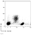
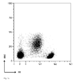
- Figs. 1a-1e show a comparison of dot plots of peripheral blood mononuclear cells (PBMC) stained with CD4 antibody (clone Vit4) conjugated with: Fig. 1a fluorescein isothiocyanate (FITC); Fig. 1b fluorescein multimerized with a branched PEG (PEG-FAM); Fig. 1c Alexa Fluor 488 (AF488); Fig. 1d Alexa Fluor 488 multimerized with a branched PEG (PEG-AF488), or (1e) R-phycoerythrin (PE) measured by flow cytometry.
- FITC fluorescein isothiocyanate
- Fig. 1b fluorescein multimerized with a branched PEG
- Fig. 1c Alexa Fluor 488 AF488)
- Fig. 1d Alexa Fluor 488 multimerized with a branched PEG PEG-AF488)
- PE R-phycoerythr
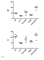
- Fig. 2 shows the median fluorescence intensities (MFI) and stain indices (SI) of T helper cells (CD4 bright positive) of five different donors stained with CD4 antibody (clone Vit4) conjugated with FITC, fluorescein multimerized on a branched PEG (PEG-FAM), Alexa Fluor 488 (AF488), Alexa Fluor 488 multimerized on a branched PEG (PEG-AF488) or PE measured by flow cytometry.
- MFI median fluorescence intensities
- SI stain indices
- Fig. 3 shows the effect of an increasing DOL on the staining of T helper cells for fluorescein multimerized on an eight-branch polyethylene glycol (PEG-FAM) conjugated to an antibody against human CD4 (clone Vit4) in comparison to the staining with said antibody against CD4, which is conjugated to conventional fluorescein isothiocyanate (FITC).
- Antibody staining concentration is 3 ⁇ g/mL in all cases.
- Fig. 4a shows histogram plots of CD4-PEG-FAM stained non-fixed and methanol fixed T helper cells
- Fig. 4b shows CD4-PE stained non-fixed and methanol fixed T helper cells.
- fluorescent dyes with PEG subunits are according to the invention.
- the fluorescent dyes according to the invention can be prepared by standard chemistry known to the person skilled in the art and as further disclosed in the examples of the present patent application.
- Z is at least one of F and R and p is at least one of n and m (depending on whether the respective polyether branch binds to F or R, with the provision that at least two F and at least one group R are comprised.
- F, R, n and m have the meaning as already disclosed.
- the fluorophore F used in the present invention is coupled to the core moiety C via the polyether scaffold and may be any organic fluorescent dye molecule.
- fluorophores have found use as laser dyes in organic dye lasers.
- Substituted versions, which can be conjugated to biomolecules, have found use as labels in flow cytometry and fluorescence microscopy.
- the fluorophore F is selected from the group consisting of xanthene dyes, rhodamine dyes, coumarine dyes, cyanine dyes, pyrene dyes, oxazine dyes, pyridyl oxazole dyes and pyrromethene dyes.
- the fluorophore F is substituted with one more water solubility imparting substituents selected from the group consisting of sulfonates, phosphonates, phosphates, polyethers, sulfonamides and carbonates. It is particularly advantageous to use fluorescent dyes with sulfonate substituents, such as dyes of the Alexa Fluor family provided by Thermo Fisher Scientific Inc..
- the degree of sulfonate substitution per fluorophore may be 2 or more, i.e., for rhodamine dyes or cyanine dyes.
- T helper cells stained with a CD4 conjugate of Alexa Fluor 488 multimerized on a branched PEG are almost twice as bright (mean MFI 98) as T helper cells stained with a CD4 conjugate of fluorescein multimerized on a branched PEG (PEG-FAM) (mean MFI 55).
- the core moiety C may be any structure comprising 1 to 100 carbon atoms, which allows attachment of x+y polyether residues (S) n and (S) m .
- Useful core moieties for the invention are polyhydroxy compounds, polyamino compounds and polythio compounds.
- Preferred are polyhydroxy compounds, such as pentaerythritol with four hydroxyl group as attachment points for 3 to 4 polyether residues via ether bonds, dipentaerythritol with six hydroxyl groups as attachment points for 3 to 6 polyether branches via ether bonds, tripentaerythritol or hexaglycerol with eight hydroxyl groups as attachment points for 3 to 8 polyether branches via ether bonds.
- the polyether residues (S) n and (S) m consist of ether monomer units S which may each containing 1 to 10, and preferably 1 to 4 oxygen atoms per monomer subunit S. These polyether branches might be homopolymeric or copolymeric, i.e., alternating or block copolymers. Polyether residues (S) n and (S) m can be linear or branched, especially in the case when monomer units S with 2 or more oxygen atoms are used. In a preferred embodiment of the invention, the polyether residues comprise polyethylene glycol chains, i.e., S stands for the residue CH 2 CH 2 O- and n, m are independently integers ranging from 10 to 200.
- multi-arm polyethylene glycols serve as scaffolds including core moiety and polyether branches.
- Multi-arm polyethylen glycols are commercialized by, for example, Nanocs Inc. or NOF Corporation.
- the fluorescent dyes according to the invention are attached to biomolecules via groups R, comprising a reactive group.
- Groups R shall be capable of forming covalent bonds via the reactive group of the biomolecule, with functional groups, like amino and thiol groups.
- Either R or (S) m /(S) n comprise a subunit capable of forming a covalent bond with the respective other group.
- polyether groups (S) m /(S) n are commercially available with amino end groups, which can react, for example, with an R group comprising a carboxy function.
- a person skilled in the art will have no difficulty in selecting the appropriate chemistry.
- R comprising such reactive groups are, e.g., active esters, such as N-hydroxysuccinimid ester, tetrafluorophenyl ester, pentafluorophenyl ester, sulfodichlorophenyl ester, imido ester, isothiocyanate, isocyanate, sulfonyl halides, acyl halides, acyl azide, monochlorotriazine, dichlorotriazine, aldehyde, glyoxal, maleimide, iodoacetamide, hydrazine, azidonitrophenyl, phosphoramidite, alkyne, alkyl azide, diene or allyl groups.
- active esters such as N-hydroxysuccinimid ester, tetrafluorophenyl ester, pentafluorophenyl ester, sulfodichlorophenyl ester,
- Covalent bond formation is also possible via reactive groups R, which are functional groups capable of reacting with a reaction substrate carrying a suitable reactive group.
- Functional groups suitable for coupling reactions may be comprised, e.g., of an amino group, a thiol group, a hydroxyl group, or a carboxyl group.
- Another object of the invention are fluorescent biomolecule conjugates comprising one or more fluorescent dyes as already disclosed conjugated via group R to at least one biomolecule selected from group consisting of immunoglobulin, antibody, fragmented antibody, Fab, Fab', F(ab')2, sdAb, scFv, di-scFv, each naturally or recombinant.
- biomolecule conjugates of the invention are either prepared by reacting a biomolecule with a reactive group R of the fluorescent dye or by reacting an activated biomolecule with a suitable functional group R of the fluorescent dyes.
- Biomolecules suitable for conjugation with the fluorescent dye of the invention may be proteins, peptides, carbohydrates, nucleic acids, lipids and combinations thereof. These biomolecule are capable of binding to binding partners, such as analytes, cell surface markers, antigens etc., in order to label, detect and quantify said analytes, cell surface markers, antigens etc..
- Fragmented antibodies may be synthesized by recombinant procedures including covalent and non-covalent conjugates containing these kinds of molecules.
- the biomolecule is selected from a group consisting of peptide/MHC-complexes, receptors for cell adhesion or costimulatory molecules, receptor ligands, antigens, hapten binders, avidin, streptavidin, neutravidin, aptamers, primers and ligase substrates.
- receptors are moieties e.g., for cell adhesion or costimulatory molecules and hapten binders are avidin, streptavidin or neutravidin.
- Nucleic acids may be, e.g., aptamers, primers, or substrates for ligases.
- Biotin binders such as avidin, streptavidin or neutravidin labeled with fluorophore labeled branched polyether scaffold allow for a sensitive detection via biotinylated primary detection molecules due to an additional multiplying effect of the multiple biotinylation of said primary detection molecule.
- the biomolecule is an IgG antibody with 2 to 7 fluorescent polyether labels carrying each 4 to 6 fluorophore groups, resulting in fluorophore DOLs of 8 to 40 dye molecules per antibody molecule, preferably a fluorophore DOL of more than 10, most preferably a DOL of 15 or more fluorophore units.
- the biomolecule conjugates comprise one or more fluorescent dyes wherein at least one biomolecule is conjugated with 2 to 20 fluorescent dyes comprising each 1 to 10 fluorophores F, inclusive. It is preferred that more than one fluorescent dye is present in the biomolecule conjugates.
- biomolecule conjugates and/or the fluorescent dyes of the invention are especially useful for detection, counting or separation of cells utilizing a certain set of antigens recognized by the biomolecule conjugate.
- Another object of the invention is therefore a method of analyzing cells or tissue labeled by the fluorescent biomolecule conjugates according to the invention by flow cytometry and/or by fluorescence microscopy. Accordingly, the method may include labeling with the fluorescent biomolecule conjugates as described, and performing at least one of flow cytometry and fluorescence microscopy.
- one or more populations of labeled cells are detected from the sample and separated as target cells.
- the cells detected by the conjugate are separated from the sample by electrostatic forces, piezoelectric forces, mechanical separation or opto-acoustic means.
- Suitable for such separations are especially flow sorters, e.g., FACS or MEMS-based cell sorter systems, for example, as disclosed in EP14187215.0 or EP14187214.3 .
- the location of the binding target of biomolecule conjugates on cell or tissue samples is determined by fluorescence microscopy.
- fluorescence microscopy include epifluorescence microscopy, confocal laser scanning microscopy, multi photon microscopy, total internal reflection fluorescence (TIRF) microscopy, single plane illumination microscopy (SPIM) and super resolution microscopy methods such as, e.g., stimulated emission depletion (STED) microscopy, stochastic optical reconstruction microscopy (STORM), photo activated localization microscopy (PALM), or spatially modulated illumination (SMI) microscopy.
- Step A Preparation of AF488 multimerized on an 8-arm PEG (PEG-AF488)
- Amino-PEG (8-arm) was dissolved in 0.5 M PBS buffer, pH 7 at a concentration of 10 mg/mL.
- AF488, NHS ester dissolved in DMSO (2 mg/mL) was added in a 20-fold molar excess and incubated at room temperature for 30 minutes in the dark. Free dye was removed by SEC under standard conditions.
- the resulting PEG-AF488 had a DOL of 5.7.
- PEG-FAM is obtained by the same procedure using NHS-fluorescein (NHS-FAM).
- Step B Coupling of CD4 antibody with PEG-AF488
- PEG-AF488 (2.5 mg/mL in phosphate buffered saline) as obtained in step A was activated by addition of 10-fold molar excess SMCC and incubation at room temperature for 1 h.
- CD4 antibody (clone Vit 4) was reduced by reacting with 10 mmol/L dithiothreitol for 1 h.
- Maleimide activated PEG-AF488 and reduced antibody were both subjected to buffer exchange with phosphate buffered saline, pH 7.2 containing 2 mM EDTA over Sephadex G25.
- Maleimide activated PEG-AF488 was added to reduced antibody at a 15-fold excess and incubated for 1 h at room temperature in the dark.
- the reaction product is purified by SEC.
- the resulting CD4-PEG-AF488 had a DOL of 13.2.
- CD4 is coupled to PEG-FAM using the same protocol.
- Step C Staining of PBMC with CD4 antibody conjugated to fluorochromes multimerized on 8-arm PEG.
- peripheral blood mononuclear cells PBMC
- PBMC peripheral blood mononuclear cells
- CD4 antibody conjugated with R-phycoerythrin (PE) or non-multimerized fluorochromes FITC or AF488 is used for comparison.
- Fig. 1 shows dot plot examples of fluorescence intensities versus side scatter (SSC) using the following: Fig. 1a fluorescein isothiocyanate (FITC); Fig. 1b fluorescein multimerized with a branched PEG (PEG-FAM); Fig. 1c Alexa Fluor 488 (AF488); Fig. 1d Alexa Fluor 488 multimerized with a branched PEG (PEG-AF488), or (1e) R-phycoerythrin (PE) and measured by flow cytometry.
- FITC fluorescein isothiocyanate
- Fig. 1b fluorescein multimerized with a branched PEG
- Fig. 1c Alexa Fluor 488 AF488)
- Fig. 1d Alexa Fluor 488 multimerized with a branched PEG PEG-AF488)
- PE R-phycoerythrin
- Fig. 2 shows the median fluorescent intensities (MFI) and stain index (SI) results for PBMCs of 5 donors.
- Cells stained with antibody labeled with fluorochromes according to the invention are at least twice as bright as cells stained with antibody labeled with the parent fluorochromes and on par with cells labeled with phycobiliprotein labeled antibody (CD4-PE).
- Fig. 3 shows the effect of an increasing DOL on the staining of T helper cells for fluorescein multimerized on an eight-branch polyethylene glycol (PEG-FAM) conjugated to an antibody against human CD4 (clone Vit4) in comparison to the staining with said antibody against CD4, which is conjugated to conventional fluorescein isothiocyanate (FITC).
- Antibody staining concentration is 3 ⁇ g/mL in all cases.
- the fluorochrome is also sulfonated as AF488 (Alexa Fluor).
- AF488 Alexa Fluor
- the resulting antibody conjugate labeled with AF488 multimerized on a 8-arm PEG is about twice as bright as the corresponding conjugate made from fluorescein multimerized on a 8-arm PEG.
- Example 2 Influence of methanol fixation on staining intensities.
- Peripheral blood mononuclear cells are stained as described in example 1 excluding propidium iodide as counterstain.
- For methanol fixation 1 mL methanol is added to 100 ⁇ L cell suspension and incubated for 30 minutes on ice. Cells are washed twice with and resuspended in 1 mL phosphate buffered saline containing 2 mM EDTA and 0.5% BSA. Stained and fixed cells are analyzed on the MACSQuant 10 analyzer in comparison to non-fixed cells.
- Fig. 4a and 4b show histogram plots of CD4-PEG-FAM stained non-fixed and methanol fixed T helper cells ( Fig.
- CD4-PE stained non-fixed and methanol fixed T helper cells Fig. 4b .
- the fluorescence intensity of CD4-PEG-FAM is stable versus methanol fixation whereas the fluorescence intensity of CD4-PE shows a five-fold decrease.
- the fluorescent biomolecule conjugates and/or the fluorescent dyes of the invention show improved stability towards environmental influences such as fixation.
Abstract

C is a core moiety comprising 20 to 200 atoms;
S same or different ether residues comprising 1 to 10 carbon atoms;
n is an integer ranging from 2 to 500;
m is an integer ranging from 0 to 500;
x is an integer ranging from 2 to 50;
y is an integer ranging from 1 to 50;
R R same or different residue comprising a reactive group capable of forming a covalent bond with a biomolecule;
F same or different fluorophores covalently bound to (S)n.
Description
- The present invention is directed to fluorescent dyes with increased brightness and methods of use thereof.
- Fluorescent dyes conjugated to antibodies are commonly used for immunofluorescence analysis. A vast number of variants in antibodies, fluorescent dyes, flow cytometers, flow sorters and fluorescence microscopes has been developed in the last two decades to enable specific detection and isolation of target cells. One issue in immunofluorescence technology is the detection threshold of the fluorescence emission, which can be enhanced, for example, by better detectors, filter systems or modified fluorescent dyes.
- A limitation of conventional small molecule fluorescent dye molecules is their limited brightness. Therefore biomolecules are typically labeled with multiple dye molecules to increase the brightness of the fluorochrome conjugate. Most of the aforementioned fluorescent dye molecules, such as rhodamines or cyanines, contain planar aromatic chromophores, which are prone to hydrophobic interactions leading to dye-dye dimers with low or no fluorescence. Consequently, fluorescence intensity of labeled biomolecules is not proportional to the degree of fluorescent dye labeling of said biomolecules. At higher degrees of labeling (DOLs) the fluorescence intensity of the single dye might even decrease due to self-quenching mechanism caused by dimer, trimer or multimer formation.
- These undesired formations have been reduced to some extent by adding substituents to the flat aromatic dye molecules, which increase water solubility. Suitable substituents described in the literature might impart charges to the dye molecule, such as sulfonate groups described, e.g., in
U.S. Pat. Nos. 5,268,486 and6,977,305 ,6,130,101 and Panchuk-Voloshina, et al., J. Histochem. Cytochem. 47(9), 1179 (1999), or phosphate groups described, e.g., inWO2013056720 . Other suitable dimerization reducing substituents are bulky water-soluble polymers, such as polyethylene glycol described, e.g., in patent applicationWO2009078970 , or charged dendrimers described, e.g., inU.S. Pat. Nos. 6,913,743 , and7,655,217 . Although biomolecule conjugates of these substituted dyes achieve higher brightness at higher degrees of labeling (DOLs) than conjugates of the unsubstituted parent dyes, there is still a deviation from a linear proportionality between fluorescence intensity and degree of labeling of the labeled biomolecule albeit at a higher DOL as for the unsubstituted parent dye. - Conjugate brightness is also limited by the number of functionalization sites available on the biomolecule, which can be functionalized without loss of biomolecule activity. As a result DOLs of biomolecule conjugates are still limited, e.g., for antibodies (IgG) they are typically in the range of 4 to 8 in the case of hydrophilic labels (R. P. Haugland, Current Protocols in Cell Biology (2000) 16.5.1 - 16.5.22). Consequently the brightness of these conjugates is still inferior to, e.g., conjugates of phycobiliproteins, such as phycoerythrin (PE) or allophycocyanine (APC).
- The high fluorescence intensity of phycobiliproteins and their biomolecule conjugates is due to the presence of multiple fluorophore subunits within a phycobiliprotein. R-phycoerythrin, e.g., contains 34 phycobilin fluorophore subunits (A. N. Glazer, J. Appl. Phycol. 6, 105 (1994)). Therefore biomolecule conjugates of phycobiliproteins, such as PE or APC are popular, e.g., in flow cytometry, despite their drawbacks stemming from their protein nature such as limited stability against non-physiological solvents, temperature, and pH values as well as their limited photostability and their generally limited shelf life. Another drawback is the limited availability of different colors, which limit multiplexing capabilities within a single fluorescence assay.
- There have been efforts to construct multichromophore constructs with phycobiliprotein-like fluorescence properties by arranging fluorophores into precise supramolecular structures preventing self-quenching of excited fluorophores. Benvin et al., J. Am. Chem. Soc. 129(7), 2025 (2007) describe fluorescent dyes intercalated into supramolecular DNA templates. However, a drawback of this method is the non-covalent binding of the fluorophores within the DNA scaffold. Migration of dye molecules out of the DNA scaffold can lead to loss of fluorescence signal. In case of a multicolor experiment dye exchange between differently labeled DNA scaffolds might lead to false positive fluorescent signals. In case of a multiparameter experiment employing multiple colors it is advisable to use covalently bound fluorophores.
- Another class of brightly fluorescent dyes for biomolecule labeling are fluorescent polymers based on semiconducting polymers, such as polyfluorenes described e.g. in
US Pat. Nos. 8,158,444 ,8,354,239 , and8,802,450 and8,362,193 ,8,455,613 , and8,575,303 . These polymers also contain multiple fluorophore subunits, as the effective conjugation length within the polymer is limited to 9 - 10 monomer subunits. - Preparing brighter fluorescent dyes by multimerizing conventional fluorescent dye molecules on a water-soluble scaffold has so far not resulted in compounds useful for the labeling of biomolecules. Recommendations for fluorescent labeling of, e.g., dextrans are 0.3 - 0.7 dye molecules per dextran in the 3000 MW range, 0.5 - 2 dye molecules in the 10,000 MW range, 2 - 4 dye molecules in the 40,000 MW range and 3 - 6 dye molecules in the 70,000 MW range. Due to their large size and bulkiness these dye multimers offer no benefit in biomolecule labeling and are unsuitable for preparing biomolecule conjugates with phycobiliprotein-like fluorescence intensities. Fluorescently labeled dextrans with higher degree of labeling show quenching due to dye - dye interaction. This has found use in
US Pat. No. 5,719,031 , wherein the degree of labeling of dextran-fluorochrome-conjugates is high enough to furnish fluorescent quenching. Enzymatic degradation of the dextran-fluorochrome-conjugate is accompanied by an enhancement of the fluorescence emission signal, which is used for quantification of the enzymatic digestion process. - There remains still a considerable need for improved fluorescent dyes for labeling of biomolecules, which provide phycobiliprotein-like fluorescence intensity without the limitations of phycobiliproteins and fluorescent polymers.
- It was therefore an object of the invention to provide a fluorescent dye label with high fluorescence intensity, low unspecific background staining and stability against fixation.
- Surprisingly, it was found that fluorescent dyes multimerized on branched polyether scaffolds, such as multi-arm polyethylene glycols, are highly fluorescent without noticeable quenching. In addition, the polyether scaffold seems to reduce the tendency of fluorescent dyes to bind unspecifically.
- One aspect the invention allows for tailoring the properties of the fluorescent dye label regarding photostability and stability against different environmental conditions such as temperature, pH and solvent. In another aspect of the invention a broad range of different excitation and emission wavelengths are provided. In another aspect of the invention retention of biomolecule activity is provided despite a high number of fluorophore subunits present in the biomolecule-fluorescent dye conjugate.
- With the present invention, it is possible to introduce a high degree of fluorophore labeling to a biomolecule without loss of activity or increased unspecific binding, e.g., in case of antibodies a fluorophore DOL of greater than 10, preferably equal or greater than 15 is achieved, resulting in an antibody fluorochrome conjugate with phycobiliprotein-conjugate-like fluorescence intensity and low unspecific background staining.
- Accordingly, an object of the invention are fluorescent dyes according to the general formula I:

- C
- is a core moiety comprising 20 to 200 atoms;
- S
- same or different ether residues comprising 1 to 10 carbon atoms;
- n
- is an integer ranging from 2 to 500;
- m
- is an integer ranging from 0 to 500;
- x
- is an integer ranging from 2 to 50;
- y
- is an integer ranging from 1 to 50;
- R
- same or different residue comprising a reactive group capable of forming a covalent bond with a biomolecule;
- F
- same or different fluorophores covalently bound to (S)n.
- The core moiety C provides multiple (x+y) attachment points for the polyether residues (S)n and (S)m. R comprises a reactive group capable of forming a covalent bond with a biomolecule which recognizes cellular structures like antigens.
- Various exemplary details are described with reference to the following figures, wherein:
-
Figs. 1a-1e show a comparison of dot plots of peripheral blood mononuclear cells (PBMC) stained with CD4 antibody (clone Vit4) conjugated with:Fig. 1a fluorescein isothiocyanate (FITC);Fig. 1b fluorescein multimerized with a branched PEG (PEG-FAM);Fig. 1c Alexa Fluor 488 (AF488);Fig. 1d Alexa Fluor 488 multimerized with a branched PEG (PEG-AF488), or (1e) R-phycoerythrin (PE) measured by flow cytometry. -
Fig. 2 shows the median fluorescence intensities (MFI) and stain indices (SI) of T helper cells (CD4 bright positive) of five different donors stained with CD4 antibody (clone Vit4) conjugated with FITC, fluorescein multimerized on a branched PEG (PEG-FAM), Alexa Fluor 488 (AF488),Alexa Fluor 488 multimerized on a branched PEG (PEG-AF488) or PE measured by flow cytometry. -
Fig. 3 shows the effect of an increasing DOL on the staining of T helper cells for fluorescein multimerized on an eight-branch polyethylene glycol (PEG-FAM) conjugated to an antibody against human CD4 (clone Vit4) in comparison to the staining with said antibody against CD4, which is conjugated to conventional fluorescein isothiocyanate (FITC). Antibody staining concentration is 3 µg/mL in all cases. -
Fig. 4a shows histogram plots of CD4-PEG-FAM stained non-fixed and methanol fixed T helper cells;Fig. 4b shows CD4-PE stained non-fixed and methanol fixed T helper cells. - In all figures, fluorescent dyes with PEG subunits are according to the invention.
- The fluorescent dyes according to the invention can be prepared by standard chemistry known to the person skilled in the art and as further disclosed in the examples of the present patent application.
- By way of example, preferred fluorescent dyes according to the invention are shown in the following general formula II and III


- In formula (II) and (III), Z is at least one of F and R and p is at least one of n and m (depending on whether the respective polyether branch binds to F or R, with the provision that at least two F and at least one group R are comprised. F, R, n and m have the meaning as already disclosed.
- The fluorophore F used in the present invention is coupled to the core moiety C via the polyether scaffold and may be any organic fluorescent dye molecule. Typically, such fluorophores have found use as laser dyes in organic dye lasers. Substituted versions, which can be conjugated to biomolecules, have found use as labels in flow cytometry and fluorescence microscopy.
- Preferably, the fluorophore F is selected from the group consisting of xanthene dyes, rhodamine dyes, coumarine dyes, cyanine dyes, pyrene dyes, oxazine dyes, pyridyl oxazole dyes and pyrromethene dyes.
- In a variant of the invention, the fluorophore F is substituted with one more water solubility imparting substituents selected from the group consisting of sulfonates, phosphonates, phosphates, polyethers, sulfonamides and carbonates. It is particularly advantageous to use fluorescent dyes with sulfonate substituents, such as dyes of the Alexa Fluor family provided by Thermo Fisher Scientific Inc.. The degree of sulfonate substitution per fluorophore may be 2 or more, i.e., for rhodamine dyes or cyanine dyes. The use of sulfonated dyes compared to unsulfonated dyes leads to even brighter conjugates of fluorophores multimerized on a polyether scaffold as can be seen in
Fig. 2 : T helper cells stained with a CD4 conjugate ofAlexa Fluor 488 multimerized on a branched PEG (PEG-AF488) are almost twice as bright (mean MFI 98) as T helper cells stained with a CD4 conjugate of fluorescein multimerized on a branched PEG (PEG-FAM) (mean MFI 55). - The fluorophores F can be attached to the polyether scaffolds by methods known in the art, i.e., by reacting a fluorescent dye with a group reactive towards amino or thiol groups with a branched polyether scaffold with amino end groups.
- The core moiety C may be any structure comprising 1 to 100 carbon atoms, which allows attachment of x+y polyether residues (S)n and (S)m.
- Useful core moieties for the invention are polyhydroxy compounds, polyamino compounds and polythio compounds. Preferred are polyhydroxy compounds, such as pentaerythritol with four hydroxyl group as attachment points for 3 to 4 polyether residues via ether bonds, dipentaerythritol with six hydroxyl groups as attachment points for 3 to 6 polyether branches via ether bonds, tripentaerythritol or hexaglycerol with eight hydroxyl groups as attachment points for 3 to 8 polyether branches via ether bonds.

- The polyether residues (S)n and (S)m consist of ether monomer units S which may each containing 1 to 10, and preferably 1 to 4 oxygen atoms per monomer subunit S. These polyether branches might be homopolymeric or copolymeric, i.e., alternating or block copolymers. Polyether residues (S)n and (S)m can be linear or branched, especially in the case when monomer units S with 2 or more oxygen atoms are used. In a preferred embodiment of the invention, the polyether residues comprise polyethylene glycol chains, i.e., S stands for the residue CH2CH2O- and n, m are independently integers ranging from 10 to 200.
- In a particular useful embodiment of the invention commercially available multi-arm polyethylene glycols (branched PEGs) serve as scaffolds including core moiety and polyether branches. Multi-arm polyethylen glycols are commercialized by, for example, Nanocs Inc. or NOF Corporation.
- The fluorescent dyes according to the invention are attached to biomolecules via groups R, comprising a reactive group. Groups R shall be capable of forming covalent bonds via the reactive group of the biomolecule, with functional groups, like amino and thiol groups. Either R or (S)m/(S)n comprise a subunit capable of forming a covalent bond with the respective other group. For example, polyether groups (S)m/(S)n are commercially available with amino end groups, which can react, for example, with an R group comprising a carboxy function. A person skilled in the art will have no difficulty in selecting the appropriate chemistry. Examples for R comprising such reactive groups are, e.g., active esters, such as N-hydroxysuccinimid ester, tetrafluorophenyl ester, pentafluorophenyl ester, sulfodichlorophenyl ester, imido ester, isothiocyanate, isocyanate, sulfonyl halides, acyl halides, acyl azide, monochlorotriazine, dichlorotriazine, aldehyde, glyoxal, maleimide, iodoacetamide, hydrazine, azidonitrophenyl, phosphoramidite, alkyne, alkyl azide, diene or allyl groups.
- Covalent bond formation is also possible via reactive groups R, which are functional groups capable of reacting with a reaction substrate carrying a suitable reactive group. Functional groups suitable for coupling reactions may be comprised, e.g., of an amino group, a thiol group, a hydroxyl group, or a carboxyl group.
- It is also conceivable to use additional reactive groups R' on polyether branches with low reactivity, with the purpose of keeping apart fluorophores to prevent dye-dye interactions. These low-reactive groups R' may be hydrogen atom, an alkyl group, a trifluoroalkyl group, an aryl group, a halogen group, a sulfonyl group, a sulfonate group, a phosphonate group, a sulfonamide group, or combinations thereof. At least one of the above mentioned reactive groups R is necessary for the performance of the fluorescent dyes according to the invention.
- Another object of the invention are fluorescent biomolecule conjugates comprising one or more fluorescent dyes as already disclosed conjugated via group R to at least one biomolecule selected from group consisting of immunoglobulin, antibody, fragmented antibody, Fab, Fab', F(ab')2, sdAb, scFv, di-scFv, each naturally or recombinant.
- The biomolecule conjugates of the invention are either prepared by reacting a biomolecule with a reactive group R of the fluorescent dye or by reacting an activated biomolecule with a suitable functional group R of the fluorescent dyes.
- Biomolecules suitable for conjugation with the fluorescent dye of the invention may be proteins, peptides, carbohydrates, nucleic acids, lipids and combinations thereof. These biomolecule are capable of binding to binding partners, such as analytes, cell surface markers, antigens etc., in order to label, detect and quantify said analytes, cell surface markers, antigens etc..
- Fragmented antibodies may be synthesized by recombinant procedures including covalent and non-covalent conjugates containing these kinds of molecules.
- In one embodiment of the invention, the biomolecule is selected from a group consisting of peptide/MHC-complexes, receptors for cell adhesion or costimulatory molecules, receptor ligands, antigens, hapten binders, avidin, streptavidin, neutravidin, aptamers, primers and ligase substrates. Preferably, receptors are moieties e.g., for cell adhesion or costimulatory molecules and hapten binders are avidin, streptavidin or neutravidin. Nucleic acids may be, e.g., aptamers, primers, or substrates for ligases.
- Biotin binders, such as avidin, streptavidin or neutravidin labeled with fluorophore labeled branched polyether scaffold allow for a sensitive detection via biotinylated primary detection molecules due to an additional multiplying effect of the multiple biotinylation of said primary detection molecule.
- In one embodiment of the invention the biomolecule is an IgG antibody with 2 to 7 fluorescent polyether labels carrying each 4 to 6 fluorophore groups, resulting in fluorophore DOLs of 8 to 40 dye molecules per antibody molecule, preferably a fluorophore DOL of more than 10, most preferably a DOL of 15 or more fluorophore units.
- In yet another embodiment of the invention, the biomolecule conjugates comprise one or more fluorescent dyes wherein at least one biomolecule is conjugated with 2 to 20 fluorescent dyes comprising each 1 to 10 fluorophores F, inclusive. It is preferred that more than one fluorescent dye is present in the biomolecule conjugates.
- The biomolecule conjugates and/or the fluorescent dyes of the invention are especially useful for detection, counting or separation of cells utilizing a certain set of antigens recognized by the biomolecule conjugate.
- Another object of the invention is therefore a method of analyzing cells or tissue labeled by the fluorescent biomolecule conjugates according to the invention by flow cytometry and/or by fluorescence microscopy. Accordingly, the method may include labeling with the fluorescent biomolecule conjugates as described, and performing at least one of flow cytometry and fluorescence microscopy.
- In another embodiment of the invention one or more populations of labeled cells are detected from the sample and separated as target cells. Preferably the cells detected by the conjugate are separated from the sample by electrostatic forces, piezoelectric forces, mechanical separation or opto-acoustic means. Suitable for such separations are especially flow sorters, e.g., FACS or MEMS-based cell sorter systems, for example, as disclosed in
EP14187215.0 EP14187214.3 - In another embodiment of the invention the location of the binding target of biomolecule conjugates on cell or tissue samples is determined by fluorescence microscopy. Suitable methods of fluorescence microscopy include epifluorescence microscopy, confocal laser scanning microscopy, multi photon microscopy, total internal reflection fluorescence (TIRF) microscopy, single plane illumination microscopy (SPIM) and super resolution microscopy methods such as, e.g., stimulated emission depletion (STED) microscopy, stochastic optical reconstruction microscopy (STORM), photo activated localization microscopy (PALM), or spatially modulated illumination (SMI) microscopy.
- Amino-PEG (8-arm) was dissolved in 0.5 M PBS buffer,
pH 7 at a concentration of 10 mg/mL. AF488, NHS ester dissolved in DMSO (2 mg/mL) was added in a 20-fold molar excess and incubated at room temperature for 30 minutes in the dark. Free dye was removed by SEC under standard conditions. The resulting PEG-AF488 had a DOL of 5.7. - PEG-FAM is obtained by the same procedure using NHS-fluorescein (NHS-FAM).
- PEG-AF488 (2.5 mg/mL in phosphate buffered saline) as obtained in step A was activated by addition of 10-fold molar excess SMCC and incubation at room temperature for 1 h. In parallel CD4 antibody (clone Vit 4) was reduced by reacting with 10 mmol/L dithiothreitol for 1 h. Maleimide activated PEG-AF488 and reduced antibody were both subjected to buffer exchange with phosphate buffered saline, pH 7.2 containing 2 mM EDTA over Sephadex G25. Maleimide activated PEG-AF488 was added to reduced antibody at a 15-fold excess and incubated for 1 h at room temperature in the dark. The reaction product is purified by SEC. The resulting CD4-PEG-AF488 had a DOL of 13.2.
- CD4 is coupled to PEG-FAM using the same protocol.
- For
staining experiments 106 peripheral blood mononuclear cells (PBMC) each are resuspended in 100 µl phosphate buffered saline containing 2 mM EDTA and 0.5% BSA. Cells are stained with the respective CD4 antibody conjugates as obtained in step B at 3 µg/mL and incubated for 10 minutes at room temperature. Counterstaining is performed with CD3-APC and propidium iodide for dead cell exclusion. - Commercially available CD4 antibody conjugated with R-phycoerythrin (PE) or non-multimerized fluorochromes FITC or AF488 is used for comparison.
- Cells are incubated 10 minutes at room temperature in the dark. Washing is performed by centrifugation and resuspension in 1 ml phosphate buffered saline containing 2 mM EDTA and 0.5% BSA. Stained cells are analyzed on the MACSQuant 10 analyzer.
Fig. 1 shows dot plot examples of fluorescence intensities versus side scatter (SSC) using the following:Fig. 1a fluorescein isothiocyanate (FITC);Fig. 1b fluorescein multimerized with a branched PEG (PEG-FAM);Fig. 1c Alexa Fluor 488 (AF488);Fig. 1d Alexa Fluor 488 multimerized with a branched PEG (PEG-AF488), or (1e) R-phycoerythrin (PE) and measured by flow cytometry. -
Fig. 2 shows the median fluorescent intensities (MFI) and stain index (SI) results for PBMCs of 5 donors. - Cells stained with antibody labeled with fluorochromes according to the invention are at least twice as bright as cells stained with antibody labeled with the parent fluorochromes and on par with cells labeled with phycobiliprotein labeled antibody (CD4-PE).
Fig. 3 shows the effect of an increasing DOL on the staining of T helper cells for fluorescein multimerized on an eight-branch polyethylene glycol (PEG-FAM) conjugated to an antibody against human CD4 (clone Vit4) in comparison to the staining with said antibody against CD4, which is conjugated to conventional fluorescein isothiocyanate (FITC). Antibody staining concentration is 3 µg/mL in all cases. - It is especially advantageous if the fluorochrome is also sulfonated as AF488 (Alexa Fluor). The resulting antibody conjugate labeled with AF488 multimerized on a 8-arm PEG (PEG-AF488) is about twice as bright as the corresponding conjugate made from fluorescein multimerized on a 8-arm PEG.
- Peripheral blood mononuclear cells are stained as described in example 1 excluding propidium iodide as counterstain. For
methanol fixation 1 mL methanol is added to 100 µL cell suspension and incubated for 30 minutes on ice. Cells are washed twice with and resuspended in 1 mL phosphate buffered saline containing 2 mM EDTA and 0.5% BSA. Stained and fixed cells are analyzed on the MACSQuant 10 analyzer in comparison to non-fixed cells.Fig. 4a and4b show histogram plots of CD4-PEG-FAM stained non-fixed and methanol fixed T helper cells (Fig. 4a ) and CD4-PE stained non-fixed and methanol fixed T helper cells (Fig. 4b ). The fluorescence intensity of CD4-PEG-FAM is stable versus methanol fixation whereas the fluorescence intensity of CD4-PE shows a five-fold decrease. - Accordingly, the fluorescent biomolecule conjugates and/or the fluorescent dyes of the invention show improved stability towards environmental influences such as fixation.
- While various details have been described in conjunction with the exemplary implementations outlined above, various alternatives, modifications, variations, improvements, and/or substantial equivalents, whether known or that are or may be presently unforeseen, may become apparent upon reviewing the foregoing disclosure. Accordingly, the exemplary implementations set forth above, are intended to be illustrative, not limiting.
Claims (13)
- A fluorescent dye according to the general formula I:
 C is a core moiety comprising 20 to 200 atoms;S same or different ether residues comprising 1 to 10 carbon atoms;n is an integer ranging from 2 to 500;m is an integer ranging from 0 to 500;x is an integer ranging from 2 to 50;y is an integer ranging from 1 to 50;R same or different residue comprising a reactive group capable of forming a covalent bond with a biomolecule;F same or different fluorophores covalently bound to (S)n.
 - The fluorescent dye according to claim 1, wherein the fluorophore F is selected from the group consisting of xanthene dyes, rhodamine dyes, coumarine dyes, cyanine dyes, pyrene dyes, oxazine dyes, pyridyl oxazole dyes and pyrromethene dyes.
- The fluorescent dye according to claim 1 or 2, wherein the fluorophore F is substituted with one more water solubility imparting substituents selected from the group consisting of sulfonates, phosphonates, phosphates, sulfonamides, polyethers and carbonates.
- The fluorescent dye according to one or more of the claims 1 to 3 wherein the core moiety C is a polyhydroxy compound, a polyamino compound or a polythio compound.
- The fluorescent dye according to according to one or more of the claims 1 to 4 wherein the core moiety C is selected from the group consisting of pentaerythritol, dipentaerythritol, tripentaerythritol and hexaglycerol.
- The fluorescent dye according to one or more of the claims 1 to 5 wherein S stands for the residue CH2CH2O- and n, m are independently integers ranging from 10 to 200.
- The fluorescent dye according to one or more of the claims 1 to 6 wherein R comprises for a reactive group selected from the group consisting of N-hydroxysuccinimid ester, tetrafluorophenyl ester, pentafluorophenyl ester, sulfodichlorophenyl ester, imido ester, isothiocyanate, isocyanate, sulfonyl halides, acyl halides, acyl azide, monochlorotriazine, dichlorotriazine, aldehyde, glyoxal, maleimide, iodoacetamide, hydrazine, azidonitrophenyl, phosphoramidite, alkyne, alkyl azide, dienes or allyl groups.
- The fluorescent dye according to one or more of the claims 1 to 7 characterized by
the structure of general formula (II)
with the provision that general formula (II) comprises at least two F and at least one group R. - The fluorescent dye according to one or more of the claims 1 to 7 characterized by the structure of general formula (III)

with the provision that general formula (II) comprises at least two F and at least one group R. - Fluorescent biomolecule conjugates comprising one or more fluorescent dyes according to one or more of the claims 1 to 9 conjugated via group R to at least one biomolecule selected from group consisting of immunoglobulin, antibody, fragmented antibody, Fab, Fab', F(ab')2, sdAb, scFv, di-scFv each of naturally or recombinant origin.
- Fluorescent biomolecule conjugates comprising one or more fluorescent dyes according to one or more of the claims 1 to 9 conjugated via group R to at least one biomolecule selected from the group consisting of peptide/MHC-complexes, receptors for cell adhesion or costimulatory molecules, receptor ligands, antigens, hapten binders, avidin, streptavidin, neutravidin, aptamers, primers and ligase substrates.
- Fluorescent biomolecule conjugates according to claim 10 or 11 wherein at least one biomolecule is conjugated with 2 to 20 fluorescent dyes according to one or more of the claims 1 to 9, each comprising 1 to 10 fluorophores F.
- Method of analyzing cells or tissue labeled by the fluorescent biomolecule conjugates according to one or more of the claims 10 to 12 by flow cytometry and/or by fluorescence microscopy.
Priority Applications (3)
| Application Number | Priority Date | Filing Date | Title |
|---|---|---|---|
| EP15169549.1A EP3098269B1 (en) | 2015-05-28 | 2015-05-28 | Bright fluorochromes based on multimerization of fluorescent dyes on branched polyether scaffolds |
| JP2016106411A JP6928742B2 (en) | 2015-05-28 | 2016-05-27 | Bright fluorescent dye based on the multimerization of fluorescent dye on the branched-chain polyether skeleton |
| CN201610359279.1A CN106189362B (en) | 2015-05-28 | 2016-05-27 | Bright fluorochromes based on the polymerization of fluorescent dyes on branched polyether backbones |
Applications Claiming Priority (1)
| Application Number | Priority Date | Filing Date | Title |
|---|---|---|---|
| EP15169549.1A EP3098269B1 (en) | 2015-05-28 | 2015-05-28 | Bright fluorochromes based on multimerization of fluorescent dyes on branched polyether scaffolds |
Publications (2)
| Publication Number | Publication Date |
|---|---|
| EP3098269A1 true EP3098269A1 (en) | 2016-11-30 |
| EP3098269B1 EP3098269B1 (en) | 2022-04-06 |
Family
ID=53264578
Family Applications (1)
| Application Number | Title | Priority Date | Filing Date |
|---|---|---|---|
| EP15169549.1A Active EP3098269B1 (en) | 2015-05-28 | 2015-05-28 | Bright fluorochromes based on multimerization of fluorescent dyes on branched polyether scaffolds |
Country Status (3)
| Country | Link |
|---|---|
| EP (1) | EP3098269B1 (en) |
| JP (1) | JP6928742B2 (en) |
| CN (1) | CN106189362B (en) |
Cited By (3)
| Publication number | Priority date | Publication date | Assignee | Title |
|---|---|---|---|---|
| EP3489684A1 (en) * | 2017-11-27 | 2019-05-29 | Miltenyi Biotec GmbH | Method for photobleaching stained cells |
| WO2020216439A1 (en) | 2019-04-23 | 2020-10-29 | Miltenyi Biotec B.V. & Co. KG | Reversible cell detection with conjugates having a linker for increased fluorescent brightness and an enzymatically releasable fluorescent moiety |
| CN112111564A (en) * | 2020-07-31 | 2020-12-22 | 南方科技大学 | Probe and preparation method and application thereof |
Families Citing this family (2)
| Publication number | Priority date | Publication date | Assignee | Title |
|---|---|---|---|---|
| EP3728137A4 (en) * | 2017-12-22 | 2021-12-08 | North Carolina State University | Polymeric fluorophores, compositions comprising the same, and methods of preparing and using the same |
| CN111308064B (en) * | 2020-02-27 | 2024-04-09 | 四川新健康成生物股份有限公司 | Method for improving sensitivity of immunochromatography marker and application of method in detection of interleukin 6 |
Citations (17)
| Publication number | Priority date | Publication date | Assignee | Title |
|---|---|---|---|---|
| US5268486A (en) | 1986-04-18 | 1993-12-07 | Carnegie-Mellon Unversity | Method for labeling and detecting materials employing arylsulfonate cyanine dyes |
| US5719031A (en) | 1996-08-14 | 1998-02-17 | Molecular Probes, Inc. | Dye labeled polymers as reagents for measuring polymer degradation |
| US6130101A (en) | 1997-09-23 | 2000-10-10 | Molecular Probes, Inc. | Sulfonated xanthene derivatives |
| US6913743B2 (en) | 1994-12-07 | 2005-07-05 | Institut Fur Diagnostikforschung Gmbh An Der Freien Universitat Berlin | Near infrared imaging agent |
| US6977305B2 (en) | 2000-09-29 | 2005-12-20 | Molecular Probes, Inc. | Modified carbocyanine dyes and their conjugates |
| WO2006031851A1 (en) * | 2004-09-14 | 2006-03-23 | Applera Corporation | Multi-chromophoric quencher constructs for use in high sensitivity energy transfer probes |
| WO2006125736A1 (en) * | 2005-05-27 | 2006-11-30 | Ciba Specialty Chemicals Holding Inc. | Functionalized nanoparticles |
| WO2008009579A1 (en) * | 2006-07-18 | 2008-01-24 | Ciba Holding Inc. | Polymeric hair dyes |
| WO2009078970A1 (en) | 2007-12-14 | 2009-06-25 | Biotium, Inc. | Fluorescent compounds |
| WO2011075720A1 (en) * | 2009-12-18 | 2011-06-23 | Sun Chemical Corporation | Colored fluids for electrowetting, electrofluidic, and electrophoretic technologies |
| US20120052506A1 (en) * | 2010-08-25 | 2012-03-01 | Pacific Biosciences Of California, Inc. | Cyanine dyes |
| US8158444B2 (en) | 2006-10-06 | 2012-04-17 | Sirigen, Inc. | Fluorescent methods and materials for directed biomarker signal amplification |
| WO2012119859A1 (en) * | 2011-03-10 | 2012-09-13 | Unilever Plc | Dye polymer |
| US8362193B2 (en) | 2010-01-19 | 2013-01-29 | Sirigen Group Limited | Reagents for directed biomarker signal amplification |
| WO2013056720A1 (en) | 2011-10-18 | 2013-04-25 | Max-Planck-Gesellschaft Zur Foerderung Der Wissenschaften E.V. | Fluorescent dyes with phosphorylated hydroxymethyl groups and their use in light microscopy and imaging techniques |
| WO2013092774A1 (en) * | 2011-12-23 | 2013-06-27 | General Electric Company | High-density fluorescent dye clusters |
| US20150119553A1 (en) * | 2010-10-20 | 2015-04-30 | Li-Cor, Inc. | Bioconjugates of cyanine dyes |
Family Cites Families (1)
| Publication number | Priority date | Publication date | Assignee | Title |
|---|---|---|---|---|
| JP4331066B2 (en) * | 2004-07-16 | 2009-09-16 | 株式会社東芝 | Magnetic recording medium, magnetic recording apparatus, and stamper |
-
2015
- 2015-05-28 EP EP15169549.1A patent/EP3098269B1/en active Active
-
2016
- 2016-05-27 JP JP2016106411A patent/JP6928742B2/en active Active
- 2016-05-27 CN CN201610359279.1A patent/CN106189362B/en active Active
Patent Citations (22)
| Publication number | Priority date | Publication date | Assignee | Title |
|---|---|---|---|---|
| US5268486A (en) | 1986-04-18 | 1993-12-07 | Carnegie-Mellon Unversity | Method for labeling and detecting materials employing arylsulfonate cyanine dyes |
| US7655217B2 (en) | 1994-12-07 | 2010-02-02 | Institut fur Diagnosikforschung GmbH | Near infrared imaging agent |
| US6913743B2 (en) | 1994-12-07 | 2005-07-05 | Institut Fur Diagnostikforschung Gmbh An Der Freien Universitat Berlin | Near infrared imaging agent |
| US5719031A (en) | 1996-08-14 | 1998-02-17 | Molecular Probes, Inc. | Dye labeled polymers as reagents for measuring polymer degradation |
| US6130101A (en) | 1997-09-23 | 2000-10-10 | Molecular Probes, Inc. | Sulfonated xanthene derivatives |
| US6977305B2 (en) | 2000-09-29 | 2005-12-20 | Molecular Probes, Inc. | Modified carbocyanine dyes and their conjugates |
| WO2006031851A1 (en) * | 2004-09-14 | 2006-03-23 | Applera Corporation | Multi-chromophoric quencher constructs for use in high sensitivity energy transfer probes |
| WO2006125736A1 (en) * | 2005-05-27 | 2006-11-30 | Ciba Specialty Chemicals Holding Inc. | Functionalized nanoparticles |
| WO2008009579A1 (en) * | 2006-07-18 | 2008-01-24 | Ciba Holding Inc. | Polymeric hair dyes |
| US8354239B2 (en) | 2006-10-06 | 2013-01-15 | Sirigen, Inc. | Fluorescent methods and materials for directed biomarker signal amplification |
| US8802450B2 (en) | 2006-10-06 | 2014-08-12 | Sirigen, Inc. | Fluorescent methods and materials for directed biomarker signal amplification |
| US8158444B2 (en) | 2006-10-06 | 2012-04-17 | Sirigen, Inc. | Fluorescent methods and materials for directed biomarker signal amplification |
| WO2009078970A1 (en) | 2007-12-14 | 2009-06-25 | Biotium, Inc. | Fluorescent compounds |
| WO2011075720A1 (en) * | 2009-12-18 | 2011-06-23 | Sun Chemical Corporation | Colored fluids for electrowetting, electrofluidic, and electrophoretic technologies |
| US8362193B2 (en) | 2010-01-19 | 2013-01-29 | Sirigen Group Limited | Reagents for directed biomarker signal amplification |
| US8455613B2 (en) | 2010-01-19 | 2013-06-04 | Sirigen Group Limited | Reagents for directed biomarker signal amplification |
| US8575303B2 (en) | 2010-01-19 | 2013-11-05 | Sirigen Group Limited | Reagents for directed biomarker signal amplification |
| US20120052506A1 (en) * | 2010-08-25 | 2012-03-01 | Pacific Biosciences Of California, Inc. | Cyanine dyes |
| US20150119553A1 (en) * | 2010-10-20 | 2015-04-30 | Li-Cor, Inc. | Bioconjugates of cyanine dyes |
| WO2012119859A1 (en) * | 2011-03-10 | 2012-09-13 | Unilever Plc | Dye polymer |
| WO2013056720A1 (en) | 2011-10-18 | 2013-04-25 | Max-Planck-Gesellschaft Zur Foerderung Der Wissenschaften E.V. | Fluorescent dyes with phosphorylated hydroxymethyl groups and their use in light microscopy and imaging techniques |
| WO2013092774A1 (en) * | 2011-12-23 | 2013-06-27 | General Electric Company | High-density fluorescent dye clusters |
Non-Patent Citations (4)
| Title |
|---|
| A. N. GLAZER, J. APPL. PHYCOL., vol. 6, 1994, pages 105 |
| BENVIN ET AL., J. AM. CHEM. SOC., vol. 129, no. 7, 2007, pages 2025 |
| PANCHUK-VOLOSHINA ET AL., J. HISTOCHEM. CYTOCHEM., vol. 47, no. 9, 1999, pages 1179 |
| R. P. HAUGLAND, CURRENT PROTOCOLS IN CELL BIOLOGY, 2000, pages 16.5.1 - 16.5.22 |
Cited By (3)
| Publication number | Priority date | Publication date | Assignee | Title |
|---|---|---|---|---|
| EP3489684A1 (en) * | 2017-11-27 | 2019-05-29 | Miltenyi Biotec GmbH | Method for photobleaching stained cells |
| WO2020216439A1 (en) | 2019-04-23 | 2020-10-29 | Miltenyi Biotec B.V. & Co. KG | Reversible cell detection with conjugates having a linker for increased fluorescent brightness and an enzymatically releasable fluorescent moiety |
| CN112111564A (en) * | 2020-07-31 | 2020-12-22 | 南方科技大学 | Probe and preparation method and application thereof |
Also Published As
| Publication number | Publication date |
|---|---|
| EP3098269B1 (en) | 2022-04-06 |
| CN106189362B (en) | 2022-04-05 |
| JP6928742B2 (en) | 2021-09-01 |
| JP2017002284A (en) | 2017-01-05 |
| CN106189362A (en) | 2016-12-07 |
Similar Documents
| Publication | Publication Date | Title |
|---|---|---|
| US9670318B2 (en) | Bright fluorochromes based on multimerization of fluorescent dyes | |
| EP3098269B1 (en) | Bright fluorochromes based on multimerization of fluorescent dyes on branched polyether scaffolds | |
| JP7097885B2 (en) | Water-soluble polymer dye | |
| US6479303B1 (en) | Fluorescent labeling complexes with large Stokes shift formed by coupling together cyanine and other fluorochromes capable of resonance energy transfer | |
| AU760598B2 (en) | Rigidized trimethine cyanine dyes | |
| US10174045B2 (en) | Benzopyrylium compounds | |
| US7745640B2 (en) | Hydrophilic labels for biomolecules | |
| ITMI952049A1 (en) | FLUORESCENT DYES OF THE SULFUR BENZ AND INDOCYANINE FAMILY | |
| JP2005029801A (en) | Method for labeling and detecting material using luminescent arylsulfonate cyanine dye | |
| EP3481900A1 (en) | Blue-excitable water-solvated polymeric dyes | |
| JP7354285B2 (en) | Reversible cell detection in conjugates with linkers and enzymatically releasable fluorescent moieties to increase fluorescence brightness | |
| US20230228746A1 (en) | Water-soluble M-conjugated fluorescent 1,1-binaphthyl-based tandem polymers | |
| JP2019095451A (en) | Method for photobleaching stained cells | |
| Efimova et al. | Synthesis and optical characteristics of 4-styrylpyridinium dyes and their conjugates with antibody | |
| US20230228745A1 (en) | Water-soluble ii-conjugated fluorescent 1,1 -binaphthyl-based polymers with tuneable absorption |
Legal Events
| Date | Code | Title | Description |
|---|---|---|---|
| PUAI | Public reference made under article 153(3) epc to a published international application that has entered the european phase |
Free format text: ORIGINAL CODE: 0009012 |
|
| AK | Designated contracting states |
Kind code of ref document: A1 Designated state(s): AL AT BE BG CH CY CZ DE DK EE ES FI FR GB GR HR HU IE IS IT LI LT LU LV MC MK MT NL NO PL PT RO RS SE SI SK SM TR |
|
| AX | Request for extension of the european patent |
Extension state: BA ME |
|
| STAA | Information on the status of an ep patent application or granted ep patent |
Free format text: STATUS: REQUEST FOR EXAMINATION WAS MADE |
|
| 17P | Request for examination filed |
Effective date: 20170503 |
|
| RBV | Designated contracting states (corrected) |
Designated state(s): AL AT BE BG CH CY CZ DE DK EE ES FI FR GB GR HR HU IE IS IT LI LT LU LV MC MK MT NL NO PL PT RO RS SE SI SK SM TR |
|
| STAA | Information on the status of an ep patent application or granted ep patent |
Free format text: STATUS: EXAMINATION IS IN PROGRESS |
|
| 17Q | First examination report despatched |
Effective date: 20180807 |
|
| STAA | Information on the status of an ep patent application or granted ep patent |
Free format text: STATUS: EXAMINATION IS IN PROGRESS |
|
| RAP1 | Party data changed (applicant data changed or rights of an application transferred) |
Owner name: MILTENYI BIOTEC B.V. & CO. KG |
|
| GRAP | Despatch of communication of intention to grant a patent |
Free format text: ORIGINAL CODE: EPIDOSNIGR1 |
|
| STAA | Information on the status of an ep patent application or granted ep patent |
Free format text: STATUS: GRANT OF PATENT IS INTENDED |
|
| INTG | Intention to grant announced |
Effective date: 20211129 |
|
| GRAS | Grant fee paid |
Free format text: ORIGINAL CODE: EPIDOSNIGR3 |
|
| GRAA | (expected) grant |
Free format text: ORIGINAL CODE: 0009210 |
|
| STAA | Information on the status of an ep patent application or granted ep patent |
Free format text: STATUS: THE PATENT HAS BEEN GRANTED |
|
| AK | Designated contracting states |
Kind code of ref document: B1 Designated state(s): AL AT BE BG CH CY CZ DE DK EE ES FI FR GB GR HR HU IE IS IT LI LT LU LV MC MK MT NL NO PL PT RO RS SE SI SK SM TR |
|
| REG | Reference to a national code |
Ref country code: GB Ref legal event code: FG4D |
|
| REG | Reference to a national code |
Ref country code: CH Ref legal event code: EP |
|
| REG | Reference to a national code |
Ref country code: AT Ref legal event code: REF Ref document number: 1481325 Country of ref document: AT Kind code of ref document: T Effective date: 20220415 |
|
| REG | Reference to a national code |
Ref country code: IE Ref legal event code: FG4D |
|
| REG | Reference to a national code |
Ref country code: DE Ref legal event code: R096 Ref document number: 602015077993 Country of ref document: DE |
|
| REG | Reference to a national code |
Ref country code: LT Ref legal event code: MG9D |
|
| REG | Reference to a national code |
Ref country code: NL Ref legal event code: MP Effective date: 20220406 |
|
| REG | Reference to a national code |
Ref country code: AT Ref legal event code: MK05 Ref document number: 1481325 Country of ref document: AT Kind code of ref document: T Effective date: 20220406 |
|
| PG25 | Lapsed in a contracting state [announced via postgrant information from national office to epo] |
Ref country code: NL Free format text: LAPSE BECAUSE OF FAILURE TO SUBMIT A TRANSLATION OF THE DESCRIPTION OR TO PAY THE FEE WITHIN THE PRESCRIBED TIME-LIMIT Effective date: 20220406 |
|
| PG25 | Lapsed in a contracting state [announced via postgrant information from national office to epo] |
Ref country code: SE Free format text: LAPSE BECAUSE OF FAILURE TO SUBMIT A TRANSLATION OF THE DESCRIPTION OR TO PAY THE FEE WITHIN THE PRESCRIBED TIME-LIMIT Effective date: 20220406 Ref country code: PT Free format text: LAPSE BECAUSE OF FAILURE TO SUBMIT A TRANSLATION OF THE DESCRIPTION OR TO PAY THE FEE WITHIN THE PRESCRIBED TIME-LIMIT Effective date: 20220808 Ref country code: NO Free format text: LAPSE BECAUSE OF FAILURE TO SUBMIT A TRANSLATION OF THE DESCRIPTION OR TO PAY THE FEE WITHIN THE PRESCRIBED TIME-LIMIT Effective date: 20220706 Ref country code: LT Free format text: LAPSE BECAUSE OF FAILURE TO SUBMIT A TRANSLATION OF THE DESCRIPTION OR TO PAY THE FEE WITHIN THE PRESCRIBED TIME-LIMIT Effective date: 20220406 Ref country code: HR Free format text: LAPSE BECAUSE OF FAILURE TO SUBMIT A TRANSLATION OF THE DESCRIPTION OR TO PAY THE FEE WITHIN THE PRESCRIBED TIME-LIMIT Effective date: 20220406 Ref country code: GR Free format text: LAPSE BECAUSE OF FAILURE TO SUBMIT A TRANSLATION OF THE DESCRIPTION OR TO PAY THE FEE WITHIN THE PRESCRIBED TIME-LIMIT Effective date: 20220707 Ref country code: FI Free format text: LAPSE BECAUSE OF FAILURE TO SUBMIT A TRANSLATION OF THE DESCRIPTION OR TO PAY THE FEE WITHIN THE PRESCRIBED TIME-LIMIT Effective date: 20220406 Ref country code: ES Free format text: LAPSE BECAUSE OF FAILURE TO SUBMIT A TRANSLATION OF THE DESCRIPTION OR TO PAY THE FEE WITHIN THE PRESCRIBED TIME-LIMIT Effective date: 20220406 Ref country code: BG Free format text: LAPSE BECAUSE OF FAILURE TO SUBMIT A TRANSLATION OF THE DESCRIPTION OR TO PAY THE FEE WITHIN THE PRESCRIBED TIME-LIMIT Effective date: 20220706 Ref country code: AT Free format text: LAPSE BECAUSE OF FAILURE TO SUBMIT A TRANSLATION OF THE DESCRIPTION OR TO PAY THE FEE WITHIN THE PRESCRIBED TIME-LIMIT Effective date: 20220406 |
|
| PG25 | Lapsed in a contracting state [announced via postgrant information from national office to epo] |
Ref country code: RS Free format text: LAPSE BECAUSE OF FAILURE TO SUBMIT A TRANSLATION OF THE DESCRIPTION OR TO PAY THE FEE WITHIN THE PRESCRIBED TIME-LIMIT Effective date: 20220406 Ref country code: PL Free format text: LAPSE BECAUSE OF FAILURE TO SUBMIT A TRANSLATION OF THE DESCRIPTION OR TO PAY THE FEE WITHIN THE PRESCRIBED TIME-LIMIT Effective date: 20220406 Ref country code: LV Free format text: LAPSE BECAUSE OF FAILURE TO SUBMIT A TRANSLATION OF THE DESCRIPTION OR TO PAY THE FEE WITHIN THE PRESCRIBED TIME-LIMIT Effective date: 20220406 Ref country code: IS Free format text: LAPSE BECAUSE OF FAILURE TO SUBMIT A TRANSLATION OF THE DESCRIPTION OR TO PAY THE FEE WITHIN THE PRESCRIBED TIME-LIMIT Effective date: 20220806 |
|
| REG | Reference to a national code |
Ref country code: CH Ref legal event code: PL |
|
| REG | Reference to a national code |
Ref country code: DE Ref legal event code: R097 Ref document number: 602015077993 Country of ref document: DE |
|
| REG | Reference to a national code |
Ref country code: BE Ref legal event code: MM Effective date: 20220531 |
|
| PG25 | Lapsed in a contracting state [announced via postgrant information from national office to epo] |
Ref country code: SM Free format text: LAPSE BECAUSE OF FAILURE TO SUBMIT A TRANSLATION OF THE DESCRIPTION OR TO PAY THE FEE WITHIN THE PRESCRIBED TIME-LIMIT Effective date: 20220406 Ref country code: SK Free format text: LAPSE BECAUSE OF FAILURE TO SUBMIT A TRANSLATION OF THE DESCRIPTION OR TO PAY THE FEE WITHIN THE PRESCRIBED TIME-LIMIT Effective date: 20220406 Ref country code: RO Free format text: LAPSE BECAUSE OF FAILURE TO SUBMIT A TRANSLATION OF THE DESCRIPTION OR TO PAY THE FEE WITHIN THE PRESCRIBED TIME-LIMIT Effective date: 20220406 Ref country code: MC Free format text: LAPSE BECAUSE OF FAILURE TO SUBMIT A TRANSLATION OF THE DESCRIPTION OR TO PAY THE FEE WITHIN THE PRESCRIBED TIME-LIMIT Effective date: 20220406 Ref country code: LU Free format text: LAPSE BECAUSE OF NON-PAYMENT OF DUE FEES Effective date: 20220528 Ref country code: LI Free format text: LAPSE BECAUSE OF NON-PAYMENT OF DUE FEES Effective date: 20220531 Ref country code: EE Free format text: LAPSE BECAUSE OF FAILURE TO SUBMIT A TRANSLATION OF THE DESCRIPTION OR TO PAY THE FEE WITHIN THE PRESCRIBED TIME-LIMIT Effective date: 20220406 Ref country code: DK Free format text: LAPSE BECAUSE OF FAILURE TO SUBMIT A TRANSLATION OF THE DESCRIPTION OR TO PAY THE FEE WITHIN THE PRESCRIBED TIME-LIMIT Effective date: 20220406 Ref country code: CZ Free format text: LAPSE BECAUSE OF FAILURE TO SUBMIT A TRANSLATION OF THE DESCRIPTION OR TO PAY THE FEE WITHIN THE PRESCRIBED TIME-LIMIT Effective date: 20220406 Ref country code: CH Free format text: LAPSE BECAUSE OF NON-PAYMENT OF DUE FEES Effective date: 20220531 |
|
| PLBE | No opposition filed within time limit |
Free format text: ORIGINAL CODE: 0009261 |
|
| STAA | Information on the status of an ep patent application or granted ep patent |
Free format text: STATUS: NO OPPOSITION FILED WITHIN TIME LIMIT |
|
| 26N | No opposition filed |
Effective date: 20230110 |
|
| PG25 | Lapsed in a contracting state [announced via postgrant information from national office to epo] |
Ref country code: AL Free format text: LAPSE BECAUSE OF FAILURE TO SUBMIT A TRANSLATION OF THE DESCRIPTION OR TO PAY THE FEE WITHIN THE PRESCRIBED TIME-LIMIT Effective date: 20220406 |
|
| PG25 | Lapsed in a contracting state [announced via postgrant information from national office to epo] |
Ref country code: IE Free format text: LAPSE BECAUSE OF NON-PAYMENT OF DUE FEES Effective date: 20220528 |
|
| PG25 | Lapsed in a contracting state [announced via postgrant information from national office to epo] |
Ref country code: SI Free format text: LAPSE BECAUSE OF FAILURE TO SUBMIT A TRANSLATION OF THE DESCRIPTION OR TO PAY THE FEE WITHIN THE PRESCRIBED TIME-LIMIT Effective date: 20220406 Ref country code: BE Free format text: LAPSE BECAUSE OF NON-PAYMENT OF DUE FEES Effective date: 20220531 |
|
| PGFP | Annual fee paid to national office [announced via postgrant information from national office to epo] |
Ref country code: FR Payment date: 20230526 Year of fee payment: 9 Ref country code: DE Payment date: 20230519 Year of fee payment: 9 |
|
| PGFP | Annual fee paid to national office [announced via postgrant information from national office to epo] |
Ref country code: GB Payment date: 20230524 Year of fee payment: 9 |
|
| PG25 | Lapsed in a contracting state [announced via postgrant information from national office to epo] |
Ref country code: IT Free format text: LAPSE BECAUSE OF FAILURE TO SUBMIT A TRANSLATION OF THE DESCRIPTION OR TO PAY THE FEE WITHIN THE PRESCRIBED TIME-LIMIT Effective date: 20220406 |
|
| PG25 | Lapsed in a contracting state [announced via postgrant information from national office to epo] |
Ref country code: HU Free format text: LAPSE BECAUSE OF FAILURE TO SUBMIT A TRANSLATION OF THE DESCRIPTION OR TO PAY THE FEE WITHIN THE PRESCRIBED TIME-LIMIT; INVALID AB INITIO Effective date: 20150528 |