US20030191356A1 - Medical devices - Google Patents
Medical devices Download PDFInfo
- Publication number
- US20030191356A1 US20030191356A1 US10/117,947 US11794702A US2003191356A1 US 20030191356 A1 US20030191356 A1 US 20030191356A1 US 11794702 A US11794702 A US 11794702A US 2003191356 A1 US2003191356 A1 US 2003191356A1
- Authority
- US
- United States
- Prior art keywords
- radiation
- cavity
- catheter
- port
- radiation source
- Prior art date
- Legal status (The legal status is an assumption and is not a legal conclusion. Google has not performed a legal analysis and makes no representation as to the accuracy of the status listed.)
- Granted
Links
- 230000005855 radiation Effects 0.000 claims abstract description 123
- 239000012530 fluid Substances 0.000 claims abstract description 24
- 239000000463 material Substances 0.000 claims description 75
- 238000000034 method Methods 0.000 claims description 21
- 238000004891 communication Methods 0.000 claims description 19
- 238000001727 in vivo Methods 0.000 claims description 5
- 210000001124 body fluid Anatomy 0.000 claims description 4
- 230000008859 change Effects 0.000 claims description 4
- 238000002347 injection Methods 0.000 claims description 3
- 239000007924 injection Substances 0.000 claims description 3
- 230000000144 pharmacologic effect Effects 0.000 claims description 3
- 239000003566 sealing material Substances 0.000 claims description 3
- 238000002513 implantation Methods 0.000 claims description 2
- 230000000149 penetrating effect Effects 0.000 claims description 2
- 238000007920 subcutaneous administration Methods 0.000 claims description 2
- 239000007789 gas Substances 0.000 description 6
- 208000015181 infectious disease Diseases 0.000 description 6
- 230000002792 vascular Effects 0.000 description 6
- 241000894006 Bacteria Species 0.000 description 4
- 239000003814 drug Substances 0.000 description 4
- 229940079593 drug Drugs 0.000 description 4
- 239000007788 liquid Substances 0.000 description 3
- 210000003462 vein Anatomy 0.000 description 3
- XKRFYHLGVUSROY-UHFFFAOYSA-N Argon Chemical compound [Ar] XKRFYHLGVUSROY-UHFFFAOYSA-N 0.000 description 2
- RYGMFSIKBFXOCR-UHFFFAOYSA-N Copper Chemical compound [Cu] RYGMFSIKBFXOCR-UHFFFAOYSA-N 0.000 description 2
- 239000011149 active material Substances 0.000 description 2
- 239000000853 adhesive Substances 0.000 description 2
- 230000001070 adhesive effect Effects 0.000 description 2
- 230000015572 biosynthetic process Effects 0.000 description 2
- 239000008280 blood Substances 0.000 description 2
- 210000004369 blood Anatomy 0.000 description 2
- 239000011888 foil Substances 0.000 description 2
- 229910052751 metal Inorganic materials 0.000 description 2
- 239000002184 metal Substances 0.000 description 2
- 229920001296 polysiloxane Polymers 0.000 description 2
- 230000004044 response Effects 0.000 description 2
- 230000001954 sterilising effect Effects 0.000 description 2
- 238000004659 sterilization and disinfection Methods 0.000 description 2
- 238000011282 treatment Methods 0.000 description 2
- 229910052724 xenon Inorganic materials 0.000 description 2
- FHNFHKCVQCLJFQ-UHFFFAOYSA-N xenon atom Chemical compound [Xe] FHNFHKCVQCLJFQ-UHFFFAOYSA-N 0.000 description 2
- VEXZGXHMUGYJMC-UHFFFAOYSA-M Chloride anion Chemical compound [Cl-] VEXZGXHMUGYJMC-UHFFFAOYSA-M 0.000 description 1
- 240000004808 Saccharomyces cerevisiae Species 0.000 description 1
- 206010040047 Sepsis Diseases 0.000 description 1
- FAPWRFPIFSIZLT-UHFFFAOYSA-M Sodium chloride Chemical compound [Na+].[Cl-] FAPWRFPIFSIZLT-UHFFFAOYSA-M 0.000 description 1
- RTAQQCXQSZGOHL-UHFFFAOYSA-N Titanium Chemical compound [Ti] RTAQQCXQSZGOHL-UHFFFAOYSA-N 0.000 description 1
- 241000700605 Viruses Species 0.000 description 1
- 230000003213 activating effect Effects 0.000 description 1
- 239000012190 activator Substances 0.000 description 1
- 230000000845 anti-microbial effect Effects 0.000 description 1
- 229910052786 argon Inorganic materials 0.000 description 1
- 208000037815 bloodstream infection Diseases 0.000 description 1
- 150000001875 compounds Chemical class 0.000 description 1
- 239000011889 copper foil Substances 0.000 description 1
- 238000012377 drug delivery Methods 0.000 description 1
- -1 e.g. Substances 0.000 description 1
- 210000003238 esophagus Anatomy 0.000 description 1
- 230000005281 excited state Effects 0.000 description 1
- 239000000835 fiber Substances 0.000 description 1
- 238000010304 firing Methods 0.000 description 1
- 230000002496 gastric effect Effects 0.000 description 1
- 230000002070 germicidal effect Effects 0.000 description 1
- 238000001631 haemodialysis Methods 0.000 description 1
- 230000036541 health Effects 0.000 description 1
- 230000000322 hemodialysis Effects 0.000 description 1
- 230000002458 infectious effect Effects 0.000 description 1
- 238000001990 intravenous administration Methods 0.000 description 1
- 229910052743 krypton Inorganic materials 0.000 description 1
- DNNSSWSSYDEUBZ-UHFFFAOYSA-N krypton atom Chemical compound [Kr] DNNSSWSSYDEUBZ-UHFFFAOYSA-N 0.000 description 1
- 230000007246 mechanism Effects 0.000 description 1
- 238000010079 rubber tapping Methods 0.000 description 1
- 238000007789 sealing Methods 0.000 description 1
- 239000011780 sodium chloride Substances 0.000 description 1
- 210000002784 stomach Anatomy 0.000 description 1
- 230000009885 systemic effect Effects 0.000 description 1
- 239000012815 thermoplastic material Substances 0.000 description 1
- 239000010936 titanium Substances 0.000 description 1
- 229910052719 titanium Inorganic materials 0.000 description 1
- 210000002700 urine Anatomy 0.000 description 1
- 238000004804 winding Methods 0.000 description 1
Images


Classifications
-
- A—HUMAN NECESSITIES
- A61—MEDICAL OR VETERINARY SCIENCE; HYGIENE
- A61M—DEVICES FOR INTRODUCING MEDIA INTO, OR ONTO, THE BODY; DEVICES FOR TRANSDUCING BODY MEDIA OR FOR TAKING MEDIA FROM THE BODY; DEVICES FOR PRODUCING OR ENDING SLEEP OR STUPOR
- A61M39/00—Tubes, tube connectors, tube couplings, valves, access sites or the like, specially adapted for medical use
- A61M39/02—Access sites
- A61M39/0208—Subcutaneous access sites for injecting or removing fluids
-
- A—HUMAN NECESSITIES
- A61—MEDICAL OR VETERINARY SCIENCE; HYGIENE
- A61N—ELECTROTHERAPY; MAGNETOTHERAPY; RADIATION THERAPY; ULTRASOUND THERAPY
- A61N5/00—Radiation therapy
- A61N5/06—Radiation therapy using light
- A61N5/0601—Apparatus for use inside the body
-
- A—HUMAN NECESSITIES
- A61—MEDICAL OR VETERINARY SCIENCE; HYGIENE
- A61N—ELECTROTHERAPY; MAGNETOTHERAPY; RADIATION THERAPY; ULTRASOUND THERAPY
- A61N5/00—Radiation therapy
- A61N5/06—Radiation therapy using light
- A61N2005/0658—Radiation therapy using light characterised by the wavelength of light used
- A61N2005/0661—Radiation therapy using light characterised by the wavelength of light used ultraviolet
-
- A—HUMAN NECESSITIES
- A61—MEDICAL OR VETERINARY SCIENCE; HYGIENE
- A61N—ELECTROTHERAPY; MAGNETOTHERAPY; RADIATION THERAPY; ULTRASOUND THERAPY
- A61N5/00—Radiation therapy
- A61N5/10—X-ray therapy; Gamma-ray therapy; Particle-irradiation therapy
- A61N5/1001—X-ray therapy; Gamma-ray therapy; Particle-irradiation therapy using radiation sources introduced into or applied onto the body; brachytherapy
Definitions
- the invention relates to medical devices, such as, for example, those that can be communicable with a body.
- a device includes a port and a catheter.
- the port includes a cavity defined by a housing and a septum through which a needle can penetrate to deliver fluid to the cavity.
- the septum can be made of, for example, a self-sealing silicone.
- the port can be placed extracorporeally or implanted subcutaneously.
- the catheter has a proximal end that is in fluid communication with the cavity of the port, a body portion that extends through the subject's skin, and a distal end that is in fluid communication with the vascular system, e.g., implanted in a vein.
- the catheter is also implanted subcutaneously and extends from the port cavity to the vascular system. In both types of ports, fluid delivered through the septum to the port cavity can be delivered to the vascular system via the catheter.
- the port and the catheter can be subject to infection.
- bacteria can be transferred from the subject's skin to the port cavity and the catheter when the needle penetrates the skin and the septum.
- the bacteria can infect the port cavity, the catheter, and bodily tissue surrounding the device, exposing the subject to risk.
- the infection can spread and become systemic, exposing the subject to greater health risk.
- the invention relates to medical devices, such as, for example, those that can be communicable with a body.
- the invention features medical devices that are capable of providing in vivo sterilization, for example, for germicidal and antimicrobial purposes, thereby reducing the risk of infection, such as catheter-related blood stream infections.
- the invention features a medical device having a cavity communicable with a body to deliver or to receive a fluid, and a radiation source configured to expose a portion of the cavity to radiation.
- Embodiments may include one or more of the following features.
- the cavity is capable of being in fluid communication with the body.
- the radiation includes ultraviolet radiation, such as ultraviolet-C radiation.
- the cavity is defined by a catheter.
- the device includes a controller in electrical communication with the radiation source. The controller is configured to detect a change in electrical resistance.
- the cavity is defined by a port, such as one configured for subcutaneous implantation or extracorporeal placement.
- the invention features a medical device including a port defining a cavity and having a penetrable portion, a radiation source in the cavity, and a catheter in fluid communication with the cavity.
- Embodiments may include one or more of the following features.
- the radiation source is capable of emitting ultraviolet radiation, e.g., ultraviolet-C radiation.
- the device further includes a plurality of radiation sources in the cavity, for example, arranged such that substantially the entire surface of the cavity is exposed to radiation from the sources.
- the device further includes a controller interfaced with the radiation source. The controller may control the radiation source based on the presence of injectable material in the cavity.
- the device further includes a second radiation source in the catheter.
- the device further includes a plurality of radiation sources positioned axially along the length of the catheter. The plurality of radiation sources are radially centered along the catheter.
- the penetrable portion can include a self-sealing material.
- the penetrable portion can be penetrable by an injection needle.
- the port can be secured extracorporeally and/or implanted subcutaneously.
- the invention features a medical device including a port defining a cavity and having a penetrable portion, a catheter in fluid communication with the cavity, and a radiation source in the catheter.
- Embodiments may include one or more of the following features.
- the radiation source is capable of emitting ultraviolet radiation, e.g., ultraviolet-C radiation.
- the device further includes a plurality of radiation sources positioned axially along the length of the catheter.
- the device further includes a controller interfaced with the radiation source. The controller controls the radiation source based on the presence of injectable material in the catheter.
- the catheter has a distal end configured to be in fluid communication with a bodily vessel.
- the port is configured to be secured extracorporeally and/or implanted subcutaneously.
- the invention features a method including introducing an injectable material into a cavity of a port having a catheter in fluid communication with the cavity, and exposing the injectable material in the cavity to radiation.
- Embodiments may include one or more of the following features.
- the radiation is ultraviolet radiation.
- the method further includes exposing injectable material in the catheter to radiation.
- the method further includes s en sing the injectable material in the cavity.
- the method includes exposing the injectable material to a dosage of ultraviolet radiation sufficient to modify an organism in the injectable material.
- the method includes penetrating a portion of the port with a needle. Exposing the injectable material to radiation is performed in vivo.
- the method further includes exposing the injectable material in the cavity to radiation at a predetermined time after introducing the material into the cavity.
- the invention features a method including introducing a material into a cavity in fluid communication with a body, and exposing the material in the cavity to radiation, such as ultraviolet radiation, e.g., ultraviolet-C radiation. Exposing the material to radiation can be performed in vivo or extracorporeally.
- radiation such as ultraviolet radiation, e.g., ultraviolet-C radiation.
- the material can be a bodily fluid and/or a pharmacological material.
- Embodiments may have one or more of the following advantages. Colonization of unwanted organism, e.g., bacteria, in the device or in the body can be reduced, thereby reducing the risk of infection. Formation of a biofilm can be inhibited or reduced, which can reduce formation of clots.
- the invention can be applied to a variety of medical devices.
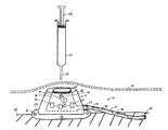
- FIG. 1 is an illustration of an embodiment of a medical device.
- FIG. 2 is a schematic cross sectional view of an embodiment of a radiation source.
- FIG. 3 is an illustration of an embodiment of a medical device.
- FIG. 4 is an illustration of an embodiment of a medical system.
- a medical device 10 includes a port 12 and a catheter 14 , both of which are implanted under a subject's skin 16 for extended periods of time, i.e., the device is indwelling subcutaneously.
- Port 12 includes a housing 18 , a septum 20 , an outlet 22 in fluid communication with catheter 14 , and a base 24 having attachment openings 26 configured to secure the port to bodily tissue 28 .
- Housing 18 and septum 20 define a cavity 30 in fluid communication with outlet 22 .
- Catheter 14 connects to outlet 22 and extends to an exit 32 that is in fluid communication with the subject's vascular system, e.g., a vein.
- Port 12 further includes a plurality of radiation sources 34 and a controller 36 ; and catheter 14 includes a plurality of radiation sources 38 and a controller 40 .
- Radiation sources 34 and 38 are generally configured to treat or to modify a material 42 , such as a pharmacological compound, e.g., a drug, that is introduced into port 12 and catheter 14 , respectively.
- radiation sources 34 and 38 are capable of modifying material 42 by generating and emitting energy.
- One type of energy is ultraviolet light (about 100 to about 400 nm), e.g., UV-C light (about 100 to about 280 nm).
- Radiation sources 34 and 38 can emit energy sufficient to modify material 42 .
- radiation sources 34 and 38 can emit a sufficient dosage of ultraviolet light that can inactivate, kill, reduce, neutralize, inhibit, or otherwise modify, organisms in material 42 such as bacteria, viruses, yeasts, protozoa, and molds.
- Controllers 36 and 40 are configured to control radiation sources 34 and 38 , respectively.
- Controllers 36 and 40 include a power source, e.g., a micro-cell or a battery, a sensor, and a programmable microprocessor chip that are in electrical communication with the radiation sources.
- Controllers 36 and 40 are capable of detecting material 42 that is introduced into port 12 and catheter 14 , respectively, and activating radiation sources 34 and 38 according to a predetermined manner.
- the controller can activate radiation sources 34 for a predetermined amounted of time, at a predetermined frequency, and/or at a predetermined time after it has detected the material.
- controller 36 can activate radiation sources 34 sequentially to radiate a bolus of material 42 with multiple exposures. That is, controllers 36 and 40 can provide an automatic mechanism for detecting material 42 in device 10 and actuating radiation sources 34 and 38 in a predetermined manner.
- material 42 e.g., a drug
- a syringe 45 is introduced into cavity 30 by piercing the subject's skin 16 and septum 20 with a needle 47 , and injecting the material.
- controller 36 detects the material and activates radiation sources 34 in a predetermined manner.
- radiation sources 34 can emit ultraviolet light at predetermined intervals for a predetermined duration sufficient to reduce or eliminate unwanted organisms in material 42 .
- controller 40 of the catheter detects the material and activates radiation sources 38 in a predetermined manner to further treat the material in the catheter.
- the material can be exposed to multiple treatments, e.g., sterilization, steps.
- infectious material that may have been introduced into the subject, e.g., from skin 16 or needle 47 , can be reduced, thereby reducing the risk of infection to the subject.
- device 10 can be used to treat bodily material, such as blood, that is withdrawn from the subject through the device.
- bodily material such as blood
- Bodily material is introduced into device 10 by piercing skin 16 and septum 20 with needle 47 , and drawing a plunger 49 of syringe 45 .
- controller 40 activates radiation sources 38 according to a predetermined manner; and/or as the bodily material then flows to cavity 30 , controller 36 activates radiation sources 34 according to a predetermined manner.
- material withdrawn from the subject can be treated, e.g., sterilized.
- catheter 14 may include multiple controllers 40 , e.g., one controller can be adjacent to exit 32 .
- Radiation sources 34 and 38 can be positioned in port 12 and 14 , respectively, in numerous configurations. Generally, sources 34 and 38 are arranged such that material 42 can be treated with energy from the sources, e.g., with sufficient dosage. For example, sources 34 and 38 can be arranged such that the entire surface of cavity 30 and/or the entire interior surface of catheter 14 are exposed to energy emitted from the sources, e.g., there is a clear line of sight between any point on the surface(s) and at least one radiation source. Within cavity 30 , sources 34 can be arranged symmetrically or asymmetrically. Sources 34 can be arranged in any configuration, such as in a circle, an oval, a triangle, a square, a rectangle, or any polygon. Sources 34 can be arranged near base 24 , near septum 20 , and/or in between the base and the septum. Sources 34 can be secured, for example, by an adhesive, or by forming openings in housing 18 into which the sources are placed.
- sources 38 can be arranged along the length of the catheter. Sources 38 can be arranged collinearly or not collinearly, e.g., offset from a longitudinal axis of catheter 14 . Sources 38 can be arranged equally or unequally spaced apart. Sources 38 may be spaced from the wall of catheter 14 . For example, sources 38 may be arranged centered relative to the cross section of the catheter, so that material 42 flows around all sides of the sources. Sources 38 can be positioned in catheter 14 , for example, by using an adhesive to attach the sources to the wall of the catheter, or by extruding the catheter to include projections that extend radially inward to support the sources, while allowing material to flow through the catheter.
- Energy device 44 includes a top portion 46 , a body portion 48 connected to the top portion, and a flash lamp 50 secured to and centered inside the top portion by a friction ring 52 .
- Body portion 48 includes lenticular patterns or a Fresnel lens 53 that can be embossed or molded on a surface of the body portion to focus or diffuse light generated by flash lamp 50 .
- Flash lamp 50 is a gas discharge lamp capable of generating energy of relatively short duration and high intensity, such as ultraviolet light.
- the gas can be xenon, argon, krypton, or a combination of gases such as xenon and a chloride.
- Flash lamp 50 produces light by providing a potential difference through the gas.
- energy device 44 further includes two leads 54 and a third lead 60 .
- Leads 54 extend from a connector 56 to a transformer 58 and then to flash lamp 50 .
- Leads 54 are used to provide a potential difference between ends of flash lamp 50 to generate light.
- Transformer 58 e.g., constructed by winding enamel-covered copper wire around a cylindrical form and tapping the wire at predetermined points, serves as a voltage step up or step down system for power supplied to flash lamp 50 .
- energy device 44 does not include a transformer.
- leads 54 may be insulated to prevent arcing during use.
- Third lead 60 extends from a ground of connector 60 to a metal foil 62 , e.g., copper foil, placed adjacent to a surface of flash lamp 50 .
- Foil 62 can help in the firing of flash lamp 50 , e.g., enhanced flash output, by providing an approximately equipotential charge along the length of flash lamp 50 , thereby reducing the peak voltage for flash output.
- leads 54 and third lead 60 extend to connector 56 , which is configured to connect with a power source 62 .
- power source 62 applies a voltage potential between leads 54 , which causes an electrical discharge through the gas in flash lamp 50 . The electrical discharge excites the gas, which emits radiation when it electronically decays from an excited state.
- energy device 44 that can be used as radiation sources, such as arc lamps and sonoluminescent light devices, are described in WO 98/22184 and U.S. patent application Publication 2001/0,003,800 A1, both hereby incorporated by reference in their entirety.
- a sensor includes at least two electrodes, e.g., pins or contacts, that are exposed to flow of material 42 to detect a change in electrical conductivity. In operation, the sensor detects a first conductivity prior to any material being in the port or catheter. When material is introduced into the port or the catheter and contacts the electrodes, the detected conductivity changes, e.g. increases when the material bridges the electrodes. This change in conductivity is communicated to the microprocessor chip of the controller, which activates the appropriate radiation sources accordingly.
- a sensor includes one electrode, with housing 18 serving as a second electrode.
- microcomponent liquid sensors are also commercially available, such as the type available from Texas Instruments (e.g., SpreetaTM liquid sensor) and C.A.T. GmbH & Co. (e.g., resistive liquid sensor).
- the radiation source(s) in port 12 and/or catheter 14 are activated manually and/or remotely.
- the radiation sources may not be controlled by a controller positioned in a device.
- Radiation sources in a subcutaneously implanted device may be activated externally.
- the radiation sources can be activated by an activator, e.g., an electromagnetic emitter that can activate a radiation source in a medical device.
- An external switch can be used to turn the radiation sources on, e.g., at the time material 42 is injected, and turn the radiation sources off when injection is complete.
- the power source can be placed within the medical device as described above or placed outside the device.
- a battery pack can be placed remote from the device, e.g., port 12 , and connected to controller 36 and/or 40 via wires that extend through the port and/or catheter 14 .
- Port 12 can be made of a biocompatible metal, such as titanium, or a thermoplastic material.
- Septum 20 can be made of self-sealing material that can be pierced by a needle, such as a silicone.
- medical device 100 includes a port 120 that is secured extracorporeally during use, and a catheter 140 that extends from the port, through skin 160 , and into the subject's vascular system.
- Device 100 , port 120 and catheter 140 are generally similar to device 10 , port 12 and catheter 14 , respectively, as described herein.
- Controller 36 and radiation sources 34 can be applied to other varieties of medical devices.
- a medical system 70 includes a fluid, e.g., saline, source 72 , a catheter 74 connected to the source, and a needle 76 connected to the catheter.
- System 70 further includes an inlet 78 for introducing a material, such as a drug, into catheter 74 .
- Controller 36 and radiation sources 34 can be placed in catheter 74 as described herein. Controller 36 and radiation sources 34 can be used to treat fluid from source 72 and/or other materials introduced into catheter 74 , e.g., through inlet 78 .
- port 12 includes one or more radiation sources
- catheter 14 includes no radiation sources; and vice versa.
- Port 12 and/or catheter 14 can include more than one set of controller and radiation sources.
- one set of controller and radiation source(s) can be configured to activate in response to a first material or condition.
- Another set of controller and radiation source(s) can be configured to activate in response to another material or condition, e.g., different than the first material or condition.
- the controller(s) can be placed anywhere in a device, for example, near a septum, near a base, in or near an outlet, and/or anywhere along the length of a catheter, e.g., near the ends of the catheter.
- radiation can be delivered to a medical device using optic fibers.
- radiation energies can be used, for example, X-rays and infrared radiation.
- Other types of radiation sources can be used, e.g., light emitting diodes.
- radiation source(s) 34 and/or 38 are kept on continuously.
- Material 42 can be material that is introduced to the subject or withdrawn from the subject.
- material 42 can be a pharmaceutically active material, e.g., a drug.
- radiation source(s) 34 and/or 38 can be used to activate the pharmaceutically active material.
- Material 42 can be a bodily fluid, such as blood, urine, or gastric fluids.
- the medial device can be relatively large or relatively small.
- the medical device e.g., port and catheter
- the medical device can be appropriately dimensioned according to how it is used, e.g., in an esophagus, in a vein, or in a body cavity, such as the stomach.
Abstract
Description
- The invention relates to medical devices, such as, for example, those that can be communicable with a body.
- Repeated access to a subject's vascular system, for example, for intravenous drug delivery, for withdrawal of bodily fluids, or for extracorporeal treatments such as hemodialysis, can be established by a variety of medical devices. In some embodiments, a device includes a port and a catheter. The port includes a cavity defined by a housing and a septum through which a needle can penetrate to deliver fluid to the cavity. The septum can be made of, for example, a self-sealing silicone. The port can be placed extracorporeally or implanted subcutaneously. In embodiments in which the port is placed extracorporeally, the catheter has a proximal end that is in fluid communication with the cavity of the port, a body portion that extends through the subject's skin, and a distal end that is in fluid communication with the vascular system, e.g., implanted in a vein. In embodiments in which the port is implanted subcutaneously, the catheter is also implanted subcutaneously and extends from the port cavity to the vascular system. In both types of ports, fluid delivered through the septum to the port cavity can be delivered to the vascular system via the catheter.
- During use, the port and the catheter can be subject to infection. For example, for subcutaneously implanted ports, bacteria can be transferred from the subject's skin to the port cavity and the catheter when the needle penetrates the skin and the septum. The bacteria can infect the port cavity, the catheter, and bodily tissue surrounding the device, exposing the subject to risk. The infection can spread and become systemic, exposing the subject to greater health risk.
- The invention relates to medical devices, such as, for example, those that can be communicable with a body.
- In one aspect, the invention features medical devices that are capable of providing in vivo sterilization, for example, for germicidal and antimicrobial purposes, thereby reducing the risk of infection, such as catheter-related blood stream infections.
- In another aspect, the invention features a medical device having a cavity communicable with a body to deliver or to receive a fluid, and a radiation source configured to expose a portion of the cavity to radiation.
- Embodiments may include one or more of the following features. The cavity is capable of being in fluid communication with the body. The radiation includes ultraviolet radiation, such as ultraviolet-C radiation. The cavity is defined by a catheter. The device includes a controller in electrical communication with the radiation source. The controller is configured to detect a change in electrical resistance. The cavity is defined by a port, such as one configured for subcutaneous implantation or extracorporeal placement.
- In another aspect, the invention features a medical device including a port defining a cavity and having a penetrable portion, a radiation source in the cavity, and a catheter in fluid communication with the cavity.
- Embodiments may include one or more of the following features. The radiation source is capable of emitting ultraviolet radiation, e.g., ultraviolet-C radiation. The device further includes a plurality of radiation sources in the cavity, for example, arranged such that substantially the entire surface of the cavity is exposed to radiation from the sources. The device further includes a controller interfaced with the radiation source. The controller may control the radiation source based on the presence of injectable material in the cavity. The device further includes a second radiation source in the catheter. The device further includes a plurality of radiation sources positioned axially along the length of the catheter. The plurality of radiation sources are radially centered along the catheter.
- The penetrable portion can include a self-sealing material. The penetrable portion can be penetrable by an injection needle.
- The port can be secured extracorporeally and/or implanted subcutaneously.
- In another aspect, the invention features a medical device including a port defining a cavity and having a penetrable portion, a catheter in fluid communication with the cavity, and a radiation source in the catheter.
- Embodiments may include one or more of the following features. The radiation source is capable of emitting ultraviolet radiation, e.g., ultraviolet-C radiation. The device further includes a plurality of radiation sources positioned axially along the length of the catheter. The device further includes a controller interfaced with the radiation source. The controller controls the radiation source based on the presence of injectable material in the catheter. The catheter has a distal end configured to be in fluid communication with a bodily vessel. The port is configured to be secured extracorporeally and/or implanted subcutaneously.
- In another aspect, the invention features a method including introducing an injectable material into a cavity of a port having a catheter in fluid communication with the cavity, and exposing the injectable material in the cavity to radiation.
- Embodiments may include one or more of the following features. The radiation is ultraviolet radiation. The method further includes exposing injectable material in the catheter to radiation. The method further includes s en sing the injectable material in the cavity. The method includes exposing the injectable material to a dosage of ultraviolet radiation sufficient to modify an organism in the injectable material. The method includes penetrating a portion of the port with a needle. Exposing the injectable material to radiation is performed in vivo. The method further includes exposing the injectable material in the cavity to radiation at a predetermined time after introducing the material into the cavity.
- In another aspect, the invention features a method including introducing a material into a cavity in fluid communication with a body, and exposing the material in the cavity to radiation, such as ultraviolet radiation, e.g., ultraviolet-C radiation. Exposing the material to radiation can be performed in vivo or extracorporeally.
- The material can be a bodily fluid and/or a pharmacological material.
- Embodiments may have one or more of the following advantages. Colonization of unwanted organism, e.g., bacteria, in the device or in the body can be reduced, thereby reducing the risk of infection. Formation of a biofilm can be inhibited or reduced, which can reduce formation of clots. The invention can be applied to a variety of medical devices.
- Other features and advantages of the invention will be apparent from the description of the preferred embodiments thereof and from the claims.
- FIG. 1 is an illustration of an embodiment of a medical device.
- FIG. 2 is a schematic cross sectional view of an embodiment of a radiation source.
- FIG. 3 is an illustration of an embodiment of a medical device.
- FIG. 4 is an illustration of an embodiment of a medical system.
- Referring to FIG. 1, a
medical device 10 includes a port 12 and acatheter 14, both of which are implanted under a subject'sskin 16 for extended periods of time, i.e., the device is indwelling subcutaneously. Port 12 includes ahousing 18, aseptum 20, anoutlet 22 in fluid communication withcatheter 14, and a base 24 havingattachment openings 26 configured to secure the port tobodily tissue 28.Housing 18 andseptum 20 define acavity 30 in fluid communication withoutlet 22.Catheter 14 connects tooutlet 22 and extends to anexit 32 that is in fluid communication with the subject's vascular system, e.g., a vein. - Port 12 further includes a plurality of
radiation sources 34 and acontroller 36; andcatheter 14 includes a plurality ofradiation sources 38 and acontroller 40.Radiation sources material 42, such as a pharmacological compound, e.g., a drug, that is introduced into port 12 andcatheter 14, respectively. In some embodiments,radiation sources material 42 by generating and emitting energy. One type of energy is ultraviolet light (about 100 to about 400 nm), e.g., UV-C light (about 100 to about 280 nm).Radiation sources material 42. For example,radiation sources material 42 such as bacteria, viruses, yeasts, protozoa, and molds. -
Controllers radiation sources Controllers Controllers material 42 that is introduced into port 12 andcatheter 14, respectively, and activatingradiation sources controller 36 detects a material in port 12, the controller can activateradiation sources 34 for a predetermined amounted of time, at a predetermined frequency, and/or at a predetermined time after it has detected the material. For example,controller 36 can activateradiation sources 34 sequentially to radiate a bolus ofmaterial 42 with multiple exposures. That is,controllers material 42 indevice 10 and actuatingradiation sources - During use,
material 42, e.g., a drug, from asyringe 45 is introduced intocavity 30 by piercing the subject'sskin 16 andseptum 20 with aneedle 47, and injecting the material. Asmaterial 42 flows throughcavity 30,controller 36 detects the material and activatesradiation sources 34 in a predetermined manner. For example,radiation sources 34 can emit ultraviolet light at predetermined intervals for a predetermined duration sufficient to reduce or eliminate unwanted organisms inmaterial 42. Asmaterial 42 flows fromcavity 30, tooutlet 22, and tocatheter 14,controller 40 of the catheter detects the material and activatesradiation sources 38 in a predetermined manner to further treat the material in the catheter. Thus, asmaterial 42 flows throughdevice 10 andexit 32, the material can be exposed to multiple treatments, e.g., sterilization, steps. As a result, infectious material that may have been introduced into the subject, e.g., fromskin 16 orneedle 47, can be reduced, thereby reducing the risk of infection to the subject. - Similarly,
device 10 can be used to treat bodily material, such as blood, that is withdrawn from the subject through the device. Bodily material is introduced intodevice 10 by piercingskin 16 andseptum 20 withneedle 47, and drawing aplunger 49 ofsyringe 45. As the bodily material flows throughcatheter 14,controller 40 activatesradiation sources 38 according to a predetermined manner; and/or as the bodily material then flows tocavity 30,controller 36 activatesradiation sources 34 according to a predetermined manner. As a result, material withdrawn from the subject can be treated, e.g., sterilized. In some embodiments,catheter 14 may includemultiple controllers 40, e.g., one controller can be adjacent to exit 32. -
Radiation sources port 12 and 14, respectively, in numerous configurations. Generally, sources 34 and 38 are arranged such thatmaterial 42 can be treated with energy from the sources, e.g., with sufficient dosage. For example,sources cavity 30 and/or the entire interior surface ofcatheter 14 are exposed to energy emitted from the sources, e.g., there is a clear line of sight between any point on the surface(s) and at least one radiation source. Withincavity 30,sources 34 can be arranged symmetrically or asymmetrically.Sources 34 can be arranged in any configuration, such as in a circle, an oval, a triangle, a square, a rectangle, or any polygon.Sources 34 can be arranged nearbase 24, nearseptum 20, and/or in between the base and the septum.Sources 34 can be secured, for example, by an adhesive, or by forming openings inhousing 18 into which the sources are placed. - Within
catheter 14,sources 38 can be arranged along the length of the catheter.Sources 38 can be arranged collinearly or not collinearly, e.g., offset from a longitudinal axis ofcatheter 14.Sources 38 can be arranged equally or unequally spaced apart.Sources 38 may be spaced from the wall ofcatheter 14. For example,sources 38 may be arranged centered relative to the cross section of the catheter, so thatmaterial 42 flows around all sides of the sources.Sources 38 can be positioned incatheter 14, for example, by using an adhesive to attach the sources to the wall of the catheter, or by extruding the catheter to include projections that extend radially inward to support the sources, while allowing material to flow through the catheter. - Referring to FIG. 2, an embodiment of
radiation sources energy device 44, is shown.Energy device 44 includes a top portion 46, abody portion 48 connected to the top portion, and aflash lamp 50 secured to and centered inside the top portion by afriction ring 52.Body portion 48 includes lenticular patterns or aFresnel lens 53 that can be embossed or molded on a surface of the body portion to focus or diffuse light generated byflash lamp 50.Flash lamp 50 is a gas discharge lamp capable of generating energy of relatively short duration and high intensity, such as ultraviolet light. The gas can be xenon, argon, krypton, or a combination of gases such as xenon and a chloride. -
Flash lamp 50 produces light by providing a potential difference through the gas. Still referring to FIG. 2,energy device 44 further includes two leads 54 and a third lead 60. Leads 54 extend from aconnector 56 to atransformer 58 and then toflash lamp 50. Leads 54 are used to provide a potential difference between ends offlash lamp 50 to generate light.Transformer 58, e.g., constructed by winding enamel-covered copper wire around a cylindrical form and tapping the wire at predetermined points, serves as a voltage step up or step down system for power supplied toflash lamp 50. In some embodiments,energy device 44 does not include a transformer. For example, leads 54 may be insulated to prevent arcing during use. Third lead 60 extends from a ground of connector 60 to ametal foil 62, e.g., copper foil, placed adjacent to a surface offlash lamp 50.Foil 62 can help in the firing offlash lamp 50, e.g., enhanced flash output, by providing an approximately equipotential charge along the length offlash lamp 50, thereby reducing the peak voltage for flash output. As mentioned above, leads 54 and third lead 60 extend toconnector 56, which is configured to connect with apower source 62. During use,power source 62 applies a voltage potential between leads 54, which causes an electrical discharge through the gas inflash lamp 50. The electrical discharge excites the gas, which emits radiation when it electronically decays from an excited state. - Other embodiments of
energy device 44 that can be used as radiation sources, such as arc lamps and sonoluminescent light devices, are described in WO 98/22184 and U.S. patent application Publication 2001/0,003,800 A1, both hereby incorporated by reference in their entirety. - The sensors of
controllers material 42 in port 12 andcatheter 14, respectively. In some embodiments, a sensor includes at least two electrodes, e.g., pins or contacts, that are exposed to flow ofmaterial 42 to detect a change in electrical conductivity. In operation, the sensor detects a first conductivity prior to any material being in the port or catheter. When material is introduced into the port or the catheter and contacts the electrodes, the detected conductivity changes, e.g. increases when the material bridges the electrodes. This change in conductivity is communicated to the microprocessor chip of the controller, which activates the appropriate radiation sources accordingly. In some embodiments, a sensor includes one electrode, withhousing 18 serving as a second electrode. Other sensors, for example, microcomponent liquid sensors, are also commercially available, such as the type available from Texas Instruments (e.g., Spreeta™ liquid sensor) and C.A.T. GmbH & Co. (e.g., resistive liquid sensor). - In embodiments, the radiation source(s) in port 12 and/or
catheter 14 are activated manually and/or remotely. The radiation sources may not be controlled by a controller positioned in a device. Radiation sources in a subcutaneously implanted device may be activated externally. During use, for example, the radiation sources can be activated by an activator, e.g., an electromagnetic emitter that can activate a radiation source in a medical device. An external switch can be used to turn the radiation sources on, e.g., at thetime material 42 is injected, and turn the radiation sources off when injection is complete. - The power source can be placed within the medical device as described above or placed outside the device. For example, a battery pack can be placed remote from the device, e.g., port 12, and connected to
controller 36 and/or 40 via wires that extend through the port and/orcatheter 14. - Port 12 can be made of a biocompatible metal, such as titanium, or a thermoplastic material.
Septum 20 can be made of self-sealing material that can be pierced by a needle, such as a silicone. - Other Embodiments
- Referring to FIG. 3, in embodiments,
medical device 100 includes aport 120 that is secured extracorporeally during use, and acatheter 140 that extends from the port, throughskin 160, and into the subject's vascular system.Device 100,port 120 andcatheter 140 are generally similar todevice 10, port 12 andcatheter 14, respectively, as described herein. -
Controller 36 andradiation sources 34 can be applied to other varieties of medical devices. Referring to FIG. 4, a medical system 70 includes a fluid, e.g., saline,source 72, acatheter 74 connected to the source, and aneedle 76 connected to the catheter. System 70 further includes aninlet 78 for introducing a material, such as a drug, intocatheter 74.Controller 36 andradiation sources 34 can be placed incatheter 74 as described herein.Controller 36 andradiation sources 34 can be used to treat fluid fromsource 72 and/or other materials introduced intocatheter 74, e.g., throughinlet 78. - In some embodiments, port 12 includes one or more radiation sources, and
catheter 14 includes no radiation sources; and vice versa. Port 12 and/orcatheter 14 can include more than one set of controller and radiation sources. For example, one set of controller and radiation source(s) can be configured to activate in response to a first material or condition. Another set of controller and radiation source(s) can be configured to activate in response to another material or condition, e.g., different than the first material or condition. The controller(s) can be placed anywhere in a device, for example, near a septum, near a base, in or near an outlet, and/or anywhere along the length of a catheter, e.g., near the ends of the catheter. - In certain embodiments, radiation can be delivered to a medical device using optic fibers.
- Other radiation energies can be used, for example, X-rays and infrared radiation. Other types of radiation sources can be used, e.g., light emitting diodes.
- In some embodiments, radiation source(s) 34 and/or 38 are kept on continuously.
-
Material 42 can be material that is introduced to the subject or withdrawn from the subject. For example,material 42 can be a pharmaceutically active material, e.g., a drug. In some embodiments, radiation source(s) 34 and/or 38 can be used to activate the pharmaceutically active material.Material 42 can be a bodily fluid, such as blood, urine, or gastric fluids. - The medial device can be relatively large or relatively small. For example, the medical device, e.g., port and catheter, can be appropriately dimensioned according to how it is used, e.g., in an esophagus, in a vein, or in a body cavity, such as the stomach.
- Other embodiments are within the claims.
Claims (45)
Priority Applications (3)
| Application Number | Priority Date | Filing Date | Title |
|---|---|---|---|
| US10/117,947 US7232429B2 (en) | 2002-04-08 | 2002-04-08 | Medical devices |
| AU2003220647A AU2003220647A1 (en) | 2002-04-08 | 2003-04-03 | Medical devices |
| PCT/US2003/010171 WO2003086508A1 (en) | 2002-04-08 | 2003-04-03 | Medical devices |
Applications Claiming Priority (1)
| Application Number | Priority Date | Filing Date | Title |
|---|---|---|---|
| US10/117,947 US7232429B2 (en) | 2002-04-08 | 2002-04-08 | Medical devices |
Publications (2)
| Publication Number | Publication Date |
|---|---|
| US20030191356A1 true US20030191356A1 (en) | 2003-10-09 |
| US7232429B2 US7232429B2 (en) | 2007-06-19 |
Family
ID=28674315
Family Applications (1)
| Application Number | Title | Priority Date | Filing Date |
|---|---|---|---|
| US10/117,947 Expired - Fee Related US7232429B2 (en) | 2002-04-08 | 2002-04-08 | Medical devices |
Country Status (3)
| Country | Link |
|---|---|
| US (1) | US7232429B2 (en) |
| AU (1) | AU2003220647A1 (en) |
| WO (1) | WO2003086508A1 (en) |
Cited By (8)
| Publication number | Priority date | Publication date | Assignee | Title |
|---|---|---|---|---|
| US20050163954A1 (en) * | 2004-01-22 | 2005-07-28 | Shaw William J. | Medical devices |
| US20060057128A1 (en) * | 2004-09-10 | 2006-03-16 | Dimauro Thomas M | Intradiscal injection of autologous interferon |
| US20090030435A1 (en) * | 2004-09-27 | 2009-01-29 | Theranova, Llc | Method and apparatus for anchoring cardiovascular implants |
| WO2009023818A1 (en) * | 2007-08-15 | 2009-02-19 | Theranova, Llc | Method and apparatus for automated active sterilization of fully implanted devices |
| US20130218075A1 (en) * | 2009-11-03 | 2013-08-22 | Yissum Research Development Company Of The Hebrew University Of Jerusalem, Ltd. | Device for irradiating an internal body surface |
| US20130303996A1 (en) * | 2012-04-16 | 2013-11-14 | Puracath Medical, Inc. | System and method for disinfecting a catheter system |
| US9603558B2 (en) | 2008-08-15 | 2017-03-28 | Theranova, Llc | Methods and devices for the diagnosis and treatment of diabetes |
| AU2019283918B2 (en) * | 2008-10-10 | 2021-05-13 | Implantica Patent Ltd | Fastening means for implantable medical control assembly |
Families Citing this family (52)
| Publication number | Priority date | Publication date | Assignee | Title |
|---|---|---|---|---|
| US7947022B2 (en) | 2005-03-04 | 2011-05-24 | C. R. Bard, Inc. | Access port identification systems and methods |
| US8029482B2 (en) | 2005-03-04 | 2011-10-04 | C. R. Bard, Inc. | Systems and methods for radiographically identifying an access port |
| WO2006096686A1 (en) | 2005-03-04 | 2006-09-14 | C.R. Bard, Inc. | Access port identification systems and methods |
| US9474888B2 (en) | 2005-03-04 | 2016-10-25 | C. R. Bard, Inc. | Implantable access port including a sandwiched radiopaque insert |
| EP1896117B1 (en) | 2005-04-27 | 2011-01-12 | C.R.Bard, Inc. | Power injector system for injecting contrast media into an intravenous line |
| US10307581B2 (en) | 2005-04-27 | 2019-06-04 | C. R. Bard, Inc. | Reinforced septum for an implantable medical device |
| US8147455B2 (en) | 2005-04-27 | 2012-04-03 | C. R. Bard, Inc. | Infusion apparatuses and methods of use |
| US20070198026A1 (en) * | 2006-02-23 | 2007-08-23 | Cauthen Joseph C | Medical device support and stabilizer |
| BRPI0602271B8 (en) * | 2006-04-26 | 2021-06-22 | Augusto Silva Pires E Albuquerque Marcos | partially implantable long-term central venous catheter device |
| US8491547B2 (en) * | 2006-04-28 | 2013-07-23 | Medtronic, Inc. | Septum monitoring system and method for an implantable therapeutic substance delivery device |
| US9265912B2 (en) | 2006-11-08 | 2016-02-23 | C. R. Bard, Inc. | Indicia informative of characteristics of insertable medical devices |
| US9642986B2 (en) | 2006-11-08 | 2017-05-09 | C. R. Bard, Inc. | Resource information key for an insertable medical device |
| US8257325B2 (en) | 2007-06-20 | 2012-09-04 | Medical Components, Inc. | Venous access port with molded and/or radiopaque indicia |
| US8197087B2 (en) * | 2007-07-05 | 2012-06-12 | Baxter International Inc. | Peritoneal dialysis patient connection system using ultraviolet light emitting diodes |
| US8496609B2 (en) | 2007-07-05 | 2013-07-30 | Baxter International Inc. | Fluid delivery system with spiked cassette |
| US8764702B2 (en) * | 2007-07-05 | 2014-07-01 | Baxter International Inc. | Dialysis system having dual patient line connection and prime |
| US7736328B2 (en) | 2007-07-05 | 2010-06-15 | Baxter International Inc. | Dialysis system having supply container autoconnection |
| US8157761B2 (en) | 2007-07-05 | 2012-04-17 | Baxter International Inc. | Peritoneal dialysis patient connection system |
| US7955295B2 (en) | 2007-07-05 | 2011-06-07 | Baxter International Inc. | Fluid delivery system with autoconnect features |
| US9610432B2 (en) | 2007-07-19 | 2017-04-04 | Innovative Medical Devices, Llc | Venous access port assembly with X-ray discernable indicia |
| EP2180915B1 (en) | 2007-07-19 | 2017-10-04 | Medical Components, Inc. | Venous access port assembly with x-ray discernable indicia |
| US8366652B2 (en) * | 2007-08-17 | 2013-02-05 | The Invention Science Fund I, Llc | Systems, devices, and methods including infection-fighting and monitoring shunts |
| US8114346B2 (en) * | 2007-08-17 | 2012-02-14 | The Invention Science Fund I, Llc | Event-triggered ultraviolet light sterilization of surfaces |
| US20090048648A1 (en) * | 2007-08-17 | 2009-02-19 | Searete Llc, A Limited Liability Corporation Of The State Of Delaware | Self-sterilizing device |
| US8647292B2 (en) | 2007-08-17 | 2014-02-11 | The Invention Science Fund I, Llc | Systems, devices, and methods including catheters having components that are actively controllable between two or more wettability states |
| US8706211B2 (en) | 2007-08-17 | 2014-04-22 | The Invention Science Fund I, Llc | Systems, devices, and methods including catheters having self-cleaning surfaces |
| US8460229B2 (en) | 2007-08-17 | 2013-06-11 | The Invention Science Fund I, Llc | Systems, devices, and methods including catheters having components that are actively controllable between transmissive and reflective states |
| US20090163964A1 (en) * | 2007-08-17 | 2009-06-25 | Searete Llc, A Limited Liability Corporation Of The State Of Delaware | System, devices, and methods including sterilizing excitation delivery implants with general controllers and onboard power |
| US8734718B2 (en) | 2007-08-17 | 2014-05-27 | The Invention Science Fund I, Llc | Systems, devices, and methods including catheters having an actively controllable therapeutic agent delivery component |
| US8162924B2 (en) | 2007-08-17 | 2012-04-24 | The Invention Science Fund I, Llc | System, devices, and methods including actively-controllable superoxide water generating systems |
| US8753304B2 (en) | 2007-08-17 | 2014-06-17 | The Invention Science Fund I, Llc | Systems, devices, and methods including catheters having acoustically actuatable waveguide components for delivering a sterilizing stimulus to a region proximate a surface of the catheter |
| US8029740B2 (en) | 2008-07-11 | 2011-10-04 | The Invention Science Fund I, Llc | Event-triggered self-sterilization of article surfaces |
| US8702640B2 (en) | 2007-08-17 | 2014-04-22 | The Invention Science Fund I, Llc | System, devices, and methods including catheters configured to monitor and inhibit biofilm formation |
| US9579496B2 (en) | 2007-11-07 | 2017-02-28 | C. R. Bard, Inc. | Radiopaque and septum-based indicators for a multi-lumen implantable port |
| WO2010011930A2 (en) * | 2008-07-24 | 2010-01-28 | Boston Scientific Scimed, Inc. | Various catheter devices for myocardial injections or other uses |
| US8932271B2 (en) | 2008-11-13 | 2015-01-13 | C. R. Bard, Inc. | Implantable medical devices including septum-based indicators |
| US11890443B2 (en) | 2008-11-13 | 2024-02-06 | C. R. Bard, Inc. | Implantable medical devices including septum-based indicators |
| US9044544B2 (en) | 2008-11-21 | 2015-06-02 | Baxter International Inc. | Dialysis machine having auto-connection system with roller occluder |
| US20120041285A1 (en) | 2008-12-04 | 2012-02-16 | Searete Llc, A Limited Liability Corporation Of The State Of Delaware | Systems, devices, and methods including implantable devices with anti-microbial properties |
| WO2010065135A1 (en) | 2008-12-04 | 2010-06-10 | Searete, Llc | System, devices, and methods including actively-controllable sterilizing excitation delivery implants |
| US8585627B2 (en) | 2008-12-04 | 2013-11-19 | The Invention Science Fund I, Llc | Systems, devices, and methods including catheters configured to monitor biofilm formation having biofilm spectral information configured as a data structure |
| JP2013510652A (en) | 2009-11-17 | 2013-03-28 | シー・アール・バード・インコーポレーテッド | Overmolded access port including locking feature and identification feature |
| US9101678B2 (en) | 2011-11-03 | 2015-08-11 | Elwha Llc | Heat-sanitization of surfaces |
| US10894173B2 (en) | 2012-04-05 | 2021-01-19 | Light Line Medical, Inc. | Methods and apparatus to deliver therapeutic, non-ultraviolet electromagnetic radiation to inactivate infectious agents and/or to enhance healthy cell growth via a catheter residing in a body cavity |
| US9808647B2 (en) | 2012-04-05 | 2017-11-07 | Veritas Medical, L.L.C. | Methods and apparatus to inactivate infectious agents on a catheter residing in a body cavity |
| US10307612B2 (en) | 2012-04-05 | 2019-06-04 | Light Line Medical, Inc. | Methods and apparatus to deliver therapeutic, non-ultraviolet electromagnetic radiation to inactivate infectious agents and/or to enhance healthy cell growth via a catheter residing in a body cavity |
| US11229728B1 (en) | 2020-08-24 | 2022-01-25 | Light Line Medical, Inc. | Method and apparatus to deliver therapeutic, non-ultraviolet electromagnetic radiation in a dialysis system |
| US11497932B2 (en) | 2012-04-05 | 2022-11-15 | Light Line Medical, Inc. | Electromagnetic radiation delivery and monitoring system and methods for preventing, reducing and/or eliminating catheter-related infections during institutional or in-home use |
| US11229808B2 (en) | 2012-04-05 | 2022-01-25 | Light Line Medical, Inc. | Methods and apparatus to deliver therapeutic, non-ultraviolet electromagnetic radiation versatilely via a catheter residing in a body cavity |
| WO2014120620A1 (en) | 2013-01-29 | 2014-08-07 | Puracath Medical, Inc. | Apparatus and method for disinfecting a catheter |
| US11007361B2 (en) | 2014-06-05 | 2021-05-18 | Puracath Medical, Inc. | Transfer catheter for ultraviolet disinfection |
| MX2017011956A (en) | 2015-03-18 | 2018-07-06 | Puracath Medical Inc | Catheter connection system for ultraviolet light disinfection. |
Citations (28)
| Publication number | Priority date | Publication date | Assignee | Title |
|---|---|---|---|---|
| US4412834A (en) * | 1981-06-05 | 1983-11-01 | Baxter Travenol Laboratories | Antimicrobial ultraviolet irradiation of connector for continuous ambulatory peritoneal dialysis |
| US4475900A (en) * | 1981-06-05 | 1984-10-09 | Popovich Robert P | Method of peritoneal dialysis involving ultraviolet radiation of dialysis apparatus |
| US5092849A (en) * | 1987-08-25 | 1992-03-03 | Shiley Infusaid, Inc. | Implantable device |
| US5147318A (en) * | 1991-03-04 | 1992-09-15 | Board Of Regents, The University Of Texas System | Valved arterial catheter |
| US5240675A (en) * | 1992-09-24 | 1993-08-31 | Wilk Peter J | Method for cleaning endoscope |
| US5260020A (en) * | 1992-09-17 | 1993-11-09 | Wilk Peter J | Method and apparatus for catheter sterilization |
| US5304155A (en) * | 1992-07-14 | 1994-04-19 | Cook Pacemaker Corporation | Valved catheter |
| US5352204A (en) * | 1990-03-01 | 1994-10-04 | Ensminger William D | Implantable access devices |
| US5562618A (en) * | 1994-01-21 | 1996-10-08 | Sims Deltec, Inc. | Portal assembly and catheter connector |
| US5637877A (en) * | 1995-06-06 | 1997-06-10 | Rare Earth Medical, Inc. | Ultraviolet sterilization of instrument lumens |
| US5848989A (en) * | 1997-06-05 | 1998-12-15 | Davinci Biomedical Research Products, Inc. | Implantable port with low profile housing for delivery/collection of fluids and implantation method |
| US5855203A (en) * | 1997-12-19 | 1999-01-05 | Matter; Jean-Paul | Respiratory circuit with in vivo sterilization |
| US5931829A (en) * | 1997-01-21 | 1999-08-03 | Vasca, Inc. | Methods and systems for establishing vascular access |
| US5931801A (en) * | 1997-01-21 | 1999-08-03 | Vasca, Inc. | Valve port assembly with interlock |
| US5947958A (en) * | 1995-09-14 | 1999-09-07 | Conceptus, Inc. | Radiation-transmitting sheath and methods for its use |
| US5997524A (en) * | 1997-07-18 | 1999-12-07 | Vasca, Inc. | Catheter assembly for percutaneous access to subcutaneous port |
| US6007516A (en) * | 1997-01-21 | 1999-12-28 | Vasca, Inc. | Valve port and method for vascular access |
| US6083148A (en) * | 1991-06-14 | 2000-07-04 | Proxima Therapeutics, Inc. | Tumor treatment |
| US6090068A (en) * | 1998-07-23 | 2000-07-18 | Chanut; Stephane | Injection and perfusion device for use in an implantable port access catheter |
| US6120492A (en) * | 1997-07-18 | 2000-09-19 | Vasca, Inc. | Method and apparatus for percutaneously accessing an implanted port |
| US6132415A (en) * | 1999-02-09 | 2000-10-17 | Vasca, Inc. | Systems and methods for removing retained fluids and infusing therapeutic fluids |
| US6245039B1 (en) * | 1998-10-05 | 2001-06-12 | Vasca, Inc. | Methods and apparatus for performing flow-through peritoneal dialysis |
| US20010003800A1 (en) * | 1996-11-21 | 2001-06-14 | Steven J. Frank | Interventional photonic energy emitter system |
| US6258079B1 (en) * | 1997-01-21 | 2001-07-10 | Vasca, Inc. | Method and systems for establishing vascular access |
| US6270489B1 (en) * | 1999-07-01 | 2001-08-07 | Catheter Innovations, Inc. | Anti-clotting methods and apparatus for indwelling catheter tubes |
| US6270475B1 (en) * | 1997-03-26 | 2001-08-07 | Diesetronic Licensing Ag | Port body for the administration of drugs |
| US6283951B1 (en) * | 1996-10-11 | 2001-09-04 | Transvascular, Inc. | Systems and methods for delivering drugs to selected locations within the body |
| US6299609B1 (en) * | 1998-01-07 | 2001-10-09 | Vasca, Inc. | Methods and apparatus for inhibiting infection of subcutaneously implanted devices |
Family Cites Families (7)
| Publication number | Priority date | Publication date | Assignee | Title |
|---|---|---|---|---|
| US5112303A (en) * | 1991-05-02 | 1992-05-12 | Pudenz-Schulte Medical Research Corporation | Tumor access device and method for delivering medication into a body cavity |
| US5476460A (en) | 1994-04-29 | 1995-12-19 | Minimed Inc. | Implantable infusion port with reduced internal volume |
| US5995860A (en) * | 1995-07-06 | 1999-11-30 | Thomas Jefferson University | Implantable sensor and system for measurement and control of blood constituent levels |
| US6290677B1 (en) | 1996-01-24 | 2001-09-18 | Sumitomo Bakelite Company Limited | Medicinal liquid injection port |
| WO1998022184A1 (en) | 1996-11-21 | 1998-05-28 | Boston Scientific Corporation | Mucosal ablation using light |
| US6117064A (en) * | 1997-01-06 | 2000-09-12 | Apple; Marc G. | Catheter system |
| US6309380B1 (en) | 1999-01-27 | 2001-10-30 | Marian L. Larson | Drug delivery via conformal film |
-
2002
- 2002-04-08 US US10/117,947 patent/US7232429B2/en not_active Expired - Fee Related
-
2003
- 2003-04-03 AU AU2003220647A patent/AU2003220647A1/en not_active Abandoned
- 2003-04-03 WO PCT/US2003/010171 patent/WO2003086508A1/en not_active Application Discontinuation
Patent Citations (31)
| Publication number | Priority date | Publication date | Assignee | Title |
|---|---|---|---|---|
| US4412834A (en) * | 1981-06-05 | 1983-11-01 | Baxter Travenol Laboratories | Antimicrobial ultraviolet irradiation of connector for continuous ambulatory peritoneal dialysis |
| US4475900A (en) * | 1981-06-05 | 1984-10-09 | Popovich Robert P | Method of peritoneal dialysis involving ultraviolet radiation of dialysis apparatus |
| US5092849A (en) * | 1987-08-25 | 1992-03-03 | Shiley Infusaid, Inc. | Implantable device |
| US5352204A (en) * | 1990-03-01 | 1994-10-04 | Ensminger William D | Implantable access devices |
| US5147318A (en) * | 1991-03-04 | 1992-09-15 | Board Of Regents, The University Of Texas System | Valved arterial catheter |
| US6083148A (en) * | 1991-06-14 | 2000-07-04 | Proxima Therapeutics, Inc. | Tumor treatment |
| US5304155A (en) * | 1992-07-14 | 1994-04-19 | Cook Pacemaker Corporation | Valved catheter |
| US5260020A (en) * | 1992-09-17 | 1993-11-09 | Wilk Peter J | Method and apparatus for catheter sterilization |
| US5240675A (en) * | 1992-09-24 | 1993-08-31 | Wilk Peter J | Method for cleaning endoscope |
| US5562618A (en) * | 1994-01-21 | 1996-10-08 | Sims Deltec, Inc. | Portal assembly and catheter connector |
| US5743873A (en) * | 1994-01-21 | 1998-04-28 | Sims Deltec, Inc. | Methods for using catheter connectors and portals, and methods of assembly |
| US5613945A (en) * | 1994-01-21 | 1997-03-25 | Sims Deltec, Inc. | Portal assembly |
| US5637877A (en) * | 1995-06-06 | 1997-06-10 | Rare Earth Medical, Inc. | Ultraviolet sterilization of instrument lumens |
| US5947958A (en) * | 1995-09-14 | 1999-09-07 | Conceptus, Inc. | Radiation-transmitting sheath and methods for its use |
| US6283951B1 (en) * | 1996-10-11 | 2001-09-04 | Transvascular, Inc. | Systems and methods for delivering drugs to selected locations within the body |
| US20010003800A1 (en) * | 1996-11-21 | 2001-06-14 | Steven J. Frank | Interventional photonic energy emitter system |
| US5931829A (en) * | 1997-01-21 | 1999-08-03 | Vasca, Inc. | Methods and systems for establishing vascular access |
| US6258079B1 (en) * | 1997-01-21 | 2001-07-10 | Vasca, Inc. | Method and systems for establishing vascular access |
| US6007516A (en) * | 1997-01-21 | 1999-12-28 | Vasca, Inc. | Valve port and method for vascular access |
| US5931801A (en) * | 1997-01-21 | 1999-08-03 | Vasca, Inc. | Valve port assembly with interlock |
| US6238369B1 (en) * | 1997-01-21 | 2001-05-29 | Vasco, Inc. | Method and systems for establishing vascular access |
| US6270475B1 (en) * | 1997-03-26 | 2001-08-07 | Diesetronic Licensing Ag | Port body for the administration of drugs |
| US5848989A (en) * | 1997-06-05 | 1998-12-15 | Davinci Biomedical Research Products, Inc. | Implantable port with low profile housing for delivery/collection of fluids and implantation method |
| US6120492A (en) * | 1997-07-18 | 2000-09-19 | Vasca, Inc. | Method and apparatus for percutaneously accessing an implanted port |
| US5997524A (en) * | 1997-07-18 | 1999-12-07 | Vasca, Inc. | Catheter assembly for percutaneous access to subcutaneous port |
| US5855203A (en) * | 1997-12-19 | 1999-01-05 | Matter; Jean-Paul | Respiratory circuit with in vivo sterilization |
| US6299609B1 (en) * | 1998-01-07 | 2001-10-09 | Vasca, Inc. | Methods and apparatus for inhibiting infection of subcutaneously implanted devices |
| US6090068A (en) * | 1998-07-23 | 2000-07-18 | Chanut; Stephane | Injection and perfusion device for use in an implantable port access catheter |
| US6245039B1 (en) * | 1998-10-05 | 2001-06-12 | Vasca, Inc. | Methods and apparatus for performing flow-through peritoneal dialysis |
| US6132415A (en) * | 1999-02-09 | 2000-10-17 | Vasca, Inc. | Systems and methods for removing retained fluids and infusing therapeutic fluids |
| US6270489B1 (en) * | 1999-07-01 | 2001-08-07 | Catheter Innovations, Inc. | Anti-clotting methods and apparatus for indwelling catheter tubes |
Cited By (13)
| Publication number | Priority date | Publication date | Assignee | Title |
|---|---|---|---|---|
| US7854756B2 (en) | 2004-01-22 | 2010-12-21 | Boston Scientific Scimed, Inc. | Medical devices |
| US20050163954A1 (en) * | 2004-01-22 | 2005-07-28 | Shaw William J. | Medical devices |
| US20060057128A1 (en) * | 2004-09-10 | 2006-03-16 | Dimauro Thomas M | Intradiscal injection of autologous interferon |
| US7367961B2 (en) * | 2004-09-10 | 2008-05-06 | Depuy Spine, Inc. | Intradiscal injection of autologous interferon |
| US20090030435A1 (en) * | 2004-09-27 | 2009-01-29 | Theranova, Llc | Method and apparatus for anchoring cardiovascular implants |
| US20100256607A1 (en) * | 2007-08-15 | 2010-10-07 | Daniel Rogers Burnett | Method and apparatus for automated active sterilization of fully implanted devices |
| WO2009023818A1 (en) * | 2007-08-15 | 2009-02-19 | Theranova, Llc | Method and apparatus for automated active sterilization of fully implanted devices |
| US8865063B2 (en) | 2007-08-15 | 2014-10-21 | Theranova, Llc | Method and apparatus for automated active sterilization of fully implanted devices |
| US9603558B2 (en) | 2008-08-15 | 2017-03-28 | Theranova, Llc | Methods and devices for the diagnosis and treatment of diabetes |
| AU2019283918B2 (en) * | 2008-10-10 | 2021-05-13 | Implantica Patent Ltd | Fastening means for implantable medical control assembly |
| US20130218075A1 (en) * | 2009-11-03 | 2013-08-22 | Yissum Research Development Company Of The Hebrew University Of Jerusalem, Ltd. | Device for irradiating an internal body surface |
| US20130303996A1 (en) * | 2012-04-16 | 2013-11-14 | Puracath Medical, Inc. | System and method for disinfecting a catheter system |
| US9295742B2 (en) * | 2012-04-16 | 2016-03-29 | Puracath Medical, Inc. | System and method for disinfecting a catheter system |
Also Published As
| Publication number | Publication date |
|---|---|
| WO2003086508A1 (en) | 2003-10-23 |
| AU2003220647A1 (en) | 2003-10-27 |
| US7232429B2 (en) | 2007-06-19 |
Similar Documents
| Publication | Publication Date | Title |
|---|---|---|
| US7232429B2 (en) | Medical devices | |
| US10471277B2 (en) | Methods and apparatus to inactivate infectious agents on a drainage catheter residing in a body cavity | |
| US5921244A (en) | Internal magnetic device to enhance drug therapy | |
| US7899544B2 (en) | Access port indicator for implantable medical device | |
| AU2011234040B2 (en) | Methods and apparatus for reducing count of infectious agents in intravenous access systems | |
| US5505700A (en) | Electro-osmotic infusion catheter | |
| US5702432A (en) | Intracorporeal light treatment of blood | |
| US6719738B2 (en) | Device for directly delivering an active substance within a cell tissue, means for implanting said device and appliances for injecting active substance into said device | |
| US20080306454A1 (en) | Apparatus And Method For Sterilization Of An Intravenous Catheter | |
| KR102040884B1 (en) | Infection-resistant catheters | |
| US20080027399A1 (en) | Antimicrobial vascular access device | |
| US20120161032A1 (en) | Catheter insertion sterilization | |
| US20060095020A1 (en) | Introduction of agent with medical device | |
| US20080215010A1 (en) | Apparatus and method for using an intraosseous space for moving fluid into and out of the body | |
| CN110402155B (en) | Implantable device | |
| EP0510857A1 (en) | Drug release device | |
| CN102210905B (en) | Implanted nerve electrical stimulation system with drug release device | |
| KR101030814B1 (en) | Transurethral electro-chemical treatment system | |
| EP4295873A1 (en) | An infusion catheter system | |
| US20180221679A1 (en) | Catheter assembly and components thereof | |
| WO2021236248A1 (en) | Medical device having a photosensitizer and related methods | |
| WO1997036631A1 (en) | Urinary catheter having elongating balloon |
Legal Events
| Date | Code | Title | Description |
|---|---|---|---|
| AS | Assignment |
Owner name: BOSTON SCIENTIFIC CORPORATION, MASSACHUSETTS Free format text: ASSIGNMENT OF ASSIGNORS INTEREST;ASSIGNOR:MORECI, STEVE;REEL/FRAME:012778/0767 Effective date: 20020403 |
|
| STCF | Information on status: patent grant |
Free format text: PATENTED CASE |
|
| FPAY | Fee payment |
Year of fee payment: 4 |
|
| FPAY | Fee payment |
Year of fee payment: 8 |
|
| FEPP | Fee payment procedure |
Free format text: MAINTENANCE FEE REMINDER MAILED (ORIGINAL EVENT CODE: REM.); ENTITY STATUS OF PATENT OWNER: LARGE ENTITY |
|
| LAPS | Lapse for failure to pay maintenance fees |
Free format text: PATENT EXPIRED FOR FAILURE TO PAY MAINTENANCE FEES (ORIGINAL EVENT CODE: EXP.); ENTITY STATUS OF PATENT OWNER: LARGE ENTITY |
|
| STCH | Information on status: patent discontinuation |
Free format text: PATENT EXPIRED DUE TO NONPAYMENT OF MAINTENANCE FEES UNDER 37 CFR 1.362 |
|
| FP | Lapsed due to failure to pay maintenance fee |
Effective date: 20190619 |