US20050069495A1 - Cancer specific radiolabeled conjugates regulated by the cell cycle for the treatment and diagnosis of cancer - Google Patents
Cancer specific radiolabeled conjugates regulated by the cell cycle for the treatment and diagnosis of cancer Download PDFInfo
- Publication number
- US20050069495A1 US20050069495A1 US10/671,376 US67137603A US2005069495A1 US 20050069495 A1 US20050069495 A1 US 20050069495A1 US 67137603 A US67137603 A US 67137603A US 2005069495 A1 US2005069495 A1 US 2005069495A1
- Authority
- US
- United States
- Prior art keywords
- conjugate
- cells
- dht
- iudr
- cancer
- Prior art date
- Legal status (The legal status is an assumption and is not a legal conclusion. Google has not performed a legal analysis and makes no representation as to the accuracy of the status listed.)
- Granted
Links
- 0 *OCC1OC(C)CC1* Chemical compound *OCC1OC(C)CC1* 0.000 description 2
- GLTRBCOSKISPQP-VSFZJPHZSA-N C.C.[H]C12CCC3C(CC[C@]4(C)[C@@H](OC(=O)CCC(=O)OC5CC(N6C=C([Sn](C)(C)C)C(=O)NC6=O)OC5CC[PH](C)=O)CC[C@@]34[H])[C@@]1(C)CCC(=O)C2.[H][C@@]12CC[C@H](OC(=O)CCC(=O)OC3CC(N4C=C(I)[C@H](O)NC4=O)OC3CO)[C@@]1(C)CCC1C2CC[C@@]2([H])CC(=O)CC[C@]12C.[H][C@@]12CC[C@H](OC(=O)CCC(=O)OC3CC(N4C=C([125I])C(=O)NC4=O)OC3COP(=O)(O)O)[C@@]1(C)CCC1C2CC[C@@]2([H])CC(=O)CC[C@]12C.[H][C@@]12CC[C@H](OC(=O)CC[C@@H](O)OC3CC(N4C=C(I)C(=O)NC4=O)OC3CO[PH](C)=O)[C@@]1(C)CCC1C2CC[C@@]2([H])CC(=O)CC[C@]12C Chemical compound C.C.[H]C12CCC3C(CC[C@]4(C)[C@@H](OC(=O)CCC(=O)OC5CC(N6C=C([Sn](C)(C)C)C(=O)NC6=O)OC5CC[PH](C)=O)CC[C@@]34[H])[C@@]1(C)CCC(=O)C2.[H][C@@]12CC[C@H](OC(=O)CCC(=O)OC3CC(N4C=C(I)[C@H](O)NC4=O)OC3CO)[C@@]1(C)CCC1C2CC[C@@]2([H])CC(=O)CC[C@]12C.[H][C@@]12CC[C@H](OC(=O)CCC(=O)OC3CC(N4C=C([125I])C(=O)NC4=O)OC3COP(=O)(O)O)[C@@]1(C)CCC1C2CC[C@@]2([H])CC(=O)CC[C@]12C.[H][C@@]12CC[C@H](OC(=O)CC[C@@H](O)OC3CC(N4C=C(I)C(=O)NC4=O)OC3CO[PH](C)=O)[C@@]1(C)CCC1C2CC[C@@]2([H])CC(=O)CC[C@]12C GLTRBCOSKISPQP-VSFZJPHZSA-N 0.000 description 1
- RFVRUSYFECCQBR-OEYLWLGOSA-M C.CCC1OC(N2C=C(I)C(=O)NC2=O)CC1O.CCC1OC(N2C=C([Sn](C)(C)C)[C@H](O)NC2=O)CC1O.O=C1NC(=O)N(C2CC(O)C(CO)O2)C=C1I.[H][C@@]12CC[C@H](OP(=O)(OC)OCC3OC(N4C=C([125I])C(=O)NC4=O)CC3O)[C@@]1(C)CCC1C2CC[C@@]2([H])CC(=O)CC[C@]12C.[Na][125I] Chemical compound C.CCC1OC(N2C=C(I)C(=O)NC2=O)CC1O.CCC1OC(N2C=C([Sn](C)(C)C)[C@H](O)NC2=O)CC1O.O=C1NC(=O)N(C2CC(O)C(CO)O2)C=C1I.[H][C@@]12CC[C@H](OP(=O)(OC)OCC3OC(N4C=C([125I])C(=O)NC4=O)CC3O)[C@@]1(C)CCC1C2CC[C@@]2([H])CC(=O)CC[C@]12C.[Na][125I] RFVRUSYFECCQBR-OEYLWLGOSA-M 0.000 description 1
- OUETXBBSZAMJOT-DHAIXWSUSA-M C.CCC1OC(N2C=C(I)C(=O)NC2=O)CC1O.CCC1OC(N2C=C([Sn](C)(C)C)[C@H](O)NC2=O)CC1O.O=C1NC(=O)N(C2CC(O)C(CO)O2)C=C1I.[H][C@@]12CC[C@H](O[PH](=O)OCC3OC(N4C=C([125I])C(=O)NC4=O)CC3O)[C@@]1(C)CCC1C2CC[C@@]2([H])CC(=O)CC[C@]12C.[Na][125I] Chemical compound C.CCC1OC(N2C=C(I)C(=O)NC2=O)CC1O.CCC1OC(N2C=C([Sn](C)(C)C)[C@H](O)NC2=O)CC1O.O=C1NC(=O)N(C2CC(O)C(CO)O2)C=C1I.[H][C@@]12CC[C@H](O[PH](=O)OCC3OC(N4C=C([125I])C(=O)NC4=O)CC3O)[C@@]1(C)CCC1C2CC[C@@]2([H])CC(=O)CC[C@]12C.[Na][125I] OUETXBBSZAMJOT-DHAIXWSUSA-M 0.000 description 1
- WRIOJALMVLBTPR-RCZCPTMLSA-M C.CCC1OC(N2CC(I)C(=O)NC2=O)CC1O.[H][C@@]12CC[C@H](OC(=O)CCC(=O)OC3CC(N4C=C(I)C(=O)NC4=O)OC3CO)[C@@]1(C)CCC1C2CC[C@@]2([H])CC(=O)CC[C@]12C.[H][C@@]12CC[C@H](OC(=O)CCC(=O)OC3CC(N4C=C([125I])C(=O)NC4=O)OC3CO)[C@@]1(C)CCC1C2CC[C@@]2([H])CC(=O)CC[C@]12C.[H][C@@]12CC[C@H](OC(=O)CCC(=O)OC3CC(N4C=C([Sn](C)(C)C)C(=O)NC4=O)OC3CO)[C@@]1(C)CCC1C2CC[C@@]2([H])CC(=O)CC[C@]12C.[Na][125I] Chemical compound C.CCC1OC(N2CC(I)C(=O)NC2=O)CC1O.[H][C@@]12CC[C@H](OC(=O)CCC(=O)OC3CC(N4C=C(I)C(=O)NC4=O)OC3CO)[C@@]1(C)CCC1C2CC[C@@]2([H])CC(=O)CC[C@]12C.[H][C@@]12CC[C@H](OC(=O)CCC(=O)OC3CC(N4C=C([125I])C(=O)NC4=O)OC3CO)[C@@]1(C)CCC1C2CC[C@@]2([H])CC(=O)CC[C@]12C.[H][C@@]12CC[C@H](OC(=O)CCC(=O)OC3CC(N4C=C([Sn](C)(C)C)C(=O)NC4=O)OC3CO)[C@@]1(C)CCC1C2CC[C@@]2([H])CC(=O)CC[C@]12C.[Na][125I] WRIOJALMVLBTPR-RCZCPTMLSA-M 0.000 description 1
- HJBHCNSNWFJVKG-ACLNJWLWSA-N C.C[PH](=O)OCC1OC(N2C=C(I)C(=O)NC2=O)CC1O.O=C1NC(=O)N(C2CC(O)C(CO)O2)C=C1I.[H][C@@]12CC[C@H](OC(=O)CCC(=O)OC3CC(N4C=C([125I])C(=O)NC4=O)OC3COP(=O)(O)O)[C@@]1(C)CCC1C2CC[C@@]2([H])CC(=O)CC[C@]12C.[H][C@@]12CC[C@H](OC(=O)CCC(=O)OC3CC(N4C=C([Sn](C)(C)C)C(=O)NC4=O)OC3CC[PH](C)=O)[C@@]1(C)CCC1C2CC[C@@]2([H])CC(=O)CC[C@]12C Chemical compound C.C[PH](=O)OCC1OC(N2C=C(I)C(=O)NC2=O)CC1O.O=C1NC(=O)N(C2CC(O)C(CO)O2)C=C1I.[H][C@@]12CC[C@H](OC(=O)CCC(=O)OC3CC(N4C=C([125I])C(=O)NC4=O)OC3COP(=O)(O)O)[C@@]1(C)CCC1C2CC[C@@]2([H])CC(=O)CC[C@]12C.[H][C@@]12CC[C@H](OC(=O)CCC(=O)OC3CC(N4C=C([Sn](C)(C)C)C(=O)NC4=O)OC3CC[PH](C)=O)[C@@]1(C)CCC1C2CC[C@@]2([H])CC(=O)CC[C@]12C HJBHCNSNWFJVKG-ACLNJWLWSA-N 0.000 description 1
- YSFDBOYSNAXGKT-VJYSPDAOSA-N C.[H][C@@]12CC[C@H](OC(=O)CCC(=O)OC3CC(N4C=C([125I])C(=O)NC4=O)OC3CO)[C@@]1(C)CCC1C2CC[C@@]2([H])CC(=O)CC[C@]12C Chemical compound C.[H][C@@]12CC[C@H](OC(=O)CCC(=O)OC3CC(N4C=C([125I])C(=O)NC4=O)OC3CO)[C@@]1(C)CCC1C2CC[C@@]2([H])CC(=O)CC[C@]12C YSFDBOYSNAXGKT-VJYSPDAOSA-N 0.000 description 1
- XJTXLEHDJBMVSA-VJYSPDAOSA-N C.[H][C@@]12CC[C@H](OC(=O)CCC(=O)OC3CC(N4C=C([125I])C(=O)NC4=O)OC3COP(=O)(O)O)[C@@]1(C)CCC1C2CC[C@@]2([H])CC(=O)CC[C@]12C Chemical compound C.[H][C@@]12CC[C@H](OC(=O)CCC(=O)OC3CC(N4C=C([125I])C(=O)NC4=O)OC3COP(=O)(O)O)[C@@]1(C)CCC1C2CC[C@@]2([H])CC(=O)CC[C@]12C XJTXLEHDJBMVSA-VJYSPDAOSA-N 0.000 description 1
- OHCXGJAJZPCFLY-SCQFKNOQSA-N [H][C@@]12CC[C@H](OP(=O)(O)OCC3OC(N4C=C(C)C(=O)NC4=O)CC3O)[C@@]1(C)CCC1C2CC[C@@]2([H])CC(=O)CC[C@]12C Chemical compound [H][C@@]12CC[C@H](OP(=O)(O)OCC3OC(N4C=C(C)C(=O)NC4=O)CC3O)[C@@]1(C)CCC1C2CC[C@@]2([H])CC(=O)CC[C@]12C OHCXGJAJZPCFLY-SCQFKNOQSA-N 0.000 description 1
- WMRJITYFMIBPMH-QCMAGIOMSA-N [H][C@@]12CC[C@H](OP(=O)(OC)OCC3OC(N4C=C(C)C(=O)NC4=O)CC3O)[C@@]1(C)CCC1C2CC[C@@]2([H])CC(=O)CC[C@]12C Chemical compound [H][C@@]12CC[C@H](OP(=O)(OC)OCC3OC(N4C=C(C)C(=O)NC4=O)CC3O)[C@@]1(C)CCC1C2CC[C@@]2([H])CC(=O)CC[C@]12C WMRJITYFMIBPMH-QCMAGIOMSA-N 0.000 description 1
- UFHXYIXNMUETMD-SCQFKNOQSA-N [H][C@@]12CC[C@H](O[PH](=O)OCC3OC(N4C=C(C)C(=O)NC4=O)CC3O)[C@@]1(C)CCC1C2CC[C@@]2([H])CC(=O)CC[C@]12C Chemical compound [H][C@@]12CC[C@H](O[PH](=O)OCC3OC(N4C=C(C)C(=O)NC4=O)CC3O)[C@@]1(C)CCC1C2CC[C@@]2([H])CC(=O)CC[C@]12C UFHXYIXNMUETMD-SCQFKNOQSA-N 0.000 description 1
Images


Classifications
-
- A—HUMAN NECESSITIES
- A61—MEDICAL OR VETERINARY SCIENCE; HYGIENE
- A61K—PREPARATIONS FOR MEDICAL, DENTAL OR TOILETRY PURPOSES
- A61K51/00—Preparations containing radioactive substances for use in therapy or testing in vivo
- A61K51/02—Preparations containing radioactive substances for use in therapy or testing in vivo characterised by the carrier, i.e. characterised by the agent or material covalently linked or complexing the radioactive nucleus
- A61K51/04—Organic compounds
- A61K51/0493—Steroids, e.g. cholesterol, testosterone
-
- A—HUMAN NECESSITIES
- A61—MEDICAL OR VETERINARY SCIENCE; HYGIENE
- A61K—PREPARATIONS FOR MEDICAL, DENTAL OR TOILETRY PURPOSES
- A61K51/00—Preparations containing radioactive substances for use in therapy or testing in vivo
- A61K51/02—Preparations containing radioactive substances for use in therapy or testing in vivo characterised by the carrier, i.e. characterised by the agent or material covalently linked or complexing the radioactive nucleus
- A61K51/04—Organic compounds
- A61K51/0491—Sugars, nucleosides, nucleotides, oligonucleotides, nucleic acids, e.g. DNA, RNA, nucleic acid aptamers
-
- C—CHEMISTRY; METALLURGY
- C07—ORGANIC CHEMISTRY
- C07H—SUGARS; DERIVATIVES THEREOF; NUCLEOSIDES; NUCLEOTIDES; NUCLEIC ACIDS
- C07H19/00—Compounds containing a hetero ring sharing one ring hetero atom with a saccharide radical; Nucleosides; Mononucleotides; Anhydro-derivatives thereof
- C07H19/02—Compounds containing a hetero ring sharing one ring hetero atom with a saccharide radical; Nucleosides; Mononucleotides; Anhydro-derivatives thereof sharing nitrogen
- C07H19/04—Heterocyclic radicals containing only nitrogen atoms as ring hetero atom
- C07H19/048—Pyridine radicals
Definitions
- the present invention relates generally to targeted therapy and medical imaging as applied to cancer treatment and diagnosis, and in particular to conjugates composed of a radiolabeled, cell cycle-dependent therapeutic agent chemically coupled to a ligand that targets androgen receptor (AR).
- the conjugates of the invention are taken up selectively by malignant tumor cells that have androgen receptor and are incorporated into the nucleus of such cells, where they produce a cytotoxic effect and/or are detectable via radioimaging techniques.
- Radioisotopes for the treatment and diagnosis of cancer.
- One of the intended benefits of targeted therapy is to diminish the incidence and severity of side effects by confining toxic exposure, more or less, to the disease site.
- Certain radioisotopes, particularly Auger electron-emitting isotopes, such as 123 I and 125 I are known to be very toxic to viable cells, but only if they are localized within the nucleus of the cell. (Warters et al., Curr. Top. Stop Rad.
- Protein and polypeptide hormones and growth factors may be directly radiolabeled and used to target a tumor cell.
- radioisotopes bound to amino acid residues of hormones, growth factors and the like exit from the cell after catabolism, and do not appreciably bind to nuclear material.
- the above-noted need is satisfied by the radiolabeled conjugates of the present invention which are capable of targeting and being selectively taken up and degraded by a tumor cell, and thereby delivering to the tumor cell nucleus a radioisotope capable of being incorporated into the nuclear material, so as to produce a cytotoxic effect and/or to render the cell detectable by radioimaging.
- the conjugates of the invention can be safely administered in long term cancer treatments, without producing significant adverse health effects.
- the present invention also provides methods of using the above-described conjugate for treating and diagnosing cancers comprising cells having AR, especially ovarian, breast and prostate cancer.
- the conjugates of the present invention have been designed so as to take advantage of two characteristics of many relapsing cancers, i.e. (1) relapsed/advance cancers have a large portion of rapidly growing and dividing cells (i.e. a large S-phase fraction); and (2) AR is expressed in practically all prostate cancer (primary and metastatic), ovarian cancer (>90% positive for AR regardless of the tumor site) and breast cancer (even when estrogen receptor (ER)-negative and progesterone receptor (PR)-negative, breast cancer cells express AR).
- the conjugate Upon administration, the conjugate first binds to the sex hormone binding globulin, which in turn carries it exclusively to cells that have AR. Subsequent to this interaction, the entire conjugate is transported into the cell.
- Intracellular enzymes cleave the linking moiety, thus releasing and trapping within the cell the portion of the conjugate that is responsible for killing tumor cells.
- This cytotoxic effect is induced only when the cell cycle dependent therapeutic agent is incorporated into the DNA of dividing tumor cells.
- the conjugate that remains in systemic circulation, or enters normal tissue or organs is essentially innocuous. Accordingly, the radiolabeled conjugates of the invention may be administered frequently and without adverse effects.
- FIG. 1 is a set of two graphs depicting the binding of 125 IUdR-DHT to OVCAR-3 cells in vitro.
- FIG. 1A plots the fraction of “bound” counts to “free counts” against “bound” 125 IUdR-DHT (mM).
- FIG. 1B plots bound 125 IUdR-DHT (mM) against free 125 IUdR-DHT (mM).
- FIG. 2 is a set of two graphs depicting the stability of 125 IUdR-DHT.
- FIG. 2A depicts the stability of 125 IUdR-DHT with time in the presence or absence of OVCAR-3 cells.
- FIG. 2B depicts the kinetics of the emergence of 125 IUdR in the medium in the presence or absence of OVCAR-3 cells.
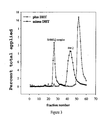
- FIG. 3 is a graph showing the HPLC profiles of 5′-MP- 125 IUdR-3′-DHT with SHBG in the presence or absence of DHT.
- FIG. 4 is a graph sowing the tumor uptake of 5′-MP- 125 IUdR-3′-DHT in athymic mice (bars) and the amount of DNA-bound 125 IUdR (line).
- FIG. 5 is a set of two graphs showing the in vivo uptake of 125 IUdR-3′-DHT by OVCAR-3 tumors in athymic mice (A) and the amount bound to DNA (B) over time.
- the conjugates of the present invention are composed of one component which is effective for killing cancer cells that make DNA and multiply and another component that is capable of specifically targeting AR expressed by cancer cells.
- Thymidine analogs such as IUdR have certain characteristics which make their radiolabeled derivatives useful for the treatment or diagnosis of tumors whether macroscopically observable or not. Because such thymidine analogs are low-molecular-weight molecules, they diffuse readily within tissues. IUdR, for example, when radiolabeled with an Auger electron emitter, such as, 123 I, 125 I, 77 Br, 80m Br, 195m Hg, and 113 Sn, is innocuous outside the cell and ineffective at killing cells when within the cytoplasm.
- Auger electron emitter such as, 123 I, 125 I, 77 Br, 80m Br, 195m Hg, and 113 Sn
- IUdR may also be radiolabeled with beta- or alpha-emitters such as, 131 I, 32 P, and radioisotopes of astatine (e.g., 225 At). Unlike Auger electron emitters, these radioisotopes are radiotoxic even when outside of the cell. Such isotopes would allow for the irradiation of neighboring cells, i.e., a bystander effect, which is beneficial, particularly if AR expression is not uniform. IUdR is, for the most part, taken up selectively by dividing cancerous cells located within nondividing cells and is indefinitely retained following DNA incorporation.
- Nondividing cells will not incorporate radiolabeled IUdR into their DNA and most of the radiolabeled IUdR that is not taken up by cancerous cells will be catabolized/dehalogenated rapidly [t 1/2 of minutes] and thus will not incorporate into the DNA of distant noncancerous dividing cells. Furthermore, since it is a small molecule, radiolabeled IUdR will not induce an antibody response and as such lends itself to repeated injections, continuous infusion, or similar modes of administration.
- Radiolabeled thymidine analogs are conjugated to an AR ligand.
- Radiolabeled IUdR for example, is conjugated to the androgen ligand DHT.
- AR is expressed on cells from a variety of cancers, such as 50-90% of breast tumors (Bryan, R. M., et al. (1984) Cancer, 54:2436-2440; Lea, O. A., et al. (1989) Cancer Res., 49:7162-7167; Soreide, J. A., et al. (1992) Eur. J. Surg. Oncol., 18:112-118).
- DHT in addition to providing specific targeting of the conjugate to cells expressing AR, has also demonstrated anti-cancer effects in breast cancer experimental models (see, for example, Poulin, R., et al. (1988) Breast Cancer Res. Treat., 12:213-225) and other androgens, such as fluoxymesterone, have produced anti-cancer effects in administration to patients (see, for example, Ingle, J. N., et al. (1991) Cancer, 67:886-891).
- conjugates in accordance with the present invention have the formula wherein R and R′ are as previously defined.
- lower alkyl refers to a straight or branched chain hydrocarbon group having 1 to 8 carbon atoms, preferably 1 to 4 carbon atoms.
- conjugates within the scope of this invention are the following: Synthetic Routes for the Preparation of these Conjugates are Exemplified Hereinbelow.
- Therapeutic preparations comprising the conjugates of this invention may be conveniently formulated for administration with a biologically acceptable vehicle, which may include the patient's own serum or serum fractions.
- a biologically acceptable vehicle include the patient's own serum or serum fractions.
- suitable vehicles include liposomes and similar injectable suspensions, saline, activated carbon absorbents, and solutions containing cyclodextrins such as alphadex and betadex.
- IUdR compounds may be derivatized, e.g. by esterification of available hydroxyl groups, with long chain fatty acids to increase the circulation half-life of the compounds.
- the concentration for diagnostic uses of the conjugate in the chosen vehicle should normally be from about 0.1 mCi/mL to about 10 mCi/mL.
- the concentration for therapeutic uses of the conjugate in the chosen vehicle should normally be from about 1 mCi/mL to about 100 mCi/mL. These concentrations may vary depending on whether the method of administration is intravenous, intraperitoneal, or intratumor. In all cases, any substance used in formulating a therapeutic preparation in accordance with this invention should be virus-free, pharmaceutically pure and substantially non-toxic.
- the action of contaminating micro organisms may be prevented by various anti-bacterial and/or anti-fungal agents, such as parabens, chlorbutinol, phenyl, sorbic acid, thimerosal and the like. It will often be preferable to include in the formulation isotonic agents, for example, glucose or sodium chloride. Additionally, free-radical scavengers and antioxidants such as ascorbic acid and the like may be employed to allow for a longer storage of the radioactive drug.
- biologically acceptable vehicle is intended to include any and all solvents, dispersion media and the like which may be appropriate for the desired route of administration of the conjugates described herein, either as such, or in the form of a pharmaceutical preparation, as described above. Except insofar as any conventional vehicle is incompatible with the conjugates of this invention, its use in formulating pharmaceutical preparations including such conjugates is contemplated. It is noted in this regard that administration of the conjugates of this invention with any substance that competes with the conjugate for AR binding is to be avoided.
- the conjugates described herein may be administered parenterally to a patient, either as such or in combination with a biologically acceptable vehicle, as noted above, by conventional procedures.
- the preferred modes of administration are intravenous and intraperitoneal.
- the conjugate will typically be administered at a dose that provides from about 1 mCi (37 MBq)-20 mCi (740 MBq) of radioactivity per 24 hours.
- the amount of the conjugate administered for diagnosis will generally be an amount sufficient to provide between 0.1 mCi and 10 mCi of radioactivity.
- the imaging can commence immediately after the administration. To detect DNA uptake, imaging may begin 1 hour after administration.
- imaging can occur at least daily for 7 days or longer to assess the tumor growth kinetics.
- the determination of an appropriate dose of the conjugate, either therapeutic or diagnostic, for a particular patient will, of course, be determined based on the type and stage of the patient's cancer and the judgment of the attending medical oncologist or radiologist, as the case may be.
- the term “patient” includes both humans and animals.
- the targeted delivery of radio nuclides to cancer cells in the manner described herein produces strong cytotoxic activity, in that the radionuclide is introduced into the DNA of the multiplying cells, where it induces DNA strand breaks in the double helix.
- the usual in vivo degredation pathways are by-passed, bioavailability of the radiolabeled agent is improved and more tumor cells are exposed to the cell killing effect of the radiation as they enter into the S phase.
- the solvent was evaporated to dryness and an oily residue was treated with ethyl acetate/hexanes mixture to produce white precipitate of the crude DHT half-ester of succinic acid.
- the end product was collected by filtration and dried in vacuo.
- the purification of the DHT half-ester of succinic acid was accomplished on a silica gel column with dichloromethane/methanol mixture (10:0.4; v/v) as an eluant to give 2.12 g (78% yield) of the pure half ester.
- Dimethoxytrityl chloride (4 g, 11.3 mmol) is added in two portions into a stirred solution of 5-iodo-2′-deoxyuridine (4 g, 11.4 mmol; IUdR) in 50 mL anhydrous pyridine at room temperature. The mixture is stirred overnight. Pyridine is evaporated to dryness and an oily residue is dissolved in 100 mL ethyl acetate, washed with water, 10% citric acid, aqueous saturated sodium bicarbonate solution, and saline. The washed organic layer is dried over MgSO 4 .
- Ethyl acetate is evaporated to dryness giving a crude DMT-IUdR in the from of a glass-like residue, which is redissolved in ethyl acetate/ethyl ether mixture, filtered and allowed to crystallize at 4° C. overnight.
- the crystalline, pure DMT-IUdR is collected by filtration.
- the volume of the filtrate is reduced under vacuum and a silica gel column is used to purify the remaining DMT-IUdR using dichloromethane/methanol (10:0.5; v/v) as a column solvent.
- the 5′-DMT-protected 6 (3 g) was dissolved in 25 mL of 2-propanol and 1 mL of 90% CF 3 COOH (TFA) was added. After approximately 4 hours at room temperature, TLC indicated that 100% of the DMT protection was removed to generate 5′-deprotected derivative 6. After evaporation of the solvent, the residue was treated with 5 mL water and evaporated again to remove the majority of TFA. The glassy residue was dissolved in dichloromethane and dried over MgSO 4 . The crude product was purified on a silica gel column with ethyl acetate/hexanes (15:10; v/v) as a solvent to give 2.8 g (82%) of pure conjugate 6. The deprotection of 5′-DMT derivative 6 with trifluoroacetic acid can also be conducted in tert-butanol as a solvent.
- the preparation of the 5-trialkylstannyl-derivatives (7) is accomplished as described in U.S. Pat. No. 5,468,853 to Baranowska-Kortylewicz which is commonly owned with its present application.
- the radiolabeling is also accomplished using procedures described in U.S. Pat. No. 5,468,853.
- the radioisotopes that can be utilized in this protocol include radiohalides such as radioiodine, radiobromine, and radioastatine.
- the reaction can be conducted with radiochlorine, but not as efficiently, and it does not work for radiofluorine.
- Suitable oxidants include: iodogen, chloromine-T, N-chlorosuccinimide, hydrogen peroxide, tert-butyl hydroperoxide (T-HYDRO solution; 70% wt in water) and cerium ammonium nitrite.
- the conjugate obtained by this synthesis is relatively hydrophobic but still soluble in aqueous media at “no-carrier-added” radioactivity levels.
- the release of 125 IUdR from the succinate ester appears to be very effective, both in vitro in cell culture and in vivo in tumor bearing animals.
- the half-life in vivo is short and may be detrimental to the delivery of therapeutic doses to tumor. On the other hand, because the half-life is short, repeated injections should be possible without a large radioactive burden in normal tissue.
- IUdR (1.5 g, 4.25 mmol) was dissolved in 7 mL of DMF/THF mixture (3:4; v/v) under strictly anhydrous conditions. After cooling to 0° C., 1H-tetrazole (1.2 g, 17.1 mmol) and di-tert-butyl diisopropylphosphoramidite (1.65 mL, 5.1 mmol) were added. The mixture was stirred at 0-4° C. for 72 h. Tert-Butyl hydroperoxide (4.6 mL) was added at ⁇ 80° C. and the mixture was allowed to slowly warm up to room temperature. A 5% solution of NaHSO 3 (10 mL) was added and the whole mixture was evaporated to dryness.
- the DHT half-ester of succinic acid prepared as described in Example 1 above, (0.77 g, 1.46 mmol) was dissolved in 10 mL of anhydrous dichloromethane.
- Dicyclohexylcarbodiimide (DCC; 0.32 g, 1.5 mmol) was added followed by phosphorylated IUdR (8) (0.8 g, 1.46 mmol) and DMAP (70 mg).
- DCC dicyclohexylcarbodiimide
- phosphorylated IUdR (8) 0.8 g, 1.46 mmol
- DMAP 70 mg
- the reaction mixture was stirred at room temperature until starting materials disappeared as determined by TLC.
- the solvent was evaporated under vacuum and replaced with ethyl acetate/hexanes mixture (3:2; v/v).
- the precipitated DCU was removed by filtration and the filtrate was washed with citric acid and water.
- the stannyl derivative was prepared and radiolabeling was carried out in the same general manner as in Example 1, above. Removal of the protecting t-butyl group was effected by a breif exposure of the 125I-labeled derivative to the HPLC solvent which is typically 0.05% to 0.5% trichloroacetic acid (TCA) in acetonitrile at room temperature.
- HPLC solvent typically 0.05% to 0.5% trichloroacetic acid (TCA) in acetonitrile at room temperature.
- Conjugate 2 is the most hydrophilic of the conjugates described herein (two negative charges on the monophosphate group). It cannot cross the cell membrane by itself, i.e., it requires the active transport mechanism from the systemic circulation into the tumor cells with the aid of sex hormone binding globulin.
- Dihydrotestosterone (5 ⁇ -androstan-17 ⁇ -ol-3′-one; 2 g, 6.9 mmol) was dissolved in 30 mL of anhydrous THF under nitrogen.
- N,N-Diisopropylethylamine (DEPA; 1.78 g, 13.8 mmol) and DMAP (20 mg, 0.13 mmol) were added.
- a stirred mixture was cooled to 0° C. and N,N-diisopropylmethylphosphoamidic chloride (2.05 g, 10.33 mmol) was added neat in five 0.5-mL portions. Stirring was continued at 0° C. for 30 min. The precipitate was filtered off and the filtrate was evaporated to dryness under vacuum.
- a crude product was purified on a silica gel column using dichloromethane/ethyl acetate/triethylamine (10/10/0.05; v/v) as a solvent. Only partial purification was achieved and the material obtained from the column was used in the following step of the synthesis.
- the stannyl derivative (14) was prepared and radiolabeling was carried out in the same general manner as in Example 1, above.
- the method of scheme 4 can be performed using the N,N-diisopropylmethylphosphoramidate of DHT, and following the same procedure described above except that the methyl group is hydrolyzed prior to the stannylation reaction.
- the final reaction product of scheme 4 with this modification is 5′-[ 125 I]iodo-2′-deoxy-5′-monophosphate-O-(mono-dihydrotestosterone) (4A).
- Conjugates 4 and 4A possess interesting characterisitics in that the negative charge on the monophosphate—either already present as in conjugate 3 or generated after administration by circulating carboxylases—will prevent the uptake of this drug via passive diffusion (charged molecules cannot readily cross the cell membrane, they require the active transport mechanism to be able to enter cancer cells), thus improving the selectivity.
- This is very similar to conjugate 2, but the DHT on phosphate is more stable compared to succinate esters.
- Diisopropylphosphoramidous dichloride (0.65 mL, 3.45 mmol) was added to a stirred solution of DHT (2 g, 6.9 mmol) containing 1.5 mL (8.5 mmol) of DEPA and 65 mg of DMAP in 50 mL anhydrous THF at 0° C. Copious amounts of white precipitate were formed. The stirring was continued for 2 hours at room temperature. The precipitate was removed by filtration and the filtrate was evaporated to dryness.
- the crude product was partially purified on a silica gel column using dichloromethane/ethyl acetate/triethylamine (10/5/0.05; v/v) as a solvent. The partially purified (approximately 95% pure) N,N-diisopropylphophoramidite (DHT) 2 was used in the following reaction.
- the white precipitate was filtered off and the filtrate was treated with 5% NaHSO 3 (10 mL).
- the organic layer was evaporated to dryness and the residue was kept under high vacuum for 1 hour.
- the residue was extracted with dichloromethane (2 ⁇ 50 mL) and the combined extracts were washed with water, NaHCO 3 and dried over MgSO 4 .
- the stannyl derivative 15 was prepared and radiolabeling was carried out in the same general manner as in Example 1, above.
- Conjugate 5 is the most hydrophobic of all the conjugates described herein. It may be best suited for local/regional administration (e.g., intraperitoneal in the case of ovarian cancer) with a slow diffusion from the site of injection.
- OVCAR-3 cells were thawed from a stock or freshly harvested by a lavage of the peritoneal cavity of athymic mice bearing intraperitoneal ascites OVCAR-3 tumors.
- the cells were twice washed with full media (RPMI 1640 medium with 2 mM L-glutamine adjusted to contain 1.5 g/L sodium bicarbonate, 4.5 g/L glucose, 10 mM HEPES, and 1.0 mM sodium pyruvate and supplemented with 0.01 mg/ml bovine insulin, 80%; fetal bovine serum, 20%).
- the cells were spun at 450 rpm for 10 minutes to collect a cell pellet and were then resuspended in full media to achieve approximately 5 ⁇ 10 6 cells/mL. The cells were counted and their viability was determined by Trypan Blue staining. “Cold” dihydrotestosterone (DHT) was dissolved in 95% ethanol to create a stock solution of 5 ⁇ g/mL. Similarly, 95% ethanol was added to 125 IUdR-DHT to produce 250,000 cpm/100 ⁇ L. Forty samples were prepared in plastic tubes as set forth in Table 1.
- DHT dihydrotestosterone
- the “total applied” counts were then determined in a gamma counter.
- the samples were then incubated at 0-4° C. with shaking for 14-16 hours.
- the cells were centrifuged at 1,000 rpm for 15 minutes at 4° C. to generate a cell pellet.
- a known volume of the supernatant from the centrifugation was transfered to new plastic tubes and counted in a gamma counter to determine the “free” counts.
- the cell pellet with the remainder of the supernatant was then counted in the gamma counter.
- “Bound” counts were then determined by subtracting counts associated with the supernatant from the remaining counts. Plots of this data can be found in FIG. 1 .
- the data show a single type of binding sites with a high affinity for 125 IUdR-DHT. Use of the excess DHT revealed that the high affinity seen is due to the DHT portion of the drug and thus dictated by the presence of AR.
- the cells were then washed with ice-cold phosphate buffered saline (PBS) and gently centrifuged. Again, a known volume of the supernatant was removed and both the supernatant and pellet were counted in the gamma counter.
- PBS phosphate buffered saline
- OVCAR-3 cells were harvested by a lavage of the peritoneal cavity of athymic mice bearing intraperitoneal ascites OVCAR-3 tumors. Cells were washed with Hank's balanced salt solution (HBBS), resuspended in RPMI 1640 medium without serum and used in a binding assay.
- HBBS Hank's balanced salt solution
- tissue culture flasks T25
- RPMI 1640 penicillin, streptomycin, L-glutamine and 10% fetal bovine serum.
- the cells were allowed to reach about 70% confluency and the serum-containing medium was aspirated and replaced with serum-free medium (SFM).
- SFM serum-free medium
- the cells were allowed to remain in SFM for 24 hours. After 24 hours, the medium was aspirated and SFM containing 10 nM dihydrotestosterone (DHT) was added to three flasks. The volume of added SFM was carefully measured to assure that each flask received an identical volume of SFM.
- DHT dihydrotestosterone
- the medium was aspirated and 1 mL aliquots were counted in a gamma counter to determine cpm/mL after incubation.
- Cells were washed two times with ice-cold PBS containing 0.1% BSA. The washes were combined, the total volume measured, and 1 mL aliquots were counted in a gamma-counter.
- To the cells 5 mL of 1 M NaOH was added and incubated for two hours. The NaOH wash was collected and 1-mL aliquots were counted in a gamma-counter.
- One mL PBS was added to the remaining cells and the cells were scraped and transferred into gamma-counter tubes to determine cpm/mL.
- This procedure may be employed to determine competitive binding wherein the amount of cpm in the NaOH-treated cell pellet in the absence of “cold” DHT is the “total bound” and the amount of cpm in NaOH-treated cells that were incubated with “cold” DHT can yield the amount of nonspecific binding.
- This method works well with cells that grow as monolayers such as PC3, LNCaP, and DU-145 cells.
- OVCAR-3 cells in full growth medium (RPMI 1640 medium with 2 mM L-glutamine adjusted to contain 1.5 g/L sodium bicarbonate, 4.5 g/L glucose, 10 mM HEPES, and 1.0 mM sodium pyruvate and supplemented with 0.01 mg/ml bovine insulin, 80%; fetal bovine serum, 20%) at 1 ⁇ 10 6 cells per flask were plated onto three Petri dishes and allowed to attach for 24 hours. As a control, one dish without cells was subjected to the same treatments. After 24 hours, the cells were trypsinized from one dish to determine cell number and viability. The media was then aspirated from the remaining dishes, including the control, and replaced with 1 mL of fresh media.
- 0.1 mCi of 125 IUdR-DHT in 0.10 mL of ethanol was added to all of the dishes.
- the dishes were incubated at 37° C.
- 10 ⁇ L of media was removed from each of the dishes of which 2 ⁇ L was added to a silica gel TLC plate.
- 2 ⁇ L was added to glass gamma counter tubes as standards.
- the TLC plates were run with a 3:1 mixture of ethyl acetate and hexane (v/v). When the solvent front approached the end of the plate, the plate was removed from the solvent, dried, and cut into strips. The standards and the strips were then counted.
- the percent recovered 125 IUdR-DHT was calculated as a ratio of the intact 125 IUdR-DHT at a given timepoint divided by the intact 125 IUdR-DHT at the 0 minute timepoint (see FIG. 2A ). Similar calculations were used to determine the kinetics of the appearance of 125 IUdR in the medium, which was determined to occur at a rate of 8.5%/hour ( FIG. 2B ). This rate is in comparison to the disappearance of 125 IUdR-DHT from the media at a rate of 16%/hour.
- 125 IUdR is freely diffusible from cells and therefore only a fraction of 125 IUdR-DHT hydrolyzed in cells is used for DNA synthesis. The inclusion of a negatively-charged monophosphate assists in the retention of these compounds in the cell.
- the testosterone or DHT signaling in AR-positive cells involves sex hormone binding globulin (SHBG).
- SHBG sex hormone binding globulin
- Human SHBG-DHT complexes interact with plasma membranes of several sex steroid-dependent cells and tissues. This interaction involves the binding of SHBG to a specific membrane protein receptor, which promotes its cellular internalization.
- the next step entails binding to AR and conformational changes in AR. AR is subsequently translocated to the nucleus where it dimerizes and binds DNA.
- the overall process allows for the intracellular delivery of the 125 IUdR drugs only to cells expressing AR.
- the samples were then incubated at 0-4° C. for 3 hours. 0.1 mL aliquots were removed from the samples and counted in a gamma counter. To each tube, 0.4 mL of ice-cold charcoal-dextran solution was added and the tubes were incubated on ice for 15 minutes. The samples were then centrifuged at 4° C. for 20 minutes at 1,000 rpm. 0.5 ml aliquots were taken for gamma-counting and the residue was also counted.
- Athymic mice bearing intraperitoneal OVCAR-3 tumors were treated with 0.05 mCi (1.4 kBq) 5′-MP- 125 IUdR-3′-DHT intraperitoneally.
- the cells were fractionated using a Wako kit (Wako Cemicals USA, Inc., Richmond, Va.) or, alternatively, with a NE-PREP nuclear and cyoplasmic extraction kit (Pierce, Rockford, Ill.) per the manufacturer's instruction.
- Wako kit Wako Cemicals USA, Inc., Richmond, Va.
- NE-PREP nuclear and cyoplasmic extraction kit Pieris, Rockford, Ill.
- the stability of 125 IUdR-3′DHT in the presence of OVCAR-3 cells showed that the emergence of “free” 125 IUdR in the growth medium requires interaction of the drugs with specific cells.
Abstract
Description
- Pursuant to 35 U.S.C. Section 202(c), it is acknowledged that the United States Government has certain rights in the invention described herein, which was made in part with funds from the Department of Defense, Grant No. DAMD17-99-1-9313.
- The present invention relates generally to targeted therapy and medical imaging as applied to cancer treatment and diagnosis, and in particular to conjugates composed of a radiolabeled, cell cycle-dependent therapeutic agent chemically coupled to a ligand that targets androgen receptor (AR). The conjugates of the invention are taken up selectively by malignant tumor cells that have androgen receptor and are incorporated into the nucleus of such cells, where they produce a cytotoxic effect and/or are detectable via radioimaging techniques.
- The main treatments for breast, prostate, ovarian and many other cancers are surgery, chemotherapy and radiation therapy. In some cases a combination of two or more of these treatments is recommended. Typically, clinical trials for advanced carcinomas use combination chemotherapy based on established anti-cancer agents. For example, there are numerous active clinical trials (Phase I) dealing with recurrent and progressive ovarian carcinoma that rely on existing drugs such as paclitaxel, carboplatin, cisplatin, floxouridine and similar drugs in a combination chemotherapy. Many of these include an autologous stem cell support to combat the side effects brought on by the administration of these drugs. Newer drugs include matrix metalloproteinase inhibitors, vaccines, and antibodies.
- Many of the currently available front-line and salvage agents used in cancer therapy are associated with cumulative and/or irreversible toxicities that pose challenges for long-term treatment planning. The irreversible effects associated with some of these therapies include development of multidrug resistance, neurotoxicity, and nephrotoxicity. All of these diminish the probability of improved responses when multiple treatments are needed to keep the cancer under control.
- It has previously been proposed to use targeted cytotoxic radioisotopes for the treatment and diagnosis of cancer. One of the intended benefits of targeted therapy is to diminish the incidence and severity of side effects by confining toxic exposure, more or less, to the disease site. Certain radioisotopes, particularly Auger electron-emitting isotopes, such as 123I and 125I are known to be very toxic to viable cells, but only if they are localized within the nucleus of the cell. (Warters et al., Curr. Top. Stop Rad. Res., 12:389 (1977).) It has been reported that 5′-iodo-2′-deoxyuridine (IUdR), when labeled with the Auger electron emitter 123I or 125I exhibits substantial toxicity in mammalian cells in vitro (Makrigiorgos et al., Radiat. Res., 118:532-44 (1989)) and produces a therapeutic effect in animal tumor models (Baranowska-Kortylewicz et al., Int. J. Radiat. Oncol. Biol. Phys., 21:1541-51 (1991)). Furthermore, radiolabeled IUdR has been found to enable scintigraphic detection of animal and human tumors (Baranowska-Kortylewicz, supra). See also U.S. Pat. Nos. 5,094,835 and 5,308,605.
- Considerable effort has been devoted to developing antibodies for the targeted delivery of therapeutic and diagnostic agents. However, antibodies themselves have not been capable of reaching the cell nucleus in effective amounts. Most such antibodies react with the cell surface, and are gradually internalized, routed to lysosomes and degraded (Kyriakos et al., Cancer Res., 52:835 (1992)). Degregation products, including any radioisotopes attached thereto, then gradually leave the cell by crossing the lysosomal membrane and then the cell membrane. Although a conventional radioisotope label on an antibody degradation product can theoretically pass through the nuclear membrane and deliver some radioactivity to the nucleus (Woo et al., WO 90/03799) actual observations show that the amount is limited, and in any event, is insufficient to have a toxic effect on tumor cells.
- Protein and polypeptide hormones and growth factors, particularly those having cell surface receptors, may be directly radiolabeled and used to target a tumor cell. As in the case of targeting radiolabeled antibodies, however, radioisotopes bound to amino acid residues of hormones, growth factors and the like exit from the cell after catabolism, and do not appreciably bind to nuclear material.
- Despite the many advances in the field of cancer therapy and diagnosis, there remains an acute need for innovative treatment methods, particularly for cancers having high instances of relapse, which can be safely applied in a repetitive, long-term regimen, without the side effects produced by existing treatments.
- The above-noted need is satisfied by the radiolabeled conjugates of the present invention which are capable of targeting and being selectively taken up and degraded by a tumor cell, and thereby delivering to the tumor cell nucleus a radioisotope capable of being incorporated into the nuclear material, so as to produce a cytotoxic effect and/or to render the cell detectable by radioimaging. The conjugates of the invention can be safely administered in long term cancer treatments, without producing significant adverse health effects.
- In accordance with one aspect of the present invention, there is provided a cancer-specific radiolabeled conjugate of the formula:

wherein B* represents 5′ substituted uracil substituted with a radionuclide; R represents H, OH, or O-L-DHT, L being a cleavable bifunctional linking moiety and DHT is 4-dihydrotestosterone, which is bound through its hydroxyl substituent to said linking moiety; and R′ represents a phospho group or a substituted phospho group having the formula —PO(ORa)(ORb), —PO(ORa)(ODHT) or —PO(ODHT)2, Ra and Rb being the same or different and representing H or lower alkyl and DHT is as previously defined, with the proviso that at least one of the R and R′ substituents comprises a DHT moiety. - The present invention also provides methods of using the above-described conjugate for treating and diagnosing cancers comprising cells having AR, especially ovarian, breast and prostate cancer.
- The conjugates of the present invention have been designed so as to take advantage of two characteristics of many relapsing cancers, i.e. (1) relapsed/advance cancers have a large portion of rapidly growing and dividing cells (i.e. a large S-phase fraction); and (2) AR is expressed in practically all prostate cancer (primary and metastatic), ovarian cancer (>90% positive for AR regardless of the tumor site) and breast cancer (even when estrogen receptor (ER)-negative and progesterone receptor (PR)-negative, breast cancer cells express AR). Upon administration, the conjugate first binds to the sex hormone binding globulin, which in turn carries it exclusively to cells that have AR. Subsequent to this interaction, the entire conjugate is transported into the cell. Intracellular enzymes cleave the linking moiety, thus releasing and trapping within the cell the portion of the conjugate that is responsible for killing tumor cells. This cytotoxic effect is induced only when the cell cycle dependent therapeutic agent is incorporated into the DNA of dividing tumor cells. This dependence of radiotoxicity on the participation of the radiolabeled agent in DNA synthesis, in combination with relatively rapid pharmacokinetics, limits the exposure of normal tissue to radiation. In other words, the conjugate that remains in systemic circulation, or enters normal tissue or organs, is essentially innocuous. Accordingly, the radiolabeled conjugates of the invention may be administered frequently and without adverse effects.
-
FIG. 1 is a set of two graphs depicting the binding of 125IUdR-DHT to OVCAR-3 cells in vitro.FIG. 1A plots the fraction of “bound” counts to “free counts” against “bound” 125IUdR-DHT (mM).FIG. 1B plots bound 125IUdR-DHT (mM) against free 125IUdR-DHT (mM). -
FIG. 2 is a set of two graphs depicting the stability of 125IUdR-DHT.FIG. 2A depicts the stability of 125IUdR-DHT with time in the presence or absence of OVCAR-3 cells.FIG. 2B depicts the kinetics of the emergence of 125IUdR in the medium in the presence or absence of OVCAR-3 cells. -
FIG. 3 is a graph showing the HPLC profiles of 5′-MP-125IUdR-3′-DHT with SHBG in the presence or absence of DHT. -
FIG. 4 is a graph sowing the tumor uptake of 5′-MP-125IUdR-3′-DHT in athymic mice (bars) and the amount of DNA-bound 125IUdR (line). -
FIG. 5 is a set of two graphs showing the in vivo uptake of 125IUdR-3′-DHT by OVCAR-3 tumors in athymic mice (A) and the amount bound to DNA (B) over time. - The conjugates of the present invention are composed of one component which is effective for killing cancer cells that make DNA and multiply and another component that is capable of specifically targeting AR expressed by cancer cells.
- Thymidine analogs, such as IUdR, have certain characteristics which make their radiolabeled derivatives useful for the treatment or diagnosis of tumors whether macroscopically observable or not. Because such thymidine analogs are low-molecular-weight molecules, they diffuse readily within tissues. IUdR, for example, when radiolabeled with an Auger electron emitter, such as, 123I, 125I, 77Br, 80mBr, 195mHg, and 113Sn, is innocuous outside the cell and ineffective at killing cells when within the cytoplasm. IUdR may also be radiolabeled with beta- or alpha-emitters such as, 131I, 32P, and radioisotopes of astatine (e.g., 225At). Unlike Auger electron emitters, these radioisotopes are radiotoxic even when outside of the cell. Such isotopes would allow for the irradiation of neighboring cells, i.e., a bystander effect, which is beneficial, particularly if AR expression is not uniform. IUdR is, for the most part, taken up selectively by dividing cancerous cells located within nondividing cells and is indefinitely retained following DNA incorporation. Nondividing cells will not incorporate radiolabeled IUdR into their DNA and most of the radiolabeled IUdR that is not taken up by cancerous cells will be catabolized/dehalogenated rapidly [t1/2 of minutes] and thus will not incorporate into the DNA of distant noncancerous dividing cells. Furthermore, since it is a small molecule, radiolabeled IUdR will not induce an antibody response and as such lends itself to repeated injections, continuous infusion, or similar modes of administration.
- In order to provide cancer cell specificity and enhanced delivery, the radiolabeled thymidine analogs are conjugated to an AR ligand. Radiolabeled IUdR, for example, is conjugated to the androgen ligand DHT. Importantly, AR is expressed on cells from a variety of cancers, such as 50-90% of breast tumors (Bryan, R. M., et al. (1984) Cancer, 54:2436-2440; Lea, O. A., et al. (1989) Cancer Res., 49:7162-7167; Soreide, J. A., et al. (1992) Eur. J. Surg. Oncol., 18:112-118). DHT, in addition to providing specific targeting of the conjugate to cells expressing AR, has also demonstrated anti-cancer effects in breast cancer experimental models (see, for example, Poulin, R., et al. (1988) Breast Cancer Res. Treat., 12:213-225) and other androgens, such as fluoxymesterone, have produced anti-cancer effects in administration to patients (see, for example, Ingle, J. N., et al. (1991) Cancer, 67:886-891).
- Particularly preferred conjugates in accordance with the present invention have the formula

wherein R and R′ are as previously defined. - The term “lower alkyl”, as used herein, refers to a straight or branched chain hydrocarbon group having 1 to 8 carbon atoms, preferably 1 to 4 carbon atoms.
- Specific examples of conjugates within the scope of this invention are the following:


Synthetic Routes for the Preparation of these Conjugates are Exemplified Hereinbelow. - In vitro studies conducted to date with certain of the specific conjugates described above have produced several notable results. First, coupling of DHT to 125IUdR produces cell killing agents which specifically target AR. Second, excess DHT competes with the DHT-containing conjugate for binding to AR. Third, uptake of the conjugate by the cell and metabolic processing of the conjugate is dependent on the presence of AR. Furthermore, in vivo studies have shown that certain of the above-described conjugates are bound specifically to the sex hormone binding globulin and are actively transported into cells that express AR and that uptake of 125IUdR transported to in vivo grown tumors in the form of conjugates with DHT is dependent on the AR-expression and results in the arrest of tumor growth.
- Therapeutic preparations comprising the conjugates of this invention may be conveniently formulated for administration with a biologically acceptable vehicle, which may include the patient's own serum or serum fractions. Other suitable vehicles include liposomes and similar injectable suspensions, saline, activated carbon absorbents, and solutions containing cyclodextrins such as alphadex and betadex. Additionally, IUdR compounds may be derivatized, e.g. by esterification of available hydroxyl groups, with long chain fatty acids to increase the circulation half-life of the compounds. The concentration for diagnostic uses of the conjugate in the chosen vehicle should normally be from about 0.1 mCi/mL to about 10 mCi/mL. The concentration for therapeutic uses of the conjugate in the chosen vehicle should normally be from about 1 mCi/mL to about 100 mCi/mL. These concentrations may vary depending on whether the method of administration is intravenous, intraperitoneal, or intratumor. In all cases, any substance used in formulating a therapeutic preparation in accordance with this invention should be virus-free, pharmaceutically pure and substantially non-toxic.
- If necessary, the action of contaminating micro organisms may be prevented by various anti-bacterial and/or anti-fungal agents, such as parabens, chlorbutinol, phenyl, sorbic acid, thimerosal and the like. It will often be preferable to include in the formulation isotonic agents, for example, glucose or sodium chloride. Additionally, free-radical scavengers and antioxidants such as ascorbic acid and the like may be employed to allow for a longer storage of the radioactive drug.
- As used herein, the term “biologically acceptable vehicle” is intended to include any and all solvents, dispersion media and the like which may be appropriate for the desired route of administration of the conjugates described herein, either as such, or in the form of a pharmaceutical preparation, as described above. Except insofar as any conventional vehicle is incompatible with the conjugates of this invention, its use in formulating pharmaceutical preparations including such conjugates is contemplated. It is noted in this regard that administration of the conjugates of this invention with any substance that competes with the conjugate for AR binding is to be avoided.
- The conjugates described herein may be administered parenterally to a patient, either as such or in combination with a biologically acceptable vehicle, as noted above, by conventional procedures. The preferred modes of administration are intravenous and intraperitoneal. For therapeutic applications, the conjugate will typically be administered at a dose that provides from about 1 mCi (37 MBq)-20 mCi (740 MBq) of radioactivity per 24 hours. The amount of the conjugate administered for diagnosis will generally be an amount sufficient to provide between 0.1 mCi and 10 mCi of radioactivity. For the registration of AR expression, the imaging can commence immediately after the administration. To detect DNA uptake, imaging may begin 1 hour after administration. Notably, with longer lived radioisotopes, imaging can occur at least daily for 7 days or longer to assess the tumor growth kinetics. The determination of an appropriate dose of the conjugate, either therapeutic or diagnostic, for a particular patient will, of course, be determined based on the type and stage of the patient's cancer and the judgment of the attending medical oncologist or radiologist, as the case may be.
- As used herein, the term “patient” includes both humans and animals.
- The targeted delivery of radio nuclides to cancer cells in the manner described herein produces strong cytotoxic activity, in that the radionuclide is introduced into the DNA of the multiplying cells, where it induces DNA strand breaks in the double helix. Moreover, by delivering radiolabeled agents to a specific site and relying on mechanisms operational at the site of delivery to release the radiolabeled agent, the usual in vivo degredation pathways are by-passed, bioavailability of the radiolabeled agent is improved and more tumor cells are exposed to the cell killing effect of the radiation as they enter into the S phase.
- The following examples describe the synthesis of the aforementioned conjugates 1-5 of the present invention, as well as biological testing of certain of the conjugates. These examples are provided for illustrative purposes only and are not intended to limit the scope of the invention in anyway.
- A. Preparation of DHT Half Ester of Succinic Acid
- Succinic acid anhydride (2 g, 20 mmol) and DHT (2 g, 6.9 mmol) were dissolved in anhydrous pyridine and 80 mg of 4-dimethylaminopyridine (DMAP) was added. The reaction flask was placed on a rotatory evaporator and approximately 20 mL of pyridine was evaporated under reduced pressure at 50° C. The reaction mixture was then stirred for 1 h at room temperature and ice-water was added (50 mL). The mixture was extracted with ethyl acetate (2×50 mL), washed with 5% citric acid, water and brine. The ethyl acetate layer was dried over anhydrous MgSO4. The solvent was evaporated to dryness and an oily residue was treated with ethyl acetate/hexanes mixture to produce white precipitate of the crude DHT half-ester of succinic acid. The end product was collected by filtration and dried in vacuo. The purification of the DHT half-ester of succinic acid was accomplished on a silica gel column with dichloromethane/methanol mixture (10:0.4; v/v) as an eluant to give 2.12 g (78% yield) of the pure half ester.
- B. Preparation of 5′-Dimethoxytrityl-125IUdR
- Dimethoxytrityl chloride (4 g, 11.3 mmol) is added in two portions into a stirred solution of 5-iodo-2′-deoxyuridine (4 g, 11.4 mmol; IUdR) in 50 mL anhydrous pyridine at room temperature. The mixture is stirred overnight. Pyridine is evaporated to dryness and an oily residue is dissolved in 100 mL ethyl acetate, washed with water, 10% citric acid, aqueous saturated sodium bicarbonate solution, and saline. The washed organic layer is dried over MgSO4. Ethyl acetate is evaporated to dryness giving a crude DMT-IUdR in the from of a glass-like residue, which is redissolved in ethyl acetate/ethyl ether mixture, filtered and allowed to crystallize at 4° C. overnight. The crystalline, pure DMT-IUdR is collected by filtration. The volume of the filtrate is reduced under vacuum and a silica gel column is used to purify the remaining DMT-IUdR using dichloromethane/methanol (10:0.5; v/v) as a column solvent.
- C. Conjugation Reaction and Deprotection
- As shown in Scheme 1 below, purified DMT-IUdR (4 g, 6.01 mmol) is dissolved in 3 mL freshly distilled from CaH2 anhydrous dichloromethane and the DHT half-ester of succinic acid, prepared a previously described, (2.35 g, 6.0 mmol) is added. To this mixture dicyclohexylcarbodiimide (1.26 g, 6.2 mmol; DCC) is added in the presence of 80 mg of 4-dimethylaminopyridine (DMAP). The mixture was stirred at room temperature and the reaction progress was monitored on TLC. When the esterification was complete, the reaction mixture was diluted with ethyl ether (10 mL) and filtered to remove dicyclohexylurea (DCU). The filtrate was washed with water, citric acid and brine and was dried over MgSO4. The residue of crude 5′-DMT-protected 6 obtained after evaporation of the solvent was purified on a silica gel column using ethyl acetate/hexanes (3:2; v/v) as a solvent. The yield of 5′-DMT-protected 6 was 3.1 g (50%). The 5′-DMT-protected 6 (3 g) was dissolved in 25 mL of 2-propanol and 1 mL of 90% CF3COOH (TFA) was added. After approximately 4 hours at room temperature, TLC indicated that 100% of the DMT protection was removed to generate 5′-
deprotected derivative 6. After evaporation of the solvent, the residue was treated with 5 mL water and evaporated again to remove the majority of TFA. The glassy residue was dissolved in dichloromethane and dried over MgSO4. The crude product was purified on a silica gel column with ethyl acetate/hexanes (15:10; v/v) as a solvent to give 2.8 g (82%) ofpure conjugate 6. The deprotection of 5′-DMT derivative 6 with trifluoroacetic acid can also be conducted in tert-butanol as a solvent. - D. Preparation of Stannyl Derivatives and Radiolabeling Procedure
- The preparation of the 5-trialkylstannyl-derivatives (7) is accomplished as described in U.S. Pat. No. 5,468,853 to Baranowska-Kortylewicz which is commonly owned with its present application. The radiolabeling is also accomplished using procedures described in U.S. Pat. No. 5,468,853. The radioisotopes that can be utilized in this protocol include radiohalides such as radioiodine, radiobromine, and radioastatine. The reaction can be conducted with radiochlorine, but not as efficiently, and it does not work for radiofluorine. Suitable oxidants include: iodogen, chloromine-T, N-chlorosuccinimide, hydrogen peroxide, tert-butyl hydroperoxide (T-HYDRO solution; 70% wt in water) and cerium ammonium nitrite.

- The conjugate obtained by this synthesis is relatively hydrophobic but still soluble in aqueous media at “no-carrier-added” radioactivity levels. The release of 125IUdR from the succinate ester appears to be very effective, both in vitro in cell culture and in vivo in tumor bearing animals. The half-life in vivo is short and may be detrimental to the delivery of therapeutic doses to tumor. On the other hand, because the half-life is short, repeated injections should be possible without a large radioactive burden in normal tissue.
- A. Phosphorylation Reaction
- IUdR (1.5 g, 4.25 mmol) was dissolved in 7 mL of DMF/THF mixture (3:4; v/v) under strictly anhydrous conditions. After cooling to 0° C., 1H-tetrazole (1.2 g, 17.1 mmol) and di-tert-butyl diisopropylphosphoramidite (1.65 mL, 5.1 mmol) were added. The mixture was stirred at 0-4° C. for 72 h. Tert-Butyl hydroperoxide (4.6 mL) was added at −80° C. and the mixture was allowed to slowly warm up to room temperature. A 5% solution of NaHSO3 (10 mL) was added and the whole mixture was evaporated to dryness. The residue was extracted with dichloromethane (2×50 mL) and the organic layer dried with MgSO4. A mixture of all possible phosphorylated derivatives of IUdR was obtained, i.e., 3′-monosubstituted, 5′-monosubstituted and 3′,5′-disubstituted. Separation of the desired 5′-isomer was accomplished on a silica gel column with chloroform/methanol (10:0.6; v/v) as a solvent. The desired pure 5′-monosubstituted derivative (8) was isolated in 52% yield. The final purification was accomplished by crystallization from ethyl ether to give white solid, mp. 121-123° C.
- B. Conjugation Reaction and Radiolabeling
- The DHT half-ester of succinic acid, prepared as described in Example 1 above, (0.77 g, 1.46 mmol) was dissolved in 10 mL of anhydrous dichloromethane. Dicyclohexylcarbodiimide (DCC; 0.32 g, 1.5 mmol) was added followed by phosphorylated IUdR (8) (0.8 g, 1.46 mmol) and DMAP (70 mg). The reaction mixture was stirred at room temperature until starting materials disappeared as determined by TLC. The solvent was evaporated under vacuum and replaced with ethyl acetate/hexanes mixture (3:2; v/v). The precipitated DCU was removed by filtration and the filtrate was washed with citric acid and water. The organic layer was dried under MgSO4 and evaporated again. The purification of the resulting conjugate 9 was done on a silica gel column using chloroform/methanol mixture (10:0.6; v/v) as a solvent. The final pure conjugate was obtained in 49% yield (0.66 g).
- The stannyl derivative was prepared and radiolabeling was carried out in the same general manner as in Example 1, above. Removal of the protecting t-butyl group was effected by a breif exposure of the 125I-labeled derivative to the HPLC solvent which is typically 0.05% to 0.5% trichloroacetic acid (TCA) in acetonitrile at room temperature.

- An alternative approach to the synthesis of
conjugate 2 is shown in Scheme 3, in which the conjugation reaction is carried out prior to phosphorylation. However,Scheme 2 has been found to produce the desired conjugate in higher yield.
-
Conjugate 2 is the most hydrophilic of the conjugates described herein (two negative charges on the monophosphate group). It cannot cross the cell membrane by itself, i.e., it requires the active transport mechanism from the systemic circulation into the tumor cells with the aid of sex hormone binding globulin. - A. Preparation of N,N-diisopropylmethylphoshoramidate of DHT
- Dihydrotestosterone (5α-androstan-17β-ol-3′-one; 2 g, 6.9 mmol) was dissolved in 30 mL of anhydrous THF under nitrogen. N,N-Diisopropylethylamine (DEPA; 1.78 g, 13.8 mmol) and DMAP (20 mg, 0.13 mmol) were added. A stirred mixture was cooled to 0° C. and N,N-diisopropylmethylphosphoamidic chloride (2.05 g, 10.33 mmol) was added neat in five 0.5-mL portions. Stirring was continued at 0° C. for 30 min. The precipitate was filtered off and the filtrate was evaporated to dryness under vacuum. A crude product was purified on a silica gel column using dichloromethane/ethyl acetate/triethylamine (10/10/0.05; v/v) as a solvent. Only partial purification was achieved and the material obtained from the column was used in the following step of the synthesis.
- B. Preparation of 5-Iodo-2′Deoxyuridine-5′-Monomethylphosphate-3′-Succinyl DHT
- A solution of the freshly prepared N,N-diisopropylmethylphosphoramidate of DHT (1.55 g, 3.4 mmol) in anhydrous THF (20 mL) was cooled to 0° C. and 1H-tetrazole (1.4 g, 20 mmol) was added followed by IUdR (1.1 g, 3.1 mmol) dissolved in 5 mL anhydrous DMF. The mixture was allowed to react overnight at 0°-2° C. Tert-Butyl hydroperoxide (4 mL, 70% w solution in water) was added at −10° C. The mixture was allowed to slowly reach room temperature and after 1 hour of stirring at room temperature the solvent was evaporated to dryness under vacuum. The semi-solid residue was extracted with dichloromethane (2×50 mL) and washed with 5% NaHSO3 and water. During the extraction unreacted IUdR was separated and removed by filtration. The filtrate was dried over MgSO4 and again evaporated to dryness. The residue was analyzed on TLC plate and revealed the presence of two UV-positive spots along with a small residual spot corresponding to unreacted IUdR. The two UV-positive components were separated on a silica gel column using dichloromethane/methanol mixture (10:0.6; v/v) as a solvent. The major component (TLC Rf=0.65) was identified as a 5′-regioisomer (12) and was obtained after the column chromatography in 63% yield (1.23 g).
- The stannyl derivative (14) was prepared and radiolabeling was carried out in the same general manner as in Example 1, above.

- Alternatively, the method of scheme 4 can be performed using the N,N-diisopropylmethylphosphoramidate of DHT, and following the same procedure described above except that the methyl group is hydrolyzed prior to the stannylation reaction. The final reaction product of scheme 4 with this modification is 5′-[125I]iodo-2′-deoxy-5′-monophosphate-O-(mono-dihydrotestosterone) (4A).
- Conjugates 4 and 4A possess interesting characterisitics in that the negative charge on the monophosphate—either already present as in conjugate 3 or generated after administration by circulating carboxylases—will prevent the uptake of this drug via passive diffusion (charged molecules cannot readily cross the cell membrane, they require the active transport mechanism to be able to enter cancer cells), thus improving the selectivity. This is very similar to
conjugate 2, but the DHT on phosphate is more stable compared to succinate esters. - A. Preparation of N,N-diisopropylphosphoramidite (DHT)2
- Diisopropylphosphoramidous dichloride (0.65 mL, 3.45 mmol) was added to a stirred solution of DHT (2 g, 6.9 mmol) containing 1.5 mL (8.5 mmol) of DEPA and 65 mg of DMAP in 50 mL anhydrous THF at 0° C. Copious amounts of white precipitate were formed. The stirring was continued for 2 hours at room temperature. The precipitate was removed by filtration and the filtrate was evaporated to dryness. The crude product was partially purified on a silica gel column using dichloromethane/ethyl acetate/triethylamine (10/5/0.05; v/v) as a solvent. The partially purified (approximately 95% pure) N,N-diisopropylphophoramidite (DHT)2 was used in the following reaction.
- B. Preparation of 5-Iodo-2′Deoxyuridine-5′-Mono-O-(bis-dihydrotestosterone)phosphate
- A solution of freshly prepared N,N-diisopropylphosphoramidite of DHT (2.1 g, 2.92 mmol) in 20 mL of anhydrous THF was stirred at 0° C. while 1H-tetrazole (1 g, 14.2 mmol) followed by IUdR (2 g, 5.65 mmol) as a solution in 5 mL of anhydrous DMF were added sequentially. This reaction mixture was stirred at 0°-2° C. overnight. After cooling to −10° C., 4 mL of tert-butyl hydroperoxide was added and with constant stirring. The mixture was allowed to reach room temperature over a period of 30 min. The white precipitate was filtered off and the filtrate was treated with 5% NaHSO3 (10 mL). The organic layer was evaporated to dryness and the residue was kept under high vacuum for 1 hour. The residue was extracted with dichloromethane (2×50 mL) and the combined extracts were washed with water, NaHCO3 and dried over MgSO4. The solvent was evaporated to dryness and the residue containing two UV-positive components (Rf 0.58 and Rf 0.40 on TLC) were separated and purified on a silica gel column using dichloromethane/methanol (10:0.7; v/v) as a solvent to give 0.9 g (Rf 0.58) and 0.15 g (Rf 0.40) of the two regioisomers. The faster isomer (Rf 0.58) was identified as pure conjugate 13.
- The stannyl derivative 15 was prepared and radiolabeling was carried out in the same general manner as in Example 1, above.

- Conjugate 5 is the most hydrophobic of all the conjugates described herein. It may be best suited for local/regional administration (e.g., intraperitoneal in the case of ovarian cancer) with a slow diffusion from the site of injection.
- To generate a Scatchard plot for the binding of 125IUdR-DHT to OVCAR-3 cells, OVCAR-3 cells were thawed from a stock or freshly harvested by a lavage of the peritoneal cavity of athymic mice bearing intraperitoneal ascites OVCAR-3 tumors. The cells were twice washed with full media (RPMI 1640 medium with 2 mM L-glutamine adjusted to contain 1.5 g/L sodium bicarbonate, 4.5 g/L glucose, 10 mM HEPES, and 1.0 mM sodium pyruvate and supplemented with 0.01 mg/ml bovine insulin, 80%; fetal bovine serum, 20%). The cells were spun at 450 rpm for 10 minutes to collect a cell pellet and were then resuspended in full media to achieve approximately 5×10⁶ cells/mL. The cells were counted and their viability was determined by Trypan Blue staining. “Cold” dihydrotestosterone (DHT) was dissolved in 95% ethanol to create a stock solution of 5 μg/mL. Similarly, 95% ethanol was added to 125IUdR-DHT to produce 250,000 cpm/100 μL. Forty samples were prepared in plastic tubes as set forth in Table 1.
TABLE 1 Tube 125IUdR-DHT “cold” DHT 95% EtOH number cells cpm (μL) ug (μL) μL 1 a, b 5 × 10⁶ 250,000 (100) 5 (10) 0 2 a, b 5 × 10⁶ 200,000 (80) 5 (10) 20 3 a, b 5 × 10⁶ 150,000 (60) 5 (10) 40 4 a, b 5 × 10⁶ 125,000 (50) 5 (10) 50 5 a, b 5 × 10⁶ 100,000 (40) 5 (10) 60 6 a, b 5 × 10⁶ 75,000 (30) 5 (10) 70 7 a, b 5 × 10⁶ 50,000 (20) 5 (10) 80 8 a, b 5 × 10⁶ 25,000 (10) 5 (10) 90 9 a, b 5 × 10⁶ 12,500 (5) 5 (10) 95 10 a, b 5 × 10⁶ 0 (0) 5 (10) 100 11 a, b 5 × 10⁶ 250,000 (100) 0 10 12 a, b 5 × 10⁶ 200,000 (80) 0 30 13 a, b 5 × 10⁶ 150,000 (60) 0 50 14 a, b 5 × 10⁶ 125,000 (50) 0 60 15 a, b 5 × 10⁶ 100,000 (40) 0 70 16 a, b 5 × 10⁶ 75,000 (30) 0 80 17 a, b 5 × 10⁶ 50,000 (20) 0 90 18 a, b 5 × 10⁶ 25,000 (10) 0 100 19 a, b 5 × 10⁶ 12,500 (5) 0 105 20 a, b 5 × 10⁶ 0 (0) 0 110
The tubes were capped and gently vortexed. The “total applied” counts were then determined in a gamma counter. The samples were then incubated at 0-4° C. with shaking for 14-16 hours. The cells were centrifuged at 1,000 rpm for 15 minutes at 4° C. to generate a cell pellet. A known volume of the supernatant from the centrifugation was transfered to new plastic tubes and counted in a gamma counter to determine the “free” counts. The cell pellet with the remainder of the supernatant was then counted in the gamma counter. “Bound” counts were then determined by subtracting counts associated with the supernatant from the remaining counts. Plots of this data can be found inFIG. 1 . The data show a single type of binding sites with a high affinity for 125IUdR-DHT. Use of the excess DHT revealed that the high affinity seen is due to the DHT portion of the drug and thus dictated by the presence of AR. - To determine the strength of the interaction of 125IUdR-DHT with the cells, the cells were then washed with ice-cold phosphate buffered saline (PBS) and gently centrifuged. Again, a known volume of the supernatant was removed and both the supernatant and pellet were counted in the gamma counter.
- For general binding assays, OVCAR-3 cells were harvested by a lavage of the peritoneal cavity of athymic mice bearing intraperitoneal ascites OVCAR-3 tumors. Cells were washed with Hank's balanced salt solution (HBBS), resuspended in RPMI 1640 medium without serum and used in a binding assay.
- Alternatively, cells harvested from the intraperitoneal ascites tumors were plated in tissue culture flasks (T25) at 1×10⁶ cells/mL in RPMI 1640 with added penicillin, streptomycin, L-glutamine and 10% fetal bovine serum. The cells were allowed to reach about 70% confluency and the serum-containing medium was aspirated and replaced with serum-free medium (SFM). The cells were allowed to remain in SFM for 24 hours. After 24 hours, the medium was aspirated and SFM containing 10 nM dihydrotestosterone (DHT) was added to three flasks. The volume of added SFM was carefully measured to assure that each flask received an identical volume of SFM. To the another three flasks an identical volume of SFM without DHT was added. The cells were incubated overnight. The medium was removed from all flasks and the cells were washed with 40 mM HEPES supplemented with 0.1% bovine serum albumin (0.1 g BSA in 100 mL HEPES), pH 7.4. 125IUdR-DHT derivative was dissolved in HEPES/BSA buffer to produce a final concentration of about 50,000 cpm/mL. An identical volume of 125I-DHT-containing buffer was added to all six flasks and incubated for 5 hours at 4° C. The medium was aspirated and 1 mL aliquots were counted in a gamma counter to determine cpm/mL after incubation. Cells were washed two times with ice-cold PBS containing 0.1% BSA. The washes were combined, the total volume measured, and 1 mL aliquots were counted in a gamma-counter. To the cells, 5 mL of 1 M NaOH was added and incubated for two hours. The NaOH wash was collected and 1-mL aliquots were counted in a gamma-counter. One mL PBS was added to the remaining cells and the cells were scraped and transferred into gamma-counter tubes to determine cpm/mL. This procedure may be employed to determine competitive binding wherein the amount of cpm in the NaOH-treated cell pellet in the absence of “cold” DHT is the “total bound” and the amount of cpm in NaOH-treated cells that were incubated with “cold” DHT can yield the amount of nonspecific binding. This method works well with cells that grow as monolayers such as PC3, LNCaP, and DU-145 cells.
- OVCAR-3 cells in full growth medium (RPMI 1640 medium with 2 mM L-glutamine adjusted to contain 1.5 g/L sodium bicarbonate, 4.5 g/L glucose, 10 mM HEPES, and 1.0 mM sodium pyruvate and supplemented with 0.01 mg/ml bovine insulin, 80%; fetal bovine serum, 20%) at 1×10⁶ cells per flask were plated onto three Petri dishes and allowed to attach for 24 hours. As a control, one dish without cells was subjected to the same treatments. After 24 hours, the cells were trypsinized from one dish to determine cell number and viability. The media was then aspirated from the remaining dishes, including the control, and replaced with 1 mL of fresh media. 0.1 mCi of 125IUdR-DHT in 0.10 mL of ethanol was added to all of the dishes. The dishes were incubated at 37° C. At 0, 5, 15, 30, 60, 120, 180, and 240 minutes, 10 μL of media was removed from each of the dishes of which 2 μL was added to a silica gel TLC plate. Additionally, 2 μL was added to glass gamma counter tubes as standards. The TLC plates were run with a 3:1 mixture of ethyl acetate and hexane (v/v). When the solvent front approached the end of the plate, the plate was removed from the solvent, dried, and cut into strips. The standards and the strips were then counted. The percent recovered 125IUdR-DHT was calculated as a ratio of the intact 125IUdR-DHT at a given timepoint divided by the intact 125IUdR-DHT at the 0 minute timepoint (see
FIG. 2A ). Similar calculations were used to determine the kinetics of the appearance of 125IUdR in the medium, which was determined to occur at a rate of 8.5%/hour (FIG. 2B ). This rate is in comparison to the disappearance of 125IUdR-DHT from the media at a rate of 16%/hour. Notably, 125IUdR is freely diffusible from cells and therefore only a fraction of 125IUdR-DHT hydrolyzed in cells is used for DNA synthesis. The inclusion of a negatively-charged monophosphate assists in the retention of these compounds in the cell. - Typically, the testosterone or DHT signaling in AR-positive cells involves sex hormone binding globulin (SHBG). Human SHBG-DHT complexes interact with plasma membranes of several sex steroid-dependent cells and tissues. This interaction involves the binding of SHBG to a specific membrane protein receptor, which promotes its cellular internalization. The next step entails binding to AR and conformational changes in AR. AR is subsequently translocated to the nucleus where it dimerizes and binds DNA. The overall process allows for the intracellular delivery of the 125IUdR drugs only to cells expressing AR.
- To assay the binding of 5′-MP-125IUdR-3′-DHT to SHBG, a 0.1 mg/mL solution of SHBG in 0.1 M Tris buffer, pH 7.6 containing 0.1% bovine serum albumin was prepared as a stock solution. Additionally, a solution of 0.1 mCi/mL 5′-MP-125IUdR-3′-DHT in 50% ethanol and a solution of 0.05 mg/mL DHT in 50% ethanol were prepared. Twenty samples of were prepared as set forth in Table 2 with the 5′-MP-125IUdR-3′-DHT being added last.
TABLE 2 SHBG DHT 5′-MP-125IUdR- Tris buff 50% EtOH tube mL mL 3′-DHT (mL) mL mL 1 0.2 0.000 0.01 0.590 0 2 0.2 0.000 0.01 0.590 0 3 0.2 0.002 0.01 0.588 0 4 0.2 0.002 0.01 0.588 0 5 0.2 0.004 0.01 0.586 0 6 0.2 0.004 0.01 0.586 0 7 0.2 0.008 0.01 0.582 0 8 0.2 0.008 0.01 0.582 0 9 0.2 0.016 0.01 0.574 0 10 0.2 0.016 0.01 0.574 0 11 0.2 0.032 0.01 0.558 0 12 0.2 0.032 0.01 0.558 0 13 0.2 0.064 0.01 0.526 0 14 0.2 0.064 0.01 0.526 0 15 0.2 0 0.01 0.526 0.064 16 0.2 0 0.01 0.526 0.064 17 0.2 0 0.01 0.574 0.016 18 0.2 0 0.01 0.574 0.016 19 0.2 0 0.01 0.586 0.004 20 0.2 0 0.01 0.586 0.004
The tubes were then vortexed for about 30 seconds and incubated for 10 minutes in a water bath at 37° C. The samples were then incubated at 0-4° C. for 3 hours. 0.1 mL aliquots were removed from the samples and counted in a gamma counter. To each tube, 0.4 mL of ice-cold charcoal-dextran solution was added and the tubes were incubated on ice for 15 minutes. The samples were then centrifuged at 4° C. for 20 minutes at 1,000 rpm. 0.5 ml aliquots were taken for gamma-counting and the residue was also counted. Selected samples were analyzed by size-exclusion HPLC (tandem TSK2000 and TSK3000 size exclusion columns (TosoHaas; Montgomeryville, Pa.) with a guard column using PBS, pH7.2 as the eluant). As seen inFIG. 3 , SHBG specifically bound 5′-MP-125IUdR-3′-DHT and this interaction was competed away with “cold” DHT. - Athymic mice bearing intraperitoneal OVCAR-3 tumors were treated with 0.05 mCi (1.4 kBq) 5′-MP-125IUdR-3′-DHT intraperitoneally. Tumor cells were recovered by peritoneal lavage from the mice at 0, 24, 48, and 72 hours after 5′-MP-125IUdR-3′-DHT administration. The cells were washed with phosphate buffered saline. The cells were subsequently pelleted and counted in a gamma counter to determine tumor uptake (n=5;
FIG. 4 ). To determine the amount of DNA-bound 125IUdR, the cells were fractionated using a Wako kit (Wako Cemicals USA, Inc., Richmond, Va.) or, alternatively, with a NE-PREP nuclear and cyoplasmic extraction kit (Pierce, Rockford, Ill.) per the manufacturer's instruction. The distribution of radioactivity in the cytoplasm and DNA were measured and normalized to tumor size (FIG. 4 ). - With regard to 125IUdR-3′-DHT, the biodistribution of the reagent (0.001-0.010 mCi/mouse; 0.027-0.27 kBq/mouse) in athymic mice bearing AR-expressing OVCAR-3 tumors is seen in Table 3. Additionally, the efficacy of the reagent against the intraperitoneal OVCAR-3 tumors after 6 fractionated doses (0.1 mCi/dose/mouse; 2.7 kBq/dose/mouse) is seen in Table 4. The tumor uptake of 125IUdR-3′-DHT and the incorporation into DNA over time is shown in
FIG. 5 .TABLE 3 Percent injected dose per gram tissue average of n = 10 (standard deviation) 1.5 h 24 h 48 h 72 h Blood 29.61 0.25 0.30 0.17 (5.22) (0.08) (0.15) (0.00) OVCAR-3 tumor 20.70 5.66 5.41 3.54 (IP) (0.95) (0.37) (0.73) (0.27) solid OVCAR-3* 12.94 0.67 0.28 0.24 (1.56) (0.46) (0.01) (0.01) Brain 1.91 0.03 0.05 0.04 (0.39) (0.01) (0.01) (0.00) Kidney 17.67 0.20 0.18 0.22 (3.18) (0.02) (0.01) (0.01) Spleen 15.60 0.59 0.44 0.59 (2.82) (0.02) (0.04) (0.11) Liver 11.07 0.24 0.32 0.33 (2.92) (0.04) (0.06) (0.03) Uterus 11.43 0.54 0.66 0.97 (0.42) (0.21) (0.33) (0.34)
*on average approximately 2 mice in each group developed solid IP tumors
-
TABLE 4 125IUdR-DHT with excess “cold” DHT to block uptake of the Shamtreatment 125IUdR-DHT radioactive drug (control mice) mean tumor 0.304 ± 0.320* 0.635 ± 0.332 1.955 ± 1.109 burden (g) median tumor 0.244 0.533 1.864 size (g) range (g) 0.012-0.781 0.192-1.238 0.515-3.402 tumor growth −670% −230% 0% reduction
*average (n = 10) and standard deviation
Notably, the stability of 125IUdR-3′DHT in the presence of OVCAR-3 cells showed that the emergence of “free” 125IUdR in the growth medium requires interaction of the drugs with specific cells. This experiment also showed that the first step in the enzymatic hydrolysis of the reagent appears to be the removal of the DHT moiety. This allows at least a partial retention of the negatively charged succinate inside the cell where the next hydrolytic step frees 125IUdR to be taken up by DNA. - A number of literature and patent references are cited in the foregoing specification in order to more fully describe the state of the art to which this invention pertains. The entire disclosure of each of these citations is incorporated by reference herein.
- While certain embodiments of the present invention have been described and/or specifically exemplified above, various other embodiments will be apparent to those skilled in the art from the foregoing disclosure. The present invention is, therefore, not limited to such embodiments, but is capable of considerable variation and modification without departing from the scope of the following claims.
Claims (18)


Priority Applications (1)
| Application Number | Priority Date | Filing Date | Title |
|---|---|---|---|
| US10/671,376 US7220730B2 (en) | 2003-09-25 | 2003-09-25 | Cancer specific radiolabeled conjugates regulated by the cell cycle for the treatment and diagnosis of cancer |
Applications Claiming Priority (1)
| Application Number | Priority Date | Filing Date | Title |
|---|---|---|---|
| US10/671,376 US7220730B2 (en) | 2003-09-25 | 2003-09-25 | Cancer specific radiolabeled conjugates regulated by the cell cycle for the treatment and diagnosis of cancer |
Publications (2)
| Publication Number | Publication Date |
|---|---|
| US20050069495A1 true US20050069495A1 (en) | 2005-03-31 |
| US7220730B2 US7220730B2 (en) | 2007-05-22 |
Family
ID=34376127
Family Applications (1)
| Application Number | Title | Priority Date | Filing Date |
|---|---|---|---|
| US10/671,376 Expired - Lifetime US7220730B2 (en) | 2003-09-25 | 2003-09-25 | Cancer specific radiolabeled conjugates regulated by the cell cycle for the treatment and diagnosis of cancer |
Country Status (1)
| Country | Link |
|---|---|
| US (1) | US7220730B2 (en) |
Cited By (10)
| Publication number | Priority date | Publication date | Assignee | Title |
|---|---|---|---|---|
| WO2008115511A1 (en) * | 2007-03-20 | 2008-09-25 | Peak Biosciences, Inc. | Method for therapeutic administration of radionucleosides |
| US20090117041A1 (en) * | 2007-08-10 | 2009-05-07 | Kortylewicz Zbigniew P | Radiologic Agents for Monitoring Alzheimer's Disease Progression and Evaluating a Response to Therapy and Processes for the Preparation of Such Agents |
| US20100233081A1 (en) * | 2007-05-10 | 2010-09-16 | Peak Bioscience, Inc. | Methods for administration of radiotherapeutic agents |
| US20100280494A1 (en) * | 2006-08-08 | 2010-11-04 | Matsuura James E | Catheter and array for anticancer therapy |
| WO2011079245A1 (en) * | 2009-12-23 | 2011-06-30 | Board Of Regents Of The University Of Nebraska | Targeted radiolabeled compounds and their use for the treatment and diagnosis of cancer |
| CN102344476A (en) * | 2010-07-30 | 2012-02-08 | 孙伟燕 | Thymidine cyclophosphate compound and antitumor application thereof |
| US20150190534A1 (en) * | 2014-01-09 | 2015-07-09 | University Of Bern | Compounds for use as positron emission imaging agents |
| US11103603B2 (en) * | 2016-10-20 | 2021-08-31 | The University Of North Carolina At Chapel Hill | 18F-labeled compounds for PET imaging and uses thereof |
| US11834458B2 (en) | 2021-03-23 | 2023-12-05 | Nuvation Bio Inc. | Anti-cancer nuclear hormone receptor-targeting compounds |
| US11952349B2 (en) | 2019-11-13 | 2024-04-09 | Nuvation Bio Inc. | Anti-cancer nuclear hormone receptor-targeting compounds |
Families Citing this family (1)
| Publication number | Priority date | Publication date | Assignee | Title |
|---|---|---|---|---|
| US11826430B2 (en) | 2019-05-14 | 2023-11-28 | Nuvation Bio Inc. | Anti-cancer nuclear hormone receptor-targeting compounds |
Citations (2)
| Publication number | Priority date | Publication date | Assignee | Title |
|---|---|---|---|---|
| US5096694A (en) * | 1987-04-17 | 1992-03-17 | Ire-Celltarg S.A. | Compounds which are useful, in particular, for radiotherapy or imaging of cancer |
| US5468853A (en) * | 1993-12-30 | 1995-11-21 | Board Of Regents University Of Nebraska | Synthesis of 5-radiohalo-2'-deoxyuridine |
Family Cites Families (2)
| Publication number | Priority date | Publication date | Assignee | Title |
|---|---|---|---|---|
| WO1990003799A1 (en) | 1988-10-12 | 1990-04-19 | Centocor, Inc. | Radiotherapeutic immunoconjugates labeled with iodine-125 |
| JPH1059990A (en) * | 1996-08-14 | 1998-03-03 | Daiichi Rajio Isotope Kenkyusho:Kk | Radioactively marked uridine derivative and medicine containing the same |
-
2003
- 2003-09-25 US US10/671,376 patent/US7220730B2/en not_active Expired - Lifetime
Patent Citations (2)
| Publication number | Priority date | Publication date | Assignee | Title |
|---|---|---|---|---|
| US5096694A (en) * | 1987-04-17 | 1992-03-17 | Ire-Celltarg S.A. | Compounds which are useful, in particular, for radiotherapy or imaging of cancer |
| US5468853A (en) * | 1993-12-30 | 1995-11-21 | Board Of Regents University Of Nebraska | Synthesis of 5-radiohalo-2'-deoxyuridine |
Cited By (19)
| Publication number | Priority date | Publication date | Assignee | Title |
|---|---|---|---|---|
| US20100280494A1 (en) * | 2006-08-08 | 2010-11-04 | Matsuura James E | Catheter and array for anticancer therapy |
| US20100222668A1 (en) * | 2007-03-20 | 2010-09-02 | Dalke William D | Guidance and implantation of catheters |
| US20110135569A1 (en) * | 2007-03-20 | 2011-06-09 | Peak Biosciences Inc. | Method for therapeutic administration of radionucleosides |
| US8600479B2 (en) | 2007-03-20 | 2013-12-03 | Peak Biosciences, Inc. | Guidance and implantation of catheters |
| WO2008115511A1 (en) * | 2007-03-20 | 2008-09-25 | Peak Biosciences, Inc. | Method for therapeutic administration of radionucleosides |
| US8470295B2 (en) * | 2007-05-10 | 2013-06-25 | Peak Biosciences, Inc. | Methods of treatment of androgenic steroidal hormone dependent cancer with auger electron-emitting nucleoside analogs |
| US20100233081A1 (en) * | 2007-05-10 | 2010-09-16 | Peak Bioscience, Inc. | Methods for administration of radiotherapeutic agents |
| US20090117041A1 (en) * | 2007-08-10 | 2009-05-07 | Kortylewicz Zbigniew P | Radiologic Agents for Monitoring Alzheimer's Disease Progression and Evaluating a Response to Therapy and Processes for the Preparation of Such Agents |
| US9315534B2 (en) | 2007-08-10 | 2016-04-19 | Board Of Regents Of The University Of Nebraska | Radiologic agents for monitoring alzheimer's disease progression and Evaluating a Response to Therapy and processes for the preparation of such agents |
| EP2515650A1 (en) * | 2009-12-23 | 2012-10-31 | The Board of Regents of the University of Nebraska | Targeted radiolabeled compounds and their use for the treatment and diagnosis of cancer |
| EP2515650A4 (en) * | 2009-12-23 | 2013-05-29 | Univ Nebraska | Targeted radiolabeled compounds and their use for the treatment and diagnosis of cancer |
| AU2010336357B2 (en) * | 2009-12-23 | 2015-04-16 | Board Of Regents Of The University Of Nebraska | Targeted radiolabeled compounds and their use for the treatment and diagnosis of cancer |
| WO2011079245A1 (en) * | 2009-12-23 | 2011-06-30 | Board Of Regents Of The University Of Nebraska | Targeted radiolabeled compounds and their use for the treatment and diagnosis of cancer |
| CN102344476A (en) * | 2010-07-30 | 2012-02-08 | 孙伟燕 | Thymidine cyclophosphate compound and antitumor application thereof |
| CN102344476B (en) * | 2010-07-30 | 2016-04-06 | 医影生物纳米技术(苏州)有限公司 | Thymidine cyclic phosphate compound and anticancer usage thereof |
| US20150190534A1 (en) * | 2014-01-09 | 2015-07-09 | University Of Bern | Compounds for use as positron emission imaging agents |
| US11103603B2 (en) * | 2016-10-20 | 2021-08-31 | The University Of North Carolina At Chapel Hill | 18F-labeled compounds for PET imaging and uses thereof |
| US11952349B2 (en) | 2019-11-13 | 2024-04-09 | Nuvation Bio Inc. | Anti-cancer nuclear hormone receptor-targeting compounds |
| US11834458B2 (en) | 2021-03-23 | 2023-12-05 | Nuvation Bio Inc. | Anti-cancer nuclear hormone receptor-targeting compounds |
Also Published As
| Publication number | Publication date |
|---|---|
| US7220730B2 (en) | 2007-05-22 |
Similar Documents
| Publication | Publication Date | Title |
|---|---|---|
| KR101313712B1 (en) | Conjugates for dual imaging and radiochemotherapy: composition, manufacturing, and applications | |
| US8022235B2 (en) | Compositions of phospholipid ether boronic acids and esters and methods for their synthesis and use | |
| JP2003026657A (en) | Compound for binding bio-active substance on bio-particle surface membrane, composition and method of the same | |
| US7220730B2 (en) | Cancer specific radiolabeled conjugates regulated by the cell cycle for the treatment and diagnosis of cancer | |
| JP2008534617A (en) | Poly (peptides) as chelating agents: production methods and uses | |
| KR20080103562A (en) | Chelating conjugates having a substituted aromatic moiety and derivatives thereof | |
| JP2007297404A (en) | Radionuclide bound to nucleotide polyphosphate as tumor imaging agent | |
| JP2004536034A (en) | Therapeutic and diagnostic compounds, compositions and methods | |
| UA125369C2 (en) | Radiopharmaceutical complexes | |
| Kortylewicz et al. | Radiolabeled cyclosaligenyl monophosphates of 5-iodo-2′-deoxyuridine, 5-iodo-3′-fluoro-2′, 3′-dideoxyuridine, and 3′-fluorothymidine for molecular radiotherapy of cancer: Synthesis and biological evaluation | |
| US8119103B2 (en) | Bifunctional resorcinol, thioresorcinol, and dithioresorcinol derivative metal chelating conjugates | |
| El-Mabhouh et al. | A 99mTc-labeled gemcitabine bisphosphonate drug conjugate as a probe to assess the potential for targeted chemotherapy of metastatic bone cancer | |
| JP5099919B2 (en) | Bifunctional metal chelate conjugate | |
| JP2007291134A (en) | Cardiovascular and thrombus imaging agents, methods and kits | |
| El-Mabhouh et al. | 188 Re-labelled gemcitabine/bisphosphonate (Gem/BP): a multi-functional, bone-specific agent as a potential treatment for bone metastases | |
| US6696551B1 (en) | 225Ac-HEHA and related compounds, methods of synthesis and methods of use | |
| AU2010336357B2 (en) | Targeted radiolabeled compounds and their use for the treatment and diagnosis of cancer | |
| EP3601288B1 (en) | Isomerically pure 18f-labelled tetrahydrofolates | |
| WO2022050376A1 (en) | Drug delivery method using light, and target accumulation type complex | |
| EP0590766B1 (en) | Hydroxylaryl containing aminocarboxylic acid chelating agents | |
| CA3233458A1 (en) | Macrocycle containing compounds and radiolabelled complexes thereof, as ligands in targeted radiotherapy applications | |
| WO2012173222A1 (en) | Gallium-labeled drug | |
| CN101389355A (en) | Bifunctional resorcinol, thioresorcinol, and dithioresorcinol derivative metal chelating conjugates |
Legal Events
| Date | Code | Title | Description |
|---|---|---|---|
| AS | Assignment |
Owner name: BOARD OF REGENTS OF THE UNIVERSITY OF NEBRASKA, NE Free format text: ASSIGNMENT OF ASSIGNORS INTEREST;ASSIGNORS:BARANOWSKA-KORTYLEWICZ, JANINA;KORTYLEWICZ, ZBIGNIEW P.;REEL/FRAME:014905/0436 Effective date: 20040426 |
|
| AS | Assignment |
Owner name: ARMY, UNITED STATES GOVERNMENT, THE, SECRETARY OF Free format text: CONFIRMATORY LICENSE;ASSIGNOR:UNIVERSITY OF NEBRASKA MEDICAL CENTER;REEL/FRAME:016833/0958 Effective date: 20041210 |
|
| FEPP | Fee payment procedure |
Free format text: PAYOR NUMBER ASSIGNED (ORIGINAL EVENT CODE: ASPN); ENTITY STATUS OF PATENT OWNER: SMALL ENTITY |
|
| STCF | Information on status: patent grant |
Free format text: PATENTED CASE |
|
| FPAY | Fee payment |
Year of fee payment: 4 |
|
| FPAY | Fee payment |
Year of fee payment: 8 |
|
| MAFP | Maintenance fee payment |
Free format text: PAYMENT OF MAINTENANCE FEE, 12TH YR, SMALL ENTITY (ORIGINAL EVENT CODE: M2553); ENTITY STATUS OF PATENT OWNER: SMALL ENTITY Year of fee payment: 12 |