US20050244470A1 - Sustained release intraocular implants containing tyrosine kinase inhibitors and related methods - Google Patents
Sustained release intraocular implants containing tyrosine kinase inhibitors and related methods Download PDFInfo
- Publication number
- US20050244470A1 US20050244470A1 US10/837,361 US83736104A US2005244470A1 US 20050244470 A1 US20050244470 A1 US 20050244470A1 US 83736104 A US83736104 A US 83736104A US 2005244470 A1 US2005244470 A1 US 2005244470A1
- Authority
- US
- United States
- Prior art keywords
- implant
- tyrosine kinase
- kinase inhibitor
- eye
- implants
- Prior art date
- Legal status (The legal status is an assumption and is not a legal conclusion. Google has not performed a legal analysis and makes no representation as to the accuracy of the status listed.)
- Granted
Links
- 0 *C.C[RaH].[2*]N1C(=O)C(=CCC2=CC=CC=C2)C2=C1C=CC=C2 Chemical compound *C.C[RaH].[2*]N1C(=O)C(=CCC2=CC=CC=C2)C2=C1C=CC=C2 0.000 description 20
- QDVWJMUHZAMGNO-UHFFFAOYSA-N CCN(CC)CCCOC(C)C Chemical compound CCN(CC)CCCOC(C)C QDVWJMUHZAMGNO-UHFFFAOYSA-N 0.000 description 10
- QGKOHSUFRXIKPO-UHFFFAOYSA-N CC(C)OCCCN1CCCCC1 Chemical compound CC(C)OCCCN1CCCCC1 QGKOHSUFRXIKPO-UHFFFAOYSA-N 0.000 description 9
- PARDALBXSPCGRP-UHFFFAOYSA-N CC(C)CCCCN1CCCCC1 Chemical compound CC(C)CCCCN1CCCCC1 PARDALBXSPCGRP-UHFFFAOYSA-N 0.000 description 7
- BNBLWPUWMNURAX-UHFFFAOYSA-N CC(C)CN1CCCCC1 Chemical compound CC(C)CN1CCCCC1 BNBLWPUWMNURAX-UHFFFAOYSA-N 0.000 description 7
- XLZMWNWNBXSZKF-UHFFFAOYSA-N CC(C)N1CCOCC1 Chemical compound CC(C)N1CCOCC1 XLZMWNWNBXSZKF-UHFFFAOYSA-N 0.000 description 7
- XTWYARJGACRMGR-UHFFFAOYSA-N CC(C)OCCN1CCCC1 Chemical compound CC(C)OCCN1CCCC1 XTWYARJGACRMGR-UHFFFAOYSA-N 0.000 description 7
- HDEUOOBSAKUIRJ-UHFFFAOYSA-N CC(C)CCCCN1CCOCC1 Chemical compound CC(C)CCCCN1CCOCC1 HDEUOOBSAKUIRJ-UHFFFAOYSA-N 0.000 description 6
- PWMYYGYCTJRBBX-UHFFFAOYSA-N CC(C)CCN1CCCCC1 Chemical compound CC(C)CCN1CCCCC1 PWMYYGYCTJRBBX-UHFFFAOYSA-N 0.000 description 6
- PWPUPXJJCPIOGY-UHFFFAOYSA-N CC(C)CCN1CCOCC1 Chemical compound CC(C)CCN1CCOCC1 PWPUPXJJCPIOGY-UHFFFAOYSA-N 0.000 description 6
- QKVSMSABRNCNRS-UHFFFAOYSA-N CC(C)CN1CCOCC1 Chemical compound CC(C)CN1CCOCC1 QKVSMSABRNCNRS-UHFFFAOYSA-N 0.000 description 6
- PRKXEYYPWVQBLM-UHFFFAOYSA-N CC(C)NCCCN1CCOCC1 Chemical compound CC(C)NCCCN1CCOCC1 PRKXEYYPWVQBLM-UHFFFAOYSA-N 0.000 description 6
- NVFSQXGPNCPGOR-UHFFFAOYSA-N CC(C)NCCN1CCOCC1 Chemical compound CC(C)NCCN1CCOCC1 NVFSQXGPNCPGOR-UHFFFAOYSA-N 0.000 description 6
- XVUVQAGOLUXWCB-UHFFFAOYSA-N CC(C)OCC1CCCN(C)C1 Chemical compound CC(C)OCC1CCCN(C)C1 XVUVQAGOLUXWCB-UHFFFAOYSA-N 0.000 description 6
- DQTFXWXEDYSIRO-UHFFFAOYSA-N CC(C)OCCN1CCCCC1 Chemical compound CC(C)OCCN1CCCCC1 DQTFXWXEDYSIRO-UHFFFAOYSA-N 0.000 description 6
- ODIQTOYGORNLPE-UHFFFAOYSA-N CC(C)N1CCN(C)CC1 Chemical compound CC(C)N1CCN(C)CC1 ODIQTOYGORNLPE-UHFFFAOYSA-N 0.000 description 5
- IJUOMCMYAFYRGH-UHFFFAOYSA-N CC1CN(C(C)C)CC(C)O1 Chemical compound CC1CN(C(C)C)CC(C)O1 IJUOMCMYAFYRGH-UHFFFAOYSA-N 0.000 description 4
- YJAJULNXPNZYQX-UHFFFAOYSA-N CC(C)NCCCN1CCN(C)CC1 Chemical compound CC(C)NCCCN1CCN(C)CC1 YJAJULNXPNZYQX-UHFFFAOYSA-N 0.000 description 3
- FAWYUMGHXFVCFW-UHFFFAOYSA-N CC(C)OCCCCN1CCCCC1 Chemical compound CC(C)OCCCCN1CCCCC1 FAWYUMGHXFVCFW-UHFFFAOYSA-N 0.000 description 3
- FGGUOSGGXBUWRK-UHFFFAOYSA-N CCN(CC)CCOC(C)C Chemical compound CCN(CC)CCOC(C)C FGGUOSGGXBUWRK-UHFFFAOYSA-N 0.000 description 3
- VLPMBAFPFZTPEB-WUKNDPDISA-N O=C(O)CC1O/C(=C2/C(=O)NC3=CC(F)=CC=C32)C2=C1C=CC=C2 Chemical compound O=C(O)CC1O/C(=C2/C(=O)NC3=CC(F)=CC=C32)C2=C1C=CC=C2 VLPMBAFPFZTPEB-WUKNDPDISA-N 0.000 description 3
- KXIXHISTUVHOCY-UHFFFAOYSA-N CC(C)N1CCCCC1 Chemical compound CC(C)N1CCCCC1 KXIXHISTUVHOCY-UHFFFAOYSA-N 0.000 description 2
- KMVUFFGTJAHXDV-UHFFFAOYSA-N CC(C)OCCCN1CCSCC1 Chemical compound CC(C)OCCCN1CCSCC1 KMVUFFGTJAHXDV-UHFFFAOYSA-N 0.000 description 2
- YLHBPQSRXNJCQI-UHFFFAOYSA-N CC1=C2C(=CC=C1)NC(=O)C2=CNC1=CC=C(CCN2CCCCC2)C=C1 Chemical compound CC1=C2C(=CC=C1)NC(=O)C2=CNC1=CC=C(CCN2CCCCC2)C=C1 YLHBPQSRXNJCQI-UHFFFAOYSA-N 0.000 description 2
- ZNMCVJOAUSZYQL-UHFFFAOYSA-N CCN(CC)CCOC1=CC=C(NC=C2C(=O)NC3=CC(F)=CC=C32)C=C1 Chemical compound CCN(CC)CCOC1=CC=C(NC=C2C(=O)NC3=CC(F)=CC=C32)C=C1 ZNMCVJOAUSZYQL-UHFFFAOYSA-N 0.000 description 2
- JTNXQVCPQMQLHK-UHFFFAOYSA-N CC(C)#[SH] Chemical compound CC(C)#[SH] JTNXQVCPQMQLHK-UHFFFAOYSA-N 0.000 description 1
- KJUXBAKIRGSUAC-UHFFFAOYSA-N CC(C)C(=S)N1CCOCC1 Chemical compound CC(C)C(=S)N1CCOCC1 KJUXBAKIRGSUAC-UHFFFAOYSA-N 0.000 description 1
- ATDQIPIFGWOSJZ-UHFFFAOYSA-N CC(C)OCCCCN1CCCC1 Chemical compound CC(C)OCCCCN1CCCC1 ATDQIPIFGWOSJZ-UHFFFAOYSA-N 0.000 description 1
- QDWOAXATZDTVBO-UHFFFAOYSA-N CC(C)OCCCCN1CCN(C)CC1 Chemical compound CC(C)OCCCCN1CCN(C)CC1 QDWOAXATZDTVBO-UHFFFAOYSA-N 0.000 description 1
- MUSSLOCHKDAEIK-UHFFFAOYSA-N CC(C)OCCCCN1CCOCC1 Chemical compound CC(C)OCCCCN1CCOCC1 MUSSLOCHKDAEIK-UHFFFAOYSA-N 0.000 description 1
- UJQOOKDZIXZAJG-UHFFFAOYSA-N CC(C)OCCCCN1CCSCC1 Chemical compound CC(C)OCCCCN1CCSCC1 UJQOOKDZIXZAJG-UHFFFAOYSA-N 0.000 description 1
- DQDMFVPHODNTIZ-UHFFFAOYSA-N CC(C)OCCCN1CCC(F)C1 Chemical compound CC(C)OCCCN1CCC(F)C1 DQDMFVPHODNTIZ-UHFFFAOYSA-N 0.000 description 1
- IXUBECFKQFSDRM-UHFFFAOYSA-N CC(C)OCCCN1CCCC1 Chemical compound CC(C)OCCCN1CCCC1 IXUBECFKQFSDRM-UHFFFAOYSA-N 0.000 description 1
- BHTCSYCYFDJJIZ-UHFFFAOYSA-N CC(C)OCCCN1CCN(C)CC1 Chemical compound CC(C)OCCCN1CCN(C)CC1 BHTCSYCYFDJJIZ-UHFFFAOYSA-N 0.000 description 1
- IZEQCHGSCUYCJE-UHFFFAOYSA-N CC(C)OCCCN1CCOCC1 Chemical compound CC(C)OCCCN1CCOCC1 IZEQCHGSCUYCJE-UHFFFAOYSA-N 0.000 description 1
- OZYLRKSJIHCPNI-UHFFFAOYSA-N CC(NCCN1CCCCC1)S=S Chemical compound CC(NCCN1CCCCC1)S=S OZYLRKSJIHCPNI-UHFFFAOYSA-N 0.000 description 1
- QUVBLOFGEZMFKR-GAJUPIPESA-N CC1=C2C(=CC=C1)NC(=O)C2=CNC1=CC=C(CCN2CCCCC2)C=C1.CCN(CC)CCOC1=CC=C(NC=C2C(=O)NC3=CC(F)=CC=C32)C=C1.NC1=CC2=C(C=C1)CO/C2=C1/C(=O)NC2=CC=C(Cl)C=C21.O=C(O)CC1O/C(=C2/C(=O)NC3=CC(F)=CC=C32)C2=C1C=CC=C2 Chemical compound CC1=C2C(=CC=C1)NC(=O)C2=CNC1=CC=C(CCN2CCCCC2)C=C1.CCN(CC)CCOC1=CC=C(NC=C2C(=O)NC3=CC(F)=CC=C32)C=C1.NC1=CC2=C(C=C1)CO/C2=C1/C(=O)NC2=CC=C(Cl)C=C21.O=C(O)CC1O/C(=C2/C(=O)NC3=CC(F)=CC=C32)C2=C1C=CC=C2 QUVBLOFGEZMFKR-GAJUPIPESA-N 0.000 description 1
- SHDQOWRDAZMDQX-FOCLMDBBSA-N CC1=CC2=C(C=C1)CO/C2=C1/C(=O)NC2=CC=C(Cl)C=C21 Chemical compound CC1=CC2=C(C=C1)CO/C2=C1/C(=O)NC2=CC=C(Cl)C=C21 SHDQOWRDAZMDQX-FOCLMDBBSA-N 0.000 description 1
- LEVMONLZKBUYCH-CCEZHUSRSA-N NC1=CC2=C(C=C1)CO/C2=C1/C(=O)NC2=CC=C(Cl)C=C21 Chemical compound NC1=CC2=C(C=C1)CO/C2=C1/C(=O)NC2=CC=C(Cl)C=C21 LEVMONLZKBUYCH-CCEZHUSRSA-N 0.000 description 1
Images


Classifications
-
- A—HUMAN NECESSITIES
- A61—MEDICAL OR VETERINARY SCIENCE; HYGIENE
- A61K—PREPARATIONS FOR MEDICAL, DENTAL OR TOILETRY PURPOSES
- A61K9/00—Medicinal preparations characterised by special physical form
- A61K9/0012—Galenical forms characterised by the site of application
- A61K9/0048—Eye, e.g. artificial tears
- A61K9/0051—Ocular inserts, ocular implants
-
- A—HUMAN NECESSITIES
- A61—MEDICAL OR VETERINARY SCIENCE; HYGIENE
- A61K—PREPARATIONS FOR MEDICAL, DENTAL OR TOILETRY PURPOSES
- A61K9/00—Medicinal preparations characterised by special physical form
- A61K9/14—Particulate form, e.g. powders, Processes for size reducing of pure drugs or the resulting products, Pure drug nanoparticles
- A61K9/16—Agglomerates; Granulates; Microbeadlets ; Microspheres; Pellets; Solid products obtained by spray drying, spray freeze drying, spray congealing,(multiple) emulsion solvent evaporation or extraction
- A61K9/1605—Excipients; Inactive ingredients
- A61K9/1629—Organic macromolecular compounds
- A61K9/1641—Organic macromolecular compounds obtained otherwise than by reactions only involving carbon-to-carbon unsaturated bonds, e.g. polyethylene glycol, poloxamers
- A61K9/1647—Polyesters, e.g. poly(lactide-co-glycolide)
-
- A—HUMAN NECESSITIES
- A61—MEDICAL OR VETERINARY SCIENCE; HYGIENE
- A61P—SPECIFIC THERAPEUTIC ACTIVITY OF CHEMICAL COMPOUNDS OR MEDICINAL PREPARATIONS
- A61P27/00—Drugs for disorders of the senses
- A61P27/02—Ophthalmic agents
-
- A—HUMAN NECESSITIES
- A61—MEDICAL OR VETERINARY SCIENCE; HYGIENE
- A61P—SPECIFIC THERAPEUTIC ACTIVITY OF CHEMICAL COMPOUNDS OR MEDICINAL PREPARATIONS
- A61P27/00—Drugs for disorders of the senses
- A61P27/02—Ophthalmic agents
- A61P27/06—Antiglaucoma agents or miotics
-
- A—HUMAN NECESSITIES
- A61—MEDICAL OR VETERINARY SCIENCE; HYGIENE
- A61P—SPECIFIC THERAPEUTIC ACTIVITY OF CHEMICAL COMPOUNDS OR MEDICINAL PREPARATIONS
- A61P35/00—Antineoplastic agents
-
- A—HUMAN NECESSITIES
- A61—MEDICAL OR VETERINARY SCIENCE; HYGIENE
- A61P—SPECIFIC THERAPEUTIC ACTIVITY OF CHEMICAL COMPOUNDS OR MEDICINAL PREPARATIONS
- A61P43/00—Drugs for specific purposes, not provided for in groups A61P1/00-A61P41/00
-
- A—HUMAN NECESSITIES
- A61—MEDICAL OR VETERINARY SCIENCE; HYGIENE
- A61P—SPECIFIC THERAPEUTIC ACTIVITY OF CHEMICAL COMPOUNDS OR MEDICINAL PREPARATIONS
- A61P9/00—Drugs for disorders of the cardiovascular system
- A61P9/10—Drugs for disorders of the cardiovascular system for treating ischaemic or atherosclerotic diseases, e.g. antianginal drugs, coronary vasodilators, drugs for myocardial infarction, retinopathy, cerebrovascula insufficiency, renal arteriosclerosis
-
- Y—GENERAL TAGGING OF NEW TECHNOLOGICAL DEVELOPMENTS; GENERAL TAGGING OF CROSS-SECTIONAL TECHNOLOGIES SPANNING OVER SEVERAL SECTIONS OF THE IPC; TECHNICAL SUBJECTS COVERED BY FORMER USPC CROSS-REFERENCE ART COLLECTIONS [XRACs] AND DIGESTS
- Y10—TECHNICAL SUBJECTS COVERED BY FORMER USPC
- Y10T—TECHNICAL SUBJECTS COVERED BY FORMER US CLASSIFICATION
- Y10T428/00—Stock material or miscellaneous articles
- Y10T428/29—Coated or structually defined flake, particle, cell, strand, strand portion, rod, filament, macroscopic fiber or mass thereof
- Y10T428/2982—Particulate matter [e.g., sphere, flake, etc.]
- Y10T428/2984—Microcapsule with fluid core [includes liposome]
- Y10T428/2985—Solid-walled microcapsule from synthetic polymer
Definitions
- the present invention generally relates to devices and methods to treat an eye of a patient, and more specifically to intraocular implants that provide extended release of a therapeutic agent to an eye in which the implant is placed, and to methods of making and using such implants, for example, to treat or reduce one or more symptoms of an ocular condition.
- Intraocular injection such as intravitreal injections, resolves some constraints posed by the BRB and significantly reduces the risk of systemic toxicity, intraocular injection techniques may result in retinal detachment, physical damage to the lens, exogenous endophthalmitis, and also may result in high pulsed concentrations of drug at the lens and other intraocular tissues.
- TKIs tyrosine kinase inhibitors
- the inventors are unaware of any small molecule TKIs given by intraocular administration, let alone, intraocular implants containing TKIs.
- U.S. Pat. No. 6,713,081 discloses ocular implant devices made from polyvinyl alcohol and used for the delivery of a therapeutic agent to an eye in a controlled and sustained manner.
- the implants may be placed subconjunctivally or intravitreally in an eye.
- Biocompatible implants for placement in the eye have also been disclosed in a number of patents, such as U.S. Pat. Nos. 4,521,210; 4,853,224; 4,997,652; 5,164,188; 5,443,505; 5,501,856; 5,766,242; 5,824,072; 5,869,079; 6,074,661; 6,331,313; 6,369,116; and 6,699,493.
- eye implantable drug delivery systems such as intraocular implants, and methods of using such systems, that are capable of releasing a therapeutic agent at a sustained or controlled rate for extended periods of time and in amounts with few or no negative side effects.
- the present invention provides new drug delivery systems, and methods of making and using such systems, for extended or sustained drug release into an eye, for example, to achieve one or more desired therapeutic effects.
- the drug delivery systems are in the form of implants or implant elements that may be placed in an eye.
- the present systems and methods advantageously provide for extended release times of one or more therapeutic agents.
- the patient in whose eye the implant has been placed receives a therapeutic amount of an agent for a long or extended time period without requiring additional administrations of the agent.
- the patient has a substantially consistent level of therapeutically active agent available for consistent treatment of the eye over a relatively long period of time, for example, on the order of at least about one week, such as between about one and about six months or even for more than one year after receiving an implant.
- Such extended release times facilitate obtaining successful treatment results.
- the implants allow for prolonged delivery of a therapeutic agent while reducing invasive procedures and reducing high transient concentrations associated with pulsed dosing.
- Intraocular implants in accordance with the disclosure herein comprise a therapeutic component and a drug release sustaining component associated with the therapeutic component.
- the implants may be solid, semisolid, or viscoelastic.
- the therapeutic component comprises, consists essentially of, or consists of, a tyrosine kinase inhibitor (TKI), for example, an agent or compound that inhibits or reduces the activity of tyrosine kinase.
- TKI may also be understood to be a small molecule TKI.
- the drug release sustaining component is associated with the therapeutic component to sustain release of an amount of the TKI into an eye in which the implant is placed. TKIs may be released from the implant by diffusion, erosion, dissolution or osmosis.
- the drug release sustaining component may comprise one or more biodegradable polymers or one or more non-biodegradable polymers.
- biodegradalbe polymers of the present implants may include poly-lactide-co-glycolide (PLGA and PLA), polyesters, poly(ortho ester), poly(phosphazine), poly(phosphate ester), polycaprolactone, natural polymers such as gelatin or collagen, or polymeric blends.
- the amount of the TKI is released into the eye for a period of time greater than about one week after the implant is placed in the eye and is effective in reducing or treating an ocular condition.
- the intraocular implants comprise a TKI and a biodegradable polymer matrix.
- the TKI is associated with a biodegradable polymer matrix that degrades at a rate effective to sustain release of an amount of the TKI from the implant effective to treat an ocular condition.
- the intraocular implant is biodegradable or bioerodible and provides a sustained release of the TKI in an eye for extended periods of time, such as for more than one week, for example for about one month or more and up to about six months or more.
- the implants may be configured to provide release of the therapeutic agent in substantially one direction, or the implants may provide release of the therapeutic agent from all surfaces of the implant.
- the biodegradable polymer matrix of the foregoing implants may be a mixture of biodegradable polymers or the matrix may comprise a single type of biodegradable polymer.
- the matrix may comprise a polymer selected from the group consisting of polylactides, poly(lactide-co-glycolides), polycaprolactones, and combinations thereof.
- intraocular implants comprise a therapeutic component that comprises a TKI, and a polymeric outer layer covering the therapeutic component.
- the polymeric outer layer includes one or more orifices or openings or holes that are effective to allow a liquid to pass into the implant, and to allow the TKI to pass out of the implant.
- the therapeutic component is provided in a core or interior portion of the implant, and the polymeric outer layer covers or coats the core.
- the polymeric outer layer may include one or more non-biodegradable portions.
- the implant can provide an extended release of the TKI for more than about two months, and for more than about one year, and even for more than about five or about ten years.
- the present implants provide a sustained or controlled delivery of therapeutic agents at a maintained level despite the rapid elimination of the TKIs from the eye.
- the present implants are capable of delivering therapeutic amounts of a TKI for a period of at least about 30 days to about a year despite the short intraocular half-lives associated with TKIs.
- Plasma TKI levels obtained after implantation are extremely low, thereby reducing issues or risks of systemic toxicity.
- the controlled delivery of the TKIs from the present implants permits the TKIs to be administered into an eye with reduced toxicity or detioration of the blood-aqueous and blood-retinal barriers, which may be associated with intraocular injection of liquid formulations containing TKIs.
- a method of making the present implants involves combining or mixing the TKI with a biodegradable polymer or polymers. The mixture may then be extruded or compressed to form a single composition. The single composition may then be processed to form individual implants suitable for placement in an eye of a patient.
- Another method of making the present implants involves providing a polymeric coating around a core portion containing a TKI, wherein the polymeric coating has one or more holes.
- the implants may be placed in an ocular region to treat a variety of ocular conditions, such as treating, preventing, or reducing at least one symptom associated with non-exudative age related macular degeneration, exudative age related macular degeneration, choroidal neovascularization, acute macular neuroretinopathy, cystoid macular edema, diabetic macular edema, Behcet's disease, diabetic retinopathy, retinal arterial occlusive disease, central retinal vein occlusion, uveitic retinal disease, retinal detachment, trauma, conditions caused by laser treatment, conditions caused by photodynamic therapy, photocoagulation, radiation retinopathy, epiretinal membranes, proliferative diabetic retinopathy, branch retinal vein occlusion, anterior ischemic optic neuropathy, non-retinopathy diabetic retinal dysfunction, retinitis pigmentosa, ocular tumors, ocular neoplasms, and the like.
- Kits in accordance with the present invention may comprise one or more of the present implants, and instructions for using the implants.
- the instructions may explain how to administer the implants to a patient, and types of conditions that may be treated with the implants.
- FIG. 1 is a graph showing the vitreous humor concentration of two TKIs as a function of time.
- FIG. 2 is a graph similar to FIG. 1 for two different TKIs.
- FIG. 3 is a graph of the cumulative release profile for AGN 200954 as a function of time.
- FIG. 4 is a graph of the cumulative release profile for AGN 202314 as a function of time.
- FIG. 5 is a graph similar to FIG. 4 for different formulations of AGN 202314.
- FIG. 6 is a graph of the TTL release for AGN 201634 as a function of time.
- FIG. 7 is a graph similar to FIG. 6 with implants containing 30% AGN 201634.
- FIG. 8 is a graph similar to FIG. 6 for AGN 201634 in different solutions.
- FIG. 9 is a graph of the percent of TKI released as a function of time in different a Tween 80/saline solution.
- FIG. 10 is a graph similar to FIG. 9 except in a phosphate buffer solution.
- FIG. 11 is a graph of the cumulative release profile for TKI AGN 201634 of Formulation 1 in saline and PBS.
- FIG. 12 is a graph of the cumulative release profile for TKI AGN 201634 release of Formulation 3 in media of a pH of 6.0 (with 0.1% CTAB), 7.4 (PBS) or 8.5 (with 0.5% SDS).
- FIG. 13 is a graph of the cumulative release profile for TKI AGN 201634 release of Formulation 4 in media of a pH of 6.0 (with 0.1% CTAB), 7.4 (PBS) or 8.5 (with 0.5% SDS).
- FIG. 14 is an illustration of a biodegradable implant comprising a drug-releasing active layer and a barrier layer.
- FIG. 15 is a graph of the cumulative release profile for a TKI-containing implant and Dexamethasone-containing implants, in which the biodegradable polymer is polycaprolactone.
- controlled and sustained administration of a therapeutic agent through the use of one or more intraocular implants may improve treatment of undesirable ocular conditions.
- the implants comprise a pharmaceutically acceptable polymeric composition and are formulated to release one or more pharmaceutically active agents, such as tyrosine kinase inhibitors (TKIs), over an extended period of time.
- TKIs tyrosine kinase inhibitors
- the implants are effective to provide a therapeutically effective dosage of the agent or agents directly to a region of the eye to treat, prevent, and/or reduce one or more symptoms of one or more undesirable ocular conditions.
- therapeutic agents will be made available at the site where they are needed and will be maintained for an extended period of time, rather than subjecting the patient to repeated injections or, in the case of self-administered drops, ineffective treatment with only limited bursts of exposure to the active agent or agents.
- An intraocular implant in accordance with the disclosure herein comprises a therapeutic component and a drug release sustaining component associated with the therapeutic component.
- the therapeutic component comprises, consists essentially of, or consists of, a TKI.
- the drug release sustaining component is associated with the therapeutic component to sustain release of an effective amount of the therapeutic component into an eye in which the implant is placed.
- the amount of the therapeutic component is released into the eye for a period of time greater than about one week after the implant is placed in the eye, and is effective in treating and/or reducing at least one symptom of one or more ocular conditions, such as conditions wherein migration or proliferation of retinal pigment epithelium or glial cells causes or contributes to the cause of the condition.
- an “intraocular implant” refers to a device or element that is structured, sized, or otherwise configured to be placed in an eye. Intraocular implants are generally biocompatible with physiological conditions of an eye and do not cause adverse side effects. Intraocular implants may be placed in an eye without disrupting vision of the eye.
- a “therapeutic component” refers to a portion of an intraocular implant comprising one or more therapeutic agents or substances used to treat a medical condition of the eye.
- the therapeutic component may be a discrete region of an intraocular implant, or it may be homogenously distributed throughout the implant.
- the therapeutic agents of the therapeutic component are typically ophthalmically acceptable, and are provided in a form that does not cause adverse reactions when the implant is placed in an eye.
- a “drug release sustaining component” refers to a portion of the intraocular implant that is effective to provide a sustained release of the therapeutic agents of the implant.
- a drug release sustaining component may be a biodegradable polymer matrix, or it may be a coating covering a core region of the implant that comprises a therapeutic component.
- association with means mixed with, dispersed within, coupled to, covering, or surrounding.
- an “ocular region” or “ocular site” refers generally to any area of the eyeball, including the anterior and posterior segment of the eye, and which generally includes, but is not limited to, any functional (e.g., for vision) or structural tissues found in the eyeball, or tissues or cellular layers that partly or completely line the interior or exterior of the eyeball.
- areas of the eyeball in an ocular region include the anterior chamber, the posterior chamber, the vitreous cavity, the choroid, the suprachoroidal space, the conjunctiva, the subconjunctival space, the episcleral space, the intracorneal space, the epicorneal space, the sclera, the pars plana, surgically-induced avascular regions, the macula, and the retina.
- an “ocular condition” is a disease, ailment or condition which affects or involves the eye or one of the parts or regions of the eye.
- the eye includes the eyeball and the tissues and fluids which constitute the eyeball, the periocular muscles (such as the oblique and rectus muscles) and the portion of the optic nerve which is within or adjacent to the eyeball.
- An anterior ocular condition is a disease, ailment or condition which affects or which involves an anterior (i.e. front of the eye) ocular region or site, such as a periocular muscle, an eye lid or an eye ball tissue or fluid which is located anterior to the posterior wall of the lens capsule or ciliary muscles.
- an anterior ocular condition primarily affects or involves the conjunctiva, the cornea, the anterior chamber, the iris, the posterior chamber (behind the retina but in front of the posterior wall of the lens capsule), the lens or the lens capsule and blood vessels and nerve which vascularize or innervate an anterior ocular region or site.
- an anterior ocular condition can include a disease, ailment or condition, such as for example, aphakia; pseudophakia; astigmatism; blepharospasm; cataract; conjunctival diseases; conjunctivitis; corneal diseases; corneal ulcer; dry eye syndromes; eyelid diseases; lacrimal apparatus diseases; lacrimal duct obstruction; myopia; presbyopia; pupil disorders; refractive disorders and strabismus.
- Glaucoma can also be considered to be an anterior ocular condition because a clinical goal of glaucoma treatment can be to reduce a hypertension of aqueous fluid in the anterior chamber of the eye (i.e. reduce intraocular pressure).
- a posterior ocular condition is a disease, ailment or condition which primarily affects or involves a posterior ocular region or site such as choroid or sclera (in a position posterior to a plane through the posterior wall of the lens capsule), vitreous, vitreous chamber, retina, optic nerve (i.e. the optic disc), and blood vessels and nerves which vascularize or innervate a posterior ocular region or site.
- a posterior ocular region or site such as choroid or sclera (in a position posterior to a plane through the posterior wall of the lens capsule), vitreous, vitreous chamber, retina, optic nerve (i.e. the optic disc), and blood vessels and nerves which vascularize or innervate a posterior ocular region or site.
- a posterior ocular condition can include a disease, ailment or condition, such as for example, acute macular neuroretinopathy; Behcet's disease; choroidal neovascularization; diabetic uveitis; histoplasmosis; infections, such as fungal or viral-caused infections; macular degeneration, such as acute macular degeneration, non-exudative age related macular degeneration and exudative age related macular degeneration; edema, such as macular edema, cystoid macular edema and diabetic macular edema; multifocal choroiditis; ocular trauma which affects a posterior ocular site or location; ocular tumors; retinal disorders, such as central retinal vein occlusion, diabetic retinopathy (including proliferative diabetic retinopathy), proliferative vitreoretinopathy (PVR), retinal arterial occlusive disease, retinal detachment, uveitic retinal
- biodegradable polymer refers to a polymer or polymers which degrade in vivo, and wherein erosion of the polymer or polymers overtime occurs concurrent with or subsequent to release of the therapeutic agent.
- hydrogels such as methylcellulose which act to release drug through polymer swelling are specifically excluded from the term “biodegradable polymer”.
- biodegradable and “bioerodible” are equivalent and are used interchangeably herein.
- a biodegradable polymer may be a homopolymer, a copolymer, or a polymer comprising more than two different polymeric units.
- treat refers to reduction or resolution or prevention of an ocular condition, ocular injury or damage, or to promote healing of injured or damaged ocular tissue.
- terapéuticaally effective amount refers to the level or amount of agent needed to treat an ocular condition, or reduce or prevent ocular injury or damage without causing significant negative or adverse side effects to the eye or a region of the eye.
- Intraocular implants have been developed which can release drug loads over various' time periods. These implants, which when inserted into an eye, such as the vitreous of an eye, provide therapeutic levels of a TKI, for extended periods of time (e.g., for about 1 week or more).
- the disclosed implants are effective in treating ocular conditions, such as non-exudative age related macular degeneration, exudative age related macular degeneration, choroidal neovascularization, acute macular neuroretinopathy, cystoid macular edema, diabetic macular edema, Behcet's disease, diabetic retinopathy, retinal arterial occlusive disease, central retinal vein occlusion, uveitic retinal disease, retinal detachment, trauma, conditions caused by laser treatment, conditions caused by photodynamic therapy, photocoagulation, radiation retinopathy, epiretinal membranes, proliferative diabetic retinopathy, branch retinal vein occlusion, anterior ischemic optic neuropathy, non-retinopathy diabetic retinal dysfunction, retinitis pigmentosa, ocular tumors, ocular neoplasms, and the like.
- ocular conditions such as non-exudative age related macular degeneration, exudative age related macular
- an intraocular implant comprises a biodegradable polymer matrix.
- the biodegradable polymer matrix is one type of a drug release sustaining component.
- the biodegradable polymer matrix is effective in forming a biodegradable intraocular implant.
- the biodegradable intraocular implant comprises a TKI associated with the biodegradable polymer matrix. The matrix degrades at a rate effective to sustain release of an amount of the TKI for a time greater than about one week from the time in which the implant is placed in ocular region or ocular site, such as the vitreous of an eye.
- the TKI of the implant is typically an agent that inhibits or reduces the activity of a tyrosine kinase.
- the TKI may inhibit tyrosine kinase activity by directly acting on a tyrosine kinase molecule, or it may cooperate with one or more other factors or agents to achieve the desired inhibition. Examples of TKIs useful in the present implants are described in U.S. patent application Ser. No. 10/256,879 (U.S. Pub. No. 20030199478) and Ser. No. 10/259,703 (U.S. Pub. No. 20030225152).
- a TKI of the present implants include organic molecules capable of modulating, regulating and/or inhibiting tyrosine kinase signal transduction.
- Some compounds useful in the present implants are represented by the following formula
- the TKI is a compound having the foregoing formula, wherein R 1 is selected from the group consisting of H, i.e. b is 0; CH 3 , F, Cl and phenyl.
- R is selected from the group consisting of CH 3 , CH 2 CH 3 , OCH 3 , OH, t-butyl, F, CN, C(O)NH 2 , HN C(O)CH 3 , CH 2 C(O)OH, SO 2 NH 2 , C(O)OH, OCF 2 H, isopropyl, C 2 H 5 OH, C(O)OCH 3 , CH 2 OH, NH—CH ⁇ CH, HC ⁇ N—N—H, N ⁇ CH—S, O(CR 7 R 8 ) d R 6 , (CR 7 R 8 ) c R 6 and —NR 2 (CR 7 R 8 ) d R 6 , wherein R 6 is selected from the group consisting of 3-fluoropyrrolidinyl, 3-fluoropiperidinyl, 2-pyridinyl, 3-pyridinyl, 4-pyridinyl, 3-pyrrolinyl, pyrrolidinyl, methyl isonipecotate
- R is selected from the group consisting of m-ethyl, p-methoxy, p-hydroxy, m-hydroxy, p-cyano, m-C(O)NH 2 , p-HNC(O)CH 3 , p-CH 2 C(O)OH, p-SO 2 NH 2 , p-CH 2 OH, m-methoxy, p-CH 2 CH 2 OH, HNCH ⁇ CH, HC ⁇ N—NH, p-morpholinyl, N ⁇ CH—S, p-OCHF 2 , p-COOH, p-CH 3 , p-OCH 3 , m-F, m-CH 2 N(C 2 H 3 ) 2 , (CR 7 R 8 ) c R 6 , O(CR 7 R 8 ) d R 6 and NR 2 (CR 7 R 8 ) d R 6 .
- R may represent a condensed ring that is attached to the above phenyl ring at two positions.
- CH 2 CH 2 CH 2 may be attached at the 3 and 4 (or m and p) positions of the phenyl ring.
- R is selected from the group consisting of fluoro, methyl, (CR 7 R 8 ) c R 6 , O(CR 7 R 8 ) d R 6 and NR 2 (CR 7 R 8 ) d R 6 wherein R 6 is selected from dimethylamino, diethylamino, 3-fluoropyrrolidinyl, 3-fluoropiperidinyl, 3-pyridinyl, 4-pyridinyl, pyrrolidinyl, morpholinyl, piperazinyl, heptamethyleneiminyl, tetrahydrofurfurylaminyl, 4-aminotetrahydropyranyl, N,N-diisopropylethylenediaminyl and 4-aminomethyltetrahydropyran.
- the compounds of the present implants may be selected from the compounds of the tables below.
- TABLE 1 Unsubstituted 4-Methyl & 5-Chloro 3-[(Substituted Phenylamino)-methylene]-1,3-dihydro-in- dol-2-ones. R Substitution 1.
- the present implants may also comprise a TKI or a combination of TKIs represented by the following formulas
- TKIs that may be used in the present implants include those compounds disclosed in Goel et al., “Tyrosine Kinase Inhibitors: A Clinical Perspective”, Current Oncology Reports, 4:9-19 (2002); Haluska et al., “Receptor tyrosine kinase inhibitors”, Current Opinion in Investigational Drugs, 2(2):280-286 (2001); Hubbard et al., “Protein tyrosine kinase structure and function”, Annu. Rev.
- the present implants may also include salts of the TKIs.
- Pharmaceutically acceptable acid addition salts of the compounds of the invention are those formed from acids which form non-toxic addition salts containing pharmaceutically acceptable anions, such as the hydrochloride, hydrobromide, hydroiodide, sulfate, or bisulfate, phosphate or acid phosphate, acetate, maleate, fumarate, oxalate, lactate, tartrate, citrate, gluconate, saccharate and p-toluene sulphonate salts.
- the implant may comprise a therapeutic component which comprises, consists essentially of, or consists of a TKI, salts thereof, and mixtures thereof.
- the biodegradable polymer matrix of such implants may be substantially free of polyvinyl alcohol, or in other words, includes no polyvinyl alcohol.
- TKIs may be obtained or synthesized using conventional methods, such as by routine chemical synthesis methods known to persons of ordinary skill in the art.
- Therapeutically effective TKIs may be screened and identified using conventional screening technologies used for the TKIs described herein.
- the TKIs may be in a particulate or powder form and entrapped by the biodegradable polymer matrix.
- TKI particles in intraocular implants will have an effective average size less than about 3000 nanometers.
- the particles may have an effective average particle size about an order of magnitude smaller than 3000 nanometers.
- the particles may have an effective average particle size of less than about 500 nanometers.
- the particles may have an effective average particle size of less than about 400 nanometers, and in still further embodiments, a size less than about 200 nanometers.
- the TKI of the implant is preferably from about 10% to 90% by weight of the implant. More preferably, the TKI is from about 20% to about 80% by weight of the implant. In a preferred embodiment, the TKI comprises about 40% by weight of the implant (e.g., 30%-50%). In another embodiment, the TKI comprises about 60% by weight of the implant.
- Suitable polymeric materials or compositions for use in the implant include those materials which are compatible, that is biocompatible, with the eye so as to cause no substantial interference with the functioning or physiology of the eye. Such materials preferably are at least partially and more preferably substantially completely biodegradable or bioerodible.
- useful polymeric materials include, without limitation, such materials derived from and/or including organic esters and organic ethers, which when degraded result in physiologically acceptable degradation products, including the monomers.
- polymeric materials derived from and/or including, anhydrides, amides, orthoesters and the like, by themselves or in combination with other monomers may also find use.
- the polymeric materials may be addition or condensation polymers, advantageously condensation polymers.
- the polymeric materials may be cross-linked or non-cross-linked, for example not more than lightly cross-linked, such as less than about 5%, or less than about 1% of the polymeric material being cross-linked. For the most part, besides carbon and hydrogen, the polymers will include at least one of oxygen and nitrogen, advantageously oxygen.
- the oxygen may be present as oxy, e.g. hydroxy or ether, carbonyl, e.g. non-oxo-carbonyl, such as carboxylic acid ester, and the like.
- the nitrogen may be present as amide, cyano and amino.
- Polyesters of interest include polymers of D-lactic acid, L-lactic acid, racemic lactic acid, glycolic acid, polycaprolactone, and combinations thereof.
- L-lactate or D-lactate a slowly eroding polymer or polymeric material is achieved, while erosion is substantially enhanced with the lactate racemate.
- polysaccharides are, without limitation, calcium alginate, and functionalized celluloses, particularly carboxymethylcellulose esters characterized by being water insoluble, a molecular weight of about 5 kD to 500 kD, for example.
- polymers of interest include, without limitation, polyesters, polyethers and combinations thereof which are biocompatible and may be biodegradable and/or bioerodible.
- Some preferred characteristics of the polymers or polymeric materials for use in the present invention may include biocompatibility, compatibility with the therapeutic component, ease of use of the polymer in making the drug delivery systems of the present invention, a half-life in the physiological environment of at least about 6 hours, preferably greater than about one day, not significantly increasing the viscosity of the vitreous, and water insolubility.
- the biodegradable polymeric materials which are included to form the matrix are desirably subject to enzymatic or hydrolytic instability.
- Water soluble polymers may be cross-linked with hydrolytic or biodegradable unstable cross-links to provide useful water insoluble polymers.
- the degree of stability can be varied widely, depending upon the choice of monomer, whether a homopolymer or copolymer is employed, employing mixtures of polymers, and whether the polymer includes terminal acid groups.
- the relative average molecular weight of the polymeric composition employed in the implant is the relative average molecular weight of the polymeric composition employed in the implant. Different molecular weights of the same or different polymeric compositions may be included in the implant to modulate the release profile. In certain implants, the relative average molecular weight of the polymer will range from about 9 to about 64 kD, usually from about 10 to about 54 kD, and more usually from about 12 to about 45 kD.
- copolymers of glycolic acid and lactic acid are used, where the rate of biodegradation is controlled by the ratio of glycolic acid to lactic acid.
- the most rapidly degraded copolymer has roughly equal amounts of glycolic acid and lactic acid.
- Homopolymers, or copolymers having ratios other than equal, are more resistant to degradation.
- the ratio of glycolic acid to lactic acid will also affect the brittleness of the implant, where a more flexible implant is desirable for larger geometries.
- the % of polylactic acid in the polylactic acid polyglycolic acid (PLGA) copolymer can be 0-100%, preferably about 15-85%, more preferably about 35-65%. In some implants, a 50/50 PLGA copolymer is used.
- the biodegradable polymer matrix of the intraocular implant may comprise a mixture of two or more biodegradable polymers.
- the implant may comprise a mixture of a first biodegradable polymer and a different second biodegradable polymer.
- One or more of the biodegradable polymers may have terminal acid groups.
- the matrix of the intraocular implant may release drug at a rate effective to sustain release of an amount of the TKI for more than one week after implantation into an eye. In certain implants, therapeutic amounts of the antiexcitotoxic agents are released for more than about one month, and even for about six months or more.
- the biodegradable intraocular implant comprises a TKI with a biodegradable polymer matrix that comprises a poly(lactide-co-glycolide) or a poly (D,L-lactide-co-glycolide).
- the implant may have an amount of the TKI from about 20% to about 60% by weight of the implant. Such a mixture is effective in sustaining release of a therapeutically effective amount of the TKI for a time period from about one month to about six months from the time the implant is placed in an eye.
- biodegradable intraocular implant comprises a TKI with a biodegradable polymer matrix that comprises a single type of polymer.
- the biodegradable polymer matrix may consist essentially of a polycaprolactone.
- the polycaprolactone may have a molecular weight between about 10 and about 20 kilodaltons, such as about 15 kilodaltons.
- the release of the TKI(s) from the intraocular implant comprising a biodegradable polymer matrix may include an initial burst of release followed by a gradual increase in the amount of the antiexcitotoxic agent(s) released, or the release may include an initial delay in release of the antiexcitotoxic agent(s) followed by an increase in release.
- the percent of the TKI(s) that has been released is about one hundred.
- the implants disclosed herein do not completely release, or release about 100% of the TKI(s), until after about one week of being placed in an eye.
- the TKI(s) may be desirable to provide a relatively constant rate of release of the TKI(s) from the implant over the life of the implant.
- the release rate may change to either increase or decrease depending on the formulation of the biodegradable polymer matrix.
- the release profile of the TKI(s) may include one or more linear portions and/or one or more non-linear portions.
- the release rate is greater than zero once the implant has begun to degrade or erode.
- the implants may be monolithic, i.e. having the active agent or agents homogenously distributed through the polymeric matrix, or encapsulated, where a reservoir of active agent is encapsulated by the polymeric matrix. Due to ease of manufacture, monolithic implants are usually preferred over encapsulated forms. However, the greater control afforded by the encapsulated, reservoir-type implant may be of benefit in some circumstances, where the therapeutic level of the drug falls within a narrow window.
- the therapeutic component, including the TKI(s) may be distributed in a non-homogenous pattern in the matrix.
- the implant may include a portion that has a greater concentration of the TKI(s) relative to a second portion of the implant.
- the implant 100 may be understood to be a unidirectional drug delivery device.
- the implant 100 is characterized by comprising a first portion 110 and a second portion 120 .
- First portion 110 comprises a mixture of a therapeutic agent, such as TKI, and a biodegradable polymer matrix, such as a matrix of PLGA, PLA, or a combination thereof.
- Second portion 120 comprises a polymer, such as a biodegradable polymer, and is substantially free of the therapeutic agent.
- the polymeric component of the first portion 110 and the second portion 120 may comprise the same polymer material, e.g., both components may be made from a PLGA polymer.
- First portion 110 may be understood to be an active layer
- second portion 120 may be understood to be a barrier layer, which is effective to prevent or reduce diffusion of the therapeutic agent from one side of the implant.
- the layers may be separately formed as films and pressed together using a Carver press, for example. Or the layers may be co-extruded using conventional extrusion techniques or injection molded using injection molding techniques.
- the implant 110 is effective to control the flow or release of a therapeutic agent in a specific direction, such as one direction.
- the implant can be applied to a diseased location, such as in an eye, that needs the release of the therapeutic agent in a specific and controlled manner, such as for subconjunctival applications.
- the present implants may also comprise a combination of a TKI and polycaprolactone, as described herein. Such implants may provide a single order release rate for about 70 days or more after placement in an eye.
- the polycaprolactone may have a molecular weight of about 15 kilodaltons.
- one embodiment of the present implants comprises a poorly soluble drug or therapeutic agent and a single polymeric component that releases the drug at a substantially linear release rate (e.g., a zero order rate).
- the present implants may also include a non-biodegradable polymer component, as described herein.
- a therapeutic agent such as TKI
- TKI may be achieved by movement of the therapeutic agent through one or more openings, orifices, or holes.
- An example of such an implant is disclosed in U.S. Pat. No. 6,331,313.
- the intraocular implants disclosed herein may have a size of between about 5 ⁇ m and about 2 mm, or between about 10 ⁇ m and about 1 mm for administration with a needle, greater than 1 mm, or greater than 2 mm, such as 3 mm or up to 10 mm, for administration by surgical implantation.
- the vitreous chamber in humans is able to accommodate relatively large implants of varying geometries, having lengths of, for example, 1 to 10 mm.
- the implant may be a cylindrical pellet (e.g., rod) with dimensions of about 2 mm ⁇ 0.75 mm diameter.
- the implant may be a cylindrical pellet with a length of about 7 mm to about 10 mm, and a diameter of about 0.75 mm to about 1.5 mm.
- the implants may also be at least somewhat flexible so as to facilitate both insertion of the implant in the eye, such as in the vitreous, and accommodation of the implant.
- the total weight of the implant is usually about 250-5000 ⁇ g, more preferably about 500-1000 ⁇ g.
- an implant may be about 500 ⁇ g, or about 1000 ⁇ g.
- the dimensions and total weight of the implant(s) may be larger or smaller, depending on the type of individual.
- humans have a vitreous volume of approximately 3.8 ml, compared with approximately 30 ml for horses, and approximately 60-100 ml for elephants.
- An implant sized for use in a human may be scaled up or down accordingly for other animals, for example, about 8 times larger for an implant for a horse, or about, for example, 26 times larger for an implant for an elephant.
- implants can be prepared where the center may be of one material and the surface may have one or more layers of the same or a different composition, where the layers may be cross-linked, or of a different molecular weight, different density or porosity, or the like.
- the center may be a polylactate coated with a polylactate-polyglycolate copolymer, so as to enhance the rate of initial degradation.
- the center may be polyvinyl alcohol coated with polylactate, so that upon degradation of the polylactate exterior the center would dissolve and be rapidly washed out of the eye.
- the implants may be of any geometry including fibers, sheets, films, microspheres, spheres, circular discs, plaques and the like.
- the upper limit for the implant size will be determined by factors such as toleration for the implant, size limitations on insertion, ease of handling, etc.
- the sheets or films will be in the range of at least about 0.5 mm ⁇ 0.5 mm, usually about 3-10 mm ⁇ 5-10 mm with a thickness of about 0.1-1.0 mm for ease of handling.
- the fiber diameter will generally be in the range of about 0.05 to 3 mm and the fiber length will generally be in the range of about 0.5-10 mm.
- Spheres may be in the range of about 0.5 ⁇ m to 4 mm in diameter, with comparable volumes for other shaped particles.
- the size and form of the implant can also be used to control the rate of release, period of treatment, and drug concentration at the site of implantation. Larger implants will deliver a proportionately larger dose, but depending on the surface to mass ratio, may have a slower release rate.
- the particular size and geometry of the implant are chosen to suit the site of implantation.
- the proportions of TKI(s), polymer, and any other modifiers may be empirically determined by formulating several implants with varying proportions.
- a USP approved method for dissolution or release test can be used to measure the rate of release (USP 23; NF 18 (1995) pp. 1790-1798).
- USP 23; NF 18 (1995) pp. 1790-1798 For example, using the infinite sink method, a weighed sample of the implant is added to a measured volume of a solution containing 0.9% NaCl in water, where the solution volume will be such that the drug concentration is after release is less than 5% of saturation. The mixture is maintained at 37° C. and stirred slowly to maintain the implants in suspension.
- the appearance of the dissolved drug as a function of time may be followed by various methods known in the art, such as spectrophotometrically, HPLC, mass spectroscopy, etc. until the absorbance becomes constant or until greater than 90% of the drug has been released.
- the intraocular implants may also include one or more additional ophthalmically acceptable therapeutic agents.
- the implant may include one or more antihistamines, one or more antibiotics, one or more beta blockers, one or more steroids, one or more antineoplastic agents, one or more immunosuppressive agents, one or more antiviral agents, one or more antioxidant agents, and mixtures thereof.
- Pharmacologic or therapeutic agents which may find use in the present systems, include, without limitation, those disclosed in U.S. Pat. No. 4,474,451, columns 4-6 and U.S. Pat. No. 4,327,725, columns 7-8.
- antihistamines include, and are not limited to, loradatine, hydroxyzine, diphenhydramine, chlorpheniramine, brompheniramine, cyproheptadine, terfenadine, clemastine, triprolidine, carbinoxamine, diphenylpyraline, phenindamine, azatadine, tripelennamine, dexchlorpheniramine, dexbrompheniramine, methdilazine, and trimprazine doxylamine, pheniramine, pyrilamine, chiorcyclizine, thonzylamine, and derivatives thereof.
- antibiotics include without limitation, cefazolin, cephradine, cefaclor, cephapirin, ceftizoxime, cefoperazone, cefotetan, cefutoxime, cefotaxime, cefadroxil, ceftazidime, cephalexin, cephalothin, cefamandole, cefoxitin, cefonicid, ceforanide, ceftriaxone, cefadroxil, cephradine, cefuroxime, cyclosporine, ampicillin, amoxicillin, cyclacillin, ampicillin, penicillin G, penicillin V potassium, piperacillin, oxacillin, bacampicillin, cloxacillin, ticarcillin, azlocillin, carbenicillin, methicillin, nafcillin, erythromycin, tetracycline, doxycycline, minocycline, aztreonam, chloramphenicol,
- beta blockers examples include acebutolol, atenolol, labetalol, metoprolol, propranolol, timolol, and derivatives thereof.
- steroids examples include corticosteroids, such as cortisone, prednisolone, flurometholone, dexamethasone, medrysone, loteprednol, fluazacort, hydrocortisone, prednisone, betamethasone, prednisone, methylprednisolone, riamcinolone hexacatonide, paramethasone acetate, diflorasone, fluocinonide, fluocinolone, triamcinolone, derivatives thereof, and mixtures thereof.
- corticosteroids such as cortisone, prednisolone, flurometholone, dexamethasone, medrysone, loteprednol, fluazacort, hydrocortisone, prednisone, betamethasone, prednisone, methylprednisolone, riamcinolone hexacatonide, paramethasone acetate, dif
- antineoplastic agents include adriamycin, cyclophosphamide, actinomycin, bleomycin, duanorubicin, doxorubicin, epirubicin, mitomycin, methotrexate, fluorouracil, carboplatin, carmustine (BCNU), methyl-CCNU, cisplatin, etoposide, interferons, camptothecin and derivatives thereof, phenesterine, taxol and derivatives thereof, taxotere and derivatives thereof, vinblastine, vincristine, tamoxifen, etoposide, piposulfan, cyclophosphamide, and flutamide, and derivatives thereof.
- antineoplastic agents include adriamycin, cyclophosphamide, actinomycin, bleomycin, duanorubicin, doxorubicin, epirubicin, mitomycin, methotrexate, fluorouracil, carbop
- immunosuppresive agents include cyclosporine, azathioprine, tacrolimus, and derivatives thereof.
- antiviral agents examples include interferon gamma, zidovudine, amantadine hydrochloride, ribavirin, acyclovir, valciclovir, dideoxycytidine, phosphonoformic acid, ganciclovir and derivatives thereof.
- antioxidant agents include ascorbate, alpha-tocopherol, mannitol, reduced glutathione, various carotenoids, cysteine, uric acid, taurine, tyrosine, superoxide dismutase, lutein, zeaxanthin, cryotpxanthin, astazanthin, lycopene, N-acetyl-cysteine, carnosine, gamma-glutamylcysteine, quercitin, lactoferrin, dihydrolipoic acid, citrate, Ginkgo Biloba extract, tea catechins, bilberry extract, vitamins E or esters of vitamin E, retinyl palmitate, and derivatives thereof.
- therapeutic agents include squalamine, carbonic anhydrase inhibitors, alpha agonists, prostamides, prostaglandins, antiparasitics, antifungals, and derivatives thereof.
- the amount of active agent or agents employed in the implant, individually or in combination, will vary widely depending on the effective dosage required and the desired rate of release from the implant. As indicated herein, the agent will be at least about 1, more usually at least about 10 weight percent of the implant, and usually not more than about 80, more usually not more than about 40 weight percent of the implant.
- the intraocular implants disclosed herein may include effective amounts of buffering agents, preservatives and the like.
- Suitable water soluble buffering agents include, without limitation, alkali and alkaline earth carbonates, phosphates, bicarbonates, citrates, borates, acetates, succinates and the like, such as sodium phosphate, citrate, borate, acetate, bicarbonate, carbonate and the like.
- These agents advantageously present in amounts sufficient to maintain a pH of the system of between about 2 to about 9 and more preferably about 4 to about 8.
- the buffering agent may be as much as about 5% by weight of the total implant.
- Suitable water soluble preservatives include sodium bisulfite, sodium bisulfate, sodium thiosulfate, ascorbate, benzalkonium chloride, chlorobutanol, thimerosal, phenylmercuric acetate, phenylmercuric borate, phenylmercuric nitrate, parabens, methylparaben, polyvinyl alcohol, benzyl alcohol, phenylethanol and the like and mixtures thereof. These agents may be present in amounts of from 0.001 to about 5% by weight and preferably 0.01 to about 2% by weight.
- the implants may include a solubility enhancing component provided in an amount effective to enhance the solubility of the TKI(s) relative to substantially identical implants without the solubility enhancing component.
- an implant may include a ⁇ -cyclodextrin, which is effective in enhancing the solubility of the TKI.
- the ⁇ -cyclodextrin may be provided in an amount from about 0.5% (w/w) to about 25% (w/w) of the implant.
- the ⁇ -cyclodextrin is provided in an amount from about 5% (w/w) to about 15% (w/w) of the implant
- mixtures of implants may be utilized employing the same or different pharmacological agents. In this way, a cocktail of release profiles, giving a biphasic or triphasic release with a single administration is achieved, where the pattern of release may be greatly varied.
- the implants may also have a sigmoidal release profile.
- release modulators such as those described in U.S. Pat. No. 5,869,079 may be included in the implants.
- the amount of release modulator employed will be dependent on the desired release profile, the activity of the modulator, and on the release profile of the antiexcitotoxic agent(s) in the absence of modulator.
- Electrolytes such as sodium chloride and potassium chloride may also be included in the implant.
- the buffering agent or enhancer is hydrophilic, it may also act as a release accelerator. Hydrophilic additives act to increase the release rates through faster dissolution of the material surrounding the drug particles, which increases the surface area of the drug exposed, thereby increasing the rate of drug bioerosion.
- a hydrophobic buffering agent or enhancer dissolve more slowly, slowing the exposure of drug particles, and thereby slowing the rate of drug bioerosion.
- Useful techniques include, but are not necessarily limited to, solvent evaporation methods, phase separation methods, interfacial methods, molding methods, injection molding methods, extrusion methods, co-extrusion methods, carver press method, die cutting methods, heat compression, combinations thereof and the like.
- Extrusion methods may be used to avoid the need for solvents in manufacturing.
- the polymer and drug are chosen so as to be stable at the temperatures required for manufacturing, usually at least about 85 degrees Celsius.
- Extrusion methods use temperatures of about 25 degrees C. to about 150 degrees C., more preferably about 65 degrees C. to about 130 degrees C.
- An implant may be produced by bringing the temperature to about 60 degrees C. to about 150 degrees C. for drug/polymer mixing, such as about 130 degrees C., for a time period of about 0 to 1 hour, 0 to 30 minutes, or 5-15 minutes. For example, a time period may be about 10 minutes, preferably about 0 to 5 min.
- the implants are then extruded at a temperature of about 60 degrees C. to about 130 degrees C., such as about 75 degrees C.
- the implant may be coextruded so that a coating is formed over a core region during the manufacture of the implant.
- Compression methods may be used to make the implants, and typically yield implants with faster release rates than extrusion methods.
- Compression methods may use pressures of about 50-150 psi, more preferably about 70-80 psi, even more preferably about 76 psi, and use temperatures of about 0 degrees C. to about 115 degrees C., more preferably about 25 degrees C.
- the implants of the present invention may be inserted into the eye, for example the vitreous chamber of the eye, by a variety of methods, including placement by forceps or by trocar following making a 2-3 mm incision in the sclera.
- a device that may be used to insert the implants into an eye is disclosed in U.S. Patent Publication No. 2004/0054374.
- the method of placement may influence the therapeutic component or drug release kinetics. For example, delivering the implant with a trocar may result in placement of the implant deeper within the vitreous than placement by forceps, which may result in the implant being closer to the edge of the vitreous.
- the location of the implant may influence the concentration gradients of therapeutic component or drug surrounding the element, and thus influence the release rates (e.g., an element placed closer to the edge of the vitreous may result in a slower release rate).
- the present implants are configured to release an amount of the TKI(s) effective to treat or reduce a symptom of an ocular condition, such as an ocular condition.
- the implants disclosed herein may also be configured to release the antiexcitotoxic agent(s) or additional therapeutic agents, as described above, which to prevent diseases or conditions, such as the following:
- MACULOPATHIES/RETINAL DEGENERATION Non-Exudative Age Related Macular Degeneration (ARMD), Exudative Age Related Macular Degeneration (ARMD), Choroidal Neovascularization, Diabetic Retinopathy, Acute Macular Neuroretinopathy, Central Serous Chorioretinopathy, Cystoid Macular Edema, Diabetic Macular Edema.
- UVEITIS/RETINITIS/CHOROIDITIS Acute Multifocal Placoid Pigment Epitheliopathy, Behcet's Disease, Birdshot Retinochoroidopathy, Infectious (Syphilis, Lyme, Tuberculosis, Toxoplasmosis), Intermediate Uveitis (Pars Planitis), Multifocal Choroiditis, Multiple Evanescent White Dot Syndrome (MEWDS), Ocular Sarcoidosis, Posterior Scleritis, Serpignous Choroiditis, Subretinal Fibrosis and Uveitis Syndrome, Vogt-Koyanagi-Harada Syndrome.
- VASCULAR DISEASES/EXUDATIVE DISEASES Coat's Disease, Parafoveal Telangiectasis, Papillophlebitis, Frosted Branch Angitis, Sickle Cell Retinopathy and other Hemoglobinopathies, Angioid Streaks, Familial Exudative Vitreoretinopathy.
- TRAUMATIC/SURGICAL Sympathetic Ophthalmia, Uveitic Retinal Disease, Retinal Detachment, Trauma, Laser, PDT, Photocoagulation, Hypoperfusion During Surgery, Radiation Retinopathy, Bone Marrow Transplant Retinopathy.
- PROLIFERATIVE DISORDERS Proliferative Vitreal Retinopathy and Epiretinal Membranes, Proliferative Diabetic Retinopathy, Retinopathy of Prematurity (retrolental fibroplastic).
- INFECTIOUS DISORDERS Ocular Histoplasmosis, Ocular Toxocariasis, Presumed Ocular Histoplasmosis Syndrome (POHS), Endophthalmitis, Toxoplasmosis, Retinal Diseases Associated with HIV Infection, Choroidal Disease Associated with HIV Infection, Uveitic Disease Associated with HIV Infection, Viral Retinitis, Acute Retinal Necrosis, Progressive Outer Retinal Necrosis, Fungal Retinal Diseases, Ocular Syphilis, Ocular Tuberculosis, Diffuse Unilateral Subacute Neuroretinitis, Myiasis.
- GENETIC DISORDERS Systemic Disorders with Accosiated Retinal Dystrophies, Congenital Stationary Night Blindness, Cone Dystrophies, Fundus Flavimaculatus, Best's Disease, Pattern Dystrophy of the Retinal Pigmented Epithelium, X-Linked Retinoschisis, Sorsby's Fundus Dystrophy, Benign Concentric Maculopathy, Bietti's Crystalline Dystrophy, pseudoxanthoma elasticum, Osler Weber syndrome.
- RETINAL TEARS/HOLES Retinal Detachment, Macular Hole, Giant Retinal Tear.
- TUMORS Retinal Disease Associated with Tumors, Solid Tumors, Tumor Metastasis, Benign Tumors, for example, hemangiomas, neurofibromas, trachomas, and pyogenic granulomas, Congenital Hypertrophy of the RPE, Posterior Uveal Melanoma, Choroidal Hemangioma, Choroidal Osteoma, Choroidal Metastasis, Combined Hamartoma of the Retina and Retinal Pigmented Epithelium, Retinoblastoma, Vasoproliferative Tumors of the Ocular Fundus, Retinal Astrocytoma, Intraocular Lymphoid Tumors.
- MISCELLANEOUS Punctate Inner Choroidopathy, Acute Posterior Multifocal Placoid Pigment Epitheliopathy, Myopic Retinal Degeneration, Acute Retinal Pigment Epithelitis, Ocular inflammatory and immune disorders, ocular vascular malfunctions, Corneal Graft Rejection, Neovascular Glaucoma and the like.
- an implant such as the implants disclosed herein, is administered to a posterior segment of an eye of a human or animal patient, and preferably, a living human or animal.
- an implant is administered without accessing the subretinal space of the eye.
- a method of treating a patient may include placing the implant directly into the posterior chamber of the eye.
- a method of treating a patient may comprise administering an implant to the patient by at least one of intravitreal injection, subconjuctival injection, sub-tenon injections, retrobulbar injection, and suprachoroidal injection.
- a method of improving vision or maintaining vision in a patient comprises administering one or more implants containing one or more TKIs, as disclosed herein to a patient by at least one of intravitreal injection, subconjuctival injection, sub-tenon injection, retrobulbar injection, and suprachoroidal injection.
- a syringe apparatus including an appropriately sized needle for example, a 22 gauge needle, a 27 gauge needle or a 30 gauge needle, can be effectively used to inject the composition with the posterior segment of an eye of a human or animal. Repeat injections are often not necessary due to the extended release of the TKI from the implants.
- kits for treating an ocular condition of the eye comprising: a) a container comprising an extended release implant comprising a therapeutic component including a TKI, and a drug release sustaining component; and b) instructions for use. Instructions may include steps of how to handle the implants, how to insert the implants into an ocular region, and what to expect from using the implants.
- the animals were dosed with a 50 mL intravitreal injection of 242 ng AGN 201088, 128 ng AGN 201666, 114 ng 3-[(4-Morpholin-4-yl-phenylamino)-methylene]-1,3-dihydro-indol-2-one or 222 ng 3-(6-Amino-3H-isobenzofuran-1-ylidene)-5-chloro-1,3-dihydro-indol-2-one per eye.
- the TKI concentration in the vitreous humor was determined using a liquid chromatography tandem mass spectrometry method (LC-MS/MS).
- Tyrosine kinase inhibitors were incorporated into PLGA or PLA implants by extrusion.
- the TKIs were milled with the polymers at certain ratios then extruded into filaments. These filaments were subsequently cut into implants weighing approximately 1 mg.
- TKIs were formulated in the PLGA and PLA implants based on their potencies and physicochemical properties as shown in Table 2.
- TKI release from the implants was assessed in vitro. Implants were placed into vials containing release medium and shaken at 37° C. At the appropriate time points a sample was taken from the release medium for analysis and the medium totally replaced to maintain sink conditions. Drug in the sample was assayed by HPLC and the cumulative percent release of drug from the implant noted as a function of time.
- the in vitro release profiles of AGN 200954, AGN 202314, AGN 201635 and AGN 202564 are depicted in FIGS. 3 through 10 , respectively.
- TKIs over a wide range of physicochemical properties can be engineered to release drug in vitro over a period of weeks to a year.
- Implants containing AGN 202314 were placed intravitreally or subconjunctivally in an eye.
- the implants released AGN 202314 in-vitro over a 14 day period ( FIG. 3 .).
- the intent of this study was to achieve an intravitreal in-vivo/in-vitro correlation with the intravitreal implants and assess the feasibility of periocular delivery.
- Intravitreal Implants PLGA (400 ⁇ g AGN 202314 dose, 1 mg total implant weight), were implanted by surgical incision into the mid vitreous of albino rabbits. At days 8, 15, 31 and 61 rabbits were sacrificed and the vitreous humor, lens, aqueous humor and plasma assayed for AGN 202314.
- Subconjunctival implants PLGA (1200 ⁇ g AGN 202314 dose; three implants) and PLA microspheres (300 ⁇ g AGN 202314) were implanted subconjunctivally. At days 8, 15, 31 and 61 rabbits were sacrificed and the vitreous humor, lens, aqueous humor and plasma assayed for AGN 202314.
- AGN 202314 The data from this study indicates that a good in vitro in vivo correlation was established for AGN 202314.
- the AGN 202314 implant released drug over a two week period both in vitro and in vivo. It is also important that plasma levels remain BLQ or extremely low for all time points. This shows that even in a worst case scenario of intravitreal delivery over two weeks systemic exposure is negligible. It was also noted that periocular delivery was unsuccessful at delivering AGN 202314 to the vitreous and retina.
- a follow-on two-month ocular pharmacokinetic study of AGN 202314 following a single intravitreal implantation into albino rabbit eyes was initiated.
- the formulations delivered AGN 202314 over a period of four months in-vitro.
- the following 1 mg implants were evaluated: 30% AGN 202314/70% Purac PLA; Lot# JS493028 ( FIG. 4 ), 50% AGN 201634/50% Purac PLA; Lot # JS493034 ( FIG. 6 .).
- Intravitreal Implants PLGA (500 ⁇ g AGN 200954 dose, 1 mg total implant weight, Purac polymer) and PLGA (500 ⁇ g AGN 200954 dose, 1 mg total implant weight, RG503H polymer), were implanted by surgical incision into the mid vitreous of albino rabbits. At days 8, 15, 31 and 61 rabbits were sacrificed and the vitreous humor, lens, aqueous humor and plasma assayed for AGN 202314.
- PLGA implant 500 ⁇ g AGN 200954 dose, 1 mg total implant weight, Purac polymer
- PLGA microspheres 370 ⁇ g and 740 ⁇ g AGN 200954
- TKI release was examined for implants made from poly(D,L-lactide-co-glycolide) (PDLG) or poly(D,L-lactide) (PDL) in different media with or without addition of detergent at 37° C. in a shaking water bath.
- PDLG poly(D,L-lactide-co-glycolide)
- PDL poly(D,L-lactide)
- AGN 201634 was obtained from Allergan, and its chemical structure is shown below. It was used as received without further purification.
- PDLG/PDL polymer materials were obtained from Purac America Inc.
- TKI release was examined in various medium, including saline, phosphate buffer saline of pH 7.4, 50 mM bicarbonate buffer of pH 6.0 ⁇ 0.1 with 0.1% cetyltrimethylammonium bromide (CTAB), and 50 mM borate buffer of pH 8.5 ⁇ 0.1 with 0.5% sodium dodecyl sulfate (SDS) in a shaking water bath (Precision) at 37° C. Sample was incubated in 10 mL of medium, and was totally replaced with fresh medium at each sampling time.
- CTCAB cetyltrimethylammonium bromide
- SDS sodium dodecyl sulfate
- Drug concentration was determined by HPLC using a Waters 2690 Separation Module equipped with a Waters XTerra RP8 column (3.9 ⁇ 150 mm, 5 ⁇ m, equilibrated at ambient) and a Waters 996 photodiode array detector (set at 238 nm) using 0.1% acetic acid in acetonitrile/water (40/60 by volume) as the mobile phase under a flow rate of 1.2 mL/min. The column was equilibrated with mobile phase for at least 30 min before initiating any sample injection.
- the characteristics of formulations including formulation identification, Lot number, drug loading, inherent viscosity of polymer, and extrusion temperature are summarized in the following table.
- the drug load is from 20 to 50%.
- the formulations were extruded from a 750 ⁇ m nozzle to form cylindrical DDS.
- I.V. inherent viscosity of polymer material.
- the stability of AGN 201634 standard solution in deionized water/acetonitrile (75%/25%) was examined at 4° C., and the results are summarized in the following table.
- the concentration of standard solution was from 0.0695 ⁇ g/mL to 8.693 ⁇ g/mL, and was analyzed on day 14, 21, and 35.
- the results show that the recovery was all greater than 95%, indicating a good stability of AGN 201634 in deionized water/acetonitrile (75%/25%) at 4° C. for up to 35 days even the concentration was up to 8.7 ⁇ g/mL.
- Formulations 3 and 4 were prepared to various concentration in a medium of pH 6.0, 7.4 or 8.5, respectively, and subjected to an incubation condition of either 7 days under ambient condition or 14 days at 4° C., and the results are summarized in the following table. The results show that the recovery was all better than 98%, indicating that AGN 201634 was stable in media of pH 6.0, 7.4 and 8.5, and lasted for 7 days in ambient or 14 days at 4° C. TABLE 10 Stability of TKI in Formulations 3 and 4 in media under various incubation conditions.
- TKI releases of Formulation 1 in 20 mL of saline or 20-30 mL of PBS are demonstrated in FIG. 11 .
- the DDS was incubated in a vial of either 40 or 20 mL, and 10 mL sample solution was replaced by same volume of fresh medium, respectively, at each sampling time.
- the release profiles in saline and PBS were obviously different. Less than 5% of TKI was released in saline during the first 70 days. In contrast, less than 5% of AGN 201634 was released at the first 3 weeks when DDS was incubated in PBS, the same as in saline, but more than 80% of AGN 201634 was released after 70 days.
- TKI releases of Formulations 3 and 4 in 10 mL media of a pH of 6.0 (with 0.1% CTAB), 7.4 (PBS) or 8.5 (with 0.5% SDS) at 37° C. are demonstrated in FIGS. 12 and 13 , respectively.
- approximately 47%, 6%, and 68% of TKI was released from F4 when DDS was incubated in media as described above. It seems that TKI release in different pH medium is pH 8.5>pH 6.0>pH 7.4, with or without the assistant from detergent in the medium. No large standard deviations are found in all media for both formulations.
- tyrosine kinase inhibitor (AGN 201634) DDS were formulated using various PLGA or PLA at various drug loading.
- the stability of AGN 201634 solution in DI water/acetonitrile (75%/25%) at 4° C. was more than 35 days, and DDS solution in various pH medium was more than 7 days under ambient condition or 14 days at 4° C.
- Different drug release profiles were found when DDS was tested in PBS or saline.
- Drug burst effect was found only in Formulation 3 when incubating in a medium of pH 6.0. Controlled AGN 201634 release in vitro was more than 4 weeks in a medium of pH 8.5, and more than 5 months in media of pH 7.4 and pH 6.0.
- Biodegradable implants are made by combining a TKI with a biodegradable polymer composition in a stainless steel mortar.
- the biodegradable polymer composition comprises a single type of biodegradable polymer.
- the combination is mixed via a Turbula shaker set at 96 RPM for 15 minutes.
- the powder blend is scraped off the wall of the mortar and then remixed for an additional 15 minutes.
- the mixed powder blend is heated to a semi-molten state at specified temperature for a total of 30 minutes, forming a polymer/drug melt.
- Rods are manufactured by pelletizing the polymer/drug melt using a 9 gauge polytetrafluoroethylene (PTFE) tubing, loading the pellet into the barrel and extruding the material at the specified core extrusion temperature into filaments. The filaments are then cut into about 1 mg size implants or drug delivery systems. The rods have dimensions of about 2 mm long ⁇ 0.72 mm diameter. The rod implants weigh between about 900 ⁇ g and 1100 ⁇ g.
- PTFE polytetrafluoroethylene
- Wafers are formed by flattening the polymer melt with a Carver press at a specified temperature and cutting the flattened material into wafers, each weighing about 1 mg.
- the wafers have a diameter of about 2.5 mm and a thickness of about 0.13 mm.
- the wafer implants weigh between about 900 ⁇ g and 1100 ⁇ g.
- In-vitro release testing can be performed on each lot of implant (rod or wafer).
- Each implant may be placed into a 24 mL screw cap vial with 10 mL of Phosphate Buffered Saline solution at 37° C. and 1 mL aliquots are removed and replaced with equal volume of fresh medium on day 1, 4, 7, 14, 28, and every two weeks thereafter.
- Drug assays may be performed by HPLC, which consists of a Waters 2690 Separation Module (or 2696), and a Waters 2996 Photodiode Array Detector.
- HPLC which consists of a Waters 2690 Separation Module (or 2696), and a Waters 2996 Photodiode Array Detector.
- An Ultrasphere, C-18 (2), 5 ⁇ m; 4.6 ⁇ 150 mm column heated at 30° C. can be used for separation and the detector can be set at 264 nm.
- the mobile phase can be (10:90) MeOH-buffered mobile phase with a flow rate of 1 mL/min and a total run time of 12 min per sample.
- the buffered mobile phase may comprise (68:0.75:0.25:31) 13 mM 1-Heptane Sulfonic Acid, sodium salt-glacial acetic acid-triethylamine-Methanol.
- the release rates can be determined by calculating the amount of drug being released in a
- the single polymer chosen for the implant was poly(caprolactone).
- Rod and wafer implants were formulated at a ratio of 50:50 (poly(caprolactone):TKI).
- a 1 mg implant comprises about 500 ⁇ g poly(caprolactone) and 500 ⁇ g TKI.
- AGN 200954 was used as the TKI.
- the particular poly(caprolactone) had a molecular weight of about 15 kilodaltons.
- the nearly linear release rate is extremely hard to achieve with other biodegradable implants based on a single polymeric component, as shown for the dexamethasone containing implants in FIG. 15 .
- Additional biodegradable implants are made by combining a TKI with a biodegradable polymer composition as described in Example 5.
- the polymers chosen for the implants can be obtained from Boehringer Ingelheim or Purac America, for example.
- Examples of polymers include: RG502, RG752, R202H, R203 and R206, and Purac PDLG (50/50).
- RG502 is (50:50) poly(D,L-lactide-co-glycolide)
- RG752 is (75:25) poly(D,L-lactide-co-glycolide)
- R202H is 100% poly(D, L-lactide) with acid end group or terminal acid groups
- R203 and R206 are both 100% poly(D, L-lactide).
- Purac PDLG (50/50) is (50:50) poly(D,L-lactide-co-glycolide).
- the inherent viscosity of RG502, RG752, R202H, R203, R206, and Purac PDLG are 0.2, 0.2, 0.2, 0.3, 1.0, and 0.2 dL/g, respectively.
- the average molecular weight of RG502, RG752, R202H, R203, R206, and Purac PDLG are, 11700, 11200, 6500, 14000, 63300, and 9700 daltons, respectively.
Abstract
Description
- The present invention generally relates to devices and methods to treat an eye of a patient, and more specifically to intraocular implants that provide extended release of a therapeutic agent to an eye in which the implant is placed, and to methods of making and using such implants, for example, to treat or reduce one or more symptoms of an ocular condition.
- Delivery of drugs to the retina, vitreous and uveal tract is typically achieved by high systemic dosing, intra-ocular injections or other heroic measures. Penetration of systemically administered drugs into the retina is severely restricted by the blood-retinal barriers (BRB) for most compounds. Although intraocular injection, such as intravitreal injections, resolves some constraints posed by the BRB and significantly reduces the risk of systemic toxicity, intraocular injection techniques may result in retinal detachment, physical damage to the lens, exogenous endophthalmitis, and also may result in high pulsed concentrations of drug at the lens and other intraocular tissues.
- Compounds are eliminated from the vitreous by diffusion to the retro-zonular space with clearance via the aqueous humor or by trans-retinal elimination. Most compounds utilize the former pathway while lipophilic compounds and those with trans-retinal transport mechanisms will utilize the latter. Unfortunately, compounds that are eliminated across the retina have extremely short half-lives. Hence, for these compounds it is difficult to maintain therapeutic concentrations by direct intraocular injection, and therefore, frequent injection is often required.
- Additionally, the rapid elimination of retinaly cleared compounds makes formulation of controlled delivery systems challenging. For example, tyrosine kinase inhibitors (TKIs) may possess extremely short intraocular half-lives, and thus, may pose a challenge to the formulation of controlled delivery systems. The inventors are unaware of any small molecule TKIs given by intraocular administration, let alone, intraocular implants containing TKIs.
- U.S. Pat. No. 6,713,081 discloses ocular implant devices made from polyvinyl alcohol and used for the delivery of a therapeutic agent to an eye in a controlled and sustained manner. The implants may be placed subconjunctivally or intravitreally in an eye.
- Biocompatible implants for placement in the eye have also been disclosed in a number of patents, such as U.S. Pat. Nos. 4,521,210; 4,853,224; 4,997,652; 5,164,188; 5,443,505; 5,501,856; 5,766,242; 5,824,072; 5,869,079; 6,074,661; 6,331,313; 6,369,116; and 6,699,493.
- It would be advantageous to provide eye implantable drug delivery systems, such as intraocular implants, and methods of using such systems, that are capable of releasing a therapeutic agent at a sustained or controlled rate for extended periods of time and in amounts with few or no negative side effects.
- The present invention provides new drug delivery systems, and methods of making and using such systems, for extended or sustained drug release into an eye, for example, to achieve one or more desired therapeutic effects. The drug delivery systems are in the form of implants or implant elements that may be placed in an eye. The present systems and methods advantageously provide for extended release times of one or more therapeutic agents. Thus, the patient in whose eye the implant has been placed receives a therapeutic amount of an agent for a long or extended time period without requiring additional administrations of the agent. For example, the patient has a substantially consistent level of therapeutically active agent available for consistent treatment of the eye over a relatively long period of time, for example, on the order of at least about one week, such as between about one and about six months or even for more than one year after receiving an implant. Such extended release times facilitate obtaining successful treatment results. The implants allow for prolonged delivery of a therapeutic agent while reducing invasive procedures and reducing high transient concentrations associated with pulsed dosing.
- Intraocular implants in accordance with the disclosure herein comprise a therapeutic component and a drug release sustaining component associated with the therapeutic component. The implants may be solid, semisolid, or viscoelastic. In accordance with the present invention, the therapeutic component comprises, consists essentially of, or consists of, a tyrosine kinase inhibitor (TKI), for example, an agent or compound that inhibits or reduces the activity of tyrosine kinase. The TKI may also be understood to be a small molecule TKI. The drug release sustaining component is associated with the therapeutic component to sustain release of an amount of the TKI into an eye in which the implant is placed. TKIs may be released from the implant by diffusion, erosion, dissolution or osmosis. The drug release sustaining component may comprise one or more biodegradable polymers or one or more non-biodegradable polymers. Examples of biodegradalbe polymers of the present implants may include poly-lactide-co-glycolide (PLGA and PLA), polyesters, poly(ortho ester), poly(phosphazine), poly(phosphate ester), polycaprolactone, natural polymers such as gelatin or collagen, or polymeric blends. The amount of the TKI is released into the eye for a period of time greater than about one week after the implant is placed in the eye and is effective in reducing or treating an ocular condition.
- In one embodiment, the intraocular implants comprise a TKI and a biodegradable polymer matrix. The TKI is associated with a biodegradable polymer matrix that degrades at a rate effective to sustain release of an amount of the TKI from the implant effective to treat an ocular condition. The intraocular implant is biodegradable or bioerodible and provides a sustained release of the TKI in an eye for extended periods of time, such as for more than one week, for example for about one month or more and up to about six months or more. The implants may be configured to provide release of the therapeutic agent in substantially one direction, or the implants may provide release of the therapeutic agent from all surfaces of the implant.
- The biodegradable polymer matrix of the foregoing implants may be a mixture of biodegradable polymers or the matrix may comprise a single type of biodegradable polymer. For example, the matrix may comprise a polymer selected from the group consisting of polylactides, poly(lactide-co-glycolides), polycaprolactones, and combinations thereof.
- In another embodiment, intraocular implants comprise a therapeutic component that comprises a TKI, and a polymeric outer layer covering the therapeutic component. The polymeric outer layer includes one or more orifices or openings or holes that are effective to allow a liquid to pass into the implant, and to allow the TKI to pass out of the implant. The therapeutic component is provided in a core or interior portion of the implant, and the polymeric outer layer covers or coats the core. The polymeric outer layer may include one or more non-biodegradable portions. The implant can provide an extended release of the TKI for more than about two months, and for more than about one year, and even for more than about five or about ten years. One example of such a polymeric outer layer covering is disclosed in U.S. Pat. No. 6,331,313.
- Advantageously, the present implants provide a sustained or controlled delivery of therapeutic agents at a maintained level despite the rapid elimination of the TKIs from the eye. For example, the present implants are capable of delivering therapeutic amounts of a TKI for a period of at least about 30 days to about a year despite the short intraocular half-lives associated with TKIs. Plasma TKI levels obtained after implantation are extremely low, thereby reducing issues or risks of systemic toxicity. The controlled delivery of the TKIs from the present implants permits the TKIs to be administered into an eye with reduced toxicity or detioration of the blood-aqueous and blood-retinal barriers, which may be associated with intraocular injection of liquid formulations containing TKIs.
- A method of making the present implants involves combining or mixing the TKI with a biodegradable polymer or polymers. The mixture may then be extruded or compressed to form a single composition. The single composition may then be processed to form individual implants suitable for placement in an eye of a patient.
- Another method of making the present implants involves providing a polymeric coating around a core portion containing a TKI, wherein the polymeric coating has one or more holes.
- The implants may be placed in an ocular region to treat a variety of ocular conditions, such as treating, preventing, or reducing at least one symptom associated with non-exudative age related macular degeneration, exudative age related macular degeneration, choroidal neovascularization, acute macular neuroretinopathy, cystoid macular edema, diabetic macular edema, Behcet's disease, diabetic retinopathy, retinal arterial occlusive disease, central retinal vein occlusion, uveitic retinal disease, retinal detachment, trauma, conditions caused by laser treatment, conditions caused by photodynamic therapy, photocoagulation, radiation retinopathy, epiretinal membranes, proliferative diabetic retinopathy, branch retinal vein occlusion, anterior ischemic optic neuropathy, non-retinopathy diabetic retinal dysfunction, retinitis pigmentosa, ocular tumors, ocular neoplasms, and the like.
- Kits in accordance with the present invention may comprise one or more of the present implants, and instructions for using the implants. For example, the instructions may explain how to administer the implants to a patient, and types of conditions that may be treated with the implants.
- Each and every feature described herein, and each and every combination of two or more of such features, is included within the scope of the present invention provided that the features included in such a combination are not mutually inconsistent. In addition, any feature or combination of features may be specifically excluded from any embodiment of the present invention.
- Additional aspects and advantages of the present invention are set forth in the following description and claims, particularly when considered in conjunction with the accompanying drawings.
-
FIG. 1 is a graph showing the vitreous humor concentration of two TKIs as a function of time. -
FIG. 2 is a graph similar toFIG. 1 for two different TKIs. -
FIG. 3 is a graph of the cumulative release profile forAGN 200954 as a function of time. -
FIG. 4 is a graph of the cumulative release profile forAGN 202314 as a function of time. -
FIG. 5 is a graph similar toFIG. 4 for different formulations ofAGN 202314. -
FIG. 6 is a graph of the TTL release forAGN 201634 as a function of time. -
FIG. 7 is a graph similar toFIG. 6 with implants containing 30% AGN 201634. -
FIG. 8 is a graph similar toFIG. 6 forAGN 201634 in different solutions. -
FIG. 9 is a graph of the percent of TKI released as a function of time in different aTween 80/saline solution. -
FIG. 10 is a graph similar toFIG. 9 except in a phosphate buffer solution. -
FIG. 11 is a graph of the cumulative release profile forTKI AGN 201634 ofFormulation 1 in saline and PBS. -
FIG. 12 is a graph of the cumulative release profile forTKI AGN 201634 release ofFormulation 3 in media of a pH of 6.0 (with 0.1% CTAB), 7.4 (PBS) or 8.5 (with 0.5% SDS). -
FIG. 13 is a graph of the cumulative release profile forTKI AGN 201634 release ofFormulation 4 in media of a pH of 6.0 (with 0.1% CTAB), 7.4 (PBS) or 8.5 (with 0.5% SDS). -
FIG. 14 is an illustration of a biodegradable implant comprising a drug-releasing active layer and a barrier layer. -
FIG. 15 is a graph of the cumulative release profile for a TKI-containing implant and Dexamethasone-containing implants, in which the biodegradable polymer is polycaprolactone. - As described herein, controlled and sustained administration of a therapeutic agent through the use of one or more intraocular implants may improve treatment of undesirable ocular conditions. The implants comprise a pharmaceutically acceptable polymeric composition and are formulated to release one or more pharmaceutically active agents, such as tyrosine kinase inhibitors (TKIs), over an extended period of time. The implants are effective to provide a therapeutically effective dosage of the agent or agents directly to a region of the eye to treat, prevent, and/or reduce one or more symptoms of one or more undesirable ocular conditions. Thus, with a single administration, therapeutic agents will be made available at the site where they are needed and will be maintained for an extended period of time, rather than subjecting the patient to repeated injections or, in the case of self-administered drops, ineffective treatment with only limited bursts of exposure to the active agent or agents.
- An intraocular implant in accordance with the disclosure herein comprises a therapeutic component and a drug release sustaining component associated with the therapeutic component. In accordance with the present invention, the therapeutic component comprises, consists essentially of, or consists of, a TKI. The drug release sustaining component is associated with the therapeutic component to sustain release of an effective amount of the therapeutic component into an eye in which the implant is placed. The amount of the therapeutic component is released into the eye for a period of time greater than about one week after the implant is placed in the eye, and is effective in treating and/or reducing at least one symptom of one or more ocular conditions, such as conditions wherein migration or proliferation of retinal pigment epithelium or glial cells causes or contributes to the cause of the condition. Some examples of ocular conditions that may be treated with the implants of the present invention include, without limitation, non-exudative age related macular degeneration, exudative age related macular degeneration, choroidal neovascularization, acute macular neuroretinopathy, cystoid macular edema, diabetic macular edema, Behcet's disease, diabetic retinopathy, retinopathy of prematurity, retinal arterial occlusive disease, central retinal vein occlusion, uveitic retinal disease, retinal detachment, trauma, conditions caused by laser treatment, conditions caused by photodynamic therapy, photocoagulation, radiation retinopathy, epiretinal membranes, proliferative diabetic retinopathy, branch retinal vein occlusion, anterior ischemic optic neuropathy, non-retinopathy diabetic retinal dysfunction, retinitis pigmentosa, ocular tumors, ocular neoplasms, and the like.
- For the purposes of this description, we use the following terms as defined in this section, unless the context of the word indicates a different meaning.
- As used herein, an “intraocular implant” refers to a device or element that is structured, sized, or otherwise configured to be placed in an eye. Intraocular implants are generally biocompatible with physiological conditions of an eye and do not cause adverse side effects. Intraocular implants may be placed in an eye without disrupting vision of the eye.
- As used herein, a “therapeutic component” refers to a portion of an intraocular implant comprising one or more therapeutic agents or substances used to treat a medical condition of the eye. The therapeutic component may be a discrete region of an intraocular implant, or it may be homogenously distributed throughout the implant. The therapeutic agents of the therapeutic component are typically ophthalmically acceptable, and are provided in a form that does not cause adverse reactions when the implant is placed in an eye.
- As used herein, a “drug release sustaining component” refers to a portion of the intraocular implant that is effective to provide a sustained release of the therapeutic agents of the implant. A drug release sustaining component may be a biodegradable polymer matrix, or it may be a coating covering a core region of the implant that comprises a therapeutic component.
- As used herein, “associated with” means mixed with, dispersed within, coupled to, covering, or surrounding.
- As used herein, an “ocular region” or “ocular site” refers generally to any area of the eyeball, including the anterior and posterior segment of the eye, and which generally includes, but is not limited to, any functional (e.g., for vision) or structural tissues found in the eyeball, or tissues or cellular layers that partly or completely line the interior or exterior of the eyeball. Specific examples of areas of the eyeball in an ocular region include the anterior chamber, the posterior chamber, the vitreous cavity, the choroid, the suprachoroidal space, the conjunctiva, the subconjunctival space, the episcleral space, the intracorneal space, the epicorneal space, the sclera, the pars plana, surgically-induced avascular regions, the macula, and the retina.
- As used herein, an “ocular condition” is a disease, ailment or condition which affects or involves the eye or one of the parts or regions of the eye. Broadly speaking the eye includes the eyeball and the tissues and fluids which constitute the eyeball, the periocular muscles (such as the oblique and rectus muscles) and the portion of the optic nerve which is within or adjacent to the eyeball.
- An anterior ocular condition is a disease, ailment or condition which affects or which involves an anterior (i.e. front of the eye) ocular region or site, such as a periocular muscle, an eye lid or an eye ball tissue or fluid which is located anterior to the posterior wall of the lens capsule or ciliary muscles. Thus, an anterior ocular condition primarily affects or involves the conjunctiva, the cornea, the anterior chamber, the iris, the posterior chamber (behind the retina but in front of the posterior wall of the lens capsule), the lens or the lens capsule and blood vessels and nerve which vascularize or innervate an anterior ocular region or site.
- Thus, an anterior ocular condition can include a disease, ailment or condition, such as for example, aphakia; pseudophakia; astigmatism; blepharospasm; cataract; conjunctival diseases; conjunctivitis; corneal diseases; corneal ulcer; dry eye syndromes; eyelid diseases; lacrimal apparatus diseases; lacrimal duct obstruction; myopia; presbyopia; pupil disorders; refractive disorders and strabismus. Glaucoma can also be considered to be an anterior ocular condition because a clinical goal of glaucoma treatment can be to reduce a hypertension of aqueous fluid in the anterior chamber of the eye (i.e. reduce intraocular pressure).
- A posterior ocular condition is a disease, ailment or condition which primarily affects or involves a posterior ocular region or site such as choroid or sclera (in a position posterior to a plane through the posterior wall of the lens capsule), vitreous, vitreous chamber, retina, optic nerve (i.e. the optic disc), and blood vessels and nerves which vascularize or innervate a posterior ocular region or site.
- Thus, a posterior ocular condition can include a disease, ailment or condition, such as for example, acute macular neuroretinopathy; Behcet's disease; choroidal neovascularization; diabetic uveitis; histoplasmosis; infections, such as fungal or viral-caused infections; macular degeneration, such as acute macular degeneration, non-exudative age related macular degeneration and exudative age related macular degeneration; edema, such as macular edema, cystoid macular edema and diabetic macular edema; multifocal choroiditis; ocular trauma which affects a posterior ocular site or location; ocular tumors; retinal disorders, such as central retinal vein occlusion, diabetic retinopathy (including proliferative diabetic retinopathy), proliferative vitreoretinopathy (PVR), retinal arterial occlusive disease, retinal detachment, uveitic retinal disease; sympathetic opthalmia; Vogt Koyanagi-Harada (VKH) syndrome; uveal diffusion; a posterior ocular condition caused by or influenced by an ocular laser treatment; posterior ocular conditions caused by or influenced by a photodynamic therapy, photocoagulation, radiation retinopathy, epiretinal membrane disorders, branch retinal vein occlusion, anterior ischemic optic neuropathy, non-retinopathy diabetic retinal dysfunction, retinitis pigmentosa, and glaucoma. Glaucoma can be considered a posterior ocular condition because the therapeutic goal is to prevent the loss of or reduce the occurrence of loss of vision due to damage to or loss of retinal cells or optic nerve cells (i.e. neuroprotection).
- The term “biodegradable polymer” refers to a polymer or polymers which degrade in vivo, and wherein erosion of the polymer or polymers overtime occurs concurrent with or subsequent to release of the therapeutic agent. Specifically, hydrogels such as methylcellulose which act to release drug through polymer swelling are specifically excluded from the term “biodegradable polymer”. The terms “biodegradable” and “bioerodible” are equivalent and are used interchangeably herein. A biodegradable polymer may be a homopolymer, a copolymer, or a polymer comprising more than two different polymeric units.
- The term “treat”, “treating”, or “treatment” as used herein, refers to reduction or resolution or prevention of an ocular condition, ocular injury or damage, or to promote healing of injured or damaged ocular tissue.
- The term “therapeutically effective amount” as used herein, refers to the level or amount of agent needed to treat an ocular condition, or reduce or prevent ocular injury or damage without causing significant negative or adverse side effects to the eye or a region of the eye.
- Intraocular implants have been developed which can release drug loads over various' time periods. These implants, which when inserted into an eye, such as the vitreous of an eye, provide therapeutic levels of a TKI, for extended periods of time (e.g., for about 1 week or more). The disclosed implants are effective in treating ocular conditions, such as non-exudative age related macular degeneration, exudative age related macular degeneration, choroidal neovascularization, acute macular neuroretinopathy, cystoid macular edema, diabetic macular edema, Behcet's disease, diabetic retinopathy, retinal arterial occlusive disease, central retinal vein occlusion, uveitic retinal disease, retinal detachment, trauma, conditions caused by laser treatment, conditions caused by photodynamic therapy, photocoagulation, radiation retinopathy, epiretinal membranes, proliferative diabetic retinopathy, branch retinal vein occlusion, anterior ischemic optic neuropathy, non-retinopathy diabetic retinal dysfunction, retinitis pigmentosa, ocular tumors, ocular neoplasms, and the like.
- In one embodiment of the present invention, an intraocular implant comprises a biodegradable polymer matrix. The biodegradable polymer matrix is one type of a drug release sustaining component. The biodegradable polymer matrix is effective in forming a biodegradable intraocular implant. The biodegradable intraocular implant comprises a TKI associated with the biodegradable polymer matrix. The matrix degrades at a rate effective to sustain release of an amount of the TKI for a time greater than about one week from the time in which the implant is placed in ocular region or ocular site, such as the vitreous of an eye.
- The TKI of the implant is typically an agent that inhibits or reduces the activity of a tyrosine kinase. The TKI may inhibit tyrosine kinase activity by directly acting on a tyrosine kinase molecule, or it may cooperate with one or more other factors or agents to achieve the desired inhibition. Examples of TKIs useful in the present implants are described in U.S. patent application Ser. No. 10/256,879 (U.S. Pub. No. 20030199478) and Ser. No. 10/259,703 (U.S. Pub. No. 20030225152).
- In short, a TKI of the present implants include organic molecules capable of modulating, regulating and/or inhibiting tyrosine kinase signal transduction. Some compounds useful in the present implants are represented by the following formula

-
- wherein R1 is selected from the group consisting of halogen, NO2, CN, C1 to C4 alkyl and aryl, e.g. phenyl; R2 is selected from the group consisting of hydrogen, C1 to C8 alkyl, COCH3, CH2CH2OH, CH2CH2CH2OH and phenyl; R is selected from the group consisting of D, halogen, C1 to C8 alkyl, CF3, OCF3, OCF2H, CH2CN, CN, SR2, (CR7R8)cC(O)OR2, C(O)N(R2)2, (CR7, R8)cOR2, HNC(O)R2, HN—C(O)OR2, (CR7R8)cN(R2)2, SO2 (CR7R8)cN(R2)2, OP(O)(OR2)2, OC(O)OR2, OCH2O, HN—CH═CH, —N(COR2)CH2CH2, HC═N—NH, N═CH—S, O(CR7R8)d—R6 and (CR7R8)c—R6, —NR2(CR7R8)dR6 wherein R6 is selected from the group consisting of halogen, 3-fluoropyrrolidinyl, 3-fluoropiperidinyl, 2-pyridinyl, 3-pyridinyl, 4-pyridinyl, 3-pyrrolinyl, pyrrolidinyl, methyl isonipecotate, N-(2-methoxyethyl)-N-methylamyl, 1,2,3,6-tetrahydropyridinyl, morpholinyl, hexamethyleneiminyl, piperazinyl-2-one, piperazinyl, N-(2-methoxyethyl)ethylaminyl, thiomorpholinyl, heptamethyleneiminyl, 1-piperazinylcarboxaldehyde, 2,3,6,7-tetrahydro-(1H)-1,4-diazepinyl-5(4H)-one, N-methylhomopiperazinyl, (3-dimethylamino)pyrrolidinyl, N-(2-methoxyethyl)-N-propylaminyl, isoindolinyl, nipecotamidinyl, isonipecotamidinyl, 1-acetylpiperazinyl, 3-acetamidopyrrolidinyl, trans-decahydroisoquinolinyl, cis-decahydroisoquinolinyl, N-acetylhomopiperazinyl, 3-(diethylamino)pyrrolidinyl, 1,4-dioxa-8-azaspiro[4.5]decaninyl, 1-(2-methoxyethyl)-piperazinyl, 2-pyrrolidin-3-ylpyridinyl, 4-pyrrolidin-3-ylpyridinyl, 3-(methylsulfonyl)pyrrolidinyl, 3-picolylmethylaminyl, 2-(2-methylaminoethyl)pyridinyl, 1-(2-pyrimidyl)-piperazinyl, 1-(2-pyrazinyl)-piperazinyl, 2-methylaminomethyl-1,3-dioxolane, 2-(N-methyl-2-aminoethylyl, 3-dioxolane, 3-(N-acetyl-N-methylamino)pyrrolidinyl, 2-methoxyethylaminyl, tetrahydrofurfurylaminyl, 4-aminotetrahydropyran, 2-amino-1-methoxybutane, 2-methoxyisopropylaminyl, 1-(3-aminopropyl)imidazole, histamyl, N,N-diisopropylethylenediaminyl, 1-benzyl-3-aminopyrrolidyl 2-(aminomethyl)-5-methylpyrazinyl, 2,2-dimethyl-1,3-dioxolane-4-methanaminyl, (R)-3-amino-1-N-BOC-pyrrolidinyl, 4-amino-1,2,2,6,6-pentamethylpiperidinyl, 4-aminomethyltetrahydropyran, ethanolamine and alkyl-substituted derivatives thereof and wherein when c is 1 said CH2 may be

- and CH2CH2CH2; provided said alkyl or phenyl radicals may be substituted with one or two halo, hydroxy or lower alkyl amino radicals wherein R7 and R8 may be selected from the group consisting of H, F and C1-C4 alkyl or CR7R8 may represent a carbocyclic ring of from 3 to 6 carbons, preferably R7 and R8 are H or CH3;
- b is o or an integer of from 1 to 3;
- a is 0 or an integer of from 1 to 5, preferably 1 to 3;
- c is 0 or an integer of from 1 to 4,
- d is an integer of from 2 to 5;
- the wavy line represents a E or Z bond and pharmaceutically acceptable salts thereof.
- wherein R1 is selected from the group consisting of halogen, NO2, CN, C1 to C4 alkyl and aryl, e.g. phenyl; R2 is selected from the group consisting of hydrogen, C1 to C8 alkyl, COCH3, CH2CH2OH, CH2CH2CH2OH and phenyl; R is selected from the group consisting of D, halogen, C1 to C8 alkyl, CF3, OCF3, OCF2H, CH2CN, CN, SR2, (CR7R8)cC(O)OR2, C(O)N(R2)2, (CR7, R8)cOR2, HNC(O)R2, HN—C(O)OR2, (CR7R8)cN(R2)2, SO2 (CR7R8)cN(R2)2, OP(O)(OR2)2, OC(O)OR2, OCH2O, HN—CH═CH, —N(COR2)CH2CH2, HC═N—NH, N═CH—S, O(CR7R8)d—R6 and (CR7R8)c—R6, —NR2(CR7R8)dR6 wherein R6 is selected from the group consisting of halogen, 3-fluoropyrrolidinyl, 3-fluoropiperidinyl, 2-pyridinyl, 3-pyridinyl, 4-pyridinyl, 3-pyrrolinyl, pyrrolidinyl, methyl isonipecotate, N-(2-methoxyethyl)-N-methylamyl, 1,2,3,6-tetrahydropyridinyl, morpholinyl, hexamethyleneiminyl, piperazinyl-2-one, piperazinyl, N-(2-methoxyethyl)ethylaminyl, thiomorpholinyl, heptamethyleneiminyl, 1-piperazinylcarboxaldehyde, 2,3,6,7-tetrahydro-(1H)-1,4-diazepinyl-5(4H)-one, N-methylhomopiperazinyl, (3-dimethylamino)pyrrolidinyl, N-(2-methoxyethyl)-N-propylaminyl, isoindolinyl, nipecotamidinyl, isonipecotamidinyl, 1-acetylpiperazinyl, 3-acetamidopyrrolidinyl, trans-decahydroisoquinolinyl, cis-decahydroisoquinolinyl, N-acetylhomopiperazinyl, 3-(diethylamino)pyrrolidinyl, 1,4-dioxa-8-azaspiro[4.5]decaninyl, 1-(2-methoxyethyl)-piperazinyl, 2-pyrrolidin-3-ylpyridinyl, 4-pyrrolidin-3-ylpyridinyl, 3-(methylsulfonyl)pyrrolidinyl, 3-picolylmethylaminyl, 2-(2-methylaminoethyl)pyridinyl, 1-(2-pyrimidyl)-piperazinyl, 1-(2-pyrazinyl)-piperazinyl, 2-methylaminomethyl-1,3-dioxolane, 2-(N-methyl-2-aminoethylyl, 3-dioxolane, 3-(N-acetyl-N-methylamino)pyrrolidinyl, 2-methoxyethylaminyl, tetrahydrofurfurylaminyl, 4-aminotetrahydropyran, 2-amino-1-methoxybutane, 2-methoxyisopropylaminyl, 1-(3-aminopropyl)imidazole, histamyl, N,N-diisopropylethylenediaminyl, 1-benzyl-3-aminopyrrolidyl 2-(aminomethyl)-5-methylpyrazinyl, 2,2-dimethyl-1,3-dioxolane-4-methanaminyl, (R)-3-amino-1-N-BOC-pyrrolidinyl, 4-amino-1,2,2,6,6-pentamethylpiperidinyl, 4-aminomethyltetrahydropyran, ethanolamine and alkyl-substituted derivatives thereof and wherein when c is 1 said CH2 may be
- In certain implants, the TKI is a compound having the foregoing formula, wherein R1 is selected from the group consisting of H, i.e. b is 0; CH3, F, Cl and phenyl.
- Preferably, R is selected from the group consisting of CH3, CH2CH3, OCH3, OH, t-butyl, F, CN, C(O)NH2, HN C(O)CH3, CH2C(O)OH, SO2NH2, C(O)OH, OCF2H, isopropyl, C2H5OH, C(O)OCH3, CH2OH, NH—CH═CH, HC═N—N—H, N═CH—S, O(CR7R8)dR6, (CR7R8)cR6 and —NR2(CR7R8)dR6, wherein R6 is selected from the group consisting of 3-fluoropyrrolidinyl, 3-fluoropiperidinyl, 2-pyridinyl, 3-pyridinyl, 4-pyridinyl, 3-pyrrolinyl, pyrrolidinyl, methyl isonipecotate, N-(2-methoxyethyl)-N-methylamyl, 1,2,3,6-tetrahydropyridinyl, morpholinyl, hexamethyleneiminyl, piperazinyl-2-one, piperazinyl, N-(2-methoxyethyl)ethylaminyl, thiomorpholinyl, heptamethyleneiminyl, 1-piperazinylcarboxaldehyde, 2,3,6,7-tetrahydro-(1H)-1,4-diazepinyl-5(4H)-one, N-methylhomopiperazinyl, (3-dimethylamino)pyrrolidinyl, N-(2-methoxyethyl)-N-propylaminyl, isoindolinyl, nipecotamidinyl, isonipecotamidinyl, 1-acetylpiperazinyl, 3-acetamidopyrrolidinyl, trans-decahydroisoquinolinyl, cis-decahydroisoquinolinyl, N-acetylhomopiperazinyl, 3-(diethylamino)pyrrolidinyl, 1,4-dioxa-8-azaspiro[4.5]decaninyl, 1-(2-methoxyethyl)-piperazinyl, 2-pyrrolidin-3-ylpyridinyl, 4-pyrrolidin-3-ylpyridinyl, 3-(methylsulfonyl)pyrrolidinyl, 3-picolylmethylaminyl, 2-(2-methylaminoethyl)pyridinyl, 1-2-pyrimidyl)-piperazinyl, 1-(2-pyrazinyl)-piperazinyl, 2-methylaminomethyl-1,3-dioxolane, 2-(N-methyl-2-aminoethyl)-1,3-dioxolane, 3-(N-acetyl-N-methylamino)pyrrolidinyl, 2-methoxyethylaminyl, tetrahydrofurfurylaminyl, 4-aminotetrahydropyran, 2-amino-1-methoxybutane, 2-methoxyisopropylaminyl, 1-(3-aminopropyl)imidazole, histamyl, N,N-diisopropylethylenediaminyl, 1-benzyl-3-aminopyrrolidyl 2-(aminomethyl)-5-methylpyrazinyl, 2,2-dimethyl-1,3-dioxolane-4-methanaminyl, (R)-3-amino-1-N-BOC-pyrrolidinyl, 4-amino-1,2,2,6,6-pentamethylpiperidinyl, 4-aminomethyltetrahydropyranyl, ethanolamine and alkyl-substituted derivatives thereof, e.g. R6 is morpholinyl or CH2N(CH3)2.
- More preferably, R is selected from the group consisting of m-ethyl, p-methoxy, p-hydroxy, m-hydroxy, p-cyano, m-C(O)NH2, p-HNC(O)CH3, p-CH2C(O)OH, p-SO2NH2, p-CH2OH, m-methoxy, p-CH2CH2OH, HNCH═CH, HC═N—NH, p-morpholinyl, N═CH—S, p-OCHF2, p-COOH, p-CH3, p-OCH3, m-F, m-CH2N(C2H3)2, (CR7R8)cR6, O(CR7R8)dR6 and NR2(CR7R8)dR6.
- It is noted that R may represent a condensed ring that is attached to the above phenyl ring at two positions. For example, CH2CH2CH2 may be attached at the 3 and 4 (or m and p) positions of the phenyl ring.
- Still more preferably, R is selected from the group consisting of fluoro, methyl, (CR7R8)cR6, O(CR7R8)dR6 and NR2(CR7R8)dR6 wherein R6 is selected from dimethylamino, diethylamino, 3-fluoropyrrolidinyl, 3-fluoropiperidinyl, 3-pyridinyl, 4-pyridinyl, pyrrolidinyl, morpholinyl, piperazinyl, heptamethyleneiminyl, tetrahydrofurfurylaminyl, 4-aminotetrahydropyranyl, N,N-diisopropylethylenediaminyl and 4-aminomethyltetrahydropyran.
- In particular, the compounds of the present implants may be selected from the compounds of the tables below.
TABLE 1 Unsubstituted 4-Methyl & 5-Chloro 3-[(Substituted Phenylamino)-methylene]-1,3-dihydro-in- dol-2-ones. 
R Substitution 1. Example # R1 2 3 4 5 6 1 H H H H H H 2 H H Br H H H 3 H H H Br H H 4 H Br H H H H 5 H H H Et H H 6 H H Et H H H 7 H H H OMe H H 8 H H H CO2Et H H 9 H Et H H H H 10 H H F Me H H 11 H Me F H H H 12 H H H OH H H 13 H H Cl OH H H 14 H Me H F H H 15 H H OH H H H 16 H H OMe H OMe H 17 H H H tBu H H 18 H H H Me H H 19 H H Me H Me H 20 H H Me Me H H 21 H H F OMe H H 22 H H CF3 H H H 23 H H —CH2CH2CH2— H H 24 H F H Cl H H 25 H H H CF3 H H 26 H F H Me OCO2Et H 27 H F H Me OCO2CH2C(CH3)3 H 28 H F H Cl OH H 29 H H H CN H H 30 H H H CH2CN H H 31 H H —CH═CH—NH— H H 32 H H —NH—N═CH— H H 33 H H H CONH2 H H 34 H H H NHCOCH3 H H 35 H H CH2CO2 H H H 36 H H H Cl H H Unsubstituted, 4-methyl & 5-Chloro 3-[(Substituted Phenylamino)-methylene]-1,3-di- hydro-indol-2-ones. 
R Substitution 2. Example # R1 2 3 4 5 6 37 H H CO2H Cl H H 38 H H H SO2NH2 H H 39 H H H SO2NHCOCH3 H H 40 H H H N-morpholino H H 41 H H H OPh H H 42 H H OMe OMe H H 43 H H —S—CH═N— H H 44 H H OH CO2H H H 45 H H CF3 Cl H H 46 H H CF3 H CF3 H 47 H H CF3 F H H 48 H H OH Me H H 49 H H OH OMe H H 50 H H H OCHF2 H H 51 H H H OCF3 H H 52 H H H iPr H H 53 H F H Me H H 54 H H Me Cl H H 55 H H CF3 OMe H H 56 H H CF3 Me H H 57 5′-Cl H OMe H H H 58 4′-Me H H H H H 59 4′-Me H H OMe H H 60 4′-Me H OH H H H 61 4′-Me H OMe H OMe H 62 4′-Me H H Me H H 63 4′-Me H Me H Me H 64 5′-Cl H H OCHF2 H H 65 5′-Cl H OH OMe H H 66 5′-Cl H H OCF3 H H 67 5′-Cl H Me OH H H 68 5′-Cl H —OCH2O— H H 69 5′-Cl H Me Me H H 70 5′-Cl H H iPr H H 71 5′-Cl H OH Me H H 72 5′-Cl H H (CH2)2OH H H Unsubstituted, 4-methyl & 5-Chloro 3-[(Substituted Phenylamino)-methylene]-1,3-dihydro-in- dol-2-ones. 
R Substitution 3. Example # R1 2 3 4 5 6 73 5′-Cl H H OMe H H 74 5′-Cl H H H H H 75 5′-Cl H OMe H OMe H 76 5′-Cl H OH H H H 77 5′-Cl H H OH H H 78 5′-Cl H Me H Me H 79 5′-Cl H H Me H H 80 H H —OCH2O— H H 81 H H CO2H OH H H 82 H H H OEt H H 83 H H —N(COMe)—CH2—CH2— H H 84 H H H OPO(OH)2 H H 85 H H CO2H CO2H H H 86 H H H CO2H H H 87 H H H (CH2)2OH H H 88 H H H CH2OH H H 89 H H OMe CO2CH3 H H 90 4′-Me H —NH—N═CH— H H 91 4′-Me H F OMe H H 92 4′-Me H —S—CH═N— H H 93 4′-Me H OMe CO2CH3 H H 94 H H OMe H H H 95 4′-Me H Me Me H H 96 4′-Me H H OH H H 97 4′-Me H —CH═CH—NH— H H 98 4′-Me H H t-Bu H H 99 4′-Me H H CH2OH H H 100 5′-Cl H H t-Bu H H 101 5′-Cl H —S—CH═N— H H 102 5′-Cl H OMe OMe H H 103 5′-Cl H —NH—N═CH— H H 104 5′-Cl OMe H Cl OMe H 105 5′-Cl H F OMe H H 106 5′-Cl H H N-morpholino H H 107 5′-Cl H H OEt H H 108 5′-Cl H CO2H OH H H Unsubstituted, 4-methyl & 5-Chloro 3-[(Substituted Phenylamino)-methylene]-1,3-dihydro-in- dol-2-ones. 
R Substitution 4. Example # R1 2 3 4 5 6 109 5′-Cl H CH2NEt2 OH H H 110 5′-Cl H —CH═CH—NH— H H 111 5′-Cl H H CH2OH H H 112 5′-Cl H Me iPr H H 113 4′-Me H H CH2CH2OH H H 114 5′-Cl H H NHCOMe H H 115 5′-Cl H H CH2CO2H H H 116 5′-Cl H H SO2NH2 H H 117 4′-Me H OH OMe H H 118 4′-Me H CO2H OH H H 119 4′-Me H H OCHF2 H H 120 4′-Me H H OCF3 H H 121 4′-Me H CF3 OMe H H 122 4′-Me H H OEt H H 123 4′-Me H H iPr H H 124 4′-Me H —O—CH2—O— H H 125 4′-Me H OH Me H H 126 4′-Me H OMe OMe H H 127 4′-Me Et H H H H 128 4′-Me H H CN H H 129 4′-Me H H CONH2 H H 130 4′-Me H H NHCOCH3 H H 131 4′-Me H H CH2CO2H H H 132 4′-Me H Me OH H H 133 H H Me OH H H 134 H H OH NHCO2Et H H 135 4′-Me F H OMe H H 136 H H H SMe H H 137 4′-Me H H SMe H H 138 5′-Cl H H SMe H H 139 H H H —CH2CH2CH2CO2H H H 140 4′-Me H H —CH2CH2CH2CO2H H H 141 H H —CH2CH2CO2H H H H 142 4′-Me H —CH2CH2CO2H H H H 143 5′-Cl H —CH2CH2CO2H H H H 144 H H H —CH2CH2CO2H H H 145 4′-Me H H —CH2CH2CO2H H H 146 5′-Cl H H —CH2CH2CO2H H H Unsubstituted, 4-methyl, 5-Chloro & 5-Fluoro 3-[(Substituted Phenyl- amino)-methylene]-1,3-dihydro-indol-2-ones. 
R Substitution 5. Example # R 1 2 3 4 5 6 147 4′-Me H Et H H H 148 5′-Cl H Et H H H 149 5′-Cl H H Et H H 150 5′-Cl H H —CH2CH2CH2CO2H H H 151 4′-Me H H Et H H 152 5′-Cl H H —CN H H 155 4′-Me H OH CO2H H H 156 H H H N(Me)2 H H 157 H H H 
H H 158 H H H 
H H 159 H H H 
H H 160 H H CH2N(Et)2 OH H H 161 4′-Me H CH2N(Et)2 OH H H 162 5′-F H —CH═CH—NH— H H 163 5′-F H —NH—N═CH— H H 164 5′-F H OH OMe H H 165 5′-F H H CH2CH2CO2H H H 166 5′-F H H SO2NH2 H H 167 5′-F H H 
H H 168 5′-F H H 
H H 169 5′-F H H H H H 170 5′-F H H CONH2 H H 171 5′-F H H SMe H H 172 5′-F H F OMe H H 173 5′-F H —S—CH═N— H H 174 5′-F H H CH2CO2H H H 175 5′-F H CH2CH2CO2H H H H 176 5′-F H Et H H H Unsubstituted, 4-methyl, 5-Chloro & 5-Fluoro 3-[(Substituted Phenylamino)-methylene]-1,3-di- hydro-indol-2-ones. 
R Substitution 6. Example # R 1 2 3 4 5 6 177 5′-F H OH H H H 178 5′-F H H CH2OH H H 179 H H H 
H H 180 H H H NH2 H H 181 4′-Me H H NH2 H H 182 H H CH(OH)CH3 H H H 183 4′-Me H CH(OH)CH3 H H H 184 H H CH2OH H H H 185 4′-Me H CH2OH H H H 186 H H NHCO2t-Bu H H H 187 4′-Me H NHCO2t-Bu H H H 188 H H H N(Et)2 H H 189 4′-Me H H N(Et)2 H H 190 H H SO2N(CH2CH2OH)2 H H H 191 4′-Me H SO2N(CH2CH2OH)2 H H H 192 H H H SO2NCH2CH2OH H H 193 H H SO2NCH2CH2CH2OH H H H 194 4′-Me H SO2NCH2CH2CH2OH H H H 195 H H CO2H 
H H 196 4′-Me H H 
H H 197 4′-Me H H SO2NCH2CH2OH H H 198 H H H OCH2CH2CH2Cl H H 199 H H H OCH2CH2CH2CH2Cl H H 200 H H H OCH2CH2CH2I H H 201 H H H OCH2CH2CH2CH2I H H 202 4′-Me D D D D D 203 H D D CO2H D D 204 H D D NH2 D D 205 4′-Me D D NH2 D D Unsubstituted, 4-methyl, 5-Chloro & 5-Fluoro 3-[(Substituted Phenylamino)-methylene]-1,3-di- hydro-indol-2-ones. 
R Substitution 7. Example # R 1 2 3 4 5 6 206 H H H 
H H 207 H H H OCH2CH2CH2CH2N(Et)2 H H 208 H H H 
H H 209 H H H 
H H 210 4′-Me H NH2 H H H 211 H H NH2 H H H 212 H H NH2 Me H H 213 4′-Me H NH2 Me H H 214 H H H OCH2CH2CH2N(Et)2 H H 215 H H H 
H H 216 H H H 
H H 217 H H H 
H H 218 H H H 
H H 219 5′-F H H 
H H 220 4′-Me H H 
H H Unsubstituted, 4-Fluoro, 4-methyl, 5-Chloro, 5-Cyano, 5-Fluoro, 5-Nitro, 6-Fluoro & 6-Aryl 3-[(Substituted Phenylamino)-methylene]-1,3-dihydro-indol-2-ones. 
R Substitution 8. Example # R 1 2 3 4 5 6 221 5′-F H H 
H H 222 5′-F H H OMe H H 223 H D D D D D 224 H H H CH2CO2H H H 225 H H H 
H H 226 H H H 
H H 227 4′-Me H H 
H H 228 6′-F H H 
H H 229 6′-F H H 
H H 230 6′-F H H 
H H 231 4′-Me H H 
H H 232 5′-Cl H H 
H H 233 5′-F H H 
H H 234 6′-F H H 
H H 235 H H H 
H H 236 5′-NO2 H H 
H H Unsubstituted, 4-Fluoro, 4-methyl, 5-Chloro, 5-Cyano, 5-Fluoro, 5-Nitro, 6-Fluoro & 6-Aryl 3-[(Substituted Phenylamino)-methylene]-1,3-dihydro-indol-2-ones. 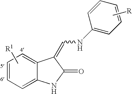
R Substitution 9. Example # R 1 2 3 4 5 6 237 5′-CN H H 
H H 238 4′-Me H H 
H H 239 6′-F H H 
H H 240 5′-F H H 
H H 241 5′-Cl H H 
H H 242 4′-Me H H 
H H 243 6′-F H H 
H H 244 5′-F H H 
H H 245 5′-Cl H H 
H H 246 4′-Me H H 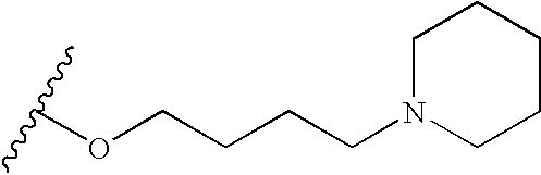
H H 247 6′-F H H 
H H 248 H H F 
H H 249 4′-Me H F 
H H Unsubstituted, 4-Fluoro, 4-methyl, 5-Chloro, 5-Cyano, 5-Fluoro, 5-Nitro, 6-Fluoro & 6-Aryl 3-[(Substituted Phenylamino)-methylene]-1,3-dihydro-indol-2-ones. 
Substitution 10. Example # R 1 2 3 4 5 6 250 6′-F H F 
H H 251 H H H 
H H 252 4′-Me H H 
H H 253 6′-F H H 
H H 254 H H H 
H H 255 4′-F H H 
H H 256 4′-Me H H 
H H 257 4′-F H H 
H H 258 5′-F H H 
H H 259 6′-F H H 
H H 260 5′-Cl H H 
H H 261 4′-F H H 
H H 262 5′-Cl H H 
H H 263 5′-F H H 
H H 264 4′-Me H H 
H H Unsubstituted, 4-Fluoro, 4-methyl, 5-Chloro, 5-Cyano, 5-Fluoro, 5-Nitro, 6-Fluoro & 6-Aryl 3-[(Substituted Phenylamino)-methylene]-1,3-dihydro-indol-2-ones. 
R Substitution 11. Example # R 1 2 3 4 5 6 265 H H H 
H H 266 6′-F H H 
H H 267 4′-F H H 
H H 268 6′-(3-Methoxyphenyl) H H 
H H 269 6′-(3-Methoxyphenyl) H H 
H H 270 4′-Me H H 
H H 271 6′-F H H 
H H 272 H H H 
H H 273 4′-F H H 
H H 274 5′-F H H 
H H 275 5′-Cl H H 
H H 276 6′-(3-Methoxyphenyl) H H 
H H 277 6′-(3-Methoxyphenyl) H H 
H H 278 4′-Me H H 
H H Unsubstituted, 4-Fluoro, 4-methyl, 5-Chloro, 5-Cyano, 5-Fluoro, 5-Nitro, 6-Fluoro & 6-Aryl 3-[(Substituted Phanylamino)-methylene]-1,3-dihydro-indol-2-ones. 
R Substitution 12. Example # R 1 2 3 4 5 6 279 6′-F H H 
H H 280 H H H 
H H 281 4′-F H H 
H H 282 5′-F H H 
H H 283 5′-Cl H H 
H H 284 H H H 
H H 285 5′-Cl H H 
H H 286 4′-Me H H 
H H 287 4′-F H H 
H H 288 5′-F H H 
H H 289 6′-F H H 
H H 290 H H H 
H H 291 5′-Cl H H 
H H 292 4′-Me H H 
H H Unsubstituted, 4-Fluoro, 4-methyl, 5-Chloro, 5-Cyano, 5-Fluoro, 5-Nitro, 6-Fluoro & 6-Aryl 3-[(Substituted Phenylamino)-methylene]-1,3-dihydro-indol-2-ones. 
R Substitution 13. Example # R 1 2 3 4 5 6 293 4′-F H H 
H H 294 5′-F H H 
H H 295 6′-F H H 
H H 296 4′-Me H H 
H H 297 H H H 
H H 298 6′-F H H 
H H 299 5′-Cl H H 
H H 300 5′-F H H 
H H 301 4′-F H H 
H H 302 H H H 
H H 303 4′-Me H H 
H H 304 6′-F H H 
H H Unsubstituted, 4-Fluoro, 4-methyl, 5-Chloro, 5-Fluoro & 6-Fluoro 3-[(Substituted Phenyl- amino)-methylene]-1,3-dihydro-indol-2-ones. 
R Substitution 14. Example # R 1 2 3 4 5 6 305 H H H 
H H 306 H H H 
H H 307 5′-Cl H H 
H H 308 4′-Me H H 
H H 309 4′-F H H 
H H 310 5′-F H H 
H H 311 6′-F H H 
H H 312 H H H 
H H 313 5′-Cl H H 
H H 314 4′-Me H H 
H H 315 4′-F H H 
H H 316 5′-F H H 
H H 317 6′-F H H 
H H Unsubstituted, 4-Fluoro, 4-methyl, 5-Chloro, 5-Fluoro & 6-Fluoro 3-[(Substituted Phenyl- amino)-methylene]-1,3-dihydro-indol-2-ones. 
R Substitution 15. Example # R 1 2 3 4 5 6 318 H H H 
H H 319 5′-Cl H H 
H H 320 4′-Me H H 
H H 321 4′-F H H 
H H 322 5′-F H H 
H H 323 6′-F H H 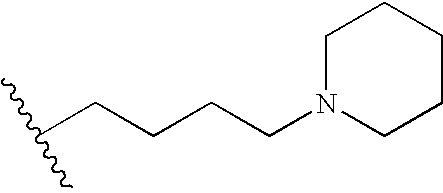
H H - The present implants may also comprise a TKI or a combination of TKIs represented by the following formulas

- Additional TKIs that may be used in the present implants include those compounds disclosed in Goel et al., “Tyrosine Kinase Inhibitors: A Clinical Perspective”, Current Oncology Reports, 4:9-19 (2002); Haluska et al., “Receptor tyrosine kinase inhibitors”, Current Opinion in Investigational Drugs, 2(2):280-286 (2001); Hubbard et al., “Protein tyrosine kinase structure and function”, Annu. Rev. Biochem., 69:373-98 (2000); Busse et al., “Tyrosine kinase inhibitors: rationale, mechanisms of action, and implications for drug resistance”, Semin Oncol 28(suppl 16) 47-55 (2001); and Fabbro et al., “Protein tyrosine kinase inhibitors: new treatment modalities?”, Current Opinion in Pharmacology, 2:374-381 (2002).
- The foregoing compounds may be synthesized using routine chemical technologies and methods including those disclosed in U.S. patent application Ser. No. 10/256,879 (U.S. Pub. No. 20030199478) and Ser. No. 10/259,703 (U.S. Pub. No. 20030225152) and the other above-identified references.
- The present implants may also include salts of the TKIs. Pharmaceutically acceptable acid addition salts of the compounds of the invention are those formed from acids which form non-toxic addition salts containing pharmaceutically acceptable anions, such as the hydrochloride, hydrobromide, hydroiodide, sulfate, or bisulfate, phosphate or acid phosphate, acetate, maleate, fumarate, oxalate, lactate, tartrate, citrate, gluconate, saccharate and p-toluene sulphonate salts.
- Thus, the implant may comprise a therapeutic component which comprises, consists essentially of, or consists of a TKI, salts thereof, and mixtures thereof. The biodegradable polymer matrix of such implants may be substantially free of polyvinyl alcohol, or in other words, includes no polyvinyl alcohol.
- Additional TKIs may be obtained or synthesized using conventional methods, such as by routine chemical synthesis methods known to persons of ordinary skill in the art. Therapeutically effective TKIs may be screened and identified using conventional screening technologies used for the TKIs described herein.
- The TKIs may be in a particulate or powder form and entrapped by the biodegradable polymer matrix. Usually, TKI particles in intraocular implants will have an effective average size less than about 3000 nanometers. In certain implants, the particles may have an effective average particle size about an order of magnitude smaller than 3000 nanometers. For example, the particles may have an effective average particle size of less than about 500 nanometers. In additional implants, the particles may have an effective average particle size of less than about 400 nanometers, and in still further embodiments, a size less than about 200 nanometers.
- The TKI of the implant is preferably from about 10% to 90% by weight of the implant. More preferably, the TKI is from about 20% to about 80% by weight of the implant. In a preferred embodiment, the TKI comprises about 40% by weight of the implant (e.g., 30%-50%). In another embodiment, the TKI comprises about 60% by weight of the implant.
- Suitable polymeric materials or compositions for use in the implant include those materials which are compatible, that is biocompatible, with the eye so as to cause no substantial interference with the functioning or physiology of the eye. Such materials preferably are at least partially and more preferably substantially completely biodegradable or bioerodible.
- Examples of useful polymeric materials include, without limitation, such materials derived from and/or including organic esters and organic ethers, which when degraded result in physiologically acceptable degradation products, including the monomers. Also, polymeric materials derived from and/or including, anhydrides, amides, orthoesters and the like, by themselves or in combination with other monomers, may also find use. The polymeric materials may be addition or condensation polymers, advantageously condensation polymers. The polymeric materials may be cross-linked or non-cross-linked, for example not more than lightly cross-linked, such as less than about 5%, or less than about 1% of the polymeric material being cross-linked. For the most part, besides carbon and hydrogen, the polymers will include at least one of oxygen and nitrogen, advantageously oxygen. The oxygen may be present as oxy, e.g. hydroxy or ether, carbonyl, e.g. non-oxo-carbonyl, such as carboxylic acid ester, and the like. The nitrogen may be present as amide, cyano and amino. The polymers set forth in Heller, Biodegradable
- Polymers in Controlled Drug Delivery, In: CRC Critical Reviews in Therapeutic Drug Carrier Systems, Vol. 1, CRC Press, Boca Raton, Fla. 1987, pp 39-90, which describes encapsulation for controlled drug delivery, may find use in the present implants.
- Of additional interest are polymers of hydroxyaliphatic carboxylic acids, either homopolymers or copolymers, and polysaccharides. Polyesters of interest include polymers of D-lactic acid, L-lactic acid, racemic lactic acid, glycolic acid, polycaprolactone, and combinations thereof. Generally, by employing the L-lactate or D-lactate, a slowly eroding polymer or polymeric material is achieved, while erosion is substantially enhanced with the lactate racemate.
- Among the useful polysaccharides are, without limitation, calcium alginate, and functionalized celluloses, particularly carboxymethylcellulose esters characterized by being water insoluble, a molecular weight of about 5 kD to 500 kD, for example.
- Other polymers of interest include, without limitation, polyesters, polyethers and combinations thereof which are biocompatible and may be biodegradable and/or bioerodible.
- Some preferred characteristics of the polymers or polymeric materials for use in the present invention may include biocompatibility, compatibility with the therapeutic component, ease of use of the polymer in making the drug delivery systems of the present invention, a half-life in the physiological environment of at least about 6 hours, preferably greater than about one day, not significantly increasing the viscosity of the vitreous, and water insolubility.
- The biodegradable polymeric materials which are included to form the matrix are desirably subject to enzymatic or hydrolytic instability. Water soluble polymers may be cross-linked with hydrolytic or biodegradable unstable cross-links to provide useful water insoluble polymers. The degree of stability can be varied widely, depending upon the choice of monomer, whether a homopolymer or copolymer is employed, employing mixtures of polymers, and whether the polymer includes terminal acid groups.
- Equally important to controlling the biodegradation of the polymer and hence the extended release profile of the implant is the relative average molecular weight of the polymeric composition employed in the implant. Different molecular weights of the same or different polymeric compositions may be included in the implant to modulate the release profile. In certain implants, the relative average molecular weight of the polymer will range from about 9 to about 64 kD, usually from about 10 to about 54 kD, and more usually from about 12 to about 45 kD.
- In some implants, copolymers of glycolic acid and lactic acid are used, where the rate of biodegradation is controlled by the ratio of glycolic acid to lactic acid. The most rapidly degraded copolymer has roughly equal amounts of glycolic acid and lactic acid. Homopolymers, or copolymers having ratios other than equal, are more resistant to degradation. The ratio of glycolic acid to lactic acid will also affect the brittleness of the implant, where a more flexible implant is desirable for larger geometries. The % of polylactic acid in the polylactic acid polyglycolic acid (PLGA) copolymer can be 0-100%, preferably about 15-85%, more preferably about 35-65%. In some implants, a 50/50 PLGA copolymer is used.
- The biodegradable polymer matrix of the intraocular implant may comprise a mixture of two or more biodegradable polymers. For example, the implant may comprise a mixture of a first biodegradable polymer and a different second biodegradable polymer. One or more of the biodegradable polymers may have terminal acid groups.
- Release of a drug from an erodible polymer is the consequence of several mechanisms or combinations of mechanisms. Some of these mechanisms include desorption from the implants surface, dissolution, diffusion through porous channels of the hydrated polymer and erosion. Erosion can be bulk or surface or a combination of both. As discussed herein, the matrix of the intraocular implant may release drug at a rate effective to sustain release of an amount of the TKI for more than one week after implantation into an eye. In certain implants, therapeutic amounts of the antiexcitotoxic agents are released for more than about one month, and even for about six months or more.
- One example of the biodegradable intraocular implant comprises a TKI with a biodegradable polymer matrix that comprises a poly(lactide-co-glycolide) or a poly (D,L-lactide-co-glycolide). The implant may have an amount of the TKI from about 20% to about 60% by weight of the implant. Such a mixture is effective in sustaining release of a therapeutically effective amount of the TKI for a time period from about one month to about six months from the time the implant is placed in an eye.
- Another example of the biodegradable intraocular implant comprises a TKI with a biodegradable polymer matrix that comprises a single type of polymer. For example, the biodegradable polymer matrix may consist essentially of a polycaprolactone. The polycaprolactone may have a molecular weight between about 10 and about 20 kilodaltons, such as about 15 kilodaltons. These implants are capable of providing a nearly linear release rate for at least about 70 days.
- The release of the TKI(s) from the intraocular implant comprising a biodegradable polymer matrix may include an initial burst of release followed by a gradual increase in the amount of the antiexcitotoxic agent(s) released, or the release may include an initial delay in release of the antiexcitotoxic agent(s) followed by an increase in release. When the implant is substantially completely degraded, the percent of the TKI(s) that has been released is about one hundred. Compared to existing implants, the implants disclosed herein do not completely release, or release about 100% of the TKI(s), until after about one week of being placed in an eye.
- It may be desirable to provide a relatively constant rate of release of the TKI(s) from the implant over the life of the implant. For example, it may be desirable for the TKI(s) to be released in amounts from about 0.01 μg to about 2 μg per day for the life of the implant. However, the release rate may change to either increase or decrease depending on the formulation of the biodegradable polymer matrix. In addition, the release profile of the TKI(s) may include one or more linear portions and/or one or more non-linear portions. Preferably, the release rate is greater than zero once the implant has begun to degrade or erode.
- The implants may be monolithic, i.e. having the active agent or agents homogenously distributed through the polymeric matrix, or encapsulated, where a reservoir of active agent is encapsulated by the polymeric matrix. Due to ease of manufacture, monolithic implants are usually preferred over encapsulated forms. However, the greater control afforded by the encapsulated, reservoir-type implant may be of benefit in some circumstances, where the therapeutic level of the drug falls within a narrow window. In addition, the therapeutic component, including the TKI(s), may be distributed in a non-homogenous pattern in the matrix. For example, the implant may include a portion that has a greater concentration of the TKI(s) relative to a second portion of the implant.
- One
such implant 100 is illustrated inFIG. 14 . Theimplant 100 may be understood to be a unidirectional drug delivery device. Theimplant 100 is characterized by comprising afirst portion 110 and asecond portion 120.First portion 110 comprises a mixture of a therapeutic agent, such as TKI, and a biodegradable polymer matrix, such as a matrix of PLGA, PLA, or a combination thereof.Second portion 120 comprises a polymer, such as a biodegradable polymer, and is substantially free of the therapeutic agent. The polymeric component of thefirst portion 110 and thesecond portion 120 may comprise the same polymer material, e.g., both components may be made from a PLGA polymer. Although the therapeutic agent is a TKI, other implants may include other therapeutic agents, including those described herein.First portion 110 may be understood to be an active layer, andsecond portion 120 may be understood to be a barrier layer, which is effective to prevent or reduce diffusion of the therapeutic agent from one side of the implant. The layers may be separately formed as films and pressed together using a Carver press, for example. Or the layers may be co-extruded using conventional extrusion techniques or injection molded using injection molding techniques. Theimplant 110 is effective to control the flow or release of a therapeutic agent in a specific direction, such as one direction. The implant can be applied to a diseased location, such as in an eye, that needs the release of the therapeutic agent in a specific and controlled manner, such as for subconjunctival applications. - The present implants may also comprise a combination of a TKI and polycaprolactone, as described herein. Such implants may provide a single order release rate for about 70 days or more after placement in an eye. The polycaprolactone may have a molecular weight of about 15 kilodaltons. Thus, one embodiment of the present implants, comprises a poorly soluble drug or therapeutic agent and a single polymeric component that releases the drug at a substantially linear release rate (e.g., a zero order rate).
- The present implants may also include a non-biodegradable polymer component, as described herein. The release of a therapeutic agent, such as TKI, may be achieved by movement of the therapeutic agent through one or more openings, orifices, or holes. An example of such an implant is disclosed in U.S. Pat. No. 6,331,313.
- The intraocular implants disclosed herein may have a size of between about 5 μm and about 2 mm, or between about 10 μm and about 1 mm for administration with a needle, greater than 1 mm, or greater than 2 mm, such as 3 mm or up to 10 mm, for administration by surgical implantation. The vitreous chamber in humans is able to accommodate relatively large implants of varying geometries, having lengths of, for example, 1 to 10 mm. The implant may be a cylindrical pellet (e.g., rod) with dimensions of about 2 mm×0.75 mm diameter. Or the implant may be a cylindrical pellet with a length of about 7 mm to about 10 mm, and a diameter of about 0.75 mm to about 1.5 mm.
- The implants may also be at least somewhat flexible so as to facilitate both insertion of the implant in the eye, such as in the vitreous, and accommodation of the implant. The total weight of the implant is usually about 250-5000 μg, more preferably about 500-1000 μg. For example, an implant may be about 500 μg, or about 1000 μg. For non-human individuals, the dimensions and total weight of the implant(s) may be larger or smaller, depending on the type of individual. For example, humans have a vitreous volume of approximately 3.8 ml, compared with approximately 30 ml for horses, and approximately 60-100 ml for elephants. An implant sized for use in a human may be scaled up or down accordingly for other animals, for example, about 8 times larger for an implant for a horse, or about, for example, 26 times larger for an implant for an elephant.
- Thus, implants can be prepared where the center may be of one material and the surface may have one or more layers of the same or a different composition, where the layers may be cross-linked, or of a different molecular weight, different density or porosity, or the like. For example, where it is desirable to quickly release an initial bolus of drug, the center may be a polylactate coated with a polylactate-polyglycolate copolymer, so as to enhance the rate of initial degradation. Alternatively, the center may be polyvinyl alcohol coated with polylactate, so that upon degradation of the polylactate exterior the center would dissolve and be rapidly washed out of the eye.
- The implants may be of any geometry including fibers, sheets, films, microspheres, spheres, circular discs, plaques and the like. The upper limit for the implant size will be determined by factors such as toleration for the implant, size limitations on insertion, ease of handling, etc. Where sheets or films are employed, the sheets or films will be in the range of at least about 0.5 mm×0.5 mm, usually about 3-10 mm×5-10 mm with a thickness of about 0.1-1.0 mm for ease of handling. Where fibers are employed, the fiber diameter will generally be in the range of about 0.05 to 3 mm and the fiber length will generally be in the range of about 0.5-10 mm. Spheres may be in the range of about 0.5 μm to 4 mm in diameter, with comparable volumes for other shaped particles.
- The size and form of the implant can also be used to control the rate of release, period of treatment, and drug concentration at the site of implantation. Larger implants will deliver a proportionately larger dose, but depending on the surface to mass ratio, may have a slower release rate. The particular size and geometry of the implant are chosen to suit the site of implantation.
- The proportions of TKI(s), polymer, and any other modifiers may be empirically determined by formulating several implants with varying proportions. A USP approved method for dissolution or release test can be used to measure the rate of release (
USP 23; NF 18 (1995) pp. 1790-1798). For example, using the infinite sink method, a weighed sample of the implant is added to a measured volume of a solution containing 0.9% NaCl in water, where the solution volume will be such that the drug concentration is after release is less than 5% of saturation. The mixture is maintained at 37° C. and stirred slowly to maintain the implants in suspension. The appearance of the dissolved drug as a function of time may be followed by various methods known in the art, such as spectrophotometrically, HPLC, mass spectroscopy, etc. until the absorbance becomes constant or until greater than 90% of the drug has been released. - In addition to the TKI(s) included in the intraocular implants disclosed herein, the intraocular implants may also include one or more additional ophthalmically acceptable therapeutic agents. For example, the implant may include one or more antihistamines, one or more antibiotics, one or more beta blockers, one or more steroids, one or more antineoplastic agents, one or more immunosuppressive agents, one or more antiviral agents, one or more antioxidant agents, and mixtures thereof.
- Pharmacologic or therapeutic agents which may find use in the present systems, include, without limitation, those disclosed in U.S. Pat. No. 4,474,451, columns 4-6 and U.S. Pat. No. 4,327,725, columns 7-8.
- Examples of antihistamines include, and are not limited to, loradatine, hydroxyzine, diphenhydramine, chlorpheniramine, brompheniramine, cyproheptadine, terfenadine, clemastine, triprolidine, carbinoxamine, diphenylpyraline, phenindamine, azatadine, tripelennamine, dexchlorpheniramine, dexbrompheniramine, methdilazine, and trimprazine doxylamine, pheniramine, pyrilamine, chiorcyclizine, thonzylamine, and derivatives thereof.
- Examples of antibiotics include without limitation, cefazolin, cephradine, cefaclor, cephapirin, ceftizoxime, cefoperazone, cefotetan, cefutoxime, cefotaxime, cefadroxil, ceftazidime, cephalexin, cephalothin, cefamandole, cefoxitin, cefonicid, ceforanide, ceftriaxone, cefadroxil, cephradine, cefuroxime, cyclosporine, ampicillin, amoxicillin, cyclacillin, ampicillin, penicillin G, penicillin V potassium, piperacillin, oxacillin, bacampicillin, cloxacillin, ticarcillin, azlocillin, carbenicillin, methicillin, nafcillin, erythromycin, tetracycline, doxycycline, minocycline, aztreonam, chloramphenicol, ciprofloxacin hydrochloride, clindamycin, metronidazole, gentamicin, lincomycin, tobramycin, vancomycin, polymyxin B sulfate, colistimethate, colistin, azithromycin, augmentin, sulfamethoxazole, trimethoprim, gatifloxacin, ofloxacin, and derivatives thereof.
- Examples of beta blockers include acebutolol, atenolol, labetalol, metoprolol, propranolol, timolol, and derivatives thereof.
- Examples of steroids include corticosteroids, such as cortisone, prednisolone, flurometholone, dexamethasone, medrysone, loteprednol, fluazacort, hydrocortisone, prednisone, betamethasone, prednisone, methylprednisolone, riamcinolone hexacatonide, paramethasone acetate, diflorasone, fluocinonide, fluocinolone, triamcinolone, derivatives thereof, and mixtures thereof.
- Examples of antineoplastic agents include adriamycin, cyclophosphamide, actinomycin, bleomycin, duanorubicin, doxorubicin, epirubicin, mitomycin, methotrexate, fluorouracil, carboplatin, carmustine (BCNU), methyl-CCNU, cisplatin, etoposide, interferons, camptothecin and derivatives thereof, phenesterine, taxol and derivatives thereof, taxotere and derivatives thereof, vinblastine, vincristine, tamoxifen, etoposide, piposulfan, cyclophosphamide, and flutamide, and derivatives thereof.
- Examples of immunosuppresive agents include cyclosporine, azathioprine, tacrolimus, and derivatives thereof.
- Examples of antiviral agents include interferon gamma, zidovudine, amantadine hydrochloride, ribavirin, acyclovir, valciclovir, dideoxycytidine, phosphonoformic acid, ganciclovir and derivatives thereof.
- Examples of antioxidant agents include ascorbate, alpha-tocopherol, mannitol, reduced glutathione, various carotenoids, cysteine, uric acid, taurine, tyrosine, superoxide dismutase, lutein, zeaxanthin, cryotpxanthin, astazanthin, lycopene, N-acetyl-cysteine, carnosine, gamma-glutamylcysteine, quercitin, lactoferrin, dihydrolipoic acid, citrate, Ginkgo Biloba extract, tea catechins, bilberry extract, vitamins E or esters of vitamin E, retinyl palmitate, and derivatives thereof.
- Other therapeutic agents include squalamine, carbonic anhydrase inhibitors, alpha agonists, prostamides, prostaglandins, antiparasitics, antifungals, and derivatives thereof.
- The amount of active agent or agents employed in the implant, individually or in combination, will vary widely depending on the effective dosage required and the desired rate of release from the implant. As indicated herein, the agent will be at least about 1, more usually at least about 10 weight percent of the implant, and usually not more than about 80, more usually not more than about 40 weight percent of the implant.
- In addition to the therapeutic component, the intraocular implants disclosed herein may include effective amounts of buffering agents, preservatives and the like. Suitable water soluble buffering agents include, without limitation, alkali and alkaline earth carbonates, phosphates, bicarbonates, citrates, borates, acetates, succinates and the like, such as sodium phosphate, citrate, borate, acetate, bicarbonate, carbonate and the like. These agents advantageously present in amounts sufficient to maintain a pH of the system of between about 2 to about 9 and more preferably about 4 to about 8. As such the buffering agent may be as much as about 5% by weight of the total implant. Suitable water soluble preservatives include sodium bisulfite, sodium bisulfate, sodium thiosulfate, ascorbate, benzalkonium chloride, chlorobutanol, thimerosal, phenylmercuric acetate, phenylmercuric borate, phenylmercuric nitrate, parabens, methylparaben, polyvinyl alcohol, benzyl alcohol, phenylethanol and the like and mixtures thereof. These agents may be present in amounts of from 0.001 to about 5% by weight and preferably 0.01 to about 2% by weight.
- In addition, the implants may include a solubility enhancing component provided in an amount effective to enhance the solubility of the TKI(s) relative to substantially identical implants without the solubility enhancing component. For example, an implant may include a β-cyclodextrin, which is effective in enhancing the solubility of the TKI. The β-cyclodextrin may be provided in an amount from about 0.5% (w/w) to about 25% (w/w) of the implant. In certain implants, the β-cyclodextrin is provided in an amount from about 5% (w/w) to about 15% (w/w) of the implant
- In some situations mixtures of implants may be utilized employing the same or different pharmacological agents. In this way, a cocktail of release profiles, giving a biphasic or triphasic release with a single administration is achieved, where the pattern of release may be greatly varied. The implants may also have a sigmoidal release profile.
- Additionally, release modulators such as those described in U.S. Pat. No. 5,869,079 may be included in the implants. The amount of release modulator employed will be dependent on the desired release profile, the activity of the modulator, and on the release profile of the antiexcitotoxic agent(s) in the absence of modulator. Electrolytes such as sodium chloride and potassium chloride may also be included in the implant. Where the buffering agent or enhancer is hydrophilic, it may also act as a release accelerator. Hydrophilic additives act to increase the release rates through faster dissolution of the material surrounding the drug particles, which increases the surface area of the drug exposed, thereby increasing the rate of drug bioerosion. Similarly, a hydrophobic buffering agent or enhancer dissolve more slowly, slowing the exposure of drug particles, and thereby slowing the rate of drug bioerosion.
- Various techniques may be employed to produce the implants described herein. Useful techniques include, but are not necessarily limited to, solvent evaporation methods, phase separation methods, interfacial methods, molding methods, injection molding methods, extrusion methods, co-extrusion methods, carver press method, die cutting methods, heat compression, combinations thereof and the like.
- Specific methods are discussed in U.S. Pat. No. 4,997,652. Extrusion methods may be used to avoid the need for solvents in manufacturing. When using extrusion methods, the polymer and drug are chosen so as to be stable at the temperatures required for manufacturing, usually at least about 85 degrees Celsius. Extrusion methods use temperatures of about 25 degrees C. to about 150 degrees C., more preferably about 65 degrees C. to about 130 degrees C. An implant may be produced by bringing the temperature to about 60 degrees C. to about 150 degrees C. for drug/polymer mixing, such as about 130 degrees C., for a time period of about 0 to 1 hour, 0 to 30 minutes, or 5-15 minutes. For example, a time period may be about 10 minutes, preferably about 0 to 5 min. The implants are then extruded at a temperature of about 60 degrees C. to about 130 degrees C., such as about 75 degrees C.
- In addition, the implant may be coextruded so that a coating is formed over a core region during the manufacture of the implant.
- Compression methods may be used to make the implants, and typically yield implants with faster release rates than extrusion methods. Compression methods may use pressures of about 50-150 psi, more preferably about 70-80 psi, even more preferably about 76 psi, and use temperatures of about 0 degrees C. to about 115 degrees C., more preferably about 25 degrees C.
- The implants of the present invention may be inserted into the eye, for example the vitreous chamber of the eye, by a variety of methods, including placement by forceps or by trocar following making a 2-3 mm incision in the sclera. One example of a device that may be used to insert the implants into an eye is disclosed in U.S. Patent Publication No. 2004/0054374. The method of placement may influence the therapeutic component or drug release kinetics. For example, delivering the implant with a trocar may result in placement of the implant deeper within the vitreous than placement by forceps, which may result in the implant being closer to the edge of the vitreous. The location of the implant may influence the concentration gradients of therapeutic component or drug surrounding the element, and thus influence the release rates (e.g., an element placed closer to the edge of the vitreous may result in a slower release rate).
- The present implants are configured to release an amount of the TKI(s) effective to treat or reduce a symptom of an ocular condition, such as an ocular condition.
- The implants disclosed herein may also be configured to release the antiexcitotoxic agent(s) or additional therapeutic agents, as described above, which to prevent diseases or conditions, such as the following:
- MACULOPATHIES/RETINAL DEGENERATION: Non-Exudative Age Related Macular Degeneration (ARMD), Exudative Age Related Macular Degeneration (ARMD), Choroidal Neovascularization, Diabetic Retinopathy, Acute Macular Neuroretinopathy, Central Serous Chorioretinopathy, Cystoid Macular Edema, Diabetic Macular Edema.
- UVEITIS/RETINITIS/CHOROIDITIS: Acute Multifocal Placoid Pigment Epitheliopathy, Behcet's Disease, Birdshot Retinochoroidopathy, Infectious (Syphilis, Lyme, Tuberculosis, Toxoplasmosis), Intermediate Uveitis (Pars Planitis), Multifocal Choroiditis, Multiple Evanescent White Dot Syndrome (MEWDS), Ocular Sarcoidosis, Posterior Scleritis, Serpignous Choroiditis, Subretinal Fibrosis and Uveitis Syndrome, Vogt-Koyanagi-Harada Syndrome.
- VASCULAR DISEASES/EXUDATIVE DISEASES: Coat's Disease, Parafoveal Telangiectasis, Papillophlebitis, Frosted Branch Angitis, Sickle Cell Retinopathy and other Hemoglobinopathies, Angioid Streaks, Familial Exudative Vitreoretinopathy.
- TRAUMATIC/SURGICAL: Sympathetic Ophthalmia, Uveitic Retinal Disease, Retinal Detachment, Trauma, Laser, PDT, Photocoagulation, Hypoperfusion During Surgery, Radiation Retinopathy, Bone Marrow Transplant Retinopathy.
- PROLIFERATIVE DISORDERS: Proliferative Vitreal Retinopathy and Epiretinal Membranes, Proliferative Diabetic Retinopathy, Retinopathy of Prematurity (retrolental fibroplastic).
- INFECTIOUS DISORDERS: Ocular Histoplasmosis, Ocular Toxocariasis, Presumed Ocular Histoplasmosis Syndrome (POHS), Endophthalmitis, Toxoplasmosis, Retinal Diseases Associated with HIV Infection, Choroidal Disease Associated with HIV Infection, Uveitic Disease Associated with HIV Infection, Viral Retinitis, Acute Retinal Necrosis, Progressive Outer Retinal Necrosis, Fungal Retinal Diseases, Ocular Syphilis, Ocular Tuberculosis, Diffuse Unilateral Subacute Neuroretinitis, Myiasis.
- GENETIC DISORDERS: Systemic Disorders with Accosiated Retinal Dystrophies, Congenital Stationary Night Blindness, Cone Dystrophies, Fundus Flavimaculatus, Best's Disease, Pattern Dystrophy of the Retinal Pigmented Epithelium, X-Linked Retinoschisis, Sorsby's Fundus Dystrophy, Benign Concentric Maculopathy, Bietti's Crystalline Dystrophy, pseudoxanthoma elasticum, Osler Weber syndrome.
- RETINAL TEARS/HOLES: Retinal Detachment, Macular Hole, Giant Retinal Tear.
- TUMORS: Retinal Disease Associated with Tumors, Solid Tumors, Tumor Metastasis, Benign Tumors, for example, hemangiomas, neurofibromas, trachomas, and pyogenic granulomas, Congenital Hypertrophy of the RPE, Posterior Uveal Melanoma, Choroidal Hemangioma, Choroidal Osteoma, Choroidal Metastasis, Combined Hamartoma of the Retina and Retinal Pigmented Epithelium, Retinoblastoma, Vasoproliferative Tumors of the Ocular Fundus, Retinal Astrocytoma, Intraocular Lymphoid Tumors.
- MISCELLANEOUS: Punctate Inner Choroidopathy, Acute Posterior Multifocal Placoid Pigment Epitheliopathy, Myopic Retinal Degeneration, Acute Retinal Pigment Epithelitis, Ocular inflammatory and immune disorders, ocular vascular malfunctions, Corneal Graft Rejection, Neovascular Glaucoma and the like.
- In one embodiment, an implant, such as the implants disclosed herein, is administered to a posterior segment of an eye of a human or animal patient, and preferably, a living human or animal. In at least one embodiment, an implant is administered without accessing the subretinal space of the eye. For example, a method of treating a patient may include placing the implant directly into the posterior chamber of the eye. In other embodiments, a method of treating a patient may comprise administering an implant to the patient by at least one of intravitreal injection, subconjuctival injection, sub-tenon injections, retrobulbar injection, and suprachoroidal injection.
- In at least one embodiment, a method of improving vision or maintaining vision in a patient comprises administering one or more implants containing one or more TKIs, as disclosed herein to a patient by at least one of intravitreal injection, subconjuctival injection, sub-tenon injection, retrobulbar injection, and suprachoroidal injection. A syringe apparatus including an appropriately sized needle, for example, a 22 gauge needle, a 27 gauge needle or a 30 gauge needle, can be effectively used to inject the composition with the posterior segment of an eye of a human or animal. Repeat injections are often not necessary due to the extended release of the TKI from the implants.
- In another aspect of the invention, kits for treating an ocular condition of the eye are provided, comprising: a) a container comprising an extended release implant comprising a therapeutic component including a TKI, and a drug release sustaining component; and b) instructions for use. Instructions may include steps of how to handle the implants, how to insert the implants into an ocular region, and what to expect from using the implants.
- The ocular pharmacokinetics of 3-[(4-Morpholin-4-yl-phenylamino)-methylene]-1,3-dihydro-indol-2-one (AGN 199659), 3-(6-Amino-3H-isobenzofuran-1-ylidene)-5-chloro-1,3-dihydro-indol-2-one (AGN 200954),
AGN 201088 andAGN 201666 following single intravitreal injections into female albino rabbit eyes was determined. The animals were dosed with a 50 mL intravitreal injection of 242 ngAGN 201088, 128ng AGN 201666, 114 ng 3-[(4-Morpholin-4-yl-phenylamino)-methylene]-1,3-dihydro-indol-2-one or 222 ng 3-(6-Amino-3H-isobenzofuran-1-ylidene)-5-chloro-1,3-dihydro-indol-2-one per eye. Vitreous humor samples (n=4 eyes per timepoint) were collected at 0.5, 1, 2, 4, 8, and 12 hr postdose. The TKI concentration in the vitreous humor was determined using a liquid chromatography tandem mass spectrometry method (LC-MS/MS). - All compounds were eliminated fairly rapidly from the rabbit eye. This indicates a transretinal route of elimination. There was no bias to compound nucleus. However, even though elimination was extremely rapid it was determined that local sustained delivery was feasible. Based on the vitreal clearance determined in this study for 3-[(4-Morpholin-4-yl-phenylamino)-methylene]-1,3-dihydro-indol-2-one, 3-(6-Amino-3H-isobenzofuran-1-ylidene)-5-chloro-1,3-dihydro-indol-2-one,
AGN 201088 andAGN 201666, and assuming steady state efficacious concentration at twice the EC50 values (determined by in vitro receptor binding and intracellular Ca2+ assay) all the tyrosine kinase inhibitors tested could be formulated into 1 mg implants that would maintain the desired steady state drug vitreal concentrations for a duration of about six months. This data is summarized in Table 1 andFIGS. 1 and 2 .TABLE 1 TKI Pharmacokinetic Parameters after a Single Intravitreal Injection AGN AGN AGN AGN Parameter 199659 200954 201088 201666 Dose (ng) 114 222 242 128 C0 (ng/mL) 502 566 222 332 t1/2 (hr) 1.21 2.59 1.11 2.32 AUC0-tlast 488 778 272 466 (ng · hr/mL) Cl (mL/hr) 0.232 0.260 0.885 0.270 Vss (mL) 0.255 0.705 1.23 0.577 Theoretical 6 200 ug 5 ug 150 ug 126 ug mo dose - Tyrosine kinase inhibitors were incorporated into PLGA or PLA implants by extrusion. The TKIs were milled with the polymers at certain ratios then extruded into filaments. These filaments were subsequently cut into implants weighing approximately 1 mg. Several TKIs were formulated in the PLGA and PLA implants based on their potencies and physicochemical properties as shown in Table 2.
TABLE 2 Tyrosine Kinase Inhibitors Formulated in PLGA Implants AGN Number Structure Projected C55 Efficacy Solubility (□g/mL) pH 7log P pKa AGN 200954 
4 ng/mL 0.3 2.21 4.24 10.03 AGN 202314
96 ng/mL 202 3.80 9.68 AGN 202560 
105 ng/mL 41 3.66 9.65 AGN 201634
28 ng/mL 88 1.25 4.06 10.25 - TKI release from the implants was assessed in vitro. Implants were placed into vials containing release medium and shaken at 37° C. At the appropriate time points a sample was taken from the release medium for analysis and the medium totally replaced to maintain sink conditions. Drug in the sample was assayed by HPLC and the cumulative percent release of drug from the implant noted as a function of time. The in vitro release profiles of
AGN 200954,AGN 202314, AGN 201635 and AGN 202564 are depicted inFIGS. 3 through 10 , respectively. - From the formulation release data depicted in
FIGS. 3 through 10 it is evident that TKIs over a wide range of physicochemical properties can be engineered to release drug in vitro over a period of weeks to a year. -
Implants containing AGN 202314 were placed intravitreally or subconjunctivally in an eye. The implants releasedAGN 202314 in-vitro over a 14 day period (FIG. 3 .). The intent of this study was to achieve an intravitreal in-vivo/in-vitro correlation with the intravitreal implants and assess the feasibility of periocular delivery. - Intravitreal Implants, PLGA (400
μg AGN 202314 dose, 1 mg total implant weight), were implanted by surgical incision into the mid vitreous of albino rabbits. Atdays 8, 15, 31 and 61 rabbits were sacrificed and the vitreous humor, lens, aqueous humor and plasma assayed forAGN 202314. - Subconjunctival implants, PLGA (1200
μg AGN 202314 dose; three implants) and PLA microspheres (300 μg AGN 202314) were implanted subconjunctivally. Atdays 8, 15, 31 and 61 rabbits were sacrificed and the vitreous humor, lens, aqueous humor and plasma assayed forAGN 202314. - The data are summarized in Tables 3 through 5
TABLE 3 PK Results from 2 Month Intravitreal Implantation AGN 202314 Implant (400 μg rod) Day 8 15 31 61 Retina (ng/g) 1220 100 BLQ BLQ Vitreous 327 85 BLQ BLQ Humor (ng/g) Lens (ng/g) ALQ (6580) ALQ (8980) 724 35.8 Aqueous 2.10 6.50 BLQ BLQ Humor (ng/mL) Plasma 0.255 BLQ BLQ BLQ (ng/mL)
Below the limit of quantitation (BLQ):
Retina and lens: <5 ng/g,
VH: <30 ng/g,
AH: <0.5 ng/mL,
Plasma: <0.5 ng/mL
Above the limit of quantitation (ALQ):
Retina and lens: >2000 ng/g,
VH: >3000 ng/g,
AH: >30 ng/mL,
Plasma: >200 ng/mL
-
TABLE 4 PK Results from 2 Mo Suboncjunctival Implantation of AGN 202314Microspheres Day 8 15 31 61 Microsphere (300 μg AGN 202314) - 0.63 dL/g PLA Retina (ng/g) 12.4 BLQ 19.2 BLQ Vitreous BLQ BLQ BLQ BLQ Humor (ng/g) Lens (ng/g) 5.19 BLQ BLQ BLQ Aqueous BLQ BLQ BLQ BLQ Humor (ng/mL) Plasma BLQ BLQ BLQ BLQ (ng/mL) Microsphere (300 μg AGN 202314) - 1.2 dL/g PLA Retina (ng/g) 3.7 5.1 BLQ BLQ Vitreous BLQ BLQ BLQ BLQ Humor (ng/g) Lens (ng/g) BLQ BLQ 2.72 BLQ Aqueous BLQ BLQ BLQ BLQ Humor (ng/mL) Plasma BLQ BLQ BLQ BLQ (ng/mL)
BLQ = Retina (<5 ng/g),
VH (<30 ng/g),
lens (<5 ng/g),
AH (<0.5 ng/mL),
plasma (<0.5 ng/mL)
-
TABLE 5 Month Subconjunctival Implantation (3 rods with a total of 1.2 mg AGN 202314) Day 8 15 31 61 Retina (ng/g) 63.8 BLQ BLQ BLQ Vitreous BLQ BLQ BLQ BLQ Humor (ng/g) Lens (ng/g) 229 7.93 BLQ BLQ Aqueous 6.38 BLQ BLQ BLQ Humor (ng/mL) Plasma BLQ BLQ BLQ BLQ (ng/mL)
BLQ:
Retina and lens: <5 ng/g,
VH: <30 ng/g,
AH: <0.5 ng/mL,
Plasma: <0.5 ng/mL
- The data from this study indicates that a good in vitro in vivo correlation was established for
AGN 202314. TheAGN 202314 implant released drug over a two week period both in vitro and in vivo. It is also important that plasma levels remain BLQ or extremely low for all time points. This shows that even in a worst case scenario of intravitreal delivery over two weeks systemic exposure is negligible. It was also noted that periocular delivery was unsuccessful at deliveringAGN 202314 to the vitreous and retina. - A follow-on two-month ocular pharmacokinetic study of
AGN 202314 following a single intravitreal implantation into albino rabbit eyes was initiated. The formulations deliveredAGN 202314 over a period of four months in-vitro. The following 1 mg implants were evaluated: 30% AGN 202314/70% Purac PLA; Lot# JS493028 (FIG. 4 ), 50% AGN 201634/50% Purac PLA; Lot # JS493034 (FIG. 6 .). - Two rabbits (4 eyes and 2 plasma) were used per timepoint. Implants were administered by a bilateral surgical intravitreal placement by sclerotomy without vitrectomy. The vitreous humor and
retina AGN 202314 concentrations were assayed atdays 8, 15, 31 and 61. The data are displayed in Table 6.TABLE 6 One Month Data from the AGN 202314Intravitreal Study Day 8 15 31 50% Purac PLA (AGN 202314 - 500 μg) Retina (ng/g) 95.3 ± 18.7 87.7 ± 29.6 157 ± 120 VH (ng/g): 22.2 ± 25.6 BLQ 69.9 ± 87.5 70% Purac PLA (AGN 202314 - 300 μg) Retina (ng/g) 78.1 ± 7.2 197 ± 88.7 189 ± 126 VH (ng/g) BLQ 33.7 ± 25.8 BLQ
Analytical range:
Retina BLQ < 5 ng/g;
VH BLQ < 30 ng/g
- The retinal levels achieved from this study approach therapeutic levels by the first week and are maintained over the first thirty days. This data shows that actual in vivo sustained delivery of a TKI locally is feasible.
- A six month pharmacokinetic study was initiated with intravitreal and
subconjunctival AGN 200954 implants. The implants releasedAGN 200954 in-vitro over a 180 day period (FIG. 3 ). Intravitreal Implants, PLGA (500μg AGN 200954 dose, 1 mg total implant weight, Purac polymer) and PLGA (500μg AGN 200954 dose, 1 mg total implant weight, RG503H polymer), were implanted by surgical incision into the mid vitreous of albino rabbits. Atdays 8, 15, 31 and 61 rabbits were sacrificed and the vitreous humor, lens, aqueous humor and plasma assayed forAGN 202314. Subconjunctival implants, PLGA implant (500μg AGN 200954 dose, 1 mg total implant weight, Purac polymer) and PLGA microspheres (370 μg and 740 μg AGN 200954), were administered. Atdays 8, 15, 31 and 61 rabbits were sacrificed and the vitreous humor, lens, aqueous humor and plasma assayed forAGN 200954. -
AGN 200954 Pharmacokinetics afterIntravitreal Administration Day 8 31 61 91 181 Intravitreal Implantation (500 μg rod) Formulation PU Retina BLQ 48.7 207 161 210 (ng/g) Vitreous BLQ 18.2 109 657 76.3 Humor (ng/g) Lens (ng/g) 70.2 243 586 768 1296 Aqueous BLQ BLQ BLQ BLQ 288 Humor (ng/mL) Plasma BLQ BLQ BLQ BLQ BLQ (ng/mL) Intravitreal Implantation (500 μg rod) Formulation RG503H Retina 15.7 17.7 416 58.4 24.9 (ng/g) Vitreous 560 126 189 65.2 227 Humor(ng/g) Lens (ng/g) 160 386 464 239 248 Aqueous BLQ BLQ BLQ BLQ 316 Humor (ng/mL) Plasma BLQ BLQ BLQ BLQ BLQ (ng/mL)
BLQ = Retina (5 ng/g),
VH (30 ng/g),
lens (5 ng/g),
AH (0.05 ng/mL),
plasma (0.05 ng/mL)
- It is evident from the data that a considerable in-vivo lag time exists for the first formulation not seen in vitro. Neither formulation exhibits measurable plasma concentrations.
- TKI release was examined for implants made from poly(D,L-lactide-co-glycolide) (PDLG) or poly(D,L-lactide) (PDL) in different media with or without addition of detergent at 37° C. in a shaking water bath.
-
AGN 201634 was obtained from Allergan, and its chemical structure is shown below. It was used as received without further purification. PDLG/PDL polymer materials were obtained from Purac America Inc.
- TKI release was examined in various medium, including saline, phosphate buffer saline of pH 7.4, 50 mM bicarbonate buffer of pH 6.0±0.1 with 0.1% cetyltrimethylammonium bromide (CTAB), and 50 mM borate buffer of pH 8.5±0.1 with 0.5% sodium dodecyl sulfate (SDS) in a shaking water bath (Precision) at 37° C. Sample was incubated in 10 mL of medium, and was totally replaced with fresh medium at each sampling time. Drug concentration was determined by HPLC using a Waters 2690 Separation Module equipped with a Waters XTerra RP8 column (3.9×150 mm, 5 μm, equilibrated at ambient) and a Waters 996 photodiode array detector (set at 238 nm) using 0.1% acetic acid in acetonitrile/water (40/60 by volume) as the mobile phase under a flow rate of 1.2 mL/min. The column was equilibrated with mobile phase for at least 30 min before initiating any sample injection.
- The characteristics of formulations, including formulation identification, Lot number, drug loading, inherent viscosity of polymer, and extrusion temperature are summarized in the following table. The drug load is from 20 to 50%. The formulations were extruded from a 750 μm nozzle to form cylindrical DDS.
TABLE 8 Characteristics of TKI formulations. Drug Formulation Loading Extrusion Temp # Lot # (%) Polymer I.V. (° C.) F1 JS443159 40 PDLG 0.2 68 F2 JS443020 20 PDLG 0.2 70 F3 JS493023 50 PDL 0.5 85 F4 JS493034 30 PDL 0.5 78
Note:
I.V.: inherent viscosity of polymer material.
- The stability of
AGN 201634 standard solution in deionized water/acetonitrile (75%/25%) was examined at 4° C., and the results are summarized in the following table. The concentration of standard solution was from 0.0695 μg/mL to 8.693 μg/mL, and was analyzed onday AGN 201634 in deionized water/acetonitrile (75%/25%) at 4° C. for up to 35 days even the concentration was up to 8.7 μg/mL. - Stability of
AGN 201634 Standard Solution of Various Concentrations in DI Water/Acetonitrile (75%/25%) at 4° C. (Table 9).Conc. Of Standard Day (μg/mL) Recovery (%) 14 0.0695 95.2 0.348 98.3 0.695 98.4 2.173 98.6 8.693 98.6 21 0.0695 96.5 0.348 101.3 0.695 102.1 2.173 101.0 8.693 101.6 35 0.0695 106.1 0.348 98.3 0.695 101.3 2.173 100.8 8.693 100.3 - To examine the stability of TKI in formulation,
Formulations AGN 201634 was stable in media of pH 6.0, 7.4 and 8.5, and lasted for 7 days in ambient or 14 days at 4° C.TABLE 10 Stability of TKI in Formulations media under various incubation conditions. Concen- Incubation tration Incubation Temperature Recovery Formulation Medium (μg/mL) Time (day) (oC.) (%) F3 pH 6.0 5.60 7 ambient 100.1 3.45 14 4 102.7 pH 7.4 2.53 7 ambient 101.1 2.29 14 4 103.7 pH 8.5 10.34 7 ambient 100.8 10.24 14 4 98.8 F4 pH 6.0 0.94 7 ambient 100.4 0.84 14 4 102.4 pH 7.4 0.18 7 ambient 104.3 0.48 14 4 101.3 pH 8.5 1.39 7 ambient 100.4 1.20 14 4 100.2 - TKI releases of
Formulation 1 in 20 mL of saline or 20-30 mL of PBS are demonstrated inFIG. 11 . The DDS was incubated in a vial of either 40 or 20 mL, and 10 mL sample solution was replaced by same volume of fresh medium, respectively, at each sampling time. The release profiles in saline and PBS were obviously different. Less than 5% of TKI was released in saline during the first 70 days. In contrast, less than 5% ofAGN 201634 was released at the first 3 weeks when DDS was incubated in PBS, the same as in saline, but more than 80% ofAGN 201634 was released after 70 days. However, no significantly difference in release profile was found when DDS was incubated in 20 or 30 mL of PBS in a 20 or 40 mL vial. It seems that release medium plans an important role in the variation of release profile instead of incubation volume. Due to this slow and diverged release profile, the release profile ofFormulation 2 was not performed since its formulation was based on the same polymer with a lower drug loading. - TKI releases of
Formulations FIGS. 12 and 13 , respectively. For F3, more than 50%, 45%, and 75% TKI was released at the first 3, 7, and 2 weeks, when DDS was incubated in a medium of pH 6.0 (with 0.1% CTAB), 7.4 (PBS) and 8.5 (with 0.5% SDS), respectively. On the other hand, approximately 47%, 6%, and 68% of TKI was released from F4 when DDS was incubated in media as described above. It seems that TKI release in different pH medium is pH 8.5>pH 6.0>pH 7.4, with or without the assistant from detergent in the medium. No large standard deviations are found in all media for both formulations. - To monitor the appearance of DDS during dissolution, the images of F3 and F4 formulations incubated in 10 mL media of a pH of 6.0 (with 0.1% CTAB), 7.4 (PBS) or 8.5 (with 0.5% SDS) at 37° C. were. All formulations experienced swelling followed by matrix degradation, resulting in drug release. No complete disintegration of formulation matrix was observed within 153 days at 37° C.
- In summary, tyrosine kinase inhibitor (AGN 201634) DDS were formulated using various PLGA or PLA at various drug loading. The stability of
AGN 201634 solution in DI water/acetonitrile (75%/25%) at 4° C. was more than 35 days, and DDS solution in various pH medium was more than 7 days under ambient condition or 14 days at 4° C. Different drug release profiles were found when DDS was tested in PBS or saline. Drug burst effect was found only inFormulation 3 when incubating in a medium of pH 6.0.Controlled AGN 201634 release in vitro was more than 4 weeks in a medium of pH 8.5, and more than 5 months in media of pH 7.4 and pH 6.0. - Biodegradable implants are made by combining a TKI with a biodegradable polymer composition in a stainless steel mortar. The biodegradable polymer composition comprises a single type of biodegradable polymer. The combination is mixed via a Turbula shaker set at 96 RPM for 15 minutes. The powder blend is scraped off the wall of the mortar and then remixed for an additional 15 minutes. The mixed powder blend is heated to a semi-molten state at specified temperature for a total of 30 minutes, forming a polymer/drug melt.
- Rods are manufactured by pelletizing the polymer/drug melt using a 9 gauge polytetrafluoroethylene (PTFE) tubing, loading the pellet into the barrel and extruding the material at the specified core extrusion temperature into filaments. The filaments are then cut into about 1 mg size implants or drug delivery systems. The rods have dimensions of about 2 mm long×0.72 mm diameter. The rod implants weigh between about 900 μg and 1100 μg.
- Wafers are formed by flattening the polymer melt with a Carver press at a specified temperature and cutting the flattened material into wafers, each weighing about 1 mg. The wafers have a diameter of about 2.5 mm and a thickness of about 0.13 mm. The wafer implants weigh between about 900 μg and 1100 μg.
- In-vitro release testing can be performed on each lot of implant (rod or wafer). Each implant may be placed into a 24 mL screw cap vial with 10 mL of Phosphate Buffered Saline solution at 37° C. and 1 mL aliquots are removed and replaced with equal volume of fresh medium on
day - Drug assays may be performed by HPLC, which consists of a Waters 2690 Separation Module (or 2696), and a Waters 2996 Photodiode Array Detector. An Ultrasphere, C-18 (2), 5 μm; 4.6×150 mm column heated at 30° C. can be used for separation and the detector can be set at 264 nm. The mobile phase can be (10:90) MeOH-buffered mobile phase with a flow rate of 1 mL/min and a total run time of 12 min per sample. The buffered mobile phase may comprise (68:0.75:0.25:31) 13 mM 1-Heptane Sulfonic Acid, sodium salt-glacial acetic acid-triethylamine-Methanol. The release rates can be determined by calculating the amount of drug being released in a given volume of medium over time in μg/day.
- The single polymer chosen for the implant was poly(caprolactone). Rod and wafer implants were formulated at a ratio of 50:50 (poly(caprolactone):TKI). Thus, a 1 mg implant comprises about 500 μg poly(caprolactone) and 500 μg TKI.
AGN 200954 was used as the TKI. - As shown in
FIG. 15 , implants formed from a poorly soluble drug (TKI) and a single type of a biodegradable polymer (poly(caprolactone)) released TKI at nearly zero-order rate for at least about 70 days. The particular poly(caprolactone) had a molecular weight of about 15 kilodaltons. The nearly linear release rate is extremely hard to achieve with other biodegradable implants based on a single polymeric component, as shown for the dexamethasone containing implants inFIG. 15 . - Additional biodegradable implants are made by combining a TKI with a biodegradable polymer composition as described in Example 5. The polymers chosen for the implants can be obtained from Boehringer Ingelheim or Purac America, for example. Examples of polymers include: RG502, RG752, R202H, R203 and R206, and Purac PDLG (50/50). RG502 is (50:50) poly(D,L-lactide-co-glycolide), RG752 is (75:25) poly(D,L-lactide-co-glycolide), R202H is 100% poly(D, L-lactide) with acid end group or terminal acid groups, R203 and R206 are both 100% poly(D, L-lactide). Purac PDLG (50/50) is (50:50) poly(D,L-lactide-co-glycolide). The inherent viscosity of RG502, RG752, R202H, R203, R206, and Purac PDLG are 0.2, 0.2, 0.2, 0.3, 1.0, and 0.2 dL/g, respectively. The average molecular weight of RG502, RG752, R202H, R203, R206, and Purac PDLG are, 11700, 11200, 6500, 14000, 63300, and 9700 daltons, respectively.
- All references, articles, publications and patents and patent applications cited herein are incorporated by reference in their entireties.
- While this invention has been described with respect to various specific examples and embodiments, it is to be understood that the invention is not limited thereto and that it can be variously practiced within the scope of the following claims.
Claims (28)


Priority Applications (34)
| Application Number | Priority Date | Filing Date | Title |
|---|---|---|---|
| US10/837,361 US7771742B2 (en) | 2004-04-30 | 2004-04-30 | Sustained release intraocular implants containing tyrosine kinase inhibitors and related methods |
| CA2565182A CA2565182C (en) | 2004-04-30 | 2005-04-27 | Intravitreal implants comprising microspheres which encapsulate a tyrosine kinase inhibitor and a biodegradable polymer |
| BRPI0509461-5A BRPI0509461A (en) | 2004-04-30 | 2005-04-27 | intravitreal implants comprising microspheres encapsulating a tyrosine kinase inhibitor and a biodegradable polymer |
| PCT/US2005/014649 WO2005107706A2 (en) | 2004-04-30 | 2005-04-27 | Biodegradable intravitreal tyrosine kinase inhibitors implants |
| CA2838526A CA2838526C (en) | 2004-04-30 | 2005-04-27 | Intravitreal implants comprising microspheres which encapsulate a tyrosine kinase inhibitor and a biodegradable polymer |
| JP2007511016A JP2007535556A (en) | 2004-04-30 | 2005-04-27 | Intravitreal implants containing microspheres encapsulating tyrosine kinase inhibitors and biodegradable polymers |
| EP05742156.2A EP1742610B1 (en) | 2004-04-30 | 2005-04-27 | Intravitreal implants comprising microspheres which encapsulate a tyrosine kinase inhibitor and a biodegradable polymer |
| AU2005244780A AU2005244780B2 (en) | 2004-04-30 | 2005-04-27 | Intravitreal implants comprising microspheres which encapsulate a tyrosine kinase inhibitor and a biodegradable polymer |
| PCT/US2005/014811 WO2005112884A1 (en) | 2004-04-30 | 2005-04-27 | Intravitreal implants comprising microspheres which encapsulate a tyrosine kinase inhibitor and a biodegradable polymer |
| JP2007511075A JP5582680B2 (en) | 2004-04-30 | 2005-04-29 | Biodegradable tyrosine kinase inhibitor intravitreal implant |
| US11/119,001 US8512738B2 (en) | 2004-04-30 | 2005-04-29 | Biodegradable intravitreal tyrosine kinase implants |
| PCT/US2005/015114 WO2005107708A1 (en) | 2004-04-30 | 2005-04-29 | Biodegradable intravitreal tyrosine kinase inhibitors implants |
| EP05742243A EP1750665B1 (en) | 2004-04-30 | 2005-04-29 | Biodegradable intravitreal tyrosine kinase inhibitors implants |
| BRPI0510485-8A BRPI0510485A (en) | 2004-04-30 | 2005-04-29 | biodegradable intravitreal tyrosine kinase inhibitor implants |
| US11/119,463 US8481069B2 (en) | 2004-04-30 | 2005-04-29 | Tyrosine kinase microspheres |
| AU2005240093A AU2005240093B2 (en) | 2004-04-30 | 2005-04-29 | Biodegradable intravitreal tyrosine kinase inhibitors implants |
| TW094114157A TWI377956B (en) | 2004-04-30 | 2005-05-02 | Biodegradable intravitreal tyrosine kinase implants |
| CA2565221A CA2565221C (en) | 2004-04-30 | 2005-05-02 | Biodegradable intravitreal tyrosine kinase inhibitors implants |
| TW101119860A TWI501766B (en) | 2004-04-30 | 2005-05-02 | Method of making biodegradable intravitreal tyrosine kinase implants |
| ARP050101756A AR048646A1 (en) | 2004-04-30 | 2005-05-02 | INTRAOCULAR IMPLANT OR BIODEGRADABLE INTRAVITREO THAT INCLUDES A THYROSINE KINASE INHIBITOR AND A BIODEGRADABLE POLYMER, METHOD TO PRODUCE IT AND CORRESPONDING MEDICATION |
| US12/029,425 US8409607B2 (en) | 2004-04-30 | 2008-02-11 | Sustained release intraocular implants containing tyrosine kinase inhibitors and related methods |
| US12/029,424 US8404267B2 (en) | 2004-04-30 | 2008-02-11 | Sustained release intraocular implants containing tyrosine kinase inhibitors and related methods |
| US12/380,493 US8455656B2 (en) | 2004-04-30 | 2009-02-26 | Kinase inhibitors |
| JP2011178804A JP5608138B2 (en) | 2004-04-30 | 2011-08-18 | Biodegradable tyrosine kinase inhibitor intravitreal implant |
| JP2012003321A JP5586637B2 (en) | 2004-04-30 | 2012-01-11 | Method for producing biodegradable active agent microspheres |
| US13/527,344 US8465778B2 (en) | 2004-04-30 | 2012-06-19 | Method of making tyrosine kinase microspheres |
| US13/777,411 US8968766B2 (en) | 2004-04-30 | 2013-02-26 | Sustained release intraocular implants containing tyrosine kinase inhibitors and related methods |
| US13/908,741 US20130267497A1 (en) | 2004-04-30 | 2013-06-03 | Kinase inhibitors |
| US13/936,353 US9056045B2 (en) | 2004-04-30 | 2013-07-08 | Intraocular biodegradable microspheres |
| US13/970,001 US9233070B2 (en) | 2004-04-30 | 2013-08-19 | Biodegradable intravitreal tyrosine kinase implants |
| JP2014114968A JP5985536B2 (en) | 2004-04-30 | 2014-06-03 | Biodegradable intraocular implant |
| US14/992,592 US10076492B2 (en) | 2004-04-30 | 2016-01-11 | Biodegradable intravitreal tyrosine kinase implants |
| US16/125,941 US10881608B2 (en) | 2004-04-30 | 2018-09-10 | Biodegradable intravitreal tyrosine kinase implants |
| US17/142,165 US20210121397A1 (en) | 2004-04-30 | 2021-01-05 | Biodegradable intravitreal tyrosine kinase implants |
Applications Claiming Priority (1)
| Application Number | Priority Date | Filing Date | Title |
|---|---|---|---|
| US10/837,361 US7771742B2 (en) | 2004-04-30 | 2004-04-30 | Sustained release intraocular implants containing tyrosine kinase inhibitors and related methods |
Related Child Applications (6)
| Application Number | Title | Priority Date | Filing Date |
|---|---|---|---|
| US11/119,001 Continuation-In-Part US8512738B2 (en) | 2004-04-30 | 2005-04-29 | Biodegradable intravitreal tyrosine kinase implants |
| US11/119,463 Continuation-In-Part US8481069B2 (en) | 2004-04-30 | 2005-04-29 | Tyrosine kinase microspheres |
| US11/941,753 Continuation-In-Part US7915443B2 (en) | 2004-04-30 | 2007-11-16 | Sulfoximines as kinase inhibitors |
| US12/029,425 Division US8409607B2 (en) | 2004-04-30 | 2008-02-11 | Sustained release intraocular implants containing tyrosine kinase inhibitors and related methods |
| US12/029,424 Division US8404267B2 (en) | 2004-04-30 | 2008-02-11 | Sustained release intraocular implants containing tyrosine kinase inhibitors and related methods |
| US12/380,493 Continuation-In-Part US8455656B2 (en) | 2004-04-30 | 2009-02-26 | Kinase inhibitors |
Publications (2)
| Publication Number | Publication Date |
|---|---|
| US20050244470A1 true US20050244470A1 (en) | 2005-11-03 |
| US7771742B2 US7771742B2 (en) | 2010-08-10 |
Family
ID=34967821
Family Applications (7)
| Application Number | Title | Priority Date | Filing Date |
|---|---|---|---|
| US10/837,361 Active 2029-02-23 US7771742B2 (en) | 2004-04-30 | 2004-04-30 | Sustained release intraocular implants containing tyrosine kinase inhibitors and related methods |
| US11/119,463 Active 2028-09-19 US8481069B2 (en) | 2004-04-30 | 2005-04-29 | Tyrosine kinase microspheres |
| US12/029,425 Active 2027-05-31 US8409607B2 (en) | 2004-04-30 | 2008-02-11 | Sustained release intraocular implants containing tyrosine kinase inhibitors and related methods |
| US12/029,424 Active 2027-05-12 US8404267B2 (en) | 2004-04-30 | 2008-02-11 | Sustained release intraocular implants containing tyrosine kinase inhibitors and related methods |
| US13/527,344 Active US8465778B2 (en) | 2004-04-30 | 2012-06-19 | Method of making tyrosine kinase microspheres |
| US13/777,411 Active 2024-05-16 US8968766B2 (en) | 2004-04-30 | 2013-02-26 | Sustained release intraocular implants containing tyrosine kinase inhibitors and related methods |
| US13/936,353 Active US9056045B2 (en) | 2004-04-30 | 2013-07-08 | Intraocular biodegradable microspheres |
Family Applications After (6)
| Application Number | Title | Priority Date | Filing Date |
|---|---|---|---|
| US11/119,463 Active 2028-09-19 US8481069B2 (en) | 2004-04-30 | 2005-04-29 | Tyrosine kinase microspheres |
| US12/029,425 Active 2027-05-31 US8409607B2 (en) | 2004-04-30 | 2008-02-11 | Sustained release intraocular implants containing tyrosine kinase inhibitors and related methods |
| US12/029,424 Active 2027-05-12 US8404267B2 (en) | 2004-04-30 | 2008-02-11 | Sustained release intraocular implants containing tyrosine kinase inhibitors and related methods |
| US13/527,344 Active US8465778B2 (en) | 2004-04-30 | 2012-06-19 | Method of making tyrosine kinase microspheres |
| US13/777,411 Active 2024-05-16 US8968766B2 (en) | 2004-04-30 | 2013-02-26 | Sustained release intraocular implants containing tyrosine kinase inhibitors and related methods |
| US13/936,353 Active US9056045B2 (en) | 2004-04-30 | 2013-07-08 | Intraocular biodegradable microspheres |
Country Status (7)
| Country | Link |
|---|---|
| US (7) | US7771742B2 (en) |
| EP (2) | EP1742610B1 (en) |
| JP (5) | JP2007535556A (en) |
| AU (2) | AU2005244780B2 (en) |
| BR (1) | BRPI0509461A (en) |
| CA (2) | CA2565182C (en) |
| WO (2) | WO2005107706A2 (en) |
Cited By (26)
| Publication number | Priority date | Publication date | Assignee | Title |
|---|---|---|---|---|
| US20060004084A1 (en) * | 2002-04-03 | 2006-01-05 | Andrews Steven W | (3Z)-3-(2,3-dihydro-1H-inden-1-ylidene)-1,3-dihydro-2H-indol-2-ones as kinase inhibitors |
| WO2008020931A2 (en) * | 2006-08-08 | 2008-02-21 | Peak Biosciences, Inc. | Device for delivery of anti-cancer agents to tissue |
| US8206737B2 (en) | 2004-04-30 | 2012-06-26 | Allergan, Inc. | Hypotensive lipid-containing biodegradable intraocular implants and related methods |
| US8298570B2 (en) | 2004-04-30 | 2012-10-30 | Allergan, Inc. | Sustained release intraocular implants and related methods |
| US8455656B2 (en) | 2004-04-30 | 2013-06-04 | Allergan, Inc. | Kinase inhibitors |
| US8529492B2 (en) | 2009-12-23 | 2013-09-10 | Trascend Medical, Inc. | Drug delivery devices and methods |
| US8673341B2 (en) | 2004-04-30 | 2014-03-18 | Allergan, Inc. | Intraocular pressure reduction with intracameral bimatoprost implants |
| US8771722B2 (en) | 2004-04-30 | 2014-07-08 | Allergan, Inc. | Methods of treating ocular disease using steroid-containing sustained release intraocular implants |
| US8846094B2 (en) | 2003-11-12 | 2014-09-30 | Allergan, Inc. | Peripherally administered viscous formulations |
| US9101583B2 (en) | 2004-04-30 | 2015-08-11 | Allergan, Inc. | Microparticles manufactured in an oil-in-water process comprising a prostamide |
| US9233070B2 (en) | 2004-04-30 | 2016-01-12 | Allergan, Inc. | Biodegradable intravitreal tyrosine kinase implants |
| US9265775B2 (en) | 2003-11-12 | 2016-02-23 | Allergan, Inc. | Pharmaceutical compositions |
| US9326949B2 (en) | 2004-04-30 | 2016-05-03 | Allergan, Inc. | Method of making oil-in-oil emulsified polymeric implants containing a hypotensive lipid |
| US9572859B2 (en) | 2004-01-20 | 2017-02-21 | Allergan, Inc. | Compositions and methods for localized therapy of the eye |
| US9603738B2 (en) | 2013-03-15 | 2017-03-28 | Dose Medical Corporation | Implants with controlled drug delivery features and methods of using same |
| US9636255B2 (en) | 2009-02-13 | 2017-05-02 | Dose Medical Corporation | Uveoscleral drug delivery implant and methods for implanting the same |
| US9668915B2 (en) | 2010-11-24 | 2017-06-06 | Dose Medical Corporation | Drug eluting ocular implant |
| US9775846B2 (en) | 2004-04-30 | 2017-10-03 | Allergan, Inc. | Hypotensive lipid-containing biodegradable intraocular implants and related implants |
| US10245178B1 (en) | 2011-06-07 | 2019-04-02 | Glaukos Corporation | Anterior chamber drug-eluting ocular implant |
| US10406029B2 (en) | 2001-04-07 | 2019-09-10 | Glaukos Corporation | Ocular system with anchoring implant and therapeutic agent |
| CN111861124A (en) * | 2020-06-18 | 2020-10-30 | 汕头大学 | Recognizable performance evaluation method and system suitable for injection molding machine and storage medium |
| US10959941B2 (en) | 2014-05-29 | 2021-03-30 | Glaukos Corporation | Implants with controlled drug delivery features and methods of using same |
| US11318043B2 (en) | 2016-04-20 | 2022-05-03 | Dose Medical Corporation | Bioresorbable ocular drug delivery device |
| EP3976106A4 (en) * | 2019-05-31 | 2023-01-11 | DOSE Medical Corporation | Bioerodible polyester polymer implants and related methods of use |
| US11564833B2 (en) | 2015-09-25 | 2023-01-31 | Glaukos Corporation | Punctal implants with controlled drug delivery features and methods of using same |
| US11925578B2 (en) | 2015-09-02 | 2024-03-12 | Glaukos Corporation | Drug delivery implants with bi-directional delivery capacity |
Families Citing this family (83)
| Publication number | Priority date | Publication date | Assignee | Title |
|---|---|---|---|---|
| US20060177416A1 (en) | 2003-10-14 | 2006-08-10 | Medivas, Llc | Polymer particle delivery compositions and methods of use |
| US7771742B2 (en) | 2004-04-30 | 2010-08-10 | Allergan, Inc. | Sustained release intraocular implants containing tyrosine kinase inhibitors and related methods |
| EP1868661A1 (en) * | 2005-04-08 | 2007-12-26 | SurModics, Inc. | Sustained release implants for subretinal delivery |
| JP5178520B2 (en) | 2005-09-22 | 2013-04-10 | メディバス エルエルシー | Solid polymer delivery compositions and methods of use thereof |
| EP1926780B1 (en) | 2005-09-22 | 2013-08-14 | Medivas, LLC | Bis-( -amino)-diol-diester-containing poly(ester amide) and poly(ester urethane) compositions and methods of use |
| SMP200800060B (en) | 2006-04-07 | 2009-07-14 | Procter & Gamble | Antibodies that bind the human protein tyrosine phosphatase beta (hptbeta) and their uses |
| CA2649672C (en) * | 2006-05-02 | 2015-07-07 | Medivas, Llc | Delivery of ophthalmologic agents to the exterior or interior of the eye |
| US20070260203A1 (en) * | 2006-05-04 | 2007-11-08 | Allergan, Inc. | Vasoactive agent intraocular implant |
| US7622593B2 (en) | 2006-06-27 | 2009-11-24 | The Procter & Gamble Company | Human protein tyrosine phosphatase inhibitors and methods of use |
| CN101553206B (en) * | 2006-11-09 | 2012-11-21 | 爱尔康研究有限公司 | Water insoluble polymer matrix for drug delivery |
| US8969415B2 (en) * | 2006-12-01 | 2015-03-03 | Allergan, Inc. | Intraocular drug delivery systems |
| US8642067B2 (en) | 2007-04-02 | 2014-02-04 | Allergen, Inc. | Methods and compositions for intraocular administration to treat ocular conditions |
| EP2178502A2 (en) | 2007-06-21 | 2010-04-28 | Yale University | Sustained delivery of drugs from biodegradable polymeric microparticles |
| US9095404B2 (en) | 2008-05-12 | 2015-08-04 | University Of Utah Research Foundation | Intraocular drug delivery device and associated methods |
| US9877973B2 (en) | 2008-05-12 | 2018-01-30 | University Of Utah Research Foundation | Intraocular drug delivery device and associated methods |
| US10064819B2 (en) | 2008-05-12 | 2018-09-04 | University Of Utah Research Foundation | Intraocular drug delivery device and associated methods |
| CA2723588A1 (en) | 2008-05-12 | 2009-11-19 | University Of Utah Research Foundation | Intraocular drug delivery device and associated uses |
| WO2009143288A1 (en) * | 2008-05-20 | 2009-11-26 | Yale University | Biodegradable sustained-release polymeric microparticulates comprising a hydrophobic drug and determined for ophthalmic use |
| US20100104654A1 (en) | 2008-10-27 | 2010-04-29 | Allergan, Inc. | Prostaglandin and prostamide drug delivery systems and intraocular therapeutic uses thereof |
| US20100278897A1 (en) * | 2009-05-01 | 2010-11-04 | Allergan, Inc. | Intraocular bioactive agent delivery system with molecular partitioning system |
| BRPI1006897A2 (en) | 2009-07-06 | 2015-09-08 | Akebia Therapeutics Inc | compounds, compositions and methods for preventing cancer cell metastasis. |
| AU2011207281B2 (en) * | 2010-01-22 | 2016-10-20 | Allergan, Inc. | Intracameral sustained release therapeutic agent implants |
| EP2538929A4 (en) | 2010-02-25 | 2014-07-09 | Univ Johns Hopkins | Sustained delivery of therapeutic agents to an eye compartment |
| US10307372B2 (en) | 2010-09-10 | 2019-06-04 | The Johns Hopkins University | Rapid diffusion of large polymeric nanoparticles in the mammalian brain |
| US8349005B2 (en) | 2011-01-03 | 2013-01-08 | Masatoshi Murata | Method for burying implant to choroid |
| US9327037B2 (en) | 2011-02-08 | 2016-05-03 | The Johns Hopkins University | Mucus penetrating gene carriers |
| PL2717914T3 (en) * | 2011-06-10 | 2020-05-18 | Ramscor, Inc. | Sustained release formulations for delivery of proteins to the eye and methods of preparing same |
| JP6045575B2 (en) | 2011-06-23 | 2016-12-14 | ディーエスエム アイピー アセッツ ビー.ブイ. | Novel biodegradable polyesteramide copolymers for drug delivery |
| US9873765B2 (en) | 2011-06-23 | 2018-01-23 | Dsm Ip Assets, B.V. | Biodegradable polyesteramide copolymers for drug delivery |
| CA2850830A1 (en) | 2011-10-13 | 2013-04-18 | Aerpio Therapeutics, Inc. | Methods for treating vascular leak syndrome and cancer |
| KR101811917B1 (en) | 2012-01-19 | 2017-12-22 | 더 존스 홉킨스 유니버시티 | Nanoparticles formulations with enhanced mucus penetration |
| EP2825537B1 (en) | 2012-03-12 | 2017-01-04 | Allergan, Inc. | Dihydropyridopyrimidine and dihydronaphthyridine derivatives as tyrosine kinase inhibitors of especially vegf and pdgf |
| WO2013138393A1 (en) * | 2012-03-12 | 2013-09-19 | Allergan, Inc. | Method of treating ophthalmic conditions with kinase inhibitors |
| EA030318B1 (en) | 2012-03-16 | 2018-07-31 | Дзе Джонс Хопкинс Юниверсити | Non-linear multiblock copolymer-drug conjugates for the delivery of active agents |
| CA2867381C (en) | 2012-03-16 | 2016-09-20 | The Johns Hopkins University | Controlled release formulations for the delivery of hif-1 inhibitors |
| WO2013166408A1 (en) | 2012-05-03 | 2013-11-07 | Kala Pharmaceuticals, Inc. | Pharmaceutical nanoparticles showing improved mucosal transport |
| US11596599B2 (en) | 2012-05-03 | 2023-03-07 | The Johns Hopkins University | Compositions and methods for ophthalmic and/or other applications |
| US9827191B2 (en) | 2012-05-03 | 2017-11-28 | The Johns Hopkins University | Compositions and methods for ophthalmic and/or other applications |
| KR102140989B1 (en) | 2012-05-03 | 2020-08-04 | 칼라 파마슈티컬스, 인크. | Pharmaceutical nanoparticles showing improved mucosal transport |
| CA2872519C (en) | 2012-05-04 | 2017-09-05 | The Johns Hopkins University | Lipid-based drug carriers for rapid penetration through mucus linings |
| US10568975B2 (en) | 2013-02-05 | 2020-02-25 | The Johns Hopkins University | Nanoparticles for magnetic resonance imaging tracking and methods of making and using thereof |
| EP2964194B1 (en) | 2013-03-05 | 2022-08-17 | University of Pittsburgh - Of the Commonwealth System of Higher Education | Thermoresponsive hydrogel containing polymer microparticles for noninvasive ocular drug delivery |
| US20150050277A1 (en) | 2013-03-15 | 2015-02-19 | Aerpio Therapeutics Inc. | Compositions and methods for treating ocular diseases |
| US10517759B2 (en) | 2013-03-15 | 2019-12-31 | Glaukos Corporation | Glaucoma stent and methods thereof for glaucoma treatment |
| US20140271772A1 (en) * | 2013-03-15 | 2014-09-18 | Barry J. Margulies | Biodegradable subcutaneous implants and methods of making |
| CA2936239A1 (en) | 2014-01-16 | 2015-07-23 | Ontogenesis, Llc | Compositions and methods for the treatment of intraocular neovascularization and/or leakage |
| WO2015127389A1 (en) | 2014-02-23 | 2015-08-27 | The Johns Hopkins University | Hypotonic enema formulations and methods of use |
| WO2015138882A1 (en) | 2014-03-14 | 2015-09-17 | Aerpio Therapeutics, Inc. | Hptp-beta inhibitors |
| CN107278151A (en) | 2014-12-15 | 2017-10-20 | 约翰霍普金斯大学 | Sutent preparation and its application method in treatment glaucoma |
| JP6720447B2 (en) | 2014-12-18 | 2020-07-08 | ディーエスエム アイピー アセッツ ビー.ブイ.Dsm Ip Assets B.V. | Drug delivery system for delivery of acid sensitive drugs |
| CA2974715C (en) | 2015-01-27 | 2020-05-05 | The Johns Hopkins University | Hypotonic hydrogel formulations for enhanced transport of active agents at mucosal surfaces |
| JP2018522942A (en) * | 2015-05-29 | 2018-08-16 | エッジ セラピューティクス インコーポレイテッド | Compositions and methods for reducing visual impairment |
| ES2908479T3 (en) | 2015-08-26 | 2022-04-29 | Achillion Pharmaceuticals Inc | Compounds for the treatment of immune and inflammatory disorders |
| AR106018A1 (en) | 2015-08-26 | 2017-12-06 | Achillion Pharmaceuticals Inc | ARYL, HETEROARYL AND HETEROCYCLIC COMPOUNDS FOR THE TREATMENT OF MEDICAL DISORDERS |
| CA2999766A1 (en) | 2015-09-22 | 2017-03-30 | Graybug Vision, Inc. | Compounds and compositions for the treatment of ocular disorders |
| SG10201912000WA (en) | 2015-09-23 | 2020-02-27 | Aerpio Therapeutics Inc | Methods of treating intraocular pressure with activators of tie-2 |
| CA3004886A1 (en) | 2015-11-12 | 2017-05-18 | Graybug Vision, Inc. | Aggregating microparticles for medical therapy |
| WO2017197046A1 (en) | 2016-05-10 | 2017-11-16 | C4 Therapeutics, Inc. | C3-carbon linked glutarimide degronimers for target protein degradation |
| WO2017197036A1 (en) | 2016-05-10 | 2017-11-16 | C4 Therapeutics, Inc. | Spirocyclic degronimers for target protein degradation |
| WO2017197055A1 (en) | 2016-05-10 | 2017-11-16 | C4 Therapeutics, Inc. | Heterocyclic degronimers for target protein degradation |
| US20170342383A1 (en) | 2016-05-27 | 2017-11-30 | Corning Incorporated | Lithium disilicate glass-ceramic compositions and methods thereof |
| US10647962B2 (en) | 2016-05-27 | 2020-05-12 | Corning Incorporated | Bioactive aluminoborate glasses |
| US10676713B2 (en) | 2016-05-27 | 2020-06-09 | Corning Incorporated | Bioactive borophosphate glasses |
| US10751367B2 (en) | 2016-05-27 | 2020-08-25 | Corning Incorporated | Bioactive glass microspheres |
| US10059621B2 (en) | 2016-05-27 | 2018-08-28 | Corning Incorporated | Magnetizable glass ceramic composition and methods thereof |
| US11395853B2 (en) | 2016-06-23 | 2022-07-26 | University Of Pittsburgh - Of The Commonwealth System Of Higher Education | Biomimetic drug delivery of an immunomodulatory agent for the treatment of ocular conditions |
| ES2902006T3 (en) | 2016-06-27 | 2022-03-24 | Achillion Pharmaceuticals Inc | Quinazoline and indole compounds to treat medical disorders |
| EP3478294A4 (en) | 2016-07-01 | 2020-06-10 | G1 Therapeutics, Inc. | Pyrimidine-based antiproliferative agents |
| CA3056923A1 (en) | 2017-03-23 | 2018-09-27 | Graybug Vision, Inc. | Drugs and compositions for the treatment of ocular disorders |
| AU2018265415A1 (en) | 2017-05-10 | 2019-10-31 | Graybug Vision, Inc. | Extended release microparticles and suspensions thereof for medical therapy |
| WO2019089573A1 (en) | 2017-10-30 | 2019-05-09 | University Of Pittsburgh - Of The Commonwealth System Of Higher Education | Treatment of ocular conditions utilizing a histone/protein deacetylase inhibitor |
| US10857259B2 (en) | 2017-11-28 | 2020-12-08 | Corning Incorporated | Chemically strengthened bioactive glass-ceramics |
| WO2019108556A1 (en) | 2017-11-28 | 2019-06-06 | Corning Incorporated | Bioactive glass compositions and dentin hypersensitivity remediation |
| EP3717427A1 (en) | 2017-11-28 | 2020-10-07 | Corning Incorporated | High liquidus viscosity bioactive glass |
| WO2019108557A1 (en) | 2017-11-28 | 2019-06-06 | Corning Incorporated | Bioactive borate glass and methods thereof |
| KR20210018199A (en) | 2018-03-26 | 2021-02-17 | 씨4 테라퓨틱스, 인코포레이티드 | Cerevlon binder for decomposition of Ikaros |
| US20230022157A1 (en) | 2018-08-20 | 2023-01-26 | Achillion Pharmaceuticals, Inc. | Pharmaceutical compounds for the treatment of complement factor d medical disorders |
| WO2020081723A1 (en) | 2018-10-16 | 2020-04-23 | Georgia State University Research Foundation, Inc. | Carbon monoxide prodrugs for the treatment of medical disorders |
| CN114269341A (en) | 2019-04-29 | 2022-04-01 | 视点制药公司 | Tie-2 activator targeting Schlemm's canal |
| CN117531051A (en) | 2019-06-27 | 2024-02-09 | 雷尔生物公司 | Ocular device delivery methods and systems |
| TW202207943A (en) * | 2020-05-21 | 2022-03-01 | 美商灰色視覺公司 | Durable implants and microparticles for long-term ocular therapy |
| WO2022040130A1 (en) * | 2020-08-18 | 2022-02-24 | Oakwood Laboratories, Llc | Microsphere formulations comprising ketamine and methods for making and using the same |
| WO2022081835A1 (en) | 2020-10-15 | 2022-04-21 | Genentech, Inc. | Hyaluronic acid binding derivatives of versican (vg1) for long acting delivery of therapeutics |
Citations (89)
| Publication number | Priority date | Publication date | Assignee | Title |
|---|---|---|---|---|
| US4008864A (en) * | 1974-02-18 | 1977-02-22 | Nils Gustav Yngve Torphammar | Locking mechanism for a safety belt |
| US4014335A (en) * | 1975-04-21 | 1977-03-29 | Alza Corporation | Ocular drug delivery device |
| US4052505A (en) * | 1975-05-30 | 1977-10-04 | Alza Corporation | Ocular therapeutic system manufactured from copolymer |
| US4088864A (en) * | 1974-11-18 | 1978-05-09 | Alza Corporation | Process for forming outlet passageways in pills using a laser |
| US4144317A (en) * | 1975-05-30 | 1979-03-13 | Alza Corporation | Device consisting of copolymer having acetoxy groups for delivering drugs |
| US4158005A (en) * | 1975-02-10 | 1979-06-12 | Interx Research Corporation | Intermediates useful in the synthesis of optically active m-acyloxy-α-[(methylamino)methyl]benzyl alcohols |
| US4186184A (en) * | 1977-12-27 | 1980-01-29 | Alza Corporation | Selective administration of drug with ocular therapeutic system |
| US4190642A (en) * | 1978-04-17 | 1980-02-26 | Alza Corporation | Ocular therapeutic system for dispensing a medication formulation |
| US4200098A (en) * | 1978-10-23 | 1980-04-29 | Alza Corporation | Osmotic system with distribution zone for dispensing beneficial agent |
| US4281654A (en) * | 1980-04-07 | 1981-08-04 | Alza Corporation | Drug delivery system for controlled ocular therapy |
| US4285987A (en) * | 1978-10-23 | 1981-08-25 | Alza Corporation | Process for manufacturing device with dispersion zone |
| US4327725A (en) * | 1980-11-25 | 1982-05-04 | Alza Corporation | Osmotic device with hydrogel driving member |
| US4396625A (en) * | 1980-05-13 | 1983-08-02 | Sumitomo Chemical Company, Limited | Treatment of glaucoma or ocular hypertension and ophthalmic composition |
| US4425346A (en) * | 1980-08-01 | 1984-01-10 | Smith And Nephew Associated Companies Limited | Pharmaceutical compositions |
| US4474451A (en) * | 1982-02-19 | 1984-10-02 | Olympus Optical Co., Ltd. | Diaphragm control circuit for TTL automatic electronic flash |
| US4478818A (en) * | 1982-12-27 | 1984-10-23 | Alza Corporation | Ocular preparation housing steroid in two different therapeutic forms |
| US4494274A (en) * | 1982-05-28 | 1985-01-22 | Thurlow Heida L | Cookware with covers having metal handles |
| US4521210A (en) * | 1982-12-27 | 1985-06-04 | Wong Vernon G | Eye implant for relieving glaucoma, and device and method for use therewith |
| US4599353A (en) * | 1982-05-03 | 1986-07-08 | The Trustees Of Columbia University In The City Of New York | Use of eicosanoids and their derivatives for treatment of ocular hypertension and glaucoma |
| US4649151A (en) * | 1982-09-27 | 1987-03-10 | Health Research, Inc. | Drugs comprising porphyrins |
| US4656186A (en) * | 1985-04-30 | 1987-04-07 | Nippon Petrochemicals Co., Ltd. | Tetrapyrrole therapeutic agents |
| US4668506A (en) * | 1985-08-16 | 1987-05-26 | Bausch & Lomb Incorporated | Sustained-release formulation containing and amino acid polymer |
| US4675338A (en) * | 1984-07-18 | 1987-06-23 | Nippon Petrochemicals Co., Ltd. | Tetrapyrrole therapeutic agents |
| US4693885A (en) * | 1984-07-18 | 1987-09-15 | Nippon Petrochemicals Co., Ltd. | Tetrapyrrole therapeutic agents |
| US4853224A (en) * | 1987-12-22 | 1989-08-01 | Visionex | Biodegradable ocular implants |
| US4863457A (en) * | 1986-11-24 | 1989-09-05 | Lee David A | Drug delivery device |
| US4865846A (en) * | 1988-06-03 | 1989-09-12 | Kaufman Herbert E | Drug delivery system |
| US4935498A (en) * | 1989-03-06 | 1990-06-19 | Board Of Regents, The University Of Texas System | Expanded porphyrins: large porphyrin-like tripyrroledimethine-derived macrocycles |
| US4959217A (en) * | 1986-05-22 | 1990-09-25 | Syntex (U.S.A.) Inc. | Delayed/sustained release of macromolecules |
| US4981871A (en) * | 1987-05-15 | 1991-01-01 | Abelson Mark B | Treatment of ocular hypertension with class I calcium channel blocking agents |
| US4997652A (en) * | 1987-12-22 | 1991-03-05 | Visionex | Biodegradable ocular implants |
| US5002962A (en) * | 1988-07-20 | 1991-03-26 | Health Research, Inc. | Photosensitizing agents |
| US5017579A (en) * | 1986-02-14 | 1991-05-21 | Sanofi | Use of aminoalkoxyphenyl derivatives for reducing and/or controlling excessive intraocular pressure |
| US5019400A (en) * | 1989-05-01 | 1991-05-28 | Enzytech, Inc. | Very low temperature casting of controlled release microspheres |
| US5034413A (en) * | 1989-07-27 | 1991-07-23 | Allergan, Inc. | Intraocular pressure reducing 9,11-diacyl prostaglandins |
| US5089509A (en) * | 1988-09-15 | 1992-02-18 | Allergan, Inc. | Disubstituted acetylenes bearing heteroaromatic and heterobicyclic groups having retinoid like activity |
| US5093349A (en) * | 1988-07-20 | 1992-03-03 | Health Research Inc. | Photosensitizing agents |
| US5100431A (en) * | 1990-09-27 | 1992-03-31 | Allergan, Inc. | Single stitch suture needle and method |
| US5190966A (en) * | 1988-07-06 | 1993-03-02 | Health Research, Inc. | Purified hematoporphyrin dimers and trimers useful in photodynamic therapy |
| US5198460A (en) * | 1988-07-20 | 1993-03-30 | Health Research Inc. | Pyropheophorbides and their use in photodynamic therapy |
| US5300114A (en) * | 1992-05-04 | 1994-04-05 | Allergan, Inc. | Subconjunctival implants for ocular drug delivery |
| US5356629A (en) * | 1991-07-12 | 1994-10-18 | United States Surgical Corporation | Composition for effecting bone repair |
| US5378475A (en) * | 1991-02-21 | 1995-01-03 | University Of Kentucky Research Foundation | Sustained release drug delivery devices |
| US5385887A (en) * | 1993-09-10 | 1995-01-31 | Genetics Institute, Inc. | Formulations for delivery of osteogenic proteins |
| US5438071A (en) * | 1992-04-27 | 1995-08-01 | American Cyanamid Company | Stable porfimer sodium compositions and methods for their manufacture |
| US5443505A (en) * | 1993-11-15 | 1995-08-22 | Oculex Pharmaceuticals, Inc. | Biocompatible ocular implants |
| US5501856A (en) * | 1990-11-30 | 1996-03-26 | Senju Pharmaceutical Co., Ltd. | Controlled-release pharmaceutical preparation for intra-ocular implant |
| US5504074A (en) * | 1993-08-06 | 1996-04-02 | Children's Medical Center Corporation | Estrogenic compounds as anti-angiogenic agents |
| US5595945A (en) * | 1989-01-05 | 1997-01-21 | The United States Of America As Represented By The Department Of Energy | Ceramic composite coating |
| US5597897A (en) * | 1991-06-21 | 1997-01-28 | Genetics Institute, Inc. | Pharmaceutical formulations of osteogenic proteins |
| US5655832A (en) * | 1992-04-16 | 1997-08-12 | Tir Technologies, Inc. | Multiple wavelength light processor |
| US5656297A (en) * | 1992-03-12 | 1997-08-12 | Alkermes Controlled Therapeutics, Incorporated | Modulated release from biocompatible polymers |
| US5707643A (en) * | 1993-02-26 | 1998-01-13 | Santen Pharmaceutical Co., Ltd. | Biodegradable scleral plug |
| US5770589A (en) * | 1993-07-27 | 1998-06-23 | The University Of Sydney | Treatment of macular degeneration |
| US5776699A (en) * | 1995-09-01 | 1998-07-07 | Allergan, Inc. | Method of identifying negative hormone and/or antagonist activities |
| US5798349A (en) * | 1994-03-14 | 1998-08-25 | The General Hospital Corporation | Use of green porphyrins to treat neovasculature in the eye |
| US5824074A (en) * | 1994-02-03 | 1998-10-20 | Koch; Hans-Reinhard | Intraoccular lens arrangement and method for correcting astigmatism |
| US5869079A (en) * | 1995-06-02 | 1999-02-09 | Oculex Pharmaceuticals, Inc. | Formulation for controlled release of drugs by combining hydrophilic and hydrophobic agents |
| US5877207A (en) * | 1996-03-11 | 1999-03-02 | Allergan Sales, Inc. | Synthesis and use of retinoid compounds having negative hormone and/or antagonist activities |
| US5882682A (en) * | 1991-12-27 | 1999-03-16 | Merck & Co., Inc. | Controlled release simvastatin delivery device |
| US5906920A (en) * | 1995-08-29 | 1999-05-25 | The Salk Institute For Biological Studies | Methods for the detection of ligands for retinoid X receptors |
| US5913884A (en) * | 1996-09-19 | 1999-06-22 | The General Hospital Corporation | Inhibition of fibrosis by photodynamic therapy |
| US5919813A (en) * | 1998-03-13 | 1999-07-06 | Johns Hopkins University, School Of Medicine | Use of a protein tyrosine kinase pathway inhibitor in the treatment of diabetic retinopathy |
| US5919970A (en) * | 1997-04-24 | 1999-07-06 | Allergan Sales, Inc. | Substituted diaryl or diheteroaryl methanes, ethers and amines having retinoid agonist, antagonist or inverse agonist type biological activity |
| US5922773A (en) * | 1992-12-04 | 1999-07-13 | The Children's Medical Center Corp. | Glaucoma treatment |
| US5958954A (en) * | 1995-09-01 | 1999-09-28 | Allergan Sales, Inc. | Synthesis and use of retinoid compounds having negative hormone and/or antagonist activities |
| US6051576A (en) * | 1994-01-28 | 2000-04-18 | University Of Kentucky Research Foundation | Means to achieve sustained release of synergistic drugs by conjugation |
| US6066675A (en) * | 1996-09-13 | 2000-05-23 | The Regents Of The University Of California | Method for treatment of retinal diseases |
| US6074661A (en) * | 1997-08-11 | 2000-06-13 | Allergan Sales, Inc. | Sterile bioerodible occular implant device with a retinoid for improved biocompatability |
| US6217895B1 (en) * | 1999-03-22 | 2001-04-17 | Control Delivery Systems | Method for treating and/or preventing retinal diseases with sustained release corticosteroids |
| US6217869B1 (en) * | 1992-06-09 | 2001-04-17 | Neorx Corporation | Pretargeting methods and compounds |
| US6258319B1 (en) * | 1989-10-26 | 2001-07-10 | Cerus Corporation | Device and method for photoactivation |
| US6270492B1 (en) * | 1994-09-09 | 2001-08-07 | Cardiofocus, Inc. | Phototherapeutic apparatus with diffusive tip assembly |
| US6271220B1 (en) * | 1998-03-11 | 2001-08-07 | Allergan Sales, Inc. | Anti-angiogenic agents |
| US6270749B1 (en) * | 1996-12-11 | 2001-08-07 | Pharmacyclics, Inc. | Use of Texaphyrin in ocular diagnosis and therapy |
| US6274614B1 (en) * | 1997-02-11 | 2001-08-14 | Qlt Inc. | Methods, compositions and articles for reducing or preventing the effects of inflammation |
| US6290713B1 (en) * | 1999-08-24 | 2001-09-18 | Thomas A. Russell | Flexible illuminators for phototherapy |
| US6294361B1 (en) * | 1990-05-15 | 2001-09-25 | New York Blood Center, Inc. | Processes for photoreactive inactivation of a virus in blood cell or coagulation factor containing compositions and use thereof for preparing compositions useful for transfusion |
| US6357568B1 (en) * | 2000-09-27 | 2002-03-19 | Shou Mao Chen | Structure for protecting a luggage shell |
| US6369116B1 (en) * | 1995-06-02 | 2002-04-09 | Oculex Pharmaceuticals, Inc. | Composition and method for treating glaucoma |
| US6403649B1 (en) * | 1992-09-21 | 2002-06-11 | Allergan Sales, Inc. | Non-acidic cyclopentane heptanoic acid,2-cycloalkyl or arylalkyl derivatives as therapeutic agents |
| US6455062B1 (en) * | 1997-08-11 | 2002-09-24 | Allergan, Inc. | Implant device with a retinoid for improved biocompatibility |
| US6565871B2 (en) * | 1994-12-02 | 2003-05-20 | Elan Drug Delivery Ltd. | Solid dose delivery vehicle and methods of making same |
| US6573280B2 (en) * | 1997-06-30 | 2003-06-03 | Allergan, Inc. | Calcium blockers to treat proliferative vitreoretinopathy |
| US20030119812A1 (en) * | 2001-11-08 | 2003-06-26 | Brazzell Romulus Kimbro | Method for decreasing capillary permeability in the retina |
| US6699493B2 (en) * | 2000-11-29 | 2004-03-02 | Oculex Pharmaceuticals, Inc. | Method for reducing or preventing transplant rejection in the eye and intraocular implants for use therefor |
| US6713081B2 (en) * | 2001-03-15 | 2004-03-30 | The United States Of America As Represented By The Department Of Health And Human Services | Ocular therapeutic agent delivery devices and methods for making and using such devices |
| US6726918B1 (en) * | 2000-07-05 | 2004-04-27 | Oculex Pharmaceuticals, Inc. | Methods for treating inflammation-mediated conditions of the eye |
| US6765012B2 (en) * | 2001-09-27 | 2004-07-20 | Allergan, Inc. | 3-(Arylamino)methylene-1,3-dihydro-2H-indol-2-ones as kinase inhibitors |
Family Cites Families (59)
| Publication number | Priority date | Publication date | Assignee | Title |
|---|---|---|---|---|
| US3845770A (en) | 1972-06-05 | 1974-11-05 | Alza Corp | Osmatic dispensing device for releasing beneficial agent |
| US3916899A (en) | 1973-04-25 | 1975-11-04 | Alza Corp | Osmotic dispensing device with maximum and minimum sizes for the passageway |
| US4057619A (en) | 1975-06-30 | 1977-11-08 | Alza Corporation | Ocular therapeutic system with selected membranes for administering ophthalmic drug |
| US4063064A (en) | 1976-02-23 | 1977-12-13 | Coherent Radiation | Apparatus for tracking moving workpiece by a laser beam |
| US4303637A (en) | 1980-04-04 | 1981-12-01 | Alza Corporation | Medication indicated for ocular hypertension |
| US4304765A (en) | 1980-10-14 | 1981-12-08 | Alza Corporation | Ocular insert housing steroid in two different therapeutic forms |
| FR2577509B1 (en) | 1985-02-21 | 1987-05-07 | Nirvana Espar Systems Sa | SAILING BOAT MAT |
| DE3734223A1 (en) | 1987-10-09 | 1989-04-20 | Boehringer Ingelheim Kg | IMPLANTABLE, BIODEGRADABLE ACTIVE SUBSTANCE RELEASE SYSTEM |
| US4968715A (en) | 1988-07-06 | 1990-11-06 | Health Research, Inc. | Use of purified hematoporphyrin trimers in photodynamic therapy |
| DE68929435T2 (en) | 1988-09-06 | 2003-07-17 | Pharmacia Ab | Prostaglandin derivatives used to treat glaucoma or ocular hypertension |
| US5457183A (en) | 1989-03-06 | 1995-10-10 | Board Of Regents, The University Of Texas System | Hydroxylated texaphyrins |
| US5173504A (en) | 1989-04-21 | 1992-12-22 | Health Research, Inc. | Bacteriochlorophyll-a derivatives useful in photodynamic therapy |
| US5171741A (en) | 1989-04-21 | 1992-12-15 | Health Research, Inc. | Bacteriochlorophyll-a derivatives useful in photodynamic therapy |
| US5268178A (en) | 1989-09-25 | 1993-12-07 | The Board Of Regents, The University Of Texas System | Biodegradable antibiotic implants and methods of their use in treating and preventing infections |
| US5164188A (en) | 1989-11-22 | 1992-11-17 | Visionex, Inc. | Biodegradable ocular implants |
| US5075115A (en) | 1990-04-02 | 1991-12-24 | Fmc Corporation | Process for polymerizing poly(lactic acid) |
| US5169638A (en) | 1991-10-23 | 1992-12-08 | E. R. Squibb & Sons, Inc. | Buoyant controlled release powder formulation |
| IT1263116B (en) | 1992-04-09 | 1996-07-30 | Rotta Research Lab | BASIC DERIVATIVES OF GLUTAMIC ACID AND ASPARTIC ACID, PROCEDURE FOR THEIR PREPARATION AND THEIR PHARMACEUTICAL USE |
| US5688819A (en) | 1992-09-21 | 1997-11-18 | Allergan | Cyclopentane heptanoic acid, 2-cycloalkyl or arylalkyl derivatives as therapeutic agents |
| US5466233A (en) | 1994-04-25 | 1995-11-14 | Escalon Ophthalmics, Inc. | Tack for intraocular drug delivery and method for inserting and removing same |
| JPH08151322A (en) * | 1994-09-30 | 1996-06-11 | Takeda Chem Ind Ltd | Oral sustained release agent |
| US6113941A (en) | 1994-09-30 | 2000-09-05 | Takeda Chemical Industries, Ltd. | Substained release microcapsule of physiologically active compound which is slightly water soluble at pH 6 to 8 |
| US5880141A (en) * | 1995-06-07 | 1999-03-09 | Sugen, Inc. | Benzylidene-Z-indoline compounds for the treatment of disease |
| WO1997034920A1 (en) * | 1996-03-21 | 1997-09-25 | Sugen, Inc. | Assays for kdr/flk-1 receptor tyrosine kinase inhibitors |
| WO1999045920A2 (en) * | 1998-03-13 | 1999-09-16 | Johns Hopkins University School Of Medicine | The use of a protein tyrosine inhibitor such as genistein in the treatment of diabetic retinopathy or ocular inflammation |
| WO2000002491A1 (en) | 1998-07-09 | 2000-01-20 | Curelight Ltd. | Apparatus and method for efficient high energy photodynamic therapy of acne vulgaris and seborrhea |
| ATE269114T1 (en) | 1998-11-20 | 2004-07-15 | Univ Connecticut | METHOD AND DEVICE FOR CONTROLLING TISSUE IMPLANT INTERACTIONS |
| US6482854B1 (en) | 1999-03-25 | 2002-11-19 | Massachusetts Eye And Ear Infirmary | Glaucoma treatment |
| MXPA01011344A (en) * | 1999-05-07 | 2004-06-03 | Univ Johns Hopkins Med | The use of a protein tyrosine kinase pathway inhibitor in the treatment of ocular disorders. |
| US6317616B1 (en) | 1999-09-15 | 2001-11-13 | Neil David Glossop | Method and system to facilitate image guided surgery |
| US6331313B1 (en) | 1999-10-22 | 2001-12-18 | Oculex Pharmaceticals, Inc. | Controlled-release biocompatible ocular drug delivery implant devices and methods |
| US6319273B1 (en) | 1999-12-16 | 2001-11-20 | Light Sciences Corporation | Illuminating device for treating eye disease |
| US7125542B2 (en) | 2000-02-10 | 2006-10-24 | Massachusetts Eye And Ear Infirmary | Methods and compositions for treating conditions of the eye |
| AU2002248284A1 (en) | 2000-11-01 | 2002-08-06 | Allergan, Inc. | Compositions for treatment of ocular neovascularization |
| US6595945B2 (en) | 2001-01-09 | 2003-07-22 | J. David Brown | Glaucoma treatment device and method |
| EP1418903A2 (en) * | 2001-04-23 | 2004-05-19 | The Board of Regents of the University of Texas System | Prostanoids augment ocular drug penetration |
| TWI298257B (en) | 2001-05-31 | 2008-07-01 | Allergan Inc | Hypotensive lipid and timolol compositions and methods of using same |
| US6713268B2 (en) | 2001-06-26 | 2004-03-30 | Allergan, Inc. | Methods of identifying ocular hypotensive compounds having reduced hyperpigmentation |
| GB0122318D0 (en) | 2001-09-14 | 2001-11-07 | Novartis Ag | Organic compounds |
| US6559173B1 (en) * | 2001-09-27 | 2003-05-06 | Allergan, Inc. | 3-(heteroarylamino)methylene-1,3-dihydro-2H-indol-2-ones as kinase inhibitors |
| US20030180294A1 (en) * | 2002-02-22 | 2003-09-25 | Devries Gerald W. | Methods of extending corneal graft survival |
| US6541504B1 (en) * | 2002-04-03 | 2003-04-01 | Allergan Sales, Llc | (3Z)-3-(2,3-dihydro-1H-inden-1-ylidene)-1,3-dihydro-2H-indol-2-ones as kinase inhibitors |
| AU2003228427B2 (en) * | 2002-04-03 | 2009-07-02 | Allergan, Inc. | (3Z) -3-(3-hydro-isobenzofuran-1-ylidene)-1, 3-dihydro-2H-indol-2-ones as kinase inhibitors |
| US6899717B2 (en) | 2002-09-18 | 2005-05-31 | Allergan, Inc. | Methods and apparatus for delivery of ocular implants |
| US6699863B1 (en) * | 2002-11-27 | 2004-03-02 | Allergan, Inc. | Kinase inhibitors for the treatment of disease |
| US6747025B1 (en) * | 2002-11-27 | 2004-06-08 | Allergan, Inc. | Kinase inhibitors for the treatment of disease |
| US20040253293A1 (en) * | 2003-06-16 | 2004-12-16 | Afshin Shafiee | Rate controlled release of a pharmaceutical agent in a biodegradable device |
| US20050003007A1 (en) * | 2003-07-02 | 2005-01-06 | Michele Boix | Method of sterilization of polymeric microparticles |
| US20050009910A1 (en) | 2003-07-10 | 2005-01-13 | Allergan, Inc. | Delivery of an active drug to the posterior part of the eye via subconjunctival or periocular delivery of a prodrug |
| US8512738B2 (en) | 2004-04-30 | 2013-08-20 | Allergan, Inc. | Biodegradable intravitreal tyrosine kinase implants |
| US8455656B2 (en) * | 2004-04-30 | 2013-06-04 | Allergan, Inc. | Kinase inhibitors |
| US7771742B2 (en) * | 2004-04-30 | 2010-08-10 | Allergan, Inc. | Sustained release intraocular implants containing tyrosine kinase inhibitors and related methods |
| US7589057B2 (en) | 2004-04-30 | 2009-09-15 | Allergan, Inc. | Oil-in-water method for making alpha-2 agonist polymeric drug delivery systems |
| US7993634B2 (en) | 2004-04-30 | 2011-08-09 | Allergan, Inc. | Oil-in-oil emulsified polymeric implants containing a hypotensive lipid and related methods |
| US8722097B2 (en) | 2004-04-30 | 2014-05-13 | Allergan, Inc. | Oil-in-water method for making polymeric implants containing a hypotensive lipid |
| US20060182781A1 (en) | 2004-04-30 | 2006-08-17 | Allergan, Inc. | Methods for treating ocular conditions with cyclic lipid contraining microparticles |
| JP5122460B2 (en) * | 2005-09-16 | 2013-01-16 | アラーガン、インコーポレイテッド | Compositions and methods for intraocular delivery of therapeutic agents |
| EP2099757B1 (en) * | 2006-11-16 | 2014-06-25 | Allergan, Inc. | Sulfoximines as kinase inhibitors |
| US8642067B2 (en) * | 2007-04-02 | 2014-02-04 | Allergen, Inc. | Methods and compositions for intraocular administration to treat ocular conditions |
-
2004
- 2004-04-30 US US10/837,361 patent/US7771742B2/en active Active
-
2005
- 2005-04-27 CA CA2565182A patent/CA2565182C/en active Active
- 2005-04-27 JP JP2007511016A patent/JP2007535556A/en active Pending
- 2005-04-27 WO PCT/US2005/014649 patent/WO2005107706A2/en active Application Filing
- 2005-04-27 AU AU2005244780A patent/AU2005244780B2/en active Active
- 2005-04-27 EP EP05742156.2A patent/EP1742610B1/en active Active
- 2005-04-27 BR BRPI0509461-5A patent/BRPI0509461A/en not_active Application Discontinuation
- 2005-04-27 WO PCT/US2005/014811 patent/WO2005112884A1/en not_active Application Discontinuation
- 2005-04-27 CA CA2838526A patent/CA2838526C/en active Active
- 2005-04-29 AU AU2005240093A patent/AU2005240093B2/en active Active
- 2005-04-29 US US11/119,463 patent/US8481069B2/en active Active
- 2005-04-29 JP JP2007511075A patent/JP5582680B2/en active Active
- 2005-04-29 EP EP05742243A patent/EP1750665B1/en active Active
-
2008
- 2008-02-11 US US12/029,425 patent/US8409607B2/en active Active
- 2008-02-11 US US12/029,424 patent/US8404267B2/en active Active
-
2011
- 2011-08-18 JP JP2011178804A patent/JP5608138B2/en active Active
-
2012
- 2012-01-11 JP JP2012003321A patent/JP5586637B2/en active Active
- 2012-06-19 US US13/527,344 patent/US8465778B2/en active Active
-
2013
- 2013-02-26 US US13/777,411 patent/US8968766B2/en active Active
- 2013-07-08 US US13/936,353 patent/US9056045B2/en active Active
-
2014
- 2014-06-03 JP JP2014114968A patent/JP5985536B2/en active Active
Patent Citations (100)
| Publication number | Priority date | Publication date | Assignee | Title |
|---|---|---|---|---|
| US4008864A (en) * | 1974-02-18 | 1977-02-22 | Nils Gustav Yngve Torphammar | Locking mechanism for a safety belt |
| US4088864A (en) * | 1974-11-18 | 1978-05-09 | Alza Corporation | Process for forming outlet passageways in pills using a laser |
| US4158005A (en) * | 1975-02-10 | 1979-06-12 | Interx Research Corporation | Intermediates useful in the synthesis of optically active m-acyloxy-α-[(methylamino)methyl]benzyl alcohols |
| US4014335A (en) * | 1975-04-21 | 1977-03-29 | Alza Corporation | Ocular drug delivery device |
| US4052505A (en) * | 1975-05-30 | 1977-10-04 | Alza Corporation | Ocular therapeutic system manufactured from copolymer |
| US4144317A (en) * | 1975-05-30 | 1979-03-13 | Alza Corporation | Device consisting of copolymer having acetoxy groups for delivering drugs |
| US4186184A (en) * | 1977-12-27 | 1980-01-29 | Alza Corporation | Selective administration of drug with ocular therapeutic system |
| US4190642A (en) * | 1978-04-17 | 1980-02-26 | Alza Corporation | Ocular therapeutic system for dispensing a medication formulation |
| US4200098A (en) * | 1978-10-23 | 1980-04-29 | Alza Corporation | Osmotic system with distribution zone for dispensing beneficial agent |
| US4285987A (en) * | 1978-10-23 | 1981-08-25 | Alza Corporation | Process for manufacturing device with dispersion zone |
| US4281654A (en) * | 1980-04-07 | 1981-08-04 | Alza Corporation | Drug delivery system for controlled ocular therapy |
| US4396625A (en) * | 1980-05-13 | 1983-08-02 | Sumitomo Chemical Company, Limited | Treatment of glaucoma or ocular hypertension and ophthalmic composition |
| US4425346A (en) * | 1980-08-01 | 1984-01-10 | Smith And Nephew Associated Companies Limited | Pharmaceutical compositions |
| US4327725A (en) * | 1980-11-25 | 1982-05-04 | Alza Corporation | Osmotic device with hydrogel driving member |
| US4474451A (en) * | 1982-02-19 | 1984-10-02 | Olympus Optical Co., Ltd. | Diaphragm control circuit for TTL automatic electronic flash |
| US4599353A (en) * | 1982-05-03 | 1986-07-08 | The Trustees Of Columbia University In The City Of New York | Use of eicosanoids and their derivatives for treatment of ocular hypertension and glaucoma |
| US4494274A (en) * | 1982-05-28 | 1985-01-22 | Thurlow Heida L | Cookware with covers having metal handles |
| US5028621A (en) * | 1982-09-27 | 1991-07-02 | Health Research, Inc. | Drugs comprising porphyrins |
| US4866168A (en) * | 1982-09-27 | 1989-09-12 | Health Research, Inc. | Hematoporphyrin derivatives and process of preparing |
| US4649151A (en) * | 1982-09-27 | 1987-03-10 | Health Research, Inc. | Drugs comprising porphyrins |
| US4932934A (en) * | 1982-09-27 | 1990-06-12 | Health Research, Inc. | Methods for treatment of tumors |
| US4478818A (en) * | 1982-12-27 | 1984-10-23 | Alza Corporation | Ocular preparation housing steroid in two different therapeutic forms |
| US4521210A (en) * | 1982-12-27 | 1985-06-04 | Wong Vernon G | Eye implant for relieving glaucoma, and device and method for use therewith |
| US4675338A (en) * | 1984-07-18 | 1987-06-23 | Nippon Petrochemicals Co., Ltd. | Tetrapyrrole therapeutic agents |
| US4693885A (en) * | 1984-07-18 | 1987-09-15 | Nippon Petrochemicals Co., Ltd. | Tetrapyrrole therapeutic agents |
| US4656186A (en) * | 1985-04-30 | 1987-04-07 | Nippon Petrochemicals Co., Ltd. | Tetrapyrrole therapeutic agents |
| US4668506A (en) * | 1985-08-16 | 1987-05-26 | Bausch & Lomb Incorporated | Sustained-release formulation containing and amino acid polymer |
| US5017579A (en) * | 1986-02-14 | 1991-05-21 | Sanofi | Use of aminoalkoxyphenyl derivatives for reducing and/or controlling excessive intraocular pressure |
| US4959217A (en) * | 1986-05-22 | 1990-09-25 | Syntex (U.S.A.) Inc. | Delayed/sustained release of macromolecules |
| US4863457A (en) * | 1986-11-24 | 1989-09-05 | Lee David A | Drug delivery device |
| US4981871A (en) * | 1987-05-15 | 1991-01-01 | Abelson Mark B | Treatment of ocular hypertension with class I calcium channel blocking agents |
| US4997652A (en) * | 1987-12-22 | 1991-03-05 | Visionex | Biodegradable ocular implants |
| US4853224A (en) * | 1987-12-22 | 1989-08-01 | Visionex | Biodegradable ocular implants |
| US4865846A (en) * | 1988-06-03 | 1989-09-12 | Kaufman Herbert E | Drug delivery system |
| US5190966A (en) * | 1988-07-06 | 1993-03-02 | Health Research, Inc. | Purified hematoporphyrin dimers and trimers useful in photodynamic therapy |
| US5314905A (en) * | 1988-07-20 | 1994-05-24 | Health Research, Inc. | Pyropheophorbides conjugates and their use in photodynamic therapy |
| US5459159A (en) * | 1988-07-20 | 1995-10-17 | Health Research, Inc. | Pyropheophorbides and their use in photodynamic therapy |
| US5093349A (en) * | 1988-07-20 | 1992-03-03 | Health Research Inc. | Photosensitizing agents |
| US5198460A (en) * | 1988-07-20 | 1993-03-30 | Health Research Inc. | Pyropheophorbides and their use in photodynamic therapy |
| US5002962A (en) * | 1988-07-20 | 1991-03-26 | Health Research, Inc. | Photosensitizing agents |
| US5089509A (en) * | 1988-09-15 | 1992-02-18 | Allergan, Inc. | Disubstituted acetylenes bearing heteroaromatic and heterobicyclic groups having retinoid like activity |
| US5595945A (en) * | 1989-01-05 | 1997-01-21 | The United States Of America As Represented By The Department Of Energy | Ceramic composite coating |
| US4935498A (en) * | 1989-03-06 | 1990-06-19 | Board Of Regents, The University Of Texas System | Expanded porphyrins: large porphyrin-like tripyrroledimethine-derived macrocycles |
| US5019400A (en) * | 1989-05-01 | 1991-05-28 | Enzytech, Inc. | Very low temperature casting of controlled release microspheres |
| US5034413A (en) * | 1989-07-27 | 1991-07-23 | Allergan, Inc. | Intraocular pressure reducing 9,11-diacyl prostaglandins |
| US6258319B1 (en) * | 1989-10-26 | 2001-07-10 | Cerus Corporation | Device and method for photoactivation |
| US6294361B1 (en) * | 1990-05-15 | 2001-09-25 | New York Blood Center, Inc. | Processes for photoreactive inactivation of a virus in blood cell or coagulation factor containing compositions and use thereof for preparing compositions useful for transfusion |
| US5100431A (en) * | 1990-09-27 | 1992-03-31 | Allergan, Inc. | Single stitch suture needle and method |
| US5501856A (en) * | 1990-11-30 | 1996-03-26 | Senju Pharmaceutical Co., Ltd. | Controlled-release pharmaceutical preparation for intra-ocular implant |
| US5378475A (en) * | 1991-02-21 | 1995-01-03 | University Of Kentucky Research Foundation | Sustained release drug delivery devices |
| US5597897A (en) * | 1991-06-21 | 1997-01-28 | Genetics Institute, Inc. | Pharmaceutical formulations of osteogenic proteins |
| US5356629A (en) * | 1991-07-12 | 1994-10-18 | United States Surgical Corporation | Composition for effecting bone repair |
| US5882682A (en) * | 1991-12-27 | 1999-03-16 | Merck & Co., Inc. | Controlled release simvastatin delivery device |
| US5656297A (en) * | 1992-03-12 | 1997-08-12 | Alkermes Controlled Therapeutics, Incorporated | Modulated release from biocompatible polymers |
| US5655832A (en) * | 1992-04-16 | 1997-08-12 | Tir Technologies, Inc. | Multiple wavelength light processor |
| US5438071A (en) * | 1992-04-27 | 1995-08-01 | American Cyanamid Company | Stable porfimer sodium compositions and methods for their manufacture |
| US5300114A (en) * | 1992-05-04 | 1994-04-05 | Allergan, Inc. | Subconjunctival implants for ocular drug delivery |
| US6217869B1 (en) * | 1992-06-09 | 2001-04-17 | Neorx Corporation | Pretargeting methods and compounds |
| US6403649B1 (en) * | 1992-09-21 | 2002-06-11 | Allergan Sales, Inc. | Non-acidic cyclopentane heptanoic acid,2-cycloalkyl or arylalkyl derivatives as therapeutic agents |
| US5922773A (en) * | 1992-12-04 | 1999-07-13 | The Children's Medical Center Corp. | Glaucoma treatment |
| US5707643A (en) * | 1993-02-26 | 1998-01-13 | Santen Pharmaceutical Co., Ltd. | Biodegradable scleral plug |
| US5770589A (en) * | 1993-07-27 | 1998-06-23 | The University Of Sydney | Treatment of macular degeneration |
| US5504074A (en) * | 1993-08-06 | 1996-04-02 | Children's Medical Center Corporation | Estrogenic compounds as anti-angiogenic agents |
| US5385887A (en) * | 1993-09-10 | 1995-01-31 | Genetics Institute, Inc. | Formulations for delivery of osteogenic proteins |
| US5824072A (en) * | 1993-11-15 | 1998-10-20 | Oculex Pharmaceuticals, Inc. | Biocompatible ocular implants |
| US5766242A (en) * | 1993-11-15 | 1998-06-16 | Oculex Pharmaceuticals, Inc. | Biocompatible ocular implants |
| US5443505A (en) * | 1993-11-15 | 1995-08-22 | Oculex Pharmaceuticals, Inc. | Biocompatible ocular implants |
| US6051576A (en) * | 1994-01-28 | 2000-04-18 | University Of Kentucky Research Foundation | Means to achieve sustained release of synergistic drugs by conjugation |
| US5824074A (en) * | 1994-02-03 | 1998-10-20 | Koch; Hans-Reinhard | Intraoccular lens arrangement and method for correcting astigmatism |
| US5798349A (en) * | 1994-03-14 | 1998-08-25 | The General Hospital Corporation | Use of green porphyrins to treat neovasculature in the eye |
| US6225303B1 (en) * | 1994-03-14 | 2001-05-01 | Massachusetts Eye And Ear Infirmary | Use of green porphyrins to treat neovasculature in the eye |
| US6270492B1 (en) * | 1994-09-09 | 2001-08-07 | Cardiofocus, Inc. | Phototherapeutic apparatus with diffusive tip assembly |
| US6565871B2 (en) * | 1994-12-02 | 2003-05-20 | Elan Drug Delivery Ltd. | Solid dose delivery vehicle and methods of making same |
| US6369116B1 (en) * | 1995-06-02 | 2002-04-09 | Oculex Pharmaceuticals, Inc. | Composition and method for treating glaucoma |
| US5869079A (en) * | 1995-06-02 | 1999-02-09 | Oculex Pharmaceuticals, Inc. | Formulation for controlled release of drugs by combining hydrophilic and hydrophobic agents |
| US5906920A (en) * | 1995-08-29 | 1999-05-25 | The Salk Institute For Biological Studies | Methods for the detection of ligands for retinoid X receptors |
| US5958954A (en) * | 1995-09-01 | 1999-09-28 | Allergan Sales, Inc. | Synthesis and use of retinoid compounds having negative hormone and/or antagonist activities |
| US5776699A (en) * | 1995-09-01 | 1998-07-07 | Allergan, Inc. | Method of identifying negative hormone and/or antagonist activities |
| US5877207A (en) * | 1996-03-11 | 1999-03-02 | Allergan Sales, Inc. | Synthesis and use of retinoid compounds having negative hormone and/or antagonist activities |
| US6066675A (en) * | 1996-09-13 | 2000-05-23 | The Regents Of The University Of California | Method for treatment of retinal diseases |
| US5913884A (en) * | 1996-09-19 | 1999-06-22 | The General Hospital Corporation | Inhibition of fibrosis by photodynamic therapy |
| US6270749B1 (en) * | 1996-12-11 | 2001-08-07 | Pharmacyclics, Inc. | Use of Texaphyrin in ocular diagnosis and therapy |
| US6274614B1 (en) * | 1997-02-11 | 2001-08-14 | Qlt Inc. | Methods, compositions and articles for reducing or preventing the effects of inflammation |
| US5919970A (en) * | 1997-04-24 | 1999-07-06 | Allergan Sales, Inc. | Substituted diaryl or diheteroaryl methanes, ethers and amines having retinoid agonist, antagonist or inverse agonist type biological activity |
| US6573280B2 (en) * | 1997-06-30 | 2003-06-03 | Allergan, Inc. | Calcium blockers to treat proliferative vitreoretinopathy |
| US6537568B2 (en) * | 1997-08-11 | 2003-03-25 | Allergan, Inc. | Implant device with a retinoid for improved biocompatibility |
| US6074661A (en) * | 1997-08-11 | 2000-06-13 | Allergan Sales, Inc. | Sterile bioerodible occular implant device with a retinoid for improved biocompatability |
| US6455062B1 (en) * | 1997-08-11 | 2002-09-24 | Allergan, Inc. | Implant device with a retinoid for improved biocompatibility |
| US6271220B1 (en) * | 1998-03-11 | 2001-08-07 | Allergan Sales, Inc. | Anti-angiogenic agents |
| US5919813A (en) * | 1998-03-13 | 1999-07-06 | Johns Hopkins University, School Of Medicine | Use of a protein tyrosine kinase pathway inhibitor in the treatment of diabetic retinopathy |
| US5919813C1 (en) * | 1998-03-13 | 2002-01-29 | Univ Johns Hopkins Med | Use of a protein tyrosine kinase pathway inhibitor in the treatment of diabetic retinopathy |
| US6548078B2 (en) * | 1999-03-22 | 2003-04-15 | Control Delivery Systems | Method for treating and/or preventing retinal diseases with sustained release corticosteroids |
| US6217895B1 (en) * | 1999-03-22 | 2001-04-17 | Control Delivery Systems | Method for treating and/or preventing retinal diseases with sustained release corticosteroids |
| US6290713B1 (en) * | 1999-08-24 | 2001-09-18 | Thomas A. Russell | Flexible illuminators for phototherapy |
| US6726918B1 (en) * | 2000-07-05 | 2004-04-27 | Oculex Pharmaceuticals, Inc. | Methods for treating inflammation-mediated conditions of the eye |
| US6357568B1 (en) * | 2000-09-27 | 2002-03-19 | Shou Mao Chen | Structure for protecting a luggage shell |
| US6699493B2 (en) * | 2000-11-29 | 2004-03-02 | Oculex Pharmaceuticals, Inc. | Method for reducing or preventing transplant rejection in the eye and intraocular implants for use therefor |
| US6713081B2 (en) * | 2001-03-15 | 2004-03-30 | The United States Of America As Represented By The Department Of Health And Human Services | Ocular therapeutic agent delivery devices and methods for making and using such devices |
| US6765012B2 (en) * | 2001-09-27 | 2004-07-20 | Allergan, Inc. | 3-(Arylamino)methylene-1,3-dihydro-2H-indol-2-ones as kinase inhibitors |
| US20030119812A1 (en) * | 2001-11-08 | 2003-06-26 | Brazzell Romulus Kimbro | Method for decreasing capillary permeability in the retina |
Cited By (52)
| Publication number | Priority date | Publication date | Assignee | Title |
|---|---|---|---|---|
| US10406029B2 (en) | 2001-04-07 | 2019-09-10 | Glaukos Corporation | Ocular system with anchoring implant and therapeutic agent |
| US20060004084A1 (en) * | 2002-04-03 | 2006-01-05 | Andrews Steven W | (3Z)-3-(2,3-dihydro-1H-inden-1-ylidene)-1,3-dihydro-2H-indol-2-ones as kinase inhibitors |
| US8846094B2 (en) | 2003-11-12 | 2014-09-30 | Allergan, Inc. | Peripherally administered viscous formulations |
| US9265775B2 (en) | 2003-11-12 | 2016-02-23 | Allergan, Inc. | Pharmaceutical compositions |
| US9089478B2 (en) | 2003-11-12 | 2015-07-28 | Allergen, Inc. | Peripherally administered viscous formulations |
| US9572859B2 (en) | 2004-01-20 | 2017-02-21 | Allergan, Inc. | Compositions and methods for localized therapy of the eye |
| US10406168B2 (en) | 2004-04-30 | 2019-09-10 | Allergan, Inc. | Oil-in-oil emulsified polymeric implants containing a hypotensive lipid and related methods |
| US9707238B2 (en) | 2004-04-30 | 2017-07-18 | Allergan, Inc. | Oil-in-water method for making polymeric implants containing a hypotensive lipid |
| US10881608B2 (en) | 2004-04-30 | 2021-01-05 | Allergan, Inc. | Biodegradable intravitreal tyrosine kinase implants |
| US8637068B2 (en) | 2004-04-30 | 2014-01-28 | Allergan, Inc. | Hypotensive prostamide-containing biodegradable intraocular implants and related methods |
| US8673341B2 (en) | 2004-04-30 | 2014-03-18 | Allergan, Inc. | Intraocular pressure reduction with intracameral bimatoprost implants |
| US8771722B2 (en) | 2004-04-30 | 2014-07-08 | Allergan, Inc. | Methods of treating ocular disease using steroid-containing sustained release intraocular implants |
| US8440216B2 (en) | 2004-04-30 | 2013-05-14 | Allergan, Inc. | Sustained release intraocular implants and related methods |
| US8900622B1 (en) | 2004-04-30 | 2014-12-02 | Allergan, Inc. | Hypotensive lipid-containing biodegradable intraocular implants and related methods |
| US8911767B2 (en) | 2004-04-30 | 2014-12-16 | Allergan, Inc. | Hypotensive lipid-containing biodegradable intraocular implants and related methods |
| US8962009B2 (en) | 2004-04-30 | 2015-02-24 | Allergan, Inc. | Sustained release intraocular implants and related methods |
| US8298570B2 (en) | 2004-04-30 | 2012-10-30 | Allergan, Inc. | Sustained release intraocular implants and related methods |
| US10864218B2 (en) | 2004-04-30 | 2020-12-15 | Allergan, Inc. | Hypotensive lipid-containing biodegradable intraocular implants and related methods |
| US9101583B2 (en) | 2004-04-30 | 2015-08-11 | Allergan, Inc. | Microparticles manufactured in an oil-in-water process comprising a prostamide |
| US9233070B2 (en) | 2004-04-30 | 2016-01-12 | Allergan, Inc. | Biodegradable intravitreal tyrosine kinase implants |
| US8206737B2 (en) | 2004-04-30 | 2012-06-26 | Allergan, Inc. | Hypotensive lipid-containing biodegradable intraocular implants and related methods |
| US9326949B2 (en) | 2004-04-30 | 2016-05-03 | Allergan, Inc. | Method of making oil-in-oil emulsified polymeric implants containing a hypotensive lipid |
| US9393223B2 (en) | 2004-04-30 | 2016-07-19 | Allergan, Inc. | Hypotensive lipid-containing biodegradable intraocular implants and related methods |
| US10398707B2 (en) | 2004-04-30 | 2019-09-03 | Allergan, Inc. | Hypotensive lipid-containing biodegradable intraocular implants and related implants |
| US10328086B2 (en) | 2004-04-30 | 2019-06-25 | Allergan, Inc. | Hypotensive lipid-containing biodegradable intraocular implants and related methods |
| US10076492B2 (en) | 2004-04-30 | 2018-09-18 | Allergan, Inc. | Biodegradable intravitreal tyrosine kinase implants |
| US10064872B2 (en) | 2004-04-30 | 2018-09-04 | Allergan, Inc. | Oil-in-water method for making polymeric implants containing a hypotensive lipid |
| US9669039B2 (en) | 2004-04-30 | 2017-06-06 | Allergan, Inc. | Oil-in-oil emulsified polymeric implants containing a hypotensive lipid and related methods |
| US9775846B2 (en) | 2004-04-30 | 2017-10-03 | Allergan, Inc. | Hypotensive lipid-containing biodegradable intraocular implants and related implants |
| US8455656B2 (en) | 2004-04-30 | 2013-06-04 | Allergan, Inc. | Kinase inhibitors |
| US9750751B2 (en) | 2004-04-30 | 2017-09-05 | Allergan, Inc. | Hypotensive lipid-containing biodegradable intraocular implants and related methods |
| WO2008020931A3 (en) * | 2006-08-08 | 2008-11-06 | Peak Biosciences Inc | Device for delivery of anti-cancer agents to tissue |
| US20090304576A1 (en) * | 2006-08-08 | 2009-12-10 | Warren Stephen L | Device for delivery of anti-cancer agents to tissue |
| WO2008020931A2 (en) * | 2006-08-08 | 2008-02-21 | Peak Biosciences, Inc. | Device for delivery of anti-cancer agents to tissue |
| US9636255B2 (en) | 2009-02-13 | 2017-05-02 | Dose Medical Corporation | Uveoscleral drug delivery implant and methods for implanting the same |
| US11426306B2 (en) | 2009-05-18 | 2022-08-30 | Dose Medical Corporation | Implants with controlled drug delivery features and methods of using same |
| US10206813B2 (en) | 2009-05-18 | 2019-02-19 | Dose Medical Corporation | Implants with controlled drug delivery features and methods of using same |
| US10813789B2 (en) | 2009-05-18 | 2020-10-27 | Dose Medical Corporation | Drug eluting ocular implant |
| US8529492B2 (en) | 2009-12-23 | 2013-09-10 | Trascend Medical, Inc. | Drug delivery devices and methods |
| US9549846B2 (en) | 2009-12-23 | 2017-01-24 | Novartis Ag | Drug delivery devices and methods |
| US9089392B2 (en) | 2009-12-23 | 2015-07-28 | Transcend Medical, Inc. | Drug delivery devices and methods |
| US9668915B2 (en) | 2010-11-24 | 2017-06-06 | Dose Medical Corporation | Drug eluting ocular implant |
| US10245178B1 (en) | 2011-06-07 | 2019-04-02 | Glaukos Corporation | Anterior chamber drug-eluting ocular implant |
| US11253394B2 (en) | 2013-03-15 | 2022-02-22 | Dose Medical Corporation | Controlled drug delivery ocular implants and methods of using same |
| US9603738B2 (en) | 2013-03-15 | 2017-03-28 | Dose Medical Corporation | Implants with controlled drug delivery features and methods of using same |
| US10959941B2 (en) | 2014-05-29 | 2021-03-30 | Glaukos Corporation | Implants with controlled drug delivery features and methods of using same |
| US11925578B2 (en) | 2015-09-02 | 2024-03-12 | Glaukos Corporation | Drug delivery implants with bi-directional delivery capacity |
| US11564833B2 (en) | 2015-09-25 | 2023-01-31 | Glaukos Corporation | Punctal implants with controlled drug delivery features and methods of using same |
| US11318043B2 (en) | 2016-04-20 | 2022-05-03 | Dose Medical Corporation | Bioresorbable ocular drug delivery device |
| EP3976106A4 (en) * | 2019-05-31 | 2023-01-11 | DOSE Medical Corporation | Bioerodible polyester polymer implants and related methods of use |
| US11883525B2 (en) | 2019-05-31 | 2024-01-30 | Dose Medical Corporation | Bioerodible polyester polymer axitinib ocular implants and related methods of use |
| CN111861124A (en) * | 2020-06-18 | 2020-10-30 | 汕头大学 | Recognizable performance evaluation method and system suitable for injection molding machine and storage medium |
Also Published As
Similar Documents
| Publication | Publication Date | Title |
|---|---|---|
| US20210121397A1 (en) | Biodegradable intravitreal tyrosine kinase implants | |
| US7771742B2 (en) | Sustained release intraocular implants containing tyrosine kinase inhibitors and related methods | |
| US8715709B2 (en) | Sustained release intraocular implants and methods for treating ocular neuropathies | |
| US20160008354A1 (en) | Sustained release intraocular implants and methods for preventing retinal dysfunction | |
| US20050244471A1 (en) | Estradiol derivative and estratopone containing sustained release intraocular implants and related methods | |
| AU2014203794B2 (en) | Biodegradable intraocular tyrosine kinase inhibitor implants |
Legal Events
| Date | Code | Title | Description |
|---|---|---|---|
| AS | Assignment |
Owner name: ALLERGAN, INC., CALIFORNIA Free format text: ASSIGNMENT OF ASSIGNORS INTEREST;ASSIGNORS:HUGHES, PATRICK M.;MALONE, THOMAS C.;DE VRIES, GERALD;AND OTHERS;REEL/FRAME:015299/0486 Effective date: 20040430 |
|
| AS | Assignment |
Owner name: ALLERGAN, INC., CALIFORNIA Free format text: ASSIGNMENT OF ASSIGNORS INTEREST;ASSIGNORS:MALONE, THOMAS C.;DEVRIES, GERALD W.;EDELMAN, JEFFREY L.;AND OTHERS;SIGNING DATES FROM 20040723 TO 20040812;REEL/FRAME:016180/0778 Owner name: ALLERGAN, INC., CALIFORNIA Free format text: ASSIGNMENT OF ASSIGNORS INTEREST;ASSIGNORS:MALONE, THOMAS C.;DEVRIES, GERALD W.;EDELMAN, JEFFREY L.;AND OTHERS;REEL/FRAME:016180/0778;SIGNING DATES FROM 20040723 TO 20040812 |
|
| AS | Assignment |
Owner name: ALLERGAN, INC., CALIFORNIA Free format text: CORRECTIVE COVERSHEET TO CORRECT THE ASSIGNORS NAME PREVIOUSLY RECORDED ON REEL 016180, FRAME 0778;ASSIGNORS:MALONE, THOMAS C.;DEVRIES, GERALD W.;EDELMAN, JEFFREY L.;AND OTHERS;SIGNING DATES FROM 20040723 TO 20040812;REEL/FRAME:016767/0079 Owner name: ALLERGAN, INC., CALIFORNIA Free format text: CORRECTIVE COVERSHEET TO CORRECT THE ASSIGNORS NAME PREVIOUSLY RECORDED ON REEL 016180, FRAME 0778.;ASSIGNORS:MALONE, THOMAS C.;DEVRIES, GERALD W.;EDELMAN, JEFFREY L.;AND OTHERS;REEL/FRAME:016767/0079;SIGNING DATES FROM 20040723 TO 20040812 |
|
| STCF | Information on status: patent grant |
Free format text: PATENTED CASE |
|
| CC | Certificate of correction | ||
| FPAY | Fee payment |
Year of fee payment: 4 |
|
| MAFP | Maintenance fee payment |
Free format text: PAYMENT OF MAINTENANCE FEE, 8TH YEAR, LARGE ENTITY (ORIGINAL EVENT CODE: M1552) Year of fee payment: 8 |
|
| MAFP | Maintenance fee payment |
Free format text: PAYMENT OF MAINTENANCE FEE, 12TH YEAR, LARGE ENTITY (ORIGINAL EVENT CODE: M1553); ENTITY STATUS OF PATENT OWNER: LARGE ENTITY Year of fee payment: 12 |