US20080286207A1 - Polycyclo Dyes and Use Thereof - Google Patents
Polycyclo Dyes and Use Thereof Download PDFInfo
- Publication number
- US20080286207A1 US20080286207A1 US12/028,209 US2820908A US2008286207A1 US 20080286207 A1 US20080286207 A1 US 20080286207A1 US 2820908 A US2820908 A US 2820908A US 2008286207 A1 US2008286207 A1 US 2008286207A1
- Authority
- US
- United States
- Prior art keywords
- compound
- group
- disease
- fluorochrome
- alkyl
- Prior art date
- Legal status (The legal status is an assumption and is not a legal conclusion. Google has not performed a legal analysis and makes no representation as to the accuracy of the status listed.)
- Granted
Links
- 239000000975 dye Substances 0.000 title description 17
- 150000001875 compounds Chemical class 0.000 claims abstract description 72
- 238000003384 imaging method Methods 0.000 claims abstract description 47
- 108020004707 nucleic acids Proteins 0.000 claims abstract description 17
- 102000039446 nucleic acids Human genes 0.000 claims abstract description 17
- 150000007523 nucleic acids Chemical class 0.000 claims abstract description 16
- 102000004169 proteins and genes Human genes 0.000 claims abstract description 6
- 108090000623 proteins and genes Proteins 0.000 claims abstract description 6
- -1 —COCH2I Chemical class 0.000 claims description 67
- 238000000034 method Methods 0.000 claims description 63
- 125000000217 alkyl group Chemical group 0.000 claims description 38
- BFMYDTVEBKDAKJ-UHFFFAOYSA-L disodium;(2',7'-dibromo-3',6'-dioxido-3-oxospiro[2-benzofuran-1,9'-xanthene]-4'-yl)mercury;hydrate Chemical compound O.[Na+].[Na+].O1C(=O)C2=CC=CC=C2C21C1=CC(Br)=C([O-])C([Hg])=C1OC1=C2C=C(Br)C([O-])=C1 BFMYDTVEBKDAKJ-UHFFFAOYSA-L 0.000 claims description 38
- 239000000523 sample Substances 0.000 claims description 36
- 229910052739 hydrogen Inorganic materials 0.000 claims description 35
- 125000003118 aryl group Chemical group 0.000 claims description 32
- 201000010099 disease Diseases 0.000 claims description 32
- 208000037265 diseases, disorders, signs and symptoms Diseases 0.000 claims description 32
- 125000000623 heterocyclic group Chemical group 0.000 claims description 24
- 150000003839 salts Chemical class 0.000 claims description 22
- 238000012634 optical imaging Methods 0.000 claims description 21
- 230000003287 optical effect Effects 0.000 claims description 19
- 229910052760 oxygen Inorganic materials 0.000 claims description 17
- 229910052717 sulfur Inorganic materials 0.000 claims description 17
- 125000000753 cycloalkyl group Chemical group 0.000 claims description 14
- 238000001727 in vivo Methods 0.000 claims description 14
- 238000000338 in vitro Methods 0.000 claims description 13
- 125000003837 (C1-C20) alkyl group Chemical group 0.000 claims description 12
- 229910052736 halogen Inorganic materials 0.000 claims description 12
- 150000002367 halogens Chemical class 0.000 claims description 12
- 206010028980 Neoplasm Diseases 0.000 claims description 11
- 125000005647 linker group Chemical group 0.000 claims description 11
- 125000002877 alkyl aryl group Chemical group 0.000 claims description 9
- 125000002915 carbonyl group Chemical group [*:2]C([*:1])=O 0.000 claims description 9
- 125000003178 carboxy group Chemical group [H]OC(*)=O 0.000 claims description 9
- 238000002591 computed tomography Methods 0.000 claims description 9
- 150000002148 esters Chemical class 0.000 claims description 9
- 239000007850 fluorescent dye Substances 0.000 claims description 9
- 230000004807 localization Effects 0.000 claims description 9
- 238000010521 absorption reaction Methods 0.000 claims description 8
- 125000003710 aryl alkyl group Chemical group 0.000 claims description 7
- 201000011510 cancer Diseases 0.000 claims description 7
- 125000002887 hydroxy group Chemical group [H]O* 0.000 claims description 7
- 241001465754 Metazoa Species 0.000 claims description 6
- 229910006069 SO3H Inorganic materials 0.000 claims description 6
- 125000003367 polycyclic group Chemical group 0.000 claims description 6
- BDHFUVZGWQCTTF-UHFFFAOYSA-M sulfonate Chemical compound [O-]S(=O)=O BDHFUVZGWQCTTF-UHFFFAOYSA-M 0.000 claims description 6
- 206010061218 Inflammation Diseases 0.000 claims description 5
- 150000001412 amines Chemical class 0.000 claims description 5
- 150000001540 azides Chemical class 0.000 claims description 5
- 125000004122 cyclic group Chemical group 0.000 claims description 5
- 230000004054 inflammatory process Effects 0.000 claims description 5
- 125000001997 phenyl group Chemical group [H]C1=C([H])C([H])=C(*)C([H])=C1[H] 0.000 claims description 5
- LFGREXWGYUGZLY-UHFFFAOYSA-N phosphoryl Chemical group [P]=O LFGREXWGYUGZLY-UHFFFAOYSA-N 0.000 claims description 5
- 208000024172 Cardiovascular disease Diseases 0.000 claims description 4
- 238000011156 evaluation Methods 0.000 claims description 4
- 238000012544 monitoring process Methods 0.000 claims description 4
- 230000004770 neurodegeneration Effects 0.000 claims description 4
- 208000015122 neurodegenerative disease Diseases 0.000 claims description 4
- 125000000020 sulfo group Chemical group O=S(=O)([*])O[H] 0.000 claims description 4
- LSNNMFCWUKXFEE-UHFFFAOYSA-M Bisulfite Chemical compound OS([O-])=O LSNNMFCWUKXFEE-UHFFFAOYSA-M 0.000 claims description 3
- 208000035473 Communicable disease Diseases 0.000 claims description 3
- 208000028782 Hereditary disease Diseases 0.000 claims description 3
- 150000001408 amides Chemical class 0.000 claims description 3
- 229910052799 carbon Inorganic materials 0.000 claims description 3
- 150000007942 carboxylates Chemical class 0.000 claims description 3
- 150000001732 carboxylic acid derivatives Chemical class 0.000 claims description 3
- 230000007613 environmental effect Effects 0.000 claims description 3
- 208000026278 immune system disease Diseases 0.000 claims description 3
- 208000015181 infectious disease Diseases 0.000 claims description 3
- 208000030159 metabolic disease Diseases 0.000 claims description 3
- 150000008300 phosphoramidites Chemical class 0.000 claims description 3
- 229910052711 selenium Inorganic materials 0.000 claims description 3
- 125000004169 (C1-C6) alkyl group Chemical group 0.000 claims description 2
- HFHFGHLXUCOHLN-UHFFFAOYSA-N 2-fluorophenol Chemical class OC1=CC=CC=C1F HFHFGHLXUCOHLN-UHFFFAOYSA-N 0.000 claims description 2
- 208000020084 Bone disease Diseases 0.000 claims description 2
- PEEHTFAAVSWFBL-UHFFFAOYSA-N Maleimide Chemical compound O=C1NC(=O)C=C1 PEEHTFAAVSWFBL-UHFFFAOYSA-N 0.000 claims description 2
- 208000024556 Mendelian disease Diseases 0.000 claims description 2
- 239000012472 biological sample Substances 0.000 claims description 2
- 150000001733 carboxylic acid esters Chemical class 0.000 claims description 2
- 238000002659 cell therapy Methods 0.000 claims description 2
- 208000016097 disease of metabolism Diseases 0.000 claims description 2
- 125000000250 methylamino group Chemical group [H]N(*)C([H])([H])[H] 0.000 claims description 2
- 229910052755 nonmetal Inorganic materials 0.000 claims description 2
- 208000023504 respiratory system disease Diseases 0.000 claims description 2
- 229920006395 saturated elastomer Polymers 0.000 claims description 2
- 229940124530 sulfonamide Drugs 0.000 claims description 2
- 150000003456 sulfonamides Chemical class 0.000 claims description 2
- IPZJQDSFZGZEOY-UHFFFAOYSA-N dimethylmethylene Chemical compound C[C]C IPZJQDSFZGZEOY-UHFFFAOYSA-N 0.000 claims 1
- 208000027866 inflammatory disease Diseases 0.000 claims 1
- 238000011503 in vivo imaging Methods 0.000 abstract description 7
- 230000001225 therapeutic effect Effects 0.000 abstract description 5
- 241000124008 Mammalia Species 0.000 abstract description 2
- 150000003384 small molecules Chemical class 0.000 abstract 1
- 210000004027 cell Anatomy 0.000 description 53
- OKKJLVBELUTLKV-UHFFFAOYSA-N Methanol Chemical compound OC OKKJLVBELUTLKV-UHFFFAOYSA-N 0.000 description 36
- 239000000203 mixture Substances 0.000 description 26
- 0 *C.[1*][N+]1=C(/C=C/C2=C(C)/C(=C/C=C3\CCCN3[2*])C3CC2C3)CCC1.[3*]C.[4*]C.[5*]C.[W].[W][W] Chemical compound *C.[1*][N+]1=C(/C=C/C2=C(C)/C(=C/C=C3\CCCN3[2*])C3CC2C3)CCC1.[3*]C.[4*]C.[5*]C.[W].[W][W] 0.000 description 20
- 239000001257 hydrogen Substances 0.000 description 19
- 125000004435 hydrogen atom Chemical class [H]* 0.000 description 18
- 125000001424 substituent group Chemical group 0.000 description 15
- XLYOFNOQVPJJNP-UHFFFAOYSA-N water Substances O XLYOFNOQVPJJNP-UHFFFAOYSA-N 0.000 description 15
- RTZKZFJDLAIYFH-UHFFFAOYSA-N Diethyl ether Chemical group CCOCC RTZKZFJDLAIYFH-UHFFFAOYSA-N 0.000 description 14
- 238000002360 preparation method Methods 0.000 description 14
- 239000003795 chemical substances by application Substances 0.000 description 13
- 238000001514 detection method Methods 0.000 description 13
- GHYOCDFICYLMRF-UTIIJYGPSA-N (2S,3R)-N-[(2S)-3-(cyclopenten-1-yl)-1-[(2R)-2-methyloxiran-2-yl]-1-oxopropan-2-yl]-3-hydroxy-3-(4-methoxyphenyl)-2-[[(2S)-2-[(2-morpholin-4-ylacetyl)amino]propanoyl]amino]propanamide Chemical compound C1(=CCCC1)C[C@@H](C(=O)[C@@]1(OC1)C)NC([C@H]([C@@H](C1=CC=C(C=C1)OC)O)NC([C@H](C)NC(CN1CCOCC1)=O)=O)=O GHYOCDFICYLMRF-UTIIJYGPSA-N 0.000 description 12
- 125000003342 alkenyl group Chemical group 0.000 description 12
- 229940125797 compound 12 Drugs 0.000 description 12
- 239000000243 solution Substances 0.000 description 11
- 210000001519 tissue Anatomy 0.000 description 11
- QTBSBXVTEAMEQO-UHFFFAOYSA-N Acetic acid Chemical compound CC(O)=O QTBSBXVTEAMEQO-UHFFFAOYSA-N 0.000 description 9
- WEVYAHXRMPXWCK-UHFFFAOYSA-N Acetonitrile Chemical compound CC#N WEVYAHXRMPXWCK-UHFFFAOYSA-N 0.000 description 9
- XEKOWRVHYACXOJ-UHFFFAOYSA-N Ethyl acetate Chemical compound CCOC(C)=O XEKOWRVHYACXOJ-UHFFFAOYSA-N 0.000 description 9
- ZMANZCXQSJIPKH-UHFFFAOYSA-N Triethylamine Chemical compound CCN(CC)CC ZMANZCXQSJIPKH-UHFFFAOYSA-N 0.000 description 9
- 125000004414 alkyl thio group Chemical group 0.000 description 9
- 125000004429 atom Chemical group 0.000 description 8
- 238000006243 chemical reaction Methods 0.000 description 8
- 239000011541 reaction mixture Substances 0.000 description 8
- 238000002560 therapeutic procedure Methods 0.000 description 8
- 125000003545 alkoxy group Chemical group 0.000 description 7
- 125000000304 alkynyl group Chemical group 0.000 description 7
- QVGXLLKOCUKJST-UHFFFAOYSA-N atomic oxygen Chemical compound [O] QVGXLLKOCUKJST-UHFFFAOYSA-N 0.000 description 7
- 238000005516 engineering process Methods 0.000 description 7
- 238000000799 fluorescence microscopy Methods 0.000 description 7
- 125000001072 heteroaryl group Chemical group 0.000 description 7
- 125000005842 heteroatom Chemical group 0.000 description 7
- 238000002372 labelling Methods 0.000 description 7
- 239000001301 oxygen Substances 0.000 description 7
- 125000000472 sulfonyl group Chemical group *S(*)(=O)=O 0.000 description 7
- 239000011593 sulfur Substances 0.000 description 7
- 238000003325 tomography Methods 0.000 description 7
- QFLWZFQWSBQYPS-AWRAUJHKSA-N (3S)-3-[[(2S)-2-[[(2S)-2-[5-[(3aS,6aR)-2-oxo-1,3,3a,4,6,6a-hexahydrothieno[3,4-d]imidazol-4-yl]pentanoylamino]-3-methylbutanoyl]amino]-3-(4-hydroxyphenyl)propanoyl]amino]-4-[1-bis(4-chlorophenoxy)phosphorylbutylamino]-4-oxobutanoic acid Chemical compound CCCC(NC(=O)[C@H](CC(O)=O)NC(=O)[C@H](Cc1ccc(O)cc1)NC(=O)[C@@H](NC(=O)CCCCC1SC[C@@H]2NC(=O)N[C@H]12)C(C)C)P(=O)(Oc1ccc(Cl)cc1)Oc1ccc(Cl)cc1 QFLWZFQWSBQYPS-AWRAUJHKSA-N 0.000 description 6
- WFDIJRYMOXRFFG-UHFFFAOYSA-N Acetic anhydride Chemical compound CC(=O)OC(C)=O WFDIJRYMOXRFFG-UHFFFAOYSA-N 0.000 description 6
- LFQSCWFLJHTTHZ-UHFFFAOYSA-N Ethanol Chemical compound CCO LFQSCWFLJHTTHZ-UHFFFAOYSA-N 0.000 description 6
- NINIDFKCEFEMDL-UHFFFAOYSA-N Sulfur Chemical compound [S] NINIDFKCEFEMDL-UHFFFAOYSA-N 0.000 description 6
- 230000027455 binding Effects 0.000 description 6
- 230000015572 biosynthetic process Effects 0.000 description 6
- 125000004093 cyano group Chemical group *C#N 0.000 description 6
- 230000005284 excitation Effects 0.000 description 6
- 238000009472 formulation Methods 0.000 description 6
- 239000012216 imaging agent Substances 0.000 description 6
- RAXXELZNTBOGNW-UHFFFAOYSA-N imidazole Natural products C1=CNC=N1 RAXXELZNTBOGNW-UHFFFAOYSA-N 0.000 description 6
- 239000007787 solid Substances 0.000 description 6
- 238000003786 synthesis reaction Methods 0.000 description 6
- IJGRMHOSHXDMSA-UHFFFAOYSA-N Atomic nitrogen Chemical compound N#N IJGRMHOSHXDMSA-UHFFFAOYSA-N 0.000 description 5
- 241000699670 Mus sp. Species 0.000 description 5
- 150000001299 aldehydes Chemical class 0.000 description 5
- 239000002585 base Substances 0.000 description 5
- 150000001720 carbohydrates Chemical class 0.000 description 5
- 235000014633 carbohydrates Nutrition 0.000 description 5
- 125000000524 functional group Chemical group 0.000 description 5
- 238000004128 high performance liquid chromatography Methods 0.000 description 5
- 230000003993 interaction Effects 0.000 description 5
- 150000002632 lipids Chemical class 0.000 description 5
- 239000000463 material Substances 0.000 description 5
- 238000000816 matrix-assisted laser desorption--ionisation Methods 0.000 description 5
- 125000000325 methylidene group Chemical group [H]C([H])=* 0.000 description 5
- 239000002105 nanoparticle Substances 0.000 description 5
- 150000002894 organic compounds Chemical class 0.000 description 5
- 239000008188 pellet Substances 0.000 description 5
- NMHMNPHRMNGLLB-UHFFFAOYSA-N phloretic acid Chemical compound OC(=O)CCC1=CC=C(O)C=C1 NMHMNPHRMNGLLB-UHFFFAOYSA-N 0.000 description 5
- ACVYVLVWPXVTIT-UHFFFAOYSA-M phosphinate Chemical compound [O-][PH2]=O ACVYVLVWPXVTIT-UHFFFAOYSA-M 0.000 description 5
- 238000006467 substitution reaction Methods 0.000 description 5
- ASGMFNBUXDJWJJ-JLCFBVMHSA-N (1R,3R)-3-[[3-bromo-1-[4-(5-methyl-1,3,4-thiadiazol-2-yl)phenyl]pyrazolo[3,4-d]pyrimidin-6-yl]amino]-N,1-dimethylcyclopentane-1-carboxamide Chemical compound BrC1=NN(C2=NC(=NC=C21)N[C@H]1C[C@@](CC1)(C(=O)NC)C)C1=CC=C(C=C1)C=1SC(=NN=1)C ASGMFNBUXDJWJJ-JLCFBVMHSA-N 0.000 description 4
- UNILWMWFPHPYOR-KXEYIPSPSA-M 1-[6-[2-[3-[3-[3-[2-[2-[3-[[2-[2-[[(2r)-1-[[2-[[(2r)-1-[3-[2-[2-[3-[[2-(2-amino-2-oxoethoxy)acetyl]amino]propoxy]ethoxy]ethoxy]propylamino]-3-hydroxy-1-oxopropan-2-yl]amino]-2-oxoethyl]amino]-3-[(2r)-2,3-di(hexadecanoyloxy)propyl]sulfanyl-1-oxopropan-2-yl Chemical compound O=C1C(SCCC(=O)NCCCOCCOCCOCCCNC(=O)COCC(=O)N[C@@H](CSC[C@@H](COC(=O)CCCCCCCCCCCCCCC)OC(=O)CCCCCCCCCCCCCCC)C(=O)NCC(=O)N[C@H](CO)C(=O)NCCCOCCOCCOCCCNC(=O)COCC(N)=O)CC(=O)N1CCNC(=O)CCCCCN\1C2=CC=C(S([O-])(=O)=O)C=C2CC/1=C/C=C/C=C/C1=[N+](CC)C2=CC=C(S([O-])(=O)=O)C=C2C1 UNILWMWFPHPYOR-KXEYIPSPSA-M 0.000 description 4
- TVTJUIAKQFIXCE-HUKYDQBMSA-N 2-amino-9-[(2R,3S,4S,5R)-4-fluoro-3-hydroxy-5-(hydroxymethyl)oxolan-2-yl]-7-prop-2-ynyl-1H-purine-6,8-dione Chemical compound NC=1NC(C=2N(C(N(C=2N=1)[C@@H]1O[C@@H]([C@H]([C@H]1O)F)CO)=O)CC#C)=O TVTJUIAKQFIXCE-HUKYDQBMSA-N 0.000 description 4
- OJRUSAPKCPIVBY-KQYNXXCUSA-N C1=NC2=C(N=C(N=C2N1[C@H]3[C@@H]([C@@H]([C@H](O3)COP(=O)(CP(=O)(O)O)O)O)O)I)N Chemical compound C1=NC2=C(N=C(N=C2N1[C@H]3[C@@H]([C@@H]([C@H](O3)COP(=O)(CP(=O)(O)O)O)O)O)I)N OJRUSAPKCPIVBY-KQYNXXCUSA-N 0.000 description 4
- 229940127007 Compound 39 Drugs 0.000 description 4
- 102000004190 Enzymes Human genes 0.000 description 4
- 108090000790 Enzymes Proteins 0.000 description 4
- YLQBMQCUIZJEEH-UHFFFAOYSA-N Furan Chemical compound C=1C=COC=1 YLQBMQCUIZJEEH-UHFFFAOYSA-N 0.000 description 4
- VEXZGXHMUGYJMC-UHFFFAOYSA-N Hydrochloric acid Chemical compound Cl VEXZGXHMUGYJMC-UHFFFAOYSA-N 0.000 description 4
- 241000699666 Mus <mouse, genus> Species 0.000 description 4
- MUBZPKHOEPUJKR-UHFFFAOYSA-N Oxalic acid Chemical compound OC(=O)C(O)=O MUBZPKHOEPUJKR-UHFFFAOYSA-N 0.000 description 4
- KYQCOXFCLRTKLS-UHFFFAOYSA-N Pyrazine Chemical compound C1=CN=CC=N1 KYQCOXFCLRTKLS-UHFFFAOYSA-N 0.000 description 4
- JUJWROOIHBZHMG-UHFFFAOYSA-N Pyridine Chemical compound C1=CC=NC=C1 JUJWROOIHBZHMG-UHFFFAOYSA-N 0.000 description 4
- KAESVJOAVNADME-UHFFFAOYSA-N Pyrrole Chemical compound C=1C=CNC=1 KAESVJOAVNADME-UHFFFAOYSA-N 0.000 description 4
- QAOWNCQODCNURD-UHFFFAOYSA-L Sulfate Chemical compound [O-]S([O-])(=O)=O QAOWNCQODCNURD-UHFFFAOYSA-L 0.000 description 4
- 210000001744 T-lymphocyte Anatomy 0.000 description 4
- YTPLMLYBLZKORZ-UHFFFAOYSA-N Thiophene Chemical compound C=1C=CSC=1 YTPLMLYBLZKORZ-UHFFFAOYSA-N 0.000 description 4
- 239000002253 acid Substances 0.000 description 4
- 239000003153 chemical reaction reagent Substances 0.000 description 4
- 229940125758 compound 15 Drugs 0.000 description 4
- 229940125851 compound 27 Drugs 0.000 description 4
- 230000009977 dual effect Effects 0.000 description 4
- 230000005670 electromagnetic radiation Effects 0.000 description 4
- 238000001839 endoscopy Methods 0.000 description 4
- 238000002347 injection Methods 0.000 description 4
- 239000007924 injection Substances 0.000 description 4
- 150000002576 ketones Chemical class 0.000 description 4
- 210000004072 lung Anatomy 0.000 description 4
- 229910052757 nitrogen Inorganic materials 0.000 description 4
- 210000000056 organ Anatomy 0.000 description 4
- 239000002245 particle Substances 0.000 description 4
- 239000008194 pharmaceutical composition Substances 0.000 description 4
- 239000000843 powder Substances 0.000 description 4
- 125000003808 silyl group Chemical group [H][Si]([H])([H])[*] 0.000 description 4
- 239000000126 substance Substances 0.000 description 4
- 125000000547 substituted alkyl group Chemical group 0.000 description 4
- 239000006228 supernatant Substances 0.000 description 4
- STBLNCCBQMHSRC-BATDWUPUSA-N (2s)-n-[(3s,4s)-5-acetyl-7-cyano-4-methyl-1-[(2-methylnaphthalen-1-yl)methyl]-2-oxo-3,4-dihydro-1,5-benzodiazepin-3-yl]-2-(methylamino)propanamide Chemical compound O=C1[C@@H](NC(=O)[C@H](C)NC)[C@H](C)N(C(C)=O)C2=CC(C#N)=CC=C2N1CC1=C(C)C=CC2=CC=CC=C12 STBLNCCBQMHSRC-BATDWUPUSA-N 0.000 description 3
- ZYZCALPXKGUGJI-DDVDASKDSA-M (e,3r,5s)-7-[3-(4-fluorophenyl)-2-phenyl-5-propan-2-ylimidazol-4-yl]-3,5-dihydroxyhept-6-enoate Chemical compound C=1C=C(F)C=CC=1N1C(\C=C\[C@@H](O)C[C@@H](O)CC([O-])=O)=C(C(C)C)N=C1C1=CC=CC=C1 ZYZCALPXKGUGJI-DDVDASKDSA-M 0.000 description 3
- RFFLAFLAYFXFSW-UHFFFAOYSA-N 1,2-dichlorobenzene Chemical compound ClC1=CC=CC=C1Cl RFFLAFLAYFXFSW-UHFFFAOYSA-N 0.000 description 3
- VKIGAWAEXPTIOL-UHFFFAOYSA-N 2-hydroxyhexanenitrile Chemical compound CCCCC(O)C#N VKIGAWAEXPTIOL-UHFFFAOYSA-N 0.000 description 3
- DKIDEFUBRARXTE-UHFFFAOYSA-N 3-mercaptopropanoic acid Chemical compound OC(=O)CCS DKIDEFUBRARXTE-UHFFFAOYSA-N 0.000 description 3
- UHOVQNZJYSORNB-UHFFFAOYSA-N Benzene Chemical compound C1=CC=CC=C1 UHOVQNZJYSORNB-UHFFFAOYSA-N 0.000 description 3
- 229940126639 Compound 33 Drugs 0.000 description 3
- 108020004414 DNA Proteins 0.000 description 3
- PEDCQBHIVMGVHV-UHFFFAOYSA-N Glycerine Chemical compound OCC(O)CO PEDCQBHIVMGVHV-UHFFFAOYSA-N 0.000 description 3
- KFZMGEQAYNKOFK-UHFFFAOYSA-N Isopropanol Chemical compound CC(C)O KFZMGEQAYNKOFK-UHFFFAOYSA-N 0.000 description 3
- AFVFQIVMOAPDHO-UHFFFAOYSA-N Methanesulfonic acid Chemical compound CS(O)(=O)=O AFVFQIVMOAPDHO-UHFFFAOYSA-N 0.000 description 3
- KBHCPIJKJQNHPN-UHFFFAOYSA-N N=NP(O)=O Chemical group N=NP(O)=O KBHCPIJKJQNHPN-UHFFFAOYSA-N 0.000 description 3
- PCNDJXKNXGMECE-UHFFFAOYSA-N Phenazine Natural products C1=CC=CC2=NC3=CC=CC=C3N=C21 PCNDJXKNXGMECE-UHFFFAOYSA-N 0.000 description 3
- KWYUFKZDYYNOTN-UHFFFAOYSA-M Potassium hydroxide Chemical compound [OH-].[K+] KWYUFKZDYYNOTN-UHFFFAOYSA-M 0.000 description 3
- CZPWVGJYEJSRLH-UHFFFAOYSA-N Pyrimidine Chemical compound C1=CN=CN=C1 CZPWVGJYEJSRLH-UHFFFAOYSA-N 0.000 description 3
- RWRDLPDLKQPQOW-UHFFFAOYSA-N Pyrrolidine Chemical compound C1CCNC1 RWRDLPDLKQPQOW-UHFFFAOYSA-N 0.000 description 3
- PNUZDKCDAWUEGK-CYZMBNFOSA-N Sitafloxacin Chemical compound C([C@H]1N)N(C=2C(=C3C(C(C(C(O)=O)=CN3[C@H]3[C@H](C3)F)=O)=CC=2F)Cl)CC11CC1 PNUZDKCDAWUEGK-CYZMBNFOSA-N 0.000 description 3
- HEMHJVSKTPXQMS-UHFFFAOYSA-M Sodium hydroxide Chemical compound [OH-].[Na+] HEMHJVSKTPXQMS-UHFFFAOYSA-M 0.000 description 3
- SMNRFWMNPDABKZ-WVALLCKVSA-N [[(2R,3S,4R,5S)-5-(2,6-dioxo-3H-pyridin-3-yl)-3,4-dihydroxyoxolan-2-yl]methoxy-hydroxyphosphoryl] [[[(2R,3S,4S,5R,6R)-4-fluoro-3,5-dihydroxy-6-(hydroxymethyl)oxan-2-yl]oxy-hydroxyphosphoryl]oxy-hydroxyphosphoryl] hydrogen phosphate Chemical compound OC[C@H]1O[C@H](OP(O)(=O)OP(O)(=O)OP(O)(=O)OP(O)(=O)OC[C@H]2O[C@H]([C@H](O)[C@@H]2O)C2C=CC(=O)NC2=O)[C@H](O)[C@@H](F)[C@@H]1O SMNRFWMNPDABKZ-WVALLCKVSA-N 0.000 description 3
- 125000003368 amide group Chemical group 0.000 description 3
- 125000004397 aminosulfonyl group Chemical group NS(=O)(=O)* 0.000 description 3
- 206010003246 arthritis Diseases 0.000 description 3
- 238000003556 assay Methods 0.000 description 3
- WPYMKLBDIGXBTP-UHFFFAOYSA-N benzoic acid Chemical compound OC(=O)C1=CC=CC=C1 WPYMKLBDIGXBTP-UHFFFAOYSA-N 0.000 description 3
- XSCHRSMBECNVNS-UHFFFAOYSA-N benzopyrazine Natural products N1=CC=NC2=CC=CC=C21 XSCHRSMBECNVNS-UHFFFAOYSA-N 0.000 description 3
- 210000000988 bone and bone Anatomy 0.000 description 3
- 210000004556 brain Anatomy 0.000 description 3
- 239000000872 buffer Substances 0.000 description 3
- 125000004432 carbon atom Chemical group C* 0.000 description 3
- 238000004113 cell culture Methods 0.000 description 3
- KRKNYBCHXYNGOX-UHFFFAOYSA-N citric acid Chemical compound OC(=O)CC(O)(C(O)=O)CC(O)=O KRKNYBCHXYNGOX-UHFFFAOYSA-N 0.000 description 3
- 229940125878 compound 36 Drugs 0.000 description 3
- 229940127573 compound 38 Drugs 0.000 description 3
- 238000001816 cooling Methods 0.000 description 3
- 125000000392 cycloalkenyl group Chemical group 0.000 description 3
- 238000002474 experimental method Methods 0.000 description 3
- 150000004676 glycans Chemical class 0.000 description 3
- 229940088597 hormone Drugs 0.000 description 3
- 239000005556 hormone Substances 0.000 description 3
- 150000002430 hydrocarbons Chemical group 0.000 description 3
- 239000000543 intermediate Substances 0.000 description 3
- 239000003446 ligand Substances 0.000 description 3
- 239000007788 liquid Substances 0.000 description 3
- 125000000449 nitro group Chemical group [O-][N+](*)=O 0.000 description 3
- PIDFDZJZLOTZTM-KHVQSSSXSA-N ombitasvir Chemical compound COC(=O)N[C@@H](C(C)C)C(=O)N1CCC[C@H]1C(=O)NC1=CC=C([C@H]2N([C@@H](CC2)C=2C=CC(NC(=O)[C@H]3N(CCC3)C(=O)[C@@H](NC(=O)OC)C(C)C)=CC=2)C=2C=CC(=CC=2)C(C)(C)C)C=C1 PIDFDZJZLOTZTM-KHVQSSSXSA-N 0.000 description 3
- 239000005017 polysaccharide Substances 0.000 description 3
- 229920001282 polysaccharide Polymers 0.000 description 3
- 238000002600 positron emission tomography Methods 0.000 description 3
- 125000002924 primary amino group Chemical group [H]N([H])* 0.000 description 3
- 230000002285 radioactive effect Effects 0.000 description 3
- 108020003175 receptors Proteins 0.000 description 3
- 102000005962 receptors Human genes 0.000 description 3
- 238000002603 single-photon emission computed tomography Methods 0.000 description 3
- 239000002904 solvent Substances 0.000 description 3
- 125000005420 sulfonamido group Chemical group S(=O)(=O)(N*)* 0.000 description 3
- 238000001356 surgical procedure Methods 0.000 description 3
- 230000008685 targeting Effects 0.000 description 3
- 210000004881 tumor cell Anatomy 0.000 description 3
- YBJHBAHKTGYVGT-ZKWXMUAHSA-N (+)-Biotin Chemical compound N1C(=O)N[C@@H]2[C@H](CCCCC(=O)O)SC[C@@H]21 YBJHBAHKTGYVGT-ZKWXMUAHSA-N 0.000 description 2
- 108091032973 (ribonucleotides)n+m Proteins 0.000 description 2
- KDCGOANMDULRCW-UHFFFAOYSA-N 7H-purine Chemical compound N1=CNC2=NC=NC2=C1 KDCGOANMDULRCW-UHFFFAOYSA-N 0.000 description 2
- UJOBWOGCFQCDNV-UHFFFAOYSA-N 9H-carbazole Chemical compound C1=CC=C2C3=CC=CC=C3NC2=C1 UJOBWOGCFQCDNV-UHFFFAOYSA-N 0.000 description 2
- PAYRUJLWNCNPSJ-UHFFFAOYSA-N Aniline Chemical compound NC1=CC=CC=C1 PAYRUJLWNCNPSJ-UHFFFAOYSA-N 0.000 description 2
- 201000001320 Atherosclerosis Diseases 0.000 description 2
- 241000894006 Bacteria Species 0.000 description 2
- VIWVBIAKAUJYIA-RIOUIBDQSA-N CN1C2CCC1/C(=C\NC1=CC=CC=C1)C(Cl)=C2/C=[N-]\C1=CC=CC=C1.CN1C2CCC1/C(=C\O)C(Cl)=C2C=O.ClC1=C(/C=[N-]\C2=CC=CC=C2)C2CCC(C2)/C1=C\NC1=CC=CC=C1.O=CC1=C(Cl)/C(=C/O)C2CCC1C2 Chemical compound CN1C2CCC1/C(=C\NC1=CC=CC=C1)C(Cl)=C2/C=[N-]\C1=CC=CC=C1.CN1C2CCC1/C(=C\O)C(Cl)=C2C=O.ClC1=C(/C=[N-]\C2=CC=CC=C2)C2CCC(C2)/C1=C\NC1=CC=CC=C1.O=CC1=C(Cl)/C(=C/O)C2CCC1C2 VIWVBIAKAUJYIA-RIOUIBDQSA-N 0.000 description 2
- BDAGIHXWWSANSR-UHFFFAOYSA-M Formate Chemical compound [O-]C=O BDAGIHXWWSANSR-UHFFFAOYSA-M 0.000 description 2
- UFHFLCQGNIYNRP-UHFFFAOYSA-N Hydrogen Chemical compound [H][H] UFHFLCQGNIYNRP-UHFFFAOYSA-N 0.000 description 2
- SIKJAQJRHWYJAI-UHFFFAOYSA-N Indole Chemical compound C1=CC=C2NC=CC2=C1 SIKJAQJRHWYJAI-UHFFFAOYSA-N 0.000 description 2
- 238000012307 MRI technique Methods 0.000 description 2
- YNAVUWVOSKDBBP-UHFFFAOYSA-N Morpholine Chemical compound C1COCCN1 YNAVUWVOSKDBBP-UHFFFAOYSA-N 0.000 description 2
- ZCQWOFVYLHDMMC-UHFFFAOYSA-N Oxazole Chemical compound C1=COC=N1 ZCQWOFVYLHDMMC-UHFFFAOYSA-N 0.000 description 2
- NBIIXXVUZAFLBC-UHFFFAOYSA-N Phosphoric acid Chemical compound OP(O)(O)=O NBIIXXVUZAFLBC-UHFFFAOYSA-N 0.000 description 2
- GLUUGHFHXGJENI-UHFFFAOYSA-N Piperazine Chemical compound C1CNCCN1 GLUUGHFHXGJENI-UHFFFAOYSA-N 0.000 description 2
- NQRYJNQNLNOLGT-UHFFFAOYSA-N Piperidine Chemical compound C1CCNCC1 NQRYJNQNLNOLGT-UHFFFAOYSA-N 0.000 description 2
- WTKZEGDFNFYCGP-UHFFFAOYSA-N Pyrazole Chemical compound C=1C=NNC=1 WTKZEGDFNFYCGP-UHFFFAOYSA-N 0.000 description 2
- SMWDFEZZVXVKRB-UHFFFAOYSA-N Quinoline Chemical compound N1=CC=CC2=CC=CC=C21 SMWDFEZZVXVKRB-UHFFFAOYSA-N 0.000 description 2
- 108020004459 Small interfering RNA Proteins 0.000 description 2
- VMHLLURERBWHNL-UHFFFAOYSA-M Sodium acetate Chemical compound [Na+].CC([O-])=O VMHLLURERBWHNL-UHFFFAOYSA-M 0.000 description 2
- FAPWRFPIFSIZLT-UHFFFAOYSA-M Sodium chloride Chemical compound [Na+].[Cl-] FAPWRFPIFSIZLT-UHFFFAOYSA-M 0.000 description 2
- QAOWNCQODCNURD-UHFFFAOYSA-N Sulfuric acid Chemical compound OS(O)(=O)=O QAOWNCQODCNURD-UHFFFAOYSA-N 0.000 description 2
- 241000251539 Vertebrata <Metazoa> Species 0.000 description 2
- 241000700605 Viruses Species 0.000 description 2
- 241000021375 Xenogenes Species 0.000 description 2
- DZBUGLKDJFMEHC-UHFFFAOYSA-N acridine Chemical compound C1=CC=CC2=CC3=CC=CC=C3N=C21 DZBUGLKDJFMEHC-UHFFFAOYSA-N 0.000 description 2
- 230000009471 action Effects 0.000 description 2
- 125000001931 aliphatic group Chemical group 0.000 description 2
- 230000004075 alteration Effects 0.000 description 2
- 150000001413 amino acids Chemical class 0.000 description 2
- 125000003277 amino group Chemical group 0.000 description 2
- 230000003321 amplification Effects 0.000 description 2
- 230000008468 bone growth Effects 0.000 description 2
- 239000000969 carrier Substances 0.000 description 2
- 230000001413 cellular effect Effects 0.000 description 2
- 238000005119 centrifugation Methods 0.000 description 2
- 238000010226 confocal imaging Methods 0.000 description 2
- 239000000562 conjugate Substances 0.000 description 2
- 239000002872 contrast media Substances 0.000 description 2
- 229940079593 drug Drugs 0.000 description 2
- 239000003814 drug Substances 0.000 description 2
- 239000012039 electrophile Substances 0.000 description 2
- 230000008030 elimination Effects 0.000 description 2
- 238000003379 elimination reaction Methods 0.000 description 2
- 238000001917 fluorescence detection Methods 0.000 description 2
- 230000006870 function Effects 0.000 description 2
- 239000011521 glass Substances 0.000 description 2
- XMBWDFGMSWQBCA-UHFFFAOYSA-N hydrogen iodide Chemical compound I XMBWDFGMSWQBCA-UHFFFAOYSA-N 0.000 description 2
- 150000003949 imides Chemical class 0.000 description 2
- 239000007943 implant Substances 0.000 description 2
- 238000010348 incorporation Methods 0.000 description 2
- 238000011534 incubation Methods 0.000 description 2
- 150000007529 inorganic bases Chemical class 0.000 description 2
- NOESYZHRGYRDHS-UHFFFAOYSA-N insulin Chemical compound N1C(=O)C(NC(=O)C(CCC(N)=O)NC(=O)C(CCC(O)=O)NC(=O)C(C(C)C)NC(=O)C(NC(=O)CN)C(C)CC)CSSCC(C(NC(CO)C(=O)NC(CC(C)C)C(=O)NC(CC=2C=CC(O)=CC=2)C(=O)NC(CCC(N)=O)C(=O)NC(CC(C)C)C(=O)NC(CCC(O)=O)C(=O)NC(CC(N)=O)C(=O)NC(CC=2C=CC(O)=CC=2)C(=O)NC(CSSCC(NC(=O)C(C(C)C)NC(=O)C(CC(C)C)NC(=O)C(CC=2C=CC(O)=CC=2)NC(=O)C(CC(C)C)NC(=O)C(C)NC(=O)C(CCC(O)=O)NC(=O)C(C(C)C)NC(=O)C(CC(C)C)NC(=O)C(CC=2NC=NC=2)NC(=O)C(CO)NC(=O)CNC2=O)C(=O)NCC(=O)NC(CCC(O)=O)C(=O)NC(CCCNC(N)=N)C(=O)NCC(=O)NC(CC=3C=CC=CC=3)C(=O)NC(CC=3C=CC=CC=3)C(=O)NC(CC=3C=CC(O)=CC=3)C(=O)NC(C(C)O)C(=O)N3C(CCC3)C(=O)NC(CCCCN)C(=O)NC(C)C(O)=O)C(=O)NC(CC(N)=O)C(O)=O)=O)NC(=O)C(C(C)CC)NC(=O)C(CO)NC(=O)C(C(C)O)NC(=O)C1CSSCC2NC(=O)C(CC(C)C)NC(=O)C(NC(=O)C(CCC(N)=O)NC(=O)C(CC(N)=O)NC(=O)C(NC(=O)C(N)CC=1C=CC=CC=1)C(C)C)CC1=CN=CN1 NOESYZHRGYRDHS-UHFFFAOYSA-N 0.000 description 2
- 238000005305 interferometry Methods 0.000 description 2
- 238000012771 intravital microscopy Methods 0.000 description 2
- AWJUIBRHMBBTKR-UHFFFAOYSA-N isoquinoline Chemical compound C1=NC=CC2=CC=CC=C21 AWJUIBRHMBBTKR-UHFFFAOYSA-N 0.000 description 2
- 238000000670 ligand binding assay Methods 0.000 description 2
- 210000002540 macrophage Anatomy 0.000 description 2
- 238000002595 magnetic resonance imaging Methods 0.000 description 2
- 210000004962 mammalian cell Anatomy 0.000 description 2
- QPJVMBTYPHYUOC-UHFFFAOYSA-N methyl benzoate Chemical compound COC(=O)C1=CC=CC=C1 QPJVMBTYPHYUOC-UHFFFAOYSA-N 0.000 description 2
- 238000000386 microscopy Methods 0.000 description 2
- 150000007522 mineralic acids Chemical class 0.000 description 2
- 238000003199 nucleic acid amplification method Methods 0.000 description 2
- 239000012038 nucleophile Substances 0.000 description 2
- 239000002773 nucleotide Substances 0.000 description 2
- 125000003729 nucleotide group Chemical group 0.000 description 2
- 210000004940 nucleus Anatomy 0.000 description 2
- 150000007524 organic acids Chemical class 0.000 description 2
- 150000007530 organic bases Chemical class 0.000 description 2
- RDOWQLZANAYVLL-UHFFFAOYSA-N phenanthridine Chemical compound C1=CC=C2C3=CC=CC=C3C=NC2=C1 RDOWQLZANAYVLL-UHFFFAOYSA-N 0.000 description 2
- UEZVMMHDMIWARA-UHFFFAOYSA-M phosphonate Chemical compound [O-]P(=O)=O UEZVMMHDMIWARA-UHFFFAOYSA-M 0.000 description 2
- XHXFXVLFKHQFAL-UHFFFAOYSA-N phosphoryl trichloride Chemical compound ClP(Cl)(Cl)=O XHXFXVLFKHQFAL-UHFFFAOYSA-N 0.000 description 2
- BWHMMNNQKKPAPP-UHFFFAOYSA-L potassium carbonate Chemical compound [K+].[K+].[O-]C([O-])=O BWHMMNNQKKPAPP-UHFFFAOYSA-L 0.000 description 2
- 230000008569 process Effects 0.000 description 2
- 108090000765 processed proteins & peptides Proteins 0.000 description 2
- 239000000047 product Substances 0.000 description 2
- 238000000746 purification Methods 0.000 description 2
- PBMFSQRYOILNGV-UHFFFAOYSA-N pyridazine Chemical compound C1=CC=NN=C1 PBMFSQRYOILNGV-UHFFFAOYSA-N 0.000 description 2
- UMJSCPRVCHMLSP-UHFFFAOYSA-N pyridine Natural products COC1=CC=CN=C1 UMJSCPRVCHMLSP-UHFFFAOYSA-N 0.000 description 2
- 238000012552 review Methods 0.000 description 2
- 206010039073 rheumatoid arthritis Diseases 0.000 description 2
- 239000011669 selenium Substances 0.000 description 2
- 230000035945 sensitivity Effects 0.000 description 2
- 239000001632 sodium acetate Substances 0.000 description 2
- 235000017281 sodium acetate Nutrition 0.000 description 2
- 239000011780 sodium chloride Substances 0.000 description 2
- 125000006850 spacer group Chemical group 0.000 description 2
- 238000001228 spectrum Methods 0.000 description 2
- 210000004988 splenocyte Anatomy 0.000 description 2
- 239000003381 stabilizer Substances 0.000 description 2
- 238000003756 stirring Methods 0.000 description 2
- 210000002784 stomach Anatomy 0.000 description 2
- 239000000758 substrate Substances 0.000 description 2
- 239000000725 suspension Substances 0.000 description 2
- 238000012360 testing method Methods 0.000 description 2
- 150000007970 thio esters Chemical class 0.000 description 2
- 125000003396 thiol group Chemical group [H]S* 0.000 description 2
- 229930192474 thiophene Natural products 0.000 description 2
- JOXIMZWYDAKGHI-UHFFFAOYSA-N toluene-4-sulfonic acid Chemical compound CC1=CC=C(S(O)(=O)=O)C=C1 JOXIMZWYDAKGHI-UHFFFAOYSA-N 0.000 description 2
- VZCYOOQTPOCHFL-UHFFFAOYSA-N trans-butenedioic acid Natural products OC(=O)C=CC(O)=O VZCYOOQTPOCHFL-UHFFFAOYSA-N 0.000 description 2
- 238000002834 transmittance Methods 0.000 description 2
- 230000032258 transport Effects 0.000 description 2
- QQXLDOJGLXJCSE-KNVOCYPGSA-N tropinone Chemical compound C1C(=O)C[C@H]2CC[C@@H]1N2C QQXLDOJGLXJCSE-KNVOCYPGSA-N 0.000 description 2
- 238000002604 ultrasonography Methods 0.000 description 2
- UAOUIVVJBYDFKD-XKCDOFEDSA-N (1R,9R,10S,11R,12R,15S,18S,21R)-10,11,21-trihydroxy-8,8-dimethyl-14-methylidene-4-(prop-2-enylamino)-20-oxa-5-thia-3-azahexacyclo[9.7.2.112,15.01,9.02,6.012,18]henicosa-2(6),3-dien-13-one Chemical compound C([C@@H]1[C@@H](O)[C@@]23C(C1=C)=O)C[C@H]2[C@]12C(N=C(NCC=C)S4)=C4CC(C)(C)[C@H]1[C@H](O)[C@]3(O)OC2 UAOUIVVJBYDFKD-XKCDOFEDSA-N 0.000 description 1
- IUSARDYWEPUTPN-OZBXUNDUSA-N (2r)-n-[(2s,3r)-4-[[(4s)-6-(2,2-dimethylpropyl)spiro[3,4-dihydropyrano[2,3-b]pyridine-2,1'-cyclobutane]-4-yl]amino]-3-hydroxy-1-[3-(1,3-thiazol-2-yl)phenyl]butan-2-yl]-2-methoxypropanamide Chemical compound C([C@H](NC(=O)[C@@H](C)OC)[C@H](O)CN[C@@H]1C2=CC(CC(C)(C)C)=CN=C2OC2(CCC2)C1)C(C=1)=CC=CC=1C1=NC=CS1 IUSARDYWEPUTPN-OZBXUNDUSA-N 0.000 description 1
- CUKWUWBLQQDQAC-VEQWQPCFSA-N (3s)-3-amino-4-[[(2s)-1-[[(2s)-1-[[(2s)-1-[[(2s,3s)-1-[[(2s)-1-[(2s)-2-[[(1s)-1-carboxyethyl]carbamoyl]pyrrolidin-1-yl]-3-(1h-imidazol-5-yl)-1-oxopropan-2-yl]amino]-3-methyl-1-oxopentan-2-yl]amino]-3-(4-hydroxyphenyl)-1-oxopropan-2-yl]amino]-3-methyl-1-ox Chemical compound C([C@@H](C(=O)N[C@@H]([C@@H](C)CC)C(=O)N[C@@H](CC=1NC=NC=1)C(=O)N1[C@@H](CCC1)C(=O)N[C@@H](C)C(O)=O)NC(=O)[C@@H](NC(=O)[C@H](CCCN=C(N)N)NC(=O)[C@@H](N)CC(O)=O)C(C)C)C1=CC=C(O)C=C1 CUKWUWBLQQDQAC-VEQWQPCFSA-N 0.000 description 1
- QGKMIGUHVLGJBR-UHFFFAOYSA-M (4z)-1-(3-methylbutyl)-4-[[1-(3-methylbutyl)quinolin-1-ium-4-yl]methylidene]quinoline;iodide Chemical compound [I-].C12=CC=CC=C2N(CCC(C)C)C=CC1=CC1=CC=[N+](CCC(C)C)C2=CC=CC=C12 QGKMIGUHVLGJBR-UHFFFAOYSA-M 0.000 description 1
- FLBAYUMRQUHISI-UHFFFAOYSA-N 1,8-naphthyridine Chemical compound N1=CC=CC2=CC=CN=C21 FLBAYUMRQUHISI-UHFFFAOYSA-N 0.000 description 1
- WJFKNYWRSNBZNX-UHFFFAOYSA-N 10H-phenothiazine Chemical compound C1=CC=C2NC3=CC=CC=C3SC2=C1 WJFKNYWRSNBZNX-UHFFFAOYSA-N 0.000 description 1
- TZMSYXZUNZXBOL-UHFFFAOYSA-N 10H-phenoxazine Chemical compound C1=CC=C2NC3=CC=CC=C3OC2=C1 TZMSYXZUNZXBOL-UHFFFAOYSA-N 0.000 description 1
- BAXOFTOLAUCFNW-UHFFFAOYSA-N 1H-indazole Chemical compound C1=CC=C2C=NNC2=C1 BAXOFTOLAUCFNW-UHFFFAOYSA-N 0.000 description 1
- RUCXOCPVHVMEDF-UHFFFAOYSA-N 2,3,3-trimethylbenzo[g]indole-5,7-disulfonic acid Chemical compound OS(=O)(=O)C1=CC=C2C(N=C(C3(C)C)C)=C3C=C(S(O)(=O)=O)C2=C1 RUCXOCPVHVMEDF-UHFFFAOYSA-N 0.000 description 1
- NHBKXEKEPDILRR-UHFFFAOYSA-N 2,3-bis(butanoylsulfanyl)propyl butanoate Chemical compound CCCC(=O)OCC(SC(=O)CCC)CSC(=O)CCC NHBKXEKEPDILRR-UHFFFAOYSA-N 0.000 description 1
- FAQVDANXTSFXGA-UHFFFAOYSA-N 2,3-dihydro-1h-benzo[g]indole Chemical group C1=CC=CC2=C(NCC3)C3=CC=C21 FAQVDANXTSFXGA-UHFFFAOYSA-N 0.000 description 1
- VEPOHXYIFQMVHW-XOZOLZJESA-N 2,3-dihydroxybutanedioic acid (2S,3S)-3,4-dimethyl-2-phenylmorpholine Chemical compound OC(C(O)C(O)=O)C(O)=O.C[C@H]1[C@@H](OCCN1C)c1ccccc1 VEPOHXYIFQMVHW-XOZOLZJESA-N 0.000 description 1
- HCSBTDBGTNZOAB-UHFFFAOYSA-N 2,3-dinitrobenzoic acid Chemical compound OC(=O)C1=CC=CC([N+]([O-])=O)=C1[N+]([O-])=O HCSBTDBGTNZOAB-UHFFFAOYSA-N 0.000 description 1
- UXGVMFHEKMGWMA-UHFFFAOYSA-N 2-benzofuran Chemical compound C1=CC=CC2=COC=C21 UXGVMFHEKMGWMA-UHFFFAOYSA-N 0.000 description 1
- VHMICKWLTGFITH-UHFFFAOYSA-N 2H-isoindole Chemical compound C1=CC=CC2=CNC=C21 VHMICKWLTGFITH-UHFFFAOYSA-N 0.000 description 1
- MGADZUXDNSDTHW-UHFFFAOYSA-N 2H-pyran Chemical compound C1OC=CC=C1 MGADZUXDNSDTHW-UHFFFAOYSA-N 0.000 description 1
- BMYNFMYTOJXKLE-UHFFFAOYSA-N 3-azaniumyl-2-hydroxypropanoate Chemical compound NCC(O)C(O)=O BMYNFMYTOJXKLE-UHFFFAOYSA-N 0.000 description 1
- PXACTUVBBMDKRW-UHFFFAOYSA-N 4-bromobenzenesulfonic acid Chemical compound OS(=O)(=O)C1=CC=C(Br)C=C1 PXACTUVBBMDKRW-UHFFFAOYSA-N 0.000 description 1
- SJZRECIVHVDYJC-UHFFFAOYSA-M 4-hydroxybutyrate Chemical compound OCCCC([O-])=O SJZRECIVHVDYJC-UHFFFAOYSA-M 0.000 description 1
- OBKXEAXTFZPCHS-UHFFFAOYSA-N 4-phenylbutyric acid Chemical compound OC(=O)CCCC1=CC=CC=C1 OBKXEAXTFZPCHS-UHFFFAOYSA-N 0.000 description 1
- GDRVFDDBLLKWRI-UHFFFAOYSA-N 4H-quinolizine Chemical compound C1=CC=CN2CC=CC=C21 GDRVFDDBLLKWRI-UHFFFAOYSA-N 0.000 description 1
- GJCOSYZMQJWQCA-UHFFFAOYSA-N 9H-xanthene Chemical compound C1=CC=C2CC3=CC=CC=C3OC2=C1 GJCOSYZMQJWQCA-UHFFFAOYSA-N 0.000 description 1
- 208000030507 AIDS Diseases 0.000 description 1
- QTBSBXVTEAMEQO-UHFFFAOYSA-M Acetate Chemical compound CC([O-])=O QTBSBXVTEAMEQO-UHFFFAOYSA-M 0.000 description 1
- NIXOWILDQLNWCW-UHFFFAOYSA-M Acrylate Chemical compound [O-]C(=O)C=C NIXOWILDQLNWCW-UHFFFAOYSA-M 0.000 description 1
- 208000024827 Alzheimer disease Diseases 0.000 description 1
- 206010001935 American trypanosomiasis Diseases 0.000 description 1
- QGZKDVFQNNGYKY-UHFFFAOYSA-O Ammonium Chemical compound [NH4+] QGZKDVFQNNGYKY-UHFFFAOYSA-O 0.000 description 1
- VHUUQVKOLVNVRT-UHFFFAOYSA-N Ammonium hydroxide Chemical compound [NH4+].[OH-] VHUUQVKOLVNVRT-UHFFFAOYSA-N 0.000 description 1
- 206010002329 Aneurysm Diseases 0.000 description 1
- 102400000345 Angiotensin-2 Human genes 0.000 description 1
- 101800000733 Angiotensin-2 Proteins 0.000 description 1
- 108091023037 Aptamer Proteins 0.000 description 1
- IYMAXBFPHPZYIK-BQBZGAKWSA-N Arg-Gly-Asp Chemical compound NC(N)=NCCC[C@H](N)C(=O)NCC(=O)N[C@@H](CC(O)=O)C(O)=O IYMAXBFPHPZYIK-BQBZGAKWSA-N 0.000 description 1
- 102400000059 Arg-vasopressin Human genes 0.000 description 1
- 101800001144 Arg-vasopressin Proteins 0.000 description 1
- 102000005427 Asialoglycoprotein Receptor Human genes 0.000 description 1
- 101800001288 Atrial natriuretic factor Proteins 0.000 description 1
- 102400001282 Atrial natriuretic peptide Human genes 0.000 description 1
- 101800001890 Atrial natriuretic peptide Proteins 0.000 description 1
- 208000023275 Autoimmune disease Diseases 0.000 description 1
- 208000035143 Bacterial infection Diseases 0.000 description 1
- 239000005711 Benzoic acid Substances 0.000 description 1
- BVKZGUZCCUSVTD-UHFFFAOYSA-M Bicarbonate Chemical class OC([O-])=O BVKZGUZCCUSVTD-UHFFFAOYSA-M 0.000 description 1
- 229940122361 Bisphosphonate Drugs 0.000 description 1
- 102000013585 Bombesin Human genes 0.000 description 1
- 108010051479 Bombesin Proteins 0.000 description 1
- 208000018084 Bone neoplasm Diseases 0.000 description 1
- ZOXJGFHDIHLPTG-UHFFFAOYSA-N Boron Chemical compound [B] ZOXJGFHDIHLPTG-UHFFFAOYSA-N 0.000 description 1
- 206010006187 Breast cancer Diseases 0.000 description 1
- CPELXLSAUQHCOX-UHFFFAOYSA-M Bromide Chemical compound [Br-] CPELXLSAUQHCOX-UHFFFAOYSA-M 0.000 description 1
- SOABEZMFMRCCEI-UHFFFAOYSA-N C1=CC2CCC1C2.CCOC(=O)C(Cl)(Cl)Cl.CO[Na].ClC1=CC2CCC(C1)C2.ClC1=CC2CCC(C2)C1Cl.O=C1CC2CCC(C1)C2.[AlH3].[LiH] Chemical compound C1=CC2CCC1C2.CCOC(=O)C(Cl)(Cl)Cl.CO[Na].ClC1=CC2CCC(C1)C2.ClC1=CC2CCC(C2)C1Cl.O=C1CC2CCC(C1)C2.[AlH3].[LiH] SOABEZMFMRCCEI-UHFFFAOYSA-N 0.000 description 1
- MFNIRRXIICSWPR-UHFFFAOYSA-N C=P(C)(C)C Chemical compound C=P(C)(C)C MFNIRRXIICSWPR-UHFFFAOYSA-N 0.000 description 1
- NZZJPZKSAHCECF-UHFFFAOYSA-N C=P(C)(C)CC.C=P(C)(CC)OC Chemical compound C=P(C)(C)CC.C=P(C)(CC)OC NZZJPZKSAHCECF-UHFFFAOYSA-N 0.000 description 1
- NIAADCWHKALMFJ-UHFFFAOYSA-N CC(C)C1=N(C)C2=CC=CC=C2S1.CC(C)C1=[N+](C)C2=CC=C3C=CC=CC3=C2C1(C)C.CC(C)C1=[N+](C)C2=CC=CC=C2C1(C)C.CC(C)C1=[N+](C)C2=NC=CC=C2C1(C)C.CC(C)C1=[NH+2](C)C2=CC=CC=C2O1 Chemical compound CC(C)C1=N(C)C2=CC=CC=C2S1.CC(C)C1=[N+](C)C2=CC=C3C=CC=CC3=C2C1(C)C.CC(C)C1=[N+](C)C2=CC=CC=C2C1(C)C.CC(C)C1=[N+](C)C2=NC=CC=C2C1(C)C.CC(C)C1=[NH+2](C)C2=CC=CC=C2O1 NIAADCWHKALMFJ-UHFFFAOYSA-N 0.000 description 1
- PHVOCXYVRAPKTA-UHFFFAOYSA-N CC(C)C1=[N+](C)C2=CC=C3C=CC=CC3=C2C1(C)C.CC(C)C1=[N+](C)C2=CC=CC=C2C1(C)C.CC(C)C1=[N+](C)C2=CC=CC=C2O1.CC(C)C1=[N+](C)C2=CC=CC=C2S1.CC(C)C1=[N+](C)C2=NC=CC=C2C1(C)C Chemical compound CC(C)C1=[N+](C)C2=CC=C3C=CC=CC3=C2C1(C)C.CC(C)C1=[N+](C)C2=CC=CC=C2C1(C)C.CC(C)C1=[N+](C)C2=CC=CC=C2O1.CC(C)C1=[N+](C)C2=CC=CC=C2S1.CC(C)C1=[N+](C)C2=NC=CC=C2C1(C)C PHVOCXYVRAPKTA-UHFFFAOYSA-N 0.000 description 1
- MIKAFIHBYJYGNO-CXPBYDOOSA-N CC1(C)C2=CC(SOOO)=CC=C2N(CCCSOOO)=C1/C=C/C1=C(SCCC(=O)O)/C(=C/C=C2/N(CCCSOOO)C3=C(C=C(S(=O)(=O)O)C=C3)C2(C)C)C2CCC1C2.CCCN1=C(C)C(C)(C)C2=CC(SOOO)=CC=C21.CCCN1C2=C(C=C(S(=O)(=O)O)C=C2)C(C)(C)/C1=C\C=C1\C(Cl)=C(/C=C/C2=N(CCC)C3=C(C=C(SOOO)C=C3)C2(C)C)C2CCC1C2.Cl.ClC1=C(C=NC2=CC=CC=C2)C2CCC(C2)/C1=C\NC1=CC=CC=C1.O=C1CC2CCC(C1)C2.O=S(=O)=O.O=S(=O)=O.O=S(=O)=O Chemical compound CC1(C)C2=CC(SOOO)=CC=C2N(CCCSOOO)=C1/C=C/C1=C(SCCC(=O)O)/C(=C/C=C2/N(CCCSOOO)C3=C(C=C(S(=O)(=O)O)C=C3)C2(C)C)C2CCC1C2.CCCN1=C(C)C(C)(C)C2=CC(SOOO)=CC=C21.CCCN1C2=C(C=C(S(=O)(=O)O)C=C2)C(C)(C)/C1=C\C=C1\C(Cl)=C(/C=C/C2=N(CCC)C3=C(C=C(SOOO)C=C3)C2(C)C)C2CCC1C2.Cl.ClC1=C(C=NC2=CC=CC=C2)C2CCC(C2)/C1=C\NC1=CC=CC=C1.O=C1CC2CCC(C1)C2.O=S(=O)=O.O=S(=O)=O.O=S(=O)=O MIKAFIHBYJYGNO-CXPBYDOOSA-N 0.000 description 1
- JYRLWSWBEQUVSC-LORMUICUSA-N CC1=C(C=NC2=CC=CC=C2)C2CC(C2)/C1=C\NC1=CC=CC=C1.CC1=C(C=O)C2CC(C2)/C1=C\O Chemical compound CC1=C(C=NC2=CC=CC=C2)C2CC(C2)/C1=C\NC1=CC=CC=C1.CC1=C(C=O)C2CC(C2)/C1=C\O JYRLWSWBEQUVSC-LORMUICUSA-N 0.000 description 1
- XYVRWDXXWHGKLK-XORNEEQHSA-N CCCCN1=C(C)C(C)(C)C2=CC=CC=C21.CCCCN1C2=C(C=CC=C2)C(C)(C)/C1=C\C=C1\C(Cl)=C(/C=C/C2=N(CCCC)C3=C(C=CC=C3)C2(C)C)C2CCC1N2C.CN1C2CCC1/C(=C\C=C1\N(CCCCS(=O)(=O)O)C3=C(C=CC=C3)C1(C)C)C(SCCC(=O)O)=C2/C=C/C1=N(CCCCS(=O)(=O)O)C2=CC=CC=C2C1(C)C.CN1C2CCC1/C(=C\NC1=CC=CC=C1)C(Cl)=C2C=NC1=CC=CC=C1.CN1C2CCC1CC(=O)C2.Cl.O=S(=O)=O.O=S(=O)=O.O=S(=O)=O Chemical compound CCCCN1=C(C)C(C)(C)C2=CC=CC=C21.CCCCN1C2=C(C=CC=C2)C(C)(C)/C1=C\C=C1\C(Cl)=C(/C=C/C2=N(CCCC)C3=C(C=CC=C3)C2(C)C)C2CCC1N2C.CN1C2CCC1/C(=C\C=C1\N(CCCCS(=O)(=O)O)C3=C(C=CC=C3)C1(C)C)C(SCCC(=O)O)=C2/C=C/C1=N(CCCCS(=O)(=O)O)C2=CC=CC=C2C1(C)C.CN1C2CCC1/C(=C\NC1=CC=CC=C1)C(Cl)=C2C=NC1=CC=CC=C1.CN1C2CCC1CC(=O)C2.Cl.O=S(=O)=O.O=S(=O)=O.O=S(=O)=O XYVRWDXXWHGKLK-XORNEEQHSA-N 0.000 description 1
- JGCURNYFOXYMHX-UHFFFAOYSA-N CCN1=C(C)C(C)(C)C2=C3C=C(S(=O)(=O)O)C=C(S(=O)(=O)O)C3=CC=C21 Chemical compound CCN1=C(C)C(C)(C)C2=C3C=C(S(=O)(=O)O)C=C(S(=O)(=O)O)C3=CC=C21 JGCURNYFOXYMHX-UHFFFAOYSA-N 0.000 description 1
- DKXLHBDFWDOPKH-BZONFYJJSA-N CCN1C2=C(C3=C(C=C2)C(S(=O)(=O)O)=CC(S(=O)(=O)O)=C3)C(C)(C)/C1=C\C=C1\C(Cl)=C(/C=C/C2=N(CC)C3=CC=C4C(SOOO)=CC(S(=O)(=O)O)=CC4=C3C2(C)C)C2CCC1N2C Chemical compound CCN1C2=C(C3=C(C=C2)C(S(=O)(=O)O)=CC(S(=O)(=O)O)=C3)C(C)(C)/C1=C\C=C1\C(Cl)=C(/C=C/C2=N(CC)C3=CC=C4C(SOOO)=CC(S(=O)(=O)O)=CC4=C3C2(C)C)C2CCC1N2C DKXLHBDFWDOPKH-BZONFYJJSA-N 0.000 description 1
- YMMDUSCPLLYZFB-QNJAZDAMSA-N CN1C2CCC1/C(=C\C=C1\C(CCCSOOO)C3=C(C=C(S(=O)(=O)O)C=C3)C1(C)C)C(Cl)=C2/C=C/C1=N(CCCSOOO)C2=C(C=C(SOOO)C=C2)C1(C)C.CN1C2CCC1/C(=C\C=C1\N(CCCSOOO)C3=C(C=C(S(=O)(=O)O)C=C3)C1(C)C)C(SCCC(=O)O)=C2/C=C/C1=N(CCCSOOO)C2=CC=C(SOOO)C=C2C1(C)C Chemical compound CN1C2CCC1/C(=C\C=C1\C(CCCSOOO)C3=C(C=C(S(=O)(=O)O)C=C3)C1(C)C)C(Cl)=C2/C=C/C1=N(CCCSOOO)C2=C(C=C(SOOO)C=C2)C1(C)C.CN1C2CCC1/C(=C\C=C1\N(CCCSOOO)C3=C(C=C(S(=O)(=O)O)C=C3)C1(C)C)C(SCCC(=O)O)=C2/C=C/C1=N(CCCSOOO)C2=CC=C(SOOO)C=C2C1(C)C YMMDUSCPLLYZFB-QNJAZDAMSA-N 0.000 description 1
- MBABOKRGFJTBAE-UHFFFAOYSA-N COS(C)(=O)=O Chemical compound COS(C)(=O)=O MBABOKRGFJTBAE-UHFFFAOYSA-N 0.000 description 1
- HHVIBTZHLRERCL-UHFFFAOYSA-N CS(C)(=O)=O Chemical compound CS(C)(=O)=O HHVIBTZHLRERCL-UHFFFAOYSA-N 0.000 description 1
- 102000055006 Calcitonin Human genes 0.000 description 1
- 108060001064 Calcitonin Proteins 0.000 description 1
- 102100034279 Calcium-binding mitochondrial carrier protein Aralar2 Human genes 0.000 description 1
- OKTJSMMVPCPJKN-UHFFFAOYSA-N Carbon Chemical compound [C] OKTJSMMVPCPJKN-UHFFFAOYSA-N 0.000 description 1
- CURLTUGMZLYLDI-UHFFFAOYSA-N Carbon dioxide Chemical compound O=C=O CURLTUGMZLYLDI-UHFFFAOYSA-N 0.000 description 1
- 208000024699 Chagas disease Diseases 0.000 description 1
- VEXZGXHMUGYJMC-UHFFFAOYSA-M Chloride anion Chemical compound [Cl-] VEXZGXHMUGYJMC-UHFFFAOYSA-M 0.000 description 1
- KRKNYBCHXYNGOX-UHFFFAOYSA-K Citrate Chemical compound [O-]C(=O)CC(O)(CC([O-])=O)C([O-])=O KRKNYBCHXYNGOX-UHFFFAOYSA-K 0.000 description 1
- 206010009944 Colon cancer Diseases 0.000 description 1
- 229920000742 Cotton Polymers 0.000 description 1
- 108090000695 Cytokines Proteins 0.000 description 1
- 102000004127 Cytokines Human genes 0.000 description 1
- FBPFZTCFMRRESA-FSIIMWSLSA-N D-Glucitol Natural products OC[C@H](O)[C@H](O)[C@@H](O)[C@H](O)CO FBPFZTCFMRRESA-FSIIMWSLSA-N 0.000 description 1
- FBPFZTCFMRRESA-JGWLITMVSA-N D-glucitol Chemical compound OC[C@H](O)[C@@H](O)[C@H](O)[C@H](O)CO FBPFZTCFMRRESA-JGWLITMVSA-N 0.000 description 1
- 102000053602 DNA Human genes 0.000 description 1
- 206010012434 Dermatitis allergic Diseases 0.000 description 1
- 206010012689 Diabetic retinopathy Diseases 0.000 description 1
- 241000255581 Drosophila <fruit fly, genus> Species 0.000 description 1
- 101100075747 Drosophila melanogaster Lztr1 gene Proteins 0.000 description 1
- 241000792859 Enema Species 0.000 description 1
- 206010016654 Fibrosis Diseases 0.000 description 1
- 206010017533 Fungal infection Diseases 0.000 description 1
- AEMRFAOFKBGASW-UHFFFAOYSA-M Glycolate Chemical compound OCC([O-])=O AEMRFAOFKBGASW-UHFFFAOYSA-M 0.000 description 1
- 108090000288 Glycoproteins Proteins 0.000 description 1
- 102000003886 Glycoproteins Human genes 0.000 description 1
- 102000006771 Gonadotropins Human genes 0.000 description 1
- 108010086677 Gonadotropins Proteins 0.000 description 1
- 208000003807 Graves Disease Diseases 0.000 description 1
- 208000015023 Graves' disease Diseases 0.000 description 1
- 239000012981 Hank's balanced salt solution Substances 0.000 description 1
- 102000001554 Hemoglobins Human genes 0.000 description 1
- 108010054147 Hemoglobins Proteins 0.000 description 1
- SQUHHTBVTRBESD-UHFFFAOYSA-N Hexa-Ac-myo-Inositol Natural products CC(=O)OC1C(OC(C)=O)C(OC(C)=O)C(OC(C)=O)C(OC(C)=O)C1OC(C)=O SQUHHTBVTRBESD-UHFFFAOYSA-N 0.000 description 1
- 241000238631 Hexapoda Species 0.000 description 1
- 208000023105 Huntington disease Diseases 0.000 description 1
- 206010021143 Hypoxia Diseases 0.000 description 1
- 108090001061 Insulin Proteins 0.000 description 1
- 102000004877 Insulin Human genes 0.000 description 1
- 208000007766 Kaposi sarcoma Diseases 0.000 description 1
- FEWJPZIEWOKRBE-JCYAYHJZSA-L L-tartrate(2-) Chemical compound [O-]C(=O)[C@H](O)[C@@H](O)C([O-])=O FEWJPZIEWOKRBE-JCYAYHJZSA-L 0.000 description 1
- JVTAAEKCZFNVCJ-UHFFFAOYSA-M Lactate Chemical compound CC(O)C([O-])=O JVTAAEKCZFNVCJ-UHFFFAOYSA-M 0.000 description 1
- 108090001090 Lectins Proteins 0.000 description 1
- 102000004856 Lectins Human genes 0.000 description 1
- 102000008072 Lymphokines Human genes 0.000 description 1
- 108010074338 Lymphokines Proteins 0.000 description 1
- 206010025323 Lymphomas Diseases 0.000 description 1
- OFOBLEOULBTSOW-UHFFFAOYSA-L Malonate Chemical compound [O-]C(=O)CC([O-])=O OFOBLEOULBTSOW-UHFFFAOYSA-L 0.000 description 1
- 208000031888 Mycoses Diseases 0.000 description 1
- QQXLDOJGLXJCSE-UHFFFAOYSA-N N-methylnortropinone Natural products C1C(=O)CC2CCC1N2C QQXLDOJGLXJCSE-UHFFFAOYSA-N 0.000 description 1
- 108010025020 Nerve Growth Factor Proteins 0.000 description 1
- 102000015336 Nerve Growth Factor Human genes 0.000 description 1
- 208000001132 Osteoporosis Diseases 0.000 description 1
- 206010033128 Ovarian cancer Diseases 0.000 description 1
- 206010061535 Ovarian neoplasm Diseases 0.000 description 1
- 102400000050 Oxytocin Human genes 0.000 description 1
- 101800000989 Oxytocin Proteins 0.000 description 1
- XNOPRXBHLZRZKH-UHFFFAOYSA-N Oxytocin Natural products N1C(=O)C(N)CSSCC(C(=O)N2C(CCC2)C(=O)NC(CC(C)C)C(=O)NCC(N)=O)NC(=O)C(CC(N)=O)NC(=O)C(CCC(N)=O)NC(=O)C(C(C)CC)NC(=O)C1CC1=CC=C(O)C=C1 XNOPRXBHLZRZKH-UHFFFAOYSA-N 0.000 description 1
- 229910019142 PO4 Inorganic materials 0.000 description 1
- 229910019213 POCl3 Inorganic materials 0.000 description 1
- 208000030852 Parasitic disease Diseases 0.000 description 1
- 208000018737 Parkinson disease Diseases 0.000 description 1
- NBIIXXVUZAFLBC-UHFFFAOYSA-L Phosphate ion(2-) Chemical compound OP([O-])([O-])=O NBIIXXVUZAFLBC-UHFFFAOYSA-L 0.000 description 1
- OAICVXFJPJFONN-UHFFFAOYSA-N Phosphorus Chemical compound [P] OAICVXFJPJFONN-UHFFFAOYSA-N 0.000 description 1
- 239000002202 Polyethylene glycol Substances 0.000 description 1
- 208000024777 Prion disease Diseases 0.000 description 1
- 206010071229 Procedural haemorrhage Diseases 0.000 description 1
- 102000003946 Prolactin Human genes 0.000 description 1
- 108010057464 Prolactin Proteins 0.000 description 1
- XBDQKXXYIPTUBI-UHFFFAOYSA-M Propionate Chemical compound CCC([O-])=O XBDQKXXYIPTUBI-UHFFFAOYSA-M 0.000 description 1
- 108010076504 Protein Sorting Signals Proteins 0.000 description 1
- 201000004681 Psoriasis Diseases 0.000 description 1
- WBTCZXYOKNRFQX-UHFFFAOYSA-N S1(=O)(=O)NC1=O Chemical group S1(=O)(=O)NC1=O WBTCZXYOKNRFQX-UHFFFAOYSA-N 0.000 description 1
- 102000003800 Selectins Human genes 0.000 description 1
- 108090000184 Selectins Proteins 0.000 description 1
- BUGBHKTXTAQXES-UHFFFAOYSA-N Selenium Chemical compound [Se] BUGBHKTXTAQXES-UHFFFAOYSA-N 0.000 description 1
- VYPSYNLAJGMNEJ-UHFFFAOYSA-N Silicium dioxide Chemical compound O=[Si]=O VYPSYNLAJGMNEJ-UHFFFAOYSA-N 0.000 description 1
- 208000000453 Skin Neoplasms Diseases 0.000 description 1
- 102000005157 Somatostatin Human genes 0.000 description 1
- 108010056088 Somatostatin Proteins 0.000 description 1
- 208000006011 Stroke Diseases 0.000 description 1
- 101710172711 Structural protein Proteins 0.000 description 1
- KDYFGRWQOYBRFD-UHFFFAOYSA-N Succinic acid Natural products OC(=O)CCC(O)=O KDYFGRWQOYBRFD-UHFFFAOYSA-N 0.000 description 1
- LSNNMFCWUKXFEE-UHFFFAOYSA-N Sulfurous acid Chemical compound OS(O)=O LSNNMFCWUKXFEE-UHFFFAOYSA-N 0.000 description 1
- WYURNTSHIVDZCO-UHFFFAOYSA-N Tetrahydrofuran Chemical compound C1CCOC1 WYURNTSHIVDZCO-UHFFFAOYSA-N 0.000 description 1
- FZWLAAWBMGSTSO-UHFFFAOYSA-N Thiazole Chemical compound C1=CSC=N1 FZWLAAWBMGSTSO-UHFFFAOYSA-N 0.000 description 1
- 208000007536 Thrombosis Diseases 0.000 description 1
- 102000004338 Transferrin Human genes 0.000 description 1
- 108090000901 Transferrin Proteins 0.000 description 1
- 206010052779 Transplant rejections Diseases 0.000 description 1
- 241000223109 Trypanosoma cruzi Species 0.000 description 1
- 102000003990 Urokinase-type plasminogen activator Human genes 0.000 description 1
- 108090000435 Urokinase-type plasminogen activator Proteins 0.000 description 1
- 238000005874 Vilsmeier-Haack formylation reaction Methods 0.000 description 1
- 208000036142 Viral infection Diseases 0.000 description 1
- ZZXDRXVIRVJQBT-UHFFFAOYSA-M Xylenesulfonate Chemical compound CC1=CC=CC(S([O-])(=O)=O)=C1C ZZXDRXVIRVJQBT-UHFFFAOYSA-M 0.000 description 1
- DGEZNRSVGBDHLK-UHFFFAOYSA-N [1,10]phenanthroline Chemical compound C1=CN=C2C3=NC=CC=C3C=CC2=C1 DGEZNRSVGBDHLK-UHFFFAOYSA-N 0.000 description 1
- 239000006096 absorbing agent Substances 0.000 description 1
- 238000000862 absorption spectrum Methods 0.000 description 1
- 238000009825 accumulation Methods 0.000 description 1
- IPBVNPXQWQGGJP-UHFFFAOYSA-N acetic acid phenyl ester Natural products CC(=O)OC1=CC=CC=C1 IPBVNPXQWQGGJP-UHFFFAOYSA-N 0.000 description 1
- TUCNEACPLKLKNU-UHFFFAOYSA-N acetyl Chemical compound C[C]=O TUCNEACPLKLKNU-UHFFFAOYSA-N 0.000 description 1
- YTIVTFGABIZHHX-UHFFFAOYSA-L acetylenedicarboxylate(2-) Chemical compound [O-]C(=O)C#CC([O-])=O YTIVTFGABIZHHX-UHFFFAOYSA-L 0.000 description 1
- 230000002378 acidificating effect Effects 0.000 description 1
- 150000007513 acids Chemical class 0.000 description 1
- 230000001154 acute effect Effects 0.000 description 1
- 125000002015 acyclic group Chemical group 0.000 description 1
- 125000002252 acyl group Chemical group 0.000 description 1
- 125000004442 acylamino group Chemical group 0.000 description 1
- 210000000577 adipose tissue Anatomy 0.000 description 1
- 239000000443 aerosol Substances 0.000 description 1
- 230000002776 aggregation Effects 0.000 description 1
- 238000004220 aggregation Methods 0.000 description 1
- 125000002723 alicyclic group Chemical group 0.000 description 1
- 239000003513 alkali Substances 0.000 description 1
- 229910001854 alkali hydroxide Inorganic materials 0.000 description 1
- 229910001860 alkaline earth metal hydroxide Inorganic materials 0.000 description 1
- 150000004703 alkoxides Chemical class 0.000 description 1
- 125000004453 alkoxycarbonyl group Chemical group 0.000 description 1
- 150000003973 alkyl amines Chemical class 0.000 description 1
- 229910000147 aluminium phosphate Inorganic materials 0.000 description 1
- 150000001409 amidines Chemical class 0.000 description 1
- 125000005021 aminoalkenyl group Chemical group 0.000 description 1
- 125000004103 aminoalkyl group Chemical group 0.000 description 1
- 125000005014 aminoalkynyl group Chemical group 0.000 description 1
- 239000000908 ammonium hydroxide Substances 0.000 description 1
- 206010002026 amyotrophic lateral sclerosis Diseases 0.000 description 1
- 239000012491 analyte Substances 0.000 description 1
- 238000004458 analytical method Methods 0.000 description 1
- 230000033115 angiogenesis Effects 0.000 description 1
- 229950006323 angiotensin ii Drugs 0.000 description 1
- 150000001450 anions Chemical class 0.000 description 1
- 230000001093 anti-cancer Effects 0.000 description 1
- 230000009830 antibody antigen interaction Effects 0.000 description 1
- 239000000427 antigen Substances 0.000 description 1
- 108091007433 antigens Proteins 0.000 description 1
- 102000036639 antigens Human genes 0.000 description 1
- 230000006907 apoptotic process Effects 0.000 description 1
- 238000013459 approach Methods 0.000 description 1
- 239000007864 aqueous solution Substances 0.000 description 1
- 108010072041 arginyl-glycyl-aspartic acid Proteins 0.000 description 1
- KBZOIRJILGZLEJ-LGYYRGKSSA-N argipressin Chemical compound C([C@H]1C(=O)N[C@@H](CCC(N)=O)C(=O)N[C@@H](CC(N)=O)C(=O)N[C@@H](CSSC[C@@H](C(N[C@@H](CC=2C=CC(O)=CC=2)C(=O)N1)=O)N)C(=O)N1[C@@H](CCC1)C(=O)N[C@@H](CCCN=C(N)N)C(=O)NCC(N)=O)C1=CC=CC=C1 KBZOIRJILGZLEJ-LGYYRGKSSA-N 0.000 description 1
- 150000004982 aromatic amines Chemical class 0.000 description 1
- 210000001367 artery Anatomy 0.000 description 1
- 108010006523 asialoglycoprotein receptor Proteins 0.000 description 1
- 238000000429 assembly Methods 0.000 description 1
- 230000000712 assembly Effects 0.000 description 1
- 239000012298 atmosphere Substances 0.000 description 1
- 201000008937 atopic dermatitis Diseases 0.000 description 1
- 208000010668 atopic eczema Diseases 0.000 description 1
- MNFORVFSTILPAW-UHFFFAOYSA-N azetidin-2-one Chemical class O=C1CCN1 MNFORVFSTILPAW-UHFFFAOYSA-N 0.000 description 1
- 125000000852 azido group Chemical group *N=[N+]=[N-] 0.000 description 1
- 210000003719 b-lymphocyte Anatomy 0.000 description 1
- 230000001580 bacterial effect Effects 0.000 description 1
- 208000022362 bacterial infectious disease Diseases 0.000 description 1
- 230000008901 benefit Effects 0.000 description 1
- 235000010233 benzoic acid Nutrition 0.000 description 1
- 210000000013 bile duct Anatomy 0.000 description 1
- 230000008827 biological function Effects 0.000 description 1
- 239000012620 biological material Substances 0.000 description 1
- 230000008236 biological pathway Effects 0.000 description 1
- 230000031018 biological processes and functions Effects 0.000 description 1
- 229960002685 biotin Drugs 0.000 description 1
- 239000011616 biotin Substances 0.000 description 1
- 235000020958 biotin Nutrition 0.000 description 1
- 150000004663 bisphosphonates Chemical class 0.000 description 1
- 210000004204 blood vessel Anatomy 0.000 description 1
- DNDCVAGJPBKION-DOPDSADYSA-N bombesin Chemical compound C([C@@H](C(=O)N[C@@H](CC(C)C)C(=O)N[C@@H](CCSC)C(N)=O)NC(=O)CNC(=O)[C@@H](NC(=O)[C@H](C)NC(=O)[C@H](CC=1NC2=CC=CC=C2C=1)NC(=O)[C@H](CCC(N)=O)NC(=O)[C@H](CC(N)=O)NC(=O)CNC(=O)[C@H](CC(C)C)NC(=O)[C@H](CCCNC(N)=N)NC(=O)[C@H](CCC(N)=O)NC(=O)[C@H]1NC(=O)CC1)C(C)C)C1=CN=CN1 DNDCVAGJPBKION-DOPDSADYSA-N 0.000 description 1
- 229910052796 boron Inorganic materials 0.000 description 1
- 210000000481 breast Anatomy 0.000 description 1
- 201000008274 breast adenocarcinoma Diseases 0.000 description 1
- KDYFGRWQOYBRFD-NUQCWPJISA-N butanedioic acid Chemical compound O[14C](=O)CC[14C](O)=O KDYFGRWQOYBRFD-NUQCWPJISA-N 0.000 description 1
- BBBFJLBPOGFECG-VJVYQDLKSA-N calcitonin Chemical compound N([C@H](C(=O)N[C@@H](CC(C)C)C(=O)NCC(=O)N[C@@H](CCCCN)C(=O)N[C@@H](CC(C)C)C(=O)N[C@@H](CO)C(=O)N[C@@H](CCC(N)=O)C(=O)N[C@@H](CCC(O)=O)C(=O)N[C@@H](CC(C)C)C(=O)N[C@@H](CC=1NC=NC=1)C(=O)N[C@@H](CCCCN)C(=O)N[C@@H](CC(C)C)C(=O)N[C@@H](CCC(N)=O)C(=O)N[C@@H]([C@@H](C)O)C(=O)N[C@@H](CC=1C=CC(O)=CC=1)C(=O)N1[C@@H](CCC1)C(=O)N[C@@H](CCCNC(N)=N)C(=O)N[C@@H]([C@@H](C)O)C(=O)N[C@@H](CC(N)=O)C(=O)N[C@@H]([C@@H](C)O)C(=O)NCC(=O)N[C@@H](CO)C(=O)NCC(=O)N[C@@H]([C@@H](C)O)C(=O)N1[C@@H](CCC1)C(N)=O)C(C)C)C(=O)[C@@H]1CSSC[C@H](N)C(=O)N[C@@H](CO)C(=O)N[C@@H](CC(N)=O)C(=O)N[C@@H](CC(C)C)C(=O)N[C@@H](CO)C(=O)N[C@@H]([C@@H](C)O)C(=O)N1 BBBFJLBPOGFECG-VJVYQDLKSA-N 0.000 description 1
- 229960004015 calcitonin Drugs 0.000 description 1
- 239000002775 capsule Substances 0.000 description 1
- 125000002837 carbocyclic group Chemical group 0.000 description 1
- BVKZGUZCCUSVTD-UHFFFAOYSA-N carbonic acid Chemical compound OC(O)=O BVKZGUZCCUSVTD-UHFFFAOYSA-N 0.000 description 1
- 150000004649 carbonic acid derivatives Chemical class 0.000 description 1
- 210000001715 carotid artery Anatomy 0.000 description 1
- NSQLIUXCMFBZME-MPVJKSABSA-N carperitide Chemical compound C([C@H]1C(=O)NCC(=O)NCC(=O)N[C@@H](CCCNC(N)=N)C(=O)N[C@@H](CCSC)C(=O)N[C@@H](CC(O)=O)C(=O)N[C@@H](CCCNC(N)=N)C(=O)N[C@H](C(NCC(=O)N[C@@H](C)C(=O)N[C@@H](CCC(N)=O)C(=O)N[C@@H](CO)C(=O)NCC(=O)N[C@@H](CC(C)C)C(=O)NCC(=O)N[C@@H](CSSC[C@@H](C(=O)N1)NC(=O)[C@H](CO)NC(=O)[C@H](CO)NC(=O)[C@H](CCCNC(N)=N)NC(=O)[C@H](CCCNC(N)=N)NC(=O)[C@H](CC(C)C)NC(=O)[C@@H](N)CO)C(=O)N[C@@H](CC(N)=O)C(=O)N[C@@H](CO)C(=O)N[C@@H](CC=1C=CC=CC=1)C(=O)N[C@@H](CCCNC(N)=N)C(=O)N[C@@H](CC=1C=CC(O)=CC=1)C(O)=O)=O)[C@@H](C)CC)C1=CC=CC=C1 NSQLIUXCMFBZME-MPVJKSABSA-N 0.000 description 1
- 239000006285 cell suspension Substances 0.000 description 1
- 208000015114 central nervous system disease Diseases 0.000 description 1
- 230000008859 change Effects 0.000 description 1
- 238000012512 characterization method Methods 0.000 description 1
- BJDCWCLMFKKGEE-CMDXXVQNSA-N chembl252518 Chemical compound C([C@@](OO1)(C)O2)C[C@H]3[C@H](C)CC[C@@H]4[C@@]31[C@@H]2O[C@H](O)[C@@H]4C BJDCWCLMFKKGEE-CMDXXVQNSA-N 0.000 description 1
- KVSASDOGYIBWTA-UHFFFAOYSA-N chloro benzoate Chemical compound ClOC(=O)C1=CC=CC=C1 KVSASDOGYIBWTA-UHFFFAOYSA-N 0.000 description 1
- 238000004587 chromatography analysis Methods 0.000 description 1
- QZHPTGXQGDFGEN-UHFFFAOYSA-N chromene Chemical compound C1=CC=C2C=C[CH]OC2=C1 QZHPTGXQGDFGEN-UHFFFAOYSA-N 0.000 description 1
- WCZVZNOTHYJIEI-UHFFFAOYSA-N cinnoline Chemical compound N1=NC=CC2=CC=CC=C21 WCZVZNOTHYJIEI-UHFFFAOYSA-N 0.000 description 1
- 210000001072 colon Anatomy 0.000 description 1
- 239000002299 complementary DNA Substances 0.000 description 1
- 229940125807 compound 37 Drugs 0.000 description 1
- 230000021615 conjugation Effects 0.000 description 1
- 230000008878 coupling Effects 0.000 description 1
- 238000010168 coupling process Methods 0.000 description 1
- 238000005859 coupling reaction Methods 0.000 description 1
- 239000006071 cream Substances 0.000 description 1
- 238000004132 cross linking Methods 0.000 description 1
- 210000000172 cytosol Anatomy 0.000 description 1
- GHVNFZFCNZKVNT-UHFFFAOYSA-N decanoic acid Chemical compound CCCCCCCCCC(O)=O GHVNFZFCNZKVNT-UHFFFAOYSA-N 0.000 description 1
- 239000000412 dendrimer Substances 0.000 description 1
- 229920000736 dendritic polymer Polymers 0.000 description 1
- 206010012601 diabetes mellitus Diseases 0.000 description 1
- 238000009543 diffuse optical tomography Methods 0.000 description 1
- NBIIXXVUZAFLBC-UHFFFAOYSA-M dihydrogenphosphate Chemical compound OP(O)([O-])=O NBIIXXVUZAFLBC-UHFFFAOYSA-M 0.000 description 1
- WCDNEIJBGKEOTO-UHFFFAOYSA-N dimethyl 5-[(3,4-dimethylphenyl)sulfamoyl]-3-methylthiophene-2,4-dicarboxylate Chemical compound CC1=C(C(=O)OC)SC(S(=O)(=O)NC=2C=C(C)C(C)=CC=2)=C1C(=O)OC WCDNEIJBGKEOTO-UHFFFAOYSA-N 0.000 description 1
- XPPKVPWEQAFLFU-UHFFFAOYSA-J diphosphate(4-) Chemical compound [O-]P([O-])(=O)OP([O-])([O-])=O XPPKVPWEQAFLFU-UHFFFAOYSA-J 0.000 description 1
- 235000011180 diphosphates Nutrition 0.000 description 1
- 208000009190 disseminated intravascular coagulation Diseases 0.000 description 1
- 238000007876 drug discovery Methods 0.000 description 1
- 238000002651 drug therapy Methods 0.000 description 1
- 238000001035 drying Methods 0.000 description 1
- 230000000694 effects Effects 0.000 description 1
- 210000002308 embryonic cell Anatomy 0.000 description 1
- 238000000295 emission spectrum Methods 0.000 description 1
- 239000000839 emulsion Substances 0.000 description 1
- 210000001163 endosome Anatomy 0.000 description 1
- 239000007920 enema Substances 0.000 description 1
- 229940079360 enema for constipation Drugs 0.000 description 1
- 210000003238 esophagus Anatomy 0.000 description 1
- 150000002170 ethers Chemical class 0.000 description 1
- 125000001495 ethyl group Chemical group [H]C([H])([H])C([H])([H])* 0.000 description 1
- 230000029142 excretion Effects 0.000 description 1
- 239000000835 fiber Substances 0.000 description 1
- 230000004761 fibrosis Effects 0.000 description 1
- 238000000684 flow cytometry Methods 0.000 description 1
- 239000012530 fluid Substances 0.000 description 1
- 238000001215 fluorescent labelling Methods 0.000 description 1
- 229940014144 folate Drugs 0.000 description 1
- OVBPIULPVIDEAO-LBPRGKRZSA-N folic acid Chemical compound C=1N=C2NC(N)=NC(=O)C2=NC=1CNC1=CC=C(C(=O)N[C@@H](CCC(O)=O)C(O)=O)C=C1 OVBPIULPVIDEAO-LBPRGKRZSA-N 0.000 description 1
- 235000019152 folic acid Nutrition 0.000 description 1
- 239000011724 folic acid Substances 0.000 description 1
- 125000002485 formyl group Chemical group [H]C(*)=O 0.000 description 1
- 239000012634 fragment Substances 0.000 description 1
- VZCYOOQTPOCHFL-OWOJBTEDSA-L fumarate(2-) Chemical compound [O-]C(=O)\C=C\C([O-])=O VZCYOOQTPOCHFL-OWOJBTEDSA-L 0.000 description 1
- 230000002538 fungal effect Effects 0.000 description 1
- JKFAIQOWCVVSKC-UHFFFAOYSA-N furazan Chemical compound C=1C=NON=1 JKFAIQOWCVVSKC-UHFFFAOYSA-N 0.000 description 1
- 230000002496 gastric effect Effects 0.000 description 1
- 239000000499 gel Substances 0.000 description 1
- 210000002288 golgi apparatus Anatomy 0.000 description 1
- 239000002622 gonadotropin Substances 0.000 description 1
- 239000008187 granular material Substances 0.000 description 1
- 239000003102 growth factor Substances 0.000 description 1
- 239000001963 growth medium Substances 0.000 description 1
- 150000004820 halides Chemical class 0.000 description 1
- 125000001188 haloalkyl group Chemical group 0.000 description 1
- IXCSERBJSXMMFS-UHFFFAOYSA-N hcl hcl Chemical compound Cl.Cl IXCSERBJSXMMFS-UHFFFAOYSA-N 0.000 description 1
- MNWFXJYAOYHMED-UHFFFAOYSA-N heptanoic acid Chemical compound CCCCCCC(O)=O MNWFXJYAOYHMED-UHFFFAOYSA-N 0.000 description 1
- 150000002390 heteroarenes Chemical class 0.000 description 1
- KKLGDUSGQMHBPB-UHFFFAOYSA-N hex-2-ynedioic acid Chemical compound OC(=O)CCC#CC(O)=O KKLGDUSGQMHBPB-UHFFFAOYSA-N 0.000 description 1
- FUZZWVXGSFPDMH-UHFFFAOYSA-M hexanoate Chemical compound CCCCCC([O-])=O FUZZWVXGSFPDMH-UHFFFAOYSA-M 0.000 description 1
- 238000009396 hybridization Methods 0.000 description 1
- 229930195733 hydrocarbon Natural products 0.000 description 1
- 239000000017 hydrogel Substances 0.000 description 1
- QAOWNCQODCNURD-UHFFFAOYSA-M hydrogensulfate Chemical compound OS([O-])(=O)=O QAOWNCQODCNURD-UHFFFAOYSA-M 0.000 description 1
- 229940071870 hydroiodic acid Drugs 0.000 description 1
- 230000002209 hydrophobic effect Effects 0.000 description 1
- 230000007954 hypoxia Effects 0.000 description 1
- 238000005286 illumination Methods 0.000 description 1
- 150000002466 imines Chemical class 0.000 description 1
- 210000002865 immune cell Anatomy 0.000 description 1
- 238000003018 immunoassay Methods 0.000 description 1
- 238000012296 in situ hybridization assay Methods 0.000 description 1
- 238000000099 in vitro assay Methods 0.000 description 1
- PZOUSPYUWWUPPK-UHFFFAOYSA-N indole Natural products CC1=CC=CC2=C1C=CN2 PZOUSPYUWWUPPK-UHFFFAOYSA-N 0.000 description 1
- RKJUIXBNRJVNHR-UHFFFAOYSA-N indolenine Natural products C1=CC=C2CC=NC2=C1 RKJUIXBNRJVNHR-UHFFFAOYSA-N 0.000 description 1
- LPAGFVYQRIESJQ-UHFFFAOYSA-N indoline Chemical class C1=CC=C2NCCC2=C1 LPAGFVYQRIESJQ-UHFFFAOYSA-N 0.000 description 1
- HOBCFUWDNJPFHB-UHFFFAOYSA-N indolizine Chemical compound C1=CC=CN2C=CC=C21 HOBCFUWDNJPFHB-UHFFFAOYSA-N 0.000 description 1
- 230000001939 inductive effect Effects 0.000 description 1
- 230000004968 inflammatory condition Effects 0.000 description 1
- 229960000367 inositol Drugs 0.000 description 1
- CDAISMWEOUEBRE-GPIVLXJGSA-N inositol Chemical compound O[C@H]1[C@H](O)[C@@H](O)[C@H](O)[C@H](O)[C@@H]1O CDAISMWEOUEBRE-GPIVLXJGSA-N 0.000 description 1
- 229940125396 insulin Drugs 0.000 description 1
- 230000003834 intracellular effect Effects 0.000 description 1
- 210000005061 intracellular organelle Anatomy 0.000 description 1
- HVTICUPFWKNHNG-UHFFFAOYSA-N iodoethane Chemical compound CCI HVTICUPFWKNHNG-UHFFFAOYSA-N 0.000 description 1
- 230000005865 ionizing radiation Effects 0.000 description 1
- 230000002262 irrigation Effects 0.000 description 1
- 238000003973 irrigation Methods 0.000 description 1
- 230000007794 irritation Effects 0.000 description 1
- 208000028867 ischemia Diseases 0.000 description 1
- 230000000302 ischemic effect Effects 0.000 description 1
- KQNPFQTWMSNSAP-UHFFFAOYSA-N isobutyric acid Chemical compound CC(C)C(O)=O KQNPFQTWMSNSAP-UHFFFAOYSA-N 0.000 description 1
- ZLTPDFXIESTBQG-UHFFFAOYSA-N isothiazole Chemical compound C=1C=NSC=1 ZLTPDFXIESTBQG-UHFFFAOYSA-N 0.000 description 1
- CTAPFRYPJLPFDF-UHFFFAOYSA-N isoxazole Chemical compound C=1C=NOC=1 CTAPFRYPJLPFDF-UHFFFAOYSA-N 0.000 description 1
- 210000003734 kidney Anatomy 0.000 description 1
- 150000003951 lactams Chemical class 0.000 description 1
- 150000002596 lactones Chemical class 0.000 description 1
- 239000002523 lectin Substances 0.000 description 1
- 230000003902 lesion Effects 0.000 description 1
- 208000032839 leukemia Diseases 0.000 description 1
- QDLAGTHXVHQKRE-UHFFFAOYSA-N lichenxanthone Natural products COC1=CC(O)=C2C(=O)C3=C(C)C=C(OC)C=C3OC2=C1 QDLAGTHXVHQKRE-UHFFFAOYSA-N 0.000 description 1
- 239000002502 liposome Substances 0.000 description 1
- 210000004185 liver Anatomy 0.000 description 1
- 238000004020 luminiscence type Methods 0.000 description 1
- 206010025135 lupus erythematosus Diseases 0.000 description 1
- 210000001165 lymph node Anatomy 0.000 description 1
- 239000008176 lyophilized powder Substances 0.000 description 1
- 208000002780 macular degeneration Diseases 0.000 description 1
- 239000006249 magnetic particle Substances 0.000 description 1
- 201000004792 malaria Diseases 0.000 description 1
- VZCYOOQTPOCHFL-UPHRSURJSA-N maleic acid Chemical compound OC(=O)\C=C/C(O)=O VZCYOOQTPOCHFL-UPHRSURJSA-N 0.000 description 1
- IWYDHOAUDWTVEP-UHFFFAOYSA-M mandelate Chemical compound [O-]C(=O)C(O)C1=CC=CC=C1 IWYDHOAUDWTVEP-UHFFFAOYSA-M 0.000 description 1
- 238000013507 mapping Methods 0.000 description 1
- 238000001840 matrix-assisted laser desorption--ionisation time-of-flight mass spectrometry Methods 0.000 description 1
- 238000000691 measurement method Methods 0.000 description 1
- 230000001404 mediated effect Effects 0.000 description 1
- 210000004379 membrane Anatomy 0.000 description 1
- 239000012528 membrane Substances 0.000 description 1
- QSHDDOUJBYECFT-UHFFFAOYSA-N mercury Chemical compound [Hg] QSHDDOUJBYECFT-UHFFFAOYSA-N 0.000 description 1
- 229910052753 mercury Inorganic materials 0.000 description 1
- 108020004999 messenger RNA Proteins 0.000 description 1
- 230000004060 metabolic process Effects 0.000 description 1
- 229910044991 metal oxide Inorganic materials 0.000 description 1
- 150000004706 metal oxides Chemical class 0.000 description 1
- 125000005341 metaphosphate group Chemical group 0.000 description 1
- 230000001394 metastastic effect Effects 0.000 description 1
- 206010061289 metastatic neoplasm Diseases 0.000 description 1
- 229940098779 methanesulfonic acid Drugs 0.000 description 1
- AWIJRPNMLHPLNC-UHFFFAOYSA-N methanethioic s-acid Chemical compound SC=O AWIJRPNMLHPLNC-UHFFFAOYSA-N 0.000 description 1
- NIQQIJXGUZVEBB-UHFFFAOYSA-N methanol;propan-2-one Chemical compound OC.CC(C)=O NIQQIJXGUZVEBB-UHFFFAOYSA-N 0.000 description 1
- IZYBEMGNIUSSAX-UHFFFAOYSA-N methyl benzenecarboperoxoate Chemical compound COOC(=O)C1=CC=CC=C1 IZYBEMGNIUSSAX-UHFFFAOYSA-N 0.000 description 1
- 229940095102 methyl benzoate Drugs 0.000 description 1
- 125000002496 methyl group Chemical group [H]C([H])([H])* 0.000 description 1
- 239000000693 micelle Substances 0.000 description 1
- 239000011859 microparticle Substances 0.000 description 1
- 239000004005 microsphere Substances 0.000 description 1
- 238000002156 mixing Methods 0.000 description 1
- 239000003068 molecular probe Substances 0.000 description 1
- 201000006417 multiple sclerosis Diseases 0.000 description 1
- 206010028417 myasthenia gravis Diseases 0.000 description 1
- XGZVNVFLUGNOJQ-UHFFFAOYSA-N n,n-dimethylformamide;ethyl acetate Chemical compound CN(C)C=O.CCOC(C)=O XGZVNVFLUGNOJQ-UHFFFAOYSA-N 0.000 description 1
- 229940031182 nanoparticles iron oxide Drugs 0.000 description 1
- PSZYNBSKGUBXEH-UHFFFAOYSA-N naphthalene-1-sulfonic acid Chemical compound C1=CC=C2C(S(=O)(=O)O)=CC=CC2=C1 PSZYNBSKGUBXEH-UHFFFAOYSA-N 0.000 description 1
- KVBGVZZKJNLNJU-UHFFFAOYSA-N naphthalene-2-sulfonic acid Chemical compound C1=CC=CC2=CC(S(=O)(=O)O)=CC=C21 KVBGVZZKJNLNJU-UHFFFAOYSA-N 0.000 description 1
- 230000017074 necrotic cell death Effects 0.000 description 1
- 229940053128 nerve growth factor Drugs 0.000 description 1
- 239000002858 neurotransmitter agent Substances 0.000 description 1
- 125000004433 nitrogen atom Chemical group N* 0.000 description 1
- QJGQUHMNIGDVPM-UHFFFAOYSA-N nitrogen group Chemical group [N] QJGQUHMNIGDVPM-UHFFFAOYSA-N 0.000 description 1
- JFNLZVQOOSMTJK-KNVOCYPGSA-N norbornene Chemical compound C1[C@@H]2CC[C@H]1C=C2 JFNLZVQOOSMTJK-KNVOCYPGSA-N 0.000 description 1
- 230000005937 nuclear translocation Effects 0.000 description 1
- 238000007899 nucleic acid hybridization Methods 0.000 description 1
- 239000002777 nucleoside Substances 0.000 description 1
- 125000003835 nucleoside group Chemical group 0.000 description 1
- WWZKQHOCKIZLMA-UHFFFAOYSA-M octanoate Chemical compound CCCCCCCC([O-])=O WWZKQHOCKIZLMA-UHFFFAOYSA-M 0.000 description 1
- 239000002674 ointment Substances 0.000 description 1
- 229940005483 opioid analgesics Drugs 0.000 description 1
- 238000012014 optical coherence tomography Methods 0.000 description 1
- 235000005985 organic acids Nutrition 0.000 description 1
- 239000003960 organic solvent Substances 0.000 description 1
- 230000003204 osmotic effect Effects 0.000 description 1
- 201000008482 osteoarthritis Diseases 0.000 description 1
- 230000002611 ovarian Effects 0.000 description 1
- 235000006408 oxalic acid Nutrition 0.000 description 1
- MHYFEEDKONKGEB-UHFFFAOYSA-N oxathiane 2,2-dioxide Chemical compound O=S1(=O)CCCCO1 MHYFEEDKONKGEB-UHFFFAOYSA-N 0.000 description 1
- 125000004430 oxygen atom Chemical group O* 0.000 description 1
- XNOPRXBHLZRZKH-DSZYJQQASA-N oxytocin Chemical compound C([C@H]1C(=O)N[C@H](C(N[C@@H](CCC(N)=O)C(=O)N[C@@H](CC(N)=O)C(=O)N[C@@H](CSSC[C@H](N)C(=O)N1)C(=O)N1[C@@H](CCC1)C(=O)N[C@@H](CC(C)C)C(=O)NCC(N)=O)=O)[C@@H](C)CC)C1=CC=C(O)C=C1 XNOPRXBHLZRZKH-DSZYJQQASA-N 0.000 description 1
- 229960001723 oxytocin Drugs 0.000 description 1
- 238000007911 parenteral administration Methods 0.000 description 1
- 239000006072 paste Substances 0.000 description 1
- 230000035515 penetration Effects 0.000 description 1
- 239000000863 peptide conjugate Substances 0.000 description 1
- 230000002093 peripheral effect Effects 0.000 description 1
- 210000004303 peritoneum Anatomy 0.000 description 1
- 229950000688 phenothiazine Drugs 0.000 description 1
- DYUMLJSJISTVPV-UHFFFAOYSA-N phenyl propanoate Chemical compound CCC(=O)OC1=CC=CC=C1 DYUMLJSJISTVPV-UHFFFAOYSA-N 0.000 description 1
- 229940049953 phenylacetate Drugs 0.000 description 1
- WLJVXDMOQOGPHL-UHFFFAOYSA-N phenylacetic acid Chemical compound OC(=O)CC1=CC=CC=C1 WLJVXDMOQOGPHL-UHFFFAOYSA-N 0.000 description 1
- 229950009215 phenylbutanoic acid Drugs 0.000 description 1
- NBIIXXVUZAFLBC-UHFFFAOYSA-K phosphate Chemical compound [O-]P([O-])([O-])=O NBIIXXVUZAFLBC-UHFFFAOYSA-K 0.000 description 1
- 239000010452 phosphate Substances 0.000 description 1
- 229910052698 phosphorus Inorganic materials 0.000 description 1
- 239000011574 phosphorus Substances 0.000 description 1
- 238000001126 phototherapy Methods 0.000 description 1
- XNGIFLGASWRNHJ-UHFFFAOYSA-L phthalate(2-) Chemical compound [O-]C(=O)C1=CC=CC=C1C([O-])=O XNGIFLGASWRNHJ-UHFFFAOYSA-L 0.000 description 1
- LFSXCDWNBUNEEM-UHFFFAOYSA-N phthalazine Chemical compound C1=NN=CC2=CC=CC=C21 LFSXCDWNBUNEEM-UHFFFAOYSA-N 0.000 description 1
- 239000006187 pill Substances 0.000 description 1
- 231100000572 poisoning Toxicity 0.000 description 1
- 230000000607 poisoning effect Effects 0.000 description 1
- 229920001223 polyethylene glycol Polymers 0.000 description 1
- 229920000642 polymer Polymers 0.000 description 1
- 208000014081 polyp of colon Diseases 0.000 description 1
- 229910000027 potassium carbonate Inorganic materials 0.000 description 1
- 239000002244 precipitate Substances 0.000 description 1
- 238000001556 precipitation Methods 0.000 description 1
- 238000002953 preparative HPLC Methods 0.000 description 1
- 239000003755 preservative agent Substances 0.000 description 1
- 102000004196 processed proteins & peptides Human genes 0.000 description 1
- 238000012545 processing Methods 0.000 description 1
- 238000004393 prognosis Methods 0.000 description 1
- 229940097325 prolactin Drugs 0.000 description 1
- KCXFHTAICRTXLI-UHFFFAOYSA-N propane-1-sulfonic acid Chemical compound CCCS(O)(=O)=O KCXFHTAICRTXLI-UHFFFAOYSA-N 0.000 description 1
- UORVCLMRJXCDCP-UHFFFAOYSA-M propynoate Chemical compound [O-]C(=O)C#C UORVCLMRJXCDCP-UHFFFAOYSA-M 0.000 description 1
- 210000002307 prostate Anatomy 0.000 description 1
- 125000002577 pseudohalo group Chemical group 0.000 description 1
- CPNGPNLZQNNVQM-UHFFFAOYSA-N pteridine Chemical compound N1=CN=CC2=NC=CN=C21 CPNGPNLZQNNVQM-UHFFFAOYSA-N 0.000 description 1
- 150000004040 pyrrolidinones Chemical class 0.000 description 1
- 239000002096 quantum dot Substances 0.000 description 1
- JWVCLYRUEFBMGU-UHFFFAOYSA-N quinazoline Chemical compound N1=CN=CC2=CC=CC=C21 JWVCLYRUEFBMGU-UHFFFAOYSA-N 0.000 description 1
- 230000005855 radiation Effects 0.000 description 1
- 238000001959 radiotherapy Methods 0.000 description 1
- 238000003753 real-time PCR Methods 0.000 description 1
- 230000008707 rearrangement Effects 0.000 description 1
- 210000000664 rectum Anatomy 0.000 description 1
- 238000001055 reflectance spectroscopy Methods 0.000 description 1
- 125000006853 reporter group Chemical group 0.000 description 1
- 238000011160 research Methods 0.000 description 1
- 238000007363 ring formation reaction Methods 0.000 description 1
- YGSDEFSMJLZEOE-UHFFFAOYSA-M salicylate Chemical compound OC1=CC=CC=C1C([O-])=O YGSDEFSMJLZEOE-UHFFFAOYSA-M 0.000 description 1
- 201000004409 schistosomiasis Diseases 0.000 description 1
- CDAISMWEOUEBRE-UHFFFAOYSA-N scyllo-inosotol Natural products OC1C(O)C(O)C(O)C(O)C1O CDAISMWEOUEBRE-UHFFFAOYSA-N 0.000 description 1
- 229940116351 sebacate Drugs 0.000 description 1
- CXMXRPHRNRROMY-UHFFFAOYSA-L sebacate(2-) Chemical compound [O-]C(=O)CCCCCCCCC([O-])=O CXMXRPHRNRROMY-UHFFFAOYSA-L 0.000 description 1
- 210000005005 sentinel lymph node Anatomy 0.000 description 1
- 238000012163 sequencing technique Methods 0.000 description 1
- 201000000849 skin cancer Diseases 0.000 description 1
- 239000004055 small Interfering RNA Substances 0.000 description 1
- 239000012265 solid product Substances 0.000 description 1
- NHXLMOGPVYXJNR-ATOGVRKGSA-N somatostatin Chemical compound C([C@H]1C(=O)N[C@H](C(N[C@@H](CO)C(=O)N[C@@H](CSSC[C@@H](C(=O)N[C@@H](CCCCN)C(=O)N[C@@H](CC(N)=O)C(=O)N[C@@H](CC=2C=CC=CC=2)C(=O)N[C@@H](CC=2C=CC=CC=2)C(=O)N[C@@H](CC=2C3=CC=CC=C3NC=2)C(=O)N[C@@H](CCCCN)C(=O)N[C@H](C(=O)N1)[C@@H](C)O)NC(=O)CNC(=O)[C@H](C)N)C(O)=O)=O)[C@H](O)C)C1=CC=CC=C1 NHXLMOGPVYXJNR-ATOGVRKGSA-N 0.000 description 1
- 229960000553 somatostatin Drugs 0.000 description 1
- 239000000600 sorbitol Substances 0.000 description 1
- 238000004611 spectroscopical analysis Methods 0.000 description 1
- 239000007921 spray Substances 0.000 description 1
- 210000000130 stem cell Anatomy 0.000 description 1
- 238000003860 storage Methods 0.000 description 1
- TYFQFVWCELRYAO-UHFFFAOYSA-L suberate(2-) Chemical compound [O-]C(=O)CCCCCCC([O-])=O TYFQFVWCELRYAO-UHFFFAOYSA-L 0.000 description 1
- 125000005346 substituted cycloalkyl group Chemical group 0.000 description 1
- KDYFGRWQOYBRFD-UHFFFAOYSA-L succinate(2-) Chemical compound [O-]C(=O)CCC([O-])=O KDYFGRWQOYBRFD-UHFFFAOYSA-L 0.000 description 1
- 150000005846 sugar alcohols Polymers 0.000 description 1
- 125000003375 sulfoxide group Chemical group 0.000 description 1
- 150000008053 sultones Chemical class 0.000 description 1
- 239000000829 suppository Substances 0.000 description 1
- 238000011477 surgical intervention Methods 0.000 description 1
- 230000002195 synergetic effect Effects 0.000 description 1
- 238000010189 synthetic method Methods 0.000 description 1
- 239000003826 tablet Substances 0.000 description 1
- 239000013077 target material Substances 0.000 description 1
- 229940095064 tartrate Drugs 0.000 description 1
- 230000002123 temporal effect Effects 0.000 description 1
- 230000002381 testicular Effects 0.000 description 1
- RAOIDOHSFRTOEL-UHFFFAOYSA-N tetrahydrothiophene Chemical compound C1CCSC1 RAOIDOHSFRTOEL-UHFFFAOYSA-N 0.000 description 1
- 229940126585 therapeutic drug Drugs 0.000 description 1
- GVIJJXMXTUZIOD-UHFFFAOYSA-N thianthrene Chemical compound C1=CC=C2SC3=CC=CC=C3SC2=C1 GVIJJXMXTUZIOD-UHFFFAOYSA-N 0.000 description 1
- DUYAAUVXQSMXQP-UHFFFAOYSA-M thioacetate Chemical compound CC([S-])=O DUYAAUVXQSMXQP-UHFFFAOYSA-M 0.000 description 1
- 125000002813 thiocarbonyl group Chemical group *C(*)=S 0.000 description 1
- QERYCTSHXKAMIS-UHFFFAOYSA-N thiophene-2-carboxylic acid Chemical compound OC(=O)C1=CC=CS1 QERYCTSHXKAMIS-UHFFFAOYSA-N 0.000 description 1
- RYYWUUFWQRZTIU-UHFFFAOYSA-K thiophosphate Chemical compound [O-]P([O-])([O-])=S RYYWUUFWQRZTIU-UHFFFAOYSA-K 0.000 description 1
- 239000012049 topical pharmaceutical composition Substances 0.000 description 1
- 230000001988 toxicity Effects 0.000 description 1
- 231100000419 toxicity Toxicity 0.000 description 1
- 239000003053 toxin Substances 0.000 description 1
- 231100000765 toxin Toxicity 0.000 description 1
- 108700012359 toxins Proteins 0.000 description 1
- 239000012581 transferrin Substances 0.000 description 1
- 230000009466 transformation Effects 0.000 description 1
- 150000003852 triazoles Chemical class 0.000 description 1
- 238000000108 ultra-filtration Methods 0.000 description 1
- 241001515965 unidentified phage Species 0.000 description 1
- 210000003932 urinary bladder Anatomy 0.000 description 1
- 238000001291 vacuum drying Methods 0.000 description 1
- 208000019553 vascular disease Diseases 0.000 description 1
- 230000003612 virological effect Effects 0.000 description 1
- 239000011782 vitamin Substances 0.000 description 1
- 229940088594 vitamin Drugs 0.000 description 1
- 235000013343 vitamin Nutrition 0.000 description 1
- 229930003231 vitamin Natural products 0.000 description 1
- 238000005406 washing Methods 0.000 description 1
- 230000029663 wound healing Effects 0.000 description 1
- 229940071104 xylenesulfonate Drugs 0.000 description 1
Classifications
-
- C—CHEMISTRY; METALLURGY
- C09—DYES; PAINTS; POLISHES; NATURAL RESINS; ADHESIVES; COMPOSITIONS NOT OTHERWISE PROVIDED FOR; APPLICATIONS OF MATERIALS NOT OTHERWISE PROVIDED FOR
- C09B—ORGANIC DYES OR CLOSELY-RELATED COMPOUNDS FOR PRODUCING DYES, e.g. PIGMENTS; MORDANTS; LAKES
- C09B23/00—Methine or polymethine dyes, e.g. cyanine dyes
- C09B23/0075—Methine or polymethine dyes, e.g. cyanine dyes the polymethine chain being part of an heterocyclic ring
-
- C—CHEMISTRY; METALLURGY
- C09—DYES; PAINTS; POLISHES; NATURAL RESINS; ADHESIVES; COMPOSITIONS NOT OTHERWISE PROVIDED FOR; APPLICATIONS OF MATERIALS NOT OTHERWISE PROVIDED FOR
- C09B—ORGANIC DYES OR CLOSELY-RELATED COMPOUNDS FOR PRODUCING DYES, e.g. PIGMENTS; MORDANTS; LAKES
- C09B23/00—Methine or polymethine dyes, e.g. cyanine dyes
- C09B23/0008—Methine or polymethine dyes, e.g. cyanine dyes substituted on the polymethine chain
- C09B23/0033—Methine or polymethine dyes, e.g. cyanine dyes substituted on the polymethine chain the substituent being bound through a sulfur atom
-
- C—CHEMISTRY; METALLURGY
- C09—DYES; PAINTS; POLISHES; NATURAL RESINS; ADHESIVES; COMPOSITIONS NOT OTHERWISE PROVIDED FOR; APPLICATIONS OF MATERIALS NOT OTHERWISE PROVIDED FOR
- C09B—ORGANIC DYES OR CLOSELY-RELATED COMPOUNDS FOR PRODUCING DYES, e.g. PIGMENTS; MORDANTS; LAKES
- C09B23/00—Methine or polymethine dyes, e.g. cyanine dyes
- C09B23/0008—Methine or polymethine dyes, e.g. cyanine dyes substituted on the polymethine chain
- C09B23/005—Methine or polymethine dyes, e.g. cyanine dyes substituted on the polymethine chain the substituent being a COOH and/or a functional derivative thereof
-
- C—CHEMISTRY; METALLURGY
- C09—DYES; PAINTS; POLISHES; NATURAL RESINS; ADHESIVES; COMPOSITIONS NOT OTHERWISE PROVIDED FOR; APPLICATIONS OF MATERIALS NOT OTHERWISE PROVIDED FOR
- C09B—ORGANIC DYES OR CLOSELY-RELATED COMPOUNDS FOR PRODUCING DYES, e.g. PIGMENTS; MORDANTS; LAKES
- C09B23/00—Methine or polymethine dyes, e.g. cyanine dyes
- C09B23/0066—Methine or polymethine dyes, e.g. cyanine dyes the polymethine chain being part of a carbocyclic ring,(e.g. benzene, naphtalene, cyclohexene, cyclobutenene-quadratic acid)
Definitions
- Optical imaging methods offer a number of advantages over other imaging methods. Such imaging typically uses light in the red and near-infrared (NIR) range (600-1200 nm) to maximize tissue penetration and minimize absorption from natural biological absorbers such as hemoglobin and water. Optical imaging may provide high sensitivity, does not require exposure of test subjects or laboratory personnel to ionizing radiation, can allow for simultaneous use of multiple, distinguishable probes (which may be important in molecular imaging), and offers high temporal and spatial resolution, which is important in functional imaging and in vivo microscopy, respectively.
- NIR near-infrared
- filtered light or a laser with a defined bandwidth is used as a source of excitation light.
- the excitation light travels through body tissue, and when the excitation light encounters a reporter molecule (for example, a contrast agent or imaging probe), the light is absorbed.
- the reporter molecule then emits light that has detectably different properties from the excitation light.
- the resulting emitted light then can be used to construct an image.
- Most optical imaging techniques have relied on the use of organic and inorganic fluorescent dyes as the reporter molecule.
- Fluorescent dyes are generally known and used for fluorescence labeling and detection of various biological and non-biological materials by procedures such as fluorescence microscopy, fluorescence immunoassay, and flow cytometry.
- a typical method for labeling such materials with fluorescent dyes is to create a fluorescent complex by means of bonding between suitable groups on the dye molecule and compatible groups on the material to be labeled.
- materials such as cells, tissues, amino acids, proteins, antibodies, drugs, hormones, nucleotides, nucleic acids, lipids and polysaccharides and the like may be chemically labeled and detected or quantified, or may be used as fluorescent probes which can bind specifically to target materials and detected by fluorescence detection methods.
- Brightly fluorescent dyes permit detection or localization of the attached materials with great sensitivity.
- polymethine dyes have been useful as sensitizers in photography, especially in the red and near infrared regions of the spectrum.
- these dyes have been used in a variety of different innovative technological areas, such as laser and electro-optic applications, optical recording media, and medical diagnostic applications.
- Such applications place high demands on dyes, for example, on the degree of purity required and the reproducibility of synthetic methods.
- Polymethine dyes are useful as labeling agents in biological applications, especially when such dyes include functional groups that are capable of forming a stable covalent bond with a biological molecule of interest so that the resulting conjugate will function as an efficient fluorescent biological analyte or probe.
- the invention is based, in part, upon the discovery that it is possible to produce fluorochrome compounds containing a polymethine bridge, which can be used in a variety of in vitro and in vivo imaging applications.
- the invention provides a family of fluorochrome compounds that can be generally represented by Formula 1:
- Z 1 and Z 2 each independently represent a polycyclic group comprising a heterocyclic moiety
- PMB represents a polymethine bridge comprising a bridged polycyclo moiety.
- the polymethine bridge comprises a bridged bicyclo moiety.
- the fluorochrome compounds of the invention have the formula
- R 1 , R 2 , R 3 , R 4 , R 5 , R 6 , X 1 , X 2 , X 3 , X 4 , X 5 , W 1 , W 2 , n 1 and n 2 are described in more detail below.
- the invention provides an in vivo optical imaging method.
- the method comprises the steps of (a) administering to a subject, such as an animal or human, a fluorochrome compound of the invention, (b) allowing the fluorochrome compound to distribute within the subject or to contact or interact with a biological target, (c) exposing the subject to electromagnetic radiation, for example, light, of a wavelength absorbable by the fluorochrome compound, and (d) detecting an optical signal emitted by the fluorochrome compound, for example, with an endoscope, catheter, tomographic system, a planar or reflectance system, hand-held optical imaging system, or intraoperative systems and microscope.
- the signal emitted by the compound can be used to construct an image, for example, a tomographic image, of a region or structure to be imaged. It is understood that the fluorochrome compound can comprise a fluorochrome dye chemically linked to a biomolecule.
- the foregoing steps may be repeated at predetermined intervals thereby permitting the evaluation of the emitted signals of the fluorescent compound in the subject over time.
- two or more compounds whose signal properties are distinguishable can be administered to the subject and their emission properties can be used to image two or more features in the subject.
- the disclosed methods can be used to detect and/or monitor a disease, for example, bone disease, cancer, cardiovascular disease, dermatological disease, environmental disease, immunologic disease, infectious disease, inflammation, inherited disease, metabolic disease, neurodegenerative disease, ophthalmic disease, and respiratory disease.
- a disease for example, bone disease, cancer, cardiovascular disease, dermatological disease, environmental disease, immunologic disease, infectious disease, inflammation, inherited disease, metabolic disease, neurodegenerative disease, ophthalmic disease, and respiratory disease.
- cells are labeled with a fluorochrome compound described herein and the resulting labeled cells administered to the subject.
- the signal emitted by the fluorochrome compound can be used to monitor transport and localization of the cells or to evaluate the efficacy of a cell therapy.
- the invention provides an in vitro optical imaging method.
- the method comprises the steps of (a) contacting a sample, for example, a biological sample, with the fluorochrome compound of the invention, (b) allowing the fluorochrome compound to become activated by or to bind to a biological target; (c) optionally, removing unbound fluorochrome compound; (d) exposing the sample to electromagnetic radiation, for example, light, of a wavelength absorbable by the fluorochrome compound; and (e) detecting signal emitted from the fluorochrome compound thereby to determine whether the fluorochrome compound has been activated by or bound to the biological target.
- the present invention provides a family of fluorochrome compounds (dyes) that absorb and/or emit light having a wavelength in the range from about 500 nm to about 1100 nm, more preferably in the range from about 600 nm to about 900 nm.
- the dyes absorb and/or emit light having a wavelength in the range from about 600 nm to about 850 nm, from about 650 nm to about 900 nm, or from about 650 nm to about 850 nm.
- the fluorochrome compounds are particularly useful in a variety of in vitro and in vivo imaging applications.
- the fluorochrome compounds of the invention can be represented by the formula Z 1 -PMB-Z 2 , and salts thereof, wherein Z 1 and Z 2 each independently represent the same or different polycyclic groups containing a heterocyclic moiety, and PMB represents a polymethine bridge comprising a bridged polycyclo moiety.
- Z 1 and Z 2 each independently represent the same or different polycyclic groups containing a heterocyclic moiety
- PMB represents a polymethine bridge comprising a bridged polycyclo moiety.
- “Chemically linked” means connected by an attractive force between atoms strong enough to allow the combined aggregate to function as a unit. This includes, but is not limited to, chemical bonds such as covalent bonds, non-covalent bonds such as ionic bonds, metallic bonds, and bridge bonds, hydrophobic interactions, hydrogen bonds, and van der Waals interactions. This also includes crosslinking or caging.
- alkyl is art-recognized, and includes saturated aliphatic groups, including straight-chain alkyl groups, branched-chain alkyl groups, cycloalkyl (alicyclic) groups, alkyl substituted cycloalkyl groups, and cycloalkyl substituted alkyl groups.
- a straight chain or branched chain alkyl has about 30 or fewer carbon atoms in its backbone (e.g., C 1 -C 30 for straight chain, C 3 -C 30 for branched chain), and alternatively, about 20 or fewer.
- cycloalkyls have from about 3 to about 10 carbon atoms in their ring structure, and alternatively about 5, 6 or 7 carbons in the ring structure.
- alkyl also includes halosubstituted alkyls.
- alkyl includes “substituted alkyls”, which refers to alkyl moieties having substituents replacing a hydrogen on one or more carbons of the hydrocarbon backbone.
- substituents may include, for example, a hydroxyl, a carbonyl (such as a carboxyl, an alkoxycarbonyl, a formyl, or an acyl), a thiocarbonyl (such as a thioester, a thioacetate, or a thioformate), an alkoxyl, a phosphoryl, a phosphonate, a phosphinate, an amino, an amido, an amidine, an imine, a cyano, a nitro, an azido, a sulfhydryl, an alkylthio, a sulfate, a sulfonate, a sulfamoyl, a sulfonamido, a
- the moieties substituted on the hydrocarbon chain may themselves be substituted, if appropriate.
- the substituents of a substituted alkyl may include substituted and unsubstituted forms of amino, azido, imino, amido, phosphoryl (including phosphonate and phosphinate), sulfonyl (including sulfate, sulfonamido, sulfamoyl and sulfonate), and silyl groups, as well as ethers, alkylthios, carbonyls (including ketones, aldehydes, carboxylates, and esters), —CN and the like. Exemplary substituted alkyls are described below. Cycloalkyls may be further substituted with alkyls, alkenyls, alkoxys, alkylthios, aminoalkyls, carbonyl-substituted alkyls, —CN, and the like.
- aralkyl and “alkylaryl” are art-recognized and refer to an alkyl group substituted with an aryl group (e.g., an aromatic or heteroaromatic group).
- alkenyl and alkynyl are art-recognized and refer to unsaturated aliphatic groups analogous in length and possible substitution to the alkyls described above, but that contain at least one double or triple bond, respectively.
- heteroatom is art-recognized and refers to an atom of any element other than carbon or hydrogen.
- Illustrative heteroatoms include boron, nitrogen, oxygen, phosphorus, sulfur and selenium.
- aryl is art-recognized and refers to 5-, 6- and 7-membered single-ring aromatic groups that may include from zero to four heteroatoms, for example, benzene, pyrrole, furan, thiophene, imidazole, oxazole, thiazole, triazole, pyrazole, pyridine, pyrazine, pyridazine and pyrimidine, and the like.
- aryl groups having heteroatoms in the ring structure may also be referred to as “heteroaryl” or “heteroaromatics.”
- the aromatic ring may be substituted at one or more ring positions with such substituents as described above, for example, halogen, azide, alkyl, aralkyl, alkenyl, alkynyl, cycloalkyl, hydroxyl, alkoxyl, amino, nitro, sulfhydryl, imino, amido, phosphonate, phosphinate, carbonyl, carboxyl, silyl, ether, alkylthio, sulfonyl, sulfonamido, ketone, aldehyde, ester, heterocyclyl, aromatic or heteroaromatic moieties, —CF 3 , —CN, or the like.
- aryl also includes polycyclic ring systems having two or more cyclic rings in which two or more carbons are common to two adjoining rings (the rings are “fused rings”) wherein at least one of the rings is aromatic, e.g., the other cyclic rings may be cycloalkyls, cycloalkenyls, cycloalkynyls, aryls and/or heterocyclyls.
- heterocyclyl refers to 3- to about 10-membered ring structures, alternatively 3- to about 7-membered rings, whose ring structures include one to four heteroatoms.
- Heterocycles may also be polycycles.
- Heterocyclyl groups include, for example, thiophene, thianthrene, furan, pyran, isobenzofuran, chromene, xanthene, phenoxanthene, pyrrole, imidazole, pyrazole, isothiazole, isoxazole, pyridine, pyrazine, pyrimidine, pyridazine, indolizine, isoindole, indole, indazole, purine, quinolizine, isoquinoline, quinoline, phthalazine, naphthyridine, quinoxaline, quinazoline, cinnoline, pteridine, carbazole, carboline, phenanthridine, acridine, pyrimidine, phenanthroline, phenazine, phenarsazine, phenothiazine, furazan, phenoxazine, pyrrolidine, o
- the heterocyclic ring may be substituted at one or more positions with such substituents as described above, as for example, halogen, alkyl, aralkyl, alkenyl, alkynyl, cycloalkyl, hydroxyl, amino, nitro, sulfhydryl, imino, amido, phosphonate, phosphinate, carbonyl, carboxyl, silyl, ether, alkylthio, sulfonyl, ketone, aldehyde, ester, a heterocyclyl, an aromatic or heteroaromatic moiety, —CF 3 , —CN, or the like.
- substituents as described above, as for example, halogen, alkyl, aralkyl, alkenyl, alkynyl, cycloalkyl, hydroxyl, amino, nitro, sulfhydryl, imino, amido, phosphonate, phosphinate, carbonyl, carboxy
- polycyclyl refers to two or more rings (e.g., cycloalkyls, cycloalkenyls, cycloalkynyls, aryls and/or heterocyclyls) in which two or more carbons are common to two adjoining rings, e.g., the rings are “fused rings.” Rings that are joined through non-adjacent atoms are termed “bridged” rings.
- rings e.g., cycloalkyls, cycloalkenyls, cycloalkynyls, aryls and/or heterocyclyls
- Each of the rings of the polycycle may be substituted with such substituents as described above, as for example, halogen, alkyl, aralkyl, alkenyl, alkynyl, cycloalkyl, hydroxyl, amino, nitro, sulfhydryl, imino, amido, phosphonate, phosphinate, carbonyl, carboxyl, silyl, ether, alkylthio, sulfonyl, ketone, aldehyde, ester, a heterocyclyl, an aromatic or heteroaromatic moiety, —CF 3 , —CN, or the like.
- substituents as described above, as for example, halogen, alkyl, aralkyl, alkenyl, alkynyl, cycloalkyl, hydroxyl, amino, nitro, sulfhydryl, imino, amido, phosphonate, phosphinate, carbonyl, carboxyl, si
- nitro is art-recognized and refers to —NO 2 ;
- halogen is art-recognized and refers to —F, —Cl, —Br or —I;
- sulfhydryl is art-recognized and refers to —SH;
- hydroxyl means —OH;
- sulfonyl is art-recognized and refers to —SO 2 —.
- Halide designates the corresponding anion of the halogens, and “pseudohalide” has the definition set forth in “ Advanced Inorganic Chemistry ” by Cotton and Wilkinson.
- amine and “amino” are art-recognized and refer to both unsubstituted and substituted amines, e.g., a moiety that may be represented by the general formulas:
- R 50 , R 51 , R 52 and R 53 each independently represent a hydrogen, an alkyl, an alkenyl, —(CH 2 ) m —R 61 , or R 50 and R 51 , taken together with the N atom to which they are attached complete a heterocycle having from 4 to 8 atoms in the ring structure;
- R 61 represents an aryl, a cycloalkyl, a cycloalkenyl, a heterocycle or a polycycle; and
- m is zero or an integer in the range of 1 to 8.
- only one of R 50 or R 51 may be a carbonyl, e.g., R 50 , R 51 and the nitrogen together do not form an imide.
- R 50 and R 51 each independently represent a hydrogen, an alkyl, an alkenyl, or —(CH 2 ) m —R 61 .
- alkylamine includes an amine group, as defined above, having a substituted or unsubstituted alkyl attached thereto, i.e., at least one of R 50 and R 51 is an alkyl group.
- acylamino is art-recognized and refers to a moiety that may be represented by the general formula:
- R 50 is as defined above, and R 54 represents a hydrogen, an alkyl, an alkenyl or —(CH 2 ) m —R 61 , where m and R 61 are as defined above.
- amino is art recognized as an amino-substituted carbonyl and includes a moiety that may be represented by the general formula:
- alkylthio refers to an alkyl group, as defined above, having a sulfur radical attached thereto.
- the “alkylthio” moiety is represented by one of —S-alkyl, —S-alkenyl, —S-alkynyl, and —S—(CH 2 ) m —R 61 , wherein m and R 61 are defined above.
- Representative alkylthio groups include methylthio, ethylthio, and the like.
- carbonyl is art recognized and includes such moieties as may be represented by the general formulas:
- X 50 is a bond or represents an oxygen or a sulfur
- R 55 and R 56 represents a hydrogen, an alkyl, an alkenyl, —(CH 2 ) m —R 61 or a pharmaceutically acceptable salt
- R 56 represents a hydrogen, an alkyl, an alkenyl or —(CH 2 ) m —R 61 , where m and R 61 are defined above.
- X 50 is an oxygen and R 55 or R 56 is not hydrogen
- the formula represents an “ester.”
- X 50 is an oxygen, and R 55 is as defined above, the moiety is referred to herein as a carboxyl group, and particularly when R 55 is a hydrogen, the formula represents a “carboxylic acid.”
- R 56 is hydrogen
- the formula represents a “formate.” In general, where the oxygen atom of the above formula is replaced by sulfur, the formula represents a “thiolcarbonyl” group.
- X 50 is a sulfur and R 55 or R 56 is not hydrogen
- the formula represents a “thiolester.”
- X 50 is a sulfur and R 55 is hydrogen
- the formula represents a “thiolcarboxylic acid.”
- X 50 is a sulfur and R 56 is hydrogen
- the formula represents a “thiolformate.”
- X 50 is a bond, and R 55 is not hydrogen
- the above formula represents a “ketone” group.
- X 50 is a bond, and R 55 is hydrogen
- the above formula represents an “aldehyde” group.
- alkoxyl or “alkoxy” are art-recognized and refer to an alkyl group, as defined above, having an oxygen attached thereto.
- Representative alkoxyl groups include methoxy, ethoxy, propyloxy, tert-butoxy and the like.
- An “ether” is two hydrocarbons covalently linked by an oxygen. Accordingly, the substituent of an alkyl that renders that alkyl an ether is or resembles an alkoxyl, such as may be represented by one of —O-alkyl, —O-alkenyl, —O-alkynyl, —O—(CH 2 ) m —R 61 , where m and R 61 are described above.
- R 57 is an electron pair, hydrogen, alkyl, cycloalkyl, or aryl.
- R 57 is as defined above.
- R 50 and R 56 are as defined above.
- sulfamoyl is art-recognized and refers to a moiety that may be represented by the general formula:
- R 50 and R 51 are as defined above.
- sulfonyl is art-recognized and refers to a moiety that may be represented by the general formula:
- R 58 is one of the following: hydrogen, alkyl, alkenyl, alkynyl, cycloalkyl, heterocyclyl, aryl or heteroaryl.
- sulfoxido is art-recognized and refers to a moiety that may be represented by the general formula:
- phosphoryl is art-recognized and may in general be represented by the formula:
- Q 50 represents S or O
- R 59 represents hydrogen, a lower alkyl or an aryl.
- the phosphoryl group of the phosphorylalkyl may be represented by the general formulas:
- Q 50 and R 59 each independently, are defined above, and Q 51 represents O, S or N.
- Q 50 is S
- the phosphoryl moiety is a “phosphorothioate”.
- R 60 represents a lower alkyl or an aryl.
- Analogous substitutions may be made to alkenyl and alkynyl groups to produce, for example, aminoalkenyls, aminoalkynyls, amidoalkenyls, amidoalkynyls, iminoalkenyls, iminoalkynyls, thioalkenyls, thioalkynyls, carbonyl-substituted alkenyls or alkynyls.
- each expression e.g., alkyl, m, n, and the like, when it occurs more than once in any structure, is intended to be independent of its definition elsewhere in the same structure.
- substitution or “substituted with” includes the implicit proviso that such substitution is in accordance with permitted valence of the substituted atom and the substituent, and that the substitution results in a stable compound, e.g., which does not spontaneously undergo transformation such as by rearrangement, cyclization, elimination, or other reaction.
- the term “substituted” is also contemplated to include all permissible substituents of organic compounds.
- the permissible substituents include acyclic and cyclic, branched and unbranched, carbocyclic and heterocyclic, aromatic and nonaromatic substituents of organic compounds.
- Illustrative substituents include, for example, those described herein above.
- the permissible substituents may be one or more and the same or different for appropriate organic compounds.
- the heteroatoms such as nitrogen may have hydrogen substituents and/or any permissible substituents of organic compounds described herein which satisfy the valences of the heteroatoms. This invention is not intended to be limited in any manner by the permissible substituents of organic compounds.
- polymethine bridge refers to a conjugated double bond methylene chain comprising an odd number of carbons. Such a bridge can include a ring structure as part of the conjugated double bond methylene chain.
- physiologically acceptable carrier refers to a carrier in which one or more of the compounds of the invention are dispersed, dissolved, suspended, admixed and physiologically tolerable, i.e., can be administered to, in, or on the subject's body without undue discomfort, or irritation, or toxicity.
- compositions are described as having, including, or comprising specific components, it is contemplated that compositions also consist essentially of, or consist of, the recited components.
- processes are described as having, including, or comprising specific process steps, the processes also consist essentially of, or consist of, the recited processing steps.
- order of steps or order for performing certain actions are immaterial so long as the invention remains operable.
- two or more steps or actions may be conducted simultaneously.
- Z 1 and Z 2 each independently represent a polycyclic group comprising a heterocyclic moiety.
- Z 1 and Z 2 each independently can be selected from a substituted or unsubstituted indolinium or a benzindolinium ring.
- PMB represents a polymethine bridge comprising a bridged polycyclo moiety.
- the compounds have an absorption and emission wavelengths in the range from about 500 nm to about 1100 nm, preferably in the range from about 600 nm to about 900 nm.
- the dyes absorb and/or emit light having a wavelength in the range from about 600 nm to about 850 nm, from about 650 nm to about 900 nm, or from about 650 nm to about 850 nm.
- Z 1 , Z 2 , and/or PMB optionally can include a linker moiety capable of forming a covalent bond, and/or chemical linkage to a biomolecule.
- a linker moiety can include a reactive group that is capable of chemically reacting with a functional group on a different compound to form a covalent linkage, or a functional group that is capable of chemically reacting with a reactive group on different compound to form a covalent linkage.
- a reactive group can include, for example, an electrophile or nucleophile that can form a covalent linkage via exposure to a corresponding functional group that is a nucleophile or electrophile, respectively.
- the reactive group is a photoactivatable group, and becomes chemically reactive only after illumination with light of an appropriate wavelength.
- a reaction between the compound of the invention and the biomolecule to be linked can result in one or more atoms of a reactive group incorporated into a new linkage attaching a compound of the invention to the conjugated substance.
- Biomolecules contemplated herein include, but are not limited to, proteins (for example, enzymes, hormones, antibodies and antigen binding fragments thereof, and single chain antibodies), peptides, amino acids, glycoproteins, ligands for cell receptors, polysaccharides, carbohydrates, nucleic acids (for example, DNA and RNA), nucleosides, nucleotides, aptamers, peptidyl nucleic acids, cell receptors, enzyme substrates, enzyme cofactors, biotin, hormones, neurotransmitters, growth factors, cytokines, lymphokines, lectins, selectins, lipids, lipid assemblies (for example, micelles or vesicles), and toxins.
- proteins for example, enzymes, hormones, antibodies and antigen binding fragments thereof, and single chain antibodies
- peptides amino acids
- glycoproteins for cell receptors
- ligands for cell receptors polysaccharides
- carbohydrates for example, nucleic acids (for example, DNA and RNA
- biomolecules can be used, such as those involved in targeting and delivery such as folate-mediated targeting (Leamon & Low, Drug Discovery Today, 6:44-51, 2001), transferrin, vitamins, carbohydrates and ligands that target internalizing receptors, including, but not limited to, asialoglycoprotein receptor, somatostatin, nerve growth factor, oxytocin, bombesin, calcitonin, arginine vasopressin, angiotensin II, atrial natriuretic peptide, insulin, glucagons, prolactin, gonadotropin, various opioids and urokinase-type plasminogen activator.
- folate-mediated targeting Leamon & Low, Drug Discovery Today, 6:44-51, 2001
- transferrin vitamins, carbohydrates and ligands that target internalizing receptors, including, but not limited to, asialoglycoprotein receptor, somatostatin, nerve growth factor, oxytocin, bombesin, calcit
- Biomolecules can also include organic molecules, polymers, dendrimers, cells (for example, mammalian cells, non mammalian cells, plant cells, insect cells, embryonic cells), bacteria, bacteriophage, viruses, organisms, particles, microparticles, or nanoparticles. Biomolecules can also include therapeutic drug molecules including but not limited to phototherapy or radiotherapy molecules.
- the fluorochrome compounds of the present invention can be used to create one or more of the following types of imaging agents or probes: a molecular probe, an activatable probe, an enzyme-activatable probe, a quantum dot-based imaging probe, a nanoparticle-based imaging probe, a probe targeted to a biomolecule, a wavelength shifting beacon, a multicolor probe, a probe with high binding affinity to a target, a non-specific imaging probe, cell based probe, a dual modality agent, an optical/CT dual modality agent (e.g., an optical agent physically or chemically bound to a CT agent), an optical/MR dual modality agent (e.g., an optical agent physically or chemically bound to an MR agent), an optical/nuclear dual modality agent (e.g., an optical agent physically or chemically bound or with a radioactive atom) and/or any combination thereof.
- imaging agents or probes e.g., an optical agent physically or chemically bound to a CT agent
- Compounds of the invention that include a chemically linked biomolecule may have enhanced fluorescence as compared to the compound that is not chemically linked to a biomolecule.
- the fluorescence is enhanced by about 10%, about 25% or about 50% when compared with the unlinked compound.
- Biomolecules chemically linked to the compounds of the invention may alter or enhance accumulation, biodistribution, elimination, targeting, binding, and/or recognition of the molecules in vivo and/or in vitro.
- (L) v when v is greater than 1, represents copies of the same linker or a combination of different linkers.
- fluorochrome compound of the present invention can be chemically linked to a single biomolecule.
- Salts of the disclosed compounds are also contemplated, and include both base and acid addition salts.
- the compounds of the present invention can have one or more sufficiently acidic proton that can react with a suitable organic or inorganic base to form a base addition salt.
- Base addition salts include those derived from inorganic bases, such as ammonium or alkali or alkaline earth metal hydroxides, carbonates, bicarbonates, and the like, and organic bases such as alkoxides, alkyl amides, alkyl and aryl amines, and the like.
- bases useful in preparing the salts of this invention thus include sodium hydroxide, potassium hydroxide, ammonium hydroxide, potassium carbonate, and the like.
- the compounds of the present invention having a sufficiently basic group, such as an amine can react with an organic or inorganic acid to form an acid addition salt.
- Acids commonly employed to form acid addition salts from compounds with basic groups are inorganic acids such as hydrochloric acid, hydrobromic acid, hydroiodic acid, sulfuric acid, phosphoric acid, and the like, and organic acids such as p-toluenesulfonic acid, methanesulfonic acid, oxalic acid, p-bromophenyl-sulfonic acid, carbonic acid, succinic acid, citric acid, benzoic acid, acetic acid, and the like.
- salts include the sulfate, pyrosulfate, bisulfate, sulfite, bisulfite, phosphate, monohydrogenphosphate, dihydrogenphosphate, metaphosphate, pyrophosphate, chloride, bromide, iodide, acetate, propionate, decanoate, caprylate, acrylate, formate, isobutyrate, caproate, heptanoate, propiolate, oxalate, malonate, succinate, suberate, sebacate, fumarate, maleate, butyne-1,4-dioate, hexyne-1,6-dioate, benzoate, chlorobenzoate, methylbenzoate, dinitrobenzoate, hydroxybenzoate, methoxybenzoate, phthalate, sulfonate, xylenesulfonate, phenylacetate, phenylpropionate, phenylbutyrate
- compounds of Formula (1) can be represented by:
- X 3 can be selected from the group consisting of: O—(CH 2 ) j -T 1 , S—(CH 2 ) j -T 1 , O-Ph-(CH 2 ) j -T 1 , S-Ph-(CH 2 ) j -T 1 , wherein j is an integer from 0 to 6, and Ph is phenyl.
- T 1 is selected from the group consisting of —NH 2 , —OH, —SH, —SO 3 H, carboxyl, —COCl, —(CO)O(CO)R 13 , —CONHNH 2 , substituted and unsubstituted N-hydroxysuccinimido esters, substituted and unsubstituted N-hydroxysulfosuccinimido esters, nitro- or fluoro-phenol esters, azide, —NCS, —CHO, azide, —COCH 2 I, phosphoramidite, phthalamido, and maleimide, wherein R 13 is selected from the group consisting of H, alkyl and aryl.
- X 4 is —CH 2 —CH 2 —
- X 5 is selected from the group consisting of —CH 2 , —NCH 3 , and —C(CH 3 ) 2 .
- n 1 and n 2 are 1.
- X 1 and X 2 are —C(CH 3 ) 2 .
- W 1 and W 2 may be the same or different.
- W 1 and W 2 can be selected from the group consisting of:
- the compounds of the present invention can be represented by any one of the formulae 11-22:
- T 2 is O or S
- X 5 is N-Me or CH 2
- R 11 is selected from the group consisting of SO 3 H, COOH, OH, SH, and NH 2
- R 12 is selected from the group consisting of: H, SO 3 H, NCS, (CH 2 ) k COOH, wherein k is an integer from 0 to 6.
- the structure of the polymethine bridge may affect the absorption and fluorescence properties of the fluorochrome.
- R 1 , R 2 , R 3 , R 4 , R 5 , R 6 , W 1 , W 2 , X 1 , X 2 , X 3 , X 4 , X 5 , n 1 , and n 2 are as defined herein.
- the compounds disclosed herein can be synthesized as follows. First, a quaternized heterocycle, Z 1 , is prepared. Then, the heterocyclic base is reacted with a polymethine bridge (PMB) that is an electrophilic reagent, such as PhNH—PMB—CH ⁇ NHPh.HCl, or RO—PMB—CH(OR) 2 , where PMB includes a conjugated double bond chain, (CH ⁇ CH) n —, that includes a polycyclo moiety as part of such chain, and where Ph is a phenyl ring and R a methyl or ethyl group, to obtain hemicyanines such as Z 1 -PMB—CH ⁇ NHPh or Z 1 -PMB—CH ⁇ NAcPh (where Ac is the acetyl radical) or Z 1 -(CH ⁇ CH) n —OR.
- PMB polymethine bridge
- the invention provides compounds of general structural formulae 40 and 41:
- X 4 and X 5 are each independently selected from the group consisting of CH 2 —CH 2 , NMe, and CMe 2 .
- the compounds of the invention can be chemically linked to a biological molecule or biomolecule (BM).
- BM biological molecule or biomolecule
- the resulting compound-biomolecule conjugate can have a high binding affinity to a target, for example, due to an interaction between the biological molecule and the target, for example, via a receptor-ligand interaction, enzyme-substrate interaction, an antibody-antigen interaction, or the like.
- Such chemically linked compounds, of the general form [Z 1 -(PMB)-Z 2 ]—BM can be represented, for example, as:
- R 1 , R 2 , R 3 , R 4 , R 5 , R 6 , R 9 , W 1 , W 2 , X 1 , X 2 , X 3 , X 4 , X 5 , T 1 , n 1 , and n 2 are as defined herein, Y ⁇ is a counterion, and BM is a biomolecule.
- a biomolecule can be chemically linked to such compound via any one or more of the groups identified as R 1 , R 2 , R 3 , R 4 , R 5 , R 6 , R 9 , W 1 , W 2 , X 1 , X 2 , X 3 , X 4 , X 5 , and T 1 .
- the compounds can be labeled with biomolecules or cells as follows.
- the compounds (fluorochromes) of the present invention are incubated with one or more biomolecules at various concentrations for about 5 minutes to 24 hours or more at a temperature from about 4° C. to about 37° C. After the incubation, the free fluorochrome or the fluorochrome that has not been chemically linked to the biomolecule can be removed using methods known to those skilled in art, such as for example, chromatography or ultrafiltration methods.
- Cells can be centrifuged after incubation to create a cell pellet from which the supernatant is removed. Cells can be resuspended in culture media or physiologic saline to wash away residual, unbound or free fluorochrome. This can be repeated several times. In this manner, cells can be labeled either by direct conjugation to internal or external cellular molecules or by non-specific cell uptake into various intracellular compartments, including but not limited to cytosol, endosomes, nucleus, golgi apparatus, and other intracellular organelles.
- kits that contain, for example, a composition in a powder or lyophilized form, and instructions for using, including reconstituting, dosage information, and storage information for in vivo and/or in vitro applications.
- Kits may optionally contain containers of a composition in a liquid form ready for use, or requiring further mixing with solutions for administration, such as vials for reconstituting powder forms, syringes for injection, customized IV delivery systems, inhalers, etc. Such containers may contain single or multiple subject doses.
- a kit can contain components that aid in the detection of the compositions in vivo or in vitro, for example, specialized endoscopes, light filters.
- compositions suitable for administration to a subject can be formulated in a pharmaceutical composition suitable for administration to a subject, for example, an animal or human subject.
- the formulations include the compounds together with a physiologically acceptable carrier suitable for the desired form and/or dose of administration.
- Physiologically acceptable carriers can include water, saline, and may further include agents such as buffers, and other agents such as preservatives that are compatible for use in pharmaceutical formulations.
- the preferred carrier is a fluid, preferably a liquid, more preferably an aqueous solution; however, carriers for solid formulations, topical formulations, inhaled formulations, ophthalmic formulations, and transdermal formulations are also contemplated as within the scope of the invention.
- compositions can include one or more stabilizers in a physiologically acceptable carrier.
- stabilizers for use in such compositions include, for example, low molecular weight carbohydrates, for example a linear polyalcohol, such as sorbitol, and glycerol.
- Other low molecular weight carbohydrates, such as inositol, may also be used.
- the compounds of the invention can be administered orally or parenterally.
- parenteral administration the compounds can be administered intravenously, intramuscularly, cutaneously, percutaneously, subcutaneously, rectally, nasally, vaginally, and ocularly.
- the composition may be in the form of, e.g., solid tablets, capsules, pills, powders including lyophilized powders, colloidal suspensions, microspheres, liposomes granulates, suspensions, emulsions, solutions, gels, including hydrogels, pastes, ointments, creams, plasters, irrigation solutions, drenches, osmotic delivery devices, suppositories, enemas, injectables, implants, sprays, or aerosols.
- the pharmaceutical compositions can be formulated according to conventional pharmaceutical practice (see, for example, Remington: The Science and Practice of Pharmacy, 20th edition, 2000, ed. A. R. Germaro, Lippincott Williams & Wilkins, Philadelphia, and Encyclopedia of Pharmaceutical Technology, eds. J. Swarbrick and J. C. Boylan, 1988-1999, Marcel Dekker, New York).
- the compounds of the invention can be used in a variety of in vivo and in vitro applications. These applications are discussed in the following sections.
- the invention provides novel fluorescent compounds that can be used in a variety of imaging applications, for example, optical imaging applications.
- optical imaging techniques see, e.g., Alfano et al., Ann. NY Acad. Sci. 820:248-270, 1997; Weissleder, Nature Biotechnology 19, 316-317 (2001); Ntziachristos et al., Eur. Radiol. 13:195-208 (2003); Graves et al., Curr. Mol. Med. 4:419-430 (2004); Citrin et al., Expert Rev. Anticancer Ther. 4:857-864 (2004); Ntziachristos, Ann. Rev. Biomed. Eng. 8:1-33 (2006); Koo et al., Cell Oncol. 28:127-139 (2006); and Rao et al., Curr. Opin. Biotechnol. 18:17-25 (2007).
- An imaging system useful in the practice of this invention typically includes three basic components: (1) an appropriate light source for exciting the fluorochrome compounds of the invention, (2) a system for separating or distinguishing emissions from light used for inducing fluorochrome excitation, and (3) a detection system.
- This detection system can be hand-held or incorporated into other useful imaging devices such as endoscopes, catheters, intraoperative microscopes and/or viewers.
- the light source provides monochromatic (or substantially monochromatic) light.
- the light source can be a suitably filtered white light, i.e., bandpass light from a broadband source.
- bandpass light i.e., bandpass light from a broadband source.
- light from a 150-watt halogen lamp can be passed through a suitable bandpass filter commercially available from Omega Optical (Brattleboro, Vt.).
- the light source can be a laser. See, e.g., Boas et al., Proc. Natl. Acad. Sci . USA 91:4887-4891, 1994; Ntziachristos et al., Proc. Natl. Acad. Sci. USA 97:2767-2772, 2000; and Alexander, J. Clin.
- a high pass or bandpass filter can be used to separate optical emissions from excitation light.
- a suitable high pass or bandpass filter is commercially available from Omega Optical, Burlington, Vt.
- the light detection system can be viewed as including a light gathering/image forming component and a light detection/image recording component.
- the light detection system can be a single integrated device that incorporates both components, the light gathering/image forming component and light detection/image recording component are discussed separately.
- a particularly useful light gathering/image forming component is an endoscope.
- Endoscopic devices and techniques which have been used for in vivo optical imaging of numerous tissues and organs, including peritoneum (Gahlen et al., J. Photochem. Photobiol. B 52:131-135, 1999), ovarian cancer (Major et al., Gynecol. Oncol. 66:122-132, 1997), colon and rectum (Mycek et al., Gastrointest. Endosc.
- Other types of light gathering components are catheter-based devices, including fiber optics devices. Such devices are particularly suitable for intravascular imaging. See, for example, Tearney et al., Science 276: 2037-2039, 1997; and Circulation 94: 3013, 1996.
- Still other imaging technologies including phased array technology (Boas et al., Proc. Natl. Acad. Sci. USA 91:4887-4891, 1994; Chance, Ann. NY Acad. Sci. 838:29-45, 1998), optical tomography (Cheng et al., Optics Express 3:118-123, 1998; and Siegel et al., Optics Express 4:287-298, 1999), intravital microscopy (Dellian et al., Br. J. Cancer 82:1513-1518, 2000; Monsky et al, Cancer Res.
- the fluorochrome compounds of the invention can be used with the fluorochrome compounds of the invention.
- the fluorochrome compounds can be used in a variety of imaging systems, for example, [1] the IVIS® Imaging Systems: 100 Series, 200 Series (Xenogen, Alameda, Calif.), [2] SPECTRUM and LUMINA (Xenogen, Alameda, Calif.), [3] the SoftScan® or the eXplore OptixTM (GE Healthcare, United Kingdom), [4] MaestroTM and NuanceTM-2 Systems (CRi, Woburn, Mass.), [5] Image Station In-Vivo FX from Carestream Molecular Imaging, Rochester, N.Y.
- light detection/image recording components e.g., charge coupled device (CCD) systems or photographic film
- CCD charge coupled device
- photographic film e.g., photographic film
- the choice of light detection/image recording depends on factors including the type of light gathering/image forming component being used. It is understood, however, that the selection of suitable components, assembling them into an optical imaging system, and operating the system is within ordinary skill in the art.
- Optical imaging and measurement techniques include, but are not limited to, fluorescence imaging, luminescence imaging; endoscopy; fluorescence endoscopy; optical coherence tomography; transmittance imaging; time resolved transmittance imaging; confocal imaging; nonlinear microscopy; photoacoustic imaging; acousto-optical imaging; spectroscopy; reflectance spectroscopy; intravital imaging; two photon imaging; interferometry; coherence interferometry; diffuse optical tomography and fluorescence molecular tomography.
- the fluorochrome compounds of the injection can be coupled to or incorporated within a solid support, for example, a particle. Accordingly, it is understood that the fluorochrome compounds can be coupled to metal oxide nanoparticles that have magnetic properties to produce particles that are also fluorescent. Accordingly, the resulting particles can also be used in MRI imaging using techniques known in the art. For a review of MRI techniques see Westbrook, Handbook of MRI Technique, 2 nd Edition, 1999, Blackwell Science. It is possible that images obtained, for example, by fluorescent molecular tomography and by magnetic resonance imaging can be co-registered or fused with one another to provide additional information about the item being imaged. Furthermore, multi-modality imaging systems (i.e., combined optical and MR imaging systems) can be used to create combined optical MR images.
- multi-modality imaging systems i.e., combined optical and MR imaging systems
- compositions and methods of the present invention can be used in combination with other imaging compositions and methods.
- the fluorochrome compounds of the invention can be used to image regions of interest via optical imaging protocols either alone or in combination with other traditional imaging modalities, such as, X-ray, computed tomography (CT), MR imaging, ultrasound, positron emission tomography (PET), and single photon computerized tomography (SPECT).
- CT computed tomography
- PET positron emission tomography
- SPECT single photon computerized tomography
- the compositions and methods of the present invention can be used in combination with CT or MR imaging to obtain both anatomical and molecular information simultaneously, for example, by co-registration of an image generated by another imaging modality.
- compositions and methods of the present invention can also be used in combination with X-ray, CT, PET, ultrasound, SPECT, MR and other optical contrast agents or alternatively, the fluorochrome compounds of the present invention may also contain imaging agents, such as iodine, gadolinium atoms and radioactive isotopes, which can be detected using CT, PET, SPECT, and MR imaging modalities in combination with optical imaging.
- imaging agents such as iodine, gadolinium atoms and radioactive isotopes, which can be detected using CT, PET, SPECT, and MR imaging modalities in combination with optical imaging.
- An exemplary method of in vivo optical imaging comprises the steps of (a) administering to a subject, for example, a human or an animal, a fluorescent compound of the present invention; (b) allowing sufficient time for the fluorochrome compound to distribute within the subject or to contact or interact with a biological target; (c) exposing the subject to electromagnetic radiation, for example, light of a wavelength absorbable by the fluorochrome compound; and (d) detecting an optical signal emitted by the fluorochrome compound.
- the subject may be a vertebrate animal, for example, a mammal, including a human.
- the animal may also be non-vertebrate, (e.g., C. elegans, drosophila , or other model research organisms, etc.).
- the biological target can include, without limitation, cells, cell culture, tissues, tissue sections, organs, organ sections, cytospin samples, proteins, nucleic acids, carbohydrates, lipids, or the like.
- steps (a)-(d) can be repeated at predetermined time intervals thereby to permit evaluation of the emitted signals of the fluorochrome compounds in the subject over time.
- the illuminating and detecting steps (steps (c) and (d), respectively) can be performed using a planar imaging system, endoscope, catheter, tomographic system, hand-held optical imaging system, goggles, or an intraoperative microscope.
- the signal emitted by the fluorochrome compound can be used to construct an image, for example, a tomographic image.
- a detection system can be positioned around or in the vicinity of a subject (for example, an animal or a human) to detect optical and/or other signals (e.g., MR, nuclear, X-ray) emitted from the subject.
- the emitted optical and/or other signals can be processed to construct an image, for example, a tomographic or planar image.
- the processed signals can be displayed as images either alone or as fused (combined) images.
- step (a) noted above two or more imaging agents whose signal properties are distinguishable from one another are administered to the subject, either at the same time or sequentially, wherein at least one of the imaging agents contains a fluorochrome compound of the invention.
- the use of multiple agents permits the recording of multiple biological processes, functions or targets.
- the invention also features an in vivo imaging method where labeled cells are administered to the subject.
- the cells can be labeled with the fluorochrome compound ex vivo.
- the cells can be derived directly from a subject or from another source (e.g., from another subject, cell culture, etc.).
- the fluorochrome compound can be mixed with the cells to effectively label the cells and the resulting labeled cells administered into a subject in step (a). Steps (b)-(d) then are followed as described above.
- This method can be used for monitoring trafficking and localization of certain cell types, including T-cells, tumor cells, immune cells and stem cells, and other cell types. In particular, this method may be used to monitor cell-based therapies.
- the methods of the invention can be used to determine a number of indicia, including tracking the localization of the fluorochrome compounds in the subject over time or assessing changes or alterations in the metabolism and/or excretion of the fluorochrome compounds in the subject over time.
- the methods can also be used to follow therapy for such diseases by imaging molecular events and biological pathways modulated by such therapy, including but not limited to determining efficacy, optimal timing, optimal dosing levels (including for individual patients or test subjects), and synergistic effects of combinations of therapy.
- the methods and compositions of the invention can also be used to help a physician or surgeon to identify and characterize areas of disease, such as arthritis, cancers and specifically colon polyps, or vulnerable or unstable plaque, to distinguish diseased and normal tissue, such as detecting tumor margins that are difficult to detect using an ordinary operating microscope, e.g., in brain surgery, to help dictate a therapeutic or surgical intervention, for example, by determining whether a lesion is cancerous and should be removed or non-cancerous and left alone, or in surgically staging a disease, e.g., intraoperative lymph node staging, sentinel lymph node mapping, or assessing intraoperative bleeding or to delineate tumor margins.
- a disease e.g., intraoperative lymph node staging, sentinel lymph node mapping, or assessing intraoperative bleeding or to delineate tumor margins.
- the methods and compositions of the invention can also be used in the detection, characterization and/or determination of the localization of a disease, especially early disease, the severity of a disease or a disease-associated condition, the staging of a disease, and/or monitoring a disease.
- the presence, absence, or level of an emitted signal can be indicative of a disease state.
- the methods and compositions of the invention can also be used to monitor and/or guide various therapeutic interventions, such as surgical procedures, and monitoring drug therapy, including cell based therapies.
- the methods of the invention can also be used in prognosis of a disease or disease condition.
- examples of such disease or disease conditions that can be detected or monitored include, for example, inflammation (e.g., inflammation caused by arthritis, for example, rheumatoid arthritis), cancer (e.g., colorectal, ovarian, lung, breast, prostate, cervical, testicular, skin, brain, gastrointestinal, pancreatic, liver, kidney, bladder, stomach, leukemia, mouth, esophageal, bone), cardiovascular disease (e.g., atherosclerosis and inflammatory conditions of blood vessels, ischemia, stroke, thrombosis, disseminated intravascular coagulation), dermatologic disease (e.g., Kaposi's Sarcoma, psoriasis, allergic dermatitis), ophthalmic disease (e.g., macular degeneration, diabetic retinopathy), infectious disease (e.g., bacterial, viral, fungal and parasitic infections, including Acquired Immunodeficiency Syndrome, malaria, Cha
- the methods and compositions of the invention can be used, for example, to determine the presence and/or localization of tumor cells, the presence and/or localization of inflammation, including the presence of activated macrophages, for instance in atherosclerosis or arthritis, the presence and in localization of vascular disease including areas at risk for acute occlusion (i.e., vulnerable plaques) in coronary and peripheral arteries, regions of expanding aneurysms, unstable plaque in carotid arteries, and ischemic areas.
- the disclosed methods of the invention can be used, for example, in identification and evaluation of apoptosis, necrosis, hypoxia and angiogenesis.
- the disclosed methods may also be used to assess the effect of a therapeutic compound or therapy on a specified molecular target by, for example, imaging a subject prior to and after treatment with the therapeutic compound or therapy, and comparing corresponding images.
- fluorochrome compounds can be used in a variety of in vitro assays, for example, binding experiments, and in vitro imaging experiments. It is understood that the imaging technologies discussed in the previous section are also applicable to in vitro imaging experiments.
- An exemplary in vitro imaging method comprises: (a) contacting a sample with a probe comprising a fluorochrome compound of the invention; (b) allowing the fluorochrome compound to (i) become activated by and/or (ii) bind to a biological target; (c) optionally removing unactivated or unbound fluorochrome compound; (d) exposing the sample to electromagentic radiation, for example, light, of a wavelength absorbable by the fluorochrome compound; and (e) detecting signal emitted from the fluorochrome compounds thereby to determine whether the probes have been activated or bound by the biological target.
- the sample can be a liquid or solid sample containing, for example, primary cells, cell cultures, or tissue.
- the biological target can be, for example, a cell, an aggregation of cells, a tissue or tissue sample, a structure (both on the macrocellular level (for example, bone or tissue) or on a subcellular level (for example, a mitochrondia or nucleus)), and a cellular component, for example, a protein (for example, an enzyme or structural protein), lipid, nucleic acid or polysaccharide.
- the fluorochrome compounds can be used in a variety of in vitro ligand binding assays such, when incorporated into magnetic particles, can be used in magnetic detection based assays (see, U.S. Pat. Nos. 6,046,585 and 6,275,031, U.S. Pat. No. 5,445,970; U.S. Pat. No. 4,219,335, Chemla, et. al. (2000) Proc Natl Acad. Sci USA 97, 14268-72). They can also be used in magnetic resonance based ligand binding assays such as those described in U.S. Pat. No. 5,164,297 and Perez et al. Nature Biotechnol. 2002, 20(8):816-20.
- the fluorochrome compounds can also be used for cell sorting and counting applications.
- the fluorochrome compounds can also be used as reporter groups in a nucleic acid-based assays.
- the fluorochrome compounds can be coupled to nucleic acids, for example, DNA or RNA, modified nucleic acids, PNAs, molecular beacons, or other nucleic acid binding molecules (for example, small interfering RNA or siRNA) for use in hybridization assays, for example, in situ hybridization assays, sequencing reactions, amplification reactions, for example, real-time polymerase chain reaction amplification reactions.
- a fluorochrome compound of the invention is chemically linked to a single-stranded nucleic acid (probe) and contacted with a sample suspected of containing one or more single stranded nucleic acids (target nucleic acids), optionally immobilized on a solid support.
- the probe is incubated with the sample under conditions to permit the probe to hybridize to target nucleic acid in the sample to form a duplex. Unbound probe can be removed by washing, and the bound probe can be detected, wherein the presence or level of fluorescence emitted by the fluorochrome compound in the probe is indicative of the presence or amount of the target nucleic acid in the sample.
- Mobile phase Acetonitrile, 25 mM triethylammonium acetate.
- Mobile phase Acetonitrile, 25 mM triethylammonium acetate.
- Mobile phase Acetonitrile, 25 mM triethylammonium acetate.
- Compound 34 was prepared from 1 g of Compound 33 by a Vilsmeier reaction using the procedure described for Compound 27.
- Compound 15 is obtained from Compound 36 by the reaction of mercaptopropionic acid in DMF and triethylamine at room temperature overnight, followed by HPLC purification.
- Compound 38 was synthesized using compounds 27 and 37, by following the same procedure that was described for the synthesis of compound 11. The yield was 65% yield. Abs. max: 805 nm (water), 814 nm (MeOH); Em. Max: 827 nm (water), 835 nm (MeOH).
- Compound 13 (structure shown below) was produced from Compound 39 (structure shown below) as described in this Example.
- Compound 39 was synthesized from Compound 27 and the respective quaternary salt 35 by a procedure similar to that described for Compound 11.
- Compound 13 was obtained from Compound 39 by carrying out a reaction with 3-mercapto propionic acid and triethylamine in DMF at room temperature using the procedure described for Compounds 15 and 12.
- Mouse splenocytes are prepared as a single cell suspension, and the T cell subpopulation within the splenocyte preparation are enriched by passage over a column that removes B cells and macrophages (R&D kit, Mouse T-cell enrichment columns, MTCC500). T cells then are centrifuged to generate a cell pellet of 10 7 cells. The supernatant is removed from the cell pellet, and a solution of 10 mg/mL N-hydroxysuccinimide ester of Compound 12 in 100 ⁇ L is added. The cells are incubated at room temperature for 5 minutes, followed by 2 rounds of centrifugation and resuspension in physiological buffer to wash away unbound Compound 12. Cells are assessed by fluorescence microscopy.
- Mouse 4T1 breast adenocarcinoma cells are centrifuged to generate a cell pellet of 10 7 cells. The supernatant is removed from the cell pellet, and a solution of 10 mg/mL N-hydroxysuccinimide ester of Compound 12 in 100 ⁇ L is added. Cells are incubated at room temperature for 5 minutes, followed by 2 rounds of centrifugation and resuspension in physiological buffer to wash away unbound Compound 12. Cells are assessed by fluorescence microscopy.
- mice are injected intravenously into mice at 5 ⁇ 10 5 cells per mouse, and live mice are imaged by fluorescent molecular tomography immediately after injection and 24 hours after injection. As 4T1 cells primarily metastasize to the lungs, lung fluorescence can be quantified.
- a solution of the N-hydroxysuccinimide ester of Compound 12 is chemically linked to an Arg-Gly-Asp containing peptide under basic conditions to yield a biocompatible fluorescent molecule for in vivo optical imaging.
- the tumor cell line HT-29 (human colon carcinoma/HTB-38) is obtained from ATCC (Manassas, Va.). HT-29 cells are grown in McCoy's supplemented with 10% FBS at 37° C. in a humidified atmosphere containing 5% CO 2 . Exponentially growing cells are trypsinized and re-suspended in Hank's Balanced Salt Solution at a concentration of 3 ⁇ 10 7 cells/mL. Female NU/NU mice 6-8 weeks old (Charles River Laboratory, Wilmington, Mass.) are injected subcutaneously with 3 ⁇ 10 6 HT-29 cells bilaterally in the first mammary fat pads.
- mice are injected intravenously with the fluorescent molecule (in 150 ⁇ L of 1 ⁇ PBS) and imaged after 24 hours on a fluorescence reflectance system (FRI, Kodak 2000MM) system and a Fluorescence Tomography System (FMT) from VisEn Medical, Inc. (Woburn, Mass.).
- FMI fluorescence reflectance system
- FMT Fluorescence Tomography System
- a solution of the N-hydroxysuccinimide ester of Compound 12 is chemically linked to a bisphosphonate containing biomolecule under basic conditions to yield a biocompatible fluorescent molecule for in vivo optical imaging.
- F 1 mice Five day-old BALB/c ⁇ CF-1 F 1 mice are injected subcutaneously with the fluorescent molecule (in 15 ⁇ L 1 ⁇ PBS) and imaged 24 hours later using a fluorescence reflectance imaging (FRI) system (Kodak 2000MM). Areas of bone growth are imaged.
- FI fluorescence reflectance imaging
- a solution of the N-hydroxysuccinimide ester of Compound 12 is chemically linked to amine groups disposed on a polymeric surface of iron oxide nanoparticles to yield a biocompatible fluorescent platform for in vivo fluorescence imaging. Subsequent coupling of polyethyleneglycol to these nanoparticles yields a biocompatible imaging agent suitable for fluorescence imaging and intravital microscopy.
Abstract
Description
- This application claims priority to U.S. Ser. No. 60/889,066 filed Feb. 9, 2007, which is hereby incorporated by reference in its entirety.
- Optical imaging methods offer a number of advantages over other imaging methods. Such imaging typically uses light in the red and near-infrared (NIR) range (600-1200 nm) to maximize tissue penetration and minimize absorption from natural biological absorbers such as hemoglobin and water. Optical imaging may provide high sensitivity, does not require exposure of test subjects or laboratory personnel to ionizing radiation, can allow for simultaneous use of multiple, distinguishable probes (which may be important in molecular imaging), and offers high temporal and spatial resolution, which is important in functional imaging and in vivo microscopy, respectively.
- In fluorescence imaging, filtered light or a laser with a defined bandwidth is used as a source of excitation light. The excitation light travels through body tissue, and when the excitation light encounters a reporter molecule (for example, a contrast agent or imaging probe), the light is absorbed. The reporter molecule then emits light that has detectably different properties from the excitation light. The resulting emitted light then can be used to construct an image. Most optical imaging techniques have relied on the use of organic and inorganic fluorescent dyes as the reporter molecule.
- Fluorescent dyes are generally known and used for fluorescence labeling and detection of various biological and non-biological materials by procedures such as fluorescence microscopy, fluorescence immunoassay, and flow cytometry. A typical method for labeling such materials with fluorescent dyes is to create a fluorescent complex by means of bonding between suitable groups on the dye molecule and compatible groups on the material to be labeled. In this way, materials such as cells, tissues, amino acids, proteins, antibodies, drugs, hormones, nucleotides, nucleic acids, lipids and polysaccharides and the like may be chemically labeled and detected or quantified, or may be used as fluorescent probes which can bind specifically to target materials and detected by fluorescence detection methods. Brightly fluorescent dyes permit detection or localization of the attached materials with great sensitivity.
- For many years, polymethine dyes have been useful as sensitizers in photography, especially in the red and near infrared regions of the spectrum. However, in more recent years, these dyes have been used in a variety of different innovative technological areas, such as laser and electro-optic applications, optical recording media, and medical diagnostic applications. Such applications place high demands on dyes, for example, on the degree of purity required and the reproducibility of synthetic methods. Polymethine dyes are useful as labeling agents in biological applications, especially when such dyes include functional groups that are capable of forming a stable covalent bond with a biological molecule of interest so that the resulting conjugate will function as an efficient fluorescent biological analyte or probe.
- Notwithstanding, there is an ongoing need for new dyes that can be used in various medical, diagnostic and biological applications.
- The invention is based, in part, upon the discovery that it is possible to produce fluorochrome compounds containing a polymethine bridge, which can be used in a variety of in vitro and in vivo imaging applications.
- In one aspect, the invention provides a family of fluorochrome compounds that can be generally represented by Formula 1:
-
Z1-PMB-Z2 (1), and salts thereof, - wherein Z1 and Z2 each independently represent a polycyclic group comprising a heterocyclic moiety, and PMB represents a polymethine bridge comprising a bridged polycyclo moiety. In certain embodiments, the polymethine bridge comprises a bridged bicyclo moiety.
- In a preferred embodiment, the fluorochrome compounds of the invention have the formula
-

- and salts thereof, wherein R1, R2, R3, R4, R5, R6, X1, X2, X3, X4, X5, W1, W2, n1 and n2 are described in more detail below.
- In another aspect, the invention provides an in vivo optical imaging method. The method comprises the steps of (a) administering to a subject, such as an animal or human, a fluorochrome compound of the invention, (b) allowing the fluorochrome compound to distribute within the subject or to contact or interact with a biological target, (c) exposing the subject to electromagnetic radiation, for example, light, of a wavelength absorbable by the fluorochrome compound, and (d) detecting an optical signal emitted by the fluorochrome compound, for example, with an endoscope, catheter, tomographic system, a planar or reflectance system, hand-held optical imaging system, or intraoperative systems and microscope. The signal emitted by the compound can be used to construct an image, for example, a tomographic image, of a region or structure to be imaged. It is understood that the fluorochrome compound can comprise a fluorochrome dye chemically linked to a biomolecule.
- The foregoing steps may be repeated at predetermined intervals thereby permitting the evaluation of the emitted signals of the fluorescent compound in the subject over time. In certain embodiments two or more compounds whose signal properties are distinguishable can be administered to the subject and their emission properties can be used to image two or more features in the subject.
- The disclosed methods can be used to detect and/or monitor a disease, for example, bone disease, cancer, cardiovascular disease, dermatological disease, environmental disease, immunologic disease, infectious disease, inflammation, inherited disease, metabolic disease, neurodegenerative disease, ophthalmic disease, and respiratory disease.
- In certain embodiments, cells are labeled with a fluorochrome compound described herein and the resulting labeled cells administered to the subject. The signal emitted by the fluorochrome compound can be used to monitor transport and localization of the cells or to evaluate the efficacy of a cell therapy.
- In another aspect, the invention provides an in vitro optical imaging method. The method comprises the steps of (a) contacting a sample, for example, a biological sample, with the fluorochrome compound of the invention, (b) allowing the fluorochrome compound to become activated by or to bind to a biological target; (c) optionally, removing unbound fluorochrome compound; (d) exposing the sample to electromagnetic radiation, for example, light, of a wavelength absorbable by the fluorochrome compound; and (e) detecting signal emitted from the fluorochrome compound thereby to determine whether the fluorochrome compound has been activated by or bound to the biological target.
- The present invention provides a family of fluorochrome compounds (dyes) that absorb and/or emit light having a wavelength in the range from about 500 nm to about 1100 nm, more preferably in the range from about 600 nm to about 900 nm. In certain embodiments, the dyes absorb and/or emit light having a wavelength in the range from about 600 nm to about 850 nm, from about 650 nm to about 900 nm, or from about 650 nm to about 850 nm. The fluorochrome compounds are particularly useful in a variety of in vitro and in vivo imaging applications.
- The fluorochrome compounds of the invention can be represented by the formula Z1-PMB-Z2, and salts thereof, wherein Z1 and Z2 each independently represent the same or different polycyclic groups containing a heterocyclic moiety, and PMB represents a polymethine bridge comprising a bridged polycyclo moiety. The fluorochrome compounds will be discussed in more detail hereinbelow. However, before further description of the present invention, certain terms employed in the specification, examples and appended claims are collected together in the following section.
- The definitions listed herein should be read in light of the remainder of the disclosure and understood as by a person of skill in the art. Unless defined otherwise, all technical and scientific terms used herein have the same meaning as commonly understood by a person of ordinary skill in the art to which this invention belongs.
- “Chemically linked” means connected by an attractive force between atoms strong enough to allow the combined aggregate to function as a unit. This includes, but is not limited to, chemical bonds such as covalent bonds, non-covalent bonds such as ionic bonds, metallic bonds, and bridge bonds, hydrophobic interactions, hydrogen bonds, and van der Waals interactions. This also includes crosslinking or caging.
- The term “alkyl” is art-recognized, and includes saturated aliphatic groups, including straight-chain alkyl groups, branched-chain alkyl groups, cycloalkyl (alicyclic) groups, alkyl substituted cycloalkyl groups, and cycloalkyl substituted alkyl groups. In certain embodiments, a straight chain or branched chain alkyl has about 30 or fewer carbon atoms in its backbone (e.g., C1-C30 for straight chain, C3-C30 for branched chain), and alternatively, about 20 or fewer. Likewise, cycloalkyls have from about 3 to about 10 carbon atoms in their ring structure, and alternatively about 5, 6 or 7 carbons in the ring structure. The term “alkyl” also includes halosubstituted alkyls.
- Moreover, the term “alkyl” includes “substituted alkyls”, which refers to alkyl moieties having substituents replacing a hydrogen on one or more carbons of the hydrocarbon backbone. Such substituents may include, for example, a hydroxyl, a carbonyl (such as a carboxyl, an alkoxycarbonyl, a formyl, or an acyl), a thiocarbonyl (such as a thioester, a thioacetate, or a thioformate), an alkoxyl, a phosphoryl, a phosphonate, a phosphinate, an amino, an amido, an amidine, an imine, a cyano, a nitro, an azido, a sulfhydryl, an alkylthio, a sulfate, a sulfonate, a sulfamoyl, a sulfonamido, a sulfonyl, a heterocyclyl, an aralkyl, or an aromatic or heteroaromatic moiety. It will be understood by those skilled in the art that the moieties substituted on the hydrocarbon chain may themselves be substituted, if appropriate. For instance, the substituents of a substituted alkyl may include substituted and unsubstituted forms of amino, azido, imino, amido, phosphoryl (including phosphonate and phosphinate), sulfonyl (including sulfate, sulfonamido, sulfamoyl and sulfonate), and silyl groups, as well as ethers, alkylthios, carbonyls (including ketones, aldehydes, carboxylates, and esters), —CN and the like. Exemplary substituted alkyls are described below. Cycloalkyls may be further substituted with alkyls, alkenyls, alkoxys, alkylthios, aminoalkyls, carbonyl-substituted alkyls, —CN, and the like.
- The terms “aralkyl” and “alkylaryl” are art-recognized and refer to an alkyl group substituted with an aryl group (e.g., an aromatic or heteroaromatic group).
- The terms “alkenyl” and “alkynyl” are art-recognized and refer to unsaturated aliphatic groups analogous in length and possible substitution to the alkyls described above, but that contain at least one double or triple bond, respectively.
- The term “heteroatom” is art-recognized and refers to an atom of any element other than carbon or hydrogen. Illustrative heteroatoms include boron, nitrogen, oxygen, phosphorus, sulfur and selenium.
- The term “aryl” is art-recognized and refers to 5-, 6- and 7-membered single-ring aromatic groups that may include from zero to four heteroatoms, for example, benzene, pyrrole, furan, thiophene, imidazole, oxazole, thiazole, triazole, pyrazole, pyridine, pyrazine, pyridazine and pyrimidine, and the like. Those aryl groups having heteroatoms in the ring structure may also be referred to as “heteroaryl” or “heteroaromatics.” The aromatic ring may be substituted at one or more ring positions with such substituents as described above, for example, halogen, azide, alkyl, aralkyl, alkenyl, alkynyl, cycloalkyl, hydroxyl, alkoxyl, amino, nitro, sulfhydryl, imino, amido, phosphonate, phosphinate, carbonyl, carboxyl, silyl, ether, alkylthio, sulfonyl, sulfonamido, ketone, aldehyde, ester, heterocyclyl, aromatic or heteroaromatic moieties, —CF3, —CN, or the like. The term “aryl” also includes polycyclic ring systems having two or more cyclic rings in which two or more carbons are common to two adjoining rings (the rings are “fused rings”) wherein at least one of the rings is aromatic, e.g., the other cyclic rings may be cycloalkyls, cycloalkenyls, cycloalkynyls, aryls and/or heterocyclyls.
- The terms “heterocyclyl,” “heterocyclic group” or “heterocyclic moiety” are art-recognized and refer to 3- to about 10-membered ring structures, alternatively 3- to about 7-membered rings, whose ring structures include one to four heteroatoms. Heterocycles may also be polycycles. Heterocyclyl groups include, for example, thiophene, thianthrene, furan, pyran, isobenzofuran, chromene, xanthene, phenoxanthene, pyrrole, imidazole, pyrazole, isothiazole, isoxazole, pyridine, pyrazine, pyrimidine, pyridazine, indolizine, isoindole, indole, indazole, purine, quinolizine, isoquinoline, quinoline, phthalazine, naphthyridine, quinoxaline, quinazoline, cinnoline, pteridine, carbazole, carboline, phenanthridine, acridine, pyrimidine, phenanthroline, phenazine, phenarsazine, phenothiazine, furazan, phenoxazine, pyrrolidine, oxolane, thiolane, oxazole, piperidine, piperazine, morpholine, lactones, lactams such as azetidinones and pyrrolidinones, sultams, sultones, and the like. The heterocyclic ring may be substituted at one or more positions with such substituents as described above, as for example, halogen, alkyl, aralkyl, alkenyl, alkynyl, cycloalkyl, hydroxyl, amino, nitro, sulfhydryl, imino, amido, phosphonate, phosphinate, carbonyl, carboxyl, silyl, ether, alkylthio, sulfonyl, ketone, aldehyde, ester, a heterocyclyl, an aromatic or heteroaromatic moiety, —CF3, —CN, or the like.
- The terms “polycyclyl,” “polycyclic group” or “polycyclo moiety” are art-recognized and refer to two or more rings (e.g., cycloalkyls, cycloalkenyls, cycloalkynyls, aryls and/or heterocyclyls) in which two or more carbons are common to two adjoining rings, e.g., the rings are “fused rings.” Rings that are joined through non-adjacent atoms are termed “bridged” rings. Each of the rings of the polycycle may be substituted with such substituents as described above, as for example, halogen, alkyl, aralkyl, alkenyl, alkynyl, cycloalkyl, hydroxyl, amino, nitro, sulfhydryl, imino, amido, phosphonate, phosphinate, carbonyl, carboxyl, silyl, ether, alkylthio, sulfonyl, ketone, aldehyde, ester, a heterocyclyl, an aromatic or heteroaromatic moiety, —CF3, —CN, or the like.
- The term “nitro” is art-recognized and refers to —NO2; the term “halogen” is art-recognized and refers to —F, —Cl, —Br or —I; the term “sulfhydryl” is art-recognized and refers to —SH; the term “hydroxyl” means —OH; and the term “sulfonyl” is art-recognized and refers to —SO2—. “Halide” designates the corresponding anion of the halogens, and “pseudohalide” has the definition set forth in “Advanced Inorganic Chemistry” by Cotton and Wilkinson.
- The terms “amine” and “amino” are art-recognized and refer to both unsubstituted and substituted amines, e.g., a moiety that may be represented by the general formulas:
-

- wherein R50, R51, R52 and R53 each independently represent a hydrogen, an alkyl, an alkenyl, —(CH2)m—R61, or R50 and R51, taken together with the N atom to which they are attached complete a heterocycle having from 4 to 8 atoms in the ring structure; R61 represents an aryl, a cycloalkyl, a cycloalkenyl, a heterocycle or a polycycle; and m is zero or an integer in the range of 1 to 8. In certain embodiments, only one of R50 or R51 may be a carbonyl, e.g., R50, R51 and the nitrogen together do not form an imide. In other embodiments, R50 and R51 (and optionally R52) each independently represent a hydrogen, an alkyl, an alkenyl, or —(CH2)m—R61. Thus, the term “alkylamine” includes an amine group, as defined above, having a substituted or unsubstituted alkyl attached thereto, i.e., at least one of R50 and R51 is an alkyl group.
- The term “acylamino” is art-recognized and refers to a moiety that may be represented by the general formula:
-

- wherein R50 is as defined above, and R54 represents a hydrogen, an alkyl, an alkenyl or —(CH2)m—R61, where m and R61 are as defined above.
- The term “amido” is art recognized as an amino-substituted carbonyl and includes a moiety that may be represented by the general formula:
-

- wherein R50 and R51 are as defined above. Certain embodiments of the amide in the present invention will not include imides which may be unstable.
- The term “alkylthio” refers to an alkyl group, as defined above, having a sulfur radical attached thereto. In certain embodiments, the “alkylthio” moiety is represented by one of —S-alkyl, —S-alkenyl, —S-alkynyl, and —S—(CH2)m—R61, wherein m and R61 are defined above. Representative alkylthio groups include methylthio, ethylthio, and the like.
- The term “carbonyl” is art recognized and includes such moieties as may be represented by the general formulas:
-

- wherein X50 is a bond or represents an oxygen or a sulfur, and R55 and R56 represents a hydrogen, an alkyl, an alkenyl, —(CH2)m—R61 or a pharmaceutically acceptable salt, R56 represents a hydrogen, an alkyl, an alkenyl or —(CH2)m—R61, where m and R61 are defined above. Where X50 is an oxygen and R55 or R56 is not hydrogen, the formula represents an “ester.” Where X50 is an oxygen, and R55 is as defined above, the moiety is referred to herein as a carboxyl group, and particularly when R55 is a hydrogen, the formula represents a “carboxylic acid.” Where X50 is an oxygen, and R56 is hydrogen, the formula represents a “formate.” In general, where the oxygen atom of the above formula is replaced by sulfur, the formula represents a “thiolcarbonyl” group. Where X50 is a sulfur and R55 or R56 is not hydrogen, the formula represents a “thiolester.” Where X50 is a sulfur and R55 is hydrogen, the formula represents a “thiolcarboxylic acid.” Where X50 is a sulfur and R56 is hydrogen, the formula represents a “thiolformate.” On the other hand, where X50 is a bond, and R55 is not hydrogen, the above formula represents a “ketone” group. Where X50 is a bond, and R55 is hydrogen, the above formula represents an “aldehyde” group.
- The terms “alkoxyl” or “alkoxy” are art-recognized and refer to an alkyl group, as defined above, having an oxygen attached thereto. Representative alkoxyl groups include methoxy, ethoxy, propyloxy, tert-butoxy and the like. An “ether” is two hydrocarbons covalently linked by an oxygen. Accordingly, the substituent of an alkyl that renders that alkyl an ether is or resembles an alkoxyl, such as may be represented by one of —O-alkyl, —O-alkenyl, —O-alkynyl, —O—(CH2)m—R61, where m and R61 are described above.
- The term “sulfonate” is art recognized and refers to a moiety that may be represented by the general formula:
-

- in which R57 is an electron pair, hydrogen, alkyl, cycloalkyl, or aryl.
- The term “sulfate” is art recognized and includes a moiety that may be represented by the general formula:
-

- in which R57 is as defined above.
- The term “sulfonamido” is art recognized and includes a moiety that may be represented by the general formula:
-

- in which R50 and R56 are as defined above.
- The term “sulfamoyl” is art-recognized and refers to a moiety that may be represented by the general formula:
-

- in which R50 and R51 are as defined above.
- The term “sulfonyl” is art-recognized and refers to a moiety that may be represented by the general formula:
-

- in which R58 is one of the following: hydrogen, alkyl, alkenyl, alkynyl, cycloalkyl, heterocyclyl, aryl or heteroaryl.
- The term “sulfoxido” is art-recognized and refers to a moiety that may be represented by the general formula:
-

- in which R58 is defined above.
- The term “phosphoryl” is art-recognized and may in general be represented by the formula:
-

- wherein Q50 represents S or O, and R59 represents hydrogen, a lower alkyl or an aryl. When used to substitute, e.g., an alkyl, the phosphoryl group of the phosphorylalkyl may be represented by the general formulas:
-

- wherein Q50 and R59, each independently, are defined above, and Q51 represents O, S or N. When Q50 is S, the phosphoryl moiety is a “phosphorothioate”.
- The term “phosphoramidite” is art-recognized and may be represented in the general formulas:
-

- wherein Q51, R50, R51 and R59 are as defined above.
- The term “phosphonamidite” is art-recognized and may be represented in the general formulas:
-

- wherein Q51, R50, R51 and R59 are as defined above, and R60 represents a lower alkyl or an aryl.
- Analogous substitutions may be made to alkenyl and alkynyl groups to produce, for example, aminoalkenyls, aminoalkynyls, amidoalkenyls, amidoalkynyls, iminoalkenyls, iminoalkynyls, thioalkenyls, thioalkynyls, carbonyl-substituted alkenyls or alkynyls.
- The definition of each expression, e.g., alkyl, m, n, and the like, when it occurs more than once in any structure, is intended to be independent of its definition elsewhere in the same structure.
- It will be understood that “substitution” or “substituted with” includes the implicit proviso that such substitution is in accordance with permitted valence of the substituted atom and the substituent, and that the substitution results in a stable compound, e.g., which does not spontaneously undergo transformation such as by rearrangement, cyclization, elimination, or other reaction.
- The term “substituted” is also contemplated to include all permissible substituents of organic compounds. In a broad aspect, the permissible substituents include acyclic and cyclic, branched and unbranched, carbocyclic and heterocyclic, aromatic and nonaromatic substituents of organic compounds. Illustrative substituents include, for example, those described herein above. The permissible substituents may be one or more and the same or different for appropriate organic compounds. For purposes of this invention, the heteroatoms such as nitrogen may have hydrogen substituents and/or any permissible substituents of organic compounds described herein which satisfy the valences of the heteroatoms. This invention is not intended to be limited in any manner by the permissible substituents of organic compounds. The term “polymethine bridge” refers to a conjugated double bond methylene chain comprising an odd number of carbons. Such a bridge can include a ring structure as part of the conjugated double bond methylene chain.
- The term “physiologically acceptable carrier” refers to a carrier in which one or more of the compounds of the invention are dispersed, dissolved, suspended, admixed and physiologically tolerable, i.e., can be administered to, in, or on the subject's body without undue discomfort, or irritation, or toxicity.
- Throughout the description, where compositions are described as having, including, or comprising specific components, it is contemplated that compositions also consist essentially of, or consist of, the recited components. Similarly, where processes are described as having, including, or comprising specific process steps, the processes also consist essentially of, or consist of, the recited processing steps. Further, it should be understood that the order of steps or order for performing certain actions are immaterial so long as the invention remains operable. Moreover, two or more steps or actions may be conducted simultaneously.
- As discussed, the compounds of the invention can be represented by the Formula (I)
-
Z1-(PMB)-Z1 (1), and salts thereof. - Z1 and Z2 each independently represent a polycyclic group comprising a heterocyclic moiety. For example, Z1 and Z2 each independently can be selected from a substituted or unsubstituted indolinium or a benzindolinium ring. PMB represents a polymethine bridge comprising a bridged polycyclo moiety. The compounds have an absorption and emission wavelengths in the range from about 500 nm to about 1100 nm, preferably in the range from about 600 nm to about 900 nm. In certain embodiments, the dyes absorb and/or emit light having a wavelength in the range from about 600 nm to about 850 nm, from about 650 nm to about 900 nm, or from about 650 nm to about 850 nm.
- Z1, Z2, and/or PMB optionally can include a linker moiety capable of forming a covalent bond, and/or chemical linkage to a biomolecule. Such a linker moiety can include a reactive group that is capable of chemically reacting with a functional group on a different compound to form a covalent linkage, or a functional group that is capable of chemically reacting with a reactive group on different compound to form a covalent linkage. Such a reactive group can include, for example, an electrophile or nucleophile that can form a covalent linkage via exposure to a corresponding functional group that is a nucleophile or electrophile, respectively. Alternatively, the reactive group is a photoactivatable group, and becomes chemically reactive only after illumination with light of an appropriate wavelength. A reaction between the compound of the invention and the biomolecule to be linked can result in one or more atoms of a reactive group incorporated into a new linkage attaching a compound of the invention to the conjugated substance.
- Biomolecules contemplated herein include, but are not limited to, proteins (for example, enzymes, hormones, antibodies and antigen binding fragments thereof, and single chain antibodies), peptides, amino acids, glycoproteins, ligands for cell receptors, polysaccharides, carbohydrates, nucleic acids (for example, DNA and RNA), nucleosides, nucleotides, aptamers, peptidyl nucleic acids, cell receptors, enzyme substrates, enzyme cofactors, biotin, hormones, neurotransmitters, growth factors, cytokines, lymphokines, lectins, selectins, lipids, lipid assemblies (for example, micelles or vesicles), and toxins. Other biomolecules can be used, such as those involved in targeting and delivery such as folate-mediated targeting (Leamon & Low, Drug Discovery Today, 6:44-51, 2001), transferrin, vitamins, carbohydrates and ligands that target internalizing receptors, including, but not limited to, asialoglycoprotein receptor, somatostatin, nerve growth factor, oxytocin, bombesin, calcitonin, arginine vasopressin, angiotensin II, atrial natriuretic peptide, insulin, glucagons, prolactin, gonadotropin, various opioids and urokinase-type plasminogen activator. Also contemplated are membrane, transmembrane, and nuclear translocation signal sequences, which can be derived from a number of sources including, without limitation, viruses and bacteria. Biomolecules can also include organic molecules, polymers, dendrimers, cells (for example, mammalian cells, non mammalian cells, plant cells, insect cells, embryonic cells), bacteria, bacteriophage, viruses, organisms, particles, microparticles, or nanoparticles. Biomolecules can also include therapeutic drug molecules including but not limited to phototherapy or radiotherapy molecules.
- The fluorochrome compounds of the present invention can be used to create one or more of the following types of imaging agents or probes: a molecular probe, an activatable probe, an enzyme-activatable probe, a quantum dot-based imaging probe, a nanoparticle-based imaging probe, a probe targeted to a biomolecule, a wavelength shifting beacon, a multicolor probe, a probe with high binding affinity to a target, a non-specific imaging probe, cell based probe, a dual modality agent, an optical/CT dual modality agent (e.g., an optical agent physically or chemically bound to a CT agent), an optical/MR dual modality agent (e.g., an optical agent physically or chemically bound to an MR agent), an optical/nuclear dual modality agent (e.g., an optical agent physically or chemically bound or with a radioactive atom) and/or any combination thereof.
- Compounds of the invention that include a chemically linked biomolecule may have enhanced fluorescence as compared to the compound that is not chemically linked to a biomolecule. In certain embodiments, the fluorescence is enhanced by about 10%, about 25% or about 50% when compared with the unlinked compound. Biomolecules chemically linked to the compounds of the invention may alter or enhance accumulation, biodistribution, elimination, targeting, binding, and/or recognition of the molecules in vivo and/or in vitro.
- One or more biomolecules may be chemically linked to Z1, PMB, and/or Z2 via multivalent linkages or linkers containing several reactive functional groups to form a biocompatible fluorescent molecule of the structure (Z1-(PMB)-Z2)-((L)v(BM)r)t, wherein L is a linker or spacer or multivalent spacer or linker, BM is a biomolecule, Z1, Z2 and PMB are as previously defined, and t=1-6, v=1-500 and r=1-500. (L)v, when v is greater than 1, represents copies of the same linker or a combination of different linkers.
- Examples of appropriate linker moieties for compounds of the present invention have been previously described in the literature (see, U.S. Patent Appl. 2002/0064794 (2002); U.S. Pat. No. 6,086,737; U.S. Pat. No. 6,048,982; U.S. Pat. No. 6,747,159; and U.S. Pat. No. 6,448,008).
- It is understood that more than one fluorochrome compound of the present invention can be chemically linked to a single biomolecule. An example of such a structure can be represented as: [Z1-(PMB)-Z2]u-BM, wherein u=1-500 and Z1, Z2, PMB and BM are as defined above.
- Salts of the disclosed compounds are also contemplated, and include both base and acid addition salts. The compounds of the present invention can have one or more sufficiently acidic proton that can react with a suitable organic or inorganic base to form a base addition salt. Base addition salts include those derived from inorganic bases, such as ammonium or alkali or alkaline earth metal hydroxides, carbonates, bicarbonates, and the like, and organic bases such as alkoxides, alkyl amides, alkyl and aryl amines, and the like. Such bases useful in preparing the salts of this invention thus include sodium hydroxide, potassium hydroxide, ammonium hydroxide, potassium carbonate, and the like.
- The compounds of the present invention having a sufficiently basic group, such as an amine can react with an organic or inorganic acid to form an acid addition salt. Acids commonly employed to form acid addition salts from compounds with basic groups are inorganic acids such as hydrochloric acid, hydrobromic acid, hydroiodic acid, sulfuric acid, phosphoric acid, and the like, and organic acids such as p-toluenesulfonic acid, methanesulfonic acid, oxalic acid, p-bromophenyl-sulfonic acid, carbonic acid, succinic acid, citric acid, benzoic acid, acetic acid, and the like. Examples of such salts include the sulfate, pyrosulfate, bisulfate, sulfite, bisulfite, phosphate, monohydrogenphosphate, dihydrogenphosphate, metaphosphate, pyrophosphate, chloride, bromide, iodide, acetate, propionate, decanoate, caprylate, acrylate, formate, isobutyrate, caproate, heptanoate, propiolate, oxalate, malonate, succinate, suberate, sebacate, fumarate, maleate, butyne-1,4-dioate, hexyne-1,6-dioate, benzoate, chlorobenzoate, methylbenzoate, dinitrobenzoate, hydroxybenzoate, methoxybenzoate, phthalate, sulfonate, xylenesulfonate, phenylacetate, phenylpropionate, phenylbutyrate, citrate, lactate, gamma-hydroxybutyrate, glycolate, tartrate, methanesulfonate, propanesulfonate, naphthalene-1-sulfonate, naphthalene-2-sulfonate, mandelate, and the like.
- For example, compounds of Formula (1) can be represented by:
-

- or a salt thereof, wherein:
-
- X1 and X2 each independently are selected from the group consisting of C(CH2K1)(CH2K2), O, S, and Se;
- K1 and K2 each independently are selected from the group consisting of H, halogen, and C1-C20 alkyl; wherein K1 and K2 together can optionally form part of a cyclic ring;
- R1 and R2 each independently are selected from the group consisting of H, C1-20 alkyl, alkylaryl, and —R9T1;
- R9 independently is selected from the group consisting of C1-C20 alkyl, aryl, alkylaryl, and —(CH2—O—CH2)yCH2;
- T1 is a linker moiety capable of forming a covalent bond with a biomolecule;
- y is an integer selected from 1 to 100;
- X3 is selected from the group consisting of H, halogen, -T2R10, -T2R9T1, and —R9T1;
- T2 is selected from the group consisting of S, O, and NR′;
- R10 is selected from the group consisting of H, C1-20 alkyl, aryl, arylalkyl;
- X4 and X5 each independently are selected from the group (CK1K2)i; NR′,
- —NR′R9T1, O, and S;
- i is an integer selected from 1 to 7;
- R′ is selected from the group consisting of H, halogen, and alkyl;
- W1 and W2 each represent nonmetal atoms as part of an aryl moiety;
- n1 and n2 each independently are selected from 0 to 4;
- R3, R4, R5 and R6 each independently are selected from the group consisting of H, halogen, carboxylate, carboxylic acid, carboxylic ester, amine, amide, sulfonamide, hydroxyl, O-alkyl, S-alkyl, alkyl, sulfonic acid, sulfonate, a phosphoryl, SO2NR7-Q-CHR8—[(CH2)m—Y—(CH2)p—(O)k]h(CH2)dT1, and —R9T1;
- R7 independently is selected from the group consisting of C1-C20 alkyl, aryl; alkylaryl;
- R8 independently is selected from the group consisting of H, C1-C20 alkyl, aryl and alkylaryl;
- R7 and R8, when taken in combination, optionally form a 4-, 5-, 6- or 7-member, optionally substituted, saturated or unsaturated ring;
- Q independently is selected from the group consisting of a bond, carbonyl, C1-C6 alkyl, and C1-C6 cycloalkyl;
- Y independently is selected from the group consisting of a bond, —SO2NR7-Q-CHR8—, —O—, —COO—, and —CONR′—;
- h is an integer selected from 0 to 70;
- k is 0 or 1;
- d is an integer from 0 to 12;
- m is an integer from 0 to 12; and
- p is an integer from 0 to 12.
- In certain embodiments, X3 can be selected from the group consisting of: O—(CH2)j-T1, S—(CH2)j-T1, O-Ph-(CH2)j-T1, S-Ph-(CH2)j-T1, wherein j is an integer from 0 to 6, and Ph is phenyl.
- In other embodiments, T1 is selected from the group consisting of —NH2, —OH, —SH, —SO3H, carboxyl, —COCl, —(CO)O(CO)R13, —CONHNH2, substituted and unsubstituted N-hydroxysuccinimido esters, substituted and unsubstituted N-hydroxysulfosuccinimido esters, nitro- or fluoro-phenol esters, azide, —NCS, —CHO, azide, —COCH2I, phosphoramidite, phthalamido, and maleimide, wherein R13 is selected from the group consisting of H, alkyl and aryl.
- In other embodiments, X4 is —CH2—CH2—, and X5 is selected from the group consisting of —CH2, —NCH3, and —C(CH3)2.
- In other embodiments, n1 and n2 are 1.
- In other embodiments, X1 and X2 are —C(CH3)2.
- It is understood that W1 and W2 may be the same or different. For example, W1 and W2 can be selected from the group consisting of:
-

- Incorporation of one or more non-hydrogen substituents on the fused rings can be used to tune the absorption and emission spectrum of the resulting dye.
- In certain embodiments, the compounds of the present invention can be represented by any one of the formulae 11-22:
-


- wherein T2 is O or S; X5 is N-Me or CH2; R11 is selected from the group consisting of SO3H, COOH, OH, SH, and NH2; and R12 is selected from the group consisting of: H, SO3H, NCS, (CH2)kCOOH, wherein k is an integer from 0 to 6.
- The structure of the polymethine bridge may affect the absorption and fluorescence properties of the fluorochrome. The length of the polymethine bridge, which depends on the number of carbon atoms or the number of double bonds between Z1 and Z2, can cause a change in the absorption and fluorescence properties of the fluorochrome. For example, when n1=0, n2=0, X3═H, and W1 and W2 each are an indolinium heterocycle not fused to additional rings, the resulting fluorochromes typically exhibit an absorption maximum near about 650 nm. Similar compounds, with n1=1, n2=1, typically exhibit an absorption maximum near about 750 nm.
- When a compound of the invention is depicted herein by structure indicating the positions of the double bonds in the rings and polymethine bridge, it is to be understood that the structure also encompasses any resonance structures as shown, for example, in the figure below:
-

- wherein, in each of the foregoing structures, R1, R2, R3, R4, R5, R6, W1, W2, X1, X2, X3, X4, X5, n1, and n2, are as defined herein.
- Generally, the compounds disclosed herein can be synthesized as follows. First, a quaternized heterocycle, Z1, is prepared. Then, the heterocyclic base is reacted with a polymethine bridge (PMB) that is an electrophilic reagent, such as PhNH—PMB—CH═NHPh.HCl, or RO—PMB—CH(OR)2, where PMB includes a conjugated double bond chain, (CH═CH)n—, that includes a polycyclo moiety as part of such chain, and where Ph is a phenyl ring and R a methyl or ethyl group, to obtain hemicyanines such as Z1-PMB—CH═NHPh or Z1-PMB—CH═NAcPh (where Ac is the acetyl radical) or Z1-(CH═CH)n—OR. These intermediates then are reacted with a different quaternary heterocycle, Z2. The functionalized side arm is attached either to the first (Z1) or to the second (Z2) quaternized heterocycle. The final result is a non-symmetric polymethine labeling reagent, Z1-PMB-Z2. Examples of hemicyanine intermediates are described in F. M. Hamer, “Some Unsymmetrical Pentamethincyanine Dyes and their Tetramethin Intermediates”, J. Chem. Soc., 32 (1949) and R. B. Mujumdar, L. A. Ernst, Swati R. Mujumdar, C. J. Lewis, and A. S. Waggoner, “Cyanine Dye Labelling Reagents: Sulfoindocyanine Succinimidyl Esters”, Bioconjugate Chemistry, 4, 105, (1993).
- In another aspect, the invention provides compounds of general structural formulae 40 and 41:
-

- wherein X3, X4 and X5 are as defined above, and Ph is phenyl
- In certain embodiments, X4 and X5 are each independently selected from the group consisting of CH2—CH2, NMe, and CMe2.
- The certain other embodiments, following structures represented by formulae 42-45 are contemplated:
-

- In certain embodiments, the compounds of the invention can be chemically linked to a biological molecule or biomolecule (BM). The resulting compound-biomolecule conjugate can have a high binding affinity to a target, for example, due to an interaction between the biological molecule and the target, for example, via a receptor-ligand interaction, enzyme-substrate interaction, an antibody-antigen interaction, or the like. Such chemically linked compounds, of the general form [Z1-(PMB)-Z2]—BM, can be represented, for example, as:
-

- wherein, in each of the foregoing structures, R1, R2, R3, R4, R5, R6, R9, W1, W2, X1, X2, X3, X4, X5, T1, n1, and n2, are as defined herein, Y− is a counterion, and BM is a biomolecule. The foregoing structures are exemplary and it is understood that a biomolecule (BM) can be chemically linked to such compound via any one or more of the groups identified as R1, R2, R3, R4, R5, R6, R9, W1, W2, X1, X2, X3, X4, X5, and T1.
- The compounds can be labeled with biomolecules or cells as follows. The compounds (fluorochromes) of the present invention are incubated with one or more biomolecules at various concentrations for about 5 minutes to 24 hours or more at a temperature from about 4° C. to about 37° C. After the incubation, the free fluorochrome or the fluorochrome that has not been chemically linked to the biomolecule can be removed using methods known to those skilled in art, such as for example, chromatography or ultrafiltration methods.
- Cells can be centrifuged after incubation to create a cell pellet from which the supernatant is removed. Cells can be resuspended in culture media or physiologic saline to wash away residual, unbound or free fluorochrome. This can be repeated several times. In this manner, cells can be labeled either by direct conjugation to internal or external cellular molecules or by non-specific cell uptake into various intracellular compartments, including but not limited to cytosol, endosomes, nucleus, golgi apparatus, and other intracellular organelles.
- The disclosed compounds and/or compositions can be packaged as a kit, which may optionally include instructions for using the compounds. Non-limiting examples include kits that contain, for example, a composition in a powder or lyophilized form, and instructions for using, including reconstituting, dosage information, and storage information for in vivo and/or in vitro applications. Kits may optionally contain containers of a composition in a liquid form ready for use, or requiring further mixing with solutions for administration, such as vials for reconstituting powder forms, syringes for injection, customized IV delivery systems, inhalers, etc. Such containers may contain single or multiple subject doses. Additionally, a kit can contain components that aid in the detection of the compositions in vivo or in vitro, for example, specialized endoscopes, light filters.
- Compounds disclosed herein, including those compounds chemically linked to a biomolecule, can be formulated in a pharmaceutical composition suitable for administration to a subject, for example, an animal or human subject. Accordingly, the formulations include the compounds together with a physiologically acceptable carrier suitable for the desired form and/or dose of administration. Physiologically acceptable carriers can include water, saline, and may further include agents such as buffers, and other agents such as preservatives that are compatible for use in pharmaceutical formulations. The preferred carrier is a fluid, preferably a liquid, more preferably an aqueous solution; however, carriers for solid formulations, topical formulations, inhaled formulations, ophthalmic formulations, and transdermal formulations are also contemplated as within the scope of the invention.
- In addition, the pharmaceutical compositions can include one or more stabilizers in a physiologically acceptable carrier. Suitable example of stabilizers for use in such compositions include, for example, low molecular weight carbohydrates, for example a linear polyalcohol, such as sorbitol, and glycerol. Other low molecular weight carbohydrates, such as inositol, may also be used.
- It is contemplated that the compounds of the invention can be administered orally or parenterally. For parenteral administration, the compounds can be administered intravenously, intramuscularly, cutaneously, percutaneously, subcutaneously, rectally, nasally, vaginally, and ocularly. Thus, the composition may be in the form of, e.g., solid tablets, capsules, pills, powders including lyophilized powders, colloidal suspensions, microspheres, liposomes granulates, suspensions, emulsions, solutions, gels, including hydrogels, pastes, ointments, creams, plasters, irrigation solutions, drenches, osmotic delivery devices, suppositories, enemas, injectables, implants, sprays, or aerosols. The pharmaceutical compositions can be formulated according to conventional pharmaceutical practice (see, for example, Remington: The Science and Practice of Pharmacy, 20th edition, 2000, ed. A. R. Germaro, Lippincott Williams & Wilkins, Philadelphia, and Encyclopedia of Pharmaceutical Technology, eds. J. Swarbrick and J. C. Boylan, 1988-1999, Marcel Dekker, New York).
- The compounds of the invention can be used in a variety of in vivo and in vitro applications. These applications are discussed in the following sections.
- (a) In Vivo Applications
- The invention provides novel fluorescent compounds that can be used in a variety of imaging applications, for example, optical imaging applications. For a review of optical imaging techniques, see, e.g., Alfano et al., Ann. NY Acad. Sci. 820:248-270, 1997; Weissleder, Nature Biotechnology 19, 316-317 (2001); Ntziachristos et al., Eur. Radiol. 13:195-208 (2003); Graves et al., Curr. Mol. Med. 4:419-430 (2004); Citrin et al., Expert Rev. Anticancer Ther. 4:857-864 (2004); Ntziachristos, Ann. Rev. Biomed. Eng. 8:1-33 (2006); Koo et al., Cell Oncol. 28:127-139 (2006); and Rao et al., Curr. Opin. Biotechnol. 18:17-25 (2007).
- An imaging system useful in the practice of this invention typically includes three basic components: (1) an appropriate light source for exciting the fluorochrome compounds of the invention, (2) a system for separating or distinguishing emissions from light used for inducing fluorochrome excitation, and (3) a detection system. This detection system can be hand-held or incorporated into other useful imaging devices such as endoscopes, catheters, intraoperative microscopes and/or viewers.
- Preferably, the light source provides monochromatic (or substantially monochromatic) light. The light source can be a suitably filtered white light, i.e., bandpass light from a broadband source. For example, light from a 150-watt halogen lamp can be passed through a suitable bandpass filter commercially available from Omega Optical (Brattleboro, Vt.). Depending upon the system, the light source can be a laser. See, e.g., Boas et al., Proc. Natl. Acad. Sci. USA 91:4887-4891, 1994; Ntziachristos et al., Proc. Natl. Acad. Sci. USA 97:2767-2772, 2000; and Alexander, J. Clin. Laser Med. Surg. 9:416-418, 1991. Information on lasers for imaging can be found, for example, at Imaging Diagnostic Systems, Inc., Plantation, Fla. and various other sources. A high pass or bandpass filter can be used to separate optical emissions from excitation light. A suitable high pass or bandpass filter is commercially available from Omega Optical, Burlington, Vt.
- In general, the light detection system can be viewed as including a light gathering/image forming component and a light detection/image recording component. Although the light detection system can be a single integrated device that incorporates both components, the light gathering/image forming component and light detection/image recording component are discussed separately.
- A particularly useful light gathering/image forming component is an endoscope. Endoscopic devices and techniques which have been used for in vivo optical imaging of numerous tissues and organs, including peritoneum (Gahlen et al., J. Photochem. Photobiol. B 52:131-135, 1999), ovarian cancer (Major et al., Gynecol. Oncol. 66:122-132, 1997), colon and rectum (Mycek et al., Gastrointest. Endosc. 48:390-394, 1998; and Stepp et al., Endoscopy 30:379-386, 1998), bile ducts (Izuishi et al., Hepatogastroenterology 46:804-807, 1999), stomach (Abe et al., Endoscopy 32:281-286, 2000), bladder (Kriegmair et al., Urol. Int. 63:27-31, 1999; and Riedl et al., J. Endourol. 13:755-759, 1999), lung (Hirsch et al., Clin Cancer Res 7:5-220, 2001), brain (Ward, J. Laser Appl. 10:224-228, 1998), esophagus, and head and neck regions can be employed in the practice of the present invention.
- Other types of light gathering components are catheter-based devices, including fiber optics devices. Such devices are particularly suitable for intravascular imaging. See, for example, Tearney et al., Science 276: 2037-2039, 1997; and Circulation 94: 3013, 1996.
- Still other imaging technologies, including phased array technology (Boas et al., Proc. Natl. Acad. Sci. USA 91:4887-4891, 1994; Chance, Ann. NY Acad. Sci. 838:29-45, 1998), optical tomography (Cheng et al., Optics Express 3:118-123, 1998; and Siegel et al., Optics Express 4:287-298, 1999), intravital microscopy (Dellian et al., Br. J. Cancer 82:1513-1518, 2000; Monsky et al, Cancer Res. 59:4129-4135, 1999; and Fukumura et al., Cell 94:715-725, 1998), confocal imaging (Korlach et al., Proc. Natl. Acad. Sci. USA 96:8461-8466, 1999; Rajadhyaksha et al., J. Invest. Dermatol. 104:946-952, 1995; and Gonzalez et al., J. Med. 30:337-356, 1999) and fluorescence molecular tomography (FMT) (Nziachristos et al., Nature Medicine 8:757-760, 2002; U.S. Pat. No. 6,615,063, PCT Application No. WO 03/102558, and PCT US/03/07579) can be used with the fluorochrome compounds of the invention. Similarly, the fluorochrome compounds can be used in a variety of imaging systems, for example, [1] the IVIS® Imaging Systems: 100 Series, 200 Series (Xenogen, Alameda, Calif.), [2] SPECTRUM and LUMINA (Xenogen, Alameda, Calif.), [3] the SoftScan® or the eXplore Optix™ (GE Healthcare, United Kingdom), [4] Maestro™ and Nuance™-2 Systems (CRi, Woburn, Mass.), [5] Image Station In-Vivo FX from Carestream Molecular Imaging, Rochester, N.Y. (formerly Kodak Molecular Imaging Systems), [6] OV100, IV100 (Olympus Corporation, Japan), [7] Cellvizio Mauna Kea Technologies, France) [8] NanoSPECT/CT or HiSPECT (Bioscan, Washington, D.C.), [9] CTLM® or LILA™ (Imaging Diagnostic Systems, Plantation, Fla.), [10] DYNOT™ (NIRx Medical Technologies, Glen Head, N.Y.) and [11] NightOWL Imaging Systems by Berthold Technologies, Germany.
- A variety of light detection/image recording components, e.g., charge coupled device (CCD) systems or photographic film, can be used in such systems. The choice of light detection/image recording depends on factors including the type of light gathering/image forming component being used. It is understood, however, that the selection of suitable components, assembling them into an optical imaging system, and operating the system is within ordinary skill in the art.
- Optical imaging and measurement techniques include, but are not limited to, fluorescence imaging, luminescence imaging; endoscopy; fluorescence endoscopy; optical coherence tomography; transmittance imaging; time resolved transmittance imaging; confocal imaging; nonlinear microscopy; photoacoustic imaging; acousto-optical imaging; spectroscopy; reflectance spectroscopy; intravital imaging; two photon imaging; interferometry; coherence interferometry; diffuse optical tomography and fluorescence molecular tomography.
- It is contemplated that the fluorochrome compounds of the injection can be coupled to or incorporated within a solid support, for example, a particle. Accordingly, it is understood that the fluorochrome compounds can be coupled to metal oxide nanoparticles that have magnetic properties to produce particles that are also fluorescent. Accordingly, the resulting particles can also be used in MRI imaging using techniques known in the art. For a review of MRI techniques see Westbrook, Handbook of MRI Technique, 2nd Edition, 1999, Blackwell Science. It is possible that images obtained, for example, by fluorescent molecular tomography and by magnetic resonance imaging can be co-registered or fused with one another to provide additional information about the item being imaged. Furthermore, multi-modality imaging systems (i.e., combined optical and MR imaging systems) can be used to create combined optical MR images.
- In addition, the compositions and methods of the present invention can be used in combination with other imaging compositions and methods. For example, the fluorochrome compounds of the invention can be used to image regions of interest via optical imaging protocols either alone or in combination with other traditional imaging modalities, such as, X-ray, computed tomography (CT), MR imaging, ultrasound, positron emission tomography (PET), and single photon computerized tomography (SPECT). For instance, the compositions and methods of the present invention can be used in combination with CT or MR imaging to obtain both anatomical and molecular information simultaneously, for example, by co-registration of an image generated by another imaging modality. The compositions and methods of the present invention can also be used in combination with X-ray, CT, PET, ultrasound, SPECT, MR and other optical contrast agents or alternatively, the fluorochrome compounds of the present invention may also contain imaging agents, such as iodine, gadolinium atoms and radioactive isotopes, which can be detected using CT, PET, SPECT, and MR imaging modalities in combination with optical imaging.
- An exemplary method of in vivo optical imaging comprises the steps of (a) administering to a subject, for example, a human or an animal, a fluorescent compound of the present invention; (b) allowing sufficient time for the fluorochrome compound to distribute within the subject or to contact or interact with a biological target; (c) exposing the subject to electromagnetic radiation, for example, light of a wavelength absorbable by the fluorochrome compound; and (d) detecting an optical signal emitted by the fluorochrome compound.
- It is understood that the subject may be a vertebrate animal, for example, a mammal, including a human. The animal may also be non-vertebrate, (e.g., C. elegans, drosophila, or other model research organisms, etc.). The biological target can include, without limitation, cells, cell culture, tissues, tissue sections, organs, organ sections, cytospin samples, proteins, nucleic acids, carbohydrates, lipids, or the like.
- The foregoing steps, including, for example, steps (a)-(d), can be repeated at predetermined time intervals thereby to permit evaluation of the emitted signals of the fluorochrome compounds in the subject over time. The illuminating and detecting steps (steps (c) and (d), respectively) can be performed using a planar imaging system, endoscope, catheter, tomographic system, hand-held optical imaging system, goggles, or an intraoperative microscope. The signal emitted by the fluorochrome compound can be used to construct an image, for example, a tomographic image.
- Before or during these steps, a detection system can be positioned around or in the vicinity of a subject (for example, an animal or a human) to detect optical and/or other signals (e.g., MR, nuclear, X-ray) emitted from the subject. The emitted optical and/or other signals can be processed to construct an image, for example, a tomographic or planar image. In addition, the processed signals can be displayed as images either alone or as fused (combined) images.
- In addition, it is possible to practice an in vivo imaging method that selectively detects and images one or more imaging agents simultaneously. In such an approach, for example, in step (a) noted above, two or more imaging agents whose signal properties are distinguishable from one another are administered to the subject, either at the same time or sequentially, wherein at least one of the imaging agents contains a fluorochrome compound of the invention. The use of multiple agents permits the recording of multiple biological processes, functions or targets.
- The invention also features an in vivo imaging method where labeled cells are administered to the subject. The cells can be labeled with the fluorochrome compound ex vivo. The cells can be derived directly from a subject or from another source (e.g., from another subject, cell culture, etc.). The fluorochrome compound can be mixed with the cells to effectively label the cells and the resulting labeled cells administered into a subject in step (a). Steps (b)-(d) then are followed as described above. This method can be used for monitoring trafficking and localization of certain cell types, including T-cells, tumor cells, immune cells and stem cells, and other cell types. In particular, this method may be used to monitor cell-based therapies.
- It is understood that the formulation of the fluorochrome compounds, the choice of mode of administration, the dosages of fluorochrome compounds administered to the subject, and the timing between administration of the fluorochrome compounds and their exposure of to light (and also other forms of electromagnetic radiation if appropriate under the circumstances) is within the level of skill in the art.
- The methods of the invention can be used to determine a number of indicia, including tracking the localization of the fluorochrome compounds in the subject over time or assessing changes or alterations in the metabolism and/or excretion of the fluorochrome compounds in the subject over time. The methods can also be used to follow therapy for such diseases by imaging molecular events and biological pathways modulated by such therapy, including but not limited to determining efficacy, optimal timing, optimal dosing levels (including for individual patients or test subjects), and synergistic effects of combinations of therapy.
- The methods and compositions of the invention can also be used to help a physician or surgeon to identify and characterize areas of disease, such as arthritis, cancers and specifically colon polyps, or vulnerable or unstable plaque, to distinguish diseased and normal tissue, such as detecting tumor margins that are difficult to detect using an ordinary operating microscope, e.g., in brain surgery, to help dictate a therapeutic or surgical intervention, for example, by determining whether a lesion is cancerous and should be removed or non-cancerous and left alone, or in surgically staging a disease, e.g., intraoperative lymph node staging, sentinel lymph node mapping, or assessing intraoperative bleeding or to delineate tumor margins.
- The methods and compositions of the invention can also be used in the detection, characterization and/or determination of the localization of a disease, especially early disease, the severity of a disease or a disease-associated condition, the staging of a disease, and/or monitoring a disease. The presence, absence, or level of an emitted signal can be indicative of a disease state. The methods and compositions of the invention can also be used to monitor and/or guide various therapeutic interventions, such as surgical procedures, and monitoring drug therapy, including cell based therapies. The methods of the invention can also be used in prognosis of a disease or disease condition.
- With respect to each of the foregoing, examples of such disease or disease conditions that can be detected or monitored (before, during or after therapy) include, for example, inflammation (e.g., inflammation caused by arthritis, for example, rheumatoid arthritis), cancer (e.g., colorectal, ovarian, lung, breast, prostate, cervical, testicular, skin, brain, gastrointestinal, pancreatic, liver, kidney, bladder, stomach, leukemia, mouth, esophageal, bone), cardiovascular disease (e.g., atherosclerosis and inflammatory conditions of blood vessels, ischemia, stroke, thrombosis, disseminated intravascular coagulation), dermatologic disease (e.g., Kaposi's Sarcoma, psoriasis, allergic dermatitis), ophthalmic disease (e.g., macular degeneration, diabetic retinopathy), infectious disease (e.g., bacterial, viral, fungal and parasitic infections, including Acquired Immunodeficiency Syndrome, malaria, Chagas disease, schistosomiasis), immunologic disease (e.g., an autoimmune disorder, lymphoma, multiple sclerosis, rheumatoid arthritis, diabetes mellitus, lupus erythematosis, myasthenia gravis, Graves disease), central nervous system disease (e.g., a neurodegenerative disease, such as Parkinson's disease, Alzheimer's disease, Huntington's disease, amyotrophic lateral sclerosis, prion disease), inherited diseases, metabolic diseases, environmental diseases (e.g., lead, mercury and radioactive poisoning, skin cancer), bone-related disease (e.g., osteoporosis, primary and metastatic bone tumors, osteoarthritis), neurodegenerative disease, and surgery-related complications (such as graft rejection, organ rejection, alterations in wound healing, fibrosis, or other complications related to surgical implants).
- The methods and compositions of the invention, therefore, can be used, for example, to determine the presence and/or localization of tumor cells, the presence and/or localization of inflammation, including the presence of activated macrophages, for instance in atherosclerosis or arthritis, the presence and in localization of vascular disease including areas at risk for acute occlusion (i.e., vulnerable plaques) in coronary and peripheral arteries, regions of expanding aneurysms, unstable plaque in carotid arteries, and ischemic areas. The disclosed methods of the invention can be used, for example, in identification and evaluation of apoptosis, necrosis, hypoxia and angiogenesis. Alternatively, the disclosed methods may also be used to assess the effect of a therapeutic compound or therapy on a specified molecular target by, for example, imaging a subject prior to and after treatment with the therapeutic compound or therapy, and comparing corresponding images.
- (b) In Vitro Applications
- In addition, it is appreciated that the fluorochrome compounds can be used in a variety of in vitro assays, for example, binding experiments, and in vitro imaging experiments. It is understood that the imaging technologies discussed in the previous section are also applicable to in vitro imaging experiments.
- An exemplary in vitro imaging method comprises: (a) contacting a sample with a probe comprising a fluorochrome compound of the invention; (b) allowing the fluorochrome compound to (i) become activated by and/or (ii) bind to a biological target; (c) optionally removing unactivated or unbound fluorochrome compound; (d) exposing the sample to electromagentic radiation, for example, light, of a wavelength absorbable by the fluorochrome compound; and (e) detecting signal emitted from the fluorochrome compounds thereby to determine whether the probes have been activated or bound by the biological target.
- The sample can be a liquid or solid sample containing, for example, primary cells, cell cultures, or tissue. The biological target can be, for example, a cell, an aggregation of cells, a tissue or tissue sample, a structure (both on the macrocellular level (for example, bone or tissue) or on a subcellular level (for example, a mitochrondia or nucleus)), and a cellular component, for example, a protein (for example, an enzyme or structural protein), lipid, nucleic acid or polysaccharide.
- The fluorochrome compounds can be used in a variety of in vitro ligand binding assays such, when incorporated into magnetic particles, can be used in magnetic detection based assays (see, U.S. Pat. Nos. 6,046,585 and 6,275,031, U.S. Pat. No. 5,445,970; U.S. Pat. No. 4,219,335, Chemla, et. al. (2000) Proc Natl Acad. Sci USA 97, 14268-72). They can also be used in magnetic resonance based ligand binding assays such as those described in U.S. Pat. No. 5,164,297 and Perez et al. Nature Biotechnol. 2002, 20(8):816-20. The fluorochrome compounds can also be used for cell sorting and counting applications.
- The fluorochrome compounds can also be used as reporter groups in a nucleic acid-based assays. For example, the fluorochrome compounds can be coupled to nucleic acids, for example, DNA or RNA, modified nucleic acids, PNAs, molecular beacons, or other nucleic acid binding molecules (for example, small interfering RNA or siRNA) for use in hybridization assays, for example, in situ hybridization assays, sequencing reactions, amplification reactions, for example, real-time polymerase chain reaction amplification reactions. For example, for detecting a single stranded nucleic acid (i.e., mRNA, cDNA or denatured double-stranded DNA) in a sample via nucleic acid hybridization principles, a fluorochrome compound of the invention is chemically linked to a single-stranded nucleic acid (probe) and contacted with a sample suspected of containing one or more single stranded nucleic acids (target nucleic acids), optionally immobilized on a solid support. The probe is incubated with the sample under conditions to permit the probe to hybridize to target nucleic acid in the sample to form a duplex. Unbound probe can be removed by washing, and the bound probe can be detected, wherein the presence or level of fluorescence emitted by the fluorochrome compound in the probe is indicative of the presence or amount of the target nucleic acid in the sample.
- The invention will now be illustrated by means of the following examples, which are given for the purpose of illustration only and without any intention to limit the scope of the present invention.
- Representative materials and methods that may be used in preparing the compounds of the invention are described further below. All commercially available chemicals and solvents (reagent grade) are used as supplied without further purification in general. Analytical and preparative HPLC methods include:
- A Column: Agilent Zorbax 80 Å, Extend C18, 4.6×250 mm (5 μm).
- Mobile phase: Acetonitrile, 25 mM triethylammonium acetate.
- B Column: Varian Dynamax, 100 Å, C18, 41.4×250 mm.
- Mobile phase: Acetonitrile, 25 mM triethylammonium acetate.
- C Column: Phenomenex Jupiter, 300 Å, C18
- Mobile phase: Acetonitrile, 25 mM triethylammonium acetate.
- Compound 12 was synthesized according to the following scheme (Scheme 1).
-

- 2 mL POCl3 was added dropwise to 3 mL DMF cooled at 0° C. over two minutes. After stirring for five minutes, 1 g of tropinone 26 in 2 mL DMF was added over a period of 10 minutes. An instant bright orange color developed. After stirring at 0° C. for 30 minutes, the cooling bath was removed and the reaction mixture allowed to warm up to room temperature over 30 minutes, and then heated to 70° C. on a water bath. After 3 hours, the reaction mixture was cooled to room temperature and 1.4 mL aniline in 3 mL ethanol was added with cooling on ice. The mixture was subsequently poured into 30 g ice containing 2 mL concentrated hydrochloric acid (HCl) in a beaker. The mixture was stirred well and stored at 4° C. overnight. The precipitate of Compound 27 was collected by centrifuging the brown solution. The residue was washed gently with 50% aqueous ethanol and dried under vacuum to produce 1.0 g of product in a 35% yield.
- A mixture of 20 mmol of 2,3,3 trimethyl indolinine and 25 mmol of butane sultone was heated in 20 mL of 1,2 dichlorobenzene in a sealed tube at 125° C. for 8 hours. After decanting the organic solvent, the residue was repeatedly precipitated from methanol-acetone until a pale pink free flowing powder of quaternary salt 28 was obtained in 75-80% yield.
- 120 mg of quaternary salt 28 (0.4 mmol) and 80 mg of the chlorobisanil 27 (0.2 mmol) were mixed in a 15 mL glass tube. 2 mL of acetic acid, 2 mL of acetic anhydride and 82 mg of sodium acetate (1 mmol) were added. The tube was sealed and heated at 110° C. for 3 hours, during which time the reaction mixture slowly turned green. After cooling, 5 mL ethyl acetate was added to the reaction mixture, which precipitated the crude dye as a dark residue. The dark residue was purified on a semi-prep RPC18 HPLC column. Speed vac drying of the HPLC fraction afforded 110 mg of Compound 11, in 71% yield, and was identified by MALDI. Calculated for K salt (C40H51ClKN3O6S2) 808.53; Observed 808.81. Abs. max: 760 nm (water), 770 nm (MeOH); Em. max: 775 nm (water), 790 nm (MeOH).
- 110 mg of Compound 11 was dissolved in 1 mL of DMF to which was added 25 μL of mercaptopropionic acid and 5 μL of triethylamine in a 2 mL tube. The contents were stirred at room temperature overnight. Analysis of the reaction mixture by HPLC revealed complete conversion of Compound 11 to Compound 12. The product was isolated by repeated precipitation in DMF-ethyl acetate. Yield 80 mg, 69%. Identified by MALDI: Calculated for C43H57N3O8S3 +: 840.12; Observed 841.70. Abs. max: 768 nm (water), 780 nm (MeOH); Em. max: 787 nm (water), 810 nm (MeOH).
- Compound 15 was prepared as described in the following scheme (Scheme 2).
-

- Compound 33 was synthesized according to the literature procedure (Organic Syntheses, CV6, p 142) from commercially available norbornene 29 as illustrated in Scheme 3.
-

- Compound 34 was prepared from 1 g of Compound 33 by a Vilsmeier reaction using the procedure described for Compound 27.
- Compound 35, N(4-sulfonato)-2,3,3 trimethyl-5-sulfo-indolinine was prepared in the same manner as described for Compound 28.
- 0.1 mmol of Compound 34 and 0.2 mmol of compound 35 were mixed in a glass tube to which was added 2 mL acetic acid, 2 mL acetic anhydride and 1 mmol sodium acetate. The tube was sealed and heated at 110° C. Dye formation was indicated by the reaction mixture turning green. After 5 hours, the reaction mixture was cooled, 5 mL ethyl acetate added, and the precipitated dye was washed twice with ethyl acetate. Supernatant solvents were discarded, and the residue after speed vacuum drying was purified on a semi prep RPC18 HPLC column, which afforded 65% of pure Compound 36. It was identified by MALDI. MW 886.49 calculated for C38H46ClN2O12S4 +, found 886.01. Abs. max: 794 nm (water), 802 nm (MeOH); Em. max: 814 nm (water), 823 nm (MeOH).
- Compound 15 is obtained from Compound 36 by the reaction of mercaptopropionic acid in DMF and triethylamine at room temperature overnight, followed by HPLC purification.
- 2,3,3-Trimethylbenzindole-5,7-disulfonate (3.1 g, 7 mmol) was dissolved in 25 mL of dry DMF resulting in a clear orange solution. Ethyl iodide, 3 mL (5.85 g, 37.5 mmol, Aldrich) was added and the solution was heated to 130° C. in a sealed tube for 16 hours. The reaction mixture, which turned dark purple was cooled and poured into 150 mL of ethyl ether. The mixture was centrifuged and the solvent decanted off. The solid product was further washed in the tube with three 25 mL portions of 2-propanol followed by 25 mL of ether and dried in vacuum. 2.6 g of dark purple solid (85%) was obtained and confirmed by MALDI-TOF-MS. m/e 397.1 [M]+ calculated for C17H19NO6S2 +, found 397.6.
-

- Compound 38 was synthesized using compounds 27 and 37, by following the same procedure that was described for the synthesis of compound 11. The yield was 65% yield. Abs. max: 805 nm (water), 814 nm (MeOH); Em. Max: 827 nm (water), 835 nm (MeOH).
-

- Compound 13 (structure shown below) was produced from Compound 39 (structure shown below) as described in this Example.
-

- Compound 39 was synthesized from Compound 27 and the respective quaternary salt 35 by a procedure similar to that described for Compound 11. MALDI calculated for C38H47ClN3O12S4 +: 901.51; Found: 901.27. Abs. max: 768 nm (water), 780 nm (MeOH); Em. max: 788 nm (water), 802 nm (MeOH).
- Compound 13 was obtained from Compound 39 by carrying out a reaction with 3-mercapto propionic acid and triethylamine in DMF at room temperature using the procedure described for Compounds 15 and 12. MALDI calculated for C41H51KN3O14S5 +: 1009.28; Found: 1008.99. Abs. max: 777 nm (water), 788 nm (MeOH); Em. max: 797 nm (water), 810 nm (MeOH).
- Mouse splenocytes are prepared as a single cell suspension, and the T cell subpopulation within the splenocyte preparation are enriched by passage over a column that removes B cells and macrophages (R&D kit, Mouse T-cell enrichment columns, MTCC500). T cells then are centrifuged to generate a cell pellet of 107 cells. The supernatant is removed from the cell pellet, and a solution of 10 mg/mL N-hydroxysuccinimide ester of Compound 12 in 100 μL is added. The cells are incubated at room temperature for 5 minutes, followed by 2 rounds of centrifugation and resuspension in physiological buffer to wash away unbound Compound 12. Cells are assessed by fluorescence microscopy.
- Mouse 4T1 breast adenocarcinoma cells are centrifuged to generate a cell pellet of 107 cells. The supernatant is removed from the cell pellet, and a solution of 10 mg/mL N-hydroxysuccinimide ester of Compound 12 in 100 μL is added. Cells are incubated at room temperature for 5 minutes, followed by 2 rounds of centrifugation and resuspension in physiological buffer to wash away unbound Compound 12. Cells are assessed by fluorescence microscopy.
- Cells are injected intravenously into mice at 5×10⁵ cells per mouse, and live mice are imaged by fluorescent molecular tomography immediately after injection and 24 hours after injection. As 4T1 cells primarily metastasize to the lungs, lung fluorescence can be quantified.
- A solution of the N-hydroxysuccinimide ester of Compound 12 is chemically linked to an Arg-Gly-Asp containing peptide under basic conditions to yield a biocompatible fluorescent molecule for in vivo optical imaging.
- The tumor cell line HT-29 (human colon carcinoma/HTB-38) is obtained from ATCC (Manassas, Va.). HT-29 cells are grown in McCoy's supplemented with 10% FBS at 37° C. in a humidified atmosphere containing 5% CO2. Exponentially growing cells are trypsinized and re-suspended in Hank's Balanced Salt Solution at a concentration of 3×10⁷ cells/mL. Female NU/NU mice 6-8 weeks old (Charles River Laboratory, Wilmington, Mass.) are injected subcutaneously with 3×10⁶ HT-29 cells bilaterally in the first mammary fat pads. One week later, when tumors are approximately 30 mm3, the mice are injected intravenously with the fluorescent molecule (in 150 μL of 1×PBS) and imaged after 24 hours on a fluorescence reflectance system (FRI, Kodak 2000MM) system and a Fluorescence Tomography System (FMT) from VisEn Medical, Inc. (Woburn, Mass.).
- A solution of the N-hydroxysuccinimide ester of Compound 12 is chemically linked to a bisphosphonate containing biomolecule under basic conditions to yield a biocompatible fluorescent molecule for in vivo optical imaging.
- Five day-old BALB/c×CF-1 F1 mice are injected subcutaneously with the fluorescent molecule (in 15 μL 1×PBS) and imaged 24 hours later using a fluorescence reflectance imaging (FRI) system (Kodak 2000MM). Areas of bone growth are imaged.
- A solution of the N-hydroxysuccinimide ester of Compound 12 is chemically linked to amine groups disposed on a polymeric surface of iron oxide nanoparticles to yield a biocompatible fluorescent platform for in vivo fluorescence imaging. Subsequent coupling of polyethyleneglycol to these nanoparticles yields a biocompatible imaging agent suitable for fluorescence imaging and intravital microscopy.
- All publications, patents, and patent applications cited herein are hereby expressly incorporated by reference in their entirety and for all purposes to the same extent as if each was so individually denoted.
- The invention may be embodied in other specific forms without departing form the spirit or essential characteristics thereof. The foregoing embodiments are therefore to be considered in all respects illustrative rather than limiting on the invention described herein. Scope of the invention is thus indicated by the appended claims rather than by the foregoing description, and all changes that come within the meaning and range of equivalency of the claims are intended to be embraced therein
Claims (40)
Z1-PMB-Z2


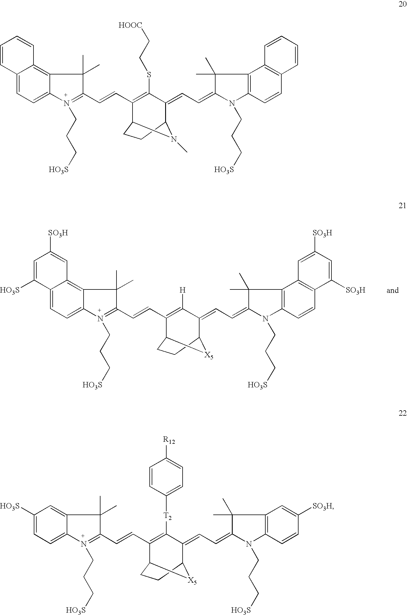


Priority Applications (2)
| Application Number | Priority Date | Filing Date | Title |
|---|---|---|---|
| US12/028,209 US8221721B2 (en) | 2007-02-09 | 2008-02-08 | Polycyclo dyes and use thereof |
| US13/548,451 US9365721B2 (en) | 2007-02-09 | 2012-07-13 | Polycyclo dyes and use thereof |
Applications Claiming Priority (2)
| Application Number | Priority Date | Filing Date | Title |
|---|---|---|---|
| US88906607P | 2007-02-09 | 2007-02-09 | |
| US12/028,209 US8221721B2 (en) | 2007-02-09 | 2008-02-08 | Polycyclo dyes and use thereof |
Related Child Applications (1)
| Application Number | Title | Priority Date | Filing Date |
|---|---|---|---|
| US13/548,451 Continuation US9365721B2 (en) | 2007-02-09 | 2012-07-13 | Polycyclo dyes and use thereof |
Publications (2)
| Publication Number | Publication Date |
|---|---|
| US20080286207A1 true US20080286207A1 (en) | 2008-11-20 |
| US8221721B2 US8221721B2 (en) | 2012-07-17 |
Family
ID=39690741
Family Applications (2)
| Application Number | Title | Priority Date | Filing Date |
|---|---|---|---|
| US12/028,209 Active 2031-01-11 US8221721B2 (en) | 2007-02-09 | 2008-02-08 | Polycyclo dyes and use thereof |
| US13/548,451 Active US9365721B2 (en) | 2007-02-09 | 2012-07-13 | Polycyclo dyes and use thereof |
Family Applications After (1)
| Application Number | Title | Priority Date | Filing Date |
|---|---|---|---|
| US13/548,451 Active US9365721B2 (en) | 2007-02-09 | 2012-07-13 | Polycyclo dyes and use thereof |
Country Status (6)
| Country | Link |
|---|---|
| US (2) | US8221721B2 (en) |
| EP (1) | EP2118206B9 (en) |
| JP (2) | JP5643514B2 (en) |
| DK (1) | DK2118206T3 (en) |
| ES (1) | ES2670852T3 (en) |
| WO (1) | WO2008100817A2 (en) |
Cited By (8)
| Publication number | Priority date | Publication date | Assignee | Title |
|---|---|---|---|---|
| US20080267883A1 (en) * | 2005-09-02 | 2008-10-30 | Milind Rajopadhye | Nicotinic and Picolinic Acid Derived Near-Infrared Fluorophores |
| US20080317676A1 (en) * | 2005-09-02 | 2008-12-25 | Milind Rajopadhye | Biocompatible Fluorescent Imaging Agents |
| US20090123383A1 (en) * | 2000-10-27 | 2009-05-14 | Frangioni John V | Non-Isotopic Detection of Osteoblastic Activity In Vivo Using Modified Bisphosphonates |
| US20090130024A1 (en) * | 2005-09-02 | 2009-05-21 | Narasimhachari Narayanan | Biocompatible N, N-Disubstituted Sulfonamide-Containing Fluorescent Dye Labels |
| US20090220430A1 (en) * | 2008-01-18 | 2009-09-03 | Milind Rajopadhye | Fluorescent Imaging Agents |
| US20100172841A1 (en) * | 2007-03-08 | 2010-07-08 | Peterson Jeffrey D | Viable near-infrared fluorochrome labeled cells and methods of making and using the same |
| WO2012154885A3 (en) * | 2011-05-09 | 2013-04-04 | Visen Medical, Inc. | Carbonic anhydrase targeting agents and methods of using same |
| US9897604B2 (en) | 2013-03-15 | 2018-02-20 | Visen Medical, Inc. | 4,4-disubstituted cyclohexyl bridged heptamethine cyanine dyes and uses thereof |
Families Citing this family (12)
| Publication number | Priority date | Publication date | Assignee | Title |
|---|---|---|---|---|
| US9913917B2 (en) | 2005-12-22 | 2018-03-13 | Visen Medical, Inc. | Biocompatible fluorescent metal oxide nanoparticles |
| DK2118206T3 (en) | 2007-02-09 | 2018-06-18 | Visen Medical Inc | POLYCYCLOF COLORS AND APPLICATION THEREOF |
| ES2684697T3 (en) * | 2009-02-06 | 2018-10-04 | Beth Israel Deaconess Medical Center, Inc. | Agents for obtaining balanced load images |
| CN104245954A (en) | 2012-03-30 | 2014-12-24 | 文森医学公司 | Bacterial imaging agents and methods of using same |
| US20140051975A1 (en) * | 2012-08-15 | 2014-02-20 | Aspect Imaging Ltd. | Multiple heterogeneous imaging systems for clinical and preclinical diagnosis |
| IL221489A0 (en) * | 2012-08-15 | 2012-12-31 | Aspect Imaging Ltd | Imaging system for generating a rendered image |
| CA2882019C (en) | 2012-08-15 | 2021-02-09 | Visen Medical, Inc. | Prostate specific antigen agents and methods of using same for prostate cancer imaging |
| US20140051973A1 (en) * | 2012-08-15 | 2014-02-20 | Aspect Imaging Ltd | Mri imaging system for generating a rendered image |
| WO2014144793A1 (en) | 2013-03-15 | 2014-09-18 | Visen Medical, Inc. | Substituted silaxanthenium red to near-infrared fluorochromes for in vitro and in vivo imaging and detection |
| EP3165153A1 (en) * | 2015-11-05 | 2017-05-10 | Deutsches Krebsforschungszentrum Stiftung des Öffentlichen Rechts | System for fluorescence aided surgery |
| US11549017B2 (en) | 2017-09-29 | 2023-01-10 | Perkinelmer Health Sciences, Inc. | NIR to SWIR fluorescent compounds for imaging and detection |
| CA3094902A1 (en) * | 2018-03-21 | 2019-09-26 | Ecole Polytechnique Federale De Lausanne (Epfl) | Azacyanine dyes and use thereof |
Citations (44)
| Publication number | Priority date | Publication date | Assignee | Title |
|---|---|---|---|---|
| US4219335A (en) * | 1978-09-18 | 1980-08-26 | E. I. Du Pont De Nemours And Company | Immunochemical testing using tagged reagents |
| US5164297A (en) * | 1990-05-03 | 1992-11-17 | Advanced Magnetics Inc. | Solvent mediated relaxation assay system |
| US5445970A (en) * | 1992-03-20 | 1995-08-29 | Abbott Laboratories | Magnetically assisted binding assays using magnetically labeled binding members |
| US5561039A (en) * | 1994-08-17 | 1996-10-01 | Fuji Photo Film Co., Ltd. | Silver halide photographic light-sensitive material |
| US5593658A (en) * | 1992-09-04 | 1997-01-14 | The General Hospital Corporation | Medical compositions |
| US5604088A (en) * | 1993-03-04 | 1997-02-18 | Fuji Photo Film Co., Ltd. | Silver halide photographic material |
| US5876915A (en) * | 1996-07-24 | 1999-03-02 | Agfa-Gevaert | Photothermographic recording material comprising sensitizing dyes and a recording process therefor |
| US6046585A (en) * | 1997-11-21 | 2000-04-04 | Quantum Design, Inc. | Method and apparatus for making quantitative measurements of localized accumulations of target particles having magnetic particles bound thereto |
| US6048982A (en) * | 1986-04-18 | 2000-04-11 | Carnegie Mellon University | Cyanine dyes as labeling reagents for detection of biological and other materials by luminescence methods |
| US6083486A (en) * | 1998-05-14 | 2000-07-04 | The General Hospital Corporation | Intramolecularly-quenched near infrared fluorescent probes |
| US6083485A (en) * | 1994-12-07 | 2000-07-04 | Institut Fur Diagnostikforschung Gmbh | Near infrared radiation in-vivo diagnostic methods and dyes |
| US6086737A (en) * | 1984-03-29 | 2000-07-11 | Li-Cor, Inc. | Sequencing near infrared and infrared fluorescence labeled DNA for detecting using laser diodes and suitable labels therefor |
| US6120351A (en) * | 1998-08-31 | 2000-09-19 | Ingersoll-Rand Company | Automatic machinability measuring and machining methods and apparatus therefor |
| US6136612A (en) * | 1995-10-09 | 2000-10-24 | Sorin Biomedica Cardio S.P.A. | Sulfo benz[E]indocyanine flourescent dyes |
| US6180086B1 (en) * | 2000-01-18 | 2001-01-30 | Mallinckrodt Inc. | Hydrophilic cyanine dyes |
| US6183726B1 (en) * | 2000-01-18 | 2001-02-06 | Mallinckrodt Inc. | Versatile hydrophilic dyes |
| US6395257B1 (en) * | 2000-01-18 | 2002-05-28 | Mallinckrodt Inc. | Dendrimer precursor dyes for imaging |
| US20020064794A1 (en) * | 2000-09-29 | 2002-05-30 | Molecular Probes, Inc. | Modified carbocyanine dyes and their conjugates |
| US6448008B1 (en) * | 1999-07-02 | 2002-09-10 | Innosense, S.R.L. | Fluorescent cyanine labels containing a sulfamido linker arm |
| US6534041B1 (en) * | 1997-04-23 | 2003-03-18 | Institute For Diagnostic Research Gmbh Of The Free University Of Berlin | Acid-labile and enzymatically divisible dye compounds for diagnosis with near infrared light and for therapy |
| US20030124194A1 (en) * | 2002-01-02 | 2003-07-03 | Gaw Debra A. | Amine functionalized superparamagnetic nanoparticles for the synthesis of bioconjugates and uses therefor |
| US6592847B1 (en) * | 1998-05-14 | 2003-07-15 | The General Hospital Corporation | Intramolecularly-quenched near infrared flourescent probes |
| US20030143499A1 (en) * | 1999-03-05 | 2003-07-31 | Nobuaki Kagawa | Sensitizing dye and silver halide photographic material |
| US6615063B1 (en) * | 2000-11-27 | 2003-09-02 | The General Hospital Corporation | Fluorescence-mediated molecular tomography |
| US6740755B2 (en) * | 2000-11-28 | 2004-05-25 | Giuseppe Caputo | Process and method for the preparation of asymmetric monofunctionalized indocyanine labelling reagents and obtained compounds |
| US6747159B2 (en) * | 2001-01-03 | 2004-06-08 | Giuseppe Caputo | Symmetric, monofunctionalised polymethine dyes labelling reagents |
| US20040260072A1 (en) * | 2003-01-24 | 2004-12-23 | Kai Licha | Hydrophilic, thiol-reactive cyanine dyes and conjugates thereof with biomolecules for fluorescence diagnosis |
| US6869593B2 (en) * | 2000-10-27 | 2005-03-22 | Beth Israel Deaconess Medical Center | Non-isotopic detection of osteoblastic activity in vivo using modified bisphosphonates |
| US20050171434A1 (en) * | 2002-01-16 | 2005-08-04 | Visen Medical, Inc. | Chromophore probes for optical imaging |
| US20050169843A1 (en) * | 2001-01-05 | 2005-08-04 | Ralph Weissleder | Activatable imaging probes |
| US6949572B2 (en) * | 2001-06-28 | 2005-09-27 | Pfizer Inc. | Triamide-substituted heterobicyclic compounds |
| US20050214221A1 (en) * | 2002-03-11 | 2005-09-29 | Visen Medical, Inc. | Optical imaging probes |
| US20060280688A1 (en) * | 2000-09-19 | 2006-12-14 | Li-Cor, Inc. | Optical fluorescent imaging |
| US20080226562A1 (en) * | 2005-12-22 | 2008-09-18 | Kevin Groves | Biocompatible fluorescent metal oxide nanoparticles |
| US20080267883A1 (en) * | 2005-09-02 | 2008-10-30 | Milind Rajopadhye | Nicotinic and Picolinic Acid Derived Near-Infrared Fluorophores |
| US20090037114A1 (en) * | 2007-07-30 | 2009-02-05 | Chengbin Peng | 4d+ prestack seismic data structure, and methods and apparatus for processing 4d+ prestack seismic data |
| US20090130024A1 (en) * | 2005-09-02 | 2009-05-21 | Narasimhachari Narayanan | Biocompatible N, N-Disubstituted Sulfonamide-Containing Fluorescent Dye Labels |
| US20090220430A1 (en) * | 2008-01-18 | 2009-09-03 | Milind Rajopadhye | Fluorescent Imaging Agents |
| US20100166659A1 (en) * | 2005-01-07 | 2010-07-01 | Kai Licha | Use of cyanine dyes for the diagnosis of proliferative diseases |
| US20100172841A1 (en) * | 2007-03-08 | 2010-07-08 | Peterson Jeffrey D | Viable near-infrared fluorochrome labeled cells and methods of making and using the same |
| US20100189657A1 (en) * | 2006-03-20 | 2010-07-29 | The General Hospital Corporation | Intramolecularly quenched fluorochrome conjugates and methods of use |
| US20100268070A1 (en) * | 2008-11-26 | 2010-10-21 | Visen Medical, Inc. | Methods and Compositions for Identifying Subjects at Risk of Developing Stent Thrombosis |
| US7947256B2 (en) * | 2005-09-02 | 2011-05-24 | Visen Medical, Inc. | Biocompatible fluorescent imaging agents |
| US20110165075A1 (en) * | 2008-03-14 | 2011-07-07 | Milind Rajopadhye | Integrin targeting agents and in-vivo and in-vitro imaging methods using the same |
Family Cites Families (11)
| Publication number | Priority date | Publication date | Assignee | Title |
|---|---|---|---|---|
| JP3794793B2 (en) * | 1996-07-24 | 2006-07-12 | アグフア−ゲヴエルト,ナームローゼ・フエンノートシヤツプ | Photothermographic recording material containing sensitizing dye and recording method therefor |
| US6214533B1 (en) | 1998-04-10 | 2001-04-10 | Konica Corporation | Thermally developable photosensitive material |
| DE60116510T2 (en) * | 2000-09-19 | 2006-07-13 | Li-Cor, Inc., Lincoln | cyanine |
| WO2003102558A1 (en) | 2002-06-04 | 2003-12-11 | Visen Medical, Inc. | Imaging volumes with arbitrary geometries in contact and non-contact tomography |
| AU2003303954A1 (en) | 2002-10-25 | 2004-10-11 | Emory University | Multifunctional magnetic nanoparticle probes for intracellular molecular imaging and monitoring |
| EP1462854A1 (en) | 2003-03-24 | 2004-09-29 | Konica Minolta Medical & Graphic Inc. | Photothermographic imaging material |
| US20080102036A1 (en) | 2003-06-04 | 2008-05-01 | Poss Kirtland G | Biocompatible Fluorescent Silicon Nanoparticles |
| US7332257B2 (en) | 2003-07-11 | 2008-02-19 | Asahi Glass Company, Limited | Composition for optical film, and optical film |
| WO2005017539A2 (en) | 2003-08-14 | 2005-02-24 | The General Hospital Corporation | Imaging pathology |
| US7658070B2 (en) | 2004-09-21 | 2010-02-09 | Drs Sustainment Systems, Inc. | Method and apparatus for improving the energy conversion efficiency of electrical power generators |
| DK2118206T3 (en) | 2007-02-09 | 2018-06-18 | Visen Medical Inc | POLYCYCLOF COLORS AND APPLICATION THEREOF |
-
2008
- 2008-02-08 DK DK08729371.8T patent/DK2118206T3/en active
- 2008-02-08 US US12/028,209 patent/US8221721B2/en active Active
- 2008-02-08 ES ES08729371.8T patent/ES2670852T3/en active Active
- 2008-02-08 EP EP08729371.8A patent/EP2118206B9/en active Active
- 2008-02-08 JP JP2009549253A patent/JP5643514B2/en active Active
- 2008-02-08 WO PCT/US2008/053399 patent/WO2008100817A2/en active Application Filing
-
2012
- 2012-07-13 US US13/548,451 patent/US9365721B2/en active Active
-
2014
- 2014-03-31 JP JP2014071365A patent/JP2014167119A/en not_active Withdrawn
Patent Citations (58)
| Publication number | Priority date | Publication date | Assignee | Title |
|---|---|---|---|---|
| US4219335A (en) * | 1978-09-18 | 1980-08-26 | E. I. Du Pont De Nemours And Company | Immunochemical testing using tagged reagents |
| US6086737A (en) * | 1984-03-29 | 2000-07-11 | Li-Cor, Inc. | Sequencing near infrared and infrared fluorescence labeled DNA for detecting using laser diodes and suitable labels therefor |
| US6048982A (en) * | 1986-04-18 | 2000-04-11 | Carnegie Mellon University | Cyanine dyes as labeling reagents for detection of biological and other materials by luminescence methods |
| US5164297A (en) * | 1990-05-03 | 1992-11-17 | Advanced Magnetics Inc. | Solvent mediated relaxation assay system |
| US5445970A (en) * | 1992-03-20 | 1995-08-29 | Abbott Laboratories | Magnetically assisted binding assays using magnetically labeled binding members |
| US5593658A (en) * | 1992-09-04 | 1997-01-14 | The General Hospital Corporation | Medical compositions |
| US5604088A (en) * | 1993-03-04 | 1997-02-18 | Fuji Photo Film Co., Ltd. | Silver halide photographic material |
| US5561039A (en) * | 1994-08-17 | 1996-10-01 | Fuji Photo Film Co., Ltd. | Silver halide photographic light-sensitive material |
| US6083485A (en) * | 1994-12-07 | 2000-07-04 | Institut Fur Diagnostikforschung Gmbh | Near infrared radiation in-vivo diagnostic methods and dyes |
| US7025949B2 (en) * | 1994-12-07 | 2006-04-11 | Institut Fur Diagnostikforschung Gmbh An Der Freien Universitat Berlin | In-vivo diagnostic method by means of near infrared radiation |
| US6926885B2 (en) * | 1994-12-07 | 2005-08-09 | Institut Fur Diagnostikforschung Gmbh | In-vivo diagnostic method by means of near infrared radiation |
| US7445767B2 (en) * | 1994-12-07 | 2008-11-04 | Institut Fur Diagnostikforschung Gmbh An Der Freien Universitat Berlin | In-vivo diagnostic method by near infrared radiation |
| US6913743B2 (en) * | 1994-12-07 | 2005-07-05 | Institut Fur Diagnostikforschung Gmbh An Der Freien Universitat Berlin | Near infrared imaging agent |
| US6258340B1 (en) * | 1994-12-07 | 2001-07-10 | Institut Fur Diagnostikforschung Gmbh | In-vivo diagnostic method by near infrared radiation |
| US7655217B2 (en) * | 1994-12-07 | 2010-02-02 | Institut fur Diagnosikforschung GmbH | Near infrared imaging agent |
| US20100129293A1 (en) * | 1994-12-07 | 2010-05-27 | Institut Fur Diagnostikforschung Gmbh An Der Freien Universitat Berlin | Near Infrared Imaging Agent |
| US6136612A (en) * | 1995-10-09 | 2000-10-24 | Sorin Biomedica Cardio S.P.A. | Sulfo benz[E]indocyanine flourescent dyes |
| US5876915A (en) * | 1996-07-24 | 1999-03-02 | Agfa-Gevaert | Photothermographic recording material comprising sensitizing dyes and a recording process therefor |
| US6534041B1 (en) * | 1997-04-23 | 2003-03-18 | Institute For Diagnostic Research Gmbh Of The Free University Of Berlin | Acid-labile and enzymatically divisible dye compounds for diagnosis with near infrared light and for therapy |
| US6046585A (en) * | 1997-11-21 | 2000-04-04 | Quantum Design, Inc. | Method and apparatus for making quantitative measurements of localized accumulations of target particles having magnetic particles bound thereto |
| US6275031B1 (en) * | 1997-11-21 | 2001-08-14 | Quantum Design, Inc. | Method and apparatus for making quantitative measurements of localized accumulations of magnetic particles |
| US6083486A (en) * | 1998-05-14 | 2000-07-04 | The General Hospital Corporation | Intramolecularly-quenched near infrared fluorescent probes |
| US6592847B1 (en) * | 1998-05-14 | 2003-07-15 | The General Hospital Corporation | Intramolecularly-quenched near infrared flourescent probes |
| US20060275775A1 (en) * | 1998-05-14 | 2006-12-07 | The General Hospital Corporation | Intramolecularly-quenched near infrared fluorescent probes |
| US6120351A (en) * | 1998-08-31 | 2000-09-19 | Ingersoll-Rand Company | Automatic machinability measuring and machining methods and apparatus therefor |
| US20030143499A1 (en) * | 1999-03-05 | 2003-07-31 | Nobuaki Kagawa | Sensitizing dye and silver halide photographic material |
| US6448008B1 (en) * | 1999-07-02 | 2002-09-10 | Innosense, S.R.L. | Fluorescent cyanine labels containing a sulfamido linker arm |
| US6180086B1 (en) * | 2000-01-18 | 2001-01-30 | Mallinckrodt Inc. | Hydrophilic cyanine dyes |
| US6395257B1 (en) * | 2000-01-18 | 2002-05-28 | Mallinckrodt Inc. | Dendrimer precursor dyes for imaging |
| US6183726B1 (en) * | 2000-01-18 | 2001-02-06 | Mallinckrodt Inc. | Versatile hydrophilic dyes |
| US20060280688A1 (en) * | 2000-09-19 | 2006-12-14 | Li-Cor, Inc. | Optical fluorescent imaging |
| US20020064794A1 (en) * | 2000-09-29 | 2002-05-30 | Molecular Probes, Inc. | Modified carbocyanine dyes and their conjugates |
| US6869593B2 (en) * | 2000-10-27 | 2005-03-22 | Beth Israel Deaconess Medical Center | Non-isotopic detection of osteoblastic activity in vivo using modified bisphosphonates |
| US7374746B2 (en) * | 2000-10-27 | 2008-05-20 | Beth Israel Deaconees Medical Center | Non-isotopic detection of osteoblastic activity in vivo using modified bisphosphonates |
| US6615063B1 (en) * | 2000-11-27 | 2003-09-02 | The General Hospital Corporation | Fluorescence-mediated molecular tomography |
| US6740755B2 (en) * | 2000-11-28 | 2004-05-25 | Giuseppe Caputo | Process and method for the preparation of asymmetric monofunctionalized indocyanine labelling reagents and obtained compounds |
| US6747159B2 (en) * | 2001-01-03 | 2004-06-08 | Giuseppe Caputo | Symmetric, monofunctionalised polymethine dyes labelling reagents |
| US20050169843A1 (en) * | 2001-01-05 | 2005-08-04 | Ralph Weissleder | Activatable imaging probes |
| US20110152501A1 (en) * | 2001-01-05 | 2011-06-23 | The General Hospital Corporation | Activatable Imaging Probes |
| US6949572B2 (en) * | 2001-06-28 | 2005-09-27 | Pfizer Inc. | Triamide-substituted heterobicyclic compounds |
| US20030124194A1 (en) * | 2002-01-02 | 2003-07-03 | Gaw Debra A. | Amine functionalized superparamagnetic nanoparticles for the synthesis of bioconjugates and uses therefor |
| US20090068115A1 (en) * | 2002-01-02 | 2009-03-12 | Visen Medical, Inc. | Amine Functionalized Superparamagnetic Nanoparticles for the Synthesis of Bioconjugates and Uses Therefor |
| US20050171434A1 (en) * | 2002-01-16 | 2005-08-04 | Visen Medical, Inc. | Chromophore probes for optical imaging |
| US20100074847A1 (en) * | 2002-01-16 | 2010-03-25 | Madden Karen N | Chromophore Probes for Optical Imaging |
| US20050214221A1 (en) * | 2002-03-11 | 2005-09-29 | Visen Medical, Inc. | Optical imaging probes |
| US20110171136A1 (en) * | 2002-03-11 | 2011-07-14 | Poss Kirtland G | Optical imaging probes |
| US20040260072A1 (en) * | 2003-01-24 | 2004-12-23 | Kai Licha | Hydrophilic, thiol-reactive cyanine dyes and conjugates thereof with biomolecules for fluorescence diagnosis |
| US20100166659A1 (en) * | 2005-01-07 | 2010-07-01 | Kai Licha | Use of cyanine dyes for the diagnosis of proliferative diseases |
| US20090130024A1 (en) * | 2005-09-02 | 2009-05-21 | Narasimhachari Narayanan | Biocompatible N, N-Disubstituted Sulfonamide-Containing Fluorescent Dye Labels |
| US7947256B2 (en) * | 2005-09-02 | 2011-05-24 | Visen Medical, Inc. | Biocompatible fluorescent imaging agents |
| US20080267883A1 (en) * | 2005-09-02 | 2008-10-30 | Milind Rajopadhye | Nicotinic and Picolinic Acid Derived Near-Infrared Fluorophores |
| US20080226562A1 (en) * | 2005-12-22 | 2008-09-18 | Kevin Groves | Biocompatible fluorescent metal oxide nanoparticles |
| US20100189657A1 (en) * | 2006-03-20 | 2010-07-29 | The General Hospital Corporation | Intramolecularly quenched fluorochrome conjugates and methods of use |
| US20100172841A1 (en) * | 2007-03-08 | 2010-07-08 | Peterson Jeffrey D | Viable near-infrared fluorochrome labeled cells and methods of making and using the same |
| US20090037114A1 (en) * | 2007-07-30 | 2009-02-05 | Chengbin Peng | 4d+ prestack seismic data structure, and methods and apparatus for processing 4d+ prestack seismic data |
| US20090220430A1 (en) * | 2008-01-18 | 2009-09-03 | Milind Rajopadhye | Fluorescent Imaging Agents |
| US20110165075A1 (en) * | 2008-03-14 | 2011-07-07 | Milind Rajopadhye | Integrin targeting agents and in-vivo and in-vitro imaging methods using the same |
| US20100268070A1 (en) * | 2008-11-26 | 2010-10-21 | Visen Medical, Inc. | Methods and Compositions for Identifying Subjects at Risk of Developing Stent Thrombosis |
Cited By (22)
| Publication number | Priority date | Publication date | Assignee | Title |
|---|---|---|---|---|
| US20090123383A1 (en) * | 2000-10-27 | 2009-05-14 | Frangioni John V | Non-Isotopic Detection of Osteoblastic Activity In Vivo Using Modified Bisphosphonates |
| US8771646B2 (en) | 2005-09-02 | 2014-07-08 | Visen Medical, Inc. | Nicotinic acid and picolinic acid derived near-infrared fluorophores |
| US20080317676A1 (en) * | 2005-09-02 | 2008-12-25 | Milind Rajopadhye | Biocompatible Fluorescent Imaging Agents |
| US20090130024A1 (en) * | 2005-09-02 | 2009-05-21 | Narasimhachari Narayanan | Biocompatible N, N-Disubstituted Sulfonamide-Containing Fluorescent Dye Labels |
| US10442930B2 (en) | 2005-09-02 | 2019-10-15 | Visen Medical, Inc. | Biocompatible N, N-disubstituted sulfonamide-containing fluorescent dye labels |
| US20080267883A1 (en) * | 2005-09-02 | 2008-10-30 | Milind Rajopadhye | Nicotinic and Picolinic Acid Derived Near-Infrared Fluorophores |
| US7947256B2 (en) | 2005-09-02 | 2011-05-24 | Visen Medical, Inc. | Biocompatible fluorescent imaging agents |
| US8173819B2 (en) | 2005-09-02 | 2012-05-08 | Visen Medical, Inc. | Nicotinic and picolinic acid derived near-infrared fluorophores |
| US9574085B2 (en) | 2005-09-02 | 2017-02-21 | Visen Medical, Inc. | Biocompatible N, N-disubstituted sulfonamide-containing fluorescent dye labels |
| US8455651B2 (en) | 2005-09-02 | 2013-06-04 | Visen Medical, Inc. | Nicotinic acid and picolinic acid derived near-infrared fluorophores |
| US20100172841A1 (en) * | 2007-03-08 | 2010-07-08 | Peterson Jeffrey D | Viable near-infrared fluorochrome labeled cells and methods of making and using the same |
| US8815214B2 (en) | 2008-01-18 | 2014-08-26 | Visen Medical, Inc. | Fluorescent imaging agents |
| US9427481B2 (en) | 2008-01-18 | 2016-08-30 | Visen Medical, Inc. | Fluorescent imaging agents |
| US9999687B2 (en) | 2008-01-18 | 2018-06-19 | Visen Medical, Inc. | Fluorescent imaging agents |
| US20090220430A1 (en) * | 2008-01-18 | 2009-09-03 | Milind Rajopadhye | Fluorescent Imaging Agents |
| WO2012154885A3 (en) * | 2011-05-09 | 2013-04-04 | Visen Medical, Inc. | Carbonic anhydrase targeting agents and methods of using same |
| US10221159B2 (en) | 2011-05-09 | 2019-03-05 | Visen Medical, Inc. | Carbonic anhydrase targeting agents and methods of using same |
| CN110204478A (en) * | 2011-05-09 | 2019-09-06 | 文森医学公司 | Carbonic anhydrase targeting agent and its application method |
| US11059802B2 (en) | 2011-05-09 | 2021-07-13 | Visen Medical, Inc. | Carbonic anhydrase targeting agents and methods of using same |
| US9897604B2 (en) | 2013-03-15 | 2018-02-20 | Visen Medical, Inc. | 4,4-disubstituted cyclohexyl bridged heptamethine cyanine dyes and uses thereof |
| US10473658B2 (en) | 2013-03-15 | 2019-11-12 | Visen Medical, Inc. | 4,4-disubstituted cyclohexyl bridged heptamethine cyanine dyes and uses thereof |
| US11193932B2 (en) | 2013-03-15 | 2021-12-07 | Visen Medical, Inc. | 4,4-disubstituted cyclohexyl bridged heptamethine cyanine dyes and uses thereof |
Also Published As
| Publication number | Publication date |
|---|---|
| US9365721B2 (en) | 2016-06-14 |
| WO2008100817A3 (en) | 2009-04-09 |
| US8221721B2 (en) | 2012-07-17 |
| JP2014167119A (en) | 2014-09-11 |
| EP2118206A2 (en) | 2009-11-18 |
| DK2118206T3 (en) | 2018-06-18 |
| EP2118206B9 (en) | 2018-08-29 |
| JP2010518236A (en) | 2010-05-27 |
| EP2118206B1 (en) | 2018-04-11 |
| WO2008100817A2 (en) | 2008-08-21 |
| ES2670852T3 (en) | 2018-06-01 |
| US20130137873A1 (en) | 2013-05-30 |
| JP5643514B2 (en) | 2014-12-17 |
Similar Documents
| Publication | Publication Date | Title |
|---|---|---|
| US11193932B2 (en) | 4,4-disubstituted cyclohexyl bridged heptamethine cyanine dyes and uses thereof | |
| US9365721B2 (en) | Polycyclo dyes and use thereof | |
| US9649389B2 (en) | Substituted silaxanthenium red to near-infrared fluorochromes for in vitro and in vivo imaging and detection | |
| US8173819B2 (en) | Nicotinic and picolinic acid derived near-infrared fluorophores | |
| JP5106397B2 (en) | Biocompatible N, N-disubstituted sulfonamide-containing fluorescent dye label |
Legal Events
| Date | Code | Title | Description |
|---|---|---|---|
| AS | Assignment |
Owner name: VISEN MEDICAL, INC., MASSACHUSETTS Free format text: ASSIGNMENT OF ASSIGNORS INTEREST;ASSIGNOR:NARAYANAN, NARA;REEL/FRAME:020778/0225 Effective date: 20080407 |
|
| STCF | Information on status: patent grant |
Free format text: PATENTED CASE |
|
| CC | Certificate of correction | ||
| FPAY | Fee payment |
Year of fee payment: 4 |
|
| MAFP | Maintenance fee payment |
Free format text: PAYMENT OF MAINTENANCE FEE, 8TH YEAR, LARGE ENTITY (ORIGINAL EVENT CODE: M1552); ENTITY STATUS OF PATENT OWNER: LARGE ENTITY Year of fee payment: 8 |
|
| FEPP | Fee payment procedure |
Free format text: MAINTENANCE FEE REMINDER MAILED (ORIGINAL EVENT CODE: REM.); ENTITY STATUS OF PATENT OWNER: LARGE ENTITY |