CROSS-REFERENCE TO RELATED APPLICATIONS
-
The present application is a continuation-in-part of and claims priority to co-pending U.S. patent application Ser. No. 11/969,040, filed Jan. 3, 2008 (the '040 application). The '040 application, in turn, is a continuation-in-part of and claims priority to co-pending U.S. patent application Ser. No. 11/893,171, filed Aug. 14, 2007 (the '171 application). The '171 application claims priority under 35 U.S.C. 119(e) to U.S. Provisional patent application Ser. No. 60/837,868, filed on Aug. 14, 2006 (the '868 application), and to U.S. Provisional patent application Ser. No. 60/837,869, filed on Aug. 14, 2006 (the '869 application). The entire contents of each of these prior applications are incorporated herein by reference.
-
The present application is a continuation-in-part of and claims priority to co-pending U.S. patent application Ser. No. 12/301,126, filed Nov. 17, 2008 (the '126 application), which claims priority under 35 U.S.C. § 371 to PCT application PCT/U.S.07/18160 (the '160 application), filed Aug. 14, 2007, which claims priority to the '868 and '869 applications. The entire contents of each of these prior applications are incorporated herein by reference.
-
The present application also claims priority to co-pending U.S. Provisional patent application Ser. No. 61/018,783, filed Jan. 3, 2008, the entire contents of which are incorporated herein by reference.
GOVERNMENT SUPPORT
-
This invention was made with United States government support awarded by the National Institute of General Medical Sciences (glue grant contract number U54 GM62116) and by the National Institutes of Health (contract number GM57073). The United States Government has certain rights in the invention.
BACKGROUND OF THE INVENTION
-
Influenza has a long history of pandemics, epidemics, resurgences and outbreaks. Avian influenza, including the H5N1 strain, is a highly contagious and potentially fatal pathogen, but it currently has only a limited ability to infect humans. However, avian flu viruses have historically observed to accumulate mutations that alter its host specificity and allow it to readily infect humans. In fact, two of the major flu pandemics of the last century originated from avian flu viruses that changed their genetic makeup to allow for human infection.
-
There is a significant concern that the current H5N1, H7N7, H9N2 and H2N2 avian influenza strains might accumulate mutations that alter their host specificity and allow them to readily infect humans. Therefore, there is a need to assess whether the HA protein in these strains can, in fact, convert to a form that can readily infect humans, and a further need to identify HA variants with such ability. There is a further need to understand the characteristics of HA proteins generally that allow or prohibit infection of different subjects, particularly humans. There is also a need for vaccines and therapeutic strategies for effective treatment or delay of onset of disease caused by influenza virus.
SUMMARY OF THE INVENTION
-
The present invention binding agents with particular glycan binding characteristics. In particular, the present invention provides binding agents that bind to sialylated glycans having an umbrella-like topology. In certain embodiments, inventive binding agents bind to umbrella-topology glycans with high affinity and/or specificity. In some embodiments, inventive binding agents show a binding preference for umbrella-topology glycans as compared with cone-topology glycans. In some embodiments, inventive binding agents compete with hemagglutinin for binding to glycans on hemagglutinin receptors. In some embodiments, inventive binding agents compete with hemagglutinin for binding to umbrella-topology glycans.
-
The present invention also provides diagnostic and therapeutic reagents and methods associated with provided binding agents, including vaccines.
BRIEF DESCRIPTION OF THE DRAWING
-
FIG. 1. Alignment of exemplary sequences of wild type HA. Sequences were obtained from the NCBI influenza virus sequence database (http://www.ncbi.nlm.nih.gov/genomes/FLU/FLU.html)
-
FIG. 2. Sequence alignment of HA glycan binding domain. Gray: conserved amino acids involved in binding to sialic acid. Red: particular amino acids involved in binding to Neu5Acα2-3/6Gal motifs. Yellow: amino acids that influence positioning of Q226 (137, 138) and E190 (186, 228). Green: amino acids involved in binding to other monosaccharides (or modifications) attached to Neu5Acα2-3/6Gal motif. The sequence for ASI30, APR34, ADU63, ADS97 and Viet04 were obtained from their respective crystal structures. The other sequences were obtained from SwissProt (http://us.expasy.org). Abbreviations: ADA76, A/duck/Alberta/35/76 (H1N1); ASI30, A/Swine/Iowa/30 (H1N1); APR34, A/Puerto Rico/8/34 (H1N1); ASC18, A/South Carolina/1/18 (H1N1), AT91, A/Texas/36/91 (H1N1); ANY18, A/New York/1/18 (H1N1); ADU63, A/Duck/Ukraine/1/63 (H3N8); AAI68, A/Aichi/2/68 (H3N2); AM99, A/Moscow/10/99 (H3N2); ADS97, A/Duck/Singapore/3/97 (H5N3); Viet04, A/Vietnam/1203/2004 (H5N1).
-
FIG. 3. Sequence alignment illustrating conserved subsequences characteristic of H1 HA.
-
FIG. 4. Sequence alignment illustrating conserved subsequences characteristic of H3 HA.
-
FIG. 5. Sequence alignment illustrating conserved subsequences characteristic of H5 HA.
-
FIG. 6. Framework for understanding glycan receptor specificity. α2-3- and/or α2-6-linked glycans can adopt different topologies. According to the present invention, the ability of an HA polypeptide to bind to certain of these topologies confers upon it the ability to mediate infection of different hosts, for example, humans. As illustrated in Panel A of this figure, the present invention defines two particularly relevant topologies, a “cone” topology and an “umbrella” topology. The cone topology can be adopted by α2-3- and/or α2-6-linked glycans, and is typical of short oligosaccharides or branched oligosaccharides attached to a core (although this topology can be adopted by certain long oligosaccharides). The umbrella topology can only be adopted by α2-6-linked glycans (presumably due to the increased conformational plurality afforded by the extra C5-C6 bond that is present in the α2-6 linkage), and is predominantly adopted by long oligosaccharides or branched glycans with long oligosaccharide branches, particularly containing the motif Neu5Acα2-6Galβ1-3/4GlcNAc-. As described herein, ability of HA polypeptides to bind the umbrella glycan topology, confers binding to human receptors and/or ability to mediate infection of humans. Panel B of this Figure specifically shows the topology of α2-3 and α2-6 as governed by the glycosidic torsion angles of the trisaccharide motifs—Neu5Acα2-3Galβ1-3/4GlcNAc and Neu5Acα2-6Galβ1-4GlcNAc respectively. A parameter (θ)—angle between C2 atom of Neu5Ac and C1 atoms of the subsequent Gal and GlcNAc sugars in these trisaccharide motifs was defined to characterize the topology. Superimposition of the θ contour and the conformational maps of the α2-3 and α2-6 motifs shows that α2-3 motifs adopt 100% cone-like topology and α2-6 motifs sampled both cone-like and umbrella-like topologies (Panel C). In the cone-like topology sampled by α2-3 and α2-6, GlcNAc and subsequent sugars are positioned along a region spanning a cone. Interactions of HA with cone-like topology primarily involve contacts of amino acids at the numbered positions (based on H3 HA numbering) with Neu5Ac and Gal sugars. On the other hand, in umbrella-like topology, which is unique to α2-6, GlcNAc and subsequent sugars bend towards the HA binding site (as observed in HA-α2-6 co-crystal structures). Longer α2-6 oligosaccharides (e.g. at least a tetrasaccharide) would favor this conformation since it is stabilized by intra-sugar van der Waals contact between acetyl groups of GlcNAc and Neu5Ac. HA interactions with umbrella-like topology involve contacts of amino acids at the numbered positions (based on H3 HA numbering) with GlcNAc and subsequent sugars in addition to contacts with Neu5Ac and Gal sugars. Panel C of this Figure depicts conformational sampling of cone- and umbrella-like topology by α2-3 and α2-6. Sections (A)-(D) show the conformational (φ, ψ) maps of Neu5Acα2-3Gal, Neu5Acα2-6Gal, Galβ1-3GlcNAc, and Galβ1-4GlcNAc linkages, respectively. These maps obtained from GlycoMaps DB (http://www.glycosciences.de/modeling/glycomapsdb/) were generated using ab initio MD simulations using MM3 force field. Energy distribution is color coded starting from red (representing highest energy) to green representing lowest energy. Encircled regions 1-5 represent (φ,ψ) values observed for the α2-3 and α2-6 oligosaccharides in the HA-glycan co-crystal structures. The trans conformation (encircled region 1) of Neu5Acα2-3Gal predominates in HA binding pocket with the exception of the co-crystal structure of A/Aichi/2/68H3N2 HA with α2-3 where this conformation is gauche (encircled region 2). On the other hand, the cis conformation of Neu5Acα2-6Gal (encircled region 3) predominates in HA binding pocket. The cone-like topology is sampled by encircled regions 1 and 2 and the umbrella-like topology is sampled by encircled region 3. Sections (E)-(F) show sampling of cone-like and umbrella-like topologies by α2-3 and α2-6 motifs, respectively. Regions marked in red in the conformational maps were used as the outer boundaries to calculate the θ parameter (angle between C2 atom of Neu5Ac and C1 atoms of subsequent Gal and GlcNAc sugars) for a given set of (φ,ψ) values. Based on the energy cutoff, the value of θ>110° was used to characterize cone-like topology and θ<100° was used to characterize umbrella-like topology. Superimposition of the θ contour with the conformational energy map indicated that α2-3 motif adopts 100% cone-like topology since it was energetically unfavorable to adopt umbrella-like topology. On the other hand, the α2-6 motif sampled both the cone-like and umbrella-like topologies and this sampling was classified based on the ω angle (O-C6-C5-H5) of Neu5Acα2-6Gal linkage.
-
FIG. 7. Interactions of HA residues with cone vs umbrella glycan topologies. Analysis of HA-glycan co-crystals reveals that the position of Neu5Ac relative to the HA binding site is almost invariant. Contacts with Neu5Ac involve highly conserved residues such as F98, S/T136, W153, H183 and L/1194. Contacts with other sugars involve different residues, depending on whether the sugar linkage is α2-3 or α2-6 and whether the glycan topology is cone or umbrella. For example, in the cone topology, the primary contacts are with Neu5Ac and with Gal sugars. E190 and Q226 play particularly important roles in this binding. This Figure also illustrates other positions (e.g., 137, 145, 186, 187, 193, 222) that can participate in binding to cone structures. In some cases, different residues can make different contacts with different glycan structures. The type of amino acid in these positions can influence ability of an HA polypeptide to bind to receptors with different modification and/or branching patterns in the glycan structures. In the umbrella topology, contacts are made with sugars beyond Neu5Ac and Gal. This Figure illustrates residues (e.g., 137, 145, 156, 159, 186, 187, 189, 190, 192, 193, 196, 222, 225, 226) that can participate in binding to umbrella structures. In some cases, different residues can make different contacts with different glycan structures. The type of amino acid in these positions can influence ability of an HA polypeptide to bind to receptors with different modification and/or branching patterns in the glycan structures. In some embodiments, a D residue at position 190 and/or a D residue at position 225 contribute(s) to binding to umbrella topologies.
-
FIG. 8. Exemplary cone topologies. This Figure illustrates certain exemplary (but not exhaustive) glycan structures that adopt cone topologies.
-
FIG. 9. Exemplary umbrella topologies. (A) Certain exemplary (but not exhaustive) N- and O-linked glycan structures that can adopt umbrella topologies. (B) Certain exemplary (but not exhaustive) O-linked glycan structures that can adopt umbrella topologies.
-
FIG. 10. Glycan profile of human bronchial epithelial cells and human colonic epithelial cells. To further investigate the glycan diversity in the upper respiratory tissues, N-linked glycans were isolated from HBEs (a representative upper respiratory cell line) and analyzed using MALDI-MS. The predominant expression of a2-6 in HBEs was confirmed by pre-treating the sample with Sialidase S (a2-3 specific) and Sialidase A (cleaves and SA). The predominant expression of glycans with long branch topology is supported by TOF-TOF fragmentation analysis of representative mass peaks (highlighted in cyan). To provide a reference for glycan diversity in the upper respiratory tissues, the N-linked glycan profile of human colonic epithelial cells (HT29; a representative gut cell line) was obtained. This cell line was chosen because the current H5N1 viruses have been shown to infect gut cells. Sialidase A and S pre-treatment controls showed predominant expression of a2-3 glycans (highlighted in red) in the HT-29 cells. Moreover, the long branch glycan topology is not as prevalent as observed for HBEs. Therefore, human adaptation of the H5N1 HA would involve HA mutations that would enable high affinity binding to the diverse glycans expressed in the human upper respiratory tissues (e.g., umbrella glycans).
-
FIG. 11. Data mining platform. Shown in (A) are the main components of the data mining platform. The features are derived from the data objects which are extracted from the database. The features are prepared into datasets that are used by the classification methods to derive patterns or rules (B), shows the key software modules that enable the user to apply the data mining process to the glycan array data.
-
FIG. 12. Features used in data mining analysis. This figure shows the features defined herein as representative motifs that illustrate the different types of pairs, triplets and quadruplets abstracted from the glycans on the glycan microarray. The rationale behind choosing these features is based on the binding of di-tetra saccharides to the glycan binding site of HA. The final dataset comprise features from the glycans as well as the binding signals for each of the HAs screened on the array. Among the different methods for classification, the rule induction classification method was utilized. One of the main advantages of this method is that it generates IF-THEN rules which can be interpreted more easily when compared to the other statistical or mathematical methods. The two main objectives of the classification were: (1) identifying features present on a set of high affinity glycan ligands, which enhance binding, and (2) identifying features that are in the low affinity glycan ligands that are not favorable for binding.
-
FIG. 13. Classifiers used in data mining analysis. This figure presents a table of classifier ids and rules.
-
FIG. 14. Conformational map and solvent accessibility of Neu5Acα2-3Gal and Neu5Acα2-6Gal motifs. Panel A shows the conformational map of Neu5Acα2-3Gal linkage. The encircled region 2 is the trans conformation observed in the APR34_H1—23, ADU63_H3—23 and ADS97_H5—23 co-crystal structures. The encircled region 1 is the conformation observed in the AAI68_H3—23 co-crystal structure. Panel B shows the conformational map of Neu5Acα2-6Gal where the cis-conformation (encircled region 3) is observed in all the HA-α2-6 sialylated glycan co-crystal structures. Panel C shows difference between solvent accessible surface area (SASA) of Neu5Ac α2-3 and α2-6 sialylated oligosaccharides in the respective HA-glycan co-crystal structures. The red and cyan bars respectively indicate that Neu5Ac in α2-6 (positive value) or α2-3 (negative value) sialylated glycans makes more contact with glycan binding site. Panel D shows difference between SASA of NeuAc in α2-3 sialylated glycans bound to swine and human H1 (H1α2-3), avian and human H3 (H3α2-3), and of NeuAc in α2-6 sialylated glycans bound to swine and human H1 (H1α2-6). The negative bar in cyan for H3α2-3 indicates lesser contact of the human H3 HA with Neu5Acα2-3Gal compared to that of avian H3. Torsion angles—φ: C2-C1-O-C3 (for Neu5Acα2-3/6 linkage); ψ: C1-O-C3-H3 (for Neu5Acα2-3Gal) or C1-O-C6-C5 (for Neu5Acα2-6Gal); ω: O-C6-C5-H5 (for Neu5Acα2-6Gal) linkages. The φ, ψ maps were obtained from GlycoMaps DB (http://www.glycosciences.de/modeling/glycomapsdb/) which was developed by Dr. Martin Frank and Dr. Claus-Wilhelm von der Lieth (German Cancer Research Institute, Heidelberg, Germany). The coloring scheme from high energy to low energy is from bright red to bright green, respectively.
-
FIG. 15. Residues involved in binding of H1, H3 and H5 HA to α2-3/6 sialylated glycans. Panels A-D show the difference (A in the abscissa) in solvent accessible surface area (SASA) of residues interacting with α2-3 and α2-6 sialylated glycans, respectively, in ASI30_H1, APR34_H1, ADU63_H3 and ADS97_H5 co-crystal structures. Green bars correspond to residues that directly interact with the glycan and light orange bars correspond to residues proximal to Glu/Asp 190 and Gln/Leu226. Positive value of A for the green bars indicates more contact of that residue with α2-6 sialylated glycans whereas a negative value of A indicates more contact with α2-3 sialylated glycans. Panel E summarizes in tabular form the residues involved in binding to α2-3/6 sialylated glycans in H1, H3 and H5 HA. Certain key residues involved in binding to α2-3 sialylated glycans are colored blue and certain key residues involved in binding to α2-6 sialylated glycans are colored red.
-
FIG. 16. Binding of Viet04_H5 HA to biantennary α2-6 sialylated glycan (cone topology). Stereo view of surface rendered Viet04_H5 glycan binding site with Neu5Acα2-6Gal linkage in the extended conformation (obtained from the pertussis toxin co-crystal structure; PDB ID: 1PTO). Lys193 (orange) does not have any contacts with the glycan in this conformation. The additional amino acids potentially involved in binding to the glycan in this conformation are Asn186, Lys222 and Ser227. However, certain contacts observed in the HA binding to the α2-6 sialylated oligosaccharide in the cis-conformation are absent in the extended conformation. Without wishing to be bound by any particular theory, we note that this suggests that the extended conformation may not bind to HA as optimally as the cis-conformation. The structures of branched N-linked glycans where the Neu5Acα2-6Galβ1-4GlcNAcb branch was attached to the Manα1-3Man (PDB ID: 1LGC) and Manα1-6Man (PDB ID: 1ZAG) were superimposed on to the Neu5Acα2-6Gal linkage in the Viet04_H5 HA binding site for both the cis and the extended conformation of this linkage. The superimposition shows that the structure with Neu5Acα2-6Galβ1-4GlcNAc branch attached to Manα1-6Man of the core has unfavorable steric overlaps with the binding site (in both the conformations). On the other hand, the structure with this branch attached to Manα1-3Man of the core (shown in figure where trimannose core is colored in purple) has steric overlaps with Lys193 in the cis-conformation but can bind without any contact with Lys193 in the extended conformation, albeit less optimally.
-
FIG. 17. Production of WT H1, H3 and H5 HA. Panel A shows the soluble form of HA protein from H1N1 (A/South Carolina/1/1918), H3N2 (A/Moscow/10/1999) and H5N1 (A/Vietnam/1203/2004), run on a 4-12% SDS-polyacrylamide gel and blotted onto nitrocellulose membranes. H1N1 HA was probed using goat anti-Influenza A antibody and anti-goat IgG-HRP. H3N2 was probed using ferret anti-H3N2 HA antisera and anti-ferret-HRP. H5N1 was probed using anti-avian H5N1 HA antibody and anti-rabbit IgG-HRP. H1N1 HA and H3N2 HA are present as HA0, while H5N1 HA is present as both HA0 and HA1. Panel B shows full length H5N1 HA and two variants (Glu190Asp, Lys193Ser, Gly225Asp, Gln226Leu, “DSDL” and GLu190Asp Lys193Ser Gln223Leu Gly228Ser “DSLS”) run on an SDS-polyacrylamide gel and blotted onto a nitrocellulose membrane. The HA was probed with anti-avian H5N1 antibody and anti-rabbit IgG-HRP.
-
FIG. 18. Lectin staining of upper respiratory tissue sections. A co-stain of the tracheal tissue with Jacalin (green) and ConA (red) reveals a preferential binding of Jacalin (binds specifically to O-linked glycans) to goblet cells on the apical surface of the trachea and conA (binds specifically to N-linked glycans) to the ciliated tracheal epithelial cells. Without wishing to be bound by any particular theory, we note that this finding suggests that goblet cells predominantly express O-linked glycans while ciliated epithelial cells predominantly express N-linked glycans. Co-staining of trachea with Jacalin and SNA (red; binds specifically to α2-6) shows binding of SNA to both goblet and ciliated cells. On the other hand, co-staining of Jacalin (green) and MAL (red), which specifically binds to α2-3 sialylated glycans, shows weak minimal to no binding of MAL to the pseudostratified tracheal epithelium but extensive binding to the underlying regions of the tissue. Together, the lectin staining data indicated predominant expression and extensive distribution of α2-6 sialylated glycans as a part of both N-linked and O-linked glycans respectively in ciliated and goblet cells on the apical side of the tracheal epithelium.
-
FIG. 19. Dose response binding of recombinant H1, H3 WT HA to upper and lower respiratory tissue sections. HA binding is shown in green against propidium iodide staining (red). The apical side of tracheal tissue predominantly expresses α2-6 glycans with long branch topology. The alveolar tissue on the other hand predominantly expresses a2-3 glycans. H1 HA binds significantly to the apical surface of the trachea and its binding reduces gradually with dilution from 40 to 10 μg/ml. H1 HA also shows some weak binding to the alveolar tissue only at the highest concentration. The binding pattern of H3 HA is different from that of H1 HA. For example, H3 HA shows significant binding to both tracheal and alveolar tissue sections at 40 and 20 μg/ml. However, at a concentration of 10 μg/ml, H3 HA shows binding primarily to the apical side of the tracheal tissue and little or no binding to the alveolar tissue. Together, these tissue binding data highlight the importance of high affinity binding to the apical side of tracheal tissue. Furthermore, these data reveal that high specificity for α2-6 sialylated glycan (as demonstrated by H1 HA) is not absolutely required to mediate infection of humans, since H3 HA shows some affinity for α2-3 sialylated glycans.
-
FIG. 20. Direct binding dose response of H1, H3 and H5 WT HA. Shows from top to bottom are the binding signals (normalized to the saturation level of around 800000) respectively for wild type H1, H3, and H5 HA at various concentrations. The legend for the glycans is shown as an inset, where LN corresponds to Galb104GlcNAc and 3′SLN and 6′SLN, respectively, correspond to α2-3 and α2-6 linked sialic acid at the LN. The characteristic binding pattern of the H1 and H3 HAs, which are adapted to infect humans, is their binding at saturating levels to the long α2-6 (6′SLN-LN) glycans over a range of dilution from 40 μg/ml down to 5 μg/ml. While H1 HA is highly specific for binding to the long α2-6 sialylated glycans, H3 HA also binds to short α2-6 sialylated glycans (6′SLN) with high affinity and to a long α2-3 with lower affinity relative to α2-6. This direct binding dose response of H1 and H3 HA is consistent with the tissue binding pattern. Furthermore, the high affinity binding of H1 and H3 HA to long α2-6 silalylated glycans correlates with their extensive binding to the apical side of tracheal tissues (which expresses α2-6 sialylated glycans with long branch topology). This correlation provides valuable insights into the upper respiratory tissue tropism of human-adapted H1 and H3 Has. The H5 HA, on the other hand, shows the opposite glycan binding trend, binding with high affinity to α2-3 (saturating signals from 40 μg/ml down to 2.5 μg/ml) as compared with its relatively low affinity for α2-6 sialylated glycans (significant signals seen only at 20-40 μg/ml). Thus, without wishing to be bound by any particular theory, the present inventors propose that a necessary condition for human adaptation of an HA polypeptide (e.g., avian H5 HA) is to gain the ability to bind to long α2-6 sialylated glycans (e.g., umbrella topology glycans), which are predominantly expressed in the human upper airway, with high affinity.
-
FIG. 21. Direct binding of SNA-1 and competitive inhibition of HA binding by SNA-1. Top panel shows from top to bottom the binding signals (normalized to the saturation level of around 800000) respectively for wild type SNA-1 at various concentrations. The legend for the glycans is shown as an inset, where LN corresponds to Galb1-4GlcNAc and 3′SLN and 6′SLN, respectively, correspond to α2-3 and α2-6 linked sialic acid at the LN. Bottom panel shows immunofluorescence microsopy analysis of competition assay with SNA-1 and SC18 HA polypeptides. Green channel (top left) shows SNA-1 detection. Red channel (top right) shows SC18 HA detection. The merged image is at the bottom.
DESCRIPTION OF HA SEQUENCE ELEMENTS
HA Sequence Element 1
-
HA Sequence Element 1 is a sequence element corresponding approximately to residues 97-185 (where residue positions are assigned using H3 HA as reference) of many HA proteins found in natural influenza isolates. This sequence element has the basic structure:
-
| |
C (Y/F) P X1 C X2 W X3 W X4 H H P, wherein: |
-
- X1 is approximately 30-45 amino acids long;
- X2 is approximately 5-20 amino acids long;
- X3 is approximately 25-30 amino acids long; and
- X4 is approximately 2 amino acids long.
-
In some embodiments, X1 is about 35-45, or about 35-43, or about 35, 36, 37, 38, 38, 40, 41, 42, or 43 amino acids long. In some embodiments, X2 is about 9-15, or about 9-14, or about 9, 10, 11, 12, 13, or 14 amino acids long. In some embodiments, X3 is about 26-28, or about 26, 27, or 28 amino acids long. In some embodiments, X4 has the sequence (G/A) (I/V). In some embodiments, X4 has the sequence GI; in some embodiments, X4 has the sequence GV; in some embodiments, X4 has the sequence AI; in some embodiments, X4 has the sequence AV.
-
In some embodiments, HA Sequence Element 1 comprises a disulfide bond. In some embodiments, this disulfide bond bridges residues corresponding to positions 97 and 139 (based on the canonical H3 numbering system utilized herein).
-
In some embodiments, and particularly in H1 polypeptides, X1 is about 43 amino acids long, and/or X2 is about 13 amino acids long, and/or X3 is about 26 amino acids long. In some embodiments, and particularly in H1 polypeptides, HA Sequence Element 1 has the structure:
-
| |
C Y P X1A T (A/T) (A/S) C X2 W X3 W X4 H H P, |
| |
wherein: |
-
- X1A is approximately 27-42, or approximately 32-42, or approximately 32-40, or approximately 26-41, or approximately 31-41, or approximately 31-39, or approximately 31, 32, 33, 34, 35, 36, 37, 38, 39, or 40 amino acids long, and X2-X4 are as above.
-
In some embodiments, and particularly in H1 polypeptides, HA Sequence Element 1 has the structure:
-
| C Y P X1A T (A/T) (A/S) C X2 W (I/L) (T/V) X3A W X4 |
| |
| H H P, wherein: |
-
- X1A is approximately 27-42, or approximately 32-42, or approximately 32-40, or approximately 32, 33, 34, 35, 36, 37, 38, 39, or 40 amino acids long,
- X3A is approximately 23-28, or approximately 24-26, or approximately 24, 25, or 26 amino acids long, and X2 and X4 are as above.
-
In some embodiments, and particularly in H1 polypeptides, HA Sequence Element 1 includes the sequence:
-
typically within X
1, (including within X
1A) and especially beginning about residue 12 of X
1 (as illustrated, for example, in
FIGS. 1-3).
-
In some embodiments, and particularly in H3 polypeptides, X1 is about 39 amino acids long, and/or X2 is about 13 amino acids long, and/or X3 is about 26 amino acids long.
-
In some embodiments, and particularly in H3 polypeptides, HA Sequence Element 1 has the structure:
-
| |
C Y P X1A S (S/N) (A/S) C X2 W X3 W X4 H H P, |
| |
wherein: |
-
- X1A is approximately 27-42, or approximately 32-42, or approximately 32-40, or approximately 23-38, or approximately 28-38, or approximately 28-36, or approximately 28, 29, 30, 31, 32, 33, 34, 35, 36, 37, 38, 39, or 40 amino acids long, and X2-X4 are as above.
-
In some embodiments, and particularly in H3 polypeptides, HA Sequence Element 1 has the structure:
-
| C Y P X1A S (S/N) (A/S) C X2 W L (T/H) X3A W X4 H |
| |
| H P, wherein: |
-
- X1A is approximately 27-42, or approximately 32-42, or approximately 32-40, or approximately 32, 33, 34, 35, 36, 37, 38, 39, or 40 amino acids long,
- X3A is approximately 23-28, or approximately 24-26, or approximately 24, 25, or 26 amino acids long, and X2 and X4 are as above.
-
In some embodiments, and particularly in H3 polypeptides, HA Sequence Element 1 includes the sequence:
-
| | (L/I) (V/I) A S S G T L L F, |
typically within X
1 (including within X
1A), and especially beginning about residue 12 of X
1 (as illustrated, for example, in
FIGS. 1,
2 and
4).
-
In some embodiments, and particularly in H5 polypeptides, X1 is about 42 amino acids long, and/or X2 is about 13 amino acids long, and/or X3 is about 26 amino acids long.
-
In some embodiments, and particularly in H5 polypeptides, HA Sequence Element 1 has the structure:
-
| C Y P X1A S S A C X2 W X3 W X4 H H P, wherein: |
-
- X1A is approximately 27-42, or approximately 32-42, or approximately 32-40, or approximately 23-38, or approximately 28-38, or approximately 28-36, or approximately 28, 29, 30, 31, 32, 33, 34, 35, 36, 37, 38, 39, or 40 amino acids long, and X2-X4 are as.
-
In some embodiments, and particularly in H5 polypeptides, HA Sequence Element 1 has the structure:
-
| |
C Y P X1A S S A C X2 W L I X3A W X4 H H P, |
| |
wherein: |
-
- X1A is approximately 27-42, or approximately 32-42, or approximately 32-40, or approximately 32, 33, 34, 35, 36, 37, 38, 39, or 40 amino acids long, and
- X3A is approximately 23-28, or approximately 24-26, or approximately 24, 25, or 26 amino acids long, and X2 and X4 are as above.
-
In some embodiments, and particularly in H5 polypeptides, HA Sequence Element 1 is extended (i.e., at a position corresponding to residues 186-193) by the sequence:
-
-
In some embodiments, and particularly in H5 polypeptides, HA Sequence Element 1 includes the sequence:
-
| | Y E L L K H L X S X X N H F E K, |
typically within X
1, and especially beginning about
residue 6 of X
1 (as illustrated, for example, in
FIGS. 1,
2, and
5).
HA Sequence Element 2
-
HA Sequence Element 2 is a sequence element corresponding approximately to residues 324-340 (again using a numbering system based on H3 HA) of many HA proteins found in natural influenza isolates. This sequence element has the basic structure:
-
In some embodiments,
HA Sequence Element 2 has the sequence:
-
| |
P X1G A I A G F I E, wherein: |
|
-
- X1 is approximately 4-14 amino acids long, or about 8-12 amino acids long, or about 12, 11, 10, 9 or 8 amino acids long. In some embodiments, this sequence element provides the HA0 cleavage site, allowing production of HA1 and HA2.
-
In some embodiments, and particularly in H1 polypeptides, HA Sequence Element 2 has the structure:
-
| |
P S (I/V) Q S R X1A G A I A G F I E, wherein: |
|
-
- X1A is approximately 3 amino acids long; in some embodiments, X1A is G (L/I) F.
-
In some embodiments, and particularly in H3 polypeptides, HA Sequence Element 2 has the structure:
-
| |
P X K X T R X1A G A I A G F I E, wherein: |
|
-
- X1A is approximately 3 amino acids long; in some embodiments, X1A is G (L/I)F.
-
In some embodiments, and particularly in H5 polypeptides, HA Sequence Element 2 has the structure:
-
| P Q R X X X R X X R X1A G A I A G F I E, wherein: |
|
-
- X1A is approximately 3 amino acids long; in some embodiments, X1A is G (L/I)F.
DEFINITIONS
-
Affinity: As is known in the art, “affinity” is a measure of the tightness with a particular ligand (e.g., an HA polypeptide) binds to its partner (e.g., and HA receptor). Affinities can be measured in different ways.
-
Binding: It will be understood that the term “binding”, as used herein, typically refers to a non-covalent association between or among agents. In many embodiments herein, binding is addressed with respect to particular glycans (e.g., umbrella topology glycans or cone topology glycans). It will be appreciated by those of ordinary skill in the art that such binding may be assessed in any of a variety of contexts. In some embodiments, binding is assessed with respect to free glycans. In some embodiments, binding is assessed with respect to glycans attached (e.g., covalently linked to) a carrier. In some such embodiments, the carrier is a polypeptide. In some embodiments, binding is assessed with respect to glycans attached to an HA receptor. In such embodiments, reference may be made to receptor binding or to glycan binding.
-
Binding agent: In general, the term “binding agent” is used herein to refer to any entity that binds to glycans (e.g., to umbrella-topology glycans) as described herein. Binding agents may be of any chemical type. In some embodiments, binding agents are polypeptides (including, e.g., antibodies or antibody fragments); in some such embodiments, binding agents are HA polypeptides; in other embodiments, binding agents are polypeptides whose amino acid sequence does not include an HA characteristic sequence (i.e., “Non-HA polypeptides). In some embodiments, binding agents are small molecules. In some embodiments, binding agents are nucleic acids. In some embodiments, binding agents are aptamers. In some embodiments, binding agents are polymers; in some embodiments, binding agents are non-polymeric. In some embodiments, binding agents are carbohydrates. In some embodiments, binding agents are lectins. In some embodiments, binding agents as described herein bind to sialylated glycans having an umbrella-like topology. In certain embodiments, binding agents bind to umbrella-topology glycans with high affinity and/or specificity. In some embodiments, binding agents show a binding preference for umbrella-topology glycans as compared with cone-topology glycans. In some embodiments, binding agents compete with hemagglutinin for binding to glycans on hemagglutinin receptors. In some embodiments, binding agents compete with hemagglutinin for binding to umbrella-topology glycans. In some embodiments, a binding agent provided herein is an umbrella topology blocking agent. In some embodiments, a binding agent provided herein is an umbrella topology specific blocking agent. In some embodiments, binding agents bind to umbrella topology glycan mimics.
-
Biologically active: As used herein, the phrase “biologically active” refers to a characteristic of any agent that has activity in a biological system, and particularly in an organism. For instance, an agent that, when administered to an organism, has a biological effect on that organism, is considered to be biologically active. In particular embodiments, where a protein or polypeptide is biologically active, a portion of that protein or polypeptide that shares at least one biological activity of the protein or polypeptide is typically referred to as a “biologically active” portion.
-
Broad spectrum human-binding (BSHB) H5 HA polypeptides: As used herein, the phrase “broad spectrum human-binding H5 HA” refers to a version of an H5 HA polypeptide that binds to HA receptors found in human epithelial tissues, and particularly to human HA receptors having α2-6 sialylated glycans. Moreover, inventive BSHB H5 HAs bind to a plurality of different α2-6 sialylated glycans. In some embodiments, BSHB H5 HAs bind to a sufficient number of different α2-6 sialylated glycans found in human samples that viruses containing them have a broad ability to infect human populations, and particularly to bind to upper respiratory tract receptors in those populations. In some embodiments, BSHB H5 HA bind to umbrella glycans (e.g., long α2-6 sialylated glycans) as described herein.
-
Characteristic portion: As used herein, the phrase a “characteristic portion” of a protein or polypeptide is one that contains a continuous stretch of amino acids, or a collection of continuous stretches of amino acids, that together are characteristic of a protein or polypeptide. Each such continuous stretch generally will contain at least two amino acids. Furthermore, those of ordinary skill in the art will appreciate that typically at least 5, 10, 15, 20 or more amino acids are required to be characteristic of a protein. In general, a characteristic portion is one that, in addition to the sequence identity specified above, shares at least one functional characteristic with the relevant intact protein.
-
Characteristic sequence: A “characteristic sequence” is a sequence that is found in all members of a family of polypeptides or nucleic acids, and therefore can be used by those of ordinary skill in the art to define members of the family.
-
Cone topology: The phrase “cone topology” is used herein to refer to a 3-dimensional arrangement adopted by certain glycans and in particular by glycans on HA receptors. As illustrated in FIG. 6, the cone topology can be adopted by α2-3 sialylated glycans or by α2-6 sialylated glycans, and is typical of short oligonucleotide chains, though some long oligonucleotides can also adopt this conformation. The cone topology is characterized by the glycosidic torsion angles of Neu5Acα2-3Gal linkage which samples three regions of minimum energy conformations given by φ (C1-C2-O-C3/C6) value of around −60, 60 or 180 and ψ (C2-O-C3/C6-H3/C5) samples −60 to 60 (FIG. 14). FIG. 8 presents certain representative (though not exhaustive) examples of glycans that adopt a cone topology.
-
Corresponding to: As used herein, the term “corresponding to” is often used to designate the position/identity of an amino acid residue in an HA polypeptide. Those of ordinary skill will appreciate that, for purposes of simplicity, a canonical numbering system (based on wild type H3 HA) is utilized herein (as illustrated, for example, in FIGS. 1-5), so that an amino acid “corresponding to” a residue at position 190, for example, need not actually be the 190th amino acid in a particular amino acid chain but rather corresponds to the residue found at 190 in wild type H3 HA; those of ordinary skill in the art readily appreciate how to identify corresponding amino acids.
-
Degree of separation removed: As used herein, amino acids that are a “degree of separation removed” are HA amino acids that have indirect effects on glycan binding. For example, one-degree-of-separation-removed amino acids may either: (1) interact with the direct-binding amino acids; and/or (2) otherwise affect the ability of direct-binding amino acids to interact with glycan that is associated with host cell HA receptors; such one-degree-of-separation-removed amino acids may or may not directly bind to glycan themselves. Two-degree-of-separation-removed amino acids either (1) interact with one-degree-of-separation-removed amino acids; and/or (2) otherwise affect the ability of the one-degree-of-separation-removed amino acids to interact with direct-binding amino acids, etc.
-
Direct-binding amino acids: As used herein, the phrase “direct-binding amino acids” refers to HA polypeptide amino acids which interact directly with one or more glycans that is associated with host cell HA receptors.
-
Engineered: The term “engineered”, as used herein, describes a polypeptide whose amino acid sequence has been selected by man. For example, an engineered HA polypeptide has an amino acid sequence that differs from the amino acid sequences of HA polypeptides found in natural influenza isolates. In some embodiments, an engineered HA polypeptide has an amino acid sequence that differs from the amino acid sequence of HA polypeptides included in the NCBI database.
-
H1 polypeptide: An “H1 polypeptide”, as that term is used herein, is an HA polypeptide whose amino acid sequence includes at least one sequence element that is characteristic of H1 and distinguishes H1 from other HA subtypes. Representative such sequence elements can be determined by alignments such as, for example, those illustrated in FIGS. 1-3 and include, for example, those described herein with regard to H1-specific embodiments of HA Sequence Elements.
-
H3 polypeptide: An “H3 polypeptide”, as that term is used herein, is an HA polypeptide whose amino acid sequence includes at least one sequence element that is characteristic of H3 and distinguishes H3 from other HA subtypes. Representative such sequence elements can be determined by alignments such as, for example, those illustrated in FIGS. 1, 2, and 4 and include, for example, those described herein with regard to H3-specific embodiments of HA Sequence Elements.
-
H5 polypeptide: An “H5 polypeptide”, as that term is used herein, is an HA polypeptide whose amino acid sequence includes at least one sequence element that is characteristic of H5 and distinguishes H5 from other HA subtypes. Representative such sequence elements can be determined by alignments such as, for example, those illustrated in FIGS. 1, 2, and 5 and include, for example, those described herein with regard to H5-specific embodiments of HA Sequence Elements.
-
Hemagglutinin (HA) polypeptide: As used herein, the term “hemagglutinin polypeptide” (or “HA polypeptide”) refers to a polypeptide whose amino acid sequence includes at least one characteristic sequence of HA. A wide variety of HA sequences from influenza isolates are known in the art; indeed, the National Center for Biotechnology Information (NCBI) maintains a database (www.ncbi.nlm.nih.gov/genomes/FLU/flu.html) that, as of the filing of the present application included 9796 HA sequences. Those of ordinary skill in the art, referring to this database, can readily identify sequences that are characteristic of HA polypeptides generally, and/or of particular HA polypeptides (e.g., H1, H2, H3, H4, H5, H6, H7, H8, H9, H10, H11, H12, H13, H14, H15, or H16 polypeptides; or of HAs that mediate infection of particular hosts, e.g., avian, camel, canine, cat, civet, environment, equine, human, leopard, mink, mouse, seal, stone martin, swine, tiger, whale, etc. For example, in some embodiments, an HA polypeptide includes one or more characteristic sequence elements found between about residues 97 and 185, 324 and 340, 96 and 100, and/or 130-230 of an HA protein found in a natural isolate of an influenza virus. In some embodiments, an HA polypeptide has an amino acid sequence comprising at least one of HA Sequence Elements 1 and 2, as defined herein. In some embodiments, an HA polypeptide has an amino acid sequence comprising HA Sequence Elements 1 and 2, in some embodiments separated from one another by about 100-200, or by about 125-175, or about 125-160, or about 125-150, or about 129-139, or about 129, 130, 131, 132, 133, 134, 135, 136, 137, 138, or 139 amino acids. In some embodiments, an HA polypeptide has an amino acid sequence that includes residues at positions within the regions 96-100 and/or 130-230 that participate in glycan binding. For example, many HA polypeptides include one or more of the following residues: Tyr98, Ser/Thr136, Trp153, His183, and Leu/Ile194. In some embodiments, an HA polypeptide includes at least 2, 3, 4, or all 5 of these residues.
-
Isolated: The term “isolated”, as used herein, refers to an agent or entity that has either (i) been separated from at least some of the components with which it was associated when initially produced (whether in nature or in an experimental setting); or (ii) produced by the hand of man. Isolated agents or entities may be separated from at least about 10%, 20%, 30%, 40%, 50%, 60%, 70%, 80%, 90%, or more of the other components with which they were initially associated. In some embodiments, isolated agents are more than 90%, 91%, 92%, 93%, 94%, 95%, 96%, 97%, 98%, 99% pure.
-
Linkage Specific Blocking Agent (LSBA): As used herein, the term “linkage specific blocking agent” refers to an agent which binds to an HA receptor having an α2-6 sialylated glycan. In some embodiments, an LSBA selectively binds to an HA receptor having an α2-6 sialylated glycan with at least about 40, 50, or 75% of the affinity of that for an HA receptor having an α2-3 sialylated glycan. In some embodiments, an LSBA selectively binds to an HA receptor having an α2-6 sialylated glycan with at least about 2, 4, 5, or 10 times greater affinity than that for an HA receptor having an α2-3 sialylated glycan. In some embodiments, an LSBA has an affinity for an α2-6 sialylated glycan that is at least 50, 100, 150, or 200% of its affinity for an α2-3 sialylated glycan. In some embodiments, an LSBA may compete with hemagglutinin for binding to an HA receptor. For example, an LSBA may selectively inhibit the binding of an influenza virus particle (e.g., human or avian influenza virus) to an HA receptor based on the linkage characteristics (e.g., α2-6 sialylated glycan or α2-3 sialylated glycan). In some embodiments, an LSBA is a polypeptide. In some such embodiments, an LSBA polypeptide has an amino acid sequence that is substantially identical or substantially homologous to that of a naturally-occurring polypeptide. In some embodiments, an LSBA polypeptide is an HA polypeptide. In some embodiments, an LSBA polypeptide is a naturally-occurring HA polypeptide, or a fragment thereof. In some embodiments, an LSBA polypeptide has an amino acid sequence that is not related to that of an HA polypeptide. In some embodiments, an LSBA polypeptide is an antibody or fragment thereof. In some embodiments, an LSBA polypeptide is a lectin (e.g., SNA-1). In some embodiments, an LSBA is not a polypeptide. In some embodiments, an LSBA is a small molecule. In some embodiments, an LSBA is a nucleic acid.
-
Long oligosaccharide: For purposes of the present disclosure, an oligosaccharide is typically considered to be “long” if it includes at least one linear chain that has at least four saccharide residues.
-
Non-natural amino acid: The phrase “non-natural amino acid” refers to an entity having the chemical structure of an amino acid (i.e.:
-
-
and therefore being capable of participating in at least two peptide bonds, but having an R group that differs from those found in nature. In some embodiments, non-natural amino acids may also have a second R group rather than a hydrogen, and/or may have one or more other substitutions on the amino or carboxylic acid moieties.
-
Polypeptide: A “polypeptide”, generally speaking, is a string of at least two amino acids attached to one another by a peptide bond. In some embodiments, a polypeptide may include at least 3-5 amino acids, each of which is attached to others by way of at least one peptide bond. Those of ordinary skill in the art will appreciate that polypeptides sometimes include “non-natural” amino acids or other entities that nonetheless are capable of integrating into a polypeptide chain, optionally.
-
Pure: As used herein, an agent or entity is “pure” if it is substantially free of other components. For example, a preparation that contains more than about 90% of a particular agent or entity is typically considered to be a pure preparation. In some embodiments, an agent or entity is at least 91%, 92%, 93%, 94%, 95%, 96%, 97%, 98%<0r 99% pure.
-
Short oligosaccharide: For purposes of the present disclosure, an oligosaccharide is typically considered to be “short” if it has fewer than 4, or certainly fewer than 3, residues in any linear chain.
-
Specificity: As is known in the art, “specificity” is a measure of the ability of a particular ligand (e.g., an HA polypeptide) to distinguish its binding partner (e.g., a human HA receptor, and particularly a human upper respiratory tract HA receptor) from other potential binding partners (e.g., an avian HA receptor).
-
Substantial homology: The phrase “substantial homology” is used herein to refer to a comparison between amino acid or nucleic acid sequences. As will be appreciated by those of ordinary skill in the art, two sequences are generally considered to be “substantially homologous” if they contain homologous residues in corresponding positions. Homologous residues may be identical residues. Alternatively, homologous residues may be non-identical residues will appropriately similar structural and/or functional characteristics. For example, as is well known by those of ordinary skill in the art, certain amino acids are typically classified as “hydrophobic” or “hydrophilic” amino acids, and/or as having “polar” or “non-polar” side chains Substitution of one amino acid for another of the same type may often be considered a “homologous” substitution. Typical amino acid categorizations are summarized below:
-
| |
|
| |
Alanine |
Ala |
A |
nonpolar |
neutral |
1.8 |
| |
Arginine |
Arg |
R |
polar |
positive |
−4.5 |
| |
Asparagine |
Asn |
N |
polar |
neutral |
−3.5 |
| |
Aspartic |
Asp |
D |
polar |
negative |
−3.5 |
| |
acid |
| |
Cysteine |
Cys |
C |
nonpolar |
neutral |
2.5 |
| |
Glutamic |
Glu |
E |
polar |
negative |
−3.5 |
| |
acid |
| |
Glutamine |
Gln |
Q |
polar |
neutral |
−3.5 |
| |
Glycine |
Gly |
G |
nonpolar |
neutral |
−0.4 |
| |
Histidine |
His |
H |
polar |
positive |
−3.2 |
| |
Isoleucine |
Ile |
I |
nonpolar |
neutral |
4.5 |
| |
Leucine |
Leu |
L |
nonpolar |
neutral |
3.8 |
| |
Lysine |
Lys |
K |
polar |
positive |
−3.9 |
| |
Methionine |
Met |
M |
nonpolar |
neutral |
1.9 |
| |
Phenylalanine |
Phe |
F |
nonpolar |
neutral |
2.8 |
| |
Proline |
Pro |
P |
nonpolar |
neutral |
−1.6 |
| |
Serine |
Ser |
S |
polar |
neutral |
−0.8 |
| |
Threonine |
Thr |
T |
polar |
neutral |
−0.7 |
| |
Tryptophan |
Trp |
W |
nonpolar |
neutral |
−0.9 |
| |
Tyrosine |
Tyr |
Y |
polar |
neutral |
−1.3 |
| |
Valine |
Val |
V |
nonpolar |
neutral |
4.2 |
| |
|
-
| | |
| | Ambiguous Amino Acids | 3-Letter | 1-Letter |
| | |
| | Asparagine or aspartic acid | Asx | B |
| | Glutamine or glutamic acid | Glx | Z |
| | Leucine or Isoleucine | Xle | J |
| | Unspecified or unknown amino acid | Xaa | X |
| | |
As is well known in this art, amino acid or nucleic acid sequences may be compared using any of a variety of algorithms, including those available in commercial computer programs such as BLASTN for nucleotide sequences and BLASTP, gapped BLAST, and PSI-BLAST for amino acid sequences. Exemplary such programs are described in Altschul, et al., Basic local alignment search tool,
J. Mol. Biol., 215(3): 403-410, 1990; Altschul, et al.,
Methods in Enzymology; Altschul, et al., “Gapped BLAST and PSI-BLAST: a new generation of protein database search programs”,
Nucleic Acids Res. 25:3389-3402, 1997; Baxevanis, et al.,
Bioinformatics: A Practical Guide to the Analysis of Genes and Proteins, Wiley, 1998; and Misener, et al., (eds.),
Bioinformatics Methods and Protocols (Methods in Molecular Biology, Vol. 132), Humana Press, 1999. In addition to identifying homologous sequences, the programs mentioned above typically provide an indication of the degree of homology. In some embodiments, two sequences are considered to be substantially homologous if at least 50%, 55%, 60%, 65%, 70%, 75%, 80%, 85%, 90%, 91%, 92%, 93%, 94%, 95%, 96%, 97%, 98%, 99% or more of their corresponding residues are homologous over a relevant stretch of residues. In some embodiments, the relevant stretch is a complete sequence. In some embodiments, the relevant stretch is at least 10, 15, 20, 25, 30, 35, 40, 45, 50, 55, 60, 65, 70, 75, 80, 85, 90, 95, 100, 125, 150, 175, 200, 225, 250, 275, 300, 325, 350, 375, 400, 425, 450, 475, 500 or more residues.
-
Substantial identity: The phrase “substantial identity” is used herein to refer to a comparison between amino acid or nucleic acid sequences. As will be appreciated by those of ordinary skill in the art, two sequences are generally considered to be “substantially identical” if they contain identical residues in corresponding positions. As is well known in this art, amino acid or nucleic acid sequences may be compared using any of a variety of algorithms, including those available in commercial computer programs such as BLASTN for nucleotide sequences and BLASTP, gapped BLAST, and PSI-BLAST for amino acid sequences. Exemplary such programs are described in Altschul, et al., Basic local alignment search tool, J. Mol. Biol., 215(3): 403-410, 1990; Altschul, et al., Methods in Enzymology; Altschul, et al., “Gapped BLAST and PSI-BLAST: a new generation of protein database search programs”, Nucleic Acids Res. 25:3389-3402, 1997; Baxevanis, et al., Bioinformatics: A Practical Guide to the Analysis of Genes and Proteins, Wiley, 1998; and Misener, et al., (eds.), Bioinformatics Methods and Protocols (Methods in Molecular Biology, Vol. 132), Humana Press, 1999. In addition to identifying identical sequences, the programs mentioned above typically provide an indication of the degree of identity. In some embodiments, two sequences are considered to be substantially identical if at least 50%, 55%, 60%, 65%, 70%, 75%, 80%, 85%, 90%, 91%, 92%, 93%, 94%, 95%, 96%, 97%, 98%, 99% or more of their corresponding residues are identical over a relevant stretch of residues. In some embodiments, the relevant stretch is a complete sequence. In some embodiments, the relevant stretch is at least 10, 15, 20, 25, 30, 35, 40, 45, 50, 55, 60, 65, 70, 75, 80, 85, 90, 95, 100, 125, 150, 175, 200, 225, 250, 275, 300, 325, 350, 375, 400, 425, 450, 475, 500 or more residues.
-
Therapeutic agent: As used herein, the phrase “therapeutic agent” refers to any agent that elicits a desired biological or pharmacological effect.
-
Treatment: As used herein, the term “treatment” refers to any method used to alleviate, delay onset, reduce severity or incidence, or yield prophylaxis of one or more symptoms or aspects of a disease, disorder, or condition. For the purposes of the present invention, treatment can be administered before, during, and/or after the onset of symptoms.
-
Umbrella topology: The phrase “umbrella topology” is used herein to refer to a 3-dimensional arrangement adopted by certain glycans and in particular by glycans on HA receptors. The present invention encompasses the recognition that binding to umbrella topology glycans is characteristic of HA proteins that mediate infection of human hosts. As illustrated in FIG. 6, the umbrella topology is typically adopted only by α2-6 sialylated glycans, and is typical of long (e.g., greater than tetrasaccharide) oligosaccharides. In some embodiments, umbrella-topology glycans are glycans exhibiting a three-dimensional structure substantially similar to the structure presented in FIG. 6 (right panel). In some embodiments, umbrella-topology glycans are glycans which contact HA polypeptides via the amino acid residues shown in FIG. 6 (right panel). In some embodiments, umbrella-topology glycans are glycans which are able to contact and/or specifically bind to the amino acid binding pocket shown in FIG. 6 (right panel). In some embodiments, glycan structural topology is classified based on parameter θ defined as angle between C2 of Sia, C1 of Gal, and C1 of GlcNAc. Values of θ<100° represent cone-like topology adopted by α2-3 and short α2-6 glycans. Values of θ>110° represent umbrella-like topology, such as topology adopted by long α2-6 glycans (FIG. 6). An example of umbrella topology is given by +angle of Neu5Acα2-6Gal linkage of around −60 (see, for example, FIG. 14). FIG. 9 presents certain representative (though not exhaustive) examples of glycans that can adopt an umbrella topology. The long α2-6 motifs presented in FIG. 9 includes Neu5Acα2-6 linked at the non-reducing end to a long chain (e.g., at least a trisaccharide) found as a part of biological N-linked glycans, O-linked glycans, and glycolipids. The boxed inset shows examples of the umbrella-topology long α2-6 glycan moieties that are found as a part of biological glycans that bind to high affinity with HA. In some embodiments, umbrella-topology glycans (e.g., at a site) comprise a greater proportion of long (e.g. multiple lactosamine units) α2-6 oligosaccharide branches than short α2-6 (e.g. single lactosamine) branches. In some embodiments, umbrella-topology glycans (e.g., at a site) comprise about 2-fold, about 3-fold, about 4-fold, about 5-fold, about 10-fold, about 20-fold, about 50-fold, or greater than about 50-fold more long α2-6 oligosaccharide branches than short α2-6 (e.g. single lactosamine) branches. In certain embodiments, the unique characteristic of HA interactions with umbrella-topology glycans and/or glycan decoys is the HA contact with a glycan comprising sialic acid (SA) and/or SA analogs at the non-reducing end. In some embodiments, chain length of the oligosaccharide is at least a trisaccharide (excluding the SA or SA analog). In some embodiments, a combination of the numbered residues shown in the right-hand panel of FIG. 6 is involved in contacts with umbrella-like topology. In certain embodiments, umbrella topology glycans are oligosaccharides of the following form:
-
-
where:
-
(a) Neu5Ac α2-6 is typically (but not essentially) at the non-reducing end;
-
(b) Sug1:
-
- (i) is a hexose (frequently Gal or Glc) or hexosamine (GlcNAc or GalNAc) in α or β configuration (frequently β- for N- and O-linked extension and α- in the case of GalNAcα- that is O-linked to glycoprotein);
- (ii) no sugars other than Neu5Acα2-6 are attached to any of the non-reducing positions of Sug1 (except when Sug1 is GalNAcα- that is O-linked to the glycoprotein); and/or
- (iii) non-sugar moieties such as sulfate, phosphate, guanidium, amine, N-acetyl, etc. can be attached to non-reducing positions (typically 6 position) of Sug1 (e.g., to improve contacts with HA);
-
(c) Sug2 and/or Sug3 is/are:
-
- (i) hexose (frequently Gal or Glc) or hexosamine (GlcNAc or GalNAc) in α or β configuration (frequently β); and/or
- (ii) sugars (such as Fuc) or non-sugar moieties such as sulfate, phosphate, guanidium, amine, N-acetyl, etc. can be attached to non-reducing positions of Sug2, Sug3, and/or Sug4;
-
(d) Linkage between any two sugars in the oligosaccharide apart from Neu5Acα2-6 linkage can be 1-2, 1-3, 1-4, and/or 1-6 (typically 1-3 or 1-4); and/or
-
(e) Structure where Neu5Acα2-6 is linked GalNAcα that is O-linked to the glycoprotein and additional sugars are linked to the non-reducing end of GalNAcα for example
-
- (i) Neu5Acα2-6(Neu5Acα2-3Galβ1-3)GalNAcα-
- (ii) Neu5Acα2-6(Galβ1-3)GalNAcα-
-
Umbrella topology blocking agent (UTBA): As used herein, the term “umbrella topology blocking agent” refers to an agent which binds to an HA receptor having an umbrella topology glycan. In some embodiments, a UTBA binds to an HA receptor having an umbrella topology glycan foundin human upper airways. A UBTA can bind to either an umbrella topology glycan and/or to a cone topology glycan. In some embodiments, a UTBA selectively binds to an umbrella topology glycan with 50, 100, 150, or 200% of its affinity for a cone topology glycan. In some embodiments a UTBA selectively binds to an umbrella topology glycan with 50-150% of its affinity for a cone topology glycan. In some embodiments, and in some embodiments a UTBA binds to an umbrella topology glycan with about the same affinity as for a cone topology glycan. For example, in some embodiments, a UTBA binds an umbrella topology glycan (e.g., 6′SLN-LN) with about 50-200%, 50-150%, or about the same affinity to which it binds a cone topology glycan (e.g., 3′SLN-LN). In some embodiments, a UTBA selectively inhibits the binding of an influenza virus particle (e.g., a human or avian influenza virus) to the HA receptor based on the glycan topology of the receptor (e.g., umbrella or cone). In some embodiments, a UTBA is a polypeptide. In some such embodiments, a UTBA polypeptide has an amino acid sequence that is substantially identical or substantially homologous to that of a naturally-occurring polypeptide. In some embodiments, a UTBA polypeptide is an HA polypeptide. In some embodiments, a UTBA polypeptide is a naturally-occurring HA polypeptide, or a fragment thereof. In some embodiments, a UTBA polypeptide has an amino acid sequence that is not related to that of an HA polypeptide. In some embodiments, a UTBA polypeptide is an antibody or fragment thereof. In some embodiments, a UTBA polypeptide is a lectin (e.g., SNA-1). In some embodiments, a UTBA is not a polypeptide. In some embodiments, a UTBA is a small molecule. In some embodiments, a UTBA is a nucleic acid.
-
Umbrella topology glycan mimic: An “umbrella topology glycan mimic” is an agent, other than an umbrella topology glycan, that binds to binding agents as described herein. In some embodiments, umbrella topology glycan mimics are agents that bind to HA polypeptides. In some such embodiments, umbrella topology glycan mimics are agents that interact with HA polypeptide residues selected from the group consisting of residues 136, 137, 145, 153, 155, 156, 159, 186, 187, 189, 190, 192, 193, 194, 196, 222, 225, 226, 228 and combinations thereof. In some such embodiments, umbrella topology glycan mimics are agents that interact with HA polypeptide residues selected from the group consisting of residues. In some such embodiments, umbrella topology glycan mimics are agents that interact with HA polypeptide residues selected from the group consisting of residues 156, 159, 189, 192, 193, 196, and combinations thereof. In some such embodiments, umbrella topology glycan mimics are agents that interact with HA polypeptide residues selected from the group consisting of residues 186, 187, 189, 190, and combinations thereof. In some such embodiments, umbrella topology glycan mimics are agents that interact with HA polypeptide residues selected from the group consisting of residues 137, 145, 190, 226, 228, and combinations thereof. In some such embodiments, umbrella topology glycan mimics are agents that interact with HA polypeptide residues selected from the group consisting of residues 190, 222, 225, 226, and combinations thereof. In some such embodiments, umbrella topology glycan mimics are agents that interact with HA polypeptide residues selected from the group consisting of residues 136, 153, 155, 194, and combinations thereof. In some such embodiments, umbrella topology glycan mimics are agents that interact with HA polypeptide residues selected from the group consisting of residues 190 and 226. In some such embodiments, umbrella topology glycan mimics are agents that interact with HA polypeptide residues selected from the group consisting of residues 222, 225, and 226. In some such embodiments, umbrella topology glycan mimics are agents that interact with HA polypeptide residues selected from the group consisting of residues 190, 192, 193, and 225. In some such embodiments, umbrella topology glycan mimics are agents that interact with HA polypeptide residues selected from the group consisting of residues 186, 193, and 222. Note that amino acid positions stated above are based on H3 HA numbering. In certain embodiments, an HA topology glycan mimic is an agent that competes with umbrella topology glycans for interaction with an HA polypeptide.
-
Umbrella topology specific blocking agent (UTSBA): As used herein, the term “umbrella topology specific blocking agent” refers to an agent which binds to an HA receptor having an umbrella topology glycan found in human upper airways. A UTSBA selectively binds an umbrella topology glycan HA. For example, a UTSBA binds an umbrella topology glycan (e.g., 6′SLN-LN) with about at least 2, 4, 5, or 10 times greater affinity than it binds to a cone topology glycan (e.g., 3′SLN-LN). Typically, the affinity of a UTSBA for an umbrella topology glycan is greater than 1 nM. Typically the affinity of a UTSBA for a cone topology glycan is less is at least within 2 to 3 orders of magnitude of the binding affinity of umbrella topology glycans to human adapted HAs such as SC18, Mos99, Tx91, etc. and α2-6 binding plant lectins such as SNA-1. The binding affinity of UTSBA as measured by the dose-dependent direct binding assay (FIGS. 19 and 20) would typically be at least 1 nM. Typically the affinity of a UTSBA for a cone topology glycan is at most 1 to 3 orders of magnitude less than the binding affinity of cone topology glycans to avian HAs such as Viet0405, Av18, etc. In some embodiments, a UTSBA selectively inhibits binding of an influenza virus particle (e.g., a human or avian influenza virus) to the HA receptor (e.g., an H1, H2 or H3 or a human-adapted H5, H7 or H9) based on glycan topology (e.g., umbrella or cone). In some embodiments, a UTSBA is a polypeptide. In some such embodiments, a UTSBA polypeptide has an amino acid sequence that is that is substantially identical or substantially homologous to that of a naturally-occurring polypeptide. In some embodiments, a UTSBA polypeptide is an HA polypeptide. In some embodiments, a UTSBA polypeptide is a naturally-occurring HA polypeptide, or a fragment thereof. In some embodiments, a UTSBA polypeptide has an amino acid sequence that is not related to that of an HA polypeptide. In some embodiments, a UTSBA polypeptide is an antibody or fragment thereof. In some embodiments, a UTSBA polypeptide is a lectin (e.g., SNA-1). In some embodiments, a UTSBA is not a polypeptide. In some embodiments, a UTSBA is a small molecule. In some embodiments, a UTSBA is a nucleic acid.
-
Vaccination: As used herein, the term “vaccination” refers to the administration of a composition intended to generate an immune response, for example to a disease-causing agent. For the purposes of the present invention, vaccination can be administered before, during, and/or after exposure to a disease-causing agent, and in certain embodiments, before, during, and/or shortly after exposure to the agent. In some embodiments, vaccination includes multiple administrations, appropriately spaced in time, of a vaccinating composition.
-
Variant: As used herein, the term “variant” is a relative term that describes the relationship between a particular polypeptide (e.g., HA polypeptide) of interest and a “parent” polypeptide to which its sequence is being compared. A polypeptide of interest is considered to be a “variant” of a parent polypeptide if the polypeptide of interest has an amino acid sequence that is identical to that of the parent but for a small number of sequence alterations at particular positions. Typically, fewer than 20%, 15%, 10%, 9%, 8%, 7%, 6%, 5%, 4%, 3%, 2% of the residues in the variant are substituted as compared with the parent. In some embodiments, a variant has 10, 9, 8, 7, 6, 5, 4, 3, 2, or 1 substituted residue as compared with a parent. Often, a variant has a very small number (e.g., fewer than 5, 4, 3, 2, or 1) number of substituted functional residues (i.e., residues that participate in a particular biological activity). Furthermore, a variant typically has not more than 5, 4, 3, 2, or 1 additions or deletions, and often has no additions or deletions, as compared with the parent. Moreover, any additions or deletions are typically fewer than about 25, 20, 19, 181, 17, 16, 15, 14, 13, 10, 9, 8, 7, 6, and commonly are fewer than about 5, 4, 3, or 2 residues. In some embodiments, the parent polypeptide is one found in nature. For example, a parent HA polypeptide may be one found in a natural (e.g., wilde type) isolate of an influenza virus (e.g., a wild type HA).
-
Vector: As used herein, “vector” refers to a nucleic acid molecule capable of transporting another nucleic acid to which it has been linked. In some embodiment, vectors are capable of extra-chromosomal replication and/or expression of nucleic acids to which they are linked in a host cell such as a eukaryotic or prokaryotic cell. Vectors capable of directing the expression of operatively linked genes are referred to herein as “expression vectors.”
-
Wild type: As is understood in the art, the phrase “wild type” generally refers to a normal form of a protein or nucleic acid, as is found in nature. For example, wild type HA polypeptides are found in natural isolates of influenza virus. A variety of different wild type HA sequences can be found in the NCBI influenza virus sequence database, http://www.ncbi.nlm.nih.gov/genomes/FLU/FLU.html.
DETAILED DESCRIPTION OF CERTAIN PARTICULAR EMBODIMENTS OF THE INVENTION
-
The present invention provides binding agents (e.g., HA polypeptides, LSBAs, UTBAs, UTSBAs, etc.) that bind to umbrella topology glycans. In some embodiments, the present invention provides binding agents that bind to umbrella topology glycans found on HA receptors of a particular target species. For example, in some embodiments, the present invention provides binding agents that bind to umbrella topology glycans found on human HA receptors, e.g., HA receptors found on human epithelial cells, and particularly binding agents that bind to umbrella topology glycans found on human HA receptors in the upper respiratory tract.
-
The present invention provides binding agents that bind to HA receptors found on cells in the human upper respiratory tract, and in particular provides binding agents that binds to such receptors (and/or to their glycans, particularly to their umbrella glycans) with a designated affinity and/or specificity.
-
The present invention encompasses the recognition that gaining an ability to bind umbrella topology glycans (e.g., long a2-6 sialylated glycans), and particularly an ability to bind with high affinity, may confer upon an HA polypeptide variant the ability to infect humans (where its parent HA polypeptide cannot). Without wishing to be bound by any particular theory, the present inventors propose that binding to umbrella topology glycans may be paramount, and in particular that loss of binding to other glycan types may not be required.
-
The present invention further provides various reagents and methods associated with inventive binding agents (e.g., HA polypeptides, UTBAs, UTSBAs, etc.) including, for example, systems for identifying them, strategies for preparing them, antibodies that bind to them, and various diagnostic and therapeutic methods relating to them. Further description of certain embodiments of these aspects, and others, of the present invention, is presented below.
Hemaglutinin (HA)
-
Influenza viruses are RNA viruses which are characterized by a lipid membrane envelope containing two glycoproteins, hemagglutinin (HA) and neuraminidase (NA), embedded in the membrane of the virus particular. There are 16 known HA subtypes and 9 NA subtypes, and different influenza strains are named based on the number of the strain's HA and NA subtypes. Based on comparisons of amino acid sequence identity and of crystal structures, the HA subtypes have been divided into two main groups and four smaller clades. The different HA subtypes do not necessarily share strong amino acid sequence identity, but the overall 3D structures of the different HA subtypes are similar to one another, with several subtle differences that can be used for classification purposes. For example, the particular orientation of the membrane-distal subdomains in relation to a central α-helix is one structural characteristic commonly used to determine HA subtype (Russell et al., Virology, 325:287, 2004).
-
HA exists in the membrane as a homotrimer of one of 16 subtypes, termed H1-H16. Only three of these subtypes (H1, H2, and H3) have thus far become adapted for human infection. One reported characteristic of HAs that have adapted to infect humans (e.g., of HAs from the pandemic H1N1 (1918) and H3N2 (1967-68) influenza subtypes) is their ability to preferentially bind to α2-6 sialylated glycans in comparison with their avian progenitors that preferentially bind to α2-3 sialylated glycans (Skehel & Wiley, Annu Rev Biochem, 69:531, 2000; Rogers, & Paulson, Virology, 127:361, 1983; Rogers et al., Nature, 304:76, 1983; Sauter et al., Biochemistry, 31:9609, 1992; Connor et al., Virology, 205:17, 1994; Tumpey et al., Science, 310:77, 2005). The present invention, however, encompasses the recognition that ability to infect human hosts correlates less with binding to glycans of a particular linkage, and more with binding to glycans of a particular topology. Thus, the present invention demonstrates that HAs that mediate infection of humans bind to umbrella topology glycans, often showing preference for umbrella topology glycans over cone topology glycans (even though cone-topology glycans may be α2-6 sialylated glycans).
-
Several crystal structures of HAs from H1 (human and swine), H3 (avian) and H5 (avian) subtypes bound to sialylated oligosaccharides (of both α2-3 and α2-6 linkages) are available and provide molecular insights into the specific amino acids that are involved in distinct interactions of the HAs with these glycans (Eisen et al., Virology, 232:19, 1997; Ha et al., Proc Natl Acad Sci USA, 98:11181, 2001; Ha et al., Virology, 309:209, 2003; Gamblin et al., Science, 303:1838, 2004; Stevens et al., Science, 303:1866, 2004; Russell et al., Glycoconj J 23:85, 2006; Stevens et al., Science, 312:404, 2006).
-
For example, the crystal structures of H5 (A/duck/Singapore/3/97) alone or bound to an α2-3 or an α2-6 sialylated oligosaccharide identifies certain amino acids that interact directly with bound glycans, and also amino acids that are one or more degree of separation removed (Stevens et al., Proc Natl Acad Sci USA 98:11181, 2001). In some cases, conformation of these residues is different in bound versus unbound states. For instance, Glu190, Lys193 and Gln226 all participate in direct-binding interactions and have different conformations in the bound versus the unbound state. The conformation of Asn186, which is proximal to Glu190, is also significantly different in the bound versus the unbound state.
Binding Agents
-
As noted above, the present invention encompasses the finding that binding to umbrella topology glycans correlates with ability to mediate infection of particular hosts, including for example, humans. Accordingly, the present invention provides binding agents (e.g., HA polypeptides, LSBAs, UTBAs, UTSBAs, etc.) that bind to umbrella glycans (and/or to umbrella topology glycan mimics). In certain embodiments, inventive binding agents bind to umbrella glycans (and/or to umbrella topology glycan mimics) with high affinity. In certain embodiments, inventive binding agents bind to a plurality of different umbrella topology glycans, often with high affinity and/or specificity.
-
In some embodiments, inventive binding agents bind to umbrella topology glycans (e.g., long α2-6 silaylated glycans such as, for example, Neu5Acα2-6Galβ1-4GlcNAcβ1-3Galβ1-4GlcNAc-) with high affinity. For example, in some embodiments, inventive binding agents bind to umbrella topology glycans with an affinity comparable to that observed for a wild type HA that mediates infection of a humans (e.g., H1N1 HA or H3N2 HA). In some embodiments, inventive binding agents bind to umbrella glycans with an affinity that is at least 25%, 30%, 35%, 40%, 45%, 50%, 55%, 60%, 65%, 70%, 75%, 80%, 85%, 90%, 95%, 96%, 97%, 98%, 99%, or 100% of that observed under comparable conditions for a wild type HA that mediates infection of humans. In some embodiments, inventive binding agents bind to umbrella glycans with an affinity that is greater than that observed under comparable conditions for a wild type HA that mediates infection of humans.
-
In certain embodiments, binding affinity of inventive binding agents is assessed over a range of concentrations. Such a strategy provides significantly more information, particularly in multivalent binding assays, than do single-concentration analyses. In some embodiments, for example, binding affinities of inventive binding agents are assessed over concentrations ranging over at least 2, 3, 4, 5, 6, 7, 8, 9, 10 or more fold.
-
In certain embodiments, inventive binding agents show high affinity if they show a saturating signal in a multivalent glycan array binding assay such as those described herein. In some embodiments, inventive binding agents show high affinity if they show a signal above about 400000 or more (e.g., above about 500000, 600000, 700000, 800000, etc) in such studies. In some embodiments, binding agents as described herein show saturating binding to umbrella glycans over a concentration range of at least 2 fold, 3 fold, 4 fold, 5 fold or more, and in some embodiments over a concentration range as large as 10 fold or more.
-
Furthermore, in some embodiments, inventive binding agents bind to umbrella topology glycans (and/or to umbrella topology glycan mimics) more strongly than they bind to cone topology glycans. In some embodiments, inventive binding agents show a relative affinity for umbrella glycans vs cone glycans that is about 10, 9, 8, 7, 6, 5, 4, 3, or 2.
-
In some embodiments, inventive binding agents bind to α2-6 sialylated glycans; in some embodiments, inventive binding agents bind preferentially to α2-6 sialylated glycans. In certain embodiments, inventive binding agents bind to a plurality of different α2-6 sialylated glycans. In some embodiments, inventive binding agents are not able to bind to α2-3 sialylated glycans, and in other embodiments inventive binding agents are able to bind to α2-3 sialylated glycans.
-
In some embodiments, inventive binding agents bind to receptors found on human upper respiratory epithelial cells. In certain embodiments, inventive binding agents bind to HA receptors in the bronchus and/or trachea. In some embodiments, inventive binding agents are not able to bind receptors in the deep lung, and in other embodiments, inventive binding agents are able to bind receptors in the deep lung.
-
In some embodiments, inventive binding agents bind to at least about 10%, 15%, 20%, 25%, 30% 35%, 40%, 45%, 50%, 55%, 60%, 65%, 70%, 75%, 80%, 85%, 90% 95% or more of the glycans found on HA receptors in human upper respiratory tract tissues (e.g., epithelial cells).
-
In some embodiments, inventive binding agents bind to one or more of the glycans illustrated in FIG. 9. In some embodiments, inventive binding agents bind to multiple glycans illustrated in FIG. 9. In some embodiments, inventive binding agents bind with high affinity and/or specificity to glycans illustrated in FIG. 9. In some embodiments, inventive binding agents bind to glycans illustrated in FIG. 9 preferentially as compared with their binding to glycans illustrated in FIG. 8. In some embodiments, inventive binding agents bind to an oligosaccharide of the following form:
-
-
where:
-
- 1. Neu5Ac α2-6 is always or almost always at the non-reducing end;
- 2. Sug1:
- a. is a hexose (frequently Gal or Glc) or hexosamine (GlcNAc or GalNAc) in a or configuration (frequently β- for N- and O-linked extension and α- in the case of GalNAcα- that is O-linked to glycoprotein);
- b. no sugars other than Neu5Acα2-6 should be attached to any of the non-reducing positions of Sug1 (except when Sug1 is GalNAcα- that is O-linked to the glycoprotein); and/or
- c. non-sugar moieties such as sulfate, phosphate, guanidium, amine, N-acetyl, etc. can be attached to non-reducing positions (typically 6 position) of Sug1 to improve contacts with HA;
- 3. Sug2 and/or Sug3:
- a. hexose (frequently Gal or Glc) or hexosamine (GlcNAc or GalNAc) in a or configuration (frequently β); and/or
- b. sugars (such as Fuc) or non-sugar moieties such as sulfate, phosphate, guanidium, amine, N-acetyl, etc. can be attached to non-reducing positions of Sug2, Sug3, and/or Sug4;
- 4. Linkage between any two sugars in the oligosaccharide apart from Neu5Acα2-6 linkage can be 1-2, 1-3, 1-4, and/or 1-6 (typically 1-3 or 1-4); and/or
- 5. Structure where Neu5Acα2-6 is linked GalNAcα that is O-linked to the glycoprotein and additional sugars are linked to the non-reducing end of GalNAcα for example
- i. Neu5Acα2-6(Neu5Acα2-3Galβ1-3)GalNAcα-
- ii. Neu5Acα2-6(Galβ1-3)GalNAcα-
-
The present invention provides binding agents with designated binding specificity, and also provides binding agents with designated binding characteristics with respect to umbrella glycans.
-
Certain particular binding agents provided by the present invention are described in more detail below.
-
HA Polypeptides
-
In some embodiments, inventive binding agents are HA polypeptides. For example, the present invention provides isolated HA polypeptides with designated binding specificity, and also provides engineered HA polypeptides with designated binding characteristics with respect to umbrella glycans.
-
In some embodiments, provided HA polypeptides with designated binding characteristics are H1 polypeptides. In some embodiments, inventive HA polypeptides with designated binding characteristics are H2 polypeptides. In some embodiments, inventive HA polypeptides with designated binding characteristics are H3 polypeptides. In some embodiments, inventive HA polypeptides with designated binding characteristics are H4 polypeptides. In some embodiments, inventive HA polypeptides with designated binding characteristics are H5 polypeptides. In some embodiments, inventive HA polypeptides with designated binding characteristics are H6 polypeptides. In some embodiments, inventive HA polypeptides with designated binding characteristics are H7 polypeptides. In some embodiments, inventive HA polypeptides with designated binding characteristics are H8 polypeptides. In some embodiments, inventive HA polypeptides with designated binding characteristics are H9 polypeptides. In some embodiments, inventive HA polypeptides with designated binding characteristics are H10 polypeptides. In some embodiments, inventive HA polypeptides with designated binding characteristics are H11 polypeptides. In some embodiments, inventive HA polypeptides with designated binding characteristics are H12 polypeptides. In some embodiments, inventive HA polypeptides with designated binding characteristics are H13 polypeptides. In some embodiments, inventive HA polypeptides with designated binding characteristics are H14 polypeptides. In some embodiments, inventive HA polypeptides with designated binding characteristics are H15 polypeptides. In some embodiments, inventive HA polypeptides with designated binding characteristics are H16 polypeptides.
-
In some embodiments, inventive HA polypeptides with designated binding characteristics are not H1 polypeptides, are not H2 polypeptides, and/or are not H3 polypeptides.
-
In some embodiments, inventive HA polypeptides do not include the H1 protein from any of the strains: A/South Carolina/1/1918; A/Puerto Rico/8/1934; A/Taiwan/1/1986; A/Texas/36/1991; A/Beijing/262/1995; A/Johannesburg/92/1996; A/New Caledonia/20/1999; A/Solomon Islands/3/2006.
-
In some embodiments, inventive HA polypeptides are not the H2 protein from any of the strains of the Asian flu epidemic of 1957-58). In some embodiments, inventive HA polypeptides do not include the H2 protein from any of the strains: A/Japan/305+/1957; A/Singapore/1/1957; A/Taiwan/1/1964; A/Taiwan/1/1967.
-
In some embodiments, inventive HA polypeptides do not include the H3 protein from any of the strains: A/Aichi/2/1968; A/Phillipines/2/1982; A/Mississippi/1/1985; A/Leningrad/360/1986; A/Sichuan/2/1987; A/Shanghai/11/1987; A/Beijing/353/1989; A/Shandong/9/1993; A/Johannesburg/33/1994; A/Nanchang/813/1995; A/Sydney/5/1997; A/Moscow/10/1999; A/Panama/2007/1999; A/Wyoming/3/2003; A/Oklahoma/323/2003; A/California/7/2004; A/Wisconsin/65/2005.
-
Variant HA Polypeptides
-
In certain embodiments, a provided HA polypeptide is a variant of a parent HA polypeptide in that its amino acid sequence is identical to that of the parent HA but for a small number of particular sequence alterations. In some embodiments, the parent HA is an HA polypeptide found in a natural isolate of an influenza virus (e.g., a wild type HA polypeptide).
-
In some embodiments, inventive HA polypeptide variants have different glycan binding characteristics than their corresponding parent HA polypeptides. In some embodiments, inventive HA variant polypeptides have greater affinity and/or specificity for umbrella glycans (e.g., as compared with for cone glycans) than do their cognate parent HA polypeptides. In certain embodiments, such HA polypeptide variants are engineered variants.
-
In some embodiments, HA polypeptide variants with altered glycan binding characteristics have sequence alternations in residues within or affecting the glycan binding site. In some embodiments, such substitutions are of amino acids that interact directly with bound glycan; in other embodiments, such substitutions are of amino acids that are one degree of separation removed from those that interact with bound glycan, in that the one degree of separation removed-amino acids either (1) interact with the direct-binding amino acids; (2) otherwise affect the ability of the direct-binding amino acids to interact with glycan, but do not interact directly with glycan themselves; or (3) otherwise affect the ability of the direct-binding amino acids to interact with glycan, and also interact directly with glycan themselves. Inventive HA polypeptide variants contain substitutions of one or more direct-binding amino acids, one or more first degree of separation-amino acids, one or more second degree of separation-amino acids, or any combination of these. In some embodiments, inventive HA polypeptide variants may contain substitutions of one or more amino acids with even higher degrees of separation.
-
In some embodiments, HA polypeptide variants with altered glycan binding characteristics have sequence alterations in residues that make contact with sugars beyond Neu5Ac and Gal (see, for example, FIG. 7).
-
In some embodiments, HA polypeptide variants have at least one amino acid substitution, as compared with a wild type parent HA. In certain embodiments, inventive HA polypeptide variants have at least two, three, four, five or more amino acid substitutions as compared with a cognate wild type parent HA; in some embodiments inventive HA polypeptide variants have two, three, or four amino acid substitutions. In some embodiments, all such amino acid substitutions are located within the glycan binding site.
-
In some embodiments, HA polypeptide variants have sequence substitutions at positions corresponding to one or more of residues 137, 145, 156, 159, 186, 187, 189, 190, 192, 193, 196, 222, 225, 226, and 228. In some embodiments, HA polypeptide variants have sequence substitutions at positions corresponding to one or more of residues 156, 159, 189, 192, 193, and 196; and/or at positions corresponding to one or more of residues 186, 187, 189, and 190; and/or at positions corresponding to one or more of residues 190, 222, 225, and 226; and/or at positions corresponding to one or more of residues 137, 145, 190, 226 and 228. In some embodiments, HA polypeptide variants have sequence substitutions at positions corresponding to one or more of residues 190, 225, 226, and 228.
-
In certain embodiments, HA polypeptide variants, and particularly H5 polypeptide variants, have one or more amino acid substitutions relative to a wild type parent HA (e.g., H5) at residues selected from the group consisting of residues 98, 136, 138, 153, 155, 159, 183, 186, 187, 190, 193, 194, 195, 222, 225, 226, 227, and 228. In other embodiments, HA polypeptide variants, and particularly H5 polypeptide variants, have one or more amino acid substitutions relative to a wild type parent HA at residues selected from amino acids located in the region of the receptor that directly binds to the glycan, including but not limited to residues 98, 136, 153, 155, 183, 190, 193, 194, 222, 225, 226, 227, and 228. In further embodiments, an HA polypeptide variant, and particularly an H5 polypeptide variant, has one or more amino acid substitutions relative to a wild type parent HA at residues selected from amino acids located adjacent to the region of the receptor that directly binds the glycan, including but not limited to residues 98, 138, 186, 187, 195, and 228.
-
In some embodiments, an inventive HA polypeptide variant, and particularly an H5 polypeptide variant has one or more amino acid substitutions relative to a wild type parent HA at residues selected from the group consisting of residues 138, 186, 187, 190, 193, 222, 225, 226, 227 and 228. In other embodiments, an inventive HA polypeptide variant, and particularly an H5 polypeptide variant, has one or more amino acid substitutions relative to a wild type parent HA at residues selected from amino acids located in the region of the receptor that directly binds to the glycan, including but not limited to residues 190, 193, 222, 225, 226, 227, and 228. In further embodiments, an inventive HA polypeptide variant, and particularly an H5 polypeptide variant, has one or more amino acid substitutions relative to a wild type parent HA at residues selected from amino acids located adjacent to the region of the receptor that directly binds the glycan, including but not limited to residues 138, 186, 187, and 228.
-
In further embodiments, an HA polypeptide variant, and particularly an H5 polypeptide variant, has one or more amino acid substitutions relative to a wild type parent HA at residues selected from the group consisting of residues 98, 136, 153, 155, 183, 194, and 195. In other embodiments, an HA polypeptide variant, and particularly an H5 polypeptide variant, has one or more amino acid substitutions relative to a wild type parent HA at residues selected from amino acids located in the region of the receptor that directly binds to the glycan, including but not limited to residues 98, 136, 153, 155, 183, and 194. In further embodiments, an inventive HA polypeptide variant, and particularly an H5 polypeptide variant, has one or more amino acid substitutions relative to a wild type parent HA at residues selected from amino acids located adjacent to the region of the receptor that directly binds the glycan, including but not limited to residues 98 and 195.
-
In certain embodiments, an HA polypeptide variant, and particularly an H5 polypeptide variant has one or more amino acid substitutions relative to a wild type parent HA at residues selected from amino acids that are one degree of separation removed from those that interact with bound glycan, in that the one degree of separation removed-amino acids either (1) interact with the direct-binding amino acids; (2) otherwise affect the ability of the direct-binding amino acids to interact with glycan, but do not interact directly with glycan themselves; or (3) otherwise affect the ability of the direct-binding amino acids to interact with glycan, and also interact directly with glycan themselves, including but not limited to residues 98, 138, 186, 187, 195, and 228.
-
In further embodiments, an HA polypeptide variant, and particularly an H5 polypeptide variant, has one or more amino acid substitutions relative to a wild type parent HA at residues selected from amino acids that are one degree of separation removed from those that interact with bound glycan, in that the one degree of separation removed-amino acids either (1) interact with the direct-binding amino acids; (2) otherwise affect the ability of the direct-binding amino acids to interact with glycan, but do not interact directly with glycan themselves; or (3) otherwise affect the ability of the direct-binding amino acids to interact with glycan, and also interact directly with glycan themselves, including but not limited to residues 138, 186, 187, and 228.
-
In further embodiments, an HA polypeptide variant, and particularly an H5 polypeptide variant, has one or more amino acid substitutions relative to a wild type parent HA at residues selected from amino acids that are one degree of separation removed from those that interact with bound glycan, in that the one degree of separation removed-amino acids either (1) interact with the direct-binding amino acids; (2) otherwise affect the ability of the direct-binding amino acids to interact with glycan, but do not interact directly with glycan themselves; or (3) otherwise affect the ability of the direct-binding amino acids to interact with glycan, and also interact directly with glycan themselves, including but not limited to residues 98 and 195.
-
In certain embodiments, an HA polypeptide variant, and particularly an H5 polypeptide variant, has an amino acid substitution relative to a wild type parent HA at residue 159.
-
In other embodiments, an HA polypeptide variant, and particularly an H5 polypeptide variant, has one or more amino acid substitutions relative to a wild type parent HA at residues selected from 190, 193, 225, and 226. In some embodiments, an HA polypeptide variant, and particularly an H5 polypeptide variant, has one or more amino acid substitutions relative to a wild type parent HA at residues selected from 190, 193, 226, and 228.
-
In some embodiments, an inventive HA polypeptide variant, and particularly an H5 variant has one or more of the following amino acid substitutions: Ser137Ala, Lys156Glu, Asn186Pro, Asp187Ser, Asp187Thr, Ala189Gln, Ala189Lys, Ala189Thr, Glu190Asp, Glu190Thr, Lys193Arg, Lys193Asn, Lys193His, Lys193Ser, Gly225Asp, Gln226Ile, Gln226Leu, Gln226Val, Ser227Ala, Gly228Ser.
-
In some embodiments, an inventive HA polypeptide variant, and particularly an H5 variant has one or more of the following sets of amino acid substitutions:
-
| Glu190Asp, Lys193Ser, Gly225Asp and Gln226Leu; | |
| |
| Glu190Asp, Lys193Ser, Gln226Leu and Gly228Ser; |
| |
| Ala189Gln, Lys193Ser, Gln226Leu, Gly228Ser; |
| |
| Ala189Gln, Lys193Ser, Gln226Leu, Gly228Ser; |
| |
| Asp187Ser/Thr, Ala189Gln, Lys193Ser, Gln226Leu, Gly228Ser; |
| |
| Ala189Lys, Lys193Asn, Gln226Leu, Gly228Ser; |
| |
| Asp187Ser/Thr, Ala189Lys, Lys193Asn, Gln226Leu, Gly228Ser; |
| |
| Lys156Glu, Ala189Lys, Lys193Asn, Gln226Leu, Gly228Ser; |
| |
| Lys193His, Gln226Leu/Ile/Val, Gly228 Ser; |
| |
| Lys193Arg, Gln226Leu/Ile/Val, Gly228 Ser; |
| |
| Ala189Lys, Lys193Asn, Gly225Asp; |
| |
| Lys156Glu, Ala189Lys, Lys193Asn, Gly225Asp; |
| |
| Ser137Ala, Lys156Glu, Ala189Lys, Lys193Asn, Gly225Asp; |
| |
| Glu190Thr, Lys193Ser, Gly225Asp; |
| |
| Asp187Thr, Ala189Thr, Glu190Asp, Lys193Ser, Gly225Asp; |
| |
| Asn186Pro, Asp187Thr, Ala189Thr, Glu190Asp, Lys193Ser, Gly225Asp; |
| |
| Asn186Pro, Asp187Thr, Ala189Thr, Glu190Asp, Lys193Ser, Gly225Asp, Ser227Ala. |
In some such embodiments, the HA polypeptide has at least one further substitution as compared with a wild type HA, such that affinity and/or specificity of the variant for umbrella glycans is increased.
-
In some embodiments, inventive HA polypeptides (including HA polypeptide variants) have sequences that include D190, D225, L226, and/or S228. In some embodiments, inventive HA polypeptides have sequences that include D190 and D225; in some embodiments, inventive HA polypeptides have sequences that include L226 and S228.
-
In some embodiments, inventive HA polypeptide variants have an open binding site as compared with a parent HA, and particularly with a parent wild type HAs.
-
Portions or Fragments of HA Polypeptides
-
The present invention further provides characteristic portions (which may or may not be binding agents) of inventive HA polypeptides and nucleic acids that encode them. In general, a characteristic portion is one that contains a continuous stretch of amino acids, or a collection of continuous stretches of amino acids, that together are characteristic of the HA polypeptide. Each such continuous stretch generally will contain at least two amino acids. Furthermore, those of ordinary skill in the art will appreciate that typically at least 5, 10, 15, 20 or more amino acids are required to be characteristic of a H5 HA polypeptide. In general, a characteristic portion is one that, in addition to the sequence identity specified above, shares at least one functional characteristic with the relevant intact HA polypeptide. In some embodiments, inventive characteristic portions of HA polypeptides share glycan binding characteristics with the relevant full-length HA polypeptides.
-
Non-HA Polypeptides
-
In some embodiments, binding agents provided in accordance with the present invention are polypeptides whose amino acid sequence does not include a characteristic HA sequence. Such polypeptides are referred to herein as “Non-HA polypeptides”. In some embodiments, a Non-HA polypeptide has an amino acid sequence selected in advance (e.g., via rational design, including for example, introduction of strategic amino acid alterations [additions, deletions, and/or substitutions] as compared with a reference sequence). In some embodiments, a Non-HA polypeptide has an amino acid sequence that is determined stochastically and, for example, identified on the basis of the desirable binding characteristics defined herein.
-
Antibodies
-
In some embodiments, binding agents provided in accordance with the present invention are antibodies (e.g., that bind to umbrella topology glycans and/or to umbrella topology glycan mimics). Antibodies suitable for the invention include antibodies or fragments of antibodies that bind immunospecifically to any umbrella topology glycan epitope. As used herein, the term “antibodies” is intended to include immunoglobulins and fragments thereof which are specifically reactive to the designated protein or peptide, or fragments thereof. Suitable antibodies include, but are not limited to, human antibodies, primatized antibodies, chimeric antibodies, bi-specific antibodies, humanized antibodies, conjugated antibodies (i.e., antibodies conjugated or fused to other proteins, radiolabels, cytotoxins), Small Modular ImmunoPharmaceuticals (“SMIPs™”), single chain antibodies, cameloid antibodies, and antibody fragments. As used herein, the term “antibodies” also includes intact monoclonal antibodies, polyclonal antibodies, single domain antibodies (e.g., shark single domain antibodies (e.g., IgNAR or fragments thereof)), multispecific antibodies (e.g. bi-specific antibodies) formed from at least two intact antibodies, and antibody fragments so long as they exhibit the desired biological activity. Antibody polypeptides for use herein may be of any type (e.g., IgA, IgD, IgE, IgG, IgM).
-
As used herein, an “antibody fragment” includes a portion of an intact antibody, such as, for example, the antigen-binding or variable region of an antibody. Examples of antibody fragments include Fab, Fab′, F(ab′)2, and Fv fragments; triabodies; tetrabodies; linear antibodies; single-chain antibody molecules; and multi specific antibodies formed from antibody fragments. The term “antibody fragment” also includes any synthetic or genetically engineered protein that acts like an antibody by binding to a specific antigen to form a complex. For example, antibody fragments include isolated fragments, “Fv” fragments, consisting of the variable regions of the heavy and light chains, recombinant single chain polypeptide molecules in which light and heavy chain variable regions are connected by a peptide linker (“ScFv proteins”), and minimal recognition units consisting of the amino acid residues that mimic the hypervariable region.
-
Antibodies can be generated using methods well known in the art. For example, protocols for antibody production are described by Harlow and Lane, Antibodies: A Laboratory Manual, (1988). Typically, antibodies can be generated in mouse, rat, guinea pig, hamster, camel, llama, shark, or other appropriate host. Alternatively, antibodies may be made in chickens, producing IgY molecules (Schade et al., (1996) ALTEX 13(5):80-85). In some embodiments, antibodies suitable for the present invention are subhuman primate antibodies. For example, general techniques for raising therapeutically useful antibodies in baboons may be found, for example, in Goldenberg et al., international patent publication No. WO 91/11465 (1991), and in Losman et al., Int. J. Cancer 46: 310 (1990). In some embodiments, monoclonal antibodies may be prepared using hybridoma methods (Milstein and Cuello, (1983) Nature 305(5934):537-40). In some embodiments, monoclonal antibodies may also be made by recombinant methods (U.S. Pat. No. 4,166,452, 1979).
-
In some embodiments, antibodies suitable for the invention may include humanized or human antibodies. Humanized forms of non-human antibodies are chimeric Igs, Ig chains or fragments (such as Fv, Fab, Fab′, F(ab′)2 or other antigen-binding subsequences of Abs) that contain minimal sequence derived from non-human Ig. Generally, a humanized antibody has one or more amino acid residues introduced from a non-human source. These non-human amino acid residues are often referred to as “import” residues, which are typically taken from an “import” variable domain. Humanization is accomplished by substituting rodent complementarity determining regions (CDRs) or CDR sequences for the corresponding sequences of a human antibody (Riechmann et al., Nature 332(6162):323-7, 1988; Verhoeyen et al., Science. 239(4847):1534-6, 1988). Such “humanized” antibodies are chimeric Abs (U.S. Pat. No. 4,816,567, 1989), wherein substantially less than an intact human variable domain has been substituted by the corresponding sequence from a non-human species. In some embodiments, humanized antibodies are typically human antibodies in which some CDR residues and possibly some FR residues are substituted by residues from analogous sites in rodent Abs. Humanized antibodies include human Igs (recipient antibody) in which residues from a CDR of the recipient are replaced by residues from a CDR of a non-human species (donor antibody) such as mouse, rat or rabbit, having the desired specificity, affinity and capacity. In some instances, corresponding non-human residues replace Fv framework residues of the human Ig. Humanized antibodies may comprise residues that are found neither in the recipient antibody nor in the imported CDR or framework sequences. In general, the humanized antibody comprises substantially all of at least one, and typically two, variable domains, in which most if not all of the CDR regions correspond to those of a non-human Ig and most if not all of the FR regions are those of a human Ig consensus sequence. The humanized antibody optimally also comprises at least a portion of an Ig constant region (Fc), typically that of a human Ig (Riechmann et al., Nature 332(6162):323-7, 1988; Verhoeyen et al., Science. 239(4847):1534-6, 1988).
-
Human antibodies can also be produced using various techniques, including phage display libraries (Hoogenboom et al., Mol Immunol. (1991) 28(9):1027-37; Marks et al., J Mol Biol. (1991) 222(3):581-97) and the preparation of human monoclonal antibodies (Reisfeld and Sell, 1985, Cancer Surv. 4(1):271-90). Similarly, introducing human Ig genes into transgenic animals in which the endogenous Ig genes have been partially or completely inactivated can be exploited to synthesize human antibodies. Upon challenge, human antibody production is observed, which closely resembles that seen in humans in all respects, including gene rearrangement, assembly, and antibody repertoire (Fishwild et al., High-avidity human IgG kappa monoclonal antibodies from a novel strain of minilocus transgenic mice, Nat Biotechnol. 1996 July; 14(7):845-51; Lonberg et al., Antigen-specific human antibodies from mice comprising four distinct genetic modifications, Nature 1994 Apr. 28; 368(6474):856-9; Lonberg and Huszar, Human antibodies from transgenic mice, Int. Rev. Immunol. 1995; 13(1):65-93; Marks et al., By-passing immunization: building high affinity human antibodies by chain shuffling. Biotechnology (N Y). 1992 July; 10(7):779-83).
-
Lectins
-
In some embodiments, binding agents provided in accordance with the present invention are lectins. Lectins are sugar-binding proteins which may bind to a soluble carbohydrate or to a carbohydrate moiety which is a part of a glycoconjugate (e.g., a glycopeptide or glycolipid). Lectins typically agglutinate certain animal cells and/or precipitate glycoconjugates by recognizing a particular sugar moiety. For example, SNA-1 is a lectin that has a high affinity for α2-6 sialic acids. As yet another example, polyporus squamosus lectins (PSL1a and PSL1b) have high affinity for binding sialylated glycoconjugates containing Neu5Acα2,6Galβ1,4Glc/GlcNAc trisaccharide sequences of asparagine-linked glycoproteins. Non-limiting exemplary lectins that may act as binding agents include SNA-1, SNA-1′, PSL1a, PSL1b, and polypeptides derived therefrom.
-
Amino acid sequences of exemplary lectins are provided below in Tables 1-4.
-
| TABLE 1 |
| |
| Sambucus Nigra Lectin 1 (Genbank Accession No. U27122): |
| |
| |
| MRLVAKLLYLAVLAICGLGIHGALTHPRVTPPVYPSVSFNLTGADTYEPFLRALQEKVILGNHT |
|
| |
| AFDLPVLNPESQVSDSNRFVLVPLTNPSGDTVTLAIDVVNLYVVAFSSNGKSYFFSGSTAVQRD |
| |
| NLFVDTTQEELNFTGNYTSLERQVGFGRVYIPLGPKSLDQAISSLRTYTLTAGDTKPLARGLLV |
| |
| VIQMVSEAARFRYIELRIRTSITDASEFTPDLLMLSMENNWSSMSSEIQQAQPGGIFAGVVQLR |
| |
| DERNNSIEVTNFRRLFELTYIAVLLYGCAPVTSSSYSNNAIDAQIIKMPVFRGGEYEKVCSVVE |
| |
| VTRRISGWDGLCVDVRYGHYIDGNPVQLRPCGNECNQLWTFRTDGTIRWLGKCLTASSSVMIYD |
| |
| CNTVPPEATKWVVSIDGTITNPHSGLVLTAPQAAEGTALSLENNIHAARQGWTVGDVEPLVTFI |
| |
| VGYKQMCLRENGENNFVWLEDCVLNRVQQEWALYGDGTIRVNSNRSLCVTSEDHEPSDLIVILK |
| |
| CEGSGNQRWVFNTNGTISNPNAKLLMDVAQRDVSLRKIILYRPTGNPNQQWITTTHPA |
| |
-
| TABLE 2 |
| |
| Sambucus Nigra Lectin 1′ (Genbank Accession No. U66191): |
| |
| |
| MKVVATILYLVVLAICGLGIHGAHPTHSAPPTVYPSVSFNLTEANSNEYRHFLQELRGKVILGS |
|
| |
| HRAFDLPVLNPESKVSDSDRFVLVRLTNPSRKKVTLAIDVVTFYVVAFAQNDRSYFFSGSSEVQ |
| |
| RENLFVDTTQEDLNFKGDYTSLEHQVGFGRVYIPLGPKSLAQSISSLSTYKSSAGDNKRLARSL |
| |
| LVVIQMVSEAARFRYIQLRIQASITDAKEFTPDLLMLSMENKWSSMSSEIQQAQPGGAFAQVVK |
| |
| LLDQRNHPIDVTNFRRLFQLTSVAVLLHGCPTVTKMPAYIIKMPVFNGGEDEERCSVVEEVTRR |
| |
| IGGRDGFCAEVKNGDEKDGTPVQLSSCGEQSNQQWTFSTDGTIQSLGKCLTTSSSVMIYNCKVV |
| |
| PPESTKWVVSIDGTITNPRSGLVLTAPKAAEGTLVSLEKNVHAARQGWIVGNVEPLVTFIVGYE |
| |
| QMCLETNPGNNDVSLGDCSVKSASKVDQKWALYGDGTIRVNNDRSLCVTSEGKSSNEPIIILKC |
| |
| LGWANQRWVFNTDGTISNPDSKLVMHVDQNDVPLRKIILSHPSGTSNQQWIASTHPA |
| |
-
| TABLE 3 |
| |
| Polyporous squamosus lectin 1a (UniProt Q7SWT9) |
| |
| |
| MSFQGHGIYYIASAYVANTRLALSEDSSANKSPDVIISSDAVDPLNNLWLIEPVGEADTY |
|
| |
| TVRNAFAGSYMDLAGHAATDGTAIIGYRPTGGDNQKWIISQINDVWKIKSKETGTFVTLL |
| |
| NGDGGGTGTVVGWQNITNNTSQNWTFQKLSQTGANVHATLLACPALRQDFKSYLSDGLYL |
| |
| VLTRDQISSIWQASGLGSTPWRSEIFDCDDFATVFKGAVAKWGNENFKANGFALLCGLMF |
| |
| GSKSSGAHAYNWFVERGNFSTVTFFEPQNGTYSANAWDYKAYFGLF |
| |
-
| TABLE 4 |
| |
| Polyporous squamosus lectin 1b (UniProt Q7SWT8) |
| |
| |
| MSFEGHGIYHIPHAHVANIRMALANRGSGQNGTPVIAWDSNNDAFDHMWLVEPTGEADTY |
|
| |
| TIHNVSTGTYMDVTASAVADNTPIIGYQRTGNDNQKWIIRQVQTDGGDRPWKIQCKATGT |
| |
| FATLYSGGGSGTAIVGWRLVNSNGNQDWVFQKLSQTSVNVHATLLACGATVGQDFKNYLY |
| |
| DGLYLVLPRDRISATWKASGLGETARRDGIYDSDEFAMTFKSAAATWGKENFKADGFAIL |
| |
| CGMMFGTKASTNRHAYNWVVERGSFSTVTFFEPQNGTYSDDAWGYKAYFGLF |
| |
-
Aptamers
-
In some embodiments, binding agents provided in accordance with the present invention are aptamers. Aptamers are macromolecules composed of nucleic acid (e.g., RNA, DNA) that bind tightly to a specific molecular target (e.g., an umbrella topology glycan). A particular aptamer may be described by a linear nucleotide sequence and is typically about 15-60 nucleotides in length. Without wishing to be bound by any theory, it is contemplated that the chain of nucleotides in an aptamer form intramolecular interactions that fold the molecule into a complex three-dimensional shape, and this three-dimensional shape allows the aptamer to bind tightly to the surface of its target molecule. Given the extraordinary diversity of molecular shapes that exist within the universe of all possible nucleotide sequences, aptamers may be obtained for a wide array of molecular targets, including proteins and small molecules. In addition to high specificity, aptamers have very high affinities for their targets (e.g., affinities in the picomolar to low nanomolar range for proteins). Aptamers are chemically stable and can be boiled or frozen without loss of activity. Because they are synthetic molecules, they are amenable to a variety of modifications, which can optimize their function for particular applications. For example, aptamers can be modified to dramatically reduce their sensitivity to degradation by enzymes in the blood for use in in vivo applications. In addition, aptamers can be modified to alter their biodistribution or plasma residence time.
-
Selection of aptamers that can bind umbrella topology glycans (and/or to umbrella topology glycan mimics) can be achieved through methods known in the art. For example, aptamers can be selected using the SELEX (Systematic Evolution of Ligands by Exponential Enrichment) method (Tuerk, C., and Gold, L., Science 249:505-510 (1990)). In the SELEX method, a large library of nucleic acid molecules (e.g., 1015 different molecules) is produced and/or screened with the target molecule (e.g., an umbrella topology glycan of umbrella topology glycan epitope). The target molecule is allowed to incubate with the library of nucleotide sequences for a period of time. Several methods, known in the art, can then be used to physically isolate the aptamer target molecules from the unbound molecules in the mixture, which can be discarded. The aptamers with the highest affinity for the target molecule can then be purified away from the target molecule and amplified enzymatically to produce a new library of molecules that is substantially enriched for aptamers that can bind the target molecule. The enriched library can then be used to initiate a new cycle of selection, partitioning, and amplification. After 5-15 cycles of this iterative selection, partitioning and amplification process, the library is reduced to a small number of aptamers that bind tightly to the target molecule. Individual molecules in the mixture can then be isolated, their nucleotide sequences determined, and their properties with respect to binding affinity and specificity measured and compared. Isolated aptamers can then be further refined to eliminate any nucleotides that do not contribute to target binding and/or aptamer structure, thereby producing aptamers truncated to their core binding domain. See Jayasena, S. D. Clin. Chem. 45:1628-1650 (1999) for review of aptamer technology; the entire teachings of which are incorporated herein by reference).
Production of Polypeptides
-
Inventive polypeptides (e.g., HA polypeptides and/or Non-HA polypeptides), and/or characteristic portions thereof, or nucleic acids encoding them, may be produced by any available means.
-
Inventive polypeptides (or characteristic portions) may be produced, for example, by utilizing a host cell system engineered to express an inventive polypeptide-encoding nucleic acid.
-
Any system can be used to produce polypeptides (or characteristic portions), such as egg, baculovirus, plant, yeast, Madin-Darby Canine Kidney cells (MDCK), or Vero (African green monkey kidney) cells. Alternatively or additionally, polypeptides (or characteristic portions) can be expressed in cells using recombinant techniques, such as through the use of an expression vector (Sambrook et al., Molecular Cloning: A Laboratory Manual, CSHL Press, 1989).
-
Alternatively or additionally, inventive polypeptides (or characteristic portions thereof) can be produced by synthetic means.
-
Alternatively or additionally, inventive polypeptides (or characteristic portions thereof), and particularly HA polypeptides, may be produced in the context of intact virus, whether otherwise wild type, attenuated, killed, etc. Inventive polypeptides (e.g., HA polypeptides), or characteristic portions thereof, may also be produced in the context of virus like particles.
-
In some embodiments, HA polypeptides (or characteristic portions thereof) can be isolated and/or purified from influenza virus. For example, virus may be grown in eggs, such as embryonated hen eggs, in which case the harvested material is typically allantoic fluid. Alternatively or additionally, influenza virus may be derived from any method using tissue culture to grow the virus. Suitable cell substrates for growing the virus include, for example, dog kidney cells such as MDCK or cells from a clone of MDCK, MDCK-like cells, monkey kidney cells such as AGMK cells including Vero cells, cultured epithelial cells as continuous cell lines, 293T cells, BK-21 cells, CV-1 cells, or any other mammalian cell type suitable for the production of influenza virus for vaccine purposes, readily available from commercial sources (e.g., ATCC, Rockville, Md.). Suitable cell substrates also include human cells such as MRC-5 cells. Suitable cell substrates are not limited to cell lines; for example primary cells such as chicken embryo fibroblasts are also included.
-
Also, it will be appreciated by those of ordinary skill in the art that polypeptides, and particularly variant HA polypeptides as described herein, may be generated, identified, isolated, and/or produced by culturing cells or organisms that produce the polypeptide (whether alone or as part of a complex, including as part of a virus particle or virus), under conditions that allow ready screening and/or selection of polypeptides capable of binding to umbrella-topology glycans. To give but one example, in some embodiments, it may be useful to produce and/or study a collection of polypeptides (e.g., HA variant polypeptides) under conditions that reveal and/or favor those variants that bind to umbrella topology glycans (e.g., with particular specificity and/or affinity). In some embodiments, such a collection of polypeptides (e.g., HA variant polypeptides) results from evolution in nature. In some embodiments, such a collection of polypeptides (e.g., HA variant polypeptides) results from engineering. In some embodiments, such a collection of polypeptides (e.g., HA variant polypeptides) results from a combination of engineering and natural evolution.
HA Receptors
-
HA interacts with the surface of cells by binding to a glycoprotein receptor. Binding of HA to HA receptors is predominantly mediated by N-linked glycans on the HA receptors. Specifically, HA on the surface of flu virus particles recognizes sialylated glycans that are associated with HA receptors on the surface of the cellular host. After recognition and binding, the host cell engulfs the viral cell and the virus is able to replicate and produce many more virus particles to be distributed to neighboring cells. Some crystal structures of exemplary HA-glycan interactions have been identified and are presented in Table 1:
-
| TABLE 1 |
| |
| Crystal Structures of HA-Glycan Complexes |
| Abbreviation (PDB ID) |
Virus Strain |
Glycan (with assigned coordinates) |
| |
| ASI30_H1_23 (1RV0) |
A/Swine/Iowa/30 (H1N1) |
Neu5Ac |
| ASI30_H1_26 (1RVT) |
A/Swine/Iowa/30 (H1N1) |
Neu5Acα6Galβ4GlcNAcβ3Galβ4Glc |
| APR34_H1_23 (1RVX) |
A/Puerto Rico/8/34 (H1N1) |
Neu5Acα3Galβ4GlcNAc |
| APR34_H1_26 (1RVZ) |
A/Puerto Rico/8/34 (H1N1) |
Neu5Acα6Galβ4GlcNAc |
| ADU63_H3_23 (1MQM) |
A/Duck/Ukraine/1/63 (H3N8) |
Neu5Acα3Gal |
| ADU63_H3_26 (1MQN) |
A/Duck/Ukraine/1/63 (H3N8) |
Neu5Acα6Gal |
| AAI68_H3_23 (1HGG) |
A/Aichi/2/68 (H3N2) |
Neu5Acα3Galβ4Glc |
| ADS97_H5_23 (1JSN) |
A/Duck/Singapore/3/97 (H5N3) |
Neu5Acα3Galβ3GlcNAc |
| ADS97_H5_26(1JSO) |
A/Duck/Singapore/3/97 (H5N3) |
Neu5Ac |
| Viet04_H5 (2FK0) |
A/Vietnam/1203/2004 (H5N1) |
| |
| HA - α2-6 sialylated glycan complexes were generated by superimposition of the CA trace of the HA1 subunit of ADU63_H3 and ADS97_H5 and Viet04_H5 on ASI30_H1_26 and APR34_H1_26 (H1). Although the structural complexes of the human A/Aichi/2/68 (H3N2) with α2-6 sialylated glycans are published (Eisen et al., 1997, Virology, 232: 19), their coordinates were not available in the Protein Data Bank. The SARF2 (http://123d.ncifcrf.gov/sarf2.html) program was used to obtain the structural alignment of the different HA1 subunits for superimposition. |
-
HA receptors are modified by either α2-3 or α2-6 sialylated glycans near the receptor's HA-binding site, and the type of linkage of the receptor-bound glycan can affect the conformation of the receptor's HA-binding site, thus affecting the receptor's specificity for different HAs.
-
For example, the glycan binding pocket of avian HA is narrow. According to the present invention, this pocket binds to the trans conformation of α2-3 sialylated glycans, and/or to cone-topology glycans, whether α2-3 or α2-6 linked.
-
HA receptors in avian tissues, and also in human deep lung and gastrointestinal (GI) tract tissues are characterized by α2-3 sialylated glycan linkages, and furthermore (according to the present invention), are characterized by glycans, including α2-3 sialylated and/or α2-6 sialylated glycans, which predominantly adopt cone topologies. HA receptors having such cone-topology glycans may be referred to herein as CTHArs.
-
By contrast, human HA receptors in the bronchus and trachea of the upper respiratory tract are modified by α2-6 sialylated glycans. Unlike the α2-3 motif, the α2-6 motif has an additional degree of conformational freedom due to the C6-C5 bond (Russell et al., Glycoconj J 23:85, 2006). HAs that bind to such α2-6 sialylated glycans have a more open binding pocket to accommodate the diversity of structures arising from this conformational freedom. Moreover, according to the present invention, HAs may need to bind to glycans (e.g., α2-6 sialylated glycans) in an umbrella topology, and particularly may need to bind to such umbrella topology glycans with strong affinity and/or specificity, in order to effectively mediate infection of human upper respiratory tract tissues. HA receptors having umbrella-topology glycans may be referred to herein as UTHArs.
-
As a result of these spatially restricted glycosylation profiles, humans are not usually infected by viruses containing many wild type avian HAs (e.g., avian H5). Specifically, because the portions of the human respiratory tract that are most likely to encounter virus (i.e., the trachea and bronchi) lack receptors with cone glycans (e.g., α2-3 sialylated glycans, and/or short glycans) and wild type avian HAs typically bind primarily or exclusively to receptors associated with cone glycans (e.g., α2-3 sialylated glycans, and/or short glycans), humans rarely become infected with avian viruses. Only when in sufficiently close contact with virus that it can access the deep lung and/or gastrointestinal tract receptors having umbrella glycans (e.g., long α2-6 sialylated glycans) do humans become infected.
Glycan Arrays
-
To rapidly expand the current knowledge of known specific glycan-glycan binding protein (GBP) interactions, the Consortium for Functional Glycomics (CFG; www.functionalglycomics.org), an international collaborative research initiative, has developed glycan arrays comprising several glycan structures that have enabled high throughput screening of GBPs for novel glycan ligand specificities. The glycan arrays comprise both monovalent and polyvalent glycan motifs (i.e. attached to polyacrylamide backbone), and each array comprises 264 glycans with low (10 μM) and high (100 μM) concentrations, and six spots for each concentration (see http://www.functionalglycomics.org/static/consortium/resources/resourcecoreh5.shtml).
-
The arrays predominantly comprise synthetic glycans that capture the physiological diversity of N- and O-linked glycans. In addition to the synthetic glycans, N-linked glycan mixtures derived from different mammalian glycoproteins are also represented on the array.
-
As used herein, a glycan “array” refers to a set of one or more glycans, optionally immobilized on a solid support. In some embodiments, an “array” is a collection of glycans present as an organized arrangement or pattern at two or more locations that are physically separated in space. Typically, a glycan array will have at least 4, 8, 16, 24, 48, 96 or several hundred or thousand discrete locations. In general, inventive glycan arrays may have any of a variety of formats. Various different array formats applicable to biomolecules are known in the art. For example, a huge number of protein and/or nucleic acid arrays are well known. Those of ordinary skill in the art will immediately appreciate standard array formats appropriate for glycan arrays of the present invention.
-
In some embodiments, inventive glycan arrays are present in “microarray” formats. A microarray may typically have sample locations separated by a distance of 50-200 microns or less and immobilized sample in the nano to micromolar range or nano to picogram range. Array formats known in the art include, for example, those in which each discrete sample location has a scale of, for example, ten microns.
-
In some embodiments, inventive glycan arrays comprise a plurality of glycans spatially immobilized on a support. The present invention provides glycan molecules arrayed on a support. As used herein, “support” refers to any material which is suitable to be used to array glycan molecules. As will be appreciated by those of ordinary skill in the art, any of a wide variety of materials may be employed. To give but a few examples, support materials which may be of use in the invention include hydrophobic membranes, for example, nitrocellulose, PVDF or nylon membranes. Such membranes are well known in the art and can be obtained from, for example, Bio-Rad, Hemel Hempstead, UK.
-
In further embodiments, the support on which glycans are arrayed may comprise a metal oxide. Suitable metal oxides include, but are not limited to, titanium oxide, tantalum oxide, and aluminum oxide. Examples of such materials may be obtained from Sigma-Aldrich Company Ltd, Fancy Road, Poole, Dorset. BH12 4QH UK.
-
In yet further embodiments, such a support is or comprises a metal oxide gel. A metal oxide gel is considered to provide a large surface area within a given macroscopic area to aid immobilization of the carbohydrate-containing molecules.
-
Additional or alternative support materials which may be used in accordance with the present invention include gels, for example silica gels or aluminum oxide gels. Examples of such materials may be obtained from, for example, Merck KGaA, Darmstadt, Germany.
-
In some embodiments of the invention, glycan arrays are immobilized on a support that can resist change in size or shape during normal use. For example a support may be a glass slide coated with a component material suitable to be used to array glycans. Also, some composite materials can desirable provide solidity to a support.
-
As demonstrated herein, inventive arrays are useful for the identification and/or characterization of different HA polypeptides and their binding characteristics. In certain embodiments, inventive HA polypeptides are tested on such arrays to assess their ability to bind to umbrella topology glycans (e.g., to α2-6 sialylated glycans, and particularly to long α2-6 sialylated glycans arranged in an umbrella topology).
-
Indeed, the present invention provides arrays of α2-6 sialylated glycans, and optionally α2-3 sialylated glycans, that can be used to characterize HA polypeptide binding capabilities and/or as a diagnostic to detect, for example, human-binding HA polypeptides. In some embodiments, inventive arrays contain glycans (e.g., α2-6 sialylated glycans, and particularly long α2-6 sialylated glycans) in an umbrella topology. As will be clear to those of ordinary skill in the art, such arrays are useful for characterizing or detecting any HA polypeptides, including for example, those found in natural influenza isolates in addition to those designed and/or prepared by researchers.
-
In some embodiments, such arrays include glycans representative of about 10%, 15%, 20%, 25%, 30% 35%, 40%, 45%, 50%, 55%, 60%, 65%, 70%, 75%, 80%, 85%, 90% 95%, or more of the glycans (e.g., the umbrella glycans, which will often be α2-6 sialylated glycans, particularly long α2-6 sialylated glycans) found on human HA receptors, and particularly on human upper respiratory tract HA receptors. In some embodiments, inventive arrays include some or all of the glycan structures depicted in FIG. 10 In some embodiments, arrays include at least about 10%, 15%, 20%, 25%, 30% 35%, 40%, 45%, 50%, 55%, 60%, 65%, 70%, 75%, 80%, 85%, 90% 95%, or more of these depicted glycans.
-
The present invention provides methods for identifying or characterizing HA proteins using glycan arrays. In some embodiments, for example, such methods comprise steps of (1) providing a sample containing HA polypeptide, (2) contacting the sample with a glycan array comprising, and (3) detecting binding of HA polypeptide to one or more glycans on the array.
-
Suitable sources for samples containing HA polypeptides to be contacted with glycan arrays according to the present invention include, but are not limited to, pathological samples, such as blood, serum/plasma, peripheral blood mononuclear cells/peripheral blood lymphocytes (PBMC/PBL), sputum, urine, feces, throat swabs, dermal lesion swabs, cerebrospinal fluids, cervical smears, pus samples, food matrices, and tissues from various parts of the body such as brain, spleen, and liver. Alternatively or additionally, other suitable sources for samples containing HA polypeptides include, but are not limited to, environmental samples such as soil, water, and flora. Yet other samples include laboratory samples, for example of engineered HA polypeptides designed and/or prepared by researchers. Other samples that have not been listed may also be applicable.
-
A wide variety of detection systems suitable for assaying HA polypeptide binding to inventive glycan arrays are known in the art. For example, HA polypeptides can be detectably labeled (directly or indirectly) prior to or after being contacted with the array; binding can then be detected by detection of localized label. In some embodiments, scanning devices can be utilized to examine particular locations on an array.
-
Alternatively or additionally, binding to arrayed glycans can be measured using, for example, calorimetric, fluorescence, or radioactive detection systems, or other labeling methods, or other methods that do not require labeling. In general, fluorescent detection typically involves directly probing the array with a fluorescent molecule and monitoring fluorescent signals. Alternatively or additionally, arrays can be probed with a molecule that is tagged (for example, with biotin) for indirect fluorescence detection (in this case, by testing for binding of fluorescently-labeled streptavidin). Alternatively or additionally, fluorescence quenching methods can be utilized in which the arrayed glycans are fluorescently labeled and probed with a test molecule (which may or may not be labeled with a different fluorophore). In such embodiments, binding to the array acts to squelch the fluorescence emitted from the arrayed glycan, therefore binding is detected by loss of fluorescent emission. Alternatively or additionally, arrayed glycans can be probed with a live tissue sample that has been grown in the presence of a radioactive substance, yielding a radioactively labeled probe. Binding in such embodiments can be detected by measuring radioactive emission.
-
Such methods are useful to determine the fact of binding and/or the extent of binding by HA polypeptides to inventive glycan arrays. In some embodiments of the invention, such methods can further be used to identify and/or characterize agents that interfere with or otherwise alter glycan-HA polypeptide interactions.
-
Methods described below may be of particular use in, for example, identifying whether a molecule thought to be capable of interacting with a carbohydrate can actually do so, or to identify whether a molecule unexpectedly has the capability of interacting with a carbohydrate.
-
The present invention also provides methods of using inventive arrays, for example, to detect a particular agent in a test sample. For instance, such methods may comprise steps of (1) contacting a glycan array with a test sample (e.g., with a sample thought to contain an HA polypeptide); and, (2) detecting the binding of any agent in the test sample to the array.
-
Yet further, binding to inventive arrays may be utilized, for example, to determine kinetics of interaction between binding agent and glycan. For example, inventive methods for determining interaction kinetics may include steps of (1) contacting a glycan array with the molecule being tested; and, (2) measuring kinetics of interaction between the binding agent and arrayed glycan(s).
-
The kinetics of interaction of a binding agent with any of the glycans in an inventive array can be measured by real time changes in, for example, calorimetric or fluorescent signals, as detailed above. Such methods may be of particular use in, for example, determining whether a particular binding agent is able to interact with a specific carbohydrate with a higher degree of binding than does a different binding agent interacting with the same carbohydrate.
-
It will be appreciated, of course, that glycan binding by inventive HA polypeptides can be evaluated on glycan samples or sources not present in an array format per se. For example, inventive HA polypeptides can be bound to tissue samples and/or cell lines to assess their glycan binding characteristics. Appropriate cell lines include, for example, any of a variety of mammalian cell lines, particularly those expressing HA receptors containing umbrella topology glycans (e.g., at least some of which may be α2-6 sialylated glycans, and particularly long α2-6 sialylated glycans). In some embodiments, utilized cell lines express individual glycans with umbrella topology. In some embodiments, utilized cell lines express a diversity of glycans. In some embodiments, cell lines are obtained from clinical isolates; in some they are maintained or manipulated to have a desired glycan distribution and/or prevalence. In some embodiments, tissue samples and/or cell lines express glycans characteristic of mammalian upper respiratory epithelial cells.
Data Mining Platform
-
As discussed here, according to the present invention, HA polypeptides can be identified and/or characterized by mining data from glycan binding studies, structural information (e.g., HA crystal structures), and/or protein structure prediction programs.
-
The main steps involved in the particular data mining process utilized by the present inventors (and exemplified herein) are illustrated in FIG. 11. These steps involved operations on three elements: data objects, features, and classifiers. “Data objects” were the raw data that were stored in a database. In the case of glycan array data, the chemical description of glycan structures in terms of monosaccharides and linkages and their binding signals with different GBPs screened constituted the data objects. Properties of the data objects were “features.” Rules or patterns obtained based on the features were chosen to describe a data object. “Classifiers” were the rules or patterns that were used to either cluster data objects into specific classes or determine relationships between or among features. The classifiers provided specific features that were satisfied by the glycans that bind with high affinity to a GBP. These rules were of two kinds: (1) features present on a set of high affinity glycan ligands, which can be considered to enhance binding, and (2) features that should not be present in the high affinity glycan ligands, which can be considered not favorable for binding.
-
The data mining platform utilized herein comprised software modules that interact with each other (FIG. 11) to perform the operations described above. The feature extractor interfaces to the CFG database to extract features, and the object-based relational database used by CFG facilitates the flexible definition of features.
-
Feature Extraction and Data Preparation
-
Representative features extracted from the glycans on the glycan array are listed in Table 5.
-
| TABLE 5 |
| |
| Features extracted from the glycans on the glycan array. |
| Features extracted |
Feature Description |
| |
| Monosaccharide level |
|
| Composition |
Number of hex, hexNAcs, dHex, sialic acids, etc [In FIG. 1, the |
| |
composition is Hex = 5; HexNAc = 4]. Terminal composition is |
| |
distinctly recorded [In FIG. 1, the terminal composition is |
| |
Hex = 2; HexNAc = 2]. |
| Explicit Composition |
Number of Glc, Gal, GlcNAc, Fuc, GalNAc, Neu5Ac, Neu5Gc, etc |
| |
[In FIG. 1, the explicit composition is Man = 5; GlcNAc = 4]. Terminal |
| |
explicit composition is explicitly recorded [In FIG. 1, the terminal |
| |
explicit composition is Man = 2; GlcNAc = 2]. |
| Higher order features |
| Pairs |
Pair refers to a pair of monosaccharide, connected covalently by a |
| |
linkage. The pairs are classified into two categories, regular [B] and |
| |
terminal [T] to distinguish between the pair with one monosaccharide |
| |
that terminates in the non reducing end [FIG. 2]. The frequency of |
| |
the pairs were extracted as features |
| Triplets |
Triplet refers to a set of three monosaccharides connected covalently |
| |
by two linkages. We classify them into three categories namely |
| |
regular [B], terminal [T] and surface [S] [FIG. 2]. The |
| |
compositions of each category of triplets were extracted as features |
| Quadruplets |
Similar to the triplet features, quadruplets features are also extracted, |
| |
with four monosaccharides and their linkages [FIG. 2]. Quadruplets |
| |
are classified into two varieties regular [B] and surface [S]. The |
| |
frequencies of the different quadruplets were extracted as features |
| Clusters |
In the case of surface triplets and quadruplets above, if the linkage |
| |
information is ignored, we get a set of monosaccharide clusters, and |
| |
their frequency of occurrence (composition) is tabulated. These |
| |
features were chosen to analyze the importance of types of linkages |
| |
between the monosaccharides. |
| Average Leaf Depth |
As an indicator of the effective length of the probes, average depth of |
| |
the reducing end of the tree is extracted as a glycan feature. In FIG. |
| |
2B, the leaf depths are 3, 4 and 3, and the average is 3.34 |
| Number of Leaves |
As a measure of spread of the glycan tree, the number of non |
| |
reducing monosaccharides is extracted as a feature. For FIG. 2B, |
| |
the number of leaves is 3. For FIG. 1 it is 4. |
| GBP binding features |
These features are obtained for all GBPs screened using the array |
| Mean signal per glycan |
Raw signal value averaged over triplicate or quadruplicate [depending |
| |
on array version] representation of the same glycan |
| Signal to Noise Ratio |
Mean noise computed based on negative control [standardized |
| |
method developed by CFG] to calculate signal to noise ratio [S/N] |
| |
| The features described in this table were used by the rule based classification algorithm to identify patterns that characterized binding to specific GBP. |
-
The rationale behind choosing these particular features shown was that glycan binding sites on GBPs typically accommodate di-tetra-saccharides. A tree based representation was used to capture the information on monosaccharides and linkages in the glycan structures (root of the tree at the reducing end). This representation facilitated the abstraction of various features including higher order features such as connected set of monosaccharide triplets, etc (FIG. 12). The data preparation involved generating a column-wise listing of all glycans in the glycan array along with abstracted features (Table 5) for each glycan. From this master table of glycans and their features, a subset is chosen for the rule based classification (see below) to determine specific patterns that govern the binding to a specific GBP or set of GBPs.
-
Classifiers
-
Different types of classifiers have been developed and used in many applications. They fall primarily into three main categories: Mathematical Methods, Distance Methods and Logic Methods. These different methods and their advantages and disadvantages are discussed in detail in Weiss & Indrukhya (Predictive data mining—A practical guide. Morgan Kaufmann, San Francisco, 1998). For this specific application we chose a method called Rule Induction, which falls under Logic Methods. The Rule Induction classifier generates patterns in form of IF-THEN rules.
-
One of the main advantages of the Logic Methods, and specifically classifiers such as the Rule Induction method that generate IF-THEN rules, is that the results of the classifiers can be explained more easily when compared to the other statistical or mathematical methods. This allows one to explore the structural and biological significance of the rule or pattern discovered. An example rule generated using the features described earlier (Table 5) is: IF A Glycan contains “Galb4GlcNAcb3Gal[B]” and DOES NOT contain “Fuca3GlcNAc[B]”, THEN the Glycan will bind with higher affinity to Galectin 3. The specific Rule Induction algorithm that was used in this case is the one developed by Weiss & Indurkya (Predictive data mining—A practical guide. Morgan Kaufmann, San Francisco, 1998.
-
Binding Levels
-
A threshold that distinguished low affinity and high affinity binding was defined for each of the glycan array screening data sets.
Nucleic Acids
-
In certain embodiments, the present invention provides nucleic acids which encode an HA polypeptide or a characteristic or biologically active portion of an HA polypeptide. In other embodiments, the invention provides nucleic acids which are complementary to nucleic acids which encode an HA polypeptide or a characteristic or biologically active portion of an HA polypeptide.
-
In other embodiments, the invention provides nucleic acid molecules which hybridize to nucleic acids encoding an HA polypeptide or a characteristic or biologically active portion of an HA polypeptide. Such nucleic acids can be used, for example, as primers or as probes. To give but a few examples, such nucleic acids can be used as primers in polymerase chain reaction (PCR), as probes for hybridization (including in situ hybridization), and/or as primers for reverse transcription-PCR (RT-PCR).
-
In certain embodiments, nucleic acids can be DNA or RNA, and can be single stranded or double-stranded. In some embodiments, inventive nucleic acids may include one or more non-natural nucleotides; in other embodiments, inventive nucleic acids include only natural nucleotides.
Antibodies to Polypeptides
-
The present invention provides antibodies to inventive binding agent polypeptides (e.g., HA polypeptides). These may be monoclonal or polyclonal and may be prepared by any of a variety of techniques known to those of ordinary skill in the art (e.g., see Harlow and Lane, Antibodies: A Laboratory Manual, Cold Spring Harbor Laboratory, 1988). For example, antibodies can be produced by cell culture techniques, including the generation of monoclonal antibodies, or via transfection of antibody genes into suitable bacterial or mammalian cell hosts, in order to allow for the production of recombinant antibodies.
Testing Binding Agents in Animal Models
-
The present invention provides methods for testing inventive binding agent (e.g., HA polypeptides, LSBAs, USBAs, UTSBAs, etc.) in an animal host. As used herein, an “animal host” includes any animal model suitable for influenza research. For example, animal hosts suitable for the invention can be any mammalian hosts, including primates, ferrets, cats, dogs, cows, horses, rodents such as, mice, hamsters, rabbits, and rats. In certain embodiments, an animal host used for the invention is a ferret. In particular, in some embodiments, an animal host is naïve to viral exposure or infection prior to administration of an inventive binding agent (optionally in an inventive composition). In some embodiments, the animal host is inoculated with, infected with, or otherwise exposed to virus prior to or concurrent with administration of an inventive binding agent. An animal host used in the practice of the present invention can be innoculated with, infected with, or otherwise exposed to virus by any method known in the art. In some embodiments, an animal host may be innoculated with, infected with, or exposed to virus intranasally.
-
In some embodiments, a suitable animal host may have a similar distribution of umbrella vs. cone topology glycans and/or α2-6 glycans vs. α2-3 glycans to the distribution found in the human respiratory tract. For example, it is contemplated that a ferret as an animal host may be more representative than a mouse when used as model of disease caused by influenza viruses in humans (Tumpey, et al. Science (2007) 315; 655-659). Without wishing to be bound any theories, the present invention encompasses the idea that ferrets may have a more similar distribution of glycans in the respiratory tract to those in the human respiratory tract than mouse does to human.
-
Naïve and/or innoculated animals may be used for any of a variety of studies. For example, such animal models may be used for virus transmission studies as in known in the art. It is comtemplated that the use of ferrets in virus transmission studies may serve as a reliable predictor for virus transmission in humans. For example, air transmission of viral influenza from innoculated animals (e.g., ferrets) to naïve animals is known in the art (Tumpey, et al Science (2007) 315; 655-659). Virus transmission studies may be used to test inventive binding agent polypeptides (e.g., HA polypeptides). For example, inventive binding agents may be administered to a suitable animal host before, during or after virus transmission studies in order to determine the efficacy of said binding agent in blocking virus binding and/or infectivity in the animal host. Using information gathered from virus transmission studies in an animal host, one may predict the efficacy of a binding agent in blocking virus binding and/or infectivity in a human host.
Pharmaceutical Compositions
-
In some embodiments, the present invention provides for pharmaceutical compositions including inventive binding agents (e.g., HA polypeptides, LSBAs, UTBAs, UTBSAs, etc.) and/or related entities. For example, in some embodiments, binding agent polypeptide(s) (e.g., HA polypeptides), nucleic acids encoding such polypeptides, characteristic or biologically active fragments of such polypeptides or nucleic acids, antibodies that bind to and/or compete with such polypeptides or fragments, small molecules that interact with or compete with such polypeptides or with glycans that bind to them, etc. are included in inventive pharmaceutical compositions.
-
The invention encompasses treatment of influenza infections by administration of such inventive pharmaceutical compositions. In some embodiments, inventive pharmaceutical compositions are administered to a subject suffering from or susceptible to an influenza infection. In some embodiments, a subject is considered to be suffering from an influenza infection in the subject is displaying one or more symptoms commonly associated with influenza infection. In some embodiments, the subject is known or believed to have been exposed to the influenza virus. In some embodiments, a subject is considered to be susceptible to an influenza infection if the subject is known or believed to have been exposed to the influenza virus. In some embodiments, a subject is known or believed to have been exposed to the influenza virus if the subject has been in contact with other individuals known or suspected to have been infected with the influenza virus and/or if the subject is or has been present in a location in which influenza infection is known or thought to be prevalent.
-
In some embodiments, subjects suffering from or susceptible to influenza infection are tested for antibodies to inventive binding agents prior to, during, or after administration of inventive pharmaceutical compositions. In some embodiments, subjects having such antibodies are not administered pharmaceutical compositions comprising inventive binding agents. In some embodiments, an appropriate dose of pharmaceutical composition and/or binding agent is selected based on detection (or lack thereof) of such antibodies.
-
In some embodiments, selection of a particular subject for treatment, particular binding agent or composition for administration, and/or particular dose or regimen for administration, is memorialized, for example in a written, printed, or electronic storage form.
-
Inventive compositions may be administered prior to or after development of one or more symptoms of influenza infection.
-
The invention encompasses treatment of influenza infections by administration of compounds described herein. In some embodiments, treatment of influenza infections according to the present invention is accomplished by administration of a vaccine. To date, although significant accomplishments have been made in the development of influenza vaccines, there is room for further improvement. The present invention provides vaccines comprising inventive binding agents (e.g., HA polypeptides, LSBAs, UTBAs, UTBSAs, etc.), and particularly comprising binding agents that bind to umbrella glycans (e.g., α2-6 linked umbrella glycans such as, for example, long α2-6 sialylated glycans).
-
To give but one example, attempts to generate vaccines specific for the H5N1 strain in humans have generally not been successful due, at least in part, to low immunogenicity of H5 HAs. In one study, a vaccine directed at the H5N1 strain was shown to yield antibody titers of 1:40, which is not a titer high enough to guarantee protection from infection. Furthermore, the dosage required to generate even a modest 1:40 antibody titer (two doses of 90 μg of purified killed virus or antigen) was 12-times that normally used in the case of the common seasonal influenza virus vaccine (Treanor et al., N Eng J Med, 354:1343, 2006). Other studies have similarly shown that current H5 vaccines are not highly immunogenic (Bresson et al., Lancet, 367:1657, 2006). In some embodiments, inventive vaccines are formulated utilizing one or more strategies (see, for example, Enserink, Science, 309:996, 2005) intended to allow use of lower dose of H5 HA protein, and/or to achieve higher immunogenicity. For example, in some embodiments, multivalency is improved (e.g., via use of dendrimers); in some embodiments, one or more adjuvants is utilized, etc.
-
In some embodiments, the present invention provides for vaccines and the administration of these vaccines to a human subject. In certain embodiments, vaccines are compositions comprising one or more of the following: (1) inactivated virus, (2) live attenuated influenza virus, for example, replication-defective virus, (3) inventive binding agent (e.g., HA polypeptides, LSBAs, UTBAs, UTBSAs, etc.)), (4) nucleic acid encoding binding agent polypeptide (e.g., HA polypeptide) or characteristic or biologically active portion thereof, (5) DNA vector that encodes inventive binding agent polypeptide (e.g., HA polypeptide) or characteristic or biologically active portion thereof, and/or (6) expression system, for example, cells expressing one or more influenza proteins to be used as antigens.
-
Thus, in some embodiments, the present invention provides inactivated flu vaccines. In certain embodiments, inactivated flu vaccines comprise one of three types of antigen preparation: inactivated whole virus, sub-virions where purified virus particles are disrupted with detergents or other reagents to solubilize the lipid envelope (“split” vaccine) or purified HA polypeptide (“subunit” vaccine). In certain embodiments, virus can be inactivated by treatment with formaldehyde, beta-propiolactone, ether, ether with detergent (such as Tween-80), cetyl trimethyl ammonium bromide (CTAB) and Triton N101, sodium deoxycholate and tri(n-butyl) phosphate. Inactivation can occur after or prior to clarification of allantoic fluid (from virus produced in eggs); the virions are isolated and purified by centrifugation (Nicholson et al., eds., Textbook of Influenza, Blackwell Science, Malden, Mass., 1998). To assess the potency of the vaccine, the single radial immunodiffusion (SRD) test can be used (Schild et al., Bull. World Health Organ., 52:43-50 & 223-31, 1975; Mostow et al., J. Clin. Microbiol., 2:531, 1975).
-
The present invention also provides live, attenuated flu vaccines, and methods for attenuation are well known in the art. In certain embodiments, attenuation is achieved through the use of reverse genetics, such as site-directed mutagenesis.
-
In some embodiments, influenza virus for use in vaccines is grown in eggs, for example, in embryonated hen eggs, in which case the harvested material is allantoic fluid. Alternatively or additionally, influenza virus may be derived from any method using tissue culture to grow the virus. Suitable cell substrates for growing the virus include, for example, dog kidney cells such as MDCK or cells from a clone of MDCK, MDCK-like cells, monkey kidney cells such as AGMK cells including Vero cells, cultured epithelial cells as continuous cell lines, 293T cells, BK-21 cells, CV-1 cells, or any other mammalian cell type suitable for the production of influenza virus (including upper airway epithelial cells) for vaccine purposes, readily available from commercial sources (e.g., ATCC, Rockville, Md.). Suitable cell substrates also include human cells such as MRC-5 cells. Suitable cell substrates are not limited to cell lines; for example primary cells such as chicken embryo fibroblasts are also included.
-
In some embodiments, inventive vaccines further comprise one or more adjuvants. For example, aluminum salts (Baylor et al., Vaccine, 20:S18, 2002) and monophosphoryl lipid A (MPL; Ribi et al., (1986, Immunology and immunopharmacology of bacterial endotoxins, Plenum Publ. Corp., NY, p 407, 1986) can be used as adjuvants in human vaccines. Alternatively or additionally, new compounds are currently being tested as adjuvants in human vaccines, such as MF59 (Chiron Corp., http://www.chiron.com/investors/pressreleases/2005/051028.html), CPG 7909 (Cooper et al., Vaccine, 22:3136, 2004), and saponins, such as QS21 (Ghochikyan et al., Vaccine, 24:2275, 2006).
-
Additionally, some adjuvants are known in the art to enhance the immunogenicity of influenza vaccines, such as poly[di(carboxylatophenoxy)phosphazene] (PCCP; Payne et al., Vaccine, 16:92, 1998), interferon-γ (Cao et al., Vaccine, 10:238, 1992), block copolymer P1205 (CRL1005; Katz et al., Vaccine, 18:2177, 2000), interleukin-2 (IL-2; Mbwuike et al., Vaccine, 8:347, 1990), and polymethyl methacrylate (PMMA; Kreuter et al., J. Pharm. Sci., 70:367, 1981).
-
In some embodiments, inventive compositions, e.g., compositions of binding agents, do not include adjuvants (e.g., provided compositions are essentially free of adjugants). In some embodiments, inventive compositions do not include an alum adjuvant (e.g., provided compositions are essentially free of alum).
-
In addition to vaccines, the present invention provides other therapeutic compositions useful in the treatment of viral infections. In some embodiments, treatment is accomplished by administration of an agent that interferes with expression or activity of an HA polypeptide.
-
In some embodiments, the present invention provides pharmaceutical compositions comprising antibodies or other agents related to provided polypeptides. For example, the invention provides compositions containing antibodies recognize virus particles containing a particular HA polypeptide (e.g., an HA polypeptide that binds to umbrella glycans), nucleic acids (such as nucleic acid sequences complementary to HA sequences, which can be used for RNAi), glycans that compete for binding to HA receptors, small molecules or glycomometics that compete the glycan-HA polypeptide interaction, or any combination thereof. In some embodiments, collections of different agents, having diverse structures are utilized. In some embodiments, therapeutic compositions comprise one or more multivalent agents. In some embodiments, treatment comprises urgent administration shortly after exposure or suspicion of exposure.
-
In general, a pharmaceutical composition will include a therapeutic agent in addition to one or more inactive agents such as a sterile, biocompatible carrier including, but not limited to, sterile water, saline, buffered saline, or dextrose solution. Alternatively or additionally, the composition can contain any of a variety of additives, such as stabilizers, buffers, excipients (e.g., sugars, amino acids, etc), or preservatives.
-
In certain embodiments, the therapeutic agent present in an inventive pharmaceutical composition will consist of one or more binding agents as described herein. In some embodiments, an inventive pharmaceutical composition contains a binding agent (e.g., an HA polypeptide, LSBA, UTBA, UTSBA, etc.) that binds to umbrella topology glycans (and/or to umbrella topology glycan mimics). In some such embodiments, the inventive composition is substantially free of related agents (e.g., of other HA polypeptides, etc.) that do not bind to umbrella-topology glycans. In some such embodiments, the inventive pharmaceutical compositions contains not more than 50%, 40%, 30%, 20%, 10%, 5%, or 1% of an agent that binds to HA receptor glycans other than umbrella topology glycans.
-
In certain embodiments, a pharmaceutical composition will include a therapeutic agent that is encapsulated, trapped, or bound within a lipid vesicle, a bioavailable and/or biocompatible and/or biodegradable matrix, or other microparticle.
-
In some embodiments, a provided pharmaceutical composition will include a binding agent (e.g., an HA polypeptide, LSBA, UTBA, UTSBA, etc.) that is not aggregated. For example, in some embodiments, less than 1%, 2%, 5%, 10%, 20%, or 30%, by dry weight or number, of the binding agent is present in an aggregate.
-
In some embodiments, a provided pharmaceutical composition will include a binding agent (e.g., an HA polypeptide, LSBA, UTBA, UTSBA, etc.) that is not denatured. For example, in some embodiments, less than 1%, 2%, 5%, 10%, 20%, or 30%, by dry weight or number, of the UTSBA administered is denatured.
-
In some embodiments, a provided pharmaceutical composition will include a binding agent (e.g., an HA polypeptide, LSBA, UTBA, UTSBA, etc.) that is not inactive. For example, in some embodiments, less than 1%, 2%, 5%, 10%, 20%, or 30%, by dry weight or number, of the UTSBA administered is inactive.
-
In some embodiments, inventive pharmaceutical compositions are formulated to reduce immunogenicity of provided binding agents. For example, in some embodiments, a provided binding agent is associated with (e.g., bound to) an agent, such as polyethylene glycol and/or carboxymethyl cellulose, that masks its immunogenicity. In some embodiments, a provided binding agent has additional glycosylation that reduces immunogenicity.
-
Pharmaceutical compositions of the present invention may be administered either alone or in combination with one or more other therapeutic agents including, but not limited to, vaccines and/or antibodies. By “in combination with,” it is not intended to imply that the agents must be administered at the same time or formulated for delivery together, although these methods of delivery are within the scope of the invention. In general, each agent will be administered at a dose and on a time schedule determined for that agent. Additionally, the invention encompasses the delivery of the inventive pharmaceutical compositions in combination with agents that may improve their bioavailability, reduce or modify their metabolism, inhibit their excretion, or modify their distribution within the body. Although the pharmaceutical compositions of the present invention can be used for treatment of any subject (e.g., any animal) in need thereof, they are most preferably used in the treatment of humans. In some embodiments, inventive pharmaceutical compositions and/or binding agents are administered in combination with one or more of an anti-viral agent (e.g., Oseltamivir [tamiflu], Zanamavir [Releza], etc.) and/or a sialydase.
-
Pharmaceutical compositions of the present invention can be administered by a variety of routes, including oral, intravenous, intramuscular, intra-arterial, subcutaneous, intraventricular, transdermal, interdermal, rectal, intravaginal, intraperitoneal, topical (as by powders, ointments, creams, or drops), mucosal, nasal, buccal, enteral, sublingual; by intratracheal instillation, bronchial instillation, and/or inhalation; and/or as an oral spray, nasal spray, and/or aerosol. In general the most appropriate route of administration will depend upon a variety of factors including the nature of the agent (e.g., its stability in the environment of the gastrointestinal tract), the condition of the patient (e.g., whether the patient is able to tolerate oral administration), etc.
-
At present the oral or nasal spray or aerosol route (e.g., by inhalation) is most commonly used to deliver therapeutic agents directly to the lungs and respiratory system. However, the invention encompasses the delivery of the inventive pharmaceutical composition by any appropriate route taking into consideration likely advances in the sciences of drug delivery.
-
In some embodiments, preparations for inhaled or aerosol delivery comprise a plurality of particles. In some embodiments, such preparations have a mean particle size of 4, 5, 6, 7, 8, 9, 10, 11, 12, or 13 microns. In some embodiments, preparations for inhaled or aerosol delivery are formulated as a dry powder. In some embodiments, preparations for inhaled or aerosol delivery are formulated as a wet powder, for example through inclusion of a wetting agent. in some embodiments, the wetting agent is selected from the group consisting of water, saline, or other liquid of physiological pH.
-
In some embodiments, inventive compositions are administered as drops to the nasal or buccal cavity. In some embodiments, a dose may comprise a plurality of drops (e.g., 1-100, 1-50, 1-20, 1-10, 1-5, etc.)
-
In some embodiments, inventive compositions are administered using a device that delivers a metered dosage of composition (e.g., of binding agent).
-
Suitable devices for use in delivering intradermal pharmaceutical compositions described herein include short needle devices such as those described in U.S. Pat. No. 4,886,499, U.S. Pat. No. 5,190,521, U.S. Pat. No. 5,328,483, U.S. Pat. No. 5,527,288, U.S. Pat. No. 4,270,537, U.S. Pat. No. 5,015,235, U.S. Pat. No. 5,141,496, U.S. Pat. No. 5,417,662. Intradermal compositions may also be administered by devices which limit the effective penetration length of a needle into the skin, such as those described in WO99/34850, incorporated herein by reference, and functional equivalents thereof. Also suitable are jet injection devices which deliver liquid vaccines to the dermis via a liquid jet injector or via a needle which pierces the stratum corneum and produces a jet which reaches the dermis. Jet injection devices are described for example in U.S. Pat. No. 5,480,381, U.S. Pat. No. 5,599,302, U.S. Pat. No. 5,334,144, U.S. Pat. No. 5,993,412, U.S. Pat. No. 5,649,912, U.S. Pat. No. 5,569,189, U.S. Pat. No. 5,704,911, U.S. Pat. No. 5,383,851, U.S. Pat. No. 5,893,397, U.S. Pat. No. 5,466,220, U.S. Pat. No. 5,339,163, U.S. Pat. No. 5,312,335, U.S. Pat. No. 5,503,627, U.S. Pat. No. 5,064,413, U.S. Pat. No. 5,520,639, U.S. Pat. No. 4,596,556, U.S. Pat. No. 4,790,824, U.S. Pat. No. 4,941,880, U.S. Pat. No. 4,940,460, WO 97/37705 and WO 97/13537. Also suitable are ballistic powder/particle delivery devices which use compressed gas to accelerate vaccine in powder form through the outer layers of the skin to the dermis. Additionally, conventional syringes may be used in the classical mantoux method of intradermal administration.
-
General considerations in the formulation and manufacture of pharmaceutical agents may be found, for example, in Remington's Pharmaceutical Sciences, 19th ed., Mack Publishing Co., Easton, Pa., 1995.
-
Inventive pharmaceutical compositions may be administered in any dose appropriate to achieve a desired outcome. In some embodiments, the desired outcome is reduction in intesity, severity, and/or frequency, and/or delay of onset of one or more symptoms of influenza infection.
-
In some embodiments, inventive pharmaceutical compositions are formulated to administer a dose of binding agent effective to compete with influenza HA for binding to umbrella topology glycans. In some embodiments, such binding by influenza HA is reduced after administration of one or more doses of an inventive composition as compared with its level absent such administration. In some embodiments, inventive pharmaceutical compositions are formulated to administer a dose of binding agent effective to saturate at least 10%, 15%, 20%, 25%, 30%, 35%, 40%, 45%, 50%, 55%, 60%, 65%, 70%, 75%, 80%, 85%, 90%, 91%, 92%, 93%, 94%, 95%, 96%, 97%, 98%, 99% or more HA binding sites (e.g., HA binding sites containing umbrella topology glycans) present in the subject (e.g., in the respiratory tract of the subject) receiving the composition.
-
In some embodiments, inventive pharmaceutical compositions are formulated to deliver a unit dose of binding agent within the range of 0.0001 to 1000 nmg/kg.
-
In some embodiments, inventive pharmaceutical compositions are administered in multiple doses. In some embodiments, inventive pharmaceutical compositions are administered in multiple doses/day. In some embodiments, inventive pharmaceutical compositions are administered according to a continuous dosing regimen, such that the subject does not undergo periods of less than therapeutic dosing interposed between periods of therapeutic dosing. In some embodiments, inventive pharmaceutical compositions are administered according to an intermittent dosing regimen, such that the subject undergoes at least one period of less than therapeutic dosing interposed between two periods of therapeutic dosing.
Diagnostics/Kits
-
The present invention provides kits for detecting binding agents (e.g., HA polypeptides, LSBAs, UTBAs, UTSBAs, etc), and particular for detecting binding agents with particular glycan binding characteristics (e.g., binding to umbrella glycans, to α2-6 sialylated glycans, to long α2-6 sialylated glycans, etc.) in pathological samples, including, but not limited to, blood, serum/plasma, peripheral blood mononuclear cells/peripheral blood lymphocytes (PBMC/PBL), sputum, urine, feces, throat swabs, dermal lesion swabs, cerebrospinal fluids, cervical smears, pus samples, food matrices, and tissues from various parts of the body such as brain, spleen, and liver. The present invention also provides kits for detecting binding agents (e.g., HA polypeptides, LSBAs, UTBAs, UTSBAs, etc) of interest in environmental samples, including, but not limited to, soil, water, and flora. Other samples that have not been listed may also be applicable.
-
In some embodiments, the present invention provides kits for detecting HA polypeptides as described herein whether or not such polypeptides are binding agents.
-
In certain embodiments, inventive kits may include one or more agents that specifically detect binding agents (e.g., HA polypeptides, LSBAs, UTBAs, UTSBAs, etc) with particular glycan binding characteristics. Such detecting agents may include, for example, antibodies that specifically recognize certain binding agents (e.g., binding agents that bind to umbrella glycans and/or to α2-6 sialylated glycans and/or to long α2-6 sialylated glycans), which can be used to specifically detect such binding agents by ELISA, immunofluorescence, and/or immunoblotting.
-
Antibodies that bind to HA polypeptides can also be used in virus neutralization tests, in which a sample is treated with antibody specific to HA polypeptides of interest, and tested for its ability to infect cultured cells relative to untreated sample. If the virus in that sample contains such HA polypeptides, the antibody will neutralize the virus and prevent it from infecting the cultured cells. Alternatively or additionally, such antibodies can also be used in HA-inhibition tests, in which the HA protein is isolated from a given sample, treated with antibody specific to a particular HA polypeptide or set of HA polypeptides, and tested for its ability to agglutinate erythrocytes relative to untreated sample. If the virus in the sample contains such an HA polypeptide, the antibody will neutralize the activity of the HA polypeptide and prevent it from agglutinating erythrocytes (Harlow & Lane, Antibodies: A Laboratory Manual, CSHL Press, 1988;www.who.int/csr/resources/publications/influenza/WHO_CDS_CSR_NCS—2002—5/en/index.html; www.who.int/csr/disease/avian_influenza/guidelines/labtests/en/index.html). In other embodiments, such agents may include nucleic acids that specifically bind to nucleotides that encode particular HA polypeptides and that can be used to specifically detect such HA polypeptides by RT-PCR or in situ hybridization (www.who.int/csr/resources/publications/influenza/WHO_CDS_CSR_NCS—2002—5/en/index.html; www.who.int/csr/disease/avian_influenza/guidelines/labtests/en/index.html). In certain embodiments, nucleic acids which have been isolated from a sample are amplified prior to detection. In certain embodiments, diagnostic reagents can be detectably labeled.
-
The present invention also provides kits containing reagents according to the invention for the generation of influenza viruses and vaccines. Contents of the kits include, but are not limited to, expression plasmids containing HA nucleotides (or characteristic or biologically active portions) encoding HA polypeptides of interest (or characteristic or biologically active portions). Alternatively or additionally, kits may contain expression plasmids that express HA polypeptides of interest (or characteristic or biologically active portions). Expression plasmids containing no virus genes may also be included so that users are capable of incorporating HA nucleotides from any influenza virus of interest. Mammalian cell lines may also be included with the kits, including but not limited to, Vero and MDCK cell lines. In certain embodiments, diagnostic reagents can be detectably labeled.
-
In certain embodiments, kits for use in accordance with the present invention may include, a reference sample, instructions for processing samples, performing the test, instructions for interpreting the results, buffers and/or other reagents necessary for performing the test. In certain embodiments the kit can comprise a panel of antibodies.
-
In some embodiments of the present invention, glycan arrays, as discussed above, may be utilized as diagnostics and/or kits.
-
In certain embodiments, inventive glycan arrays and/or kits are used to perform dose response studies to assess binding of HA polypeptides to umbrella glycans at multiple doses (e.g., as described herein). Such studies give particularly valuable insight into the binding characteristics of tested HA polypeptides, and are particularly useful to assess specific binding. Dose response binding studies of this type find many useful applications. To give but one example, they can be helpful in tracking the evolution of binding characteristics in a related series of HA polypeptide variants, whether the series is generated through natural evolution, intentional engineering, or a combination of the two.
-
In certain embodiments, inventive glycan arrays and/or kits are used to induce, identify, and/or select binding agents (e.g., HA polypeptides, and/or HA polypeptide variants) having desired binding characteristics. For instance, in some embodiments, inventive glycan arrays and/or kits are used to exert evolutionary (e.g., screening and/or selection) pressure on a population of polypeptide binding agents (e.g., HA polypeptides).
-
The present invention provides kits for administration of inventive pharmaceutical compositions. For example, in some embodiments, the invention provides a kit comprising at least one dose of a binding agent. In some embodiments, the invention provides a kit comprising an initial unit dose and a subsequent unit dose of a binding agent. In some such embodiments, the initial unit dose is greater than the subsequent unit dose or wherein the two doses are equal.
-
In some embodiments, inventive kits (particularly those for administration of inventive pharmaceutical compositions) comprise at least one component of a delivery device, e.g., an inhaler. In some such embodiments, the invention provides a kit comprising at least one component of a delivery device, e.g., an inhaler and a dose of an of a binding agent.
-
In some embodiments, provided kits comprise instructions for use.
EXEMPLIFICATION
Example 1
Framework for Binding Specificity of H1, H3 and H5 has to α2-3 and α2-6 Sialylated Glycans
-
Crystal structures of HAs from H1 (PDB IDS: 1RD8, 1RU7, 1RUY, 1RV0, 1RVT, 1RVX, 1RVZ), H3 (PDB IDs: 1MQL, 1MQM, 1MQN) and H5 (1JSN, 1JSO, 2FK0) and their complexes with α2-3 and/or α2-6 sialylated oligosaccharides have provided molecular insights into residues involved in specific HA-glycan interactions. More recently, the glycan receptor specificity of avian and human H1 and H3 subtypes has been elaborated by screening the wild type and mutants on glycan arrays comprising of a variety of α2-3 and α2-6 sialylated glycans.
-
The Asp 190Glu mutation in the HA of the 1918 human pandemic virus reversed its specificity from α2-6 to α2-3 sialylated glycans (Stevens et al., J. Mol. Biol., 355:1143, 2006; Glaser et al., J. Virol., 79:11533, 2005). On the other hand, the double mutation Glu190Asp and Gly225Asp on an avian H1 (A/Duck/Alberta/35/1976) reversed its specificity from α2-3 to α2-6 sialylated glycans. In the case of the H3 subtype, the amino acid changes from Gln226 to Leu and Gly228 to Ser between the 1963 avian H3N8 strain and the 1967-68 pandemic human H3N2 strain correlate with the change in their preference from α2-3 to α2-6 sialylated glycans (Rogers et al., Nature, 304:76, 1983). The relationship between the HA glycan binding specificity and transmission efficiency was demonstrated in a ferret model using the highly pathogenic and virulent 1918 H1N1 viruses (Tumpey, T. M. et al. Science 315: 655, 2007).
-
Switching the receptor binding specificity from the parental human α2-6 sialylated glycan (SC18) receptor preference to an avian α2-3 sialylated receptor preference (AV18) resulted in a virus that was unable to transmit. On the other hand, one of the mixed α2-3/α2-6 sialylated glycan specificity virus (A/New York/1/18 (NY18)) showed no transmission, surprisingly A/Texas/36/91 (Tx91) virus, also mixed α2-3/α2-6 sialylated glycan specificity, was able to efficiently transmit. Furthermore, as stated above, various strains of the highly pathogenic H5N1 viruses also show mixed α2-3/α2-6 sialylated glycan specificity (Yamada, S. et al. Nature 444:378, 2006), and have yet been able to transmit from human-to-human. The confounding results with respect to HA's sialylated glycan specificity and transmission posed the following questions. First, is there diversity in the sialylated glycans found in the upper airways in humans, and could that account for the specificity and tissue tropism of the virus? Second, are there nuances of glycan conformation that might play a role in how both α2-3 and/or α2-6 sialylated glycans bind to HA glycan binding pocket? Taken together, what are the glycan binding requirements of the Influenza A virus HA for human adaptation?
Structural Constraints Imposed by Glycan Topology and Substitutions on H1, H3 and H5 HA Binding to α2-3 Sialylated Glycans
-
Analysis of all the HA-glycan co-crystal structures indicates that the orientation of the Neu5Ac sugar (SA) is fixed relative to the HA glycan binding site. A highly conserved set of amino acids Phe95, Ser/Thr136, Trp153, His183, Leu/Ile194 across different HA subtypes are involved in anchoring the SA. Therefore, the specificity of HA to α2-3 or α2-6 is governed by interactions of the HA glycan binding site with the glycosidic oxygen atom and sugars beyond SA.
-
The conformation of the Neu5Acα2-3Gal linkage is such that the positioning of Gal and sugars beyond Gal in α2-3 fall in a cone-like region governed by the glycosidic torsion angles at this linkage (FIG. 6). The typical region of minimum energy conformations is given by φ values of around −60 or 60 or 180 where ψ samples −60 to 60 (FIG. 14). In these minimum energy regions, the sugars beyond Gal in α2-3 are projected out of the HA glycan binding site. This is also evident from the co-crystal structures of HA with the α2-3 motif (Neu5Acα2-3Galβ1-3/4GlcNAc-) where the φ value is typically around 180 (referred to as trans conformation). The trans conformation causes the α2-3 motif to project out of the pocket. This implies that structural variations (sulfation and fucosylation) branching at the Gal and/or GlcNAc (or GalNAc) sugars centered on the three sugar (or trisaccharide) α2-3 motif will have the most influence on the HA binding (FIG. 7). This structural implication is consistent with the three distinct classifiers for HA binding to α2-3 sialylated glycans obtained from the data mining analysis (Table 3). The common feature in all these three classes is that the Neu5Acα2-3Gal should not be present along with a GalNAcα/β1-4Gal. Analysis of the crystal structures showed that the GalNAc linked to Gal of Neu5Acα2-3Gal made unfavorable steric contacts with the protein, consistent with the classifiers.
-
In addition to the conserved anchor points for sialic acid binding, two critical residues, Gln226 and Glu190, are involved in binding to the Neu5Acα2-3Gal motif Gln226, located at the base of the binding site, interacts with the glycosidic oxygen atom of the Neu5Acα2-3Gal linkage (FIG. 15, Panels C,D). Glu190, located on the opposite side of Gln226 interacts with Neu5Ac and Gal monosaccharides (FIG. 15, Panels C,D). Further, residues Ala138 (proximal to Gln226) and Gly228 (proximal to Glu190), which are highly conserved in avian HAs could be involved in facilitating the right conformation of Gln226 and Glu190 for optimal interactions with α2-3 sialylated glycans (FIG. 15). APR34, a human H1 subtype, contains all the four amino acids Ala138, Glu190, Gln226 and Gly228 and binds to α2-3 sialylated glycans as observed in its crystal structure (FIG. 14, Panel B).
-
Superimposition of the glycan binding site in the crystal structures of AAI68_H3—23, ADU67_H3—23 and APR34_H1—23 gives additional insights into the positioning of the Glu190 side chain and its effect on HA binding to α2-3 sialylated glycans. The side chain of Glu190 in H1 HA is further (around 1 Å) into the binding site in comparison with that of Glu190 in H3 HA. This could be due to the amino acid differences Pro186 in H1 HA as against Ser186 in H3 HA which are proximal to the Glu190 residue. This change in side chain conformation of Glu190 could correlate with the binding of avian H1 (and not avian H3) with moderate affinity to some of the α2-6 sialylated glycans as shown by the data mining analysis of the glycan microarray data (Table 3). Further, substitution of Gly228 to Ser—a hallmark change between avian and human H3 subtypes—alters the conformation of Glu 190 and interferes with the interaction of human H3 HA to Neu5Acα2-3Gal in the trans conformation. This is further elaborated by the distinct conformation (that is not trans) of Neu5Acα2-3Gal motif observed in the human AAI68_H3—23 co-crystal structure. The Neu5Acα2-3Gal motif in this conformation provides less optimal contacts with human H3 HA binding site compared to those provided by this motif in the trans conformation with the avian H3 HA (FIG. 14). As a consequence of this loss of contacts, the Gly228Ser mutation in human H3 HA makes its glycan binding site less favorable for interaction with α2-3 sialylated glycans. This structural observation is consistent with the results from the data mining analysis (Table 3) which shows that the human H3 HA has only a moderate affinity for some of the α2-3 sialylated glycans.
-
How do the structural variations around the Neu5Acα2-3Gal influence HA-glycan interactions? Lys193, which is highly conserved in the avian H5 (FIG. 5) is positioned to interact with 6-O sulfated Gal and/or 6-O sulfated GlcNAc in Neu5Acα2-3Galβ1-4GlcNAc. This observation is validated by the data mining analysis wherein only the avian H5 binds with high affinity to α2-3 sialylated glycans that are sulfated at the Gal or GlcNAc (Table 3). In a similar fashion, a basic amino acid at position 222 could interact with 4-O sulfated GlcNAc in Neu5Acα2-3Galβ1-3GlcNAc motif or 6-O sulfated GlcNAc in Neu5Acα2-3Galβ1-4GlcNAc motif. On the other hand, a bulky side chain such as Lys222 in H1 and H5 and Trp222 in H3 potentially interferes with a fucosylated GlcNAc in Neu5Acα2-3Galβ1-4(Fucα1-3) GlcNAc motif. This structural observation corroborates the classifier rule α2-3 Type C observed for avian H3 and H5 strains (Table 3), which shows that fucosylation at the GlcNAc is detrimental to binding. The binding of Viet04_H5 HA to α2-3 sialylated glycans is similar to that of ADS97_H5 HA (Table 3) given the almost identical amino acids in their respective glycan binding sites.
-
Thus, for binding to α2-3 sialylated glycans, apart from the residues that anchor Neu5Ac, Glu190 and Gln226, highly conserved in all avian H1, H3 and H5 subtypes are critical for binding to Neu5Acα2-3Gal motif. The contacts with GlcNAc or GalNAc and substitutions such as sulfation and fucosylation in the α2-3 motif involve amino acids at positions 137, 186, 187, 193 and 222. HA from H1, H3 and H5 exhibit differential binding specificity to the diverse α2-3 sialylated glycans present in the glycan microarray. The amino acid residues in these positions are not conserved across the different HAs and this accounts for the different binding specificities
Structural Constraints Imposed by Glycan Topology and Substitutions on H1 and H3 HA Binding to α2-6 Sialylated Glycans
-
In the case of Neu5Acα2-6Gal linkage, the presence of the additional C6-C5 bond provides added conformational flexibility. The position of Gal and subsequent sugars in α2-6 would span a much larger umbrella-like region as compared to the cone-like region in the case of α2-3 (FIG. 6). The binding to α2-6 would involve optimal contacts with the Neu5Ac and Gal sugars at the base of such an umbrella topology and also the subsequent sugars depending on the length of the oligosaccharide. Short α2-6 oligosaccharides such as Neu5Acα2-6Galβ1-3/4Glc would potentially adopt a cone-like topology. On the other hand, the presence of a GlcNAc instead of Glc in the α2-6 motif Neu5Acα2-6Galβ1-4GlcNAc- would potentially favor the umbrella topology which is stabilized by optimal van der Waals contact between the acetyl carbons of both GlcNAc and Neu5Ac. However, the α2-6 motif can also adopt a cone topology such that additional factors such as branching and HA binding can compensate for the stability provided by the umbrella topology. The cone topology of the α2-6 motif present as a part of multiple short oligosaccharide branches in an N-linked glycan could be stabilized by intra sugar interactions. On the other hand, the umbrella topology would be favored by the α2-6 motif in a long oligosaccharide branch (at least a tetrasaccharide). The co-crystal structures of H1 and H3 HAs with the α2-6 motif (Neu5Acα2-6Galβ1-4GlcNAc-) motif supports the above notion wherein the φ˜−60 (referred to as cis conformation) causes the sugars beyond Neu5Acα2-6Gal to bend towards the HA protein to make optimal contacts with the binding site (FIG. 7).
-
In H1 HA, superimposition of the glycan binding domain of HA from a human H1N1 (A/South Carolina/1/1918) subtype with that of ASI30_H1—26 and APR34_H1—26 provided insights into the amino acids involved in providing specificity to the α2-6 sialylated glycan. Lys222 and Asp225 are positioned to interact with the oxygen atoms of the Gal in the Neu5Acα2-6Gal motif. Asp190 and Ser/Asn193 are positioned to interact with additional monosaccharides GlcNAcα1-3Gal of the Neu5Acα2-6Galα1-4GlcNAcα1-3Gal motif (FIG. 15, Panels A,B).
-
Asp190, Lys222 and Asp225 are highly conserved among the H1 HAs from the 1918 human pandemic strains. Although the amino acid Gln226 is highly conserved in all the avian and human H1 subtypes, it does not appear to be as involved in binding to α2-6 sialylated glycans (in human H1 subtypes) compared to its role in binding to α2-3 sialylated glycans (in the avian H1 subtypes). The data mining analysis of the glycan array results for wild type and mutant form of the avian and human H1 HAs further substantiates the role of the above amino acids in binding to α2-6 sialylated glycans (Table 3). The Glu190Asp/Gly225Asp double mutant of the avian H1 HA reverses its binding to α2-6 sialylated glycans (Table 3). Further, the Lys222Leu mutant of human ANY18_H1 removes its binding to all the sialylated glycans on the array consistent with the essential role of Lys222 in glycan binding.
-
In order to identify amino acids that provide specificity for H3N2 HA binding to α2-6 sialylated glycans, the glycan binding domain of HA from human H3N2 (AAI68_H3), ADU63_H3—26 and ASI30_H1—26 were superimposed. Analysis of these superimposed structures showed that Leu226 is positioned to provide optimal van der Waals contact with the C6 atom of the Neu5α2-6Gal motif and Ser228 is positioned to interact with O9 of the sialic acid. Ser228 in the human H3 also interacts with Glu190 (unlike Gly228 in avian ADU63_H3 which does not) thereby affecting its side chain conformation. The side chain of Glu190 in human H3 HA is displaced slightly into the binding site by about 0.7 Å in comparison with that of Glu190 in avian H3 HA. These differences limit the ability of human H3 HA to bind to α2-3 sialylated glycans and correlate with its preferential binding to α2-6 sialylated glycans. Thus, the Gln226Leu and Gly228Ser mutations cause a reversal of the glycan receptor specificity of avian H3 to human H3 subtype during the 1967 pandemic.
-
Comparison of HAs from 1967-68 pandemic H3N2 and those from more recent H3 subtypes (after 1990) show that the Glu190 is mutated to Asp in the recent subtypes. This mutation further enhances the binding of human H3 to α2-6 sialylated glycans since Asp 190 in human H3 is positioned to interact favorably with these glycans. This structural implication is further corroborated by the data mining analysis of the glycan array data on a human H3 subtype (A/Moscow/10/1999). This HA comprises Asp190, Leu226 and Ser228 (FIG. 2) and shows strong preference to α2-6 sialylated glycans (Table 3).
-
The above observations highlight both the similarities as well as differences between H1 and H3 HA binding to α2-6 sialylated glycans. In both H1 and H3 HA, Asp190 and Ser/Asn193 are positioned to make favorable contacts with monosaccharides beyond Neu5Acα2-6Gal motif (FIG. 15, Panels A,B). The differences in the amino acids and their contacts with α2-6 sialylated glycans between H1 and H3 HA provide distinct surface and ionic complimentarity for binding these glycans. The Neu5Acα2-6Gal linkage has an additional degree of conformational freedom than the Neu5Acα2-3Gal. Thus the HA binding to α2-6 sialylated glycans has a more open binding pocket to accommodate this conformational freedom. While Leu226 in human H3 HA is positioned to provide optimal van der Waals contact with Neu5Acα2-6Gal, the ionic contacts provided by Gln226 in H1 HA to this motif are not as optimal. On the other hand in H1, the amino acids Lys222 and Asp225 provide more optimal ionic contacts with α2-6 sialylated glycans compared to Trp222 and Gly225 in H3.
Structural Constraints for Binding of Wild Type and Mutant H5 HAs to α2-6 Sialylated Glycans
-
The interactions with α2-6 sialylated glycans provided by the different amino acids in H1 and H3 HA suggested that the current avian H5N1 HA could mutate into a H1-like or H3-like glycan binding site in order to reverse its glycan receptor specificity. Based on the above framework, the hypothesized H1-like and H3-like mutations for H5 HA are further elaborated and tested as discussed below.
-
Analysis of the superimposed ASI30_H1—26, APR34_H1—26, ADS97_H5—26 and Viet04_H5 structures provided insights into the H1-like binding of H5 HA to α2-6 sialylated glycans. Since the H1 and H5 HAs belong to the same structural clade, their glycan binding sites share a similar topology and distribution of amino acids (Russell et al., Virology, 325:287, 2004). Lys222, which is highly conserved in avian H5 HAs is positioned to provide optimal contacts with Gal of Neu5Acα2-6Gal motif similar to the analogous Lys in H1 HA. Glu190 and Gly225 in Viet04_H5 (in the place of Asp 190 and Asp225 in H1) do not provide the necessary contacts with the Neu5Acα2-6Galβ1-4GlcNAc motif similar to H1. Therefore Glu190Asp and Gly225Asp mutations in H5 HA could potentially improve the contacts with α2-6 sialylated glycans.
-
Analysis of the interactions beyond GlcNAc in the Neu5Acα2-6Galβ1-4GlcNAcβ1-3Galβ1-4Glc oligosaccharide and the glycan binding pocket of H1 and H5 HAs showed that while Ser/Asn193 in H1 HA provides favorable contacts with the penultimate Gal, the analogous Lys193 in H5 has unfavorable steric overlaps with the GlcNAcβ1-3Gal motif. Thus, the Lys193Ser mutation can provide additional favorable contacts (along with Glu190Asp and Gly225Asp mutations) with α2-6 sialylated glycans.
-
The highly conserved Gln226 in H1 HA is also conserved in the avian H5 HA. Given that Gln226 plays a less active role in H1 HA binding to α2-6 sialylated glycans (as discussed above), mutation of this amino acid to a hydrophobic amino acid such as Leu could potentially enhance its van der Waals contact with C6 atom of Gal in Neu5Acα2-6Gal motif.
-
The superimposition of ADU63_H3—26, AAI68_H3, ADS97_H5—26 and Viet04_H5 provides insights into the H3-like binding of H5 HA to α2-6 sialylated glycans. While this superimposition structurally aligned the glycan binding site of H5 and H3 HA, it was not as good as the structural alignment between H5 and H1. The favorable van der Waals contact and ionic contact with Neu5α2-6Gal motif respectively provided by Leu226 and Ser228 in H3 HA were absent in H5 HA (with Gln226 and Gly228). Given that Leu226 and Ser228 are critical for binding to α2-6 sialylated glycans in human H3 HA, the Gln226Leu and Gly228Ser mutations in H5 HA could potentially provide optimal contacts with α2-6 sialylated glycans. Further, even in the comparison between H3 and H5, Lys193 is positioned such that it would have unfavorable steric contacts with the monosaccharides beyond Neu5Acα2-6Gal motif as against Ser193 in human H3 HA which is positioned to provide favorable contacts. Although the HA from the 1967-68 pandemic H3N2 comprises of Glu190, Asp190 in H5 HA would be positioned to provide better ionic contacts with Neu5Acα2-6Gal motif in longer oligosaccharides.
-
The roles of the above mentioned residues were further corroborated by data mining analysis of glycan array data for wild type and mutant forms of Viet04_H5 (Table 3). The double mutant, Glu190Asp/Gly225Asp, does not bind to any glycan structure since it loses the amino acid Glu190 for binding α2-3 sialylated glycans and has the steric interference from Lys193 for binding to α2-6 sialylated glycans. Similarly the double mutant, Gln226Leu/Gly228Ser binds to some of the α2-3 sialylated glycans (α2-3 Type B classifier) but only to a single biantennary α2-6 sialylated glycan (α2-6 Type A classifier).
-
Analysis of this binding to the biantennary α2-6 sialylated glycan showed that the Neu5Acα2-6Gal linkage in this glycan can potentially bind in an extended conformation to the double mutant albeit with lesser contacts (FIG. 16). Furthermore, the Neu5Acα2-6Gal on the Malα1-3Man branch binds more favorably compared to the same motif on the Manα1-6Man branch which has unfavorable steric contacts with the glycan binding site of H5 HA (FIG. 16). The narrow specificity of the Gln226Leu/Gly228Ser double mutant to α2-6 sialylated glycans is consistent with Lys193 interfering with the binding.
-
Without wishing to be bound by any particular theory, the present inventors propose that a necessary condition for human adaptation of influenza A virus HAs is to gain the ability to bind to long α2-6 (predominantly expressed in human upper airway) with high affinity. For example, an aspect of glycan diversity is the length of the lactosamine branch that is capped with the sialic acid. This is captured by the two distinct features of α2-6 sialylated glycans derived from the data mining analysis (Table 3). One feature is characterized by the Neu5Acα2-6Galβ1-4GlcNAc linked to the Man of the N-linked core and the other is characterized by this motif linked to another lactose amine unit forming a longer branch (which typically adopts umbrella topology). Thus, the extensive binding of the mutant H5 HAs to the upper airways may only be possible if these mutants bind with high affinity to the glycans with long α2-6 adopting the umbrella topology. For example, according to the present invention, desirable binding patterns include binding to umbrella glycans depicted in FIG. 9.
-
By contrast, we note a recent report of modified H5 HA proteins (containing Gly228Ser and Gln226Leu/Gly228Ser substitution) showed binding to only a single biantennary a2-6 sialyl-lactosamine glycan structure on the glycan array (Stevens et al., Science 312:404, 2006). Such modified H5 HA proteins are therefore not BSHB H5 HAs, as described herein.
Example 2
Cloning Baculovirus Synthesis Expression and Purification of HA
-
Hemagglutinin in viruses is present as a trimer and is anchored to the membrane. The full length construct of HA has a N-terminal signal peptide and a C-terminal transmembrane sequence. For recombinant expression of HA, often a shortened construct of HA is used which allows the protein to be secreted. This shortened soluble construct is created by replacing the HA's N-terminal signal peptide with a Gp67 signal peptide sequence and the C-terminal transmembrane region is replaced by a ‘foldon’ sequence followed by a tryptic cleavage site and a 6×-His tag (Stevens et al., J. Mol. Biol., 355:1143, 2006). Both full length and the soluble form of HA were expressed in insect cells. Suspension cultures of Sf-9 cells in Sf900 II SFM medium (Invitrogen) were infected with baculoviruses containing either full length or soluble form of HA. The cells were harvested 72-96 hours post infection.
-
Hemagglutinin (HA) from A/Vietnam/1203/2004 was a kind gift from Adolfo García-Sastre. This “wild type” (WT) HA was used as template to create two different mutant constructs, DSLS and DSDL, using QuikChange II XL Site-Directed Mutagenesis Kit (Stratagene) and QuikChange Multi Site-Directed Mutagenesis Kit (Stratagene). The primers used for mutagenesis were designed using the web based program, PrimerX (http://bioinformatics.org/primerx/), and synthesized by Invitrogen. The WT and mutant HA genes were sub-cloned into the entry vector pENTR-D-TOPO (Invitrogen) using TOPO ligation. The entry vectors containing the WT and mutant genes were recombined with BaculoDirect linear DNA (Invitrogen) using Gateway cloning technology. DNA sequencing was performed at each sub-cloning step to confirm the accuracy of the sequences. The recombinant baculovirus DNA produced was used to transfect Spodoptera frugiperda Sf-9 cells (Invitrogen) to yield primary stock of virus.
-
The full length HA was purified from the membrane fraction of the infected cells by a method modified from Wang et al. (2006) Vaccine, 24:2176. Briefly, the cells from the 150 ml culture were harvested by centrifugation and the cell pellet was extracted with 30 ml of 1% Tergitol NP-9 in buffer A (20 mM sodium phosphate, 1.0 mM EDTA, 0.01% Tergitol-NP9, 5% glycerol, pH 5.93) at 4° C. for 30 min. The extract was then subjected to centrifugation at 6,000 g for 15 min. The supernatant was filtered using a 0.45 micron filter and loaded on Q/SP columns (GE healthcare, Piscataway, N.J.) that were previously equilibrated with Buffer A. After loading, the columns were washed with 20 ml of Buffer A. Then, the anion exchange column Q was disconnected and the SP column was used for elution of protein using five 5 ml fractions of buffer B (20 mM sodium phosphate, 0.03% Tergitol, 5% glycerol, pH 8.2) and two 5 ml fractions of buffer C (20 mM sodium phosphate, 150 mM NaCl, 0.03% Tergitol, 5% glycerol, pH 8.2). The fractions containing the protein of interest were pooled together and subjected to ultrafiltration using Amicon Ultra 100 K NMWL membrane filters (Millipore). The protein was concentrated and reconstituted in PBS.
-
The soluble form of HA was purified from the supernatant of the infected cells using the protocol described in Stevens et al. (2004). Briefly, the supernatant was concentrated and the soluble HA was recovered from the concentrated cell supernatant by performing affinity chromatography using Ni-NTA beads (Qiagen). Eluting fractions containing HA were pooled and dialyzed against 10 mM Tris-HCl, 50 mM NaCl; pH 8.0. Ion exchange chromatography was performed on the dialyzed samples using a Mono-Q HR10/10 column (Pharmacia). The fractions containing HA were pooled together and subjected to ultrafiltration using Amicon Ultra 100 K NMWL membrane filters (Millipore). The protein was concentrated and reconstituted in PBS.
-
The presence of the protein in the samples was verified by performing western blot analysis with anti avian H5N1 HA antibody. Through dot-blot immunoassay (using WT H5 HA obtained from Protein Sciences Inc as the reference) the protein concentration of WT and the mutants were determined. In the various experiments that were performed the protein concentration of the H5 HA (WT and mutants) were typically found to be in 20-50 microgram/ml range. Based on the protein concentration for a given lot appropriate serial dilutions in the ranges of 1:10-1:100 were used (see FIG. 17).
Example 3
Application of Data Mining Platform to Investigate Glycan Binding Specificity of HA
-
A framework for the binding of H5N1 subtype to α2-3/6 sialylated glycans was developed (FIG. 7). This framework comprises two complementary analyses. The first involves a systematic analysis of an HA glycan binding site and its interactions with α2-3 and α2-6 sialylated glycans using the H1, H3 and H5 HA-glycan co-crystal structures (Table 2).
-
This analysis provides important insights into the interactions of an HA glycan binding site with a variety of α2-3/6 sialylated glycans, including glycans of either umbrella or cone topology. The second involves a data mining approach to analyze the glycan array data on the different H1, H3 and H5 HAs. This data mining analysis correlates the strong, weak and non-binders of the different wild type and mutant HAs to the structural features of the glycans in the microarray (Table 3).
-
Importantly, these correlations (classifiers) capture the effect of subtle structural variations of the α2-3/6 sialylated linkages and/or of different topologies on binding to the different HAs. The correlations of glycan features obtained from the data mining analysis are mapped onto the HA glycan binding site, providing a framework to systematically investigate the binding of H1, H3 and H5 HAs to α2-3 and α2-6 sialylated glycans, including glycans of different topologies, as discussed below.
-
To give but one example, application of this framework to H5 HA according to the present invention illustrates how length of an α2-6 oligosaccharide chain becomes more important, especially in the context of degree of branching, than the nuances of structural variations around the glycan. For example, a triantennary structure with a single α2-6 motif versus a biantennary structure with a longer α2-6 motif will influence HA-glycan binding as against structural variations around the individual α2-6 motif. This is confirmed by the distinct length dependent classifiers for the α2-6 motif obtained herein from data mining (Table 3).
Example 4
Broad Spectrum Human Binding H5 HA Polypeptides
-
In some particular embodiments of the present invention, HA polypeptides are H5 polypeptides. In some such embodiments, inventive H5 polypeptides show binding (e.g., high affinity and/or specificity binding) to umbrella glycans. In some embodiments, inventive H5 polypeptides are termed “broad spectrum human binding” (BSHB) H5 polypeptides.
-
The phrase “broad spectrum human binding” (BSHB) was originally coined to refer to H5 polypeptides bind to HA receptors found in human epithelial tissues, and particularly to human HA receptors characterized by α2-6 sialylated glycans. As discussed above, with regard to HA polypeptides generally, in some embodiments, inventive BSHB H5 HA polypeptides bind to receptors found on human upper respiratory epithelial cells. Furthermore, inventive BSHB H5 HA polypeptides bind to a plurality of different α2-6 sialylated glycans. In certain embodiments, BSHB H5 HA polypeptides bind to umbrella glycans.
-
In certain embodiments, inventive BSHB H5 HA polypeptides bind to HA receptors in the bronchus and/or trachea. In some embodiments, BSHB H5 HA polypeptides are not able to bind receptors in the deep lung, and in other embodiments, BSHB H5 HA polypeptides are able to bind receptors in the deep lung. In further embodiments, BSHB H5 HA polypeptides are not able to bind to α2-3 sialylated glycans, and in other embodiments BSHB H5 HA polypeptides are able to bind to α2-3 sialylated glycans.
-
In certain embodiments, inventive BSHB H5 HA polypeptides are variants of a parent H5 HA (e.g., an H5 HA found in a natural influenza isolate). For example, in some embodiments, inventive BSHB H5 HA polypeptides have at least one amino acid substitution, as compared with wild type H5 HA, within or affecting the glycan binding site. In some embodiments, such substitutions are of amino acids that interact directly with bound glycan; in other embodiments, such substitutions are of amino acids that are one degree of separation removed from those that interact with bound glycan, in that the one degree of separation removed-amino acids either (1) interact with the direct-binding amino acids; (2) otherwise affect the ability of the direct-binding amino acids to interact with glycan, but do not interact directly with glycan themselves; or (3) otherwise affect the ability of the direct-binding amino acids to interact with glycan, and also interact directly with glycan themselves. Inventive BSHB H5 HA polypeptides contain substitutions of one or more direct-binding amino acids, one or more first degree of separation-amino acids, one or more second degree of separation-amino acids, or any combination of these. In some embodiments, inventive BSHB H5 HA polypeptides may contain substitutions of one or more amino acids with even higher degrees of separation.
-
In certain embodiments, inventive BSHB H5 HA polypeptides have at least two, three, four, five or more amino acid substitutions as compared with wild type H5 HA; in some embodiments inventive BSHB H5 HA polypeptides have two, three, or four amino acid substitutions. In some embodiments, all such amino acid substitutions are located within the glycan binding site.
-
In certain embodiments, a BSHB H5 HA polypeptide has one or more amino acid substitutions relative to wild type H5 HA at residues selected from the group consisting of residues 98, 136, 138, 153, 155, 159, 183, 186, 187, 190, 193, 194, 195, 222, 225, 226, 227, and 228. In other embodiments, a BSHB H5 HA polypeptide has one or more amino acid substitutions relative to wild type H5 HA at residues selected from amino acids located in the region of the receptor that directly binds to the glycan, including but not limited to residues 98, 136, 153, 155, 183, 190, 193, 194, 222, 225, 226, 227, and 228. In further embodiments, a BSHB H5 HA polypeptide has one or more amino acid substitutions relative to wild type H5 HA at residues selected from amino acids located adjacent to the region of the receptor that directly binds the glycan, including but not limited to residues 98, 138, 186, 187, 195, and 228.
-
In further embodiments, a BSHB H5 HA polypeptide has one or more amino acid substitutions relative to wild type H5 HA at residues selected from the group consisting of residues 138, 186, 187, 190, 193, 222, 225, 226, 227 and 228. In other embodiments, a BSHB H5 HA polypeptide has one or more amino acid substitutions relative to wild type H5 HA at residues selected from amino acids located in the region of the receptor that directly binds to the glycan, including but not limited to residues 190, 193, 222, 225, 226, 227, and 228. In further embodiments, a BSHB H5 HA polypeptide has one or more amino acid substitutions relative to wild type H5 HA at residues selected from amino acids located adjacent to the region of the receptor that directly binds the glycan, including but not limited to residues 138, 186, 187, and 228.
-
In further embodiments, a BSHB H5 HA polypeptide has one or more amino acid substitutions relative to wild type H5 HA at residues selected from the group consisting of residues 98, 136, 153, 155, 183, 194, and 195. In other embodiments, a BSHB H5 HA polypeptide has one or more amino acid substitutions relative to wild type H5 HA at residues selected from amino acids located in the region of the receptor that directly binds to the glycan, including but not limited to residues 98, 136, 153, 155, 183, and 194. In further embodiments, a BSHB H5 HA polypeptide has one or more amino acid substitutions relative to wild type H5 HA at residues selected from amino acids located adjacent to the region of the receptor that directly binds the glycan, including but not limited to residues 98 and 195.
-
In certain embodiments, a BSHB H5 HA polypeptide has one or more amino acid substitutions relative to wild type H5 HA at residues selected from amino acids that are one degree of separation removed from those that interact with bound glycan, in that the one degree of separation removed-amino acids either (1) interact with the direct-binding amino acids; (2) otherwise affect the ability of the direct-binding amino acids to interact with glycan, but do not interact directly with glycan themselves; or (3) otherwise affect the ability of the direct-binding amino acids to interact with glycan, and also interact directly with glycan themselves, including but not limited to residues 98, 138, 186, 187, 195, and 228.
-
In further embodiments, a BSHB H5 HA polypeptide has one or more amino acid substitutions relative to wild type H5 HA at residues selected from amino acids that are one degree of separation removed from those that interact with bound glycan, in that the one degree of separation removed-amino acids either (1) interact with the direct-binding amino acids; (2) otherwise affect the ability of the direct-binding amino acids to interact with glycan, but do not interact directly with glycan themselves; or (3) otherwise affect the ability of the direct-binding amino acids to interact with glycan, and also interact directly with glycan themselves, including but not limited to residues 138, 186, 187, and 228.
-
In further embodiments, a BSHB H5 HA polypeptide has one or more amino acid substitutions relative to wild type H5 HA at residues selected from amino acids that are one degree of separation removed from those that interact with bound glycan, in that the one degree of separation removed-amino acids either (1) interact with the direct-binding amino acids; (2) otherwise affect the ability of the direct-binding amino acids to interact with glycan, but do not interact directly with glycan themselves; or (3) otherwise affect the ability of the direct-binding amino acids to interact with glycan, and also interact directly with glycan themselves, including but not limited to residues 98 and 195.
-
In certain embodiments, a BSHB H5 HA polypeptide has an amino acid substitution relative to wild type H5 HA at residue 159.
-
In other embodiments, a BSHB H5 HA polypeptide has one or more amino acid substitutions relative to wild type H5 HA at residues selected from 190, 193, 225, and 226. In some embodiments, a BSHB H5 HA polypeptide has one or more amino acid substitutions relative to wild type H5 HA at residues selected from 190, 193, 226, and 228. In some embodiments, an inventive HA polypeptide variant, and particularly an H5 variant has one or more of the following amino acid substitutions: Ser137Ala, Lys156Glu, Asn186Pro, Asp187Ser, Asp187Thr, Ala189Gln, Ala189Lys, Ala189Thr, Glu190Asp, Glu190Thr, Lys193Arg, Lys193Asn, Lys193His, Lys193Ser, Gly225Asp, Gln226Ile, Gln226Leu, Gln226Val, Ser227Ala, Gly228Ser.
-
In some embodiments, an inventive HA polypeptide variant, and particularly an H5 variant has one or more of the following sets of amino acid substitutions:
-
| Glu190Asp, Lys193Ser, Gly225Asp and Gln226Leu; |
|
| |
| Glu190Asp, Lys193Ser, Gln226Leu and Gly228Ser; |
| |
| Ala189Gln, Lys193Ser, Gln226Leu, Gly228Ser; |
| |
| Ala189Gln, Lys193Ser, Gln226Leu, Gly228Ser; |
| |
| Asp187Ser/Thr, Ala189Gln, Lys193Ser, Gln226Leu, Gly228Ser; |
| |
| Ala189Lys, Lys193Asn, Gln226Leu, Gly228Ser; |
| |
| Asp187Ser/Thr, Ala189Lys, Lys193Asn, Gln226Leu, Gly228Ser; |
| |
| Lys156Glu, Ala189Lys, Lys193Asn, Gln226Leu, Gly228Ser; |
| |
| Lys193His, Gln226Leu/Ile/Val, Gly228 Ser; |
| |
| Lys193Arg, Gln226Leu/Ile/Val, Gly228 Ser; |
| |
| Ala189Lys, Lys193Asn, Gly225Asp; |
| |
| Lys156Glu, Ala189Lys, Lys193Asn, Gly225Asp; |
| |
| Ser137Ala, Lys156Glu, Ala189Lys, Lys193Asn, Gly225Asp; |
| |
| Glu190Thr, Lys193Ser, Gly225Asp; |
| |
| Asp187Thr, Ala189Thr, Glu190Asp, Lys193Ser, Gly225Asp; |
| |
| Asn186Pro, Asp187Thr, Ala189Thr, Glu190Asp, Lys193Ser, Gly225Asp; |
| |
| Asn186Pro, Asp187Thr, Ala189Thr, Glu190Asp, Lys193Ser, Gly225Asp, Ser227Ala. |
-
In some such embodiments, the HA polypeptide has at least one further substitution as compared with a wild type HA, such that affinity and/or specificity of the variant for umbrella glycans is increased.
-
In certain embodiments, inventive BSHB H5 HA polypeptides have amino acid sequences characteristic of H1 HAs. For example, in some embodiments, such H1-like BSHB H5 HA polypeptides have substitutions Glu190Asp, Lys193Ser, Gly225Asp and Gln226Leu.
-
In certain embodiments, inventive BSHB H5 HA polypeptides have amino acid sequences characteristic of H1 HAs. For example, in some embodiments, such H3-like BSHB H5 HAs contain substitutions Glu190Asp, Lys193Ser, Gln226Leu and Gly228Ser.
-
In some embodiments, inventive BSHB H5 HA polypeptides have an open binding site as compared with wild type H5 HAs. In some embodiments, inventive BSHB H5 HA polypeptides bind to the following α2-6 sialylated glycans:
-
-
and combinations thereof. In some embodiments, inventive BSHB H5 HA polypeptides bind to glycans of the structure:
-
-
and combinations thereof; and/or
-
-
and/or
-
-
and/or
-
-
and combinations thereof. In some embodiments, inventive BSHB H5 HA polypeptides bind to
in some embodiments to
-
-
in some embodiments to
-
-
and in some embodiments to
-
-
In some embodiments, inventive BSHB H5 HA polypeptides bind to umbrella topology glycans. In some embodiments, inventive BSHB H5 HA polypeptides bind to at least some of the glycans (e.g., α2-6 silaylated glycans) depicted in FIG. 9. In some embodiments, inventive BSHB H5 HA polypeptides bind to multiple glycans depicted in FIG. 9.
-
In some embodiments, inventive BSHB H5 HA polypeptides bind to at least about 10%, 15%, 20%, 25%, 30% 35%, 40%, 45%, 50%, 55%, 60%, 65%, 70%, 75%, 80%, 85%, 90% 95% or more of the glycans found on HA receptors in human upper respiratory tract tissues (e.g., epithelial cells).
Example 5
Glycan Diversity in the Human Upper Respiratory Tissues
-
Lectin binding studies showed diversity in the distribution of α2-3 and α2-6 in the upper respiratory tissues. Staining studies indicate predominant distribution of α2-6 sialylated glycans as a part of both N-linked (ciliated cells) and O-linked glycans (in the goblet cells) on the apical side of the tracheal epithelium (FIG. 18). On the other hand, the internal regions of the tracheal tissue predominantly comprises of α2-3 distributed on N-linked glycans. A long-standing question is what α2-6 sialylated glycan receptors are present on human lungs?
-
MALDI-MS glycan profiling analyses showed a substantial diversity (FIG. 10) as well as predominant expression of α2-6 sialylated glycans on the human upper airways. Significantly, fragmentation of representative mass peaks using MALDI TOF-TOF supports glycan topologies where longer oligosaccharide branches with multiple lactosamine repeats are extensively distributed as compared to short oligosaccharide branches (FIG. 10). To provide a reference for the diversity in the distribution and topology of glycans in the upper airway, MALDI-MS analysis was performed on N-linked glycans from human colonic epithelial cells (HT29). It is known that the current H5N1 viruses primarily infect the gut and hence these cells were chosen as representative gut cells. The glycan profile of HT29 cells is significantly different from that of the HBEs wherein there is a predominant distribution of α2-3 and the long oligosaccharide branch glycan topology is not as observed (FIG. 10).
-
Data in FIG. 18 were generated by the following method. Formalin fixed and paraffin embedded human tracheal tissue sections were purchased from US Biological. After the tissue sections were deparaffinized and rehydrated, endogenous biotin was blocked using the streptavidin/biotin blocking kit (Vector Labs). Sections were then incubated with FITC labeled Jacalin (specific for O-linked glycans), biotinylated Concanavalin A (Con A, specific for α-linked mannose residues, which are part of the core oligosaccharide structure that constitute N-linked glycans), biotinylated Maackia amurensis lectin (MAL, specific for SAα2,3-gal) and biotinylated Sambuccus nigra agglutinin (SNA, specific for SAα2,6-gal) (Vector labs; 10 μg/ml in PBS with 0.5% Tween-20) for 3 hrs. After washing with TBST (Tris buffered saline with 1% Tween-20), the sections were incubated with Alexa fluor 546 streptavidin (2 μg/ml in PBS with 0.5% Tween-20) for 1 hr. Slides were washed with TBST and viewed under a confocal microscope (Zeiss LSM510 laser scanning confocal microscopy). All incubations were performed at room temperature (RT).
-
Data in FIG. 10 were generated using the following method. The cells (˜70×106) were harvested when they were >90% confluent with 100 mM citrate saline buffer and the cell membrane was isolated after treatment with protease inhibitor (Calbiochem) and homogenization. The cell membrane fraction was treated with PNGaseF (New England Biolabs) and the reaction mixture was incubated overnight at 37° C. The reaction mixture was boiled for 10 min to deactivate the enzyme and the deglycosylated peptides and proteins were removed using a Sep-Pak C18 SPE cartridge (Waters). The glycans were further desalted and purified into neutral (25% acetonitrile fraction) and acidic (50% acetonitrile containing 0.05% trifluoroacetic acid) fractions using graphitized carbon solid-phase extraction columns (Supelco). The acidic fractions were analyzed by MALDI-TOF MS in positive and negative modes respectively with soft ionization conditions (accelerating voltage 22 kV, grid voltage 93%, guide wire 0.3% and extraction delay time of 150 ns). The peaks were calibrated as non-sodiated species. The predominant expression of α2-6 sialylated glycans was confirmed by pretreatment of samples using Sialidase A and S. The isolated glycans were subsequently incubated with 0.1 U of Arthrobacter ureafaciens sialidase (Sialidase A, non-specific) or Streptococcus pneumoniae sialidase (Sialidase S, specific for α2-3 sialylated glycans) in a final volume of 100 mL of 50 mM sodium phosphate, pH 6.0 at 37° C. for 24 hrs. The neutral and the acidic fractions were analyzed by MALDI-TOF MS in positive and negative modes respectively.
Example 6
Dose Response Binding of H1 and H3 HA to Human Lung Tissues
-
The apical side of tracheal tissue predominantly expresses α2-6 glycans with long branch topology. The alveolar tissue on the other hand predominantly expresses α2-3 glycans. H1 HA binds significantly to the apical surface of the trachea and its binding reduces gradually with dilution from 40 to 10 μg/ml (FIG. 19). H1 HA also shows some weak binding to the alveolar tissue only at the highest concentration. The binding pattern of H3 HA is different from that of H1 HA where in H3 HA shows significant binding to both tracheal and alveolar tissue sections at 40 and 20 μg/ml (FIG. 19). However, at a concentration of 10 μg/ml, the HA shows binding primarily to the apical side of the tracheal tissue and little to no binding to the alveolar tissue. Together, the tissue binding data point to 1) the high affinity binding of H1 and H3 HA to the apical side of the tracheal tissue and 2) while H3 HA shows affinity for α2-3 (relatively lower than α2-6) H1 HA is highly specific for α2-6.
-
The data in FIG. 19 were generated using the following methods. Formalin fixed and paraffin embedded human tissue lung and tracheal sections were purchased from US Biomax, Inc and from US Biological, respectively. Tissue sections were deparaffinized, rehydrated and incubated with 1% BSA in PBS for 30 minutes to prevent non-specific binding. H1N1 and H3N2 HA were pre-complexed with primary antibody (mouse anti 6×His tag, Abcam) and secondary antibody (Alexa fluor 488 goat anti mouse, Invitrogen) in a ratio of 4:2:1, respectively, for 20 minutes on ice. The complexes formed were diluted in 1% BSA-PBS to a final HA concentration of 40, 20 or 10 μg/ml. Tissue sections were then incubated with the HA-antibody complexes for 3 hours at RT. Sections were counterstained with propidium iodide (Invitrogen; 1:100 in TBST), washed extensively and then viewed under a confocal microscope (Zeiss LSM510 laser scanning confocal microscopy).
Example 7
Dose Response Direct Binding of Wild Type Ha Polypeptides to Glycans of Different Topology
-
As described herein, the present invention encompasses the recognition that binding by HA polypeptides to glycans having a particular topology, herein termed “umbrella” topology, correlates with ability of the HA polypeptides to mediate infection of human hosts. The present Example describes results of direct binding studies with different HA polypeptides that mediate infection of different hosts, and illustrates the correlation between human infection and umbrella glycan binding.
-
Direct binding assays typically utilize glycan arrays in which defined glycan structures (e.g., monovalent or multivalent) are presented on a support (e.g., glass slides or well plates), often using a polymer backbone. In so-called “sequential” assays, trimeric HA polypeptide is bound to the array and then is detected, for example using labeled (e.g., with FITC or horse radish peroxidase) primary and secondary antibodies. In “multivalent” assays, trimeric HA is first complexed with primary and secondary antibodies (typically in a 4:2:1 HA:primary:secondary ratio), such that there are 12 glycan binding sites per pre-complexed HA, and is then contacted with the array. Binding assays are typically carried out over a range of HA concentrations, so that information is obtained regarding relative affinities for different glycans in the array.
-
For example, direct binding studies were performed with arrays having different glycans such as 3′SLN, 6′SLN, 3′SLN-LN, 6′SLN-LN, and 3′SLN-LN-LN, where LN represents Galβ1-4GlcNAc, 3′ represents Neu5Acα2-3, and 6′ represents Neu5Acα2-6). Specifically, biotinylated glycans (50 μl of 120 pmol/ml) were incubated overnight (in PBS at 4° C.) with a streptavidin-coated High Binding Capacity 384-well plate that was previously rinsed three times with PBS. The plate was then washed three times with PBS to remove excess glycan, and was used without further processing.
-
Appropriate amounts of His-tagged HA protein, primary antibody (mouse anti 6×His tag) and secondary antibody (HRP conjugated goat anti-mouse IgG) were incubated in a ratio of 4:2:1 HA:primary:secondary for 15 minutes on ice. The mixture (i.e., precomplexed HA) was then made up to a final volume of 250 μl with 1% BSA in PBS. 50 μl of the precomplexed HA was then added to the glycan-coated wells in the 384-well plate, and was incubated at room temperature for 2 hours. The wells were subsequently washed three times with PBS containing 0.05% TWEEN-20, and then three times with PBS. HRP activity was estimated using Amplex Red Peroxidase Kit (Invitrogen, CA) according to the manufacturer's instructions. Serial dilutions of HA precomplexes were studied. Appropriate negative (non-sialylated glycans) and background (no glycans or no HA) controls were included, and all assays were done in triplicate. Results are presented in FIG. 20
-
One characteristic of the binding pattern of known human adapted H1 and H3 HAs is their binding at saturating levels to the long α2-6 (6′SLN-LN) over a range of dilution from 40 down to 5 μg/ml (FIG. 20). While H1 HA is highly specific for binding to the long α2-6, H3 HA also binds to short α2-6 (6′SLN) with high affinity and to a long α2-3 with a lower affinity relative to α2-6 (FIG. 20). The direct binding dose response of H1 and H3 HA is consistent with the tissue binding pattern. Furthermore, the high affinity binding of H1 and H3 HA to long α2-6 correlates with their extensive binding to apical side of the tracheal tissues which expresses α2-6 glycans with long branch topology. This correlation provides valuable insights into the upper respiratory tissue tropism of human adapted H1 and H3 HAs. The tested H5 HA on the other hand shows the opposite glycan binding trend wherein it binds with high affinity to α2-3 (saturating signals from 40 down to 2.5 μg/ml) as compared to its relatively low affinity for α2-6 (significant signals seen only at 20-40 μg/ml) (FIG. 20).
Example 8
SNA-1 is a Binding Agents that Blocks HA Polypeptide Interaction with Umbrella Topology Glycans
-
As described herein, the present invention encompasses the recognition that binding by binding agents to glycans having a particular topology, herein termed “umbrella” topology, correlates with ability of the binding agent to compete with binding of HA polypeptides. The present Example describes results of direct binding studies with different binding agents and competitive binding studies with HA polypeptides, that illustrates the competitive inhibition of HA binding to umbrella topology glycans by binding agents.
-
Direct binding assays typically utilize glycan arrays in which defined glycan structures (e.g., monovalent or multivalent) are presented on a support (e.g., glass slides or well plates), often using a polymer backbone. In so-called “sequential” assays, a binding agent is bound to the array and then is detected, for example using labeled (e.g., with FITC or horse radish peroxidase) primary and secondary antibodies. In “multivalent” assays, a binding agent is first complexed with primary and secondary antibodies (typically in a 4:2:1 binding agent:primary:secondary ratio), such that there are 12 glycan binding sites per pre-complexed binding agent, and is then contacted with the array. Binding assays are typically carried out over a range of binding agent concentrations, so that information is obtained regarding relative affinities for different glycans in the array.
-
For example, direct binding studies were performed with arrays having different glycans such as 3′SLN, 6′SLN, 3′SLN-LN, 6′SLN-LN, and 3′SLN-LN-LN, where LN represents Galβ1-4GlcNAc, 3′ represents Neu5Acα2-3, and 6′ represents Neu5Acα2-6). Specifically, biotinylated glycans (50 μl of 120 pmol/ml) were incubated overnight (in PBS at 4° C.) with a streptavidin-coated High Binding Capacity 384-well plate that was previously rinsed three times with PBS. The plate was then washed three times with PBS to remove excess glycan, and was used without further processing.
-
Appropriate amounts of His-tagged SNA-1 protein, primary antibody (mouse anti 6×His tag) and secondary antibody (HRP conjugated goat anti-mouse IgG) were incubated in a ratio of 4:2:1 SNA-1:primary:secondary for 15 minutes on ice. The mixture (i.e., precomplexed SNA-1) was then made up to a final volume of 250 μl with 1% BSA in PBS. 50 μl of the precomplexed SNA-1 was then added to the glycan-coated wells in the 384-well plate, and was incubated at room temperature for 2 hours. The wells were subsequently washed three times with PBS containing 0.05% TWEEN-20, and then three times with PBS. HRP activity was estimated using Amplex Red Peroxidase Kit (Invitrogen, CA) according to the manufacturer's instructions. Serial dilutions of SNA-1 precomplexes were studied. Appropriate negative (non-sialylated glycans) and background (no glycans or no SNA-1) controls were included, and all assays were done in triplicate. Results are presented in FIG. 21. SNA-1 shows high affinity binding to both cone and umbrella topology α2-6 glycans.
-
Competitive binding studies were performed in tracheal tissue with SNA-1 and HA polypeptides. Tracheal tissue was pre-incubated for three hours with 10 μg/ml of FITC labeled SNA-1. SC18 HA polypeptide precomplexed with primary and secondary antibodies in the ratio 4:2:1 (HA:primary:secondary) was then added to the tracheal tissue (at 20 μg/ml HA concentration). The presence of SNA-1 and SC18 HA polypeptides were detected by immunofluorescence microscopy. Results are presented in FIG. 21. The binding of SC18 HA polypeptide was substantially reduced upon pre-incubation of tracheal tissue with SNA-1.
Example 9
Testing Inventive Binding Agents in an Animal Host
-
As described herein, the present invention encompasses the recognition that the use of animal hosts (e.g., ferrets) for the study of transmission of virus may provide a reliable indicator of human virus transmission. Similarly, the present invention encompasses the recognition that the use of animal hosts (e.g., ferrets) treated with inventive binding agents (e.g., HA polypeptides) for the study of transmission of virus may provide a reliable indicator of the efficacy of such inventive binding agents for prevention or treatment of cirus in a human host.
-
The present Example describes a virus transmission assay that can be used in the presence or absence of inventive binding agents to determine viral transmission in a suitable animal model. Animal hosts, e.g., ferrets, can be housed in adjacent cages that prevent direct and indirect contact between animals. However, these housing conditions allow the spread of influenza virus through the air. A first portion of the animals are innoculated via methods known in the art, e.g., intranasally, with an effective amount of virus (“innoculated animals”). Naïve animals can then be introduced into cages adjacent to the innoculated animals one, two, three or more days later.
-
Animals used in the study can be killed at any time one, two, three or more days post-innoculation or transmission for analysis. Suitable analysis for virus transmission studies can include, but is not limited to determination of infectious virus titers (e.g., by nasal washes), observation of physical symptoms in the animals (e.g., lethargy, anorexia, rhinorrhea, sneezing, high fever, and/or death), immunohistochemical analysis of respiratory tissues, among others.
-
The virus transmission assay described above can also incorporate the treatment of the animal host with an inventive binding agent described herein before, during or after innoculation or transmission of virus. Analytic methods described herein are then used to determine the efficacy of the binding agent(s) in blocking transmission and/or infection of the animal host with the virus.
EQUIVALENTS
-
Those skilled in the art will recognize, or be able to ascertain using no more than routine experimentation, many equivalents to the specific embodiments of the invention described herein. The scope of the present invention is not intended to be limited to the above Description, but rather is as set forth in the following claims:
-
| TABLE 1 |
| |
| Features extracted from the glycans on the glycan array. |
| Features extracted |
Feature Description |
| |
| Monosaccharide level |
|
| Composition |
Number of hex, hexNAcs, dHex, sialic acids, etc [In FIG. 1, the composition is |
| |
Hex = 5; HexNAc = 4]. Terminal composition is distinctly recorded [In FIG. 1, the terminal |
| |
composition is Hex = 2; HexNAc = 2]. |
| Explicit Composition |
Number of Glc, Gal, GlcNAc, Fuc, GalNAc, Neu5Ac, Neu5Gc, etc [In FIG. 1, the explicit |
| |
composition is Man = 5; GlcNAc = 4]. Terminal explicit composition is explicitly recorded [In |
| |
FIG. 1, the terminal explicit composition is Man = 2; GlcNAc = 2]. |
| Higher order features |
| Pairs |
Pair refers to a pair of monosaccharide, connected covalently by a linkage. The pairs are |
| |
classified into two categories, regular [B] and terminal [T] to distinguish between the |
| |
pair with one monosaccharide that terminates in the non reducing end [FIG. 2]. The |
| |
frequency of the pairs were extracted as features |
| Triplets |
Triplet refers to a set of three monosaccharides connected covalently by two linkages. |
| |
We classify them into three categories namely regular [B], terminal [T] and surface [S] |
| |
[FIG. 2]. The compositions of each category of triplets were extracted as features |
| Quadruplets |
Similar to the triplet features, quadruplets features are also extracted, with four |
| |
monosaccharides and their linkages [FIG. 2]. Quadruplets are classified into two |
| |
varieties regular [B] and surface [S]. The frequencies of the different quadruplets |
| |
were extracted as features |
| Clusters |
In the case of surface triplets and quadruplets above, if the linkage information is ignored, |
| |
we get a set of monosaccharide clusters, and their frequency of occurrence (composition) |
| |
is tabulated. These features were chosen to analyze the importance of types of linkages |
| |
between the monosaccharides. |
| Average Leaf Depth |
As an indicator of the effective length of the probes, average depth of the reducing end of |
| |
the tree is extracted as a glycan feature. In FIG. 2B, the leaf depths are 3, 4 and 3, and |
| |
the average is 3.34 |
| Number of Leaves |
As a measure of spread of the glycan tree, the number of non reducing monosaccharides |
| |
is extracted as a feature. For FIG. 2B, the number of leaves is 3. For FIG. 1 it is 4. |
| GBP binding features |
These features are obtained for all GBPs screened using the array |
| Mean signal per glycan |
Raw signal value averaged over triplicate or quadruplicate [depending on array version] |
| |
representation of the same glycan |
| Signal to Noise Ratio |
Mean noise computed based on negative control [standardized method developed by |
| |
CFG] to calculate signal to noise ratio [S/N] |
| |
| The features described in this table were used by the rule based classification algorithm to identify patterns that characterized binding to specific GBP. |
-
| TABLE 2 |
| |
| Crystal structures of HA-glycan complexes |
| Abbreviation (PDB ID) |
Virus strain |
Glycan (with assigned coordinates) |
| |
| ASI30_H1_23 (1RV0) |
A/Swine/Iowa/30 (H1N1) |
Neu5Ac |
| ASI30_H1_26 (1RVT) |
A/Swine/Iowa/30 (H1N1) |
Neu5Acα6Galβ4GlcNAcβ3Galβ4Glc |
| APR34_H1_23 (1RVX) |
A/Puerto Rico/8/34 (H1N1) |
Neu5Acα3Galβ4GlcNAc |
| APR34_H1_26 (1RVZ) |
A/Puerto Rico/8/34 (H1N1) |
Neu5Acα6Galβ4GlcNAc |
| ADU63_H3_23 (1MQM) |
A/Duck/Ukraine/1/63 (H3N8) |
Neu5Acα3Gal |
| ADU63_H3_26 (1MQN) |
A/Duck/Ultraine/1/63 (H3N8) |
Neu5Acα6Gal |
| AAI68_H3_23 (1HGG) |
A/Aichi/2/68 (H3N2) |
Neu5Acα3Galβ4Glc |
| ADS97_H5_23 (1JSN) |
A/Duck/Singapore/3/97 (H5N3) |
Neu5Acα3Galβ3GlcNAc |
| ADS97_H5_26(1JSO) |
A/Duck/Singapore/3/97 (H5N3) |
Neu5Ac |
| Viet04_H5 (2FK0) |
A/Vietnam/1203/2004 (H5N1) |
| |
| The HA-α2-6 sialylated glycan complexes were generated by superimposition of the CA trace of the HA1 subunit of ADU63_H3 and ADS97_H5 and Viet04_H5 on ASI30_H1_26 and APR34_H1_26 (H1). Although the structural complexes of the human A/Aichi/2/68 (H3N2) with α2- 6 sialylated glycans are published17, their coordinates were not available in the Protein Data Bank. The SARF2 (http://123d.ncifcrf.gov/sarf2.html) program was used to obtain the structural alignment of the different HA1 subunits for superimposition. |
-
| TABLE 3 |
| |
| Glycan receptor specificity of HAs based on classifier rules |
| Influenza Strain |
α2-3 Typea |
α2-6 Typeb |
| |
| A/Duck/Alberta/35/76 (Avian H1N1) |
|
(Type C) |
|
(Type A1) |
| |
| A/Duck/Alberta/35/76 (Avian H1N1) Glu190Asp/Gly225 Asp double mutant |
No |
|
(Type B) |
| |
| A/South Carolina/1/18 (Human H1N1) |
No |
|
(A or B) |
| |
| A/New York/1/18 (Human H1N1) |
|
(Type C2) |
|
(Type B3) |
| |
| A/Texas/36/91 (Human H1N1) |
|
(Type A5) |
|
(A or B) |
| |
| A/New York/1/18 (Human H1N1) Asp 190Glu mutant4 |
|
(Type C5) |
|
(A or B) |
| |
| A/New York/1/18 (Human H1N1) |
No |
No |
| Lys222 Leu mutant |
| |
| A/Duck/Ukraine/1/63 (Avian H3N8) |
|
No |
| |
| A/Moscow/10/99 (Human H3N2) |
No6 |
|
(Type B7) |
| |
| A/Duck/Singapore/3/97 (Avian H5N3) |
|
(Type C2) |
No |
| |
| A/Vietnam/1203/04 (Avian H5N1) |
|
(Type A) |
No |
| |
| A/Vietnam/1203/04 (Avian H5N1) Glu 190Asp/Gly225 Asp double mutant |
No |
No |
| |
| A/Vietnam/1203/04 (Avian H5N1) Gln226Leu/Gly228Ser double mutant |
|
(Type B) |
|
(Type A) |
| |
| A/Vietnam/1203/04 (Avian H5N1) Arg216Glu, Ser221 Pro double mutant |
|
(Type C) |
No |
| |
| 1Border line high binder; 2Sulfated GlcNAc[6/S]/Gal[6S] high binders3; Border line high) binders to a2-6 Type B. Only |
| sulfated GlcNAc[6S]/Gal[6S] are high binders; 4Binds to several non-sialylated glycans; 5Border line high to α2-3 |
| sialylated glycans; 6Few border line high binders to sulfated GlcNAc on Neu5Acα3Galβ3/4GlcNAc; 7High binders are |
| Neu5Acα6Galβ4GlcNAcβ3Gal & !GlcNAcα6Man; Others are borderline high. |
| Keys: ▪ GlcNAc; □ GalNAc; ◯ Gal; Man; Fuc; Neu5Ac; |
| The data from glycan microarray screening of H1, H3 and H5 subtypes were obtained from the Consortium for Functional |
| Glycomics (CFG) web site-http://www.functionalglycomics.org/glycomics/publicdata/primaryscreen.jsp . The details of |
| the data mining analysis including the description of features and classifiers are provided in Suppl Figure 5. The rule |
| induction classification method was used to generate the following classifiers (or rules) that govern the binding of HA to |
| α2-3/6 sialylated glycans. Classifiers for α2-3 sialylated glycan binding-Type A: Neu5Acα3Gal & !GalNAcβ4Gal, Type B: |
| Neu5Acα3Galβ4GlcNAc & !GalNAcβ4Gal & {GlcNAcβ3Gal or GlcNAc[6S]}, Type C: Neu5Acα3Galβ & |
| !GalNAcβ4Gal & !Fucα3/4GlcNAc. Classifiers for α2-6 sialylated glycan binding-Type A: Neu5Acα6Galβ4GlcNAcb?Man, |
| Type B: Neu5Acα6Galβ4GlcNAc & !GlcNAcb?Man. These complex rules are graphically represented in the table for clarity. |
| The rules are provided as a logical combination of features among high affinity binders that enhance binding and features |
| among weak and non-binders that are detrimental to binding (shown after the ′!′ symbol in the text description and as a red |
| linkage with a ′x′ sign in the graphical representation). The presence of mannose in the α2-6 classifiers arises from the single |
| 6′-sialyl lactosamnine containing biantennary N-linked glycan on the glycan array. |


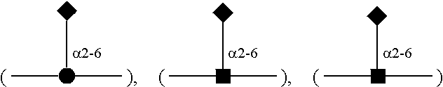


 Fuc;
Fuc; Neu5Ac;