US4900679A - Method for determining the existence and/or the monitoring of a pathological condition in a mammal and a test kit therefor - Google Patents
Method for determining the existence and/or the monitoring of a pathological condition in a mammal and a test kit therefor Download PDFInfo
- Publication number
- US4900679A US4900679A US07/236,039 US23603988A US4900679A US 4900679 A US4900679 A US 4900679A US 23603988 A US23603988 A US 23603988A US 4900679 A US4900679 A US 4900679A
- Authority
- US
- United States
- Prior art keywords
- mammal
- kit
- blood
- sample
- clotting
- Prior art date
- Legal status (The legal status is an assumption and is not a legal conclusion. Google has not performed a legal analysis and makes no representation as to the accuracy of the status listed.)
- Expired - Lifetime
Links
- 241000124008 Mammalia Species 0.000 title claims abstract description 69
- 230000001575 pathological effect Effects 0.000 title claims abstract description 50
- 238000000034 method Methods 0.000 title claims description 29
- 238000012360 testing method Methods 0.000 title claims description 24
- 238000012544 monitoring process Methods 0.000 title description 4
- 230000035602 clotting Effects 0.000 claims abstract description 22
- 206010053567 Coagulopathies Diseases 0.000 claims abstract description 21
- 210000004369 blood Anatomy 0.000 claims description 35
- 239000008280 blood Substances 0.000 claims description 33
- 239000002158 endotoxin Substances 0.000 claims description 29
- 239000003146 anticoagulant agent Substances 0.000 claims description 11
- 229940127219 anticoagulant drug Drugs 0.000 claims description 11
- 230000028993 immune response Effects 0.000 claims description 11
- 230000001732 thrombotic effect Effects 0.000 claims description 10
- 239000001509 sodium citrate Substances 0.000 claims description 6
- NLJMYIDDQXHKNR-UHFFFAOYSA-K sodium citrate Chemical compound O.O.[Na+].[Na+].[Na+].[O-]C(=O)CC(O)(CC([O-])=O)C([O-])=O NLJMYIDDQXHKNR-UHFFFAOYSA-K 0.000 claims description 6
- 229920001525 carrageenan Polymers 0.000 claims description 5
- 108090000765 processed proteins & peptides Proteins 0.000 claims description 5
- 102000014150 Interferons Human genes 0.000 claims description 4
- 108010050904 Interferons Proteins 0.000 claims description 4
- 108090001090 Lectins Proteins 0.000 claims description 4
- 102000004856 Lectins Human genes 0.000 claims description 4
- 102000006386 Myelin Proteins Human genes 0.000 claims description 4
- 108010083674 Myelin Proteins Proteins 0.000 claims description 4
- 230000023555 blood coagulation Effects 0.000 claims description 4
- 235000010418 carrageenan Nutrition 0.000 claims description 4
- 238000011534 incubation Methods 0.000 claims description 4
- 230000000977 initiatory effect Effects 0.000 claims description 4
- 229940079322 interferon Drugs 0.000 claims description 4
- 239000002523 lectin Substances 0.000 claims description 4
- 210000005012 myelin Anatomy 0.000 claims description 4
- 239000002644 phorbol ester Substances 0.000 claims description 4
- -1 thromboplastins Proteins 0.000 claims description 4
- 102000008186 Collagen Human genes 0.000 claims description 3
- 108010035532 Collagen Proteins 0.000 claims description 3
- 230000005856 abnormality Effects 0.000 claims description 3
- 238000004458 analytical method Methods 0.000 claims description 3
- 229920001436 collagen Polymers 0.000 claims description 3
- 239000003226 mitogen Substances 0.000 claims description 3
- 150000004633 phorbol derivatives Chemical class 0.000 claims description 3
- 241000894006 Bacteria Species 0.000 claims description 2
- 241000700605 Viruses Species 0.000 claims description 2
- 230000003213 activating effect Effects 0.000 claims description 2
- 230000000052 comparative effect Effects 0.000 claims description 2
- 102000004196 processed proteins & peptides Human genes 0.000 claims description 2
- 239000000523 sample Substances 0.000 claims 9
- 239000013068 control sample Substances 0.000 claims 8
- 150000001875 compounds Chemical class 0.000 claims 4
- 230000007170 pathology Effects 0.000 claims 4
- 239000003999 initiator Substances 0.000 claims 2
- OYPRJOBELJOOCE-UHFFFAOYSA-N Calcium Chemical compound [Ca] OYPRJOBELJOOCE-UHFFFAOYSA-N 0.000 claims 1
- 238000009007 Diagnostic Kit Methods 0.000 claims 1
- 239000002671 adjuvant Substances 0.000 claims 1
- 239000011575 calcium Substances 0.000 claims 1
- 229910052791 calcium Inorganic materials 0.000 claims 1
- 238000005259 measurement Methods 0.000 claims 1
- 239000002504 physiological saline solution Substances 0.000 claims 1
- 238000002360 preparation method Methods 0.000 claims 1
- 238000009877 rendering Methods 0.000 claims 1
- ZNCPFRVNHGOPAG-UHFFFAOYSA-L sodium oxalate Chemical compound [Na+].[Na+].[O-]C(=O)C([O-])=O ZNCPFRVNHGOPAG-UHFFFAOYSA-L 0.000 claims 1
- 229940039790 sodium oxalate Drugs 0.000 claims 1
- 239000002955 immunomodulating agent Substances 0.000 abstract description 45
- 229940121354 immunomodulator Drugs 0.000 abstract description 45
- 230000002584 immunomodulator Effects 0.000 abstract description 31
- 230000002489 hematologic effect Effects 0.000 abstract description 30
- 239000012530 fluid Substances 0.000 abstract description 29
- 230000001413 cellular effect Effects 0.000 abstract description 27
- 102000009123 Fibrin Human genes 0.000 abstract description 10
- 108010073385 Fibrin Proteins 0.000 abstract description 10
- BWGVNKXGVNDBDI-UHFFFAOYSA-N Fibrin monomer Chemical compound CNC(=O)CNC(=O)CN BWGVNKXGVNDBDI-UHFFFAOYSA-N 0.000 abstract description 10
- 229950003499 fibrin Drugs 0.000 abstract description 10
- 238000010998 test method Methods 0.000 abstract description 4
- 206010028980 Neoplasm Diseases 0.000 description 27
- 201000011510 cancer Diseases 0.000 description 24
- 201000010099 disease Diseases 0.000 description 18
- 208000037265 diseases, disorders, signs and symptoms Diseases 0.000 description 18
- 239000011780 sodium chloride Substances 0.000 description 13
- FAPWRFPIFSIZLT-UHFFFAOYSA-M Sodium chloride Chemical compound [Na+].[Cl-] FAPWRFPIFSIZLT-UHFFFAOYSA-M 0.000 description 11
- 238000001356 surgical procedure Methods 0.000 description 11
- 210000001616 monocyte Anatomy 0.000 description 8
- 238000002560 therapeutic procedure Methods 0.000 description 8
- 230000000694 effects Effects 0.000 description 7
- 210000001519 tissue Anatomy 0.000 description 7
- 206010040047 Sepsis Diseases 0.000 description 6
- 230000002159 abnormal effect Effects 0.000 description 6
- 230000015572 biosynthetic process Effects 0.000 description 6
- 210000004027 cell Anatomy 0.000 description 6
- 239000003795 chemical substances by application Substances 0.000 description 6
- 230000015271 coagulation Effects 0.000 description 6
- 238000005345 coagulation Methods 0.000 description 6
- 206010012601 diabetes mellitus Diseases 0.000 description 6
- 208000014674 injury Diseases 0.000 description 6
- 230000002980 postoperative effect Effects 0.000 description 6
- 238000011282 treatment Methods 0.000 description 6
- 108010000499 Thromboplastin Proteins 0.000 description 5
- 102000002262 Thromboplastin Human genes 0.000 description 5
- 238000004422 calculation algorithm Methods 0.000 description 5
- 229940079593 drug Drugs 0.000 description 5
- 239000003814 drug Substances 0.000 description 5
- 238000011156 evaluation Methods 0.000 description 5
- 201000006417 multiple sclerosis Diseases 0.000 description 5
- 230000004044 response Effects 0.000 description 5
- 210000000481 breast Anatomy 0.000 description 4
- 238000002405 diagnostic procedure Methods 0.000 description 4
- 230000008733 trauma Effects 0.000 description 4
- 206010006187 Breast cancer Diseases 0.000 description 3
- 208000026310 Breast neoplasm Diseases 0.000 description 3
- UXVMQQNJUSDDNG-UHFFFAOYSA-L Calcium chloride Chemical compound [Cl-].[Cl-].[Ca+2] UXVMQQNJUSDDNG-UHFFFAOYSA-L 0.000 description 3
- 241000588724 Escherichia coli Species 0.000 description 3
- 239000012190 activator Substances 0.000 description 3
- 238000000540 analysis of variance Methods 0.000 description 3
- 239000000427 antigen Substances 0.000 description 3
- 102000036639 antigens Human genes 0.000 description 3
- 108091007433 antigens Proteins 0.000 description 3
- 230000001900 immune effect Effects 0.000 description 3
- 230000002757 inflammatory effect Effects 0.000 description 3
- 230000036210 malignancy Effects 0.000 description 3
- 239000003805 procoagulant Substances 0.000 description 3
- 230000000246 remedial effect Effects 0.000 description 3
- 230000035945 sensitivity Effects 0.000 description 3
- 238000007619 statistical method Methods 0.000 description 3
- PGOHTUIFYSHAQG-LJSDBVFPSA-N (2S)-6-amino-2-[[(2S)-5-amino-2-[[(2S)-2-[[(2S)-2-[[(2S)-2-[[(2S)-4-amino-2-[[(2S)-2-[[(2S)-2-[[(2S)-2-[[(2S)-2-[[(2S)-5-amino-2-[[(2S)-5-amino-2-[[(2S)-2-[[(2S)-2-[[(2S)-2-[[(2S,3R)-2-[[(2S)-5-amino-2-[[(2S)-2-[[(2S)-2-[[(2S,3R)-2-[[(2S)-2-[[(2S)-2-[[(2S)-2-[[(2S)-2-[[(2S)-5-amino-2-[[(2S)-1-[(2S,3R)-2-[[(2S)-2-[[(2S)-2-[[(2R)-2-[[(2S)-2-[[(2S)-2-[[2-[[(2S)-2-[[(2S)-2-[[(2S)-2-[[(2S)-1-[(2S)-2-[[(2S)-2-[[(2S)-2-[[(2S)-2-amino-4-methylsulfanylbutanoyl]amino]-3-(1H-indol-3-yl)propanoyl]amino]-5-carbamimidamidopentanoyl]amino]propanoyl]pyrrolidine-2-carbonyl]amino]-3-methylbutanoyl]amino]-4-methylpentanoyl]amino]-4-methylpentanoyl]amino]acetyl]amino]-3-hydroxypropanoyl]amino]-4-methylpentanoyl]amino]-3-sulfanylpropanoyl]amino]-4-methylsulfanylbutanoyl]amino]-5-carbamimidamidopentanoyl]amino]-3-hydroxybutanoyl]pyrrolidine-2-carbonyl]amino]-5-oxopentanoyl]amino]-3-hydroxypropanoyl]amino]-3-hydroxypropanoyl]amino]-3-(1H-imidazol-5-yl)propanoyl]amino]-4-methylpentanoyl]amino]-3-hydroxybutanoyl]amino]-3-(1H-indol-3-yl)propanoyl]amino]-5-carbamimidamidopentanoyl]amino]-5-oxopentanoyl]amino]-3-hydroxybutanoyl]amino]-3-hydroxypropanoyl]amino]-3-carboxypropanoyl]amino]-3-hydroxypropanoyl]amino]-5-oxopentanoyl]amino]-5-oxopentanoyl]amino]-3-phenylpropanoyl]amino]-5-carbamimidamidopentanoyl]amino]-3-methylbutanoyl]amino]-4-methylpentanoyl]amino]-4-oxobutanoyl]amino]-5-carbamimidamidopentanoyl]amino]-3-(1H-indol-3-yl)propanoyl]amino]-4-carboxybutanoyl]amino]-5-oxopentanoyl]amino]hexanoic acid Chemical compound CSCC[C@H](N)C(=O)N[C@@H](Cc1c[nH]c2ccccc12)C(=O)N[C@@H](CCCNC(N)=N)C(=O)N[C@@H](C)C(=O)N1CCC[C@H]1C(=O)N[C@@H](C(C)C)C(=O)N[C@@H](CC(C)C)C(=O)N[C@@H](CC(C)C)C(=O)NCC(=O)N[C@@H](CO)C(=O)N[C@@H](CC(C)C)C(=O)N[C@@H](CS)C(=O)N[C@@H](CCSC)C(=O)N[C@@H](CCCNC(N)=N)C(=O)N[C@@H]([C@@H](C)O)C(=O)N1CCC[C@H]1C(=O)N[C@@H](CCC(N)=O)C(=O)N[C@@H](CO)C(=O)N[C@@H](CO)C(=O)N[C@@H](Cc1cnc[nH]1)C(=O)N[C@@H](CC(C)C)C(=O)N[C@@H]([C@@H](C)O)C(=O)N[C@@H](Cc1c[nH]c2ccccc12)C(=O)N[C@@H](CCCNC(N)=N)C(=O)N[C@@H](CCC(N)=O)C(=O)N[C@@H]([C@@H](C)O)C(=O)N[C@@H](CO)C(=O)N[C@@H](CC(O)=O)C(=O)N[C@@H](CO)C(=O)N[C@@H](CCC(N)=O)C(=O)N[C@@H](CCC(N)=O)C(=O)N[C@@H](Cc1ccccc1)C(=O)N[C@@H](CCCNC(N)=N)C(=O)N[C@@H](C(C)C)C(=O)N[C@@H](CC(C)C)C(=O)N[C@@H](CC(N)=O)C(=O)N[C@@H](CCCNC(N)=N)C(=O)N[C@@H](Cc1c[nH]c2ccccc12)C(=O)N[C@@H](CCC(O)=O)C(=O)N[C@@H](CCC(N)=O)C(=O)N[C@@H](CCCCN)C(O)=O PGOHTUIFYSHAQG-LJSDBVFPSA-N 0.000 description 2
- 208000030507 AIDS Diseases 0.000 description 2
- BHPQYMZQTOCNFJ-UHFFFAOYSA-N Calcium cation Chemical compound [Ca+2] BHPQYMZQTOCNFJ-UHFFFAOYSA-N 0.000 description 2
- 108010062580 Concanavalin A Proteins 0.000 description 2
- WQZGKKKJIJFFOK-GASJEMHNSA-N Glucose Natural products OC[C@H]1OC(O)[C@H](O)[C@@H](O)[C@@H]1O WQZGKKKJIJFFOK-GASJEMHNSA-N 0.000 description 2
- 241000282414 Homo sapiens Species 0.000 description 2
- 206010061218 Inflammation Diseases 0.000 description 2
- 241000712079 Measles morbillivirus Species 0.000 description 2
- 210000001744 T-lymphocyte Anatomy 0.000 description 2
- 108090000190 Thrombin Proteins 0.000 description 2
- 208000007536 Thrombosis Diseases 0.000 description 2
- 229920000392 Zymosan Polymers 0.000 description 2
- 150000001412 amines Chemical class 0.000 description 2
- 230000001580 bacterial effect Effects 0.000 description 2
- 239000011324 bead Substances 0.000 description 2
- 230000008901 benefit Effects 0.000 description 2
- 238000001574 biopsy Methods 0.000 description 2
- 239000001110 calcium chloride Substances 0.000 description 2
- 229910001628 calcium chloride Inorganic materials 0.000 description 2
- 229910001424 calcium ion Inorganic materials 0.000 description 2
- 239000000679 carrageenan Substances 0.000 description 2
- 229940113118 carrageenan Drugs 0.000 description 2
- 230000008859 change Effects 0.000 description 2
- 238000002512 chemotherapy Methods 0.000 description 2
- 210000001072 colon Anatomy 0.000 description 2
- 230000000295 complement effect Effects 0.000 description 2
- YPHMISFOHDHNIV-FSZOTQKASA-N cycloheximide Chemical compound C1[C@@H](C)C[C@H](C)C(=O)[C@@H]1[C@H](O)CC1CC(=O)NC(=O)C1 YPHMISFOHDHNIV-FSZOTQKASA-N 0.000 description 2
- 238000001514 detection method Methods 0.000 description 2
- 238000002651 drug therapy Methods 0.000 description 2
- 239000008103 glucose Substances 0.000 description 2
- 230000012010 growth Effects 0.000 description 2
- 208000026278 immune system disease Diseases 0.000 description 2
- 229940027941 immunoglobulin g Drugs 0.000 description 2
- 230000004054 inflammatory process Effects 0.000 description 2
- 239000004816 latex Substances 0.000 description 2
- 229920000126 latex Polymers 0.000 description 2
- 230000003902 lesion Effects 0.000 description 2
- 150000002632 lipids Chemical class 0.000 description 2
- 210000004698 lymphocyte Anatomy 0.000 description 2
- 238000009607 mammography Methods 0.000 description 2
- 239000000463 material Substances 0.000 description 2
- 239000012528 membrane Substances 0.000 description 2
- 210000000440 neutrophil Anatomy 0.000 description 2
- 239000002245 particle Substances 0.000 description 2
- 102000004169 proteins and genes Human genes 0.000 description 2
- 108090000623 proteins and genes Proteins 0.000 description 2
- 230000005855 radiation Effects 0.000 description 2
- 238000005070 sampling Methods 0.000 description 2
- 239000000126 substance Substances 0.000 description 2
- 208000011580 syndromic disease Diseases 0.000 description 2
- 230000002792 vascular Effects 0.000 description 2
- PJVWKTKQMONHTI-UHFFFAOYSA-N warfarin Chemical compound OC=1C2=CC=CC=C2OC(=O)C=1C(CC(=O)C)C1=CC=CC=C1 PJVWKTKQMONHTI-UHFFFAOYSA-N 0.000 description 2
- 229960005080 warfarin Drugs 0.000 description 2
- UHVMMEOXYDMDKI-JKYCWFKZSA-L zinc;1-(5-cyanopyridin-2-yl)-3-[(1s,2s)-2-(6-fluoro-2-hydroxy-3-propanoylphenyl)cyclopropyl]urea;diacetate Chemical compound [Zn+2].CC([O-])=O.CC([O-])=O.CCC(=O)C1=CC=C(F)C([C@H]2[C@H](C2)NC(=O)NC=2N=CC(=CC=2)C#N)=C1O UHVMMEOXYDMDKI-JKYCWFKZSA-L 0.000 description 2
- SNICXCGAKADSCV-JTQLQIEISA-N (-)-Nicotine Chemical compound CN1CCC[C@H]1C1=CC=CN=C1 SNICXCGAKADSCV-JTQLQIEISA-N 0.000 description 1
- HVAUUPRFYPCOCA-AREMUKBSSA-N 2-O-acetyl-1-O-hexadecyl-sn-glycero-3-phosphocholine Chemical compound CCCCCCCCCCCCCCCCOC[C@@H](OC(C)=O)COP([O-])(=O)OCC[N+](C)(C)C HVAUUPRFYPCOCA-AREMUKBSSA-N 0.000 description 1
- XTWYTFMLZFPYCI-KQYNXXCUSA-N 5'-adenylphosphoric acid Chemical compound C1=NC=2C(N)=NC=NC=2N1[C@@H]1O[C@H](COP(O)(=O)OP(O)(O)=O)[C@@H](O)[C@H]1O XTWYTFMLZFPYCI-KQYNXXCUSA-N 0.000 description 1
- XTWYTFMLZFPYCI-UHFFFAOYSA-N Adenosine diphosphate Natural products C1=NC=2C(N)=NC=NC=2N1C1OC(COP(O)(=O)OP(O)(O)=O)C(O)C1O XTWYTFMLZFPYCI-UHFFFAOYSA-N 0.000 description 1
- QGZKDVFQNNGYKY-UHFFFAOYSA-O Ammonium Chemical compound [NH4+] QGZKDVFQNNGYKY-UHFFFAOYSA-O 0.000 description 1
- 108010039209 Blood Coagulation Factors Proteins 0.000 description 1
- 102000015081 Blood Coagulation Factors Human genes 0.000 description 1
- HIYAVKIYRIFSCZ-CVXKHCKVSA-N Calcimycin Chemical compound CC([C@H]1OC2([C@@H](C[C@H]1C)C)O[C@H]([C@H](CC2)C)CC=1OC2=CC=C(C(=C2N=1)C(O)=O)NC)C(=O)C1=CC=CN1 HIYAVKIYRIFSCZ-CVXKHCKVSA-N 0.000 description 1
- VEXZGXHMUGYJMC-UHFFFAOYSA-M Chloride anion Chemical compound [Cl-] VEXZGXHMUGYJMC-UHFFFAOYSA-M 0.000 description 1
- 241000777300 Congiopodidae Species 0.000 description 1
- 108090000790 Enzymes Proteins 0.000 description 1
- 102000004190 Enzymes Human genes 0.000 description 1
- 102000008857 Ferritin Human genes 0.000 description 1
- 108050000784 Ferritin Proteins 0.000 description 1
- 238000008416 Ferritin Methods 0.000 description 1
- 229920001503 Glucan Polymers 0.000 description 1
- 206010019233 Headaches Diseases 0.000 description 1
- 206010020751 Hypersensitivity Diseases 0.000 description 1
- 102000004895 Lipoproteins Human genes 0.000 description 1
- 108090001030 Lipoproteins Proteins 0.000 description 1
- 206010027476 Metastases Diseases 0.000 description 1
- 241001465754 Metazoa Species 0.000 description 1
- HVAUUPRFYPCOCA-AREMUKBSSA-O PAF Chemical compound CCCCCCCCCCCCCCCCOC[C@@H](OC(C)=O)COP(O)(=O)OCC[N+](C)(C)C HVAUUPRFYPCOCA-AREMUKBSSA-O 0.000 description 1
- 108090000526 Papain Proteins 0.000 description 1
- 102100029251 Phagocytosis-stimulating peptide Human genes 0.000 description 1
- 240000007643 Phytolacca americana Species 0.000 description 1
- 235000009074 Phytolacca americana Nutrition 0.000 description 1
- 108010003541 Platelet Activating Factor Proteins 0.000 description 1
- 108010033737 Pokeweed Mitogens Proteins 0.000 description 1
- 108010094028 Prothrombin Proteins 0.000 description 1
- 102100027378 Prothrombin Human genes 0.000 description 1
- 206010038063 Rectal haemorrhage Diseases 0.000 description 1
- 208000025747 Rheumatic disease Diseases 0.000 description 1
- 241000607142 Salmonella Species 0.000 description 1
- 108010023197 Streptokinase Proteins 0.000 description 1
- 208000006011 Stroke Diseases 0.000 description 1
- 238000000692 Student's t-test Methods 0.000 description 1
- 108010084754 Tuftsin Proteins 0.000 description 1
- 206010053614 Type III immune complex mediated reaction Diseases 0.000 description 1
- 108090000435 Urokinase-type plasminogen activator Proteins 0.000 description 1
- 102000003990 Urokinase-type plasminogen activator Human genes 0.000 description 1
- PNNCWTXUWKENPE-UHFFFAOYSA-N [N].NC(N)=O Chemical compound [N].NC(N)=O PNNCWTXUWKENPE-UHFFFAOYSA-N 0.000 description 1
- 210000000683 abdominal cavity Anatomy 0.000 description 1
- 230000004913 activation Effects 0.000 description 1
- 206010000891 acute myocardial infarction Diseases 0.000 description 1
- 230000006978 adaptation Effects 0.000 description 1
- 208000026935 allergic disease Diseases 0.000 description 1
- 230000007815 allergy Effects 0.000 description 1
- 230000004075 alteration Effects 0.000 description 1
- WQZGKKKJIJFFOK-VFUOTHLCSA-N beta-D-glucose Chemical compound OC[C@H]1O[C@@H](O)[C@H](O)[C@@H](O)[C@@H]1O WQZGKKKJIJFFOK-VFUOTHLCSA-N 0.000 description 1
- 210000003445 biliary tract Anatomy 0.000 description 1
- 230000003115 biocidal effect Effects 0.000 description 1
- 239000010836 blood and blood product Substances 0.000 description 1
- 239000003114 blood coagulation factor Substances 0.000 description 1
- 238000004820 blood count Methods 0.000 description 1
- 229940125691 blood product Drugs 0.000 description 1
- 238000010241 blood sampling Methods 0.000 description 1
- 229940003871 calcium ion Drugs 0.000 description 1
- HIYAVKIYRIFSCZ-UHFFFAOYSA-N calcium ionophore A23187 Natural products N=1C2=C(C(O)=O)C(NC)=CC=C2OC=1CC(C(CC1)C)OC1(C(CC1C)C)OC1C(C)C(=O)C1=CC=CN1 HIYAVKIYRIFSCZ-UHFFFAOYSA-N 0.000 description 1
- 238000004364 calculation method Methods 0.000 description 1
- 210000000170 cell membrane Anatomy 0.000 description 1
- 210000003850 cellular structure Anatomy 0.000 description 1
- 238000000546 chi-square test Methods 0.000 description 1
- 150000001860 citric acid derivatives Chemical class 0.000 description 1
- 238000004140 cleaning Methods 0.000 description 1
- 239000000084 colloidal system Substances 0.000 description 1
- 208000029742 colonic neoplasm Diseases 0.000 description 1
- 238000011498 curative surgery Methods 0.000 description 1
- 238000009109 curative therapy Methods 0.000 description 1
- 238000003745 diagnosis Methods 0.000 description 1
- 230000037213 diet Effects 0.000 description 1
- 235000005911 diet Nutrition 0.000 description 1
- 238000003748 differential diagnosis Methods 0.000 description 1
- 230000001700 effect on tissue Effects 0.000 description 1
- 239000003792 electrolyte Substances 0.000 description 1
- 229940088598 enzyme Drugs 0.000 description 1
- 210000003238 esophagus Anatomy 0.000 description 1
- 230000001747 exhibiting effect Effects 0.000 description 1
- 210000003128 head Anatomy 0.000 description 1
- 231100000869 headache Toxicity 0.000 description 1
- 230000036541 health Effects 0.000 description 1
- 208000019622 heart disease Diseases 0.000 description 1
- 208000031169 hemorrhagic disease Diseases 0.000 description 1
- 238000010562 histological examination Methods 0.000 description 1
- 230000016178 immune complex formation Effects 0.000 description 1
- 230000036737 immune function Effects 0.000 description 1
- 208000015181 infectious disease Diseases 0.000 description 1
- 239000003112 inhibitor Substances 0.000 description 1
- 238000003780 insertion Methods 0.000 description 1
- 230000037431 insertion Effects 0.000 description 1
- 230000003993 interaction Effects 0.000 description 1
- 230000003834 intracellular effect Effects 0.000 description 1
- 230000000366 juvenile effect Effects 0.000 description 1
- 150000002576 ketones Chemical class 0.000 description 1
- 208000017169 kidney disease Diseases 0.000 description 1
- 210000000867 larynx Anatomy 0.000 description 1
- 231100001231 less toxic Toxicity 0.000 description 1
- 210000000265 leukocyte Anatomy 0.000 description 1
- GZQKNULLWNGMCW-PWQABINMSA-N lipid A (E. coli) Chemical compound O1[C@H](CO)[C@@H](OP(O)(O)=O)[C@H](OC(=O)C[C@@H](CCCCCCCCCCC)OC(=O)CCCCCCCCCCCCC)[C@@H](NC(=O)C[C@@H](CCCCCCCCCCC)OC(=O)CCCCCCCCCCC)[C@@H]1OC[C@@H]1[C@@H](O)[C@H](OC(=O)C[C@H](O)CCCCCCCCCCC)[C@@H](NC(=O)C[C@H](O)CCCCCCCCCCC)[C@@H](OP(O)(O)=O)O1 GZQKNULLWNGMCW-PWQABINMSA-N 0.000 description 1
- 210000004185 liver Anatomy 0.000 description 1
- 208000019423 liver disease Diseases 0.000 description 1
- 210000004072 lung Anatomy 0.000 description 1
- 210000001165 lymph node Anatomy 0.000 description 1
- 238000013160 medical therapy Methods 0.000 description 1
- 230000009401 metastasis Effects 0.000 description 1
- 239000000203 mixture Substances 0.000 description 1
- 238000012986 modification Methods 0.000 description 1
- 230000004048 modification Effects 0.000 description 1
- 239000003607 modifier Substances 0.000 description 1
- 208000010125 myocardial infarction Diseases 0.000 description 1
- 210000003739 neck Anatomy 0.000 description 1
- 229960002715 nicotine Drugs 0.000 description 1
- SNICXCGAKADSCV-UHFFFAOYSA-N nicotine Natural products CN1CCCC1C1=CC=CN=C1 SNICXCGAKADSCV-UHFFFAOYSA-N 0.000 description 1
- 210000000056 organ Anatomy 0.000 description 1
- 210000001672 ovary Anatomy 0.000 description 1
- 150000003891 oxalate salts Chemical class 0.000 description 1
- 230000000242 pagocytic effect Effects 0.000 description 1
- 238000007427 paired t-test Methods 0.000 description 1
- 210000000496 pancreas Anatomy 0.000 description 1
- 230000001717 pathogenic effect Effects 0.000 description 1
- 238000012335 pathological evaluation Methods 0.000 description 1
- 230000001991 pathophysiological effect Effects 0.000 description 1
- 239000002831 pharmacologic agent Substances 0.000 description 1
- 230000000144 pharmacologic effect Effects 0.000 description 1
- PHEDXBVPIONUQT-RGYGYFBISA-N phorbol 13-acetate 12-myristate Chemical compound C([C@]1(O)C(=O)C(C)=C[C@H]1[C@@]1(O)[C@H](C)[C@H]2OC(=O)CCCCCCCCCCCCC)C(CO)=C[C@H]1[C@H]1[C@]2(OC(C)=O)C1(C)C PHEDXBVPIONUQT-RGYGYFBISA-N 0.000 description 1
- XLCISDOVNFLSGO-VONOSFMSSA-N phorbol-12-myristate Chemical compound C([C@]1(O)C(=O)C(C)=C[C@H]1[C@@]1(O)[C@H](C)[C@H]2OC(=O)CCCCCCCCCCCCC)C(CO)=C[C@H]1[C@H]1[C@]2(O)C1(C)C XLCISDOVNFLSGO-VONOSFMSSA-N 0.000 description 1
- 230000008569 process Effects 0.000 description 1
- 230000002947 procoagulating effect Effects 0.000 description 1
- 238000004393 prognosis Methods 0.000 description 1
- 230000000750 progressive effect Effects 0.000 description 1
- 230000002035 prolonged effect Effects 0.000 description 1
- 229940039716 prothrombin Drugs 0.000 description 1
- 238000001959 radiotherapy Methods 0.000 description 1
- 210000000664 rectum Anatomy 0.000 description 1
- 230000000306 recurrent effect Effects 0.000 description 1
- 230000000979 retarding effect Effects 0.000 description 1
- 206010039073 rheumatoid arthritis Diseases 0.000 description 1
- 238000012216 screening Methods 0.000 description 1
- 239000003998 snake venom Substances 0.000 description 1
- 210000001584 soft palate Anatomy 0.000 description 1
- 241000894007 species Species 0.000 description 1
- 208000010110 spontaneous platelet aggregation Diseases 0.000 description 1
- 239000000021 stimulant Substances 0.000 description 1
- 229960005202 streptokinase Drugs 0.000 description 1
- 239000000725 suspension Substances 0.000 description 1
- 238000012353 t test Methods 0.000 description 1
- UEUXEKPTXMALOB-UHFFFAOYSA-J tetrasodium;2-[2-[bis(carboxylatomethyl)amino]ethyl-(carboxylatomethyl)amino]acetate Chemical compound [Na+].[Na+].[Na+].[Na+].[O-]C(=O)CN(CC([O-])=O)CCN(CC([O-])=O)CC([O-])=O UEUXEKPTXMALOB-UHFFFAOYSA-J 0.000 description 1
- 230000001225 therapeutic effect Effects 0.000 description 1
- 229960004072 thrombin Drugs 0.000 description 1
- 230000000472 traumatic effect Effects 0.000 description 1
- 229940035670 tuftsin Drugs 0.000 description 1
- IESDGNYHXIOKRW-LEOABGAYSA-N tuftsin Chemical compound C[C@@H](O)[C@H](N)C(=O)N[C@@H](CCCCN)C(=O)N1CCC[C@H]1C(=O)N[C@H](CCCNC(N)=N)C(O)=O IESDGNYHXIOKRW-LEOABGAYSA-N 0.000 description 1
- 210000004881 tumor cell Anatomy 0.000 description 1
- 230000004614 tumor growth Effects 0.000 description 1
- 238000002562 urinalysis Methods 0.000 description 1
- 210000003932 urinary bladder Anatomy 0.000 description 1
- 229960005356 urokinase Drugs 0.000 description 1
- 208000019553 vascular disease Diseases 0.000 description 1
- 230000002227 vasoactive effect Effects 0.000 description 1
- 210000005253 yeast cell Anatomy 0.000 description 1
Images

Classifications
-
- G—PHYSICS
- G01—MEASURING; TESTING
- G01N—INVESTIGATING OR ANALYSING MATERIALS BY DETERMINING THEIR CHEMICAL OR PHYSICAL PROPERTIES
- G01N33/00—Investigating or analysing materials by specific methods not covered by groups G01N1/00 - G01N31/00
- G01N33/48—Biological material, e.g. blood, urine; Haemocytometers
- G01N33/50—Chemical analysis of biological material, e.g. blood, urine; Testing involving biospecific ligand binding methods; Immunological testing
- G01N33/53—Immunoassay; Biospecific binding assay; Materials therefor
- G01N33/574—Immunoassay; Biospecific binding assay; Materials therefor for cancer
- G01N33/57484—Immunoassay; Biospecific binding assay; Materials therefor for cancer involving compounds serving as markers for tumor, cancer, neoplasia, e.g. cellular determinants, receptors, heat shock/stress proteins, A-protein, oligosaccharides, metabolites
- G01N33/57488—Immunoassay; Biospecific binding assay; Materials therefor for cancer involving compounds serving as markers for tumor, cancer, neoplasia, e.g. cellular determinants, receptors, heat shock/stress proteins, A-protein, oligosaccharides, metabolites involving compounds identifable in body fluids
-
- G—PHYSICS
- G01—MEASURING; TESTING
- G01N—INVESTIGATING OR ANALYSING MATERIALS BY DETERMINING THEIR CHEMICAL OR PHYSICAL PROPERTIES
- G01N33/00—Investigating or analysing materials by specific methods not covered by groups G01N1/00 - G01N31/00
- G01N33/48—Biological material, e.g. blood, urine; Haemocytometers
- G01N33/50—Chemical analysis of biological material, e.g. blood, urine; Testing involving biospecific ligand binding methods; Immunological testing
- G01N33/53—Immunoassay; Biospecific binding assay; Materials therefor
- G01N33/569—Immunoassay; Biospecific binding assay; Materials therefor for microorganisms, e.g. protozoa, bacteria, viruses
- G01N33/56911—Bacteria
-
- G—PHYSICS
- G01—MEASURING; TESTING
- G01N—INVESTIGATING OR ANALYSING MATERIALS BY DETERMINING THEIR CHEMICAL OR PHYSICAL PROPERTIES
- G01N33/00—Investigating or analysing materials by specific methods not covered by groups G01N1/00 - G01N31/00
- G01N33/48—Biological material, e.g. blood, urine; Haemocytometers
- G01N33/50—Chemical analysis of biological material, e.g. blood, urine; Testing involving biospecific ligand binding methods; Immunological testing
- G01N33/53—Immunoassay; Biospecific binding assay; Materials therefor
- G01N33/569—Immunoassay; Biospecific binding assay; Materials therefor for microorganisms, e.g. protozoa, bacteria, viruses
- G01N33/56983—Viruses
- G01N33/56988—HIV or HTLV
-
- G—PHYSICS
- G01—MEASURING; TESTING
- G01N—INVESTIGATING OR ANALYSING MATERIALS BY DETERMINING THEIR CHEMICAL OR PHYSICAL PROPERTIES
- G01N33/00—Investigating or analysing materials by specific methods not covered by groups G01N1/00 - G01N31/00
- G01N33/48—Biological material, e.g. blood, urine; Haemocytometers
- G01N33/50—Chemical analysis of biological material, e.g. blood, urine; Testing involving biospecific ligand binding methods; Immunological testing
- G01N33/86—Chemical analysis of biological material, e.g. blood, urine; Testing involving biospecific ligand binding methods; Immunological testing involving blood coagulating time or factors, or their receptors
-
- G—PHYSICS
- G01—MEASURING; TESTING
- G01N—INVESTIGATING OR ANALYSING MATERIALS BY DETERMINING THEIR CHEMICAL OR PHYSICAL PROPERTIES
- G01N2800/00—Detection or diagnosis of diseases
- G01N2800/52—Predicting or monitoring the response to treatment, e.g. for selection of therapy based on assay results in personalised medicine; Prognosis
Definitions
- This invention relates to a method of testing a hematologic fluid, and more particularly to a method of testing and determine the existence in the mammal of a pathological state or condition, to monitor a known pathological state existing in a mammal and a test kit therefor.
- CBC complete blood count
- chemistries e.g. glucose or electrolyte levels
- urinalysis test for glucose, ketones, etc.
- a mammal of a pathological state or condition e.g. cancer, AIDS, sepsis and the like is generally performed after the mammal has experienced some abnormal physical response, e.g. lack of energy, headaches, rectal bleeding, lumps, etc., or as preliminarily detected during an annual physical examination.
- diagnostic procedures and/or other protocols are thereafter initiated and evaluated to qualify the pathological state as well as to quantify the extent of advancement of the pathological state or condition. Diagnostic procedures may involve X-rays, e.g. mammography for breast cancer, proctoscopy of the colon, etc.
- a pathological state has been found to exist in the mammal and has been qualified as to the specific pathological state, there may be remedial procedures to reduce the impact of the pathologic state on the mammal, e.g. drug, radiation therapy, chemotherapy, and the like protocol, or alternately to eliminate the pathological state, e.g. by surgical procedure.
- remedial procedures e.g. drug, radiation therapy, chemotherapy, and the like protocol
- the effectiveness of the remedial procedure is difficult to timely assess. For example, in the surgical removal of cancerous growth, only subsequent biopsies of proximate tissue may demonstrate total removal, and then, not necessarily on a 100 percent certain basis, let alone the possibility of metastasis.
- Tests have been developed to determine the immune function of monocytes, neutrophils, lymphocytes, etc. wherein the individuality is isolated and tested for individual functionality by diverse methods. Such procedures are costly and time consuming and are not specific to a particular pathological state. Also, the results of individuality tests are difficult to interpret, let alone correlate. For example, although mammography may delineate the size, location, etc. of a lump in the breast in a female, the results will not always qualify whether the lump is cancerous or benign. Such pathological evaluation is effected by pathological observation of the actual cellular structure after biopsy or surgical removal of the lump.
- liver enzymes for liver disease e.g. liver enzymes for liver disease, blood urea nitrogen for kidney disease, T-cell function for immunological disorders, prothrombin and partial thromboplastin times for bleeding disorders, etc.
- T-cell function for immunological disorders
- prothrombin and partial thromboplastin times for bleeding disorders, etc.
- such tests cannot determine either the effects of therapy on the coagulation changes in thrombotic diseases, or the effects of therapy in cancer and other diseases which involve alterations in the immune response system.
- An object of the present invention is to provide a method for determining whether a pathologic state or condition exists in a mammal.
- a further object of the present invention is to provide a method for determining whether a pathological state or condition exists in mammal that may be performed in a facile and inexpensive manner.
- Another object of the present invention is to provide a method for determining whether a pathological state or condition exists in a mammal, that may be readily effected in a relatively short period of time.
- Yet another object of the present invention is to provide a dependable method for determining whether a pathological state or condition exists in a mammal with minimal, if any, false readings.
- a still further object of the present invention is to provide a simple method for sequentially determining the course of a known pathological state or condition in a mammal.
- Still yet another object of the present invention is to provide a method for determining effectiveness of a surgical procedure on a mammal to erradicate a pathological state or condition, or to detect recurrent disease.
- Another object of the present invention is to monitor the effectiveness of a drug regime or like protocol on a mammal having a known pathological state or condition.
- Still another object of the present invention is to monitor the effectiveness of a remedial program for retarding growth, reducing or destroying a known pathological state or condition in a mammal.
- the reaction parameter is a clotting parameter as determined as fibrin levels or a function of a time differential between fibrin levels.
- the ratio between the reaction parameters of such a cellular hematologic fluid of a mammal being tested without and with an immunomodulator is compared with the ratio between the reaction parameters of cellular hematologic fluids of mammals of known healthy states without and with a like immunomodulator to assess the existence or non-existence of a pathological state or condition in the mammal being tested.
- a reaction parameter of a cellular hematologic fluid of a mammal with a pre-existing pathological condition when admixed with an immunomodulator is different than the reaction parameters of cellular hematologic fluids of mammals, in known healthy states when admixed with the same immunomodulator.
- the method of the present invention does not diagnose a specific pathological condition, but points to the existence of a pathological condition in the mammal being evaluated, although in certain instances the process of the present invention may be capable of diagnosing algorithmically a specific pathological condition.
- cellular hematologic fluid of a mammal is the whole blood thereof or a fraction thereof including monocytes and other cellular or noncellular components of the mammal.
- blood coagulation and/or the immune response system of a mammal having an existing pathological state or condition to an immunomodulator is different than the blood coagulation and/or immune response system of health mammals to like immunomodulators.
- monocytes to varying degrees are involved in the immune response system of the hematologic fluid to the immunomodulator, it is believed that the immune response system involves an interreaction between the monocytes and other components, e.g. T-cells, lymphocytes, neutrophils, etc. in the cellular hematologic fluid.
- the pathological conditions include cancer, sepsis, AIDS, diabetes, multiple sclerosis, acute myocardial infarction, trauma, vascular thrombosis, etc. and any pathological state or condition affecting the immune response system of a mammal, it being understood by one skilled in the art that the specific pathological state or condition existing in a test mammal is generally qualified after a positive determination of the existence of a pathological state or condition in accordance with the method of the present invention.
- the term "mammals” as used herein includes Homo sapiens, and domesticated animals, e.g. race horses.
- immunomodulator means an immunoactivator or immunoattenuator which is an agent that either promotes or accelerates, or retards or attenuates, respectively, coagulation of whole blood or whole fractions (i.e. as expressed by recalcification time (RT)).
- Immunomodulators include, inter alia, endotoxins, measles virus, Interferon, phorbol esters, collagens, anticoagulants such as warfarin, platelet activating factors, carrageenans, adjunct peptides, thromboplastins, antigens, myelin, gram negative bacteria, lectins such as Concanavalin-A, mitogens such as pokeweed mitogens, etc.
- recalcification time is defined as any time period between initiation of fibrin formation to an end point thereof or to some intermediate point, it being understood that values for clotting parameters may be based on the rate of fibrin formation, for example, as defined by the integrated area beneath a rate curve between limits, etc.
- Anticoagulants for whole blood or fractions thereof include the citrates such as sodium citrate, the oxalates, sodium ethylenediamine tetra-acetic acid, etc., with sodium citrate being generally preferred.
- a more sophisticated algorithm is based upon the calculation of a "Thrombotic Index", defined as the ratio of the recalcification time (RT v ) of the cellular hematologic fluid of a mammal (in a vehicle, e.g. saline) in the absence of an immunomodulator, to the recalcification time (RT i ) thereof also in a vehicle and in the presence of an immunomodulator, in accordance with the following equation (I):
- thrombotic index of the mammal being tested being compared with the thrombotic indices of healthy mammals.
- the percent differences of clotting of test mammals are then compared with percent differences of clotting of healthy mammals.
- a SONOCLOT® Coagulation Analyzer is available from Sienco, Inc. for measuring viscoelastic properties as a function of mechanical impedance of the sample being tested. Such analysis is very sensitive to fibrin formation thereby providing improved sensitivity and reproducibility of results.
- TEG Thromboelastograph
- Still another apparatus is the HEMOCHRON® 400 available from the International Technidyne Corporation of Edison, N.J., evidencing significant data correlation to that of the SONOCLOT®.
- E. coli endotoxin strain 055:B5
- RT i recalcification time-endotoxin
- a hematological sample e.g. by venipuncture using a syringe (20 gauge needle) without stasis or undue force to draw blood.
- the hematological fluid is transferred to a tube including and admixed with an anticoagulant, e.g. 3.8% solution of sodium citrate.
- an anticoagulant e.g. 3.8% solution of sodium citrate.
- the volumetric ratio is from about nine (9) parts hematological fluid to one (1) part anticoagulant. While many anticoagulants are available, sodium citrate is generally preferred since the pH level thereof is essentially similar to the pH level of the hematological fluid of the mammal being tested, and is less toxic to the cellular elements.
- an aliquot portion (2 milliliters) of the anticoagulated hematological fluid or citrated whole blood (CWB) is admixed in a tube with the endotoxin (e.g. 20 ⁇ l of a 1 mg/cc suspension or solution of E. coli endotoxin) and incubated for a predetermined time period, generally of from 2 to 4 hours. It has been generally found that longer incubation time periods provide result of greater sensitivity.
- the endotoxin e.g. 20 ⁇ l of a 1 mg/cc suspension or solution of E. coli endotoxin
- incubation temperatures range from about 35° C. to 40° C., preferably about 37° C.
- a predetermined amount of a calcium-ion containing composition such as calcium chloride (CaCl 2 ), e.g.
- Recalcification times-immunomodulator (RT i ) of a cellular hematological fluid for mammals in a healthy state range between 3.93 to 6.04 with a mean recalcification time being 4.66, as determined by TEG; and 4.6 to 7.2 with a mean of 5.69, as determined by SONOCLOT®.
- RT i recalcification times-immunomodulator
- the term "statistically significant differences between the groups studied” means that when using the appropriate statistical analysis (e.g. Chi-square tests, t-test) the probability of the groups being the same is less than 5%, e.g. p ⁇ 0.05. In other words, the probability of obtaining the same results on a completely random basis is less than 5 out of 100 attempts.
- the normal volunteer controls were of both sexes, ranged in age from 21 to 69, and were both smokers and non-smokers. No data was ascertained from the volunteers as to current drug intake nor whether the volunteers were currently under treatment for any disease.
- the cancer patients were evaluated at the time of diagnosis of the disease. There were significant differences between the recalcification times-endotoxin (RT i ) between the group of the healthy volunteers and the group of the cancer patients, whereas there were no significant differences between recalcification times-saline (RT v ) of such groups. Additionally, it can be readily seen that the thrombotic index (TI) is greater for the group of cancer patients than the thrombotic index (TI) of the healthy volunteers. The same proposition held true of the comparison of percent difference in clotting (PDOC) therebetween. The values for TI or PDOC do not overlap for these groups.
- the method of the present invention permits a clinician to evaluate effects of therapy on the state of the cancer in a cancer patient. For example, small changes in RT i values and a lowering of TI or PDOC values after non-fully curative treatments have been demonstrated. If chemotheraphy and/or radiation treatment do not alter such values, changes in the treatment are then suggested to find a more effective drug regime and/or radiation protocol.
- An advantage of the method of the present invention is the convenience of sampling and evaluation at varying times after therapy and the assessment of effectiveness of treatment prior to physical appearance of clinical changes.
- cancers detected by the method of the present invention are cancers of the lung, breast, biliary tract, bladder, larynx, ovary, head and neck, colon, rectum, esophagus, soft palate, pancreas, and floor of the mouth.
- the presence or absence of remaining malignancy after curative surgery may be determined by the methods of the present invention.
- Table II sets forth specific data with reference to six patients; patients 1 to 4 having cancer and patients 5 and 6 having benign breast lesions.
- RT i recalcification times-endotoxin
- diabetics with abnormal values have the more severe disease (e.g. juvenile diabetics, diabetics with vascular complications, diabetics with disease more than 15 years, etc.).
- the methods of the present invention will be useful in measuring therapeutic effects on diabetic activity, such as diet control, exercise and drug treatment, and may become a goal of the therapy to bring the RT v and RT i values of diabetics into the normal range.
- Table IV sets forth mean values and ranges of RT i , RT v , TI and PDOC of healthy volunteers (per Example I) with mean values and ranges of RT i , RT v , TI and PDOC of four (4) confirmed advanced AIDS patients:
- RT i recalcification times-endotoxin
- TI and PDOC values are sifgnificantly lower, particularly in three out of four cases.
- Another form of algorithm could be derived to more unequivocally identify all such AIDS patients.
- the methods of the present invention illustrate that mammals having AIDS or AIDS-like disease have a PDOC value below the control, and with some statistically determined base line value, to qualify individuals for blood donation.
- Table V sets forth values of a group (9 patients) exhibiting a localized septic condition from a group (62 patients) who did not exhibit such a condition, it being noted that RT i values are lower and TI and PDOC values are higher, as expected (compared to a normal population) as a result of released thromboplastins and immunological consequences from the presence of traumatized tissues:
- the RT i and TI values are not readily comparable with values of RT i and TI of non-surgery or pre-surgery patients.
- RT v There were no significant differences in the RT v values, but highly significant differences in the RT i , TI, and PDOC values between survivors and non-survivors. Unlike the elevated value of TI and PDOC in cancer patients, elevated values occurring within 48 hours post-operative or post-trauma patients are good prognostication indicators. If the post-operative values are low, there is an indication of possible septic complications. Further, a TI value of 1.60 appears to be the threshold value of immunocompetence, since 8 of 9 patients who died had values below 1.60, whereas only one patient with a value above 1.60 died.
- test measures the immunocompetence of an individual. Therefore, activation (accelerated clotting) in response to test endotoxin, of the cancer patient (early diagnosed), and post-operative or post-trauma patients, each showing an altered (activated) state of the immune response system as reflected in accelerated clotting time under the influence of test endotoxin, may in part explain the thrombotic complications associated with these states.
- Table VII set forth values of 33 patients with multiple sclerosis, many of whom were in clinical remission at the time of testing:
- the test method of this invention is capable of detecting a variety of disease states in which the latent procoagualant generation is elevated (activated monocytes). These states include myocardial infarction, stroke, infections, acquired immune dysfunction syndrome (AIDS), rheumatoid arthritis, cancer, multiple sclerosis, etc.
- diseases states include myocardial infarction, stroke, infections, acquired immune dysfunction syndrome (AIDS), rheumatoid arthritis, cancer, multiple sclerosis, etc.
- the concentrations of the immunomodulators e.g., endotoxin, carrageenan, etc.
- the sensitivity to a threshold challenge can be ascertained.
- use of immunomodulator concentrations one-hundredth the optimum concentration can also detect an activated cell in a blood sample.
- Use of these varying immunomodulator dose concentrations enables the detection, and effects of therapy on some diseases.
- immunomodulators there are a variety of immunomodulators than can activate isolated monocytes and thereby stimulate these cells to generate a procoagulant activity in culture.
- These stimulants include endotoxin, immune complexes, complement split products, inflammatory particles, amines, phorbol esters, lectins, antigens, chemical mediators of inflammation, lipoproteins, virus damaged cells, tumor cells, etc. It is believed that the stimuli that activate monocytes interact with cellular plasma membranes. Perturbation of these membranes can occur via immunomodulatory-cell membrane contact involving specific receptors or unspecific interactions.
- citrated blood samples utilized to obtain the following data were from normal individuals as well as from individuals with a variety of disease states. In some examples, differences can be seen in the values obtained between saline, endotoxin and other immunomodulators but they may not be significant (p values) changes. This is due to the relatively small sample size and/or the fact that bloods from healthy and varying disease states were utilized.
- Thromboplastin also called tissue factor or clotting factor III. Material generated by monocytes, procoagulant, immunomodulator.
- the threshold quantity necessary to give a significant change in RT relative to that of the saline value may be of clinical significance.
- the minimum concentration of a particular immunomodulator necessary to produce change is called threshold value and this may vary between diseases and may enable a differential diagnosis (e.g. cancer, not diabetes, etc.) to be made.
- RT saline is very prolonged due to the anticoagulant drug.
- RT endotoxin mean is in the normal range suggesting that this anticoagulant has little effect on tissue factor generation.
- Advantages of the present invention are many, e.g. donor blood may be pre-screened, particularly where a pathological condition of AIDS may exist, let alone the undesirability of use of blood for transfusions where such blood evidences the existence of a pathological state or condition in the blood donor.
- the present invention may be used to evaluate compatibility of transfusion of a particular blood donor.
- the methods of the present invention permit the facile monitoring of the effectiveness of drug therapy or regime to a particular pathological condition or state in a mammal, e.g. diabetes.
- the present invention permits a facile evaluation of the potential acceptance or rejection by a mammal of a transplant organ.
- the present invention is discussed with primary reference to the evaluation of a cellular hematologic fluid to determine if a mammal has a pre-existing pathological condition, it is apparent to one skilled in the art that the method of the present invention may be used in the prognosis of treatment of a known pathogenic state in a mammal.
- the immune response system of the cellular hematologic fluid of a mammal having undergone surgery for the removal of cancerous tissue may be evaluated to determine if all cancerous tissue has been removed from the mammal and/or the extent of sepsis thereof.
- a post-operative protocol e.g. chemotherapy, may be monitored for effectiveness of such post-operative protocol.
Landscapes
- Health & Medical Sciences (AREA)
- Life Sciences & Earth Sciences (AREA)
- Engineering & Computer Science (AREA)
- Immunology (AREA)
- Hematology (AREA)
- Urology & Nephrology (AREA)
- Molecular Biology (AREA)
- Biomedical Technology (AREA)
- Chemical & Material Sciences (AREA)
- Cell Biology (AREA)
- Physics & Mathematics (AREA)
- Analytical Chemistry (AREA)
- Pathology (AREA)
- Biotechnology (AREA)
- Food Science & Technology (AREA)
- Medicinal Chemistry (AREA)
- General Physics & Mathematics (AREA)
- Microbiology (AREA)
- Biochemistry (AREA)
- General Health & Medical Sciences (AREA)
- Virology (AREA)
- Tropical Medicine & Parasitology (AREA)
- Oncology (AREA)
- Hospice & Palliative Care (AREA)
- AIDS & HIV (AREA)
- Investigating Or Analysing Biological Materials (AREA)
Abstract
There is disclosed a method of testing, and a kit therefor, a cellular hematologic fluid derived from a mammal to determine the existence in the mammal of a pathological state or condition wherein an immunomodulator is admixed with the cellular hematologic fluid of the mammal and a reaction parameter determined and compared with known reaction parameters of cellular hematologic fluids of mammals of known healthy states to like immunomodulator. In a preferred embodiment of the present invention, the reaction parameter is a clotting parameter as determined as fibrin levels.
Description
This is a division of Application Ser. No. 034,101 now patent No. 4,814,247, filed Mar. 16, 1987 which is in turn a continuation-in-part application of U.S. application Ser. No. 734,799, filed May 16, 1985, now U.S. Pat. No. 4,705,756, which is a continuation-in-part application of U.S. application Ser. No. 703,120, filed Feb. 19, 1985 now abandoned, which is a continuation application of U.S. application Ser. No. 06/538,783, filed Oct. 4, 1983 now abandoned, which is continuation-in-part application of U.S. application Ser. No. 06/440,540, filed Jan. 26, 1983 now abandoned.
This invention relates to a method of testing a hematologic fluid, and more particularly to a method of testing and determine the existence in the mammal of a pathological state or condition, to monitor a known pathological state existing in a mammal and a test kit therefor.
Common diagnostic tests performed on asymptomatic individuals during the course of their annual physical examination might include: complete blood count (CBC), blood chemistries (e.g. glucose or electrolyte levels) and urinalysis (test for glucose, ketones, etc.). Occasionally, these tests may detect a disease which was not obvious upon physical examination alone. These routine screening test would be useless in detecting at an early stage the disease states which kill and disable the great majority of individuals including cancer, rheumatic diseases, AIDS, heart disease, vascular disease, and others. Such disease states can in part be characterized by abnormalities in either the blood coagulation or immune response system, or both.
At present, the detection in a mammal of a pathological state or condition, e.g. cancer, AIDS, sepsis and the like is generally performed after the mammal has experienced some abnormal physical response, e.g. lack of energy, headaches, rectal bleeding, lumps, etc., or as preliminarily detected during an annual physical examination. Once evidencing such abnormal physical response, diagnostic procedures and/or other protocols are thereafter initiated and evaluated to qualify the pathological state as well as to quantify the extent of advancement of the pathological state or condition. Diagnostic procedures may involve X-rays, e.g. mammography for breast cancer, proctoscopy of the colon, etc.
Additionally, once a pathological state has been found to exist in the mammal and has been qualified as to the specific pathological state, there may be remedial procedures to reduce the impact of the pathologic state on the mammal, e.g. drug, radiation therapy, chemotherapy, and the like protocol, or alternately to eliminate the pathological state, e.g. by surgical procedure. In any event, the effectiveness of the remedial procedure is difficult to timely assess. For example, in the surgical removal of cancerous growth, only subsequent biopsies of proximate tissue may demonstrate total removal, and then, not necessarily on a 100 percent certain basis, let alone the possibility of metastasis.
Tests have been developed to determine the immune function of monocytes, neutrophils, lymphocytes, etc. wherein the individuality is isolated and tested for individual functionality by diverse methods. Such procedures are costly and time consuming and are not specific to a particular pathological state. Also, the results of individuality tests are difficult to interpret, let alone correlate. For example, although mammography may delineate the size, location, etc. of a lump in the breast in a female, the results will not always qualify whether the lump is cancerous or benign. Such pathological evaluation is effected by pathological observation of the actual cellular structure after biopsy or surgical removal of the lump.
Some of the above tests or procedures performed in a clinical laboratory are useful in the monitoring of certain diseases, e.g. liver enzymes for liver disease, blood urea nitrogen for kidney disease, T-cell function for immunological disorders, prothrombin and partial thromboplastin times for bleeding disorders, etc. However, such tests cannot determine either the effects of therapy on the coagulation changes in thrombotic diseases, or the effects of therapy in cancer and other diseases which involve alterations in the immune response system.
An object of the present invention is to provide a method for determining whether a pathologic state or condition exists in a mammal.
A further object of the present invention is to provide a method for determining whether a pathological state or condition exists in mammal that may be performed in a facile and inexpensive manner.
Another object of the present invention is to provide a method for determining whether a pathological state or condition exists in a mammal, that may be readily effected in a relatively short period of time.
Yet another object of the present invention is to provide a dependable method for determining whether a pathological state or condition exists in a mammal with minimal, if any, false readings.
A still further object of the present invention is to provide a simple method for sequentially determining the course of a known pathological state or condition in a mammal.
Still yet another object of the present invention is to provide a method for determining effectiveness of a surgical procedure on a mammal to erradicate a pathological state or condition, or to detect recurrent disease.
Another object of the present invention is to monitor the effectiveness of a drug regime or like protocol on a mammal having a known pathological state or condition.
Still another object of the present invention is to monitor the effectiveness of a remedial program for retarding growth, reducing or destroying a known pathological state or condition in a mammal.
These and other objects of the present invention are achieved by admixing an immunomodulator as defined herein and a cellular hematologic fluid of a mammal and determining a reaction parameter thereof and comparing such reaction parameter with known reaction parameters of cellular hematologic fluids of mammals of known healthy states with like immunomodulator. In a preferred embodiment of the present invention, the reaction parameter is a clotting parameter as determined as fibrin levels or a function of a time differential between fibrin levels.
In another embodiment of the present invention, the ratio between the reaction parameters of such a cellular hematologic fluid of a mammal being tested without and with an immunomodulator is compared with the ratio between the reaction parameters of cellular hematologic fluids of mammals of known healthy states without and with a like immunomodulator to assess the existence or non-existence of a pathological state or condition in the mammal being tested.
It has unexpectedly been observed that a reaction parameter of a cellular hematologic fluid of a mammal with a pre-existing pathological condition when admixed with an immunomodulator is different than the reaction parameters of cellular hematologic fluids of mammals, in known healthy states when admixed with the same immunomodulator. Generally, the method of the present invention does not diagnose a specific pathological condition, but points to the existence of a pathological condition in the mammal being evaluated, although in certain instances the process of the present invention may be capable of diagnosing algorithmically a specific pathological condition. As used herein, cellular hematologic fluid of a mammal is the whole blood thereof or a fraction thereof including monocytes and other cellular or noncellular components of the mammal.
While the theory of the invention is not fully understood, nor do Applicants wish to be held to any theory of invention, it is believed that blood coagulation and/or the immune response system of a mammal having an existing pathological state or condition to an immunomodulator is different than the blood coagulation and/or immune response system of health mammals to like immunomodulators. While monocytes to varying degrees are involved in the immune response system of the hematologic fluid to the immunomodulator, it is believed that the immune response system involves an interreaction between the monocytes and other components, e.g. T-cells, lymphocytes, neutrophils, etc. in the cellular hematologic fluid.
The pathological conditions, the nonspecific existence of which are identified by the present invention include cancer, sepsis, AIDS, diabetes, multiple sclerosis, acute myocardial infarction, trauma, vascular thrombosis, etc. and any pathological state or condition affecting the immune response system of a mammal, it being understood by one skilled in the art that the specific pathological state or condition existing in a test mammal is generally qualified after a positive determination of the existence of a pathological state or condition in accordance with the method of the present invention. The term "mammals" as used herein includes Homo sapiens, and domesticated animals, e.g. race horses.
As used herein, the term "immunomodulator" means an immunoactivator or immunoattenuator which is an agent that either promotes or accelerates, or retards or attenuates, respectively, coagulation of whole blood or whole fractions (i.e. as expressed by recalcification time (RT)). Immunomodulators include, inter alia, endotoxins, measles virus, Interferon, phorbol esters, collagens, anticoagulants such as warfarin, platelet activating factors, carrageenans, adjunct peptides, thromboplastins, antigens, myelin, gram negative bacteria, lectins such as Concanavalin-A, mitogens such as pokeweed mitogens, etc.
While there exists a plethora of reaction parameters that may be evaluated in the method of the present invention, it has been found that the clotting parameter as determined by fibrin formation, hereinafter referred to a recalcification time (RT), is a particularly facile and inexpensive method for evaluating a response of a cellular hematologic fluid to an immunomodulator. The term "recalcification time (RT)" is defined as any time period between initiation of fibrin formation to an end point thereof or to some intermediate point, it being understood that values for clotting parameters may be based on the rate of fibrin formation, for example, as defined by the integrated area beneath a rate curve between limits, etc.
Anticoagulants for whole blood or fractions thereof include the citrates such as sodium citrate, the oxalates, sodium ethylenediamine tetra-acetic acid, etc., with sodium citrate being generally preferred.
As hereinabove discussed, it has been observed that there exists a difference between the reaction parameters of cellular hematologic fluids of healthy mammals to an immunomodulator, compared to reaction parameters of cellular hematologic fluid of a mammal having a pre-existing pathological condition to such an immunomodulator. Thus, in the context of clotting parameters, and specifically recalcification times, a comparison thereof readily identifies a mammal having an existing pathological condition. Many algorithms may be developed using such reaction parameters, and more specific algorithms may be derived to more fully evaluate clotting parameters to determine the existence in a mammal of a specific pathological condition or state.
A more sophisticated algorithm is based upon the calculation of a "Thrombotic Index", defined as the ratio of the recalcification time (RTv) of the cellular hematologic fluid of a mammal (in a vehicle, e.g. saline) in the absence of an immunomodulator, to the recalcification time (RTi) thereof also in a vehicle and in the presence of an immunomodulator, in accordance with the following equation (I):
TI=RT.sub.v ≈RT.sub.i
with the thrombotic index of the mammal being tested being compared with the thrombotic indices of healthy mammals.
Still another algorithm is formulated by a percent difference of clotting (PDOC) in accordance with the following equation (II): ##EQU1##
The percent differences of clotting of test mammals are then compared with percent differences of clotting of healthy mammals.
There are many apparatuses available for measuring reaction parameters, e.g. chromatographic columns for concentrations of a specific chemical, as well as for measuring clotting parameters. For example, a SONOCLOT® Coagulation Analyzer is available from Sienco, Inc. for measuring viscoelastic properties as a function of mechanical impedance of the sample being tested. Such analysis is very sensitive to fibrin formation thereby providing improved sensitivity and reproducibility of results. There is another device, the Thromboelastograph (TEG) for similarly measuring viscoelastic properties, however the TEG is not as sensitive as the SONOCLOT® and presents disposal and cleaning problems. Still another apparatus is the HEMOCHRON® 400 available from the International Technidyne Corporation of Edison, N.J., evidencing significant data correlation to that of the SONOCLOT®.
To facilitate an understanding of the present invention, the following description thereof will initially be particularized with reference to the use of an endotoxin, specifically E. coli endotoxin (strain 055:B5) as the immunomodulator in a suitable vehicle, e.g. saline, on the recalcification time-endotoxin (RTi) of cellular hematological fluids.
From a mammmal to be tested, there is withdrawn a hematological sample, e.g. by venipuncture using a syringe (20 gauge needle) without stasis or undue force to draw blood. It will be appreciated by one skilled in the art that traumatization of blood sampling should be minimal since imperfect sampling introduces tissue factors into the blood sample and thus would impact on the validity of the results. The hematological fluid is transferred to a tube including and admixed with an anticoagulant, e.g. 3.8% solution of sodium citrate. Generally, the volumetric ratio is from about nine (9) parts hematological fluid to one (1) part anticoagulant. While many anticoagulants are available, sodium citrate is generally preferred since the pH level thereof is essentially similar to the pH level of the hematological fluid of the mammal being tested, and is less toxic to the cellular elements.
Thereafter, an aliquot portion (2 milliliters) of the anticoagulated hematological fluid or citrated whole blood (CWB) is admixed in a tube with the endotoxin (e.g. 20 μl of a 1 mg/cc suspension or solution of E. coli endotoxin) and incubated for a predetermined time period, generally of from 2 to 4 hours. It has been generally found that longer incubation time periods provide result of greater sensitivity.
Generally, incubation temperatures range from about 35° C. to 40° C., preferably about 37° C. After incubation, a predetermined amount of a calcium-ion containing composition, such as calcium chloride (CaCl2), e.g. 10 μl of 0.5M CaCl2 is admixed with 0.4 cc of the incubated hematologic fluid with the admixture introduced into a cuvette for insertion into the hereinabove mentioned SONOCLOT® Coagulation Analyzer [100 μl of 0.1M CaCl2 to 0.5 cc for a HEMOCHRON® 400] set to determine a recalcification time between initial fibrin formation and a "given" fibrin concentration, e.g. a 10% scale deflection as taken as an end point. If a thromboelastograph is used, the recalcification time is in terms of R values. It is understood by one skilled in the art that calcium ions are necessary to fibrin formation.
Recalcification times-immunomodulator (RTi) of a cellular hematological fluid for mammals in a healthy state range between 3.93 to 6.04 with a mean recalcification time being 4.66, as determined by TEG; and 4.6 to 7.2 with a mean of 5.69, as determined by SONOCLOT®.
Recalcification times-immunomodulator (RTi) a cellular hematological fluid for a mammal having a pathological condition, as subsequently confirmed by other diagnostic procedures, range above or below the RTi values of healthy mammals, as more fully hereinafter disclosed and discussed.
Comparison of the recalcification times-immunomodulator (RTi) of the cellular hematologic fluid of a test mammal with known recalcification times of cellular hematological fluids of healthy mammals permits an essentially instantaneous evaluation of the state of the test mammal, i.e. healthy or the existence of a pathological condition in the test mammal, as more fully hereinafter discussed with reference to the Examples.
Statistical analysis is used to gather and summarize data in order to make such data comprehensible and to be able to draw appropriate conclusions from the results.
Discussion of the following Examples includes certain statistical analyses for a better understanding of the inventive contribution hereof. As used herein, the term "statistically significant differences between the groups studied" means that when using the appropriate statistical analysis (e.g. Chi-square tests, t-test) the probability of the groups being the same is less than 5%, e.g. p<0.05. In other words, the probability of obtaining the same results on a completely random basis is less than 5 out of 100 attempts.
The following Examples are illustrative of methods of the present invention, and it is understood that the scope of the invention is not to be limited thereby. Additionally, it will be understood by one skilled in the art that a particular pathological state was generally determined after a tested mammal (homo sapiens) exhibited a positive response to a method of the present invention. Additionally, the data with respect to healthy mammals as to a select immunomodulator at given limits to obtain recalcification times-saline (RTv) and recalcification times-endotoxin (RTi) established a base or standard from which the mammals being tested were generally compared for recalcification times-endotoxin (RTi), thrombotic index and percent of difference of clotting.
The normal volunteer controls were of both sexes, ranged in age from 21 to 69, and were both smokers and non-smokers. No data was ascertained from the volunteers as to current drug intake nor whether the volunteers were currently under treatment for any disease.
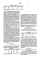
The following Table I sets forth mean values and ranges for RTi, RTv, TI and PDOC of a group of cancer patients and a group of healthy volunteers (control):
                                  TABLE 1*
__________________________________________________________________________
GROUP
     RTi,
         range
             RTv,
                 range
                      TI, range
                              PDOC,
                                   range
__________________________________________________________________________
Control
     4.66
         3.93-
             6.13
                 5.24-
                      1.32
                          1.15-
                              23.4 14.0-
(n = 23) 6.04    7.61     1.53     34.5
Cancer
     2.25
         1.54-
             6.46
                 4.45-
                      2.71
                          1.62-
                              60.3 38.3-
(n = 25) 4.02    8.72     4.39     88.0
p < 0.001        p = NS
__________________________________________________________________________
 *Recalcification times determined by TEG.
The cancer patients were evaluated at the time of diagnosis of the disease. There were significant differences between the recalcification times-endotoxin (RTi) between the group of the healthy volunteers and the group of the cancer patients, whereas there were no significant differences between recalcification times-saline (RTv) of such groups. Additionally, it can be readily seen that the thrombotic index (TI) is greater for the group of cancer patients than the thrombotic index (TI) of the healthy volunteers. The same proposition held true of the comparison of percent difference in clotting (PDOC) therebetween. The values for TI or PDOC do not overlap for these groups.
It has been found in cancer patients where a large portion of the tumor load is removed and minute portions remain, that the RTi, TI and PDOC differences still range outside the parameters of the group of healthy volunteers. A patient having cancer of the colon exhibited TI and PDOC values of 1.91 and 47.6%, respectively, one week post surgery. It was subsequently determined that tumor growth had invaded adjoining tissue.
The method of the present invention permits a clinician to evaluate effects of therapy on the state of the cancer in a cancer patient. For example, small changes in RTi values and a lowering of TI or PDOC values after non-fully curative treatments have been demonstrated. If chemotheraphy and/or radiation treatment do not alter such values, changes in the treatment are then suggested to find a more effective drug regime and/or radiation protocol. An advantage of the method of the present invention is the convenience of sampling and evaluation at varying times after therapy and the assessment of effectiveness of treatment prior to physical appearance of clinical changes.
Of the types of cancers detected by the method of the present invention included are cancers of the lung, breast, biliary tract, bladder, larynx, ovary, head and neck, colon, rectum, esophagus, soft palate, pancreas, and floor of the mouth. As hereinabove mentioned, the presence or absence of remaining malignancy after curative surgery may be determined by the methods of the present invention.
The following Table II sets forth specific data with reference to six patients; patients 1 to 4 having cancer and patients 5 and 6 having benign breast lesions.
              TABLE II*
______________________________________
Patient    RT.sub.i
                  RT.sub.v   TI   PDOC
______________________________________
1          2.00   7.31       3.66 72.6
2          3.28   7.04       2.15 53.4
3          2.51   6.61       2.63 60.2
4          3.52   6.06       1.72 42.0
5          4.32   4.54       1.05 4.84
6          4.87   5.98       1.23 18.6
______________________________________
 *Recalcification times determined by TEG.
The above date clearly illustrates a lower recalcification times-endotoxin (RTi) for the patients (#1-#4) with breast cancer as distinguished from the patients (#5-#6) with benign breast lesions. A similarity of comparison was noted between the RTi values of patients (#1-#4) and RTi values set forth in Table I, above. Additionally patients (#1-#4) had significantly higher TI and PDOC values. Patients #3 and #4, post one week surgery, exhibited TI and PDOC values of 1.25 and 1.39 and 20.0% and 27.9%, respectively, indicative of the successful removal of all malignancy as demonstrated by subsequent histological examination of tissue and non-existence of cancerous cells in the lymph nodes.
As hereinabove discussed, in cancer cases where the malignancy is not totally removed, the RTi, TI and PDOC values remain in the pre-operative ranges.
The following Table III sets forth mean values for RTi and RTv of a group of 36 patients having diabetes:
              TABLE III*
______________________________________
Group     RT.sub.i  Range     RT.sub.v
                                      Range
______________________________________
Control   5.69 ± 0.74
                    4.6-7.2   6.55 ± 0.08
5.3-8.5
(n = 19)
Diabetics 4.99 ± 1.20
                    3.0-8.1   5.65 ± 2.3
                                      3.3-16.8
          p < 0.001           p < 0.001
______________________________________
 *Recalcification times determined by SONOCLOT ® Coagulation Analyzer.
Eighteen of 36 (50%) diabetics had accelerated clotting in the saline incubated samples and 15 of 36 (42%) had clotting times shorter than the shortest control value for the endotoxin incubated samples. It would appear that diabetics with abnormal values have the more severe disease (e.g. juvenile diabetics, diabetics with vascular complications, diabetics with disease more than 15 years, etc.). The methods of the present invention will be useful in measuring therapeutic effects on diabetic activity, such as diet control, exercise and drug treatment, and may become a goal of the therapy to bring the RTv and RTi values of diabetics into the normal range.
The following Table IV sets forth mean values and ranges of RTi, RTv, TI and PDOC of healthy volunteers (per Example I) with mean values and ranges of RTi, RTv, TI and PDOC of four (4) confirmed advanced AIDS patients:
                                  TABLE IV*
__________________________________________________________________________
Group
     RT.sub.i,
         range
             RT.sub.v,
                 range
                     TI, range
                             PDOC,
                                  range
__________________________________________________________________________
AIDS 4.94
         4.43-
             5.78
                 5.12-
                     1.17
                         1.08-
                             14.5 7.5-
         5.40    6.94    1.29     22.2
Control
     4.66
         3.93-
             6.13
                 5.24-
                     1.32
                         1.15-
                             23.4 14.0-
         6.04    7.61    1.53     34.5
__________________________________________________________________________
 *Recalcification time determined by TEG.
From the above data, it can be seen that an evaluation of the recalcification times-endotoxin (RTi) of the AIDS patients are higher, but not necessarily significant as distinguished from like comparison of cancer patients. It is noted, however, that the TI and PDOC values are sifgnificantly lower, particularly in three out of four cases. Another form of algorithm could be derived to more unequivocally identify all such AIDS patients. Thus, the methods of the present invention illustrate that mammals having AIDS or AIDS-like disease have a PDOC value below the control, and with some statistically determined base line value, to qualify individuals for blood donation.
In surgical intensive care units, particularly patients exposed to severe trauma and/or major surgery and are being managed by life-support systems frequently succumb to multi-system failure resulting from progressive and perhaps undetected sepsis.
The following Table V sets forth values of a group (9 patients) exhibiting a localized septic condition from a group (62 patients) who did not exhibit such a condition, it being noted that RTi values are lower and TI and PDOC values are higher, as expected (compared to a normal population) as a result of released thromboplastins and immunological consequences from the presence of traumatized tissues:
              TABLE V*
______________________________________
GROUP    RT.sub.i
                RT.sub.v TI        PDOC
______________________________________
Control  4.66   6.53     1.32      23.4
Septic                   8 < 1.30 > 2
                                   8 < 23.0 > 2
Non-Septic               2 < 1.30 > 60
                                   2 < 23.0 > 60
                         p < 0.001
______________________________________
 *Recalcification times determined by TEG.
As hereinabove mentioned, as a result of the traumatic condition of the patients being tested, the RTi and TI values are not readily comparable with values of RTi and TI of non-surgery or pre-surgery patients.
In eighty percent (80%) of the septic patients, the thrombotic indices are not elevated, as expected. Values of TI lower than 1.30 and PDOC values lower than 23.0% usually shown the presence of sepsis in the abdominal cavity of the patient, probably due to the fact that high concentrations of an immunomodulator have entered the bloodstream of such individuals.
The following Table VI sets forth the RTi, RTv, TI and PDOC values for post-operative and post-traumatic patients for non-surviving patients (9), surviving patients (46), and non-operative normal volunteers (23):
              TABLE VI*
______________________________________
Group       RT.sub.i
                   RT.sub.v   TI   PDOC
______________________________________
Control     4.66   6.53       1.32 23.4
Non-Survivor
            4.51   6.72       1.49 32.9
Survivor    2.21   6.09       2.75 63.9
______________________________________
 *Recalcification times determined by TEG.
There were no significant differences in the RTv values, but highly significant differences in the RTi, TI, and PDOC values between survivors and non-survivors. Unlike the elevated value of TI and PDOC in cancer patients, elevated values occurring within 48 hours post-operative or post-trauma patients are good prognostication indicators. If the post-operative values are low, there is an indication of possible septic complications. Further, a TI value of 1.60 appears to be the threshold value of immunocompetence, since 8 of 9 patients who died had values below 1.60, whereas only one patient with a value above 1.60 died.
The test measures the immunocompetence of an individual. Therefore, activation (accelerated clotting) in response to test endotoxin, of the cancer patient (early diagnosed), and post-operative or post-trauma patients, each showing an altered (activated) state of the immune response system as reflected in accelerated clotting time under the influence of test endotoxin, may in part explain the thrombotic complications associated with these states.
The following Table VII set forth values of 33 patients with multiple sclerosis, many of whom were in clinical remission at the time of testing:
              TABLE VII*
______________________________________
Group  RT.sub.i  Range   RT.sub.v
                                 Range PDOC
______________________________________
MS
Patients
       3.67 ± 1.27
                 1.2- 7.0
                         4.99 ± .93
                                 2.7- 7.3
                                       26.2 ± 20.0
(n = 33)
Control
       5.69 ± .75
                 4.6- 7.2
                         6.55 ± .82
                                 5.3- 8.5
                                       12.2 ± 11.1
(n = 19)
______________________________________
 *Recalcification times determined by SONOCLOT ® Coagulation Analyzer
Twenty-two of the 33 MS patients exhibited abnormal RTv values, and 29 of the 33 patients had abnormal RTi values. The data indicates that an active disease state is present, even though a state of clinical remission is observed. It is readily appreciated by one skilled in the art that a practitioner will utilize the methods of the present invention to tailor medical therapy to the presence of an active disease, as well as to monitor the patient's condition during therapy.
As indicated earlier and as illustrated above, the test method of this invention is capable of detecting a variety of disease states in which the latent procoagualant generation is elevated (activated monocytes). These states include myocardial infarction, stroke, infections, acquired immune dysfunction syndrome (AIDS), rheumatoid arthritis, cancer, multiple sclerosis, etc.
In addition, by varying the concentrations of the immunomodulators, e.g., endotoxin, carrageenan, etc., the sensitivity to a threshold challenge can be ascertained. In some states cancer, multiple sclerosis, etc., use of immunomodulator concentrations one-hundredth the optimum concentration can also detect an activated cell in a blood sample. Use of these varying immunomodulator dose concentrations enables the detection, and effects of therapy on some diseases.
There are a variety of immunomodulators than can activate isolated monocytes and thereby stimulate these cells to generate a procoagulant activity in culture. These stimulants include endotoxin, immune complexes, complement split products, inflammatory particles, amines, phorbol esters, lectins, antigens, chemical mediators of inflammation, lipoproteins, virus damaged cells, tumor cells, etc. It is believed that the stimuli that activate monocytes interact with cellular plasma membranes. Perturbation of these membranes can occur via immunomodulatory-cell membrane contact involving specific receptors or unspecific interactions.
The purpose of these examples is to show that a multitude of immunomodulators in this protocol in which whole blood is employed can be utilized and that endotoxin was just a representative of the group.
The citrated blood samples utilized to obtain the following data were from normal individuals as well as from individuals with a variety of disease states. In some examples, differences can be seen in the values obtained between saline, endotoxin and other immunomodulators but they may not be significant (p values) changes. This is due to the relatively small sample size and/or the fact that bloods from healthy and varying disease states were utilized.
In Table VIII, below, the number of citrated whole blood samples tested, the mean RT saline±SD (standard deviation of the means) RT endotoxin±SD and RT immunomodulator±SD are tabulated. The analysis of variance (ANOVA) was used when comparing the means for the different group values of P-0.05 are significant.
                                  TABLE VIII
__________________________________________________________________________
Number of
Samples
      Concentration
              Saline
                    Endotoxin
                            Immunomodulator
                                       Category
__________________________________________________________________________
3     10 μg/cc
              6.43 ± 1.70
                    5.00 ± .85
                            5.23 ± .68
                                       Adjunct peptide, muamyl peptide,
                            Adjunct Peptide
                                       immunological activator, 10
                                       μg/cc
                                       CWB
9     20 μg/cc
              6.97 ± 1.94
                    5.29 ± .93*
                            5.36 ± 1.51
                                       Lectin
                            Con A
9     20 μg/cc
              4.90 ± 1.28
                    4.57 ± 1.07
                            4.57 ± 1.32
                                       Mitogen
                            Pokeweed
9     unknown 4.90 ± 1.28
                    4.57 ± 1.07
                            4.79 ± 1.03
                                       Measles virus
                            Virus
7     2 μg/cc
              5.30 ± .91
                    4.09 ± .57
                            5.30 ± 1.04
                                       Platelet activating factor,
                                       lipid,
                            PAF        mediator released during trauma
                                       and inflammation, allergy
5     20 μg/cc
              6.04 ± 1.26
                    5.22 ± .49
                            4.54 ± .45**
                                       Zymosan - immunomodulator
                            Zymosan
7     1 μg/cc
              5.51 ±  .74
                    5.94 ± .63
                            5.10 ± .87
                                       Myelin, protein, antigen
                            Myelin
__________________________________________________________________________
Number of
      Final   RT Saline
                    RT Endotoxin
                            RT Immunomodulator
Samples
      Concentration
              ± SD
                    ± SD ± SD    Category
__________________________________________________________________________
5     1 μg/cc
              .56 ± 1.33
                    5.10 ± 1.92
                            5.86 ± 1.34
                                       12-O--tetradecanoyl phorbol
                            TPA        13-acetate (TPA phorbol
                                       ester) cellular activator
3     .001 unit/cc
              4.37 ± .76
                    3.50 ± 2.35
                            3.56 ± 0.72
                                       Thrombin - enzyme
                            Thrombin   procoagulant
7     2.0 units/cc
              4.91 ± .51
                    4.13 ± .08*
                            3.72 ± 1.27**
                                       Interferon -
                            Interferon immunological mediator
                                       pharmacological agent
9     10 μg/cc
              4.80 ± 1.14
                    4.22 ± 0.84*
                            3.28 ± 0.68**
                                       Carageenan - inflammatory
                            Carrageenan
                                       mediator, colloid
9     400 μg/cc
              4.80 ± 1.14
                    4.22 ± 0.84*
                            3.55 ± .72**
                                       Latex Beads,
                            Latex Beads
                                       Phagocytic particle
8     10 μg/cc
              5.26 ± 1.58
                    3.04 ± 1.20*
                            4.35 ± 1.70**
                                       Platelet activator,
                            Collagen   abnormal surface for
                                       blood contact,
                                       immunomodulator
5     3.0 mg/cc
              5.18 ± .88
                    4.26 ± .78*
                            6.62 ± 1.62
                                       Amine, intracellular
                            ammonium   compartment modifier
                            chloride   affects pH
__________________________________________________________________________
 *signifies RT endotoxin different than RT saline.
 **signifies RT immunomodulator different than RT saline.
In addition to the above immunomodulators, the following materials were successfully on at least one sample:
______________________________________
NAME             CATEGORY
______________________________________
Lipid A          lipid, bacterial product
Ionophore A 23187
                 pharmacological stimulant
Mixed Bloods     foreign cells, complement,
                 immune complex formation etc.
Cycloheximide    DNA, RNA inhibitors, antibiotic
Snake Venom      immunologically active,
                 procoagulant
Nicotine         Vasoactive biochemical
Salmonella & Other Endotoxins
                 bacterial products
Adenosine Diphosphate
                 effects cellular energy balance
                 platelet aggregation
Mixed Plasma     foreign species
Tuftsin          tetrapeptide - leukocyte
                 stimulant
Glucan           yeast cell wall, foreign surface
Streptokinase    clot dissolving agent, lyte
                 agent, immunologically active
Urokinase        clot dissolving agent, lyte
                 agent, immunologically active
Papain           enzyme
Immunoglobulin G (IgG)
                 inflammatory blood product
Ferritin         foreign protein
PPD              purified protein derivative
Phytohemagluttinin
                 lectin
Pathophysiological Agents
                 damaged cells, etc.
______________________________________
In this Example, the effect of varying immunomodulator concentrations on recalcification times was measured. The immunomodulators, and other parameters of testing are set forth below:
A. Thromboplastin, (also called tissue factor or clotting factor III). Material generated by monocytes, procoagulant, immunomodulator.
______________________________________
Concentration of tissue factor (ug/CC CWB)
       0       3         6         15
______________________________________
RT ± SD
         6.11 ± 0.92
                   2.98 ± .60
                             2.34 ± .35
                                     1.96 ± .51
n = 8 samples
______________________________________
Significance ANOVA p<:05 for comparisons between different groups
B. E. coli endotoxin
______________________________________
Concentration of endotoxin (ug/cc CWB)
       0       10        1.0       0.1
______________________________________
         (A)       (B)       (C)     (D)
RT ± DF
         5.25 ± 1.03
                   3.75 ± .78
                             4.20 ± .49
                                     4.16 ± .32
n = 8 samples
______________________________________
Significance (ANUA)
______________________________________
       A vs B;       P < .001
       A vs C;       P < 05
       A vs D;       P < .05
       B vs C;       P = NS
       C vs D;       P = NS
______________________________________
By varying immunomodulator concentrations, the threshold quantity necessary to give a significant change in RT relative to that of the saline value may be of clinical significance. The minimum concentration of a particular immunomodulator necessary to produce change is called threshold value and this may vary between diseases and may enable a differential diagnosis (e.g. cancer, not diabetes, etc.) to be made.
Samples of citrated whole blood from patients (4) on Warfarin (anticoagulant) therapy were analyzed with this test. The prothrobin time of patients ranged between 18.6 to 65 seconds (normal 12 sec.).
______________________________________
RT saline (min)
              RT endotoxin (min)
______________________________________
18.7 ± 3.56
              5.48 ± 2.91
______________________________________
significance, paired t test, P<0.02
The RT saline is very prolonged due to the anticoagulant drug. However, RT endotoxin mean is in the normal range suggesting that this anticoagulant has little effect on tissue factor generation.
Advantages of the present invention are many, e.g. donor blood may be pre-screened, particularly where a pathological condition of AIDS may exist, let alone the undesirability of use of blood for transfusions where such blood evidences the existence of a pathological state or condition in the blood donor. The present invention may be used to evaluate compatibility of transfusion of a particular blood donor. Still further, the methods of the present invention permit the facile monitoring of the effectiveness of drug therapy or regime to a particular pathological condition or state in a mammal, e.g. diabetes. The present invention permits a facile evaluation of the potential acceptance or rejection by a mammal of a transplant organ.
While the present invention is discussed with primary reference to the evaluation of a cellular hematologic fluid to determine if a mammal has a pre-existing pathological condition, it is apparent to one skilled in the art that the method of the present invention may be used in the prognosis of treatment of a known pathogenic state in a mammal. As illustrated, in Example II above, the immune response system of the cellular hematologic fluid of a mammal having undergone surgery for the removal of cancerous tissue may be evaluated to determine if all cancerous tissue has been removed from the mammal and/or the extent of sepsis thereof. Similarly, a post-operative protocol, e.g. chemotherapy, may be monitored for effectiveness of such post-operative protocol.
While the invention herein has been described in connection with exemplary embodiments thereof, it will be understood that many modifications will be apparent to those of ordinary skill in the art and that this application is intended to cover any adaptations or variations thereof. Therefore, it is manifestly intended that this invention be only limited to the claims and the equivalents thereof.
Claims (14)
1. A prepackaged diagnostic kit for analyzing the blood of a mammal to determine the presence or development of pathology suspected of causing abnormalities in the immune response, system and/or the blood coagulated of the mammal comprising:
A. at least one first container having a predetermined anticoagulant disposed therein, for receiving whole blood and rendering the same anticoagulated;
B. at least one second container serving for the preparation of a control sample when an aliquot portion of the anticoagulated blood from said first container is introduced therein;
C. at least one third container having a predetermined quantity of a modulator for reception of a further aliquot of said anticoagulated blood from said first container;
D. means for initiating the clotting of said control sample and said activated sample to facilitate a subsequent measurement of the reaction parameters of each of said samples; and
E. a diagnostic protocol for the determination of the pathological state of said mammal by the analysis of said control and said activated blood sample by the comparison of the respective reaction parameters thereof.
2. The kit of claim 1 including at least one syringe for extracting whole blood from said mammal.
3. The kit of claim 1 wherein at least said second and said third containers comprise test tubes with removable caps therefor.
4. The kit of claim 1 wherein said modulator is present in individual sample containers in an amount of from about 10 to about 20 ul/cc of citrated whole blood.
5. The kit of claim 1, wherein said suitable vehicle comprises a quantity of a physiological saline solution.
6. The kit of claim 1, wherein said protocol comprises instructions for the determination of a Thrombotic Index (TI) comprising the ratio of the reaction parameter of the control sample to that of the activated sample and/or the Percentage Difference Of Clotting (PDOC) comprising the difference between the reaction parameters of said control sample and said activated sample, said difference multiplied by 100 and divided by the said control sample reaction parameter.
7. The kit of claim 6, wherein said protocol further comprises one or more predetermined Thrombotic Indices and/or PDOC values derived from respective ratios of reaction parameters of standard control samples to reaction parameters of respective standard activated samples that have been associated with the healthy state and/or the presence or development of particular pathologies, for comparative determination and analysis with the samples prepared and processed by use of the remaining components of said kit.
8. The kit of claim 1, wherein said modulator is selected from the group consisting of endotoxins, virus, Interferon, phorbol esters, collagens, platelet activating factors, carrageenans, adjuvant containing peptides, thromboplastins, myelin, gram negative bacteria, lectins and mitogens.
9. The kit of claim 8, wherein said modulator is an endotoxin.
10. The kit of claim 1, wherein said anticoagulant comprises a compound selected from the group consisting of sodium citrate and sodium oxalate.
11. The kit of claim 10, wherein said anticoagulant comprises sodium citrate.
12. The kit of claim 1, wherein said means for initiating clotting comprises a clotting initiator compound.
13. The kit of claim 12, wherein said clotting initiator compound comprises a calcium-containing compound.
14. A method for analyzing the blood of a mammal to determine the presence or development of pathology suspected of causing abnormalities in the immune response system and/or the blood coagulation of the mammal consisting essentially of:
A. preparing a quantity of anticoagulated whole blood from a sample of whole blood taken from a mammal;
B. taking an aliquot portion of said anticoagulated blood and introduing said aliquot portion into a first container and thereby preparing a control sample;
C. taking a further aliquot portion of said anticoagulated blood and introducing said further aliquot portion into a second container having therein a modulator and thereby preparing an activated sample;
D. incubating said control sample and said activated sample at a predetermined suitable incubation temperature from about 1 to about 4 hours;
E. initiating clotting activity and measuring a reaction parameter for each of said control sample and said activated sample; and
F. identifying the presence of pathology by comparing the reaction parameters measured in Step E. with similar reaction parameters measured from a mammal in a healthy state.
Priority Applications (1)
| Application Number | Priority Date | Filing Date | Title |
|---|---|---|---|
| US07/236,039 US4900679A (en) | 1983-01-26 | 1988-08-23 | Method for determining the existence and/or the monitoring of a pathological condition in a mammal and a test kit therefor |
Applications Claiming Priority (5)
| Application Number | Priority Date | Filing Date | Title |
|---|---|---|---|
| US44054083A | 1983-01-26 | 1983-01-26 | |
| US53878383A | 1983-10-04 | 1983-10-04 | |
| US06/734,799 US4705756A (en) | 1983-01-26 | 1985-05-16 | Method of determining the existence and/or the monitoring of a pathological condition in a mammal |
| WOPCT/US86/01075 | 1986-05-16 | ||
| US07/236,039 US4900679A (en) | 1983-01-26 | 1988-08-23 | Method for determining the existence and/or the monitoring of a pathological condition in a mammal and a test kit therefor |
Related Parent Applications (2)
| Application Number | Title | Priority Date | Filing Date |
|---|---|---|---|
| US06703120 Continuation-In-Part | 1985-02-19 | ||
| US07/034,101 Division US4814247A (en) | 1983-01-26 | 1986-05-16 | Method for determining the existance and/or the monitoring of a pathological condition in a mammal |
Publications (1)
| Publication Number | Publication Date |
|---|---|
| US4900679A true US4900679A (en) | 1990-02-13 |
Family
ID=27499835
Family Applications (1)
| Application Number | Title | Priority Date | Filing Date |
|---|---|---|---|
| US07/236,039 Expired - Lifetime US4900679A (en) | 1983-01-26 | 1988-08-23 | Method for determining the existence and/or the monitoring of a pathological condition in a mammal and a test kit therefor |
Country Status (1)
| Country | Link |
|---|---|
| US (1) | US4900679A (en) |
Cited By (13)
| Publication number | Priority date | Publication date | Assignee | Title |
|---|---|---|---|---|
| US5266462A (en) * | 1992-06-03 | 1993-11-30 | Baxter Diagnostics Inc. | Measurement of platelet activities |
| WO1998014779A1 (en) * | 1996-10-02 | 1998-04-09 | University Of Medicine And Dentistry Of New Jersey | Comparative determination of viscosity in body fluids |
| US5783447A (en) * | 1996-10-02 | 1998-07-21 | University Of Medicine And Dentistry Of New Jersey | Hypercoagulability comparative determinants obtained using detection systems with variable force-induced energy inputs |
| US6245573B1 (en) | 1998-02-12 | 2001-06-12 | University Of Medicine And Dentistry Of New Jersey | Rapid assessment of the coagulant activity of blood |
| US20050148899A1 (en) * | 2003-10-22 | 2005-07-07 | Walker William F. | Method and apparatus for characterization of clot formation |
| US9031701B2 (en) | 2011-02-15 | 2015-05-12 | Hemosonics Llc | Characterization of blood hemostasis and oxygen transport parameters |
| US9272280B2 (en) | 2011-02-15 | 2016-03-01 | Hemosonics Llc | Device, systems and methods for evaluation of hemostasis |
| US9726647B2 (en) | 2015-03-17 | 2017-08-08 | Hemosonics, Llc | Determining mechanical properties via ultrasound-induced resonance |
| US10147152B2 (en) | 2010-04-08 | 2018-12-04 | Hemosonics, Llc | Hemostatic parameter display |
| US10996230B2 (en) | 2008-12-23 | 2021-05-04 | C A Casyso Gmbh | Cartridge device for a measuring system for measuring viscoelastic characteristics of a sample liquid, a corresponding measuring system, and a corresponding method |
| US11054396B2 (en) | 2011-05-19 | 2021-07-06 | Hemosonics Llc | Hemostasis analyzer |
| US11327069B2 (en) | 2014-09-29 | 2022-05-10 | Ca Casyso Gmbh | Blood testing system and method |
| US11366093B2 (en) | 2017-04-20 | 2022-06-21 | Hemosonics, Llc | Disposable system for analysis of hemostatic function |
Citations (5)
| Publication number | Priority date | Publication date | Assignee | Title |
|---|---|---|---|---|
| US3267364A (en) * | 1962-04-18 | 1966-08-16 | Becton Dickinson Co | Coagulation timer |
| US3449081A (en) * | 1965-03-29 | 1969-06-10 | Electronic Instr Co | Test kit |
| US4047890A (en) * | 1973-11-01 | 1977-09-13 | Bio/Data Corporation | Method and apparatus for determining deficiencies in enzymatic reactors particularly clotting factor levels in blood plasmas |
| US4705756A (en) * | 1983-01-26 | 1987-11-10 | University Of Medicine And Dentistry Of New Jersey | Method of determining the existence and/or the monitoring of a pathological condition in a mammal |
| US4814247A (en) * | 1983-01-26 | 1989-03-21 | University Of Medicine And Dentistry Of New Jersey | Method for determining the existance and/or the monitoring of a pathological condition in a mammal |
-
1988
- 1988-08-23 US US07/236,039 patent/US4900679A/en not_active Expired - Lifetime
Patent Citations (5)
| Publication number | Priority date | Publication date | Assignee | Title |
|---|---|---|---|---|
| US3267364A (en) * | 1962-04-18 | 1966-08-16 | Becton Dickinson Co | Coagulation timer |
| US3449081A (en) * | 1965-03-29 | 1969-06-10 | Electronic Instr Co | Test kit |
| US4047890A (en) * | 1973-11-01 | 1977-09-13 | Bio/Data Corporation | Method and apparatus for determining deficiencies in enzymatic reactors particularly clotting factor levels in blood plasmas |
| US4705756A (en) * | 1983-01-26 | 1987-11-10 | University Of Medicine And Dentistry Of New Jersey | Method of determining the existence and/or the monitoring of a pathological condition in a mammal |
| US4814247A (en) * | 1983-01-26 | 1989-03-21 | University Of Medicine And Dentistry Of New Jersey | Method for determining the existance and/or the monitoring of a pathological condition in a mammal |
Cited By (36)
| Publication number | Priority date | Publication date | Assignee | Title |
|---|---|---|---|---|
| US5266462A (en) * | 1992-06-03 | 1993-11-30 | Baxter Diagnostics Inc. | Measurement of platelet activities |
| WO1998014779A1 (en) * | 1996-10-02 | 1998-04-09 | University Of Medicine And Dentistry Of New Jersey | Comparative determination of viscosity in body fluids |
| US5783447A (en) * | 1996-10-02 | 1998-07-21 | University Of Medicine And Dentistry Of New Jersey | Hypercoagulability comparative determinants obtained using detection systems with variable force-induced energy inputs |
| US5792660A (en) * | 1996-10-02 | 1998-08-11 | University Of Medicine And Dentistry Of New Jersey | Comparative determinants of viscosity in body fluids obtained with probes providing increased sensitivity |
| US6245573B1 (en) | 1998-02-12 | 2001-06-12 | University Of Medicine And Dentistry Of New Jersey | Rapid assessment of the coagulant activity of blood |
| US8740818B2 (en) | 2003-10-22 | 2014-06-03 | Hemosonics, Llc | Method and apparatus for characterization of clot formation |
| US9723996B2 (en) | 2003-10-22 | 2017-08-08 | Hemosonics, Llc | Method and apparatus for characterization of clot formation |
| US7892188B2 (en) | 2003-10-22 | 2011-02-22 | Hemosonics, Llc | Method and apparatus for characterization of clot formation |
| US20110034805A1 (en) * | 2003-10-22 | 2011-02-10 | Walker William F | Method and apparatus for characterization of clot formation |
| US20050148899A1 (en) * | 2003-10-22 | 2005-07-07 | Walker William F. | Method and apparatus for characterization of clot formation |
| US11892459B2 (en) | 2008-12-23 | 2024-02-06 | C A Casyso Gmbh | Cartridge device for a measuring system for measuring viscoelastic characteristics of a sample liquid, a corresponding measuring system, and a corresponding method |
| US11879899B2 (en) | 2008-12-23 | 2024-01-23 | C A Casyso Gmbh | Cartridge device for a measuring system for measuring viscoelastic characteristics of a sample liquid, a corresponding measuring system, and a corresponding method |
| US10996230B2 (en) | 2008-12-23 | 2021-05-04 | C A Casyso Gmbh | Cartridge device for a measuring system for measuring viscoelastic characteristics of a sample liquid, a corresponding measuring system, and a corresponding method |
| US11768211B2 (en) | 2008-12-23 | 2023-09-26 | C A Casyso Gmbh | Cartridge device for a measuring system for measuring viscoelastic characteristics of a sample liquid, a corresponding measuring system, and a corresponding method |
| US11360106B2 (en) | 2008-12-23 | 2022-06-14 | C A Casyso Gmbh | Cartridge device for a measuring system for measuring viscoelastic characteristics of a sample liquid, a corresponding measuring system, and a corresponding method |
| US11131680B2 (en) | 2008-12-23 | 2021-09-28 | C A Casyso Gmbh | Cartridge device for a measuring system for measuring viscoelastic characteristics of a sample liquid, a corresponding measuring system, and a corresponding method |
| US11061038B2 (en) | 2008-12-23 | 2021-07-13 | C A Casyso Gmbh | Cartridge device for a measuring system for measuring viscoelastic characteristics of a sample liquid, a corresponding measuring system, and a corresponding method |
| US11456071B2 (en) | 2010-04-08 | 2022-09-27 | Hemosonics Llc | Hemostatic parameter display |
| US10147152B2 (en) | 2010-04-08 | 2018-12-04 | Hemosonics, Llc | Hemostatic parameter display |
| US9031701B2 (en) | 2011-02-15 | 2015-05-12 | Hemosonics Llc | Characterization of blood hemostasis and oxygen transport parameters |
| US9977039B2 (en) | 2011-02-15 | 2018-05-22 | Hemosonics Llc | Devices, systems and methods for evaluation of hemostasis |
| US9272280B2 (en) | 2011-02-15 | 2016-03-01 | Hemosonics Llc | Device, systems and methods for evaluation of hemostasis |
| US9410971B2 (en) | 2011-02-15 | 2016-08-09 | Hemosonics Llc | Devices, systems and methods for evaluation of hemostasis |
| US10481168B2 (en) | 2011-02-15 | 2019-11-19 | Hemosonics Llc | Devices, systems and methods for evaluation of hemostasis |
| US10161944B2 (en) | 2011-02-15 | 2018-12-25 | Hemosonics Llc | Methods for evaluation of hemostasis |
| US10962524B2 (en) | 2011-02-15 | 2021-03-30 | HomoSonics LLC | Characterization of blood hemostasis and oxygen transport parameters |
| US10031144B2 (en) | 2011-02-15 | 2018-07-24 | Hemosonics Llc | Devices, systems and methods for evaluation of hemostasis |
| US11680940B2 (en) | 2011-02-15 | 2023-06-20 | Hemosonics Llc | Characterization of blood hemostasis and oxygen transport parameters |
| US11054396B2 (en) | 2011-05-19 | 2021-07-06 | Hemosonics Llc | Hemostasis analyzer |
| US11719688B2 (en) | 2014-09-29 | 2023-08-08 | C A Casyso Gmbh | Blood testing system and method |
| US11327069B2 (en) | 2014-09-29 | 2022-05-10 | Ca Casyso Gmbh | Blood testing system and method |
| US11656206B2 (en) | 2015-03-17 | 2023-05-23 | Hemosonics Llc | Determining mechanical properties via ultrasound-induced resonance |
| US9726647B2 (en) | 2015-03-17 | 2017-08-08 | Hemosonics, Llc | Determining mechanical properties via ultrasound-induced resonance |
| US11002712B2 (en) | 2015-03-17 | 2021-05-11 | Hemosonics Llc | Determining mechanical properties via ultrasound-induced resonance |
| US10495613B2 (en) | 2015-03-17 | 2019-12-03 | Hemosonics, Llc | Determining mechanical properties via ultrasound-induced resonance |
| US11366093B2 (en) | 2017-04-20 | 2022-06-21 | Hemosonics, Llc | Disposable system for analysis of hemostatic function |
Similar Documents
| Publication | Publication Date | Title |
|---|---|---|
| US4705756A (en) | Method of determining the existence and/or the monitoring of a pathological condition in a mammal | |
| US4814247A (en) | Method for determining the existance and/or the monitoring of a pathological condition in a mammal | |
| Millar et al. | An evaluation of the heat precipitation method for plasma fibrinogen estimation | |
| Edwards et al. | Activation of blood coagulation in Crohn's disease: Increased plasma fibrinopeptide A levels and enhanced generation of monocyte tissue factor activity | |
| Mustard et al. | C-reactive protein levels predict postoperative septic complications | |
| Goldhaber et al. | Quantitative plasma D-dimer levels among patients undergoing pulmonary angiography for suspected pulmonary embolism | |
| US4900679A (en) | Method for determining the existence and/or the monitoring of a pathological condition in a mammal and a test kit therefor | |
| EP2235542B1 (en) | Diagnostic in vitro method for assessing von willebrand disease and increased bleeding risk associated with von willebrand disease and acquired or congenital disorders of platelet function | |
| WO2002079375A1 (en) | Rapid assessment of coagulation activity in whole blood | |
| CA1226216A (en) | Hematological prediction of sepsis and other disease states | |
| JPH02501115A (en) | Platelet aggregation reagent, reagent container and EDTA - method for measuring platelet aggregation in anticoagulant blood | |
| Caprini et al. | The identification of accelerated coagulability | |
| Lang et al. | Rapid Differentiation of Subarachnoid Hemorrhage from Traumatic Lumbar Puncture Using the D-Dimer/Assay | |
| US6156530A (en) | Method for analysis of haemostatic activity | |
| US3853710A (en) | Serum diagnostic test for maladies causing change in fibrinolytic activity in the blood | |
| US3960669A (en) | Urine diagnostic test for fibrinolytic activity | |
| RU2618404C1 (en) | Screening diagnosis method of acute cellular rejection of transplanted heart | |
| RU2723627C1 (en) | Method for prediction of severe preeclampsia in pregnant women accompanying a mutation of the prothrombin gene, genotype f2g20210a | |
| CN110361531B (en) | Experimental method for detecting particle coagulation promoting activity | |
| Oger et al. | Assessment of activated protein C resistance using a new and rapid venom-based test: STA Staclot APC-R | |
| Meyers | Fibrin split products in the severely burned patient | |
| RU2789822C1 (en) | Method for optimizing laboratory diagnosis of thrombinemia in patients with covid-19 infection | |
| RU2262109C2 (en) | Method for determination of blood plasma antithrombin iii concentration | |
| Darwish et al. | Anti-outer-membrane Porin C Antibody as Probable Biomarkers for Ulcerative Colitis (UC) | |
| JPS6122255A (en) | Hematological predicting and inspection method of septicemiaand other symptom |
Legal Events
| Date | Code | Title | Description |
|---|---|---|---|
| STCF | Information on status: patent grant |
Free format text: PATENTED CASE |
|
| FEPP | Fee payment procedure |
Free format text: PAYOR NUMBER ASSIGNED (ORIGINAL EVENT CODE: ASPN); ENTITY STATUS OF PATENT OWNER: SMALL ENTITY |
|
| FPAY | Fee payment |
Year of fee payment: 4 |
|
| FPAY | Fee payment |
Year of fee payment: 8 |
|
| SULP | Surcharge for late payment | ||
| FPAY | Fee payment |
Year of fee payment: 12 |