US5238652A - Analytical test devices for competition assay for drugs of non-protein antigens using immunochromatographic techniques - Google Patents
Analytical test devices for competition assay for drugs of non-protein antigens using immunochromatographic techniques Download PDFInfo
- Publication number
- US5238652A US5238652A US07/540,844 US54084490A US5238652A US 5238652 A US5238652 A US 5238652A US 54084490 A US54084490 A US 54084490A US 5238652 A US5238652 A US 5238652A
- Authority
- US
- United States
- Prior art keywords
- latex particles
- body fluid
- fluid sample
- analytical test
- colored
- Prior art date
- Legal status (The legal status is an assumption and is not a legal conclusion. Google has not performed a legal analysis and makes no representation as to the accuracy of the status listed.)
- Expired - Fee Related
Links
Images


Classifications
-
- G—PHYSICS
- G01—MEASURING; TESTING
- G01N—INVESTIGATING OR ANALYSING MATERIALS BY DETERMINING THEIR CHEMICAL OR PHYSICAL PROPERTIES
- G01N33/00—Investigating or analysing materials by specific methods not covered by groups G01N1/00 - G01N31/00
- G01N33/48—Biological material, e.g. blood, urine; Haemocytometers
- G01N33/50—Chemical analysis of biological material, e.g. blood, urine; Testing involving biospecific ligand binding methods; Immunological testing
- G01N33/53—Immunoassay; Biospecific binding assay; Materials therefor
- G01N33/558—Immunoassay; Biospecific binding assay; Materials therefor using diffusion or migration of antigen or antibody
-
- G—PHYSICS
- G01—MEASURING; TESTING
- G01N—INVESTIGATING OR ANALYSING MATERIALS BY DETERMINING THEIR CHEMICAL OR PHYSICAL PROPERTIES
- G01N33/00—Investigating or analysing materials by specific methods not covered by groups G01N1/00 - G01N31/00
- G01N33/48—Biological material, e.g. blood, urine; Haemocytometers
- G01N33/50—Chemical analysis of biological material, e.g. blood, urine; Testing involving biospecific ligand binding methods; Immunological testing
- G01N33/53—Immunoassay; Biospecific binding assay; Materials therefor
- G01N33/543—Immunoassay; Biospecific binding assay; Materials therefor with an insoluble carrier for immobilising immunochemicals
- G01N33/54366—Apparatus specially adapted for solid-phase testing
- G01N33/54386—Analytical elements
- G01N33/54387—Immunochromatographic test strips
- G01N33/54388—Immunochromatographic test strips based on lateral flow
-
- B—PERFORMING OPERATIONS; TRANSPORTING
- B01—PHYSICAL OR CHEMICAL PROCESSES OR APPARATUS IN GENERAL
- B01L—CHEMICAL OR PHYSICAL LABORATORY APPARATUS FOR GENERAL USE
- B01L3/00—Containers or dishes for laboratory use, e.g. laboratory glassware; Droppers
- B01L3/50—Containers for the purpose of retaining a material to be analysed, e.g. test tubes
- B01L3/502—Containers for the purpose of retaining a material to be analysed, e.g. test tubes with fluid transport, e.g. in multi-compartment structures
- B01L3/5027—Containers for the purpose of retaining a material to be analysed, e.g. test tubes with fluid transport, e.g. in multi-compartment structures by integrated microfluidic structures, i.e. dimensions of channels and chambers are such that surface tension forces are important, e.g. lab-on-a-chip
-
- G—PHYSICS
- G01—MEASURING; TESTING
- G01N—INVESTIGATING OR ANALYSING MATERIALS BY DETERMINING THEIR CHEMICAL OR PHYSICAL PROPERTIES
- G01N33/00—Investigating or analysing materials by specific methods not covered by groups G01N1/00 - G01N31/00
- G01N33/48—Biological material, e.g. blood, urine; Haemocytometers
- G01N33/50—Chemical analysis of biological material, e.g. blood, urine; Testing involving biospecific ligand binding methods; Immunological testing
- G01N33/53—Immunoassay; Biospecific binding assay; Materials therefor
- G01N33/543—Immunoassay; Biospecific binding assay; Materials therefor with an insoluble carrier for immobilising immunochemicals
- G01N33/54366—Apparatus specially adapted for solid-phase testing
- G01N33/54386—Analytical elements
- G01N33/54387—Immunochromatographic test strips
- G01N33/54388—Immunochromatographic test strips based on lateral flow
- G01N33/54389—Immunochromatographic test strips based on lateral flow with bidirectional or multidirectional lateral flow, e.g. wherein the sample flows from a single, common sample application point into multiple strips, lanes or zones
-
- G—PHYSICS
- G01—MEASURING; TESTING
- G01N—INVESTIGATING OR ANALYSING MATERIALS BY DETERMINING THEIR CHEMICAL OR PHYSICAL PROPERTIES
- G01N33/00—Investigating or analysing materials by specific methods not covered by groups G01N1/00 - G01N31/00
- G01N33/48—Biological material, e.g. blood, urine; Haemocytometers
- G01N33/50—Chemical analysis of biological material, e.g. blood, urine; Testing involving biospecific ligand binding methods; Immunological testing
- G01N33/58—Chemical analysis of biological material, e.g. blood, urine; Testing involving biospecific ligand binding methods; Immunological testing involving labelled substances
- G01N33/585—Chemical analysis of biological material, e.g. blood, urine; Testing involving biospecific ligand binding methods; Immunological testing involving labelled substances with a particulate label, e.g. coloured latex
-
- B—PERFORMING OPERATIONS; TRANSPORTING
- B01—PHYSICAL OR CHEMICAL PROCESSES OR APPARATUS IN GENERAL
- B01L—CHEMICAL OR PHYSICAL LABORATORY APPARATUS FOR GENERAL USE
- B01L2300/00—Additional constructional details
- B01L2300/04—Closures and closing means
- B01L2300/041—Connecting closures to device or container
- B01L2300/045—Connecting closures to device or container whereby the whole cover is slidable
-
- B—PERFORMING OPERATIONS; TRANSPORTING
- B01—PHYSICAL OR CHEMICAL PROCESSES OR APPARATUS IN GENERAL
- B01L—CHEMICAL OR PHYSICAL LABORATORY APPARATUS FOR GENERAL USE
- B01L2300/00—Additional constructional details
- B01L2300/04—Closures and closing means
- B01L2300/046—Function or devices integrated in the closure
-
- B—PERFORMING OPERATIONS; TRANSPORTING
- B01—PHYSICAL OR CHEMICAL PROCESSES OR APPARATUS IN GENERAL
- B01L—CHEMICAL OR PHYSICAL LABORATORY APPARATUS FOR GENERAL USE
- B01L2300/00—Additional constructional details
- B01L2300/08—Geometry, shape and general structure
- B01L2300/0809—Geometry, shape and general structure rectangular shaped
- B01L2300/0825—Test strips
-
- B—PERFORMING OPERATIONS; TRANSPORTING
- B01—PHYSICAL OR CHEMICAL PROCESSES OR APPARATUS IN GENERAL
- B01L—CHEMICAL OR PHYSICAL LABORATORY APPARATUS FOR GENERAL USE
- B01L2400/00—Moving or stopping fluids
- B01L2400/04—Moving fluids with specific forces or mechanical means
- B01L2400/0475—Moving fluids with specific forces or mechanical means specific mechanical means and fluid pressure
- B01L2400/0487—Moving fluids with specific forces or mechanical means specific mechanical means and fluid pressure fluid pressure, pneumatics
-
- B—PERFORMING OPERATIONS; TRANSPORTING
- B01—PHYSICAL OR CHEMICAL PROCESSES OR APPARATUS IN GENERAL
- B01L—CHEMICAL OR PHYSICAL LABORATORY APPARATUS FOR GENERAL USE
- B01L3/00—Containers or dishes for laboratory use, e.g. laboratory glassware; Droppers
- B01L3/50—Containers for the purpose of retaining a material to be analysed, e.g. test tubes
- B01L3/502—Containers for the purpose of retaining a material to be analysed, e.g. test tubes with fluid transport, e.g. in multi-compartment structures
- B01L3/5023—Containers for the purpose of retaining a material to be analysed, e.g. test tubes with fluid transport, e.g. in multi-compartment structures with a sample being transported to, and subsequently stored in an absorbent for analysis
-
- Y—GENERAL TAGGING OF NEW TECHNOLOGICAL DEVELOPMENTS; GENERAL TAGGING OF CROSS-SECTIONAL TECHNOLOGIES SPANNING OVER SEVERAL SECTIONS OF THE IPC; TECHNICAL SUBJECTS COVERED BY FORMER USPC CROSS-REFERENCE ART COLLECTIONS [XRACs] AND DIGESTS
- Y10—TECHNICAL SUBJECTS COVERED BY FORMER USPC
- Y10S—TECHNICAL SUBJECTS COVERED BY FORMER USPC CROSS-REFERENCE ART COLLECTIONS [XRACs] AND DIGESTS
- Y10S435/00—Chemistry: molecular biology and microbiology
- Y10S435/81—Packaged device or kit
-
- Y—GENERAL TAGGING OF NEW TECHNOLOGICAL DEVELOPMENTS; GENERAL TAGGING OF CROSS-SECTIONAL TECHNOLOGIES SPANNING OVER SEVERAL SECTIONS OF THE IPC; TECHNICAL SUBJECTS COVERED BY FORMER USPC CROSS-REFERENCE ART COLLECTIONS [XRACs] AND DIGESTS
- Y10—TECHNICAL SUBJECTS COVERED BY FORMER USPC
- Y10S—TECHNICAL SUBJECTS COVERED BY FORMER USPC CROSS-REFERENCE ART COLLECTIONS [XRACs] AND DIGESTS
- Y10S436/00—Chemistry: analytical and immunological testing
- Y10S436/901—Drugs of abuse, e.g. narcotics, amphetamine
Definitions
- the invention relates to analytical test devices which use immunochromatographic assays formulated on a competitive immunochemical protocol, and which determine small hapten, non-protein molecules such as those representing the presence of drugs of abuse. More particularly, the invention relates to self-contained analytical devices which require only the addition of a few drops of body fluid such as urine or other liquid to initiate a complex, multi-step immunoassay that produces a visually perceptible precipitin via antigen/antibody reactions, and which do not require instrumentation or sophisticated training to assess the results.
- the invention further relates to housing articles useful for packaging kits for these immunoassays, to novel reagents, and to methods for utilizing the novel reagents in the test devices which simultaneously can assay in a single device for up to five (5) of the National Institute of Drug Abuse (NIDA) designated drugs of abuse recommended in a drug screen.
- NIDA National Institute of Drug Abuse
- Immunoassay methods have distinct advantages when compared, for example, with techniques using thin layer chromatography, gas chromatography, gas chromatography/mass spectroscopy or high performance liquid chromatography because of the high specific accuracy associated with immunoassays and their ease of use.
- test and/or device which is simple to use and which can be used anywhere rather than only in a laboratory setting, which via competition of the analyte, or drug, and the analyte conjugate for limited antibody binding sites, can identify presence or absence of drugs of abuse in humans, and which does not require instrumentation to read the end results.
- the test devices of this invention will detect drugs of abuse, for example, in a stable immunoassay configuration using certain novel protein conjugates of these drugs of abuse, and the accompanying antibodies.
- a self-contained analytical testing device which can simultaneously perform assays for multiple drugs of abuse in a single device.
- test device which uses various immunochemical based configurations embodied in an immunochromatographic system.
- the test devices use small antigen conjugates, have precipitin end points with hapten detection capabilities, contain chromatographic supports made of impregnated membranes, and offer accuracy as well as ease of use in non-laboratory settings.
- the test devices require only the addition of a few drops of urine, biological fluids or aqueous solutions.
- At least five drugs of abuse can be tested simultaneously in a single device.
- the five drugs of abuse recommended by the NIDA for a drug screen namely amphetamines/methamphetamines, cocaine, opiates, phencyclidine, and cannabinoids, are thus readily detectable simultaneously using a simple test kit.
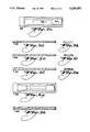
- FIG. 1a is a top view of a test device according to the invention.
- FIG. 1b is a side view of the test device of FIG. 1a.
- FIG. 2a is a top view of a top piece of a test device according to the invention.
- FIG. 2b a side section view of the top piece of FIG. 2a.
- FIG. 2c is a top view of a bottom piece of a test device according to the invention.
- FIG. 2e is a side section view of an alternative embodiment of a bottom piece having a dish-like sample well.
- FIG. 2g is a top view of a test device having the top piece of FIG. 2a and the bottom piece of FIG. 2c.
- FIG. 2i is an end section view of the top piece of FIG. 2b.
- FIG. 2k is an end view of the bottom piece of FIG. 2d.
- FIG. 3b is a side sectional view of the test device holder of FIG. 3a.
- FIG. 3c is an end sectional view of the test device holder of FIG. 3a.
- FIG. 3e is a side sectional view of the test device holder of FIG. 3d.
- FIG. 3f is an end sectional view of the test device holder of FIG. 3d.
- FIG. 3h is a side sectional view of the test device assembly of FIG. 3g.
- FIG. 4a is a top view of a top piece of an alternative embodiment of a test device according to the invention.
- FIG. 4c is a top view of a bottom piece of an alternative embodiment of a test device according to the invention.
- FIG. 4f is a side sectional view of the test device of FIG. 4e.
- FIG. 4i is an end sectional view of the top piece of FIG. 4a taken along line 4i--4i.
- FIG. 4j is an end view of the bottom piece of FIG. 4c.
- FIG. 5 is a top view of an alternative embodiment a test device having a capillary passage.
- FIG. 6a is a top view of a top piece of an alternative embodiment of a test device according to the invention.
- FIG. 6b is a top view of a bottom piece of an alternative embodiment of a test device according to the invention.
- FIG. 7a is a top view of a bottom piece of a test device assembly having a common reception cavity.
- FIG. 7b is a top view of a top piece of a test device assembly having a common reception cavity.
- FIG. 7c is a top view of a test device assembly having the bottom piece of FIG. 7a and the top piece of FIG. 7b.
- FIG. 8a is a top view of an alternative embodiment of a bottom piece of a test device assembly having a common reception cavity.
- FIG. 8b is a top view of an alternative embodiment of a top piece of a test device assembly having a common reception cavity.
- FIG. 8c is a top view of a test device assembly having the bottom piece of FIG. 8a and the top piece of FIG. 8b.
- test devices contain all of the components for a specific immunoreaction, and provide an appropriate visual end point which detects or identifies the presence or absence of drugs of abuse in the test sample.
- test devices of the invention comprise:
- test kit housing having means for introduction of a body fluid sample and means defining a flow path for the body fluid sample
- a supply of microscopic colored latex particles disposed adjacent to the means for introduction of the body fluid sample along the flow path, the colored latex particles becoming suspended in the body fluid sample and moving with flow of the body fluid sample along the flow path, the latex particles being sensitized with a supply of antibodies for the non-protein antigen at least on a surface thereof, said antibodies being responsive to said non-protein antigens and being operable to complex therewith;
- a chromatographic membrane support disposed within the test kit housing and being impregnated at a predetermined site along the flowpath downstream of the colored latex particles with an immobilized drug conjugate probe sensitive to said antibodies of the latex particles, and operable to complex therewith;
- the latex particles accumulate at the predetermined site by complexing of the antibodies on the latex particles with the drug conjugate probe on the membrane support to leave a visually perceptible colored mark, and when the non-protein antigens are present in the body fluid sample, complexing of the non-protein antigens to the supply of antibodies on the latex particles substantially exhausts the antibody supply on the latex particles such that the latex particles cannot complex to the immobilized drug conjugate probe, leaving no visually perceptible mark at the predetermined site.
- the elongated moisture impervious housing provides an opening for the introduction of body fluid samples to the test device.
- the opening in the moisture impervious housing permits the introduction of small quantities of human body fluids into the housing.
- An absorbent well containing an absorbent pad is in communicating relationship with the housing opening.
- the porous chromatographic membrane support located within the housing may be a nitrocellouse membrane such as that available from Schleicher & Schuell, Inc. or a nylon membrane such as that available from Amicon under the AUTOBLOC trademark.
- Such porous membranes have the natural ability to bind proteins, and immunoreagents can be applied directly to the membranes and immobilized thereon.
- the membranes are available in a broad range of pore sizes which provides a range of carrier materials which can be selected for test devices for particular drugs of abuse.
- the membranes have from about 1.0 to about 12.0 micron pore sizes, preferably from about 5 to 12 micron pore sizes.
- the porous chromatographic membrane support is impregnated with a specific antigen or probe such as a drug conjugate probe.
- a specific antigen or probe such as a drug conjugate probe.
- antigens of derivatives thereof for any of the five drugs of abuse recognized by the NIDA may be impregnated on the membrane.
- a benzoylecgonine derivative is the antigen which is impregnated to identify the presence or absence of cocaine in the tested body fluid sample.
- a carboxy methylmorphine derivative conjugate with bovine serum albumin is the antigen to detect the presence or absence of opiates such as morphine.
- a methamphetamine conjugate is used as the antigen to test for the presence or absence of amphetamines and methamphetamines, and a phencyclidine conjugate or a tetrahydrocannabinoid conjugate are respectively impregnated to determine the presence or absence of phencyclidine or cannabinoids in body fluids.
- the volume of drug conjugate probe immobilized on the membrane ranges from about 0.1 to about 10 microliters per centimeter.
- a second immobilized immuno reaction probe is applied to the membrane downstream of the drug conjugate probe.
- the second immuno reaction probe is a protein antigen and the second probe serves as a test or indicator that the analytical test device is operative. Any available protein antigen can be selected for the second immuno reaction probe.
- the protein is applied or impregnated on the membrane at a concentration of from about 0.1 to about 10 milligrams per milliliter.
- the third component impregnated on the chromatographic membrane support is an area of colored microscopic latex spheres.
- the latex spheres are sensitized or coated with detecting antibodies for the non-protein antigen or drug of abuse or their metabolites, and an antibody for the second or protein probe.
- the latex spheres may be from about 0.1 to about 1 micron in diameter.
- the latex spheres may be colored in any desirable color such as red, blue or green, blue being the preferred color.
- the latex spheres are applied to the membrane in an area from about 1 to about 10 millimeters in width.
- the latex spheres are applied to the membrane upstream of the immobilized drug conjugate probe, and in close proximity to the opening of the housing and the absorbent pad found therein.
- the latex spheres are immobile when applied to the membrane although, in contrast with the drug conjugate probe and the immuno reaction probe, the latex spheres become mobile when in contact with moisture.
- the latex spheres are coated with a detecting antibody for the non-protein antigen.
- the detecting antibody will vary depending upon the drug of abuse which is detected in the housing.
- the latex spheres will also be coated with an antibody for the immuno reactive antigen and probe downstream of the drug conjugate probe.
- the latex spheres preferably are bathed with a protein agent to coat the entire circumference of the sphere to facilitate movement of the spheres as they traverse the chromatographic membrane.
- a body fluid sample such as a sample of urine is introduced to the opening within the moisture impervious housing.
- the body fluid sample is absorbed on the pad beneath the opening and capillary action results in contact between the body fluid sample and the colored latex spheres.
- the urine or body fluid combines with the spheres after a short time of incubation, perhaps three to five minutes, for example.
- the body fluid sample being tested contains a drug of abuse or its metabolite, it will immediately bind with or complex with the antibody present on the latex sphere.
- the latex spheres will continue their migration and traversal of the membrane where the protein antibody on the latex spheres will complex with the protein antigen immobilized on the second probe.
- the result of this complex will be the appearance of a colored line on the probe, a fact which demonstrates the viability of the test device. This means that a negative test urine sample will produce two colored lines on the membrane, whereas, as stated above, a positive test urine sample will produce only one line.
- FIGS. 1a and 1b show an analytical test device 100 which has two pieces of plastic that are welded to fabricate a plastic housing of a test device.
- the plastic used for these devices may be clear or opaque, for example of a polymer or copolymer material as known to those skilled in the art of mold or cavity casting.
- a porous chromatographic membrane 102 which is impregnated with immunochemical agents and has an absorbent pad placed at slot 103.
- a reception cavity 107 communicates with the membrane through a passage channel 108.
- An aperture for an air equalization outlet is located at 104.
- a view window area is revealed, and this action can be arranged to create an air displacement or vacuum by expanding a compartment downstream from a sample well, which thereby draws or pulls the aqueous mixture containing the analyte away from the reception cavity and into a path leading toward the predetermined site of the antigen at which a visual mark may be produced.
- This action initiates movement of the liquid through the passage channel 108 and onto the porous membrane 102.
- the passage channel 108 preferably has a coarse surface to effectively prevent the free flow of the sample and reagent mixture toward the self-contained membrane until such vacuum pulling action is initiated to commence flow.
- the passage channel assists in at least one of mixing and incubation of the analyte solution and immunochemical components of the test kit, contained in the well 107 or in the channel, in the kit as provided.
- This device allows for the deposition of the reagents onto the membrane.
- the reagents may include for example, antibody-coated colored particles, which can be freeze dried in a carrier buffer comprising protein stabilizers.
- Sample urine solutions are added dropwise to the reception cavity 107 to suspend the particles and solubilize the other reagents in the well.
- the aqueous mixture is allowed to enter the passage channel and come in contact, via capillary action, with the carrier membrane after the movement of the sleeve 105 is completed.
- the stop catch 109 allows for the sleeve to move the appropriate length, whereupon the sleeve is stopped.
- the colored particles pass with flow assisted by capillary action along the membrane. When the colored particles complex, they are immobilized on the membrane through specific antibody/antigen reactions with antibodies or antigens provided on the membrane at predetermined location. This produces a precipitin color end point defining for example a line, specific design or symbol which is visible at said location.
- the antibody which might be absorbed on a latex particle on the membrane for example, this specific test analyte or hapten, is blocked due to previous absorption of the analyte in the test sample. This prevents the formation of a precipitin end point.
- a reference immuno reaction probe or communicant can be added to the chromatographic support, flowing to a point downstream of the predetermined location. The appearance of a color end point then informs the user of the completion of an analytical test run as well as indicating the stability or viability of the test components.
- This reference probe or communicant line can be constructed, for example, with a second antibody/antigen reaction or with other configurations such as, for example, an avidin/biotin interaction.
- FIGS. 2a-2m illustrate another embodiment of a test device according to the invention.
- a top piece of the housing 201, reception cavity 203, reception cavity opening 204, air outlet 202, bottom half of the device 205, frame for receiving the chromatographic support membrane, and absorbent pad location 207 are included.
- Test sample liquid is delivered into the reception cavity opening 204 of the device 200.
- the sample well may or may not have an absorbent pad therein.
- the device can define dish-like sample well 208, as shown in FIG. 2e, which facilitates sample capillary migration. As shown in FIG. 2f, the sample well is inclined such that it slopes into the membrane chamber.
- FIGS. 4a-4k show device 408 which is another version of a test device.
- the chromatographic membrane 409 is essentially suspended in a chamber 405 between bottom piece 404 and top piece 400. Teeth 410 extended from the top and bottom pieces and hold the membrane between their opposing ends.
- Teeth 410 extended from the top and bottom pieces and hold the membrane between their opposing ends.
- FIG. 5 represents a different configuration of a test device 500 in which an aqueous sample is first allowed to mix with the latex particles in the sample well 501 to facilitate equilibration, incubation and mixing of the imaunochemicals with the sample. The mixture is then introduced or channeled into the capillary passage 502 for movement via capillary action toward a membrane located at 503. During the migration of the test sample mixture down the capillary passage, the analytes in the sample or the particles containing antigen and/or antibody have time to mix, react and complex as a prelude to migrating through the porous material that contains the probes or communicants which give the precipitin end point as described for device 100. The absorbent pad is found at 504. The test device represented in FIGS.
- test devices and configurations allow more than one assay to be accomplished in a single housing unit such as 100, 200, 408, 500 and 608.
- two or more analytes can be assayed on the same membrane by the addition of the antibodies/antigens specific for each test such that appropriate probe or communicant end points can be ascertained which are distinct for each analyte.
- This membrane has antigen conjugate probe lines corresponding to the specific antibodies/antigens that are absorbed on the latex particles.
- the housings, holders, test protocols and end points are as described previously for the assays.
- FIGS. 7a-7c Another embodiment of the multiple test configurations is illustrated in FIGS. 7a-7c.
- Three test devices 705 are placed in a single holder 700 which has bottom piece 701 and top piece 702.
- the test devices share a common reception cavity 703.
- a liquid test sample is introduced into the reception cavity through a central opening 704.
- Each test strip contains a specific analyte-conjugate probe and the corresponding antibody is coated on latex particles.
- These strips are placed in the plastic holder in a symmetrical pattern and are individually marked for each specific test. In this design, only a single test sample is applied to the common reception cavity for the multiple analyte testing.
- the aqueous sample permeates down to the different test strips which allows rehydration of deposited particles.
- the colored latex particles migrate only through the capillary channel defined by the porous membrane strip.
- Reference or communicant areas are separated from other analytes with an external reference added.
- Each chromatographic strip is cut in size to take from 40 microliters to 100 microliters of aqueous sample.
- the test is usually completed within five to ten minutes.
- a minus sign or some other symbol placed in each view window area indicates qualitatively the results of the test. Absence of the minus sign, or reference symbol, is an indication that the sample is positive on the particular test.
- the invention is described primarily with reference to a competition assay wherein any antigens in the sample (representing presence of a drug of abuse or the like) bind to the colored latex particles which have been treated with antibodies responsive to the antigen.
- the competition upon which this embodiment of the test is based is thus a competition for a limited number of antibody-bearing bonding sites, between latex bodies whose antibodies have been exhausted by presence of the antigen in the sample, and latex bodies whose antibodies have not been blocked by presence of the antigen and remain able to bind to antigen conjugates.
- the test it is also possible, and within the scope of the invention, to base the test on a competition between the antigens in the sample and functionally identical antigens or antigen conjugates fixed on the latex particles, for the limited number of binding sites of antibodies fixed on the membrane support.
- the mobility of antigens in the body fluid sample is substantially greater than the mobility of antigen conjugates on the latex particles suspended in the body fluid sample, the antigens in the body fluid sample (representing the presence of drugs of abuse or the like) tend to find the binding sites on the membrane support before the latex particles reach the binding sites.
- a more direct competition between the relatively more mobile liquid-suspended antigens and the relatively less mobile latex-fixed antigens also provide distinct visually identifiable results depending on the presence or absence of the antigens in the body fluid sample.
- the colored latex particles bearing the antigens eventually flow to and complex with the fixed antibodies at the binding sites, leaving a distinct color mark.
- these antigens are free and relatively mobile, and bind to the fixed antibodies before the less-mobile latex particles arrive at the binding sites.
- the binding site has already been exhausted by free antigens in the body fluid sample (which of course are not colored), and no colored mark results.
- antigens which can be used in the method of this invention are related to the antigen/antibody conplexation reaction for each of the individual immunological assays.
- R 1 H, CH 3 , --CH 2 CH 3 , NHCH 2 COOH;
- R 2 --CO-- R 3 , --CO(CH 2 ) 2 COR 3 ;
- R 3 serum albumin (bovine, human); hemocyanin, ovalbumin, synthetic polypeptides, serum globulins.
- R 3 same as defined in formula (I);
- n 0 to 3;
- the antigens for the tetrahydrocannabinoids are conjugates of derivatives reported in FEBS Letters (1975), 55, 257-260, for example.
- the antigens for the phencyclidine assay are conjugates of derivatives reported in Res. Comm. Chem. Pathol. Pharmacol. (1979), 25, 547-557, for example.
- Blue Colored latex particles (Bangs Laboratories, Inc., with diameter of from 0.075 microns, stock code P000750PR, up to a diameter of 0.899 microns, #P0008990 CB) are washed with a mixed bed of ion exchange resins (Bio-Rad AG 501-X8, 20-50 mesh). A benzoylecgonine derivative was then covalently bound to the carboxylated latex by the method described (R. S. Molday, et al., J. Cell. Biol. (1975), 64 75-88). The antigen-coated latex beads were suspended in a carrier buffer containing glycerol (0.5%) and polyethylene glycol 6000 (2% wt/vol).
- This latex suspension was then applied to a porous membrane support (Schleicher & Schuell, Inc. #30270 and #65790; for 8 and 12 micron pore size nitrocellulose membrane, respectively) that was already coated with an antibody specific for benzoylecgonine, at a concentration of from 0.5 or 4 milligrams per milliliter.
- a second, different antibody was absorbed, that is an IgG molecule used to mark the latex particles, and which was applied at a concentration of from 0.2 to 8 milligrams per milliliter.
- the volumes of each of the antibody solutions absorbed onto the membrane range from 0.1 to 10 microliters per centimeter.
- the membranes were then sliced into strips that are from 0.5 to centimeter wide to about 5 centimeters long which are then assembled in housing device 200, for example.
- four or five drops of urine are added to the sample well.
- the urine will travel down the membrane, to, first, mobilize and suspend the latex particles, which then flow over the two protein probe or communicant lines. If benzoylecgonine is not present in the urine sample, the probe containing the benzoylecgonine specific antibody will complex with the benzoylecgonine conjugate on the colored latex beads to produce a visual precipitin end point.
- the second marker protein will give a precipitin complex to demonstrate the viability of the entire test system.
- benzoylecgonine is present in the urine sample, the free benzoylecgonine, a metabolite of cocaine, will compete for the antibody binding sites to prevent the benzoylecgonine bound to the latex from complexation at the antibody probe site on the membrane.
- the latex beads will pass over the antibody probe line and no colored precipitin or complex end point is observed.
- a negative test result shows up with two color precipitin end point lines.
- a positive test result shows only one line, for example, corresponding to the reference or control probe or communicant interaction.
- a nylon membrane (Autobloc , Amicon) with a pore size of 5 to 10 microns was used as the solid phase support.
- a carboxymethyl morphine derivative (B. H. Wainer, et al., Science (1972), 176, 1143-1144), was conjugated to bovine serum albumin, and this protein-hapten conjugate at a molar ratio of 1:10 to 1:20 was absorbed onto the membrane at a concentration of between 0.2 and 10 milligrams per milliliter.
- the probe line was formed with about 0.1 to 10 microliters per centimeter of this conjugate solution being applied in a 1 millimeter width line.
- Anti bovine serum albumin antibody at an IgG concentration of 0.1 to 8 milligrams per milliliter was used to form a reference communicant line, for example, and was added at a rate of from 0.1 to 10 microliters per centimeter on the 0.5 to 1 centimeter membrane.
- Colored latex particles are treated with an aqueous solution of antibody specific for morphine and were then applied to a sucrose foundation on the membrane at a site 2 to 3 centimeters away from the probe lines. After the loaded nylon membranes were completely dried, they were placed into device 408 with two absorbent pads on each end of the membrane, and the complete test device was sealed in a moisture barrier bag with a desiccant such as, for example, silica gel or molecular sieves. To assay for opiates, 4 or 5 drops of test urine sample are introduced into the reception cavity. After about 10 minutes, the latex spheres have completely traversed the membrane. The sample is read as positive or negative for opiates as described in Example 1.
- Polyclonal antibody to amphetamines/methamphetamines were absorbed onto colored latex particles in the concentrations described in Examples 1 and 2, and then deposited and dried in the sample well in device holder (300).
- the membrane was prepared by the addition of methamphetamine probe line made with a methamphetamine conjugate (L. T. Cheng, et al., FEBS Letters (1973), 36, 339-342) and the reference antibody as described in Example 2.
- the test for amphetamines/methamphetamine is performed by adding 5 drops of the urine test sample to the reception cavity containing the latex carriers. The suspended latex in the test urine is allowed to stand at ambient temperature for 5 minutes with or without occasional mixing or stirring.
- the suspension is then transferred to the membrane in device 200 which is contained in holder 300.
- the end point is read as illustrated in Example 1.
- a thorough preincubation with urine that is positive for amphetamine/methamphetamine will preclude or prevent the antibody absorbed on the latex from binding to the amphetamines/methamphetamine probe line.
- the reference line or control is not subject to analyte/antibody complexation because the reference antibody and antigen are not present in the specimen to offer a competition.
- Example 3 The procedure of Example 3 is followed except that the colored latex particles are dried in the reception cavities of devices 408 or 608 and are kept dry in sealed bags.
- the assembly in these devices includes a porous membrane of nitrocellulose, or nylon and an absorbent pad. Latex particles to which antibody to phencyclidine are absorbed and dried in the device 408 and 608 which also contained the membrane charged with phencyclidine conjugate (L. S. Rosenberg and H. V. Vunakis, Res. Comm. in Chem. Phathology and Pharmacology (1979), 25, 547-557).
- the devices are equilibrated to ambient temperature before a test run if the devices had been refrigerated.
- test urine sample suspensions are introduced to the capillary channel with a dropper for migration to the membrane enclosed in devices 408 or 608 which are prepared by procedures described in Examples 1 and 2. If necessary, additional drops of urine are added to maintain a flow through the membrane to the pad. The precipitin end points for samples negative or positive for phencyclidine are observed as described in Example 1.
- the procedure for the determination of marijuana metabolites in the urine is similar as described in Example 3 except that a tetrahydrocannabinoid conjugate (M. Cais., et al., FEBS Letters (1975) 55 257-260) is attached to the 10 micron pore size nylon membrane in the presence of a bifunctional linking agent such as, for example, glutataldehyde 0.01% to 0.1%.
- a bifunctional linking agent such as, for example, glutataldehyde 0.01% to 0.1%.
- the appropriate antibodies selected for this test have an ELISA titre of over 1/4000 with an affinity constant greater than l ⁇ 10 9 M -1 .
- Polyclonal antibody was first purified to the gamma globuline fraction with a DEAE ion exchange column, then dialysed against 50 mM phosphate buffer saline PH 7.4.
- the dialysate was then diluted in the same buffer to from 0.1 to 10 milligrams per milliliter.
- the antibody solution at these concentration ranges is employed to coat latex particles through a passive absorption procedure (C. F. Nathan and Z. A. Cohn, J. Exp. Med. (1981) 154, 1539-53).
- the latex is post blocked with two volumes of normal rabbit serum.
- Monoclonal antibody against 11-nor-delta-9-tetrahydrocannabinoid-9-carboxylic acid is produced by immunizing animals with an immunogen conjugate tetrahydrocannabinoid keyhole limpet hemocyanin molecule. This antibody is purified through a protein-A affinity column, (Pierce Chemical Co. #21008).
- the immunoglobulin fraction is added at a concentration of from 0.1 to 10 milligrams per milliliter.
- the optimum concentration for latex coating is determined by the actual test sensitivity and visibility of the precipitin end point.
- Devices 408 and 608 are used to determine cannabinoid and metabolites in the urine as described in Example 4.
- the colored latex particles were coated with antibody to benzoylecgonine through a double coating procedure. That is, anti-mouse IgG is coated first on the particles, then the mouse anti-benzoylecgonine is absorbed.
- a benzoylecgonine conjugate was absorbed on the membrane as the communicant or probe for the end point as the preceeding examples, a reference probe was added to the porous support. The assay was run exactly as described in Example 2.
- assays for opiates, amphetamines/methamphetamines, cannabinoids and phencylidine can be configured using the same formulations but using appropriate antibodies, antigens and antigen conjugates for each desired analyte test.
- a dual test for benzoylecgonine and morphine on one porous membrane was accomplished in device 408 by using the procedure described in Example 2 except that nitrocellulose (8 microns) was used in place of the nylon porous support.
- Colored latex particles were individually coated with antibody for each analyte, and the coated latex particles were then mixed.
- the latex particles were of the same color, but different colored particles could be used for each analyte, or, alternatively, the colored latex particles were treated sequentially with each antibody according to specific antibody titres and absorptivity.
- probe lines prepared from a specific analyte-containing molecule conjugated to either bovine serum albumin, keyhole limpet hemocyanin or a polypeptide for each analyte, were applied to the membrane at different sites as described in Examples 1 and 2. The test was performed exactly as described in Example 1, and each probe or communicant area was read for a specific analyte test. With this configuration, benzoylecgonine was detected at the 300 to 400 nanogram per milliliter level, and morphine was detected below 300 nanograms per milliliter in urine.
- This ecgonine derivative (70 mg) was dissolved in 2.5 mL of 32% hydrobromic acid dissolved in acetic acid. The resulting solution was stirred at 5° C. for one hour, and then allowed to warm to room temperature. After being stirred an additional 3 hours, ether (50 mL) was added and the supernatant was decanated from the white precipitate. This process of washing with ether was repeated three times. The white solid was dried in vacuo, and the hygroscopic hydrobromide salt was dissolved in 6 mL of 5% of dimethyformamide in water, and the pH was adjusted to 7.0-7.1 with 5% aqueous tetramethylammonium hydroxide.
- Example 8 The procedure of Example 8 was followed except that 4-aminocinnamic acid was substituted for 4-aminobenzoic acid to afford 4-(6-carbobenzyloxyaminocaproyl) aminocinnamic acid, mp 142°-144° C.
- the preparation of the cinnamoylecgonine-BSA conjugate was exactly as described in Example 8 for the benzoylecgonine-BSA conjugate by substituting the above cinnamoyl derivative for the benzoyl derivative.
- Example 8 The procedure for Example 8 was followed except that 4-aminophenylacetic acid was substituted for 4-aminobenzoic acid to afford 4-(6-carbobenzyloxyaminocaproyl) aminophenylacetic acid, mp 136°-138° C.
- the preparation of the phenylacetyl-BSA conjugate was exactly as described in Example 8 for the benzoylecgonine-BSA conjugate by substituting the above phenylacetyl derivative for the benzoyl derivative.
- reaction was diluted with iced brine, the product was extracted repeatedly with ethyl acetate and the water-washed organic extracts were dried over Na 2 SO 4 , concentrated and azeotroped with toluene to afford about 1.5 g of yellow, oily N-trifluoroacetamido-4-hydroxymethamphetamine.
- This methyl ester (300 mg, 0.58 mmole) was dissolved in 8 mL of methanol and a solution of 3.63 g of potassium hydroxide in 4 mL of water was added. After being stirred at room temperature for 2 days, the methanol was evaporated, 5 mL of water was added and the pH was adjusted to 6.8-7.0 with dilute HCI to give a volume of about 30 mL. Then, 1,2-dimethoxylethane (20 mL) was added followed by 134 mg (1.164 mmole) of N-hydroxsuccinimide. The mixture was cooled to 10° C.
Abstract
Description
Claims (24)
Priority Applications (6)
| Application Number | Priority Date | Filing Date | Title |
|---|---|---|---|
| US07/540,844 US5238652A (en) | 1990-06-20 | 1990-06-20 | Analytical test devices for competition assay for drugs of non-protein antigens using immunochromatographic techniques |
| EP19910912282 EP0535133A4 (en) | 1990-06-20 | 1991-06-06 | Analytical test devices for competition assay for drugs of non-protein antigens using immunochromatographic techniques |
| JP91511490A JPH05508020A (en) | 1990-06-20 | 1991-06-06 | Analytical test device for medicinal competitive assay of non-protein antigens using immunochromatography method |
| PCT/US1991/004048 WO1991019980A1 (en) | 1990-06-20 | 1991-06-06 | Analytical test devices for competition assay for drugs of non-protein antigens using immunochromatographic techniques |
| CA002085731A CA2085731A1 (en) | 1990-06-20 | 1991-06-06 | Analytical test devices for competition assay for drugs of non-protein antigens using immunochromatographic techniques |
| AU80542/91A AU8054291A (en) | 1990-06-20 | 1991-06-06 | Analytical test devices for competition assay for drugs of non-protein antigens using immunochromatographic techniques |
Applications Claiming Priority (1)
| Application Number | Priority Date | Filing Date | Title |
|---|---|---|---|
| US07/540,844 US5238652A (en) | 1990-06-20 | 1990-06-20 | Analytical test devices for competition assay for drugs of non-protein antigens using immunochromatographic techniques |
Publications (1)
| Publication Number | Publication Date |
|---|---|
| US5238652A true US5238652A (en) | 1993-08-24 |
Family
ID=24157166
Family Applications (1)
| Application Number | Title | Priority Date | Filing Date |
|---|---|---|---|
| US07/540,844 Expired - Fee Related US5238652A (en) | 1990-06-20 | 1990-06-20 | Analytical test devices for competition assay for drugs of non-protein antigens using immunochromatographic techniques |
Country Status (6)
| Country | Link |
|---|---|
| US (1) | US5238652A (en) |
| EP (1) | EP0535133A4 (en) |
| JP (1) | JPH05508020A (en) |
| AU (1) | AU8054291A (en) |
| CA (1) | CA2085731A1 (en) |
| WO (1) | WO1991019980A1 (en) |
Cited By (113)
| Publication number | Priority date | Publication date | Assignee | Title |
|---|---|---|---|---|
| WO1994023299A1 (en) * | 1993-03-31 | 1994-10-13 | Quidel Corporation | Multiple assay device |
| US5476016A (en) * | 1993-10-27 | 1995-12-19 | Board Of Regents Of The University Of Nebraska | Apparatus for annotating data on an assay medium |
| US5500375A (en) * | 1993-04-13 | 1996-03-19 | Serex, Inc. | Integrated packaging-holder device for immunochromatographic assays in flow-through or dipstick formats |
| WO1996012936A1 (en) * | 1994-10-21 | 1996-05-02 | First Medical, Inc. | System and method for plasma separation and measurement |
| US5525525A (en) * | 1994-05-11 | 1996-06-11 | Asian Pacific Research Foundation | Immuno-latex chromatographic procedure for detection of ciguatoxin, and related polyether marine toxins |
| WO1996018904A1 (en) * | 1994-12-13 | 1996-06-20 | Bsi Corporation | Device for performing one or more competitive immunoassays |
| DE19502375A1 (en) * | 1995-01-26 | 1996-08-01 | Denzel Klaus Dipl Biol | Detection of narcotics in saliva |
| US5556789A (en) * | 1993-07-15 | 1996-09-17 | Boehringer Mannheim Gmbh | Device for the simultaneous determination of several analytes |
| USD380554S (en) * | 1994-12-12 | 1997-07-01 | Medix Biotech, Inc. | Transparent assay tester for biological fluids with a magnification lens |
| WO1997026083A1 (en) * | 1996-01-17 | 1997-07-24 | Boehringer Mannheim Italia S.P.A. | Device for the carrying out of rapid diagnostic tests on samples of liquids |
| WO1997027483A1 (en) * | 1996-01-25 | 1997-07-31 | Multisorb Technologies, Inc. | Medical diagnostic test strip with desiccant |
| USD384164S (en) * | 1995-04-06 | 1997-09-23 | Medix Biotech, Inc. | Transparent assay tester for biological fluids |
| US5753517A (en) * | 1996-03-29 | 1998-05-19 | University Of British Columbia | Quantitative immunochromatographic assays |
| US5770458A (en) * | 1995-02-10 | 1998-06-23 | Roche Diagnostics Systems, Inc. | Apparatus and method for conducting a binding assay on an absorbant carrier material |
| US5846487A (en) * | 1996-11-26 | 1998-12-08 | Bennett, Ii; Edward R. | Specimen cartridge |
| US5877028A (en) | 1991-05-29 | 1999-03-02 | Smithkline Diagnostics, Inc. | Immunochromatographic assay device |
| US5879951A (en) | 1997-01-29 | 1999-03-09 | Smithkline Diagnostics, Inc. | Opposable-element assay device employing unidirectional flow |
| US5939252A (en) | 1997-05-09 | 1999-08-17 | Lennon; Donald J. | Detachable-element assay device |
| US5962336A (en) * | 1997-10-20 | 1999-10-05 | Sun; Ming | Multi-test panel |
| US5976895A (en) * | 1996-03-11 | 1999-11-02 | American Biomedica Corporation | Device for the collection, testing and shipment of body fluid samples |
| US5981298A (en) * | 1995-12-13 | 1999-11-09 | Surmodics, Inc. | Immunoassay device and method |
| US5985675A (en) * | 1997-12-31 | 1999-11-16 | Charm Sciences, Inc. | Test device for detection of an analyte |
| US5998220A (en) | 1991-05-29 | 1999-12-07 | Beckman Coulter, Inc. | Opposable-element assay devices, kits, and methods employing them |
| WO2000005579A1 (en) * | 1998-07-22 | 2000-02-03 | Syntron Bioresearch, Inc. | Multiple analyte assay device |
| US6027904A (en) * | 1996-03-29 | 2000-02-22 | University Of British Columbia | Platelet count assay using thrombospondin or β-thromboglobulin |
| US6046058A (en) * | 1998-11-20 | 2000-04-04 | Sun; Ming | Color-coded test strip |
| US6103141A (en) * | 1997-01-23 | 2000-08-15 | Multisorb Technologies, Inc. | Desiccant deposit |
| US6136610A (en) * | 1998-11-23 | 2000-10-24 | Praxsys Biosystems, Inc. | Method and apparatus for performing a lateral flow assay |
| WO2000063697A1 (en) * | 1997-11-05 | 2000-10-26 | American Bio Medica Corporation | Device for the testing of fluid samples and process for making the device |
| US6165798A (en) * | 1996-10-10 | 2000-12-26 | University Of British Columbia | Optical quantification of analytes in membranes |
| US6168758B1 (en) * | 1997-11-19 | 2001-01-02 | Starplex Scientific | Liquid sample assay device |
| US6168956B1 (en) | 1991-05-29 | 2001-01-02 | Beckman Coulter, Inc. | Multiple component chromatographic assay device |
| GB2354320A (en) * | 1999-04-21 | 2001-03-21 | American Bio Medica Corp | Device for the testing of fluid samples and process for making the device |
| US6319466B1 (en) | 1997-07-16 | 2001-11-20 | Charm Sciences, Inc. | Test device for detecting the presence of a residue analyte in a sample |
| EP1178316A1 (en) * | 2000-08-04 | 2002-02-06 | The Jordanian Pharmaceutical Manufacturing and Medical Equipment Co.Ltd. | Medical Kit and method for the determination of a drug |
| US20020031845A1 (en) * | 1996-03-11 | 2002-03-14 | American Bio Medica Corporation | Device for the collection, testing and shipment of body fluid samples |
| US6372516B1 (en) | 2000-09-07 | 2002-04-16 | Sun Biomedical Laboratories, Inc. | Lateral flow test device |
| US6448088B1 (en) * | 1998-03-07 | 2002-09-10 | Robert A. Levine | Method and apparatus for detecting insoluable constituents in a quiescent urine sample |
| US6514769B2 (en) | 1999-07-29 | 2003-02-04 | Jin Po Lee | Multiple analyte assay device with sample integrity monitoring system |
| US20030032196A1 (en) * | 2001-07-18 | 2003-02-13 | Siliang Zhou | Test strip for a lateral flow assay for a sample containing whole cells |
| US6528323B1 (en) | 1999-06-14 | 2003-03-04 | Praxsys Biosystems, Inc. | Bidirectional lateral flow test strip and method |
| US6566051B1 (en) * | 1999-01-15 | 2003-05-20 | Medtox Scientific, Inc. | Lateral flow test strip |
| US20030119083A1 (en) * | 2000-04-20 | 2003-06-26 | Owens Samuel M. | Monoclonal antibody antagonists for treating medical problems associated with d-amphetamine-like drugs |
| US6605476B2 (en) * | 1996-05-02 | 2003-08-12 | Abbott Laboratories | Immunochromatographic assay device |
| EP1354186A2 (en) * | 2001-01-23 | 2003-10-22 | Varian, Inc. | Lateral flow testing device with on-board chemical reactant |
| US20030211636A1 (en) * | 2002-04-19 | 2003-11-13 | Pfizer Inc. | Point of care test for measurement of therapeutic drug levels |
| US6652807B1 (en) | 2000-07-13 | 2003-11-25 | Oceanit Test Systems, Inc. | Cigua-dart method for detection of ciguatera toxins |
| US6663831B2 (en) | 2001-04-04 | 2003-12-16 | Forefront Diagnostics, Inc. | “One-device” system for testing constituents in fluids |
| US6669937B2 (en) * | 2000-04-20 | 2003-12-30 | The Board Of Trustees Of The University Of Arkansas | Monoclonal antibody antagonists for treating medical problems associated with d-amphetamine-like drugs |
| US6699722B2 (en) | 2000-04-14 | 2004-03-02 | A-Fem Medical Corporation | Positive detection lateral-flow apparatus and method for small and large analytes |
| US20040115022A1 (en) * | 1997-07-03 | 2004-06-17 | Albertson Stephen H. | Categorizing fasteners and construction connectors using visual identifiers |
| US6767710B2 (en) | 2001-03-30 | 2004-07-27 | Praxsys Biosystems, Llc | Prewetting stop flow test strip |
| US6767510B1 (en) * | 1992-05-21 | 2004-07-27 | Biosite, Inc. | Diagnostic devices and apparatus for the controlled movement of reagents without membranes |
| US6770490B1 (en) | 1998-07-15 | 2004-08-03 | Oceanit Test Systems, Inc. | Membrane immunobead assay for the detection of ciguatoxin and related polyether marine toxins |
| GB2404023A (en) * | 2004-07-02 | 2005-01-19 | Cozart Bioscience Ltd | Delta-9-tetrahydrocannabinol detection method |
| US20050106750A1 (en) * | 2003-11-14 | 2005-05-19 | Tung Hsiaoho E. | Sample collection cup with integrated sample analysis system |
| US20050112024A1 (en) * | 2003-11-14 | 2005-05-26 | Lijian Guo | Sample collection cup with integrated activatable sample analysis system |
| US20050136552A1 (en) * | 1992-05-21 | 2005-06-23 | Biosite, Inc. | Diagnostic devices and apparatus for the controlled movement of reagents without membranes |
| WO2005072398A2 (en) | 2004-01-28 | 2005-08-11 | Bamburgh Marrsh Llc | Specimen sample collection device and test system |
| US20050191692A1 (en) * | 2001-05-10 | 2005-09-01 | Thompson Vicki S. | Rapid classification of biological components |
| WO2005079548A2 (en) * | 2004-02-17 | 2005-09-01 | Analog Scientific Llc | Narcotic analgesic tracking system |
| US20050202733A1 (en) * | 2004-03-09 | 2005-09-15 | Brother Kogyo Kabushiki Kaisha | Test object receptacle, test apparatus, and test method |
| US20050214161A1 (en) * | 2004-03-23 | 2005-09-29 | Gupta Surendra K | Test device for simultaneous measurement of multiple analytes in a single sample |
| US20060078471A1 (en) * | 2004-10-12 | 2006-04-13 | Witty Thomas R | Apparatus and method for a precision flow assay |
| US20060088896A1 (en) * | 2002-10-15 | 2006-04-27 | Silverio Casolaro | Diagnostic device for rapid determination of buprenorphine |
| US20060097223A1 (en) * | 2004-11-09 | 2006-05-11 | Multisorb Technologies, Inc. | Humidity control device |
| US20060270058A1 (en) * | 2005-05-31 | 2006-11-30 | United Drug Testing Lab Inc. | Method for collecting, extracting and quantifying drugs from saliva samples and surfaces |
| EP1741785A1 (en) | 1997-10-03 | 2007-01-10 | Merial | Porcine circoviruses, nucleic acids, polypeptides and vaccines |
| US20070056873A1 (en) * | 2004-04-30 | 2007-03-15 | Elorz Oscar L | Blistered rapid diagnostic test with incorporated moisture absorbent material |
| US20070092402A1 (en) * | 2005-10-25 | 2007-04-26 | Yuzhang Wu | Device for detecting analytes in fluid samples |
| US20070190585A1 (en) * | 2001-05-10 | 2007-08-16 | Apel William A | Antibody profiling sensitivity through increased reporter antibody layering |
| US20070196862A1 (en) * | 2003-01-02 | 2007-08-23 | Kuo-Jeng Wang | Method for detecting a response of each probe zone on a test strip |
| US20070208275A1 (en) * | 2006-03-01 | 2007-09-06 | Home Access Health Corporation | Specimen collection device |
| US20070207145A1 (en) * | 2003-04-21 | 2007-09-06 | Board Of Trustees Of The University Of Arkansas | Mouse/human chimeric anti-phencyclidine antibody and uses thereof |
| US20070223781A1 (en) * | 2002-12-27 | 2007-09-27 | Kuo-Jeng Wang | Method for determining a response of each probe zone on a test strip |
| US20070238653A1 (en) * | 2000-04-20 | 2007-10-11 | Board Of Trustees Of The University Of Arkansas | Methamphetamine-like hapten compounds, linkers, carriers and compositions and uses thereof |
| US20080145949A1 (en) * | 2006-12-15 | 2008-06-19 | Xuedong Song | Indicator immobilization on assay devices |
| US7410808B1 (en) | 2003-12-08 | 2008-08-12 | Charm Sciences, Inc. | Method and assay for detection of residues |
| US20080286881A1 (en) * | 2007-05-14 | 2008-11-20 | Apel William A | Compositions and methods for combining report antibodies |
| US20080300796A1 (en) * | 2007-05-31 | 2008-12-04 | Lassahn Gordon D | Biological analysis methods, biological analysis devices, and articles of manufacture |
| US20090104715A1 (en) * | 2007-09-28 | 2009-04-23 | Fujifilm Corporation | Method for mixing two or more types of liquids in porous carrier |
| WO2009055288A2 (en) * | 2007-10-25 | 2009-04-30 | Advnt Biotechnologies, Llc | Comibnation rapid detection cartridges for biological and environmental agents, methods of production and uses thereof |
| US20090253119A1 (en) * | 2004-07-29 | 2009-10-08 | Siliang Zhou | Lateral flow system and assay |
| US20100018236A1 (en) * | 2008-07-28 | 2010-01-28 | Multisorb Technologies, Inc. | Humidity control for product in a refrigerator |
| US20100025266A1 (en) * | 2004-04-30 | 2010-02-04 | Oscar Landeta Elorz | Blistered rapid diagnostic test with incorporated moisture absorbent material |
| US20100143391A1 (en) * | 2007-04-20 | 2010-06-10 | The Board Of Trustees Of The University Of Arkansas | Hapten compounds and compositions and uses thereof |
| US20100173423A1 (en) * | 2009-01-06 | 2010-07-08 | Inverness Medical Switzerland Gmbh | Multiple testing apparatus and method |
| US7858756B2 (en) | 2006-06-15 | 2010-12-28 | The Board Of Trustees Of The University Of Arkansas | Monoclonal antibodies that selectively recognize methamphetamine and methamphetamine like compounds |
| US20110003371A1 (en) * | 2002-10-11 | 2011-01-06 | Qinwei Shi | Diagnostic devices |
| US20110065601A1 (en) * | 2009-09-17 | 2011-03-17 | Battelle Energy Alliance, Llc | Identification of discriminant proteins through antibody profiling, methods and apparatus for identifying an individual |
| US20110065594A1 (en) * | 2009-09-17 | 2011-03-17 | Battelle Energy Alliance, Llc | Identification of discriminant proteins through antibody profiling, methods and apparatus for identifying an individual |
| US20120094276A1 (en) * | 2009-11-04 | 2012-04-19 | Buchanan Thomas M | Methods and devices to enhance sensitivity and evaluate sample adequacy and reagent reactivity in rapid lateral flow immunoassays |
| US8221705B2 (en) | 2007-06-21 | 2012-07-17 | Gen-Probe, Incorporated | Receptacles for storing substances in different physical states |
| USRE44031E1 (en) | 2001-05-10 | 2013-02-26 | Battelle Energy Alliance, Llc | Antibody profiling sensitivity through increased reporter antibody layering |
| US8394626B2 (en) | 2003-01-04 | 2013-03-12 | Alere Switzerland Gmbh | Specimen collection and assay container |
| US8481334B1 (en) | 2001-11-06 | 2013-07-09 | Charm Sciences, Inc. | Method of attaching a ligand to a solid support |
| US9008373B2 (en) | 2010-05-06 | 2015-04-14 | Charm Sciences, Inc. | Device, system and method for transit testing of samples |
| US9023353B2 (en) | 2013-03-13 | 2015-05-05 | The Board Of Trustees Of The University Of Arkansas | Anti-(+)—methamphetamine monoclonal antibodies |
| US9234889B1 (en) | 2008-12-18 | 2016-01-12 | Charm Sciences, Inc. | Method and test strip for detecting residues |
| US20160041076A1 (en) * | 2013-04-06 | 2016-02-11 | Universita' Di Pisa | A method and a device for assessing whether a drug of abuse and/or a metabolite therefof is present in a keratin material |
| US20160054279A1 (en) * | 2014-08-22 | 2016-02-25 | Karen Schlosser | Method for Detecting Second and Thirdhand Smoke |
| US9440136B2 (en) | 2013-03-14 | 2016-09-13 | Warrior Sports, Inc. | Goal tender leg pad |
| US20160327460A1 (en) * | 2013-11-08 | 2016-11-10 | Espci | Method and support for storing and concentrating a non-volatile compound |
| USRE46351E1 (en) * | 2001-05-10 | 2017-03-28 | Battelle Energy Alliance, Llc | Antibody profiling sensitivity through increased reporter antibody layering |
| US20170219573A1 (en) * | 2015-10-15 | 2017-08-03 | James William Needham | Multiplexed lateral flow assay systems and methods for their use |
| US20180024073A1 (en) * | 2016-07-21 | 2018-01-25 | Ronald Schornstein | Reach-extended test strip |
| CN110337589A (en) * | 2017-03-28 | 2019-10-15 | 电化株式会社 | Membrane carrier and the liquor sample detection kit and its manufacturing method for using it |
| US10744507B2 (en) | 2012-11-13 | 2020-08-18 | Premier Biotech, Inc. | Screening device for analysis of bodily fluids |
| US11119102B1 (en) | 2016-02-16 | 2021-09-14 | Charm Sciences, Inc. | Test device, method, and assembly |
| US11150246B2 (en) * | 2015-09-11 | 2021-10-19 | Vigilant Biosciences, Inc. | Device for early detection of disease states |
| USD995337S1 (en) * | 2021-06-14 | 2023-08-15 | Derek Kalev Toomre | Multi-odorant testing device |
| USD1011215S1 (en) * | 2019-12-03 | 2024-01-16 | Testcard Ltd. | Test kit |
| USD1013543S1 (en) * | 2019-12-03 | 2024-02-06 | Testcard Ltd | Test kit |
Families Citing this family (13)
| Publication number | Priority date | Publication date | Assignee | Title |
|---|---|---|---|---|
| US5233042A (en) * | 1991-12-16 | 1993-08-03 | Biosite Diagnostics, Inc. | Cocaine derivatives |
| US5747352A (en) * | 1994-05-23 | 1998-05-05 | Beckman Instruments, Inc. | Reagents and methods for the rapid and quantitative assay of pharmacological agents |
| GB2300914B (en) * | 1995-04-28 | 1998-04-29 | Tepnel Medical Ltd | Analytical device |
| EP0763738A1 (en) * | 1995-09-14 | 1997-03-19 | Unipath Limited | Assays for Chlamydia in diluted urine samples |
| US6121008A (en) * | 1996-03-20 | 2000-09-19 | Serex, Inc. | Chromatographic immunoassay device and method utilizing particle valency for quantification |
| US5817770A (en) * | 1997-03-21 | 1998-10-06 | Drug Abuse Sciences, Inc. | Cocaethylene immunogens and antibodies |
| WO1999061406A2 (en) * | 1998-05-22 | 1999-12-02 | Abbott Laboratories | Antiangiogenic drug to treat cancer, arthritis and retinopathy |
| US6632961B1 (en) | 1998-05-22 | 2003-10-14 | Abbott Laboratories | Antiangiogenic drug to treat cancer, arthritis and retinopathy |
| CO5170498A1 (en) * | 1999-05-28 | 2002-06-27 | Abbott Lab | BIARIL SULFONAMIDS ARE USEFUL AS CELL PROLIFERATION INHIBITORS |
| SE9904175D0 (en) * | 1999-11-18 | 1999-11-18 | Pharmacia & Upjohn Diag Ab | Assay device and use thereof |
| US6812038B1 (en) | 1999-11-18 | 2004-11-02 | Pharmacia Diagnostics Ab | Assay device and use thereof |
| JP2008014751A (en) * | 2006-07-05 | 2008-01-24 | Denka Seiken Co Ltd | Membrane assay method using colored latex particle, and kit |
| US11422136B2 (en) * | 2017-10-19 | 2022-08-23 | Idexx Laboratories, Inc. | Detection of symmetrical dimethylarginine |
Citations (14)
| Publication number | Priority date | Publication date | Assignee | Title |
|---|---|---|---|---|
| US4594327A (en) * | 1983-11-02 | 1986-06-10 | Syntex (U.S.A.) Inc. | Assay method for whole blood samples |
| US4740468A (en) * | 1985-02-14 | 1988-04-26 | Syntex (U.S.A.) Inc. | Concentrating immunochemical test device and method |
| US4745075A (en) * | 1984-09-06 | 1988-05-17 | Burroughs Wellcome Co. | Diagnostic test methods |
| US4774174A (en) * | 1981-01-23 | 1988-09-27 | Baxter Travenol Laboratories, Inc. | Solid phase system for ligand assay |
| US4803170A (en) * | 1985-05-09 | 1989-02-07 | Ultra Diagnostics Corporation | Competitive immunoassay method, device and test kit |
| US4829010A (en) * | 1987-03-13 | 1989-05-09 | Tanox Biosystems, Inc. | Immunoassay device enclosing matrixes of antibody spots for cell determinations |
| US4857453A (en) * | 1987-04-07 | 1989-08-15 | Syntex (U.S.A.) Inc. | Immunoassay device |
| US4868132A (en) * | 1987-02-03 | 1989-09-19 | Abbott Laboratories | Fluorescence polarization immunoassay for amphetamine/methamphetamine |
| US4938927A (en) * | 1982-01-08 | 1990-07-03 | Environmental Diagnostics, Inc. | Rotary fluid manipulator |
| US4943522A (en) * | 1987-06-01 | 1990-07-24 | Quidel | Lateral flow, non-bibulous membrane assay protocols |
| US4952520A (en) * | 1987-06-05 | 1990-08-28 | Daiichi Pure Chemicals Co., Ltd. | Immunoassay making use of latex agglutination |
| US4956275A (en) * | 1987-04-14 | 1990-09-11 | Molecular Devices Corporation | Migratory detection immunoassay |
| US5026653A (en) * | 1985-04-02 | 1991-06-25 | Leeco Diagnostic, Inc. | Scavenger antibody mixture and its use for immunometric assay |
| US5141875A (en) * | 1982-01-08 | 1992-08-25 | Environmental Diagnostics, Inc. | Rotary fluid manipulator |
-
1990
- 1990-06-20 US US07/540,844 patent/US5238652A/en not_active Expired - Fee Related
-
1991
- 1991-06-06 CA CA002085731A patent/CA2085731A1/en not_active Abandoned
- 1991-06-06 AU AU80542/91A patent/AU8054291A/en not_active Abandoned
- 1991-06-06 JP JP91511490A patent/JPH05508020A/en active Pending
- 1991-06-06 EP EP19910912282 patent/EP0535133A4/en not_active Withdrawn
- 1991-06-06 WO PCT/US1991/004048 patent/WO1991019980A1/en not_active Application Discontinuation
Patent Citations (14)
| Publication number | Priority date | Publication date | Assignee | Title |
|---|---|---|---|---|
| US4774174A (en) * | 1981-01-23 | 1988-09-27 | Baxter Travenol Laboratories, Inc. | Solid phase system for ligand assay |
| US4938927A (en) * | 1982-01-08 | 1990-07-03 | Environmental Diagnostics, Inc. | Rotary fluid manipulator |
| US5141875A (en) * | 1982-01-08 | 1992-08-25 | Environmental Diagnostics, Inc. | Rotary fluid manipulator |
| US4594327A (en) * | 1983-11-02 | 1986-06-10 | Syntex (U.S.A.) Inc. | Assay method for whole blood samples |
| US4745075A (en) * | 1984-09-06 | 1988-05-17 | Burroughs Wellcome Co. | Diagnostic test methods |
| US4740468A (en) * | 1985-02-14 | 1988-04-26 | Syntex (U.S.A.) Inc. | Concentrating immunochemical test device and method |
| US5026653A (en) * | 1985-04-02 | 1991-06-25 | Leeco Diagnostic, Inc. | Scavenger antibody mixture and its use for immunometric assay |
| US4803170A (en) * | 1985-05-09 | 1989-02-07 | Ultra Diagnostics Corporation | Competitive immunoassay method, device and test kit |
| US4868132A (en) * | 1987-02-03 | 1989-09-19 | Abbott Laboratories | Fluorescence polarization immunoassay for amphetamine/methamphetamine |
| US4829010A (en) * | 1987-03-13 | 1989-05-09 | Tanox Biosystems, Inc. | Immunoassay device enclosing matrixes of antibody spots for cell determinations |
| US4857453A (en) * | 1987-04-07 | 1989-08-15 | Syntex (U.S.A.) Inc. | Immunoassay device |
| US4956275A (en) * | 1987-04-14 | 1990-09-11 | Molecular Devices Corporation | Migratory detection immunoassay |
| US4943522A (en) * | 1987-06-01 | 1990-07-24 | Quidel | Lateral flow, non-bibulous membrane assay protocols |
| US4952520A (en) * | 1987-06-05 | 1990-08-28 | Daiichi Pure Chemicals Co., Ltd. | Immunoassay making use of latex agglutination |
Cited By (190)
| Publication number | Priority date | Publication date | Assignee | Title |
|---|---|---|---|---|
| US6017767A (en) | 1991-05-29 | 2000-01-25 | Beckman Coulter, Inc. | Assay device |
| US6168956B1 (en) | 1991-05-29 | 2001-01-02 | Beckman Coulter, Inc. | Multiple component chromatographic assay device |
| US5877028A (en) | 1991-05-29 | 1999-03-02 | Smithkline Diagnostics, Inc. | Immunochromatographic assay device |
| US5998220A (en) | 1991-05-29 | 1999-12-07 | Beckman Coulter, Inc. | Opposable-element assay devices, kits, and methods employing them |
| US6767510B1 (en) * | 1992-05-21 | 2004-07-27 | Biosite, Inc. | Diagnostic devices and apparatus for the controlled movement of reagents without membranes |
| US20050136552A1 (en) * | 1992-05-21 | 2005-06-23 | Biosite, Inc. | Diagnostic devices and apparatus for the controlled movement of reagents without membranes |
| US7615191B2 (en) | 1992-05-21 | 2009-11-10 | Biosite, Inc. | Diagnostic devices and apparatus for the controlled movement of reagents without membranes |
| US7824611B2 (en) | 1992-05-21 | 2010-11-02 | Biosite, Inc. | Diagnostic devices and apparatus for the controlled movement of reagents without membranes |
| WO1994023299A1 (en) * | 1993-03-31 | 1994-10-13 | Quidel Corporation | Multiple assay device |
| US5500375A (en) * | 1993-04-13 | 1996-03-19 | Serex, Inc. | Integrated packaging-holder device for immunochromatographic assays in flow-through or dipstick formats |
| US6087185A (en) * | 1993-04-13 | 2000-07-11 | Serex, Inc. | Integrated packaging holder device for immunochromatographic assays in flow-through or dipstick formats |
| US5556789A (en) * | 1993-07-15 | 1996-09-17 | Boehringer Mannheim Gmbh | Device for the simultaneous determination of several analytes |
| US5476016A (en) * | 1993-10-27 | 1995-12-19 | Board Of Regents Of The University Of Nebraska | Apparatus for annotating data on an assay medium |
| US5525525A (en) * | 1994-05-11 | 1996-06-11 | Asian Pacific Research Foundation | Immuno-latex chromatographic procedure for detection of ciguatoxin, and related polyether marine toxins |
| US5589399A (en) * | 1994-10-21 | 1996-12-31 | First Medical, Inc. | System and method for plasma separation and measurement |
| US5798272A (en) * | 1994-10-21 | 1998-08-25 | First Medical, Inc. | Method for plasma separation and measurement |
| WO1996012936A1 (en) * | 1994-10-21 | 1996-05-02 | First Medical, Inc. | System and method for plasma separation and measurement |
| USD380554S (en) * | 1994-12-12 | 1997-07-01 | Medix Biotech, Inc. | Transparent assay tester for biological fluids with a magnification lens |
| US5707818A (en) * | 1994-12-13 | 1998-01-13 | Bsi Corporation | Device and method for simultaneously performing multiple competitive immunoassays |
| WO1996018904A1 (en) * | 1994-12-13 | 1996-06-20 | Bsi Corporation | Device for performing one or more competitive immunoassays |
| DE19502375C2 (en) * | 1995-01-26 | 2000-06-21 | Denzel Klaus | Narcotics detection system |
| DE19502375A1 (en) * | 1995-01-26 | 1996-08-01 | Denzel Klaus Dipl Biol | Detection of narcotics in saliva |
| US5770458A (en) * | 1995-02-10 | 1998-06-23 | Roche Diagnostics Systems, Inc. | Apparatus and method for conducting a binding assay on an absorbant carrier material |
| USD384164S (en) * | 1995-04-06 | 1997-09-23 | Medix Biotech, Inc. | Transparent assay tester for biological fluids |
| US5981298A (en) * | 1995-12-13 | 1999-11-09 | Surmodics, Inc. | Immunoassay device and method |
| WO1997026083A1 (en) * | 1996-01-17 | 1997-07-24 | Boehringer Mannheim Italia S.P.A. | Device for the carrying out of rapid diagnostic tests on samples of liquids |
| US5962333A (en) * | 1996-01-25 | 1999-10-05 | Multisorb Technologies, Inc. | Medical diagnostic test strip with desiccant |
| WO1997027483A1 (en) * | 1996-01-25 | 1997-07-31 | Multisorb Technologies, Inc. | Medical diagnostic test strip with desiccant |
| US20020031845A1 (en) * | 1996-03-11 | 2002-03-14 | American Bio Medica Corporation | Device for the collection, testing and shipment of body fluid samples |
| US5976895A (en) * | 1996-03-11 | 1999-11-02 | American Biomedica Corporation | Device for the collection, testing and shipment of body fluid samples |
| US6372515B1 (en) * | 1996-03-11 | 2002-04-16 | American Bio Medica Corporation | Device for the testing of fluid samples and process for making the device |
| US20020137231A1 (en) * | 1996-03-11 | 2002-09-26 | American Bio Medica Corp. | Device for the collection, testing and shipment of body fluid samples |
| US6027904A (en) * | 1996-03-29 | 2000-02-22 | University Of British Columbia | Platelet count assay using thrombospondin or β-thromboglobulin |
| US5753517A (en) * | 1996-03-29 | 1998-05-19 | University Of British Columbia | Quantitative immunochromatographic assays |
| US6605476B2 (en) * | 1996-05-02 | 2003-08-12 | Abbott Laboratories | Immunochromatographic assay device |
| US6165798A (en) * | 1996-10-10 | 2000-12-26 | University Of British Columbia | Optical quantification of analytes in membranes |
| US5846487A (en) * | 1996-11-26 | 1998-12-08 | Bennett, Ii; Edward R. | Specimen cartridge |
| US6103141A (en) * | 1997-01-23 | 2000-08-15 | Multisorb Technologies, Inc. | Desiccant deposit |
| US5879951A (en) | 1997-01-29 | 1999-03-09 | Smithkline Diagnostics, Inc. | Opposable-element assay device employing unidirectional flow |
| US5939252A (en) | 1997-05-09 | 1999-08-17 | Lennon; Donald J. | Detachable-element assay device |
| US20040115022A1 (en) * | 1997-07-03 | 2004-06-17 | Albertson Stephen H. | Categorizing fasteners and construction connectors using visual identifiers |
| US7097983B2 (en) | 1997-07-16 | 2006-08-29 | Charm Sciences, Inc. | Method for detecting the presence of an analyte in a sample |
| US6319466B1 (en) | 1997-07-16 | 2001-11-20 | Charm Sciences, Inc. | Test device for detecting the presence of a residue analyte in a sample |
| EP1741785A1 (en) | 1997-10-03 | 2007-01-10 | Merial | Porcine circoviruses, nucleic acids, polypeptides and vaccines |
| US5962336A (en) * | 1997-10-20 | 1999-10-05 | Sun; Ming | Multi-test panel |
| WO2000063697A1 (en) * | 1997-11-05 | 2000-10-26 | American Bio Medica Corporation | Device for the testing of fluid samples and process for making the device |
| US6168758B1 (en) * | 1997-11-19 | 2001-01-02 | Starplex Scientific | Liquid sample assay device |
| US6475805B1 (en) | 1997-12-31 | 2002-11-05 | Charm Sciences, Inc. | Method for detection of an analyte |
| US5985675A (en) * | 1997-12-31 | 1999-11-16 | Charm Sciences, Inc. | Test device for detection of an analyte |
| US6448088B1 (en) * | 1998-03-07 | 2002-09-10 | Robert A. Levine | Method and apparatus for detecting insoluable constituents in a quiescent urine sample |
| US6770490B1 (en) | 1998-07-15 | 2004-08-03 | Oceanit Test Systems, Inc. | Membrane immunobead assay for the detection of ciguatoxin and related polyether marine toxins |
| WO2000005579A1 (en) * | 1998-07-22 | 2000-02-03 | Syntron Bioresearch, Inc. | Multiple analyte assay device |
| US6046058A (en) * | 1998-11-20 | 2000-04-04 | Sun; Ming | Color-coded test strip |
| US6136610A (en) * | 1998-11-23 | 2000-10-24 | Praxsys Biosystems, Inc. | Method and apparatus for performing a lateral flow assay |
| US6566051B1 (en) * | 1999-01-15 | 2003-05-20 | Medtox Scientific, Inc. | Lateral flow test strip |
| US20050208677A1 (en) * | 1999-01-15 | 2005-09-22 | Medtox Scientific, Inc. | Lateral flow test strip |
| US20040023364A1 (en) * | 1999-01-15 | 2004-02-05 | Mitchell Owens | Lateral flow test strip |
| US20040228761A1 (en) * | 1999-01-15 | 2004-11-18 | Medtox Scientific, Inc. | Lateral flow test strip |
| GB2354320A (en) * | 1999-04-21 | 2001-03-21 | American Bio Medica Corp | Device for the testing of fluid samples and process for making the device |
| US6528323B1 (en) | 1999-06-14 | 2003-03-04 | Praxsys Biosystems, Inc. | Bidirectional lateral flow test strip and method |
| US7229839B2 (en) | 1999-06-14 | 2007-06-12 | Relia Diagnostic Systems, Llc | Bidirectional lateral flow test strip and method |
| US20030157729A1 (en) * | 1999-06-14 | 2003-08-21 | Thayer Richard M. | Bidirectional lateral flow test strip and method |
| US6514769B2 (en) | 1999-07-29 | 2003-02-04 | Jin Po Lee | Multiple analyte assay device with sample integrity monitoring system |
| US7517699B2 (en) | 2000-04-14 | 2009-04-14 | Quantrx Biomedical Corporation | Positive detection lateral-flow apparatus and method for small and large analytes |
| US6699722B2 (en) | 2000-04-14 | 2004-03-02 | A-Fem Medical Corporation | Positive detection lateral-flow apparatus and method for small and large analytes |
| US7632929B2 (en) | 2000-04-20 | 2009-12-15 | The Board Of Trustees Of The University Of Arkansas | Methamphetamine-like hapten compounds, linkers, carriers and compositions and uses thereof |
| US20030119083A1 (en) * | 2000-04-20 | 2003-06-26 | Owens Samuel M. | Monoclonal antibody antagonists for treating medical problems associated with d-amphetamine-like drugs |
| US7202348B2 (en) * | 2000-04-20 | 2007-04-10 | The University Of Arkansas For Medical Sciences | Monoclonal antibody antagonists for treating medical problems associated with d-amphetamine-like drugs |
| US9303092B2 (en) | 2000-04-20 | 2016-04-05 | The Board Of Trustees Of The University Of Arkansas | Methamphetamine-like hapten compounds, linkers, carriers and compositions and uses thereof |
| US20100055126A1 (en) * | 2000-04-20 | 2010-03-04 | Board Of Trustees Of The University Of Arkansas | Methamphetamine-like hapten compounds, linkers, carriers and compositions and uses thereof |
| US20070238653A1 (en) * | 2000-04-20 | 2007-10-11 | Board Of Trustees Of The University Of Arkansas | Methamphetamine-like hapten compounds, linkers, carriers and compositions and uses thereof |
| US6669937B2 (en) * | 2000-04-20 | 2003-12-30 | The Board Of Trustees Of The University Of Arkansas | Monoclonal antibody antagonists for treating medical problems associated with d-amphetamine-like drugs |
| US8299222B2 (en) | 2000-04-20 | 2012-10-30 | The Board Of Trustees Of The University Of Arkansas | Methamphetamine-like hapten compounds, linkers, carriers and compositions and uses thereof |
| US7015045B1 (en) | 2000-07-13 | 2006-03-21 | Oceanit Test Systems, Inc. | Cigua-dart method for detection of ciguatera toxins |
| US6652807B1 (en) | 2000-07-13 | 2003-11-25 | Oceanit Test Systems, Inc. | Cigua-dart method for detection of ciguatera toxins |
| EP1178316A1 (en) * | 2000-08-04 | 2002-02-06 | The Jordanian Pharmaceutical Manufacturing and Medical Equipment Co.Ltd. | Medical Kit and method for the determination of a drug |
| US6372516B1 (en) | 2000-09-07 | 2002-04-16 | Sun Biomedical Laboratories, Inc. | Lateral flow test device |
| EP1354186A4 (en) * | 2001-01-23 | 2005-07-27 | Varian Inc | Lateral flow testing device with on-board chemical reactant |
| EP1354186A2 (en) * | 2001-01-23 | 2003-10-22 | Varian, Inc. | Lateral flow testing device with on-board chemical reactant |
| US20090257915A1 (en) * | 2001-03-30 | 2009-10-15 | Relia Diagnostic Systems, Inc. | Prewetting lateral flow test strip |
| US20070283747A1 (en) * | 2001-03-30 | 2007-12-13 | Relia Diagnostic Systems, Llc | Prewetting lateral flow test strip |
| US7309611B2 (en) | 2001-03-30 | 2007-12-18 | Relia Diagnostic Systems, Llc | Prewetting stop flow test strip |
| US7521196B2 (en) | 2001-03-30 | 2009-04-21 | Relia Diagnostic Systems, Llc | Prewetting lateral flow test strip |
| US20040241882A1 (en) * | 2001-03-30 | 2004-12-02 | Praxsys Biosystems, Llc. | Prewetting stop flow test strip |
| US6767710B2 (en) | 2001-03-30 | 2004-07-27 | Praxsys Biosystems, Llc | Prewetting stop flow test strip |
| US6663831B2 (en) | 2001-04-04 | 2003-12-16 | Forefront Diagnostics, Inc. | “One-device” system for testing constituents in fluids |
| US20070190585A1 (en) * | 2001-05-10 | 2007-08-16 | Apel William A | Antibody profiling sensitivity through increased reporter antibody layering |
| US7695919B2 (en) * | 2001-05-10 | 2010-04-13 | Battelle Energy Alliance, Llc | Antibody profiling sensitivity through increased reporter antibody layering |
| USRE46351E1 (en) * | 2001-05-10 | 2017-03-28 | Battelle Energy Alliance, Llc | Antibody profiling sensitivity through increased reporter antibody layering |
| USRE44539E1 (en) * | 2001-05-10 | 2013-10-15 | United States Department Of Energy | Rapid classification of biological components |
| US7682797B2 (en) * | 2001-05-10 | 2010-03-23 | Battelle Energy Alliance, Llc | Rapid classification of biological components |
| USRE44031E1 (en) | 2001-05-10 | 2013-02-26 | Battelle Energy Alliance, Llc | Antibody profiling sensitivity through increased reporter antibody layering |
| US7682798B2 (en) * | 2001-05-10 | 2010-03-23 | Battelle Energy Alliance, Llc | Rapid classification of biological components |
| US20060057741A1 (en) * | 2001-05-10 | 2006-03-16 | Thompson Vicki S | Rapid classification of biological components |
| US20050191692A1 (en) * | 2001-05-10 | 2005-09-01 | Thompson Vicki S. | Rapid classification of biological components |
| US7605004B2 (en) | 2001-07-18 | 2009-10-20 | Relia Diagnostic Systems Llc | Test strip for a lateral flow assay for a sample containing whole cells |
| US20030032196A1 (en) * | 2001-07-18 | 2003-02-13 | Siliang Zhou | Test strip for a lateral flow assay for a sample containing whole cells |
| US20100092945A1 (en) * | 2001-07-18 | 2010-04-15 | Relia Diagnostic Systems, Llc | Test strip for a lateral flow assay for a sample containing whole cells |
| US8481334B1 (en) | 2001-11-06 | 2013-07-09 | Charm Sciences, Inc. | Method of attaching a ligand to a solid support |
| US20030211636A1 (en) * | 2002-04-19 | 2003-11-13 | Pfizer Inc. | Point of care test for measurement of therapeutic drug levels |
| US20110003371A1 (en) * | 2002-10-11 | 2011-01-06 | Qinwei Shi | Diagnostic devices |
| US20060088896A1 (en) * | 2002-10-15 | 2006-04-27 | Silverio Casolaro | Diagnostic device for rapid determination of buprenorphine |
| US7885444B2 (en) | 2002-12-27 | 2011-02-08 | Transpacific Systems, Llc | Method for determining a response of each probe zone on a test strip |
| US20070223781A1 (en) * | 2002-12-27 | 2007-09-27 | Kuo-Jeng Wang | Method for determining a response of each probe zone on a test strip |
| US20070196862A1 (en) * | 2003-01-02 | 2007-08-23 | Kuo-Jeng Wang | Method for detecting a response of each probe zone on a test strip |
| US7822245B2 (en) * | 2003-01-02 | 2010-10-26 | Kuo-Jeng Wang | Method for detecting a response of each probe zone on a test strip |
| US8394626B2 (en) | 2003-01-04 | 2013-03-12 | Alere Switzerland Gmbh | Specimen collection and assay container |
| US8865458B2 (en) | 2003-01-04 | 2014-10-21 | Alere Switzerland Gmbh | Specimen collection and assay container |
| US20070207145A1 (en) * | 2003-04-21 | 2007-09-06 | Board Of Trustees Of The University Of Arkansas | Mouse/human chimeric anti-phencyclidine antibody and uses thereof |
| US7438852B2 (en) | 2003-11-14 | 2008-10-21 | Inverness Medical Switzerland Gmbh | Sample collection cup with integrated sample analysis system |
| US20050106750A1 (en) * | 2003-11-14 | 2005-05-19 | Tung Hsiaoho E. | Sample collection cup with integrated sample analysis system |
| US20050112024A1 (en) * | 2003-11-14 | 2005-05-26 | Lijian Guo | Sample collection cup with integrated activatable sample analysis system |
| US7550112B2 (en) | 2003-11-14 | 2009-06-23 | Oakville Hong Kong Company Limited | Sample collection cup with integrated activatable sample analysis system |
| US20080274566A1 (en) * | 2003-12-08 | 2008-11-06 | Charm Sciences, Inc. | Method and assay for detection of residues |
| US9063137B2 (en) | 2003-12-08 | 2015-06-23 | Charm Sciences, Inc. | Method and assay for detection of residues |
| US7410808B1 (en) | 2003-12-08 | 2008-08-12 | Charm Sciences, Inc. | Method and assay for detection of residues |
| US20110070127A1 (en) * | 2003-12-08 | 2011-03-24 | Saul Steven J | Method and Assay for Detection of Residues |
| US8592171B1 (en) | 2003-12-08 | 2013-11-26 | Charm Sciences, Inc. | Method and assay for detection of residues |
| US7863057B2 (en) | 2003-12-08 | 2011-01-04 | Charm Sciences, Inc. | Method and assay for detection of residues |
| WO2005072398A2 (en) | 2004-01-28 | 2005-08-11 | Bamburgh Marrsh Llc | Specimen sample collection device and test system |
| US20060018800A1 (en) * | 2004-01-28 | 2006-01-26 | Slowey Paul D | Specimen sample collection device and test system |
| US7618591B2 (en) | 2004-01-28 | 2009-11-17 | Bamburgh Marrsh Llc | Specimen sample collection device and test system |
| US20060040406A1 (en) * | 2004-02-17 | 2006-02-23 | David Schneider | Narcotic analgesic tracking system |
| WO2005079548A2 (en) * | 2004-02-17 | 2005-09-01 | Analog Scientific Llc | Narcotic analgesic tracking system |
| WO2005079548A3 (en) * | 2004-02-17 | 2009-04-09 | Analog Scient Llc | Narcotic analgesic tracking system |
| US20050202733A1 (en) * | 2004-03-09 | 2005-09-15 | Brother Kogyo Kabushiki Kaisha | Test object receptacle, test apparatus, and test method |
| US7790468B2 (en) * | 2004-03-09 | 2010-09-07 | Brother Kogyo Kabushiki Kaisha | Test object receptacle, test apparatus, and test method |
| US20050214161A1 (en) * | 2004-03-23 | 2005-09-29 | Gupta Surendra K | Test device for simultaneous measurement of multiple analytes in a single sample |
| US20100025266A1 (en) * | 2004-04-30 | 2010-02-04 | Oscar Landeta Elorz | Blistered rapid diagnostic test with incorporated moisture absorbent material |
| US20070056873A1 (en) * | 2004-04-30 | 2007-03-15 | Elorz Oscar L | Blistered rapid diagnostic test with incorporated moisture absorbent material |
| GB2404023A (en) * | 2004-07-02 | 2005-01-19 | Cozart Bioscience Ltd | Delta-9-tetrahydrocannabinol detection method |
| GB2404023B (en) * | 2004-07-02 | 2005-07-06 | Cozart Bioscience Ltd | Delta-9-tetrahydrocannabinol detection method |
| US8003407B2 (en) | 2004-07-29 | 2011-08-23 | Relia Diagnostic Systems, Llc | Lateral flow system and assay |
| US20090253119A1 (en) * | 2004-07-29 | 2009-10-08 | Siliang Zhou | Lateral flow system and assay |
| US20060078471A1 (en) * | 2004-10-12 | 2006-04-13 | Witty Thomas R | Apparatus and method for a precision flow assay |
| US20100025629A1 (en) * | 2004-11-09 | 2010-02-04 | Multisorb Technologies, Inc. | Humidity control device |
| US7959719B2 (en) | 2004-11-09 | 2011-06-14 | Multisorb Technologies, Inc. | Humidity control device |
| US20060097223A1 (en) * | 2004-11-09 | 2006-05-11 | Multisorb Technologies, Inc. | Humidity control device |
| US7501011B2 (en) | 2004-11-09 | 2009-03-10 | Multisorb Technologies, Inc. | Humidity control device |
| US20060270058A1 (en) * | 2005-05-31 | 2006-11-30 | United Drug Testing Lab Inc. | Method for collecting, extracting and quantifying drugs from saliva samples and surfaces |
| US20070092402A1 (en) * | 2005-10-25 | 2007-04-26 | Yuzhang Wu | Device for detecting analytes in fluid samples |
| US9011770B2 (en) | 2005-10-25 | 2015-04-21 | Alere Switzerland Gmbh | Device for detecting analytes in fluid samples |
| US9119577B2 (en) * | 2006-03-01 | 2015-09-01 | Home Access Health Corporation | Specimen collection device |
| US20070208275A1 (en) * | 2006-03-01 | 2007-09-06 | Home Access Health Corporation | Specimen collection device |
| US7858756B2 (en) | 2006-06-15 | 2010-12-28 | The Board Of Trustees Of The University Of Arkansas | Monoclonal antibodies that selectively recognize methamphetamine and methamphetamine like compounds |
| US20080145949A1 (en) * | 2006-12-15 | 2008-06-19 | Xuedong Song | Indicator immobilization on assay devices |
| US7935538B2 (en) | 2006-12-15 | 2011-05-03 | Kimberly-Clark Worldwide, Inc. | Indicator immobilization on assay devices |
| WO2008118558A3 (en) * | 2007-03-26 | 2008-12-18 | Battelle Energy Alliance Llc | Improved antibody profiling sensitivity through increased reporter antibody layering |
| US20100143391A1 (en) * | 2007-04-20 | 2010-06-10 | The Board Of Trustees Of The University Of Arkansas | Hapten compounds and compositions and uses thereof |
| US8853146B2 (en) | 2007-04-20 | 2014-10-07 | The Board Of Trustees Of The University Of Arkansas | Hapten compounds and compositions and uses thereof |
| US20080286881A1 (en) * | 2007-05-14 | 2008-11-20 | Apel William A | Compositions and methods for combining report antibodies |
| US20080300796A1 (en) * | 2007-05-31 | 2008-12-04 | Lassahn Gordon D | Biological analysis methods, biological analysis devices, and articles of manufacture |
| US20080298667A1 (en) * | 2007-05-31 | 2008-12-04 | Lassahn Gordon D | Image portion identification methods, image parsing methods, image parsing systems, and articles of manufacture |
| US8351674B2 (en) | 2007-05-31 | 2013-01-08 | Battelle Energy Alliance, Llc | Image portion identification methods, image parsing methods, image parsing systems, and articles of manufacture |
| US10688458B2 (en) | 2007-06-21 | 2020-06-23 | Gen-Probe Incorporated | System and method of using multi-chambered receptacles |
| US11235294B2 (en) | 2007-06-21 | 2022-02-01 | Gen-Probe Incorporated | System and method of using multi-chambered receptacles |
| US8221705B2 (en) | 2007-06-21 | 2012-07-17 | Gen-Probe, Incorporated | Receptacles for storing substances in different physical states |
| US11235295B2 (en) | 2007-06-21 | 2022-02-01 | Gen-Probe Incorporated | System and method of using multi-chambered receptacles |
| US10744469B2 (en) | 2007-06-21 | 2020-08-18 | Gen-Probe Incorporated | Multi-chambered receptacles |
| US20090104715A1 (en) * | 2007-09-28 | 2009-04-23 | Fujifilm Corporation | Method for mixing two or more types of liquids in porous carrier |
| US7888002B2 (en) * | 2007-09-28 | 2011-02-15 | Fujifilm Corporation | Method for mixing two or more types of liquids in porous carriers |
| WO2009055288A2 (en) * | 2007-10-25 | 2009-04-30 | Advnt Biotechnologies, Llc | Comibnation rapid detection cartridges for biological and environmental agents, methods of production and uses thereof |
| WO2009055288A3 (en) * | 2007-10-25 | 2009-06-11 | Advnt Biotechnologies Llc | Comibnation rapid detection cartridges for biological and environmental agents, methods of production and uses thereof |
| US8057586B2 (en) | 2008-07-28 | 2011-11-15 | Multisorb Technologies, Inc. | Humidity control for product in a refrigerator |
| US20100018236A1 (en) * | 2008-07-28 | 2010-01-28 | Multisorb Technologies, Inc. | Humidity control for product in a refrigerator |
| US9234889B1 (en) | 2008-12-18 | 2016-01-12 | Charm Sciences, Inc. | Method and test strip for detecting residues |
| US20100173423A1 (en) * | 2009-01-06 | 2010-07-08 | Inverness Medical Switzerland Gmbh | Multiple testing apparatus and method |
| US20110065594A1 (en) * | 2009-09-17 | 2011-03-17 | Battelle Energy Alliance, Llc | Identification of discriminant proteins through antibody profiling, methods and apparatus for identifying an individual |
| US9410965B2 (en) | 2009-09-17 | 2016-08-09 | Battelle Energy Alliance, Llc | Identification of discriminant proteins through antibody profiling, methods and apparatus for identifying an individual |
| US8969009B2 (en) | 2009-09-17 | 2015-03-03 | Vicki S. Thompson | Identification of discriminant proteins through antibody profiling, methods and apparatus for identifying an individual |
| US20110065601A1 (en) * | 2009-09-17 | 2011-03-17 | Battelle Energy Alliance, Llc | Identification of discriminant proteins through antibody profiling, methods and apparatus for identifying an individual |
| US8309366B2 (en) * | 2009-11-04 | 2012-11-13 | Buchanan Thomas M | Methods and devices to enhance sensitivity and evaluate sample adequacy and reagent reactivity in rapid lateral flow immunoassays |
| US20120094276A1 (en) * | 2009-11-04 | 2012-04-19 | Buchanan Thomas M | Methods and devices to enhance sensitivity and evaluate sample adequacy and reagent reactivity in rapid lateral flow immunoassays |
| US9008373B2 (en) | 2010-05-06 | 2015-04-14 | Charm Sciences, Inc. | Device, system and method for transit testing of samples |
| US10744507B2 (en) | 2012-11-13 | 2020-08-18 | Premier Biotech, Inc. | Screening device for analysis of bodily fluids |
| US9023353B2 (en) | 2013-03-13 | 2015-05-05 | The Board Of Trustees Of The University Of Arkansas | Anti-(+)—methamphetamine monoclonal antibodies |
| US9440136B2 (en) | 2013-03-14 | 2016-09-13 | Warrior Sports, Inc. | Goal tender leg pad |
| US9616315B2 (en) | 2013-03-14 | 2017-04-11 | Warrior Sports, Inc. | Goal tender leg pad |
| US20160041076A1 (en) * | 2013-04-06 | 2016-02-11 | Universita' Di Pisa | A method and a device for assessing whether a drug of abuse and/or a metabolite therefof is present in a keratin material |
| US20160327460A1 (en) * | 2013-11-08 | 2016-11-10 | Espci | Method and support for storing and concentrating a non-volatile compound |
| US20160054279A1 (en) * | 2014-08-22 | 2016-02-25 | Karen Schlosser | Method for Detecting Second and Thirdhand Smoke |
| US11150246B2 (en) * | 2015-09-11 | 2021-10-19 | Vigilant Biosciences, Inc. | Device for early detection of disease states |
| US20170219573A1 (en) * | 2015-10-15 | 2017-08-03 | James William Needham | Multiplexed lateral flow assay systems and methods for their use |
| US10996221B2 (en) * | 2015-10-15 | 2021-05-04 | Inbios International, Inc. | Multiplexed lateral flow assay systems and methods for their use |
| US11119102B1 (en) | 2016-02-16 | 2021-09-14 | Charm Sciences, Inc. | Test device, method, and assembly |
| US20180024073A1 (en) * | 2016-07-21 | 2018-01-25 | Ronald Schornstein | Reach-extended test strip |
| CN110337589A (en) * | 2017-03-28 | 2019-10-15 | 电化株式会社 | Membrane carrier and the liquor sample detection kit and its manufacturing method for using it |
| USD1011215S1 (en) * | 2019-12-03 | 2024-01-16 | Testcard Ltd. | Test kit |
| USD1013543S1 (en) * | 2019-12-03 | 2024-02-06 | Testcard Ltd | Test kit |
| USD995337S1 (en) * | 2021-06-14 | 2023-08-15 | Derek Kalev Toomre | Multi-odorant testing device |
Also Published As
| Publication number | Publication date |
|---|---|
| JPH05508020A (en) | 1993-11-11 |
| CA2085731A1 (en) | 1991-12-21 |
| WO1991019980A1 (en) | 1991-12-26 |
| AU8054291A (en) | 1992-01-07 |
| EP0535133A1 (en) | 1993-04-07 |
| EP0535133A4 (en) | 1993-11-24 |
Similar Documents
| Publication | Publication Date | Title |
|---|---|---|
| US5238652A (en) | Analytical test devices for competition assay for drugs of non-protein antigens using immunochromatographic techniques | |
| AU678461B2 (en) | Opposable-element chromatographic assay device | |
| US7101682B2 (en) | Simultaneous screening of multiple analytes | |
| AU706085B2 (en) | Device and method for simultaneously performing multiple competitive immunoassays | |
| US8828739B2 (en) | Lateral flow immunoassay controls | |
| US6699722B2 (en) | Positive detection lateral-flow apparatus and method for small and large analytes | |
| AU602694B2 (en) | Improved colloidal gold membrane assay | |
| US5518887A (en) | Immunoassays empolying generic anti-hapten antibodies and materials for use therein | |
| AU2007280929B2 (en) | Analysis device for biological sample | |
| US5474902A (en) | Semi-permeable capillary assay device | |
| US6979576B1 (en) | Methods of use of one step immunochromatographic device for Streptococcus A antigen | |
| US5981298A (en) | Immunoassay device and method | |
| CA2047050A1 (en) | Analyte-substitute reagent for use in specific binding assay methods, devices and kits | |
| JPH0654317B2 (en) | Immunoassay device | |
| JPH02132375A (en) | Testing apparatus and method for colored particle immunoassay | |
| US5270166A (en) | Immunoassays employing generic anti-hapten antibodies and materials for use therein | |
| CA2158570C (en) | Opposable-element chromatographic assay device | |
| CA2052909A1 (en) | Device for performing a rapid single manual assay |
Legal Events
| Date | Code | Title | Description |
|---|---|---|---|
| AS | Assignment |
Owner name: DRUG SCREENING SYSTEMS, INC., A CORP OF PA, NEW JE Free format text: ASSIGNMENT OF ASSIGNORS INTEREST.;ASSIGNORS:SUN, MING;PFEIFFER, FRANCIS R.;REEL/FRAME:005396/0800 Effective date: 19900619 |
|
| FEPP | Fee payment procedure |
Free format text: PAYOR NUMBER ASSIGNED (ORIGINAL EVENT CODE: ASPN); ENTITY STATUS OF PATENT OWNER: LARGE ENTITY |
|
| AS | Assignment |
Owner name: EDGEWATER PRIVATE EQUITY FUND, L.P., IOWA Free format text: SECURITY INTEREST;ASSIGNOR:DRUG SCREENING SYSTEMS, INC.;REEL/FRAME:008328/0010 Effective date: 19970115 Owner name: PAPPAJOHN, JOHN, IOWA Free format text: SECURITY INTEREST;ASSIGNOR:DRUG SCREENING SYSTEMS, INC.;REEL/FRAME:008328/0010 Effective date: 19970115 |
|
| FPAY | Fee payment |
Year of fee payment: 4 |
|
| AS | Assignment |
Owner name: CASCO STANDARDS, INC., MAINE Free format text: SECURITY INTEREST;ASSIGNOR:DRUG SCREENING SYSTEMS, INC.;REEL/FRAME:008401/0848 Effective date: 19970314 |
|
| AS | Assignment |
Owner name: CASCO STANDARDS, INC., MAINE Free format text: ASSIGNMENT OF ASSIGNORS INTEREST;ASSIGNOR:DRUG SCREENING SYSTEMS, INC.;REEL/FRAME:008639/0235 Effective date: 19970616 |
|
| FEPP | Fee payment procedure |
Free format text: PAT HLDR NO LONGER CLAIMS SMALL ENT STAT AS SMALL BUSINESS (ORIGINAL EVENT CODE: LSM2); ENTITY STATUS OF PATENT OWNER: LARGE ENTITY |
|
| REFU | Refund |
Free format text: REFUND - PAYMENT OF MAINTENANCE FEE, 8TH YR, SMALL ENTITY (ORIGINAL EVENT CODE: R284); ENTITY STATUS OF PATENT OWNER: LARGE ENTITY |
|
| FPAY | Fee payment |
Year of fee payment: 8 |
|
| REMI | Maintenance fee reminder mailed | ||
| LAPS | Lapse for failure to pay maintenance fees | ||
| STCH | Information on status: patent discontinuation |
Free format text: PATENT EXPIRED DUE TO NONPAYMENT OF MAINTENANCE FEES UNDER 37 CFR 1.362 |
|
| FP | Lapsed due to failure to pay maintenance fee |
Effective date: 20050824 |