US5264357A - Nucleic acids vectors and cells for the synthesis of membrane anchor fusion polypeptides - Google Patents
Nucleic acids vectors and cells for the synthesis of membrane anchor fusion polypeptides Download PDFInfo
- Publication number
- US5264357A US5264357A US07/811,048 US81104891A US5264357A US 5264357 A US5264357 A US 5264357A US 81104891 A US81104891 A US 81104891A US 5264357 A US5264357 A US 5264357A
- Authority
- US
- United States
- Prior art keywords
- daf
- cells
- sequence
- dna
- cell
- Prior art date
- Legal status (The legal status is an assumption and is not a legal conclusion. Google has not performed a legal analysis and makes no representation as to the accuracy of the status listed.)
- Expired - Lifetime
Links
Images


Classifications
-
- G—PHYSICS
- G01—MEASURING; TESTING
- G01N—INVESTIGATING OR ANALYSING MATERIALS BY DETERMINING THEIR CHEMICAL OR PHYSICAL PROPERTIES
- G01N33/00—Investigating or analysing materials by specific methods not covered by groups G01N1/00 - G01N31/00
- G01N33/48—Biological material, e.g. blood, urine; Haemocytometers
- G01N33/50—Chemical analysis of biological material, e.g. blood, urine; Testing involving biospecific ligand binding methods; Immunological testing
- G01N33/68—Chemical analysis of biological material, e.g. blood, urine; Testing involving biospecific ligand binding methods; Immunological testing involving proteins, peptides or amino acids
- G01N33/6803—General methods of protein analysis not limited to specific proteins or families of proteins
- G01N33/6842—Proteomic analysis of subsets of protein mixtures with reduced complexity, e.g. membrane proteins, phosphoproteins, organelle proteins
-
- B—PERFORMING OPERATIONS; TRANSPORTING
- B82—NANOTECHNOLOGY
- B82Y—SPECIFIC USES OR APPLICATIONS OF NANOSTRUCTURES; MEASUREMENT OR ANALYSIS OF NANOSTRUCTURES; MANUFACTURE OR TREATMENT OF NANOSTRUCTURES
- B82Y5/00—Nanobiotechnology or nanomedicine, e.g. protein engineering or drug delivery
-
- C—CHEMISTRY; METALLURGY
- C07—ORGANIC CHEMISTRY
- C07K—PEPTIDES
- C07K14/00—Peptides having more than 20 amino acids; Gastrins; Somatostatins; Melanotropins; Derivatives thereof
- C07K14/005—Peptides having more than 20 amino acids; Gastrins; Somatostatins; Melanotropins; Derivatives thereof from viruses
-
- C—CHEMISTRY; METALLURGY
- C07—ORGANIC CHEMISTRY
- C07K—PEPTIDES
- C07K14/00—Peptides having more than 20 amino acids; Gastrins; Somatostatins; Melanotropins; Derivatives thereof
- C07K14/435—Peptides having more than 20 amino acids; Gastrins; Somatostatins; Melanotropins; Derivatives thereof from animals; from humans
- C07K14/705—Receptors; Cell surface antigens; Cell surface determinants
- C07K14/70596—Molecules with a "CD"-designation not provided for elsewhere
-
- C—CHEMISTRY; METALLURGY
- C07—ORGANIC CHEMISTRY
- C07K—PEPTIDES
- C07K16/00—Immunoglobulins [IGs], e.g. monoclonal or polyclonal antibodies
- C07K16/18—Immunoglobulins [IGs], e.g. monoclonal or polyclonal antibodies against material from animals or humans
- C07K16/26—Immunoglobulins [IGs], e.g. monoclonal or polyclonal antibodies against material from animals or humans against hormones ; against hormone releasing or inhibiting factors
-
- C—CHEMISTRY; METALLURGY
- C07—ORGANIC CHEMISTRY
- C07K—PEPTIDES
- C07K16/00—Immunoglobulins [IGs], e.g. monoclonal or polyclonal antibodies
- C07K16/18—Immunoglobulins [IGs], e.g. monoclonal or polyclonal antibodies against material from animals or humans
- C07K16/28—Immunoglobulins [IGs], e.g. monoclonal or polyclonal antibodies against material from animals or humans against receptors, cell surface antigens or cell surface determinants
- C07K16/2896—Immunoglobulins [IGs], e.g. monoclonal or polyclonal antibodies against material from animals or humans against receptors, cell surface antigens or cell surface determinants against molecules with a "CD"-designation, not provided for elsewhere
-
- C—CHEMISTRY; METALLURGY
- C12—BIOCHEMISTRY; BEER; SPIRITS; WINE; VINEGAR; MICROBIOLOGY; ENZYMOLOGY; MUTATION OR GENETIC ENGINEERING
- C12N—MICROORGANISMS OR ENZYMES; COMPOSITIONS THEREOF; PROPAGATING, PRESERVING, OR MAINTAINING MICROORGANISMS; MUTATION OR GENETIC ENGINEERING; CULTURE MEDIA
- C12N15/00—Mutation or genetic engineering; DNA or RNA concerning genetic engineering, vectors, e.g. plasmids, or their isolation, preparation or purification; Use of hosts therefor
- C12N15/09—Recombinant DNA-technology
- C12N15/11—DNA or RNA fragments; Modified forms thereof; Non-coding nucleic acids having a biological activity
- C12N15/62—DNA sequences coding for fusion proteins
- C12N15/625—DNA sequences coding for fusion proteins containing a sequence coding for a signal sequence
-
- G—PHYSICS
- G01—MEASURING; TESTING
- G01N—INVESTIGATING OR ANALYSING MATERIALS BY DETERMINING THEIR CHEMICAL OR PHYSICAL PROPERTIES
- G01N33/00—Investigating or analysing materials by specific methods not covered by groups G01N1/00 - G01N31/00
- G01N33/48—Biological material, e.g. blood, urine; Haemocytometers
- G01N33/50—Chemical analysis of biological material, e.g. blood, urine; Testing involving biospecific ligand binding methods; Immunological testing
- G01N33/68—Chemical analysis of biological material, e.g. blood, urine; Testing involving biospecific ligand binding methods; Immunological testing involving proteins, peptides or amino acids
-
- A—HUMAN NECESSITIES
- A61—MEDICAL OR VETERINARY SCIENCE; HYGIENE
- A61K—PREPARATIONS FOR MEDICAL, DENTAL OR TOILETRY PURPOSES
- A61K38/00—Medicinal preparations containing peptides
-
- C—CHEMISTRY; METALLURGY
- C07—ORGANIC CHEMISTRY
- C07K—PEPTIDES
- C07K2319/00—Fusion polypeptide
-
- C—CHEMISTRY; METALLURGY
- C07—ORGANIC CHEMISTRY
- C07K—PEPTIDES
- C07K2319/00—Fusion polypeptide
- C07K2319/01—Fusion polypeptide containing a localisation/targetting motif
- C07K2319/02—Fusion polypeptide containing a localisation/targetting motif containing a signal sequence
-
- C—CHEMISTRY; METALLURGY
- C07—ORGANIC CHEMISTRY
- C07K—PEPTIDES
- C07K2319/00—Fusion polypeptide
- C07K2319/01—Fusion polypeptide containing a localisation/targetting motif
- C07K2319/035—Fusion polypeptide containing a localisation/targetting motif containing a signal for targeting to the external surface of a cell, e.g. to the outer membrane of Gram negative bacteria, GPI- anchored eukaryote proteins
-
- C—CHEMISTRY; METALLURGY
- C07—ORGANIC CHEMISTRY
- C07K—PEPTIDES
- C07K2319/00—Fusion polypeptide
- C07K2319/32—Fusion polypeptide fusions with soluble part of a cell surface receptor, "decoy receptors"
-
- C—CHEMISTRY; METALLURGY
- C07—ORGANIC CHEMISTRY
- C07K—PEPTIDES
- C07K2319/00—Fusion polypeptide
- C07K2319/40—Fusion polypeptide containing a tag for immunodetection, or an epitope for immunisation
-
- C—CHEMISTRY; METALLURGY
- C07—ORGANIC CHEMISTRY
- C07K—PEPTIDES
- C07K2319/00—Fusion polypeptide
- C07K2319/70—Fusion polypeptide containing domain for protein-protein interaction
- C07K2319/74—Fusion polypeptide containing domain for protein-protein interaction containing a fusion for binding to a cell surface receptor
- C07K2319/75—Fusion polypeptide containing domain for protein-protein interaction containing a fusion for binding to a cell surface receptor containing a fusion for activation of a cell surface receptor, e.g. thrombopoeitin, NPY and other peptide hormones
-
- C—CHEMISTRY; METALLURGY
- C07—ORGANIC CHEMISTRY
- C07K—PEPTIDES
- C07K2319/00—Fusion polypeptide
- C07K2319/90—Fusion polypeptide containing a motif for post-translational modification
- C07K2319/91—Fusion polypeptide containing a motif for post-translational modification containing a motif for glycosylation
- C07K2319/912—Fusion polypeptide containing a motif for post-translational modification containing a motif for glycosylation containing a GPI (phosphatidyl-inositol glycane) anchor
-
- C—CHEMISTRY; METALLURGY
- C12—BIOCHEMISTRY; BEER; SPIRITS; WINE; VINEGAR; MICROBIOLOGY; ENZYMOLOGY; MUTATION OR GENETIC ENGINEERING
- C12N—MICROORGANISMS OR ENZYMES; COMPOSITIONS THEREOF; PROPAGATING, PRESERVING, OR MAINTAINING MICROORGANISMS; MUTATION OR GENETIC ENGINEERING; CULTURE MEDIA
- C12N2710/00—MICROORGANISMS OR ENZYMES; COMPOSITIONS THEREOF; PROPAGATING, PRESERVING, OR MAINTAINING MICROORGANISMS; MUTATION OR GENETIC ENGINEERING; CULTURE MEDIA dsDNA viruses
- C12N2710/00011—Details
- C12N2710/16011—Herpesviridae
- C12N2710/16611—Simplexvirus, e.g. human herpesvirus 1, 2
- C12N2710/16622—New viral proteins or individual genes, new structural or functional aspects of known viral proteins or genes
-
- G—PHYSICS
- G01—MEASURING; TESTING
- G01N—INVESTIGATING OR ANALYSING MATERIALS BY DETERMINING THEIR CHEMICAL OR PHYSICAL PROPERTIES
- G01N2333/00—Assays involving biological materials from specific organisms or of a specific nature
- G01N2333/435—Assays involving biological materials from specific organisms or of a specific nature from animals; from humans
- G01N2333/705—Assays involving receptors, cell surface antigens or cell surface determinants
Definitions
- This application relates to the preparation of decay accelerating factor (hereinafter abbreviated as DAF) in recombinant cell culture.
- DAF decay accelerating factor
- complement activation Antigenic cells targeted by the humoral immune response are lysed by a process called complement activation. This process consists of a series or cascade of proteolytic activities initiated by the binding of antibody with its antigen.
- the components that participate in complement activation are many and complex, although for the purposes herein the most important are C4b and C3b. In a key step in complement activation, these two proteins become covalently associated with the target cell surface and then serve as anchors for the assembly of C3 and C5 convertases, the amplifying enzymes of the cascade.
- Complement activation must focus only on the target and must not occur on host cells. However, in the course of complement activation, large numbers of nascent C4b and C3b fragments are liberated into the fluid phase. Most react with water, but some by chance could bind to nearby host cells and lead to their damage. For this and possibly other reasons, the activities of bound, as well as free, C3b and C4b fragments are under strict control by a complex system of serum and membrane proteins.
- DAF was reported to have been purified to a single 70 Kd band on silver stained SDS-PAGE from a pooled extract of human erythrocytes stroma (Medof et al., 1984, "J. Exp. Med.” 160:1558). The molecule was hydrophobic and tended to form multimers of ⁇ 150 Kd as determined by molecular sieve chromatography. Purified DAF could reassociate with red blood cells. Only a small number of DAF molecules ( ⁇ 10) had a significant effect on the hemolytic effect of activated complement. Medof et al. concluded that DAF can only function intrinsically within the cell membrane, and suggested that it offered the possibility of correcting in vitro the defect in the membranes of cells from patients with PNH.
- Red blood cell DAF is limited to the native membrane bound form, including any naturally occurring alleles as may exist. Methods are needed for synthesizing amino acid and glycosylation variants which can function as DAF agonists characteristics such as the absence of C-Terminal lipid, resistance to proteases, or the ability to deliver DAF to the membranes of target cells.
- the objects of this invention are accomplished by expression of DAF in recombinant cell culture, a process that fundamentally comprises providing nucleic acid encoding DAF, transforming a host cell with the DAF-encoding nucleic acid, and culturing the cell in order to express DAF in the host cell culture.
- the method of this invention enables the preparation of novel forms of DAF, including amino acid sequence variants and glycosylation variants.
- Amino acid sequence variants consist of deletions, substitutions and insertions of one or more DAF amino acid residues.
- DAF also is expressed in a form unaccompanied by the glycosylation associated with the native DAF (including unaccompanied by any glycosylation whatsoever), obtained as a product of expression of DAF in heterologous recombinant cell culture. DAF in any form as a component of a recombinant cell culture is novel.
- DAF membrane-bound form of DAF
- mDAF membrane-bound form of DAF
- the novel C-terminus of sDAF is postulated to result in vivo in the secretion of the protein into the blood stream (where it may be biologically active) because the presence of the intron changes the reading frame of the last exon so as to eliminate the "signal" directing attachment of phosphatidylinositol (the membrane anchor for mDAF).
- This novel form of DAF was unappreciated until the pioneering work herein was accomplished, and it differs from mDAF in containing an antigenically distinct C-terminus.
- sDAF is useful in diagnosis of PNH since it is now possible to determine whether the condition in an individual results from a failure to express any of the DAF gene or a failure of post-translational processing to attach the phosphatidylinositol anchor.
- Novel nucleic acids also are provided, including (1) cell free nucleic acid identified as encoding DAF, including genomic DNA, cDNA or RNA, I2) DNA encoding DAF free of an untranslated intervening sequence (introns) or flanking genomic DNA, and (3) nucleic acid encoding DAF which is free of nucleic acid encoding any other protein homologous to the source of the nucleic acid that encodes DAF. Also within the scope of this invention is nucleic acid which does not encode DAF but which is capable of hybridizing with nucleic acid encoding DAF.
- Nucleic acid encoding DAF is useful in the expression of DAF in recombinant cell culture or for assaying test samples for the presence of DAF-encoding nucleic acid. Labelled DAF-encoding or hybridizing nucleic acid is provided for use in such assays.
- Recombinant DAF is formulated into therapeutically acceptable vehicles and administered for the treatment of PNH or inflammatory or cell lytic autoimmune diseases.
- DAF conjugates or fusions are prepared that deliver DAF to target cells in order to inhibit complement activation at the surfaces of such cells.
- the conjugates or fusions are useful for ameliorating allograft rejection or autoimmune diseases.
- glycophospholipid membrane anchor domain for mDAF or functionally equivalent domains from other proteins which also are anchored by glycophospholipids, are fused to proteins or multimers of such proteins which are heterologous to the source of the membrane anchor domain, for example hormones, antigens (especially from infectious organisms), allergens, immunoglobulins, enzymes, receptors and the like.
- the anchor fusions are used in combination with the recombinant cells which express them or are recovered and formulated into therapeutic compositions, used as diagnostic assay components, or employed in affinity purification procedures.
- the fusions will contain the heterologous polypeptide fused at its C-terminus to the anchor domain, which in turn is covalently substituted at its C-terminal carboxyl with a glycophospholipid.
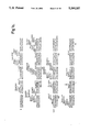
- FIGS. 1a-2c depict the cDNA sequence for clones ⁇ 33 (to the HindIII site at residue 1) and ⁇ 47 (HindIII to the 3' end). The point at which the intron is removed is designated by an asterisk.
- the probable phosphatidylinositol derivatization site is Cys 330 and the C-terminal hydrophobic region extends from residues 331-347. Amino acid residues are numbered from the mature amino terminus at Asp 1 .
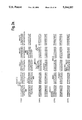
- FIGS. 2a-2c depict the cDNA sequence of clones ⁇ 33 to the HindIII site at residue +1) and ⁇ 41 (HindIII to 3' end) encoding human sDAF.
- the unspliced intron in the cDNA encoding sDAF is bracketed. Restriction enzyme sites are shown using conventional abbreviations.
- the predicted amino acid sequence for each DAF predicted species is shown, together with the secretory leader and mature N-terminus of each (designated by arrows).
- FIG. 3 is a schematic diagram showing the regions of HSV 1 glycoprotein D (gD-1) and DAF that are present in the gD-1/DAF fusion protein produced in Example 3.
- Truncated (secreted) gD-1 was constructed from native (membrane) gD-1 (14) and comprises amino acids 1-300, including the hydrophobic signal sequence (residues 1-25, indicated as a gray area).
- the hydrophobic membrane spanning domain (residues 340-360, cross-hatched region) and the C-terminal hydrophobic domain (residues 361-393) are excluded.
- the point of truncation (residue 300) is indicated by a broken line.
- Truncated gD-1 was fused to residue 311 of membrane DAF.
- the gD-1/DAF fusion contains the last 37 residues of membrane DAF predicted from the cDNA sequence (residues 311-347) and includes a C-terminal hydrophobic region (residues 331-347, depicted in black).
- DAF is defined to be any molecule having the pre or mature amino acid sequence set forth in FIGS. 1 or 2 as well as their amino acid sequence or glycosylation variants (including natural alleles) which are capable of exhibiting a biological activity in common with the native DAF of FIGS. 1 or 2.
- Native DAF is DAF obtained from serum, blood cells or other animal fluids or tissues.
- DAF biological activity is defined as any of 1) immunological cross-reactivity with at least one epitope of native DAF, or 2) the possession of at least one hormonal, regulatory or effector function qualitatively in common with native DAF.
- an amino acid sequence variant need not exhibit any DAF immunomodulatory activity to fall within the definition of DAF.
- a variant may act as an antagonist and competitively inhibit native DAF, yet have no immunomodulatory activity per se.
- the variant may be neither an antagonist nor have immunomodulatory activity, but still fall within the definition if it remains capable of cross-reacting with antibody raised against native DAF.
- An example of a presently known DAF immunomodulatory activity is inhibition of C4b2a functional activity (Medof et al., 1984, Id.).
- Amino acid sequence variants of DAF include deletions from, or insertions or substitutions of residues within the pre or mature DAF sequence shown in FIGS. 1 or 2.
- Amino acid sequence deletions generally range from about 1 to 10 residues and typically are contiguous. Contiguous deletions ordinarily are made in even numbers of residues, but single or odd numbers of deletions ar e within the scope hereof.
- Representative deletions are [des Cys 33 ] mature mDAF, [des Cys 330 - Thr 347 ] mature mDAF, [des Thr 2 - Gly 327 ] mature sDAF.
- a particularly interesting deletion is Cys 330 -Thr 347 from mDAF. This eliminates the membrane anchor site and transmenbrane region, resulting in a molecule that, like sDAF, is secreted but which bears none of the unique antigenic determinants of sDAF.
- Insertions also are preferably made in even numbers of residues when the variation falls within the mature DAF sequence, although insertions may range from 1 to 5 residues in general. However, insertions also include fusions onto the amino or carboxyl termini of DAF or from 1 residue to polypeptides of essentially unrestricted length.
- An example of a single terminal insertion is mature DAF having an N-terminal methionyl. This variant is an artifact of the direct expression of DAF in recombinant cell culture, i.e., expression without a signal sequence to direct the secretion or cell membrane association of mature DAF.
- terminal insertions include 1) fusions of heterologous signal sequences to the N-terminus of mature DAF in order to facilitate the secretion of mature DAF from recombinant hosts, 2) fusions of immunogenic polypeptides, e.g. bacterial polypeptides such as beta-lactamase or an enzyme encoded by the E. coli trp locus and 3) fusions with cell surface binding substances, including hormones, growth factors or antibodies. Fusions with cell surface binding substances need not be produced by recombinant methods, but can be the product of covalent or noncovalent association with DAF, including its phosphatidylinositol group.
- an antibody or fragment thereof bearing the variable region is covalently bound to, or expressed in recombinant cell culture as a fusion with, the C-terminus of DAF.
- the DAF is bound to antibodies specific for the HLA antigens of the allograft.
- the antibody and DAF are covalently bounded, for example, by the method of EP 170,697A, although other methods for linking proteins are conventional and known to the artisan.
- Immunogenic fusions are useful for preparing immunogenic DAFs suitable as vaccines for preparing anti-DAF antibodies. These are useful for the preparation of diagnostic reagents. Representative insertions are [Thr 329 Leuleu Cys 330 ] (Seq. ID No.
- the third group of variants are those in which at least one residue in the DAF molecule has been removed and a different residue inserted in its place. Such substitutions generally are made in accordance with following Table.
- substitutions that are less conservative than those in Table 1, i.e., selecting residues that differ more significantly in their effect on maintaining (a) the structure of the polypeptide backbone in the area of the substitution, for example as a sheet of helical conformation, (b) the charge or hydrophobicity of the molecule at the target site or (c) the bulk of the side chain.
- the substitutions in general expected to produce the greatest changes in DAF properties will be those in which (a) a hydrophilic residue, e.g. seryl or threonyl, is substituted for (or by) a hydrophobic residue, e.g.
- leucyl isoleucyl, phenylalanyl, valyl, or alanyl
- a cysteine or proline is substituted for (or by) any other residue
- a residue having an electropositive side chain e.g., lysyl, arginyl, or histidyl
- an electronegative residue e.g., glutamyl or aspartyl
- a residue having a bulky side chain e.g., phenylalanine, is substituted for (or by) one not having such a side chain, e.g. glycine.
- Representative substituted DAFs are [Cys 330 ⁇ Met] mature mDAF, [Cys 330 ⁇ Ser] mature mDAF, [Cys 2 ⁇ Ser] mature mDAF, [Lys 125 Lys 126 ⁇ Gln] mature DAF, [Gly 144 ⁇ Pro] mature DAF, [Ile 146 ⁇ Met] mature DAF, [Phe 169 ⁇ Tyr] mature DAF, [Pro 19 2 ⁇ Gly] mature DAF, [Ile 201 ⁇ Leu] mature DAF, [Asn 236 Asn 237 ⁇ AspAsp] mature DAF, [Glu 239 ⁇ Asp] mature DAF, [Ser 256 ⁇ Tyr] mature DAF, [Val 268 ⁇ Phe] mature DAF, [Lys 285 ⁇ Gln] mature DAF, [Thr 294 ⁇ Ser] mature DAF and [Leu 324 ⁇ Ser] mature DAF.
- the above described variants are made in either sDAF or mDAF.
- the following variants are made in the unique sDAF C-terminal: [Lys 352 ⁇ Gln] mature sDAF, [Cys 339 ⁇ Ser] mature sDAF, [Arg 394 ⁇ His] mature sDAF and mature sDAF [Leu 403 Phe 404 Leu 405 ⁇ SerTyrSer] mature sDAF.
- any naturally occurring alleles are not included within the scope of DAF variants because the variants described herein are predetermined DAF variants.
- the C-terminal domain of mDAF contains a site (referred to as the "phospholipid anchor domain") to which phospholipid, generally glycophospholipid, is attached in the course of post-translational processing.
- This domain contains about from 20-30 residues, the phospholipid bding covalently linked to the C-terminal residue corboxyl.
- This domain or any fragment of mDAF containing it is produced as a fusion with any other polypeptide for which it is desired to create a membrane-bound form.
- phospholipid anchor domain when used in reference to expressed fusions refers to the post-translationally modified fusion, as will be described more fully infra.
- an ordinarily secreted hormone is produced in recombinant cell culture as a C-terminal fusion of the preprotein with the phospholipid anchor domain of mDAF. Rather than being secreted this fusion will be transported to the cell membrane and remain lodged there by virtue of the phosphatidy-choline anchor.
- Such recombinant cells are useful as immunogens or vaccines for the hormone or other selected polypeptide. Sequestering the polypeptide in the membrane also protects it from dilution into the culture medium.
- fusion polypeptides having C-terminal lipids are useful in diagnostic assays for the polypeptides or their antibodies since the terminal lipid provides a convenient site for adsorption onto microtiter or test tube surfaces and the like.
- proteins that contain C-terminal domains substituted with phospholipid anchors.
- Such proteins include Thy-1 (Low et al., “Nature” (London) 318:62 [1985] and Tse et al., “Science” 230:1003 [1985]), the variant surface glycoproteins (VSGs) of African trypanosomes (Ferguson et al., "J. Biol. Chem.” 260:14547 [1985]), acetylcholinesterase (Futerman et al., "Biochem. J.” 226:369 [1985]), 5' nucleotidase (Low et al., "Biochim. Biophys.
- DNA encoding the C-terminal about 30-50 residues of a polypeptide ordinarily bearing such an anchor is ligated to DNA encoding the desired polypeptide, or to a suitable fragment multimer or amino acid sequence variant thereof.
- the DNA encoding the anchor recognition site is inserted at the C-terminus of the desired protein.
- the anchor recognition site includes the anchor domain as well as a short, approximately 10-20 residue, hydrophobit sequence located C-terminal to the anchor domain which will be processed off the fusion. This is accomplished by routine procedures well known to those skilled in the art.
- the DNA encoding the selected phospholipid anchor recognition site is synthesized by in vitro methods or by obtaining a suitable fragment from cDNA or genomic DNA encoding the native anchored protein. Since the anchor domain is found within the about from 20 to 40 residues upstream from the C-terminal hydrophobic domain one should use DNA encoding the hydrophobic domain as well as the approximately 20 to 40 residues upstream therefrom.
- glycophospholipid anchors Many proteins in addition to DAF are known to contain glycophospholipid anchors, and their amino acid sequences (including the C-terminal about 20-50 residues which will be employed as anchors in heterologous fusions) are known. Examples include acetylcholinesterase (M. Schumacher et al., “Nature” 319:407-409 [1986]), Thy-1 (T. Seki et al., “Nature” 313:485-487 [1985] and T. Moriuchi et al. "FEBS Lett.” 178:105-108 [1985]), VSG (T. Brucei) (Cross, "Philos. Trans. R. Soc. London” Ser.
- the C-terminus of the heterologous polypeptide contains an active site or immune epitope which is to be sterically free
- This optimally will be additional sequences from the anchor domain donor polypeptide, for example about from 10 to 50 residues N-terminal to the anchor domain, but also may be artificial sequences.
- amino acid sequences inputed from DNAs encoding phospholipid anchor domains exhibit little or no sequence homology beyond a C-terminal sequence of about from 10 to 20 residues containing uncharged, hydrophobic residues (leucine, glycine, threonine, valine, methionine, isoleucine and/or phenylalanine).
- a C-terminal sequence of about from 10 to 20 residues containing uncharged, hydrophobic residues (leucine, glycine, threonine, valine, methionine, isoleucine and/or phenylalanine).
- the phospholipid anchor domain is embraced with the region immediately N-terminal to the hydrophobic sequence and is readily identifiable on this basis.
- Those skilled in the art will be capable of refining the optional sequence of the phospholipid anchor domain.
- polypeptides to be linked to the phospholipid anchor domain are unlimited. Their choice will depend upon the therapeutic or diagnostic objection which is intended. All that is necessary is that the fused polypeptide exhibit the desired biological activity of the unfused polypeptide prior to its expression as a hybrid with a phospholipid anchor domain.
- the polypeptide may be of any length, from about 4 residues to thousands, and includes enzymes, hormones, antigens and the like.
- the expression hosts for these fusions are cells capable of processing the phospholipid recognition site and attaching the phospholipid to the anchor domain.
- Such cells preferably are mammalian continuous cell lines as described elsewhere herein, most preferably DHFR - CHO cells.
- the fused polypeptide is employed together with the cells in which it is produced, i.e., without recovery from the expression hosts, in the immunogen utility described above.
- the fusion is recovered from the expression host prior to its use.
- the fusion is recovered from host cell membranes by preparing cell membrane extracts in substantially the same fashion as mDAF or other anchored polypeptides heretofore have been isolated.
- Other methods for obtaining preparations of membrane anchored polypeptides such as receptors also are known and are adaptable for use in recovering the fusions described herein.
- the host cell membranes are separated from the cytoplasm, solubilized with nonionic detergent, and the fusion recovered by adsorption on immunoaffinity, substrate or ligand affinity columns.
- the fusions will be recovered as polypeptides containing the heterologous polypeptide and glycophospholipid anchor domain together with C-terminally linked glycophospholipid. Note that the fusion protein will be recovered in a form which is free of the C-terminal hydrophobic sequence present before processing of the fusion and substitution with the glycophospholipid.
- Fusions which are purified free of host cell membranes are useful as therapeutic compositions.
- a fusion containing a plasminogen activator enzyme such as urokinase or tissue plasminogen activator is fused to a glycophospholipid anchor domain and administered in therapeutic compositions to patients experiencing myocardial infarcts or other disorders accompanied by undesirable blood clots.
- the enzyme is fused at its C-terminus to the N-terminus of the glycophospholipid anchor domain.
- glycophospholipid anchor domain includes both the amino acid sequence as well as the glycophospholipid substituted at a carboxyl group of the C-terminal amino acid residue.
- the fused plasminogen activator will insert into blood cells and vasculature where it will be most effective at activating plasminogen and will not be subject to removal from the blood stream by degradative processes such as those performed by the liver or spleen, thereby extending the half life of the enzyme and targeting it more directly to the desired therapeutic site.
- the novel fusions are particularly useful in overcoming defects or deficiencies within the immune system, particularly in the process of antigen presentation.
- An antigen to which it is desired to modulate an immune response is synthesized as a fusion with a phospholipid anchor domain and fusion administered under conditions and in a dosage determined to produce the desired effect.
- the fusion must preserve the relevant epitope(s) of the antigen. This is readily determined by conventional competitive-type immunoassay using antibody raised against the native antigen and labeled native antigen, in accordance with methods well known to those skilled in the art.
- Antigen fusions also are useful in in vitro diagnostics as described above or in affinity chromatography.
- novel fusions herein optionally are formulated into liposomes or other lipid membrane carriers. This is readily accomplished by mixing a solution of the fusion with a preformed liposomal suspension and incubating until the insertion of the fusions into the liposomal bilayer. Alternately, the fusions are admixed with the aqueous solution used in the preparation of the liposomes. Alternatively, the fusions are formulated into conventional pharmacologically acceptable vehicles as described below for mDAF. Since the fusions bear hydrophobic substituent they can be formulated with pharmacologically acceptable detergents such as Tween 20 or PEG, or with serum albumin.
- pharmacologically acceptable detergents such as Tween 20 or PEG, or with serum albumin.
- a variant typically is made by site specific mutagenesis of the native DAF-encoding nucleic acid, expression of the variant nucleic acid in recombinant cell culture and, optionally, purification from the cell culture for example by immunoaffinity adsorption on a rabbit polyclonal anti-DAF column (in order to absorb the variant by at least one remaining immune epitope).
- the activity of the cell lysate or purified DAF variant is then screened in a suitable screening assay for the desired characteristic.
- a change in the immunological character of the DAF such as affinity for a given antibody, is measured by a competitive-type immunoassay.
- Changes in immunomodulator activity are measured by the C4b2a assay, although as more becomes known about the functions in vivo of sDAF and mDAF other assays will become useful in such screening. Modifications of such protein properties as redox or thermal stability, hydrophobicity, susceptibility to proteolytic degradation, or the tendency to aggregate with carriers or into multimers are assayed by methods well known to the artisan.
- DAF from other species than humans e.g. bovine, equine, ovine, porcine and the like is included within the scope hereof.
- DAF preferably is made by synthesis in recombinant cell culture. In order to do so, it is first necessary to secure nucleic acid that encodes DAF. The inventors encountered considerable hardship in attempting to identify any nucleic acid encoding DAF.
- the sequence of the human mDNA encoding DAF that was ultimately determined is shown in FIG. 1.
- study of cDNAs from hela cells led to the identification of cDNA encoding sDAF, shown in FIG. 2.
- FIG. 2 Once this DNA has been identified it is a straight-forward matter for those skilled in the art to obtain it my nucleic acid hybridization to genomic libraries of human DNA or, if it is desired to obtain DNA encoding the DAF of another animal species, then by hybridization of DNA libraries from cells of that species.
- the hybridization analysis is now straight-forward because FIGS. 1 and 2 enable the preparation of very long synthetic probes that are perfect or nearly perfect matches for the target DNA.
- the cDNA or genomic library selected as the source for the DAF nucleic acid will not contain a single clone encoding the full length DAF, only partial clones.
- These partial clones and fragments are readily assembled into a full length DNA by cleaving the partial clones at selected restriction sites in overlapping sections, recovering each of the desired fragments and ligating them in the proper order and orientation. If necessary, oligonucleotides are prepared to supply any missing sequences.
- the DAF-encoding nucleic acid is then ligated into a replicable vector for further cloning or for expression.
- Vectors are useful for performing two functions in collaboration with compatible host cells (a host-vector system). One function is to facilitate the cloning of the nucleic acid that encodes the DAF, i.e., to produce usable quantities of the nucleic acid. The other function is to direct the expression of DAF. One or both of these functions are performed by the vector-host system.
- the vectors will contain different components depending upon the function they are to perform as well as the host cell that is selected for cloning or expression.
- Each vector will contain nucleic acid that encodes DAF as described above. Typically, this will be DNA that encodes the DAF in its mature form linked at its amino terminus to a secretion signal.
- This secretion signal preferably is the DAF presequence that normally directs the secretion of DAF from human cells in vivo.
- suitable secretion signals also include signals from other animal DAFs, viral signals or signals from secreted polypeptides of the same or related species.
- Expression and cloning vectors contain a nucleic acid sequence that enables the vector to replicate in one or more selected host cells.
- this sequence is one that enables the vector to replicate independently of the host chromosomes, and includes origins of replication or autonomously replicating sequences.
- origins of replication or autonomously replicating sequences are well-known for a variety of bacteria, yeast and viruses.
- the origin of replication from the well-known plasmid pBR322 is suitable for most gram negative bacteria, the 2 ⁇ plasmid origin for yeast and various viral origins (SV40, polyoma, adenovirus, VSV or BPV) are useful for cloning vectors in mammalian cells.
- Origins are not needed for mammalian expression vectors (the SV40 origin is used in the Examples only because it contains the early promoter).
- Most expression vectors are "shuttle vectors, i.e. they are capable of replication in at least one class of organisms but can be transfected into another organism for expression. For example, a vector is cloned in E. coli and then the same vector is transfected into yeast or mammalian cells for expression even though it is not capable of replicating independently of the host cell chromosome.
- DNA also is cloned by insertion into the host genome. This is readily accomplished with bacillus species, for example, by including in the vector a DNA sequence that is complementary to a sequence found in bacillus genomic DNA. Transfection of bacillus with this vector results in homologous recombination with the genome and insertion of DAF DNA. However, the recovery of genomic DNA encoding DAF is more complex than that of an exogenously replicated vector because restriction enzyme digestion is required to excise the DAF DNA.
- DNA is inserted into a host genome for purposes of preparing a stable cell line or microbe for DAF expression.
- Selection genes also termed a selectable marker. This is a gene that encodes a protein necessary for the survival or growth of a host cell transformed with the vector. The presence of this gene ensures that any host cell which deletes the vector will not obtain an advantage in growth or reproduction over transformed hosts.
- Typical selection genes encode proteins that (a) confer resistance to antibiotics or other toxins, e.g. ampicillin, neomycin, methotrexate or tetracycline, (b) complement auxotrophic deficiencies, or (c) supply critical nutrients not available from complex media, e.g. the gene encoding D-alanine racemase for bacilli.
- a suitable selection gene for use in yeast is the trp1 gene present in the yeast plasmid YRp7 (Stinchcomb et al., 1979, "Nature", 282; 39; Kingsman et al., 1979, “Gene", 7: 141; or Tschemper et al., 1980, “Gene", 10: 157).
- the trp1 gene provides a selection marker for a mutant strain of yeast lacking the ability to grow in tryptophan, for example ATCC No. 44076 or PEP41 (Jones, 1977, "Genetics", 85: 12).
- the presence of the trp1 lesion in the yeast host cell genome then provides an effective environment for detecting transformation by growth in the absence of tryptophan.
- Leu2 deficient yeast strains (ATCC 20,622 or 38,626) are complemented by known plasmids bearing the Leu2 gene.
- Suitable selectable markers for mammalian cells are dihydrofolate reductase (DHFR) or thymidine kinase. Such markers enable the identification of cells which were competent to take up the DAF nucleic acid.
- the mammalian cell transformants. are placed under selection pressure which only the transformants are uniquely adapted to survive by virtue of having taken up the marker.
- Selection pressure is imposed by culturing the transformants under conditions in which the concentration of selection agent in the medium is successively changed, thereby leading to amplification of both the selection gene and the DNA that encodes DAF.
- Amplification is the process by which genes in greater demand for the production of a protein critical for growth are reiterated in tandem within the chromosomes of successive generations of recombinant cells. Increased quantities of DAF are synthesized from the amplified DNA.
- cells transformed with the DHFR selection gene are first identified by culturing all of the transformants in a culture medium which lacks hypoxanthine, glycine, and thymidine.
- An appropriate host cell in this case is the Chinese hamster ovary (CHO) cell line deficient in DHFR activity, prepared and propagated as described by Urlaub and Chasin, 1980, "Proc. Nat'l. Acad. Sci. USA” 77: 4216.
- a particularly useful DHFR is a mutant DHFR that is highly resistant to MTX (EP 117,060A).
- This selection agent can be used with any otherwise suitable host, e.g. ATCC No. CCL61 CHO-K1), notwithstanding the presence of endogenous DHFR.
- the DHFR and DAF-encoding DNA then is amplified by exposure to an agent (methotrexate, or MTX) that inactivates the DHFR.
- an agent metalhotrexate, or MTX
- Expression vectors unlike cloning vectors, should contain a promoter which is recognized by the host organism and is operably linked to the DAF nucleic acid. Promoters are untranslated sequences located upstream from the start codon of a structural gene (generally within about 100 to 1000 bp) that control the transcription and translation of nucleic acid under their control. They typically fall into two classes, inducible and constitutive. Inducible promoters are promoters that initiate increased levels of transcription from DNA under their control in response to some change in culture conditions, e.g. The presence or absence of a nutrient or a change in temperature. At this time a large number of promoters recognized by a variety of potential host cells are well known.
- promoters are operably linked to DAF-encoding DNA by removing them from their gene of origin by restriction enzyme digestion, followed by insertion 5' to the start of codon for DAF. This is not to say that the genomic DAF promoter is not usable. However, heterologous promoters generally will result in greater transcription and higher yields of expressed DAF.
- Nucleic acid is operably linked when it is placed into a functional relationship with another nucleic acid sequence.
- DNA for a presequence or secretory leader is operably linked to DNA for a polypeptide if it is expressed as a preprotein which participates in the secretion of the polypeptide;
- a promoter or enhancer is operably linked to a coding sequence if it affects the transcription of the sequence; or
- a ribosome binding site is operably linked to a coding sequence if it is positioned so as to facilitate translation.
- operably linked means that the DNA sequences being linked are contiguous and, in the case of a secretory leader, contiguous and in reading phase. Linking is accomplished by ligation at convenient restriction sites. If such sites do not exist then synthetic oligonucleotide adaptors or linkers are used in accord with conventional practice.
- Promoters suitable for use with prokaryotic hosts include the ⁇ -lactamase and lactose promoter systems (Chang et al., 1978, “Nature", 275: 615; and Goeddel et al., 1979, “Nature", 281: 544), alkaline phosphatase, a tryptophan (trp) promoter system (Goeddel 1980, "Nucleic Acids Res.” 8: 4057 and EPO Appln. Publ. No. 36,776) and hybrid promoters such as the tac promoter (H. de Boer et al., 1983, "Proc. Nat'l. Acad. Sci. USA” 80: 21-25). However, other known bacterial promoters are suitable.
- Suitable promoting sequences for use with yeast hosts include the promoters for 3-phosphoglycerate kinase (Hitzeman et al., 1980, “J. Biol. Chem.”, 225: 2073) or other glycolytic enzymes (Hess et al., 1968, "J. Adv.
- Enzyme Reg.” 7: 149; and Holland, 1978, “Biochemistry", 17: 4900
- enolase such as enolase, glyceraldehyde-3-phosphate dehydrogenase, hexokinase, pyruvate decarboxylase, phosphofructokinase, glucose-6-phosphate isomerase, 3-phosphoglycerate mutase, pyruvate kinase, triosephosphate isomerase, phosphoglucose isomerase, and glucokinase.
- yeast promoters which are inducible promoters having the additional advantage of transcription controlled by growth conditions, are the promoter regions for alcohol dehydrogenase 2, isocytochrome C, acid phosphatase, degradative enzymes associated with nitrogen metabolism, metallothionein, glyceraldehyde-3-phosphate dehydrogenase, and enzymes responsible for maltose and galactose utilization.
- Suitable vectors and promoters for use in yeast expression are further described in R. Hitzeman et al., EP 73,657A.
- Yeast enhancers also are advantageously used with yeast promoters.
- DAF transcription from vectors kin mammalian host cells is controlled by promoter obtained from the genomes of viruses such as polyoma, cytomegalovirus, adenovirus, retroviruses, hepatitis-B virus and most preferably Simian Virus 40 (SV40), or from heterologous mammalian promoters, e.g. the actin promoter.
- SV40 Simian Virus 40
- the early and late promoters of the SV40 virus are conveniently obtained as an SV40 restriction fragment which also contains the SV40 viral origin of replication (Fiers et al., 1978, "Nature", 273: 113).
- promoters from the host cell or related species also are useful herein.
- An enhancer is a nucleotide sequence, usually about from 10-300 bp, that acts on a promoter to increase its transcription and does so in a manner that is relatively orientation and position independent.
- Many enhancer sequences are now known from mammalian genes (globin, elastase, albumin, ⁇ -fetoprotein and insulin). Typically, however, one will use an enhancer from a eukaryotic cell virus.
- Examples include the SV40 enhancer on the late side of the replication origin (bp 100-270), the cytomegalovirus early promoter enhancer, the polyoma enhancer on the late side of the replication origin, and adenoviral enhancers.
- the enhancer may be spliced into the vector at a position 5' or 3' to the DAF-encoding sequence, but is preferably located at a site 5' from the promoter.
- Expression vectors used in eukaryotic host cells will also contain sequences necessary for the termination of transcription and for stabilizing the mRNA. Such sequences are commonly available from the 5' and, occasionally 3' untranslated regions of eukaryotic or viral DNAs or cDNAs. These regions contain regions that are transcribed as polyadenylated segments in the untranslated portion of the mRNA encoding DAF. the 3' untranslated regions also include transcription termination sites.
- Suitable host cells for cloning or expressing the vectors herein are prokaryotes, yeast or higher eukaryotic cells.
- Prokaryotes include gram negative or gram positive organisms, for example E. coli or bacilli.
- a preferred cloning host is E. coli 294 (ATCC 31,446) although other gram negative or gram positive prokaryotes such as E. coli B, E. coli X1776 (ATCC 31,537), E. coli W3110 (ATCC 27,325), pseudomonas species, or Serratia Marcesans are suitable.
- eukaryotic microbes such as filamentous fungi or yeast are suitable hosts for DAF-encoding vectors.
- Saccharmoyces cerevisiae, or common baker's yeast is the most commonly used among lower eukaryotic host microorganisms.
- a number of other genera, species and strains are commonly available and useful herein.
- the preferred host cells for the expression of DAF are cells derived from multicellular organisms. DAF's large size, together with its intramolecular disulfide bond(s) and, in the case of mDAF, its unique post-translational processing, suggests that the host cell will optimally be of a higher phylogenetic order than the microbes if one is to expect the recombinant protein to demonstrate optimal conformational fidelity to native DAF. In addition, it may be desirable to glycosylate DAF. All of these functions can be best performed by higher eukaryotic cells. In principle, any higher eukaryotic cell culture is workable, whether from vertebrate or invertebrate culture, although cells from mammals such as humans are preferred.
- Host cells are transformed with the above-described expression or cloning vectors and cultured in conventional nutrient media modified as is appropriate for inducing promoters or selecting transformants containing amplified genes.
- the culture conditions such as temperature, pH and the like, suitably are those previously used with the host cell selected for cloning or expression, as the case may be, and will be apparent to the ordinary artisan.
- sDAF preferably is recovered from the culture medium as a secreted protein, although it also may be recovered from host cell lysates when directly expressed without a secretory signal.
- the culture medium or lysate is centrifuged to remove particulate cell debris.
- DAF also is purified from contaminant soluble proteins for example by adsorption on a selection column e.g. ConA, elution, adsorption on an anti-sDAF or anti-mDAF immunoaffinity column and elution therefrom.
- a selection column e.g. ConA
- elution e.g. elution
- an anti-sDAF or anti-mDAF immunoaffinity column elution therefrom.
- other processes such as chromatography on alkyl Sepharose, silica or an anion or cation exchange resin or gel electrophoresis are used to separate the sDAF from contaminants.
- mDAF is recovered from transformant cell membranes using the method of Medof et al. (1984. Id.). mDAF variants in which the hydrophobic transmembrane region and/or the mDAF phosphatidylinositol-binding residue are deleted or substituted are recovered in the same fashion as sDAF, although variants in which the transmembrane region remains intact also are recovered from transformant cell membranes.
- native DAF has a tendency to aggregate under some conditions it may be useful to stabilize the aggregative state of the multimers by providing in the separations a minor amount of a nonionic surfactant such as Tween or polyethylene glycol.
- a protease inhibitor such as PMSF also may be useful to inhibit proteolytic degradation during purification, and antibiotics may be included to prevent the growth of adventitious contaminants.
- purification methods suitable for native DAF may require modification to account for changes in the character of DAF or its variants upon expression in recombinant cell culture.
- a DAF polypeptide produced in prokaryotic cell culture will not adsorb to Con-A Sepharose because it will be unglycosylated.
- other methods such as gel electrophoresis, ion exchange or immunoaffinity purification should be employed.
- sDAF lipid-free C-terminal mDAF variants will not absorb as readily to hydrophobic adsorbents as does mDAF.
- Appropriate purification methods will be apparent to the artisan, depending upon the characteristics of the particular recombinant DAF.
- DAF is prepared as a nontoxic salt with such ions as sodium, potassium, phosphate, chloride and the like.
- DAF is stored in phosphate buffered saline or may be lyophilized in the presence of an excipient including sugar alcohols, e.g. mannitol or sorbitol; monosaccharides, e.g., glucose, mannose, galactose or fructose; oligosaccharides such as maltose, lactose or sucrose; and proteins such as human serum albumin.
- sugar alcohols e.g. mannitol or sorbitol
- monosaccharides e.g., glucose, mannose, galactose or fructose
- oligosaccharides such as maltose, lactose or sucrose
- proteins such as human serum albumin.
- excipients also may contribute to the stability of DAF to inactivation or precipitation upon aqueous storage, and may be used together with other stabilizers which are conventional per se.
- stabilizers include chelating agents, e.g. EDTA; antioxidants such as ascorbate or dithiothreitol; amino acids; and nonionic surfactants such as polyethylene glycol or block copolymers of polyethylene and polypropylene glycol.
- DAF is administered to humans or animals in order to ameliorate various disorders stemming from immune dysfunction or misdirection, particularly defects in the humoral immune response. Examples include PNH, inflammatory conditions such as inflammatory bowel disease (colitis), rheumatoid arthritis, allograft rejection and the like. Treatment with DAF should be instituted early in the development of such disorders.
- Therapeutic DAF compositions will contain a therapeutically effective dose of DAF in a pharmacologically acceptable carrier.
- the dose, carrier and route of administration selected will depend, among other factors, upon the disorder or condition to be treated, the condition of the patient, the desired route of administration, and the activity of the selected DAF variant. This is readily determined and monitored by the physician during the course of therapy.
- the carrier for infusion or injection of DAF is a sterile isotonic aqueous solution, for example saline for injection or 5% dextrose.
- a sterile isotonic aqueous solution for example saline for injection or 5% dextrose.
- These preparations are injected or infused by intranasal, subcutaneous, intravenous, intraperitoneal or other conventional routes of administration. Preparations also are injected into the synonial fluid of arthritic joints.
- DAF also is provided in a sustained release carrier.
- Suitable examples include semipermeable polymer matrices in the form of shaped articles, e.g. suppositories, or microcapsules.
- Implantable or microcapsules sustained relase matrices include polyactides (U.S. Pat. No. 3,773,919, EP58,481) copolymers of L-glutamic acid and gamma ethyl-L-glutamate (U. Sidman et al., 1983, "Biopolymers” 22(1): 547-556), poly (2-hydroxyethyl-methacrylate) (R. Langer et al., 1981, "J. Biomed, Mater.
- Sustained release DAF compositions also include liposomally entrapped DAF. Liposomes containng DAF are prepared by methods known per se: DE 3,218,121A; Epstein et al. 1985, “Proc. Natl. Acad. Sci. USA” 82: 3688-3602; Hwang et al., 1980, "Proc. Natl. Acad. Sci.
- the liposomes are of the small (about 200-800 Angstroms) unilamelar type in which the lipid content is greater than about 30 mol. % cholesterol, the selected proportion being adjusted for the optimal rate of DAF leakage.
- Sustained release DAF preparations are implanted or injected into proximity to the site of inflammation or therapy, for example adjacent to arthritic joints or inflamed intestinal tissue.
- Antisera raised against DAF is one described by Medof et al. (1984, Id.) Antisera are employed for immunoaffinity purification or DAF and in an ELISA assay for DAF. Antibody specific for the unique C-terminus of sDAF is made by immunizing an animal against an immunogenic sDAF conjugate, e.g.
- an immunogenic fusion made in recombinant cell culture as described elsewhere herein, and thereafter screening for the presence of anti-SDAF titer by passing the antiserum through a column of immobilized mDAF in order to adsorb antibodies directed against mDAF epitopes, incubating the unadsorbed antiserum in the presence of 125 I-sDAF (prepared in substantially the same fashion as 125 I-mDAF, Medof et al., 1984, Id.) to permit the unique sDAF epitopes to bind to the anti-DAF antibodies in the unadsorbed antiserum, and determining the amount of unbound 125 I-sDAF, e.g. by adsorption on protein-A Sepharose.
- 125 I-sDAF prepared in substantially the same fashion as 125 I-mDAF, Medof et al., 1984, Id.
- the sDAF-specific antibodies in such antisera are prepared by adsorption as immobilized mDAF, recovery of the unadsorbed fraction, adsorption on immobilized SDAF and elution with pH 4-6 buffer to recover the SDAF-specific antibodies substantially free of MDAF antibodies.
- spleen cells from immunized animals showing anti-SDAF neutralizing titer are recovered and fused to myeloma cells or are transformed with EB virus in known fashion in order to prepare monoclonal sDAF-specific antibodies.
- Neutralizing antibodies against DAF are useful when conjugated to immunogenic polypeptides as immunogens for raising anti-idiotypic antibodies having DAF activity. Such anti-idiotypic antibodies are useful for the same diagnostic and therapeutic purposes as DAF.
- Plasmids are designated by a low case p preceded and/or followed by capital letters and/or numbers.
- the starting plasmids herein are commercially available, are publicly available on an unrestricted basis, or can be constructed from such available plasmids in accord with published procedures.
- other equivalent plasmids are known in the art and will be apparent to the ordinary artisan.
- “Digestion” of DNA refers to catalytic cleavage of the DNA with an enzyme that acts only at certian locations in the DNA. Such enzymes are called restriction enzymes, and the sites for which each is specific is called a restriction site.
- restriction enzymes are commercially available and their reaction conditions, cofactors and other requirements as established by the enzyme suppliers are used. Restriction enzymes commonly are designated by abbreviations composed of a capital letter followed by other letters representing the microorganism from which each restriction enzyme originally was obtained and then a number designating the particular enzyme. In general, about 1 ⁇ g of plasmid or DNA fragment is used with about 2 units of enzyme in about 20 ⁇ l of buffer solution.
- Appropriate buffers and substrate amounts for particular restriction enzymes are specified by the manufacturer. Incubation times of about 1 hour at 37° C. are ordinarily used, but may vary in accordance with the supplier's instructions. After incubation, protein is removed by extraction with phenol and chloroform, and the digested nucleic acid is recovered from the aqueous fraction by precipitation with ethanol. Digestion with a restriction enzyme infrequently is followed with bacterial alkaline phosphatase hydrolysis of the terminal 5' phosphates to prevent the two restriction cleaved ends of a DNA fragment from "circularizing" or forming a closed loop that would impede insertion of another DNA fragment at the restriction site.
- “Filling” or “blunting” refers to the procedure by which the single stranded end in the cohesive terminus of a restriction enzyme-cleaved nucleic acid is converted to a double strand. This eliminates the cohesive terminus and forms a blunt end. This process is a versatile tool for converting a restriction cut end that may cohesive with the ends created by only one or a few other restriction enzymes into a terminus compatible with any blunt-cutting restriction endonuclease or other filled cohesive terminus.
- blunting is accomplished by incubating 2-15 ⁇ g of the target DNA in 10 mM MgCl 2 , 1mM dithiothreitol, 50 mM NaCl, 10 mM Tris (pH 7.5) buffer at about 37° C. in the presence of 8 units of the Klenow fragment of DNA polymerase I and 250 ⁇ m of each of the four deoxynucleoside triphosphates.
- the incubation generally is terminated after 30 min. by phenol and chloroform extraction and ethanol precipitation.
- Recovery or "isolation" of a given fragment of DNA from a restriction digest means separation of the digest on polyacrylamide or agrose gel by electrophoresis, identification of the fragment of interest by comparison of its mobility versus that of marker DNA fragments of known molecular weight, removal of the gel section containing the desired fragment, and separation of the gel from DNA.
- This procedure is known generally. For example, see R. Lawn et al., 1981, “Nucleic Acids Res.” 9:6103-6114, and D. Goeddel et al., 1980, "Nucleic Acids Res.: 8:4057.
- Northern blotting is a method by which the presence of a cellular mRNA is confirmed by hybridization to a known, labelled oligonucleotide or DNA fragment.
- Northern analysis shall mean electrophoretic separation of the mRNA on 1 percent agarose in the presence of a denaturant (formaldehyde - 7%), transfer to nitrocellulose hybridization to the labelled fragment as described by T. Maniatis et al., Id., p. 202.
- Transformation means introducing DNA into an organism so that the DNA is replicable, either as an extrachromosomal element or chromosomal integrate.
- the method used herein for transformation of E. coli is the CaCl 2 method of Mandel et al., 1970, "J. Mol. Biol.” 53: 154.
- Ligase refers to the process of forming phosphodiester bonds between two double stranded nucleic acid fragments (T. Maniatis et al., Id., p. 146). Unless otherwise provided, ligation may be accomplished using know buffers and conditions with 10 units of T4 DNA ligase ("ligase”) per 0.5 ⁇ g of approximately equimolar amounts of the DNA fragments to be ligated.
- ligase T4 DNA ligase
- Preparation of DNA from transformants means isolating plasmid DNA from bacterial culture. Unless otherwise provided, the alkali/SDS method of Maniatis et al., Id., P. 90, may be used.
- Oligonucleotides are short length single or double stranded polydeoxynucleotides which are chemically synthesized by known methods and then purified on polyacrylamide gels.
- Human DAF was purified to homogeneity and 23 amino acids of N-terminal sequence were determined. Five of these were ambiguous.
- a 69mer oligonucleotide probe based on this amino acid sequence was synthesized in vitro: The 32p-labelled (Kinased) probe and the following nucleotide sequence: ##STR1##
- a Hela cell ⁇ cDNA library (approx. 1 ⁇ 10 6 recombinants) was screened under low stringency conditions with this 69mer. Only one DAF clone ( ⁇ 21) was identified, together with 6 false positives (by sequencing, these turned out to have limited nucleic acid homology with the probe, but a totally different amino and sequence).
- ⁇ 21 contained an insert encoding the sequence: Asp.Cys.Gly.Leu.Pro.Pro.Asp.Val.Pro.Asn.Ala.Gln.Pro.Ala.Leu.Glu. Gly Arg.Thr. Ser.Phe.Pro.Gly., (Seq. ID No. 8) whereon the underlined residues differed from those identified by amino terminal sequencing.
- the initial DAF clone (Clone ⁇ 21) was 1395 bp in length and contained a poly A tail but was missing the initiator methionine.
- clone DAF ⁇ 41 was 55 bp longer than DAF ⁇ 21 and included an ATG for translation initiation.
- Clone DAF ⁇ 47 was 93 bp shorter than DAF ⁇ 21 at the 5'end.
- DAF ⁇ 21 and DAF ⁇ 41 were completely overlapping in the coding region of the protein and encoded a protein of 400 amino acids.
- DAF ⁇ 47 and DAF ⁇ 33 contained an apparent ⁇ deletion ⁇ of 118 bp of coding region with respect to DAF ⁇ 21 and DAF ⁇ 41.
- DAF ⁇ 21 and DAF ⁇ 41 contained an unspliced (unremoved) intron of 118 bp.
- DAF ⁇ 35 and DAF ⁇ 37 one of which contains the same intron and one of which does not.
- the frequency with which the unspliced form is present in the library (3 out of 6 clones) suggests that it is unlikely the unspliced clones represents improperly spliced message. Rather, there appear to be two forms of the DAF protein. These 2 forms are identical at amino acid positions 1-327, while having different C-terminal sequences.
- the unspliced form contains an additional 79 amino acids, the spliced form contains an additional 20 amino acids. Since the splice produces a change in reading frame there is no homology between the 2 proteins at the C-terminii.
- Clones DAF ⁇ 33, ⁇ 41 and ⁇ 47 from Example 1 were each subcloned into pUC19, a readily available cloning vector for E. coli, by digesting each of the ⁇ clones with EcoRI, recovering the DAF inserts from each, digesting pUC19 with EcoRI, ligating the inserts into opened pUC19 and transforming E. coli 294 with the each ligation mixture.
- pUC1933, pUC1941 and pUC1947 were recovered from ampicillin resistant colonies.
- pUC1933, pUC1941 and pUC1947 were each digested with EcoRI and HindIII and the fragments (I, II and III respectively) containing the 5' end of the DAF gene, and the 3' ends of the sDAF and mDAF genes, respectively, were recovered.
- pUC19 digested with EcoRI was ligated to Fragments I and II in a three way ligation and pUC19sDAF was recovered from an ampicillin resistant E. coli colony. This was the subclone of the complete SDAF gene shown in FIGS. 2a-2c.
- pUC19MDAF was constructed in the same way as pUC10sDAF except that Fragment III was used in place of Fragment II. This subclone contained the complete MDAF gene of FIG. 1a-1c.
- PE348HVE400D122 (also pE342 VE400D22, EP 117,058A) is digested wtih HindIII such that DHFR - containing fragment is recovered.
- the HindIII cohesive terminii are filled, the fragment digested with ClarI and the following fragment isolated ##STR3##
- PE348 MBV E400D22 also is digested with ClAI and SocII such that the 990 bp fragment containing the SV40 ori and HVsAg poly A sequence is recovered (Fragment b).
- pUCsDAF and pUCmDAF were digested with EcoRI and each DAF encoding fragment isolated (Fragments CII and CIII, respectively).
- Fragments CII, a and b are ligated in a three way ligation and transfected into E. coli 294.
- pE348sDAf is recovered from an ampicillin resistant colony. It contains the sDAF gene in proper orientation 3' to the SV40 early promoter.
- the SDAF gene is under the control of the SV40 early promoter in an expression vector suitable for transformation into and methotrexate selection and amplification in a mammalian host cell.
- pE348mDAF is constructed in the same way except that Fragment CIII is used.
- An alternative expression vector is constructed by digesting p342E (Crowley et al., 1983, "Mol. Cell. Biol.” 3:44-55) with EcoRI and HpaI, and the vector fragment recovered. Either of pUC19mDAF or pUC19sDAF are digested with AccI (for MDAF) or blunt XhoII (for SDAF), filled, digested with EcorI and the DAF-encoding fragments recovered. The DAF fragments are ligated into the vector fragment and expression vectors recovered. This vector does not contain the DHFR gene, although cotransformation with pFD11 (Simonsen et al., 1983, "P.N.A.S.-USA” 80:2045-99) will produce satisfactory results.
- PE348mDAF or pE348sDAF are co-transfected into DHFR - CHO cells using conventional methods, inoculated into HAT medium and transformants selected by culture in media containing serial increases in methotrexate concentration to amplify the DHFR and DAF genes.
- a transformant clone is recovered that stably expresses DAF and secretes it into the culture medium.
- the sDAF is recovered from the medium by adsorption onto an immunoaffinity column containing protein-A sepharose immobilized rabbit polyclonal antibody to sDAF and elution with pH5 glycine buffer.
- pE348mDAF is transformed into an amplified in DHFR - CHO cells in the same way.
- mDAF is recovered by isolation from detergent lysates of host cell membranes in essentially the same fashion as mDAF has been recovered heretofore from red blood cell stroma.
- a fusion protein was constructed in which the last 37 amino acids of membrane DAF predicted by the spliced cDNA were fused in-frame to the C-terminus of a truncated form of the Herpes Simplex Virus Type 1 (HSV 1) glycoprotein D (gD-1) that ordinarily is constitutively secreted to the culture medium since it lacks the C-terminal membrane-spanning domain (Lasky et al., "Bio/Technology 2:527 [1984]).
- HSV 1 Herpes Simplex Virus Type 1 glycoprotein D
- a HindIII-HinfI fragment encoding the first 300 amino acids of HSV gD-1 was ligated via a synthetic linker to XmnI-EcoRV fragment encoding the C-terminus of DAF (residues 316-347).
- the synthetic HinFI-XMnI linker (5'-ATTCGCCAAATAAAGCAAGTGG-AACC) encoded amino acid 301 of gD-1 and amino acids 311-317 of DAF and created an in-frame fusion.
- the DNA encoding the gD-1/DAF fusion protein was inserted into a mammalian expression vector between an RSV promoter and an SV40 polyadenylation sequence by excision of the CAT gene and insertion of the fusion DNA (Gorman et al., "Proc. Natl. Acad. Sci. USA” 79:6777 [1982]) and transfected into CHO cells by the calcium-phosphate coprecipitation method (Wigler et al., "Proc. Natl. Acad. Sci. USA” 76:1373 [1979] and Simonsen et al., "Proc. Natl. Acad. Sci. USA” 80:2495 [1983]).
- Mouse dihydrofolate reductase cDNA provided a selectable marker for gene expression (Simonsen et al., "Proc. Natl. Acad. Sci. USA” 80:2495 [1983]).
- Stable cell lines derived from individual colonies were used for analysis.
- Cell lines expressing native gD-1or truncated Gd-1 were derived as described (Lasky et al., "Bio/Technology 2:527 [1984] and Berman et al., "Science” 222:525 [1983]).
- the resultant fusion protein (FIG.
- gD-1 /DAF fusion protein contains the N-terminal 75% of gD-1 (residues 1-300) including the signal sequence and the C-terminal 10% (37amino acids) of membrane DAF including the 20 amino acid segment that is divergent between the two predicted DAF proteins and 17 amino acids of adjacent common sequence.
- the C-terminal segment of DAF encoded by the gD-1/DAF fusion contains a 17 amino acid hydrophobic resion at the C-terminus which may act as a transient membrane anchor thought to be removed post-translationally and replaced with PI-anchor (Low, M. G., "J. Biochem.” 244:1-13 [1987]; Cross, G. A. M. "Cell” 48:179-181 [1987]; "Nature” 325:545 [1987]).
- the above experiments do not distinguish whether the fusion protein is anchored by a phospholipid anchor or by the 17 amino acid hydrophobic region.
- CHO cells expressing either native gD-1 or gD/DAF fusion were incubated with purified phosphatidylinositol-specific phospholipase C (PI-PLC) from Staphylococcus aureus (Low, M. G., "Meth. Enzymol.” 71:741 [1981]), and analyzed by indirect fluorescence and flow cytometry (FACS).
- PI-PLC phosphatidylinositol-specific phospholipase C
- PI-PLC which is free of proteolytic contaminants (Low et al, "Nature” 318:62 [1985]) resulted in a substantial reduction in the amount of gD-1/DAF on the cell surface as indicated by the marked decrease in relative cell fluorescence displayed on a log scale.
- full-length native gD-1expressed on the cell surface was unaffected by treatment with PI-PLC.
- the glycophospholipid anchor of DAF contains ethanolamine and glucosamine in addition to phosphatidylinositol (Medof et al., "Biochemistry” 25:6740 [1986]).
- the glycosylated phospholipid is thought to be linked to the protein through an amine bond between the terminal carboxyl group of the polypeptide and the amine group of ethanolamine (Low, M. G., "J. Biochem.” 244:1-13 [1987] and Cross, G. A. M., "Cell” 48:179-181 [1987]).
- gD-1/DAF fusion protein is anchored by such a structure
- cells were metabolically labelled with either [ 3 H]ethanolamine or [ 35 S]cysteine and the protein analyzed by immunopreocipitation.
- Preliminary pulse-chase experiments and experiments with neuraminidase suggest that the 37 kD species is a precursor, while the larger species represent mature, highly glycoslated forms of the protein.
- a [ 3 H]Ethanolamine-labelled bands corresponding to the 46kD species is a precursor, while the larger species represent mature, highly glycosylated forms of the protein.
- [ 3 H]Ethanolamine-labeled bands corresponding to 46 kD and 51 kD species but not a 37 kD species were specifically detected in cells expressing gD-1/DAF. Attachment of the clycophospholipid anchor is thought to be an early event in the biosynthesis of lipid-anchored proteins (Medof et al., "Biochemistry" 25:6740 [1986] and Berman et al. "Science” 222:524 [1983]).
- Gd-1/DAF gD-1/DAF on the cell surface was sensitive to digestion with highly purified phosophatidylinositol-specific phospholipase C while native gD-1was unaffected.
- the information or "signal" necessary for directing the attachment of a phospholipid membrane anchor is contained within the C-terminal 37 amino acids of DAF.
- the concept that the C-terminal sequence plays a role in directing the attachment of lipid is supported by recent identification of multiple classes of the neural cell adhesion molecule (N-CAM) mRNa, presumably resulting from differential mRNA splicing.
- N-CAM neural cell adhesion molecule
- the different forms of N-CAM encoded by these mRNAs have different C-terminal domains, apparently resulting in membrane attachment either via a hydrophobic membrane-spanning domain, or via a phospholipid (Hemperly et al., "Proc. Natl. Acad. Sci. USA” 83:9822 [1986]).
Abstract
Novel fusions of a phospholipid anchor domain and a polypeptide heterologous to the anchor domain donor polypeptide are provided for industrial use. Therapeutic administration of the fusions enables the targeting of biological activity to cell membrane surfaces.
Description
This is a divisional of U.S. Ser. No. 07/083,757, filed Aug. 6, 1987 now U.S. Pat. No. 5,109,113, which is continuation-in-part of U.S. Ser. No. 06/859,107, filed May 2, 1986 (Abandoned); U.S. Ser. No. 06/738,171 filed May 24, 1985 (Abandoned); and PCT/US86/01177 filed May 23, 1986 (Abandoned).
This application relates to the preparation of decay accelerating factor (hereinafter abbreviated as DAF) in recombinant cell culture. In particular, it is concerned with the large scale manufacture of DAF suitable for pharmaceutical or diagnostic use.
Antigenic cells targeted by the humoral immune response are lysed by a process called complement activation. This process consists of a series or cascade of proteolytic activities initiated by the binding of antibody with its antigen. The components that participate in complement activation are many and complex, although for the purposes herein the most important are C4b and C3b. In a key step in complement activation, these two proteins become covalently associated with the target cell surface and then serve as anchors for the assembly of C3 and C5 convertases, the amplifying enzymes of the cascade.
Complement activation must focus only on the target and must not occur on host cells. However, in the course of complement activation, large numbers of nascent C4b and C3b fragments are liberated into the fluid phase. Most react with water, but some by chance could bind to nearby host cells and lead to their damage. For this and possibly other reasons, the activities of bound, as well as free, C3b and C4b fragments are under strict control by a complex system of serum and membrane proteins.
Recent evidence (Medof, et al. 1982. "J. Exp. Med." 156:1739; Medof et al. 1984. "J. Exp. Med." 159:1669) suggests that regulation of the activities of substrate-bound C4b and C3b is distinct from control of the fluid phase fragments. The functions of the former are controlled mainly by two membrane proteins: the C3b/C4b receptor (CR1) and DAF. CR1 dissociates C2 and factor B from C4b and C3b in C3 and C5 convertase complexes and promotes the cleavage of C3b (Medof, et al. 1982. "J. Exp. Med." 156:1739; Fearon, D. T. 1979, "Proc. Natl. Acad. Sci. USA" 76:5867; Medicus, et al. 1983. "Eur. J. Immunol." 13:465; and Ross, et al. 1982 "J. Immunol." 129:2051) and C4b (Medof, et al. 1984. "J. Exp. Med." 159:1669; Iida et al. 1981. "J. Exp. Med." 153:1138) by the serum enzyme C3b/C4b inactivator (I). DAF has been shown also to enhance the decay dissociation of C2 and factor B from C3 convertases (Nicholson-Weller, et al. 1982, "J. Immunol." 129:205 and Pangburn, M. K. et al. 1983 "J. Exp. Med." 157:1971). The reason for the apparent redundancy in regulatory activities of the two membrane factors and their respective roles in convertase control has remained unclear. Abnormalities of CR1 have been found in systemic lupus erythematosus (SLE) (Miyakawa, Y. et al. 1981 "Lancet" 2:493; Iida, K. et al. 1982 "J. Exp. Med." 155:1427; Wilson, J. G. et al. 1982 "N. Engl. J. Med." 307:981; Taylor, R. P. et al. 1983 "Arthritis Rheum." 26:736), a condition associated with defective immune complex handling, and abnormalities of DAF have been found in paroxysmal nocturnal hemoglobinuria (PNH) (Pangburn, M. K. et al. 1983 "J. Exp. Med." 157:1971; Pangburn, M. K. et al. 1983 "Proc. Natl. Acad. Sci." 80:5430; Nicholson-Weller, A. et al. 1983 "Proc. Natl. Acad. Sci." 80:5066), a condition associated with heightened susceptibility of blood cells to lysis.
DAF was reported to have been purified to a single 70 Kd band on silver stained SDS-PAGE from a pooled extract of human erythrocytes stroma (Medof et al., 1984, "J. Exp. Med." 160:1558). The molecule was hydrophobic and tended to form multimers of ≧150 Kd as determined by molecular sieve chromatography. Purified DAF could reassociate with red blood cells. Only a small number of DAF molecules (<10) had a significant effect on the hemolytic effect of activated complement. Medof et al. concluded that DAF can only function intrinsically within the cell membrane, and suggested that it offered the possibility of correcting in vitro the defect in the membranes of cells from patients with PNH.
Existing methods for obtaining DAF are unsatisfactory for its commercial preparation. Red cells contain extremely small quantities of DAF. Furthermore, blood contains viruses and other biologically active components which pose a risk of adverse reactions in recipients or users.
Red blood cell DAF is limited to the native membrane bound form, including any naturally occurring alleles as may exist. Methods are needed for synthesizing amino acid and glycosylation variants which can function as DAF agonists characteristics such as the absence of C-Terminal lipid, resistance to proteases, or the ability to deliver DAF to the membranes of target cells.
Accordingly, it is an object herein to prepare DAF in commercial quantity from a therapeutically acceptable source.
It is a further object to obtain human DAF from a source that is completely uncontaminated with other human proteins.
It is an additional object to prepare amino acid sequence and glycosylation variants of DAF.
Other objects of this invention will be apparent from the specification as a whole.
The objects of this invention are accomplished by expression of DAF in recombinant cell culture, a process that fundamentally comprises providing nucleic acid encoding DAF, transforming a host cell with the DAF-encoding nucleic acid, and culturing the cell in order to express DAF in the host cell culture.
The method of this invention enables the preparation of novel forms of DAF, including amino acid sequence variants and glycosylation variants. Amino acid sequence variants consist of deletions, substitutions and insertions of one or more DAF amino acid residues. DAF also is expressed in a form unaccompanied by the glycosylation associated with the native DAF (including unaccompanied by any glycosylation whatsoever), obtained as a product of expression of DAF in heterologous recombinant cell culture. DAF in any form as a component of a recombinant cell culture is novel.
Unexpectedly, we discovered during our studies of cell processing of DAF mRNA that the membrane-bound form of DAF (mDAF) is not the only form in which it is expressed in vivo. In fact another form of DAF exists, called sDAF. This form is encoded by an mRNA species from which the last 3' intron has not been spliced, resulting in an amino acid sequence C-terminal to residue 327 that is entirely different from that of mDAF. The novel C-terminus of sDAF is postulated to result in vivo in the secretion of the protein into the blood stream (where it may be biologically active) because the presence of the intron changes the reading frame of the last exon so as to eliminate the "signal" directing attachment of phosphatidylinositol (the membrane anchor for mDAF). This novel form of DAF was unappreciated until the pioneering work herein was accomplished, and it differs from mDAF in containing an antigenically distinct C-terminus. sDAF is useful in diagnosis of PNH since it is now possible to determine whether the condition in an individual results from a failure to express any of the DAF gene or a failure of post-translational processing to attach the phosphatidylinositol anchor.
Novel nucleic acids also are provided, including (1) cell free nucleic acid identified as encoding DAF, including genomic DNA, cDNA or RNA, I2) DNA encoding DAF free of an untranslated intervening sequence (introns) or flanking genomic DNA, and (3) nucleic acid encoding DAF which is free of nucleic acid encoding any other protein homologous to the source of the nucleic acid that encodes DAF. Also within the scope of this invention is nucleic acid which does not encode DAF but which is capable of hybridizing with nucleic acid encoding DAF.
Nucleic acid encoding DAF is useful in the expression of DAF in recombinant cell culture or for assaying test samples for the presence of DAF-encoding nucleic acid. Labelled DAF-encoding or hybridizing nucleic acid is provided for use in such assays.
Recombinant DAF is formulated into therapeutically acceptable vehicles and administered for the treatment of PNH or inflammatory or cell lytic autoimmune diseases. DAF conjugates or fusions are prepared that deliver DAF to target cells in order to inhibit complement activation at the surfaces of such cells. The conjugates or fusions are useful for ameliorating allograft rejection or autoimmune diseases.
The glycophospholipid membrane anchor domain for mDAF, or functionally equivalent domains from other proteins which also are anchored by glycophospholipids, are fused to proteins or multimers of such proteins which are heterologous to the source of the membrane anchor domain, for example hormones, antigens (especially from infectious organisms), allergens, immunoglobulins, enzymes, receptors and the like. The anchor fusions are used in combination with the recombinant cells which express them or are recovered and formulated into therapeutic compositions, used as diagnostic assay components, or employed in affinity purification procedures. The fusions will contain the heterologous polypeptide fused at its C-terminus to the anchor domain, which in turn is covalently substituted at its C-terminal carboxyl with a glycophospholipid.
FIGS. 1a-2c (Seq. ID No. 9) depict the cDNA sequence for clones λ33 (to the HindIII site at residue 1) and λ47 (HindIII to the 3' end). The point at which the intron is removed is designated by an asterisk. The probable phosphatidylinositol derivatization site is Cys330 and the C-terminal hydrophobic region extends from residues 331-347. Amino acid residues are numbered from the mature amino terminus at Asp1.
FIGS. 2a-2c (Seg. ID No. 10) depict the cDNA sequence of clones λ33 to the HindIII site at residue +1) and λ41 (HindIII to 3' end) encoding human sDAF. The unspliced intron in the cDNA encoding sDAF is bracketed. Restriction enzyme sites are shown using conventional abbreviations. The predicted amino acid sequence for each DAF predicted species is shown, together with the secretory leader and mature N-terminus of each (designated by arrows).
FIG. 3 is a schematic diagram showing the regions of HSV 1 glycoprotein D (gD-1) and DAF that are present in the gD-1/DAF fusion protein produced in Example 3. Truncated (secreted) gD-1 was constructed from native (membrane) gD-1 (14) and comprises amino acids 1-300, including the hydrophobic signal sequence (residues 1-25, indicated as a gray area). The hydrophobic membrane spanning domain (residues 340-360, cross-hatched region) and the C-terminal hydrophobic domain (residues 361-393) are excluded. The point of truncation (residue 300) is indicated by a broken line. Truncated gD-1 was fused to residue 311 of membrane DAF. the gD-1/DAF fusion contains the last 37 residues of membrane DAF predicted from the cDNA sequence (residues 311-347) and includes a C-terminal hydrophobic region (residues 331-347, depicted in black).
DAF is defined to be any molecule having the pre or mature amino acid sequence set forth in FIGS. 1 or 2 as well as their amino acid sequence or glycosylation variants (including natural alleles) which are capable of exhibiting a biological activity in common with the native DAF of FIGS. 1 or 2. Henceforth, the term DAF shall mean either or both forms unless otherwise appropriate. Native DAF is DAF obtained from serum, blood cells or other animal fluids or tissues. DAF biological activity is defined as any of 1) immunological cross-reactivity with at least one epitope of native DAF, or 2) the possession of at least one hormonal, regulatory or effector function qualitatively in common with native DAF. Since amino acid sequence variations of DAF having antagonist or agonist activity are included, an amino acid sequence variant need not exhibit any DAF immunomodulatory activity to fall within the definition of DAF. For example, a variant may act as an antagonist and competitively inhibit native DAF, yet have no immunomodulatory activity per se. Alternatively, the variant may be neither an antagonist nor have immunomodulatory activity, but still fall within the definition if it remains capable of cross-reacting with antibody raised against native DAF. An example of a presently known DAF immunomodulatory activity is inhibition of C4b2a functional activity (Medof et al., 1984, Id.).
Amino acid sequence variants of DAF include deletions from, or insertions or substitutions of residues within the pre or mature DAF sequence shown in FIGS. 1 or 2. Amino acid sequence deletions generally range from about 1 to 10 residues and typically are contiguous. Contiguous deletions ordinarily are made in even numbers of residues, but single or odd numbers of deletions ar e within the scope hereof. Representative deletions are [des Cys33 ] mature mDAF, [des Cys330 - Thr347 ] mature mDAF, [des Thr2 - Gly327 ] mature sDAF. A particularly interesting deletion is Cys330 -Thr347 from mDAF. This eliminates the membrane anchor site and transmenbrane region, resulting in a molecule that, like sDAF, is secreted but which bears none of the unique antigenic determinants of sDAF.
Insertions also are preferably made in even numbers of residues when the variation falls within the mature DAF sequence, although insertions may range from 1 to 5 residues in general. However, insertions also include fusions onto the amino or carboxyl termini of DAF or from 1 residue to polypeptides of essentially unrestricted length. An example of a single terminal insertion is mature DAF having an N-terminal methionyl. This variant is an artifact of the direct expression of DAF in recombinant cell culture, i.e., expression without a signal sequence to direct the secretion or cell membrane association of mature DAF. Other examples of terminal insertions include 1) fusions of heterologous signal sequences to the N-terminus of mature DAF in order to facilitate the secretion of mature DAF from recombinant hosts, 2) fusions of immunogenic polypeptides, e.g. bacterial polypeptides such as beta-lactamase or an enzyme encoded by the E. coli trp locus and 3) fusions with cell surface binding substances, including hormones, growth factors or antibodies. Fusions with cell surface binding substances need not be produced by recombinant methods, but can be the product of covalent or noncovalent association with DAF, including its phosphatidylinositol group. For example, an antibody or fragment thereof bearing the variable region is covalently bound to, or expressed in recombinant cell culture as a fusion with, the C-terminus of DAF. For amelioration of allograft rejection the DAF is bound to antibodies specific for the HLA antigens of the allograft. The antibody and DAF are covalently bounded, for example, by the method of EP 170,697A, although other methods for linking proteins are conventional and known to the artisan. Immunogenic fusions are useful for preparing immunogenic DAFs suitable as vaccines for preparing anti-DAF antibodies. These are useful for the preparation of diagnostic reagents. Representative insertions are [Thr329 Leuleu Cys330 ] (Seq. ID No. 1) mature DAF, [Arg100 His Arg100 ] mature DAF, [Lys125 GlnLys126 GlnLys127 ] (Seq. ID No. 2) mature DAF, [ Pro193 LeuLeu Ala194 ] (Seq. ID No. 3) mature DAF, [Pro247 AspAspGlu248 ] (Seq. ID No. 4) mature DAF, [Thr282 SerSerThr283 ] (Seq. ID No. 5) mature DAF, and [Gly316 ThrThrThr317 ] (Seq. ID No. 6) mature DAF.
The third group of variants are those in which at least one residue in the DAF molecule has been removed and a different residue inserted in its place. Such substitutions generally are made in accordance with following Table.
              TABLE 1
______________________________________
Original Residue  Exemplary Substitutions
______________________________________
Ala               gly; ser
Arg               lys
Asn               gln; his
Asp               glu
Cys               ser
Gln               asn
Glu               asp
Gly               ala
His               asn; gln
Ile               leu; val
Leu               ile; val
Lys               arg; gly; glu
Met               met; leu; tyr
Ser               thr
Thr               ser
Trp               tyr
Tyr               trp; phe
Val               ile; leu
______________________________________
Substantial changes in function or immunological identity are made by selecting substitutions that are less conservative than those in Table 1, i.e., selecting residues that differ more significantly in their effect on maintaining (a) the structure of the polypeptide backbone in the area of the substitution, for example as a sheet of helical conformation, (b) the charge or hydrophobicity of the molecule at the target site or (c) the bulk of the side chain. The substitutions in general expected to produce the greatest changes in DAF properties will be those in which (a) a hydrophilic residue, e.g. seryl or threonyl, is substituted for (or by) a hydrophobic residue, e.g. leucyl, isoleucyl, phenylalanyl, valyl, or alanyl; (b) a cysteine or proline is substituted for (or by) any other residue; (c) a residue having an electropositive side chain, e.g., lysyl, arginyl, or histidyl, is substituted for (or by) an electronegative residue, e.g., glutamyl or aspartyl; or (d) a residue having a bulky side chain, e.g., phenylalanine, is substituted for (or by) one not having such a side chain, e.g. glycine.
Representative substituted DAFs are [Cys330 →Met] mature mDAF, [Cys330 →Ser] mature mDAF, [Cys2 →Ser] mature mDAF, [Lys125 Lys126 →Gln] mature DAF, [Gly144 →Pro] mature DAF, [Ile146 →Met] mature DAF, [Phe169 →Tyr] mature DAF, [Pro19 2→Gly] mature DAF, [Ile201 →Leu] mature DAF, [Asn236 Asn237 →AspAsp] mature DAF, [Glu239 →Asp] mature DAF, [Ser256 →Tyr] mature DAF, [Val268 →Phe] mature DAF, [Lys285 →Gln] mature DAF, [Thr294 →Ser] mature DAF and [Leu324 →Ser] mature DAF.
The above described variants are made in either sDAF or mDAF. The following variants are made in the unique sDAF C-terminal: [Lys352 →Gln] mature sDAF, [Cys339 →Ser] mature sDAF, [Arg394 →His] mature sDAF and mature sDAF [Leu403 Phe404 Leu405 →SerTyrSer] mature sDAF.
for the purposes herein, any naturally occurring alleles are not included within the scope of DAF variants because the variants described herein are predetermined DAF variants.
The C-terminal domain of mDAF contains a site (referred to as the "phospholipid anchor domain") to which phospholipid, generally glycophospholipid, is attached in the course of post-translational processing. This domain contains about from 20-30 residues, the phospholipid bding covalently linked to the C-terminal residue corboxyl. This domain or any fragment of mDAF containing it, is produced as a fusion with any other polypeptide for which it is desired to create a membrane-bound form. It will be understood that "phospholipid anchor domain" when used in reference to expressed fusions refers to the post-translationally modified fusion, as will be described more fully infra. For example, an ordinarily secreted hormone is produced in recombinant cell culture as a C-terminal fusion of the preprotein with the phospholipid anchor domain of mDAF. Rather than being secreted this fusion will be transported to the cell membrane and remain lodged there by virtue of the phosphatidy-choline anchor. Such recombinant cells are useful as immunogens or vaccines for the hormone or other selected polypeptide. Sequestering the polypeptide in the membrane also protects it from dilution into the culture medium. Finally, fusion polypeptides having C-terminal lipids are useful in diagnostic assays for the polypeptides or their antibodies since the terminal lipid provides a convenient site for adsorption onto microtiter or test tube surfaces and the like.
Other proteins are known that contain C-terminal domains substituted with phospholipid anchors. Such proteins include Thy-1 (Low et al., "Nature" (London) 318:62 [1985] and Tse et al., "Science" 230:1003 [1985]), the variant surface glycoproteins (VSGs) of African trypanosomes (Ferguson et al., "J. Biol. Chem." 260:14547 [1985]), acetylcholinesterase (Futerman et al., "Biochem. J." 226:369 [1985]), 5' nucleotidase (Low et al., "Biochim. Biophys. Acta" 508:565 [1978]) as well as DAF (Davitz et al., "J. Exp. Med." 163:1150 [1986] and Medof et al., "Biochemistry" 25:6740 [1986]). Attachment of the DAF anchor, which contains glycosylated phosphatidylinositol (PI) and ethanolamine, apparently occurs following proteolytic removal of 17-31 C-terminal residues from mDAF (Low, M. G. "J. Biochem." 244:1-13 [1987] and Cross, G. A. M., "Celol"48:179-181 [1987]).
In order to construct fusions of a desired polypeptide and a phospholipid anchor domain, DNA encoding the C-terminal about 30-50 residues of a polypeptide ordinarily bearing such an anchor is ligated to DNA encoding the desired polypeptide, or to a suitable fragment multimer or amino acid sequence variant thereof. The DNA encoding the anchor recognition site is inserted at the C-terminus of the desired protein. The anchor recognition site includes the anchor domain as well as a short, approximately 10-20 residue, hydrophobit sequence located C-terminal to the anchor domain which will be processed off the fusion. This is accomplished by routine procedures well known to those skilled in the art. For example, the DNA encoding the selected phospholipid anchor recognition site is synthesized by in vitro methods or by obtaining a suitable fragment from cDNA or genomic DNA encoding the native anchored protein. Since the anchor domain is found within the about from 20 to 40 residues upstream from the C-terminal hydrophobic domain one should use DNA encoding the hydrophobic domain as well as the approximately 20 to 40 residues upstream therefrom.
Many proteins in addition to DAF are known to contain glycophospholipid anchors, and their amino acid sequences (including the C-terminal about 20-50 residues which will be employed as anchors in heterologous fusions) are known. Examples include acetylcholinesterase (M. Schumacher et al., "Nature" 319:407-409 [1986]), Thy-1 (T. Seki et al., "Nature" 313:485-487 [1985] and T. Moriuchi et al. "FEBS Lett." 178:105-108 [1985]), VSG (T. Brucei) (Cross, "Philos. Trans. R. Soc. London" Ser. B 307:3-12 [1984]) and alkaline phosphatase (Weiss et al., "Proc. Natl. Acad. Sci. USA" 83:7182-7186 [1986]). For general reviews on such polypeptides see M. G. Low, "Biochem. J." 244:1-13 (1987) and M. G. Low et al. "TIBS" 11:212-215 (1986).
In some instances, e.g. where the C-terminus of the heterologous polypeptide contains an active site or immune epitope which is to be sterically free, then it will be desirable to introduce a spacer polypeptide between the C-terminus of the heterologous polypeptide and the phospholipid anchor domain. This optimally will be additional sequences from the anchor domain donor polypeptide, for example about from 10 to 50 residues N-terminal to the anchor domain, but also may be artificial sequences.
The amino acid sequences inputed from DNAs encoding phospholipid anchor domains exhibit little or no sequence homology beyond a C-terminal sequence of about from 10 to 20 residues containing uncharged, hydrophobic residues (leucine, glycine, threonine, valine, methionine, isoleucine and/or phenylalanine). However, this notwithstanding, the phospholipid anchor domain is embraced with the region immediately N-terminal to the hydrophobic sequence and is readily identifiable on this basis. Those skilled in the art will be capable of refining the optional sequence of the phospholipid anchor domain.
As noted above, the character and identity of polypeptides to be linked to the phospholipid anchor domain are unlimited. Their choice will depend upon the therapeutic or diagnostic objection which is intended. All that is necessary is that the fused polypeptide exhibit the desired biological activity of the unfused polypeptide prior to its expression as a hybrid with a phospholipid anchor domain. The polypeptide may be of any length, from about 4 residues to thousands, and includes enzymes, hormones, antigens and the like.
The expression hosts for these fusions are cells capable of processing the phospholipid recognition site and attaching the phospholipid to the anchor domain. Such cells preferably are mammalian continuous cell lines as described elsewhere herein, most preferably DHFR- CHO cells.
The fused polypeptide is employed together with the cells in which it is produced, i.e., without recovery from the expression hosts, in the immunogen utility described above. In other instances, e.g. adsorption of the fusion to hydrophobic affinity matrices in connection with preparing diagnostic kits, the fusion is recovered from the expression host prior to its use. The fusion is recovered from host cell membranes by preparing cell membrane extracts in substantially the same fashion as mDAF or other anchored polypeptides heretofore have been isolated. Other methods for obtaining preparations of membrane anchored polypeptides such as receptors also are known and are adaptable for use in recovering the fusions described herein. Typically, the host cell membranes are separated from the cytoplasm, solubilized with nonionic detergent, and the fusion recovered by adsorption on immunoaffinity, substrate or ligand affinity columns. The fusions will be recovered as polypeptides containing the heterologous polypeptide and glycophospholipid anchor domain together with C-terminally linked glycophospholipid. Note that the fusion protein will be recovered in a form which is free of the C-terminal hydrophobic sequence present before processing of the fusion and substitution with the glycophospholipid.
Fusions which are purified free of host cell membranes are useful as therapeutic compositions. For example, a fusion containing a plasminogen activator enzyme such as urokinase or tissue plasminogen activator is fused to a glycophospholipid anchor domain and administered in therapeutic compositions to patients experiencing myocardial infarcts or other disorders accompanied by undesirable blood clots. Preferably, the enzyme is fused at its C-terminus to the N-terminus of the glycophospholipid anchor domain. It will be understood that "glycophospholipid anchor domain" includes both the amino acid sequence as well as the glycophospholipid substituted at a carboxyl group of the C-terminal amino acid residue. The fused plasminogen activator will insert into blood cells and vasculature where it will be most effective at activating plasminogen and will not be subject to removal from the blood stream by degradative processes such as those performed by the liver or spleen, thereby extending the half life of the enzyme and targeting it more directly to the desired therapeutic site.
These advantages are applicable to any polypeptide which desirably functions at cell membrane surfaces, particularly cells readily accessible to the circulatory system such as hematopoietic cells or vascular epithelia. For example, patients suffering from disorders characterized by the absence of a critical enzyme activity, as for example in inborn errors of metabolism, are treated by an infusion of the enzyme in question fused to a phospholipid anchor domain. The kinetics of synthesis and delivery to the cells of the required metabolite are improved over simply infusing the metabolite. This approach also provides many advantages over somatic cell transformation as an alternative method to providing the metabolite. The fusion is injected into the cerebrospinal fluid, e.g., in order to address metabolic deficiencies of brain cells, or into the lymph system or blood stream as required to optimally target other tissue or organ system-specific disorders.
The novel fusions are particularly useful in overcoming defects or deficiencies within the immune system, particularly in the process of antigen presentation. An antigen to which it is desired to modulate an immune response is synthesized as a fusion with a phospholipid anchor domain and fusion administered under conditions and in a dosage determined to produce the desired effect. There is no limit on the choice of antigen, but the fusion must preserve the relevant epitope(s) of the antigen. This is readily determined by conventional competitive-type immunoassay using antibody raised against the native antigen and labeled native antigen, in accordance with methods well known to those skilled in the art. Antigen fusions also are useful in in vitro diagnostics as described above or in affinity chromatography.
The novel fusions herein optionally are formulated into liposomes or other lipid membrane carriers. This is readily accomplished by mixing a solution of the fusion with a preformed liposomal suspension and incubating until the insertion of the fusions into the liposomal bilayer. Alternately, the fusions are admixed with the aqueous solution used in the preparation of the liposomes. Alternatively, the fusions are formulated into conventional pharmacologically acceptable vehicles as described below for mDAF. Since the fusions bear hydrophobic substituent they can be formulated with pharmacologically acceptable detergents such as Tween 20 or PEG, or with serum albumin.
The following disclosure relating to DAF is to be considered as applying with equal effect to the glycophospholipid fusions described immediately infra, except as noted that the fusions should be produced in higher eukaryotes.
Most deletions and insertions, and substitutions in particular will not produce radical changes in the characteristics of the DAF molecule. However, when it is difficult to predict the exact effect of the substitution, deletion or insertion in advance of doing so, for example when modifying DAF receptor binding domain or an immune epitope, one skilled in the art will appreciate that the effect will be evaluated by routine screening assays. For example, a variant typically is made by site specific mutagenesis of the native DAF-encoding nucleic acid, expression of the variant nucleic acid in recombinant cell culture and, optionally, purification from the cell culture for example by immunoaffinity adsorption on a rabbit polyclonal anti-DAF column (in order to absorb the variant by at least one remaining immune epitope). The activity of the cell lysate or purified DAF variant is then screened in a suitable screening assay for the desired characteristic. For example, a change in the immunological character of the DAF, such as affinity for a given antibody, is measured by a competitive-type immunoassay. Changes in immunomodulator activity are measured by the C4b2a assay, although as more becomes known about the functions in vivo of sDAF and mDAF other assays will become useful in such screening. Modifications of such protein properties as redox or thermal stability, hydrophobicity, susceptibility to proteolytic degradation, or the tendency to aggregate with carriers or into multimers are assayed by methods well known to the artisan.
DAF from other species than humans, e.g. bovine, equine, ovine, porcine and the like is included within the scope hereof.
DAF preferably is made by synthesis in recombinant cell culture. In order to do so, it is first necessary to secure nucleic acid that encodes DAF. The inventors encountered considerable hardship in attempting to identify any nucleic acid encoding DAF. The sequence of the human mDNA encoding DAF that was ultimately determined is shown in FIG. 1. As noted above, study of cDNAs from hela cells led to the identification of cDNA encoding sDAF, shown in FIG. 2. Once this DNA has been identified it is a straight-forward matter for those skilled in the art to obtain it my nucleic acid hybridization to genomic libraries of human DNA or, if it is desired to obtain DNA encoding the DAF of another animal species, then by hybridization of DNA libraries from cells of that species. The hybridization analysis is now straight-forward because FIGS. 1 and 2 enable the preparation of very long synthetic probes that are perfect or nearly perfect matches for the target DNA.
It is possible that the cDNA or genomic library selected as the source for the DAF nucleic acid will not contain a single clone encoding the full length DAF, only partial clones. These partial clones and fragments are readily assembled into a full length DNA by cleaving the partial clones at selected restriction sites in overlapping sections, recovering each of the desired fragments and ligating them in the proper order and orientation. If necessary, oligonucleotides are prepared to supply any missing sequences.
The DAF-encoding nucleic acid is then ligated into a replicable vector for further cloning or for expression. Vectors are useful for performing two functions in collaboration with compatible host cells (a host-vector system). One function is to facilitate the cloning of the nucleic acid that encodes the DAF, i.e., to produce usable quantities of the nucleic acid. The other function is to direct the expression of DAF. One or both of these functions are performed by the vector-host system. The vectors will contain different components depending upon the function they are to perform as well as the host cell that is selected for cloning or expression.
Each vector will contain nucleic acid that encodes DAF as described above. Typically, this will be DNA that encodes the DAF in its mature form linked at its amino terminus to a secretion signal. This secretion signal preferably is the DAF presequence that normally directs the secretion of DAF from human cells in vivo. However, suitable secretion signals also include signals from other animal DAFs, viral signals or signals from secreted polypeptides of the same or related species.
Expression and cloning vectors contain a nucleic acid sequence that enables the vector to replicate in one or more selected host cells. Generally, in cloning vectors this sequence is one that enables the vector to replicate independently of the host chromosomes, and includes origins of replication or autonomously replicating sequences. Such sequences are well-known for a variety of bacteria, yeast and viruses. The origin of replication from the well-known plasmid pBR322 is suitable for most gram negative bacteria, the 2μ plasmid origin for yeast and various viral origins (SV40, polyoma, adenovirus, VSV or BPV) are useful for cloning vectors in mammalian cells. Origins are not needed for mammalian expression vectors (the SV40 origin is used in the Examples only because it contains the early promoter). Most expression vectors are "shuttle vectors, i.e. they are capable of replication in at least one class of organisms but can be transfected into another organism for expression. For example, a vector is cloned in E. coli and then the same vector is transfected into yeast or mammalian cells for expression even though it is not capable of replicating independently of the host cell chromosome.
DNA also is cloned by insertion into the host genome. This is readily accomplished with bacillus species, for example, by including in the vector a DNA sequence that is complementary to a sequence found in bacillus genomic DNA. Transfection of bacillus with this vector results in homologous recombination with the genome and insertion of DAF DNA. However, the recovery of genomic DNA encoding DAF is more complex than that of an exogenously replicated vector because restriction enzyme digestion is required to excise the DAF DNA.
Generally, DNA is inserted into a host genome for purposes of preparing a stable cell line or microbe for DAF expression.
Expression and cloning vectors should contain a selection gene, also termed a selectable marker. This is a gene that encodes a protein necessary for the survival or growth of a host cell transformed with the vector. The presence of this gene ensures that any host cell which deletes the vector will not obtain an advantage in growth or reproduction over transformed hosts. Typical selection genes encode proteins that (a) confer resistance to antibiotics or other toxins, e.g. ampicillin, neomycin, methotrexate or tetracycline, (b) complement auxotrophic deficiencies, or (c) supply critical nutrients not available from complex media, e.g. the gene encoding D-alanine racemase for bacilli.
A suitable selection gene for use in yeast is the trp1 gene present in the yeast plasmid YRp7 (Stinchcomb et al., 1979, "Nature", 282; 39; Kingsman et al., 1979, "Gene", 7: 141; or Tschemper et al., 1980, "Gene", 10: 157). The trp1 gene provides a selection marker for a mutant strain of yeast lacking the ability to grow in tryptophan, for example ATCC No. 44076 or PEP41 (Jones, 1977, "Genetics", 85: 12). The presence of the trp1 lesion in the yeast host cell genome then provides an effective environment for detecting transformation by growth in the absence of tryptophan. Similarly, Leu2 deficient yeast strains (ATCC 20,622 or 38,626) are complemented by known plasmids bearing the Leu2 gene.
Examples of suitable selectable markers for mammalian cells are dihydrofolate reductase (DHFR) or thymidine kinase. Such markers enable the identification of cells which were competent to take up the DAF nucleic acid. The mammalian cell transformants.are placed under selection pressure which only the transformants are uniquely adapted to survive by virtue of having taken up the marker. Selection pressure is imposed by culturing the transformants under conditions in which the concentration of selection agent in the medium is successively changed, thereby leading to amplification of both the selection gene and the DNA that encodes DAF. Amplification is the process by which genes in greater demand for the production of a protein critical for growth are reiterated in tandem within the chromosomes of successive generations of recombinant cells. Increased quantities of DAF are synthesized from the amplified DNA.
For example, cells transformed with the DHFR selection gene are first identified by culturing all of the transformants in a culture medium which lacks hypoxanthine, glycine, and thymidine. An appropriate host cell in this case is the Chinese hamster ovary (CHO) cell line deficient in DHFR activity, prepared and propagated as described by Urlaub and Chasin, 1980, "Proc. Nat'l. Acad. Sci. USA" 77: 4216. A particularly useful DHFR is a mutant DHFR that is highly resistant to MTX (EP 117,060A). This selection agent can be used with any otherwise suitable host, e.g. ATCC No. CCL61 CHO-K1), notwithstanding the presence of endogenous DHFR. The DHFR and DAF-encoding DNA then is amplified by exposure to an agent (methotrexate, or MTX) that inactivates the DHFR. One ensures that the cell requires more DHFR (and consequently amplifies all exogenous DNA) by selecting only for cells that can grown in successive rounds of ever-greater MTX concentration.
Other methods, vectors and host cells suitable for adaptation to the synthesis of the hybrid receptor in recombinant vertebrate cell culture are described in M. J. Gething et al., "Nature" 293: 620-625 (1981); N. Mantei et al., "Nature" 281: 40-46; and A. Levinson et al., EP 117,060A and 117,058A. A particularly useful starting plasmid for mammalian cell culture expression of DAF is pE342.HBV E400.D22 (also called pE348HBVE400D22, EP 117,058A).
Expression vectors, unlike cloning vectors, should contain a promoter which is recognized by the host organism and is operably linked to the DAF nucleic acid. Promoters are untranslated sequences located upstream from the start codon of a structural gene (generally within about 100 to 1000 bp) that control the transcription and translation of nucleic acid under their control. They typically fall into two classes, inducible and constitutive. Inducible promoters are promoters that initiate increased levels of transcription from DNA under their control in response to some change in culture conditions, e.g. The presence or absence of a nutrient or a change in temperature. At this time a large number of promoters recognized by a variety of potential host cells are well known. These promoters are operably linked to DAF-encoding DNA by removing them from their gene of origin by restriction enzyme digestion, followed by insertion 5' to the start of codon for DAF. This is not to say that the genomic DAF promoter is not usable. However, heterologous promoters generally will result in greater transcription and higher yields of expressed DAF.
Nucleic acid is operably linked when it is placed into a functional relationship with another nucleic acid sequence. For example, DNA for a presequence or secretory leader is operably linked to DNA for a polypeptide if it is expressed as a preprotein which participates in the secretion of the polypeptide; a promoter or enhancer is operably linked to a coding sequence if it affects the transcription of the sequence; or a ribosome binding site is operably linked to a coding sequence if it is positioned so as to facilitate translation. Generally, operably linked means that the DNA sequences being linked are contiguous and, in the case of a secretory leader, contiguous and in reading phase. Linking is accomplished by ligation at convenient restriction sites. If such sites do not exist then synthetic oligonucleotide adaptors or linkers are used in accord with conventional practice.
Promoters suitable for use with prokaryotic hosts include the β-lactamase and lactose promoter systems (Chang et al., 1978, "Nature", 275: 615; and Goeddel et al., 1979, "Nature", 281: 544), alkaline phosphatase, a tryptophan (trp) promoter system (Goeddel 1980, "Nucleic Acids Res." 8: 4057 and EPO Appln. Publ. No. 36,776) and hybrid promoters such as the tac promoter (H. de Boer et al., 1983, "Proc. Nat'l. Acad. Sci. USA" 80: 21-25). However, other known bacterial promoters are suitable. Their nucleotide sequences have been published, thereby enabling a skilled worker operably to ligate them to DNA encoding DAF (Siebenlist et al., 1980, "Cell" 20: 269) using linkers or adaptors to supply any required restriction sites. Promoters for use in bacterial systems also will contain a Shine-Dalgarno (S.D.) sequence operably linked to the DNA encoding DAF.
Suitable promoting sequences for use with yeast hosts include the promoters for 3-phosphoglycerate kinase (Hitzeman et al., 1980, "J. Biol. Chem.", 225: 2073) or other glycolytic enzymes (Hess et al., 1968, "J. Adv. Enzyme Reg.", 7: 149; and Holland, 1978, "Biochemistry", 17: 4900), such as enolase, glyceraldehyde-3-phosphate dehydrogenase, hexokinase, pyruvate decarboxylase, phosphofructokinase, glucose-6-phosphate isomerase, 3-phosphoglycerate mutase, pyruvate kinase, triosephosphate isomerase, phosphoglucose isomerase, and glucokinase.
Other yeast promoters, which are inducible promoters having the additional advantage of transcription controlled by growth conditions, are the promoter regions for alcohol dehydrogenase 2, isocytochrome C, acid phosphatase, degradative enzymes associated with nitrogen metabolism, metallothionein, glyceraldehyde-3-phosphate dehydrogenase, and enzymes responsible for maltose and galactose utilization. Suitable vectors and promoters for use in yeast expression are further described in R. Hitzeman et al., EP 73,657A. Yeast enhancers also are advantageously used with yeast promoters.
DAF transcription from vectors kin mammalian host cells is controlled by promoter obtained from the genomes of viruses such as polyoma, cytomegalovirus, adenovirus, retroviruses, hepatitis-B virus and most preferably Simian Virus 40 (SV40), or from heterologous mammalian promoters, e.g. the actin promoter. The early and late promoters of the SV40 virus are conveniently obtained as an SV40 restriction fragment which also contains the SV40 viral origin of replication (Fiers et al., 1978, "Nature", 273: 113). Of course, promoters from the host cell or related species also are useful herein.
Transcription of DAF-encoding DNA by higher eukaryotes is increased by inserting an enhancer sequence into the vector. An enhancer is a nucleotide sequence, usually about from 10-300 bp, that acts on a promoter to increase its transcription and does so in a manner that is relatively orientation and position independent. Many enhancer sequences are now known from mammalian genes (globin, elastase, albumin, α-fetoprotein and insulin). Typically, however, one will use an enhancer from a eukaryotic cell virus. Examples include the SV40 enhancer on the late side of the replication origin (bp 100-270), the cytomegalovirus early promoter enhancer, the polyoma enhancer on the late side of the replication origin, and adenoviral enhancers. The enhancer may be spliced into the vector at a position 5' or 3' to the DAF-encoding sequence, but is preferably located at a site 5' from the promoter.
Expression vectors used in eukaryotic host cells (yeast, fungi, insect, plant, animal, human or nucleated cells from other muticellular organisms) will also contain sequences necessary for the termination of transcription and for stabilizing the mRNA. Such sequences are commonly available from the 5' and, occasionally 3' untranslated regions of eukaryotic or viral DNAs or cDNAs. These regions contain regions that are transcribed as polyadenylated segments in the untranslated portion of the mRNA encoding DAF. the 3' untranslated regions also include transcription termination sites.
Suitable host cells for cloning or expressing the vectors herein are prokaryotes, yeast or higher eukaryotic cells. Prokaryotes include gram negative or gram positive organisms, for example E. coli or bacilli. A preferred cloning host is E. coli 294 (ATCC 31,446) although other gram negative or gram positive prokaryotes such as E. coli B, E. coli X1776 (ATCC 31,537), E. coli W3110 (ATCC 27,325), pseudomonas species, or Serratia Marcesans are suitable.
In addition to prokaryotes, eukaryotic microbes such as filamentous fungi or yeast are suitable hosts for DAF-encoding vectors. Saccharmoyces cerevisiae, or common baker's yeast, is the most commonly used among lower eukaryotic host microorganisms. However, a number of other genera, species and strains are commonly available and useful herein.
The preferred host cells for the expression of DAF are cells derived from multicellular organisms. DAF's large size, together with its intramolecular disulfide bond(s) and, in the case of mDAF, its unique post-translational processing, suggests that the host cell will optimally be of a higher phylogenetic order than the microbes if one is to expect the recombinant protein to demonstrate optimal conformational fidelity to native DAF. In addition, it may be desirable to glycosylate DAF. All of these functions can be best performed by higher eukaryotic cells. In principle, any higher eukaryotic cell culture is workable, whether from vertebrate or invertebrate culture, although cells from mammals such as humans are preferred. Propagation of such cells in culture is per se well known. See Tissue Culture, Academic Press, Kruse and Patterson, editors (1973). Examples of useful mammalian host cell lines are VERO and HeLa cells. Chinese hamster ovary cell lines, the WI38, BHK, COS-7, MDCK cell lines and human embryonic kidney cell line 293.
Host cells are transformed with the above-described expression or cloning vectors and cultured in conventional nutrient media modified as is appropriate for inducing promoters or selecting transformants containing amplified genes. The culture conditions, such as temperature, pH and the like, suitably are those previously used with the host cell selected for cloning or expression, as the case may be, and will be apparent to the ordinary artisan.
sDAF preferably is recovered from the culture medium as a secreted protein, although it also may be recovered from host cell lysates when directly expressed without a secretory signal. As a first step, the culture medium or lysate is centrifuged to remove particulate cell debris. DAF also is purified from contaminant soluble proteins for example by adsorption on a selection column e.g. ConA, elution, adsorption on an anti-sDAF or anti-mDAF immunoaffinity column and elution therefrom. Alternatively, other processes such as chromatography on alkyl Sepharose, silica or an anion or cation exchange resin or gel electrophoresis are used to separate the sDAF from contaminants. mDAF is recovered from transformant cell membranes using the method of Medof et al. (1984. Id.). mDAF variants in which the hydrophobic transmembrane region and/or the mDAF phosphatidylinositol-binding residue are deleted or substituted are recovered in the same fashion as sDAF, although variants in which the transmembrane region remains intact also are recovered from transformant cell membranes.
Since native DAF has a tendency to aggregate under some conditions it may be useful to stabilize the aggregative state of the multimers by providing in the separations a minor amount of a nonionic surfactant such as Tween or polyethylene glycol. A protease inhibitor such as PMSF also may be useful to inhibit proteolytic degradation during purification, and antibiotics may be included to prevent the growth of adventitious contaminants.
One skilled in the art will appreciate that purification methods suitable for native DAF may require modification to account for changes in the character of DAF or its variants upon expression in recombinant cell culture. For example, a DAF polypeptide produced in prokaryotic cell culture will not adsorb to Con-A Sepharose because it will be unglycosylated. In this case, other methods such as gel electrophoresis, ion exchange or immunoaffinity purification should be employed. Similarly, sDAF lipid-free C-terminal mDAF variants will not absorb as readily to hydrophobic adsorbents as does mDAF. Appropriate purification methods will be apparent to the artisan, depending upon the characteristics of the particular recombinant DAF.
DAF is prepared as a nontoxic salt with such ions as sodium, potassium, phosphate, chloride and the like. Generally, DAF is stored in phosphate buffered saline or may be lyophilized in the presence of an excipient including sugar alcohols, e.g. mannitol or sorbitol; monosaccharides, e.g., glucose, mannose, galactose or fructose; oligosaccharides such as maltose, lactose or sucrose; and proteins such as human serum albumin.
The foregoing excipients also may contribute to the stability of DAF to inactivation or precipitation upon aqueous storage, and may be used together with other stabilizers which are conventional per se. Such stabilizers include chelating agents, e.g. EDTA; antioxidants such as ascorbate or dithiothreitol; amino acids; and nonionic surfactants such as polyethylene glycol or block copolymers of polyethylene and polypropylene glycol.
DAF is administered to humans or animals in order to ameliorate various disorders stemming from immune dysfunction or misdirection, particularly defects in the humoral immune response. Examples include PNH, inflammatory conditions such as inflammatory bowel disease (colitis), rheumatoid arthritis, allograft rejection and the like. Treatment with DAF should be instituted early in the development of such disorders.
Therapeutic DAF compositions will contain a therapeutically effective dose of DAF in a pharmacologically acceptable carrier. The dose, carrier and route of administration selected will depend, among other factors, upon the disorder or condition to be treated, the condition of the patient, the desired route of administration, and the activity of the selected DAF variant. This is readily determined and monitored by the physician during the course of therapy.
The carrier for infusion or injection of DAF is a sterile isotonic aqueous solution, for example saline for injection or 5% dextrose. These preparations are injected or infused by intranasal, subcutaneous, intravenous, intraperitoneal or other conventional routes of administration. Preparations also are injected into the synonial fluid of arthritic joints.
DAF also is provided in a sustained release carrier. Suitable examples include semipermeable polymer matrices in the form of shaped articles, e.g. suppositories, or microcapsules. Implantable or microcapsules sustained relase matrices include polyactides (U.S. Pat. No. 3,773,919, EP58,481) copolymers of L-glutamic acid and gamma ethyl-L-glutamate (U. Sidman et al., 1983, "Biopolymers" 22(1): 547-556), poly (2-hydroxyethyl-methacrylate) (R. Langer et al., 1981, "J. Biomed, Mater. Res." 15: 167-277 and R. Langer, 1982, "Chem Tech." 12: 98-105), ethylene vinyl acetate (R. Langer et al,, Id.), or poly-D-(-)-3-Hydroxybutyric acid (EP 133,988A). Sustained release DAF compositions also include liposomally entrapped DAF. Liposomes containng DAF are prepared by methods known per se: DE 3,218,121A; Epstein et al. 1985, "Proc. Natl. Acad. Sci. USA" 82: 3688-3602; Hwang et al., 1980, "Proc. Natl. Acad. Sci. USA" 77: 4030-4034; EP 52322A; EP 36676A; EP 88046A; EP 143949A; EP 142641A; Japanese patent application 83-118008; U.S. Pat. Nos. 4,485,045 and 4,544,545; and EP 102,324A. Ordinarily the liposomes are of the small (about 200-800 Angstroms) unilamelar type in which the lipid content is greater than about 30 mol. % cholesterol, the selected proportion being adjusted for the optimal rate of DAF leakage.
Sustained release DAF preparations are implanted or injected into proximity to the site of inflammation or therapy, for example adjacent to arthritic joints or inflamed intestinal tissue.
Polyclonal rabbit or murine antisera raised against DAF is one described by Medof et al. (1984, Id.) Antisera are employed for immunoaffinity purification or DAF and in an ELISA assay for DAF. Antibody specific for the unique C-terminus of sDAF is made by immunizing an animal against an immunogenic sDAF conjugate, e.g. an immunogenic fusion made in recombinant cell culture as described elsewhere herein, and thereafter screening for the presence of anti-SDAF titer by passing the antiserum through a column of immobilized mDAF in order to adsorb antibodies directed against mDAF epitopes, incubating the unadsorbed antiserum in the presence of 125 I-sDAF (prepared in substantially the same fashion as 125 I-mDAF, Medof et al., 1984, Id.) to permit the unique sDAF epitopes to bind to the anti-DAF antibodies in the unadsorbed antiserum, and determining the amount of unbound 125 I-sDAF, e.g. by adsorption on protein-A Sepharose.
The sDAF-specific antibodies in such antisera are prepared by adsorption as immobilized mDAF, recovery of the unadsorbed fraction, adsorption on immobilized SDAF and elution with pH 4-6 buffer to recover the SDAF-specific antibodies substantially free of MDAF antibodies. Alternatively, spleen cells from immunized animals showing anti-SDAF neutralizing titer are recovered and fused to myeloma cells or are transformed with EB virus in known fashion in order to prepare monoclonal sDAF-specific antibodies.
Neutralizing antibodies against DAF are useful when conjugated to immunogenic polypeptides as immunogens for raising anti-idiotypic antibodies having DAF activity. Such anti-idiotypic antibodies are useful for the same diagnostic and therapeutic purposes as DAF.
In order to simplify the Examples certain frequently occurring methods will be referenced by shorthand phrases.
"Plasmids" are designated by a low case p preceded and/or followed by capital letters and/or numbers. The starting plasmids herein are commercially available, are publicly available on an unrestricted basis, or can be constructed from such available plasmids in accord with published procedures. In addition, other equivalent plasmids are known in the art and will be apparent to the ordinary artisan.
"Digestion" of DNA refers to catalytic cleavage of the DNA with an enzyme that acts only at certian locations in the DNA. Such enzymes are called restriction enzymes, and the sites for which each is specific is called a restriction site. The various restriction enzymes used herein are commercially available and their reaction conditions, cofactors and other requirements as established by the enzyme suppliers are used. Restriction enzymes commonly are designated by abbreviations composed of a capital letter followed by other letters representing the microorganism from which each restriction enzyme originally was obtained and then a number designating the particular enzyme. In general, about 1 μg of plasmid or DNA fragment is used with about 2 units of enzyme in about 20 μl of buffer solution. Appropriate buffers and substrate amounts for particular restriction enzymes are specified by the manufacturer. Incubation times of about 1 hour at 37° C. are ordinarily used, but may vary in accordance with the supplier's instructions. After incubation, protein is removed by extraction with phenol and chloroform, and the digested nucleic acid is recovered from the aqueous fraction by precipitation with ethanol. Digestion with a restriction enzyme infrequently is followed with bacterial alkaline phosphatase hydrolysis of the terminal 5' phosphates to prevent the two restriction cleaved ends of a DNA fragment from "circularizing" or forming a closed loop that would impede insertion of another DNA fragment at the restriction site. Unless otherwise stated, digestion of plasmids is not followed by 5' terminal dephosphorylation. Procedures and reagents for dephosphorylation are conventional (T. Maniatis et al., 1982, Molecular Cloning pp. 133--134).
"Filling" or "blunting" refers to the procedure by which the single stranded end in the cohesive terminus of a restriction enzyme-cleaved nucleic acid is converted to a double strand. This eliminates the cohesive terminus and forms a blunt end. This process is a versatile tool for converting a restriction cut end that may cohesive with the ends created by only one or a few other restriction enzymes into a terminus compatible with any blunt-cutting restriction endonuclease or other filled cohesive terminus. Typically, blunting is accomplished by incubating 2-15 μg of the target DNA in 10 mM MgCl2, 1mM dithiothreitol, 50 mM NaCl, 10 mM Tris (pH 7.5) buffer at about 37° C. in the presence of 8 units of the Klenow fragment of DNA polymerase I and 250 μm of each of the four deoxynucleoside triphosphates. The incubation generally is terminated after 30 min. by phenol and chloroform extraction and ethanol precipitation.
"Recovery" or "isolation" of a given fragment of DNA from a restriction digest means separation of the digest on polyacrylamide or agrose gel by electrophoresis, identification of the fragment of interest by comparison of its mobility versus that of marker DNA fragments of known molecular weight, removal of the gel section containing the desired fragment, and separation of the gel from DNA. This procedure is known generally. For example, see R. Lawn et al., 1981, "Nucleic Acids Res." 9:6103-6114, and D. Goeddel et al., 1980, "Nucleic Acids Res.: 8:4057.
"Northern" blotting is a method by which the presence of a cellular mRNA is confirmed by hybridization to a known, labelled oligonucleotide or DNA fragment. For the purposes herein, unless otherwise provided, Northern analysis shall mean electrophoretic separation of the mRNA on 1 percent agarose in the presence of a denaturant (formaldehyde - 7%), transfer to nitrocellulose hybridization to the labelled fragment as described by T. Maniatis et al., Id., p. 202.
"Transformation" means introducing DNA into an organism so that the DNA is replicable, either as an extrachromosomal element or chromosomal integrate. Unless otherwise provided, the method used herein for transformation of E. coli is the CaCl2 method of Mandel et al., 1970, "J. Mol. Biol." 53: 154.
"Ligation" refers to the process of forming phosphodiester bonds between two double stranded nucleic acid fragments (T. Maniatis et al., Id., p. 146). Unless otherwise provided, ligation may be accomplished using know buffers and conditions with 10 units of T4 DNA ligase ("ligase") per 0.5 μg of approximately equimolar amounts of the DNA fragments to be ligated.
"Preparation" of DNA from transformants means isolating plasmid DNA from icrobial culture. Unless otherwise provided, the alkali/SDS method of Maniatis et al., Id., P. 90, may be used.
"Olignucleotides" are short length single or double stranded polydeoxynucleotides which are chemically synthesized by known methods and then purified on polyacrylamide gels.
The following examples are intended to merely illustrate the best mode now known for practicing the invention, but the invention is not to be considered limited thereto.
All literature citations herein are expressly incorporated by reference.
Human DAF was purified to homogeneity and 23 amino acids of N-terminal sequence were determined. Five of these were ambiguous.
A 69mer oligonucleotide probe based on this amino acid sequence was synthesized in vitro: The 32p-labelled (Kinased) probe and the following nucleotide sequence: ##STR1##
A Hela cell λcDNA library (approx. 1×10⁶ recombinants) was screened under low stringency conditions with this 69mer. Only one DAF clone (λ21) was identified, together with 6 false positives (by sequencing, these turned out to have limited nucleic acid homology with the probe, but a totally different amino and sequence). λ21 contained an insert encoding the sequence: Asp.Cys.Gly.Leu.Pro.Pro.Asp.Val.Pro.Asn.Ala.Gln.Pro.Ala.Leu.Glu. Gly Arg.Thr. Ser.Phe.Pro.Gly., (Seq. ID No. 8) whereon the underlined residues differed from those identified by amino terminal sequencing.
The initial DAF clone (Clone λ21) was 1395 bp in length and contained a poly A tail but was missing the initiator methionine.
To determine the size of DAG MRNA a Northern bolt containing Hela cell Poly A+ RNA was screened 32p-labelled with DAF λ21. This probe hybridized to two messages of sizes approximately 1500bp and 2,000 bp. These were of roughly equal intensity.
To identify longer DAF clones with extensions at either of the 5' or 3' ends, we isolated 2 small restricton fragments from the 5' to 3' ends of ∥21 as follows: ##STR2##
These probes were labelled with 32 p and used to rescreen the Hela CDNA library for additional DAF - encoding clones. 2 more clones were identified, DAF λ41 and DAF λ47. These hybridized to both probes and were no longer than the DAF λ21 insert at approximately 2,000 bp and 2,200 bp respectively. Both of these clones contained aobut 780 bp of additional 3' untranslated sequence before the poly A tail. The 3'-untranslated sequence of the DAF gene contains a number of polyadenylation signals (AATAAA) and it appears that either an upstream of a downstream signal can be used to generate either the approx. 1,500 bp or the approx. 2,000 bp MRNAS.
At the 5'end, clone DAF λ41 was 55 bp longer than DAF λ21 and included an ATG for translation initiation. Clone DAF λ47 was 93 bp shorter than DAF λ21 at the 5'end.
Clone DAF 33 also was identified, but it only hybridized to the 5' probe. This clone was 71 bp longer than DAF λ21 at the 5' end, and therefore represented the longest extension in the 5' direction.
DAF λ21 and DAF λ41 were completely overlapping in the coding region of the protein and encoded a protein of 400 amino acids. DAF λ47 and DAF λ33 contained an apparent `deletion` of 118 bp of coding region with respect to DAF λ21 and DAF λ41. On closer inspection it appeared that DAF λ21 and DAF λ41 contained an unspliced (unremoved) intron of 118 bp. Subsequently two more clones were identified, DAF λ35 and DAF λ37, one of which contains the same intron and one of which does not.
The frequency with which the unspliced form is present in the library (3 out of 6 clones) suggests that it is unlikely the unspliced clones represents improperly spliced message. Rather, there appear to be two forms of the DAF protein. These 2 forms are identical at amino acid positions 1-327, while having different C-terminal sequences. The unspliced form contains an additional 79 amino acids, the spliced form contains an additional 20 amino acids. Since the splice produces a change in reading frame there is no homology between the 2 proteins at the C-terminii.
From the hydropathy plots of the 2 DAF proteins, and from a comparision with the well-characterized Thy-1 membrane-bound glycoprotein, it is concluded that the spliced DAF CDNA directs synthesis of membrane-bound DAF, while the unspliced version encodes a soluble form.
Clones DAF λ33, λ41 and λ47 from Example 1 were each subcloned into pUC19, a readily available cloning vector for E. coli, by digesting each of the λ clones with EcoRI, recovering the DAF inserts from each, digesting pUC19 with EcoRI, ligating the inserts into opened pUC19 and transforming E. coli 294 with the each ligation mixture. pUC1933, pUC1941 and pUC1947 were recovered from ampicillin resistant colonies.
pUC1933, pUC1941 and pUC1947 were each digested with EcoRI and HindIII and the fragments (I, II and III respectively) containing the 5' end of the DAF gene, and the 3' ends of the sDAF and mDAF genes, respectively, were recovered. pUC19 digested with EcoRI was ligated to Fragments I and II in a three way ligation and pUC19sDAF was recovered from an ampicillin resistant E. coli colony. This was the subclone of the complete SDAF gene shown in FIGS. 2a-2c.
pUC19MDAF was constructed in the same way as pUC10sDAF except that Fragment III was used in place of Fragment II. This subclone contained the complete MDAF gene of FIG. 1a-1c.
PE348HVE400D122(also pE342 VE400D22, EP 117,058A) is digested wtih HindIII such that DHFR - containing fragment is recovered. The HindIII cohesive terminii are filled, the fragment digested with ClarI and the following fragment isolated ##STR3##
PE348 MBV E400D22 also is digested with ClAI and SocII such that the 990 bp fragment containing the SV40 ori and HVsAg poly A sequence is recovered (Fragment b).
pUCsDAF and pUCmDAF were digested with EcoRI and each DAF encoding fragment isolated (Fragments CII and CIII, respectively).
Fragments CII, a and b are ligated in a three way ligation and transfected into E. coli 294. pE348sDAf is recovered from an ampicillin resistant colony. It contains the sDAF gene in proper orientation 3' to the SV40 early promoter. The SDAF gene is under the control of the SV40 early promoter in an expression vector suitable for transformation into and methotrexate selection and amplification in a mammalian host cell.
pE348mDAF is constructed in the same way except that Fragment CIII is used.
An alternative expression vector is constructed by digesting p342E (Crowley et al., 1983, "Mol. Cell. Biol." 3:44-55) with EcoRI and HpaI, and the vector fragment recovered. Either of pUC19mDAF or pUC19sDAF are digested with AccI (for MDAF) or blunt XhoII (for SDAF), filled, digested with EcorI and the DAF-encoding fragments recovered. The DAF fragments are ligated into the vector fragment and expression vectors recovered. This vector does not contain the DHFR gene, although cotransformation with pFD11 (Simonsen et al., 1983, "P.N.A.S.-USA" 80:2045-99) will produce satisfactory results.
PE348mDAF or pE348sDAF are co-transfected into DHFR- CHO cells using conventional methods, inoculated into HAT medium and transformants selected by culture in media containing serial increases in methotrexate concentration to amplify the DHFR and DAF genes. A transformant clone is recovered that stably expresses DAF and secretes it into the culture medium. The sDAF is recovered from the medium by adsorption onto an immunoaffinity column containing protein-A sepharose immobilized rabbit polyclonal antibody to sDAF and elution with pH5 glycine buffer.
pE348mDAF is transformed into an amplified in DHFR- CHO cells in the same way. mDAF is recovered by isolation from detergent lysates of host cell membranes in essentially the same fashion as mDAF has been recovered heretofore from red blood cell stroma.
In this Example a fusion protein was constructed in which the last 37 amino acids of membrane DAF predicted by the spliced cDNA were fused in-frame to the C-terminus of a truncated form of the Herpes Simplex Virus Type 1 (HSV 1) glycoprotein D (gD-1) that ordinarily is constitutively secreted to the culture medium since it lacks the C-terminal membrane-spanning domain (Lasky et al., "Bio/Technology 2:527 [1984]). A HindIII-HinfI fragment encoding the first 300 amino acids of HSV gD-1was ligated via a synthetic linker to XmnI-EcoRV fragment encoding the C-terminus of DAF (residues 316-347). The synthetic HinFI-XMnI linker (5'-ATTCGCCAAATAAAGCAAGTGG-AACC) encoded amino acid 301 of gD-1 and amino acids 311-317 of DAF and created an in-frame fusion.
The DNA encoding the gD-1/DAF fusion protein was inserted into a mammalian expression vector between an RSV promoter and an SV40 polyadenylation sequence by excision of the CAT gene and insertion of the fusion DNA (Gorman et al., "Proc. Natl. Acad. Sci. USA" 79:6777 [1982]) and transfected into CHO cells by the calcium-phosphate coprecipitation method (Wigler et al., "Proc. Natl. Acad. Sci. USA" 76:1373 [1979] and Simonsen et al., "Proc. Natl. Acad. Sci. USA" 80:2495 [1983]). Mouse dihydrofolate reductase cDNA provided a selectable marker for gene expression (Simonsen et al., "Proc. Natl. Acad. Sci. USA" 80:2495 [1983]). Stable cell lines derived from individual colonies were used for analysis. Cell lines expressing native gD-1or truncated Gd-1 were derived as described (Lasky et al., "Bio/Technology 2:527 [1984] and Berman et al., "Science" 222:525 [1983]). The resultant fusion protein (FIG. 3) contains the N-terminal 75% of gD-1 (residues 1-300) including the signal sequence and the C-terminal 10% (37amino acids) of membrane DAF including the 20 amino acid segment that is divergent between the two predicted DAF proteins and 17 amino acids of adjacent common sequence. The gD-1 /DAF fusion protein, native gD-1 (Berman et al., "Science" 222:524 [1983]), and the truncated gD-1 (Lasky et al., "Bio/Technology 2:527 [1984]) were expressed in CHO cells and localized by indirect immunofluorescence. Internal labeling of premeabilized cells expressing either native gD-1 or the gD-1 /DAF fusion showed similar localization of immunofluorescence in a perinculear region, possibly the endoplasmic reticulum. Cells expresssing truncated gD-1showed intense fluorescence diffused throughout the cell cytoplasm. Immunofluorescence of intact (non-permeabilized) cells expressing full-length native Gd-1 shows that this protein is expressed on the cell surface as expected from its transmembrane domain. In contrast, no surface labeling was detected in cells expressing the truncated (secreted) form of gD-1. Cells expressing the gD-1/DAF fusion protein also show surface staining indicating that addition of the C-terminal domain of DAF redirects the secreted (truncated) gD-1to the plasma membrane.
The C-terminal segment of DAF encoded by the gD-1/DAF fusion contains a 17 amino acid hydrophobic resion at the C-terminus which may act as a transient membrane anchor thought to be removed post-translationally and replaced with PI-anchor (Low, M. G., "J. Biochem." 244:1-13 [1987]; Cross, G. A. M. "Cell" 48:179-181 [1987]; "Nature" 325:545 [1987]). The above experiments do not distinguish whether the fusion protein is anchored by a phospholipid anchor or by the 17 amino acid hydrophobic region. Therefore, to determine the nature of the attachment, CHO cells expressing either native gD-1 or gD/DAF fusion were incubated with purified phosphatidylinositol-specific phospholipase C (PI-PLC) from Staphylococcus aureus (Low, M. G., "Meth. Enzymol." 71:741 [1981]), and analyzed by indirect fluorescence and flow cytometry (FACS). Treatment with PI-PLC (which is free of proteolytic contaminants (Low et al, "Nature" 318:62 [1985]) resulted in a substantial reduction in the amount of gD-1/DAF on the cell surface as indicated by the marked decrease in relative cell fluorescence displayed on a log scale. Typically, 70-80% of the cell-surface gD-1/DAF was released by PI-PLC as indicaed by quantitative FACS analysis. In contrast, full-length native gD-1expressed on the cell surface was unaffected by treatment with PI-PLC. The specificity of the release was further confirmed by the observation that the phospholipase C from either Clostridium perfringens or Bacillus cereus, which does not hydrolyze phosphotidylinositol (Little, C., "Meth. Enzymol." 71:725 [1981] and Takahashi, T. et al., "Meth Enzymol." 71:710 [1981]), did not release Gd-1/DAF from the plasma membrane.
The glycophospholipid anchor of DAF contains ethanolamine and glucosamine in addition to phosphatidylinositol (Medof et al., "Biochemistry" 25:6740 [1986]). The glycosylated phospholipid is thought to be linked to the protein through an amine bond between the terminal carboxyl group of the polypeptide and the amine group of ethanolamine (Low, M. G., "J. Biochem." 244:1-13 [1987] and Cross, G. A. M., "Cell" 48:179-181 [1987]). To confirm that the gD-1/DAF fusion protein is anchored by such a structure cells were metabolically labelled with either [3 H]ethanolamine or [35 S]cysteine and the protein analyzed by immunopreocipitation. Multiple forms of gD-1/DAF, a 37 kD species and at least two larger, highly difuse species of approximately 46 kD and 52 kD, respectively, were detected by both polyclonal and monoclonal antibodies to HSV-1only in cells expressing gD-1/DAF. Preliminary pulse-chase experiments and experiments with neuraminidase suggest that the 37 kD species is a precursor, while the larger species represent mature, highly glycoslated forms of the protein. A [3 H]Ethanolamine-labelled bands corresponding to the 46kD species is a precursor, while the larger species represent mature, highly glycosylated forms of the protein. [3 H]Ethanolamine-labeled bands corresponding to 46 kD and 51 kD species but not a 37 kD species were specifically detected in cells expressing gD-1/DAF. Attachment of the clycophospholipid anchor is thought to be an early event in the biosynthesis of lipid-anchored proteins (Medof et al., "Biochemistry" 25:6740 [1986] and Berman et al. "Science" 222:524 [1983]). The absence of a [3 H]ethanolamine-labeled band corresponding to the 37 kD gD-1/DAF precursor may be due to long pulse (16 h) used to label cells in this experiment. Native Gd-1 was not labeled with [3 H]ethanolamine.
It was concluded that th Gd-1/DAF fusion protein is linked to the plasma membrane via phosphatidylinositol. This conclusion is supported by the following evidence. First, gD-1/DAF on the cell surface was sensitive to digestion with highly purified phosophatidylinositol-specific phospholipase C while native gD-1was unaffected. Second, broad specificity phospholipases were ineffective in releasing gD-1/DAF. Third, gD-1/DAF was specifically labeled by [3 H]ethanolamine a component of the glyucophospholipid anchor. Thus, the information or "signal" necessary for directing the attachment of a phospholipid membrane anchor is contained within the C-terminal 37 amino acids of DAF. The concept that the C-terminal sequence plays a role in directing the attachment of lipid is supported by recent identification of multiple classes of the neural cell adhesion molecule (N-CAM) mRNa, presumably resulting from differential mRNA splicing. The different forms of N-CAM encoded by these mRNAs have different C-terminal domains, apparently resulting in membrane attachment either via a hydrophobic membrane-spanning domain, or via a phospholipid (Hemperly et al., "Proc. Natl. Acad. Sci. USA" 83:9822 [1986]). Inspection of the C-terminal amino acid sequences available for PI-anchored proteins has revealed no obvious homology, the only common feature being the presence of a short hydrophobic peptide (15-20 residues) at the C-terminus predicted by the cDNA sequence. This hydrophobic peptide, which could serve as a transient membrane anchor, is presumed to be removed during processing (Low, M. G. "J. Biochem." 244:1-13 [1987] and Cross, G. A. M., "Cell" 48:179-181 [1987]). The lack of sequence conservation in the C-terminal region of PI-anchored proteins suggests that the processing signal is conformational in character. Addition of a phospholipid membrane anchor by the means described above offers a novel mechanism for targeting soluble or secreted proteins to the cell surface membrane.
__________________________________________________________________________
SEQUENCE LISTING
(1) GENERAL INFORMATION:
(iii) NUMBER OF SEQUENCES: 10
(2) INFORMATION FOR SEQ ID NO:1:
(i) SEQUENCE CHARACTERISTICS:
(A) LENGTH: 4 amino acids
(B) TYPE: amino acid
(D) TOPOLOGY: linear
(xi) SEQUENCE DESCRIPTION: SEQ ID NO:1:
ThrLeuLeuCys
14
( 2) INFORMATION FOR SEQ ID NO:2:
(i) SEQUENCE CHARACTERISTICS:
(A) LENGTH: 5 amino acids
(B) TYPE: amino acid
(D) TOPOLOGY: linear
(xi) SEQUENCE DESCRIPTION: SEQ ID NO:2:
LysGlnLysGlnLys
15
(2) INFORMATION FOR SEQ ID NO:3:
(i) SEQUENCE CHARACTERISTICS:
(A) LENGTH: 4 amino acids
(B) TYPE: amino acid
 (D) TOPOLOGY: linear
(xi) SEQUENCE DESCRIPTION: SEQ ID NO:3:
ProLeuLeuAla
14
(2) INFORMATION FOR SEQ ID NO:4:
(i) SEQUENCE CHARACTERISTICS:
(A) LENGTH: 4 amino acids
(B) TYPE: amino acid
(D) TOPOLOGY: linear
(xi) SEQUENCE DESCRIPTION: SEQ ID NO:4:
ProAspAspGlu
14
(2) INFORMATION FOR SEQ ID NO:5:
(i) SEQUENCE CHARACTERISTICS:
(A) LENGTH: 4 amino acids
(B) TYPE: amino acid
(D) TOPOLOGY: linear
(xi) SEQUENCE DESCRIPTION: SEQ ID NO:5:
ThrSerSerThr
14
(2) INFORMATION FOR SEQ ID NO:6:
(i) SEQUENCE CHARACTERISTICS:
(A) LENGTH: 4 amino acids
(B) TYPE: amino acid
 (D) TOPOLOGY: linear
(xi) SEQUENCE DESCRIPTION: SEQ ID NO:6:
GlyThrThrThr
14
(2) INFORMATION FOR SEQ ID NO:7:
(i) SEQUENCE CHARACTERISTICS:
(A) LENGTH: 67 bases
(B) TYPE: nucleic acid
(C) STRANDEDNESS: single
(D) TOPOLOGY: linear
(xi) SEQUENCE DESCRIPTION: SEQ ID NO:7:
GCTGAGCACCTGCCC CCTGATGTGCCCAATGCCCAGCCTGCCCTGGAGGG50
CAAGAAACCCTTCCCTG67
(2) INFORMATION FOR SEQ ID NO:8:
(i) SEQUENCE CHARACTERISTICS:
(A) LENGTH: 23 amino acids
(B) TYPE: amino acid
(D) TOPOLOGY: linear
(xi) SEQUENCE DESCRIPTION: SEQ ID NO:8:
AspCysGlyLeuProProAspV alProAsnAlaGlnProAlaLeu
151015
GluGlyArgThrSerPheProGly
2023
(2) INFORMATION FOR SEQ ID NO:9:
(i) SEQUENCE CHARACTERISTICS:
(A ) LENGTH: 2116 bases
(B) TYPE: nucleic acid
(C) STRANDEDNESS: single
(D) TOPOLOGY: linear
(xi) SEQUENCE DESCRIPTION: SEQ ID NO:9:
CCGCTGGGCGTAGCTGCGACTCGGCGGAGTCCCGGCGGCGCGTCCTTGTT50
CTAACCCGGCGCGCCATGACCGTCGCGCGGCCGAGCGTG 89
MetThrValAlaArgProSerVal
15
CCCGCGGCGCTGCCCCTCCTCGGGGAGCTGCCCCGGCTG128
ProAlaAlaLe uProLeuLeuGlyGluLeuProArgLeu
101520
CTGCTGCTGGTGCTGTTGTGCCTGCCGGCCGTGTGGGGT167
LeuLeuLeuValLeuLeuCysLeuP roAlaValTrpGly
2530
GACTGTGGCCTTCCCCCAGATGTACCTAATGCCCAGCCA206
AspCysGlyLeuProProAspValProAsnAlaGlnPro
35 4045
GCTTTGGAAGGCCGTACAAGTTTTCCCGAGGATACTGTA245
AlaLeuGluGlyArgThrSerPheProGluAspThrVal
50 5560
ATAACGTACAAATGTGAAGAAAGCTTTGTGAAAATTCCT284
IleThrTyrLysCysGluGluSerPheValLysIlePro
6570
 GGCGAGAAGGACTCAGTGATCTGCCTTAAGGGCAGTCAA323
GlyGluLysAspSerValIleCysLeuLysGlySerGln
758085
TGGTCAGAT ATTGAAGAGTTCTGCAATCGTAGCTGCGAG362
TrpSerAspIleGluGluPheCysAsnArgSerCysGlu
9095
GTGCCAACAAGGCTAAATTCTGCATCCCTCAAA CAGCCT401
ValProThrArgLeuAsnSerAlaSerLeuLysGlnPro
100105110
TATATCACTCAGAATTATTTTCCAGTCGGTACTGTTGTG440
Ty rIleThrGlnAsnTyrPheProValGlyThrValVal
115120125
GAATATGAGTGCCGTCCAGGTTACAGAAGAGAACCTTCT479
GluTyrGluCysA rgProGlyTyrArgArgGluProSer
130135
CTATCACCAAAACTAACTTGCCTTCAGAATTTAAAATGG518
LeuSerProLysLeuThrCysLeuGlnAsnLeu LysTrp
140145150
TCCACAGCAGTCGAATTTTGTAAAAAGAAATCATGCCCT557
SerThrAlaValGluPheCysLysLysLysSerCysPro
 155160
AATCCGGGAGAAATACGAAATGGTCAGATTGATGTACCA596
AsnProGlyGluIleArgAsnGlyGlnIleAspValPro
165170 175
GGTGGCATATTATTTGGTGCAACCATCTCCTTCTCATGT635
GlyGlyIleLeuPheGlyAlaThrIleSerPheSerCys
180185190
 AACACAGGGTACAAATTATTTGGCTCGACTTCTAGTTTT674
AsnThrGlyTyrLysLeuPheGlySerThrSerSerPhe
195200
TGTCTTATTTCAGGCAGC TCTGTCCAGTGGAGTGACCCG713
CysLeuIleSerGlySerSerValGlnTrpSerAspPro
205210215
TTGCCAGAGTGCAGAGAAATTTATTGTCCAGC ACCACCA752
LeuProGluCysArgGluIleTyrCysProAlaProPro
220225
CAAATTGACAATGGAATAATTCAAGGGGAACGTGACCAT791
GlnIleA spAsnGlyIleIleGlnGlyGluArgAspHis
230235240
TATGGATATAGACAGTCTGTAACGTATGCATGTAATAAA830
TyrGlyTyrArgGlnSerValThr TyrAlaCysAsnLys
245250255
GGATTCACCATGATTGGAGAGCACTCTATTTATTGTACT869
GlyPheThrMetIleGlyGluHisSerIleTyrCys Thr
260265
GTGAATAATGATGAAGGAGAGTGGAGTGGCCCACCACCT908
ValAsnAsnAspGluGlyGluTrpSerGlyProProPro
270 275280
GAATGCAGAGGAAAATCTCTAACTTCCAAGGTCCCACCA947
GluCysArgGlyLysSerLeuThrSerLysValProPro
2852 90
ACAGTTCAGAAACCTACCACAGTAAATGTTCCAACTACA986
ThrValGlnLysProThrThrValAsnValProThrThr
295300305
GAAGTCTCA CCAACTTCTCAGAAAACCACCACAAAAACC1025
GluValSerProThrSerGlnLysThrThrThrLysThr
310315320
ACCACACCAAATGCTCAAG CAACACGGAGTACACCTGTT1064
ThrThrProAsnAlaGlnAlaThrArgSerThrProVal
325330
TCCAGGACAACCAAGCATTTTCATGAAACAACCCCAAAT 1103
SerArgThrThrLysHisPheHisGluThrThrProAsn
335340345
AAAGGAAGTGGAACCACTTCAGGTACTACCCGTCTTCTA1142
Lys GlySerGlyThrThrSerGlyThrThrArgLeuLeu
350355
TCTGGGCACACGTGTTTCACGTTGACAGGTTTGCTTGGG1181
SerGlyHisThrCysPheThrLeuTh rGlyLeuLeuGly
360365370
ACGCTAGTAACCATGGGCTTGCTGACTTAGCCAAAGAAG1220
ThrLeuValThrMetGlyLeuLeuThr
375 380381
AGTTAAGAAGAAAATACACACAAGTATACAGACTGTTCCTAGTTTCTTAG1270
ACTTATCTGCATATTGGATAAAATAAATGCAATTGTGCTCTTCATTTAGG1320
ATGCTTTCATTGTCTTTAAGATGTGTTAG GAATGTCAACAGAGCAAGGAG1370
AAAAAAGGCAGTCCTGGAATCACATTCTTAGCACACCTACACCTCTTGAA1420
AATAGAACAACTTGCAGAATTGAGAGTGATTCCTTTCCTAAAAGTGTAAG1470
AAAGCATAGAGATTTGTTCGTA TTTAGAATGGGATCACGAGGAAAAGAGA1520
AGGAAAGTGATTTTTTTCCACAAGATCTGTAATGTTATTTCCACTTATAA1570
AGGAAATAAAAATGAAAACATTATTTGGATATCAAAAGCAAATAAAACCC1620
AATTCAGTCTCTTCTA AGCAAAATTGCTAAAGAGAGATGAACCACATTAT1670
AAAGTAATCTTTGGCTGTAAGGCATTTTCATCTTTCCTTCGGGTTGGCAA1720
AATATTTTAAAGGTAAAACATGCTGGTGAACCAGGGGTGTTGATGGTGAT1770
AAGGGAGGAA TATAGAATGAAAGACTGAATCTTCCTTTGTTGCACAAATA1820
GAGTTTGGAAAAAGCCTGTGAAAGGTGTCTTCTTTGACTTAATGTCTTTA1870
AAAGTATCCAGAGATACTACAATATTAACATAAGAAAAGATTATATATTA1920
TTT CTGAATCGAGATGTCCATAGTCAAATTTGTAAATCTTATTCTTTTGT1970
AATATTTATTTATATTTATTTATGACAGTGAACATTCTGATTTTACATGT2020
AAAACAAGAAAAGTTGAAGAAGATATGTGAAGAAAAATGTATTTTTCCTA2070
 AATAGAAATAAATGATCCCATTTTTTGGTAAAAAAAAAAAAAAAAA2116
(2) INFORMATION FOR SEQ ID NO:10:
(i) SEQUENCE CHARACTERISTICS:
(A) LENGTH: 2234 bases
(B) TYPE: nucleic acid
(C) STRANDEDNESS: single
(D) TOPOLOGY: linear
(xi) SEQUENCE DESCRIPTION: SEQ ID NO:10:
CCGCTGGGCGTAGCTGCGA CTCGGCGGAGTCCCGGCGGCGCGTCCTTGTT50
CTAACCCGGCGCGCCATGACCGTCGCGCGGCCGAGCGTG89
MetThrValAlaArgProSerVal
1 5
CCCGCGGCGCTGCCCCTCCTCGGGGAGCTGCCCCGGCTG128
ProAlaAlaLeuProLeuLeuGlyGluLeuProArgLeu
101520
 CTGCTGCTGGTGCTGTTGTGCCTGCCGGCCGTGTGGGGT167
LeuLeuLeuValLeuLeuCysLeuProAlaValTrpGly
2530
GACTGTGGCCTTCCCCCAGATGT ACCTAATGCCCAGCCA206
AspCysGlyLeuProProAspValProAsnAlaGlnPro
354045
GCTTTGGAAGGCCGTACAAGTTTTCCCGAGGATACTGTA2 45
AlaLeuGluGlyArgThrSerPheProGluAspThrVal
505560
ATAACGTACAAATGTGAAGAAAGCTTTGTGAAAATTCCT284
Ile ThrTyrLysCysGluGluSerPheValLysIlePro
6570
GGCGAGAAGGACTCAGTGATCTGCCTTAAGGGCAGTCAA323
GlyGluLysAspSerValIleCys LeuLysGlySerGln
758085
TGGTCAGATATTGAAGAGTTCTGCAATCGTAGCTGCGAG362
TrpSerAspIleGluGluPheCysAsnArgSerCysGl u
9095
GTGCCAACAAGGCTAAATTCTGCATCCCTCAAACAGCCT401
ValProThrArgLeuAsnSerAlaSerLeuLysGlnPro
1001 05110
TATATCACTCAGAATTATTTTCCAGTCGGTACTGTTGTG440
TyrIleThrGlnAsnTyrPheProValGlyThrValVal
115120 125
GAATATGAGTGCCGTCCAGGTTACAGAAGAGAACCTTCT479
GluTyrGluCysArgProGlyTyrArgArgGluProSer
130135
CTATCACC AAAACTAACTTGCCTTCAGAATTTAAAATGG518
LeuSerProLysLeuThrCysLeuGlnAsnLeuLysTrp
140145150
TCCACAGCAGTCGAATTTTGTA AAAAGAAATCATGCCCT557
SerThrAlaValGluPheCysLysLysLysSerCysPro
155160
AATCCGGGAGAAATACGAAATGGTCAGATTGATGTACCA596
 AsnProGlyGluIleArgAsnGlyGlnIleAspValPro
165170175
GGTGGCATATTATTTGGTGCAACCATCTCCTTCTCATGT635
GlyGlyIleLeuPhe GlyAlaThrIleSerPheSerCys
180185190
AACACAGGGTACAAATTATTTGGCTCGACTTCTAGTTTT674
AsnThrGlyTyrLysLeuPheGlySe rThrSerSerPhe
195200
TGTCTTATTTCAGGCAGCTCTGTCCAGTGGAGTGACCCG713
CysLeuIleSerGlySerSerValGlnTrpSerAspPro
 205210215
TTGCCAGAGTGCAGAGAAATTTATTGTCCAGCACCACCA752
LeuProGluCysArgGluIleTyrCysProAlaProPro
220 225
CAAATTGACAATGGAATAATTCAAGGGGAACGTGACCAT791
GlnIleAspAsnGlyIleIleGlnGlyGluArgAspHis
230235240
 TATGGATATAGACAGTCTGTAACGTATGCATGTAATAAA830
TyrGlyTyrArgGlnSerValThrTyrAlaCysAsnLys
245250255
GGATTCACCA TGATTGGAGAGCACTCTATTTATTGTACT869
GlyPheThrMetIleGlyGluHisSerIleTyrCysThr
260265
GTGAATAATGATGAAGGAGAGTGGAGTGGC CCACCACCT908
ValAsnAsnAspGluGlyGluTrpSerGlyProProPro
270275280
GAATGCAGAGGAAAATCTCTAACTTCCAAGGTCCCACCA947
 GluCysArgGlyLysSerLeuThrSerLysValProPro
285290
ACAGTTCAGAAACCTACCACAGTAAATGTTCCAACTACA986
ThrValGlnLysProThrTh rValAsnValProThrThr
295300305
GAAGTCTCACCAACTTCTCAGAAAACCACCACAAAAACC1025
GluValSerProThrSerGlnLysThrThrThrLys Thr
310315320
ACCACACCAAATGCTCAAGCAACACGGAGTACACCTGTT1064
ThrThrProAsnAlaGlnAlaThrArgSerThrProVal
 325330
TCCAGGACAACCAAGCATTTTCATGAAACAACCCCAAAT1103
SerArgThrThrLysHisPheHisGluThrThrProAsn
335340 345
AAAGGAAGTGGAACCACTTCAGGTACTACCCGTCTTCTA1142
LysGlySerGlyThrThrSerGlyThrThrArgLeuLeu
350355
T CTGGTTCTCGTCCTGTCACCCAGGCTGGTATGCGGTGG1181
SerGlySerArgProValThrGlnAlaGlyMetArgTrp
360365370
TGTGATCGTAGCTCACTG CAGTCTCGAACTCCTGGGTTC1220
CysAspArgSerSerLeuGlnSerArgThrProGlyPhe
375380385
AAGCGATCCTTCCACTTCAGCCTCCCAA GTAGCTGGTAC1259
LysArgSerPheHisPheSerLeuProSerSerTrpTyr
390395
TACAGGGCACACGTGTTTCACGTTGACAGGTTTGCTTGG1298
 TyrArgAlaHisValPheHisValAspArgPheAlaTrp
400405410
GACGCTAGTAACCATGGGCTTGCTGACTTAGCCAAAGAA1337
AspAlaSerAsn HisGlyLeuAlaAspLeuAlaLysGlu
415420
GAGTTAAGAAGAAAATACACACAAGTATACAGACTGTTC1376
GluLeuArgArgLysTyrThrGlnValTyrArgLe uPhe
425430435
CTAGTTTCTTAGACTTATCTGCATATTGGATAAAATAAATGCAA1420
LeuValSer
440
TTGTGCTCTTCATTTAGGATGCTTTC ATTGTCTTTAAGATGTGTTAGGAA1470
TGTCAACAGAGCAAGGAGAAAAAAGGCAGTCCTGGAATCACATTCTTAGC1520
ACACCTACACCTCTTGAAAATAGAACAACTTGCAGAATTGAGAGTGATTC1570
CTTTCCTAAAAGTGTAAGAA AGCATAGAGATTTGTTCGTATTTAGAATGG1620
GATCACGAGGAAAAGAGAAGGAAAGTGATTTTTTTCCACAAGATCTGTAA1670
TGTTATTTCCACTTATAAAGGAAATAAAAATGAAAACATTATTTGGATAT1720
CAAAAGCAAATAA AACCCAATTCAGTCTCTTCTAAGCAAAATTGCTAAAG1770
AGAGATGAACCACATTATAAAGTAATCTTTGGCTGTAAGGCATTTTCATC1820
TTTCCTTCGGGTTGGCAAAATATTTTAAAGGTAAAACATGCTGGTGAACC1870
AGGGGTG TTGATGGTGATAAGGGAGGAATATAGAATGAAAGACTGAATCT1920
TCCTTTGTTGCACAAATAGAGTTTGGAAAAAGCCTGTGAAAGGTGTCTTC1970
TTTGACTTAATGTCTTTAAAAGTATCCAGAGATACTACAATATTAACATA2020
 AGAAAAGATTATATATTATTTCTGAATCGAGATGTCCATAGTCAAATTTG2070
TAAATCTTATTCTTTTGTAATATTTATTTATATTTATTTATGACAGTGAA2120
CATTCTGATTTTACATGTAAAACAAGAAAAGTTGAAGAAGATATGTGAAG2170
 AAAAATGTATTTTTCCTAAATAGAAATAAATGATCCCATTTTTTGGTAAA2220
AAAAAAAAAAAAAA2234
Claims (4)
1. Nucleic acid encoding a polypeptide comprising a decay accelerating factor phospholipid anchor recognition site fused to a polypeptide heterologous to said phospholipid anchor recognition site.
2. The nucleic acid of claim 1 which encodes, in 5' to 3' order, the heterologous polypeptide and the decay accelerating factor phospholipid anchor recognition site.
3. The nucleic acid of claim 1 wherein the phospholipid anchor recognition site contains about from 10 to 20 residues.
4. A recombinant host cell transformed with nucleic acid encoding a polypeptide comprising a decay accelerating factor phospholipid anchor recognition site fused to a polypeptide heterologous to said phospholipid anchor recognition site.
Priority Applications (4)
| Application Number | Priority Date | Filing Date | Title |
|---|---|---|---|
| US07/811,048 US5264357A (en) | 1985-05-24 | 1991-12-19 | Nucleic acids vectors and cells for the synthesis of membrane anchor fusion polypeptides |
| US08/017,934 US5374548A (en) | 1986-05-02 | 1993-02-12 | Methods and compositions for the attachment of proteins to liposomes using a glycophospholipid anchor |
| US08/358,283 US5763224A (en) | 1985-05-24 | 1994-12-19 | Decay accelerating factor (DAF) and nucleic acids encoding it |
| US09/014,240 US6632634B1 (en) | 1985-05-24 | 1998-01-27 | Decay accelerating factor (DAF) and nucleic acid encoding it |
Applications Claiming Priority (4)
| Application Number | Priority Date | Filing Date | Title |
|---|---|---|---|
| US73817185A | 1985-05-24 | 1985-05-24 | |
| US85910786A | 1986-05-02 | 1986-05-02 | |
| US07/083,757 US5109113A (en) | 1986-05-02 | 1987-08-06 | Membrane anchor fusion polypeptides |
| US07/811,048 US5264357A (en) | 1985-05-24 | 1991-12-19 | Nucleic acids vectors and cells for the synthesis of membrane anchor fusion polypeptides |
Related Parent Applications (2)
| Application Number | Title | Priority Date | Filing Date |
|---|---|---|---|
| US73817185A Continuation-In-Part | 1985-05-24 | 1985-05-24 | |
| US07/083,757 Division US5109113A (en) | 1985-05-24 | 1987-08-06 | Membrane anchor fusion polypeptides |
Related Child Applications (1)
| Application Number | Title | Priority Date | Filing Date |
|---|---|---|---|
| US08/017,934 Continuation-In-Part US5374548A (en) | 1985-05-24 | 1993-02-12 | Methods and compositions for the attachment of proteins to liposomes using a glycophospholipid anchor |
Publications (1)
| Publication Number | Publication Date |
|---|---|
| US5264357A true US5264357A (en) | 1993-11-23 |
Family
ID=27491822
Family Applications (1)
| Application Number | Title | Priority Date | Filing Date |
|---|---|---|---|
| US07/811,048 Expired - Lifetime US5264357A (en) | 1985-05-24 | 1991-12-19 | Nucleic acids vectors and cells for the synthesis of membrane anchor fusion polypeptides |
Country Status (1)
| Country | Link |
|---|---|
| US (1) | US5264357A (en) |
Cited By (5)
| Publication number | Priority date | Publication date | Assignee | Title |
|---|---|---|---|---|
| WO1995008570A1 (en) * | 1993-09-24 | 1995-03-30 | Cytomed, Inc. | Chimeric proteins which block complement activation |
| US5763224A (en) * | 1985-05-24 | 1998-06-09 | Genentech, Inc. | Decay accelerating factor (DAF) and nucleic acids encoding it |
| US6838446B1 (en) | 1999-05-01 | 2005-01-04 | Astrazeneca Ab | Vector for expression of GPI-enzyme hybrid |
| US7732195B2 (en) | 2006-11-01 | 2010-06-08 | Facet Biotech Corporation | Tethered vectors for cell surface immunoglobulin display |
| WO2014047222A2 (en) | 2012-09-19 | 2014-03-27 | Abbvie Biotherapeutics Inc. | Methods for identifying antibodies with reduced immunogenicity |
Citations (2)
| Publication number | Priority date | Publication date | Assignee | Title |
|---|---|---|---|---|
| US4480041A (en) * | 1982-07-09 | 1984-10-30 | Collaborative Research, Inc. | Use of phosphotriester intermediates for preparation of functionalized liposomes |
| WO1986007062A1 (en) * | 1985-05-24 | 1986-12-04 | New York University | Monoclonal antibody to decay accelerating factor (daf), a method for making it, and use |
-
1991
- 1991-12-19 US US07/811,048 patent/US5264357A/en not_active Expired - Lifetime
Patent Citations (2)
| Publication number | Priority date | Publication date | Assignee | Title |
|---|---|---|---|---|
| US4480041A (en) * | 1982-07-09 | 1984-10-30 | Collaborative Research, Inc. | Use of phosphotriester intermediates for preparation of functionalized liposomes |
| WO1986007062A1 (en) * | 1985-05-24 | 1986-12-04 | New York University | Monoclonal antibody to decay accelerating factor (daf), a method for making it, and use |
Non-Patent Citations (32)
| Title |
|---|
| Caras et al., Nature 325:545 548 (1987). * |
| Caras et al., Nature 325:545-548 (1987). |
| Caras et al., Science 288:1280 1282 (1987). * |
| Caras et al., Science 288:1280-1282 (1987). |
| Fearon, D. T., P.N.A.S. 76(11):5867 5871 (1979). * |
| Fearon, D. T., P.N.A.S. 76(11):5867-5871 (1979). |
| Ferguson et al., J. Biol. Chem. 260(27):14547 55 (1985). * |
| Ferguson et al., J. Biol. Chem. 260(27):14547-55 (1985). |
| Holers et al., Immunology Today 6(6): 188 192 (1985). * |
| Holers et al., Immunology Today 6(6): 188-192 (1985). |
| Iida, K. & Nussenzweig, V., J. Exp. Med. 153:1138 1150 (1981). * |
| Iida, K. & Nussenzweig, V., J. Exp. Med. 153:1138-1150 (1981). |
| Medicus et al., Eur. J. Immunol. 13:465 470 (1983). * |
| Medicus et al., Eur. J. Immunol. 13:465-470 (1983). |
| Medof & Nussenzweig, J. Exp. Med. 159:1669 1685 (1984). * |
| Medof & Nussenzweig, J. Exp. Med. 159:1669-1685 (1984). |
| Medof et al., Biochemistry 25:6740 6747 (1986). * |
| Medof et al., Biochemistry 25:6740-6747 (1986). |
| Medof et al., J. Exp. Med. 156:1739 1754 (1982). * |
| Medof et al., J. Exp. Med. 156:1739-1754 (1982). |
| Medof et al., J. Exp. Med. 160:1558 1578 (1984). * |
| Medof et al., J. Exp. Med. 160:1558-1578 (1984). |
| Medof et al., PNAS 84:2007 2011 (1987). * |
| Medof et al., PNAS 84:2007-2011 (1987). |
| Nicholson Weller et al., J. Immunol. 129(1):184 189 (1982). * |
| Nicholson-Weller et al., J. Immunol. 129(1):184-189 (1982). |
| Pangburn et al., J. Exp. Med. 157:1971 1980 (1983). * |
| Pangburn et al., J. Exp. Med. 157:1971-1980 (1983). |
| Ross et al., J. Immunol. 129(5):2051 2060 (1982). * |
| Ross et al., J. Immunol. 129(5):2051-2060 (1982). |
| Templeton et al. "Construction & Expression of a Recombinant DNA Gene . . . " Molecular & Cellular Biology 4(2):282-289, Feb. 1984. |
| Templeton et al. Construction & Expression of a Recombinant DNA Gene . . . Molecular & Cellular Biology 4(2):282 289, Feb. 1984. * |
Cited By (11)
| Publication number | Priority date | Publication date | Assignee | Title |
|---|---|---|---|---|
| US5763224A (en) * | 1985-05-24 | 1998-06-09 | Genentech, Inc. | Decay accelerating factor (DAF) and nucleic acids encoding it |
| US6632634B1 (en) | 1985-05-24 | 2003-10-14 | Genentech, Inc. | Decay accelerating factor (DAF) and nucleic acid encoding it |
| WO1995008570A1 (en) * | 1993-09-24 | 1995-03-30 | Cytomed, Inc. | Chimeric proteins which block complement activation |
| US5679546A (en) * | 1993-09-24 | 1997-10-21 | Cytomed, Inc. | Chimeric proteins which block complement activation |
| US5851528A (en) * | 1993-09-24 | 1998-12-22 | Cytomed, Inc. | Methods of inhibiting complement activation |
| US6838446B1 (en) | 1999-05-01 | 2005-01-04 | Astrazeneca Ab | Vector for expression of GPI-enzyme hybrid |
| US7732195B2 (en) | 2006-11-01 | 2010-06-08 | Facet Biotech Corporation | Tethered vectors for cell surface immunoglobulin display |
| US20110008883A1 (en) * | 2006-11-01 | 2011-01-13 | Facet Biotech Corporation | Mammalian cell-based immunoglobulin display libraries |
| US8163546B2 (en) | 2006-11-01 | 2012-04-24 | Abbott Biotherapeutics Corp. | Mammalian cell-based immunoglobulin display libraries |
| WO2014047222A2 (en) | 2012-09-19 | 2014-03-27 | Abbvie Biotherapeutics Inc. | Methods for identifying antibodies with reduced immunogenicity |
| EP3211006A2 (en) | 2012-09-19 | 2017-08-30 | AbbVie Biotherapeutics Inc. | Variant antibodies to tnf alpha |
Similar Documents
| Publication | Publication Date | Title |
|---|---|---|
| US5109113A (en) | Membrane anchor fusion polypeptides | |
| US5763224A (en) | Decay accelerating factor (DAF) and nucleic acids encoding it | |
| Paborsky et al. | Purification of recombinant human tissue factor | |
| EP0278776B2 (en) | Methods and deoxyribonucleic acid for the preparation of tissue factor protein | |
| JP3608572B2 (en) | Method for producing water-soluble polypeptide | |
| AU616672B2 (en) | Growth hormone receptor | |
| US7507412B2 (en) | Glycoprotein VI fusion proteins | |
| US20060229435A1 (en) | Lymphocyte homing receptors | |
| US5780025A (en) | Compositions and methods for the synthesis and assay of enkephalinase | |
| EP0244267B2 (en) | Variants of decay accelerating factor (DAF) prepared by recombinant DNA technology | |
| US5264357A (en) | Nucleic acids vectors and cells for the synthesis of membrane anchor fusion polypeptides | |
| US7084251B1 (en) | Methods and Deoxyribonucleic acid for the preparation of tissue factor protein | |
| US6994988B1 (en) | Methods and deoxyribonucleic acid for the preparation of tissue factor protein | |
| US7049131B1 (en) | Methods and deoxyribonucleic acid for the preparation of tissue factor protein | |
| CA1341589C (en) | Methods and deoxyribonucleic acid for the preparation of tissue factor protein | |
| WO1988009818A1 (en) | Growth hormone receptor | |
| WO1991008289A1 (en) | Sperm specific nuclear autoantigenic protein |
Legal Events
| Date | Code | Title | Description |
|---|---|---|---|
| STCF | Information on status: patent grant |
Free format text: PATENTED CASE |
|
| FPAY | Fee payment |
Year of fee payment: 4 |
|
| FPAY | Fee payment |
Year of fee payment: 8 |
|
| FPAY | Fee payment |
Year of fee payment: 12 |