US5326357A - Reconstituted cartridge tissue - Google Patents
Reconstituted cartridge tissue Download PDFInfo
- Publication number
- US5326357A US5326357A US07/835,831 US83583192A US5326357A US 5326357 A US5326357 A US 5326357A US 83583192 A US83583192 A US 83583192A US 5326357 A US5326357 A US 5326357A
- Authority
- US
- United States
- Prior art keywords
- chondrocytes
- collagen
- cartilage
- biological material
- cartilage tissue
- Prior art date
- Legal status (The legal status is an assumption and is not a legal conclusion. Google has not performed a legal analysis and makes no representation as to the accuracy of the status listed.)
- Expired - Lifetime
Links
Images


Classifications
-
- A—HUMAN NECESSITIES
- A61—MEDICAL OR VETERINARY SCIENCE; HYGIENE
- A61L—METHODS OR APPARATUS FOR STERILISING MATERIALS OR OBJECTS IN GENERAL; DISINFECTION, STERILISATION OR DEODORISATION OF AIR; CHEMICAL ASPECTS OF BANDAGES, DRESSINGS, ABSORBENT PADS OR SURGICAL ARTICLES; MATERIALS FOR BANDAGES, DRESSINGS, ABSORBENT PADS OR SURGICAL ARTICLES
- A61L27/00—Materials for grafts or prostheses or for coating grafts or prostheses
- A61L27/36—Materials for grafts or prostheses or for coating grafts or prostheses containing ingredients of undetermined constitution or reaction products thereof, e.g. transplant tissue, natural bone, extracellular matrix
- A61L27/3604—Materials for grafts or prostheses or for coating grafts or prostheses containing ingredients of undetermined constitution or reaction products thereof, e.g. transplant tissue, natural bone, extracellular matrix characterised by the human or animal origin of the biological material, e.g. hair, fascia, fish scales, silk, shellac, pericardium, pleura, renal tissue, amniotic membrane, parenchymal tissue, fetal tissue, muscle tissue, fat tissue, enamel
- A61L27/3612—Cartilage, synovial fluid
-
- A—HUMAN NECESSITIES
- A61—MEDICAL OR VETERINARY SCIENCE; HYGIENE
- A61L—METHODS OR APPARATUS FOR STERILISING MATERIALS OR OBJECTS IN GENERAL; DISINFECTION, STERILISATION OR DEODORISATION OF AIR; CHEMICAL ASPECTS OF BANDAGES, DRESSINGS, ABSORBENT PADS OR SURGICAL ARTICLES; MATERIALS FOR BANDAGES, DRESSINGS, ABSORBENT PADS OR SURGICAL ARTICLES
- A61L27/00—Materials for grafts or prostheses or for coating grafts or prostheses
- A61L27/36—Materials for grafts or prostheses or for coating grafts or prostheses containing ingredients of undetermined constitution or reaction products thereof, e.g. transplant tissue, natural bone, extracellular matrix
- A61L27/3604—Materials for grafts or prostheses or for coating grafts or prostheses containing ingredients of undetermined constitution or reaction products thereof, e.g. transplant tissue, natural bone, extracellular matrix characterised by the human or animal origin of the biological material, e.g. hair, fascia, fish scales, silk, shellac, pericardium, pleura, renal tissue, amniotic membrane, parenchymal tissue, fetal tissue, muscle tissue, fat tissue, enamel
- A61L27/3633—Extracellular matrix [ECM]
-
- A—HUMAN NECESSITIES
- A61—MEDICAL OR VETERINARY SCIENCE; HYGIENE
- A61L—METHODS OR APPARATUS FOR STERILISING MATERIALS OR OBJECTS IN GENERAL; DISINFECTION, STERILISATION OR DEODORISATION OF AIR; CHEMICAL ASPECTS OF BANDAGES, DRESSINGS, ABSORBENT PADS OR SURGICAL ARTICLES; MATERIALS FOR BANDAGES, DRESSINGS, ABSORBENT PADS OR SURGICAL ARTICLES
- A61L27/00—Materials for grafts or prostheses or for coating grafts or prostheses
- A61L27/36—Materials for grafts or prostheses or for coating grafts or prostheses containing ingredients of undetermined constitution or reaction products thereof, e.g. transplant tissue, natural bone, extracellular matrix
- A61L27/3641—Materials for grafts or prostheses or for coating grafts or prostheses containing ingredients of undetermined constitution or reaction products thereof, e.g. transplant tissue, natural bone, extracellular matrix characterised by the site of application in the body
- A61L27/3645—Connective tissue
- A61L27/365—Bones
-
- A—HUMAN NECESSITIES
- A61—MEDICAL OR VETERINARY SCIENCE; HYGIENE
- A61L—METHODS OR APPARATUS FOR STERILISING MATERIALS OR OBJECTS IN GENERAL; DISINFECTION, STERILISATION OR DEODORISATION OF AIR; CHEMICAL ASPECTS OF BANDAGES, DRESSINGS, ABSORBENT PADS OR SURGICAL ARTICLES; MATERIALS FOR BANDAGES, DRESSINGS, ABSORBENT PADS OR SURGICAL ARTICLES
- A61L27/00—Materials for grafts or prostheses or for coating grafts or prostheses
- A61L27/36—Materials for grafts or prostheses or for coating grafts or prostheses containing ingredients of undetermined constitution or reaction products thereof, e.g. transplant tissue, natural bone, extracellular matrix
- A61L27/3641—Materials for grafts or prostheses or for coating grafts or prostheses containing ingredients of undetermined constitution or reaction products thereof, e.g. transplant tissue, natural bone, extracellular matrix characterised by the site of application in the body
- A61L27/3645—Connective tissue
- A61L27/3654—Cartilage, e.g. meniscus
-
- A—HUMAN NECESSITIES
- A61—MEDICAL OR VETERINARY SCIENCE; HYGIENE
- A61P—SPECIFIC THERAPEUTIC ACTIVITY OF CHEMICAL COMPOUNDS OR MEDICINAL PREPARATIONS
- A61P43/00—Drugs for specific purposes, not provided for in groups A61P1/00-A61P41/00
-
- C—CHEMISTRY; METALLURGY
- C12—BIOCHEMISTRY; BEER; SPIRITS; WINE; VINEGAR; MICROBIOLOGY; ENZYMOLOGY; MUTATION OR GENETIC ENGINEERING
- C12N—MICROORGANISMS OR ENZYMES; COMPOSITIONS THEREOF; PROPAGATING, PRESERVING, OR MAINTAINING MICROORGANISMS; MUTATION OR GENETIC ENGINEERING; CULTURE MEDIA
- C12N5/00—Undifferentiated human, animal or plant cells, e.g. cell lines; Tissues; Cultivation or maintenance thereof; Culture media therefor
- C12N5/06—Animal cells or tissues; Human cells or tissues
- C12N5/0602—Vertebrate cells
- C12N5/0652—Cells of skeletal and connective tissues; Mesenchyme
- C12N5/0655—Chondrocytes; Cartilage
-
- A—HUMAN NECESSITIES
- A61—MEDICAL OR VETERINARY SCIENCE; HYGIENE
- A61F—FILTERS IMPLANTABLE INTO BLOOD VESSELS; PROSTHESES; DEVICES PROVIDING PATENCY TO, OR PREVENTING COLLAPSING OF, TUBULAR STRUCTURES OF THE BODY, e.g. STENTS; ORTHOPAEDIC, NURSING OR CONTRACEPTIVE DEVICES; FOMENTATION; TREATMENT OR PROTECTION OF EYES OR EARS; BANDAGES, DRESSINGS OR ABSORBENT PADS; FIRST-AID KITS
- A61F2/00—Filters implantable into blood vessels; Prostheses, i.e. artificial substitutes or replacements for parts of the body; Appliances for connecting them with the body; Devices providing patency to, or preventing collapsing of, tubular structures of the body, e.g. stents
- A61F2/02—Prostheses implantable into the body
- A61F2/30—Joints
- A61F2/30756—Cartilage endoprostheses
-
- A—HUMAN NECESSITIES
- A61—MEDICAL OR VETERINARY SCIENCE; HYGIENE
- A61F—FILTERS IMPLANTABLE INTO BLOOD VESSELS; PROSTHESES; DEVICES PROVIDING PATENCY TO, OR PREVENTING COLLAPSING OF, TUBULAR STRUCTURES OF THE BODY, e.g. STENTS; ORTHOPAEDIC, NURSING OR CONTRACEPTIVE DEVICES; FOMENTATION; TREATMENT OR PROTECTION OF EYES OR EARS; BANDAGES, DRESSINGS OR ABSORBENT PADS; FIRST-AID KITS
- A61F2310/00—Prostheses classified in A61F2/28 or A61F2/30 - A61F2/44 being constructed from or coated with a particular material
- A61F2310/00005—The prosthesis being constructed from a particular material
- A61F2310/00365—Proteins; Polypeptides; Degradation products thereof
-
- A—HUMAN NECESSITIES
- A61—MEDICAL OR VETERINARY SCIENCE; HYGIENE
- A61K—PREPARATIONS FOR MEDICAL, DENTAL OR TOILETRY PURPOSES
- A61K35/00—Medicinal preparations containing materials or reaction products thereof with undetermined constitution
- A61K35/12—Materials from mammals; Compositions comprising non-specified tissues or cells; Compositions comprising non-embryonic stem cells; Genetically modified cells
-
- A—HUMAN NECESSITIES
- A61—MEDICAL OR VETERINARY SCIENCE; HYGIENE
- A61L—METHODS OR APPARATUS FOR STERILISING MATERIALS OR OBJECTS IN GENERAL; DISINFECTION, STERILISATION OR DEODORISATION OF AIR; CHEMICAL ASPECTS OF BANDAGES, DRESSINGS, ABSORBENT PADS OR SURGICAL ARTICLES; MATERIALS FOR BANDAGES, DRESSINGS, ABSORBENT PADS OR SURGICAL ARTICLES
- A61L2430/00—Materials or treatment for tissue regeneration
- A61L2430/06—Materials or treatment for tissue regeneration for cartilage reconstruction, e.g. meniscus
-
- C—CHEMISTRY; METALLURGY
- C12—BIOCHEMISTRY; BEER; SPIRITS; WINE; VINEGAR; MICROBIOLOGY; ENZYMOLOGY; MUTATION OR GENETIC ENGINEERING
- C12N—MICROORGANISMS OR ENZYMES; COMPOSITIONS THEREOF; PROPAGATING, PRESERVING, OR MAINTAINING MICROORGANISMS; MUTATION OR GENETIC ENGINEERING; CULTURE MEDIA
- C12N2501/00—Active agents used in cell culture processes, e.g. differentation
- C12N2501/20—Cytokines; Chemokines
- C12N2501/23—Interleukins [IL]
-
- C—CHEMISTRY; METALLURGY
- C12—BIOCHEMISTRY; BEER; SPIRITS; WINE; VINEGAR; MICROBIOLOGY; ENZYMOLOGY; MUTATION OR GENETIC ENGINEERING
- C12N—MICROORGANISMS OR ENZYMES; COMPOSITIONS THEREOF; PROPAGATING, PRESERVING, OR MAINTAINING MICROORGANISMS; MUTATION OR GENETIC ENGINEERING; CULTURE MEDIA
- C12N2533/00—Supports or coatings for cell culture, characterised by material
- C12N2533/50—Proteins
- C12N2533/54—Collagen; Gelatin
Definitions
- the present invention relates to reconstituted cartilage tissue; to a method for producing reconstituted cartilage tissue; and to cartilage tissue reconstituted in vitro from isolated chondrocytes.
- Articular cartilage is a specialized tissue found at the end of articulating bones. Cartilage, unlike other connective tissues, lacks blood vessels, nerves, lymphatics and basement membrane. It is responsible for the distribution of load resistance to compressive forces, and the smooth gliding that is part of joint function.
- Cartilage is composed of chondrocytes which synthesize an abundant extracellular matrix, which is composed of water, collagens, proteoglycans and noncollagenous proteins and lipids. Collagen serves to trap proteoglycans and to provide tensile strength to the tissue. Type II collagen is the predominant collagen in cartilage tissue.
- the proteoglycans are composed of a variable number of glycosaminoglycan chains, keratan sulphate, chondroitin sulphate and/or dermatan sulphate, and N-linked and O-linked oligosaccharides covalently bound to a protein core. The sulphated glycosaminoglycans are negatively charged resulting in an osmotic swelling pressure that draws in water.
- articular cartilage appears very homogenous, the matrix organization and composition differ from the superficial to the deep zones (Aydelotte and Kuettner, Conn. Tiss. Res. 18: 205, 1988; Zanetti et al, J. Cell Biol. 101: 53, 1985; and Poole et al, J. Anat. 138: 13, 1984).
- Articular cartilage appears to be composed of zones which show a characteristic gradation of features from the surface of the tissue to the base of the tissue adjacent to the bone. In the superficial zone, for example, chondrocytes are flattened and lie parallel to the surface embedded in a matrix that contains tangentially arranged collagen and few proteoglycans.
- chondrocytes are spherical and surrounded by a matrix rich in proteoglycans and obliquely organized collagen fibers.
- the collagen fibers are vertically oriented.
- the keratan sulphate rich proteoglycans increase in concentration with increasing distance from the cartilage surface (Zanetti et al, supra).
- chondrocyte phenotype in these cultures is labile and the chondrocytes dedifferentiate to fibroblasts, as defined by production of type I collagen and small nonaggregating proteoglycans (Von der Mark et al, Nature 267: 531, 1977; and Solursh, Am. J. Med. Gen. 34: 30, 1989).
- Green (Clin. Orth. Rel. Res. 75:248. 1971) teaches a process for growing chondrocytes in vitro in pelleted aggregate cultures on Millipore cellulose acetate inserts. Green describes the in vitro production of a chondro-myxoid matrix by rabbit chondrocytes. Kuettner et al (J. Cell. Biol. 93:751, (1982)) describe methods of culturing bovine chondrocytes on plastic dishes. Kuettner in U.S. Pat. No. 4,356,261 describes methods of culturing chondrocytes in suspension culture in roller bottles. Type II collagen was reported as being the major matrix-associated collagen synthesized in vitro.
- Kuettner et al analyzed proteoglycans synthesized by the chondrocytes in their culture system by chromatography of 35 S pulse-labelled cultures. The proteoglycans synthesized were compared with those of in vivo bovine articular cartilage. Bassler et al (1986) teach a suspension culture of human chondrocytes wherein aggregates of chondrocytes with secreted matrix were produced by a gyratory shaker. Type II collagen and proteoglycans were detected in the secreted matrix by immunofluoresence and radioimmunoassay.
- Cheung In Vitro Cell. Dev. Biol. 21:353, 1985 teaches a method of culturing canine chondrocytes on porous hydroxyapatite ceramic granules. The cells reportedly proliferated and secreted metachromatic extracellular matrix for up to 13 months.
- An agarose gel matrix has also been described as suitable for the in vitro culture of human chondrocytes (Delbruck et al, Conn. Tiss. Res. 15:155, 1986). Delbruck et al disclosed human chondrocytes distributed in the agarose and forming a pericellular region surrounded by an interterritorial-like region. Type II collagen was detected in the gel matrix cultures by immunofluoresence and acid soluble collagens were examined by SDS polyacrylamide gel electrophoresis.
- Macklis et al (In Vitro Cell. Develop. Biol. 21:180, 1985) teach a collagen surface for culturing peripheral nervous system cells, comprising collagen derivatized to polystyrene plastic culture dishes. Macklis et al disclose that the derivatized coating process yielded enhanced collagen adhesion and increased long term survival of cultured nerve cells, compared to collagen coating produced by absorption techniques.
- the present inventor has reconstituted cartilage tissue on a substrate in vitro from isolated chondrocytes.
- the reconstituted cartilage tissue has a biochemical composition and physiological organization substantially the same as animal articular cartilage tissue.
- the present inventor reconstituted cartilage characterized by a continuous layer of cartilage tissue having a substantial extracellular matrix and possessing zones very similar to those found in bovine cartilage in vivo.
- the superficial zone is characterized by chondrocytes which are flattened and arranged parallel to the substrate, and a matrix which contains collagen fibres. Mid and deep zones have spherical chondrocytes and the matrix contains collagen fibres.
- the reconstituted cartilage tissue ultrastructurally has a pericellular and interterritorial arrangement similar to that of in vivo bovine cartilage.
- the reconstituted cartilage of the present invention also has a biochemical composition substantially the same as animal articular cartilage.
- type II collagen is present throughout the reconstituted cartilage and the ratio of galactosamine to glucosamine amino sugars in the glycosaminoglycans in the reconstituted cartilage tissue of the invention is between about 4.5 and 6.5.
- the reconstituted cartilage tissue of the invention also responds to interleukin 1 ⁇ in a similar manner to in vivo cartilage tissue.
- the invention relates to cartilage tissue reconstituted on a substrate in vitro from isolated chondrocytes, said tissue having a biochemical composition and physiological organization substantially the same as animal articular cartilage tissue.
- the invention also relates to a method for producing reconstituted cartilage tissue comprising removing articular cartilage tissue from the joint of an animal; digesting said cartilage tissue to obtain isolated chondrocytes; forming a monolayer of chondrocytes on a substrate; culturing the chondrocytes in growth media to produce a tissue having a biochemical composition and physiological organization substantially the same as articular cartilage tissue.
- the invention further relates to artificial cartilage tissue reconstituted in vitro by digesting cartilage tissue to obtain isolated chondrocytes; forming a monolayer of chondrocytes on a substrate; culturing the chondrocytes and growth medium to produce a tissue having a biochemical composition and physiological organization substantially the same as articular cartilage tissue.
- the invention still further relates to a method of using the reconstituted cartilage tissue of the present invention to test pharmaceutical preparations for efficacy in the treatment of diseases of the joint and to a method of using the reconstituted tissue of the present invention as an implant to replace or repair damaged or deficient cartilage.
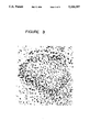
- FIG. 1 is a photomicrograph showing cartilage generated by chondrocytes cultured on collagen-coated inserts (F) and stained with Toluidine blue;
- FIG. 2 is a photomicrograph showing cartilage from the bovine joint
- FIG. 3 is a photomicrograph of fetal cartilage showing the lack of cellular organization characteristic of articular cartilage (C);
- FIG. 4 is an electron micrograph of a chondrocyte surrounded by a pericellular matrix ( ⁇ ) and an interterritorial matrix within the cartilage tissue generated in culture on collagen-coated inserts;
- FIG. 5 is an electron micrograph of the interterritorial matrix of reconstituted cartilage tissue showing the presence of collagen fibers with characteristic periodic banding;
- FIG. 6 is a Western blot showing the presence of Type II collagen in a pepsin extract (E) of the cartilage tissue generated in culture and a collagen standard (S);
- FIG. 7 is a photomicrograph of a section of reconstituted cartilage tissue incubated with anti-Type II collagen antibody, followed by anti-rabbit IgG conjugated to alkaline phosphate;
- FIG. 8 is a photomicrograph of a section of reconstituted cartilage tissue incubated with antibody directed against Factor VIII related antigen and reacted as in FIG. 7.
- the present invention provides cartilage tissue reconstituted on a substrate in vitro from isolated chondrocytes, said tissue having a biochemical composition and physiological organization substantially the same as animal articular cartilage tissue.
- the reconstituted cartilage tissue of the invention is characterized by a continuous layer of cartilage tissue, having a substantial extracellular matrix, and comprising a superficial zone wherein the chondrocytes are flattened and arranged parallel to the substrate, and mid and deep zones wherein the chondrocytes are spherical and wherein the matrix in the superficial, mid and deep zones contains collagen fibers.
- the present invention also relates to a method for producing reconstituted cartilage tissue.
- the method comprises removing articular cartilage from the joint of an animal; digesting the cartilage tissue to obtain isolated chondrocytes; forming a monolayer of chondrocytes on a substrate; and culturing the chondrocytes in growth media to produce a tissue having a biochemical composition and physiological organization substantially the same as articular cartilage tissue.
- the reconstituted cartilage tissue of the present invention may be prepared from chondrocytes isolated from articular cartilage from animals, preferably humans.
- a particularly useful system may be prepared from chondrocytes isolated from bovine articular cartilage, for example from the metacarpophalangeal joints.
- the chondrocytes may be isolated by sequential enzyme digestion techniques, such as those described in Kandel et al, Biochem. Biophys. Acta. 1035:130, 1990.
- the cartilage may be treated with 0.5% protease followed by 0.04% bacterial collagenase.
- the chondrocytes are plated on a substrate preferably a porous tissue culture insert, for example a MILLICELL®-CM insert, which has been coated with an attachment factor. Attachment factors are known in the art, see for example, Streuli and Bissell, J. Cell. Biol. 110:1405, 1990 and Buck and Horwitz, Ann, Rev. Cell Biol. 3:179, 1987.
- attachment factors examples include type I collagen, type II collagen, type IV collagen, fibronectin, gelatin, laminin, polylysine, vitronectin, cytotactin, echinonectin, entactin, tenascin, thrombospondin, uvomorulin, biglycan, chondroitin sulfate, decorin, dermatan sulfate, heparin, and hyaluronic acid.
- the attachment factor used in the method of the invention is collagen, most preferably type I collagen.
- the chondrocytes are plated at a high cell density of at least 1.0 cm 2 , preferably from 1.0 to 7 ⁇ 10 6 cm 2 .
- the substrate is a MILLICELL®-CM tissue culture insert, pore size 0.4 ⁇ m, coated with a growth attachment factor, preferably collagen, most preferably type I collagen diluted in acetic acid.
- the insert may subsequently be air dried and sterilized, for example by ultra violet light.
- the chondrocytes seeded on the coated substrate may be grown in suitable culture conditions.
- suitable culture media are known in the art, such as Ham's F12 medium.
- the culture medium may contain serum, for example fetal bovine serum in a concentration range of about 2-15% and may further contain growth factors and ascorbic acid.
- the cells may be cultured at 37° C. in a humidified atmosphere supplemented with CO 2 , for at least 2 weeks.
- the present inventor has found that, after as little as 14 days in culture, the cells will produce a tissue with substantial extracellular matrix, which is substantially identical to in vivo bovine cartilage.
- the cells produce a substantial extracellular matrix and form a continuous layer of cartilage tissue possessing zones very similar to those found in natural bovine cartilage in vivo.
- the superficial zone is characterized by chondrocytes having a flattened morphology and a matrix which does not stain, or stains poorly, with toluidine blue, indicating the relative absence of sulphated proteoglycans.
- Chondrocytes in the mid and deep zones have a spherical appearance and the matrix contains abundant sulphated proteoglycans, as evidenced by staining with toluidine blue.
- Collagen fibers are present diffusely throughout the matrix.
- the reconstituted cartilage tissue ultrastructurally has a pericellular and interterritorial arrangement similar to that of in vivo bovine cartilage.
- the chondrocytes possess abundant rough endoplasmic reticulum and are surrounded by matrix.
- the pericellular matrix contains numerous thin non-banded collagen fibers.
- the collagen in the interterritorial matrix is less compacted and embedded in electron translucent amorphous material, similar to articular cartilage.
- Collagen fibers in the interterritorial region of the matrix exhibit the periodic banding characteristic of collagen fibers in the interterritorial zone of cartilage tissue.
- the biochemical composition of the reconstituted cartilage tissue is substantially the same as animal articular cartilage tissue.
- the presence of type II collagen in the reconstituted cartilage tissue is indicative of the differentiated phenotype of chondrocytes.
- the presence of type II collagen was determined in the reconstituted cartilage tissue by means of polyacrylamide gel electrophoresis and Western blot analysis.
- Type II collagen is present throughout the reconstituted cartilage tissue as determined by immunohistochemical staining of the cartilage using polyclonal antibody directed against Type II collagen.
- the ratio of galactosamine to glucosamine amino sugars found in the glycosaminoglycans present in cartilage in the reconstituted cartilage tissue of the present invention is between about 4.5 and 6.5.
- the reconstituted cartilage tissue responds to interleukin 1 ⁇ in a similar manner to in vivo cartilage tissue.
- Interleukin 1 ⁇ stimulates production of matrix metalloproteases that can degrade cartilage matrix macromolecules and inhibit synthesis of proteoglycans.
- Treatment of the reconstituted tissue with human recombinant interleukin 1 ⁇ results in a loss of cartilage and matrix components.
- the reconstituted cartilage tissue of the present invention can be used as a model system for in vitro studies of cartilage structure, function and development.
- the reconstituted cartilage of the present invention may be used in the testing of pharmaceutical preparations useful in the treatment of diseases of the joint, for example osteoarthritis, inflammatory arthropathies, septic arthritis and crystalline arthropathies.
- the reconstituted cartilage tissue of the invention may also be implanted into the joints of patients to replace or repair damaged or deficient cartilage.
- the cartilage can be used to test angiogenic factors as the cartilage is normally resistant to vascular infiltration. It is also contemplated that the reconstituted cartilage tissue of the present invention can be used to enhance healing of bone fractures when inserted into the site of a fracture.
- Chondrocytes were isolated from bovine articular cartilage obtained from metacarpophalangeal joints using the sequential enzyme digestion techniques described in Kandel et al, Biochem. Biophys. Acta. 1053:130 (1990). Briefly, the cartilage was treated with 0.5% protease for one hour and 0.04% bacterial collagenase overnight. The isolated chondrocytes were washed three times and plated at a cell density of approximately 1.5 ⁇ 10 6 /cm 2 on collagen-coated MILLICELL®-CM porous tissue culture inserts.
- MILLICELL®-CM inserts pore size 0.4 ⁇ m, were coated with Type I collagen (Vitrogen, Type I collagen, Collagen Corporation), diluted to 1 mg/ml with 12 mM acetic acid. Following coating, the inserts were air dried for up to 18 hours and sterilized with ultraviolet light for 15 minutes.
- Type I collagen Virogen, Type I collagen, Collagen Corporation
- the collagen-coated inserts seeded with chondrocytes, were placed in sterile tissue culture wells and cultured in Ham's F12 medium, supplemented with 5% fetal bovine serum. The cells were cultured at 37° C. in a humidified atmosphere supplemented with 5% CO 2 . The medium was changed 2.5 to 3 days after the initial plating and every 2 days thereafter. The volume of medium added depended on the size of inserts used. For the larger inserts (30 mm) 1.5 ml of medium was placed on top of the insert and 1.0 ml was placed underneath. The inserts were harvested at selected time intervals up until 3 months, fixed and examined by light or electron microscopy.
- the inserts were fixed in 10% formalin, embedded in paraffin and cut into sections approximately 5 ⁇ m thick. The sections were stained with either hematoxylin and eosin to visualize the cells or with the cationic dye, toluidine blue, to stain sulphated proteoglycans. In vivo samples of intact cartilage from bovine metacarpophalangeal joints were similarly processed for light microscopy.
- insert culture For electron microscopy, representative portions of the insert culture were fixed in 2% glutaraldehyde for one hour at room temperature. The inserts were washed with phosphate buffer and then immersed in 1% osmium tetraoxide for one hour at room temperature. The inserts were washed with a phosphate buffer and dehydrated in graded ethanols to 100% ethanol. The inserts were immersed in 100% propylene oxide for 20 minutes, then embedded in plastic resin (SPURR®) and polymerized overnight at 70° C. Thin sections were cut and stained with uranyl acetate and lead citrate before examination by electron microscopy.
- SPURR® plastic resin
- FIG. 2 shows the organization of chondrocytes in cartilage tissue dissected from the bovine joint. The chondrocytes in the superficial regions are flattened and the chondrocytes in the deep layers are more spherical. Toluidine blue staining shows the presence of sulphated proteoglycans throughout the cartilage. The staining is lighter in the superficial zone suggesting there are fewer proteoglycans present. Fetal cartilage has no specific cellular organization as is shown in FIG. 3.
- the superficial zone was characterized by chondrocytes having a flattened morphology and a matrix which did not stain with toluidine blue, indicating the relative absence of sulphated proteoglycans.
- Chondrocytes in the mid zone had a less flattened appearance and the matrix contained sulphated proteoglycans, as evidenced by the metachromasia produced by toluidine blue staining. Chondrocytes in the deep zone were spherical in appearance and the matrix was metachromatic. Examination by polarized light microscopy showed the birefringence, indicative of collagen fibers.
- FIG. 5 shows collagen fibers from the interterritorial matrix of reconstituted cartilage tissue.
- the collagen fibers exhibit the periodic banding characteristic of collagen fibers in the interterritorial zone of in vivo cartilage tissue. Numerous electron dense granules were present, which appeared as dots throughout the matrix, indicating the presence of proteoglycans.
- Chondrocyte cultures were established, following the procedures outlined in Example 1 above, and maintained in culture for 2-4 weeks.
- the cartilage tissue was extracted with 100 ⁇ g/ml of pepsin at 4° C. After 24 hours an additional 100 ⁇ g of pepsin was added and the extraction continued for another 24 hours.
- the pepsin extract was neutralized with the addition of an equal volume of Laemmli's buffer.
- type II collagen was determined by polyacrylamide gel electrophoresis in the presence of sodium dodecylsulfate (SDS-PAGE) according to the procedure of Laemmli (U.K., Nature (London) 227, 680 (1974)). An 8% polyacrylamide gel was used. A sample of pepsin extract was applied to the gel and electrophoresed at 100 Ma for about 2 hours. Bands on the gel were visualized by staining with Coomassie Brilliant Blue. Only one band was observed co-migrating with the collagen standard. The identity of the band was confirmed by Western blot analysis to be type II collagen.
- SDS-PAGE sodium dodecylsulfate
- the Western blot analysis was carried out by known standard techniques, generally as described in Coding, J.W., Monoclonal Antibodies: Principles and Practice, 2nd ed., Academic Press, London pp. 195-199, 1986.
- the band was transferred electrophoretically from the gel to nitrocellulose sheets and incubated with one of two antibodies directed against type II collagen or an antibody directed against type I collagen (Southern Biotechnology Assoc., U.S.A).
- One anti-type II collagen monoclonal antibody was obtained from Developmental Studies Hybridoma Bank, Iowa and one was a polyclonal antibody obtained from Southern Biotechnology Assoc. U.S.A.
- the blots were subjected to three washes and incubated for one hour in alkaline phosphatase conjugated to anti-IgG antibody. Blots were washed 3 times and developed with NBT/BCIP (Promega, U.S.A.) from 1 to 30 minutes, until colour developed.
- NBT/BCIP Promega, U.S.A.
- Type II collagen was detected in the cultures as shown in FIG. 6. Only type II collagen was detected in the pepsin extract of the reconstituted cartilage tissue, as indicated by the arrow on FIG. 6. The results confirm that type II collagen is the major collagen produced by the chondrocyte culture.
- the distribution of type II collagen in the chondrocyte cultures was examined as follows. Frozen sections of the cultured collagen tissue were predigested with 300 I.U. testicular hyaluronidase at 37° C. for 15 minutes. The sections were washed in phosphate buffered saline and incubated with polyclonal anti-type II collagen antibody (Southern Biotechnology Associates 1:100 dilution), for 3 hours at room temperature. The sections were washed in phosphate buffered saline and then incubated for 45 minutes with anti-rabbit IgG antibody conjugated to alkaline phosphate (Vector, U.S.A.). The sections were washed three times.
- Interleukin 1 Stimulates Matrix Loss in the Chondrocyte Culture
- Chondrocyte cultures were established as described above and grown for 2 weeks. The cultures were incubated with 10 ng/ml of human recombinant interleukin 1 ⁇ (Ciba-Geigy, Switzerland) for seven days. After seven days the cultures were processed for light microscopy as described above in Example 1 and assessed histologically following staining with toluidine blue. Toluidine blue staining was taken as an indication of the presence of sulphated proteoglycans. Toluidine blue staining was greatly diminished in cultures treated with IL-1, compared to untreated controls, indicating a loss of cartilage and matrix components in the remaining cartilage.
- the tissue sample comprising bovine articular cartilage or cultured cartilage tissue following papain digestion was hydrolysed with 6 N hydrochloric acid and examined by high pressure liquid chromatography using a Water's Pico Tag amino acid analysis system (Biotechnology Centre, University of Toronto) the ratio of galactosamine to glucosamine in the bovine articular cartilage was 5.55, whereas in the cultured cartilage, the ratio was 5.14. There was no significant difference between the ratios, indicating that the ratio of these glycosaminoglycans are likely similar in both the intact tissue and the artificial tissue generated in culture.
- the ratio of galactosamine to glucosamine is indicative of the types of glycosaminoglycans present in the extracellular matrix of the bovine cartilage and the cultured cartilage tissue.
Abstract
Description
Claims (22)
Priority Applications (12)
| Application Number | Priority Date | Filing Date | Title |
|---|---|---|---|
| US07/835,831 US5326357A (en) | 1992-03-18 | 1992-03-18 | Reconstituted cartridge tissue |
| AT93907686T ATE193550T1 (en) | 1992-03-18 | 1993-03-18 | RECONSTRUCTED CARTILAGE TISSUE |
| PCT/CA1993/000111 WO1993019168A1 (en) | 1992-03-18 | 1993-03-18 | Reconstituted cartilage tissue |
| AU38816/93A AU677953B2 (en) | 1992-03-18 | 1993-03-18 | Reconstituted cartilage tissue |
| JP51613193A JP3587305B2 (en) | 1992-03-18 | 1993-03-18 | Remodeled cartilage tissue |
| CA002131904A CA2131904C (en) | 1992-03-18 | 1993-03-18 | Reconstituted cartilage tissue |
| EP93907686A EP0631619B1 (en) | 1992-03-18 | 1993-03-18 | Reconstituted cartilage tissue |
| PT93907686T PT631619E (en) | 1992-03-18 | 1993-03-18 | RECONSTITUTED CARTILAGINEO FABRIC |
| DK93907686T DK0631619T3 (en) | 1992-03-18 | 1993-03-18 | Reconstructed cartilage tissue |
| ES93907686T ES2146611T3 (en) | 1992-03-18 | 1993-03-18 | RECONSTITUTED CARTILAGINOUS TISSUE. |
| DE69328776T DE69328776T2 (en) | 1992-03-18 | 1993-03-18 | RECONSTRUCTED CARTILAGE TISSUE |
| GR20000401884T GR3034190T3 (en) | 1992-03-18 | 2000-08-14 | Reconstituted cartilage tissue. |
Applications Claiming Priority (1)
| Application Number | Priority Date | Filing Date | Title |
|---|---|---|---|
| US07/835,831 US5326357A (en) | 1992-03-18 | 1992-03-18 | Reconstituted cartridge tissue |
Publications (1)
| Publication Number | Publication Date |
|---|---|
| US5326357A true US5326357A (en) | 1994-07-05 |
Family
ID=25270583
Family Applications (1)
| Application Number | Title | Priority Date | Filing Date |
|---|---|---|---|
| US07/835,831 Expired - Lifetime US5326357A (en) | 1992-03-18 | 1992-03-18 | Reconstituted cartridge tissue |
Country Status (12)
| Country | Link |
|---|---|
| US (1) | US5326357A (en) |
| EP (1) | EP0631619B1 (en) |
| JP (1) | JP3587305B2 (en) |
| AT (1) | ATE193550T1 (en) |
| AU (1) | AU677953B2 (en) |
| CA (1) | CA2131904C (en) |
| DE (1) | DE69328776T2 (en) |
| DK (1) | DK0631619T3 (en) |
| ES (1) | ES2146611T3 (en) |
| GR (1) | GR3034190T3 (en) |
| PT (1) | PT631619E (en) |
| WO (1) | WO1993019168A1 (en) |
Cited By (93)
| Publication number | Priority date | Publication date | Assignee | Title |
|---|---|---|---|---|
| EP0739631A2 (en) * | 1995-04-26 | 1996-10-30 | El Gendler | Laminar bone support for cartilage growth |
| US5716404A (en) * | 1994-12-16 | 1998-02-10 | Massachusetts Institute Of Technology | Breast tissue engineering |
| US5763416A (en) | 1994-02-18 | 1998-06-09 | The Regent Of The University Of Michigan | Gene transfer into bone cells and tissues |
| WO1998051317A1 (en) * | 1997-05-13 | 1998-11-19 | Osiris Therapeutics, Inc. | Osteoarthritis cartilage regeneration using human mesenchymal stem cells |
| US5904717A (en) * | 1986-01-28 | 1999-05-18 | Thm Biomedical, Inc. | Method and device for reconstruction of articular cartilage |
| US5906827A (en) * | 1994-06-03 | 1999-05-25 | Creative Biomolecules, Inc. | Matrix for the manufacture of autogenous replacement body parts |
| US5935594A (en) * | 1993-10-28 | 1999-08-10 | Thm Biomedical, Inc. | Process and device for treating and healing a tissue deficiency |
| US5942496A (en) | 1994-02-18 | 1999-08-24 | The Regent Of The University Of Michigan | Methods and compositions for multiple gene transfer into bone cells |
| US5962427A (en) | 1994-02-18 | 1999-10-05 | The Regent Of The University Of Michigan | In vivo gene transfer methods for wound healing |
| US5981825A (en) * | 1994-05-13 | 1999-11-09 | Thm Biomedical, Inc. | Device and methods for in vivo culturing of diverse tissue cells |
| US6074840A (en) | 1994-02-18 | 2000-06-13 | The Regents Of The University Of Michigan | Recombinant production of latent TGF-beta binding protein-3 (LTBP-3) |
| US6077989A (en) * | 1996-05-28 | 2000-06-20 | Kandel; Rita | Resorbable implant biomaterial made of condensed calcium phosphate particles |
| WO2000056251A1 (en) | 1999-03-24 | 2000-09-28 | Chondros, Inc. | Cell-culture and polymer constructs |
| WO2000073417A1 (en) * | 1999-05-27 | 2000-12-07 | The Research Foundation Of State University Of New York | In vitro cell culture device including cartilage and methods of using the same |
| WO2001029189A2 (en) * | 1999-10-15 | 2001-04-26 | Mount Sinai Hospital | Synthetic substrate for tissue formation |
| US6235316B1 (en) | 1997-04-04 | 2001-05-22 | Barnes-Jewish Hospital | Neocartilage and methods of use |
| US6242247B1 (en) * | 1996-06-04 | 2001-06-05 | Sulzer Orthopedics Ltd. | Method for making cartilage and implants |
| WO2001045764A1 (en) * | 1999-12-20 | 2001-06-28 | Verigen Transplantation Service International Ag | Cellular matrix |
| US6300127B1 (en) | 1997-07-30 | 2001-10-09 | Emory University | Bone mineralization proteins, DNA, vectors, expression systems |
| EP1158938A1 (en) * | 1999-03-01 | 2001-12-05 | Rush-Presbyterian-St.Luke's Medical Center | In vitro production of transplantable cartilage tissue |
| WO2002010348A2 (en) * | 2000-07-29 | 2002-02-07 | Smith & Nephew Plc | Tissue implant for cartilage repair |
| WO2002045766A1 (en) * | 2000-12-06 | 2002-06-13 | Japan Tissue Engineering Co., Ltd. | Tissue equivalant for transplantation and process for producing the same |
| US20020111695A1 (en) * | 1995-11-06 | 2002-08-15 | Mount Sinai Hospital Corporation | Reconstituted mineralized cartilage tissue |
| US20020119177A1 (en) * | 2000-12-21 | 2002-08-29 | Bowman Steven M. | Reinforced foam implants with enhanced integrity for soft tissue repair and regeneration |
| US20020127265A1 (en) * | 2000-12-21 | 2002-09-12 | Bowman Steven M. | Use of reinforced foam implants with enhanced integrity for soft tissue repair and regeneration |
| US6464729B1 (en) | 1995-11-06 | 2002-10-15 | Mount Sinai Hospital Corporation | Reconstituted mineralized cartilage tissue |
| US6482401B1 (en) | 1998-12-23 | 2002-11-19 | Naturopathic Laboratories International, Inc. | Composition for the relief of joint pain and myofascial pain and method of preparing same |
| US20020187182A1 (en) * | 2001-02-14 | 2002-12-12 | Genzyme Corporation | Biocompatible fleece for hemostasis and tissue engineering |
| US20020193338A1 (en) * | 1994-02-18 | 2002-12-19 | Goldstein Steven A. | In vivo gene transfer methods for wound healing |
| WO2003016860A2 (en) * | 2001-08-14 | 2003-02-27 | Washington University In St. Louis | Systems and methods for screening pharmaceutical chemicals |
| WO2003024463A1 (en) | 2001-09-15 | 2003-03-27 | Rush-Presbyterian-St. Luke's Medical Center | Stratified cartilage tissue and methods to engineer same |
| US6551618B2 (en) | 1994-03-15 | 2003-04-22 | University Of Birmingham | Compositions and methods for delivery of agents for neuronal regeneration and survival |
| US20030138873A1 (en) * | 2002-01-22 | 2003-07-24 | Koichi Masuda | Tissue engineered cartilage for drug discovery |
| US20030147935A1 (en) * | 2000-12-21 | 2003-08-07 | Ethicon, Inc. | Use of reinforced foam implants with enhanced integrity for soft tissue repair and regeneration |
| US20030165473A1 (en) * | 2001-11-09 | 2003-09-04 | Rush-Presbyterian-St. Luke's Medical Center | Engineered intervertebral disc tissue |
| US20030191107A1 (en) * | 2002-01-22 | 2003-10-09 | Pfizer Inc. | 3-(Imidazolyl)-2-aminopropanoic acids |
| US20030193104A1 (en) * | 2000-12-21 | 2003-10-16 | Melican Mora Carolynne | Reinforced tissue implants and methods of manufacture and use |
| US20040078090A1 (en) * | 2002-10-18 | 2004-04-22 | Francois Binette | Biocompatible scaffolds with tissue fragments |
| US20040078077A1 (en) * | 2002-10-18 | 2004-04-22 | Francois Binette | Biocompatible scaffold for ligament or tendon repair |
| US20040083001A1 (en) * | 2000-06-29 | 2004-04-29 | Rita Kandel | Intervertebral disc |
| EP1457552A1 (en) * | 2003-03-13 | 2004-09-15 | Technische Universität Dresden | Carrier for tissue- and cell-culture for the preparation of implants and implant containing the carrier |
| US20040197908A1 (en) * | 2003-04-02 | 2004-10-07 | Minoru Ueda | Artificial cartilage |
| US6835377B2 (en) | 1998-05-13 | 2004-12-28 | Osiris Therapeutics, Inc. | Osteoarthritis cartilage regeneration |
| WO2005023324A1 (en) * | 2003-09-04 | 2005-03-17 | Smith & Nephew Plc | Meniscal superfacial zone cells for articular cartilage repair |
| WO2005036179A1 (en) * | 2003-10-02 | 2005-04-21 | Genzyme Corporation | Assays for evaluating anti-osteoarthritic activity |
| WO2005039528A2 (en) | 2003-10-23 | 2005-05-06 | Katharina Beschorner | Composition for the treatment of arthrosis/arthritis, especially for treating joints |
| US20050113937A1 (en) * | 2003-11-26 | 2005-05-26 | Francois Binette | Conformable tissue repair implant capable of injection delivery |
| US20050152882A1 (en) * | 2003-12-11 | 2005-07-14 | Isto Technologies | Particulate cartilage system |
| US20050177249A1 (en) * | 2004-02-09 | 2005-08-11 | Kladakis Stephanie M. | Scaffolds with viable tissue |
| US20050196387A1 (en) * | 2004-02-20 | 2005-09-08 | Isto Technologies, Inc. | Intervertebral disk repair, methods and devices therefor |
| US20050234549A1 (en) * | 2004-04-20 | 2005-10-20 | Kladakis Stephanie M | Meniscal repair scaffold |
| US20050232967A1 (en) | 2004-04-20 | 2005-10-20 | Kladakis Stephanie M | Nonwoven tissue scaffold |
| US20060073588A1 (en) * | 2004-10-01 | 2006-04-06 | Isto Technologies, Inc. | Method for chondrocyte expansion with phenotype retention |
| US20060275273A1 (en) * | 2004-02-20 | 2006-12-07 | Seyedin Mitchell S | Intervertebral Disc Repair, Methods and Devices Therefor |
| US20070122796A1 (en) * | 2001-09-25 | 2007-05-31 | Japan Science And Technology Agency National Institute For Environmental Studies | Method of preparing basement membrane, method of constructing basement membrane specimen, reconstituted artificial tissue using the basement membrane specimen and process for producing the same |
| US20070154552A1 (en) * | 2003-02-21 | 2007-07-05 | Siegal Gene P | Biologically active native biomatrix composition |
| US20070184550A1 (en) * | 2005-08-02 | 2007-08-09 | Satoshi Miyauchi | Artificial cartilage tissue and production method thereof |
| US20080038812A1 (en) * | 2001-08-14 | 2008-02-14 | The Washington University | Systems for screening pharmaceutical chemicals |
| US20080039257A1 (en) * | 2006-07-18 | 2008-02-14 | Holmes Alan G | Transmission device with selectable motor connections |
| US7488348B2 (en) | 2003-05-16 | 2009-02-10 | Musculoskeletal Transplant Foundation | Cartilage allograft plug |
| US20090041730A1 (en) * | 2000-04-25 | 2009-02-12 | Barry Francis P | Joint Repair Using Mesenchymal Stem Cells |
| US20090311221A1 (en) * | 2005-09-16 | 2009-12-17 | St. Marianna University, School Of Medicine | Biomaterials for regenerative medicine |
| US7815926B2 (en) | 2005-07-11 | 2010-10-19 | Musculoskeletal Transplant Foundation | Implant for articular cartilage repair |
| US7837740B2 (en) | 2007-01-24 | 2010-11-23 | Musculoskeletal Transplant Foundation | Two piece cancellous construct for cartilage repair |
| USRE42208E1 (en) | 2003-04-29 | 2011-03-08 | Musculoskeletal Transplant Foundation | Glue for cartilage repair |
| US7901457B2 (en) | 2003-05-16 | 2011-03-08 | Musculoskeletal Transplant Foundation | Cartilage allograft plug |
| US7901461B2 (en) | 2003-12-05 | 2011-03-08 | Ethicon, Inc. | Viable tissue repair implants and methods of use |
| US7923250B2 (en) | 1997-07-30 | 2011-04-12 | Warsaw Orthopedic, Inc. | Methods of expressing LIM mineralization protein in non-osseous cells |
| US20110118143A1 (en) * | 2008-06-05 | 2011-05-19 | Tetsuro Wakatsuki | Three dimensional tissues for high-throughput assays |
| AU2007203472B2 (en) * | 2000-07-29 | 2011-07-14 | Smith & Nephew Plc | Tissue implant for cartilage repair |
| US8016867B2 (en) | 1999-07-23 | 2011-09-13 | Depuy Mitek, Inc. | Graft fixation device and method |
| US20110301065A1 (en) * | 2000-07-24 | 2011-12-08 | Yeda Research And Development Co., Ltd. | Identifying antigen clusters for monitoring a global state of an immune system |
| US8221780B2 (en) | 2004-04-20 | 2012-07-17 | Depuy Mitek, Inc. | Nonwoven tissue scaffold |
| US8226715B2 (en) | 2003-06-30 | 2012-07-24 | Depuy Mitek, Inc. | Scaffold for connective tissue repair |
| US8292968B2 (en) | 2004-10-12 | 2012-10-23 | Musculoskeletal Transplant Foundation | Cancellous constructs, cartilage particles and combinations of cancellous constructs and cartilage particles |
| US8435551B2 (en) | 2007-03-06 | 2013-05-07 | Musculoskeletal Transplant Foundation | Cancellous construct with support ring for repair of osteochondral defects |
| US8449561B2 (en) | 1999-07-23 | 2013-05-28 | Depuy Mitek, Llc | Graft fixation device combination |
| US8480757B2 (en) | 2005-08-26 | 2013-07-09 | Zimmer, Inc. | Implants and methods for repair, replacement and treatment of disease |
| US8497121B2 (en) | 2006-12-20 | 2013-07-30 | Zimmer Orthobiologics, Inc. | Method of obtaining viable small tissue particles and use for tissue repair |
| WO2014011890A1 (en) * | 2012-07-11 | 2014-01-16 | Osiris Therapeutics, Inc. | Disrupted cartilage products |
| AU2008302088B2 (en) * | 2007-09-21 | 2014-02-20 | Isto Technologies, Inc. | Method for chondrocyte expansion with phenotype retention |
| US8895045B2 (en) | 2003-03-07 | 2014-11-25 | Depuy Mitek, Llc | Method of preparation of bioabsorbable porous reinforced tissue implants and implants thereof |
| US8945535B2 (en) | 2005-09-20 | 2015-02-03 | Zimmer Orthobiologics, Inc. | Implant for the repair of a cartilage defect and method for manufacturing the implant |
| US9138318B2 (en) | 2007-04-12 | 2015-09-22 | Zimmer, Inc. | Apparatus for forming an implant |
| US9238090B1 (en) | 2014-12-24 | 2016-01-19 | Fettech, Llc | Tissue-based compositions |
| US9701940B2 (en) | 2005-09-19 | 2017-07-11 | Histogenics Corporation | Cell-support matrix having narrowly defined uniformly vertically and non-randomly organized porosity and pore density and a method for preparation thereof |
| US10077420B2 (en) | 2014-12-02 | 2018-09-18 | Histogenics Corporation | Cell and tissue culture container |
| US10167447B2 (en) | 2012-12-21 | 2019-01-01 | Zimmer, Inc. | Supports and methods for promoting integration of cartilage tissue explants |
| US10195044B2 (en) | 2013-07-12 | 2019-02-05 | Sinai Health System | Intervertebral disc implant |
| US10545144B2 (en) | 2013-12-31 | 2020-01-28 | Yeda Research And Development Co., Ltd. | Diagnosis of systemic lupus erythematosus using oligonucleotides antigens |
| US10583220B2 (en) | 2003-08-11 | 2020-03-10 | DePuy Synthes Products, Inc. | Method and apparatus for resurfacing an articular surface |
| US11047855B2 (en) | 2015-03-01 | 2021-06-29 | Immunarray Ltd. | Diagnosis of systemic lupus erythematosus using protein, peptide and oligonucleotide antigens |
| US11052175B2 (en) | 2015-08-19 | 2021-07-06 | Musculoskeletal Transplant Foundation | Cartilage-derived implants and methods of making and using same |
Families Citing this family (15)
| Publication number | Priority date | Publication date | Assignee | Title |
|---|---|---|---|---|
| US5902741A (en) * | 1986-04-18 | 1999-05-11 | Advanced Tissue Sciences, Inc. | Three-dimensional cartilage cultures |
| US5723331A (en) * | 1994-05-05 | 1998-03-03 | Genzyme Corporation | Methods and compositions for the repair of articular cartilage defects in mammals |
| GB9503492D0 (en) | 1995-02-22 | 1995-04-12 | Ed Geistlich S Hne A G F R Che | Chemical product |
| EP0735135A3 (en) * | 1995-03-31 | 1997-12-29 | DIZG Deutsches Institüt für Zell- und Gewebeersatz gGmbH | Cell sheets and transport system for cell sheets |
| DE19540487A1 (en) * | 1995-10-20 | 1997-04-24 | Olaf Schultz | Cell interaction system for induction of artificial 3-dimensional tissue |
| US6569172B2 (en) * | 1996-08-30 | 2003-05-27 | Verigen Transplantation Service International (Vtsi) | Method, instruments, and kit for autologous transplantation |
| US5989269A (en) | 1996-08-30 | 1999-11-23 | Vts Holdings L.L.C. | Method, instruments and kit for autologous transplantation |
| US9034315B2 (en) | 1997-10-10 | 2015-05-19 | Ed. Geistlich Soehne Ag Fuer Chemische Industrie | Cell-charged multi-layer collagen membrane |
| US8858981B2 (en) | 1997-10-10 | 2014-10-14 | Ed. Geistlich Soehne Fuer Chemistrie Industrie | Bone healing material comprising matrix carrying bone-forming cells |
| US20050186283A1 (en) | 1997-10-10 | 2005-08-25 | Ed. Geistlich Soehne Ag Fuer Chemistrie Industrie | Collagen carrier of therapeutic genetic material, and method |
| KR20040077968A (en) | 1998-08-14 | 2004-09-07 | 페리겐 트란스플란타치온 서비스 인터나치오날 (파우테에스이) 아게 | Methods, instruments and materials for chondrocyte cell transplantation |
| US6713085B2 (en) | 2001-04-27 | 2004-03-30 | Ed. Geistlich Soehne Ag Fuer Chemische Industrie | Method and membrane for mucosa regeneration |
| CA2412012C (en) | 2001-11-20 | 2011-08-02 | Ed. Geistlich Soehne Ag Fuer Chemische Industrie | Resorbable extracellular matrix containing collagen i and collagen ii for reconstruction of cartilage |
| US7931687B2 (en) * | 2002-05-13 | 2011-04-26 | Articular Engineering, Llc | Tissue engineered osteochondral implant |
| US20050069572A1 (en) * | 2002-10-09 | 2005-03-31 | Jennifer Elisseeff | Multi-layered polymerizing hydrogels for tissue regeneration |
Citations (10)
| Publication number | Priority date | Publication date | Assignee | Title |
|---|---|---|---|---|
| US3703575A (en) * | 1970-12-23 | 1972-11-21 | Heinrich Thiele | Reconstructed cartilaginous tissue and method of implanting it into the human or animal body |
| US4356261A (en) * | 1980-04-22 | 1982-10-26 | Rush-Presbyterian-St. Luke's Medical Center | Anti-invasion factor containing cultures |
| EP0175286A2 (en) * | 1984-09-14 | 1986-03-26 | MCW Research Foundation, Inc. | In vitro cell culture system and method |
| US4642120A (en) * | 1983-03-23 | 1987-02-10 | Ramot University Authority For Applied Research And Industrial Development Ltd. | Repair of cartilage and bones |
| US4846835A (en) * | 1987-06-15 | 1989-07-11 | Grande Daniel A | Technique for healing lesions in cartilage |
| US4904259A (en) * | 1988-04-29 | 1990-02-27 | Samuel Itay | Compositions and methods for repair of cartilage and bone |
| WO1990012603A1 (en) * | 1989-04-17 | 1990-11-01 | Vacanti Joseph P | Neomorphogenesis of cartilage in vivo from cell culture |
| EP0396138A2 (en) * | 1989-05-04 | 1990-11-07 | Millipore Corporation | Method for growing cellular tissue |
| US5037656A (en) * | 1986-12-04 | 1991-08-06 | Millipore Corporation | Porous membrane having hydrophilic and cell growth promotions surface and process |
| US5053050A (en) * | 1988-04-29 | 1991-10-01 | Samuel Itay | Compositions for repair of cartilage and bone |
-
1992
- 1992-03-18 US US07/835,831 patent/US5326357A/en not_active Expired - Lifetime
-
1993
- 1993-03-18 CA CA002131904A patent/CA2131904C/en not_active Expired - Fee Related
- 1993-03-18 AT AT93907686T patent/ATE193550T1/en not_active IP Right Cessation
- 1993-03-18 ES ES93907686T patent/ES2146611T3/en not_active Expired - Lifetime
- 1993-03-18 DE DE69328776T patent/DE69328776T2/en not_active Expired - Fee Related
- 1993-03-18 WO PCT/CA1993/000111 patent/WO1993019168A1/en active IP Right Grant
- 1993-03-18 PT PT93907686T patent/PT631619E/en unknown
- 1993-03-18 EP EP93907686A patent/EP0631619B1/en not_active Expired - Lifetime
- 1993-03-18 DK DK93907686T patent/DK0631619T3/en active
- 1993-03-18 AU AU38816/93A patent/AU677953B2/en not_active Ceased
- 1993-03-18 JP JP51613193A patent/JP3587305B2/en not_active Expired - Fee Related
-
2000
- 2000-08-14 GR GR20000401884T patent/GR3034190T3/en not_active IP Right Cessation
Patent Citations (13)
| Publication number | Priority date | Publication date | Assignee | Title |
|---|---|---|---|---|
| US3703575A (en) * | 1970-12-23 | 1972-11-21 | Heinrich Thiele | Reconstructed cartilaginous tissue and method of implanting it into the human or animal body |
| US4356261A (en) * | 1980-04-22 | 1982-10-26 | Rush-Presbyterian-St. Luke's Medical Center | Anti-invasion factor containing cultures |
| US4642120A (en) * | 1983-03-23 | 1987-02-10 | Ramot University Authority For Applied Research And Industrial Development Ltd. | Repair of cartilage and bones |
| EP0175286A2 (en) * | 1984-09-14 | 1986-03-26 | MCW Research Foundation, Inc. | In vitro cell culture system and method |
| US4757017A (en) * | 1984-09-14 | 1988-07-12 | Mcw Research Foundation, Inc. | In vitro cell culture system |
| US5041138A (en) * | 1986-11-20 | 1991-08-20 | Massachusetts Institute Of Technology | Neomorphogenesis of cartilage in vivo from cell culture |
| US5037656A (en) * | 1986-12-04 | 1991-08-06 | Millipore Corporation | Porous membrane having hydrophilic and cell growth promotions surface and process |
| US4846835A (en) * | 1987-06-15 | 1989-07-11 | Grande Daniel A | Technique for healing lesions in cartilage |
| US4904259A (en) * | 1988-04-29 | 1990-02-27 | Samuel Itay | Compositions and methods for repair of cartilage and bone |
| US5053050A (en) * | 1988-04-29 | 1991-10-01 | Samuel Itay | Compositions for repair of cartilage and bone |
| WO1990012603A1 (en) * | 1989-04-17 | 1990-11-01 | Vacanti Joseph P | Neomorphogenesis of cartilage in vivo from cell culture |
| US4996154A (en) * | 1989-05-04 | 1991-02-26 | Millipore Corporation | Method for growing cellular tissue |
| EP0396138A2 (en) * | 1989-05-04 | 1990-11-07 | Millipore Corporation | Method for growing cellular tissue |
Non-Patent Citations (45)
| Title |
|---|
| Amiel et al., Conn. Tiss. Res., 18:27, 1988. * |
| Aulthouse et al., In Vitro Cell & Dev. Biol., 25:659, 1989. * |
| Aydelotte and Kuettner, Conn. Tiss. Res., 18:205, 1988. * |
| Bassleer et al., In Vitro, 22:113, 1986. * |
| Benya & Shaffer, Cell., 30:215, 1982. * |
| Billings et al., Acta. Orthop. Scand., 61:201, 1990. * |
| Brown et al., Conn. Tiss. Res., 24:157, 1990. * |
| Buck and Horwitz, Ann. Rev. Cell Biol., 3:179, 1987. * |
| Cheung, In Vitro Cell. Dev. Biol., 21:353, 1985. * |
| Delbruck et al., Conn. Tiss. Res., 15:155, 1986. * |
| Franzen et al., Differentiation, 36:199, 1987. * |
| Goldberg and Kolibas, Conn. Tissue Res., 24:265, 1990. * |
| Green, Clin. Orthop. Rel. Res., 75:248, 1971. * |
| Guo et al., Conn. Tiss. Res., 19:277, 1989. * |
| Hale et al., In Vitro Cell. Biol. & Dev. Biol., 22:597, 1986. * |
| Horwitz and Dorfman, J. Cell Biol., 45:434, 1970. * |
| Jennings et al., Cell. Biol. Int. Rep., 7:149, 1983. * |
| Kandel et al., Biochem. Biophys. Acta., 1035:130, 1990. * |
| Kato et al., Proc. Natl. Acad. Sci., 85:9552, 1988. * |
| Kuettner et al., (J. Cell. Biol.), 93:751,. (1982). * |
| Lane and Brighton, Arth. Rheum., 17:235, 1974. * |
| Macklis et al., In Vitro Cell. Develop. Biol., 21:180, 1985. * |
| Manning and Bonner, Arth. Rheum., 10:235, 1967. * |
| Morales et al., J. Biol. Chem., 259:6720, 1984. * |
| Nakahara et al., Bone, 11:181, 1990. * |
| O Driscoll et al., Trans. Orthop. Res., 37:125, 1991. * |
| O'Driscoll et al., Trans. Orthop. Res., 37:125, 1991. |
| Poole et al., J. Anat., 138:13, 1984. * |
| Robinson et al., Calcif. Tissue Int., 46:246, 1990. * |
| Schneiderman et al., J. Orthop. Res., 4:393, 1986. * |
| Schwartz et al., In Vitro, 18:254, 1982. * |
| Solursh, Am. J. Med. Gen., 34:30, 1989. * |
| Solursh, Development and Diseases of Cartridge and Bone Marrow, Alan R. Liss Inc., 1987. * |
| Solursh, J. Cell. Biochem., 45:258, 1991. * |
| Streuli and Bissell, J. Cell. Biol., 110:1405, 1990. * |
| Thompson et al., Exp. Cell. Res., 157:483, 1985. * |
| Trippel et al., J. Bone & Joint Surgery, 1990:816. * |
| Urban and Bayliss, Biochem. Biophys. Acta., 992:59, 1988. * |
| Urist, Marshall R., "Clinical Orthopaedics and Related Research", No. 186, Jun. 1984, Philadelphia, Pa., U.S., pp. 231-239, T. Kimura et al., Chondrocytes Embedded in Collagen Gels Maintain Cartilage Phenstype During Long Term Cultures. |
| Urist, Marshall R., Clinical Orthopaedics and Related Research , No. 186, Jun. 1984, Philadelphia, Pa., U.S., pp. 231 239, T. Kimura et al., Chondrocytes Embedded in Collagen Gels Maintain Cartilage Phenstype During Long Term Cultures. * |
| Van Kampen and Veldhuijzen, Exp. Cell. Res., 140:440, 1982. * |
| Von der Mark et al., Nature, 267:531, 1977. * |
| Watt and Dudhia, Differentiation, 38:140, 1988. * |
| Yannas, Collagen, 3:87. * |
| Zanetti et al., J. Cell Biol., 101:53, 1985. * |
Cited By (198)
| Publication number | Priority date | Publication date | Assignee | Title |
|---|---|---|---|---|
| US5904717A (en) * | 1986-01-28 | 1999-05-18 | Thm Biomedical, Inc. | Method and device for reconstruction of articular cartilage |
| US5935594A (en) * | 1993-10-28 | 1999-08-10 | Thm Biomedical, Inc. | Process and device for treating and healing a tissue deficiency |
| US6074840A (en) | 1994-02-18 | 2000-06-13 | The Regents Of The University Of Michigan | Recombinant production of latent TGF-beta binding protein-3 (LTBP-3) |
| US20020193338A1 (en) * | 1994-02-18 | 2002-12-19 | Goldstein Steven A. | In vivo gene transfer methods for wound healing |
| US5763416A (en) | 1994-02-18 | 1998-06-09 | The Regent Of The University Of Michigan | Gene transfer into bone cells and tissues |
| US5962427A (en) | 1994-02-18 | 1999-10-05 | The Regent Of The University Of Michigan | In vivo gene transfer methods for wound healing |
| US6774105B1 (en) | 1994-02-18 | 2004-08-10 | The Regents Of The University Of Michigan | Methods of using latent TGF-β binding proteins |
| US5942496A (en) | 1994-02-18 | 1999-08-24 | The Regent Of The University Of Michigan | Methods and compositions for multiple gene transfer into bone cells |
| US6551618B2 (en) | 1994-03-15 | 2003-04-22 | University Of Birmingham | Compositions and methods for delivery of agents for neuronal regeneration and survival |
| US5981825A (en) * | 1994-05-13 | 1999-11-09 | Thm Biomedical, Inc. | Device and methods for in vivo culturing of diverse tissue cells |
| US6264701B1 (en) | 1994-05-13 | 2001-07-24 | Kensey Nash Corporation | Device and methods for in vivo culturing of diverse tissue cells |
| US5906827A (en) * | 1994-06-03 | 1999-05-25 | Creative Biomolecules, Inc. | Matrix for the manufacture of autogenous replacement body parts |
| US20050089544A1 (en) * | 1994-06-03 | 2005-04-28 | Khouri Roger K. | Manufacture of autogenous replacement body parts |
| US6027743A (en) * | 1994-06-03 | 2000-02-22 | Stryker Corporation | Manufacture of autogenous replacement body parts |
| US6110482A (en) * | 1994-06-03 | 2000-08-29 | Styker Corporation | Manufacture of autogenous replacement body parts |
| US5716404A (en) * | 1994-12-16 | 1998-02-10 | Massachusetts Institute Of Technology | Breast tissue engineering |
| EP0739631A2 (en) * | 1995-04-26 | 1996-10-30 | El Gendler | Laminar bone support for cartilage growth |
| EP0739631A3 (en) * | 1995-04-26 | 1998-05-06 | El Gendler | Laminar bone support for cartilage growth |
| US5904716A (en) * | 1995-04-26 | 1999-05-18 | Gendler; El | Method for reconstituting cartilage tissue using demineralized bone and product thereof |
| US20100068241A1 (en) * | 1995-11-06 | 2010-03-18 | Mount Sinai Hospital | Reconstituted mineralized cartilage tissue |
| US20020111695A1 (en) * | 1995-11-06 | 2002-08-15 | Mount Sinai Hospital Corporation | Reconstituted mineralized cartilage tissue |
| US6464729B1 (en) | 1995-11-06 | 2002-10-15 | Mount Sinai Hospital Corporation | Reconstituted mineralized cartilage tissue |
| US6077989A (en) * | 1996-05-28 | 2000-06-20 | Kandel; Rita | Resorbable implant biomaterial made of condensed calcium phosphate particles |
| US6242247B1 (en) * | 1996-06-04 | 2001-06-05 | Sulzer Orthopedics Ltd. | Method for making cartilage and implants |
| US6387693B2 (en) | 1996-06-04 | 2002-05-14 | Sulzer Orthopedics Ltd. | Method for producing cartilage tissue and implants for repairing enchondral and osteochondral defects as well as arrangement for carrying out the method |
| US6645764B1 (en) | 1997-04-04 | 2003-11-11 | Barnes-Jewish Hospital | Neocartilage and methods of use |
| US20030224518A1 (en) * | 1997-04-04 | 2003-12-04 | Adkisson Huston D. | Cartilage composites and methods of use |
| US6235316B1 (en) | 1997-04-04 | 2001-05-22 | Barnes-Jewish Hospital | Neocartilage and methods of use |
| US7087227B2 (en) | 1997-04-04 | 2006-08-08 | Barnes-Jewish Hospital | Cartilage composites and methods of use |
| WO1998051317A1 (en) * | 1997-05-13 | 1998-11-19 | Osiris Therapeutics, Inc. | Osteoarthritis cartilage regeneration using human mesenchymal stem cells |
| US6300127B1 (en) | 1997-07-30 | 2001-10-09 | Emory University | Bone mineralization proteins, DNA, vectors, expression systems |
| US6521750B2 (en) | 1997-07-30 | 2003-02-18 | Univ Emory | Bone mineralization proteins, DNA, vectors, expression systems |
| US6444803B1 (en) | 1997-07-30 | 2002-09-03 | Emory University | Bone mineralization proteins, DNA, vectors, expression systems |
| US7923250B2 (en) | 1997-07-30 | 2011-04-12 | Warsaw Orthopedic, Inc. | Methods of expressing LIM mineralization protein in non-osseous cells |
| US6378527B1 (en) | 1998-04-08 | 2002-04-30 | Chondros, Inc. | Cell-culture and polymer constructs |
| US20020123142A1 (en) * | 1998-04-08 | 2002-09-05 | Hungerford David S. | Cell-culture and polymer constructs |
| US20020133235A1 (en) * | 1998-04-08 | 2002-09-19 | Hungerford David S. | Cell-culture and polymer constructs |
| US6835377B2 (en) | 1998-05-13 | 2004-12-28 | Osiris Therapeutics, Inc. | Osteoarthritis cartilage regeneration |
| US6482401B1 (en) | 1998-12-23 | 2002-11-19 | Naturopathic Laboratories International, Inc. | Composition for the relief of joint pain and myofascial pain and method of preparing same |
| EP1158938A1 (en) * | 1999-03-01 | 2001-12-05 | Rush-Presbyterian-St.Luke's Medical Center | In vitro production of transplantable cartilage tissue |
| EP1158938A4 (en) * | 1999-03-01 | 2002-10-16 | Rush Presbyterian St Luke | In vitro production of transplantable cartilage tissue |
| EP1738717A3 (en) * | 1999-03-01 | 2007-03-07 | Rush University Medical Center | In vitro production of transplantable cartilage tissue |
| WO2000056251A1 (en) | 1999-03-24 | 2000-09-28 | Chondros, Inc. | Cell-culture and polymer constructs |
| US6312952B1 (en) * | 1999-05-27 | 2001-11-06 | The Research Foundation Of State University Of New York | In vitro cell culture device including cartilage and methods of using the same |
| WO2000073417A1 (en) * | 1999-05-27 | 2000-12-07 | The Research Foundation Of State University Of New York | In vitro cell culture device including cartilage and methods of using the same |
| US6465205B2 (en) | 1999-05-27 | 2002-10-15 | The Research Foundation Of State University Of New York | In vitro cell culture device including cartilage and methods of using the same |
| US8449561B2 (en) | 1999-07-23 | 2013-05-28 | Depuy Mitek, Llc | Graft fixation device combination |
| US8016867B2 (en) | 1999-07-23 | 2011-09-13 | Depuy Mitek, Inc. | Graft fixation device and method |
| WO2001029189A2 (en) * | 1999-10-15 | 2001-04-26 | Mount Sinai Hospital | Synthetic substrate for tissue formation |
| WO2001029189A3 (en) * | 1999-10-15 | 2001-11-01 | Mount Sinai Hospital Corp | Synthetic substrate for tissue formation |
| US20070071733A1 (en) * | 1999-10-15 | 2007-03-29 | Mount Sinai Hospital | Synthetic substrate for tissue formation |
| WO2001045764A1 (en) * | 1999-12-20 | 2001-06-28 | Verigen Transplantation Service International Ag | Cellular matrix |
| US20090041730A1 (en) * | 2000-04-25 | 2009-02-12 | Barry Francis P | Joint Repair Using Mesenchymal Stem Cells |
| US9050178B2 (en) | 2000-04-25 | 2015-06-09 | Mesoblast International Sàrl | Joint repair using mesenchymal stem cells |
| US9814580B2 (en) | 2000-04-25 | 2017-11-14 | Mesoblast International Sarl | Joint repair using mesenchymal stem cells |
| US20080119936A1 (en) * | 2000-06-29 | 2008-05-22 | Mount Sinai Hospital | Intervertebral disc |
| US8163554B2 (en) | 2000-06-29 | 2012-04-24 | Mount Sinai Hospital | Intervertebral disc |
| US20040083001A1 (en) * | 2000-06-29 | 2004-04-29 | Rita Kandel | Intervertebral disc |
| US8703654B2 (en) * | 2000-07-24 | 2014-04-22 | Yeda Research And Development Co. Ltd. | Identifying antigen clusters for monitoring a global state of an immune system |
| US20110301065A1 (en) * | 2000-07-24 | 2011-12-08 | Yeda Research And Development Co., Ltd. | Identifying antigen clusters for monitoring a global state of an immune system |
| US10082503B2 (en) | 2000-07-24 | 2018-09-25 | Yeda Research And Development Co. Ltd. | Identifying antigen clusters for monitoring a global state of an immune system |
| US11002735B2 (en) | 2000-07-24 | 2021-05-11 | Yeda Research And Development Co. Ltd. | Identifying antigen clusters for monitoring a global state of an immune system |
| WO2002010348A2 (en) * | 2000-07-29 | 2002-02-07 | Smith & Nephew Plc | Tissue implant for cartilage repair |
| EP2071020A3 (en) * | 2000-07-29 | 2014-12-31 | Smith&Nephew PLC | Tissue Implant |
| US20040033212A1 (en) * | 2000-07-29 | 2004-02-19 | Thomson Brian Mark | Tissue implant |
| AU2001275746B2 (en) * | 2000-07-29 | 2007-04-26 | Smith & Nephew Plc | Tissue implant for cartilage repair |
| WO2002010348A3 (en) * | 2000-07-29 | 2002-09-06 | Smith & Nephew | Tissue implant for cartilage repair |
| US9452238B2 (en) | 2000-07-29 | 2016-09-27 | Smith & Nephew LLP | Tissue implant |
| AU2007203472B2 (en) * | 2000-07-29 | 2011-07-14 | Smith & Nephew Plc | Tissue implant for cartilage repair |
| CN1327910C (en) * | 2000-12-06 | 2007-07-25 | 越智光夫 | Tissue equivalant for transplantation and process for producing the same |
| US20040030406A1 (en) * | 2000-12-06 | 2004-02-12 | Mitsuo Ochi | Tissue equivalent for transplantation and process for producing the same |
| EP1358895A4 (en) * | 2000-12-06 | 2007-02-28 | Japan Tissue Eng Co Ltd | Tissue equivalant for transplantation and process for producing the same |
| US20070172812A1 (en) * | 2000-12-06 | 2007-07-26 | Mitsuo Ochi | Tissue equivalent for transplantation and method for producing same |
| EP1358895A1 (en) * | 2000-12-06 | 2003-11-05 | Japan Tissue Engineering Co., Ltd. | Tissue equivalant for transplantation and process for producing the same |
| WO2002045766A1 (en) * | 2000-12-06 | 2002-06-13 | Japan Tissue Engineering Co., Ltd. | Tissue equivalant for transplantation and process for producing the same |
| US20020119177A1 (en) * | 2000-12-21 | 2002-08-29 | Bowman Steven M. | Reinforced foam implants with enhanced integrity for soft tissue repair and regeneration |
| US6884428B2 (en) | 2000-12-21 | 2005-04-26 | Depuy Mitek, Inc. | Use of reinforced foam implants with enhanced integrity for soft tissue repair and regeneration |
| US20030193104A1 (en) * | 2000-12-21 | 2003-10-16 | Melican Mora Carolynne | Reinforced tissue implants and methods of manufacture and use |
| US8691259B2 (en) | 2000-12-21 | 2014-04-08 | Depuy Mitek, Llc | Reinforced foam implants with enhanced integrity for soft tissue repair and regeneration |
| US6852330B2 (en) | 2000-12-21 | 2005-02-08 | Depuy Mitek, Inc. | Reinforced foam implants with enhanced integrity for soft tissue repair and regeneration |
| US20020127265A1 (en) * | 2000-12-21 | 2002-09-12 | Bowman Steven M. | Use of reinforced foam implants with enhanced integrity for soft tissue repair and regeneration |
| US20030147935A1 (en) * | 2000-12-21 | 2003-08-07 | Ethicon, Inc. | Use of reinforced foam implants with enhanced integrity for soft tissue repair and regeneration |
| US20060067967A1 (en) * | 2000-12-21 | 2006-03-30 | Depuy Mitek, Inc. | Reinforced foam implants with enhanced integrity for soft tissue repair and regeneration |
| US20020187182A1 (en) * | 2001-02-14 | 2002-12-12 | Genzyme Corporation | Biocompatible fleece for hemostasis and tissue engineering |
| WO2003016860A3 (en) * | 2001-08-14 | 2003-06-12 | Univ St Louis | Systems and methods for screening pharmaceutical chemicals |
| US20030064358A1 (en) * | 2001-08-14 | 2003-04-03 | Elliot Elson | Systems and methods for screening pharmaceutical chemicals |
| US8227240B2 (en) | 2001-08-14 | 2012-07-24 | The Washington University | Systems for screening pharmaceutical chemicals |
| US20090068701A1 (en) * | 2001-08-14 | 2009-03-12 | Washington University In St. Louis | Systems and methods for screening pharmaceutical chemicals |
| WO2003016860A2 (en) * | 2001-08-14 | 2003-02-27 | Washington University In St. Louis | Systems and methods for screening pharmaceutical chemicals |
| US7449306B2 (en) | 2001-08-14 | 2008-11-11 | Washington University In St. Louis | Systems and methods for screening pharmaceutical chemicals |
| US20080038812A1 (en) * | 2001-08-14 | 2008-02-14 | The Washington University | Systems for screening pharmaceutical chemicals |
| US8071381B2 (en) | 2001-08-14 | 2011-12-06 | Washington University In St. Louis | Systems and methods for screening pharmaceutical chemicals |
| US20030077821A1 (en) * | 2001-09-15 | 2003-04-24 | Sah Robert L. | Methods to engineer stratified cartilage tissue |
| US7476257B2 (en) * | 2001-09-15 | 2009-01-13 | Rush University Medical Center | Methods to engineer stratified cartilage tissue |
| WO2003024463A1 (en) | 2001-09-15 | 2003-03-27 | Rush-Presbyterian-St. Luke's Medical Center | Stratified cartilage tissue and methods to engineer same |
| EP1435980A1 (en) * | 2001-09-15 | 2004-07-14 | Rush-Presbyterian-St. Luke's Medical Center | Stratified cartilage tissue and methods to engineer same |
| EP1435980A4 (en) * | 2001-09-15 | 2006-06-07 | Univ California | Stratified cartilage tissue and methods to engineer same |
| AU2002335747B2 (en) * | 2001-09-15 | 2009-01-29 | Rush University Medical Center | Stratified cartilage tissue and methods to engineer same |
| US20070122796A1 (en) * | 2001-09-25 | 2007-05-31 | Japan Science And Technology Agency National Institute For Environmental Studies | Method of preparing basement membrane, method of constructing basement membrane specimen, reconstituted artificial tissue using the basement membrane specimen and process for producing the same |
| US8765473B2 (en) * | 2001-09-25 | 2014-07-01 | Japan Science And Technology Agency | Method of preparing basement membrane, method of constructing basement membrane specimen, reconstituted artificial tissue using the basement membrane specimen and process for producing the same |
| US20090142311A1 (en) * | 2001-11-09 | 2009-06-04 | Koichi Masuda | Engineered intervertebral disc tissue |
| US20030165473A1 (en) * | 2001-11-09 | 2003-09-04 | Rush-Presbyterian-St. Luke's Medical Center | Engineered intervertebral disc tissue |
| US20060160214A1 (en) * | 2001-11-09 | 2006-07-20 | Rush University Medical Center | Engineered intervertebral disc tissue |
| US20030138873A1 (en) * | 2002-01-22 | 2003-07-24 | Koichi Masuda | Tissue engineered cartilage for drug discovery |
| EP1474524A1 (en) * | 2002-01-22 | 2004-11-10 | Rush-Presbyterian-St. Luke's Medical Center | Tissue engineered cartilage for drug discovery |
| US20030191107A1 (en) * | 2002-01-22 | 2003-10-09 | Pfizer Inc. | 3-(Imidazolyl)-2-aminopropanoic acids |
| EP1474524A4 (en) * | 2002-01-22 | 2006-06-21 | Univ Rush Medical Center | Tissue engineered cartilage for drug discovery |
| US20040078090A1 (en) * | 2002-10-18 | 2004-04-22 | Francois Binette | Biocompatible scaffolds with tissue fragments |
| US8637066B2 (en) | 2002-10-18 | 2014-01-28 | Depuy Mitek, Llc | Biocompatible scaffold for ligament or tendon repair |
| US10603408B2 (en) | 2002-10-18 | 2020-03-31 | DePuy Synthes Products, Inc. | Biocompatible scaffolds with tissue fragments |
| US7824701B2 (en) | 2002-10-18 | 2010-11-02 | Ethicon, Inc. | Biocompatible scaffold for ligament or tendon repair |
| US20040078077A1 (en) * | 2002-10-18 | 2004-04-22 | Francois Binette | Biocompatible scaffold for ligament or tendon repair |
| US9511171B2 (en) | 2002-10-18 | 2016-12-06 | Depuy Mitek, Llc | Biocompatible scaffolds with tissue fragments |
| US7727550B2 (en) * | 2003-02-21 | 2010-06-01 | The Uab Research Foundation | Biologically active native biomatrix composition |
| US20070154552A1 (en) * | 2003-02-21 | 2007-07-05 | Siegal Gene P | Biologically active native biomatrix composition |
| US8895045B2 (en) | 2003-03-07 | 2014-11-25 | Depuy Mitek, Llc | Method of preparation of bioabsorbable porous reinforced tissue implants and implants thereof |
| EP1457552A1 (en) * | 2003-03-13 | 2004-09-15 | Technische Universität Dresden | Carrier for tissue- and cell-culture for the preparation of implants and implant containing the carrier |
| US20040197908A1 (en) * | 2003-04-02 | 2004-10-07 | Minoru Ueda | Artificial cartilage |
| USRE42208E1 (en) | 2003-04-29 | 2011-03-08 | Musculoskeletal Transplant Foundation | Glue for cartilage repair |
| USRE43258E1 (en) | 2003-04-29 | 2012-03-20 | Musculoskeletal Transplant Foundation | Glue for cartilage repair |
| US7901457B2 (en) | 2003-05-16 | 2011-03-08 | Musculoskeletal Transplant Foundation | Cartilage allograft plug |
| US7488348B2 (en) | 2003-05-16 | 2009-02-10 | Musculoskeletal Transplant Foundation | Cartilage allograft plug |
| US8221500B2 (en) | 2003-05-16 | 2012-07-17 | Musculoskeletal Transplant Foundation | Cartilage allograft plug |
| US9211362B2 (en) | 2003-06-30 | 2015-12-15 | Depuy Mitek, Llc | Scaffold for connective tissue repair |
| US8226715B2 (en) | 2003-06-30 | 2012-07-24 | Depuy Mitek, Inc. | Scaffold for connective tissue repair |
| US10583220B2 (en) | 2003-08-11 | 2020-03-10 | DePuy Synthes Products, Inc. | Method and apparatus for resurfacing an articular surface |
| WO2005023324A1 (en) * | 2003-09-04 | 2005-03-17 | Smith & Nephew Plc | Meniscal superfacial zone cells for articular cartilage repair |
| WO2005036179A1 (en) * | 2003-10-02 | 2005-04-21 | Genzyme Corporation | Assays for evaluating anti-osteoarthritic activity |
| AU2004283485B2 (en) * | 2003-10-23 | 2009-05-07 | Katharina Trott | Composition for the treatment of arthrosis/arthritis, especially for treating joints |
| WO2005039528A3 (en) * | 2003-10-23 | 2005-06-16 | Katharina Beschorner | Composition for the treatment of arthrosis/arthritis, especially for treating joints |
| WO2005039528A2 (en) | 2003-10-23 | 2005-05-06 | Katharina Beschorner | Composition for the treatment of arthrosis/arthritis, especially for treating joints |
| US20070020245A1 (en) * | 2003-10-23 | 2007-01-25 | Katharina Trott | Composition for the treatment of arthrosis/arthritis, especially for treating joints |
| US8137702B2 (en) | 2003-11-26 | 2012-03-20 | Depuy Mitek, Inc. | Conformable tissue repair implant capable of injection delivery |
| US7316822B2 (en) | 2003-11-26 | 2008-01-08 | Ethicon, Inc. | Conformable tissue repair implant capable of injection delivery |
| US20050113937A1 (en) * | 2003-11-26 | 2005-05-26 | Francois Binette | Conformable tissue repair implant capable of injection delivery |
| US8496970B2 (en) | 2003-11-26 | 2013-07-30 | Depuy Mitek, Llc | Conformable tissue repair implant capable of injection delivery |
| US7875296B2 (en) | 2003-11-26 | 2011-01-25 | Depuy Mitek, Inc. | Conformable tissue repair implant capable of injection delivery |
| US8641775B2 (en) | 2003-12-05 | 2014-02-04 | Depuy Mitek, Llc | Viable tissue repair implants and methods of use |
| US7901461B2 (en) | 2003-12-05 | 2011-03-08 | Ethicon, Inc. | Viable tissue repair implants and methods of use |
| US20100322994A1 (en) * | 2003-12-11 | 2010-12-23 | Isto Technologies, Inc. | Particulate cartilage system |
| US8524268B2 (en) | 2003-12-11 | 2013-09-03 | Zimmer, Inc. | Cadaveric allogenic human juvenile cartilage implant |
| US8784863B2 (en) | 2003-12-11 | 2014-07-22 | Zimmer, Inc. | Particulate cadaveric allogenic cartilage system |
| US7824711B2 (en) | 2003-12-11 | 2010-11-02 | Isto Technologies, Inc. | Particulate cartilage system |
| US8652507B2 (en) | 2003-12-11 | 2014-02-18 | Zimmer, Inc. | Juvenile cartilage composition |
| EP2338442A1 (en) | 2003-12-11 | 2011-06-29 | Isto Technologies Inc. | Particulate cartilage system |
| US8765165B2 (en) | 2003-12-11 | 2014-07-01 | Zimmer, Inc. | Particulate cartilage system |
| EP2335650A1 (en) | 2003-12-11 | 2011-06-22 | Isto Technologies Inc. | Particulate cartilage system |
| US8834914B2 (en) | 2003-12-11 | 2014-09-16 | Zimmer, Inc. | Treatment methods using a particulate cadaveric allogenic juvenile cartilage particles |
| EP2338441A1 (en) | 2003-12-11 | 2011-06-29 | Isto Technologies Inc. | Particulate cartilage system |
| US20050152882A1 (en) * | 2003-12-11 | 2005-07-14 | Isto Technologies | Particulate cartilage system |
| US8518433B2 (en) | 2003-12-11 | 2013-08-27 | Zimmer, Inc. | Method of treating an osteochondral defect |
| US20050177249A1 (en) * | 2004-02-09 | 2005-08-11 | Kladakis Stephanie M. | Scaffolds with viable tissue |
| US11395865B2 (en) | 2004-02-09 | 2022-07-26 | DePuy Synthes Products, Inc. | Scaffolds with viable tissue |
| US7879604B2 (en) | 2004-02-20 | 2011-02-01 | Isto Technoliges, Inc. | Intervertebral disk repair, methods and devices therefor |
| US20050196387A1 (en) * | 2004-02-20 | 2005-09-08 | Isto Technologies, Inc. | Intervertebral disk repair, methods and devices therefor |
| WO2005081870A3 (en) * | 2004-02-20 | 2006-12-14 | Isto Technologies Inc | Intervertebral disc repair, methods and devices therefor |
| US20060275273A1 (en) * | 2004-02-20 | 2006-12-07 | Seyedin Mitchell S | Intervertebral Disc Repair, Methods and Devices Therefor |
| US20050232967A1 (en) | 2004-04-20 | 2005-10-20 | Kladakis Stephanie M | Nonwoven tissue scaffold |
| US8657881B2 (en) | 2004-04-20 | 2014-02-25 | Depuy Mitek, Llc | Meniscal repair scaffold |
| US20050234549A1 (en) * | 2004-04-20 | 2005-10-20 | Kladakis Stephanie M | Meniscal repair scaffold |
| US8137686B2 (en) | 2004-04-20 | 2012-03-20 | Depuy Mitek, Inc. | Nonwoven tissue scaffold |
| US8221780B2 (en) | 2004-04-20 | 2012-07-17 | Depuy Mitek, Inc. | Nonwoven tissue scaffold |
| US20060073588A1 (en) * | 2004-10-01 | 2006-04-06 | Isto Technologies, Inc. | Method for chondrocyte expansion with phenotype retention |
| US7273756B2 (en) * | 2004-10-01 | 2007-09-25 | Isto Technologies, Inc. | Method for chondrocyte expansion with phenotype retention |
| US8292968B2 (en) | 2004-10-12 | 2012-10-23 | Musculoskeletal Transplant Foundation | Cancellous constructs, cartilage particles and combinations of cancellous constructs and cartilage particles |
| US7815926B2 (en) | 2005-07-11 | 2010-10-19 | Musculoskeletal Transplant Foundation | Implant for articular cartilage repair |
| US7923244B2 (en) * | 2005-08-02 | 2011-04-12 | Pg Research Co., Ltd. | Artificial cartilage tissue and production method thereof |
| US20070184550A1 (en) * | 2005-08-02 | 2007-08-09 | Satoshi Miyauchi | Artificial cartilage tissue and production method thereof |
| US8480757B2 (en) | 2005-08-26 | 2013-07-09 | Zimmer, Inc. | Implants and methods for repair, replacement and treatment of disease |
| US20090311221A1 (en) * | 2005-09-16 | 2009-12-17 | St. Marianna University, School Of Medicine | Biomaterials for regenerative medicine |
| US9701940B2 (en) | 2005-09-19 | 2017-07-11 | Histogenics Corporation | Cell-support matrix having narrowly defined uniformly vertically and non-randomly organized porosity and pore density and a method for preparation thereof |
| US8945535B2 (en) | 2005-09-20 | 2015-02-03 | Zimmer Orthobiologics, Inc. | Implant for the repair of a cartilage defect and method for manufacturing the implant |
| US20080039257A1 (en) * | 2006-07-18 | 2008-02-14 | Holmes Alan G | Transmission device with selectable motor connections |
| US8497121B2 (en) | 2006-12-20 | 2013-07-30 | Zimmer Orthobiologics, Inc. | Method of obtaining viable small tissue particles and use for tissue repair |
| US7837740B2 (en) | 2007-01-24 | 2010-11-23 | Musculoskeletal Transplant Foundation | Two piece cancellous construct for cartilage repair |
| US8906110B2 (en) | 2007-01-24 | 2014-12-09 | Musculoskeletal Transplant Foundation | Two piece cancellous construct for cartilage repair |
| US8435551B2 (en) | 2007-03-06 | 2013-05-07 | Musculoskeletal Transplant Foundation | Cancellous construct with support ring for repair of osteochondral defects |
| US9138318B2 (en) | 2007-04-12 | 2015-09-22 | Zimmer, Inc. | Apparatus for forming an implant |
| AU2008302088B2 (en) * | 2007-09-21 | 2014-02-20 | Isto Technologies, Inc. | Method for chondrocyte expansion with phenotype retention |
| US9631169B2 (en) | 2008-06-05 | 2017-04-25 | Invivo Sciences Inc. | Three dimensional tissues for high-throughput assays |
| US20110118143A1 (en) * | 2008-06-05 | 2011-05-19 | Tetsuro Wakatsuki | Three dimensional tissues for high-throughput assays |
| US10732174B2 (en) | 2008-06-05 | 2020-08-04 | Invivosciences, Inc. | Three dimensional tissues for high-throughput assays |
| US11406735B2 (en) | 2012-07-11 | 2022-08-09 | Osiris Therapeutics, Inc. | Methods of manufacturing cartilage products |
| US11413373B2 (en) | 2012-07-11 | 2022-08-16 | Osiris Therapeutics, Inc. | Disrupted cartilage products |
| WO2014011890A1 (en) * | 2012-07-11 | 2014-01-16 | Osiris Therapeutics, Inc. | Disrupted cartilage products |
| US10874763B2 (en) | 2012-07-11 | 2020-12-29 | Osiris Therapeutics, Inc. | Porated cartilage products |
| US10167447B2 (en) | 2012-12-21 | 2019-01-01 | Zimmer, Inc. | Supports and methods for promoting integration of cartilage tissue explants |
| US10195044B2 (en) | 2013-07-12 | 2019-02-05 | Sinai Health System | Intervertebral disc implant |
| US10545144B2 (en) | 2013-12-31 | 2020-01-28 | Yeda Research And Development Co., Ltd. | Diagnosis of systemic lupus erythematosus using oligonucleotides antigens |
| US11846636B2 (en) | 2013-12-31 | 2023-12-19 | Yeda Research And Development Co. Ltd. | Diagnosis of systemic lupus erythematosus using oligonucleotides antigens |
| US10077420B2 (en) | 2014-12-02 | 2018-09-18 | Histogenics Corporation | Cell and tissue culture container |
| US11555172B2 (en) | 2014-12-02 | 2023-01-17 | Ocugen, Inc. | Cell and tissue culture container |
| US9238090B1 (en) | 2014-12-24 | 2016-01-19 | Fettech, Llc | Tissue-based compositions |
| US11938246B2 (en) | 2014-12-24 | 2024-03-26 | Fettech, Llc | Tissue-based compositions and methods of use thereof |
| US11047855B2 (en) | 2015-03-01 | 2021-06-29 | Immunarray Ltd. | Diagnosis of systemic lupus erythematosus using protein, peptide and oligonucleotide antigens |
| US11052175B2 (en) | 2015-08-19 | 2021-07-06 | Musculoskeletal Transplant Foundation | Cartilage-derived implants and methods of making and using same |
| US11806443B2 (en) | 2015-08-19 | 2023-11-07 | Musculoskeletal Transplant Foundation | Cartilage-derived implants and methods of making and using same |
| US11938245B2 (en) | 2015-08-19 | 2024-03-26 | Musculoskeletal Transplant Foundation | Cartilage-derived implants and methods of making and using same |
Also Published As
| Publication number | Publication date |
|---|---|
| DE69328776D1 (en) | 2000-07-06 |
| GR3034190T3 (en) | 2000-11-30 |
| EP0631619A1 (en) | 1995-01-04 |
| ES2146611T3 (en) | 2000-08-16 |
| EP0631619B1 (en) | 2000-05-31 |
| WO1993019168A1 (en) | 1993-09-30 |
| DE69328776T2 (en) | 2000-11-09 |
| DK0631619T3 (en) | 2000-10-09 |
| AU677953B2 (en) | 1997-05-15 |
| AU3881693A (en) | 1993-10-21 |
| ATE193550T1 (en) | 2000-06-15 |
| JPH07505620A (en) | 1995-06-22 |
| PT631619E (en) | 2000-09-29 |
| CA2131904C (en) | 2007-10-16 |
| JP3587305B2 (en) | 2004-11-10 |
| CA2131904A1 (en) | 1993-09-30 |
Similar Documents
| Publication | Publication Date | Title |
|---|---|---|
| US5326357A (en) | Reconstituted cartridge tissue | |
| US6485723B1 (en) | Enhanced submucosal tissue graft constructs | |
| KR100944204B1 (en) | Stratified cartilage tissue and methods to engineer same | |
| US5891558A (en) | Biopolymer foams for use in tissue repair and reconstruction | |
| Freed et al. | Composition of cell‐polymer cartilage implants | |
| KR100365573B1 (en) | Transplantation tissue and its manufacturing method | |
| US5904716A (en) | Method for reconstituting cartilage tissue using demineralized bone and product thereof | |
| US6179872B1 (en) | Biopolymer matt for use in tissue repair and reconstruction | |
| WO1999047186A1 (en) | Chitosan-based composite materials containing glycosaminoglycan for cartilage repair | |
| AU7100398A (en) | Neocartilage and methods of use | |
| KR100520944B1 (en) | Collagen-based supports for tissue engineering and manufacture of biomaterials | |
| Götz et al. | Localisation of extracellular matrix components in the embryonic human notochord and axial mesenchyme. | |
| AU766719B2 (en) | Enhanced submucosal tissue graft constructs | |
| KR100926975B1 (en) | Differentiation of Mesenchymal Stem Cells and Culture of Chondrocytes Using Alginate Coated Fibrin / HA Mixture Scaffolds | |
| Bard et al. | The extracellular matrix of the developing cornea: diversity, deposition and function | |
| Izumi et al. | Chondrocyte transplantation for osteochondral defects with the use of suspension culture | |
| KR100646046B1 (en) | Use of collagen of aquatic origin for the production of supports for tissue engineering, and supports and biomaterials obtained | |
| KR100684778B1 (en) | Method for Inducing Chondrogenesis From Ultrasound Pretreated Mesenchymal Stem Cells | |
| Hemmen | Cellular and molecular aspects of articular cartilage resurfacing |
Legal Events
| Date | Code | Title | Description |
|---|---|---|---|
| AS | Assignment |
Owner name: MOUNT SINAI HOSPITAL CORPORATION, CANADA Free format text: ASSIGNMENT OF ASSIGNORS INTEREST.;ASSIGNOR:KANDEL, RITA;REEL/FRAME:006449/0060 Effective date: 19930312 |
|
| STCF | Information on status: patent grant |
Free format text: PATENTED CASE |
|
| CC | Certificate of correction | ||
| FEPP | Fee payment procedure |
Free format text: PAT HLDR NO LONGER CLAIMS SMALL ENT STAT AS INDIV INVENTOR (ORIGINAL EVENT CODE: LSM1); ENTITY STATUS OF PATENT OWNER: LARGE ENTITY |
|
| FPAY | Fee payment |
Year of fee payment: 4 |
|
| FPAY | Fee payment |
Year of fee payment: 8 |
|
| FEPP | Fee payment procedure |
Free format text: PAYOR NUMBER ASSIGNED (ORIGINAL EVENT CODE: ASPN); ENTITY STATUS OF PATENT OWNER: LARGE ENTITY |
|
| FPAY | Fee payment |
Year of fee payment: 12 |