US5370994A - Solid phase assay for urea - Google Patents
Solid phase assay for urea Download PDFInfo
- Publication number
- US5370994A US5370994A US08/165,220 US16522093A US5370994A US 5370994 A US5370994 A US 5370994A US 16522093 A US16522093 A US 16522093A US 5370994 A US5370994 A US 5370994A
- Authority
- US
- United States
- Prior art keywords
- membrane
- urease
- urea
- assay
- ligand
- Prior art date
- Legal status (The legal status is an assumption and is not a legal conclusion. Google has not performed a legal analysis and makes no representation as to the accuracy of the status listed.)
- Expired - Fee Related
Links
Images
Classifications
-
- C—CHEMISTRY; METALLURGY
- C12—BIOCHEMISTRY; BEER; SPIRITS; WINE; VINEGAR; MICROBIOLOGY; ENZYMOLOGY; MUTATION OR GENETIC ENGINEERING
- C12Q—MEASURING OR TESTING PROCESSES INVOLVING ENZYMES, NUCLEIC ACIDS OR MICROORGANISMS; COMPOSITIONS OR TEST PAPERS THEREFOR; PROCESSES OF PREPARING SUCH COMPOSITIONS; CONDITION-RESPONSIVE CONTROL IN MICROBIOLOGICAL OR ENZYMOLOGICAL PROCESSES
- C12Q1/00—Measuring or testing processes involving enzymes, nucleic acids or microorganisms; Compositions therefor; Processes of preparing such compositions
- C12Q1/58—Measuring or testing processes involving enzymes, nucleic acids or microorganisms; Compositions therefor; Processes of preparing such compositions involving urea or urease
-
- G—PHYSICS
- G01—MEASURING; TESTING
- G01N—INVESTIGATING OR ANALYSING MATERIALS BY DETERMINING THEIR CHEMICAL OR PHYSICAL PROPERTIES
- G01N33/00—Investigating or analysing materials by specific methods not covered by groups G01N1/00 - G01N31/00
- G01N33/48—Biological material, e.g. blood, urine; Haemocytometers
- G01N33/50—Chemical analysis of biological material, e.g. blood, urine; Testing involving biospecific ligand binding methods; Immunological testing
- G01N33/53—Immunoassay; Biospecific binding assay; Materials therefor
- G01N33/543—Immunoassay; Biospecific binding assay; Materials therefor with an insoluble carrier for immobilising immunochemicals
- G01N33/54366—Apparatus specially adapted for solid-phase testing
- G01N33/54386—Analytical elements
-
- G—PHYSICS
- G01—MEASURING; TESTING
- G01N—INVESTIGATING OR ANALYSING MATERIALS BY DETERMINING THEIR CHEMICAL OR PHYSICAL PROPERTIES
- G01N33/00—Investigating or analysing materials by specific methods not covered by groups G01N1/00 - G01N31/00
- G01N33/48—Biological material, e.g. blood, urine; Haemocytometers
- G01N33/50—Chemical analysis of biological material, e.g. blood, urine; Testing involving biospecific ligand binding methods; Immunological testing
- G01N33/58—Chemical analysis of biological material, e.g. blood, urine; Testing involving biospecific ligand binding methods; Immunological testing involving labelled substances
- G01N33/581—Chemical analysis of biological material, e.g. blood, urine; Testing involving biospecific ligand binding methods; Immunological testing involving labelled substances with enzyme label (including co-enzymes, co-factors, enzyme inhibitors or substrates)
-
- Y—GENERAL TAGGING OF NEW TECHNOLOGICAL DEVELOPMENTS; GENERAL TAGGING OF CROSS-SECTIONAL TECHNOLOGIES SPANNING OVER SEVERAL SECTIONS OF THE IPC; TECHNICAL SUBJECTS COVERED BY FORMER USPC CROSS-REFERENCE ART COLLECTIONS [XRACs] AND DIGESTS
- Y10—TECHNICAL SUBJECTS COVERED BY FORMER USPC
- Y10S—TECHNICAL SUBJECTS COVERED BY FORMER USPC CROSS-REFERENCE ART COLLECTIONS [XRACs] AND DIGESTS
- Y10S435/00—Chemistry: molecular biology and microbiology
- Y10S435/805—Test papers
-
- Y—GENERAL TAGGING OF NEW TECHNOLOGICAL DEVELOPMENTS; GENERAL TAGGING OF CROSS-SECTIONAL TECHNOLOGIES SPANNING OVER SEVERAL SECTIONS OF THE IPC; TECHNICAL SUBJECTS COVERED BY FORMER USPC CROSS-REFERENCE ART COLLECTIONS [XRACs] AND DIGESTS
- Y10—TECHNICAL SUBJECTS COVERED BY FORMER USPC
- Y10S—TECHNICAL SUBJECTS COVERED BY FORMER USPC CROSS-REFERENCE ART COLLECTIONS [XRACs] AND DIGESTS
- Y10S435/00—Chemistry: molecular biology and microbiology
- Y10S435/97—Test strip or test slide
-
- Y—GENERAL TAGGING OF NEW TECHNOLOGICAL DEVELOPMENTS; GENERAL TAGGING OF CROSS-SECTIONAL TECHNOLOGIES SPANNING OVER SEVERAL SECTIONS OF THE IPC; TECHNICAL SUBJECTS COVERED BY FORMER USPC CROSS-REFERENCE ART COLLECTIONS [XRACs] AND DIGESTS
- Y10—TECHNICAL SUBJECTS COVERED BY FORMER USPC
- Y10S—TECHNICAL SUBJECTS COVERED BY FORMER USPC CROSS-REFERENCE ART COLLECTIONS [XRACs] AND DIGESTS
- Y10S436/00—Chemistry: analytical and immunological testing
- Y10S436/807—Apparatus included in process claim, e.g. physical support structures
- Y10S436/81—Tube, bottle, or dipstick
-
- Y—GENERAL TAGGING OF NEW TECHNOLOGICAL DEVELOPMENTS; GENERAL TAGGING OF CROSS-SECTIONAL TECHNOLOGIES SPANNING OVER SEVERAL SECTIONS OF THE IPC; TECHNICAL SUBJECTS COVERED BY FORMER USPC CROSS-REFERENCE ART COLLECTIONS [XRACs] AND DIGESTS
- Y10—TECHNICAL SUBJECTS COVERED BY FORMER USPC
- Y10S—TECHNICAL SUBJECTS COVERED BY FORMER USPC CROSS-REFERENCE ART COLLECTIONS [XRACs] AND DIGESTS
- Y10S436/00—Chemistry: analytical and immunological testing
- Y10S436/904—Oxidation - reduction indicators
Definitions
- This invention relates to immunoassay for a ligand, and more particularly relates to membrane immunoassay and particular reagents useful therein.
- Assay systems which are both rapid and sensitive have been developed to determine the concentration of a substance, generally referred to as the analyte, present in low concentration in a fluid sample.
- Immunoassays depend on the binding of an antigen or hapten to a specific antibody and have been particularly useful because they give high levels of specificity and sensitivity.
- These assays employ one of the above reagents in labeled form, the labeled reagent being referred to as the tracer.
- Enzymes have often been used as labels in immunoassay.
- EIA enzyme immunoassay
- an enzyme is covalently conjugated with one component of a specifically binding antigen-antibody pair, and the resulting enzyme conjugate is reacted with a substrate to produce a signal which is detected and measured.
- the signal may be a color change, detected with the naked eye or by a spectrophotometric technique, or may be conversion of the substrate to a product detected by fluorescence.
- a convenient format for EIA is solid phase immunoassay in which one of the assay reagents is immobilized on a solid support.
- the solid support may be in the form of a dipstick, the inside wall of a test tube or cuvette or the well of a microtiter plate.
- a particularly useful solid support is a microporous membrane.
- Membrane immunoassay is often referred to as flow-through assay.
- flow-through EIA wherein flow is generated by capillary action are the assays described in U.S. Pat. No. 3,888,629 to Bagshaw, U.S. Pat. No. 4,246,339 to Cole et al. and U.S. Pat. No. 4,632,901 to Valkirs et al.
- U.S. Pat. No. 4,277,560 to Gray and U.S. Pat. No. 4,812,293 to McLaurin et al. are examples of flow-through assays using pressure and vacuum respectively.
- any number of liquids may be caused to flow through the membrane to effect binding, separation and washing of assay components.
- the final step in most membrane EIA procedures is contacting a color developing reagent, such as a chromogen, with the membrane.
- a color developing reagent such as a chromogen
- the chromogen reacts with enzyme captured on the membrane to produce a colored product which may be detected as evidence of the presence of analyte or measured as evidence of the concentration of analyte.
- the colored product may be soluble, in which case it will pass through the membrane and be detected in the filtrate, or it may be insoluble and form a colored spot on the membrane.
- the enzyme urease converts urea into carbon dioxide and ammonia. It has been developed as a label for solution immunoassay wherein a rise in pH of the assay medium due to the ammonia production is detected colorimetrically with an indicator such as bromcresol purple (Chandler et al., Journal of Immunological Methods 53, 187 (1982); U.S. Pat. No. 4,590,157).
- One aspect of the invention is a method for enzyme immunoassay of a ligand.
- a liquid sample suspected of containing the ligand is incubated with a solid support having affixed thereto a capture antiligand whereby the ligand is bound to the support.
- the support may then be contacted with a tracer comprising a detection antiligand conjugated to urease so that urease becomes affixed to the support.
- a substrate composition for the urease is then brought into contact with support.
- Urease converts a first component of the composition to ammonia.
- the ammonia raises the pH of the assay medium sufficiently to activate a second component of the composition, a pH dependent reducing agent.
- the reducing agent reduces a third component, a tetrazolium salt, to a colored insoluble formazan which precipitates as a detectable spot on the support.
- a preferred support is a porous membrane which preferably is mounted in a suitable holder adjacent an absorbent pad.
- the pad causes liquids to flow through the membrane by capillary action.
- the preferred ligand for assay is an antigen, most preferably a viral antigen, and the preferred capture antiligand is an antibody.
- the preferred tracer is a second antibody, referred to as the detection antibody, conjugated to ureas.
- the preferred substrate composition includes urea as the ammonia generating compound and ascorbic acid as the reducing agent which reduces the tetrazolium salt when the liberated ammonia causes a rise in the pH.
- the assay system of the invention provides significant improvement in immunoassay using urease as the label.
- the substrate composition provides a colored detectable product which precipitates on a solid support in contrast to all urease assays in the prior art which are solution assays. The color formed is stable and not reversible, so that the enzymatic reaction can be stopped with a wash solution without loss or change in the color.
- urease assays of the prior art using pH indicators such as bromcresol purple or phenol red give reversible color changes.
- the invention takes advantage of the fast turnover rate of urease and overcomes the limitation in the prior art of solution assay, making possible commercial development of a urease assay by the far easier and more dependable solid phase format.
- the figure illustrates detection of urease activity by the method of the invention as a function of incubation time.
- a substrate composition which is converted by the urease to a colored insoluble product.
- a second aspect of the invention is a method for immunoassay of a ligand using urease as the label and the composition as substrate. The method takes advantage of the many attributes of urease as a label and at the same time overcomes the drawback which has heretofore prevented this enzyme from achieving widespread use in immunoassay.
- the assay of the invention may be performed by any conventional solid phase technique in which the presence or absence of a ligand in a sample is detected by enzyme catalyzed conversion of a substrate to a colored product.
- the assay of the invention may be performed by immunochromatography.
- a liquid phase containing ligand to be detected migrates by capillary action across a solid phase, such as a glassplate, having various assay components deposited on adjacent but separated zones thereof.
- Representative of this procedure are the assays disclosed in U.S. Pat. No. 4,740,468 to Weng et al. and U.S. Pat. No. 4,446,232 to Liotta.
- a dipstick In this procedure, a solid phase, usually a glass plate having a binder containing a capture antibody thereon, is dipped alternately into the test liquid, liquids containing assay reagents and wash liquids. In the last dip, an enzyme captured on the dipstick in proportion to the concentration of ligand in the test liquid converts the substrate of the invention to a colored product which deposits on the dipstick and which is indicative of the presence of ligand.
- the method may be used in an assay wherein the ligand may be a nucleic acid probe and the tracer may be a complementary strand of DNA or RNA conjugated to urease. Assay procedures having enzymes conjugated to DNA and RNA strands are well-known in the art.
- a preferred assay technique is flow-through assay in which the solid phase is a porous membrane.
- the membrane may be positioned in any suitable assay device adapted for flow-through assay as known in the art.
- flow of assay liquids is promoted by capillary action induced by a pad of absorbent material adjacent the membrane, and the membrane and absorbent pad are mounted in a suitable housing.
- Membrane flow-through assay and various devices therefor have been disclosed and several devices are commercially available.
- the porous membrane may be of any material which does not interfere in any way with any other component or step of the assay.
- Suitable membranes are, for example, of glass fiber, polyvinylidene difluoride, polycarbonate, nitrocellulose and nylon.
- Such membranes are well-known in the art and many are commercially available from suppliers such as Pall (East Hills, N.Y.), Millipore (Bedford, Mass.) and Schleicher and Schuell (Keene, N.H.).
- the ligand may be from any source, and may be an antigen, an antibody or a hapten.
- the ligand may be an endocrine hormone, such as HCG or FSH, present in body fluid, or it may be isolated from a body fluid and subsequently introduced into a different liquid, such as buffer.
- the ligand may be from a source other than a body fluid, as, for example, a culture of microorganisms such as Chlamydia or a cellular extract thereof.
- Antibodies, such as the antibody against Lyme disease may be assayed, or the ligand may be a hapten such as a therapeutic drug or a drug of abuse.
- Preferred ligands are antigens, most preferably viral antigens present in a body fluid, such as Adenovirus, Parainfluenza 3 virus and, most preferably.
- HSV Herpes simplex virus
- RSV Respiratory syncytial virus
- Flu A Influenza A
- the membrane may be coated with an antiligand specific for the ligand.
- the antiligand may be an antibody which binds specifically to the antigen and thereby captures the antigen on the membrane. This reagent is hereinafter referred to as the capture antibody.
- the membrane may be further coated with an inert protein to fill any binding sites on the membrane not occupied by the capture antibody.
- inert protein means a protein which is immunologically unreactive toward any other component of the assay and which does not substantially bind nonspecifically to other proteins in the assay medium, with the understanding that the inert protein may well be immunologically reactive toward other materials which are not part of the assay of the invention.
- suitable inert proteins are casein and albumin, although others will be evident to those skilled in the art.
- the ligand is a hapten
- Such procedures are well-known in the art of hapten immunoassay, and further details with respect to this aspect of the invention are not needed for a complete understanding of the invention.
- Coating of the membrane with either or both of the capture antibody or the inert protein may be carried out by any suitable method, preferably by incubating the membrane with a solution of the antibody and/or inert protein whereby the protein is physically absorbed into the polymeric matrix of the surface of the membrane. Coating procedures are wholly conventional in the art.
- the membrane may be incubated with the sample suspected of containing the ligand in order to bind the ligand to the antiligand coated onto the membrane.
- the sample is applied to the coated membrane and allowed to pass through the membrane in a transient, flow-through format for about 1 to 15, preferably about 5 minutes at a temperature of about 0° to 50° C., preferably about ambient temperature.
- antigen in the sample is captured on the membrane in proportion to its concentration in the sample.
- viral antigen is absorbed preferentially even when the sample contains a large excess of extraneous protein, such as is the case when the sample is a body fluid.
- the membrane may be coated with the inert protein and the antigen absorbed directly onto this surface and the assay performed without a capture antibody.
- Flow-through immunoassay absent a capture antibody is disclosed in copending application Ser. No. 272,380, filed Nov. 17, 1988, now abandoned, of common assignee herewith.
- the antigen may be adsorbed directly onto the membrane, and the membrane containing antigen subsequently treated with the inert protein to fill all unoccupied binding sites.
- the membrane having ligand bound thereto may be treated with a solution of the tracer.
- the tracer may be an antiligand, hereinafter referred to as the detection antiligand, conjugated to urease wherein the assay is performed by the conventional sandwich or half sandwich technique.
- the preferred detection antiligand is an antibody which binds to antigen captured on the membrane and thereby affixes urease to the membrane surface in direct proportion to the quantity of antigen in the sample.
- the urease may be conjugated by conventional methods to a binder such as biotin, avidin and streptavidin and the latter bound to the antiligand.
- the assay may also be performed by competitive assay, in which case the tracer may be the ligand conjugated to urease or to a conjugate of urease with biotin, avidin or streptavidin.
- the ligand and tracer compete for antiligand binding sites, and the urease becomes affixed to the membrane surface in inverse proportion to the quantity of ligand in the sample.
- Competitive assay may also be carried out with a tracer comprising urease linked to a hapten which has been used to raise the detection antibody, preferably a monoclonal antibody.
- the membrane having urease affixed thereto may be treated with the substrate composition of the invention.
- the composition may include at least three components which interact consecutively leading to a colored insoluble product.
- the first component of the composition is a compound converted by the urease to a pH-raising substance. Suitable compounds as known in the art are urea, substituted ureas such as, for example, N-methylurea and semicarbazide, and simple amides such as formamide and acetamide. Urease cleaves these compounds to ammonia which raises the pH of the assay medium.
- the preferred first component is urea.
- the second component of the substrate composition may be a pH dependent reducing agent for the third component.
- Suitable second components are indoxyl and, preferably, ascorbic acid.
- the second component does not reduce the third component, a tetrazolium salt, at low pH.
- Liberation of ammonia by the action of urease on the first component raises the pH sufficiently to trigger the reduction of the tetrazolium salt to a colored, insoluble formazan.
- Reduction of tetrazolium salts to formazans is well-known in the art.
- Representative suitable tetrazolium salts are iodonitrotetrazolium violet and nitrobluetetrazolium chloride, although others are well-known to those skilled in the art.
- ligand in the sample is captured by the antiligand coated onto the membrane.
- Captured ligand binds to the tracer whereby urease becomes affixed to the membrane.
- Urease affixed to the membrane converts the urea in the substrate composition to ammonia.
- the ammonia raises the pH to a level at which the ascorbic acid reduces the tetrazolium salt to the formazan. Accordingly, in the sandwich assay format, the appearance of color on the membrane is indicative of ligand in the sample.
- ligand in the sample competes with tracer for binding to the antiligand.
- tracer and therefore, urease, becomes affixed to the membrane in inverse proportion to the concentration of ligand. Accordingly, in a competitive assay, absence of colored formazan is indicative of ligand in the sample.
- the method of the invention may be used to assay for urea.
- urease may be adsorbed on the membrane and a solution containing an unknown amount of urea, ascorbate and a tetrazolium salt may then be passed through the membrane.
- Urea in the unknown causes a spot to appear on the membrane.
- the color may be compared to standards containing known quantities of urea to quantitate the urea.
- a membrane filter stack was assembled with the following configuration:
- the membrane layers were encased in a plastic holder which includes a receiving well formed above the top layer. Within this well was fitted a flow restriction insert which has an aperture more narrow than the receiving well and sits flush against the top membrane.
- urease-biotin conjugate in TEOA buffer 150 ⁇ l, 0.1M triethanolamine, 1 mM EDTA, pH 7.6 was added through the restriction insert to produce a triangular pattern of adsorbed urease-biotin conjugate on the membrane. After one minute, the insert was removed and the membrane washed with TEOA buffer (200 ⁇ l). An aqueous solution of iodonitrotetrazolium violet (200 ⁇ l, 0.2 mg/ml) was added, followed by urease substrate (200 ⁇ l, 25 mM urea, 1 mM EDTA, 20 mM sodium ascorbate, pH 5.0).
- stop buffer 400 ⁇ l, 150 mM sodium citrate, pH 3.0
- Gretag reflectometer magenta setting
- Example I The procedure in Example I was followed except for the substitution of aqueous iodonitrotetrazolium violet by nitrobluetetrazolium chloride (300 ⁇ l, 0.165 mg/ml, 0.5% methanol). After twelve minutes, the membrane color was determined used a Gretag reflectometer (black setting). The detection limit for urease-biotin was 10 ng/test. Membranes without added urease showed no triangular pattern.
- Urease-biotin conjugate was adsorbed onto Biodyne C membranes as described in Example I. The insert was removed and the sample washed with TEOA buffer (200 ⁇ l). Nitrobluetetrazolium chloride was added as in Example II. Urease substrate solution (25 mM urea, 1 mM, EDTA, 1 mM indoxyl butyrate) was treated with rabbit liver esterase (5.3 ⁇ g/ml) to generate indoxyl from indoxylbutyrate. The solution was mixed briefly and added to the device. After twelve minutes, stop buffer was added and the membrane color was read using a Gretag reflectometer (black setting). The detection limit for urease-biotin was 10 ng/test. Color was observed only where the urease-biotin conjugate was bound to the membrane.
- Example II Devices are prepared as described in Example I.
- a solution of urease in TEOA buffer is adsorbed onto the Biodyne C membrane.
- An aqueous solution of iodonitrotetrazolium violet (200 ⁇ L, 0.2 mg/mL) is added and allowed to flow through the device.
- a sample containing an unknown quantity of urea is passed through the membrane and standards having known concentrations of urea in substrate buffer (1 mM EDTA, 20 mM sodium ascorbate) are applied to other membranes having the same amount of adsorbed urease. After five minutes the intensity of color in the unknown is measured with a reflectometer and correlated to the amount of urea present in the standards. This correlation is in turn used to determine the concentration of urea present in unknown samples assayed by the above method.
Abstract
A method for detecting urea in a liquid sample in which urease is adsorbed on a solid support such as a membrane and contacted with a solution suspected of containing urea, a pH-dependent reducing agent and a tetrazolium salt. When urea is present in the solution, the adsorbed urease converts it to ammonia, thus raising the pH and causing the pH-dependent reducing agent to reduce the tetrazolium salt to an insoluble colored formazan. The formazan precipitates as a detectable spot on the solid support, indicating the presence of urea in the liquid sample.
Description
This is a division of application Ser. No. 07/851,602, filed Mar. 16, 1992 now U.S. Pat. No. 5,328,831.
1. Field of the Invention
This invention relates to immunoassay for a ligand, and more particularly relates to membrane immunoassay and particular reagents useful therein.
2. Background of the Invention
Assay systems which are both rapid and sensitive have been developed to determine the concentration of a substance, generally referred to as the analyte, present in low concentration in a fluid sample. Immunoassays depend on the binding of an antigen or hapten to a specific antibody and have been particularly useful because they give high levels of specificity and sensitivity. These assays employ one of the above reagents in labeled form, the labeled reagent being referred to as the tracer.
Enzymes have often been used as labels in immunoassay. In conventional enzyme immunoassay (EIA), an enzyme is covalently conjugated with one component of a specifically binding antigen-antibody pair, and the resulting enzyme conjugate is reacted with a substrate to produce a signal which is detected and measured. The signal may be a color change, detected with the naked eye or by a spectrophotometric technique, or may be conversion of the substrate to a product detected by fluorescence.
A convenient format for EIA is solid phase immunoassay in which one of the assay reagents is immobilized on a solid support. The solid support may be in the form of a dipstick, the inside wall of a test tube or cuvette or the well of a microtiter plate. A particularly useful solid support is a microporous membrane.
Membrane immunoassay is often referred to as flow-through assay. Examples of flow-through EIA wherein flow is generated by capillary action are the assays described in U.S. Pat. No. 3,888,629 to Bagshaw, U.S. Pat. No. 4,246,339 to Cole et al. and U.S. Pat. No. 4,632,901 to Valkirs et al. U.S. Pat. No. 4,277,560 to Gray and U.S. Pat. No. 4,812,293 to McLaurin et al. are examples of flow-through assays using pressure and vacuum respectively.
In membrane EIA, any number of liquids may be caused to flow through the membrane to effect binding, separation and washing of assay components. The final step in most membrane EIA procedures is contacting a color developing reagent, such as a chromogen, with the membrane. The chromogen reacts with enzyme captured on the membrane to produce a colored product which may be detected as evidence of the presence of analyte or measured as evidence of the concentration of analyte. The colored product may be soluble, in which case it will pass through the membrane and be detected in the filtrate, or it may be insoluble and form a colored spot on the membrane.
The enzyme urease converts urea into carbon dioxide and ammonia. It has been developed as a label for solution immunoassay wherein a rise in pH of the assay medium due to the ammonia production is detected colorimetrically with an indicator such as bromcresol purple (Chandler et al., Journal of Immunological Methods 53, 187 (1982); U.S. Pat. No. 4,590,157).
EIA in which urease is detected colorimetrically in solution by the Chandler et al. procedure provides an excellent visual readout because the detected product is deeply colored and water soluble. On the other hand, rapid diffusion due to the water solubility precludes deposition of the product as a spot on a solid phase, such as a dipstick or a membrane. This severely limits usefulness of urease in solid phase EIA procedures. There is a need for a urease substrate which would be converted to an insoluble product to be deposited on a solid phase. Such a substrate would greatly extend the usefulness of urease as an immunological label. The present invention provides such a substrate.
One aspect of the invention is a method for enzyme immunoassay of a ligand. A liquid sample suspected of containing the ligand is incubated with a solid support having affixed thereto a capture antiligand whereby the ligand is bound to the support. The support may then be contacted with a tracer comprising a detection antiligand conjugated to urease so that urease becomes affixed to the support. A substrate composition for the urease is then brought into contact with support. Urease converts a first component of the composition to ammonia. The ammonia raises the pH of the assay medium sufficiently to activate a second component of the composition, a pH dependent reducing agent. When activated, the reducing agent reduces a third component, a tetrazolium salt, to a colored insoluble formazan which precipitates as a detectable spot on the support.
A preferred support is a porous membrane which preferably is mounted in a suitable holder adjacent an absorbent pad. The pad causes liquids to flow through the membrane by capillary action.
The preferred ligand for assay is an antigen, most preferably a viral antigen, and the preferred capture antiligand is an antibody. The preferred tracer is a second antibody, referred to as the detection antibody, conjugated to ureas. The preferred substrate composition includes urea as the ammonia generating compound and ascorbic acid as the reducing agent which reduces the tetrazolium salt when the liberated ammonia causes a rise in the pH.
Thus, the assay system of the invention provides significant improvement in immunoassay using urease as the label. First and foremost, the substrate composition provides a colored detectable product which precipitates on a solid support in contrast to all urease assays in the prior art which are solution assays. The color formed is stable and not reversible, so that the enzymatic reaction can be stopped with a wash solution without loss or change in the color. In contrast, urease assays of the prior art using pH indicators such as bromcresol purple or phenol red give reversible color changes. The invention takes advantage of the fast turnover rate of urease and overcomes the limitation in the prior art of solution assay, making possible commercial development of a urease assay by the far easier and more dependable solid phase format.
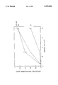
The figure illustrates detection of urease activity by the method of the invention as a function of incubation time.
While this invention is satisfied by embodiments in many different forms, there will herein be described in detail preferred embodiments of the invention, with the understanding that the present disclosure is to be considered as exemplary of the principles of the invention and is not intended to limit the invention to the embodiments described. The scope of the invention will be measured by the appended claims and their equivalents.
One aspect of the present invention is a substrate composition which is converted by the urease to a colored insoluble product. A second aspect of the invention is a method for immunoassay of a ligand using urease as the label and the composition as substrate. The method takes advantage of the many attributes of urease as a label and at the same time overcomes the drawback which has heretofore prevented this enzyme from achieving widespread use in immunoassay.
The assay of the invention may be performed by any conventional solid phase technique in which the presence or absence of a ligand in a sample is detected by enzyme catalyzed conversion of a substrate to a colored product. For example, the assay of the invention may be performed by immunochromatography. In this procedure, a liquid phase containing ligand to be detected migrates by capillary action across a solid phase, such as a glassplate, having various assay components deposited on adjacent but separated zones thereof. Representative of this procedure are the assays disclosed in U.S. Pat. No. 4,740,468 to Weng et al. and U.S. Pat. No. 4,446,232 to Liotta.
Another suitable assay technique uses a dipstick. In this procedure, a solid phase, usually a glass plate having a binder containing a capture antibody thereon, is dipped alternately into the test liquid, liquids containing assay reagents and wash liquids. In the last dip, an enzyme captured on the dipstick in proportion to the concentration of ligand in the test liquid converts the substrate of the invention to a colored product which deposits on the dipstick and which is indicative of the presence of ligand.
While immunoassay for a ligand as described above is a preferred application of the invention, one skilled in the art will immediately recognize that the method may be used in an assay wherein the ligand may be a nucleic acid probe and the tracer may be a complementary strand of DNA or RNA conjugated to urease. Assay procedures having enzymes conjugated to DNA and RNA strands are well-known in the art.
A preferred assay technique is flow-through assay in which the solid phase is a porous membrane. The membrane may be positioned in any suitable assay device adapted for flow-through assay as known in the art. In preferred devices, flow of assay liquids is promoted by capillary action induced by a pad of absorbent material adjacent the membrane, and the membrane and absorbent pad are mounted in a suitable housing. Membrane flow-through assay and various devices therefor have been disclosed and several devices are commercially available.
The porous membrane may be of any material which does not interfere in any way with any other component or step of the assay. Suitable membranes are, for example, of glass fiber, polyvinylidene difluoride, polycarbonate, nitrocellulose and nylon. Such membranes are well-known in the art and many are commercially available from suppliers such as Pall (East Hills, N.Y.), Millipore (Bedford, Mass.) and Schleicher and Schuell (Keene, N.H.).
The ligand may be from any source, and may be an antigen, an antibody or a hapten. For example, the ligand may be an endocrine hormone, such as HCG or FSH, present in body fluid, or it may be isolated from a body fluid and subsequently introduced into a different liquid, such as buffer. In other cases, the ligand may be from a source other than a body fluid, as, for example, a culture of microorganisms such as Chlamydia or a cellular extract thereof. Antibodies, such as the antibody against Lyme disease, may be assayed, or the ligand may be a hapten such as a therapeutic drug or a drug of abuse.
Preferred ligands are antigens, most preferably viral antigens present in a body fluid, such as Adenovirus, Parainfluenza 3 virus and, most preferably. Herpes simplex virus (HSV), Respiratory syncytial virus (RSV), and Influenza A (Flu A). The invention will hereinafter be described generically in terms of the preferred membrane assay.
The membrane may be coated with an antiligand specific for the ligand. Thus, in the case where the ligand is the preferred viral antigen, the antiligand may be an antibody which binds specifically to the antigen and thereby captures the antigen on the membrane. This reagent is hereinafter referred to as the capture antibody. The membrane may be further coated with an inert protein to fill any binding sites on the membrane not occupied by the capture antibody. (In the present disclosure, the term inert protein means a protein which is immunologically unreactive toward any other component of the assay and which does not substantially bind nonspecifically to other proteins in the assay medium, with the understanding that the inert protein may well be immunologically reactive toward other materials which are not part of the assay of the invention.) Representative nonlimiting examples of suitable inert proteins are casein and albumin, although others will be evident to those skilled in the art.
If the ligand is a hapten, it may be necessary to conjugate the hapten to a protein in order to raise a suitable anti-hapten capture antibody. Such procedures are well-known in the art of hapten immunoassay, and further details with respect to this aspect of the invention are not needed for a complete understanding of the invention.
Coating of the membrane with either or both of the capture antibody or the inert protein may be carried out by any suitable method, preferably by incubating the membrane with a solution of the antibody and/or inert protein whereby the protein is physically absorbed into the polymeric matrix of the surface of the membrane. Coating procedures are wholly conventional in the art.
The membrane may be incubated with the sample suspected of containing the ligand in order to bind the ligand to the antiligand coated onto the membrane. Preferably the sample is applied to the coated membrane and allowed to pass through the membrane in a transient, flow-through format for about 1 to 15, preferably about 5 minutes at a temperature of about 0° to 50° C., preferably about ambient temperature. By this procedure, antigen in the sample is captured on the membrane in proportion to its concentration in the sample. In addition, it has been found that viral antigen is absorbed preferentially even when the sample contains a large excess of extraneous protein, such as is the case when the sample is a body fluid.
In an alternate embodiment of the invention, the membrane may be coated with the inert protein and the antigen absorbed directly onto this surface and the assay performed without a capture antibody. Flow-through immunoassay absent a capture antibody is disclosed in copending application Ser. No. 272,380, filed Nov. 17, 1988, now abandoned, of common assignee herewith. In still another embodiment, the antigen may be adsorbed directly onto the membrane, and the membrane containing antigen subsequently treated with the inert protein to fill all unoccupied binding sites.
The membrane having ligand bound thereto may be treated with a solution of the tracer. The tracer may be an antiligand, hereinafter referred to as the detection antiligand, conjugated to urease wherein the assay is performed by the conventional sandwich or half sandwich technique. The preferred detection antiligand is an antibody which binds to antigen captured on the membrane and thereby affixes urease to the membrane surface in direct proportion to the quantity of antigen in the sample. Alternatively, the urease may be conjugated by conventional methods to a binder such as biotin, avidin and streptavidin and the latter bound to the antiligand.
The assay may also be performed by competitive assay, in which case the tracer may be the ligand conjugated to urease or to a conjugate of urease with biotin, avidin or streptavidin. In this format, the ligand and tracer compete for antiligand binding sites, and the urease becomes affixed to the membrane surface in inverse proportion to the quantity of ligand in the sample. Competitive assay may also be carried out with a tracer comprising urease linked to a hapten which has been used to raise the detection antibody, preferably a monoclonal antibody.
Conjugation of enzymes, such as urease, or enzyme conjugates with biotin, avidin or streptavidin to antigens or antibodies is well-known in the art and deemed to be within the purview of one skilled in the art with no further detail.
The membrane having urease affixed thereto may be treated with the substrate composition of the invention. The composition may include at least three components which interact consecutively leading to a colored insoluble product. The first component of the composition is a compound converted by the urease to a pH-raising substance. Suitable compounds as known in the art are urea, substituted ureas such as, for example, N-methylurea and semicarbazide, and simple amides such as formamide and acetamide. Urease cleaves these compounds to ammonia which raises the pH of the assay medium. The preferred first component is urea.
The second component of the substrate composition may be a pH dependent reducing agent for the third component. Suitable second components are indoxyl and, preferably, ascorbic acid. The second component does not reduce the third component, a tetrazolium salt, at low pH. Liberation of ammonia by the action of urease on the first component, however, raises the pH sufficiently to trigger the reduction of the tetrazolium salt to a colored, insoluble formazan. Reduction of tetrazolium salts to formazans is well-known in the art. Representative suitable tetrazolium salts are iodonitrotetrazolium violet and nitrobluetetrazolium chloride, although others are well-known to those skilled in the art.
Thus, in the preferred sandwich assay of the invention, ligand in the sample is captured by the antiligand coated onto the membrane. Captured ligand binds to the tracer whereby urease becomes affixed to the membrane. Urease affixed to the membrane converts the urea in the substrate composition to ammonia. The ammonia raises the pH to a level at which the ascorbic acid reduces the tetrazolium salt to the formazan. Accordingly, in the sandwich assay format, the appearance of color on the membrane is indicative of ligand in the sample.
In a competitive assay, ligand in the sample competes with tracer for binding to the antiligand. Thus, in this assay configuration, tracer, and therefore, urease, becomes affixed to the membrane in inverse proportion to the concentration of ligand. Accordingly, in a competitive assay, absence of colored formazan is indicative of ligand in the sample.
It is evident that the method of the invention may be used to assay for urea. For this embodiment of the invention, urease may be adsorbed on the membrane and a solution containing an unknown amount of urea, ascorbate and a tetrazolium salt may then be passed through the membrane. Urea in the unknown causes a spot to appear on the membrane. The color may be compared to standards containing known quantities of urea to quantitate the urea.
The following examples are provided to further describe the invention but are in no way to be considered as limitative of the invention.
A membrane filter stack was assembled with the following configuration:
______________________________________
Top Layer -   Three micron Biodyne ® C
              Membrane, (Pall, Glen Cove, New
              York, #BIA0030HC5). Precoated
              by immersion in phosphate
              buffered saline containing 0.3%
              casein for 30 minutes at ambient
              temperature.
Next Layer -  Non-woven rayon sheet
              (Schleicher and Schuell, Keene,
              New Hampshire; #5-S)
Bottom Layer -
              Cellulose absorbent pads (2)
              (Filtration Sciences, Mount
              Holly Springs, Pennsylvania; #ED
              320-200)
______________________________________
The membrane layers were encased in a plastic holder which includes a receiving well formed above the top layer. Within this well was fitted a flow restriction insert which has an aperture more narrow than the receiving well and sits flush against the top membrane.
A solution of urease-biotin conjugate in TEOA buffer (150 μl, 0.1M triethanolamine, 1 mM EDTA, pH 7.6) was added through the restriction insert to produce a triangular pattern of adsorbed urease-biotin conjugate on the membrane. After one minute, the insert was removed and the membrane washed with TEOA buffer (200 μl). An aqueous solution of iodonitrotetrazolium violet (200 μl, 0.2 mg/ml) was added, followed by urease substrate (200 μl, 25 mM urea, 1 mM EDTA, 20 mM sodium ascorbate, pH 5.0). After 10, 20 and 30 minutes, stop buffer (400 μl, 150 mM sodium citrate, pH 3.0) was added and the membrane color determined using a Gretag reflectometer (magenta setting). This color was observed as a triangular pattern corresponding to adsorbed urease-biotin conjugate. Samples without added urease showed no triangular pattern on the membrane.
The relationship between the time of incubation and the limit of urease detection is given in the Figure.
The procedure in Example I was followed except for the substitution of aqueous iodonitrotetrazolium violet by nitrobluetetrazolium chloride (300 μl, 0.165 mg/ml, 0.5% methanol). After twelve minutes, the membrane color was determined used a Gretag reflectometer (black setting). The detection limit for urease-biotin was 10 ng/test. Membranes without added urease showed no triangular pattern.
Urease-biotin conjugate was adsorbed onto Biodyne C membranes as described in Example I. The insert was removed and the sample washed with TEOA buffer (200 μl). Nitrobluetetrazolium chloride was added as in Example II. Urease substrate solution (25 mM urea, 1 mM, EDTA, 1 mM indoxyl butyrate) was treated with rabbit liver esterase (5.3 μg/ml) to generate indoxyl from indoxylbutyrate. The solution was mixed briefly and added to the device. After twelve minutes, stop buffer was added and the membrane color was read using a Gretag reflectometer (black setting). The detection limit for urease-biotin was 10 ng/test. Color was observed only where the urease-biotin conjugate was bound to the membrane.
Devices are prepared as described in Example I. A solution of urease in TEOA buffer is adsorbed onto the Biodyne C membrane. An aqueous solution of iodonitrotetrazolium violet (200 μL, 0.2 mg/mL) is added and allowed to flow through the device. A sample containing an unknown quantity of urea is passed through the membrane and standards having known concentrations of urea in substrate buffer (1 mM EDTA, 20 mM sodium ascorbate) are applied to other membranes having the same amount of adsorbed urease. After five minutes the intensity of color in the unknown is measured with a reflectometer and correlated to the amount of urea present in the standards. This correlation is in turn used to determine the concentration of urea present in unknown samples assayed by the above method.
Claims (2)
1. A method for detecting urea comprising:
a) adsorbing urease onto a solid support;
b) contacting the support with
i) a solution suspected of containing urea;
ii) a pH-dependent reducing agent selected from the group consisting of indoxyl, indoxyl butyrate, sodium ascorbate and ascorbic acid, and
iii) a tetrazolium salt such that the tetrazolium salt is reduced to a colored formazan which forms a colored spot on the support if urea is present in the solution.
2. The method of claim 1 wherein the tetrazolium salt is selected from the group consisting of iodonitrotetrazolium violet and nitrobluetetrazolium chloride.
Priority Applications (1)
| Application Number | Priority Date | Filing Date | Title |
|---|---|---|---|
| US08/165,220 US5370994A (en) | 1990-05-25 | 1993-12-10 | Solid phase assay for urea |
Applications Claiming Priority (3)
| Application Number | Priority Date | Filing Date | Title |
|---|---|---|---|
| US07/528,526 US5139934A (en) | 1990-05-25 | 1990-05-25 | Substrate composition and method for solid phase urease immunoassay |
| US07/851,602 US5328831A (en) | 1990-05-25 | 1992-03-16 | Substrate composition for solid phase urease immunoassay |
| US08/165,220 US5370994A (en) | 1990-05-25 | 1993-12-10 | Solid phase assay for urea |
Related Parent Applications (1)
| Application Number | Title | Priority Date | Filing Date |
|---|---|---|---|
| US07/851,602 Division US5328831A (en) | 1990-05-25 | 1992-03-16 | Substrate composition for solid phase urease immunoassay |
Publications (1)
| Publication Number | Publication Date |
|---|---|
| US5370994A true US5370994A (en) | 1994-12-06 |
Family
ID=24106044
Family Applications (3)
| Application Number | Title | Priority Date | Filing Date |
|---|---|---|---|
| US07/528,526 Expired - Fee Related US5139934A (en) | 1990-05-25 | 1990-05-25 | Substrate composition and method for solid phase urease immunoassay |
| US07/851,602 Expired - Fee Related US5328831A (en) | 1990-05-25 | 1992-03-16 | Substrate composition for solid phase urease immunoassay |
| US08/165,220 Expired - Fee Related US5370994A (en) | 1990-05-25 | 1993-12-10 | Solid phase assay for urea |
Family Applications Before (2)
| Application Number | Title | Priority Date | Filing Date |
|---|---|---|---|
| US07/528,526 Expired - Fee Related US5139934A (en) | 1990-05-25 | 1990-05-25 | Substrate composition and method for solid phase urease immunoassay |
| US07/851,602 Expired - Fee Related US5328831A (en) | 1990-05-25 | 1992-03-16 | Substrate composition for solid phase urease immunoassay |
Country Status (6)
| Country | Link |
|---|---|
| US (3) | US5139934A (en) |
| EP (1) | EP0458231A1 (en) |
| JP (1) | JPH06105255B2 (en) |
| AU (1) | AU640750B2 (en) |
| CA (1) | CA2037521A1 (en) |
| IE (1) | IE910682A1 (en) |
Cited By (5)
| Publication number | Priority date | Publication date | Assignee | Title |
|---|---|---|---|---|
| US5874216A (en) * | 1996-02-23 | 1999-02-23 | Ensys Environmental Products, Inc. | Indirect label assay device for detecting small molecules and method of use thereof |
| US5998156A (en) * | 1997-06-25 | 1999-12-07 | Fujirebio Inc. | Color developing method, enzyme immunoassay using the color developing method, and immunochromatography incorporating the enzyme immunoassay |
| US20040157281A1 (en) * | 2003-02-11 | 2004-08-12 | Chemsensing, Inc. | Method and apparatus for detecting an analyte |
| US20100233723A1 (en) * | 2006-01-19 | 2010-09-16 | Lattec I/S | Dry stick device and method for determining an analyte in a sample |
| US20110039290A1 (en) * | 2006-01-19 | 2011-02-17 | Kim Clausen | Novel dry stick device construction and method for determining an analyte in a sample using said dry stick device |
Families Citing this family (27)
| Publication number | Priority date | Publication date | Assignee | Title |
|---|---|---|---|---|
| US5139934A (en) * | 1990-05-25 | 1992-08-18 | Becton, Dickinson And Company | Substrate composition and method for solid phase urease immunoassay |
| US5314804A (en) * | 1992-03-24 | 1994-05-24 | Serim Research Corporation | Test for Helicobacter pylori |
| AU675251B2 (en) * | 1993-01-04 | 1997-01-30 | Becton Dickinson & Company | Flow-through hybridization assay for oligonucleotide sequences |
| US5354658A (en) * | 1993-01-28 | 1994-10-11 | Dennis Wright | Non-radioactive method for detecting a labelled segment and a solution or composition therefor |
| AU5495194A (en) * | 1993-02-26 | 1994-09-01 | Becton Dickinson & Company | Flow through membrane nucleic acid hybridization assay |
| US5916746A (en) * | 1996-05-09 | 1999-06-29 | Kirkegaard & Perry Laboratories, Inc. | Formazan-based immunoassay |
| AU4790597A (en) * | 1996-11-01 | 1998-05-29 | Nycomed Pharma As | Process for the extraction of proteins |
| US6225074B1 (en) | 1997-08-18 | 2001-05-01 | Dennis Wright | Direct chloramphenicol acetyl transferase assay |
| EP0919811A1 (en) * | 1997-12-01 | 1999-06-02 | Universiteit Maastricht | Immunoassay method and kit |
| US5972595A (en) * | 1997-12-19 | 1999-10-26 | Nen Life Science Products, Inc. | Enzyme assay using a solid phase substrate |
| US6066446A (en) * | 1997-12-19 | 2000-05-23 | Nen Life Science Products, Inc. | Assay member and method for its manufacture |
| US7052831B2 (en) * | 2000-09-29 | 2006-05-30 | Becton Dickinson And Company | Detection of multiple analytes from a single sample using a multi-well, multi-analyte flow-through diagnostic test device |
| WO2002052263A1 (en) * | 2000-12-22 | 2002-07-04 | Bio A.R.T. Bvba | Flow through assay device, diagnostic kit comprising said assay device and use of said assay device in the detection of an analyte present in a sample |
| WO2003064679A2 (en) * | 2002-01-30 | 2003-08-07 | Id Biomedical Corporation | Methods for detecting vancomycin-resistant microorganisms and compositions therefor |
| WO2004050910A1 (en) * | 2002-12-04 | 2004-06-17 | Pamgene Bv | Method for hybridisation of immobilized genomic dna |
| FR2853077B1 (en) * | 2003-03-28 | 2005-12-30 | Vedalab | SOLID PHASE IMMUNOCHROMATOGRAPHIC METHODS |
| US7521226B2 (en) | 2004-06-30 | 2009-04-21 | Kimberly-Clark Worldwide, Inc. | One-step enzymatic and amine detection technique |
| JPWO2006043387A1 (en) * | 2004-09-28 | 2008-05-22 | 第一化学薬品株式会社 | Method of immunological examination of specimen and examination instrument used for the method |
| US7504235B2 (en) | 2005-08-31 | 2009-03-17 | Kimberly-Clark Worldwide, Inc. | Enzyme detection technique |
| US8758989B2 (en) * | 2006-04-06 | 2014-06-24 | Kimberly-Clark Worldwide, Inc. | Enzymatic detection techniques |
| US7897360B2 (en) | 2006-12-15 | 2011-03-01 | Kimberly-Clark Worldwide, Inc. | Enzyme detection techniques |
| US8012770B2 (en) | 2009-07-31 | 2011-09-06 | Invisible Sentinel, Inc. | Device for detection of antigens and uses thereof |
| MX2012004105A (en) | 2009-10-09 | 2012-09-07 | Invisible Sentinel Inc | Device for detection of antigens and uses thereof. |
| EP3608021A3 (en) | 2011-01-27 | 2020-04-22 | Invisible Sentinel, Inc. | Analyte detection devices, multiplex and tabletop devices for detection of analytes, and uses thereof |
| GB201112395D0 (en) * | 2011-07-19 | 2011-08-31 | Bio Nano Consulting | Immunoassay |
| AU2013230917C1 (en) | 2012-03-09 | 2021-09-16 | Invisible Sentinel, Inc. | Methods and compositions for detecting multiple analytes with a single signal |
| CN113671168B (en) * | 2021-07-16 | 2023-08-11 | 武汉轻工大学 | For detecting aflatoxin B 1 ELISA kit, preparation method thereof and detection method |
Citations (5)
| Publication number | Priority date | Publication date | Assignee | Title |
|---|---|---|---|---|
| US4590157A (en) * | 1980-12-22 | 1986-05-20 | Commonwealth Serum Laboratories Commission | Method for detecting antigens and antibodies |
| US4727025A (en) * | 1984-05-25 | 1988-02-23 | Boehringer Mannheim Gmbh | Process and reagent for the fully enzymatic determination of urea |
| US4892817A (en) * | 1987-09-21 | 1990-01-09 | Biogenex Laboratories | Stable phosphatase substrate composition |
| US4946776A (en) * | 1988-01-03 | 1990-08-07 | Orgenics Ltd. | Stable chromogenic substrate mixture of indoxyl phosphate and tetrazolium salt, method of making and using same in biological and diagnostic assays |
| US5139934A (en) * | 1990-05-25 | 1992-08-18 | Becton, Dickinson And Company | Substrate composition and method for solid phase urease immunoassay |
Family Cites Families (13)
| Publication number | Priority date | Publication date | Assignee | Title |
|---|---|---|---|---|
| IT1006557B (en) * | 1971-09-08 | 1976-10-20 | Bagshawe Kenneth Dawson | REACTION CELL PARTICULARLY USEFUL IN RADIOIMMUNITY TESTS |
| US4024021A (en) * | 1973-07-19 | 1977-05-17 | The Dow Chemical Company | Determination of glutamate and glutamic transaminases |
| DE2754944C3 (en) * | 1977-12-09 | 1981-12-03 | Boehringer Mannheim Gmbh, 6800 Mannheim | Method and reagent for the determination of ascorbic acid |
| US4235964A (en) * | 1978-09-28 | 1980-11-25 | Bochner Barry R | Method for testing and identifying microorganisms |
| US4277560A (en) * | 1978-10-24 | 1981-07-07 | Technicon Instruments Corporation | Enzyme immunoassays using immobilized reagents in a flowing stream |
| US4246339A (en) * | 1978-11-01 | 1981-01-20 | Millipore Corporation | Test device |
| US4446232A (en) * | 1981-10-13 | 1984-05-01 | Liotta Lance A | Enzyme immunoassay with two-zoned device having bound antigens |
| IT1205338B (en) * | 1983-08-09 | 1989-03-15 | Miles Italiana | COMPOSITION AND METHOD FOR THE ENZYMATIC DETERMINATION OF UREA IN ORGANIC LIQUIDS |
| US4632901A (en) * | 1984-05-11 | 1986-12-30 | Hybritech Incorporated | Method and apparatus for immunoassays |
| US4740468A (en) * | 1985-02-14 | 1988-04-26 | Syntex (U.S.A.) Inc. | Concentrating immunochemical test device and method |
| US4812293A (en) * | 1986-06-30 | 1989-03-14 | Becton, Dickinson And Company | Vacuum actuated assay device and method of using same |
| US5032506A (en) * | 1986-12-16 | 1991-07-16 | Enzymatics, Inc. | Color control system |
| CA2023850A1 (en) * | 1989-09-28 | 1991-03-29 | Randal A. Hoke | Stabilizing reagent for membrane immunoassay |
-
1990
- 1990-05-25 US US07/528,526 patent/US5139934A/en not_active Expired - Fee Related
-
1991
- 1991-02-28 IE IE068291A patent/IE910682A1/en unknown
- 1991-03-04 CA CA002037521A patent/CA2037521A1/en not_active Abandoned
- 1991-03-08 AU AU72784/91A patent/AU640750B2/en not_active Ceased
- 1991-04-26 JP JP3096614A patent/JPH06105255B2/en not_active Expired - Lifetime
- 1991-05-18 EP EP91108115A patent/EP0458231A1/en not_active Withdrawn
-
1992
- 1992-03-16 US US07/851,602 patent/US5328831A/en not_active Expired - Fee Related
-
1993
- 1993-12-10 US US08/165,220 patent/US5370994A/en not_active Expired - Fee Related
Patent Citations (5)
| Publication number | Priority date | Publication date | Assignee | Title |
|---|---|---|---|---|
| US4590157A (en) * | 1980-12-22 | 1986-05-20 | Commonwealth Serum Laboratories Commission | Method for detecting antigens and antibodies |
| US4727025A (en) * | 1984-05-25 | 1988-02-23 | Boehringer Mannheim Gmbh | Process and reagent for the fully enzymatic determination of urea |
| US4892817A (en) * | 1987-09-21 | 1990-01-09 | Biogenex Laboratories | Stable phosphatase substrate composition |
| US4946776A (en) * | 1988-01-03 | 1990-08-07 | Orgenics Ltd. | Stable chromogenic substrate mixture of indoxyl phosphate and tetrazolium salt, method of making and using same in biological and diagnostic assays |
| US5139934A (en) * | 1990-05-25 | 1992-08-18 | Becton, Dickinson And Company | Substrate composition and method for solid phase urease immunoassay |
Cited By (10)
| Publication number | Priority date | Publication date | Assignee | Title |
|---|---|---|---|---|
| US5874216A (en) * | 1996-02-23 | 1999-02-23 | Ensys Environmental Products, Inc. | Indirect label assay device for detecting small molecules and method of use thereof |
| US6376195B1 (en) | 1996-02-23 | 2002-04-23 | Strategic Diagnostics Inc. | Indirect label assay device for detecting small molecules and method of use thereof |
| US5998156A (en) * | 1997-06-25 | 1999-12-07 | Fujirebio Inc. | Color developing method, enzyme immunoassay using the color developing method, and immunochromatography incorporating the enzyme immunoassay |
| US6251618B1 (en) * | 1997-06-25 | 2001-06-26 | Fujirebio Inc. | Color developing method, enzyme immunoassay using the color developing method, and immunochromatography incorporating the enzyme immunoassay |
| US20040157281A1 (en) * | 2003-02-11 | 2004-08-12 | Chemsensing, Inc. | Method and apparatus for detecting an analyte |
| US20100233723A1 (en) * | 2006-01-19 | 2010-09-16 | Lattec I/S | Dry stick device and method for determining an analyte in a sample |
| US20110039290A1 (en) * | 2006-01-19 | 2011-02-17 | Kim Clausen | Novel dry stick device construction and method for determining an analyte in a sample using said dry stick device |
| US8198073B2 (en) | 2006-01-19 | 2012-06-12 | Lattec I/S | Dry stick device and method for determining an analyte in a sample |
| US8206944B2 (en) | 2006-01-19 | 2012-06-26 | Lattec I/S | Dry stick device construction and method for determining an analyte in a sample using said dry stick device |
| US8460863B2 (en) | 2006-01-19 | 2013-06-11 | Lattec I/S | Dry stick device and method for determining an analyte in a sample |
Also Published As
| Publication number | Publication date |
|---|---|
| CA2037521A1 (en) | 1991-11-26 |
| EP0458231A1 (en) | 1991-11-27 |
| AU7278491A (en) | 1991-11-28 |
| AU640750B2 (en) | 1993-09-02 |
| US5139934A (en) | 1992-08-18 |
| JPH04231874A (en) | 1992-08-20 |
| IE910682A1 (en) | 1991-12-04 |
| US5328831A (en) | 1994-07-12 |
| JPH06105255B2 (en) | 1994-12-21 |
Similar Documents
| Publication | Publication Date | Title |
|---|---|---|
| US5370994A (en) | Solid phase assay for urea | |
| US5279935A (en) | Method of immunossay including deactivation of endogenous alkaline phosphatase | |
| AU613797B2 (en) | Solid-phase analytical device with adjacent control areas | |
| US5160701A (en) | Solid-phase analytical device and method for using same | |
| US4916056A (en) | Solid-phase analytical device and method for using same | |
| US7312028B2 (en) | Highly cost-effective analytical device for performing immunoassays with ultra high sensitivity | |
| EP0427984B1 (en) | Articles for use in enzyme immuno-assay techniques, and methods of making and using such articles | |
| EP0171150B2 (en) | Method and apparatus for assaying with optional reagent quality control | |
| EP0369361B1 (en) | Immunoassay on a preblocked solid surface | |
| CA2003942A1 (en) | Solid assay support systems | |
| US5166054A (en) | Method for immunoassay using lactoperoxidase, starch and iodine | |
| EP0420170A2 (en) | Stabilizing reagent for membrane immunoassay | |
| EP0152254A2 (en) | Chromogenic support immunoassay utilizing labeled complement components | |
| JPS6330767A (en) | Solid-phase system containing tetrazolium salt for testing ligand-receptor | |
| GB2241329A (en) | Sandwich type immunoassay | |
| US5460946A (en) | Diagnostic test kit and specific binding assay using modulator of signal resulting from peroxidase label |
Legal Events
| Date | Code | Title | Description |
|---|---|---|---|
| REMI | Maintenance fee reminder mailed | ||
| LAPS | Lapse for failure to pay maintenance fees | ||
| FP | Lapsed due to failure to pay maintenance fee |
Effective date: 19981206 |
|
| STCH | Information on status: patent discontinuation |
Free format text: PATENT EXPIRED DUE TO NONPAYMENT OF MAINTENANCE FEES UNDER 37 CFR 1.362 |