US5374548A - Methods and compositions for the attachment of proteins to liposomes using a glycophospholipid anchor - Google Patents
Methods and compositions for the attachment of proteins to liposomes using a glycophospholipid anchor Download PDFInfo
- Publication number
- US5374548A US5374548A US08/017,934 US1793493A US5374548A US 5374548 A US5374548 A US 5374548A US 1793493 A US1793493 A US 1793493A US 5374548 A US5374548 A US 5374548A
- Authority
- US
- United States
- Prior art keywords
- daf
- gpi
- anchor
- cell
- cells
- Prior art date
- Legal status (The legal status is an assumption and is not a legal conclusion. Google has not performed a legal analysis and makes no representation as to the accuracy of the status listed.)
- Expired - Lifetime
Links
Images


Classifications
-
- G—PHYSICS
- G01—MEASURING; TESTING
- G01N—INVESTIGATING OR ANALYSING MATERIALS BY DETERMINING THEIR CHEMICAL OR PHYSICAL PROPERTIES
- G01N33/00—Investigating or analysing materials by specific methods not covered by groups G01N1/00 - G01N31/00
- G01N33/48—Biological material, e.g. blood, urine; Haemocytometers
- G01N33/50—Chemical analysis of biological material, e.g. blood, urine; Testing involving biospecific ligand binding methods; Immunological testing
- G01N33/68—Chemical analysis of biological material, e.g. blood, urine; Testing involving biospecific ligand binding methods; Immunological testing involving proteins, peptides or amino acids
-
- A—HUMAN NECESSITIES
- A61—MEDICAL OR VETERINARY SCIENCE; HYGIENE
- A61K—PREPARATIONS FOR MEDICAL, DENTAL OR TOILETRY PURPOSES
- A61K9/00—Medicinal preparations characterised by special physical form
- A61K9/10—Dispersions; Emulsions
- A61K9/127—Liposomes
- A61K9/1271—Non-conventional liposomes, e.g. PEGylated liposomes, liposomes coated with polymers
-
- B—PERFORMING OPERATIONS; TRANSPORTING
- B82—NANOTECHNOLOGY
- B82Y—SPECIFIC USES OR APPLICATIONS OF NANOSTRUCTURES; MEASUREMENT OR ANALYSIS OF NANOSTRUCTURES; MANUFACTURE OR TREATMENT OF NANOSTRUCTURES
- B82Y5/00—Nanobiotechnology or nanomedicine, e.g. protein engineering or drug delivery
-
- C—CHEMISTRY; METALLURGY
- C07—ORGANIC CHEMISTRY
- C07K—PEPTIDES
- C07K14/00—Peptides having more than 20 amino acids; Gastrins; Somatostatins; Melanotropins; Derivatives thereof
- C07K14/005—Peptides having more than 20 amino acids; Gastrins; Somatostatins; Melanotropins; Derivatives thereof from viruses
-
- C—CHEMISTRY; METALLURGY
- C07—ORGANIC CHEMISTRY
- C07K—PEPTIDES
- C07K14/00—Peptides having more than 20 amino acids; Gastrins; Somatostatins; Melanotropins; Derivatives thereof
- C07K14/435—Peptides having more than 20 amino acids; Gastrins; Somatostatins; Melanotropins; Derivatives thereof from animals; from humans
- C07K14/575—Hormones
-
- C—CHEMISTRY; METALLURGY
- C07—ORGANIC CHEMISTRY
- C07K—PEPTIDES
- C07K14/00—Peptides having more than 20 amino acids; Gastrins; Somatostatins; Melanotropins; Derivatives thereof
- C07K14/435—Peptides having more than 20 amino acids; Gastrins; Somatostatins; Melanotropins; Derivatives thereof from animals; from humans
- C07K14/705—Receptors; Cell surface antigens; Cell surface determinants
- C07K14/70596—Molecules with a "CD"-designation not provided for elsewhere
-
- C—CHEMISTRY; METALLURGY
- C07—ORGANIC CHEMISTRY
- C07K—PEPTIDES
- C07K16/00—Immunoglobulins [IGs], e.g. monoclonal or polyclonal antibodies
- C07K16/18—Immunoglobulins [IGs], e.g. monoclonal or polyclonal antibodies against material from animals or humans
- C07K16/26—Immunoglobulins [IGs], e.g. monoclonal or polyclonal antibodies against material from animals or humans against hormones ; against hormone releasing or inhibiting factors
-
- C—CHEMISTRY; METALLURGY
- C07—ORGANIC CHEMISTRY
- C07K—PEPTIDES
- C07K16/00—Immunoglobulins [IGs], e.g. monoclonal or polyclonal antibodies
- C07K16/18—Immunoglobulins [IGs], e.g. monoclonal or polyclonal antibodies against material from animals or humans
- C07K16/28—Immunoglobulins [IGs], e.g. monoclonal or polyclonal antibodies against material from animals or humans against receptors, cell surface antigens or cell surface determinants
- C07K16/2896—Immunoglobulins [IGs], e.g. monoclonal or polyclonal antibodies against material from animals or humans against receptors, cell surface antigens or cell surface determinants against molecules with a "CD"-designation, not provided for elsewhere
-
- C—CHEMISTRY; METALLURGY
- C12—BIOCHEMISTRY; BEER; SPIRITS; WINE; VINEGAR; MICROBIOLOGY; ENZYMOLOGY; MUTATION OR GENETIC ENGINEERING
- C12N—MICROORGANISMS OR ENZYMES; COMPOSITIONS THEREOF; PROPAGATING, PRESERVING, OR MAINTAINING MICROORGANISMS; MUTATION OR GENETIC ENGINEERING; CULTURE MEDIA
- C12N15/00—Mutation or genetic engineering; DNA or RNA concerning genetic engineering, vectors, e.g. plasmids, or their isolation, preparation or purification; Use of hosts therefor
- C12N15/09—Recombinant DNA-technology
- C12N15/11—DNA or RNA fragments; Modified forms thereof; Non-coding nucleic acids having a biological activity
- C12N15/62—DNA sequences coding for fusion proteins
- C12N15/625—DNA sequences coding for fusion proteins containing a sequence coding for a signal sequence
-
- A—HUMAN NECESSITIES
- A61—MEDICAL OR VETERINARY SCIENCE; HYGIENE
- A61K—PREPARATIONS FOR MEDICAL, DENTAL OR TOILETRY PURPOSES
- A61K38/00—Medicinal preparations containing peptides
-
- C—CHEMISTRY; METALLURGY
- C07—ORGANIC CHEMISTRY
- C07K—PEPTIDES
- C07K2319/00—Fusion polypeptide
-
- C—CHEMISTRY; METALLURGY
- C07—ORGANIC CHEMISTRY
- C07K—PEPTIDES
- C07K2319/00—Fusion polypeptide
- C07K2319/01—Fusion polypeptide containing a localisation/targetting motif
- C07K2319/02—Fusion polypeptide containing a localisation/targetting motif containing a signal sequence
-
- C—CHEMISTRY; METALLURGY
- C07—ORGANIC CHEMISTRY
- C07K—PEPTIDES
- C07K2319/00—Fusion polypeptide
- C07K2319/01—Fusion polypeptide containing a localisation/targetting motif
- C07K2319/035—Fusion polypeptide containing a localisation/targetting motif containing a signal for targeting to the external surface of a cell, e.g. to the outer membrane of Gram negative bacteria, GPI- anchored eukaryote proteins
-
- C—CHEMISTRY; METALLURGY
- C07—ORGANIC CHEMISTRY
- C07K—PEPTIDES
- C07K2319/00—Fusion polypeptide
- C07K2319/32—Fusion polypeptide fusions with soluble part of a cell surface receptor, "decoy receptors"
-
- C—CHEMISTRY; METALLURGY
- C07—ORGANIC CHEMISTRY
- C07K—PEPTIDES
- C07K2319/00—Fusion polypeptide
- C07K2319/40—Fusion polypeptide containing a tag for immunodetection, or an epitope for immunisation
-
- C—CHEMISTRY; METALLURGY
- C07—ORGANIC CHEMISTRY
- C07K—PEPTIDES
- C07K2319/00—Fusion polypeptide
- C07K2319/70—Fusion polypeptide containing domain for protein-protein interaction
- C07K2319/74—Fusion polypeptide containing domain for protein-protein interaction containing a fusion for binding to a cell surface receptor
- C07K2319/75—Fusion polypeptide containing domain for protein-protein interaction containing a fusion for binding to a cell surface receptor containing a fusion for activation of a cell surface receptor, e.g. thrombopoeitin, NPY and other peptide hormones
-
- C—CHEMISTRY; METALLURGY
- C07—ORGANIC CHEMISTRY
- C07K—PEPTIDES
- C07K2319/00—Fusion polypeptide
- C07K2319/90—Fusion polypeptide containing a motif for post-translational modification
- C07K2319/91—Fusion polypeptide containing a motif for post-translational modification containing a motif for glycosylation
- C07K2319/912—Fusion polypeptide containing a motif for post-translational modification containing a motif for glycosylation containing a GPI (phosphatidyl-inositol glycane) anchor
-
- C—CHEMISTRY; METALLURGY
- C12—BIOCHEMISTRY; BEER; SPIRITS; WINE; VINEGAR; MICROBIOLOGY; ENZYMOLOGY; MUTATION OR GENETIC ENGINEERING
- C12N—MICROORGANISMS OR ENZYMES; COMPOSITIONS THEREOF; PROPAGATING, PRESERVING, OR MAINTAINING MICROORGANISMS; MUTATION OR GENETIC ENGINEERING; CULTURE MEDIA
- C12N2710/00—MICROORGANISMS OR ENZYMES; COMPOSITIONS THEREOF; PROPAGATING, PRESERVING, OR MAINTAINING MICROORGANISMS; MUTATION OR GENETIC ENGINEERING; CULTURE MEDIA dsDNA viruses
- C12N2710/00011—Details
- C12N2710/16011—Herpesviridae
- C12N2710/16611—Simplexvirus, e.g. human herpesvirus 1, 2
- C12N2710/16622—New viral proteins or individual genes, new structural or functional aspects of known viral proteins or genes
-
- G—PHYSICS
- G01—MEASURING; TESTING
- G01N—INVESTIGATING OR ANALYSING MATERIALS BY DETERMINING THEIR CHEMICAL OR PHYSICAL PROPERTIES
- G01N2333/00—Assays involving biological materials from specific organisms or of a specific nature
- G01N2333/435—Assays involving biological materials from specific organisms or of a specific nature from animals; from humans
- G01N2333/705—Assays involving receptors, cell surface antigens or cell surface determinants
-
- Y—GENERAL TAGGING OF NEW TECHNOLOGICAL DEVELOPMENTS; GENERAL TAGGING OF CROSS-SECTIONAL TECHNOLOGIES SPANNING OVER SEVERAL SECTIONS OF THE IPC; TECHNICAL SUBJECTS COVERED BY FORMER USPC CROSS-REFERENCE ART COLLECTIONS [XRACs] AND DIGESTS
- Y10—TECHNICAL SUBJECTS COVERED BY FORMER USPC
- Y10S—TECHNICAL SUBJECTS COVERED BY FORMER USPC CROSS-REFERENCE ART COLLECTIONS [XRACs] AND DIGESTS
- Y10S436/00—Chemistry: analytical and immunological testing
- Y10S436/829—Liposomes, e.g. encapsulation
-
- Y—GENERAL TAGGING OF NEW TECHNOLOGICAL DEVELOPMENTS; GENERAL TAGGING OF CROSS-SECTIONAL TECHNOLOGIES SPANNING OVER SEVERAL SECTIONS OF THE IPC; TECHNICAL SUBJECTS COVERED BY FORMER USPC CROSS-REFERENCE ART COLLECTIONS [XRACs] AND DIGESTS
- Y10—TECHNICAL SUBJECTS COVERED BY FORMER USPC
- Y10S—TECHNICAL SUBJECTS COVERED BY FORMER USPC CROSS-REFERENCE ART COLLECTIONS [XRACs] AND DIGESTS
- Y10S530/00—Chemistry: natural resins or derivatives; peptides or proteins; lignins or reaction products thereof
- Y10S530/827—Proteins from mammals or birds
- Y10S530/829—Blood
Definitions
- This application relates to the preparation of decay accelerating factor (hereinafter abbreviated as DAF) in recombinant cell culture.
- DAF decay accelerating factor
- complement activation Antigenic cells targeted by the humoral immune response are lysed by a process called complement activation. This process consists of a series or cascade of proteolytic activities initiated by the binding of antibody with its antigen.
- the components that participate in complement activation are many and complex, although for the purposes herein the most important are C4b and C3b. In a key step in complement activation, these two proteins become covalently associated with the target cell surface and then serve as anchors for the assembly of C3 and C5 convertases, the amplifying enzymes of the cascade.
- Complement activation must focus only on the target and must not occur on host cells. However, in the course of complement activation, large numbers of nascent C4b and C3b fragments are liberated into the fluid phase. Most react with water, but some by chance could bind to nearby host cells and lead to their damage. For this and possibly other reasons, the activities of bound, as well as free, C3b and C4b fragments are under strict control by a complex system of serum and membrane proteins.
- DAF was reported to have been purified to a single 70 Kd band on silver stained SDS-PAGE from a pooled extract of human erythrocyte stroma (Medof et al., J. Exp. Med. 160:1558 [1983]). The molecule was hydrophobic and tended to form multimers of greater than or equal to 150 Kd as determined by molecular sieve chromatography. Purified DAF could reassociate with red blood cells. Only a small number of DAF molecules (less than 100) had a significant effect on the hemolytic effect of activated complement. Medof et al. concluded that DAF can only function intrinsically within the cell membrane, and suggested that it offered the possibility of correcting in vitro the defect in the membranes of cells from patients with PNH.
- Red blood cell DAF is limited to the native membrane bound form, including any naturally occurring alleles as may exist. Methods are needed for synthesizing amino acid and glycosylation variants which can function as DAF agonists or antagonists, or which will exhibit other desirable characteristics such as the absence of C-terminal lipid, resistance to proteases, or the ability to deliver DAF to the membranes of target cells.
- the objects of this invention are accomplished by expression of DAF in recombinant cell culture, a process that fundamentally comprises providing nucleic acid encoding DAF, transforming a host cell with the DAF-encoding nucleic acid, and culturing the cell in order to express DAF in the host cell culture.
- the method of this invention enables the preparation of novel forms of DAF, including amino acid sequence variants and glycosylation variants.
- Amino acid sequence variants consist of deletions, substitutions and insertions of one or more DAF amino acid residues.
- DAF also is expressed in a form unaccompanied by the glycosylation associated with the native DAF (including unaccompanied by any glycosylation whatsoever), obtained as a product of expression of DAF in heterologous recombinant cell culture. DAF in any form as a component of a recombinant cell culture is novel.
- DAF membrane-bound form of DAF
- mDAF membrane-bound form of DAF
- the novel C-terminus of sDAF is postulated to result in vivo in the secretion of the protein into the blood stream (where it may be biologically active) because the presence of the intron changes the reading frame of the last exon so as to eliminate the "signal" directing attachment of phosphatidylinositol (the membrane anchor for mDAF).
- This novel form of DAF was unappreciated until the pioneering work herein was accomplished, and it differs from mDAF in containing an antigenically distinct C-terminus.
- sDAF is useful in diagnosis of PNH since it is now possible to determine whether the condition in an individual results from a failure to express any of the DAF gene or a failure of post-translational processing to attach the phosphatidylinositol anchor.
- Novel nucleic acids also are provided, including (1) cell free nucleic acid identified as encoding DAF, including genomic DNA, cDNA or RNA, (2) DNA encoding DAF free of an untranslated intervening sequence (introns) or flanking genomic DNA, and (3) nucleic acid encoding DAF which is free of nucleic acid encoding any other protein homologous to the source of the nucleic acid that encodes DAF. Also within the scope of this invention is nucleic acid which does not encode DAF but which is capable of hybridizing with nucleic acid encoding DAF.
- Nucleic acid encoding DAF is useful in the expression of DAF in recombinant cell culture or for assaying test samples for the presence of DAF-encoding nucleic acid. Labelled DAF-encoding or hybridizing nucleic acid is provided for use in such assays.
- Recombinant DAF is formulated into therapeutically acceptable vehicles and administered for the treatment of PNH or inflammatory or cell lytic autoimmune diseases.
- DAF conjugates or fusions are prepared that deliver DAF to target cells in order to inhibit complement activation at the surfaces of such cells.
- the conjugates or fusions are useful for ameliorating allograft rejection or autoimmune diseases.
- the carboxyl terminal domain that specifies glycophospholipid membrane anchor attachment for mDAF (referred to as the GPI signal domain, wherein GPI is an abbreviation of glycophosphatidylinositol), or functionally equivalent domains from other proteins which also are anchored by glycophospholipids, are fused to proteins or multimers of such proteins which are heterologous to the source of the GPI signal domain, for example hormones, antigens (especially from infectious organisms), allergens, immunoglobulins, enzymes, receptors and the like.
- the anchor fusions are used in combination with the recombinant cells which express them or are recovered and formulated into therapeutic compositions, used as diagnostic assay components, or employed in affinity purification procedures.
- the fusions will contain the heterologous polypeptide fused at its C-terminus to the GPI signal domain, that specifies a processing event in the cell that results in cleavage and removal of the GPI signal domain, and covalent attachment of a GPI anchor to the new C-terminus of the protein.
- the last about 30-50 residues of DAF contain a signal (the "GPI signal") that directs a processing event in cells in which the last about 28 residues are proteolytically removed and replaced with a hydrophobic glycolipid (GPI) that acts as a membrane anchor.
- GPI hydrophobic glycolipid
- Another aspect of the invention is a method for targeting a liposome to a cell of interest, comprising incorporating into the liposome a GPI-linked protein produced by fusing a GPI signal domain to a polypeptide heterologous to the GPI signal domain.
- compositions comprising a liposome, wherein a GPI-linked polypeptide is incorporated into the liposome.
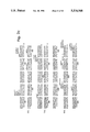
- FIGS. 1a-1f depict the cDNA sequence for clones ⁇ 33 (to the HindIII site at residue 1) and ⁇ 47 (HindIII to the 3' end). The point at which the intron is removed is designated by an asterisk.
- the probable phosphatidylinositol derivatization site is Cyst 330 and the C-terminal hydrophobic region extends from residues 331-347. Amino acid residues are numbered from the mature amino terminus at Asp 1 .
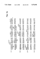
- FIGS. 2a-2g depict the cDNA sequence of clones ⁇ 33 to the HindIII site at residue +1) and ⁇ 41 (HindIII to 3' end) encoding human sDAF.
- the unspliced intron in the cDNA encoding sDAF is bracketed. Restriction enzyme sites are shown using conventional abbreviations.
- the predicted amino acid sequence for each DAF predicted species is shown, together with the secretory leader and mature N-terminus of each (designated by arrows).
- FIG. 3 is a schematic diagram showing the regions of HSV 1 glycoprotein D (gD-1) and DAF that are present in the gD-1/DAF fusion protein produced in Example 3.
- Truncated (secreted) gD-1 was constructed from native (membrane) gD-1 (14) and comprises amino acids 1-300, including the hydrophobic signal sequence (residues 1-25, indicated as a grey area).
- the hydrophobic membrane spanning domain (residues 340-360, cross-hatched region) and the C-terminal hydrophobic domain (residues 361-393) are excluded.
- the point of truncation (residue 300) is indicated by a broken line.
- Truncated gD-1 was fused to residue 311 of membrane DAF.
- the gD-1/DAF fusion contains the last 37 residues of membrane DAF predicted from the cDNA sequence (residues 311-347) that comprises the GPI signal domain, which includes a C-terminal hydrophobic region (residues 331-347, depicted in black).
- a similar fusion, gp120/DAF was constructed in which gD-1 was replaced with HIV-1 IIIB gp120. Expression of this fusion in 293 cells produced GPI-linked gp120.
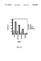
- FIG. 4 is a graph depicting sucrose gradient fractionation of gp120/DAF/liposomes.
- gp120/DAF refers to GPI-linked gp120 , produced by expression of the fusion of the DAF GPI signal domain to the cDNA encoding gp120.
- 125 I-labeled gp120/DAF (3.5 ⁇ g, in 0.8% octyl glucoside) was mixed with 4 C-labeled liposomes (3 mg/ml of lipid, composed of equimolar amounts of phosphatidylcholine and cholesterol) in 685 ⁇ l of phosphate buffered saline (PBS).
- PBS phosphate buffered saline
- the mixture was dialyzed overnight and 200 ⁇ l was loaded on a 3-25% sucrose gradient and centrifuged at 250,000 xg for 4.5 h at 4° C. Sequential fractions were collected from the top of the gradient and counted to determine the position of the 125 -labeled gp120/DAF. A parallel gradient containing 14 C-labeled liposomes alone was analyzed to determine the position of the liposomes.
- FIG. 5 is a graph depicting relative levels of CD4 on the surface of transfected 293 and CHO cells.
- CD4/DAF is a GPI-linked form of CD4 formed by expressing a cDNA encoding the extracellular domain of CD4 fused to the GPI signal domain of DAF.
- FIG. 6 is a graph depicting targeted binding of FITC-dextran-labeled liposomes to CHO cells expressing CD4. Preformed liposomes loaded with FITC-dextran were coated with gp120/DAF and then incubated at 4° C. for 1-2 h with parental CHO cells or with cells expressing CD4 or CD4DAF as indicated. The cells were then washed and analyzed by FACS.
- FIG. 7 is a graph depicting targeted binding of FITC-dextran-labeled liposomes to 293 cells expressing CD4 mediated by GPI-linked gp120/DAF.
- Preformed liposomes loaded with FITC-dextran and coated with gp120/DAF were incubated at 4° C. with parental 293 cells or with 293 cells expressing CD4 or CD4DAF as indicated. The cells were then washed, and incubated at 4° C. in the presence or absence of PIPLC, and analyzed by FACS. PIPLC-treatment removed the FITC-labeled liposomes from the cell surface, indicating that attachment is mediated by a GPI-linked molecule.
- FIG. 8 is a graph depicting the targeted binding of FITC-dextran-labeled liposomes to cells.
- FITC-dextran-loaded liposomes coated with gp120/DAF were incubated at 4° C. with CHO cells as indicated, in the presence of molecules that interfere with the binding of gp120 to CD4 (anti-CD4 or anti-gp120 monoclonal antibodies, or soluble CD4, denoted rCD4IgG). The cells were then washed and analyzed by FACS.
- DAF is defined to be any molecule having the precursor or mature amino acid sequence set forth in FIGS. 1 or 2 as well as their amino acid sequence or glycosylation variants (including natural alleles) which are capable of exhibiting a biological activity in common with the native DAF of FIGS. 1 or 2.
- Native DAF is DAF obtained from serum, blood cells or other animal fluids or tissues.
- DAF biological activity is defined as any of 1) immunological cross-reactivity with at least one epitope of native DAF, or 2) the possession of at least one hormonal, regulatory or effector function qualitatively in common with native DAF.
- an amino acid sequence variant need not exhibit any DAF immunomodulatory activity to fall within the definition of DAF.
- a variant may act as an antagonist and competitively inhibit native DAF, yet have no immunomoduiatory activity per se.
- the variant may be neither an antagonist nor have immunomodulatory activity, but still fall within the definition if it remains capable of cross-reacting with antibody raised against native DAF.
- An example of a presently known DAF immunomodulatory activity is inhibition of C4b2a functional activity (Medof et al., 1984, Id.).
- Amino acid sequence variants of DAF include deletions from, or insertions or substitutions of residues within the pre or mature DAF sequence shown in FIGS. 1 or 2.
- Amino acid sequence deletions generally range from about 1 to 10 residues and typically are contiguous. Contiguous deletions ordinarily are made in even numbers of residues, but single or odd numbers of deletions are within the scope hereof.
- Representative deletions are [des Cyst 330 ] mature mDAF, [des Cys 330 -Thr 347 ] mature mDAF, [des Thr 2 -Gly 327 ] mature sDAF.
- a particularly interesting deletion is Cys 330 -Thru 347 from mDAF. This eliminates the membrane anchor attachment site and the GPI signal domain, resulting in a molecule that, like sDAF, is secreted but which bears none of the unique antigenic determinants of sDAF.
- Insertions also are preferably made in even numbers of residues when the variation falls within the mature DAF sequence, although insertions may range from 1 to 5 residues in general. However, insertions also include fusions onto the amino or carboxyl termini of DAF or from 1 residue to polypeptides of essentially unrestricted length.
- An example of a single terminal insertion is mature DAF having an N-terminal methionyl. This variant is an artifact of the direct expression of DAF in recombinant cell culture, i.e., expression without a signal sequence to direct the secretion or cell membrane association of mature DAF.
- terminal insertions include 1) fusions of heterologous signal sequences to the N-terminus of mature DAF in order to facilitate the secretion of mature DAF from recombinant hosts, 2) fusions of immunogenic polypeptides, e.g. bacterial polypeptides such as beta-lactamase or an enzyme encoded by the E. coli trp locus and 3) fusions with cell surface binding substances, including hormones, growth factors or antibodies. Fusions with cell surface binding substances need not be produced by recombinant methods, but can be the product of covalent or noncovalent association with DAF, including its phosphatidylinositol group.
- an antibody or fragment thereof bearing the variable region is covalently bound to, or expressed in recombinant cell culture as a fusion with, the C-terminus of DAF.
- the DAF is bound to antibodies specific for the HLA antigens of the allograft.
- the antibody and DAF are covalently bounded, for example, by the method of EP 170,697A, although other methods for linking proteins are conventional and known to the artisan.
- Immunogenic fusions are useful for preparing immunogenic DAFs suitable as vaccines for preparing anti-DAF antibodies. These are useful for the preparation of diagnostic reagents.
- Representative insertions are [Thru 329 LeuLeu Cys 330 ] mature DAF, [Arg 100 His Arg 100 ] mature DAF, [Lys 125 GlnLys 126 GlnLys 127 ] mature DAF, [Pro 193 LeuLeu Ala 194 ] mature DAF, [Pro 247 AspAspGlu 248 ] mature DAF, [Thr 282 SerSerThr 283 ] mature DAF, and [Gly 316 ThrThrThr 317 ] mature DAF.
- the third group of variants are those in which at least one residue in the DAF molecule has been removed and a different residue inserted in its place. Such substitutions generally are made in accordance with following Table.
- Substantial changes in function or immunological identity are made by selecting substitutions that are less conservative than those in Table 1, i.e., selecting residues that differ more significantly in their effect on maintaining (a) the structure of the polypeptide backbone in the area of the substitution, for example as a sheet of helical conformation, (b) the charge or hydrophobicity of the molecule at the target site or (c) the bulk of the side chain.
- the substitutions in general expected to produce the greatest changes in DAP properties will be those in which (a) a hydrophilic residue, e.g. seryl or threonyl, is substituted for (or by) a hydrophobic residue, e.g.
- leucyl isoleucyl, phenylalanyl, valyl, or alanyl
- a cysteine or proline is substituted for (or by) any other residue
- a residue having an electropositive side chain e.g., lysyl, arginyl, or histidyl
- an electronegative residue e.g., glutamyl or aspartyl
- a residue having a bulky side chain e.g., phenylalanine, is substituted for (or by) one not having such a side chain, e.g. glycine.
- Representative substituted DAFs are [Cys 330 ⁇ Met] mature mDAF, [Cys 330 ⁇ Ser] mature mDAF, [Cys 2 ⁇ Ser] mature mDAF, [Lys 125 Lys 126 ⁇ Gln] mature DAF, [Gly 144 ⁇ Pro] mature DAF, [Ile 146 ⁇ Met] mature DAF, [Phe 169 ⁇ Tyr] mature DAF, [Pro 192 ⁇ Gly] mature DAF, [Ile 201 ⁇ Leu] mature DAF, [Asn 236 Asn 237 ⁇ AspAsp] mature DAF, [Glu 239 ⁇ Asp] mature DAF, [Ser 256 ⁇ Tyr] mature DAF, [Val 268 ⁇ Phe] mature DAF, [Lys 285 ⁇ Gln] mature DAF, [Thr 294 ⁇ Ser] mature DAF and (Leu 324 ⁇ Ser] mature DAF.
- the above described variants are made in either sDAF or mDAF.
- the following variants are made in the unique sDAF C-terminal: [Lys 352 ⁇ Gln] mature sDAF, [Cys 339 ⁇ Ser] mature sDAF, [Arg 394 ⁇ His] mature sDAF and mature sDAF [Leu 403 Phe 404 Leu 405 ⁇ SerTyrSer] mature sDAF.
- any naturally occurring alleles are not included within the scope of DAF variants because the variants described herein are predetermined DAF variants.
- the C-terminal domain of mDAF contains a signal (referred to as the "GPI signal domain") which directs attachment of a GPI membrane anchor in the course of post-translational processing.
- This domain contains about from 29-37 residues and is removed prior to attachment of the GPI anchor to the C-terminal residue carboxyl.
- This domain or any fragment of mDAF containing it is produced as a fusion with any other polypeptide for which it is desired to create a membrane-bound form.
- “GPI-linked” when used in reference to expressed fusions refers to the post-translationally modified fusion, as will be described more fully infra.
- an ordinarily secreted hormone is produced in recombinant cell culture as a C-terminal fusion of the preprotein with the GPI signal domain of mDAF. Rather than being secreted this fusion will become GPI-linked during processing and will be transported to the cell membrane and remain lodged there by virtue of the GPI anchor.
- Such recombinant cells are useful as immunogens or vaccines for the hormone or other selected polypeptide. Sequestering the polypeptide in the membrane also protects it from dilution into the culture medium.
- fusion polypeptides having C-terminal lipids are useful in diagnostic assays for the polypeptides or their antibodies since the terminal lipid provides a convenient site for adsorption onto microtiter or test tube surfaces and the like.
- proteins that contain C-terminal domains substituted with phospholipid anchors.
- Such proteins include Thy-1 (Low et al., Nature (London) 318:62 [1985] and Tse et al., Science 230: 1003 [1985]) , the variant surface glycoproteins (VSGs) of African trypanosomes (Ferguson et al., J. Biol. Chem. 260: 14547 [1985]) , acetylcholinesterase (Futerman et al., Biochem. J. 226: 369 [1985]) , 5' nucleotidase (Low et al., Biochim. Biophys.
- DNA encoding the C-terminal about 30-50 residues of a polypeptide ordinarily bearing such an anchor is ligated to DNA encoding the desired polypeptide, or to a suitable fragment multimer or amino acid sequence variant thereof.
- the DNA encoding the GPI signal is inserted at the C-terminus of the desired protein.
- the GPI signal includes a cleavage attachment site for the anchor as well as a short, approximately 10-20 residue, hydrophobic sequence located C-terminal to the cleavage site. This is accomplished by routine procedures well known to those skilled in the art.
- the DNA encoding the selected GPI signal is synthesized by in vitro methods or by obtaining a suitable fragment from cDNA or genomic DNA encoding the native anchored protein. Since the anchor domain is found within about the COOH-terminal about 30 to 50 residues encoded by the cDNA one should use DNA encoding approximately the COOH-terminal about 30 to 50 residues.
- glycophospholipid anchors Many proteins in addition to DAF are known to contain glycophospholipid anchors, and their amino acid sequences (including the C-terminal about 20-50 residues which will be employed as GPI signal domains in heterologous fusions) are known. Examples include acetylcholinesterase (M. Schumacher et al., Nature 319:407-409 [1986]), Thy-1 (T. Seki et al., Nature 313:485-487 [1985] and T. Moriuchi et al. FEBS Lett. 178:105-108 [1985]), VSG (T. Brucei) (Cross, Philos. Trans. R. Soc. London, Ser.
- the C-terminus of the heterologous polypeptide contains an active site or immune epitope which is to be sterically free
- This optimally will be additional sequences from the anchor domain donor polypeptide, for example about from 10 to 50 residues N-terminal to the anchor domain, but also may be artificial sequences.
- the amino acid sequences imputed from DNAs encoding GPI signal domains exhibit little or no sequence homology beyond a C-terminal sequence of about from 10 to 20 residues containing uncharged, hydrophobic residues (leucine, glycine, threonine, valine, methionine, isoleucine and/or phenylalanine).
- the phospholipid anchor domain is embraced within the region immediately N-terminal to the hydrophobic sequence and is readily identifiable on this basis. Those skilled in the art will be capable of refining the optional sequence of the phospholipid anchor domain.
- polypeptides to be linked to the phospholipid anchor domain are unlimited. Their choice will depend upon the therapeutic or diagnostic objective which is intended. All that is necessary is that the fused polypeptide exhibit the desired biological activity of the unfused polypeptide prior to its expression as a hybrid with a phospholipid anchor domain.
- the polypeptide may be of any length, from about 4 residues to thousands, and includes enzymes, hormones, antigens and the like.
- the expression hosts for these fusions are cells capable of processing the GPI signal and attaching the GPI anchor.
- Such cells preferably are mammalian continuous cell lines as described elsewhere herein, most preferably DHFR - CHO or 293 cells.
- the fused polypeptide is employed together with the cells in which it is produced, i.e., without recovery from the expression hosts, in the immunogen utility described above.
- the fusion is recovered from the expression host prior to its use.
- the fusion is recovered from host cell membranes by preparing cell membrane extracts in substantially the same fashion as mDAF or other anchored polypeptides heretofore have been isolated.
- Other methods for obtaining preparations of membrane anchored polypeptides such as receptors also are known and are adaptable for use in recovering the fusions described herein.
- the host cell membranes are separated from the cytoplasm, solubilized with nonionic detergent, and the fusion recovered by adsorption on immunoaffinity, substrate or ligand affinity columns.
- the fusions will be recovered as polypeptides containing the heterologous polypeptide with a C-terminally linked GPI-anchor glycophospholipid. Note that the fusion protein will be recovered in a form which is free of the C-terminal hydrophobic sequence present before processing of the fusion and substitution with the glycophospholipid.
- Fusions which are purified free of host cell membranes are useful as therapeutic compositions.
- a fusion containing a plasminogen activator enzyme such as urokinase or tissue plasminogen activator is fused to a GPI signal domain and administered in therapeutic compositions to patients experiencing myocardial infarcts or other disorders accompanied by undesirable blood clots.
- the enzyme is fused at its C-terminus to the N-terminus of the GPI signal domain.
- the GPI signal domain specifies attachment of a GPI glycophospholipid, substituted at a carboxyl group of the C-terminal amino acid residue.
- the fused plasminogen activator will insert into blood cells and vasculature where it will be most effective at activating plasminogen and will not be subject to removal from the blood stream by degradative processes such as those performed by the liver or spleen, thereby extending the half life of the enzyme and targeting it more directly to the desired therapeutic site.
- the novel fusions are particularly useful in overcoming defects or deficiencies within the immune system, particularly in the process of antigen presentation.
- An antigen to which it is desired to modulate an immune response is synthesized as a fusion with a GPI signal domain and the resultant GPI-linked polypeptide administered under conditions and in a dosage determined to produce the desired effect.
- the fusion must preserve the relevant epitope(s) of the antigen. This is readily determined by conventional competitive-type immunoassay using antibody raised against the native antigen and labeled native antigen, in accordance with methods well known to those skilled in the art.
- Antigen fusions also are useful in in vitro diagnostics as described above or in affinity chromatography.
- novel fusions herein optionally are formulated into liposomes or other lipid membrane carriers. This is readily accomplished by mixing a solution of the GPI-linked fusion protein with a preformed liposomal suspension and incubating until the insertion of the fusions into the liposomal bilayer. Alternatively, the fusions are admixed with the aqueous solution used in the preparation of the liposomes. Alternatively, the fusions are formulated into conventional pharmacologically acceptable vehicles as described below for mDAF. Since the fusions bear hydrophobic substituent they can be formulated with pharmacologically acceptable detergents such as Tween 20 or polyethylene glycol (PEG), or with serum albumin.
- pharmacologically acceptable detergents such as Tween 20 or polyethylene glycol (PEG), or with serum albumin.
- Such liposome fusions are especially useful in the treatment of infectious diseases and cancer therapy.
- GPI-linked CD4 CD4/DAF
- CD4/DAF GPI-linked CD4
- the CD4/DAF may be linked to a liposome within which a toxic drug has been packaged, and then used to target the construct to HIV infected cells which express gp120 on their surfaces.
- Similar GPI fusions to ligands or antibodies can be used to target liposome containing toxic agent to cancer cells having receptors or antigens which specifically bind to the ligands or antibodies.
- a variant typically is made by site specific mutagenesis of the native DAF-encoding nucleic acid, expression of the variant nucleic acid in recombinant cell culture and, optionally, purification from the cell culture for example by immunoaffinity adsorption on a rabbit polyclonal anti-DAF column .(in order to adsorb the variant by at least one remaining immune epitope).
- the activity of the cell lysate or purified DAF variant is then screened in a suitable screening assay for the desired characteristic.
- a change in the immunological character of the DAF such as affinity for a given antibody, is measured by a competitive-type immunoassay.
- Changes in immunomodulator activity are measured by the C4b2a assay, although as more becomes known about the functions in vivo of sDAF and mDAF other assays will become useful in such screening. Modifications of such protein properties as redox or thermal stability, hydrophobicity, susceptibility to proteolytic degradation, or the tendency to aggregate with carriers or into multimers are assayed by methods well known to the artisan.
- DAF from other species than humans e.g. bovine, equine, ovine, porcine and the like is included within the scope hereof.
- DAF preferably is made by synthesis in recombinant cell culture. In order to do so, it is first necessary to secure nucleic acid that encodes DAF. The inventors encountered considerable hardship in attempting to identify any nucleic acid encoding DAF.
- the sequence of the human mDNA encoding DAF that was ultimately determined is shown in FIG. 1.
- study of cDNAs from hela cells led to the identification of eDNA encoding sDAF, shown in FIG. 2.
- FIG. 2 Once this DNA has been identified it is a straight-forward matter for those skilled in the art to obtain it by nucleic acid hybridization to genomic libraries of human DNA or, if it is desired to obtain DNA encoding the DAF of another animal species, then by hybridization of DNA libraries from cells of that species.
- the hybridization analysis is now straight-forward because FIGS. 1 and 2 enable the preparation of very long synthetic probes that are perfect or nearly perfect matches for the target DNA.
- the cDNA or genomic library selected as the source for the DAF nucleic acid will not contain a single clone encoding the full length DAF, only partial clones.
- These partial clones and fragments are readily assembled into a full length DNA by cleaving the partial clones at selected restriction sites in overlapping sections, recovering each of the desired fragments and ligating them in the proper order and orientation. If necessary, oligonucleotides are prepared to supply any missing sequences.
- the DAF-encoding nucleic acid is then ligated into a replicable vector for further cloning or for expression.
- Vectors are useful for performing two functions in collaboration with compatible host cells (a host-vector system). One function is to facilitate the cloning of the nucleic acid that encodes the DAF, i.e., to produce usable quantities of the nucleic acid. The other function is to direct the expression of DAF. One or both of these functions are performed by the vector-host system.
- the vectors will contain different components depending upon the function they are to perform as well as the host cell that is selected for cloning or expression.
- Each vector will contain nucleic acid that encodes DAF as described above. Typically, this will be DNA that encodes the DAF in its mature form linked at its amino terminus to a secretion signal.
- This secretion signal preferably is the DAF presequence that normally directs the secretion of DAF from human cells in vivo.
- suitable secretion signals also include signals from other animal DAFs, viral signals or signals from secreted polypeptides of the same or related species.
- Expression and cloning vectors contain a nucleic acid sequence that enables the vector to replicate in one or more selected host cells.
- this sequence is one that enables the vector to replicate independently of the host chromosomes, and includes origins of replication or autonomously replicating sequences.
- origins of replication or autonomously replicating sequences are well-known for a variety of bacteria, yeast and viruses.
- the origin of replication from the well-known plasmid pBR322 is suitable for most gram negative bacteria, the 2 ⁇ plasmid origin for yeast and various viral origins (SV40, polyoma, adenovirus, VSV or BPV) are useful for cloning vectors in mammalian cells.
- Origins are not needed for mammalian expression vectors (the SV40 origin is used in the Examples only because it contains the early promoter).
- Most expression vectors are "shuttle" vectors, i.e. they are capable of replication in at least one class of organisms but can be transfected into another organism for expression. For example, a vector is cloned in E. coli and then the same vector is transfected into yeast or mammalian cells for expression even though it is not capable of replicating independently of the host cell chromosome.
- DNA also is cloned by insertion into the host genome. This is readily accomplished with bacillus species, for example, by including in the vector a DNA sequence that is complementary to a sequence found in bacillus genomic DNA. Transfection of bacillus with this vector results in homologous recombination with the genome and insertion of DAF DNA. However, the recovery of genomic DNA encoding DAF is more complex than that of an exogenously replicated vector because restriction enzyme digestion is required to excise the DAF DNA.
- DNA is inserted into a host genome for purposes of preparing a stable cell line or microbe for DAF expression.
- Selection genes also termed a selectable marker. This is a gene that encodes a protein necessary for the survival or growth of a host cell transformed with the vector. The presence of this gene ensures that any host cell which deletes the vector will not obtain an advantage in growth or reproduction over transformed hosts.
- Typical selection genes encode proteins that (a) confer resistance to antibiotics or other toxins, e.g. ampicillin, neomycin, methotrexate or tetracycline, (b) complement auxotrophic deficiencies, or (c) supply critical nutrients not available from complex media, e.g. the gene encoding D-alanine racemase for bacilli.
- a suitable selection gene for use in yeast is the trp1 gene present in the yeast plasmid YRp7 (Stinchcomb et al., Nature 282:39 [1979]; Kingsman et al., Gene 7:141 [1979]; or Tschemper et al., Gene 10:157 [1980]).
- the trp1 gene provides a selection marker for a mutant strain of yeast lacking the ability to grow in tryptophan, for example ATCC No. 44076 or PEP4-1 (Jones, Genetics 85:12 [1977]).
- the presence of the trp1 lesion in the yeast host cell genome then provides an effective environment for detecting transformation by growth in the absence of tryptophan.
- Leu2 deficient yeast strains (ATCC 20,622 or 38,626) are complemented by known plasmids bearing the Leu2 gene.
- Suitable selectable markers for mammalian cells are dihydrofolate reductase. (DHFR) or thymidine kinase. Such markers enable the identification of cells which were competent to take up the DAF nucleic acid.
- the mammalian cell transformants are placed under selection pressure which only the transformants are uniquely adapted to survive by virtue of having taken up the marker.
- Selection pressure is imposed by culturing the transformants under conditions in which the concentration of selection agent in the medium is successively changed, thereby leading to amplification of both the selection gene and the DNA that encodes DAF.
- Amplification is the process by which genes in greater demand for the production of a protein critical for growth are reiterated in tandem within the chromosomes of successive generations of recombinant cells. Increased quantities of DAF are synthesized from the amplified DNA.
- cells transformed with the DHFR selection gene are first identified by culturing all of the transformants in a culture medium which lacks hypoxanthine, glycine, and thymidine.
- An appropriate host cell in this case is the Chinese hamster ovary (CHO) cell line deficient in DHFR activity, prepared and propagated as described by Urlaub and Chasin, Proc. Natl., Acad. Sci. USA 77:4216 (1980).
- a particularly useful DHFR is a mutant DHFR that is highly resistant to methotrexate (MTX) (EP 117,060A).
- This selection agent can be used with any otherwise suitable host, e.g. ATCC No.
- DHFR and DAF-encoding DNA then is amplified by exposure to an agent MTX that inactivates the DHFR.
- agent MTX that inactivates the DHFR.
- Expression vectors unlike cloning vectors, should contain a promoter which is recognize d by the host organism and is operably linked to the DAF nucleic acid. Promoters are untranslated sequences located upstream from the start codon of a structural gene (generally within about 100 to 1000 bp) that control the transcription and translation of nucleic acid under their control. They typically fall into two classes, inducible and constitutive. Inducible promoters are promoters that initiate increased levels of transcription from DNA under their control in response to some change in culture conditions, e.g. the presence or absence of a nutrient or a change in temperature. An this time a large number of promoters recognized by a variety of potential host cells are well known.
- promoters are operably linked to DAF-encoding DNA by removing them from their gene of origin by restriction enzyme digestion, followed by insertion 5' to the start codon for DAF. This is not to say that the genomic DAF promoter is not usable. However, heterologous promoters generally will result in greater transcription and higher yields of expressed DAF.
- Nucleic acid is operably linked when it is placed into a functional relationship with another nucleic acid sequence.
- DNA for a presequence or secretory leader is operably linked to DNA for a polypeptide if it is expressed as a preprotein which participates in the secretion of the polypeptide;
- a promoter or enhancer is operably linked to a coding sequence if it affects the transcription of the sequence; or
- a ribosome binding site is operably linked to a coding sequence if it is positioned so as to facilitate translation.
- operably linked means that the DNA sequences being linked are contiguous and, in the case of a secretory leader, contiguous and in reading phase. Linking is accomplished by ligation at convenient restriction sites. If such sites do not exist then synthetic oligonucleotide adaptors or linkers are used in accord with conventional practice.
- Promoters suitable for use with prokaryotic hosts include the ⁇ -lactamase and lactose promoter systems (Chang et al., Nature 275:615 [1978]; and Goeddel et al., Nature 281:544 [1979]), alkaline phosphatase, a tryptophan (trp) promoter system (Goeddel, Nucleic Acids Res. 8:4057 [1980] and EPO Appln. Publ. No. 36,776) and hybrid promoters such as the tac promoter (H. de Boer et al., Proc. Natl. Acad. Sci. USA 80:21-25 [1983]).
- trp tryptophan
- Suitable promoting sequences for use with yeast hosts include the promoters for 3-phosphoglycerate kinase (Hitzeman et al., J. Biol. Chem. 255:2073 [1980]) or other glycolytic enzymes (Hess et al., J. Adv. Enzyme Reg.
- yeast promoters which are inducible promoters having the additional advantage of transcription controlled by growth conditions, are the promoter regions for alcohol dehydrogenase 2, isocytochrome C, acid phosphatase, degradative enzymes associated with nitrogen metabolism, metallothionein, glyceraldehyde-3phosphate dehydrogenase, and enzymes responsible for maitose and galactose utilization.
- Suitable vectors and promoters for use in yeast expression are further described in R. Hitzeman et al., EP 73,657A.
- Yeast enhancers also are advantageously used with yeast promoters.
- DAF transcription from vectors in mammalian host cells is controlled by promoters obtained from the genomes of viruses such as polyoma, cytomegalovirus, adenovirus, retroviruses, hepatitis-B virus and most preferably Simian Virus 40 (SV40), or from heterologous mammalian promoters, e.g. the actin promoter.
- SV40 Simian Virus 40
- the early and late promoters of the SV40 virus are conveniently obtained as an SV40 restriction fragment which also contains the SV40 viral origin of replication (Fiers et al., Nature 273:113 [1978]).
- promoters from the host cell or related species also are useful herein.
- An enhancer is a nucleotide sequence, usually about from 10-300 bp, that acts on a promoter to increase its transcription and does se in a manner that is relatively orientation and position independent.
- Many enhancer sequences are now known from mammalian genes (globin, elastase, albumin, ⁇ -fetoprotein and insulin). Typically, however, one will use an enhancer from a eukaryotic cell virus.
- Examples include the SV40 enhancer on the late side of the replication origin (bp 100-270), the cytomegalovirus early promoter enhancer, the polyoma enhancer on the late side of the replication origin, and adenoviral enhancers.
- the enhancer may be spliced into the vector at a position 5' or 3' to the DAF-encoding sequence, but is preferably located at a site 5' from the promoter.
- Expression vectors used in eukaryotic host cells will also contain sequences necessary for the termination of transcription and for stabilizing the mRNA. Such sequences are commonly available from the 5' and, occasionally 3' untranslated regions of eukaryotic or viral DNAs or cDNAs. These regions contain regions that are transcribed as polyadenylated segments in the untranslated portion of the mRNA encoding DAF. The 3' untranslated regions also include transcription termination sites.
- Suitable host cells for cloning or expressing the vectors herein are prokaryotes, yeast or higher eukaryotic cells.
- Prokaryotes include gram negative or gram positive organisms, for example E. coli or bacilli.
- a preferred cloning host is E. coli 294 (ATCC 31,446) although other gram negative or gram positive prokaryotes such as E. coli B, E. coli X1776 (ATCC 31,537), E. coli W3110 (ATCC 27,325), pseudomonas species, or Serratia marcesans are suitable.
- eukaryotic microbes such as filamentous fungi or yeast are suitable hosts for DAF-encoding vectors.
- Saccharomyces cerevisiae, or common baker's yeast is the most commonly used among lower eukaryotic host microorganisms.
- a number of other genera, species and strains are commonly available and useful herein.
- the preferred host cells for the expression of DAF are cells derived from multicellular organisms. DAF's large sizer together with its intramolecular disulfide bond(s) and, in the case of mDAF, its unique post-translational processing, suggests that the host cell will optimally be of a higher phylogenetic order than the microbes if one is to expect the recombinant protein to demonstrate optimal conformational fidelity to native DAF. In addition, it may be desirable to glycosylate DAF. All of these functions can be best performed by higher eukaryotic cells. In principle, any higher eukaryotic cell culture is workable, whether from vertebrate or invertebrate cultured although cells from mammals such as humans are preferred.
- Host cells are transformed with the above-described expression or cloning vectors and cultured in conventional nutrient media modified as is appropriate for inducing promoters or selecting transformants containing amplified genes.
- the culture conditions such as temperature, pH and the like, suitably are those previously used with the host cell selected for cloning or expression, as the case may be, and will be apparent to the ordinary artisan.
- sDAF preferably is recovered from the culture medium as a secreted protein, although it also may be recovered from host cell lysates when directly expressed without a secretory signal.
- the culture medium or lysate is centrifuged to remove particulate cell debris.
- DAF also is purified from contaminant soluble proteins for example by adsorption on a section column, e.g. ConA, election, adsorption on an anti-sDAF or anti-mDAF immunoaffinity column and elution therefrom.
- other processes such as chromatography on alkyl Sepharose, silica or an anion or cation exchange resin or gel electrophoresis are used to separate the sDAF from contaminants.
- mDAF is recovered from transformant cell membranes using the method of Medof et al. (1984. Id.). mDAF variants in which the hydrophobic transmembrane region and/or the mDAF phosphatidylinositol-binding residue are deleted or substituted are recovered in the same fashion as sDAF, although variants in which the transmembrane region remains intact also are recovered from transformant cell membranes.
- native DAF has a tendency to aggregate under some conditions it may be useful to stabilize the aggregative state of the multimers by providing in the separations a minor amount of a nonionic surfactant such as Tween or polyethylene glycol.
- a protease inhibitor such as PMSF also may be useful to inhibit proteolytic degradation during purification, and antibiotics may be included to prevent the growth of adventitious contaminants.
- purification methods suitable for native DAF may require modification to account for changes in the character of DAF or its variants upon expression in recombinant cell culture.
- a DAF polypeptide produced in prokaryotic cell culture will not adsorb to Con-A Sepharose because it will be unglycosylated.
- other methods such as gel electrophoresis, ion exchange or immunoaffinity purification should be employed.
- sDAF lipid-free C-terminal mDAF variants will not adsorb as readily to hydrophobic adsorbents as does mDAF.
- Appropriate purification methods will be apparent to the artisan, depending upon the characteristics of the particular recombinant DAF.
- DAF is prepared as a nontoxic salt with such ions as sodium, potassium, phosphate, chloride and the like.
- DAF is stored in phosphate buffered saline or may be lyophilized in the presence of an excipient including sugar alcohols, e.g. mannitel or sorbitol; monosaccharides, e.g., glucose, mannose, galactose or fructose; oligosaccharides such as maltose, lactose or sucrose; and proteins such as human serum albumin.
- sugar alcohols e.g. mannitel or sorbitol
- monosaccharides e.g., glucose, mannose, galactose or fructose
- oligosaccharides such as maltose, lactose or sucrose
- proteins such as human serum albumin.
- excipients also may contribute to the stability of DAF to inactivation or precipitation upon aqueous storage, and may be used together with other stabilizers which are conventional per se.
- stabilizers include chelating agents, e.g. EDTA; antioxidants such as ascorbate or dithiothreitol; amino acids; and nonionic surfactants such as polyethylene glycol or block copolymers of polyethylene and polypropylene glycol.
- DAF is administered to humans or animals in order to ameliorate various disorders Stemming from immune dysfunction or misdirection, particularly defects in the humoral immune response. Examples include PNH, inflammatory conditions such as inflammatory bowel disease (colitis), rheumatoid arthritis, allograft rejection and the like. Treatment with DAF should be instituted early in the development of such disorders.
- Therapeutic DAF compositions will contain a therapeutically effective dose of DAF in a pharmacologically acceptable carrier.
- the dose, carrier and route of administration selected will depend, among other factors, upon the disorder or condition to be treated, the condition of the patient, the desired route of administration, and the activity of the selected DAF variant. This is readily determined and monitored by the physician during the course of therapy.
- the carrier for infusion or injection of DAF is a sterile isotonic aqueous solution, for example saline for injection or 5% dextrose.
- a sterile isotonic aqueous solution for example saline for injection or 5% dextrose.
- These preparations are injected or infused by intranasal, subcutaneous, intravenous, intraperitoneal or other conventional routes of administration. Preparations also are injected into the synovial fluid of arthritic joints.
- DAF also is provided in a sustained release carrier.
- Suitable examples include semipermeable polymer matrices in the form of shaped articles, e.g. suppositories, or microcapsules.
- Implantable or microcapsules sustained release matrices include polylactides (U.S. Pat. No. 3,773,919, EP 58,481) copolymers of L-glutamic acid and gamma ethyl-L-glutamate (Sidman et al., Biopolymers 22(1):547-556 [1983]), poly (2-hydroxyethyl-methacrylate) (R. Langer et al., J. Biomed. Mater. Res. 15:167-277 [1981] and R.
- Sustained release DAF compositions also include liposomally entrapped DAF. Liposomes containing DAF are prepared by methods known per se: DE 3,218,121A; Epstein et al. Proc. Natl. Acad. Sci. USA 82:3688-3692 [1985]; Hwang et al., Proc. Natl. Acad. Sci.
- the liposomes are of the small (about 200-800 Angstroms) unilamelar type in which the lipid content is greater than about 30 mol. % cholesterol, the selected proportion being adjusted for the optimal rate of DAF leakage.
- Sustained release DAF preparations are implanted or injected into proximity to the site of inflammation or therapy, for example adjacent to arthritic joints or inflamed intestinal tissue.
- Antisera raised against DAF is one described by Medof et al. (1984, Id.). Antisera are employed for immunoaffinity purification or DAF and in an ELISA assay for DAF. Antibody specific for the unique C-terminus of sDAF is made by immunizing an animal against an immunogenic sDAF conjugate, e.g.
- an immunogenic fusion made in recombinant cell culture as described elsewhere herein, and thereafter screening for the presence of anti-sDAF titer by passing the antiserum through a column of immobilized mDAF in order to adsorb antibodies directed against mDAF epitopes, incubating the unadsorbed antiserum in the presence of 125 I-sDAF (prepared in substantially the same fashion as 125 I-mDAF, Medof et al., 1984, Id.) to permit the unique sDAF epitopes to bind to the anti-sDAF antibodies in the unadsorbed antiserum, and determining the amount of unbound 125 I-sDAF, e.g. by adsorption on protein-A Sepharose.
- 125 I-sDAF prepared in substantially the same fashion as 125 I-mDAF, Medof et al., 1984, Id.
- the sDAF-specific antibodies in such antisera are prepared by adsorption as immobilized mDAF, recovery of the unadsorbed fraction, adsorption on immobilized sDAF and elution with pH 4-6 buffer to recover the sDAF-specific antibodies substantially free of mDAF antibodies.
- spleen cells from immunized animals showing anti-sDAF neutralizing titer are recovered and fused to myeloma cells or are transformed with EB virus in known fashion in order to prepare monoclonal sDAF-specific antibodies.
- Neutralizing antibodies against DAF are useful when conjugated to immunogenic polypeptides as immunogens for raising anti-idiotypic antibodies having DAF activity. Such anti-idiotypic antibodies are useful for the same diagnostic and therapeutic purposes as DAF.
- Plasmids are designated by a lower case “p” preceded and/or followed by capital letters and/or numbers.
- the starting plasmids herein are commercially available, are publicly available on an unrestricted basis, or can be constructed from such available plasmids in accord with published procedures.
- other equivalent plasmids are known in the art and will be apparent to the ordinary artisan.
- “Digestion” of DNA refers to catalytic cleavage of the DNA with an enzyme that acts only at certain locations in the DNA. Such enzymes are called restriction enzymes, and the sites for which each is specific is called a restriction site.
- restriction enzymes are commercially available and their reaction conditions, cofactors and other requirements as established by the enzyme suppliers were used. Restriction enzymes commonly are designated by abbreviations composed of a capital letter followed by other letters representing the microorganism from which each restriction enzyme originally was obtained and then a number designating the particular enzyme. In general, about 1 ⁇ g of plasmid or DNA fragment is used with about 2 units of enzyme in about 20 ⁇ l of buffer solution. Appropriate buffers and substrate amounts for particular restriction enzymes are specified by the manufacturer.
- Incubation times of about 1 hour at 37° C. are ordinarily used, but may vary in accordance with the supplier's instructions.
- protein is removed by extraction with phenol and chloroform, and the digested nucleic acid is recovered from the aqueous fraction by precipitation with ethanol.
- Digestion with a restriction enzyme infrequently is followed with bacterial alkaline phosphatase hydrolysis of the terminal 5' phosphates to prevent the two restriction cleaved ends of a DNA fragment from "circularizing" or forming a closed loop that would impede insertion of another DNA fragment at the restriction site.
- digestion of plasmids is not followed by 5' terminal dephosphorylation. Procedures and reagents for dephosphorylation are conventional (T. Maniatis et al., 1982, Molecular Cloning pp. 133-134).
- “Filling” or “blunting” refers to the procedure by which the single stranded end in the cohesive terminus of a restriction enzyme-cleaved nucleic acid is converted to a double strand. This eliminates the cohesive terminus and forms a blunt end. This process is a versatile tool for converting a restriction cut end that may be cohesive with the ends created by only one or a few other restriction enzymes into a terminus compatible with any blunt-cutting restriction endonuclease or other filled cohesive terminus.
- blunting is accomplished by incubating 2-15 ⁇ g of the target DNA in 10 m MMg Cl 2 , 1 mM dithiothreitol, 50 mM NaCl, 10 mM Tris (pH 7.5) buffer at about 37° C. in the presence of 8 units of the Klenow fragment of DNA polymerase I and 250 mM of each of the four deoxynucleoside triphosphates.
- the incubation generally is terminated after 30 min. by phenol and chloroform extraction and ethanol precipitation.
- Recovery or “isolation” of a given fragment of DNA from a restriction digest means separation of the digest on polyacrylamide or agarose gel by electrophoresis, identification of the fragment of interest by comparison of its mobility versus that of marker DNA fragments of known molecular weight, removal of the gel section containing the desired fragment, and separation of the gel from DNA.
- This procedure is known generally. For example, see R. Lawn et al., Nucleic Acids Res. 9:6103-6114 [1981], and D. Goeddel et al., Nucleic Acids Res. 8:4057 [1980].
- Northern blotting is a method by which the presence of a cellular mRNA is confirmed by hybridization to a known, labelled oligonucleotide or DNA fragment.
- Northern analysis shall mean electrophoretic separation of the mRNA on 1 percent agarose in the presence of a denaturant (e.g., about 7% formaldehyde), transfer to nitrocellulose hybridization to the labelled fragment as described by T. Maniatis et al., Id., p. 202.
- Transformation means introducing DNA into an organism so that the DNA is replicable, either as an extrachromosomal element or chromosomal integrant.
- the method used herein for transformation of E. coli is the CaCl 2 method of Mandel et al., J. Mol. Biol. 53:154 (1970).
- Ligase refers to the process of forming phosphodiester bonds between two double stranded nucleic acid fragments (T. Maniatis et al., Id., p. 146). Unless otherwise provided, ligation may be accomplished using known buffers and conditions with 10 units of T4 DNA ligase ("ligase”) per 0.5 ⁇ g of approximately equimolar amounts of the DNA fragments to be ligated.
- ligase T4 DNA ligase
- Preparation of DNA from transformants means isolating plasmid DNA from microbial culture. Unless otherwise provided, the alkaline/SDS method of Maniatis et al., Id., P. 90, may be used.
- Oligonucleotides are short length single or double stranded polydeoxynucleotides which are chemically synthesized by known methods and then purified on polyacrylamide gels.
- Human DAF was purified to homogeneity and 23 amino acids of N-terminal sequence were determined. Five of these were ambiguous.
- a 69mer oligonucleotide probe based on this amino acid sequence was synthesized in vitro:
- the 2 P-labelled (Kinased) probe had the following nucleotide sequence:
- a HeLa cell ⁇ cDNA library (approx. 1 ⁇ 10 6 recombinants) was screened under low strihgency conditions with this 69mer. Only one DAF clone ( ⁇ 21) was identified, together with 6 false positives (by sequencing, these turned out to have limited nucleic acid homology with the probe, but a totally different amino and sequence). ⁇ 21 contained an insert encoding the sequence:
- the initial DAF clone (clone ⁇ 21) was 1395 bp in length and contained a poly A tail but was missing the initiator methionine.
- DAF ⁇ 41 and DAF ⁇ 47 Two more clones were identified, DAF ⁇ 41 and DAF ⁇ 47. These hybridized to both probes and were longer than the DAF ⁇ 21 insert at approximately 2,000 bp and 2,200 bp respectively. Both of these clones contained about 780 bp of additional 3' untranslated sequence before the poly A tail.
- the 3'-untranslated sequence of the DAF gene contains a number of polyadenylation signals (AATAAA) and it appears that either an upstream of a downstream signal can be used to generate either the approx. 1,500 bp or the approx. 2,000 bp MRNAS.
- AATAAA polyadenylation signals
- clone DAF ⁇ 41 was 55 bp longer than DAF ⁇ 21 and included an ATG for translation initiation.
- Clone DAF ⁇ 47 was 93 bp shorter than DAF ⁇ 21 at the 5' end.
- DAF ⁇ 21 and DAF ⁇ 41 were completely overlapping in the coding region of the protein and encoded a protein of 440 amino acids.
- DAF ⁇ 47 and DAF ⁇ 33 contained an apparent ⁇ deletion ⁇ of 118 bp of coding region with respect to DAF ⁇ 21 and DAF ⁇ 41.
- DAF ⁇ 21 and DAF ⁇ 41 contained an unspliced (unremoved) intron of 118 bp.
- DAF ⁇ 35 and DAF ⁇ 37 one of which contains the same intron and one of which does not.
- the frequency with which the unspliced form is present in the library (3 out of 6 clones) suggests that it is unlikely the unspliced clones represents improperly spliced message. Rather, there appear to be two forms of the DAF protein. These 2 forms are identical at amino acid positions 1-327, while having different C-terminal sequences.
- the unspliced form contains an additional 79 amino acids, the spliced form contains an additional 20 amino acids. Since the splice produces a change in reading frame there is no homology between the 2 proteins at the C-terminii.
- Clones DAF ⁇ 33, ⁇ 41 and ⁇ 47 from Example 1 were each subcloned into pUC19, a readily available cloning vector for E. coli, by digesting each of the A clones with EcoRI, recovering the DAF inserts from each, digesting pUC19 with EcoRI, ligating the inserts into opened pUC19 and transforming E. coli 294 with the each ligation mixture.
- pUC1933, pUC1941 and pUC1947 were recovered from ampicillin resistant colonies.
- pUC1933, pUC1941 and pUC1947 were each digested with EcoRI and HindIII and the fragments (I, II and III respectively) containing the 5' end of the DAF gene, and the 3' ends of the sDAF and mDAF genes, respectively, were recovered.
- pUCI9 digested with EcoRI was ligated to Fragments I and II in a three way ligation and pUC19sDAF was recovered from an ampicillin resistant E. coli colony. This was the subclone of the complete sDAF gene shown in FIGS. 2a-2c.
- pUC19mDAF was constructed in the same way as pUC19sDAF except that Fragment III was used in place of Fragment II. This subclone contained the complete mDAF gene of FIG. 1a-1c.
- pE348HBVE400D22 (also pE342HBVE400D22, EP 117,058A] is digested with HindIII such that the DHFR--containing fragment is recovered.
- HindIII cohesive terminii are filled, the fragment digested with ClaI and the following fragment isolated.
- pE348 MBV E400D22 also is digested with ClaI and SocII such that the 990 bp fragment containing the SV40 ori and HVsAg poly A sequence is recovered (Fragment b).
- pUCsDAF and pUCmDAF were digested with EcoRI and each DAF--encoding fragment isolated (Fragments CII and CIII, respectively).
- Fragments CII, a and b are ligated in a three way ligation and transfected into E. coli 294.
- pE348sDAF is recovered from an ampicillin resistant colony. It contains the sDAF gene in proper orientation 3' to the SV40 sDAF early promoter.
- the sDAF gene is under the control of the SV40 early promoter in an expression vector suitable for transformation into and methotrexate selection and amplification in a mammalian host cell.
- pE348mDAF is constructed in the same way except that Fragment CIII is used.
- An alternative expression vector is constructed by digesting p342E (Crowley et al., Mol. Cell. Biol. 3:44-55 [1983]) with EcoRI and HpaI, and the vector fragment recovered. Either of pUC19mDAF or pUC19sDAF are digested with AccI (for mDAF) or blunt XhoII (for sDAF), filled, digested with EcoRI and the DAF--encoding fragments recovered. The DAF fragments are ligated into the vector fragment and expression vectors recovered. This vector does not contain the DHFR gene, although cotransformation with pFDll (Simonsen et al., Proc. Natl. Acad. Sci. USA 80:2495-99 [1983]) will produce satisfactory results.
- pE348mDAF or pE348sDAF are co-transfected into DHFR - CHO cells using conventional methods, inoculated into HAT medium and transformants selected by culture in media containing serial increases in methotrexate concentration to amplify the DHFR and DAF genes.
- a transformant clone is recovered that stably expresses DAF and secretes it into the culture medium.
- the sDAF is recovered from the medium by adsorption onto an immunoaffinity column containing protein-A sepharose immobilized rabbit polyclonal antibody to sDAF and elution with pH5 glycine buffer.
- pE348mDAF is transformed into an amplified in DHFR -C HO cells in the same way.
- mDAF is recovered by isolation from detergent lysates of host cell membranes in essentially the same fashion as mDAF has been recovered heretofore from red blood cell stroma.
- a fusion protein was constructed in which the last 37 amino acids of membrane DAF predicted by the spliced cDNA were fused in-frame to, the C-terminus of a truncated form of the Herpes Simplex Virus Type 1 (HSV 1) glycoprotein D (gD-1) that ordinarily is constitutively secreted to the culture medium since it lacks the C-terminal membrane-spanning domain (Lasky et al., Bio/Technology 2:527 [1984]).
- HSV 1 Herpes Simplex Virus Type 1 glycoprotein D
- HindIII-HinfI fragment encoding the first 300 amino acids of HSV gD-1 was ligated via a synthetic linker to a XmnI-EcoRV fragment encoding the C-terminus of DAF (residues 316-347).
- the synthetic HinfI-XmnI linker (5'-ATTCGCCAAATAAAGGAAGTGG-AACC) encoded amino acid 301 of gD-1 and amino acids 311-317 of DAF and created an in-frame fusion.
- the DNA encoding the gD-1/DAF fusion protein was inserted into a mammalian expression vector between an RSV promoter and an SV40 polyadenylation sequence by excision of the CAT gene and insertion of the fusion DNA (Gorman et al., Proc. Natl. Acad. Sci. USA 79:6777 [1982]) and transfected into CHO cells by the calcium-phosphate coprecipitation method (Wigler et al. Proc. Natl. Acad. Sci. USA 76:1373 [1979] and Simonsen et al. Proc. Natl. Acad. Sci. USA 80:2495 [1983]).
- Mouse dihydrofolate reductase cDNA provided a selectable marker for gene expression (Simonsen et al., Proc. Natl. Acad. Sci. USA 80:2495 [1983]).
- Stable cell lines derived from individual colonies were used for analysis. Cell lines expressing native gD-1 or truncated gD-1 were derived as described (Lasky et al., Bio/Technology 2:527 [1984] and Berman et al., Science 222:524 [1983]).
- the resultant fusion protein (FIG.
- gD-1 contains the N-terminal 75% of gD-1 (residues 1-300) including the signal sequence, and the C-terminal 10% (37 amino acids) of membrane DAF including the 20 amino acid segment that is divergent between the two predicted DAF proteins and 17 amino acids of adjacent common sequence.
- the gD-1/DAF fusion protein, native gD- 1 (Berman et al., Science 222:524 [1983]), and the truncated gD-1 (Lasky et al., Bio/Technology 2:527 [1984]) were expressed in CHO cells and localized by indirect immunofluorescence.
- the C-terminal segment of DAF encoded by the gD-1/DAF fusion contains a 17 amino acid hydrophobic region at the C-terminus which may act as a transient membrane anchor thought to be removed post-translationally and replaced with a GPI-anchor (Low, M. G., J. Biochem. 244:1-13 [1987]; Cross, G. A. M. Cell 48:179-181 [1987]; and Caras, I. W. et al., Nature 325:545 [1987]).
- the above experiments do not distinguish whether the fusion protein is anchored by a GPI anchor or by the 17 amino acid hydrophobic region.
- CHO cells expressing either native gD-1 or gD/DAF fusion were incubated with purified phosphatidylinositol-specific phospholipase C (PI-PLC) from Staphylococcus aureus (Low, M. G., Meth. Enzymol. 71:741 [1981]), and analyzed by indirect fluorescence and flow cytometry (FACS).
- PI-PLC phosphatidylinositol-specific phospholipase C
- PI-PLC which is free of proteolytic contaminants (Low et al., Nature 318:62 [1985]) resulted in a substantial reduction in the amount of gD-1/DAF on the cell surface as indicated by the marked decrease in relative cell fluorescence displayed on a log scale.
- full-length native gD-1 expressed on the cell surface was unaffected by treatment with PI-PLC.
- glycophospholipid (GPI) anchor of DAF contains ethanolamine and glucosamine in addition to phosphatidyiinositol (Medof et al., Biochemistry 25:6740 [1986]).
- the glycosylaned phospholipid is thought to be linked to the protein through an amine bond between the terminal carboxyl group of the polypeptide and the amine group of ethanolamine (Low, M. G., J. Biochem. 244:1-13 [1987] and Cross, G. A. M., Cell 48:179-181 [1987]).
- gD-1/DAF fusion protein is anchored by such a structure
- cells were metabolically labelled with either [ 3 H]ethanolamine or [ 35 S]cysteine and the proteins analyzed by immunoprecipitation.
- Preliminary pulse-chase experiments and experiments with neuraminidase suggest that the 37 kD species is a precursor, while the larger species represent mature, highly glycosylated forms of the protein.
- a [ 3 H]Ethanolamine-labelled band corresponding to the 46 kD species is a precursor, while the larger species represent mature, highly glycosylated forms of the protein.
- [ 3 H]Ethanolamine-labeled bands corresponding to 46 kD and 52 kD species but not a 37 kD species were specifically detected in cells expressing gD-1/DAF. Attachment of the glycophospholipid anchor is thought to be an early event in the biosynthesis of lipid-anchored proteins (Medof et al., Biochemistry 25:6740 [1986] and Berman et al. Science 222:524 [1983]).
- gD-1/DAF gD-1/DAF fusion protein is linked to the plasma membrane via phosphatidylinositol. This conclusion is supported by the following evidence.
- gD-1/DAF on the cell surface was sensitive to digestion with highly purified phosphatidylinositol-specific phospholipase C while native gD-1 was unaffected.
- Second, broad specificity phospholipases were ineffective in releasing gD-1/DAF.
- gD-1/DAF was specifically labeled by [ 3 H]ethanolamine, a component of the glycophospholipid anchor.
- the information or "signal" necessary for directing the attachment of a phospholipid membrane anchor is contained within the C-terminal 37 amino acids of DAF.
- N-CAM neural cell adhesion molecule
- glycophosphatidylinositol (GPI) membrane anchor was used instead of chemical cross-linking to attach a biologically relevant protein of interest to liposomes.
- GPI-anchor attachment may be used, for example, to add a targeting function to the liposomes.
- the advantages of this approach are multifold. First, chemical manipulation of the protein of interest, which can be damaging and cause inactivation, is avoided. Second, the orientation of a GPI-anchored protein on the liposome surface is precise; i.e. the protein molecules will all be anchored at their COOH-termini via the GPI-anchor, with the ectodomain of the protein facing the aqueous environment.
- the protein is a receptor or a ligand for a receptor, this arrangement would resemble the native state in which such proteins are normally attached to a cell surface with the ectodomain facing outwards.
- preformed liposomes packaged with any molecule of choice can be used. Since the GPI-anchored proteins spontaneously incorporate into the lipid bilayer, attachment of the protein is extremely simple and easy to achieve.
- a GPI-anchor can be added to potentially any recombinant secreted or membrane protein using a DNA sequence encoding a GPI-signal, such as, for example, the COOH-terminal sequence of DAF.
- the fusion protein gp120/DAF was created.
- This fusion protein contains HIV-1 gp120 fused to the COOH-terminal 37 amino acids of DAF.
- this construct When expressed in 293 or CHO cells, this construct produced GPI-linked gp120/DAF on the cell surface.
- Recombinant gp120/DAF was purified by affinity chromatography from the membranes of transfected 293 cells, following extraction with a detergent (either octylglucoside or CHAPS).
- 125 I-labeled gp120/DAF was mixed with 14 C-labeled liposomes and then dialyzed against PBS to remove residual detergent.
- the mixture was loaded onto a 3-25% sucrose gradient and centrifuged at 250,000 ⁇ g for 4.5 h at 4° C. Fractions were then collected and analyzed for the presence of 125 I-labeled gp120/DAF.
- a parallel gradient containing only 14 C-labeled liposomes was analyzed to locate the position of the liposomes on the gradient. As shown in FIG.
- liposome-associated gp120/DAF can target liposomes to cells bearing the appropriate receptor (CD4)
- preformed liposomes loaded with a fluorescent marker either FITC-dextran or carboxyfluorescein
- a fluorescent marker either FITC-dextran or carboxyfluorescein
- These liposomes were then incubated at 4° C. with transfected 293 or CHO cells expressing either recombinant transmembrane CD4 (Genentech, Inc.) or recombinant GPI-linked CD4 (CD4DAF), or with nontransfected control cells that lack CD4.
- the cells were then washed to remove non-bound liposomes, and the amount of cell-associated fluorescence (a measure of the binding) was assessed using a fluorescence-activated cell sorter (FACS) (FIG. 5).
- FACS fluorescence-activated cell sorter
- the cells were labeled with a fluorescent antibody to CD4, to provide a relative measure of the levels of CD4 on their surface.
- gp120/DAF-tagged liposomes bound very weakly to nontransformed 293 or CHO cells that lack CD4.
- these liposomes interacted strongly with cells expressing either native CD4 or CD4DAF. This result demonstrated (i) that gp120/DAF-tagged liposomes bound preferentially to cells that expressed the CD4 receptor on their surface, and (ii) that the level of CD4 on the cell surface was proportional to the level of binding.
- gp120/DAF when mixed with liposomes, spontaneously associated with the lipid bilayer.
- the liposome-associated gp120/DAF could bind to CD4 on the surface of cells, and effectively targeted liposomes to cells that express the CD4 receptor.
Abstract
Description
              TABLE 1
______________________________________
Original Residue  Exemplary Substitutions
______________________________________
Ala               gly; ser
Arg               lys
Asn               gln; his
Asp               glu
Cys               ser
Gln               asn
Glu               asp
Gly               ala
His               asn; gln
Ile               leu; val
Leu               ile; val
Lys               arg; gln; glu
Met               met; leu; tyr
Ser               thr
Thr               ser
Trp               tyr
Tyr               trp; phe
Val               ile; leu
______________________________________
GCTGAGCACCTGCCCCCTGATGTGCCCAATGCCCAGCCTGCCCTGGAGGGCAAGA AACCCTTCCCTG
AsPCYsGlYLeuProProAspValProAsnAlaGlnProAlaLeuGluGlvArg ThrSerPheProGly
______________________________________
DHFR      HBsAg Poly A pML    SV40 ori
______________________________________
 ##STR2##
                                 ##STR3##
      (blunt)
(Fragment a, 4084 bp)
______________________________________
Claims (18)
Priority Applications (3)
| Application Number | Priority Date | Filing Date | Title |
|---|---|---|---|
| US08/017,934 US5374548A (en) | 1986-05-02 | 1993-02-12 | Methods and compositions for the attachment of proteins to liposomes using a glycophospholipid anchor |
| US08/358,283 US5763224A (en) | 1985-05-24 | 1994-12-19 | Decay accelerating factor (DAF) and nucleic acids encoding it |
| US09/014,240 US6632634B1 (en) | 1985-05-24 | 1998-01-27 | Decay accelerating factor (DAF) and nucleic acid encoding it |
Applications Claiming Priority (4)
| Application Number | Priority Date | Filing Date | Title |
|---|---|---|---|
| US85910786A | 1986-05-02 | 1986-05-02 | |
| US07/083,757 US5109113A (en) | 1986-05-02 | 1987-08-06 | Membrane anchor fusion polypeptides |
| US07/811,048 US5264357A (en) | 1985-05-24 | 1991-12-19 | Nucleic acids vectors and cells for the synthesis of membrane anchor fusion polypeptides |
| US08/017,934 US5374548A (en) | 1986-05-02 | 1993-02-12 | Methods and compositions for the attachment of proteins to liposomes using a glycophospholipid anchor |
Related Parent Applications (1)
| Application Number | Title | Priority Date | Filing Date |
|---|---|---|---|
| US07/811,048 Continuation-In-Part US5264357A (en) | 1985-05-24 | 1991-12-19 | Nucleic acids vectors and cells for the synthesis of membrane anchor fusion polypeptides |
Related Child Applications (1)
| Application Number | Title | Priority Date | Filing Date |
|---|---|---|---|
| US08/358,283 Continuation US5763224A (en) | 1985-05-24 | 1994-12-19 | Decay accelerating factor (DAF) and nucleic acids encoding it |
Publications (1)
| Publication Number | Publication Date |
|---|---|
| US5374548A true US5374548A (en) | 1994-12-20 |
Family
ID=27374600
Family Applications (3)
| Application Number | Title | Priority Date | Filing Date |
|---|---|---|---|
| US08/017,934 Expired - Lifetime US5374548A (en) | 1985-05-24 | 1993-02-12 | Methods and compositions for the attachment of proteins to liposomes using a glycophospholipid anchor |
| US08/358,283 Expired - Lifetime US5763224A (en) | 1985-05-24 | 1994-12-19 | Decay accelerating factor (DAF) and nucleic acids encoding it |
| US09/014,240 Expired - Fee Related US6632634B1 (en) | 1985-05-24 | 1998-01-27 | Decay accelerating factor (DAF) and nucleic acid encoding it |
Family Applications After (2)
| Application Number | Title | Priority Date | Filing Date |
|---|---|---|---|
| US08/358,283 Expired - Lifetime US5763224A (en) | 1985-05-24 | 1994-12-19 | Decay accelerating factor (DAF) and nucleic acids encoding it |
| US09/014,240 Expired - Fee Related US6632634B1 (en) | 1985-05-24 | 1998-01-27 | Decay accelerating factor (DAF) and nucleic acid encoding it |
Country Status (1)
| Country | Link |
|---|---|
| US (3) | US5374548A (en) |
Cited By (402)
| Publication number | Priority date | Publication date | Assignee | Title |
|---|---|---|---|---|
| WO1996040065A1 (en) * | 1995-06-07 | 1996-12-19 | Jenner Technologies | Improved liposomal formulation |
| WO1997035886A1 (en) * | 1996-03-22 | 1997-10-02 | Imutran Limited | Surfaces which prevent or reduce complement activation |
| WO1997035561A1 (en) * | 1996-03-28 | 1997-10-02 | The Board Of Trustees Of The University Of Illinois | Materials and methods for making improved liposome compositions |
| US5763224A (en) * | 1985-05-24 | 1998-06-09 | Genentech, Inc. | Decay accelerating factor (DAF) and nucleic acids encoding it |
| US5795587A (en) * | 1995-01-23 | 1998-08-18 | University Of Pittsburgh | Stable lipid-comprising drug delivery complexes and methods for their production |
| WO1999050435A1 (en) * | 1998-03-27 | 1999-10-07 | Genetics Institute, Inc. | Gpi-122, a novel glycophosphatidylinositol-anchored protein |
| WO1999061051A1 (en) * | 1998-05-26 | 1999-12-02 | Genitrix, Lcc | Compositions and methods of modulating an immune response to an antigen |
| US6008202A (en) * | 1995-01-23 | 1999-12-28 | University Of Pittsburgh | Stable lipid-comprising drug delivery complexes and methods for their production |
| WO2000029556A2 (en) * | 1998-11-17 | 2000-05-25 | The Government Of The United States Of America As Represented By The Secretary, Department Of Health And Human Services | Identification of glycosphingolipids that promote hiv-1 entry into cells |
| US6120794A (en) * | 1995-09-26 | 2000-09-19 | University Of Pittsburgh | Emulsion and micellar formulations for the delivery of biologically active substances to cells |
| WO2000054756A2 (en) | 1999-03-15 | 2000-09-21 | Queen's University At Kingston | Nitrate esters and their use for introducing neuroprotection and cognition enhancement |
| US6217886B1 (en) | 1997-07-14 | 2001-04-17 | The Board Of Trustees Of The University Of Illinois | Materials and methods for making improved micelle compositions |
| US6329356B1 (en) | 1998-04-10 | 2001-12-11 | Neurochem, Inc. | Phosphono-carboxylate compounds for treating amyloidosis |
| US6417160B1 (en) | 1997-10-14 | 2002-07-09 | Nadine A. Tatton | Methods for increasing schwann cell survival |
| US6440736B1 (en) * | 1998-10-16 | 2002-08-27 | U-Bisys B.V. | Altering the properties of cells or of particles with membranes derived from cells by means of lipid-modified proteinaceous molecules |
| US6444793B1 (en) * | 1997-12-03 | 2002-09-03 | Curis, Inc. | Hydrophobically-modified hedgehog protein compositions and methods |
| US20020137786A1 (en) * | 1998-02-12 | 2002-09-26 | William G. Tatton | Deprenyl compounds to treat viral infections and reduce tissue damage associated therewith |
| GB2380735A (en) * | 2001-10-13 | 2003-04-16 | Univ Sheffield | GPI-anchored proteins |
| US6562836B1 (en) | 1999-05-24 | 2003-05-13 | Queen's University Of Kingston | Methods and compounds for inhibiting amyloid deposits |
| WO2003040169A2 (en) | 2001-11-07 | 2003-05-15 | Celldex Therapeutics , Inc. | Human monoclonal antibodies to dendritic cells |
| US6610321B2 (en) | 1996-07-03 | 2003-08-26 | University Of Pittsburgh | Emulsion formulations for hydrophilic active agents |
| US20030161813A1 (en) * | 1998-04-30 | 2003-08-28 | Ton Logtenberg | Altering the properties of cells or of particles with membranes derived from cells by means of lipid-modified proteinaceous molecules |
| AU766772B2 (en) * | 1998-09-22 | 2003-10-23 | Molecular Express, Inc. | Immunogenic liposome compositions |
| US20030203865A1 (en) * | 2001-04-30 | 2003-10-30 | Pierrot Harvie | Lipid-comprising drug delivery complexes and methods for their production |
| US20040018594A1 (en) * | 2001-05-03 | 2004-01-29 | Alsobrook John P. | Novel antibodies that bind to antigenic polypeptides, nucleic acids encoding the antigens, and methods of use |
| US20040043432A1 (en) * | 2000-07-07 | 2004-03-04 | Rowling Pamela Jane Elizabeth | Compounds targeted to cellular locations |
| US20040147531A1 (en) * | 2001-08-31 | 2004-07-29 | Neurochem (International) Limited | Amidine derivatives for treating amyloidosis |
| US20040198832A1 (en) * | 1999-04-28 | 2004-10-07 | Queen's University At Kingston | Compositions and methods for treating amyloidosis |
| US6825325B1 (en) * | 1998-10-16 | 2004-11-30 | Fraunhofer Gesellschaft Zur Forderung Der Angewandten Forschung E.V. | Molecular pathogenicide mediated plant disease resistance |
| US20040266684A1 (en) * | 1996-07-15 | 2004-12-30 | Adprotech Limited | Conjugates of soluble peptidic compounds with membrane-binding agents |
| US6838446B1 (en) | 1999-05-01 | 2005-01-04 | Astrazeneca Ab | Vector for expression of GPI-enzyme hybrid |
| US20050038117A1 (en) * | 2003-06-23 | 2005-02-17 | Neurochem (International) Limited | Methods and compositions for treating amyloid-related diseases |
| US20050059577A1 (en) * | 2001-10-13 | 2005-03-17 | Richard Ross | Glycosylphosphatidylinositol containing polypeptides |
| US20050096385A1 (en) * | 2003-06-23 | 2005-05-05 | Xianqi Kong | Methods and compositions for treating amyloid-related diseases |
| US6897297B1 (en) | 1997-12-03 | 2005-05-24 | Curis, Inc. | Hydrophobically-modified protein compositions and methods |
| WO2005059106A2 (en) | 2003-12-10 | 2005-06-30 | Medarex, Inc. | Interferon alpha antibodies and their uses |
| US20050182118A1 (en) * | 2004-01-23 | 2005-08-18 | Neurochem (International) Limited | Amidine derivatives for treating amyloidosis |
| WO2005078104A1 (en) | 2004-02-09 | 2005-08-25 | Synamem Corporation | Method for generating tethered proteins |
| US20050239170A1 (en) * | 2000-07-14 | 2005-10-27 | Hedley Mary L | Alpha-MSH related compounds and methods of use |
| US20050255154A1 (en) * | 2004-05-11 | 2005-11-17 | Lena Pereswetoff-Morath | Method and composition for treating rhinitis |
| WO2006017538A2 (en) | 2004-08-03 | 2006-02-16 | Dyax Corp. | Hk1-binding proteins |
| US20060073566A1 (en) * | 1997-11-28 | 2006-04-06 | Curis, Inc. | Active modified hedgehog proteins |
| US20060167057A1 (en) * | 2004-11-16 | 2006-07-27 | Xianqi Kong | Compounds for the treatment of CNS and amyloid associated diseases |
| WO2006089232A2 (en) | 2005-02-18 | 2006-08-24 | Medarex, Inc. | Monoclonal antibodies against cd30 lacking fucosyl residues |
| US20060223855A1 (en) * | 2004-12-22 | 2006-10-05 | Neurochem (International) Limited | Methods and compositions for treating amyloid-related diseases |
| WO2007002223A2 (en) | 2005-06-20 | 2007-01-04 | Medarex, Inc. | Cd19 antibodies and their uses |
| US20070010573A1 (en) * | 2003-06-23 | 2007-01-11 | Xianqi Kong | Methods and compositions for treating amyloid-related diseases |
| US7166295B1 (en) * | 1995-05-26 | 2007-01-23 | Meir Strahilevitz | Methods of treatment and diagnostic visualization, particularly in cancer |
| WO2007038637A2 (en) | 2005-09-26 | 2007-04-05 | Medarex, Inc. | Human monoclonal antibodies to cd70 |
| US7226758B1 (en) | 1991-09-20 | 2007-06-05 | Amgen Inc. | Nucleic acids encoding glial cell line-derived neurotrophic factor (GDNF) |
| WO2007067992A2 (en) | 2005-12-08 | 2007-06-14 | Medarex, Inc. | Human monoclonal antibodies to fucosyl-gm1 and methods for using anti-fucosyl-gm1 |
| WO2007084672A2 (en) | 2006-01-17 | 2007-07-26 | Medarex, Inc. | Monoclonal antibodies against cd30 lacking in fucosyl and xylosyl residues |
| WO2007096149A1 (en) | 2006-02-23 | 2007-08-30 | Novartis Ag | Thymic stromal lympho po i et in (tslp) antibodies and uses thereof |
| US20070232556A1 (en) * | 2006-03-31 | 2007-10-04 | Montine Thomas J | Methods and compositions for the treatment of neurological diseases and disorders |
| US20070258941A1 (en) * | 2006-05-02 | 2007-11-08 | Pfister Brian E | Methods and compositions for remediation of disc herniation by modifying structure |
| US20080003220A1 (en) * | 2006-04-21 | 2008-01-03 | Amgen, Inc. | Buffering agents for biopharmaceutical formulations |
| WO2008030611A2 (en) | 2006-09-05 | 2008-03-13 | Medarex, Inc. | Antibodies to bone morphogenic proteins and receptors therefor and methods for their use |
| US20080112953A1 (en) * | 2006-10-06 | 2008-05-15 | Amgen Inc. | Stable formulations |
| US20080124326A1 (en) * | 2006-10-20 | 2008-05-29 | Amgen Inc. | Stable polypeptide formulations |
| WO2008070569A2 (en) | 2006-12-01 | 2008-06-12 | Medarex, Inc. | Human antibodies that bind cd22 and uses thereof |
| WO2008074004A2 (en) | 2006-12-14 | 2008-06-19 | Medarex, Inc. | Human antibodies that bind cd70 and uses thereof |
| WO2008076560A2 (en) | 2006-11-15 | 2008-06-26 | Medarex, Inc. | Human monoclonal antibodies to btla and methods of use |
| US20080213282A1 (en) * | 2006-12-21 | 2008-09-04 | Jaby Jacob | Formulations |
| WO2009032845A2 (en) | 2007-09-04 | 2009-03-12 | Compugen, Ltd. | Polypeptides and polynucleotides, and uses thereof as a drug target for producing drugs and biologics |
| WO2009047356A1 (en) | 2007-10-12 | 2009-04-16 | Novartis Ag | Compositions and methods for use for antibodies against sclerostin |
| WO2009053442A1 (en) | 2007-10-23 | 2009-04-30 | Novartis Ag | Use of trkb antibodies for the treatment of respiratory disorders |
| WO2009054863A2 (en) | 2006-12-13 | 2009-04-30 | Medarex, Inc. | Human antibodies that bind cd19 and uses thereof |
| US20090156668A1 (en) * | 2004-03-19 | 2009-06-18 | The Trustees Of Columbia University In The City Of New York | Ginkgolide Compounds, Compositions, And Extracts, And Uses Thereof |
| WO2009092011A1 (en) | 2008-01-18 | 2009-07-23 | Medimmune, Llc | Cysteine engineered antibodies for site-specific conjugation |
| WO2009100309A2 (en) | 2008-02-08 | 2009-08-13 | Medimmune, Llc | Anti-ifnar1 antibodies with reduced fc ligand affinity |
| WO2009133208A1 (en) | 2008-05-02 | 2009-11-05 | Novartis Ag | Improved fibronectin-based binding molecules and uses thereof |
| EP2116603A2 (en) | 2004-11-12 | 2009-11-11 | The University Of British Columbia | Antimicrobial peptides |
| EP2127648A1 (en) | 2003-06-23 | 2009-12-02 | BELLUS Health (International) Limited | Pharmaceutical formulations of amyloid inhibiting compounds |
| EP2161336A1 (en) | 2005-05-09 | 2010-03-10 | ONO Pharmaceutical Co., Ltd. | Human monoclonal antibodies to programmed death 1(PD-1) and methods for treating cancer using anti-PD-1 antibodies alone or in combination with other immunotherapeutics |
| EP2166021A1 (en) | 2008-09-16 | 2010-03-24 | Ganymed Pharmaceuticals AG | Monoclonal antibodies for treatment of cancer |
| WO2010035261A2 (en) | 2008-09-29 | 2010-04-01 | Ben Gurion University Of The Negev Research And Development Authority | Amyloid beta-peptides and methods of use thereof |
| WO2010045315A1 (en) | 2008-10-14 | 2010-04-22 | Dyax Corp. | Use of igf-ii/igf-iie binding for the treatment and prevention of systemic sclerosis associated pulmonary fibrosis |
| US20100111975A1 (en) * | 2007-03-14 | 2010-05-06 | Ugur Sahin | Monoclonal Antibodies for Treatment of Cancer |
| US7723056B1 (en) | 1997-10-16 | 2010-05-25 | The University Of Manchester | Particles |
| WO2010067308A2 (en) | 2008-12-08 | 2010-06-17 | Compugen Ltd. | Polypeptides and polynucleotides, and uses thereof as a drug target for producing drugs and biologics |
| WO2010102175A1 (en) | 2009-03-05 | 2010-09-10 | Medarex, Inc. | Fully human antibodies specific to cadm1 |
| WO2010112458A1 (en) | 2009-03-31 | 2010-10-07 | Novartis Ag | Composition and methods of use for therapeutic antibodies specific for the il-12 receptore betal subunit |
| WO2010125003A1 (en) | 2009-04-27 | 2010-11-04 | Novartis Ag | Compositions and methods for increasing muscle growth |
| WO2010128398A1 (en) | 2009-05-04 | 2010-11-11 | Pangenetics 110 B.V. | Antibodies against nerve growth factor (ngf) with enhanced in vivo stability |
| US20100316626A1 (en) * | 2006-08-11 | 2010-12-16 | The Government Of The United States Of America As Represented By The Secretary | Methods for treatment and diagnosis of psychiatric disorders |
| WO2011017249A1 (en) | 2009-08-03 | 2011-02-10 | Medarex, Inc. | Antiproliferative compounds, conjugates thereof, methods therefor, and uses thereof |
| EP2295469A2 (en) | 2005-11-24 | 2011-03-16 | Ganymed Pharmaceuticals AG | Monoclonal antibodies against claudin-18 for treatment of cancer |
| WO2011029823A1 (en) | 2009-09-09 | 2011-03-17 | Novartis Ag | Monoclonal antibody reactive with cd63 when expressed at the surface of degranulated mast cells |
| US20110091454A1 (en) * | 2004-01-27 | 2011-04-21 | Alex Diber | Methods and systems for annotating biomolecular sequences |
| WO2011047083A1 (en) | 2009-10-13 | 2011-04-21 | Oxford Biotherapeutics Ltd. | Antibodies against epha10 |
| EP2316856A1 (en) | 2002-10-17 | 2011-05-04 | Genmab A/S | Human monoclonal antibodies against CD20 |
| WO2011051327A2 (en) | 2009-10-30 | 2011-05-05 | Novartis Ag | Small antibody-like single chain proteins |
| WO2011051466A1 (en) | 2009-11-02 | 2011-05-05 | Novartis Ag | Anti-idiotypic fibronectin-based binding molecules and uses thereof |
| WO2011067711A2 (en) | 2009-12-01 | 2011-06-09 | Compugen Ltd | Novel heparanase splice variant |
| WO2011073326A2 (en) | 2009-12-18 | 2011-06-23 | Novartis Ag | Organic compositions to treat hsf1-related diseases |
| WO2011076781A1 (en) | 2009-12-22 | 2011-06-30 | Novartis Ag | Tetravalent cd47-antibody constant region fusion protein for use in therapy |
| WO2011085103A2 (en) | 2010-01-06 | 2011-07-14 | Dyax Corp. | Plasma kallikrein binding proteins |
| WO2011092233A1 (en) | 2010-01-29 | 2011-08-04 | Novartis Ag | Yeast mating to produce high-affinity combinations of fibronectin-based binders |
| US20110190374A1 (en) * | 2008-05-30 | 2011-08-04 | Dana-Farber Cancer Institute, Inc. | Methods of treating a meiotic kinesin associated disease |
| US20110189179A1 (en) * | 2005-09-12 | 2011-08-04 | Ugur Sahin | Identification of Tumor-Associated Antigens for Diagnosis and Therapy |
| WO2011098449A1 (en) | 2010-02-10 | 2011-08-18 | Novartis Ag | Methods and compounds for muscle growth |
| WO2011116090A1 (en) | 2010-03-17 | 2011-09-22 | Abbott Research B.V. | Anti-nerve growth factor (ngf) antibody compositions |
| WO2011116885A1 (en) | 2010-03-23 | 2011-09-29 | Ganymed Pharmaceuticals Ag | Monoclonal antibodies for treatment of cancer |
| WO2011130434A2 (en) | 2010-04-13 | 2011-10-20 | Celldex Therapeutics Inc. | Antibodies that bind human cd27 and uses thereof |
| WO2011131707A1 (en) | 2010-04-23 | 2011-10-27 | Novartis Ag | ORGANIC COMPOSITIONS TO TREAT BETA-ENaC-RELATED DISEASES |
| EP2383295A1 (en) | 2003-12-10 | 2011-11-02 | Medarex, Inc. | IP-10 antibodies and their uses |
| WO2011140151A1 (en) | 2010-05-04 | 2011-11-10 | Dyax Corp. | Antibodies against epidermal growth factor receptor (egfr) |
| WO2011140254A1 (en) | 2010-05-04 | 2011-11-10 | Adimab, Llc | Antibodies against epidermal growth factor receptor (egfr) and uses thereof |
| WO2011138392A1 (en) | 2010-05-06 | 2011-11-10 | Novartis Ag | Compositions and methods of use for therapeutic low density lipoprotein -related protein 6 (lrp6) antibodies |
| WO2011138391A1 (en) | 2010-05-06 | 2011-11-10 | Novartis Ag | Compositions and methods of use for therapeutic low density lipoprotein - related protein 6 (lrp6) multivalent antibodies |
| WO2011140198A2 (en) | 2010-05-05 | 2011-11-10 | The Board Of Trustees Of The University Of Illinois | Compounds and methods of treating brain disorders |
| WO2012001647A2 (en) | 2010-06-30 | 2012-01-05 | Compugen Ltd. | Polypeptides and uses thereof as a drug for treatment of multiple sclerosis, rheumatoid arthritis and other autoimmune disorders |
| EP2404615A1 (en) | 2003-04-23 | 2012-01-11 | Medarex, Inc. | Humanized antibodies to interferon alpha receptor-1 (IFNAR-1) |
| WO2012022734A2 (en) | 2010-08-16 | 2012-02-23 | Medimmune Limited | Anti-icam-1 antibodies and methods of use |
| WO2012022814A1 (en) | 2010-08-20 | 2012-02-23 | Novartis Ag | Antibodies for epidermal growth factor receptor 3 (her3) |
| WO2012027471A1 (en) | 2010-08-24 | 2012-03-01 | Sgc Pharma, Inc. | Salt compound |
| WO2012035518A1 (en) | 2010-09-17 | 2012-03-22 | Compugen Ltd. | Compositions and methods for treatment of drug resistant multiple myeloma |
| WO2012042480A1 (en) | 2010-09-28 | 2012-04-05 | Kahr Medical Ltd. | Compositions and methods for treatment of hematological malignancies |
| WO2012045085A1 (en) | 2010-10-01 | 2012-04-05 | Oxford Biotherapeutics Ltd. | Anti-rori antibodies |
| WO2012045703A1 (en) | 2010-10-05 | 2012-04-12 | Novartis Ag | Anti-il12rbeta1 antibodies and their use in treating autoimmune and inflammatory disorders |
| WO2012069466A1 (en) | 2010-11-24 | 2012-05-31 | Novartis Ag | Multispecific molecules |
| EP2486941A1 (en) | 2006-10-02 | 2012-08-15 | Medarex, Inc. | Human antibodies that bind CXCR4 and uses thereof |
| WO2012109624A2 (en) | 2011-02-11 | 2012-08-16 | Zyngenia, Inc. | Monovalent and multivalent multispecific complexes and uses thereof |
| WO2012119989A2 (en) | 2011-03-04 | 2012-09-13 | Oryzon Genomics, S.A. | Methods and antibodies for the diagnosis and treatment of cancer |
| WO2012140627A1 (en) | 2011-04-15 | 2012-10-18 | Compugen Ltd. | Polypeptides and polynucleotides, and uses thereof for treatment of immune related disorders and cancer |
| WO2012162561A2 (en) | 2011-05-24 | 2012-11-29 | Zyngenia, Inc. | Multivalent and monovalent multispecific complexes and their uses |
| EP2532679A1 (en) | 2005-10-21 | 2012-12-12 | Novartis AG | Human antibodies against il13 and therapeutic uses |
| EP2535355A2 (en) | 2005-03-23 | 2012-12-19 | Genmab A/S | Antibodies against CD38 for treatment of multiple myeloma |
| WO2012172521A1 (en) | 2011-06-16 | 2012-12-20 | Novartis Ag | Soluble proteins for use as therapeutics |
| WO2013001517A1 (en) | 2011-06-30 | 2013-01-03 | Compugen Ltd. | Polypeptides and uses thereof for treatment of autoimmune disorders and infection |
| WO2013003625A2 (en) | 2011-06-28 | 2013-01-03 | Oxford Biotherapeutics Ltd. | Antibodies |
| WO2013006437A1 (en) | 2011-07-01 | 2013-01-10 | Novartis Ag | Method for treating metabolic disorders |
| WO2013006547A2 (en) | 2011-07-05 | 2013-01-10 | Merrimack Pharmaceuticals, Inc. | Antibodies against epidermal growth factor receptor (egfr) and uses thereof |
| WO2013010955A1 (en) | 2011-07-15 | 2013-01-24 | Morphosys Ag | Antibodies that are cross-reactive for macrophage migration inhibitory factor (mif) and d-dopachrome tautomerase (d-dt) |
| WO2013016714A1 (en) | 2011-07-28 | 2013-01-31 | Sea Lane Biotechnologies | Sur-binding proteins against erbb3 |
| WO2013030778A2 (en) | 2011-09-02 | 2013-03-07 | Novartis Ag | Organic compositions to treat hsf1-related diseases |
| EP2567709A2 (en) | 2007-11-02 | 2013-03-13 | Novartis AG | Molecules and methods for modulating low-density-lipoprotein receptor-related protein 6 (LRP6) |
| EP2570137A2 (en) | 2007-11-07 | 2013-03-20 | Celldex Therapeutics, Inc. | Antibodies that bind human dendritic and epithelial cell 205 (DEC-205) |
| WO2013043569A1 (en) | 2011-09-20 | 2013-03-28 | Vical Incorporated | Synergistic anti-tumor efficacy using alloantigen combination immunotherapy |
| WO2013084147A2 (en) | 2011-12-05 | 2013-06-13 | Novartis Ag | Antibodies for epidermal growth factor receptor 3 (her3) |
| WO2013084148A2 (en) | 2011-12-05 | 2013-06-13 | Novartis Ag | Antibodies for epidermal growth factor receptor 3 (her3) directed to domain ii of her3 |
| WO2013096828A1 (en) | 2011-12-22 | 2013-06-27 | Sea Lane Biotechnologies, Llc | Surrogate binding proteins |
| WO2013093809A1 (en) | 2011-12-23 | 2013-06-27 | Pfizer Inc. | Engineered antibody constant regions for site-specific conjugation and methods and uses therefor |
| WO2013105022A2 (en) | 2012-01-09 | 2013-07-18 | Novartis Ag | Organic compositions to treat beta-catenin-related diseases |
| EP2626371A1 (en) | 2007-07-31 | 2013-08-14 | MedImmune, LLC | Multispecific epitope binding proteins and uses thereof |
| WO2013122823A1 (en) | 2012-02-13 | 2013-08-22 | Bristol-Myers Squibb Company | Enediyne compounds, conjugates thereof, and uses and methods therefor |
| EP2641612A1 (en) | 2008-02-05 | 2013-09-25 | Bristol-Myers Squibb Company | Alpha 5 - beta 1 antibodies and their uses |
| WO2013142796A2 (en) | 2012-03-23 | 2013-09-26 | Bristol-Myers Squibb Company | Methods of treatments using ctla4 antibodies |
| US8552154B2 (en) | 2008-09-26 | 2013-10-08 | Emory University | Anti-PD-L1 antibodies and uses therefor |
| EP2647388A1 (en) | 2007-02-16 | 2013-10-09 | Merrimack Pharmaceuticals, Inc. | Antibodies Against ERBB3 and Uses Thereof |
| EP2662390A1 (en) | 2004-06-21 | 2013-11-13 | Medarex, L.L.C. | Interferon alpha receptor 1 antibodies and their uses |
| WO2013184514A1 (en) | 2012-06-04 | 2013-12-12 | Irm Llc | Site-specific labeling methods and molecules produced thereby |
| US8618248B2 (en) | 2006-10-31 | 2013-12-31 | President And Fellows Of Harvard College | Phosphopeptide compositions and anti-phosphopeptide antibody compositions and methods of detecting phosphorylated peptides |
| WO2014008218A1 (en) | 2012-07-02 | 2014-01-09 | Bristol-Myers Squibb Company | Optimization of antibodies that bind lymphocyte activation gene-3 (lag-3), and uses thereof |
| EP2711018A1 (en) | 2009-06-22 | 2014-03-26 | MedImmune, LLC | Engineered Fc regions for site-specific conjugation |
| WO2014072876A1 (en) | 2012-11-09 | 2014-05-15 | Pfizer Inc. | Platelet-derived growth factor b specific antibodies and compositions and uses thereof |
| WO2014097116A1 (en) | 2012-12-18 | 2014-06-26 | Novartis Ag | Stabilized insulin-like growth factor polypeptides |
| WO2014124258A2 (en) | 2013-02-08 | 2014-08-14 | Irm Llc | Specific sites for modifying antibodies to make immunoconjugates |
| WO2014124316A2 (en) | 2013-02-08 | 2014-08-14 | Irm Llc | Specific sites for modifying antibodies to make immunoconjugates |
| WO2014122613A1 (en) | 2013-02-08 | 2014-08-14 | Novartis Ag | Anti-il-17a antibodies and their use in treating autoimmune and inflammatory disorders |
| WO2014126836A1 (en) | 2013-02-14 | 2014-08-21 | Bristol-Myers Squibb Company | Tubulysin compounds, methods of making and use |
| WO2014143185A1 (en) | 2012-05-24 | 2014-09-18 | Abbvie Inc. | Purification of proteins using hydrophobic interaction chromatography |
| WO2014143184A1 (en) | 2012-04-20 | 2014-09-18 | Abbvie Inc. | Modulated lysine variant species compositions and methods for producing and using the same |
| WO2014151634A1 (en) | 2013-03-15 | 2014-09-25 | Bristol-Myers Squibb Company | Macrocyclic inhibitors of the pd-1/pd-l1 and cd80(b7-1)/pd-l1 protein/protein interactions |
| WO2014158231A1 (en) | 2013-03-14 | 2014-10-02 | Abbvie Inc. | Low acidic species compositions and methods for producing and using the same |
| WO2014159554A1 (en) | 2013-03-14 | 2014-10-02 | Abbvie Inc. | Low acidic species compositions and methods for producing the same using displacement chromatography |
| WO2014160160A2 (en) | 2013-03-13 | 2014-10-02 | Novartis Ag | Antibody drug conjugates |
| WO2014159239A2 (en) | 2013-03-14 | 2014-10-02 | Novartis Ag | Antibodies against notch 3 |
| WO2014207067A1 (en) | 2013-06-26 | 2014-12-31 | Helmholtz Zentrum München - Deutsches Forschungszentrum für Gesundheit und Umwelt (GmbH) | The (s)-enantiomer of mepazine |
| EP2829609A1 (en) | 1999-08-24 | 2015-01-28 | E. R. Squibb & Sons, L.L.C. | Human CTLA-4 antibodies and their uses |
| WO2015050959A1 (en) | 2013-10-01 | 2015-04-09 | Yale University | Anti-kit antibodies and methods of use thereof |
| WO2015049630A1 (en) | 2013-10-02 | 2015-04-09 | Novartis Ag | Insulin-like growth factor mimetics for use in therapy |
| WO2015052537A1 (en) | 2013-10-11 | 2015-04-16 | Oxford Biotherapeutics Ltd | Conjugated antibodies against ly75 for the treatment of cancer |
| WO2015073580A1 (en) | 2013-11-13 | 2015-05-21 | Pfizer Inc. | Tumor necrosis factor-like ligand 1a specific antibodies and compositions and uses thereof |
| WO2015087276A1 (en) | 2013-12-12 | 2015-06-18 | Novartis Ag | A process for preparing a composition of pegylated proteins |
| WO2015109212A1 (en) | 2014-01-17 | 2015-07-23 | Pfizer Inc. | Anti-il-2 antibodies and compositions and uses thereof |
| WO2015112578A1 (en) | 2014-01-21 | 2015-07-30 | Dyax Corp. | Plasma kallikrein binding proteins and uses thereof in treating hereditary angioedema |
| EP2905030A1 (en) | 2008-08-11 | 2015-08-12 | E. R. Squibb & Sons, L.L.C. | Human antibodies that bind lymphocyte activation gene-3 (LAG-3) and uses thereof |
| WO2015138615A2 (en) | 2014-03-12 | 2015-09-17 | Irm Llc | Specific sites for modifying antibodies to make immunoconjugates |
| WO2015187835A2 (en) | 2014-06-06 | 2015-12-10 | Bristol-Myers Squibb Company | Antibodies against glucocorticoid-induced tumor necrosis factor receptor (gitr) and uses thereof |
| WO2015198217A2 (en) | 2013-02-08 | 2015-12-30 | Novartis Ag | Compositions and methods for long-acting antibodies targeting il-17 |
| WO2016004197A1 (en) | 2014-07-03 | 2016-01-07 | Abbvie Inc. | Methods for modulating protein glycosylation profiles of recombinant protein therapeutics using cobalt |
| EP2966089A1 (en) | 2011-06-02 | 2016-01-13 | Dyax Corp. | Fc receptor binding proteins |
| WO2016007764A1 (en) | 2014-07-09 | 2016-01-14 | Abbvie Inc. | Methods for modulating the glycosylation profile of recombinant proteins using non-commonly used sugars |
| EP2982379A1 (en) | 2005-07-01 | 2016-02-10 | E. R. Squibb & Sons, L.L.C. | Human monoclonal antibodies to programmed death ligand 1 (pd-l1) |
| WO2016020791A1 (en) | 2014-08-05 | 2016-02-11 | Novartis Ag | Ckit antibody drug conjugates |
| US9260520B2 (en) | 2008-04-25 | 2016-02-16 | Dyax Corp. | Antibodies to FeRn and uses thereof |
| WO2016024195A1 (en) | 2014-08-12 | 2016-02-18 | Novartis Ag | Anti-cdh6 antibody drug conjugates |
| WO2016030888A1 (en) | 2014-08-26 | 2016-03-03 | Compugen Ltd. | Polypeptides and uses thereof as a drug for treatment of autoimmune disorders |
| EP2993174A1 (en) | 2014-09-08 | 2016-03-09 | Helmholtz Zentrum München Deutsches Forschungszentrum für Gesundheit und Umwelt GmbH | Pyrazolopyridine derivatives and their use in therapy |
| WO2016057624A1 (en) | 2014-10-10 | 2016-04-14 | Bristol-Myers Squibb Company | Immunomodulators |
| EP3009454A2 (en) | 2009-04-20 | 2016-04-20 | Oxford Bio Therapeutics Limited | Antibodies specific to cadherin-17 |
| WO2016075670A1 (en) | 2014-11-14 | 2016-05-19 | Novartis Ag | Antibody drug conjugates |
| WO2016077260A1 (en) | 2014-11-10 | 2016-05-19 | Bristol-Myers Squibb Company | Tubulysin analogs and methods of making and use |
| WO2016075137A1 (en) | 2014-11-10 | 2016-05-19 | Helmholtz Zentrum München - Deutsches Forschungszentrum für Gesundheit und Umwelt (GmbH) | Spiropyrazine derivatives as inhibitors of non-apoptotic regulated cell-death |
| WO2016077518A1 (en) | 2014-11-14 | 2016-05-19 | Bristol-Myers Squibb Company | Macrocyclic peptides useful as immunomodulators |
| WO2016081748A2 (en) | 2014-11-21 | 2016-05-26 | Bristol-Myers Squibb Company | Antibodies against cd73 and uses thereof |
| WO2016086036A2 (en) | 2014-11-25 | 2016-06-02 | Bristol-Myers Squibb Company | Methods and compositions for 18f-radiolabeling of biologics |
| WO2016086021A1 (en) | 2014-11-25 | 2016-06-02 | Bristol-Myers Squibb Company | Novel pd-l1 binding polypeptides for imaging |
| WO2016087514A1 (en) | 2014-12-02 | 2016-06-09 | Cemm - Forschungszentrum Für Molekulare Medizin Gmbh | Anti-mutant calreticulin antibodies and their use in the diagnosis and therapy of myeloid malignancies |
| WO2016092508A1 (en) | 2014-12-12 | 2016-06-16 | Massachusetts General Hospital | Treatment of breast cancer brain metastases |
| WO2016097865A1 (en) | 2014-12-19 | 2016-06-23 | Regenesance B.V. | Antibodies that bind human c6 and uses thereof |
| WO2016100285A1 (en) | 2014-12-18 | 2016-06-23 | Bristol-Myers Squibb Company | Immunomodulators |
| WO2016100608A1 (en) | 2014-12-19 | 2016-06-23 | Bristol-Myers Squibb Company | Immunomodulators |
| WO2016115201A1 (en) | 2015-01-14 | 2016-07-21 | Bristol-Myers Squibb Company | Heteroarylene-bridged benzodiazepine dimers, conjugates thereof, and methods of making and using |
| WO2016120828A1 (en) | 2015-01-30 | 2016-08-04 | Novartis Ag | Treatment of breast cancer by m-csf antagonist |
| WO2016126646A1 (en) | 2015-02-04 | 2016-08-11 | Bristol-Myers Squibb Company | Immunomodulators |
| WO2016144773A1 (en) | 2015-03-06 | 2016-09-15 | Abbvie Inc. | Arabinosylated glycoproteins |
| WO2016145085A2 (en) | 2015-03-09 | 2016-09-15 | Celldex Therapeutics, Inc. | Cd27 agonists |
| WO2016149351A1 (en) | 2015-03-18 | 2016-09-22 | Bristol-Myers Squibb Company | Immunomodulators |
| WO2016164308A1 (en) | 2015-04-06 | 2016-10-13 | Subdomain, Llc | De novo binding domain containing polypeptides and uses thereof |
| WO2016162405A1 (en) | 2015-04-10 | 2016-10-13 | Helmholtz Zentrum München - Deutsches Forschungszentrum für Gesundheit und Umwelt (GmbH) | Pyrone derivatives for use as antiviral agents |
| US9499480B2 (en) | 2006-10-12 | 2016-11-22 | Bhi Limited Partnership | Methods, compounds, compositions and vehicles for delivering 3-amino-1-propanesulfonic acid |
| WO2016188932A2 (en) | 2015-05-22 | 2016-12-01 | Helmholtz Zentrum München - Deutsches Forschungszentrum für Gesundheit und Umwelt (GmbH) | Combination compositions and their use in methods for treating obesity and obesity-related disorders |
| US9512232B2 (en) | 2012-05-09 | 2016-12-06 | Ganymed Pharmaceuticals Ag | Antibodies against Claudin 18.2 useful in cancer diagnosis |
| WO2016196228A1 (en) | 2015-05-29 | 2016-12-08 | Bristol-Myers Squibb Company | Antibodies against ox40 and uses thereof |
| WO2016203432A1 (en) | 2015-06-17 | 2016-12-22 | Novartis Ag | Antibody drug conjugates |
| WO2017004016A1 (en) | 2015-06-29 | 2017-01-05 | The Rockefeller University | Antibodies to cd40 with enhanced agonist activity |
| WO2017009712A1 (en) | 2015-07-13 | 2017-01-19 | Compugen Ltd. | Hide1 compositions and methods |
| WO2017015619A1 (en) | 2015-07-23 | 2017-01-26 | The Regents Of The University Of California | Antibodies to coagulation factor xia and uses thereof |
| WO2017046676A1 (en) | 2015-09-16 | 2017-03-23 | Novartis Ag | Polyomavirus neutralizing antibodies |
| WO2017059132A1 (en) | 2015-09-29 | 2017-04-06 | The General Hospital Corporation | Methods of treating and diagnosing disease using biomarkers for bcg therapy |
| US9617336B2 (en) | 2012-02-01 | 2017-04-11 | Compugen Ltd | C10RF32 antibodies, and uses thereof for treatment of cancer |
| WO2017070561A1 (en) | 2015-10-23 | 2017-04-27 | Pfizer Inc. | Anti-il-2 antibodies and compositions and uses thereof |
| WO2017087678A2 (en) | 2015-11-19 | 2017-05-26 | Bristol-Myers Squibb Company | Antibodies against glucocorticoid-induced tumor necrosis factor receptor (gitr) and uses thereof |
| WO2017085563A1 (en) | 2015-11-21 | 2017-05-26 | Fundació Privada Institut De Recerca De La Sida-Caixa | Hiv antibody derivatives with dual antiviral and immunomodulatory activity |
| WO2017095875A1 (en) | 2015-11-30 | 2017-06-08 | Bristol-Myers Squibb Company | Anti human ip-10 antibodies and their uses |
| WO2017095823A1 (en) | 2015-11-30 | 2017-06-08 | The Regents Of The University Of California | Tumor-specific payload delivery and immune activation using a human antibody targeting a highly specific tumor cell surface antigen |
| WO2017100679A1 (en) | 2015-12-11 | 2017-06-15 | Dyax Corp. | Plasma kallikrein inhibitors and uses thereof for treating hereditary angioedema attack |
| WO2017125897A1 (en) | 2016-01-21 | 2017-07-27 | Novartis Ag | Multispecific molecules targeting cll-1 |
| WO2017141208A1 (en) | 2016-02-17 | 2017-08-24 | Novartis Ag | Tgfbeta 2 antibodies |
| WO2017151830A1 (en) | 2016-03-04 | 2017-09-08 | Bristol-Myers Squibb Company | Immunomodulators |
| WO2017152085A1 (en) | 2016-03-04 | 2017-09-08 | Bristol-Myers Squibb Company | Combination therapy with anti-cd73 antibodies |
| WO2017151176A1 (en) | 2016-03-04 | 2017-09-08 | The Rockefeller University | Antibodies to cd40 with enhanced agonist activity |
| US9775785B2 (en) | 2004-05-18 | 2017-10-03 | Ganymed Pharmaceuticals Ag | Antibody to genetic products differentially expressed in tumors and the use thereof |
| WO2017176608A1 (en) | 2016-04-05 | 2017-10-12 | Bristol-Myers Squibb Company | Macrocyclic inhibitors of the pd-1/pd-l1 and cd80/pd-l1 protein/protein interactions |
| WO2017184619A2 (en) | 2016-04-18 | 2017-10-26 | Celldex Therapeutics, Inc. | Agonistic antibodies that bind human cd40 and uses thereof |
| WO2017182524A1 (en) | 2016-04-22 | 2017-10-26 | Biontech Rna Pharmaceuticals Gmbh | Methods for providing single-stranded rna |
| WO2017201111A1 (en) | 2016-05-19 | 2017-11-23 | Bristol-Myers Squibb Company | Pet-imaging immunomodulators |
| WO2017210302A1 (en) | 2016-06-01 | 2017-12-07 | Bristol-Myers Squibb Company | Pet imaging with pd-l1 binding polypeptides |
| WO2017210335A1 (en) | 2016-06-01 | 2017-12-07 | Bristol-Myers Squibb Company | Imaging methods using 18f-radiolabeled biologics |
| EP3255144A1 (en) | 2007-08-10 | 2017-12-13 | E. R. Squibb & Sons, L.L.C. | Recombineering construct for preparing transgenic mice capable of producing human immunoglobulin |
| WO2018013818A2 (en) | 2016-07-14 | 2018-01-18 | Bristol-Myers Squibb Company | Antibodies against tim3 and uses thereof |
| WO2018035391A1 (en) | 2016-08-19 | 2018-02-22 | Bristol-Myers Squibb Company | Seco-cyclopropapyrroloindole compounds, antibody-drug conjugates thereof, and methods of making and use |
| WO2018044970A1 (en) | 2016-08-31 | 2018-03-08 | University Of Rochester | Human monoclonal antibodies to human endogenous retrovirus k envelope (herv-k) and uses thereof |
| EP3305781A1 (en) | 2016-10-07 | 2018-04-11 | Deutsches Krebsforschungszentrum | Chemical substances which inhibit the enzymatic activity of human kallikrein-related peptidase 6 (klk6) |
| WO2018071822A2 (en) | 2016-10-13 | 2018-04-19 | Massachusetts Institute Of Technology | Antibodies that bind zika virus envelope protein and uses thereof |
| WO2018085750A2 (en) | 2016-11-07 | 2018-05-11 | Bristol-Myers Squibb Company | Immunomodulators |
| WO2018089807A2 (en) | 2016-11-11 | 2018-05-17 | The Regents Of The University Of California | Anti-cd46 antibodies and methods of use |
| DE112016004398T5 (en) | 2015-09-28 | 2018-06-14 | Limited Liability Company "Oncomax" (Llc "Oncomax") | THE ANTIBODIES THAT SPECIFICALLY BIND THE TYPE-1 RECEPTOR OF THE FIBROBLAST GROWTH FACTOR, THE USE OF ANTIBODIES FOR THE TREATMENT OF ONCOLOGICAL DISEASES, A PROCESS FOR THE PREPARATION OF THE ANTIBODIES |
| US10017561B2 (en) | 2009-05-13 | 2018-07-10 | I2 Pharmaceuticals, Inc. | Neutralizing molecules to influenza viruses |
| WO2018127917A1 (en) | 2017-01-05 | 2018-07-12 | Kahr Medical Ltd. | A pd1-41bbl fusion protein and methods of use thereof |
| WO2018127919A1 (en) | 2017-01-05 | 2018-07-12 | Kahr Medical Ltd. | A SIRP1 alpha-41BBL FUSION PROTEIN AND METHODS OF USE THEREOF |
| WO2018133873A1 (en) | 2017-01-23 | 2018-07-26 | 苏州康宁杰瑞生物科技有限公司 | Pd-l1 binding polypeptide or composite |
| WO2018142322A1 (en) | 2017-02-03 | 2018-08-09 | Novartis Ag | Anti-ccr7 antibody drug conjugates |
| WO2018151821A1 (en) | 2017-02-17 | 2018-08-23 | Bristol-Myers Squibb Company | Antibodies to alpha-synuclein and uses thereof |
| WO2018187613A2 (en) | 2017-04-07 | 2018-10-11 | Bristol-Myers Squibb Company | Anti-icos agonist antibodies and uses thereof |
| WO2018185618A1 (en) | 2017-04-03 | 2018-10-11 | Novartis Ag | Anti-cdh6 antibody drug conjugates and anti-gitr antibody combinations and methods of treatment |
| WO2018209052A1 (en) * | 2017-05-10 | 2018-11-15 | Wellstat Immuno Therapeutics, Llc | Enveloped virus resistant to complement inactivation for the treatment of cancer |
| WO2018207023A2 (en) | 2017-05-10 | 2018-11-15 | Albajuna Therapeutics, S.L. | Fc-fusion protein derivatives with high dual hiv antiviral and immunomodulatory activity |
| WO2018213097A1 (en) | 2017-05-15 | 2018-11-22 | University Of Rochester | Broadly neutralizing anti-influenza monoclonal antibody and uses thereof |
| WO2018218056A1 (en) | 2017-05-25 | 2018-11-29 | Birstol-Myers Squibb Company | Antibodies comprising modified heavy constant regions |
| WO2018226992A1 (en) | 2017-06-07 | 2018-12-13 | Adrx, Inc. | Tau aggregation inhibitors |
| WO2018237153A1 (en) | 2017-06-23 | 2018-12-27 | Bristol-Myers Squibb Company | Immunomodulators acting as antagonists of pd-1 |
| WO2018233333A1 (en) | 2017-06-23 | 2018-12-27 | 苏州博聚华生物医药科技有限公司 | Monoclonal antibody of targeted human tumor stem cells and application of monoclonal antibody |
| EP3424530A1 (en) | 2013-03-15 | 2019-01-09 | Zyngenia, Inc. | Multivalent and monovalent multispecific complexes and their uses |
| WO2019036725A2 (en) | 2017-08-18 | 2019-02-21 | Adrx, Inc. | Tau aggregation peptide inhibitors |
| US10214580B2 (en) | 2007-03-27 | 2019-02-26 | I2 Pharmaceuticals, Inc. | Constructs and libraries comprising antibody surrogate light chain sequences |
| EP3447075A2 (en) | 2015-05-15 | 2019-02-27 | The General Hospital Corporation | Antagonistic anti-tumor necrosis factor receptor superfamily antibodies |
| WO2019042282A1 (en) | 2017-08-28 | 2019-03-07 | 上海易乐生物技术有限公司 | Polypeptide and antibody bound to polypeptide |
| WO2019070643A1 (en) | 2017-10-03 | 2019-04-11 | Bristol-Myers Squibb Company | Immunomodulators |
| WO2019075090A1 (en) | 2017-10-10 | 2019-04-18 | Tilos Therapeutics, Inc. | Anti-lap antibodies and uses thereof |
| WO2019080883A1 (en) | 2017-10-26 | 2019-05-02 | Immuneonco Biopharmaceuticals (Shanghai) Co., Ltd. | Novel recombinant fusion proteins, preparation and use thereof |
| WO2019089921A1 (en) | 2017-11-01 | 2019-05-09 | Bristol-Myers Squibb Company | Immunostimulatory agonistic antibodies for use in treating cancer |
| WO2019086878A1 (en) | 2017-11-02 | 2019-05-09 | Oxford Biotherapeutics Ltd | Antibodies and methods of use |
| WO2019099440A1 (en) | 2017-11-14 | 2019-05-23 | Arcellx, Inc. | Multifunctional immune cell therapies |
| WO2019099433A2 (en) | 2017-11-14 | 2019-05-23 | Arcellx, Inc. | D-domain containing polypeptides and uses thereof |
| WO2019106578A2 (en) | 2017-12-01 | 2019-06-06 | Novartis Ag | Polyomavirus neutralizing antibodies |
| WO2019108639A1 (en) | 2017-12-01 | 2019-06-06 | Pfizer Inc. | Anti-cxcr5 antibodies and compositions and uses thereof |
| WO2019120232A1 (en) | 2017-12-20 | 2019-06-27 | Harbour Biomed (Shanghai) Co., Ltd | Antibodies binding ctla-4 and uses thereof |
| WO2019123375A1 (en) | 2017-12-21 | 2019-06-27 | Gliapharm Sa | Compositions and methods of treatment for neurological disorders comprising motor neuron diseases |
| WO2019123378A1 (en) | 2017-12-21 | 2019-06-27 | Gliapharm Sa | Compositions and methods of treatment for neurological disorders comprising a dementia |
| WO2019133747A1 (en) | 2017-12-27 | 2019-07-04 | Bristol-Myers Squibb Company | Anti-cd40 antibodies and uses thereof |
| WO2019140229A1 (en) | 2018-01-12 | 2019-07-18 | Bristol-Myers Squibb Company | Antibodies against tim3 and uses thereof |
| EP3514178A1 (en) | 2013-03-15 | 2019-07-24 | Novartis AG | Antibody drug conjugates |
| US10370455B2 (en) | 2014-12-05 | 2019-08-06 | Immunext, Inc. | Identification of VSIG8 as the putative VISTA receptor (V-R) and use thereof to produce VISTA/VSIG8 agonists and antagonists |
| US10414824B2 (en) | 2002-11-22 | 2019-09-17 | Ganymed Pharmaceuticals Ag | Genetic products differentially expressed in tumors and the use thereof |
| EP3539990A1 (en) | 2014-07-16 | 2019-09-18 | Dana-Farber Cancer Institute, Inc. | Her3 inhibition in low-grade serous cancers |
| WO2019175356A1 (en) | 2018-03-15 | 2019-09-19 | Biontech Rna Pharmaceuticals Gmbh | 5'-cap-trinucleotide- or higher oligonucleotide compounds and their uses in stabilizing rna, expressing proteins and in therapy |
| WO2019190984A1 (en) | 2018-03-29 | 2019-10-03 | Pfizer Inc. | Lfa3 variants and compositions and uses thereof |
| WO2019184920A1 (en) | 2018-03-26 | 2019-10-03 | 上海易乐生物技术有限公司 | Use of probdnf regulator in b cell-related diseases |
| WO2019195126A1 (en) | 2018-04-02 | 2019-10-10 | Bristol-Myers Squibb Company | Anti-trem-1 antibodies and uses thereof |
| WO2019200256A1 (en) | 2018-04-12 | 2019-10-17 | Bristol-Myers Squibb Company | Anticancer combination therapy with cd73 antagonist antibody and pd-1/pd-l1 axis antagonist antibody |
| WO2019202160A2 (en) | 2018-04-20 | 2019-10-24 | Iomx Therapeutics Ag | A 5-thiazolecarboxamide kinase inhibitor and uses thereof |
| WO2019204462A2 (en) | 2018-04-17 | 2019-10-24 | Celldex Therapeutics, Inc. | Anti-cd27 and anti-pd-l1 antibodies and bispecific constructs |
| WO2019213384A1 (en) | 2018-05-03 | 2019-11-07 | University Of Rochester | Anti-influenza neuraminidase monoclonal antibodies and uses thereof |
| WO2019229699A1 (en) | 2018-05-31 | 2019-12-05 | Novartis Ag | Hepatitis b antibodies |
| WO2019229701A2 (en) | 2018-06-01 | 2019-12-05 | Novartis Ag | Binding molecules against bcma and uses thereof |
| WO2019238843A1 (en) | 2018-06-14 | 2019-12-19 | Berlin-Chemie Ag | Pharmaceutical combinations |
| WO2020012486A1 (en) | 2018-07-11 | 2020-01-16 | Kahr Medical Ltd. | SIRPalpha-4-1BBL VARIANT FUSION PROTEIN AND METHODS OF USE THEREOF |
| WO2020014298A1 (en) | 2018-07-10 | 2020-01-16 | Rush University Medical Center | Use of immunomodulators to control infection and stimulate healing in normal and diabetic wounds |
| WO2020029982A1 (en) | 2018-08-08 | 2020-02-13 | Immuneonco Biopharmaceuticals (Shanghai) Co., Ltd | Recombinant bifunctional protein targeting cd47 and her2 |
| WO2020047352A1 (en) | 2018-08-30 | 2020-03-05 | Dyax Corp. | Plasma kallikrein inhibitors and uses thereof for treating hereditary angioedema attack |
| WO2020051333A1 (en) | 2018-09-07 | 2020-03-12 | Pfizer Inc. | Anti-avb8 antibodies and compositions and uses thereof |
| WO2020049208A1 (en) | 2018-09-09 | 2020-03-12 | Fundacio Privada Institut De Recerca De La Sida - Caixa | Aurora kinase as target for treating, preventing or curing hiv infection or aids |
| WO2020053742A2 (en) | 2018-09-10 | 2020-03-19 | Novartis Ag | Anti-hla-hbv peptide antibodies |
| WO2020076969A2 (en) | 2018-10-10 | 2020-04-16 | Tilos Therapeutics, Inc. | Anti-lap antibody variants and uses thereof |
| EP3643713A1 (en) | 2018-10-23 | 2020-04-29 | iOmx Therapeutics AG | Heterocyclic kinase inhibitors and uses thereof |
| WO2020084005A1 (en) | 2018-10-23 | 2020-04-30 | Myelopro Diagnostics And Research Gmbh | Compounds targeting mutant calreticulin |
| WO2020102501A1 (en) | 2018-11-16 | 2020-05-22 | Bristol-Myers Squibb Company | Anti-nkg2a antibodies and uses thereof |
| WO2020112781A1 (en) | 2018-11-28 | 2020-06-04 | Bristol-Myers Squibb Company | Antibodies comprising modified heavy constant regions |
| WO2020128612A2 (en) | 2018-12-21 | 2020-06-25 | Novartis Ag | Antibodies to pmel17 and conjugates thereof |
| WO2020132378A2 (en) | 2018-12-22 | 2020-06-25 | Gliapharm Sa | Compositions and methods of treatment for neurological disorders comprising depression |
| WO2020128863A1 (en) | 2018-12-19 | 2020-06-25 | Novartis Ag | Anti-tnf-alpha antibodies |
| EP3683239A1 (en) | 2019-01-17 | 2020-07-22 | Beijing Mabworks Biotech Co. Ltd. | Antibodies binding human claudin 18.2 and uses thereof |
| US10745467B2 (en) | 2010-03-26 | 2020-08-18 | The Trustees Of Dartmouth College | VISTA-Ig for treatment of autoimmune, allergic and inflammatory disorders |
| WO2020184944A1 (en) | 2019-03-08 | 2020-09-17 | 앱티스 주식회사 | Site-specific antibody conjugation and antibody-drug conjugate as specific example thereof |
| WO2020186132A1 (en) | 2019-03-14 | 2020-09-17 | Dyax Corp. | Treating hereditary angioedema attack with reduced dosage of antibodies binding to human plasma kallikrein |
| US10781254B2 (en) | 2010-03-26 | 2020-09-22 | The Trustees Of Dartmouth College | VISTA regulatory T cell mediator protein, VISTA binding agents and use thereof |
| WO2020237081A1 (en) | 2019-05-21 | 2020-11-26 | Bristol-Myers Squibb Company | Immunomodulators |
| WO2021001653A1 (en) | 2019-07-03 | 2021-01-07 | Oxford Biotherapeutics Ltd | Antibodies and methods of use |
| WO2021005599A1 (en) | 2019-07-11 | 2021-01-14 | Kahr Medical Ltd. | Heterodimers and methods of use thereof |
| US10894818B2 (en) | 2014-10-03 | 2021-01-19 | Massachusetts Institute Of Technology | Antibodies that bind Ebola glycoprotein and uses thereof |
| WO2021011678A1 (en) | 2019-07-15 | 2021-01-21 | Bristol-Myers Squibb Company | Anti-trem-1 antibodies and uses thereof |
| WO2021011681A1 (en) | 2019-07-15 | 2021-01-21 | Bristol-Myers Squibb Company | Antibodies against human trem-1 and uses thereof |
| US10899836B2 (en) | 2016-02-12 | 2021-01-26 | Janssen Pharmaceutica Nv | Method of identifying anti-VISTA antibodies |
| US10933115B2 (en) | 2012-06-22 | 2021-03-02 | The Trustees Of Dartmouth College | VISTA antagonist and methods of use |
| EP3789399A1 (en) | 2014-11-21 | 2021-03-10 | Bristol-Myers Squibb Company | Antibodies comprising modified heavy constant regions |
| WO2021044361A1 (en) | 2019-09-06 | 2021-03-11 | Novartis Ag | Therapeutic fusion proteins |
| WO2021087056A1 (en) | 2019-10-30 | 2021-05-06 | Bristol-Myers Squibb Company | Immunomodulators |
| US11009509B2 (en) | 2015-06-24 | 2021-05-18 | Janssen Pharmaceutica Nv | Anti-VISTA antibodies and fragments |
| US11014987B2 (en) | 2013-12-24 | 2021-05-25 | Janssen Pharmaceutics Nv | Anti-vista antibodies and fragments, uses thereof, and methods of identifying same |
| WO2021102143A1 (en) | 2019-11-19 | 2021-05-27 | Bristol-Myers Squibb Company | Macrocyclic peptides as pd-l1 inhibitors and immunomodulators for the treatment of cancer and infectious diseases |
| WO2021141684A1 (en) | 2020-01-06 | 2021-07-15 | Bristol-Myers Squibb Company | Immunomodulators |
| WO2021141682A1 (en) | 2020-01-06 | 2021-07-15 | Bristol-Myers Squibb Company | Macrocyclic peptides as pd-l1 inhibitors and immunomodulators for the treatment of cancer and infectious diseases |
| WO2021146160A1 (en) | 2020-01-13 | 2021-07-22 | Takeda Pharmaceutical Company Limited | Plasma kallikrein inhibitors and uses thereof for treating pediatric hereditary angioedema attack |
| WO2021170020A1 (en) | 2020-02-27 | 2021-09-02 | Chia Tai Tianqing Pharmaceutical Group Co., Ltd. | Antibodies binding il4r and uses thereof |
| WO2021174045A1 (en) | 2020-02-28 | 2021-09-02 | Bristol-Myers Squibb Company | Radiolabeled fibronectin based scaffolds and antibodies and theranostic uses thereof |
| WO2021183428A1 (en) | 2020-03-09 | 2021-09-16 | Bristol-Myers Squibb Company | Antibodies to cd40 with enhanced agonist activity |
| US11123426B2 (en) | 2014-06-11 | 2021-09-21 | The Trustees Of Dartmouth College | Use of vista agonists and antagonists to suppress or enhance humoral immunity |
| WO2021188480A1 (en) | 2020-03-16 | 2021-09-23 | Bristol-Myers Squibb Company | Immunomodulators |
| WO2021202501A1 (en) | 2020-03-30 | 2021-10-07 | Bristol-Myers Squibb Company | Immunomodulators |
| WO2021202948A2 (en) | 2020-04-04 | 2021-10-07 | Dyax Corp. | Plasma kallikrein inhibitors and uses thereof for treating acute respiratory distress syndrome |
| WO2021202235A1 (en) | 2020-04-01 | 2021-10-07 | University Of Rochester | Monoclonal antibodies against the hemagglutinin (ha) and neuraminidase (na) of influenza h3n2 viruses |
| WO2021207449A1 (en) | 2020-04-09 | 2021-10-14 | Merck Sharp & Dohme Corp. | Affinity matured anti-lap antibodies and uses thereof |
| EP3901151A1 (en) | 2020-04-21 | 2021-10-27 | iOmx Therapeutics AG | Halogenated-heteroaryl and other heterocyclic kinase inhibitors, and uses thereof |
| WO2021220199A1 (en) | 2020-04-30 | 2021-11-04 | Novartis Ag | Ccr7 antibody drug conjugates for treating cancer |
| WO2021222935A2 (en) | 2020-04-28 | 2021-11-04 | The Rockefeller University | Neutralizing anti-sars-cov-2 antibodies and methods of use thereof |
| WO2021220218A1 (en) | 2020-05-01 | 2021-11-04 | Novartis Ag | Immunoglobulin variants |
| WO2021219731A2 (en) | 2020-04-28 | 2021-11-04 | Iomx Therapeutics Ag | Bicyclic kinase inhibitors and uses thereof |
| WO2021220215A1 (en) | 2020-05-01 | 2021-11-04 | Novartis Ag | Engineered immunoglobulins |
| WO2021226460A1 (en) | 2020-05-08 | 2021-11-11 | Bristol-Myers Squibb Company | Immunomodulators |
| WO2021231732A1 (en) | 2020-05-15 | 2021-11-18 | Bristol-Myers Squibb Company | Antibodies to garp |
| US11180557B2 (en) | 2012-06-22 | 2021-11-23 | King's College London | Vista modulators for diagnosis and treatment of cancer |
| WO2021237109A1 (en) | 2020-05-22 | 2021-11-25 | Trailhead Biosystems Inc. | Combination therapy for treatment of viral infections |
| EP3916020A1 (en) | 2015-03-30 | 2021-12-01 | Dyax Corp. | Plasma kallikrein inhibitors and uses thereof for preventing hereditary angioedema attack |
| WO2022023581A1 (en) | 2020-07-31 | 2022-02-03 | Universite De Montpellier | Armed nk cells for universal cell therapy |
| US11242392B2 (en) | 2013-12-24 | 2022-02-08 | Janssen Pharmaceutica Nv | Anti-vista antibodies and fragments |
| US11242393B2 (en) | 2018-03-23 | 2022-02-08 | Bristol-Myers Squibb Company | Antibodies against MICA and/or MICB and uses thereof |
| WO2022072797A1 (en) | 2020-10-02 | 2022-04-07 | Bristol-Myers Squibb Company | Macrocyclic peptide boronate immunomodulators |
| WO2022076794A1 (en) | 2020-10-08 | 2022-04-14 | Bristol-Myers Squibb Company | Dimeric milla immunomodulators |
| WO2022076796A1 (en) | 2020-10-09 | 2022-04-14 | Bristol-Myers Squibb Company | Macrocyclic immunomodulators |
| WO2022098870A1 (en) | 2020-11-04 | 2022-05-12 | The Rockefeller University | Neutralizing anti-sars-cov-2 antibodies |
| WO2022097065A2 (en) | 2020-11-06 | 2022-05-12 | Novartis Ag | ANTIBODY Fc VARIANTS |
| WO2022097060A1 (en) | 2020-11-06 | 2022-05-12 | Novartis Ag | Cd19 binding molecules and uses thereof |
| WO2022097061A1 (en) | 2020-11-06 | 2022-05-12 | Novartis Ag | Anti-cd19 agent and b cell targeting agent combination therapy for treating b cell malignancies |
| WO2022101358A1 (en) | 2020-11-11 | 2022-05-19 | BioNTech SE | Monoclonal antibodies directed against programmed death-1 protein and their use in medicine |
| US11365258B2 (en) | 2017-03-10 | 2022-06-21 | Berlin-Chemie Ag | Pharmaceutical combinations comprising an anti-LY75 antibody |
| WO2022143670A1 (en) | 2020-12-30 | 2022-07-07 | 和铂医药(苏州)有限公司 | Antibody capable of binding to trop2, and use thereof |
| WO2022155324A1 (en) | 2021-01-15 | 2022-07-21 | The Rockefeller University | Neutralizing anti-sars-cov-2 antibodies |
| WO2022204412A1 (en) | 2021-03-24 | 2022-09-29 | Bristol-Myers Squibb Company | Immunomodulators |
| WO2022217019A1 (en) | 2021-04-09 | 2022-10-13 | Celldex Therapeutics, Inc. | Antibodies against ilt4, bispecific anti-ilt4/pd-l1 antibody and uses thereof |
| WO2022221284A2 (en) | 2021-04-12 | 2022-10-20 | Bristol-Myers Squibb Company | Cyclic peptide immunomodulators |
| WO2022221720A1 (en) | 2021-04-16 | 2022-10-20 | Novartis Ag | Antibody drug conjugates and methods for making thereof |
| US11484604B2 (en) | 2020-08-07 | 2022-11-01 | Fortis Therapeutics, Inc. | Immunoconjugates targeting CD46 and methods of use thereof |
| WO2022235867A2 (en) | 2021-05-06 | 2022-11-10 | The Rockefeller University | Neutralizing anti-sars- cov-2 antibodies and methods of use thereof |
| WO2022248835A1 (en) | 2021-05-26 | 2022-12-01 | Oxford Biotherapeutics Ltd | Pharmaceutical combination comprising an anti-cd205 antibody and an immune checkpoint inhibitor |
| US11525000B2 (en) | 2016-04-15 | 2022-12-13 | Immunext, Inc. | Anti-human VISTA antibodies and use thereof |
| WO2022261161A1 (en) | 2021-06-09 | 2022-12-15 | Bristol-Myers Squibb Company | Cyclic peptide immunomodulators |
| WO2022258792A1 (en) | 2021-06-09 | 2022-12-15 | Helmholtz Zentrum München - Deutsches Forschungszentrum für Gesundheit und Umwelt (GmbH) | Novel anti-fibrotic drugs |
| US11529416B2 (en) | 2012-09-07 | 2022-12-20 | Kings College London | Vista modulators for diagnosis and treatment of cancer |
| WO2023288213A1 (en) | 2021-07-12 | 2023-01-19 | Bristol-Myers Squibb Company | Macrocyclic immunomodulators |
| WO2023004240A1 (en) | 2021-07-19 | 2023-01-26 | Bristol-Myers Squibb Company | Macrocylic immunomodulators |
| EP4151229A1 (en) | 2021-09-15 | 2023-03-22 | ImmuneOnco Biopharmaceuticals (Shanghai) Inc. | Recombinant fusion proteins targeting cd47 and pd-l1, preparation and use thereof |
| EP4166570A1 (en) | 2021-10-13 | 2023-04-19 | ImmuneOnco Biopharmaceuticals (Shanghai) Inc. | Recombinant fusion proteins targeting cd47 and cd24, preparation and use thereof |
| WO2023069994A1 (en) | 2021-10-20 | 2023-04-27 | Bristol-Myers Squibb Company | Immunomodulators |
| WO2023073599A1 (en) | 2021-10-28 | 2023-05-04 | Novartis Ag | Engineered fc variants |
| US11655294B2 (en) | 2016-06-15 | 2023-05-23 | Amgen Inc. | Methods and compositions for the treatment of celiac disease, non-celiac gluten sensitivity, and refractory celiac disease |
| WO2023091865A1 (en) | 2021-11-08 | 2023-05-25 | Celldex Therapeutics, Inc. | Anti-ilt4 and anti-pd-1 bispecific constructs |
| WO2023089314A1 (en) | 2021-11-18 | 2023-05-25 | Oxford Biotherapeutics Limited | Pharmaceutical combinations |
| WO2023102507A1 (en) | 2021-12-03 | 2023-06-08 | Bristol-Myers Squibb Company | Macrocyclic immunomodulators |
| WO2023105281A1 (en) | 2021-12-11 | 2023-06-15 | Fundaciò Privada Institut De Recerca De La Sida-Caixa | Soluble tigit recombinant proteins |
| WO2023147399A1 (en) | 2022-01-27 | 2023-08-03 | The Rockefeller University | Broadly neutralizing anti-sars-cov-2 antibodies targeting the n-terminal domain of the spike protein and methods of use thereof |
| EP4249066A2 (en) | 2014-12-23 | 2023-09-27 | Bristol-Myers Squibb Company | Antibodies to tigit |
| WO2023192873A1 (en) | 2022-03-28 | 2023-10-05 | Bristol-Myers Squibb Company | Macrocyclic immunomodulators |
| EP4257132A1 (en) | 2022-04-08 | 2023-10-11 | iOmx Therapeutics AG | Sik3 inhibitors for treating diseases resistant to death receptor signalling |
| EP4257609A1 (en) | 2022-04-08 | 2023-10-11 | iOmx Therapeutics AG | Combination therapies based on pd-1 inhibitors and sik3 inhibitors |
| WO2023209568A1 (en) | 2022-04-26 | 2023-11-02 | Novartis Ag | Multispecific antibodies targeting il-13 and il-18 |
| WO2023217987A1 (en) | 2022-05-12 | 2023-11-16 | BioNTech SE | Monoclonal antibodies directed against programmed death-1 protein and their use in medicine |
| WO2023225661A1 (en) | 2022-05-20 | 2023-11-23 | Bristol-Myers Squibb Company | Macrocyclic immunomodulators |
| CN117224483A (en) * | 2023-09-01 | 2023-12-15 | 山东京卫制药有限公司 | 2- (4-methylthiazol-5-yl) ethyl nitrate brain-targeted liposome applicable to heat assisted treatment |
| WO2024059472A1 (en) | 2022-09-12 | 2024-03-21 | Bristol-Myers Squibb Company | Macrocyclic immunomodulators |
Families Citing this family (19)
| Publication number | Priority date | Publication date | Assignee | Title |
|---|---|---|---|---|
| US20020156246A1 (en) * | 1998-11-06 | 2002-10-24 | Millennium Pharmaceuticals, Inc. | 15625 receptor, a novel G-protein coupled receptor |
| AU772579B2 (en) * | 1998-12-22 | 2004-04-29 | Patrys Limited | Fragment of the gylcoprotein CD55/DAF for producing highly effective anti-tumour medicaments and method for using the same |
| DE19909771A1 (en) | 1998-12-22 | 2000-06-29 | Heinz Peter Vollmers | Substance for the production of highly effective tumor drugs and methods |
| EP1157041B1 (en) * | 1999-03-01 | 2005-06-01 | Genentech, Inc. | Antibodies for cancer therapy and diagnosis |
| DE50214797D1 (en) * | 2001-07-24 | 2011-01-13 | Debiovision Inc | RECEPTOR, USE AND MOUSE ANTIBODY |
| DE10210427A1 (en) * | 2002-03-09 | 2003-10-09 | Hans Konrad Mueller-Hermelink | Human monoclonal antibody |
| ES2627445T3 (en) | 2002-05-01 | 2017-07-28 | Miltenyi Biotec Technology, Inc. | Lentivirus vector particles resistant to complement inactivation |
| AU2003249533A1 (en) * | 2002-07-04 | 2004-01-23 | Oncomab Gmbh | Neoplasm specific antibodies and uses thereof |
| DE10311248A1 (en) | 2003-03-14 | 2004-09-30 | Müller-Hermelink, Hans Konrad, Prof. Dr. | Human monoclonal antibody |
| NZ547260A (en) * | 2003-11-12 | 2009-07-31 | Patrys Ltd | Methods of identifying neoplasm-specific antibodies and uses thereof |
| EP1531162A1 (en) * | 2003-11-14 | 2005-05-18 | Heinz Vollmers | Adenocarcinoma specific antibody SAM-6, and uses thereof |
| DE10353175A1 (en) * | 2003-11-14 | 2005-06-16 | Müller-Hermelink, Hans Konrad, Prof. Dr. | Human monoclonal antibody with fatliquoring effect |
| WO2005069726A2 (en) | 2004-01-21 | 2005-08-04 | Case Western Reserve University | Hybrid and chimeric polypeptides that regulate activation of complement |
| US20090029460A1 (en) * | 2004-01-26 | 2009-01-29 | Peter Vollmers | Neoplasm-specific polypeptides and their uses |
| DE102004015179A1 (en) * | 2004-03-25 | 2005-10-13 | Oncomab Gmbh | Antigen of the PM-2 antibody and its use |
| US7902849B2 (en) * | 2006-01-03 | 2011-03-08 | Applied Materials Israel, Ltd. | Apparatus and method for test structure inspection |
| AU2007355108B2 (en) * | 2006-11-27 | 2013-07-11 | Patrys Limited | Novel glycosylated peptide target in neoplastic cells |
| US20110052501A1 (en) * | 2008-01-31 | 2011-03-03 | Liat Dassa | Polypeptides and polynucleotides, and uses thereof as a drug target for producing drugs and biologics |
| JP2012517216A (en) * | 2009-02-09 | 2012-08-02 | パトリス リミテッド | SAM-6 variants, targets and uses |
Citations (7)
| Publication number | Priority date | Publication date | Assignee | Title |
|---|---|---|---|---|
| US3993754A (en) * | 1974-10-09 | 1976-11-23 | The United States Of America As Represented By The United States Energy Research And Development Administration | Liposome-encapsulated actinomycin for cancer chemotherapy |
| US4480041A (en) * | 1982-07-09 | 1984-10-30 | Collaborative Research, Inc. | Use of phosphotriester intermediates for preparation of functionalized liposomes |
| WO1986007062A1 (en) * | 1985-05-24 | 1986-12-04 | New York University | Monoclonal antibody to decay accelerating factor (daf), a method for making it, and use |
| US4687661A (en) * | 1983-06-29 | 1987-08-18 | Daiichi Seiyaku Co., Ltd. | Method for producing liposomes |
| EP0244267A2 (en) * | 1986-05-02 | 1987-11-04 | Genentech, Inc. | Variants of decay accelerating factor (DAF) prepared by recombinant DNA technology |
| US5109113A (en) * | 1986-05-02 | 1992-04-28 | Genentech, Inc. | Membrane anchor fusion polypeptides |
| US5223394A (en) * | 1989-04-10 | 1993-06-29 | Biogen, Inc. | Recombinant dna molecule comprising lymphocyte function-associated antigen 3 phosphatidylinositol linkage signal sequence |
Family Cites Families (4)
| Publication number | Priority date | Publication date | Assignee | Title |
|---|---|---|---|---|
| US5212071A (en) | 1988-04-01 | 1993-05-18 | The Johns Hopkins University | Nucleic acids encoding a human C3b/C4b receptor (CR1) |
| US4675285A (en) * | 1984-09-19 | 1987-06-23 | Genetics Institute, Inc. | Method for identification and isolation of DNA encoding a desired protein |
| US5374548A (en) * | 1986-05-02 | 1994-12-20 | Genentech, Inc. | Methods and compositions for the attachment of proteins to liposomes using a glycophospholipid anchor |
| US5264357A (en) * | 1985-05-24 | 1993-11-23 | Genentech, Inc. | Nucleic acids vectors and cells for the synthesis of membrane anchor fusion polypeptides |
-
1993
- 1993-02-12 US US08/017,934 patent/US5374548A/en not_active Expired - Lifetime
-
1994
- 1994-12-19 US US08/358,283 patent/US5763224A/en not_active Expired - Lifetime
-
1998
- 1998-01-27 US US09/014,240 patent/US6632634B1/en not_active Expired - Fee Related
Patent Citations (7)
| Publication number | Priority date | Publication date | Assignee | Title |
|---|---|---|---|---|
| US3993754A (en) * | 1974-10-09 | 1976-11-23 | The United States Of America As Represented By The United States Energy Research And Development Administration | Liposome-encapsulated actinomycin for cancer chemotherapy |
| US4480041A (en) * | 1982-07-09 | 1984-10-30 | Collaborative Research, Inc. | Use of phosphotriester intermediates for preparation of functionalized liposomes |
| US4687661A (en) * | 1983-06-29 | 1987-08-18 | Daiichi Seiyaku Co., Ltd. | Method for producing liposomes |
| WO1986007062A1 (en) * | 1985-05-24 | 1986-12-04 | New York University | Monoclonal antibody to decay accelerating factor (daf), a method for making it, and use |
| EP0244267A2 (en) * | 1986-05-02 | 1987-11-04 | Genentech, Inc. | Variants of decay accelerating factor (DAF) prepared by recombinant DNA technology |
| US5109113A (en) * | 1986-05-02 | 1992-04-28 | Genentech, Inc. | Membrane anchor fusion polypeptides |
| US5223394A (en) * | 1989-04-10 | 1993-06-29 | Biogen, Inc. | Recombinant dna molecule comprising lymphocyte function-associated antigen 3 phosphatidylinositol linkage signal sequence |
Non-Patent Citations (81)
| Title |
|---|
| Caras et al., "Analysis of the Signal for Attachment of a Glycophospholipid Membrane Anchor", J. Cell. Biol., 108: 1387-1396 (1989). |
| Caras et al., Analysis of the Signal for Attachment of a Glycophospholipid Membrane Anchor , J. Cell. Biol., 108: 1387 1396 (1989). * |
| Caras, et al., "Cloning of decay-accelerating factor suggests novel use of splicing to generate two proteins", Nature 325:545-549 (Feb. 1987). |
| Caras, et al., "Signal for Attachment of a Phospholipid Membrane Anchor in Decay Accelerating Factor" Science 238:1280-1282 (Nov. 1987). |
| Caras, et al., Cloning of decay accelerating factor suggests novel use of splicing to generate two proteins , Nature 325:545 549 (Feb. 1987). * |
| Caras, et al., Signal for Attachment of a Phospholipid Membrane Anchor in Decay Accelerating Factor Science 238:1280 1282 (Nov. 1987). * |
| Cross, G. A. M., "Eukaryotic protein modification and membrane attachment via phosphatidylinositol", Cell, 48: 179-181 (1987). |
| Cross, G. A. M., "Structure of the variant glycoproteins and surface coat of Trypanosoma brucei", Philos Trans. R. Soc. London, Ser. B 307:3-12 (1984). |
| Cross, G. A. M., Eukaryotic protein modification and membrane attachment via phosphatidylinositol , Cell, 48: 179 181 (1987). * |
| Cross, G. A. M., Structure of the variant glycoproteins and surface coat of Trypanosoma brucei , Philos Trans. R. Soc. London, Ser. B 307:3 12 (1984). * |
| Davitz et al., "Release of decay-accelerating factor (DAF) from the cell membrane by phosphatidylinositol-specific phospholipase C (PIPLC)," J. Exp. Med., 163: 1150-1161 (1985). |
| Davitz et al., Release of decay accelerating factor (DAF) from the cell membrane by phosphatidylinositol specific phospholipase C (PIPLC), J. Exp. Med., 163: 1150 1161 (1985). * |
| Fearon, "Regulation of the amplification C3 convertase of human complement by an inhibitory protein isolated from human erythrocyte membrane", Proc. Natl. Acad. Sci. USA 76(11):5867-5871 (Nov. 1979). |
| Fearon, Regulation of the amplification C3 convertase of human complement by an inhibitory protein isolated from human erythrocyte membrane , Proc. Natl. Acad. Sci. USA 76(11):5867 5871 (Nov. 1979). * |
| Ferguson et al., "Glycosyl-sn-1,2-dimyristylphosphatidylinositol is Covalently Linked to Trypanosoma brucei Variant Surface Glycoprotein", J. Biol. Chem. 260(27):14547-14555 (1985). |
| Ferguson et al., Glycosyl sn 1,2 dimyristylphosphatidylinositol is Covalently Linked to Trypanosoma brucei Variant Surface Glycoprotein , J. Biol. Chem. 260(27):14547 14555 (1985). * |
| Futerman et al., "Physiochemical behaviour and structural characteristics of membrane-bound acetylcholinesterase from Torpedo electric organ", Biochem. J., 226: 369-377 (1985). |
| Futerman et al., Physiochemical behaviour and structural characteristics of membrane bound acetylcholinesterase from Torpedo electric organ , Biochem. J., 226: 369 377 (1985). * |
| Holers, et al., "Human C3b- and C4b-regulatory proteins: a new multi-gene family" Immunology Today 6(6):188-192 (1985). |
| Holers, et al., Human C3b and C4b regulatory proteins: a new multi gene family Immunology Today 6(6):188 192 (1985). * |
| Iida et al., "Complement Receptor (CR1) Deficiency in Erythrocytes from Patients with Systemic Lupus Erythematosus" J. Exp. Med. 155:1427-1438 (May 1982). |
| Iida et al., "Complement Receptor is an Inhibitor of the Complement Cascade" J. Exp. Med. 153:1138-1150 (May 1981). |
| Iida et al., Complement Receptor (CR1) Deficiency in Erythrocytes from Patients with Systemic Lupus Erythematosus J. Exp. Med. 155:1427 1438 (May 1982). * |
| Iida et al., Complement Receptor is an Inhibitor of the Complement Cascade J. Exp. Med. 153:1138 1150 (May 1981). * |
| J. R. Murphy et al, PNAS 83:8258 Nov. 1986. * |
| Kam et al., "Cloning, sequencing, and chromosomal localization of human term placental alkaline phosphatase cDNA", P.N.A.S., 82, 8715-8719 (1985). |
| Kam et al., Cloning, sequencing, and chromosomal localization of human term placental alkaline phosphatase cDNA , P.N.A.S., 82, 8715 8719 (1985). * |
| Low & Finean, "Specific release of plasma membrane enzyme by a phosphatidylinositol-specific phospholipase C", Biochimica et Biophysica Acta, 508: 565-570 (1978). |
| Low & Finean, Specific release of plasma membrane enzyme by a phosphatidylinositol specific phospholipase C , Biochimica et Biophysica Acta, 508: 565 570 (1978). * |
| Low et al., "Phosphatidylinositol is the membrane-anchoring domain of the Thy-1 glycoprotein", Nature (London), 318: 62-64 (1985). |
| Low et al., Phosphatidylinositol is the membrane anchoring domain of the Thy 1 glycoprotein , Nature ( London ), 318: 62 64 (1985). * |
| Low, "Biochemistry of the glycosyl-phosphatidylinositol membrane protein anchors" Biochem. J. 244:1-13 (1987). |
| Low, Biochemistry of the glycosyl phosphatidylinositol membrane protein anchors Biochem. J. 244:1 13 (1987). * |
| Low, M. G. et al., "Covalently attached phosphatidylinositol as a hydrophobic anchor for membrane proteins", TIBS, 11: 212-215 (1986). |
| Low, M. G. et al., Covalently attached phosphatidylinositol as a hydrophobic anchor for membrane proteins , TIBS, 11: 212 215 (1986). * |
| Medicus et al., "Role of human factor I and C3b receptor in the cleavage of surface-bound C3bi molecules" Eur. J. Immunol. 13:465-470 (1983). |
| Medicus et al., Role of human factor I and C3b receptor in the cleavage of surface bound C3bi molecules Eur. J. Immunol. 13:465 470 (1983). * |
| Medof, et al., "Cloning and characterization of cDNAs encoding the complete sequence of decay-accelerating factor of human complement" Proc. Natl. Acad. Sci. USA 84:2007-2011 (Apr. 1987). |
| Medof, et al., "Control of the Function of Substrate-Bound C4b-C3b By the Complement Receptor CR1" J. Exp. Med. 159:1669-1685 (Jun. 1984). |
| Medof, et al., "Decay Accelerating Factor of Complement Is Anchored to Cells by a C-Terminal Glycolipid" Biochemistry 25:6740-6747 (1986). |
| Medof, et al., "Inhibition of Complement Activation on the Surface of Cells After Incorporation of Decay-Accelerating Factor (DAF) into their Membranes" J. Exp. Med. 160:1558-1578 (Nov. 1984). |
| Medof, et al., "Unique Role of the Complement Receptor CR1 in the Degradation of C3b Associated with Immune Complexes" J. Exp. Med. 156:1739--1754 (Dec. 1982). |
| Medof, et al., Cloning and characterization of cDNAs encoding the complete sequence of decay accelerating factor of human complement Proc. Natl. Acad. Sci. USA 84:2007 2011 (Apr. 1987). * |
| Medof, et al., Control of the Function of Substrate Bound C4b C3b By the Complement Receptor CR1 J. Exp. Med. 159:1669 1685 (Jun. 1984). * |
| Medof, et al., Decay Accelerating Factor of Complement Is Anchored to Cells by a C Terminal Glycolipid Biochemistry 25:6740 6747 (1986). * |
| Medof, et al., Inhibition of Complement Activation on the Surface of Cells After Incorporation of Decay Accelerating Factor (DAF) into their Membranes J. Exp. Med. 160:1558 1578 (Nov. 1984). * |
| Medof, et al., Unique Role of the Complement Receptor CR1 in the Degradation of C3b Associated with Immune Complexes J. Exp. Med. 156:1739 1754 (Dec. 1982). * |
| Miyakawa, et al., "Defective Immune-Adherence (C3b) Receptor on Erythrocytes from Patients with Systemic Lupus Erythematosus" The Lancet 493-497 (Sep. 1981). |
| Miyakawa, et al., Defective Immune Adherence (C3b) Receptor on Erythrocytes from Patients with Systemic Lupus Erythematosus The Lancet 493 497 (Sep. 1981). * |
| Moran & Caras, "Fusion of Sequence Elements from Non-anchored Proteins to Generate a Fully Functional Signal for Glycophosphatidylinositol Membrane Anchor Attachment", J. Cell. Biol., 115(6): 1595-1600 (1991). |
| Moran & Caras, Fusion of Sequence Elements from Non anchored Proteins to Generate a Fully Functional Signal for Glycophosphatidylinositol Membrane Anchor Attachment , J. Cell. Biol., 115(6): 1595 1600 (1991). * |
| Moran et al., "Human recombinant soluble decay accelerating factor inhibits complement activation in vitro and in vivo," J. Immunol., 149: 1736-1743 (1992). |
| Moran et al., Human recombinant soluble decay accelerating factor inhibits complement activation in vitro and in vivo, J. Immunol., 149: 1736 1743 (1992). * |
| Nicholson Weller, et al., Affected erythrocytes of patients with paroxysmal nocturnal hemoglobinuria are deficient in the complement regulatory protein, decay accelerating factor PNAS 80:5066 5070 (Aug. 1983). * |
| Nicholson Weller, et al., Isolation of a Human Erythrocyte Membrane Glycoprotein with Decay Accelerating Activity for C3 Convertases of the Complement System J. of Immunology 129(1):184 189 (Jul. 1982). * |
| Nicholson-Weller, et al., "Affected erythrocytes of patients with paroxysmal nocturnal hemoglobinuria are deficient in the complement regulatory protein, decay accelerating factor" PNAS 80:5066-5070 (Aug. 1983). |
| Nicholson-Weller, et al., "Isolation of a Human Erythrocyte Membrane Glycoprotein with Decay-Accelerating Activity for C3 Convertases of the Complement System" J. of Immunology 129(1):184-189 (Jul. 1982). |
| Pangburn, et al., "Deficiency of an erythrocyte membrane protein with complement regulatory activity in paroxysmal nocturnal hemoglobinuria" PNAS 80:5430-5434 (Sep. 1983). |
| Pangburn, et al., "Paroxysmal Nocturnal Hemoglobinuria: Deficiency in Factor H-Like Functions of the Abnormal Erythrocytes" J. Exp. Med. 157:1971-1980 (Jun. 1983). |
| Pangburn, et al., Deficiency of an erythrocyte membrane protein with complement regulatory activity in paroxysmal nocturnal hemoglobinuria PNAS 80:5430 5434 (Sep. 1983). * |
| Pangburn, et al., Paroxysmal Nocturnal Hemoglobinuria: Deficiency in Factor H Like Functions of the Abnormal Erythrocytes J. Exp. Med. 157:1971 1980 (Jun. 1983). * |
| Ross, et al., "Generation of Three Different Fragments of Bound C3 with Purified Factor I or Serum" J. of Immunology 129:(5):2051-2060 (Nov. 1982). |
| Ross, et al., Generation of Three Different Fragments of Bound C3 with Purified Factor I or Serum J. of Immunology 129:(5):2051 2060 (Nov. 1982). * |
| Schumacher et al., "Primary structure of Torpedo californica acetylcholinesterase deduced from its cDNA sequence", Nature, 319: 407-409 (1986). |
| Schumacher et al., Primary structure of Torpedo californica acetylcholinesterase deduced from its cDNA sequence , Nature, 319: 407 409 (1986). * |
| Seki et al., "Hydrophobic Transmembrane Segment at the Carboxyl Terminus of Thy-1", Science, 227 649-651 (1985). |
| Seki et al., "Structural organization of the rat thy-1 gene", Nature, 313: 485-487 (1985). |
| Seki et al., Hydrophobic Transmembrane Segment at the Carboxyl Terminus of Thy 1 Science, 227 649 651 (1985). * |
| Seki et al., Structural organization of the rat thy 1 gene , Nature, 313: 485 487 (1985). * |
| T. Moriuchi et al., "Rat Thy-1 antigen has a hydrophobic segment at the carboxyl terminus", FEBS Lett. 178(1):105-108 (1985). |
| T. Moriuchi et al., Rat Thy 1 antigen has a hydrophobic segment at the carboxyl terminus , FEBS Lett. 178(1):105 108 (1985). * |
| Taylor, et al., "Decreased Complement Mediated Binding of Antibody/3H-dsDNA Immune Complexes to the Red Blood Cells of Patients with Systemic Lupus Erythematosus, Theumatoid Arthritis, and Mematologic Malignancies" Arthritis and Theumatism, 26 (6):736-744 (Jun. 1983). |
| Taylor, et al., Decreased Complement Mediated Binding of Antibody/3H dsDNA Immune Complexes to the Red Blood Cells of Patients with Systemic Lupus Erythematosus, Theumatoid Arthritis, and Mematologic Malignancies Arthritis and Theumatism, 26 (6):736 744 (Jun. 1983). * |
| Templeton et al., "Construction and Expression of a Recombinant DNA Gene Encoding a Polyomavirus Middle-Size Tumor Antigen with the Carboxyl Terminus of the Vesicular Stomatitis Virus Glycoprotein G" Mol. and Cell. Biol. 4(2):282-289 (Feb. 1984). |
| Templeton et al., Construction and Expression of a Recombinant DNA Gene Encoding a Polyomavirus Middle Size Tumor Antigen with the Carboxyl Terminus of the Vesicular Stomatitis Virus Glycoprotein G Mol. and Cell. Biol. 4(2):282 289 (Feb. 1984). * |
| Tse et al., "A Glycophospholipid Tail at the Carboxyl Terminus of the Thy-1 Glycoprotein of Neurons and Thymocytes", Science, 230:1003-1008 (1978). |
| Tse et al., A Glycophospholipid Tail at the Carboxyl Terminus of the Thy 1 Glycoprotein of Neurons and Thymocytes , Science, 230:1003 1008 (1978). * |
| Weiss et al., "Isolation and characterization of a cDNA encoding a human liver/bone/kidney-type alkaline", Proc. Natl. Acad. Sci. USA 83:7182-7186 (1986). |
| Weiss et al., Isolation and characterization of a cDNA encoding a human liver/bone/kidney type alkaline , Proc. Natl. Acad. Sci. USA 83:7182 7186 (1986). * |
| Wilson, et al., "Mode of Inheritance of Decreased C3b Receptors on Erythrocytes of Patients with Systemic Lupus Erythematosis" N. Engl. J. Med. 307(16):981-986 (Oct. 1982). |
| Wilson, et al., Mode of Inheritance of Decreased C3b Receptors on Erythrocytes of Patients with Systemic Lupus Erythematosis N. Engl. J. Med. 307(16):981 986 (Oct. 1982). * |
Cited By (606)
| Publication number | Priority date | Publication date | Assignee | Title |
|---|---|---|---|---|
| US5763224A (en) * | 1985-05-24 | 1998-06-09 | Genentech, Inc. | Decay accelerating factor (DAF) and nucleic acids encoding it |
| US6632634B1 (en) | 1985-05-24 | 2003-10-14 | Genentech, Inc. | Decay accelerating factor (DAF) and nucleic acid encoding it |
| US7226758B1 (en) | 1991-09-20 | 2007-06-05 | Amgen Inc. | Nucleic acids encoding glial cell line-derived neurotrophic factor (GDNF) |
| US7361640B2 (en) | 1995-01-23 | 2008-04-22 | University Of Pittsburgh | Stable lipid-comprising drug delivery complexes and methods for their production |
| US7335509B2 (en) | 1995-01-23 | 2008-02-26 | University Of Pittsburgh | Stable lipid-comprising drug delivery complexes and methods for their production |
| US7655468B2 (en) | 1995-01-23 | 2010-02-02 | University Of Pittsburgh | Stable lipid-comprising drug delivery complexes and methods for their production |
| US8771728B2 (en) | 1995-01-23 | 2014-07-08 | University of Pittsburgh—of the Commonwealth System of Higher Education | Stable lipid-comprising drug delivery complexes and methods for their production |
| US6008202A (en) * | 1995-01-23 | 1999-12-28 | University Of Pittsburgh | Stable lipid-comprising drug delivery complexes and methods for their production |
| US5795587A (en) * | 1995-01-23 | 1998-08-18 | University Of Pittsburgh | Stable lipid-comprising drug delivery complexes and methods for their production |
| US7993672B2 (en) | 1995-01-23 | 2011-08-09 | University Of Pittsburgh | Stable lipid-comprising drug delivery complexes and methods for their production |
| US7166295B1 (en) * | 1995-05-26 | 2007-01-23 | Meir Strahilevitz | Methods of treatment and diagnostic visualization, particularly in cancer |
| WO1996040065A1 (en) * | 1995-06-07 | 1996-12-19 | Jenner Technologies | Improved liposomal formulation |
| US5820880A (en) * | 1995-06-07 | 1998-10-13 | The United States Of America As Represented By The Secretary Of The Army | Liposomal formulation |
| US6120794A (en) * | 1995-09-26 | 2000-09-19 | University Of Pittsburgh | Emulsion and micellar formulations for the delivery of biologically active substances to cells |
| US20030211143A1 (en) * | 1995-09-26 | 2003-11-13 | Dexi Liu | Emulsion and micellar formulations for the delivery of biologically active substances to cells |
| US6586003B2 (en) | 1995-09-26 | 2003-07-01 | University Of Pittsburgh | Emulsion and micellar formulations for the delivery of biologically active substances to cells |
| WO1997035886A1 (en) * | 1996-03-22 | 1997-10-02 | Imutran Limited | Surfaces which prevent or reduce complement activation |
| US6197333B1 (en) | 1996-03-28 | 2001-03-06 | The Board Of Trustees Of The University Of Illinois | Materials and methods for making improved liposome compositions |
| WO1997035561A1 (en) * | 1996-03-28 | 1997-10-02 | The Board Of Trustees Of The University Of Illinois | Materials and methods for making improved liposome compositions |
| AU733212B2 (en) * | 1996-03-28 | 2001-05-10 | Board Of Trustees Of The University Of Illinois, The | Material and methods for making improved liposome compositions |
| US6348214B1 (en) | 1996-03-28 | 2002-02-19 | The Board Of Trustees Of The Illinois | Materials and methods for making improved liposome compositions |
| US6890557B2 (en) | 1996-07-03 | 2005-05-10 | University Of Pittsburgh | Emulsion formulations for hydrophilic active agents |
| US20030224041A1 (en) * | 1996-07-03 | 2003-12-04 | Leaf Huang | Emulsion formulations for hydrophilic active agents |
| US6610321B2 (en) | 1996-07-03 | 2003-08-26 | University Of Pittsburgh | Emulsion formulations for hydrophilic active agents |
| US7655617B2 (en) | 1996-07-15 | 2010-02-02 | Adprotech Limited | Conjugates of soluble peptidic compounds with membrane-binding agents |
| US20100286367A1 (en) * | 1996-07-15 | 2010-11-11 | Adprotech Limited | Conjugates of soluble peptidic compounds with membrane-binding agents |
| US20040266684A1 (en) * | 1996-07-15 | 2004-12-30 | Adprotech Limited | Conjugates of soluble peptidic compounds with membrane-binding agents |
| US6217886B1 (en) | 1997-07-14 | 2001-04-17 | The Board Of Trustees Of The University Of Illinois | Materials and methods for making improved micelle compositions |
| US6417160B1 (en) | 1997-10-14 | 2002-07-09 | Nadine A. Tatton | Methods for increasing schwann cell survival |
| US20030004104A1 (en) * | 1997-10-14 | 2003-01-02 | Tatton Nadine A. | Methods for increasing schwann cell survival |
| US7160857B2 (en) | 1997-10-14 | 2007-01-09 | Tatton Nadine A | Methods for increasing schwann cell survival |
| US7723056B1 (en) | 1997-10-16 | 2010-05-25 | The University Of Manchester | Particles |
| US20060073566A1 (en) * | 1997-11-28 | 2006-04-06 | Curis, Inc. | Active modified hedgehog proteins |
| US6444793B1 (en) * | 1997-12-03 | 2002-09-03 | Curis, Inc. | Hydrophobically-modified hedgehog protein compositions and methods |
| US6897297B1 (en) | 1997-12-03 | 2005-05-24 | Curis, Inc. | Hydrophobically-modified protein compositions and methods |
| US20020137786A1 (en) * | 1998-02-12 | 2002-09-26 | William G. Tatton | Deprenyl compounds to treat viral infections and reduce tissue damage associated therewith |
| WO1999050435A1 (en) * | 1998-03-27 | 1999-10-07 | Genetics Institute, Inc. | Gpi-122, a novel glycophosphatidylinositol-anchored protein |
| US6440952B2 (en) | 1998-04-10 | 2002-08-27 | Queen's University At Kingston | Phosphono-carboxylate compounds for treating amyloidosis |
| US20060135479A1 (en) * | 1998-04-10 | 2006-06-22 | Szarek Walter A | Phosphono-carboxylate compounds for treating amyloidosis |
| US6329356B1 (en) | 1998-04-10 | 2001-12-11 | Neurochem, Inc. | Phosphono-carboxylate compounds for treating amyloidosis |
| US20030161813A1 (en) * | 1998-04-30 | 2003-08-28 | Ton Logtenberg | Altering the properties of cells or of particles with membranes derived from cells by means of lipid-modified proteinaceous molecules |
| WO1999061051A1 (en) * | 1998-05-26 | 1999-12-02 | Genitrix, Lcc | Compositions and methods of modulating an immune response to an antigen |
| AU765529B2 (en) * | 1998-05-26 | 2003-09-18 | Genitrix, Llc | Compositions and methods of modulating an immune response to an antigen |
| KR100592825B1 (en) * | 1998-09-22 | 2006-06-23 | 몰레큘라 익스프레스 인코포레이티드 | Immunogenic liposome compositions |
| AU766772B2 (en) * | 1998-09-22 | 2003-10-23 | Molecular Express, Inc. | Immunogenic liposome compositions |
| US6440736B1 (en) * | 1998-10-16 | 2002-08-27 | U-Bisys B.V. | Altering the properties of cells or of particles with membranes derived from cells by means of lipid-modified proteinaceous molecules |
| US6825325B1 (en) * | 1998-10-16 | 2004-11-30 | Fraunhofer Gesellschaft Zur Forderung Der Angewandten Forschung E.V. | Molecular pathogenicide mediated plant disease resistance |
| WO2000029556A3 (en) * | 1998-11-17 | 2000-11-23 | Us Health | Identification of glycosphingolipids that promote hiv-1 entry into cells |
| WO2000029556A2 (en) * | 1998-11-17 | 2000-05-25 | The Government Of The United States Of America As Represented By The Secretary, Department Of Health And Human Services | Identification of glycosphingolipids that promote hiv-1 entry into cells |
| WO2000054756A2 (en) | 1999-03-15 | 2000-09-21 | Queen's University At Kingston | Nitrate esters and their use for introducing neuroprotection and cognition enhancement |
| US20090099100A1 (en) * | 1999-04-28 | 2009-04-16 | Bellus Health (International) Limited | Compositions and methods for treating amyloidosis |
| US20040198832A1 (en) * | 1999-04-28 | 2004-10-07 | Queen's University At Kingston | Compositions and methods for treating amyloidosis |
| US6838446B1 (en) | 1999-05-01 | 2005-01-04 | Astrazeneca Ab | Vector for expression of GPI-enzyme hybrid |
| US20040248876A1 (en) * | 1999-05-24 | 2004-12-09 | Queen's University At Kingston And Neurochem, Inc. | Methods and compounds for inhibiting amyloid deposits |
| US20080227767A1 (en) * | 1999-05-24 | 2008-09-18 | Neurochem (International) Limited | Methods and compounds for inhibiting amyloid deposits |
| US7393875B2 (en) | 1999-05-24 | 2008-07-01 | Neurochem (International) Limited | Methods and compounds for inhibiting amyloid deposits |
| US7786174B2 (en) | 1999-05-24 | 2010-08-31 | Bellus Health (International) Limited | Methods and compounds for inhibiting amyloid deposits |
| US6562836B1 (en) | 1999-05-24 | 2003-05-13 | Queen's University Of Kingston | Methods and compounds for inhibiting amyloid deposits |
| EP3214175A1 (en) | 1999-08-24 | 2017-09-06 | E. R. Squibb & Sons, L.L.C. | Human ctla-4 antibodies and their uses |
| EP2829609A1 (en) | 1999-08-24 | 2015-01-28 | E. R. Squibb & Sons, L.L.C. | Human CTLA-4 antibodies and their uses |
| US20070031444A1 (en) * | 2000-07-07 | 2007-02-08 | Adprotech Limited | Compounds targeted to cellular locations |
| US20040043432A1 (en) * | 2000-07-07 | 2004-03-04 | Rowling Pamela Jane Elizabeth | Compounds targeted to cellular locations |
| US20050239170A1 (en) * | 2000-07-14 | 2005-10-27 | Hedley Mary L | Alpha-MSH related compounds and methods of use |
| US20070015257A1 (en) * | 2000-07-14 | 2007-01-18 | Mgi Pharma Biologics, Inc., A Delaware Corporation | Alpha-MSH related compounds and methods of use |
| US7169603B2 (en) * | 2000-07-14 | 2007-01-30 | Mgi Pharma Biologics, Inc. | α-MSH related compounds and methods of use |
| US20030203865A1 (en) * | 2001-04-30 | 2003-10-30 | Pierrot Harvie | Lipid-comprising drug delivery complexes and methods for their production |
| US20050025821A1 (en) * | 2001-04-30 | 2005-02-03 | Pierrot Harvie | Lipid-comprising drug delivery complexes and methods for their production |
| US20040018594A1 (en) * | 2001-05-03 | 2004-01-29 | Alsobrook John P. | Novel antibodies that bind to antigenic polypeptides, nucleic acids encoding the antigens, and methods of use |
| US20040147531A1 (en) * | 2001-08-31 | 2004-07-29 | Neurochem (International) Limited | Amidine derivatives for treating amyloidosis |
| US7485713B2 (en) * | 2001-10-13 | 2009-02-03 | Asterion Limited | Polypeptides comprising growth hormone receptor extracellular domain and glycosylphosphatidylinositol |
| US20050059577A1 (en) * | 2001-10-13 | 2005-03-17 | Richard Ross | Glycosylphosphatidylinositol containing polypeptides |
| GB2380735A (en) * | 2001-10-13 | 2003-04-16 | Univ Sheffield | GPI-anchored proteins |
| WO2003040169A2 (en) | 2001-11-07 | 2003-05-15 | Celldex Therapeutics , Inc. | Human monoclonal antibodies to dendritic cells |
| EP3284753A2 (en) | 2002-10-17 | 2018-02-21 | Genmab A/S | Human monoclonal antibodies against cd20 |
| EP2316856A1 (en) | 2002-10-17 | 2011-05-04 | Genmab A/S | Human monoclonal antibodies against CD20 |
| EP2330130A1 (en) | 2002-10-17 | 2011-06-08 | Genmab A/S | Human monoclonal antibodies against CD20 |
| US10414824B2 (en) | 2002-11-22 | 2019-09-17 | Ganymed Pharmaceuticals Ag | Genetic products differentially expressed in tumors and the use thereof |
| EP2404615A1 (en) | 2003-04-23 | 2012-01-11 | Medarex, Inc. | Humanized antibodies to interferon alpha receptor-1 (IFNAR-1) |
| US20050038117A1 (en) * | 2003-06-23 | 2005-02-17 | Neurochem (International) Limited | Methods and compositions for treating amyloid-related diseases |
| EP2127648A1 (en) | 2003-06-23 | 2009-12-02 | BELLUS Health (International) Limited | Pharmaceutical formulations of amyloid inhibiting compounds |
| US20070010573A1 (en) * | 2003-06-23 | 2007-01-11 | Xianqi Kong | Methods and compositions for treating amyloid-related diseases |
| US8642801B2 (en) | 2003-06-23 | 2014-02-04 | Bhi Limited Partnership | Methods and compositions for treating amyloid-related diseases |
| US7414076B2 (en) | 2003-06-23 | 2008-08-19 | Neurochem (International) Limited | Methods and compositions for treating amyloid-related diseases |
| US7244764B2 (en) | 2003-06-23 | 2007-07-17 | Neurochem (International) Limited | Methods and compositions for treating amyloid-related diseases |
| US20050096385A1 (en) * | 2003-06-23 | 2005-05-05 | Xianqi Kong | Methods and compositions for treating amyloid-related diseases |
| US7598269B2 (en) | 2003-06-23 | 2009-10-06 | Bellus Health (International) Limited | Methods and compositions for treating amyloid-related diseases |
| WO2005059106A2 (en) | 2003-12-10 | 2005-06-30 | Medarex, Inc. | Interferon alpha antibodies and their uses |
| EP2383295A1 (en) | 2003-12-10 | 2011-11-02 | Medarex, Inc. | IP-10 antibodies and their uses |
| EP2865687A1 (en) | 2003-12-10 | 2015-04-29 | E. R. Squibb & Sons, L.L.C. | IP-10 antibodies and their uses |
| EP2418220A2 (en) | 2003-12-10 | 2012-02-15 | Medarex, Inc. | Interferon alpha antibodies and their uses |
| US20050182118A1 (en) * | 2004-01-23 | 2005-08-18 | Neurochem (International) Limited | Amidine derivatives for treating amyloidosis |
| US7262223B2 (en) | 2004-01-23 | 2007-08-28 | Neurochem (International) Limited | Amidine derivatives for treating amyloidosis |
| US20110091454A1 (en) * | 2004-01-27 | 2011-04-21 | Alex Diber | Methods and systems for annotating biomolecular sequences |
| WO2005078104A1 (en) | 2004-02-09 | 2005-08-25 | Synamem Corporation | Method for generating tethered proteins |
| US20090156668A1 (en) * | 2004-03-19 | 2009-06-18 | The Trustees Of Columbia University In The City Of New York | Ginkgolide Compounds, Compositions, And Extracts, And Uses Thereof |
| US9980959B2 (en) * | 2004-05-11 | 2018-05-29 | Biolipox Ab | Method and composition for treating rhinitis |
| US20050255154A1 (en) * | 2004-05-11 | 2005-11-17 | Lena Pereswetoff-Morath | Method and composition for treating rhinitis |
| US20070026058A1 (en) * | 2004-05-11 | 2007-02-01 | Lena Pereswetoff-Morath | Method and composition for treating rhinitis |
| US20130039969A1 (en) * | 2004-05-11 | 2013-02-14 | Biolipox Ab | Method And Composition For Treating Rhinitis |
| US9775785B2 (en) | 2004-05-18 | 2017-10-03 | Ganymed Pharmaceuticals Ag | Antibody to genetic products differentially expressed in tumors and the use thereof |
| EP2662390A1 (en) | 2004-06-21 | 2013-11-13 | Medarex, L.L.C. | Interferon alpha receptor 1 antibodies and their uses |
| WO2006017538A2 (en) | 2004-08-03 | 2006-02-16 | Dyax Corp. | Hk1-binding proteins |
| EP2116603A2 (en) | 2004-11-12 | 2009-11-11 | The University Of British Columbia | Antimicrobial peptides |
| US20060167057A1 (en) * | 2004-11-16 | 2006-07-27 | Xianqi Kong | Compounds for the treatment of CNS and amyloid associated diseases |
| US8044100B2 (en) | 2004-12-22 | 2011-10-25 | Bellus Health Inc. | Methods and compositions for treating amyloid-related diseases |
| US20060223855A1 (en) * | 2004-12-22 | 2006-10-05 | Neurochem (International) Limited | Methods and compositions for treating amyloid-related diseases |
| US8835654B2 (en) | 2004-12-22 | 2014-09-16 | Bhi Limited Partnership | Method and compositions for treating amyloid-related diseases |
| WO2006089232A2 (en) | 2005-02-18 | 2006-08-24 | Medarex, Inc. | Monoclonal antibodies against cd30 lacking fucosyl residues |
| EP3312196A1 (en) | 2005-03-23 | 2018-04-25 | Genmab A/S | Antibodies against cd38 for treatment of multiple myeloma |
| EP2551282A2 (en) | 2005-03-23 | 2013-01-30 | Genmab A/S | Antibodies against CD38 for treatment of multiple myeloma |
| EP3153525A1 (en) | 2005-03-23 | 2017-04-12 | Genmab A/S | Antibodies against cd38 for treatment of multiple myeloma |
| EP2567976A2 (en) | 2005-03-23 | 2013-03-13 | Genmab A/S | Antibodies against CD38 for treatment of multiple myeloma |
| EP2535355A2 (en) | 2005-03-23 | 2012-12-19 | Genmab A/S | Antibodies against CD38 for treatment of multiple myeloma |
| EP2161336A1 (en) | 2005-05-09 | 2010-03-10 | ONO Pharmaceutical Co., Ltd. | Human monoclonal antibodies to programmed death 1(PD-1) and methods for treating cancer using anti-PD-1 antibodies alone or in combination with other immunotherapeutics |
| EP2439272A2 (en) | 2005-05-09 | 2012-04-11 | Ono Pharmaceutical Co., Ltd. | Human monoclonal antibodies to programmed death 1(PD-1) and methods for treating cancer using anti-PD-1 antibodies alone or in combination with other immunotherapeutics |
| EP2418278A2 (en) | 2005-05-09 | 2012-02-15 | Ono Pharmaceutical Co., Ltd. | Human monoclonal antibodies to programmed death 1(PD-1) and methods for treating cancer using anti-PD-1 antibodies alone or in combination with other immunotherapeutics |
| EP2439273A2 (en) | 2005-05-09 | 2012-04-11 | Ono Pharmaceutical Co., Ltd. | Human monoclonal antibodies to programmed death 1(PD-1) and methods for treating cancer using anti-PD-1 antibodies alone or in combination with other immunotherapeutics |
| EP3530736A2 (en) | 2005-05-09 | 2019-08-28 | ONO Pharmaceutical Co., Ltd. | Human monoclonal antibodies to programmed death 1 (pd-1) and methods for treating cancer using anti-pd-1 antibodies alone or in combination with other immunotherapeutics |
| WO2007002223A2 (en) | 2005-06-20 | 2007-01-04 | Medarex, Inc. | Cd19 antibodies and their uses |
| EP2982379A1 (en) | 2005-07-01 | 2016-02-10 | E. R. Squibb & Sons, L.L.C. | Human monoclonal antibodies to programmed death ligand 1 (pd-l1) |
| US9919036B2 (en) | 2005-09-12 | 2018-03-20 | Ganymed Pharmaceuticals Ag | Identification of tumor-associated antigens for diagnosis and therapy |
| US8975375B2 (en) | 2005-09-12 | 2015-03-10 | Ganymed Pharmaceuticals Ag | Identification of tumor-associated antigens for diagnosis and therapy |
| US20110189179A1 (en) * | 2005-09-12 | 2011-08-04 | Ugur Sahin | Identification of Tumor-Associated Antigens for Diagnosis and Therapy |
| WO2007038637A2 (en) | 2005-09-26 | 2007-04-05 | Medarex, Inc. | Human monoclonal antibodies to cd70 |
| EP2532679A1 (en) | 2005-10-21 | 2012-12-12 | Novartis AG | Human antibodies against il13 and therapeutic uses |
| EP2532677A1 (en) | 2005-10-21 | 2012-12-12 | Novartis AG | Human antibodies against il13 and therapeutic uses |
| US10738108B2 (en) | 2005-11-24 | 2020-08-11 | Astellas Pharma Inc. | Monoclonal antibodies against claudin-18 for treatment of cancer |
| EP3421498A1 (en) | 2005-11-24 | 2019-01-02 | Astellas Pharma Inc. | Monoclonal antibodies against claudin-18 for treatment of cancer |
| EP2311879A2 (en) | 2005-11-24 | 2011-04-20 | Ganymed Pharmaceuticals AG | Monoclonal antibodies against claudin-18 for treatment of cancer |
| EP2311878A2 (en) | 2005-11-24 | 2011-04-20 | Ganymed Pharmaceuticals AG | Monoclonal antibodies against claudin-18 for treatment of cancer |
| US9499609B2 (en) | 2005-11-24 | 2016-11-22 | Ganymed Pharmaceuticals Ag | Monoclonal antibodies against claudin-18 for treatment of cancer |
| US10174104B2 (en) | 2005-11-24 | 2019-01-08 | Ganymed Pharmaceuticals Gmbh | Monoclonal antibodies against claudin-18 for treatment of cancer |
| EP2311877A2 (en) | 2005-11-24 | 2011-04-20 | Ganymed Pharmaceuticals AG | Monoclonal antibodies against claudin-18 for treatment of cancer |
| US11739139B2 (en) | 2005-11-24 | 2023-08-29 | Astellas Pharma Inc. | Monoclonal antibodies against Claudin-18 for treatment of cancer |
| EP2295469A2 (en) | 2005-11-24 | 2011-03-16 | Ganymed Pharmaceuticals AG | Monoclonal antibodies against claudin-18 for treatment of cancer |
| EP3656793A1 (en) | 2005-11-24 | 2020-05-27 | Astellas Pharma Inc. | Monoclonal antibodies against claudin-18 for treatment of cancer |
| US9212228B2 (en) | 2005-11-24 | 2015-12-15 | Ganymed Pharmaceuticals Ag | Monoclonal antibodies against claudin-18 for treatment of cancer |
| US9751934B2 (en) | 2005-11-24 | 2017-09-05 | Ganymed Pharmaceuticals Ag | Monoclonal antibodies against claudin-18 for treatment of cancer |
| US10017564B2 (en) | 2005-11-24 | 2018-07-10 | Ganymed Pharmaceuticals Gmbh | Monoclonal antibodies against claudin-18 for treatment of cancer |
| EP3312197A1 (en) | 2005-11-24 | 2018-04-25 | Ganymed Pharmaceuticals GmbH | Monoclonal antibodies against claudin-18 for treatment of cancer |
| WO2007067992A2 (en) | 2005-12-08 | 2007-06-14 | Medarex, Inc. | Human monoclonal antibodies to fucosyl-gm1 and methods for using anti-fucosyl-gm1 |
| WO2007084672A2 (en) | 2006-01-17 | 2007-07-26 | Medarex, Inc. | Monoclonal antibodies against cd30 lacking in fucosyl and xylosyl residues |
| WO2007096149A1 (en) | 2006-02-23 | 2007-08-30 | Novartis Ag | Thymic stromal lympho po i et in (tslp) antibodies and uses thereof |
| US20070232556A1 (en) * | 2006-03-31 | 2007-10-04 | Montine Thomas J | Methods and compositions for the treatment of neurological diseases and disorders |
| US20080003220A1 (en) * | 2006-04-21 | 2008-01-03 | Amgen, Inc. | Buffering agents for biopharmaceutical formulations |
| US20070258941A1 (en) * | 2006-05-02 | 2007-11-08 | Pfister Brian E | Methods and compositions for remediation of disc herniation by modifying structure |
| US20100316626A1 (en) * | 2006-08-11 | 2010-12-16 | The Government Of The United States Of America As Represented By The Secretary | Methods for treatment and diagnosis of psychiatric disorders |
| WO2008030611A2 (en) | 2006-09-05 | 2008-03-13 | Medarex, Inc. | Antibodies to bone morphogenic proteins and receptors therefor and methods for their use |
| EP2486941A1 (en) | 2006-10-02 | 2012-08-15 | Medarex, Inc. | Human antibodies that bind CXCR4 and uses thereof |
| US20080112953A1 (en) * | 2006-10-06 | 2008-05-15 | Amgen Inc. | Stable formulations |
| US10238611B2 (en) | 2006-10-12 | 2019-03-26 | Bellus Health Inc. | Methods, compounds, compositions and vehicles for delivering 3-amino-1-propanesulfonic acid |
| US10857109B2 (en) | 2006-10-12 | 2020-12-08 | Bellus Health, Inc. | Methods, compounds, compositions and vehicles for delivering 3-amino-1-propanesulfonic acid |
| US11020360B2 (en) | 2006-10-12 | 2021-06-01 | Bellus Health Inc. | Methods, compounds, compositions and vehicles for delivering 3-amino-1-propanesulfonic acid |
| US9499480B2 (en) | 2006-10-12 | 2016-11-22 | Bhi Limited Partnership | Methods, compounds, compositions and vehicles for delivering 3-amino-1-propanesulfonic acid |
| US20100158908A1 (en) * | 2006-10-20 | 2010-06-24 | Amgen Inc. | Stable Polypeptide Formulations |
| US8241632B2 (en) | 2006-10-20 | 2012-08-14 | Amgen Inc. | Stable polypeptide formulations |
| US20080124326A1 (en) * | 2006-10-20 | 2008-05-29 | Amgen Inc. | Stable polypeptide formulations |
| US7705132B2 (en) | 2006-10-20 | 2010-04-27 | Amgen Inc. | Stable polypeptide formulations |
| US8618248B2 (en) | 2006-10-31 | 2013-12-31 | President And Fellows Of Harvard College | Phosphopeptide compositions and anti-phosphopeptide antibody compositions and methods of detecting phosphorylated peptides |
| WO2008076560A2 (en) | 2006-11-15 | 2008-06-26 | Medarex, Inc. | Human monoclonal antibodies to btla and methods of use |
| WO2008070569A2 (en) | 2006-12-01 | 2008-06-12 | Medarex, Inc. | Human antibodies that bind cd22 and uses thereof |
| WO2009054863A2 (en) | 2006-12-13 | 2009-04-30 | Medarex, Inc. | Human antibodies that bind cd19 and uses thereof |
| WO2008074004A2 (en) | 2006-12-14 | 2008-06-19 | Medarex, Inc. | Human antibodies that bind cd70 and uses thereof |
| US20080213282A1 (en) * | 2006-12-21 | 2008-09-04 | Jaby Jacob | Formulations |
| EP2647388A1 (en) | 2007-02-16 | 2013-10-09 | Merrimack Pharmaceuticals, Inc. | Antibodies Against ERBB3 and Uses Thereof |
| EP3248617A2 (en) | 2007-02-16 | 2017-11-29 | Merrimack Pharmaceuticals, Inc. | Antibodies against erbb3 and uses thereof |
| EP2716301A2 (en) | 2007-02-16 | 2014-04-09 | Merrimack Pharmaceuticals, Inc. | Antibodies against ERBB3 and uses thereof |
| EP2487189A1 (en) | 2007-03-14 | 2012-08-15 | GANYMED Pharmaceuticals AG | Monoclonal antibodies for treatment of cancer |
| US20100111975A1 (en) * | 2007-03-14 | 2010-05-06 | Ugur Sahin | Monoclonal Antibodies for Treatment of Cancer |
| US8354104B2 (en) | 2007-03-14 | 2013-01-15 | Ganymed Pharmaceuticals Ag | Monoclonal antibodies for treatment of cancer |
| US10125196B2 (en) | 2007-03-14 | 2018-11-13 | Ganymed Pharmaceuticals Ag | Monoclonal antibodies for treatment of cancer |
| US8961980B2 (en) | 2007-03-14 | 2015-02-24 | Ganymed Pharmaceuticals Ag | Monoclonal antibodies for treatment of cancer |
| US9475867B2 (en) | 2007-03-14 | 2016-10-25 | Ganymed Pharmaceuticals Ag | Monoclonal antibodies for treatment of cancer |
| US10214580B2 (en) | 2007-03-27 | 2019-02-26 | I2 Pharmaceuticals, Inc. | Constructs and libraries comprising antibody surrogate light chain sequences |
| EP2626371A1 (en) | 2007-07-31 | 2013-08-14 | MedImmune, LLC | Multispecific epitope binding proteins and uses thereof |
| EP3255144A1 (en) | 2007-08-10 | 2017-12-13 | E. R. Squibb & Sons, L.L.C. | Recombineering construct for preparing transgenic mice capable of producing human immunoglobulin |
| US9107862B2 (en) | 2007-09-04 | 2015-08-18 | Compugen Ltd. | Polypeptides and polynucleotides, and uses thereof as a drug target for producing drugs and biologics |
| EP2769729A1 (en) | 2007-09-04 | 2014-08-27 | Compugen Ltd. | Polypeptides and polynucleotides, and uses thereof as a drug target for producing drugs and biologics |
| US9555087B2 (en) | 2007-09-04 | 2017-01-31 | Compugen Ltd | Polypeptides and polynucleotides, and uses thereof as a drug target for producing drugs and biologics |
| US9375466B2 (en) | 2007-09-04 | 2016-06-28 | Compugen Ltd | Polypeptides and polynucleotides, and uses thereof as a drug target for producing drugs and biologics |
| EP2769728A1 (en) | 2007-09-04 | 2014-08-27 | Compugen Ltd. | Polypeptides and polynucleotides, and uses thereof as a drug target for producing drugs and biologics |
| US8415455B2 (en) | 2007-09-04 | 2013-04-09 | Compugen Ltd | Polypeptides and polynucleotides, and uses thereof as a drug target for producing drugs and biologics |
| WO2009032845A2 (en) | 2007-09-04 | 2009-03-12 | Compugen, Ltd. | Polypeptides and polynucleotides, and uses thereof as a drug target for producing drugs and biologics |
| US10098934B2 (en) | 2007-09-04 | 2018-10-16 | Compugen Ltd | Polypeptides and polynucleotides, and uses thereof as a drug target for producing drugs and biologics |
| WO2009047356A1 (en) | 2007-10-12 | 2009-04-16 | Novartis Ag | Compositions and methods for use for antibodies against sclerostin |
| EP2586796A1 (en) | 2007-10-12 | 2013-05-01 | Novartis AG | Compositions and methods for use for antibodies against sclerostin |
| WO2009053442A1 (en) | 2007-10-23 | 2009-04-30 | Novartis Ag | Use of trkb antibodies for the treatment of respiratory disorders |
| EP3305324A1 (en) | 2007-11-02 | 2018-04-11 | Novartis AG | Molecules and methods for modulating low-density-lipoprotein receptor-related protein 6 (lrp6) |
| EP2567709A2 (en) | 2007-11-02 | 2013-03-13 | Novartis AG | Molecules and methods for modulating low-density-lipoprotein receptor-related protein 6 (LRP6) |
| EP2570137A2 (en) | 2007-11-07 | 2013-03-20 | Celldex Therapeutics, Inc. | Antibodies that bind human dendritic and epithelial cell 205 (DEC-205) |
| WO2009092011A1 (en) | 2008-01-18 | 2009-07-23 | Medimmune, Llc | Cysteine engineered antibodies for site-specific conjugation |
| EP2641612A1 (en) | 2008-02-05 | 2013-09-25 | Bristol-Myers Squibb Company | Alpha 5 - beta 1 antibodies and their uses |
| EP2650017A2 (en) | 2008-02-05 | 2013-10-16 | Bristol-Myers Squibb Company | Alpha 5 - beta 1 antibodies and their uses |
| WO2009100309A2 (en) | 2008-02-08 | 2009-08-13 | Medimmune, Llc | Anti-ifnar1 antibodies with reduced fc ligand affinity |
| EP3670538A1 (en) | 2008-04-25 | 2020-06-24 | Dyax Corp. | Antibodies against fcrn and use thereof |
| EP3348573A1 (en) | 2008-04-25 | 2018-07-18 | Dyax Corp. | Method of producing antibodies against fcrn and use thereof |
| US9260520B2 (en) | 2008-04-25 | 2016-02-16 | Dyax Corp. | Antibodies to FeRn and uses thereof |
| EP2439212A1 (en) | 2008-05-02 | 2012-04-11 | Novartis AG | Improved fibronectin-based binding molecules and uses thereof |
| WO2009133208A1 (en) | 2008-05-02 | 2009-11-05 | Novartis Ag | Improved fibronectin-based binding molecules and uses thereof |
| EP2383292A1 (en) | 2008-05-02 | 2011-11-02 | Novartis AG | Improved fibronectin-based binding molecules and uses thereof |
| EP3173424A1 (en) | 2008-05-02 | 2017-05-31 | Novartis Ag | Improved fibronectin-based binding molecules and uses thereof |
| US8962592B2 (en) | 2008-05-30 | 2015-02-24 | Dana-Farber Cancer Institute, Inc. | Methods of treating a meiotic kinesin associated disease |
| US20110190374A1 (en) * | 2008-05-30 | 2011-08-04 | Dana-Farber Cancer Institute, Inc. | Methods of treating a meiotic kinesin associated disease |
| US8629118B2 (en) | 2008-05-30 | 2014-01-14 | Dana-Farber Cancer Institute, Inc. | Methods of treating a meiotic kinesin associated disease |
| US9645136B2 (en) | 2008-05-30 | 2017-05-09 | Dana-Farber Cancer Institute, Inc. | Methods of treating a meiotic kinesin-associated disease |
| EP2905030A1 (en) | 2008-08-11 | 2015-08-12 | E. R. Squibb & Sons, L.L.C. | Human antibodies that bind lymphocyte activation gene-3 (LAG-3) and uses thereof |
| EP3597216A1 (en) | 2008-08-11 | 2020-01-22 | E. R. Squibb & Sons, L.L.C. | Human antibodies that bind lymphocyte activation gene-3 (lag-3) and uses thereof |
| EP4147714A1 (en) | 2008-08-11 | 2023-03-15 | E. R. Squibb & Sons, L.L.C. | Human antibodies that bind lymphocyte activation gene-3 (lag-3) and uses thereof |
| EP3312198A1 (en) | 2008-09-16 | 2018-04-25 | Ganymed Pharmaceuticals GmbH | Monoclonal anti-gt468 antibodies for treatment of cancer |
| US8946388B2 (en) | 2008-09-16 | 2015-02-03 | Ganymed Pharmaceuticals Ag | Monoclonal antibodies for treatment of cancer |
| EP2166021A1 (en) | 2008-09-16 | 2010-03-24 | Ganymed Pharmaceuticals AG | Monoclonal antibodies for treatment of cancer |
| US20110223182A1 (en) * | 2008-09-16 | 2011-09-15 | Ugur Sahin | Monoclonal antibodies for treatment of cancer |
| US8552154B2 (en) | 2008-09-26 | 2013-10-08 | Emory University | Anti-PD-L1 antibodies and uses therefor |
| US11261251B2 (en) | 2008-09-26 | 2022-03-01 | Dana-Farber Cancer Institute, Inc. | Human anti-PD-1, PD-L1, and PD-L2 antibodies and uses therefor |
| US10370448B2 (en) | 2008-09-26 | 2019-08-06 | Emory University | Human anti-PD-1, PD-L1, and PD-L2 antibodies and uses therefor |
| US9102727B2 (en) | 2008-09-26 | 2015-08-11 | Emory University | Human anti-PD-1 antibodies and uses therefor |
| US10011656B2 (en) | 2008-09-26 | 2018-07-03 | Emory University | Human anti-PD-1, PD-L1, and PD-L2 antibodies and uses therefor |
| EP3530672A1 (en) | 2008-09-26 | 2019-08-28 | Dana Farber Cancer Institute, Inc. | Human anti-pd-1, pd-l1, and pd-l2 antibodies and uses thereof |
| WO2010035261A2 (en) | 2008-09-29 | 2010-04-01 | Ben Gurion University Of The Negev Research And Development Authority | Amyloid beta-peptides and methods of use thereof |
| WO2010045315A1 (en) | 2008-10-14 | 2010-04-22 | Dyax Corp. | Use of igf-ii/igf-iie binding for the treatment and prevention of systemic sclerosis associated pulmonary fibrosis |
| EP2865689A1 (en) | 2008-12-08 | 2015-04-29 | Compugen Ltd. | FAM26F polypeptides and polynucleotides, and uses thereof as a drug target for producing drugs and biologics |
| WO2010067308A2 (en) | 2008-12-08 | 2010-06-17 | Compugen Ltd. | Polypeptides and polynucleotides, and uses thereof as a drug target for producing drugs and biologics |
| WO2010102175A1 (en) | 2009-03-05 | 2010-09-10 | Medarex, Inc. | Fully human antibodies specific to cadm1 |
| WO2010112458A1 (en) | 2009-03-31 | 2010-10-07 | Novartis Ag | Composition and methods of use for therapeutic antibodies specific for the il-12 receptore betal subunit |
| EP3009454A2 (en) | 2009-04-20 | 2016-04-20 | Oxford Bio Therapeutics Limited | Antibodies specific to cadherin-17 |
| EP3275900A1 (en) | 2009-04-27 | 2018-01-31 | Novartis AG | Compositions and methods for increasing muscle growth |
| WO2010125003A1 (en) | 2009-04-27 | 2010-11-04 | Novartis Ag | Compositions and methods for increasing muscle growth |
| WO2010128398A1 (en) | 2009-05-04 | 2010-11-11 | Pangenetics 110 B.V. | Antibodies against nerve growth factor (ngf) with enhanced in vivo stability |
| US10017561B2 (en) | 2009-05-13 | 2018-07-10 | I2 Pharmaceuticals, Inc. | Neutralizing molecules to influenza viruses |
| EP2711018A1 (en) | 2009-06-22 | 2014-03-26 | MedImmune, LLC | Engineered Fc regions for site-specific conjugation |
| WO2011017249A1 (en) | 2009-08-03 | 2011-02-10 | Medarex, Inc. | Antiproliferative compounds, conjugates thereof, methods therefor, and uses thereof |
| WO2011029823A1 (en) | 2009-09-09 | 2011-03-17 | Novartis Ag | Monoclonal antibody reactive with cd63 when expressed at the surface of degranulated mast cells |
| WO2011047083A1 (en) | 2009-10-13 | 2011-04-21 | Oxford Biotherapeutics Ltd. | Antibodies against epha10 |
| WO2011051327A2 (en) | 2009-10-30 | 2011-05-05 | Novartis Ag | Small antibody-like single chain proteins |
| WO2011051466A1 (en) | 2009-11-02 | 2011-05-05 | Novartis Ag | Anti-idiotypic fibronectin-based binding molecules and uses thereof |
| WO2011067711A2 (en) | 2009-12-01 | 2011-06-09 | Compugen Ltd | Novel heparanase splice variant |
| EP3766976A1 (en) | 2009-12-18 | 2021-01-20 | Arrowhead Pharmaceuticals, Inc. | Organic compositions to treat hsf1-related diseases |
| WO2011073326A2 (en) | 2009-12-18 | 2011-06-23 | Novartis Ag | Organic compositions to treat hsf1-related diseases |
| EP3000885A2 (en) | 2009-12-18 | 2016-03-30 | Arrowhead Research Corporation | Organic compositions to treat hsf1-related diseases |
| EP3406720A1 (en) | 2009-12-18 | 2018-11-28 | Arrowhead Pharmaceuticals, Inc. | Organic compositions to treat hsf1-related diseases |
| WO2011076781A1 (en) | 2009-12-22 | 2011-06-30 | Novartis Ag | Tetravalent cd47-antibody constant region fusion protein for use in therapy |
| WO2011085103A2 (en) | 2010-01-06 | 2011-07-14 | Dyax Corp. | Plasma kallikrein binding proteins |
| EP3459564A1 (en) | 2010-01-06 | 2019-03-27 | Dyax Corp. | Plasma kallikrein binding proteins |
| EP4011917A1 (en) | 2010-01-06 | 2022-06-15 | Takeda Pharmaceutical Company Limited | Plasma kallikrein binding proteins |
| WO2011092233A1 (en) | 2010-01-29 | 2011-08-04 | Novartis Ag | Yeast mating to produce high-affinity combinations of fibronectin-based binders |
| WO2011098449A1 (en) | 2010-02-10 | 2011-08-18 | Novartis Ag | Methods and compounds for muscle growth |
| WO2011116090A1 (en) | 2010-03-17 | 2011-09-22 | Abbott Research B.V. | Anti-nerve growth factor (ngf) antibody compositions |
| US10596256B2 (en) | 2010-03-23 | 2020-03-24 | TRON—Translationale Onkologie an der Universitätsmedizin der Johannes Gutenberg-Universität Mainz gemeinnützige GmbH | Monoclonal anti-GT 468 antibodies for treatment of cancer |
| WO2011116885A1 (en) | 2010-03-23 | 2011-09-29 | Ganymed Pharmaceuticals Ag | Monoclonal antibodies for treatment of cancer |
| EP2371864A1 (en) | 2010-03-23 | 2011-10-05 | Ganymed Pharmaceuticals AG | Monoclonal antibodies for treatment of cancer |
| US9216218B2 (en) | 2010-03-23 | 2015-12-22 | Ganymed Pharmaceuticals Ag | Monoclonal antibodies for treatment of cancer |
| US10781254B2 (en) | 2010-03-26 | 2020-09-22 | The Trustees Of Dartmouth College | VISTA regulatory T cell mediator protein, VISTA binding agents and use thereof |
| US10745467B2 (en) | 2010-03-26 | 2020-08-18 | The Trustees Of Dartmouth College | VISTA-Ig for treatment of autoimmune, allergic and inflammatory disorders |
| EP3165540A1 (en) | 2010-04-13 | 2017-05-10 | Celldex Therapeutics, Inc. | Antibodies that bind human cd27 and uses thereof |
| WO2011130434A2 (en) | 2010-04-13 | 2011-10-20 | Celldex Therapeutics Inc. | Antibodies that bind human cd27 and uses thereof |
| EP3431604A1 (en) | 2010-04-23 | 2019-01-23 | Arrowhead Pharmaceuticals, Inc. | Organic compositions to treat beta-enac-related diseases |
| EP3061824A2 (en) | 2010-04-23 | 2016-08-31 | Arrowhead Research Corporation | Organic compositions to treat beta-enac-related diseases |
| WO2011131707A1 (en) | 2010-04-23 | 2011-10-27 | Novartis Ag | ORGANIC COMPOSITIONS TO TREAT BETA-ENaC-RELATED DISEASES |
| WO2011140151A1 (en) | 2010-05-04 | 2011-11-10 | Dyax Corp. | Antibodies against epidermal growth factor receptor (egfr) |
| WO2011140254A1 (en) | 2010-05-04 | 2011-11-10 | Adimab, Llc | Antibodies against epidermal growth factor receptor (egfr) and uses thereof |
| WO2011140198A2 (en) | 2010-05-05 | 2011-11-10 | The Board Of Trustees Of The University Of Illinois | Compounds and methods of treating brain disorders |
| EP3345926A1 (en) | 2010-05-06 | 2018-07-11 | Novartis AG | Compositions and methods of use for therapeutic low density lipoprotein-related protein 6 (lrp6) antibodies |
| EP4234698A2 (en) | 2010-05-06 | 2023-08-30 | Novartis AG | Compositions and methods of use for therapeutic low density lipoprotein-related protein 6 (lrp6) antibodies |
| WO2011138392A1 (en) | 2010-05-06 | 2011-11-10 | Novartis Ag | Compositions and methods of use for therapeutic low density lipoprotein -related protein 6 (lrp6) antibodies |
| WO2011138391A1 (en) | 2010-05-06 | 2011-11-10 | Novartis Ag | Compositions and methods of use for therapeutic low density lipoprotein - related protein 6 (lrp6) multivalent antibodies |
| EP3202415A2 (en) | 2010-06-30 | 2017-08-09 | Compugen Ltd. | C1orf32 fused to the fc fragment for the treatment of multiple sclerosis, rheumatoid arthritis and other autoimmune disorders |
| WO2012001647A2 (en) | 2010-06-30 | 2012-01-05 | Compugen Ltd. | Polypeptides and uses thereof as a drug for treatment of multiple sclerosis, rheumatoid arthritis and other autoimmune disorders |
| WO2012022734A2 (en) | 2010-08-16 | 2012-02-23 | Medimmune Limited | Anti-icam-1 antibodies and methods of use |
| WO2012022814A1 (en) | 2010-08-20 | 2012-02-23 | Novartis Ag | Antibodies for epidermal growth factor receptor 3 (her3) |
| WO2012027471A1 (en) | 2010-08-24 | 2012-03-01 | Sgc Pharma, Inc. | Salt compound |
| WO2012035518A1 (en) | 2010-09-17 | 2012-03-22 | Compugen Ltd. | Compositions and methods for treatment of drug resistant multiple myeloma |
| WO2012042480A1 (en) | 2010-09-28 | 2012-04-05 | Kahr Medical Ltd. | Compositions and methods for treatment of hematological malignancies |
| EP3828205A1 (en) | 2010-10-01 | 2021-06-02 | Oxford BioTherapeutics Ltd | Anti-ror1 antibodies |
| EP3219731A1 (en) | 2010-10-01 | 2017-09-20 | Oxford BioTherapeutics Ltd | Anti-ror1 antibodies |
| WO2012045085A1 (en) | 2010-10-01 | 2012-04-05 | Oxford Biotherapeutics Ltd. | Anti-rori antibodies |
| WO2012045703A1 (en) | 2010-10-05 | 2012-04-12 | Novartis Ag | Anti-il12rbeta1 antibodies and their use in treating autoimmune and inflammatory disorders |
| WO2012069466A1 (en) | 2010-11-24 | 2012-05-31 | Novartis Ag | Multispecific molecules |
| WO2012109624A2 (en) | 2011-02-11 | 2012-08-16 | Zyngenia, Inc. | Monovalent and multivalent multispecific complexes and uses thereof |
| WO2012119989A2 (en) | 2011-03-04 | 2012-09-13 | Oryzon Genomics, S.A. | Methods and antibodies for the diagnosis and treatment of cancer |
| WO2012140627A1 (en) | 2011-04-15 | 2012-10-18 | Compugen Ltd. | Polypeptides and polynucleotides, and uses thereof for treatment of immune related disorders and cancer |
| WO2012162561A2 (en) | 2011-05-24 | 2012-11-29 | Zyngenia, Inc. | Multivalent and monovalent multispecific complexes and their uses |
| US11739152B2 (en) | 2011-06-02 | 2023-08-29 | Takeda Pharmaceutical Company Limited | Antibodies which bind Fc receptors (FcRn) |
| US9862768B2 (en) | 2011-06-02 | 2018-01-09 | Dyax Corp. | Methods of producing antibodies to neonatal Fc receptor (FcRn) |
| US9359438B2 (en) | 2011-06-02 | 2016-06-07 | Dyax Corporation | Human neonatal Fc receptor antibodies and methods of use thereof |
| EP2966089A1 (en) | 2011-06-02 | 2016-01-13 | Dyax Corp. | Fc receptor binding proteins |
| US10479834B2 (en) | 2011-06-02 | 2019-11-19 | Dyax Corp. | Fc receptor antibodies and methods of use thereof |
| US11014988B2 (en) | 2011-06-02 | 2021-05-25 | Dyax Corp. | Nucleic acids encoding anti-neonatal Fc receptor (FcRn) antibodies |
| WO2012172521A1 (en) | 2011-06-16 | 2012-12-20 | Novartis Ag | Soluble proteins for use as therapeutics |
| WO2013003625A2 (en) | 2011-06-28 | 2013-01-03 | Oxford Biotherapeutics Ltd. | Antibodies |
| US9428574B2 (en) | 2011-06-30 | 2016-08-30 | Compugen Ltd. | Polypeptides and uses thereof for treatment of autoimmune disorders and infection |
| WO2013001517A1 (en) | 2011-06-30 | 2013-01-03 | Compugen Ltd. | Polypeptides and uses thereof for treatment of autoimmune disorders and infection |
| WO2013006437A1 (en) | 2011-07-01 | 2013-01-10 | Novartis Ag | Method for treating metabolic disorders |
| WO2013006547A2 (en) | 2011-07-05 | 2013-01-10 | Merrimack Pharmaceuticals, Inc. | Antibodies against epidermal growth factor receptor (egfr) and uses thereof |
| EP3090759A1 (en) | 2011-07-05 | 2016-11-09 | Merrimack Pharmaceuticals, Inc. | Antibodies against epidermal growth factor receptor (egfr) and uses thereof |
| WO2013010955A1 (en) | 2011-07-15 | 2013-01-24 | Morphosys Ag | Antibodies that are cross-reactive for macrophage migration inhibitory factor (mif) and d-dopachrome tautomerase (d-dt) |
| WO2013016714A1 (en) | 2011-07-28 | 2013-01-31 | Sea Lane Biotechnologies | Sur-binding proteins against erbb3 |
| US10300140B2 (en) | 2011-07-28 | 2019-05-28 | I2 Pharmaceuticals, Inc. | Sur-binding proteins against ERBB3 |
| EP3098314A1 (en) | 2011-09-02 | 2016-11-30 | Arrowhead Research Corporation | Organic compositions to treat hsf1-related diseases |
| WO2013030778A2 (en) | 2011-09-02 | 2013-03-07 | Novartis Ag | Organic compositions to treat hsf1-related diseases |
| EP3521432A1 (en) | 2011-09-02 | 2019-08-07 | Arrowhead Pharmaceuticals, Inc. | Organic compositions to treat hsf1-related diseases |
| WO2013043569A1 (en) | 2011-09-20 | 2013-03-28 | Vical Incorporated | Synergistic anti-tumor efficacy using alloantigen combination immunotherapy |
| WO2013084148A2 (en) | 2011-12-05 | 2013-06-13 | Novartis Ag | Antibodies for epidermal growth factor receptor 3 (her3) directed to domain ii of her3 |
| EP3590538A1 (en) | 2011-12-05 | 2020-01-08 | Novartis AG | Antibodies for epidermal growth factor receptor 3 (her3) |
| WO2013084147A2 (en) | 2011-12-05 | 2013-06-13 | Novartis Ag | Antibodies for epidermal growth factor receptor 3 (her3) |
| US9975956B2 (en) | 2011-12-22 | 2018-05-22 | I2 Pharmaceuticals, Inc. | Surrogate binding proteins which bind DR4 and/or DR5 |
| WO2013096828A1 (en) | 2011-12-22 | 2013-06-27 | Sea Lane Biotechnologies, Llc | Surrogate binding proteins |
| EP3446707A1 (en) | 2011-12-22 | 2019-02-27 | i2 Pharmaceuticals, Inc. | Surrogate binding proteins |
| WO2013093809A1 (en) | 2011-12-23 | 2013-06-27 | Pfizer Inc. | Engineered antibody constant regions for site-specific conjugation and methods and uses therefor |
| US10023862B2 (en) | 2012-01-09 | 2018-07-17 | Arrowhead Pharmaceuticals, Inc. | Organic compositions to treat beta-catenin-related diseases |
| WO2013105022A2 (en) | 2012-01-09 | 2013-07-18 | Novartis Ag | Organic compositions to treat beta-catenin-related diseases |
| US9617336B2 (en) | 2012-02-01 | 2017-04-11 | Compugen Ltd | C10RF32 antibodies, and uses thereof for treatment of cancer |
| WO2013122823A1 (en) | 2012-02-13 | 2013-08-22 | Bristol-Myers Squibb Company | Enediyne compounds, conjugates thereof, and uses and methods therefor |
| WO2013142796A2 (en) | 2012-03-23 | 2013-09-26 | Bristol-Myers Squibb Company | Methods of treatments using ctla4 antibodies |
| WO2014143184A1 (en) | 2012-04-20 | 2014-09-18 | Abbvie Inc. | Modulated lysine variant species compositions and methods for producing and using the same |
| US9512232B2 (en) | 2012-05-09 | 2016-12-06 | Ganymed Pharmaceuticals Ag | Antibodies against Claudin 18.2 useful in cancer diagnosis |
| US10053512B2 (en) | 2012-05-09 | 2018-08-21 | Ganymed Pharmaceuticals Ag | Antibodies against claudin 18.2 useful in cancer diagnosis |
| WO2014143185A1 (en) | 2012-05-24 | 2014-09-18 | Abbvie Inc. | Purification of proteins using hydrophobic interaction chromatography |
| WO2013184514A1 (en) | 2012-06-04 | 2013-12-12 | Irm Llc | Site-specific labeling methods and molecules produced thereby |
| US11752189B2 (en) | 2012-06-22 | 2023-09-12 | The Trustees Of Dartmouth College | Vista antagonist and methods of use |
| US10933115B2 (en) | 2012-06-22 | 2021-03-02 | The Trustees Of Dartmouth College | VISTA antagonist and methods of use |
| US11180557B2 (en) | 2012-06-22 | 2021-11-23 | King's College London | Vista modulators for diagnosis and treatment of cancer |
| WO2014008218A1 (en) | 2012-07-02 | 2014-01-09 | Bristol-Myers Squibb Company | Optimization of antibodies that bind lymphocyte activation gene-3 (lag-3), and uses thereof |
| EP3795592A1 (en) | 2012-07-02 | 2021-03-24 | Bristol-Myers Squibb Company | Optimization of antibodies that bind lymphocyte activation gene-3 (lag-3), and uses thereof |
| EP3275899A1 (en) | 2012-07-02 | 2018-01-31 | Bristol-Myers Squibb Company | Optimization of antibodies that bind lymphocyte activation gene-3 (lag-3), and uses thereof |
| US11529416B2 (en) | 2012-09-07 | 2022-12-20 | Kings College London | Vista modulators for diagnosis and treatment of cancer |
| WO2014072876A1 (en) | 2012-11-09 | 2014-05-15 | Pfizer Inc. | Platelet-derived growth factor b specific antibodies and compositions and uses thereof |
| EP3626735A1 (en) | 2012-12-18 | 2020-03-25 | Novartis AG | Stabilized insulin-like growth factor polypeptides |
| WO2014097116A1 (en) | 2012-12-18 | 2014-06-26 | Novartis Ag | Stabilized insulin-like growth factor polypeptides |
| EP3929217A2 (en) | 2013-02-08 | 2021-12-29 | Novartis AG | Specific sites for modifying antibodies to make immunoconjugates |
| EP3656786A1 (en) | 2013-02-08 | 2020-05-27 | Novartis AG | Anti-il-17a antibodies and their use in treating autoimmune and inflammatory disorders |
| WO2014122613A1 (en) | 2013-02-08 | 2014-08-14 | Novartis Ag | Anti-il-17a antibodies and their use in treating autoimmune and inflammatory disorders |
| WO2014124258A2 (en) | 2013-02-08 | 2014-08-14 | Irm Llc | Specific sites for modifying antibodies to make immunoconjugates |
| WO2015198217A2 (en) | 2013-02-08 | 2015-12-30 | Novartis Ag | Compositions and methods for long-acting antibodies targeting il-17 |
| WO2014124316A2 (en) | 2013-02-08 | 2014-08-14 | Irm Llc | Specific sites for modifying antibodies to make immunoconjugates |
| WO2014126836A1 (en) | 2013-02-14 | 2014-08-21 | Bristol-Myers Squibb Company | Tubulysin compounds, methods of making and use |
| WO2014160160A2 (en) | 2013-03-13 | 2014-10-02 | Novartis Ag | Antibody drug conjugates |
| EP3611189A1 (en) | 2013-03-14 | 2020-02-19 | Novartis AG | Antibodies against notch 3 |
| WO2014159239A2 (en) | 2013-03-14 | 2014-10-02 | Novartis Ag | Antibodies against notch 3 |
| WO2014159554A1 (en) | 2013-03-14 | 2014-10-02 | Abbvie Inc. | Low acidic species compositions and methods for producing the same using displacement chromatography |
| WO2014158231A1 (en) | 2013-03-14 | 2014-10-02 | Abbvie Inc. | Low acidic species compositions and methods for producing and using the same |
| EP3626732A1 (en) | 2013-03-15 | 2020-03-25 | Bristol-Myers Squibb Company | Macrocyclic inhibitors of the pd-1/pd-l1 and cd80(b7-1)/pd-l1 protein/protein interactions |
| EP3514178A1 (en) | 2013-03-15 | 2019-07-24 | Novartis AG | Antibody drug conjugates |
| WO2014151634A1 (en) | 2013-03-15 | 2014-09-25 | Bristol-Myers Squibb Company | Macrocyclic inhibitors of the pd-1/pd-l1 and cd80(b7-1)/pd-l1 protein/protein interactions |
| EP3424530A1 (en) | 2013-03-15 | 2019-01-09 | Zyngenia, Inc. | Multivalent and monovalent multispecific complexes and their uses |
| WO2014207067A1 (en) | 2013-06-26 | 2014-12-31 | Helmholtz Zentrum München - Deutsches Forschungszentrum für Gesundheit und Umwelt (GmbH) | The (s)-enantiomer of mepazine |
| EP4001275A1 (en) | 2013-06-26 | 2022-05-25 | Helmholtz Zentrum München Deutsches Forschungszentrum für Gesundheit und Umwelt GmbH | (s)-enantiomer of mepazine as paracaspase (malt1) inhibitor for the treatment of cancer |
| WO2015050959A1 (en) | 2013-10-01 | 2015-04-09 | Yale University | Anti-kit antibodies and methods of use thereof |
| WO2015049630A1 (en) | 2013-10-02 | 2015-04-09 | Novartis Ag | Insulin-like growth factor mimetics for use in therapy |
| WO2015052537A1 (en) | 2013-10-11 | 2015-04-16 | Oxford Biotherapeutics Ltd | Conjugated antibodies against ly75 for the treatment of cancer |
| WO2015073580A1 (en) | 2013-11-13 | 2015-05-21 | Pfizer Inc. | Tumor necrosis factor-like ligand 1a specific antibodies and compositions and uses thereof |
| WO2015087276A1 (en) | 2013-12-12 | 2015-06-18 | Novartis Ag | A process for preparing a composition of pegylated proteins |
| US11014987B2 (en) | 2013-12-24 | 2021-05-25 | Janssen Pharmaceutics Nv | Anti-vista antibodies and fragments, uses thereof, and methods of identifying same |
| US11242392B2 (en) | 2013-12-24 | 2022-02-08 | Janssen Pharmaceutica Nv | Anti-vista antibodies and fragments |
| WO2015109212A1 (en) | 2014-01-17 | 2015-07-23 | Pfizer Inc. | Anti-il-2 antibodies and compositions and uses thereof |
| WO2015112578A1 (en) | 2014-01-21 | 2015-07-30 | Dyax Corp. | Plasma kallikrein binding proteins and uses thereof in treating hereditary angioedema |
| EP3848057A1 (en) | 2014-01-21 | 2021-07-14 | Dyax Corp. | Plasma kallikrein binding proteins and uses thereof in treating hereditary angioedema |
| WO2015138615A2 (en) | 2014-03-12 | 2015-09-17 | Irm Llc | Specific sites for modifying antibodies to make immunoconjugates |
| EP3960767A2 (en) | 2014-03-12 | 2022-03-02 | Novartis AG | Specific sites for modifying antibodies to make immunoconjugates |
| EP3998079A1 (en) | 2014-06-06 | 2022-05-18 | Bristol-Myers Squibb Company | Antibodies against glucocorticoid-induced tumor necrosis factor receptor (gitr) and uses thereof |
| EP3610924A1 (en) | 2014-06-06 | 2020-02-19 | Bristol-Myers Squibb Company | Antibodies against glucocorticoid-induced tumor necrosis factor receptor (gitr) and uses thereof |
| WO2015187835A2 (en) | 2014-06-06 | 2015-12-10 | Bristol-Myers Squibb Company | Antibodies against glucocorticoid-induced tumor necrosis factor receptor (gitr) and uses thereof |
| US11123426B2 (en) | 2014-06-11 | 2021-09-21 | The Trustees Of Dartmouth College | Use of vista agonists and antagonists to suppress or enhance humoral immunity |
| WO2016004197A1 (en) | 2014-07-03 | 2016-01-07 | Abbvie Inc. | Methods for modulating protein glycosylation profiles of recombinant protein therapeutics using cobalt |
| WO2016007764A1 (en) | 2014-07-09 | 2016-01-14 | Abbvie Inc. | Methods for modulating the glycosylation profile of recombinant proteins using non-commonly used sugars |
| EP3539990A1 (en) | 2014-07-16 | 2019-09-18 | Dana-Farber Cancer Institute, Inc. | Her3 inhibition in low-grade serous cancers |
| WO2016020791A1 (en) | 2014-08-05 | 2016-02-11 | Novartis Ag | Ckit antibody drug conjugates |
| WO2016024195A1 (en) | 2014-08-12 | 2016-02-18 | Novartis Ag | Anti-cdh6 antibody drug conjugates |
| WO2016030888A1 (en) | 2014-08-26 | 2016-03-03 | Compugen Ltd. | Polypeptides and uses thereof as a drug for treatment of autoimmune disorders |
| US10138242B2 (en) | 2014-09-08 | 2018-11-27 | Helmholtz Zentrum Muenchen—Deutsches Forschungszentrum Fuer Gesundheit Und Umwelt (Gmbh) | Pyrazolopyridine derivatives and their use in therapy |
| EP2993174A1 (en) | 2014-09-08 | 2016-03-09 | Helmholtz Zentrum München Deutsches Forschungszentrum für Gesundheit und Umwelt GmbH | Pyrazolopyridine derivatives and their use in therapy |
| WO2016038045A1 (en) | 2014-09-08 | 2016-03-17 | Helmholtz Zentrum München - Deutsches Forschungszentrum für Gesundheit und Umwelt (GmbH) | Pyrazolopyridine derivatives and their use in therapy |
| US9840504B2 (en) | 2014-09-08 | 2017-12-12 | Helmholtz Zentrum Muenchen—Deutsches Forschungzentrum Fuer Gesundheit Un | Pyrazolopyridine derivatives and their use in therapy |
| US10894818B2 (en) | 2014-10-03 | 2021-01-19 | Massachusetts Institute Of Technology | Antibodies that bind Ebola glycoprotein and uses thereof |
| WO2016057624A1 (en) | 2014-10-10 | 2016-04-14 | Bristol-Myers Squibb Company | Immunomodulators |
| WO2016077260A1 (en) | 2014-11-10 | 2016-05-19 | Bristol-Myers Squibb Company | Tubulysin analogs and methods of making and use |
| WO2016075137A1 (en) | 2014-11-10 | 2016-05-19 | Helmholtz Zentrum München - Deutsches Forschungszentrum für Gesundheit und Umwelt (GmbH) | Spiropyrazine derivatives as inhibitors of non-apoptotic regulated cell-death |
| EP3778622A1 (en) | 2014-11-14 | 2021-02-17 | Bristol-Myers Squibb Company | Macrocyclic peptides useful as immunomodulators |
| WO2016077518A1 (en) | 2014-11-14 | 2016-05-19 | Bristol-Myers Squibb Company | Macrocyclic peptides useful as immunomodulators |
| WO2016075670A1 (en) | 2014-11-14 | 2016-05-19 | Novartis Ag | Antibody drug conjugates |
| EP3789399A1 (en) | 2014-11-21 | 2021-03-10 | Bristol-Myers Squibb Company | Antibodies comprising modified heavy constant regions |
| EP3725808A1 (en) | 2014-11-21 | 2020-10-21 | Bristol-Myers Squibb Company | Antibodies against cd73 and uses thereof |
| WO2016081748A2 (en) | 2014-11-21 | 2016-05-26 | Bristol-Myers Squibb Company | Antibodies against cd73 and uses thereof |
| WO2016086021A1 (en) | 2014-11-25 | 2016-06-02 | Bristol-Myers Squibb Company | Novel pd-l1 binding polypeptides for imaging |
| WO2016086036A2 (en) | 2014-11-25 | 2016-06-02 | Bristol-Myers Squibb Company | Methods and compositions for 18f-radiolabeling of biologics |
| EP4218832A2 (en) | 2014-11-25 | 2023-08-02 | Bristol-Myers Squibb Company | Methods and compositions for 18f-radiolabeling of the fibronectin type (iii) domain |
| EP3702367A1 (en) | 2014-11-25 | 2020-09-02 | Bristol-Myers Squibb Company | Novel pd-l1 binding polypeptides for imaging |
| WO2016087514A1 (en) | 2014-12-02 | 2016-06-09 | Cemm - Forschungszentrum Für Molekulare Medizin Gmbh | Anti-mutant calreticulin antibodies and their use in the diagnosis and therapy of myeloid malignancies |
| US10370455B2 (en) | 2014-12-05 | 2019-08-06 | Immunext, Inc. | Identification of VSIG8 as the putative VISTA receptor (V-R) and use thereof to produce VISTA/VSIG8 agonists and antagonists |
| WO2016092508A1 (en) | 2014-12-12 | 2016-06-16 | Massachusetts General Hospital | Treatment of breast cancer brain metastases |
| WO2016100285A1 (en) | 2014-12-18 | 2016-06-23 | Bristol-Myers Squibb Company | Immunomodulators |
| WO2016097865A1 (en) | 2014-12-19 | 2016-06-23 | Regenesance B.V. | Antibodies that bind human c6 and uses thereof |
| EP3945096A1 (en) | 2014-12-19 | 2022-02-02 | Regenesance B.V. | Antibodies that bind human c6 and uses thereof |
| WO2016100608A1 (en) | 2014-12-19 | 2016-06-23 | Bristol-Myers Squibb Company | Immunomodulators |
| EP4249066A2 (en) | 2014-12-23 | 2023-09-27 | Bristol-Myers Squibb Company | Antibodies to tigit |
| WO2016115201A1 (en) | 2015-01-14 | 2016-07-21 | Bristol-Myers Squibb Company | Heteroarylene-bridged benzodiazepine dimers, conjugates thereof, and methods of making and using |
| WO2016120828A1 (en) | 2015-01-30 | 2016-08-04 | Novartis Ag | Treatment of breast cancer by m-csf antagonist |
| WO2016126646A1 (en) | 2015-02-04 | 2016-08-11 | Bristol-Myers Squibb Company | Immunomodulators |
| WO2016144773A1 (en) | 2015-03-06 | 2016-09-15 | Abbvie Inc. | Arabinosylated glycoproteins |
| WO2016145085A2 (en) | 2015-03-09 | 2016-09-15 | Celldex Therapeutics, Inc. | Cd27 agonists |
| WO2016149351A1 (en) | 2015-03-18 | 2016-09-22 | Bristol-Myers Squibb Company | Immunomodulators |
| EP3916020A1 (en) | 2015-03-30 | 2021-12-01 | Dyax Corp. | Plasma kallikrein inhibitors and uses thereof for preventing hereditary angioedema attack |
| WO2016164308A1 (en) | 2015-04-06 | 2016-10-13 | Subdomain, Llc | De novo binding domain containing polypeptides and uses thereof |
| WO2016164305A1 (en) | 2015-04-06 | 2016-10-13 | Subdomain, Llc | De novo binding domain containing polypeptides and uses thereof |
| EP3903804A1 (en) | 2015-04-06 | 2021-11-03 | Subdomain, LLC | De novo binding domain containing polypeptides and uses thereof |
| WO2016162405A1 (en) | 2015-04-10 | 2016-10-13 | Helmholtz Zentrum München - Deutsches Forschungszentrum für Gesundheit und Umwelt (GmbH) | Pyrone derivatives for use as antiviral agents |
| EP3447075A2 (en) | 2015-05-15 | 2019-02-27 | The General Hospital Corporation | Antagonistic anti-tumor necrosis factor receptor superfamily antibodies |
| EP4292664A2 (en) | 2015-05-15 | 2023-12-20 | The General Hospital Corporation | Antagonistic anti-tumor necrosis factor receptor superfamily antibodies |
| WO2016188932A2 (en) | 2015-05-22 | 2016-12-01 | Helmholtz Zentrum München - Deutsches Forschungszentrum für Gesundheit und Umwelt (GmbH) | Combination compositions and their use in methods for treating obesity and obesity-related disorders |
| WO2016196228A1 (en) | 2015-05-29 | 2016-12-08 | Bristol-Myers Squibb Company | Antibodies against ox40 and uses thereof |
| WO2016203432A1 (en) | 2015-06-17 | 2016-12-22 | Novartis Ag | Antibody drug conjugates |
| US11009509B2 (en) | 2015-06-24 | 2021-05-18 | Janssen Pharmaceutica Nv | Anti-VISTA antibodies and fragments |
| WO2017004016A1 (en) | 2015-06-29 | 2017-01-05 | The Rockefeller University | Antibodies to cd40 with enhanced agonist activity |
| EP3971211A1 (en) | 2015-07-13 | 2022-03-23 | Compugen Ltd. | Hide1 compositions and methods |
| WO2017009712A1 (en) | 2015-07-13 | 2017-01-19 | Compugen Ltd. | Hide1 compositions and methods |
| WO2017015619A1 (en) | 2015-07-23 | 2017-01-26 | The Regents Of The University Of California | Antibodies to coagulation factor xia and uses thereof |
| WO2017046676A1 (en) | 2015-09-16 | 2017-03-23 | Novartis Ag | Polyomavirus neutralizing antibodies |
| DE112016004398T5 (en) | 2015-09-28 | 2018-06-14 | Limited Liability Company "Oncomax" (Llc "Oncomax") | THE ANTIBODIES THAT SPECIFICALLY BIND THE TYPE-1 RECEPTOR OF THE FIBROBLAST GROWTH FACTOR, THE USE OF ANTIBODIES FOR THE TREATMENT OF ONCOLOGICAL DISEASES, A PROCESS FOR THE PREPARATION OF THE ANTIBODIES |
| WO2017059132A1 (en) | 2015-09-29 | 2017-04-06 | The General Hospital Corporation | Methods of treating and diagnosing disease using biomarkers for bcg therapy |
| WO2017070561A1 (en) | 2015-10-23 | 2017-04-27 | Pfizer Inc. | Anti-il-2 antibodies and compositions and uses thereof |
| WO2017087678A2 (en) | 2015-11-19 | 2017-05-26 | Bristol-Myers Squibb Company | Antibodies against glucocorticoid-induced tumor necrosis factor receptor (gitr) and uses thereof |
| WO2017085563A1 (en) | 2015-11-21 | 2017-05-26 | Fundació Privada Institut De Recerca De La Sida-Caixa | Hiv antibody derivatives with dual antiviral and immunomodulatory activity |
| US11866479B2 (en) | 2015-11-21 | 2024-01-09 | Fundació Privada Institute De Recera De La Sida-Caixa | HIV antibody derivatives with dual antiviral and immunomodulatory activities from CD4 and GP41 |
| EP4059956A1 (en) | 2015-11-21 | 2022-09-21 | Fundació Privada Institut de Recerca de la SIDA-Caixa | Hiv antibody derivatives with dual antiviral and immunomodulatory activity |
| WO2017095823A1 (en) | 2015-11-30 | 2017-06-08 | The Regents Of The University Of California | Tumor-specific payload delivery and immune activation using a human antibody targeting a highly specific tumor cell surface antigen |
| WO2017095875A1 (en) | 2015-11-30 | 2017-06-08 | Bristol-Myers Squibb Company | Anti human ip-10 antibodies and their uses |
| WO2017100679A1 (en) | 2015-12-11 | 2017-06-15 | Dyax Corp. | Plasma kallikrein inhibitors and uses thereof for treating hereditary angioedema attack |
| EP3851457A1 (en) | 2016-01-21 | 2021-07-21 | Novartis AG | Multispecific molecules targeting cll-1 |
| WO2017125897A1 (en) | 2016-01-21 | 2017-07-27 | Novartis Ag | Multispecific molecules targeting cll-1 |
| US10899836B2 (en) | 2016-02-12 | 2021-01-26 | Janssen Pharmaceutica Nv | Method of identifying anti-VISTA antibodies |
| WO2017141208A1 (en) | 2016-02-17 | 2017-08-24 | Novartis Ag | Tgfbeta 2 antibodies |
| WO2017152085A1 (en) | 2016-03-04 | 2017-09-08 | Bristol-Myers Squibb Company | Combination therapy with anti-cd73 antibodies |
| WO2017151176A1 (en) | 2016-03-04 | 2017-09-08 | The Rockefeller University | Antibodies to cd40 with enhanced agonist activity |
| WO2017151830A1 (en) | 2016-03-04 | 2017-09-08 | Bristol-Myers Squibb Company | Immunomodulators |
| WO2017176608A1 (en) | 2016-04-05 | 2017-10-12 | Bristol-Myers Squibb Company | Macrocyclic inhibitors of the pd-1/pd-l1 and cd80/pd-l1 protein/protein interactions |
| US11603402B2 (en) | 2016-04-15 | 2023-03-14 | Immunext, Inc. | Anti-human vista antibodies and use thereof |
| US11603403B2 (en) | 2016-04-15 | 2023-03-14 | Immunext, Inc. | Anti-human vista antibodies and use thereof |
| US11525000B2 (en) | 2016-04-15 | 2022-12-13 | Immunext, Inc. | Anti-human VISTA antibodies and use thereof |
| US11649283B2 (en) | 2016-04-15 | 2023-05-16 | Immunext, Inc. | Anti-human vista antibodies and use thereof |
| WO2017184619A2 (en) | 2016-04-18 | 2017-10-26 | Celldex Therapeutics, Inc. | Agonistic antibodies that bind human cd40 and uses thereof |
| WO2017182524A1 (en) | 2016-04-22 | 2017-10-26 | Biontech Rna Pharmaceuticals Gmbh | Methods for providing single-stranded rna |
| EP4008782A1 (en) | 2016-04-22 | 2022-06-08 | BioNTech SE | Methods for providing single-stranded rna |
| EP3827849A1 (en) | 2016-05-19 | 2021-06-02 | Bristol-Myers Squibb Company | Pet-imaging immunomodulators |
| WO2017201111A1 (en) | 2016-05-19 | 2017-11-23 | Bristol-Myers Squibb Company | Pet-imaging immunomodulators |
| WO2017210335A1 (en) | 2016-06-01 | 2017-12-07 | Bristol-Myers Squibb Company | Imaging methods using 18f-radiolabeled biologics |
| WO2017210302A1 (en) | 2016-06-01 | 2017-12-07 | Bristol-Myers Squibb Company | Pet imaging with pd-l1 binding polypeptides |
| US11655294B2 (en) | 2016-06-15 | 2023-05-23 | Amgen Inc. | Methods and compositions for the treatment of celiac disease, non-celiac gluten sensitivity, and refractory celiac disease |
| WO2018013818A2 (en) | 2016-07-14 | 2018-01-18 | Bristol-Myers Squibb Company | Antibodies against tim3 and uses thereof |
| US10077306B2 (en) | 2016-07-14 | 2018-09-18 | Bristol-Myers Squibb Company | Antibodies against TIM3 and uses thereof |
| US11591392B2 (en) | 2016-07-14 | 2023-02-28 | Bristol-Myers Squibb Company | Antibodies against TIM3 and uses thereof |
| US10533052B2 (en) | 2016-07-14 | 2020-01-14 | Bristol-Myers Squibb Company | Antibodies against TIM3 and uses thereof |
| WO2018035391A1 (en) | 2016-08-19 | 2018-02-22 | Bristol-Myers Squibb Company | Seco-cyclopropapyrroloindole compounds, antibody-drug conjugates thereof, and methods of making and use |
| WO2018044970A1 (en) | 2016-08-31 | 2018-03-08 | University Of Rochester | Human monoclonal antibodies to human endogenous retrovirus k envelope (herv-k) and uses thereof |
| WO2018065607A1 (en) | 2016-10-07 | 2018-04-12 | Deutsches Krebsforschungszentrum Stiftung des öffentlichen Rechts | Chemical substances which inhibit the enzymatic activity of human kallikrein related peptidase 6 (klk6) |
| EP3305781A1 (en) | 2016-10-07 | 2018-04-11 | Deutsches Krebsforschungszentrum | Chemical substances which inhibit the enzymatic activity of human kallikrein-related peptidase 6 (klk6) |
| WO2018071822A2 (en) | 2016-10-13 | 2018-04-19 | Massachusetts Institute Of Technology | Antibodies that bind zika virus envelope protein and uses thereof |
| WO2018085750A2 (en) | 2016-11-07 | 2018-05-11 | Bristol-Myers Squibb Company | Immunomodulators |
| WO2018089807A2 (en) | 2016-11-11 | 2018-05-17 | The Regents Of The University Of California | Anti-cd46 antibodies and methods of use |
| WO2018127917A1 (en) | 2017-01-05 | 2018-07-12 | Kahr Medical Ltd. | A pd1-41bbl fusion protein and methods of use thereof |
| EP3939993A1 (en) | 2017-01-05 | 2022-01-19 | KAHR Medical Ltd. | A sirp1-alpha-4-1bbl fusion protein and methods of use thereof |
| WO2018127919A1 (en) | 2017-01-05 | 2018-07-12 | Kahr Medical Ltd. | A SIRP1 alpha-41BBL FUSION PROTEIN AND METHODS OF USE THEREOF |
| WO2018133873A1 (en) | 2017-01-23 | 2018-07-26 | 苏州康宁杰瑞生物科技有限公司 | Pd-l1 binding polypeptide or composite |
| WO2018142322A1 (en) | 2017-02-03 | 2018-08-09 | Novartis Ag | Anti-ccr7 antibody drug conjugates |
| US11827695B2 (en) | 2017-02-17 | 2023-11-28 | Bristol-Myers Squibb Company | Antibodies to alpha-synuclein and uses thereof |
| US11142570B2 (en) | 2017-02-17 | 2021-10-12 | Bristol-Myers Squibb Company | Antibodies to alpha-synuclein and uses thereof |
| WO2018151821A1 (en) | 2017-02-17 | 2018-08-23 | Bristol-Myers Squibb Company | Antibodies to alpha-synuclein and uses thereof |
| US11365258B2 (en) | 2017-03-10 | 2022-06-21 | Berlin-Chemie Ag | Pharmaceutical combinations comprising an anti-LY75 antibody |
| EP4257614A2 (en) | 2017-03-10 | 2023-10-11 | Berlin-Chemie AG | Pharmaceutical combinations comprising an anti-ly75 antibody |
| WO2018185618A1 (en) | 2017-04-03 | 2018-10-11 | Novartis Ag | Anti-cdh6 antibody drug conjugates and anti-gitr antibody combinations and methods of treatment |
| WO2018187613A2 (en) | 2017-04-07 | 2018-10-11 | Bristol-Myers Squibb Company | Anti-icos agonist antibodies and uses thereof |
| WO2018207023A2 (en) | 2017-05-10 | 2018-11-15 | Albajuna Therapeutics, S.L. | Fc-fusion protein derivatives with high dual hiv antiviral and immunomodulatory activity |
| US11274141B2 (en) | 2017-05-10 | 2022-03-15 | Wellstat Immunotherapeutics, Llc | Enveloped virus resistant to complement inactivation for the treatment of cancer |
| WO2018209052A1 (en) * | 2017-05-10 | 2018-11-15 | Wellstat Immuno Therapeutics, Llc | Enveloped virus resistant to complement inactivation for the treatment of cancer |
| WO2018213097A1 (en) | 2017-05-15 | 2018-11-22 | University Of Rochester | Broadly neutralizing anti-influenza monoclonal antibody and uses thereof |
| WO2018218056A1 (en) | 2017-05-25 | 2018-11-29 | Birstol-Myers Squibb Company | Antibodies comprising modified heavy constant regions |
| EP4098662A1 (en) | 2017-05-25 | 2022-12-07 | Bristol-Myers Squibb Company | Antibodies comprising modified heavy constant regions |
| WO2018226992A1 (en) | 2017-06-07 | 2018-12-13 | Adrx, Inc. | Tau aggregation inhibitors |
| WO2018237153A1 (en) | 2017-06-23 | 2018-12-27 | Bristol-Myers Squibb Company | Immunomodulators acting as antagonists of pd-1 |
| WO2018233333A1 (en) | 2017-06-23 | 2018-12-27 | 苏州博聚华生物医药科技有限公司 | Monoclonal antibody of targeted human tumor stem cells and application of monoclonal antibody |
| WO2019036725A2 (en) | 2017-08-18 | 2019-02-21 | Adrx, Inc. | Tau aggregation peptide inhibitors |
| WO2019042282A1 (en) | 2017-08-28 | 2019-03-07 | 上海易乐生物技术有限公司 | Polypeptide and antibody bound to polypeptide |
| WO2019070643A1 (en) | 2017-10-03 | 2019-04-11 | Bristol-Myers Squibb Company | Immunomodulators |
| US11230601B2 (en) | 2017-10-10 | 2022-01-25 | Tilos Therapeutics, Inc. | Methods of using anti-lap antibodies |
| WO2019075090A1 (en) | 2017-10-10 | 2019-04-18 | Tilos Therapeutics, Inc. | Anti-lap antibodies and uses thereof |
| WO2019080883A1 (en) | 2017-10-26 | 2019-05-02 | Immuneonco Biopharmaceuticals (Shanghai) Co., Ltd. | Novel recombinant fusion proteins, preparation and use thereof |
| WO2019089921A1 (en) | 2017-11-01 | 2019-05-09 | Bristol-Myers Squibb Company | Immunostimulatory agonistic antibodies for use in treating cancer |
| WO2019086878A1 (en) | 2017-11-02 | 2019-05-09 | Oxford Biotherapeutics Ltd | Antibodies and methods of use |
| WO2019099440A1 (en) | 2017-11-14 | 2019-05-23 | Arcellx, Inc. | Multifunctional immune cell therapies |
| WO2019099433A2 (en) | 2017-11-14 | 2019-05-23 | Arcellx, Inc. | D-domain containing polypeptides and uses thereof |
| WO2019106578A2 (en) | 2017-12-01 | 2019-06-06 | Novartis Ag | Polyomavirus neutralizing antibodies |
| WO2019108639A1 (en) | 2017-12-01 | 2019-06-06 | Pfizer Inc. | Anti-cxcr5 antibodies and compositions and uses thereof |
| WO2019120232A1 (en) | 2017-12-20 | 2019-06-27 | Harbour Biomed (Shanghai) Co., Ltd | Antibodies binding ctla-4 and uses thereof |
| EP4295915A2 (en) | 2017-12-20 | 2023-12-27 | Harbour Biomed (Shanghai) Co., Ltd | Antibodies binding ctla-4 and uses thereof |
| WO2019123375A1 (en) | 2017-12-21 | 2019-06-27 | Gliapharm Sa | Compositions and methods of treatment for neurological disorders comprising motor neuron diseases |
| WO2019123378A1 (en) | 2017-12-21 | 2019-06-27 | Gliapharm Sa | Compositions and methods of treatment for neurological disorders comprising a dementia |
| US11952427B2 (en) | 2017-12-27 | 2024-04-09 | Bristol-Myers Squibb Company | Anti-CD40 antibodies and uses thereof |
| WO2019133747A1 (en) | 2017-12-27 | 2019-07-04 | Bristol-Myers Squibb Company | Anti-cd40 antibodies and uses thereof |
| US11306149B2 (en) | 2017-12-27 | 2022-04-19 | Bristol-Myers Squibb Company | Anti-CD40 antibodies and uses thereof |
| WO2019140229A1 (en) | 2018-01-12 | 2019-07-18 | Bristol-Myers Squibb Company | Antibodies against tim3 and uses thereof |
| WO2019175356A1 (en) | 2018-03-15 | 2019-09-19 | Biontech Rna Pharmaceuticals Gmbh | 5'-cap-trinucleotide- or higher oligonucleotide compounds and their uses in stabilizing rna, expressing proteins and in therapy |
| US11242393B2 (en) | 2018-03-23 | 2022-02-08 | Bristol-Myers Squibb Company | Antibodies against MICA and/or MICB and uses thereof |
| WO2019184920A1 (en) | 2018-03-26 | 2019-10-03 | 上海易乐生物技术有限公司 | Use of probdnf regulator in b cell-related diseases |
| WO2019190984A1 (en) | 2018-03-29 | 2019-10-03 | Pfizer Inc. | Lfa3 variants and compositions and uses thereof |
| WO2019195126A1 (en) | 2018-04-02 | 2019-10-10 | Bristol-Myers Squibb Company | Anti-trem-1 antibodies and uses thereof |
| US11155618B2 (en) | 2018-04-02 | 2021-10-26 | Bristol-Myers Squibb Company | Anti-TREM-1 antibodies and uses thereof |
| WO2019200256A1 (en) | 2018-04-12 | 2019-10-17 | Bristol-Myers Squibb Company | Anticancer combination therapy with cd73 antagonist antibody and pd-1/pd-l1 axis antagonist antibody |
| WO2019204462A2 (en) | 2018-04-17 | 2019-10-24 | Celldex Therapeutics, Inc. | Anti-cd27 and anti-pd-l1 antibodies and bispecific constructs |
| WO2019202160A2 (en) | 2018-04-20 | 2019-10-24 | Iomx Therapeutics Ag | A 5-thiazolecarboxamide kinase inhibitor and uses thereof |
| WO2019213384A1 (en) | 2018-05-03 | 2019-11-07 | University Of Rochester | Anti-influenza neuraminidase monoclonal antibodies and uses thereof |
| WO2019229699A1 (en) | 2018-05-31 | 2019-12-05 | Novartis Ag | Hepatitis b antibodies |
| US11932681B2 (en) | 2018-05-31 | 2024-03-19 | Novartis Ag | Hepatitis B antibodies |
| WO2019229701A2 (en) | 2018-06-01 | 2019-12-05 | Novartis Ag | Binding molecules against bcma and uses thereof |
| WO2019238843A1 (en) | 2018-06-14 | 2019-12-19 | Berlin-Chemie Ag | Pharmaceutical combinations |
| WO2020014298A1 (en) | 2018-07-10 | 2020-01-16 | Rush University Medical Center | Use of immunomodulators to control infection and stimulate healing in normal and diabetic wounds |
| WO2020012486A1 (en) | 2018-07-11 | 2020-01-16 | Kahr Medical Ltd. | SIRPalpha-4-1BBL VARIANT FUSION PROTEIN AND METHODS OF USE THEREOF |
| WO2020029982A1 (en) | 2018-08-08 | 2020-02-13 | Immuneonco Biopharmaceuticals (Shanghai) Co., Ltd | Recombinant bifunctional protein targeting cd47 and her2 |
| WO2020047352A1 (en) | 2018-08-30 | 2020-03-05 | Dyax Corp. | Plasma kallikrein inhibitors and uses thereof for treating hereditary angioedema attack |
| WO2020051333A1 (en) | 2018-09-07 | 2020-03-12 | Pfizer Inc. | Anti-avb8 antibodies and compositions and uses thereof |
| WO2020049208A1 (en) | 2018-09-09 | 2020-03-12 | Fundacio Privada Institut De Recerca De La Sida - Caixa | Aurora kinase as target for treating, preventing or curing hiv infection or aids |
| WO2020053742A2 (en) | 2018-09-10 | 2020-03-19 | Novartis Ag | Anti-hla-hbv peptide antibodies |
| WO2020076969A2 (en) | 2018-10-10 | 2020-04-16 | Tilos Therapeutics, Inc. | Anti-lap antibody variants and uses thereof |
| US11130802B2 (en) | 2018-10-10 | 2021-09-28 | Tilos Therapeutics, Inc. | Anti-lap antibody variants |
| WO2020083909A1 (en) | 2018-10-23 | 2020-04-30 | Iomx Therapeutics Ag | Dasatinib and another 5-thiazolecarboxamide kinase inhibitor, and uses thereof |
| WO2020083926A1 (en) | 2018-10-23 | 2020-04-30 | Iomx Therapeutics Ag | Heterocyclic kinase inhibitors and uses thereof |
| WO2020084005A1 (en) | 2018-10-23 | 2020-04-30 | Myelopro Diagnostics And Research Gmbh | Compounds targeting mutant calreticulin |
| EP3643713A1 (en) | 2018-10-23 | 2020-04-29 | iOmx Therapeutics AG | Heterocyclic kinase inhibitors and uses thereof |
| WO2020102501A1 (en) | 2018-11-16 | 2020-05-22 | Bristol-Myers Squibb Company | Anti-nkg2a antibodies and uses thereof |
| WO2020112781A1 (en) | 2018-11-28 | 2020-06-04 | Bristol-Myers Squibb Company | Antibodies comprising modified heavy constant regions |
| WO2020128863A1 (en) | 2018-12-19 | 2020-06-25 | Novartis Ag | Anti-tnf-alpha antibodies |
| WO2020128612A2 (en) | 2018-12-21 | 2020-06-25 | Novartis Ag | Antibodies to pmel17 and conjugates thereof |
| WO2020132378A2 (en) | 2018-12-22 | 2020-06-25 | Gliapharm Sa | Compositions and methods of treatment for neurological disorders comprising depression |
| EP3683239A1 (en) | 2019-01-17 | 2020-07-22 | Beijing Mabworks Biotech Co. Ltd. | Antibodies binding human claudin 18.2 and uses thereof |
| WO2020184944A1 (en) | 2019-03-08 | 2020-09-17 | 앱티스 주식회사 | Site-specific antibody conjugation and antibody-drug conjugate as specific example thereof |
| WO2020186132A1 (en) | 2019-03-14 | 2020-09-17 | Dyax Corp. | Treating hereditary angioedema attack with reduced dosage of antibodies binding to human plasma kallikrein |
| WO2020237081A1 (en) | 2019-05-21 | 2020-11-26 | Bristol-Myers Squibb Company | Immunomodulators |
| WO2021001653A1 (en) | 2019-07-03 | 2021-01-07 | Oxford Biotherapeutics Ltd | Antibodies and methods of use |
| WO2021005599A1 (en) | 2019-07-11 | 2021-01-14 | Kahr Medical Ltd. | Heterodimers and methods of use thereof |
| WO2021011681A1 (en) | 2019-07-15 | 2021-01-21 | Bristol-Myers Squibb Company | Antibodies against human trem-1 and uses thereof |
| WO2021011678A1 (en) | 2019-07-15 | 2021-01-21 | Bristol-Myers Squibb Company | Anti-trem-1 antibodies and uses thereof |
| WO2021044361A1 (en) | 2019-09-06 | 2021-03-11 | Novartis Ag | Therapeutic fusion proteins |
| WO2021044360A1 (en) | 2019-09-06 | 2021-03-11 | Novartis Ag | Therapeutic fusion proteins |
| WO2021044362A1 (en) | 2019-09-06 | 2021-03-11 | Novartis Ag | Therapeutic fusion proteins |
| WO2021087056A1 (en) | 2019-10-30 | 2021-05-06 | Bristol-Myers Squibb Company | Immunomodulators |
| WO2021102143A1 (en) | 2019-11-19 | 2021-05-27 | Bristol-Myers Squibb Company | Macrocyclic peptides as pd-l1 inhibitors and immunomodulators for the treatment of cancer and infectious diseases |
| WO2021141682A1 (en) | 2020-01-06 | 2021-07-15 | Bristol-Myers Squibb Company | Macrocyclic peptides as pd-l1 inhibitors and immunomodulators for the treatment of cancer and infectious diseases |
| WO2021141684A1 (en) | 2020-01-06 | 2021-07-15 | Bristol-Myers Squibb Company | Immunomodulators |
| WO2021146160A1 (en) | 2020-01-13 | 2021-07-22 | Takeda Pharmaceutical Company Limited | Plasma kallikrein inhibitors and uses thereof for treating pediatric hereditary angioedema attack |
| WO2021170020A1 (en) | 2020-02-27 | 2021-09-02 | Chia Tai Tianqing Pharmaceutical Group Co., Ltd. | Antibodies binding il4r and uses thereof |
| WO2021174045A1 (en) | 2020-02-28 | 2021-09-02 | Bristol-Myers Squibb Company | Radiolabeled fibronectin based scaffolds and antibodies and theranostic uses thereof |
| WO2021183428A1 (en) | 2020-03-09 | 2021-09-16 | Bristol-Myers Squibb Company | Antibodies to cd40 with enhanced agonist activity |
| WO2021188480A1 (en) | 2020-03-16 | 2021-09-23 | Bristol-Myers Squibb Company | Immunomodulators |
| WO2021202501A1 (en) | 2020-03-30 | 2021-10-07 | Bristol-Myers Squibb Company | Immunomodulators |
| WO2021202235A1 (en) | 2020-04-01 | 2021-10-07 | University Of Rochester | Monoclonal antibodies against the hemagglutinin (ha) and neuraminidase (na) of influenza h3n2 viruses |
| WO2021202948A2 (en) | 2020-04-04 | 2021-10-07 | Dyax Corp. | Plasma kallikrein inhibitors and uses thereof for treating acute respiratory distress syndrome |
| WO2021207449A1 (en) | 2020-04-09 | 2021-10-14 | Merck Sharp & Dohme Corp. | Affinity matured anti-lap antibodies and uses thereof |
| EP3901151A1 (en) | 2020-04-21 | 2021-10-27 | iOmx Therapeutics AG | Halogenated-heteroaryl and other heterocyclic kinase inhibitors, and uses thereof |
| WO2021214117A1 (en) | 2020-04-21 | 2021-10-28 | Iomx Therapeutics Ag | Halogenated-heteroaryl and other heterocyclic kinase inhibitors, and uses thereof |
| WO2021219731A2 (en) | 2020-04-28 | 2021-11-04 | Iomx Therapeutics Ag | Bicyclic kinase inhibitors and uses thereof |
| WO2021222935A2 (en) | 2020-04-28 | 2021-11-04 | The Rockefeller University | Neutralizing anti-sars-cov-2 antibodies and methods of use thereof |
| WO2021220199A1 (en) | 2020-04-30 | 2021-11-04 | Novartis Ag | Ccr7 antibody drug conjugates for treating cancer |
| WO2021220218A1 (en) | 2020-05-01 | 2021-11-04 | Novartis Ag | Immunoglobulin variants |
| WO2021220215A1 (en) | 2020-05-01 | 2021-11-04 | Novartis Ag | Engineered immunoglobulins |
| WO2021226460A1 (en) | 2020-05-08 | 2021-11-11 | Bristol-Myers Squibb Company | Immunomodulators |
| WO2021231732A1 (en) | 2020-05-15 | 2021-11-18 | Bristol-Myers Squibb Company | Antibodies to garp |
| WO2021237109A1 (en) | 2020-05-22 | 2021-11-25 | Trailhead Biosystems Inc. | Combination therapy for treatment of viral infections |
| FR3112939A1 (en) | 2020-07-31 | 2022-02-04 | Universite De Montpellier | Universal cell therapy product and its use |
| WO2022023581A1 (en) | 2020-07-31 | 2022-02-03 | Universite De Montpellier | Armed nk cells for universal cell therapy |
| US11484604B2 (en) | 2020-08-07 | 2022-11-01 | Fortis Therapeutics, Inc. | Immunoconjugates targeting CD46 and methods of use thereof |
| WO2022072797A1 (en) | 2020-10-02 | 2022-04-07 | Bristol-Myers Squibb Company | Macrocyclic peptide boronate immunomodulators |
| WO2022076794A1 (en) | 2020-10-08 | 2022-04-14 | Bristol-Myers Squibb Company | Dimeric milla immunomodulators |
| WO2022076796A1 (en) | 2020-10-09 | 2022-04-14 | Bristol-Myers Squibb Company | Macrocyclic immunomodulators |
| WO2022098870A1 (en) | 2020-11-04 | 2022-05-12 | The Rockefeller University | Neutralizing anti-sars-cov-2 antibodies |
| WO2022097060A1 (en) | 2020-11-06 | 2022-05-12 | Novartis Ag | Cd19 binding molecules and uses thereof |
| WO2022097061A1 (en) | 2020-11-06 | 2022-05-12 | Novartis Ag | Anti-cd19 agent and b cell targeting agent combination therapy for treating b cell malignancies |
| WO2022097065A2 (en) | 2020-11-06 | 2022-05-12 | Novartis Ag | ANTIBODY Fc VARIANTS |
| WO2022101358A1 (en) | 2020-11-11 | 2022-05-19 | BioNTech SE | Monoclonal antibodies directed against programmed death-1 protein and their use in medicine |
| WO2022143670A1 (en) | 2020-12-30 | 2022-07-07 | 和铂医药(苏州)有限公司 | Antibody capable of binding to trop2, and use thereof |
| WO2022155324A1 (en) | 2021-01-15 | 2022-07-21 | The Rockefeller University | Neutralizing anti-sars-cov-2 antibodies |
| WO2022204412A1 (en) | 2021-03-24 | 2022-09-29 | Bristol-Myers Squibb Company | Immunomodulators |
| WO2022217019A1 (en) | 2021-04-09 | 2022-10-13 | Celldex Therapeutics, Inc. | Antibodies against ilt4, bispecific anti-ilt4/pd-l1 antibody and uses thereof |
| WO2022221284A2 (en) | 2021-04-12 | 2022-10-20 | Bristol-Myers Squibb Company | Cyclic peptide immunomodulators |
| WO2022221720A1 (en) | 2021-04-16 | 2022-10-20 | Novartis Ag | Antibody drug conjugates and methods for making thereof |
| WO2022235867A2 (en) | 2021-05-06 | 2022-11-10 | The Rockefeller University | Neutralizing anti-sars- cov-2 antibodies and methods of use thereof |
| WO2022248835A1 (en) | 2021-05-26 | 2022-12-01 | Oxford Biotherapeutics Ltd | Pharmaceutical combination comprising an anti-cd205 antibody and an immune checkpoint inhibitor |
| WO2022258792A1 (en) | 2021-06-09 | 2022-12-15 | Helmholtz Zentrum München - Deutsches Forschungszentrum für Gesundheit und Umwelt (GmbH) | Novel anti-fibrotic drugs |
| WO2022261161A1 (en) | 2021-06-09 | 2022-12-15 | Bristol-Myers Squibb Company | Cyclic peptide immunomodulators |
| WO2023288213A1 (en) | 2021-07-12 | 2023-01-19 | Bristol-Myers Squibb Company | Macrocyclic immunomodulators |
| WO2023004240A1 (en) | 2021-07-19 | 2023-01-26 | Bristol-Myers Squibb Company | Macrocylic immunomodulators |
| EP4151229A1 (en) | 2021-09-15 | 2023-03-22 | ImmuneOnco Biopharmaceuticals (Shanghai) Inc. | Recombinant fusion proteins targeting cd47 and pd-l1, preparation and use thereof |
| EP4166570A1 (en) | 2021-10-13 | 2023-04-19 | ImmuneOnco Biopharmaceuticals (Shanghai) Inc. | Recombinant fusion proteins targeting cd47 and cd24, preparation and use thereof |
| WO2023069994A1 (en) | 2021-10-20 | 2023-04-27 | Bristol-Myers Squibb Company | Immunomodulators |
| WO2023073599A1 (en) | 2021-10-28 | 2023-05-04 | Novartis Ag | Engineered fc variants |
| WO2023091865A1 (en) | 2021-11-08 | 2023-05-25 | Celldex Therapeutics, Inc. | Anti-ilt4 and anti-pd-1 bispecific constructs |
| WO2023089314A1 (en) | 2021-11-18 | 2023-05-25 | Oxford Biotherapeutics Limited | Pharmaceutical combinations |
| WO2023102507A1 (en) | 2021-12-03 | 2023-06-08 | Bristol-Myers Squibb Company | Macrocyclic immunomodulators |
| WO2023105281A1 (en) | 2021-12-11 | 2023-06-15 | Fundaciò Privada Institut De Recerca De La Sida-Caixa | Soluble tigit recombinant proteins |
| WO2023147399A1 (en) | 2022-01-27 | 2023-08-03 | The Rockefeller University | Broadly neutralizing anti-sars-cov-2 antibodies targeting the n-terminal domain of the spike protein and methods of use thereof |
| WO2023192873A1 (en) | 2022-03-28 | 2023-10-05 | Bristol-Myers Squibb Company | Macrocyclic immunomodulators |
| WO2023194544A1 (en) | 2022-04-08 | 2023-10-12 | Iomx Therapeutics Ag | Sik3 inhibitors for treating diseases resistant to death receptor signalling |
| WO2023194622A1 (en) | 2022-04-08 | 2023-10-12 | Iomx Therapeutics Ag | Combination therapies based on pd-1 inhibitors and sik3 inhibitors |
| EP4257609A1 (en) | 2022-04-08 | 2023-10-11 | iOmx Therapeutics AG | Combination therapies based on pd-1 inhibitors and sik3 inhibitors |
| EP4257132A1 (en) | 2022-04-08 | 2023-10-11 | iOmx Therapeutics AG | Sik3 inhibitors for treating diseases resistant to death receptor signalling |
| WO2023209568A1 (en) | 2022-04-26 | 2023-11-02 | Novartis Ag | Multispecific antibodies targeting il-13 and il-18 |
| WO2023217987A1 (en) | 2022-05-12 | 2023-11-16 | BioNTech SE | Monoclonal antibodies directed against programmed death-1 protein and their use in medicine |
| WO2023225661A1 (en) | 2022-05-20 | 2023-11-23 | Bristol-Myers Squibb Company | Macrocyclic immunomodulators |
| WO2024059472A1 (en) | 2022-09-12 | 2024-03-21 | Bristol-Myers Squibb Company | Macrocyclic immunomodulators |
| CN117224483A (en) * | 2023-09-01 | 2023-12-15 | 山东京卫制药有限公司 | 2- (4-methylthiazol-5-yl) ethyl nitrate brain-targeted liposome applicable to heat assisted treatment |
Also Published As
| Publication number | Publication date |
|---|---|
| US5763224A (en) | 1998-06-09 |
| US6632634B1 (en) | 2003-10-14 |
Similar Documents
| Publication | Publication Date | Title |
|---|---|---|
| US5374548A (en) | Methods and compositions for the attachment of proteins to liposomes using a glycophospholipid anchor | |
| US5109113A (en) | Membrane anchor fusion polypeptides | |
| JP7331075B2 (en) | synTac polypeptides and uses thereof | |
| JP4276295B2 (en) | Chimeric protein for use in transporting selected substances into cells | |
| US7507412B2 (en) | Glycoprotein VI fusion proteins | |
| EP0787802B1 (en) | Methods and deoxyribonucleic acid for the preparation of tissue factor protein | |
| AU2002333241A1 (en) | Glycoprotein VI fusion proteins | |
| JP3670276B2 (en) | CD4-Gamma-2 chimera and CD4-IgG2 chimera | |
| EP0244267B2 (en) | Variants of decay accelerating factor (DAF) prepared by recombinant DNA technology | |
| CA3152665A1 (en) | Modified tff2 polypeptides | |
| CA2982612A1 (en) | Use of cd24 for lowering low-density lipoprotein cholesterol levels | |
| AU648041B2 (en) | Non-human primate CD4 polypeptides, fusions thereof, DNA encoding, and uses thereof | |
| CN111356471A (en) | Abutip formulation comprising lysine salt as tonicity modifier and use thereof | |
| US5264357A (en) | Nucleic acids vectors and cells for the synthesis of membrane anchor fusion polypeptides | |
| WO2023143547A1 (en) | Anti-cd28 antibody and use thereof |
Legal Events
| Date | Code | Title | Description |
|---|---|---|---|
| AS | Assignment |
Owner name: FITTS, RENEE A., CALIFORNIA Free format text: ASSIGNMENT OF ASSIGNORS INTEREST.;ASSIGNOR:CARAS, INGRID W.;REEL/FRAME:006436/0882 Effective date: 19930212 |
|
| AS | Assignment |
Owner name: GENENTECH, INC. Free format text: A CORRECTIVE ASSIGNMENT TO CORRECT ASSIGNEE ON REEL 6436 FRAME 882;ASSIGNOR:CARAS, INGRID W.;REEL/FRAME:007113/0845 Effective date: 19930212 |
|
| STCF | Information on status: patent grant |
Free format text: PATENTED CASE |
|
| CC | Certificate of correction | ||
| FPAY | Fee payment |
Year of fee payment: 4 |
|
| FPAY | Fee payment |
Year of fee payment: 8 |
|
| FPAY | Fee payment |
Year of fee payment: 12 |