US5426097A - Calreticulin: a novel antithrombotic agent - Google Patents
Calreticulin: a novel antithrombotic agent Download PDFInfo
- Publication number
- US5426097A US5426097A US08/045,261 US4526193A US5426097A US 5426097 A US5426097 A US 5426097A US 4526193 A US4526193 A US 4526193A US 5426097 A US5426097 A US 5426097A
- Authority
- US
- United States
- Prior art keywords
- calreticulin
- human
- factor
- subject
- binding
- Prior art date
- Legal status (The legal status is an assumption and is not a legal conclusion. Google has not performed a legal analysis and makes no representation as to the accuracy of the status listed.)
- Expired - Lifetime
Links
Images


Classifications
-
- C—CHEMISTRY; METALLURGY
- C07—ORGANIC CHEMISTRY
- C07K—PEPTIDES
- C07K14/00—Peptides having more than 20 amino acids; Gastrins; Somatostatins; Melanotropins; Derivatives thereof
- C07K14/435—Peptides having more than 20 amino acids; Gastrins; Somatostatins; Melanotropins; Derivatives thereof from animals; from humans
- C07K14/46—Peptides having more than 20 amino acids; Gastrins; Somatostatins; Melanotropins; Derivatives thereof from animals; from humans from vertebrates
- C07K14/47—Peptides having more than 20 amino acids; Gastrins; Somatostatins; Melanotropins; Derivatives thereof from animals; from humans from vertebrates from mammals
- C07K14/4701—Peptides having more than 20 amino acids; Gastrins; Somatostatins; Melanotropins; Derivatives thereof from animals; from humans from vertebrates from mammals not used
- C07K14/4702—Regulators; Modulating activity
-
- C—CHEMISTRY; METALLURGY
- C07—ORGANIC CHEMISTRY
- C07K—PEPTIDES
- C07K14/00—Peptides having more than 20 amino acids; Gastrins; Somatostatins; Melanotropins; Derivatives thereof
- C07K14/435—Peptides having more than 20 amino acids; Gastrins; Somatostatins; Melanotropins; Derivatives thereof from animals; from humans
- C07K14/46—Peptides having more than 20 amino acids; Gastrins; Somatostatins; Melanotropins; Derivatives thereof from animals; from humans from vertebrates
- C07K14/47—Peptides having more than 20 amino acids; Gastrins; Somatostatins; Melanotropins; Derivatives thereof from animals; from humans from vertebrates from mammals
- C07K14/4701—Peptides having more than 20 amino acids; Gastrins; Somatostatins; Melanotropins; Derivatives thereof from animals; from humans from vertebrates from mammals not used
- C07K14/4725—Proteoglycans, e.g. aggreccan
-
- A—HUMAN NECESSITIES
- A61—MEDICAL OR VETERINARY SCIENCE; HYGIENE
- A61K—PREPARATIONS FOR MEDICAL, DENTAL OR TOILETRY PURPOSES
- A61K38/00—Medicinal preparations containing peptides
-
- Y—GENERAL TAGGING OF NEW TECHNOLOGICAL DEVELOPMENTS; GENERAL TAGGING OF CROSS-SECTIONAL TECHNOLOGIES SPANNING OVER SEVERAL SECTIONS OF THE IPC; TECHNICAL SUBJECTS COVERED BY FORMER USPC CROSS-REFERENCE ART COLLECTIONS [XRACs] AND DIGESTS
- Y10—TECHNICAL SUBJECTS COVERED BY FORMER USPC
- Y10S—TECHNICAL SUBJECTS COVERED BY FORMER USPC CROSS-REFERENCE ART COLLECTIONS [XRACs] AND DIGESTS
- Y10S530/00—Chemistry: natural resins or derivatives; peptides or proteins; lignins or reaction products thereof
- Y10S530/827—Proteins from mammals or birds
- Y10S530/848—Lungs
Definitions
- the vitamin K-dependent coagulation Factor IX/IXa has an important role in hemostasis and can contribute to the pathogenesis of thrombosis (1-4).
- the vitamin K-dependent coagulation Factor IX/IXa has an important role in hemostasis and can contribute to the pathogenesis of thrombosis (1-4).
- the association of infused and endogenous Factor IX with the vessel wall (5-6), and in vitro studies demonstrating Factor IX binding to endothelium and platelets (7-9) studies have been performed to characterize the molecular basis of this coagulation protein-cell surface interaction.
- EC endothelial cell
- GST glutathione S-transferase
- PVDF polyvinylidene difluoride
- PVC polyvinlychloride plate binding assay
- tPA tissue plasminogen activator
- PAI-1 plasminogen activator inhibitor-1
- IC intracoronary.
- the subject invention provides a pharmaceutical composition, which comprises an amount of calreticulin effective for blocking or preventing thrombosis in a subject, causing substantially no defect or no defect in normal hemostasis, and a pharmaceutically effective carrier.
- the subject invention provides a method for blocking or preventing thrombosis in a subject, causing substantially no defect or no defect in normal hemostasis, which method comprises administering calreticulin to the subject in an amount effective for blocking or preventing thrombosis.
- the subject invention provides a pharmaceutical composition, which comprises calreticulin in combination with an other antithrombotic agent, in an amount and proportion effective for enhancing the action of the other antithrombotic agent, to prevent clotting or dissolve clots which have already formed.
- the subject invention provides a method for enhancing the action of an other antithrombotic agent which prevent clotting or dissolve clots which have already formed, comprising administering to a subject calreticulin in combination with the other antithrombotic agent in an amount and proportion effective for enhancing the action of the other antithrombotic agent, to prevent clotting or dissolve clots which have already formed.
- FIG. 1A Binding of 125 I-Factor IX to lung extract immobilized in PVC wells; dependence on extract concentration.
- FIG. 1B Binding of 125 I-Factor IX to lung extract immobilized in PVC wells; dependence on 125 I-Factor IX concentration.
- FIG. 1C Binding of 125 I-Factor IX to lung extract immobilized in PVC wells; competition study with unlabelled Factors IX, X and prothrombin.
- FIG. 2A Purification of a ⁇ 55 kDa polypeptide from lung extracts based on its ability to bind 125 I-Factor IX in the PVC assay; detergent extract of bovine lung applied to a hydroxylapatite column. OD 280 (solid line) and binding activity in the PVC assay (broken line) are plotted for each fraction.
- the active pool of material applied to Mono Q included fractions 10-30, and is indicated.
- FIG. 2B Purification of a ⁇ 55 kDa polypeptide from lung extracts based on its ability to bind 125 I-Factor IX in the PVC assay; FPLC Mono Q.
- the pool with Factor IX binding activity from the hydroxylapatite column was dialyzed and applied to FPLC Mono Q.
- the pool of fractions from Mono Q subjected to preparative SDS-PAGE is indicated by the bar.
- FIG. 3 Lane 1.SDS-PAGE and gel elution of ⁇ 55 kDa polypeptide derived from lung extract which binds Factor IX; nonreduced SDS-PAGE (10%) of the pool from Mono Q with Factor IX binding activity visualized by Coomassie blue staining.
- FIG. 3 Lane 2.SDS-PAGE and gel elution of ⁇ 55 kDa polypeptide derived from lung extract which binds Factor IX; activity profile of material eluted from the indicated slice in lane A.
- FIG. 3 Lanes 3 and 4.SDS-PAGE and gel elution of ⁇ 55 kDa polypeptide derived from lung extract which binds Factor IX; the material eluted from slices #5-6 in lane 2 was subjected to nonreduced (lane 3) or reduced (lane 4) SDS-PAGE; Lane 4, activity profile of the material in lane 3.
- FIG. 4 HPLC reversed-phase chromatography of tryptic digest and protein sequence of fragments (inset) from ⁇ 55 kDa polypeptide isolated by hydroxylapatite and FPLC Mono Q, followed by gel elution.
- FIG. 5 Lanes 1, 2, 3, 4.SDS-PAGE of ⁇ 55 kDa polypeptide and purified, recombinant rabbit calreticulin.
- FIG. 6A, 6B, 6C Binding of Factors IX, X and prothrombin to recombinant calreticulin; dependence on calreticulin concentration.
- PVC wells were incubated with the indicated concentration of recombinant calreticulin, excess sites were blocked with albumin-containing buffer, and then a binding assay was performed by adding either 125 I-Factor IX (A), 125 I-Factor X (B) or 125 I-prothrombin (C) alone or in the presence of excess of the respective unlabelled protein. Specific binding is shown.
- FIG. 6D, 6E, 6F Binding of Factors IX, X and prothrombin to recombinant calreticulin; dependence on concentration of Factors IX, X and prothrombin.
- PVC wells were incubated with recombinant calreticulin, excess sites were blocked with blocking buffer, and then a binding assay was performed by adding the indicated concentration of either 125 I-Factor IX (D), 125 I-Factor X (E), or 125 I-prothrombin (F) alone or in the presence of excess of the respective unlabelled protein. Specific binding is plotted versus free/added 125 I-labelled clotting factor (either Factor IX, X or prothrombin). The inset shows Scatchard analysis of the same data.
- FIG. 7A, 7B, 7C Competitive binding study: effect of unlabelled Factors IX, X and prothrombin on the binding of 125 I-Factor IX to recombinant calreticulin.
- Dixon plot showing 1/Bound (1/B, fmole -1 ) versus protein added (nM).
- FIG. 8A Interaction of 125 I-Factor IX with the N-domain, P-domain and C-domain of calreticulin; wells were incubated with the indicated recombinant domain of calreticulin or glutathione-S-transferase control protein, excess sites in the wells were blocked with albumin-containing buffer, and then a binding assay was performed with 125 I-Factor IX alone or in the presence of excess of unlabelled protein. Specific binding is plotted versus the calreticulin domain (N, P or C) used in the assay.
- FIG. 8B, 8C, 8D Interaction of 125 I-Factor IX with the N-domain, P-domain and C-domain of calreticulin; binding of 125 I-Factors IX, X and prothrombin to the C domain of calreticulin; Wells were incubated with C domain, excess sites were blocked with blocking buffer, and then a binding assay was performed with the indicated concentrations of either 125 I-Factor IX (B), 125 I-Factor X (C) or 125 I-prothrombin (D) alone or in the presence of a 30-fold excess of the unlabelled respective protein. Specific binding is plotted versus the concentration of free/added tracer.
- FIG. 9A Infusion of recombinant rabbit calreticulin into mice; removal of infused 125 I-calreticulin from the plasma. Mice were infused via the tail vein with 125 I-calreticulin, and at the indicated times blood was withdrawn for determination of radioactivity.
- FIG. 9B Infusion of recombinant rabbit calreticulin into mice; removal of infused 125 I-albumin from the plasma. Mice were treated as in (A), except that 125 I-albumin was used in place of 125 I-calreticulin.
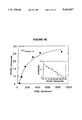
- FIG. 10 Effect of calreticulin on coronary thrombosis in a canine model.
- FIG. 11A Binding of calreticulin to ECs; dose-dependence.
- FIG. 11B Binding of calreticulin to ECs; competitive binding study of 125 I-calreticulin binding to endothelium: effect of unlabelled C-domain.
- FIG. 12 Effect of calreticulin on EC nitric oxide synthase activity.
- the subject invention provides a pharmaceutical composition, which comprises an amount of calreticulin effective for blocking or preventing thrombosis in a subject, causing substantially no defect or no defect in normal hemostasis, and a pharmaceutically effective carrier.
- calreticulin was known only as a regulator of intracellular calcium concentration in the endoplasmic reticulum and sarcoplasmic reticulum; prior to this invention, calreticulin was not known to have any extracellular function.
- This invention shows that the effect of calreticulin is in the blood vessel, but not in the circulating blood.
- Calreticulin can be used for the prevention of thrombosis and for the treatment of thrombosis.
- Normal hemostasis refers to clot formation in response to injury. When hemostasis is normal, the amount of bleeding is normal; when hemostasis is abnormal, the amount of bleeding is excessive.
- An advantage of this invention is the fact that calreticulin causes substantially no defect or no defect in normal hemostasis. This makes patient care easier, because there is no or substantially no excessive amount of bleeding. In contrast, other antithrombotic agents cause defects in normal hemostasis. Calreticulin can be used in any medical setting where bleeding is a problem or is expected to be a problem.
- the subject invention provides a method for blocking or preventing thrombosis in a subject, causing substantially no defect or no defect in normal hemostasis, which method comprises administering calreticulin to the subject in an amount effective for blocking or preventing thrombosis.
- the subject invention provides that the administering may be intracoronary or intravenous.
- Intravenous administration has the standard meaning.
- intravenous administration means injection into a peripheral vein, for example, a superficial vein, as in the arm or leg.
- intracoronary administration has the standard meaning.
- intracoronary administration means injection into the right or left coronary artery.
- An example of intracoronary administration is injection into the left circumflex coronary artery.
- An advantage of this invention is that administration may be intravenous, because other antithrombotic agents can be administered only by intracoronary means.
- This invention allows for intravenous administration because calreticulin causes substantially no defect or no defect in normal hemostasis.
- the subject invention provides that the subject may be a human.
- the human may be a patient.
- the subject may also include other mammals; examples include dogs, cats, horses, rodents, or pigs, rabbits, among others.
- the subject invention provides that the human may be a human at risk for thrombosis; a human who can receive traditional antithrombotic agents but who has not had an effective response to the traditional antithrombotic agent; or a human unable to receive traditional antithrombotic agents.
- antithrombotic agents are those antithrombotic agents which are currently known, and include antifibrinolytic agents, antiplatelet agents, or anticoagulant agents.
- antifibrinolytic agents include streptokinase, tissue plasminogen activator, urokinase, or acylated or other modified forms of plasmin.
- An example of a known antiplatelet agent is aspirin.
- known anticoagulant agents include heparin, warfarin, coumarin derivatives, thrombin inhibitors, or Factor Xa inhibitors.
- the subject invention provides that the human unable to receive traditional antithrombotic agents is a human undergoing intracranial surgery, or a human with a hemostatic defect.
- hemostatic defects include coagulation and platelet disorders.
- Calreticulin may be used for humans for whom the use of traditional antithrombotic agents is contraindicated, for example, a human undergoing intracranial surgery, or a human with a hemostatic defect.
- the subject invention provides that when the administration is intracoronary, and the subject is a human, then the amount is from about (0.04 mg calreticulin)/(Kg human subject) to about (0.07 mg calreticulin)/(Kg human subject).
- the subject invention provides that when the administration is intracoronary, and the subject is a human, then the amount is from about (0.05 mg calreticulin)/(Kg human subject) to about (0.06 mg calreticulin)/(Kg human subject).
- the subject invention provides that when the administration is intravenous, and the subject is a human, then the amount is from about (0.2 mg calreticulin)/(Kg human subject) to about (0.4 mg calreticulin)/(Kg human subject).
- the subject invention provides that when the administration is intravenous, and the subject is a human, then the amount may be about (0.3 mg calreticulin)/(Kg human subject).
- this method may comprise the administering of the pharmaceutical composition, which pharmaceutical composition comprises an amount of calreticulin effective for blocking or preventing thrombosis in a subject, causing substantially no defect or no defect in normal hemostasis, and a pharmaceutically effective carrier.
- the subject invention provides a pharmaceutical composition, which comprises calreticulin in combination with an other antithrombotic agent, in an amount and proportion for enhancing the action of the other antithrombotic agent, to prevent clotting or dissolve clots which have already formed.
- the subject invention provides a method for enhancing the action of an other antithrombotic agent which prevents clotting or dissolves clots which have already formed, comprising administering to a subject calreticulin in combination with the other antithrombotic agent in an amount and proportion effective for enhancing the action of the other antithrombotic agent, to prevent clotting or dissolve clots which have already formed.
- the subject invention provides that the other antithrombotic agent may be an antifibrinolytic agent.
- the subject invention provides that the antifibrinolytic agent may be streptokinase, tissue plasminogen activator, urokinase, or acylated or other modified forms of plasmin.
- the subject invention provides that the other antithrombotic agent may be an antiplatelet agent.
- the subject invention provides that the antiplatelet agent may be aspirin, or an agent which blocks glycoprotein IIbIIIa.
- the subject invention provides that the other antithrombotic agent may be an anticoagulant agent.
- anticoagulant agent may be heparin, warfarin, coumarin derivatives, thrombin inhibitors, or Factor Xa inhibitors.
- the subject invention provides that the subject may be a human.
- the subject invention provides that the human is a human at risk for thrombosis; a human who can receive traditional antithrombotic agents but who has not had an effective response to the traditional antithrombotic agent; or a human unable to receive traditional antithrombotic agents.
- the subject invention provides that the human unable to receive traditional antithrombotic agents may be a human undergoing intracranial surgery, or a human with a hemostatic defect.
- the subject invention provides that the administering may be intracoronary or intravenous.
- Coagulation Factor IX/IXa has been shown to bind to cellular surfaces, and Factor IXa expresses its procoagulant activity by assembling into the intrinsic Factor X activating complex (Factors IXa/VIIIa/X), which also forms on membrane surfaces. We have therefore sought to identify cellular proteins which act by binding Factor IX/IXa.
- An ⁇ 55 kDa polypeptide was purified to homogeneity from bovine lung extracts based on its capacity to bind 125 I-Factor IX in a dose-dependent and saturable manner.
- the ⁇ 55 kDa polypeptide was identified as calreticulin, a previously described intracellular calcium binding protein.
- Recombinant calreticulin bound vitamin K-dependent coagulation factors, 125 I-Factor IX, 125 I-Factor X, and 125 I-prothrombin (Kd's of ⁇ 2.7, 3.2, and 8.3 nM, respectively), via interaction with its calcium binding C-domain, although it did not affect the coagulant properties of these proteins.
- a ⁇ 55 kDa, trypsin-sensitive polypeptide was purified from bovine lung extract based on its capacity to bind coagulation Factor IX/IXa in a dose-dependent, saturable manner (Kd ⁇ 2 nM). Protein sequence data, amino terminal and internal, indicated that it was identical to calreticulin. Recombinant calreticulin bound Factor IX in a similar manner, and the binding site was localized to the C-domain, which contains low affinity, high capacity calcium binding sites.
- Blockade of intravascular thrombosis in animals receiving calreticulin was not accompanied by prolongation of the APTT (activated partial thromboplastic time) or PT (prothrombin time) in coronary sinus or left atrial blood.
- doses of heparin sufficient to prevent coronary thrombosis in this model markedly prolonged the APTT.
- calreticulin potentially exerts a selective [that is, lack of change in plasma coagulation parameters (PT and APTT) in presence of a blockade of localized coronary thrombosis (canine thrombosis model)] antithrombotic effect.
- Bovine Factors IX, X (a pool of X 1 and X 2 ) and prothrombin were purified to homogeneity by previously described methods (17-19), and were supplied by Enzyme Research Laboratories, Inc. (South Bend, Ind.).
- Factor IX was radiolabelled by the lactoperoxidase method (20) using Enzymobeads (Bio-Rad, Richmond, CA), 125 I-Factor IX was isolated as described (22), and the specific radioactivity was ⁇ 2.9 ⁇ 10 4 cpm/ng.
- the radioactivity profile of 125 I-Factor IX on SDS-PAGE (10%) showed a single peak with Mr ⁇ 55 kDa.
- Factor X and prothrombin were radiolabelled by the same procedure to specific radioactivities of 3.4 ⁇ 10 4 cpm/ng and 2.7 ⁇ 10 4 cpm/ng, respectively.
- Bovine serum albumin (Sigma, St. Louis, Mo.) was also radioiodinated by the same procedure to a final specific radioactivity of 1.5 ⁇ 10 3 cpm/ng.
- Other coagulation factors, human proteins C and S, bovine Factor Xa, human Factor Va, thrombin and antithrombin III were also obtained from Enzyme Research Labs.
- Recombinant rabbit calreticulin and discrete domains from calreticulin were expressed in E. coli using the glutathione S-transferase (GST) fusion protein system with pGEX-3X plasmid (22), as described previously (23). Plasmids containing intact calreticulin, the N-domain (amino acids 1-182), the P-domain (amino acids 182-290), or the C-domain (amino acids 330-401) were expressed in BNN103 E. coli host (23). GST and GST-fusion proteins were purified to homogeneity by one-step glutathione-Sepharose 4B affinity chromatography (Pharmacia, Piscataway, N.J.).
- the GST-full length calreticulin fusion protein was cleaved with Factor Xa, re-applied to glutathione-Sepharose 4B, and the pass-through contained homogeneous recombinant calreticulin.
- Native rabbit calreticulin was isolated by a selective ammonium sulfate precipitation procedure in the presence of protease inhibitors followed by FPLC Mono Q as described (24). Calreticulin was labelled using the same method for preparing 125 I-Factor IX.
- 125 I-calreticulin was separated from free iodine by gel filtration, and the final tracer had a specific radioactivity of 3.0 ⁇ 10 4 cpm/ng, was >95% precipitable in trichloroacetic acid (20%), and migrated as a single band with Mr ⁇ 55 kDa on SDS-PAGE (10%).
- PVC assay polyvinylchloride plate binding assay
- EC binding assay a polyvinylchloride plate binding assay
- PVC binding assays were performed with lung extracts, partially purified and purified calreticulin. Samples, prepared as described below, were diluted in buffer containing NaCO 3 (0.015M; pH 9.2)/CaCl 2 (0.1 mM), and 0.05 ml of this material was incubated at 4° C. for 15 hrs in wells of 96-well PVC plates.
- washing buffer HEPES, 10 mM, pH 7.55; NaCl, 137 mM; glucose, 11 mM; KCl, 4 mM; CaCl 2 , 2.6 mM; bovine serum albumin, 0.5 mg/ml
- blocking buffer Tris, 20 mM, pH 7.4; NaCl, 0.1M; CaCl 2 , 1 mM; bovine serum albumin, 50 mg/ml
- Binding of 125 I-calreticulin to endothelium was studied using confluent endothelial monolayers (0.32 cm 2 /well). Bovine aortic ECs were prepared and cultured as described previously (21). Wells were washed with elution buffer, washing buffer and incubation buffer, and were then incubated with binding buffer (0.05 ml/well) containing 125 I-calreticulin alone (total binding) or in the presence of a 100-fold molar excess of unlabelled calreticulin (nonspecific binding). Following a 2 hr incubation at 4° C., wells were washed six times rapidly with ice cold washing buffer as above.
- binding buffer was replaced by washing buffer, except that the amount of calcium chloride or EDTA was varied as stated. Dissociation was studied by the method of infinite dilution (27). Following a binding assay, performed as described above, wells were washed and fresh binding buffer was added for the indicated time. Then, wells were washed once, and residual radioactivity was determined.
- Bovine lung powder (30 g, Sigma) was added to 300 ml of Tris (20 mM; pH 7.4), NaCl (0.1M), PMSF (1 mM), trasylol (0.1%), and octyl- ⁇ -glucoside (1%) for 16 hr at 4° C. with constant mixing.
- Insoluble material was removed by centrifugation (11,000 g) for 30 min at 4° C., the supernatant (25 g) was filtered (0.45 ⁇ m), and applied to hydroxylapatite (300 ml, IBF, Savage, Md.; column equilibration buffer: Tris, 20 mM, pH 7.4; NaCl, 0.1M; octyl- ⁇ -glucoside, 0.1%; flow rate, 1 ml/min). The column was washed until the absorbance at 280 nm was ⁇ 0.01, and was then step-eluted with buffer containing 1M NaCl.
- the column was eluted with an ascending NaCl gradient (0.05 to 1M), fractions were collected (1 ml/fraction; flow rate of 1 ml/min), and an aliquot of each was assayed as above.
- Samples with peak Factor IX binding activity were pooled ( ⁇ 70 ml), concentrated by centrifugation on Centricon membranes (Amicon, Lexington, Mass.; molecular weight cut-off 10,000)( ⁇ 7 ml), and then applied to preparative nonreduced SDS-PAGE (10%; 28). Lanes of the gel were either stained with Coomassie blue or were cut into 4 mm slices, incubated with Na acetate (0.1M; pH 8.3) and octyl- ⁇ -glucoside (0.1%) for 15 hr at 4° C. Tubes were then centrifuged (10,000 g) for 5 min, and the supernatant was diluted 1:100 for testing in the PVC Factor IX binding assay. Where indicated, material eluted from the slices of the gel with peak Factor IX binding activity (corresponding to Mr ⁇ 55 kDa) were again subjected to nonreduced SDS-PAGE (10%) and gel elution.
- ⁇ 55 kDa polypeptide with peak Factor IX binding activity was subjected to SDS-PAGE (10%), transferred to polyvinylidene difluoride (PVDF) membranes, and the ⁇ 55 kDa was subjected to amino terminal sequencing.
- Proteolytic digestion of protein adsorbed to PVDF pieces was performed by treating strips with polyvinylpyrrolidine solution (0.2%) followed by trypsin in Tris/HCl (pH 8.5), as described (29).
- the resultant peptide fragments were isolated by reversed-phase HPLC on a C 8 column (2.1 ⁇ 5 cm, YMC Inc., Morris Plain, N.J.). Blotted protein samples on PVDF and peptide fragments recovered by HPLC peptide mapping were sequenced using an Applied biosystems Model 470A gas-phase sequencer with an "on-line" PTH amino acid analyzer (30-31) .
- Coagulation assays were carried out as described previously to assess Factor IXa-VIIIa-dependent of Factor X on endothelium and phospholipid (21,32), Factor Xa-Va-dependent activation of prothrombin on endothelium/phospholipid (33-35), tissue factor/Factor VIIa-dependent activation of Factor X on endothelial cells stimulated with tumor necrosis factor (36), thrombin/thrombomodulin (on bovine ECs)-dependent activation of protein C (37), and activated protein C/protein S-mediated inactivation of Factor Va on ECs (38)(the latter study employed human ECs and human activated protein C, protein S and Factor Va, see below).
- the prothrombin time (PT) and activated partial thromboplastin times (APTT) were performed by standard methods.
- mice Infusion of calreticulin into mice. Mice were infused via the tail vein with 125 I-calreticulin or 125 I-albumin. Plasma samples were obtained at the indicated times, and clearance data was fit using non-linear regression techniques (SAS Procedure NLIN, Cary N.C.). A bi-exponential equation (two rate constants) was fit to each of the clearance curves. In addition, an overall clearance rate was calculated from the area under the curve. Immunohistologic studies were performed on the vascular tissue obtained from mice infused with unlabelled calreticulin. At the indicated time after the infusion, animals were sacrificed, organs were removed and fixed by immersion in neutral-buffered formalin (10%), and embedded in paraffin by standard procedures.
- Sections were rehydrated, blocked in nonfat dry milk (4%), and incubated with goat anti-rabbit calreticulin IgG (40 min at 37° C.). Primary antibody was revealed using a rabbit anti-goat IgG avidin-biotin conjugated system, as per the manufacturer's instructions (Sigma, St. Louis Mo.), with 3-amino-9-ethylcarbazole as chromogen.
- tissue space microliters plasma per gram dry tissue weight
- cpm per volume plasma was calculated from the average of the cpm in the 0 and 20 min (for albumin) or in the 0 and 10 sec (for calreticulin) samples.
- tissue space for 125 I-albumin was calculated by subtracting the 125 I-albumin tissue space from the 125 I-calreticulin tissue space (40).
- Canine thrombosis and extravascular hemostasis model In vivo coronary thrombosis was induced with electric current, as described previously (4,41). This model involves instrumentation of the left circumflex coronary artery with a Doppler flow probe to assess coronary blood flow velocity, and placement of a needle electrode intraluminally to initiate thrombus formation. Sampling catheters are present in the left atrium and coronary sinus, and length-segment crystals are placed in the anterior and posterior myocardial walls. Following a 30 min period to allow stabilization of hemodynamic parameters, current was applied (150 ⁇ A) to the needle electrode until a 50% increase in blood flow velocity occurs.
- the bleeding tendency at extravascular sites was assessed using a modified incisional bleeding time: a uniform 1 cm deep, 5 cm, long abdominal wall incision was made and a pre-weighed 4 ⁇ 4 inch gauze was inserted for 5 min. The gauze was then removed, re-weighed, and the weight of blood loss quantitated, as described previously (4).
- Nitric oxide production was studied using the citrulline assay (42), which measures production of 14 C-L-citrulline from by exposing ECs to 14 C-L-arginine.
- the ⁇ 55 kDa polypeptide band was then subjected to sequencing. A portion of the amino terminal sequence aligned closely with that previously reported for rabbit (46) calreticulin, as well as the human, dog, rat, and pig counterparts (47-49,24)(Table 2). In addition, the band was cleaved with trypsin, fragments were separated on reversed phase HPLC and subjected to sequence analysis (FIG. 4), and found to match those in rabbit calreticulin: #1 residues 262-265; #2 residues 32-36 and 39-43, and #3, residues 306-322 (46).
- Recombinant calreticulin and its c-terminal domain interact with vitamin K-dependent coagulation factors. These data indicated that the ⁇ 55 kDa polypeptide purified from lung extract was calreticulin. Consistent with this, on reduced and non-reduced SDS-PAGE, purified rabbit calreticulin (bovine calreticulin has not been cloned) and the ⁇ 55 kDa protein from bovine lung extract were shown to comigrate (FIG. 5). Further, recombinant rabbit calreticulin interacted with vitamin K-dependent coagulation proteins similarly to the ⁇ 55 kDa polypeptide in the PVC assay.
- 125 I-Factor IX binding was observed in wells with adsorbed to calreticulin, and the binding was proportional to the amount of calreticulin incubated with the well (FIG. 6A). Similar binding of 125 I-Factor X and 125 I-prothrombin was observed (FIG. 6B-C, respectively). In each case, interaction of the vitamin K-dependent coagulation factor with calreticulin was dependent on its concentration, demonstrating Kd's of ⁇ 2.7, 3.2, and 8.3 nM, for studies with 125 I-Factors IX, X and prothrombin, respectively (FIGS. 6D,E,F).
- tissue factor/Factor VIIa-mediated activation of Factor X using matrices prepared from endothelial cells exposed to tumor necrosis factor as the source of tissue factor)(36), Factor IXa-VIIIa-mediated activation of Factor X (using crude cephalin as the phospholipid surface or endothelium), Factor Xa-Va-mediated activation of prothrombin (using cephalin as the phospholipid surface or endothelium), and thrombomodulin-dependent, thrombin-mediated activation of protein C (using intact bovine ECs as the source of thrombomodulin)(37).
- a ⁇ 55 kDa polypeptide was isolated from lung extracts based on its capacity to bind Factor IX.
- the ⁇ 55 kDa co-migrated on SDS-PAGE with calreticulin, and displayed virtual sequence identity with calreticulin of five different species.
- recombinant calreticulin bound Factor IX in a manner analogous to the ⁇ 55 kDa lung-derived polypeptide.
- calreticulin may stimulate ECs resulting in their production of anticoagulant mediators which prevent effective thrombus formation. If these findings can be extrapolated to a range of thrombosis models, calreticulin may prove to be a novel type of antithrombotic agent.
- FIG. 1 Binding of 125 I-Factor IX to lung extract immobilized in PVC wells.
- FIG. 2 Purification of a ⁇ 55 kDa polypeptide from lung extracts based on its ability to bind 125 I-Factor IX in the PVC assay.
- Panel A detergent extract of bovine lung (30 g) was applied to a hydroxylapatite column, the column was washed in equilibration buffer, followed by 1M NaCl, and finally step-eluted with NaPO 4 (0.5M, pH 7.4; the arrow indicates when step-elution was begun).
- OD 280 solid line
- binding activity in the PVC assay broken line
- binding activity in the PVC assay broken line
- One binding unit is defined as one count/minute of specific binding of 125 I-Factor IX in the PVC assay/milliliter of sample applied to a PVC plate at a 1:1 dilution.
- the active pool of material applied to Mono Q included fractions 10-30, and is indicated.
- Panel B FPLC Mono Q.
- the pool with Factor IX binding activity from the hydroxylapatite column was dialyzed and applied to FPLC Mono Q.
- the column was washed with equilibration buffer and eluted with an ascending salt gradient (50 mM to 1M).
- OD 280 salt concentration of the gradient, and binding units in the PVC assay are plotted for each fraction.
- the pool of fractions from Mono Q subjected to preparative SDS-PAGE is indicated by the bar.
- FIG. 3 SDS-PAGE and gel elution of ⁇ 55 kDa polypeptide derived from lung extract which binds Factor IX.
- Lane 1 nonreduced SDS-PAGE (10%) of the pool from Mono Q with Factor IX binding activity visualized by Coomassie blue staining.
- Lane 2 activity profile of material eluted from the indicated slice in lane A.
- a nonstained lane on the gel otherwise identical to lane A was sliced ( ⁇ 4 mm pieces), subjected to elution as described in the text, and used to coat PVC wells for a binding assay with 125 I-Factor IX alone (6.3 nM) or in the presence of an 100-fold excess of unlabelled Factor IX.
- Factor IX binding activity is seen to be maximal in the slices (#5-6) corresponding to ⁇ 55 kDa. Data are expressed as counts/min bound per sample of extract. Lanes 3 and 4, the material eluted from slices #5-6 in lane 2 was subjected to nonreduced (lane 3) or reduced (lane 4) SDS-PAGE (10%), and material on the gel was visualized by silver staining. Lane 4, activity profile of the material in lane 3. The material eluted from slices #5-6 in lane 2 was subjected to nonreduced SDS-PAGE (10%) and again the gel was sliced and proteins eluted. The eluted material was test in the PVC assay for its ability to bind 125 I-Factor IX.
- FIG. 4 HPLC reversed-phase chromatography of tryptic digest and protein sequence of fragments (inset) from ⁇ 55 kDa polypeptide isolated by hydroxylapatite and FPLC Mono Q, followed by gel elution. Details of methods are described in the text. Amino acids in the sequences in the inset are designated by: A, Ala; C, Cys; D, Asp; E, Glu; F, Phe; G, Gly; H, His; I, Ile; K, Lys; L, Leu; M, Met; N, Asn; P, Pro; Q, Gln; R, Arg; S, Set; T, Thr, V, Val; W, Trp; and Y, Tyr. X is an amino acid residue not identified at that position. Sequence number 1 in the insert is SEQ ID NO:7. Sequence number 2 in the insert is SEQ ID NO:8. Sequence number 3 in the insert is SEQ ID NO:9.
- FIG. 5 SDS-PAGE of ⁇ 55 kDa polypeptide and purified, recombinant rabbit calreticulin.
- FIG. 6 Binding of Factors IX, X and prothrombin to recombinant calreticulin.
- A-C Dependence on calreticulin concentration. PVC wells were incubated with the indicated concentration of recombinant calreticulin, excess sites were blocked with albumin-containing buffer, and then a binding assay was performed by adding either 125 I-Factor IX (6.3 nM; A), 125 I-Factor X (6.0 nM; B) or 125 I-prothrombin (8.0 nM; C) alone or in the presence of a 100-fold excess of the respective unlabelled protein. Specific binding is shown (mean ⁇ SEM of triplicate determinations). D-F.
- FIG. 7 Competitive binding study: effect of unlabelled Factors IX, X and prothrombin on the binding of 125 I-Factor IX to recombinant calreticulin.
- Dixon plot showing 1/Bound (1/B, fmole -1 ) versus protein added (nM).
- FIG. 8 Interaction of 125 I-Factor IX with the N-domain, P-domain and C-domain of calreticulin.
- A. Wells were incubated with the indicated recombinant domain of calreticulin or glutathione-S-transferase control protein (0.5 ⁇ g/well) for 15 hr at 4° C., excess sites in the wells were blocked with albumin-containing buffer, and then a binding assay was performed with 125 I-Factor IX alone (6.3 nM) or in the presence of a 100-fold molar excess of unlabelled protein. Specific binding is plotted versus the calreticulin domain (N, P or C) used in the assay (mean ⁇ SEM of triplicate determinations).
- B-D Binding of 125 I-Factors IX, X and prothrombin to the C domain of calreticulin.
- Wells were incubated with C domain (0.5 ⁇ g/well) for 15 hr at 4° C., excess sites were blocked with blocking buffer, and then a binding assay was performed with the indicated concentrations of either 125 I-Factor IX (B), 125 I-Factor X (C) or 125 I-prothrombin (D) alone or in the presence of a 30-fold excess of the unlabelled respective protein.
- Specific binding is plotted versus the concentration of free/added tracer. Data were analyzed by the nonlinear least-squares program, and the curve indicates the best fit line.
- FIG. 9 Infusion of recombinant rabbit calreticulin into mice.
- A Removal of infused 125 I-calreticulin from the plasma. Mice were infused via the tail vein with 125 I-calreticulin (0.43 ⁇ g/animal), and at the indicated times blood was withdrawn for determination of radioactivity. The solid line represents the best fit to the data using the weighted least squares non-linear regression procedure.
- B Removal of infused 125 I-albumin from the plasma. Mice were treated as in (A), except that 125 I-albumin (0.34 ⁇ g/animal) was used in place of 125 I-calreticulin.
- FIG. 10 Effect of calreticulin on coronary thrombosis in a canine model. Following instrumentation of the left circumflex artery, current was applied to the needle electrode until a 50% increase in mean blood flow velocity occurred (this corresponds to a ⁇ 50% decrease in cross-sectional luminal area). The current was then turned off, a bolus of the indicated amount of calreticulin or saline was given into the left circumflex (in each case, volume was 0.5 ml), and vessel patency was monitored as described in the text.
- FIG. 11 Binding of calreticulin to ECs.
- FIG. 12 Effect of calreticulin on EC nitric oxide synthase activity. Cultured ECs (3 ⁇ 10 6 cells/point) were incubated for the indicated time with calreticulin (380 nM), and then the citrulline assay was performed as described in the text.
Abstract
Description
              TABLE 1
______________________________________
Purification of ≈55 kDa Polypeptide From Lung Extract.sup.+
                                         Purf.
                 Activity Specific       (for
         Protein units*   activity Yield each
Step     mg      ×10.sup.9
                          U/mg × 10.sup.6
                                   %     step)
______________________________________
lung     2460    3.6         1.5   --    1
extract
HA#      147     2.3       22      6     15
Mono S    14     2.1      150      9.5     6.5
SDS-PAGE    0.3   0.345   1150     2.1   8
______________________________________
 Purf. is an abbreviation for Purification.
 *Units are defined arbitrarily; Factor IX binding activity is defined as
 the product of specifically bound .sup.125 IFactor IX in the PVC assay x
 dilution factor of the sample corrected to a sample size of 1 ml.
 #HA = hydroxylapatite
 + = The starting material was 30 grams of bovine lung acetone powder, and
 the amount indicated in the top line (lung extract) is that which remaine
 after solubilization in detergentcontaining buffer and centrifugation to
 remove insoluble material (termed lung extract).
              TABLE 2
______________________________________
Amino-Terminal Sequence Analysis Of ≈55 kDa Polypeptide
From Lung Extract, And Comparison With Sequence Of
Calreticulin (CRT)*
______________________________________
55.sup.1      SEQ ID NO: 1
human.sup.2 CRT
              SEQ ID NO: 2
dog.sup.3 CRT SEQ ID NO: 3
rabbit.sup.4 CRT
              SEQ ID NO: 4
rat.sup.5 CRT SEQ ID NO: 5
pig.sup.6 CRT SEQ ID NO: 6
______________________________________
 *Mismatches of the 55 kDa sequence with the indicated sequence are
 designated by lower case letters.
 .sup.1 = Sequence derived from amino terminal sequence analysis of the
 ≈55 kDa polypeptide purified from bovine lung as described in the
 text.
 .sup.2 = Sequence derived from human calreticulin (47)
 .sup.3 = Sequence derived from dog calreticulin (48)
 .sup.4 = Sequence derived from rabbit calreticulin (46)
 .sup.5 = Sequence derived from rat calreticulin (49)
 .sup.6 = Sequence derived from pig calreticulin (26)
__________________________________________________________________________
SEQUENCE LISTING
(1) GENERAL INFORMATION:
(iii) NUMBER OF SEQUENCES: 9
(2) INFORMATION FOR SEQ ID NO:1:
(i) SEQUENCE CHARACTERISTICS:
(A) LENGTH: 12 amino acids
(B) TYPE: amino acid
(C) STRANDEDNESS: unknown
(D) TOPOLOGY: unknown
(ii) MOLECULE TYPE: peptide
(xi) SEQUENCE DESCRIPTION: SEQ ID NO:1:
Thr ValTyrPheLysGluGlnPheLeuAspGlyAsp
1510
(2) INFORMATION FOR SEQ ID NO:2:
(i) SEQUENCE CHARACTERISTICS:
(A) LENGTH: 12 amino acids
(B) TYPE: amino acid
(C) STRANDEDNESS: unknown
(D) TOPOLOGY: unknown
(ii) MOLECULE TYPE: peptide
(xi) SEQUENCE DESCRIPTION: SEQ ID NO:2:
AlaValTyrPheLysGluGlnPheLeuAspGlyAsp
1510
(2) INFORMATION FOR SEQ ID NO:3:
(i) SEQUENCE CHARACTERISTICS:
(A) LENGTH: 12 amino acids
(B) TYPE: amino acid
(C) STRANDEDNESS: unknown
 (D) TOPOLOGY: unknown
(ii) MOLECULE TYPE: peptide
(xi) SEQUENCE DESCRIPTION: SEQ ID NO:3:
ThrIleTyrPheLysGluGlnPheLeuAspGlyAsp
1510
(2) INFORMATION FOR SEQ ID NO:4:
(i) SEQUENCE CHARACTERISTICS:
(A) LENGTH: 12 amino acids
(B) TYPE: amino acid
(C) STRANDEDNESS: unknown
(D) TOPOLOGY: unknown
(ii) MOLECULE TYPE: peptide
(xi) SEQUENCE DESCRIPTION: SEQ ID NO:4:
ValValTyrPheLysGluGlnPheLeuAspGlyAsp
1510
(2) INFORMATION FOR SEQ ID NO:5:
(i) SEQUENCE CHARACTERISTICS:
 (A) LENGTH: 12 amino acids
(B) TYPE: amino acid
(C) STRANDEDNESS: unknown
(D) TOPOLOGY: unknown
(ii) MOLECULE TYPE: peptide
(xi) SEQUENCE DESCRIPTION: SEQ ID NO:5:
AlaIleTyrPheLysGluGlnPheLeuAspGlyAsp
1510
(2) INFORMATION FOR SEQ ID NO:6:
(i) SEQUENCE CHARACTERISTICS:
(A) LENGTH: 12 amino acids
(B) TYPE: amino acid
(C) STRANDEDNESS: unknown
(D) TOPOLOGY: unknown
(ii) MOLECULE TYPE: peptide
(xi) SEQUENCE DESCRIPTION: SEQ ID NO:6:
ThrIleTyrPheLysGluGlnPheLeuAspGlyAsp
15 10
(2) INFORMATION FOR SEQ ID NO:7:
(i) SEQUENCE CHARACTERISTICS:
(A) LENGTH: 4 amino acids
(B) TYPE: amino acid
(C) STRANDEDNESS: unknown
(D) TOPOLOGY: unknown
(ii) MOLECULE TYPE: peptide
(xi) SEQUENCE DESCRIPTION: SEQ ID NO:7:
GlnIleAspAsn
(2) INFORMATION FOR SEQ ID NO:8:
(i) SEQUENCE CHARACTERISTICS:
(A) LENGTH: 10 amino acids
(B) TYPE: amino acid
(C) STRANDEDNESS: unknown
(D) TOPOLOGY: unknown
(ii) MOLECULE TYPE: peptide
(xi) SEQUENCE DESCRIPTION: SEQ ID NO:8:
PheValLeuSerSerPheTyrGlyAspGln
1510
(2 ) INFORMATION FOR SEQ ID NO:9:
(i) SEQUENCE CHARACTERISTICS:
(A) LENGTH: 17 amino acids
(B) TYPE: amino acid
(C) STRANDEDNESS: unknown
(D) TOPOLOGY: unknown
(ii) MOLECULE TYPE: peptide
(xi) SEQUENCE DESCRIPTION: SEQ ID NO:9:
SerGlyThrIlePheAspAsnPheLeuIleThrAsnAspGluAlaTyr
1 51015
Ala
Claims (19)
Priority Applications (1)
| Application Number | Priority Date | Filing Date | Title |
|---|---|---|---|
| US08/045,261 US5426097A (en) | 1993-04-06 | 1993-04-06 | Calreticulin: a novel antithrombotic agent |
Applications Claiming Priority (1)
| Application Number | Priority Date | Filing Date | Title |
|---|---|---|---|
| US08/045,261 US5426097A (en) | 1993-04-06 | 1993-04-06 | Calreticulin: a novel antithrombotic agent |
Publications (1)
| Publication Number | Publication Date |
|---|---|
| US5426097A true US5426097A (en) | 1995-06-20 |
Family
ID=21936878
Family Applications (1)
| Application Number | Title | Priority Date | Filing Date |
|---|---|---|---|
| US08/045,261 Expired - Lifetime US5426097A (en) | 1993-04-06 | 1993-04-06 | Calreticulin: a novel antithrombotic agent |
Country Status (1)
| Country | Link |
|---|---|
| US (1) | US5426097A (en) |
Cited By (22)
| Publication number | Priority date | Publication date | Assignee | Title |
|---|---|---|---|---|
| US5525478A (en) * | 1991-10-04 | 1996-06-11 | Matschiner; John T. | Soluble thrombomodulin-based one-stage assay for vitamin-K dependent coagulation-inhibiting proteins |
| WO1996036643A1 (en) * | 1995-05-17 | 1996-11-21 | University Of Alberta | Method of inhibiting restenosis using calreticulin |
| US5716795A (en) * | 1991-10-04 | 1998-02-10 | Matschiner; John T. | Thrombomodulin-based coagulometric assay of the protein C system |
| WO1998023151A1 (en) * | 1996-11-27 | 1998-06-04 | Gelfand Mathew I | Use of thrombolytic reagents for prevention of vascular disease |
| US5854202A (en) * | 1995-01-24 | 1998-12-29 | Dedhar; Shoukat | Peptide fragments of calreticulin, peptide mimetics thereof, and pharmaceutical compostions comprising same |
| WO2000020577A1 (en) * | 1998-10-06 | 2000-04-13 | The Government Of The United States Of America, Represented By The Secretary, Dept. Of Health And Huuman Services, The National Institutes Of Health | Use of calreticulin and calreticulin fragments to inhibit endothelial cell growth and angiogenesis, and suppress tumor growth |
| US6171864B1 (en) * | 1996-07-05 | 2001-01-09 | Pioneer Hi-Bred International, Inc. | Calreticulin genes and promoter regions and uses thereof |
| US6426220B1 (en) | 2000-10-30 | 2002-07-30 | Isis Pharmaceuticals, Inc. | Antisense modulation of calreticulin expression |
| US6518397B1 (en) | 1997-07-24 | 2003-02-11 | Shoukat Dedhar | Pharmaceuticals for modulating hormone responsiveness |
| US6596690B2 (en) | 1998-10-06 | 2003-07-22 | The United States Of America As Represented By The Department Of Health And Human Services | Vasostatin as marrow protectant |
| US20040086845A1 (en) * | 1999-10-20 | 2004-05-06 | Tzyy-Choou Wu | Superior molecular vaccine linking the translocation domain of a bacterial toxin to an antigen |
| US6867180B1 (en) * | 1998-10-06 | 2005-03-15 | The United States Of America As Represented By The Department Of Health And Human Services | Use of calreticulin and calreticulin fragments to inhibit endothelial cell growth and angiogenesis, and suppress tumor growth |
| US20060128720A1 (en) * | 2002-03-21 | 2006-06-15 | Kufe Donald W | Inhibition of cell death responses induced by oxidative stress |
| US20080069840A1 (en) * | 2005-01-06 | 2008-03-20 | Tzyy-Choou Wu | RNA Interference That Blocks Expression of Pro-Apoptotic Proteins Potentiates Immunity Induced by DNA and Transfected Dendritic Cell Vaccines |
| US20080102084A1 (en) * | 2005-01-26 | 2008-05-01 | Tzyy-Choou Wu | Anti-cancer DNA Vaccine Employing Plasmids Encoding Mutant Oncoprotein Antigen and Calreticulin |
| US20080260765A1 (en) * | 2007-03-15 | 2008-10-23 | Johns Hopkins University | HPV DNA Vaccines and Methods of Use Thereof |
| US20090148471A1 (en) * | 2000-08-03 | 2009-06-11 | The Johns Hopkins University | Molecular Vaccine Linking an Endoplasmic Reticulum Chaperone Polypeptide to an Antigen |
| US20090175431A1 (en) * | 2002-07-10 | 2009-07-09 | Blake Bookstaff | Method and system for providing directory assistance to erroneous telephone calls |
| US20090285861A1 (en) * | 2008-04-17 | 2009-11-19 | Tzyy-Choou Wu | Tumor cell-based cancer immunotherapeutic compositions and methods |
| US20100278871A1 (en) * | 2003-05-05 | 2010-11-04 | Johns Hopkins University | Anti-cancer dna vaccine employing plasmids encoding signal sequence, mutant oncoprotein antigen, and heat shock protein |
| US20100330105A1 (en) * | 2006-08-22 | 2010-12-30 | John Hopkins University | Anticancer Combination Therapies |
| US9085638B2 (en) | 2007-03-07 | 2015-07-21 | The Johns Hopkins University | DNA vaccine enhancement with MHC class II activators |
-
1993
- 1993-04-06 US US08/045,261 patent/US5426097A/en not_active Expired - Lifetime
Non-Patent Citations (32)
| Title |
|---|
| Abe Hiroshi et al., Molecular Brain Research, vol. 14 pp. 337 343 (1992). * |
| Abe Hiroshi et al., Molecular Brain Research, vol. 14 pp. 337-343 (1992). |
| Baksh, et al., Protein Express, and Purif. (Aug. 1992) 3(4): 322 331. * |
| Baksh, et al., Protein Express, and Purif. (Aug. 1992) 3(4): 322-331. |
| Baksh, Shairaz et al., "Expression and purification of recombinant and native calrehiculin," Protein Expression Purification, vol. 3(4), pp. 322-331 (1992). CA117(17):166922m. |
| Baksh, Shairaz et al., Expression and purification of recombinant and native calrehiculin, Protein Expression Purification, vol. 3(4), pp. 322 331 (1992). CA117(17):166922m. * |
| Benedict, et al., Circulation Research (Jan. 1986) 58(1): 58 67. * |
| Benedict, et al., Circulation Research (Jan. 1986) 58(1): 58-67. |
| Benedict, et al., J. Clin. Invent. (Nov. 1991) 88: 1760 1765. * |
| Benedict, et al., J. Clin. Invent. (Nov. 1991) 88: 1760-1765. |
| Bush, et al., Faseb J. (Oct. 1990) 4(13): 3087 3098. * |
| Bush, et al., Faseb J. (Oct. 1990) 4(13): 3087-3098. |
| Fliegel, et al., J. Biol. Chem. (Dec. 25, 1989) 264(36): 21522 21528. * |
| Fliegel, et al., J. Biol. Chem. (Dec. 25, 1989) 264(36): 21522-21528. |
| Gitel, et al., PNAS USA (Jul. 1977) 74(7): 3028 3032. * |
| Gitel, et al., PNAS-USA (Jul. 1977) 74(7): 3028-3032. |
| Johnson Robin et al., Molecular Brain Research, vol. 12, pp. 69 76, (1992). * |
| Johnson Robin et al., Molecular Brain Research, vol. 12, pp. 69-76, (1992). |
| Michalak, et al., Biochem. J. (Aug. 1, 1992) 285(3): 681 692. * |
| Michalak, et al., Biochem. J. (Aug. 1, 1992) 285(3): 681-692. |
| Mookerjee, et al., Immunol. Invest (1993) 22(6&7): 415 429. * |
| Mookerjee, et al., Immunol. Invest (1993) 22(6&7): 415-429. |
| Opas, Michal et al., "Calcium Storage in non-muscle tissue . . . ", Biochem. Cell Biol., vol. 70, pp. 972-979 (1992). |
| Opas, Michal et al., Calcium Storage in non muscle tissue . . . , Biochem. Cell Biol., vol. 70, pp. 972 979 (1992). * |
| Romson, et al., Thrombosis Research (Mar. 15, 1980) 17(6): 841 853. * |
| Romson, et al., Thrombosis Research (Mar. 15, 1980) 17(6): 841-853. |
| Routsias, et al., Clin. Exp. Immunol. (Mar. 1993) 91(3): 437 441. * |
| Routsias, et al., Clin. Exp. Immunol. (Mar. 1993) 91(3): 437-441. |
| Routsias, J. A. et al, "Calreticulin . . . ", Clin. Exp. Immunol., vol. 91(3), pp. 437-441 (1993) CA119(19):2015996. |
| Routsias, J. A. et al, Calreticulin . . . , Clin. Exp. Immunol., vol. 91(3), pp. 437 441 (1993) CA119(19):2015996. * |
| Sueyoshi, Tatsuya et al., "A new procedure for the separation of protein 2 . . . ", Thrombosis Research, vol. 63, pp. 569-575, 1991. |
| Sueyoshi, Tatsuya et al., A new procedure for the separation of protein 2 . . . , Thrombosis Research, vol. 63, pp. 569 575, 1991. * |
Cited By (35)
| Publication number | Priority date | Publication date | Assignee | Title |
|---|---|---|---|---|
| US5716795A (en) * | 1991-10-04 | 1998-02-10 | Matschiner; John T. | Thrombomodulin-based coagulometric assay of the protein C system |
| US5525478A (en) * | 1991-10-04 | 1996-06-11 | Matschiner; John T. | Soluble thrombomodulin-based one-stage assay for vitamin-K dependent coagulation-inhibiting proteins |
| US5854202A (en) * | 1995-01-24 | 1998-12-29 | Dedhar; Shoukat | Peptide fragments of calreticulin, peptide mimetics thereof, and pharmaceutical compostions comprising same |
| WO1996036643A1 (en) * | 1995-05-17 | 1996-11-21 | University Of Alberta | Method of inhibiting restenosis using calreticulin |
| US6171864B1 (en) * | 1996-07-05 | 2001-01-09 | Pioneer Hi-Bred International, Inc. | Calreticulin genes and promoter regions and uses thereof |
| WO1998023151A1 (en) * | 1996-11-27 | 1998-06-04 | Gelfand Mathew I | Use of thrombolytic reagents for prevention of vascular disease |
| US5837688A (en) * | 1996-11-27 | 1998-11-17 | Gelfand; Mathew I. | Use of thrombolytic reagents for prevention of vascular disease |
| US6518397B1 (en) | 1997-07-24 | 2003-02-11 | Shoukat Dedhar | Pharmaceuticals for modulating hormone responsiveness |
| US20090149380A1 (en) * | 1998-10-06 | 2009-06-11 | The Government Of The United States Of America As Represented By The Secretary Of The Department Of | Vasostatin as marrow protectant |
| US7432236B2 (en) | 1998-10-06 | 2008-10-07 | The United States Of America As Represented By The Secretary Of The Department Of Health And Human Services | Vasostatin as marrow protectant |
| US6596690B2 (en) | 1998-10-06 | 2003-07-22 | The United States Of America As Represented By The Department Of Health And Human Services | Vasostatin as marrow protectant |
| US20030216299A1 (en) * | 1998-10-06 | 2003-11-20 | The Government Of The United States Of America As | Vasostatin as marrow protectant |
| US7812117B2 (en) | 1998-10-06 | 2010-10-12 | The United States Of America As Represented By The Department Of Health And Human Services | Vasostatin as marrow protectant |
| US6867180B1 (en) * | 1998-10-06 | 2005-03-15 | The United States Of America As Represented By The Department Of Health And Human Services | Use of calreticulin and calreticulin fragments to inhibit endothelial cell growth and angiogenesis, and suppress tumor growth |
| US7488711B2 (en) | 1998-10-06 | 2009-02-10 | The United States Of America As Represented By The Secretary Of The Department Of Health And Human Services | Use of calreticulin and calreticulin fragments to inhibit endothelial cell growth and angiogenesis, and suppress tumor growth |
| US20050208018A1 (en) * | 1998-10-06 | 2005-09-22 | The Government of the United States of America as represented by the Secretary of the | Use of calreticulin and calreticulin fragments to inhibit endothelial cell growth and angiogenesis, and suppress tumor growth |
| WO2000020577A1 (en) * | 1998-10-06 | 2000-04-13 | The Government Of The United States Of America, Represented By The Secretary, Dept. Of Health And Huuman Services, The National Institutes Of Health | Use of calreticulin and calreticulin fragments to inhibit endothelial cell growth and angiogenesis, and suppress tumor growth |
| US20040086845A1 (en) * | 1999-10-20 | 2004-05-06 | Tzyy-Choou Wu | Superior molecular vaccine linking the translocation domain of a bacterial toxin to an antigen |
| US8128922B2 (en) | 1999-10-20 | 2012-03-06 | Johns Hopkins University | Superior molecular vaccine linking the translocation domain of a bacterial toxin to an antigen |
| US9758551B2 (en) | 1999-10-20 | 2017-09-12 | The Johns Hopkins University | Superior molecular vaccine linking the translocation domain of a bacterial toxin to an antigen |
| US8007781B2 (en) | 2000-08-03 | 2011-08-30 | The Johns Hopkins University | Molecular vaccine linking an endoplasmic reticulum chaperone polypeptide to an antigen |
| US20090148471A1 (en) * | 2000-08-03 | 2009-06-11 | The Johns Hopkins University | Molecular Vaccine Linking an Endoplasmic Reticulum Chaperone Polypeptide to an Antigen |
| US20050101553A1 (en) * | 2000-10-30 | 2005-05-12 | Bennett C. F. | Antisense modulation of calreticulin expression |
| US6426220B1 (en) | 2000-10-30 | 2002-07-30 | Isis Pharmaceuticals, Inc. | Antisense modulation of calreticulin expression |
| US20060128720A1 (en) * | 2002-03-21 | 2006-06-15 | Kufe Donald W | Inhibition of cell death responses induced by oxidative stress |
| US20090175431A1 (en) * | 2002-07-10 | 2009-07-09 | Blake Bookstaff | Method and system for providing directory assistance to erroneous telephone calls |
| US20100278871A1 (en) * | 2003-05-05 | 2010-11-04 | Johns Hopkins University | Anti-cancer dna vaccine employing plasmids encoding signal sequence, mutant oncoprotein antigen, and heat shock protein |
| US9701725B2 (en) | 2003-05-05 | 2017-07-11 | The Johns Hopkins University | Anti-cancer DNA vaccine employing plasmids encoding signal sequence, mutant oncoprotein antigen, and heat shock protein |
| US9011866B2 (en) | 2005-01-06 | 2015-04-21 | The Johns Hopkins University | RNA interference that blocks expression of pro-apoptotic proteins potentiates immunity induced by DNA and transfected dendritic cell vaccines |
| US20080069840A1 (en) * | 2005-01-06 | 2008-03-20 | Tzyy-Choou Wu | RNA Interference That Blocks Expression of Pro-Apoptotic Proteins Potentiates Immunity Induced by DNA and Transfected Dendritic Cell Vaccines |
| US20080102084A1 (en) * | 2005-01-26 | 2008-05-01 | Tzyy-Choou Wu | Anti-cancer DNA Vaccine Employing Plasmids Encoding Mutant Oncoprotein Antigen and Calreticulin |
| US20100330105A1 (en) * | 2006-08-22 | 2010-12-30 | John Hopkins University | Anticancer Combination Therapies |
| US9085638B2 (en) | 2007-03-07 | 2015-07-21 | The Johns Hopkins University | DNA vaccine enhancement with MHC class II activators |
| US20080260765A1 (en) * | 2007-03-15 | 2008-10-23 | Johns Hopkins University | HPV DNA Vaccines and Methods of Use Thereof |
| US20090285861A1 (en) * | 2008-04-17 | 2009-11-19 | Tzyy-Choou Wu | Tumor cell-based cancer immunotherapeutic compositions and methods |
Similar Documents
| Publication | Publication Date | Title |
|---|---|---|
| US5426097A (en) | Calreticulin: a novel antithrombotic agent | |
| Grainger et al. | Release and activation of platelet latent TGF–β in blood clots during dissolution with plasmin | |
| Gomi et al. | Antithrombotic effect of recombinant human thrombomodulin on thrombin-induced thromboembolism in mice | |
| US5968897A (en) | Agents affecting thrombosis and hemostasis | |
| US5374617A (en) | Treatment of bleeding with modified tissue factor in combination with FVIIa | |
| CA2134750C (en) | Truncated tissue factor and fviia or fvii activator for blood coagulation | |
| US5759542A (en) | Compositions and methods for the delivery of drugs by platelets for the treatment of cardiovascular and other diseases | |
| CA2080462C (en) | Therapeutic uses of actin-binding compounds | |
| Markwardt | Past, present and future of hirudin | |
| JP3051995B2 (en) | Oxidation-resistant thrombomodulin analogs | |
| JP3517236B2 (en) | Protease-resistant thrombomodulin analog | |
| Stern et al. | In vivo evidence of intravascular binding sites for coagulation factor IX | |
| Sherman et al. | Fibronectin: blood turnover in normal animals and during intravascular coagulation | |
| JP2006241109A (en) | FUSION PROTEIN CONTAINING ANTIBODY AGAINST HUMAN P SELECTIN AND DSPAalpha1 | |
| CA2139652C (en) | Thrombin-inhibitors | |
| Szemraj et al. | New derivative of staphylokinase SAK-RGD-K2-Hirul exerts thrombolytic effects in the arterial thrombosis model in rats | |
| Fauvel et al. | Platelet-collagen interaction: adhesion of human blood platelets to purified (CB4) peptide from type III collagen | |
| Benedict et al. | Active site-blocked factor Xa prevents thrombus formation in the coronary vasculature in parallel with inhibition of extravascular coagulation in a canine thrombosis model | |
| Mitchell et al. | Cleavage of protein S by a platelet membrane protease. | |
| Koyama et al. | Relationship between post‐translational glycosylation and anticoagulant function of secretable recombinant mutants of human thrombomodulin | |
| JPH11507664A (en) | Factor IX binding peptides derived from factor VIII and their use as inhibitors of blood coagulation | |
| EP0239644A1 (en) | Novel physiologically active substance having blood coagulation controlling activity | |
| Oates et al. | The regulation of human factor V by a neutrophil protease | |
| EP0487660B1 (en) | Treatment of thrombotic events | |
| US20040072757A1 (en) | Agents affecting thrombosis and hemostasis |
Legal Events
| Date | Code | Title | Description |
|---|---|---|---|
| AS | Assignment |
Owner name: TRUSTEES OF COLUMBIA UNIVERSITY IN THE CITY OF NEW Free format text: ASSIGNMENT OF ASSIGNORS INTEREST;ASSIGNORS:STERN, DAVID M.;KUWABARA, KEISUKE;BENEDICT, CLAUDE;AND OTHERS;REEL/FRAME:007214/0934;SIGNING DATES FROM 19940824 TO 19940919 |
|
| STPP | Information on status: patent application and granting procedure in general |
Free format text: APPLICATION UNDERGOING PREEXAM PROCESSING |
|
| AS | Assignment |
Owner name: TRUSTEES OF COLUMBIA UNIVERSITY IN THE CITY OF NEW Free format text: CORRECTION OF NOTICE OF RECORDATION ASSIGNMENT;ASSIGNORS:STERN, DAVID M.;KUWABARA, KEISUKE;BENEDICT, CLAUDE;AND OTHERS;REEL/FRAME:007374/0680 Effective date: 19940919 Owner name: BOARD OF REGENTS, THE UNIVERSITY OF TEXAS SYSTEM, Free format text: CORRECTION OF NOTICE OF RECORDATION ASSIGNMENT;ASSIGNORS:STERN, DAVID M.;KUWABARA, KEISUKE;BENEDICT, CLAUDE;AND OTHERS;REEL/FRAME:007374/0680 Effective date: 19940919 |
|
| FPAY | Fee payment |
Year of fee payment: 4 |
|
| FPAY | Fee payment |
Year of fee payment: 8 |
|
| FPAY | Fee payment |
Year of fee payment: 12 |