BACKGROUND OF THE INVENTION
The present invention is directed to the method of production and therapeutic application of a covalent dimer of the cytokine kit ligand with increased proliferative activity.
Kit ligand (KL) is a growth and differentiation factor for an assortment of cell types, and is known to be a ligand for the c-kit proto-oncogene. KL was initially identified based on a variety of biological activities and has therefore been referred to by different names, including Stem Cell Factor, Mast Cell Growth Factor, and more recently Steel Factor, in recognition of the gene locus in the mouse which encodes KL, as described by Anderson, et al., (1990) Cell 63,235-243; Huang, E., et al. (1990) Cell 63, 225-233; Martin, et al. (1990) Cell 63,203-211; Nocka, et al., (1990) EMBO J. 9, 3287-3294; Williams, et al., (1990) Cell 63,167-174; Zsebo, et al. (1990) Cell 63, 195-201; Zsebo, K. M., et al., (1990) Cell 63, 213-214.
The ability of KL to promote the proliferation of a variety of cell types indicates that KL is useful as a therapeutic in a variety of clinical indications where enhanced hematopoietic recovery would be beneficial. For example, KL stimulates the survival and proliferation of immature hematopoietic stem cells and progenitor cells, as reported by deVries, et al. (1991) J. Exp. Med. 173, 1205; McKniece, et al., (1991) Exp. Hematol. 19, 226-231; Metcalf, et al., Proc. Natl. Acad. Sci. USA 88, 6239-6243; Nocka, et al. (1990) EMBO J. 9, 3287-3294. Thus, KL could be used for the ex vivo expansion of stem cells and progenitors from donor bone marrow prior to transplantation, as proposed in U.S. Pat. No. 5,199,942 to Gillis. KL also acts on erythroid progenitors, and in combination with erythropoietin, drives their differentiation, as reported by Nocka, et al., (1990). This property should make KL useful in treating anemias such as that associated with patients having Diamond Blackfan Syndrome, described by Alter, et al., (1992) Blood 80, 3000-3008. KL is also a potent growth factor for megakaryocytic progenitors and in combination with late acting thrombopoietic factors such as IL-6, stimulates megakaryocytic differentiation, as reported by Briddell (1991), Blood 78, 904-911. KL could thus be useful in stimulating megakaryocyte proliferation and platelet production in thrombocytopenic patients Andrews, et al., (1992) Blood 80, 920-927; Hunt, et al., (1992) Blood 80, 904-911. KL has also been shown to be a potent cytokine in the mobilization of stem cells from the bone marrow to the peripheral blood and, in combination with G-CSF, results in significantly greater numbers of progenitor cells than are mobilized through other treatments, as reported by Andrews, et al., (1992) Blood 80, 920-927; Molineux, et al., (1991) Blood 78, 961; Andrews, et al., (1992) Blood 80, 2715; Briddell, et al., (1993) Blood 82, 1720-1723. Stem cells and progenitors that have first been mobilized and then collected from the peripheral blood have been shown by Juttner, et al. (1992) Int. J. Cell Cloning 10, 160, to be useful either alone or in combination with a bone marrow transplant to speed hematopoietic recovery post radio/chemotherapy.
While KL has many properties which make it a potentially useful therapeutic, KL also acts as a mast cell priming factor and secretagogue, promoting the release of mast cell-derived proinflammatory mediators which can lead not only to local tissue inflammation but more dangerously, to systemic anaphylaxis, as observed by Coleman, et al., (1993) J. Immunol. 150, 556-562; Columbo, et al., (1992) J. Immunol. 149, 599-608; and Nakajima, et al., (1992) Biochem. Biophys. Res. Comm. 183, 1076-1083. The mast cell activating property of KL has been shown to limit the therapeutic potential of native KL. In phase one clinical trials by Amgen of KL administered to patients undergoing chemotherapy, a significant number of patients experienced anaphylactic episodes in response to the KL therapy, mandating their removal from the KL treatment Crawford, et al., (1993) Proc. Am. Soc. Clin. Oncol. 12,135; Demetri, et al., (1993) Proc. Amer. Soc. Clin Oncol. 12, 142. Patients that received lower doses of KL, less than 25 μg/kg/day, exhibited minimal side effects; however, at this dose range, KL alone provides little benefit in terms of hematopoietic recovery or peripheral blood progenitor mobilization.
KL, and the receptor to which it binds, the proto-oncogene c-kit, are considered to be members of the Platelet Derived Growth Factor (PDGF) family. Members of this family have several common features, including the structure of the ligands, described by Nocka, et al., (1990); Flanagan, et al., (1991) Cell 64, 1125-1135; Huang, et al., (1992); Bazan (1991) Cell 65, 9-10; Huang, et al., (1990) and the structure and mechanism of action of the receptors, as described by Williams, et al., (1990) Cell 63, 167-174.
The synthesis and expression of KL is similar to other members of the PDGF family, particularly colony stimulating factor-I (CSF-1 or Macrophage-CSF (M-CSF)) Kawasaki, E. S., et al., (1985) Science 230, 291-296; Wong, G. G., et al., (1987) Science 235, 1504-1508, and the recently identified ligand for the Flt-3/Flk-2 receptor Lyman, et al., (1993) Cell 75, 1157-1167. M-CSF is synthesized from multiple mRNA transcripts that encode for transmembrane proteins, but which lead to either a predominant cell surface bound CSF-1 molecule due to the lack of one proteolytic cleavage site, or to a soluble, proteolytically cleaved CSF-1. Rettenmeier, C. W., Roussel, M. F. (1988) Mol. Cell. Biol. 8, 5026-5034. Similarly, there are at least two naturally occurring forms of KL that arise due to alternative mRNA splicing, as reported by Anderson, et al. (1990), Flanagan, et al., (1991), and Huang, et al., (1992). Both forms are first synthesized as transmembrane proteins. The most abundant form (KL-1) gives rise to a protein of 45 kDa which has a proteolytic cleavage site at amino acids 164-165 (Martin, et al., (1990)), and is readily cleaved to give rise to a soluble protein subunit of 30-35 kDa (Huang, et al., (1992)). The second form of KL (KL-2) is derived from a message in which exon 6, encoding the proteolytic cleavage site, has been spliced out (Anderson, et al., (1990); Flanagan, J. G., et al., (1991) Cell 64, 1125-1135. Without this site a less efficient proteolytic site is used, and the majority of KL-2 remains as a cell surface protein (Flanagan, et al., (1991); Huang, et al, (1992)).
KL does not contain an intermolecular disulfide bond; although it occurs as a dimer when isolated, the units are held together solely by non-covalent interactions (Nocka, et al., (1990); Asakawa, J. Biol. Chem. 266, 18942-18948. Thus, as analyzed by gel filtration chromatography, soluble KL (KL-1) migrates as a dimer of approximately 60 kDa, when glycosylated or 40 kD when not glycosylated. However, when analyzed by SDS-PAGE under reducing or non-reducing conditions, native KL migrates with an apparent molecular weight of a monomer, between 30 and 35 kDa when glycosylated or between 18 and 20 kD when not glycosylated. It is unknown whether membrane associated KL, KL-2, exists in a dimeric state.
cDNA's encoding human, mouse, and rat KL have been cloned and expressed in mammalian, yeast and bacterial cells, as disclosed in PCT/US91/04272 by Immunex Corporation and PCT/US90/05548 by Amgen, Inc. The recombinant KL proteins have biological activity that is comparable to naturally occurring KL of the appropriate species. The protein has been shown to have intrachain disulfide bonds between cysteines at amino acid residues 4 and 89 and at residues 43 and 138, as described by Immunex and Amgen. As described by Amgen, when human KL was expressed as an insoluble protein in E. coli and refolded into active protein, the predominant form of the protein was a properly oxidized protein having a molecular weight of between 18,000 and 20,000 Da as determined under non-reducing conditions. A 37,000 Da protein was also observed under non-reduced conditions; however, no mention of biological activity was made. As reported by Immunex, mutants that were truncated to amino acid 138, that had the first two amino acids removed from the N-terminus, and that were missing the fifth glycosylation site were all active.
Recombinant KL from human and rodent preparations has been found to be as effective as the native molecules when assessed in a variety of in vitro hematopoietic assays. Lu, et al., (1991) J. Biol. Chem. 266, 8102-8107; Martin, et al., (1990) Cell 63,203-211; McKniece, et al., (1991) Exp. Hematol. 19, 226-231. The therapeutic potential of recombinant KL was suggested by its efficacy in several pre-clinical animal models. For example, administration of KL to rodents at dosages of 100 and 200 μg/kg/day led to significant increases in platelets, reticulocytes, and white blood cells, and to a dramatic increase in the number of circulating progenitor cells, as reported by Molineux, et al., (1991) Blood 78, 961; Bodine, et al., (1993) Blood 82, 45-455. Primate studies demonstrated a similar effect of KL on the hematopoietic system, as reported by Andrews, et al., (1991) Blood 78, 1975-1980. An important study in baboons demonstrated a dose-response effect of KL which mirrored effects seen in later clinical trials; KL had little effect on the hematopoietic system at dosages of between 10 and 25 μg/kg/day, but significant effect at between 100 and 200 μg/kg/day, as described by Andrews, et al., (1992) Blood 82, 920-927. Additionally, in a mouse irradiation model, pre-treatment with KL rescued most of the animals exposed to a dose of radiation that was lethal to untreated animals, as described by Zsebo, et al. (1992) Blood 89, 9464-9468.
Although animal models suggested efficacy of KL in stimulating hematopoiesis, when assessed in a clinical trial for its ability to promote the mobilization of stem cells and myeloid progenitors from the bone marrow to the peripheral blood in patients who had received chemotherapy, significant toxicity, manifested as anaphylactic episodes or localized tissue inflammation, occurred in many patients in response to KL, as reported by Crawford, et al., (1993) Proc. Am. Soc. Oncol. 12, 135; Demetri, et al., (1993) Proc. Amer. Soc. Clin. Oncol. 12, 142. This toxicity was attributed to the mast cell priming-degranulating activity of KL, and occurred at dosages of 50 μg/kg/day or greater, below the dosage required for effective stem cell mobilization. Thus, native KL can be considered to possess an unfavorable "P:A" (cell proliferation:mast cell activation) ratio.
It is therefore an object of the present invention to provide a modified form of KL which shows increased potency in mediating cell proliferation in vitro, but no increase in its ability to promote mast cell priming.
It is a further object of the present invention to provide methods for making and using a modified KL having a more favorable P:A ratio which can stimulate hematopoietic recovery or stem cell/progenitor cell mobilization with less toxicity than native KL due to mast cell activation.
SUMMARY OF THE INVENTION
A modified ligand for c-kit proto-oncogene has been prepared wherein the protein is stabilized by intermolecular disulfide linkage between cysteine residues. This dimeric protein can be prepared by expression of a recombinant protein which is solubilized and refolded under conditions resulting in a covalently associated dimer. Native KL has cysteines at 4, 43, 89, and 138, and is believed to form intrachain bonds between 4 and 89 and 43 and 138. Examples demonstrate the purification and characterization of dimeric kit ligand (KL-CD) which has at least one intermolecular disulfide bond formed between one of the four cysteines in one monomer and one of the four cysteines in the other monomer.
BRIEF DESCRIPTION OF THE DRAWINGS
FIG. 1 is an alignment of the amino acid sequence of the soluble form of Kit Ligand from human (amino acids 1 to 164 of Sequence ID No. 2), murine (amino acids 1 to 164 of Sequence ID No. 6), and rat species (amino acids 1 to 164 of Sequence ID No. 4).
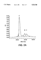
FIG. 2A is the elution profile from a first C18 column; where non-covalently linked mKL (KL-NC) elutes at approximately 38% n-propanol, KL-CD elutes at approximately 45% n-propanol, and a third peak containing a different form of KL-CD with very low activity elutes after the biologically active KL-CD peak.
FIG. 2B is a photograph of SDS PAGE under reducing and non-reducing conditions of KL-CD and KL-NC eluted from the C18 column shown in FIG. 2A.
FIG. 2C is a graph of KL bioactivity, CPM tritium incorporated versus nL fraction shown in FIG. 2A added, for peak A (dark squares), peak B (open squares), and peak C (dark diamonds) of FIG. 2A, as measured by the ability to promote proliferation of the cell line MO7e.
FIG. 3A is a photograph of SDS-PAGE of KL-CD and KL-NC refolded from KL-NC in the presence or absence of glutathione.
FIG. 3B is a chromatogram of a C18 reverse phase HPLC separation of refolded material as shown in FIG. 3A.
FIG. 3C is a graph of KL bioactivity of fraction from the chromatogram shown in FIG. 3b.
FIG. 4 is a graph of KL bioactivity showing proliferation of MO7e in response to purified KL-NC (open squares), KL-CD (dark diamonds), and human KL (dark squares).
FIG. 5 is a graph of mast cell activation (% release) by purified KL-NC (open squares) and KL-CD (dark squares) in the presence of anti-TNP and TNP-BSA.
FIG. 6 is a graph of enhancement of mast cell degranulation (percent release above antigen alone) versus time (minutes) for KL.
FIG. 7 is a bar graph of the desensitization of mast cell degranulation using antigen (dark bars) alone or after prior exposure to KL (open bars).
DETAILED DESCRIPTION OF THE INVENTION
As described herein, it has been discovered that it is possible to prepare a covalently crosslinked dimer of kit ligand, which differs from native KL in its relative ability to stimulate cell proliferation and mast cell degranulation. The modified KL is referred to herein as KL-CD, for KL-covalent dimer. Non-covalently linked KL is referred to herein as KL-NC.
Nucleotide and Amino Acid Sequences of Kit Ligand
The nucleotide sequence for murine kit ligand is shown in Sequence ID No.5; the corresponding amino acid sequence is shown in Sequence ID No. 6. The nucleotide sequence for human kit ligand is shown in Sequence ID No. 1; the corresponding human amino acid sequence is shown in Sequence ID No. 2.
There is appreciable conservation at the primary sequence level among KL from the different species, in particular in the number and location of the cysteine residues, as shown by FIG. 1. Human (Sequence ID No. 2), murine (Sequence ID No. 6) and rat (Sequence ID No. 4) molecules are highly conserved at the amino acid level, with 79% 80% and 92% identity between human and mouse, human and rat, and mouse and rat, respectively. Furthermore, the number and location of the cysteines are absolutely conserved. Accordingly, the results shown in the examples for murine KL-CD may be extrapolated to human as well as other mammalian species of KL.
It is not necessary, and in fact is not preferred, to utilize the nucleotide sequence encoding the full length KL; in a preferred embodiment, the sequence encodes at least the first 138 amino acids, more preferably the first 162, 164, or 165 amino acids (minus the hydrophobic membrane binding region). Conservative substitutions, additions, and deletions based on differences in amino acid sequence using sequence alignment, as well as based on similarities in structure, charge, and chemistry, can be made to yield a functionally equivalent KL, referred to herein as KL, unless specifically noted otherwise.
Additional cysteines can be inserted in locations between the cysteines in the naturally occurring cysteines in order to form additional or alternative interchain covalent linkages, as described below.
Methods to make Covalently linked KL Dimers
In the preferred embodiment, dimers are made by expressing KL from a recombinant nucleotide sequence encoding KL which includes cysteine residues at positions corresponding to at least 4, 43, 89, and 138, denaturing the KL under conditions equivalent to between 4 and 12 M urea, most preferably 6 M urea, then refolding the proteins under conditions wherein the proteins form a covalently associated dimer, for example, in Tris™-HCl or phosphate buffered saline (PBS). Other modifications yielding KL-CD with altered biological activity can be produced as described below.
1. Formation of KL-CD having at least one interchain disulfide bond in place of at least one of the intrachain disulfide.
A preferred form of KL-CD described in Example 1 of this application contains at least one intermolecular disulfide bond in place of at least one of the intramolecular disulfide bonds found in KL-NC. This form of KL-CD has been demonstrated to be ten fold more potent than KL-NC in its ability to support the proliferation of a number of different types of cells. However, the mast cell priming/activating property of KL is not increased in the KL-CD molecule. This differential increase in growth stimulation in contrast to mast cell priming-activation of KL-CD is of utmost importance since the KL-induced anaphylaxis is presumably due to its action on mast cells. With native KL and KL-NC, ten fold more KL is required to stimulate maximal cell growth than is required to maximally prime mast cells for activation. In comparison, KL-CD is equipotent in its ability to stimulate cell growth as well as to prime mast cells for activation. The P:A ratio for KL-CD is thus ten fold more favorable than the P:A ratio of KL-NC.
KL-CD is also significantly more stable than KL-NC. The increased stability of KL-CD is particularly apparent at low concentrations, between 1 and 100 ng/ml, when incubated at 37° C. for several days to weeks. At these concentrations, significant loss in activity is observed for the recombinant KL-NC.
2. Deletion of one of the four cysteines in naturally occurring KL
A mutant KL dimer could also be formed as described above, where there is only one intrachain disulfide bond, by deletion of one of the cysteine residues not required for intrachain disulfide bond in KL-CD nor essential for biological activity.
Theoretically, the biologically active form of KL-CD, as described in Example 1, might consist of one intramolecular disulfide bond, one intermolecular disulfide, and one unpaired cysteine in each KL monomer. In this case, the cysteine residue not involved in a disulfide bond can be changed to another amino acid such as serine. Construction of this mutant cDNA could facilitate the formation of KL-CD with the same properties as the KL-CD described in Example 1. Since only one intramolecular disulfide bond could form from such a mutation, this mutation could result in a much greater yield of active KL-CD as compared to that in Example 1.
Specifically, covalent dimers of KL with desirable biological properties can be formed by the substitution of one of the other four cysteines (4, 43, 89 and 138), most preferably 43 or 138, with another amino acid, preferably serine. KL with any three of the four cysteines could fold into KL-CD with similar or different properties to that formed from KL with four cysteines.
3. Addition of one or more cysteines to naturally occurring KL
One or more cysteines can be added to KL to allow formation of additional intrachain or interchain disulfide bonds. For example, a fifth or additional cysteine(s) can be introduced into the cDNA to facilitate the formation of interchain disulfide bonds in addition to the two native intrachain disulfides. This interchain disulfide can be placed within a region of KL analogous to that of M-CSF. M-CSF contains an interchain disulfide formed between the cysteines at amino acid 31 in the two monomers, which is within the region where the two monomers are juxtaposed. Thus, mutation of an amino acid residue to cysteine within amino acids 18 to 30 of KL would be expected to generate a form of KL-CD with properties similar to that of the KL-CD described in example 1. More specifically, an additional amino acid such as another cysteine can be introduced between residues 25 and 26 since these residues must be interrupted by a single amino acid "space" in order to align the sequences of KL and M-CSF. (Bazan, F. (1991) Cell 65, 9-10). Alternatively, a location for an additional cysteine designed to yield an intermolecular disulfide bond can be determined through the elucidation of the disulfide pairs in the KL-CD described in Example 1.
4. Formation of KL fusion protein dimers
Covalent dimers or higher order multimers of KL with increased biological activity can be produced through the fusion of KL with heterologous proteins which contain covalent interactions with multiple subunits. An example of this is with immunoglobulin (Ig) Fc domain fusion proteins which have been used for the expression of a number of proteins as dimeric molecules, as described by Lindsley, P. S., et al. (1991) J. Exp. Med. 174, 561-569. In the Lindsley Ig fusion constructs, the gene encoding the protein of interest (CTLA-4) was fused to nucleotide sequences encoding the hinge, CH2 and CH3 domains of human Ig Cγ1. The resulting fusion protein formed soluble dimers and tetramers, which were disulfide linked through cysteines located in the CTLA-4 region. Those fusion proteins demonstrated CTLA-4 activity.
KL fusion proteins may also be generated for use in ex vivo cell culture, where the KL fusion proteins are immobilized to a solid substrate. This can be accomplished through the use of KL-Fc fusion proteins bound to Protein A beads. This can also be accomplished by the addition of a collagen binding domain to KL directly or via an Fc bridge so that KL can be coupled to collagen beads or coated substrates.
Methods for making soluble dimeric protein which is expressed on the host cell surface as a chimeric fusion protein incorporating the extracellular portion of the protein with the stem region of C4b binding protein (C4bp) are described in U.S. Ser. No. 08/118,366 filed Aug. 8, 1993,
In these constructs, the extracellular domain of CD28, a cell surface dimer, is fused to C-terminal 58 amino acids of C4 binding protein. When these constructs are expressed in mammalian cells, a multimeric CD28-C4bp protein is expressed on the cell surface. The protein can be cleaved from the multimeric surface protein to yield soluble protein. A dimeric protein can also be produced by expression of a plasmid vector incorporating the segments of the gene encoding placental alkaline phosphatase (PAP) adjacent to sequence encoding the extracellular region of the KL cDNA amino acids 1-164 or 165, and a lipid anchor, the cleavage site for a phospholipase, as described in PCT/US92/01867. In the constructs described in the '867 application the extracellular domain of CD28 is fused to the C-terminal 49 amino acids of PAP. When this construct is expressed, the C-terminal 30 amino acids of the PAP portion of the fusion protein are cleaved, leaving a C-terminal arginine residue. That arginine is then available for the addition of phosphatidylinositol glycan (pI-G). The pI-G acts as an anchor to hold the fusion protein to the cell membrane. Subsequent cleavage with phospholipase C releases soluble, dimeric CD28.
Stabilized dimers with increased biological activity can also be produced through non-covalent means by the fusion with domains that readily form stable hetero- or homomeric multimers. An example would be to use the so called "Leucine zipper" domain which will self associate with another protein that contains a Leucine zipper domain.
4. Formation of Chemically coupled KL dimers.
Chemical methods, not involving peptide or disulfide bond formation, which form a covalent bond between monomers may also be used to make KL dimers. Methods using a variety of commercially available bifunctional reagents that are available which crosslink proteins, for example, via free amino groups, can be utilized. The reagent DSS from Pierce Chemical Co., would be suitable for this purpose and its use is well known to those skilled in the art. Alternatively, the reagent BASED (Pierce) is a photoreactive crosslinker which reacts non-specifically and could be useful for crosslinking near the dimer interface of KL-NC.
Expression and isolation of KL-CD
KL-CD can be obtained by expression of the nucleotide sequence encoding KL in an appropriate procaryotic or eucaryotic expression system, for example, E. coli, followed by unfolding in urea and refolding in a more physiological buffer.
The formation of KL-CD through expression in mammalian and other eukaryotic species is demonstrated in Example 1. The protein can also be expressed in mammalian, yeast or insect cells, then purified and subsequently denatured and refolded to facilitate intermolecular disulfide bond formation. In some cases it may be desirable to remove sugars using endoglycosidases and other enzymes to cleave sugars that interfere with intra- and/or interchain disulfide formation.
KL-CD can be expressed in prokaryotic as well as eukaryotic expression systems. The following are examples of expression vectors which may be used in procaryotic systems:
The pPL expression series use the strong PL promoter of lambda phage, and can be expressed in a number of procaryotic expression systems (Reed, Cell, 25, 713-719 (1981), Simatake and Rosenberg, Nature, 292, 128-132 (1981), Mott, et al., Proc. Natl. Acad. Sci. USA, 82, 88-92 (1985)).
The pOX expression series, which uses the oxygen-dependent promoter of vireoscilia hemoglobin gene, is expressed in E. coli (Khosla, et al., BioTechnology 8, 554-558 (1990)).
pKK223-3 uses a hybrid promoter derived from the fusion between the promoters of the tryptophan and lactose operons of E. coli (Brosius and Holy, Proc. Natl. Acad. Sci. USA 81 6929-6933 (1984)).
The following are examples of expression vectors which may be used for expression in a eukaryotic expression system:
pMSG uses the promoter from the mouse mammary tumor virus long terminal repeat (MMTV). Suitable host cells for pMSG are Chinese hamster ovary cell, Hela cell and mouse Lkt negative cells (Lee, F., et al., Nature 294, 28-232 (1981)).
pSVL uses the SV40 late promoter. Suitable host cells are COS cells for high level transient expression (Sprague, et al., J. Virol. 45, 773-781 (1983); Gempleton and Eckhart, Mol. Cell. Biol. 4, 817-821 (1984)).
pRSV uses Rous Sarcoma Virus promoter. Suitable host cells are mouse fibroblast cells, lymphoblastoid cells and COS cells (Gorman, et al. Science 221, 551-553 (1983)).
pBPV is a DNA viral vector derived from bovine papilloma virus. It is stably expressed in mouse mammary tumor cells, C127 (Zin, et al., i Cell 34, 865-879 (1983); Saraver, et al., Mol. Cell. Biol. 1, 486-496 (1981)); Saraver, et al.,Proc. Natl. Acad. Sci., USA 79, 7147-7151 (1982); Law, et al., Mol. Cell. Biol. 3, 2110-2115 (1983)).
Baculovirus expression vectors are stably expressed in insect cells such as Sf9 (Luckow and Summers, BioTechnology, 6, 47-55 (1988); Miller, L. K., Ann. Rev. Microbiology 42, 177-199 (1988)).
Methods for making transgenic animals are well known. DNA encoding the KL can be introduced into the cells in culture using transfection or into embryos for production of transgenic animals expressing the KLs. As known in the art, transfection can be accomplished by electroporation, calcium phosphate precipitation, a lipofectin-based procedure, or microinjection or through use of a "gene gun". In each case, cDNA encoding the KL is subcloned into a plasmid-based vector which encodes elements for efficient expression in the genetically engineered cell. The plasmid-based vector preferably contains a marker such as the neomycin gene for selection of stable transfectants with the cytotoxic aminoglycoside G418 in eukaryotic cells and an ampicillin gene for plasmid selection in bacteria. In the preferred embodiment, the KL is expressed in soluble form; in the most preferred embodiment, the KL is expressed using a tissue specific protein such as the casein promoter, to avoid potential side effects and to increase recoverable yields.
Infection, which for endothelial cells is preferred, is accomplished by incorporating the genetic sequence for the KL into a retroviral vector. Various procedures are known in the art for such incorporation. One such procedure which has been widely used in the art employs a defective murine retrovirus, Psi-2 cells for packaging the retrovirus, and the amphotropic packaging cell line Psi-AM to prepare infectious amphotropic virus for use in infecting the target donor cells, as described by Kohn et al., 1987 "Retroviral-mediated gene transfer into mammalian cells" Blood Cells 13:285-298. Alternatively, rather than a defective Moloney murine retrovirus, a retrovirus of the self-inactivating and double-copy type can be used, such as that described by Hantzopoulos et al., 1989 "Improved gene expression upon transfer of the adenosine deaminase minigene outside the transcriptional unit of a retroviral vector" Proc. Natl. Acad. Sci. USA 86:3519-3523.
A variety of methods are known to those skilled in the art for making transgenic animals expressing a KL protein. Examples of particularly useful animals include mice, rats, rabbits, pigs, sheep, and cattle, all of which have been made transgenic using standard techniques. The most well known method for making a transgenic animal is by superovulation of a donor female, surgical removal of the egg and injection of the genetic material in the pronuclei of the embryo, as taught by U.S. Pat. No. 4,873,191 to Wagner, the teachings of which are incorporated herein. Another commonly used technique involves the genetic manipulation of embryonic stem cells (ES cells). ES cells are grown as described, for example, in Robertson, E. J. "Embryo-derived stem cell lines" in: Teratocarcinomas and embryonic stem cells: A practical approach. E. J. Robertson, ed. 71-112 (Oxford-Washington, D.C.: IRL Press, 1987). Genetic material is introduced into the embryonic stem cells, for example, by electroporation according to the method of McMahon, A. P., and Bradley, A. Cell 62, 1073-1085 (1991). Colonies are picked from day 6 to day 9 of selection into 96 or 24 well dishes (Costar) and expanded and used to isolate DNA for Southern blot analysis. Chimeric mice are generated as described in Bradley, "Production and analysis of chimaeric mice" in Teratocarcinomas and embryonic stem cells: A practical approach E. J. Robertson, ed. pp. 113-151 (Oxford, Washington, D.C. IRL Press 1987), the teachings of which are incorporated herein. Genetic material is injected into blastocysts. From those implanted females that become pregnant, chimaeras are selected from the offspring and bred to produce germline chimaeras for use as donor animals.
Properties of KL-CD
In Example 1, a truncated form of murine KL including amino acids 1 to 164 plus an additional N-terminal methionine required for synthesis in E. coli (amino acids 1 to 164 of Sequence ID No. 6) was expressed. This form corresponds to the natural soluble form of murine KL-1. In this method, KL is synthesized and accumulates within the bacteria in an insoluble form. KL-CD is obtained by solubilization of the protein with denaturant (urea), and refolding into biologically active protein by removal of the denaturant (by dialysis into buffer such as Tris™ HCl or PBS). During the refolding, both intrachain and interchain disulfide bonds are formed, resulting in two types of KL which can promote cell proliferation, KL-NC and KL-CD. In addition, a biologically inactive KL-CD is formed. The three forms of KL can be separated from one another and from contaminating E. coli proteins using a high resolution chromatography method such as C18 reverse-phase HPLC.
Expression of KL-CD, derived from KL-cDNAs with three cysteines or with greater than four cysteines in eukaryotic cells can be facilitated by expression of the full length KL cDNA (KL-1, Huang, et al., 1992) with the appropriate Cysteine mutation such that KL dimers will associate first in the membrane and then be released via cleavage at the native proteolytic cleavage site.
KL-NC presumably contains the two disulfide bonds normally found in KL. Since the KL-CD forms contain at least one intermolecular disulfide bond, they can have at most, only one of the intramolecular disulfides found in the native molecule. Presumably the biologically active and inactive forms of KL-CD consist of different combinations of intra- and intermolecular disulfide bonds.
Since native KL forms non-covalent dimers in solution, it was of interest to determine the stoichiometry of KL-CD in solution. When KL-CD and KL-NC are co-injected onto an HPLC gel filtration column, the proteins co-elute as a single peak of approximately 40 kDa, suggesting that both KL-CD and KL-NC are dimeric in solution.
Covalent dimers of KL-related proteins
The approaches outlined above for the formation of a covalent dimer of KL may also be applied to the formation of covalent dimers of other non-disulfide linked multimeric proteins. In particular, the recently characterized ligand of the FLT-3/FLK-2 receptor is predicted to be structurally similar to KL based on sequence similarity, and like KL, forms non-covalent dimers in solution. The amino acid sequence of the murine FLT-3 ligand is shown as Sequence ID No. 7, as reported by Lyman, et al., (1993) Cell 1157-1167. The full length cDNA can be expressed in eukaryotic cells with vectors specified and soluble protein recovered after proteolytic cleavage via the endogenous protease in CV-1 cells. Alternatively, a soluble form of Flt-3L can be isolated from eukaryotic or prokaryotic cells by expression of a fragment of the cDNA, for example, from amino acid one to 135 or one to 163. Cysteines at positions 119, 124, and 130 can also be replaced by other amino acids, preferably serine. Other modifications such as additional cysteines, in the same region as specified for KL as well as fusion proteins can also be used to produce disulfide linked FLT-3L.
Formation of covalent dimers of the Flt-3 ligand are expected to have desirable biological properties similar to that of KL, including increased potency in stimulating proliferation of bone marrow subpopulations, and increased stability. Biologically active, disulfide-linked covalent dimers of FLT-3L may be obtained more easily with the human form which contains six cysteine residues, rather than with the mouse form, which contains nine cysteines. As with KL, covalent dimers of FLT-3L may be formed by denaturation and refolding, through the addition of cysteines in the region of amino acid 31, via fusion proteins, or by chemical crosslinking means.
Biological Activities and Applications of KL-CD
Native KL has multiple biological activities, affecting the growth and differentiation of a variety of hematopoietic cells, as well as the activation of mast cells. While the mast cell activating property of native KL limits its utility as a therapeutic, KL-CD has properties which make it useful for applications that were originally intended for native KL. As described in Example 3, murine KL-CD is at least ten-fold more potent than murine KL-NC as well as human KL-NC in stimulating the proliferation of two different human cell lines, and ten-fold more potent than murine KL-NC in stimulating the proliferation of murine mast cells. However, KL-CD is only equipotent to that of murine KL-NC in priming mast cells for IgE-dependent degranulation.
The selectivity of KL-CD for promoting cell proliferation but not mast cell degranulation, may make the disulfide-linked form particularly useful as a therapeutic since dosages may be set which promote a desired proliferation event but which avoid mast cell degranulation-induced anaphylaxis. For example, since KL-CD is ten-fold more potent than KL-NC in promoting cell proliferation, a dose of 10 μg/kg/day of KL-CD should be as effective as a 100 μg/kg/day dose of KL-NC, a dose which stimulated significant hematopoietic recovery. Since KL-CD is equipotent to KL-NC in promoting mast cell degranulation, a dose of 10 μg/kg/day of KL-CD is below the dose of 25 μg/kg/day which resulted in mast cell-related side effects in some patients, and well below the dose of KL-NC of 100 μg/kg/day which resulted in serious mast cell-related effects in many patients.
KL-CD can be used to stimulate hematopoietic recovery following chemo/radiotherapy or bone marrow (hematopoietic cell) transplantation, as previously described in the literature, and reviewed in the Background of the Invention. This may be accomplished with KL used as a single agent or in combination with other cytokines, such as G-CSF or GM-CSF for neutrophil recovery, or IL-6 or other factors that promote platelet recovery. KL-CD may also be more effective in treating certain anemias such as those associated with Diamond Blackfan Syndrome, those induced by chemo or radiotherapy or viral infections, or aplastic anemia. The dimer will also be useful in the mobilization of stem cells from the bone marrow to the peripheral blood alone and or in combination with other cytokines, such as G-CSF, or chemotherapy. Since KL-CD would be used at the same dose as KL-NC, which is limited by its toxicity, KL-CD should be significantly more effective than KL-NC in the aforementioned applications, due to its enhanced potency in promoting cell proliferation.
While short-term exposures of mast cells in culture to KL results in mast cell priming, i.e. in enhanced IgE-dependent mast cell degranulation, prolonged exposure of these cells to KL results in a desensitization of the priming effect. This suggests that patients could be desensitized to the mast cell activating effects of KL by treatment with a level of KL-CD below the toxicity level. A subsequent treatment of high level KL-CD or KL-NC might then provide enhanced hematopoietic recovery without causing mast cell associated toxicity. The level of hematopoietic recovery might be greater than that observed for KL-CD used at a level below the toxicity level. In summary, KL-CD or KL-NC should be useful in a desensitization protocol to establish a higher toxicity level for KL.
KL-CD should also have utility for ex vivo applications. Although the differential proliferative/mast cell activating property of KL-CD is less important for ex vivo uses, its increased biological activity make it useful in the culture of hematopoietic cells. KL is effective by itself or preferably in combination with other cytokines, such as IL-1, IL-3, IL-6, IL-11, G-CSF, GM-CSF, LIF, FLT-3L and combinations thereof, for the ex vivo expansion of stem cells and progenitors for transplantation. The ability of KL-CD to stimulate the proliferation of immature stem/progenitor cells makes KL particularly useful in protocols involving the transduction of genes into hematopoietic cells for gene therapy. KL-CD could be used at lower dosages relative to KL-NC for these ex vivo applications. KL-CD might also result in qualitative differences in hematopoietic cell expansion compared to KL-NC, perhaps resulting in the selective expansion of a certain type of progenitor cell.
The greater stability of KL-CD relative to KL-NC may enhance the utility of KL-CD in ex vivo applications. KL-NC exhibits properties in vitro which suggests an inherent instability of the molecule, perhaps due to the dissociation of the dimer into monomers at lower protein concentrations, or to internalization and degradation of the molecule by the responding cells. This is illustrated by repeated daily feeding of mast cell cultures with KL which gives significantly better growth than two to three times a week feeding. It may be possible to use the covalently-linked KL dimer to overcome this apparent instability, and allow one to use a significantly lower concentration of soluble KL-CD to support long term cell cultures.
In summary, KL-CD can be utilized as an additive to cell culture media as extrapolated from the published data relating to KL, or in combination with a pharmaceutically acceptable carrier for administration to a patient. Exemplary pharmaceutical carriers include diluents such as saline and phosphate buffered saline, additives such as preservatives, detergents, solubilizing agents, anti-oxidants, pH buffers, and salts, as well as alternative carrier forms such as polymers, liposomes, micelies, and vesicles. These are administered to a patient in an amount effective to produce an improvement in a particular condition, for example, to increase platelet numbers. Treatment may be alone or in combination with other compounds demonstrated to have hematopoietic activity, including erythropoietin, G-CSF, GM-CSF, interleukins 1-11, IGF-I FLT-3 ligand or LIF.
EXAMPLE 1
Purification and Characterization of Covalent Dimer-Kit Ligand (KL-CD) from Native KL Sequence.
A truncated mouse KL cDNA containing amino acids 1 to 164 (amino acids 1 to 164 of Sequence ID No. 6) was subcloned from the full length cDNA. The endpoint of this truncated cDNA was chosen based on the site of proteolytic cleavage in the native transmembrane form of the molecule which gives rise to the soluble form (Huang et al., 1992). The truncated KL cDNA was cloned into an expression vector, placing it under control of the phage lambda early gene promotor PL. (Lambda II, Hendrix, Roberts, Stahl, Weisberg, editors. Cold Spring Harbor Laboratory, Cold Spring Harbor, N.Y. (1983)). This promoter is regulated in a temperature-sensitive manner by a mutant phage lambda CI gene which is also present on the expression vector. The truncated cDNA was expressed in E. coli strain DH5-a (BRL-GIBCO). As is typical for high level expression in E. coli, the truncated KL accumulated in an insoluble form in inclusion bodies in the bacteria.
A two liter culture of E. coli expressing KL was harvested, the cells were lysed by sonication, and the inclusion bodies containing insoluble KL were isolated by centrifugation at 10,000 ×g. The inclusion bodies were washed by resuspension in 20 mM Tris HC1, pH 7.4 200 mM NaCl 1 mM EDTA and re-centrifuged. The inclusion bodies were solubilized by incubation in 6 M urea at 4° C. for 1 h, followed by centrifugation to remove insoluble material. After solubilization of inclusion bodies containing mKL, the protein was dialyzed against 20 mM Tris pH 8.0 at 4° C. for 48 to 72 h. Insoluble material was removed by centrifugation at 10,000 ×g, and the protein was applied to a Vydak™ C18 1×25 cm HPLC column that had been equilibrated with 0.1 M ammonium acetate pH 6.0 and 25% n-propanol. The column was washed with equilibration buffer, and then eluted with a linear gradient from 30-50% n-propanol, 0.1 M ammonium acetate pH 6.0.
mKL bioactivity, as measured by the ability to promote proliferation of the cell line MO7e described in Example 2, elutes in two peaks; non-covalently linked mKL (KL-NC) elutes at approximately 38% n-propanol, KL-CD elutes at approximately 45% n-propanol, as shown in FIG. 2A. A third peak containing a different form of KL-CD with very low activity elutes after the biologically active KL-CD peak, as also shown in FIG. 2A.
The KL-NC and KL-CD peaks were purified to homogeneity by re-application to the C18 column, and elution with narrower gradients. KL-NC was purified using a gradient from 32-45% n-propanol. The active and inactive KL-CD forms were purified using a 2 h gradient of 35-45% n-propanol. After the second C18 column, the NC and CD forms were in a highly purified state, and contained low levels of E. coli -derived endotoxin (less than 1 E.U. per mg protein as assayed by the BioWhittaker Inc. Amebocyte Lysate Assay). Prepared through these means, approximately 15% of the mKL refolds into active KL-CD, 15% into inactive KL-CD, and 70% into KL-NC.
The difference between KL-NC and the two KL-CD forms can be seen not only by their different retention times on the C18 column, but by SDS-PAGE under reducing/non-reducing conditions, as shown in FIG. 2B. Under reduced conditions, KL-NC as well as the two forms of KL-CD migrate with an apparent molecular weight of about 18 kDa. Under non-reduced conditions, KL-NC migrates with an apparent molecular weight of about 18 kDa, while the two different forms of KL-CD migrate with an apparent molecular weight of 36 kDa. As assessed by SDS-PAGE under non-reducing conditions, the active form of KL-CD has a slightly greater apparent molecular weight than the inactive form of KL-CD. The higher apparent molecular weight of KL-CD as compared with KL-NC under non-reducing conditions is indicative of the covalent linkage of two KL monomers via at least one disulfide bond.
The nature of KL-CD and KL-NC has been confirmed by Laser Desorption/Time of Flight Mass Spectrometry. By this method, KL-NC has a mass of 18,440 daltons. Both active and inactive KL-CD had a mass of 36,860 daltons; these forms apparently differ only in their disulfide bonds, with inactive KL-CD containing a disulfide bond arrangement which greatly diminishes activity.
EXAMPLE 2
Formation of KL-CD from KL-NC via disulfide rearrangement.
KL-CD can also be derived from pure KL-NC through a non-enzymatic reaction involving the rearrangement of disulfide bonds. The reaction consists of pure, correctly folded KL-NC at 1 mg/ml, 50 mM Tris pH 9.0, 2 M guanidine-HCl (added to partially unfold the KL-NC), and reduced and oxidized forms of glutathione (500 μM and 125 μM final concentration, respectively). The reaction mixture was incubated for 20 h at 22° C., and then dialyzed against 0.1 M ammonium acetate at 4° C. to remove the guanidine to allow folding and to stop disulfide exchange. The rearrangement reaction was monitored by SDS-PAGE under non-reducing conditions.
Proteins with molecular weights of the active and inactive forms of KL-CD were formed via rearrangement of KL-NC disulfides. This rearrangement required the presence of 2 M guanidine-HCl. Additionally, several other KL species were formed, which might be inactive forms of KL-CD. The mixture of proteins resulting from disulfide rearrangement of KL-NC was purified by C18 reverse phase chromatography as in Example 1.
FIG. 3A is a photograph of SDS-PAGE of KL-CD and KL-NC refolded from KL-NC in the presence or absence of glutathione. FIG. 3B is a chromatogram of a C18 reverse phase HPLC separation of refolded material as shown in FIG. 3A. FIG. 3C is a graph of KL bioactivity of fractions from the chromatogram shown in FIG. 3b.
Two peaks of biologically active KL were identified; the second peak consisted of KL-CD which migrates with the same apparent molecular weight of KL-CD purified as in Example 1 in SDS-PAGE under reducing conditions. Although much greater amounts of KL-NC were present after disulfide rearrangement, C18 fractions containing KL-NC or KL-CD had comparable activity in promoting cell proliferation, as shown by FIG. 4. This shows that KL-CD with increased biological activity can be formed from KL-NC through disulfide rearrangement.
Disulfide rearrangement conditions can be established which maximize KL-CD formation. It may be preferable however to purify recombinant KL in a completely unfolded state by C18 reverse-phase HPLC, and then fold the protein into CD- and NC- KL forms using the disulfide-rearrangement conditions described above.
EXAMPLE 3
Biological activity of KL-CD in in vitro biological assays.
a. Cell proliferation.
KL supports the proliferation of a variety of growth factor dependent cell lines. Murine KL is equally potent on both human and murine cells, while human KL is active on human cells but shows minimal activity on murine cells. The human megakaryocytic cell line M07e, which is maintained in the presence of GM-CSF, is used to assess human and murine KL. Murine bone marrow-derived mast cells (BMMC), which are established and then maintained for up to three months in the presence of IL-3 (Yung, Y. P., et al. (1982) J. Immunol., 129, 1256-1261), are also used to assess the activity of murine KL (Nocka, K., et al. (1990).
The cells are washed and resuspended in media lacking their maintenance growth factor and plated into 96 well plates. Column fractions of KL samples are added and serially diluted and the cells are incubated at 37° C. for 24 h. The cells are then pulsed with 2.5 μCi/ml of .sup.[3] H-Thymidine for 6-12 hr, harvested onto glass fiber filters, and the amount of 3 H-thymidine on the filter is determined on a Packard TopCount™ scintillation counter. The data is analyzed by plotting the CPM 3 H incorporated into DNA versus the concentration of KL.
b. Mast Cell Priming Activity
Primary cultures of murine mast cells derived from bone marrow and cultured in IL-3 (BMMC) can be utilized not only for proliferation assays as described above, but may also be used as a quantitative and sensitive measure of the priming or activation potential of cytokines. With human mast cells, KL is the most potent cytokine identified to date with significant mast cell priming activity in vitro (Bischoff and Dahinden (1992) J. Exp. Med., 175, 237-244). Murine BMMC sensitized with IgE immunoreactive with trinitrophenol (TNP) (ascites from IGELa2, ATCC # TIB 142) can be primed by KL such that when stimulated with specific antigen (TNP-BSA), exhibit a significant increase in the release of mediators compared to unprimed cells. When BMMC derived from the C57/B16×DBA2 F1 (BDF1) strain of mice are activated at low cell density (1×10⁵ cells/ml) in physiological buffer, low levels of proinflammatory mediators and secretory granule enzymes, typically 10-25% of their granule hexosaminidase, are released upon stimulation with IgE and antigen alone. Following a short priming period with KL (0 to 10 minutes), maximal granule enzyme release in the range of 40 to 60% enzyme release is observed. FIG. 5 is a graph of mast cell activation by purified KL-NC and KL-CD as a function of concentration. The mast cell priming activity of native KL has an ED50 of 0.5 to 1 ng/ml in this assay.
EXAMPLE 4
Desensitization of mast cells to a KL response.
Human lung and murine bone marrow derived mast cells respond to the exposure of various priming agents with very rapid kinetics, as described by Bischoff and Dahinden (1992). The priming assay as specified in Example 3 is typically carried out with a priming period of 5 to 10 minutes followed by the addition of antigen for a further period of 10 minutes. As shown below, if the antigen is withheld for 30 minutes or longer, the priming affect of KL is lost and the level of degranulation is similar to that seen with antigen alone. Furthermore, BMMCs can no longer respond to a second treatment with KL when they have already been desensitized. Once the effect of Kl is lost, mast cells cannot respond to a second dose of KL within a one to two hour period. This desensitization could be used therefore to minimize a response to a subsequent therapeutic dose of KL.
Kinetics of Priming with KL
BMMCs which had been previously sensitized with IgE (anti-TNP) were incubated with control diluent or KL for various periods of time (0, 2, 5, 7, 10, 20, 30, 40, 50, and 60 minutes) and then activated with antigen (TNP-BSA). Percent release of hexoseaminidase was determined ten minutes after the addition of antigen.
Exposure of cells to KL for up to ten minutes prior to antigen resulted in maximal activation, as shown by FIG. 6. With exposure of cells to KL for 20 minutes and longer, no significant release was observed above that seen with antigen alone.
Desensitization of mast cells with KL
Sensitized BMMCs were incubated in the presence or absence of KL for 45 minutes (1st phase). Following this incubation period, cells were washed and BMMCs were primed with either control diluent, or KL for 10 minutes (2nd phase). Cells were then activated by the addition of antigen for a further 10 minutes. Cells that had been cultured in the control medium for the 45 minute period responded to antigen and exhibited a significant enhancement when treated with KL as the second agent and then antigen. However, cells that had been pretreated with KL were only activated to the level seen with antigen alone. Secondary stimulation with KL did not lead to enhanced degranulation, as shown by FIG. 7.
Modifications and variations of the present invention will be obvious to those skilled in the art from the foregoing detailed description. Such modifications and variations are intended to come within the scope of the appended claims.
__________________________________________________________________________
SEQUENCE LISTING
(1) GENERAL INFORMATION:
(iii) NUMBER OF SEQUENCES: 7
(2) INFORMATION FOR SEQ ID NO:1:
(i) SEQUENCE CHARACTERISTICS:
(A) LENGTH: 838 base pairs
(B) TYPE: nucleic acid
(C) STRANDEDNESS: double
(D) TOPOLOGY: linear
(ii) MOLECULE TYPE: cDNA
(iii) HYPOTHETICAL: NO
 (iv) ANTI-SENSE: NO
(ix) FEATURE:
(A) NAME/KEY: CDS
(B) LOCATION: 17..835
(ix) FEATURE:
(A) NAME/KEY: sig.sub.-- peptide
(B) LOCATION: 17..91
(ix) FEATURE:
(A) NAME/KEY: mat.sub.-- peptide
(B) LOCATION: 92..835
(x) PUBLICATION INFORMATION:
(A) AUTHORS: Martin, et al.
 (C) JOURNAL: Cell
(D) VOLUME: 63
(F) PAGES: 203-211
(G) DATE: 1990
(K) RELEVANT RESIDUES IN SEQ ID NO:1: FROM 1 TO 273
(xi) SEQUENCE DESCRIPTION: SEQ ID NO:1:
AGCGCTGCCTTTCCTTATGAAGAAGACACAAACTTGGATTCTCACTTGC49
 MetLysLysThrGlnThrTrpIleLeuThrCys
-25-20-15
ATTTATCTTCAGCTGCTCCTATTTAATCCTCTCGTCAAAACTGAAGGG97
Il eTyrLeuGlnLeuLeuLeuPheAsnProLeuValLysThrGluGly
-10-51
ATCTGCAGGAATCGTGTGACTAATAATGTAAAAGACGTCACTAAATTG145
IleCysArgAsnArgValThrAsnAsnValLysAspValThrLysLeu
51015
GTGGCAAATCTTCCAAAAGACTACATGATAACCCTCAAATATGTCCCC193
V alAlaAsnLeuProLysAspTyrMetIleThrLeuLysTyrValPro
202530
GGGATGGATGTTTTGCCAAGTCATTGTTGGATAAGCGAGATGGTAGTA241
GlyMetA spValLeuProSerHisCysTrpIleSerGluMetValVal
35404550
CAATTGTCAGACAGCTTGACTGATCTTCTGGACAAGTTTTCAAATATT289
G lnLeuSerAspSerLeuThrAspLeuLeuAspLysPheSerAsnIle
556065
TCTGAAGGCTTGAGTAATTATTCCATCATAGACAAACTTGTGAATATA337
SerGluGlyLeuSerAsnTyrSerIleIleAspLysLeuValAsnIle
707580
GTGGATGACCTTGTGGAGTGCGTGAAAGAAAACTCATCTAAGGATCTA385
ValAspAspLeuValGluCysValLysGluAsnSerSerLysAspLeu
859095
AAAAAATCATTCAAGAGCCCAGAACCCAGGCTCTTTACTCCTGAAGAA433
L ysLysSerPheLysSerProGluProArgLeuPheThrProGluGlu
100105110
TTCTTTAGAATTTTTAATAGATCCATTGATGCCTTCAAGGACTTTGTA481
PhePheA rgIlePheAsnArgSerIleAspAlaPheLysAspPheVal
115120125130
GTGGCATCTGAAACTAGTGATTGTGTGGTTTCTTCAACATTAAGTCCT529
V alAlaSerGluThrSerAspCysValValSerSerThrLeuSerPro
135140145
GAGAAAGATTCCAGAGTCAGTGTCACAAAACCATTTATGTTACCCCCT577
GluLysAspSerArgValSerValThrLysProPheMetLeuProPro
150155160
GTTGCAGCCAGCTCCCTTAGGAATGACAGCAGTAGCAGTAATAGGAAG625
ValAlaAlaSerSerLeuArgAsnAspSerSerSerSerAsnArgLys
165170175
GCCAAAAATCCCCCTGGAGACTCCAGCCTACACTGGGCAGCCATGGCA673
A laLysAsnProProGlyAspSerSerLeuHisTrpAlaAlaMetAla
180185190
TTGCCAGCATTGTTTTCTCTTATAATTGGCTTTGCTTTTGGAGCCTTA721
LeuProA laLeuPheSerLeuIleIleGlyPheAlaPheGlyAlaLeu
195200205210
TACTGGAAGAAGAGACAGCCAAGTCTTACAAGGGCAGTTGAAAATATA769
T yrTrpLysLysArgGlnProSerLeuThrArgAlaValGluAsnIle
215220225
CAAATTAATGAAGAGGATAATGAGATAAGTATGTTGCAAGAGAAAGAG817
GlnIleAsnGluGluAspAsnGluIleSerMetLeuGlnGluLysGlu
230235240
AGAGAGTTTCAAGAAGTGTAA838
ArgGluPheGlnGluVal
245
(2) INFORMATION FOR SEQ ID NO:2:
(i) SEQUENCE CHARACTERISTICS:
(A) LENGTH: 273 amino acids
(B) TYPE: amino acid
(D) TOPOLOGY: linear
(ii) MOLECULE TYPE: protein
(ix) FEATURE:
(A) NAME/KEY: cleavage site
(B) LOCATION: 164..165
(xi ) SEQUENCE DESCRIPTION: SEQ ID NO:2:
MetLysLysThrGlnThrTrpIleLeuThrCysIleTyrLeuGlnLeu
-25-20-15-10
LeuLeuPheAsnProLeuValLysThrGluGlyIleCysA rgAsnArg
-515
ValThrAsnAsnValLysAspValThrLysLeuValAlaAsnLeuPro
101520
LysAs pTyrMetIleThrLeuLysTyrValProGlyMetAspValLeu
253035
ProSerHisCysTrpIleSerGluMetValValGlnLeuSerAspSer
4045 5055
LeuThrAspLeuLeuAspLysPheSerAsnIleSerGluGlyLeuSer
606570
AsnTyrSerIleIleAspLys LeuValAsnIleValAspAspLeuVal
758085
GluCysValLysGluAsnSerSerLysAspLeuLysLysSerPheLys
9095 100
SerProGluProArgLeuPheThrProGluGluPhePheArgIlePhe
105110115
AsnArgSerIleAspAlaPheLysAspPheValValAlaSerGluThr
120125130135
SerAspCysValValSerSerThrLeuSerProGluLysAspSerArg
140145150
Va lSerValThrLysProPheMetLeuProProValAlaAlaSerSer
155160165
LeuArgAsnAspSerSerSerSerAsnArgLysAlaLysAsnProPro
170 175180
GlyAspSerSerLeuHisTrpAlaAlaMetAlaLeuProAlaLeuPhe
185190195
SerLeuIleIleGlyPheAlaPheGlyAla LeuTyrTrpLysLysArg
200205210215
GlnProSerLeuThrArgAlaValGluAsnIleGlnIleAsnGluGlu
220225 230
AspAsnGluIleSerMetLeuGlnGluLysGluArgGluPheGlnGlu
235240245
Val
(2) INFORMATION FOR SEQ ID NO:3:
(i) SEQUENCE CHARACTERISTICS:
(A) LENGTH: 630 base pairs
 (B) TYPE: nucleic acid
(C) STRANDEDNESS: double
(D) TOPOLOGY: linear
(ii) MOLECULE TYPE: cDNA
(iii) HYPOTHETICAL: NO
(iv) ANTI-SENSE: NO
(ix) FEATURE:
(A) NAME/KEY: CDS
(B) LOCATION: 26..628
(ix) FEATURE:
(A) NAME/KEY: sig.sub.-- peptide
(B) LOCATION: 26..100
 (ix) FEATURE:
(A) NAME/KEY: mat.sub.-- peptide
(B) LOCATION: 101..628
(x) PUBLICATION INFORMATION:
(H) DOCUMENT NUMBER: WO 91/05795 A1
(I) FILING DATE: 28-SEP-1990
(J) PUBLICATION DATE: 02-MAY- 1991
(K) RELEVANT RESIDUES IN SEQ ID NO:3: FROM 1 TO 630
(xi) SEQUENCE DESCRIPTION: SEQ ID NO:3:
CTGGATCGCAGCGCTGCCTTTCCTTAT GAAGAAGACACAAACTTGGATTATC52
MetLysLysThrGlnThrTrpIleIle
-25-20
ACTTGCATTTATCTTCAACTGCTC CTATTTAATCCTCTCGTCAAAACT100
ThrCysIleTyrLeuGlnLeuLeuLeuPheAsnProLeuValLysThr
-15-10-5
CAGGAGATCTGCAGGAATCCTGTGACTG ATAATGTAAAAGACATTACA148
GlnGluIleCysArgAsnProValThrAspAsnValLysAspIleThr
151015
AAACTGGTGGCGAATCTTCCAAATG ACTATATGATAACCCTCAACTAT196
LysLeuValAlaAsnLeuProAsnAspTyrMetIleThrLeuAsnTyr
202530
GTCGCCGGGATGGATGTTTTGCCTA GTCATTGTTGGTTACGAGATATG244
ValAlaGlyMetAspValLeuProSerHisCysTrpLeuArgAspMet
354045
GTAACACACTTATCAGTCAGCTTGACTA CTCTTCTGGACAAGTTTTCA292
ValThrHisLeuSerValSerLeuThrThrLeuLeuAspLysPheSer
505560
AATATTTCTGAAGGCTTGAGTAATTATTCCATCA TAGACAAACTTGGG340
AsnIleSerGluGlyLeuSerAsnTyrSerIleIleAspLysLeuGly
65707580
AAAATAGTGGATGACCTCGTGGCATGTA TGGAAGAAAATGCACCTAAG388
LysIleValAspAspLeuValAlaCysMetGluGluAsnAlaProLys
859095
AATGTAAAAGAATCACTGAAGAAGC CAGAAACTAGAAACTTTACTCCT436
AsnValLysGluSerLeuLysLysProGluThrArgAsnPheThrPro
100105110
GAAGAATTCTTTAGTATTTTCAATA GATCCATTGATGCCTTCAAGGAC484
GluGluPhePheSerIlePheAsnArgSerIleAspAlaPheLysAsp
115120125
TTCATGGTGGCATCTGACACTAGTGATT GTGTGCTCTCTTCAACATTA532
PheMetValAlaSerAspThrSerAspCysValLeuSerSerThrLeu
130135140
GGTCCTGAGAAAGATTCCAGAGTCAGTGTCACAA AACCATTTATGTTA580
GlyProGluLysAspSerArgValSerValThrLysProPheMetLeu
145150155160
CCCCCTGTTGCAGCCAGTTCCCTTAGGA ATGACAGCAGTAGCAGTAAT628
ProProValAlaAlaSerSerLeuArgAsnAspSerSerSerSerAsn
165170175
AG 630
(2) INFORMATION FOR SEQ ID NO:4:
(i) SEQUENCE CHARACTERISTICS:
(A) LENGTH: 201 amino acids
(B) TYPE: amino acid
(D) TOPOLOGY: linear
(ii) MOLECULE TYPE: protein
(ix) FEATURE:
(A) NAME/KEY: cleavage site
(B) LOCATION: 164..165
(xi) SEQUENCE DESCRIPTION: SEQ ID NO:4:
MetLysLysThrGlnThrTrpIleIleThrCysIleTyrLeuGlnLeu
-25-20-15-10
LeuLeuPheAsnProLeuValLysThrGlnGluIleCysAr gAsnPro
-515
ValThrAspAsnValLysAspIleThrLysLeuValAlaAsnLeuPro
101520
AsnAsp TyrMetIleThrLeuAsnTyrValAlaGlyMetAspValLeu
253035
ProSerHisCysTrpLeuArgAspMetValThrHisLeuSerValSer
4045 5055
LeuThrThrLeuLeuAspLysPheSerAsnIleSerGluGlyLeuSer
606570
AsnTyrSerIleIleAspLys LeuGlyLysIleValAspAspLeuVal
758085
AlaCysMetGluGluAsnAlaProLysAsnValLysGluSerLeuLys
9095 100
LysProGluThrArgAsnPheThrProGluGluPhePheSerIlePhe
105110115
AsnArgSerIleAspAlaPheLysAspPheMetValAlaSerAspThr
 120125130135
SerAspCysValLeuSerSerThrLeuGlyProGluLysAspSerArg
140145150
Val SerValThrLysProPheMetLeuProProValAlaAlaSerSer
155160165
LeuArgAsnAspSerSerSerSerAsn
170175
(2) INFORMATION FOR SEQ ID NO:5:
(i) SEQUENCE CHARACTERISTICS:
(A) LENGTH: 822 base pairs
(B) TYPE: nucleic acid
(C) STRANDEDNESS: double
(D) TOPOLOGY: linear
(ii) MOLECULE TYPE: cDNA
(iii) HYPOTHETICAL: NO
(iv) ANTI-SENSE: NO
(ix) FEATURE:
(A) NAME/KEY: CDS
(B) LOCATION: 1..819
(ix) FEATURE:
 (A) NAME/KEY: sig.sub.-- peptide
(B) LOCATION: 1..75
(ix) FEATURE:
(A) NAME/KEY: mat.sub.-- peptide
(B) LOCATION: 76..819
(x) PUBLICATION INFORMATION:
(H) DOCUMENT NUMBER: WO 91/05795 A1
(I) FILING DATE: 28-SEP-1990
(J) PUBLICATION DATE: 02-MAY- 1991
(K) RELEVANT RESIDUES IN SEQ ID NO:5: FROM 1 TO 822
(xi ) SEQUENCE DESCRIPTION: SEQ ID NO:5:
ATGAAGAAGACACAAACTTGGATTATCACTTGCATTTATCTTCAACTG48
MetLysLysThrGlnThrTrpIleIleThrCysIleTyrLeuGlnLeu
-25-20-15 -10
CTCCTATTTAATCCTCTTGTCAAAACCAAGGAGATCTGCGGGAATCCT96
LeuLeuPheAsnProLeuValLysThrLysGluIleCysGlyAsnPro
5 15
GTGACTGATAATGTAAAAGACATTACAAAACTGGTGGCAAATCTTCCA144
ValThrAspAsnValLysAspIleThrLysLeuValAlaAsnLeuPro
1015 20
AATGACTATATGATAACCCTCAACTATGTCGCCGGGATGGATGTTTTG192
AsnAspTyrMetIleThrLeuAsnTyrValAlaGlyMetAspValLeu
2530 35
CCTAGTCATTGTTGGCTACGAGATATGGTAATACAATTATCACTCAGC240
ProSerHisCysTrpLeuArgAspMetValIleGlnLeuSerLeuSer
404550 55
TTGACTACTCTTCTGGACAAGTTCTCAAATATTTCTGAAGGCTTGAGT288
LeuThrThrLeuLeuAspLysPheSerAsnIleSerGluGlyLeuSer
6065 70
AATTACTCCATCATAGACAAACTTGGGAAAATAGTGGATGACCTCGTG336
AsnTyrSerIleIleAspLysLeuGlyLysIleValAspAspLeuVal
7580 85
TTATGCATGGAAGAAAACGCACCGAAGAATATAAAAGAATCTCCGAAG384
LeuCysMetGluGluAsnAlaProLysAsnIleLysGluSerProLys
9095 100
AGGCCAGAAACTAGATCCTTTACTCCTGAAGAATTCTTTAGTATTTTC432
ArgProGluThrArgSerPheThrProGluGluPhePheSerIlePhe
105110 115
AATAGATCCATTGATGCCTTTAAGGACTTTATGGTGGCATCTGACACT480
AsnArgSerIleAspAlaPheLysAspPheMetValAlaSerAspThr
120125130 135
AGTGACTGTGTGCTCTCTTCAACATTAGGTCCCGAGAAAGATTCCAGA528
SerAspCysValLeuSerSerThrLeuGlyProGluLysAspSerArg
140145 150
GTCAGTGTCACAAAACCATTTATGTTACCCCCTGTTGCAGCCAGCTCC576
ValSerValThrLysProPheMetLeuProProValAlaAlaSerSer
155160 165
CTTAGGAATGACAGCAGTAGCAGTAATAGGAAAGCCGCAAAGGCCCCT624
LeuArgAsnAspSerSerSerSerAsnArgLysAlaAlaLysAlaPro
170175 180
GAAGACTCGGGCCTACAATGGACAGCCATGGCATTGCCGGCTCTCATT672
GluAspSerGlyLeuGlnTrpThrAlaMetAlaLeuProAlaLeuIle
185190 195
TCGCTTGTAATTGGCTTTGCTTTTGGAGCCTTATACTGGAAGAAGAAA720
SerLeuValIleGlyPheAlaPheGlyAlaLeuTyrTrpLysLysLys
200205210 215
CAGTCAAGTCTTACAAGGGCAGTTGAAAATATACAGATTAATGAAGAG768
GlnSerSerLeuThrArgAlaValGluAsnIleGlnIleAsnGluGlu
220225 230
GATAATGAGATAAGTATGTTGCAACAGAAAGAGAGAGAATTTCAAGAG816
AspAsnGluIleSerMetLeuGlnGlnLysGluArgGluPheGlnGlu
235240 245
GTGTAA822
Val
(2) INFORMATION FOR SEQ ID NO:6:
(i) SEQUENCE CHARACTERISTICS:
(A) LENGTH: 273 amino acids
(B) TYPE: amino acid
(D) TOPOLOGY: linear
(ii) MOLECULE TYPE: protein
(ix) FEATURE:
(A) NAME/KEY: cleavage site
(B) LOCATION: 164..165
(xi) SEQUENCE DESCRIPTION: SEQ ID NO:6:
MetLysLysThrGlnThrTrpIleIleThrCysIleTyrLeuGlnLeu
-25-20-15- 10
LeuLeuPheAsnProLeuValLysThrLysGluIleCysGlyAsnPro
-515
ValThrAspAsnValLysAspIleThrLysLeuValAlaAsnLeuPro
 101520
AsnAspTyrMetIleThrLeuAsnTyrValAlaGlyMetAspValLeu
253035
ProSerHisCysTrpLeuArgA spMetValIleGlnLeuSerLeuSer
40455055
LeuThrThrLeuLeuAspLysPheSerAsnIleSerGluGlyLeuSer
60 6570
AsnTyrSerIleIleAspLysLeuGlyLysIleValAspAspLeuVal
758085
LeuCysMetGluGluAsnAlaProLysAsnIl eLysGluSerProLys
9095100
ArgProGluThrArgSerPheThrProGluGluPhePheSerIlePhe
105110115
Asn ArgSerIleAspAlaPheLysAspPheMetValAlaSerAspThr
120125130135
SerAspCysValLeuSerSerThrLeuGlyProGluLysAspSerArg
 140145150
ValSerValThrLysProPheMetLeuProProValAlaAlaSerSer
155160165
LeuArgAsnAspS erSerSerSerAsnArgLysAlaAlaLysAlaPro
170175180
GluAspSerGlyLeuGlnTrpThrAlaMetAlaLeuProAlaLeuIle
185190 195
SerLeuValIleGlyPheAlaPheGlyAlaLeuTyrTrpLysLysLys
200205210215
GlnSerSerLeuThrArgAlaValGluAsnIleGl nIleAsnGluGlu
220225230
AspAsnGluIleSerMetLeuGlnGlnLysGluArgGluPheGlnGlu
235240 245
Val
(2) INFORMATION FOR SEQ ID NO:7:
(i) SEQUENCE CHARACTERISTICS:
(A) LENGTH: 231 amino acids
(B) TYPE: amino acid
(C) STRANDEDNESS: single
(D) TOPOLOGY: linear
(ii) MOLECULE TYPE: peptide
(iii) HYPOTHETICAL: NO
(iv) ANTI-SENSE: NO
(v) FRAGMENT TYPE: N-terminal
(x) PUBLICATION INFORMATION:
(A ) AUTHORS: Lyman, et al.
(C) JOURNAL: Cell
(F) PAGES: 1157-1167
(G) DATE: 1993
(K) RELEVANT RESIDUES IN SEQ ID NO:7: FROM 1 TO 231
(xi) SEQUENCE DESCRIPTION: SEQ ID NO:7:
MetThrValLeuAlaProAlaTrpSerProAsnSerSerLeuLeuLeu
15 1015
LeuLeuLeuLeuLeuSerProCysLeuArgGlyThrProAspCysTyr
202530
PheSerHisSerP roIleSerSerAsnPheLysValLysPheArgGlu
354045
LeuThrAspHisLeuLeuLysAspTyrProValThrValAlaValAsn
50 5560
LeuGlnAspGluLysHisCysLysAlaLeuTrpSerLeuPheLeuAla
65707580
GlnArgTrpIleGlu GlnLeuLysThrValAlaGlySerLysMetGln
859095
ThrLeuLeuGluAspValAsnThrGluIleHisPheValThrSerCys
 100105110
ThrPheGlnProLeuProGluCysLeuArgPheValGlnThrAsnIle
115120125
SerHisLeuLeu LysAspThrCysThrGlnLeuLeuAlaLeuLysPro
130135140
CysIleGlyLysAlaCysGlnAsnPheSerArgCysLeuGluValGln
145 150155160
CysGlnProAspSerSerThrLeuLeuProProArgSerProIleAla
165170175
LeuGl uAlaThrGluLeuProGluProArgProArgGlnLeuLeuLeu
180185190
LeuLeuLeuLeuLeuProLeuThrLeuValLeuLeuAlaAlaAlaTrp
 195200205
GlyLeuArgTrpGlnArgAlaArgArgLysGlyGluLeuHisProGly
210215220
ValProLeuP roSerHisPro
225230
__________________________________________________________________________