US5541109A - Expression cloning of c-src SH3-domain binding proteins - Google Patents

Expression cloning of c-src SH3-domain binding proteins Download PDF

Info

Publication number
US5541109A
US5541109A US08/230,047 US23004794A US5541109A US 5541109 A US5541109 A US 5541109A US 23004794 A US23004794 A US 23004794A US 5541109 A US5541109 A US 5541109A
Authority
US
United States
Prior art keywords
binding
seq
sequence
src
gst
Prior art date
Legal status (The legal status is an assumption and is not a legal conclusion. Google has not performed a legal analysis and makes no representation as to the accuracy of the status listed.)
Expired - Fee Related
Application number
US08/230,047
Inventor
George H. Searfoss, III
Yuri D. Ivashchenko
Michael C. Jaye
Victoria J. South
Stephen M. French
Christopher Cheadle
George A. Ricca
Current Assignee (The listed assignees may be inaccurate. Google has not performed a legal analysis and makes no representation or warranty as to the accuracy of the list.)
Aventis Pharmaceuticals Inc
Original Assignee
Rhone Poulenc Rorer Pharmaceuticals Inc
Priority date (The priority date is an assumption and is not a legal conclusion. Google has not performed a legal analysis and makes no representation as to the accuracy of the date listed.)
Filing date
Publication date
Application filed by Rhone Poulenc Rorer Pharmaceuticals Inc filed Critical Rhone Poulenc Rorer Pharmaceuticals Inc
Priority to US08/230,047 priority Critical patent/US5541109A/en
Assigned to RHONE-POULENC RORER PHARMACEUTICALS INC. reassignment RHONE-POULENC RORER PHARMACEUTICALS INC. ASSIGNMENT OF ASSIGNORS INTEREST (SEE DOCUMENT FOR DETAILS). Assignors: IVASHCHENKO, YURI D., SEARFOSS, GEORGE H., III.
Application granted granted Critical
Publication of US5541109A publication Critical patent/US5541109A/en
Anticipated expiration legal-status Critical
Expired - Fee Related legal-status Critical Current

Links

Images

Classifications

    • CCHEMISTRY; METALLURGY
    • C07ORGANIC CHEMISTRY
    • C07KPEPTIDES
    • C07K7/00Peptides having 5 to 20 amino acids in a fully defined sequence; Derivatives thereof
    • C07K7/04Linear peptides containing only normal peptide links
    • C07K7/06Linear peptides containing only normal peptide links having 5 to 11 amino acids
    • CCHEMISTRY; METALLURGY
    • C07ORGANIC CHEMISTRY
    • C07KPEPTIDES
    • C07K14/00Peptides having more than 20 amino acids; Gastrins; Somatostatins; Melanotropins; Derivatives thereof
    • C07K14/435Peptides having more than 20 amino acids; Gastrins; Somatostatins; Melanotropins; Derivatives thereof from animals; from humans
    • C07K14/46Peptides having more than 20 amino acids; Gastrins; Somatostatins; Melanotropins; Derivatives thereof from animals; from humans from vertebrates
    • C07K14/47Peptides having more than 20 amino acids; Gastrins; Somatostatins; Melanotropins; Derivatives thereof from animals; from humans from vertebrates from mammals
    • CCHEMISTRY; METALLURGY
    • C07ORGANIC CHEMISTRY
    • C07KPEPTIDES
    • C07K7/00Peptides having 5 to 20 amino acids in a fully defined sequence; Derivatives thereof
    • C07K7/04Linear peptides containing only normal peptide links
    • C07K7/08Linear peptides containing only normal peptide links having 12 to 20 amino acids

Definitions

  • the present invention relates to the identification of 3 cDNAs based upon their ability to interact with the SH3 domain of c-src and which interact additionally with SH3 domains from other proteins.
  • the present invention further relates to the identification of a novel heptamer amino acid motif identified by screening a phage random peptide library with the SH3 domain of c-src.
  • the cDNAs, their SH3-binding motifs, or the novel heptamer amino acid motif identified by screening the phage random peptide library may be useful in inhibiting signal transduction, particularly in pathways involving c-src or src-like kinases, such as T-cell activation and bone resorption by osteoclasts. Additionally, various binding assays can be established using these components, and used to discover low molecular weight inhibitors of the signal transduction pathways in which they participate.
  • Src-homology regions 2 and 3 are conserved sequence motifs consisting of approximately 100 and 60 amino acid residues, respectively, and found in many eukaryotic proteins with diverse functions (1-3). SH3 domains have been identified in several cytoskeleton-associated proteins such as p80/p85, myosin 1b, and spectrin, in the neutrophil NADPH oxidase-associated proteins p47 and p67, and in several yeast proteins important for morphogenesis (Bem1p and ABP-1), mating (FUS1) or for the regulation of ras activity (Cdc25 and Ste6; for review see Musacchio et al. (4)).
  • SH3 domains play a role in multimeric protein complex formation at or near cytoplasmic membranes.
  • Some proteins which contain both SH2 and SH3 domains e.g. Grb2--the mammalian homologue of Sem5 and drk-proteins from C. elegans and drosophila, respectively) perform the function of adaptor molecules by joining activated receptor tyrosine kinases with the p21 ras guanine nucleotide-releasing protein (GNRP) SOS.
  • GNRP guanine nucleotide-releasing protein
  • Grb2 and its homologues bind to phosphotyrosine on activated membrane-anchored receptor tyrosine kinases through their SH2 domain and to SOS through their amino- and carboxyterminal SH3 domains (5-9). These processes lead to translocation of SOS to the plasma membrane where ras proteins are located.
  • SH3-containing and SH3-binding proteins are involved in a highly conserved signal transduction pathway from activated growth factor receptors to p21 ras.
  • the non receptor tyrosine kinase c-src consists of an SH3, SH2 and tyrosine kinase domain. c-src appears to be most important in the normal function of osteoclasts, as determined from studies of src-knock-out mice (10).
  • the catalytic activity of c-src and other nonreceptor tyrosine kinases is inhibited by the intramolecular association of their intrinsic SH2 domain to a phosphorylated Tyr (position 527) in the carboxy-terminal tail.
  • Recent data indicate that the intrinsic SH3 domain (in cooperation with the SH2 domain) may also participate in the regulation of the kinase activity of these enzymes.
  • c-src SH3 domain Deletion of the c-src SH3 domain reduces the phosphorylation of Tyr-527 by csk kinase, resulting in the upregulation of c-src kinase activity (11).
  • the c-src SH3 domain may contribute to the repression of src catalytic activity by stabilizing the conformation most favorable for the interaction between the src SH2 domain and the phosphorylated carboxy-terminal Tyr-527 residue (12).
  • the SH3 domain in the src family kinases can also be considered as an internal, potential regulator of kinase activity functioning in cooperation with the SH2 domain.
  • SH3 binding proteins were isolated by affinity purification from cytoskeleton-rich fractions of Balb/c 3T3 cells, and one of these proteins was identified as paxillin, a vinculin-binding cytoskeletal protein. Some other SH3 binding proteins identified by this method possessed kinase activity, and probably belong to the family of serine and/or threonine kinases (14).
  • SH3 binding protein 3BP-1 bound to the SH3 domain of the c-abl tyrosine kinase (and to the SH3 domain of c-src as well) through a proline-rich sequence (19).
  • This protein was identified by using a glutathione S-transferase (GST) fusion protein, which included the SH3 region of the c-Abl proto oncogene, to probe a ⁇ gt11 cDNA expression library.
  • GST glutathione S-transferase
  • the SH3 binding sequences in 3BP-1 and several other proteins identified in this work were localized to a nine or ten amino acid proline-rich motif, XPXXPPP ⁇ XP (SEQ ID NO: 1, ⁇ represents hydrophobic amino acid residues) (20). Similar motifs have since been recognized in several other proteins including the PI 3-kinase p85 subunit (21), dynamin (22), formin, and the acetylcholine muscarinic receptor (20). In several of these proteins the putative binding sites are multiple and overlapping (22).
  • Random peptide libraries offer a unique, abundant and complex source of short peptides which can be used to identify specific binding sequences and core amino acid consensus sequences for virtually any screening agent. Successful screening of these libraries has been described not only for epitopes recognized by monoclonal antibodies (23-33), but also for the identification of peptide sequences interacting directly with other proteins such as the molecular chaperone BiP (34), calmodulin (35), a 5 b 1 integrin (36), platelet glycoprotein IIb/IIIa (37), S-Protein (15, 38) streptavidin (39) and concanavalin A (40, 41).
  • BiP molecular chaperone BiP
  • calmodulin 35
  • a 5 b 1 integrin 36
  • platelet glycoprotein IIb/IIIa 37
  • streptavidin 15, 38
  • concanavalin A 40, 41.
  • phage cDNA libraries have been screened with antibodies, nucleic acids, and tyrosine-phosphorylated polypeptides.
  • the identification of cDNA clones encoding proteins which interact with src SH3 domain, native (intact) c-src, or any segment of c-src has never previously been reported.
  • This invention provides a unique SH3 binding domain core motif of the sequence RPLPXXP (SEQ ID NO: 3) derived by screening a completely random bacteriophage peptide library with a Src SH3 containing protein.
  • this invention provides 3 cDNA clones encoding proteins which interact with the SH3 domain of c-src, as well as the amino acid sequences which mediate this binding.
  • Another embodiment of this invention is a method of identifying SH3-binding proteins and elucidating the sequences which mediate binding. This method may be used as an assay to select compounds which bind to this site and which inhibit or enhance the binding of the SH3 domain.
  • FIG. 1 shows a screen of the phage display library with GST/PKNSrc SH3.
  • the phage display library was screened through 4 cycles of enrichment and amplification (1°-4°). At each cycle the phage were tested for their ability to bind to either immobilized GST/PKNSrc SH3 or GST/PKA only using an ELISA assay as described in Example 1.
  • FIG. 2 demonstrates the analysis of Src SH3 domain binding to synthetic peptides.
  • Panel A represents a bar chart depicting the % of 32 P-labeled GST/PKNSrc SH3 or control 32 P-labeled GST/PKA proteins precipitated with peptides covalently attached to AminoLink® resin.
  • Panel B demonstrates an autoradiograph of a polyacrylamide gel containing radiolabelled Src SH3 protein precipitated with SH3 binding peptides covalently attached to the resin.
  • FIG. 3 depicts the binding of radiolabeled GST/PKNSrc SH3 protein to the fuse 5B 4.3 phage sequence expressed as a GST fusion protein in E. coil.
  • FIG. 4 shows an SDS polyacrylamide gel demonstrating the specificity of SH3 domain binding by a phage identified peptide.
  • FIG. 5 shows autoradiographs demonstrating the reaction of src SH3 binding protein fusions with radiolabelled src SH3.
  • Duplicate blots were reacted with either 32 P labeled GST/PKNsrc SH3 (panel A) or with 32 P labeled GST/PKA (panel B). After washing, blots were exposed to X-ray film.
  • FIG. 6 depicts a diagram and sequence of the Src SH3 binding cDNA L14 (SEQ ID NOS: 4 and 5) and a diagram of the human infant brain cDNA EST06380 aligned to show the regions of homology between the two sequences.
  • FIG. 7 depicts a diagram and sequence of the Src SH3 binding cDNA L17 (SEQ ID NOS: 6 and 7).
  • FIG. 8 depicts a diagram and sequence of Src SH3 binding cDNA L35 (SEQ ID NOS: 8 and 9).
  • FIG. 9 demonstrates the reaction of electroblotted Src SH3 binding protein-gene 10 fusions with a panel of various SH3/GST fusion proteins.
  • FIG. 10 demonstrates the alignment of known SH3 binding motifs with homologous sequences from clones L14, L17 and L35 (SEQ ID NOS: 1 and 10-25).
  • FIG. 11 demonstrates the binding analysis of peptides representing putative SH3 binding sites of the cloned proteins L14, L17 and L35 with various SH3 domains.
  • FIG. 12 demonstrates the binding of radiolabeled 32 P-GST/PKNSH3 fusions to immobilized peptides.
  • Enzymes were purchased from New England Biolabs and Boehringer Mannheim. The site directed mutagenesis kit was purchased from Amersham. The fuse5 vector and bacterial strains were kindly provided by Dr. George Smith (University Of Missouri, Columbia). Glutathione Sepharose® was obtained from Pharmacia. AminoLink® coupling gel was obtained from Pierce. The coating, washing, and blocking/dilution buffers used in the library screen and in ELISA assays were obtained from 5 Prime ⁇ 3 Prime, Inc. Immulon microtiter trays were purchased from Dynatech Laboratories Inc.
  • a GST vector (GST/Src SH3) encoding the Src SH3 domain (amino acids 84-148) was kindly provided by Dr. I. Gout (Ludwig Institute for Cancer Research, London).
  • a cDNA containing the SH3 domain of chicken c-Fyn was amplified by the polymerase chain reaction and cloned into the pGEX-2T vector at the BamHI-EcoRI sites.
  • a GST vector containing the Ras-GAP SH3 was kindly provided by Dr. Bruno Tocque (Rhone-Poulenc Rorer, Vitry). Radiolabeled GST/Src SH3 and control GST protein were prepared as described by Ron and Dressier (43).
  • oligonucleotides encoding a specific site for phosphorylation by protein kinase A were annealed and ligated into the BamHI site of the GST/Src SH3 vector and the GST vector.
  • the purified GST/PKA/Src SH3 and GST/PKA fusion proteins were phosphorylated in vitro using the catalytic subunit of protein kinase A and [g- 32 P-ATP]. Three ⁇ g of fusion protein was reacted for 1 hour at 30° C.
  • Escherichia coli XL1-Blue cells containing GST, GST/SrcSH3 or GST/PKNSrcSH3 fusion constructs were grown overnight in LB containing 100 ⁇ g/ml ampicillin (LB/amp). Overnight cultures were diluted 1:50 in fresh LB/amp (1 liter) and cells were grown to an O.D. 550 of 0.3 at 37° C., induced with 1 mM isopropyl- ⁇ -D-thiogalactopyranoside (IPTG), and grown for an additional three hours.
  • IPTG isopropyl- ⁇ -D-thiogalactopyranoside
  • PBS phosphate buffered saline
  • a mixture of proteinase inhibitors including pepstatin A (0.25 ⁇ g/ml), aprotinin (0.5 ⁇ g/ml), leupeptin (0.25 ⁇ g/ml), and PMSF (1 mM).
  • PBS phosphate buffered saline
  • PMSF PMSF
  • a fusion phage library was constructed in the filamentous bacteriophage fuse 5B, a vector derived from fd filamentous phage (44).
  • the fuse5B vector was constructed from the vector fuse5 (27) by removal of a downstream BstXI site in fuse5 followed by replacement of the SfiI cloning site in gene III with a BstXI cloning site.
  • Site directed mutagenesis was carried out using the oligonucleotide described by Cwirla et al.(29).
  • For construction of the library a collection of oligonucleotides encoding all possible 15-mer peptides was synthesized on an Applied Biosystems Model 394 DNA synthesizer.
  • the sequence of the degenerate oligonucleotide as well as the two half-site oligonucleotides was as described by Cwirla et al. (29) with the exception that (NNK) 15 (SEQ ID NO: 23) was used instead of (NNK) 6 (SEQ ID NO: 24) in the degenerate oligonucleotide sequence.
  • the three oligonucleotides were phosphorylated with T4 polynucleotide kinase and then annealed with BstXI digested fuse5B DNA followed by overnight ligation at 15° C. The ligation products were precipitated with ethanol, redissolved in water and then electroporated into electrocompetent E.
  • the GST/PKA/SrcSH3 fusion protein was immobilized at a concentration of 20 ⁇ g/ml in coating buffer in individual microtiter wells and incubated overnight at 4° C. The wells were blocked for one hour with 1X blocking/dilution buffer and rinsed 5 times with washing buffer. Approximately 1 ⁇ 10 11 tetracycline transducing units (TTU) of fusion phage in 1X blocking/dilution buffer was allowed to bind overnight at 4° C. The microtiter wells were then rinsed 5 times with washing buffer followed by the two elutions of bound phage with 0.2 ml of 50 mM sodium citrate, pH 2, 150 mM NaCl, for 5 min at room temperature.
  • TTU tetracycline transducing units
  • Phage elutions were pooled, neutralized and then amplified by reinfection into E. coli K 91-Kan cells (44). This process was repeated three times prior to analysis of the clones by DNA sequencing. Phage collected from the amplifications corresponding to each of four cycles of enrichment were tested for specific binding to GST/PKNSrc SH3 protein using a microtiter plate assay. Phage bound to the immobilized GST/PKA/Src SH3 were detected in this assay using a biotinylated polyclonal anti-M13 antibody and phage detection ELISA system (5 Prime ⁇ 3 Prime) according to the manufacturer's recommended conditions. In brief, individual microtiter wells were coated overnight at 4° C.
  • phage single stranded DNA was isolated from 45 clones by polyethylene glycol precipitation, phenol-chloroform extraction, and ethanol precipitation and used for dideoxynucleotide sequencing using [ ⁇ - 35 S]dATP and Sequenase T7 DNA Polymerase, essentially as per the manufacturer's instructions (United States Biochemical).
  • the DNA sequence from eight isolates could not be clearly established and these clones were not pursued further.
  • the deduced amino acid sequence of the remaining clones is shown in Table 1.
  • At least three of the phage derived sequences (fuse5B 4.2, fuse5B 4.3 and fuse5B 4.27) exhibited identity in six out of seven amino acids when compared with the lambda 14 sequence.
  • Six out of nine phage derived sequences were identical with the lambda 14 clone in five out of seven amino acid residues.
  • peptides representing the lambda 14 sequence and the sequences isolated from the phage library were synthesized, coupled to a resin, and then tested for the ability to specifically and quantitatively precipitate 32 P-labeled GST/PKA/Src SH3 protein.
  • Peptides were constructed manually using a custom built apparatus designed for the rapid simultaneous synthesis of 0.01-0.02 mmoles of peptide. Solid phase methodology using a 9-fluorenylmethyloxy carbonyl (FMOC) protection scheme in conjunction with the HOBT/Hbtu activation chemistry (45) was used.
  • FMOC 9-fluorenylmethyloxy carbonyl
  • Peptides of interest were subsequently constructed in larger quantities (0.1-0.25 mmoles) using an Applied Biosystems Model 430 Peptide Synthesizer running Applied Biosystems Fast-moc® coupling cycle. All peptides were cleaved for 1.5 hours at room temperature using a cleavage reagent of 82.5% trifluoroacetic acid, 5% phenol, 5% H 2 O, 5% thioanisole, and 2.5% ethanedithiol (46). Following cleavage, the peptides were precipitated with ether, washed, then dried for 1 hour under vacuum.
  • peptides were then solubilized in either water, 10% acetic acid, or 10 mM ammonium bicarbonate depending upon the peptides net charge or solubility.
  • Peptides were analyzed by reverse phase HPLC for purity and by ion spray mass spectrometry for molecular weight integrity. A purity level of 95% was achieved for all peptides along with correct mass spectrometry data.
  • Lyophilized peptides were dissolved in H 2 O and coupled to AminoLink® coupling gel. The efficiency of coupling was measured by analyzing the peptide solution, before and after coupling, by reverse phase HPLC and by reading the absorbance at 220 nm. Both methods demonstrated greater than 70% coupling efficiency for all peptides.
  • FIG. 2A panel B demonstrates an autoradiograph of a polyacrylamide gel containing radiolabelled Src SH3 protein precipitated with SH3 binding peptides covalently attached to the resin.
  • the gel lanes correspond with samples as shown in the bar graph on FIG. 2, panel A.
  • Samples of 32 P-labeled GST/PKNSrc SH3 protein precipitated by the various SH3 binding peptides are shown in the even-numbered lanes while samples of the precipitated control 32 P-labeled GST/PKA protein are shown in the odd-numbered lanes.
  • the 7-mer sequence from both the phage derived and the cDNA derived SH3 binding domain sequences appears to retain virtually all of the SH3 binding activity of the parental 15mer sequences.
  • the fact that the 7-mer fuse5B 4.3 sequence has strong homology with the lambda-14 Src SH3-binding motif probably explains their indistinguishable activity in this assay.
  • Src SH3-binding sequences for the Src SH3 domain is most clearly illustrated by the low binding of these sequences to 32 P-labeled GST/PKA, which exhibits approximately a 100 fold lower binding to all peptides than does GST/PKNSrc SH3.
  • a non-specific but otherwise proline-rich peptide (FIG. 2, SV40 peptide) also failed to display any significant binding.
  • FIG. 3 depicts the binding of radiolabeled GST/PKNSrc SH3 protein to the fuse 5B 4.3 phage sequence expressed as a GST fusion protein in E. coli.
  • Oligonucleotides encoding the sequence RPLPVPP from the fuse 5B 4.3 phage sequence were synthesized, annealed, ligated into the pGEX-2T GST expression vector, and transformed into E. coli.
  • Cell cultures containing either GST fuse 5B 4.3 or GST only were grown to mid-log phase and induced with IPTG. Cells from the induced cultures were harvested, lysed in SDS-loading buffer, electrophoresed on an SDS polyacrylamide gel, and transferred to a nitrocellulose membrane.
  • the membrane was then reacted with radiolabeled GST/PKNSrc SH3 protein.
  • Lane 1. Lysate from GST fuse 5B 4.3
  • Lane 2. Lysate from GST only.
  • the Src SH3 probe reacted with the GST fusion protein containing the fuse5B 4.3 7-mer, while no reaction was obtained with GST only.
  • FIG. 4 shows an SDS polyacrylamide gel demonstrating the specificity of SH3 domain binding by a phage identified peptide.
  • Various GST/SH3 fusion proteins as well as GST only were reacted with the Src SH3 binding peptide, fuse 5B 4.3-12 (as described for FIG. 2), covalently attached to AminoLink® resin. Precipitated proteins remaining after washing were electrophoresed on an and the gel was then stained with Coomassie Blue.
  • phage display library for the identification of SH3 binding motifs is that it is a rapid and convenient method, requiring approximately one week to perform three rounds of phage selection and amplification.
  • the data presented herein, and that of Chen et al.(42) illustrate that at least some SH3 binding motifs consist of as little as 6-7 amino acids.
  • the identification of additional binding motifs for specific SH3 domains should be possible by further application of the phage peptide library approach. This approach should facilitate the rapid identification of binding motifs for specific SH3 domains and should also provide a groundwork for a more detailed analysis of structure/function relationships.
  • a mouse 12 day embryo cDNA library constructed in ⁇ EXlox (Novagen) was screened as follows: 1.4 ⁇ 10 6 library phage were used to infect logarithmically growing E. coli BL21 (DE3)pLysE on forty 150 mm plates. Plates were incubated for 8 hrs at 37° C. and then overlayed with nitrocellulose (NC) circles presoaked and dried in 10 mM IPTG. The IPTG induces expression of the T7 RNA polymerase present on the host chromosome within a lamba lysogen.
  • TTBS Triton X-100 in Tris buffered saline
  • the filters were blocked in 5% non fat dry milk in TTBS (blotto) for 2-16 hr at RT with gentle mixing. Filters were then reacted with 10-50 ng/ml labeled Src SH3 probe ( 32 P-GST/PKNSrc SH3) in fresh blotto (2 ml/filter) with gentle agitation overnight at room temperature. Filters were washed with large volumes of TTBS several times and then dried and exposed to X-ray film overnight.
  • Three reacting plaques were recovered and subjected to several rounds of plaque purification using this proceedure.
  • Primary lysates were prepared of pure clones, and phage from these were used to infect E. coli BM25.8 for automatic subcloning of the pEXlox cDNAs.
  • the three positive ⁇ EXlox clones were converted to plasmids and introduced into E. coli pLysE. After addition of IPTG, cells were incubated 2 hours at 37° C. for expression of the recombinant fusion proteins.
  • FIG. 5 depicts the autoradiographs demonstrating the reaction of src SH3 binding protein fusions with radiolabelled src SH3. Inductions of the Src SH3 binding protein-gene 10 fusions from pEXlox clones (L35, L17, and L14) were run on SDS-PAGE gels and transfered to nitrocellulose.
  • Duplicate blots were reacted with either 32 P labeled GST/PKNsrc SH3 (panel A) or with 32 P labeled GST/PKA (panel B). After washing, blots were exposed to X-ray film. The mobility of MW markers is indicated, and the loading order of inductions are shown on top of each panel.
  • the apparent molecular weights of the cDNA-encoded polypeptides are 38-40, about 48, and 3-5 kDa for clones L14, L17, and L35, respectively.
  • nucleotide sequences of the cDNA inserts of clones L14 (SEQ ID NO: 4), L17 (SEQ ID NO: 6), and L35 (SEQ ID NO: 8), were determined using Sequenase T7 DNA Polymerase, essentially as described in Example 1.
  • Nucleotide sequence analysis of clone L14 (1168 bp) and comparison to DNA and protein databases revealed a 221 bp region with approximately 90% identity to an anonymous cDNA (EST06380) directionally cloned from a human infant brain cDNA library (47).
  • FIG. 6 depicts a diagram and sequence of the Src SH3 binding cDNA L14.
  • Position 1 is the first base of the L14 sequence after the EcoRI site of the vector
  • position 1168 is the last base of the insert before the HindIII site of the vector.
  • the EST06380 homologous sequence is boxed.
  • Putative SH3 binding domains (L14-1 and L14-2) and the putative polyA addition signal site are underlined.
  • the internal HindIII site at position 532 used to generate the truncated version of L14 is shown and possible sites for tyrosine phosphorylation are indicated by a bold Y.
  • L17 Nucleotide sequence analysis of clone L17 (987 bp) and comparison to DNA and protein databases revealed 99.9% identity of L17 with two segments of sequence of mouse kinesin-like-protein (KIF-2) (48).
  • L17 consists of a 253 bp sequence, not reported in the KIF-2 sequence, followed by 89 bp of KIF-2 5' untranslated sequence, followed by a second region of 645 bp of KIF-2 sequence which contains the KIF-2 translation initiation codon (FIGS. 7a,b). Although these two KIF-2 sequences are contiguous within the sequence of L17, they are separated by 383 bp in the KIF-2 sequence (48).
  • L17 is the product of alternative splicing of the KIF-2 gene, in which initiation of translation occurs upstream of the start codon normally utilized by KIF-2, resulting in the translation of sequences previously ascribed to the 5' untranslated region of KIF-2.
  • two proline-rich putative SH3-binding sequences are found within this region of L17. Experiments to determine which of these sequences were responsible for binding of the L17 fusion protein to the c-src SH3 domain (FIG. 5) are described below.
  • the longest deduced open reading frame within clone L17 encodes a 329-residue protein with a calculated molecular weight of about 40 kDa.
  • L17 fusion protein has an abnormally slow mobility on SDS-PAGE (migrating with an apparent molecular weight of 48 kDa after subtraction of the mass of the T7 gene 10 protein sequences) is unclear. This may be due to the high content of prolines within the amino-terminal third of the L17 protein.
  • FIG. 7 depicts a diagram and sequence of the Src SH3 binding cDNA L17.
  • the diagram represents the full length of the L17 cDNA isolate and shows the alignment to the homologous sequence of the mouse kinesin like gene KIF-2 (48).
  • Position 1 is the first base of the insert sequence after the EcoRI site of the vector, and position 987 is the last base of the insert before the HindIII site of the vector.
  • Nucleotides 1-252 of L17 are unique, while sequence 253-987 (boxed) is homologous to KIF-2 sequence.
  • L17 corresponds to the sequence -493 to -404 of the KIF-2 5' untranslated and nucleotides 343 to 987 of L17 corespond to -21 to 624 of the KIF-2 sequence.
  • nucleotides -404 and -21 of KIF-2 are adjacent.
  • L17 appears to be an alternatively spliced form of KIF-2 in which a portion (nucleotides -403 to -22) of the untranslated region of KIF-2 is deleted and another (nucleotides -493 to -404) forms part of the open reading frame in L17.
  • Nucleotides 1-252 of L17 which form part of the open reading frame in this clone may represent part of an upstream exon of the KIF-2 gene.
  • the SmaI site at position 127 of L17 which was used to create a truncated version of L17 which contained only the L17-1 putative SH3 binding site is indicated.
  • the region 1-127 was used to generate a non KIF-2 probe for northern hybridizations, and is indicated below the L17 sequence. Differences in sequence between L17 and KIF-2 are shown, with the differing KIF-2 bases printed above the L17 sequence. Positions -493, -21, +1, and 624 of the KIF-2 sequence are indicated.
  • FIG. 8 depicts a diagram and sequence of Src SH3 binding cDNA L35.
  • Position 1 is the first base of the insert sequence after the EcoRI site of the vector, position 261 is the last base of the insert before the HindIII site of the vector.
  • Three consecutive translation stop codons are located at nucleotides 88-96.
  • the putatative SH3 binding domain (L35) is indicated by a bar on the diagram and the sequence of the synthesized peptide L35 is underlined below.
  • Clone L35 contained a cDNA insert of only 261 bp possessing an open reading frame of 30 amino acids, in which a proline-rich putative SH3-binding sequence was found (FIG. 8).
  • the size of the encoded polypeptide (approximately 3.3 kDa) is in good agreement with that predicted (3-5 kDa) from the migration of the L35 fusion protein (FIG. 5a), after subtraction of the mass of the T7 gene 10 protein sequences.
  • the cDNA and deduced amino acid sequences of clone 35 show no significant homology to any sequences in the current nucleic acid and protein databases.
  • Truncated versions of L14 and L17 clones were prepared as follows: Lambda 14 was digested with HindIII (internal site at nt 532) and the fragment containing the vector plus the 5' portion of the insert cDNA was gel purified and recircularized by ligation. This construct (DL14) consisted of only the 178 amino-terminal amino acid residues of L14 and contained only one putative SH3 binding domain (FIG. 6).
  • the extreme 5' portion of L17 was prepared by digesting the clone with SmaI and HindIII (SmaI site at nt 85 of insert and HindIII site at nt 987) which releases the majority of the cDNA insert leaving only the sequence encoding the proline-rich amino-terminal 28 residues. After gel purification, the vector and extreme 5' portion of L17 was recircularized by ligation at the blunted HindIII and SmaI sites. Truncated versions were used for Northern blotting analysis, or expressed and analyzed in the binding assay described below.
  • a panel of SH3 domains from various proteins (c-src kinase, neuronal src, fgr-kinase, pl 3-kinase p85a subunit, NCF1/2 and NCF2/2) produced as GST fusions in recombinant pGEX vectors was kindly provided by Dr. Ivan Gout, Ludwig Institute for Cancer Research, London; pGEX vectors encoding GST/rasGAP and GST/Grb2 SH3 domains were provided by Dr.
  • Binding reactivity was detected with an anti GST antibody (Amrad, Australia) diluted 1:5000 followed by anti rabbit IgG-HRP (1:2000 dilution; Boehringer) and ECL methodology (Amersham). The results of these analyses are shown in FIG. 9 and summarized in Table 2.
  • FIG. 9 demonstrates the reaction of electroblotted Src SH3 binding protein-gene 10 fusions with a panel of various SH3/GST fusion proteins.
  • Inductions of the Src SH3 binding protein-gene 10 fusions from pEXlox clones (L35, L17, DL14, and L14) were run on SDS PAGE gels and transfered to nitrocellulose. Blots were reacted with various GST/SH3 fusions or GST only as indicated. The origin of the SH3 domain in each GST fusion is indicated in parenthesis.
  • the GST/Src SH3 fusions also contained the Protein Kinase A (PKA) labeling site, and in the first panel the site has been phosphorylated with nonradioactive ATP by PKA.
  • PKA Protein Kinase A
  • the reaction of GST/SH3 fusions with the blotted SH3-binding protein-gene 10 fusions was detected using a rabbit polyclonal anti GST antibody (Amrad), followed by an anti Rabbit IgG-HRP conjugate antibody, with subsequent ECL (enhanced chemiluminesence) reaction and exposure to X-ray film. Mobility of MW markers are indicated.
  • DL14 is a truncated version of L14, containing only the NH 2 terminal SH3-binding sequence.
  • L35 reacted most strongly with the SH3 domains of c-src (FIG. 9b) and c-fyn (not shown), and to lesser degrees with other SH3 domains, such as c-fgr (FIG. 9c) and Grb2 amino-terminal (FIG. 9d) and carboxyl-terminal (FIG. 9i) SH3 domains.
  • L35 bound better and to a greater variety of SH3 domains than did L14 and L17, but was unable to bind to several SH3 domains, such as rasGAP (FIG. 9g) and bound some others very poorly.
  • Table 2 summarizes the binding experiment data presented in FIG. 9. The relative strength of binding is shown as indicated.
  • L14 reacted with many but not all of the SH3 domains which reacted with L35. Strongest reactions were observed with the SH3 domains of c-src (FIG. 9b), c-fgr (FIG. 9c) and c-fyn (not shown). L14 reacted less well with other SH3 domains, such as Grb2 amino-terminal (FIG. 9d) and PI 3-kinase p85a subunit (FIG. 9f) and didn't react with others such as Grb2 carboxyl-terminal (FIG. 9i) and rasGAP (FIG. 9g) SH3 domains.
  • SH3 domains such as Grb2 amino-terminal (FIG. 9d) and PI 3-kinase p85a subunit (FIG. 9f)
  • L17 bound much less well to the same spectrum of SH3 domains than did L35 or L14, with the notable exception of rasGAP (FIG. 9g), to which only L17 bound weakly and neither L35 nor L14 bound.
  • the reaction of L17 was most strong with the SH3 domains of PI 3-kinase p85a (FIG. 9f) and rasGAP (FIG. 9g).
  • L17 failed to react with the SH3 domain of c-src (FIG. 9a), although L17 was cloned on this basis and bound to the 32 P-GST/PKNSrc SH3 probe but not to the control 32 P-GST/PKA probe (FIGS.
  • the GST/PKNSrc SH3 fusion protein was phosphorylated in vitro with non-radioactive ATP, reacted with filters containing the L14, L17 and L35 proteins, and detected with anti-GST antibodies and ECL. Indeed, the presence of the phosphorylated serine in the c-srcSH3 domain's amino terminal flanking sequence was crucial for the binding to the L17 fusion protein (FIGS. 9a,b,e).
  • This motif bears a striking resemblance to the heptamer consensus sequence identified by screening of the phage random peptide library with the SH3 domain of c-src (Table 1).
  • the amino terminal putative SH3-binding motif in clone 17 (L17-1, residues 30-43, FIG. 7) has ⁇ 46% identity with the P2 sequence within dynamin (FIG. 10) and ⁇ 60% amino acid homology with P1 sequence from dynamin (the P1 proline-rich sequence, residues 785-806 of dynamin p100 overlaps with the P2 sequence, residues 777-794, see ref. 22).
  • pL14-2 PQHSAVPPRPGPA (SEQ ID NO: 39, L14 residues 241-253, FIG. 6); pL17-1: AHPPARPPVRPQPG (SEQ ID NO: 20, L17 residues 30-43, FIG. 7); pL17-2: APPAATPPPPRLFRPPA (SEQ ID NO: 21, L17 residues 72-88, FIG. 7); pL35: TPAPPPLPPRNV (SEQ ID NO: 17, L35 residues 13-24, FIG. 8); pSV40: KPPTPPPEPET (SEQ ID NO: 22, Sigma L0765).
  • the coupled peptides were reacted with radiolabeled GST/PKNSrc SH3, GST/PKNrasGAP SH3 or GST/PKA (control) fusions (8 ⁇ 10 5 dpm) or with unlabeled GST/SH3 fusions (5 ⁇ g).
  • the binding was done at room temperature for 1 hour and the resin was washed extensively with TTBS. Binding of radiolabeled probes was determined in a scintillation counter. Unlabeled GST/SH3 fusions were released from resin by boiling in SDS sample buffer and analyzed by SDS-PAGE.
  • Relative binding was expressed as a % of input cpm for radiolabeled fusions or by the relative intensity of the Coomassie blue-stainable band for unlabeled fusions.
  • Controls included labeled (GST/PKA) and unlabeled GST alone with each of the peptides and reaction of SH3 fusions with a heterologous proline-rich peptide from SV40 T antigen.
  • FIG. 11 demonstrates the binding analysis of peptides representing putative SH3 binding sites of the cloned proteins L14, L17 and L35 with various SH3 domains.
  • Peptides (L14-1, L35, L17-1, and L17-2) corresponding to the putative SH3 binding domains of the pEXlox clones (L14, L35, and L17) were coupled to Aminolink® agarose. The bound peptides were then reacted with equal amounts of various purified GST/SH3 fusions or with purified GST alone. The peptide-linked agarose beads were thoroughly washed and then resuspended in SDS loading dye, boiled and run on SDS-PAGE gels along with MW size markers. The gels were stained with Coomassie blue and the presence and intensity of the captured GST/SH3 fusion protein was ascertained.
  • the 69 kD band seen in all lanes is bovine serum albumin, which was used to block nonspecific binding sites on beads and was included in the reaction buffer (3%), as well.
  • FIG. 11 Gel A. reacted with peptide pL14-1; B. reacted with peptide pL35; C. reacted with peptide pL17-2, and D. reacted with peptide pL17-1.
  • the panel of GST fusions reacting with the coupled peptides is indicated above the corresponding lanes of the gels. Mobilities of MW markers (size in kD) are indicated.
  • FIG. 12 demonstrates the binding of radiolabeled 32 P-GST/PKA/SH3 fusions to immobilized peptides.
  • Peptides representing putative SH3 binding motifs determined from the sequences of clones L14, L17, and L35 were coupled to Aminolink® agerose, as described for FIG. 11.
  • As a control a proline rich peptide from SV40 T antigen was also coupled.
  • Peptide-linked beads were reacted with 32 P-GST/PKNSH3 domains from c-src and rasGAP proteins and to 32 P-GST/PKA alone, washed extensively with TTBS and the binding of radiolabeled proteins determined by scintillation counting.
  • the bar graph shows the amount of labeled fusion bound to each peptide-linked resin as a % of total input cpm.
  • the SH3 binding motifs from both L35 and L14 proteins have a flanking Arg residue at the carboxyl terminus of the L35 SH3 binding motif (PPPLPPR, SEQ ID NO: 40) and at the amino terminus of the L14 SH3 binding motif (RPLPSPP, SEQ ID NO: 35).
  • the specific binding of SH3 domains by these proline-rich motifs may depend partially on the presence of a flanking arginine.
  • carboxyl Arg in SOS proline-rich motifs for the binding of Grb2 SH3 domains has been previously described (9).
  • Peptide p17-1 (FIG. 11d) bound poorly to the c-src SH3 domain but bound well to the SH3 domain of PI 3-kinase p85a, as expected from the binding of the L17 fusion protein with GST/p85a SH3 (FIG. 9f, Table 2). Similar to the results obtained with peptide pL14-1, the peptide pL17-1 bound comparatively well to GST/PLC-g SH3, despite the inability of the L17 fusion protein to bind to GST/PLC-g SH3 (FIG. 9j, Table 2). The discrepencies between the results of FIGS. 9 and 11 may be due to the use of reduced, denatured and electroblotted T7 gene 10 protein fusions in FIG.
  • the discrepencies may indicate that sequences which flank the SH3 binding domains in L14 and L17 proteins (but not in L35) exert a negative influence on its binding to some (eg. PLC-g SH3 domain) but not all SH3 domains. This negative effect would not be observed in the experiments described in FIGS. 11 and 12 which utilized short, synthetic peptides.
  • Peptide L17-2 behaved identically to peptide L17-1 with respect to its binding to the SH3 domains of PI 3-kinase p85a, c-src, and PLC-g. However, in contrast to the other peptides, only peptide L17-2 displayed any binding to the SH3 domain of rasGAP (FIG. 11c), consistent with the observed binding of GST/rasGAP SH3 to electroblotted L17 but not L14 or L35 fusion proteins (FIG. 9g, Table 2). The recognition of rasGAP SH3 by peptide L17-2, although weak, reflects a specific but probably sub-optimal interaction. This is not surprising in view of the fact that clone L17 was isolated on the basis of its binding to the SH3 domain of c-src, which is quite different from the SH3 domain of rasGAP protein (4).
  • Peptide pL14-1 diverges even further from the decamer consensus sequence, yet is capable of binding a variety of SH3 domains, as are peptides pL17-1 and pL17-2, which are even more divergent.
  • Peptide pL17-1 and, to a lesser degree, pL17-2 bears significant homology to a promiscuous SH3 binding motif in the GTPase dynamin which bears little resemblance to the decamer consensus sequence.
  • a multiple tissue Northern blot containing mRNAs from various tissues of adult mice and rats was used to examine the size(s) and tissue distribution of mRNAs hybridizing to radiolabelled cDNA probes of clones L14, L17, and L35.
  • 32 P cRNA probes were made from each of the isolates, the truncated version of L17, as well as a PCR produced fragment of L17 which contained only the KIF-2 domain.
  • the cRNA runoffs were prepared using SP6 RNA polymerase and the SP6 promoter sequence located 3' of the cDNA cloning site in pEXlox.
  • the complete, 987 bp probe of L17 detected mRNAs of 4.5, 3.5, and 3.0 kb, as previously reported for the mouse kinesin-like protein KIF-2 (48). The greatest expression of these mRNAs was found in brain, with weaker expression in spleen and heart. A probe derived from the 253 bp sequence in L17 which is not contained within the KIF-2 sequence hybridized only to the 4.5 kb mRNA. As this region of L17 contains functional SH3-binding domains, the differential expression of these KIF-2 mRNAs may have important functional significance.
  • Clone L17 may represent a developmentally important form of kinesin which might be regulated by specific interaction with PI 3-kinase, src-kinase, or other SH3 domain-containing proteins. It is possible, for example, that src, through its SH3 domain, may bind to the proline rich motifs identified in the L17 protein, and either regulate the activity or cellular distribution of this KIF-2 variant. The importance of appropriate regulation of src activity for the proper development of the nervous system (52) may, in part, be related to this interaction.
  • L17 appears to be an alternatively spliced form of KIF-2 capable of interaction with various SH3 domains, it may represent a developmentally important form of kinesin which might be regulated by specific interaction with PI 3-kinase, src-kinase, or other SH3 domain-containing proteins.
  • the probe made from the L35 cDNA insert hybridized to a 4.0 kb mRNA which is most highly expressed in brain, with lesser expression observed in various other tissues.
  • the non-receptor tyrosine kinase c-src represents the prototypic member of a family of cytoplasmic tyrosine kinases which are involved in signal transduction cascades initiated in various ways, including activation of cytokine or growth factor receptors as well as activation of T-cells and platelets.
  • C-src consists of an SH3, SH2 and tyrosine kinase domain, and each of these modules (SH3, SH2, and tyrosine kinase domain) are present in a variety of cellular proteins which perform a myriad of functions.

Abstract

This invention provides a unique SH3 binding domain core motif of the sequence RPLPXXP and cDNA clones encoding proteins which interact with the SH3 domain of c-src, as well as the amino acid sequences which mediate this binding.
Another embodiment of this invention is a method of identifying SH3-binding proteins and elucidating the sequences which mediate binding. This method may be used as an assay to select compounds which bind to this site and which inhibit or enhance the binding of the SH3 domain.

Description

FIELD OF THE INVENTION
The present invention relates to the identification of 3 cDNAs based upon their ability to interact with the SH3 domain of c-src and which interact additionally with SH3 domains from other proteins. The present invention further relates to the identification of a novel heptamer amino acid motif identified by screening a phage random peptide library with the SH3 domain of c-src. The cDNAs, their SH3-binding motifs, or the novel heptamer amino acid motif identified by screening the phage random peptide library may be useful in inhibiting signal transduction, particularly in pathways involving c-src or src-like kinases, such as T-cell activation and bone resorption by osteoclasts. Additionally, various binding assays can be established using these components, and used to discover low molecular weight inhibitors of the signal transduction pathways in which they participate.
BACKGROUND OF THE INVENTION
Src-homology regions 2 and 3 (SH2 and SH3) are conserved sequence motifs consisting of approximately 100 and 60 amino acid residues, respectively, and found in many eukaryotic proteins with diverse functions (1-3). SH3 domains have been identified in several cytoskeleton-associated proteins such as p80/p85, myosin 1b, and spectrin, in the neutrophil NADPH oxidase-associated proteins p47 and p67, and in several yeast proteins important for morphogenesis (Bem1p and ABP-1), mating (FUS1) or for the regulation of ras activity (Cdc25 and Ste6; for review see Musacchio et al. (4)). The observation that many SH3-containing proteins are cytoskeleton-associated led to the suggestion that SH3 domains play a role in multimeric protein complex formation at or near cytoplasmic membranes. Some proteins which contain both SH2 and SH3 domains (e.g. Grb2--the mammalian homologue of Sem5 and drk-proteins from C. elegans and drosophila, respectively) perform the function of adaptor molecules by joining activated receptor tyrosine kinases with the p21 ras guanine nucleotide-releasing protein (GNRP) SOS. Grb2 and its homologues bind to phosphotyrosine on activated membrane-anchored receptor tyrosine kinases through their SH2 domain and to SOS through their amino- and carboxyterminal SH3 domains (5-9). These processes lead to translocation of SOS to the plasma membrane where ras proteins are located. Thus, SH3-containing and SH3-binding proteins are involved in a highly conserved signal transduction pathway from activated growth factor receptors to p21 ras.
The non receptor tyrosine kinase c-src consists of an SH3, SH2 and tyrosine kinase domain. c-src appears to be most important in the normal function of osteoclasts, as determined from studies of src-knock-out mice (10). The catalytic activity of c-src and other nonreceptor tyrosine kinases is inhibited by the intramolecular association of their intrinsic SH2 domain to a phosphorylated Tyr (position 527) in the carboxy-terminal tail. Recent data indicate that the intrinsic SH3 domain (in cooperation with the SH2 domain) may also participate in the regulation of the kinase activity of these enzymes. Deletion of the c-src SH3 domain reduces the phosphorylation of Tyr-527 by csk kinase, resulting in the upregulation of c-src kinase activity (11). In addition to the above, the c-src SH3 domain may contribute to the repression of src catalytic activity by stabilizing the conformation most favorable for the interaction between the src SH2 domain and the phosphorylated carboxy-terminal Tyr-527 residue (12). Several mutations in the src SH3 domain were reported to increase its catalytic activity and oncogenicity, and there are some indications that the N-terminal region of the src SH3 domain may be responsible for specific interactions with as yet unidentified negative regulators of src activity (13). Therefore, the SH3 domain in the src family kinases can also be considered as an internal, potential regulator of kinase activity functioning in cooperation with the SH2 domain.
Several src SH3 binding proteins were isolated by affinity purification from cytoskeleton-rich fractions of Balb/c 3T3 cells, and one of these proteins was identified as paxillin, a vinculin-binding cytoskeletal protein. Some other SH3 binding proteins identified by this method possessed kinase activity, and probably belong to the family of serine and/or threonine kinases (14).
Though the structural basis for the interaction of different SH2 domains with phosphotyrosine has been well studied (15-18), the interaction between SH3 domains and SH3 binding proteins is much less well characterized, partly because only a few SH3 binding proteins have been identified. The first reported SH3 binding protein (3BP-1) bound to the SH3 domain of the c-abl tyrosine kinase (and to the SH3 domain of c-src as well) through a proline-rich sequence (19). This protein was identified by using a glutathione S-transferase (GST) fusion protein, which included the SH3 region of the c-Abl proto oncogene, to probe a λgt11 cDNA expression library. The SH3 binding sequences in 3BP-1 and several other proteins identified in this work were localized to a nine or ten amino acid proline-rich motif, XPXXPPPΨXP (SEQ ID NO: 1, Ψ represents hydrophobic amino acid residues) (20). Similar motifs have since been recognized in several other proteins including the PI 3-kinase p85 subunit (21), dynamin (22), formin, and the acetylcholine muscarinic receptor (20). In several of these proteins the putative binding sites are multiple and overlapping (22).
The specificity of SH3 binding proteins toward different SH3 domains has been studied using the 3BP-1 protein (with Abl, Src, Neural Src, Crk-SH3 domains, (19)), dynamin (with 15 different SH3 domains, (22)), paxillin (with Src, neural Src, Lyn SH3-domains, (14)). These experiments demonstrate that most of the presently identified SH3 domain binding sequences have a broad spectrum of SH3 domains as possible binding partners (19, 22). On the other hand, no binding motifs have been identified for some SH3 domains which exhibit a low homology to the Src SH3 domain (e.g. the ras-GAP SH3 domain). Therefore, the identification of SH3 binding clones and identification of motifs recognized by SH3 domains represents an important step in understanding the determinants of specificity for this type of protein-protein interaction.
Random peptide libraries offer a unique, abundant and complex source of short peptides which can be used to identify specific binding sequences and core amino acid consensus sequences for virtually any screening agent. Successful screening of these libraries has been described not only for epitopes recognized by monoclonal antibodies (23-33), but also for the identification of peptide sequences interacting directly with other proteins such as the molecular chaperone BiP (34), calmodulin (35), a5 b1 integrin (36), platelet glycoprotein IIb/IIIa (37), S-Protein (15, 38) streptavidin (39) and concanavalin A (40, 41).
Recently, Chen et al. (42) utilized the SH3 domain of PI3-kinase p85 to screen a biased combinatorial library of synthetic peptides in which prolines were fixed in three of nine positions with the six other positions being randomized. The bias for this library, represented by the formula XXXPPXPXX (SEQ ID NO: 2), was derived from an alignment of the SH3-binding motifs in 3BP-1 and the guanine nucleotide exchange factor Sos1.
Various strategies have been employed to screen phage cDNA libraries for clones encoding proteins which interact with the screening agent. For example, phage cDNA libraries have been screened with antibodies, nucleic acids, and tyrosine-phosphorylated polypeptides. The identification of cDNA clones encoding proteins which interact with src SH3 domain, native (intact) c-src, or any segment of c-src has never previously been reported.
SUMMARY OF THE INVENTION
This invention provides a unique SH3 binding domain core motif of the sequence RPLPXXP (SEQ ID NO: 3) derived by screening a completely random bacteriophage peptide library with a Src SH3 containing protein.
Additionally, this invention provides 3 cDNA clones encoding proteins which interact with the SH3 domain of c-src, as well as the amino acid sequences which mediate this binding.
Another embodiment of this invention is a method of identifying SH3-binding proteins and elucidating the sequences which mediate binding. This method may be used as an assay to select compounds which bind to this site and which inhibit or enhance the binding of the SH3 domain.
Other and further objects, features and advantages will be apparent from the following description of the preferred embodiments of the invention given for the purpose of disclosure when taken in conjunction with the following drawings.
BRIEF DESCRIPTION OF THE FIGURES
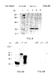
FIG. 1 shows a screen of the phage display library with GST/PKNSrc SH3. The phage display library was screened through 4 cycles of enrichment and amplification (1°-4°). At each cycle the phage were tested for their ability to bind to either immobilized GST/PKNSrc SH3 or GST/PKA only using an ELISA assay as described in Example 1.
FIG. 2 demonstrates the analysis of Src SH3 domain binding to synthetic peptides. Panel A represents a bar chart depicting the % of 32 P-labeled GST/PKNSrc SH3 or control 32 P-labeled GST/PKA proteins precipitated with peptides covalently attached to AminoLink® resin. Panel B demonstrates an autoradiograph of a polyacrylamide gel containing radiolabelled Src SH3 protein precipitated with SH3 binding peptides covalently attached to the resin.
FIG. 3 depicts the binding of radiolabeled GST/PKNSrc SH3 protein to the fuse 5B 4.3 phage sequence expressed as a GST fusion protein in E. coil.
FIG. 4 shows an SDS polyacrylamide gel demonstrating the specificity of SH3 domain binding by a phage identified peptide.
FIG. 5 shows autoradiographs demonstrating the reaction of src SH3 binding protein fusions with radiolabelled src SH3. Duplicate blots were reacted with either 32 P labeled GST/PKNsrc SH3 (panel A) or with 32 P labeled GST/PKA (panel B). After washing, blots were exposed to X-ray film.
FIG. 6 depicts a diagram and sequence of the Src SH3 binding cDNA L14 (SEQ ID NOS: 4 and 5) and a diagram of the human infant brain cDNA EST06380 aligned to show the regions of homology between the two sequences.
FIG. 7 depicts a diagram and sequence of the Src SH3 binding cDNA L17 (SEQ ID NOS: 6 and 7).
FIG. 8 depicts a diagram and sequence of Src SH3 binding cDNA L35 (SEQ ID NOS: 8 and 9).
FIG. 9 demonstrates the reaction of electroblotted Src SH3 binding protein-gene 10 fusions with a panel of various SH3/GST fusion proteins.
FIG. 10 demonstrates the alignment of known SH3 binding motifs with homologous sequences from clones L14, L17 and L35 (SEQ ID NOS: 1 and 10-25).
FIG. 11 demonstrates the binding analysis of peptides representing putative SH3 binding sites of the cloned proteins L14, L17 and L35 with various SH3 domains.
FIG. 12 demonstrates the binding of radiolabeled 32 P-GST/PKNSH3 fusions to immobilized peptides.
The following examples describe the isolation, purification and measurement of biological activity of the proteinaceous factors and antibodies of the present invention and are not intended to be limiting unless so expressly stated.
EXAMPLE 1 Isolation of a Novel c-src SH3 Binding Motif by Screening a Phage Random Peptide Library
A. Reagents, Peptides and Vectors
Enzymes were purchased from New England Biolabs and Boehringer Mannheim. The site directed mutagenesis kit was purchased from Amersham. The fuse5 vector and bacterial strains were kindly provided by Dr. George Smith (University Of Missouri, Columbia). Glutathione Sepharose® was obtained from Pharmacia. AminoLink® coupling gel was obtained from Pierce. The coating, washing, and blocking/dilution buffers used in the library screen and in ELISA assays were obtained from 5 Prime→3 Prime, Inc. Immulon microtiter trays were purchased from Dynatech Laboratories Inc.
A GST vector (GST/Src SH3) encoding the Src SH3 domain (amino acids 84-148) was kindly provided by Dr. I. Gout (Ludwig Institute for Cancer Research, London). A cDNA containing the SH3 domain of chicken c-Fyn was amplified by the polymerase chain reaction and cloned into the pGEX-2T vector at the BamHI-EcoRI sites. A GST vector containing the Ras-GAP SH3 was kindly provided by Dr. Bruno Tocque (Rhone-Poulenc Rorer, Vitry). Radiolabeled GST/Src SH3 and control GST protein were prepared as described by Ron and Dressier (43). Briefly, oligonucleotides encoding a specific site for phosphorylation by protein kinase A (PKA) were annealed and ligated into the BamHI site of the GST/Src SH3 vector and the GST vector. The purified GST/PKA/Src SH3 and GST/PKA fusion proteins were phosphorylated in vitro using the catalytic subunit of protein kinase A and [g-32 P-ATP]. Three μg of fusion protein was reacted for 1 hour at 30° C. in 50 mM phosphate buffer pH 8.0, 10 mM MgCl2, 4 mM DTT, 5 mM NaF, 75 u/ml of protein kinase A catalytic subunit (Sigma) and 100 μCi of [γ-32 P]-ATP (NEN) in a total volume of 100 μl. The labeled protein was purified by chromatography on glutathione-Sepharose® and had a specific activity of approximately 1-2×10⁵ dpm/ng.
B. Expression and Purification of GST and GST/SrcSH3 protein
Escherichia coli XL1-Blue cells containing GST, GST/SrcSH3 or GST/PKNSrcSH3 fusion constructs were grown overnight in LB containing 100 μg/ml ampicillin (LB/amp). Overnight cultures were diluted 1:50 in fresh LB/amp (1 liter) and cells were grown to an O.D.550 of 0.3 at 37° C., induced with 1 mM isopropyl-β-D-thiogalactopyranoside (IPTG), and grown for an additional three hours. Cells were harvested by centrifugation, washed once with phosphate buffered saline (PBS), resuspended in a small volume of PBS (10-20 ml) containing a mixture of proteinase inhibitors including pepstatin A (0.25 μg/ml), aprotinin (0.5 μg/ml), leupeptin (0.25 μg/ml), and PMSF (1 mM). Cells were sonicated on ice and the cell lysates were centrifuged at 12,000×g for 30 min. Supernatant fractions were reacted for 30 min at 4° C. with 1 ml of a 50% slurry of glutathione Sepharose, washed twice with 50 ml of PBS (at 4° C.) and the bound GST fusion proteins were eluted by reaction for 15 min at 4° C. with 1.5 ml freshly prepared reduced glutathione. Protein was quantified by measuring the absorbance at 280 nm followed by characterization by SDS polyacrylamide gel electrophoresis.
C. Library Construction
A fusion phage library was constructed in the filamentous bacteriophage fuse 5B, a vector derived from fd filamentous phage (44). The fuse5B vector was constructed from the vector fuse5 (27) by removal of a downstream BstXI site in fuse5 followed by replacement of the SfiI cloning site in gene III with a BstXI cloning site. Site directed mutagenesis was carried out using the oligonucleotide described by Cwirla et al.(29). For construction of the library a collection of oligonucleotides encoding all possible 15-mer peptides was synthesized on an Applied Biosystems Model 394 DNA synthesizer. The sequence of the degenerate oligonucleotide as well as the two half-site oligonucleotides was as described by Cwirla et al. (29) with the exception that (NNK)15 (SEQ ID NO: 23) was used instead of (NNK)6 (SEQ ID NO: 24) in the degenerate oligonucleotide sequence. The three oligonucleotides were phosphorylated with T4 polynucleotide kinase and then annealed with BstXI digested fuse5B DNA followed by overnight ligation at 15° C. The ligation products were precipitated with ethanol, redissolved in water and then electroporated into electrocompetent E. coli MC1061 cells using a BTX model 600 electroporation apparatus, yielding 5×10⁷ independent transformants. This level of complexity should allow for the presence of virtually all possible hexamer sequences (206 =6.4×10⁷ sequences) especially when one considers that each 15-mer contains 10 nested hexamer sequences. Nucleotide sequence analysis of random clones from this library indicated that greater than 50% of the clones contained an inserted sequence with an open reading frame (data not shown).
D. Library Screening
The GST/PKA/SrcSH3 fusion protein was immobilized at a concentration of 20 μg/ml in coating buffer in individual microtiter wells and incubated overnight at 4° C. The wells were blocked for one hour with 1X blocking/dilution buffer and rinsed 5 times with washing buffer. Approximately 1×10¹¹ tetracycline transducing units (TTU) of fusion phage in 1X blocking/dilution buffer was allowed to bind overnight at 4° C. The microtiter wells were then rinsed 5 times with washing buffer followed by the two elutions of bound phage with 0.2 ml of 50 mM sodium citrate, pH 2, 150 mM NaCl, for 5 min at room temperature. Phage elutions were pooled, neutralized and then amplified by reinfection into E. coli K 91-Kan cells (44). This process was repeated three times prior to analysis of the clones by DNA sequencing. Phage collected from the amplifications corresponding to each of four cycles of enrichment were tested for specific binding to GST/PKNSrc SH3 protein using a microtiter plate assay. Phage bound to the immobilized GST/PKA/Src SH3 were detected in this assay using a biotinylated polyclonal anti-M13 antibody and phage detection ELISA system (5 Prime→3 Prime) according to the manufacturer's recommended conditions. In brief, individual microtiter wells were coated overnight at 4° C. with a 2 μg/ml solution of Src SH3/PKNGST or GST/PKA in coating buffer. Several concentrations of phage were initially tested for obtaining an optimal signal to background ratio. Phage (0.2 ml) from pools of all four amplifications of the GST/PKNSrcSH3 fuse5B library screen were allowed to react with the immobilized GST proteins for two hours at room temperature. The microtiter wells were then washed, reacted for one hour with biotinylated anti-M13 polyclonal antibodies, washed again and then reacted with streptavidin conjugated alkaline phosphatase for 30 min. Following further washes, the reaction was developed with p-nitrophenyl phosphate in diethanolamine buffer and the absorbance at 405 nm measured. In order to confirm the titer of each phage sample reacted in this experiment, aliquots of each sample were tested simultaneously by reaction with immobilized nonbiotinylated anti-M13 antibodies supplied by the manufacturer. Detection of bound phage was then performed with the biotinylated anti-M13 antibody as described above. The results of this experiment (FIG. 1) demonstrated a pattern of specific binding to GST/PKA/Src SH3 with an increase in enrichment after each round of selection. Binding to the control GST/PKA protein was minimal throughout the experiment.
At the end of 4 rounds of screening, phage single stranded DNA was isolated from 45 clones by polyethylene glycol precipitation, phenol-chloroform extraction, and ethanol precipitation and used for dideoxynucleotide sequencing using [α-35 S]dATP and Sequenase T7 DNA Polymerase, essentially as per the manufacturer's instructions (United States Biochemical). An antisense oligonucleotide, 5' GCCTGTAGCATTCCACAGACAA3' (SEQ ID NO: 25), specific for the fuse5B vector downstream of the cloning site, was used as the sequencing primer. The DNA sequence from eight isolates could not be clearly established and these clones were not pursued further. The deduced amino acid sequence of the remaining clones is shown in Table 1.
                                  TABLE 1                                 
__________________________________________________________________________
Sequence                                                                  
      Peptide                             Number of                       
                                                Relative                  
Name  Sequence                            Isolates                        
                                                Binding                   
__________________________________________________________________________
                                                (%)                       
 ##STR1##                                                                 
Table 1 shows the Src SH3 binding sequences (SEQ ID NOS: 26-34) derived   
from screening the fuse 5B phage display library. Phage derived sequences 
are arranged according to a common consensus SH3 binding domain motif.    
This consensus sequence is also displayed in a SH3 binding domain motif   
(SEQ ID NO: 18) identified in a protein (Lambda 14) which was isolated    
during a screen of a mouse embryo cDNA library with Src SH3 (see Example  
2. The frequency of appearance of each isolate is indicated. The relative 
binding strength of peptides corresponding to each sequence was           
determined, as in FIG. 2, by calculating the % of .sup.32 P-labeled       
GST/PKNSrc SH3 or control .sup.32 P-labeled GST/PKA proteins precipitated 
with peptides covalently attached to AminoLink® resin. The % of bound 
ligand was calculated as bound (cpm)/total (cpm)×100%. The peptides 
correspond to a negative control (SV40 peptide) or the sequences derived  
from either the cDNA (lambda 14) or the phage library (fuse 5B 4.3)       
screens. The peptides tested were as follows:                             
______________________________________                                    
KPPTPPPEPET   SV40-peptide (11 mer)                                       
              (SEQ ID NO: 22)                                             
QSRPLPSPPKFT  Lambda 14 (12 mer) (SEQ ID NO: 18)                          
RPLPSPP       Lambda 14 (7 mer) (SEQ ID NO: 35)                           
LALARPLPVPPWRQI                                                           
              fuse5B 4.3-15 (15 mer)                                      
              (SEQ ID NO: 28)                                             
LARPLPVPPWRQ  fuse5B 4.3-12 (12 mer)                                      
              (SEQ ID NO: 36)                                             
RPLPVPP       fuse5B 4.3-7 (7 mer) (SEQ ID NO: 37)                        
______________________________________                                    
These 37 clones comprised only 9 different sequences, many of which were repeated several or more times as indicated. Further examination of these sequences revealed a highly reiterated, proline-rich, 7 amino acid consensus sequence, RPLPXXP (SEQ ID NO: 3), contained within the sequence of the clones examined. This motif demonstrated a strong similarity to a Src SH3 binding sequence identified within a protein (lambda 14) isolated by screening of a lambda-lox mouse embryo cDNA library with a 32 P-labeled Src SH3 domain probe. This work is described in Example 2.
At least three of the phage derived sequences (fuse5B 4.2, fuse5B 4.3 and fuse5B 4.27) exhibited identity in six out of seven amino acids when compared with the lambda 14 sequence. Six out of nine phage derived sequences were identical with the lambda 14 clone in five out of seven amino acid residues. The phage clone (fuse5B 4.2) isolated with the highest frequency (17/37), exhibited identity in seven out of eight amino acids when compared with the lambda 14 sequence.
E. Analysis of srcSH3 binding motif using synthetic peptides
Based on these data, peptides representing the lambda 14 sequence and the sequences isolated from the phage library were synthesized, coupled to a resin, and then tested for the ability to specifically and quantitatively precipitate 32 P-labeled GST/PKA/Src SH3 protein. Peptides were constructed manually using a custom built apparatus designed for the rapid simultaneous synthesis of 0.01-0.02 mmoles of peptide. Solid phase methodology using a 9-fluorenylmethyloxy carbonyl (FMOC) protection scheme in conjunction with the HOBT/Hbtu activation chemistry (45) was used. Peptides of interest were subsequently constructed in larger quantities (0.1-0.25 mmoles) using an Applied Biosystems Model 430 Peptide Synthesizer running Applied Biosystems Fast-moc® coupling cycle. All peptides were cleaved for 1.5 hours at room temperature using a cleavage reagent of 82.5% trifluoroacetic acid, 5% phenol, 5% H2 O, 5% thioanisole, and 2.5% ethanedithiol (46). Following cleavage, the peptides were precipitated with ether, washed, then dried for 1 hour under vacuum. The peptides were then solubilized in either water, 10% acetic acid, or 10 mM ammonium bicarbonate depending upon the peptides net charge or solubility. Peptides were analyzed by reverse phase HPLC for purity and by ion spray mass spectrometry for molecular weight integrity. A purity level of 95% was achieved for all peptides along with correct mass spectrometry data. Lyophilized peptides were dissolved in H2 O and coupled to AminoLink® coupling gel. The efficiency of coupling was measured by analyzing the peptide solution, before and after coupling, by reverse phase HPLC and by reading the absorbance at 220 nm. Both methods demonstrated greater than 70% coupling efficiency for all peptides.
An aliquot of the resin with covalently bound peptide (20 ml of wet beads, containing ˜150 ng of peptide) was incubated with ˜5 ng of either 32 P-labeled GST/PKA/SrcSH3 fusion protein or 32 P-labeled GST/PKA fusion protein, lacking the SH3 domain, as control. Incubations were carried out with approximately 5×10⁵ cpm of protein (5 ng) in PBS buffer, pH 7.0, containing 5% BSA and 0.1% Tween 20. After a 30 min incubation at room temperature, the beads were extensively washed by centrifugation with PBS containing Triton X-100 until no radioactivity was detected in the wash buffer. The amount of bound GST/PKA/SrcSH3 and GST/PKA proteins was detected by Cherenkov counting and expressed as the percent of the bound versus the total added cpm. Results of this experiment are presented in Table 1 and demonstrate that the phage sequence fuse5B 4.2, isolated in highest abundance in the phage library screen, has a very strong binding capacity toward the Src SH3 domain. All but three of the sequences obtained in the screen exhibited this strong binding capacity. Clones that showed weaker binding contained substitutions for either the NH2 -terminal arginine residue or the COOH-terminal proline residue.
In order to define the core sequence responsible for binding in a more precise manner, a series of truncated peptides, based upon the lambda 14 sequence and the 4.3 clone isolated from the phage library, were also tested for the ability to specifically and quantitatively precipitate 32 P-labeled GST/PKA/Src SH3 protein (FIG. 2A). Additionally, an aliquot of the radiolabeled Src SH3 domain bound to the immobilized peptide was also analyzed on SDS polyacrylamide gels (FIG. 2B). FIG. 2, panel B demonstrates an autoradiograph of a polyacrylamide gel containing radiolabelled Src SH3 protein precipitated with SH3 binding peptides covalently attached to the resin. The gel lanes correspond with samples as shown in the bar graph on FIG. 2, panel A. Samples of 32 P-labeled GST/PKNSrc SH3 protein precipitated by the various SH3 binding peptides are shown in the even-numbered lanes while samples of the precipitated control 32 P-labeled GST/PKA protein are shown in the odd-numbered lanes. The 7-mer sequence from both the phage derived and the cDNA derived SH3 binding domain sequences, appears to retain virtually all of the SH3 binding activity of the parental 15mer sequences. The fact that the 7-mer fuse5B 4.3 sequence has strong homology with the lambda-14 Src SH3-binding motif probably explains their indistinguishable activity in this assay. The specificity of the Src SH3-binding sequences for the Src SH3 domain is most clearly illustrated by the low binding of these sequences to 32 P-labeled GST/PKA, which exhibits approximately a 100 fold lower binding to all peptides than does GST/PKNSrc SH3. In addition, a non-specific but otherwise proline-rich peptide (FIG. 2, SV40 peptide) also failed to display any significant binding.
We next demonstrated that the fuse5B 4.3 peptide retained its ability to bind Src SH3 even when the peptide was placed within the context of a larger protein sequence. For this experiment, oligonucleotides that encoded this 7 amino acid region of the 4.3 sequence were cloned into the GST vector in such a way that the seven amino acid peptide was expressed as a fusion protein with the GST sequence. E. coil lysates containing either the GST fusion protein or the GST protein only were subjected to SDS polyacrylamide gel electrophoresis, transferred to nitrocellulose and then probed with a 32 P-labeled Src SH3 probe.
FIG. 3 depicts the binding of radiolabeled GST/PKNSrc SH3 protein to the fuse 5B 4.3 phage sequence expressed as a GST fusion protein in E. coli. Oligonucleotides encoding the sequence RPLPVPP from the fuse 5B 4.3 phage sequence were synthesized, annealed, ligated into the pGEX-2T GST expression vector, and transformed into E. coli. Cell cultures containing either GST fuse 5B 4.3 or GST only were grown to mid-log phase and induced with IPTG. Cells from the induced cultures were harvested, lysed in SDS-loading buffer, electrophoresed on an SDS polyacrylamide gel, and transferred to a nitrocellulose membrane. The membrane was then reacted with radiolabeled GST/PKNSrc SH3 protein. Lane 1. Lysate from GST fuse 5B 4.3, Lane 2. Lysate from GST only. The Src SH3 probe reacted with the GST fusion protein containing the fuse5B 4.3 7-mer, while no reaction was obtained with GST only.
Finally, in order to demonstrate specificity of the interaction of the Src SH3 domain with the sequences identified here, the fuse5B 4.3 sequence immobilized to resin, was reacted with different SH3 containing GST fusion proteins including the Fyn SH3 and Ras-GAP SH3. FIG. 4 shows an SDS polyacrylamide gel demonstrating the specificity of SH3 domain binding by a phage identified peptide. Various GST/SH3 fusion proteins as well as GST only were reacted with the Src SH3 binding peptide, fuse 5B 4.3-12 (as described for FIG. 2), covalently attached to AminoLink® resin. Precipitated proteins remaining after washing were electrophoresed on an and the gel was then stained with Coomassie Blue. Lane 1. Molecular weight markers, Lane 2. GST only, Lane 3. GST/Src SH3, Lane 4.--GST/Fyn SH3, Lane 5. GST/Ras-GAP SH3. The fuse5B 4.3 sequence was able to precipitate the Src SH3 and the closely related (8) Fyn SH3, but failed to react with the Ras-GAP SH3.
Recently, Chen et al. (42) utilized the SH3 domain of PI3-kinase p85 to screen a biased combinatorial library of synthetic peptides in which prolines were fixed in three of nine positions with the six other positions being randomized. The bias for this library, represented by the formula XXXPPXPXX (SEQ ID NO: 3), was derived from an alignment of the SH3-binding motifs in 3BP-1 and the guanine nucleotide exchange factor Sos1. Although the biased combinatorial approach (42) can be successfully employed, the sequences which are identified by this technique are obviously influenced by the underlying assumptions (biases). This is avoided by the use of a completely random phage library. Additional advantages of the phage display library for the identification of SH3 binding motifs is that it is a rapid and convenient method, requiring approximately one week to perform three rounds of phage selection and amplification. The data presented herein, and that of Chen et al.(42) illustrate that at least some SH3 binding motifs consist of as little as 6-7 amino acids. Thus, the identification of additional binding motifs for specific SH3 domains should be possible by further application of the phage peptide library approach. This approach should facilitate the rapid identification of binding motifs for specific SH3 domains and should also provide a groundwork for a more detailed analysis of structure/function relationships.
EXAMPLE 2 Expression Cloning of Novel c-src Binding Proteins
A. Identification of cDNA clones encoding proteins which interact with src SH3 domain
A mouse 12 day embryo cDNA library constructed in λEXlox (Novagen) was screened as follows: 1.4×10⁶ library phage were used to infect logarithmically growing E. coli BL21 (DE3)pLysE on forty 150 mm plates. Plates were incubated for 8 hrs at 37° C. and then overlayed with nitrocellulose (NC) circles presoaked and dried in 10 mM IPTG. The IPTG induces expression of the T7 RNA polymerase present on the host chromosome within a lamba lysogen. Plates were incubated an additional 12-16 hrs at 37° C., the NC circles were removed and washed thoroughly in TTBS (0.1% Triton X-100 in Tris buffered saline) to remove bacterial debris. The filters were blocked in 5% non fat dry milk in TTBS (blotto) for 2-16 hr at RT with gentle mixing. Filters were then reacted with 10-50 ng/ml labeled Src SH3 probe (32 P-GST/PKNSrc SH3) in fresh blotto (2 ml/filter) with gentle agitation overnight at room temperature. Filters were washed with large volumes of TTBS several times and then dried and exposed to X-ray film overnight. Three reacting plaques were recovered and subjected to several rounds of plaque purification using this proceedure. Primary lysates were prepared of pure clones, and phage from these were used to infect E. coli BM25.8 for automatic subcloning of the pEXlox cDNAs. To verify the interaction of the c-src SH3 domain with the cDNA-encoded proteins, the three positive λEXlox clones were converted to plasmids and introduced into E. coli pLysE. After addition of IPTG, cells were incubated 2 hours at 37° C. for expression of the recombinant fusion proteins. Cells were lysed in SDS loading buffer, proteins were resolved by SDS-PAGE, transferred to nitrocellulose, probed with either 32 P-GST/PKNSrc SH3 or 32 P-GST/PKA as control, and detected by autoradiography. FIG. 5 depicts the autoradiographs demonstrating the reaction of src SH3 binding protein fusions with radiolabelled src SH3. Inductions of the Src SH3 binding protein-gene 10 fusions from pEXlox clones (L35, L17, and L14) were run on SDS-PAGE gels and transfered to nitrocellulose. Duplicate blots were reacted with either 32 P labeled GST/PKNsrc SH3 (panel A) or with 32 P labeled GST/PKA (panel B). After washing, blots were exposed to X-ray film. The mobility of MW markers is indicated, and the loading order of inductions are shown on top of each panel. Clones L14, L17, and L35, respectively, produced fusion proteins of 66, 72, and 35 kDa, which bound only to the 32 P-GST/PKNSrc SH3 probe (FIG. 5a) but not to the control 32 P-GST/PKA probe (FIG. 5b). No binding to either probe was observed with lysates from non IPTG-induced cultures (data not shown). Taking into account the molecular weight of the T7 gene 10 protein (27.4 kDa) in each of these fusions, the apparent molecular weights of the cDNA-encoded polypeptides are 38-40, about 48, and 3-5 kDa for clones L14, L17, and L35, respectively.
B. Analysis of pEXlox cDNAs
The nucleotide sequences of the cDNA inserts of clones L14 (SEQ ID NO: 4), L17 (SEQ ID NO: 6), and L35 (SEQ ID NO: 8), were determined using Sequenase T7 DNA Polymerase, essentially as described in Example 1. Nucleotide sequence analysis of clone L14 (1168 bp) and comparison to DNA and protein databases revealed a 221 bp region with approximately 90% identity to an anonymous cDNA (EST06380) directionally cloned from a human infant brain cDNA library (47). FIG. 6 depicts a diagram and sequence of the Src SH3 binding cDNA L14. Also shown is a diagram of the human infant brain cDNA EST06380 (47) aligned to show the regions of homology between the two sequences, with the boundary positions marked. Position 1 is the first base of the L14 sequence after the EcoRI site of the vector, position 1168 is the last base of the insert before the HindIII site of the vector. The EST06380 homologous sequence is boxed. Putative SH3 binding domains (L14-1 and L14-2) and the putative polyA addition signal site are underlined. The internal HindIII site at position 532 used to generate the truncated version of L14 is shown and possible sites for tyrosine phosphorylation are indicated by a bold Y.
Two proline-rich putative SH3-binding sequences were identifiable within the deduced amino acid sequence of L14 (FIGS. 6a, b). Experiments to determine which of these sequences were responsible for binding of the L14 fusion protein to the c-src SH3 domain are described below. The longest deduced open reading frame within clone L14 encodes a 370-residue protein with a predicted molecular weight of about 40 kDa, in close agreement with that deduced from the electrophoretic mobility of the L14 fusion protein (FIG. 5).
Nucleotide sequence analysis of clone L17 (987 bp) and comparison to DNA and protein databases revealed 99.9% identity of L17 with two segments of sequence of mouse kinesin-like-protein (KIF-2) (48). L17 consists of a 253 bp sequence, not reported in the KIF-2 sequence, followed by 89 bp of KIF-2 5' untranslated sequence, followed by a second region of 645 bp of KIF-2 sequence which contains the KIF-2 translation initiation codon (FIGS. 7a,b). Although these two KIF-2 sequences are contiguous within the sequence of L17, they are separated by 383 bp in the KIF-2 sequence (48). Thus, it appears that L17 is the product of alternative splicing of the KIF-2 gene, in which initiation of translation occurs upstream of the start codon normally utilized by KIF-2, resulting in the translation of sequences previously ascribed to the 5' untranslated region of KIF-2. Importantly, two proline-rich putative SH3-binding sequences are found within this region of L17. Experiments to determine which of these sequences were responsible for binding of the L17 fusion protein to the c-src SH3 domain (FIG. 5) are described below. Finally, the longest deduced open reading frame within clone L17 encodes a 329-residue protein with a calculated molecular weight of about 40 kDa. The reason why the L17 fusion protein has an abnormally slow mobility on SDS-PAGE (migrating with an apparent molecular weight of 48 kDa after subtraction of the mass of the T7 gene 10 protein sequences) is unclear. This may be due to the high content of prolines within the amino-terminal third of the L17 protein.
FIG. 7 depicts a diagram and sequence of the Src SH3 binding cDNA L17. The diagram represents the full length of the L17 cDNA isolate and shows the alignment to the homologous sequence of the mouse kinesin like gene KIF-2 (48). Position 1 is the first base of the insert sequence after the EcoRI site of the vector, and position 987 is the last base of the insert before the HindIII site of the vector. Nucleotides 1-252 of L17 are unique, while sequence 253-987 (boxed) is homologous to KIF-2 sequence. The region 253-342 of L17 corresponds to the sequence -493 to -404 of the KIF-2 5' untranslated and nucleotides 343 to 987 of L17 corespond to -21 to 624 of the KIF-2 sequence. In L17, nucleotides -404 and -21 of KIF-2 are adjacent. Thus, L17 appears to be an alternatively spliced form of KIF-2 in which a portion (nucleotides -403 to -22) of the untranslated region of KIF-2 is deleted and another (nucleotides -493 to -404) forms part of the open reading frame in L17. Nucleotides 1-252 of L17 which form part of the open reading frame in this clone may represent part of an upstream exon of the KIF-2 gene. The SmaI site at position 127 of L17 which was used to create a truncated version of L17 which contained only the L17-1 putative SH3 binding site is indicated. The region 1-127 was used to generate a non KIF-2 probe for northern hybridizations, and is indicated below the L17 sequence. Differences in sequence between L17 and KIF-2 are shown, with the differing KIF-2 bases printed above the L17 sequence. Positions -493, -21, +1, and 624 of the KIF-2 sequence are indicated.
FIG. 8 depicts a diagram and sequence of Src SH3 binding cDNA L35. Position 1 is the first base of the insert sequence after the EcoRI site of the vector, position 261 is the last base of the insert before the HindIII site of the vector. Three consecutive translation stop codons are located at nucleotides 88-96. The putatative SH3 binding domain (L35) is indicated by a bar on the diagram and the sequence of the synthesized peptide L35 is underlined below. Clone L35 contained a cDNA insert of only 261 bp possessing an open reading frame of 30 amino acids, in which a proline-rich putative SH3-binding sequence was found (FIG. 8). The size of the encoded polypeptide (approximately 3.3 kDa) is in good agreement with that predicted (3-5 kDa) from the migration of the L35 fusion protein (FIG. 5a), after subtraction of the mass of the T7 gene 10 protein sequences. The cDNA and deduced amino acid sequences of clone 35 show no significant homology to any sequences in the current nucleic acid and protein databases.
C. Truncations of cDNAs
Truncated versions of L14 and L17 clones were prepared as follows: Lambda 14 was digested with HindIII (internal site at nt 532) and the fragment containing the vector plus the 5' portion of the insert cDNA was gel purified and recircularized by ligation. This construct (DL14) consisted of only the 178 amino-terminal amino acid residues of L14 and contained only one putative SH3 binding domain (FIG. 6). The extreme 5' portion of L17 was prepared by digesting the clone with SmaI and HindIII (SmaI site at nt 85 of insert and HindIII site at nt 987) which releases the majority of the cDNA insert leaving only the sequence encoding the proline-rich amino-terminal 28 residues. After gel purification, the vector and extreme 5' portion of L17 was recircularized by ligation at the blunted HindIII and SmaI sites. Truncated versions were used for Northern blotting analysis, or expressed and analyzed in the binding assay described below.
D. Analysis of SH3 domain binding by clones L14, L17, and L35
A panel of SH3 domains from various proteins (c-src kinase, neuronal src, fgr-kinase, pl 3-kinase p85a subunit, NCF1/2 and NCF2/2) produced as GST fusions in recombinant pGEX vectors was kindly provided by Dr. Ivan Gout, Ludwig Institute for Cancer Research, London; pGEX vectors encoding GST/rasGAP and GST/Grb2 SH3 domains were provided by Dr. Bruno Tocqu e, Rh one-Poulenc Rorer, Vitry sur Seine, France; cDNA encoding the SH3 domains of fyn-kinase, csk-kinase, PLC-g and crk-protooncogene were produced by reverse-transcriptase-PCR and cloned into pGEX-2T vector between the BamHI and EcoRI sites. All GST/SH3 fusions were expressed and purified using glutathione-Sepharose affinity chromatography as described above. These GST/SH3 fusions were reacted with NC membranes containing the expressed T7 gene 10 cDNA fusions using the same binding conditions as used for library screening. Binding reactivity was detected with an anti GST antibody (Amrad, Australia) diluted 1:5000 followed by anti rabbit IgG-HRP (1:2000 dilution; Boehringer) and ECL methodology (Amersham). The results of these analyses are shown in FIG. 9 and summarized in Table 2.
FIG. 9 demonstrates the reaction of electroblotted Src SH3 binding protein-gene 10 fusions with a panel of various SH3/GST fusion proteins. Inductions of the Src SH3 binding protein-gene 10 fusions from pEXlox clones (L35, L17, DL14, and L14) were run on SDS PAGE gels and transfered to nitrocellulose. Blots were reacted with various GST/SH3 fusions or GST only as indicated. The origin of the SH3 domain in each GST fusion is indicated in parenthesis. The GST/Src SH3 fusions also contained the Protein Kinase A (PKA) labeling site, and in the first panel the site has been phosphorylated with nonradioactive ATP by PKA. The reaction of GST/SH3 fusions with the blotted SH3-binding protein-gene 10 fusions was detected using a rabbit polyclonal anti GST antibody (Amrad), followed by an anti Rabbit IgG-HRP conjugate antibody, with subsequent ECL (enhanced chemiluminesence) reaction and exposure to X-ray film. Mobility of MW markers are indicated. DL14 is a truncated version of L14, containing only the NH2 terminal SH3-binding sequence.
L35 reacted most strongly with the SH3 domains of c-src (FIG. 9b) and c-fyn (not shown), and to lesser degrees with other SH3 domains, such as c-fgr (FIG. 9c) and Grb2 amino-terminal (FIG. 9d) and carboxyl-terminal (FIG. 9i) SH3 domains. In general, L35 bound better and to a greater variety of SH3 domains than did L14 and L17, but was unable to bind to several SH3 domains, such as rasGAP (FIG. 9g) and bound some others very poorly. Table 2 summarizes the binding experiment data presented in FIG. 9. The relative strength of binding is shown as indicated.
              TABLE 2                                                     
______________________________________                                    
Analysis of SH3 Domain binding specificity                                
SH3 domain   L14        L17        L35                                    
______________________________________                                    
c-src        +++        +-(+++)*   ++++                                   
neuronal src +-         -          ++                                     
c-Fyn        ++++       +-         ++++                                   
fgr          +++        +-         +++                                    
CRK          -          -          +                                      
PLC-g        -(++**)    -          ++                                     
H-NCF 1/2    -          -          +-                                     
H-NCF 2/2    -          -          -                                      
GRB-2 N term +          +-         ++                                     
GRB-2 C term -          +-         ++                                     
GRB-2 Full length                                                         
             +-         +-         ++                                     
GAP          -          +          -                                      
PI 3-kinase p85a                                                          
             ++         +          ++                                     
R-CSK        -          -          -                                      
______________________________________                                    
 (-) no detectable interaction by all methods;                            
 (+-) very weak binding;                                                  
 (+) weak binding but easily detected by all methods;                     
 (++) moderate binding;                                                   
 (+++) strong and                                                         
 (++++) very strong binding.                                              
 *binding only with phosphorylated GST/PKA/Src SH3                        
 **Binding only wkh peptide immobilized on AminoLink ® agarose        
L14 reacted with many but not all of the SH3 domains which reacted with L35. Strongest reactions were observed with the SH3 domains of c-src (FIG. 9b), c-fgr (FIG. 9c) and c-fyn (not shown). L14 reacted less well with other SH3 domains, such as Grb2 amino-terminal (FIG. 9d) and PI 3-kinase p85a subunit (FIG. 9f) and didn't react with others such as Grb2 carboxyl-terminal (FIG. 9i) and rasGAP (FIG. 9g) SH3 domains. Unlike L35, only the amino terminal but not the carboxyl terminal Grb2 SH3 domain bound to L14. The DL14 and full length L14 proteins bound equally well and to the same spectrum of SH3 domains (FIG. 9), suggesting that the amino terminal putative SH3 binding domain (residues 21-29) is the main SH3 binding site within the L14 protein.
In general, L17 bound much less well to the same spectrum of SH3 domains than did L35 or L14, with the notable exception of rasGAP (FIG. 9g), to which only L17 bound weakly and neither L35 nor L14 bound. The reaction of L17 was most strong with the SH3 domains of PI 3-kinase p85a (FIG. 9f) and rasGAP (FIG. 9g). Surprisingly, in this experiment L17 failed to react with the SH3 domain of c-src (FIG. 9a), although L17 was cloned on this basis and bound to the 32 P-GST/PKNSrc SH3 probe but not to the control 32 P-GST/PKA probe (FIGS. 5a,b). The main difference between the experiments presented in FIG. 5 and FIG. 9 is the method of detection of the bound SH3 domains: in FIG. 5 the GST/PKNSrc SH3 fusion protein was phosphorylated in vitro with 32 P-g-ATP and detected by autoradiography, whereas in FIG. 9 bound GST/SH3 fusion proteins were detected with anti GST antibodies followed by ECL. These results suggested that serine phosphorylation of the PKA recognition site within the GST/PKNSrc SH3 fusion protein was crucial for the binding of of the src SH3 sequence to the L17 protein. To test this possibility, the GST/PKNSrc SH3 fusion protein was phosphorylated in vitro with non-radioactive ATP, reacted with filters containing the L14, L17 and L35 proteins, and detected with anti-GST antibodies and ECL. Indeed, the presence of the phosphorylated serine in the c-srcSH3 domain's amino terminal flanking sequence was crucial for the binding to the L17 fusion protein (FIGS. 9a,b,e). A fusion protein in which the PKA recognition site was situated downstream of the c-srcSH3 domain and phosphorylated in vitro with 32 P-γ-ATP was unable to bind L17, but bound to L35 and L14 like the original 32 P-GST/PKNSrc SH3 probe (data not shown). These results argue that it is not the phosphorylated PKA site within the GST/PKNSrc SH3 fusion protein per se which is responsible for the interaction with L17. Rather, these results demonstrate that serine phosphorylation within the flanking sequence amino terminal to the c-srcSH3 domain is necessary for the recognition of L17 by the GST/PKNSrc SH3 fusion protein. Consistent with these findings, only L14 and L35 proteins but not L17 were able to bind to full length autophosphorylated c-src kinase in vitro. It is uncertain whether in vivo the affinity or specificity of binding of the c-srcSH3 domain is similarly regulated by upstream serine/threonine phosphorylation. This may also be applicable for the SH3 binding motifs. In such cases, phosphorylation-dephosphorylation may potentially serve as an important regulator of SH3 domain binding. The importance of serine/threonine phosphorylation in controlling the growth factor activated MAP kinase pathway was demonstrated recently (49, 50). In these experiments, the association of raf-1 kinase with p21 ras was inhibited by phosphorylation of raf-1 on serine 43 by the cAMP-dependent protein kinase, PKA. In a similar manner, cAMP-elevating agents which activate PKA may potentially regulate interactions between SH3 domains and SH3 binding proteins. We have found that the SH3-binding protein L14 can be phosphorylated by src kinase in vitro, and several candidate tyrosine phosphorylation sites are indicated in the L14 sequence (FIG. 6). In this respect, L14 protein resembles actin filament-associated protein AFAP-110 which has srcSH3 binding sites (FIG. 10) and is tyrosine phosphorylated by src kinase (51). Whether or not this phosphorylation is physiologically important is unclear.
E. Identification of SH3-binding sequences within clones L14, L17, and L35.
As mentioned above, putative SH3-binding sequences in clones L14, L17, and L35 were tentatively identified based on their proline-rich character (FIGS. 5-8). An alignment of these putative sequences with the some of known SH3-binding sequences in different proteins is shown in FIG. 10. The possible alignments of some SH3 binding motifs from various proteins is also shown.
This analysis showed that the sequence of this region of clone L35 (residues 13-24, FIG. 8), which exhibited the best binding to the greatest variety of tested SH3 domains, contains a hexapeptide sequence PPPΨPP (SEQ ID NO: 38) (where Ψ is hydrophobic amino acid) which is also found in the SH3-binding domains of 3BP-1, formin and mSOS. Of the two candidate SH3-binding sequences in clone L14 (residues 19-30 and residues 246-253, FIG. 6) only the amino-terminal motif could be reasonably aligned with the known SH3-binding sequences. This motif bears a striking resemblance to the heptamer consensus sequence identified by screening of the phage random peptide library with the SH3 domain of c-src (Table 1). The amino terminal putative SH3-binding motif in clone 17 (L17-1, residues 30-43, FIG. 7) has ˜46% identity with the P2 sequence within dynamin (FIG. 10) and ˜60% amino acid homology with P1 sequence from dynamin (the P1 proline-rich sequence, residues 785-806 of dynamin p100 overlaps with the P2 sequence, residues 777-794, see ref. 22). In spite of having 10 common residues, the P1 and P2 dynamin sequences displayed very different SH3 binding properties; while the P2 sequence had the same SH3 binding specificity (including binding of c-src SH3) as native dynamin, a synthetic peptide corresponding to the P1 sequence inhibited the association of dynamin with the SH3 domains of PI 3-kinase p85a and PLC-g but didn't bind c-src SH3 (22). This is consistent with the data presented in FIG. 9 and Table 2 in which L17 bound best to the PI 3-kinase p85a SH3 domain. The carboxyl terminal putative SH3-binding motif in clone 17 (residues 72-88, FIG. 6) resembles the upstream motif but aligned less well to the P2 (and P1 motif, not shown) sequence of dynamin (FIG. 10). In general, the proline-rich sequences in clone L17 have poor homology with other SH3 binding motifs presented in FIG. 10.
In order to determine whether the putative SH3 binding motifs in clones L14, L17, and L35 can bind to the SH3 domains of c-src and other proteins, 5 synthetic peptides were prepared: Peptides corresponding to putative SH3 binding domains were synthesized as described in Example 1 and coupled to 500 ml of AminoLink® agarose (Pierce). The peptides had the following sequences: pL14-1: QSRPLPSPPKFT (SEQ ID NO: 18; L14 residues 18-29, FIG. 6); pL14-17mer: RPLPSPP (SEQ ID NO: 35, L14 residues 20-26, FIG. 6); pL14-2: PQHSAVPPRPGPA (SEQ ID NO: 39, L14 residues 241-253, FIG. 6); pL17-1: AHPPARPPVRPQPG (SEQ ID NO: 20, L17 residues 30-43, FIG. 7); pL17-2: APPAATPPPPRLFRPPA (SEQ ID NO: 21, L17 residues 72-88, FIG. 7); pL35: TPAPPPLPPRNV (SEQ ID NO: 17, L35 residues 13-24, FIG. 8); pSV40: KPPTPPPEPET (SEQ ID NO: 22, Sigma L0765). The coupled peptides were reacted with radiolabeled GST/PKNSrc SH3, GST/PKNrasGAP SH3 or GST/PKA (control) fusions (8×10⁵ dpm) or with unlabeled GST/SH3 fusions (5 μg). The binding was done at room temperature for 1 hour and the resin was washed extensively with TTBS. Binding of radiolabeled probes was determined in a scintillation counter. Unlabeled GST/SH3 fusions were released from resin by boiling in SDS sample buffer and analyzed by SDS-PAGE. Relative binding was expressed as a % of input cpm for radiolabeled fusions or by the relative intensity of the Coomassie blue-stainable band for unlabeled fusions. Controls included labeled (GST/PKA) and unlabeled GST alone with each of the peptides and reaction of SH3 fusions with a heterologous proline-rich peptide from SV40 T antigen.
FIG. 11 demonstrates the binding analysis of peptides representing putative SH3 binding sites of the cloned proteins L14, L17 and L35 with various SH3 domains.
Peptides (L14-1, L35, L17-1, and L17-2) corresponding to the putative SH3 binding domains of the pEXlox clones (L14, L35, and L17) were coupled to Aminolink® agarose. The bound peptides were then reacted with equal amounts of various purified GST/SH3 fusions or with purified GST alone. The peptide-linked agarose beads were thoroughly washed and then resuspended in SDS loading dye, boiled and run on SDS-PAGE gels along with MW size markers. The gels were stained with Coomassie blue and the presence and intensity of the captured GST/SH3 fusion protein was ascertained. The 69 kD band seen in all lanes is bovine serum albumin, which was used to block nonspecific binding sites on beads and was included in the reaction buffer (3%), as well. On FIG. 11: Gel A. reacted with peptide pL14-1; B. reacted with peptide pL35; C. reacted with peptide pL17-2, and D. reacted with peptide pL17-1. The panel of GST fusions reacting with the coupled peptides is indicated above the corresponding lanes of the gels. Mobilities of MW markers (size in kD) are indicated.
FIG. 12 demonstrates the binding of radiolabeled 32 P-GST/PKA/SH3 fusions to immobilized peptides. Peptides representing putative SH3 binding motifs determined from the sequences of clones L14, L17, and L35 were coupled to Aminolink® agerose, as described for FIG. 11. As a control a proline rich peptide from SV40 T antigen was also coupled. Peptide-linked beads were reacted with 32 P-GST/PKNSH3 domains from c-src and rasGAP proteins and to 32 P-GST/PKA alone, washed extensively with TTBS and the binding of radiolabeled proteins determined by scintillation counting. The bar graph shows the amount of labeled fusion bound to each peptide-linked resin as a % of total input cpm.
In this analysis no binding of any of the peptides to the negative control GST protein (FIG. 11) nor any binding of SH3 domains to the negative control SV40 peptide (FIG. 12) was observed. Peptides pL14-1 and pL35 (FIGS. 11a,b) bound well to the SH3 domains of PI 3-kinase p85a subunit and c-src and not to rasGAP, as expected from the results with the L14 and L35 fusion proteins (FIGS. 9a,b,f,g, Table 2). The SH3 domain of PLC-g also bound well to peptide pL14-1, which contrasts with the results obtained with the L14 fusion protein (FIG. 9j, Table 2). The SH3 binding motifs from both L35 and L14 proteins have a flanking Arg residue at the carboxyl terminus of the L35 SH3 binding motif (PPPLPPR, SEQ ID NO: 40) and at the amino terminus of the L14 SH3 binding motif (RPLPSPP, SEQ ID NO: 35). The specific binding of SH3 domains by these proline-rich motifs may depend partially on the presence of a flanking arginine. The importance of carboxyl Arg in SOS proline-rich motifs for the binding of Grb2 SH3 domains has been previously described (9).
Peptide p17-1 (FIG. 11d) bound poorly to the c-src SH3 domain but bound well to the SH3 domain of PI 3-kinase p85a, as expected from the binding of the L17 fusion protein with GST/p85a SH3 (FIG. 9f, Table 2). Similar to the results obtained with peptide pL14-1, the peptide pL17-1 bound comparatively well to GST/PLC-g SH3, despite the inability of the L17 fusion protein to bind to GST/PLC-g SH3 (FIG. 9j, Table 2). The discrepencies between the results of FIGS. 9 and 11 may be due to the use of reduced, denatured and electroblotted T7 gene 10 protein fusions in FIG. 9 vs. immobilized peptides in FIG. 11. Alternatively, the discrepencies may indicate that sequences which flank the SH3 binding domains in L14 and L17 proteins (but not in L35) exert a negative influence on its binding to some (eg. PLC-g SH3 domain) but not all SH3 domains. This negative effect would not be observed in the experiments described in FIGS. 11 and 12 which utilized short, synthetic peptides.
Peptide L17-2 behaved identically to peptide L17-1 with respect to its binding to the SH3 domains of PI 3-kinase p85a, c-src, and PLC-g. However, in contrast to the other peptides, only peptide L17-2 displayed any binding to the SH3 domain of rasGAP (FIG. 11c), consistent with the observed binding of GST/rasGAP SH3 to electroblotted L17 but not L14 or L35 fusion proteins (FIG. 9g, Table 2). The recognition of rasGAP SH3 by peptide L17-2, although weak, reflects a specific but probably sub-optimal interaction. This is not surprising in view of the fact that clone L17 was isolated on the basis of its binding to the SH3 domain of c-src, which is quite different from the SH3 domain of rasGAP protein (4).
The experiments shown in FIG. 11 demonstrate that the putative SH3 binding motifs identified in clones L14, L17 and L35 bind to the SH3 domains of several different proteins to varying extents. The results also show that a given SH3 domain-containing protein recognizes divergent SH3 binding motifs to different degrees.
In an attempt to quantitate the relative binding of different SH3 domains to the SH3 binding motifs identified in clones L14, L17 and L35, GST/PKNSH3 fusion proteins (or, as control, GST/PKA) were phosphorylated in vitro with 32 P-γ-ATP and reacted with the peptide-linked beads as described in FIG. 11. After removal of unbound proteins by extensive washing, bound CPM was measured in a β-counter. Binding was expressed as percent of CPM bound to beads/CPM applied to beads. An additional control in this experiment consisted of beads linked to a proline-rich peptide derived from the COOH terminus of SV40 T antigen. This peptide, KPPTPPPEPET (SEQ ID NO: 22), somewhat resembles a consensus SH3 binding motif (FIG. 10). Peptides pL14-1 and pL35 both efficiently bound (70 and 90%, respectively) to the SH3 domain of c-src, as expected from previous results (FIGS. 5, 9, and 11). Peptide pL14-1-7-mer, a truncated version of peptide pL14-1, bound to the SH3 domain of c-src with the same efficiency (FIG. 12). The binding of peptides pL14-2, pL17-1 and pL17-2 to the SH3 domain of c-src was quite low by comparision (5-10%) but significantly higher than that obtained (<1%) with the SV40 peptide. These results are consistent with the poor alignment of pL14-2, pL17-1 and pL 17-2 to SH3-binding consensus sequences (FIG. 10). The best binding to the rasGAP SH3 domain, ˜10%, was observed with peptide pL17-2. This result compares favorably to those in FIG. 11, showing low but approximately equal binding of c-src and rasGAP SH3 domains to peptide p17-2.
Ren and colleagues localized the c-abl SH3 binding motifs of two proteins, 3BP-1 and 3BP-2, to a ten or nine amino acid stretch with the overall consensus of XPXXPPPΨXP (20). The SH3 binding motif L35 conforms best to this motif, with the exception that proline is in the third rather than the second position. Since L35 is unable to bind c-abl SH3 domain (M.Duschesne, personal communication), it would appear that proline in position 2 is obligatory for binding to SH3 domain of c-Abl but not for the binding to the SH3 domains from several other proteins (eg., c-Src, c-Fyn, PI 3-kinase p85a). Peptide pL14-1 diverges even further from the decamer consensus sequence, yet is capable of binding a variety of SH3 domains, as are peptides pL17-1 and pL17-2, which are even more divergent. Peptide pL17-1 and, to a lesser degree, pL17-2, bears significant homology to a promiscuous SH3 binding motif in the GTPase dynamin which bears little resemblance to the decamer consensus sequence. These data indicate that key proline residues are essential for SH3 binding, but surrounding residues influence binding, and caution that it may be oversimplistic to attempt to determine general SH3-binding consensus sequences. It is interesting that only the most divergent SH3 binding motif, pL17-2, displayed any binding (albeit weak) to the rasGAP SH3 domain. This result suggests that families of SH3 binding motifs will likely be identified, and that it is quite unlikely that rasGAP SH3 binding motifs will bear much resemblance to the decamer SH3 binding consensus sequence already described.
E. mRNA analysis
A multiple tissue Northern blot containing mRNAs from various tissues of adult mice and rats was used to examine the size(s) and tissue distribution of mRNAs hybridizing to radiolabelled cDNA probes of clones L14, L17, and L35. 32 P cRNA probes were made from each of the isolates, the truncated version of L17, as well as a PCR produced fragment of L17 which contained only the KIF-2 domain. The cRNA runoffs were prepared using SP6 RNA polymerase and the SP6 promoter sequence located 3' of the cDNA cloning site in pEXlox. The probes that were generated were used to hybridize to a Northern blot containing mouse mRNA (2 μg/lane) from various tissues (MTN Clonetech). The blot was prepared, hybridized and stripped according to the manufacturer's instructions. The results of this analysis are summarized in Table 3. Clone L14 detected a ˜1.5 kb mRNA which is most highly expressed in heart and skeletal muscle. The lack of detection of this mRNA in brain is surprising, since clone L14 appears to be the mouse homologue of EST06380, which was cloned from a human infant brain cDNA library (47). This result suggests that expression of this gene in the brain may be developmentally regulated.
              TABLE 3                                                     
______________________________________                                    
General characteristics of clones L14, L17, and L35                       
cDNA inserts, encoded open reading frames, and                            
tissue distribution of mRNA.                                              
          Lambda 14                                                       
                   Lambda 17  Lambda 35                                   
______________________________________                                    
cDNA size   1168 bp    987 bp     261 bp                                  
ORF size    370 aa     329 aa     30 aa                                   
Predicted MW of                                                           
            40.1 kD    35.99 kD   3.2 kD                                  
protein                                                                   
Observed MW of                                                            
            40 kD      48 kD      3.0 kD                                  
protein                                                                   
Homology    human brain                                                   
                       mouse KIF- no significant                          
            cDNA.sup.4 2 protein.sup.5                                    
                                  homology                                
                                  detected                                
mRNA size (kb)                                                            
            1.5        4.5.sup.1, 3.5.sup.2,                              
                                  4.0                                     
                       3.0                                                
Tissue distribution.sup.3                                                 
            H*, SM     H, Br*, Sp,                                        
                                  H, Br*, Sp,                             
                       Liv, SM, K,                                        
                                  Liv, SM, K,                             
______________________________________                                    
 *The asterisk indicates tissue showing the strongest signal.             
 .sup.1 indicates the only mRNA size detected using the non KIF2 portion o
 L17 as a probe.                                                          
 .sup.2 indicates the strongest hybridizing mRNA seen when the entire L17 
 cDNA insert was used as a probe.                                         
 3. tissue type, Br -- brain, H -- heart, K -- kidney, Liv -- liver, Sp --
 spleen, SM -- skeletal muscle, T -- testes.                              
 4. Adams, M., Soares, M. Kerlavage, A., Fields, C., Venter, J. C. (1993) 
 Nature genetics 4, 373-380.                                              
 5. Aizawa, H., Sekine, Y., Takemura, R., Zhang, Z., Nangaku, M., and     
 Hirokawa, N. (1992) Journal of Cell Biology 119, 1287-1296.              
The complete, 987 bp probe of L17 detected mRNAs of 4.5, 3.5, and 3.0 kb, as previously reported for the mouse kinesin-like protein KIF-2 (48). The greatest expression of these mRNAs was found in brain, with weaker expression in spleen and heart. A probe derived from the 253 bp sequence in L17 which is not contained within the KIF-2 sequence hybridized only to the 4.5 kb mRNA. As this region of L17 contains functional SH3-binding domains, the differential expression of these KIF-2 mRNAs may have important functional significance. Clone L17 may represent a developmentally important form of kinesin which might be regulated by specific interaction with PI 3-kinase, src-kinase, or other SH3 domain-containing proteins. It is possible, for example, that src, through its SH3 domain, may bind to the proline rich motifs identified in the L17 protein, and either regulate the activity or cellular distribution of this KIF-2 variant. The importance of appropriate regulation of src activity for the proper development of the nervous system (52) may, in part, be related to this interaction.
Since L17 appears to be an alternatively spliced form of KIF-2 capable of interaction with various SH3 domains, it may represent a developmentally important form of kinesin which might be regulated by specific interaction with PI 3-kinase, src-kinase, or other SH3 domain-containing proteins.
The probe made from the L35 cDNA insert hybridized to a 4.0 kb mRNA which is most highly expressed in brain, with lesser expression observed in various other tissues.
Altogether, the results of the mRNA analysis indicate that L14, L17, and L35 represent partial cDNA clones, with different patterns of tissue-specific expression.
The non-receptor tyrosine kinase c-src represents the prototypic member of a family of cytoplasmic tyrosine kinases which are involved in signal transduction cascades initiated in various ways, including activation of cytokine or growth factor receptors as well as activation of T-cells and platelets. C-src consists of an SH3, SH2 and tyrosine kinase domain, and each of these modules (SH3, SH2, and tyrosine kinase domain) are present in a variety of cellular proteins which perform a myriad of functions. In this invention, 3 cDNA clones encoding proteins which interact with the SH3 domain of c-src are described, as well as the amino acid sequences which mediate this binding. Based on these results, it is obvious that screening of other cDNA libraries with the c-src SH3 domain or with the SH3 domains of other proteins will result in the identification of other SH3-binding proteins and the elucidation of sequences which mediate binding in these cases. Furthermore, this invention also describes a novel heptamer amino acid motif identified by screening a phage random peptide library with the SH3 domain of c-src. The identification of additional binding motifs for specific SH3 domains by further application of the phage peptide library approach is now predictable.
One skilled in the art will readily appreciate the present invention is well adapted to carry out the objects and obtain the ends and advantages mentioned, as well as those inherent therein. The peptides, nucleotides encoding them, methods, procedures and techniques described herein are presented as representative of the preferred embodiments, or intended to be exemplary and not intended as limitations on the scope of the present invention. Changes therein and other uses will occur to those of skill in the art which are encompassed within the spirit of the invention or defined by the scope of the appended claims.
All patents and publications mentioned in this specification are indicative of the level of those skilled in the art to which the invention pertains. All patents and publications are herein incorporated by reference to the same extent as if each individual publication was specifically and individually indicated to be incorporated by reference.
REFERENCES
1. Cantley, L. C., Auger, K. R., Carpenter, C., Duckworth, B., Graziani, A., Kapeller, R., Soltoff, S. (1991) Cell 64, 281-302
2. Koch, C. A., Anderson, D., Moran, M. F., Ellis, C., Pawson, T. (1991) Science 252, 668-74
3. Mayer, B. J., Hamaguchi, M., Hanafusa, H. (1988) Nature 332, 272-275
4. Musacchio, A., Gibson, T., Lehto, V. P., Saraste, M. (1992) Febs Lett 307, 55-61
5. Clark, S. G., Stern, M. J., Horvitz, H. R. (1992) Nature 356, 340-4
6. Lowenstein, E. J., Daly, R. J., Batzer, A. G., Li, W., Margolis, B., Lammers, R., Ullrich, A., Skolnik, E. Y., Bar-Sagi, D., Schlessinger, J. (1992) Cell 70, 431-42
7. Chardin, P., Camonis, J. H., Gale, N. W., van Aelst, L., Schlessinger, J., Wigler, M. H., Bar-Sagi, D. (1993) Science 260, 1338-43
8. Olivier, J. P., Raabe, T., Henkemeyer, M., Dickson, B., Mbamalu, G., Margolis, B., Schlessinger, J., Hafen, E., Pawson, T. (1993) Cell 73, 179-91
9. Rozakis-Adcock, M., Fernley, R., Wade, J., Pawson, T., Bowtell, D. (1993) Nature 363, 83-5
10. Soriano, P., Montgomery, C., Geske, R., Bradley, A. (1991) Cell 64, 693-702
11. Okada, M., Howell, B. W., Broome, M. A., Cooper, J. A. (1993) J Biol Chem 268, 18070-5
12. Superti-Furga, G., Fumagalli, S., Koegl, M., Courtneidge, S. A., Draetta, G. (1993) Embo J 12, 2625-34
13. Dezelee, P., Barnier, J. V., Hampe, A., Laugier, D., Marx, M., Galibert, F., Calothy, G. (1992) Virology 189, 556-67
14. Weng, Z., Taylor, J. A., Turner, C. E., Brugge, J. S., SeideI-Dugan, C. (1993) J Biol Chem 268, 14956-63
15. Anderson, D., Koch, C. A., Grey, L., Ellis, C., Moran, M. F., Pawson, T. (1990) Science 250, 979-982
16. Fantl, W. J., Escobedo, J. A., Martin, G. A., Turck, C. W., del Rosario, M., McCormick, F., Williams, L. T. (1992) Cell 69, 413-23
17. Waksman, G., Kominos, D., Robertson, S. C., Pant, N., Baltimore, D., Birge, R. B., Cowburn, D., Hanafusa, H., Mayer, B. J., Overduin, M., et, al (1992) Nature 358, 646-53
18. Zhou, S., Shoelson, S. E., Chaudhuri, M., Gish, G., Pawson, T., Haser, W. G., King, F., Roberts, T., Ratnofsky, S., Lechleider, R. J., et al. (1993) Cell 72, 767-78
19. Cicchetti, P., Mayer, B. J., Thiel, G., Baltimore, D. (1992) Science 257, 803-6
20. Ren, R., Mayer, B. J., Cicchetti, P., Baltimore, D. (1993) Science 259, 1157-61
21. Prasad, K. V., Janssen, O., Kapeller, R., Raab, M., Cantley, L. C., Rudd, C. E. (1993) Proc Natl Acad Sci U.S.A. 90, 7366-70
22. Gout, I., Dhand, R., Hiles, I. D., Fry, M. J., Panayotou, G., Das, P., Truong, O., Totty, N. F., Hsuan, J., Booker, G. W., et al. (1993) Cell 75, 25-36
23. Geysen, H. M., Rodda, S. J., Mason, T. J., Tribbick, G., Shoofs, P. G. (1987) J. Immunol. Methods 102, 259-274
24. Fodor, S. P. A., Read, J. L., Pirrung, M. C., Stryer, L., Lu, A. T., Solas, D. (1991) Science 251, 767-772
25. Lam, K. S., Salmon, S. E., Hersh, E. M., Hruby, V. J., Kazmierski, W. M., Knapp, R. J. (1991) Nature 354, 82-84
26. Houghten, R. A., Pinella, C., Blondelie, S. E., Appel, J. R., Dooley, C. T., Cuervo, J. H. (1991) Nature 354, 84-86
27. Scott, J. K., Smith, G. P. (1990) Science 249, 386-90
28. Houghten, R. A., Appel, J. R., Blondelie, S. E., Cuervo, J. H., Dooley, C. T., Pinilia, C. (1992) Bio Techniques 13, 412-421
29. Cwirla, S. E., Peters, E. A., Barrett, R. W., Dower, W. J. (1990) Proceedings of the National Academy of Sciences 87, 6378-6382
30. Felici, F., Castagnoli, L., Musacchio, A., Japelli, R., Cesareni, G. (1991) J. Mol. Biol. 222, 301-310
31. Stephen, C. W., Lane, D. P. (1992) J. Mol. Biol. 225, 577-583
32. Lenstra, J. A., Erkens, J. H. F., Langeveld, J. G. A., Posthumus, W. P. A., Meloen, R. H., Gebauer, F., Correa, I., Enjuanes, L., Stanley, K. K. (1992) J. Immunol. Methods 152, 149-157
33. Luzzago, A., Felici, F., Tramontano, A., Pessi, A., Cortese, R. (1993) Gene 128, 51-57
34. Blond-Elguindi, S., Cwirla, S. E., Dower, W. J., Lipshutz, R.-J., Sprang, S. R., Sambrook, J. F., Gething, M.-J. H. (1993) Cell 75, 717-728
35. Dedman, J. R., Kaetzel, M. A., Chan, H. C., Nelson, D. J., Jamieson, G. A.
36. Koivunen, E., Gay, D. A., Ruoslahti, E. (1993) The Journal of Biological Chemistry 268, 20205-20210
37. O'Neil, K. T., Hoess, R. H., Jackson, S. A., Ramachandran, N. S., Mousa, S. A., DeGrado, W. F. (1992) Proteins: Structure, Function, and Genetics 14, 509 515
38. Smith, G. P., Schultz, D. A., Ladbury, J. E. (1993) Gene 128, 37-42
39. Devlin, J. J., Panganiban, L. C., Devlin, P. E. (1990) Science 249, 404-6
40. Scott, J. K., Loganathan, D., Easley, R. B., Gong, X., Goldstein, I. J. (1992) Proc. Natl. Acad. Sci. U.S.A. 89, 5398-5402
41. Oldenburg, K. R., Loganathan, D., Goldstein, I. J., Schultz, P. G., Gallop, M. A. (1992) Proc. Natl. Acad. Sci. U.S.A. 89, 5393-5397
42. Chen, J. K., Lane, W. S., Brauer, A. W., Tanaka, A., Schreiber, S. L. (1993) Journal of the American Chemical Society 115, 12591-12592
43. Ron, D., Dressier, H. (1992) Bio Techniques 13, 866-869
44. Parmley, S. F., Smith, G. P. (1988) Gene 73, 305-318
45. Fields, C. G., Lloyd, D. H., MacDonald, R. L., Otteson, K. M., Noble, R. L. (1991) Peptide Research 4, 95-101
46. King, D. S., Fields, C. G., Fields, G. B. (1990) International Journal of Peptide and Protein Research 36, 255-266
47. Adams, M., Soares, M., Kerlavage, A., Fields, C., Venter, J. C. (1993) Nature genetics 4, 373-380
48. Aizawa, H., Sekine, Y., Takemura, R., Zhang, Z., Nangaku, M., and Hirokawa, N. (1992) Journal of Cell Biology 119, 1287-1296
49. Cook, S., McCormick, F. (1993) Science 262, 1069-1072
50. Wu, J., Dent, P., Jelinek, T., Wolfman, A., Weber, M., Sturgill, T. (1993) Science 262, 1065-1069
51. Flynn, D.C., Leu, T. H., Reynolds, A. B., Parsons, J. T. (1993) Mol Cell Biol 13, 7892-900
52. Nada, S., Yagi, T., Takeda, H., Tokunaga, T., Nakagawa, H., Ikawa, Y., Okada, M., Aizawa, S. (1993) Cell 73, 1125-1135
__________________________________________________________________________
SEQUENCE LISTING                                                          
(1) GENERAL INFORMATION:                                                  
(iii) NUMBER OF SEQUENCES: 40                                             
(2) INFORMATION FOR SEQ ID NO:1:                                          
(i) SEQUENCE CHARACTERISTICS:                                             
(A) LENGTH: 10 amino acids                                                
(B) TYPE: amino acid                                                      
(D) TOPOLOGY: linear                                                      
(ii) MOLECULE TYPE: peptide                                               
(ix) FEATURE:                                                             
(A) NAME/KEY: Region                                                      
(B) LOCATION: 8                                                           
(D) OTHER INFORMATION: /label=hydrophobic                                 
(xi) SEQUENCE DESCRIPTION: SEQ ID NO:1:                                   
XaaProXaaXaaProProProXaaXaaPro                                            
1510                                                                      
(2) INFORMATION FOR SEQ ID NO:2:                                          
(i) SEQUENCE CHARACTERISTICS:                                             
(A) LENGTH: 9 amino acids                                                 
(B) TYPE: amino acid                                                      
(D) TOPOLOGY: linear                                                      
(ii) MOLECULE TYPE: peptide                                               
(xi) SEQUENCE DESCRIPTION: SEQ ID NO:2:                                   
XaaXaaXaaProProXaaProXaaXaa                                               
15                                                                        
(2) INFORMATION FOR SEQ ID NO:3:                                          
(i) SEQUENCE CHARACTERISTICS:                                             
(A) LENGTH: 7 amino acids                                                 
(B) TYPE: amino acid                                                      
(D) TOPOLOGY: linear                                                      
(ii) MOLECULE TYPE: peptide                                               
(xi) SEQUENCE DESCRIPTION: SEQ ID NO:3:                                   
ArgProLeuProXaaXaaPro                                                     
15                                                                        
(2) INFORMATION FOR SEQ ID NO:4:                                          
(i) SEQUENCE CHARACTERISTICS:                                             
(A) LENGTH: 1168 base pairs                                               
(B) TYPE: nucleic acid                                                    
(C) STRANDEDNESS: double                                                  
(D) TOPOLOGY: linear                                                      
(ii) MOLECULE TYPE: cDNA                                                  
(ix) FEATURE:                                                             
(A) NAME/KEY: CDS                                                         
(B) LOCATION: 1..1110                                                     
(xi) SEQUENCE DESCRIPTION: SEQ ID NO:4:                                   
AGCGAGGGAAGCAGCTCCCTGCACCCAAACCCCACTGATAAAGCCAGT48                        
SerGluGlySerSerSerLeuHisProAsnProThrAspLysAlaSer                          
151015                                                                    
AGCATCCAGTCACGCCCTCTCCCCTCACCTCCAAAGTTCACCTCCCAG96                        
SerIleGlnSerArgProLeuProSerProProLysPheThrSerGln                          
202530                                                                    
GACTCTCCAGACGGCCAGTATGAGAACAGTGAAGGGGGTTGGATGGAG144                       
AspSerProAspGlyGlnTyrGluAsnSerGluGlyGlyTrpMetGlu                          
354045                                                                    
GACTATGACTACGTCCATCTGCAGGGGAAGGAGGAATTTGAGAAGACC192                       
AspTyrAspTyrValHisLeuGlnGlyLysGluGluPheGluLysThr                          
505560                                                                    
CAGAAGGAGCTGCTAGAAAGGGGTAACATCATGCGGCAGGGAAAGGGC240                       
GlnLysGluLeuLeuGluArgGlyAsnIleMetArgGlnGlyLysGly                          
65707580                                                                  
CAACTGGAGTTGCAGCAGCTGAAACAGTTTGAGCGACTGGAGCAGGAG288                       
GlnLeuGluLeuGlnGlnLeuLysGlnPheGluArgLeuGluGlnGlu                          
859095                                                                    
GTGTCTCGTCCAATAGACCACGACCTGGCCAACTGGACACCAGCCCAG336                       
ValSerArgProIleAspHisAspLeuAlaAsnTrpThrProAlaGln                          
100105110                                                                 
CCCCTGGTGCCGGGCCGGACAGGGGGCCTGGGGTTCAGACCGACAGCT384                       
ProLeuValProGlyArgThrGlyGlyLeuGlyPheArgProThrAla                          
115120125                                                                 
GCTGCTTTCTTGAGCTGTGAGGAAGGCGAGTTCCACGGCCAACTGACC432                       
AlaAlaPheLeuSerCysGluGluGlyGluPheHisGlyGlnLeuThr                          
130135140                                                                 
ACCCGGACAGATGCGGTGGACGGCTTCTTCACTGCGGTGGCCACCAAC480                       
ThrArgThrAspAlaValAspGlyPhePheThrAlaValAlaThrAsn                          
145150155160                                                              
AACCACCCAAGATCTTGTGGCACACAGCAAGTTTGTATCTCAGTCCCA528                       
AsnHisProArgSerCysGlyThrGlnGlnValCysIleSerValPro                          
165170175                                                                 
CAAGCTTGTGTTCATTGGGACACACTGTCACGGCAGGCAAAGGCAGCT576                       
GlnAlaCysValHisTrpAspThrLeuSerArgGlnAlaLysAlaAla                          
180185190                                                                 
GATGTCCGAAGCCAAGTGACCCACTACAGCAATCTGCTGTGTGACCTC624                       
AspValArgSerGlnValThrHisTyrSerAsnLeuLeuCysAspLeu                          
195200205                                                                 
CTGCGTGGCATTGTGGCCACCACCAAGGCTGCTGCCCTGCAGTACCCA672                       
LeuArgGlyIleValAlaThrThrLysAlaAlaAlaLeuGlnTyrPro                          
210215220                                                                 
TCCCCTTCCGCTGCCCAGGACATGGTGGAGCAGGGTCAAGGAGCTAGG720                       
SerProSerAlaAlaGlnAspMetValGluGlnGlyGlnGlyAlaArg                          
225230235240                                                              
CCACAGCACTCAGCAGTTCCGCCGCGTCCTGGGCCAGCTAGCTGCTGC768                       
ProGlnHisSerAlaValProProArgProGlyProAlaSerCysCys                          
245250255                                                                 
CTGAGAGCAGAGGACCAGGATGTGAGGCTGGGGATGGGCAGCGATGCT816                       
LeuArgAlaGluAspGlnAspValArgLeuGlyMetGlySerAspAla                          
260265270                                                                 
CTGAGCCACCCAGCGGTTTGGGGACAGGTAACCCCAGCTCTGCCTTGG864                       
LeuSerHisProAlaValTrpGlyGlnValThrProAlaLeuProTrp                          
275280285                                                                 
CCTGGTGCCCTCAACTGTCCAGGGATTTGTACATATTTATATCAAGGC912                       
ProGlyAlaLeuAsnCysProGlyIleCysThrTyrLeuTyrGlnGly                          
290295300                                                                 
AGGATGTGGGATGCCTCCTCGGAGAAGCTGAGGAGCCCAGTAGGAGTG960                       
ArgMetTrpAspAlaSerSerGluLysLeuArgSerProValGlyVal                          
305310315320                                                              
TACCGTGGGCTGGGGATCACCAGGATTGGTGCACATGGGCCCCAAACC1008                      
TyrArgGlyLeuGlyIleThrArgIleGlyAlaHisGlyProGlnThr                          
325330335                                                                 
TCAGGGCTCCCTGTGACAGGCAAGTACAGTGTGGTGCACACCTCTGCA1056                      
SerGlyLeuProValThrGlyLysTyrSerValValHisThrSerAla                          
340345350                                                                 
CCAAGAAAAACCCTAAAGAACTATTTTTCACTATTGATTTTTCCAATC1104                      
ProArgLysThrLeuLysAsnTyrPheSerLeuLeuIlePheProIle                          
355360365                                                                 
ATTTGACTAATAGTCTACATTTAATAAAATTTTAAAAATGCAAAAAAAAAAGC1157                 
Ile                                                                       
370                                                                       
TTGGGCCCTAA1168                                                           
(2) INFORMATION FOR SEQ ID NO:5:                                          
(i) SEQUENCE CHARACTERISTICS:                                             
(A) LENGTH: 369 amino acids                                               
(B) TYPE: amino acid                                                      
(D) TOPOLOGY: linear                                                      
(ii) MOLECULE TYPE: protein                                               
(xi) SEQUENCE DESCRIPTION: SEQ ID NO:5:                                   
SerGluGlySerSerSerLeuHisProAsnProThrAspLysAlaSer                          
151015                                                                    
SerIleGlnSerArgProLeuProSerProProLysPheThrSerGln                          
202530                                                                    
AspSerProAspGlyGlnTyrGluAsnSerGluGlyGlyTrpMetGlu                          
354045                                                                    
AspTyrAspTyrValHisLeuGlnGlyLysGluGluPheGluLysThr                          
505560                                                                    
GlnLysGluLeuLeuGluArgGlyAsnIleMetArgGlnGlyLysGly                          
65707580                                                                  
GlnLeuGluLeuGlnGlnLeuLysGlnPheGluArgLeuGluGlnGlu                          
859095                                                                    
ValSerArgProIleAspHisAspLeuAlaAsnTrpThrProAlaGln                          
100105110                                                                 
ProLeuValProGlyArgThrGlyGlyLeuGlyPheArgProThrAla                          
115120125                                                                 
AlaAlaPheLeuSerCysGluGluGlyGluPheHisGlyGlnLeuThr                          
130135140                                                                 
ThrArgThrAspAlaValAspGlyPhePheThrAlaValAlaThrAsn                          
145150155160                                                              
AsnHisProArgSerCysGlyThrGlnGlnValCysIleSerValPro                          
165170175                                                                 
GlnAlaCysValHisTrpAspThrLeuSerArgGlnAlaLysAlaAla                          
180185190                                                                 
AspValArgSerGlnValThrHisTyrSerAsnLeuLeuCysAspLeu                          
195200205                                                                 
LeuArgGlyIleValAlaThrThrLysAlaAlaAlaLeuGlnTyrPro                          
210215220                                                                 
SerProSerAlaAlaGlnAspMetValGluGlnGlyGlnGlyAlaArg                          
225230235240                                                              
ProGlnHisSerAlaValProProArgProGlyProAlaSerCysCys                          
245250255                                                                 
LeuArgAlaGluAspGlnAspValArgLeuGlyMetGlySerAspAla                          
260265270                                                                 
LeuSerHisProAlaValTrpGlyGlnValThrProAlaLeuProTrp                          
275280285                                                                 
ProGlyAlaLeuAsnCysProGlyIleCysThrTyrLeuTyrGlnGly                          
290295300                                                                 
ArgMetTrpAspAlaSerSerGluLysLeuArgSerProValGlyVal                          
305310315320                                                              
TyrArgGlyLeuGlyIleThrArgIleGlyAlaHisGlyProGlnThr                          
325330335                                                                 
SerGlyLeuProValThrGlyLysTyrSerValValHisThrSerAla                          
340345350                                                                 
ProArgLysThrLeuLysAsnTyrPheSerLeuLeuIlePheProIle                          
355360365                                                                 
Ile                                                                       
(2) INFORMATION FOR SEQ ID NO:6:                                          
(i) SEQUENCE CHARACTERISTICS:                                             
(A) LENGTH: 987 base pairs                                                
(B) TYPE: nucleic acid                                                    
(C) STRANDEDNESS: double                                                  
(D) TOPOLOGY: linear                                                      
(ii) MOLECULE TYPE: cDNA                                                  
(ix) FEATURE:                                                             
(A) NAME/KEY: CDS                                                         
(B) LOCATION: 1..987                                                      
(xi) SEQUENCE DESCRIPTION: SEQ ID NO:6:                                   
AGCGGGGCGGGCGGCGCGAGCCTCCCACTCTTCCCCCAGCGCGCCTCA48                        
SerGlyAlaGlyGlyAlaSerLeuProLeuPheProGlnArgAlaSer                          
151015                                                                    
CGGCTGCGGCCCTCGCTTCACCCGGACGCCCGCGTGCGCGCCCGCCCG96                        
ArgLeuArgProSerLeuHisProAspAlaArgValArgAlaArgPro                          
202530                                                                    
CCCGCTCGCCCGCCCGTCCGCCCGCAGCCCGGGGCTTCAGCCCGCTCG144                       
ProAlaArgProProValArgProGlnProGlyAlaSerAlaArgSer                          
354045                                                                    
TCCGCGTTCGCCGCGGCCCCGCTCGCGTCCACGCTGCCTCCCGGACCG192                       
SerAlaPheAlaAlaAlaProLeuAlaSerThrLeuProProGlyPro                          
505560                                                                    
GCGCGGACGCGGGCCACCGCTGCCCCTCCCGCCGCCACCCCGCCCCCT240                       
AlaArgThrArgAlaThrAlaAlaProProAlaAlaThrProProPro                          
65707580                                                                  
CCCCGCCTGTTTCGCCCGCCTGCCGCCGCTCCGGATGAGGTGATGGCA288                       
ProArgLeuPheArgProProAlaAlaAlaProAspGluValMetAla                          
859095                                                                    
ACGGCCAACTTCGGCAAGATCCAGATCGGGATTTACGTGGAGATCAAG336                       
ThrAlaAsnPheGlyLysIleGlnIleGlyIleTyrValGluIleLys                          
100105110                                                                 
CGCAGCGATGGCCGAATACACCAAGCAATGGTGGCATCTTTAAATGAA384                       
ArgSerAspGlyArgIleHisGlnAlaMetValAlaSerLeuAsnGlu                          
115120125                                                                 
GATAATGAAAGTGTAACTGTTGAGTGGATAGAAAATGGAGATACGAAA432                       
AspAsnGluSerValThrValGluTrpIleGluAsnGlyAspThrLys                          
130135140                                                                 
GGCAAAGAGATTGACTTGGAGAGCATCTTTTCACTTAACCCTGACCTT480                       
GlyLysGluIleAspLeuGluSerIlePheSerLeuAsnProAspLeu                          
145150155160                                                              
GTACCTGATGAAGATATTGAGCCCAGTCCAGAACTACCTCCACCCTCG528                       
ValProAspGluAspIleGluProSerProGluLeuProProProSer                          
165170175                                                                 
TCATCCTCAAAAGTTAACAAAATTGTAAAGAACCGGCGGACTGTGGCA576                       
SerSerSerLysValAsnLysIleValLysAsnArgArgThrValAla                          
180185190                                                                 
GCTGTTAAGAATGACCCTCCCCCGAGAGATAATAGAGTGGTTGGTTCA624                       
AlaValLysAsnAspProProProArgAspAsnArgValValGlySer                          
195200205                                                                 
GCACGCGCACGGCCTAGTCAGCTTCCTGAGCAATCGTCTTCTGCACAA672                       
AlaArgAlaArgProSerGlnLeuProGluGlnSerSerSerAlaGln                          
210215220                                                                 
CAGAATGGTAGCGTTTCAGATATATCTCCAGTTCAAGCTGCAAAAAAG720                       
GlnAsnGlySerValSerAspIleSerProValGlnAlaAlaLysLys                          
225230235240                                                              
GAGTTTGGCCCTCCTTCACGTAGAAAATCCAATTGTGTGAAAGAAGTA768                       
GluPheGlyProProSerArgArgLysSerAsnCysValLysGluVal                          
245250255                                                                 
GAAAAATTGCAAGAAAAACGAGAAAAAAGGAGATTGCAACAGCAAGAA816                       
GluLysLeuGlnGluLysArgGluLysArgArgLeuGlnGlnGlnGlu                          
260265270                                                                 
CTTAGAGAAAAAAGAGCCCAGGATGTTGATGCTACAAATCCAAATTAC864                       
LeuArgGluLysArgAlaGlnAspValAspAlaThrAsnProAsnTyr                          
275280285                                                                 
GAAATTATGTGTATGATCAGAGACTTCAGAGGGAGCTTGGATTACAGA912                       
GluIleMetCysMetIleArgAspPheArgGlySerLeuAspTyrArg                          
290295300                                                                 
CCCCTAACAACAGCAGATCCTATTGATGAACATAGGATATGTGTTTGT960                       
ProLeuThrThrAlaAspProIleAspGluHisArgIleCysValCys                          
305310315320                                                              
GTAAGAAAACGACCACTCAATAAAAAA987                                            
ValArgLysArgProLeuAsnLysLys                                               
325                                                                       
(2) INFORMATION FOR SEQ ID NO:7:                                          
(i) SEQUENCE CHARACTERISTICS:                                             
(A) LENGTH: 329 amino acids                                               
(B) TYPE: amino acid                                                      
(D) TOPOLOGY: linear                                                      
(ii) MOLECULE TYPE: protein                                               
(xi) SEQUENCE DESCRIPTION: SEQ ID NO:7:                                   
SerGlyAlaGlyGlyAlaSerLeuProLeuPheProGlnArgAlaSer                          
151015                                                                    
ArgLeuArgProSerLeuHisProAspAlaArgValArgAlaArgPro                          
202530                                                                    
ProAlaArgProProValArgProGlnProGlyAlaSerAlaArgSer                          
354045                                                                    
SerAlaPheAlaAlaAlaProLeuAlaSerThrLeuProProGlyPro                          
505560                                                                    
AlaArgThrArgAlaThrAlaAlaProProAlaAlaThrProProPro                          
65707580                                                                  
ProArgLeuPheArgProProAlaAlaAlaProAspGluValMetAla                          
859095                                                                    
ThrAlaAsnPheGlyLysIleGlnIleGlyIleTyrValGluIleLys                          
100105110                                                                 
ArgSerAspGlyArgIleHisGlnAlaMetValAlaSerLeuAsnGlu                          
115120125                                                                 
AspAsnGluSerValThrValGluTrpIleGluAsnGlyAspThrLys                          
130135140                                                                 
GlyLysGluIleAspLeuGluSerIlePheSerLeuAsnProAspLeu                          
145150155160                                                              
ValProAspGluAspIleGluProSerProGluLeuProProProSer                          
165170175                                                                 
SerSerSerLysValAsnLysIleValLysAsnArgArgThrValAla                          
180185190                                                                 
AlaValLysAsnAspProProProArgAspAsnArgValValGlySer                          
195200205                                                                 
AlaArgAlaArgProSerGlnLeuProGluGlnSerSerSerAlaGln                          
210215220                                                                 
GlnAsnGlySerValSerAspIleSerProValGlnAlaAlaLysLys                          
225230235240                                                              
GluPheGlyProProSerArgArgLysSerAsnCysValLysGluVal                          
245250255                                                                 
GluLysLeuGlnGluLysArgGluLysArgArgLeuGlnGlnGlnGlu                          
260265270                                                                 
LeuArgGluLysArgAlaGlnAspValAspAlaThrAsnProAsnTyr                          
275280285                                                                 
GluIleMetCysMetIleArgAspPheArgGlySerLeuAspTyrArg                          
290295300                                                                 
ProLeuThrThrAlaAspProIleAspGluHisArgIleCysValCys                          
305310315320                                                              
ValArgLysArgProLeuAsnLysLys                                               
325                                                                       
(2) INFORMATION FOR SEQ ID NO:8:                                          
(i) SEQUENCE CHARACTERISTICS:                                             
(A) LENGTH: 261 base pairs                                                
(B) TYPE: nucleic acid                                                    
(C) STRANDEDNESS: double                                                  
(D) TOPOLOGY: linear                                                      
(ii) MOLECULE TYPE: cDNA                                                  
(ix) FEATURE:                                                             
(A) NAME/KEY: CDS                                                         
(B) LOCATION: 1..90                                                       
(xi) SEQUENCE DESCRIPTION: SEQ ID NO:8:                                   
AGCGAAAAAAAAAAACCAAAAGAACTAAGACATGCCACCCCCGCCCCG48                        
SerGluLysLysLysProLysGluLeuArgHisAlaThrProAlaPro                          
151015                                                                    
CCCCCACTTCCACCCCGCAATGTTGCTTTTCTTGATGGTTAATAATAAA97                       
ProProLeuProProArgAsnValAlaPheLeuAspGly                                   
202530                                                                    
TACTGTCACGTAGCTGTGTACAAAGAGATGTGAAATACTTTCAGGCAAAAATAAACTGTA157           
AGTGACTCATGAAAGTTGGCCTTGCTGTGTGGTTGTGGGGGGTGGGGGGATGGACAGGGG217           
TGGGGGGGGGGGATGTCTATGCAGGGAGGGGGCAGGACACACCT261                           
(2) INFORMATION FOR SEQ ID NO:9:                                          
(i) SEQUENCE CHARACTERISTICS:                                             
(A) LENGTH: 29 amino acids                                                
(B) TYPE: amino acid                                                      
(D) TOPOLOGY: linear                                                      
(ii) MOLECULE TYPE: protein                                               
(xi) SEQUENCE DESCRIPTION: SEQ ID NO:9:                                   
SerGluLysLysLysProLysGluLeuArgHisAlaThrProAlaPro                          
151015                                                                    
ProProLeuProProArgAsnValAlaPheLeuAspGly                                   
2025                                                                      
(2) INFORMATION FOR SEQ ID NO:10:                                         
(i) SEQUENCE CHARACTERISTICS:                                             
(A) LENGTH: 17 amino acids                                                
(B) TYPE: amino acid                                                      
(D) TOPOLOGY: linear                                                      
(ii) MOLECULE TYPE: peptide                                               
(xi) SEQUENCE DESCRIPTION: SEQ ID NO:10:                                  
AlaProThrMetProProProLeuProProValProProGlnProAla                          
151015                                                                    
Arg                                                                       
(2) INFORMATION FOR SEQ ID NO:11:                                         
(i) SEQUENCE CHARACTERISTICS:                                             
(A) LENGTH: 10 amino acids                                                
(B) TYPE: amino acid                                                      
(D) TOPOLOGY: linear                                                      
(ii) MOLECULE TYPE: peptide                                               
(xi) SEQUENCE DESCRIPTION: SEQ ID NO:11:                                  
PheProAlaTyrProProProProValPro                                            
1510                                                                      
(2) INFORMATION FOR SEQ ID NO:12:                                         
(i) SEQUENCE CHARACTERISTICS:                                             
(A) LENGTH: 10 amino acids                                                
(B) TYPE: amino acid                                                      
(D) TOPOLOGY: linear                                                      
(ii) MOLECULE TYPE: peptide                                               
(xi) SEQUENCE DESCRIPTION: SEQ ID NO:12:                                  
AlaProProThrProProProLeuProPro                                            
1510                                                                      
(2) INFORMATION FOR SEQ ID NO:13:                                         
(i) SEQUENCE CHARACTERISTICS:                                             
(A) LENGTH: 10 amino acids                                                
(B) TYPE: amino acid                                                      
(D) TOPOLOGY: linear                                                      
(ii) MOLECULE TYPE: peptide                                               
(xi) SEQUENCE DESCRIPTION: SEQ ID NO:13:                                  
ProProAlaLeuProProProProArgPro                                            
1510                                                                      
(2) INFORMATION FOR SEQ ID NO:14:                                         
(i) SEQUENCE CHARACTERISTICS:                                             
(A) LENGTH: 10 amino acids                                                
(B) TYPE: amino acid                                                      
(D) TOPOLOGY: linear                                                      
(ii) MOLECULE TYPE: peptide                                               
(xi) SEQUENCE DESCRIPTION: SEQ ID NO:14:                                  
ProProAspAsnGlyProProProLeuPro                                            
1510                                                                      
(2) INFORMATION FOR SEQ ID NO:15:                                         
(i) SEQUENCE CHARACTERISTICS:                                             
(A) LENGTH: 10 amino acids                                                
(B) TYPE: amino acid                                                      
(D) TOPOLOGY: linear                                                      
(ii) MOLECULE TYPE: peptide                                               
(xi) SEQUENCE DESCRIPTION: SEQ ID NO:15:                                  
ProProGlnMetProLeuProGluIlePro                                            
1510                                                                      
(2) INFORMATION FOR SEQ ID NO:16:                                         
(i) SEQUENCE CHARACTERISTICS:                                             
(A) LENGTH: 13 amino acids                                                
(B) TYPE: amino acid                                                      
(D) TOPOLOGY: linear                                                      
(ii) MOLECULE TYPE: peptide                                               
(xi) SEQUENCE DESCRIPTION: SEQ ID NO:16:                                  
AspGluValProValProProProValProProArgArg                                   
1510                                                                      
(2) INFORMATION FOR SEQ ID NO:17:                                         
(i) SEQUENCE CHARACTERISTICS:                                             
(A) LENGTH: 12 amino acids                                                
(B) TYPE: amino acid                                                      
(D) TOPOLOGY: linear                                                      
(ii) MOLECULE TYPE: peptide                                               
(xi) SEQUENCE DESCRIPTION: SEQ ID NO:17:                                  
ThrProAlaProProProLeuProProArgAsnVal                                      
1510                                                                      
(2) INFORMATION FOR SEQ ID NO:18:                                         
(i) SEQUENCE CHARACTERISTICS:                                             
(A) LENGTH: 12 amino acids                                                
(B) TYPE: amino acid                                                      
(D) TOPOLOGY: linear                                                      
(ii) MOLECULE TYPE: peptide                                               
(xi) SEQUENCE DESCRIPTION: SEQ ID NO:18:                                  
GlnSerArgProLeuProSerProProLysPheThr                                      
1510                                                                      
(2) INFORMATION FOR SEQ ID NO:19:                                         
(i) SEQUENCE CHARACTERISTICS:                                             
(A) LENGTH: 13 amino acids                                                
(B) TYPE: amino acid                                                      
(D) TOPOLOGY: linear                                                      
(ii) MOLECULE TYPE: peptide                                               
(xi) SEQUENCE DESCRIPTION: SEQ ID NO:19:                                  
ArgArgAlaProAlaValProProAlaArgProGlySer                                   
1510                                                                      
(2) INFORMATION FOR SEQ ID NO:20:                                         
(i) SEQUENCE CHARACTERISTICS:                                             
(A) LENGTH: 14 amino acids                                                
(B) TYPE: amino acid                                                      
(D) TOPOLOGY: linear                                                      
(ii) MOLECULE TYPE: peptide                                               
(xi) SEQUENCE DESCRIPTION: SEQ ID NO:20:                                  
AlaHisProProAlaArgProProValArgProGlnProGly                                
1510                                                                      
(2) INFORMATION FOR SEQ ID NO:21:                                         
(i) SEQUENCE CHARACTERISTICS:                                             
(A) LENGTH: 17 amino acids                                                
(B) TYPE: amino acid                                                      
(D) TOPOLOGY: linear                                                      
(ii) MOLECULE TYPE: peptide                                               
(xi) SEQUENCE DESCRIPTION: SEQ ID NO:21:                                  
AlaProProAlaAlaThrProProProProArgLeuPheArgProPro                          
151015                                                                    
Ala                                                                       
(2) INFORMATION FOR SEQ ID NO:22:                                         
(i) SEQUENCE CHARACTERISTICS:                                             
(A) LENGTH: 11 amino acids                                                
(B) TYPE: amino acid                                                      
(D) TOPOLOGY: linear                                                      
(ii) MOLECULE TYPE: peptide                                               
(xi) SEQUENCE DESCRIPTION: SEQ ID NO:22:                                  
LysProProThrProProProGluProGluThr                                         
1510                                                                      
(2) INFORMATION FOR SEQ ID NO:23:                                         
(i) SEQUENCE CHARACTERISTICS:                                             
(A) LENGTH: 45 base pairs                                                 
(B) TYPE: nucleic acid                                                    
(C) STRANDEDNESS: single                                                  
(D) TOPOLOGY: linear                                                      
(ii) MOLECULE TYPE: other nucleic acid                                    
(xi) SEQUENCE DESCRIPTION: SEQ ID NO:23:                                  
NNKNNKNNKNNKNNKNNKNNKNNKNNKNNKNNKNNKNNKNNKNNK45                           
(2) INFORMATION FOR SEQ ID NO:24:                                         
(i) SEQUENCE CHARACTERISTICS:                                             
(A) LENGTH: 18 base pairs                                                 
(B) TYPE: nucleic acid                                                    
(C) STRANDEDNESS: single                                                  
(D) TOPOLOGY: linear                                                      
(ii) MOLECULE TYPE: other nucleic acid                                    
(xi) SEQUENCE DESCRIPTION: SEQ ID NO:24:                                  
NNKNNKNNKNNKNNKNNK18                                                      
(2) INFORMATION FOR SEQ ID NO:25:                                         
(i) SEQUENCE CHARACTERISTICS:                                             
(A) LENGTH: 22 base pairs                                                 
(B) TYPE: nucleic acid                                                    
(C) STRANDEDNESS: single                                                  
(D) TOPOLOGY: linear                                                      
(ii) MOLECULE TYPE: other nucleic acid                                    
(xi) SEQUENCE DESCRIPTION: SEQ ID NO:25:                                  
GCCTGTAGCATTCCACAGACAA22                                                  
(2) INFORMATION FOR SEQ ID NO:26:                                         
(i) SEQUENCE CHARACTERISTICS:                                             
(A) LENGTH: 15 amino acids                                                
(B) TYPE: amino acid                                                      
(D) TOPOLOGY: linear                                                      
(ii) MOLECULE TYPE: peptide                                               
(xi) SEQUENCE DESCRIPTION: SEQ ID NO:26:                                  
TrpLeuHisLeuHisSerArgProLeuProSerThrProHisAsp                             
151015                                                                    
(2) INFORMATION FOR SEQ ID NO:27:                                         
(i) SEQUENCE CHARACTERISTICS:                                             
(A) LENGTH: 15 amino acids                                                
(B) TYPE: amino acid                                                      
(D) TOPOLOGY: linear                                                      
(ii) MOLECULE TYPE: peptide                                               
(xi) SEQUENCE DESCRIPTION: SEQ ID NO:27:                                  
AlaGlyAspArgProLeuProProLeuProTyrAsnProLysSer                             
151015                                                                    
(2) INFORMATION FOR SEQ ID NO:28:                                         
(i) SEQUENCE CHARACTERISTICS:                                             
(A) LENGTH: 15 amino acids                                                
(B) TYPE: amino acid                                                      
(D) TOPOLOGY: linear                                                      
(ii) MOLECULE TYPE: peptide                                               
(xi) SEQUENCE DESCRIPTION: SEQ ID NO:28:                                  
LeuAlaLeuAlaArgProLeuProValProProTrpArgGlnIle                             
151015                                                                    
(2) INFORMATION FOR SEQ ID NO:29:                                         
(i) SEQUENCE CHARACTERISTICS:                                             
(A) LENGTH: 15 amino acids                                                
(B) TYPE: amino acid                                                      
(D) TOPOLOGY: linear                                                      
(ii) MOLECULE TYPE: peptide                                               
(xi) SEQUENCE DESCRIPTION: SEQ ID NO:29:                                  
ThrGlyProArgProLeuProLeuProProLeuArgSerMetSer                             
151015                                                                    
(2) INFORMATION FOR SEQ ID NO:30:                                         
(i) SEQUENCE CHARACTERISTICS:                                             
(A) LENGTH: 15 amino acids                                                
(B) TYPE: amino acid                                                      
(D) TOPOLOGY: linear                                                      
(ii) MOLECULE TYPE: peptide                                               
(xi) SEQUENCE DESCRIPTION: SEQ ID NO:30:                                  
HisSerHisPheHisProArgProLeuProProLeuProValArg                             
151015                                                                    
(2) INFORMATION FOR SEQ ID NO:31:                                         
(i) SEQUENCE CHARACTERISTICS:                                             
(A) LENGTH: 15 amino acids                                                
(B) TYPE: amino acid                                                      
(D) TOPOLOGY: linear                                                      
(ii) MOLECULE TYPE: peptide                                               
(xi) SEQUENCE DESCRIPTION: SEQ ID NO:31:                                  
SerPheArgProLeuProProLeuProGlnPheLeuProMetTyr                             
151015                                                                    
(2) INFORMATION FOR SEQ ID NO:32:                                         
(i) SEQUENCE CHARACTERISTICS:                                             
(A) LENGTH: 15 amino acids                                                
(B) TYPE: amino acid                                                      
(D) TOPOLOGY: linear                                                      
(ii) MOLECULE TYPE: peptide                                               
(xi) SEQUENCE DESCRIPTION: SEQ ID NO:32:                                  
SerThrLeuMetLysIleSerAsnArgProLeuProAlaAlaSer                             
151015                                                                    
(2) INFORMATION FOR SEQ ID NO:33:                                         
(i) SEQUENCE CHARACTERISTICS:                                             
(A) LENGTH: 15 amino acids                                                
(B) TYPE: amino acid                                                      
(D) TOPOLOGY: linear                                                      
(ii) MOLECULE TYPE: peptide                                               
(xi) SEQUENCE DESCRIPTION: SEQ ID NO:33:                                  
ArgProGlyAspProLeuProArgThrProIleAlaGlyAspThr                             
151015                                                                    
(2) INFORMATION FOR SEQ ID NO:34:                                         
(i) SEQUENCE CHARACTERISTICS:                                             
(A) LENGTH: 15 amino acids                                                
(B) TYPE: amino acid                                                      
(D) TOPOLOGY: linear                                                      
(ii) MOLECULE TYPE: peptide                                               
(xi) SEQUENCE DESCRIPTION: SEQ ID NO:34:                                  
PheValGlyAspProLeuProTyrIleProHisMetHisTrpPhe                             
151015                                                                    
(2) INFORMATION FOR SEQ ID NO:35:                                         
(i) SEQUENCE CHARACTERISTICS:                                             
(A) LENGTH: 7 amino acids                                                 
(B) TYPE: amino acid                                                      
(D) TOPOLOGY: linear                                                      
(ii) MOLECULE TYPE: peptide                                               
(xi) SEQUENCE DESCRIPTION: SEQ ID NO:35:                                  
ArgProLeuProSerProPro                                                     
15                                                                        
(2) INFORMATION FOR SEQ ID NO:36:                                         
(i) SEQUENCE CHARACTERISTICS:                                             
(A) LENGTH: 12 amino acids                                                
(B) TYPE: amino acid                                                      
(D) TOPOLOGY: linear                                                      
(ii) MOLECULE TYPE: peptide                                               
(xi) SEQUENCE DESCRIPTION: SEQ ID NO:36:                                  
LeuAlaArgProLeuProValProProTrpArgGln                                      
1510                                                                      
(2) INFORMATION FOR SEQ ID NO:37:                                         
(i) SEQUENCE CHARACTERISTICS:                                             
(A) LENGTH: 7 amino acids                                                 
(B) TYPE: amino acid                                                      
(D) TOPOLOGY: linear                                                      
(ii) MOLECULE TYPE: peptide                                               
(xi) SEQUENCE DESCRIPTION: SEQ ID NO:37:                                  
ArgProLeuProValProPro                                                     
15                                                                        
(2) INFORMATION FOR SEQ ID NO:38:                                         
(i) SEQUENCE CHARACTERISTICS:                                             
(A) LENGTH: 6 amino acids                                                 
(B) TYPE: amino acid                                                      
(D) TOPOLOGY: linear                                                      
(ii) MOLECULE TYPE: peptide                                               
(ix) FEATURE:                                                             
(A) NAME/KEY: Region                                                      
(B) LOCATION: 4                                                           
(D) OTHER INFORMATION: /label=hydrophobic                                 
(xi) SEQUENCE DESCRIPTION: SEQ ID NO:38:                                  
ProProProXaaProPro                                                        
15                                                                        
(2) INFORMATION FOR SEQ ID NO:39:                                         
(i) SEQUENCE CHARACTERISTICS:                                             
(A) LENGTH: 13 amino acids                                                
(B) TYPE: amino acid                                                      
(D) TOPOLOGY: linear                                                      
(ii) MOLECULE TYPE: peptide                                               
(xi) SEQUENCE DESCRIPTION: SEQ ID NO:39:                                  
ProGlnHisSerAlaValProProArgProGlyProAla                                   
1510                                                                      
(2) INFORMATION FOR SEQ ID NO:40:                                         
(i) SEQUENCE CHARACTERISTICS:                                             
(A) LENGTH: 7 amino acids                                                 
(B) TYPE: amino acid                                                      
(D) TOPOLOGY: linear                                                      
(ii) MOLECULE TYPE: peptide                                               
(xi) SEQUENCE DESCRIPTION: SEQ ID NO:40:                                  
ProProProLeuProProArg                                                     
15                                                                        
__________________________________________________________________________

Claims (18)

We claim:
1. An isolated DNA molecule comprising a DNA sequence selected from the group consisting of:
(a) nucleotides 1-87 of the sequence shown on FIG. 8 (SEQ ID NO: 8);
(b) DNA sequences complementary to the sequence in (a); and
(c) DNA sequences which only differ from the sequences in (a) or (b) due to the degeneracy of the genetic code.
2. The DNA molecule of claim 1, which encodes the peptide of FIG. 8 (SEQ ID. NO: 9).
3. The DNA molecule of claim 1, in which the DNA sequence is operably linked to regulatory control sequences.
4. A plasmid comprising the DNA of claim 3.
5. A recombinant cell transformed with the plasmid of claim 4.
6. The DNA molecule of claim 1, comprising the DNA sequence shown on FIG. 8 (SEQ ID NO: 8).
7. An isolated DNA molecule comprising a DNA sequence selected from the group consisting of:
(a) nucleotides 1-987 of the sequence shown on FIG. 7 (SEQ ID NO: 6);
(b) DNA sequences complementary to the sequence in (a); and
(c) DNA sequences which only differ from the sequences in (a) or (b) due to the degeneracy of the genetic code.
8. The DNA molecule of claim 7, which encodes the peptide of FIG. 7 (SEQ ID. NO: 7).
9. The DNA molecule of claim 7, in which the DNA sequence is operably linked to regulatory control sequences.
10. A plasmid comprising the DNA of claim 9.
11. A recombinant cell transformed with the plasmid of claim 10.
12. The DNA molecule of claim 7, comprising the DNA sequence shown on FIG. 7 (SEQ ID NO: 6).
13. An isolated DNA molecule comprising a DNA sequence selected from the group consisting of:
(a) nucleotides 1-1107 of the sequence shown on FIG. 6 (SEQ ID NO: 4);
(b) DNA sequences complementary to the sequence in (a); and
(c) DNA sequences which only differ from the sequences in (a) or (b) due to the degeneracy of the genetic code.
14. The DNA molecule of claim 13, which encodes the peptide of FIG. 6 (SEQ ID. NO: 5).
15. The DNA molecule of claim 13, in which the DNA sequence is operably linked to regulatory control sequences.
16. A plasmid comprising the DNA of claim 15.
17. A recombinant cell transformed with the plasmid of claim 16.
18. The DNA molecule of claim 13, comprising the DNA sequence shown on FIG. 6 (SEQ ID NO: 4).
US08/230,047 1994-04-19 1994-04-19 Expression cloning of c-src SH3-domain binding proteins Expired - Fee Related US5541109A (en)

Priority Applications (1)

Application Number Priority Date Filing Date Title
US08/230,047 US5541109A (en) 1994-04-19 1994-04-19 Expression cloning of c-src SH3-domain binding proteins

Applications Claiming Priority (1)

Application Number Priority Date Filing Date Title
US08/230,047 US5541109A (en) 1994-04-19 1994-04-19 Expression cloning of c-src SH3-domain binding proteins

Publications (1)

Publication Number Publication Date
US5541109A true US5541109A (en) 1996-07-30

Family

ID=22863747

Family Applications (1)

Application Number Title Priority Date Filing Date
US08/230,047 Expired - Fee Related US5541109A (en) 1994-04-19 1994-04-19 Expression cloning of c-src SH3-domain binding proteins

Country Status (1)

Country Link
US (1) US5541109A (en)

Cited By (29)

* Cited by examiner, † Cited by third party
Publication number Priority date Publication date Assignee Title
WO1998054209A2 (en) * 1997-05-31 1998-12-03 Peptide Therapeutics Limited Human mast cell function-associated antigen (mafa) and uses thereof
WO1998058064A1 (en) * 1997-06-19 1998-12-23 Incyte Pharmaceuticals, Inc. Kinesin-related protein
WO1999022761A1 (en) * 1997-10-31 1999-05-14 Sloan-Kettering Institute For Cancer Research Conjugate heat shock protein-binding peptides
US5955259A (en) * 1996-12-19 1999-09-21 Brandeis University Method for assessing modulation of potassium ion channel activity
US5994053A (en) * 1997-02-13 1999-11-30 Ohio State University Research Foundation Phragmoplastin and methods of examining cell plate development
US6171800B1 (en) * 1995-06-07 2001-01-09 Biogen, Inc. Method of making and binding CAIP polypeptides
US6303574B1 (en) * 1994-07-22 2001-10-16 The University Of North Carolina At Chapel Hill Scr SH3 binding peptides and methods of isolating and using same
US6309820B1 (en) 1995-04-07 2001-10-30 University Of North Carolina At Chapel Hill Polypeptides having a functional domain of interest and methods of identifying and using same
US6432920B1 (en) * 1994-07-22 2002-08-13 Cytogen Corporation Nck SH3 binding peptides
US20030185843A1 (en) * 1995-08-18 2003-10-02 Rothman James E. Heat shock protein-based vaccines and immunotherapies
US20030185842A1 (en) * 1995-08-18 2003-10-02 Rothman James E. Heat shock protein-based vaccines and immunotherapies
US20030194409A1 (en) * 2002-01-17 2003-10-16 Rothman James E. Conjugate heat shock protein-binding peptides
US6709821B2 (en) 1995-04-07 2004-03-23 University Of North Carolina At Chapel Hill Polypeptides having a functional domain of interest and methods of identifying and using same
US20040071722A1 (en) * 1995-08-18 2004-04-15 Rothman James E. Heat shock protein-based vaccines and immunotherapies
US20040127684A1 (en) * 1995-08-18 2004-07-01 Rothman James E. Heat shock protein-based vaccines and immunotherapies
US20050074805A1 (en) * 2003-10-03 2005-04-07 Hoffmann-La Roche Inc. Specific markers for diabetes
EP1533619A2 (en) 2003-11-20 2005-05-25 F. Hoffmann-La Roche Ag Specific markers for metabolic syndrome
US20050214312A1 (en) * 2003-04-11 2005-09-29 Flechtner Jessica B Heat shock protein-based vaccines and immunotherapies
US20050266399A1 (en) * 2003-06-30 2005-12-01 Peter Berndt HCV regulated protein expression
US20060252101A1 (en) * 2002-06-26 2006-11-09 Stephen Strittmatter Modulators and modulation of the interaction between rgm and neogenin
US20080163387A1 (en) * 1999-11-24 2008-07-03 Yeatman Timothy J Mutated Src oncogene composition and methods
US7420037B2 (en) 2003-02-13 2008-09-02 Antigenics Inc. Heat shock protein-based vaccines and immunotherapies
WO2011083147A1 (en) 2010-01-08 2011-07-14 Cemm-Forschungsinstitut Für Molekulare Medizin Gmbh Wave1 inhibition in the medical intervention of inflammatory diseases and/or infections caused by a pathogen
WO2011131626A1 (en) 2010-04-19 2011-10-27 Medizinische Universität Innsbruck Tmem195 encodes for tetrahydrobiopterin-dependent alkylglycerol monooxygenase activity
US8680239B2 (en) 2000-12-22 2014-03-25 Max-Planck-Gesellschaft Zur Foerderung Der Wissenschaften E.V. Use of RGM and its modulators
US8906864B2 (en) 2005-09-30 2014-12-09 AbbVie Deutschland GmbH & Co. KG Binding domains of proteins of the repulsive guidance molecule (RGM) protein family and functional fragments thereof, and their use
US8962803B2 (en) 2008-02-29 2015-02-24 AbbVie Deutschland GmbH & Co. KG Antibodies against the RGM A protein and uses thereof
US9102722B2 (en) 2012-01-27 2015-08-11 AbbVie Deutschland GmbH & Co. KG Composition and method for the diagnosis and treatment of diseases associated with neurite degeneration
US9175075B2 (en) 2009-12-08 2015-11-03 AbbVie Deutschland GmbH & Co. KG Methods of treating retinal nerve fiber layer degeneration with monoclonal antibodies against a retinal guidance molecule (RGM) protein

Citations (2)

* Cited by examiner, † Cited by third party
Publication number Priority date Publication date Assignee Title
DE4005684A1 (en) * 1990-02-22 1991-08-29 Max Planck Gesellschaft Improving transformation efficiency of plants - by introducing a construct contg. cDNA fragment encoding luteovirus envelope protein
US5434064A (en) * 1991-01-18 1995-07-18 New York University Expression-cloning method for identifying target proteins for eukaryotic tyrosine kinases and novel target proteins

Patent Citations (2)

* Cited by examiner, † Cited by third party
Publication number Priority date Publication date Assignee Title
DE4005684A1 (en) * 1990-02-22 1991-08-29 Max Planck Gesellschaft Improving transformation efficiency of plants - by introducing a construct contg. cDNA fragment encoding luteovirus envelope protein
US5434064A (en) * 1991-01-18 1995-07-18 New York University Expression-cloning method for identifying target proteins for eukaryotic tyrosine kinases and novel target proteins

Non-Patent Citations (34)

* Cited by examiner, † Cited by third party
Title
BioTechniques 13(6):866 869, 1992, Ron Dressler, pGSTag A Versatile Bacterial Expression Plasmid for Ezymatic Labeling of Recombinant Proteins, English Original. *
BioTechniques 13(6):866-869, 1992, Ron Dressler, pGSTag-A Versatile Bacterial Expression Plasmid for Ezymatic Labeling of Recombinant Proteins, English Original.
Cell 71:359 62, 1992, Pawson Gish, SH2 and SH3 Domains: From Structure to Function, English Original. *
Cell 71:359-62, 1992, Pawson Gish, SH2 and SH3 Domains: From Structure to Function, English Original.
Cell 72:945 52, 1993, Koyama Yu Dalgarno, Shin Zydowsky Schreiber, Structure of the P13K SH3 Domain and Analysis of the SH3 Family, English Original. *
Cell 72:945-52, 1993, Koyama Yu Dalgarno, Shin Zydowsky Schreiber, Structure of the P13K SH3 Domain and Analysis of the SH3 Family, English Original.
Cell 76:933 45, 1994, Yu Chen Feng, Dalgarno Brauer et al., Structural Basis for the Binding of Proline Rich Peptides to SH3 Domains, English Original. *
Cell 76:933-45, 1994, Yu Chen Feng, Dalgarno Brauer et al., Structural Basis for the Binding of Proline-Rich Peptides to SH3 Domains, English Original.
Current Biology 3(7):434 43, 1993, Pawson Schlessinger, SH2 and SH3 domains, English Original. *
Current Biology 3(7):434-43, 1993, Pawson Schlessinger, SH2 and SH3 domains, English Original.
Gene 73:305 318, 1988, Parmley Smith, Antibody selectable filamentous fd phage vectors: affinity purification of target genes, English Original. *
Gene 73:305-318, 1988, Parmley Smith, Antibody-selectable filamentous fd phage vectors: affinity purification of target genes, English Original.
J. Am. Chem. Soc. 115:12591 2, 1993, Chen Lane Brauer, Tanaka Schreiber, Biased Combinatorial Libraries: Novel Ligands for the SH3 Domain of Phosphatidylinositol 3 Kinase, English Original. *
J. Am. Chem. Soc. 115:12591-2, 1993, Chen Lane Brauer, Tanaka Schreiber, Biased Combinatorial Libraries: Novel Ligands for the SH3 Domain of Phosphatidylinositol 3-Kinase, English Original.
J. Cell. Biol. 119(5):1287 96, 1992, Aizawa Sekine Takemura, Zhang Nangaku Hirokawa, Kinesin Family in Murine Central Nervous System, English Original. *
J. Cell. Biol. 119(5):1287-96, 1992, Aizawa Sekine Takemura, Zhang Nangaku Hirokawa, Kinesin Family in Murine Central Nervous System, English Original.
Nature Genetics 4:373 80, 1993, Adams Soares Kerlavage, Fields Venter, Rapid cDNA sequencing (expressed sequence tags) from a directionally cloned human infant brain cDNA library. English Original. *
Nature Genetics 4:373-80, 1993, Adams Soares Kerlavage, Fields Venter, Rapid cDNA sequencing (expressed sequence tags) from a directionally cloned human infant brain cDNA library. English Original.
Proc. Natl. Acad. Sci. USA 87:6378 6382, 1990, Cwirla Peters Barrett, Dower, Peptides on phage: A vast library of peptides for identifying ligands, English Original. *
Proc. Natl. Acad. Sci. USA 87:6378-6382, 1990, Cwirla Peters Barrett, Dower, Peptides on phage: A vast library of peptides for identifying ligands, English Original.
Richardson, H. E. et al. (1990) "Human cDNAs encoding homologs of the small p 34cdc28/cdc2 -associated protein of S. cerevisiae and S. pombe" Genes & Develop. 4:1332-1344.
Richardson, H. E. et al. (1990) Human cDNAs encoding homologs of the small p 34 cdc28/cdc2 associated protein of S. cerevisiae and S. pombe Genes & Develop. 4:1332 1344. *
Science 249:386 406, 1990, Scott Smith, Searching for Peptide Ligands with an Epitope Library, English Original. *
Science 249:386-406, 1990, Scott Smith, Searching for Peptide Ligands with an Epitope Library, English Original.
Science 249:404 6, 1990, Devlin Panganiban Devlin, Random Peptide Libraries: A Source of Specific Protein Binding Molecules, English Original. *
Science 249:404-6, 1990, Devlin Panganiban Devlin, Random Peptide Libraries: A Source of Specific Protein Binding Molecules, English Original.
Science 252:668 74, 1991, Koch Anderson Moran, Ellis Pawson SH2 and SH3 Domains: Elements That Control Interactions of Cytoplasmic Signaling Proteins, English Original. *
Science 252:668-74, 1991, Koch Anderson Moran, Ellis Pawson SH2 and SH3 Domains: Elements That Control Interactions of Cytoplasmic Signaling Proteins, English Original.
Science 257:803 6, 1992, Cicchetti Mayer Thiel, Baltimore, Identification of a Protein That Binds to the SH3 Region of Abl and Is Similar to Bcr and GAP rho, English Original. *
Science 257:803-6, 1992, Cicchetti Mayer Thiel, Baltimore, Identification of a Protein That Binds to the SH3 Region of Abl and Is Similar to Bcr and GAP-rho, English Original.
Science 258:1665 8, 1992, Yu Rosen Shin, Seidel Dugan Brugge Schreiber, Solution Structure of the SH3 Domain of Src and Identification of Its Ligand Binding Site, English Original. *
Science 258:1665-8, 1992, Yu Rosen Shin, Seidel-Dugan Brugge Schreiber, Solution Structure of the SH3 Domain of Src and Identification of Its Ligand-Binding Site, English Original.
Science 259:1157 61, 1993, Ren Mayer Cicchetti, Baltimore, Identification of a Ten Amino Acid Proline Rich SH3 Binding Site, English Original. *
Science 259:1157-61, 1993, Ren Mayer Cicchetti, Baltimore, Identification of a Ten-Amino Acid Proline-Rich SH3 Binding Site, English Original.

Cited By (55)

* Cited by examiner, † Cited by third party
Publication number Priority date Publication date Assignee Title
US6703482B2 (en) 1994-07-22 2004-03-09 The University Of North Carolina At Chapel Hill Src SH3 binding peptides and methods of isolating and using same
US20030186863A1 (en) * 1994-07-22 2003-10-02 Sparks Andrew B. Nck SH3 binding peptides
US6432920B1 (en) * 1994-07-22 2002-08-13 Cytogen Corporation Nck SH3 binding peptides
US6303574B1 (en) * 1994-07-22 2001-10-16 The University Of North Carolina At Chapel Hill Scr SH3 binding peptides and methods of isolating and using same
US6309820B1 (en) 1995-04-07 2001-10-30 University Of North Carolina At Chapel Hill Polypeptides having a functional domain of interest and methods of identifying and using same
US7223547B2 (en) 1995-04-07 2007-05-29 Cytogen Corporation Polypeptides having a functional domain of interest and methods of identifying and using same
US20040157216A1 (en) * 1995-04-07 2004-08-12 Sparks Andrew B. Polypeptides having a functional domain of interest and methods of identifying and using same
US6709821B2 (en) 1995-04-07 2004-03-23 University Of North Carolina At Chapel Hill Polypeptides having a functional domain of interest and methods of identifying and using same
US6171800B1 (en) * 1995-06-07 2001-01-09 Biogen, Inc. Method of making and binding CAIP polypeptides
US20040071722A1 (en) * 1995-08-18 2004-04-15 Rothman James E. Heat shock protein-based vaccines and immunotherapies
US20040071723A1 (en) * 1995-08-18 2004-04-15 Rothman James E. Heat shock protein-based vaccines and immunotherapies
US20080274128A1 (en) * 1995-08-18 2008-11-06 Rothman James E Heat shock protein-based vaccines and immunotherapies
US7618637B2 (en) 1995-08-18 2009-11-17 Sloan-Kettering Institute For Cancer Research Heat shock protein-based vaccines and immunotherapies
US20030185843A1 (en) * 1995-08-18 2003-10-02 Rothman James E. Heat shock protein-based vaccines and immunotherapies
US20030185842A1 (en) * 1995-08-18 2003-10-02 Rothman James E. Heat shock protein-based vaccines and immunotherapies
US20040127684A1 (en) * 1995-08-18 2004-07-01 Rothman James E. Heat shock protein-based vaccines and immunotherapies
US20040071724A1 (en) * 1995-08-18 2004-04-15 Rothman James E. Heat shock protein-based vaccines and immunotherapies
US6641812B2 (en) 1995-08-18 2003-11-04 Sloan Kettering Institute For Cancer Research Heat shock protein-based vaccines and immunotherapies
US6656679B2 (en) 1995-08-18 2003-12-02 Sloan-Kettering Institute For Cancer Research Heat shock protein-based vaccines and immunotherapies
US6673348B2 (en) 1995-08-18 2004-01-06 Sloan-Kettering Institute For Cancer Research Heat shock protein-based vaccines and immunotherapies
US20040071720A1 (en) * 1995-08-18 2004-04-15 Rothman James E. Heat shock protein-based vaccines and immunotherapies
US20040071725A1 (en) * 1995-08-18 2004-04-15 Rothman James E. Heat shock protein-based vaccines and immunotherapies
US5955259A (en) * 1996-12-19 1999-09-21 Brandeis University Method for assessing modulation of potassium ion channel activity
US6248868B1 (en) 1997-02-13 2001-06-19 The Ohio State University Research Foundation Phragmoplastin and methods of examining cell plate development
US5994053A (en) * 1997-02-13 1999-11-30 Ohio State University Research Foundation Phragmoplastin and methods of examining cell plate development
WO1998054209A2 (en) * 1997-05-31 1998-12-03 Peptide Therapeutics Limited Human mast cell function-associated antigen (mafa) and uses thereof
WO1998054209A3 (en) * 1997-05-31 1999-03-11 Peptide Therapeutics Ltd Human mast cell function-associated antigen (mafa) and uses thereof
WO1998058064A1 (en) * 1997-06-19 1998-12-23 Incyte Pharmaceuticals, Inc. Kinesin-related protein
WO1999022761A1 (en) * 1997-10-31 1999-05-14 Sloan-Kettering Institute For Cancer Research Conjugate heat shock protein-binding peptides
US20030166530A1 (en) * 1997-10-31 2003-09-04 Rothman James E. Conjugate heat shock protein-binding peptides
US20030134787A1 (en) * 1997-10-31 2003-07-17 Rothman James E. Conjugate heat shock protein-binding peptides
US7803909B2 (en) 1999-11-24 2010-09-28 University Of South Florida Mutated Src oncogene composition and methods
US20080163387A1 (en) * 1999-11-24 2008-07-03 Yeatman Timothy J Mutated Src oncogene composition and methods
US8680239B2 (en) 2000-12-22 2014-03-25 Max-Planck-Gesellschaft Zur Foerderung Der Wissenschaften E.V. Use of RGM and its modulators
US20030194409A1 (en) * 2002-01-17 2003-10-16 Rothman James E. Conjugate heat shock protein-binding peptides
US20060252101A1 (en) * 2002-06-26 2006-11-09 Stephen Strittmatter Modulators and modulation of the interaction between rgm and neogenin
US20110003971A1 (en) * 2002-06-26 2011-01-06 Abbott Laboratories Modulators and modulation of the interaction between rgm and neogenin
US7771952B2 (en) 2002-06-26 2010-08-10 Abott Laboratories Modulators and modulation of the interaction between RGM and Neogenin
US7420037B2 (en) 2003-02-13 2008-09-02 Antigenics Inc. Heat shock protein-based vaccines and immunotherapies
US7309491B2 (en) 2003-04-11 2007-12-18 Antigenics Inc. Heat shock protein-based vaccines and immunotherapies
US20050214312A1 (en) * 2003-04-11 2005-09-29 Flechtner Jessica B Heat shock protein-based vaccines and immunotherapies
US20050266399A1 (en) * 2003-06-30 2005-12-01 Peter Berndt HCV regulated protein expression
US20050074805A1 (en) * 2003-10-03 2005-04-07 Hoffmann-La Roche Inc. Specific markers for diabetes
US20050112675A1 (en) * 2003-11-20 2005-05-26 Kochan Jarema P. Specific markers for metabolic syndrome
EP2053409A1 (en) 2003-11-20 2009-04-29 F. Hoffmann-La Roche Ag Specific markers for metabolic syndrome
EP1533619A2 (en) 2003-11-20 2005-05-25 F. Hoffmann-La Roche Ag Specific markers for metabolic syndrome
US8906864B2 (en) 2005-09-30 2014-12-09 AbbVie Deutschland GmbH & Co. KG Binding domains of proteins of the repulsive guidance molecule (RGM) protein family and functional fragments thereof, and their use
US8962803B2 (en) 2008-02-29 2015-02-24 AbbVie Deutschland GmbH & Co. KG Antibodies against the RGM A protein and uses thereof
US9605069B2 (en) 2008-02-29 2017-03-28 AbbVie Deutschland GmbH & Co. KG Antibodies against the RGM a protein and uses thereof
US9175075B2 (en) 2009-12-08 2015-11-03 AbbVie Deutschland GmbH & Co. KG Methods of treating retinal nerve fiber layer degeneration with monoclonal antibodies against a retinal guidance molecule (RGM) protein
WO2011083147A1 (en) 2010-01-08 2011-07-14 Cemm-Forschungsinstitut Für Molekulare Medizin Gmbh Wave1 inhibition in the medical intervention of inflammatory diseases and/or infections caused by a pathogen
WO2011131626A1 (en) 2010-04-19 2011-10-27 Medizinische Universität Innsbruck Tmem195 encodes for tetrahydrobiopterin-dependent alkylglycerol monooxygenase activity
US10106602B2 (en) 2012-01-27 2018-10-23 AbbVie Deutschland GmbH & Co. KG Isolated monoclonal anti-repulsive guidance molecule A antibodies and uses thereof
US9102722B2 (en) 2012-01-27 2015-08-11 AbbVie Deutschland GmbH & Co. KG Composition and method for the diagnosis and treatment of diseases associated with neurite degeneration
US9365643B2 (en) 2012-01-27 2016-06-14 AbbVie Deutschland GmbH & Co. KG Antibodies that bind to repulsive guidance molecule A (RGMA)

Similar Documents

Publication Publication Date Title
US5541109A (en) Expression cloning of c-src SH3-domain binding proteins
Alexandropoulos et al. Coordinate activation of c-Src by SH3-and SH2-binding sites on a novel p130Cas-related protein, Sin.
Cheadle et al. Identification of a Src SH3 domain binding motif by screening a random phage display library.
Hildebrand et al. An SH3 domain-containing GTPase-activating protein for Rho and Cdc42 associates with focal adhesion kinase
Rozakis-Adcock et al. The SH2 and SH3 domains of mammalian Grb2 couple the EGF receptor to the Ras activator mSos1
US6432920B1 (en) Nck SH3 binding peptides
Knudsen et al. Affinity and specificity requirements for the first Src homology 3 domain of the Crk proteins.
Chan et al. Formin binding proteins bear WWP/WW domains that bind proline‐rich peptides and functionally resemble SH3 domains.
van der Geer et al. The Shc adaptor protein is highly phosphorylated at conserved, twin tyrosine residues (Y239/240) that mediate protein–protein interactions
US6004746A (en) Interaction trap systems for detecting protein interactions
Sparks et al. Identification and characterization of Src SH3 ligands from phage-displayed random peptide libraries.
Simon et al. Grb2 SH3 binding to peptides from Sos: evaluation of a general model for SH3-ligand interactions
EP0833941B1 (en) Polypeptides having a functional domain of interest and methods of identifying and using same
US6794144B1 (en) Methods and materials for generating SH3 domains with tailored binding properties
JP2003274984A (en) IKK-beta-PROTEIN, NUCLEIC ACID AND METHOD
US6312887B1 (en) Method of using a crystal of the N-terminal domain of a signal transducer and activator of transcription
WO1996023813A1 (en) Peptides and compounds that bind to sh2 domains
Minakhin et al. Mapping the molecular interface between the σ70 subunit of E. coli RNA polymerase and T4 AsiA
JP2001510346A (en) IKK-α proteins, nucleic acids and methods
US20030113749A1 (en) Interaction trap systems for detecting protein interactions
US20050112552A1 (en) Identification and isolation of novel polypeptides having PDZ domains and methods of using same
Yamabhai et al. Examining the specificity of Src homology 3 domain–ligand interactions with alkaline phosphatase fusion proteins
US6492116B1 (en) Assay for identifying inhibitors of the interaction between proteins p53 and dm2
Lynch et al. Simultaneous assay of Src SH3 and SH2 domain binding using different wavelength fluorescence polarization probes
WO1996021011A9 (en) PEPTIDES SPECIFIC FOR THE FIRST Crk-SH3 DOMAIN

Legal Events

Date Code Title Description
AS Assignment

Owner name: RHONE-POULENC RORER PHARMACEUTICALS INC., PENNSYLV

Free format text: ASSIGNMENT OF ASSIGNORS INTEREST;ASSIGNORS:SEARFOSS, GEORGE H., III.;IVASHCHENKO, YURI D.;REEL/FRAME:007676/0047

Effective date: 19951005

FEPP Fee payment procedure

Free format text: PAYOR NUMBER ASSIGNED (ORIGINAL EVENT CODE: ASPN); ENTITY STATUS OF PATENT OWNER: LARGE ENTITY

FPAY Fee payment

Year of fee payment: 4

REMI Maintenance fee reminder mailed
LAPS Lapse for failure to pay maintenance fees
FP Lapsed due to failure to pay maintenance fee

Effective date: 20040730

STCH Information on status: patent discontinuation

Free format text: PATENT EXPIRED DUE TO NONPAYMENT OF MAINTENANCE FEES UNDER 37 CFR 1.362