US5616465A - Detection and isolation of nucleic acid sequences using competitive hybridization probes - Google Patents
Detection and isolation of nucleic acid sequences using competitive hybridization probes Download PDFInfo
- Publication number
- US5616465A US5616465A US08/512,897 US51289795A US5616465A US 5616465 A US5616465 A US 5616465A US 51289795 A US51289795 A US 51289795A US 5616465 A US5616465 A US 5616465A
- Authority
- US
- United States
- Prior art keywords
- detectable marker
- hybridization
- solid support
- hybridization probe
- detecting
- Prior art date
- Legal status (The legal status is an assumption and is not a legal conclusion. Google has not performed a legal analysis and makes no representation as to the accuracy of the status listed.)
- Expired - Lifetime
Links
Images


Classifications
-
- C—CHEMISTRY; METALLURGY
- C12—BIOCHEMISTRY; BEER; SPIRITS; WINE; VINEGAR; MICROBIOLOGY; ENZYMOLOGY; MUTATION OR GENETIC ENGINEERING
- C12Q—MEASURING OR TESTING PROCESSES INVOLVING ENZYMES, NUCLEIC ACIDS OR MICROORGANISMS; COMPOSITIONS OR TEST PAPERS THEREFOR; PROCESSES OF PREPARING SUCH COMPOSITIONS; CONDITION-RESPONSIVE CONTROL IN MICROBIOLOGICAL OR ENZYMOLOGICAL PROCESSES
- C12Q1/00—Measuring or testing processes involving enzymes, nucleic acids or microorganisms; Compositions therefor; Processes of preparing such compositions
- C12Q1/68—Measuring or testing processes involving enzymes, nucleic acids or microorganisms; Compositions therefor; Processes of preparing such compositions involving nucleic acids
- C12Q1/6813—Hybridisation assays
- C12Q1/6816—Hybridisation assays characterised by the detection means
- C12Q1/6823—Release of bound markers
-
- C—CHEMISTRY; METALLURGY
- C12—BIOCHEMISTRY; BEER; SPIRITS; WINE; VINEGAR; MICROBIOLOGY; ENZYMOLOGY; MUTATION OR GENETIC ENGINEERING
- C12Q—MEASURING OR TESTING PROCESSES INVOLVING ENZYMES, NUCLEIC ACIDS OR MICROORGANISMS; COMPOSITIONS OR TEST PAPERS THEREFOR; PROCESSES OF PREPARING SUCH COMPOSITIONS; CONDITION-RESPONSIVE CONTROL IN MICROBIOLOGICAL OR ENZYMOLOGICAL PROCESSES
- C12Q1/00—Measuring or testing processes involving enzymes, nucleic acids or microorganisms; Compositions therefor; Processes of preparing such compositions
- C12Q1/68—Measuring or testing processes involving enzymes, nucleic acids or microorganisms; Compositions therefor; Processes of preparing such compositions involving nucleic acids
- C12Q1/6813—Hybridisation assays
Definitions
- the present invention relates to a method for identifying nucleic acid sequences using two or more hybridization probes which hybridize to the same nucleic acid sequence.
- a variety of assays have been developed to detect the presence of a particular nucleic acid sequence, hereinafter referred to as a target sequence, through selective hybridization of a hybridization probe to the target sequence.
- a hybridization probe In order for a hybridization probe to hybridize to a target sequence, the hybridization probe must contain a nucleic acid sequence that is at least partially complementary to the target sequence. The complementary sequence of the probe must also be sufficiently long so that the hybridization probe selectively hybridizes to the target sequence over non-target sequences.
- hybridization probe In addition to hybridizing a hybridization probe to a target sequence, the hybridization probe must be detectable.
- a variety of sandwich hybridization assays have been developed which identify a target nucleic acid sequence through the hybridization of a target nucleic acid sequence to two different hybridization probes.
- the first step of the assay generally involves the hybridization of a target nucleic acid sequence to a first hybridization probe.
- the hybridized pair is then generally immobilized to a solid support.
- a second hybridization probe containing a detectable marker is then hybridized to the target sequence, thereby enabling the target sequence hybridized to the first hybridization probe to be detected.
- Sandwich hybridization assays require the use of two different hybridization probes where each hybridization probe hybridizes to a separate, non-overlapping portion of the target nucleic acid sequence. However, it is not always possible to design two hybridization probes to a target sequence where each hybridization probe hybridizes to different non-overlapping portions of the target sequence. As a result, some prior art sandwich hybridization assays employ hybridization probes that are not specific for the target nucleic acid. This significantly limits the quantitative accuracy of the assay for detecting target nucleic acid sequences in a sample.
- a method for detecting a target nucleic acid sequence in a sample is provided using two or more hybridization probes which hybridize to the same sequence of a target nucleic acid.
- a sample containing a target nucleic acid is contacted with a first hybridization probe and a second hybridization probe under conditions favorable for hybridization.
- the first and second hybridization probes are able to simultaneously hybridize to the target nucleic acid sequence, the first and second hybridization probes including a first fraction of hybridization probes which include a first complexing agent capable of forming a binding pair with a second complexing agent and a second fraction of hybridization probes which include a detectable marker, the second fraction of hybridization probes having the same ratio of first hybridization probes to second hybridization probes as the first fraction.
- the first complexing agent attached to the first fraction of hybridization probes is contacted with a second complexing agent, the second complexing agent being attached to a solid support such that when the first and second complexing agents are attached, the target nucleic acids hybridized to the first fraction of hybridization probes become immobilized on to the solid support.
- the immobilized target nucleic acids are then separated and detected by detecting the detectable marker attached to the second fraction of hybridization probes.
- the first fraction of hybridization probes is preferably detachably linked to the solid support. This may be accomplished by incorporating a detachable linker between the first fraction of hybridization probes and the first complexing agent, between the solid support and the second complexing agent or between the first and second complexing agents.
- the presence of the released target sequences may be determined by detecting the presence of the detectable marker or by detecting the presence of the released target sequence itself.
- a detachable linker When a detachable linker is employed, purification of the target nucleic acid sequence hybridized to the hybridization probes can be accomplished by releasing the target nucleic acid sequences from the solid support, dehybridizing the target sequence from the hybridization probes and isolating the released dehybridized target nucleic acid sequence, for example by gel electrophoresis.
- the target sequence may readily be isolated and made available for further analysis, such as sequencing.
- the first complexing agent is preferably an antigen, antibody, biotin, a biotin derivative or analogue, avidin or an avidin derivative and analogue.
- the detectable marker is preferably a radioisotope, an isotope measurable by AMS, a fluorescent molecule, a chemiluminescent molecule, an antibody, the nucleic acid itself or an enzymatically modifiable substrate, the modified enzymatic substrate being analytically detectable.
- a kit is also provided for detecting a target nucleic acid sequence in a sample.
- the kit may include a first fraction of hybridization probes having a first hybridization probe and a second hybridization probe, the first and second hybridization probes being able to simultaneously hybridize to the target nucleic acid sequence and a first complexing agent attached to the first and second hybridization probes the first complexing agent forming a binding pair with a second complexing agent.
- the kit also includes a second fraction of hybridization probes having the first and second hybridization probes used in the first fraction wherein the ratio of the first to second hybridization probes in the second fraction is approximately equal to the ratio of first to second hybridization probes in the first fraction.
- the second fraction of hybridization probes also includes a detectable marker for detecting the target sequence.
- the kit may further include a second complexing agent attached to a solid support as well as instructions for using the kit.
- the assay of the present invention may also be readily adapted for the diagnosis of disease, the occurrence of which is associated with and/or identifiable by the presence or absence of a target nucleic acid sequence.
- the hybridization probe or probes are designed to selectively hybridize to a nucleic acid sequence associated with and/or characteristic of a disease.
- the present invention also relates to a kit for diagnosing disease using the competitive hybridization assay of the present invention.
- FIG. 1 schematically illustrates the hybridization assay of the present invention using competitive hybridization probes.
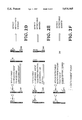
- FIGS. 2A-F depict several approaches for isolating and/or detecting the detectable marker on the immobilized hybridization probes where competitive hybridization probes are used.
- FIG. 2A illustrates a detectable marker being detected by first digesting the nucleic acids immobilized on the solid support using DNase.
- FIG. 2B illustrates the detectable marker being separated from the solid support prior to detection of the detectable marker by the dehybridization of the hybridization probes.
- FIG. 2C illustrates the detectable marker being separated from the solid support prior to detection of the detectable marker by detaching the second complexing agent from the solid support.
- FIG. 2D illustrates the detectable marker being separated from the solid support prior to detection of the detectable marker by detaching the first and second complexing agents.
- FIG. 2E illustrates the detectable marker being separated from the solid support prior to detection of the detectable marker by detaching the first complexing agent from the hybridization probes.
- FIG. 2F illustrates the detectable marker being separated from the solid support prior to detection of the detectable marker by detaching the detectable marker from the hybridization probes.
- the present invention relates to a rapid and efficient hybridization assay for detecting, isolating, and accurately quantifying target nucleic acid sequences in a sample of nucleic acids.
- two or more hybridization probes are used which hybridize to the same sequence of a target nucleic acid.
- the hybridization assay is used to detect the presence or absence of a nucleic acid sequence associated with a disease.
- the nucleic acid sequence to be detected may be a cancer gene or a pathogen (viral, fungal or bacterial) DNA insert.
- a sample of nucleic acids is first obtained.
- the chromosomal DNA is first isolated from a sample of cells.
- Chromosomal DNA may be isolated by any of the variety of methods known in the art.
- the chromosomal DNA may be isolated by the method taught in Vooijs, et al. Am. J. Hum. Genet. 52:586-597 (1993) or by using the GIBCO BRL TRIzolTM Reagent (Life Technologies, Gaithersburg, Md.), each of which is incorporated herein by reference.
- Chromosomal DNA may be analyzed as whole chromosomes, chromosome fragments or chromosomal DNA fragments, all of which are hereinafter referred to as chromosomal DNA.
- the chromosomal DNA may be organized as an extended double strand, as extended nucleosomes, as chromatin fiber, as folded fiber, and as interphase, prophase or metaphase DNA.
- the hybridization assay of the present invention utilizes the fact that hybridization probes do not perfectly and stoichiometrically hybridize to a target sequence such that only a single hybridization probe binds to a given sequence. Rather, the actual hybridization of a hybridization probe to a target sequence is generally imperfect such that a series of hybridization probes partially hybridize to the same target sequence to which the hybridization probe is complementary. This is particularly true when hybridization probes having a long sequence of nucleic acids are used.
- the hybridization assay of the present invention is designed to take advantage of the imperfect, non-stoichiometric hybridization of hybridization probes by utilizing a competitive hybridization scheme in order to detect the presence of a target sequence in a sample of nucleic acids. More specifically, the assay presupposes that the hybridization probes will be imperfect and non-stoichiometric in nature and employs a series of hybridization probes in which the hybridization probes compete to hybridize to the same target sequence.
- the competitive nature of the hybridization assay of the present invention provides unusual control over the sensitivity of the hybridization assay. It also provides a faster, more accurate and more sensitive method for detecting and quantifying nucleic acid sequences.
- the assay is schematically illustrated in FIG. 1.
- the sample of nucleic acid sequences are simultaneously contacted with a first and a second fraction of hybridization probes under conditions favorable to hybridization.
- the first fraction of hybridization probes includes a first hybridization probe and a second hybridization probe where the nucleic acid sequences used to form each hybridization probe are able to simultaneously hybridize to the target nucleic acid sequence.
- the first and second hybridization probes may hybridize to separate and distinct portions of the target sequence or, may hybridize to overlapping portions of the target sequence.
- the first and second hybridization probes are only limited in that the first and second probes each include a nucleic acid sequence that is specific to the target sequence but that does not overlap with the other sequence, thereby enabling both hybridization probes to simultaneously hybridize to the target sequence.
- the first and second hybridization probes may include RNA or DNA sequences such that the complementary nucleic acid sequences formed between the hybridization probes and the target sequence may be two DNA sequences, two RNA sequences or an RNA and a DNA sequence.
- the hybridization probes in the first fraction further include a first complexing agent capable of forming a binding pair with a second complexing agent which may be attached to a solid support.
- the formation of a bond between the first and second complexing agent enables the immobilization of the first fraction of hybridization probes to a solid support through the attachment of the second complexing agent to the solid support.
- those nucleic acid sequences that hybridize to the immobilized first fraction of hybridization probes will also be immobilized.
- the second fraction of hydridization probes includes the same first and second hybridization probes as are employed in the first fraction of hybridization probes except that the hybridization probes in the second fraction include a detectable marker instead of the first complexing agent. As illustrated in FIG. 1, the detectable marker serves to enable the detection of those target sequences that hybridize to at least one hybridization probe from the first and second fractions of hybridization probes.
- the ratio of the first hybridization probe relative to the second hybridization probe used in the first fraction of hybridization probes is approximately equal to the ratio of the first hybridization probe relative to the second hybridization probe in the second fraction of hybridization probes.
- the ratio of first to second hybridization probes in the second fraction also is X:Y.
- the first and second fractions of hybridization probes have the same relative concentration of first and second hybridization probes.
- the proportion of hybridization probes hybridizing to the target sequence from the first and second fractions should correlate to the ratio of hybridization probes from the first and second fractions that are used.
- the ratio between the first and second fractions of hybridization probes may be used to control the sensitivity of the hybridization assay.
- the first and second hybridization probes are simultaneously contacted with the sample of nucleic acids such that the two fractions of hybridization probes competitively hybridize to the target sequence.
- the detectable marker is attached to only a given fraction of hybridization probes, i.e., the second fraction of hybridization probes.
- a greater number of second hybridization probes will hybridize to the target sequence.
- a greater number of detectable markers will be immobilized to indicate the presence of the target sequence.
- This enables one to control the amount of detectable marker that becomes attached to the target sequence, thereby providing the user of the present assay with control over the amount of detectable marker that becomes attached to the target sequence. Accordingly, one is able to increase or decrease the sensitivity of the assay of the present invention by increasing or decreasing the ratio of the second fraction of hybridization probes to the first fraction.
- the ratio between the first fraction and the second fraction be between about 1:4 and 4:1, more preferably 1:1.
- the hybridized target nucleic acid sequences are contacted with a second complexing agent bound to a solid support.
- the second complexing agent forms a binding pair with the first complexing agent which is bound to the hybridization probes of the first fraction.
- the binding pair formed between the first and second complexing agents serves to immobilize the first fraction of hybridization probes as well as those target sequences which are hybridized to the first fraction of probes. Any hybridization probes from the second fraction which are hybridized to target sequences that are immobilized on the solid support will also become immobilized.
- the immobilized target sequences are then separated from any non-immobilized nucleic acids. Separation of the immobilized nucleic acids from non-immobilized nucleic acids may be accomplished by a variety of methods known in the art including, but not limited to, centrifugation, filtration, magnetic separation, chemical separation and washing.
- the immobilized target sequences After the immobilized target sequences have been separated from any non-immobilized nucleic acids, the immobilized sequences are analyzed for the presence of a detectable marker. The quantity of a target sequence in a sample can then be readily determined by quantifying the detectable marker.
- the hybridization probes used in the invention are preferably between about 100 and 1000 base pairs long, more preferably between about 200 and 700 base pairs long, and most preferably between about 300 and 500 base pairs long.
- the first and second complexing agents used in the embodiment of the present invention may be any pair of complexing agents which form a strong binding pair. Since elevated temperatures are generally required for hybridization, the binding pair should preferably be stable at temperatures at least up to about 37° C. under hybridization conditions.
- suitable binding pairs of complexing agents include antibody-antigen pairs, biotin-avidin and digoxigenin-anti-digoxigenin.
- Avidin-biotin and analogues and derivatives thereof are particularly preferred as binding pairs due to their enhanced thermal stability.
- avidin derivatives include, but are not limited to, streptavidin, succinyl avidin, ferritin avidin, enzyme avidin and cross-linked avidin.
- biotin derivatives include, but are not limited to caproylamidobiotin and biocytin.
- biotin analogues include, but are not limited to desthiobiotin and biotin sulfone.
- Biotin-antibiotin antibody is an example of a suitable antibody-antigen pair.
- any solid support to which a complexing agent may be attached may be used in the present invention.
- suitable solid support materials include, but are not limited to, silicates such as glass and silica gel, cellulose and nitrocellulose papers, nylon, polystyrene, polymethacrylate, latex, rubber, and fiuorocarbon resins such as TEFLONTM.
- the solid support material may be used in a wide variety of shapes including, but not limited to slides and beads.
- Slides provide several functional advantages and thus are a preferred form of solid support. Slides can be readily used with any chromosome organization. Due to their flat surface, probe and hybridization reagents can be minimized using glass slides. Slides also enable the targeted application of reagents, are easy to keep at a constant temperature, are easy to wash and facilitate the direct visualization of RNA and/or DNA immobilized on the solid support. Removal of RNA and/or DNA immobilized on the solid support is also facilitated using slides. It is estimated that a standard microscope glass slide can contain 50,000 to 100,000 cells worth of DNA. Beads, such as BioMag Strepavidin magnetic beads are another preferred form of solid support containing a second complexing agent.
- avidin or an avidin derivative be used as the second complexing agent.
- Avidin may be chemically attached to glass using the N-hydroxysuccinamide active ester of avidin as taught by Manning, et al. Biochemistry 16:1364-1370 (1977) and may be attached to nylon by a carbodiimide coupling as taught by Jasiewicz, et al. Exp. Cell Res. 100:213-217 (1976).
- Magnetic microbeads labelled with avidin and strepavidin labelled bead may be obtained from Advanced Magnetics, Inc., Cambridge, Mass. and from Spherotech, Inc., Libertyville, Ill.
- any analytically detectable marker that can be attached to or incorporated into a hybridization probe may be used in the present invention.
- An analytically detectable marker refers to any molecule, moiety or atom which can be analytically detected and quantified.
- Methods for detecting analytically detectable markers include, but are not limited to, radioactivity, fluorescence, absorbance, mass spectroscopy, EPR, NMR, XRF, luminescence and phosphorescence.
- any radiolabel which provides an adequate signal and a sufficient half-life may be used as a detectable marker.
- Commonly used radioisotopes include 3 H, 14 C, 32 P and 125 I. In a preferred embodiment, 14 C is used as the detectable marker and is detected by accelerator mass spectroscopy (AMS).
- 14 C is preferred because of its exceptionally long half-life and because of the very high sensitivity of AMS for detecting 14 C isotopes.
- Other isotopes that may be detected using AMS include, but are not limited to, 3 H, 125 I, 41 Ca, 63 Ni and 38 Cl.
- Fluorescent molecules such as fluorescein and its derivatives, rhodamine and its derivatives, dansyl, umbeliferone and acridimium, and chemiluminescent molecules such as luciferin and 2,3-dihydrophthalazinediones may also be used as detectable markers.
- Molecules which bind to an analytically detectable marker may also be covalently attached to or incorporated into hybridization probe, for example, as taught by Ward, European Patent Application No. 63,879 which is incorporated herein by reference. In such instances, the hybridization probe is detected by adding an analytically detectable marker which specifically binds to the probe, thereby enabling detection of the probe.
- Examples of such molecules and their analytically detectable counterparts include biotin and either fluorescent or chemiluminescent avidin.
- Antibodies that bind to an analytically detectable antigen may also be used as a detectable marker.
- the detectable marker may also be a molecule which, when subjected to chemical or enzymatic modification, becomes analytically detectable such as those disclosed in Leafy, et al., Proc. Natl. Acad. Sci. (U.S.A.), 80:4045-4049 (1983) which is incorporated herein by reference.
- Other examples of suitable detectable markers include protein binding sequences which can be detected by binding proteins, such as those disclosed in U.S. Pat. No. 4,556,643 which is incorporated herein by reference.
- the nucleic acid sequence employed in the first and/or second hybridization probe may function as a detectable marker where the bases forming the nucleic acid sequence are quantified using techniques known in the art.
- the nucleic acid sequence employed in the hybridization probe may itself function as a detectable marker where the bases forming the nucleic acid sequence are quantified using techniques known in the art.
- the presence or absence of the detectable marker attached to the second fraction of hybridization probes is detected in order to quantify the target sequence.
- the detection and quantification of the detectable marker can be performed using a variety of methods, depending upon the particular hybridization probes and detectable markers employed.
- the first fraction of hybridization probes is preferably detachably linked to the solid support. This may be accomplished by incorporating a detachable linker between the first fraction of hybridization probes and the first complexing agent, between the solid support and the second complexing agent or between the first and second complexing agents.
- the presence of the released target sequences may be determined by detecting the presence of the detectable marker or by detecting the presence of the released target sequence itself.
- the target nucleic acid sequence hybridized to the hybridization probes can be accomplished by releasing the target nucleic acid sequences from the solid support, dehybridizing the target sequence and the hybridization probes, and purifying the target sequence, for example by gel electrophoresis.
- the target sequence may readily be isolated and made available for further analysis, such as sequencing.
- FIGS. 2A-F illustrate alternate embodiments for detecting the detectable marker.
- the detectable marker may be detected by treating the immobilized nucleic acid sequences with DNase to digest any DNA immobilized on the solid support. The digested DNA is then collected after enzymatic digestion and analyzed for the presence of the detectable marker.
- the nucleic acids attached to the solid support may be removed from the solid support by a variety of chemical and physical methods available, including, for example, treatment with a basic solution (e.g., concentrated NaOH), treatment with an acidic solution and denaturalization of DNA using standard methods such as elevated temperatures or reagents.
- the entire solid support containing the immobilized nucleic acids and hybridization probes may be graphitized and analyzed using accelerator mass spectroscopy (AMS).
- AMS accelerator mass spectroscopy
- the detectable marker may be separated from the solid support prior to detection of the detectable marker by dehybridizing the hybridization probes from the target nucleic acid sequence. This may be done by heating the solid support and immobilized sequences to at least 70° C. in a denaturization solution.
- the detectable marker may be separated from the solid support odor to detection of the detectable marker by breaking the bond between the second complexing agent and the solid support. This may be accomplished through the use of a detachable linker positioned between the second complexing agent and the solid support.
- suitable detachable linkages include, but are not limited to the detachable linkers described in Lin, et al., J. Org. Chem. 56:6850-6856 (1991); Ph.D. Thesis of W. -C. Lin, U. C. Riverside, (1990); Hobart, et al., J.
- the detectable marker may be separated from the solid support prior to detection of the detectable marker by breaking the bond between the first and second complexing agents.
- the bond between the first and second complexing agents may be broken.
- the detectable marker may be separated from the solid support prior to detection of the detectable marker by breaking the bond between the first complexing agent and the nucleic acid sequences forming the hybridization probe. This may be accomplished through the use of a detachable linker positioned between the first complexing agent and the nucleic acid sequence forming the hybridization probe. Examples of suitable detachable linkages include, but are not limited to the detachable linkers described in the references cited above.
- the detectable marker may be separated from the solid support prior to detection by detaching the detectable marker from the hybridization probe. This may be accomplished through the use of a detachable linker between the detectable marker and the nucleic acid sequence forming the hybridization probe. Examples of suitable detachable linkages include, but are not limited to, the detachable linkers described in the references cited above.
- the detectable marker may be detected by a variety of methods known in the art, depending on the particular detectable marker employed.
- AMS may be used when the detectable marker is a radioisotope such as 14 C
- liquid scintillation may be used when the detectable marker is tritiated thymidine
- standard fluorescence or spectroscopic methods may be used when the detectable marker is a fluorescent molecule or the DNA itself.
- the quantity of the target nucleic acid sequence that is present may be determined based on the signal generated from the detectable marker using a calibration curve.
- the calibration curve may be formed by analyzing a serial dilution of a sample of nucleic acids having a known concentration of the target sequence.
- a calibration curve may be generated by analyzing a series of known amounts of cells from a cell line in which the concentration of the target sequence is known.
- samples of cells may be analyzed according to the method of the present invention and according to a method known in the art for quantifying the target nucleic acid sequence.
- Alternative methods for generating a calibration curve are within the level of skill in the art and may be used in conjunction with the method of the present invention.
- the present invention also relates to a kit for performing the hybridization assays of the present invention. Unless otherwise specified, the components of the kit are the same as those used in the assays of the present invention.
- the kit includes a first hybridization probe mixture for use as the first fraction in the assay and a second hybridization probe mixture for use as the second fraction in the assay.
- the first and second hybridization probe mixtures each contain the same ratio of first hybridization probes to second hybridization probes.
- the hybridization probes of the first hybridization probe mixture also include a first complexing agent attached to the probe that is capable of forming a binding pair with a second complexing agent.
- the hybridization probes of the second hybridization probe mixture include a detectable marker. It is preferred that the detectable marker included on the second hybridization probe mixture be of a known concentration relative to the second hybridization probe mixture.
- the kit may also include a second complexing agent attached to a solid support capable of binding to the first complexing agent used in first hybridization probe mixture.
- the kit may also include written instructions for practicing the assay and one or more target nucleic acid sequences for use in the preparation of a calibration curve.
- a first and a second hybridization probe are generated for a target sequence which can simultaneously hybridize to the target sequence.
- Several techniques are known in the art for generating single-stranded hybridization probes to a target nucleic acid sequence, for example, as a messenger RNA sequence corresponding to the target sequence, or complementary DNA obtained from reverse transcriptase, or as genomic DNA obtained from the target genome by endonuclease digestion.
- Biotinylated first and second hybridization probes are prepared by chemically modifying aliquots of the first and the second hybridization probes to incorporate biotinylated uridine according to the method of Pinkel, et al., Proc. Natl. Acad. Sci. (USA) 83:2934-2938 (1986), which is incorporated herein by reference.
- first and second hybridization probes are also prepared by amplifying the first and the second hybridization probes using pcr in the presence of a 4 C labelled nucleic acid base according to the method described in Vooijs, et al., Am. J. Hum. Genet. 52:586-597 (1993) which is incorporated herein by reference.
- the first fraction of hybridization probes is prepared by mixing the first and second biotinylated probes in a 1:1 ratio.
- the second fraction of hybridization probes is then prepared by mixing the first and second 14 C labelled probes in a 1:1 ratio.
- the first and second fractions of hybridization probes are then mixed together such that the ratio between the first and second fractions of hybridization probes is 1:1.
- the mixture of fractions of hybridization probes are contacted with a sample of nucleic acids under conditions favorable for hybridization.
- any unbound probes may optionally be separated from the hybridized target DNA by gel electrophoresis.
- the sample of nucleic acids containing the hybridized competitive probes is added to a solid support labelled with avidin to immobilize the first fraction of hybridization probes by an avidin-biotin linkage. Any nucleic acids hybridized to the first fraction of hybridization probes also become immobilized to the solid support.
- the avidin labelled solid support may be prepared by the methods described in Manning, et al. Biochemistry 16:1364-1370 (1977) and Jasiewicz, et al. Exp. Cell Res. 100:213-217 (1976), each of which are incorporated herein by reference.
- the solid support is then washed with cold, pH 7 buffered saline to remove any first and second hybridization probes and DNA segments which are not immobilized on the solid support.
- the remaining immobilized nucleic acids are analyzed for the presence of 14 C.
- DNase or concentrated NaOH is employed to separate any immobilized nucleic acids from the solid support.
- the nucleic acids isolated are then grafitized and analyzed using AMS for the presence of 14 C according to the method of Vogel et. al., Anal. Chem. 11: 142-149 (1991) which is incorporated herein by reference.
- the 14 C signal obtained from the accelerator mass spectrometer may be calibrated by performing the assay using a sample containing a known quantity of the target nucleic acid.
- the nucleic acid sample being analyzed may be analyzed according to the method of the present invention and according to a method known in the art for quantifying the target nucleic acid. Then, by serially diluting the sample of nucleic acids and assaying the sample according to the method described in the present example, a calibration curve may be generated.
- Alternative methods for generating a calibration curve are within the level of skill in the art and may be used in conjunction with the method of the present invention.
Abstract
Description
Claims (12)
Priority Applications (2)
| Application Number | Priority Date | Filing Date | Title |
|---|---|---|---|
| US08/512,897 US5616465A (en) | 1995-08-09 | 1995-08-09 | Detection and isolation of nucleic acid sequences using competitive hybridization probes |
| US09/364,155 US6270972B1 (en) | 1995-08-09 | 1999-07-30 | Kit for detecting nucleic acid sequences using competitive hybridization probes |
Applications Claiming Priority (1)
| Application Number | Priority Date | Filing Date | Title |
|---|---|---|---|
| US08/512,897 US5616465A (en) | 1995-08-09 | 1995-08-09 | Detection and isolation of nucleic acid sequences using competitive hybridization probes |
Related Child Applications (1)
| Application Number | Title | Priority Date | Filing Date |
|---|---|---|---|
| US72054096A Continuation | 1995-08-09 | 1996-09-30 |
Publications (1)
| Publication Number | Publication Date |
|---|---|
| US5616465A true US5616465A (en) | 1997-04-01 |
Family
ID=24041079
Family Applications (2)
| Application Number | Title | Priority Date | Filing Date |
|---|---|---|---|
| US08/512,897 Expired - Lifetime US5616465A (en) | 1995-08-09 | 1995-08-09 | Detection and isolation of nucleic acid sequences using competitive hybridization probes |
| US09/364,155 Expired - Fee Related US6270972B1 (en) | 1995-08-09 | 1999-07-30 | Kit for detecting nucleic acid sequences using competitive hybridization probes |
Family Applications After (1)
| Application Number | Title | Priority Date | Filing Date |
|---|---|---|---|
| US09/364,155 Expired - Fee Related US6270972B1 (en) | 1995-08-09 | 1999-07-30 | Kit for detecting nucleic acid sequences using competitive hybridization probes |
Country Status (1)
| Country | Link |
|---|---|
| US (2) | US5616465A (en) |
Cited By (12)
| Publication number | Priority date | Publication date | Assignee | Title |
|---|---|---|---|---|
| US5916776A (en) * | 1997-08-27 | 1999-06-29 | Sarnoff Corporation | Amplification method for a polynucleotide |
| US6027879A (en) * | 1995-08-09 | 2000-02-22 | The Regents Of The University Of California | Detection and isolation of nucleic acid sequences using a bifunctional hybridization probe |
| WO2002031206A2 (en) * | 2000-10-11 | 2002-04-18 | Ragland William L | Methods and compositions using hybridization assays for detecting infectious agents |
| US20030027157A1 (en) * | 2000-09-27 | 2003-02-06 | Rongdian Fu | Method for determining relative abundance of nucleic acid sequences |
| US6569647B1 (en) | 1994-06-22 | 2003-05-27 | Mount Sinai School Of Medicine Of New York University | Nucleic acid amplification method: ramification-extension amplification method (RAM) |
| US6593086B2 (en) | 1996-05-20 | 2003-07-15 | Mount Sinai School Of Medicine Of New York University | Nucleic acid amplification methods |
| US20030175706A1 (en) * | 1994-06-22 | 2003-09-18 | Zhang David Y. | Nucleic acid amplification methods |
| US20040062750A1 (en) * | 1999-09-14 | 2004-04-01 | The General Hospital Corporation | Use of mullerian inhibiting substance for treating excess androgen states |
| US20040130709A1 (en) * | 2002-10-16 | 2004-07-08 | Taizo Yamamoto | Appearance inspection machine for flat tablet |
| US20050118603A1 (en) * | 2002-10-11 | 2005-06-02 | Ahram Biosystems Inc. | Target detection system having a conformationally sensitive probe comprising a nucleic acid based signal transducer |
| US20050176032A1 (en) * | 1998-12-23 | 2005-08-11 | Breslauer Kenneth J. | Methods and kits for screening nucleic acid duplex stability |
| WO2017223075A1 (en) * | 2016-06-20 | 2017-12-28 | Slive, Inc. | Biomarker detection |
Citations (20)
| Publication number | Priority date | Publication date | Assignee | Title |
|---|---|---|---|---|
| US4358535A (en) * | 1980-12-08 | 1982-11-09 | Board Of Regents Of The University Of Washington | Specific DNA probes in diagnostic microbiology |
| US4376110A (en) * | 1980-08-04 | 1983-03-08 | Hybritech, Incorporated | Immunometric assays using monoclonal antibodies |
| WO1983001459A1 (en) * | 1981-10-16 | 1983-04-28 | Ranki, Tuula, Marjut | A method and reagent combination for the diagnosis of microorganisms by sandwich hybridization of nucleic acids |
| WO1984003285A1 (en) * | 1983-02-22 | 1984-08-30 | Molecular Biosystems Inc | Defined sequence single strand oligonucleotides incorporating reporter groups, process for the chemical synthesis thereof, and nucleosides useful in such synthesis |
| EP0117440A1 (en) * | 1983-01-27 | 1984-09-05 | Enzo Biochem, Inc. | Methods and structures employing non-radioactive chemically-labeled polynucleotide probes |
| WO1985004674A1 (en) * | 1984-04-05 | 1985-10-24 | Life Technologies, Inc. | Immobilization of nucleic acids |
| US4556643A (en) * | 1982-07-26 | 1985-12-03 | Agracetus | Assay method and probe for polynucleotide sequences |
| WO1987003622A1 (en) * | 1985-12-13 | 1987-06-18 | Princeton University | Amplified hybridization assay |
| WO1987003911A1 (en) * | 1985-12-17 | 1987-07-02 | Genetics Institute, Inc. | Displacement polynucleotide method and reagent complex |
| EP0235726A2 (en) * | 1986-03-05 | 1987-09-09 | Miles Inc. | Rapid detection of nucleic acid sequences in a sample by labeling the sample |
| WO1988001302A1 (en) * | 1986-08-11 | 1988-02-25 | Siska Diagnostics, Inc. | Nucleic acid probe assay methods and compositions |
| US4775619A (en) * | 1984-10-16 | 1988-10-04 | Chiron Corporation | Polynucleotide determination with selectable cleavage sites |
| EP0286898A2 (en) * | 1982-06-23 | 1988-10-19 | Enzo Biochem, Inc. | Modified labeled nucleotides and polynucleotides and methods of preparing, utilizing and detecting same |
| EP0292128A1 (en) * | 1987-04-28 | 1988-11-23 | Tamir Biotechnology Ltd | Improved DNA probes |
| US4794082A (en) * | 1987-02-17 | 1988-12-27 | Hoechst Celanese Corporation | Biotinylating agents |
| US5109124A (en) * | 1988-06-01 | 1992-04-28 | Biogen, Inc. | Nucleic acid probe linked to a label having a terminal cysteine |
| US5273882A (en) * | 1985-06-13 | 1993-12-28 | Amgen | Method and kit for performing nucleic acid hybridization assays |
| US5310650A (en) * | 1986-09-29 | 1994-05-10 | Abbott Laboratoires | Method and device for improved reaction kinetics in nucleic acid hybridizations |
| US5438194A (en) * | 1993-07-30 | 1995-08-01 | High Voltage Engineering Europa B.V. | Ultra-sensitive molecular identifier |
| EP0231495B1 (en) * | 1985-12-13 | 1999-06-16 | Enzo Biochem, Inc. | One-step method and polynucleotide compounds for hybridizing to target polynucleotides |
Family Cites Families (3)
| Publication number | Priority date | Publication date | Assignee | Title |
|---|---|---|---|---|
| US5200314A (en) | 1990-03-23 | 1993-04-06 | Chiron Corporation | Polynucleotide capture assay employing in vitro amplification |
| US5387505A (en) | 1990-05-04 | 1995-02-07 | Eastman Kodak Company | Preparation and isolation of single-stranded biotinylated nucleic acids by heat avidin-biotin cleavage |
| US5521300A (en) | 1991-08-13 | 1996-05-28 | Norval B. Galloway | Oligonucleotides complementary to mycobacterial nucleic acids |
-
1995
- 1995-08-09 US US08/512,897 patent/US5616465A/en not_active Expired - Lifetime
-
1999
- 1999-07-30 US US09/364,155 patent/US6270972B1/en not_active Expired - Fee Related
Patent Citations (25)
| Publication number | Priority date | Publication date | Assignee | Title |
|---|---|---|---|---|
| US4376110A (en) * | 1980-08-04 | 1983-03-08 | Hybritech, Incorporated | Immunometric assays using monoclonal antibodies |
| US4358535A (en) * | 1980-12-08 | 1982-11-09 | Board Of Regents Of The University Of Washington | Specific DNA probes in diagnostic microbiology |
| US4358535B1 (en) * | 1980-12-08 | 1986-05-13 | ||
| US4563419A (en) * | 1981-10-16 | 1986-01-07 | Orion Corporation Ltd. | Detection of microbial nucleic acids by a one-step sandwich hybridization test |
| WO1983001459A1 (en) * | 1981-10-16 | 1983-04-28 | Ranki, Tuula, Marjut | A method and reagent combination for the diagnosis of microorganisms by sandwich hybridization of nucleic acids |
| EP0079139A1 (en) * | 1981-10-16 | 1983-05-18 | Orion-yhtymä Oy | A method and reagent combination for the identification of microorganisms and the use of sandwich hybridization of nucleic acids therefor |
| US4486539A (en) * | 1981-10-16 | 1984-12-04 | Orioon Corporation Ltd. | Detection of microbial nucleic acids by a one-step sandwich hybridization test |
| EP0286898A2 (en) * | 1982-06-23 | 1988-10-19 | Enzo Biochem, Inc. | Modified labeled nucleotides and polynucleotides and methods of preparing, utilizing and detecting same |
| EP0285057B1 (en) * | 1982-06-23 | 1995-03-01 | Enzo Biochem, Inc. | Modified labeled nucleotides and polynucleotides and methods of preparing, utilizing and detecting same |
| US4556643A (en) * | 1982-07-26 | 1985-12-03 | Agracetus | Assay method and probe for polynucleotide sequences |
| EP0117440A1 (en) * | 1983-01-27 | 1984-09-05 | Enzo Biochem, Inc. | Methods and structures employing non-radioactive chemically-labeled polynucleotide probes |
| WO1984003285A1 (en) * | 1983-02-22 | 1984-08-30 | Molecular Biosystems Inc | Defined sequence single strand oligonucleotides incorporating reporter groups, process for the chemical synthesis thereof, and nucleosides useful in such synthesis |
| WO1985004674A1 (en) * | 1984-04-05 | 1985-10-24 | Life Technologies, Inc. | Immobilization of nucleic acids |
| US4775619A (en) * | 1984-10-16 | 1988-10-04 | Chiron Corporation | Polynucleotide determination with selectable cleavage sites |
| US5273882A (en) * | 1985-06-13 | 1993-12-28 | Amgen | Method and kit for performing nucleic acid hybridization assays |
| WO1987003622A1 (en) * | 1985-12-13 | 1987-06-18 | Princeton University | Amplified hybridization assay |
| EP0231495B1 (en) * | 1985-12-13 | 1999-06-16 | Enzo Biochem, Inc. | One-step method and polynucleotide compounds for hybridizing to target polynucleotides |
| WO1987003911A1 (en) * | 1985-12-17 | 1987-07-02 | Genetics Institute, Inc. | Displacement polynucleotide method and reagent complex |
| EP0235726A2 (en) * | 1986-03-05 | 1987-09-09 | Miles Inc. | Rapid detection of nucleic acid sequences in a sample by labeling the sample |
| WO1988001302A1 (en) * | 1986-08-11 | 1988-02-25 | Siska Diagnostics, Inc. | Nucleic acid probe assay methods and compositions |
| US5310650A (en) * | 1986-09-29 | 1994-05-10 | Abbott Laboratoires | Method and device for improved reaction kinetics in nucleic acid hybridizations |
| US4794082A (en) * | 1987-02-17 | 1988-12-27 | Hoechst Celanese Corporation | Biotinylating agents |
| EP0292128A1 (en) * | 1987-04-28 | 1988-11-23 | Tamir Biotechnology Ltd | Improved DNA probes |
| US5109124A (en) * | 1988-06-01 | 1992-04-28 | Biogen, Inc. | Nucleic acid probe linked to a label having a terminal cysteine |
| US5438194A (en) * | 1993-07-30 | 1995-08-01 | High Voltage Engineering Europa B.V. | Ultra-sensitive molecular identifier |
Non-Patent Citations (6)
| Title |
|---|
| Enzymatic Synthesis of Biotin labeled Polynucleotides: Novel Nucleic Acid Affinity Probes, Pennina R. Langer, Alex A. Waldrop and David C. Ward; Proc. Natl. Acad. Sci. USA, vol. 78, No. 11, pp. 6633 6637. * |
| Enzymatic Synthesis of Biotin-labeled Polynucleotides: Novel Nucleic Acid Affinity Probes, Pennina R. Langer, Alex A. Waldrop and David C. Ward; Proc. Natl. Acad. Sci. USA, vol. 78, No. 11, pp. 6633-6637. |
| Gene Maping and Gene Enrichment by the Avidin Biotin Interaction: Use of Cytochrome c as a Polyamine Bridge; Ann Sodja and Norman Davidson; Nucleic Acids Research, vol. 5, No. 2, Feb. 1978, pp. 385 401. * |
| Gene Maping and Gene Enrichment by the Avidin-Biotin Interaction: Use of Cytochrome-c as a Polyamine Bridge; Ann Sodja and Norman Davidson; Nucleic Acids Research, vol. 5, No. 2, Feb. 1978, pp. 385-401. |
| Ligation of Oligonucleotides to Nucleic Acids or Proteins via Disulfide Bonds, Barbara C. F. Chu and Leslie E. Orgel; Nucleic Acids Research, vol. 16, No. 9, 1988, pp. 3671 3691. * |
| Ligation of Oligonucleotides to Nucleic Acids or Proteins via Disulfide Bonds, Barbara C. F. Chu and Leslie E. Orgel; Nucleic Acids Research, vol. 16, No. 9, 1988, pp. 3671-3691. |
Cited By (19)
| Publication number | Priority date | Publication date | Assignee | Title |
|---|---|---|---|---|
| US20030190604A1 (en) * | 1994-06-22 | 2003-10-09 | Zhang David Y. | Nucleic acid amplification method: ramification-extension amplification method (RAM) |
| US20030175706A1 (en) * | 1994-06-22 | 2003-09-18 | Zhang David Y. | Nucleic acid amplification methods |
| US20070269799A9 (en) * | 1994-06-22 | 2007-11-22 | Zhang David Y | Nucleic acid amplification methods |
| US8632999B1 (en) | 1994-06-22 | 2014-01-21 | David Y. Zhang | Nucleic acid amplification methods |
| US6569647B1 (en) | 1994-06-22 | 2003-05-27 | Mount Sinai School Of Medicine Of New York University | Nucleic acid amplification method: ramification-extension amplification method (RAM) |
| US6855523B2 (en) | 1994-06-22 | 2005-02-15 | Mount Sinai School Of Medicine Of New York University | Nucleic acid amplification method: ramification-extension amplification method (RAM) |
| US6027879A (en) * | 1995-08-09 | 2000-02-22 | The Regents Of The University Of California | Detection and isolation of nucleic acid sequences using a bifunctional hybridization probe |
| US6593086B2 (en) | 1996-05-20 | 2003-07-15 | Mount Sinai School Of Medicine Of New York University | Nucleic acid amplification methods |
| US5916776A (en) * | 1997-08-27 | 1999-06-29 | Sarnoff Corporation | Amplification method for a polynucleotide |
| US7468250B2 (en) * | 1998-12-23 | 2008-12-23 | Rutgers, The State University Of New Jersey | Methods and kits for screening nucleic acid duplex stability |
| US20050176032A1 (en) * | 1998-12-23 | 2005-08-11 | Breslauer Kenneth J. | Methods and kits for screening nucleic acid duplex stability |
| US20040062750A1 (en) * | 1999-09-14 | 2004-04-01 | The General Hospital Corporation | Use of mullerian inhibiting substance for treating excess androgen states |
| US20030027157A1 (en) * | 2000-09-27 | 2003-02-06 | Rongdian Fu | Method for determining relative abundance of nucleic acid sequences |
| US6897023B2 (en) * | 2000-09-27 | 2005-05-24 | The Molecular Sciences Institute, Inc. | Method for determining relative abundance of nucleic acid sequences |
| WO2002031206A3 (en) * | 2000-10-11 | 2003-07-31 | William L Ragland | Methods and compositions using hybridization assays for detecting infectious agents |
| WO2002031206A2 (en) * | 2000-10-11 | 2002-04-18 | Ragland William L | Methods and compositions using hybridization assays for detecting infectious agents |
| US20050118603A1 (en) * | 2002-10-11 | 2005-06-02 | Ahram Biosystems Inc. | Target detection system having a conformationally sensitive probe comprising a nucleic acid based signal transducer |
| US20040130709A1 (en) * | 2002-10-16 | 2004-07-08 | Taizo Yamamoto | Appearance inspection machine for flat tablet |
| WO2017223075A1 (en) * | 2016-06-20 | 2017-12-28 | Slive, Inc. | Biomarker detection |
Also Published As
| Publication number | Publication date |
|---|---|
| US6270972B1 (en) | 2001-08-07 |
Similar Documents
| Publication | Publication Date | Title |
|---|---|---|
| US5731153A (en) | Identification of random nucleic acid sequence aberrations using dual capture probes which hybridize to different chromosome regions | |
| JP3738910B2 (en) | Hybridization-ligation analysis to detect specific nucleic acid sequences | |
| JP2801051B2 (en) | Methods and reagents for detecting nucleobase sequences | |
| US4752566A (en) | Displacement polynucleotide method and reagent complex employing labeled probe polynucleotide | |
| DK175384B1 (en) | Improved method for determining nucleic acids and reagent combination and reagent kits thereto | |
| JP2577881B2 (en) | Assay method using polynucleotide chain | |
| US6428964B1 (en) | Method for alteration detection | |
| AU658962B2 (en) | Rapid assays for amplification products | |
| EP0204510B1 (en) | Amplification of hybridization signals by employing complementary dna strands | |
| JPH08242896A (en) | Method for detecting arrangement of nucleic acid | |
| US5616465A (en) | Detection and isolation of nucleic acid sequences using competitive hybridization probes | |
| CA2004326A1 (en) | Assay of sequences using amplified genes | |
| IE902390A1 (en) | Hydrophobic nucleic acid probe | |
| JP4425640B2 (en) | DNA-binding protein detection method | |
| JPH0743376B2 (en) | Nucleic acid sequence assay method and molecular gene probe | |
| JP2644419B2 (en) | Immobilization of nucleic acids | |
| US6027879A (en) | Detection and isolation of nucleic acid sequences using a bifunctional hybridization probe | |
| JPS61502889A (en) | Chemically labeled cholic acid, its usage and kit for its implementation | |
| US5783387A (en) | Method for identifying and quantifying nucleic acid sequence aberrations | |
| WO2023173711A1 (en) | Aptamer for specifically recognizing soluble st2 and use thereof | |
| JP5211790B2 (en) | DNA methylation measurement method | |
| EP1352097B1 (en) | A method and test kit for quantitative determination of variations in polynucleotide amounts in cell or tissue samples | |
| US5871906A (en) | Method for detection of amplified nucleic acid products and related diagnostic assays | |
| AU2002226448A1 (en) | A method and test kit for quantitative determination of variations in polynucleotide amounts in cell or tissue samples | |
| Bortolin et al. | Time-resolved immunofluorometric determination of specific mRNA sequences amplified by the polymerase chain reaction |
Legal Events
| Date | Code | Title | Description |
|---|---|---|---|
| AS | Assignment |
Owner name: CALIFORNIA, REGENTS OF THE UNIVERSITY OF, THE, CAL Free format text: ASSIGNMENT OF ASSIGNORS INTEREST;ASSIGNORS:LUCAS, JOE N.;STRAUME, TORE;BOGEN, KENNETH T.;REEL/FRAME:007633/0274 Effective date: 19950727 Owner name: REGENTS OF THE UNIVESITY OF CALIFORNIA, THE, CALIF Free format text: ASSIGNMENT OF ASSIGNORS INTEREST;ASSIGNORS:LUCAS, JOE N.;STRAUME, TORE;BOGEN, KENNETH T.;REEL/FRAME:007633/0274 Effective date: 19950727 |
|
| STCF | Information on status: patent grant |
Free format text: PATENTED CASE |
|
| FEPP | Fee payment procedure |
Free format text: PAYOR NUMBER ASSIGNED (ORIGINAL EVENT CODE: ASPN); ENTITY STATUS OF PATENT OWNER: LARGE ENTITY |
|
| AS | Assignment |
Owner name: ENERGY, U.S. DEPARTMENT OF, CALIFORNIA Free format text: CONFIRMATORY LICENSE;ASSIGNOR:CALIFORNIA, UNIVERSITY OF;REEL/FRAME:010618/0866 Effective date: 20000217 |
|
| FPAY | Fee payment |
Year of fee payment: 4 |
|
| FPAY | Fee payment |
Year of fee payment: 8 |
|
| FEPP | Fee payment procedure |
Free format text: PAT HOLDER NO LONGER CLAIMS SMALL ENTITY STATUS, ENTITY STATUS SET TO UNDISCOUNTED (ORIGINAL EVENT CODE: STOL); ENTITY STATUS OF PATENT OWNER: LARGE ENTITY |
|
| AS | Assignment |
Owner name: LAWRENCE LIVERMORE NATIONAL SECURITY LLC, CALIFORN Free format text: ASSIGNMENT OF ASSIGNORS INTEREST;ASSIGNOR:THE REGENTS OF THE UNIVERSITY OF CALIFORNIA;REEL/FRAME:021217/0050 Effective date: 20080623 |
|
| FPAY | Fee payment |
Year of fee payment: 12 |