US5774599A - Method for precompensation of digital images for enhanced presentation on digital displays with limited capabilities - Google Patents
Method for precompensation of digital images for enhanced presentation on digital displays with limited capabilities Download PDFInfo
- Publication number
- US5774599A US5774599A US08/404,399 US40439995A US5774599A US 5774599 A US5774599 A US 5774599A US 40439995 A US40439995 A US 40439995A US 5774599 A US5774599 A US 5774599A
- Authority
- US
- United States
- Prior art keywords
- image
- frequency component
- signal
- digital image
- component signal
- Prior art date
- Legal status (The legal status is an assumption and is not a legal conclusion. Google has not performed a legal analysis and makes no representation as to the accuracy of the status listed.)
- Expired - Lifetime
Links
- 238000000034 method Methods 0.000 title claims abstract description 64
- 238000012545 processing Methods 0.000 claims abstract description 31
- 230000000007 visual effect Effects 0.000 claims abstract description 11
- 230000002238 attenuated effect Effects 0.000 claims description 14
- 230000004044 response Effects 0.000 claims description 10
- 230000003321 amplification Effects 0.000 claims description 4
- 238000003199 nucleic acid amplification method Methods 0.000 claims description 4
- 230000035945 sensitivity Effects 0.000 claims description 4
- 238000005259 measurement Methods 0.000 claims 1
- 230000002829 reductive effect Effects 0.000 abstract description 11
- 210000000038 chest Anatomy 0.000 description 20
- 238000009125 cardiac resynchronization therapy Methods 0.000 description 16
- 210000004072 lung Anatomy 0.000 description 11
- 230000008569 process Effects 0.000 description 9
- 230000006870 function Effects 0.000 description 6
- 238000003672 processing method Methods 0.000 description 6
- 239000000872 buffer Substances 0.000 description 5
- 238000002059 diagnostic imaging Methods 0.000 description 5
- 238000002601 radiography Methods 0.000 description 5
- 230000000694 effects Effects 0.000 description 4
- 230000001965 increasing effect Effects 0.000 description 4
- 201000003144 pneumothorax Diseases 0.000 description 4
- 208000029523 Interstitial Lung disease Diseases 0.000 description 3
- 238000013459 approach Methods 0.000 description 3
- 230000003247 decreasing effect Effects 0.000 description 3
- 230000002708 enhancing effect Effects 0.000 description 3
- 238000001914 filtration Methods 0.000 description 3
- 238000012546 transfer Methods 0.000 description 3
- 238000012800 visualization Methods 0.000 description 3
- 238000004458 analytical method Methods 0.000 description 2
- 230000008901 benefit Effects 0.000 description 2
- 230000006835 compression Effects 0.000 description 2
- 238000007906 compression Methods 0.000 description 2
- 238000003745 diagnosis Methods 0.000 description 2
- 230000004313 glare Effects 0.000 description 2
- 230000006872 improvement Effects 0.000 description 2
- 230000000670 limiting effect Effects 0.000 description 2
- 230000000873 masking effect Effects 0.000 description 2
- 210000001370 mediastinum Anatomy 0.000 description 2
- 230000003287 optical effect Effects 0.000 description 2
- 230000005855 radiation Effects 0.000 description 2
- 238000012360 testing method Methods 0.000 description 2
- 238000012935 Averaging Methods 0.000 description 1
- 102100035593 POU domain, class 2, transcription factor 1 Human genes 0.000 description 1
- 101710084414 POU domain, class 2, transcription factor 1 Proteins 0.000 description 1
- OAICVXFJPJFONN-UHFFFAOYSA-N Phosphorus Chemical compound [P] OAICVXFJPJFONN-UHFFFAOYSA-N 0.000 description 1
- 230000003044 adaptive effect Effects 0.000 description 1
- 210000003484 anatomy Anatomy 0.000 description 1
- 230000006399 behavior Effects 0.000 description 1
- 210000004204 blood vessel Anatomy 0.000 description 1
- 210000000988 bone and bone Anatomy 0.000 description 1
- 238000004364 calculation method Methods 0.000 description 1
- 230000008859 change Effects 0.000 description 1
- 230000002939 deleterious effect Effects 0.000 description 1
- 230000001419 dependent effect Effects 0.000 description 1
- 238000010586 diagram Methods 0.000 description 1
- 229940079593 drug Drugs 0.000 description 1
- 239000003814 drug Substances 0.000 description 1
- 238000005516 engineering process Methods 0.000 description 1
- 230000004438 eyesight Effects 0.000 description 1
- 238000007429 general method Methods 0.000 description 1
- 239000011521 glass Substances 0.000 description 1
- 238000010191 image analysis Methods 0.000 description 1
- 238000003384 imaging method Methods 0.000 description 1
- 238000013383 initial experiment Methods 0.000 description 1
- 239000004973 liquid crystal related substance Substances 0.000 description 1
- 239000011159 matrix material Substances 0.000 description 1
- 108091025504 miR-8192 stem-loop Proteins 0.000 description 1
- 238000012986 modification Methods 0.000 description 1
- 230000004048 modification Effects 0.000 description 1
- 230000008447 perception Effects 0.000 description 1
- 238000012552 review Methods 0.000 description 1
- 238000000926 separation method Methods 0.000 description 1
- 210000004872 soft tissue Anatomy 0.000 description 1
- 210000001519 tissue Anatomy 0.000 description 1
Images


Classifications
-
- G06T5/92—
-
- G—PHYSICS
- G06—COMPUTING; CALCULATING OR COUNTING
- G06T—IMAGE DATA PROCESSING OR GENERATION, IN GENERAL
- G06T5/00—Image enhancement or restoration
- G06T5/40—Image enhancement or restoration by the use of histogram techniques
Definitions
- the present invention relates generally to image enhancement processing and relates more specifically to precompensation image processing for the enhancement of digital (radiographic) images which are displayed on devices with limited display capabilities.
- digital displays have lower spatial resolution, smaller maximum luminance and less luminance dynamic range compared to film transilluminated on a light box, presenting inferior image quality, particularly when operating in hospital areas that contain high levels of ambient light. It has been generally accepted that the only way to overcome the limitations of soft-copy display would be to increase spatial resolution and overall luminance. While these goals will make soft-copy displays more usable, they will likely increase their cost. It is therefore desirable to provide image processing to precompensate digital medical images for improved diagnostic utility on ordinary, inexpensive displays, even in the presence of high ambient light.
- ICU intensive care unit
- a film image of projection radiography of the chest typically generates a image with a sensitometric luminance dynamic range of approximately 2.5 to 3.5 log 10 units.
- the luminance dynamic range of the film image, DRimage is therefore 2.5 to 3.5 log 10 units.
- a film image when transilluminated on a light box for viewing typically produces a maximum luminance of over 2400 candela/m 2 (nits) and a minimum luminance of 0.8 nits, when measured in a dark viewing room.
- a laser printer typically has a pixel size of 80 microns, with over 4000 pixels per line.
- CRTs provide less available luminance dynamic range than film on a light box.
- a typical CRT in a darkened room has a maximum luminance of 160 nits and a minimum of 0.5 nits.
- CRTs typically produce a luminance dynamic range, DRdisplay, of about 320 or 2.5 log 10 units. Additionally, CRTs have a limited spatial resolution due to high-spatial-frequency attenuation which reduces fine detail contrast. CRTs attenuate the highest spatial-frequencies due to limitations in amplifier switching time and light piping in the glass face plate (See: Roehrig, H; Ji, TL; Browne, M; Dallas, WJ; Blume, Hartwig.
- CRTs manifest a nonlinear luminance response to an amplitude signal or driving level (See: E. Muka, H. Blume, S. Daly, "Display of Medical Images on CRT Soft-Copy Displays", SPIE Medical Imaging 1995, Vol. 2431, Image Display, 1995), which must be corrected to allow control of the appearance of a displayed image.
- image input data are passed through a look-up-table (LUT) prior to display so that nonlinearity of the display is minimized and the image is correctly rendered on the display as a function of luminance.
- LUT look-up-table
- Each monitor can be individually characterized and a unique LUT provided to produce a known luminance response. This step enables the display of images on CRTs so that the images appear nominally the same on the CRT as they would appear on a transilluminated film image.
- luminance dynamic range values are limited to viewing areas wherein the ambient light is well controlled, such as in a darkened radiology reading room.
- Typical hospital working conditions provide high ambient light that creates a "glare luminance" that appears to emanate from the CRT.
- Ambient light in the ICU for instance typically creates a "glare luminance” which increases the minimum luminance to about 3.2 nits, thus reducing the luminance dynamic range of the CRT to about 50 or the log 10 dynamic range to 1.7.
- Ambient lighting can also vary substantially between locations.
- HVS human visual system
- the task of a display device is to present all diagnostic image information, i.e., that currently utilized in film readings, for viewing by the human observer. This presents a problem, since this information exceeds the capacity of digital display devices (e.g., dynamic range of CRTs is only 1.7 log 10 units while film range is 3.2 log 10 units).
- image processing techniques attenuate the amplitude values in a digital image enabling it to fit into the smaller luminance dynamic range which is available on a CRT.
- simple attenuation produces a loss in image contrast and visibility of details such as edges and textures. Detailed features may already be difficult to see in an original image, thus, additional loss of detail due to luminance factors may render an image unsuitable for viewing on a CRT.
- more powerful image processing approaches must be considered if one hopes to successfully display medical images on CRTs in high ambient light and achieve the level of detail discrimination achieved with film.
- U.S. Pat. No. 5,319,719 discloses an apparatus for processing digital radiographic images by decreasing the amplitude of a low frequency component of an original signal through unsharp masking to compress the luminance dynamic range of a displayed image. There is no disclosure of boosting high frequencies of the digital radiograph image or of correlating ambient viewing conditions or the HVS with the image compensation treatment.
- U.S. Pat. No. 4,903,205 issued Feb. 20, 1990, by Hishinuma, discloses a radiation image displaying system in which an original digital radiographic image is processed for display. An unsharp mask signal corresponding to a super-low frequency filter is obtained and a processed signal is derived from the sum of the original signal and the difference between the original signal and the unsharp mask signal multiplied by an emphasis coefficient. There is no disclosure of emphasizing the high frequency range to compensate for high frequency attenuation of the display device.
- the present invention provides a solution to the problems of the previous soft copy display methods.
- the present invention provides a process that precompensates medical images for the loss of image quality in digital displays due to their characteristics of less maximum luminance, less luminance dynamic range and less high-frequency response, and the deleterious effects of operating the display in a high ambient light environment. It recognizes the fact that a human observer is involved and bases the selection of critical parameters of the process on an understanding of the human visual system.
- the present invention provides a method of precompensating digital (e.g., radiological) images for display on commercially available display devices (e.g., CRTs) to facilitate access by observers (e.g., clinicians) in high ambient light (e.g., Intensive Care Unit) environments.
- CRTs commercially available display devices
- observers e.g., clinicians
- high ambient light e.g., Intensive Care Unit
- Advantages of the method of the invention are that it is a simple, computationally efficient process, and visually lossless, that is, the method produces an image on a CRT that contains as much perceptible detail as transilluminated radiological film.
- a method for enhancing the visibility of a CRT image in a medical imaging computer system comprising the steps of providing digital image data which is representative of an original medical image and having a luminance dynamic range and a spatial frequency content representative of the original medical image; determining a low frequency component signal of the digital image; determining a high frequency component signal of the digital image; producing an attenuated low frequency component signal of the digital image from the low frequency component signal; producing an amplified high frequency component signal of the digital image signal from the high frequency component signal; and combining the attenuated low frequency component signal and the amplified high frequency signal to produce a precompensated image signal which has improved luminance dynamic range and spatial frequency characteristics when the image is presented for display on the display device.
- FIGS. 1-6 are graphical views useful in explaining the present invention.
- FIG. 7 is a diagrammatic view of an embodiment of the image processing method of the present invention.
- FIGS. 8 and 9 are stylized graphical views of a radiograph
- FIG. 10 is a graphical representation of the difference between of the transfer function of the standard and modified unsharp mask algorithm
- FIGS. 11-12 are graphical views useful in explaining the present invention.
- FIG. 13 is a block diagram of apparatus for implementing the method of the present invention.
- an image source 20 produces a digital image which is stored in image data file 22.
- Image source 20 can, for example, be a source of a medical or radiographic image (e.g., x-ray source, diagnostic imaging source (CT, MRI, US, PET), storage phosphor reader, x-ray film digitizer, image archival source (magnetic or optical disk, etc.).
- a medical or radiographic image e.g., x-ray source, diagnostic imaging source (CT, MRI, US, PET), storage phosphor reader, x-ray film digitizer, image archival source (magnetic or optical disk, etc.).
- the image data file representing an image to be processed before display on computer display 24 (cathode-ray-tube (CRT), liquid crystal display, plasma display, reflective hardcopy printer, etc.) is stored in computer data memory 26.
- the image processing method can be stored in memory 28 (ROM, hard drive), a copy of which is then stored in the computer instruction memory 30.
- Image processing parameters are entered into memory 30 by way of image processing parameters device 32 (user input device, other computer, etc.).
- the computer central processing unit, CPU, 32 processes the image stored in memory 26 in accordance with the image processing algorithm stored in memory 30.
- the processed image is stored in computer display memory 34 prior to presentation for display on display 24 where it can be observed by human observer 36.
- the images used in an initial study comprise a set of pneumothorax films acquired on medium speed, wide latitude screen film. (In an extension to the study a set of images from other imaging situations was included).
- the image set was a series of PA (posterior-anterior) and lateral radiographs taken on 35 ⁇ 47 cm film. A subset of twenty PA films which are characteristic of these radiographs was selected. Images included very subtle pneumothoraces, more obvious tension pneumothoraces, interstitial lung disease as well as a selection of central catheters, pacemakers and chest tubes.
- Low-frequency components of the image convey information such as image type, gross location within the image and tissue type (for example PA chest vs. lateral chest vs. hand, mediastinum vs. lung fields, soft tissue vs. bone, and subdiaphragmatic lung vs. superdiaphragmatic lung). For this reason, the dynamic range of this component can be reduced without compromising useful image information.
- High-frequency content carries detail, texture and edge information that is most critical for diagnosis.
- the HVS is more sensitive to these higher frequencies.
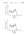
- FIG. 1 is a histogram of center 640,000 (800 ⁇ 800 pixel region) pixels of an unprocessed chest radiograph. This image has all of its original frequency components.
- FIG. 2 shows a histogram of center 640,000 pixels of a chest radiograph in FIG. 1, after the image pixel values have been processed or blurred using an unsharp mask size of 59 pixels square.
- the unsharp image mask contains only the low-frequency (less than 0.13 cycles/mm) components. Note that the width of the curve representing the luminance dynamic range in FIG. 2 comprising only the low frequency component is nearly the same width as the luminance dynamic range curve in FIG. 1. This indicates that the low-frequency component of an image carries most of the dynamic range of the whole image.
- FIG. 3 shows a histogram of center 640,000 pixels of the radiograph in FIG. 2, subtracted from radiograph in FIG. 1.
- This image contains only the high-frequency (greater than 0.13 cycles/mm) components. Note that the width of the curve is much less that the widths of the previous curves. This indicates very little dynamic range is carried by the high-frequency component, thus a relatively small portion of the original dynamic range is required to display the high-frequency component by itself.
- FIG. 4 represents a Fast Fourier Transform (FFT) of the same image.
- FFT Fast Fourier Transform
- FIG. 5 shows the FFT of the center 512 ⁇ 512 pixel region of the chest radiograph in FIG. 2. Note the reduced high-frequency component of the low-pass filtered image.
- FIG. 6 shows an FFT of the center 512 ⁇ 512 pixel region of the chest radiograph in FIG. 3. Note the reduced low-frequency component of the high pass filtered image.
- the image is separated into low spatial-frequency and high spatial-frequency components.
- the low-frequency component is attenuated and the high-frequency component is amplified. Attenuation of the low-frequency component reduces the dynamic range of the whole image without a substantial loss of detail in the image. The observer sees a change in low-frequency contrast but is still able to identify image type and location of regions within the image.
- Preamplification of the high-frequency component pre-emphasizes the image to compensate for the high-frequency attenuation caused by the CRT and ambient lighting. Additionally, high-frequency amplification helps enhance edges present in the image. This is illustrated in FIGS. 8 and 9.
- FIG. 8 shows a stylized representation of a region of a radiograph. There is a low amplitude, high-frequency sine wave superimposed on a high amplitude, low-frequency square wave.
- FIG. 9 shows a filtered version of FIG. 8 after processing according to the invention. Note that the amplitude of the high-frequency sine wave is increased and amplitude of the low-frequency square wave is reduced. The overall amplitude is reduced and the dynamic range is decreased, allowing such an image to fit into a more limited dynamic range display, with the high frequency boost compensating for the display's contrast attenuation.
- USM unsharp mask
- the USM method uses a uniformly weighted spatial average of a square mask of pixels (kernel) to create a blurred or low-pass frequency version of the image.
- the blurred version of the image is subtracted from the original image so that only the high-frequency portion or high-pass portion of the image remains.
- the two components are scaled appropriately and summed, producing a reduced dynamic range, sharpness enhanced version of the image.
- Parameters for optimizing the image include: lowboost, highboost, and mask size for the frequency separation.
- the enhancement method of the present invention unlike the typical unsharp mask, independently scales both the low-pass and high-pass portions of the image.
- the high-pass portion of the image is boosted and the low-pass portion of the image is left unmodified.
- the typical unsharp mask produces edge enhancement without reducing dynamic range.
- FIG. 10 shows transfer functions (0.345 mm pixels) for the typical unsharp mask filter with a mask 13 pixels square having a high-frequency boost of 2.0 and a modified unsharp mask filter with a mask 59 pixels square, having a lowboost of 0.7 and a highboost of 1.2. Note that the modified unsharp mask attenuates the low frequencies, enhances starting from a lower frequency and provides less high-frequency gain than the typical unsharp mask.
- FIG. 11 shows the results of applying the modified algorithm wherein a histogram of the center 640000 pixels of the radiogram in FIG. 1 as shown after processing with the modified algorithm shown in FIG. 10. Note that the image dynamic range is reduced.
- FIG. 12 shows the results of applying the modified algorithm to the radiogram in FIG. 1 wherein the center 512 ⁇ 512 region is depicted as an FFT. Compare the original image shown as a solid line with that image processed with the modified unsharp mask algorithm shown as the short, wide dashed line. The long, thin dashed line is the original image scaled by lowboost (0.7).
- the image processing method of the present invention is based on the luminance dynamic range of the image, DRimage.
- the method accounts for the fact that the image is displayed on a CRT having a luminance dynamic range less than DRimage.
- the processed display image, DRdisplay has a smaller dynamic range than the original image, DRimage.
- the image processing sets the lowboost parameter to DRdisplay/DRimage.
- the value of lowboost is set by the dynamic range requirements of the application.
- the available CRT luminance dynamic range is 1.70 log units. Due to typically high ambient light incident on an ICU CRT, the viewable luminance dynamic range of an original transilluminated film image is typically reduced from the sensitometric value of 3.5 log 10 units to about 2.50 log 10 units (See: R. Bollen and J. Vranckx, Influence of ambient light on the visual sensitometric properties of, and detail perception on, a radiograph, Proc. SPIE Vol. 273 Application of Optical Instrumentation in Medicine IX, pp. 57-61, 1981). The ratio of these two dynamic ranges (1.7/2.5) is 0.7. This ratio puts an upper limit on the value of lowboost. In the initial experiments, the value of lowboost was set to 0.7.
- the CRT often causes a loss in image contrast due to a loss in contrast transfer at increasing spatial frequencies, thus this property demands additional compensation (enhancement) of the image to be displayed.
- Highboost is increased to account for this aspect.
- the final value of highboost is determined by the amount of edge and sharpness enhancement desired by the observer. The user may select the amount of sharpness needed for the current task. Detail oriented tasks require the inclusion of more detail and thus more highboost of displayed images.
- HVS provides a maximum contrast sensitivity function, CSF, at about 4 cycles/degree, although this frequency is a function of the absolute luminance.
- CSF contrast sensitivity function
- viewers are positioned at about 50 cm from the display. This implies a maximum CSF at a spatial frequency of about 0.5 cycles/mm at the display.
- the image should be enhanced at this spatial frequency so that the HVS can perform effectively. Images should be enhanced in some useful range about that spatial frequency.
- image enhancement preferably begins at about 0.05 cycles/mm and extends past 0.5 cycles/mm.
- USM operates in this manner as it is a high-pass filter. Selecting an USM mask of 2 cm enables restoration of the attenuation imposed on the image by compression of the luminance dynamic range (Lowboost process) so that the HVS can effectively view an image on a CRT. Based on calculations used to construct FIG. 10, it can be shown that USM provides fully developed enhancement for all spatial frequencies beyond a 1.4/Mask Size. Thus, a 2 cm mask size provides fully developed enhancement after 0.07 cycles/mm which meets the above requirements.
- a pixel frequency of 4 pixels/mm results. Since a typical chest x-ray film is larger than the CRT viewing area, the image having dimensions of 43 cm high, the image is reduced and appears smaller when displayed on the CRT. Thus, a 2 cm feature in the image will be rendered as a 1.2 cm feature on the CRT. This requires about 49 CRT pixels (mask size should always be odd) to compose the mask of the modified USM method of the present invention. This places the maximum gain of the modified USM method at about 0.11 cycles/mm, which meets the requirements discussed in the previous paragraph.
- CRT displays for medical images usually include a feature that permits the image to be magnified. Magnification enables an operator to increase the size of CRT image features so that the features appear the same size on a CRT as they would appear on film. This optional magnification reduces the spatial frequency at which the modified USM enhancement occurs to the values quoted above.
- image enhancement processing such as USM increases the visibility of high spatial frequency noise.
- Complex image processing such as multiresolution representations based on octaves of spatial frequency are employed to limit the enhancement at higher spatial frequencies and decrease the visibility of high spatial frequency noise (See S. Ranganath, Image Filtering Using Multiresolution Representations, IEEE Transactions of Pattern Analysis and Machine Intelligence, Vol. 13, No. 5, pp 426-440, May, 1991).
- the CRT causes a loss in image contrast at increasing spatial frequencies, the CRT effectively provides a low-pass filter of the displayed image.
- the invention can use a modified USM successfully resulting in a very simple technique.
- the present invention provides a luminance-calibrated soft-copy display so that image data can be correctly displayed with respect to luminance.
- Many existing image processing processes do not include this aspect. It is a significant factor to producing visible image details because of the non-linear behavior of the HVS.
- This aspect of the operation is also significant as the images preferably appear familiar (like transilluminated film) to radiologists who typically have been trained viewing images visualized using traditional film and light box methods.
- the CRT is calibrated using a LUT to accept input data as film density which represents the space in which the enhancement was completed.
- This set Lowboost 0.7.
- a value of highboost of 1.2 was chosen for its balance of enhancing pneumothorax lines while minimally changing the appearance of the lung parenchyma and producing minimal high-frequency noise.
- the optimum value of highboost and mask size also depend on the original size of the image and the resolution of film digitizer which produces the digital image from film.
- Standard size (35 ⁇ 43 cm ) chest film were digitized to 4k ⁇ 5k pixels (117 pixels/cm).
- a square region 4k ⁇ 4k was extracted and down sized by pixel averaging to 1k ⁇ 1k (29 pixels/cm, 0.345 mm/pixel).
- Mask sizes ranging from 3 to 99 were tried. Objects smaller than the mask are enhanced in their entirety; objects larger than the mask are edge enhanced and attenuated. When small masks are used, the image is attenuated over a larger frequency range which causes more luminance dynamic range compression. To avoid double line artifacts, the mask should be larger than the size of most common structures appearing in the image, for example, blood vessels.
- the mask size which produced the best image was approximately 59 pixels or 2.0 cm in width. This mask size correlates with a study by Prokop et. al. (See: Prokop, Mathias, Schaefer, Cornelia M, Oestmann, Jorg W, Galanski, Michael. Improved Parameters for Unsharp Mask Filtering of Digital Chest Radiographs. Radiology 1993. 97:521-526.) in which a mask size of 2.5 cm was found to be superior to mask sizes of 1.4 mm, 5 mm, and 7 cm.
- the larger masks produced images with a better preserved tone scale. Edges were enhanced to a lesser extent and double line artifacts were minimized. In one image there was a subdiaphragmatic nodule. The nodule was better preserved in the image processed with the larger mask.
- a modified USM filter (59 pixels) attenuates by using a Lowboost (0.7) until about 0.01 cycles/mm after which Highboost begins to provide restoration of contrast (5% USM gain). Complete restoration and enhancement (maximum gain) occurs at 0.07 cycles/mm.
- FIG. 10 shows a standard USM filter based on 13 pixels and a gain of 2.0. A smaller mask such as this is typically used in commercially available Computed Radiography.
- Processed images have less low-frequency contrast than the original image.
- the tone scale is altered, but the image is easily recognizable so that the CRT image appearance is not objectionable to the viewer, that is, the CRT image looks similar to a film image.
- Areas which were very dark in the original image, such as lung fields, are reproduced lighter and have more high-frequency contrast so that more details can be distinguished in the lung parenchyma.
- Areas which are very light in the originals, such the mediastinum have more high-frequency contrast and vertebral bodies can be more easily seen.
- There is a slight edge enhancement which highlights fine lines in the lung fields such as pneumothorax lines. Lung markings are more prominent, but images with interstitial lung disease can be differentiated from normal lung images.
- the CRTs used in the initial study were Imlogix model 1000 electronic viewboxes.
- the screen size was 25 cm ⁇ 25 cm with a resolution was 1024 ⁇ 1024 ⁇ 12 bits.
- Monitors were calibrated with a maximum luminance of 160 nits and a minimum luminance of 0.5 nits producing a log dynamic range of 2.5 in a dark room. Monitors were individually linearized with respect to log luminance using LUTs.
- the image processing technique of the present invention has successfully rendered digitized chest radiographs which are clinically useful on a 1k ⁇ 1k display under high ambient lighting conditions.
- Soft-copy display was observed to qualitatively equivalent to film for visualizing image details such as pneumothoraces and accessing tube placement. For some over penetrated films, the soft-copy was superior to the original film.
- images processed using the method of the present invention appeared equal to or better than unprocessed images in all cases.
- the general methods used are not dependent on image type or the display technology. All that is required is that the image data is displayed on a CRT or the like for visualization. Only the values of the parameters need be selected according to the image size and type, the characteristics of the display device, and the ambient lighting conditions present in the viewing area.
Abstract
Description
                                  APPENDIX
__________________________________________________________________________
/*************************************************************************
************/
/* program us.c                             */
/* banded modified unsharp mask             */
/*                                          */
/* IH = I - IL (blurred image)              */
/*                                          */
/* output = lowboost * IL + highboost * IH  */
/*   = lowboost * IL + highboost * I = highboost * IL
                                            */
/*   = (lowboost - highboost) * IL + highboost * I
                                            */
/*   = lowscale * IL + highboost * I        */
/*                                          */
/* PHO 28 Oct 1 993                         */
/* copyright 1993 Paul Ho                   */
/* All rights reserved                      */
/*************************************************************************
************/
#include<stdio.h>
#include<stdiib.h>
#include <sys/file.h>
main(argc, argv)
int argc;
char *argv !;
FILE *fdout, *fdin;
                /* output and input file descriptors */
short lines, pixels;
                /* rows and column sizes of input file */
short width;    /* width of the mask */
short leadwidth;
                /* first half of the mask size */
short lagwidth; /* second half of the mask size */
long masksize;  /* number of pixels in the mask */
short ras;      /* number of rasters loaded in memory */
float lowboost, highboost, lowscale;
                /* weighting factors */
long *header;   /* header copy space */
short *inimage; /* input image space */
short *outimage;
                /* output image space */
long vert;      /* vert accumulator for blurring */
long acc;       /* mask accumulator sum of verts */
short temp;     /* temp to hold new pixel value */
short i,j;      /* counter dummy variables */
/* parse command line input */
if(argc|= 6)
 {
 printf("usage: us infile outfile mask lowboost highboost\n");
 printf("mask: positive odd integer\n");
 exit(0);
 }
width = atoi(argv 3!);
if ((width < 0) II ((width % 2) == 0))
 {
 printf("width must a positive odd integer\n");
 exit(0);
 }
ras=width + 1;
masksize = width * width;
leadwidth = width/2;
lagwidth = width - leadwidth;
lowboost = (float)atof(argv 4!);
highboost = (float)atof(argv 5!);
lowscale = lowboost - highboost;
/* open input file */
if((fdin = fopen(argv 1!, "r"))== NULL)
 {
 printf("could not open %s\n", argv  1!);
 exit(0);
 }
/* open the output file for writing */
if ((fdout = fopen(argv 2!, "w"))== NULL)
 {
 printf("\nError creating output file\n");
 exit(0);
 }
/* copy MIR 8192 byte header */
header = (long*)calloc(2048, sizeof(long));
fread(header, sizeof(long), 2048, fdin);
fwrite(header, sizeof(long), 2048, fdout);
/* read image size */
lines = (short)header 0!;
pixels = (short)header 1!;
free(header);
/* create temp variable space */
/* space for (mask width + 1) lines of the input image */
/* so that lines can be read only once
inimage = (short *)calloc(pixels * ras, sizeof(short));
/* space for one line of the output image, image written one line at a
time */
outimage - (short *)calloc(pixels, sizeof(short));
/* space for (width) number of vertical sum buffers */
vert = (long *)calloc(pixels, sizeof(long));
/* vertical sum buffers each hold a number of pixels equal to (mask
height) */
for (i = 0; i < pixels; i++)
 vert i! = 0;
/* read in (width) rasters and initialize vertical sum buffers */
for(j=0;j < width;j++)
 {
 fread(&inimage (j % ras) * pixels!, sizeof(short), pixels, fdin);
 for (i = 0; i < pixels; i++)
 vert i! += inimage (j % ras) * pixels + i!;
 }
/* copy first lagwidth lines to output, scaling by lowboost */
for (j = 0; j < lagwidth; j++)
 {
 for (i = 0; i < pixels; i++)
 outimage  i! = lowboost * inimage (j % ras) * pixels + i!;
 fwrite(outimage, sizeof(short), pixels, fdout);
 }
/* main routine for interior of image */
for (j = lagwidth; j < lines - leadwidth; j++)
 {
 /* read new data raster                    */
 /* can overwrite old raster when no longer needed
                                            */
 fread(&inimage ((j + leadwidth) % ras) * pixels!,
 sizeof(short), pixels, fdin);
 /* update each vertical sum buffer */
 for(i = 0; i < pixels; i++)
 vert i! = vert i!
  - inimage ((j - lagwidth) % ras) * pixels.+ i!
  + inimage ((j + leadwidth) % ras) * pixels + i!;
 /* sum vertical buffers to get total mask value */
 acc = 0;
 for(i = 0; i < width; i++)
 acc += vert i!;
 /* copy first lagwidth pixels to output, scaling by lowboost */
 for (i = 0; i < lagwidth; i++)
 outimage i! = lowboost * inimage (j % ras) * pixels + i!;
 /* unsharp mask interior of image */
 /* out = highboost * in + lowscale * lowpass */
 for (i = lagwidth; i < pixels - leadwidth; i++)
 {
 /* acc updated to reflect new columns */
 acc = acc - vert i - lagwidth! + vert i + leadwidth!;
 temp = highboost * inimage (j % ras) * pixels + i!
  + lowscale * (short)(acc/masksize);
 /* bounds checking */
 if(temp < 0)temp = 0;
 if (temp > 4095) temp = 4095;
 outimage i! = temp;
 }
 /* copy last lagwidth pixels to output, scaling by lowboost */
 for (i = pixels - leadwidth; i < pixels; i++)
 outimage i! = lowboost * inimage (j % ras) * pixels + i!;
 fwrite(outimage, sizeof(short), pixels, fdout);
 }
/* copy last leadwidth lines, scaling by lowboost */
for (j = lines - leadwidth; j < lines; j++)
 {
 for (i = 0; i < pixels; i++)
 outimage i! = lowboost * inimage (j % ras) * pixels + i!;
 fwrite(outimage, sizeof(short), pixels, fdout);
 }
free(vert);
free(outimage);
free(inimage);
fclose(fdin);
fclose(fdout);
}
__________________________________________________________________________
Claims (26)
Priority Applications (4)
| Application Number | Priority Date | Filing Date | Title |
|---|---|---|---|
| US08/404,399 US5774599A (en) | 1995-03-14 | 1995-03-14 | Method for precompensation of digital images for enhanced presentation on digital displays with limited capabilities |
| EP96103723A EP0732669B1 (en) | 1995-03-14 | 1996-03-09 | A method for precompensation of digital images for enhanced presentation on digital displays with limited capabilities |
| DE69625839T DE69625839T2 (en) | 1995-03-14 | 1996-03-09 | Pre-compensating digital images for improved display on digital displays with limited capability |
| JP05519196A JP3681213B2 (en) | 1995-03-14 | 1996-03-12 | Compensatory pre-processing of digital images for highlighting on digital displays with limited performance |
Applications Claiming Priority (1)
| Application Number | Priority Date | Filing Date | Title |
|---|---|---|---|
| US08/404,399 US5774599A (en) | 1995-03-14 | 1995-03-14 | Method for precompensation of digital images for enhanced presentation on digital displays with limited capabilities |
Publications (1)
| Publication Number | Publication Date |
|---|---|
| US5774599A true US5774599A (en) | 1998-06-30 |
Family
ID=23599445
Family Applications (1)
| Application Number | Title | Priority Date | Filing Date |
|---|---|---|---|
| US08/404,399 Expired - Lifetime US5774599A (en) | 1995-03-14 | 1995-03-14 | Method for precompensation of digital images for enhanced presentation on digital displays with limited capabilities |
Country Status (4)
| Country | Link |
|---|---|
| US (1) | US5774599A (en) |
| EP (1) | EP0732669B1 (en) |
| JP (1) | JP3681213B2 (en) |
| DE (1) | DE69625839T2 (en) |
Cited By (74)
| Publication number | Priority date | Publication date | Assignee | Title |
|---|---|---|---|---|
| US5905817A (en) * | 1995-12-25 | 1999-05-18 | Fuji Photo Film Co., Ltd. | Image reproducing method and apparatus using dynamic range compression based on an unsharp signal generated by IIR filter |
| US5936682A (en) * | 1996-07-09 | 1999-08-10 | Stmicroelectronics S.R.L. | Circuit for enhancing chrominance transitions in real-time video reception |
| US5978518A (en) * | 1997-02-25 | 1999-11-02 | Eastman Kodak Company | Image enhancement in digital image processing |
| US6035065A (en) * | 1996-06-10 | 2000-03-07 | Fuji Xerox Co., Ltd. | Image processing coefficient determination method, image processing coefficient calculation system, image processing system, image processing method, and storage medium |
| US6069979A (en) * | 1997-02-25 | 2000-05-30 | Eastman Kodak Company | Method for compressing the dynamic range of digital projection radiographic images |
| US6072913A (en) * | 1996-12-13 | 2000-06-06 | Fuji Photo Film Co., Ltd. | Image processing method and apparatus |
| US6211919B1 (en) * | 1997-03-28 | 2001-04-03 | Tektronix, Inc. | Transparent embedment of data in a video signal |
| US6240217B1 (en) * | 1997-02-24 | 2001-05-29 | Redflex Traffic Systems Pty Ltd | Digital image processing |
| US6243095B1 (en) * | 1996-12-05 | 2001-06-05 | Peter E. Shile | Navigation and display system for digital radiographs |
| US6285798B1 (en) * | 1998-07-06 | 2001-09-04 | Eastman Kodak Company | Automatic tone adjustment by contrast gain-control on edges |
| US6304277B1 (en) | 1999-01-15 | 2001-10-16 | Colorcentric.Com, Inc. | Remote modification of digital images using scripts |
| US6392764B1 (en) * | 1998-11-12 | 2002-05-21 | Xerox Corporation | Systems and methods for adaptively filtering palettized images |
| US20020080246A1 (en) * | 2000-12-22 | 2002-06-27 | Parulski Kenneth A. | Camera having user interface ambient sensor viewer adaptation compensation and method |
| US6421468B1 (en) * | 1999-01-06 | 2002-07-16 | Seiko Epson Corporation | Method and apparatus for sharpening an image by scaling elements of a frequency-domain representation |
| US6424730B1 (en) | 1998-11-03 | 2002-07-23 | Eastman Kodak Company | Medical image enhancement method for hardcopy prints |
| US6463167B1 (en) * | 1996-09-19 | 2002-10-08 | Philips Medical Systems Technologies Ltd. | Adaptive filtering |
| US20020154323A1 (en) * | 2001-03-10 | 2002-10-24 | Sobol Robert E. | Method for variable contrast mapping of digital images |
| US20020154832A1 (en) * | 2001-03-10 | 2002-10-24 | Sobol Robert E. | Method for contrast mapping of digital images that converges on a solution |
| WO2002101636A1 (en) * | 2001-06-12 | 2002-12-19 | Mclean Hospital Corporation | Color magnetic resonance imaging |
| US20030081854A1 (en) * | 2001-06-12 | 2003-05-01 | Deshpande Sachin G. | Filter for combined de-ringing and edge sharpening |
| US20030103677A1 (en) * | 2001-11-20 | 2003-06-05 | Ingeborg Tastl | System and method for effectively rendering high dynamic range images |
| US6633684B1 (en) | 2000-07-07 | 2003-10-14 | Athentech Technologies Corp. | Distortion-free image contrast enhancement |
| US6690488B1 (en) * | 1999-09-30 | 2004-02-10 | Polaroid Corporation | Method and apparatus for estimating the spatial frequency response of a digital image acquisition system from the images it produces |
| US20040036923A1 (en) * | 2000-09-20 | 2004-02-26 | Nils Kokemohr | Digital image sharpening system |
| US20040066980A1 (en) * | 2002-10-02 | 2004-04-08 | Gindele Edward B. | Enhancing the tonal and color characteristics of digital images using expansive and compressive tone scale functions |
| US6720992B1 (en) * | 1999-08-04 | 2004-04-13 | Wolfgang Zahn | Method for improving the contrast reproduction of digitized images |
| US20040081363A1 (en) * | 2002-10-25 | 2004-04-29 | Eastman Kodak Company | Enhancing the tonal and spatial characteristics of digital images using selective spatial filters |
| US20040105592A1 (en) * | 2001-04-10 | 2004-06-03 | Estelle Lesellier | Method and device for post-processing digital images |
| US6775407B1 (en) * | 2000-08-02 | 2004-08-10 | Eastman Kodak Company | Producing a final modified digital image using a source digital image and a difference digital image |
| US20040234154A1 (en) * | 2002-11-06 | 2004-11-25 | Hier Richard G. | Systems and methods for image enhancement in multiple dimensions |
| US20040252908A1 (en) * | 2003-04-04 | 2004-12-16 | Keping Chen | Method and apparatus for enhancing detail in an image |
| US20040258289A1 (en) * | 2003-04-15 | 2004-12-23 | Joachim Hornegger | Method for digital subtraction angiography using a volume dataset |
| US20040263942A1 (en) * | 2003-06-24 | 2004-12-30 | Lopez Matthew Grant | Display system allowing enhanced dynamic range |
| US20050008262A1 (en) * | 2003-06-03 | 2005-01-13 | Konica Minolta Medical & Graphic, Inc. | Medical image system, and medical image processing method |
| US20050062695A1 (en) * | 2003-09-23 | 2005-03-24 | Eastman Kodak Company | Display device and system |
| US20050088402A1 (en) * | 2001-11-09 | 2005-04-28 | Daly Scott J. | Liquid crystal display backlight with variable amplitude LED |
| US20050117186A1 (en) * | 2003-11-21 | 2005-06-02 | Baoxin Li | Liquid crystal display with adaptive color |
| US20050248524A1 (en) * | 2004-05-04 | 2005-11-10 | Sharp Laboratories Of America, Inc. | Liquid crystal display with colored backlight |
| US20060023965A1 (en) * | 2004-07-30 | 2006-02-02 | Hewlett-Packard Development Company, L.P. | Adjusting pixels by desired gains and factors |
| US20060074307A1 (en) * | 2004-05-10 | 2006-04-06 | Tatsuo Igarashi | Body cavity diagnostic system |
| US20060104533A1 (en) * | 2004-11-16 | 2006-05-18 | Sharp Laboratories Of America, Inc. | High dynamic range images from low dynamic range images |
| AU2001234808B2 (en) * | 2000-02-02 | 2006-06-08 | Quvis, Inc. | System and method for optimizing image resolution using pixelated imaging devices |
| US20060182363A1 (en) * | 2004-12-21 | 2006-08-17 | Vladimir Jellus | Method for correcting inhomogeneities in an image, and an imaging apparatus therefor |
| US20060187242A1 (en) * | 2005-02-18 | 2006-08-24 | Lee Seong-Deok | Method of, and apparatus for image enhancement taking ambient illuminance into account |
| US20060228043A1 (en) * | 2005-03-30 | 2006-10-12 | Pioneer Corporation | Image processing apparatus |
| US20070009145A1 (en) * | 2001-11-23 | 2007-01-11 | Robin Winsor | Balancing areas of varying density in a digital image |
| US20070036456A1 (en) * | 2005-04-13 | 2007-02-15 | Hooper David S | Image contrast enhancement |
| US20070071318A1 (en) * | 2003-09-11 | 2007-03-29 | Haruo Yamashita | Visual processing device, visual processing method, visual processing program, and semiconductor device |
| US20070109447A1 (en) * | 2003-09-11 | 2007-05-17 | Haruo Yamashita | Visual processing device, visual processing method, visual processing program, and semiconductor device |
| US20070188623A1 (en) * | 2003-09-11 | 2007-08-16 | Haruo Yamashita | Visual processing device, visual processing method, visual processing program, intergrated circuit, display device, image-capturing device, and portable information terminal |
| US20070232863A1 (en) * | 2006-02-21 | 2007-10-04 | Fujinon Corporation | Body cavity interior observing apparatus |
| US7444034B1 (en) * | 2002-11-06 | 2008-10-28 | Digivision, Inc. | Systems and methods for image enhancement in multiple dimensions |
| US20090169086A1 (en) * | 2004-07-27 | 2009-07-02 | Michael Thoms | Method and device for improving perceptibility different structures on radiographs |
| US7616829B1 (en) * | 2003-10-29 | 2009-11-10 | Apple Inc. | Reducing undesirable block based image processing artifacts by DC image filtering |
| US20100066817A1 (en) * | 2007-02-25 | 2010-03-18 | Humaneyes Technologies Ltd. | method and a system for calibrating and/or visualizing a multi image display and for reducing ghosting artifacts |
| US20100098340A1 (en) * | 2007-01-15 | 2010-04-22 | Assaf Zomet | Method And A System For Lenticular Printing |
| US20100132033A1 (en) * | 2006-07-14 | 2010-05-27 | Ge Medical Systems Global Technology Company, Llc | Service system |
| US20100172596A1 (en) * | 2003-11-06 | 2010-07-08 | Hier Richard G | Systems and Methods for Image Enhancement in Multiple Dimensions |
| US7777714B2 (en) | 2004-05-04 | 2010-08-17 | Sharp Laboratories Of America, Inc. | Liquid crystal display with adaptive width |
| US20100207961A1 (en) * | 2007-07-23 | 2010-08-19 | Humaneyes Technologies Ltd. | Multi view displays and methods for producing the same |
| US20100232729A1 (en) * | 2009-03-12 | 2010-09-16 | Yissum Research Development Company Of The Hebrew University Of Jerusalem | Method and system for shift-map image editing |
| US20100246967A1 (en) * | 2008-09-17 | 2010-09-30 | Takamasa Ando | Image processing device, imaging device, evaluation device, image processing method, and optical system evaluation method |
| US7853094B2 (en) | 2006-01-24 | 2010-12-14 | Sharp Laboratories Of America, Inc. | Color enhancement technique using skin color detection |
| US7872631B2 (en) | 2004-05-04 | 2011-01-18 | Sharp Laboratories Of America, Inc. | Liquid crystal display with temporal black point |
| US7898519B2 (en) | 2005-02-17 | 2011-03-01 | Sharp Laboratories Of America, Inc. | Method for overdriving a backlit display |
| EP2290946A3 (en) * | 2000-02-07 | 2011-04-27 | Sony Corporation | Device and method for image processing |
| US20110229043A1 (en) * | 2010-03-18 | 2011-09-22 | Fujitsu Limited | Image processing apparatus and image processing method |
| US8050511B2 (en) | 2004-11-16 | 2011-11-01 | Sharp Laboratories Of America, Inc. | High dynamic range images from low dynamic range images |
| US8121401B2 (en) | 2006-01-24 | 2012-02-21 | Sharp Labortories of America, Inc. | Method for reducing enhancement of artifacts and noise in image color enhancement |
| USRE43707E1 (en) | 2005-05-17 | 2012-10-02 | Barco N.V. | Methods, apparatus, and devices for noise reduction |
| US8395577B2 (en) | 2004-05-04 | 2013-03-12 | Sharp Laboratories Of America, Inc. | Liquid crystal display with illumination control |
| US8941580B2 (en) | 2006-11-30 | 2015-01-27 | Sharp Laboratories Of America, Inc. | Liquid crystal display with area adaptive backlight |
| US9070316B2 (en) | 2004-10-25 | 2015-06-30 | Barco Nv | Optical correction for high uniformity panel lights |
| US20170193636A1 (en) * | 2014-03-27 | 2017-07-06 | Noritsu Precision Co., Ltd. | Image processing device |
Families Citing this family (19)
| Publication number | Priority date | Publication date | Assignee | Title |
|---|---|---|---|---|
| FR2799028B1 (en) * | 1999-09-27 | 2002-05-03 | Ge Medical Syst Sa | METHOD FOR RECONSTRUCTING A THREE-DIMENSIONAL IMAGE OF ELEMENTS OF STRONG CONTRAST |
| FR2803069B1 (en) * | 1999-12-28 | 2002-12-13 | Ge Medical Syst Sa | METHOD AND SYSTEM FOR COMPENSATING THE THICKNESS OF AN ORGAN |
| US6721441B1 (en) * | 1999-12-30 | 2004-04-13 | General Electric Company | Extended dynamic range system for digital X-ray imaging detectors |
| EP1239415A1 (en) * | 2001-03-05 | 2002-09-11 | Ge Medical Systems Sa | Method and system of management of the dynamics of a digitized radiological image |
| JP4626086B2 (en) * | 2001-05-09 | 2011-02-02 | 株式会社島津製作所 | Digital subtraction device |
| US7619585B2 (en) | 2001-11-09 | 2009-11-17 | Puredepth Limited | Depth fused display |
| NZ517713A (en) | 2002-06-25 | 2005-03-24 | Puredepth Ltd | Enhanced viewing experience of a display through localised dynamic control of background lighting level |
| NZ521505A (en) | 2002-09-20 | 2005-05-27 | Deep Video Imaging Ltd | Multi-view display |
| US7106352B2 (en) | 2003-03-03 | 2006-09-12 | Sun Microsystems, Inc. | Automatic gain control, brightness compression, and super-intensity samples |
| NZ525956A (en) | 2003-05-16 | 2005-10-28 | Deep Video Imaging Ltd | Display control system for use with multi-layer displays |
| US7315656B2 (en) | 2003-05-22 | 2008-01-01 | The Boeing Company | Methods and apparatus for enhanced viewing of aerial refueling operations |
| WO2006129529A1 (en) | 2005-06-02 | 2006-12-07 | Konica Minolta Holdings, Inc. | Image processing method and image processing apparatus |
| NZ542843A (en) | 2005-10-05 | 2008-08-29 | Pure Depth Ltd | Method of manipulating visibility of images on a volumetric display |
| US8432411B2 (en) | 2007-05-18 | 2013-04-30 | Pure Depth Limited | Method and system for improving display quality of a multi-component display |
| US8330768B2 (en) | 2007-07-27 | 2012-12-11 | Sharp Laboratories Of America, Inc. | Apparatus and method for rendering high dynamic range images for standard dynamic range display |
| JP4367965B1 (en) | 2009-03-16 | 2009-11-18 | 横浜ゴム株式会社 | Pneumatic tire |
| US9524700B2 (en) | 2009-05-14 | 2016-12-20 | Pure Depth Limited | Method and system for displaying images of various formats on a single display |
| US8928682B2 (en) | 2009-07-07 | 2015-01-06 | Pure Depth Limited | Method and system of processing images for improved display |
| JP7047643B2 (en) * | 2018-07-17 | 2022-04-05 | コニカミノルタ株式会社 | Image processing equipment, radiography system, image processing program and image processing method |
Citations (10)
| Publication number | Priority date | Publication date | Assignee | Title |
|---|---|---|---|---|
| US4315318A (en) * | 1978-12-26 | 1982-02-09 | Fuji Photo Film Co., Ltd. | Method and apparatus for processing a radiation image |
| US4317179A (en) * | 1978-12-26 | 1982-02-23 | Fuji Photo Film Co., Ltd. | Method and apparatus for processing a radiographic image |
| US4346409A (en) * | 1979-12-25 | 1982-08-24 | Fuji Photo Film Co., Ltd. | Method of and apparatus for processing a radiographic image |
| US4674125A (en) * | 1983-06-27 | 1987-06-16 | Rca Corporation | Real-time hierarchal pyramid signal processing apparatus |
| US4697594A (en) * | 1985-08-21 | 1987-10-06 | North American Philips Corporation | Displaying a single parameter image |
| GB2194706A (en) * | 1986-08-29 | 1988-03-09 | Agfa Gevaert Ag | Method of electronically processing a colored image for copying |
| US4747052A (en) * | 1984-11-14 | 1988-05-24 | Philips Medical Systems, Inc. | Radiation image processing |
| US4903205A (en) * | 1985-10-15 | 1990-02-20 | Fuji Photo Film Co. Ltd. | Method and apparatus for displaying radiation image, and method and apparatus for calculating unsharp mask signal used for the same |
| US5319719A (en) * | 1991-05-15 | 1994-06-07 | Konica Corporation | Processing apparatus for radiographic image signals |
| US5387983A (en) * | 1991-09-27 | 1995-02-07 | Minolta Camera Kabushiki Kaisha | Facsimile apparatus comprising converting means for converting binary image data into multi-value image data and image processing apparatus judging pseudo half-tone image |
Family Cites Families (1)
| Publication number | Priority date | Publication date | Assignee | Title |
|---|---|---|---|---|
| US5012333A (en) * | 1989-01-05 | 1991-04-30 | Eastman Kodak Company | Interactive dynamic range adjustment system for printing digital images |
-
1995
- 1995-03-14 US US08/404,399 patent/US5774599A/en not_active Expired - Lifetime
-
1996
- 1996-03-09 EP EP96103723A patent/EP0732669B1/en not_active Expired - Lifetime
- 1996-03-09 DE DE69625839T patent/DE69625839T2/en not_active Expired - Lifetime
- 1996-03-12 JP JP05519196A patent/JP3681213B2/en not_active Expired - Fee Related
Patent Citations (10)
| Publication number | Priority date | Publication date | Assignee | Title |
|---|---|---|---|---|
| US4315318A (en) * | 1978-12-26 | 1982-02-09 | Fuji Photo Film Co., Ltd. | Method and apparatus for processing a radiation image |
| US4317179A (en) * | 1978-12-26 | 1982-02-23 | Fuji Photo Film Co., Ltd. | Method and apparatus for processing a radiographic image |
| US4346409A (en) * | 1979-12-25 | 1982-08-24 | Fuji Photo Film Co., Ltd. | Method of and apparatus for processing a radiographic image |
| US4674125A (en) * | 1983-06-27 | 1987-06-16 | Rca Corporation | Real-time hierarchal pyramid signal processing apparatus |
| US4747052A (en) * | 1984-11-14 | 1988-05-24 | Philips Medical Systems, Inc. | Radiation image processing |
| US4697594A (en) * | 1985-08-21 | 1987-10-06 | North American Philips Corporation | Displaying a single parameter image |
| US4903205A (en) * | 1985-10-15 | 1990-02-20 | Fuji Photo Film Co. Ltd. | Method and apparatus for displaying radiation image, and method and apparatus for calculating unsharp mask signal used for the same |
| GB2194706A (en) * | 1986-08-29 | 1988-03-09 | Agfa Gevaert Ag | Method of electronically processing a colored image for copying |
| US5319719A (en) * | 1991-05-15 | 1994-06-07 | Konica Corporation | Processing apparatus for radiographic image signals |
| US5387983A (en) * | 1991-09-27 | 1995-02-07 | Minolta Camera Kabushiki Kaisha | Facsimile apparatus comprising converting means for converting binary image data into multi-value image data and image processing apparatus judging pseudo half-tone image |
Non-Patent Citations (16)
| Title |
|---|
| E. Muka et al., "Display of Medical Images on CRT Soft-Copy Displays," SPIE Medical Imaging, vol. 2431, Image Display, 1995. |
| E. Muka et al., Display of Medical Images on CRT Soft Copy Displays, SPIE Medical Imaging, vol. 2431, Image Display, 1995. * |
| J.H. Kim et al., "Improved Visualization of Simulated Nodules by Adaptive Enhancement of Digital Chest Radiography, " Acad. Radio., vol. 1, No. 2, Oct. 1994, pp. 93-99. |
| J.H. Kim et al., Improved Visualization of Simulated Nodules by Adaptive Enhancement of Digital Chest Radiography, Acad. Radio., vol. 1, No. 2, Oct. 1994, pp. 93 99. * |
| Pratt, William K, Digital Image Processing , 2nd Ed., John Wiley & Sons, Inc., New York, 1991, pp. 303 305. * |
| Pratt, William K, Digital Image Processing , 2nd Ed., John Wiley & Sons, Inc., New York, 1991, pp. 303-305. |
| Prokop et al. "Improved Parameters for Unsharp Mask Filtering of Digital Chest Radiographs," Radiographs 1993, 187:521-526. |
| Prokop et al. Improved Parameters for Unsharp Mask Filtering of Digital Chest Radiographs, Radiographs 1993, 187:521 526. * |
| R. Bollen and J. Vranckx, "Influence of Ambient Light on the `Visual` Sensitometric Properties of, and Detail Perception on, a Radiograph", Proc. SPIE, vol. 273, Application of Optical Instrumentation in Medicine IX, pp. 57-61. |
| R. Bollen and J. Vranckx, Influence of Ambient Light on the Visual Sensitometric Properties of, and Detail Perception on, a Radiograph , Proc. SPIE, vol. 273, Application of Optical Instrumentation in Medicine IX, pp. 57 61. * |
| Roehrig, H. et al., "Signal-to-Noise Ratio and Maximum Information Content of Images Displayed by a CRT", Proceedings SPIE Medical Imaging IV: Image Capture and Display (1990), vol. 1232, pp. 115-133. |
| Roehrig, H. et al., Signal to Noise Ratio and Maximum Information Content of Images Displayed by a CRT , Proceedings SPIE Medical Imaging IV: Image Capture and Display (1990), vol. 1232, pp. 115 133. * |
| S. Daly, The Visible Differences Predictor; An Algorithm for the Assessment of Image Fidelity, A.B. Watson, Editor, Digital Images and Human Vision, Massachusetts: MIT Press, 1993, pp. 179 206. * |
| S. Daly, The Visible Differences Predictor; An Algorithm for the Assessment of Image Fidelity, A.B. Watson, Editor, Digital Images and Human Vision, Massachusetts: MIT Press, 1993, pp. 179-206. |
| S. Raganath, "Image Filtering Using Multiresolution Representations," IEEE Transactions of Pattern Analysis and Machine Intelligence, vol. 13, No. 5, May 1991, pp. 426-440. |
| S. Raganath, Image Filtering Using Multiresolution Representations, IEEE Transactions of Pattern Analysis and Machine Intelligence, vol. 13, No. 5, May 1991, pp. 426 440. * |
Cited By (119)
| Publication number | Priority date | Publication date | Assignee | Title |
|---|---|---|---|---|
| US5905817A (en) * | 1995-12-25 | 1999-05-18 | Fuji Photo Film Co., Ltd. | Image reproducing method and apparatus using dynamic range compression based on an unsharp signal generated by IIR filter |
| US6035065A (en) * | 1996-06-10 | 2000-03-07 | Fuji Xerox Co., Ltd. | Image processing coefficient determination method, image processing coefficient calculation system, image processing system, image processing method, and storage medium |
| US5936682A (en) * | 1996-07-09 | 1999-08-10 | Stmicroelectronics S.R.L. | Circuit for enhancing chrominance transitions in real-time video reception |
| US6463167B1 (en) * | 1996-09-19 | 2002-10-08 | Philips Medical Systems Technologies Ltd. | Adaptive filtering |
| US6243095B1 (en) * | 1996-12-05 | 2001-06-05 | Peter E. Shile | Navigation and display system for digital radiographs |
| US6072913A (en) * | 1996-12-13 | 2000-06-06 | Fuji Photo Film Co., Ltd. | Image processing method and apparatus |
| US6240217B1 (en) * | 1997-02-24 | 2001-05-29 | Redflex Traffic Systems Pty Ltd | Digital image processing |
| US5978518A (en) * | 1997-02-25 | 1999-11-02 | Eastman Kodak Company | Image enhancement in digital image processing |
| US6069979A (en) * | 1997-02-25 | 2000-05-30 | Eastman Kodak Company | Method for compressing the dynamic range of digital projection radiographic images |
| US6211919B1 (en) * | 1997-03-28 | 2001-04-03 | Tektronix, Inc. | Transparent embedment of data in a video signal |
| US6285798B1 (en) * | 1998-07-06 | 2001-09-04 | Eastman Kodak Company | Automatic tone adjustment by contrast gain-control on edges |
| US6424730B1 (en) | 1998-11-03 | 2002-07-23 | Eastman Kodak Company | Medical image enhancement method for hardcopy prints |
| US6392764B1 (en) * | 1998-11-12 | 2002-05-21 | Xerox Corporation | Systems and methods for adaptively filtering palettized images |
| US6421468B1 (en) * | 1999-01-06 | 2002-07-16 | Seiko Epson Corporation | Method and apparatus for sharpening an image by scaling elements of a frequency-domain representation |
| US6304277B1 (en) | 1999-01-15 | 2001-10-16 | Colorcentric.Com, Inc. | Remote modification of digital images using scripts |
| US6720992B1 (en) * | 1999-08-04 | 2004-04-13 | Wolfgang Zahn | Method for improving the contrast reproduction of digitized images |
| US6690488B1 (en) * | 1999-09-30 | 2004-02-10 | Polaroid Corporation | Method and apparatus for estimating the spatial frequency response of a digital image acquisition system from the images it produces |
| AU2001234808B2 (en) * | 2000-02-02 | 2006-06-08 | Quvis, Inc. | System and method for optimizing image resolution using pixelated imaging devices |
| EP2290946A3 (en) * | 2000-02-07 | 2011-04-27 | Sony Corporation | Device and method for image processing |
| US6633684B1 (en) | 2000-07-07 | 2003-10-14 | Athentech Technologies Corp. | Distortion-free image contrast enhancement |
| US6775407B1 (en) * | 2000-08-02 | 2004-08-10 | Eastman Kodak Company | Producing a final modified digital image using a source digital image and a difference digital image |
| US20040036923A1 (en) * | 2000-09-20 | 2004-02-26 | Nils Kokemohr | Digital image sharpening system |
| US7268916B2 (en) | 2000-09-20 | 2007-09-11 | Nik Software, Inc. | Digital image sharpening system |
| US6989859B2 (en) * | 2000-12-22 | 2006-01-24 | Eastman Kodak Company | Camera having user interface ambient sensor viewer adaptation compensation and method |
| US20020080246A1 (en) * | 2000-12-22 | 2002-06-27 | Parulski Kenneth A. | Camera having user interface ambient sensor viewer adaptation compensation and method |
| US20020154832A1 (en) * | 2001-03-10 | 2002-10-24 | Sobol Robert E. | Method for contrast mapping of digital images that converges on a solution |
| US6807299B2 (en) * | 2001-03-10 | 2004-10-19 | Hewlett-Packard Development Company, L.P. | Method for contrast mapping of digital images that converges on a solution |
| US20020154323A1 (en) * | 2001-03-10 | 2002-10-24 | Sobol Robert E. | Method for variable contrast mapping of digital images |
| US20040105592A1 (en) * | 2001-04-10 | 2004-06-03 | Estelle Lesellier | Method and device for post-processing digital images |
| US7373010B2 (en) * | 2001-04-10 | 2008-05-13 | Koninklijke Philips Electronics N.V. | Method and device for post-processing digital images |
| US6804384B2 (en) | 2001-06-12 | 2004-10-12 | Mclean Hospital Corporation | Color magnetic resonance imaging |
| WO2002101636A1 (en) * | 2001-06-12 | 2002-12-19 | Mclean Hospital Corporation | Color magnetic resonance imaging |
| US20030081854A1 (en) * | 2001-06-12 | 2003-05-01 | Deshpande Sachin G. | Filter for combined de-ringing and edge sharpening |
| US7003173B2 (en) * | 2001-06-12 | 2006-02-21 | Sharp Laboratories Of America, Inc. | Filter for combined de-ringing and edge sharpening |
| US7675500B2 (en) | 2001-11-09 | 2010-03-09 | Sharp Laboratories Of America, Inc. | Liquid crystal display backlight with variable amplitude LED |
| US7714830B2 (en) | 2001-11-09 | 2010-05-11 | Sharp Laboratories Of America, Inc. | Liquid crystal display backlight with level change |
| US8378955B2 (en) | 2001-11-09 | 2013-02-19 | Sharp Laboratories Of America, Inc. | Liquid crystal display backlight with filtering |
| US20050088402A1 (en) * | 2001-11-09 | 2005-04-28 | Daly Scott J. | Liquid crystal display backlight with variable amplitude LED |
| US20050088401A1 (en) * | 2001-11-09 | 2005-04-28 | Daly Scott J. | Liquid crystal display backlight with level change |
| US7737936B2 (en) | 2001-11-09 | 2010-06-15 | Sharp Laboratories Of America, Inc. | Liquid crystal display backlight with modulation |
| US20030103677A1 (en) * | 2001-11-20 | 2003-06-05 | Ingeborg Tastl | System and method for effectively rendering high dynamic range images |
| US6993200B2 (en) * | 2001-11-20 | 2006-01-31 | Sony Corporation | System and method for effectively rendering high dynamic range images |
| US20070009145A1 (en) * | 2001-11-23 | 2007-01-11 | Robin Winsor | Balancing areas of varying density in a digital image |
| US20040066980A1 (en) * | 2002-10-02 | 2004-04-08 | Gindele Edward B. | Enhancing the tonal and color characteristics of digital images using expansive and compressive tone scale functions |
| US7130485B2 (en) * | 2002-10-02 | 2006-10-31 | Eastman Kodak Company | Enhancing the tonal and color characteristics of digital images using expansive and compressive tone scale functions |
| US7116838B2 (en) * | 2002-10-25 | 2006-10-03 | Eastman Kodak Company | Enhancing the tonal and spatial characteristics of digital images using selective spatial filters |
| US20040081363A1 (en) * | 2002-10-25 | 2004-04-29 | Eastman Kodak Company | Enhancing the tonal and spatial characteristics of digital images using selective spatial filters |
| US20040234154A1 (en) * | 2002-11-06 | 2004-11-25 | Hier Richard G. | Systems and methods for image enhancement in multiple dimensions |
| US7668390B2 (en) * | 2002-11-06 | 2010-02-23 | Digivision, Inc. | Systems and methods for image enhancement in multiple dimensions |
| US7444034B1 (en) * | 2002-11-06 | 2008-10-28 | Digivision, Inc. | Systems and methods for image enhancement in multiple dimensions |
| US20040252908A1 (en) * | 2003-04-04 | 2004-12-16 | Keping Chen | Method and apparatus for enhancing detail in an image |
| US7742651B2 (en) * | 2003-04-04 | 2010-06-22 | Thomson Licensing | Method and apparatus for enhancing detail in an image |
| US7386156B2 (en) * | 2003-04-15 | 2008-06-10 | Siemens Aktiengesellschaft | Method for digital subtraction angiography using a volume dataset |
| US20040258289A1 (en) * | 2003-04-15 | 2004-12-23 | Joachim Hornegger | Method for digital subtraction angiography using a volume dataset |
| US20050008262A1 (en) * | 2003-06-03 | 2005-01-13 | Konica Minolta Medical & Graphic, Inc. | Medical image system, and medical image processing method |
| US20040263942A1 (en) * | 2003-06-24 | 2004-12-30 | Lopez Matthew Grant | Display system allowing enhanced dynamic range |
| US20070188623A1 (en) * | 2003-09-11 | 2007-08-16 | Haruo Yamashita | Visual processing device, visual processing method, visual processing program, intergrated circuit, display device, image-capturing device, and portable information terminal |
| US7945115B2 (en) | 2003-09-11 | 2011-05-17 | Panasonic Corporation | Visual processing device, visual processing method, visual processing program, and semiconductor device |
| US20070071318A1 (en) * | 2003-09-11 | 2007-03-29 | Haruo Yamashita | Visual processing device, visual processing method, visual processing program, and semiconductor device |
| US7860339B2 (en) | 2003-09-11 | 2010-12-28 | Panasonic Corporation | Visual processing device, visual processing method, visual processing program, intergrated circuit, display device, image-capturing device, and portable information terminal |
| US7783126B2 (en) | 2003-09-11 | 2010-08-24 | Panasonic Corporation | Visual processing device, visual processing method, visual processing program, and semiconductor device |
| US20080107360A1 (en) * | 2003-09-11 | 2008-05-08 | Haruo Yamashita | Visual processing device, visual processing method, visual processing program, integrated circuit, display device, image-capturing device, and portable information terminal |
| US20100309216A1 (en) * | 2003-09-11 | 2010-12-09 | Haruo Yamashita | Visual processing device, visual processing method, visual processing program, and semiconductor device |
| US8165417B2 (en) | 2003-09-11 | 2012-04-24 | Panasonic Corporation | Visual processing device, visual processing method, visual processing program, integrated circuit, display device, image-capturing device, and portable information terminal |
| US20070109447A1 (en) * | 2003-09-11 | 2007-05-17 | Haruo Yamashita | Visual processing device, visual processing method, visual processing program, and semiconductor device |
| US7271780B2 (en) * | 2003-09-23 | 2007-09-18 | Eastman Kodak Company | Display device and system |
| US20050062695A1 (en) * | 2003-09-23 | 2005-03-24 | Eastman Kodak Company | Display device and system |
| US7616829B1 (en) * | 2003-10-29 | 2009-11-10 | Apple Inc. | Reducing undesirable block based image processing artifacts by DC image filtering |
| US20100172596A1 (en) * | 2003-11-06 | 2010-07-08 | Hier Richard G | Systems and Methods for Image Enhancement in Multiple Dimensions |
| US8107760B2 (en) * | 2003-11-06 | 2012-01-31 | Z Microsystems Visualization Technologies, Llc | Systems and methods for image enhancement in multiple dimensions |
| US20050117186A1 (en) * | 2003-11-21 | 2005-06-02 | Baoxin Li | Liquid crystal display with adaptive color |
| US20090262067A1 (en) * | 2004-05-04 | 2009-10-22 | Sharp Laboratories Of America , Inc. | Liquid crystal display with colored backlight |
| US7872631B2 (en) | 2004-05-04 | 2011-01-18 | Sharp Laboratories Of America, Inc. | Liquid crystal display with temporal black point |
| US8395577B2 (en) | 2004-05-04 | 2013-03-12 | Sharp Laboratories Of America, Inc. | Liquid crystal display with illumination control |
| US20050248524A1 (en) * | 2004-05-04 | 2005-11-10 | Sharp Laboratories Of America, Inc. | Liquid crystal display with colored backlight |
| US7777714B2 (en) | 2004-05-04 | 2010-08-17 | Sharp Laboratories Of America, Inc. | Liquid crystal display with adaptive width |
| US7602369B2 (en) | 2004-05-04 | 2009-10-13 | Sharp Laboratories Of America, Inc. | Liquid crystal display with colored backlight |
| US8400396B2 (en) | 2004-05-04 | 2013-03-19 | Sharp Laboratories Of America, Inc. | Liquid crystal display with modulation for colored backlight |
| US20060074307A1 (en) * | 2004-05-10 | 2006-04-06 | Tatsuo Igarashi | Body cavity diagnostic system |
| US8244019B2 (en) * | 2004-07-27 | 2012-08-14 | Duerr Dental Gmbh & Co. Kg | Method and device for improving perceptibility different structures on radiographs |
| US20090169086A1 (en) * | 2004-07-27 | 2009-07-02 | Michael Thoms | Method and device for improving perceptibility different structures on radiographs |
| US20060023965A1 (en) * | 2004-07-30 | 2006-02-02 | Hewlett-Packard Development Company, L.P. | Adjusting pixels by desired gains and factors |
| US7426314B2 (en) * | 2004-07-30 | 2008-09-16 | Hewlett-Packard Development Company, L.P. | Adjusting pixels by desired gains and factors |
| US9070316B2 (en) | 2004-10-25 | 2015-06-30 | Barco Nv | Optical correction for high uniformity panel lights |
| US8050511B2 (en) | 2004-11-16 | 2011-11-01 | Sharp Laboratories Of America, Inc. | High dynamic range images from low dynamic range images |
| US8050512B2 (en) * | 2004-11-16 | 2011-11-01 | Sharp Laboratories Of America, Inc. | High dynamic range images from low dynamic range images |
| US20060104533A1 (en) * | 2004-11-16 | 2006-05-18 | Sharp Laboratories Of America, Inc. | High dynamic range images from low dynamic range images |
| US7672498B2 (en) * | 2004-12-21 | 2010-03-02 | Siemens Aktiengesellschaft | Method for correcting inhomogeneities in an image, and an imaging apparatus therefor |
| US20060182363A1 (en) * | 2004-12-21 | 2006-08-17 | Vladimir Jellus | Method for correcting inhomogeneities in an image, and an imaging apparatus therefor |
| US7898519B2 (en) | 2005-02-17 | 2011-03-01 | Sharp Laboratories Of America, Inc. | Method for overdriving a backlit display |
| US7995851B2 (en) * | 2005-02-18 | 2011-08-09 | Samsung Electronics Co., Ltd. | Method of, and apparatus for image enhancement taking ambient illuminance into account |
| US20060187242A1 (en) * | 2005-02-18 | 2006-08-24 | Lee Seong-Deok | Method of, and apparatus for image enhancement taking ambient illuminance into account |
| US20060228043A1 (en) * | 2005-03-30 | 2006-10-12 | Pioneer Corporation | Image processing apparatus |
| US7702175B2 (en) * | 2005-03-30 | 2010-04-20 | Pioneer Corporation | Image processing apparatus for enhancing high frequency components |
| US8228560B2 (en) | 2005-04-13 | 2012-07-24 | Acd Systems International Inc. | Image contrast enhancement |
| US20070036456A1 (en) * | 2005-04-13 | 2007-02-15 | Hooper David S | Image contrast enhancement |
| US8014034B2 (en) | 2005-04-13 | 2011-09-06 | Acd Systems International Inc. | Image contrast enhancement |
| US8928947B2 (en) | 2005-04-13 | 2015-01-06 | Acd Systems International Inc. | Image contrast enhancement |
| USRE43707E1 (en) | 2005-05-17 | 2012-10-02 | Barco N.V. | Methods, apparatus, and devices for noise reduction |
| US9143657B2 (en) | 2006-01-24 | 2015-09-22 | Sharp Laboratories Of America, Inc. | Color enhancement technique using skin color detection |
| US8121401B2 (en) | 2006-01-24 | 2012-02-21 | Sharp Labortories of America, Inc. | Method for reducing enhancement of artifacts and noise in image color enhancement |
| US7853094B2 (en) | 2006-01-24 | 2010-12-14 | Sharp Laboratories Of America, Inc. | Color enhancement technique using skin color detection |
| US7684851B2 (en) * | 2006-02-21 | 2010-03-23 | Fujinon Corporation | Body cavity interior observing apparatus |
| US20070232863A1 (en) * | 2006-02-21 | 2007-10-04 | Fujinon Corporation | Body cavity interior observing apparatus |
| US20100132033A1 (en) * | 2006-07-14 | 2010-05-27 | Ge Medical Systems Global Technology Company, Llc | Service system |
| US8941580B2 (en) | 2006-11-30 | 2015-01-27 | Sharp Laboratories Of America, Inc. | Liquid crystal display with area adaptive backlight |
| US20100098340A1 (en) * | 2007-01-15 | 2010-04-22 | Assaf Zomet | Method And A System For Lenticular Printing |
| US20100066817A1 (en) * | 2007-02-25 | 2010-03-18 | Humaneyes Technologies Ltd. | method and a system for calibrating and/or visualizing a multi image display and for reducing ghosting artifacts |
| US8520060B2 (en) * | 2007-02-25 | 2013-08-27 | Humaneyes Technologies Ltd. | Method and a system for calibrating and/or visualizing a multi image display and for reducing ghosting artifacts |
| US9035968B2 (en) | 2007-07-23 | 2015-05-19 | Humaneyes Technologies Ltd. | Multi view displays and methods for producing the same |
| US20100207961A1 (en) * | 2007-07-23 | 2010-08-19 | Humaneyes Technologies Ltd. | Multi view displays and methods for producing the same |
| US20100246967A1 (en) * | 2008-09-17 | 2010-09-30 | Takamasa Ando | Image processing device, imaging device, evaluation device, image processing method, and optical system evaluation method |
| US8346010B2 (en) * | 2008-09-17 | 2013-01-01 | Panasonic Corporation | Image processing device, imaging device, evaluation device, image processing method, and optical system evaluation method |
| US20100232729A1 (en) * | 2009-03-12 | 2010-09-16 | Yissum Research Development Company Of The Hebrew University Of Jerusalem | Method and system for shift-map image editing |
| US8249394B2 (en) * | 2009-03-12 | 2012-08-21 | Shmuel Peleg | Method and system for shift-map image editing |
| US8639039B2 (en) * | 2010-03-18 | 2014-01-28 | Fujitsu Limited | Apparatus and method for estimating amount of blurring |
| US20110229043A1 (en) * | 2010-03-18 | 2011-09-22 | Fujitsu Limited | Image processing apparatus and image processing method |
| US20170193636A1 (en) * | 2014-03-27 | 2017-07-06 | Noritsu Precision Co., Ltd. | Image processing device |
| US10311550B2 (en) * | 2014-03-27 | 2019-06-04 | Noritsu Precision Co., Ltd. | Image processing device for eliminating graininess of image |
Also Published As
| Publication number | Publication date |
|---|---|
| DE69625839T2 (en) | 2003-11-06 |
| JP3681213B2 (en) | 2005-08-10 |
| EP0732669B1 (en) | 2003-01-22 |
| DE69625839D1 (en) | 2003-02-27 |
| EP0732669A1 (en) | 1996-09-18 |
| JPH0918811A (en) | 1997-01-17 |
Similar Documents
| Publication | Publication Date | Title |
|---|---|---|
| US5774599A (en) | Method for precompensation of digital images for enhanced presentation on digital displays with limited capabilities | |
| US6424730B1 (en) | Medical image enhancement method for hardcopy prints | |
| EP1501049B1 (en) | Method for rendering digital radiographic images for display based on independent control of fundamental image quality parameters | |
| US6069979A (en) | Method for compressing the dynamic range of digital projection radiographic images | |
| Vuylsteke et al. | Multiscale image contrast amplification (MUSICA) | |
| US6782137B1 (en) | Digital image display improvement system and method | |
| US6415015B2 (en) | Method and system of compensation of thickness of an organ | |
| JP3402630B2 (en) | Method and apparatus for enhancing contrast | |
| US5978518A (en) | Image enhancement in digital image processing | |
| US8131051B2 (en) | Advanced automatic digital radiographic hot light method and apparatus | |
| Prokop et al. | Principles of image processing in digital chest radiography | |
| Vuylsteke et al. | Image processing in computed radiography | |
| Van Metter et al. | Enhanced latitude for digital projection radiography | |
| Suzuki et al. | Contrast mapping and evaluation for electronic x-ray images on CRT display monitor | |
| Roehrig et al. | The liquid crystal display (LCD) for medical imaging in comparison with the cathode ray tube display (CRT) | |
| Kim et al. | Improved visualization of simulated nodules by adaptive enhancement of digital chest radiography | |
| JP2004206426A (en) | Image processing apparatus, image processing method, image output method, program, recording medium with program recorded thereon and image output system | |
| EP4105875A1 (en) | Method for contrast enhancement | |
| EP4187481A1 (en) | Greyscale gradation processing method | |
| EP0527524B1 (en) | Image quality enhancing method | |
| Angelhed et al. | Optimization and evaluation of an image intensifier TV system for digital chest imaging | |
| Flynn | Application of the ILO International Classification of Radiographs of Pneumoconioses to Digital Chest Radiographic Images | |
| Pizer et al. | Display, including enhancement, of two-dimensional images | |
| Cvetkovic et al. | Enhancement tuning and control for high dynamic range images in multi-scale locally adaptive contrast enhancement algorithms | |
| Johnston et al. | Effective Presentation of Medical Images |
Legal Events
| Date | Code | Title | Description |
|---|---|---|---|
| AS | Assignment |
Owner name: EASTMAN KODAK COMPANY, NEW YORK Free format text: ASSIGNMENT OF ASSIGNORS INTEREST;ASSIGNORS:MUKA, EDWARD;HO, PAUL C.;WHITING, BRUCE R.;REEL/FRAME:007855/0613;SIGNING DATES FROM 19950505 TO 19960117 Owner name: WASHINGTON UNIVERSITY, MISSOURI Free format text: ASSIGNMENT OF ASSIGNORS INTEREST;ASSIGNORS:MUKA, EDWARD;HO, PAUL C.;WHITING, BRUCE R.;REEL/FRAME:007855/0613;SIGNING DATES FROM 19950505 TO 19960117 |
|
| FEPP | Fee payment procedure |
Free format text: PAYOR NUMBER ASSIGNED (ORIGINAL EVENT CODE: ASPN); ENTITY STATUS OF PATENT OWNER: SMALL ENTITY |
|
| STCF | Information on status: patent grant |
Free format text: PATENTED CASE |
|
| FPAY | Fee payment |
Year of fee payment: 4 |
|
| FPAY | Fee payment |
Year of fee payment: 8 |
|
| AS | Assignment |
Owner name: CREDIT SUISSE, CAYMAN ISLANDS BRANCH, AS ADMINISTR Free format text: FIRST LIEN OF INTELLECTUAL PROPERTY SECURITY AGREEMENT;ASSIGNOR:CARESTREAM HEALTH, INC.;REEL/FRAME:019649/0454 Effective date: 20070430 Owner name: CREDIT SUISSE, CAYMAN ISLANDS BRANCH, AS ADMINISTR Free format text: SECOND LIEN INTELLECTUAL PROPERTY SECURITY AGREEME;ASSIGNOR:CARESTREAM HEALTH, INC.;REEL/FRAME:019773/0319 Effective date: 20070430 |
|
| FEPP | Fee payment procedure |
Free format text: PAT HOLDER CLAIMS SMALL ENTITY STATUS, ENTITY STATUS SET TO SMALL (ORIGINAL EVENT CODE: LTOS); ENTITY STATUS OF PATENT OWNER: SMALL ENTITY |
|
| REFU | Refund |
Free format text: REFUND - PAYMENT OF MAINTENANCE FEE, 12TH YEAR, LARGE ENTITY (ORIGINAL EVENT CODE: R1553); ENTITY STATUS OF PATENT OWNER: SMALL ENTITY |
|
| FPAY | Fee payment |
Year of fee payment: 12 |
|
| AS | Assignment |
Owner name: CARESTREAM HEALTH, INC., NEW YORK Free format text: RELEASE OF SECURITY INTEREST IN INTELLECTUAL PROPERTY (FIRST LIEN);ASSIGNOR:CREDIT SUISSE AG, CAYMAN ISLANDS BRANCH;REEL/FRAME:026069/0012 Effective date: 20110225 |