US5986754A - Medical diagnostic apparatus using a Fresnel reflector - Google Patents
Medical diagnostic apparatus using a Fresnel reflector Download PDFInfo
- Publication number
- US5986754A US5986754A US08/986,560 US98656097A US5986754A US 5986754 A US5986754 A US 5986754A US 98656097 A US98656097 A US 98656097A US 5986754 A US5986754 A US 5986754A
- Authority
- US
- United States
- Prior art keywords
- layer
- sample
- reaction area
- light
- detector
- Prior art date
- Legal status (The legal status is an assumption and is not a legal conclusion. Google has not performed a legal analysis and makes no representation as to the accuracy of the status listed.)
- Expired - Lifetime
Links
- 238000006243 chemical reaction Methods 0.000 claims abstract description 32
- 239000013060 biological fluid Substances 0.000 claims abstract description 12
- 230000008859 change Effects 0.000 claims abstract description 11
- 230000003287 optical effect Effects 0.000 claims abstract description 10
- 210000004369 blood Anatomy 0.000 claims description 6
- 239000008280 blood Substances 0.000 claims description 6
- 239000003153 chemical reaction reagent Substances 0.000 claims description 6
- 210000002381 plasma Anatomy 0.000 claims description 3
- 229920001169 thermoplastic Polymers 0.000 claims description 3
- 239000004416 thermosoftening plastic Substances 0.000 claims description 3
- 230000023555 blood coagulation Effects 0.000 claims description 2
- 239000012530 fluid Substances 0.000 abstract description 11
- 239000012491 analyte Substances 0.000 abstract description 9
- 238000003556 assay Methods 0.000 description 6
- 210000004027 cell Anatomy 0.000 description 6
- 230000031700 light absorption Effects 0.000 description 4
- 238000000034 method Methods 0.000 description 4
- 238000012360 testing method Methods 0.000 description 4
- 239000000463 material Substances 0.000 description 3
- PGOHTUIFYSHAQG-LJSDBVFPSA-N (2S)-6-amino-2-[[(2S)-5-amino-2-[[(2S)-2-[[(2S)-2-[[(2S)-2-[[(2S)-4-amino-2-[[(2S)-2-[[(2S)-2-[[(2S)-2-[[(2S)-2-[[(2S)-5-amino-2-[[(2S)-5-amino-2-[[(2S)-2-[[(2S)-2-[[(2S)-2-[[(2S,3R)-2-[[(2S)-5-amino-2-[[(2S)-2-[[(2S)-2-[[(2S,3R)-2-[[(2S)-2-[[(2S)-2-[[(2S)-2-[[(2S)-2-[[(2S)-5-amino-2-[[(2S)-1-[(2S,3R)-2-[[(2S)-2-[[(2S)-2-[[(2R)-2-[[(2S)-2-[[(2S)-2-[[2-[[(2S)-2-[[(2S)-2-[[(2S)-2-[[(2S)-1-[(2S)-2-[[(2S)-2-[[(2S)-2-[[(2S)-2-amino-4-methylsulfanylbutanoyl]amino]-3-(1H-indol-3-yl)propanoyl]amino]-5-carbamimidamidopentanoyl]amino]propanoyl]pyrrolidine-2-carbonyl]amino]-3-methylbutanoyl]amino]-4-methylpentanoyl]amino]-4-methylpentanoyl]amino]acetyl]amino]-3-hydroxypropanoyl]amino]-4-methylpentanoyl]amino]-3-sulfanylpropanoyl]amino]-4-methylsulfanylbutanoyl]amino]-5-carbamimidamidopentanoyl]amino]-3-hydroxybutanoyl]pyrrolidine-2-carbonyl]amino]-5-oxopentanoyl]amino]-3-hydroxypropanoyl]amino]-3-hydroxypropanoyl]amino]-3-(1H-imidazol-5-yl)propanoyl]amino]-4-methylpentanoyl]amino]-3-hydroxybutanoyl]amino]-3-(1H-indol-3-yl)propanoyl]amino]-5-carbamimidamidopentanoyl]amino]-5-oxopentanoyl]amino]-3-hydroxybutanoyl]amino]-3-hydroxypropanoyl]amino]-3-carboxypropanoyl]amino]-3-hydroxypropanoyl]amino]-5-oxopentanoyl]amino]-5-oxopentanoyl]amino]-3-phenylpropanoyl]amino]-5-carbamimidamidopentanoyl]amino]-3-methylbutanoyl]amino]-4-methylpentanoyl]amino]-4-oxobutanoyl]amino]-5-carbamimidamidopentanoyl]amino]-3-(1H-indol-3-yl)propanoyl]amino]-4-carboxybutanoyl]amino]-5-oxopentanoyl]amino]hexanoic acid Chemical compound CSCC[C@H](N)C(=O)N[C@@H](Cc1c[nH]c2ccccc12)C(=O)N[C@@H](CCCNC(N)=N)C(=O)N[C@@H](C)C(=O)N1CCC[C@H]1C(=O)N[C@@H](C(C)C)C(=O)N[C@@H](CC(C)C)C(=O)N[C@@H](CC(C)C)C(=O)NCC(=O)N[C@@H](CO)C(=O)N[C@@H](CC(C)C)C(=O)N[C@@H](CS)C(=O)N[C@@H](CCSC)C(=O)N[C@@H](CCCNC(N)=N)C(=O)N[C@@H]([C@@H](C)O)C(=O)N1CCC[C@H]1C(=O)N[C@@H](CCC(N)=O)C(=O)N[C@@H](CO)C(=O)N[C@@H](CO)C(=O)N[C@@H](Cc1cnc[nH]1)C(=O)N[C@@H](CC(C)C)C(=O)N[C@@H]([C@@H](C)O)C(=O)N[C@@H](Cc1c[nH]c2ccccc12)C(=O)N[C@@H](CCCNC(N)=N)C(=O)N[C@@H](CCC(N)=O)C(=O)N[C@@H]([C@@H](C)O)C(=O)N[C@@H](CO)C(=O)N[C@@H](CC(O)=O)C(=O)N[C@@H](CO)C(=O)N[C@@H](CCC(N)=O)C(=O)N[C@@H](CCC(N)=O)C(=O)N[C@@H](Cc1ccccc1)C(=O)N[C@@H](CCCNC(N)=N)C(=O)N[C@@H](C(C)C)C(=O)N[C@@H](CC(C)C)C(=O)N[C@@H](CC(N)=O)C(=O)N[C@@H](CCCNC(N)=N)C(=O)N[C@@H](Cc1c[nH]c2ccccc12)C(=O)N[C@@H](CCC(O)=O)C(=O)N[C@@H](CCC(N)=O)C(=O)N[C@@H](CCCCN)C(O)=O PGOHTUIFYSHAQG-LJSDBVFPSA-N 0.000 description 2
- 108010000499 Thromboplastin Proteins 0.000 description 2
- 102000002262 Thromboplastin Human genes 0.000 description 2
- 238000010521 absorption reaction Methods 0.000 description 2
- 210000002421 cell wall Anatomy 0.000 description 2
- 239000011248 coating agent Substances 0.000 description 2
- 238000000576 coating method Methods 0.000 description 2
- 239000011521 glass Substances 0.000 description 2
- 239000007788 liquid Substances 0.000 description 2
- 238000005259 measurement Methods 0.000 description 2
- 125000006850 spacer group Chemical group 0.000 description 2
- 239000012780 transparent material Substances 0.000 description 2
- 206010053567 Coagulopathies Diseases 0.000 description 1
- 108010094028 Prothrombin Proteins 0.000 description 1
- 102100027378 Prothrombin Human genes 0.000 description 1
- 230000001154 acute effect Effects 0.000 description 1
- 238000004458 analytical method Methods 0.000 description 1
- 230000008901 benefit Effects 0.000 description 1
- 150000001720 carbohydrates Chemical class 0.000 description 1
- 235000014633 carbohydrates Nutrition 0.000 description 1
- 230000035602 clotting Effects 0.000 description 1
- 230000015271 coagulation Effects 0.000 description 1
- 238000005345 coagulation Methods 0.000 description 1
- 239000003086 colorant Substances 0.000 description 1
- 230000007547 defect Effects 0.000 description 1
- 238000013461 design Methods 0.000 description 1
- 238000002405 diagnostic procedure Methods 0.000 description 1
- 239000003814 drug Substances 0.000 description 1
- 229940079593 drug Drugs 0.000 description 1
- 230000000694 effects Effects 0.000 description 1
- 239000003792 electrolyte Substances 0.000 description 1
- 239000007789 gas Substances 0.000 description 1
- 230000002489 hematologic effect Effects 0.000 description 1
- 238000003018 immunoassay Methods 0.000 description 1
- 238000002329 infrared spectrum Methods 0.000 description 1
- 150000002632 lipids Chemical class 0.000 description 1
- 239000002184 metal Substances 0.000 description 1
- 238000012986 modification Methods 0.000 description 1
- 230000004048 modification Effects 0.000 description 1
- 239000004033 plastic Substances 0.000 description 1
- 239000002985 plastic film Substances 0.000 description 1
- 229920000728 polyester Polymers 0.000 description 1
- 230000008569 process Effects 0.000 description 1
- 102000004169 proteins and genes Human genes 0.000 description 1
- 108090000623 proteins and genes Proteins 0.000 description 1
- 229940039716 prothrombin Drugs 0.000 description 1
- 230000009467 reduction Effects 0.000 description 1
- 210000003296 saliva Anatomy 0.000 description 1
- 210000002966 serum Anatomy 0.000 description 1
- 239000000758 substrate Substances 0.000 description 1
- 239000003053 toxin Substances 0.000 description 1
- 231100000765 toxin Toxicity 0.000 description 1
- 108700012359 toxins Proteins 0.000 description 1
- 238000002211 ultraviolet spectrum Methods 0.000 description 1
- 210000002700 urine Anatomy 0.000 description 1
Images


Classifications
-
- G—PHYSICS
- G01—MEASURING; TESTING
- G01N—INVESTIGATING OR ANALYSING MATERIALS BY DETERMINING THEIR CHEMICAL OR PHYSICAL PROPERTIES
- G01N21/00—Investigating or analysing materials by the use of optical means, i.e. using sub-millimetre waves, infrared, visible or ultraviolet light
- G01N21/01—Arrangements or apparatus for facilitating the optical investigation
- G01N21/03—Cuvette constructions
- G01N21/05—Flow-through cuvettes
-
- G—PHYSICS
- G01—MEASURING; TESTING
- G01N—INVESTIGATING OR ANALYSING MATERIALS BY DETERMINING THEIR CHEMICAL OR PHYSICAL PROPERTIES
- G01N21/00—Investigating or analysing materials by the use of optical means, i.e. using sub-millimetre waves, infrared, visible or ultraviolet light
- G01N21/01—Arrangements or apparatus for facilitating the optical investigation
- G01N21/03—Cuvette constructions
- G01N2021/0346—Capillary cells; Microcells
Definitions
- This invention relates to a medical diagnostic apparatus for optically measuring the concentration of an analyte in or a property of a biological fluid; more particularly, an apparatus whose optical system includes a Fresnel reflector.
- a variety of medical diagnostic procedures involve tests on biological fluids, such as blood, urine, or saliva, and are based on a change in light absorption of such a fluid or an element of the fluid, such as blood serum.
- Some of these procedures make use of a transparent or translucent device to contain the biological fluid and a reagent.
- a change in light absorption of the fluid can be related to an analyte concentration in, or property of, the fluid.
- a light source is located adjacent to one surface of the device and a detector is adjacent to the opposite surface.
- the light can pass through the device twice and the source and detector can be on the same side of the device.
- a device of this latter type in which light is first transmitted through the sample area, then reflected through a second time is called a "transflectance" device.
- References to "light” throughout this specification and the appended claims should be understood to include the infrared and ultraviolet spectra, as well as the visible. References to “absorption” are meant to refer to the reduction in intensity as a light beam passes through a medium, thus, it encompasses both “true” absorption and scattering.
- a transparent test device is described in Wells et al. WO94/02850, published on Feb. 3, 1994.
- Their device comprises a sealed housing, which is transparent or translucent, impervious, and rigid or semi-rigid.
- An assay material is contained within the housing, together with one or more assay reagents at predetermined sites.
- the housing is opened and the sample introduced just before conducting the assay.
- the combination of assay reagents and analyte in the sample results in a change in optical properties, such as color, of selected reagents at the end of the assay.
- the results can be read visually or with an optical instrument.
- U.S. Pat. No. 4,037,974 issued Jul. 26, 1977, to T. Fletcher et al. discloses a sample cell for spectrophotometers, which prevents stray light from being carried to a detector through the cell wall.
- the cell has a side wall whose outer surface has a sawtooth configuration, which causes light that enters the side wall from the interior of the cell either to be reflected back into the sample or pass out through the cell wall.
- U.S. Pat. No. 4,116,566, issued Sep. 26, 1978, to E. Sick discloses a device for detecting defects in webs of material, using an optical system that may include a sawtooth mirror.
- U.S. Pat. No. 4,233,029 issued Nov. 11, 1980, to R. Columbus discloses a liquid transport device that has opposed surfaces, each of which may have a sawtooth cross section.
- the sawtooth arrangement of one surface is orthogonal to the arrangement of the other surface, and the resultant liquid flow is multidirectional.
- U.S. Pat. No. 5,214,277 issued May 25, 1993, to J. Disten, III discloses a cell that holds near-infrared reflectance spectrometer samples.
- the spectrometer includes a light source which illuminates a sample that is contained in the cell.
- the cell surfaces direct the light onto the sample, collect light diffusely reflected from the sample and direct the light back toward a detector.
- U.S. Pat. No. 5,522,255, issued Jun. 4, 1996, to G. Neel et al. discloses an instrument and method for determining a coagulation characteristic of blood by a reflectance technique.
- the sample holder includes a combination reagent heater and reflector. (See also WO95/07452.)
- U.S. Pat. No. 5,468,606, issued Nov. 21, 1995, to G. Bogart et al. discloses an analyte-detecting device which has a substrate with an optically active surface. When illuminated, the surface exhibits different colors, depending on whether or not the analyte is present.
- the present invention is a device for use in a medical diagnostic apparatus that comprises a light source to illuminate a sample of a biological fluid and a detector to detect light that has passed through the sample.
- the device comprises
- a second layer which includes a Fresnel reflector, for providing specular reflection of light that has passed through the first layer and the reaction area and directing the light toward the detector.
- (d) means for calculating the analyte concentration or property of the fluid from the measured reflected light.
- a Fresnel reflector is a reflector having a cross section that is substantially a sawtooth.
- the reflector may be homogeneous, but preferably it has a reflective coating.
- layers that are elements of the present device are typically thin sheets, that is not necessarily their form, and no thickness limitation is intended by the use of that term.
- FIG. 1 is a schematic side view of a device of this invention.
- FIG. 1A is an enlarged view of a segment of the device of
- FIG. 2 is a schematic side view of an alternative embodiment of a device of this invention.
- FIG. 3 is a schematic side view of an apparatus of the invention.
- FIG. 4 is a schematic side view of an alternative embodiment of an apparatus of the invention.
- the device is of the type that contains the fluid to be analyzed in a transparent or translucent container, typically, but not necessarily, a capillary-fill container, and that relates a change in light absorption of the fluid to an analyte concentration in the fluid or to a property of the fluid.
- the device includes a reaction area, in which the sample undergoes a reaction that causes a change in light absorption; a transparent layer, through which a light beam is incident on the sample; and a second layer, on the side of the sample that is opposite the transparent layer.
- a Fresnel reflector at or near a surface of the second layer, reflects light back through the sample and toward a detector.
- This type of device is suitable for a variety of analytical tests of biological fluids, such as determining biochemical or hematological characteristics, or measuring the concentration in such fluids of proteins, carbohydrates, lipids, drugs, toxins, gases, electrolytes, etc.
- the device is particularly well suited for measuring blood clotting time--"prothrombin time" or "PT"--and details regarding such a device appear below. The modifications needed to adapt the device for other applications require no more than routine experimentation.
- FIG. 1 is a schematic of a device 10 of the present invention.
- Reaction area 12 which for a PT test contains thromboplastin, is sandwiched between transparent layer 14 and backing layer 16.
- the reaction area is illuminated with light from source 18. After passing through the sample contained in reaction area 12, the light is reflected from Fresnel reflector 20 of backing layer 16 toward detector 22.
- Optional electrical contacts A and B are discussed below, in connection with the description of FIG. 4.
- Optional spacer layer 24 separates layers 14 and 16 and has a through hole that forms reaction area 12.
- Sample e.g., whole blood or blood plasma, is applied to the reaction area through hole 26 in backing layer 16.
- reaction area 12 reacts with the blood to change the optical properties in a way that can be related to clotting time.
- the grooved surface of backing layer 16 can form a capillary gap, which, together with optional vent 28, facilitates filling of reaction area 12.
- reaction area 12 and Fresnel reflector 20 are about the same size, but design considerations, cost, etc. may dictate that one be larger than the other.
- Fresnel reflector 20 may cover part or all of backing layer 16.
- Transparent layer 14 can be of any suitable transparent material known in the art, such as glass or plastic sheet. Polyester is a preferred material, because it is inexpensive, available, and easy to process.
- Backing layer 16 may also be of glass or plastic, but it needn't be transparent (if Fresnel reflector 20 adjoins reaction area 12), so it may also be of metal or other non-transparent material.
- Light source 18 may be an incandescent or fluorescent source of light; preferably, it is one or more LEDs.
- Detector 22 may be any photodetector that is sensitive to the wavelength(s) of light emitted by source 18. Among the suitable detectors, well known in the art, are photomultiplier tubes, photodiodes, etc. Preferably, detector 22 is a photodiode.
- Optional spacing layer 24 is preferably double-stick tape, which provides the walls for reaction area 12 and adheres layers 14 and 16 together.
- FIG. 1A is an enlarged view of Fresnel reflector 20, which may be the reflective surface of a homogeneous backing layer 16, a reflective coating on the backing layer surface, or a reflective element that is attached to the backing layer.
- the surface consists of a series of grooves that are characterized by slope angle s, draft angle d, and facet spacing f. Although each of these parameters may change along the length of the reflector, the draft angle and facet spacing are generally constant.
- the slope angle may change with distance from the optical axis of the reflector, or it can also be constant, depending on whether or not the reflected beam is to be diffused and/or focused.
- the facet spacing f should be at least about 10 times the wavelength of light used.
- the facet spacing should be at least 9000 nm, or 9 ⁇ m.
- the facet spacing is preferably not much larger than necessary, since large facet spacing corresponds to a small number of grooves and a thick (in cross section) reflector, neither of which is desirable.
- the slope angle S is dictated by the parameters of the optical system and the desire to provide specular reflection to the detector. The draft an-le is not critical, since it usually doesn't affect the optical performance of the reflector if it is kept small. Fresnel reflectors and detailed information regarding specific applications are available from Fresnel Optics, Inc., Henrietta, N.Y.
- the Fresnel reflector in FIGS. 1 and 1A is shown on the "bottom" surface of backing layer 16; i.e., the surface that adjoins the reaction area. As shown in FIG. 2, however, the reflector may be on the top surface 30 of backing layer 16 or it may be sandwiched between two layers that comprise a backing layer. Of course, if the reflector is on the top surface or is sandwiched between two layers, then the layer between the reflector and the reaction area must be transparent. When the Fresnel reflector is on either the top or bottom surface of the backing layer, the backing layer is preferably a metallized transparent sheet.
- FIG. 3 depicts a meter 100 of the present invention.
- the meter holds a device 110, which has reaction area 112 sandwiched between transparent front layer 114 and transparent backing layer 116.
- the reaction area is illuminated with light from source 118.
- the light passes through backing layer 116 and is reflected from Fresnel reflector 120 toward detector 122.
- Optional spacer layer 124 is preferably double-stick tape.
- the reflected light signal detected by detector 122 is used to calculate the sample property of interest.
- transparent layers 114 and 116 comprise thermoplastic sheets.
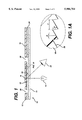
- FIG. 4 depicts an alternative embodiment of the apparatus of FIG. 3, in which the reaction area is heated with heater 130.
- Heater 130 may be a radiant heater, resistance heater, or any other suitable type of heater well known in the art. If Fresnel reflector 120 comprises a metallized layer, the layer serves to diffuse the heat, so that the temperature of the reaction area is quite uniform.
- the metallized layer of device 110 may include electrical contacts (such as A and B shown in FIG. 2), and the reaction layer can be heated by passing a current through the metallized layer. Note that the positions of source 118 and detector 122 are reversed from their positions in FIG. 3. The arrangement in FIG.
- the apparatus is part of an analyzer (such as the "VITROS ECI” Analyzer, available from J&J Ortho Clinical Diagnostics, Rochester, N.Y.) that includes a light source which provides incident light at an acute (e.g., 45°) angle to the plane of the device and a detector that is normal to the plane of the device.
- an analyzer such as the "VITROS ECI” Analyzer, available from J&J Ortho Clinical Diagnostics, Rochester, N.Y.
- a light source which provides incident light at an acute (e.g., 45°) angle to the plane of the device and a detector that is normal to the plane of the device.
- FIG. 4 includes an optional auxiliary detector 132.
- the purpose of this detector is to detect gross movement of device 110 during the measurement and to generate an error signal when it detects such gross movement.
- Detector 132 would normally detect small changes during the course of a measurement. Changes that exceed a threshold value (easily determined by routine experimentation) would trigger the error signal.
Abstract
A device for use in a medical diagnostic apparatus includes an area for containing a sample of biological fluid as it undergoes a reaction that causes a detectable change in the sample's optical properties. The reaction area is sandwiched between a transparent front layer and a Fresnel reflector layer. The area is illuminated through the front layer, and the light is reflected from the reflector layer to a detector. The detector monitors the light incident on it and calculates, from the change during the course of the reaction, an analyte concentration or property of the fluid sample. The reaction area is optionally heated.
Description
1. Field of the Invention
This invention relates to a medical diagnostic apparatus for optically measuring the concentration of an analyte in or a property of a biological fluid; more particularly, an apparatus whose optical system includes a Fresnel reflector.
2. Description of the Related Art
A variety of medical diagnostic procedures involve tests on biological fluids, such as blood, urine, or saliva, and are based on a change in light absorption of such a fluid or an element of the fluid, such as blood serum. Some of these procedures make use of a transparent or translucent device to contain the biological fluid and a reagent. A change in light absorption of the fluid can be related to an analyte concentration in, or property of, the fluid. Typically, a light source is located adjacent to one surface of the device and a detector is adjacent to the opposite surface. Alternatively, if a reflector is located adjacent to that opposite surface, then the light can pass through the device twice and the source and detector can be on the same side of the device. A device of this latter type, in which light is first transmitted through the sample area, then reflected through a second time is called a "transflectance" device. References to "light" throughout this specification and the appended claims should be understood to include the infrared and ultraviolet spectra, as well as the visible. References to "absorption" are meant to refer to the reduction in intensity as a light beam passes through a medium, thus, it encompasses both "true" absorption and scattering.
An example of a transparent test device is described in Wells et al. WO94/02850, published on Feb. 3, 1994. Their device comprises a sealed housing, which is transparent or translucent, impervious, and rigid or semi-rigid. An assay material is contained within the housing, together with one or more assay reagents at predetermined sites. The housing is opened and the sample introduced just before conducting the assay. The combination of assay reagents and analyte in the sample results in a change in optical properties, such as color, of selected reagents at the end of the assay. The results can be read visually or with an optical instrument.
U.S. Pat. No. 4,037,974, issued Jul. 26, 1977, to T. Fletcher et al. discloses a sample cell for spectrophotometers, which prevents stray light from being carried to a detector through the cell wall. In one embodiment, the cell has a side wall whose outer surface has a sawtooth configuration, which causes light that enters the side wall from the interior of the cell either to be reflected back into the sample or pass out through the cell wall.
U.S. Pat. No. 4,116,566, issued Sep. 26, 1978, to E. Sick discloses a device for detecting defects in webs of material, using an optical system that may include a sawtooth mirror.
U.S. Pat. No. 4,233,029, issued Nov. 11, 1980, to R. Columbus discloses a liquid transport device that has opposed surfaces, each of which may have a sawtooth cross section. The sawtooth arrangement of one surface is orthogonal to the arrangement of the other surface, and the resultant liquid flow is multidirectional.
U.S. Pat. No. 5,214,277, issued May 25, 1993, to J. Drennen, III discloses a cell that holds near-infrared reflectance spectrometer samples. The spectrometer includes a light source which illuminates a sample that is contained in the cell. The cell surfaces direct the light onto the sample, collect light diffusely reflected from the sample and direct the light back toward a detector.
U.S. Pat. No. 5,522,255, issued Jun. 4, 1996, to G. Neel et al. discloses an instrument and method for determining a coagulation characteristic of blood by a reflectance technique. The sample holder includes a combination reagent heater and reflector. (See also WO95/07452.)
U.S. Pat. No. D 375,799, issued Nov. 19, 1996, to W. Leiva et al. discloses a transparent assay tester for biological fluids with a Fresnel lens.
U.S. Pat. No. 5,468,606, issued Nov. 21, 1995, to G. Bogart et al. discloses an analyte-detecting device which has a substrate with an optically active surface. When illuminated, the surface exhibits different colors, depending on whether or not the analyte is present.
U.S. Pat. No. 5,478,527, issued Dec. 26, 1995, to E. Gustafson et al. discloses a multilayer reflective biograting for use in an immunoassay. The diffraction signal from the biograting changes in the presence of an analyte
The present invention is a device for use in a medical diagnostic apparatus that comprises a light source to illuminate a sample of a biological fluid and a detector to detect light that has passed through the sample. The device comprises
(a) a reaction area for containing at least a portion of the sample, sandwiched between
(b) a substantially transparent first layer, and
(c) a second layer, which includes a Fresnel reflector, for providing specular reflection of light that has passed through the first layer and the reaction area and directing the light toward the detector.
Another embodiment of the present invention is an apparatus for measuring an analyte concentration or property of a biological fluid comprising
(a) a means for illuminating a sample of the biological fluid,
(b) means for positioning the sample in the path of light from the illuminating means,
(c) means for detecting and measuring light that has interacted with the sample at one or more wavelengths, the detecting and measuring means comprising a Fresnel reflector, and
(d) means for calculating the analyte concentration or property of the fluid from the measured reflected light.
As used in the present specification and claims, a "Fresnel reflector" is a reflector having a cross section that is substantially a sawtooth. The reflector may be homogeneous, but preferably it has a reflective coating. An advantage provided by a Fresnel reflector in the present invention is to provide specular, rather than diffuse, reflection of a signal to a detector and thereby to permit a higher signal-to-noise ratio.
Although the "layers" that are elements of the present device are typically thin sheets, that is not necessarily their form, and no thickness limitation is intended by the use of that term.
FIG. 1 is a schematic side view of a device of this invention.
FIG. 1A is an enlarged view of a segment of the device of
FIG. 1.
FIG. 2 is a schematic side view of an alternative embodiment of a device of this invention.
FIG. 3 is a schematic side view of an apparatus of the invention.
FIG. 4 is a schematic side view of an alternative embodiment of an apparatus of the invention.
This invention relates to a diagnostic device for analyzing biological fluid. The device is of the type that contains the fluid to be analyzed in a transparent or translucent container, typically, but not necessarily, a capillary-fill container, and that relates a change in light absorption of the fluid to an analyte concentration in the fluid or to a property of the fluid. The device includes a reaction area, in which the sample undergoes a reaction that causes a change in light absorption; a transparent layer, through which a light beam is incident on the sample; and a second layer, on the side of the sample that is opposite the transparent layer. A Fresnel reflector, at or near a surface of the second layer, reflects light back through the sample and toward a detector.
This type of device is suitable for a variety of analytical tests of biological fluids, such as determining biochemical or hematological characteristics, or measuring the concentration in such fluids of proteins, carbohydrates, lipids, drugs, toxins, gases, electrolytes, etc. The device is particularly well suited for measuring blood clotting time--"prothrombin time" or "PT"--and details regarding such a device appear below. The modifications needed to adapt the device for other applications require no more than routine experimentation.
FIG. 1 is a schematic of a device 10 of the present invention. Reaction area 12, which for a PT test contains thromboplastin, is sandwiched between transparent layer 14 and backing layer 16. The reaction area is illuminated with light from source 18. After passing through the sample contained in reaction area 12, the light is reflected from Fresnel reflector 20 of backing layer 16 toward detector 22. Optional electrical contacts A and B are discussed below, in connection with the description of FIG. 4. Optional spacer layer 24 separates layers 14 and 16 and has a through hole that forms reaction area 12. Sample (e.g., whole blood or blood plasma, is applied to the reaction area through hole 26 in backing layer 16. In a PT test, thromboplastin in the reaction area reacts with the blood to change the optical properties in a way that can be related to clotting time. The grooved surface of backing layer 16 can form a capillary gap, which, together with optional vent 28, facilitates filling of reaction area 12. Preferably, as shown, reaction area 12 and Fresnel reflector 20 are about the same size, but design considerations, cost, etc. may dictate that one be larger than the other. Similarly, Fresnel reflector 20 may cover part or all of backing layer 16. Transparent layer 14 can be of any suitable transparent material known in the art, such as glass or plastic sheet. Polyester is a preferred material, because it is inexpensive, available, and easy to process. Backing layer 16 may also be of glass or plastic, but it needn't be transparent (if Fresnel reflector 20 adjoins reaction area 12), so it may also be of metal or other non-transparent material. Light source 18 may be an incandescent or fluorescent source of light; preferably, it is one or more LEDs. Detector 22 may be any photodetector that is sensitive to the wavelength(s) of light emitted by source 18. Among the suitable detectors, well known in the art, are photomultiplier tubes, photodiodes, etc. Preferably, detector 22 is a photodiode. Optional spacing layer 24 is preferably double-stick tape, which provides the walls for reaction area 12 and adheres layers 14 and 16 together.
FIG. 1A is an enlarged view of Fresnel reflector 20, which may be the reflective surface of a homogeneous backing layer 16, a reflective coating on the backing layer surface, or a reflective element that is attached to the backing layer. The surface consists of a series of grooves that are characterized by slope angle s, draft angle d, and facet spacing f. Although each of these parameters may change along the length of the reflector, the draft angle and facet spacing are generally constant. The slope angle may change with distance from the optical axis of the reflector, or it can also be constant, depending on whether or not the reflected beam is to be diffused and/or focused. In order to minimize diffraction effects, the facet spacing f should be at least about 10 times the wavelength of light used. Thus, if 900 nm light is used, the facet spacing should be at least 9000 nm, or 9 μm. The facet spacing is preferably not much larger than necessary, since large facet spacing corresponds to a small number of grooves and a thick (in cross section) reflector, neither of which is desirable. The slope angle S is dictated by the parameters of the optical system and the desire to provide specular reflection to the detector. The draft an-le is not critical, since it usually doesn't affect the optical performance of the reflector if it is kept small. Fresnel reflectors and detailed information regarding specific applications are available from Fresnel Optics, Inc., Henrietta, N.Y.
The Fresnel reflector in FIGS. 1 and 1A is shown on the "bottom" surface of backing layer 16; i.e., the surface that adjoins the reaction area. As shown in FIG. 2, however, the reflector may be on the top surface 30 of backing layer 16 or it may be sandwiched between two layers that comprise a backing layer. Of course, if the reflector is on the top surface or is sandwiched between two layers, then the layer between the reflector and the reaction area must be transparent. When the Fresnel reflector is on either the top or bottom surface of the backing layer, the backing layer is preferably a metallized transparent sheet.
FIG. 3 depicts a meter 100 of the present invention. The meter holds a device 110, which has reaction area 112 sandwiched between transparent front layer 114 and transparent backing layer 116. The reaction area is illuminated with light from source 118. After passing through the sample contained in reaction area 112, the light passes through backing layer 116 and is reflected from Fresnel reflector 120 toward detector 122. Optional spacer layer 124 is preferably double-stick tape. The reflected light signal detected by detector 122 is used to calculate the sample property of interest. Preferably, transparent layers 114 and 116 comprise thermoplastic sheets.
FIG. 4 depicts an alternative embodiment of the apparatus of FIG. 3, in which the reaction area is heated with heater 130. Heater 130 may be a radiant heater, resistance heater, or any other suitable type of heater well known in the art. If Fresnel reflector 120 comprises a metallized layer, the layer serves to diffuse the heat, so that the temperature of the reaction area is quite uniform. Alternatively, the metallized layer of device 110 may include electrical contacts (such as A and B shown in FIG. 2), and the reaction layer can be heated by passing a current through the metallized layer. Note that the positions of source 118 and detector 122 are reversed from their positions in FIG. 3. The arrangement in FIG. 4 is preferred when the apparatus is part of an analyzer (such as the "VITROS ECI" Analyzer, available from J&J Ortho Clinical Diagnostics, Rochester, N.Y.) that includes a light source which provides incident light at an acute (e.g., 45°) angle to the plane of the device and a detector that is normal to the plane of the device.
Finally, FIG. 4 includes an optional auxiliary detector 132. The purpose of this detector is to detect gross movement of device 110 during the measurement and to generate an error signal when it detects such gross movement. Detector 132 would normally detect small changes during the course of a measurement. Changes that exceed a threshold value (easily determined by routine experimentation) would trigger the error signal.
Claims (7)
1. For use in a medical diagnostic apparatus that comprises a light source to illuminate a sample of a biological fluid and a detector to detect light that has passed through the sample, a device that comprises
(a) a reaction area for containing at least a portion of the sample, sandwiched between
(b) a substantially transparent first layer, and
(c) a second layer, comprising a metallized thermoplastic sheet having means for making an electrical connection to the metallized sheet and including a Fresnel reflector, for providing specular reflection of light that has passed through the first layer and the reaction area and directing the light toward the detector.
2. The device of claim 1 in which the biological fluid is whole blood or blood plasma.
3. The device of claim 2 in which the reaction area comprises a reagent that reacts with the blood or plasma to change an optical property thereof in a way that can be quantitatively related to a blood clotting characteristic.
4. The device of claim 1 in which the Fresnel reflector comprises a surface of the second layer that is reflective and that adjoins the reaction area.
5. The device of claim 4 in which the reflective surface is grooved and is spaced apart from an adjoining surface of the first layer a distance that is small enough to maintain a capillary flow of sample into the reaction area when the sample is introduced between the adjoining surfaces.
6. The device of claim 1 in which the Fresnel reflector is separated from the reaction area by a transparent section of the second layer.
7. The device of claim 1 in which the first layer comprises a thermoplastic sheet.
Priority Applications (2)
| Application Number | Priority Date | Filing Date | Title |
|---|---|---|---|
| US08/986,560 US5986754A (en) | 1997-12-08 | 1997-12-08 | Medical diagnostic apparatus using a Fresnel reflector |
| JP10361978A JPH11258150A (en) | 1997-12-08 | 1998-12-07 | Medical diagnosis apparatus using fresnel reflecting body |
Applications Claiming Priority (1)
| Application Number | Priority Date | Filing Date | Title |
|---|---|---|---|
| US08/986,560 US5986754A (en) | 1997-12-08 | 1997-12-08 | Medical diagnostic apparatus using a Fresnel reflector |
Publications (1)
| Publication Number | Publication Date |
|---|---|
| US5986754A true US5986754A (en) | 1999-11-16 |
Family
ID=25532550
Family Applications (1)
| Application Number | Title | Priority Date | Filing Date |
|---|---|---|---|
| US08/986,560 Expired - Lifetime US5986754A (en) | 1997-12-08 | 1997-12-08 | Medical diagnostic apparatus using a Fresnel reflector |
Country Status (2)
| Country | Link |
|---|---|
| US (1) | US5986754A (en) |
| JP (1) | JPH11258150A (en) |
Cited By (71)
| Publication number | Priority date | Publication date | Assignee | Title |
|---|---|---|---|---|
| US6312888B1 (en) | 1998-06-10 | 2001-11-06 | Abbott Laboratories | Diagnostic assay for a sample of biological fluid |
| US6458326B1 (en) | 1999-11-24 | 2002-10-01 | Home Diagnostics, Inc. | Protective test strip platform |
| US6525330B2 (en) | 2001-02-28 | 2003-02-25 | Home Diagnostics, Inc. | Method of strip insertion detection |
| US6541266B2 (en) | 2001-02-28 | 2003-04-01 | Home Diagnostics, Inc. | Method for determining concentration of an analyte in a test strip |
| US6562625B2 (en) | 2001-02-28 | 2003-05-13 | Home Diagnostics, Inc. | Distinguishing test types through spectral analysis |
| US20060110283A1 (en) * | 2002-11-12 | 2006-05-25 | Inverness Medical Switzerland Gmbh | Photometric determination of coagulation time in undiluted whole blood |
| US7875047B2 (en) | 2002-04-19 | 2011-01-25 | Pelikan Technologies, Inc. | Method and apparatus for a multi-use body fluid sampling device with sterility barrier release |
| US7892183B2 (en) | 2002-04-19 | 2011-02-22 | Pelikan Technologies, Inc. | Method and apparatus for body fluid sampling and analyte sensing |
| US7901365B2 (en) | 2002-04-19 | 2011-03-08 | Pelikan Technologies, Inc. | Method and apparatus for penetrating tissue |
| US7909777B2 (en) | 2002-04-19 | 2011-03-22 | Pelikan Technologies, Inc | Method and apparatus for penetrating tissue |
| US7909775B2 (en) | 2001-06-12 | 2011-03-22 | Pelikan Technologies, Inc. | Method and apparatus for lancet launching device integrated onto a blood-sampling cartridge |
| US7909778B2 (en) | 2002-04-19 | 2011-03-22 | Pelikan Technologies, Inc. | Method and apparatus for penetrating tissue |
| US7909774B2 (en) | 2002-04-19 | 2011-03-22 | Pelikan Technologies, Inc. | Method and apparatus for penetrating tissue |
| US7914465B2 (en) | 2002-04-19 | 2011-03-29 | Pelikan Technologies, Inc. | Method and apparatus for penetrating tissue |
| EP2322914A1 (en) * | 2003-03-24 | 2011-05-18 | Intuity Medical, Inc. | Analyte concentration detection devices and methods |
| US7976476B2 (en) | 2002-04-19 | 2011-07-12 | Pelikan Technologies, Inc. | Device and method for variable speed lancet |
| US7981056B2 (en) | 2002-04-19 | 2011-07-19 | Pelikan Technologies, Inc. | Methods and apparatus for lancet actuation |
| US7981055B2 (en) | 2001-06-12 | 2011-07-19 | Pelikan Technologies, Inc. | Tissue penetration device |
| US7988645B2 (en) | 2001-06-12 | 2011-08-02 | Pelikan Technologies, Inc. | Self optimizing lancing device with adaptation means to temporal variations in cutaneous properties |
| US8007446B2 (en) | 2002-04-19 | 2011-08-30 | Pelikan Technologies, Inc. | Method and apparatus for penetrating tissue |
| US8062231B2 (en) | 2002-04-19 | 2011-11-22 | Pelikan Technologies, Inc. | Method and apparatus for penetrating tissue |
| US8079960B2 (en) | 2002-04-19 | 2011-12-20 | Pelikan Technologies, Inc. | Methods and apparatus for lancet actuation |
| US8197421B2 (en) | 2002-04-19 | 2012-06-12 | Pelikan Technologies, Inc. | Method and apparatus for penetrating tissue |
| US8221334B2 (en) | 2002-04-19 | 2012-07-17 | Sanofi-Aventis Deutschland Gmbh | Method and apparatus for penetrating tissue |
| US8251921B2 (en) | 2003-06-06 | 2012-08-28 | Sanofi-Aventis Deutschland Gmbh | Method and apparatus for body fluid sampling and analyte sensing |
| US8262614B2 (en) | 2003-05-30 | 2012-09-11 | Pelikan Technologies, Inc. | Method and apparatus for fluid injection |
| US8267870B2 (en) | 2002-04-19 | 2012-09-18 | Sanofi-Aventis Deutschland Gmbh | Method and apparatus for body fluid sampling with hybrid actuation |
| US8282576B2 (en) | 2003-09-29 | 2012-10-09 | Sanofi-Aventis Deutschland Gmbh | Method and apparatus for an improved sample capture device |
| US8296918B2 (en) | 2003-12-31 | 2012-10-30 | Sanofi-Aventis Deutschland Gmbh | Method of manufacturing a fluid sampling device with improved analyte detecting member configuration |
| US8333710B2 (en) | 2002-04-19 | 2012-12-18 | Sanofi-Aventis Deutschland Gmbh | Tissue penetration device |
| US8360993B2 (en) | 2005-09-30 | 2013-01-29 | Intuity Medical, Inc. | Method for body fluid sample extraction |
| US8360992B2 (en) | 2002-04-19 | 2013-01-29 | Sanofi-Aventis Deutschland Gmbh | Method and apparatus for penetrating tissue |
| US8372016B2 (en) | 2002-04-19 | 2013-02-12 | Sanofi-Aventis Deutschland Gmbh | Method and apparatus for body fluid sampling and analyte sensing |
| US8382682B2 (en) | 2002-04-19 | 2013-02-26 | Sanofi-Aventis Deutschland Gmbh | Method and apparatus for penetrating tissue |
| US8435190B2 (en) | 2002-04-19 | 2013-05-07 | Sanofi-Aventis Deutschland Gmbh | Method and apparatus for penetrating tissue |
| US8439872B2 (en) | 1998-03-30 | 2013-05-14 | Sanofi-Aventis Deutschland Gmbh | Apparatus and method for penetration with shaft having a sensor for sensing penetration depth |
| WO2013079619A1 (en) * | 2011-12-01 | 2013-06-06 | Biosurfit, S.A. | Photometric device and method |
| US8556829B2 (en) | 2002-04-19 | 2013-10-15 | Sanofi-Aventis Deutschland Gmbh | Method and apparatus for penetrating tissue |
| US8574895B2 (en) | 2002-12-30 | 2013-11-05 | Sanofi-Aventis Deutschland Gmbh | Method and apparatus using optical techniques to measure analyte levels |
| US8641644B2 (en) | 2000-11-21 | 2014-02-04 | Sanofi-Aventis Deutschland Gmbh | Blood testing apparatus having a rotatable cartridge with multiple lancing elements and testing means |
| US8652831B2 (en) | 2004-12-30 | 2014-02-18 | Sanofi-Aventis Deutschland Gmbh | Method and apparatus for analyte measurement test time |
| US8668656B2 (en) | 2003-12-31 | 2014-03-11 | Sanofi-Aventis Deutschland Gmbh | Method and apparatus for improving fluidic flow and sample capture |
| US8702624B2 (en) | 2006-09-29 | 2014-04-22 | Sanofi-Aventis Deutschland Gmbh | Analyte measurement device with a single shot actuator |
| US8721671B2 (en) | 2001-06-12 | 2014-05-13 | Sanofi-Aventis Deutschland Gmbh | Electric lancet actuator |
| US8784335B2 (en) | 2002-04-19 | 2014-07-22 | Sanofi-Aventis Deutschland Gmbh | Body fluid sampling device with a capacitive sensor |
| US8801631B2 (en) | 2005-09-30 | 2014-08-12 | Intuity Medical, Inc. | Devices and methods for facilitating fluid transport |
| US8828203B2 (en) | 2004-05-20 | 2014-09-09 | Sanofi-Aventis Deutschland Gmbh | Printable hydrogels for biosensors |
| US8919605B2 (en) | 2009-11-30 | 2014-12-30 | Intuity Medical, Inc. | Calibration material delivery devices and methods |
| US8965476B2 (en) | 2010-04-16 | 2015-02-24 | Sanofi-Aventis Deutschland Gmbh | Tissue penetration device |
| US8969097B2 (en) | 2005-06-13 | 2015-03-03 | Intuity Medical, Inc. | Analyte detection devices and methods with hematocrit-volume correction and feedback control |
| US9144401B2 (en) | 2003-06-11 | 2015-09-29 | Sanofi-Aventis Deutschland Gmbh | Low pain penetrating member |
| US20150340566A1 (en) * | 2013-01-10 | 2015-11-26 | Koninklijke Philips N.V. | Led with shaped growth substrate for side emission |
| US9226699B2 (en) | 2002-04-19 | 2016-01-05 | Sanofi-Aventis Deutschland Gmbh | Body fluid sampling module with a continuous compression tissue interface surface |
| US9248267B2 (en) | 2002-04-19 | 2016-02-02 | Sanofi-Aventis Deustchland Gmbh | Tissue penetration device |
| US9297816B1 (en) | 2012-12-21 | 2016-03-29 | University Of South Florida | Devices and methods for measuring blood coagulation |
| US9314194B2 (en) | 2002-04-19 | 2016-04-19 | Sanofi-Aventis Deutschland Gmbh | Tissue penetration device |
| US9351680B2 (en) | 2003-10-14 | 2016-05-31 | Sanofi-Aventis Deutschland Gmbh | Method and apparatus for a variable user interface |
| US9375169B2 (en) | 2009-01-30 | 2016-06-28 | Sanofi-Aventis Deutschland Gmbh | Cam drive for managing disposable penetrating member actions with a single motor and motor and control system |
| US9386944B2 (en) | 2008-04-11 | 2016-07-12 | Sanofi-Aventis Deutschland Gmbh | Method and apparatus for analyte detecting device |
| US9427532B2 (en) | 2001-06-12 | 2016-08-30 | Sanofi-Aventis Deutschland Gmbh | Tissue penetration device |
| US9636051B2 (en) | 2008-06-06 | 2017-05-02 | Intuity Medical, Inc. | Detection meter and mode of operation |
| US9775553B2 (en) | 2004-06-03 | 2017-10-03 | Sanofi-Aventis Deutschland Gmbh | Method and apparatus for a fluid sampling device |
| US9782114B2 (en) | 2011-08-03 | 2017-10-10 | Intuity Medical, Inc. | Devices and methods for body fluid sampling and analysis |
| US9795747B2 (en) | 2010-06-02 | 2017-10-24 | Sanofi-Aventis Deutschland Gmbh | Methods and apparatus for lancet actuation |
| US9820684B2 (en) | 2004-06-03 | 2017-11-21 | Sanofi-Aventis Deutschland Gmbh | Method and apparatus for a fluid sampling device |
| US9833183B2 (en) | 2008-05-30 | 2017-12-05 | Intuity Medical, Inc. | Body fluid sampling device—sampling site interface |
| US10330667B2 (en) | 2010-06-25 | 2019-06-25 | Intuity Medical, Inc. | Analyte monitoring methods and systems |
| US10383556B2 (en) | 2008-06-06 | 2019-08-20 | Intuity Medical, Inc. | Medical diagnostic devices and methods |
| US10729386B2 (en) | 2013-06-21 | 2020-08-04 | Intuity Medical, Inc. | Analyte monitoring system with audible feedback |
| US10772550B2 (en) | 2002-02-08 | 2020-09-15 | Intuity Medical, Inc. | Autonomous, ambulatory analyte monitor or drug delivery device |
| EP1625386B1 (en) * | 2003-03-24 | 2021-06-23 | Intuity Medical, Inc. | Analyte concentration detection devices and methods |
Families Citing this family (1)
| Publication number | Priority date | Publication date | Assignee | Title |
|---|---|---|---|---|
| BRPI0910934A2 (en) * | 2008-04-04 | 2015-10-06 | Medical Vision Res And Dev Ab | Particle measurement in liquid using light reflection. |
Citations (10)
| Publication number | Priority date | Publication date | Assignee | Title |
|---|---|---|---|---|
| US4037974A (en) * | 1974-10-17 | 1977-07-26 | Fletcher Taylor C | Sample cell for spectrophotometers |
| US4116566A (en) * | 1975-11-12 | 1978-09-26 | Erwin Sick Gesellschaft Mit Beschrankter Haftung Optik-Elektronik | Line scanning device for detecting defects in webs of material |
| US4233029A (en) * | 1978-10-25 | 1980-11-11 | Eastman Kodak Company | Liquid transport device and method |
| US4566791A (en) * | 1983-10-31 | 1986-01-28 | Pacific Scientific Company | Fluid sample cell comprising Fresnel sectors |
| US5044747A (en) * | 1989-03-03 | 1991-09-03 | Lt Industries | Modular flow-through cell |
| US5214277A (en) * | 1992-06-15 | 1993-05-25 | Drennen Iii James K | Near-infrared reflectance spectrometer system and related sample cell and sample support |
| WO1994002850A1 (en) * | 1992-07-21 | 1994-02-03 | Medix Biotech, Inc. | Transparent assay test devices and methods |
| US5468606A (en) * | 1989-09-18 | 1995-11-21 | Biostar, Inc. | Devices for detection of an analyte based upon light interference |
| US5478527A (en) * | 1990-05-17 | 1995-12-26 | Adeza Biomedical Corporation | Highly reflective biogratings |
| US5522255A (en) * | 1993-08-31 | 1996-06-04 | Boehringer Mannheim Corporation | Fluid dose, flow and coagulation sensor for medical instrument |
-
1997
- 1997-12-08 US US08/986,560 patent/US5986754A/en not_active Expired - Lifetime
-
1998
- 1998-12-07 JP JP10361978A patent/JPH11258150A/en active Pending
Patent Citations (10)
| Publication number | Priority date | Publication date | Assignee | Title |
|---|---|---|---|---|
| US4037974A (en) * | 1974-10-17 | 1977-07-26 | Fletcher Taylor C | Sample cell for spectrophotometers |
| US4116566A (en) * | 1975-11-12 | 1978-09-26 | Erwin Sick Gesellschaft Mit Beschrankter Haftung Optik-Elektronik | Line scanning device for detecting defects in webs of material |
| US4233029A (en) * | 1978-10-25 | 1980-11-11 | Eastman Kodak Company | Liquid transport device and method |
| US4566791A (en) * | 1983-10-31 | 1986-01-28 | Pacific Scientific Company | Fluid sample cell comprising Fresnel sectors |
| US5044747A (en) * | 1989-03-03 | 1991-09-03 | Lt Industries | Modular flow-through cell |
| US5468606A (en) * | 1989-09-18 | 1995-11-21 | Biostar, Inc. | Devices for detection of an analyte based upon light interference |
| US5478527A (en) * | 1990-05-17 | 1995-12-26 | Adeza Biomedical Corporation | Highly reflective biogratings |
| US5214277A (en) * | 1992-06-15 | 1993-05-25 | Drennen Iii James K | Near-infrared reflectance spectrometer system and related sample cell and sample support |
| WO1994002850A1 (en) * | 1992-07-21 | 1994-02-03 | Medix Biotech, Inc. | Transparent assay test devices and methods |
| US5522255A (en) * | 1993-08-31 | 1996-06-04 | Boehringer Mannheim Corporation | Fluid dose, flow and coagulation sensor for medical instrument |
Cited By (156)
| Publication number | Priority date | Publication date | Assignee | Title |
|---|---|---|---|---|
| US8439872B2 (en) | 1998-03-30 | 2013-05-14 | Sanofi-Aventis Deutschland Gmbh | Apparatus and method for penetration with shaft having a sensor for sensing penetration depth |
| US6312888B1 (en) | 1998-06-10 | 2001-11-06 | Abbott Laboratories | Diagnostic assay for a sample of biological fluid |
| US6458326B1 (en) | 1999-11-24 | 2002-10-01 | Home Diagnostics, Inc. | Protective test strip platform |
| US8641644B2 (en) | 2000-11-21 | 2014-02-04 | Sanofi-Aventis Deutschland Gmbh | Blood testing apparatus having a rotatable cartridge with multiple lancing elements and testing means |
| US6525330B2 (en) | 2001-02-28 | 2003-02-25 | Home Diagnostics, Inc. | Method of strip insertion detection |
| US6541266B2 (en) | 2001-02-28 | 2003-04-01 | Home Diagnostics, Inc. | Method for determining concentration of an analyte in a test strip |
| US6562625B2 (en) | 2001-02-28 | 2003-05-13 | Home Diagnostics, Inc. | Distinguishing test types through spectral analysis |
| US9937298B2 (en) | 2001-06-12 | 2018-04-10 | Sanofi-Aventis Deutschland Gmbh | Tissue penetration device |
| US7988645B2 (en) | 2001-06-12 | 2011-08-02 | Pelikan Technologies, Inc. | Self optimizing lancing device with adaptation means to temporal variations in cutaneous properties |
| US8679033B2 (en) | 2001-06-12 | 2014-03-25 | Sanofi-Aventis Deutschland Gmbh | Tissue penetration device |
| US7909775B2 (en) | 2001-06-12 | 2011-03-22 | Pelikan Technologies, Inc. | Method and apparatus for lancet launching device integrated onto a blood-sampling cartridge |
| US9694144B2 (en) | 2001-06-12 | 2017-07-04 | Sanofi-Aventis Deutschland Gmbh | Sampling module device and method |
| US8641643B2 (en) | 2001-06-12 | 2014-02-04 | Sanofi-Aventis Deutschland Gmbh | Sampling module device and method |
| US8721671B2 (en) | 2001-06-12 | 2014-05-13 | Sanofi-Aventis Deutschland Gmbh | Electric lancet actuator |
| US8622930B2 (en) | 2001-06-12 | 2014-01-07 | Sanofi-Aventis Deutschland Gmbh | Tissue penetration device |
| US9427532B2 (en) | 2001-06-12 | 2016-08-30 | Sanofi-Aventis Deutschland Gmbh | Tissue penetration device |
| US9802007B2 (en) | 2001-06-12 | 2017-10-31 | Sanofi-Aventis Deutschland Gmbh | Methods and apparatus for lancet actuation |
| US8382683B2 (en) | 2001-06-12 | 2013-02-26 | Sanofi-Aventis Deutschland Gmbh | Tissue penetration device |
| US8360991B2 (en) | 2001-06-12 | 2013-01-29 | Sanofi-Aventis Deutschland Gmbh | Tissue penetration device |
| US7981055B2 (en) | 2001-06-12 | 2011-07-19 | Pelikan Technologies, Inc. | Tissue penetration device |
| US8206319B2 (en) | 2001-06-12 | 2012-06-26 | Sanofi-Aventis Deutschland Gmbh | Tissue penetration device |
| US8343075B2 (en) | 2001-06-12 | 2013-01-01 | Sanofi-Aventis Deutschland Gmbh | Tissue penetration device |
| US8337421B2 (en) | 2001-06-12 | 2012-12-25 | Sanofi-Aventis Deutschland Gmbh | Tissue penetration device |
| US8016774B2 (en) | 2001-06-12 | 2011-09-13 | Pelikan Technologies, Inc. | Tissue penetration device |
| US8206317B2 (en) | 2001-06-12 | 2012-06-26 | Sanofi-Aventis Deutschland Gmbh | Tissue penetration device |
| US8282577B2 (en) | 2001-06-12 | 2012-10-09 | Sanofi-Aventis Deutschland Gmbh | Method and apparatus for lancet launching device integrated onto a blood-sampling cartridge |
| US8123700B2 (en) | 2001-06-12 | 2012-02-28 | Pelikan Technologies, Inc. | Method and apparatus for lancet launching device integrated onto a blood-sampling cartridge |
| US8211037B2 (en) | 2001-06-12 | 2012-07-03 | Pelikan Technologies, Inc. | Tissue penetration device |
| US8162853B2 (en) | 2001-06-12 | 2012-04-24 | Pelikan Technologies, Inc. | Tissue penetration device |
| US8845550B2 (en) | 2001-06-12 | 2014-09-30 | Sanofi-Aventis Deutschland Gmbh | Tissue penetration device |
| US8216154B2 (en) | 2001-06-12 | 2012-07-10 | Sanofi-Aventis Deutschland Gmbh | Tissue penetration device |
| US9560993B2 (en) | 2001-11-21 | 2017-02-07 | Sanofi-Aventis Deutschland Gmbh | Blood testing apparatus having a rotatable cartridge with multiple lancing elements and testing means |
| US10772550B2 (en) | 2002-02-08 | 2020-09-15 | Intuity Medical, Inc. | Autonomous, ambulatory analyte monitor or drug delivery device |
| US8337419B2 (en) | 2002-04-19 | 2012-12-25 | Sanofi-Aventis Deutschland Gmbh | Tissue penetration device |
| US8491500B2 (en) | 2002-04-19 | 2013-07-23 | Sanofi-Aventis Deutschland Gmbh | Methods and apparatus for lancet actuation |
| US8197421B2 (en) | 2002-04-19 | 2012-06-12 | Pelikan Technologies, Inc. | Method and apparatus for penetrating tissue |
| US8221334B2 (en) | 2002-04-19 | 2012-07-17 | Sanofi-Aventis Deutschland Gmbh | Method and apparatus for penetrating tissue |
| US9072842B2 (en) | 2002-04-19 | 2015-07-07 | Sanofi-Aventis Deutschland Gmbh | Method and apparatus for penetrating tissue |
| US8235915B2 (en) | 2002-04-19 | 2012-08-07 | Sanofi-Aventis Deutschland Gmbh | Method and apparatus for penetrating tissue |
| US9089678B2 (en) | 2002-04-19 | 2015-07-28 | Sanofi-Aventis Deutschland Gmbh | Method and apparatus for penetrating tissue |
| US8197423B2 (en) | 2002-04-19 | 2012-06-12 | Pelikan Technologies, Inc. | Method and apparatus for penetrating tissue |
| US8267870B2 (en) | 2002-04-19 | 2012-09-18 | Sanofi-Aventis Deutschland Gmbh | Method and apparatus for body fluid sampling with hybrid actuation |
| US8157748B2 (en) | 2002-04-19 | 2012-04-17 | Pelikan Technologies, Inc. | Methods and apparatus for lancet actuation |
| US8079960B2 (en) | 2002-04-19 | 2011-12-20 | Pelikan Technologies, Inc. | Methods and apparatus for lancet actuation |
| US8062231B2 (en) | 2002-04-19 | 2011-11-22 | Pelikan Technologies, Inc. | Method and apparatus for penetrating tissue |
| US8333710B2 (en) | 2002-04-19 | 2012-12-18 | Sanofi-Aventis Deutschland Gmbh | Tissue penetration device |
| US8007446B2 (en) | 2002-04-19 | 2011-08-30 | Pelikan Technologies, Inc. | Method and apparatus for penetrating tissue |
| US9089294B2 (en) | 2002-04-19 | 2015-07-28 | Sanofi-Aventis Deutschland Gmbh | Analyte measurement device with a single shot actuator |
| US8337420B2 (en) | 2002-04-19 | 2012-12-25 | Sanofi-Aventis Deutschland Gmbh | Tissue penetration device |
| US7988644B2 (en) | 2002-04-19 | 2011-08-02 | Pelikan Technologies, Inc. | Method and apparatus for a multi-use body fluid sampling device with sterility barrier release |
| US9907502B2 (en) | 2002-04-19 | 2018-03-06 | Sanofi-Aventis Deutschland Gmbh | Method and apparatus for penetrating tissue |
| US9839386B2 (en) | 2002-04-19 | 2017-12-12 | Sanofi-Aventis Deustschland Gmbh | Body fluid sampling device with capacitive sensor |
| US8360992B2 (en) | 2002-04-19 | 2013-01-29 | Sanofi-Aventis Deutschland Gmbh | Method and apparatus for penetrating tissue |
| US7981056B2 (en) | 2002-04-19 | 2011-07-19 | Pelikan Technologies, Inc. | Methods and apparatus for lancet actuation |
| US8366637B2 (en) | 2002-04-19 | 2013-02-05 | Sanofi-Aventis Deutschland Gmbh | Method and apparatus for penetrating tissue |
| US8372016B2 (en) | 2002-04-19 | 2013-02-12 | Sanofi-Aventis Deutschland Gmbh | Method and apparatus for body fluid sampling and analyte sensing |
| US7976476B2 (en) | 2002-04-19 | 2011-07-12 | Pelikan Technologies, Inc. | Device and method for variable speed lancet |
| US7959582B2 (en) | 2002-04-19 | 2011-06-14 | Pelikan Technologies, Inc. | Method and apparatus for penetrating tissue |
| US8382682B2 (en) | 2002-04-19 | 2013-02-26 | Sanofi-Aventis Deutschland Gmbh | Method and apparatus for penetrating tissue |
| US8388551B2 (en) | 2002-04-19 | 2013-03-05 | Sanofi-Aventis Deutschland Gmbh | Method and apparatus for multi-use body fluid sampling device with sterility barrier release |
| US8403864B2 (en) | 2002-04-19 | 2013-03-26 | Sanofi-Aventis Deutschland Gmbh | Method and apparatus for penetrating tissue |
| US8414503B2 (en) | 2002-04-19 | 2013-04-09 | Sanofi-Aventis Deutschland Gmbh | Methods and apparatus for lancet actuation |
| US8430828B2 (en) | 2002-04-19 | 2013-04-30 | Sanofi-Aventis Deutschland Gmbh | Method and apparatus for a multi-use body fluid sampling device with sterility barrier release |
| US8435190B2 (en) | 2002-04-19 | 2013-05-07 | Sanofi-Aventis Deutschland Gmbh | Method and apparatus for penetrating tissue |
| US9186468B2 (en) | 2002-04-19 | 2015-11-17 | Sanofi-Aventis Deutschland Gmbh | Method and apparatus for penetrating tissue |
| US9795334B2 (en) | 2002-04-19 | 2017-10-24 | Sanofi-Aventis Deutschland Gmbh | Method and apparatus for penetrating tissue |
| US8202231B2 (en) | 2002-04-19 | 2012-06-19 | Sanofi-Aventis Deutschland Gmbh | Method and apparatus for penetrating tissue |
| US8496601B2 (en) | 2002-04-19 | 2013-07-30 | Sanofi-Aventis Deutschland Gmbh | Methods and apparatus for lancet actuation |
| US8556829B2 (en) | 2002-04-19 | 2013-10-15 | Sanofi-Aventis Deutschland Gmbh | Method and apparatus for penetrating tissue |
| US8562545B2 (en) | 2002-04-19 | 2013-10-22 | Sanofi-Aventis Deutschland Gmbh | Tissue penetration device |
| US9226699B2 (en) | 2002-04-19 | 2016-01-05 | Sanofi-Aventis Deutschland Gmbh | Body fluid sampling module with a continuous compression tissue interface surface |
| US8574168B2 (en) | 2002-04-19 | 2013-11-05 | Sanofi-Aventis Deutschland Gmbh | Method and apparatus for a multi-use body fluid sampling device with analyte sensing |
| US8579831B2 (en) | 2002-04-19 | 2013-11-12 | Sanofi-Aventis Deutschland Gmbh | Method and apparatus for penetrating tissue |
| US7938787B2 (en) | 2002-04-19 | 2011-05-10 | Pelikan Technologies, Inc. | Method and apparatus for penetrating tissue |
| US8636673B2 (en) | 2002-04-19 | 2014-01-28 | Sanofi-Aventis Deutschland Gmbh | Tissue penetration device |
| US7914465B2 (en) | 2002-04-19 | 2011-03-29 | Pelikan Technologies, Inc. | Method and apparatus for penetrating tissue |
| US7909774B2 (en) | 2002-04-19 | 2011-03-22 | Pelikan Technologies, Inc. | Method and apparatus for penetrating tissue |
| US9724021B2 (en) | 2002-04-19 | 2017-08-08 | Sanofi-Aventis Deutschland Gmbh | Method and apparatus for penetrating tissue |
| US7909778B2 (en) | 2002-04-19 | 2011-03-22 | Pelikan Technologies, Inc. | Method and apparatus for penetrating tissue |
| US7909777B2 (en) | 2002-04-19 | 2011-03-22 | Pelikan Technologies, Inc | Method and apparatus for penetrating tissue |
| US8690796B2 (en) | 2002-04-19 | 2014-04-08 | Sanofi-Aventis Deutschland Gmbh | Method and apparatus for penetrating tissue |
| US7901365B2 (en) | 2002-04-19 | 2011-03-08 | Pelikan Technologies, Inc. | Method and apparatus for penetrating tissue |
| US7892183B2 (en) | 2002-04-19 | 2011-02-22 | Pelikan Technologies, Inc. | Method and apparatus for body fluid sampling and analyte sensing |
| US8784335B2 (en) | 2002-04-19 | 2014-07-22 | Sanofi-Aventis Deutschland Gmbh | Body fluid sampling device with a capacitive sensor |
| US9498160B2 (en) | 2002-04-19 | 2016-11-22 | Sanofi-Aventis Deutschland Gmbh | Method for penetrating tissue |
| US7875047B2 (en) | 2002-04-19 | 2011-01-25 | Pelikan Technologies, Inc. | Method and apparatus for a multi-use body fluid sampling device with sterility barrier release |
| US8808201B2 (en) | 2002-04-19 | 2014-08-19 | Sanofi-Aventis Deutschland Gmbh | Methods and apparatus for penetrating tissue |
| US9339612B2 (en) | 2002-04-19 | 2016-05-17 | Sanofi-Aventis Deutschland Gmbh | Tissue penetration device |
| US9248267B2 (en) | 2002-04-19 | 2016-02-02 | Sanofi-Aventis Deustchland Gmbh | Tissue penetration device |
| US8845549B2 (en) | 2002-04-19 | 2014-09-30 | Sanofi-Aventis Deutschland Gmbh | Method for penetrating tissue |
| US8905945B2 (en) | 2002-04-19 | 2014-12-09 | Dominique M. Freeman | Method and apparatus for penetrating tissue |
| US9314194B2 (en) | 2002-04-19 | 2016-04-19 | Sanofi-Aventis Deutschland Gmbh | Tissue penetration device |
| US20060110283A1 (en) * | 2002-11-12 | 2006-05-25 | Inverness Medical Switzerland Gmbh | Photometric determination of coagulation time in undiluted whole blood |
| US8574895B2 (en) | 2002-12-30 | 2013-11-05 | Sanofi-Aventis Deutschland Gmbh | Method and apparatus using optical techniques to measure analyte levels |
| US9034639B2 (en) | 2002-12-30 | 2015-05-19 | Sanofi-Aventis Deutschland Gmbh | Method and apparatus using optical techniques to measure analyte levels |
| EP2322914A1 (en) * | 2003-03-24 | 2011-05-18 | Intuity Medical, Inc. | Analyte concentration detection devices and methods |
| US9095292B2 (en) | 2003-03-24 | 2015-08-04 | Intuity Medical, Inc. | Analyte concentration detection devices and methods |
| EP1625386B1 (en) * | 2003-03-24 | 2021-06-23 | Intuity Medical, Inc. | Analyte concentration detection devices and methods |
| US8231832B2 (en) | 2003-03-24 | 2012-07-31 | Intuity Medical, Inc. | Analyte concentration detection devices and methods |
| US8262614B2 (en) | 2003-05-30 | 2012-09-11 | Pelikan Technologies, Inc. | Method and apparatus for fluid injection |
| US8251921B2 (en) | 2003-06-06 | 2012-08-28 | Sanofi-Aventis Deutschland Gmbh | Method and apparatus for body fluid sampling and analyte sensing |
| US9144401B2 (en) | 2003-06-11 | 2015-09-29 | Sanofi-Aventis Deutschland Gmbh | Low pain penetrating member |
| US10034628B2 (en) | 2003-06-11 | 2018-07-31 | Sanofi-Aventis Deutschland Gmbh | Low pain penetrating member |
| US8945910B2 (en) | 2003-09-29 | 2015-02-03 | Sanofi-Aventis Deutschland Gmbh | Method and apparatus for an improved sample capture device |
| US8282576B2 (en) | 2003-09-29 | 2012-10-09 | Sanofi-Aventis Deutschland Gmbh | Method and apparatus for an improved sample capture device |
| US9351680B2 (en) | 2003-10-14 | 2016-05-31 | Sanofi-Aventis Deutschland Gmbh | Method and apparatus for a variable user interface |
| US8296918B2 (en) | 2003-12-31 | 2012-10-30 | Sanofi-Aventis Deutschland Gmbh | Method of manufacturing a fluid sampling device with improved analyte detecting member configuration |
| US8668656B2 (en) | 2003-12-31 | 2014-03-11 | Sanofi-Aventis Deutschland Gmbh | Method and apparatus for improving fluidic flow and sample capture |
| US9561000B2 (en) | 2003-12-31 | 2017-02-07 | Sanofi-Aventis Deutschland Gmbh | Method and apparatus for improving fluidic flow and sample capture |
| US9261476B2 (en) | 2004-05-20 | 2016-02-16 | Sanofi Sa | Printable hydrogel for biosensors |
| US8828203B2 (en) | 2004-05-20 | 2014-09-09 | Sanofi-Aventis Deutschland Gmbh | Printable hydrogels for biosensors |
| US9820684B2 (en) | 2004-06-03 | 2017-11-21 | Sanofi-Aventis Deutschland Gmbh | Method and apparatus for a fluid sampling device |
| US9775553B2 (en) | 2004-06-03 | 2017-10-03 | Sanofi-Aventis Deutschland Gmbh | Method and apparatus for a fluid sampling device |
| US8652831B2 (en) | 2004-12-30 | 2014-02-18 | Sanofi-Aventis Deutschland Gmbh | Method and apparatus for analyte measurement test time |
| US11419532B2 (en) | 2005-06-13 | 2022-08-23 | Intuity Medical, Inc. | Analyte detection devices and methods with hematocrit/volume correction and feedback control |
| US10226208B2 (en) | 2005-06-13 | 2019-03-12 | Intuity Medical, Inc. | Analyte detection devices and methods with hematocrit/volume correction and feedback control |
| US9366636B2 (en) | 2005-06-13 | 2016-06-14 | Intuity Medical, Inc. | Analyte detection devices and methods with hematocrit/volume correction and feedback control |
| US8969097B2 (en) | 2005-06-13 | 2015-03-03 | Intuity Medical, Inc. | Analyte detection devices and methods with hematocrit-volume correction and feedback control |
| US10433780B2 (en) | 2005-09-30 | 2019-10-08 | Intuity Medical, Inc. | Devices and methods for facilitating fluid transport |
| US8360994B2 (en) | 2005-09-30 | 2013-01-29 | Intuity Medical, Inc. | Arrangement for body fluid sample extraction |
| US10842427B2 (en) | 2005-09-30 | 2020-11-24 | Intuity Medical, Inc. | Body fluid sampling arrangements |
| US8795201B2 (en) | 2005-09-30 | 2014-08-05 | Intuity Medical, Inc. | Catalysts for body fluid sample extraction |
| US9380974B2 (en) | 2005-09-30 | 2016-07-05 | Intuity Medical, Inc. | Multi-site body fluid sampling and analysis cartridge |
| US9060723B2 (en) | 2005-09-30 | 2015-06-23 | Intuity Medical, Inc. | Body fluid sampling arrangements |
| US8801631B2 (en) | 2005-09-30 | 2014-08-12 | Intuity Medical, Inc. | Devices and methods for facilitating fluid transport |
| US8360993B2 (en) | 2005-09-30 | 2013-01-29 | Intuity Medical, Inc. | Method for body fluid sample extraction |
| US9839384B2 (en) | 2005-09-30 | 2017-12-12 | Intuity Medical, Inc. | Body fluid sampling arrangements |
| US8382681B2 (en) | 2005-09-30 | 2013-02-26 | Intuity Medical, Inc. | Fully integrated wearable or handheld monitor |
| US10441205B2 (en) | 2005-09-30 | 2019-10-15 | Intuity Medical, Inc. | Multi-site body fluid sampling and analysis cartridge |
| US8702624B2 (en) | 2006-09-29 | 2014-04-22 | Sanofi-Aventis Deutschland Gmbh | Analyte measurement device with a single shot actuator |
| US9386944B2 (en) | 2008-04-11 | 2016-07-12 | Sanofi-Aventis Deutschland Gmbh | Method and apparatus for analyte detecting device |
| US9833183B2 (en) | 2008-05-30 | 2017-12-05 | Intuity Medical, Inc. | Body fluid sampling device—sampling site interface |
| US11045125B2 (en) | 2008-05-30 | 2021-06-29 | Intuity Medical, Inc. | Body fluid sampling device-sampling site interface |
| US11553860B2 (en) | 2008-06-06 | 2023-01-17 | Intuity Medical, Inc. | Medical diagnostic devices and methods |
| US10383556B2 (en) | 2008-06-06 | 2019-08-20 | Intuity Medical, Inc. | Medical diagnostic devices and methods |
| US11399744B2 (en) | 2008-06-06 | 2022-08-02 | Intuity Medical, Inc. | Detection meter and mode of operation |
| US9636051B2 (en) | 2008-06-06 | 2017-05-02 | Intuity Medical, Inc. | Detection meter and mode of operation |
| US9375169B2 (en) | 2009-01-30 | 2016-06-28 | Sanofi-Aventis Deutschland Gmbh | Cam drive for managing disposable penetrating member actions with a single motor and motor and control system |
| US9897610B2 (en) | 2009-11-30 | 2018-02-20 | Intuity Medical, Inc. | Calibration material delivery devices and methods |
| US11933789B2 (en) | 2009-11-30 | 2024-03-19 | Intuity Medical, Inc. | Calibration material delivery devices and methods |
| US8919605B2 (en) | 2009-11-30 | 2014-12-30 | Intuity Medical, Inc. | Calibration material delivery devices and methods |
| US11002743B2 (en) | 2009-11-30 | 2021-05-11 | Intuity Medical, Inc. | Calibration material delivery devices and methods |
| US8965476B2 (en) | 2010-04-16 | 2015-02-24 | Sanofi-Aventis Deutschland Gmbh | Tissue penetration device |
| US9795747B2 (en) | 2010-06-02 | 2017-10-24 | Sanofi-Aventis Deutschland Gmbh | Methods and apparatus for lancet actuation |
| US10330667B2 (en) | 2010-06-25 | 2019-06-25 | Intuity Medical, Inc. | Analyte monitoring methods and systems |
| US11672452B2 (en) | 2011-08-03 | 2023-06-13 | Intuity Medical, Inc. | Devices and methods for body fluid sampling and analysis |
| US11051734B2 (en) | 2011-08-03 | 2021-07-06 | Intuity Medical, Inc. | Devices and methods for body fluid sampling and analysis |
| US9782114B2 (en) | 2011-08-03 | 2017-10-10 | Intuity Medical, Inc. | Devices and methods for body fluid sampling and analysis |
| US11382544B2 (en) | 2011-08-03 | 2022-07-12 | Intuity Medical, Inc. | Devices and methods for body fluid sampling and analysis |
| US9594016B2 (en) * | 2011-12-01 | 2017-03-14 | Biosurfit, S.A. | Photometric device and method |
| US20150085275A1 (en) * | 2011-12-01 | 2015-03-26 | Biosurfit, S.A. | Photometric device and method |
| WO2013079619A1 (en) * | 2011-12-01 | 2013-06-06 | Biosurfit, S.A. | Photometric device and method |
| US9297816B1 (en) | 2012-12-21 | 2016-03-29 | University Of South Florida | Devices and methods for measuring blood coagulation |
| US10481145B1 (en) | 2012-12-21 | 2019-11-19 | University Of South Florida | Devices and methods for measuring blood coagulation |
| US20150340566A1 (en) * | 2013-01-10 | 2015-11-26 | Koninklijke Philips N.V. | Led with shaped growth substrate for side emission |
| US10729386B2 (en) | 2013-06-21 | 2020-08-04 | Intuity Medical, Inc. | Analyte monitoring system with audible feedback |
Also Published As
| Publication number | Publication date |
|---|---|
| JPH11258150A (en) | 1999-09-24 |
Similar Documents
| Publication | Publication Date | Title |
|---|---|---|
| US5986754A (en) | Medical diagnostic apparatus using a Fresnel reflector | |
| JP4623522B2 (en) | Read head for optical inspection equipment | |
| US4810658A (en) | Photometric instruments, their use in methods of optical analysis, and ancillary devices therefor | |
| JP2916637B2 (en) | Measuring device for diffuse spectral reflectance | |
| US6195158B1 (en) | Apparatus and method for rapid spectrophotometric pre-test screen of specimen for a blood analyzer | |
| KR100816799B1 (en) | Test element analysis system and method for analytical investigation using the same | |
| US20110223673A1 (en) | Polarized Optics for Optical Diagnostic Device | |
| CA2271886A1 (en) | Analytical apparatus | |
| PL178711B1 (en) | Reading out device for test strips | |
| US20100182606A1 (en) | Apparatus and method for multi-parameter optical measurements | |
| US6522398B2 (en) | Apparatus for measuring hematocrit | |
| US9804089B2 (en) | Sensing device for detecting a target substance | |
| US5039225A (en) | Apparatus for measurement of reflection density | |
| Wolfbeis | Capillary waveguide sensors | |
| US20070122914A1 (en) | Obtaining measurements of light transmitted through an assay test strip | |
| CA1201299A (en) | Optical readhead | |
| US20060128034A1 (en) | Diagnostic test using gated measurement of fluorescence from quantum dots | |
| JP2011089917A (en) | Testpiece housing and immunochromatographic device for concentration measurement | |
| JPS5995441A (en) | Measuring device for reflected light | |
| US6804007B2 (en) | Apparatus for multiplexing two surface plasma resonance channels onto a single linear scanned array | |
| US11867634B2 (en) | Dual-sensor detection of reflectance signals for thin-film based assays | |
| WO2024033027A1 (en) | Reader system for a lateral flow test and according method |
Legal Events
| Date | Code | Title | Description |
|---|---|---|---|
| AS | Assignment |
Owner name: LIFESCAN, INC., CALIFORNIA Free format text: ASSIGNMENT OF ASSIGNORS INTEREST;ASSIGNOR:HARDING, IAN;REEL/FRAME:008893/0489 Effective date: 19971205 |
|
| STCF | Information on status: patent grant |
Free format text: PATENTED CASE |
|
| FPAY | Fee payment |
Year of fee payment: 4 |
|
| FPAY | Fee payment |
Year of fee payment: 8 |
|
| FPAY | Fee payment |
Year of fee payment: 12 |