US6020122A - Hepatitis C virus second envelope (HCV-E2) glycoprotein expression system - Google Patents
Hepatitis C virus second envelope (HCV-E2) glycoprotein expression system Download PDFInfo
- Publication number
- US6020122A US6020122A US08/478,073 US47807395A US6020122A US 6020122 A US6020122 A US 6020122A US 47807395 A US47807395 A US 47807395A US 6020122 A US6020122 A US 6020122A
- Authority
- US
- United States
- Prior art keywords
- hcv
- antigen
- antibody
- recombinant
- protein
- Prior art date
- Legal status (The legal status is an assumption and is not a legal conclusion. Google has not performed a legal analysis and makes no representation as to the accuracy of the status listed.)
- Expired - Fee Related
Links
- 241000711549 Hepacivirus C Species 0.000 title claims abstract description 94
- 102000003886 Glycoproteins Human genes 0.000 title abstract description 3
- 108090000288 Glycoproteins Proteins 0.000 title abstract description 3
- 108090000623 proteins and genes Proteins 0.000 claims abstract description 66
- 102000004169 proteins and genes Human genes 0.000 claims abstract description 47
- 239000013612 plasmid Substances 0.000 claims abstract description 34
- 239000000427 antigen Substances 0.000 claims description 61
- 108091007433 antigens Proteins 0.000 claims description 61
- 102000036639 antigens Human genes 0.000 claims description 61
- 238000012360 testing method Methods 0.000 claims description 49
- 239000003153 chemical reaction reagent Substances 0.000 claims description 44
- 239000000203 mixture Substances 0.000 claims description 26
- 238000000034 method Methods 0.000 claims description 21
- 241000700605 Viruses Species 0.000 claims description 13
- 150000001875 compounds Chemical class 0.000 claims description 10
- 238000001514 detection method Methods 0.000 claims description 5
- 230000009918 complex formation Effects 0.000 claims 2
- 208000005176 Hepatitis C Diseases 0.000 claims 1
- 108010008281 Recombinant Fusion Proteins Proteins 0.000 abstract description 44
- 102000007056 Recombinant Fusion Proteins Human genes 0.000 abstract description 44
- 150000001413 amino acids Chemical class 0.000 abstract description 8
- 108060003951 Immunoglobulin Proteins 0.000 abstract description 7
- 102000018358 immunoglobulin Human genes 0.000 abstract description 7
- 108010076504 Protein Sorting Signals Proteins 0.000 abstract description 5
- 101710091045 Envelope protein Proteins 0.000 abstract 1
- 101710188315 Protein X Proteins 0.000 abstract 1
- 102100021696 Syncytin-1 Human genes 0.000 abstract 1
- 230000001254 nonsecretory effect Effects 0.000 abstract 1
- 238000003556 assay Methods 0.000 description 61
- 108090000765 processed proteins & peptides Proteins 0.000 description 49
- 239000000523 sample Substances 0.000 description 43
- 102000004196 processed proteins & peptides Human genes 0.000 description 38
- 210000004027 cell Anatomy 0.000 description 35
- 230000009870 specific binding Effects 0.000 description 34
- 229960005486 vaccine Drugs 0.000 description 34
- 229920001184 polypeptide Polymers 0.000 description 33
- 239000007790 solid phase Substances 0.000 description 33
- 239000012491 analyte Substances 0.000 description 25
- 238000006243 chemical reaction Methods 0.000 description 17
- 230000000890 antigenic effect Effects 0.000 description 16
- 239000007787 solid Substances 0.000 description 14
- 102000004190 Enzymes Human genes 0.000 description 13
- 108090000790 Enzymes Proteins 0.000 description 13
- 229940088598 enzyme Drugs 0.000 description 13
- 239000000126 substance Substances 0.000 description 13
- YBJHBAHKTGYVGT-ZKWXMUAHSA-N (+)-Biotin Chemical group N1C(=O)N[C@@H]2[C@H](CCCCC(=O)O)SC[C@@H]21 YBJHBAHKTGYVGT-ZKWXMUAHSA-N 0.000 description 12
- 238000002820 assay format Methods 0.000 description 11
- 238000009472 formulation Methods 0.000 description 11
- 102000037865 fusion proteins Human genes 0.000 description 11
- 108020001507 fusion proteins Proteins 0.000 description 11
- 230000002163 immunogen Effects 0.000 description 11
- 239000004480 active ingredient Substances 0.000 description 10
- 239000001963 growth medium Substances 0.000 description 10
- 238000002360 preparation method Methods 0.000 description 10
- 108020004414 DNA Proteins 0.000 description 9
- 239000011324 bead Substances 0.000 description 9
- 238000009739 binding Methods 0.000 description 9
- 239000000463 material Substances 0.000 description 9
- 239000011859 microparticle Substances 0.000 description 9
- 150000003839 salts Chemical group 0.000 description 9
- 239000000243 solution Substances 0.000 description 9
- 230000003612 virological effect Effects 0.000 description 9
- 102000008482 12E7 Antigen Human genes 0.000 description 8
- 108010020567 12E7 Antigen Proteins 0.000 description 8
- FAPWRFPIFSIZLT-UHFFFAOYSA-M Sodium chloride Chemical compound [Na+].[Cl-] FAPWRFPIFSIZLT-UHFFFAOYSA-M 0.000 description 8
- 239000002671 adjuvant Substances 0.000 description 8
- 230000027455 binding Effects 0.000 description 8
- 238000004519 manufacturing process Methods 0.000 description 8
- 239000002609 medium Substances 0.000 description 8
- 239000000546 pharmaceutical excipient Substances 0.000 description 8
- 210000004369 blood Anatomy 0.000 description 7
- 239000008280 blood Substances 0.000 description 7
- 239000003085 diluting agent Substances 0.000 description 7
- 238000013519 translation Methods 0.000 description 7
- LFQSCWFLJHTTHZ-UHFFFAOYSA-N Ethanol Chemical compound CCO LFQSCWFLJHTTHZ-UHFFFAOYSA-N 0.000 description 6
- PEDCQBHIVMGVHV-UHFFFAOYSA-N Glycerine Chemical compound OCC(O)CO PEDCQBHIVMGVHV-UHFFFAOYSA-N 0.000 description 6
- 238000002835 absorbance Methods 0.000 description 6
- 229960002685 biotin Drugs 0.000 description 6
- 235000020958 biotin Nutrition 0.000 description 6
- 239000011616 biotin Substances 0.000 description 6
- 210000002381 plasma Anatomy 0.000 description 6
- 108020003175 receptors Proteins 0.000 description 6
- 239000012146 running buffer Substances 0.000 description 6
- 239000000829 suppository Substances 0.000 description 6
- 108091032973 (ribonucleotides)n+m Proteins 0.000 description 5
- 241000700588 Human alphaherpesvirus 1 Species 0.000 description 5
- FBOZXECLQNJBKD-ZDUSSCGKSA-N L-methotrexate Chemical compound C=1N=C2N=C(N)N=C(N)C2=NC=1CN(C)C1=CC=C(C(=O)N[C@@H](CCC(O)=O)C(O)=O)C=C1 FBOZXECLQNJBKD-ZDUSSCGKSA-N 0.000 description 5
- 108090001090 Lectins Proteins 0.000 description 5
- 102000004856 Lectins Human genes 0.000 description 5
- 241000829100 Macaca mulatta polyomavirus 1 Species 0.000 description 5
- 239000000020 Nitrocellulose Substances 0.000 description 5
- 229920002684 Sepharose Polymers 0.000 description 5
- -1 acetic Chemical class 0.000 description 5
- 230000003321 amplification Effects 0.000 description 5
- 238000010367 cloning Methods 0.000 description 5
- 239000012634 fragment Substances 0.000 description 5
- 208000006454 hepatitis Diseases 0.000 description 5
- 238000003018 immunoassay Methods 0.000 description 5
- 208000015181 infectious disease Diseases 0.000 description 5
- 239000002523 lectin Substances 0.000 description 5
- 229960000485 methotrexate Drugs 0.000 description 5
- 229920001220 nitrocellulos Polymers 0.000 description 5
- 238000003199 nucleic acid amplification method Methods 0.000 description 5
- 238000000746 purification Methods 0.000 description 5
- 230000028327 secretion Effects 0.000 description 5
- 239000011780 sodium chloride Substances 0.000 description 5
- 239000006228 supernatant Substances 0.000 description 5
- 239000000725 suspension Substances 0.000 description 5
- 108700039791 Hepatitis C virus nucleocapsid Proteins 0.000 description 4
- 101710144111 Non-structural protein 3 Proteins 0.000 description 4
- 230000001154 acute effect Effects 0.000 description 4
- 150000001720 carbohydrates Chemical class 0.000 description 4
- 235000014633 carbohydrates Nutrition 0.000 description 4
- 229920002678 cellulose Polymers 0.000 description 4
- 230000009137 competitive binding Effects 0.000 description 4
- 239000011521 glass Substances 0.000 description 4
- 238000003306 harvesting Methods 0.000 description 4
- 238000011534 incubation Methods 0.000 description 4
- 239000012678 infectious agent Substances 0.000 description 4
- 239000002502 liposome Substances 0.000 description 4
- HQKMJHAJHXVSDF-UHFFFAOYSA-L magnesium stearate Chemical compound [Mg+2].CCCCCCCCCCCCCCCCCC([O-])=O.CCCCCCCCCCCCCCCCCC([O-])=O HQKMJHAJHXVSDF-UHFFFAOYSA-L 0.000 description 4
- 210000004962 mammalian cell Anatomy 0.000 description 4
- 239000012528 membrane Substances 0.000 description 4
- 108020004999 messenger RNA Proteins 0.000 description 4
- 210000002966 serum Anatomy 0.000 description 4
- 238000001890 transfection Methods 0.000 description 4
- GETQZCLCWQTVFV-UHFFFAOYSA-N trimethylamine Chemical compound CN(C)C GETQZCLCWQTVFV-UHFFFAOYSA-N 0.000 description 4
- 238000005406 washing Methods 0.000 description 4
- 108090001008 Avidin Chemical group 0.000 description 3
- OYPRJOBELJOOCE-UHFFFAOYSA-N Calcium Chemical compound [Ca] OYPRJOBELJOOCE-UHFFFAOYSA-N 0.000 description 3
- 229920002271 DEAE-Sepharose Polymers 0.000 description 3
- 101150074155 DHFR gene Proteins 0.000 description 3
- IAZDPXIOMUYVGZ-UHFFFAOYSA-N Dimethylsulphoxide Chemical compound CS(C)=O IAZDPXIOMUYVGZ-UHFFFAOYSA-N 0.000 description 3
- 102100038132 Endogenous retrovirus group K member 6 Pro protein Human genes 0.000 description 3
- 208000031886 HIV Infections Diseases 0.000 description 3
- 241000713772 Human immunodeficiency virus 1 Species 0.000 description 3
- 241000713340 Human immunodeficiency virus 2 Species 0.000 description 3
- FYYHWMGAXLPEAU-UHFFFAOYSA-N Magnesium Chemical compound [Mg] FYYHWMGAXLPEAU-UHFFFAOYSA-N 0.000 description 3
- 108091028043 Nucleic acid sequence Proteins 0.000 description 3
- 239000012124 Opti-MEM Substances 0.000 description 3
- 241000283973 Oryctolagus cuniculus Species 0.000 description 3
- 108091005804 Peptidases Proteins 0.000 description 3
- 239000004793 Polystyrene Substances 0.000 description 3
- 239000004365 Protease Substances 0.000 description 3
- 101710172711 Structural protein Proteins 0.000 description 3
- 108010022394 Threonine synthase Proteins 0.000 description 3
- 108010003533 Viral Envelope Proteins Proteins 0.000 description 3
- 239000002253 acid Substances 0.000 description 3
- 229910052784 alkaline earth metal Inorganic materials 0.000 description 3
- 230000001580 bacterial effect Effects 0.000 description 3
- 239000002585 base Substances 0.000 description 3
- 239000011230 binding agent Substances 0.000 description 3
- 239000000872 buffer Substances 0.000 description 3
- 229910052791 calcium Inorganic materials 0.000 description 3
- 239000011575 calcium Substances 0.000 description 3
- 239000000969 carrier Substances 0.000 description 3
- 239000003795 chemical substances by application Substances 0.000 description 3
- 238000003776 cleavage reaction Methods 0.000 description 3
- 238000012875 competitive assay Methods 0.000 description 3
- 238000010790 dilution Methods 0.000 description 3
- 239000012895 dilution Substances 0.000 description 3
- LOKCTEFSRHRXRJ-UHFFFAOYSA-I dipotassium trisodium dihydrogen phosphate hydrogen phosphate dichloride Chemical compound P(=O)(O)(O)[O-].[K+].P(=O)(O)([O-])[O-].[Na+].[Na+].[Cl-].[K+].[Cl-].[Na+] LOKCTEFSRHRXRJ-UHFFFAOYSA-I 0.000 description 3
- 208000037265 diseases, disorders, signs and symptoms Diseases 0.000 description 3
- 238000005516 engineering process Methods 0.000 description 3
- 239000003623 enhancer Substances 0.000 description 3
- 239000013604 expression vector Substances 0.000 description 3
- 230000028993 immune response Effects 0.000 description 3
- 239000004816 latex Substances 0.000 description 3
- 229920000126 latex Polymers 0.000 description 3
- 229910052749 magnesium Inorganic materials 0.000 description 3
- 239000011777 magnesium Substances 0.000 description 3
- 238000005259 measurement Methods 0.000 description 3
- 238000006386 neutralization reaction Methods 0.000 description 3
- 150000007523 nucleic acids Chemical class 0.000 description 3
- 239000002773 nucleotide Substances 0.000 description 3
- 125000003729 nucleotide group Chemical group 0.000 description 3
- 239000012071 phase Substances 0.000 description 3
- 239000002953 phosphate buffered saline Substances 0.000 description 3
- 239000004033 plastic Substances 0.000 description 3
- 229920003023 plastic Polymers 0.000 description 3
- 229920000642 polymer Polymers 0.000 description 3
- 229920002223 polystyrene Polymers 0.000 description 3
- 239000011148 porous material Substances 0.000 description 3
- 239000000047 product Substances 0.000 description 3
- 235000019419 proteases Nutrition 0.000 description 3
- 230000002829 reductive effect Effects 0.000 description 3
- 238000004621 scanning probe microscopy Methods 0.000 description 3
- 230000007017 scission Effects 0.000 description 3
- 238000012216 screening Methods 0.000 description 3
- 230000003068 static effect Effects 0.000 description 3
- 238000003860 storage Methods 0.000 description 3
- 239000000758 substrate Substances 0.000 description 3
- 210000001519 tissue Anatomy 0.000 description 3
- 238000013518 transcription Methods 0.000 description 3
- 230000035897 transcription Effects 0.000 description 3
- 210000002700 urine Anatomy 0.000 description 3
- YHQZWWDVLJPRIF-JLHRHDQISA-N (4R)-4-[[(2S,3R)-2-[acetyl-[(3R,4R,5S,6R)-3-amino-4-[(1R)-1-carboxyethoxy]-5-hydroxy-6-(hydroxymethyl)oxan-2-yl]amino]-3-hydroxybutanoyl]amino]-5-amino-5-oxopentanoic acid Chemical compound C(C)(=O)N([C@@H]([C@H](O)C)C(=O)N[C@H](CCC(=O)O)C(N)=O)C1[C@H](N)[C@@H](O[C@@H](C(=O)O)C)[C@H](O)[C@H](O1)CO YHQZWWDVLJPRIF-JLHRHDQISA-N 0.000 description 2
- YYGNTYWPHWGJRM-UHFFFAOYSA-N (6E,10E,14E,18E)-2,6,10,15,19,23-hexamethyltetracosa-2,6,10,14,18,22-hexaene Chemical compound CC(C)=CCCC(C)=CCCC(C)=CCCC=C(C)CCC=C(C)CCC=C(C)C YYGNTYWPHWGJRM-UHFFFAOYSA-N 0.000 description 2
- MIJDSYMOBYNHOT-UHFFFAOYSA-N 2-(ethylamino)ethanol Chemical compound CCNCCO MIJDSYMOBYNHOT-UHFFFAOYSA-N 0.000 description 2
- GUBGYTABKSRVRQ-XLOQQCSPSA-N Alpha-Lactose Chemical compound O[C@@H]1[C@@H](O)[C@@H](O)[C@@H](CO)O[C@H]1O[C@@H]1[C@@H](CO)O[C@H](O)[C@H](O)[C@H]1O GUBGYTABKSRVRQ-XLOQQCSPSA-N 0.000 description 2
- QGZKDVFQNNGYKY-UHFFFAOYSA-O Ammonium Chemical compound [NH4+] QGZKDVFQNNGYKY-UHFFFAOYSA-O 0.000 description 2
- IJGRMHOSHXDMSA-UHFFFAOYSA-N Atomic nitrogen Chemical compound N#N IJGRMHOSHXDMSA-UHFFFAOYSA-N 0.000 description 2
- 241000894006 Bacteria Species 0.000 description 2
- VTYYLEPIZMXCLO-UHFFFAOYSA-L Calcium carbonate Chemical compound [Ca+2].[O-]C([O-])=O VTYYLEPIZMXCLO-UHFFFAOYSA-L 0.000 description 2
- 241000283707 Capra Species 0.000 description 2
- 108091026890 Coding region Proteins 0.000 description 2
- 108020004705 Codon Proteins 0.000 description 2
- 108020004635 Complementary DNA Chemical group 0.000 description 2
- FBPFZTCFMRRESA-KVTDHHQDSA-N D-Mannitol Chemical compound OC[C@@H](O)[C@@H](O)[C@H](O)[C@H](O)CO FBPFZTCFMRRESA-KVTDHHQDSA-N 0.000 description 2
- 102100024746 Dihydrofolate reductase Human genes 0.000 description 2
- WSFSSNUMVMOOMR-UHFFFAOYSA-N Formaldehyde Chemical compound O=C WSFSSNUMVMOOMR-UHFFFAOYSA-N 0.000 description 2
- WQZGKKKJIJFFOK-GASJEMHNSA-N Glucose Natural products OC[C@H]1OC(O)[C@H](O)[C@@H](O)[C@@H]1O WQZGKKKJIJFFOK-GASJEMHNSA-N 0.000 description 2
- DHMQDGOQFOQNFH-UHFFFAOYSA-N Glycine Chemical compound NCC(O)=O DHMQDGOQFOQNFH-UHFFFAOYSA-N 0.000 description 2
- 241000700721 Hepatitis B virus Species 0.000 description 2
- 108010001336 Horseradish Peroxidase Proteins 0.000 description 2
- DGAQECJNVWCQMB-PUAWFVPOSA-M Ilexoside XXIX Chemical compound C[C@@H]1CC[C@@]2(CC[C@@]3(C(=CC[C@H]4[C@]3(CC[C@@H]5[C@@]4(CC[C@@H](C5(C)C)OS(=O)(=O)[O-])C)C)[C@@H]2[C@]1(C)O)C)C(=O)O[C@H]6[C@@H]([C@H]([C@@H]([C@H](O6)CO)O)O)O.[Na+] DGAQECJNVWCQMB-PUAWFVPOSA-M 0.000 description 2
- 102000008394 Immunoglobulin Fragments Human genes 0.000 description 2
- 108010021625 Immunoglobulin Fragments Proteins 0.000 description 2
- ZDXPYRJPNDTMRX-VKHMYHEASA-N L-glutamine Chemical compound OC(=O)[C@@H](N)CCC(N)=O ZDXPYRJPNDTMRX-VKHMYHEASA-N 0.000 description 2
- 229930182816 L-glutamine Natural products 0.000 description 2
- HNDVDQJCIGZPNO-YFKPBYRVSA-N L-histidine Chemical compound OC(=O)[C@@H](N)CC1=CN=CN1 HNDVDQJCIGZPNO-YFKPBYRVSA-N 0.000 description 2
- GUBGYTABKSRVRQ-QKKXKWKRSA-N Lactose Natural products OC[C@H]1O[C@@H](O[C@H]2[C@H](O)[C@@H](O)C(O)O[C@@H]2CO)[C@H](O)[C@@H](O)[C@H]1O GUBGYTABKSRVRQ-QKKXKWKRSA-N 0.000 description 2
- 229930195725 Mannitol Natural products 0.000 description 2
- 241001465754 Metazoa Species 0.000 description 2
- 108091034117 Oligonucleotide Proteins 0.000 description 2
- 206010035226 Plasma cell myeloma Diseases 0.000 description 2
- ZLMJMSJWJFRBEC-UHFFFAOYSA-N Potassium Chemical compound [K] ZLMJMSJWJFRBEC-UHFFFAOYSA-N 0.000 description 2
- 240000004808 Saccharomyces cerevisiae Species 0.000 description 2
- VYPSYNLAJGMNEJ-UHFFFAOYSA-N Silicium dioxide Chemical compound O=[Si]=O VYPSYNLAJGMNEJ-UHFFFAOYSA-N 0.000 description 2
- XUIMIQQOPSSXEZ-UHFFFAOYSA-N Silicon Chemical compound [Si] XUIMIQQOPSSXEZ-UHFFFAOYSA-N 0.000 description 2
- 229920002472 Starch Polymers 0.000 description 2
- QAOWNCQODCNURD-UHFFFAOYSA-N Sulfuric acid Chemical compound OS(O)(=O)=O QAOWNCQODCNURD-UHFFFAOYSA-N 0.000 description 2
- BHEOSNUKNHRBNM-UHFFFAOYSA-N Tetramethylsqualene Natural products CC(=C)C(C)CCC(=C)C(C)CCC(C)=CCCC=C(C)CCC(C)C(=C)CCC(C)C(C)=C BHEOSNUKNHRBNM-UHFFFAOYSA-N 0.000 description 2
- IQFYYKKMVGJFEH-XLPZGREQSA-N Thymidine Chemical compound O=C1NC(=O)C(C)=CN1[C@@H]1O[C@H](CO)[C@@H](O)C1 IQFYYKKMVGJFEH-XLPZGREQSA-N 0.000 description 2
- 150000001342 alkaline earth metals Chemical class 0.000 description 2
- 229910052782 aluminium Inorganic materials 0.000 description 2
- XAGFODPZIPBFFR-UHFFFAOYSA-N aluminium Chemical compound [Al] XAGFODPZIPBFFR-UHFFFAOYSA-N 0.000 description 2
- WNROFYMDJYEPJX-UHFFFAOYSA-K aluminium hydroxide Chemical compound [OH-].[OH-].[OH-].[Al+3] WNROFYMDJYEPJX-UHFFFAOYSA-K 0.000 description 2
- 125000003277 amino group Chemical group 0.000 description 2
- 230000001745 anti-biotin effect Effects 0.000 description 2
- TZCXTZWJZNENPQ-UHFFFAOYSA-L barium sulfate Chemical compound [Ba+2].[O-]S([O-])(=O)=O TZCXTZWJZNENPQ-UHFFFAOYSA-L 0.000 description 2
- WQZGKKKJIJFFOK-VFUOTHLCSA-N beta-D-glucose Chemical compound OC[C@H]1O[C@@H](O)[C@H](O)[C@@H](O)[C@@H]1O WQZGKKKJIJFFOK-VFUOTHLCSA-N 0.000 description 2
- 239000013060 biological fluid Substances 0.000 description 2
- 239000012472 biological sample Substances 0.000 description 2
- 210000001124 body fluid Anatomy 0.000 description 2
- 239000010839 body fluid Substances 0.000 description 2
- OSGAYBCDTDRGGQ-UHFFFAOYSA-L calcium sulfate Chemical compound [Ca+2].[O-]S([O-])(=O)=O OSGAYBCDTDRGGQ-UHFFFAOYSA-L 0.000 description 2
- 239000002775 capsule Substances 0.000 description 2
- 125000003178 carboxy group Chemical group [H]OC(*)=O 0.000 description 2
- 238000004113 cell culture Methods 0.000 description 2
- 239000006285 cell suspension Substances 0.000 description 2
- 239000001913 cellulose Substances 0.000 description 2
- 210000001175 cerebrospinal fluid Anatomy 0.000 description 2
- 210000004978 chinese hamster ovary cell Anatomy 0.000 description 2
- 230000001684 chronic effect Effects 0.000 description 2
- 239000011248 coating agent Substances 0.000 description 2
- 238000000576 coating method Methods 0.000 description 2
- 239000012141 concentrate Substances 0.000 description 2
- 238000010276 construction Methods 0.000 description 2
- 229920001577 copolymer Polymers 0.000 description 2
- 239000012228 culture supernatant Substances 0.000 description 2
- 230000001419 dependent effect Effects 0.000 description 2
- 239000008121 dextrose Substances 0.000 description 2
- 102000004419 dihydrofolate reductase Human genes 0.000 description 2
- 108020001096 dihydrofolate reductase Proteins 0.000 description 2
- 201000010099 disease Diseases 0.000 description 2
- PRAKJMSDJKAYCZ-UHFFFAOYSA-N dodecahydrosqualene Natural products CC(C)CCCC(C)CCCC(C)CCCCC(C)CCCC(C)CCCC(C)C PRAKJMSDJKAYCZ-UHFFFAOYSA-N 0.000 description 2
- 239000012636 effector Substances 0.000 description 2
- 230000001804 emulsifying effect Effects 0.000 description 2
- 239000000839 emulsion Substances 0.000 description 2
- 239000002532 enzyme inhibitor Substances 0.000 description 2
- 210000003527 eukaryotic cell Anatomy 0.000 description 2
- OVBPIULPVIDEAO-LBPRGKRZSA-N folic acid Chemical compound C=1N=C2NC(N)=NC(=O)C2=NC=1CNC1=CC=C(C(=O)N[C@@H](CCC(O)=O)C(O)=O)C=C1 OVBPIULPVIDEAO-LBPRGKRZSA-N 0.000 description 2
- 230000004927 fusion Effects 0.000 description 2
- VPZXBVLAVMBEQI-UHFFFAOYSA-N glycyl-DL-alpha-alanine Natural products OC(=O)C(C)NC(=O)CN VPZXBVLAVMBEQI-UHFFFAOYSA-N 0.000 description 2
- 108010078326 glycyl-glycyl-valine Proteins 0.000 description 2
- 231100000283 hepatitis Toxicity 0.000 description 2
- HNDVDQJCIGZPNO-UHFFFAOYSA-N histidine Natural products OC(=O)C(N)CC1=CN=CN1 HNDVDQJCIGZPNO-UHFFFAOYSA-N 0.000 description 2
- 235000011167 hydrochloric acid Nutrition 0.000 description 2
- 230000002209 hydrophobic effect Effects 0.000 description 2
- 150000004679 hydroxides Chemical class 0.000 description 2
- FDGQSTZJBFJUBT-UHFFFAOYSA-N hypoxanthine Chemical compound O=C1NC=NC2=C1NC=N2 FDGQSTZJBFJUBT-UHFFFAOYSA-N 0.000 description 2
- 230000003100 immobilizing effect Effects 0.000 description 2
- 210000000987 immune system Anatomy 0.000 description 2
- 230000004957 immunoregulator effect Effects 0.000 description 2
- 230000002779 inactivation Effects 0.000 description 2
- 238000010348 incorporation Methods 0.000 description 2
- 239000004615 ingredient Substances 0.000 description 2
- 238000002347 injection Methods 0.000 description 2
- 239000007924 injection Substances 0.000 description 2
- 150000007529 inorganic bases Chemical class 0.000 description 2
- 230000003993 interaction Effects 0.000 description 2
- 230000000968 intestinal effect Effects 0.000 description 2
- 238000010255 intramuscular injection Methods 0.000 description 2
- 239000007927 intramuscular injection Substances 0.000 description 2
- 238000005342 ion exchange Methods 0.000 description 2
- JJWLVOIRVHMVIS-UHFFFAOYSA-N isopropylamine Chemical compound CC(C)N JJWLVOIRVHMVIS-UHFFFAOYSA-N 0.000 description 2
- 239000008101 lactose Substances 0.000 description 2
- 210000000265 leukocyte Anatomy 0.000 description 2
- 239000007788 liquid Substances 0.000 description 2
- 239000006193 liquid solution Substances 0.000 description 2
- 239000006194 liquid suspension Substances 0.000 description 2
- 210000004880 lymph fluid Anatomy 0.000 description 2
- 239000006166 lysate Substances 0.000 description 2
- ZLNQQNXFFQJAID-UHFFFAOYSA-L magnesium carbonate Chemical compound [Mg+2].[O-]C([O-])=O ZLNQQNXFFQJAID-UHFFFAOYSA-L 0.000 description 2
- 239000001095 magnesium carbonate Substances 0.000 description 2
- 229910000021 magnesium carbonate Inorganic materials 0.000 description 2
- 235000019359 magnesium stearate Nutrition 0.000 description 2
- 238000012423 maintenance Methods 0.000 description 2
- 239000000594 mannitol Substances 0.000 description 2
- 235000010355 mannitol Nutrition 0.000 description 2
- 230000001404 mediated effect Effects 0.000 description 2
- JMUHBNWAORSSBD-WKYWBUFDSA-N mifamurtide Chemical compound CCCCCCCCCCCCCCCC(=O)OC[C@@H](OC(=O)CCCCCCCCCCCCCCC)COP(O)(=O)OCCNC(=O)[C@H](C)NC(=O)CC[C@H](C(N)=O)NC(=O)[C@H](C)NC(=O)[C@@H](C)O[C@H]1[C@H](O)[C@@H](CO)OC(O)[C@@H]1NC(C)=O JMUHBNWAORSSBD-WKYWBUFDSA-N 0.000 description 2
- 229960005225 mifamurtide Drugs 0.000 description 2
- 108700007621 mifamurtide Proteins 0.000 description 2
- 235000013336 milk Nutrition 0.000 description 2
- 239000008267 milk Substances 0.000 description 2
- 210000004080 milk Anatomy 0.000 description 2
- 150000007522 mineralic acids Chemical class 0.000 description 2
- 201000000050 myeloid neoplasm Diseases 0.000 description 2
- 229920005615 natural polymer Polymers 0.000 description 2
- 230000007935 neutral effect Effects 0.000 description 2
- 108020004707 nucleic acids Proteins 0.000 description 2
- 102000039446 nucleic acids Human genes 0.000 description 2
- 150000007524 organic acids Chemical class 0.000 description 2
- 235000005985 organic acids Nutrition 0.000 description 2
- 150000007530 organic bases Chemical class 0.000 description 2
- 239000006179 pH buffering agent Substances 0.000 description 2
- 239000002245 particle Substances 0.000 description 2
- 235000011007 phosphoric acid Nutrition 0.000 description 2
- 150000003016 phosphoric acids Chemical class 0.000 description 2
- 239000006187 pill Substances 0.000 description 2
- 229920001515 polyalkylene glycol Polymers 0.000 description 2
- 235000010482 polyoxyethylene sorbitan monooleate Nutrition 0.000 description 2
- 229920000053 polysorbate 80 Polymers 0.000 description 2
- 239000011591 potassium Substances 0.000 description 2
- 229910052700 potassium Inorganic materials 0.000 description 2
- 239000000843 powder Substances 0.000 description 2
- 125000002924 primary amino group Chemical group [H]N([H])* 0.000 description 2
- MFDFERRIHVXMIY-UHFFFAOYSA-N procaine Chemical compound CCN(CC)CCOC(=O)C1=CC=C(N)C=C1 MFDFERRIHVXMIY-UHFFFAOYSA-N 0.000 description 2
- 229960004919 procaine Drugs 0.000 description 2
- 238000012545 processing Methods 0.000 description 2
- 230000001681 protective effect Effects 0.000 description 2
- 230000009467 reduction Effects 0.000 description 2
- 230000000241 respiratory effect Effects 0.000 description 2
- CVHZOJJKTDOEJC-UHFFFAOYSA-N saccharin Chemical compound C1=CC=C2C(=O)NS(=O)(=O)C2=C1 CVHZOJJKTDOEJC-UHFFFAOYSA-N 0.000 description 2
- 210000003296 saliva Anatomy 0.000 description 2
- 238000007423 screening assay Methods 0.000 description 2
- 230000035945 sensitivity Effects 0.000 description 2
- 239000011734 sodium Substances 0.000 description 2
- 229910052708 sodium Inorganic materials 0.000 description 2
- 239000001488 sodium phosphate Substances 0.000 description 2
- 229910000162 sodium phosphate Inorganic materials 0.000 description 2
- 238000001179 sorption measurement Methods 0.000 description 2
- 229940031439 squalene Drugs 0.000 description 2
- TUHBEKDERLKLEC-UHFFFAOYSA-N squalene Natural products CC(=CCCC(=CCCC(=CCCC=C(/C)CCC=C(/C)CC=C(C)C)C)C)C TUHBEKDERLKLEC-UHFFFAOYSA-N 0.000 description 2
- 238000010561 standard procedure Methods 0.000 description 2
- 239000008107 starch Substances 0.000 description 2
- 235000019698 starch Nutrition 0.000 description 2
- 238000013268 sustained release Methods 0.000 description 2
- 239000012730 sustained-release form Substances 0.000 description 2
- 229920001059 synthetic polymer Polymers 0.000 description 2
- 239000003826 tablet Substances 0.000 description 2
- 210000001138 tear Anatomy 0.000 description 2
- 230000001225 therapeutic effect Effects 0.000 description 2
- 150000003626 triacylglycerols Chemical class 0.000 description 2
- RYFMWSXOAZQYPI-UHFFFAOYSA-K trisodium phosphate Chemical compound [Na+].[Na+].[Na+].[O-]P([O-])([O-])=O RYFMWSXOAZQYPI-UHFFFAOYSA-K 0.000 description 2
- 238000002255 vaccination Methods 0.000 description 2
- 239000003981 vehicle Substances 0.000 description 2
- 210000002845 virion Anatomy 0.000 description 2
- XLYOFNOQVPJJNP-UHFFFAOYSA-N water Substances O XLYOFNOQVPJJNP-UHFFFAOYSA-N 0.000 description 2
- 238000009736 wetting Methods 0.000 description 2
- JKMHFZQWWAIEOD-UHFFFAOYSA-N 2-[4-(2-hydroxyethyl)piperazin-1-yl]ethanesulfonic acid Chemical compound OCC[NH+]1CCN(CCS([O-])(=O)=O)CC1 JKMHFZQWWAIEOD-UHFFFAOYSA-N 0.000 description 1
- 229920001817 Agar Polymers 0.000 description 1
- 229920000936 Agarose Polymers 0.000 description 1
- GFBLJMHGHAXGNY-ZLUOBGJFSA-N Ala-Asn-Asp Chemical compound [H]N[C@@H](C)C(=O)N[C@@H](CC(N)=O)C(=O)N[C@@H](CC(O)=O)C(O)=O GFBLJMHGHAXGNY-ZLUOBGJFSA-N 0.000 description 1
- CVGNCMIULZNYES-WHFBIAKZSA-N Ala-Asn-Gly Chemical compound [H]N[C@@H](C)C(=O)N[C@@H](CC(N)=O)C(=O)NCC(O)=O CVGNCMIULZNYES-WHFBIAKZSA-N 0.000 description 1
- HFBFSOAKPUZCCO-ZLUOBGJFSA-N Ala-Cys-Asn Chemical compound C[C@@H](C(=O)N[C@@H](CS)C(=O)N[C@@H](CC(=O)N)C(=O)O)N HFBFSOAKPUZCCO-ZLUOBGJFSA-N 0.000 description 1
- NINQYGGNRIBFSC-CIUDSAMLSA-N Ala-Lys-Ser Chemical compound NCCCC[C@H](NC(=O)[C@@H](N)C)C(=O)N[C@@H](CO)C(O)=O NINQYGGNRIBFSC-CIUDSAMLSA-N 0.000 description 1
- 102000002260 Alkaline Phosphatase Human genes 0.000 description 1
- 108020004774 Alkaline Phosphatase Proteins 0.000 description 1
- 239000005995 Aluminium silicate Substances 0.000 description 1
- JTWOBPNAVBESFW-FXQIFTODSA-N Arg-Cys-Asp Chemical compound C(C[C@@H](C(=O)N[C@@H](CS)C(=O)N[C@@H](CC(=O)O)C(=O)O)N)CN=C(N)N JTWOBPNAVBESFW-FXQIFTODSA-N 0.000 description 1
- VSPLYCLMFAUZRF-GUBZILKMSA-N Arg-Cys-Met Chemical compound CSCC[C@@H](C(=O)O)NC(=O)[C@H](CS)NC(=O)[C@H](CCCN=C(N)N)N VSPLYCLMFAUZRF-GUBZILKMSA-N 0.000 description 1
- DIIGDGJKTMLQQW-IHRRRGAJSA-N Arg-Lys-His Chemical compound C1=C(NC=N1)C[C@@H](C(=O)O)NC(=O)[C@H](CCCCN)NC(=O)[C@H](CCCN=C(N)N)N DIIGDGJKTMLQQW-IHRRRGAJSA-N 0.000 description 1
- OWSMKCJUBAPHED-JYJNAYRXSA-N Arg-Pro-Tyr Chemical compound NC(N)=NCCC[C@H](N)C(=O)N1CCC[C@H]1C(=O)N[C@H](C(O)=O)CC1=CC=C(O)C=C1 OWSMKCJUBAPHED-JYJNAYRXSA-N 0.000 description 1
- ISJWBVIYRBAXEB-CIUDSAMLSA-N Arg-Ser-Glu Chemical compound [H]N[C@@H](CCCNC(N)=N)C(=O)N[C@@H](CO)C(=O)N[C@@H](CCC(O)=O)C(O)=O ISJWBVIYRBAXEB-CIUDSAMLSA-N 0.000 description 1
- 208000002109 Argyria Diseases 0.000 description 1
- UBKOVSLDWIHYSY-ACZMJKKPSA-N Asn-Glu-Ser Chemical compound [H]N[C@@H](CC(N)=O)C(=O)N[C@@H](CCC(O)=O)C(=O)N[C@@H](CO)C(O)=O UBKOVSLDWIHYSY-ACZMJKKPSA-N 0.000 description 1
- DWRXFEITVBNRMK-UHFFFAOYSA-N Beta-D-1-Arabinofuranosylthymine Natural products O=C1NC(=O)C(C)=CN1C1C(O)C(O)C(CO)O1 DWRXFEITVBNRMK-UHFFFAOYSA-N 0.000 description 1
- 102100026189 Beta-galactosidase Human genes 0.000 description 1
- 108091003079 Bovine Serum Albumin Proteins 0.000 description 1
- 239000004215 Carbon black (E152) Substances 0.000 description 1
- 102000014914 Carrier Proteins Human genes 0.000 description 1
- 108010078791 Carrier Proteins Proteins 0.000 description 1
- 206010008909 Chronic Hepatitis Diseases 0.000 description 1
- 244000303965 Cyamopsis psoralioides Species 0.000 description 1
- UKVGHFORADMBEN-GUBZILKMSA-N Cys-Arg-Arg Chemical compound [H]N[C@@H](CS)C(=O)N[C@@H](CCCNC(N)=N)C(=O)N[C@@H](CCCNC(N)=N)C(O)=O UKVGHFORADMBEN-GUBZILKMSA-N 0.000 description 1
- UPURLDIGQGTUPJ-ZKWXMUAHSA-N Cys-Gly-Ile Chemical compound CC[C@H](C)[C@@H](C(=O)O)NC(=O)CNC(=O)[C@H](CS)N UPURLDIGQGTUPJ-ZKWXMUAHSA-N 0.000 description 1
- 102000053602 DNA Human genes 0.000 description 1
- 230000004543 DNA replication Effects 0.000 description 1
- 108090000204 Dipeptidase 1 Proteins 0.000 description 1
- 101150082674 E2 gene Proteins 0.000 description 1
- 108010010803 Gelatin Proteins 0.000 description 1
- JEFZIKRIDLHOIF-BYPYZUCNSA-N Gln-Gly Chemical compound NC(=O)CC[C@H](N)C(=O)NCC(O)=O JEFZIKRIDLHOIF-BYPYZUCNSA-N 0.000 description 1
- QPRZKNOOOBWXSU-CIUDSAMLSA-N Glu-Asp-Arg Chemical compound OC(=O)CC[C@H](N)C(=O)N[C@@H](CC(O)=O)C(=O)N[C@H](C(O)=O)CCCN=C(N)N QPRZKNOOOBWXSU-CIUDSAMLSA-N 0.000 description 1
- QXPRJQPCFXMCIY-NKWVEPMBSA-N Gly-Ala-Pro Chemical compound C[C@@H](C(=O)N1CCC[C@@H]1C(=O)O)NC(=O)CN QXPRJQPCFXMCIY-NKWVEPMBSA-N 0.000 description 1
- GNBMOZPQUXTCRW-STQMWFEESA-N Gly-Asn-Trp Chemical compound C1=CC=C2C(C[C@H](NC(=O)[C@H](CC(N)=O)NC(=O)CN)C(O)=O)=CNC2=C1 GNBMOZPQUXTCRW-STQMWFEESA-N 0.000 description 1
- UEGIPZAXNBYCCP-NKWVEPMBSA-N Gly-Cys-Pro Chemical compound C1C[C@@H](N(C1)C(=O)[C@H](CS)NC(=O)CN)C(=O)O UEGIPZAXNBYCCP-NKWVEPMBSA-N 0.000 description 1
- BUEFQXUHTUZXHR-LURJTMIESA-N Gly-Gly-Pro zwitterion Chemical compound NCC(=O)NCC(=O)N1CCC[C@H]1C(O)=O BUEFQXUHTUZXHR-LURJTMIESA-N 0.000 description 1
- NSTUFLGQJCOCDL-UWVGGRQHSA-N Gly-Leu-Arg Chemical compound NCC(=O)N[C@@H](CC(C)C)C(=O)N[C@H](C(O)=O)CCCN=C(N)N NSTUFLGQJCOCDL-UWVGGRQHSA-N 0.000 description 1
- MIIVFRCYJABHTQ-ONGXEEELSA-N Gly-Leu-Val Chemical compound [H]NCC(=O)N[C@@H](CC(C)C)C(=O)N[C@@H](C(C)C)C(O)=O MIIVFRCYJABHTQ-ONGXEEELSA-N 0.000 description 1
- SYOJVRNQCXYEOV-XVKPBYJWSA-N Gly-Val-Glu Chemical compound [H]NCC(=O)N[C@@H](C(C)C)C(=O)N[C@@H](CCC(O)=O)C(O)=O SYOJVRNQCXYEOV-XVKPBYJWSA-N 0.000 description 1
- 239000004471 Glycine Substances 0.000 description 1
- 229910003556 H2 SO4 Inorganic materials 0.000 description 1
- 239000007995 HEPES buffer Substances 0.000 description 1
- 208000005331 Hepatitis D Diseases 0.000 description 1
- 241000724675 Hepatitis E virus Species 0.000 description 1
- 208000037262 Hepatitis delta Diseases 0.000 description 1
- 241000724709 Hepatitis delta virus Species 0.000 description 1
- 241000709721 Hepatovirus A Species 0.000 description 1
- SYMSVYVUSPSAAO-IHRRRGAJSA-N His-Arg-Leu Chemical compound [H]N[C@@H](CC1=CNC=N1)C(=O)N[C@@H](CCCNC(N)=N)C(=O)N[C@@H](CC(C)C)C(O)=O SYMSVYVUSPSAAO-IHRRRGAJSA-N 0.000 description 1
- 206010020460 Human T-cell lymphotropic virus type I infection Diseases 0.000 description 1
- 241000714260 Human T-lymphotropic virus 1 Species 0.000 description 1
- 241000714259 Human T-lymphotropic virus 2 Species 0.000 description 1
- 241000725303 Human immunodeficiency virus Species 0.000 description 1
- UGQMRVRMYYASKQ-UHFFFAOYSA-N Hypoxanthine nucleoside Natural products OC1C(O)C(CO)OC1N1C(NC=NC2=O)=C2N=C1 UGQMRVRMYYASKQ-UHFFFAOYSA-N 0.000 description 1
- NCSIQAFSIPHVAN-IUKAMOBKSA-N Ile-Asn-Thr Chemical compound CC[C@H](C)[C@@H](C(=O)N[C@@H](CC(=O)N)C(=O)N[C@@H]([C@@H](C)O)C(=O)O)N NCSIQAFSIPHVAN-IUKAMOBKSA-N 0.000 description 1
- LEDRIAHEWDJRMF-CFMVVWHZSA-N Ile-Asn-Tyr Chemical compound CC[C@H](C)[C@H](N)C(=O)N[C@@H](CC(N)=O)C(=O)N[C@H](C(O)=O)CC1=CC=C(O)C=C1 LEDRIAHEWDJRMF-CFMVVWHZSA-N 0.000 description 1
- KUHFPGIVBOCRMV-MNXVOIDGSA-N Ile-Gln-Leu Chemical compound CC[C@H](C)[C@@H](C(=O)N[C@@H](CCC(=O)N)C(=O)N[C@@H](CC(C)C)C(=O)O)N KUHFPGIVBOCRMV-MNXVOIDGSA-N 0.000 description 1
- CDGLBYSAZFIIJO-RCOVLWMOSA-N Ile-Gly-Gly Chemical compound CC[C@H](C)[C@H]([NH3+])C(=O)NCC(=O)NCC([O-])=O CDGLBYSAZFIIJO-RCOVLWMOSA-N 0.000 description 1
- ANTFEOSJMAUGIB-KNZXXDILSA-N Ile-Thr-Pro Chemical compound CC[C@H](C)[C@@H](C(=O)N[C@@H]([C@@H](C)O)C(=O)N1CCC[C@@H]1C(=O)O)N ANTFEOSJMAUGIB-KNZXXDILSA-N 0.000 description 1
- 102000006496 Immunoglobulin Heavy Chains Human genes 0.000 description 1
- 108010019476 Immunoglobulin Heavy Chains Proteins 0.000 description 1
- 108010025815 Kanamycin Kinase Proteins 0.000 description 1
- 241000880493 Leptailurus serval Species 0.000 description 1
- DBVWMYGBVFCRBE-CIUDSAMLSA-N Leu-Asn-Asn Chemical compound [H]N[C@@H](CC(C)C)C(=O)N[C@@H](CC(N)=O)C(=O)N[C@@H](CC(N)=O)C(O)=O DBVWMYGBVFCRBE-CIUDSAMLSA-N 0.000 description 1
- BAJIJEGGUYXZGC-CIUDSAMLSA-N Leu-Asn-Cys Chemical compound CC(C)C[C@@H](C(=O)N[C@@H](CC(=O)N)C(=O)N[C@@H](CS)C(=O)O)N BAJIJEGGUYXZGC-CIUDSAMLSA-N 0.000 description 1
- NEEOBPIXKWSBRF-IUCAKERBSA-N Leu-Glu-Gly Chemical compound [H]N[C@@H](CC(C)C)C(=O)N[C@@H](CCC(O)=O)C(=O)NCC(O)=O NEEOBPIXKWSBRF-IUCAKERBSA-N 0.000 description 1
- IFMPDNRWZZEZSL-SRVKXCTJSA-N Leu-Leu-Cys Chemical compound CC(C)C[C@H](N)C(=O)N[C@@H](CC(C)C)C(=O)N[C@@H](CS)C(O)=O IFMPDNRWZZEZSL-SRVKXCTJSA-N 0.000 description 1
- SBANPBVRHYIMRR-GARJFASQSA-N Leu-Ser-Pro Chemical compound CC(C)C[C@@H](C(=O)N[C@@H](CO)C(=O)N1CCC[C@@H]1C(=O)O)N SBANPBVRHYIMRR-GARJFASQSA-N 0.000 description 1
- SBANPBVRHYIMRR-UHFFFAOYSA-N Leu-Ser-Pro Natural products CC(C)CC(N)C(=O)NC(CO)C(=O)N1CCCC1C(O)=O SBANPBVRHYIMRR-UHFFFAOYSA-N 0.000 description 1
- ICYRCNICGBJLGM-HJGDQZAQSA-N Leu-Thr-Asp Chemical compound CC(C)C[C@H](N)C(=O)N[C@@H]([C@@H](C)O)C(=O)N[C@H](C(O)=O)CC(O)=O ICYRCNICGBJLGM-HJGDQZAQSA-N 0.000 description 1
- VDIARPPNADFEAV-WEDXCCLWSA-N Leu-Thr-Gly Chemical compound CC(C)C[C@H](N)C(=O)N[C@@H]([C@@H](C)O)C(=O)NCC(O)=O VDIARPPNADFEAV-WEDXCCLWSA-N 0.000 description 1
- FBNPMTNBFFAMMH-UHFFFAOYSA-N Leu-Val-Arg Natural products CC(C)CC(N)C(=O)NC(C(C)C)C(=O)NC(C(O)=O)CCCN=C(N)N FBNPMTNBFFAMMH-UHFFFAOYSA-N 0.000 description 1
- ODTZHNZPINULEU-KKUMJFAQSA-N Lys-Phe-Asn Chemical compound C1=CC=C(C=C1)C[C@@H](C(=O)N[C@@H](CC(=O)N)C(=O)O)NC(=O)[C@H](CCCCN)N ODTZHNZPINULEU-KKUMJFAQSA-N 0.000 description 1
- CAODKDAPYGUMLK-FXQIFTODSA-N Met-Asn-Ser Chemical compound CSCC[C@H](N)C(=O)N[C@@H](CC(N)=O)C(=O)N[C@@H](CO)C(O)=O CAODKDAPYGUMLK-FXQIFTODSA-N 0.000 description 1
- 101100278853 Mus musculus Dhfr gene Proteins 0.000 description 1
- YBAFDPFAUTYYRW-UHFFFAOYSA-N N-L-alpha-glutamyl-L-leucine Natural products CC(C)CC(C(O)=O)NC(=O)C(N)CCC(O)=O YBAFDPFAUTYYRW-UHFFFAOYSA-N 0.000 description 1
- OVBPIULPVIDEAO-UHFFFAOYSA-N N-Pteroyl-L-glutaminsaeure Natural products C=1N=C2NC(N)=NC(=O)C2=NC=1CNC1=CC=C(C(=O)NC(CCC(O)=O)C(O)=O)C=C1 OVBPIULPVIDEAO-UHFFFAOYSA-N 0.000 description 1
- CDOJPCSDOXYJJF-KSKNGZLJSA-N N-acetyl-beta-D-glucosaminyl-(1->4)-N-acetyl-beta-D-glucosamine Chemical compound O[C@@H]1[C@@H](NC(=O)C)[C@H](O)O[C@H](CO)[C@H]1O[C@H]1[C@H](NC(C)=O)[C@@H](O)[C@H](O)[C@@H](CO)O1 CDOJPCSDOXYJJF-KSKNGZLJSA-N 0.000 description 1
- 108010079364 N-glycylalanine Proteins 0.000 description 1
- 229930193140 Neomycin Natural products 0.000 description 1
- 101100442582 Neurospora crassa (strain ATCC 24698 / 74-OR23-1A / CBS 708.71 / DSM 1257 / FGSC 987) spe-1 gene Proteins 0.000 description 1
- GRYLNZFGIOXLOG-UHFFFAOYSA-N Nitric acid Chemical compound O[N+]([O-])=O GRYLNZFGIOXLOG-UHFFFAOYSA-N 0.000 description 1
- 239000004677 Nylon Substances 0.000 description 1
- 239000012570 Opti-MEM I medium Substances 0.000 description 1
- 241001494479 Pecora Species 0.000 description 1
- 208000037581 Persistent Infection Diseases 0.000 description 1
- GPSMLZQVIIYLDK-ULQDDVLXSA-N Phe-Lys-Val Chemical compound [H]N[C@@H](CC1=CC=CC=C1)C(=O)N[C@@H](CCCCN)C(=O)N[C@@H](C(C)C)C(O)=O GPSMLZQVIIYLDK-ULQDDVLXSA-N 0.000 description 1
- BELBBZDIHDAJOR-UHFFFAOYSA-N Phenolsulfonephthalein Chemical compound C1=CC(O)=CC=C1C1(C=2C=CC(O)=CC=2)C2=CC=CC=C2S(=O)(=O)O1 BELBBZDIHDAJOR-UHFFFAOYSA-N 0.000 description 1
- 108091000080 Phosphotransferase Proteins 0.000 description 1
- 239000004952 Polyamide Substances 0.000 description 1
- 239000004698 Polyethylene Substances 0.000 description 1
- 239000004743 Polypropylene Substances 0.000 description 1
- ICTZKEXYDDZZFP-SRVKXCTJSA-N Pro-Arg-Pro Chemical compound N([C@@H](CCCN=C(N)N)C(=O)N1[C@@H](CCC1)C(O)=O)C(=O)[C@@H]1CCCN1 ICTZKEXYDDZZFP-SRVKXCTJSA-N 0.000 description 1
- XJROSHJRQTXWAE-XGEHTFHBSA-N Pro-Cys-Thr Chemical compound [H]N1CCC[C@H]1C(=O)N[C@@H](CS)C(=O)N[C@@H]([C@@H](C)O)C(O)=O XJROSHJRQTXWAE-XGEHTFHBSA-N 0.000 description 1
- LHALYDBUDCWMDY-CIUDSAMLSA-N Pro-Glu-Ala Chemical compound C[C@H](NC(=O)[C@H](CCC(O)=O)NC(=O)[C@@H]1CCCN1)C(O)=O LHALYDBUDCWMDY-CIUDSAMLSA-N 0.000 description 1
- 238000002123 RNA extraction Methods 0.000 description 1
- 101001039269 Rattus norvegicus Glycine N-methyltransferase Proteins 0.000 description 1
- 108020004511 Recombinant DNA Proteins 0.000 description 1
- JJKSSJVYOVRJMZ-FXQIFTODSA-N Ser-Arg-Cys Chemical compound C(C[C@@H](C(=O)N[C@@H](CS)C(=O)O)NC(=O)[C@H](CO)N)CN=C(N)N JJKSSJVYOVRJMZ-FXQIFTODSA-N 0.000 description 1
- QVOGDCQNGLBNCR-FXQIFTODSA-N Ser-Arg-Ser Chemical compound [H]N[C@@H](CO)C(=O)N[C@@H](CCCNC(N)=N)C(=O)N[C@@H](CO)C(O)=O QVOGDCQNGLBNCR-FXQIFTODSA-N 0.000 description 1
- GZFAWAQTEYDKII-YUMQZZPRSA-N Ser-Gly-Leu Chemical compound CC(C)C[C@@H](C(O)=O)NC(=O)CNC(=O)[C@@H](N)CO GZFAWAQTEYDKII-YUMQZZPRSA-N 0.000 description 1
- XTWXRUWACCXBMU-XIRDDKMYSA-N Ser-Trp-His Chemical compound C1=CC=C2C(=C1)C(=CN2)C[C@@H](C(=O)N[C@@H](CC3=CN=CN3)C(=O)O)NC(=O)[C@H](CO)N XTWXRUWACCXBMU-XIRDDKMYSA-N 0.000 description 1
- 108010090804 Streptavidin Proteins 0.000 description 1
- GZYNMZQXFRWDFH-YTWAJWBKSA-N Thr-Arg-Pro Chemical compound C[C@H]([C@@H](C(=O)N[C@@H](CCCN=C(N)N)C(=O)N1CCC[C@@H]1C(=O)O)N)O GZYNMZQXFRWDFH-YTWAJWBKSA-N 0.000 description 1
- ZTPXSEUVYNNZRB-CDMKHQONSA-N Thr-Gly-Phe Chemical compound [H]N[C@@H]([C@@H](C)O)C(=O)NCC(=O)N[C@@H](CC1=CC=CC=C1)C(O)=O ZTPXSEUVYNNZRB-CDMKHQONSA-N 0.000 description 1
- YSXYEJWDHBCTDJ-DVJZZOLTSA-N Thr-Gly-Trp Chemical compound C[C@H]([C@@H](C(=O)NCC(=O)N[C@@H](CC1=CNC2=CC=CC=C21)C(=O)O)N)O YSXYEJWDHBCTDJ-DVJZZOLTSA-N 0.000 description 1
- KERCOYANYUPLHJ-XGEHTFHBSA-N Thr-Pro-Ser Chemical compound C[C@@H](O)[C@H](N)C(=O)N1CCC[C@H]1C(=O)N[C@@H](CO)C(O)=O KERCOYANYUPLHJ-XGEHTFHBSA-N 0.000 description 1
- NDZYTIMDOZMECO-SHGPDSBTSA-N Thr-Thr-Ala Chemical compound [H]N[C@@H]([C@@H](C)O)C(=O)N[C@@H]([C@@H](C)O)C(=O)N[C@@H](C)C(O)=O NDZYTIMDOZMECO-SHGPDSBTSA-N 0.000 description 1
- CYCGARJWIQWPQM-YJRXYDGGSA-N Thr-Tyr-Ser Chemical compound C[C@@H](O)[C@H]([NH3+])C(=O)N[C@H](C(=O)N[C@@H](CO)C([O-])=O)CC1=CC=C(O)C=C1 CYCGARJWIQWPQM-YJRXYDGGSA-N 0.000 description 1
- 108700009124 Transcription Initiation Site Proteins 0.000 description 1
- 239000007983 Tris buffer Substances 0.000 description 1
- CCZXBOFIBYQLEV-IHPCNDPISA-N Trp-Leu-Leu Chemical compound CC(C)C[C@H](NC(=O)[C@H](CC(C)C)NC(=O)[C@@H](N)Cc1c[nH]c2ccccc12)C(O)=O CCZXBOFIBYQLEV-IHPCNDPISA-N 0.000 description 1
- BVDHHLMIZFCAAU-BZSNNMDCSA-N Tyr-Cys-Phe Chemical compound [H]N[C@@H](CC1=CC=C(O)C=C1)C(=O)N[C@@H](CS)C(=O)N[C@@H](CC1=CC=CC=C1)C(O)=O BVDHHLMIZFCAAU-BZSNNMDCSA-N 0.000 description 1
- LFCQXIXJQXWZJI-BZSNNMDCSA-N Tyr-His-His Chemical compound C1=CC(=CC=C1C[C@@H](C(=O)N[C@@H](CC2=CN=CN2)C(=O)N[C@@H](CC3=CN=CN3)C(=O)O)N)O LFCQXIXJQXWZJI-BZSNNMDCSA-N 0.000 description 1
- VPEFOFYNHBWFNQ-UFYCRDLUSA-N Tyr-Pro-Tyr Chemical compound C([C@H](N)C(=O)N1[C@@H](CCC1)C(=O)N[C@@H](CC=1C=CC(O)=CC=1)C(O)=O)C1=CC=C(O)C=C1 VPEFOFYNHBWFNQ-UFYCRDLUSA-N 0.000 description 1
- CDOJPCSDOXYJJF-UHFFFAOYSA-N UNPD21501 Natural products OC1C(NC(=O)C)C(O)OC(CO)C1OC1C(NC(C)=O)C(O)C(O)C(CO)O1 CDOJPCSDOXYJJF-UHFFFAOYSA-N 0.000 description 1
- FPCIBLUVDNXPJO-XPUUQOCRSA-N Val-Cys-Gly Chemical compound CC(C)[C@H](N)C(=O)N[C@@H](CS)C(=O)NCC(O)=O FPCIBLUVDNXPJO-XPUUQOCRSA-N 0.000 description 1
- NXRAUQGGHPCJIB-RCOVLWMOSA-N Val-Gly-Asn Chemical compound CC(C)[C@H](N)C(=O)NCC(=O)N[C@@H](CC(N)=O)C(O)=O NXRAUQGGHPCJIB-RCOVLWMOSA-N 0.000 description 1
- AEMPCGRFEZTWIF-IHRRRGAJSA-N Val-Leu-Lys Chemical compound CC(C)[C@H](N)C(=O)N[C@@H](CC(C)C)C(=O)N[C@@H](CCCCN)C(O)=O AEMPCGRFEZTWIF-IHRRRGAJSA-N 0.000 description 1
- AEFJNECXZCODJM-UWVGGRQHSA-N Val-Val-Gly Chemical compound CC(C)[C@H]([NH3+])C(=O)N[C@@H](C(C)C)C(=O)NCC([O-])=O AEFJNECXZCODJM-UWVGGRQHSA-N 0.000 description 1
- 108010067390 Viral Proteins Proteins 0.000 description 1
- 108070000030 Viral receptors Proteins 0.000 description 1
- 229930003779 Vitamin B12 Natural products 0.000 description 1
- 229910021536 Zeolite Inorganic materials 0.000 description 1
- HMNZFMSWFCAGGW-XPWSMXQVSA-N [3-[hydroxy(2-hydroxyethoxy)phosphoryl]oxy-2-[(e)-octadec-9-enoyl]oxypropyl] (e)-octadec-9-enoate Chemical compound CCCCCCCC\C=C\CCCCCCCC(=O)OCC(COP(O)(=O)OCCO)OC(=O)CCCCCCC\C=C\CCCCCCCC HMNZFMSWFCAGGW-XPWSMXQVSA-N 0.000 description 1
- 238000010521 absorption reaction Methods 0.000 description 1
- 150000007513 acids Chemical class 0.000 description 1
- 239000008272 agar Substances 0.000 description 1
- 108010047495 alanylglycine Proteins 0.000 description 1
- 108010087924 alanylproline Proteins 0.000 description 1
- 239000000783 alginic acid Substances 0.000 description 1
- 235000010443 alginic acid Nutrition 0.000 description 1
- 229920000615 alginic acid Polymers 0.000 description 1
- 229960001126 alginic acid Drugs 0.000 description 1
- 150000004781 alginic acids Chemical class 0.000 description 1
- 239000003513 alkali Substances 0.000 description 1
- NWMHDZMRVUOQGL-CZEIJOLGSA-N almurtide Chemical compound OC(=O)CC[C@H](C(N)=O)NC(=O)[C@H](C)NC(=O)CO[C@@H]([C@H](O)[C@H](O)CO)[C@@H](NC(C)=O)C=O NWMHDZMRVUOQGL-CZEIJOLGSA-N 0.000 description 1
- PNEYBMLMFCGWSK-UHFFFAOYSA-N aluminium oxide Inorganic materials [O-2].[O-2].[O-2].[Al+3].[Al+3] PNEYBMLMFCGWSK-UHFFFAOYSA-N 0.000 description 1
- 235000012211 aluminium silicate Nutrition 0.000 description 1
- 102000006646 aminoglycoside phosphotransferase Human genes 0.000 description 1
- 150000001450 anions Chemical class 0.000 description 1
- 230000003466 anti-cipated effect Effects 0.000 description 1
- 108010009111 arginyl-glycyl-glutamic acid Proteins 0.000 description 1
- 108010029539 arginyl-prolyl-proline Proteins 0.000 description 1
- 108010069205 aspartyl-phenylalanine Proteins 0.000 description 1
- 238000003149 assay kit Methods 0.000 description 1
- 238000013096 assay test Methods 0.000 description 1
- 238000004630 atomic force microscopy Methods 0.000 description 1
- 230000002238 attenuated effect Effects 0.000 description 1
- 108010005774 beta-Galactosidase Proteins 0.000 description 1
- IQFYYKKMVGJFEH-UHFFFAOYSA-N beta-L-thymidine Natural products O=C1NC(=O)C(C)=CN1C1OC(CO)C(O)C1 IQFYYKKMVGJFEH-UHFFFAOYSA-N 0.000 description 1
- SQVRNKJHWKZAKO-UHFFFAOYSA-N beta-N-Acetyl-D-neuraminic acid Natural products CC(=O)NC1C(O)CC(O)(C(O)=O)OC1C(O)C(O)CO SQVRNKJHWKZAKO-UHFFFAOYSA-N 0.000 description 1
- 102000006635 beta-lactamase Human genes 0.000 description 1
- 229960000074 biopharmaceutical Drugs 0.000 description 1
- 230000015572 biosynthetic process Effects 0.000 description 1
- 239000010836 blood and blood product Substances 0.000 description 1
- 229940125691 blood product Drugs 0.000 description 1
- 229910000019 calcium carbonate Inorganic materials 0.000 description 1
- 150000004649 carbonic acid derivatives Chemical class 0.000 description 1
- 150000001735 carboxylic acids Chemical class 0.000 description 1
- 239000003054 catalyst Substances 0.000 description 1
- 125000002091 cationic group Chemical group 0.000 description 1
- 229920003086 cellulose ether Polymers 0.000 description 1
- 238000001311 chemical methods and process Methods 0.000 description 1
- 238000004587 chromatography analysis Methods 0.000 description 1
- 238000005352 clarification Methods 0.000 description 1
- AGVAZMGAQJOSFJ-WZHZPDAFSA-M cobalt(2+);[(2r,3s,4r,5s)-5-(5,6-dimethylbenzimidazol-1-yl)-4-hydroxy-2-(hydroxymethyl)oxolan-3-yl] [(2r)-1-[3-[(1r,2r,3r,4z,7s,9z,12s,13s,14z,17s,18s,19r)-2,13,18-tris(2-amino-2-oxoethyl)-7,12,17-tris(3-amino-3-oxopropyl)-3,5,8,8,13,15,18,19-octamethyl-2 Chemical compound [Co+2].N#[C-].[N-]([C@@H]1[C@H](CC(N)=O)[C@@]2(C)CCC(=O)NC[C@@H](C)OP(O)(=O)O[C@H]3[C@H]([C@H](O[C@@H]3CO)N3C4=CC(C)=C(C)C=C4N=C3)O)\C2=C(C)/C([C@H](C\2(C)C)CCC(N)=O)=N/C/2=C\C([C@H]([C@@]/2(CC(N)=O)C)CCC(N)=O)=N\C\2=C(C)/C2=N[C@]1(C)[C@@](C)(CC(N)=O)[C@@H]2CCC(N)=O AGVAZMGAQJOSFJ-WZHZPDAFSA-M 0.000 description 1
- 230000000295 complement effect Effects 0.000 description 1
- 238000002809 confirmatory assay Methods 0.000 description 1
- 239000012531 culture fluid Substances 0.000 description 1
- 108010016616 cysteinylglycine Proteins 0.000 description 1
- 108010060199 cysteinylproline Proteins 0.000 description 1
- 230000002950 deficient Effects 0.000 description 1
- 238000013461 design Methods 0.000 description 1
- 239000003599 detergent Substances 0.000 description 1
- 238000000502 dialysis Methods 0.000 description 1
- 230000029087 digestion Effects 0.000 description 1
- HNPSIPDUKPIQMN-UHFFFAOYSA-N dioxosilane;oxo(oxoalumanyloxy)alumane Chemical compound O=[Si]=O.O=[Al]O[Al]=O HNPSIPDUKPIQMN-UHFFFAOYSA-N 0.000 description 1
- 229940079593 drug Drugs 0.000 description 1
- 239000003814 drug Substances 0.000 description 1
- 230000000694 effects Effects 0.000 description 1
- 229920001971 elastomer Polymers 0.000 description 1
- 229940125532 enzyme inhibitor Drugs 0.000 description 1
- 210000003743 erythrocyte Anatomy 0.000 description 1
- 239000004744 fabric Substances 0.000 description 1
- 239000012894 fetal calf serum Substances 0.000 description 1
- 239000012467 final product Substances 0.000 description 1
- GNBHRKFJIUUOQI-UHFFFAOYSA-N fluorescein Chemical compound O1C(=O)C2=CC=CC=C2C21C1=CC=C(O)C=C1OC1=CC(O)=CC=C21 GNBHRKFJIUUOQI-UHFFFAOYSA-N 0.000 description 1
- 235000019152 folic acid Nutrition 0.000 description 1
- 239000011724 folic acid Substances 0.000 description 1
- 229960000304 folic acid Drugs 0.000 description 1
- 239000000499 gel Substances 0.000 description 1
- 229920000159 gelatin Polymers 0.000 description 1
- 235000019322 gelatine Nutrition 0.000 description 1
- 235000011852 gelatine desserts Nutrition 0.000 description 1
- 230000002068 genetic effect Effects 0.000 description 1
- 108010078144 glutaminyl-glycine Proteins 0.000 description 1
- 102000035122 glycosylated proteins Human genes 0.000 description 1
- 108091005608 glycosylated proteins Proteins 0.000 description 1
- 230000013595 glycosylation Effects 0.000 description 1
- 238000006206 glycosylation reaction Methods 0.000 description 1
- XKUKSGPZAADMRA-UHFFFAOYSA-N glycyl-glycyl-glycine Natural products NCC(=O)NCC(=O)NCC(O)=O XKUKSGPZAADMRA-UHFFFAOYSA-N 0.000 description 1
- 108010051307 glycyl-glycyl-proline Proteins 0.000 description 1
- 108010066198 glycyl-leucyl-phenylalanine Proteins 0.000 description 1
- 108010089804 glycyl-threonine Proteins 0.000 description 1
- 229920000578 graft copolymer Polymers 0.000 description 1
- SHFKGANKURXVMY-LCWPZEQJSA-N hcv e2 protein Chemical compound CC(C)[C@@H](C(O)=O)NC(=O)[C@H](CC(C)C)NC(=O)[C@H](CCC(N)=O)NC(=O)[C@H]([C@@H](C)CC)NC(=O)[C@H](CCCNC(N)=N)NC(=O)[C@H](CCC(N)=O)NC(=O)[C@H](CCC(N)=O)NC(=O)[C@@H]1CCCN1C(=O)[C@H](CCC(N)=O)NC(=O)[C@H](CCCNC(N)=N)NC(=O)[C@H]([C@@H](C)O)NC(=O)[C@@H](NC(=O)[C@H](CC(C)C)NC(=O)[C@H](CO)NC(=O)[C@H](C)NC(=O)[C@H](CC=1C=CC=CC=1)NC(=O)[C@H](CCCNC(N)=N)NC(=O)[C@H](CCC(N)=O)NC(=O)[C@@H](NC(=O)[C@@H](NC(=O)[C@H](CC=1NC=NC=1)NC(=O)[C@H](C)NC(=O)[C@H](CCC(N)=O)NC(=O)[C@H](C)NC(=O)CNC(=O)CNC(=O)[C@@H](NC(=O)[C@@H](NC(=O)[C@@H](NC(=O)[C@@H](NC(=O)[C@H](CC=1C=CC(O)=CC=1)NC(=O)CNC(=O)[C@H](CC(O)=O)NC(=O)[C@@H](NC(=O)CNC(=O)[C@H](C)NC(=O)[C@H](CC=1C=CC=CC=1)NC(=O)[C@H](CC(C)C)NC(=O)[C@H](CC(C)C)NC(=O)[C@@H](N)CCSC)C(C)C)[C@@H](C)O)[C@@H](C)O)C(C)C)[C@@H](C)O)[C@@H](C)O)[C@@H](C)O)CC1=CC=CC=C1 SHFKGANKURXVMY-LCWPZEQJSA-N 0.000 description 1
- 208000029570 hepatitis D virus infection Diseases 0.000 description 1
- 108010085325 histidylproline Proteins 0.000 description 1
- 239000005556 hormone Substances 0.000 description 1
- 229940088597 hormone Drugs 0.000 description 1
- 150000004677 hydrates Chemical class 0.000 description 1
- 229930195733 hydrocarbon Natural products 0.000 description 1
- 150000002430 hydrocarbons Chemical class 0.000 description 1
- 230000001900 immune effect Effects 0.000 description 1
- 230000000984 immunochemical effect Effects 0.000 description 1
- 239000003112 inhibitor Substances 0.000 description 1
- 229910010272 inorganic material Inorganic materials 0.000 description 1
- 239000011147 inorganic material Substances 0.000 description 1
- 238000003780 insertion Methods 0.000 description 1
- 230000037431 insertion Effects 0.000 description 1
- 229940029329 intrinsic factor Drugs 0.000 description 1
- 238000004255 ion exchange chromatography Methods 0.000 description 1
- 150000002500 ions Chemical class 0.000 description 1
- 230000002427 irreversible effect Effects 0.000 description 1
- 108010078274 isoleucylvaline Proteins 0.000 description 1
- NLYAJNPCOHFWQQ-UHFFFAOYSA-N kaolin Chemical compound O.O.O=[Al]O[Si](=O)O[Si](=O)O[Al]=O NLYAJNPCOHFWQQ-UHFFFAOYSA-N 0.000 description 1
- 108010053037 kyotorphin Proteins 0.000 description 1
- 108010077158 leucinyl-arginyl-tryptophan Proteins 0.000 description 1
- 210000005229 liver cell Anatomy 0.000 description 1
- 239000011159 matrix material Substances 0.000 description 1
- 239000002207 metabolite Substances 0.000 description 1
- 229910052751 metal Inorganic materials 0.000 description 1
- 239000002184 metal Substances 0.000 description 1
- 238000002156 mixing Methods 0.000 description 1
- 238000012986 modification Methods 0.000 description 1
- 230000004048 modification Effects 0.000 description 1
- 229940031348 multivalent vaccine Drugs 0.000 description 1
- 239000013642 negative control Substances 0.000 description 1
- 229960004927 neomycin Drugs 0.000 description 1
- 230000003472 neutralizing effect Effects 0.000 description 1
- 229910017604 nitric acid Inorganic materials 0.000 description 1
- 229910052757 nitrogen Inorganic materials 0.000 description 1
- 229920001778 nylon Polymers 0.000 description 1
- 239000003960 organic solvent Substances 0.000 description 1
- 239000000123 paper Substances 0.000 description 1
- 230000036961 partial effect Effects 0.000 description 1
- 230000001717 pathogenic effect Effects 0.000 description 1
- 150000002978 peroxides Chemical class 0.000 description 1
- 229960003531 phenolsulfonphthalein Drugs 0.000 description 1
- 102000020233 phosphotransferase Human genes 0.000 description 1
- 108010025488 pinealon Proteins 0.000 description 1
- 239000002985 plastic film Substances 0.000 description 1
- 229920006255 plastic film Polymers 0.000 description 1
- 229920002401 polyacrylamide Polymers 0.000 description 1
- 229920002647 polyamide Polymers 0.000 description 1
- 229920000647 polyepoxide Polymers 0.000 description 1
- 229920000728 polyester Polymers 0.000 description 1
- 229920000573 polyethylene Polymers 0.000 description 1
- 238000003752 polymerase chain reaction Methods 0.000 description 1
- 238000006116 polymerization reaction Methods 0.000 description 1
- 229920000193 polymethacrylate Polymers 0.000 description 1
- 229920001155 polypropylene Polymers 0.000 description 1
- 229920002635 polyurethane Polymers 0.000 description 1
- 239000004814 polyurethane Substances 0.000 description 1
- 229920002689 polyvinyl acetate Polymers 0.000 description 1
- 239000011118 polyvinyl acetate Substances 0.000 description 1
- 239000004800 polyvinyl chloride Substances 0.000 description 1
- 229920000915 polyvinyl chloride Polymers 0.000 description 1
- 239000013641 positive control Substances 0.000 description 1
- 230000008569 process Effects 0.000 description 1
- 108010004914 prolylarginine Proteins 0.000 description 1
- 230000000644 propagated effect Effects 0.000 description 1
- 230000006337 proteolytic cleavage Effects 0.000 description 1
- 230000002285 radioactive effect Effects 0.000 description 1
- 239000011541 reaction mixture Substances 0.000 description 1
- 230000003134 recirculating effect Effects 0.000 description 1
- 230000001105 regulatory effect Effects 0.000 description 1
- 238000011160 research Methods 0.000 description 1
- 239000011369 resultant mixture Substances 0.000 description 1
- PYWVYCXTNDRMGF-UHFFFAOYSA-N rhodamine B Chemical compound [Cl-].C=12C=CC(=[N+](CC)CC)C=C2OC2=CC(N(CC)CC)=CC=C2C=1C1=CC=CC=C1C(O)=O PYWVYCXTNDRMGF-UHFFFAOYSA-N 0.000 description 1
- 239000005060 rubber Substances 0.000 description 1
- 108010073863 saruplase Proteins 0.000 description 1
- 238000004574 scanning tunneling microscopy Methods 0.000 description 1
- 238000000926 separation method Methods 0.000 description 1
- 230000000405 serological effect Effects 0.000 description 1
- 239000004017 serum-free culture medium Substances 0.000 description 1
- SQVRNKJHWKZAKO-OQPLDHBCSA-N sialic acid Chemical compound CC(=O)N[C@@H]1[C@@H](O)C[C@@](O)(C(O)=O)OC1[C@H](O)[C@H](O)CO SQVRNKJHWKZAKO-OQPLDHBCSA-N 0.000 description 1
- 239000000741 silica gel Substances 0.000 description 1
- 229910002027 silica gel Inorganic materials 0.000 description 1
- 150000004760 silicates Chemical class 0.000 description 1
- 239000010703 silicon Substances 0.000 description 1
- 229910052710 silicon Inorganic materials 0.000 description 1
- LIVNPJMFVYWSIS-UHFFFAOYSA-N silicon monoxide Chemical class [Si-]#[O+] LIVNPJMFVYWSIS-UHFFFAOYSA-N 0.000 description 1
- 229910052814 silicon oxide Inorganic materials 0.000 description 1
- 150000003431 steroids Chemical class 0.000 description 1
- 150000003467 sulfuric acid derivatives Chemical class 0.000 description 1
- 239000000454 talc Substances 0.000 description 1
- 229910052623 talc Inorganic materials 0.000 description 1
- 229920001897 terpolymer Polymers 0.000 description 1
- 108010061238 threonyl-glycine Proteins 0.000 description 1
- 229940104230 thymidine Drugs 0.000 description 1
- 231100000331 toxic Toxicity 0.000 description 1
- 230000002588 toxic effect Effects 0.000 description 1
- 230000001960 triggered effect Effects 0.000 description 1
- LENZDBCJOHFCAS-UHFFFAOYSA-N tris Chemical compound OCC(N)(CO)CO LENZDBCJOHFCAS-UHFFFAOYSA-N 0.000 description 1
- 108010080629 tryptophan-leucine Proteins 0.000 description 1
- 108010087967 type I signal peptidase Proteins 0.000 description 1
- 108010020532 tyrosyl-proline Proteins 0.000 description 1
- 239000013598 vector Substances 0.000 description 1
- 229920002554 vinyl polymer Polymers 0.000 description 1
- 230000007501 viral attachment Effects 0.000 description 1
- 230000007919 viral pathogenicity Effects 0.000 description 1
- 230000000007 visual effect Effects 0.000 description 1
- 239000011782 vitamin Substances 0.000 description 1
- 229940088594 vitamin Drugs 0.000 description 1
- 235000013343 vitamin Nutrition 0.000 description 1
- 229930003231 vitamin Natural products 0.000 description 1
- 239000011715 vitamin B12 Substances 0.000 description 1
- 235000019163 vitamin B12 Nutrition 0.000 description 1
- 150000003722 vitamin derivatives Chemical class 0.000 description 1
- 230000003442 weekly effect Effects 0.000 description 1
- 210000005253 yeast cell Anatomy 0.000 description 1
- 239000010457 zeolite Substances 0.000 description 1
Images


Classifications
-
- C—CHEMISTRY; METALLURGY
- C07—ORGANIC CHEMISTRY
- C07K—PEPTIDES
- C07K14/00—Peptides having more than 20 amino acids; Gastrins; Somatostatins; Melanotropins; Derivatives thereof
- C07K14/005—Peptides having more than 20 amino acids; Gastrins; Somatostatins; Melanotropins; Derivatives thereof from viruses
-
- A—HUMAN NECESSITIES
- A61—MEDICAL OR VETERINARY SCIENCE; HYGIENE
- A61P—SPECIFIC THERAPEUTIC ACTIVITY OF CHEMICAL COMPOUNDS OR MEDICINAL PREPARATIONS
- A61P31/00—Antiinfectives, i.e. antibiotics, antiseptics, chemotherapeutics
- A61P31/04—Antibacterial agents
-
- G—PHYSICS
- G01—MEASURING; TESTING
- G01N—INVESTIGATING OR ANALYSING MATERIALS BY DETERMINING THEIR CHEMICAL OR PHYSICAL PROPERTIES
- G01N33/00—Investigating or analysing materials by specific methods not covered by groups G01N1/00 - G01N31/00
- G01N33/48—Biological material, e.g. blood, urine; Haemocytometers
- G01N33/50—Chemical analysis of biological material, e.g. blood, urine; Testing involving biospecific ligand binding methods; Immunological testing
- G01N33/53—Immunoassay; Biospecific binding assay; Materials therefor
- G01N33/569—Immunoassay; Biospecific binding assay; Materials therefor for microorganisms, e.g. protozoa, bacteria, viruses
- G01N33/56983—Viruses
- G01N33/56988—HIV or HTLV
-
- G—PHYSICS
- G01—MEASURING; TESTING
- G01N—INVESTIGATING OR ANALYSING MATERIALS BY DETERMINING THEIR CHEMICAL OR PHYSICAL PROPERTIES
- G01N33/00—Investigating or analysing materials by specific methods not covered by groups G01N1/00 - G01N31/00
- G01N33/48—Biological material, e.g. blood, urine; Haemocytometers
- G01N33/50—Chemical analysis of biological material, e.g. blood, urine; Testing involving biospecific ligand binding methods; Immunological testing
- G01N33/53—Immunoassay; Biospecific binding assay; Materials therefor
- G01N33/576—Immunoassay; Biospecific binding assay; Materials therefor for hepatitis
- G01N33/5767—Immunoassay; Biospecific binding assay; Materials therefor for hepatitis non-A, non-B hepatitis
-
- A—HUMAN NECESSITIES
- A61—MEDICAL OR VETERINARY SCIENCE; HYGIENE
- A61K—PREPARATIONS FOR MEDICAL, DENTAL OR TOILETRY PURPOSES
- A61K39/00—Medicinal preparations containing antigens or antibodies
-
- C—CHEMISTRY; METALLURGY
- C12—BIOCHEMISTRY; BEER; SPIRITS; WINE; VINEGAR; MICROBIOLOGY; ENZYMOLOGY; MUTATION OR GENETIC ENGINEERING
- C12N—MICROORGANISMS OR ENZYMES; COMPOSITIONS THEREOF; PROPAGATING, PRESERVING, OR MAINTAINING MICROORGANISMS; MUTATION OR GENETIC ENGINEERING; CULTURE MEDIA
- C12N2770/00—MICROORGANISMS OR ENZYMES; COMPOSITIONS THEREOF; PROPAGATING, PRESERVING, OR MAINTAINING MICROORGANISMS; MUTATION OR GENETIC ENGINEERING; CULTURE MEDIA ssRNA viruses positive-sense
- C12N2770/00011—Details
- C12N2770/24011—Flaviviridae
- C12N2770/24211—Hepacivirus, e.g. hepatitis C virus, hepatitis G virus
- C12N2770/24222—New viral proteins or individual genes, new structural or functional aspects of known viral proteins or genes
Definitions
- This invention relates generally to a mammalian expression system, and more particularly, relates to a mammalian expression system capable of generating recombinant proteins not heretofore generated at such high levels due to the non-secretor nature of the gene.
- the recombinant proteins are expressed in culture medium as well as in mammalian cells.
- HCV hepatitis C virus
- EIAs enzyme immunoassays
- PT-HCV post-transfusion HCV
- Antibodies to HCV are detected using recombinant proteins derived from the core, NS3 (viral protease) and NS4 (function unknown) genes of the virus.
- HCV third-generation EIAs (HCV 3.0 EIAs) which include an additional antigen from the NS5 region (containing the viral polymerase and a second unknown function) now are available and in use in several countries. HCV envelope antigens have not been used in these assays.
- the heterologous gene may not be efficiently transcribed into messenger RNA (mRNA).
- mRNA messenger RNA
- the mRNA may be unstable and degrade prior to translation into the protein.
- the ribosome binding site (RBS) present on the mRNA may only poorly initiate translation.
- the heterologous protein produced may be unstable in the cell or it may be toxic to the cell. If no antibodies to the protein are available or if there is no other way to assay for the protein, it may be difficult to detect the synthesized protein. Lastly, even if the protein is produced, it may be difficult to purify.
- the "carrier" portion of the hybrid gene typically found on the 5' end of the gene, provides the regulatory regions for transcription and translation as well as providing the genetic code for a peptide which facilitates detection (Shuman et al., J. Biol. Chem. 255:168 [1980]) and/or purification (Moks et al., Bio/Technology 5:379 [1987]).
- potential proteolytic cleavage sites are engineered into the fusion protein to allow for the removal of the homologous peptide portion (de Geus et al., Nucleic Acids Res. 15:3743 [1987]; Nambiar et al., Eur. J. Biochem. 163:67 [1987]; Imai et al., J. Biochem. 100:425 [1986]).
- This invention provides a novel mammalian expression system that is capable of generating high levels of expressed proteins which proteins heretofore have been difficult to express due to the non-secretor nature of the gene.
- the invention provides a plasmid for the expression of the HCV E2 antigen.
- This unique expression systems allow for the production of high levels of HCV proteins, allowing to the proper processing, glycosylation and conformation (folding) of the viral protein(s) in the system.
- the present invention provides the plasmid 577.
- HCV E2 fusion protein expressed from plasmid 577 in the mammalian expression system of the invention, can be recovered extracelluarly as well as intracellularly.
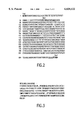
- FIG. 1 is a graphic representation of plasmid 577.
- FIG. 2 is the DNA sequence of HCV E2 antigen expression cassette.
- FIG. 3 shows a conceptual translation of the HCV E2 gene and the signal protease cleavage site wherein " - - - " denotes a signal peptidease cleavage site.
- the present invention provides ways to produce glycosylated HCV E2 fusion proteins expressed in mammalian expression systems. These glycosylated proteins have utility for a variety of applications, including, for example, assay systems for screening and prognostic applications, and as vaccine preparations. These HCV viral envelope proteins expressed in mammalian cells also allow for inhibitor studies including elucidation of specific viral attachment sites or sequences and/or viral receptors on susceptible cell types, for example, liver cells and the like.
- the procurement of specific expression clones developed as described herein in mammalian expression systems provides antigens for diagnostic assays which can aid in determining the stage of HCV infection, such as, for example, acute versus on-going or persistent infections, and/or recent infection versus past exposure. These specific expression clones also provide prognostic markers for resolution of disease such as to distinguish resolution of disease from chronic hepatitis caused by HCV. It is contemplated that earlier seroconversion to glycosylated structural antigens may be detectable by using proteins produced in these mammalian expression systems. Antibodies, both monoclonal and polyclonal, also may be produced from the proteins derived from these mammalian expression systems which then in turn may be used for diagnostic, prognostic and therapeutic applications.
- Proteins produced from these mammalian expression systems, as well as reagents produced from these proteins, can be provided in the form of a kit with one or more containers such as vials or bottles, with each container containing a separate reagent such as a monoclonal antibody, or a cocktail of monoclonal antibodies, or a recombinant protein, packaged as test kits for convenience in performing assays.
- kits with one or more containers such as vials or bottles, with each container containing a separate reagent such as a monoclonal antibody, or a cocktail of monoclonal antibodies, or a recombinant protein, packaged as test kits for convenience in performing assays.
- Other aspects of the present invention include a recombinant protein comprising an HCV epitope attached to a solid phase and an antibody to an HCV epitope attached to a solid phase.
- the present invention provides assays which utilize the recombinant proteins provided by the invention, as well as the antibodies described herein in various formats, any of which may employ a signal generating compound which generates a measurable signal in the assay. Assays which do not utilize signal generating compounds to provide a means of detection also are provided. All of the assays described generally detect either antigen or antibody, or both, and include mixing a test sample with at least one reagent provided herein to form at least one antigen/antibody complex and detecting the presence of the complex. These assays are described in detail herein.
- Vaccines for treatment of HCV infection comprising an immunogenic peptide obtained from a mammalian expression system containing envelope genes from HCV as described herein are included in the present invention. Also included in the present invention is a method for producing antibodies to HCV comprising administering to an individual an isolated immunogenic polypeptide containing an HCV epitope in an amount sufficient to produce an immune response in the inoculated individual.
- test sample refers to a component of an individual's body which is the source of the antibodies of interest. These components are well known in the art and include biological samples which can be tested by the methods described herein. Examples of test samples include human and animal body fluids such as whole blood, serum, plasma, cerebrospinal fluid, urine, lymph fluids, and various external sections of the respiratory, intestinal and genitourinary tracts, tears, saliva, milk, white blood cells, myelomas and the like, biological fluids such as cell culture supernatants, fixed tissue specimens and fixed cell specimens.
- human and animal body fluids such as whole blood, serum, plasma, cerebrospinal fluid, urine, lymph fluids, and various external sections of the respiratory, intestinal and genitourinary tracts, tears, saliva, milk, white blood cells, myelomas and the like
- biological fluids such as cell culture supernatants, fixed tissue specimens and fixed cell specimens.
- these recombinant proteins can be used to develop unique assays as described herein to detect either the presence of antigen or antibody to HCV.
- These compositions also can be used to develop monoclonal and/or polyclonal antibodies with a specific recombinant protein which specifically binds to the immunological epitope of HCV.
- recombinant proteins made by the method described herein can be used to develop vaccines by following methods known in the art.
- such vaccines are prepared as injectables, either as liquid solutions or suspensions; solid forms suitable for solution in or suspension in liquid prior to injection also may be prepared.
- the preparation may be emulsified. or the protein may be encapsulated in liposomes.
- the active immunogenic ingredients often are mixed with pharmacologically acceptable excipients which are compatible with the active ingredient. Suitable excipients include but are not limited to water, saline, dextrose, glycerol, ethanol and the like; combinations of these excipients in various amounts also may be used.
- the vaccine also may contain small amounts of auxiliary substances such as wetting or emulsifying reagents, pH buffering agents, and/or adjuvants which enhance the effectiveness of the vaccine.
- such adjuvants can include aluminum hydroxide, N-acetyl-muramyl-L-threonyl-D-isoglutamine (thr-DMP), N-acetyl-nornuramyl-L-alanyl-D-isoglutamine (CGP 11687, also referred to as nor-MPD), N-acetylmuramyul-L-alanyl-D-isoglutaminyl-L-alanine-2-(1 ⁇ 2 ⁇ -dipalmitoyl-sn-glycero-3-hydroxphosphoryloxy)-ethylamine (CGP19835A, also referred to as MTP-PE), and RIBI (MPL+TDM+CWS) in a 2% squalene/Tween-80® emulsion.
- the effectiveness of an adjuvant may be determined by measuring the amount of antibodies directed against an immunogenic polypeptide containing an HCV antigenic sequence resulting from administration of this polypeptid
- the vaccines usually are administered by intraveneous or intramuscular injection.
- Additional formulations which are suitable for other modes of administration include suppositories and, in some cases, oral formulations.
- traditional binders and carriers may include but are not limited to polyalkylene glycols or triglycerides.
- Such suppositories may be formed from mixtures containing the active ingredient in the range of about 0.5% to about 10%, preferably, about 1% to about 2%.
- Oral formulation include such normally employed excipients as, for example pharmaceutical grades of mannitol, lactose, starch, magnesium stearate, sodium saccharine, cellulose, magnesium carbonate and the like.
- These compositions may take the form of solutions, suspensions, tablets, pills, capsules, sustained release formulations or powders and contain about 10% to about 95% of active ingredient, preferably about 25% to about 70%.
- the proteins used in the vaccine may be formulated into the vaccine as neutral or salt forms.
- Pharmaceutically acceptable salts such as acid addition salts (formed with free amino groups of the peptide) and which are formed with inorganic acids such as hydrochloric or phosphoric acids, or such organic acids such as acetic, oxalic, tartaric, maleic, and others known to those skilled in the art.
- Salts formed with the free carboxyl groups also may be derived from inorganic bases such as sodium, potassium, ammonium, calcium or ferric hydroxides and the like, and such organic bases such as isopropylamine, trimethylamine, 2-ethylamino ethanol, histidine procaine, and others known to those skilled in the art.
- Vaccines are administered in a way compatible with the dosage formulation, and in such amounts as will be prophylactically and/or therapeutically effective.
- the quantity to be administered generally is in the range of about 5 micrograms to about 250 micrograms of antigen per dose, and depends upon the subject to be dosed, the capacity of the subject's immune system to synthesize antibodies, and the degree of protection sought. Precise amounts of active ingredient required to be administered also may depend upon the judgment of the practitioner and may be unique to each subject.
- the vaccine may be given in a single or multiple dose schedule.
- a multiple dose is one in which a primary course of vaccination may be with one to ten separate doses, followed by other doses given at subsequent time intervals required to maintain and/or to reenforce the immune response, for example, at one to four months for a second dose, and if required by the individual, a subsequent dose(s) after several months.
- the dosage regimen also will be determined, at least in part, by the need of the individual, and be dependent upon the practitioner's judgment. It is contemplated that the vaccine containing the immunogenic HCV envelope antigen(s) may be administered in conjunction with other immunoregulatory agents, for example, with immune globulins.
- a gene coding for a protein of interest using a DNA cloning vehicle which includes (a) expression control regions, (b) a region coding for the rabbit immunoglobulin heavy chain gamma secretion signal sequence, (c) bacterial enzyme for selection in eukaryotic cells, (d) an amplification system suitable for enhanced expresision in eukaryotic cells, and (e) a region coding for the protein of interest generally is described herein.
- the cloning vehicles described herein are capable of expressing fusion proteins; that is, immunoglobulin signal peptides sequences and adjacent immunoglobulin coding sequences fused to heterologous protein at commercially useful levels.
- FIG. 1 shows generically the features of a plasmid useful for production of fusion proteins used in the methods of this invention.
- the plasmid in FIG. 1 is disclosed as a series of assembled fragments with sections 1 to 13.
- the accession numbers of the sections refer to Genbank® accession numbers.
- the plasmid includes a control region (described hereinbelow), followed by a gene encoding an immunoglobulin signal peptide and adjacent immunoglobilin coding sequences which are linked to a gene coding for a heterologous protein of interest. Please note that slight sequence variations may occur and may have occurred when constructing the plasmid.
- Insertion of heterologous genes into a plasmid as described in FIG. 1 can be accomplished with various techniques known to those in the art.
- These fusion proteins can be utilized in various assay formats as capture reagents or protein binders in numerous ways. After preparing the recombinant proteins as described herein, the recombinant proteins can be used to develop unique assays as described herein to detect either the presence of a specific binding member of a specific binding pair. These recombinant proteins also can be used to develop monoclonal and/or polyclonal antibodies with a specific recombinant protein or synthetic peptide which specifically binds to the specific binding member of a specific binding pair.
- the fusion proteins described herein also can be used as the active ingredient of a vaccine.
- Vaccines may be prepared from one or more immunogenic polypeptides derived from nucleic acid sequences of interest or from the genome of interest to which they correspond. Vaccines may comprise recombinant polypeptides containing epitope(s) of interest. These polypeptides may be expressed in bacteria, yeast or mammalian cells, or alternatively may be isolated from viral preparations. It also is anticipated that various structural proteins may contain epitopes of interest which give rise to protective anti-epitope antibodies. Synthetic peptides therefore also can be utilized when preparing these vaccines. Thus, polypeptides containing at least one epitope of interest may be used, either singly or in combinations, in these vaccines. It also is contemplated that nonstructural proteins as well as structural proteins may provide protection against viral pathogenicity, even if they do not cause the production of neutralizing antibodies.
- multivalent vaccines may comprise one or more structural proteins, and/or one or more nonstructural proteins.
- These vaccines may be comprised of, for example, recombinant polypeptides expressed by the plasmid of the invention and/or polypeptides isolated from the virions and/or synthetic peptides.
- These immunogenic epitopes can be used in combinations, i.e., as a mixture of recombinant proteins, synthetic peptides and/or polypeptides isolated from the virion; these may be administered at the same or different time. Additionally, it may be possible to use inactivated viruses in vaccines.
- Such inactivation may be be preparation of viral lysates, or by other means known in the art to cause inactivation of hepatitis-like viruses, for example, treatment with organic solvents or detergents, or treatment with formalin.
- Attenuated viral strain preparation also is disclosed in the present invention. It is contemplated that some of the proteins may cross-react with other known viruses, and thus that shared epitopes may exist between the virus of interest and other viruses which would then give rise to protective antibodies against one or more of the disorders caused by these pathogenic agents. It is contemplated that it may be possible to design multiple purpose vaccines based upon this belief.
- vaccines which contain at least one immunogenic peptide as an active ingredient is known to one skilled in the art.
- such vaccines are prepared as injectables, either as liquid solutions or suspensions; solid forms suitable for solution in or suspension in liquid prior to injection also may be prepared.
- the preparation may be emulsified or the protein may be encapsulated in liposomes.
- the active immunogenic ingredients often are mixed with pharmacologically acceptable excipients which are compatible with the active ingredient. Suitable excipients include but are not limited to water, saline, dextrose, glycerol, ethanol and the like; combinations of these excipients in various amounts also may be used.
- the vaccine also may contain small amounts of auxiliary substances such as wetting or emulsifying reagents, pH buffering agents, and/or adjuvants which enhance the effectiveness of the vaccine.
- auxiliary substances such as wetting or emulsifying reagents, pH buffering agents, and/or adjuvants which enhance the effectiveness of the vaccine.
- adjuvants can include aluminum hydroxide, N-acetyl-muramyl-L-threonyl-D-isoglutamine (thr-DMP), N-acetyl-nornuramyl-L-alanyl-D-isoglutamine (CGP 11687, also referred to as nor-MDP), N-acetylmuramyul-L-alanyl-D-isoglutaminyl-L-alanine-2-(1 ⁇ 2 ⁇ -dipalmitoyl-sn-glycero-3-hydroxphosphoryloxy)-ethylamine (CGP 19835A, also
- the effectiveness of an adjuvant may be determined by measuring the amount of antibodies directed against an immunogenic polypeptide containing an antigenic sequence produced by the plasmid disclosed herein, resulting from administration of this polypeptide in vaccines which also are comprised of the various adjuvants.
- the vaccines usually are administered by intraveneous or intramuscular injection.
- Additional formulations which are suitable for other modes of administration include suppositories and, in some cases, oral formulations.
- traditional binders and carriers may include but are not limited to polyalkylene glycols or triglycerides.
- Such suppositories may be formed from mixtures containing the active ingredient in the range of about 0.5% to about 10%, preferably, about 1% to about 2%.
- Oral formulation include such normally employed excipients as, for example pharmaceutical grades of mannitol, lactose, starch, magnesium stearate, sodium saccharine, cellulose, magnesium carbonate and the like.
- These compositions may take the form of solutions, suspensions, tablets, pills, capsules, sustained release formulations or powders and contain about 10% to about 95% of active ingredient, preferably about 25% to about 70%.
- the proteins used in the vaccine may be formulated into the vaccine as neutral or salt forms.
- Pharmaceutically acceptable salts such as acid addition salts (formed with free amino groups of the peptide) and which are formed with inorganic acids such as hydrochloric or phosphoric acids, or such organic acids such as acetic, oxalic, tartaric, maleic, and others known to those skilled in the art.
- Salts formed with the free carboxyl groups also may be derived from inorganic bases such as sodium, potassium, ammonium, calcium or ferric hydroxides and the like, and such organic bases such as isopropylamine, trimethylamine, 2-ethylamino ethanol, histidine procaine, and others known to those skilled in the art.
- Vaccines are administered in a way compatible with the dosage formulation, and in such amounts as will be prophylactically and/or therapeutically effective.
- the quantity to be administered generally is in the range of about 5 micrograms to about 250 micrograms of antigen per dose, and depends upon the subject to be dosed, the capacity of the subject's immune system to synthesize antibodies, and the degree of protection sought. Precise amounts of active ingredient required to be administered also may depend upon the judgment of the practitioner and may be unique to each subject.
- the vaccine may be given in a single or multiple dose schedule.
- a multiple dose is one in which a primary course of vaccination may be with one to ten separate doses, followed by other doses given at subsequent time intervals required to maintain and/or to reinforce the immune response, for example, at one to four months for a second dose, and if required by the individual, a subsequent dose(s) after several months.
- the dosage regimen also will be determined, at least in part, by the need of the individual, and be dependent upon the practitioner's judgment. It is contemplated that the vaccine containing the immunogenic antigen(s) prepared as described herein may be administered in conjunction with other immunoregulatory agents, for example, with immune globulins.
- the reagent employed for the assay can be provided in the form of a kit with one or more containers such as vials or bottles, with each container containing a separate reagent such as a monoclonal antibody, or a cocktail of monoclonal antibodies, or a recombinant protein employed in the assay.
- analyte is the substance to be detected which may be present in the test sample.
- the analyte can be any substance for which there exists a naturally occurring specific binding member (such as, an antibody), or for which a specific binding member can be prepared.
- an analyte is a substance that can bind to one or more specific binding members in an assay.
- “Analyte” also includes any antigenic substances, haptens, antibodies, and combinations thereof.
- the analyte can be detected by means of naturally occurring specific binding partners (pairs) such as the use of intrinsic factor protein as a member of a specific binding pair for the determination of Vitamin B12, the use of folate-binding protein to determine folic acid, or the use of a lectin as a member of a specific binding pair for the determination of a carbohydrate.
- the analyte can include a protein, a peptide, an amino acid, a hormone, a steroid, a vitamin, a drug including those administered for therapeutic purposes as well as those administered for illicit purposes, a bacterium, a virus, and metabolites of or antibodies to any of the above substances.
- Viruses which can be tested include hepatitis-causing viruses (for example, hepatitis A virus, hepatitis B virus, hepatitis C virus, hepatitis delta, and hepatitis E virus, and hepatitis GB viruses), human immunodeficiency viruses (such as HIV-1, HIV-2), the HTLV-I and HTLV-II viruses, and the like.
- hepatitis-causing viruses for example, hepatitis A virus, hepatitis B virus, hepatitis C virus, hepatitis delta, and hepatitis E virus, and hepatitis GB viruses
- human immunodeficiency viruses such as HIV-1, HIV-2
- the HTLV-I and HTLV-II viruses and the like.
- a "specific binding member,” as used herein, is a member of a specific binding pair. That is, two different molecules where one of the molecules through chemical or physical means specifically binds to the second molecule. Therefore, in addition to antigen and antibody specific binding pairs of common immunoassays, other specific binding pairs can include biotin and avidin, carbohydrates and lectins, complementary nucleotide sequences, effector and receptor molecules, cofactors and enzymes, enzyme inhibitors and enzymes, and the like. Furthermore, specific binding pairs can include members that are analogs of the original specific binding members, for example, an analyte-analog.
- Immunoreactive specific binding members include antigens, antigen fragments, antibodies and antibody fragments, both monoclonal and polyclonal, and complexes thereof, including those formed by recombinant DNA molecules.
- hapten refers to a partial antigen or non-protein binding member which is capable of binding to an antibody, but which is not capable of eliciting antibody formation unless coupled to a carrier protein.
- a “capture reagent”, as used herein, refers to an unlabeled specific binding member which is specific either for the analyte as in a sandwich assay, for the indicator reagent or analyte as in a competitive assay, or for an ancillary specific binding member, which itself is specific for the analyte, as in an indirect assay.
- the capture reagent can be directly or indirectly bound to a solid phase material before the performance of the assay or during the performance of the assay, thereby enabling the separation of immobilized complexes from the test sample.
- the “indicator reagent” comprises a "signal generating compound” (label) which is capable of generating a measurable signal detectable by external means conjugated (attached) to a specific binding member for the analyte.
- Specific binding member as used herein means a member of a specific binding pair. That is, two different molecules where one of the molecules through chemical or physical means specifically binds to the second molecule.
- the indicator reagent also can be a member of any specific binding pair, including either hapten-anti-hapten systems such as biotin or anti-biotin, avidin or biotin, a carbohydrate or a lectin, a complementary nucleotide sequence, an effector or a receptor molecule, an enzyme cofactor and an enzyme, an enzyme inhibitor or an enzyme, and the like.
- hapten-anti-hapten systems such as biotin or anti-biotin, avidin or biotin, a carbohydrate or a lectin, a complementary nucleotide sequence, an effector or a receptor molecule, an enzyme cofactor and an enzyme, an enzyme inhibitor or an enzyme, and the like.
- An immunoreactive specific binding member can be an antibody, an antigen, or an antibody/antigen complex that is capable of binding either to the analyte as in a sandwich assay, to the capture reagent as in a competitive assay, or to the ancillary specific binding member as in an indirect assay.
- labels include chromogens, catalysts such as enzymes, luminescent compounds such as fluorescein and rhodamine, chemiluminescent compounds, radioactive elements, and direct visual labels.
- enzymes include alkaline phosphatase, horseradish peroxidase, beta-galactosidase, and the like. The selection of a particular label is not critical, but it will be capable of producing a signal either by itself or in conjunction with one or more additional substances.
- Solid phases are known to those in the art and include the walls of wells of a reaction tray, test tubes, polystyrene beads, magnetic beads, nitrocellulose strips, membranes, microparticles such as latex particles, and others.
- the “solid phase” is not critical and can be selected by one skilled in the art.
- latex particles, microparticles, magnetic or non-magnetic beads, membranes, plastic tubes, walls of microtiter wells, glass or silicon chips and sheep red blood cells are all suitable examples.
- Suitable methods for immobilizing peptides on solid phases include ionic, hydrophobic, covalent interactions and the like.
- a “solid phase”, as used herein, refers to any material which is insoluble, or can be made insoluble by a subsequent reaction.
- the solid phase can be chosen for its intrinsic ability to attract and immobilize the capture reagent.
- the solid phase can retain an additional receptor which has the ability to attract and immobilize the capture reagent.
- the additional receptor can include a charged substance that is oppositely charged with respect to the capture reagent itself or to a charged substance conjugated to the capture reagent.
- the receptor molecule can be any specific binding member which is immobilized upon (attached to) the solid phase and which has the ability to immobilize the capture reagent through a specific binding reaction.
- the receptor molecule enables the indirect binding of the capture reagent to a solid phase material before the performance of the assay or during the performance of the assay.
- the solid phase thus can be a plastic, derivatized plastic, magnetic or non-magnetic metal, glass or silicon surface of a test tube, microtiter well, sheet, bead, microparticle, chip, and other configurations known to those of ordinary skill in the art.
- the solid phase also can comprise any suitable porous material with sufficient porosity to allow access by detection antibodies and a suitable surface affinity to bind antigens.
- Microporous structures are generally preferred, but materials with gel structure in the hydrated state may be used as well.
- Such useful solid supports include:
- natural polymeric carbohydrates and their synthetically modified, cross-linked or substituted derivatives such as agar, agarose, cross-linked alginic acid, substituted and cross-linked guar gums, cellulose esters, especially with nitric acid and carboxylic acids, mixed cellulose esters, and cellulose ethers; natural polymers containing nitrogen, such as proteins and derivatives, including cross-linked or modified gelatins; natural hydrocarbon polymers, such as latex and rubber; synthetic polymers which may be prepared with suitably porous structures, such as vinyl polymers, including polyethylene, polypropylene, polystyrene, polyvinylchloride, polyvinylacetate and its partially hydrolyzed derivatives, polyacrylamides, polymethacrylates, copolymers and terpolymers of the above polycondensates, such as polyesters, polyamides, and other polymers, such as polyurethanes or polyepoxides; porous inorganic materials such as sulfates or carbonates of alka
- porous structure of nitrocellulose has excellent absorption and adsorption qualities for a wide variety of reagents including monoclonal antibodies.
- Nylon also possesses similar characteristics and also is suitable.
- porous solid supports described hereinabove are preferably in the form of sheets of thickness from about 0.01 to 0.5 mm, preferably about 0.1 mm.
- the pore size may vary within wide limits, and is preferably from about 0.025 to 15 microns, especially from about 0.15 to 15 microns.
- the surfaces of such supports may be activated by chemical processes which cause covalent linkage of the antigen or antibody to the support. The irreversible binding of the antigen or antibody is obtained, however, in general, by adsorption on the porous material by poorly understood hydrophobic forces.
- Suitable solid supports also are described in U.S. patent application Ser. No. 227,272.
- test sample includes biological samples which can be tested by the methods of the present invention described herein and include human and animal body fluids such as whole blood, serum, plasma, cerebrospinal fluid, urine, lymph fluids, and various external secretions of the respiratory, intestinal and genitorurinary tracts, tears, saliva, milk, white blood cells, myelomas and the like, biological fluids such as cell culture supernatants, fixed tissue specimens and fixed cell specimens. Any substance which can be adapted for testing with the recombinant proteins described herein and assay formats of the present invention are contemplated to be within the scope of the present invention.
- An immobilizable immune complex is separated from the rest of the reaction mixture by ionic interactions between the negatively charged poly-anion/immune complex and the previously treated, positively charged porous matrix and detected by using various signal generating systems previously described, including those described in chemiluminescent signal measurements as described in co-pending U.S. patent application Ser. No. 921,979 corresponding to EPO Publication No. 0 273,115, which enjoys common ownership and which is incorporated herein by reference.
- the methods of the present invention can be adapted for use in systems which utilize microparticle technology including in automated and semi-automated systems wherein the solid phase comprises a microparticle.
- Such systems include those described in pending U.S. patent applications Ser. No. 425,651 and 425,643, which correspond to published EPO applications Nos. EP 0 425 633 and EP 0 424 634, respectively, which are incorporated herein by reference.
- scanning probe microscopy for immunoassays also is a technology to which the recombinant proteins of the present invention or monoclonal antibodies produced from these recombinant proteins are easily adaptable.
- the capture phase for example, either a recombinant protein produced by the novel plasmid described herein or a monoclonal antibody produced therefrom, is adhered to a solid phase, the test sample is contacted to the solid phase for a time and under conditions sufficient for a reaction between the two to occur, and a scanning probe microscope is utilized to detect antigen/antibody complexes which may be present on the surface of the solid phase.
- the use of scanning tunnelling microscopy eliminates the need for labels which normally must be utilized in many immunoassay systems to detect antigen/antibody complexes.
- an assay format to detect the presence of antibody against a specific analyte (for example, an infectious agent such as a virus) in a human test sample
- a specific analyte for example, an infectious agent such as a virus
- the human test sample is contacted and incubated with a solid phase coated with at least one recombinant protein (polypeptide). If antibodies are present in the test sample, they will form a complex with the antigenic polypeptide and become affixed to the solid phase. After the complex has formed, unbound materials and reagents are removed by washing the solid phase. The complex is reacted with an indicator reagent and allowed to incubate for a time and under conditions for second complexes to form.
- an indicator reagent for example, an infectious agent such as a virus
- the presence of antibody in the test sample to the recombinant polypeptide(s) is determined by detecting the measurable signal generated. Signal generated above a cut-off value is indicative of antibody to the analyte present in the test sample. With many indicator reagents, such as enzymes, the amount of antibody present is proportional to the signal generated. Depending upon the type of test sample, it may be diluted with a suitable buffer reagent, concentrated, or contacted with the solid phase without any manipulation ("neat"). For example, it usually is preferred to test serum or plasma samples which previously have been diluted, or concentrate specimens such as urine, in order to determine the presence and/or amount of antibody present.
- a sandwich assay is provided in still another embodiment.
- This method comprises contacting a test sample with a solid phase to which at least one recombinant antigen provided herein or a combination including at least one recombinant antigen provided herein are bound, to form a mixture.
- This mixture is incubated for a time and under conditions sufficient to allow antigen/antibody complexes to form.
- These complexes then are contacted with an indicator reagent comprising antigen(s) previously conjugated to a signal generating compound, to form a second mixture.
- This second mixture is incubated for a time and under conditions sufficient for antigen/antibody/indicator reagent complexes to form.
- the presence of the antigen/antibody/indicator reagent complexes is determined by detecting the measurable signal generated.
- a first antigen which can be a recombinant antigen provided herein specific to the antibody to be detected is immobilized on a solid phase, a test sample suspected of containing the antibody is added to the solid phase, and a second antigen which can be a recombinant antigen of the invention having a label affixed thereto then is contacted with the solid phase.
- two recombinant antigens which are specific to a single binding pair member are used in one assay as a capture phase and a part of the indicator reagent.
- antigens are the same and may be made in different, e.g., heterologous, sources. These sources could be bacterial and yeast, for example. It also is within the scope of the present invention that one recombinant antigen provided herein could be used as the capture reagent or as part of the indicator reagent, and the other antigen in this assay could be a synthetic peptide, or viral lysate, or obtained from other antigenic sources known to the routineer. Further, the use of biotin and antibiotin, biotin and avidin, biotin and streptavidin, and the like, may be used to enhance the generated signal in such assays.
- more than one recombinant protein can be used in the assay format just described to test for the presence of antibody against a specific infectious agent by utilizing fusion proteins prepared as described herein against various antigenic epitopes of the viral genome of the infectious agent under study.
- recombinant polypeptides which contain epitopes within a specific viral antigenic region as well as epitopes from other antigenic regions from the viral genome to provide assays which have increased sensitivity any perhaps greater specificity than using a polypeptide from one epitope.
- Such an assay can be utilized as a confirmatory assay.
- a known amount of test sample is contacted with (a) known amount(s) of at least one solid support coated with at least one recombinant protein for a time and under conditions sufficient to form recombinant protein/antibody complexes.
- the complexes are contacted with known amount(s) of appropriate indicator reagent(s)s for a time and under suitable conditions for a reaction to occur, wherein the resultant signal generated is compared to a negative test sample in order to determine the presence of antibody to the analyte in the test sample.
- each recombinant protein utilized in the assay can be attached to a separate microparticle, and a mixture of these microparticles made by combining the various coated microparticles, which can be optimized for each assay.
- Variations to the above-described assay formats include the incorporation of recombinant proteins produced by the plasmid descibed herein of different analytes attached to the same or to different solid phases for the detection of the presence of antibody to either analyte (for example, recombinant proteins specific for certain antigenic regions of HIV-1 coated on the same or different solid phase with recombinant proteins specific for certain antigenic region(s) of HIV-2, to detect the presence of either (or both) HIV-1 or HIV-2).
- analyte for example, recombinant proteins specific for certain antigenic regions of HIV-1 coated on the same or different solid phase with recombinant proteins specific for certain antigenic region(s) of HIV-2, to detect the presence of either (or both) HIV-1 or HIV-2).
- recombinant proteins produced from the plasmid described herein containing antigenic epitopes are useful in competitive assays such as neutralization assays.
- a neutralization assay a recombinant polypeptide representing epitopes of an antigenic region of an infectious agent such as a virus, is solubilized and mixed with a sample diluent to a final concentration of between 0.5 to 50.0 ⁇ g/ml.
- a known amount of test sample preferably 10 ⁇ l
- the mixture may be preincubated for approximately 15 minutes to two hours.
- a solid phase coated with the recombinant protein described herein then is added to the reaction well, and incubated for one hour at approximately 40° C.
- a known amount of an indicator reagent for example, 200 ⁇ l of a peroxide labelled goat anti-human IgG in a conjugate diluent is added and incubated for one hour at 40° C.
- an enzyme substrate for example, OPD substrate, is added and incubated at room temperature for thirty minutes.
- the reaction is terminated by adding a stopping reagent such as 1N sulfuric acid to the reaction well. Absorbance is read at 492 nm.
- Test samples which contain antibody to the specific polypeptide generate a reduced signal caused by the competitive binding of the peptides to these antibodies in solution.
- the percentage of competitive binding may be calculated by comparing absorbance value of the sample in the presence of recombinant polypeptide to the absorbance value of the sample assayed in the absence of a recombinant polypeptide at the same dilution.
- the difference in the signals generated between the sample in the presence of recombinant protein and the sample in the absence of recombinant protein is the measurement used to determine the presence or absence of antibody.
- neutralization assays include competititve assays to detect the amount, if any, of an antigen analyte in a test sample.
- the assay comprises the steps of contacting a test sample with a known amount of analyte (in this instance, the recombinant antigen of the invention) having attached to it a signal generating compound and a solid phase to which has been attached an anti-analyte antibody.
- This resultant mixture is incubated for a time and under conditions sufficient to form either solid phaselanalyte complexes or solid phase/recombinant antigen complexes.
- the signal is triggered through means known in the art.
- Test samples which contain antibody to the specific polypeptide generate a reduced signal caused by the competitive binding of the antibody on the solid phase to the antigens in solution.
- the percentage of competitive binding may be calculated by comparing absorbance value of the sample in the presence of recombinant polypeptide to the absorbance value of the sample assayed in the absence of a recombinant polypeptide at the same dilution.
- the difference in the signals generated between the sample in the presence of recombinant protein and the sample in the absence of recombinant protein is the measurement used to determine the presence or absence of antibody.
- the recombinant proteins can be used in immunodot blot assay systems.
- the immunodot blot assay system uses a panel of purified recombinant polypeptides placed in an array on a nitrocellulose solid support.
- the prepared solid support is contacted with a sample and captures specific antibodies (specific binding member) to the recombinant protein (other specific binding member) to form specific binding member pairs.
- the captured antibodies are detected by reaction with an indicator reagent.
- the conjugate specific reaction is quantified using a reflectance optics assembly within an instrument which has been described in U.S. patent application Ser. No. 07/227,408 filed Aug. 2, 1988.
- test cartridge After reaction, the test cartridge is washed and any antigen-antibody reactions are identified using suitable well-known reagents. As described in the patents and patent applications listed herein, the entire process is amenable to automation. The specifications of these applications related to the method and apparatus for performing an immunodot blot assay are incorporated herein by reference.
- fusion proteins prepared from the plasmid described herein can be used in assays which employ a first and second solid support, as follows, for detecting antibody to a specific antigen of an analyte in a test sample.
- a first aliquot of a test sample is contacted with a first solid support coated with recombinant protein specific for an analyte for a time and under conditions sufficient to form recombinant protein/analyte antibody complexes.
- the complexes are contacted with an indicator reagent specific for the recombinant antigen.
- the indicator reagent is detected to determine the presence of antibody to the recombinant protein in the test sample.
- the presence of a different antigenic determinant of the same analyte is determined by contacting a second aliquot of a test sample with a second solid support coated with recombinant protein specific for the second antibody for a time and under conditions sufficient to form recombinant protein/second antibody complexes.
- the complexes are contacted with a second indicator reagent specific for the antibody of the complex.
- the signal is detected in order to determine the presence of antibody in the test sample, wherein the presence of antibody to either analyte recombinant protein, or both, indicates the presence of anti-analyte in the test sample.
- the solid supports can be tested simultaneously.
- haptens are known in the art. It is contemplated that haptens also can be used in assays employing fusion proteins produced by the plasmid of the invention in order to enhance performance of the assay.
- Plasmid 577 was constructed by inserting coding sequence for a secretable HCV E2 antigen in two steps, as follows. First, a duplex synthetic oligonucleotide that had been digested with Spe 1 and Xba 1 was inserted into the Xba 1 cloning site of a previously constructed expression vector by sticky end ligation. This oligonucleotide contained sequence derived from rabbit immunoglobulin gamma chain peptide and other sequences included to create restriction sites for cloning purposes. It was then inserted downstream of a promoter element and RNA transcription start site.
- This DNA segment encoded amino acid sequences to be fused in frame at the Xba 1 sites to downstream gene sequences intended to be secreted from mammalian cells.
- the constructionof this plasmid 577 is shown in FIG. 1.
- the sequence of this DNA segment is depicted in FIG. 2.
- a conceptual translation delineating the mammalian secretion signal peptidase cleavage site is shown in FIG. 3.
- a PCR product containing sequence derived from an HCV plasmid template was inserted as an Xba 1 fragment downstream of the rabbit heavy chain signal sequence.
- Encoded in the "upper" PCR primer sequence was an Xba 1 site, immediately followed by 12 nucleotide sequence that encoded the amino acid sequence Serine-Asparagine-Glutamic Acid-Leucine ("SNEL") the amino terminal sequence of human pro-urokinase.
- the amino acid sequence SNEL was intended to promote signal protease processing, efficient secretion and final product stability in culture fluids. This segment is underlined in FIG. 2.
- the primer contained nucleotides complementary to template sequences encoding amino acids starting at aa 388 of HCV.
- the "lower” PCR primer contained sequences homologous to template sequences that encode amino acids ending at 664 of HCV, a duplicate stop codon, and an Xba 1 site for cloning purposes. E2 antigen was truncated at this position to promote secretion. The Xba 1 sites appear in large bold type face and the stop codons are underlined in FIG. 2.
- plasmid 577 contains the following DNA segments described counterlockwise from the top of the plasmid, FIG. 1: (a) a 2.3 Kb fragment of pBR322 containing bacterial beta-lactamase and origin of DNA replication; (b) a 1.8 Kb cassette directing expression of a neomycin resistance gene under control of HSV-1 thyridine kinase promoter and poly-A addition signals; (c) a 1.9 Kb cassette directing expression of a dihydrofolate reductase gene under the control of an SV-40 promoter and poly-A addition signals; (d) a 3.5 Kb cassette directing expression of a modified hepatitis C virus derived gene under the control of the Simian Virus 40 T-ag promoter and transcription enhancer, the hepatitis B virus surface antigen enhancer I, and a fragment of Herpes Simplex Virus-1 genome providing poly-A addition signals; and (e) a residual 0.7 Kb
- the plasmid 577 was transfected into CHO/dhfr cells (dxb-111) (Uriacio, et al., Proc. Nat. Acad. Sci. 77, 4451-4466 (1980); these cells are available from the American Type Culture Collection [A.T.C.C.], 12301 Parklawn Drive, Rockville, Md. 20852, under Accession No. CRL 9096), using the cationic liposome-mediated procedure (Felgner, P. L. et al., Proc. Natl. Acad. Sci. 84, 7413-7417 (1987), as follows.
- CHO/dhfr cells were cultured in Ham's F-12 media supplemented with 10% fetal calf serum, L-glutamine (1 ⁇ M) and freshly seeded into a 25 cm 2 flask at a density of 5-8 ⁇ 10 5 cells per flask twenty four hours prior to transfection.
- Fifteen micrograms of plasmid DNA was added to 1.5 mls of Opti-MEM I medium and 100 microliters of Lipofectin Reagent (Gibco-BRL, Grand Island N.Y.) was added to a second 1.5 ml portion of Opti-MEM I media. The two solutions were mixed and incubated at room temperature for 20 minutes.
- the culture medium was removed from cells and replaced with the Opti-MEM I-Lipofection-DNA solution for liposome-mediated transfection of DNA into cells in tissue culture.
- the cells were incubated for three hours at 37° C. after which the Opti-MEM I-Lipofectin-DNA solution was replaced with culture medium for an additional 24 hours prior to selection.
- F-12 minus medium G dhfr/G418 selection medium
- Selection medium was Ham's F-12 with L-glutamine and without hypoxanthine, thymidine, and glycine (JRH Biosciences, Lenexa, Kans., USA ) and 300 micrograms per ml G418 (Gibco-BRL).
- Amplification of the transfected UK-HCV-E2 gene was achieved by stepwise selection of DHFR + , G418 + cells with methotrexate (reviewed by Schimke, R., Cell 37, 705-713 (1984). Cells were incubated with F-12 minus medium G, containing 150 nM methotrexate (MTX), for approximately two weeks until resistant colonies appeared. The MTX resistant cells were passaged and maintained in the appropriate selection medium. Further amplification was achieved by selection with 5 ⁇ M MTX, and cells continuously maintained in the appropriate selection medium.
- Cells in culture and undergoing various selection or amplification procedures were re-fed with the appropriate culture medium three times weekly.
- Cells were passaged 1:4, with appropriate medium, into 75 cm 2 flasks and incubated at 37° C. with 5% CO 2 using standard methods.
- Cryostorage was by resuspension of 2-4 ⁇ 10 6 cells in 1.8 ml of the appropriate culture medium containing 5% DMSO (Sigma Chem. Co., St. Louis, Mo., USA) and cold storage for 24 hours at -80° C. and then permanent storage at -135° C.
- Ham's F12 custom minus medium was overlayed onto just confluent monolayers for 12 to 24 hours at 37° C. in 5% CO 2 . Then, the growth medium was removed and the cells were rinsed three times with phosphate buffered saline (PBS) (with calcium and magnesium) available from Gibco-BRL, to remove the remaining media/serum which might be present. Cells then were incubated with VAS custom medium (VAS custom formulation with 1-glutamine with HEPES without phenol red, available from JRH Bioscience, product number 52-08678P), for one hour at 37° C. in 5% CO 2 . As a final wash, the VAS then was discarded.
- PBS phosphate buffered saline
- the sialic acid containing CHO-E2 glycoprotein was purified to greater than 90% purity from cell supernatants by ion exchange and lectin chromatography. Ten (10) separate lots from two different protein-free media all were purified, which demonstrated the reproducibility and versatility of this procedure. Purity was evaluated by R-250 coomassie and silver staining. Theoretical molecular weight of 30 Kdal was verified by Endo-F digestion.
- the concentrated and dialyzed supernatant first was loaded on an equilibrated S-Sepharose column (200 ml bed volume) at a flow rate of 5 ml/min.
- the unbound flow was collected, concentrated (YM10) to original volume and extensively dialyzed in DEAE-Sepharose running buffer (containing 0.02 M Tris buffer/0.1 M NaCl, pH 8.5). It was found that the conductivity of this buffer should be about 12 mS.
- the material was loaded onto a 200 ml DEAE-Sepharose column at a flow rate of 5 ml/min.
- the unbound flow was collected, concentrated (YM10) to original volume and extensively dialyzed in 0.01 M sodium phosphate, 0.13 M NaCl, pH 7.0.
- This buffer was termed the lectin WGA-Sepharose 6MB running buffer.
- the sample was changed into WGA running buffer, it was loaded at 0.5 ml/min onto a 10.0 ml WGA-Sepharose 6 MB column, collecting and recirculating the flow. After extensive washing (10 column volumes), the column flow was reversed and the purified CHO-E2 antigen was eluted using 10 mM N,N'-diacetylchitobiose in running buffer. The purified antigen was dialyzed against PBS and stored at -70° C.
- CHO-E2 Antigen Beads Twenty microliters (20 ⁇ l ) of cloned cell suspension or control suspensions (CHO cells transfected with expression vector containing no HCV insert) were placed into a microtiter well which was capable of containing a 1/4 inch bead. The number of wells being tested was multiplied by 0.2 and by 1.05 to obtain the volume in milliliters (ml) of diluent necessary for coating. The diluent used was SMP diluent (available from Abbott Laboratories, Abbott Park, Ill.).
- Purified HCV E2 antigen prepared as described in the previous examples was coated onto polystyrene beads following methods well-known in the art at a concentration of 1.0 to 2.0 ⁇ g/ml. The components used in the coating procedure were adjusted to provide optimum sensitivity and specificity for the antibody assay test. The specificity of the E2 antibody assay was evaluated by testing specimens from populations of volunteer blood donors. All specimens were testd at 1:41 dilution in the enzyme immunoassay using goat anti-human IgG labeled with horseradish peroxidase as the signal generating compound according to the assay protocol hereinabove (see previous examples).
- HCV 2.0 reactive plasma samples were obtained from North American Biologicals, Inc. (NABI) and only samples which were concordantly reactive in two HCV 2.0 EIA (available from Abbott Laboratories, Abbott Park, Ill. and Ortho Diagnostics, Inc., Raritan, N.J.) were analyzed further.
- HCV 2.0 EIA Abbott and Ortho
- HCV 3.0 EIA Ortho
- Abbott MATRIXTM HCV 1.0 and RIBA HCV 2.0 data were supplied by BBI with the panel.
- Eighteen (18) of 23 (78.3%) HCV 2.0 EIA positive (by both Abbott and Ortho assays) specimens were also anti-E2 positive.
- the two HCV negative panel members were E2 antibody negative.
Abstract
This invention provides a novel mammalian expression system that is capable of generating high levels of expressed hepatitis C virus (HCV) proteins which have previously proved difficult to express due to their non-secretory properties. In particular, the invention provides a plasmid for the expression of the HCV second envelope protein (E2) designated p577. This plasmid encodes a recombinant protein comprising the immunoglobulin signal peptide and amino acids 388-664 of the HCV E2 glycoprotein. This unique expression system produces high levels of HCV proteins that are properly processed, glycosylated, and folded.
Description
This application is related to pending U.S. patent application Ser. No. 08/188,281, filed Jan. 28, 1994, entitled "Mammalian Expression Systems for Hepatitis C Virus Envelope Genes" and to pending U.S. patent application Ser. No. 08/144,099, filed Oct. 28, 1993, entitled "Mammalian Expression Systems for Hepatitis C Virus", which is a continuation of U.S. Ser. No. 07/830,024, both of which enjoy common ownership and are incorporated herein by reference.
This invention relates generally to a mammalian expression system, and more particularly, relates to a mammalian expression system capable of generating recombinant proteins not heretofore generated at such high levels due to the non-secretor nature of the gene. The recombinant proteins are expressed in culture medium as well as in mammalian cells.
The introduction of the first-generation hepatitis C virus (HCV) enzyme immunoassays (EIAs) (HCV 1.0 EIAs) as screening assays in 1989 and second-generation HCV EIAs in 1992 (HCV 2.0 EIAs) has dramatically reduced the incidence of post-transfusion HCV (PT-HCV) infection in those countries where routine screening of donated blood products is performed. Antibodies to HCV are detected using recombinant proteins derived from the core, NS3 (viral protease) and NS4 (function unknown) genes of the virus. HCV third-generation EIAs (HCV 3.0 EIAs) which include an additional antigen from the NS5 region (containing the viral polymerase and a second unknown function) now are available and in use in several countries. HCV envelope antigens have not been used in these assays.
Difficulties in the expression and purification of the putative HCV viral envelope proteins (E1, E2) have prevented detailed research and possible incorporation of these proteins as targets in blood screening assays. There may be several reasons for the difficulties encountered in getting a cell to synthesize a heterologous protein and subsequently, to detect and recover the protein. For example, the heterologous gene may not be efficiently transcribed into messenger RNA (mRNA). Also, the mRNA may be unstable and degrade prior to translation into the protein. In addition, the ribosome binding site (RBS) present on the mRNA may only poorly initiate translation. The heterologous protein produced may be unstable in the cell or it may be toxic to the cell. If no antibodies to the protein are available or if there is no other way to assay for the protein, it may be difficult to detect the synthesized protein. Lastly, even if the protein is produced, it may be difficult to purify.
Fusion systems provide a means of solving many of the aforementioned problems. The "carrier" portion of the hybrid gene, typically found on the 5' end of the gene, provides the regulatory regions for transcription and translation as well as providing the genetic code for a peptide which facilitates detection (Shuman et al., J. Biol. Chem. 255:168 [1980]) and/or purification (Moks et al., Bio/Technology 5:379 [1987]). Frequently, potential proteolytic cleavage sites are engineered into the fusion protein to allow for the removal of the homologous peptide portion (de Geus et al., Nucleic Acids Res. 15:3743 [1987]; Nambiar et al., Eur. J. Biochem. 163:67 [1987]; Imai et al., J. Biochem. 100:425 [1986]).
When selecting a carrier gene for a fusion system, in addition to detectability and ease of purification, it would be extremely advantageous to start with a highly expressed gene. Expression is the result of not only efficient transcription and translation but also protein stability and benignity (the protein must not harm or inhibit the cell host). Such expression is advantageous because it can enable the production of such fusion proteins for use in assays. In genes where such expression is not possible, it would be advantageous to provide a system whereby a non-secretor gene can secrete, or express, protein in sufficient amounts to be useful in commercial assays or for other purposes such as for vaccine production.
This invention provides a novel mammalian expression system that is capable of generating high levels of expressed proteins which proteins heretofore have been difficult to express due to the non-secretor nature of the gene. In particular, the invention provides a plasmid for the expression of the HCV E2 antigen. This unique expression systems allow for the production of high levels of HCV proteins, allowing to the proper processing, glycosylation and conformation (folding) of the viral protein(s) in the system. In particular, the present invention provides the plasmid 577. HCV E2 fusion protein, expressed from plasmid 577 in the mammalian expression system of the invention, can be recovered extracelluarly as well as intracellularly.
FIG. 1 is a graphic representation of plasmid 577.
FIG. 2 is the DNA sequence of HCV E2 antigen expression cassette.
FIG. 3 (labelled translation figure) shows a conceptual translation of the HCV E2 gene and the signal protease cleavage site wherein " - - - " denotes a signal peptidease cleavage site.
The present invention provides ways to produce glycosylated HCV E2 fusion proteins expressed in mammalian expression systems. These glycosylated proteins have utility for a variety of applications, including, for example, assay systems for screening and prognostic applications, and as vaccine preparations. These HCV viral envelope proteins expressed in mammalian cells also allow for inhibitor studies including elucidation of specific viral attachment sites or sequences and/or viral receptors on susceptible cell types, for example, liver cells and the like.
The procurement of specific expression clones developed as described herein in mammalian expression systems provides antigens for diagnostic assays which can aid in determining the stage of HCV infection, such as, for example, acute versus on-going or persistent infections, and/or recent infection versus past exposure. These specific expression clones also provide prognostic markers for resolution of disease such as to distinguish resolution of disease from chronic hepatitis caused by HCV. It is contemplated that earlier seroconversion to glycosylated structural antigens may be detectable by using proteins produced in these mammalian expression systems. Antibodies, both monoclonal and polyclonal, also may be produced from the proteins derived from these mammalian expression systems which then in turn may be used for diagnostic, prognostic and therapeutic applications.
Proteins produced from these mammalian expression systems, as well as reagents produced from these proteins, can be provided in the form of a kit with one or more containers such as vials or bottles, with each container containing a separate reagent such as a monoclonal antibody, or a cocktail of monoclonal antibodies, or a recombinant protein, packaged as test kits for convenience in performing assays. Other aspects of the present invention include a recombinant protein comprising an HCV epitope attached to a solid phase and an antibody to an HCV epitope attached to a solid phase. Also included are methods for producing a recombinant protein containing an HCV epitope by incubating host cells transformed with a mammalian expression vector containing a sequence encoding a polypeptide containing an HCV epitope under conditions which allow expression of the polypeptide, and a polypeptide containing an HCV epitope produced by this method.
The present invention provides assays which utilize the recombinant proteins provided by the invention, as well as the antibodies described herein in various formats, any of which may employ a signal generating compound which generates a measurable signal in the assay. Assays which do not utilize signal generating compounds to provide a means of detection also are provided. All of the assays described generally detect either antigen or antibody, or both, and include mixing a test sample with at least one reagent provided herein to form at least one antigen/antibody complex and detecting the presence of the complex. These assays are described in detail herein.
Vaccines for treatment of HCV infection comprising an immunogenic peptide obtained from a mammalian expression system containing envelope genes from HCV as described herein are included in the present invention. Also included in the present invention is a method for producing antibodies to HCV comprising administering to an individual an isolated immunogenic polypeptide containing an HCV epitope in an amount sufficient to produce an immune response in the inoculated individual.
The term "test sample" refers to a component of an individual's body which is the source of the antibodies of interest. These components are well known in the art and include biological samples which can be tested by the methods described herein. Examples of test samples include human and animal body fluids such as whole blood, serum, plasma, cerebrospinal fluid, urine, lymph fluids, and various external sections of the respiratory, intestinal and genitourinary tracts, tears, saliva, milk, white blood cells, myelomas and the like, biological fluids such as cell culture supernatants, fixed tissue specimens and fixed cell specimens.
After preparing the recombinant proteins as described by the present invention, these recombinant proteins can be used to develop unique assays as described herein to detect either the presence of antigen or antibody to HCV. These compositions also can be used to develop monoclonal and/or polyclonal antibodies with a specific recombinant protein which specifically binds to the immunological epitope of HCV. Also, it is contemplated that recombinant proteins made by the method described herein can be used to develop vaccines by following methods known in the art.
Typically, such vaccines are prepared as injectables, either as liquid solutions or suspensions; solid forms suitable for solution in or suspension in liquid prior to injection also may be prepared. The preparation may be emulsified. or the protein may be encapsulated in liposomes. The active immunogenic ingredients often are mixed with pharmacologically acceptable excipients which are compatible with the active ingredient. Suitable excipients include but are not limited to water, saline, dextrose, glycerol, ethanol and the like; combinations of these excipients in various amounts also may be used. The vaccine also may contain small amounts of auxiliary substances such as wetting or emulsifying reagents, pH buffering agents, and/or adjuvants which enhance the effectiveness of the vaccine. For example, such adjuvants can include aluminum hydroxide, N-acetyl-muramyl-L-threonyl-D-isoglutamine (thr-DMP), N-acetyl-nornuramyl-L-alanyl-D-isoglutamine (CGP 11687, also referred to as nor-MPD), N-acetylmuramyul-L-alanyl-D-isoglutaminyl-L-alanine-2-(1`2`-dipalmitoyl-sn-glycero-3-hydroxphosphoryloxy)-ethylamine (CGP19835A, also referred to as MTP-PE), and RIBI (MPL+TDM+CWS) in a 2% squalene/Tween-80® emulsion. The effectiveness of an adjuvant may be determined by measuring the amount of antibodies directed against an immunogenic polypeptide containing an HCV antigenic sequence resulting from administration of this polypeptide in vaccines which also are comprised of the various adjuvants.
The vaccines usually are administered by intraveneous or intramuscular injection. Additional formulations which are suitable for other modes of administration include suppositories and, in some cases, oral formulations. For suppositories, traditional binders and carriers may include but are not limited to polyalkylene glycols or triglycerides. Such suppositories may be formed from mixtures containing the active ingredient in the range of about 0.5% to about 10%, preferably, about 1% to about 2%. Oral formulation include such normally employed excipients as, for example pharmaceutical grades of mannitol, lactose, starch, magnesium stearate, sodium saccharine, cellulose, magnesium carbonate and the like. These compositions may take the form of solutions, suspensions, tablets, pills, capsules, sustained release formulations or powders and contain about 10% to about 95% of active ingredient, preferably about 25% to about 70%.
The proteins used in the vaccine may be formulated into the vaccine as neutral or salt forms. Pharmaceutically acceptable salts such as acid addition salts (formed with free amino groups of the peptide) and which are formed with inorganic acids such as hydrochloric or phosphoric acids, or such organic acids such as acetic, oxalic, tartaric, maleic, and others known to those skilled in the art. Salts formed with the free carboxyl groups also may be derived from inorganic bases such as sodium, potassium, ammonium, calcium or ferric hydroxides and the like, and such organic bases such as isopropylamine, trimethylamine, 2-ethylamino ethanol, histidine procaine, and others known to those skilled in the art.
Vaccines are administered in a way compatible with the dosage formulation, and in such amounts as will be prophylactically and/or therapeutically effective. The quantity to be administered generally is in the range of about 5 micrograms to about 250 micrograms of antigen per dose, and depends upon the subject to be dosed, the capacity of the subject's immune system to synthesize antibodies, and the degree of protection sought. Precise amounts of active ingredient required to be administered also may depend upon the judgment of the practitioner and may be unique to each subject. The vaccine may be given in a single or multiple dose schedule. A multiple dose is one in which a primary course of vaccination may be with one to ten separate doses, followed by other doses given at subsequent time intervals required to maintain and/or to reenforce the immune response, for example, at one to four months for a second dose, and if required by the individual, a subsequent dose(s) after several months. The dosage regimen also will be determined, at least in part, by the need of the individual, and be dependent upon the practitioner's judgment. It is contemplated that the vaccine containing the immunogenic HCV envelope antigen(s) may be administered in conjunction with other immunoregulatory agents, for example, with immune globulins.
The expression of a gene coding for a protein of interest using a DNA cloning vehicle which includes (a) expression control regions, (b) a region coding for the rabbit immunoglobulin heavy chain gamma secretion signal sequence, (c) bacterial enzyme for selection in eukaryotic cells, (d) an amplification system suitable for enhanced expresision in eukaryotic cells, and (e) a region coding for the protein of interest generally is described herein. The cloning vehicles described herein are capable of expressing fusion proteins; that is, immunoglobulin signal peptides sequences and adjacent immunoglobulin coding sequences fused to heterologous protein at commercially useful levels. FIG. 1 shows generically the features of a plasmid useful for production of fusion proteins used in the methods of this invention. The plasmid in FIG. 1 is disclosed as a series of assembled fragments with sections 1 to 13. The accession numbers of the sections refer to Genbank® accession numbers. The plasmid includes a control region (described hereinbelow), followed by a gene encoding an immunoglobulin signal peptide and adjacent immunoglobilin coding sequences which are linked to a gene coding for a heterologous protein of interest. Please note that slight sequence variations may occur and may have occurred when constructing the plasmid.
              TABLE 1
______________________________________
Plasmid Figure Legend Construction
Plasmid 577, 10,186 base pairs double stranded DNA
SECTION DESCRIPTION
______________________________________
1       (NT 4361-2067 OF PBR322 ACCESSION J02224)
2       (NT 2249-1624 HSV-1 ACCESSION J02224 NT)
3       (NT 2518-1519 Tn5 ACCESSION NOS U00004 L19385)
4       (NT 460-210 HSV-1 ACCESSION J02224)
5       (NT 272-1, 5243-5173 SV40)
6       (NT 1-701 MOUSE DHFR ACCESSION L26316)
7       (NT 4714-4100 SV40 ACCESSION V08380)
8       (NT 272-1, 5243-5173 5V40)
9       (NT 1-77 DNA Sequence Figure SYNTHETIC DNA
        RABBITT IgG HEAVY CHAIN LEADER (HCL))
10      (NT 78-938 DNA Sequence Figure HCV E2 antigen PCR
        product)
11      (HBV ENHANCER NT 2373-2811 ACCESSION NO.
        X02763 WITH G AT NT. 2976 AND T AT NT.
        2654)
12      (NT 3688-5468 HSV1 ACCESSION NO. NT 3687-5468)
13      (NT 2536-1785 SV40 ACCESSION V08380)
______________________________________
Insertion of heterologous genes into a plasmid as described in FIG. 1 can be accomplished with various techniques known to those in the art. These fusion proteins can be utilized in various assay formats as capture reagents or protein binders in numerous ways. After preparing the recombinant proteins as described herein, the recombinant proteins can be used to develop unique assays as described herein to detect either the presence of a specific binding member of a specific binding pair. These recombinant proteins also can be used to develop monoclonal and/or polyclonal antibodies with a specific recombinant protein or synthetic peptide which specifically binds to the specific binding member of a specific binding pair. The fusion proteins described herein also can be used as the active ingredient of a vaccine.
Vaccine Preparation
Vaccines may be prepared from one or more immunogenic polypeptides derived from nucleic acid sequences of interest or from the genome of interest to which they correspond. Vaccines may comprise recombinant polypeptides containing epitope(s) of interest. These polypeptides may be expressed in bacteria, yeast or mammalian cells, or alternatively may be isolated from viral preparations. It also is anticipated that various structural proteins may contain epitopes of interest which give rise to protective anti-epitope antibodies. Synthetic peptides therefore also can be utilized when preparing these vaccines. Thus, polypeptides containing at least one epitope of interest may be used, either singly or in combinations, in these vaccines. It also is contemplated that nonstructural proteins as well as structural proteins may provide protection against viral pathogenicity, even if they do not cause the production of neutralizing antibodies.
Considering the above, multivalent vaccines may comprise one or more structural proteins, and/or one or more nonstructural proteins. These vaccines may be comprised of, for example, recombinant polypeptides expressed by the plasmid of the invention and/or polypeptides isolated from the virions and/or synthetic peptides. These immunogenic epitopes can be used in combinations, i.e., as a mixture of recombinant proteins, synthetic peptides and/or polypeptides isolated from the virion; these may be administered at the same or different time. Additionally, it may be possible to use inactivated viruses in vaccines. Such inactivation may be be preparation of viral lysates, or by other means known in the art to cause inactivation of hepatitis-like viruses, for example, treatment with organic solvents or detergents, or treatment with formalin. Attenuated viral strain preparation also is disclosed in the present invention. It is contemplated that some of the proteins may cross-react with other known viruses, and thus that shared epitopes may exist between the virus of interest and other viruses which would then give rise to protective antibodies against one or more of the disorders caused by these pathogenic agents. It is contemplated that it may be possible to design multiple purpose vaccines based upon this belief.
The preparation of vaccines which contain at least one immunogenic peptide as an active ingredient is known to one skilled in the art. Typically, such vaccines are prepared as injectables, either as liquid solutions or suspensions; solid forms suitable for solution in or suspension in liquid prior to injection also may be prepared. The preparation may be emulsified or the protein may be encapsulated in liposomes. The active immunogenic ingredients often are mixed with pharmacologically acceptable excipients which are compatible with the active ingredient. Suitable excipients include but are not limited to water, saline, dextrose, glycerol, ethanol and the like; combinations of these excipients in various amounts also may be used. The vaccine also may contain small amounts of auxiliary substances such as wetting or emulsifying reagents, pH buffering agents, and/or adjuvants which enhance the effectiveness of the vaccine. For example, such adjuvants can include aluminum hydroxide, N-acetyl-muramyl-L-threonyl-D-isoglutamine (thr-DMP), N-acetyl-nornuramyl-L-alanyl-D-isoglutamine (CGP 11687, also referred to as nor-MDP), N-acetylmuramyul-L-alanyl-D-isoglutaminyl-L-alanine-2-(1`2`-dipalmitoyl-sn-glycero-3-hydroxphosphoryloxy)-ethylamine (CGP 19835A, also referred to as MTP-PE), and REBI (MPL+TDM+CWS) in a 2% squalene/Tween-80® emulsion. The effectiveness of an adjuvant may be determined by measuring the amount of antibodies directed against an immunogenic polypeptide containing an antigenic sequence produced by the plasmid disclosed herein, resulting from administration of this polypeptide in vaccines which also are comprised of the various adjuvants.
The vaccines usually are administered by intraveneous or intramuscular injection. Additional formulations which are suitable for other modes of administration include suppositories and, in some cases, oral formulations. For suppositories, traditional binders and carriers may include but are not limited to polyalkylene glycols or triglycerides. Such suppositories may be formed from mixtures containing the active ingredient in the range of about 0.5% to about 10%, preferably, about 1% to about 2%. Oral formulation include such normally employed excipients as, for example pharmaceutical grades of mannitol, lactose, starch, magnesium stearate, sodium saccharine, cellulose, magnesium carbonate and the like. These compositions may take the form of solutions, suspensions, tablets, pills, capsules, sustained release formulations or powders and contain about 10% to about 95% of active ingredient, preferably about 25% to about 70%.
The proteins used in the vaccine may be formulated into the vaccine as neutral or salt forms. Pharmaceutically acceptable salts such as acid addition salts (formed with free amino groups of the peptide) and which are formed with inorganic acids such as hydrochloric or phosphoric acids, or such organic acids such as acetic, oxalic, tartaric, maleic, and others known to those skilled in the art. Salts formed with the free carboxyl groups also may be derived from inorganic bases such as sodium, potassium, ammonium, calcium or ferric hydroxides and the like, and such organic bases such as isopropylamine, trimethylamine, 2-ethylamino ethanol, histidine procaine, and others known to those skilled in the art.
Vaccines are administered in a way compatible with the dosage formulation, and in such amounts as will be prophylactically and/or therapeutically effective. The quantity to be administered generally is in the range of about 5 micrograms to about 250 micrograms of antigen per dose, and depends upon the subject to be dosed, the capacity of the subject's immune system to synthesize antibodies, and the degree of protection sought. Precise amounts of active ingredient required to be administered also may depend upon the judgment of the practitioner and may be unique to each subject. The vaccine may be given in a single or multiple dose schedule. A multiple dose is one in which a primary course of vaccination may be with one to ten separate doses, followed by other doses given at subsequent time intervals required to maintain and/or to reinforce the immune response, for example, at one to four months for a second dose, and if required by the individual, a subsequent dose(s) after several months. The dosage regimen also will be determined, at least in part, by the need of the individual, and be dependent upon the practitioner's judgment. It is contemplated that the vaccine containing the immunogenic antigen(s) prepared as described herein may be administered in conjunction with other immunoregulatory agents, for example, with immune globulins.
Assay Formats
It is contemplated that the reagent employed for the assay can be provided in the form of a kit with one or more containers such as vials or bottles, with each container containing a separate reagent such as a monoclonal antibody, or a cocktail of monoclonal antibodies, or a recombinant protein employed in the assay.
"Analyte," as used herein, is the substance to be detected which may be present in the test sample. The analyte can be any substance for which there exists a naturally occurring specific binding member (such as, an antibody), or for which a specific binding member can be prepared. Thus, an analyte is a substance that can bind to one or more specific binding members in an assay. "Analyte" also includes any antigenic substances, haptens, antibodies, and combinations thereof. As a member of a specific binding pair, the analyte can be detected by means of naturally occurring specific binding partners (pairs) such as the use of intrinsic factor protein as a member of a specific binding pair for the determination of Vitamin B12, the use of folate-binding protein to determine folic acid, or the use of a lectin as a member of a specific binding pair for the determination of a carbohydrate. The analyte can include a protein, a peptide, an amino acid, a hormone, a steroid, a vitamin, a drug including those administered for therapeutic purposes as well as those administered for illicit purposes, a bacterium, a virus, and metabolites of or antibodies to any of the above substances. The details for the preparation of such antibodies and the suitability for use as specific binding members are well known to those skilled in the art. Viruses which can be tested include hepatitis-causing viruses (for example, hepatitis A virus, hepatitis B virus, hepatitis C virus, hepatitis delta, and hepatitis E virus, and hepatitis GB viruses), human immunodeficiency viruses (such as HIV-1, HIV-2), the HTLV-I and HTLV-II viruses, and the like.
The present invention provides assays which utilize specific binding members. A "specific binding member," as used herein, is a member of a specific binding pair. That is, two different molecules where one of the molecules through chemical or physical means specifically binds to the second molecule. Therefore, in addition to antigen and antibody specific binding pairs of common immunoassays, other specific binding pairs can include biotin and avidin, carbohydrates and lectins, complementary nucleotide sequences, effector and receptor molecules, cofactors and enzymes, enzyme inhibitors and enzymes, and the like. Furthermore, specific binding pairs can include members that are analogs of the original specific binding members, for example, an analyte-analog. Immunoreactive specific binding members include antigens, antigen fragments, antibodies and antibody fragments, both monoclonal and polyclonal, and complexes thereof, including those formed by recombinant DNA molecules. The term "hapten", as used herein, refers to a partial antigen or non-protein binding member which is capable of binding to an antibody, but which is not capable of eliciting antibody formation unless coupled to a carrier protein.
A "capture reagent", as used herein, refers to an unlabeled specific binding member which is specific either for the analyte as in a sandwich assay, for the indicator reagent or analyte as in a competitive assay, or for an ancillary specific binding member, which itself is specific for the analyte, as in an indirect assay. The capture reagent can be directly or indirectly bound to a solid phase material before the performance of the assay or during the performance of the assay, thereby enabling the separation of immobilized complexes from the test sample.
"Indicator Reagents" may be used in the various assay formats described herein. The "indicator reagent" comprises a "signal generating compound" (label) which is capable of generating a measurable signal detectable by external means conjugated (attached) to a specific binding member for the analyte. "Specific binding member" as used herein means a member of a specific binding pair. That is, two different molecules where one of the molecules through chemical or physical means specifically binds to the second molecule. In addition to being an antibody member of a specific binding pair for the analyte, the indicator reagent also can be a member of any specific binding pair, including either hapten-anti-hapten systems such as biotin or anti-biotin, avidin or biotin, a carbohydrate or a lectin, a complementary nucleotide sequence, an effector or a receptor molecule, an enzyme cofactor and an enzyme, an enzyme inhibitor or an enzyme, and the like. An immunoreactive specific binding member can be an antibody, an antigen, or an antibody/antigen complex that is capable of binding either to the analyte as in a sandwich assay, to the capture reagent as in a competitive assay, or to the ancillary specific binding member as in an indirect assay.
The various "signal generating compounds" (labels) contemplated include chromogens, catalysts such as enzymes, luminescent compounds such as fluorescein and rhodamine, chemiluminescent compounds, radioactive elements, and direct visual labels. Examples of enzymes include alkaline phosphatase, horseradish peroxidase, beta-galactosidase, and the like. The selection of a particular label is not critical, but it will be capable of producing a signal either by itself or in conjunction with one or more additional substances.
"Solid phases" ("solid supports") are known to those in the art and include the walls of wells of a reaction tray, test tubes, polystyrene beads, magnetic beads, nitrocellulose strips, membranes, microparticles such as latex particles, and others. The "solid phase" is not critical and can be selected by one skilled in the art. Thus, latex particles, microparticles, magnetic or non-magnetic beads, membranes, plastic tubes, walls of microtiter wells, glass or silicon chips and sheep red blood cells are all suitable examples. Suitable methods for immobilizing peptides on solid phases include ionic, hydrophobic, covalent interactions and the like. A "solid phase", as used herein, refers to any material which is insoluble, or can be made insoluble by a subsequent reaction. The solid phase can be chosen for its intrinsic ability to attract and immobilize the capture reagent. Alternatively, the solid phase can retain an additional receptor which has the ability to attract and immobilize the capture reagent. The additional receptor can include a charged substance that is oppositely charged with respect to the capture reagent itself or to a charged substance conjugated to the capture reagent. As yet another alternative, the receptor molecule can be any specific binding member which is immobilized upon (attached to) the solid phase and which has the ability to immobilize the capture reagent through a specific binding reaction. The receptor molecule enables the indirect binding of the capture reagent to a solid phase material before the performance of the assay or during the performance of the assay. The solid phase thus can be a plastic, derivatized plastic, magnetic or non-magnetic metal, glass or silicon surface of a test tube, microtiter well, sheet, bead, microparticle, chip, and other configurations known to those of ordinary skill in the art.
It is contemplated and within the scope of the invention that the solid phase also can comprise any suitable porous material with sufficient porosity to allow access by detection antibodies and a suitable surface affinity to bind antigens. Microporous structures are generally preferred, but materials with gel structure in the hydrated state may be used as well. Such useful solid supports include:
natural polymeric carbohydrates and their synthetically modified, cross-linked or substituted derivatives, such as agar, agarose, cross-linked alginic acid, substituted and cross-linked guar gums, cellulose esters, especially with nitric acid and carboxylic acids, mixed cellulose esters, and cellulose ethers; natural polymers containing nitrogen, such as proteins and derivatives, including cross-linked or modified gelatins; natural hydrocarbon polymers, such as latex and rubber; synthetic polymers which may be prepared with suitably porous structures, such as vinyl polymers, including polyethylene, polypropylene, polystyrene, polyvinylchloride, polyvinylacetate and its partially hydrolyzed derivatives, polyacrylamides, polymethacrylates, copolymers and terpolymers of the above polycondensates, such as polyesters, polyamides, and other polymers, such as polyurethanes or polyepoxides; porous inorganic materials such as sulfates or carbonates of alkaline earth metals and magnesium, including barium sulfate, calcium sulfate, calcium carbonate, silicates of alkali and alkaline earth metals, aluminum and magnesium; and aluminum or silicon oxides or hydrates, such as clays, alumina, talc, kaolin, zeolite, silica gel, or glass (these materials may be used as filters with the above polymeric materials); and mixtures or copolymers of the above classes, such as graft copolymers obtained by initializing polymerization of synthetic polymers on a pre-existing natural polymer. All of these materials may be used in suitable shapes, such as films, sheets, or plates, or they may be coated onto or bonded or laminated to appropriate inert carriers, such as paper, glass, plastic films, or fabrics.
The porous structure of nitrocellulose has excellent absorption and adsorption qualities for a wide variety of reagents including monoclonal antibodies. Nylon also possesses similar characteristics and also is suitable. It is contemplated that such porous solid supports described hereinabove are preferably in the form of sheets of thickness from about 0.01 to 0.5 mm, preferably about 0.1 mm. The pore size may vary within wide limits, and is preferably from about 0.025 to 15 microns, especially from about 0.15 to 15 microns. The surfaces of such supports may be activated by chemical processes which cause covalent linkage of the antigen or antibody to the support. The irreversible binding of the antigen or antibody is obtained, however, in general, by adsorption on the porous material by poorly understood hydrophobic forces. Suitable solid supports also are described in U.S. patent application Ser. No. 227,272.
The term "test sample" includes biological samples which can be tested by the methods of the present invention described herein and include human and animal body fluids such as whole blood, serum, plasma, cerebrospinal fluid, urine, lymph fluids, and various external secretions of the respiratory, intestinal and genitorurinary tracts, tears, saliva, milk, white blood cells, myelomas and the like, biological fluids such as cell culture supernatants, fixed tissue specimens and fixed cell specimens. Any substance which can be adapted for testing with the recombinant proteins described herein and assay formats of the present invention are contemplated to be within the scope of the present invention.
Other embodiments which utilize various other solid phases also are contemplated and are within the scope of this invention. For example, ion capture procedures for immobilizing an immobilizable reaction complex with a negatively charged polymer, described in co-pending U.S. patent application Ser. No. 150,278 corresponding to EP publication 0326100, and U.S. patent application Ser. No. 375,029 (EP publication no. 0406473) both of which enjoy common ownership and are incorporated herein by reference, can be employed according to the present invention to effect a fast solution-phase immunochemical reaction. An immobilizable immune complex is separated from the rest of the reaction mixture by ionic interactions between the negatively charged poly-anion/immune complex and the previously treated, positively charged porous matrix and detected by using various signal generating systems previously described, including those described in chemiluminescent signal measurements as described in co-pending U.S. patent application Ser. No. 921,979 corresponding to EPO Publication No. 0 273,115, which enjoys common ownership and which is incorporated herein by reference.
Also, the methods of the present invention can be adapted for use in systems which utilize microparticle technology including in automated and semi-automated systems wherein the solid phase comprises a microparticle. Such systems include those described in pending U.S. patent applications Ser. No. 425,651 and 425,643, which correspond to published EPO applications Nos. EP 0 425 633 and EP 0 424 634, respectively, which are incorporated herein by reference.
The use of scanning probe microscopy (SPM) for immunoassays also is a technology to which the recombinant proteins of the present invention or monoclonal antibodies produced from these recombinant proteins are easily adaptable. In scanning probe microscopy, in particular in atomic force microscopy, the capture phase, for example, either a recombinant protein produced by the novel plasmid described herein or a monoclonal antibody produced therefrom, is adhered to a solid phase, the test sample is contacted to the solid phase for a time and under conditions sufficient for a reaction between the two to occur, and a scanning probe microscope is utilized to detect antigen/antibody complexes which may be present on the surface of the solid phase. The use of scanning tunnelling microscopy eliminates the need for labels which normally must be utilized in many immunoassay systems to detect antigen/antibody complexes.
In an assay format to detect the presence of antibody against a specific analyte (for example, an infectious agent such as a virus) in a human test sample, the human test sample is contacted and incubated with a solid phase coated with at least one recombinant protein (polypeptide). If antibodies are present in the test sample, they will form a complex with the antigenic polypeptide and become affixed to the solid phase. After the complex has formed, unbound materials and reagents are removed by washing the solid phase. The complex is reacted with an indicator reagent and allowed to incubate for a time and under conditions for second complexes to form. The presence of antibody in the test sample to the recombinant polypeptide(s) is determined by detecting the measurable signal generated. Signal generated above a cut-off value is indicative of antibody to the analyte present in the test sample. With many indicator reagents, such as enzymes, the amount of antibody present is proportional to the signal generated. Depending upon the type of test sample, it may be diluted with a suitable buffer reagent, concentrated, or contacted with the solid phase without any manipulation ("neat"). For example, it usually is preferred to test serum or plasma samples which previously have been diluted, or concentrate specimens such as urine, in order to determine the presence and/or amount of antibody present.
A sandwich assay is provided in still another embodiment. This method comprises contacting a test sample with a solid phase to which at least one recombinant antigen provided herein or a combination including at least one recombinant antigen provided herein are bound, to form a mixture. This mixture is incubated for a time and under conditions sufficient to allow antigen/antibody complexes to form. These complexes then are contacted with an indicator reagent comprising antigen(s) previously conjugated to a signal generating compound, to form a second mixture. This second mixture is incubated for a time and under conditions sufficient for antigen/antibody/indicator reagent complexes to form. The presence of the antigen/antibody/indicator reagent complexes is determined by detecting the measurable signal generated. In this assay, a first antigen which can be a recombinant antigen provided herein specific to the antibody to be detected is immobilized on a solid phase, a test sample suspected of containing the antibody is added to the solid phase, and a second antigen which can be a recombinant antigen of the invention having a label affixed thereto then is contacted with the solid phase. Thus, two recombinant antigens which are specific to a single binding pair member are used in one assay as a capture phase and a part of the indicator reagent. These antigens are the same and may be made in different, e.g., heterologous, sources. These sources could be bacterial and yeast, for example. It also is within the scope of the present invention that one recombinant antigen provided herein could be used as the capture reagent or as part of the indicator reagent, and the other antigen in this assay could be a synthetic peptide, or viral lysate, or obtained from other antigenic sources known to the routineer. Further, the use of biotin and antibiotin, biotin and avidin, biotin and streptavidin, and the like, may be used to enhance the generated signal in such assays.
In addition, more than one recombinant protein can be used in the assay format just described to test for the presence of antibody against a specific infectious agent by utilizing fusion proteins prepared as described herein against various antigenic epitopes of the viral genome of the infectious agent under study. Thus, it may be preferred to use recombinant polypeptides which contain epitopes within a specific viral antigenic region as well as epitopes from other antigenic regions from the viral genome to provide assays which have increased sensitivity any perhaps greater specificity than using a polypeptide from one epitope. Such an assay can be utilized as a confirmatory assay. In this particular assay format, a known amount of test sample is contacted with (a) known amount(s) of at least one solid support coated with at least one recombinant protein for a time and under conditions sufficient to form recombinant protein/antibody complexes. The complexes are contacted with known amount(s) of appropriate indicator reagent(s)s for a time and under suitable conditions for a reaction to occur, wherein the resultant signal generated is compared to a negative test sample in order to determine the presence of antibody to the analyte in the test sample. It further is contemplated that, when using certain solid phases such as microparticles, each recombinant protein utilized in the assay can be attached to a separate microparticle, and a mixture of these microparticles made by combining the various coated microparticles, which can be optimized for each assay.
Variations to the above-described assay formats include the incorporation of recombinant proteins produced by the plasmid descibed herein of different analytes attached to the same or to different solid phases for the detection of the presence of antibody to either analyte (for example, recombinant proteins specific for certain antigenic regions of HIV-1 coated on the same or different solid phase with recombinant proteins specific for certain antigenic region(s) of HIV-2, to detect the presence of either (or both) HIV-1 or HIV-2).
In yet another assay format, recombinant proteins produced from the plasmid described herein containing antigenic epitopes are useful in competitive assays such as neutralization assays. To perform a neutralization assay, a recombinant polypeptide representing epitopes of an antigenic region of an infectious agent such as a virus, is solubilized and mixed with a sample diluent to a final concentration of between 0.5 to 50.0 μg/ml. A known amount of test sample (preferably 10 μl), either diluted or non-diluted, is added to a reaction well, followed by 400 μl of the sample diluent containing the recombinant polypeptide. If desired, the mixture may be preincubated for approximately 15 minutes to two hours. A solid phase coated with the recombinant protein described herein then is added to the reaction well, and incubated for one hour at approximately 40° C. After washing, a known amount of an indicator reagent, for example, 200 μl of a peroxide labelled goat anti-human IgG in a conjugate diluent is added and incubated for one hour at 40° C. After washing and when using an enzyme conjugate such as described, an enzyme substrate, for example, OPD substrate, is added and incubated at room temperature for thirty minutes. The reaction is terminated by adding a stopping reagent such as 1N sulfuric acid to the reaction well. Absorbance is read at 492 nm. Test samples which contain antibody to the specific polypeptide generate a reduced signal caused by the competitive binding of the peptides to these antibodies in solution. The percentage of competitive binding may be calculated by comparing absorbance value of the sample in the presence of recombinant polypeptide to the absorbance value of the sample assayed in the absence of a recombinant polypeptide at the same dilution. Thus, the difference in the signals generated between the sample in the presence of recombinant protein and the sample in the absence of recombinant protein is the measurement used to determine the presence or absence of antibody.
Other neutralization assays are contemplated. These include competititve assays to detect the amount, if any, of an antigen analyte in a test sample. The assay comprises the steps of contacting a test sample with a known amount of analyte (in this instance, the recombinant antigen of the invention) having attached to it a signal generating compound and a solid phase to which has been attached an anti-analyte antibody. This resultant mixture is incubated for a time and under conditions sufficient to form either solid phaselanalyte complexes or solid phase/recombinant antigen complexes. The signal is triggered through means known in the art. Test samples which contain antibody to the specific polypeptide generate a reduced signal caused by the competitive binding of the antibody on the solid phase to the antigens in solution. The percentage of competitive binding may be calculated by comparing absorbance value of the sample in the presence of recombinant polypeptide to the absorbance value of the sample assayed in the absence of a recombinant polypeptide at the same dilution. Thus, the difference in the signals generated between the sample in the presence of recombinant protein and the sample in the absence of recombinant protein is the measurement used to determine the presence or absence of antibody.
In another assay format, the recombinant proteins can be used in immunodot blot assay systems. The immunodot blot assay system uses a panel of purified recombinant polypeptides placed in an array on a nitrocellulose solid support. The prepared solid support is contacted with a sample and captures specific antibodies (specific binding member) to the recombinant protein (other specific binding member) to form specific binding member pairs. The captured antibodies are detected by reaction with an indicator reagent. Preferably, the conjugate specific reaction is quantified using a reflectance optics assembly within an instrument which has been described in U.S. patent application Ser. No. 07/227,408 filed Aug. 2, 1988. The related U.S. patent application Ser. No. 07/227,586 and 07/227.590 (both of which were filed on Aug. 2, 1988) further described specific methods and apparatus useful to perform an immunodot assay, as well as U.S. Pat. No. 5,075,077 (U.S. Ser. No. 07/227,272 filed Aug. 2, 1988). Briefly, a nitrocellulose-base test cartridge is treated with multiple antigenic polypeptides. Each polypeptide is contained within a specific reaction zone on the test cartridge. After all the antigenic polypeptides have been placed on the nitrocellulose, excess binding sites on the nitrocellulose are blocked. The test cartridge then is contacted with a test sample such that each antigenic polypeptide in each reaction zone will react if the test sample contains the appropriate antibody. After reaction, the test cartridge is washed and any antigen-antibody reactions are identified using suitable well-known reagents. As described in the patents and patent applications listed herein, the entire process is amenable to automation. The specifications of these applications related to the method and apparatus for performing an immunodot blot assay are incorporated herein by reference.
It also is within the scope of the present invention that fusion proteins prepared from the plasmid described herein can be used in assays which employ a first and second solid support, as follows, for detecting antibody to a specific antigen of an analyte in a test sample. In this assay format, a first aliquot of a test sample is contacted with a first solid support coated with recombinant protein specific for an analyte for a time and under conditions sufficient to form recombinant protein/analyte antibody complexes. Then, the complexes are contacted with an indicator reagent specific for the recombinant antigen. The indicator reagent is detected to determine the presence of antibody to the recombinant protein in the test sample. Following this, the presence of a different antigenic determinant of the same analyte is determined by contacting a second aliquot of a test sample with a second solid support coated with recombinant protein specific for the second antibody for a time and under conditions sufficient to form recombinant protein/second antibody complexes. The complexes are contacted with a second indicator reagent specific for the antibody of the complex. The signal is detected in order to determine the presence of antibody in the test sample, wherein the presence of antibody to either analyte recombinant protein, or both, indicates the presence of anti-analyte in the test sample. It also is contemplated that the solid supports can be tested simultaneously.
The use of haptens is known in the art. It is contemplated that haptens also can be used in assays employing fusion proteins produced by the plasmid of the invention in order to enhance performance of the assay.
The following examples are meant to illustrate, but not to limit, the spirit and scope of the invention.
Plasmid 577 was constructed by inserting coding sequence for a secretable HCV E2 antigen in two steps, as follows. First, a duplex synthetic oligonucleotide that had been digested with Spe 1 and Xba 1 was inserted into the Xba 1 cloning site of a previously constructed expression vector by sticky end ligation. This oligonucleotide contained sequence derived from rabbit immunoglobulin gamma chain peptide and other sequences included to create restriction sites for cloning purposes. It was then inserted downstream of a promoter element and RNA transcription start site. This DNA segment encoded amino acid sequences to be fused in frame at the Xba 1 sites to downstream gene sequences intended to be secreted from mammalian cells. The constructionof this plasmid 577 is shown in FIG. 1. The sequence of this DNA segment is depicted in FIG. 2. A conceptual translation delineating the mammalian secretion signal peptidase cleavage site is shown in FIG. 3.
Second, a PCR product containing sequence derived from an HCV plasmid template was inserted as an Xba 1 fragment downstream of the rabbit heavy chain signal sequence. Encoded in the "upper" PCR primer sequence was an Xba 1 site, immediately followed by 12 nucleotide sequence that encoded the amino acid sequence Serine-Asparagine-Glutamic Acid-Leucine ("SNEL") the amino terminal sequence of human pro-urokinase. The amino acid sequence SNEL was intended to promote signal protease processing, efficient secretion and final product stability in culture fluids. This segment is underlined in FIG. 2. Immediately following this 12 nucleotide sequence the primer contained nucleotides complementary to template sequences encoding amino acids starting at aa 388 of HCV. The "lower" PCR primer contained sequences homologous to template sequences that encode amino acids ending at 664 of HCV, a duplicate stop codon, and an Xba 1 site for cloning purposes. E2 antigen was truncated at this position to promote secretion. The Xba 1 sites appear in large bold type face and the stop codons are underlined in FIG. 2.
The complete sequence of this inserted region is depicted in FIG. 2 and in SEQUENCE I.D. NO. 1. A graphical representation of this coding region is depicted in FIG. 1.
Referring to FIG. 1, plasmid 577 contains the following DNA segments described counterlockwise from the top of the plasmid, FIG. 1: (a) a 2.3 Kb fragment of pBR322 containing bacterial beta-lactamase and origin of DNA replication; (b) a 1.8 Kb cassette directing expression of a neomycin resistance gene under control of HSV-1 thyridine kinase promoter and poly-A addition signals; (c) a 1.9 Kb cassette directing expression of a dihydrofolate reductase gene under the control of an SV-40 promoter and poly-A addition signals; (d) a 3.5 Kb cassette directing expression of a modified hepatitis C virus derived gene under the control of the Simian Virus 40 T-ag promoter and transcription enhancer, the hepatitis B virus surface antigen enhancer I, and a fragment of Herpes Simplex Virus-1 genome providing poly-A addition signals; and (e) a residual 0.7 Kb fragment of Simian Virus 40 genome late region of no function of this plasmid. These data are summarized in TABLE 1 hereinabove. All of the segments of the vector were assembled by standard methods know to those skilled in the art of molecular biology.
A. Transfection of Dihydrofolate Reductase Deficient Chinese Hamster Ovary Cells.
The plasmid 577 was transfected into CHO/dhfr cells (dxb-111) (Uriacio, et al., Proc. Nat. Acad. Sci. 77, 4451-4466 (1980); these cells are available from the American Type Culture Collection [A.T.C.C.], 12301 Parklawn Drive, Rockville, Md. 20852, under Accession No. CRL 9096), using the cationic liposome-mediated procedure (Felgner, P. L. et al., Proc. Natl. Acad. Sci. 84, 7413-7417 (1987), as follows. CHO/dhfr cells were cultured in Ham's F-12 media supplemented with 10% fetal calf serum, L-glutamine (1 μM) and freshly seeded into a 25 cm2 flask at a density of 5-8×10⁵ cells per flask twenty four hours prior to transfection. Fifteen micrograms of plasmid DNA was added to 1.5 mls of Opti-MEM I medium and 100 microliters of Lipofectin Reagent (Gibco-BRL, Grand Island N.Y.) was added to a second 1.5 ml portion of Opti-MEM I media. The two solutions were mixed and incubated at room temperature for 20 minutes. The culture medium was removed from cells and replaced with the Opti-MEM I-Lipofection-DNA solution for liposome-mediated transfection of DNA into cells in tissue culture. The cells were incubated for three hours at 37° C. after which the Opti-MEM I-Lipofectin-DNA solution was replaced with culture medium for an additional 24 hours prior to selection.
B. Selection and Amplification.
One day after transfection, cells were passaged 1:3 and incubated with dhfr/G418 selection medium (hereafter, "F-12 minus medium G"). Selection medium was Ham's F-12 with L-glutamine and without hypoxanthine, thymidine, and glycine (JRH Biosciences, Lenexa, Kans., USA ) and 300 micrograms per ml G418 (Gibco-BRL).
Colonies showing the presence of dihydrofolate reductase (Ringold, et al., J. Mol Appl. Genet. 1:165-174 (1981) plus aminoglycoside phosphotransferase (Southern, P. J. and Berg, P. J., Mol. Appl. Genet. 1:327-341 (1981) appeared after 4-5 days of incubation of transfected cells with F-12 minus medium G. After approximately two weeks, DHFR/G418 cells were sufficiently expanded to allow passage and continuous maintenance in F-12 minus medium G.
Amplification of the transfected UK-HCV-E2 gene was achieved by stepwise selection of DHFR+, G418+ cells with methotrexate (reviewed by Schimke, R., Cell 37, 705-713 (1984). Cells were incubated with F-12 minus medium G, containing 150 nM methotrexate (MTX), for approximately two weeks until resistant colonies appeared. The MTX resistant cells were passaged and maintained in the appropriate selection medium. Further amplification was achieved by selection with 5 μM MTX, and cells continuously maintained in the appropriate selection medium.
C. Maintenance and Storage of Cell Lines.
Cells in culture and undergoing various selection or amplification procedures were re-fed with the appropriate culture medium three times weekly. Cells were passaged 1:4, with appropriate medium, into 75 cm2 flasks and incubated at 37° C. with 5% CO2 using standard methods. Cryostorage was by resuspension of 2-4×10⁶ cells in 1.8 ml of the appropriate culture medium containing 5% DMSO (Sigma Chem. Co., St. Louis, Mo., USA) and cold storage for 24 hours at -80° C. and then permanent storage at -135° C.
D. Antigen Production.
Ham's F12 custom minus medium was overlayed onto just confluent monolayers for 12 to 24 hours at 37° C. in 5% CO2. Then, the growth medium was removed and the cells were rinsed three times with phosphate buffered saline (PBS) (with calcium and magnesium) available from Gibco-BRL, to remove the remaining media/serum which might be present. Cells then were incubated with VAS custom medium (VAS custom formulation with 1-glutamine with HEPES without phenol red, available from JRH Bioscience, product number 52-08678P), for one hour at 37° C. in 5% CO2. As a final wash, the VAS then was discarded. Cells then were overlaid with VAS for production at 5 mls per T 25 cm2 flask, scaled proportionally for larger flasks or roller bottles). For harvest 1, the medium is removed after three to four days of incubation and then frozen to await purification with harvests 2 and 3. The monolayers were overlayed with VAS for two more three to four day harvests. The cultures were observed daily to determine cell conditions.
E. Clarification and Concentration.
Harvests were clarified at 1500×g for 30 minutes. Supernatants were concentrated to 50× in an Amicon stirred cell equipped with an Amicon YM10 membrane (available from Amicon, Amicon, Beverly, Mass.).
The sialic acid containing CHO-E2 glycoprotein was purified to greater than 90% purity from cell supernatants by ion exchange and lectin chromatography. Ten (10) separate lots from two different protein-free media all were purified, which demonstrated the reproducibility and versatility of this procedure. Purity was evaluated by R-250 coomassie and silver staining. Theoretical molecular weight of 30 Kdal was verified by Endo-F digestion.
Supernatants from cells propagated in roller bottles were spun to remove cell debris and then concentrated using an Amicon YM10 membrane to 50× (50 ml). The 50× concentrate was final filtered through a 0.2 μm filter and then extensively dialyzed (12-14 Kdal cut-off) against S-Sepharose running buffer (0.02 M sodium phosphate, no salt, pH 6.5). The ion-exchange chromatography consisted of two columns (S-Sepharose and DEAE-Sepharose). Both columns were run in series, unwanted proteins were bound onto the columns while the protein of interest was contained in the flow. The ion exchange columns were cleaned to remove the unwanted proteins with 2 M NaCl in the columns' respective running buffer.
The concentrated and dialyzed supernatant first was loaded on an equilibrated S-Sepharose column (200 ml bed volume) at a flow rate of 5 ml/min. The unbound flow was collected, concentrated (YM10) to original volume and extensively dialyzed in DEAE-Sepharose running buffer (containing 0.02 M Tris buffer/0.1 M NaCl, pH 8.5). It was found that the conductivity of this buffer should be about 12 mS. After dialysis, the material was loaded onto a 200 ml DEAE-Sepharose column at a flow rate of 5 ml/min. The unbound flow was collected, concentrated (YM10) to original volume and extensively dialyzed in 0.01 M sodium phosphate, 0.13 M NaCl, pH 7.0. This buffer was termed the lectin WGA-Sepharose 6MB running buffer. Once the sample was changed into WGA running buffer, it was loaded at 0.5 ml/min onto a 10.0 ml WGA-Sepharose 6 MB column, collecting and recirculating the flow. After extensive washing (10 column volumes), the column flow was reversed and the purified CHO-E2 antigen was eluted using 10 mM N,N'-diacetylchitobiose in running buffer. The purified antigen was dialyzed against PBS and stored at -70° C.
A. Preparation of CHO-E2 Antigen Beads. Twenty microliters (20 μl ) of cloned cell suspension or control suspensions (CHO cells transfected with expression vector containing no HCV insert) were placed into a microtiter well which was capable of containing a 1/4 inch bead. The number of wells being tested was multiplied by 0.2 and by 1.05 to obtain the volume in milliliters (ml) of diluent necessary for coating. The diluent used was SMP diluent (available from Abbott Laboratories, Abbott Park, Ill.).
Two hundred microliters (200 μl) of the reagent obtained hereinabove was added to each well containing supernatant, sealed, and the tray containing the wells was placed in an incubator pre-warmed to 40° C. The trays were shaken in the Dynamic Incubator on Dynamic mode for 20 seconds in order to mix the samples. Following this, the trays were incubated for one hour at 40° C. in a static state. Then, the E2 peptide bead was added, covered and incubated for one hour at 40° C. in a static mode. Following this incubation, the trays were washed and 200 μl of conjugate was added per well (100 ng/ml of gamma spec G anti-human HRPO in HCV 2.0 conjugate diluent, list number 4A14C, available from Abbott Laboratories, Abbott Park, Ill.). This mixture was incubated at 40° C. in a static mode for 30 minutes. Following this incubation, the beads were washed and then transferred to an EIA tube box. Then, 300 μl of OPD substrate (available from bbott Laboratories, Abbott Park, Ill.) was added to each well and the resulting ixture was incubated for 30 minutes at 40° C. The reaction was stopped by adding 1.0 ml of 1 N H2 SO4 per well. Each well was read at an absorbance of 492 (A492) on an Abbott Quantum™ instrument, and the validity of the assay then was determined as follows:
Average A492 of the negative control =1.000+/-0.10; the average A492 of the positive control =0.025+/-0.10. Following these calculations, the percent reduction of the test sample was calculated as follows: (Average A492 sample)+1×100=% Reduction
Average A492 Neg Control
Purified HCV E2 antigen prepared as described in the previous examples was coated onto polystyrene beads following methods well-known in the art at a concentration of 1.0 to 2.0 μg/ml. The components used in the coating procedure were adjusted to provide optimum sensitivity and specificity for the antibody assay test. The specificity of the E2 antibody assay was evaluated by testing specimens from populations of volunteer blood donors. All specimens were testd at 1:41 dilution in the enzyme immunoassay using goat anti-human IgG labeled with horseradish peroxidase as the signal generating compound according to the assay protocol hereinabove (see previous examples).
Chronic and acute non-A, non-B hepatitis (NANBH) specimens were obtained from multiple U.S. sites. Serially collected specimens from individuals seroconverting to HCV antigens were obtained from commercial plasma vendors. Archived samples which were HCV RNA positive (N=495) were obtained from a large virology reference laboratory in the U.S. without linkage to patients or donors. The RNA extraction and PCR amplification procedures have been described (D. Gretch et al., J. Clin. Micro. 30:2145-2149 (1992). Additional HCV RNA positive specimens were collected from Japan (C=59) and the Netherlands (N=33). Specimens from blood donors at risk for HCV infection (N=304) with ALT values greater than 100 IU/L were obtained from the New York Blood Center. Samples indeterminately reactive on Abbott MATRIX™ 1.0 HCV assay for HCV core antigen (N=139) and HCV NS3 antigen (N=149) were obtained from the Abbott Virology Reference Laboratory, North Chicago, Ill. A commerically available anti-HCV 2.0 positive blood donors and patients. A commerically available anti-HCV mixed titer panel (PHV 203) was obtained from Boston Biomedica, Inc. (BBI), West Bridgewater, MA. HCV 2.0 reactive plasma samples were obtained from North American Biologicals, Inc. (NABI) and only samples which were concordantly reactive in two HCV 2.0 EIA (available from Abbott Laboratories, Abbott Park, Ill. and Ortho Diagnostics, Inc., Raritan, N.J.) were analyzed further.
One hundred fifity nine (159) patients previously diagnosed with chronic NANBH were tested using the Abbott HCV 2.0 test and the E2 EIA. A total of 147/159 (92.5%) patients were positive with HCV 2.0 while 141/159 (88.5%) patients also had antibody to E2. Overall, there was 96.2% agreement between the HCV 2.0 and E2 assays. A high correlation (94%) between HCV core and E2 antibodies also was observed in this population. A similar high concordance was seen between HCV 2.0 and E2 assays in acute NANBH patients. Ninety-nine (99) of these 113 (87.6%) specimens gave concordant results (51 positive and 48 negative), while 10 specimens reacted exclusively with HCV 2.0 and 4 specimens were positive only in the E2 antibody assay. The overall reactive rates in acute patients for HCV 2.0 and the E2 EIA were 54% and 49%, respectively.
Serially collected specimens from 5 individual plasma donors who seroconverted to multiple HCV antigens were also shown to react wit the HCV E2 protein. In three of the five patients, E2 antibody was the first antibody detectable during seroconversion. Anti-E2 eventually appeared in all five cases.
A total of 587 individuals HCV RNA positive specimens were tested for antibody to E2 as well as for other individual HCV antibodies using the Abbott MATRIX™ HCV 2.0 assay. Five hundred seventy-one (571) of 587 of these RNA positive specimens werc shown to contain antibodies to E2, including 56/59 (94.9%) of the specimens collected in Japan. All E2 positive samples contained other HCV antibodies as detected by Abbott MATRIX™, but no single antibody occurred with greater frequency than E2 antibody in this population.
Among the cohort of blood donors with ALT values greater than 100 IU/L, 48 (15.8%) were positive for E2 antibodies. Forty-six (95.8%) of these 48 donors also were reactive in the Abbott HCV 2.0 EIA and were confirmed reactive in the Abbott MATRIX™ HCV 2.0 assay.
Specimens detected by Abbott MATRIX™ HCV 1.0 as having antibodies to HCV core or HCV NS3 exclusively, were tested for E2 antibodies. Fifty-nine (59) (42.4%) of 139 core reactive specimens were found to contain E2 antibodies as were 23/149 (15.4%) NS3 reactives.
A panel of well-characterized specimens with regard to serological markers was obtained from BBI and evaluated with the Abbott HCV 3.0 EIA and E2 antibody tests. HCV 2.0 EIA (Abbott and Ortho), HCV 3.0 EIA (Ortho), Abbott MATRIX™ HCV 1.0 and RIBA HCV 2.0 data were supplied by BBI with the panel. Eighteen (18) of 23 (78.3%) HCV 2.0 EIA positive (by both Abbott and Ortho assays) specimens were also anti-E2 positive. The two HCV negative panel members were E2 antibody negative.
Among the 23 HCV 2.0 concordantly positive specimens were six samples (26.1% of total) which scored negative in the Ortho HCV 3.0 EIA but remained reactive in the Abbott HCV 3.0 EIA. Three 50% of these six specimens were shown to contain antibodies to multiple HCV proteins including 2 specimens which had antibodies to E2. These two specimens were reactive to HCV core in both RIBA and Abbott MATRIX™ assays.
Thus, the E2 recombinant antigen from plasmid 577 was able to function in assays which employed it.
Other modifications and variations of the specific embodiments of the invention as set forth herein will be apparent to those skilled in the art. Accordingly, the invention is intended to be limited in accordance with the appended claims.
__________________________________________________________________________
#             SEQUENCE LISTING
- (1) GENERAL INFORMATION:
-    (iii) NUMBER OF SEQUENCES: 2
- (2) INFORMATION FOR SEQ ID NO:1:
-      (i) SEQUENCE CHARACTERISTICS:
#pairs    (A) LENGTH: 937 base
          (B) TYPE: nucleic acid
          (C) STRANDEDNESS: double
          (D) TOPOLOGY: linear
-     (ii) MOLECULE TYPE: DNA (genomic)
-     (xi) SEQUENCE DESCRIPTION: SEQ ID NO:1:
- ACTAGTCGAC ATGGAGACTG GGCTGCGCTG GCTTCTCCTG GTCGCTGTGC TC - #AAAGGTGT
  60
- CCAGTGTCTC GAGGGGTCTA GAAGCAATGA ACTTACCGGG GGAAGTGCCG GC - #CACACCAC
 120
- GGCTGGGCTT GTTCGTCTCC TTTCACCAGG CGCCAAGCAG AACATCCAAC TG - #ATCAACAC
 180
- CAACGGCAGT TGGCACATCA ATAGCACGGC CTTGAACTGC AATGAAAGCC TT - #AACACCGG
 240
- CTGGTTAGCA GGGCTCTTCT ATCACCACAA ATTCAACTCT TCAGGTTGTC CT - #GAGAGGTT
 300
- GGCCAGCTGC CGACGCCTTA CCGATTTTGC CCAGGGCGGG GGTCCTATCA GT - #TACGCCAA
 360
- CGGAAGCGGC CTCGATGAAC GCCCCTACTG CTGGCACTAC CCTCCAAGAC CT - #TGTGGCAT
 420
- TGTGCCCGCA AAGAGCGTGT GTGGCCCGGT ATATTGCTTC ACTCCCAGCC CC - #GTGGTGGT
 480
- GGGAACGACC GACAGGTCGG GCGCGCCTAC CTACAGCTGG GGTGCAAATG AT - #ACGGATGT
 540
- CTTTGTCCTT AACAACACCA GGCCACCGCT GGGCAATTGG TTCGGTTGCA CC - #TGGATGAA
 600
- CTCAACTGGA TTCACCAAAG TGTGCGGAGC GCCCCCTTGT GTCATCGGAG GG - #GTGGGCAA
 660
- CAACACCTTG CTCTGCCCCA CTGATTGCTT CCGCAAGCAT CCGGAAGCCA CA - #TACTCTCG
 720
- GTGCGGCTCC GGTCCCTGGA TTACACCCAG GTGCATGGTC GACTACCCGT AT - #AGGCTTTG
 780
- GCACTATCCT TGTACCATCA ATTACACCAT ATTCAAAGTC AGGATGTACG TG - #GGAGGGGT
 840
- CGAGCACAGG CTGGAAGCGG CCTGCAACTG GACGCGGGGC GAACGCTGTG AT - #CTGGAAGA
 900
#     937          CTCA GCCCGTGATA ATCTAGA
- (2) INFORMATION FOR SEQ ID NO:2:
-      (i) SEQUENCE CHARACTERISTICS:
#acids    (A) LENGTH: 305 amino
          (B) TYPE: amino acid
          (C) STRANDEDNESS: single
          (D) TOPOLOGY: linear
-     (ii) MOLECULE TYPE: protein
-     (xi) SEQUENCE DESCRIPTION: SEQ ID NO:2:
- Met Glu Thr Gly Leu Arg Trp Leu Leu Leu Va - #l Ala Val Leu Lys Gly
#                15
- Val Gln Cys Leu Glu Gly Ser Arg Ser Asn Gl - #u Leu Thr Gly Gly Ser
#            30
- Ala Gly His Thr Thr Ala Gly Leu Val Arg Le - #u Leu Ser Pro Gly Ala
#        45
- Lys Gln Asn Ile Gln Leu Ile Asn Thr Asn Gl - #y Ser Trp His Ile Asn
#    60
- Ser Thr Ala Leu Asn Cys Asn Glu Ser Leu As - #n Thr Gly Trp Leu Ala
#80
- Gly Leu Phe Tyr His His Lys Phe Asn Ser Se - #r Gly Cys Pro Glu Arg
#                95
- Leu Ala Ser Cys Arg Arg Leu Thr Asp Phe Al - #a Gln Gly Gly Gly Pro
#           110
- Ile Ser Tyr Ala Asn Gly Ser Gly Leu Asp Gl - #u Arg Pro Tyr Cys Trp
#       125
- His Tyr Pro Pro Arg Pro Cys Gly Ile Val Pr - #o Ala Lys Ser Val Cys
#   140
- Gly Pro Val Tyr Cys Phe Thr Pro Ser Pro Va - #l Val Val Gly Thr Thr
145                 1 - #50                 1 - #55                 1 -
#60
- Asp Arg Ser Gly Ala Pro Thr Tyr Ser Trp Gl - #y Ala Asn Asp Thr Asp
#               175
- Val Phe Val Leu Asn Asn Thr Arg Pro Pro Le - #u Gly Asn Trp Phe Gly
#           190
- Cys Thr Trp Met Asn Ser Thr Gly Phe Thr Ly - #s Val Cys Gly Ala Pro
#       205
- Pro Cys Val Ile Gly Gly Val Gly Asn Asn Th - #r Leu Leu Cys Pro Thr
#   220
- Asp Cys Phe Arg Lys His Pro Glu Ala Thr Ty - #r Ser Arg Cys Gly Ser
225                 2 - #30                 2 - #35                 2 -
#40
- Gly Pro Trp Ile Thr Pro Arg Cys Met Val As - #p Tyr Pro Tyr Arg Leu
#               255
- Trp His Tyr Pro Cys Thr Ile Asn Tyr Thr Il - #e Phe Lys Val Arg Met
#           270
- Tyr Val Gly Gly Val Glu His Arg Leu Glu Al - #a Ala Cys Asn Trp Thr
#       285
- Arg Gly Glu Arg Cys Asp Leu Glu Asp Arg As - #p Arg Ser Glu Leu Ser
#   300
- Pro
305
__________________________________________________________________________
Claims (4)
1. A method for detecting the presence of an anti-hepatitis C virus (HCV) antibody in a test sample comprising the following steps:
(a) contacting at least one antigen specific for said antibody with a test sample under conditions that facilitate antigen/antibody complex formation, wherein said antigen is the recombinant glycosylated HCV protein encoded by plasmid 577 (p577) having the structure shown in FIG. 1 and has been isolated and purified from mammalian host cells transfected with p577; and
(b) contacting the complex of step (a) with an indicator reagent comprising said antigen conjugated to an signal generating compound under conditions that facilitate antigen/antibody/indicator reagent complex formation,
wherein the signal generated is an indication of the presence of said anti-HCV antibody in said test sample.
2. A test kit for the detection of hepatitis C virus (HCV)-specific antibody in a test sample, wherein said kit comprises at least one container containing an isolated and purified HCV antigen, said antigen consisting of the recombinant, glycosylated HCV protein encoded by the plasmid designated p577 having the structure shown in FIG. 1.
3. A composition comprising a recombinant, glycosylated hepatitis C virus (HCV) protein, wherein said protein is encoded by the plasmid designated p577 having the structure shown in FIG. 1 and said protein has been isolated and purified from mammalian host cells transfected with p577.
4. An isolated and purified plasmid encoding a recombinant, glycosylated hepatitis C virus (HCV) antigen, wherein said plasmid has the designation p577 and the structure set forth in FIG. 1.
Priority Applications (5)
| Application Number | Priority Date | Filing Date | Title |
|---|---|---|---|
| US08/478,073 US6020122A (en) | 1995-06-07 | 1995-06-07 | Hepatitis C virus second envelope (HCV-E2) glycoprotein expression system |
| CA 2223182 CA2223182A1 (en) | 1995-06-07 | 1996-06-05 | Expression system for non-secretor genes |
| JP50168597A JPH11508035A (en) | 1995-06-07 | 1996-06-05 | Expression system for non-secretory genes |
| EP19960918273 EP0830602A1 (en) | 1995-06-07 | 1996-06-05 | Expression system for non-secretor genes |
| PCT/US1996/009345 WO1996041179A1 (en) | 1995-06-07 | 1996-06-05 | Expression system for non-secretor genes |
Applications Claiming Priority (1)
| Application Number | Priority Date | Filing Date | Title |
|---|---|---|---|
| US08/478,073 US6020122A (en) | 1995-06-07 | 1995-06-07 | Hepatitis C virus second envelope (HCV-E2) glycoprotein expression system |
Publications (1)
| Publication Number | Publication Date |
|---|---|
| US6020122A true US6020122A (en) | 2000-02-01 |
Family
ID=23898408
Family Applications (1)
| Application Number | Title | Priority Date | Filing Date |
|---|---|---|---|
| US08/478,073 Expired - Fee Related US6020122A (en) | 1995-06-07 | 1995-06-07 | Hepatitis C virus second envelope (HCV-E2) glycoprotein expression system |
Country Status (5)
| Country | Link |
|---|---|
| US (1) | US6020122A (en) |
| EP (1) | EP0830602A1 (en) |
| JP (1) | JPH11508035A (en) |
| CA (1) | CA2223182A1 (en) |
| WO (1) | WO1996041179A1 (en) |
Cited By (9)
| Publication number | Priority date | Publication date | Assignee | Title |
|---|---|---|---|---|
| US20030152942A1 (en) * | 2001-05-09 | 2003-08-14 | Lance Fors | Nucleic acid detection in pooled samples |
| US20030170678A1 (en) * | 2001-10-25 | 2003-09-11 | Neurogenetics, Inc. | Genetic markers for Alzheimer's disease and methods using the same |
| US20030180749A1 (en) * | 1999-10-13 | 2003-09-25 | Hubert Koster | Methods for generating databases and databases for identifying polymorphic genetic markers |
| US20030224380A1 (en) * | 2001-10-25 | 2003-12-04 | The General Hospital Corporation | Genes and polymorphisms on chromosome 10 associated with Alzheimer's disease and other neurodegenerative diseases |
| US20030232420A1 (en) * | 2002-05-03 | 2003-12-18 | Andreas Braun | Kinase anchor protein muteins, peptides thereof and related documents |
| US20050009031A1 (en) * | 2001-10-25 | 2005-01-13 | Becker Kenneth David | Genes and polymorphisms on chromosome 10 associated with Alzheimer's disease and other neurodegenerative diseases |
| US7091324B2 (en) | 1998-11-05 | 2006-08-15 | Board Of Trustees Of Leland Stanford Junior University | Prevention and treatment of HCV infection employing antibodies directed against conformational epitopes |
| US20070141570A1 (en) * | 2003-03-07 | 2007-06-21 | Sequenom, Inc. | Association of polymorphic kinase anchor proteins with cardiac phenotypes and related methods |
| WO2016141201A3 (en) * | 2015-03-04 | 2017-01-05 | University Of Maryland | Rational vaccine design for hepatitis c virus |
Families Citing this family (6)
| Publication number | Priority date | Publication date | Assignee | Title |
|---|---|---|---|---|
| US20050202499A1 (en) | 1996-10-31 | 2005-09-15 | Billing-Medel Patricia A. | Reagents and methods useful for detecting diseases of the breast |
| US7920906B2 (en) | 2005-03-10 | 2011-04-05 | Dexcom, Inc. | System and methods for processing analyte sensor data for sensor calibration |
| US9247900B2 (en) | 2004-07-13 | 2016-02-02 | Dexcom, Inc. | Analyte sensor |
| US7946984B2 (en) | 2004-07-13 | 2011-05-24 | Dexcom, Inc. | Transcutaneous analyte sensor |
| WO2006127694A2 (en) | 2004-07-13 | 2006-11-30 | Dexcom, Inc. | Analyte sensor |
| EP3925533B1 (en) | 2009-04-30 | 2024-04-10 | DexCom, Inc. | Performance reports associated with continuous sensor data from multiple analysis time periods |
Citations (1)
| Publication number | Priority date | Publication date | Assignee | Title |
|---|---|---|---|---|
| US5322769A (en) * | 1988-03-11 | 1994-06-21 | Abbott Laboratories | Methods for using CKS fusion proteins |
Family Cites Families (5)
| Publication number | Priority date | Publication date | Assignee | Title |
|---|---|---|---|---|
| AU637097B2 (en) * | 1989-05-09 | 1993-05-20 | Abbott Laboratories | Process for preparing an improved western blot immunoassay |
| DE69229284T2 (en) * | 1991-07-10 | 1999-12-16 | Abbott Lab | DIFFERENTIATION OF HTLV-I AND HTLV-II USING SYNTHETIC PEPTIDES |
| EP0644950A4 (en) * | 1992-04-09 | 1997-05-14 | Abbott Lab | Assay for detection of hiv antigen and hiv antibody. |
| JPH08510063A (en) * | 1994-03-02 | 1996-10-22 | アボツト・ラボラトリーズ | HIV immunoassay using recombinant protein and synthetic peptide reagents |
| WO1995033206A1 (en) * | 1994-05-31 | 1995-12-07 | Abbott Laboratories | Detection of different hiv genotypes utilizing a synthetic peptide-modified immunoassay |
-
1995
- 1995-06-07 US US08/478,073 patent/US6020122A/en not_active Expired - Fee Related
-
1996
- 1996-06-05 CA CA 2223182 patent/CA2223182A1/en not_active Abandoned
- 1996-06-05 JP JP50168597A patent/JPH11508035A/en active Pending
- 1996-06-05 EP EP19960918273 patent/EP0830602A1/en not_active Withdrawn
- 1996-06-05 WO PCT/US1996/009345 patent/WO1996041179A1/en not_active Application Discontinuation
Patent Citations (1)
| Publication number | Priority date | Publication date | Assignee | Title |
|---|---|---|---|---|
| US5322769A (en) * | 1988-03-11 | 1994-06-21 | Abbott Laboratories | Methods for using CKS fusion proteins |
Non-Patent Citations (12)
| Title |
|---|
| Graham et al., 1995, New Engl. J. Med. 333:1331 1339. * |
| Graham et al., 1995, New Engl. J. Med. 333:1331-1339. |
| Harada et al., 1995, J. Gen. Virol. 76:1223 1231. * |
| Harada et al., 1995, J. Gen. Virol. 76:1223-1231. |
| Lesniewski et al., 1995, J. Med. Virol. 45:415 422. * |
| Lesniewski et al., 1995, J. Med. Virol. 45:415-422. |
| Newman et al., 1995, Vaccine Design: The Subunit and Adjuvant Approach, Powell et al., eds., Plenum Press, New York, pp. 1 42. * |
| Newman et al., 1995, Vaccine Design: The Subunit and Adjuvant Approach, Powell et al., eds., Plenum Press, New York, pp. 1-42. |
| Prince, 1994, FEMS Microbiol. Rev. 14:273 278. * |
| Prince, 1994, FEMS Microbiol. Rev. 14:273-278. |
| Yokosuka et al., 1993, Gut supplement:S64 S65. * |
| Yokosuka et al., 1993, Gut supplement:S64-S65. |
Cited By (15)
| Publication number | Priority date | Publication date | Assignee | Title |
|---|---|---|---|---|
| US7091324B2 (en) | 1998-11-05 | 2006-08-15 | Board Of Trustees Of Leland Stanford Junior University | Prevention and treatment of HCV infection employing antibodies directed against conformational epitopes |
| US8818735B2 (en) | 1999-10-13 | 2014-08-26 | Sequenom, Inc. | Methods for generating databases and databases for identifying polymorphic genetic markers |
| US20030180749A1 (en) * | 1999-10-13 | 2003-09-25 | Hubert Koster | Methods for generating databases and databases for identifying polymorphic genetic markers |
| US8229677B2 (en) | 1999-10-13 | 2012-07-24 | Sequenom, Inc. | Methods for generating databases and databases for identifying polymorphic genetic markers |
| US20100292930A1 (en) * | 1999-10-13 | 2010-11-18 | Sequenom, Inc. | Methods for generating databases and databases for identifying polymorphic genetic markers |
| US7668658B2 (en) | 1999-10-13 | 2010-02-23 | Sequenom, Inc. | Methods for generating databases and databases for identifying polymorphic genetic markers |
| US20080015112A1 (en) * | 2001-05-09 | 2008-01-17 | Lance Fors | Nucleic acid detection in pooled samples |
| US20030152942A1 (en) * | 2001-05-09 | 2003-08-14 | Lance Fors | Nucleic acid detection in pooled samples |
| US20030224380A1 (en) * | 2001-10-25 | 2003-12-04 | The General Hospital Corporation | Genes and polymorphisms on chromosome 10 associated with Alzheimer's disease and other neurodegenerative diseases |
| US20050009031A1 (en) * | 2001-10-25 | 2005-01-13 | Becker Kenneth David | Genes and polymorphisms on chromosome 10 associated with Alzheimer's disease and other neurodegenerative diseases |
| US20030170678A1 (en) * | 2001-10-25 | 2003-09-11 | Neurogenetics, Inc. | Genetic markers for Alzheimer's disease and methods using the same |
| US20090155846A1 (en) * | 2002-05-03 | 2009-06-18 | Sequenom, Inc. | Kinase anchor protein muteins, peptides thereof and related methods |
| US20030232420A1 (en) * | 2002-05-03 | 2003-12-18 | Andreas Braun | Kinase anchor protein muteins, peptides thereof and related documents |
| US20070141570A1 (en) * | 2003-03-07 | 2007-06-21 | Sequenom, Inc. | Association of polymorphic kinase anchor proteins with cardiac phenotypes and related methods |
| WO2016141201A3 (en) * | 2015-03-04 | 2017-01-05 | University Of Maryland | Rational vaccine design for hepatitis c virus |
Also Published As
| Publication number | Publication date |
|---|---|
| EP0830602A1 (en) | 1998-03-25 |
| WO1996041179A1 (en) | 1996-12-19 |
| JPH11508035A (en) | 1999-07-13 |
| CA2223182A1 (en) | 1996-12-19 |
Similar Documents
| Publication | Publication Date | Title |
|---|---|---|
| EP0693687B1 (en) | Combinations of hepatitis C virus (HCV) antigens for use in immunoassays for anti-HCV antibodies | |
| US5683864A (en) | Combinations of hepatitis C virus (HCV) antigens for use in immunoassays for anti-HCV antibodies | |
| US6312889B1 (en) | Combinations of hepatitis c virus (HCV) antigens for use in immunoassays for anti-HCV antibodies | |
| US6020122A (en) | Hepatitis C virus second envelope (HCV-E2) glycoprotein expression system | |
| EP0935662B1 (en) | Multiple epitope fusion protein | |
| EP0627000B1 (en) | Mammalian expression systems for hcv proteins | |
| JP4171508B2 (en) | Hepatitis C virus 5 and 6 | |
| US6514731B1 (en) | Methods for the preparation of hepatitis C virus multiple copy epitope fusion antigens | |
| EP0741787B1 (en) | Mammalian expression systems for hepatitis c virus envelope genes | |
| US5322769A (en) | Methods for using CKS fusion proteins | |
| EP0593290A2 (en) | Core antigen protein of hepatitis C virus, and diagnostic method and kit using the same | |
| WO1994000594A9 (en) | Methods for using cks fusion proteins | |
| EP0642666B1 (en) | Hepatitis c assay | |
| EP0747482A2 (en) | Hepatitis GB virus recombinant proteins and uses thereof | |
| JP3184906B2 (en) | Improved HCV diagnostic reagent | |
| EP0593291A2 (en) | Nonstructural protein of hepatitis C virus, and diagnostic method and kit using the same | |
| EP0836708A1 (en) | Method for antibody to hepatitis c virus second envelope glycoprotein | |
| AU639560C (en) | Combinations of hepatitis C virus (HCV) antigens for use in immunoassays for anti-HCV antibodies |
Legal Events
| Date | Code | Title | Description |
|---|---|---|---|
| AS | Assignment |
Owner name: ABBOTT LABORATORIES, ILLINOIS Free format text: ASSIGNMENT OF ASSIGNORS INTEREST;ASSIGNORS:OKASINSKI, GREGORY F.;SCHAEFER, VERLYN G.;SUHAR, THOMAS S.;AND OTHERS;REEL/FRAME:008852/0297 Effective date: 19950606 |
|
| REMI | Maintenance fee reminder mailed | ||
| LAPS | Lapse for failure to pay maintenance fees | ||
| FP | Lapsed due to failure to pay maintenance fee |
Effective date: 20040201 |
|
| STCH | Information on status: patent discontinuation |
Free format text: PATENT EXPIRED DUE TO NONPAYMENT OF MAINTENANCE FEES UNDER 37 CFR 1.362 |