This invention was made with government support under National Institutes of Heath Grant No. DK 20586. The government has certain rights in this invention.
CROSS-REFERENCE TO A RELATED APPLICATION
This application is a continuation-in-part of patent application Ser. No. 08/717,587, filed Sep. 27, 1996 now U.S. Pat. No. 5,912,125; which is a continuation-in-part of patent application Ser. No. 08/493,197, filed June 20, 1995 now U.S. Pat. No. 5,837,833; which is a continuation-in-part of patent application Ser. No. 08/262,424, filed Jun. 20, 1994, now U.S. Pat. No. 5,604,111.
FIELD OF INVENTION
The present invention relates to novel assay methods and devices for determining the presence or concentration of oxalate in a sample; Oxalobacter genes encoding enzymes required for the catabolism of oxalate; and materials and methods for detecting and identifying Oxalobacter formigenes in a sample.
BACKGROUND OF THE INVENTION
Oxalic acid (Oxalate) is a highly toxic natural by-product of catabolism in vertebrate animals and many consumable plants. Unfortunately, a significant portion of humans are unable to properly metabolizing oxalate, a condition which may result in the formation of kidney stones in those persons. It is estimated that 70% of all kidney stones are composed of some amount of oxalate. Approximately 12 percent of the U.S. population will suffer from a kidney stone at some time in their lives, and the incidence is rising not only in the United States, but also in Sweden and Japan (Curhan, 1993). Moreover, although a healthy person breaks down or excretes sufficient quantities of oxalate to avoid excessive accumulation of oxalate in the tissues, a number of disease states are known to be associated with malfunctions of oxalate metabolism, including pyridoxine deficiency, renal failure and primary hyperoxaluria, a metabolic genetic disorder that results in the excessive deposition of oxalate in the kidneys.
Persons suffering from and at risk for developing kidney stones, as well as patients with lipid malabsorption problems (e.g., sprue, pancreatic insufficiency, inflammatory intestinal disease, bowel resection, etc.), tend to have elevated levels of urinary oxalate, a fact that has been exploited as a means for identifying individuals at risk. While elevated levels of oxalate may be present in urine, detecting elevated levels of oxalate in serum has not been routine due to the difficulty in detecting the low levels of oxalate present in serum.
Most previous methods for measuring oxalate in a biological sample first require the isolation of the oxalate by precipitation, solvent extraction, or an ion-exchange absorption (Hodgkinson, 1970). Quantitation of the isolated oxalate may be determined by any one of several methods including colorimetry, fluorometry, gas-liquid chromatography or isotope dilution techniques. Because many of the oxalate isolation techniques used in these analytical methods are not quantitative, it is normally necessary to correct for the low recovery of oxalate by adding a 14 C-labeled oxalic acid internal standard, which further complicates the analytical method. All these methods are laborious, and consequently expensive because of the amount of skilled laboratory technician time which must be employed. In addition, isolation of the oxalate may require relatively large sample volumes for starting material.
Recently, several advances in the detection and quantitation of oxalate have been made through the use of (a) oxalate degrading enzymes and (b) high performance liquid chromatography. One commercially-available enzymatic test (Sigma Chemical Company, St. Louis, Mo.) employs oxalate oxidase to oxidize oxalate to carbon dioxide and hydrogen peroxide. The hydrogen peroxide produced can then be measured colorimetrically in a second enzymatic reaction in the presence of peroxidase.
In another enzymatic method for measuring oxalate, oxalate decarboxylase is used to convert oxalate to carbon dioxide and formate. The resultant carbon dioxide can be measured manometrically, by the pH change in a carbon dioxide trapping buffer or by the color change in a pH indicator buffer. Whatever method of carbon dioxide assay is adopted, the time required for diffusion and equilibration of carbon dioxide is much longer than is desirable for a rapid analytical method.
Alternatively, the formate produced by the action of oxalate decarboxylase can be assayed with formate dehydrogenase in an NAD/NADH coupled reaction, as described in Costello, 1976 and Yriberri, 1980. This method is both cumbersome and time-consuming because oxalate decarboxylase and formate dehydrogenase differ in their optimum pH requirements, thus necessitating a pH adjustment during the analysis.
Another commercially available enzymatic test (Boehringer Mannheim) cleaves oxalate to formate and carbon dioxide, then oxidizes the formate to bicarbonate by NAD in the presence of the enzyme formate dehydrogenase. The amount of NADH is determined by means of its absorbance at 334, 340, or 365 nm. Another test ("STONE RISK" by Mission Pharmacal) measures oxalate as a part of a battery of tests for kidney stones.
Oxalobacter formigenes is a recently discovered, oxalate-degrading obligately anaerobic bacterium residing primarily in the intestines of vertebrate animals, including man (Allison et al., 1986). Although the first isolates of O. formigenes were cultured from sheep rumen (Dawson et al, 1980), additional strains have now been isolated from cecal contents of rats, guinea pigs and pigs (Argenzio et al., 1988, Daniel et al, 1987), fecal samples from man (Allison et al., 1985), and anaerobic aquatic sediments (Smith et al., 1985). This bacterium is unique among oxalate-degrading organisms having evolved a total dependence on oxalate metabolism for energy (Dawson et al., 1980). Recent evidence suggests that Oxalobacter formigenes has an important symbiotic relationship with vertebrate hosts by regulating oxalic acid absorption in the intestine as well as oxalic acid levels in the plasma (Hatch and Freel, 1996). Studies by Jensen and Allison (1994) comparing various O. formigenes isolates revealed only limited diversity of their cellular fatty acids, proteins, and nucleic acid fragments. Based on these comparisons, strains of O. formigenes have been divided into two major subgroups. In general, group I strains have shown limited intragroup diversity, while group II strains have shown greater intragroup diversity.
Special conditions are required to culture O. formigenes and their detection is based generally on the appearance of zones of clearance of calcium oxalate crystals surrounding colonies (Allison et al., 1986). Assays based on the appearance of zones of clearance of calcium-oxalate crystals surrounding bacterial colonies (Allison et al., 1985) or degradation of oxalate in culture media measured by calcium-chloride precipitation (Dawson et al., 1980) fail to confirm the oxalate-degrading bacteria as Oxalobacter.
As illustrated above, the currently existing assays for oxalate suffer from numerous problems, including cost, inaccuracy, reliability, complexity, and lack of sensitivity. Accordingly, it is an object of the subject invention to provide a simple, accurate, and sensitive assay for the detection of low levels of oxalate in a biological sample.
The current methods for culturing and identifying the presence of Oxalobacter formigenes are technically demanding and time consuming, and therefore, are not suitable for rapid and specific identification of O. formigenes, particularly for clinical diagnostics. Accordingly, another object of the subject invention is to provide a rapid, accurate polynucleotide probe-based assay for the detection of O. formigenes.
BRIEF SUMMARY OF THE INVENTION
The subject invention concerns the cloning, sequencing, and expression of the formyl-CoA transferase (frc) and the oxalyl-CoA decarboxylase (oxc) genes of Oxalobacter formigenes, and the use of the enzymes to detect the presence of oxalate in a sample. The assay of the subject invention provides, for the first time, a rapid, sensitive method to detect even very low concentrations of oxalate in biological samples. Advantageously, the biological samples in which oxalate can be detected include both urine and serum samples. The enzyme system used according to the subject invention converts oxalate to carbon dioxide and formate. In a preferred embodiment of the subject invention, the production of formate is then measured colorimetrically. This assay provides a sensitive, accurate and convenient means for detecting oxalate.
A further aspect of the subject invention is the discovery of the O. formigenes genes which encode the formyl-CoA transferase and the oxalyl-CoA decarboxylase enzymes. The discovery of these genes makes it possible to efficiently produce large quantities of pure formyl-CoA transferase and oxalyl-CoA decarboxylase for use in the assay of the subject invention or other appropriate application.
The subject invention further concerns a dipstick device for the detection and quantitation of oxalate in a sample. The dipstick device comprising comprises the oxalyl-CoA decarboxylase and formyl-CoA transferase enzymes of the present invention immobilized on a carrier matrix. A detectable signal is generated on the dipstick if oxalate is present in the sample.
The subject invention also provides a means for detecting the presence of Oxalobacter formigenes organisms in a sample. The method of detection provided for herein involves polynucleotide probes which can be used to identify Oxalobacter formigenes.
The subject invention also concerns the polynucleotide primers and the use thereof for polymerase chain reaction (PCR) amplification of Oxalobacter formigenes nucleotide sequences. Amplified Oxalobacter sequences can then be detected using the polynucleotide probes of the subject invention.
BRIEF DESCRIPTION OF THE DRAWINGS
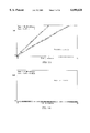
FIG. 1 shows the detection of varying concentrations of oxalate in a sample. Colorimetric absorbance for each sample was plotted over time (minutes). Positive and negative control panels are also shown.
FIG. 2 shows the nucleotide sequence of the formyl-CoA transferase gene (SEQ ID No. 1) and the deduced amino acid sequence of the formyl-CoA transferase polypeptide from Oxalobacter formigenes. Bolded letters represent amino acid residues determined by N-terminal protein sequencing.
FIG. 3 shows the nucleotide sequence of the oxalyl-CoA decarboxylase gene (SEQ ID No. 3) and flanking regions from Oxalobacter formigenes. The consensus ribosome-binding site lies approximately 10 bases upstream (double-underlined letters) from the putative translation initiation codon (positions 1 to 3). A rho-independent termination sequence lies at positions 1758 to 1790 (double-underlined letters). A putative TPP-binding site appears between positions 1351 and 1437.
FIG. 4 shows an RFLP analysis of O. formigenes, strain OxB using probes specific for the oxc gene encoding oxalyl-CoA decarboxylase and the frc gene encoding formyl-CoA transferase. Genomic DNA isolated from a 14 day culture of O. formigenes strain OxB was digested with the restriction enzyme HIND III. The digested DNA was size fractionated by electrophoreses through 0.5% agarose gels, electroblotted to a nylon membrane, then hybridized with either probe AP15 (SEQ ID No. 6) or probe AP34 (SEQ ID NO. 9) to detect oxc or probe AP273 (SEQ ID NO. 10) to detect frc.
FIG. 5 shows the sensitivity of detecting the oxc and frc genes in RFLP of O. formigenes strain OxB versus strain HC-1. Genomic DNA from each of the two strains was digested with the restriction enzyme HIND III. Two-fold serial dilutions were made of the digested DNA and size fractionated by electrophoresis through 0.5% agarose gels (left panels). RFLP analyses were carried out as described in FIG. 4, except the nylon membranes were hybridized with a 1: 1 mixture of probe AP15 (SEQ ID NO. 6) plus probe AP273 (SEQ ID NO. 10) (right panels).
FIG. 6 shows the detection of the oxc and frc genes in various strains of O. formigenes by RFLP analysis. RFLP was carried out as described in FIG. 5.
FIG. 7 shows PCR-based amplification of a genetic region of the oxc gene in various strains of O. formigenes. Using PCR primer AP15 (SEQ ID NO. 6) and primer AP22 (SEQ ID NO. 11) as PCR primers, PCR amplification was performed using genomic DNA isolated from each of the 12 strains of O. formigenes listed in Table 1 as template. PCR products were size fractionated by electrophoresis through 1.2% agarose gels and observed visually using ethidium bromide (EtBr) and UV light.
FIG. 8 shows a direct analysis of fecal samples for O. formigenes. Oxalobacter negative stool sample (A & B) was spiked with 102 (C) and 104 (D) cfu of OxB or 103 (E) and 104 (F) cfu of OxK per 0.1 gm. DNA from an unspiked O. formigenes-positive stool sample diluted 1:25 (G) and 1:50 (H).
FIG. 9 shows the identification of sequence homologies within the oxc gene expressed in representative group I and group II strains of Oxalobacter formigenes to design oligonucleotide probes. Partial sequences of 5'-end of the oxc gene generated by PRC amplification of the region bounded by the primer pair, AP34/AP21. A region of high homology shared by all strains (between bp 13 and 43) was selected for the genus-specific oligonucleotide probe, AP286, while regions of high homology shared by only group I strains (between bp 197 and 214) or shared only by group II strains (between bp 133 and 150) were selected for group-specific oligonucleotide probes, HS2 and AP307, respectively.
FIGS. 10A-10B shows the detection of Oxalobacter formigenes using a genus-specific oligonucleotide probe that hybridizes to the PCR product of the oxc gene. Using the primer pair AP34/AP21, PCR amplification was performed using genomic template DNA isolated from 8 group I and 8 group II strains of O. formigenes. The PCR products were size fractionated by electrophoresis through 1.2% agarose gels and the expected 504-508 bp product visualized with EtBr under UV light (upper panel). The PCR products were transblotted to nylon membranes and Southern blotted using the genus -specific oligonucleotide probe, AP286 (lower panel).
FIGS. 11A-11C shows the classification of group I and group II strains of Oxalobacter formigenes using group-specific oligonucleotide probes that hybridize with PCR products of the oxc gene. Using the primer pair AP34/AP21, PCR amplification was performed using genomic template DNA isolated from 8 group I and 8 group II strains of O. formigenes. The PCR products were size fractionated by electrophoresis through 1.2% agarose gels and the expected 504-508 bp product visualized with EtBr under UV light (upper panel). The PCR products were transblotted to nylon membranes and Southern blotted using HS2, the group I-specific (center panel), or AP307, the group II-specific (lower panel), oligonucleotide probes.
BRIEF DESCRIPTION OF THE SEQUENCES
SEQ ID NO. 1 is a nucleotide sequence for the formyl-CoA transferase gene (also shown in FIG. 2).
SEQ ID NO. 2 is a polypeptide encoded by SEQ ID NO. 1, which can be used according to the subject invention.
SEQ ID NO. 3 is the nucleotide sequence for the oxalyl-CoA decarboxylase gene (also shown in FIG. 3).
SEQ ID NO. 4 is a polypeptide encoded by SEQ ID NO. 3, which can be used according to the subject invention.
SEQ ID NO. 5 is an oxalyl-CoA decarboxylase sequence, which can be used as a probe according to the subject invention.
SEQ ID NO. 6 is an oxalyl-CoA decarboxylase sequence, which can be used as a probe or PCR primer according to the subject invention.
SEQ ID NO. 7 is an oxalyl-CoA decarboxylase 5'-primer, which can be used according to the subject invention.
SEQ ID NO. 8 is an oxalyl-CoA decarboxylase 3'-primer, which can be used according to the subject invention.
SEQ ID NO. 9 is an oxalyl-CoA decarboxylase sequence, which can be used as a probe or primer according to the subject invention.
SEQ ID NO. 10 is a formyl-CoA transferase sequence, which can be used as a probe according to the subject invention.
SEQ ID NO. 11 is an oxalyl-CoA decarboxylase sequence, which can be used as a PCR primer according to the subject invention.
SEQ ID NO. 12 is an oxalyl-CoA decarboxylase sequence, which can be used as a probe according to the subject invention.
SEQ ID NO. 13 is an oxalyl-CoA decarboxylase sequence, which can be used as a probe according to the subject invention.
SEQ ID NO. 14 is an oxalyl-CoA decarboxylase sequence, which can be used as a probe according to the subject invention.
SEQ ID NO. 15 is an oxalyl-CoA decarboxylase sequence, which can be used as a PCR primer according to the subject invention.
DETAILED DESCRIPTION OF THE INVENTION
The subject invention provides an accurate, sensitive assay for oxalate in biological samples such as urine and serum. Elevated levels of oxalate are correlated with urinary tract stone formation, as well as other health problems. Early detection of high levels of oxalate makes it possible to prevent, delay or reduce adverse health consequences through appropriate medication and through modulation of diet.
In the presently described diagnostic system, two enzymes are used to catabolize oxalate to carbon dioxide and formate. Specifically, any oxalate that may be present in a sample being assayed is converted into formate and carbon dioxide (CO2) through the combined action of the enzymes oxalyl-CoA decarboxylase and formyl-CoA transferase. The formate can then be detected using a variety of techniques known in the art. In a preferred embodiment, the production of formate is measured colorimetrically by linking the catabolism of formate with the production of a detectable color change (for example, the formation of a compound that absorbs a particular wavelength of light). The production of formate is directly correlated with the amount of oxalate present in the sample. Therefore, if a known amount of formate is produced using the subject enzyme system, then the amount of oxalate present in the sample can be easily quantitated.
In a preferred embodiment, the enzymes used in the subject invention are expressed by genes from the bacterium Oxalobacter formigenes. The genes encoding both oxalyl-CoA decarboxylase (Lung et al., 1994) and formyl-CoA transferase enzymes have been cloned and expressed, thus providing a readily-available source of reagent material. The subject assay is capable of detecting oxalate levels in a range as low as 0.00025-0.0005 mM (FIG. 1). This level of sensitivity makes the subject assay capable of direct detection of oxalate in serum samples consisting of little as 10 μl volume. The described system can be easily automated with standard systems known in the art.
In a preferred embodiment of the subject assay, the enzymatic reaction can be carried out in the wells of flat-bottomed 96-well microtiter plates and read in an automated plate reader. Suitable concentrations of the assay reagents oxalyl-CoA decarboxylase, oxalyl-CoA, β-NAD, formate dehydrogenase, and the sample to be assayed are added to the microtiter wells. The reaction is then brought to equilibrium (two minute incubation at 37° C. in the plate reader) to permit degradation of any residual formate that may be present in the sample. The formyl-CoA transferase enzyme is then added to the mixture to start the reaction, and the plate is read at 15 second intervals. Formate production is determined by measuring the reduction in NAD in the presence of formate dehydrogenase by detecting changes in absorbance of the sample at 340 nm (Baetz and Allison, 1989). The quantity of oxalate is determined by comparison of the unknown samples with standards having a known amount of oxalate.
Further, the enzymatic reaction of the subject assay will not be initiated until the formyl-CoA transferase, oxalyl-CoA decarboxylase, and oxalyl-CoA are all present within the reaction mixture. Therefore, initiation of the enzymatic reaction can be prevented by withholding one of the above reagents from the reaction mix. Preferably, oxalyl-CoA decarboxylase and oxalyl-CoA are added first, and the reaction is initiated by the addition of formyl-CoA transferase to the mix. However, the order of addition of the three reagents is not material to the function of the assay, so long as one of the reagents is withheld until just prior to the desired initiation point of the assay.
The formyl-CoA transferase and oxalyl-CoA decarboxylase enzymes used in the subject invention can be obtained and purified as a natural product of Oxalobacter formigenes (Baetz and Allison, 1989 and 1990). Alternatively, the enzymes can be obtained from host cells expressing the recombinant polynucleotide molecules of the subject invention that encode the enzymes. Other reagents used in the subject assay can be obtained from conventional sources, such as Sigma Chemical Company, St. Louis, Mo. Further, a person of ordinary skill in the art can readily determine the optimal concentrations of the reagents to use in the assay described herein.
A further aspect of the subject invention concerns the cloning, sequencing and expression of the Oxalobacter formigenes gene which encodes the formyl-CoA transferase used in the assay that is a subject of the invention. The gene was cloned using degenerate oligonucleotide probes (based on partial amino acid sequencing of tryptic peptides) to screen an Oxalobacter genomic DNA library. The gene encodes a polypeptide having a molecular weight of approximately 40 kD. The subject invention further concerns the cloning, sequencing, and expression of the gene which encodes oxalyl-CoA decarboxylase from Oxalobacter formigenes. The nucleotide sequence of the cDNA of formyl-CoA transferase and oxalyl-CoA decarboxylase are shown in FIGS. 2 and 3, respectively (SEQ ID NOS. 1 and 3).
Because of the redundancy of the genetic code, a variety of different polynucleotide sequences can encode the formyl-CoA transferase polypeptide disclosed herein. It is well within the skill of a person trained in the art to create alternative polynucleotide sequences encoding the same, or essentially the same, polypeptide of the subject invention. These variant or alternative polynucleotide sequences are within the scope of the subject invention. As used herein, references to "essentially the same" sequence refers to sequences which encode amino acid substitutions, deletions, additions, or insertions which do not materially alter the functional enzymatic activity of the encoded polypeptide. Further, the subject invention contemplates those polynucleotide molecules having sequences which are sufficiently homologous with the DNA sequences shown in FIGS. 2 and 3 (SEQ ID NOS. 1 and 3) so as to permit hybridization with those sequences under standard high-stringency conditions. Such hybridization conditions are conventional in the art (see, e.g., Maniatis et al., 1989).
As a person skilled in the art would appreciate, certain amino acid substitutions within the amino acid sequence of the polypeptide can be made without altering the functional activity of the enzyme. For example, amino acids may be placed in the following classes: non-polar, uncharged polar, basic, and acidic. Conservative substitutions, whereby an amino acid of one class is replaced with another amino acid of the same class, fall within the scope of the subject invention so long as the substitution does not materially alter the enzymatic reactivity of the polypeptide. Non-conservative substitutions are also contemplated as long as the substitution does not significantly alter the functional activity of the encoded polypeptide.
The polynucleotides of the subject invention can be used to express the recombinant formyl-CoA transferase enzyme. They can also be used as a probe to detect related enzymes. The polynucleotides can also be used as DNA sizing standards.
The polypeptides encoded by the polynucleotides can be used to raise an immunogenic response to the formyl-CoA transferase enzyme. They can also be used as molecular weight standards, or as inert protein in an assay. The polypeptides can also be used to detect the presence of antibodies immunoreactive with the enzyme.
The polynucleotide sequences of the subject invention may be composed of either RNA or DNA. More preferably, the polynucleotide sequences are composed of DNA. The subject invention also encompasses those polynucleotides that are complementary in sequence to the polynucleotide sequences disclosed herein.
Another aspect of the subject invention pertains to kits for carrying out the enzyme assay for oxalate. In one embodiment, the kit comprises, in packaged combination and in relative quantities to optimize the sensitivity of the described assay method, (a) the oxalyl-CoA decarboxylase, oxalyl-CoA, p-NAD, and formate dehydrogenase; and (b) formyl-CoA transferase. The kit may optionally include other reagents or solutions, such as buffering and stabilization agents, along with any other reagents that may be required for a particular signal generation system. Other reagents such as positive and negative controls can be included in the kit to provide for convenience and standardization of the assay method.
The subject invention further concerns a method for detecting the presence of Oxalobacter formigenes organisms in a sample. Specific polynucleotide probes can be prepared based on the nucleotide sequence of either the oxalyl-CoA decarboxylase or the formyl-CoA transferase gene sequence of Oxalobacter formigenes. The polynucleotide probes of the subject invention can be used to identify Oxalobacter formigenes in a sample, and to classify the strain of Oxalobacter formigenes detected. The polynucleotide probes of the subject invention can be used according to standard procedures and conditions to specifically and selectively detect polynucleotide sequences that have sufficient homology to hybridize with the probe. DNA can be isolated from bacterial microorganisms in a biological specimen (e.g., biopsy, fecal matter, tissue scrapings, etc.) using standard techniques known in the art and the isolated DNA screened for hybridization with Oxalobacter oxalyl-CoA decarboxylase-specific and/or formyl-CoA transferase-specific polynucleotide probes. Various degrees of stringency can be employed during the hybridization, depending on the amount of probe used for hybridization, the level of complementarity (i.e., homology) between the probe and target DNA fragment to be detected. The degree of stringency can be controlled by temperature, ionic strength, pH, and the presence of denaturing agents such as formamide during hybridization and washing. Hybridization methods and conditions are known in the art and are generally described in Nucleic Acid Hybridization: A Practical Approach (Hames, B. D., S. J. Higgins, eds.), IRL Press (1985).
The polynucleotide probes of the subject invention include, for example, the oxalyl-CoA decarboxylase probe A (SEQ ID NO. 5), probe AP15 (SEQ ID NO. 6), and probe AP34 (SEQ ID NO. 9), probe AP286 (SEQ ID NO. 12), probe AP307 (SEQ ID NO. 13), and probe HS-2 (SEQ ID NO. 14), specifically exemplified herein. Probes for formyl-CoA transferase include, for example, probe AP273 (SEQ ID NO. 10) specifically exemplified herein. The nucleotide sequences of the exemplified probes are shown below:
                     (SEQ ID NO. 5)
Probe A  5'-GAGCGATACCGATTGGA-3'
                     (SEQ ID NO. 6)
Probe AP15
         5'-GCACAATGCGACGACGA-3'
                     (SEQ ID NO. 9)
Probe AP34
         5'-ATACTCGGAATTGACGT-3'
                     (SEQ ID NO. 10)
Probe AP273
         5'-TTCATGTCCAGTTCAATCGAACG-3'
                     (SEQ ID NO. 12)
Probe AP286
         5'-GACAATGTAGAGTTGACTGATGGCTTTCATG-3'
                     (SEQ ID NO. 13)
Probe AP307
         5'-CAGGATGGTCAGAAGTTC-3'
                     (SEQ ID NO. 14)
Probe HS-2
         5'-CCGGTTACATCGAAGGA-3'
The polynucleotide probes contemplated in the subject invention also include any polynucleotide molecule comprising a nucleotide sequence capable of specifically hybridizing with oxalyl-CoA decarboxylase or formyl-CoA transferase polynucleotide sequence of the present invention. As used herein, reference to "substantial homology" or "substantially complementary" refers not only to polynucleotide probes of the subject invention having 100% homology with the nucleotide sequence of the target polynucleotide, or fragments thereof, but also to those sequences with sufficient homology to hybridize with the target polynucleotide. Preferably, the degree of homology will be 100%; however, the degree of homology required for detectable hybridization will vary in accordance with the level of stringency employed in the hybridization and washes. Thus, probes having less than 100% homology to the oxalyl-CoA decarboxylase or formyl-CoA transferase polynucleotide sequences can be used in the subject method under appropriate conditions of stringency. In a preferred embodiment, high stringency conditions are used. In addition, analogs of nucleosides may be substituted for naturally occurring nucleosides within the polynucleotide probes. Such probes having less than 100% homology or containing nucleoside analogs are within the scope of the subject invention. The skilled artisan, having the benefit of the disclosure contained herein, can readily prepare probes encompassed by the subject invention.
In addition, the subject invention also concerns polynucleotide primers that can be used for polymerase chain reaction (PCR) amplification of Oxalobacter formigenes nucleotide sequences. PCR amplification methods are well known in the art and are described in U.S. Pat. Nos. 4,683,195; 4,683,202; and 4,800,159. In a preferred embodiment, the polynucleotide primers are based on the oxalyl-CoA decarboxylase or formyl-CoA transferase gene sequence and can be used to amplify the full length or a portion of the target gene. The amplified Oxalobacter sequences can be detected using the probes of the subject invention according to standard procedures known in the art.
The polynucleotide primers of the subject invention include, for example, oxalyl-CoA decarboxylase PCR primer 1 (SEQ ID NO. 7), PCR primer 2 (SEQ ID NO. 8), PCR primer AP15 (SEQ ID NO. 6), and PCR primer AP22 (SEQ ID NO. 11), PCR primer AP34 (SEQ IS NO. 9), and PCR primer AP21 (SEQ ID NO. 15), specifically exemplified herein. The nucleotide sequences of the exemplified PCR primers are shown below:
                     (SEQ ID NO. 7)
PCR primer 1
            5'-CAGGTTATGCAGCTTCT-3'
                     (SEQ ID NO. 8)
PCR primer 2
            5'-GGATGGTTGTCAGGCAG-3'
                     (SEQ ID NO. 6)
PCR primer AP15
            5'-GCACAATGCGACGACGA-3'
                     (SEQ ID NO. 11)
PCR primer AP22
            5'-GTAGTTCATCATTCCGG-3'
                     (SEQ ID NO. 9)
PCR primer AP34
            5'-ATACTCGGAATTGACGT-3'
                     (SEQ ID NO. 15)
PCR primer AP21
            5'-TCCAATCGGTATCGCTC-3'
The primer pair AP34 (SEQ ID NO. 9) and AP21 (SEQ ID NO. 15) (derived from oxc sequences between bp -59 to -41 and by 451 to 435, respectively), consistently amplifies a 500 bp segment of oxc from all O. formigenes strains and isolates tested. PCR application of whole fecal DNA with this genus-specific primer pair, in conjunction with Southern Blotting using genus and group specific probes, now provides a rapid diagnostic tool to detect and speciate O. formigenes. Time-consuming steps, e.g., agarose-gel electrophoresis and Souther blot hybridizations, can be substituted with newer technologies such as microtiter-plate based colorimetric or fluorogenic assays (Jordan et al., 1996).
Polynucleotide primers contemplated by the subject invention also include any polynucleotide molecule comprising a nucleotide sequence capable of specifically priming amplification of oxalyl-CoA decarboxylase or formyl-CoA transferase polynucleotide sequences disclosed herein. As used herein, reference to "substantial homology" or "substantially complementary" refers not only to polynucleotide primers of the subject invention having 100% homology with the nucleotide sequence of the target polynucleotide, or fragments thereof, but also to those sequences with sufficient homology to hybridize with and prime the amplification of a target polynucleotide. Preferably, the degree of homology will be equal to or about 100%. The skilled artisan, having the benefit of the disclosure contained herein, can readily prepare other primers of varying nucleotide length and sequence that can be used to amplify all or portions of the oxalyl-CoA decarboxylase and/or the formyl-CoA transferase gene.
The polynucleotide probes and primers of the subject invention can be chemically synthesized or prepared through recombinant means using standard methods and equipment. The polynucleotide probes and primers can be in either single- or double-stranded form. If the probe or primer is double-stranded, then single-stranded forms can be prepared from the double-stranded form. The polynucleotide probes and primers may be comprised of natural nucleotide bases or known analogs of the natural nucleotide bases. The probes and primers of the subject invention may also comprise nucleotides that have been modified to bind labeling moieties for detecting the probe or primer or amplified gene fragment.
The polynucleotide molecules of the subject invention can be labeled using methods that are known in the art. The polynucleotides may be radioactively labeled with an isotope such as 3 H, 35 S, 14 C, or 32 P The polynucleotides can also be labeled with fluorophores, chemiluminescent compounds, or enzymes. For example, a polynucleotide molecule could be conjugated with fluorescein or rhodamine, or luciferin or luminol. Similarly, the polynucleotide molecule can be conjugated with an enzyme such as horseradish peroxidase or alkaline phosphatase. Polynucleotide molecules can also be detected by indirect means. For example, the polynucleotide may be conjugated with ligands, haptens, or antigenic determinants. The conjugated polynucleotide is then contacted with the ligand receptor, with an anti-ligand molecule that binds to the ligands, or with an antibody that binds to the hapten/antigenic determinant, respectively. For example, the polynucleotide can be labeled with digoxygenin and detected with labeled anti-digoxygenin antibodies. The ligand receptor, anti-ligand molecule, or antibody may be directly labeled with a detectable signal system, such as a fluorophore, chemiluminescent molecule, radioisotope, or enzyme. Methods for preparing and detecting labeled moieties are known in the art.
In one embodiment of the present detection method, samples to be tested for the presence of Oxalobacter formigenes are obtained from a person or animal, and DNA is isolated from the specimen using standard techniques known in the art. For example, cells can be lysed in an alkali solution, the nucleic acid extracted in phenol:chloroform, and then precipitated with ethanol. The DNA is then fragmented into various sizes using restriction endonuclease enzymes or other means known in the art. The DNA fragments are then electrophoretically separated by size on an agarose gel. In an alternative embodiment, the DNA fragments are subjected to PCR amplification using PCR primers of the present invention prior to gel electrophoresis in order to specifically amplify portions of the formyl-CoA transferase and oxalyl-CoA decarboxylase genes.
After the DNA fragments are separated on the gel, the size-fractionated DNA fragments are transferred to a membrane matrix, such as nitrocellulose, nylon, or polyvinylidene difluoride (PVDF), by Southern blotting. The DNA immobilized on the membrane matrix is single-stranded. Polynucleotide probes of the subject invention are then contacted with the membrane and allowed to hybridize with the DNA immobilized on the membrane. A probe of the present invention can be labeled with a detectable signal, such as a radioisotope, or the probe can be labeled with a hapten or antigen such as digoxigenin. The hybridization can be performed under conditions known in the art. After hybridization of the probe with the DNA fragments on the membrane, the membrane is washed to remove non-hybridized probe. Standard wash conditions are known in the art, and the stringency and number of washes employed can vary.
The membrane is then tested or observed for the presence of hybridized probe. For example, if the hybridized probe was labeled with a hapten or antigen, then it can be detected using an antibody that binds to the conjugated hapten or antigen on the probe. The antibody can be directly labeled with a detectable fluorophore, chemiluminescent molecule, radioisotope, enzyme, or other signal generating system known in the art. Alternatively, the antibody can be detected using a secondary reagent that binds to the antibody, such as anti-immunoglobulin, protein A, protein G, and other antibody binding compositions known in the art. The secondary reagent can be labeled with a detectable fluorophore, chemiluminescent molecule, radioisotope, or enzyme. The presence of a detectable hybridization signal on the membrane indicates the presence of Oxalobacter formigenes in a test sample.
The subject invention also concerns a kit for the detection of Oxalobacter formigenes in a sample. A kit contemplated by the subject invention may include in one or more containers: polynucleotide probes, positive and negative control reagents, and reagents for detecting the probes. The kit may also include polynucleotide primers for performing PCR amplification of specific Oxalobacter formigenes genes. In a preferred embodiment, the polynucleotide probes and primers are specific for the oxalyl-CoA decarboxylase and formyl-CoA transferase genes of O. formigenes.
The subject invention also concerns a dipstick device comprising the enzymes of the subject invention and dyes and/or substrates immobilized on a carrier matrix. Any dye or substrate that yields a detectable product upon exposure to the reaction products that are produced by the enzymatic reaction of oxalate with oxalyl-CoA decarboxylase and formyl-CoA transferase as described herein is contemplated for use with the subject dipstick device. The carrier matrix of the assay device can be composed of any substance capable of being impregnated with the enzyme and dye components of the subject invention, as long as the matrix is substantially inert with respect to the analyte being assayed for. For example, the carrier matrix may be composed of paper, nitrocellulose, PVDF, or plastic materials and the like.
Incorporation of the enzymes, dye and other components on the carrier matrix can be accomplished by any method such as dipping, spreading or spraying. A preferred method is impregnation of the carrier matrix material by dipping in a reagent solution and drying to remove solvent. Drying can be accomplished by any means which will not deleteriously affect the reagents incorporated, and typically is by means of an air drying oven.
The dipstick device of the subject invention is dipped in or contacted with a sample to be tested for the presence or amount of oxalate. Positive and negative controls can be used in conjunction with the dipstick device. An appropriate amount of time is allowed to pass and then the dipstick is assessed for a positive reaction by visual inspection. If oxalate is present in the sample then a detectable signal, usually in the form of a color, can be observed on the dipstick. Typically, the intensity of the color developed in a fixed time period is proportional to the concentration of oxalate present in the sample.
Following are examples which illustrate procedures, including the best mode, for practicing the invention. These examples should not be construed as limiting. All percentages are by weight and all solvent mixture proportions are by volume unless otherwise noted.
EXAMPLE 1
Determination of Level of Sensitivity of Enzyme Assay System
Samples containing oxalate at concentrations ranging from 0.004 mM to 0.00025 mM were prepared in 10 μl volumes. The samples were then assayed using the enzyme system of the subject invention in 96-well microtiter plates. Reagents were then added at the following concentrations: KH2 PO4 (pH 6.7), 50 mM; MgCl2, 5 mM; thiamine PPi (TPP), 2 mM; oxalyl-CoA, 0.375 mM; β-NAD, 1.0 mM; formate dehydrogenase, 0.25 IU; and oxalyl-CoA decarboxylase, 0.03 U. The reaction mixture was then incubated at 37° C. for 2 minutes in order to permit the degradation of any residual formate that may be present in the sample mixture. The reaction was then initiated by the addition of formyl-CoA transferase to the sample mixture. Changes in A340 were measured every 15 seconds at 37° C. (FIG. 1). Appropriate positive and negative controls were run simultaneously with the assay.
EXAMPLE 2
Detection of Oxalobacter formigenes in a Sample
Strains of Oxalobacter formigenes used in the following methods are listed in Table 1 below.
              TABLE 1
______________________________________
Description of the Oxalobacter formigenes strains
Group Classification of Source of
O. formigenes strains.sup.a
              Strain    Isolate
______________________________________
Group I       OxB       Sheep rumen
              OxWR      Wild rat cecum
              SOx-4     Freshwater lake sediment
              SOx-6     Freshwater lake sediment
              POxC      Pig cecum
              HC-1      Human feces
Group II      BA-1      Human feces
              OxK       Human feces
              HOxBLS    Human feces
              HOxRW     Human feces
              OxCR      Lab rat cecum
              OxGP      Guinea pig cecum
______________________________________
 .sup.a From Jensen and Allison (1994).
All Oxalobacter formigenes strains were grown in medium B containing 30 mM oxalate, as described in Allison et al. (1985). Human fecal samples (approximately 60 mg) were inoculated anaerobically into vials containing 9 ml of media B, then sequentially transferred through 10-8 dilutions. Cultures were incubated at 37° C. for 10 days and biochemically tested for the catabolic consumption of oxalate by CaCl2 precipitation (50 μl media, 100 μl 1% CaCl2, and 2.7 ml dH2 O) and spectrophotometric analyses (600 nm).
Cultures (10-15 ml) of O. formigenes were centrifuged at 10,000×g, the bacterial pellet was resuspended in 567 μl TE buffer (10 mM Tris-Cl, pH 7.5 plus 1 mM EDTA, pH 8.0), 30 μl 10% sodium dodecyl sulfate (SDS) and 3 μl of proteinase K (20 mg/ml), and the mixture incubated 5 hr at 37° C. to ensure bacterial cell lysis. Nucleic acids were extracted from the lysates using phenol/chloroform/isoamylalcohol (25:24:1). Chromosomal DNA was precipitated from the aqueous phase by adding 1/2 volume of 7.5 M ammonium acetate and 2 volumes of 100% ethanol. DNA was recovered by centrifugation(12,000×g), washed once with 70% ethanol, and the pellet resuspended in 15-20 μl H2 O. Bacterial DNA was also isolated directly from fresh human stool samples following lysis with chaotropic salt and guanidine thiocyanate, then binding to glass matrix (GlasPac, National Scientific Supply, San Rafael, Calif.) (Stacy-Phips et al., 1995).
Bacterial DNA was digested with the restriction endonuclease Hind III (Life Technologies, Inc., Gaithersburg, Md.). The restriction-enzyme generated fragments were size separated by gel electrophoresis through 0.5% agarose, stained with ethidium bromide (EtBr), illuminated with UV light, and photographed to document proper digestion. Digested DNA was then transferred from the agarose gels to positively-charged nylon membranes (Boehringer-Mannheim GmBH, Indianapolis, Ind.) by positive pressure blotting and UV cross-linking (Stratagene, LaJolla, Calif.). Hybridizations were carried out using internal sequence oligonucleotide probes. Oligonucleotides were synthesized in the University of Florida ICBR Oligonucleotide Synthesis Laboratory (Gainesville, Fla.) and have the sequences:
                     (SEQ ID NO. 6)
AP15 5'-GCACAATGCGACGACGA-3'
                     (SEQ ID NO. 11)
AP22 5'-GTAGTTCATCATTCCGG-3'
                     (SEQ ID NO. 9)
AP34 5'ATACTCGGAATTGACGT-3'
                     (SEQ ID NO. 10)
AP273
     5'-TTCATGTTCCAGTTCAATCGAACG-3'.
Each oligonucleotide was end-labeled with digoxigenin in a reaction using terminal transferase. The digoxigenin-labeled oligonucleotide probes were hybridized to the immobilized DNA fragments and hybridization detected calorimetrically by enzyme-linked immunoassay (ELISA) using an anti-digoxigenin alkaline phosphatase conjugate according to the manufacturer's protocol provided with the GENIUS III detection system (Boehringer-Mannheim).
All PCRs were performed according to protocols described in Anderson et al. (1993). Briefly, 50 μl reactions contained 1.5 mM MgCl2, 200 μM dNTP, 1.25 U Taq polymerase (GIBCO-BRL, Bethesda, Md.), 1 μg template DNA and 1 μM each of a 5' and 3' primer. A preferred reaction profile proved to be 94° C. for 5 min, then 45 cycles of 94° C. for 1 min of denaturation, 55° C. for 2 min of annealing and 72° C. for 3 min of primer extension. PCR products were size separated by gel electrophoresis in 1.2% agarose containing EtBr and photographed in UV light. PCR primer AP15 (SEQ ID NO. 6) and primer AP22 (SEQ ID NO. 11) were used as primers.
Previous studies by Lung et al. (1994) showed that genomic DNA of O. formigenes, strain OxB, could be digested with the restriction enzyme Hind III and that a limited number of enzyme cleavage sites existed near or within the oxalyl-CoA decarboxylase (oxc) gene. A RFLP analysis of Hind III digested OxB genomic DNA using either probe AP15 (SEQ ID NO. 6), a probe homologous to an internal sequence of the oxc gene, probe AP34 (SEQ ID NO. 9), a probe homologous to a 5'-end sequence of the oxc gene but separated from the probe AP15 (SEQ ID NO. 6) sequence by a Hind III site, or probe AP273 (SEQ ID NO. 10), a probe homologous to an internal sequence of the formyl-CoA transferase (frc) gene, is shown in FIG. 4. Using probe AP15 (SEQ ID NO. 6), a fragment of approximately 7 kb containing a portion of the oxc gene was detected, while fragments of approximately 3 kb were detected using either probe AP34 (SEQ ID NO. 9) or probe AP273 (SEQ ID NO. 10). The 3 kb fragment identified by probe AP34 (SEQ ID NO. 9) is distinct from the 3 kb fragment detected by probe AP273 (SEQ ID NO. 10).
As shown in FIG. 5, the oxalyl-CoA decarboxylase and formyl-CoA transferase genes were consistently detected in samples containing as little as 0.06 to 0.20 μg of O. formigenes, strain OxB, DNA or approximately 0.20 to 0.40 μg of O. formigenes DNA from other group I strains, such as HC-1. The 23-bp probe AP273 (SEQ ID NO. 10) can detect the frc gene in DNA samples containing only one-fourth the amount of DNA required for the 13 bp probe AP15 (SEQ ID NO. 6) to detect the oxc gene (FIG. 5, upper panel). These probes are highly specific for O. formigenes since they fail to bind to other bacterial DNA, including Escherichia coli, Alcaligenes oxalaticus, and fecal bacteroides.
Protein, lipid and genetic studies of several isolates of O. formigenes have provided the basis for dividing this genus into two major subgroupings (Jensen et al., 1994). When RFLP analyses were performed on genomic DNA isolated from various Oxalobacter formigenes strains, probes AP15 (SEQ ID NO. 6) and AP273 (SEQ ID NO. 10) were able to distinguish group I strains from group II strains on the Southern blot hybridizations (FIG. 6). All strains of O. formigenes belonging to group I (to which OxB is assigned) hybridized with both probe AP15 (SEQ ID NO. 6) and probe AP273 (SEQ ID NO. 10). Due to a characteristic slow growth of strain HC-1, only faint bands appeared in this experiment. In contrast, none of the O. formigenes strains assigned to group II hybridized with probe AP273 (SEQ ID NO. 10) and only BA-1 hybridized with probe AP15 (SEQ ID NO. 6). These data indicate a highly conserved homology of oxc and frc within group I strains and a less conserved homology within group II strains.
To increase the sensitivity of detecting O. formigenes, PCR was used to amplify that portion of oxc which by RFLP appeared to differentiate the group I and group II strains. Using primer AP15 (SEQ ID NO. 6) and primer AP22 (SEQ ID NO. 11) as PCR primers to amplify a DNA segment in the carboxy-terminal region of oxc, strains assigned to group I (i.e., OxB, HC-1, OxWR, POxC, SOx-4 and SOx-6) exhibited a common band at 452 bp (FIG. 7). In contrast, the other six strains, all belonging to group II, showed variable amplification patterns, but all showed a dominant PCR band of approximately 630 bp, with a weaker 452 bp band. Sequence analysis of this 630 bp band from strain OxK has revealed the presence of the 452 bp sequence present in the 630 bp PCR product. Close analysis of the group II strains suggest that their PCR amplification profiles are highly reproducible, suggesting group II strains may fall into three (sub)groupings: HOxBLS and HOxRW (subgroup 1), OxCR and OxGP (subgroup 2), and BA-1 and OxK (subgroup 3).
The use of PCR-based detection of the oxc gene to identify O. formigenes in clinical specimens was examined by comparing PCR and biochemical methods of detection. Specimen 1, known to be positive for O. formigenes, gave ambiguous results in biochemical testing for oxalate depletion, but exhibited the presence of the 450 bp PCR product indicative of an O. formigenes group I strain. Specimen 2, known to be negative for O. formigenes, proved negative using both PCR-based and biochemical testing. Specimen 3, known to be positive for O. formigenes, showed depletion of oxalate in all dilutions and revealed a PCR pattern suggestive of an O. formigenes group II strain. PCR amplification was not observed in the original culture or the first dilution due to the presence of inhibitors of PCR e.g., bile salts, bilirubin, etc.) which copurify with DNA.
To circumvent the inhibition of the PCR by factors co-purifying with the bacterial DNA, DNA isolation was performed by lysing fresh stool samples with guanidine thiocyanate followed by adsorption to and elution from glass matrices. Using this method, PCR-based detection of O. formigenes can be performed using fecal DNA diluted only 1:25 to 1:50 to eliminate PCR inhibitors. Sensitivity experiments using different stool samples spiked with strains OxB or OxK in the range of 101 to 107 cfu per 0.1 g of sample showed that as few as 102 to 103 cfu of O. formigenes per 0.1 g sample could be detected (FIG. 8). Again, PCR-based analyses of DNA isolated directly from a stool sample known to be positive for O. formigenes by culture methods, showed amplification patterns indicative of a group II strain (FIG. 8, lanes F & G).
EXAMPLE 3
Detection and Classification of Oxalobacter formigenes
Bacterial Strains
O. formigenes strains used included OxB (isolated from sheep rumen) and HC1, OxK, BA1, HOxBLS, HOxRW, HOxRA, HOxCC13, and HOxHM18 (isolated from human feces). In addition, several new purified cultures, including HOxUK5, HOxUK88, HOxUK90, and HOxHS (grown from human feces), were also used. All strains and isolates were grown in media B containing 30 mM potassium oxalate, as described elsewhere (Allison et al., 1985), and maintained under strict anaerobic conditions until used.
Preparation of Genomic DNA from O. formigenes Cultures
Fifteen ml cultures of O. formigenes were centrifuged at 10,000×g, the bacterial pellet resuspended in 567 μl of TE buffer (10 mM Tris-HC1, pH 7.5, plus 1 mM EDTA, pH 8.0), 30 μl of 10% sodium dodecyl sulfate plus 3 μl of proteinase K (20 mg/ml), and this mixture incubated for 5 hours at 37° C. to ensure bacterial cell lysis. Nucleic acids were extracted from the lysates with phenol:chloroform:isoamylalcohol (25:24: 1). Chromosomal DNA was precipitated by adding 1/2 volume of 7.5 M ammonium acetate and 2 volumes of 100% ethanol. DNA was recovered by centrifugation(12,000×g) and washed once in 70% ethanol. The final DNA precipitation was resuspended in 20 μl H2 O.
Sequence Analysis of the oxc Genes
The primer pair,
5'-ATACTCGGAATTGACGT-3' (a 5'-primer designated AP34) (SEQ ID NO. 9) and
5'-TCCAATCGGTATCGCTC-3' (a 3'-primer designated AP21) (SEQ ID NO. 15)
homologous to sequences within the 5'-end of the oxc gene present in strain OxB (Lung et al., 1994), was used to amplify a 500 bp DNA fragment from genomic DNA isolated from each of twelve human O. formigenes strains. Amplifications were performed in 50 μl PCR reactions containing 1.5 mM MgCl2, 200 μM deoxynucleoside triphosphate, 1.25 U of Taq polymerase (Gibco-BRL, Bethesda, Md.), 1 μg of genomic DNA and 1 μM each of 5'- and 3'-primer. PCR were carried out for 35 cycles and included an initial 5 minute denaturation step at 94° C., 1 minute annealing (with a temperature stepdown from 60° C. to 55° C.), 1 minute extension at 72° C. and a final 8 minute extension at 72° C. The PCR products were size fractionated by electrophoreses through 1.2% agarose gels containing ethidium bromide for visualization of the bands in UV light. Each 500 bp PCR product was cloned into the TA cloning system, pCR-2.1 (Invitrogen, Inc., San Diego, Calif.). Competent DH5E. coli bacteria were transfected with the recombinant plasmid and transformed bacteria selected on LB agar plates containing 10 μl/ml of ampicillin and 20 mg/ml of X-Gal. DNA from appropriate clones was isolated, checked for the presence of an insert of correct size by digestion with the restriction enzyme, Eco RI. Inserts of recombinant plasmids were sequenced using M13-forward and M13-reverse primers.
Clinical Samples
Fecal samples of 100 generally healthy children of either sex ranging in age from 0 to 12 years were examined for the presence of O. formigenes. All fecal samples were collected in Dzerzhinsk, a city in the Donetsk region of the Ukraine. This particular population was selected due to the fact that these children have had limited use of antibiotics, that might influence bacterial colonization of the intestinal tract, in treatment of childhood diseases. Approximately 25 mg sample of fresh stool (within 3-4 hours of collection), was inoculated into vials containing 10 ml of anaerobically sealed media B supplemented to 30 mM with potassium-oxalate. The vials were analyzed at the University of Florida, Gainesville, Fla. After incubation at 37° C. for one week, the loss of oxalate from each fecal culture was determined using a calcium-chloride precipitation method in which 50 μl culture media is mixed with 100 μl 0.1% CaCl2 plus 3.0 ml dH2 O and the absorbance of each mixture determined spectrophotometrically (600nm). The calcium precipitation test for loss of oxalate has been repeatedly verified as reliable by other methods (e.g., gas chromatography and butyl esters) for detection of oxalate. Typically, cultures not showing catabolism of oxalate generally have O.D. readings of about 0. 1, whereas cultures with oxalate degradation have O.D. readings less than about 0.02.
PCR-based Detection and Identification of O. formigenes
DNA was isolated from individual fecal cultures by the method of Phipps et al. (Stacy-Phipps et al., 1995) using guanidine thiocyanate as a chaotropic agent and glass-matrix for DNA binding. One 1 μl of each DNA sample was used as template in a 50 μl PCR reaction as described above. The amplified PCR products were size separated by electrophoresis through 1.2% agarose gels containing ethidium bromide and visualized with UV light. Each reaction was controlled using a reaction containing all the components of the PCR with the exception of template DNA.
Southern Blot Analysis
Southern blots were carried out as previously detailed in Example 2. Briefly, the size separated PCR products were transferred to positively charged nylon membranes (Boehringer Mannheim GmBH, Indianapolis, Ind.) by positive pressure blotting and UV-crosslinking. The oxc derived genus specific (AP286), group I specific (HS-2) and group II specific (AP307) oligonucleotides were synthesized in the University of Florida ICBR DNA Synthesis Laboratory (University of Florida, Gainesville, Fla.) and end-labeled with digoxigenin in a reaction using terminal transferase. The digoxigenin labeled oligonucleotides were hybridized to the immobilized PCR products under conditions of high stringency (5X SSC and 68° C.). Hybridization was detected colorimetrically by enzyme-linked immunosorbent assay (ELISA) with an anti-digoxigenin alkaline phosphatase conjugate according to the manufacturer's protocol provided with the GENIUS III kit (Boehringer Mannheim GmBH).
Generation of Genus-specific and Group-specific Probes
Preliminary studies looking at the efficacy of various oligonucleotide pairs to amplify portions of the oxc gene present in various O. formigenes strains revealed that the PCR primer pair AP34 (5'-primer)/Ap21 (3'-primer) amplified a 500 bp DNA fragment in both group I and group II strains. To determine the degree of sequence homology within the 5'-end of the oxc gene between various strains of O. formigenes, genomic DNA was prepared from 5 group I and 7 group II strains isolated from human fecal samples for use as template in PCR with AP34 and AP21. Each PCR amplified an expected 500 bp product that was subsequently cloned into the pCR-2.1 vector system and sequenced. A comparison of the 5'-end sequences of the oxc gene from these 12 human isolates with the OxB gene is shown in part in FIG. 9. The 5'-end of the oxc gene appears to be relatively conserved for a bacterial gene, with most bp changes occurring in the wobble base such that the codon translation is not altered. Nevertheless, there were enough sequence differences to demarcate group I strains from group II strains, thus permitting selection of regions that are conserved within strains of a specific group, but differ significantly from strains of the other group. Based on these conserved regions, genus-specific oligonucleotide probes (for example, probe AP286, homologous to the region between bp 13 and 43 of the open-reading frame), as well as group I-specific (for example, probe HS2, homologous to the region between bp 197 and 214 of the open-reading frame) and group II-specific (for example, probe AP307, homologous to the region between bp 133 and 150 of the open-reading frame) probes were prepared.
Specificity of the Genus-specific and Group-specific Oligonucleotide Probes
The specificity of probes AP286, AP307, and HS2 in detecting and classifying O. formigenes was examined using genomic DNA prepared from number of known strains and isolates. PCR amplifications with the genus-specific primer pair AP34 and AP21 resulted in the 500 bp amplification product in all cultures tested (FIG. 10A, top panel). On Southern blotting, this 500 bp fragment hybridized with a genus-specific probe, AP286 (FIG. 10B, bottom panel).
In a separate experiment, the amplified 500 bp PCR product was hybridized with either the group I-specific probe, HS2, (FIG. 11 B, middle panel) or the group II-specific probe, AP307, (FIG. 11C, bottom panel). Results clearly show a group specificity in the binding of these group-specific probes and their ability to identify subgroups of O. formigenes.
Application of a PCR-based Detection System for O. formigenes
In a double-blinded study, 100 fecal samples were collected from children ranging in age from newborn to 12 years and tested for the presence of O. formigenes using both an oxalate degradation system and our PCR-based assay system. The aim of this study was to determine the age at which children become naturally colonized with this intestinal anaerobic bacterium. Of the 100 fecal samples examined, 72 samples tested positive for O. formigenes by PCR, 59 of which also exhibited oxalate degradation in an oxalate degradation assay. Interestingly, of the 72 positive samples, 68 were group II strains while only 4 were group I strains. All fecal cultures exhibiting degradation of oxalate tested positive for O. formigenes by PCR. Although there were 13 cultures that failed to degrade oxalate that proved positive for O. formigenes by PCR, the majority of the samples that failed to degrade oxalate also failed to exhibit amplification of a product in the PCR-reaction. These data show that the PCR-based assay is probably more sensitive than the biochemical (calcium chloride precipitation) test, yet highly specific.
When the data were unblinded, a clear pattern for the natural colonization of children became evident. O. formigenes could not be detected in infants less than 6-9 months of age. O. formigenes began appearing in the intestinal tracts of children around 1 year of age, and by 3-4 years of age, all children showed signs of being colonized. Although the sample size is small, the number of children colonized with O. formigenes declined between 8-12 years of age, reaching the colonization frequency of 70-80% estimated for adult populations (Doane et al., 1989,Kleinschmidt et al., 1993, Allison et al., 1986,and Goldkin et al, 1985).
It should be understood that the examples and embodiments described herein are for illustrative purposes only and that various modifications or changes in light thereof will be suggested to persons skilled in the art and are to be included within the spirit and purview of this application and the scope of the appended claims.
REFERENCES
Allison, M. J., H. M. Cook, D. B. Milne, S. Gallaher, R. V. Clayman (1986) "Oxalate degradation by gastrointestinal bacteria from humans," J Nutr 116:455-460.
Allison, M. J., K. A. Dawson, W. R. Mayberry, J. G. Foss (1985) "Oxalobacter formigenes gen. nov., sp. nov.: oxalate degrading bacteria that inhabit the gastrointestinal tract," Arch Microbiol. 141:1-7.
Anderson, J. T., J. G. Cornellius, A. J. Jarpe, W. E. Winter, A. B. Peck (1993) "Insulin-dependent diabetes in the NOD mouse model. II. β cell destruction in autoimmune diabetes is a TH1 mediated event," Autoimmunity 15:113-122.
Argenzio, R. A., J. A. Liacos, M. J. Allison (1988) "Intestinal oxalate degrading bacteria reduce oxalate absorption and toxicity in guinea pigs," J Nutr 118:787-791.
Baetz, A. L., M. J. Allison (1989) "Purification and Characterization of Oxalyl-Coenzyme A Decarboxylase from Oxalobacterformigenes," J. Bacteriol. 171:2605-2608.
Baetz, A. L., M. J. Allison (1990) "Purification and Characterization of Formyl-Coenzyme A Transferase from Oxalobacterformigenes," J. Bacteriol. 172:3537-3540.
Costello, J., M. Hatch, E. Bourke (1976) "An enzymic method for the spectrophotometric determination of oxalic acid," J Lab. Clin. Med. 87(5):903-908.
Curhan, et al. (1993) "A Prospective study of dietary calcium and other nutrients and the risk of symptomatic kidney stones," N.E.J Med. 328:833-838.
Daniel, S. L., P. A. Hartman, M. J. Allison (1987) "Microbial degradation of oxalate in the gastrointestinal tracts of rats," Appl Environ Microbiol 53:957-964.
Dawson, K. A., M. J. Allison, P. A. Hartman (1980) "Characteristics of anerobic oxalate-degrading bacteria from the rumen," Applied Microbiol. 22:522-529.
Dawson, K. A., M. J. Allison, P. A. Hartman (1980) "Isolation and some characteristics of anaerobic oxalate-degrading bacteria from ruman" Appl. Environ. Microbiol. 40:833-839.
Doane, L. A., M. Liebman, D. R. Caldwell (1989) "Microbial oxalata degradation: effects on oxalate and calcium balance in humans," Nutrition Res 9:957-964.
Goldkin, L., D. R. Cave, B. Jaffin, W. Robinson, C. M. Bliss (1985) "A new factor in enteric hyperoxaluria: Oxalobacterformigenes" AM J Gastro 80:860.
Hatch, M., R. W. Freel (1996) "Oxalate transport across intestinal and renal epithelia" Calcium Oxalate in Biological Systems, pages 217-238, CRC Press, Boca Raton, Fla.
Hodgkinson, A. (1970) "Determination of Oxalic acid in Biological Material," Clin. Chem. 16(7):547-557.
Jensen, N. S., M. J. Allison (1994) "Studies on the diversity among anaerobic oxalate-degrading bacteria now in the species Oxalobacterformigenes" Abst. Ann. Mtg. Amer. Soc. Microbial., pages 1-29.
Jordan, J. A., M. B. Durso (1996) "Rapid speciation of the five most medically relevant candida species using PCR amplification and a microtitre plate-based detection system," Mol Diagnosis 1:51-58.
Kleinschmidt K., A. Mahlmann, R. Hautmann (1993) "Anaerobic oxalate-degrading bacteria in the gut decrease faecal and urinary oxalate concentrationsin stone formers," In R. Ryall, R. Bais, V. R. Marshall, A. M. Rofe, L. H. Smith, V. R. Walker Urolithiasis 2, Plenum Press, New York, pp. 439-441.
Lung, H., A. L. Baetz, A. B. Peck (1994) "Molecular Cloning, DNA Sequence and Gene Expression of the Oxalyl-CoA Decarboxylase Gene, oxc, from the Bacterium Oxalobacter formigenes," J Bacteriol. 176(8):2468-2472.
Maniatis, T., E. F. Fritsch, J. Sambrook (1989) Molecular Cloning: A Laboratory Manual, 2d Edition, Cold Spring Harbor Laboratory, Cold Spring Harbor, N.Y.
Smith, R. L., F. E. Strohmaier, R. S. Oremland (1985) "Isolation of anaerobic oxalate-degrading bacteria from fresh water lake sediments," Arch Microbiol 141:8-13.
Stacy-Phips, S., J. J. Mecca, J. B. Weiss (1995) "Multiplex PCR assay and simple preparation method for stool specimens detect enterotoxigenic E. coli DNA during course of infection," J Clin.
Microbiol 33:1054-1059.
Yriberri, J., L. S. Posten (1980) "A semi-automatic enzymic method for estimating urinary oxalate," Clin. Chem. 26(7):881-884.
__________________________________________________________________________
#             SEQUENCE LISTING
- (1) GENERAL INFORMATION:
-    (iii) NUMBER OF SEQUENCES: 15
- (2) INFORMATION FOR SEQ ID NO:1:
-      (i) SEQUENCE CHARACTERISTICS:
#pairs    (A) LENGTH: 1577 base
          (B) TYPE: nucleic acid
          (C) STRANDEDNESS: single
          (D) TOPOLOGY: linear
-     (ii) MOLECULE TYPE: DNA (genomic)
-     (xi) SEQUENCE DESCRIPTION: SEQ ID NO:1:
- AAGCTTGCTT CATTTTGAGA TGTTATGCGA AGTGTTAGCA ACCCAAGTTA GT - #ACCCTTCA
  60
- GCCCTTTGGG CGAAGTTTTT CTTTCTTGGC AGTTCCTTTC GGGGAAACAG CA - #CAGAGAAT
 120
- AAAAACCAAA AGTTGTACCA ACGACAAGGA AATGAGAAAT TATGACTAAA CC - #ATTAGATG
 180
- GAATTAATGT GCTTGACTTT ACCCACGTCC AGGCAGGTCC TGCCTGTACA CA - #GATGATGG
 240
- GTTTCTTGGG CGCAAACGTC ATCAAGATTG AAAGACGTGG TTCCGGAGAT AT - #GACTCGTG
 300
- GATGGCTGCA GGACAAACCA AATGTTGATT CCCTGTATTT CACGATGTTC AA - #CTGTAACA
 360
- AACGTTCGAT TGAACTGGAC ATGAAAACCC CGGAAGGCAA AGAGCTTCTG GA - #ACAGATGA
 420
- TCAAGAAAGC CGACGTCATG GTCGAAAACT TCGGACCAGG CGCACTGGAC CG - #TATGGGCT
 480
- TTACTTGGGA ATACATTCAG GAACTGAATC CACGCGTCAT TCTGGCTTCC GT - #TAAAGGCT
 540
- ATGCAGAAGG CCACGCCAAC GAACACCTGA AAGTTTATGA AAACGTTGCA CA - #GTGTTCCG
 600
- GCGGTGCTGC AGCTACCACC GGTTTCTGGG ATGGTCCTCC AACCGTTTCC GG - #CGCTGCTC
 660
- TGGGTGACTC CAACTCCGGT ATGCACCTGA TGATCGGTAT TCTGGCCGCT CT - #GGAAATGC
 720
- GTCACAAAAC CGGCCGTGGT CAGAAAGTTG CCGTCGCTAT GCAGGACGCT GT - #TCTGAATC
 780
- TGGTTCGTAT CAAACTGCGT GACCAGCAAC GTCTGGAAAG AACCGGCATT CT - #GGCTGAAT
 840
- ACCCACAGGC TCAGCCTAAC TTTGCCTTCG ACAGAGACGG TAACCCACTG TC - #CTTCGACA
 900
- ACATCACTTC CGTTCCACGT GGTGGTAACG CAGGTGGCGG CGGCCAGCCA GG - #CTGGATGC
 960
- TGAAATGTAA AGGTTGGGAA ACCGATGCGG ACTCCTACGT TTACTTCACC AT - #CGCTGCAA
1020
- ACATGTGGCC ACAGATCTGC GACATGATCG ACAAGCCAGA ATGGAAAGAC GA - #CCCAGCCT
1080
- ACAACACATT CGAAGGTCGT GTTGACAAGC TGATGGACAT CTTCTCCTTC AT - #CGAAACCA
1140
- AGTTCGCTGA CAAGGACAAA TTCGAAGTTA CCGAATGGGC TGCCCAGTAC GG - #CATTCCTT
1200
- GCGGTCCGGT CATGTCCATG AAAGAACTGG CTCACGATCC TTCCCTGCAG AA - #AGTTGGTA
1260
- CCGTCGTTGA AGTTGTCGAC GAAATTCGTG GTAACCACCT GACCGTTGGC GC - #ACCGTTCA
1320
- AATTCTCCGG ATTCCAGCCG GAAATTACCC GTGCTCCGCT GTTGGGCGAA CA - #TACCGACG
1380
- AAGTTCTGAA AGAACTGGGT CTTGACGATG CCAAGATCAA GGAACTGCAT GC - #AAAACAGG
1440
- TAGTTTGATC CGTCAGACTT TCTGGGCAAA ACGGCACTCT CCGGAGTGCC GT - #TTTTTGTC
1500
- ACACGAAACC TAATCAAACA AGCACGTGCA ATGATTCCAC ATCATTGCGG CC - #ACATTCAT
1560
# 1577             G
- (2) INFORMATION FOR SEQ ID NO:2:
-      (i) SEQUENCE CHARACTERISTICS:
#acids    (A) LENGTH: 428 amino
          (B) TYPE: amino acid
          (C) STRANDEDNESS: single
          (D) TOPOLOGY: linear
-     (ii) MOLECULE TYPE: protein
-     (xi) SEQUENCE DESCRIPTION: SEQ ID NO:2:
- Met Thr Lys Pro Leu Asp Gly Ile Asn Val Le - #u Asp Phe Thr His Val
#                15
- Gln Ala Gly Pro Ala Cys Thr Gln Met Met Gl - #y Phe Leu Gly Ala Asn
#            30
- Val Ile Lys Ile Glu Arg Arg Gly Ser Gly As - #n Met Thr Arg Gly Trp
#        45
- Leu Gln Asp Lys Pro Asn Val Asp Ser Leu Ty - #r Phe Thr Met Phe Asn
#     60
- Cys Asn Lys Arg Ser Ile Glu Leu Asp Met Ly - #s Thr Pro Glu Gly Lys
# 80
- Glu Leu Leu Glu Gln Met Ile Lys Lys Ala As - #p Val Met Val Glu Asn
#                 95
- Phe Gly Pro Gly Ala Leu Asp Arg Met Gly Ph - #e Thr Trp Glu Tyr Ile
#           110
- Gln Glu Leu Asn Pro Arg Val Ile Leu Ala Se - #r Val Lys Gly Tyr Ala
#       125
- Glu Gly His Ala Asn Glu His Leu Lys Val Ty - #r Glu Asn Val Ala Gln
#   140
- Cys Ser Gly Gly Ala Ala Ala Thr Thr Gly Ph - #e Trp Asp Gly Pro Pro
145                 1 - #50                 1 - #55                 1 -
#60
- Thr Val Ser Gly Ala Ala Leu Gly Asp Ser As - #n Ser Gly Met His Leu
#               175
- Met Ile Gly Ile Leu Ala Ala Leu Glu Met Ar - #g His Lys Thr Gly Arg
#           190
- Gly Gln Lys Val Ala Val Ala Met Gln Asp Al - #a Val Leu Asn Leu Val
#       205
- Arg Ile Lys Leu Arg Asp Gln Gln Arg Leu Gl - #u Arg Thr Gly Ile Leu
#   220
- Ala Glu Tyr Pro Gln Ala Gln Pro Asn Phe Al - #a Phe Asp Arg Asp Gly
225                 2 - #30                 2 - #35                 2 -
#40
- Asn Pro Leu Ser Phe Asn Asn Ile Thr Ser Va - #l Pro Arg Gly Gly Asn
#               255
- Ala Gly Gly Gly Gly Glu Pro Gly Trp Met Le - #u Lys Cys Lys Gly Trp
#           270
- Glu Thr Asp Ala Asp Ser Tyr Val Tyr Phe Th - #r Ile Ala Ala Asn Met
#       285
- Trp Pro Gln Ile Cys Asn Met Ile Asp Lys Pr - #o Glu Trp Lys Asp Asp
#   300
- Pro Ala Tyr Asn Thr Phe Glu Gly Arg Val As - #p Lys Leu Met Asp Ile
305                 3 - #10                 3 - #15                 3 -
#20
- Phe Ser Phe Ile Glu Thr Lys Phe Ala Asp Ly - #s Asp Lys Phe Glu Val
#               335
- Thr Glu Trp Ala Ala Gln Tyr Gly Ile Pro Cy - #s Gly Pro Val Met Ser
#           350
- Met Lys Glu Leu Ala His Asp Pro Ser Leu Gl - #n Lys Val Gly Thr Val
#       365
- Val Glu Val Val Asp Glu Ile Arg Gly Asn Hi - #s Leu Thr Val Gly Ala
#   380
- Pro Phe Lys Phe Ser Gly Phe Gln Pro Glu Il - #e Thr Arg Ala Pro Leu
385                 3 - #90                 3 - #95                 4 -
#00
- Leu Gly Glu His Thr Asp Glu Val Leu Lys Gl - #u Leu Gly Leu Asp Asp
#               415
- Ala Lys Ile Lys Glu Leu His Ala Lys Gln Va - #l Val
#           425
- (2) INFORMATION FOR SEQ ID NO:3:
-      (i) SEQUENCE CHARACTERISTICS:
#pairs    (A) LENGTH: 2088 base
          (B) TYPE: nucleic acid
          (C) STRANDEDNESS: single
          (D) TOPOLOGY: linear
-     (ii) MOLECULE TYPE: DNA (genomic)
-     (xi) SEQUENCE DESCRIPTION: SEQ ID NO:3:
- ATTTGTTTAA ATTGACCTGA ATCAATATTG CCGGATTGAT CTAGGTCAAT GA - #ATGCAAAT
  60
- TGACTTATGT CAATGGTGCC AAATTGACCT AGGTCAACGG GATTTTTAAA GG - #GTATGCGG
 120
- CATACTCGGA ATTGACGTTA AACAACGTTT ATCAAAACCA ACCAAAGAAA GG - #TATTACTC
 180
- ATGAGTAACG ACGACAATGT AGAGTTGACT GATGGCTTTC ATGTTTTGAT CG - #ATGCCCTG
 240
- AAAATGAATG ACATCGATAC CATGTATGGT GTTGTCGGCA TTCCTATCAC GA - #ACCTGGCT
 300
- CGTATGTGGC AAGATGACGG TCAGCGTTTT TACAGCTTCC GTCACGAACA AC - #ACGCAGGT
 360
- TATGCAGCTT CTATCGCCGG TTACATCGAA GGAAAACCTG GCGTTTGCTT GA - #CCGTTTCC
 420
- GCCCCTGGCT TCCTGAACGG CGTGACTTCC CTGGCTCATG CAACCACCAA CT - #GCTTCCCA
 480
- ATGATCCTGT TGAGCGGTTC CAGTGAACGT GAAATCGTCG ATTTCCAAGA CG - #GCGATTAC
 540
- GAAGAAATGG ATCAGATGAA TGTTGCACGT CCACACTGCA AAGCTTCTTT CC - #GTATCAAC
 600
- AGCATCAAAG ACATTCCAAT CGGTATCGCT CGTGCAGTTC GCACCGCTGT AT - #CCGGACGT
 660
- CCAGGTGGTG TTTACGTTGA CTTCCCAGCA AAACTGTTCG GTCAGACCAT TT - #CTGTAGAA
 720
- GAAGCTAACA AACTGCTCTT CAAACCAATC GATCCAGCTC CGGCACAGAT TC - #TTGCTGAA
 780
- GACGCTATCG CTCGCGCTGC TGACCTGATC AAGAACGCCA AACGTCCAGT TA - #TCATGCTG
 840
- GGTAAAGGCG CTGCATACGC ACAATGCGAC GACGAAATCC GCGCACTGGT TG - #AAGAAACC
 900
- GGCATCCCAT TCCTGCCAAT GGGTATGGCT AAAGGCCTGC TGCCTGACAA CC - #ATCCACAA
 960
- TCCGCTGCTG CAACCCGTGC TTTCGCACTG GCACAGTGTG ACGTTTGCGT AC - #TGATCGGC
1020
- GCTCGTCTGA ACTGGCTGAT GCAGCACGGT AAAGGCAAAA CCTGGGGCGA CG - #AACTGAAG
1080
- AAATACGTTC AGATCGACAT CCAGGCTAAC GAAATGGACA GCAACCAGCC TA - #TCGCTGCA
1140
- CCAGTTGTTG GTGACATCAA GTCCGCCGTT TCCCTGCTCC GCAAAGCACT GA - #AAGGCGCT
1200
- CCAAAAGCTG ACGCTGAATG GACCGGCGCT CTGAAAGCCA AAGTTGACGG CA - #ACAAAGCC
1260
- AAACTGGCTG GCAAGATGAC TGCCGAAACC CCATCCGGAA TGATGAACTA CT - #CCAATTCC
1320
- CTGGGCGTTG TTCGTGACTT CATGCTGGCA AATCCGGATA TTTCCCTGGT TA - #ACGAAGGC
1380
- GCTAATGCAC TCGACAACAC TCGTATGATT GTTGACATGC TGAAACCACG CA - #AACGTCTT
1440
- GACTCCGGTA CCTGGGGTGT TATGGGTATT GGTATGGGCT ACTGCGTTGC TG - #CAGCTGCT
1500
- GTTACCGGCA AACCGGTTAT CGCTGTTGAA GGCGATAGCG CATTCGGTTT CT - #CCGGTATG
1560
- GAACTGGAAA CCATCTGCCG TTACAACCTG CCAGTTACCG TTATCATCAT GA - #ACAATGGT
1620
- GGTATCTATA AAGGTAACGA AGCAGATCCA CAACCAGGCG TTATCTCCTG TA - #CCCGTCTG
1680
- ACCCGTGGTC GTTACGACAT GATGATGGAA GCATTTGGCG GTAAAGGTTA TG - #TTGCCAAT
1740
- ACTCCAGCAG AACTGAAAGC TGCTCTGGAA GAAGCTGTTG CTTCCGGCAA AC - #CATGCCTG
1800
- ATCAACGCGA TGATCGATCC AGACGCTGGT GTCGAATCTG GCCGTATCAA GA - #GCCTGAAC
1860
- GTTGTAAGTA AAGTTGGCAA GAAATAATTA GCCCAACTTT GATGACCGGT TA - #CGACCGGT
1920
- CACATAAAGT GTTCGAATGC CCTTCAAGTT TACTTGAAGG GCATTTTTTT AC - #CTTGCAGT
1980
- TTATAAACAG GAAAAATTGT ATTCAGAGCG GAAAAGCAGA TTTAAGCCAC GA - #GAAACATT
2040
#              2088TGCC ATAAACACAT TTTTAAAGCT GGCTTTTT
- (2) INFORMATION FOR SEQ ID NO:4:
-      (i) SEQUENCE CHARACTERISTICS:
#acids    (A) LENGTH: 568 amino
          (B) TYPE: amino acid
          (C) STRANDEDNESS: single
          (D) TOPOLOGY: linear
-     (ii) MOLECULE TYPE: peptide
-     (xi) SEQUENCE DESCRIPTION: SEQ ID NO:4:
- Met Ser Asn Asp Asp Asn Val Glu Leu Thr As - #p Gly Phe His Val Leu
#                 15
- Ile Asp Ala Leu Lys Met Asn Asp Ile Asp Th - #r Met Tyr Gly Val Val
#             30
- Gly Ile Pro Ile Thr Asn Leu Ala Arg Met Tr - #p Gln Asp Asp Gly Gln
#         45
- Arg Phe Tyr Ser Phe Arg His Glu Gln His Al - #a Gly Tyr Ala Ala Ser
#     60
- Ile Ala Gly Tyr Ile Glu Gly Lys Pro Gly Va - #l Cys Leu Thr Val Ser
# 80
- Ala Pro Gly Phe Leu Asn Gly Val Thr Ser Le - #u Ala His Ala Thr Thr
#                 95
- Asn Cys Phe Pro Met Ile Leu Leu Ser Gly Se - #r Ser Glu Arg Glu Ile
#           110
- Val Asp Leu Gln Gln Gly Asp Tyr Glu Glu Me - #t Asp Gln Met Asn Val
#       125
- Ala Arg Pro His Cys Lys Ala Ser Phe Arg Il - #e Asn Ser Ile Lys Asp
#   140
- Ile Pro Ile Gly Ile Ala Arg Ala Val Arg Th - #r Ala Val Ser Gly Arg
145                 1 - #50                 1 - #55                 1 -
#60
- Pro Gly Gly Val Tyr Val Asp Leu Pro Ala Ly - #s Leu Phe Gly Gln Thr
#               175
- Ile Ser Val Glu Glu Ala Asn Lys Leu Leu Ph - #e Lys Pro Ile Asp Pro
#           190
- Ala Pro Ala Gln Ile Pro Ala Glu Asp Ala Il - #e Ala Arg Ala Ala Asp
#       205
- Leu Ile Lys Asn Ala Lys Arg Pro Val Ile Me - #t Leu Gly Lys Gly Ala
#   220
- Ala Tyr Ala Gln Cys Asp Asp Glu Ile Arg Al - #a Leu Val Glu Glu Thr
225                 2 - #30                 2 - #35                 2 -
#40
- Gly Ile Pro Phe Leu Pro Met Gly Met Ala Ly - #s Gly Leu Leu Pro Asp
#               255
- Asn His Pro Gln Ser Ala Ala Ala Thr Arg Al - #a Phe Ala Leu Ala Gln
#           270
- Cys Asp Val Cys Val Leu Ile Gly Ala Arg Le - #u Asn Trp Leu Met Gln
#       285
- His Gly Lys Gly Lys Thr Trp Gly Asp Glu Le - #u Lys Lys Tyr Val Gln
#   300
- Ile Asp Ile Gln Ala Asn Glu Met Asp Ser As - #n Gln Pro Ile Ala Ala
305                 3 - #10                 3 - #15                 3 -
#20
- Pro Val Val Gly Asp Ile Lys Ser Ala Val Se - #r Leu Leu Arg Lys Ala
#               335
- Leu Lys Gly Ala Pro Lys Ala Asp Ala Glu Tr - #p Thr Gly Ala Leu Lys
#           350
- Ala Lys Val Asp Gly Asn Lys Ala Lys Leu Al - #a Gly Lys Met Thr Ala
#       365
- Glu Thr Pro Ser Gly Met Met Asn Tyr Ser As - #n Ser Leu Gly Val Val
#   380
- Arg Asp Phe Met Leu Ala Asn Pro Asp Ile Se - #r Leu Val Asn Glu Gly
385                 3 - #90                 3 - #95                 4 -
#00
- Ala Asn Ala Leu Asp Asn Thr Arg Met Ile Va - #l Asp Met Leu Lys Pro
#               415
- Arg Lys Arg Leu Asp Ser Gly Thr Trp Gly Va - #l Met Gly Ile Gly Met
#           430
- Gly Tyr Cys Val Ala Ala Ala Ala Val Thr Gl - #y Lys Pro Val Ile Ala
#       445
- Val Glu Gly Asp Ser Ala Phe Gly Phe Ser Gl - #y Met Glu Leu Glu Thr
#   460
- Ile Cys Arg Tyr Asn Leu Pro Val Thr Val Il - #e Ile Met Asn Asn Gly
465                 4 - #70                 4 - #75                 4 -
#80
- Gly Ile Tyr Lys Gly Asn Glu Ala Asp Pro Gl - #n Pro Gly Val Ile Ser
#               495
- Cys Thr Arg Leu Thr Arg Gly Arg Tyr Asp Me - #t Met Met Glu Ala Phe
#           510
- Gly Gly Lys Gly Tyr Val Ala Asn Thr Pro Al - #a Glu Leu Lys Ala Ala
#       525
- Leu Glu Glu Ala Val Ala Ser Gly Lys Pro Cy - #s Leu Ile Asn Ala Met
#   540
- Ile Asp Pro Asp Ala Gly Val Gly Ser Gly Ar - #g Ile Lys Ser Leu Asn
545                 5 - #50                 5 - #55                 5 -
#60
- Val Val Ser Lys Val Gly Lys Lys
                565
- (2) INFORMATION FOR SEQ ID NO:5:
-      (i) SEQUENCE CHARACTERISTICS:
#pairs    (A) LENGTH: 17 base
          (B) TYPE: nucleic acid
          (C) STRANDEDNESS: single
          (D) TOPOLOGY: linear
-     (ii) MOLECULE TYPE: DNA (genomic)
-     (xi) SEQUENCE DESCRIPTION: SEQ ID NO:5:
#   17             A
- (2) INFORMATION FOR SEQ ID NO:6:
-      (i) SEQUENCE CHARACTERISTICS:
#pairs    (A) LENGTH: 17 base
          (B) TYPE: nucleic acid
          (C) STRANDEDNESS: single
          (D) TOPOLOGY: linear
-     (ii) MOLECULE TYPE: DNA (genomic)
-     (xi) SEQUENCE DESCRIPTION: SEQ ID NO:6:
#   17             A
- (2) INFORMATION FOR SEQ ID NO:7:
-      (i) SEQUENCE CHARACTERISTICS:
#pairs    (A) LENGTH: 17 base
          (B) TYPE: nucleic acid
          (C) STRANDEDNESS: single
          (D) TOPOLOGY: linear
-     (ii) MOLECULE TYPE: DNA (genomic)
-     (xi) SEQUENCE DESCRIPTION: SEQ ID NO:7:
#   17             T
- (2) INFORMATION FOR SEQ ID NO:8:
-      (i) SEQUENCE CHARACTERISTICS:
#pairs    (A) LENGTH: 17 base
          (B) TYPE: nucleic acid
          (C) STRANDEDNESS: single
          (D) TOPOLOGY: linear
-     (ii) MOLECULE TYPE: DNA (genomic)
-     (xi) SEQUENCE DESCRIPTION: SEQ ID NO:8:
#   17             G
- (2) INFORMATION FOR SEQ ID NO:9:
-      (i) SEQUENCE CHARACTERISTICS:
#pairs    (A) LENGTH: 17 base
          (B) TYPE: nucleic acid
          (C) STRANDEDNESS: single
          (D) TOPOLOGY: linear
-     (ii) MOLECULE TYPE: DNA (genomic)
-     (xi) SEQUENCE DESCRIPTION: SEQ ID NO:9:
#   17             T
- (2) INFORMATION FOR SEQ ID NO:10:
-      (i) SEQUENCE CHARACTERISTICS:
#pairs    (A) LENGTH: 23 base
          (B) TYPE: nucleic acid
          (C) STRANDEDNESS: single
          (D) TOPOLOGY: linear
-     (ii) MOLECULE TYPE: DNA (genomic)
-     (xi) SEQUENCE DESCRIPTION: SEQ ID NO:10:
#                23TCGA ACG
- (2) INFORMATION FOR SEQ ID NO:11:
-      (i) SEQUENCE CHARACTERISTICS:
#pairs    (A) LENGTH: 17 base
          (B) TYPE: nucleic acid
          (C) STRANDEDNESS: single
          (D) TOPOLOGY: linear
-     (ii) MOLECULE TYPE: DNA (genomic)
-     (xi) SEQUENCE DESCRIPTION: SEQ ID NO:11:
#   17             G
- (2) INFORMATION FOR SEQ ID NO:12:
-      (i) SEQUENCE CHARACTERISTICS:
#pairs    (A) LENGTH: 31 base
          (B) TYPE: nucleic acid
          (C) STRANDEDNESS: single
          (D) TOPOLOGY: linear
-     (ii) MOLECULE TYPE: DNA (genomic)
-     (xi) SEQUENCE DESCRIPTION: SEQ ID NO:12:
#          31      CTGA TGGCTTTCAT G
- (2) INFORMATION FOR SEQ ID NO:13:
-      (i) SEQUENCE CHARACTERISTICS:
#pairs    (A) LENGTH: 18 base
          (B) TYPE: nucleic acid
          (C) STRANDEDNESS: single
          (D) TOPOLOGY: linear
-     (ii) MOLECULE TYPE: DNA (genomic)
-     (xi) SEQUENCE DESCRIPTION: SEQ ID NO:13:
#  18              TC
- (2) INFORMATION FOR SEQ ID NO:14:
-      (i) SEQUENCE CHARACTERISTICS:
#pairs    (A) LENGTH: 17 base
          (B) TYPE: nucleic acid
          (C) STRANDEDNESS: single
          (D) TOPOLOGY: linear
-     (ii) MOLECULE TYPE: DNA (genomic)
-     (xi) SEQUENCE DESCRIPTION: SEQ ID NO:14:
#   17             A
- (2) INFORMATION FOR SEQ ID NO:15:
-      (i) SEQUENCE CHARACTERISTICS:
#pairs    (A) LENGTH: 17 base
          (B) TYPE: nucleic acid
          (C) STRANDEDNESS: single
          (D) TOPOLOGY: linear
-     (ii) MOLECULE TYPE: DNA (genomic)
-     (xi) SEQUENCE DESCRIPTION: SEQ ID NO:15:
#   17             C
__________________________________________________________________________