US6117982A - Conjugates of microbeads and antibodies specific for T lymphocytes and their use as in vivo immune modulators - Google Patents
Conjugates of microbeads and antibodies specific for T lymphocytes and their use as in vivo immune modulators Download PDFInfo
- Publication number
- US6117982A US6117982A US08/046,364 US4636493A US6117982A US 6117982 A US6117982 A US 6117982A US 4636493 A US4636493 A US 4636493A US 6117982 A US6117982 A US 6117982A
- Authority
- US
- United States
- Prior art keywords
- cells
- mab
- fragments
- mabs
- antigen
- Prior art date
- Legal status (The legal status is an assumption and is not a legal conclusion. Google has not performed a legal analysis and makes no representation as to the accuracy of the status listed.)
- Expired - Fee Related
Links
Images


Classifications
-
- C—CHEMISTRY; METALLURGY
- C07—ORGANIC CHEMISTRY
- C07K—PEPTIDES
- C07K16/00—Immunoglobulins [IGs], e.g. monoclonal or polyclonal antibodies
- C07K16/18—Immunoglobulins [IGs], e.g. monoclonal or polyclonal antibodies against material from animals or humans
- C07K16/28—Immunoglobulins [IGs], e.g. monoclonal or polyclonal antibodies against material from animals or humans against receptors, cell surface antigens or cell surface determinants
- C07K16/2803—Immunoglobulins [IGs], e.g. monoclonal or polyclonal antibodies against material from animals or humans against receptors, cell surface antigens or cell surface determinants against the immunoglobulin superfamily
- C07K16/2815—Immunoglobulins [IGs], e.g. monoclonal or polyclonal antibodies against material from animals or humans against receptors, cell surface antigens or cell surface determinants against the immunoglobulin superfamily against CD8
-
- A—HUMAN NECESSITIES
- A61—MEDICAL OR VETERINARY SCIENCE; HYGIENE
- A61K—PREPARATIONS FOR MEDICAL, DENTAL OR TOILETRY PURPOSES
- A61K47/00—Medicinal preparations characterised by the non-active ingredients used, e.g. carriers or inert additives; Targeting or modifying agents chemically bound to the active ingredient
- A61K47/50—Medicinal preparations characterised by the non-active ingredients used, e.g. carriers or inert additives; Targeting or modifying agents chemically bound to the active ingredient the non-active ingredient being chemically bound to the active ingredient, e.g. polymer-drug conjugates
- A61K47/51—Medicinal preparations characterised by the non-active ingredients used, e.g. carriers or inert additives; Targeting or modifying agents chemically bound to the active ingredient the non-active ingredient being chemically bound to the active ingredient, e.g. polymer-drug conjugates the non-active ingredient being a modifying agent
- A61K47/68—Medicinal preparations characterised by the non-active ingredients used, e.g. carriers or inert additives; Targeting or modifying agents chemically bound to the active ingredient the non-active ingredient being chemically bound to the active ingredient, e.g. polymer-drug conjugates the non-active ingredient being a modifying agent the modifying agent being an antibody, an immunoglobulin or a fragment thereof, e.g. an Fc-fragment
- A61K47/6835—Medicinal preparations characterised by the non-active ingredients used, e.g. carriers or inert additives; Targeting or modifying agents chemically bound to the active ingredient the non-active ingredient being chemically bound to the active ingredient, e.g. polymer-drug conjugates the non-active ingredient being a modifying agent the modifying agent being an antibody, an immunoglobulin or a fragment thereof, e.g. an Fc-fragment the modifying agent being an antibody or an immunoglobulin bearing at least one antigen-binding site
- A61K47/6849—Medicinal preparations characterised by the non-active ingredients used, e.g. carriers or inert additives; Targeting or modifying agents chemically bound to the active ingredient the non-active ingredient being chemically bound to the active ingredient, e.g. polymer-drug conjugates the non-active ingredient being a modifying agent the modifying agent being an antibody, an immunoglobulin or a fragment thereof, e.g. an Fc-fragment the modifying agent being an antibody or an immunoglobulin bearing at least one antigen-binding site the antibody targeting a receptor, a cell surface antigen or a cell surface determinant
-
- A—HUMAN NECESSITIES
- A61—MEDICAL OR VETERINARY SCIENCE; HYGIENE
- A61K—PREPARATIONS FOR MEDICAL, DENTAL OR TOILETRY PURPOSES
- A61K47/00—Medicinal preparations characterised by the non-active ingredients used, e.g. carriers or inert additives; Targeting or modifying agents chemically bound to the active ingredient
- A61K47/50—Medicinal preparations characterised by the non-active ingredients used, e.g. carriers or inert additives; Targeting or modifying agents chemically bound to the active ingredient the non-active ingredient being chemically bound to the active ingredient, e.g. polymer-drug conjugates
- A61K47/69—Medicinal preparations characterised by the non-active ingredients used, e.g. carriers or inert additives; Targeting or modifying agents chemically bound to the active ingredient the non-active ingredient being chemically bound to the active ingredient, e.g. polymer-drug conjugates the conjugate being characterised by physical or galenical forms, e.g. emulsion, particle, inclusion complex, stent or kit
- A61K47/6905—Medicinal preparations characterised by the non-active ingredients used, e.g. carriers or inert additives; Targeting or modifying agents chemically bound to the active ingredient the non-active ingredient being chemically bound to the active ingredient, e.g. polymer-drug conjugates the conjugate being characterised by physical or galenical forms, e.g. emulsion, particle, inclusion complex, stent or kit the form being a colloid or an emulsion
- A61K47/6911—Medicinal preparations characterised by the non-active ingredients used, e.g. carriers or inert additives; Targeting or modifying agents chemically bound to the active ingredient the non-active ingredient being chemically bound to the active ingredient, e.g. polymer-drug conjugates the conjugate being characterised by physical or galenical forms, e.g. emulsion, particle, inclusion complex, stent or kit the form being a colloid or an emulsion the form being a liposome
- A61K47/6913—Medicinal preparations characterised by the non-active ingredients used, e.g. carriers or inert additives; Targeting or modifying agents chemically bound to the active ingredient the non-active ingredient being chemically bound to the active ingredient, e.g. polymer-drug conjugates the conjugate being characterised by physical or galenical forms, e.g. emulsion, particle, inclusion complex, stent or kit the form being a colloid or an emulsion the form being a liposome the liposome being modified on its surface by an antibody
-
- A—HUMAN NECESSITIES
- A61—MEDICAL OR VETERINARY SCIENCE; HYGIENE
- A61K—PREPARATIONS FOR MEDICAL, DENTAL OR TOILETRY PURPOSES
- A61K47/00—Medicinal preparations characterised by the non-active ingredients used, e.g. carriers or inert additives; Targeting or modifying agents chemically bound to the active ingredient
- A61K47/50—Medicinal preparations characterised by the non-active ingredients used, e.g. carriers or inert additives; Targeting or modifying agents chemically bound to the active ingredient the non-active ingredient being chemically bound to the active ingredient, e.g. polymer-drug conjugates
- A61K47/69—Medicinal preparations characterised by the non-active ingredients used, e.g. carriers or inert additives; Targeting or modifying agents chemically bound to the active ingredient the non-active ingredient being chemically bound to the active ingredient, e.g. polymer-drug conjugates the conjugate being characterised by physical or galenical forms, e.g. emulsion, particle, inclusion complex, stent or kit
- A61K47/6921—Medicinal preparations characterised by the non-active ingredients used, e.g. carriers or inert additives; Targeting or modifying agents chemically bound to the active ingredient the non-active ingredient being chemically bound to the active ingredient, e.g. polymer-drug conjugates the conjugate being characterised by physical or galenical forms, e.g. emulsion, particle, inclusion complex, stent or kit the form being a particulate, a powder, an adsorbate, a bead or a sphere
-
- A—HUMAN NECESSITIES
- A61—MEDICAL OR VETERINARY SCIENCE; HYGIENE
- A61K—PREPARATIONS FOR MEDICAL, DENTAL OR TOILETRY PURPOSES
- A61K9/00—Medicinal preparations characterised by special physical form
- A61K9/10—Dispersions; Emulsions
- A61K9/127—Liposomes
- A61K9/1271—Non-conventional liposomes, e.g. PEGylated liposomes, liposomes coated with polymers
-
- A—HUMAN NECESSITIES
- A61—MEDICAL OR VETERINARY SCIENCE; HYGIENE
- A61K—PREPARATIONS FOR MEDICAL, DENTAL OR TOILETRY PURPOSES
- A61K9/00—Medicinal preparations characterised by special physical form
- A61K9/14—Particulate form, e.g. powders, Processes for size reducing of pure drugs or the resulting products, Pure drug nanoparticles
- A61K9/16—Agglomerates; Granulates; Microbeadlets ; Microspheres; Pellets; Solid products obtained by spray drying, spray freeze drying, spray congealing,(multiple) emulsion solvent evaporation or extraction
- A61K9/167—Agglomerates; Granulates; Microbeadlets ; Microspheres; Pellets; Solid products obtained by spray drying, spray freeze drying, spray congealing,(multiple) emulsion solvent evaporation or extraction with an outer layer or coating comprising drug; with chemically bound drugs or non-active substances on their surface
-
- A—HUMAN NECESSITIES
- A61—MEDICAL OR VETERINARY SCIENCE; HYGIENE
- A61P—SPECIFIC THERAPEUTIC ACTIVITY OF CHEMICAL COMPOUNDS OR MEDICINAL PREPARATIONS
- A61P37/00—Drugs for immunological or allergic disorders
- A61P37/02—Immunomodulators
- A61P37/06—Immunosuppressants, e.g. drugs for graft rejection
-
- C—CHEMISTRY; METALLURGY
- C07—ORGANIC CHEMISTRY
- C07K—PEPTIDES
- C07K16/00—Immunoglobulins [IGs], e.g. monoclonal or polyclonal antibodies
- C07K16/18—Immunoglobulins [IGs], e.g. monoclonal or polyclonal antibodies against material from animals or humans
- C07K16/28—Immunoglobulins [IGs], e.g. monoclonal or polyclonal antibodies against material from animals or humans against receptors, cell surface antigens or cell surface determinants
- C07K16/2803—Immunoglobulins [IGs], e.g. monoclonal or polyclonal antibodies against material from animals or humans against receptors, cell surface antigens or cell surface determinants against the immunoglobulin superfamily
- C07K16/2809—Immunoglobulins [IGs], e.g. monoclonal or polyclonal antibodies against material from animals or humans against receptors, cell surface antigens or cell surface determinants against the immunoglobulin superfamily against the T-cell receptor (TcR)-CD3 complex
-
- C—CHEMISTRY; METALLURGY
- C07—ORGANIC CHEMISTRY
- C07K—PEPTIDES
- C07K16/00—Immunoglobulins [IGs], e.g. monoclonal or polyclonal antibodies
- C07K16/18—Immunoglobulins [IGs], e.g. monoclonal or polyclonal antibodies against material from animals or humans
- C07K16/28—Immunoglobulins [IGs], e.g. monoclonal or polyclonal antibodies against material from animals or humans against receptors, cell surface antigens or cell surface determinants
- C07K16/2803—Immunoglobulins [IGs], e.g. monoclonal or polyclonal antibodies against material from animals or humans against receptors, cell surface antigens or cell surface determinants against the immunoglobulin superfamily
- C07K16/2812—Immunoglobulins [IGs], e.g. monoclonal or polyclonal antibodies against material from animals or humans against receptors, cell surface antigens or cell surface determinants against the immunoglobulin superfamily against CD4
-
- A—HUMAN NECESSITIES
- A61—MEDICAL OR VETERINARY SCIENCE; HYGIENE
- A61K—PREPARATIONS FOR MEDICAL, DENTAL OR TOILETRY PURPOSES
- A61K38/00—Medicinal preparations containing peptides
Definitions
- the invention relates to immunoconjugates for modulating the immune system by inducing specifically the polyclonal activation, proliferation, and/or lymphokine production of T lymphocytes, or subsets thereof.
- the various branches of the immune system include antibodies, cytotoxic T cells (CTLs), T cells mediating delayed-type hypersensitivities (T TDH cells), monocytes and macrophages, natural killer (NK) cells, K cells mediating ADCC, and granulocytes. regulatory roles, and many factors are secreted by these cells and other cells in a certain concerted fashion during the activation and proliferation phases. There is good reason to believe that the concerted production of lymphokines and cytokines, at the appropriate time and in the proper relative proportions, is important for maximizing the immune response.
- the immune potentiators include substances identified from screening natural sources, such as cultures of microorganisms, marine animals, herbs, or plants, as well as substances screened from large batteries of synthetic organic compounds.
- Muramyl dipeptide which has been identified as the smallest structure from the cell wall of staphylococcal bacteria which still retains immune potentiating effects. Many analogues of muramyl dipeptide have been synthesized. Muramyl dipeptide and its analogues are macrophage activators, and have been tested and developed as therapeutic agents for tumors and as adjuvants for vaccines.
- immune potentiators derived from natural sources include double-stranded RNA and mismatched double-stranded RNA (also called ampligen) which can induce interferon synthesis and other immune functions. These substances have also been tested for treating tumors and viral diseases, such as AIDS.
- Immune potentiators may be applied to patients alone or in combination with surgery, irradiation, or chemotherapy. They may also be desirable for treating patients with viral infectious diseases or for protecting individuals, after exposure to viruses, from contracting infection. Immune potentiators may be useful as adjuvants for various vaccines for infectious diseases or cancers.
- recombinant human lymphokines and cytokines have been produced by genetic engineering. Many such recombinant "biological response modifiers" are being tested for treatment of various cancers and infectious diseases.
- a few recombinant products such as interleukin-2 (IL-2), ⁇ -interferon, ⁇ -interferon, granulocyte-colony stimulation factor and granulocyte/monocyte-colony stimulation factor (G-CSF, GM-CSF), have been approved in many countries for use against certain cancers and infectious diseases.
- IL-2 interleukin-2
- ⁇ -interferon ⁇ -interferon
- granulocyte-colony stimulation factor granulocyte/monocyte-colony stimulation factor
- G-CSF granulocyte/monocyte-colony stimulation factor
- IL-2 is approved for treating patients with renal cell carcinoma
- ⁇ -interferon is approved for treating patients with hairy cell carcinoma or with hepatitis B infection
- G-CSF and GM-CSF are approved for treating cancer patients receiving chemotherapy for the purposes of restoring lost neutrophils.
- lymphokines such as IL-2, IL-4, or ⁇ -interferon can augment some aspects of the immune system, but function only against limited immunocyte targets and can only potentiate certain immune functions and not the entire immune system. They also probably function only over short ranges and in limited areas in vivo. Also, cytokines and lymphokines which are injected into patients are cleared rapidly through the kidneys. They likely will not be present in sufficiently high concentrations in the lymphoid system for long enough to achieve their desired immunological effects.
- lymphokines or cytokines which have been studied for potentiating the immune system, most which are suitable for in vivo use do not target or enhance the T cells directly.
- muramyl dipeptide, and analogues thereof primarily activate macrophages.
- Double-stranded RNA and mismatched double-stranded RNA mainly induce interferon production by a variety of cells.
- T cell mitogens A few naturally-derived protein substances are known to be potent T cell mitogens in culture in vitro, and have been used in studies to characterize and quantitate T cell activity. These substances include phytohemagglutinin A (PHA), concanavalin A (Con A), wheat germ agglutinin (WGA), and some other lectins, defined as carbohydrate-binding plant proteins.
- PHA phytohemagglutinin A
- Con A concanavalin A
- WGA wheat germ agglutinin
- T-cell mitogenic proteins although very useful for in vitro studies, have poor specificity and therefore bind to almost all cell types. Because they are toxic and lack specificity, they are not effective for in vivo use as T cell potentiators.
- IL-2/LAK therapeutic regimen used by the Biological Therapy Institute (Franklin, Tenn.) to treat patients with various cancers, the blood is first drawn from the patients and the mononuclear cells are isolated. See Rosenberg, S. A. et al., N. Eng. J. Med. 316:889 (1987). The cells are incubated in medium containing recombinant IL-2 for several weeks, and the activated and expanded T cells, which contain the lymphokine-activated killer (LAK) cells, are harvested and injected into the patients.
- LAK lymphokine-activated killer
- ALT autolymphocyte therapy
- a number of MAbs specific for CD3 on the surface of human T cells are known to be very potent mitogens of human T cells in vitro, e.g., the MAb OKT3. Van Wauwe, J. P. et al., J. Immunology 124:2708 (1980); Chang, T. W. et al., Proc. Natl. Acad. Sci. U.S.A. 78:1805 (1981); MAb 64.1 Hansen, J. A. et al., Leukocyte Typing: Human Leukocyte Differentiation Antigens Detected by Monoclonal Antibodies, Eds. Bernard, A. et al. (Spring Verlag, New York, 1984). In medium containing only fetal calf serum and no human serum (and therefore no IgG), the anti-CD3 MAbs are much more potent than PHA or Con A in inducing T cell proliferation.
- the mitogenic effect of anti-CD3 requires both specific binding to the CD3 antigen and the presence of the Fc moiety of the antibody, as well as the presence of monocytes and macrophages.
- the best explanation for these results is that the Fc of the anti-CD3 MAbs binds to the Fc receptors on monocytes/macrophages, thereby aggregating the CD3 antigen on the T cell surface. Since CD3 is associated with the T cell antigen receptors, the aggregation of CD3 triggers the activation and proliferation of the T cells.
- OKT3 MAb which is the first MAb ever approved for therapeutic use in vivo, is strongly immunosuppressive and is approved for use as an immunosuppressor for patients receiving kidney transplants. Ortho Multicenter Group Study, N. Eng. J. Med. 313:337 (1985). The injection of OKT3 causes rapid depletion of T cells from the circulation.
- anti-CD3 induces ADCC of the T cells, i.e., as the T cells coated by anti-CD3 circulate through the spleen and liver, they are lysed by the phagocytic cells of the RES in these organs. It is also possible that some of the T cells are destroyed by complement-mediated cytolysis and some other cytolytic mechanisms.
- the immunoregulatory substances of the invention include: a mixture of F(ab') 2 fragments (or other divalent binding molecules which lack Fc) which bind noncompetitively to different monovalent antigenic epitopes on the same antigen; the F(ab') 2 fragment (or other divalent binding molecules which lack Fc) of a bispecific antibody which has each of its binding sites derived from one of the two MAbs that bind noncompetitively (i.e., without significant hindrance from each other) to monovalent antigenic epitopes on the same antigen; a conjugate including a polymeric backbone, such as polyethylene glycol (“PEG”), cellulose, dextran, ficoll, agarose, an amino acid copolymer, a liposome, or a microbead that is coupled with an antibody-derived binding molecule that lacks Fc, e.g., Fv, Fab, or F(ab') 2 , which bind to monovalent antigenic epitopes on the same anti
- the immunoregulatory substances of the invention are specific for a surface antigen of T cells or subsets thereof.
- These antigens include: CD3, idiotype bearing receptor chains and other T cell receptor (TCR)-linked components; CD2, CD4, CD5, CD8, and other T cell-specific surface components.
- TCR T cell receptor
- Many of these antigens contain only a single binding site for each MAb (i.e., a monovalent antigenic epitope).
- the main use for the immunoregulatory substances is as immune potentiators, which activate and expand T cells or a subset of the T cells, and stimulate them to produce IL-2, ⁇ -interferon, IL-1, IL-4, IL-6, tumor necrosis factor (TNF), or other lymphokines.
- TNF tumor necrosis factor
- Immune potentiators may be used to treat patients with cancers or infectious diseases, or to protect individuals exposed to infectious agents from contracting the infections. Immune potentiators may be applied as adjuvants for vaccines.
- T cells contain only one antigenic epitope which is specific for one unique MAb.
- the one MAb fragment by itself cannot cross-link and aggregate the surface antigen, which is often required for cell activation.
- the products of this invention are designed to cross-link and aggregate the surface antigens without triggering complement-mediated cytolysis or antibody-mediated cellular cytotoxicity (ADCC) in vivo.
- ADCC antibody-mediated cellular cytotoxicity
- the invention includes F(ab') 2 fragments (and other divalent binding molecules lacking Fc) which accomplish these objectives.
- the first fragments of the invention (including F(ab') 2 and other divalent binding molecules) bind noncompetitively to monovalent antigenic epitopes on the same antigen.
- the second fragments of the invention (including F(ab') 2 and other fragments) are derived from a bispecific antibody which has each of its binding sites derived from one of the two MAbs that bind noncompetitively to monovalent antigenic epitopes on the same antigen.
- the invention includes a molecular backbone or base to which binding molecules (including Fv, Fab, and F(ab') 2 ) may be conjugated.
- the backbone may be PEG, cellulose, dextran, ficoll, agarose or other hydrophilic polymers. Active groups for cross-linking may be introduced by established methods. Alternatively, long chain peptides containing Lys or Cys residues may be synthesized. A preferred family of amino acid copolymers are synthesized by a routine method, containing Gly, Ser, and Lys (or Cys) at 20:4:1 ratio, with molecular weights of 10,000 to 1,000,000 (about 150 to 15,000 amino acid residues long).
- Liposomes or microbeads formed by cross-linked polymers such as dextran or agarose may also be used as the base for conjugating antibody fragments. These liposomes and microbeads are preferably about 1 to 10 ⁇ m in diameter and can be suspended homogenously in a liquid medium by agitation.
- FIG. 1 A schematic representation of a surface molecule that has two monovalent antigenic epitopes, each recognized by a different MAb binding site.
- FIG. 2 A schematic illustration of a F(ab') 2 fragment of a MAb, which two Fab arms are identical and which is specific for one of the monovalent antigenic epitopes of the surface molecule in FIG. 1.
- FIG. 3 A schematic representation showing the formation of pairs of molecules by the bridging of the F(ab') 2 of FIG. 2.
- FIG. 4 A schematic representation of a mixture of two F(ab') 2 of two MAbs each specific for one of the monovalent antigenic epitopes of the surface molecule in FIG. 1.
- FIG. 5 A schematic representation showing the cross-linking of the surface molecules of FIG. 1 by the mixture of the two F(ab') 2 of FIG. 4. Note some singly-paired molecules are also formed.
- FIG. 6 A schematic representation showing a bispecific F(ab') 2 which is specific for both of the two different monovalent antigenic epitopes of the surface molecule of FIG. 1.
- FIG. 7 A schematic representation illustrating the cross-linking of the surface epitopes of FIG. 1 by the bispecific F(ab') 2 of FIG. 6. Some paired molecules are formed.
- FIG. 8 A schematic representation showing the cross-linking of the surface epitopes by a monospecific polymerized MAb.
- FIG. 9 A schematic representation showing the mixture of two molecular conjugates, each consisting of a polymeric backbone coupled with the Fab of a MAb specific for one of the two monovalent antigenic epitopes of the surface molecule of FIG. 1.
- FIG. 10 A schematic representation showing the molecular conjugate of a polymeric backbone coupled with two Fab of two different MAbs, each specific for one of the two monovalent antigenic epitopes of the surface molecules of FIG. 1.
- the immunoregulatory substances of the invention include: a mixture of F(ab') 2 fragments (or other divalent binding molecules which lack Fc) which each bind noncompetitively to different monovalent antigenic epitopes on the same antigen; the F(ab') 2 fragment (or other fragments which lack Fc) of a bispecific antibody which has each of its binding sites derived from one of the two MAbs that bind noncompetitively to monovalent antigenic epitopes on the same antigen; a conjugate including a polymeric backbone, such as PEG, cellulose, dextran, ficoll, agarose, or an amino acid copolymer, or a liposome, or a microbead that is coupled with an antibody-derived binding molecule that lacks Fc, e.g., Fv, Fab, or F(ab') 2 , which bind to monovalent antigenic epitopes on the same antigen; a conjugate including a polymeric backbone, a lip
- the Fv fragments of the MAbs may be produced in bacteria using single chain antibody technology, as described in U.S. Pat. No. 4,946,778 and International Application No. W088/09344.
- the Fv may also be genetically engineered to contain glycosylation sites and produced in mammalian cells, to result in a fragment containing carbohydrate moieties.
- the Fab or F(ab') 2 may be produced by enzymatic cleavage of whole IgG which is produced by a hybridoma or a transfected cell lines (a myeloma or a cell line such as CHO), using pepsin and papain digestion, respectively.
- the Fab or F(ab') 2 fragments may be wholly animal or human derived, or they may be in chimeric form, such that the constant domains are derived from the constant regions of human immunoglobulins and the variable regions are derived from the parent murine MAb.
- the Fv, Fab, or F(ab') 2 may be humanized, so that only the complementarity determining regions (CDR) are derived from an animal MAb, and the constant domains and the framework regions of the variable regions are almost entirely of human origin.
- CDR complementarity determining regions
- chimeric and humanized antibodies are well known in the art, (see, e.g., U.S. Pat. No. 4,816,567, International Application No. W084/03712, respectively).
- the Fv, Fab, or F(ab') 2 fragments may be produced from such chimeric or humanized antibodies using proteolytic digestion, as described above.
- the antibody fragments can be conjugated to the linear or cross-linked backbone of a liposome using conventional techniques. See, e.g., Ostro, M. J. (Ed.), Liposomes: from Biophysics to Therapeutics (Marcel Dekker, New York, 1987).
- One preferred method of preparing liposomes and conjugating immunoglobulins to their surface is described by Ishimoto, Y. et al., J. Immunol. Met. 75:351 (1984).
- Multilamillar liposomes composed of dipalmitoylphosphatidylcholine, cholesterol and phosphotidylethanolamine are prepared.
- Purified fragments can then be coupled to the phosphatidylethanolamine by the cross-linking agent N-hydroxysuccinimidyl 3-(2-pyridyldithio) propionate.
- the coupling of the fragment to the liposome can be demonstrated by the release of a pre-trapped marker, e.g., carboxyfluorescence, from the liposomes upon the treatment of secondary antibody against the conjugated fragment and complement.
- a pre-trapped marker e.g., carboxyfluorescence
- the antibody fragments may also be coupled to microbeads.
- microbeads are preferably about 1 to 10 ⁇ m in diameter (i.e., equal to or smaller than the diameter of resting blood lymphocytes), which allows them to be suspended in a liquid medium suitable for pharmaceutic administration in vivo. When agitated, the microbead suspension remains homogenous for at least several minutes, allowing time for withdrawal of the suspension and administration of it to a patient.
- the Sepharose 4B beads used for immobilizing anti-CD3 antibodies for in vitro studies of T cell activation are about 45-165 ⁇ m in diameter.
- the preferred microbeads should be stable for relatively long periods, and yet be subject to degradation by the hydrolases in body fluids.
- Such microbeads include those made by cross-linking, in a well-established manner, agarose or dextran, one example of which is Superose 12 (Pharmacia LKB Biotechnology, Piscataway, N.J. 08854).
- the antibody fragments may be coupled to the liposome, the microbead, or another carrier of the invention, via their carbohydrate moieties. Provided that the carbohydrate moiety is not in the hypervariable region or at the antibody binding sites, the conjugation via the cross-linking with the carbohydrate will not affect binding, as the binding sites will still be available to bind to cell surface antigens.
- fragments of the invention other than Fv
- conjugate them through the carbohydrate moiety on the constant regions This will maximize the binding sites which are available and not hindered for binding to the antigens.
- the polymers for conjugating to the antigen binding sites can be modified to generate active groups for coupling according to established methods.
- PEG can be derivatized by 1,1'-carbonyldiamidazole to form imidazole carbamate active groups, which react with amino groups of proteins. Beauchamp, C. O. et al., Anal. Biochem. 131:25 (1983). Similar reactions can be used for derivatizing agarose. Bethell, G. S. et al., J. Biol. Chem. 254:2572 (1979).
- the antibody fragments can be coupled directly to the derivatized, activated polymers.
- Bifunctional cross-linkers suitable for conjugating the activated polymers (or liposomes or microbeads) and the fragments can be selected based on the properties desired and the specific substances to be cross-linked.
- These heterobifunctional reagents are available from several commercial sources, e.g., Pierce Chemical Co., Rockford, Ill., and the reaction procedures are well-known.
- the substances of the invention in appropriate pharmaceutical vehicles, may be administered intravenously (i.v.), so that they can reach the T cells in circulation, spleen, liver, and various lymph nodes.
- the substances of the invention may also be given intraperitoneally (i.p.), where they will mainly interact with cells in the peritoneal cavity and will be delivered to other lymphoid tissues through the lymphoid circulation.
- the T cells which are activated and expanded in the spleen and peritoneal cavity may also migrate to different tissues.
- the substances of the invention may also be injected directly into or near the solid tumors, warts, or other affected tissues.
- the local T cells will be activated and expanded and mediate various immune mechanisms efficiently.
- Certain substances of the invention may only induce the activation of resting lymphocytes and not their proliferation. In such case, their administration may be followed by T cell growth factors, such as IL-1, IL-2, or IL-4.
- T cell growth factors such as IL-1, IL-2, or IL-4.
- the substances of the invention may be given alone, or in combination with surgery, irradiation treatment, or chemotherapy for cancer patients, or in combination with viral antibiotics or other anti-viral substances for patients with infectious diseases. Certain of the substances of the invention may be given as adjuvants for vaccines or infectious diseases.
- solid-phase anti-human CD3 can stimulate T cell activation and/or proliferation in the absence of monocytes.
- solid plastic sheets or Sepharose 4B beads which are coated with intact IgG of anti-CD3 MAbs, although suitable for in vitro use, are not appropriate for in vivo use.
- the coated antibodies can absorb both T cells and macrophages.
- these solid materials whether deposited i.p. or by other routes, will be maintained in situ.
- hydrophilic, soluble polymers, liposomes, or microbeads suitable for conjugating large numbers of anti-human CD3 MAb molecules are preferred.
- anti-CD3 MAb molecules When anti-CD3 MAb molecules are conjugated to the polymer backbones, liposomes, or microbeads, the Fc portion of anti-CD3 MAb will be accessible to monocytes and macrophages and other cells of the RES, and hence will facilitate phagocytosis and clearance of such conjugates. To minimize such clearance and to ensure that the mitogenic effect of anti-CD3 will be the dominant effect, and that any suppressive effect mediated by ADCC and complement-mediated cytolysis will be the lessened to low levels, fragments of anti-CD3 MAbs which are devoid of the Fc domains (i.e.
- Fv, Fab, and F(ab') 2 which do not cause the Fc-dependent ADCC and complement-mediated cytolysis, are conjugated to the polymer backbones, liposomes, or microbeads.
- the experiments with solid-phase bound anti-CD3 MAbs suggest that under certain conditions, the Fc domain of the antibody is not required in mitogenesis.
- lymphocytes Among the surface molecules that are involved in the regulation of the activities of lymphocytes, the most important are the components or molecules associated with the TCR on T cells. These antigen receptors interact with antigens or antigen-presenting cells, and respond to antigen stimulation by causing the cell to undergo a sequence of activation, clonal expansion, and differentiation. The activation and expansion of lymphocytes consequently leads to various immune reactions and responses.
- the TCR complex is very complicated and the structure is not fully characterized, despite extensive study.
- the information available indicates that the "complete" TCR complex contains one ⁇ chain, one ⁇ chain, one ⁇ chain, one ⁇ chain, one ⁇ chain, and a homodimer ⁇ chain.
- the ⁇ and ⁇ chains are clonally different and ⁇ / ⁇ dimer is customarily referred to as the TCR.
- the remaining components of the TCR complex ( ⁇ , ⁇ , ⁇ , and ⁇ chains) are not polymorphic and are categorically referred to as the CD3 antigen.
- T cells at different differentiation stages or with different functions express different sets of the chains. See e.g., Baniyash, M. et al., J. Immunol. 263:9874 (1988); Geisler, C. et al., J. Immunol. 145:1761 (1990).
- the antigenic epitopes recognized by most MAbs are monovalent (one single epitope per complex).
- the monocytes through the interaction between Fc of anti-CD3 and the FcR on monocytes, can aggregate the CD3 antigen on the surface of T cells. Since CD3 is associated with the TCR, the aggregation of the TCR complexes triggers the activation and subsequent proliferation of the T cells.
- Some MAbs specific for the CD3- ⁇ chain may recognize a divalent antigenic epitope and can cross-link and aggregate the CD3/TCR complexes.
- Most anti-CD3 MABs are likely specific for a monovalent antigenic epitope on CD3- ⁇ , CD3- ⁇ , or CD3- ⁇ , or even on CD3- ⁇ .
- anti-CD3 MAb in soluble form cannot trigger activation and proliferation of T cells because the antigenic epitopes on the CD3 molecules which are recognized by anti-CD3 MAb are likely monovalent. Accordingly, the MAb or F(ab') 2 can bridge the surface molecules and form multiple pairs of CD3, but cannot cross-link and aggregate them. Thus, by using two or more anti-CD3 MAbs, each binding to a monovalent antigenic epitope on CD3 in a noncompetitive fashion, the CD3 antigen may be cross-linked and aggregated, and thereby, the T cells will be triggered to activate and proliferate. To avoid the cytotoxicity caused by the Fc domain of the anti-CD3 MAb, F(ab') 2 derived from whole IgG, or genetically engineering F(ab') 2 , are preferred.
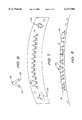
- FIG. 1 schematically illustrates that a particular antigen molecule 10 contains two monovalent antigenic epitopes, 11 and 12 respectively, each recognized by one MAb fragment.
- FIG. 2 shows the F(ab') 2 of one of such MAb fragment 14. The MAb fragment 14 will cause the formation of paired antigens 16 on the surface of T cells, as shown in FIG. 3.
- FIG. 4 schematically shows the mixture of two F(ab') (14 and 18), each recognizing one of the two antigenic epitopes 11 and 12.
- FIG. 5 shows that by using the F(ab') 2 14 and 18, a degree of cross-linking and aggregation can be achieved, but that some pairs of molecules 20 will also be linked. It can be appreciated that if three different anti-antigen MAbs are used, the cross-linking and aggregation will be even more pronounced.
- FIG. 6 shows schematically a bispecific F(ab') 2 22 with two specificities, 23 and 24, respectively being for the monovalent antigenic epitopes of CD3, 11 and 12.
- FIG. 7 shows that with this bispecific fragment 22, cross-linking and aggregation of molecules can be achieved, although some singly-paired molecules 26 are left on the T cell surface. These F(ab') 2 preparations will therefore also induce T cell activation and proliferation.
- Another means to achieve cross-linking of antigenic epitopes on T or B cell surfaces is to use plastic sheets and Sepharose 4B beads coated or conjugated with anti-CD3 MAbs. These may be implanted or deposited into certain body sites in order to trigger mitogenesis. However, as noted above, such preparations cannot be transported through the lymphoid circulation, nor can they be administered i.v.
- the appropriate backbone or base upon which to conjugate anti-CD3 MAb should be polymers which are hydrophilic, stable, non-immunogenic, nontoxic and resistant to hydrolases (e.g. glycosidases and proteases) in the serum and other body fluids in patients. Examples are PEG, cellulose, dextran, ficoll, and agarose, which each have different molecular sizes and are all well-characterized and studied.
- Another suitable "backbone” is an amino acid copolymer.
- One preferred amino acid copolymer includes Gly and Ser residues, and Lys, Cys, or another appropriate residue, for providing conjugation sites. Considering the molecular sizes of Fv, Fab, and F(ab') 2 , the optimal spacing between the adjacent Lys or Cys residues is in the range of 15 to 25 amino acids. Thus, a preferred amino acid copolymer has a composition of (Gly 15 Ser 15 Lys) n , where n is 5 to 600.
- fragments of the invention can also be conjugated to liposomes, using the methods described above, wherein reactive groups for cross-linking are introduced on the surface of the liposomes and the fragments are coupled thereto.
- fragments (or binding molecules) conjugated to liposomes may be more preferred than fragment/polymer conjugates, as the liposome conjugates can interact with antigen on T cells by a mechanism more closely resembling the interaction between cells, than when the fragment is presented on a polymer backbone.
- the fragments of the invention can also be conjugated to microbeads.
- the chemical composition of the microbeads are similar to the Sepharose 4B or Sephadex beads, although the microbeads are preferably smaller in size (preferably about 1 to 10 ⁇ m in diameter) than the Sepharose 4B or Sephadex beads typically used in chromatography.
- the Sephadex beads which also come in forms of different degrees of cross-linking of dextran molecules, have wet bead diameters of about 20-600 ⁇ m. Even the finest Sephadex beads (Sephadex G-25 SF) have wet bead diameters of 17.2-69 ⁇ m. These beads are much larger than white blood cells, which have diameters of about 10-15 ⁇ m. They are made for chromatography and readily settle after agitation.
- beads which are produced by the cross-linking of agarose or dextran and are marketed by Pharmacia LKB Biotechnology, have minimum bead diameters of about 20 ⁇ m.
- Superose 12 HR 10/30 beads have diameters of 10 ⁇ 2 ⁇ m.
- the Superose 12 beads are about the size of small, resting lymphocytes. Since blood leukocytes can be readily suspended and administered to patients, Superose 12 beads conjugated with antibodies have similar physical properties, and can also be easily suspended and withdrawn into syringes for in vivo administration. Beads of smaller sizes may be made by the same chemistry. The beads of smaller sizes may be fractionated from those of larger sizes by their sedimentation rate in a liquid suspension.
- the mitogenicity of the polymerized fragments of the invention probably depends on their sizes; more specifically, on the number of binding sites per molecular conjugate.
- the preferred molecular conjugates are those which are small but still are able to induce optimal mitogenic effects.
- Many suitable polymers are available commercially in different lengths or sizes.
- Amino acid copolymers of different lengths can also be synthesized and fractionated by molecular sieve chromatography. Polymers such as cellulose or agarose can be treated with specific enzymes, e.g., cellulase and agarase, to yield different lengths.
- FIG. 8 schematically shows that cross-linking and aggregation of surface molecules can be achieved by using a molecular conjugate 30 consisting of a polymer backbone 32 coupled with the monovalent Fab fragment 34 specific for the monovalent antigenic epitope 12 of FIG. 1.
- the molecular conjugates may also be attached to liposomes or microbeads in order to achieve maximal activation of the T cell.
- FIGS. 9 and 10 some other embodiments, schematically shown in FIGS. 9 and 10, are also included.
- a polymer backbone 40 (which can also be a microbead) is coupled with a monovalent Fab fragment 42 against antigenic epitope 12 of FIG. 1
- a polymer backbone 44 is coupled with a monovalent Fab fragment 46 against antigenic epitope 12 of FIG. 1.
- a polymer backbone 50 (which can also represent a microbead) is coupled with Fab fragments 41 and 46.
- the invention is not limited to anti-CD3 fragments, but also includes binding molecules, fragments (and conjugates thereof) which are specific for surface antigens of human T lymphocytes, and which have immunoregulatory activities in vivo, when administered according to the techniques of the invention.
- binding molecules, fragments (and conjugates thereof) which are specific for surface antigens of human T lymphocytes, and which have immunoregulatory activities in vivo, when administered according to the techniques of the invention.
- anti-CD3 many of these in vivo effects would not be predicted from the known in vitro effects or the in vivo effects with the whole antibodies.
- the desirable stimulatory effects of such products, that are prepared according to the present invention will result even though the in vivo effects of IgG specific for T cells are primarily cytolytic effects mediated by complement, ADCC, or other cytolytic mechanisms.
- anti-CD4 antibodies In addition to anti-CD3, other examples of antibodies which initiate these cytolytic effects in vivo are anti-CD4 antibodies, Alters, S. E., et al., J. Immunol. 144:4587 (1990). All of these antibodies cause T cell depletion in vivo.
- Anti-CD4 and antibodies against B cell antigen receptors have been found to have stimulatory effects in vitro. This indicates that, like anti-CD3, when formulated according to the invention, they would activate or modulate their respective target cells in vivo.
- a number of studies have indicated that the activation of T cells with an anti-CD3 MAb can be enhanced by an MAb which is specific for a different surface antigen on T cells.
- auxiliary MAbs include those specific for HLA class-I antigens, HLA class-II antigens (such as Ia), CD2, CD4, CD5, CD8, CD28, or CD37. Ceuppens, J. L. et al., J. Immunol. 137:1816 (1986); Tutt, A. et al., J.
- a fragment of anti-CD3 MAb and a fragment of an anti-CD2, anti-CD4, anti-CD5, anti-CD8, or other MAb specific for T cells is conjugated to a polymer backbone, a liposome, or a microbead.
- the polymerized or immobilized pairs of MAbs, both of which are devoid of Fc portions, can then be used to activate T cells in vivo.
- fragments of the invention which are mixtures of fragments binding to at least two different antigenic determinants
- single MAbs which bind to monovalent antigenic determinants cannot cross-link the antigens on the cell surface.
- cross-linking of the surface antigens is usually required.
- many surface molecules such as CD4 are single polypeptide chains or are composed of different polypeptide chains, and cannot be efficiently cross-linked by a single divalent antibody recognizing monovalent antigenic epitopes.
- anti-CD3 MAbs can be purchased from commercial firms offering immunochemical reagents, including Ortho Diagnostic Systems, Raritan, N.J.; Becton Dickenson Immunological Reagents, Mountain View, Calif.; Coulter Diagnostics, Hialeach, Fla.; Sigma Chemical Co., St. Louis, Mo.; Boehringer Mannheim, Indianapolis, In.; Olympus Corp., Lake Success, N.Y. All these MAbs were developed by different groups. These firms offer anti-CD3 MAb not only in purified, plain IgG, but also in fluorescein-conjugated forms.
- fluorescence flow cytometric analyses may be applied.
- a human T cell line such as CEM (ATCC CCL119 from the American Type Culture Collection), or peripheral blood mononuclear cells, can be used for the cell staining.
- the assay is to determine whether the binding of a FITC or rhodamine-labeled anti-CD3 MAb to the cells will be inhibited by the presence of varying concentrations of a second anti-CD3 MAb.
- the assay should also be reversed to determine whether the binding of the fluorescence-labeled second anti-CD3 is inhibited by the presence of the other anti-CD3.
- each anti-CD3 to the T cells is not signficantly affected by 5-10 fold concentrations of the other anti-CD3, it can be concluded that both anti-CD3 MAb can bind non-competitively to CD3 molecules on T cells. Additional confirming assays would measure whether the binding to T cells by the two MAbs is additive.
Landscapes
- Health & Medical Sciences (AREA)
- Chemical & Material Sciences (AREA)
- Immunology (AREA)
- Life Sciences & Earth Sciences (AREA)
- Medicinal Chemistry (AREA)
- General Health & Medical Sciences (AREA)
- Organic Chemistry (AREA)
- Bioinformatics & Cheminformatics (AREA)
- Engineering & Computer Science (AREA)
- Pharmacology & Pharmacy (AREA)
- Animal Behavior & Ethology (AREA)
- Public Health (AREA)
- Veterinary Medicine (AREA)
- Epidemiology (AREA)
- Genetics & Genomics (AREA)
- Molecular Biology (AREA)
- Proteomics, Peptides & Aminoacids (AREA)
- Biophysics (AREA)
- Biochemistry (AREA)
- Dispersion Chemistry (AREA)
- Cell Biology (AREA)
- Chemical Kinetics & Catalysis (AREA)
- General Chemical & Material Sciences (AREA)
- Nuclear Medicine, Radiotherapy & Molecular Imaging (AREA)
- Transplantation (AREA)
- Medicines Containing Antibodies Or Antigens For Use As Internal Diagnostic Agents (AREA)
- Medicinal Preparation (AREA)
- Medicines That Contain Protein Lipid Enzymes And Other Medicines (AREA)
Abstract
Description
Claims (5)
Priority Applications (3)
| Application Number | Priority Date | Filing Date | Title |
|---|---|---|---|
| US08/046,364 US6117982A (en) | 1991-04-19 | 1993-04-08 | Conjugates of microbeads and antibodies specific for T lymphocytes and their use as in vivo immune modulators |
| AU57292/94A AU5729294A (en) | 1992-11-25 | 1993-11-23 | Conjugates and constructs including anti-cd28 and anti-cd3 binding molecules |
| PCT/US1993/011434 WO1994012196A1 (en) | 1992-11-25 | 1993-11-23 | Conjugates and constructs including anti-cd28 and anti-cd3 binding molecules |
Applications Claiming Priority (3)
| Application Number | Priority Date | Filing Date | Title |
|---|---|---|---|
| US68800091A | 1991-04-19 | 1991-04-19 | |
| US81944992A | 1992-01-10 | 1992-01-10 | |
| US08/046,364 US6117982A (en) | 1991-04-19 | 1993-04-08 | Conjugates of microbeads and antibodies specific for T lymphocytes and their use as in vivo immune modulators |
Related Parent Applications (2)
| Application Number | Title | Priority Date | Filing Date |
|---|---|---|---|
| US68800091A Continuation-In-Part | 1991-04-19 | 1991-04-19 | |
| US81944992A Continuation | 1991-04-19 | 1992-01-10 |
Publications (1)
| Publication Number | Publication Date |
|---|---|
| US6117982A true US6117982A (en) | 2000-09-12 |
Family
ID=27104127
Family Applications (1)
| Application Number | Title | Priority Date | Filing Date |
|---|---|---|---|
| US08/046,364 Expired - Fee Related US6117982A (en) | 1991-04-19 | 1993-04-08 | Conjugates of microbeads and antibodies specific for T lymphocytes and their use as in vivo immune modulators |
Country Status (5)
| Country | Link |
|---|---|
| US (1) | US6117982A (en) |
| EP (1) | EP0516953A1 (en) |
| JP (1) | JPH0672895A (en) |
| AU (2) | AU642593B2 (en) |
| CA (1) | CA2065658A1 (en) |
Cited By (17)
| Publication number | Priority date | Publication date | Assignee | Title |
|---|---|---|---|---|
| US20040151704A1 (en) * | 2002-06-28 | 2004-08-05 | Xcyte Therapies, Inc. | Compositions and methods for restoring immune repertoire in patients with immunological defects related to autoimmunity and organ or hematopoietic stem cell transplantation |
| US20040175373A1 (en) * | 2002-06-28 | 2004-09-09 | Xcyte Therapies, Inc. | Compositions and methods for eliminating undesired subpopulations of T cells in patients with immunological defects related to autoimmunity and organ or hematopoietic stem cell transplantation |
| US20050084967A1 (en) * | 2002-06-28 | 2005-04-21 | Xcyte Therapies, Inc. | Compositions and methods for eliminating undesired subpopulations of T cells in patients with immunological defects related to autoimmunity and organ or hematopoietic stem cell transplantation |
| US20050214286A1 (en) * | 2004-01-27 | 2005-09-29 | University Of Southern California | Polymer-bound antibody cancer therapeutic agent |
| US20050233390A1 (en) * | 2003-04-09 | 2005-10-20 | Allen John W | Device including a proteinaceous factor, a recombinant proteinaceous factor, and a nucleotide sequence encoding the proteinaceous factor |
| US20070036783A1 (en) * | 2005-06-16 | 2007-02-15 | Virxsys, Corporation | Antibody complexes |
| US7541184B2 (en) | 2000-02-24 | 2009-06-02 | Invitrogen Corporation | Activation and expansion of cells |
| US7572631B2 (en) | 2000-02-24 | 2009-08-11 | Invitrogen Corporation | Activation and expansion of T cells |
| EP2711418A1 (en) * | 2012-09-25 | 2014-03-26 | Miltenyi Biotec GmbH | Method for polyclonal stimulation of T cells by flexible nanomatrices |
| US8940298B2 (en) | 2007-09-04 | 2015-01-27 | The Regents Of The University Of California | High affinity anti-prostate stem cell antigen (PSCA) antibodies for cancer targeting and detection |
| US8940871B2 (en) | 2006-03-20 | 2015-01-27 | The Regents Of The University Of California | Engineered anti-prostate stem cell antigen (PSCA) antibodies for cancer targeting |
| US9283184B2 (en) | 2008-11-24 | 2016-03-15 | Massachusetts Institute Of Technology | Methods and compositions for localized agent delivery |
| US9603944B2 (en) | 2013-09-27 | 2017-03-28 | Massachusetts Institute Of Technology | Carrier-free biologically-active protein nanostructures |
| US10294454B2 (en) | 2016-08-24 | 2019-05-21 | General Electric Company | Methods and kits for cell activation |
| US11034752B2 (en) | 2015-08-12 | 2021-06-15 | Massachusetts Institute Of Technology | Cell surface coupling of nanoparticles |
| US11472856B2 (en) | 2016-06-13 | 2022-10-18 | Torque Therapeutics, Inc. | Methods and compositions for promoting immune cell function |
| US11524033B2 (en) | 2017-09-05 | 2022-12-13 | Torque Therapeutics, Inc. | Therapeutic protein compositions and methods of making and using the same |
Families Citing this family (11)
| Publication number | Priority date | Publication date | Assignee | Title |
|---|---|---|---|---|
| US7435592B2 (en) * | 2003-05-13 | 2008-10-14 | Immunovative Therapies, Ltd. | Compositions for allogeneic cell therapy |
| ES2586295T3 (en) * | 2004-02-26 | 2016-10-13 | Immunovative Therapies, Ltd. Malcha Technology Park | Methods to prepare T cells for cell therapy |
| CA2530514C (en) * | 2004-03-01 | 2017-01-31 | Immunovative Therapies, Ltd. | Cell therapy formulation method and composition |
| EP2361930A3 (en) | 2007-03-26 | 2011-10-26 | Dako Denmark A/S | Multimers of MHC-peptide complexes and uses thereof in Borrelia infectious diseases |
| EP3023436A1 (en) | 2007-07-03 | 2016-05-25 | Dako Denmark A/S | Improved methods for generation, labeling and use of mhc multimers |
| WO2009039854A2 (en) | 2007-09-27 | 2009-04-02 | Dako Denmark A/S | Mhc multimers in tuberculosis diagnostics, vaccine and therapeutics |
| US10968269B1 (en) | 2008-02-28 | 2021-04-06 | Agilent Technologies, Inc. | MHC multimers in borrelia diagnostics and disease |
| US10722562B2 (en) | 2008-07-23 | 2020-07-28 | Immudex Aps | Combinatorial analysis and repair |
| GB0817244D0 (en) | 2008-09-20 | 2008-10-29 | Univ Cardiff | Use of a protein kinase inhibitor to detect immune cells, such as T cells |
| US10369204B2 (en) | 2008-10-02 | 2019-08-06 | Dako Denmark A/S | Molecular vaccines for infectious disease |
| JP2021515189A (en) * | 2018-02-28 | 2021-06-17 | プラス−フリー リミテッド | Extracorporeal devices and matrices for removing fibrinolytic proteins from biological fluids, methods and uses thereof |
Citations (16)
| Publication number | Priority date | Publication date | Assignee | Title |
|---|---|---|---|---|
| EP0331034A2 (en) * | 1988-02-26 | 1989-09-06 | Neorx Corporation | Functionally specific antibodies |
| US4867973A (en) * | 1984-08-31 | 1989-09-19 | Cytogen Corporation | Antibody-therapeutic agent conjugates |
| AU3242389A (en) * | 1988-04-04 | 1989-10-05 | Bristol-Myers Squibb Company | Novel antibody heteroconjugates and bispecific antibodies for use in regulation of lymphocyte activity |
| EP0340109A2 (en) * | 1988-04-28 | 1989-11-02 | The Board Of Trustees Of The Leland Stanford Junior University | Anti-T-cell receptor determinants as autoimmune disease treatment |
| WO1989012458A1 (en) * | 1988-06-14 | 1989-12-28 | Cell Med, Inc. | Heterofunctional cellular immunological reagents, vaccines containing same and methods for the use of same |
| US4925648A (en) * | 1988-07-29 | 1990-05-15 | Immunomedics, Inc. | Detection and treatment of infectious and inflammatory lesions |
| WO1990006758A1 (en) * | 1988-12-15 | 1990-06-28 | T Cell Sciences, Inc. | Monoclonal antibodies reactive with defined regions of the t cell antigen receptor |
| WO1990013316A1 (en) * | 1989-04-28 | 1990-11-15 | Baylor College Of Medicine | Dissemination of hiv-1 infected cells |
| WO1990013281A2 (en) * | 1989-04-28 | 1990-11-15 | Baylor College Of Medicine | Method of suppressing hiv infection |
| EP0417927A1 (en) * | 1989-08-24 | 1991-03-20 | Immunomedics, Inc. | Detection and treatment of infections with immunoconjugates |
| WO1991003493A1 (en) * | 1989-08-29 | 1991-03-21 | The University Of Southampton | Bi-or trispecific (fab)3 or (fab)4 conjugates |
| AU6623590A (en) * | 1989-10-20 | 1991-05-16 | Lynxvale Ltd. | Materials and methods for the treatment of foreign tissue |
| US5024940A (en) * | 1987-02-19 | 1991-06-18 | T Cell Sciences, Inc. | Nucleic acids encoding the delta chain of the T cell antigen receptor |
| WO1992006193A1 (en) * | 1990-10-05 | 1992-04-16 | Scott David Gorman | Antibodies directed against cd3 |
| WO1992007878A1 (en) * | 1990-10-26 | 1992-05-14 | The Public Health Research Institute Of The City Of New York, Inc. | Neutralizing human monoclonal antibodies specific for the v3 loop and cd-4 binding site of hiv-1 gp120 |
| WO1992013562A1 (en) * | 1991-02-11 | 1992-08-20 | The United States Of America, As Represented By The Secretary, U.S. Department Of Commerce | An immunotoxin with in vivo t cell suppressant activity |
Family Cites Families (1)
| Publication number | Priority date | Publication date | Assignee | Title |
|---|---|---|---|---|
| ZA914625B (en) * | 1990-06-22 | 1993-02-24 | Lilly Co Eli | In vivo targeting with bifunctional antibodies |
-
1992
- 1992-04-09 CA CA002065658A patent/CA2065658A1/en not_active Abandoned
- 1992-04-16 AU AU14973/92A patent/AU642593B2/en not_active Ceased
- 1992-04-16 EP EP92106666A patent/EP0516953A1/en not_active Ceased
- 1992-04-20 JP JP4126764A patent/JPH0672895A/en active Pending
-
1993
- 1993-04-08 US US08/046,364 patent/US6117982A/en not_active Expired - Fee Related
-
1994
- 1994-01-17 AU AU53815/94A patent/AU658783B2/en not_active Ceased
Patent Citations (17)
| Publication number | Priority date | Publication date | Assignee | Title |
|---|---|---|---|---|
| US4867973A (en) * | 1984-08-31 | 1989-09-19 | Cytogen Corporation | Antibody-therapeutic agent conjugates |
| US5024940A (en) * | 1987-02-19 | 1991-06-18 | T Cell Sciences, Inc. | Nucleic acids encoding the delta chain of the T cell antigen receptor |
| EP0331034A2 (en) * | 1988-02-26 | 1989-09-06 | Neorx Corporation | Functionally specific antibodies |
| AU3242389A (en) * | 1988-04-04 | 1989-10-05 | Bristol-Myers Squibb Company | Novel antibody heteroconjugates and bispecific antibodies for use in regulation of lymphocyte activity |
| EP0336379A2 (en) * | 1988-04-04 | 1989-10-11 | Oncogen Limited Partnership | Antibody heteroconjugates for use in regulation of lymphocyte activity |
| EP0340109A2 (en) * | 1988-04-28 | 1989-11-02 | The Board Of Trustees Of The Leland Stanford Junior University | Anti-T-cell receptor determinants as autoimmune disease treatment |
| WO1989012458A1 (en) * | 1988-06-14 | 1989-12-28 | Cell Med, Inc. | Heterofunctional cellular immunological reagents, vaccines containing same and methods for the use of same |
| US4925648A (en) * | 1988-07-29 | 1990-05-15 | Immunomedics, Inc. | Detection and treatment of infectious and inflammatory lesions |
| WO1990006758A1 (en) * | 1988-12-15 | 1990-06-28 | T Cell Sciences, Inc. | Monoclonal antibodies reactive with defined regions of the t cell antigen receptor |
| WO1990013316A1 (en) * | 1989-04-28 | 1990-11-15 | Baylor College Of Medicine | Dissemination of hiv-1 infected cells |
| WO1990013281A2 (en) * | 1989-04-28 | 1990-11-15 | Baylor College Of Medicine | Method of suppressing hiv infection |
| EP0417927A1 (en) * | 1989-08-24 | 1991-03-20 | Immunomedics, Inc. | Detection and treatment of infections with immunoconjugates |
| WO1991003493A1 (en) * | 1989-08-29 | 1991-03-21 | The University Of Southampton | Bi-or trispecific (fab)3 or (fab)4 conjugates |
| AU6623590A (en) * | 1989-10-20 | 1991-05-16 | Lynxvale Ltd. | Materials and methods for the treatment of foreign tissue |
| WO1992006193A1 (en) * | 1990-10-05 | 1992-04-16 | Scott David Gorman | Antibodies directed against cd3 |
| WO1992007878A1 (en) * | 1990-10-26 | 1992-05-14 | The Public Health Research Institute Of The City Of New York, Inc. | Neutralizing human monoclonal antibodies specific for the v3 loop and cd-4 binding site of hiv-1 gp120 |
| WO1992013562A1 (en) * | 1991-02-11 | 1992-08-20 | The United States Of America, As Represented By The Secretary, U.S. Department Of Commerce | An immunotoxin with in vivo t cell suppressant activity |
Non-Patent Citations (60)
| Title |
|---|
| Baniyash et al., J. Immunol. 263: 9874 (1988). * |
| Beavchamp et al., Anal. Biochem. 131: 25 (1983). * |
| Cueppens et al., J. Immunol. 137: 1816 (1986). * |
| Ellenhorn et al., J. Immunol. 144: 2840 46 (1990). * |
| Ellenhorn et al., J. Immunol. 144: 2840-46 (1990). |
| Ellenhorn et al., Science 242:569 (1988). * |
| Ellenhorn, Transplantation 50: 608 12 (1990). * |
| Ellenhorn, Transplantation 50: 608-12 (1990). |
| Furukawa, K. et al. J. Immunol. Methods 131:105 112 (1990). * |
| Furukawa, K. et al. J. Immunol. Methods 131:105-112 (1990). |
| Geisler et al., J. Immunol. 145: 1761 (1990). * |
| Geppert et al J. Immunology 138(6):1660 1666 1987. * |
| Geppert et al J. Immunology 138(6):1660-1666 1987. |
| Geppert et al., J. Immunol. 138: 1660 (1987). * |
| Geppert, T.D. et al., J. Immunol. 138: 1660 66 (1987). * |
| Geppert, T.D. et al., J. Immunol. 138: 1660-66 (1987). |
| Harlow et al. Antibodies: a laboratory manual , Cold Spring Harbor Press 1986. * |
| Harlow et al. Antibodies: a laboratory manual, Cold Spring Harbor Press 1986. |
| Harris et al Tibtech 11:42 46 1993. * |
| Harris et al Tibtech 11:42-46 1993. |
| Hirsch et al., J. Immunol. 142:737 43 (1989). * |
| Hirsch et al., J. Immunol. 142:737-43 (1989). |
| Hirsch et al., The Lancet 1390 (1989). * |
| Hirsch et al., Transplantation 47: 853 57 (1989). * |
| Hirsch et al., Transplantation 47: 853-57 (1989). |
| Hirsch et al., Transplantation 49: 1117 23 (1990). * |
| Hirsch et al., Transplantation 49: 1117-23 (1990). |
| Huang et al., J. Biol. Chem. 258:14034 40 (1983). * |
| Huang et al., J. Biol. Chem. 258:14034-40 (1983). |
| Immunology, Roitt 1985 Grower Medical Publishing 15.g 20 2. * |
| Immunology, Roitt 1985 Grower Medical Publishing 15.g 20-2. |
| Kast et al., J. Immunol. 145: 2254 (1990). * |
| Leo et al., Proc. Nat l Acad. Sci. USA 84: 1374 78 (1987). * |
| Leo et al., Proc. Nat'l Acad. Sci. USA 84: 1374-78 (1987). |
| Makinen et al JBL 264:3325 3334 1989. * |
| Makinen et al JBL 264:3325-3334 1989. |
| Makinen, et al JBL 264: 3325 3334 1989. * |
| Makinen, et al JBL 264: 3325-3334 1989. |
| Martin J.I. 136 3282 1986. * |
| Martodam et al PNAS 76: 2128 2132 1979. * |
| Martodam et al PNAS 76: 2128-2132 1979. |
| Naversina et al., Abstract From Mechanism of Tumor Rejection II #7793 (1991). |
| Naversina et al., Abstract From Mechanism of Tumor Rejection II 7793 (1991). * |
| Osband et al., Lancet 335: 994 (1990). * |
| Pastan et al., Science 254:1173 77 (1991). * |
| Pastan et al., Science 254:1173-77 (1991). |
| Richards et al. New Eng. J. Med. 323:427 28 (1990). * |
| Richards et al. New Eng. J. Med. 323:427-28 (1990). |
| Rodwell et al., Proc. Nat l Acad. Sci USA 83:2632 36 (1986). * |
| Rodwell et al., Proc. Nat'l Acad. Sci USA 83:2632-36 (1986). |
| Roitt Immunology Gower Medical Publishing 1985 p. 8.7 Fig. 8.19. * |
| Springer Nature 342: 425 434 1990. * |
| Springer Nature 342: 425-434 1990. |
| Verwilghen et al Immunology 72:269 276 1991. * |
| Verwilghen et al Immunology 72:269-276 1991. |
| Waldmann Science 252:1657 1662 1991. * |
| Waldmann Science 252:1657-1662 1991. |
| Williams et al J. Immunology 135(4):2249 2255 1985. * |
| Williams et al J. Immunology 135(4):2249-2255 1985. |
| Williams et al., J. Immunol. 135: 2249 (1985). * |
Cited By (31)
| Publication number | Priority date | Publication date | Assignee | Title |
|---|---|---|---|---|
| US7541184B2 (en) | 2000-02-24 | 2009-06-02 | Invitrogen Corporation | Activation and expansion of cells |
| US7572631B2 (en) | 2000-02-24 | 2009-08-11 | Invitrogen Corporation | Activation and expansion of T cells |
| US20040151704A1 (en) * | 2002-06-28 | 2004-08-05 | Xcyte Therapies, Inc. | Compositions and methods for restoring immune repertoire in patients with immunological defects related to autoimmunity and organ or hematopoietic stem cell transplantation |
| US20040175373A1 (en) * | 2002-06-28 | 2004-09-09 | Xcyte Therapies, Inc. | Compositions and methods for eliminating undesired subpopulations of T cells in patients with immunological defects related to autoimmunity and organ or hematopoietic stem cell transplantation |
| US20050084967A1 (en) * | 2002-06-28 | 2005-04-21 | Xcyte Therapies, Inc. | Compositions and methods for eliminating undesired subpopulations of T cells in patients with immunological defects related to autoimmunity and organ or hematopoietic stem cell transplantation |
| US8617884B2 (en) | 2002-06-28 | 2013-12-31 | Life Technologies Corporation | Methods for eliminating at least a substantial portion of a clonal antigen-specific memory T cell subpopulation |
| US9528088B2 (en) | 2002-06-28 | 2016-12-27 | Life Technologies Corporation | Methods for eliminating at least a substantial portion of a clonal antigen-specific memory T cell subpopulation |
| US20050233390A1 (en) * | 2003-04-09 | 2005-10-20 | Allen John W | Device including a proteinaceous factor, a recombinant proteinaceous factor, and a nucleotide sequence encoding the proteinaceous factor |
| US20060211065A1 (en) * | 2003-04-09 | 2006-09-21 | North Carolina A&T State University | Device including a proteinaceous factor, a recombinant proteinaceous factor, and a nucleotide sequence encoding the proteinaceous factor |
| US20090017556A1 (en) * | 2003-04-09 | 2009-01-15 | North Carolina A&T State University | Methods and complexes involving immunoglobulin molecule |
| US20050214286A1 (en) * | 2004-01-27 | 2005-09-29 | University Of Southern California | Polymer-bound antibody cancer therapeutic agent |
| US20070036783A1 (en) * | 2005-06-16 | 2007-02-15 | Virxsys, Corporation | Antibody complexes |
| US8940871B2 (en) | 2006-03-20 | 2015-01-27 | The Regents Of The University Of California | Engineered anti-prostate stem cell antigen (PSCA) antibodies for cancer targeting |
| US8940298B2 (en) | 2007-09-04 | 2015-01-27 | The Regents Of The University Of California | High affinity anti-prostate stem cell antigen (PSCA) antibodies for cancer targeting and detection |
| US9527919B2 (en) | 2007-09-04 | 2016-12-27 | The Regents Of The University Of California | High affinity anti-prostate stem cell antigen (PSCA) antibodies for cancer targeting and detection |
| US9283184B2 (en) | 2008-11-24 | 2016-03-15 | Massachusetts Institute Of Technology | Methods and compositions for localized agent delivery |
| US9393199B2 (en) | 2008-11-24 | 2016-07-19 | Massachusetts Institute Of Technology | Methods and compositions for localized agent delivery |
| WO2014048920A1 (en) * | 2012-09-25 | 2014-04-03 | Miltenyi Biotec Gmbh | Method for polyclonal stimulation of t cells by mobile nanomatrices |
| EP2711418A1 (en) * | 2012-09-25 | 2014-03-26 | Miltenyi Biotec GmbH | Method for polyclonal stimulation of T cells by flexible nanomatrices |
| US10513687B2 (en) | 2012-09-25 | 2019-12-24 | Miltenyi Biotec Gmbh | Method for polyclonal stimulation of T cells by flexible nanomatrices |
| US9603944B2 (en) | 2013-09-27 | 2017-03-28 | Massachusetts Institute Of Technology | Carrier-free biologically-active protein nanostructures |
| US10226510B2 (en) | 2013-09-27 | 2019-03-12 | Massachusetts Institute Of Technology | Carrier-free biologically-active protein nanostructures |
| US11529392B2 (en) | 2013-09-27 | 2022-12-20 | Massachusetts Institute Of Technology | Carrier-free biologically-active protein nanostructures |
| US10357544B2 (en) | 2013-09-27 | 2019-07-23 | Massachusetts Institute Of Technology | Carrier-free biologically-active protein nanostructures |
| US10588942B2 (en) | 2013-09-27 | 2020-03-17 | Massachusetts Institute Of Technology | Carrier-free biologically-active protein nanostructures |
| US11034752B2 (en) | 2015-08-12 | 2021-06-15 | Massachusetts Institute Of Technology | Cell surface coupling of nanoparticles |
| US11261226B2 (en) | 2015-08-12 | 2022-03-01 | Massachusetts Institute Of Technology (Mitn1) | Cell surface coupling of nanoparticles |
| US11472856B2 (en) | 2016-06-13 | 2022-10-18 | Torque Therapeutics, Inc. | Methods and compositions for promoting immune cell function |
| US11512288B2 (en) | 2016-08-24 | 2022-11-29 | Global Life Sciences Solutions Usa Llc | Methods and kits for cell activation |
| US10294454B2 (en) | 2016-08-24 | 2019-05-21 | General Electric Company | Methods and kits for cell activation |
| US11524033B2 (en) | 2017-09-05 | 2022-12-13 | Torque Therapeutics, Inc. | Therapeutic protein compositions and methods of making and using the same |
Also Published As
| Publication number | Publication date |
|---|---|
| EP0516953A1 (en) | 1992-12-09 |
| AU1497392A (en) | 1992-12-17 |
| CA2065658A1 (en) | 1992-10-20 |
| JPH0672895A (en) | 1994-03-15 |
| AU5381594A (en) | 1994-03-24 |
| AU642593B2 (en) | 1993-10-21 |
| AU658783B2 (en) | 1995-04-27 |
Similar Documents
| Publication | Publication Date | Title |
|---|---|---|
| US6117982A (en) | Conjugates of microbeads and antibodies specific for T lymphocytes and their use as in vivo immune modulators | |
| US6129916A (en) | Method of Increasing activation on proliferation of T cells using antibody-microbead conjugates | |
| US6197298B1 (en) | Modified binding molecules specific for T lymphocytes and their use as in vivo immune modulators in animals | |
| WO1994012196A1 (en) | Conjugates and constructs including anti-cd28 and anti-cd3 binding molecules | |
| JP2546544B2 (en) | Methods and compositions for promoting immune enhancement | |
| JP3583420B2 (en) | Targeted immunization with bispecific reagents | |
| US5872222A (en) | Conjugates of polymers and antibodies specific for T lymphocytes, and their use as adjuvants | |
| Eshhar | Tumor-specific T-bodies: towards clinical application | |
| US5601819A (en) | Bispecific antibodies for selective immune regulation and for selective immune cell binding | |
| Hornick et al. | Chimeric CLL-1 antibody fusion proteins containing granulocyte-macrophage colony-stimulating factor or interleukin-2 with specificity for B-cell malignancies exhibit enhanced effector functions while retaining tumor targeting properties | |
| JPH01501388A (en) | In vitro activation of effector cells to kill target cells | |
| JPH0198477A (en) | Igg monoclonal antibody-productive hybridmer | |
| JPH09508390A (en) | Immunostimulatory monoclonal antibody | |
| JPH05504677A (en) | Monoclonal antibody specific for IgA receptor | |
| US6106835A (en) | Modified binding molecules specific for T or B lymphocytes and their use as in vivo immune modulators | |
| JPH01501201A (en) | antibody | |
| Eisenthal et al. | Effect of combined therapy with lymphokine-activated killer cells, interleukin 2 and specific monoclonal antibody on established B16 melanoma lung metastases | |
| AU686609B2 (en) | Compositons comprising IgG3 antibodies ***** Do not seal confirmation letter to come ******* | |
| Nitta et al. | Expression of Fcγ receptors on astroglial cell lines and their role in the central nervous system | |
| Ericsson et al. | In vivo induction of γ/δ T cells with highly potent and selective anti‐tumor cytotoxicity | |
| JPH0222236A (en) | Monoclonal antibody as signal for directing sensitized effector cell to tumor region and use of conjugate thereof | |
| JPH01126558A (en) | Measurement of antibody dependant cellar cell poison | |
| Kudo et al. | Specific targeting immunotherapy of cancer with bispecific antibodies | |
| Janson | Biology of monoclonal antibodies in tumor therapy | |
| Wedrychowski et al. | Immune enhancers composed of polyvalent binding sites of anti-CD3 antibodies |
Legal Events
| Date | Code | Title | Description |
|---|---|---|---|
| FPAY | Fee payment |
Year of fee payment: 4 |
|
| AS | Assignment |
Owner name: TANOX, INC., TEXAS Free format text: ASSIGNMENT OF ASSIGNORS INTEREST;ASSIGNOR:TANOX BIOSYSTEMS, INC.;REEL/FRAME:020054/0312 Effective date: 20040802 |
|
| AS | Assignment |
Owner name: TANOX, INC., TEXAS Free format text: RE-RECORD TO CORRECT THE NATURE OF CONVEYANCE AND CORRECT ERRORNEOUSLY RECORDED SERIAL NUMBERS 60/075328, 60/083575, 60/108816, 10/908738, ETC., PREVIOUSLY RECORDED AT REEL 020054 FRAME 0312.;ASSIGNOR:TANOX BIOSYSTEMS, INC.;REEL/FRAME:020105/0437 Effective date: 19980827 |
|
| FEPP | Fee payment procedure |
Free format text: PAT HOLDER NO LONGER CLAIMS SMALL ENTITY STATUS, ENTITY STATUS SET TO UNDISCOUNTED (ORIGINAL EVENT CODE: STOL); ENTITY STATUS OF PATENT OWNER: LARGE ENTITY |
|
| AS | Assignment |
Owner name: GENENTECH, INC., CALIFORNIA Free format text: ASSIGNMENT OF ASSIGNORS INTEREST;ASSIGNOR:TANOX, INC.;REEL/FRAME:020288/0083 Effective date: 20071113 Owner name: TANOX, INC. (DE, USA), TEXAS Free format text: MERGER;ASSIGNOR:TANOX, INC. (TX, USA);REEL/FRAME:020288/0077 Effective date: 20000127 |
|
| FPAY | Fee payment |
Year of fee payment: 8 |
|
| REMI | Maintenance fee reminder mailed | ||
| LAPS | Lapse for failure to pay maintenance fees | ||
| STCH | Information on status: patent discontinuation |
Free format text: PATENT EXPIRED DUE TO NONPAYMENT OF MAINTENANCE FEES UNDER 37 CFR 1.362 |
|
| FP | Lapsed due to failure to pay maintenance fee |
Effective date: 20120912 |