US6239161B1 - Method and compositions for inhibition of adaptor protein/tyrosine kinase interactions - Google Patents
Method and compositions for inhibition of adaptor protein/tyrosine kinase interactions Download PDFInfo
- Publication number
- US6239161B1 US6239161B1 US09/565,855 US56585500A US6239161B1 US 6239161 B1 US6239161 B1 US 6239161B1 US 56585500 A US56585500 A US 56585500A US 6239161 B1 US6239161 B1 US 6239161B1
- Authority
- US
- United States
- Prior art keywords
- compound
- methylbut
- cell
- compounds
- protein
- Prior art date
- Legal status (The legal status is an assumption and is not a legal conclusion. Google has not performed a legal analysis and makes no representation as to the accuracy of the status listed.)
- Expired - Fee Related
Links
- ZDBYLTRGRUQQAW-UHFFFAOYSA-N COC1=C(C2=C(C)NC3=C2C(C)=C(C)C(C)=C3C)C(=O)C(OC)=C(/C2=C(\C)NC3=C2C(C)=C(C)C(C)=C3C)C1=O Chemical compound COC1=C(C2=C(C)NC3=C2C(C)=C(C)C(C)=C3C)C(=O)C(OC)=C(/C2=C(\C)NC3=C2C(C)=C(C)C(C)=C3C)C1=O ZDBYLTRGRUQQAW-UHFFFAOYSA-N 0.000 description 6
- 0 *c([n]c1c2c(*)c(*)c(*)c1*=C)c2C(C(C(O*)=C1c2c(*)[n]c3c2c(O*)c(*)c(*)c3*)=O)=C(*)C1=O Chemical compound *c([n]c1c2c(*)c(*)c(*)c1*=C)c2C(C(C(O*)=C1c2c(*)[n]c3c2c(O*)c(*)c(*)c3*)=O)=C(*)C1=O 0.000 description 2
- CKFUCGPLHUMJBY-UHFFFAOYSA-N [H]N1C2=C(C=CC=C2)C(C2=C(O)C(=O)C(C3=C(CC=C(C)C)N([H])C4=C3C=CC=C4)=C(O)C2=O)=C1CC=C(C)C.[H]N1C=C(C2=C(O)C(=O)C(C3=C(CC=C(C)C)N([H])C4=C3C=CC=C4)=C(O)C2=O)C2=C1C=CC=C2 Chemical compound [H]N1C2=C(C=CC=C2)C(C2=C(O)C(=O)C(C3=C(CC=C(C)C)N([H])C4=C3C=CC=C4)=C(O)C2=O)=C1CC=C(C)C.[H]N1C=C(C2=C(O)C(=O)C(C3=C(CC=C(C)C)N([H])C4=C3C=CC=C4)=C(O)C2=O)C2=C1C=CC=C2 CKFUCGPLHUMJBY-UHFFFAOYSA-N 0.000 description 2
- CMTNCWVGFLQMFM-UHFFFAOYSA-N CC.CC.COC1=C(C2=CNC3=C2C=CC=C3)C(=O)C(OC)=C(/C2=C/NC3=C2C=CC=C3)C1=O Chemical compound CC.CC.COC1=C(C2=CNC3=C2C=CC=C3)C(=O)C(OC)=C(/C2=C/NC3=C2C=CC=C3)C1=O CMTNCWVGFLQMFM-UHFFFAOYSA-N 0.000 description 1
- NNMCGEDIQKFLRV-UHFFFAOYSA-N CC.CC.COC1=C(C2=CNC3=C2C=CC=C3)C(=O)C(OC)=C(/C2=C/NC3=C2C=CC=C3)C1=O.II Chemical compound CC.CC.COC1=C(C2=CNC3=C2C=CC=C3)C(=O)C(OC)=C(/C2=C/NC3=C2C=CC=C3)C1=O.II NNMCGEDIQKFLRV-UHFFFAOYSA-N 0.000 description 1
- KYOVDNAGUYPVBT-UHFFFAOYSA-M CC.CC.COC1=C(C2=CNC3=C2C=CC=C3)C(=O)C(OC)=C(/C2=C/NC3=C2C=CC=C3)C1=O.[V]I Chemical compound CC.CC.COC1=C(C2=CNC3=C2C=CC=C3)C(=O)C(OC)=C(/C2=C/NC3=C2C=CC=C3)C1=O.[V]I KYOVDNAGUYPVBT-UHFFFAOYSA-M 0.000 description 1
Classifications
-
- A—HUMAN NECESSITIES
- A61—MEDICAL OR VETERINARY SCIENCE; HYGIENE
- A61K—PREPARATIONS FOR MEDICAL, DENTAL OR TOILETRY PURPOSES
- A61K31/00—Medicinal preparations containing organic active ingredients
- A61K31/33—Heterocyclic compounds
- A61K31/395—Heterocyclic compounds having nitrogen as a ring hetero atom, e.g. guanethidine or rifamycins
- A61K31/40—Heterocyclic compounds having nitrogen as a ring hetero atom, e.g. guanethidine or rifamycins having five-membered rings with one nitrogen as the only ring hetero atom, e.g. sulpiride, succinimide, tolmetin, buflomedil
-
- C—CHEMISTRY; METALLURGY
- C07—ORGANIC CHEMISTRY
- C07D—HETEROCYCLIC COMPOUNDS
- C07D209/00—Heterocyclic compounds containing five-membered rings, condensed with other rings, with one nitrogen atom as the only ring hetero atom
- C07D209/02—Heterocyclic compounds containing five-membered rings, condensed with other rings, with one nitrogen atom as the only ring hetero atom condensed with one carbocyclic ring
- C07D209/04—Indoles; Hydrogenated indoles
- C07D209/08—Indoles; Hydrogenated indoles with only hydrogen atoms or radicals containing only hydrogen and carbon atoms, directly attached to carbon atoms of the hetero ring
-
- A—HUMAN NECESSITIES
- A61—MEDICAL OR VETERINARY SCIENCE; HYGIENE
- A61K—PREPARATIONS FOR MEDICAL, DENTAL OR TOILETRY PURPOSES
- A61K31/00—Medicinal preparations containing organic active ingredients
- A61K31/33—Heterocyclic compounds
- A61K31/395—Heterocyclic compounds having nitrogen as a ring hetero atom, e.g. guanethidine or rifamycins
- A61K31/40—Heterocyclic compounds having nitrogen as a ring hetero atom, e.g. guanethidine or rifamycins having five-membered rings with one nitrogen as the only ring hetero atom, e.g. sulpiride, succinimide, tolmetin, buflomedil
- A61K31/403—Heterocyclic compounds having nitrogen as a ring hetero atom, e.g. guanethidine or rifamycins having five-membered rings with one nitrogen as the only ring hetero atom, e.g. sulpiride, succinimide, tolmetin, buflomedil condensed with carbocyclic rings, e.g. carbazole
- A61K31/404—Indoles, e.g. pindolol
-
- A—HUMAN NECESSITIES
- A61—MEDICAL OR VETERINARY SCIENCE; HYGIENE
- A61P—SPECIFIC THERAPEUTIC ACTIVITY OF CHEMICAL COMPOUNDS OR MEDICINAL PREPARATIONS
- A61P35/00—Antineoplastic agents
-
- C—CHEMISTRY; METALLURGY
- C07—ORGANIC CHEMISTRY
- C07D—HETEROCYCLIC COMPOUNDS
- C07D209/00—Heterocyclic compounds containing five-membered rings, condensed with other rings, with one nitrogen atom as the only ring hetero atom
- C07D209/02—Heterocyclic compounds containing five-membered rings, condensed with other rings, with one nitrogen atom as the only ring hetero atom condensed with one carbocyclic ring
- C07D209/04—Indoles; Hydrogenated indoles
- C07D209/10—Indoles; Hydrogenated indoles with substituted hydrocarbon radicals attached to carbon atoms of the hetero ring
- C07D209/14—Radicals substituted by nitrogen atoms, not forming part of a nitro radical
-
- C—CHEMISTRY; METALLURGY
- C07—ORGANIC CHEMISTRY
- C07D—HETEROCYCLIC COMPOUNDS
- C07D403/00—Heterocyclic compounds containing two or more hetero rings, having nitrogen atoms as the only ring hetero atoms, not provided for by group C07D401/00
- C07D403/02—Heterocyclic compounds containing two or more hetero rings, having nitrogen atoms as the only ring hetero atoms, not provided for by group C07D401/00 containing two hetero rings
- C07D403/10—Heterocyclic compounds containing two or more hetero rings, having nitrogen atoms as the only ring hetero atoms, not provided for by group C07D401/00 containing two hetero rings linked by a carbon chain containing aromatic rings
Definitions
- the present invention relates to methods and compositions for the inhibition of adaptor protein/phosphotyrosine interactions, especially wherein those interactions involve a protein tyrosine kinase capable of complexing with a member of the SH2 domain-containing family of adaptor proteins associated with a cell proliferative disorder.
- the present invention relates to particular organic compounds, and methods utilizing such compounds.
- extracellular molecules As a means by which to receive stimuli from their immediate environment. These extracellular signals are essential for the correct regulation of such diverse cellular processes as differentiation, contractility, secretion, cell division, contact inhibition, and metabolism.
- the extracellular molecules which can include, for example, hormones, growth factors, lymphokines, or neurotransmitters, act as ligands that bind to specific cell surface receptors. The binding of these ligands to their receptors triggers a cascade of reactions that brings about both the amplification of the original stimulus and the coordinate regulation of the separate cellular processes mentioned above.
- receptors and their extracellular ligands may be involved in abnormal or potentially deleterious processes such as virus-receptor interaction, inflammation, and cellular transformation to a cancerous state.
- a central feature of this process is the reversible phosphorylation of certain proteins.
- the phosphorylation or dephosphorylation of amino acid residues triggers conformational changes in regulated proteins that alter their biological properties. Proteins are phosphorylated by protein kinases and are dephosphorylated by protein phosphatases.
- Protein kinases and phosphatases are classified according to the amino acid residues they act on, with one class being serine-threonine kinases and phosphatases (reviewed in Scott, J. D. and Soderling, T. R., 1992, 2:289-295), which act on serine and threonine residues, and the other class being the tyrosine kinases and phosphatases (reviewed in Fischer, E. H. et al., 1991 Science 253:401-406; Schlessinger, J. and Ullrich, A., 1992, Neuron 9:383-391; Ullrich, A. and Schlessinger, J., 1990, Cell 61:203-212), which act on tyrosine residues.
- the protein kinases and phosphatases may be further defined as being receptors, i.e., the enzymes are an integral part of a transmembrane, ligand-binding molecule, or as non-receptors, meaning they respond to an extracellular molecule indirectly by being acted upon by a ligand-bound receptor.
- Phosphorylation is a dynamic process involving competing phosphorylation and dephosphorylation reactions, and the level of phosphorylation at any given instant reflects the relative activities, at that instant, of the protein kinases and phosphatases that catalyze these reactions.
- Protein tyrosine kinases comprise a large family of proteins, including many growth factor receptors and potential oncogenes, which share ancestry with, but nonetheless differ from, serine/threonine-specific protein kinases (Hanks et al., 1988, Science 241:42-52).
- Receptor-type protein tyrosine kinases having a transmembrane topology have been studied extensively. The binding of a specific ligand to the extracellular domain of a receptor protein tyrosine kinase is thought to induce receptor dimerization and phosphorylation of their own tyrosine residues. Individual phosphotyrosine residues of the cytoplasmic domains of receptors may serve as specific binding sites that interact with a host of cytoplasmic signalling molecules, thereby activating various signal transduction pathways (Ullrich, A. and Schlessinger, J., 1990, Cell 61:203-212).
- the intracellular, cytoplasmic, non-receptor protein tyrosine kinases may be broadly defined as those protein tyrosine kinases which do not contain a hydrophobic, transmembrane domain.
- cytoplasmic protein tyrosine kinases may be divided into eleven distinct morphotypes, including the SRC family (Martinez, R. et al., 1987, Science 237:411-414; Sukegawa, J. et al., 1987, Mol. Cell. Biol., 7:41-47; Yamanishi, Y. et al., 1987, 7:237-243; Marth, J. D.
- non-catalytic domains While distinct in their overall molecular structure, each of the members of these morphotypic families of cytoplasmic protein tyrosine kinases share non-catalytic domains in addition to sharing their catalytic kinase domains.
- non-catalytic domain are the SH2 (SRC homology domain 2; Sadowski, I. et al., Mol. Cell. Biol. 6: 4396-4408; Koch, C. A. et al., 1991, Science 252:668-674) domains and SH3 domains (Mayer, B. J. et al., 1988, Nature 332:269-272).
- Such non-catalytic domains are thought to be important in the regulation of protein-protein interactions during signal transduction (Pawson, T. and Gish, G., 1992, Cell 71:359-362).
- cytoplasmic protein tyrosine kinases While the metabolic roles of cytoplasmic protein tyrosine kinases are less well understood than that of the receptor-type protein tyrosine kinases, significant progress has been made in elucidating some of the processes in which this class of molecules is involved. For example, members of the src family, lck and fyn, have been shown to interact with CD4/CD8 and the T cell receptor complex, and are thus implicated in T cell activation, (Veillette, A. and Davidson, D., 1992, TIG 8:61-66), certain cytoplasmic protein tyrosine kinases have been linked to certain phases of the cell cycle (Morgan, D. O.
- Adaptor proteins are intracellular proteins having characteristic conserved peptide domains (SH2 and/or SH3 domains, as described below) which are critical to the signal transduction pathway. Such adaptor proteins serve to link protein tyrosine kinases, especially receptor-type protein tyrosine kinases to downstream intracellular signalling pathways such as the RAS signalling pathway. It is thought that such adaptor proteins may be involved in targeting signal transduction proteins to the correct site in the plasma membrane or subcellular compartments, and may also be involved in the regulation of protein movement within the cell.
- Such adaptor proteins are among the protein substrates of the receptor-type protein tyrosine kinases, and have in common one or two copies of an approximately 100 amino acid long motif. Because this motif was originally identified in c-Src-like cytoplasmic, non-receptor tyrosine kinases it is referred to as a Src homology 2 (SH2) domain.
- SH2-containing polypeptides may otherwise, however, be structurally and functionally distinct from one another (Koch, C. A. et al., 1991, Science 252:668-674).
- SH2 domains directly recognize phosphorylated tyrosine amino acid residues.
- the peptide domains also have independent sites for the recognition of amino acid residues surrounding the phosphotyrosine residue(s).
- SH3 domain In addition to SH2 peptide domains, many of the adaptor proteins involved in signal transduction contain a second conserved motif of 50-75 amino acids residues, the SH3 domain (Schlessinger, J. and Ullrich, A., 1992, Neuron 9:383-391; Pawson, T. and Gish, G. D., 1992, Cell 72:359-362; Mayer, B. J. and Baltimore, D., 1993, Trends in Cell Biol. 3 8-13; Mayer, B. J. et al., 1988, Nature 352:272-275). Much less is known about the biological role of the SH3 domain than is known about the role of SH2.
- SH3 domains function, in part, as protein-binding domains that act to link signals transmitted from the cell surface to downstream effector genes such as ras (Pawson, T. and Schlessinger, J., 1993 Current Biology, 3:434-442).
- Guanine-nucleotide-binding proteins (G-proteins; Simon, M. I. et al., 1991, Science 252:802-808; Kaziro, Y. et al., 1991, Ann. Rev. Biochem. 60:349-400)
- Ras for review, see Lowy, D. R. and Willumsen, B. M., 1993, Ann Rev. Biochem. 62:851-891
- Ras for review, see Lowy, D. R. and Willumsen, B. M., 1993, Ann Rev. Biochem. 62:851-891
- Ras the activation of receptor tyrosine kinases by ligand binding results in the accumulation of the active GTP bound form of the Ras molecule (Gibbs, J. B.
- Ras activation is also required for transformation by viral oncogenic tyrosine kinases (Smith, M. R. et al., 1986, Nature 320:540-43).
- Ras activity is regulated by the opposing actions of the GTPase-activating proteins (GAPs) and guanine nucleotide exchange factors, with GAPs stimulating the slow intrinsic rate of GTP hydrolysis on Ras and exchange factors stimulating the basal rate of exchange of GDP for GTP on Ras.
- GAPs act as negative regulators of Ras function, while exchange factors act as Ras activators.
- the GRB-2 SH2 domain binds to specific tyrosine phosphorylated sequences in receptor tyrosine kinases while the GRB-2 SH3 domains bind to proline-rich sequences present in the Sos exchange factor. Binding of GRB-2 to the receptor kinases, therefore, allows for the recruitment of Sos to the plasma membrane, where Ras is located (Schlessinger, J., 1993, TIBS 18:273-275).
- Grb2 has been shown to be associated with CSF-1 receptor (vanderGeer and Hunter, 1993, EMBO J. 12(13):5161-5172), PDGF receptor (Li et al., 1994, MCB 14(1):509-517), EGF-R (Matuoka et al., 1993, EMBO J. 12(9):3467-3475; Lowenstein et al., 1992, Cell 70:431-442) and Fak (Schlaepfer et al., 1994, Nature 372:786-791), amongst other proteins.
- Growth factors and their receptors are crucial for normal development but can also act as oncogenes leading to cell transformation, oncogenesis, and cell proliferative disorders, including cancer.
- Activation of the oncogenic potential of normal cellular proteins such as protein tyrosine kinases may, for example, occur by alteration of the proteins' corresponding enzymatic activities, their inappropriate binding to other cellular components, or both.
- BCR-ABL Philadelphia chromosome-positive human leukemias
- BCR-ABL exhibits deregulated tyrosine kinase activity. It has recently been demonstrated (Pendergast, A. M. et al., 1993, Cell 75:175-185) that BCR-ABL binds the SH2/SH3 domain-containing GRB-2 adaptor protein. Further, it has been demonstrated that BCR-ABL/GRB-2 binding is mediated by the direct interaction the GRB-2 SH2 domain and a tyrosine-phosphorylated region of the BCR-ABL protein, and that this interaction is required for the activation of the Ras signaling pathway.
- cell proliferative disorders such as the form of leukemia described above.
- One approach to the treatment of oncogenenic, cell proliferative disorders would be to attempt to “short circuit” abnormal signal transduction events which contribute to the appearance of such disorders, by interfering with one or more of these requisite events.
- the amelioration of an abnormal kinase activity may be interfered with by targeting and directly inhibiting the enzymatic activity of the kinase involved in the cell proliferative disorder. It has been proposed that certain compounds may have such anti-tyrosine kinase activity. See, for example, Levitzki and Gazit, 1995, Science 267:1782-1788, wherein certain quinazoline derivatives are proposed to directly inhibit receptor tyrosine kinase enzymatic activity.
- the inhibition of such interactions may lead to the amelioration of cell proliferative disorder symptoms.
- the utility of this approach has been demonstrated using expression of signaling incompetent proteins in cells. For example, cells expressing a mutant form of Bcr-Ab1 which lacks the tyrosine residue necessary for binding of the GrB2 SH2 domain and is thus signaling incompetent no longer exhibits a transformed phenotype (RER) (Pendergast et al., supra). To date, however, no such inhibitor of adaptor protein/protein tyrosine kinase interactions has been identified.
- the present invention relates to methods and compositions for the inhibition of adaptor protein/protein tyrosine kinase protein interactions, especially wherein those interactions involving a protein tyrosine kinase capable of complexing with a member of the SH2- and/or SH3-containing family of adaptor proteins are associated with a cell proliferative disorder.
- the present invention relates to particular organic compounds and methods utilizing such compounds.
- PTK Protein tyrosine kinase
- the compounds of the invention inhibit PTK/adaptor protein interactions, especially PTK/adaptor protein interactions wherein the PTK is, for example, an epidermal growth factor receptor (EGF-R) protein tyrosine kinase molecule, a platelet derived growth factor receptor (PDGF-R) protein tyrosine kinase molecule, or an insulin growth factor-like receptor tyrosine kinase molecule (IGF-1R).
- EGF-R epidermal growth factor receptor
- PDGF-R platelet derived growth factor receptor
- IGF-1R insulin growth factor-like receptor tyrosine kinase molecule
- R1 and R2 are independently H, acetate or aryl, alkylaryl and higher alkyl acid ester;
- R3 to R14 are independently H, alkyl, alkenyl, alkynyl, OH, hydroxyalkyl, alkoxy, nitro, halo, trihalomethyl, amide, carboxamide, carboxy, sulfonyl, sulfonamide, amino, and mercapto which can be substituted or substituted where appropriate.
- R1 and R2 of the formula can be as listed in Table I following the formula. Illustrative preparations or isolations of these compounds are found in the working examples.
- H 2-(4-fluorophenyl) 19. H 4,6-dimethoxy 20. H 5-hydroxy-6-methoxy 21. H 4-cyano 22. H 5-(4-trifluoromethylphenyl-aminocarbonyl) 23. H 2-(4-trifluoromethylphenyl-aminocarbonyl) 24. H 2-ethyl 25. H 5-bromo-6-nitro 26. OMe 2-(2-methylbut-2-en-4-yl) 27. OMe 2-(3-methyl-n-butyl)
- R1-R12 of the formula can be as listed in Table II following the formula. Illustrative preparations or isolations of these compounds are found in the working examples.
- R1 R2 R11 R12 R3-R10 1 28. H 2-(3-methyl- 2-(3-methyl- n-butyl) n-butyl) 29. H 2-methyl 2-methyl 30. H 2-ethyl 2-ethyl 31. H 2-butyl 2-butyl 32. H 2-(but-1-en- 2-(but-1-en- 4-yl) 4-yl) 33. H 2-(4-methyl- 2-(4-methyl- n-pentyl) n-pentyl) 34. H 2-phenylethyl 2-phenylethyl 35. H H 2-(3-methyl- n-butyl) 36.
- H H 2-(3-methyl- R5 carboxy n-butyl) 41.
- alkyl as used herein is meant a straight or branched chain saturated hydrocarbon group having from 1 to 20 carbons such as methyl, ethyl, isopropyl, n-butyl, s-butyl, t-butyl, n-amyl, isoamyl, n-hexyl, n-octyl and n-decyl; “alkenyl” and “alkynyl” are used to mean straight or branched chain hydrocarbon groups having from 2 to 10 carbons and unsaturated by a double or triple bond respectively, such as vinyl, allyl, propargyl, 1-methylvinyl, but-1-enyl, but-2-enyl, but-2-ynyl, 1 methylbut-2-enyl, pent-1-enyl, pent-3-enyl, 3-methylbut-1-ynyl, 1,1-dimethylallyl, hex-2-enyl and 1-methyl-1-
- substituted means that the group in question may bear one or more substituents including but not limited to halogen, hydroxy, cyano, alkyl, aryl, alkenyl, alkynyl, amino, nitro, mercapto, carboxy and other substituents known to those skilled in the art.
- Preferred compounds of the present invention include the following:
- the present invention encompasses a pharmaceutical composition comprising a compound of the invention, and methods for using a compound or pharmaceutical composition of the invention in an animal, particularly a human, to ameliorate symptoms of cell proliferative disorders involving protein tyrosine kinase/adaptor protein interactions.
- This invention is based, in part, on the discovery that the disclosed compounds, while exhibiting no inhibitory effect on protein tyrosine kinase enzymatic activity, act to inhibit the binding of an SH2-containing peptide to a tyrosine phosphorylated EGF receptor.
- the data representing this discovery is presented in the Examples in Sections 6, 7 and 8, below.
- the present invention represents the first instance whereby compounds have been discovered which directly inhibit the interaction between adaptor proteins and protein tyrosine kinase molecules.
- Described below are methods and compositions for the inhibition of adaptor protein/protein tyrosine kinase protein interactions, especially those interactions associated with a cell proliferative disorder. Specifically, described below are particular organic compounds, methods for the synthesis of such compounds, and techniques utilizing such compounds.
- R1 and R2 are independently H, acetate or aryl, alkylaryl and higher alkyl acid ester;
- R3 to R14 are independently H, alkyl, alkenyl, alkynyl, hydroxyalkyl, OH, alkoxy, nitro, halo, trihalomethyl, amide, carboxamide, carboxy, sulfonyl, sulfonamide, amino, and mercapto which can be unsubstituted or substituted where appropriate.
- alkyl groups of the compounds of the present invention may be substituted where appropriate with one or more carboxy or aryl groups.
- Alkenyl groups of compounds of the present invention may be substituted where appropriate with one or more carboxy groups.
- R1 and R2 are each independently hydrogen, lower alkyl, acetyl, aryl, alkylaryl or higher alkyl acid ester, and wherein at least one of R1 and R2 is other than hydrogen;
- R3 to R12 are each independently H, alkyl, alkylcarboxy, alkenyl, alkenylcarboxy, aryl, alkylaryl, OH, alkoxy, nitro, halo, trihalomethyl, amide, carboxamide, carboxy, sulfonyl, sulfonamide, amino, mercapto or 2-methylbut-2-en-4-yl; and wherein at least one of R11 and R12 is 2-methylbut-2-en-4-yl.
- Groups R1-R12 may be substituted or unsubstituted where appropriate.
- R1 and R2 are both H
- R3 to R10 are each independently H, alkyl, alkylcarboxy, alkenyl, alkenylcarboxy, aryl, alkylaryl, OH, alkoxy, nitro, halo, trihalomethyl, amide, carboxamide, carboxy, sulfonyl, sulfonamide, amino, mercapto or 2-methylbut-2-en-4-yl; and
- R11 and R12 are each independently H or 2-methylbut-2-en-4-yl, wherein at least one of R11 and R12 is 2-methylbut-2-en-4-yl;
- R3 to R10 is other than H.
- R1 and R2 are each independently aryl, alkylaryl and higher alkyl acid ester
- R3 to R12 are each independently H, alkyl, alkylcarboxy, alkenyl, alkenylcarboxy, aryl, alkylaryl, OH, alkoxy, nitro, fluoro, chloro, iodo, trihalomethyl, amide, carboxamide, carboxy, sulfonyl, sulfonamide, amino, or mercapto.
- R1, R2, R11 and R12 are H;
- R3 to R10 are each independently H, alkyl, alkylcarboxy, alkenyl, alkenylcarboxy, aryl, alkylaryl, alkoxy, hydroxy, nitro, halo, trihalomethyl, amide, carboxamide, carboxy, sulfonyl, sulfonamide, amino, or mercapto, wherein at least one of R3 to R10 is other than H;
- R3 when R4-R10 are each H, R3 may not be 2-methylbut-2-en-4-yl or 2-hydroxy-2-methylbut-4-yl;
- R4-R6 and R8-R10 are each H, R3 and R7 may not simultaneously be 2-methylbut-2-en-4-yl;
- R3-R4, R6-R8 and R10 are H, R5 and R9 may not simultaneously be 2-methylbut-2-en-4-yl or 3-methyl-n-butyl;
- R4 and R8 may not both be 2-methylbut-2-en-4-yl or 2-methylbut-1,4-dien-4-yl, and R4 and R8 may not be 2-methylbut-2-en-4-yl and 2-methylbut-1,4-dien-4-yl.
- the present invention also encompasses compounds of formula (III) above, and pharmaceutically acceptable salts thereof, wherein R3-R5 and R7-R9 are H and either or both of R6 and R10 are 2-methylbut-2-en-4-yl.
- R1 and R2 are acetyl
- R11 and R12 are H;
- R3 to R10 are each independently H, alkyl, alkylcarboxy, alkenyl, alkenylcarboxy, aryl, alkylaryl, OH, alkoxy, nitro, halo, trihalomethyl, amide, carboxamide, carboxy, sulfonyl, sulfonamide, amino, and mercapto, wherein:
- R1 and R2 are acetyl; or when one of R1 and R2 is acetyl and R3-R4, R6-R8 and R10-R12 are H; R5 and R9 may not simultaneously be 2-methylbut-2-en-4-yl;
- R3 and R7 may not simultaneously be 2-methylbut-2-en-4-yl
- R4 and R8 may not simultaneously be 2-methylbut-2-en-4-yl.
- R1 and R2 are lower alkyl
- R11 and R12 are H;
- R3 to R10 are each independently H, alkyl, alkylcarboxy, alkenyl, alkenylcarboxy, aryl, alkylaryl, OH, alkoxy, nitro, halo, trihalomethyl, amide, carboxamide, carboxy, sulfonyl, sulfonamide, amino, and mercapto, wherein:
- R3 when both R1 and R2 are methyl, and R4-R10 are H, R3 may not be 2-methylbut-2-en-4-yl;
- R1 and R2 when both R1 and R2 are methyl, and R3-R4, R6-R8 and R10 are H, R5 and R9 may not simultaneously be 2-methylbut-2-en-4-yl.
- the present invention also includes compounds of formula III) above, and pharmaceutically acceptable salts thereof,
- R4 is 2-methylbut-2-en-4-yl and R3 and R5-R10 are H;
- R5 is 2-methylbut-2-en-4-yl and R3-R4 and R6-R10 are H;
- R6 is 2-methylbut-2-en-4-yl, and R3-R5 and R7-R10 are H.
- R1 and R2 are each independently hydrogen, lower alkyl, acetyl, aryl, alkylaryl or higher alkyl acid ester,
- R3 to R10 are each independently H, alkyl, alkylcarboxy, aryl, alkylaryl, alkenyl, alkenylcarboxy, OH, alkoxy, nitro, halo, trihalomethyl, amide, carboxyamide, carboxy, sulfonyl, sulfonamide, amino, mercapto, 4-methylphenylsulfonylamino, or 2-methylbut-2-en-4-yl; and
- R11 and R12 are selected from the group consisting of hydrogen, methyl, ethyl, propyl, butyl, aryl, alkylaryl, alkylcarboxy, alkenylcarboxy, but-1-en-4-yl, 2-methylbut-1-en-4-yl, 4-methyl-n-pentyl, 2-phenylethyl, 2-methylpent-2-en-4-yl, and 4-carboxy-n-butyl, wherein at least one of R11 and R12 is other than hydrogen.
- R1 and R2 are each independently hydrogen, lower alkyl, acetyl, aryl, alkylaryl or higher alkyl acid ester,
- R3 to R10 are each independently H, alkyl, alkylcarboxy, aryl, alkylaryl, alkenyl, alkenylcarboxy, OH, alkoxy, nitro, halo, trihalomethyl, amide, carboxyamide, carboxy, sulfonyl, sulfonamide, amino, mercapto, 4-methylphenylsulfonylamino, or 2-methylbut-2-en-4-yl; and
- R11 and R12 are both 3-methyl-n-butyl.
- R1 and R2 are each independently hydrogen, lower alkyl, acetyl, aryl, alkylaryl or higher alkyl acid ester,
- R3 to R10 are each independently H, alkyl, alkylcarboxy, aryl, alkylaryl, alkenyl, alkenylcarboxy, OH, alkoxy, nitro, halo, trihalomethyl, amide, carboxyamide, carboxy, sulfonyl, sulfonamide, amino, mercapto, 4-methylphenylsulfonylamino, or 2-methylbut-2-en-4-yl and wherein at least one of R3 to R10 is other than hydrogen; and
- R11 and R12 are each independently hydrogen or 3-methyl-n-butyl.
- the invention encompasses the compounds described above as well as pharmaceutically acceptable salts thereof.
- the compounds of the present invention can either be synthesized or isolated as described herein.
- the compounds of the present invention can be synthesized in accordance with standard organic chemistry techniques using readily available starting materials. Alternatively, the compounds can be isolated as described in Section 5.2, below. Chemical synthesis and isolation methods are provided herein solely for illustration. Variation of these methods will be apparent to those skilled in the art.
- the present Example employed a fungus culture (PenLabs Inc. #592), and the following fermentation conditions: medium yeast malt extract plus trace elements at 22° C.
- the seed medium consisted of mannitol 60.0 g; soybean meal 12.5 g, citric acid 2.5 g, yeast extract 0.5 g, and H 2 O to 1 liter.
- the pH of the seed medium was adjusted to 7.0 before autoclaving. 30 mL seed medium were dispensed per 250 ml flask (6 days 28° C.), which was then inoculated with 1 ml of spore/mycelium homogenate suspension (2 days). Stock cultures were maintained frozen at ⁇ 80° C. in spore storage solutions.
- the fermentation mixture (mycelium and broth) was homogenized and filtered through cheesecloth by suction filtration. The filtrate was extracted three times with 0.5 v/v of ethyl acetate. The ethyl acetate layers were combined and the solvent removed by rotary evaporation. The mycelium was extracted twice with 0.4 v/v of ethyl acetate. The ethyl acetate layers were combined and the solvent removed by rotary evaporation. The oily residues both containing the asterriquinones were combined and dried on a vacuum pump overnight.
- the crude extract obtained above underwent CPC fractionation on a PC Inc. high speed countercurrent chromatograph (HSCC) containing a “tripple” coil column.
- HSCC high speed countercurrent chromatograph
- a 1:3:3:3 v/v/v/v of n-hexane, ethyl acetate, methanol and water was mixed and allowed to settle overnight.
- the lower layer was pumped into HSCC column as the stationary phase.
- the upper layer was used as the mobile phase. After two hours, the lower and upper layer were switched.
- the HSCC run was completed after four hours.
- the crude metabolites eluted from 8 to 12 minutes.
- the active fractions were pooled and evaporated under reduced pressure to dryness.
- the pooled HSCC fraction (8-12) was subjected to semi-preparative HPLC (Water HPLC system with a Water 996 photodioarray detector using Millennium software) fractionations using the following conditions:
- Mass spectra were recorded on PE Sciex LC-MS model API III (Ion Spray), exact mass measurements were performed at high resolution (HR-FAB). Mass spectral analysis for compound I gave a molecular ion of 507 (M+H) + (molecular weight: 506). The molecular formula C 32 H 31 N 2 O 4 (M + +H): 507.2289; found 507.2291). 1 H NMR spectra of compound I were recorded in CDCl 3 at 500 MHz on a Brucker DRX-500. Chemical shifts are given in ppm relative to TMS at zero ppm using the solvent peak at 7.26 ppm (CDCl 3 ) as an internal standard.
- a mixture of 100 mg. of 2,5-diacetoxy-3,6-dibromo-1,4-quinone, or other suitably protected quinone such as 3,6-dibromo-2,5-ditrimethylsiloxy-1,4-quinone, 3,6-dibromo-2,5-di-(t-butyldimethylsiloxy-1,4-quinone, 2,5-dibenzoxy-3,6-dibromo-1,4-quinone, 3,6-dibromo-2,5-diisobutryoxy-1,4-quinone, 2,5-dibenzyloxy-3,6-dibromo-1,4-quinone or 2,5-diallyoxycarbonyloxy-3,6-dibromo-1,4-quinone which can be prepared from commercially available 2,4-dibromo-3,6-dihydroxy-1,4-quinone and 180 mg of 3-[2-(2-methylbut-2-en-4-y
- 2,3,5,6-tetrabromo-1,4-quinone reacts with excess indole in the presence of potassium carbonate and aluminum oxide in dimethylformamide or dimethylsulfoxide at 100° C. to produce the substituted 2,5-dibromo-3,6-(3-indolyl)-1,4-quinone which can react with base such as sodium hydroxide to give the substituted 2,5-dihydroxy-3,6-(3-indolyl)-1,4-quinone (Hoerher, J.; Schwenner, E.; Franck, B., Liebigs Ann. Chem . 1986, 10: 1765-1771).
- Example 1 Methylation of Example 1 with methyl iodide and potassium carbonate in dimethylforamide followed by purification produced the title compound.
- This compound could also be prepared by heating 2,5-dibromo-3,6-di[2-(2-methylbut-2-en-4-yl)indol-3-yl]1,4-quinone in methanol in the presence of powdered potassium carbonate.
- Example 26 Hydrogenation of Example 26 under conditions as those in Example 3 produced the title compound.
- the reaction mixture was stirred at ⁇ 78° C. for 2 h, then quenched with water (10 ml). After warming to room temperature, water (150 ml) and 1 N HCl (1 ml) was added to neutralize the reaction mixture. The mixture was extracted with ethyl acetate (250 ml), and the organic layer was washed with brine (100 ml) and dried with sodium sulfate. The solvent was removed under reduced pressure, and the crude residue was purified by flash chromatography (4% ethyl acetate/hexane) to afford 2-(2-methyl-1-butene-4-yl)indole (664 mg. 47%) as a waxy yellow solid.
- This synthesis could be achieved by treating 2-(3-methyl-n-butyl)indole with 2 equivalents of bromanil in the presence of potassium carbonate in dimethylformamide, followed by workup and purification similar to Example 28.
- the resultant mono-indolyl adduct could then be treated with 2 equivalents of indole under the same conditions as above to provide the bis-indolyl product.
- This synthesis could start with 5-chloro-2-methylindole, which could be alkylated with methyl indole (see 28a).
- the product chloroindole could then be converted to its Grignard species and exposed to carbon dioxide to finish the synthesis.
- This synthesis could be accomplished by treating 2,5-hydroxy-3,6-di-[2-(3-methyl-n-butyl)indol-3-yl]-1,4-quinone with acetic anhydride in the presence of pyridine.
- the above compound could be synthesized by treating 5-amino-2-ethylindole with p-toluenesulfonyl chloride in the presence of triethylamine.
- the PTK/adaptor protein complexes which may be disrupted by the methods and compositions of the invention comprise at least one member of the PTK family of proteins and at least one member of the adaptor family of proteins, as described below. Under standard physiological conditions, the components of such complexes are capable of forming stable, non-covalent attachments with one or more of the other PTK/adaptor protein complex components.
- the compounds of the invention inhibit PTK/adaptor protein complexes wherein the PTK component is an epidermal growth factor receptor (EGF-R) protein tyrosine kinase molecule, a platelet derived growth factor receptor (PDGF-R) protein tyrosine kinase molecule or an insulin growth factor-like receptor tyrosine kinase molecule (IGF-1R).
- EGF-R epidermal growth factor receptor
- PDGF-R platelet derived growth factor receptor
- IGF-1R insulin growth factor-like receptor tyrosine kinase molecule
- Intracellular, cytoplasmic PTK components of the PTK/adaptor protein complexes may include, for example, members of the Src family, such molecules as src, yes, fgr, fyn, lyn, hck, lck, and blk; members of the Fes family, such as fes and fer; members of the Abl family, such as abl and arg; and members of the Jak family, such as jak1 and jak2.
- Transmembrane, receptor PTK components of the PTK/adaptor protein complexes may include, for example, such molecules as members of the FGF receptor, Sevenless/ROS, Insulin receptor, PDGF receptor, and EGF receptor family of growth factor receptors.
- the adaptor protein components of the PTK/adaptor protein complexes comprise one or more SH2 and/or one or more SH3 non-catalytic domains.
- the SH2 and SH3 domains which may be a part of the adaptor proteins are as described, above, for the PTK components.
- Adaptor proteins which may be components of the PTK/adaptor protein complexes may include, for example, p85, c-Crk, SHC, Nck, ISGF3 ⁇ , guanine triphosphatase activator protein (GAP), and members of the GRB subfamily of proteins, such as. GRB1, GRB-2, GRB-3, GRB-4, GRB-7, and GRB-10.
- the compounds and/or pharmaceutical compositions (described in Section 4.4.2, below) of the invention may be used for the treatment of cell proliferative disorders, such as oncogenic disorders, involving a PTK capable of complexing with a member of the SH2- and/or SH3-containing family of adaptor proteins.
- the compounds of the invention may be preferentially utilized in the treatment of cell proliferative disorders involving PTK/adaptor protein complexes wherein the PTK component is EGF-R, PDGF-R, MCT or IGF-1R.
- BCR-ABL-associated cancers such as, for example, chronic myelogenous and acute lymphocytic leukemias
- gliomas such as, for example, chronic myelogenous and acute lymphocytic leukemias
- gliomas such as, for example, chronic myelogenous and acute lymphocytic leukemias
- glioblastomas such as, glioblastomas
- melanoma such as, for example, chronic myelogenous and acute lymphocytic leukemias
- human ovarian cancers such as, for example, chronic myelogenous and acute lymphocytic leukemias
- human breast cancers especially HER-2/GRB-7-associated human breast cancers
- human prostate cancers such as, for example, chronic myelogenous and acute lymphocytic leukemias
- gliomas such as, for example, chronic myelogenous and acute lymphocytic leukemias
- glioblastomas
- Disruption is meant to refer not only to a physical separation of PTK/adaptor protein complex components, but is also meant to refer to a perturbation of the activity of the PTK/adaptor complexes, regardless of whether or not such complexes remain able, physically, to form.
- Activity refers to the function of the PTK/adaptor protein complex in the signal transduction cascade of the cell in which such a complex is formed, i.e., refers to the function of the complex in effecting or inhibiting the transduction of an extracellular signal into a cell.
- the compounds and pharmaceutical compositions of the invention do not, however, directly interfere with (i.e., inhibit or enhance) the enzymatic activity of the protein tyrosine kinase of interest.
- in vitro complex formation may be assayed by, first, immobilizing one component, or a functional portion thereof, of the complex of interest to a solid support.
- the immobilized complex component may be exposed to a compound such as one identified as above, and to the second component, or a functional portion thereof, of the complex of interest.
- it may be determined whether or not the second component is still capable of forming a complex with the immobilized component in the presence of the compound.
- in vivo complex formation may be assayed by utilizing co-immunoprecipitation techniques well known to those of skill in the art. Briefly, a cell line capable of forming a PTK/adaptor complex of interest may be exposed to one or more of the compounds of the invention, and a cell lysate may be prepared from this exposed cell line. An antibody raised against one of the components of the complex of interest may be added to the cell lysate, and subjected to standard immunoprecipitation techniques. In cases where a complex is still formed, the immunoprecipitation will precipitate the complex, whereas in cases where the complex has been disrupted, only the complex component to which the antibody is raised will be precipitated.
- a compound of the invention on the transformation capability of the PTK/adaptor protein of interest may be directly assayed.
- one or more of the compounds of the invention may be administered to a cell such as a fibroblast or hematopoietic cell capable of forming a PTK/adaptor complex which, in the absence of a compound of the invention, would lead to the cell's transformation (Muller, A. J. et al., 1991, Mol. Cell. Biol. 11:1785-1792; McLaughlin, J. et al., 1987, Proc. Natl. Acad. Sci. USA 84:6558-6562).
- the transformation state of the cell may then be measured in vitro, by monitoring, for example, its ability to form colonies in soft agar (Lugo and Witte, 1989, Mol. Cell. Biol. 9:1263-1270; Gishizky, M. L. and Witte, O. N., 1992, Science 256:836-839).
- a cell's transformation state may be monitored in vivo by determining its ability to form tumors in immunodeficient nude or severe combined immunodeficiency (SCID) mice (Sawyers, C. L. et al., 1992, Blood 79:2089-2098).
- SCID severe combined immunodeficiency
- the compounds of the invention may be administered to a patient at therapeutically effective doses to treat or ameliorate cell proliferative disorders involving PTK/adaptor protein interactions.
- a therapeutically effective dose refers to that amount of the compound sufficient to result in amelioration of symptoms of a cell proliferative disorder.
- Described, below, in Section 5.4.2.1 are methods for determining the effective dosage of the compounds of the invention for the treatment of cell proliferative disorders.
- Further, described, below, in Section 5.4.2.2 are methods for formulations and pharmaceutical compositions comprising the compounds of the invention, and methods for the administration of such compounds, formulations, and compositions.
- Toxicity and therapeutic efficacy of the compounds of the invention can be determined by standard pharmaceutical procedures in cell cultures or experimental animals, e.g., for determining the LD 50 (the dose lethal to 50% of the population) and the ED 50 (the dose therapeutically effective in 50% of the population).
- the dose ratio between toxic and therapeutic effects is the therapeutic index and it can be expressed as the ratio LD 50 /ED 50 .
- Compounds which exhibit large therapeutic indices are preferred. While compounds that exhibit toxic side effects may be used, care should be taken to design a delivery system that targets such compounds to the site of affected tissue in order to minimize potential damage to uninfected cells and, thereby, reduce side effects.
- the data obtained from the cell culture assays and animal studies can be used in formulating a range of dosage for use in humans.
- the dosage of such compounds lies preferably within a range of circulating concentrations that include the ED 50 with little or no toxicity.
- the dosage may vary within this range depending upon the dosage form employed and the route of administration utilized.
- the therapeutically effective dose can be estimated initially from cell culture assays.
- a dose may be formulated in animal models to achieve a circulating plasma concentration range that includes the IC 50 (i.e., the concentration of the test compound which achieves a half-maximal inhibition of symptoms) as determined in cell culture.
- IC 50 i.e., the concentration of the test compound which achieves a half-maximal inhibition of symptoms
- levels in plasma may be measured, for example, by high performance liquid chromatography.
- Dosage amount and interval may be adjusted individually to provide plasma levels of the active moiety which are sufficient to maintain inhibition of adaptor protein/protein tyrosine kinase interactions, or minimal effective concentration (MEC).
- MEC minimal effective concentration
- the MEC will vary for each compound but can be estimated from in vitro data, e.g., the interactions using the assays described herein. Dosages necessary to achieve the MEC will depend on individual characteristics and route of administration. However, HPLC assays or bioassays can be used to determine plasma concentrations.
- Dosage intervals can also be determined using the MEC value.
- Compounds should be administered using a regimen which maintains plasma levels above the MEC for 10-90% of the time, preferably between 30-90% and most preferably between 50-90%.
- adaptor proteins are intracellular proteins.
- PTK/adaptor protein interactions are intracellular, regardless of whether the PTK of interest is of the transmembrane or the intracellular type. Therefore, the compounds of the invention act intracellularly to interfere with the formation and/or activity of the PTK/adaptor complexes.
- a variety of methods are known to those of skill in the art for administration of compounds which act intracellularly, as, for example, discussed in this Section.
- compositions for use in accordance with the compounds of the present invention may be formulated in conventional manner using one or more physiologically acceptable carriers or excipients.
- the compounds and their physiologically acceptable salts and solvates may be formulated for administration by inhalation or insufflation (either through the mouth or the nose) or oral, buccal, parenteral or rectal administration.
- the pharmaceutical compositions may take the form of, for example, tablets or capsules prepared by conventional means with pharmaceutically acceptable excipients such as binding agents (e.g., pregelatinised maize starch, polyvinylpyrrolidone or hydroxypropyl methylcellulose); fillers (e.g., lactose, microcrystalline cellulose or calcium hydrogen phosphate); lubricants (e.g., magnesium stearate, talc or silica); disintegrants (e.g., potato starch or sodium starch glycolate); or wetting agents (e.g., sodium lauryl sulphate).
- binding agents e.g., pregelatinised maize starch, polyvinylpyrrolidone or hydroxypropyl methylcellulose
- fillers e.g., lactose, microcrystalline cellulose or calcium hydrogen phosphate
- lubricants e.g., magnesium stearate, talc or silica
- disintegrants e.g., potato starch
- Liquid preparations for oral administration may take the form of, for example, solutions, syrups or suspensions, or they may be presented as a dry product for constitution with water or other suitable vehicle before use.
- Such liquid preparations may be prepared by conventional means with pharmaceutically acceptable additives such as suspending agents (e.g., sorbitol syrup, cellulose derivatives or hydrogenated edible fats); emulsifying agents (e.g., lecithin or acacia); non-aqueous vehicles (e.g., almond oil, oily esters, ethyl alcohol or fractionated vegetable oils); and preservatives (e.g., methyl or propyl-p-hydroxybenzoates or sorbic acid).
- the preparations may also contain buffer salts, flavoring, coloring and sweetening agents as appropriate.
- Preparations for oral administration may be suitably formulated to give controlled release of the active compound.
- compositions may take the form of tablets or lozenges formulated in conventional manner.
- the compounds for use according to the present invention are conveniently delivered in the form of an aerosol spray presentation from pressurized packs or a nebuliser, with the use of a suitable propellant, e.g., dichlorodifluoromethane, trichlorofluoromethane, dichlorotetrafluoroethane, carbon dioxide or other suitable gas.
- a suitable propellant e.g., dichlorodifluoromethane, trichlorofluoromethane, dichlorotetrafluoroethane, carbon dioxide or other suitable gas.
- the dosage unit may be determined by providing a valve to deliver a metered amount.
- Capsules and cartridges of e.g. gelatin for use in an inhaler or insufflator may be formulated containing a powder mix of the compound and a suitable powder base such as lactose or starch.
- the compounds may be formulated for parenteral administration by injection, e.g., by bolus injection or continuous infusion.
- Formulations for injection may be presented in unit dosage form, e.g., in ampoules or in multidose containers, with an added preservative.
- the compositions may take such forms as suspensions, solutions or emulsions in oily or aqueous vehicles, and may contain formulatory agents such as suspending, stabilizing and/or dispersing agents.
- the active ingredient may be in powder form for constitution with a suitable vehicle, e.g., sterile pyrogen-free water, before use.
- the compounds may also be formulated in rectal compositions such as suppositories or retention enemas, e.g., containing conventional suppository bases such as cocoa butter or other glycerides.
- the compounds may also be formulated as a depot preparation. Such long acting formulations may be administered by implantation (for example subcutaneously or intramuscularly) or by intramuscular injection.
- the compounds may be formulated with suitable polymeric or hydrophobic materials (for example as an emulsion in an acceptable oil) or ion exchange resins, or as sparingly soluble derivatives, for example, as a sparingly soluble salt.
- compositions may, if desired, be presented in a pack or dispenser device which may contain one or more unit dosage forms containing the active ingredient.
- the pack may for example comprise metal or plastic foil, such as a blister pack.
- the pack or dispenser device may be accompanied by instructions for administration.
- the adaptor-GST (glutathione-S-transferase) fusion proteins used herein were GRB-2-GST fusion proteins prepared by expression in E. coli transformed with GRB-2/pGEX constructs.
- the GRB-2 portions of these fusion proteins consisted of only the SH2 domain of the GRB-2 protein.
- Transformed cells are grown in Luria broth (LB) supplemented with ampicillin. After reaching an optical density (OD) at 600 nm of 0.3, the cells are induced for 6 hours with isopropyl ⁇ -D-thiogalactopyranoside (IPTG) in order to express the fusion protein.
- LB Luria broth
- IPTG isopropyl ⁇ -D-thiogalactopyranoside
- the cells are precipitated, pelleted at 10,000 ⁇ g for 10 minutes at 4° C., washed, and resuspended in phosphate buffered saline (PBS).
- PBS phosphate buffered saline
- the cells are lysed by sonication (6 strokes, 5 seconds per stroke). Insoluble material is removed by centrifugation at 10,000 ⁇ g for 10 minutes at 4° C., and the supernatant is passed over a Glutathion-Sepharose column.
- Bound GRB-2-GST fusion protein is eluted off the column with 5 mM reduced glutathion, then dialyzed against PBS.
- EGF-R Epidermal growth factor receptor tyrosine kinase
- EGF-R was isolated from cells overexpressing EGF-R, specifically, the A431 (ATCC CRL 1551), cell line. The cells are lysed in HNTG buffer (20 mM Hepes/HCl, pH 7.4, 150 mM NaCl, 1.0% Triton X-100, 5% glycerol, 1 mM phenylmethylsulfonyl fluoride (PMSF), 1 mg/L aprotonin, 1 mg/L leupeptin, 10 mg/L benzamidine). EGF-R protein was isolated from the cell lysates by immobilization onto microtiter plates, as described below. EGF-R was subsequently phosphorylated in vitro, as explained below.
- HNTG buffer 20 mM Hepes/HCl, pH 7.4, 150 mM NaCl, 1.0% Triton X-100, 5% glycerol, 1 mM
- the EGF-R molecule was immobilized onto microtiter plates.
- Microtiter plates were prepared by first coating the wells of the plate, overnight at 4° C., with an anti-EGF-R monoclonal antibody directed against the extracellular domain of EGFR (UBI, #05-101) at a concentration of 0.5 ⁇ g (in PBS) per microtiter well, at a final volume of 150 ⁇ l per well.
- the coating solution was removed from the microtiter wells, and replaced with blocking buffer (5% dry milk in PBS) for 30 minutes at room temperature, after which the blocking buffer is removed and the wells were washed 4 times with TBST buffer (150 mM NaCl, 50 mM Tris-HCl, pH 7.2, 0.1% Triton X-100).
- blocking buffer 5% dry milk in PBS
- EGF-R-expressing cells were added to each well, in 150 ⁇ l of PBS, incubated 30 minutes at room temperature, with shaking. Unbound EGF-R was removed by washing wells 5 times with TBST buffer. Approximately 50-100 ng of EGF-R protein was bound per well.
- EGF-R overexpressing cell line which exhibits a high endogenous phosphatase activity, such as the A431 cell line used herein. This is because during lysis and incubation with the immobilized antibody, the phosphatases remove phosphate groups from the EGF-R molecules, thus prohibiting endogenous adaptor proteins, such as GRB proteins, to bind EGFR, which could potentially lead to artifactual results.
- cells may be starved before lysis, if the cell line utilized may be readily starved.
- GRB-2-GST fusion proteins i.e. a 1:1 ratio of EGF-R:GRB-2 proteins
- 5 ng GRB-2-GST fusion proteins i.e. a 4:1 ratio of EGF-R:GRB-2 proteins
- incubation buffer 0.1 M potassium phosphate buffer, 15 pH 6.5
- GRB-2-GST fusion protein bound to the immobilized EGF-R is then preferably determined with a purified rabbit antiserum against the GST-moiety of the fusion protein (AMRAD, New Victoria, Australia; Catalog No. 00001605). Incubations were for 30 minutes at room temperature. After incubation, antibody was removed and the wells are washed extensively with TBST. For visualization, wells were next incubated with a TAGO goat-anti-rabbit peroxidase antibody at room temperature for 30 minutes. After incubation, the antibody was removed, the wells were washed with tap water, and then with TBST.
- biotinylated monoclonal antibodies e.g., EL-6 or EL-12
- EL-6 or EL-12 may be utilized to assay fusion protein binding.
- the epitopes recognized by such antibodies map on the SH2 domain of GRB-2, but do not interfere with GRB-2 binding to phosphorylated EGFR. Binding of these antibodies is then determined by using a streptavidin-biotinylated horseradish peroxidase reactant.
- binding of the fusion protein to the immobilized EGFR may be assayed by incubating with 1 mM 1-chloro-2,4 dinitrobenzene (CDNB) and 1.54 mg/ml reduced glutathion in incubation buffer. The OD is then measured at 340 nm. This reaction is linear up to OD 1.0, and can be stopped with competitive GST inhibitors, as described in Mannervik and Danielson (Mannervik, B. and Danielson, U. H., 1988, CRC Critical Reviews in Biochemistry 23:238).
- CDNB 1-chloro-2,4 dinitrobenzene
- Compound I was tested for its ability to inhibit the binding of tyrosine phosphorylated EGF-receptor to an SH2 peptide domain of the GRB-2 adaptor protein, according to the assays described, above, in Section 5.1.
- Compound I proves to be a potent inhibitor of GRB-2/SH2 binding, having an IC 50 of 2.9 ⁇ M. (IC 50 , as used herein, return to the concentration of test compound required to inhibit one-half of GRB-2/SH2 binding relative to the amount of binding which occurs in the absence of test compound.)
- Example presented herein demonstrates that compounds of the invention inhibits cell survival in a bcr/abl-transformed cell line.
- 32D cl.3 murine lymphoblastoid cell, IL-3 dependent.
- 32D bcr/abl 32D over expressing bcr/abl kinase, pooled, IL-3 independent.
- 32D cl.3 RPM1+10% FBS+1 ng/ml IL-3+2 mM Glutamine.
- 32D bcr/abl RPM1+10% FBS+2 mM Glutamine.
- IL-3 Interleukin-3, mouse (UBI Cat. #01-374)
- tissue culture dish (10 cm, Corning 25020-100) to about 1 ⁇ 10 6 cell/ml, subculture every 2-3 days at 1:10 (1:20 for 32D bcr/abl line).
- 32D cl.3 seeding medium should contain 2 ng/ml IL-3.
- Compound I drug stock (10 mM in DMSO) was diluted 1:50. 1:2 serial dilutions were performed for the remaining 8 wells in each line of the tissue culture plate. 50 ⁇ l were added to each well. Control wells received medium alone. Cells were incubated with drugs in 5% CO 2 at 37° for 15 hrs.
- Compound I was tested herein for its ability to affect bcr/abl activity, and was found to be an inhibitor of bcr/abl function.
- Compound I on bcr/abl function was tested using the cell growth assay described, above, in Section 6.1. Briefly, three cell lines were used in this assay. First, an IL-3 dependent cell line (32D cl.3) was used, which requires the presence of the IL-3 cytokine for survival. Next, two IL-3 independent cell lines were used, including 32D cl.3 J2/leuk, which consists of the 32D cl.3 cell line transformed with raf and myc, and 32D bcr/abl, which consists of the 32D cl.3 cell line transformed with bcr/abl.
- 32D cl.3 J2/leuk which consists of the 32D cl.3 cell line transformed with raf and myc
- 32D bcr/abl which consists of the 32D cl.3 cell line transformed with bcr/abl.
- Compound I inhibits the ability of the 32D cl.3 bcr/abl cell line to survive in the absence of IL-3. This result is significant as this cell line is quite robust.
- Example presented herein demonstrates that Compound I of the invention is a potent inhibitor of cellular proliferation.
- MCF-7SRB Growth Assay MCF-7 (ATCC# HTB 22) cells (H+B22) were seeded at 2000 cells/well in a 96-well flat bottom plate in normal growth media, which was 10% FBS/RPMI supplemented with 2 mM Glutamine. The plate of cells was incubated for about 24 hours at 37° C. after which it received an equal volume of compound dilution per well making the total volume per well 200 ⁇ l. The compound was prepared at 2 times the desired highest final concentration and serially diluted in the normal growth media in a 96-well round bottom plate and then transferred to plate of cells. DMSO serves as the vector control up to 0.2% as final concentration. The cells were then incubated at 37° C.
- Assay 2 PDGF-R/SRB Adherent Cells Growth Assay. Compounds were tested for inhibition of anchorage-dependent tumor cell growth using the calorimetric assay described by Skehan et al., 1990 . J. Natl. Cancer Inst . 82:1107-1112. The assay measures protein content of acid-fixed cells using the counterion binding dye sulforhodamine B (SRB, Sigma). The compounds were solubilized in DMSO (Sigma, cell culture grade) and diluted into appropriate growth medium at two-fold the desired final assay concentration.
- SRB counterion binding dye sulforhodamine B
- C6 cells In assays using C6 cells (CCL 107), compounds (100 ⁇ l) were added to 96-well plates containing attached cellular monolayers (2000 cells/well in 100 ⁇ l).
- C6 (ATCC# CCL 107) cells were maintained in Ham's F10 supplemented with 5% fetal bovine serum (FBS) and 2 mM glutamine (GLN). After 4 days (37° C., 5% CO 2 ) the monolayers were washed 3 times with PBS and fixed with 200 ⁇ l ice-cold 10% TCA (Fisher Scientific), and kept at 4° C. for 60 min. The TCA was removed and the fixed monolayers were washed 5 times with tap water and allowed to dry completely at room temperature on absorbent paper.
- FBS fetal bovine serum
- GLN 2 mM glutamine
- the cellular protein was stained for 10 min with 100 ⁇ l 0.4% SRB dissolved in 1% acetic acid. After 5 washes with tap water, the dye was solubilized in 10 mM Tris base (100 ⁇ l per well) and absorbance read at 570 nm on a Dynatech plate reader model MR5000. Growth inhibition data were expressed as a percentage of absorbance detected in control wells which were treated with 0.4% DMSO alone. DMSO controls were not different from cells grown in regular growth medium. IC 50 values were determined using a four parameter curve fit function.
- Assay 3 MCF-7/HER-2B Growth Assay.
- the protocol used herein is essentially similar to that described above (for the MCF-7 Growth Assay) except that immediately before Compound I was added, the normal growth media was removed and 0.5% FBS/RPMI supplemented with 2 mM Glutamine is added onto the cells. The compound was also prepared in this 0.5% serum media. The plate of cells was incubated for four days and developed as per standard techniques.
- Assay 4 A431/SRB Growth Assay.
- A431 ATCC# CRL 1555 cells were tested essentially according to the protocol described, above, for the MCF-7/HER-2B growth assay.
- Compound I proved to be a potent inhibitor of cells proliferation of each of the four cell lines tested.
- IC50 refers to the concentration of test compound required to inhibit cell proliferation to 50% of the level seen in the same cell line which has not been contacted to test compound (in this case, Compound I).
- the following protocol describes the procedures used to determine the ability of the compounds to inhibit cellular proliferation in 3T3 engineered cell lines that over expressing EGFr, IGF1r, or PDGFr.
- IGF1 Ligand human, recombinant; G511, Promega Corp, USA.
- PDGF Ligand human PDGF B/B; 1276-956, Boehringer Mannheim, Germany.
- TCA Buffer 10% trichloroacetic acid (A32-500, Fisher Scientific, USA).
- Tris Base Buffer 10 mM tris base (BP152-5, Fisher Scientific, USA).
- N-1H 3T3 (ATCC# 1658) engineered cell liens: 3T3-EGFr, 3T3-IGF1r, 3T3-PDGFr.
- the ligands are prepared in the serum free DMEM with 0.1% bovine albumin.
- the negative control cells receive the serum free DMEM with 0.1% bovine albumin only; the positive control cells receive the ligands (EGF, IGF1, or PDGF) but no drugs.
- the drugs are prepared in the serum free DMEM in a 96 well plate, and a serial dilution is taken the place. A total of 10 ⁇ l/well medium of the diluted drugs are added into the cells. The total volume of each well is 200 ⁇ L. Quadruplicates (wells) and 11 concentration points are applied to each drug tested.
Abstract
The present invention relates to methods and compositions for the inhibition of adaptor protein/protein tyrosine kinase protein interactions, especially wherein those interactions involving a protein tyrosine kinase capable of complexing with a member of the SH2-and/or SH3-containing family of adaptor proteins are associated with a cell proliferative disorder. Specifically, the present invention relates to particular compounds, especially quinazoline derivative compounds, and methods utilizing such compounds.
Description
This is a division, of application Ser. No. 09/090,737, filed Jun. 4, 1998, now U.S. Pat. No. 6,090,838, which is a continuation of U.S. Ser. No. 08/658,337, filed on Jun. 5, 1996, now U.S. Pat. No. 5,780,496, which is a continuation in part of U.S. Ser. No. 08/476,136, filed on Jun. 7, 1995, now abandoned.
The present invention relates to methods and compositions for the inhibition of adaptor protein/phosphotyrosine interactions, especially wherein those interactions involve a protein tyrosine kinase capable of complexing with a member of the SH2 domain-containing family of adaptor proteins associated with a cell proliferative disorder. Specifically, the present invention relates to particular organic compounds, and methods utilizing such compounds.
2.1 PROTEIN PHOSPHORYLATION AND SIGNAL TRANSDUCTION
Cells rely, to a great extent, on extracellular molecules as a means by which to receive stimuli from their immediate environment. These extracellular signals are essential for the correct regulation of such diverse cellular processes as differentiation, contractility, secretion, cell division, contact inhibition, and metabolism. The extracellular molecules, which can include, for example, hormones, growth factors, lymphokines, or neurotransmitters, act as ligands that bind to specific cell surface receptors. The binding of these ligands to their receptors triggers a cascade of reactions that brings about both the amplification of the original stimulus and the coordinate regulation of the separate cellular processes mentioned above. In addition to normal cellular processes, receptors and their extracellular ligands may be involved in abnormal or potentially deleterious processes such as virus-receptor interaction, inflammation, and cellular transformation to a cancerous state.
A central feature of this process, referred to as signal transduction (for recent reviews, see Posada, J. and Cooper, J. A., 1992, Mol. Biol. Cell 3:583-592; Hardie, D. G., 1990, Symp. Soc. Exp. Biol. 44:241-255), is the reversible phosphorylation of certain proteins. The phosphorylation or dephosphorylation of amino acid residues triggers conformational changes in regulated proteins that alter their biological properties. Proteins are phosphorylated by protein kinases and are dephosphorylated by protein phosphatases. Protein kinases and phosphatases are classified according to the amino acid residues they act on, with one class being serine-threonine kinases and phosphatases (reviewed in Scott, J. D. and Soderling, T. R., 1992, 2:289-295), which act on serine and threonine residues, and the other class being the tyrosine kinases and phosphatases (reviewed in Fischer, E. H. et al., 1991 Science 253:401-406; Schlessinger, J. and Ullrich, A., 1992, Neuron 9:383-391; Ullrich, A. and Schlessinger, J., 1990, Cell 61:203-212), which act on tyrosine residues. The protein kinases and phosphatases may be further defined as being receptors, i.e., the enzymes are an integral part of a transmembrane, ligand-binding molecule, or as non-receptors, meaning they respond to an extracellular molecule indirectly by being acted upon by a ligand-bound receptor. Phosphorylation is a dynamic process involving competing phosphorylation and dephosphorylation reactions, and the level of phosphorylation at any given instant reflects the relative activities, at that instant, of the protein kinases and phosphatases that catalyze these reactions.
While the majority of protein phosphorylation occurs at serine and threonine amino acid residues, phosphorylation at tyrosine residues also occurs, and has begun to attract a great deal of interest since the discovery that many oncogene products and growth factor receptors possess intrinsic protein tyrosine kinase activity. The importance of protein tyrosine phosphorylation in growth factor signal transduction, cell cycle progression and neoplastic transformation is now well established (Cantley, L. C. et al., 1991, Cell 64:281-302; Hunter T., 1991, Cell 64:249-270; Nurse, 1990, Nature 344:503-508; Schlessinger, J. and Ullrich, A., 1992, Neuron 9:383-391; Ullrich, A. and Schlessinger, J., 1990, Cell 61:203-212). Subversion of normal growth control pathways leading to oncogenesis has been shown to be caused by activation or overexpression of protein tyrosine kinases which constitute a large group of dominant oncogenic proteins (reviewed in Hunter, T., 1991, Cell 64:249-270).
2.2 PROTEIN TYROSINE KINASES
Protein tyrosine kinases comprise a large family of proteins, including many growth factor receptors and potential oncogenes, which share ancestry with, but nonetheless differ from, serine/threonine-specific protein kinases (Hanks et al., 1988, Science 241:42-52).
Receptor-type protein tyrosine kinases having a transmembrane topology have been studied extensively. The binding of a specific ligand to the extracellular domain of a receptor protein tyrosine kinase is thought to induce receptor dimerization and phosphorylation of their own tyrosine residues. Individual phosphotyrosine residues of the cytoplasmic domains of receptors may serve as specific binding sites that interact with a host of cytoplasmic signalling molecules, thereby activating various signal transduction pathways (Ullrich, A. and Schlessinger, J., 1990, Cell 61:203-212).
The intracellular, cytoplasmic, non-receptor protein tyrosine kinases, may be broadly defined as those protein tyrosine kinases which do not contain a hydrophobic, transmembrane domain. Within this broad classification, one can divide the known cytoplasmic protein tyrosine kinases into eleven distinct morphotypes, including the SRC family (Martinez, R. et al., 1987, Science 237:411-414; Sukegawa, J. et al., 1987, Mol. Cell. Biol., 7:41-47; Yamanishi, Y. et al., 1987, 7:237-243; Marth, J. D. et al., 1985, Cell 43:393-404; Dymecki, S.M. et al., 1990, Science 247:332-336), the FES family (Ruebroek, A. J. M. et al., 1985, EMBO J. 4:2897-2903; Hao, Q. et al., 1989, Mol. Cell. Biol. 9:1587-1593), the ABL family (Shtivelman, E. et al., 1986, Cell 47:277-284; Kruh, G. D. et al., 1986, Science 234:1545-1548), the Zap 70 family and the JAK family. While distinct in their overall molecular structure, each of the members of these morphotypic families of cytoplasmic protein tyrosine kinases share non-catalytic domains in addition to sharing their catalytic kinase domains. Such non-catalytic domain are the SH2 (SRC homology domain 2; Sadowski, I. et al., Mol. Cell. Biol. 6: 4396-4408; Koch, C. A. et al., 1991, Science 252:668-674) domains and SH3 domains (Mayer, B. J. et al., 1988, Nature 332:269-272). Such non-catalytic domains are thought to be important in the regulation of protein-protein interactions during signal transduction (Pawson, T. and Gish, G., 1992, Cell 71:359-362).
While the metabolic roles of cytoplasmic protein tyrosine kinases are less well understood than that of the receptor-type protein tyrosine kinases, significant progress has been made in elucidating some of the processes in which this class of molecules is involved. For example, members of the src family, lck and fyn, have been shown to interact with CD4/CD8 and the T cell receptor complex, and are thus implicated in T cell activation, (Veillette, A. and Davidson, D., 1992, TIG 8:61-66), certain cytoplasmic protein tyrosine kinases have been linked to certain phases of the cell cycle (Morgan, D. O. et al., 1989, Cell 57: 775-786; Kipreos, E. T. et al., 1990, Science 248:. 217-220; Weaver et al., 1991, Mol. Cell. Biol. 11:4415-4422), and cytoplasmic protein tyrosine kinases have been implicated in neuronal development (Maness, P., 1992, Dev. Neurosci 14:257-270). Deregulation of kinase activity through mutation or overexpression is a well-established mechanism underlying cell transformation (Hunter et al., 1985, supra; Ullrich et al., supra).
2.3 ADAPTOR PROTEINS
Adaptor proteins are intracellular proteins having characteristic conserved peptide domains (SH2 and/or SH3 domains, as described below) which are critical to the signal transduction pathway. Such adaptor proteins serve to link protein tyrosine kinases, especially receptor-type protein tyrosine kinases to downstream intracellular signalling pathways such as the RAS signalling pathway. It is thought that such adaptor proteins may be involved in targeting signal transduction proteins to the correct site in the plasma membrane or subcellular compartments, and may also be involved in the regulation of protein movement within the cell.
Such adaptor proteins are among the protein substrates of the receptor-type protein tyrosine kinases, and have in common one or two copies of an approximately 100 amino acid long motif. Because this motif was originally identified in c-Src-like cytoplasmic, non-receptor tyrosine kinases it is referred to as a Src homology 2 (SH2) domain. SH2-containing polypeptides may otherwise, however, be structurally and functionally distinct from one another (Koch, C. A. et al., 1991, Science 252:668-674). SH2 domains directly recognize phosphorylated tyrosine amino acid residues. The peptide domains also have independent sites for the recognition of amino acid residues surrounding the phosphotyrosine residue(s).
When a receptor protein tyrosine kinase binds an extracellular ligand, receptor dimerization is induced, which, in turn, leads to intermolecular autophosphorylation of the dimerized kinases (Schlessinger, J. and Ullrich, A., 1992, Neuron 9: 383-391). Receptor phosphorylation, therefore, creates SH2-binding sites, to which an adaptor protein may bind.
In addition to SH2 peptide domains, many of the adaptor proteins involved in signal transduction contain a second conserved motif of 50-75 amino acids residues, the SH3 domain (Schlessinger, J. and Ullrich, A., 1992, Neuron 9:383-391; Pawson, T. and Gish, G. D., 1992, Cell 72:359-362; Mayer, B. J. and Baltimore, D., 1993, Trends in Cell Biol. 3 8-13; Mayer, B. J. et al., 1988, Nature 352:272-275). Much less is known about the biological role of the SH3 domain than is known about the role of SH2. The current view is that SH3 domains function, in part, as protein-binding domains that act to link signals transmitted from the cell surface to downstream effector genes such as ras (Pawson, T. and Schlessinger, J., 1993 Current Biology, 3:434-442).
2.4 G-PROTEINS AND SIGNAL TRANSDUCTION
Guanine-nucleotide-binding proteins, (G-proteins; Simon, M. I. et al., 1991, Science 252:802-808; Kaziro, Y. et al., 1991, Ann. Rev. Biochem. 60:349-400) such as Ras (for review, see Lowy, D. R. and Willumsen, B. M., 1993, Ann Rev. Biochem. 62:851-891), play an essential role in the transmission of mitogenic signals from receptor tyrosine kinases. Taking Ras as an example, the activation of receptor tyrosine kinases by ligand binding results in the accumulation of the active GTP bound form of the Ras molecule (Gibbs, J. B. et al., 1990, J. Biol. Chem. 265:20437-2044; Satoh, T. et al., 1990, Proc. NaTl. Acad. Sci. USA 87:5993-5997; Li, B.-Q. et al., 1992, Science 256:1456-1459; Buday, L. and Downward, J., 1993, Mol. Cell. Biol. 13:1903-1910; Medema, R. H. et al., 1993, Mol. Cell. Biol. 13:155-162). Ras activation is also required for transformation by viral oncogenic tyrosine kinases (Smith, M. R. et al., 1986, Nature 320:540-43).
Ras activity is regulated by the opposing actions of the GTPase-activating proteins (GAPs) and guanine nucleotide exchange factors, with GAPs stimulating the slow intrinsic rate of GTP hydrolysis on Ras and exchange factors stimulating the basal rate of exchange of GDP for GTP on Ras. Thus, GAPs act as negative regulators of Ras function, while exchange factors act as Ras activators.
Recently, a direct link between activated receptor tyrosine kinases and Ras was established with the finding that the mammalian GRB-2 protein, a 26 kilodalton protein comprised of a single SH2 and two SH3 domains (Lowenstein, E. J. et al., 1992, Cell 70:431-442), directly couples receptor tyrosine kinases to the Ras exchange factor Sos in mammals and Drosophila (Buday, L. and Downward, J., 1993, Cell 73:611-620; Egan, S. E. et al., 1993, Nature 363:45-51; Li, N. et al., 1993, Nature 363:85-87; Gale, N. W. et al., 1993, Nature 363:88-92; Rozakis-Adcock et al., 1993, Nature 363:83-85; Chardin, P. et al., 1993, Science 260:1338-1343; Oliver, J. P. et al., Cell 73:179-191; Simon, M. A. et al., 1993, Cell 73:169-177). The GRB-2 SH2 domain binds to specific tyrosine phosphorylated sequences in receptor tyrosine kinases while the GRB-2 SH3 domains bind to proline-rich sequences present in the Sos exchange factor. Binding of GRB-2 to the receptor kinases, therefore, allows for the recruitment of Sos to the plasma membrane, where Ras is located (Schlessinger, J., 1993, TIBS 18:273-275).
Grb2 has been shown to be associated with CSF-1 receptor (vanderGeer and Hunter, 1993, EMBO J. 12(13):5161-5172), PDGF receptor (Li et al., 1994, MCB 14(1):509-517), EGF-R (Matuoka et al., 1993, EMBO J. 12(9):3467-3475; Lowenstein et al., 1992, Cell 70:431-442) and Fak (Schlaepfer et al., 1994, Nature 372:786-791), amongst other proteins.
2.5 CELL PROLIFERATIVE DISORDERS
Growth factors and their receptors are crucial for normal development but can also act as oncogenes leading to cell transformation, oncogenesis, and cell proliferative disorders, including cancer. Activation of the oncogenic potential of normal cellular proteins such as protein tyrosine kinases may, for example, occur by alteration of the proteins' corresponding enzymatic activities, their inappropriate binding to other cellular components, or both.
Taking as an example Philadelphia chromosome-positive human leukemias, it is known that the BCR-ABL oncoprotein is involved in the pathenogenesis of such leukemias. BCR-ABL exhibits deregulated tyrosine kinase activity. It has recently been demonstrated (Pendergast, A. M. et al., 1993, Cell 75:175-185) that BCR-ABL binds the SH2/SH3 domain-containing GRB-2 adaptor protein. Further, it has been demonstrated that BCR-ABL/GRB-2 binding is mediated by the direct interaction the GRB-2 SH2 domain and a tyrosine-phosphorylated region of the BCR-ABL protein, and that this interaction is required for the activation of the Ras signaling pathway.
Thus, there are multiple events which occur along a signal transduction pathway which appear to be required for the ultimate appearance of a cell proliferative disorder such as the form of leukemia described above. One approach to the treatment of oncogenenic, cell proliferative disorders would be to attempt to “short circuit” abnormal signal transduction events which contribute to the appearance of such disorders, by interfering with one or more of these requisite events.
The amelioration of an abnormal kinase activity may be interfered with by targeting and directly inhibiting the enzymatic activity of the kinase involved in the cell proliferative disorder. It has been proposed that certain compounds may have such anti-tyrosine kinase activity. See, for example, Levitzki and Gazit, 1995, Science 267:1782-1788, wherein certain quinazoline derivatives are proposed to directly inhibit receptor tyrosine kinase enzymatic activity.
In instances wherein the signal transduction event of interest involves an adaptor protein/protein tyrosine kinase interaction, the inhibition of such interactions may lead to the amelioration of cell proliferative disorder symptoms. The utility of this approach has been demonstrated using expression of signaling incompetent proteins in cells. For example, cells expressing a mutant form of Bcr-Ab1 which lacks the tyrosine residue necessary for binding of the GrB2 SH2 domain and is thus signaling incompetent no longer exhibits a transformed phenotype (RER) (Pendergast et al., supra). To date, however, no such inhibitor of adaptor protein/protein tyrosine kinase interactions has been identified.
The present invention relates to methods and compositions for the inhibition of adaptor protein/protein tyrosine kinase protein interactions, especially wherein those interactions involving a protein tyrosine kinase capable of complexing with a member of the SH2- and/or SH3-containing family of adaptor proteins are associated with a cell proliferative disorder. Specifically, the present invention relates to particular organic compounds and methods utilizing such compounds.
“Protein tyrosine kinase” will, herein, be abbreviated “PTK”. It is to be understood that “PTK” may refer to either a transmembrane, receptor-type protein tyrosine kinase or a cytoplasmic protein tyrosine kinase, unless otherwise indicated. The compounds of the invention inhibit PTK/adaptor protein interactions, especially PTK/adaptor protein interactions wherein the PTK is, for example, an epidermal growth factor receptor (EGF-R) protein tyrosine kinase molecule, a platelet derived growth factor receptor (PDGF-R) protein tyrosine kinase molecule, or an insulin growth factor-like receptor tyrosine kinase molecule (IGF-1R).
The compounds of t e present invention are described by the formula (I) below:
2,5-bisindol-3-yl-1,4-quinone 
and pharmaceutically acceptable salts thereof, wherein:
R1 and R2 are independently H, acetate or aryl, alkylaryl and higher alkyl acid ester;
R3 to R14 are independently H, alkyl, alkenyl, alkynyl, OH, hydroxyalkyl, alkoxy, nitro, halo, trihalomethyl, amide, carboxamide, carboxy, sulfonyl, sulfonamide, amino, and mercapto which can be substituted or substituted where appropriate.
Specific compounds within the scope of the present invention are described by the formula (II) below. R1 and R2 of the formula can be as listed in Table I following the formula. Illustrative preparations or isolations of these compounds are found in the working examples. 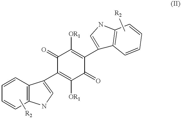
| TABLE I | ||
| Example | R1 | R2 |
| 1. | H | 2-(2-methylbut-2-en-4-yl) |
| 2. | acetyl | 2-(2-methylbut-2-en-4-yl) |
| 3. | acetyl | 2-(3-methyl-n-butyl) |
| 4. | H | 2-(3-methyl-n-butyl) |
| 5. | H | 5-bromo |
| 6. | H | 2-allyl |
| 7. | H | 2-n-propyl |
| 8. | H | 2-aminocarbonyl |
| 9. | acetyl | 2-aminocarbonyl |
| 10. | benzoyl | 2-allyl |
| 11. | H | 2-cyano |
| 12. | H | 4-methoxycarbonyl |
| 13. | H | 5,7-dimethoxy |
| 14. | H | 4,7-dimethoxy |
| 15. | H | 5-nitro |
| 16. | H | 4-(4-chlorobenzoylamino) |
| 17. | H | 4-(4-chlorophenyl) |
| 18. | H | 2-(4-fluorophenyl) |
| 19. | H | 4,6-dimethoxy |
| 20. | H | 5-hydroxy-6-methoxy |
| 21. | H | 4-cyano |
| 22. | H | 5-(4-trifluoromethylphenyl-aminocarbonyl) |
| 23. | H | 2-(4-trifluoromethylphenyl-aminocarbonyl) |
| 24. | H | 2-ethyl |
| 25. | H | 5-bromo-6-nitro |
| 26. | OMe | 2-(2-methylbut-2-en-4-yl) |
| 27. | OMe | 2-(3-methyl-n-butyl) |
Specific compounds within the scope of the present invention are also described by formula (III) below. R1-R12 of the formula can be as listed in Table II following the formula. Illustrative preparations or isolations of these compounds are found in the working examples. 
| TABLE II | ||||
| Ex. | R1 = R2 | R11 | R12 | R3-R101 |
| 28. | H | 2-(3-methyl- | 2-(3-methyl- | |
| n-butyl) | n-butyl) | |||
| 29. | H | 2-methyl | 2-methyl | |
| 30. | H | 2-ethyl | 2-ethyl | |
| 31. | H | 2-butyl | 2-butyl | |
| 32. | H | 2-(but-1-en- | 2-(but-1-en- | |
| 4-yl) | 4-yl) | |||
| 33. | H | 2-(4-methyl- | 2-(4-methyl- | |
| n-pentyl) | n-pentyl) | |||
| 34. | H | 2-phenylethyl | 2-phenylethyl | |
| 35. | H | H | 2-(3-methyl- | |
| n-butyl) | ||||
| 36. | H | 2-ethyl | 2-ethyl | R5 = R9 = carboxy |
| 37. | H | 2-(n-propyl) | 2-(n-propyl) | R5 = R9 = carboxy |
| 38. | H | 2-(3-methyl- | 2-(3-methyl- | R5 = R9 = carboxy |
| n-butyl) | n-butyl) | |||
| 39. | H | 2-(4-carboxy- | 2-(4-carboxy- | |
| n-butyl) | n-butyl) | |||
| 40. | H | H | 2-(3-methyl- | R5 = carboxy |
| n-butyl) | ||||
| 41. | H | 2-ethyl | 2-ethyl | R5 = R9 = amino |
| 42. | H | 2-(n-propyl) | 2-(n-propyl) | R5 = R9 = amino |
| 43. | H | 2-(3-methyl- | 2-(3-methyl- | R5 = R9 = amino |
| n-butyl) | n-butyl) | |||
| 44. | acetyl | 2-(3-methyl- | 2-(3-methyl- | |
| n-butyl) | n-butyl) | |||
| 45. | H | 2-ethyl | 2-ethyl | R5 = R9 = (4- |
| methylphenyl- | ||||
| sulfonylamino) | ||||
| 46. | H | 2-(n-propyl) | 2-(n-propyl) | R5 = R9 = (4- |
| methylphenyl- | ||||
| sulfonylamino) | ||||
| 47. | H | 2-(3-methyl- | 2-(3-methyl- | R5 = R9 = (4 |
| n-butyl) | n-butyl) | methylphenyl | ||
| sulfonylamino) | ||||
| 48. | H | 2-(2- | 2-(2- | |
| methylbut-1- | methylbut-1- | |||
| en-4-yl) | en-4-yl) | |||
| 49. | H | 2-(2- | 2-(2- | |
| methylpent-2- | methylpent-2- | |||
| en-5-yl) | en-5-yl) | |||
| 1Unless otherwise indicated, R3-R10 = hydrogen. | ||||
By the term “alkyl” as used herein is meant a straight or branched chain saturated hydrocarbon group having from 1 to 20 carbons such as methyl, ethyl, isopropyl, n-butyl, s-butyl, t-butyl, n-amyl, isoamyl, n-hexyl, n-octyl and n-decyl; “alkenyl” and “alkynyl” are used to mean straight or branched chain hydrocarbon groups having from 2 to 10 carbons and unsaturated by a double or triple bond respectively, such as vinyl, allyl, propargyl, 1-methylvinyl, but-1-enyl, but-2-enyl, but-2-ynyl, 1 methylbut-2-enyl, pent-1-enyl, pent-3-enyl, 3-methylbut-1-ynyl, 1,1-dimethylallyl, hex-2-enyl and 1-methyl-1-ethylallyl; “alkylaryl” means the aforementioned alkyl groups substituted by a phenyl group such as benzyl, phenethyl, phenopropyl, 1-benzylethyl, phenobutyl and 2-benzylpropyl; “aryl” as used herein includes a monocyclic or bicyclic rings, wherein at least one ring is aromatic including aromatic or hetero-aromatic hydrocarbons; the term “hydroxy-alkyl” means the aforementioned alkyl groups substituted by a single hydroxyl group such as 2-hydroxyethyl, 2-hydroxypropyl, 3-hydroxypropyl, 4-hydroxybutyl, 1-hydroxybutyl and 6-hydroxyhexyl.
The term “substituted” as used herein means that the group in question may bear one or more substituents including but not limited to halogen, hydroxy, cyano, alkyl, aryl, alkenyl, alkynyl, amino, nitro, mercapto, carboxy and other substituents known to those skilled in the art.
Preferred compounds of the present invention include the following: 
and pharmaceutically acceptable salts thereof.
In addition, the present invention encompasses a pharmaceutical composition comprising a compound of the invention, and methods for using a compound or pharmaceutical composition of the invention in an animal, particularly a human, to ameliorate symptoms of cell proliferative disorders involving protein tyrosine kinase/adaptor protein interactions.
This invention is based, in part, on the discovery that the disclosed compounds, while exhibiting no inhibitory effect on protein tyrosine kinase enzymatic activity, act to inhibit the binding of an SH2-containing peptide to a tyrosine phosphorylated EGF receptor. The data representing this discovery is presented in the Examples in Sections 6, 7 and 8, below. The Example presented in Section 5, below, describes a method for the production of the compounds of the present invention.
The present invention represents the first instance whereby compounds have been discovered which directly inhibit the interaction between adaptor proteins and protein tyrosine kinase molecules.
Described below are methods and compositions for the inhibition of adaptor protein/protein tyrosine kinase protein interactions, especially those interactions associated with a cell proliferative disorder. Specifically, described below are particular organic compounds, methods for the synthesis of such compounds, and techniques utilizing such compounds.
4.1 COMPOUNDS
The compounds of the present invention are described by the following formula (IV): 
and pharmaceutically acceptable salts thereof, wherein:
R1 and R2 are independently H, acetate or aryl, alkylaryl and higher alkyl acid ester;
R3 to R14 are independently H, alkyl, alkenyl, alkynyl, hydroxyalkyl, OH, alkoxy, nitro, halo, trihalomethyl, amide, carboxamide, carboxy, sulfonyl, sulfonamide, amino, and mercapto which can be unsubstituted or substituted where appropriate. For example, alkyl groups of the compounds of the present invention may be substituted where appropriate with one or more carboxy or aryl groups. Alkenyl groups of compounds of the present invention may be substituted where appropriate with one or more carboxy groups. Specific compounds within the scope of the present invention are found in the preceding Tables I and II. Illustrative preparations or isolations of these compounds are found in the working examples.
In one embodiment, compounds of the present invention are described by the following formula (III): 
and pharmaceutically acceptable salts thereof, wherein:
R1 and R2 are each independently hydrogen, lower alkyl, acetyl, aryl, alkylaryl or higher alkyl acid ester, and wherein at least one of R1 and R2 is other than hydrogen;
R3 to R12 are each independently H, alkyl, alkylcarboxy, alkenyl, alkenylcarboxy, aryl, alkylaryl, OH, alkoxy, nitro, halo, trihalomethyl, amide, carboxamide, carboxy, sulfonyl, sulfonamide, amino, mercapto or 2-methylbut-2-en-4-yl; and wherein at least one of R11 and R12 is 2-methylbut-2-en-4-yl.
Groups R1-R12 may be substituted or unsubstituted where appropriate.
In another embodiment, compounds of the present invention are described by formula (III) above, and pharmaceutically acceptable salts thereof, wherein:
R1 and R2 are both H;
R3 to R10 are each independently H, alkyl, alkylcarboxy, alkenyl, alkenylcarboxy, aryl, alkylaryl, OH, alkoxy, nitro, halo, trihalomethyl, amide, carboxamide, carboxy, sulfonyl, sulfonamide, amino, mercapto or 2-methylbut-2-en-4-yl; and
R11 and R12 are each independently H or 2-methylbut-2-en-4-yl, wherein at least one of R11 and R12 is 2-methylbut-2-en-4-yl;
wherein at least one of R3 to R10 is other than H.
In another embodiment, compounds of the present invention are described by formula (III) above, and pharmaceutically acceptable salts thereof, wherein:
R1 and R2 are each independently aryl, alkylaryl and higher alkyl acid ester; and
R3 to R12 are each independently H, alkyl, alkylcarboxy, alkenyl, alkenylcarboxy, aryl, alkylaryl, OH, alkoxy, nitro, fluoro, chloro, iodo, trihalomethyl, amide, carboxamide, carboxy, sulfonyl, sulfonamide, amino, or mercapto.
In another embodiment, compounds of the present invention are described by formula (III) above, and pharmaceutically acceptable salts thereof, wherein:
R1, R2, R11 and R12 are H; and
R3 to R10 are each independently H, alkyl, alkylcarboxy, alkenyl, alkenylcarboxy, aryl, alkylaryl, alkoxy, hydroxy, nitro, halo, trihalomethyl, amide, carboxamide, carboxy, sulfonyl, sulfonamide, amino, or mercapto, wherein at least one of R3 to R10 is other than H;
(a) when R4-R10 are each H, R3 may not be 2-methylbut-2-en-4-yl or 2-hydroxy-2-methylbut-4-yl;
(b) when R4-R6 and R8-R10 are each H, R3 and R7 may not simultaneously be 2-methylbut-2-en-4-yl;
(c) when R3-R4, R6-R8 and R10 are H, R5 and R9 may not simultaneously be 2-methylbut-2-en-4-yl or 3-methyl-n-butyl;
(d) when R3, R5-R7, R9-R10 are H, R4 and R8 may not both be 2-methylbut-2-en-4-yl or 2-methylbut-1,4-dien-4-yl, and R4 and R8 may not be 2-methylbut-2-en-4-yl and 2-methylbut-1,4-dien-4-yl.
The present invention also encompasses compounds of formula (III) above, and pharmaceutically acceptable salts thereof, wherein R3-R5 and R7-R9 are H and either or both of R6 and R10 are 2-methylbut-2-en-4-yl.
In another embodiment, compounds of the present invention are described by formula (III) above, and pharmaceutically acceptable salts thereof, wherein:
at least one of R1 and R2 is acetyl;
R11 and R12 are H; and
R3 to R10 are each independently H, alkyl, alkylcarboxy, alkenyl, alkenylcarboxy, aryl, alkylaryl, OH, alkoxy, nitro, halo, trihalomethyl, amide, carboxamide, carboxy, sulfonyl, sulfonamide, amino, and mercapto, wherein:
(a) when both R1 and R2 are acetyl; or when one of R1 and R2 is acetyl and R3-R4, R6-R8 and R10-R12 are H; R5 and R9 may not simultaneously be 2-methylbut-2-en-4-yl;
(b) when both R1 and R2 are acetyl and when R4-R6 and R8-R10 are H, R3 and R7 may not simultaneously be 2-methylbut-2-en-4-yl;
(c) when both R1 and R2 are acetyl and when R3, R5-R7, and R9-R10 are H, R4 and R8 may not simultaneously be 2-methylbut-2-en-4-yl.
In another embodiment, compounds of the present invention are described by formula (III) above, and pharmaceutically acceptable salts thereof, wherein:
at least one of R1 and R2 is lower alkyl;
R11 and R12 are H; and
R3 to R10 are each independently H, alkyl, alkylcarboxy, alkenyl, alkenylcarboxy, aryl, alkylaryl, OH, alkoxy, nitro, halo, trihalomethyl, amide, carboxamide, carboxy, sulfonyl, sulfonamide, amino, and mercapto, wherein:
(a) when both R1 and R2 are methyl, at least one of R3 to R10 must be a group other than H;
(b) when both R1 and R2 are methyl, and R4-R10 are H, R3 may not be 2-methylbut-2-en-4-yl;
(c) when both R1 and R2 are methyl, and R4-R6 and R8-R10 are H, R3 and R7 may not simultaneously be 2-methylbut-2-en-4-yl;
(d) when both R1 and R2 are methyl, and R3-R4, R6-R8 and R10 are H, R5 and R9 may not simultaneously be 2-methylbut-2-en-4-yl.
The present invention also includes compounds of formula III) above, and pharmaceutically acceptable salts thereof,
wherein R4 is 2-methylbut-2-en-4-yl and R3 and R5-R10 are H;
or R5 is 2-methylbut-2-en-4-yl and R3-R4 and R6-R10 are H;
or R6 is 2-methylbut-2-en-4-yl, and R3-R5 and R7-R10 are H.
In another embodiment, compounds of the present invention are described by formula (III) above, and pharmaceutically acceptable salts thereof, wherein:
R1 and R2 are each independently hydrogen, lower alkyl, acetyl, aryl, alkylaryl or higher alkyl acid ester,
R3 to R10 are each independently H, alkyl, alkylcarboxy, aryl, alkylaryl, alkenyl, alkenylcarboxy, OH, alkoxy, nitro, halo, trihalomethyl, amide, carboxyamide, carboxy, sulfonyl, sulfonamide, amino, mercapto, 4-methylphenylsulfonylamino, or 2-methylbut-2-en-4-yl; and
R11 and R12 are selected from the group consisting of hydrogen, methyl, ethyl, propyl, butyl, aryl, alkylaryl, alkylcarboxy, alkenylcarboxy, but-1-en-4-yl, 2-methylbut-1-en-4-yl, 4-methyl-n-pentyl, 2-phenylethyl, 2-methylpent-2-en-4-yl, and 4-carboxy-n-butyl, wherein at least one of R11 and R12 is other than hydrogen.
In yet another embodiment, compounds of the present invention are described by formula (III) above, and harmaceutically acceptable salts thereof, wherein:
R1 and R2 are each independently hydrogen, lower alkyl, acetyl, aryl, alkylaryl or higher alkyl acid ester,
R3 to R10 are each independently H, alkyl, alkylcarboxy, aryl, alkylaryl, alkenyl, alkenylcarboxy, OH, alkoxy, nitro, halo, trihalomethyl, amide, carboxyamide, carboxy, sulfonyl, sulfonamide, amino, mercapto, 4-methylphenylsulfonylamino, or 2-methylbut-2-en-4-yl; and
R11 and R12 are both 3-methyl-n-butyl.
In still another embodiment, compounds of the present invention are described by formula (III) above, and pharmaceutically acceptable salts thereof, wherein:
R1 and R2 are each independently hydrogen, lower alkyl, acetyl, aryl, alkylaryl or higher alkyl acid ester,
R3 to R10 are each independently H, alkyl, alkylcarboxy, aryl, alkylaryl, alkenyl, alkenylcarboxy, OH, alkoxy, nitro, halo, trihalomethyl, amide, carboxyamide, carboxy, sulfonyl, sulfonamide, amino, mercapto, 4-methylphenylsulfonylamino, or 2-methylbut-2-en-4-yl and wherein at least one of R3 to R10 is other than hydrogen; and
R11 and R12 are each independently hydrogen or 3-methyl-n-butyl. 
The invention encompasses the compounds described above as well as pharmaceutically acceptable salts thereof. The compounds of the present invention can either be synthesized or isolated as described herein.
The compounds of the present invention can be synthesized in accordance with standard organic chemistry techniques using readily available starting materials. Alternatively, the compounds can be isolated as described in Section 5.2, below. Chemical synthesis and isolation methods are provided herein solely for illustration. Variation of these methods will be apparent to those skilled in the art.
4.2 PRODUCTION OF THE COMPOUNDS
4.2.1 ISOLATION OF NATURAL PRODUCTS
The present Example employed a fungus culture (PenLabs Inc. #592), and the following fermentation conditions: medium yeast malt extract plus trace elements at 22° C. The seed medium consisted of mannitol 60.0 g; soybean meal 12.5 g, citric acid 2.5 g, yeast extract 0.5 g, and H2O to 1 liter.
The pH of the seed medium was adjusted to 7.0 before autoclaving. 30 mL seed medium were dispensed per 250 ml flask (6 days 28° C.), which was then inoculated with 1 ml of spore/mycelium homogenate suspension (2 days). Stock cultures were maintained frozen at −80° C. in spore storage solutions.
The fermentation mixture (mycelium and broth) was homogenized and filtered through cheesecloth by suction filtration. The filtrate was extracted three times with 0.5 v/v of ethyl acetate. The ethyl acetate layers were combined and the solvent removed by rotary evaporation. The mycelium was extracted twice with 0.4 v/v of ethyl acetate. The ethyl acetate layers were combined and the solvent removed by rotary evaporation. The oily residues both containing the asterriquinones were combined and dried on a vacuum pump overnight.
The crude extract obtained above underwent CPC fractionation on a PC Inc. high speed countercurrent chromatograph (HSCC) containing a “tripple” coil column. A 1:3:3:3 v/v/v/v of n-hexane, ethyl acetate, methanol and water was mixed and allowed to settle overnight. The lower layer was pumped into HSCC column as the stationary phase. The upper layer was used as the mobile phase. After two hours, the lower and upper layer were switched. The HSCC run was completed after four hours. The crude metabolites eluted from 8 to 12 minutes. The active fractions were pooled and evaporated under reduced pressure to dryness.
The pooled HSCC fraction (8-12) was subjected to semi-preparative HPLC (Water HPLC system with a Water 996 photodioarray detector using Millennium software) fractionations using the following conditions:
Two semi-preparative C18-cartridges (25×100 mm each, Nova Pak, 6μ); Flow rate: 10 mL/min.; 120 mg of the pooled HSCC fraction 8-12 dissolved in 6 mL of DMSO; 250 μL aliquots per injection; PDA monitored at 270 nm; linear gradient of 70% H2O/30% CH3CN to 100% CH3CN over 30 minutes; then isocratic at 100% CH3CN for 6 minutes; the active material eluted at 19 and 24 minutes. The active fractions from the runs were combined and evaporated under reduced pressure to dryness to yield 17 mg of Asterriquinone C-3 (Compound I) and 3 mg of Preasterriquinone C-3 (Compound II).
Mass spectra were recorded on PE Sciex LC-MS model API III (Ion Spray), exact mass measurements were performed at high resolution (HR-FAB). Mass spectral analysis for compound I gave a molecular ion of 507 (M+H)+ (molecular weight: 506). The molecular formula C32H31N2O4(M++H): 507.2289; found 507.2291). 1H NMR spectra of compound I were recorded in CDCl3 at 500 MHz on a Brucker DRX-500. Chemical shifts are given in ppm relative to TMS at zero ppm using the solvent peak at 7.26 ppm (CDCl3) as an internal standard. Compound I: 8.18 (s, 2H), 8.05 (s, 2H), 7.35-7.10 (m, 8H), 5.40 (m, 2H), 3.45 (m, 4H), 1.81 (s, 6H) and 1.75 ppm (s, 6H). 13C NMR spectra of compound I were recorded in DMSO-d6 at 125 MHz on a Brucker DRX-500. Chemical shifts are given in ppm relative to TMS at zero using the solvent peak at 39.5 ppm (DMSO-d6) as an internal standard. 138.8, 136.6, 136.3, 128.8, 128.2, 127.3, 122.3, 121.8, 121.0, 120.3, 119.5, 119.3, 112.3, 111.8, 111.6, 105.2, 102.2, 27.3, 26.4 and 18.5 ppm. Compound I gave a melting point of 150-154° C.
Mass spectral analysis for compound II gave a molecular ion of 439 (M+H)+ (molecular weight: 438). 1H NMR spectra of compound II were recorded in DMSO-d6 at 500 MHz on a Brucker DRX-500. Chemical shifts are given in ppm relative to TMS at zero ppm using the solvent peak at 2.49 ppm (DMSO-d6) as an internal standard. 11.35 (s, 1H), 10.96 (s, 1H), 10.62 (s, 1H), 7.48 (dJ=1 Hz, 1H), 7. 39 (d, J=10.0 Hz, 1H), 7.29 (d, J=10.0 Hz, 1H), 7.14 (d, J=10 Hz, 1H), 7.07 (t, J=10.0 Hz, 1H), 6.99 (t, J=10.0 Hz, 1H), 6.93 (t, J=10.0 Hz, 1H), 6.88 (t, J=10.0 Hz, 1H), 5.26 (m, 1H), 3.30 (m, 2H) 1.64 (bs, 3H) and 1.61 ppm (bs, 3H). 13C NMR spectra of compound II were recorded in CDCl3 at 125 MHz on a Brucker DRX-500. Chemical shifts are given in ppm relative to TMS at zero ppm using the solvent peak at 77.0 ppm (CDCl3) as an internal standard. 138.4, 138.3, 135.7, 135.2, 127.7, 121.6, 120.0, 119.8, 119.6, 110.7, 110.6, 100.5, 26.8, 25.8 and 18.0 ppm.
4.2.2 COMPOUND SYNTHESIS
2,5-Dihydroxy-3,6-di-[2-(2-methylbut-2-en-4-yl)indol-3-yl]1,4-quinone
A mixture of 100 mg. of 2,5-diacetoxy-3,6-dibromo-1,4-quinone, or other suitably protected quinone such as 3,6-dibromo-2,5-ditrimethylsiloxy-1,4-quinone, 3,6-dibromo-2,5-di-(t-butyldimethylsiloxy-1,4-quinone, 2,5-dibenzoxy-3,6-dibromo-1,4-quinone, 3,6-dibromo-2,5-diisobutryoxy-1,4-quinone, 2,5-dibenzyloxy-3,6-dibromo-1,4-quinone or 2,5-diallyoxycarbonyloxy-3,6-dibromo-1,4-quinone which can be prepared from commercially available 2,4-dibromo-3,6-dihydroxy-1,4-quinone and 180 mg of 3-[2-(2-methylbut-2-en-4-yl)indole, prepared by the Fisher indole synthesis, in 10 ml of anhydrous dimethylforamide, or pyridine or dimethylsulfoxide, with powdered potassium carbonate, was heated at 100° C. for 24 hours. The cooled mixture was partitioned between ethyl acetate and water. The ethyl acetate layer was then washed with brine, dried over sodium sulfate, filtered and concentrated. The crude was then purified on a medium pressure liquid chromatography column in a solvent mixture of dichloromethane and methanol to provide 25 mg of 2,5-diacetoxy-3,6-di-[2-(2-methylbut-2-en-4-yl)indol-3-yl]1,4-quinone. 2,5-Diacetoxy-3,6-di-[2(2-methylbut-2-en-4-yl)indol-3-yl]1,4-quinone was then hydrolysed with 1 N aqueous sodium hydroxide solution in methanol. Acidification of the above mixture produced the crude product after filtration. Further crystallization in ethanol and water produced the title compound. Other aforementioned protecting groups, they can be removed by conventional deprotection methods such as diluted acid, potassium fluoride or palladium (0) complex or palladium on carbon with hydrogen or by methods described by Greene and Wuts (Protective groups in organic synthesis, John Wiley and Son, 1991).
Alternatively, under the similar conditions, 2,3,5,6-tetrabromo-1,4-quinone reacts with excess indole in the presence of potassium carbonate and aluminum oxide in dimethylformamide or dimethylsulfoxide at 100° C. to produce the substituted 2,5-dibromo-3,6-(3-indolyl)-1,4-quinone which can react with base such as sodium hydroxide to give the substituted 2,5-dihydroxy-3,6-(3-indolyl)-1,4-quinone (Hoerher, J.; Schwenner, E.; Franck, B., Liebigs Ann. Chem. 1986, 10: 1765-1771).
2,5-Diacetoxy-3,6-di-[2-(2-methylbut-2-en-4-yl)indol-3-yl]1,4-quinone
2,5-Diacetoxy-3,6-di-(2-(2-methylbut-2-en-4-yl)indol-3-yl]1,4-quinone was prepared in Example 1.
2,5-Diacetoxy-3,6-di-[2(3-methyl-n-butyl)indol-3-yl]1,4-quinone
Hydrogenation of 2,5-diacetoxy-3,6-di-[2-(2-methylbut-2-en-4-yl)indol-3-yl]1,4-quinone in methanol with 5% palladium on carbon under 1 atm of hydrogen produced the title compound.
2,5-Dihydroxy-3,6-di-[2-(3-methyl-n-butyl)indol-3-yl]1,4-quinone
Base hydrolysis of 2,5-diacetoxy-3,6-di-[2-(3-methyl-n-butyl)indol-3-yl]1,4-quinone as described in Example 1 produced the title compound.
Under similar conditions as those described in Examples 1 to 4, the following compounds are prepared using either 2,5-dibromo-3,6-dihydroxy-1,4-quinone or 2,3,5,6-tetrabromoquinone as starting materials:
3,6-Di-[5-(bromo)indol-3-yl]-2,5-dihydroxy-1,4-quinone
3,6-Di-[2-(allyl)indol-3-yl]-2,5-dihydroxy-1,4-quinone
2,5-Dihydroxy-3,6-di-[2-(n-propyl)indol-3-yl]-1,4-quinone
3,6-Di-[2-(aminocarbonyl)indol-3-yl]-2,5-dihydroxy-1,4-quinone
2,5-Diacetoxy-3,6-di-[2(aminocarbonyl)indol-3-yl]-1,4-quinone
3,6-Di-[2-allylindol-3-yl]-2,5-dibenzoyloxy-1,4-quinone
2,5-Dihydroxy-3,6-di-[2-(cyano)indol-3-yl]1,4-quinone
2,5-Dihydroxy-3,6-di-[4-(methoxycarbonyl)indol-3-yl]1,4-quinone
2,5-Dihydroxy-3,6-di-[5,7-(dimethoxy)indol-3-yl]1,4-quinone
2,5-Dihydroxy-3,6-di-[4,7-(dimethoxy)indol-3-yl]1,4-quinone
2,5-Dihydroxy-3,6-di-[5-(nitro)indol-3-yl]1,4-quinone
3,6-di-[4(4-chlorobenzoylamino)indol-3-yl]-2,5-dihydroxy-1,4-quinone
3,6-di-[2-(4-chlorophenyl)indol-3-yl]-2,5-dihydroxy-1,4-quinone
2,5-Dihydroxy-3,6-di-[2-(4-fluorophenyl)indol-3-yl]1,4-quinone
2,5-Dihydroxy-3,6-di-[4,6-(dimethoxy)indol-3-yl]1,4-quinone
2,5-Dihydroxy-3,6-di-[2-(5-hydroxy-6-methoxy)indol-3-yl]1,4-quinone
2,5-Dihydroxy-3,6-di-[4-(cyano)indol-3-yl]1,4-quinone
2,5-Dihydroxy-3,6-di-[5-(4-trifluoromethylphenylaminocarbonyl)indol-3-yl]1,4-quinone
2,5-Dihydroxy-3,6-di-[2-(4-trifluoromethylphenylaminocarbonyl)indol-3-yl]1,4-quinone
2,5-Dihydroxy-3,6-di-[2-(ethyl)indol-3-yl]1,4-quinone
3,6-di-[2-(5-bromo-6-nitro)indol-3-yl]-2,5-dihydroxy-1,4-quinone
2,5-Dimethoxy-3,6-di-[2-(2-methylbut-2-en-4-yl)indol-3-yl]1,4-quinone
Methylation of Example 1 with methyl iodide and potassium carbonate in dimethylforamide followed by purification produced the title compound. This compound could also be prepared by heating 2,5-dibromo-3,6-di[2-(2-methylbut-2-en-4-yl)indol-3-yl]1,4-quinone in methanol in the presence of powdered potassium carbonate.
2,5-Dimethoxy-3,6-di-[2(3-methyl-n-butyl)indol-3-yl]1,4-quinone
Hydrogenation of Example 26 under conditions as those in Example 3 produced the title compound.
Preparation of 2,5-Dihydroxy-3,6-di-[2-(3-methyl-n-butyl)indol-3-yl]-1,4-quinone
To a glass tube containing 2-(3-methyl-n-butyl)indole (400 mg), bromanil (431 mg) and potassium carbonate (703 mg), equipped with a magnetic stir bar, was added dimethylformamide (10 ml). The mixture was stirred at room temperature for 40 h. Following dilution with 1 N HCl (100 ml), the crude mixture was extracted with ethyl acetate (200 ml). The organic layer was washed with brine (100 ml) and dried with sodium sulfate. After removal of solvent under reduced pressure, the crude residue was filtered through a short plug of flash silica, eluting with 30% ethyl acetate/hexane. The solvent was removed under reduced pressure, and the residue was purified by flash chromatography (15% ethyl acetate/hexane) to yield 2,5-dibromo-3,6-di-[2-(3-methyl-n-butyl)indol-3-yl]-1,4-quinone (40 mg, 7%) as a blue crystalline solid.
To a stirred solution of 2,5-dibromo-3,6-di-[2-(3-methyl-n-butyl)indol-3-yl]-1,4-quinone (40 mg) in methanol (1.5 ml) was added 2N methanolic sodium hydroxide (0.251 ml). The solution was stirred at room temperature for 24h, followed by dilution with water (50 ml). The product was extracted with ethyl acetate (100 ml), washed with brine (50 ml) and dried with sodium sulfate. Removal of solvent under reduced pressure provided 2,5-methoxy-3,6-di-[2-(3-methyl-n-butyl)indol-3-yl]-1,4-quinone (30 mg, 90%) as a yellow crystalline solid.
To a stirred solution of 2,5-dimethoxy-3,6-di-[2-(3-methyl-n-butyl)indol-3-yl]-1,4-quinone (9 mg) in ethanol (2 ml) was added 1 N aqueous potassium hydroxide (1 ml). The mixture was heated at 85° C. for 3.5 h, then diluted with 1 N HCl (25 ml). The product was extracted with ethyl acetate (50 ml), washed with brine (25 ml) and dried with sodium sulfate. The solvent was removed under reduced pressure to afford 2,5-dihydroxy-3,6-di-[2-(3-methyl-n-butyl)indol-3-yl]-1,4-quinone (8 mg) as a reddish-brown crystalline solid.
28a) Preparation of 2-(2-methyl-1-buten-4-yl)indole To a stirred solution of 2-methylindole (1 g) in diethylether (76 ml) under nitrogen was added a 1.6 M solution of n-butyllithium in hexane (14.3 ml) slowly dropwise via syringe. Potassium tert-butoxide (1.711 g) was then added, producing a bright yellow mixture. After stirring at room temperature under nitrogen for 50 min., the mixture was cooled to −78° C., whereupon 3-bromo-2-methylpropene (1.54 ml) was added dropwise via syringe, giving a red-orange solution. The reaction mixture was stirred at −78° C. for 2 h, then quenched with water (10 ml). After warming to room temperature, water (150 ml) and 1 N HCl (1 ml) was added to neutralize the reaction mixture. The mixture was extracted with ethyl acetate (250 ml), and the organic layer was washed with brine (100 ml) and dried with sodium sulfate. The solvent was removed under reduced pressure, and the crude residue was purified by flash chromatography (4% ethyl acetate/hexane) to afford 2-(2-methyl-1-butene-4-yl)indole (664 mg. 47%) as a waxy yellow solid.
28b) Preparation of 2-(3-methyl-n-butyl)indole Into a 3-necked round bottom flask under a blanket of nitrogen was placed 5% palladium catalyst on charcoal (771 mg). A solution of 2-(2-methyl-1-buten-4-yl) indole (671 mg) in ethanol (36 ml) was added to the flask, which was evacuated and charged with hydrogen twice. The mixture was stirred vigorously under hydrogen (1 atm) for 2 h, followed by filtration through a pad of Celite. The solvent was removed under reduced pressure and the crude residue was purified by flash chromatography (3% ethyl acetate/hexane) to give 2-(3-methyl-n-butyl)indole (400 mg, 59%) as a yellow crystalline solid.
Preparation of 2,5-Dihydroxy-3,6-di-[2-(methyl)indol-3-yl]-1,4-quinone
Refer to Example 28 using 2-methylindole as the starting indole.
Preparation of 3,6-Di-(2-ethylindol-3-yl)-2,5-dihydroxy-1,4-quinone
Refer to Example 28 using 2-ethylindole as the starting indole.
30a) Preparation of 2-ethylindole Refer to 28a) using methyl iodide as the alkylating agent.
Preparation of 3,6-Di-(2-butylindol-3-yl) 2,5-dihydroxy-1,4-quinone
Refer to Example 28 using 2-butylindole as the starting indole.
31a) Preparation of 2-(but-1-en-4-yl)indole Refer to 28a) using allyl bromide as the alkylating agent.
31b) Preparation of 2-butylindole Refer to 28b) using 2-(but-1-en-4-yl)indole as the starting material.
Preparation of 3,6-Di-[2-(but-1-en-4-yl)indol-3-yl] 2,5-dihydroxy-1,4-quinone
Refer to Example 28 using 2-(but-1-en-4-yl)indole as the starting indole.
Preparation of 2,5-Dihydroxy-3,6-di-[2-(4-methyl-n-pentyl) indol-3-yl]-1,4-quinone
Refer to Example 28 using 2-(4-methyl-n-pentyl)indole as the starting indole.
33a) Preparation of 2-(2-methyl-2-penten-5-yl) indole
Refer to 28a) using 4-bromo-2-methyl-2-butene as the alkylating reagent.
33b) Preparation of 2-(4-methyl-n-pentyl)indole
Refer to 28b) using 2-(2-methyl-2-penten-5-yl)indole as the starting material.
Preparation of 2,5-Dihydroxy-3,6-di-[2-(2-phenylethyl)indol-3-yl]-1,4-quinone
Refer to Example 28 using 2-(2-phenylethyl)indole as the starting indole.
34a) Preparation of 2-(2-phenylethyl)indole
Refer to 28a) using benzyl bromide as the alkylating agent.
Preparation of 2,5-Dihydroxy-6-(indol-3-yl) -3-[2-(3-methyl-n-butyl)indol-3-yl]-1,4-quinone
This synthesis could be achieved by treating 2-(3-methyl-n-butyl)indole with 2 equivalents of bromanil in the presence of potassium carbonate in dimethylformamide, followed by workup and purification similar to Example 28. The resultant mono-indolyl adduct could then be treated with 2 equivalents of indole under the same conditions as above to provide the bis-indolyl product.
Preparation of 3,6-Di-(5-carboxy-2-ethylindol-3-yl)-2,5-dihydroxy-1,4-quinone
Refer to Example 28 using 5-carboxy-2-ethylindole as the starting indole.
36a) Preparation of 5-carboxy-2-ethylindole
This synthesis could start with 5-chloro-2-methylindole, which could be alkylated with methyl indole (see 28a). The product chloroindole could then be converted to its Grignard species and exposed to carbon dioxide to finish the synthesis.
Preparation of 3,6-Di-[5-carboxy-2-(n-propyl)indol-3-yl]-2,5-dihydroxy-1,4-quinone
Refer to Example 28 using 5-carboxy-2-propylindole as the starting indole.
37a) Preparation of 5-carboxy-2-propylindole Refer to 36a) using ethyl iodide as the alkylating agent.
Preparation of 3,6-Di-[5-carboxy-2-(3-methyl-n-butyl)indol-3-yl-2,5-dihydroxy-1,4-quinone
Refer to Example 28 using 5-carboxy-2-(3-methyl-n-butyl)indole as the starting indole.
38a) Preparation of 5-carboxy-2-(2-methyl-1-buten-4-yl)indole
Refer to 36a) using 3-bromo-2-methylpropene as the alkylating agent.
38b) Preparation of 5-carboxy-2-(3-methyl-n-butyl)indole
Refer to 28b) using 5-carboxy-2-(2-methyl-1-buten-4-yl)indole as the starting material.
Preparation of 3,6-Di-[2-(4-carboxy-n-butyl)indol-3-yl]-2,5-dihydroxy-1,4-quinone
Refer to Example 28 using 2-(4-carboxy-n-butyl)indole as the starting indole.
39a) Preparation of 2-(4-carboxy-3-buten-1-yl)indole
Refer to 28a) using 4-bromo-2-butenoic acid as the alkylating agent.
39b) Preparation of 2-(4-carboxy-n-butyl)indole
Refer to 28b) using 2-(4-carboxy-3-buten-1-yl) indole as the starting material.
Preparation of 3-[5-Carboxy-2-(3-methyl-n-butyl)indol-3-yl]-2,5-dihydroxy-6-(indol-3-yl)-1,4-quinone
Refer to Example 35 using 5-carboxy-2-(3-methyl-n-butyl)indole in the first step.
Preparation of 3,6-Di-(5-amino-2-ethylindol-3-yl)-2,5-dihydroxy-1,4-quinone
Refer to Example 28 using 5-amino-2-ethylindole as the starting indole.
41a) Preparation of 5-amino-2-ethylindole This synthesis could be achieved beginning with a standard nitration of 2-ethylindole using sodium nitrate and sulfuric acid similar to that cited in Yokoyama; Tanaka; Yamane; Kurita; Chem. Lett.; 7; 1991; 1125-1128. The resultant 5-nitro-2-ethylindole could be reduced to the desired amino compound using catalytic hydrogenation as in 28b).
Preparation of 3,6-Di-(5-amino-2-(n-propyl)indol-3-yl]-2,5-dihydroxy-1,4-quinone
Refer to Example 28 using 5-amino-2-(n-propyl)indole as the starting indole.
42a) Preparation of S-amino-2-(n-propyl)indole
Refer to 41a) using 2-n-propylindole.
Preparation of 3,6-Di-[5-amino-2-(3-methyl-n-butyl)indol-3-yl]2,5-dihydroxy-1,4-quinone
Refer to Example 28 using 5-amino-2-(3-methyl-n-butyl) indole as the starting indole.
43a) Preparation of 5-amino-2-(3-methyl-n-butyl)indole
Refer to 41a) using 2-(3-methyl-n-butyl)indole.
Preparation of 2,5-Diacetoxy-3,6-di-[2-(3-methyl-n-butyl)indol-3-yl]-1,4-quinone
This synthesis could be accomplished by treating 2,5-hydroxy-3,6-di-[2-(3-methyl-n-butyl)indol-3-yl]-1,4-quinone with acetic anhydride in the presence of pyridine.
Preparation of 3,6-Di-[2-ethyl-5-(4-methylphenylsulfonylamino)indol-3-yl]-2,5-dihydroxy-1,4-quinone
Refer to Example 28 using 2-ethyl-5-(4-methylphenylsulfonylamino)indole as the starting indole.
45a) Preparation of 2-ethyl-5-(4-methylphenylsulfonylamino)indole
The above compound could be synthesized by treating 5-amino-2-ethylindole with p-toluenesulfonyl chloride in the presence of triethylamine.
Preparation of 2,5-Dihydroxy-3,6-di-[5-(4-methylphenylsulfonylamino)-2-(n-propyl)indol-3-yl]-1,4-quinone
Refer to Example 28 using 5-(4-methylphenylsulfonylamino)-2-(n-propyl)indole as the starting indole.
46a) Preparation of 5-(4-methylphenylsulfonylamino)-2-(n-propyl)indole
Refer to 45a) using 5-amino-2-propylindole.
Preparation of 2,5-Dihydroxy-3,6-di-[2-(3-methyl-n-butyl)-5-(4-methylphenylsulfonylamino)indol-3-yl]-1,4-quinone
Refer to Example 28 using 2-(3-methyl-n-butyl)-5-(4-methylphenylsulfonylamino)indole as the starting indole.
47a) Preparation of 2-(3-methyl-n-butyl)-5-(4-methylphenylsulfonylamino)indole
Refer to 45a) using 5-amino-2-(3-methyl-n-butyl)indole.
Preparation of 2,5-Dihydroxy-3,6-di-[2-(2-methylbut-1-en-4-yl)indol-3-yl]-1,4-quinone
Refer to Example 28 using 2-(2-methylbut-1-en-4-yl)indole as the starting indole.
4.3 PROTEIN TYROSINE KINASE/ADAPTOR PROTEIN COMPLEXES
The PTK/adaptor protein complexes which may be disrupted by the methods and compositions of the invention comprise at least one member of the PTK family of proteins and at least one member of the adaptor family of proteins, as described below. Under standard physiological conditions, the components of such complexes are capable of forming stable, non-covalent attachments with one or more of the other PTK/adaptor protein complex components. Preferably, the compounds of the invention inhibit PTK/adaptor protein complexes wherein the PTK component is an epidermal growth factor receptor (EGF-R) protein tyrosine kinase molecule, a platelet derived growth factor receptor (PDGF-R) protein tyrosine kinase molecule or an insulin growth factor-like receptor tyrosine kinase molecule (IGF-1R).
Intracellular, cytoplasmic PTK components of the PTK/adaptor protein complexes may include, for example, members of the Src family, such molecules as src, yes, fgr, fyn, lyn, hck, lck, and blk; members of the Fes family, such as fes and fer; members of the Abl family, such as abl and arg; and members of the Jak family, such as jak1 and jak2. Transmembrane, receptor PTK components of the PTK/adaptor protein complexes may include, for example, such molecules as members of the FGF receptor, Sevenless/ROS, Insulin receptor, PDGF receptor, and EGF receptor family of growth factor receptors.
The adaptor protein components of the PTK/adaptor protein complexes comprise one or more SH2 and/or one or more SH3 non-catalytic domains. The SH2 and SH3 domains which may be a part of the adaptor proteins are as described, above, for the PTK components. Adaptor proteins which may be components of the PTK/adaptor protein complexes may include, for example, p85, c-Crk, SHC, Nck, ISGF3α, guanine triphosphatase activator protein (GAP), and members of the GRB subfamily of proteins, such as. GRB1, GRB-2, GRB-3, GRB-4, GRB-7, and GRB-10.
4.4 TREATMENT OF PTK/ADAPTOR PROTEIN COMPLEX-RELATED CELL PROLIFERATIVE DISORDERS
The compounds and/or pharmaceutical compositions (described in Section 4.4.2, below) of the invention may be used for the treatment of cell proliferative disorders, such as oncogenic disorders, involving a PTK capable of complexing with a member of the SH2- and/or SH3-containing family of adaptor proteins. The compounds of the invention may be preferentially utilized in the treatment of cell proliferative disorders involving PTK/adaptor protein complexes wherein the PTK component is EGF-R, PDGF-R, MCT or IGF-1R.
Among the oncogenic disorders which may be treated by the compounds of the invention are, f or example, BCR-ABL-associated cancers (such as, for example, chronic myelogenous and acute lymphocytic leukemias), gliomas, glioblastomas, melanoma, human ovarian cancers, human breast cancers (especially HER-2/GRB-7-associated human breast cancers), and human prostate cancers.
Assays for determining the effectiveness of a compound in the disruption of a PTK/adaptor protein complex are described, below, in Section 4.4.1. Methods for the administering the compounds and/or pharmaceutical compositions of the invention to patients are described, below, in Section 4.4.2.
“Disruption”, as used here, is meant to refer not only to a physical separation of PTK/adaptor protein complex components, but is also meant to refer to a perturbation of the activity of the PTK/adaptor complexes, regardless of whether or not such complexes remain able, physically, to form. “Activity”, as used here, refers to the function of the PTK/adaptor protein complex in the signal transduction cascade of the cell in which such a complex is formed, i.e., refers to the function of the complex in effecting or inhibiting the transduction of an extracellular signal into a cell. The compounds and pharmaceutical compositions of the invention do not, however, directly interfere with (i.e., inhibit or enhance) the enzymatic activity of the protein tyrosine kinase of interest.
4.4.1 ASSAYS FOR THE DISRUPTION OF PTK/ADAPTOR PROTEIN COMPLEXES
A variety of methods may be used to assay the ability that the compounds of the invention exhibit to disrupt PTK/adaptor protein complexes. For example, in vitro complex formation may be assayed by, first, immobilizing one component, or a functional portion thereof, of the complex of interest to a solid support. Second, the immobilized complex component may be exposed to a compound such as one identified as above, and to the second component, or a functional portion thereof, of the complex of interest. Third, it may be determined whether or not the second component is still capable of forming a complex with the immobilized component in the presence of the compound.
Additionally, in vivo complex formation may be assayed by utilizing co-immunoprecipitation techniques well known to those of skill in the art. Briefly, a cell line capable of forming a PTK/adaptor complex of interest may be exposed to one or more of the compounds of the invention, and a cell lysate may be prepared from this exposed cell line. An antibody raised against one of the components of the complex of interest may be added to the cell lysate, and subjected to standard immunoprecipitation techniques. In cases where a complex is still formed, the immunoprecipitation will precipitate the complex, whereas in cases where the complex has been disrupted, only the complex component to which the antibody is raised will be precipitated.
The effect of a compound of the invention on the transformation capability of the PTK/adaptor protein of interest may be directly assayed. For example, one or more of the compounds of the invention may be administered to a cell such as a fibroblast or hematopoietic cell capable of forming a PTK/adaptor complex which, in the absence of a compound of the invention, would lead to the cell's transformation (Muller, A. J. et al., 1991, Mol. Cell. Biol. 11:1785-1792; McLaughlin, J. et al., 1987, Proc. Natl. Acad. Sci. USA 84:6558-6562). The transformation state of the cell may then be measured in vitro, by monitoring, for example, its ability to form colonies in soft agar (Lugo and Witte, 1989, Mol. Cell. Biol. 9:1263-1270; Gishizky, M. L. and Witte, O. N., 1992, Science 256:836-839). Alternatively, a cell's transformation state may be monitored in vivo by determining its ability to form tumors in immunodeficient nude or severe combined immunodeficiency (SCID) mice (Sawyers, C. L. et al., 1992, Blood 79:2089-2098). Further, the ability of the compounds of the present invention, to inhibit various tumor cell lines, such as for example, melanoma, prostate, lung and mammary tumor cell lines established as SC xenografts can be examined.
4.4.2 PHARMACEUTICAL COMPOSITIONS AND METHODS OF ADMINISTRATION
The compounds of the invention, as described, above, in Section 5.1, may be administered to a patient at therapeutically effective doses to treat or ameliorate cell proliferative disorders involving PTK/adaptor protein interactions. A therapeutically effective dose refers to that amount of the compound sufficient to result in amelioration of symptoms of a cell proliferative disorder. Described, below, in Section 5.4.2.1, are methods for determining the effective dosage of the compounds of the invention for the treatment of cell proliferative disorders. Further, described, below, in Section 5.4.2.2, are methods for formulations and pharmaceutical compositions comprising the compounds of the invention, and methods for the administration of such compounds, formulations, and compositions.
4.4.2.1 EFFECTIVE DOSE
Toxicity and therapeutic efficacy of the compounds of the invention can be determined by standard pharmaceutical procedures in cell cultures or experimental animals, e.g., for determining the LD50 (the dose lethal to 50% of the population) and the ED50 (the dose therapeutically effective in 50% of the population). The dose ratio between toxic and therapeutic effects is the therapeutic index and it can be expressed as the ratio LD50/ED50. Compounds which exhibit large therapeutic indices are preferred. While compounds that exhibit toxic side effects may be used, care should be taken to design a delivery system that targets such compounds to the site of affected tissue in order to minimize potential damage to uninfected cells and, thereby, reduce side effects.
The data obtained from the cell culture assays and animal studies can be used in formulating a range of dosage for use in humans. The dosage of such compounds lies preferably within a range of circulating concentrations that include the ED50 with little or no toxicity. The dosage may vary within this range depending upon the dosage form employed and the route of administration utilized. For any compound used in the method of the invention, the therapeutically effective dose can be estimated initially from cell culture assays. A dose may be formulated in animal models to achieve a circulating plasma concentration range that includes the IC50 (i.e., the concentration of the test compound which achieves a half-maximal inhibition of symptoms) as determined in cell culture. Such information can be used to more accurately determine useful doses in humans. Levels in plasma may be measured, for example, by high performance liquid chromatography.
Dosage amount and interval may be adjusted individually to provide plasma levels of the active moiety which are sufficient to maintain inhibition of adaptor protein/protein tyrosine kinase interactions, or minimal effective concentration (MEC). The MEC will vary for each compound but can be estimated from in vitro data, e.g., the interactions using the assays described herein. Dosages necessary to achieve the MEC will depend on individual characteristics and route of administration. However, HPLC assays or bioassays can be used to determine plasma concentrations.
Dosage intervals can also be determined using the MEC value. Compounds should be administered using a regimen which maintains plasma levels above the MEC for 10-90% of the time, preferably between 30-90% and most preferably between 50-90%.
4.4.2.2 FORMULATIONS AND ADMINISTRATION
As discussed, above, adaptor proteins are intracellular proteins. Thus, PTK/adaptor protein interactions are intracellular, regardless of whether the PTK of interest is of the transmembrane or the intracellular type. Therefore, the compounds of the invention act intracellularly to interfere with the formation and/or activity of the PTK/adaptor complexes. A variety of methods are known to those of skill in the art for administration of compounds which act intracellularly, as, for example, discussed in this Section.
Pharmaceutical compositions for use in accordance with the compounds of the present invention may be formulated in conventional manner using one or more physiologically acceptable carriers or excipients.
Thus, the compounds and their physiologically acceptable salts and solvates may be formulated for administration by inhalation or insufflation (either through the mouth or the nose) or oral, buccal, parenteral or rectal administration.
For oral administration, the pharmaceutical compositions may take the form of, for example, tablets or capsules prepared by conventional means with pharmaceutically acceptable excipients such as binding agents (e.g., pregelatinised maize starch, polyvinylpyrrolidone or hydroxypropyl methylcellulose); fillers (e.g., lactose, microcrystalline cellulose or calcium hydrogen phosphate); lubricants (e.g., magnesium stearate, talc or silica); disintegrants (e.g., potato starch or sodium starch glycolate); or wetting agents (e.g., sodium lauryl sulphate). The tablets may be coated by methods well known in the art. Liquid preparations for oral administration may take the form of, for example, solutions, syrups or suspensions, or they may be presented as a dry product for constitution with water or other suitable vehicle before use. Such liquid preparations may be prepared by conventional means with pharmaceutically acceptable additives such as suspending agents (e.g., sorbitol syrup, cellulose derivatives or hydrogenated edible fats); emulsifying agents (e.g., lecithin or acacia); non-aqueous vehicles (e.g., almond oil, oily esters, ethyl alcohol or fractionated vegetable oils); and preservatives (e.g., methyl or propyl-p-hydroxybenzoates or sorbic acid). The preparations may also contain buffer salts, flavoring, coloring and sweetening agents as appropriate.
Preparations for oral administration may be suitably formulated to give controlled release of the active compound.
For buccal administration the compositions may take the form of tablets or lozenges formulated in conventional manner.
For administration by inhalation, the compounds for use according to the present invention are conveniently delivered in the form of an aerosol spray presentation from pressurized packs or a nebuliser, with the use of a suitable propellant, e.g., dichlorodifluoromethane, trichlorofluoromethane, dichlorotetrafluoroethane, carbon dioxide or other suitable gas. In the case of a pressurized aerosol the dosage unit may be determined by providing a valve to deliver a metered amount. Capsules and cartridges of e.g. gelatin for use in an inhaler or insufflator may be formulated containing a powder mix of the compound and a suitable powder base such as lactose or starch.
The compounds may be formulated for parenteral administration by injection, e.g., by bolus injection or continuous infusion. Formulations for injection may be presented in unit dosage form, e.g., in ampoules or in multidose containers, with an added preservative. The compositions may take such forms as suspensions, solutions or emulsions in oily or aqueous vehicles, and may contain formulatory agents such as suspending, stabilizing and/or dispersing agents. Alternatively, the active ingredient may be in powder form for constitution with a suitable vehicle, e.g., sterile pyrogen-free water, before use.
The compounds may also be formulated in rectal compositions such as suppositories or retention enemas, e.g., containing conventional suppository bases such as cocoa butter or other glycerides.
In addition to the formulations described previously, the compounds may also be formulated as a depot preparation. Such long acting formulations may be administered by implantation (for example subcutaneously or intramuscularly) or by intramuscular injection. Thus, for example, the compounds may be formulated with suitable polymeric or hydrophobic materials (for example as an emulsion in an acceptable oil) or ion exchange resins, or as sparingly soluble derivatives, for example, as a sparingly soluble salt.
The compositions may, if desired, be presented in a pack or dispenser device which may contain one or more unit dosage forms containing the active ingredient. The pack may for example comprise metal or plastic foil, such as a blister pack. The pack or dispenser device may be accompanied by instructions for administration.
In the Example presented in this Section, Compound I is demonstrated to effectively inhibit the binding of tyrosine phosphorylated EGF-receptor to a GRB-2 SH2 peptide domain.
5.1 MATERIALS AND METHODS
Adaptor-GST Fusion Protein: The adaptor-GST (glutathione-S-transferase) fusion proteins used herein were GRB-2-GST fusion proteins prepared by expression in E. coli transformed with GRB-2/pGEX constructs. The GRB-2 portions of these fusion proteins consisted of only the SH2 domain of the GRB-2 protein. Transformed cells are grown in Luria broth (LB) supplemented with ampicillin. After reaching an optical density (OD) at 600 nm of 0.3, the cells are induced for 6 hours with isopropyl β-D-thiogalactopyranoside (IPTG) in order to express the fusion protein. After the 6 hour expression period, the cells are precipitated, pelleted at 10,000×g for 10 minutes at 4° C., washed, and resuspended in phosphate buffered saline (PBS). Next, the cells are lysed by sonication (6 strokes, 5 seconds per stroke). Insoluble material is removed by centrifugation at 10,000×g for 10 minutes at 4° C., and the supernatant is passed over a Glutathion-Sepharose column. Bound GRB-2-GST fusion protein is eluted off the column with 5 mM reduced glutathion, then dialyzed against PBS.
Immobilized EGF-R Tyrosine Kinase Molecule: Epidermal growth factor receptor tyrosine kinase (EGF-R). EGF-R was isolated from cells overexpressing EGF-R, specifically, the A431 (ATCC CRL 1551), cell line. The cells are lysed in HNTG buffer (20 mM Hepes/HCl, pH 7.4, 150 mM NaCl, 1.0% Triton X-100, 5% glycerol, 1 mM phenylmethylsulfonyl fluoride (PMSF), 1 mg/L aprotonin, 1 mg/L leupeptin, 10 mg/L benzamidine). EGF-R protein was isolated from the cell lysates by immobilization onto microtiter plates, as described below. EGF-R was subsequently phosphorylated in vitro, as explained below.
The EGF-R molecule was immobilized onto microtiter plates. Microtiter plates were prepared by first coating the wells of the plate, overnight at 4° C., with an anti-EGF-R monoclonal antibody directed against the extracellular domain of EGFR (UBI, #05-101) at a concentration of 0.5 μg (in PBS) per microtiter well, at a final volume of 150 μl per well.
After overnight coating, the coating solution was removed from the microtiter wells, and replaced with blocking buffer (5% dry milk in PBS) for 30 minutes at room temperature, after which the blocking buffer is removed and the wells were washed 4 times with TBST buffer (150 mM NaCl, 50 mM Tris-HCl, pH 7.2, 0.1% Triton X-100).
Cell lysate from EGF-R-expressing cells were added to each well, in 150 μl of PBS, incubated 30 minutes at room temperature, with shaking. Unbound EGF-R was removed by washing wells 5 times with TBST buffer. Approximately 50-100 ng of EGF-R protein was bound per well.
It was important to use an EGF-R overexpressing cell line which exhibits a high endogenous phosphatase activity, such as the A431 cell line used herein. This is because during lysis and incubation with the immobilized antibody, the phosphatases remove phosphate groups from the EGF-R molecules, thus prohibiting endogenous adaptor proteins, such as GRB proteins, to bind EGFR, which could potentially lead to artifactual results. Alternatively, cells may be starved before lysis, if the cell line utilized may be readily starved.
Preparation of Autophophorylated EGF-R: The following in vitro kinase reaction yielded autophosphorylated EGF-R. The kinase reaction was initiated by the addition of 15 μl of ATP/Mn2+ mix (in 50 mM MnCl21 final concentration of 10 μM ATP, for a total volume of 150 μl. The plate was incubated for 5 minutes at room temperature, shaking, the supernatant was aspirated, and the plates were then washed 5 times with TBST.
Assay Procedure: Either 30 ng GRB-2-GST fusion proteins (i.e. a 1:1 ratio of EGF-R:GRB-2 proteins) or 5 ng GRB-2-GST fusion proteins (i.e. a 4:1 ratio of EGF-R:GRB-2 proteins) were added to the phosphorylated EGF-R coated microtiter wells in incubation buffer (0.1 M potassium phosphate buffer, 15 pH 6.5) for 30 minutes, at room temperature, in the presence of Compound I. Control wells were incubated with GRB-2-GST fusion proteins in the absence of Compound I.
After incubation, wells were washed extensively with TBST. The amount of GRB-2-GST fusion protein bound to the immobilized EGF-R is then preferably determined with a purified rabbit antiserum against the GST-moiety of the fusion protein (AMRAD, New Victoria, Australia; Catalog No. 00001605). Incubations were for 30 minutes at room temperature. After incubation, antibody was removed and the wells are washed extensively with TBST. For visualization, wells were next incubated with a TAGO goat-anti-rabbit peroxidase antibody at room temperature for 30 minutes. After incubation, the antibody was removed, the wells were washed with tap water, and then with TBST. Substrate solution, ABTS (2,2′-Azinobis(3-ethylbenzthiazolinesulfonic acid)/H2O2 (1.2 μl H2O2 to 10 ml ABTS) was applied to the wells, and incubated for 20 minutes at room temperature. The reaction was stopped by addition of 5NH2SO4. The O.D. at 410 nm was determined for each well. Utilizing this technique, it is normally possible to detect as little as 2 ng GRB-2-GST over background.
Alternatively, after incubation of the test substance and the GRB-2-GST fusion protein on the EGF-R wells, biotinylated monoclonal antibodies e.g., EL-6 or EL-12, may be utilized to assay fusion protein binding. The epitopes recognized by such antibodies map on the SH2 domain of GRB-2, but do not interfere with GRB-2 binding to phosphorylated EGFR. Binding of these antibodies is then determined by using a streptavidin-biotinylated horseradish peroxidase reactant.
Additionally, after incubation of the test substance and the GRB-2-GST fusion protein on the EGF-R wells, binding of the fusion protein to the immobilized EGFR may be assayed by incubating with 1 mM 1-chloro-2,4 dinitrobenzene (CDNB) and 1.54 mg/ml reduced glutathion in incubation buffer. The OD is then measured at 340 nm. This reaction is linear up to OD 1.0, and can be stopped with competitive GST inhibitors, as described in Mannervik and Danielson (Mannervik, B. and Danielson, U. H., 1988, CRC Critical Reviews in Biochemistry 23:238).
5.2 RESULTS
Compound I was tested for its ability to inhibit the binding of tyrosine phosphorylated EGF-receptor to an SH2 peptide domain of the GRB-2 adaptor protein, according to the assays described, above, in Section 5.1.
Compound I proves to be a potent inhibitor of GRB-2/SH2 binding, having an IC50 of 2.9 μM. (IC50, as used herein, return to the concentration of test compound required to inhibit one-half of GRB-2/SH2 binding relative to the amount of binding which occurs in the absence of test compound.)
The Example presented herein demonstrates that compounds of the invention inhibits cell survival in a bcr/abl-transformed cell line.
6.1 MATERIALS AND METHODS
(1) Cell lines used in this assay are:
32D cl.3: murine lymphoblastoid cell, IL-3 dependent.
32D cl.3 J2/leuk: 32D cl.3 expressing raf and myc, IL-3 independent.
32D bcr/abl: 32D over expressing bcr/abl kinase, pooled, IL-3 independent.
(2) All the above cell lines were grown in incubator with 5% CO2 and 37° C. Their growth media are:
32D cl.3: RPM1+10% FBS+1 ng/ml IL-3+2 mM Glutamine.
32D cl.3 J2/leuk: RPM1+10% FBS+2 mM Glutamine.
32D bcr/abl: RPM1+10% FBS+2 mM Glutamine. IL-3: Interleukin-3, mouse (UBI Cat. #01-374)
(3) PBS (Dulbecco's Phosphate Buffered Saline) Gibco Cat. #450-1300EB
(4) MTT (3-[4,5-Dimethylthiazol-2-yl]-2,5-diphenyltetrazolium bromide; Thiazolyl blue)
Sigma Cat. # M-2128
working solution: 5 mg/ml PBS, store in dark @ 4° C.
(5) Solubilization Buffer
SDS Electrophoresis Grade, Fisher Cat. #BP 166.
N,N-Dimethyl-formamide (DMF), Fisher Cat. #BP1160.
Acetic Acid, Glacial, Fisher Cat. #A38.
working solution: Dissolve 200 g SDS in 250 ml warm H2O and 500 ml DMF, stir in low heat. When SDS is almost solubilized, add 25 ml 80% acetic acid and 25 ml iN HCL to solution. Adjust volume to 1000 ml:
6.2 PROCEDURE
All of the following steps were conducted at room temperature unless specifically indicated.
6.2.1 CELL SEEDING
(1) The cells were grown in tissue culture dish (10 cm, Corning 25020-100) to about 1×10⁶ cell/ml, subculture every 2-3 days at 1:10 (1:20 for 32D bcr/abl line).
(2) Viable cells were counted with trypan blue according to standard procedure.
(3) Cells were then resuspended in fresh medium at a density of 2×10⁵ cells/ml, and transfer cells to 96-well tissue culture plate (Corning, 25806-96) at 50 μl per well to reach about 2×10⁴ cells/well. Each cell line was plated with its own positive and negative control: (negative control:medium alone).
32D cl.3 seeding medium should contain 2 ng/ml IL-3.
6.2.2 ASSAY PROCEDURES
(1) Compound I drug stock (10 mM in DMSO) was diluted 1:50. 1:2 serial dilutions were performed for the remaining 8 wells in each line of the tissue culture plate. 50 μl were added to each well. Control wells received medium alone. Cells were incubated with drugs in 5% CO2 at 37° for 15 hrs.
(2) 15 μl MTT were added to each well. Plates were incubated at 37° C. for 4 hours.
(3) After 4 hours, 100 μl solubilization solution was added to each well.
(4) Plates were covered with Aluminum foil, and allowed to sit on an ELISA plate shaker and shake overnight at room temperature to completely solubilize formazan crystals.
(5) Absorbance was read at 570 nm wavelength with a reference wavelength of 630 nm using a Dynatech ELISA plate reader, Model MR 500.
6.3 RESULTS
Compound I was tested herein for its ability to affect bcr/abl activity, and was found to be an inhibitor of bcr/abl function.
The effect of Compound I on bcr/abl function was tested using the cell growth assay described, above, in Section 6.1. Briefly, three cell lines were used in this assay. First, an IL-3 dependent cell line (32D cl.3) was used, which requires the presence of the IL-3 cytokine for survival. Next, two IL-3 independent cell lines were used, including 32D cl.3 J2/leuk, which consists of the 32D cl.3 cell line transformed with raf and myc, and 32D bcr/abl, which consists of the 32D cl.3 cell line transformed with bcr/abl. Because these latter cell lines are made IL-3 independent due to the activity of the products produced by the gene sequences they have been transformed by, if these products become inactive and the cells are not exposed to IL-3, the cell will not survive. Thus, for example, if bcr/abl is inactivated in the 32D cl.3 bcr/abl cell line, cells will be unable to survive in the absence of IL-3.
Compound I inhibits the ability of the 32D cl.3 bcr/abl cell line to survive in the absence of IL-3. This result is significant as this cell line is quite robust.
The Example presented herein demonstrates that Compound I of the invention is a potent inhibitor of cellular proliferation.
7.1 MATERIALS AND METHODS
Sulforhodamine B (SRB) Growth Assays
Assay 1: MCF-7SRB Growth Assay. MCF-7 (ATCC# HTB 22) cells (H+B22) were seeded at 2000 cells/well in a 96-well flat bottom plate in normal growth media, which was 10% FBS/RPMI supplemented with 2 mM Glutamine. The plate of cells was incubated for about 24 hours at 37° C. after which it received an equal volume of compound dilution per well making the total volume per well 200 μl. The compound was prepared at 2 times the desired highest final concentration and serially diluted in the normal growth media in a 96-well round bottom plate and then transferred to plate of cells. DMSO serves as the vector control up to 0.2% as final concentration. The cells were then incubated at 37° C. in a humidified 5% CO2 incubator. Four days following dosing of compound, the media was discarded and 200 μl/well of ice-cold 10% TCA (Trichloroacetic Acid) was added to fix cells. After 60 minutes at 4° C., the TCA was discarded and the plate was rinsed 5 times with water. The plate was then air-dried and 100 μl/well of 0.4% SRB (Sulforhodamine B from Sigma) 20 in 1% Acetic Acid was added to stain cells for 10 minutes at room temperature. The SRB was discarded and the plate was rinsed 5 times with 1% Acetic Acid. After the plate was completely dried, 100 μl/well of 10 mM Tris-base was added to solubilize the dye. After 5 to 10 minutes, the plate was read on a Dynatech ELISA Plate Reader at dual wavelengths at 570 nm and 630 nm.
Assay 2: PDGF-R/SRB Adherent Cells Growth Assay. Compounds were tested for inhibition of anchorage-dependent tumor cell growth using the calorimetric assay described by Skehan et al., 1990. J. Natl. Cancer Inst. 82:1107-1112. The assay measures protein content of acid-fixed cells using the counterion binding dye sulforhodamine B (SRB, Sigma). The compounds were solubilized in DMSO (Sigma, cell culture grade) and diluted into appropriate growth medium at two-fold the desired final assay concentration. In assays using C6 cells (CCL 107), compounds (100 μl) were added to 96-well plates containing attached cellular monolayers (2000 cells/well in 100 μl). C6 (ATCC# CCL 107) cells were maintained in Ham's F10 supplemented with 5% fetal bovine serum (FBS) and 2 mM glutamine (GLN). After 4 days (37° C., 5% CO2) the monolayers were washed 3 times with PBS and fixed with 200 μl ice-cold 10% TCA (Fisher Scientific), and kept at 4° C. for 60 min. The TCA was removed and the fixed monolayers were washed 5 times with tap water and allowed to dry completely at room temperature on absorbent paper. The cellular protein was stained for 10 min with 100 μl 0.4% SRB dissolved in 1% acetic acid. After 5 washes with tap water, the dye was solubilized in 10 mM Tris base (100 μl per well) and absorbance read at 570 nm on a Dynatech plate reader model MR5000. Growth inhibition data were expressed as a percentage of absorbance detected in control wells which were treated with 0.4% DMSO alone. DMSO controls were not different from cells grown in regular growth medium. IC50 values were determined using a four parameter curve fit function.
For the anchorage-independent tumor cell growth assay, cells (3000 to 5000 per dish) suspended in 0.4% agarose in assay medium (DMEM containing 10% FCS) with and without Compounds were plated into 35 mm dishes coated with a solidified agarose base layer (0.8% agarose). After a 2 to 3 week incubation at 37° C., colonies larger than 50 μm were quantified using an Omnicon 3800 Tumor Colony counter.
Assay 3: MCF-7/HER-2B Growth Assay. The protocol used herein is essentially similar to that described above (for the MCF-7 Growth Assay) except that immediately before Compound I was added, the normal growth media was removed and 0.5% FBS/RPMI supplemented with 2 mM Glutamine is added onto the cells. The compound was also prepared in this 0.5% serum media. The plate of cells was incubated for four days and developed as per standard techniques.
Assay 4: A431/SRB Growth Assay. A431 (ATCC# CRL 1555) cells were tested essentially according to the protocol described, above, for the MCF-7/HER-2B growth assay.
7.2 RESULTS
A number of cell lines were contacted to Compound I to test Compound I's effects on cell proliferation, utilizing the SRB protocols described, above, in Section 7.1.
As shown below, Compound I proved to be a potent inhibitor of cells proliferation of each of the four cell lines tested.
| Compound I | |||
| Cell Line | IC 50 (MM) | ||
| C6 | 8 | ||
| A431 | 7.5 | ||
| MCF7 | 10 | ||
| MCF7-HER 2 | 6 | ||
IC50, as used herein, refers to the concentration of test compound required to inhibit cell proliferation to 50% of the level seen in the same cell line which has not been contacted to test compound (in this case, Compound I).
Thus, the results depicted in this Section demonstrate that Compound I acts to inhibit cell proliferation. These results, taken together with those shown in the Example presented in Section 5, above, which demonstrated that Compound I acts to inhibit adaptor protein binding to the SH2 domain of the protein tyrosine kinase receptor EGFR, indicate that Compound I acts as a cell growth inhibitor that acts by blocking adaptor protein interaction with its binding partners (such as, for example, protein tyrosine kinase molecules). Given this Compound I activity, the compound may represent an anti-cell proliferation agent.
The following protocol describes the procedures used to determine the ability of the compounds to inhibit cellular proliferation in 3T3 engineered cell lines that over expressing EGFr, IGF1r, or PDGFr.
8.1 MATERIALS AND REAGENTS
(1) EGF Ligand: stock concentration=16.5 μM; EGF 201, TOYOBO, Co., Ltd. Japan.
(2) IGF1 Ligand: human, recombinant; G511, Promega Corp, USA.
(3) PDGF Ligand: human PDGF B/B; 1276-956, Boehringer Mannheim, Germany.
(4) SRB: sulfohodamine B; S-9012, Sigma Chemical Co., USA.
SRB Dye Solution: 0.4% SRB in 1% acetic acid, glacial.
(5) Acetic Acid, Glacial: A38-212, Fisher Scientific, USA.
(6) Albumin, Bovine: fraction V powder; A-8551, Sigma Chemical Co., USA.
(7) TCA Buffer: 10% trichloroacetic acid (A32-500, Fisher Scientific, USA).
(8) Tris Base Buffer: 10 mM tris base (BP152-5, Fisher Scientific, USA).
8.2 PROCEDURE
(1) N-1H 3T3 (ATCC# 1658) engineered cell liens: 3T3-EGFr, 3T3-IGF1r, 3T3-PDGFr.
(2) Cells are seeded at 8000 cells/well in 10% FBS+2 mM GLN DMEM, in a 96 well plate. Cells are incubated at 37° C. CO2 for overnight to allow the cells attach plate.
(3) On day 2, the cells are quiesced in the serum free medium (0% FBS DMEM) for 24 hours.
(4) On day 3, the cells are treated with the ligands (EGF=5 nM, IGF1=20 nM, or PDGF=100 ng/ml) and drugs at the same time. The ligands are prepared in the serum free DMEM with 0.1% bovine albumin. The negative control cells receive the serum free DMEM with 0.1% bovine albumin only; the positive control cells receive the ligands (EGF, IGF1, or PDGF) but no drugs. The drugs are prepared in the serum free DMEM in a 96 well plate, and a serial dilution is taken the place. A total of 10 μl/well medium of the diluted drugs are added into the cells. The total volume of each well is 200 μL. Quadruplicates (wells) and 11 concentration points are applied to each drug tested.
(5) On day 4, adding the ligands (EGF, IGF1, or PDGF) to the cells again, and to keep the final ligand concentration in the cells as same as previous.
(6) On day 5, the cells were washed with PBS and fixed with 200 μl/well ice cold 10% TCA for 1 hour under 0-5° C. condition.
(7) Remove TCA and rinse wells 5 times with de-ionized water. Dry plates upside down with paper towels. Stain cells with 0.4% SRB at 100 μL/well for 10 minutes.
(8) Pour off SRB and rinse plate 5 times with 1% acetic acid. Dry plate completely.
(9) Solubilize the dye with 10 mM Tris-base at 100 μL/well for 10 minutes on a shaker.
(10) Read the plate at dual wavelengths at 570 nm and 630 nm on Dynatech Elsia plate reader.
8.3 ASSAY PROCEDURES
(1) Dilute drug stock (10 mM in DMSO) 1:50 in RPMI medium in first well, then do 1:2 dilution for 8-points in tissue culture plate. Transfer 50 μl/well of this solution to the cells. Control wells receive medium alone. Incubate the cells with drugs in 5% CO2 at 370 for 15 hrs.
(2) Add 15 μl MTT to each well. Incubate plate at 37° C. for 4 hours.
(3) After 4 hours, add 100 μl solubilization solution to each well.
(4) Cover the plate with Aluminum foil, let plate sit on ELISA plate shaker and shake overnight at room temperature to completely solubilize formazan crystals.
(5) Read absorbance at 570 nm wavelength with a reference wavelength of 630 nm using a Dynatech ELISA plate reader, Model MR 500.
It is apparent that many modifications and variations of this invention as set forth here may be made without departing from the spirit and scope thereof. The specific embodiments described hereinabove are given by way of example only and the invention is limited only by the terms of the appended claims.
Claims (9)
1. A compound of the formula: 
or a pharmaceutically acceptable salt thereof, wherein
R1 is hydrogen:
R2 is lower alkyl, acetyl, aryl, alkyaryl or higher alkyl acid este; and
R3 to R12 are each independently H, alkyl, alkylcarboxy, alkenyl, alkenylcarboxy, aryl, alkylaryl, OH, alkoxy, nitro, halo, trihalomethyl, amide, carboxamide, carboxy, sylfonyl, sulfonamide, amino, mercapto or 2-methylbut-2-en-4-yl; and wherein at least one of R11 and R12 is 2-methylbut-2-en-4-yl.
2. A pharmaceutical composition suitable for administration to humans comprising a compound of claim 1, and a pharmaceutically acceptable carrier.
3. A compound of the formula: 
or a pharmaceutically acceptable salt thereof, wherein
R1 is lower alkyl, acetyl, aryl, alkyaryl or higher alkyl acid ester;
R2 is hydrogen; and
R3 to R12 are each independently H, alkyl, alkylcarboxy, alkenyl, alkenylcarboxy, aryl, alkylaryl, OH, alkoxy, nitro, halo, trihalomethyl, amide, carboxamide, carboxy, sylfonyl, sulfonamide, amino, mercapto or 2-methylbut-2-en-4-yl; and wherein at least one of R11 and R12 is 2-methylbut-2-en-4-yl.
4. A compound of the formula: 
or a pharmaceutically acceptable salt thereof, wherein
R1 and R2 are each independently hydrogen, lower alkyl, acetyl, aryl, alkyaryl or higher alkyl acid ester, and wherein at least one of R1 and R2 is other than hydrogen;
R3 to R11 are each independently H, alkyl, alkylcarboxy, alkenyl, alkenylcarboxy, aryl, alkylaryl, OH, alkoxy, nitro, halo, trihalomethyl, amide, carboxamide, carboxy, sylfonyl, sulfonamide, amino, mercapto or 2-methylbut-2-en-4-yl;
and wherein R12 is 2-methylbut-2-en-4-yl.
5. A compound of the formula: 
or a pharmaceutically acceptable salt thereof, wherein
R1 and R2 are each independently hydrogen, lower alkyl, acetyl, aryl, alkyaryl or higher alkyl acid ester, and wherein at least one of R1 and R2 is other than hydrogen;
R4 to R6 and R8 to R12 are each independently H, alkyl, alkylcarboxy, alkenyl, alkenylcarboxy, aryl, alkylaryl, OH, alkoxy, nitro, halo, trihalomethyl, amide, carboxamide, carboxy, sylfonyl, sulfonamide, amino, mercapto or 2-methylbut-2-en-4-yl;
and wherein
R7 is H, alkyl, alkylcarboxy, alkenylcarboxy, aryl, alkylaryl, OH, alkoxy, nitro, halo, trihalomethyl, amide, carboxamide, carboxy, sylfonyl, sulfonamide, amino, mercapto or 2-methylbut-2-en-4-yl; and wherein
at least one of R11 and R12 is 2-methylbut-2-en-4-yl.
6. A pharmaceutical composition suitable for administration to humans comprising a compound of claim 3.
7. A pharmaceutical composition suitable for administration to humans comprising a compound of claim 4.
8. A pharmaceutical composition suitable for administration to humans comprising a compound of claim 5.
9. A method of ameliorating symptoms of a cell proliferative disorder wherein the cell proliferative disorder involves a protein tyrosine kinase polypeptide/adaptor polypeptide complex, comprising:
contacting a cell capable of forming the protein tyrosine kinase polypeptide/adaptor polypeptide complex with an amount of the pharmaceutical composition of claim 2 sufficient to disrupt protein tyrosine kinase polypeptide/adaptor polypeptide complexes of the cell so that symptoms of the cell proliferative disorder are ameliorated.
Priority Applications (2)
| Application Number | Priority Date | Filing Date | Title |
|---|---|---|---|
| US09/565,855 US6239161B1 (en) | 1995-06-07 | 2000-05-05 | Method and compositions for inhibition of adaptor protein/tyrosine kinase interactions |
| US09/854,424 US20020016353A1 (en) | 1995-06-07 | 2001-05-14 | Method and compositions for inhibition of adaptor protein/tyrosine kinase interactions |
Applications Claiming Priority (4)
| Application Number | Priority Date | Filing Date | Title |
|---|---|---|---|
| US47613695A | 1995-06-07 | 1995-06-07 | |
| US08/658,337 US5780496A (en) | 1995-06-07 | 1996-06-05 | Method and compositions for inhibition of adaptor protein/tyrosine kinase interactions |
| US09/090,737 US6090838A (en) | 1995-06-07 | 1998-06-04 | Methods and compositions for inhibition of adaptor protein/tyrosine kinase interactions |
| US09/565,855 US6239161B1 (en) | 1995-06-07 | 2000-05-05 | Method and compositions for inhibition of adaptor protein/tyrosine kinase interactions |
Related Parent Applications (1)
| Application Number | Title | Priority Date | Filing Date |
|---|---|---|---|
| US09/090,737 Division US6090838A (en) | 1995-06-07 | 1998-06-04 | Methods and compositions for inhibition of adaptor protein/tyrosine kinase interactions |
Related Child Applications (1)
| Application Number | Title | Priority Date | Filing Date |
|---|---|---|---|
| US09/854,424 Continuation US20020016353A1 (en) | 1995-06-07 | 2001-05-14 | Method and compositions for inhibition of adaptor protein/tyrosine kinase interactions |
Publications (1)
| Publication Number | Publication Date |
|---|---|
| US6239161B1 true US6239161B1 (en) | 2001-05-29 |
Family
ID=23890650
Family Applications (4)
| Application Number | Title | Priority Date | Filing Date |
|---|---|---|---|
| US08/658,337 Expired - Fee Related US5780496A (en) | 1995-06-07 | 1996-06-05 | Method and compositions for inhibition of adaptor protein/tyrosine kinase interactions |
| US09/090,737 Expired - Fee Related US6090838A (en) | 1995-06-07 | 1998-06-04 | Methods and compositions for inhibition of adaptor protein/tyrosine kinase interactions |
| US09/565,855 Expired - Fee Related US6239161B1 (en) | 1995-06-07 | 2000-05-05 | Method and compositions for inhibition of adaptor protein/tyrosine kinase interactions |
| US09/854,424 Abandoned US20020016353A1 (en) | 1995-06-07 | 2001-05-14 | Method and compositions for inhibition of adaptor protein/tyrosine kinase interactions |
Family Applications Before (2)
| Application Number | Title | Priority Date | Filing Date |
|---|---|---|---|
| US08/658,337 Expired - Fee Related US5780496A (en) | 1995-06-07 | 1996-06-05 | Method and compositions for inhibition of adaptor protein/tyrosine kinase interactions |
| US09/090,737 Expired - Fee Related US6090838A (en) | 1995-06-07 | 1998-06-04 | Methods and compositions for inhibition of adaptor protein/tyrosine kinase interactions |
Family Applications After (1)
| Application Number | Title | Priority Date | Filing Date |
|---|---|---|---|
| US09/854,424 Abandoned US20020016353A1 (en) | 1995-06-07 | 2001-05-14 | Method and compositions for inhibition of adaptor protein/tyrosine kinase interactions |
Country Status (11)
| Country | Link |
|---|---|
| US (4) | US5780496A (en) |
| EP (1) | EP0831809A4 (en) |
| JP (1) | JPH11506770A (en) |
| KR (1) | KR19990022369A (en) |
| CN (1) | CN1192680A (en) |
| AR (1) | AR003140A1 (en) |
| BR (1) | BR9609353A (en) |
| CA (1) | CA2224103A1 (en) |
| IL (1) | IL122427A0 (en) |
| MX (1) | MX9709442A (en) |
| WO (1) | WO1996040115A1 (en) |
Cited By (16)
| Publication number | Priority date | Publication date | Assignee | Title |
|---|---|---|---|---|
| US20020156116A1 (en) * | 1996-11-13 | 2002-10-24 | Tang Peng Cho | Bis-indolyquinone compounds |
| US20030148347A1 (en) * | 2001-12-06 | 2003-08-07 | Hollander Georg Andreas | Nuclear protein |
| US20040167801A1 (en) * | 1998-04-30 | 2004-08-26 | James Say | Analyte monitoring device and methods of use |
| US20070203872A1 (en) * | 2003-11-28 | 2007-08-30 | Manyworlds, Inc. | Affinity Propagation in Adaptive Network-Based Systems |
| US20070244380A1 (en) * | 1998-04-30 | 2007-10-18 | Abbott Diabetes Care, Inc. | Analyte monitoring device and methods of use |
| US20080076997A1 (en) * | 1998-04-30 | 2008-03-27 | Abbott Diabetes Care, Inc. | Analyte monitoring device and methods of use |
| US7462264B2 (en) * | 1991-03-04 | 2008-12-09 | Abbott Diabetes Care Inc. | Subcutaneous glucose electrode |
| US20100094110A1 (en) * | 1998-04-30 | 2010-04-15 | Abbott Diabetes Care Inc. | Analyte Monitoring Device and Methods of Use |
| US8414750B2 (en) | 1991-03-04 | 2013-04-09 | Abbott Diabetes Care Inc. | Subcutaneous glucose electrode |
| US8668645B2 (en) | 2001-01-02 | 2014-03-11 | Abbott Diabetes Care Inc. | Analyte monitoring device and methods of use |
| US8920319B2 (en) | 2005-11-01 | 2014-12-30 | Abbott Diabetes Care Inc. | Analyte monitoring device and methods of use |
| US8974386B2 (en) | 1998-04-30 | 2015-03-10 | Abbott Diabetes Care Inc. | Analyte monitoring device and methods of use |
| US9066695B2 (en) | 1998-04-30 | 2015-06-30 | Abbott Diabetes Care Inc. | Analyte monitoring device and methods of use |
| US9078607B2 (en) | 2005-11-01 | 2015-07-14 | Abbott Diabetes Care Inc. | Analyte monitoring device and methods of use |
| US9962091B2 (en) | 2002-12-31 | 2018-05-08 | Abbott Diabetes Care Inc. | Continuous glucose monitoring system and methods of use |
| US10478108B2 (en) | 1998-04-30 | 2019-11-19 | Abbott Diabetes Care Inc. | Analyte monitoring device and methods of use |
Families Citing this family (26)
| Publication number | Priority date | Publication date | Assignee | Title |
|---|---|---|---|---|
| US5786488A (en) * | 1996-11-13 | 1998-07-28 | Sugen, Inc. | Synthetic methods for the preparation of indolyquinones |
| WO1998024902A1 (en) * | 1996-12-03 | 1998-06-11 | Sugen, Inc. | Adaptor protein frs2 and related products and methods |
| US6051597A (en) * | 1997-06-13 | 2000-04-18 | Merck & Co., Inc. | Indolylquinones as antidiabetic agents |
| AU760046B2 (en) | 1998-02-27 | 2003-05-08 | United States Of America, As Represented By The Secretary Of The Department Of Health And Human Services, The | Disubstituted lavendustin a analogs and pharmaceutical compositions comprising the analogs |
| US6077849A (en) * | 1998-04-02 | 2000-06-20 | Merck & Co., Inc. | Antidiabetic agents |
| US6307090B1 (en) | 1999-01-22 | 2001-10-23 | The United States Of America As Represented By The Department Of Health And Human Services | Acylated oligopeptide derivatives having cell signal inhibiting activity |
| US7226991B1 (en) * | 1999-03-23 | 2007-06-05 | United States Of America, Represented By The Secretary, Department Of Health And Human Services | Phenylalanine derivatives |
| EP1163262B1 (en) * | 1999-03-23 | 2011-05-11 | THE GOVERNMENT OF THE UNITED STATES OF AMERICA as represented by the SECRETARY OF THE DEPARTMENT OF HEALTH AND HUMAN SERVICES | Phenylalanine derivatives |
| US7132392B1 (en) | 1999-10-22 | 2006-11-07 | The United States Of America As Represented By The Department Of Health And Human Services | Inhibition of cell motility and angiogenesis by inhibitors of Grb2-SH2-domain |
| US7871981B2 (en) * | 1999-10-22 | 2011-01-18 | The United States Of America As Represented By The Department Of Health And Human Services | Inhibition of cell motility, angiogenesis, and metastasis |
| US6165728A (en) * | 1999-11-19 | 2000-12-26 | Isis Pharmaceuticals Inc. | Antisense modulation of NCK-2 expression |
| US7425537B2 (en) * | 2000-08-22 | 2008-09-16 | The United States Of America As Represented By The Secretary Of The Department Of Health And Human Services | SH2 domain binding inhibitors |
| CA2419870A1 (en) * | 2000-08-22 | 2002-02-28 | Yang Gao | Sh2 domain binding inhibitors |
| US7057052B2 (en) * | 2002-09-26 | 2006-06-06 | Duke University | Heterocyclic quinones as pharmaceutical agents |
| US20040138104A1 (en) * | 2003-01-14 | 2004-07-15 | The Government Of The United States Of America Represented By The Secretary, | Peptides |
| US20050119163A1 (en) * | 2003-09-18 | 2005-06-02 | The Government Of The United States Of America, As Represented By The Secretary, | SH2 domain binding inhibitors |
| TW200616967A (en) | 2004-06-24 | 2006-06-01 | Smithkline Beecham Corp | Novel indazole carboxamides and their use |
| JP2008532926A (en) * | 2004-12-06 | 2008-08-21 | アバンテイス・フアルマ・エス・アー | Substituted indole compounds, compositions containing them, methods for their preparation and uses thereof |
| FR2878849B1 (en) | 2004-12-06 | 2008-09-12 | Aventis Pharma Sa | SUBSTITUTED INDOLES, COMPOSITIONS CONTAINING SAME, METHOD OF MANUFACTURE AND USE |
| US8063071B2 (en) | 2007-10-31 | 2011-11-22 | GlaxoSmithKline, LLC | Chemical compounds |
| AR065804A1 (en) | 2007-03-23 | 2009-07-01 | Smithkline Beecham Corp | COMPOSITE OF INDOL CARBOXAMIDE, PHARMACEUTICAL COMPOSITION THAT UNDERSTANDS IT AND USE OF THIS COMPOUND TO PREPARE A MEDICINAL PRODUCT |
| US8354539B2 (en) | 2009-03-10 | 2013-01-15 | Glaxo Group Limited | Indole derivatives as IKK2 inhibitors |
| AU2011211294B2 (en) * | 2010-01-27 | 2014-11-13 | Takeda Pharmaceutical Company Limited | Compounds for suppressing a peripheral nerve disorder induced by an anti - cancer agent |
| US9912758B2 (en) | 2014-12-16 | 2018-03-06 | Yahoo Holdings, Inc. | Continuing an application session on a different device |
| CN113490668A (en) | 2018-10-05 | 2021-10-08 | 安娜普尔纳生物股份有限公司 | Compounds and compositions for treating diseases associated with APJ receptor activity |
| KR102267662B1 (en) * | 2019-11-19 | 2021-06-22 | 한국화학연구원 | Benzamide derivatives, preparation method thereof, and pharmaceutical composition for use in preventing or treating cancer containing the same as an active ingredient |
Citations (1)
| Publication number | Priority date | Publication date | Assignee | Title |
|---|---|---|---|---|
| JPS5632456A (en) * | 1979-08-27 | 1981-04-01 | Kyoto Yakuhin Kogyo Kk | Quinone derivative and its preparation |
Family Cites Families (11)
| Publication number | Priority date | Publication date | Assignee | Title |
|---|---|---|---|---|
| US3917820A (en) * | 1969-05-29 | 1975-11-04 | Canadian Patents Dev | Antibiotic cochliodinol and production by chaetomium, cochliodes and chaetomium globsum |
| JPS6191167A (en) * | 1984-10-08 | 1986-05-09 | Kyoto Yakuhin Kogyo Kk | Quinone derivative |
| JPS6360966A (en) * | 1986-08-29 | 1988-03-17 | Kyoto Yakuhin Kogyo Kk | Quinone derivative |
| SE8903455D0 (en) * | 1989-10-19 | 1989-10-19 | Joakim Nelson | DYNAMIC DIGITAL CONNECTION UNIT (DFN) |
| US5469431A (en) * | 1993-07-12 | 1995-11-21 | Philips Electronics North America Corp. | Method of and apparatus for channel mapping with relative service identification |
| DE4329010A1 (en) * | 1993-08-28 | 1995-03-02 | Sel Alcatel Ag | Radio system |
| US6205143B1 (en) * | 1996-03-14 | 2001-03-20 | Telefonaktiebolaget L M Ericsson | System supporting variable bandwidth asynchronous transfer mode network access for wireline and wireless communications |
| US5841777A (en) * | 1996-08-30 | 1998-11-24 | Hewlett-Packard Company | System and method for accommodating ABR and CBR traffic on a shared communications channel |
| US6363058B1 (en) * | 1997-09-24 | 2002-03-26 | Telefonaktiebolaget L M Ericsson (Publ) | Multi-service handling by a single mobile station |
| FI980293A (en) * | 1998-02-09 | 1999-08-10 | Nokia Networks Oy | Multimedia and multi-service calls in a mobile telecommunications network |
| WO2000033589A1 (en) * | 1998-11-30 | 2000-06-08 | Nokia Networks Oy | Air interface capacity scheduling method |
-
1996
- 1996-06-05 IL IL12242796A patent/IL122427A0/en unknown
- 1996-06-05 JP JP9501241A patent/JPH11506770A/en not_active Ceased
- 1996-06-05 CN CN96196106A patent/CN1192680A/en active Pending
- 1996-06-05 EP EP96917126A patent/EP0831809A4/en not_active Withdrawn
- 1996-06-05 US US08/658,337 patent/US5780496A/en not_active Expired - Fee Related
- 1996-06-05 MX MX9709442A patent/MX9709442A/en not_active IP Right Cessation
- 1996-06-05 WO PCT/US1996/008741 patent/WO1996040115A1/en not_active Application Discontinuation
- 1996-06-05 BR BR9609353A patent/BR9609353A/en not_active Application Discontinuation
- 1996-06-05 KR KR1019970708849A patent/KR19990022369A/en not_active Application Discontinuation
- 1996-06-05 CA CA002224103A patent/CA2224103A1/en not_active Abandoned
- 1996-06-11 AR ARP960103099A patent/AR003140A1/en unknown
-
1998
- 1998-06-04 US US09/090,737 patent/US6090838A/en not_active Expired - Fee Related
-
2000
- 2000-05-05 US US09/565,855 patent/US6239161B1/en not_active Expired - Fee Related
-
2001
- 2001-05-14 US US09/854,424 patent/US20020016353A1/en not_active Abandoned
Patent Citations (1)
| Publication number | Priority date | Publication date | Assignee | Title |
|---|---|---|---|---|
| JPS5632456A (en) * | 1979-08-27 | 1981-04-01 | Kyoto Yakuhin Kogyo Kk | Quinone derivative and its preparation |
Non-Patent Citations (70)
Cited By (54)
| Publication number | Priority date | Publication date | Assignee | Title |
|---|---|---|---|---|
| US7462264B2 (en) * | 1991-03-04 | 2008-12-09 | Abbott Diabetes Care Inc. | Subcutaneous glucose electrode |
| US8414749B2 (en) | 1991-03-04 | 2013-04-09 | Abbott Diabetes Care Inc. | Subcutaneous glucose electrode |
| US8414750B2 (en) | 1991-03-04 | 2013-04-09 | Abbott Diabetes Care Inc. | Subcutaneous glucose electrode |
| US20020156116A1 (en) * | 1996-11-13 | 2002-10-24 | Tang Peng Cho | Bis-indolyquinone compounds |
| US6660763B2 (en) * | 1996-11-13 | 2003-12-09 | Sugen, Inc. | Bis-indolylquinone compounds |
| US8738109B2 (en) | 1998-04-30 | 2014-05-27 | Abbott Diabetes Care Inc. | Analyte monitoring device and methods of use |
| US20090192370A1 (en) * | 1998-04-30 | 2009-07-30 | Abbott Diabetes Care Inc. | Analyte Monitoring Device and Methods of Use |
| US20070249919A1 (en) * | 1998-04-30 | 2007-10-25 | Abbott Diabetes Care, Inc. | Analyte monitoring device and methods of use |
| US20080076997A1 (en) * | 1998-04-30 | 2008-03-27 | Abbott Diabetes Care, Inc. | Analyte monitoring device and methods of use |
| US10478108B2 (en) | 1998-04-30 | 2019-11-19 | Abbott Diabetes Care Inc. | Analyte monitoring device and methods of use |
| US20090163789A1 (en) * | 1998-04-30 | 2009-06-25 | Abbott Diabetes Care, Inc. | Analyte Monitoring Device and Methods of Use |
| US9072477B2 (en) | 1998-04-30 | 2015-07-07 | Abbott Diabetes Care Inc. | Analyte monitoring device and methods of use |
| US20100094110A1 (en) * | 1998-04-30 | 2010-04-15 | Abbott Diabetes Care Inc. | Analyte Monitoring Device and Methods of Use |
| US9066697B2 (en) | 1998-04-30 | 2015-06-30 | Abbott Diabetes Care Inc. | Analyte monitoring device and methods of use |
| US20040167801A1 (en) * | 1998-04-30 | 2004-08-26 | James Say | Analyte monitoring device and methods of use |
| US8612159B2 (en) | 1998-04-30 | 2013-12-17 | Abbott Diabetes Care Inc. | Analyte monitoring device and methods of use |
| US9066694B2 (en) | 1998-04-30 | 2015-06-30 | Abbott Diabetes Care Inc. | Analyte monitoring device and methods of use |
| US8622906B2 (en) | 1998-04-30 | 2014-01-07 | Abbott Diabetes Care Inc. | Analyte monitoring device and methods of use |
| US8641619B2 (en) | 1998-04-30 | 2014-02-04 | Abbott Diabetes Care Inc. | Analyte monitoring device and methods of use |
| US8649841B2 (en) | 1998-04-30 | 2014-02-11 | Abbott Diabetes Care Inc. | Analyte monitoring device and methods of use |
| US8666469B2 (en) | 1998-04-30 | 2014-03-04 | Abbott Diabetes Care Inc. | Analyte monitoring device and methods of use |
| US8670815B2 (en) | 1998-04-30 | 2014-03-11 | Abbott Diabetes Care Inc. | Analyte monitoring device and methods of use |
| US9066695B2 (en) | 1998-04-30 | 2015-06-30 | Abbott Diabetes Care Inc. | Analyte monitoring device and methods of use |
| US8688188B2 (en) | 1998-04-30 | 2014-04-01 | Abbott Diabetes Care Inc. | Analyte monitoring device and methods of use |
| US8734346B2 (en) | 1998-04-30 | 2014-05-27 | Abbott Diabetes Care Inc. | Analyte monitoring device and methods of use |
| US8880137B2 (en) | 1998-04-30 | 2014-11-04 | Abbott Diabetes Care Inc. | Analyte monitoring device and methods of use |
| US9011331B2 (en) | 1998-04-30 | 2015-04-21 | Abbott Diabetes Care Inc. | Analyte monitoring device and methods of use |
| US8744545B2 (en) | 1998-04-30 | 2014-06-03 | Abbott Diabetes Care Inc. | Analyte monitoring device and methods of use |
| US8774887B2 (en) | 1998-04-30 | 2014-07-08 | Abbott Diabetes Care Inc. | Analyte monitoring device and methods of use |
| US8734348B2 (en) | 1998-04-30 | 2014-05-27 | Abbott Diabetes Care Inc. | Analyte monitoring device and methods of use |
| US20070244380A1 (en) * | 1998-04-30 | 2007-10-18 | Abbott Diabetes Care, Inc. | Analyte monitoring device and methods of use |
| US8617071B2 (en) | 1998-04-30 | 2013-12-31 | Abbott Diabetes Care Inc. | Analyte monitoring device and methods of use |
| US8974386B2 (en) | 1998-04-30 | 2015-03-10 | Abbott Diabetes Care Inc. | Analyte monitoring device and methods of use |
| US9014773B2 (en) | 1998-04-30 | 2015-04-21 | Abbott Diabetes Care Inc. | Analyte monitoring device and methods of use |
| US9498159B2 (en) | 2001-01-02 | 2016-11-22 | Abbott Diabetes Care Inc. | Analyte monitoring device and methods of use |
| US9610034B2 (en) | 2001-01-02 | 2017-04-04 | Abbott Diabetes Care Inc. | Analyte monitoring device and methods of use |
| US8668645B2 (en) | 2001-01-02 | 2014-03-11 | Abbott Diabetes Care Inc. | Analyte monitoring device and methods of use |
| US9011332B2 (en) | 2001-01-02 | 2015-04-21 | Abbott Diabetes Care Inc. | Analyte monitoring device and methods of use |
| US7151162B2 (en) | 2001-12-06 | 2006-12-19 | The University Of Children's Hospital Of Both Cantons Of Basel | Nuclear protein |
| US20030148347A1 (en) * | 2001-12-06 | 2003-08-07 | Hollander Georg Andreas | Nuclear protein |
| US9962091B2 (en) | 2002-12-31 | 2018-05-08 | Abbott Diabetes Care Inc. | Continuous glucose monitoring system and methods of use |
| US20070203872A1 (en) * | 2003-11-28 | 2007-08-30 | Manyworlds, Inc. | Affinity Propagation in Adaptive Network-Based Systems |
| US8920319B2 (en) | 2005-11-01 | 2014-12-30 | Abbott Diabetes Care Inc. | Analyte monitoring device and methods of use |
| US9326716B2 (en) | 2005-11-01 | 2016-05-03 | Abbott Diabetes Care Inc. | Analyte monitoring device and methods of use |
| US9078607B2 (en) | 2005-11-01 | 2015-07-14 | Abbott Diabetes Care Inc. | Analyte monitoring device and methods of use |
| US8915850B2 (en) | 2005-11-01 | 2014-12-23 | Abbott Diabetes Care Inc. | Analyte monitoring device and methods of use |
| US10231654B2 (en) | 2005-11-01 | 2019-03-19 | Abbott Diabetes Care Inc. | Analyte monitoring device and methods of use |
| US10201301B2 (en) | 2005-11-01 | 2019-02-12 | Abbott Diabetes Care Inc. | Analyte monitoring device and methods of use |
| US10952652B2 (en) | 2005-11-01 | 2021-03-23 | Abbott Diabetes Care Inc. | Analyte monitoring device and methods of use |
| US11103165B2 (en) | 2005-11-01 | 2021-08-31 | Abbott Diabetes Care Inc. | Analyte monitoring device and methods of use |
| US11272867B2 (en) | 2005-11-01 | 2022-03-15 | Abbott Diabetes Care Inc. | Analyte monitoring device and methods of use |
| US11363975B2 (en) | 2005-11-01 | 2022-06-21 | Abbott Diabetes Care Inc. | Analyte monitoring device and methods of use |
| US11399748B2 (en) | 2005-11-01 | 2022-08-02 | Abbott Diabetes Care Inc. | Analyte monitoring device and methods of use |
| US11911151B1 (en) | 2005-11-01 | 2024-02-27 | Abbott Diabetes Care Inc. | Analyte monitoring device and methods of use |
Also Published As
| Publication number | Publication date |
|---|---|
| US6090838A (en) | 2000-07-18 |
| US20020016353A1 (en) | 2002-02-07 |
| CA2224103A1 (en) | 1996-12-19 |
| CN1192680A (en) | 1998-09-09 |
| MX9709442A (en) | 1998-02-28 |
| BR9609353A (en) | 1999-05-11 |
| AR003140A1 (en) | 1998-07-08 |
| US5780496A (en) | 1998-07-14 |
| KR19990022369A (en) | 1999-03-25 |
| IL122427A0 (en) | 1998-06-15 |
| AU697120B2 (en) | 1998-09-24 |
| AU5979996A (en) | 1996-12-30 |
| JPH11506770A (en) | 1999-06-15 |
| EP0831809A4 (en) | 2001-11-28 |
| WO1996040115A1 (en) | 1996-12-19 |
| EP0831809A1 (en) | 1998-04-01 |
Similar Documents
| Publication | Publication Date | Title |
|---|---|---|
| US6239161B1 (en) | Method and compositions for inhibition of adaptor protein/tyrosine kinase interactions | |
| US6376529B1 (en) | Mono- and bis-indolylquinones and prophylactic and therapeutic uses thereof | |
| EP1255536B1 (en) | Indolinone derivatives for modulation of c-kit tyrosine protein kinase | |
| US20080021082A1 (en) | Small molecule inhibition of a PDZ-domain interaction | |
| US8211934B2 (en) | Small molecule inhibition of PDZ-domain interaction | |
| JP2003535847A (en) | Indolinone derivatives as protein kinase / phosphatase inhibitors | |
| US5786488A (en) | Synthetic methods for the preparation of indolyquinones | |
| US6680335B2 (en) | Methods of modulating protein tyrosine kinase function with substituted indolinone compounds | |
| AU697120C (en) | Method and compositions for inhibition of adaptor protein/tyrosine kinase interactions | |
| US8324267B2 (en) | Kinase inhibitors and the use thereof |
Legal Events
| Date | Code | Title | Description |
|---|---|---|---|
| FPAY | Fee payment |
Year of fee payment: 4 |
|
| REMI | Maintenance fee reminder mailed | ||
| LAPS | Lapse for failure to pay maintenance fees | ||
| STCH | Information on status: patent discontinuation |
Free format text: PATENT EXPIRED DUE TO NONPAYMENT OF MAINTENANCE FEES UNDER 37 CFR 1.362 |
|
| FP | Lapsed due to failure to pay maintenance fee |
Effective date: 20090529 |