US6475721B2 - Sequence specific detection of nucleic acids using a solid carrier bound with nucleic acid analog probes - Google Patents
Sequence specific detection of nucleic acids using a solid carrier bound with nucleic acid analog probes Download PDFInfo
- Publication number
- US6475721B2 US6475721B2 US08/894,808 US89480897A US6475721B2 US 6475721 B2 US6475721 B2 US 6475721B2 US 89480897 A US89480897 A US 89480897A US 6475721 B2 US6475721 B2 US 6475721B2
- Authority
- US
- United States
- Prior art keywords
- nucleic acid
- group
- hydrogen
- naturally occurring
- hydroxy
- Prior art date
- Legal status (The legal status is an assumption and is not a legal conclusion. Google has not performed a legal analysis and makes no representation as to the accuracy of the status listed.)
- Expired - Lifetime
Links
- 0 *CC(CC)CCCC(CC)CCCC(CC)CI Chemical compound *CC(CC)CCCC(CC)CCCC(CC)CI 0.000 description 30
Images


Classifications
-
- C—CHEMISTRY; METALLURGY
- C12—BIOCHEMISTRY; BEER; SPIRITS; WINE; VINEGAR; MICROBIOLOGY; ENZYMOLOGY; MUTATION OR GENETIC ENGINEERING
- C12Q—MEASURING OR TESTING PROCESSES INVOLVING ENZYMES, NUCLEIC ACIDS OR MICROORGANISMS; COMPOSITIONS OR TEST PAPERS THEREFOR; PROCESSES OF PREPARING SUCH COMPOSITIONS; CONDITION-RESPONSIVE CONTROL IN MICROBIOLOGICAL OR ENZYMOLOGICAL PROCESSES
- C12Q1/00—Measuring or testing processes involving enzymes, nucleic acids or microorganisms; Compositions therefor; Processes of preparing such compositions
- C12Q1/68—Measuring or testing processes involving enzymes, nucleic acids or microorganisms; Compositions therefor; Processes of preparing such compositions involving nucleic acids
- C12Q1/6813—Hybridisation assays
- C12Q1/6834—Enzymatic or biochemical coupling of nucleic acids to a solid phase
- C12Q1/6837—Enzymatic or biochemical coupling of nucleic acids to a solid phase using probe arrays or probe chips
-
- C—CHEMISTRY; METALLURGY
- C12—BIOCHEMISTRY; BEER; SPIRITS; WINE; VINEGAR; MICROBIOLOGY; ENZYMOLOGY; MUTATION OR GENETIC ENGINEERING
- C12Q—MEASURING OR TESTING PROCESSES INVOLVING ENZYMES, NUCLEIC ACIDS OR MICROORGANISMS; COMPOSITIONS OR TEST PAPERS THEREFOR; PROCESSES OF PREPARING SUCH COMPOSITIONS; CONDITION-RESPONSIVE CONTROL IN MICROBIOLOGICAL OR ENZYMOLOGICAL PROCESSES
- C12Q1/00—Measuring or testing processes involving enzymes, nucleic acids or microorganisms; Compositions therefor; Processes of preparing such compositions
- C12Q1/68—Measuring or testing processes involving enzymes, nucleic acids or microorganisms; Compositions therefor; Processes of preparing such compositions involving nucleic acids
- C12Q1/6813—Hybridisation assays
- C12Q1/6827—Hybridisation assays for detection of mutation or polymorphism
-
- C—CHEMISTRY; METALLURGY
- C12—BIOCHEMISTRY; BEER; SPIRITS; WINE; VINEGAR; MICROBIOLOGY; ENZYMOLOGY; MUTATION OR GENETIC ENGINEERING
- C12Q—MEASURING OR TESTING PROCESSES INVOLVING ENZYMES, NUCLEIC ACIDS OR MICROORGANISMS; COMPOSITIONS OR TEST PAPERS THEREFOR; PROCESSES OF PREPARING SUCH COMPOSITIONS; CONDITION-RESPONSIVE CONTROL IN MICROBIOLOGICAL OR ENZYMOLOGICAL PROCESSES
- C12Q1/00—Measuring or testing processes involving enzymes, nucleic acids or microorganisms; Compositions therefor; Processes of preparing such compositions
- C12Q1/68—Measuring or testing processes involving enzymes, nucleic acids or microorganisms; Compositions therefor; Processes of preparing such compositions involving nucleic acids
- C12Q1/6813—Hybridisation assays
- C12Q1/6832—Enhancement of hybridisation reaction
Definitions
- Subject matter of the invention is a solid carrier having two or more nucleic acid analogs with different base sequences bound to predetermined sites on its surface.
- the invention also addresses a method for the detection of nucleic acids using a carrier of this nature.
- nucleic acids can infer the presence of an infectious agent or the genetic condition of an organism. Detection procedures based on the presence of special nucleotide sequences in particular were facilitated recently when methods for the amplification of nucleic acids that are present in small numbers became available. Due to the large quantity of sequence information and the fact that two nucleotide sequences with completely different functions often differ by just one base unit, the specific detection of nucleotide sequences still poses a considerable challenge for reagents and analytical methods that are based on the detection of nucleic acid sequences. In addition, the nucleotide sequences are often not even known, but rather are determined for the first time in the nucleic acid detection method itself.
- a method for the detection of nucleotide sequences of the HLA gene is described in EP-B-0 237 362 with which a clinically relevant point mutation can be detected.
- an oligonucleotide that is bound to a membrane and has a nucleotide sequence that is exactly complementary to one of the two nucleic acids to be differentiated is brought in contact with the sample. While certain conditions are maintained, only that nucleic acid that is exactly complementary binds to the oligonucleotide that is bound to the solid phase, and can be detected.
- the main problem with the prior art is the fact that the melting temperatures of the selected sequence-specific oligonucleotides containing the nucleic acids to be sequenced or detected are different.
- the hybridization temperature is another critical parameter. Variations of as little as 1 to 2° C. can change the intensity or produce false-negative results. Incorrect analytical results based on the presence of point mutations have serious implications for diagnosis.
- the object of this invention was, therefore, to provide an alternative method for the sequence-specific detection of nucleic acids and to provide suitable materials for this method.
- This object was accomplished by providing a solid carrier having two or more nucleic acid analogs with different base sequences bound to predetermined sites on its surface.
- Another object of the invention is a method for the sequence-specific detection of a nucleic acid using this solid carrier.
- a “solid carrier” as described by this invention refers to an object that has a surface that is so broad that specific areas can be distinguished upon it. This surface is preferably flat and larger than 5 mm 2 , and is preferably between 10 mm 2 and approx. 100 cm 2 .
- the carrier material is not liquid or gaseous, and preferably dissolves either not at all or incompletely in the sample fluids or reaction preparations that are used to immobilize nucleic acids to the surface. Examples of such materials are glass, plastics (e.g. polystyrene, polyamide, polyethylene, polypropylene), gold, etc.
- the material does not necessarily have to be completely solid itself, but rather can be made solid by the attachment of supporting materials.
- the external shape of the solid carrier basically depends on the method used to detect the presence of nucleic acids on this solid carrier. It has proven to be appropriate, for instance, to select a basically planar form, e.g. a chip.
- Solid carriers that are especially suitable are, therefore, polystyrene chips that are from 1 to 5 mm thick and have a surface area of from 1 to 5 cm 2 , for instance.
- Polyamide membranes that are 4 ⁇ 2.5 cm 2 in size have proven to be especially well-suited for use with this invention.
- Two or more nucleic acid analogs having different base sequences are bound to different sites of the surface of this carrier. These sites or regions preferably do not overlap with each other. They are preferably separated from each other by regions on the surface to which no nucleic acid analogs are bound.
- the sites to which the nucleic acid analogs are bound are referred to as “binding regions” below.
- the binding regions can have different shapes.
- These shapes are basically determined by the method of manufacturing the solid carrier or by the method used to determine the nucleic acid analogs in the binding regions.
- the minimum size of the binding regions is basically determined by the instrument with which the event—the binding of a nucleic acid to nucleic acid analogs of a region—is detected. Instruments are already available that can detect binding to regions that are approx. 1 mm in size. The upper limit of the size of the binding regions is determined by cost effectiveness and handling considerations.
- the size of the binding regions is also basically determined by the methods used to apply the nucleic acid analogs to the surface. Such methods will be described later.
- binding regions on the solid carrier depends on the intended use of the solid carrier. In the simplest case, just two binding regions are needed to detect a certain point mutation.
- a binding region contains nucleic acid analogs that have a base in the position at which the point mutation is to be detected. This base is complementary to the base in the position of the normal sequence.
- the other binding region contains a nucleic acid analog that has a base in the corresponding position that is complementary to the base of the mutated sequence.
- two nucleic acids or nucleic acid sequences that are only slightly related to each other can be detected simultaneously using a solid carrier that has two completely different nucleic acid analogs bound to its surface.
- Nucleic acid analogs refer to non-naturally occurring molecules that can detect nucleic acids by means of base pairings. They therefore contain a specific base sequence that is completely complementary to the base sequence of a nucleic acid to be detected. The base sequence is therefore preferably composed of two naturally occurring nucleobases. As long as the specificity of the base pairing is not lost, modifications to the nucleobases are also allowed, however.
- nucleic acid analogs are considered complementary to a nucleic acid that have a base sequence that forms hydrogen bridges with a base sequence of the nucleic acid per the principle of base pairing when it is bound to the nucleic acid.
- This sequence is preferably at least 8 bases long and, more preferably, between 8 and 25 bases long.
- the nucleic acid analogs are further defined by the fact that they are structurally different from nucleic acids, at least in terms of the backbone.
- the “backbone” in nucleic acids or a nucleic acid analog refers to a structure that is basically composed of identical units that each contain a base. In naturally occurring nucleic acids, the backbone is a sugar phosphate backbone. This backbone is structurally modified in nucleic acid analogs, e.g. in that the sugar or phosphate portion is completely or partially replaced with other chemical units such as non-cyclic components. Basically, identical units can also replace each other in the backbone.
- nucleic acid analogs A few characteristics of nucleic acid analogs are described below to facilitate the selection of nucleic acid analogs that are suitable for use with this invention. It is advantageous for nucleic acid analogs to have a higher affinity to sequence-complementary nucleic acids than an oligonucleotide with an identical base sequence. In addition, those nucleic acid analogs are preferred that carry fewer charges than a corresponding oligonucleotide of the same length, or that can compensate charges with the opposite charges. Basically, uncharged nucleic acid analogs are especially preferred. Especially preferred nucleic acid analogs are those whose affinity to complementary nucleic acids basically does not depend on the salt content of the hybridization complex.
- nucleic acid analogues that are especially suitable are the nucleic acid analogs described in WO 92/20702 and WO 92/20703 ( Peptide Nucleic Acid, PNA, e.g. Nature 365, 566-568 (1993) and Nucl. Acids Res. 21, 5332-5 (1993)). These patent applications are referred to for the description of the structure of the nucleic acid analogs.
- the nucleic acid analogs used in the invention have the general formula (I):
- n is at least 2
- each of L 1 -L n is independently selected from the group consisting of hydrogen, hydroxy, (C 1 -C 4 ) alkanoyl, naturally occurring nucleobases, non-naturally occurring nucleobases, aromatic moieties, DNA intercalators, nucleobase-binding groups and reporter ligands, at least one of L 1 -L n being a naturally occurring nucleobase, a non-naturally occurring nucleobase, a DNA intercalator, or a nucleobase-binding group;
- each of A 1 through A n is a single bond, a methylene group or a group of formula (IIa) or (IIb):
- X is O, S, Se, NR 3 , CH 2 or C(CH 3 ) 2 ;
- Y is a single bond, O, S or NR 4 ;
- each of p and q is an integer from 1 to 5, the sum p+q being not more than 10;
- each of r and ss is zero or an integer from 1 to 5, the sum r+ss being not more than 10;
- each R 1 and R 2 is independently selected from the group consisting of hydrogen, (C 1 -C 4 ) alkyl which may be hydroxy- or alkoxy- or alkylthio-substituted, hydroxy, alkoxy, alkylthio, amino and halogen; and
- each R 3 and R 4 is independently selected from the group consisting of hydrogen, (C 1 -C 4 ) alkyl, hydroxy- or alkoxy- or alkylthio-substituted (C 1 -C 4 ) alkyl, hydroxy, alkoxy, alkylthio and amino;
- each of B 1 -B n is N or R 3 N + , where R 3 is as defined above;
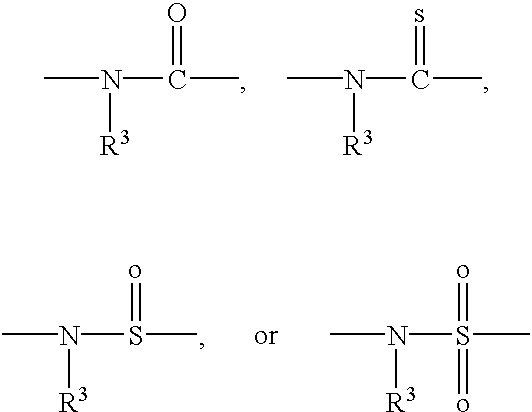
- each of C 1 -C n is CR 6 R 7 ,CHR 6 CHR 7 or CR 6 R 7 CH 2 , wherein R 6 is hydrogen and R 7 is selected from the group consisting of the side chains of naturally occurring alpha amino acids, or R 6 and R 7 are independently selected from the group consisting of hydrogen, (C 2 -C 6 ) alkyl, aryl, aralkyl, heteroaryl, hydroxy, (C 1 -C 6 ) alkoxy, (C 1 -C 6 ) alkylthio, NR 3 R 4 and SR 5 , where R 3 and R 4 are as defined above, and R 5 is hydrogen, (C 1 -C 6 ) alkyl, hydroxy-, alkoxy-, or alkylthio- substituted (C 1 -C 6 ) alkyl, or R 6 and R 7 taken together complete an alicyclic or heterocyclic system;
- each of D 1 -D n is CR 6 R 7 , CH 2 CR 6 R 7 or CHR 6 CHR 7 , where R 6 and R 7 are as defined above;
- each of G 1 -G n-1 is
- Q is —CO 2 H, —CONR′R′′, —SO 3 H or SO 2 NR′R′′ or an activated derivative of —CO 2 H, —SO 3 H;
- I is —NHR′′′R′′′′ or —NR′′′C(O)R′′′′
- R′, R′′, R′′′ and R′′′′ are independently selected from the group consisting of hydrogen, alkyl, amino protecting groups, reporter ligands, intercalators, chelators, peptides, proteins, carbohydrates, lipids, steroids, oligonucleotides and soluble and non-soluble polymers.
- C and D may be CHR 6 (CH 2 ) sss CHR 7 where sss may be from 0 to 2.
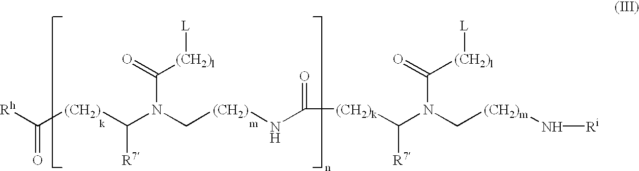
- Preferred peptide nucleic acids have general formula (III):
- each L is independently selected from the group consisting of hydrogen, phenyl, heterocycles, e.g. of one, two or three rings, naturally occurring nucleobases, and non-naturally occurring nucleobases;
- each R 7 ′ is independently selected from the group consisting of hydrogen and the side chains of naturally occurring alpha amino acids
- n is an integer from 1 to 60;
- each of k, l and m is independently zero or an integer from 1 to 5; preferably the sum of k and m is 1 or 2, most preferably 1;
- R h is OH, NH 2 , or —NHLysNH 2 ;
- R i is H or COCH 3 .
- each L is independently selected from the group consisting of the nucleobases thymine (T), adenine (A), cytosine (C), guanine (G) and uracil (U), in particular thymine, and n is an integer from 1 to 30, in particular from 4 to 20.
- T thymine
- A adenine
- C cytosine
- G guanine
- U uracil
- Preferred nucleic acid analogs are compounds that have a polyamide backbone that contains a number of bases bound along the backbone, with each base bound to a nitrogen atom of the backbone. Nucleic acid analogs should also include compounds, however, like those described in EP-A-0 672 677. Additional nucleic acid analogs are described in Recueil 91, 1069 -1080 (1971), Method in Molecular Biology 29, 355-389 (1983), Tetrahedron 31, 73-75 (1975), J. Org. Chem. 52, 4202-4206 (1987), Nucl. Acids Res.
- nucleic acid analogs are described in Proc. Natl. Acad. Sci. USA 91,7864-7868 (1994), Proc. Nat. Acad. Sci. USA 92,6097-6101 (1995) and J. Am. Chem. Soc. 117, 6140-6141 (1995).
- the nucleic acid analogs described are from 8 to 30 bases long, while a length of from 10 to 25 bases is especially advantageous.
- the nucleic acid analogs named are bound directly or indirectly to the surface of the solid carrier.
- the type of binding basically depends on which reactive group are available for binding on the solid carrier, and which reactive groups are available for binding to the nucleic acid analog without restricting the ability of the nucleic acid analogs to bind to a complementary nucleic acid.
- the type of binding also depends on whether the intent is to simultaneously bind the nucleic analogs to different sites, or to build upon them. It can also be appropriate to cover the surface of the solid carrier with a layer of a material that has a great ability to bind, or to activate the surface by means of a chemical reaction.
- Reactive groups on the surface of a solid carrier are usually selected from the group —OH, —NH 2 and SH.
- Reactive groups of nucleic acid analogs are preferably selected from the group —OH, NH 2 , —SH, —COOH, —SO 3 H and —PO 3 H 2.
- the reactive groups of the surface and the nucleic acid analog are covalently bound to each other, especially by means of a linker that is more than 15 atoms and less than 200 atoms long.
- a “linker” refers to a portion of a molecule that basically has the function of removing the nucleic acid analogs that are sterically available on the surface of the solid carrier.
- a linker is usually selected that has hydrogen atoms (e.g. in alkylene units) and numerous heteroatoms (e.g. —O— or —NH— or —NR—) that facilitate solvation.
- the linker preferably contains one or more ethylene oxy units and/or peptide groups.
- the linker contains one or more units as described in DE-A 3924705. Especially preferred are the units described as an example which is referred to as Ado (8-amino-3,6-dioxa-octanic acid), below.
- Ado 8-amino-3,6-dioxa-octanic acid
- the nucleic acid analog that is bound to a site can also be a mixture of two or more analogs having different but known sequences. This can reduce the number of sites required for a multiple determination.
- the surface of the carrier is preferably not charged, and is preferably hydrophilic.
- the invention demonstrated that the use of basically uncharged surfaces is an advantage when detecting nucleic acids.
- a solid carrier loaded with nucleic acid analogs at different sites as provided by this invention can be manufactured in different ways.
- suitable quantities of solutions that each contain different nucleic acid analogs are applied to different sites on the surface of the solid carrier, e.g. via pipette.
- the liquid samples should not mix with each other on the surface of the solid carrier. This can be accomplished, for instance, by locating the application sites far apart from each other or by using a hydrophobic barrier to stop the expansion of the liquid between the various sites.
- Either the nucleic acid analogs or the surface of the solid carrier is preferably activated for the reaction. This activation can be achieved, for instance, in that one of the groups described above is activated by the creation of a reactive species.
- activated ester e.g. a N-hydroxy succinimide ester
- activated polyamide membranes carry triazine groups, for instance, that can react with amino groups of nucleic acid analogs with the formation of a covalent bond.
- the activation can also take place by means of bifunctional reagents, squaric acid derivatives of WO 95/15983 or glutaraldehyde (GB 2197720).
- the binding of the analogs can also be realized by coating the carrier surface with nucleotide sequences that are complementary to a part of the sequence of the nucleotide analogs.
- the binding of the different nucleic acid analogs to the binding regions can take place simultaneously or sequentially.
- nucleic acid analogs that have not bound or are bound insufficiently it is advantageous to wash away any nucleic acid analogs that have not bound or are bound insufficiently, along with any binding reagents that were used. This is performed preferably under conditions in which non-bound nucleic acid analogs cannot bind with nucleic acid analogs that are bound to other regions.
- Another subject matter of this invention is a method for the sequence-specific detection of a nucleic acid using the solid carrier provided by this invention.
- Detectable nucleic acids are natural or artificial nucleic acids. “Nucleic acids” therefore also refer to nucleic acid analogs.
- the nucleic acid to be detected is the RNA or DNA in particular that is characteristic for an organism containing nucleic acids, e.g. a virus, a bacterium, a multicellular organism, a plasmid or a genetic condition such as a predisposition or a disposition for a certain disease or a spontaneous genetic mutation.
- the RNA and DNA in this case is basically of genomic origin or an origin derived therefrom.
- An important class of nucleic acids in the context of this invention are the results of a nucleic acid amplification. These results are also referred to as “amplificates” or “amplicons” below.
- the nucleic acids can be present in either their raw form or in a purified or processed form.
- a purification can also take place by separating the nucleic acids from cell components in a preparatory step, e.g. an affinity separation step.
- the nucleic acids can also be enzymatically extended, specifically amplified or transcribed.
- the carrier provided by this invention has a nucleic acid analog bound to a site that has a base sequence that is complementary to a base sequence of the nucleic acid to be detected.
- This base sequence is selected intentionally so that it can specifically reveal the presence of the nucleic acid. In the normal case this means that the mixture contains as few as possible—and preferably no—additional nucleic acids having the same total sequence.
- the carrier provided by this invention can also be used to specifically detect groups of nucleic acids. It can be a task of the method, for instance, to detect any member of a certain taxonomic group, e.g. a family of bacteria, by means of its nucleic acids.
- the base sequence of the nucleic acid analog can be intentionally selected so that it lies in a conserved region but only occurs in members of this taxonomic group.
- An additional nucleic acid analog that has a base sequence that is not complementary to the same base sequence is preferably bound to a different site of the surface. It can be a nucleic acid analog, the base sequence of which can be shorter or longer, or which can differ from the first nucleic acid analog by one or more bases. The difference in the base sequence depends on the task to be solved. The differences can include point mutations, or smaller deletions and insertions, for instance. In many genetic diseases, such as cystic fibrosis, the sequences of the nucleic acid analogs differ in terms of individual positions (point mutations) and numerous positions (deletions, for example at ⁇ 508).
- the mutation to be detected is preferably positioned close to the middle of the base sequence of the analog.
- sequences of the analog can also be intentionally selected so that their hybridization positions differ by one base each, even though the lengths are identical (overlap).
- sequences can also be intentionally selected so that the hybridization regions are adjacent to each other on the nucleic acid to be detected.
- Numerous mutations can also be detected in the same or different nucleic acids by selecting nucleic acid analogs with sequences that are complementary to the sequence on these nucleic acids.
- nucleic acid analogs are bound to different sites of the surface of the solid carrier, the base sequences of which differ in exactly that position where the alleles also differ.
- a nucleic acid analog is therefore selected that is complementary to a certain sequence of one allele, while the other nucleic acid analog is complementary to the sequence of the other allele.
- the length and hybridization sites of the nucleic acid analogs are identical.
- All alleles are usually detected for cystic fibrosis, for instance.
- the wild-type contains two healthy alleles.
- Heterozygotes contain one mutated and one wild-type sequence, and homozygotic mutants contain two mutant nucleic acids.
- the intent is not just to determine if mutants are present, but to determine if it is a heterozygotic or homozygotic case.
- mutated cells/particles are usually located in the background of non-mutated/normal cells. In these cases, selective detection can not be performed reliably or at all using methods provided by the state of the art.
- the analysis of ras mutations from DNA from stool samples, for instance, requires that a mutated sequence be reliably detected in the presence of approx. 100 normal sequences (Science 256, 102-105 (1992)).
- the quantity of many mutants of these HIV populations is less than 2% compared with all HIV sequences, however.
- the method provided by this invention therefore makes it especially quite possible to investigate mixtures of nucleic acids that are very similar to each other, even if one of the nucleic acids is present in a much greater quantity than the nucleic acid to be determined.
- the lengths of the bound nucleic acid analogs are preferably identical.
- An appropriate length has proven to be between 10 and 100, and preferably between 10 and 50 bases. Especially good results are obtained with nucleic acid analogs that are between 10 and 25 bases long.
- the sample containing nucleic acid is brought in contact with the sites on the surface of the carrier that have bound the nucleic acid analogs.
- This can be performed, for instance, by bringing the solid carrier into the sample fluid or pouring the sample fluid onto the solid carrier in one or more portions.
- the nucleic acids in the sample fluid can be denatured (single-strand) before they are brought in contact with the carrier.
- a major advantage of the invention is the fact that a preliminary denaturation step can be eliminated, e.g. by using PNAs.
- the PNAs force a strand out of the double-stranded nucleic acid to be determined.
- the sample be brought in contact with the solid carrier under conditions in which the nucleic acid to be detected binds specifically to the appropriate site on the surface by means of the nucleic acid analog which is complementary to one sequence of the nucleic acid to be determined.
- These conditions can be different for different types of nucleic acid analogs, of course, but they are easily determined for given nucleic acid analogs by performing tests. In the normal case, these conditions are based on the conditions that are known for carriers loaded with oligonucleotides. If nucleic acid analogs are used as described in WO 92/20702, however, conditions can be selected that are much different from the hybridization conditions for the corresponding oligonucleotides.
- the sample is kept in contact with the surface as long as necessary to achieve a sufficient binding of the nucleic acids to the appropriate site on the surface. This period is usually a few minutes.
- the binding can be determined using various methods.
- Instruments are already available with which changes on specific sites of surfaces can be determined directly. For methods that use these types of instruments it is not even necessary to remove the sample containing the nucleic acid from the surface after it has been applied. Normally, however, it is preferable to remove the fluid from the surface and use a wash solution to remove any remaining reagent that is still adhered to the surface. This step provides the advantage of also washing away sample components that can interfere with the determination of the binding.
- the binding of the nucleic acid to be detected with the nucleic acid analog is determined by means of a label that is inserted in the nucleic acid to be determined in a step that is performed before the sample is brought in contact with the surface.
- the label can be a detectable group such as a fluorescent group, for instance. This determination can be performed optically using a microscope or in a measuring cell provided for this purpose. While the site at which the binding took place is an indication of the presence of a nucleic acid having a certain sequence, the quantity of label at a predetermined site can be used as an indication of the quantity of the nucleic acid to be determined.
- the nucleic acids to be detected are the products of a nucleic acid amplification method such as the polymerase chain reaction as described in EP-B-0 202 362, or NASBA as described in EP-A-0 329 822. It is important that the nucleic acid sequence to bind with the nucleic acid analog be amplified by the amplification method. The better the amplification method maintains the original sequence—that is, the fewer errors that are incorporated into the sequence during amplification—the more suitable the amplification method. The polymerase chain reaction has proven to be especially suitable.
- the amplification primers are selected specifically so that the nucleic acid sequence to be detected lies in the region between the hybridization sites.
- the binding that took place can be detected directly without inserting a label, for instance, by using an intercalating agent.
- intercalating agents have the characteristic of depositing selectively on double-stranded compounds that contain bases, including the complex of the nucleic acid analog and the nucleic acid bound in sequence-specific fashion.
- the presence of the complexes can be detected using specific characteristics of intercalating agents, e.g. fluorescence. Ethidium bromide is an especially suitable agent.
- Another method for detecting hybrids without inserting a label is based on surface plasmon resonance, as described in EP-A-0 618 441, for instance.
- the surface is brought in contact with a solution of an antibody labelled for detection.
- This antibody is directed against the complex consisting of the nucleic acid analog and the nucleic acid to be determined.
- Antibodies of this nature are described in WO 95/17430, for instance.
- the detection of the hybrids depends on the type of labelling used.
- the hybrids can be detected with a scanner, a CCD camera or a microscope, for instance.
- This invention provides numerous advantages. In particular, it allows detecting sequence differences in nucleic acid regions located within secondary structures. With this invention it is also possible to increase the sensitivity of detection, because it can use a greater absolute quantity of bound nucleic acid to be determined than traditional methods. It is also possible, in particular, to increase the signal-to-noise ratio compared with methods that use oligonucleotides. In the first attempt, a signal-to-noise ratio of less than 1:1000 was obtained.
- the invention can be used in at least two fields.
- the solid carrier is used to detect known mutations and polymorphisms.
- the number of mutations and polymorphisms to be determined is an indicator of the number of different nucleic acid analogs or sites required on the surface.
- the sequence of the nucleic acid analogs is specially coordinated with the sequence of the nucleic acids around the mutations and polymorphisms.
- the sequences are selected in such a way that the base by which nucleic acid analogs having similar sequences differ is located in or near the middle of the sequence.
- Infectious diseases the simultaneous determination of different analytes/parameters, and in investigations of the condition of a gene in a bacterium or virus, e.g. for multi-drug resistance studies.
- Oncology detection of mutations in tumor suppressor genes and oncogenes, and in the determination of the relationship between mutated and normal cells.
- a sequence of short nucleic acid fragments can be determined using the method called “sequencing by hybridizatior”.
- the same number of different nucleic acid analogs are immobilized as there are permutations of the selected length of the sequence.
- 4 N sites are required, with N equal to the number of bases in each nucleic acid analog.
- N is between 5 and 12.
- Correspondingly fewer sites are required to sequence very short DNA fragments.
- the method for sequencing unknown nucleic acids using the “sequencing by hybridization” method is described in WO 92/10588.
- An advantage of this invention is the fact that the specificity of the hybridization is largely independent of the conditions in the sample. This facilitates simultaneous binding of nucleic acids to different regions on the surface.
- nucleic acid analogs such as PNA have an excellent ability to discriminate between sequences on the surface. This discrimination was better than was to be expected from the melting temperatures of analogous, dissolved compounds.
- the carriers provided by this invention are even suitable for use in numerous, consecutive determinations of nucleic acids. After a determination is performed, the carrier that is in contact with a fluid undergoes heat treatment. A temperature is selected at which the bond between the nucleic acid analog and the nucleic acid is dissolved. The carrier is then available to perform another determination.
- the method provided by this invention it is possible for the first time to determine relative quantities of very similar nucleic acids located next to each other in a sample in a concentration range of at least two logs. It has been possible to quantitatively determine mutants using sequencing methods, for instance. The maximum level of discrimination available with this method was 1:10, however.
- the sequences of the PNA molecules are shown in FIG. 1 a . They are used as examples to explain the method.
- the PNAs were prepared as described in WO 92/20702.
- FIG. 1 b shows the sequences of the DNA molecules that are homologous to the PNA sequences from FIG. 1 a and that were used for the DNA/ODN hybridization experiments.
- FIG. 1 c shows the sequences of the complementary oligonucleotides (ODN) used that were labelled with digoxigenin on the 5′-phosphate end using the 5′-DIG End Labelling Kit (Boehringer Mannheim) and that were phosphorylated with polynucleotide kinase and 32 -P-g-ATP 5′.
- ODN complementary oligonucleotides
- FIG. 1 d shows the feasible combinations of oligonucleotide (ODN) and PNA for forming hybrids. Identical combinations apply for hybridizations between DNA probes (DNA, see FIG. 1 b ) and oligonucleotides (ODN, see FIG. 1 c ).
- FIG. 2 shows the hybridization results from Example 4 to illustrate the selectivity of the method.
- the conditions were: 200 nl spot volume (100; 10; 1; 0.1 mM PNA, one concentration per cleavage), incubation at 45° C.
- FIG. 3 shows the hybridization results from Example 6. They verify that PCR amplicons are detected by immobilized PNA probes. The conditions were: 200 nl spot volume (100; 10; 1; 0.1 mM PNA, one concentration per cleavage), incubation at 45° C. The labels in FIG. 3 mean:
- FIG. 4 shows the results of the qualitative and quantitative analysis of analyte mixtures by means of PNA arrays.
- the conditions were: 200 nl spot volume (100 mM PNA, one PNA per line, one analyte mixture per cleavage).
- FIG. 5 shows the effect of linker length on the hybridization.
- the conditions were: 1 ml spot volume (100; 40; 20; 10; 5; 1 mM PNA, one concentration per cleavage, (Ado) 3 -PNA in row 1, (Ado) 6 -PNA in row 2, (Ado) 9 -PNA in row 3).
- the labels in FIG. 5 mean:
- FIGS. 6 a - 6 c demonstrate that PNA-derivatized membranes can be used many times after regeneration.
- the conditions were: 1 ml spot volume (100; 40; 20; 10; 5; 1 mM PNA, one concentration per cleavage).
- Membrane 2 Regeneration with 0.1 M sodium hydroxide solution, RT 1 h, 2 ⁇ 10 min bidistilled water RT
- Membrane 3 Regeneration with 1 M sodium hydroxide solution, RT, 1 h, 2 ⁇ 10 min bidistilled water RT
- Membrane 4 Regeneration with distilled water, 70° C. 1 h, 2 ⁇ 10 min bidistilled water RT
- Membrane 5 Regeneration with 0.1 M sodium hydroxide solution, 70° C. 1 h, 2 ⁇ 10 min bidistilled water RT
- nucleic acid analogs used were manufactured as described in WO 92/20702. Unless indicated otherwise, chemicals and reagents were products of Boehringer Mannheim GmbH.
- the membrane is derivatized as described in Example 1 using 100 ⁇ M, 10 mM, 1 ⁇ M and 0.1 mM PNA solutions. It is then prehybridized in a 50 ml hybridization vessel with 10 ml hybridization buffer (10 mM sodium phosphate, pH 7.2, 0.1% SDS (sodium dodecylsulfate)) in a hybridization oven at 45° C. After 30 minutes, 10 ml of a solution that contains the DIG-labelled oligonucleotide in a 1 mM concentration is added and the complex is hybridized for another 60 minutes.
- 10 ml hybridization buffer (10 mM sodium phosphate, pH 7.2, 0.1% SDS (sodium dodecylsulfate)
- the detection reaction is performed according to the protocol for digoxigenin detection (DIG Detection Kit, Boehringer Mannheim GmbH, BRD).
- the anti-DIG-AP conjugate is used in a 1:10000 dilution.
- CDP-StarTM is used in a 1:10000 dilution as the substrate for the alkaline phosphatase.
- the membrane is derivatized as described in Example 1 using 100 ⁇ M, 10 ⁇ M and 1 ⁇ M PNA solution.
- the membrane is prehybridized in a 50 ml screw-top container with 10 ml hybridization buffer (see Example 2) in the oven at 45° C. After 30 minutes, 10 ml of a solution that contains a fluorescent-labelled oligonucleotide in a concentration of 1 ⁇ M is added, and the preparation is hybridized for another 60 minutes.
- the membrane is then washed for 2 ⁇ 10 minutes with 25 ml wash buffer each time (see Example 2) at 45° C.
- the membrane is dried, then the intensity of the fluorescence is measured.
- Three membrane strips are derivatized with three (Ado) 6 -PNA molecules each that differ according to one or two positions of their base sequence (see FIG. 1 a , SEQ.ID.NOS. 1, 2, 3), using PNA solutions in a concentration range of between 100 mM and 0.1 mM as described in Example 1.
- the membrane strips are prehybridized with 10 ml hybridization buffer for 30 minutes in 50 ml screw-top containers.
- one of the three DIG-labelled oligonucleotides (FIG. 1 b , SEQ.ID.NOS. 4, 5, 6) is added. After hybridizing for 60 minutes, the membranes are washed for 2 ⁇ 10 minutes with 25 ml wash buffer each time. The hybridization events are detected as described in Example 2.
- FIG. 1 d All possible double-stranded hybrids between the PNA molecules involved and the oligonucleotides are shown in FIG. 1 d .
- FIG. 2 illustrates that, in almost every case, the only oligonucleotide detected is the one that is exactly complementary to the immobilized nucleic acid analog (PNA 1, PNA 2, PNA 3).
- the signal-to-noise ratios (S/N) can also be estimated from the figure. They were evaluated quantitatively, and the results are presented in Table 1.
- Hybrid PNA/ODN
- Hybrid S/N Signal (Hybrid)/Signal (Match) 1/1 655.2 100.0% 2/1 20.7 3.2% 3/1 10.3 1.6% 1/2 23.1 2.6% 2/2 871.8 100.0% 3/2 6.4 0.7% 1/3 109.4 22.7% 2/3 12.3 2.5% 3/3 481.1 100.0%
- Membrane strips are derivatized with three (Ado) 6 -PNA molecules with different base sequences (FIG. 1 a , SEQ.ID.NOS. 1, 2, 3) in a concentration of 100 mM as described in Example 1. They are then prehybridized in 20 ml hybridization vessels with 10 ml hybridization buffer (see Example 2) at 45° C. The buffer is replaced after 30 minutes. In experiments 1 through 7, the buffer to be added differs according to the analyte concentrations of the DIG-labelled components—oligonucleotide 1, 2 and 3, SEQ.ID.NOS. 4, 5, 6. The strips are hybridized for 60 minutes at 45° C. and then washed for 2 ⁇ 10 minutes with 10 ml wash buffer. The detection is performed using the procedure described in Example 2. The luminescence signal is recorded with a luminescence imager and then evaluated (FIG. 4 ).
- the signal intensities found can be used to reach a qualitative or semi-quantitative finding regarding the composition of the analyte complex.
- absolutely quantitative findings can be reached after the signal intensities are calibrated.
- a double-stranded DNA fragment is ligated in a pUC19 plasmid, the sequence of which is complementary to the PNA probe PNA 1.
- the plasmid is transformed in E. coli, cloned, and then sequenced.
- a section of the plasmid sequence is amplified and DIG-labelled during the amplification reaction. The amplification is performed in a total volume of 50 ⁇ l.
- the amplification complex consists of 1 ⁇ l plasmid (1 ng/ ⁇ l) 1 ⁇ l primer F1 (10 ⁇ M), 1 ⁇ l DIG primer R1 (10 ⁇ M), 5 ⁇ l 10 ⁇ PCR buffer (100 mM Tris/HCl, 15 mM MgCl 2 , 500 mM KCl, pH 8.3), 2 ⁇ l dNTP solution (10 mM dATP, 10 mM dCTP, 10 mM dGTP, 10 mM dTTP in distilled water, pH 7.0), 0.5 ⁇ l Taq polymerase (5 units/ ⁇ l) and 38.5 ml water.
- Primer F1 5′-GTA AAA CGA CGG CCA GT-3′ (SEQ.ID.NO. 12)
- Primer R1 5′-DIG-AAC AGC TAT GAC CAT GA-3′ (SEQ.ID.NO. 13)
- reaction mixture is warmed to 96° C. for 3 minutes.
- 30 rounds of a 3-level PCR cycle are performed (45 sec. 96° C., 30 sec, 48° C., 1 min 72° C.).
- the elongation step is increased by 5 minutes at 72° C.
- the membranes are derivatized with three (Ado) 6 -PNA sequences each that differ according to one or two positions in their base sequence (see FIG. 1 a , SEQ.ID.NOS. 1, 2, 3) using PNA solutions in a concentration range between 100 ⁇ M and 0.1 ⁇ M as described in Example 1.
- the membrane is pretreated in a 20 ml hybridization vessel with 5 ml hybridization buffer at 45° C. The buffer is replaced after 30 minutes and the analyte solution is added. To make the analyte solution, the amplification complex is diluted directly (ds amplicon) and, after 5 minutes of heat denaturation (ss amplicon), in 1 ml hybridization buffer.
- the difference between each row is the incubation period (4 h, 21 ⁇ 2 h, and 1 h).
- the difference between each column is the type of nucleic acid to be detected.
- Three overlapping rows of spots are applied to each of the 3 fields of column I.
- the difference between the rows is the sequence of the PNAs, while the difference between the columns of each field is the concentration.
- the specificity and the ability to be quantified are indicated in column I for the case in which oligonucleotides are used as the detecting nucleic acid.
- the figure illustrates the influence of incubation time. It is clear that an excellent sequence discrimination for ODN 1a and the amplificates is obtained after hybridization for just one hour.
- the difference between columns II and III in FIG. 3 is that an amplificate that was previously made single-stranded is used in one case as the nucleic acid to be detected.
- an amplificate that was not previously made single-stranded is used as the nucleic acid to be detected.
- the signals indicate clearly that it is not necessary to denature double-stranded nucleic acids before applying them to the solid carrier. This decreases the number of working steps (heating step, single strand separation, wash step) and, therefore, reduces the danger of contamination.
- PNA probes in combination with low-salt conditions therefore offer clear advantages over DNA probes.
- Membrane strips are derivatized with three (Ado) 6 -PNA sequences each (SEQ.ID.NOS. 1, 2, 3) and three DNA molecules each (SEQ.ID.NOS. 8, 10, 11) that differ according to one or two positions in their base sequence (see FIG. 1 a and 1 b ) using 50 mM solutions as described in Example 1. Unlike Example 1, the spot volume is 400 nl instead of 200 nl.
- the membrane strips are prehybridized in 20 ml hybridization vessels with either 5 ml low-salt buffer (see Example 2) or high-salt buffer (6 ⁇ SSC; 0.9 M NaCl, 90 mM sodium citrate, 0.1% SDS, pH 7.0) at either 37° C. or 45° C.
- Both the DNA and PNA probes are able to completely discriminate between complementary, single-stranded target sequences of single and double mismatched sequences.
- PNA probes demonstrate clear advantages over the DNA probes for certain types of mismatches, especially when they are not located in the middle of the sequence, but rather shifted to the end. This becomes especially clear in the example of a decentral G/T mismatch (probe 1/ODN 3), which is tolerated by the DNA probe much more strongly than by the PNA probe having the identical sequence.
- Membrane strips are derivatized with PNA molecules (see FIG. 1 a , SEQ.ID.NOS. 7, 1, 9) that differ according to the length of the linker (Ado 3 , Ado6 or Ado 9 ) using PNA solutions in the concentration range between 100 ⁇ M and 1 ⁇ M as described in Example 1. Unlike Example 1, the spot volume is 1 ⁇ l instead of 200 nl.
- the membrane strips are prehybridized in hybridization containers with 10 ml hybridization buffer (5, 10 or 25 mM sodium phosphate, 0.1% SDS, pH 7.0) for 30 minutes at 35° C. In the next step, 10 pMol 32 P-labelled oligonucleotide (see FIG.
- a membrane is derivatized with (Ado) 6 -PNA molecules (see FIG. 1 a : PNA 1b, SEQ.ID.NO. 1) using PNA solutions in a concentration range between 100 ⁇ M and 1 ⁇ M as described in Example 1.
- the PNA is applied in five identical concentration sequences.
- the spot volume is 1 ⁇ l, as in Example 8.
- the membrane is prehybridized in a hybridization vessel with 10 ml hybridization buffer (10 mM sodium phosphate, 0.1% SDS, pH 7.0) for 30 minutes at 35° C.
- 10 pMol 32 P-labelled oligonucleotide see FIG. 1 c : ODN 1b, SEQ.ID.NO.
- Membrane 4 is incubated for 60 minutes at 70° C. with 50 ml distilled water.
- Membrane 5 is incubated for 60 minutes at 70° C. with 50 ml 0.1 N sodium hydroxide solution. All membranes are then washed with distilled water for 2 ⁇ 10 minutes. After this procedure is completed, autoradiography is performed once more (FIG. 6 b ). These membrane strips are then used a second time in a hybridization reaction as described, and the hybridization events are detected using autoradiography (FIG. 6 c ).
Abstract
Description


| TABLE 1 | ||
| Hybrid (PNA/ODN) | S/N | Signal (Hybrid)/Signal (Match) |
| 1/1 | 655.2 | 100.0% |
| 2/1 | 20.7 | 3.2% |
| 3/1 | 10.3 | 1.6% |
| 1/2 | 23.1 | 2.6% |
| 2/2 | 871.8 | 100.0% |
| 3/2 | 6.4 | 0.7% |
| 1/3 | 109.4 | 22.7% |
| 2/3 | 12.3 | 2.5% |
| 3/3 | 481.1 | 100.0% |
| Primer F1: 5′-GTA AAA CGA CGG CCA GT-3′ | (SEQ.ID.NO. 12) |
| Primer R1: 5′-DIG-AAC AGC TAT GAC CAT GA-3′ | (SEQ.ID.NO. 13) |
| TABLE 2 | ||||
| PNA or | PNA or | PNA or | ||
| |
|
|
||
| |
PNA 45° C., low salt | 100.0% | 1.2% | 1.5% |
| DNA 37° C., high | 100.0% | 1.2% | 6.4 | |
| salt | ||||
| ODN | ||||
| 2 | PNA 45° C., low salt | 1.1% | 100.0% | 2.9% |
| DNA 37° C., high | <2%* | 100.0% | <2%* | |
| | ||||
| ODN | ||||
| 3 | PNA 45° C., low salt | 26.0% | 1.5% | 100.0% |
| DNA 37° C., high | 66.5% | <2%* | 100.0% | |
| salt | ||||
| *A more exact value cannot be determined because the spot intensity is lower than the standard deviation of the background signal. | ||||
| # SEQUENCE LISTING |
| <160> NUMBER OF SEQ ID NOS: 13 |
| <210> SEQ ID NO 1 |
| <211> LENGTH: 16 |
| <212> TYPE: PRT |
| <213> ORGANISM: Unknown |
| <220> FEATURE: |
| <223> OTHER INFORMATION: Description of Unknown Or |
| #ganism: Synthetic |
| <221> NAME/KEY: misc_feature |
| <222> LOCATION: (15) |
| <223> OTHER INFORMATION: note= “Xaa is N-((1 |
| #-adeninyl)acetyl)-N- |
| (2-aminoethyl)-beta-alanine” |
| <221> NAME/KEY: misc_feature |
| <222> LOCATION: (14) |
| <223> OTHER INFORMATION: note= “Xaa is N-((1 |
| #-thyminyl)acetyl)-N-(2- |
| aminoethyl)-beta-alanine” |
| <221> NAME/KEY: misc_feature |
| <222> LOCATION: (13) |
| <223> OTHER INFORMATION: note= “Xaa is N-((1 |
| #-cytosyl)acetyl)-N-(2- |
| aminoethyl)-beta-alanine” |
| <221> NAME/KEY: misc_feature |
| <222> LOCATION: (11)..(12) |
| <223> OTHER INFORMATION: note= “Xaa is N-((1 |
| #-adeninyl)acetyl)-N-(2- |
| aminoethyl)-beta-alanine” |
| <221> NAME/KEY: misc_feature |
| <222> LOCATION: (10) |
| <223> OTHER INFORMATION: note= “Xaa is N-((1 |
| #-cytosyl)acetyl)-N-(2- |
| aminoethyl)-beta-alanine” |
| <221> NAME/KEY: misc_feature |
| <222> LOCATION: (9) |
| <223> OTHER INFORMATION: note= “Xaa is N-((1 |
| #-adeninyl)acetyl)-N-(2- |
| aminoethyl)-beta-alanine” |
| <221> NAME/KEY: misc_feature |
| <222> LOCATION: (8) |
| <223> OTHER INFORMATION: note= “Xaa is N-((1 |
| #-cytosyl)acetyl)-N-(2- |
| aminoethyl)-beta-alanine” |
| <221> NAME/KEY: misc_feature |
| <222> LOCATION: (7) |
| <223> OTHER INFORMATION: note= “Xaa is N-((1 |
| #-thyminyl)acetyl)-N-(2- |
| aminoethyl)-beta-alanine” |
| <221> NAME/KEY: misc_feature |
| <222> LOCATION: (6) |
| <223> OTHER INFORMATION: note= “Xaa is N-((1 |
| #-guaninyl)acetyl)-N-(2- |
| aminoethyl)-beta-alanine” |
| <221> NAME/KEY: misc_feature |
| <222> LOCATION: (5) |
| <223> OTHER INFORMATION: note= “Xaa is N-((1 |
| #-cytosyl)acetyl)-N-(2- |
| aminoethyl)-beta-alanine” |
| <221> NAME/KEY: misc_feature |
| <222> LOCATION: (4) |
| <223> OTHER INFORMATION: note= “Xaa is N-((1 |
| #-adeninyl)acetyl)-N-(2- |
| aminoethyl)-beta-alanine” |
| <221> NAME/KEY: misc_feature |
| <222> LOCATION: (3) |
| <223> OTHER INFORMATION: note= “Xaa is N-((1 |
| #-thyminyl)acetyl)-N-(2- |
| aminoethyl)-beta-alanine” |
| <221> NAME/KEY: misc_feature |
| <222> LOCATION: (2) |
| <223> OTHER INFORMATION: note= “Xaa is N-((1 |
| #-guaninyl)acetyl)-N-(2- |
| aminoethyl)-beta-alanine” |
| <221> NAME/KEY: misc_feature |
| <222> LOCATION: (1) |
| <223> OTHER INFORMATION: note= “Xaa is N-((1 |
| #-thyminyl)acetyl)-N-(2- |
| amino-N′-(hexa(8-amino-3,6-dioxa-octano-1-yl) |
| #-ethyl)-beta-alanine” |
| <221> NAME/KEY: misc_feature |
| <222> LOCATION: (16) |
| <223> OTHER INFORMATION: Product= “OTHER” no |
| #te=“Amide” |
| <400> SEQUENCE: 1 |
| Xaa Xaa Xaa Xaa Xaa Xaa Xaa Xaa Xaa Xaa Xa |
| #a Xaa Xaa Xaa Xaa Gly |
| 1 5 |
| # 10 |
| # 15 |
| <210> SEQ ID NO 2 |
| <211> LENGTH: 16 |
| <212> TYPE: PRT |
| <213> ORGANISM: Unknown |
| <220> FEATURE: |
| <223> OTHER INFORMATION: Description of Unknown Or |
| #ganism: Synthetic |
| <221> NAME/KEY: misc_feature |
| <222> LOCATION: (15) |
| <223> OTHER INFORMATION: note= “Xaa is N-((1 |
| #-adeninyl)acetyl)-N-(2- |
| aminoethyl)-beta-alanine” |
| <221> NAME/KEY: misc_feature |
| <222> LOCATION: (14) |
| <223> OTHER INFORMATION: note= “Xaa is N-((1 |
| #-thyminyl)acetyl)-N-(2- |
| aminoethyl)-beta-alanine” |
| <221> NAME/KEY: misc_feature |
| <222> LOCATION: (13) |
| <223> OTHER INFORMATION: note= “Xaa is N-((1 |
| #-cytosyl)acetyl)-N-(2- |
| aminoethyl)-beta-alanine” |
| <221> NAME/KEY: misc_feature |
| <222> LOCATION: (11)..(12) |
| <223> OTHER INFORMATION: note= “Xaa is N-((1 |
| #-adeninyl)acetyl)-N-(2- |
| aminoethyl)-beta-alanine” |
| <221> NAME/KEY: misc_feature |
| <222> LOCATION: (10) |
| <223> OTHER INFORMATION: note= “Xaa is N-((1 |
| #-cytosyl)acetyl)-N-(2- |
| aminoethyl)-beta-alanine” |
| <221> NAME/KEY: misc_feature |
| <222> LOCATION: (9) |
| <223> OTHER INFORMATION: note= “Xaa is N-((1 |
| #-adeninyl)acetyl)-N-(2- |
| aminoethyl)-beta-alanine” |
| <221> NAME/KEY: misc_feature |
| <222> LOCATION: (8) |
| <223> OTHER INFORMATION: note= “Xaa is N-((1 |
| #-guaninyl)acetyl)-N-(2- |
| aminoethyl)-beta-alanine” |
| <221> NAME/KEY: misc_feature |
| <222> LOCATION: (7) |
| <223> OTHER INFORMATION: note= “Xaa is N-((1 |
| #-thyminyl)acetyl)-N-(2- |
| aminoethyl)-beta-alanine” |
| <221> NAME/KEY: misc_feature |
| <222> LOCATION: (6) |
| <223> OTHER INFORMATION: note= “Xaa is N-((1 |
| #-guaninyl)acetyl)-N-(2- |
| aminoethyl)-beta-alanine” |
| <221> NAME/KEY: misc_feature |
| <222> LOCATION: (5) |
| <223> OTHER INFORMATION: note= “Xaa is N-((1 |
| #-cytosyl)acetyl)-N-(2- |
| aminoethyl)-beta-alanine” |
| <221> NAME/KEY: misc_feature |
| <222> LOCATION: (4) |
| <223> OTHER INFORMATION: note= “Xaa is N-((1 |
| #-adeninyl)acetyl)-N-(2- |
| aminoethyl)-beta-alanine” |
| <221> NAME/KEY: misc_feature |
| <222> LOCATION: (3) |
| <223> OTHER INFORMATION: note= “Xaa is N-((1 |
| #-thyminyl)acetyl)-N-(2- |
| aminoethyl)-beta-alanine” |
| <221> NAME/KEY: misc_feature |
| <222> LOCATION: (2) |
| <223> OTHER INFORMATION: note= “Xaa is N-((1 |
| #-guaninyl)acetyl)-N-(2- |
| aminoethyl)-beta-alanine” |
| <221> NAME/KEY: misc_feature |
| <222> LOCATION: (1) |
| <223> OTHER INFORMATION: note= “Xaa is |
| N-((1-thyminyl)acetyl)-N-(2-amino-N′-(hexa(8- |
| #amino-3,6-dioxa- |
| octano-1-yl)-ethyl)-beta-alanine” |
| <221> NAME/KEY: misc_feature |
| <222> LOCATION: (16) |
| <223> OTHER INFORMATION: Product= “OTHER” no |
| #te=“Amide” |
| <400> SEQUENCE: 2 |
| Xaa Xaa Xaa Xaa Xaa Xaa Xaa Xaa Xaa Xaa Xa |
| #a Xaa Xaa Xaa Xaa Gly |
| 1 5 |
| # 10 |
| # 15 |
| <210> SEQ ID NO 3 |
| <211> LENGTH: 16 |
| <212> TYPE: PRT |
| <213> ORGANISM: Unknown |
| <220> FEATURE: |
| <223> OTHER INFORMATION: Description of Unknown Or |
| #ganism: Synthetic |
| <221> NAME/KEY: misc_feature |
| <222> LOCATION: (15) |
| <223> OTHER INFORMATION: note= “Xaa is N-((1 |
| #-adeninyl)acetyl)-N-(2- |
| aminoethyl)-beta-alanine” |
| <221> NAME/KEY: misc_feature |
| <222> LOCATION: (14) |
| <223> OTHER INFORMATION: note= “Xaa is N-((1 |
| #-thyminyl)acetyl)-N-(2- |
| aminoethyl)-beta-alanine” |
| <221> NAME/KEY: misc_feature |
| <222> LOCATION: (13) |
| <223> OTHER INFORMATION: note= “Xaa is N-((1 |
| #-cytosyl)acetyl)-N-(2- |
| aminoethyl)-beta-alanine” |
| <221> NAME/KEY: misc_feature |
| <222> LOCATION: (11)..(12) |
| <223> OTHER INFORMATION: note= “Xaa is N-((1 |
| #-adeninyl)acetyl)-N-(2- |
| aminoethyl)-beta-alanine” |
| <221> NAME/KEY: misc_feature |
| <222> LOCATION: (10) |
| <223> OTHER INFORMATION: note= “Xaa is N-((1 |
| #-cytosyl)acetyl)-N-(2- |
| aminoethyl)-beta-alanine” |
| <221> NAME/KEY: misc_feature |
| <222> LOCATION: (9) |
| <223> OTHER INFORMATION: note= “Xaa is N-((1 |
| #-adeninyl)acetyl)-N-(2- |
| aminoethyl)-beta-alanine” |
| <221> NAME/KEY: misc_feature |
| <222> LOCATION: (8) |
| <223> OTHER INFORMATION: note= “Xaa is N-((1 |
| #-cytosyl)acetyl)-N-(2- |
| aminoethyl)-beta-alanine” |
| <221> NAME/KEY: misc_feature |
| <222> LOCATION: (7) |
| <223> OTHER INFORMATION: note= “Xaa is N-((1 |
| #-thyminyl)acetyl)-N-(2- |
| aminoethyl)-beta-alanine” |
| <221> NAME/KEY: misc_feature |
| <222> LOCATION: (6) |
| <223> OTHER INFORMATION: note= “Xaa is N-((1 |
| #-adeninyl)acetyl)-N-(2- |
| aminoethyl)-beta-alanine” |
| <221> NAME/KEY: misc_feature |
| <222> LOCATION: (5) |
| <223> OTHER INFORMATION: note= “Xaa is N-((1 |
| #-cytosyl)acetyl)-N-(2- |
| aminoethyl)-beta-alanine” |
| <221> NAME/KEY: misc_feature |
| <222> LOCATION: (4) |
| <223> OTHER INFORMATION: note= “Xaa is N-((1 |
| #-adeninyl)acetyl)-N-(2- |
| aminoethyl)-beta-alanine” |
| <221> NAME/KEY: misc_feature |
| <222> LOCATION: (3) |
| <223> OTHER INFORMATION: note= “Xaa is N-((1 |
| #-thyminyl)acetyl)-N-(2- |
| aminoethyl)-beta-alanine” |
| <221> NAME/KEY: misc_feature |
| <222> LOCATION: (2) |
| <223> OTHER INFORMATION: note= “Xaa is N-((1 |
| #-guaninyl)acetyl)-N-(2- |
| aminoethyl)-beta-alanine” |
| <221> NAME/KEY: misc_feature |
| <222> LOCATION: (1) |
| <223> OTHER INFORMATION: note= “Xaa is |
| N-((1-thyminyl)acetyl)-N-(2-amino-N′-(hexa(8- |
| #amino-3,6-dioxa- |
| octano-1-yl)-ethyl)-beta-alanine” |
| <221> NAME/KEY: misc_feature |
| <222> LOCATION: (16) |
| <223> OTHER INFORMATION: Product= “OTHER” no |
| #te=“Amide” |
| <400> SEQUENCE: 3 |
| Xaa Xaa Xaa Xaa Xaa Xaa Xaa Xaa Xaa Xaa Xa |
| #a Xaa Xaa Xaa Xaa Gly |
| 1 5 |
| # 10 |
| # 15 |
| <210> SEQ ID NO 4 |
| <211> LENGTH: 15 |
| <212> TYPE: DNA |
| <213> ORGANISM: Unknown |
| <220> FEATURE: |
| <223> OTHER INFORMATION: Description of Unknown Or |
| #ganism: Synthetic |
| <223> OTHER INFORMATION: Molecule Type: Other nucl |
| #eic acid= |
| “oligodesoxyribonucleotide” |
| <221> NAME/KEY: misc_feature |
| <222> LOCATION: (15) |
| <223> OTHER INFORMATION: note= “labelled at |
| # the 5′-phosphate with |
| digoxigenin via aminolinker (Boehringer |
| # Mannheim GmbH, BRD) or |
| 32-P” |
| <400> SEQUENCE: 4 |
| tagttgtgac gtaca |
| # |
| # |
| # 15 |
| <210> SEQ ID NO 5 |
| <211> LENGTH: 15 |
| <212> TYPE: DNA |
| <213> ORGANISM: Unknown |
| <220> FEATURE: |
| <223> OTHER INFORMATION: Description of Unknown Or |
| #ganism: Synthetic |
| <223> OTHER INFORMATION: Molecule Type: Other nucl |
| #eic acid= |
| “oligodesoxyribonucleotide” |
| <221> NAME/KEY: misc_feature |
| <222> LOCATION: (15) |
| <223> OTHER INFORMATION: note= “labelled at |
| # the 5′-phosphate with |
| digoxigenin via aminolinker (Boehringer |
| # Mannheim GmbH, BRD)” |
| <400> SEQUENCE: 5 |
| tagttgtcac gtaca |
| # |
| # |
| # 15 |
| <210> SEQ ID NO 6 |
| <211> LENGTH: 15 |
| <212> TYPE: DNA |
| <213> ORGANISM: Unknown |
| <220> FEATURE: |
| <223> OTHER INFORMATION: Description of Unknown Or |
| #ganism: Synthetic |
| <223> OTHER INFORMATION: Molecule Type: Other nucl |
| #eic acid= |
| “oligodesoxyribonucleotide” |
| <221> NAME/KEY: misc_feature |
| <222> LOCATION: (15) |
| <223> OTHER INFORMATION: note= “labelled at |
| # the 5′-phosphate with |
| digoxigenin via aminolinker (Boehringer |
| # Mannheim GmbH, BRD)” |
| <400> SEQUENCE: 6 |
| tagttgtgat gtaca |
| # |
| # |
| # 15 |
| <210> SEQ ID NO 7 |
| <211> LENGTH: 16 |
| <212> TYPE: PRT |
| <213> ORGANISM: Unknown |
| <220> FEATURE: |
| <223> OTHER INFORMATION: Description of Unknown Or |
| #ganism: Synthetic |
| <221> NAME/KEY: misc_feature |
| <222> LOCATION: (15) |
| <223> OTHER INFORMATION: note= “Xaa is N-((1 |
| #-adeninyl)acetyl)-N-(2- |
| aminoethyl)-beta-alanine” |
| <221> NAME/KEY: misc_feature |
| <222> LOCATION: (14) |
| <223> OTHER INFORMATION: note= “Xaa is N-((1 |
| #-thyminyl)acetyl)-N-(2- |
| aminoethyl)-beta-alanine” |
| <221> NAME/KEY: misc_feature |
| <222> LOCATION: (13) |
| <223> OTHER INFORMATION: note= “Xaa is N-((1 |
| #-cytosyl)acetyl)-N-(2- |
| aminoethyl)-beta-alanine” |
| <221> NAME/KEY: misc_feature |
| <222> LOCATION: (11)..(12) |
| <223> OTHER INFORMATION: note= “Xaa is N-((1 |
| #-adeninyl)acetyl)-N-(2- |
| aminoethyl)-beta-alanine” |
| <221> NAME/KEY: misc_feature |
| <222> LOCATION: (10) |
| <223> OTHER INFORMATION: note= “Xaa is N-((1 |
| #-cytosyl)acetyl)-N-(2- |
| aminoethyl)-beta-alanine” |
| <221> NAME/KEY: misc_feature |
| <222> LOCATION: (9) |
| <223> OTHER INFORMATION: note= “Xaa is N-((1 |
| #-adeninyl)acetyl)-N-(2- |
| aminoethyl)-beta-alanine” |
| <221> NAME/KEY: misc_feature |
| <222> LOCATION: (8) |
| <223> OTHER INFORMATION: note= “Xaa is N-((1 |
| #-cytosyl)acetyl)-N-(2- |
| aminoethyl)-beta-alanine” |
| <221> NAME/KEY: misc_feature |
| <222> LOCATION: (7) |
| <223> OTHER INFORMATION: note= “Xaa is N-((1 |
| #-thyminyl)acetyl)-N-(2- |
| aminoethyl)-beta-alanine” |
| <221> NAME/KEY: misc_feature |
| <222> LOCATION: (6) |
| <223> OTHER INFORMATION: note= “Xaa is N-((1 |
| #-guaninyl)acetyl)-N-(2- |
| aminoethyl)-beta-alanine” |
| <221> NAME/KEY: misc_feature |
| <222> LOCATION: (5) |
| <223> OTHER INFORMATION: note= “Xaa is N-((1 |
| #-cytosyl)acetyl)-N-(2- |
| aminoethyl)-beta-alanine” |
| <221> NAME/KEY: misc_feature |
| <222> LOCATION: (4) |
| <223> OTHER INFORMATION: note= “Xaa is N-((1 |
| #-adeninyl)acetyl)-N-(2- |
| aminoethyl)-beta-alanine” |
| <221> NAME/KEY: misc_feature |
| <222> LOCATION: (3) |
| <223> OTHER INFORMATION: note= “Xaa is N-((1 |
| #-thyminyl)acetyl)-N-(2- |
| aminoethyl)-beta-alanine” |
| <221> NAME/KEY: misc_feature |
| <222> LOCATION: (2) |
| <223> OTHER INFORMATION: note= “Xaa is N-((1 |
| #-guaninyl)acetyl)-N-(2- |
| aminoethyl)-beta-alanine” |
| <221> NAME/KEY: misc_feature |
| <222> LOCATION: (1) |
| <223> OTHER INFORMATION: note= “Xaa is |
| N-((1-thyminyl)acetyl)-N-(2-amino-N′-(tri(8-a |
| #mino-3,6-dioxa- |
| octano-1-yl)-ethyl)-beta-alanine” |
| <221> NAME/KEY: misc_feature |
| <222> LOCATION: (16) |
| <223> OTHER INFORMATION: Product= “OTHER” no |
| #te=“Amide” |
| <400> SEQUENCE: 7 |
| Xaa Xaa Xaa Xaa Xaa Xaa Xaa Xaa Xaa Xaa Xa |
| #a Xaa Xaa Xaa Xaa Gly |
| 1 5 |
| # 10 |
| # 15 |
| <210> SEQ ID NO 8 |
| <211> LENGTH: 30 |
| <212> TYPE: DNA |
| <213> ORGANISM: Unknown |
| <220> FEATURE: |
| <223> OTHER INFORMATION: Description of Unknown Or |
| #ganism: Synthetic |
| <223> OTHER INFORMATION: Molecule Type: Other nucl |
| #eic acid= |
| “oligodesoxyribonucleotide” |
| <400> SEQUENCE: 8 |
| tttttttttt ttttttgtac gtcacaacta |
| # |
| # 30 |
| <210> SEQ ID NO 9 |
| <211> LENGTH: 16 |
| <212> TYPE: PRT |
| <213> ORGANISM: Unknown |
| <220> FEATURE: |
| <223> OTHER INFORMATION: Description of Unknown Or |
| #ganism: Synthetic |
| <221> NAME/KEY: misc_feature |
| <222> LOCATION: (15) |
| <223> OTHER INFORMATION: note= “Xaa is N-((1 |
| #-adeninyl)acetyl)-N-(2- |
| aminoethyl)-beta-alanine” |
| <221> NAME/KEY: misc_feature |
| <222> LOCATION: (14) |
| <223> OTHER INFORMATION: note= “Xaa is N-((1 |
| #-thyminyl)acetyl)-N-(2- |
| aminoethyl)-beta-alanine” |
| <221> NAME/KEY: misc_feature |
| <222> LOCATION: (13) |
| <223> OTHER INFORMATION: note= “Xaa is N-((1 |
| #-cytosyl)acetyl)-N-(2- |
| aminoethyl)-beta-alanine” |
| <221> NAME/KEY: misc_feature |
| <222> LOCATION: (11)..(12) |
| <223> OTHER INFORMATION: note= “Xaa is N-((1 |
| #-adeninyl)acetyl)-N-(2- |
| aminoethyl)-beta-alanine” |
| <221> NAME/KEY: misc_feature |
| <222> LOCATION: (10) |
| <223> OTHER INFORMATION: note= “Xaa is N-((1 |
| #-cytosyl)acetyl)-N-(2- |
| aminoethyl)-beta-alanine” |
| <221> NAME/KEY: misc_feature |
| <222> LOCATION: (9) |
| <223> OTHER INFORMATION: note= “Xaa is N-((1 |
| #-adeninyl)acetyl)-N-(2- |
| aminoethyl)-beta-alanine” |
| <221> NAME/KEY: misc_feature |
| <222> LOCATION: (8) |
| <223> OTHER INFORMATION: note= “Xaa is N-((1 |
| #-cytosyl)acetyl)-N-(2- |
| aminoethyl)-beta-alanine” |
| <221> NAME/KEY: misc_feature |
| <222> LOCATION: (7) |
| <223> OTHER INFORMATION: note= “Xaa is N-((1 |
| #-thyminyl)acetyl)-N-(2- |
| aminoethyl)-beta-alanine” |
| <221> NAME/KEY: misc_feature |
| <222> LOCATION: (6) |
| <223> OTHER INFORMATION: note= “Xaa is N-((1 |
| #-guaninyl)acetyl)-N-(2- |
| aminoethyl)-beta-alanine” |
| <221> NAME/KEY: misc_feature |
| <222> LOCATION: (5) |
| <223> OTHER INFORMATION: note= “Xaa is N-((1 |
| #-cytosyl)acetyl)-N-(2- |
| aminoethyl)-beta-alanine” |
| <221> NAME/KEY: misc_feature |
| <222> LOCATION: (4) |
| <223> OTHER INFORMATION: note= “Xaa is N-((1 |
| #-adeninyl)acetyl)-N-(2- |
| aminoethyl)-beta-alanine” |
| <221> NAME/KEY: misc_feature |
| <222> LOCATION: (3) |
| <223> OTHER INFORMATION: note= “Xaa is N-((1 |
| #-thyminyl)acetyl)-N-(2- |
| aminoethyl)-beta-alanine” |
| <221> NAME/KEY: misc_feature |
| <222> LOCATION: (2) |
| <223> OTHER INFORMATION: note= “Xaa is N-((1 |
| #-guaninyl)acetyl)-N-(2- |
| aminoethyl)-beta-alanine” |
| <221> NAME/KEY: misc_feature |
| <222> LOCATION: (1) |
| <223> OTHER INFORMATION: note= “Xaa is |
| N-((1-thyminyl)acetyl)-N-(2-amino-N′-(nona(8- |
| #amino-3,6-dioxa- |
| octano-1-yl)-ethyl)-beta-alanine” |
| <221> NAME/KEY: misc_feature |
| <222> LOCATION: (16) |
| <223> OTHER INFORMATION: Product= “OTHER” no |
| #te=“Amide” |
| <400> SEQUENCE: 9 |
| Xaa Xaa Xaa Xaa Xaa Xaa Xaa Xaa Xaa Xaa Xa |
| #a Xaa Xaa Xaa Xaa Gly |
| 1 5 |
| # 10 |
| # 15 |
| <210> SEQ ID NO 10 |
| <211> LENGTH: 30 |
| <212> TYPE: DNA |
| <213> ORGANISM: Unknown |
| <220> FEATURE: |
| <223> OTHER INFORMATION: Description of Unknown Or |
| #ganism: Synthetic |
| <223> OTHER INFORMATION: Molecule Type: Other nucl |
| #eic acid= |
| “oligodesoxyribonucleotide” |
| <400> SEQUENCE: 10 |
| tttttttttt ttttttgtac gtgacaacta |
| # |
| # 30 |
| <210> SEQ ID NO 11 |
| <211> LENGTH: 30 |
| <212> TYPE: DNA |
| <213> ORGANISM: Unknown |
| <220> FEATURE: |
| <223> OTHER INFORMATION: Description of Unknown Or |
| #ganism: Synthetic |
| <223> OTHER INFORMATION: Molecule Type: Other nucl |
| #eic acid= |
| “oligodesoxyribonucleotide” |
| <400> SEQUENCE: 11 |
| tttttttttt ttttttgtac atcacaacta |
| # |
| # 30 |
| <210> SEQ ID NO 12 |
| <211> LENGTH: 17 |
| <212> TYPE: DNA |
| <213> ORGANISM: Unknown |
| <220> FEATURE: |
| <223> OTHER INFORMATION: Description of Unknown Or |
| #ganism: Synthetic |
| <223> OTHER INFORMATION: Molecule Type: Other nucl |
| #eic acid= |
| “oligodesoxyribonucleotide” |
| <400> SEQUENCE: 12 |
| gtaaaacgac ggccagt |
| # |
| # |
| # 17 |
| <210> SEQ ID NO 13 |
| <211> LENGTH: 17 |
| <212> TYPE: DNA |
| <213> ORGANISM: Unknown |
| <220> FEATURE: |
| <223> OTHER INFORMATION: Description of Unknown Or |
| #ganism: Synthetic |
| <223> OTHER INFORMATION: Molecule Type: Other nucl |
| #eic acid= |
| “oligodesoxyribonucleotide” |
| <221> NAME/KEY: misc_feature |
| <222> LOCATION: (15) |
| <223> OTHER INFORMATION: note= “A at the |
| # 5′-terminus is bound via |
| aminomodifier (Boehringer Mannheim GmbH |
| #) to digoxigenin′′ |
| <400> SEQUENCE: 13 |
| aacagctatg accatga |
| # |
| # |
| # 17 |
Claims (41)


Priority Applications (1)
| Application Number | Priority Date | Filing Date | Title |
|---|---|---|---|
| US10/230,252 US7125972B2 (en) | 1995-03-04 | 2002-08-29 | Solid carriers with peptide nucleic acid probes and methods for regeneration |
Applications Claiming Priority (4)
| Application Number | Priority Date | Filing Date | Title |
|---|---|---|---|
| EP95103122 | 1995-03-04 | ||
| EP95103122.8 | 1995-03-04 | ||
| EP95103122 | 1995-03-04 | ||
| PCT/EP1996/000893 WO1996027680A1 (en) | 1995-03-04 | 1996-03-04 | Sequence-specific detection of nucleic acids |
Related Child Applications (1)
| Application Number | Title | Priority Date | Filing Date |
|---|---|---|---|
| US10/230,252 Division US7125972B2 (en) | 1995-03-04 | 2002-08-29 | Solid carriers with peptide nucleic acid probes and methods for regeneration |
Publications (2)
| Publication Number | Publication Date |
|---|---|
| US20010010902A1 US20010010902A1 (en) | 2001-08-02 |
| US6475721B2 true US6475721B2 (en) | 2002-11-05 |
Family
ID=8219040
Family Applications (2)
| Application Number | Title | Priority Date | Filing Date |
|---|---|---|---|
| US08/894,808 Expired - Lifetime US6475721B2 (en) | 1995-03-04 | 1996-03-04 | Sequence specific detection of nucleic acids using a solid carrier bound with nucleic acid analog probes |
| US10/230,252 Expired - Lifetime US7125972B2 (en) | 1995-03-04 | 2002-08-29 | Solid carriers with peptide nucleic acid probes and methods for regeneration |
Family Applications After (1)
| Application Number | Title | Priority Date | Filing Date |
|---|---|---|---|
| US10/230,252 Expired - Lifetime US7125972B2 (en) | 1995-03-04 | 2002-08-29 | Solid carriers with peptide nucleic acid probes and methods for regeneration |
Country Status (1)
| Country | Link |
|---|---|
| US (2) | US6475721B2 (en) |
Cited By (14)
| Publication number | Priority date | Publication date | Assignee | Title |
|---|---|---|---|---|
| US20030138821A1 (en) * | 2000-09-01 | 2003-07-24 | Zhiyong Guo | System and method for temperature gradient capillary electrophoresis |
| US20030203364A1 (en) * | 2002-04-24 | 2003-10-30 | Zhaowei Liu | Method for determining the presence of DNA variants using peptide nucleic acid probes |
| US20030211509A1 (en) * | 2002-03-26 | 2003-11-13 | Wiley Steven R. | TNF-delta ligand and uses thereof |
| US20040048276A1 (en) * | 2000-10-11 | 2004-03-11 | Zhaowei Liu | System and method for determining the presence of methylated cytosines in polynucleotides |
| WO2004046331A2 (en) | 2002-11-15 | 2004-06-03 | Idenix (Cayman) Limited | 2’-branched nucleosides and flaviviridae mutation |
| US20040178070A1 (en) * | 2002-07-16 | 2004-09-16 | Zhaowei Liu | Method and system for comparative genomics for organisms using temperature gradient electrophoresis |
| US20050064473A1 (en) * | 2003-07-31 | 2005-03-24 | Zhaowei Liu | Determination of SNP allelic frequencies using temperature gradient electrophoresis |
| US20050064400A1 (en) * | 2001-10-18 | 2005-03-24 | Zhiyong Guo | System and method for temperature gradient capillary electrophoresis |
| US20060147958A1 (en) * | 2003-05-20 | 2006-07-06 | Heather Koshinsky | System for detecting polynucleotides |
| US20070231821A1 (en) * | 2006-02-24 | 2007-10-04 | Bupp Charles R Ii | Methods and compositions for detecting polynucleotides |
| US20080096193A1 (en) * | 2006-10-24 | 2008-04-24 | Charles Robert Bupp | Methods and compositions for detecting polynucleotides |
| US20080220436A1 (en) * | 2007-02-23 | 2008-09-11 | Rachel Anne Holmes-Davis | Methods and compositions for rapid light-activated isolation and detection of analytes |
| US8017758B2 (en) * | 2002-03-21 | 2011-09-13 | Boston Probes, Inc. | PNA oligomers, oligomer sets, methods and kits pertaining to the detection of Bacillus anthracis |
| US11046999B2 (en) | 2015-09-16 | 2021-06-29 | PetaOmics, Inc. | Methods and compositions for genomic target enrichment and selective DNA sequencing |
Families Citing this family (3)
| Publication number | Priority date | Publication date | Assignee | Title |
|---|---|---|---|---|
| US20030211483A1 (en) * | 2002-05-09 | 2003-11-13 | Schroeder Benjamin G. | Methods for the enrichment of low-abundance polynucleotides |
| WO2016160877A1 (en) * | 2015-03-31 | 2016-10-06 | Rapid Pathogen Screening, Inc. | Non-enzymatic nucleic acid detection using an oligonucleotide linker with a large cell gap |
| CN114507722A (en) * | 2020-11-16 | 2022-05-17 | 深圳市真迈生物科技有限公司 | Compound modified chip and preparation method and application thereof |
Citations (10)
| Publication number | Priority date | Publication date | Assignee | Title |
|---|---|---|---|---|
| WO1992020703A1 (en) * | 1991-05-24 | 1992-11-26 | Ole Buchardt | The use of nucleic acid analogues in diagnostics and analytical procedures |
| US5217866A (en) * | 1985-03-15 | 1993-06-08 | Anti-Gene Development Group | Polynucleotide assay reagent and method |
| US5412087A (en) * | 1992-04-24 | 1995-05-02 | Affymax Technologies N.V. | Spatially-addressable immobilization of oligonucleotides and other biological polymers on surfaces |
| US5424188A (en) * | 1985-12-13 | 1995-06-13 | The Trustees Of Princeton University | Amplified hybridization assay |
| US5503980A (en) * | 1992-11-06 | 1996-04-02 | Trustees Of Boston University | Positional sequencing by hybridization |
| EP0742287A2 (en) | 1995-05-10 | 1996-11-13 | McGall, Glenn H. | Modified nucleic acid probes |
| US5612458A (en) * | 1993-12-23 | 1997-03-18 | Dako/As | Antibody to PNA/nucleic acid complexes |
| US5648213A (en) * | 1994-08-30 | 1997-07-15 | Beckman Instruments, Inc. | Compositions and methods for use in detection of analytes |
| US6015902A (en) * | 1993-06-30 | 2000-01-18 | Abbott Laboratories | Intercalators having affinity for DNA and methods of use |
| US6387163B1 (en) * | 1998-12-30 | 2002-05-14 | Mg Generon | Ozone treatment of surface of membrane to improve permselectivity |
-
1996
- 1996-03-04 US US08/894,808 patent/US6475721B2/en not_active Expired - Lifetime
-
2002
- 2002-08-29 US US10/230,252 patent/US7125972B2/en not_active Expired - Lifetime
Patent Citations (11)
| Publication number | Priority date | Publication date | Assignee | Title |
|---|---|---|---|---|
| US5217866A (en) * | 1985-03-15 | 1993-06-08 | Anti-Gene Development Group | Polynucleotide assay reagent and method |
| US5424188A (en) * | 1985-12-13 | 1995-06-13 | The Trustees Of Princeton University | Amplified hybridization assay |
| WO1992020703A1 (en) * | 1991-05-24 | 1992-11-26 | Ole Buchardt | The use of nucleic acid analogues in diagnostics and analytical procedures |
| US5412087A (en) * | 1992-04-24 | 1995-05-02 | Affymax Technologies N.V. | Spatially-addressable immobilization of oligonucleotides and other biological polymers on surfaces |
| US5503980A (en) * | 1992-11-06 | 1996-04-02 | Trustees Of Boston University | Positional sequencing by hybridization |
| US5631134A (en) | 1992-11-06 | 1997-05-20 | The Trustees Of Boston University | Methods of preparing probe array by hybridation |
| US6015902A (en) * | 1993-06-30 | 2000-01-18 | Abbott Laboratories | Intercalators having affinity for DNA and methods of use |
| US5612458A (en) * | 1993-12-23 | 1997-03-18 | Dako/As | Antibody to PNA/nucleic acid complexes |
| US5648213A (en) * | 1994-08-30 | 1997-07-15 | Beckman Instruments, Inc. | Compositions and methods for use in detection of analytes |
| EP0742287A2 (en) | 1995-05-10 | 1996-11-13 | McGall, Glenn H. | Modified nucleic acid probes |
| US6387163B1 (en) * | 1998-12-30 | 2002-05-14 | Mg Generon | Ozone treatment of surface of membrane to improve permselectivity |
Non-Patent Citations (21)
| Title |
|---|
| Beattie et al, Clin. Chem. 41/5, 700-706(1995), "Advances in Genosensor Research". |
| Chong et al Post Hybridization recovery of membrane filter-bound DNA for enzymatic DNA amplification. bio Techniques vol. (14) No. 4 575-579,578.* * |
| Egholm et al Nature vol. 365 pp. 566-568, 1993.* * |
| Egholm et al, Nature, vol. 365, Oct. 7, 1993, "PNA hybridizes to complementary oligonucleotides obeying the Watson-Crick hydrogen-bonding rules". |
| Guo et al Nucleic Acids Research vol. 22, No. 24 pp. 5456-5465, 1994.* * |
| Guo et al, Nucleic Acids Research, 1994, vol. 22, No. 24, "Direct fluorescence analysis of genetic polymorphisms by hybridization with oligonucleotide arrays on glass supports". |
| International Publication No WO 93/25706 published Dec. 23, 1993. |
| International Publication No. WO 89/10977 published Nov. 16, 1989. |
| International Publication No. WO 89/11545 published Nov. 30, 1989. |
| International Publication No. WO 89/11548 published Nov. 30, 1989. |
| International Publication No. WO 92/20702 published Nov. 26, 1992. |
| International Publication No. WO 95/01370 published Jan. 12, 1995. |
| International Publication No. WO 97/05276 published Feb. 13, 1997. |
| Mann et al Two hour DNA hybridizations using a new transfer membrane Nucleic Acids Res. vol. 17(13) pp. 5410 1989.* * |
| Maskos et al Nucleic Acids Research vol. 20, No. 7, pp. 1675-1678, 1992.* * |
| Nielsen et al Science vol. 254 pp1497-500, 1991.* * |
| Ørum et al, Nucleic Acids Research, 1993, vol. 21, No. 23, "Single base pair mutation analysis by PNA directed PCR clamping". |
| Saiki et al, Proc. Natl. Acad. USA., vol. 86, pp. 6230-6234, Aug. 1989, "Genetic analysis of amplified DNA with immobilized sequence-specific olignucleotide probes". |
| Sidransky et al, Science, vol. 256, Apr. 3, 1997, "Identification of ras Oncogene Mutations in the Stool of Patients with Curable Colorectal Tumors". |
| Southern et al Genomics 13, pp. 1008-1017,1992.* * |
| Thonnard et al, Clinical Chemistry, 41/4, 533-556 (1995), "HLA Class II Genotyping: Two Assay Systems Compared". |
Cited By (24)
| Publication number | Priority date | Publication date | Assignee | Title |
|---|---|---|---|---|
| US7175750B2 (en) | 2000-09-01 | 2007-02-13 | Spectrumedix Llc | System and method for temperature gradient capillary electrophoresis |
| US20040076947A1 (en) * | 2000-09-01 | 2004-04-22 | Zhaowei Liu | System and method for determining known DNA variants with temperature gradient electrophoresis |
| US20030138821A1 (en) * | 2000-09-01 | 2003-07-24 | Zhiyong Guo | System and method for temperature gradient capillary electrophoresis |
| US7282126B2 (en) | 2000-09-01 | 2007-10-16 | Spectrumedix Llc | System and method for determining known DNA variants with temperature gradient electrophoresis |
| US20040048276A1 (en) * | 2000-10-11 | 2004-03-11 | Zhaowei Liu | System and method for determining the presence of methylated cytosines in polynucleotides |
| US20050064400A1 (en) * | 2001-10-18 | 2005-03-24 | Zhiyong Guo | System and method for temperature gradient capillary electrophoresis |
| US8017758B2 (en) * | 2002-03-21 | 2011-09-13 | Boston Probes, Inc. | PNA oligomers, oligomer sets, methods and kits pertaining to the detection of Bacillus anthracis |
| US20030211509A1 (en) * | 2002-03-26 | 2003-11-13 | Wiley Steven R. | TNF-delta ligand and uses thereof |
| US20030203364A1 (en) * | 2002-04-24 | 2003-10-30 | Zhaowei Liu | Method for determining the presence of DNA variants using peptide nucleic acid probes |
| WO2003091457A1 (en) * | 2002-04-24 | 2003-11-06 | Spectrumedix Llc | Method for determining the presence of dna variants using peptide nucleic acid probes |
| US6872530B2 (en) * | 2002-04-24 | 2005-03-29 | Spectrumedix, Llc | Method for determining the presence of DNA variants using peptide nucleic acid probes |
| US20040178070A1 (en) * | 2002-07-16 | 2004-09-16 | Zhaowei Liu | Method and system for comparative genomics for organisms using temperature gradient electrophoresis |
| WO2004046331A2 (en) | 2002-11-15 | 2004-06-03 | Idenix (Cayman) Limited | 2’-branched nucleosides and flaviviridae mutation |
| US20060147958A1 (en) * | 2003-05-20 | 2006-07-06 | Heather Koshinsky | System for detecting polynucleotides |
| US7745119B2 (en) | 2003-05-20 | 2010-06-29 | Investigen, Inc. | System for detecting polynucleotides |
| US20110008905A1 (en) * | 2003-05-20 | 2011-01-13 | Investigen | System for detecting polynucleotides |
| US7303879B2 (en) | 2003-07-31 | 2007-12-04 | Applera Corporation | Determination of SNP allelic frequencies using temperature gradient electrophoresis |
| US20050064473A1 (en) * | 2003-07-31 | 2005-03-24 | Zhaowei Liu | Determination of SNP allelic frequencies using temperature gradient electrophoresis |
| US20070231821A1 (en) * | 2006-02-24 | 2007-10-04 | Bupp Charles R Ii | Methods and compositions for detecting polynucleotides |
| US8021839B2 (en) | 2006-02-24 | 2011-09-20 | Investigen, Inc. | Methods and compositions for detecting polynucleotides |
| US20080096193A1 (en) * | 2006-10-24 | 2008-04-24 | Charles Robert Bupp | Methods and compositions for detecting polynucleotides |
| US20080220436A1 (en) * | 2007-02-23 | 2008-09-11 | Rachel Anne Holmes-Davis | Methods and compositions for rapid light-activated isolation and detection of analytes |
| US7771947B2 (en) | 2007-02-23 | 2010-08-10 | Investigen, Inc. | Methods and compositions for rapid light-activated isolation and detection of analytes |
| US11046999B2 (en) | 2015-09-16 | 2021-06-29 | PetaOmics, Inc. | Methods and compositions for genomic target enrichment and selective DNA sequencing |
Also Published As
| Publication number | Publication date |
|---|---|
| US20030099986A1 (en) | 2003-05-29 |
| US20010010902A1 (en) | 2001-08-02 |
| US7125972B2 (en) | 2006-10-24 |
Similar Documents
| Publication | Publication Date | Title |
|---|---|---|
| US6475721B2 (en) | Sequence specific detection of nucleic acids using a solid carrier bound with nucleic acid analog probes | |
| US10240189B2 (en) | Solid support assay systems and methods utilizing non-standard bases | |
| US6127121A (en) | Oligonucleotides containing pyrazolo[3,4-D]pyrimidines for hybridization and mismatch discrimination | |
| AU697642B2 (en) | High throughput screening method for sequences or genetic alterations in nucleic acids | |
| US20050164184A1 (en) | Hybridization portion control oligonucleotide and its uses | |
| AU2002213175A1 (en) | Solid support assay systems and methods utilizing non-standard bases | |
| WO2001046467A2 (en) | Fluorescent intensity assay for duplex and triplex nucleic acid hybridization in solution utilizing fluorescent intercalators | |
| US9745633B2 (en) | Detection of PNA clamping | |
| EP1377686A1 (en) | Systems and methods for high throughput genetic analysis | |
| EP1169474A1 (en) | Olignucleotide array and methods of use | |
| US6790623B2 (en) | DNA detection by means of a strand reassociation complex | |
| JP2004000154A (en) | Sequence specific detection of nucleic acid | |
| WO2001085988A1 (en) | Methods for detecting nucleic acid molecules having particular nucleotide sequences | |
| Marras | 2 Artificial Hybridization Probes |
Legal Events
| Date | Code | Title | Description |
|---|---|---|---|
| AS | Assignment |
Owner name: BOEHRINGER MANNHEIM GMBH, GERMANY Free format text: ASSIGNMENT OF ASSIGNORS INTEREST;ASSIGNORS:KLEIBER, JORG;ORUM, HENRIK;LESTER, ANE-ULLERUP;AND OTHERS;REEL/FRAME:009152/0773;SIGNING DATES FROM 19970909 TO 19970916 |
|
| AS | Assignment |
Owner name: ROCHE DIAGNOSTICS GMBH, GERMANY Free format text: CHANGE OF NAME;ASSIGNOR:BOEHRINGER MANNHEIM GMBH;REEL/FRAME:009971/0915 Effective date: 19990104 Owner name: ROCHE DIAGNOSTICS GMBH,GERMANY Free format text: CHANGE OF NAME;ASSIGNOR:BOEHRINGER MANNHEIM GMBH;REEL/FRAME:009971/0915 Effective date: 19990104 |
|
| AS | Assignment |
Owner name: BOSTON PROBES, INC., MASSACHUSETTS Free format text: ASSIGNMENT OF ASSIGNORS INTEREST;ASSIGNOR:DAKO A/S;REEL/FRAME:011833/0540 Effective date: 20010328 Owner name: DAKO A/S, DENMARK Free format text: ASSIGNMENT OF ASSIGNORS INTEREST;ASSIGNOR:ROCHE DIAGNOSTICS GMBH;REEL/FRAME:011833/0542 Effective date: 20010323 |
|
| STCF | Information on status: patent grant |
Free format text: PATENTED CASE |
|
| FEPP | Fee payment procedure |
Free format text: PAYOR NUMBER ASSIGNED (ORIGINAL EVENT CODE: ASPN); ENTITY STATUS OF PATENT OWNER: LARGE ENTITY |
|
| FPAY | Fee payment |
Year of fee payment: 4 |
|
| AS | Assignment |
Owner name: BANK OF AMERICA, N.A, AS COLLATERAL AGENT, WASHING Free format text: SECURITY AGREEMENT;ASSIGNOR:APPLIED BIOSYSTEMS, LLC;REEL/FRAME:021976/0001 Effective date: 20081121 Owner name: BANK OF AMERICA, N.A, AS COLLATERAL AGENT,WASHINGT Free format text: SECURITY AGREEMENT;ASSIGNOR:APPLIED BIOSYSTEMS, LLC;REEL/FRAME:021976/0001 Effective date: 20081121 |
|
| FPAY | Fee payment |
Year of fee payment: 8 |
|
| AS | Assignment |
Owner name: APPLIED BIOSYSTEMS, INC., CALIFORNIA Free format text: LIEN RELEASE;ASSIGNOR:BANK OF AMERICA, N.A.;REEL/FRAME:030182/0677 Effective date: 20100528 |
|
| FPAY | Fee payment |
Year of fee payment: 12 |
|
| AS | Assignment |
Owner name: APPLIED BIOSYSTEMS, LLC, CALIFORNIA Free format text: ASSIGNMENT OF ASSIGNORS INTEREST;ASSIGNOR:BOSTON PROBES, INC.;REEL/FRAME:034627/0546 Effective date: 20141211 |