US6501974B2 - Compensation of human variability in pulse oximetry - Google Patents
Compensation of human variability in pulse oximetry Download PDFInfo
- Publication number
- US6501974B2 US6501974B2 US09/767,052 US76705201A US6501974B2 US 6501974 B2 US6501974 B2 US 6501974B2 US 76705201 A US76705201 A US 76705201A US 6501974 B2 US6501974 B2 US 6501974B2
- Authority
- US
- United States
- Prior art keywords
- value
- signals
- transformation
- invariant
- isobestic
- Prior art date
- Legal status (The legal status is an assumption and is not a legal conclusion. Google has not performed a legal analysis and makes no representation as to the accuracy of the status listed.)
- Expired - Lifetime
Links
Images


Classifications
-
- A—HUMAN NECESSITIES
- A61—MEDICAL OR VETERINARY SCIENCE; HYGIENE
- A61B—DIAGNOSIS; SURGERY; IDENTIFICATION
- A61B5/00—Measuring for diagnostic purposes; Identification of persons
- A61B5/145—Measuring characteristics of blood in vivo, e.g. gas concentration, pH value; Measuring characteristics of body fluids or tissues, e.g. interstitial fluid, cerebral tissue
- A61B5/1455—Measuring characteristics of blood in vivo, e.g. gas concentration, pH value; Measuring characteristics of body fluids or tissues, e.g. interstitial fluid, cerebral tissue using optical sensors, e.g. spectral photometrical oximeters
- A61B5/14551—Measuring characteristics of blood in vivo, e.g. gas concentration, pH value; Measuring characteristics of body fluids or tissues, e.g. interstitial fluid, cerebral tissue using optical sensors, e.g. spectral photometrical oximeters for measuring blood gases
Definitions
- the invention relates generally to pulse oximeters used to detect blood oxygenation. More specifically, the invention relates to a method for calibrating a pulse oximeter and to a pulse oximeter capable of calibrating. The invention further relates to a sensor allowing the calibration of a pulse oximeter, the sensor being an integral part of the pulse oximeter.
- Pulse oximetry is at present the standard of care for continuous monitoring of arterial oxygen saturation (SpO 2 ). Pulse oximeters provide instantaneous in-vivo measurements of arterial oxygenation, and thereby an early warning of arterial hypoxemia, for example.
- a pulse oximeter comprises a computerized measuring unit and a probe attached to the patient, typically to his or her finger or ear lobe.
- the probe includes a light source for sending an optical signal through the tissue and a photo detector for receiving the signal after transmission through the tissue.
- light absorption by the tissue can be determined.
- light absorption by the tissue varies cyclically.
- absorption is caused by venous blood, tissue, bone, and pigments, whereas during the systolic phase there is an increase in absorption, which is caused by the influx of arterial blood into the tissue.
- Pulse oximeters focus the measurement on this arterial blood portion by determining the difference between the peak absorption during the systolic phase and the constant absorption during the diastolic phase. Pulse oximetry is thus based on the assumption that the pulsatile component of the absorption is due to arterial blood only.
- I in is the light intensity entering the sample
- I out is the light intensity received from the sample
- D is the path length through the sample
- ⁇ is the extinction coefficient of the analyte in the sample at a specific wavelength
- C is the concentration of the analyte.
- the probe In pulse oximetry, in order to distinguish between two species of hemoglobin, oxyhemoglobin (HbO 2 ), and deoxyhemoglobin (RHb), absorption must be measured at two different wavelengths, i.e. the probe includes two different light emitting diodes (LEDs).
- the wavelength values widely used are 660 nm (red) and 940 nm (infrared), since the said two species of hemoglobin have substantially different absorption values at these wavelengths.
- Each LED is illuminated in turn at a frequency which is typically several hundred Hz.
- the dyshemoglobins which do not participate in oxygen transport i.e. methemoglobin (MetHb) and carboxyhemoglobin (CoHb)
- MetHb methemoglobin
- COHb carboxyhemoglobin
- Pulse oximeters are set up to measure oxygen saturation on the assumption that the patient's blood composition is the same as that of a healthy, non-smoking individual. Therefore, if these species of hemoglobin are present in higher concentrations than normal, a pulse oximeter may display erroneous data.
- intravenous dyes used for diagnostic purposes may cause considerable deviation in pulse oximeter readings.
- the effect of these dyes is short-lived since the liver purifies blood efficiently.
- coatings like nail polish may in practice impair the accuracy of a pulse oximeter, even though the absorption caused by them is constant, not pulsatile, and thus in theory it should not have an effect on the accuracy.
- the optical signal may be degraded by both noise and motion artifacts.
- One source of noise is the ambient light received by the photodetector.
- Many solutions have been devised with the aim of minimizing or eliminating the effect on the signal of the movement of the patient, and the ability of a pulse oximeter to function correctly in the presence of patient motion depends on the design of the pulse oximeter.
- One way of canceling out the motion artefact is to use an extra wavelength for this purpose.
- a further factor affecting the accuracy of a pulse oximeter is the method used to calibrate the pulse oximeter.
- the calibration is based on extensive empirical studies in which an average calibration curve is determined based on a high number of persons.
- the average difference between the theory and practice i.e. in-vivo measurements
- the calibration curve typically maps the measured in-vivo signal to a corresponding SPO 2 value.
- Pulse oximeters can also utilize the Lambert-Beer model for calculating the concentrations of the different Hb species.
- the measurement signals must first be transformed into signals applicable to the Lambert-Beer model for calculation.
- This transformation constitutes the calibration of the pulse oximeter, since it is the step by means of which the in-vivo signals are adapted into the Lambert-Beer theory according to which the pulse oximeter is designed to operate.
- the calibration curves can also be in the form of transformations used to adapt the actual in-vivo measurements to the Lambert-Beer model. Transformations are discussed for example in U.S. Pat. No. 6,104,938.
- each patient has a calibration curve of his or her own, which deviates from the average calibration curve calculated on the basis of a high number of patients. This is due to the fact that the characteristics of the finger of each patient, such as the absolute amount of venous blood, deviates from those of the average finger.
- Human variability here refers to any and all factors causing patient-specific variation in the calibration curve, including time-dependent changes in the calibration curve of a single patient.
- patient-dependent variation can also be seen as an effect of a third substance, such as a third hemoglobin species, in the blood.
- all variation is interpreted as a patient-dependent change in the calibration curve of the pulse oximeter.
- a further objective of the invention is to bring about a general-purpose solution for the calibration of pulse oximeters, a solution which is not limited to pulse oximeters explicitly using the transformations as calibration, but which can be applied to any pulse oximeter regardless of its current built-in calibration method.
- a pulse oximeter can deduce, in connection with each measurement, the patient-specific deviation from an average calibration curve known to the pulse oximeter. Utilizing this difference the pulse oximeter can then determine a new, patient-specific calibration, which takes into account the individual differences to the average calibration curves. The pulse oximeter can thus adapt the calibration to the characteristics of each individual patient.
- the pulse oximeter comprises three wavelengths. Two of the wavelengths are used for measuring the basic Hb species, i.e. oxyhemoglobin and deoxyhemoglobin, whereas the third wavelength is needed for the calibration method according to the invention.
- invariants are parameters theoretically independent of any tissue or blood parameters, except the known extinction values.
- the patient-specific variation in these invariants is then used to calibrate the pulse oximeter.
- a theoretical value is first determined for each invariant on the basis of the average calibration curve, and then in a similar way a second value is further determined for the same invariants, except that the measured (in-vivo) signals are used instead of theoretical measurement signals.
- Each second value is then compared to the corresponding theoretical value and the difference(s) is/are used for calibration purposes.
- the method is not limited to pulse oximeters explicitly using the transformations, but can be applied to any pulse oximeters.
- the way the above-mentioned difference(s) in the values of the invariant is/are used for calibration purposes depends on the type of the pulse oximeter.
- a transformation-based pulse oximeter a new patient-specific transformation can be searched for on the basis of the difference, the new transformation being such that it yields a minimum difference between said theoretical value and a second value determined on the basis of the new transformation itself.
- the above-mentioned difference in the values of the invariant can be mapped to an error value indicating a patient-specific divergence from an average value for arterial oxygen saturation.
- FIG. 1 illustrates the basic embodiment of a pulse oximeter according to the present invention
- FIG. 2 illustrates the signals utilized in the pulse oximeter of FIG. 1,
- FIG. 3 shows the extinction coefficients of two different species of hemoglobin as a function of wavelength
- FIGS. 4 a to 4 f illustrate the average transformation curves for two different pulse oximeters
- FIG. 5 is a flow diagram illustrating the first embodiment of the calibration method of the invention.
- FIG. 6 illustrates the general principle of using the pseudo-isobestic invariants according to the present invention
- FIG. 7 illustrates the basic structure of a sensor according to the invention
- FIG. 8 illustrates the way the method of the invention can be used in conventional pulse oximeters
- FIG. 9 is a flow diagram illustrating the second embodiment of the calibration method of the invention.
- a pulse oximeter i.e. a pulse oximeter with a minimum number of wavelengths.
- This kind of a pulse oximeter utilizes three wavelengths: two of the three wavelengths being required for measuring the above-mentioned two species of hemoglobin, oxyhemoglobin, and deoxyhemoglobin, the third wavelength being for the self-calibration method according to the invention.
- the pulse oximeter utilizes transformations, since in these pulse oximeters the human variability can be addressed in a sophisticated way.
- U.S. Pat. No. 6,104,938 discloses a pulse oximeter of this type.
- FIG. 1 is a block diagram of the basic embodiment of a pulse oximeter according to the present invention.
- Light from three different LEDs 10 a , 10 b and 10 c each operating at a respective wavelength, passes into patient tissue, such as a finger 11 .
- the light propagated through or reflected from the tissue is received by a photodetector 12 , which converts the optical signal received into an electrical signal and feeds it to an input amplifier 13 .
- the amplified signal is then supplied to a control unit 14 , which carries out inter alia the calibration method according to the invention.
- the control unit further controls the LED drive 15 to alternately activate the LEDs.
- each LED is typically illuminated several hundred times in a second.
- the control unit When each LED is illuminated at such a high rate as compared to the pulse rate of the patient, the control unit obtains a high number of samples at each wavelength for each cardiac cycle of the patient.
- the value of these samples i.e. the amplitude of the received signal
- the control unit therefore utilizes three measurement signals, as shown in FIG. 2, each being received at one of the wavelengths.
- each signal received is normalized by extracting the AC component oscillating at the cardiac rhythm of the patient, and then dividing the AC component by the DC component of the light transmission or reflection.
- the signal thus obtained is independent of the above-mentioned extrinsic factors.
- AC i is the AC component at wavelength i
- DC i is the DC component at wavelength i.
- the signals are also referred to below as modulation signals. The modulation signals thus indicate how absorption is affected by the arterial blood of the patient.
- the above-described measurement arrangement corresponds to a conventional three-wavelength pulse oximeter.
- the method of the present invention is implemented in the control unit of the pulse oximeter on the basis of the three modulation signals described above, i.e. the novelty of the system resides within the control unit itself.
- the control unit requires some pre-calculated data, which is stored in the memory (M 1 ) of the pulse oximeter.
- this data or at least part of it, can also be stored in the sensor part of, the pulse oximeter.
- the sensor part including at least the LEDs and the photo detector, is connected to the signal processing part, which includes the control unit. Consequently, depending on the overall configuration, the novelty can also reside partly in the sensor.
- the operation of the pulse oximeter is discussed in more detail below.
- pulse oximetry The theory of pulse oximetry is generally presented as being based on the Lambert-Beer Law. According to this theory, light transmission through the tissue at each wavelength is exponentially dependent on the absorbance of the tissue (equation 1). This theory is generally accepted and established in pulse oximetry.
- dA 1 dA ⁇ ( ⁇ 1 HbO 2 ⁇ HbO 2 + ⁇ 1 RHb ⁇ RHb )
- dA 2 dA ⁇ ( ⁇ 2 HbO 2 ⁇ HbO 2 + ⁇ 2 RHb ⁇ RHb )
- dA 3 dA ⁇ ( ⁇ 3 HbO 2 ⁇ HbO 2 + ⁇ 3 RHb ⁇ RHb )
- ⁇ i RHb is the extinction coefficient of deoxyhemoglobin at wavelength i
- HbO 2 is the concentration of oxyhemoglobin
- RHb is the concentration of deoxyhemoglobin
- FIG. 3 shows the extinction coefficients ( ⁇ HbO 2 and ⁇ RHb ) of oxyhemoglobin (HbO 2 ) and deoxyhemoglobin (RHb) as a function of the wavelength.
- the uniqueness of the present invention begins with the isobestic point present in the extinction coefficient curve. Traditionally, the isobestic point is at the wavelength where the extinction curves of the particular hemoglobin species cross. In other words, point P in the figure is the isobestic point of oxyhemoglobin (HbO 2 ) and deoxyhemoglobin (RHb). The point has the special property that the modulation signal at the wavelength in question does not depend on the respective proportions (relative concentrations) of the hemoglobin species.
- the effect of the relative concentrations of oxyhemoglobin and deoxyhemoglobin on the result of the measurement is nil. It should be noted, however, that the modulation signal is independent of the relative concentrations only, not of the absolute concentrations. Thus, the absolute amount of the hemoglobin species has an effect on the result of the measurement.
- a weighted sum of two modulation signals is utilized in the calibration process, the weight being selected so that the sum signal is isobestic.
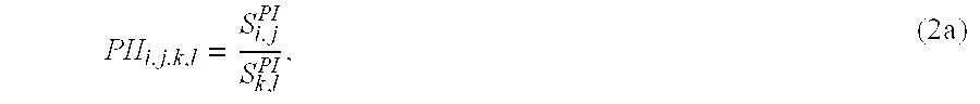
- This sum signal is in this context called a pseudo-isobestic (PI) signal and is denoted by S i,j PI , where i and j are the wavelengths.
- PI pseudo-isobestic
- a pseudo-isobestic signal is here defined as a signal calculated from the signals at two different wavelengths by summing the signals with a particular weight. The corresponding wavelengths are pseudo-isobestic within this weight.
- the pseudo-isobestic signal calculated from the signals at wavelengths 1 and 2 is thus:
- f 1.2 ⁇ 1 HbO 2 - ⁇ 1 RHb ⁇ 2 HbO 2 - ⁇ 2 RHb .
- the pseudo-isobestic signal calculated from the signals at wavelengths 1 and 2 can be written as follows:
- the pseudo-isobestic signal is still dependent on the total volumes of oxyhemoglobin and deoxyhemoglobin, since dA is dependent on said total volumes. Therefore, the signals are further normalized by forming a quotient of two pseudo-isobestic signals.
- the resulting parameter is in this context termed “pseudo-isobestic invariant” (PII), since it is, theoretically, a constant parameter (i.e. an invariant).
- the pseudo-isobestic invariant is, according to the Lambert-Beer theory, independent of any tissue or blood parameters, except the extinction values, which are known. Therefore, if the pseudo-isobestic invariant diverges from its theoretical value, the divergence must be caused by the variability between two individual patients or by temporal variations in the measurement conditions of a single patient. In other words, the divergence is caused by human variability.
- the pseudo-isobestic invariant can therefore be used for compensating the patient-specific variations in the calibration curve. This use of the PII is discussed in the following.
- the transformation can be expressed, for example, as follows:
- N k,l l,B g k,l ( N k,l in-vivo ) (3)
- N k , l d ⁇ ⁇ A k d ⁇ ⁇ A l
- ⁇ is the modulation ratio (the superscript indicating the domain)
- g is the transformation, in the form of a polynomial function (the superscript indicating the wavelengths in question), for example, and k and l are the wavelengths
- FIGS. 4 a to 4 f illustrate the average transformation curves measured for a pulse oximeter, where the two wavelengths for measuring the two species of hemoglobin are 660 nm and 900 nm and the third wavelength used for the self-calibration method according to the invention is either 725 nm or 805 nm.
- FIGS. 4 a to 4 c illustrate the transformation curves for a pulse oximeter with the third wavelength being 725 nm
- FIGS. 4 d to 4 f illustrate the transformation curves for a pulse oximeter with the third wavelength being 805 nm.
- Each curve shows the Lambert-Beer N k,l as a function of the in-vivo N k,l at wavelengths k and l.
- the transformation can be a quadratic equation which causes a correction of the order of 20 percent to the measured N k,l value, for example.
- the transformation data i.e. the transformation curves
- the number of transformation curves stored in the pulse oximeter can vary, depending on the number of wavelengths used, for example. Typically there is a transformation curve for each wavelength pair.
- the above-described formalism i.e. the pseudo-isobestic invariants (PIIs) are used to compensate for the human variability present in the pulse oximeter measurements.
- PIIs pseudo-isobestic invariants
- the effect of human variability can be seen, for example, so that it changes the transformation curves from the in-vivo N k,l values to the Lambert-Beer N k,l values (cf. FIGS. 4 a to 4 f ).
- the compensation is based on the calculation in the first phase of a theoretical value for the pseudo-isobestic invariant according to the Lambert-Beer theory, and then the calculation of a second value for the same pseudo-isobestic invariant in the second phase according to the measured signals, using the average transformation curves for mapping the measured signals in the Lambert-Beer model so that said second value can be calculated (Equation 4).
- the value of the pseudo-isobestic invariant obtained in the second phase is then compared to the theoretical value obtained in the first phase. If a difference is detected, a new calibration is sought based on the magnitude of the difference.
- the pulse oximeter is a type utilizing the above-described transformations
- a new transformation is sought, the new transformation being such that when it is used for mapping the measured signals in the Lambert-Beer model, the resulting value of the invariant equals, as accurately as possible, the theoretical value calculated in the first phase.
- the approximate average transformation can then be replaced by a more accurate transformation that takes the human variability into account.
- the patient-specific change in the transformation is seen as a change in the value of the pseudo-isobestic invariant calculated from the in-vivo signals.
- FIG. 5 is a flow diagram illustrating the steps of the calibration method according to the invention, assuming that the pulse oximeter uses the transformations for calibration purposes. In the figure, it is still assumed that the calibration is performed for a three-wavelength pulse oximeter described above.
- the first four steps ( 400 to 403 ) shown in the figure are performed in advance, i.e. before the actual in-vivo measurement is performed at step 404 . These first four steps can be performed during the manufacturing phase of the pulse oximeter in the factory.
- the wavelengths to be used are selected for the measurement (step 400 ). As mentioned above, it is assumed here that three wavelengths are selected at this step.
- the wavelengths can be those discussed in connection with FIGS. 4 a to 4 f , for example.
- the weights f are calculated for “ideal” pseudo-isobestic signals. It is to be noted here that the weights can be calculated already in this phase, i.e. before the actual measurements are performed, since the calculation is based on the extinction coefficients of the ideal model shown in FIG. 3, i.e. the said figure belongs to the ideal Lambert-Beer domain and the isobestic point and the pseudo-isobestic signals are present only in the ideal model.
- the average transformation curves according to FIGS. 4 a to 4 f are then determined for the selected wavelengths at step 402 .
- the transformation curves can be determined immediately when the wavelengths are fixed. As discussed above, these transformation curves are used later in the process to convert the actual in-vivo signals to signals that can be applied to the Lambert-Beer model, so that the pseudo-isobestic invariants can be determined again, this time on the basis of the measured signals brought to the Lambert-Beer model by means of said transformation curves.
- the “ideal” pseudo-isobestic invariant(s) (PII ijkl ) ideal (equation 2a) is/are calculated at step 403 .
- PII ijkl pseudo-isobestic invariant(s)
- equation 2a the “ideal” pseudo-isobestic invariant(s) (PII ijkl ) ideal (equation 2a) is/are calculated at step 403 .
- six pseudo-isobestic signals S 1,2 , S 1,3 , S 2,1 , S 2,3 , S 3,1 , S 3,2 PI can be determined (i.e. two wavelengths out of three can be selected in six different ways) according to equation (2).
- six different PIIs can be determined. However, only one of the PIIs is independent and provides the basis for calculating the other five.
- the approximate transformation curves calculated earlier at step 403 are then used at step 405 to convert the measured modulation signals into Lambert-Beer signals (equation 3).
- the in-vivo pseudo-isobestic invariants are calculated on the basis of these signals (step 406 ).
- the calculation is performed as described above (equations 3 and 4) and similarly as in the production phase (step 402 ), the only exception being that now the pseudo-isobestic invariant(s) is/are calculated on the basis of real measurement signals brought to the “ideal” model by means of a transformation, which is correct only for average human tissue.
- the in-vivo invariant is then compared (step 407 ) to the “ideal” invariant obtained at step 403 . If the difference between these two is greater than the maximum difference allowed, a new transformation is sought.
- This new transformation is such that when it is used at step 405 for the same measurement signals, the value of the in-vivo pseudo-isobestic invariant obtained on the basis of this transformation corresponds, as accurately as possible, to the value of the ideal invariant obtained at step 403 .
- this new transformation takes into account the differences in the current patient as compared to the average transformation curve.
- the new transformation is thus a tailored transformation for the current patient and can therefore be used for the measurements relating to the current patient. In the above-described manner the pulse oximeter calibrates itself for each patient.
- the pre-calculated data in its memory includes the average transformation curves illustrated in FIGS. 4 a to 4 f . As mentioned above, these curves can be determined in two basic ways.
- the memory preferably contains a look-up table including the numeric data constituting the transformation curves.
- the determination of the patient-specific transformation curve(s) can be performed purely on the basis of the difference between the PII values and the above-mentioned transformation curves.
- the change in the value of PII is caused by a change in one or more physiological factors which cause a change in the absolute absorption. It is possible to determine a curve to indicate how the changes in absorption affect the PII. This curve is illustrated in FIG. 6 . As can be seen from the curve, when the absorption increases, the value of PII increases and when absorption decreases, the value of PII decreases.
- the curve can be constructed either empirically or theoretically using a tissue model.
- This new transformation can be searched for entirely on the basis of the pre-stored transformation curve according to FIGS. 4 a to 4 f .
- the search can be performed in an iterative manner by shifting the transformation curve in small steps and calculating the difference from the ideal invariant at each step. Thus, steps 405 - 407 are repeated for each step.
- the new transformation is the one that yields the minimum difference between the in-vivo and the ideal value.
- the pulse oximeter knows whether the absorption has increased or decreased, and therefore it also knows in which direction the transformation curves must be shifted (i.e. up or down in FIGS. 4 a to 4 f ).
- the pre-calculated data utilized by the pulse oximeter can also be stored in the sensor part of the pulse oximeter, whereby the same sensor can be attached to different pulse oximeter housings.
- FIG. 7 illustrates the general structure of a sensor according to the invention, the detailed configuration of the sensor being dependent on which information is stored in the sensor and which in the signal processing part, and also on the amount of calculation that is appropriate in the signal processing part.
- a sensor includes the light sources ( 10 a , 10 b , 10 c ) and the photo detector ( 12 ), the light sources being adapted to emit at three or more wavelengths.
- the sensor includes a data storage unit M 2 for storing the data on the basis of which the signal processing part can perform the above-described calibration.
- the data storage unit contains the look-up tables including the average transformation curves and one or more numeric values of the “ideal” pseudo-isobestic invariants (PII ijkl ) ideal .
- the extinction values can be stored in the sensor instead of the PII values, whereby the PII values are calculated in the signal processing part on the basis of the extinction values obtained from the sensor.
- the data storage unit includes data on the wavelengths of the sensor, on the basis of which the numeric PII values can be calculated in the signal processing part of the pulse oximeter.
- the average transformation curves can also be determined in the apparatus on the basis of the wavelength information if the look-up tables are not stored in the sensor.
- the data storage unit includes only data on the wavelengths used, although it typically includes the transformation data according to FIGS. 4 a to 4 c (or 4 d to 4 f ) and one or more numeric values for the “ideal” pseudo-isobestic invariant.
- the extra wavelengths after the first three can be used entirely for the self-calibration method according to the invention or part or all of the extra wavelengths can be used for measuring additional information, such as methemoglobin (MetHb) and/or carboxyhemoglobin (CoHb).
- MetHb methemoglobin
- CoHb carboxyhemoglobin
- the number of pseudo-isobestic invariants available depends on the number of wavelengths used for the method according to the invention.
- six PIIs are available, although only one of them is independent, while the others depend on the said independent one.
- thirty PIIs can be obtained, although only two of them are independent, while the others depend on the said two.
- the number of independent PIIs corresponds to N minus 2, if N is the number of wavelengths (provided that all extra wavelengths are used for the method according to the invention).
- the pulse oximeter explicitly uses the transformations for calibration, whereby the transformation is changed so that patient-specific variation is compensated for. Since in a conventional pulse oximeter the measurement signal is mapped to a SpO 2 value, the difference between the in-vivo invariant and the “ideal” invariant can be used to indicate the error in the SpO 2 value. This is illustrated in FIG. 8 . Thus, in this case no transformation is changed but the said difference is mapped directly to the SpO 2 error. As shown in the shortened flow chart of FIG.
- this second embodiment of the method otherwise includes the same steps as in the above-described first embodiment, except that step 400 a has been added and steps 408 and 409 have been replaced by the new steps 910 and 911 , respectively.
- step 400 a the relationship depicted in FIG. 8 is created, the relationship indicating how said difference in the PII is related to the SpO 2 error.
- the curve according to FIG. 8 can also be generated empirically by taking blood samples, or theoretically based on a known tissue model.
- the pulse oximeter maps the difference detected at step 407 to the SpO 2 error, using the relationship created at step 400 a . This error is then used to correct the measured values (step 911 ).
- the difference between the two values of the PII can be mapped directly to any other quantity by which the pulse oximeter can correct the average calibration known to it in order to take into account the human variability.
- This quantity can be, for example, the modulation ratio R.
- the modulation ratio R is usually related to SpO 2 values at the wavelengths of 660 nm and 900 nm.
- the relationship according to FIG. 8 can be stored in the form of a look-up table, for example, from which the error value corresponding to a given difference can be retrieved.
- a further advantage of the pulse oximeter according to the invention is that the pseudo-isobestic signal can also be used as a measure of perfusion. Since the pseudo-isobestic signal S i,j PI is dependent only on variables which change when the perfusion changes, it can be used as a perfusion index.
- the pulse oximeter of the invention therefore automatically provides a measurement value for patient-specific perfusion. As is obvious from the above, the minimum number of wavelengths for a perfusion index is two.
Abstract
Description


Claims (16)
Priority Applications (2)
| Application Number | Priority Date | Filing Date | Title |
|---|---|---|---|
| US09/767,052 US6501974B2 (en) | 2001-01-22 | 2001-01-22 | Compensation of human variability in pulse oximetry |
| PCT/FI2001/001088 WO2002056759A1 (en) | 2001-01-22 | 2001-12-13 | Compensation of human variability in pulse oximetry |
Applications Claiming Priority (1)
| Application Number | Priority Date | Filing Date | Title |
|---|---|---|---|
| US09/767,052 US6501974B2 (en) | 2001-01-22 | 2001-01-22 | Compensation of human variability in pulse oximetry |
Publications (2)
| Publication Number | Publication Date |
|---|---|
| US20020133068A1 US20020133068A1 (en) | 2002-09-19 |
| US6501974B2 true US6501974B2 (en) | 2002-12-31 |
Family
ID=25078338
Family Applications (1)
| Application Number | Title | Priority Date | Filing Date |
|---|---|---|---|
| US09/767,052 Expired - Lifetime US6501974B2 (en) | 2001-01-22 | 2001-01-22 | Compensation of human variability in pulse oximetry |
Country Status (2)
| Country | Link |
|---|---|
| US (1) | US6501974B2 (en) |
| WO (1) | WO2002056759A1 (en) |
Cited By (147)
| Publication number | Priority date | Publication date | Assignee | Title |
|---|---|---|---|---|
| US20030033102A1 (en) * | 2001-08-09 | 2003-02-13 | Thomas Dietiker | System and method for a self-calibrating non-invasive sensor |
| WO2003068060A1 (en) * | 2002-02-15 | 2003-08-21 | Datex-Ohmeda, Inc. | Compensation of human variability in pulse oximetry |
| US6842635B1 (en) * | 1998-08-13 | 2005-01-11 | Edwards Lifesciences Llc | Optical device |
| US20060047190A1 (en) * | 2004-08-26 | 2006-03-02 | Perceptia Devices, Inc. | Oral health measurement clamping probe, system, and method |
| US20060063994A1 (en) * | 2002-04-17 | 2006-03-23 | Dietrich Schweitzer | Method for the spectroscopic determination of the oxygen saturation of blood in the presence of optical disturbance varibles |
| US20060167362A1 (en) * | 2002-10-15 | 2006-07-27 | Koninklijke Philips Electronics N.V. | Method for the presentation of information concerning variations of the perfusion |
| US20060200015A1 (en) * | 2005-03-03 | 2006-09-07 | Nellcor Puritan Bennett Incorporated | Method for enhancing pulse oximetry calculations in the presence of correlated artifacts |
| US20080132771A1 (en) * | 1998-07-04 | 2008-06-05 | Whitland Research Limited | Measurement of blood oxygen saturation |
| US20080281174A1 (en) * | 2006-06-16 | 2008-11-13 | Thomas Dietiker | System and method for a non-invasive medical sensor |
| US20090024011A1 (en) * | 2007-07-20 | 2009-01-22 | The General Electric Company | Non-invasive determination of the concentration of a blood substance |
| US20090326342A1 (en) * | 2008-06-27 | 2009-12-31 | The General Electric Company | Method, arrangement and sensor for non-invasively monitoring blood volume of a subject |
| US7647083B2 (en) | 2005-03-01 | 2010-01-12 | Masimo Laboratories, Inc. | Multiple wavelength sensor equalization |
| US7647084B2 (en) | 2005-08-08 | 2010-01-12 | Nellcor Puritan Bennett Llc | Medical sensor and technique for using the same |
| US7650177B2 (en) | 2005-09-29 | 2010-01-19 | Nellcor Puritan Bennett Llc | Medical sensor for reducing motion artifacts and technique for using the same |
| US7657295B2 (en) | 2005-08-08 | 2010-02-02 | Nellcor Puritan Bennett Llc | Medical sensor and technique for using the same |
| US7657296B2 (en) | 2005-08-08 | 2010-02-02 | Nellcor Puritan Bennett Llc | Unitary medical sensor assembly and technique for using the same |
| US7658652B2 (en) | 2006-09-29 | 2010-02-09 | Nellcor Puritan Bennett Llc | Device and method for reducing crosstalk |
| US7676253B2 (en) | 2005-09-29 | 2010-03-09 | Nellcor Puritan Bennett Llc | Medical sensor and technique for using the same |
| US7680522B2 (en) | 2006-09-29 | 2010-03-16 | Nellcor Puritan Bennett Llc | Method and apparatus for detecting misapplied sensors |
| US7684842B2 (en) | 2006-09-29 | 2010-03-23 | Nellcor Puritan Bennett Llc | System and method for preventing sensor misuse |
| US7698002B2 (en) | 2006-09-29 | 2010-04-13 | Nellcor Puritan Bennett Llc | Systems and methods for user interface and identification in a medical device |
| US7706896B2 (en) | 2006-09-29 | 2010-04-27 | Nellcor Puritan Bennett Llc | User interface and identification in a medical device system and method |
| US7720516B2 (en) | 1996-10-10 | 2010-05-18 | Nellcor Puritan Bennett Llc | Motion compatible sensor for non-invasive optical blood analysis |
| US7725146B2 (en) | 2005-09-29 | 2010-05-25 | Nellcor Puritan Bennett Llc | System and method for pre-processing waveforms |
| US7725147B2 (en) | 2005-09-29 | 2010-05-25 | Nellcor Puritan Bennett Llc | System and method for removing artifacts from waveforms |
| US7796403B2 (en) | 2006-09-28 | 2010-09-14 | Nellcor Puritan Bennett Llc | Means for mechanical registration and mechanical-electrical coupling of a faraday shield to a photodetector and an electrical circuit |
| USD626562S1 (en) | 2008-06-30 | 2010-11-02 | Nellcor Puritan Bennett Llc | Triangular saturation pattern detection indicator for a patient monitor display panel |
| USD626561S1 (en) | 2008-06-30 | 2010-11-02 | Nellcor Puritan Bennett Llc | Circular satseconds indicator and triangular saturation pattern detection indicator for a patient monitor display panel |
| US7848891B2 (en) | 2006-09-29 | 2010-12-07 | Nellcor Puritan Bennett Llc | Modulation ratio determination with accommodation of uncertainty |
| US7869849B2 (en) | 2006-09-26 | 2011-01-11 | Nellcor Puritan Bennett Llc | Opaque, electrically nonconductive region on a medical sensor |
| US7880884B2 (en) | 2008-06-30 | 2011-02-01 | Nellcor Puritan Bennett Llc | System and method for coating and shielding electronic sensor components |
| US7881762B2 (en) | 2005-09-30 | 2011-02-01 | Nellcor Puritan Bennett Llc | Clip-style medical sensor and technique for using the same |
| US7887345B2 (en) | 2008-06-30 | 2011-02-15 | Nellcor Puritan Bennett Llc | Single use connector for pulse oximetry sensors |
| US7890153B2 (en) | 2006-09-28 | 2011-02-15 | Nellcor Puritan Bennett Llc | System and method for mitigating interference in pulse oximetry |
| US7890154B2 (en) | 2004-03-08 | 2011-02-15 | Nellcor Puritan Bennett Llc | Selection of ensemble averaging weights for a pulse oximeter based on signal quality metrics |
| US7894869B2 (en) | 2007-03-09 | 2011-02-22 | Nellcor Puritan Bennett Llc | Multiple configuration medical sensor and technique for using the same |
| US7899510B2 (en) | 2005-09-29 | 2011-03-01 | Nellcor Puritan Bennett Llc | Medical sensor and technique for using the same |
| US7922665B2 (en) | 2006-09-28 | 2011-04-12 | Nellcor Puritan Bennett Llc | System and method for pulse rate calculation using a scheme for alternate weighting |
| US7925511B2 (en) | 2006-09-29 | 2011-04-12 | Nellcor Puritan Bennett Llc | System and method for secure voice identification in a medical device |
| EP2317452A1 (en) | 2009-10-28 | 2011-05-04 | General Electric Company | Multiple wavelength physiological measuring apparatus, sensor and interface unit for determination of blood parameters |
| US8007441B2 (en) | 2004-03-08 | 2011-08-30 | Nellcor Puritan Bennett Llc | Pulse oximeter with alternate heart-rate determination |
| US8064975B2 (en) | 2006-09-20 | 2011-11-22 | Nellcor Puritan Bennett Llc | System and method for probability based determination of estimated oxygen saturation |
| US8062221B2 (en) | 2005-09-30 | 2011-11-22 | Nellcor Puritan Bennett Llc | Sensor for tissue gas detection and technique for using the same |
| US8068890B2 (en) | 2006-09-29 | 2011-11-29 | Nellcor Puritan Bennett Llc | Pulse oximetry sensor switchover |
| US8068891B2 (en) | 2006-09-29 | 2011-11-29 | Nellcor Puritan Bennett Llc | Symmetric LED array for pulse oximetry |
| US8070508B2 (en) | 2007-12-31 | 2011-12-06 | Nellcor Puritan Bennett Llc | Method and apparatus for aligning and securing a cable strain relief |
| US8073518B2 (en) | 2006-05-02 | 2011-12-06 | Nellcor Puritan Bennett Llc | Clip-style medical sensor and technique for using the same |
| US8071935B2 (en) | 2008-06-30 | 2011-12-06 | Nellcor Puritan Bennett Llc | Optical detector with an overmolded faraday shield |
| US8092379B2 (en) | 2005-09-29 | 2012-01-10 | Nellcor Puritan Bennett Llc | Method and system for determining when to reposition a physiological sensor |
| US8092993B2 (en) | 2007-12-31 | 2012-01-10 | Nellcor Puritan Bennett Llc | Hydrogel thin film for use as a biosensor |
| US8095192B2 (en) | 2003-01-10 | 2012-01-10 | Nellcor Puritan Bennett Llc | Signal quality metrics design for qualifying data for a physiological monitor |
| US8109882B2 (en) | 2007-03-09 | 2012-02-07 | Nellcor Puritan Bennett Llc | System and method for venous pulsation detection using near infrared wavelengths |
| US8112375B2 (en) | 2008-03-31 | 2012-02-07 | Nellcor Puritan Bennett Llc | Wavelength selection and outlier detection in reduced rank linear models |
| US8123695B2 (en) | 2006-09-27 | 2012-02-28 | Nellcor Puritan Bennett Llc | Method and apparatus for detection of venous pulsation |
| US8133176B2 (en) | 1999-04-14 | 2012-03-13 | Tyco Healthcare Group Lp | Method and circuit for indicating quality and accuracy of physiological measurements |
| US8140272B2 (en) | 2008-03-27 | 2012-03-20 | Nellcor Puritan Bennett Llc | System and method for unmixing spectroscopic observations with nonnegative matrix factorization |
| US8145288B2 (en) | 2006-08-22 | 2012-03-27 | Nellcor Puritan Bennett Llc | Medical sensor for reducing signal artifacts and technique for using the same |
| US8160683B2 (en) | 2006-09-29 | 2012-04-17 | Nellcor Puritan Bennett Llc | System and method for integrating voice with a medical device |
| US8160668B2 (en) | 2006-09-29 | 2012-04-17 | Nellcor Puritan Bennett Llc | Pathological condition detector using kernel methods and oximeters |
| US8175671B2 (en) | 2006-09-22 | 2012-05-08 | Nellcor Puritan Bennett Llc | Medical sensor for reducing signal artifacts and technique for using the same |
| US8175670B2 (en) | 2004-03-09 | 2012-05-08 | Nellcor Puritan Bennett Llc | Pulse oximetry signal correction using near infrared absorption by water |
| US8175667B2 (en) | 2006-09-29 | 2012-05-08 | Nellcor Puritan Bennett Llc | Symmetric LED array for pulse oximetry |
| US8190225B2 (en) | 2006-09-22 | 2012-05-29 | Nellcor Puritan Bennett Llc | Medical sensor for reducing signal artifacts and technique for using the same |
| US8195262B2 (en) | 2004-02-25 | 2012-06-05 | Nellcor Puritan Bennett Llc | Switch-mode oximeter LED drive with a single inductor |
| US8199007B2 (en) | 2007-12-31 | 2012-06-12 | Nellcor Puritan Bennett Llc | Flex circuit snap track for a biometric sensor |
| US8204567B2 (en) | 2007-12-13 | 2012-06-19 | Nellcor Puritan Bennett Llc | Signal demodulation |
| US8219170B2 (en) | 2006-09-20 | 2012-07-10 | Nellcor Puritan Bennett Llc | System and method for practicing spectrophotometry using light emitting nanostructure devices |
| US8221319B2 (en) | 2009-03-25 | 2012-07-17 | Nellcor Puritan Bennett Llc | Medical device for assessing intravascular blood volume and technique for using the same |
| US8221326B2 (en) | 2007-03-09 | 2012-07-17 | Nellcor Puritan Bennett Llc | Detection of oximetry sensor sites based on waveform characteristics |
| US8229530B2 (en) | 2007-03-09 | 2012-07-24 | Nellcor Puritan Bennett Llc | System and method for detection of venous pulsation |
| US8233954B2 (en) | 2005-09-30 | 2012-07-31 | Nellcor Puritan Bennett Llc | Mucosal sensor for the assessment of tissue and blood constituents and technique for using the same |
| US8238994B2 (en) | 2005-10-28 | 2012-08-07 | Nellcor Puritan Bennett Llc | Adjusting parameters used in pulse oximetry analysis |
| US8260391B2 (en) | 2005-09-12 | 2012-09-04 | Nellcor Puritan Bennett Llc | Medical sensor for reducing motion artifacts and technique for using the same |
| US8265724B2 (en) | 2007-03-09 | 2012-09-11 | Nellcor Puritan Bennett Llc | Cancellation of light shunting |
| US8275553B2 (en) | 2008-02-19 | 2012-09-25 | Nellcor Puritan Bennett Llc | System and method for evaluating physiological parameter data |
| US8280469B2 (en) | 2007-03-09 | 2012-10-02 | Nellcor Puritan Bennett Llc | Method for detection of aberrant tissue spectra |
| US8292809B2 (en) | 2008-03-31 | 2012-10-23 | Nellcor Puritan Bennett Llc | Detecting chemical components from spectroscopic observations |
| US8311601B2 (en) | 2009-06-30 | 2012-11-13 | Nellcor Puritan Bennett Llc | Reflectance and/or transmissive pulse oximeter |
| US8315685B2 (en) | 2006-09-27 | 2012-11-20 | Nellcor Puritan Bennett Llc | Flexible medical sensor enclosure |
| US8315684B2 (en) | 2004-02-25 | 2012-11-20 | Covidien Lp | Oximeter ambient light cancellation |
| US8346328B2 (en) | 2007-12-21 | 2013-01-01 | Covidien Lp | Medical sensor and technique for using the same |
| US8352009B2 (en) | 2005-09-30 | 2013-01-08 | Covidien Lp | Medical sensor and technique for using the same |
| US8352004B2 (en) | 2007-12-21 | 2013-01-08 | Covidien Lp | Medical sensor and technique for using the same |
| US8352010B2 (en) | 2005-09-30 | 2013-01-08 | Covidien Lp | Folding medical sensor and technique for using the same |
| US8364224B2 (en) | 2008-03-31 | 2013-01-29 | Covidien Lp | System and method for facilitating sensor and monitor communication |
| US8364221B2 (en) | 2005-09-30 | 2013-01-29 | Covidien Lp | Patient monitoring alarm escalation system and method |
| US8364220B2 (en) | 2008-09-25 | 2013-01-29 | Covidien Lp | Medical sensor and technique for using the same |
| US8366613B2 (en) | 2007-12-26 | 2013-02-05 | Covidien Lp | LED drive circuit for pulse oximetry and method for using same |
| US8380271B2 (en) | 2006-06-15 | 2013-02-19 | Covidien Lp | System and method for generating customizable audible beep tones and alarms |
| US8376955B2 (en) | 2009-09-29 | 2013-02-19 | Covidien Lp | Spectroscopic method and system for assessing tissue temperature |
| US8386002B2 (en) | 2005-09-30 | 2013-02-26 | Covidien Lp | Optically aligned pulse oximetry sensor and technique for using the same |
| US8386000B2 (en) | 2008-09-30 | 2013-02-26 | Covidien Lp | System and method for photon density wave pulse oximetry and pulse hemometry |
| US8391941B2 (en) | 2009-07-17 | 2013-03-05 | Covidien Lp | System and method for memory switching for multiple configuration medical sensor |
| US8396527B2 (en) | 2006-09-22 | 2013-03-12 | Covidien Lp | Medical sensor for reducing signal artifacts and technique for using the same |
| US8401607B2 (en) | 2001-07-19 | 2013-03-19 | Covidien Lp | Nuisance alarm reductions in a physiological monitor |
| US8417309B2 (en) | 2008-09-30 | 2013-04-09 | Covidien Lp | Medical sensor |
| US8417310B2 (en) | 2009-08-10 | 2013-04-09 | Covidien Lp | Digital switching in multi-site sensor |
| US8423112B2 (en) | 2008-09-30 | 2013-04-16 | Covidien Lp | Medical sensor and technique for using the same |
| US8428675B2 (en) | 2009-08-19 | 2013-04-23 | Covidien Lp | Nanofiber adhesives used in medical devices |
| US8433383B2 (en) | 2001-10-12 | 2013-04-30 | Covidien Lp | Stacked adhesive optical sensor |
| US8433382B2 (en) | 2008-09-30 | 2013-04-30 | Covidien Lp | Transmission mode photon density wave system and method |
| US8437822B2 (en) | 2008-03-28 | 2013-05-07 | Covidien Lp | System and method for estimating blood analyte concentration |
| US8442608B2 (en) | 2007-12-28 | 2013-05-14 | Covidien Lp | System and method for estimating physiological parameters by deconvolving artifacts |
| US8452364B2 (en) | 2007-12-28 | 2013-05-28 | Covidien LLP | System and method for attaching a sensor to a patient's skin |
| US8452366B2 (en) | 2009-03-16 | 2013-05-28 | Covidien Lp | Medical monitoring device with flexible circuitry |
| US8483790B2 (en) | 2002-10-18 | 2013-07-09 | Covidien Lp | Non-adhesive oximeter sensor for sensitive skin |
| US8494786B2 (en) | 2009-07-30 | 2013-07-23 | Covidien Lp | Exponential sampling of red and infrared signals |
| US8494606B2 (en) | 2009-08-19 | 2013-07-23 | Covidien Lp | Photoplethysmography with controlled application of sensor pressure |
| US8494604B2 (en) | 2009-09-21 | 2013-07-23 | Covidien Lp | Wavelength-division multiplexing in a multi-wavelength photon density wave system |
| US8509869B2 (en) | 2009-05-15 | 2013-08-13 | Covidien Lp | Method and apparatus for detecting and analyzing variations in a physiologic parameter |
| US8505821B2 (en) | 2009-06-30 | 2013-08-13 | Covidien Lp | System and method for providing sensor quality assurance |
| US8515511B2 (en) | 2009-09-29 | 2013-08-20 | Covidien Lp | Sensor with an optical coupling material to improve plethysmographic measurements and method of using the same |
| US8577434B2 (en) | 2007-12-27 | 2013-11-05 | Covidien Lp | Coaxial LED light sources |
| US8634891B2 (en) | 2009-05-20 | 2014-01-21 | Covidien Lp | Method and system for self regulation of sensor component contact pressure |
| US8666467B2 (en) | 2001-05-17 | 2014-03-04 | Lawrence A. Lynn | System and method for SPO2 instability detection and quantification |
| US8696593B2 (en) | 2006-09-27 | 2014-04-15 | Covidien Lp | Method and system for monitoring intracranial pressure |
| US8702606B2 (en) | 2006-03-21 | 2014-04-22 | Covidien Lp | Patient monitoring help video system and method |
| US8704666B2 (en) | 2009-09-21 | 2014-04-22 | Covidien Lp | Medical device interface customization systems and methods |
| US8728001B2 (en) | 2006-02-10 | 2014-05-20 | Lawrence A. Lynn | Nasal capnographic pressure monitoring system |
| US8728059B2 (en) | 2006-09-29 | 2014-05-20 | Covidien Lp | System and method for assuring validity of monitoring parameter in combination with a therapeutic device |
| US8750953B2 (en) | 2008-02-19 | 2014-06-10 | Covidien Lp | Methods and systems for alerting practitioners to physiological conditions |
| US8781544B2 (en) | 2007-03-27 | 2014-07-15 | Cercacor Laboratories, Inc. | Multiple wavelength optical sensor |
| US8788001B2 (en) | 2009-09-21 | 2014-07-22 | Covidien Lp | Time-division multiplexing in a multi-wavelength photon density wave system |
| US8798704B2 (en) | 2009-09-24 | 2014-08-05 | Covidien Lp | Photoacoustic spectroscopy method and system to discern sepsis from shock |
| US8801613B2 (en) | 2009-12-04 | 2014-08-12 | Masimo Corporation | Calibration for multi-stage physiological monitors |
| US8862194B2 (en) | 2008-06-30 | 2014-10-14 | Covidien Lp | Method for improved oxygen saturation estimation in the presence of noise |
| US8862196B2 (en) | 2001-05-17 | 2014-10-14 | Lawrence A. Lynn | System and method for automatic detection of a plurality of SP02 time series pattern types |
| US8897850B2 (en) | 2007-12-31 | 2014-11-25 | Covidien Lp | Sensor with integrated living hinge and spring |
| US8914088B2 (en) | 2008-09-30 | 2014-12-16 | Covidien Lp | Medical sensor and technique for using the same |
| US8930145B2 (en) | 2010-07-28 | 2015-01-06 | Covidien Lp | Light focusing continuous wave photoacoustic spectroscopy and its applications to patient monitoring |
| US8965471B2 (en) | 2007-04-21 | 2015-02-24 | Cercacor Laboratories, Inc. | Tissue profile wellness monitor |
| US8968193B2 (en) | 2008-09-30 | 2015-03-03 | Covidien Lp | System and method for enabling a research mode on physiological monitors |
| US8983800B2 (en) | 2003-01-13 | 2015-03-17 | Covidien Lp | Selection of preset filter parameters based on signal quality |
| US9010634B2 (en) | 2009-06-30 | 2015-04-21 | Covidien Lp | System and method for linking patient data to a patient and providing sensor quality assurance |
| US9031793B2 (en) | 2001-05-17 | 2015-05-12 | Lawrence A. Lynn | Centralized hospital monitoring system for automatically detecting upper airway instability and for preventing and aborting adverse drug reactions |
| US9042952B2 (en) | 1997-01-27 | 2015-05-26 | Lawrence A. Lynn | System and method for automatic detection of a plurality of SPO2 time series pattern types |
| US9053222B2 (en) | 2002-05-17 | 2015-06-09 | Lawrence A. Lynn | Patient safety processor |
| US9066660B2 (en) | 2009-09-29 | 2015-06-30 | Nellcor Puritan Bennett Ireland | Systems and methods for high-pass filtering a photoplethysmograph signal |
| US9468378B2 (en) | 1997-01-27 | 2016-10-18 | Lawrence A. Lynn | Airway instability detection system and method |
| US9521971B2 (en) | 1997-07-14 | 2016-12-20 | Lawrence A. Lynn | System and method for automatic detection of a plurality of SPO2 time series pattern types |
| US9585606B2 (en) | 2009-09-29 | 2017-03-07 | Covidien Lp | Oximetry assembly |
| US9833146B2 (en) | 2012-04-17 | 2017-12-05 | Covidien Lp | Surgical system and method of use of the same |
| US9839381B1 (en) | 2009-11-24 | 2017-12-12 | Cercacor Laboratories, Inc. | Physiological measurement system with automatic wavelength adjustment |
| US9895068B2 (en) | 2008-06-30 | 2018-02-20 | Covidien Lp | Pulse oximeter with wait-time indication |
| US10092226B2 (en) | 2011-12-23 | 2018-10-09 | General Electric Company | Method, arrangement, sensor, and computer program product for non-invasively measuring hemoglobin concentrations in blood |
| US10354753B2 (en) | 2001-05-17 | 2019-07-16 | Lawrence A. Lynn | Medical failure pattern search engine |
| US11350861B2 (en) | 2014-10-10 | 2022-06-07 | Medtor, Inc. | System and method for a non-invasive medical sensor |
Families Citing this family (21)
| Publication number | Priority date | Publication date | Assignee | Title |
|---|---|---|---|---|
| US7274955B2 (en) * | 2002-09-25 | 2007-09-25 | Masimo Corporation | Parameter compensated pulse oximeter |
| US20070004976A1 (en) * | 2004-12-14 | 2007-01-04 | Zelenchuk Alex R | In vivo optical measurements of hematocrit |
| ATE450198T1 (en) * | 2005-03-14 | 2009-12-15 | Koninkl Philips Electronics Nv | METHOD AND DEVICE FOR DETERMINING BLOOD CIRCULATION IN A BODY ELEMENT |
| WO2007056995A1 (en) * | 2005-11-15 | 2007-05-24 | Weinmann Geräte für Medizin GmbH & Co. KG | Autoadaptive calibration |
| DE102007016447A1 (en) * | 2006-04-07 | 2007-10-11 | Weinmann Geräte für Medizin GmbH + Co. KG | Living thing`s bio data determining and analyzing device, has analysis device extracting continuous wave parameters, where continuous wave fluctuation is determined from continuous wave parameters and/or measuring signal |
| US20080097175A1 (en) * | 2006-09-29 | 2008-04-24 | Boyce Robin S | System and method for display control of patient monitor |
| JP4569615B2 (en) * | 2007-09-25 | 2010-10-27 | ブラザー工業株式会社 | Printing device |
| US20090171174A1 (en) * | 2007-12-31 | 2009-07-02 | Nellcor Puritan Bennett Llc | System and method for maintaining battery life |
| US20120226117A1 (en) * | 2010-12-01 | 2012-09-06 | Lamego Marcelo M | Handheld processing device including medical applications for minimally and non invasive glucose measurements |
| CN102178536B (en) * | 2011-03-29 | 2013-04-03 | 苏州易寻传感网络科技有限公司 | Method and system for measuring oxygen saturation and heart rate |
| US9629358B2 (en) * | 2013-03-15 | 2017-04-25 | Mallinckrodt Hospital Products IP Limited | Administration and monitoring of nitric oxide in ex vivo fluids |
| US10952638B2 (en) | 2015-06-12 | 2021-03-23 | ChroniSense Medical Ltd. | System and method for monitoring respiratory rate and oxygen saturation |
| US11160461B2 (en) | 2015-06-12 | 2021-11-02 | ChroniSense Medical Ltd. | Blood pressure measurement using a wearable device |
| US11464457B2 (en) | 2015-06-12 | 2022-10-11 | ChroniSense Medical Ltd. | Determining an early warning score based on wearable device measurements |
| US10687742B2 (en) | 2015-06-12 | 2020-06-23 | ChroniSense Medical Ltd. | Using invariant factors for pulse oximetry |
| US11712190B2 (en) | 2015-06-12 | 2023-08-01 | ChroniSense Medical Ltd. | Wearable device electrocardiogram |
| US11160459B2 (en) | 2015-06-12 | 2021-11-02 | ChroniSense Medical Ltd. | Monitoring health status of people suffering from chronic diseases |
| US10470692B2 (en) | 2015-06-12 | 2019-11-12 | ChroniSense Medical Ltd. | System for performing pulse oximetry |
| US11000235B2 (en) | 2016-03-14 | 2021-05-11 | ChroniSense Medical Ltd. | Monitoring procedure for early warning of cardiac episodes |
| CN112770668A (en) * | 2018-07-16 | 2021-05-07 | 布鲁恩医疗创新有限责任公司 | Perfusion and oxygenation measurements |
| CN110613462B (en) * | 2019-09-11 | 2022-04-08 | 河南大学 | Tissue oxygen saturation detection method and device free from individual difference |
Citations (14)
| Publication number | Priority date | Publication date | Assignee | Title |
|---|---|---|---|---|
| US4086915A (en) | 1975-04-30 | 1978-05-02 | Harvey I. Kofsky | Ear oximetry process and apparatus |
| US4933614A (en) | 1986-04-02 | 1990-06-12 | Alps Electric Co., Ltd. | Motor drive control circuit |
| US5278627A (en) * | 1991-02-15 | 1994-01-11 | Nihon Kohden Corporation | Apparatus for calibrating pulse oximeter |
| US5355880A (en) | 1992-07-06 | 1994-10-18 | Sandia Corporation | Reliable noninvasive measurement of blood gases |
| US5357953A (en) * | 1992-05-21 | 1994-10-25 | Puritan-Bennett Corporation | Measurement device and method of calibration |
| US5499627A (en) | 1990-10-06 | 1996-03-19 | In-Line Diagnostics Corporation | System for noninvasive hematocrit monitoring |
| US5692503A (en) * | 1995-03-10 | 1997-12-02 | Kuenstner; J. Todd | Method for noninvasive (in-vivo) total hemoglobin, oxyhemogolobin, deoxyhemoglobin, carboxyhemoglobin and methemoglobin concentration determination |
| US5792050A (en) | 1992-07-06 | 1998-08-11 | Alam; Mary K. | Near-infrared noninvasive spectroscopic determination of pH |
| US5842979A (en) | 1997-02-14 | 1998-12-01 | Ohmeda Inc. | Method and apparatus for improved photoplethysmographic monitoring of oxyhemoglobin, deoxyhemoglobin, carboxyhemoglobin and methemoglobin |
| US5891024A (en) | 1997-04-09 | 1999-04-06 | Ohmeda Inc. | Two stage calibration and analyte measurement scheme for spectrophotomeric analysis |
| US5931779A (en) * | 1996-06-06 | 1999-08-03 | Wisconsin Alumni Research Foundation | Real-time in-vivo measurement of myoglobin oxygen saturation |
| US6104938A (en) | 1996-06-12 | 2000-08-15 | Instrumentarium Oy | Procedure, apparatus and detector for the determination of fractional oxygen saturation |
| US6163715A (en) | 1996-07-17 | 2000-12-19 | Criticare Systems, Inc. | Direct to digital oximeter and method for calculating oxygenation levels |
| WO2001003577A1 (en) | 1999-07-14 | 2001-01-18 | Providence Health System - Oregon | Adaptive calibration pulsed oximetry method and device |
Family Cites Families (1)
| Publication number | Priority date | Publication date | Assignee | Title |
|---|---|---|---|---|
| DK116387A (en) * | 1986-03-07 | 1987-09-08 | Terumo Corp | APPARATUS FOR MEASURING THE Saturation Ratio of Oxygen in Blood and Procedure for Implementing the Measurement |
-
2001
- 2001-01-22 US US09/767,052 patent/US6501974B2/en not_active Expired - Lifetime
- 2001-12-13 WO PCT/FI2001/001088 patent/WO2002056759A1/en not_active Application Discontinuation
Patent Citations (17)
| Publication number | Priority date | Publication date | Assignee | Title |
|---|---|---|---|---|
| US4086915A (en) | 1975-04-30 | 1978-05-02 | Harvey I. Kofsky | Ear oximetry process and apparatus |
| US4933614A (en) | 1986-04-02 | 1990-06-12 | Alps Electric Co., Ltd. | Motor drive control circuit |
| US5499627A (en) | 1990-10-06 | 1996-03-19 | In-Line Diagnostics Corporation | System for noninvasive hematocrit monitoring |
| US5278627A (en) * | 1991-02-15 | 1994-01-11 | Nihon Kohden Corporation | Apparatus for calibrating pulse oximeter |
| US5357953A (en) * | 1992-05-21 | 1994-10-25 | Puritan-Bennett Corporation | Measurement device and method of calibration |
| US5792050A (en) | 1992-07-06 | 1998-08-11 | Alam; Mary K. | Near-infrared noninvasive spectroscopic determination of pH |
| US5630413A (en) | 1992-07-06 | 1997-05-20 | Sandia Corporation | Reliable noninvasive measurement of blood gases |
| US5355880A (en) | 1992-07-06 | 1994-10-18 | Sandia Corporation | Reliable noninvasive measurement of blood gases |
| US6061581A (en) | 1992-07-06 | 2000-05-09 | Alam; Mary K. | Invasive and in vivo near-infrared determination of pH |
| US6073037A (en) | 1992-07-06 | 2000-06-06 | Sandia Corporation | Near-infrared noninvasive determination of pH in pulse mode |
| US5692503A (en) * | 1995-03-10 | 1997-12-02 | Kuenstner; J. Todd | Method for noninvasive (in-vivo) total hemoglobin, oxyhemogolobin, deoxyhemoglobin, carboxyhemoglobin and methemoglobin concentration determination |
| US5931779A (en) * | 1996-06-06 | 1999-08-03 | Wisconsin Alumni Research Foundation | Real-time in-vivo measurement of myoglobin oxygen saturation |
| US6104938A (en) | 1996-06-12 | 2000-08-15 | Instrumentarium Oy | Procedure, apparatus and detector for the determination of fractional oxygen saturation |
| US6163715A (en) | 1996-07-17 | 2000-12-19 | Criticare Systems, Inc. | Direct to digital oximeter and method for calculating oxygenation levels |
| US5842979A (en) | 1997-02-14 | 1998-12-01 | Ohmeda Inc. | Method and apparatus for improved photoplethysmographic monitoring of oxyhemoglobin, deoxyhemoglobin, carboxyhemoglobin and methemoglobin |
| US5891024A (en) | 1997-04-09 | 1999-04-06 | Ohmeda Inc. | Two stage calibration and analyte measurement scheme for spectrophotomeric analysis |
| WO2001003577A1 (en) | 1999-07-14 | 2001-01-18 | Providence Health System - Oregon | Adaptive calibration pulsed oximetry method and device |
Non-Patent Citations (1)
| Title |
|---|
| International Search Report (3 Pages). |
Cited By (242)
| Publication number | Priority date | Publication date | Assignee | Title |
|---|---|---|---|---|
| US7720516B2 (en) | 1996-10-10 | 2010-05-18 | Nellcor Puritan Bennett Llc | Motion compatible sensor for non-invasive optical blood analysis |
| US8649839B2 (en) | 1996-10-10 | 2014-02-11 | Covidien Lp | Motion compatible sensor for non-invasive optical blood analysis |
| US9042952B2 (en) | 1997-01-27 | 2015-05-26 | Lawrence A. Lynn | System and method for automatic detection of a plurality of SPO2 time series pattern types |
| US9468378B2 (en) | 1997-01-27 | 2016-10-18 | Lawrence A. Lynn | Airway instability detection system and method |
| US9521971B2 (en) | 1997-07-14 | 2016-12-20 | Lawrence A. Lynn | System and method for automatic detection of a plurality of SPO2 time series pattern types |
| US20080132771A1 (en) * | 1998-07-04 | 2008-06-05 | Whitland Research Limited | Measurement of blood oxygen saturation |
| US20090005663A1 (en) * | 1998-07-04 | 2009-01-01 | Edwards Lifesciences Corporation | Non-invasive measurement of blood analytes |
| US7774037B2 (en) | 1998-07-04 | 2010-08-10 | Whitland Research Limited | Non-invasive measurement of blood analytes |
| US6842635B1 (en) * | 1998-08-13 | 2005-01-11 | Edwards Lifesciences Llc | Optical device |
| US8133176B2 (en) | 1999-04-14 | 2012-03-13 | Tyco Healthcare Group Lp | Method and circuit for indicating quality and accuracy of physiological measurements |
| US8932227B2 (en) | 2000-07-28 | 2015-01-13 | Lawrence A. Lynn | System and method for CO2 and oximetry integration |
| US10058269B2 (en) | 2000-07-28 | 2018-08-28 | Lawrence A. Lynn | Monitoring system for identifying an end-exhalation carbon dioxide value of enhanced clinical utility |
| US7124048B2 (en) | 2000-08-11 | 2006-10-17 | Elekon Industries Usa, Inc. | System and method for a self-calibrating non-invasive sensor |
| US20060122801A1 (en) * | 2000-08-11 | 2006-06-08 | Thomas Dietiker | System and method for a self-calibrating non-invasive sensor |
| US10297348B2 (en) | 2001-05-17 | 2019-05-21 | Lawrence A. Lynn | Patient safety processor |
| US8862196B2 (en) | 2001-05-17 | 2014-10-14 | Lawrence A. Lynn | System and method for automatic detection of a plurality of SP02 time series pattern types |
| US11439321B2 (en) | 2001-05-17 | 2022-09-13 | Lawrence A. Lynn | Monitoring system for identifying an end-exhalation carbon dioxide value of enhanced clinical utility |
| US10366790B2 (en) | 2001-05-17 | 2019-07-30 | Lawrence A. Lynn | Patient safety processor |
| US10032526B2 (en) | 2001-05-17 | 2018-07-24 | Lawrence A. Lynn | Patient safety processor |
| US8666467B2 (en) | 2001-05-17 | 2014-03-04 | Lawrence A. Lynn | System and method for SPO2 instability detection and quantification |
| US10354753B2 (en) | 2001-05-17 | 2019-07-16 | Lawrence A. Lynn | Medical failure pattern search engine |
| US9031793B2 (en) | 2001-05-17 | 2015-05-12 | Lawrence A. Lynn | Centralized hospital monitoring system for automatically detecting upper airway instability and for preventing and aborting adverse drug reactions |
| US8401607B2 (en) | 2001-07-19 | 2013-03-19 | Covidien Lp | Nuisance alarm reductions in a physiological monitor |
| US8401606B2 (en) | 2001-07-19 | 2013-03-19 | Covidien Lp | Nuisance alarm reductions in a physiological monitor |
| US8838196B2 (en) | 2001-07-19 | 2014-09-16 | Covidien Lp | Nuisance alarm reductions in a physiological monitor |
| US6889153B2 (en) * | 2001-08-09 | 2005-05-03 | Thomas Dietiker | System and method for a self-calibrating non-invasive sensor |
| US20030033102A1 (en) * | 2001-08-09 | 2003-02-13 | Thomas Dietiker | System and method for a self-calibrating non-invasive sensor |
| US8433383B2 (en) | 2001-10-12 | 2013-04-30 | Covidien Lp | Stacked adhesive optical sensor |
| WO2003068060A1 (en) * | 2002-02-15 | 2003-08-21 | Datex-Ohmeda, Inc. | Compensation of human variability in pulse oximetry |
| US7333842B2 (en) * | 2002-04-17 | 2008-02-19 | Carl Ziess Mesitec Ag | Method for the spectroscopic determination of the oxygen saturation of blood in the presence of optical disturbance variables |
| US20060063994A1 (en) * | 2002-04-17 | 2006-03-23 | Dietrich Schweitzer | Method for the spectroscopic determination of the oxygen saturation of blood in the presence of optical disturbance varibles |
| US9053222B2 (en) | 2002-05-17 | 2015-06-09 | Lawrence A. Lynn | Patient safety processor |
| US7572230B2 (en) * | 2002-10-15 | 2009-08-11 | Koninklijke Philips Electronics N.V. | Method for the presentation of information concerning variations of the perfusion |
| US20060167362A1 (en) * | 2002-10-15 | 2006-07-27 | Koninklijke Philips Electronics N.V. | Method for the presentation of information concerning variations of the perfusion |
| US8483790B2 (en) | 2002-10-18 | 2013-07-09 | Covidien Lp | Non-adhesive oximeter sensor for sensitive skin |
| US8095192B2 (en) | 2003-01-10 | 2012-01-10 | Nellcor Puritan Bennett Llc | Signal quality metrics design for qualifying data for a physiological monitor |
| US8983800B2 (en) | 2003-01-13 | 2015-03-17 | Covidien Lp | Selection of preset filter parameters based on signal quality |
| US8874181B2 (en) | 2004-02-25 | 2014-10-28 | Covidien Lp | Oximeter ambient light cancellation |
| US8315684B2 (en) | 2004-02-25 | 2012-11-20 | Covidien Lp | Oximeter ambient light cancellation |
| US8195262B2 (en) | 2004-02-25 | 2012-06-05 | Nellcor Puritan Bennett Llc | Switch-mode oximeter LED drive with a single inductor |
| US7890154B2 (en) | 2004-03-08 | 2011-02-15 | Nellcor Puritan Bennett Llc | Selection of ensemble averaging weights for a pulse oximeter based on signal quality metrics |
| US8560036B2 (en) | 2004-03-08 | 2013-10-15 | Covidien Lp | Selection of ensemble averaging weights for a pulse oximeter based on signal quality metrics |
| US8007441B2 (en) | 2004-03-08 | 2011-08-30 | Nellcor Puritan Bennett Llc | Pulse oximeter with alternate heart-rate determination |
| US8195263B2 (en) | 2004-03-09 | 2012-06-05 | Nellcor Puritan Bennett Llc | Pulse oximetry motion artifact rejection using near infrared absorption by water |
| US8175670B2 (en) | 2004-03-09 | 2012-05-08 | Nellcor Puritan Bennett Llc | Pulse oximetry signal correction using near infrared absorption by water |
| US20060047190A1 (en) * | 2004-08-26 | 2006-03-02 | Perceptia Devices, Inc. | Oral health measurement clamping probe, system, and method |
| US7440788B2 (en) | 2004-08-26 | 2008-10-21 | Kelvyn Enterprises, Inc. | Oral health measurement clamping probe, system, and method |
| US8849365B2 (en) | 2005-03-01 | 2014-09-30 | Cercacor Laboratories, Inc. | Multiple wavelength sensor emitters |
| US8912909B2 (en) | 2005-03-01 | 2014-12-16 | Cercacor Laboratories, Inc. | Noninvasive multi-parameter patient monitor |
| US11545263B2 (en) | 2005-03-01 | 2023-01-03 | Cercacor Laboratories, Inc. | Multiple wavelength sensor emitters |
| US7729733B2 (en) | 2005-03-01 | 2010-06-01 | Masimo Laboratories, Inc. | Configurable physiological measurement system |
| US9241662B2 (en) | 2005-03-01 | 2016-01-26 | Cercacor Laboratories, Inc. | Configurable physiological measurement system |
| US9351675B2 (en) | 2005-03-01 | 2016-05-31 | Cercacor Laboratories, Inc. | Noninvasive multi-parameter patient monitor |
| US8581732B2 (en) | 2005-03-01 | 2013-11-12 | Carcacor Laboratories, Inc. | Noninvasive multi-parameter patient monitor |
| US11430572B2 (en) | 2005-03-01 | 2022-08-30 | Cercacor Laboratories, Inc. | Multiple wavelength sensor emitters |
| US8634889B2 (en) | 2005-03-01 | 2014-01-21 | Cercacor Laboratories, Inc. | Configurable physiological measurement system |
| US8385996B2 (en) | 2005-03-01 | 2013-02-26 | Cercacor Laboratories, Inc. | Multiple wavelength sensor emitters |
| US8718735B2 (en) | 2005-03-01 | 2014-05-06 | Cercacor Laboratories, Inc. | Physiological parameter confidence measure |
| US10984911B2 (en) | 2005-03-01 | 2021-04-20 | Cercacor Laboratories, Inc. | Multiple wavelength sensor emitters |
| US10856788B2 (en) | 2005-03-01 | 2020-12-08 | Cercacor Laboratories, Inc. | Noninvasive multi-parameter patient monitor |
| US7957780B2 (en) | 2005-03-01 | 2011-06-07 | Masimo Laboratories, Inc. | Physiological parameter confidence measure |
| US9131882B2 (en) | 2005-03-01 | 2015-09-15 | Cercacor Laboratories, Inc. | Noninvasive multi-parameter patient monitor |
| US8050728B2 (en) | 2005-03-01 | 2011-11-01 | Masimo Laboratories, Inc. | Multiple wavelength sensor drivers |
| US8190223B2 (en) | 2005-03-01 | 2012-05-29 | Masimo Laboratories, Inc. | Noninvasive multi-parameter patient monitor |
| US7761127B2 (en) | 2005-03-01 | 2010-07-20 | Masimo Laboratories, Inc. | Multiple wavelength sensor substrate |
| US8301217B2 (en) | 2005-03-01 | 2012-10-30 | Cercacor Laboratories, Inc. | Multiple wavelength sensor emitters |
| US9167995B2 (en) | 2005-03-01 | 2015-10-27 | Cercacor Laboratories, Inc. | Physiological parameter confidence measure |
| US10327683B2 (en) | 2005-03-01 | 2019-06-25 | Cercacor Laboratories, Inc. | Multiple wavelength sensor emitters |
| US10251585B2 (en) | 2005-03-01 | 2019-04-09 | Cercacor Laboratories, Inc. | Noninvasive multi-parameter patient monitor |
| US8929964B2 (en) | 2005-03-01 | 2015-01-06 | Cercacor Laboratories, Inc. | Multiple wavelength sensor drivers |
| US10123726B2 (en) | 2005-03-01 | 2018-11-13 | Cercacor Laboratories, Inc. | Configurable physiological measurement system |
| US8255027B2 (en) | 2005-03-01 | 2012-08-28 | Cercacor Laboratories, Inc. | Multiple wavelength sensor substrate |
| US7647083B2 (en) | 2005-03-01 | 2010-01-12 | Masimo Laboratories, Inc. | Multiple wavelength sensor equalization |
| US8224411B2 (en) | 2005-03-01 | 2012-07-17 | Masimo Laboratories, Inc. | Noninvasive multi-parameter patient monitor |
| US9750443B2 (en) | 2005-03-01 | 2017-09-05 | Cercacor Laboratories, Inc. | Multiple wavelength sensor emitters |
| US9549696B2 (en) | 2005-03-01 | 2017-01-24 | Cercacor Laboratories, Inc. | Physiological parameter confidence measure |
| US8483787B2 (en) | 2005-03-01 | 2013-07-09 | Cercacor Laboratories, Inc. | Multiple wavelength sensor drivers |
| US8130105B2 (en) | 2005-03-01 | 2012-03-06 | Masimo Laboratories, Inc. | Noninvasive multi-parameter patient monitor |
| US7764982B2 (en) | 2005-03-01 | 2010-07-27 | Masimo Laboratories, Inc. | Multiple wavelength sensor emitters |
| US8818475B2 (en) | 2005-03-03 | 2014-08-26 | Covidien Lp | Method for enhancing pulse oximetry calculations in the presence of correlated artifacts |
| US7392075B2 (en) * | 2005-03-03 | 2008-06-24 | Nellcor Puritan Bennett Incorporated | Method for enhancing pulse oximetry calculations in the presence of correlated artifacts |
| US8423109B2 (en) | 2005-03-03 | 2013-04-16 | Covidien Lp | Method for enhancing pulse oximery calculations in the presence of correlated artifacts |
| US9351674B2 (en) | 2005-03-03 | 2016-05-31 | Covidien Lp | Method for enhancing pulse oximetry calculations in the presence of correlated artifacts |
| US20060200015A1 (en) * | 2005-03-03 | 2006-09-07 | Nellcor Puritan Bennett Incorporated | Method for enhancing pulse oximetry calculations in the presence of correlated artifacts |
| US7657295B2 (en) | 2005-08-08 | 2010-02-02 | Nellcor Puritan Bennett Llc | Medical sensor and technique for using the same |
| US7657296B2 (en) | 2005-08-08 | 2010-02-02 | Nellcor Puritan Bennett Llc | Unitary medical sensor assembly and technique for using the same |
| US7738937B2 (en) | 2005-08-08 | 2010-06-15 | Nellcor Puritan Bennett Llc | Medical sensor and technique for using the same |
| US7693559B2 (en) | 2005-08-08 | 2010-04-06 | Nellcor Puritan Bennett Llc | Medical sensor having a deformable region and technique for using the same |
| US8311602B2 (en) | 2005-08-08 | 2012-11-13 | Nellcor Puritan Bennett Llc | Compliant diaphragm medical sensor and technique for using the same |
| US8528185B2 (en) | 2005-08-08 | 2013-09-10 | Covidien Lp | Bi-stable medical sensor and technique for using the same |
| US7684843B2 (en) | 2005-08-08 | 2010-03-23 | Nellcor Puritan Bennett Llc | Medical sensor and technique for using the same |
| US7647084B2 (en) | 2005-08-08 | 2010-01-12 | Nellcor Puritan Bennett Llc | Medical sensor and technique for using the same |
| US7657294B2 (en) | 2005-08-08 | 2010-02-02 | Nellcor Puritan Bennett Llc | Compliant diaphragm medical sensor and technique for using the same |
| US8260391B2 (en) | 2005-09-12 | 2012-09-04 | Nellcor Puritan Bennett Llc | Medical sensor for reducing motion artifacts and technique for using the same |
| US7725146B2 (en) | 2005-09-29 | 2010-05-25 | Nellcor Puritan Bennett Llc | System and method for pre-processing waveforms |
| US7650177B2 (en) | 2005-09-29 | 2010-01-19 | Nellcor Puritan Bennett Llc | Medical sensor for reducing motion artifacts and technique for using the same |
| US7904130B2 (en) | 2005-09-29 | 2011-03-08 | Nellcor Puritan Bennett Llc | Medical sensor and technique for using the same |
| US7869850B2 (en) | 2005-09-29 | 2011-01-11 | Nellcor Puritan Bennett Llc | Medical sensor for reducing motion artifacts and technique for using the same |
| US8600469B2 (en) | 2005-09-29 | 2013-12-03 | Covidien Lp | Medical sensor and technique for using the same |
| US8965473B2 (en) | 2005-09-29 | 2015-02-24 | Covidien Lp | Medical sensor for reducing motion artifacts and technique for using the same |
| US8744543B2 (en) | 2005-09-29 | 2014-06-03 | Covidien Lp | System and method for removing artifacts from waveforms |
| US8060171B2 (en) | 2005-09-29 | 2011-11-15 | Nellcor Puritan Bennett Llc | Medical sensor for reducing motion artifacts and technique for using the same |
| US8092379B2 (en) | 2005-09-29 | 2012-01-10 | Nellcor Puritan Bennett Llc | Method and system for determining when to reposition a physiological sensor |
| US7729736B2 (en) | 2005-09-29 | 2010-06-01 | Nellcor Puritan Bennett Llc | Medical sensor and technique for using the same |
| US7676253B2 (en) | 2005-09-29 | 2010-03-09 | Nellcor Puritan Bennett Llc | Medical sensor and technique for using the same |
| US7899510B2 (en) | 2005-09-29 | 2011-03-01 | Nellcor Puritan Bennett Llc | Medical sensor and technique for using the same |
| US7725147B2 (en) | 2005-09-29 | 2010-05-25 | Nellcor Puritan Bennett Llc | System and method for removing artifacts from waveforms |
| US8062221B2 (en) | 2005-09-30 | 2011-11-22 | Nellcor Puritan Bennett Llc | Sensor for tissue gas detection and technique for using the same |
| US7881762B2 (en) | 2005-09-30 | 2011-02-01 | Nellcor Puritan Bennett Llc | Clip-style medical sensor and technique for using the same |
| US8352009B2 (en) | 2005-09-30 | 2013-01-08 | Covidien Lp | Medical sensor and technique for using the same |
| US8386002B2 (en) | 2005-09-30 | 2013-02-26 | Covidien Lp | Optically aligned pulse oximetry sensor and technique for using the same |
| US8352010B2 (en) | 2005-09-30 | 2013-01-08 | Covidien Lp | Folding medical sensor and technique for using the same |
| US8233954B2 (en) | 2005-09-30 | 2012-07-31 | Nellcor Puritan Bennett Llc | Mucosal sensor for the assessment of tissue and blood constituents and technique for using the same |
| US8364221B2 (en) | 2005-09-30 | 2013-01-29 | Covidien Lp | Patient monitoring alarm escalation system and method |
| US8238994B2 (en) | 2005-10-28 | 2012-08-07 | Nellcor Puritan Bennett Llc | Adjusting parameters used in pulse oximetry analysis |
| US8728001B2 (en) | 2006-02-10 | 2014-05-20 | Lawrence A. Lynn | Nasal capnographic pressure monitoring system |
| US8702606B2 (en) | 2006-03-21 | 2014-04-22 | Covidien Lp | Patient monitoring help video system and method |
| US8073518B2 (en) | 2006-05-02 | 2011-12-06 | Nellcor Puritan Bennett Llc | Clip-style medical sensor and technique for using the same |
| US8437826B2 (en) | 2006-05-02 | 2013-05-07 | Covidien Lp | Clip-style medical sensor and technique for using the same |
| US8380271B2 (en) | 2006-06-15 | 2013-02-19 | Covidien Lp | System and method for generating customizable audible beep tones and alarms |
| US8271063B2 (en) | 2006-06-16 | 2012-09-18 | Medtor Llc | System and method for a non-invasive medical sensor |
| US20080281174A1 (en) * | 2006-06-16 | 2008-11-13 | Thomas Dietiker | System and method for a non-invasive medical sensor |
| US8145288B2 (en) | 2006-08-22 | 2012-03-27 | Nellcor Puritan Bennett Llc | Medical sensor for reducing signal artifacts and technique for using the same |
| US8577436B2 (en) | 2006-08-22 | 2013-11-05 | Covidien Lp | Medical sensor for reducing signal artifacts and technique for using the same |
| US8064975B2 (en) | 2006-09-20 | 2011-11-22 | Nellcor Puritan Bennett Llc | System and method for probability based determination of estimated oxygen saturation |
| US8219170B2 (en) | 2006-09-20 | 2012-07-10 | Nellcor Puritan Bennett Llc | System and method for practicing spectrophotometry using light emitting nanostructure devices |
| US8538500B2 (en) | 2006-09-20 | 2013-09-17 | Covidien Lp | System and method for probability based determination of estimated oxygen saturation |
| US8190225B2 (en) | 2006-09-22 | 2012-05-29 | Nellcor Puritan Bennett Llc | Medical sensor for reducing signal artifacts and technique for using the same |
| US8190224B2 (en) | 2006-09-22 | 2012-05-29 | Nellcor Puritan Bennett Llc | Medical sensor for reducing signal artifacts and technique for using the same |
| US8396527B2 (en) | 2006-09-22 | 2013-03-12 | Covidien Lp | Medical sensor for reducing signal artifacts and technique for using the same |
| US8175671B2 (en) | 2006-09-22 | 2012-05-08 | Nellcor Puritan Bennett Llc | Medical sensor for reducing signal artifacts and technique for using the same |
| US8195264B2 (en) | 2006-09-22 | 2012-06-05 | Nellcor Puritan Bennett Llc | Medical sensor for reducing signal artifacts and technique for using the same |
| US7869849B2 (en) | 2006-09-26 | 2011-01-11 | Nellcor Puritan Bennett Llc | Opaque, electrically nonconductive region on a medical sensor |
| US8696593B2 (en) | 2006-09-27 | 2014-04-15 | Covidien Lp | Method and system for monitoring intracranial pressure |
| US8123695B2 (en) | 2006-09-27 | 2012-02-28 | Nellcor Puritan Bennett Llc | Method and apparatus for detection of venous pulsation |
| US8315685B2 (en) | 2006-09-27 | 2012-11-20 | Nellcor Puritan Bennett Llc | Flexible medical sensor enclosure |
| US7890153B2 (en) | 2006-09-28 | 2011-02-15 | Nellcor Puritan Bennett Llc | System and method for mitigating interference in pulse oximetry |
| US10022058B2 (en) | 2006-09-28 | 2018-07-17 | Covidien Lp | System and method for pulse rate calculation using a scheme for alternate weighting |
| US8801622B2 (en) | 2006-09-28 | 2014-08-12 | Covidien Lp | System and method for pulse rate calculation using a scheme for alternate weighting |
| US7796403B2 (en) | 2006-09-28 | 2010-09-14 | Nellcor Puritan Bennett Llc | Means for mechanical registration and mechanical-electrical coupling of a faraday shield to a photodetector and an electrical circuit |
| US8660626B2 (en) | 2006-09-28 | 2014-02-25 | Covidien Lp | System and method for mitigating interference in pulse oximetry |
| US7922665B2 (en) | 2006-09-28 | 2011-04-12 | Nellcor Puritan Bennett Llc | System and method for pulse rate calculation using a scheme for alternate weighting |
| US7658652B2 (en) | 2006-09-29 | 2010-02-09 | Nellcor Puritan Bennett Llc | Device and method for reducing crosstalk |
| US7684842B2 (en) | 2006-09-29 | 2010-03-23 | Nellcor Puritan Bennett Llc | System and method for preventing sensor misuse |
| US8160683B2 (en) | 2006-09-29 | 2012-04-17 | Nellcor Puritan Bennett Llc | System and method for integrating voice with a medical device |
| US8160668B2 (en) | 2006-09-29 | 2012-04-17 | Nellcor Puritan Bennett Llc | Pathological condition detector using kernel methods and oximeters |
| US7698002B2 (en) | 2006-09-29 | 2010-04-13 | Nellcor Puritan Bennett Llc | Systems and methods for user interface and identification in a medical device |
| US8160726B2 (en) | 2006-09-29 | 2012-04-17 | Nellcor Puritan Bennett Llc | User interface and identification in a medical device system and method |
| US7706896B2 (en) | 2006-09-29 | 2010-04-27 | Nellcor Puritan Bennett Llc | User interface and identification in a medical device system and method |
| US7680522B2 (en) | 2006-09-29 | 2010-03-16 | Nellcor Puritan Bennett Llc | Method and apparatus for detecting misapplied sensors |
| US7925511B2 (en) | 2006-09-29 | 2011-04-12 | Nellcor Puritan Bennett Llc | System and method for secure voice identification in a medical device |
| US7848891B2 (en) | 2006-09-29 | 2010-12-07 | Nellcor Puritan Bennett Llc | Modulation ratio determination with accommodation of uncertainty |
| US8175667B2 (en) | 2006-09-29 | 2012-05-08 | Nellcor Puritan Bennett Llc | Symmetric LED array for pulse oximetry |
| US8068890B2 (en) | 2006-09-29 | 2011-11-29 | Nellcor Puritan Bennett Llc | Pulse oximetry sensor switchover |
| US8068891B2 (en) | 2006-09-29 | 2011-11-29 | Nellcor Puritan Bennett Llc | Symmetric LED array for pulse oximetry |
| US8728059B2 (en) | 2006-09-29 | 2014-05-20 | Covidien Lp | System and method for assuring validity of monitoring parameter in combination with a therapeutic device |
| US7794266B2 (en) | 2006-09-29 | 2010-09-14 | Nellcor Puritan Bennett Llc | Device and method for reducing crosstalk |
| US8280469B2 (en) | 2007-03-09 | 2012-10-02 | Nellcor Puritan Bennett Llc | Method for detection of aberrant tissue spectra |
| US8221326B2 (en) | 2007-03-09 | 2012-07-17 | Nellcor Puritan Bennett Llc | Detection of oximetry sensor sites based on waveform characteristics |
| US8265724B2 (en) | 2007-03-09 | 2012-09-11 | Nellcor Puritan Bennett Llc | Cancellation of light shunting |
| US7894869B2 (en) | 2007-03-09 | 2011-02-22 | Nellcor Puritan Bennett Llc | Multiple configuration medical sensor and technique for using the same |
| US8109882B2 (en) | 2007-03-09 | 2012-02-07 | Nellcor Puritan Bennett Llc | System and method for venous pulsation detection using near infrared wavelengths |
| US8229530B2 (en) | 2007-03-09 | 2012-07-24 | Nellcor Puritan Bennett Llc | System and method for detection of venous pulsation |
| US8781544B2 (en) | 2007-03-27 | 2014-07-15 | Cercacor Laboratories, Inc. | Multiple wavelength optical sensor |
| US10251586B2 (en) | 2007-04-21 | 2019-04-09 | Masimo Corporation | Tissue profile wellness monitor |
| US10980457B2 (en) | 2007-04-21 | 2021-04-20 | Masimo Corporation | Tissue profile wellness monitor |
| US11647923B2 (en) | 2007-04-21 | 2023-05-16 | Masimo Corporation | Tissue profile wellness monitor |
| US8965471B2 (en) | 2007-04-21 | 2015-02-24 | Cercacor Laboratories, Inc. | Tissue profile wellness monitor |
| US9848807B2 (en) | 2007-04-21 | 2017-12-26 | Masimo Corporation | Tissue profile wellness monitor |
| US20090024011A1 (en) * | 2007-07-20 | 2009-01-22 | The General Electric Company | Non-invasive determination of the concentration of a blood substance |
| US8412296B2 (en) | 2007-07-20 | 2013-04-02 | General Electric Company | Non-invasive determination of the concentration of a blood substance |
| US8204567B2 (en) | 2007-12-13 | 2012-06-19 | Nellcor Puritan Bennett Llc | Signal demodulation |
| US8346328B2 (en) | 2007-12-21 | 2013-01-01 | Covidien Lp | Medical sensor and technique for using the same |
| US8352004B2 (en) | 2007-12-21 | 2013-01-08 | Covidien Lp | Medical sensor and technique for using the same |
| US8366613B2 (en) | 2007-12-26 | 2013-02-05 | Covidien Lp | LED drive circuit for pulse oximetry and method for using same |
| US8577434B2 (en) | 2007-12-27 | 2013-11-05 | Covidien Lp | Coaxial LED light sources |
| US8442608B2 (en) | 2007-12-28 | 2013-05-14 | Covidien Lp | System and method for estimating physiological parameters by deconvolving artifacts |
| US8452364B2 (en) | 2007-12-28 | 2013-05-28 | Covidien LLP | System and method for attaching a sensor to a patient's skin |
| US8199007B2 (en) | 2007-12-31 | 2012-06-12 | Nellcor Puritan Bennett Llc | Flex circuit snap track for a biometric sensor |
| US8092993B2 (en) | 2007-12-31 | 2012-01-10 | Nellcor Puritan Bennett Llc | Hydrogel thin film for use as a biosensor |
| US8070508B2 (en) | 2007-12-31 | 2011-12-06 | Nellcor Puritan Bennett Llc | Method and apparatus for aligning and securing a cable strain relief |
| US8897850B2 (en) | 2007-12-31 | 2014-11-25 | Covidien Lp | Sensor with integrated living hinge and spring |
| US11298076B2 (en) | 2008-02-19 | 2022-04-12 | Covidien Lp | Methods and systems for alerting practitioners to physiological conditions |
| US10076276B2 (en) | 2008-02-19 | 2018-09-18 | Covidien Lp | Methods and systems for alerting practitioners to physiological conditions |
| US8750953B2 (en) | 2008-02-19 | 2014-06-10 | Covidien Lp | Methods and systems for alerting practitioners to physiological conditions |
| US8781753B2 (en) | 2008-02-19 | 2014-07-15 | Covidien Lp | System and method for evaluating physiological parameter data |
| US8275553B2 (en) | 2008-02-19 | 2012-09-25 | Nellcor Puritan Bennett Llc | System and method for evaluating physiological parameter data |
| US8140272B2 (en) | 2008-03-27 | 2012-03-20 | Nellcor Puritan Bennett Llc | System and method for unmixing spectroscopic observations with nonnegative matrix factorization |
| US8437822B2 (en) | 2008-03-28 | 2013-05-07 | Covidien Lp | System and method for estimating blood analyte concentration |
| US8292809B2 (en) | 2008-03-31 | 2012-10-23 | Nellcor Puritan Bennett Llc | Detecting chemical components from spectroscopic observations |
| US8364224B2 (en) | 2008-03-31 | 2013-01-29 | Covidien Lp | System and method for facilitating sensor and monitor communication |
| US8112375B2 (en) | 2008-03-31 | 2012-02-07 | Nellcor Puritan Bennett Llc | Wavelength selection and outlier detection in reduced rank linear models |
| US20090326342A1 (en) * | 2008-06-27 | 2009-12-31 | The General Electric Company | Method, arrangement and sensor for non-invasively monitoring blood volume of a subject |
| US9895068B2 (en) | 2008-06-30 | 2018-02-20 | Covidien Lp | Pulse oximeter with wait-time indication |
| US8071935B2 (en) | 2008-06-30 | 2011-12-06 | Nellcor Puritan Bennett Llc | Optical detector with an overmolded faraday shield |
| USD626562S1 (en) | 2008-06-30 | 2010-11-02 | Nellcor Puritan Bennett Llc | Triangular saturation pattern detection indicator for a patient monitor display panel |
| USD626561S1 (en) | 2008-06-30 | 2010-11-02 | Nellcor Puritan Bennett Llc | Circular satseconds indicator and triangular saturation pattern detection indicator for a patient monitor display panel |
| US7887345B2 (en) | 2008-06-30 | 2011-02-15 | Nellcor Puritan Bennett Llc | Single use connector for pulse oximetry sensors |
| US7880884B2 (en) | 2008-06-30 | 2011-02-01 | Nellcor Puritan Bennett Llc | System and method for coating and shielding electronic sensor components |
| USD736250S1 (en) | 2008-06-30 | 2015-08-11 | Covidien Lp | Portion of a display panel with an indicator icon |
| US8862194B2 (en) | 2008-06-30 | 2014-10-14 | Covidien Lp | Method for improved oxygen saturation estimation in the presence of noise |
| US8364220B2 (en) | 2008-09-25 | 2013-01-29 | Covidien Lp | Medical sensor and technique for using the same |
| US8914088B2 (en) | 2008-09-30 | 2014-12-16 | Covidien Lp | Medical sensor and technique for using the same |
| US8386000B2 (en) | 2008-09-30 | 2013-02-26 | Covidien Lp | System and method for photon density wave pulse oximetry and pulse hemometry |
| US8968193B2 (en) | 2008-09-30 | 2015-03-03 | Covidien Lp | System and method for enabling a research mode on physiological monitors |
| US8423112B2 (en) | 2008-09-30 | 2013-04-16 | Covidien Lp | Medical sensor and technique for using the same |
| US8417309B2 (en) | 2008-09-30 | 2013-04-09 | Covidien Lp | Medical sensor |
| US8433382B2 (en) | 2008-09-30 | 2013-04-30 | Covidien Lp | Transmission mode photon density wave system and method |
| US8452366B2 (en) | 2009-03-16 | 2013-05-28 | Covidien Lp | Medical monitoring device with flexible circuitry |
| US8221319B2 (en) | 2009-03-25 | 2012-07-17 | Nellcor Puritan Bennett Llc | Medical device for assessing intravascular blood volume and technique for using the same |
| US8509869B2 (en) | 2009-05-15 | 2013-08-13 | Covidien Lp | Method and apparatus for detecting and analyzing variations in a physiologic parameter |
| US8634891B2 (en) | 2009-05-20 | 2014-01-21 | Covidien Lp | Method and system for self regulation of sensor component contact pressure |
| US9010634B2 (en) | 2009-06-30 | 2015-04-21 | Covidien Lp | System and method for linking patient data to a patient and providing sensor quality assurance |
| US8505821B2 (en) | 2009-06-30 | 2013-08-13 | Covidien Lp | System and method for providing sensor quality assurance |
| US8311601B2 (en) | 2009-06-30 | 2012-11-13 | Nellcor Puritan Bennett Llc | Reflectance and/or transmissive pulse oximeter |
| US8391941B2 (en) | 2009-07-17 | 2013-03-05 | Covidien Lp | System and method for memory switching for multiple configuration medical sensor |
| US9380969B2 (en) | 2009-07-30 | 2016-07-05 | Covidien Lp | Systems and methods for varying a sampling rate of a signal |
| US8494786B2 (en) | 2009-07-30 | 2013-07-23 | Covidien Lp | Exponential sampling of red and infrared signals |
| US8417310B2 (en) | 2009-08-10 | 2013-04-09 | Covidien Lp | Digital switching in multi-site sensor |
| US8428675B2 (en) | 2009-08-19 | 2013-04-23 | Covidien Lp | Nanofiber adhesives used in medical devices |
| US8494606B2 (en) | 2009-08-19 | 2013-07-23 | Covidien Lp | Photoplethysmography with controlled application of sensor pressure |
| US8788001B2 (en) | 2009-09-21 | 2014-07-22 | Covidien Lp | Time-division multiplexing in a multi-wavelength photon density wave system |
| US8704666B2 (en) | 2009-09-21 | 2014-04-22 | Covidien Lp | Medical device interface customization systems and methods |
| US8494604B2 (en) | 2009-09-21 | 2013-07-23 | Covidien Lp | Wavelength-division multiplexing in a multi-wavelength photon density wave system |
| US8798704B2 (en) | 2009-09-24 | 2014-08-05 | Covidien Lp | Photoacoustic spectroscopy method and system to discern sepsis from shock |
| US9597023B2 (en) | 2009-09-29 | 2017-03-21 | Covidien Lp | Oximetry assembly |
| US9585606B2 (en) | 2009-09-29 | 2017-03-07 | Covidien Lp | Oximetry assembly |
| US9649071B2 (en) | 2009-09-29 | 2017-05-16 | Nellcor Puritan Bennett Ireland | Systems and methods for high-pass filtering a photoplethysmograph signal |
| US8515511B2 (en) | 2009-09-29 | 2013-08-20 | Covidien Lp | Sensor with an optical coupling material to improve plethysmographic measurements and method of using the same |
| US9066660B2 (en) | 2009-09-29 | 2015-06-30 | Nellcor Puritan Bennett Ireland | Systems and methods for high-pass filtering a photoplethysmograph signal |
| US8376955B2 (en) | 2009-09-29 | 2013-02-19 | Covidien Lp | Spectroscopic method and system for assessing tissue temperature |
| EP2317452A1 (en) | 2009-10-28 | 2011-05-04 | General Electric Company | Multiple wavelength physiological measuring apparatus, sensor and interface unit for determination of blood parameters |
| US9839381B1 (en) | 2009-11-24 | 2017-12-12 | Cercacor Laboratories, Inc. | Physiological measurement system with automatic wavelength adjustment |
| US10750983B2 (en) | 2009-11-24 | 2020-08-25 | Cercacor Laboratories, Inc. | Physiological measurement system with automatic wavelength adjustment |
| US11534087B2 (en) | 2009-11-24 | 2022-12-27 | Cercacor Laboratories, Inc. | Physiological measurement system with automatic wavelength adjustment |
| US8801613B2 (en) | 2009-12-04 | 2014-08-12 | Masimo Corporation | Calibration for multi-stage physiological monitors |
| US10729402B2 (en) | 2009-12-04 | 2020-08-04 | Masimo Corporation | Calibration for multi-stage physiological monitors |
| US11571152B2 (en) | 2009-12-04 | 2023-02-07 | Masimo Corporation | Calibration for multi-stage physiological monitors |
| US8930145B2 (en) | 2010-07-28 | 2015-01-06 | Covidien Lp | Light focusing continuous wave photoacoustic spectroscopy and its applications to patient monitoring |
| US10092226B2 (en) | 2011-12-23 | 2018-10-09 | General Electric Company | Method, arrangement, sensor, and computer program product for non-invasively measuring hemoglobin concentrations in blood |
| US9833146B2 (en) | 2012-04-17 | 2017-12-05 | Covidien Lp | Surgical system and method of use of the same |
| US11350861B2 (en) | 2014-10-10 | 2022-06-07 | Medtor, Inc. | System and method for a non-invasive medical sensor |
Also Published As
| Publication number | Publication date |
|---|---|
| US20020133068A1 (en) | 2002-09-19 |
| WO2002056759A1 (en) | 2002-07-25 |
Similar Documents
| Publication | Publication Date | Title |
|---|---|---|
| US6501974B2 (en) | Compensation of human variability in pulse oximetry | |
| US8515514B2 (en) | Compensation of human variability in pulse oximetry | |
| US7142901B2 (en) | Parameter compensated physiological monitor | |
| US7274955B2 (en) | Parameter compensated pulse oximeter | |
| US7254431B2 (en) | Physiological parameter tracking system | |
| US4796636A (en) | Noninvasive reflectance oximeter | |
| US5842979A (en) | Method and apparatus for improved photoplethysmographic monitoring of oxyhemoglobin, deoxyhemoglobin, carboxyhemoglobin and methemoglobin | |
| US6104938A (en) | Procedure, apparatus and detector for the determination of fractional oxygen saturation | |
| US8412296B2 (en) | Non-invasive determination of the concentration of a blood substance | |
| US7430444B2 (en) | Photoplethysmographic device with species-specific calibration | |
| US8109882B2 (en) | System and method for venous pulsation detection using near infrared wavelengths | |
| US8221326B2 (en) | Detection of oximetry sensor sites based on waveform characteristics | |
| US20140155715A1 (en) | Wavelength switching for pulse oximetry | |
| US11918351B2 (en) | System and method for non-invasive monitoring of hemoglobin | |
| JP6276195B2 (en) | Method, assembly device, sensor, and computer program product for non-invasive measurement of hemoglobin concentration in blood | |
| US20090326342A1 (en) | Method, arrangement and sensor for non-invasively monitoring blood volume of a subject | |
| US20120253147A1 (en) | Calibration method and arrangement and sensor for non-invasively measuring blood characteristics of a subject |
Legal Events
| Date | Code | Title | Description |
|---|---|---|---|
| AS | Assignment |
Owner name: DATEX-OHMEDA, INC., MASSACHUSETTS Free format text: ASSIGNMENT OF ASSIGNORS INTEREST;ASSIGNOR:HUIKU, MATTI;REEL/FRAME:011487/0849 Effective date: 20010122 |
|
| STCF | Information on status: patent grant |
Free format text: PATENTED CASE |
|
| FEPP | Fee payment procedure |
Free format text: PAYER NUMBER DE-ASSIGNED (ORIGINAL EVENT CODE: RMPN); ENTITY STATUS OF PATENT OWNER: LARGE ENTITY Free format text: PAYOR NUMBER ASSIGNED (ORIGINAL EVENT CODE: ASPN); ENTITY STATUS OF PATENT OWNER: LARGE ENTITY |
|
| FEPP | Fee payment procedure |
Free format text: PAYOR NUMBER ASSIGNED (ORIGINAL EVENT CODE: ASPN); ENTITY STATUS OF PATENT OWNER: LARGE ENTITY |
|
| FPAY | Fee payment |
Year of fee payment: 4 |
|
| FPAY | Fee payment |
Year of fee payment: 8 |
|
| FPAY | Fee payment |
Year of fee payment: 12 |