US6551586B1 - Malaria vaccine based upon the addition of a MSA1 peptide - Google Patents
Malaria vaccine based upon the addition of a MSA1 peptide Download PDFInfo
- Publication number
- US6551586B1 US6551586B1 US09/117,415 US11741598A US6551586B1 US 6551586 B1 US6551586 B1 US 6551586B1 US 11741598 A US11741598 A US 11741598A US 6551586 B1 US6551586 B1 US 6551586B1
- Authority
- US
- United States
- Prior art keywords
- leu
- lys
- asn
- glu
- ser
- Prior art date
- Legal status (The legal status is an assumption and is not a legal conclusion. Google has not performed a legal analysis and makes no representation as to the accuracy of the status listed.)
- Expired - Fee Related
Links
- 108090000765 processed proteins & peptides Proteins 0.000 title claims abstract description 176
- 108010057081 Merozoite Surface Protein 1 Proteins 0.000 title claims abstract description 121
- 208000020997 susceptibility to multiple system atrophy 1 Diseases 0.000 title claims abstract description 21
- 229940124735 malaria vaccine Drugs 0.000 title description 6
- 108010076504 Protein Sorting Signals Proteins 0.000 claims abstract description 71
- 239000013604 expression vector Substances 0.000 claims abstract description 23
- 241000700618 Vaccinia virus Species 0.000 claims description 89
- 230000002163 immunogen Effects 0.000 claims description 35
- 210000004899 c-terminal region Anatomy 0.000 claims description 34
- 238000000034 method Methods 0.000 claims description 30
- 241000282414 Homo sapiens Species 0.000 claims description 26
- 239000000203 mixture Substances 0.000 claims description 12
- 230000028993 immune response Effects 0.000 claims description 11
- 239000003937 drug carrier Substances 0.000 claims description 5
- 239000000654 additive Substances 0.000 claims description 2
- 230000000996 additive effect Effects 0.000 claims description 2
- 239000000546 pharmaceutical excipient Substances 0.000 claims description 2
- 239000002773 nucleotide Substances 0.000 claims 2
- 125000003729 nucleotide group Chemical group 0.000 claims 2
- 201000004792 malaria Diseases 0.000 abstract description 31
- 230000009851 immunogenic response Effects 0.000 abstract description 19
- 102000004196 processed proteins & peptides Human genes 0.000 abstract description 14
- 108091006116 chimeric peptides Proteins 0.000 abstract description 12
- 241001465754 Metazoa Species 0.000 abstract description 8
- 108090000623 proteins and genes Proteins 0.000 description 131
- 102000004169 proteins and genes Human genes 0.000 description 119
- 210000004027 cell Anatomy 0.000 description 94
- 235000018102 proteins Nutrition 0.000 description 87
- 229940024606 amino acid Drugs 0.000 description 55
- 235000001014 amino acid Nutrition 0.000 description 55
- 150000001413 amino acids Chemical class 0.000 description 54
- 241000223960 Plasmodium falciparum Species 0.000 description 43
- 241000700605 Viruses Species 0.000 description 31
- 239000012634 fragment Substances 0.000 description 31
- DHMQDGOQFOQNFH-UHFFFAOYSA-N Glycine Chemical compound NCC(O)=O DHMQDGOQFOQNFH-UHFFFAOYSA-N 0.000 description 30
- 229960005486 vaccine Drugs 0.000 description 29
- 210000003936 merozoite Anatomy 0.000 description 26
- 244000045947 parasite Species 0.000 description 26
- 150000007523 nucleic acids Chemical class 0.000 description 25
- 108020004707 nucleic acids Proteins 0.000 description 23
- 102000039446 nucleic acids Human genes 0.000 description 23
- KDXKERNSBIXSRK-UHFFFAOYSA-N Lysine Natural products NCCCCC(N)C(O)=O KDXKERNSBIXSRK-UHFFFAOYSA-N 0.000 description 22
- 239000013598 vector Substances 0.000 description 22
- 108010061238 threonyl-glycine Proteins 0.000 description 21
- 235000004279 alanine Nutrition 0.000 description 20
- 108010051110 tyrosyl-lysine Proteins 0.000 description 20
- QNAYBMKLOCPYGJ-REOHCLBHSA-N L-alanine Chemical compound C[C@H](N)C(O)=O QNAYBMKLOCPYGJ-REOHCLBHSA-N 0.000 description 19
- 210000003743 erythrocyte Anatomy 0.000 description 19
- 108020004414 DNA Proteins 0.000 description 18
- 108010092854 aspartyllysine Proteins 0.000 description 18
- 108010054155 lysyllysine Proteins 0.000 description 18
- 108010073969 valyllysine Proteins 0.000 description 18
- 239000008188 pellet Substances 0.000 description 17
- 239000004471 Glycine Substances 0.000 description 14
- YBAFDPFAUTYYRW-UHFFFAOYSA-N N-L-alpha-glutamyl-L-leucine Natural products CC(C)CC(C(O)=O)NC(=O)C(N)CCC(O)=O YBAFDPFAUTYYRW-UHFFFAOYSA-N 0.000 description 14
- 108010034529 leucyl-lysine Proteins 0.000 description 14
- 108091003079 Bovine Serum Albumin Proteins 0.000 description 13
- 239000012091 fetal bovine serum Substances 0.000 description 13
- 230000002209 hydrophobic effect Effects 0.000 description 13
- 210000002966 serum Anatomy 0.000 description 13
- GLWXKFRTOHKGIT-ACZMJKKPSA-N Glu-Asn-Asn Chemical compound [H]N[C@@H](CCC(O)=O)C(=O)N[C@@H](CC(N)=O)C(=O)N[C@@H](CC(N)=O)C(O)=O GLWXKFRTOHKGIT-ACZMJKKPSA-N 0.000 description 12
- WXONSNSSBYQGNN-AVGNSLFASA-N Glu-Ser-Tyr Chemical compound [H]N[C@@H](CCC(O)=O)C(=O)N[C@@H](CO)C(=O)N[C@@H](CC1=CC=C(O)C=C1)C(O)=O WXONSNSSBYQGNN-AVGNSLFASA-N 0.000 description 12
- JRHFQUPIZOYKQP-KBIXCLLPSA-N Ile-Ala-Glu Chemical compound CC[C@H](C)[C@H](N)C(=O)N[C@@H](C)C(=O)N[C@H](C(O)=O)CCC(O)=O JRHFQUPIZOYKQP-KBIXCLLPSA-N 0.000 description 12
- 241000880493 Leptailurus serval Species 0.000 description 12
- WXHFZJFZWNCDNB-KKUMJFAQSA-N Leu-Asn-Tyr Chemical compound CC(C)C[C@H](N)C(=O)N[C@@H](CC(N)=O)C(=O)N[C@H](C(O)=O)CC1=CC=C(O)C=C1 WXHFZJFZWNCDNB-KKUMJFAQSA-N 0.000 description 12
- YVKSMSDXKMSIRX-GUBZILKMSA-N Leu-Glu-Asn Chemical compound [H]N[C@@H](CC(C)C)C(=O)N[C@@H](CCC(O)=O)C(=O)N[C@@H](CC(N)=O)C(O)=O YVKSMSDXKMSIRX-GUBZILKMSA-N 0.000 description 12
- OKKJLVBELUTLKV-UHFFFAOYSA-N Methanol Chemical compound OC OKKJLVBELUTLKV-UHFFFAOYSA-N 0.000 description 12
- SITLTJHOQZFJGG-UHFFFAOYSA-N N-L-alpha-glutamyl-L-valine Natural products CC(C)C(C(O)=O)NC(=O)C(N)CCC(O)=O SITLTJHOQZFJGG-UHFFFAOYSA-N 0.000 description 12
- KTEZUXISLQTDDQ-NHCYSSNCSA-N Val-Lys-Asp Chemical compound CC(C)[C@@H](C(=O)N[C@@H](CCCCN)C(=O)N[C@@H](CC(=O)O)C(=O)O)N KTEZUXISLQTDDQ-NHCYSSNCSA-N 0.000 description 12
- 239000000427 antigen Substances 0.000 description 12
- 108091007433 antigens Proteins 0.000 description 12
- 102000036639 antigens Human genes 0.000 description 12
- 108010077245 asparaginyl-proline Proteins 0.000 description 12
- 108010050848 glycylleucine Proteins 0.000 description 12
- ROHFNLRQFUQHCH-YFKPBYRVSA-N L-leucine Chemical compound CC(C)C[C@H](N)C(O)=O ROHFNLRQFUQHCH-YFKPBYRVSA-N 0.000 description 11
- KZSNJWFQEVHDMF-BYPYZUCNSA-N L-valine Chemical compound CC(C)[C@H](N)C(O)=O KZSNJWFQEVHDMF-BYPYZUCNSA-N 0.000 description 11
- ROHFNLRQFUQHCH-UHFFFAOYSA-N Leucine Natural products CC(C)CC(N)C(O)=O ROHFNLRQFUQHCH-UHFFFAOYSA-N 0.000 description 11
- 241000699670 Mus sp. Species 0.000 description 11
- 108091028043 Nucleic acid sequence Proteins 0.000 description 11
- 241000283973 Oryctolagus cuniculus Species 0.000 description 11
- KZSNJWFQEVHDMF-UHFFFAOYSA-N Valine Natural products CC(C)C(N)C(O)=O KZSNJWFQEVHDMF-UHFFFAOYSA-N 0.000 description 11
- 125000000539 amino acid group Chemical group 0.000 description 11
- 230000005875 antibody response Effects 0.000 description 11
- 125000003178 carboxy group Chemical group [H]OC(*)=O 0.000 description 11
- 239000013612 plasmid Substances 0.000 description 11
- 239000000047 product Substances 0.000 description 11
- 239000000243 solution Substances 0.000 description 11
- 239000004474 valine Substances 0.000 description 11
- MDNAVFBZPROEHO-UHFFFAOYSA-N Ala-Lys-Val Natural products CC(C)C(C(O)=O)NC(=O)C(NC(=O)C(C)N)CCCCN MDNAVFBZPROEHO-UHFFFAOYSA-N 0.000 description 10
- NKLRYVLERDYDBI-FXQIFTODSA-N Glu-Glu-Asp Chemical compound OC(=O)CC[C@H](N)C(=O)N[C@@H](CCC(O)=O)C(=O)N[C@@H](CC(O)=O)C(O)=O NKLRYVLERDYDBI-FXQIFTODSA-N 0.000 description 10
- AQNYKMCFCCZEEL-JYJNAYRXSA-N Glu-Lys-Tyr Chemical compound OC(=O)CC[C@H](N)C(=O)N[C@@H](CCCCN)C(=O)N[C@H](C(O)=O)CC1=CC=C(O)C=C1 AQNYKMCFCCZEEL-JYJNAYRXSA-N 0.000 description 10
- SWQALSGKVLYKDT-UHFFFAOYSA-N Gly-Ile-Ala Natural products NCC(=O)NC(C(C)CC)C(=O)NC(C)C(O)=O SWQALSGKVLYKDT-UHFFFAOYSA-N 0.000 description 10
- UOLGINIHBRIECN-FXQIFTODSA-N Ser-Glu-Glu Chemical compound [H]N[C@@H](CO)C(=O)N[C@@H](CCC(O)=O)C(=O)N[C@@H](CCC(O)=O)C(O)=O UOLGINIHBRIECN-FXQIFTODSA-N 0.000 description 10
- UIGMAMGZOJVTDN-WHFBIAKZSA-N Ser-Gly-Ser Chemical compound OC[C@H](N)C(=O)NCC(=O)N[C@@H](CO)C(O)=O UIGMAMGZOJVTDN-WHFBIAKZSA-N 0.000 description 10
- YMTOEGGOCHVGEH-IHRRRGAJSA-N Val-Lys-Lys Chemical compound CC(C)[C@H](N)C(=O)N[C@@H](CCCCN)C(=O)N[C@@H](CCCCN)C(O)=O YMTOEGGOCHVGEH-IHRRRGAJSA-N 0.000 description 10
- 208000037265 diseases, disorders, signs and symptoms Diseases 0.000 description 10
- 239000002552 dosage form Substances 0.000 description 10
- 108010080575 glutamyl-aspartyl-alanine Proteins 0.000 description 10
- 108010084264 glycyl-glycyl-cysteine Proteins 0.000 description 10
- 108010009932 leucyl-alanyl-glycyl-valine Proteins 0.000 description 10
- 108010090333 leucyl-lysyl-proline Proteins 0.000 description 10
- 108010048397 seryl-lysyl-leucine Proteins 0.000 description 10
- 239000006228 supernatant Substances 0.000 description 10
- 238000001262 western blot Methods 0.000 description 10
- MTCFGRXMJLQNBG-REOHCLBHSA-N (2S)-2-Amino-3-hydroxypropansäure Chemical compound OC[C@H](N)C(O)=O MTCFGRXMJLQNBG-REOHCLBHSA-N 0.000 description 9
- DNAZKGFYFRGZIH-QWRGUYRKSA-N Gly-Tyr-Ser Chemical compound OC[C@@H](C(O)=O)NC(=O)[C@@H](NC(=O)CN)CC1=CC=C(O)C=C1 DNAZKGFYFRGZIH-QWRGUYRKSA-N 0.000 description 9
- FADYJNXDPBKVCA-UHFFFAOYSA-N L-Phenylalanyl-L-lysin Natural products NCCCCC(C(O)=O)NC(=O)C(N)CC1=CC=CC=C1 FADYJNXDPBKVCA-UHFFFAOYSA-N 0.000 description 9
- AGPKZVBTJJNPAG-WHFBIAKZSA-N L-isoleucine Chemical compound CC[C@H](C)[C@H](N)C(O)=O AGPKZVBTJJNPAG-WHFBIAKZSA-N 0.000 description 9
- COLNVLDHVKWLRT-QMMMGPOBSA-N L-phenylalanine Chemical compound OC(=O)[C@@H](N)CC1=CC=CC=C1 COLNVLDHVKWLRT-QMMMGPOBSA-N 0.000 description 9
- KQFZKDITNUEVFJ-JYJNAYRXSA-N Leu-Phe-Gln Chemical compound NC(=O)CC[C@@H](C(O)=O)NC(=O)[C@@H](NC(=O)[C@@H](N)CC(C)C)CC1=CC=CC=C1 KQFZKDITNUEVFJ-JYJNAYRXSA-N 0.000 description 9
- SYSWVVCYSXBVJG-RHYQMDGZSA-N Val-Leu-Thr Chemical compound C[C@H]([C@@H](C(=O)O)NC(=O)[C@H](CC(C)C)NC(=O)[C@H](C(C)C)N)O SYSWVVCYSXBVJG-RHYQMDGZSA-N 0.000 description 9
- 125000003275 alpha amino acid group Chemical group 0.000 description 9
- 230000003321 amplification Effects 0.000 description 9
- 201000010099 disease Diseases 0.000 description 9
- 108020001507 fusion proteins Proteins 0.000 description 9
- 102000037865 fusion proteins Human genes 0.000 description 9
- 230000009545 invasion Effects 0.000 description 9
- 239000012528 membrane Substances 0.000 description 9
- 238000003199 nucleic acid amplification method Methods 0.000 description 9
- XLYOFNOQVPJJNP-UHFFFAOYSA-N water Substances O XLYOFNOQVPJJNP-UHFFFAOYSA-N 0.000 description 9
- QKNYBSVHEMOAJP-UHFFFAOYSA-N 2-amino-2-(hydroxymethyl)propane-1,3-diol;hydron;chloride Chemical compound Cl.OCC(N)(CO)CO QKNYBSVHEMOAJP-UHFFFAOYSA-N 0.000 description 8
- QISZHYWZHJRDAO-CIUDSAMLSA-N Asn-Asp-Lys Chemical compound C(CCN)C[C@@H](C(=O)O)NC(=O)[C@H](CC(=O)O)NC(=O)[C@H](CC(=O)N)N QISZHYWZHJRDAO-CIUDSAMLSA-N 0.000 description 8
- GLWFAWNYGWBMOC-SRVKXCTJSA-N Asn-Leu-Leu Chemical compound [H]N[C@@H](CC(N)=O)C(=O)N[C@@H](CC(C)C)C(=O)N[C@@H](CC(C)C)C(O)=O GLWFAWNYGWBMOC-SRVKXCTJSA-N 0.000 description 8
- XEGZSHSPQNDNRH-JRQIVUDYSA-N Asn-Tyr-Thr Chemical compound [H]N[C@@H](CC(N)=O)C(=O)N[C@@H](CC1=CC=C(O)C=C1)C(=O)N[C@@H]([C@@H](C)O)C(O)=O XEGZSHSPQNDNRH-JRQIVUDYSA-N 0.000 description 8
- 241000255925 Diptera Species 0.000 description 8
- 238000002965 ELISA Methods 0.000 description 8
- QSVCIFZPGLOZGH-WDSKDSINSA-N Gly-Glu-Ser Chemical compound NCC(=O)N[C@@H](CCC(O)=O)C(=O)N[C@@H](CO)C(O)=O QSVCIFZPGLOZGH-WDSKDSINSA-N 0.000 description 8
- IDOGEHIWMJMAHT-BYPYZUCNSA-N Gly-Gly-Cys Chemical compound NCC(=O)NCC(=O)N[C@@H](CS)C(O)=O IDOGEHIWMJMAHT-BYPYZUCNSA-N 0.000 description 8
- IZVICCORZOSGPT-JSGCOSHPSA-N Gly-Val-Tyr Chemical compound [H]NCC(=O)N[C@@H](C(C)C)C(=O)N[C@@H](CC1=CC=C(O)C=C1)C(O)=O IZVICCORZOSGPT-JSGCOSHPSA-N 0.000 description 8
- CIDLJWVDMNDKPT-FIRPJDEBSA-N Ile-Phe-Phe Chemical compound CC[C@H](C)[C@@H](C(=O)N[C@@H](CC1=CC=CC=C1)C(=O)N[C@@H](CC2=CC=CC=C2)C(=O)O)N CIDLJWVDMNDKPT-FIRPJDEBSA-N 0.000 description 8
- PNUCWVAGVNLUMW-CIUDSAMLSA-N Leu-Cys-Ser Chemical compound [H]N[C@@H](CC(C)C)C(=O)N[C@@H](CS)C(=O)N[C@@H](CO)C(O)=O PNUCWVAGVNLUMW-CIUDSAMLSA-N 0.000 description 8
- JIHDFWWRYHSAQB-GUBZILKMSA-N Leu-Ser-Glu Chemical compound CC(C)C[C@H](N)C(=O)N[C@@H](CO)C(=O)N[C@H](C(O)=O)CCC(O)=O JIHDFWWRYHSAQB-GUBZILKMSA-N 0.000 description 8
- AMSSKPUHBUQBOQ-SRVKXCTJSA-N Leu-Ser-Lys Chemical compound CC(C)C[C@@H](C(=O)N[C@@H](CO)C(=O)N[C@@H](CCCCN)C(=O)O)N AMSSKPUHBUQBOQ-SRVKXCTJSA-N 0.000 description 8
- PPGBXYKMUMHFBF-KATARQTJSA-N Leu-Ser-Thr Chemical compound [H]N[C@@H](CC(C)C)C(=O)N[C@@H](CO)C(=O)N[C@@H]([C@@H](C)O)C(O)=O PPGBXYKMUMHFBF-KATARQTJSA-N 0.000 description 8
- IMAKMJCBYCSMHM-AVGNSLFASA-N Lys-Glu-Lys Chemical compound NCCCC[C@H](N)C(=O)N[C@@H](CCC(O)=O)C(=O)N[C@H](C(O)=O)CCCCN IMAKMJCBYCSMHM-AVGNSLFASA-N 0.000 description 8
- SKRGVGLIRUGANF-AVGNSLFASA-N Lys-Leu-Glu Chemical compound [H]N[C@@H](CCCCN)C(=O)N[C@@H](CC(C)C)C(=O)N[C@@H](CCC(O)=O)C(O)=O SKRGVGLIRUGANF-AVGNSLFASA-N 0.000 description 8
- VHTOGMKQXXJOHG-RHYQMDGZSA-N Lys-Thr-Val Chemical compound [H]N[C@@H](CCCCN)C(=O)N[C@@H]([C@@H](C)O)C(=O)N[C@@H](C(C)C)C(O)=O VHTOGMKQXXJOHG-RHYQMDGZSA-N 0.000 description 8
- HOZNVKDCKZPRER-XUXIUFHCSA-N Met-Lys-Ile Chemical compound [H]N[C@@H](CCSC)C(=O)N[C@@H](CCCCN)C(=O)N[C@@H]([C@@H](C)CC)C(O)=O HOZNVKDCKZPRER-XUXIUFHCSA-N 0.000 description 8
- 108010002311 N-glycylglutamic acid Proteins 0.000 description 8
- 208000009182 Parasitemia Diseases 0.000 description 8
- 208000030852 Parasitic disease Diseases 0.000 description 8
- KAHUBGWSIQNZQQ-KKUMJFAQSA-N Phe-Asn-Lys Chemical compound NCCCC[C@@H](C(O)=O)NC(=O)[C@H](CC(N)=O)NC(=O)[C@@H](N)CC1=CC=CC=C1 KAHUBGWSIQNZQQ-KKUMJFAQSA-N 0.000 description 8
- GXDPQJUBLBZKDY-IAVJCBSLSA-N Phe-Ile-Ile Chemical compound [H]N[C@@H](CC1=CC=CC=C1)C(=O)N[C@@H]([C@@H](C)CC)C(=O)N[C@@H]([C@@H](C)CC)C(O)=O GXDPQJUBLBZKDY-IAVJCBSLSA-N 0.000 description 8
- MSHZERMPZKCODG-ACRUOGEOSA-N Phe-Leu-Phe Chemical compound C([C@H](N)C(=O)N[C@@H](CC(C)C)C(=O)N[C@@H](CC=1C=CC=CC=1)C(O)=O)C1=CC=CC=C1 MSHZERMPZKCODG-ACRUOGEOSA-N 0.000 description 8
- YCCUXNNKXDGMAM-KKUMJFAQSA-N Phe-Leu-Ser Chemical compound [H]N[C@@H](CC1=CC=CC=C1)C(=O)N[C@@H](CC(C)C)C(=O)N[C@@H](CO)C(O)=O YCCUXNNKXDGMAM-KKUMJFAQSA-N 0.000 description 8
- BQMFWUKNOCJDNV-HJWJTTGWSA-N Phe-Val-Ile Chemical compound [H]N[C@@H](CC1=CC=CC=C1)C(=O)N[C@@H](C(C)C)C(=O)N[C@@H]([C@@H](C)CC)C(O)=O BQMFWUKNOCJDNV-HJWJTTGWSA-N 0.000 description 8
- 102100033118 Phosphatidate cytidylyltransferase 1 Human genes 0.000 description 8
- 101710178747 Phosphatidate cytidylyltransferase 1 Proteins 0.000 description 8
- UIUWGMRJTWHIJZ-ULQDDVLXSA-N Pro-Tyr-Lys Chemical compound C1C[C@H](NC1)C(=O)N[C@@H](CC2=CC=C(C=C2)O)C(=O)N[C@@H](CCCCN)C(=O)O UIUWGMRJTWHIJZ-ULQDDVLXSA-N 0.000 description 8
- RJBFAHKSFNNHAI-XKBZYTNZSA-N Thr-Gln-Cys Chemical compound C[C@H]([C@@H](C(=O)N[C@@H](CCC(=O)N)C(=O)N[C@@H](CS)C(=O)O)N)O RJBFAHKSFNNHAI-XKBZYTNZSA-N 0.000 description 8
- HZYOWMGWKKRMBZ-BYULHYEWSA-N Val-Asp-Asp Chemical compound CC(C)[C@@H](C(=O)N[C@@H](CC(=O)O)C(=O)N[C@@H](CC(=O)O)C(=O)O)N HZYOWMGWKKRMBZ-BYULHYEWSA-N 0.000 description 8
- FEXILLGKGGTLRI-NHCYSSNCSA-N Val-Leu-Asn Chemical compound CC(C)C[C@@H](C(=O)N[C@@H](CC(=O)N)C(=O)O)NC(=O)[C@H](C(C)C)N FEXILLGKGGTLRI-NHCYSSNCSA-N 0.000 description 8
- BTWMICVCQLKKNR-DCAQKATOSA-N Val-Leu-Ser Chemical compound CC(C)[C@H]([NH3+])C(=O)N[C@@H](CC(C)C)C(=O)N[C@@H](CO)C([O-])=O BTWMICVCQLKKNR-DCAQKATOSA-N 0.000 description 8
- 108010005233 alanylglutamic acid Proteins 0.000 description 8
- 108010040443 aspartyl-aspartic acid Proteins 0.000 description 8
- 108010049041 glutamylalanine Proteins 0.000 description 8
- 108010015792 glycyllysine Proteins 0.000 description 8
- 229960000310 isoleucine Drugs 0.000 description 8
- AGPKZVBTJJNPAG-UHFFFAOYSA-N isoleucine Natural products CCC(C)C(N)C(O)=O AGPKZVBTJJNPAG-UHFFFAOYSA-N 0.000 description 8
- 108010053037 kyotorphin Proteins 0.000 description 8
- 108010030617 leucyl-phenylalanyl-valine Proteins 0.000 description 8
- 108010003700 lysyl aspartic acid Proteins 0.000 description 8
- 108010038320 lysylphenylalanine Proteins 0.000 description 8
- COLNVLDHVKWLRT-UHFFFAOYSA-N phenylalanine Natural products OC(=O)C(N)CC1=CC=CC=C1 COLNVLDHVKWLRT-UHFFFAOYSA-N 0.000 description 8
- 241000894007 species Species 0.000 description 8
- 108010071097 threonyl-lysyl-proline Proteins 0.000 description 8
- 108010003137 tyrosyltyrosine Proteins 0.000 description 8
- DCXYFEDJOCDNAF-UHFFFAOYSA-N Asparagine Natural products OC(=O)C(N)CC(N)=O DCXYFEDJOCDNAF-UHFFFAOYSA-N 0.000 description 7
- ZXFRGTAIIZHNHG-AJNGGQMLSA-N Lys-Ile-Leu Chemical compound CC[C@H](C)[C@@H](C(=O)N[C@@H](CC(C)C)C(=O)O)NC(=O)[C@H](CCCCN)N ZXFRGTAIIZHNHG-AJNGGQMLSA-N 0.000 description 7
- KBVJZCVLQWCJQN-KKUMJFAQSA-N Phe-Leu-Asn Chemical compound [H]N[C@@H](CC1=CC=CC=C1)C(=O)N[C@@H](CC(C)C)C(=O)N[C@@H](CC(N)=O)C(O)=O KBVJZCVLQWCJQN-KKUMJFAQSA-N 0.000 description 7
- 206010046865 Vaccinia virus infection Diseases 0.000 description 7
- 238000010790 dilution Methods 0.000 description 7
- 239000012895 dilution Substances 0.000 description 7
- 238000011534 incubation Methods 0.000 description 7
- 208000015181 infectious disease Diseases 0.000 description 7
- 230000005764 inhibitory process Effects 0.000 description 7
- 239000002609 medium Substances 0.000 description 7
- 239000000523 sample Substances 0.000 description 7
- ILXAOQAXSHVHTM-UHFFFAOYSA-M sodium;2-amino-2-(hydroxymethyl)propane-1,3-diol;chloride Chemical compound [Na+].[Cl-].OCC(N)(CO)CO ILXAOQAXSHVHTM-UHFFFAOYSA-M 0.000 description 7
- 210000003046 sporozoite Anatomy 0.000 description 7
- 238000012546 transfer Methods 0.000 description 7
- 208000007089 vaccinia Diseases 0.000 description 7
- CSCPPACGZOOCGX-UHFFFAOYSA-N Acetone Chemical compound CC(C)=O CSCPPACGZOOCGX-UHFFFAOYSA-N 0.000 description 6
- NHCPCLJZRSIDHS-ZLUOBGJFSA-N Ala-Asp-Ala Chemical compound [H]N[C@@H](C)C(=O)N[C@@H](CC(O)=O)C(=O)N[C@@H](C)C(O)=O NHCPCLJZRSIDHS-ZLUOBGJFSA-N 0.000 description 6
- MKZCBYZBCINNJN-DLOVCJGASA-N Ala-Asp-Phe Chemical compound C[C@H](N)C(=O)N[C@@H](CC(O)=O)C(=O)N[C@H](C(O)=O)CC1=CC=CC=C1 MKZCBYZBCINNJN-DLOVCJGASA-N 0.000 description 6
- 102000002260 Alkaline Phosphatase Human genes 0.000 description 6
- 108020004774 Alkaline Phosphatase Proteins 0.000 description 6
- JZDZLBJVYWIIQU-AVGNSLFASA-N Asn-Glu-Tyr Chemical compound [H]N[C@@H](CC(N)=O)C(=O)N[C@@H](CCC(O)=O)C(=O)N[C@@H](CC1=CC=C(O)C=C1)C(O)=O JZDZLBJVYWIIQU-AVGNSLFASA-N 0.000 description 6
- ODBSSLHUFPJRED-CIUDSAMLSA-N Asn-His-Asn Chemical compound C1=C(NC=N1)C[C@@H](C(=O)N[C@@H](CC(=O)N)C(=O)O)NC(=O)[C@H](CC(=O)N)N ODBSSLHUFPJRED-CIUDSAMLSA-N 0.000 description 6
- ZMUQQMGITUJQTI-CIUDSAMLSA-N Asn-Leu-Asn Chemical compound [H]N[C@@H](CC(N)=O)C(=O)N[C@@H](CC(C)C)C(=O)N[C@@H](CC(N)=O)C(O)=O ZMUQQMGITUJQTI-CIUDSAMLSA-N 0.000 description 6
- RVHGJNGNKGDCPX-KKUMJFAQSA-N Asn-Phe-Lys Chemical compound C1=CC=C(C=C1)C[C@@H](C(=O)N[C@@H](CCCCN)C(=O)O)NC(=O)[C@H](CC(=O)N)N RVHGJNGNKGDCPX-KKUMJFAQSA-N 0.000 description 6
- YXVAESUIQFDBHN-SRVKXCTJSA-N Asn-Phe-Ser Chemical compound [H]N[C@@H](CC(N)=O)C(=O)N[C@@H](CC1=CC=CC=C1)C(=O)N[C@@H](CO)C(O)=O YXVAESUIQFDBHN-SRVKXCTJSA-N 0.000 description 6
- BEHQTVDBCLSCBY-CFMVVWHZSA-N Asn-Tyr-Ile Chemical compound [H]N[C@@H](CC(N)=O)C(=O)N[C@@H](CC1=CC=C(O)C=C1)C(=O)N[C@@H]([C@@H](C)CC)C(O)=O BEHQTVDBCLSCBY-CFMVVWHZSA-N 0.000 description 6
- DPSUVAPLRQDWAO-YDHLFZDLSA-N Asn-Tyr-Val Chemical compound CC(C)[C@@H](C(=O)O)NC(=O)[C@H](CC1=CC=C(C=C1)O)NC(=O)[C@H](CC(=O)N)N DPSUVAPLRQDWAO-YDHLFZDLSA-N 0.000 description 6
- CBHVAFXKOYAHOY-NHCYSSNCSA-N Asn-Val-Leu Chemical compound [H]N[C@@H](CC(N)=O)C(=O)N[C@@H](C(C)C)C(=O)N[C@@H](CC(C)C)C(O)=O CBHVAFXKOYAHOY-NHCYSSNCSA-N 0.000 description 6
- WCFCYFDBMNFSPA-ACZMJKKPSA-N Asp-Asp-Glu Chemical compound OC(=O)C[C@H](N)C(=O)N[C@@H](CC(O)=O)C(=O)N[C@H](C(O)=O)CCC(O)=O WCFCYFDBMNFSPA-ACZMJKKPSA-N 0.000 description 6
- YWLDTBBUHZJQHW-KKUMJFAQSA-N Asp-Lys-Phe Chemical compound C1=CC=C(C=C1)C[C@@H](C(=O)O)NC(=O)[C@H](CCCCN)NC(=O)[C@H](CC(=O)O)N YWLDTBBUHZJQHW-KKUMJFAQSA-N 0.000 description 6
- DRCOAZZDQRCGGP-GHCJXIJMSA-N Asp-Ser-Ile Chemical compound [H]N[C@@H](CC(O)=O)C(=O)N[C@@H](CO)C(=O)N[C@@H]([C@@H](C)CC)C(O)=O DRCOAZZDQRCGGP-GHCJXIJMSA-N 0.000 description 6
- 239000006144 Dulbecco’s modified Eagle's medium Substances 0.000 description 6
- LMPBBFWHCRURJD-LAEOZQHASA-N Gln-Asn-Val Chemical compound CC(C)[C@@H](C(=O)O)NC(=O)[C@H](CC(=O)N)NC(=O)[C@H](CCC(=O)N)N LMPBBFWHCRURJD-LAEOZQHASA-N 0.000 description 6
- PNENQZWRFMUZOM-DCAQKATOSA-N Gln-Glu-Leu Chemical compound [H]N[C@@H](CCC(N)=O)C(=O)N[C@@H](CCC(O)=O)C(=O)N[C@@H](CC(C)C)C(O)=O PNENQZWRFMUZOM-DCAQKATOSA-N 0.000 description 6
- ITYRYNUZHPNCIK-GUBZILKMSA-N Glu-Ala-Leu Chemical compound [H]N[C@@H](CCC(O)=O)C(=O)N[C@@H](C)C(=O)N[C@@H](CC(C)C)C(O)=O ITYRYNUZHPNCIK-GUBZILKMSA-N 0.000 description 6
- NCWOMXABNYEPLY-NRPADANISA-N Glu-Ala-Val Chemical compound [H]N[C@@H](CCC(O)=O)C(=O)N[C@@H](C)C(=O)N[C@@H](C(C)C)C(O)=O NCWOMXABNYEPLY-NRPADANISA-N 0.000 description 6
- YYOBUPFZLKQUAX-FXQIFTODSA-N Glu-Asn-Glu Chemical compound [H]N[C@@H](CCC(O)=O)C(=O)N[C@@H](CC(N)=O)C(=O)N[C@@H](CCC(O)=O)C(O)=O YYOBUPFZLKQUAX-FXQIFTODSA-N 0.000 description 6
- ZOXBSICWUDAOHX-GUBZILKMSA-N Glu-Asn-Leu Chemical compound CC(C)C[C@@H](C(O)=O)NC(=O)[C@H](CC(N)=O)NC(=O)[C@@H](N)CCC(O)=O ZOXBSICWUDAOHX-GUBZILKMSA-N 0.000 description 6
- RDPOETHPAQEGDP-ACZMJKKPSA-N Glu-Asp-Ala Chemical compound [H]N[C@@H](CCC(O)=O)C(=O)N[C@@H](CC(O)=O)C(=O)N[C@@H](C)C(O)=O RDPOETHPAQEGDP-ACZMJKKPSA-N 0.000 description 6
- UENPHLAAKDPZQY-XKBZYTNZSA-N Glu-Cys-Thr Chemical compound C[C@H]([C@@H](C(=O)O)NC(=O)[C@H](CS)NC(=O)[C@H](CCC(=O)O)N)O UENPHLAAKDPZQY-XKBZYTNZSA-N 0.000 description 6
- NUSWUSKZRCGFEX-FXQIFTODSA-N Glu-Glu-Cys Chemical compound OC(=O)CC[C@H](N)C(=O)N[C@@H](CCC(O)=O)C(=O)N[C@@H](CS)C(O)=O NUSWUSKZRCGFEX-FXQIFTODSA-N 0.000 description 6
- ZGEJRLJEAMPEDV-SRVKXCTJSA-N Glu-Lys-Met Chemical compound CSCC[C@@H](C(=O)O)NC(=O)[C@H](CCCCN)NC(=O)[C@H](CCC(=O)O)N ZGEJRLJEAMPEDV-SRVKXCTJSA-N 0.000 description 6
- VIPDPMHGICREIS-GVXVVHGQSA-N Glu-Val-Leu Chemical compound [H]N[C@@H](CCC(O)=O)C(=O)N[C@@H](C(C)C)C(=O)N[C@@H](CC(C)C)C(O)=O VIPDPMHGICREIS-GVXVVHGQSA-N 0.000 description 6
- MIIVFRCYJABHTQ-ONGXEEELSA-N Gly-Leu-Val Chemical compound [H]NCC(=O)N[C@@H](CC(C)C)C(=O)N[C@@H](C(C)C)C(O)=O MIIVFRCYJABHTQ-ONGXEEELSA-N 0.000 description 6
- PMWSGVRIMIFXQH-KKUMJFAQSA-N His-His-Leu Chemical compound C([C@@H](C(=O)N[C@@H](CC(C)C)C(O)=O)NC(=O)[C@@H](N)CC=1NC=NC=1)C1=CN=CN1 PMWSGVRIMIFXQH-KKUMJFAQSA-N 0.000 description 6
- RNVUQLOKVIPNEM-BZSNNMDCSA-N His-Tyr-Lys Chemical compound C1=CC(=CC=C1C[C@@H](C(=O)N[C@@H](CCCCN)C(=O)O)NC(=O)[C@H](CC2=CN=CN2)N)O RNVUQLOKVIPNEM-BZSNNMDCSA-N 0.000 description 6
- GYAFMRQGWHXMII-IUKAMOBKSA-N Ile-Asp-Thr Chemical compound CC[C@H](C)[C@@H](C(=O)N[C@@H](CC(=O)O)C(=O)N[C@@H]([C@@H](C)O)C(=O)O)N GYAFMRQGWHXMII-IUKAMOBKSA-N 0.000 description 6
- TWYOYAKMLHWMOJ-ZPFDUUQYSA-N Ile-Leu-Asn Chemical compound CC[C@H](C)[C@H](N)C(=O)N[C@@H](CC(C)C)C(=O)N[C@@H](CC(N)=O)C(O)=O TWYOYAKMLHWMOJ-ZPFDUUQYSA-N 0.000 description 6
- PWUMCBLVWPCKNO-MGHWNKPDSA-N Ile-Leu-Tyr Chemical compound CC[C@H](C)[C@H](N)C(=O)N[C@@H](CC(C)C)C(=O)N[C@H](C(O)=O)CC1=CC=C(O)C=C1 PWUMCBLVWPCKNO-MGHWNKPDSA-N 0.000 description 6
- KTTMFLSBTNBAHL-MXAVVETBSA-N Ile-Phe-Cys Chemical compound CC[C@H](C)[C@@H](C(=O)N[C@@H](CC1=CC=CC=C1)C(=O)N[C@@H](CS)C(=O)O)N KTTMFLSBTNBAHL-MXAVVETBSA-N 0.000 description 6
- RKQAYOWLSFLJEE-SVSWQMSJSA-N Ile-Thr-Cys Chemical compound CC[C@H](C)[C@@H](C(=O)N[C@@H]([C@@H](C)O)C(=O)N[C@@H](CS)C(=O)O)N RKQAYOWLSFLJEE-SVSWQMSJSA-N 0.000 description 6
- XUJNEKJLAYXESH-REOHCLBHSA-N L-Cysteine Chemical compound SC[C@H](N)C(O)=O XUJNEKJLAYXESH-REOHCLBHSA-N 0.000 description 6
- CKLJMWTZIZZHCS-REOHCLBHSA-N L-aspartic acid Chemical compound OC(=O)[C@@H](N)CC(O)=O CKLJMWTZIZZHCS-REOHCLBHSA-N 0.000 description 6
- DBVWMYGBVFCRBE-CIUDSAMLSA-N Leu-Asn-Asn Chemical compound [H]N[C@@H](CC(C)C)C(=O)N[C@@H](CC(N)=O)C(=O)N[C@@H](CC(N)=O)C(O)=O DBVWMYGBVFCRBE-CIUDSAMLSA-N 0.000 description 6
- ULXYQAJWJGLCNR-YUMQZZPRSA-N Leu-Asp-Gly Chemical compound CC(C)C[C@H](N)C(=O)N[C@@H](CC(O)=O)C(=O)NCC(O)=O ULXYQAJWJGLCNR-YUMQZZPRSA-N 0.000 description 6
- HQUXQAMSWFIRET-AVGNSLFASA-N Leu-Glu-Lys Chemical compound CC(C)C[C@H](N)C(=O)N[C@@H](CCC(O)=O)C(=O)N[C@H](C(O)=O)CCCCN HQUXQAMSWFIRET-AVGNSLFASA-N 0.000 description 6
- DDVHDMSBLRAKNV-IHRRRGAJSA-N Leu-Met-Leu Chemical compound [H]N[C@@H](CC(C)C)C(=O)N[C@@H](CCSC)C(=O)N[C@@H](CC(C)C)C(O)=O DDVHDMSBLRAKNV-IHRRRGAJSA-N 0.000 description 6
- SIGZKCWZEBFNAK-QAETUUGQSA-N Leu-Ser-Ser-Tyr Chemical compound CC(C)C[C@H](N)C(=O)N[C@@H](CO)C(=O)N[C@@H](CO)C(=O)N[C@H](C(O)=O)CC1=CC=C(O)C=C1 SIGZKCWZEBFNAK-QAETUUGQSA-N 0.000 description 6
- NRQRKMYZONPCTM-CIUDSAMLSA-N Lys-Asp-Ser Chemical compound [H]N[C@@H](CCCCN)C(=O)N[C@@H](CC(O)=O)C(=O)N[C@@H](CO)C(O)=O NRQRKMYZONPCTM-CIUDSAMLSA-N 0.000 description 6
- SSYOBDBNBQBSQE-SRVKXCTJSA-N Lys-Cys-Leu Chemical compound [H]N[C@@H](CCCCN)C(=O)N[C@@H](CS)C(=O)N[C@@H](CC(C)C)C(O)=O SSYOBDBNBQBSQE-SRVKXCTJSA-N 0.000 description 6
- XFBBBRDEQIPGNR-KATARQTJSA-N Lys-Cys-Thr Chemical compound C[C@H]([C@@H](C(=O)O)NC(=O)[C@H](CS)NC(=O)[C@H](CCCCN)N)O XFBBBRDEQIPGNR-KATARQTJSA-N 0.000 description 6
- GUYHHBZCBQZLFW-GUBZILKMSA-N Lys-Gln-Cys Chemical compound C(CCN)C[C@@H](C(=O)N[C@@H](CCC(=O)N)C(=O)N[C@@H](CS)C(=O)O)N GUYHHBZCBQZLFW-GUBZILKMSA-N 0.000 description 6
- VSRXPEHZMHSFKU-IUCAKERBSA-N Lys-Gln-Gly Chemical compound NCCCC[C@H](N)C(=O)N[C@@H](CCC(N)=O)C(=O)NCC(O)=O VSRXPEHZMHSFKU-IUCAKERBSA-N 0.000 description 6
- DCRWPTBMWMGADO-AVGNSLFASA-N Lys-Glu-Leu Chemical compound [H]N[C@@H](CCCCN)C(=O)N[C@@H](CCC(O)=O)C(=O)N[C@@H](CC(C)C)C(O)=O DCRWPTBMWMGADO-AVGNSLFASA-N 0.000 description 6
- RIJCHEVHFWMDKD-SRVKXCTJSA-N Lys-Lys-Asn Chemical compound NCCCC[C@H](N)C(=O)N[C@@H](CCCCN)C(=O)N[C@@H](CC(N)=O)C(O)=O RIJCHEVHFWMDKD-SRVKXCTJSA-N 0.000 description 6
- WBSCNDJQPKSPII-KKUMJFAQSA-N Lys-Lys-Lys Chemical compound NCCCC[C@H](N)C(=O)N[C@@H](CCCCN)C(=O)N[C@@H](CCCCN)C(O)=O WBSCNDJQPKSPII-KKUMJFAQSA-N 0.000 description 6
- ATNKHRAIZCMCCN-BZSNNMDCSA-N Lys-Lys-Phe Chemical compound C1=CC=C(C=C1)C[C@@H](C(=O)O)NC(=O)[C@H](CCCCN)NC(=O)[C@H](CCCCN)N ATNKHRAIZCMCCN-BZSNNMDCSA-N 0.000 description 6
- LNMKRJJLEFASGA-BZSNNMDCSA-N Lys-Phe-Leu Chemical compound [H]N[C@@H](CCCCN)C(=O)N[C@@H](CC1=CC=CC=C1)C(=O)N[C@@H](CC(C)C)C(O)=O LNMKRJJLEFASGA-BZSNNMDCSA-N 0.000 description 6
- MGKFCQFVPKOWOL-CIUDSAMLSA-N Lys-Ser-Asp Chemical compound C(CCN)C[C@@H](C(=O)N[C@@H](CO)C(=O)N[C@@H](CC(=O)O)C(=O)O)N MGKFCQFVPKOWOL-CIUDSAMLSA-N 0.000 description 6
- RPWTZTBIFGENIA-VOAKCMCISA-N Lys-Thr-Leu Chemical compound [H]N[C@@H](CCCCN)C(=O)N[C@@H]([C@@H](C)O)C(=O)N[C@@H](CC(C)C)C(O)=O RPWTZTBIFGENIA-VOAKCMCISA-N 0.000 description 6
- JACMWNXOOUYXCD-JYJNAYRXSA-N Met-Val-Phe Chemical compound CSCC[C@H](N)C(=O)N[C@@H](C(C)C)C(=O)N[C@H](C(O)=O)CC1=CC=CC=C1 JACMWNXOOUYXCD-JYJNAYRXSA-N 0.000 description 6
- 241000699666 Mus <mouse, genus> Species 0.000 description 6
- WSXKXSBOJXEZDV-DLOVCJGASA-N Phe-Ala-Asn Chemical compound NC(=O)C[C@@H](C([O-])=O)NC(=O)[C@H](C)NC(=O)[C@@H]([NH3+])CC1=CC=CC=C1 WSXKXSBOJXEZDV-DLOVCJGASA-N 0.000 description 6
- GPSMLZQVIIYLDK-ULQDDVLXSA-N Phe-Lys-Val Chemical compound [H]N[C@@H](CC1=CC=CC=C1)C(=O)N[C@@H](CCCCN)C(=O)N[C@@H](C(C)C)C(O)=O GPSMLZQVIIYLDK-ULQDDVLXSA-N 0.000 description 6
- XQLBWXHVZVBNJM-FXQIFTODSA-N Pro-Ala-Ser Chemical compound OC[C@@H](C(O)=O)NC(=O)[C@H](C)NC(=O)[C@@H]1CCCN1 XQLBWXHVZVBNJM-FXQIFTODSA-N 0.000 description 6
- TXPUNZXZDVJUJQ-LPEHRKFASA-N Pro-Asn-Pro Chemical compound C1C[C@H](NC1)C(=O)N[C@@H](CC(=O)N)C(=O)N2CCC[C@@H]2C(=O)O TXPUNZXZDVJUJQ-LPEHRKFASA-N 0.000 description 6
- ODPIUQVTULPQEP-CIUDSAMLSA-N Pro-Gln-Asn Chemical compound NC(=O)C[C@@H](C(O)=O)NC(=O)[C@H](CCC(=O)N)NC(=O)[C@@H]1CCCN1 ODPIUQVTULPQEP-CIUDSAMLSA-N 0.000 description 6
- AZWNCEBQZXELEZ-FXQIFTODSA-N Ser-Pro-Ser Chemical compound OC[C@H](N)C(=O)N1CCC[C@H]1C(=O)N[C@@H](CO)C(O)=O AZWNCEBQZXELEZ-FXQIFTODSA-N 0.000 description 6
- XQJCEKXQUJQNNK-ZLUOBGJFSA-N Ser-Ser-Ser Chemical compound OC[C@H](N)C(=O)N[C@@H](CO)C(=O)N[C@@H](CO)C(O)=O XQJCEKXQUJQNNK-ZLUOBGJFSA-N 0.000 description 6
- MTCFGRXMJLQNBG-UHFFFAOYSA-N Serine Natural products OCC(N)C(O)=O MTCFGRXMJLQNBG-UHFFFAOYSA-N 0.000 description 6
- DCLBXIWHLVEPMQ-JRQIVUDYSA-N Thr-Asp-Tyr Chemical compound C[C@@H](O)[C@H](N)C(=O)N[C@@H](CC(O)=O)C(=O)N[C@H](C(O)=O)CC1=CC=C(O)C=C1 DCLBXIWHLVEPMQ-JRQIVUDYSA-N 0.000 description 6
- ZUUDNCOCILSYAM-KKHAAJSZSA-N Thr-Asp-Val Chemical compound [H]N[C@@H]([C@@H](C)O)C(=O)N[C@@H](CC(O)=O)C(=O)N[C@@H](C(C)C)C(O)=O ZUUDNCOCILSYAM-KKHAAJSZSA-N 0.000 description 6
- CQNFRKAKGDSJFR-NUMRIWBASA-N Thr-Glu-Asn Chemical compound C[C@H]([C@@H](C(=O)N[C@@H](CCC(=O)O)C(=O)N[C@@H](CC(=O)N)C(=O)O)N)O CQNFRKAKGDSJFR-NUMRIWBASA-N 0.000 description 6
- XFTYVCHLARBHBQ-FOHZUACHSA-N Thr-Gly-Asn Chemical compound [H]N[C@@H]([C@@H](C)O)C(=O)NCC(=O)N[C@@H](CC(N)=O)C(O)=O XFTYVCHLARBHBQ-FOHZUACHSA-N 0.000 description 6
- YUPVPKZBKCLFLT-QTKMDUPCSA-N Thr-His-Val Chemical compound C[C@H]([C@@H](C(=O)N[C@@H](CC1=CN=CN1)C(=O)N[C@@H](C(C)C)C(=O)O)N)O YUPVPKZBKCLFLT-QTKMDUPCSA-N 0.000 description 6
- VTVVYQOXJCZVEB-WDCWCFNPSA-N Thr-Leu-Glu Chemical compound [H]N[C@@H]([C@@H](C)O)C(=O)N[C@@H](CC(C)C)C(=O)N[C@@H](CCC(O)=O)C(O)=O VTVVYQOXJCZVEB-WDCWCFNPSA-N 0.000 description 6
- IJVNLNRVDUTWDD-MEYUZBJRSA-N Thr-Leu-Tyr Chemical compound [H]N[C@@H]([C@@H](C)O)C(=O)N[C@@H](CC(C)C)C(=O)N[C@@H](CC1=CC=C(O)C=C1)C(O)=O IJVNLNRVDUTWDD-MEYUZBJRSA-N 0.000 description 6
- KERCOYANYUPLHJ-XGEHTFHBSA-N Thr-Pro-Ser Chemical compound C[C@@H](O)[C@H](N)C(=O)N1CCC[C@H]1C(=O)N[C@@H](CO)C(O)=O KERCOYANYUPLHJ-XGEHTFHBSA-N 0.000 description 6
- WPSKTVVMQCXPRO-BWBBJGPYSA-N Thr-Ser-Ser Chemical compound [H]N[C@@H]([C@@H](C)O)C(=O)N[C@@H](CO)C(=O)N[C@@H](CO)C(O)=O WPSKTVVMQCXPRO-BWBBJGPYSA-N 0.000 description 6
- PRONOHBTMLNXCZ-BZSNNMDCSA-N Tyr-Leu-Lys Chemical compound NCCCC[C@@H](C(O)=O)NC(=O)[C@H](CC(C)C)NC(=O)[C@@H](N)CC1=CC=C(O)C=C1 PRONOHBTMLNXCZ-BZSNNMDCSA-N 0.000 description 6
- CVIXTAITYJQMPE-LAEOZQHASA-N Val-Glu-Asn Chemical compound CC(C)[C@H](N)C(=O)N[C@@H](CCC(O)=O)C(=O)N[C@@H](CC(N)=O)C(O)=O CVIXTAITYJQMPE-LAEOZQHASA-N 0.000 description 6
- HGJRMXOWUWVUOA-GVXVVHGQSA-N Val-Leu-Gln Chemical compound CC(C)C[C@@H](C(=O)N[C@@H](CCC(=O)N)C(=O)O)NC(=O)[C@H](C(C)C)N HGJRMXOWUWVUOA-GVXVVHGQSA-N 0.000 description 6
- QRVPEKJBBRYISE-XUXIUFHCSA-N Val-Lys-Ile Chemical compound CC[C@H](C)[C@@H](C(=O)O)NC(=O)[C@H](CCCCN)NC(=O)[C@H](C(C)C)N QRVPEKJBBRYISE-XUXIUFHCSA-N 0.000 description 6
- VIKZGAUAKQZDOF-NRPADANISA-N Val-Ser-Glu Chemical compound CC(C)[C@H](N)C(=O)N[C@@H](CO)C(=O)N[C@H](C(O)=O)CCC(O)=O VIKZGAUAKQZDOF-NRPADANISA-N 0.000 description 6
- TVGWMCTYUFBXAP-QTKMDUPCSA-N Val-Thr-His Chemical compound C[C@H]([C@@H](C(=O)N[C@@H](CC1=CN=CN1)C(=O)O)NC(=O)[C@H](C(C)C)N)O TVGWMCTYUFBXAP-QTKMDUPCSA-N 0.000 description 6
- 108010052670 arginyl-glutamyl-glutamic acid Proteins 0.000 description 6
- 235000009582 asparagine Nutrition 0.000 description 6
- 229960001230 asparagine Drugs 0.000 description 6
- 235000003704 aspartic acid Nutrition 0.000 description 6
- 108010093581 aspartyl-proline Proteins 0.000 description 6
- 108010068265 aspartyltyrosine Proteins 0.000 description 6
- OQFSQFPPLPISGP-UHFFFAOYSA-N beta-carboxyaspartic acid Natural products OC(=O)C(N)C(C(O)=O)C(O)=O OQFSQFPPLPISGP-UHFFFAOYSA-N 0.000 description 6
- 210000004369 blood Anatomy 0.000 description 6
- 239000008280 blood Substances 0.000 description 6
- 239000002299 complementary DNA Substances 0.000 description 6
- XUJNEKJLAYXESH-UHFFFAOYSA-N cysteine Natural products SCC(N)C(O)=O XUJNEKJLAYXESH-UHFFFAOYSA-N 0.000 description 6
- 235000018417 cysteine Nutrition 0.000 description 6
- 108010060199 cysteinylproline Proteins 0.000 description 6
- 238000011161 development Methods 0.000 description 6
- 238000001962 electrophoresis Methods 0.000 description 6
- 230000003053 immunization Effects 0.000 description 6
- 238000011081 inoculation Methods 0.000 description 6
- 108010045397 lysyl-tyrosyl-lysine Proteins 0.000 description 6
- 239000007758 minimum essential medium Substances 0.000 description 6
- 108010012581 phenylalanylglutamate Proteins 0.000 description 6
- 238000002360 preparation method Methods 0.000 description 6
- 230000001681 protective effect Effects 0.000 description 6
- 235000004400 serine Nutrition 0.000 description 6
- 108010033670 threonyl-aspartyl-tyrosine Proteins 0.000 description 6
- 241000283707 Capra Species 0.000 description 5
- MRWYPDWDZSLWJM-ACZMJKKPSA-N Glu-Ser-Asp Chemical compound [H]N[C@@H](CCC(O)=O)C(=O)N[C@@H](CO)C(=O)N[C@@H](CC(O)=O)C(O)=O MRWYPDWDZSLWJM-ACZMJKKPSA-N 0.000 description 5
- 241000282412 Homo Species 0.000 description 5
- DCXYFEDJOCDNAF-REOHCLBHSA-N L-asparagine Chemical compound OC(=O)[C@@H](N)CC(N)=O DCXYFEDJOCDNAF-REOHCLBHSA-N 0.000 description 5
- ILJREDZFPHTUIE-GUBZILKMSA-N Leu-Asp-Glu Chemical compound [H]N[C@@H](CC(C)C)C(=O)N[C@@H](CC(O)=O)C(=O)N[C@@H](CCC(O)=O)C(O)=O ILJREDZFPHTUIE-GUBZILKMSA-N 0.000 description 5
- ONIBWKKTOPOVIA-UHFFFAOYSA-N Proline Natural products OC(=O)C1CCCN1 ONIBWKKTOPOVIA-UHFFFAOYSA-N 0.000 description 5
- 102000007056 Recombinant Fusion Proteins Human genes 0.000 description 5
- 108010008281 Recombinant Fusion Proteins Proteins 0.000 description 5
- 238000003776 cleavage reaction Methods 0.000 description 5
- 238000010276 construction Methods 0.000 description 5
- 210000000973 gametocyte Anatomy 0.000 description 5
- 238000000338 in vitro Methods 0.000 description 5
- 235000013930 proline Nutrition 0.000 description 5
- 230000004044 response Effects 0.000 description 5
- 210000001563 schizont Anatomy 0.000 description 5
- 230000007017 scission Effects 0.000 description 5
- 210000001519 tissue Anatomy 0.000 description 5
- ZIWWTZWAKYBUOB-CIUDSAMLSA-N Ala-Asp-Leu Chemical compound [H]N[C@@H](C)C(=O)N[C@@H](CC(O)=O)C(=O)N[C@@H](CC(C)C)C(O)=O ZIWWTZWAKYBUOB-CIUDSAMLSA-N 0.000 description 4
- GGNHBHYDMUDXQB-KBIXCLLPSA-N Ala-Glu-Ile Chemical compound CC[C@H](C)[C@@H](C(O)=O)NC(=O)[C@H](CCC(O)=O)NC(=O)[C@H](C)N GGNHBHYDMUDXQB-KBIXCLLPSA-N 0.000 description 4
- CCDFBRZVTDDJNM-GUBZILKMSA-N Ala-Leu-Glu Chemical compound [H]N[C@@H](C)C(=O)N[C@@H](CC(C)C)C(=O)N[C@@H](CCC(O)=O)C(O)=O CCDFBRZVTDDJNM-GUBZILKMSA-N 0.000 description 4
- IAUSCRHURCZUJP-CIUDSAMLSA-N Ala-Lys-Cys Chemical compound NCCCC[C@H](NC(=O)[C@@H](N)C)C(=O)N[C@@H](CS)C(O)=O IAUSCRHURCZUJP-CIUDSAMLSA-N 0.000 description 4
- OINVDEKBKBCPLX-JXUBOQSCSA-N Ala-Lys-Thr Chemical compound [H]N[C@@H](C)C(=O)N[C@@H](CCCCN)C(=O)N[C@@H]([C@@H](C)O)C(O)=O OINVDEKBKBCPLX-JXUBOQSCSA-N 0.000 description 4
- MDNAVFBZPROEHO-DCAQKATOSA-N Ala-Lys-Val Chemical compound [H]N[C@@H](C)C(=O)N[C@@H](CCCCN)C(=O)N[C@@H](C(C)C)C(O)=O MDNAVFBZPROEHO-DCAQKATOSA-N 0.000 description 4
- HOVPGJUNRLMIOZ-CIUDSAMLSA-N Ala-Ser-Leu Chemical compound CC(C)C[C@@H](C(O)=O)NC(=O)[C@H](CO)NC(=O)[C@H](C)N HOVPGJUNRLMIOZ-CIUDSAMLSA-N 0.000 description 4
- RKRSYHCNPFGMTA-CIUDSAMLSA-N Arg-Glu-Asn Chemical compound [H]N[C@@H](CCCNC(N)=N)C(=O)N[C@@H](CCC(O)=O)C(=O)N[C@@H](CC(N)=O)C(O)=O RKRSYHCNPFGMTA-CIUDSAMLSA-N 0.000 description 4
- KMFPQTITXUKJOV-DCAQKATOSA-N Arg-Ser-Leu Chemical compound [H]N[C@@H](CCCNC(N)=N)C(=O)N[C@@H](CO)C(=O)N[C@@H](CC(C)C)C(O)=O KMFPQTITXUKJOV-DCAQKATOSA-N 0.000 description 4
- ZZXMOQIUIJJOKZ-ZLUOBGJFSA-N Asn-Asn-Ala Chemical compound OC(=O)[C@H](C)NC(=O)[C@H](CC(N)=O)NC(=O)[C@@H](N)CC(N)=O ZZXMOQIUIJJOKZ-ZLUOBGJFSA-N 0.000 description 4
- ACRYGQFHAQHDSF-ZLUOBGJFSA-N Asn-Asn-Asn Chemical compound NC(=O)C[C@H](N)C(=O)N[C@@H](CC(N)=O)C(=O)N[C@@H](CC(N)=O)C(O)=O ACRYGQFHAQHDSF-ZLUOBGJFSA-N 0.000 description 4
- DAPLJWATMAXPPZ-CIUDSAMLSA-N Asn-Asn-Leu Chemical compound CC(C)C[C@@H](C(O)=O)NC(=O)[C@H](CC(N)=O)NC(=O)[C@@H](N)CC(N)=O DAPLJWATMAXPPZ-CIUDSAMLSA-N 0.000 description 4
- NVGWESORMHFISY-SRVKXCTJSA-N Asn-Asn-Phe Chemical compound [H]N[C@@H](CC(N)=O)C(=O)N[C@@H](CC(N)=O)C(=O)N[C@@H](CC1=CC=CC=C1)C(O)=O NVGWESORMHFISY-SRVKXCTJSA-N 0.000 description 4
- NLCDVZJDEXIDDL-BIIVOSGPSA-N Asn-Asn-Pro Chemical compound C1C[C@@H](N(C1)C(=O)[C@H](CC(=O)N)NC(=O)[C@H](CC(=O)N)N)C(=O)O NLCDVZJDEXIDDL-BIIVOSGPSA-N 0.000 description 4
- IYVSIZAXNLOKFQ-BYULHYEWSA-N Asn-Asp-Val Chemical compound [H]N[C@@H](CC(N)=O)C(=O)N[C@@H](CC(O)=O)C(=O)N[C@@H](C(C)C)C(O)=O IYVSIZAXNLOKFQ-BYULHYEWSA-N 0.000 description 4
- XVAPVJNJGLWGCS-ACZMJKKPSA-N Asn-Glu-Asn Chemical compound C(CC(=O)O)[C@@H](C(=O)N[C@@H](CC(=O)N)C(=O)O)NC(=O)[C@H](CC(=O)N)N XVAPVJNJGLWGCS-ACZMJKKPSA-N 0.000 description 4
- JREOBWLIZLXRIS-GUBZILKMSA-N Asn-Glu-Leu Chemical compound [H]N[C@@H](CC(N)=O)C(=O)N[C@@H](CCC(O)=O)C(=O)N[C@@H](CC(C)C)C(O)=O JREOBWLIZLXRIS-GUBZILKMSA-N 0.000 description 4
- OPEPUCYIGFEGSW-WDSKDSINSA-N Asn-Gly-Glu Chemical compound [H]N[C@@H](CC(N)=O)C(=O)NCC(=O)N[C@@H](CCC(O)=O)C(O)=O OPEPUCYIGFEGSW-WDSKDSINSA-N 0.000 description 4
- OLVIPTLKNSAYRJ-YUMQZZPRSA-N Asn-Gly-Lys Chemical compound C(CCN)C[C@@H](C(=O)O)NC(=O)CNC(=O)[C@H](CC(=O)N)N OLVIPTLKNSAYRJ-YUMQZZPRSA-N 0.000 description 4
- GQRDIVQPSMPQME-ZPFDUUQYSA-N Asn-Ile-Leu Chemical compound [H]N[C@@H](CC(N)=O)C(=O)N[C@@H]([C@@H](C)CC)C(=O)N[C@@H](CC(C)C)C(O)=O GQRDIVQPSMPQME-ZPFDUUQYSA-N 0.000 description 4
- HDHZCEDPLTVHFZ-GUBZILKMSA-N Asn-Leu-Glu Chemical compound [H]N[C@@H](CC(N)=O)C(=O)N[C@@H](CC(C)C)C(=O)N[C@@H](CCC(O)=O)C(O)=O HDHZCEDPLTVHFZ-GUBZILKMSA-N 0.000 description 4
- ALHMNHZJBYBYHS-DCAQKATOSA-N Asn-Lys-Arg Chemical compound [H]N[C@@H](CC(N)=O)C(=O)N[C@@H](CCCCN)C(=O)N[C@@H](CCCNC(N)=N)C(O)=O ALHMNHZJBYBYHS-DCAQKATOSA-N 0.000 description 4
- KHCNTVRVAYCPQE-CIUDSAMLSA-N Asn-Lys-Asn Chemical compound [H]N[C@@H](CC(N)=O)C(=O)N[C@@H](CCCCN)C(=O)N[C@@H](CC(N)=O)C(O)=O KHCNTVRVAYCPQE-CIUDSAMLSA-N 0.000 description 4
- FBODFHMLALOPHP-GUBZILKMSA-N Asn-Lys-Glu Chemical compound [H]N[C@@H](CC(N)=O)C(=O)N[C@@H](CCCCN)C(=O)N[C@@H](CCC(O)=O)C(O)=O FBODFHMLALOPHP-GUBZILKMSA-N 0.000 description 4
- ZYPWIUFLYMQZBS-SRVKXCTJSA-N Asn-Lys-Lys Chemical compound C(CCN)C[C@@H](C(=O)N[C@@H](CCCCN)C(=O)O)NC(=O)[C@H](CC(=O)N)N ZYPWIUFLYMQZBS-SRVKXCTJSA-N 0.000 description 4
- CDGHMJJJHYKMPA-DLOVCJGASA-N Asn-Phe-Ala Chemical compound C[C@@H](C(=O)O)NC(=O)[C@H](CC1=CC=CC=C1)NC(=O)[C@H](CC(=O)N)N CDGHMJJJHYKMPA-DLOVCJGASA-N 0.000 description 4
- JTXVXGXTRXMOFJ-FXQIFTODSA-N Asn-Pro-Asn Chemical compound NC(=O)C[C@H](N)C(=O)N1CCC[C@H]1C(=O)N[C@@H](CC(N)=O)C(O)=O JTXVXGXTRXMOFJ-FXQIFTODSA-N 0.000 description 4
- AWXDRZJQCVHCIT-DCAQKATOSA-N Asn-Pro-Lys Chemical compound NCCCC[C@@H](C(O)=O)NC(=O)[C@@H]1CCCN1C(=O)[C@@H](N)CC(N)=O AWXDRZJQCVHCIT-DCAQKATOSA-N 0.000 description 4
- OOXUBGLNDRGOKT-FXQIFTODSA-N Asn-Ser-Arg Chemical compound [H]N[C@@H](CC(N)=O)C(=O)N[C@@H](CO)C(=O)N[C@@H](CCCNC(N)=N)C(O)=O OOXUBGLNDRGOKT-FXQIFTODSA-N 0.000 description 4
- HPBNLFLSSQDFQW-WHFBIAKZSA-N Asn-Ser-Gly Chemical compound NC(=O)C[C@H](N)C(=O)N[C@@H](CO)C(=O)NCC(O)=O HPBNLFLSSQDFQW-WHFBIAKZSA-N 0.000 description 4
- MKJBPDLENBUHQU-CIUDSAMLSA-N Asn-Ser-Leu Chemical compound [H]N[C@@H](CC(N)=O)C(=O)N[C@@H](CO)C(=O)N[C@@H](CC(C)C)C(O)=O MKJBPDLENBUHQU-CIUDSAMLSA-N 0.000 description 4
- HPNDKUOLNRVRAY-BIIVOSGPSA-N Asn-Ser-Pro Chemical compound C1C[C@@H](N(C1)C(=O)[C@H](CO)NC(=O)[C@H](CC(=O)N)N)C(=O)O HPNDKUOLNRVRAY-BIIVOSGPSA-N 0.000 description 4
- HPASIOLTWSNMFB-OLHMAJIHSA-N Asn-Thr-Asp Chemical compound [H]N[C@@H](CC(N)=O)C(=O)N[C@@H]([C@@H](C)O)C(=O)N[C@@H](CC(O)=O)C(O)=O HPASIOLTWSNMFB-OLHMAJIHSA-N 0.000 description 4
- JBDLMLZNDRLDIX-HJGDQZAQSA-N Asn-Thr-Leu Chemical compound [H]N[C@@H](CC(N)=O)C(=O)N[C@@H]([C@@H](C)O)C(=O)N[C@@H](CC(C)C)C(O)=O JBDLMLZNDRLDIX-HJGDQZAQSA-N 0.000 description 4
- PUUPMDXIHCOPJU-HJGDQZAQSA-N Asn-Thr-Lys Chemical compound C[C@H]([C@@H](C(=O)N[C@@H](CCCCN)C(=O)O)NC(=O)[C@H](CC(=O)N)N)O PUUPMDXIHCOPJU-HJGDQZAQSA-N 0.000 description 4
- JPPLRQVZMZFOSX-UWJYBYFXSA-N Asn-Tyr-Ala Chemical compound NC(=O)C[C@H](N)C(=O)N[C@H](C(=O)N[C@@H](C)C(O)=O)CC1=CC=C(O)C=C1 JPPLRQVZMZFOSX-UWJYBYFXSA-N 0.000 description 4
- DATSKXOXPUAOLK-KKUMJFAQSA-N Asn-Tyr-Leu Chemical compound [H]N[C@@H](CC(N)=O)C(=O)N[C@@H](CC1=CC=C(O)C=C1)C(=O)N[C@@H](CC(C)C)C(O)=O DATSKXOXPUAOLK-KKUMJFAQSA-N 0.000 description 4
- SYZWMVSXBZCOBZ-QXEWZRGKSA-N Asn-Val-Met Chemical compound CC(C)[C@@H](C(=O)N[C@@H](CCSC)C(=O)O)NC(=O)[C@H](CC(=O)N)N SYZWMVSXBZCOBZ-QXEWZRGKSA-N 0.000 description 4
- WSWYMRLTJVKRCE-ZLUOBGJFSA-N Asp-Ala-Asp Chemical compound OC(=O)C[C@H](N)C(=O)N[C@@H](C)C(=O)N[C@@H](CC(O)=O)C(O)=O WSWYMRLTJVKRCE-ZLUOBGJFSA-N 0.000 description 4
- BUVNWKQBMZLCDW-UGYAYLCHSA-N Asp-Asn-Ile Chemical compound [H]N[C@@H](CC(O)=O)C(=O)N[C@@H](CC(N)=O)C(=O)N[C@@H]([C@@H](C)CC)C(O)=O BUVNWKQBMZLCDW-UGYAYLCHSA-N 0.000 description 4
- RYEWQKQXRJCHIO-SRVKXCTJSA-N Asp-Asn-Tyr Chemical compound OC(=O)C[C@H](N)C(=O)N[C@@H](CC(N)=O)C(=O)N[C@H](C(O)=O)CC1=CC=C(O)C=C1 RYEWQKQXRJCHIO-SRVKXCTJSA-N 0.000 description 4
- PSLSTUMPZILTAH-BYULHYEWSA-N Asp-Gly-Ile Chemical compound CC[C@H](C)[C@@H](C(O)=O)NC(=O)CNC(=O)[C@@H](N)CC(O)=O PSLSTUMPZILTAH-BYULHYEWSA-N 0.000 description 4
- CYCKJEFVFNRWEZ-UGYAYLCHSA-N Asp-Ile-Asn Chemical compound [H]N[C@@H](CC(O)=O)C(=O)N[C@@H]([C@@H](C)CC)C(=O)N[C@@H](CC(N)=O)C(O)=O CYCKJEFVFNRWEZ-UGYAYLCHSA-N 0.000 description 4
- HOBNTSHITVVNBN-ZPFDUUQYSA-N Asp-Ile-Leu Chemical compound CC[C@H](C)[C@@H](C(=O)N[C@@H](CC(C)C)C(=O)O)NC(=O)[C@H](CC(=O)O)N HOBNTSHITVVNBN-ZPFDUUQYSA-N 0.000 description 4
- PAYPSKIBMDHZPI-CIUDSAMLSA-N Asp-Leu-Asp Chemical compound [H]N[C@@H](CC(O)=O)C(=O)N[C@@H](CC(C)C)C(=O)N[C@@H](CC(O)=O)C(O)=O PAYPSKIBMDHZPI-CIUDSAMLSA-N 0.000 description 4
- AYFVRYXNDHBECD-YUMQZZPRSA-N Asp-Leu-Gly Chemical compound [H]N[C@@H](CC(O)=O)C(=O)N[C@@H](CC(C)C)C(=O)NCC(O)=O AYFVRYXNDHBECD-YUMQZZPRSA-N 0.000 description 4
- RQHLMGCXCZUOGT-ZPFDUUQYSA-N Asp-Leu-Ile Chemical compound [H]N[C@@H](CC(O)=O)C(=O)N[C@@H](CC(C)C)C(=O)N[C@@H]([C@@H](C)CC)C(O)=O RQHLMGCXCZUOGT-ZPFDUUQYSA-N 0.000 description 4
- ZRUBWRCKIVDCFS-XPCJQDJLSA-N Asp-Leu-Thr-Ser Chemical compound OC(=O)C[C@H](N)C(=O)N[C@@H](CC(C)C)C(=O)N[C@@H]([C@@H](C)O)C(=O)N[C@@H](CO)C(O)=O ZRUBWRCKIVDCFS-XPCJQDJLSA-N 0.000 description 4
- GGRSYTUJHAZTFN-IHRRRGAJSA-N Asp-Pro-Tyr Chemical compound C1C[C@H](N(C1)C(=O)[C@H](CC(=O)O)N)C(=O)N[C@@H](CC2=CC=C(C=C2)O)C(=O)O GGRSYTUJHAZTFN-IHRRRGAJSA-N 0.000 description 4
- KGHLGJAXYSVNJP-WHFBIAKZSA-N Asp-Ser-Gly Chemical compound OC(=O)C[C@H](N)C(=O)N[C@@H](CO)C(=O)NCC(O)=O KGHLGJAXYSVNJP-WHFBIAKZSA-N 0.000 description 4
- XYPJXLLXNSAWHZ-SRVKXCTJSA-N Asp-Ser-Tyr Chemical compound [H]N[C@@H](CC(O)=O)C(=O)N[C@@H](CO)C(=O)N[C@@H](CC1=CC=C(O)C=C1)C(O)=O XYPJXLLXNSAWHZ-SRVKXCTJSA-N 0.000 description 4
- IWLZBRTUIVXZJD-OLHMAJIHSA-N Asp-Thr-Asp Chemical compound [H]N[C@@H](CC(O)=O)C(=O)N[C@@H]([C@@H](C)O)C(=O)N[C@@H](CC(O)=O)C(O)=O IWLZBRTUIVXZJD-OLHMAJIHSA-N 0.000 description 4
- JSNWZMFSLIWAHS-HJGDQZAQSA-N Asp-Thr-Leu Chemical compound C[C@H]([C@@H](C(=O)N[C@@H](CC(C)C)C(=O)O)NC(=O)[C@H](CC(=O)O)N)O JSNWZMFSLIWAHS-HJGDQZAQSA-N 0.000 description 4
- USENATHVGFXRNO-SRVKXCTJSA-N Asp-Tyr-Asp Chemical compound OC(=O)C[C@H](N)C(=O)N[C@H](C(=O)N[C@@H](CC(O)=O)C(O)=O)CC1=CC=C(O)C=C1 USENATHVGFXRNO-SRVKXCTJSA-N 0.000 description 4
- XWKBWZXGNXTDKY-ZKWXMUAHSA-N Asp-Val-Ala Chemical compound OC(=O)[C@H](C)NC(=O)[C@H](C(C)C)NC(=O)[C@@H](N)CC(O)=O XWKBWZXGNXTDKY-ZKWXMUAHSA-N 0.000 description 4
- UXRVDHVARNBOIO-QSFUFRPTSA-N Asp-Val-Ile Chemical compound CC[C@H](C)[C@@H](C(=O)O)NC(=O)[C@H](C(C)C)NC(=O)[C@H](CC(=O)O)N UXRVDHVARNBOIO-QSFUFRPTSA-N 0.000 description 4
- GIKOVDMXBAFXDF-NHCYSSNCSA-N Asp-Val-Leu Chemical compound [H]N[C@@H](CC(O)=O)C(=O)N[C@@H](C(C)C)C(=O)N[C@@H](CC(C)C)C(O)=O GIKOVDMXBAFXDF-NHCYSSNCSA-N 0.000 description 4
- OBMZMSLWNNWEJA-XNCRXQDQSA-N C1=CC=2C(C[C@@H]3NC(=O)[C@@H](NC(=O)[C@H](NC(=O)N(CC#CCN(CCCC[C@H](NC(=O)[C@@H](CC4=CC=CC=C4)NC3=O)C(=O)N)CC=C)NC(=O)[C@@H](N)C)CC3=CNC4=C3C=CC=C4)C)=CNC=2C=C1 Chemical compound C1=CC=2C(C[C@@H]3NC(=O)[C@@H](NC(=O)[C@H](NC(=O)N(CC#CCN(CCCC[C@H](NC(=O)[C@@H](CC4=CC=CC=C4)NC3=O)C(=O)N)CC=C)NC(=O)[C@@H](N)C)CC3=CNC4=C3C=CC=C4)C)=CNC=2C=C1 OBMZMSLWNNWEJA-XNCRXQDQSA-N 0.000 description 4
- 101100407084 Caenorhabditis elegans parp-2 gene Proteins 0.000 description 4
- BCSYBBMFGLHCOA-ACZMJKKPSA-N Cys-Glu-Cys Chemical compound SC[C@H](N)C(=O)N[C@@H](CCC(O)=O)C(=O)N[C@@H](CS)C(O)=O BCSYBBMFGLHCOA-ACZMJKKPSA-N 0.000 description 4
- VDUPGIDTWNQAJD-CIUDSAMLSA-N Cys-Lys-Cys Chemical compound NCCCC[C@H](NC(=O)[C@@H](N)CS)C(=O)N[C@@H](CS)C(O)=O VDUPGIDTWNQAJD-CIUDSAMLSA-N 0.000 description 4
- UDDITVWSXPEAIQ-IHRRRGAJSA-N Cys-Phe-Arg Chemical compound [H]N[C@@H](CS)C(=O)N[C@@H](CC1=CC=CC=C1)C(=O)N[C@@H](CCCNC(N)=N)C(O)=O UDDITVWSXPEAIQ-IHRRRGAJSA-N 0.000 description 4
- TXCCRYAZQBUCOV-CIUDSAMLSA-N Cys-Pro-Gln Chemical compound [H]N[C@@H](CS)C(=O)N1CCC[C@H]1C(=O)N[C@@H](CCC(N)=O)C(O)=O TXCCRYAZQBUCOV-CIUDSAMLSA-N 0.000 description 4
- YNJBLTDKTMKEET-ZLUOBGJFSA-N Cys-Ser-Ser Chemical compound SC[C@H](N)C(=O)N[C@@H](CO)C(=O)N[C@@H](CO)C(O)=O YNJBLTDKTMKEET-ZLUOBGJFSA-N 0.000 description 4
- 241000588724 Escherichia coli Species 0.000 description 4
- PONUFVLSGMQFAI-AVGNSLFASA-N Gln-Asn-Phe Chemical compound [H]N[C@@H](CCC(N)=O)C(=O)N[C@@H](CC(N)=O)C(=O)N[C@@H](CC1=CC=CC=C1)C(O)=O PONUFVLSGMQFAI-AVGNSLFASA-N 0.000 description 4
- KZEUVLLVULIPNX-GUBZILKMSA-N Gln-Asp-Lys Chemical compound C(CCN)C[C@@H](C(=O)O)NC(=O)[C@H](CC(=O)O)NC(=O)[C@H](CCC(=O)N)N KZEUVLLVULIPNX-GUBZILKMSA-N 0.000 description 4
- ZDJZEGYVKANKED-NRPADANISA-N Gln-Cys-Val Chemical compound [H]N[C@@H](CCC(N)=O)C(=O)N[C@@H](CS)C(=O)N[C@@H](C(C)C)C(O)=O ZDJZEGYVKANKED-NRPADANISA-N 0.000 description 4
- QQAPDATZKKTBIY-YUMQZZPRSA-N Gln-Gly-Met Chemical compound [H]N[C@@H](CCC(N)=O)C(=O)NCC(=O)N[C@@H](CCSC)C(O)=O QQAPDATZKKTBIY-YUMQZZPRSA-N 0.000 description 4
- MFORDNZDKAVNSR-SRVKXCTJSA-N Gln-Pro-Lys Chemical compound NCCCC[C@@H](C(O)=O)NC(=O)[C@@H]1CCCN1C(=O)[C@@H](N)CCC(N)=O MFORDNZDKAVNSR-SRVKXCTJSA-N 0.000 description 4
- DUGYCMAIAKAQPB-GLLZPBPUSA-N Gln-Thr-Glu Chemical compound [H]N[C@@H](CCC(N)=O)C(=O)N[C@@H]([C@@H](C)O)C(=O)N[C@@H](CCC(O)=O)C(O)=O DUGYCMAIAKAQPB-GLLZPBPUSA-N 0.000 description 4
- SOEXCCGNHQBFPV-DLOVCJGASA-N Gln-Val-Val Chemical compound [H]N[C@@H](CCC(N)=O)C(=O)N[C@@H](C(C)C)C(=O)N[C@@H](C(C)C)C(O)=O SOEXCCGNHQBFPV-DLOVCJGASA-N 0.000 description 4
- SBYVDRJAXWSXQL-AVGNSLFASA-N Glu-Asn-Phe Chemical compound [H]N[C@@H](CCC(O)=O)C(=O)N[C@@H](CC(N)=O)C(=O)N[C@@H](CC1=CC=CC=C1)C(O)=O SBYVDRJAXWSXQL-AVGNSLFASA-N 0.000 description 4
- WPLGNDORMXTMQS-FXQIFTODSA-N Glu-Gln-Ser Chemical compound OC(=O)CC[C@H](N)C(=O)N[C@@H](CCC(N)=O)C(=O)N[C@@H](CO)C(O)=O WPLGNDORMXTMQS-FXQIFTODSA-N 0.000 description 4
- MTAOBYXRYJZRGQ-WDSKDSINSA-N Glu-Gly-Asp Chemical compound OC(=O)CC[C@H](N)C(=O)NCC(=O)N[C@@H](CC(O)=O)C(O)=O MTAOBYXRYJZRGQ-WDSKDSINSA-N 0.000 description 4
- HPJLZFTUUJKWAJ-JHEQGTHGSA-N Glu-Gly-Thr Chemical compound [H]N[C@@H](CCC(O)=O)C(=O)NCC(=O)N[C@@H]([C@@H](C)O)C(O)=O HPJLZFTUUJKWAJ-JHEQGTHGSA-N 0.000 description 4
- VMKCPNBBPGGQBJ-GUBZILKMSA-N Glu-Leu-Asn Chemical compound CC(C)C[C@@H](C(=O)N[C@@H](CC(=O)N)C(=O)O)NC(=O)[C@H](CCC(=O)O)N VMKCPNBBPGGQBJ-GUBZILKMSA-N 0.000 description 4
- IVGJYOOGJLFKQE-AVGNSLFASA-N Glu-Leu-Lys Chemical compound CC(C)C[C@@H](C(=O)N[C@@H](CCCCN)C(=O)O)NC(=O)[C@H](CCC(=O)O)N IVGJYOOGJLFKQE-AVGNSLFASA-N 0.000 description 4
- ALMBZBOCGSVSAI-ACZMJKKPSA-N Glu-Ser-Asn Chemical compound C(CC(=O)O)[C@@H](C(=O)N[C@@H](CO)C(=O)N[C@@H](CC(=O)N)C(=O)O)N ALMBZBOCGSVSAI-ACZMJKKPSA-N 0.000 description 4
- RFTVTKBHDXCEEX-WDSKDSINSA-N Glu-Ser-Gly Chemical compound [H]N[C@@H](CCC(O)=O)C(=O)N[C@@H](CO)C(=O)NCC(O)=O RFTVTKBHDXCEEX-WDSKDSINSA-N 0.000 description 4
- SYAYROHMAIHWFB-KBIXCLLPSA-N Glu-Ser-Ile Chemical compound [H]N[C@@H](CCC(O)=O)C(=O)N[C@@H](CO)C(=O)N[C@@H]([C@@H](C)CC)C(O)=O SYAYROHMAIHWFB-KBIXCLLPSA-N 0.000 description 4
- VNCNWQPIQYAMAK-ACZMJKKPSA-N Glu-Ser-Ser Chemical compound [H]N[C@@H](CCC(O)=O)C(=O)N[C@@H](CO)C(=O)N[C@@H](CO)C(O)=O VNCNWQPIQYAMAK-ACZMJKKPSA-N 0.000 description 4
- QCMVGXDELYMZET-GLLZPBPUSA-N Glu-Thr-Glu Chemical compound [H]N[C@@H](CCC(O)=O)C(=O)N[C@@H]([C@@H](C)O)C(=O)N[C@@H](CCC(O)=O)C(O)=O QCMVGXDELYMZET-GLLZPBPUSA-N 0.000 description 4
- YPHPEHMXOYTEQG-LAEOZQHASA-N Glu-Val-Asp Chemical compound OC(=O)C[C@@H](C(O)=O)NC(=O)[C@H](C(C)C)NC(=O)[C@@H](N)CCC(O)=O YPHPEHMXOYTEQG-LAEOZQHASA-N 0.000 description 4
- FGGKGJHCVMYGCD-UKJIMTQDSA-N Glu-Val-Ile Chemical compound [H]N[C@@H](CCC(O)=O)C(=O)N[C@@H](C(C)C)C(=O)N[C@@H]([C@@H](C)CC)C(O)=O FGGKGJHCVMYGCD-UKJIMTQDSA-N 0.000 description 4
- MFVQGXGQRIXBPK-WDSKDSINSA-N Gly-Ala-Glu Chemical compound NCC(=O)N[C@@H](C)C(=O)N[C@@H](CCC(O)=O)C(O)=O MFVQGXGQRIXBPK-WDSKDSINSA-N 0.000 description 4
- JVWPPCWUDRJGAE-YUMQZZPRSA-N Gly-Asn-Leu Chemical compound [H]NCC(=O)N[C@@H](CC(N)=O)C(=O)N[C@@H](CC(C)C)C(O)=O JVWPPCWUDRJGAE-YUMQZZPRSA-N 0.000 description 4
- MHHUEAIBJZWDBH-YUMQZZPRSA-N Gly-Asp-Lys Chemical compound C(CCN)C[C@@H](C(=O)O)NC(=O)[C@H](CC(=O)O)NC(=O)CN MHHUEAIBJZWDBH-YUMQZZPRSA-N 0.000 description 4
- HDNXXTBKOJKWNN-WDSKDSINSA-N Gly-Glu-Asn Chemical compound NCC(=O)N[C@@H](CCC(O)=O)C(=O)N[C@@H](CC(N)=O)C(O)=O HDNXXTBKOJKWNN-WDSKDSINSA-N 0.000 description 4
- LIXWIUAORXJNBH-QWRGUYRKSA-N Gly-Leu-His Chemical compound CC(C)C[C@@H](C(=O)N[C@@H](CC1=CN=CN1)C(=O)O)NC(=O)CN LIXWIUAORXJNBH-QWRGUYRKSA-N 0.000 description 4
- MHXKHKWHPNETGG-QWRGUYRKSA-N Gly-Lys-Leu Chemical compound [H]NCC(=O)N[C@@H](CCCCN)C(=O)N[C@@H](CC(C)C)C(O)=O MHXKHKWHPNETGG-QWRGUYRKSA-N 0.000 description 4
- NVTPVQLIZCOJFK-FOHZUACHSA-N Gly-Thr-Asp Chemical compound [H]NCC(=O)N[C@@H]([C@@H](C)O)C(=O)N[C@@H](CC(O)=O)C(O)=O NVTPVQLIZCOJFK-FOHZUACHSA-N 0.000 description 4
- NGBGZCUWFVVJKC-IRXDYDNUSA-N Gly-Tyr-Tyr Chemical compound C([C@H](NC(=O)CN)C(=O)N[C@@H](CC=1C=CC(O)=CC=1)C(O)=O)C1=CC=C(O)C=C1 NGBGZCUWFVVJKC-IRXDYDNUSA-N 0.000 description 4
- NJZGEXYLSFGPHG-GUBZILKMSA-N His-Gln-Cys Chemical compound C1=C(NC=N1)C[C@@H](C(=O)N[C@@H](CCC(=O)N)C(=O)N[C@@H](CS)C(=O)O)N NJZGEXYLSFGPHG-GUBZILKMSA-N 0.000 description 4
- KNNSUUOHFVVJOP-GUBZILKMSA-N His-Glu-Ser Chemical compound C1=C(NC=N1)C[C@@H](C(=O)N[C@@H](CCC(=O)O)C(=O)N[C@@H](CO)C(=O)O)N KNNSUUOHFVVJOP-GUBZILKMSA-N 0.000 description 4
- OQDLKDUVMTUPPG-AVGNSLFASA-N His-Leu-Glu Chemical compound [H]N[C@@H](CC1=CNC=N1)C(=O)N[C@@H](CC(C)C)C(=O)N[C@@H](CCC(O)=O)C(O)=O OQDLKDUVMTUPPG-AVGNSLFASA-N 0.000 description 4
- VSZALHITQINTGC-GHCJXIJMSA-N Ile-Ala-Asp Chemical compound CC[C@H](C)[C@@H](C(=O)N[C@@H](C)C(=O)N[C@@H](CC(=O)O)C(=O)O)N VSZALHITQINTGC-GHCJXIJMSA-N 0.000 description 4
- RGSOCXHDOPQREB-ZPFDUUQYSA-N Ile-Asp-Leu Chemical compound CC[C@H](C)[C@@H](C(=O)N[C@@H](CC(=O)O)C(=O)N[C@@H](CC(C)C)C(=O)O)N RGSOCXHDOPQREB-ZPFDUUQYSA-N 0.000 description 4
- GECLQMBTZCPAFY-PEFMBERDSA-N Ile-Gln-Asp Chemical compound CC[C@H](C)[C@@H](C(=O)N[C@@H](CCC(=O)N)C(=O)N[C@@H](CC(=O)O)C(=O)O)N GECLQMBTZCPAFY-PEFMBERDSA-N 0.000 description 4
- BEWFWZRGBDVXRP-PEFMBERDSA-N Ile-Glu-Asn Chemical compound [H]N[C@@H]([C@@H](C)CC)C(=O)N[C@@H](CCC(O)=O)C(=O)N[C@@H](CC(N)=O)C(O)=O BEWFWZRGBDVXRP-PEFMBERDSA-N 0.000 description 4
- PNDMHTTXXPUQJH-RWRJDSDZSA-N Ile-Glu-Thr Chemical compound N[C@@H]([C@@H](C)CC)C(=O)N[C@@H](CCC(=O)O)C(=O)N[C@@H]([C@H](O)C)C(=O)O PNDMHTTXXPUQJH-RWRJDSDZSA-N 0.000 description 4
- HPCFRQWLTRDGHT-AJNGGQMLSA-N Ile-Leu-Leu Chemical compound CC[C@H](C)[C@H](N)C(=O)N[C@@H](CC(C)C)C(=O)N[C@@H](CC(C)C)C(O)=O HPCFRQWLTRDGHT-AJNGGQMLSA-N 0.000 description 4
- GVKKVHNRTUFCCE-BJDJZHNGSA-N Ile-Leu-Ser Chemical compound CC[C@H](C)[C@@H](C(=O)N[C@@H](CC(C)C)C(=O)N[C@@H](CO)C(=O)O)N GVKKVHNRTUFCCE-BJDJZHNGSA-N 0.000 description 4
- ADDYYRVQQZFIMW-MNXVOIDGSA-N Ile-Lys-Glu Chemical compound CC[C@H](C)[C@@H](C(=O)N[C@@H](CCCCN)C(=O)N[C@@H](CCC(=O)O)C(=O)O)N ADDYYRVQQZFIMW-MNXVOIDGSA-N 0.000 description 4
- GVNNAHIRSDRIII-AJNGGQMLSA-N Ile-Lys-Lys Chemical compound CC[C@H](C)[C@@H](C(=O)N[C@@H](CCCCN)C(=O)N[C@@H](CCCCN)C(=O)O)N GVNNAHIRSDRIII-AJNGGQMLSA-N 0.000 description 4
- XMYURPUVJSKTMC-KBIXCLLPSA-N Ile-Ser-Gln Chemical compound CC[C@H](C)[C@@H](C(=O)N[C@@H](CO)C(=O)N[C@@H](CCC(=O)N)C(=O)O)N XMYURPUVJSKTMC-KBIXCLLPSA-N 0.000 description 4
- ZDNNDIJTUHQCAM-MXAVVETBSA-N Ile-Ser-Phe Chemical compound CC[C@H](C)[C@@H](C(=O)N[C@@H](CO)C(=O)N[C@@H](CC1=CC=CC=C1)C(=O)O)N ZDNNDIJTUHQCAM-MXAVVETBSA-N 0.000 description 4
- NURNJECQNNCRBK-FLBSBUHZSA-N Ile-Thr-Thr Chemical compound CC[C@H](C)[C@H](N)C(=O)N[C@@H]([C@@H](C)O)C(=O)N[C@@H]([C@@H](C)O)C(O)=O NURNJECQNNCRBK-FLBSBUHZSA-N 0.000 description 4
- ONIBWKKTOPOVIA-BYPYZUCNSA-N L-Proline Chemical compound OC(=O)[C@@H]1CCCN1 ONIBWKKTOPOVIA-BYPYZUCNSA-N 0.000 description 4
- LHSGPCFBGJHPCY-UHFFFAOYSA-N L-leucine-L-tyrosine Natural products CC(C)CC(N)C(=O)NC(C(O)=O)CC1=CC=C(O)C=C1 LHSGPCFBGJHPCY-UHFFFAOYSA-N 0.000 description 4
- KWTVLKBOQATPHJ-SRVKXCTJSA-N Leu-Ala-Lys Chemical compound C[C@@H](C(=O)N[C@@H](CCCCN)C(=O)O)NC(=O)[C@H](CC(C)C)N KWTVLKBOQATPHJ-SRVKXCTJSA-N 0.000 description 4
- OXKYZSRZKBTVEY-ZPFDUUQYSA-N Leu-Asn-Ile Chemical compound [H]N[C@@H](CC(C)C)C(=O)N[C@@H](CC(N)=O)C(=O)N[C@@H]([C@@H](C)CC)C(O)=O OXKYZSRZKBTVEY-ZPFDUUQYSA-N 0.000 description 4
- PVMPDMIKUVNOBD-CIUDSAMLSA-N Leu-Asp-Ser Chemical compound CC(C)C[C@H](N)C(=O)N[C@@H](CC(O)=O)C(=O)N[C@@H](CO)C(O)=O PVMPDMIKUVNOBD-CIUDSAMLSA-N 0.000 description 4
- DPWGZWUMUUJQDT-IUCAKERBSA-N Leu-Gln-Gly Chemical compound CC(C)C[C@H](N)C(=O)N[C@@H](CCC(N)=O)C(=O)NCC(O)=O DPWGZWUMUUJQDT-IUCAKERBSA-N 0.000 description 4
- DZQMXBALGUHGJT-GUBZILKMSA-N Leu-Glu-Ala Chemical compound [H]N[C@@H](CC(C)C)C(=O)N[C@@H](CCC(O)=O)C(=O)N[C@@H](C)C(O)=O DZQMXBALGUHGJT-GUBZILKMSA-N 0.000 description 4
- HFBCHNRFRYLZNV-GUBZILKMSA-N Leu-Glu-Asp Chemical compound [H]N[C@@H](CC(C)C)C(=O)N[C@@H](CCC(O)=O)C(=O)N[C@@H](CC(O)=O)C(O)=O HFBCHNRFRYLZNV-GUBZILKMSA-N 0.000 description 4
- KVMULWOHPPMHHE-DCAQKATOSA-N Leu-Glu-Gln Chemical compound [H]N[C@@H](CC(C)C)C(=O)N[C@@H](CCC(O)=O)C(=O)N[C@@H](CCC(N)=O)C(O)=O KVMULWOHPPMHHE-DCAQKATOSA-N 0.000 description 4
- NEEOBPIXKWSBRF-IUCAKERBSA-N Leu-Glu-Gly Chemical compound [H]N[C@@H](CC(C)C)C(=O)N[C@@H](CCC(O)=O)C(=O)NCC(O)=O NEEOBPIXKWSBRF-IUCAKERBSA-N 0.000 description 4
- WQWSMEOYXJTFRU-GUBZILKMSA-N Leu-Glu-Ser Chemical compound [H]N[C@@H](CC(C)C)C(=O)N[C@@H](CCC(O)=O)C(=O)N[C@@H](CO)C(O)=O WQWSMEOYXJTFRU-GUBZILKMSA-N 0.000 description 4
- FIYMBBHGYNQFOP-IUCAKERBSA-N Leu-Gly-Gln Chemical compound CC(C)C[C@@H](C(=O)NCC(=O)N[C@@H](CCC(=O)N)C(=O)O)N FIYMBBHGYNQFOP-IUCAKERBSA-N 0.000 description 4
- UCDHVOALNXENLC-KBPBESRZSA-N Leu-Gly-Tyr Chemical compound CC(C)C[C@H]([NH3+])C(=O)NCC(=O)N[C@H](C([O-])=O)CC1=CC=C(O)C=C1 UCDHVOALNXENLC-KBPBESRZSA-N 0.000 description 4
- DBSLVQBXKVKDKJ-BJDJZHNGSA-N Leu-Ile-Ala Chemical compound [H]N[C@@H](CC(C)C)C(=O)N[C@@H]([C@@H](C)CC)C(=O)N[C@@H](C)C(O)=O DBSLVQBXKVKDKJ-BJDJZHNGSA-N 0.000 description 4
- KUIDCYNIEJBZBU-AJNGGQMLSA-N Leu-Ile-Leu Chemical compound [H]N[C@@H](CC(C)C)C(=O)N[C@@H]([C@@H](C)CC)C(=O)N[C@@H](CC(C)C)C(O)=O KUIDCYNIEJBZBU-AJNGGQMLSA-N 0.000 description 4
- OMHLATXVNQSALM-FQUUOJAGSA-N Leu-Ile-Pro Chemical compound CC[C@H](C)[C@@H](C(=O)N1CCC[C@@H]1C(=O)O)NC(=O)[C@H](CC(C)C)N OMHLATXVNQSALM-FQUUOJAGSA-N 0.000 description 4
- IAJFFZORSWOZPQ-SRVKXCTJSA-N Leu-Leu-Asn Chemical compound CC(C)C[C@H](N)C(=O)N[C@@H](CC(C)C)C(=O)N[C@@H](CC(N)=O)C(O)=O IAJFFZORSWOZPQ-SRVKXCTJSA-N 0.000 description 4
- FAELBUXXFQLUAX-AJNGGQMLSA-N Leu-Leu-Ile Chemical compound CC[C@H](C)[C@@H](C(O)=O)NC(=O)[C@H](CC(C)C)NC(=O)[C@@H](N)CC(C)C FAELBUXXFQLUAX-AJNGGQMLSA-N 0.000 description 4
- HVHRPWQEQHIQJF-AVGNSLFASA-N Leu-Lys-Glu Chemical compound [H]N[C@@H](CC(C)C)C(=O)N[C@@H](CCCCN)C(=O)N[C@@H](CCC(O)=O)C(O)=O HVHRPWQEQHIQJF-AVGNSLFASA-N 0.000 description 4
- OVZLLFONXILPDZ-VOAKCMCISA-N Leu-Lys-Thr Chemical compound [H]N[C@@H](CC(C)C)C(=O)N[C@@H](CCCCN)C(=O)N[C@@H]([C@@H](C)O)C(O)=O OVZLLFONXILPDZ-VOAKCMCISA-N 0.000 description 4
- ZAVCJRJOQKIOJW-KKUMJFAQSA-N Leu-Phe-Asp Chemical compound CC(C)C[C@H](N)C(=O)N[C@H](C(=O)N[C@@H](CC(O)=O)C(O)=O)CC1=CC=CC=C1 ZAVCJRJOQKIOJW-KKUMJFAQSA-N 0.000 description 4
- VULJUQZPSOASBZ-SRVKXCTJSA-N Leu-Pro-Glu Chemical compound [H]N[C@@H](CC(C)C)C(=O)N1CCC[C@H]1C(=O)N[C@@H](CCC(O)=O)C(O)=O VULJUQZPSOASBZ-SRVKXCTJSA-N 0.000 description 4
- XXXXOVFBXRERQL-ULQDDVLXSA-N Leu-Pro-Phe Chemical compound CC(C)C[C@H](N)C(=O)N1CCC[C@H]1C(=O)N[C@H](C(O)=O)CC1=CC=CC=C1 XXXXOVFBXRERQL-ULQDDVLXSA-N 0.000 description 4
- BRTVHXHCUSXYRI-CIUDSAMLSA-N Leu-Ser-Ser Chemical compound CC(C)C[C@H](N)C(=O)N[C@@H](CO)C(=O)N[C@@H](CO)C(O)=O BRTVHXHCUSXYRI-CIUDSAMLSA-N 0.000 description 4
- SQUFDMCWMFOEBA-KKUMJFAQSA-N Leu-Ser-Tyr Chemical compound CC(C)C[C@H](N)C(=O)N[C@@H](CO)C(=O)N[C@H](C(O)=O)CC1=CC=C(O)C=C1 SQUFDMCWMFOEBA-KKUMJFAQSA-N 0.000 description 4
- DAYQSYGBCUKVKT-VOAKCMCISA-N Leu-Thr-Lys Chemical compound CC(C)C[C@H](N)C(=O)N[C@@H]([C@@H](C)O)C(=O)N[C@@H](CCCCN)C(O)=O DAYQSYGBCUKVKT-VOAKCMCISA-N 0.000 description 4
- JGKHAFUAPZCCDU-BZSNNMDCSA-N Leu-Tyr-Leu Chemical compound CC(C)C[C@H]([NH3+])C(=O)N[C@H](C(=O)N[C@@H](CC(C)C)C([O-])=O)CC1=CC=C(O)C=C1 JGKHAFUAPZCCDU-BZSNNMDCSA-N 0.000 description 4
- YQFZRHYZLARWDY-IHRRRGAJSA-N Leu-Val-Lys Chemical compound CC(C)C[C@H](N)C(=O)N[C@@H](C(C)C)C(=O)N[C@H](C(O)=O)CCCCN YQFZRHYZLARWDY-IHRRRGAJSA-N 0.000 description 4
- GQUDMNDPQTXZRV-DCAQKATOSA-N Lys-Arg-Asp Chemical compound [H]N[C@@H](CCCCN)C(=O)N[C@@H](CCCNC(N)=N)C(=O)N[C@@H](CC(O)=O)C(O)=O GQUDMNDPQTXZRV-DCAQKATOSA-N 0.000 description 4
- ZQCVMVCVPFYXHZ-SRVKXCTJSA-N Lys-Asn-Lys Chemical compound NCCCC[C@H](N)C(=O)N[C@@H](CC(N)=O)C(=O)N[C@H](C(O)=O)CCCCN ZQCVMVCVPFYXHZ-SRVKXCTJSA-N 0.000 description 4
- KPJJOZUXFOLGMQ-CIUDSAMLSA-N Lys-Asp-Asn Chemical compound C(CCN)C[C@@H](C(=O)N[C@@H](CC(=O)O)C(=O)N[C@@H](CC(=O)N)C(=O)O)N KPJJOZUXFOLGMQ-CIUDSAMLSA-N 0.000 description 4
- OPTCSTACHGNULU-DCAQKATOSA-N Lys-Cys-Val Chemical compound CC(C)[C@@H](C(O)=O)NC(=O)[C@H](CS)NC(=O)[C@@H](N)CCCCN OPTCSTACHGNULU-DCAQKATOSA-N 0.000 description 4
- HWMZUBUEOYAQSC-DCAQKATOSA-N Lys-Gln-Glu Chemical compound [H]N[C@@H](CCCCN)C(=O)N[C@@H](CCC(N)=O)C(=O)N[C@@H](CCC(O)=O)C(O)=O HWMZUBUEOYAQSC-DCAQKATOSA-N 0.000 description 4
- RZHLIPMZXOEJTL-AVGNSLFASA-N Lys-Gln-Leu Chemical compound CC(C)C[C@@H](C(=O)O)NC(=O)[C@H](CCC(=O)N)NC(=O)[C@H](CCCCN)N RZHLIPMZXOEJTL-AVGNSLFASA-N 0.000 description 4
- GJJQCBVRWDGLMQ-GUBZILKMSA-N Lys-Glu-Ala Chemical compound [H]N[C@@H](CCCCN)C(=O)N[C@@H](CCC(O)=O)C(=O)N[C@@H](C)C(O)=O GJJQCBVRWDGLMQ-GUBZILKMSA-N 0.000 description 4
- GQFDWEDHOQRNLC-QWRGUYRKSA-N Lys-Gly-Leu Chemical compound CC(C)C[C@@H](C(O)=O)NC(=O)CNC(=O)[C@@H](N)CCCCN GQFDWEDHOQRNLC-QWRGUYRKSA-N 0.000 description 4
- SPCHLZUWJTYZFC-IHRRRGAJSA-N Lys-His-Val Chemical compound [H]N[C@@H](CCCCN)C(=O)N[C@@H](CC1=CNC=N1)C(=O)N[C@@H](C(C)C)C(O)=O SPCHLZUWJTYZFC-IHRRRGAJSA-N 0.000 description 4
- IUWMQCZOTYRXPL-ZPFDUUQYSA-N Lys-Ile-Asp Chemical compound [H]N[C@@H](CCCCN)C(=O)N[C@@H]([C@@H](C)CC)C(=O)N[C@@H](CC(O)=O)C(O)=O IUWMQCZOTYRXPL-ZPFDUUQYSA-N 0.000 description 4
- MYZMQWHPDAYKIE-SRVKXCTJSA-N Lys-Leu-Ala Chemical compound [H]N[C@@H](CCCCN)C(=O)N[C@@H](CC(C)C)C(=O)N[C@@H](C)C(O)=O MYZMQWHPDAYKIE-SRVKXCTJSA-N 0.000 description 4
- RBEATVHTWHTHTJ-KKUMJFAQSA-N Lys-Leu-Lys Chemical compound NCCCC[C@H](N)C(=O)N[C@@H](CC(C)C)C(=O)N[C@@H](CCCCN)C(O)=O RBEATVHTWHTHTJ-KKUMJFAQSA-N 0.000 description 4
- PYFNONMJYNJENN-AVGNSLFASA-N Lys-Lys-Gln Chemical compound C(CCN)C[C@@H](C(=O)N[C@@H](CCCCN)C(=O)N[C@@H](CCC(=O)N)C(=O)O)N PYFNONMJYNJENN-AVGNSLFASA-N 0.000 description 4
- UQRZFMQQXXJTTF-AVGNSLFASA-N Lys-Lys-Glu Chemical compound [H]N[C@@H](CCCCN)C(=O)N[C@@H](CCCCN)C(=O)N[C@@H](CCC(O)=O)C(O)=O UQRZFMQQXXJTTF-AVGNSLFASA-N 0.000 description 4
- GAHJXEMYXKLZRQ-AJNGGQMLSA-N Lys-Lys-Ile Chemical compound [H]N[C@@H](CCCCN)C(=O)N[C@@H](CCCCN)C(=O)N[C@@H]([C@@H](C)CC)C(O)=O GAHJXEMYXKLZRQ-AJNGGQMLSA-N 0.000 description 4
- HVAUKHLDSDDROB-KKUMJFAQSA-N Lys-Lys-Leu Chemical compound [H]N[C@@H](CCCCN)C(=O)N[C@@H](CCCCN)C(=O)N[C@@H](CC(C)C)C(O)=O HVAUKHLDSDDROB-KKUMJFAQSA-N 0.000 description 4
- YXPJCVNIDDKGOE-MELADBBJSA-N Lys-Lys-Pro Chemical compound C1C[C@@H](N(C1)C(=O)[C@H](CCCCN)NC(=O)[C@H](CCCCN)N)C(=O)O YXPJCVNIDDKGOE-MELADBBJSA-N 0.000 description 4
- WGILOYIKJVQUPT-DCAQKATOSA-N Lys-Pro-Asp Chemical compound [H]N[C@@H](CCCCN)C(=O)N1CCC[C@H]1C(=O)N[C@@H](CC(O)=O)C(O)=O WGILOYIKJVQUPT-DCAQKATOSA-N 0.000 description 4
- HYSVGEAWTGPMOA-IHRRRGAJSA-N Lys-Pro-Leu Chemical compound [H]N[C@@H](CCCCN)C(=O)N1CCC[C@H]1C(=O)N[C@@H](CC(C)C)C(O)=O HYSVGEAWTGPMOA-IHRRRGAJSA-N 0.000 description 4
- GHKXHCMRAUYLBS-CIUDSAMLSA-N Lys-Ser-Asn Chemical compound [H]N[C@@H](CCCCN)C(=O)N[C@@H](CO)C(=O)N[C@@H](CC(N)=O)C(O)=O GHKXHCMRAUYLBS-CIUDSAMLSA-N 0.000 description 4
- DLCAXBGXGOVUCD-PPCPHDFISA-N Lys-Thr-Ile Chemical compound [H]N[C@@H](CCCCN)C(=O)N[C@@H]([C@@H](C)O)C(=O)N[C@@H]([C@@H](C)CC)C(O)=O DLCAXBGXGOVUCD-PPCPHDFISA-N 0.000 description 4
- IMDJSVBFQKDDEQ-MGHWNKPDSA-N Lys-Tyr-Ile Chemical compound CC[C@H](C)[C@@H](C(=O)O)NC(=O)[C@H](CC1=CC=C(C=C1)O)NC(=O)[C@H](CCCCN)N IMDJSVBFQKDDEQ-MGHWNKPDSA-N 0.000 description 4
- FPQMQEOVSKMVMA-ACRUOGEOSA-N Lys-Tyr-Tyr Chemical compound C1=CC(=CC=C1C[C@@H](C(=O)N[C@@H](CC2=CC=C(C=C2)O)C(=O)O)NC(=O)[C@H](CCCCN)N)O FPQMQEOVSKMVMA-ACRUOGEOSA-N 0.000 description 4
- DRRXXZBXDMLGFC-IHRRRGAJSA-N Lys-Val-Leu Chemical compound CC(C)C[C@@H](C(O)=O)NC(=O)[C@H](C(C)C)NC(=O)[C@@H](N)CCCCN DRRXXZBXDMLGFC-IHRRRGAJSA-N 0.000 description 4
- 241000124008 Mammalia Species 0.000 description 4
- QZPXMHVKPHJNTR-DCAQKATOSA-N Met-Leu-Asn Chemical compound CSCC[C@H](N)C(=O)N[C@@H](CC(C)C)C(=O)N[C@@H](CC(N)=O)C(O)=O QZPXMHVKPHJNTR-DCAQKATOSA-N 0.000 description 4
- QAVZUKIPOMBLMC-AVGNSLFASA-N Met-Val-Leu Chemical compound CSCC[C@H](N)C(=O)N[C@@H](C(C)C)C(=O)N[C@H](C(O)=O)CC(C)C QAVZUKIPOMBLMC-AVGNSLFASA-N 0.000 description 4
- IIHMNTBFPMRJCN-RCWTZXSCSA-N Met-Val-Thr Chemical compound CSCC[C@H](N)C(=O)N[C@@H](C(C)C)C(=O)N[C@@H]([C@@H](C)O)C(O)=O IIHMNTBFPMRJCN-RCWTZXSCSA-N 0.000 description 4
- XZFYRXDAULDNFX-UHFFFAOYSA-N N-L-cysteinyl-L-phenylalanine Natural products SCC(N)C(=O)NC(C(O)=O)CC1=CC=CC=C1 XZFYRXDAULDNFX-UHFFFAOYSA-N 0.000 description 4
- MQWISMJKHOUEMW-ULQDDVLXSA-N Phe-Arg-His Chemical compound C([C@H](N)C(=O)N[C@@H](CCCN=C(N)N)C(=O)N[C@@H](CC=1NC=NC=1)C(O)=O)C1=CC=CC=C1 MQWISMJKHOUEMW-ULQDDVLXSA-N 0.000 description 4
- JIYJYFIXQTYDNF-YDHLFZDLSA-N Phe-Asn-Val Chemical compound CC(C)[C@@H](C(=O)O)NC(=O)[C@H](CC(=O)N)NC(=O)[C@H](CC1=CC=CC=C1)N JIYJYFIXQTYDNF-YDHLFZDLSA-N 0.000 description 4
- FMMIYCMOVGXZIP-AVGNSLFASA-N Phe-Glu-Asn Chemical compound [H]N[C@@H](CC1=CC=CC=C1)C(=O)N[C@@H](CCC(O)=O)C(=O)N[C@@H](CC(N)=O)C(O)=O FMMIYCMOVGXZIP-AVGNSLFASA-N 0.000 description 4
- JWBLQDDHSDGEGR-DRZSPHRISA-N Phe-Ile Chemical compound CC[C@H](C)[C@@H](C(O)=O)NC(=O)[C@@H](N)CC1=CC=CC=C1 JWBLQDDHSDGEGR-DRZSPHRISA-N 0.000 description 4
- SMFGCTXUBWEPKM-KBPBESRZSA-N Phe-Leu-Gly Chemical compound OC(=O)CNC(=O)[C@H](CC(C)C)NC(=O)[C@@H](N)CC1=CC=CC=C1 SMFGCTXUBWEPKM-KBPBESRZSA-N 0.000 description 4
- OSBADCBXAMSPQD-YESZJQIVSA-N Phe-Leu-Pro Chemical compound CC(C)C[C@@H](C(=O)N1CCC[C@@H]1C(=O)O)NC(=O)[C@H](CC2=CC=CC=C2)N OSBADCBXAMSPQD-YESZJQIVSA-N 0.000 description 4
- RMKGXGPQIPLTFC-KKUMJFAQSA-N Phe-Lys-Asn Chemical compound [H]N[C@@H](CC1=CC=CC=C1)C(=O)N[C@@H](CCCCN)C(=O)N[C@@H](CC(N)=O)C(O)=O RMKGXGPQIPLTFC-KKUMJFAQSA-N 0.000 description 4
- PEFJUUYFEGBXFA-BZSNNMDCSA-N Phe-Lys-Lys Chemical compound NCCCC[C@@H](C(O)=O)NC(=O)[C@H](CCCCN)NC(=O)[C@@H](N)CC1=CC=CC=C1 PEFJUUYFEGBXFA-BZSNNMDCSA-N 0.000 description 4
- XALFIVXGQUEGKV-JSGCOSHPSA-N Phe-Val-Gly Chemical compound OC(=O)CNC(=O)[C@H](C(C)C)NC(=O)[C@@H](N)CC1=CC=CC=C1 XALFIVXGQUEGKV-JSGCOSHPSA-N 0.000 description 4
- FMLRRBDLBJLJIK-DCAQKATOSA-N Pro-Leu-Ala Chemical compound OC(=O)[C@H](C)NC(=O)[C@H](CC(C)C)NC(=O)[C@@H]1CCCN1 FMLRRBDLBJLJIK-DCAQKATOSA-N 0.000 description 4
- MRYUJHGPZQNOAD-IHRRRGAJSA-N Pro-Leu-Lys Chemical compound CC(C)C[C@@H](C(=O)N[C@@H](CCCCN)C(=O)O)NC(=O)[C@@H]1CCCN1 MRYUJHGPZQNOAD-IHRRRGAJSA-N 0.000 description 4
- DRKAXLDECUGLFE-ULQDDVLXSA-N Pro-Leu-Phe Chemical compound CC(C)C[C@H](NC(=O)[C@@H]1CCCN1)C(=O)N[C@@H](Cc1ccccc1)C(O)=O DRKAXLDECUGLFE-ULQDDVLXSA-N 0.000 description 4
- MDAWMJUZHBQTBO-XGEHTFHBSA-N Pro-Thr-Cys Chemical compound C[C@H]([C@@H](C(=O)N[C@@H](CS)C(=O)O)NC(=O)[C@@H]1CCCN1)O MDAWMJUZHBQTBO-XGEHTFHBSA-N 0.000 description 4
- 101100077974 Saccharomyces cerevisiae (strain ATCC 204508 / S288c) MSA1 gene Proteins 0.000 description 4
- QGMLKFGTGXWAHF-IHRRRGAJSA-N Ser-Arg-Phe Chemical compound [H]N[C@@H](CO)C(=O)N[C@@H](CCCNC(N)=N)C(=O)N[C@@H](CC1=CC=CC=C1)C(O)=O QGMLKFGTGXWAHF-IHRRRGAJSA-N 0.000 description 4
- VAUMZJHYZQXZBQ-WHFBIAKZSA-N Ser-Asn-Gly Chemical compound OC[C@H](N)C(=O)N[C@@H](CC(N)=O)C(=O)NCC(O)=O VAUMZJHYZQXZBQ-WHFBIAKZSA-N 0.000 description 4
- FIDMVVBUOCMMJG-CIUDSAMLSA-N Ser-Asn-Leu Chemical compound CC(C)C[C@@H](C(O)=O)NC(=O)[C@H](CC(N)=O)NC(=O)[C@@H](N)CO FIDMVVBUOCMMJG-CIUDSAMLSA-N 0.000 description 4
- UGJRQLURDVGULT-LKXGYXEUSA-N Ser-Asn-Thr Chemical compound [H]N[C@@H](CO)C(=O)N[C@@H](CC(N)=O)C(=O)N[C@@H]([C@@H](C)O)C(O)=O UGJRQLURDVGULT-LKXGYXEUSA-N 0.000 description 4
- ICHZYBVODUVUKN-SRVKXCTJSA-N Ser-Asn-Tyr Chemical compound [H]N[C@@H](CO)C(=O)N[C@@H](CC(N)=O)C(=O)N[C@@H](CC1=CC=C(O)C=C1)C(O)=O ICHZYBVODUVUKN-SRVKXCTJSA-N 0.000 description 4
- TYYBJUYSTWJHGO-ZKWXMUAHSA-N Ser-Asn-Val Chemical compound [H]N[C@@H](CO)C(=O)N[C@@H](CC(N)=O)C(=O)N[C@@H](C(C)C)C(O)=O TYYBJUYSTWJHGO-ZKWXMUAHSA-N 0.000 description 4
- BTPAWKABYQMKKN-LKXGYXEUSA-N Ser-Asp-Thr Chemical compound [H]N[C@@H](CO)C(=O)N[C@@H](CC(O)=O)C(=O)N[C@@H]([C@@H](C)O)C(O)=O BTPAWKABYQMKKN-LKXGYXEUSA-N 0.000 description 4
- DGHFNYXVIXNNMC-GUBZILKMSA-N Ser-Gln-His Chemical compound C1=C(NC=N1)C[C@@H](C(=O)O)NC(=O)[C@H](CCC(=O)N)NC(=O)[C@H](CO)N DGHFNYXVIXNNMC-GUBZILKMSA-N 0.000 description 4
- BQWCDDAISCPDQV-XHNCKOQMSA-N Ser-Gln-Pro Chemical compound C1C[C@@H](N(C1)C(=O)[C@H](CCC(=O)N)NC(=O)[C@H](CO)N)C(=O)O BQWCDDAISCPDQV-XHNCKOQMSA-N 0.000 description 4
- SQBLRDDJTUJDMV-ACZMJKKPSA-N Ser-Glu-Asn Chemical compound [H]N[C@@H](CO)C(=O)N[C@@H](CCC(O)=O)C(=O)N[C@@H](CC(N)=O)C(O)=O SQBLRDDJTUJDMV-ACZMJKKPSA-N 0.000 description 4
- SVWQEIRZHHNBIO-WHFBIAKZSA-N Ser-Gly-Cys Chemical compound [H]N[C@@H](CO)C(=O)NCC(=O)N[C@@H](CS)C(O)=O SVWQEIRZHHNBIO-WHFBIAKZSA-N 0.000 description 4
- CJINPXGSKSZQNE-KBIXCLLPSA-N Ser-Ile-Gln Chemical compound [H]N[C@@H](CO)C(=O)N[C@@H]([C@@H](C)CC)C(=O)N[C@@H](CCC(N)=O)C(O)=O CJINPXGSKSZQNE-KBIXCLLPSA-N 0.000 description 4
- JIPVNVNKXJLFJF-BJDJZHNGSA-N Ser-Ile-Lys Chemical compound CC[C@H](C)[C@@H](C(=O)N[C@@H](CCCCN)C(=O)O)NC(=O)[C@H](CO)N JIPVNVNKXJLFJF-BJDJZHNGSA-N 0.000 description 4
- KCNSGAMPBPYUAI-CIUDSAMLSA-N Ser-Leu-Asn Chemical compound [H]N[C@@H](CO)C(=O)N[C@@H](CC(C)C)C(=O)N[C@@H](CC(N)=O)C(O)=O KCNSGAMPBPYUAI-CIUDSAMLSA-N 0.000 description 4
- UBRMZSHOOIVJPW-SRVKXCTJSA-N Ser-Leu-Lys Chemical compound OC[C@H](N)C(=O)N[C@@H](CC(C)C)C(=O)N[C@@H](CCCCN)C(O)=O UBRMZSHOOIVJPW-SRVKXCTJSA-N 0.000 description 4
- LRWBCWGEUCKDTN-BJDJZHNGSA-N Ser-Lys-Ile Chemical compound [H]N[C@@H](CO)C(=O)N[C@@H](CCCCN)C(=O)N[C@@H]([C@@H](C)CC)C(O)=O LRWBCWGEUCKDTN-BJDJZHNGSA-N 0.000 description 4
- SRSPTFBENMJHMR-WHFBIAKZSA-N Ser-Ser-Gly Chemical compound OC[C@H](N)C(=O)N[C@@H](CO)C(=O)NCC(O)=O SRSPTFBENMJHMR-WHFBIAKZSA-N 0.000 description 4
- CUXJENOFJXOSOZ-BIIVOSGPSA-N Ser-Ser-Pro Chemical compound C1C[C@@H](N(C1)C(=O)[C@H](CO)NC(=O)[C@H](CO)N)C(=O)O CUXJENOFJXOSOZ-BIIVOSGPSA-N 0.000 description 4
- QNBVFKZSSRYNFX-CUJWVEQBSA-N Ser-Thr-His Chemical compound C[C@H]([C@@H](C(=O)N[C@@H](CC1=CN=CN1)C(=O)O)NC(=O)[C@H](CO)N)O QNBVFKZSSRYNFX-CUJWVEQBSA-N 0.000 description 4
- PQEQXWRVHQAAKS-SRVKXCTJSA-N Ser-Tyr-Asn Chemical compound NC(=O)C[C@@H](C(O)=O)NC(=O)[C@@H](NC(=O)[C@H](CO)N)CC1=CC=C(O)C=C1 PQEQXWRVHQAAKS-SRVKXCTJSA-N 0.000 description 4
- PLQWGQUNUPMNOD-KKUMJFAQSA-N Ser-Tyr-Leu Chemical compound [H]N[C@@H](CO)C(=O)N[C@@H](CC1=CC=C(O)C=C1)C(=O)N[C@@H](CC(C)C)C(O)=O PLQWGQUNUPMNOD-KKUMJFAQSA-N 0.000 description 4
- KIEIJCFVGZCUAS-MELADBBJSA-N Ser-Tyr-Pro Chemical compound C1C[C@@H](N(C1)C(=O)[C@H](CC2=CC=C(C=C2)O)NC(=O)[C@H](CO)N)C(=O)O KIEIJCFVGZCUAS-MELADBBJSA-N 0.000 description 4
- MFQMZDPAZRZAPV-NAKRPEOUSA-N Ser-Val-Ile Chemical compound CC[C@H](C)[C@@H](C(=O)O)NC(=O)[C@H](C(C)C)NC(=O)[C@H](CO)N MFQMZDPAZRZAPV-NAKRPEOUSA-N 0.000 description 4
- HNDMFDBQXYZSRM-IHRRRGAJSA-N Ser-Val-Phe Chemical compound [H]N[C@@H](CO)C(=O)N[C@@H](C(C)C)C(=O)N[C@@H](CC1=CC=CC=C1)C(O)=O HNDMFDBQXYZSRM-IHRRRGAJSA-N 0.000 description 4
- ODSAPYVQSLDRSR-LKXGYXEUSA-N Thr-Cys-Asn Chemical compound [H]N[C@@H]([C@@H](C)O)C(=O)N[C@@H](CS)C(=O)N[C@@H](CC(N)=O)C(O)=O ODSAPYVQSLDRSR-LKXGYXEUSA-N 0.000 description 4
- LGNBRHZANHMZHK-NUMRIWBASA-N Thr-Glu-Asp Chemical compound C[C@H]([C@@H](C(=O)N[C@@H](CCC(=O)O)C(=O)N[C@@H](CC(=O)O)C(=O)O)N)O LGNBRHZANHMZHK-NUMRIWBASA-N 0.000 description 4
- UDQBCBUXAQIZAK-GLLZPBPUSA-N Thr-Glu-Glu Chemical compound [H]N[C@@H]([C@@H](C)O)C(=O)N[C@@H](CCC(O)=O)C(=O)N[C@@H](CCC(O)=O)C(O)=O UDQBCBUXAQIZAK-GLLZPBPUSA-N 0.000 description 4
- AQAMPXBRJJWPNI-JHEQGTHGSA-N Thr-Gly-Glu Chemical compound [H]N[C@@H]([C@@H](C)O)C(=O)NCC(=O)N[C@@H](CCC(O)=O)C(O)=O AQAMPXBRJJWPNI-JHEQGTHGSA-N 0.000 description 4
- UBDDORVPVLEECX-FJXKBIBVSA-N Thr-Gly-Met Chemical compound [H]N[C@@H]([C@@H](C)O)C(=O)NCC(=O)N[C@@H](CCSC)C(O)=O UBDDORVPVLEECX-FJXKBIBVSA-N 0.000 description 4
- GXUWHVZYDAHFSV-FLBSBUHZSA-N Thr-Ile-Thr Chemical compound [H]N[C@@H]([C@@H](C)O)C(=O)N[C@@H]([C@@H](C)CC)C(=O)N[C@@H]([C@@H](C)O)C(O)=O GXUWHVZYDAHFSV-FLBSBUHZSA-N 0.000 description 4
- QNCFWHZVRNXAKW-OEAJRASXSA-N Thr-Lys-Phe Chemical compound [H]N[C@@H]([C@@H](C)O)C(=O)N[C@@H](CCCCN)C(=O)N[C@@H](CC1=CC=CC=C1)C(O)=O QNCFWHZVRNXAKW-OEAJRASXSA-N 0.000 description 4
- WFAUDCSNCWJJAA-KXNHARMFSA-N Thr-Lys-Pro Chemical compound C[C@@H](O)[C@H](N)C(=O)N[C@@H](CCCCN)C(=O)N1CCC[C@@H]1C(O)=O WFAUDCSNCWJJAA-KXNHARMFSA-N 0.000 description 4
- KZURUCDWKDEAFZ-XVSYOHENSA-N Thr-Phe-Asn Chemical compound C[C@H]([C@@H](C(=O)N[C@@H](CC1=CC=CC=C1)C(=O)N[C@@H](CC(=O)N)C(=O)O)N)O KZURUCDWKDEAFZ-XVSYOHENSA-N 0.000 description 4
- DOBIBIXIHJKVJF-XKBZYTNZSA-N Thr-Ser-Gln Chemical compound C[C@@H](O)[C@H](N)C(=O)N[C@@H](CO)C(=O)N[C@H](C(O)=O)CCC(N)=O DOBIBIXIHJKVJF-XKBZYTNZSA-N 0.000 description 4
- REJRKTOJTCPDPO-IRIUXVKKSA-N Thr-Tyr-Glu Chemical compound [H]N[C@@H]([C@@H](C)O)C(=O)N[C@@H](CC1=CC=C(O)C=C1)C(=O)N[C@@H](CCC(O)=O)C(O)=O REJRKTOJTCPDPO-IRIUXVKKSA-N 0.000 description 4
- BKVICMPZWRNWOC-RHYQMDGZSA-N Thr-Val-Leu Chemical compound CC(C)C[C@@H](C(O)=O)NC(=O)[C@H](C(C)C)NC(=O)[C@@H](N)[C@@H](C)O BKVICMPZWRNWOC-RHYQMDGZSA-N 0.000 description 4
- VYVBSMCZNHOZGD-RCWTZXSCSA-N Thr-Val-Val Chemical compound [H]N[C@@H]([C@@H](C)O)C(=O)N[C@@H](C(C)C)C(=O)N[C@@H](C(C)C)C(O)=O VYVBSMCZNHOZGD-RCWTZXSCSA-N 0.000 description 4
- -1 Trp amino acids Chemical class 0.000 description 4
- GFHYISDTIWZUSU-QWRGUYRKSA-N Tyr-Asn-Gly Chemical compound [H]N[C@@H](CC1=CC=C(O)C=C1)C(=O)N[C@@H](CC(N)=O)C(=O)NCC(O)=O GFHYISDTIWZUSU-QWRGUYRKSA-N 0.000 description 4
- CYDVHRFXDMDMGX-KKUMJFAQSA-N Tyr-Asn-His Chemical compound C1=CC(=CC=C1C[C@@H](C(=O)N[C@@H](CC(=O)N)C(=O)N[C@@H](CC2=CN=CN2)C(=O)O)N)O CYDVHRFXDMDMGX-KKUMJFAQSA-N 0.000 description 4
- BEIGSKUPTIFYRZ-SRVKXCTJSA-N Tyr-Asp-Asp Chemical compound C1=CC(=CC=C1C[C@@H](C(=O)N[C@@H](CC(=O)O)C(=O)N[C@@H](CC(=O)O)C(=O)O)N)O BEIGSKUPTIFYRZ-SRVKXCTJSA-N 0.000 description 4
- IYHNBRUWVBIVJR-IHRRRGAJSA-N Tyr-Gln-Glu Chemical compound OC(=O)CC[C@@H](C(O)=O)NC(=O)[C@H](CCC(N)=O)NC(=O)[C@@H](N)CC1=CC=C(O)C=C1 IYHNBRUWVBIVJR-IHRRRGAJSA-N 0.000 description 4
- SLCSPPCQWUHPPO-JYJNAYRXSA-N Tyr-Glu-Lys Chemical compound NCCCC[C@@H](C(O)=O)NC(=O)[C@H](CCC(O)=O)NC(=O)[C@@H](N)CC1=CC=C(O)C=C1 SLCSPPCQWUHPPO-JYJNAYRXSA-N 0.000 description 4
- UNUZEBFXGWVAOP-DZKIICNBSA-N Tyr-Glu-Val Chemical compound [H]N[C@@H](CC1=CC=C(O)C=C1)C(=O)N[C@@H](CCC(O)=O)C(=O)N[C@@H](C(C)C)C(O)=O UNUZEBFXGWVAOP-DZKIICNBSA-N 0.000 description 4
- NXRGXTBPMOGFID-CFMVVWHZSA-N Tyr-Ile-Asn Chemical compound [H]N[C@@H](CC1=CC=C(O)C=C1)C(=O)N[C@@H]([C@@H](C)CC)C(=O)N[C@@H](CC(N)=O)C(O)=O NXRGXTBPMOGFID-CFMVVWHZSA-N 0.000 description 4
- WSFXJLFSJSXGMQ-MGHWNKPDSA-N Tyr-Ile-Lys Chemical compound CC[C@H](C)[C@@H](C(=O)N[C@@H](CCCCN)C(=O)O)NC(=O)[C@H](CC1=CC=C(C=C1)O)N WSFXJLFSJSXGMQ-MGHWNKPDSA-N 0.000 description 4
- HSBZWINKRYZCSQ-KKUMJFAQSA-N Tyr-Lys-Asp Chemical compound [H]N[C@@H](CC1=CC=C(O)C=C1)C(=O)N[C@@H](CCCCN)C(=O)N[C@@H](CC(O)=O)C(O)=O HSBZWINKRYZCSQ-KKUMJFAQSA-N 0.000 description 4
- BYAKMYBZADCNMN-JYJNAYRXSA-N Tyr-Lys-Gln Chemical compound [H]N[C@@H](CC1=CC=C(O)C=C1)C(=O)N[C@@H](CCCCN)C(=O)N[C@@H](CCC(N)=O)C(O)=O BYAKMYBZADCNMN-JYJNAYRXSA-N 0.000 description 4
- GYKDRHDMGQUZPU-MGHWNKPDSA-N Tyr-Lys-Ile Chemical compound CC[C@H](C)[C@@H](C(=O)O)NC(=O)[C@H](CCCCN)NC(=O)[C@H](CC1=CC=C(C=C1)O)N GYKDRHDMGQUZPU-MGHWNKPDSA-N 0.000 description 4
- ZOBLBMGJKVJVEV-BZSNNMDCSA-N Tyr-Lys-Lys Chemical compound C1=CC(=CC=C1C[C@@H](C(=O)N[C@@H](CCCCN)C(=O)N[C@@H](CCCCN)C(=O)O)N)O ZOBLBMGJKVJVEV-BZSNNMDCSA-N 0.000 description 4
- PGEFRHBWGOJPJT-KKUMJFAQSA-N Tyr-Lys-Ser Chemical compound [H]N[C@@H](CC1=CC=C(O)C=C1)C(=O)N[C@@H](CCCCN)C(=O)N[C@@H](CO)C(O)=O PGEFRHBWGOJPJT-KKUMJFAQSA-N 0.000 description 4
- SINRIKQYQJRGDQ-MEYUZBJRSA-N Tyr-Lys-Thr Chemical compound C[C@@H](O)[C@@H](C(O)=O)NC(=O)[C@H](CCCCN)NC(=O)[C@@H](N)CC1=CC=C(O)C=C1 SINRIKQYQJRGDQ-MEYUZBJRSA-N 0.000 description 4
- SLLKXDSRVAOREO-KZVJFYERSA-N Val-Ala-Thr Chemical compound C[C@H]([C@@H](C(=O)O)NC(=O)[C@H](C)NC(=O)[C@H](C(C)C)N)O SLLKXDSRVAOREO-KZVJFYERSA-N 0.000 description 4
- BYOHPUZJVXWHAE-BYULHYEWSA-N Val-Asn-Asn Chemical compound CC(C)[C@@H](C(=O)N[C@@H](CC(=O)N)C(=O)N[C@@H](CC(=O)N)C(=O)O)N BYOHPUZJVXWHAE-BYULHYEWSA-N 0.000 description 4
- AUMNPAUHKUNHHN-BYULHYEWSA-N Val-Asn-Asp Chemical compound CC(C)[C@@H](C(=O)N[C@@H](CC(=O)N)C(=O)N[C@@H](CC(=O)O)C(=O)O)N AUMNPAUHKUNHHN-BYULHYEWSA-N 0.000 description 4
- DBOXBUDEAJVKRE-LSJOCFKGSA-N Val-Asn-Val Chemical compound CC(C)[C@@H](C(=O)N[C@@H](CC(=O)N)C(=O)N[C@@H](C(C)C)C(=O)O)N DBOXBUDEAJVKRE-LSJOCFKGSA-N 0.000 description 4
- UEHRGZCNLSWGHK-DLOVCJGASA-N Val-Glu-Val Chemical compound CC(C)[C@H](N)C(=O)N[C@@H](CCC(O)=O)C(=O)N[C@@H](C(C)C)C(O)=O UEHRGZCNLSWGHK-DLOVCJGASA-N 0.000 description 4
- CELJCNRXKZPTCX-XPUUQOCRSA-N Val-Gly-Ala Chemical compound CC(C)[C@H](N)C(=O)NCC(=O)N[C@@H](C)C(O)=O CELJCNRXKZPTCX-XPUUQOCRSA-N 0.000 description 4
- PMDOQZFYGWZSTK-LSJOCFKGSA-N Val-Gly-Ile Chemical compound CC[C@H](C)[C@@H](C(O)=O)NC(=O)CNC(=O)[C@@H](N)C(C)C PMDOQZFYGWZSTK-LSJOCFKGSA-N 0.000 description 4
- LKUDRJSNRWVGMS-QSFUFRPTSA-N Val-Ile-Asp Chemical compound CC[C@H](C)[C@@H](C(=O)N[C@@H](CC(=O)O)C(=O)O)NC(=O)[C@H](C(C)C)N LKUDRJSNRWVGMS-QSFUFRPTSA-N 0.000 description 4
- VHRLUTIMTDOVCG-PEDHHIEDSA-N Val-Ile-Ile Chemical compound CC[C@H](C)[C@@H](C(=O)N[C@@H]([C@@H](C)CC)C(=O)O)NC(=O)[C@H](C(C)C)N VHRLUTIMTDOVCG-PEDHHIEDSA-N 0.000 description 4
- SDUBQHUJJWQTEU-XUXIUFHCSA-N Val-Ile-Lys Chemical compound CC[C@H](C)[C@@H](C(=O)N[C@@H](CCCCN)C(=O)O)NC(=O)[C@H](C(C)C)N SDUBQHUJJWQTEU-XUXIUFHCSA-N 0.000 description 4
- UOUIMEGEPSBZIV-ULQDDVLXSA-N Val-Lys-Tyr Chemical compound CC(C)[C@H](N)C(=O)N[C@@H](CCCCN)C(=O)N[C@H](C(O)=O)CC1=CC=C(O)C=C1 UOUIMEGEPSBZIV-ULQDDVLXSA-N 0.000 description 4
- QPPZEDOTPZOSEC-RCWTZXSCSA-N Val-Met-Thr Chemical compound C[C@H]([C@@H](C(=O)O)NC(=O)[C@H](CCSC)NC(=O)[C@H](C(C)C)N)O QPPZEDOTPZOSEC-RCWTZXSCSA-N 0.000 description 4
- BCBFMJYTNKDALA-UFYCRDLUSA-N Val-Phe-Phe Chemical compound N[C@@H](C(C)C)C(=O)N[C@@H](CC1=CC=CC=C1)C(=O)N[C@@H](CC1=CC=CC=C1)C(=O)O BCBFMJYTNKDALA-UFYCRDLUSA-N 0.000 description 4
- USLVEJAHTBLSIL-CYDGBPFRSA-N Val-Pro-Ile Chemical compound CC[C@H](C)[C@@H](C(O)=O)NC(=O)[C@@H]1CCCN1C(=O)[C@@H](N)C(C)C USLVEJAHTBLSIL-CYDGBPFRSA-N 0.000 description 4
- DVLWZWNAQUBZBC-ZNSHCXBVSA-N Val-Thr-Pro Chemical compound C[C@H]([C@@H](C(=O)N1CCC[C@@H]1C(=O)O)NC(=O)[C@H](C(C)C)N)O DVLWZWNAQUBZBC-ZNSHCXBVSA-N 0.000 description 4
- AOILQMZPNLUXCM-AVGNSLFASA-N Val-Val-Lys Chemical compound CC(C)[C@H](N)C(=O)N[C@@H](C(C)C)C(=O)N[C@H](C(O)=O)CCCCN AOILQMZPNLUXCM-AVGNSLFASA-N 0.000 description 4
- JVGDAEKKZKKZFO-RCWTZXSCSA-N Val-Val-Thr Chemical compound C[C@H]([C@@H](C(=O)O)NC(=O)[C@H](C(C)C)NC(=O)[C@H](C(C)C)N)O JVGDAEKKZKKZFO-RCWTZXSCSA-N 0.000 description 4
- 239000011543 agarose gel Substances 0.000 description 4
- KOSRFJWDECSPRO-UHFFFAOYSA-N alpha-L-glutamyl-L-glutamic acid Natural products OC(=O)CCC(N)C(=O)NC(CCC(O)=O)C(O)=O KOSRFJWDECSPRO-UHFFFAOYSA-N 0.000 description 4
- 108010013835 arginine glutamate Proteins 0.000 description 4
- 108010047857 aspartylglycine Proteins 0.000 description 4
- 238000005119 centrifugation Methods 0.000 description 4
- 230000029087 digestion Effects 0.000 description 4
- 230000000694 effects Effects 0.000 description 4
- 108010063718 gamma-glutamylaspartic acid Proteins 0.000 description 4
- 239000000499 gel Substances 0.000 description 4
- 108010055341 glutamyl-glutamic acid Proteins 0.000 description 4
- 108010028295 histidylhistidine Proteins 0.000 description 4
- 108010025306 histidylleucine Proteins 0.000 description 4
- 238000002649 immunization Methods 0.000 description 4
- 230000005847 immunogenicity Effects 0.000 description 4
- 238000002955 isolation Methods 0.000 description 4
- 108010012058 leucyltyrosine Proteins 0.000 description 4
- 108010009298 lysylglutamic acid Proteins 0.000 description 4
- 108010017391 lysylvaline Proteins 0.000 description 4
- 108010073025 phenylalanylphenylalanine Proteins 0.000 description 4
- 108010051242 phenylalanylserine Proteins 0.000 description 4
- 239000002243 precursor Substances 0.000 description 4
- 125000002924 primary amino group Chemical group [H]N([H])* 0.000 description 4
- 230000008569 process Effects 0.000 description 4
- 108010031719 prolyl-serine Proteins 0.000 description 4
- 108010079317 prolyl-tyrosine Proteins 0.000 description 4
- 108010026333 seryl-proline Proteins 0.000 description 4
- 108010020532 tyrosyl-proline Proteins 0.000 description 4
- OPIFSICVWOWJMJ-AEOCFKNESA-N 5-bromo-4-chloro-3-indolyl beta-D-galactoside Chemical compound O[C@@H]1[C@@H](O)[C@@H](O)[C@@H](CO)O[C@H]1OC1=CNC2=CC=C(Br)C(Cl)=C12 OPIFSICVWOWJMJ-AEOCFKNESA-N 0.000 description 3
- 229920000936 Agarose Polymers 0.000 description 3
- 239000004475 Arginine Substances 0.000 description 3
- IOTKDTZEEBZNCM-UGYAYLCHSA-N Asn-Asn-Ile Chemical compound [H]N[C@@H](CC(N)=O)C(=O)N[C@@H](CC(N)=O)C(=O)N[C@@H]([C@@H](C)CC)C(O)=O IOTKDTZEEBZNCM-UGYAYLCHSA-N 0.000 description 3
- XVBDDUPJVQXDSI-PEFMBERDSA-N Asn-Ile-Glu Chemical compound CC[C@H](C)[C@@H](C(=O)N[C@@H](CCC(=O)O)C(=O)O)NC(=O)[C@H](CC(=O)N)N XVBDDUPJVQXDSI-PEFMBERDSA-N 0.000 description 3
- 238000001712 DNA sequencing Methods 0.000 description 3
- KCXVZYZYPLLWCC-UHFFFAOYSA-N EDTA Chemical compound OC(=O)CN(CC(O)=O)CCN(CC(O)=O)CC(O)=O KCXVZYZYPLLWCC-UHFFFAOYSA-N 0.000 description 3
- LYCAIKOWRPUZTN-UHFFFAOYSA-N Ethylene glycol Chemical compound OCCO LYCAIKOWRPUZTN-UHFFFAOYSA-N 0.000 description 3
- CGYDXNKRIMJMLV-GUBZILKMSA-N Glu-Arg-Glu Chemical compound [H]N[C@@H](CCC(O)=O)C(=O)N[C@@H](CCCNC(N)=N)C(=O)N[C@@H](CCC(O)=O)C(O)=O CGYDXNKRIMJMLV-GUBZILKMSA-N 0.000 description 3
- VAZZOGXDUQSVQF-NUMRIWBASA-N Glu-Asn-Thr Chemical compound C[C@H]([C@@H](C(=O)O)NC(=O)[C@H](CC(=O)N)NC(=O)[C@H](CCC(=O)O)N)O VAZZOGXDUQSVQF-NUMRIWBASA-N 0.000 description 3
- PEDCQBHIVMGVHV-UHFFFAOYSA-N Glycerine Chemical compound OCC(O)CO PEDCQBHIVMGVHV-UHFFFAOYSA-N 0.000 description 3
- VFBZWZXKCVBTJR-SRVKXCTJSA-N His-Leu-Asp Chemical compound CC(C)C[C@@H](C(=O)N[C@@H](CC(=O)O)C(=O)O)NC(=O)[C@H](CC1=CN=CN1)N VFBZWZXKCVBTJR-SRVKXCTJSA-N 0.000 description 3
- UAELWXJFLZBKQS-WHOFXGATSA-N Ile-Phe-Gly Chemical compound CC[C@H](C)[C@H](N)C(=O)N[C@@H](Cc1ccccc1)C(=O)NCC(O)=O UAELWXJFLZBKQS-WHOFXGATSA-N 0.000 description 3
- RQZFWBLDTBDEOF-RNJOBUHISA-N Ile-Val-Pro Chemical compound CC[C@H](C)[C@@H](C(=O)N[C@@H](C(C)C)C(=O)N1CCC[C@@H]1C(=O)O)N RQZFWBLDTBDEOF-RNJOBUHISA-N 0.000 description 3
- KDXKERNSBIXSRK-YFKPBYRVSA-N L-lysine Chemical compound NCCCC[C@H](N)C(O)=O KDXKERNSBIXSRK-YFKPBYRVSA-N 0.000 description 3
- AYFVYJQAPQTCCC-GBXIJSLDSA-N L-threonine Chemical compound C[C@@H](O)[C@H](N)C(O)=O AYFVYJQAPQTCCC-GBXIJSLDSA-N 0.000 description 3
- LXKNSJLSGPNHSK-KKUMJFAQSA-N Leu-Leu-Lys Chemical compound CC(C)C[C@@H](C(=O)N[C@@H](CC(C)C)C(=O)N[C@@H](CCCCN)C(=O)O)N LXKNSJLSGPNHSK-KKUMJFAQSA-N 0.000 description 3
- REPBGZHJKYWFMJ-KKUMJFAQSA-N Leu-Lys-His Chemical compound CC(C)C[C@@H](C(=O)N[C@@H](CCCCN)C(=O)N[C@@H](CC1=CN=CN1)C(=O)O)N REPBGZHJKYWFMJ-KKUMJFAQSA-N 0.000 description 3
- MIMXMVDLMDMOJD-BZSNNMDCSA-N Lys-Tyr-Leu Chemical compound [H]N[C@@H](CCCCN)C(=O)N[C@@H](CC1=CC=C(O)C=C1)C(=O)N[C@@H](CC(C)C)C(O)=O MIMXMVDLMDMOJD-BZSNNMDCSA-N 0.000 description 3
- 239000002033 PVDF binder Substances 0.000 description 3
- 229920001213 Polysorbate 20 Polymers 0.000 description 3
- BCNRNJWSRFDPTQ-HJWJTTGWSA-N Pro-Ile-Phe Chemical compound [H]N1CCC[C@H]1C(=O)N[C@@H]([C@@H](C)CC)C(=O)N[C@@H](CC1=CC=CC=C1)C(O)=O BCNRNJWSRFDPTQ-HJWJTTGWSA-N 0.000 description 3
- WHNJMTHJGCEKGA-ULQDDVLXSA-N Pro-Phe-Leu Chemical compound [H]N1CCC[C@H]1C(=O)N[C@@H](CC1=CC=CC=C1)C(=O)N[C@@H](CC(C)C)C(O)=O WHNJMTHJGCEKGA-ULQDDVLXSA-N 0.000 description 3
- DNIAPMSPPWPWGF-UHFFFAOYSA-N Propylene glycol Chemical compound CC(O)CO DNIAPMSPPWPWGF-UHFFFAOYSA-N 0.000 description 3
- 240000004808 Saccharomyces cerevisiae Species 0.000 description 3
- 108091081024 Start codon Proteins 0.000 description 3
- UUSQVWOVUYMLJA-PPCPHDFISA-N Thr-Lys-Ile Chemical compound [H]N[C@@H]([C@@H](C)O)C(=O)N[C@@H](CCCCN)C(=O)N[C@@H]([C@@H](C)CC)C(O)=O UUSQVWOVUYMLJA-PPCPHDFISA-N 0.000 description 3
- AYFVYJQAPQTCCC-UHFFFAOYSA-N Threonine Natural products CC(O)C(N)C(O)=O AYFVYJQAPQTCCC-UHFFFAOYSA-N 0.000 description 3
- 239000004473 Threonine Substances 0.000 description 3
- 239000007983 Tris buffer Substances 0.000 description 3
- ARJASMXQBRNAGI-YESZJQIVSA-N Tyr-Leu-Pro Chemical compound CC(C)C[C@@H](C(=O)N1CCC[C@@H]1C(=O)O)NC(=O)[C@H](CC2=CC=C(C=C2)O)N ARJASMXQBRNAGI-YESZJQIVSA-N 0.000 description 3
- 125000003295 alanine group Chemical group N[C@@H](C)C(=O)* 0.000 description 3
- 230000000890 antigenic effect Effects 0.000 description 3
- ODKSFYDXXFIFQN-UHFFFAOYSA-N arginine Natural products OC(=O)C(N)CCCNC(N)=N ODKSFYDXXFIFQN-UHFFFAOYSA-N 0.000 description 3
- 235000009697 arginine Nutrition 0.000 description 3
- 108010038633 aspartylglutamate Proteins 0.000 description 3
- 108010005774 beta-Galactosidase Proteins 0.000 description 3
- 230000004071 biological effect Effects 0.000 description 3
- 239000000872 buffer Substances 0.000 description 3
- 238000004113 cell culture Methods 0.000 description 3
- 210000000170 cell membrane Anatomy 0.000 description 3
- 230000001413 cellular effect Effects 0.000 description 3
- 238000004587 chromatography analysis Methods 0.000 description 3
- 238000010367 cloning Methods 0.000 description 3
- 210000003527 eukaryotic cell Anatomy 0.000 description 3
- MHMNJMPURVTYEJ-UHFFFAOYSA-N fluorescein-5-isothiocyanate Chemical compound O1C(=O)C2=CC(N=C=S)=CC=C2C21C1=CC=C(O)C=C1OC1=CC(O)=CC=C21 MHMNJMPURVTYEJ-UHFFFAOYSA-N 0.000 description 3
- 238000009650 gentamicin protection assay Methods 0.000 description 3
- 230000012010 growth Effects 0.000 description 3
- 244000000011 human parasite Species 0.000 description 3
- 210000000987 immune system Anatomy 0.000 description 3
- 238000012744 immunostaining Methods 0.000 description 3
- 238000003780 insertion Methods 0.000 description 3
- 230000037431 insertion Effects 0.000 description 3
- 108010057821 leucylproline Proteins 0.000 description 3
- 210000004962 mammalian cell Anatomy 0.000 description 3
- 239000000463 material Substances 0.000 description 3
- 244000005700 microbiome Species 0.000 description 3
- 230000036961 partial effect Effects 0.000 description 3
- 239000000256 polyoxyethylene sorbitan monolaurate Substances 0.000 description 3
- 235000010486 polyoxyethylene sorbitan monolaurate Nutrition 0.000 description 3
- 229920001184 polypeptide Polymers 0.000 description 3
- 229920002981 polyvinylidene fluoride Polymers 0.000 description 3
- 238000012545 processing Methods 0.000 description 3
- 108010015796 prolylisoleucine Proteins 0.000 description 3
- 238000000746 purification Methods 0.000 description 3
- 239000012723 sample buffer Substances 0.000 description 3
- 230000017259 schizogony Effects 0.000 description 3
- 125000006850 spacer group Chemical group 0.000 description 3
- 238000007920 subcutaneous administration Methods 0.000 description 3
- 239000000758 substrate Substances 0.000 description 3
- 230000001225 therapeutic effect Effects 0.000 description 3
- 230000004797 therapeutic response Effects 0.000 description 3
- 235000008521 threonine Nutrition 0.000 description 3
- 238000011282 treatment Methods 0.000 description 3
- 239000013603 viral vector Substances 0.000 description 3
- CJDRUOGAGYHKKD-XMTJACRCSA-N (+)-Ajmaline Natural products O[C@H]1[C@@H](CC)[C@@H]2[C@@H]3[C@H](O)[C@@]45[C@@H](N(C)c6c4cccc6)[C@@H](N1[C@H]3C5)C2 CJDRUOGAGYHKKD-XMTJACRCSA-N 0.000 description 2
- VWWKKDNCCLAGRM-GVXVVHGQSA-N (2s)-2-[[2-[[(2s)-2-[[(2s)-2-amino-4-methylpentanoyl]amino]propanoyl]amino]acetyl]amino]-3-methylbutanoic acid Chemical compound CC(C)C[C@H](N)C(=O)N[C@@H](C)C(=O)NCC(=O)N[C@@H](C(C)C)C(O)=O VWWKKDNCCLAGRM-GVXVVHGQSA-N 0.000 description 2
- AXAVXPMQTGXXJZ-UHFFFAOYSA-N 2-aminoacetic acid;2-amino-2-(hydroxymethyl)propane-1,3-diol Chemical compound NCC(O)=O.OCC(N)(CO)CO AXAVXPMQTGXXJZ-UHFFFAOYSA-N 0.000 description 2
- WOVKYSAHUYNSMH-RRKCRQDMSA-N 5-bromodeoxyuridine Chemical compound C1[C@H](O)[C@@H](CO)O[C@H]1N1C(=O)NC(=O)C(Br)=C1 WOVKYSAHUYNSMH-RRKCRQDMSA-N 0.000 description 2
- PUBLUECXJRHTBK-ACZMJKKPSA-N Ala-Glu-Ser Chemical compound C[C@H](N)C(=O)N[C@@H](CCC(O)=O)C(=O)N[C@@H](CO)C(O)=O PUBLUECXJRHTBK-ACZMJKKPSA-N 0.000 description 2
- IETUUAHKCHOQHP-KZVJFYERSA-N Ala-Thr-Val Chemical compound CC(C)[C@H](NC(=O)[C@@H](NC(=O)[C@H](C)N)[C@@H](C)O)C(O)=O IETUUAHKCHOQHP-KZVJFYERSA-N 0.000 description 2
- NLXLAEXVIDQMFP-UHFFFAOYSA-N Ammonia chloride Chemical compound [NH4+].[Cl-] NLXLAEXVIDQMFP-UHFFFAOYSA-N 0.000 description 2
- NVCIXQYNWYTLDO-IHRRRGAJSA-N Arg-His-Leu Chemical compound CC(C)C[C@@H](C(=O)O)NC(=O)[C@H](CC1=CN=CN1)NC(=O)[C@H](CCCN=C(N)N)N NVCIXQYNWYTLDO-IHRRRGAJSA-N 0.000 description 2
- YTMKMRSYXHBGER-IHRRRGAJSA-N Arg-Phe-Asn Chemical compound C1=CC=C(C=C1)C[C@@H](C(=O)N[C@@H](CC(=O)N)C(=O)O)NC(=O)[C@H](CCCN=C(N)N)N YTMKMRSYXHBGER-IHRRRGAJSA-N 0.000 description 2
- XYOVHPDDWCEUDY-CIUDSAMLSA-N Asn-Ala-Leu Chemical compound [H]N[C@@H](CC(N)=O)C(=O)N[C@@H](C)C(=O)N[C@@H](CC(C)C)C(O)=O XYOVHPDDWCEUDY-CIUDSAMLSA-N 0.000 description 2
- PCKRJVZAQZWNKM-WHFBIAKZSA-N Asn-Asn-Gly Chemical compound NC(=O)C[C@H](N)C(=O)N[C@@H](CC(N)=O)C(=O)NCC(O)=O PCKRJVZAQZWNKM-WHFBIAKZSA-N 0.000 description 2
- VKCOHFFSTKCXEQ-OLHMAJIHSA-N Asn-Asn-Thr Chemical compound [H]N[C@@H](CC(N)=O)C(=O)N[C@@H](CC(N)=O)C(=O)N[C@@H]([C@@H](C)O)C(O)=O VKCOHFFSTKCXEQ-OLHMAJIHSA-N 0.000 description 2
- QHBMKQWOIYJYMI-BYULHYEWSA-N Asn-Asn-Val Chemical compound [H]N[C@@H](CC(N)=O)C(=O)N[C@@H](CC(N)=O)C(=O)N[C@@H](C(C)C)C(O)=O QHBMKQWOIYJYMI-BYULHYEWSA-N 0.000 description 2
- SEKBHZJLARBNPB-GHCJXIJMSA-N Asn-Ile-Ser Chemical compound [H]N[C@@H](CC(N)=O)C(=O)N[C@@H]([C@@H](C)CC)C(=O)N[C@@H](CO)C(O)=O SEKBHZJLARBNPB-GHCJXIJMSA-N 0.000 description 2
- HFPXZWPUVFVNLL-GUBZILKMSA-N Asn-Leu-Gln Chemical compound [H]N[C@@H](CC(N)=O)C(=O)N[C@@H](CC(C)C)C(=O)N[C@@H](CCC(N)=O)C(O)=O HFPXZWPUVFVNLL-GUBZILKMSA-N 0.000 description 2
- HZZIFFOVHLWGCS-KKUMJFAQSA-N Asn-Phe-Leu Chemical compound [H]N[C@@H](CC(N)=O)C(=O)N[C@@H](CC1=CC=CC=C1)C(=O)N[C@@H](CC(C)C)C(O)=O HZZIFFOVHLWGCS-KKUMJFAQSA-N 0.000 description 2
- UYCPJVYQYARFGB-YDHLFZDLSA-N Asn-Phe-Val Chemical compound [H]N[C@@H](CC(N)=O)C(=O)N[C@@H](CC1=CC=CC=C1)C(=O)N[C@@H](C(C)C)C(O)=O UYCPJVYQYARFGB-YDHLFZDLSA-N 0.000 description 2
- HCZQKHSRYHCPSD-IUKAMOBKSA-N Asn-Thr-Ile Chemical compound [H]N[C@@H](CC(N)=O)C(=O)N[C@@H]([C@@H](C)O)C(=O)N[C@@H]([C@@H](C)CC)C(O)=O HCZQKHSRYHCPSD-IUKAMOBKSA-N 0.000 description 2
- QNNBHTFDFFFHGC-KKUMJFAQSA-N Asn-Tyr-Lys Chemical compound C1=CC(=CC=C1C[C@@H](C(=O)N[C@@H](CCCCN)C(=O)O)NC(=O)[C@H](CC(=O)N)N)O QNNBHTFDFFFHGC-KKUMJFAQSA-N 0.000 description 2
- MJIJBEYEHBKTIM-BYULHYEWSA-N Asn-Val-Asn Chemical compound CC(C)[C@@H](C(=O)N[C@@H](CC(=O)N)C(=O)O)NC(=O)[C@H](CC(=O)N)N MJIJBEYEHBKTIM-BYULHYEWSA-N 0.000 description 2
- MYRLSKYSMXNLLA-LAEOZQHASA-N Asn-Val-Glu Chemical compound [H]N[C@@H](CC(N)=O)C(=O)N[C@@H](C(C)C)C(=O)N[C@@H](CCC(O)=O)C(O)=O MYRLSKYSMXNLLA-LAEOZQHASA-N 0.000 description 2
- SBHUBSDEZQFJHJ-CIUDSAMLSA-N Asp-Asp-Leu Chemical compound CC(C)C[C@@H](C(O)=O)NC(=O)[C@H](CC(O)=O)NC(=O)[C@@H](N)CC(O)=O SBHUBSDEZQFJHJ-CIUDSAMLSA-N 0.000 description 2
- PXLNPFOJZQMXAT-BYULHYEWSA-N Asp-Asp-Val Chemical compound CC(C)[C@@H](C(O)=O)NC(=O)[C@H](CC(O)=O)NC(=O)[C@@H](N)CC(O)=O PXLNPFOJZQMXAT-BYULHYEWSA-N 0.000 description 2
- IJHUZMGJRGNXIW-CIUDSAMLSA-N Asp-Glu-Arg Chemical compound [H]N[C@@H](CC(O)=O)C(=O)N[C@@H](CCC(O)=O)C(=O)N[C@@H](CCCNC(N)=N)C(O)=O IJHUZMGJRGNXIW-CIUDSAMLSA-N 0.000 description 2
- QBJCJWAZOPCNIX-JPLJXNOCSA-N Asp-Leu-Phe-Val Chemical compound OC(=O)C[C@H](N)C(=O)N[C@@H](CC(C)C)C(=O)N[C@H](C(=O)N[C@@H](C(C)C)C(O)=O)CC1=CC=CC=C1 QBJCJWAZOPCNIX-JPLJXNOCSA-N 0.000 description 2
- UMHUHHJMEXNSIV-CIUDSAMLSA-N Asp-Leu-Ser Chemical compound OC[C@@H](C(O)=O)NC(=O)[C@H](CC(C)C)NC(=O)[C@@H](N)CC(O)=O UMHUHHJMEXNSIV-CIUDSAMLSA-N 0.000 description 2
- YVHGKXAOSVBGJV-CIUDSAMLSA-N Asp-Lys-Cys Chemical compound C(CCN)C[C@@H](C(=O)N[C@@H](CS)C(=O)O)NC(=O)[C@H](CC(=O)O)N YVHGKXAOSVBGJV-CIUDSAMLSA-N 0.000 description 2
- HJCGDIGVVWETRO-ZPFDUUQYSA-N Asp-Lys-Ile Chemical compound CC[C@H](C)[C@H](NC(=O)[C@H](CCCCN)NC(=O)[C@@H](N)CC(O)=O)C(O)=O HJCGDIGVVWETRO-ZPFDUUQYSA-N 0.000 description 2
- GKWFMNNNYZHJHV-SRVKXCTJSA-N Asp-Lys-Leu Chemical compound CC(C)C[C@@H](C(O)=O)NC(=O)[C@H](CCCCN)NC(=O)[C@@H](N)CC(O)=O GKWFMNNNYZHJHV-SRVKXCTJSA-N 0.000 description 2
- PLOKOIJSGCISHE-BYULHYEWSA-N Asp-Val-Asn Chemical compound [H]N[C@@H](CC(O)=O)C(=O)N[C@@H](C(C)C)C(=O)N[C@@H](CC(N)=O)C(O)=O PLOKOIJSGCISHE-BYULHYEWSA-N 0.000 description 2
- 241000282693 Cercopithecidae Species 0.000 description 2
- 108020004705 Codon Proteins 0.000 description 2
- KLLFLHBKSJAUMZ-ACZMJKKPSA-N Cys-Asn-Glu Chemical compound C(CC(=O)O)[C@@H](C(=O)O)NC(=O)[C@H](CC(=O)N)NC(=O)[C@H](CS)N KLLFLHBKSJAUMZ-ACZMJKKPSA-N 0.000 description 2
- YQEHNIKPAOPBNH-DCAQKATOSA-N Cys-Val-Lys Chemical compound CC(C)[C@@H](C(=O)N[C@@H](CCCCN)C(=O)O)NC(=O)[C@H](CS)N YQEHNIKPAOPBNH-DCAQKATOSA-N 0.000 description 2
- KCJJFESQRXGTGC-BQBZGAKWSA-N Gln-Glu-Gly Chemical compound [H]N[C@@H](CCC(N)=O)C(=O)N[C@@H](CCC(O)=O)C(=O)NCC(O)=O KCJJFESQRXGTGC-BQBZGAKWSA-N 0.000 description 2
- PODFFOWWLUPNMN-DCAQKATOSA-N Gln-His-Gln Chemical compound [H]N[C@@H](CCC(N)=O)C(=O)N[C@@H](CC1=CNC=N1)C(=O)N[C@@H](CCC(N)=O)C(O)=O PODFFOWWLUPNMN-DCAQKATOSA-N 0.000 description 2
- QKCZZAZNMMVICF-DCAQKATOSA-N Gln-Leu-Glu Chemical compound NC(=O)CC[C@H](N)C(=O)N[C@@H](CC(C)C)C(=O)N[C@@H](CCC(O)=O)C(O)=O QKCZZAZNMMVICF-DCAQKATOSA-N 0.000 description 2
- LGWNISYVKDNJRP-FXQIFTODSA-N Gln-Ser-Gln Chemical compound [H]N[C@@H](CCC(N)=O)C(=O)N[C@@H](CO)C(=O)N[C@@H](CCC(N)=O)C(O)=O LGWNISYVKDNJRP-FXQIFTODSA-N 0.000 description 2
- LKDIBBOKUAASNP-FXQIFTODSA-N Glu-Ala-Glu Chemical compound OC(=O)CC[C@H](N)C(=O)N[C@@H](C)C(=O)N[C@@H](CCC(O)=O)C(O)=O LKDIBBOKUAASNP-FXQIFTODSA-N 0.000 description 2
- JJKKWYQVHRUSDG-GUBZILKMSA-N Glu-Ala-Lys Chemical compound [H]N[C@@H](CCC(O)=O)C(=O)N[C@@H](C)C(=O)N[C@@H](CCCCN)C(O)=O JJKKWYQVHRUSDG-GUBZILKMSA-N 0.000 description 2
- NTBDVNJIWCKURJ-ACZMJKKPSA-N Glu-Asp-Asn Chemical compound [H]N[C@@H](CCC(O)=O)C(=O)N[C@@H](CC(O)=O)C(=O)N[C@@H](CC(N)=O)C(O)=O NTBDVNJIWCKURJ-ACZMJKKPSA-N 0.000 description 2
- JRCUFCXYZLPSDZ-ACZMJKKPSA-N Glu-Asp-Ser Chemical compound OC(=O)CC[C@H](N)C(=O)N[C@@H](CC(O)=O)C(=O)N[C@@H](CO)C(O)=O JRCUFCXYZLPSDZ-ACZMJKKPSA-N 0.000 description 2
- KRGZZKWSBGPLKL-IUCAKERBSA-N Glu-Gly-Lys Chemical compound C(CCN)C[C@@H](C(=O)O)NC(=O)CNC(=O)[C@H](CCC(=O)O)N KRGZZKWSBGPLKL-IUCAKERBSA-N 0.000 description 2
- SJJHXJDSNQJMMW-SRVKXCTJSA-N Glu-Lys-Arg Chemical compound OC(=O)CC[C@H](N)C(=O)N[C@@H](CCCCN)C(=O)N[C@@H](CCCN=C(N)N)C(O)=O SJJHXJDSNQJMMW-SRVKXCTJSA-N 0.000 description 2
- RBXSZQRSEGYDFG-GUBZILKMSA-N Glu-Lys-Ser Chemical compound [H]N[C@@H](CCC(O)=O)C(=O)N[C@@H](CCCCN)C(=O)N[C@@H](CO)C(O)=O RBXSZQRSEGYDFG-GUBZILKMSA-N 0.000 description 2
- GZBZACMXFIPIDX-WHFBIAKZSA-N Gly-Cys-Asp Chemical compound C([C@@H](C(=O)O)NC(=O)[C@H](CS)NC(=O)CN)C(=O)O GZBZACMXFIPIDX-WHFBIAKZSA-N 0.000 description 2
- SABZDFAAOJATBR-QWRGUYRKSA-N Gly-Cys-Phe Chemical compound [H]NCC(=O)N[C@@H](CS)C(=O)N[C@@H](CC1=CC=CC=C1)C(O)=O SABZDFAAOJATBR-QWRGUYRKSA-N 0.000 description 2
- QPDUVFSVVAOUHE-XVKPBYJWSA-N Gly-Gln-Val Chemical compound CC(C)[C@H](NC(=O)[C@H](CCC(N)=O)NC(=O)CN)C(O)=O QPDUVFSVVAOUHE-XVKPBYJWSA-N 0.000 description 2
- SWQALSGKVLYKDT-ZKWXMUAHSA-N Gly-Ile-Ala Chemical compound NCC(=O)N[C@@H]([C@@H](C)CC)C(=O)N[C@@H](C)C(O)=O SWQALSGKVLYKDT-ZKWXMUAHSA-N 0.000 description 2
- HAXARWKYFIIHKD-ZKWXMUAHSA-N Gly-Ile-Ser Chemical compound NCC(=O)N[C@@H]([C@@H](C)CC)C(=O)N[C@@H](CO)C(O)=O HAXARWKYFIIHKD-ZKWXMUAHSA-N 0.000 description 2
- VEPBEGNDJYANCF-QWRGUYRKSA-N Gly-Lys-Lys Chemical compound NCCCC[C@@H](C(O)=O)NC(=O)[C@@H](NC(=O)CN)CCCCN VEPBEGNDJYANCF-QWRGUYRKSA-N 0.000 description 2
- YHYDTTUSJXGTQK-UWVGGRQHSA-N Gly-Met-Leu Chemical compound CSCC[C@H](NC(=O)CN)C(=O)N[C@@H](CC(C)C)C(O)=O YHYDTTUSJXGTQK-UWVGGRQHSA-N 0.000 description 2
- IALQAMYQJBZNSK-WHFBIAKZSA-N Gly-Ser-Asn Chemical compound [H]NCC(=O)N[C@@H](CO)C(=O)N[C@@H](CC(N)=O)C(O)=O IALQAMYQJBZNSK-WHFBIAKZSA-N 0.000 description 2
- 102000028180 Glycophorins Human genes 0.000 description 2
- 108091005250 Glycophorins Proteins 0.000 description 2
- 102000003886 Glycoproteins Human genes 0.000 description 2
- 108090000288 Glycoproteins Proteins 0.000 description 2
- 101100274557 Heterodera glycines CLE1 gene Proteins 0.000 description 2
- VJJSDSNFXCWCEJ-DJFWLOJKSA-N His-Ile-Asn Chemical compound [H]N[C@@H](CC1=CNC=N1)C(=O)N[C@@H]([C@@H](C)CC)C(=O)N[C@@H](CC(N)=O)C(O)=O VJJSDSNFXCWCEJ-DJFWLOJKSA-N 0.000 description 2
- HZMLFETXHFHGBB-UGYAYLCHSA-N Ile-Asn-Asp Chemical compound CC[C@H](C)[C@@H](C(=O)N[C@@H](CC(=O)N)C(=O)N[C@@H](CC(=O)O)C(=O)O)N HZMLFETXHFHGBB-UGYAYLCHSA-N 0.000 description 2
- NBJAAWYRLGCJOF-UGYAYLCHSA-N Ile-Asp-Asn Chemical compound CC[C@H](C)[C@@H](C(=O)N[C@@H](CC(=O)O)C(=O)N[C@@H](CC(=O)N)C(=O)O)N NBJAAWYRLGCJOF-UGYAYLCHSA-N 0.000 description 2
- JRYQSFOFUFXPTB-RWRJDSDZSA-N Ile-Gln-Thr Chemical compound CC[C@H](C)[C@@H](C(=O)N[C@@H](CCC(=O)N)C(=O)N[C@@H]([C@@H](C)O)C(=O)O)N JRYQSFOFUFXPTB-RWRJDSDZSA-N 0.000 description 2
- CMNMPCTVCWWYHY-MXAVVETBSA-N Ile-His-Leu Chemical compound CC[C@H](C)[C@@H](C(=O)N[C@@H](CC1=CN=CN1)C(=O)N[C@@H](CC(C)C)C(=O)O)N CMNMPCTVCWWYHY-MXAVVETBSA-N 0.000 description 2
- QZZIBQZLWBOOJH-PEDHHIEDSA-N Ile-Ile-Val Chemical compound N[C@@H]([C@@H](C)CC)C(=O)N[C@@H]([C@@H](C)CC)C(=O)N[C@@H](C(C)C)C(=O)O QZZIBQZLWBOOJH-PEDHHIEDSA-N 0.000 description 2
- OVDKXUDMKXAZIV-ZPFDUUQYSA-N Ile-Lys-Asn Chemical compound CC[C@H](C)[C@@H](C(=O)N[C@@H](CCCCN)C(=O)N[C@@H](CC(=O)N)C(=O)O)N OVDKXUDMKXAZIV-ZPFDUUQYSA-N 0.000 description 2
- XOZOSAUOGRPCES-STECZYCISA-N Ile-Pro-Tyr Chemical compound CC[C@H](C)[C@H](N)C(=O)N1CCC[C@H]1C(=O)N[C@H](C(O)=O)CC1=CC=C(O)C=C1 XOZOSAUOGRPCES-STECZYCISA-N 0.000 description 2
- ODKSFYDXXFIFQN-BYPYZUCNSA-P L-argininium(2+) Chemical compound NC(=[NH2+])NCCC[C@H]([NH3+])C(O)=O ODKSFYDXXFIFQN-BYPYZUCNSA-P 0.000 description 2
- WHUUTDBJXJRKMK-VKHMYHEASA-N L-glutamic acid Chemical compound OC(=O)[C@@H](N)CCC(O)=O WHUUTDBJXJRKMK-VKHMYHEASA-N 0.000 description 2
- KFKWRHQBZQICHA-STQMWFEESA-N L-leucyl-L-phenylalanine Natural products CC(C)C[C@H](N)C(=O)N[C@H](C(O)=O)CC1=CC=CC=C1 KFKWRHQBZQICHA-STQMWFEESA-N 0.000 description 2
- QIVBCDIJIAJPQS-VIFPVBQESA-N L-tryptophane Chemical compound C1=CC=C2C(C[C@H](N)C(O)=O)=CNC2=C1 QIVBCDIJIAJPQS-VIFPVBQESA-N 0.000 description 2
- POJPZSMTTMLSTG-SRVKXCTJSA-N Leu-Asn-Lys Chemical compound CC(C)C[C@@H](C(=O)N[C@@H](CC(=O)N)C(=O)N[C@@H](CCCCN)C(=O)O)N POJPZSMTTMLSTG-SRVKXCTJSA-N 0.000 description 2
- OGCQGUIWMSBHRZ-CIUDSAMLSA-N Leu-Asn-Ser Chemical compound [H]N[C@@H](CC(C)C)C(=O)N[C@@H](CC(N)=O)C(=O)N[C@@H](CO)C(O)=O OGCQGUIWMSBHRZ-CIUDSAMLSA-N 0.000 description 2
- CCQLQKZTXZBXTN-NHCYSSNCSA-N Leu-Gly-Ile Chemical compound [H]N[C@@H](CC(C)C)C(=O)NCC(=O)N[C@@H]([C@@H](C)CC)C(O)=O CCQLQKZTXZBXTN-NHCYSSNCSA-N 0.000 description 2
- IEWBEPKLKUXQBU-VOAKCMCISA-N Leu-Leu-Thr Chemical compound [H]N[C@@H](CC(C)C)C(=O)N[C@@H](CC(C)C)C(=O)N[C@@H]([C@@H](C)O)C(O)=O IEWBEPKLKUXQBU-VOAKCMCISA-N 0.000 description 2
- RZXLZBIUTDQHJQ-SRVKXCTJSA-N Leu-Lys-Asp Chemical compound [H]N[C@@H](CC(C)C)C(=O)N[C@@H](CCCCN)C(=O)N[C@@H](CC(O)=O)C(O)=O RZXLZBIUTDQHJQ-SRVKXCTJSA-N 0.000 description 2
- KPYAOIVPJKPIOU-KKUMJFAQSA-N Leu-Lys-Lys Chemical compound CC(C)C[C@H](N)C(=O)N[C@@H](CCCCN)C(=O)N[C@@H](CCCCN)C(O)=O KPYAOIVPJKPIOU-KKUMJFAQSA-N 0.000 description 2
- RTIRBWJPYJYTLO-MELADBBJSA-N Leu-Lys-Pro Chemical compound CC(C)C[C@@H](C(=O)N[C@@H](CCCCN)C(=O)N1CCC[C@@H]1C(=O)O)N RTIRBWJPYJYTLO-MELADBBJSA-N 0.000 description 2
- KZZCOWMDDXDKSS-CIUDSAMLSA-N Leu-Ser-Asn Chemical compound [H]N[C@@H](CC(C)C)C(=O)N[C@@H](CO)C(=O)N[C@@H](CC(N)=O)C(O)=O KZZCOWMDDXDKSS-CIUDSAMLSA-N 0.000 description 2
- JGAMUXDWYSXYLM-SRVKXCTJSA-N Lys-Arg-Glu Chemical compound [H]N[C@@H](CCCCN)C(=O)N[C@@H](CCCNC(N)=N)C(=O)N[C@@H](CCC(O)=O)C(O)=O JGAMUXDWYSXYLM-SRVKXCTJSA-N 0.000 description 2
- PXHCFKXNSBJSTQ-KKUMJFAQSA-N Lys-Asn-Tyr Chemical compound C1=CC(=CC=C1C[C@@H](C(=O)O)NC(=O)[C@H](CC(=O)N)NC(=O)[C@H](CCCCN)N)O PXHCFKXNSBJSTQ-KKUMJFAQSA-N 0.000 description 2
- IWWMPCPLFXFBAF-SRVKXCTJSA-N Lys-Asp-Leu Chemical compound [H]N[C@@H](CCCCN)C(=O)N[C@@H](CC(O)=O)C(=O)N[C@@H](CC(C)C)C(O)=O IWWMPCPLFXFBAF-SRVKXCTJSA-N 0.000 description 2
- ULUQBUKAPDUKOC-GVXVVHGQSA-N Lys-Glu-Val Chemical compound [H]N[C@@H](CCCCN)C(=O)N[C@@H](CCC(O)=O)C(=O)N[C@@H](C(C)C)C(O)=O ULUQBUKAPDUKOC-GVXVVHGQSA-N 0.000 description 2
- QBEPTBMRQALPEV-MNXVOIDGSA-N Lys-Ile-Glu Chemical compound OC(=O)CC[C@@H](C(O)=O)NC(=O)[C@H]([C@@H](C)CC)NC(=O)[C@@H](N)CCCCN QBEPTBMRQALPEV-MNXVOIDGSA-N 0.000 description 2
- WRODMZBHNNPRLN-SRVKXCTJSA-N Lys-Leu-Ser Chemical compound [H]N[C@@H](CCCCN)C(=O)N[C@@H](CC(C)C)C(=O)N[C@@H](CO)C(O)=O WRODMZBHNNPRLN-SRVKXCTJSA-N 0.000 description 2
- BXPHMHQHYHILBB-BZSNNMDCSA-N Lys-Lys-Tyr Chemical compound [H]N[C@@H](CCCCN)C(=O)N[C@@H](CCCCN)C(=O)N[C@@H](CC1=CC=C(O)C=C1)C(O)=O BXPHMHQHYHILBB-BZSNNMDCSA-N 0.000 description 2
- 239000004472 Lysine Substances 0.000 description 2
- CSNNHWWHGAXBCP-UHFFFAOYSA-L Magnesium sulfate Chemical compound [Mg+2].[O-][S+2]([O-])([O-])[O-] CSNNHWWHGAXBCP-UHFFFAOYSA-L 0.000 description 2
- YIGCDRZMZNDENK-UNQGMJICSA-N Met-Thr-Phe Chemical compound [H]N[C@@H](CCSC)C(=O)N[C@@H]([C@@H](C)O)C(=O)N[C@@H](CC1=CC=CC=C1)C(O)=O YIGCDRZMZNDENK-UNQGMJICSA-N 0.000 description 2
- 239000012124 Opti-MEM Substances 0.000 description 2
- 229930040373 Paraformaldehyde Natural products 0.000 description 2
- 102000007079 Peptide Fragments Human genes 0.000 description 2
- 108010033276 Peptide Fragments Proteins 0.000 description 2
- CSYVXYQDIVCQNU-QWRGUYRKSA-N Phe-Asp-Gly Chemical compound [H]N[C@@H](CC1=CC=CC=C1)C(=O)N[C@@H](CC(O)=O)C(=O)NCC(O)=O CSYVXYQDIVCQNU-QWRGUYRKSA-N 0.000 description 2
- YTILBRIUASDGBL-BZSNNMDCSA-N Phe-Leu-Leu Chemical compound CC(C)C[C@@H](C(O)=O)NC(=O)[C@H](CC(C)C)NC(=O)[C@@H](N)CC1=CC=CC=C1 YTILBRIUASDGBL-BZSNNMDCSA-N 0.000 description 2
- OWSLLRKCHLTUND-BZSNNMDCSA-N Phe-Phe-Asn Chemical compound C1=CC=C(C=C1)C[C@@H](C(=O)N[C@@H](CC2=CC=CC=C2)C(=O)N[C@@H](CC(=O)N)C(=O)O)N OWSLLRKCHLTUND-BZSNNMDCSA-N 0.000 description 2
- 241000224016 Plasmodium Species 0.000 description 2
- ZCXQTRXYZOSGJR-FXQIFTODSA-N Pro-Asp-Ser Chemical compound [H]N1CCC[C@H]1C(=O)N[C@@H](CC(O)=O)C(=O)N[C@@H](CO)C(O)=O ZCXQTRXYZOSGJR-FXQIFTODSA-N 0.000 description 2
- NMELOOXSGDRBRU-YUMQZZPRSA-N Pro-Glu-Gly Chemical compound OC(=O)CNC(=O)[C@H](CCC(=O)O)NC(=O)[C@@H]1CCCN1 NMELOOXSGDRBRU-YUMQZZPRSA-N 0.000 description 2
- CDGABSWLRMECHC-IHRRRGAJSA-N Pro-Lys-His Chemical compound C1C[C@H](NC1)C(=O)N[C@@H](CCCCN)C(=O)N[C@@H](CC2=CN=CN2)C(=O)O CDGABSWLRMECHC-IHRRRGAJSA-N 0.000 description 2
- DWGFLKQSGRUQTI-IHRRRGAJSA-N Pro-Lys-Lys Chemical compound NCCCC[C@@H](C(O)=O)NC(=O)[C@H](CCCCN)NC(=O)[C@@H]1CCCN1 DWGFLKQSGRUQTI-IHRRRGAJSA-N 0.000 description 2
- 241000283984 Rodentia Species 0.000 description 2
- KAAPNMOKUUPKOE-SRVKXCTJSA-N Ser-Asn-Phe Chemical compound OC[C@H](N)C(=O)N[C@@H](CC(N)=O)C(=O)N[C@H](C(O)=O)CC1=CC=CC=C1 KAAPNMOKUUPKOE-SRVKXCTJSA-N 0.000 description 2
- GZFAWAQTEYDKII-YUMQZZPRSA-N Ser-Gly-Leu Chemical compound CC(C)C[C@@H](C(O)=O)NC(=O)CNC(=O)[C@@H](N)CO GZFAWAQTEYDKII-YUMQZZPRSA-N 0.000 description 2
- XUDRHBPSPAPDJP-SRVKXCTJSA-N Ser-Lys-Leu Chemical compound CC(C)C[C@@H](C(O)=O)NC(=O)[C@H](CCCCN)NC(=O)[C@@H](N)CO XUDRHBPSPAPDJP-SRVKXCTJSA-N 0.000 description 2
- KZPRPBLHYMZIMH-MXAVVETBSA-N Ser-Phe-Ile Chemical compound [H]N[C@@H](CO)C(=O)N[C@@H](CC1=CC=CC=C1)C(=O)N[C@@H]([C@@H](C)CC)C(O)=O KZPRPBLHYMZIMH-MXAVVETBSA-N 0.000 description 2
- UPLYXVPQLJVWMM-KKUMJFAQSA-N Ser-Phe-Leu Chemical compound [H]N[C@@H](CO)C(=O)N[C@@H](CC1=CC=CC=C1)C(=O)N[C@@H](CC(C)C)C(O)=O UPLYXVPQLJVWMM-KKUMJFAQSA-N 0.000 description 2
- FKYWFUYPVKLJLP-DCAQKATOSA-N Ser-Pro-Leu Chemical compound CC(C)C[C@@H](C(O)=O)NC(=O)[C@@H]1CCCN1C(=O)[C@@H](N)CO FKYWFUYPVKLJLP-DCAQKATOSA-N 0.000 description 2
- PURRNJBBXDDWLX-ZDLURKLDSA-N Ser-Thr-Gly Chemical compound C[C@H]([C@@H](C(=O)NCC(=O)O)NC(=O)[C@H](CO)N)O PURRNJBBXDDWLX-ZDLURKLDSA-N 0.000 description 2
- UIIMBOGNXHQVGW-UHFFFAOYSA-M Sodium bicarbonate Chemical compound [Na+].OC([O-])=O UIIMBOGNXHQVGW-UHFFFAOYSA-M 0.000 description 2
- ZBKDBZUTTXINIX-RWRJDSDZSA-N Thr-Ile-Gln Chemical compound [H]N[C@@H]([C@@H](C)O)C(=O)N[C@@H]([C@@H](C)CC)C(=O)N[C@@H](CCC(N)=O)C(O)=O ZBKDBZUTTXINIX-RWRJDSDZSA-N 0.000 description 2
- ZMYCLHFLHRVOEA-HEIBUPTGSA-N Thr-Thr-Ser Chemical compound C[C@@H](O)[C@H](N)C(=O)N[C@@H]([C@@H](C)O)C(=O)N[C@@H](CO)C(O)=O ZMYCLHFLHRVOEA-HEIBUPTGSA-N 0.000 description 2
- QIVBCDIJIAJPQS-UHFFFAOYSA-N Tryptophan Natural products C1=CC=C2C(CC(N)C(O)=O)=CNC2=C1 QIVBCDIJIAJPQS-UHFFFAOYSA-N 0.000 description 2
- LGEYOIQBBIPHQN-UWJYBYFXSA-N Tyr-Ala-Ser Chemical compound OC[C@@H](C(O)=O)NC(=O)[C@H](C)NC(=O)[C@@H](N)CC1=CC=C(O)C=C1 LGEYOIQBBIPHQN-UWJYBYFXSA-N 0.000 description 2
- IIJWXEUNETVJPV-IHRRRGAJSA-N Tyr-Arg-Ser Chemical compound C1=CC(=CC=C1C[C@@H](C(=O)N[C@@H](CCCN=C(N)N)C(=O)N[C@@H](CO)C(=O)O)N)O IIJWXEUNETVJPV-IHRRRGAJSA-N 0.000 description 2
- NSGZILIDHCIZAM-KKUMJFAQSA-N Tyr-Leu-Ser Chemical compound CC(C)C[C@@H](C(=O)N[C@@H](CO)C(=O)O)NC(=O)[C@H](CC1=CC=C(C=C1)O)N NSGZILIDHCIZAM-KKUMJFAQSA-N 0.000 description 2
- CWVHKVVKAQIJKY-ACRUOGEOSA-N Tyr-Lys-Phe Chemical compound C1=CC=C(C=C1)C[C@@H](C(=O)O)NC(=O)[C@H](CCCCN)NC(=O)[C@H](CC2=CC=C(C=C2)O)N CWVHKVVKAQIJKY-ACRUOGEOSA-N 0.000 description 2
- BIWVVOHTKDLRMP-ULQDDVLXSA-N Tyr-Pro-Leu Chemical compound [H]N[C@@H](CC1=CC=C(O)C=C1)C(=O)N1CCC[C@H]1C(=O)N[C@@H](CC(C)C)C(O)=O BIWVVOHTKDLRMP-ULQDDVLXSA-N 0.000 description 2
- HRHYJNLMIJWGLF-BZSNNMDCSA-N Tyr-Ser-Phe Chemical compound C([C@H](N)C(=O)N[C@@H](CO)C(=O)N[C@@H](CC=1C=CC=CC=1)C(O)=O)C1=CC=C(O)C=C1 HRHYJNLMIJWGLF-BZSNNMDCSA-N 0.000 description 2
- GAKBTSMAPGLQFA-JNPHEJMOSA-N Tyr-Thr-Tyr Chemical compound C([C@H](N)C(=O)N[C@@H]([C@H](O)C)C(=O)N[C@@H](CC=1C=CC(O)=CC=1)C(O)=O)C1=CC=C(O)C=C1 GAKBTSMAPGLQFA-JNPHEJMOSA-N 0.000 description 2
- GZWPQZDVTBZVEP-BZSNNMDCSA-N Tyr-Tyr-Asn Chemical compound [H]N[C@@H](CC1=CC=C(O)C=C1)C(=O)N[C@@H](CC1=CC=C(O)C=C1)C(=O)N[C@@H](CC(N)=O)C(O)=O GZWPQZDVTBZVEP-BZSNNMDCSA-N 0.000 description 2
- QVYFTFIBKCDHIE-ACRUOGEOSA-N Tyr-Tyr-Lys Chemical compound C1=CC(=CC=C1C[C@@H](C(=O)N[C@@H](CC2=CC=C(C=C2)O)C(=O)N[C@@H](CCCCN)C(=O)O)N)O QVYFTFIBKCDHIE-ACRUOGEOSA-N 0.000 description 2
- UMPVMAYCLYMYGA-ONGXEEELSA-N Val-Leu-Gly Chemical compound CC(C)[C@H](N)C(=O)N[C@@H](CC(C)C)C(=O)NCC(O)=O UMPVMAYCLYMYGA-ONGXEEELSA-N 0.000 description 2
- YQYFYUSYEDNLSD-YEPSODPASA-N Val-Thr-Gly Chemical compound CC(C)[C@H](N)C(=O)N[C@@H]([C@@H](C)O)C(=O)NCC(O)=O YQYFYUSYEDNLSD-YEPSODPASA-N 0.000 description 2
- 108010041407 alanylaspartic acid Proteins 0.000 description 2
- 108010087924 alanylproline Proteins 0.000 description 2
- 238000010171 animal model Methods 0.000 description 2
- 230000003466 anti-cipated effect Effects 0.000 description 2
- 238000003556 assay Methods 0.000 description 2
- 239000000969 carrier Substances 0.000 description 2
- 239000003153 chemical reaction reagent Substances 0.000 description 2
- 230000034994 death Effects 0.000 description 2
- 239000012973 diazabicyclooctane Substances 0.000 description 2
- 239000012153 distilled water Substances 0.000 description 2
- DEFVIWRASFVYLL-UHFFFAOYSA-N ethylene glycol bis(2-aminoethyl)tetraacetic acid Chemical compound OC(=O)CN(CC(O)=O)CCOCCOCCN(CC(O)=O)CC(O)=O DEFVIWRASFVYLL-UHFFFAOYSA-N 0.000 description 2
- 238000009472 formulation Methods 0.000 description 2
- 230000002068 genetic effect Effects 0.000 description 2
- 239000011521 glass Substances 0.000 description 2
- 229930195712 glutamate Natural products 0.000 description 2
- 108010078144 glutaminyl-glycine Proteins 0.000 description 2
- XBGGUPMXALFZOT-UHFFFAOYSA-N glycyl-L-tyrosine hemihydrate Natural products NCC(=O)NC(C(O)=O)CC1=CC=C(O)C=C1 XBGGUPMXALFZOT-UHFFFAOYSA-N 0.000 description 2
- 108010089804 glycyl-threonine Proteins 0.000 description 2
- 239000011544 gradient gel Substances 0.000 description 2
- 210000003494 hepatocyte Anatomy 0.000 description 2
- 238000010166 immunofluorescence Methods 0.000 description 2
- 238000003125 immunofluorescent labeling Methods 0.000 description 2
- 238000010348 incorporation Methods 0.000 description 2
- 230000001939 inductive effect Effects 0.000 description 2
- 238000007918 intramuscular administration Methods 0.000 description 2
- 238000007912 intraperitoneal administration Methods 0.000 description 2
- 210000003292 kidney cell Anatomy 0.000 description 2
- 108010044056 leucyl-phenylalanine Proteins 0.000 description 2
- 210000004185 liver Anatomy 0.000 description 2
- 235000018977 lysine Nutrition 0.000 description 2
- 230000007246 mechanism Effects 0.000 description 2
- 230000001404 mediated effect Effects 0.000 description 2
- 238000004264 monolayer culture Methods 0.000 description 2
- 229920002866 paraformaldehyde Polymers 0.000 description 2
- 230000037361 pathway Effects 0.000 description 2
- YBYRMVIVWMBXKQ-UHFFFAOYSA-N phenylmethanesulfonyl fluoride Chemical compound FS(=O)(=O)CC1=CC=CC=C1 YBYRMVIVWMBXKQ-UHFFFAOYSA-N 0.000 description 2
- 229920002451 polyvinyl alcohol Polymers 0.000 description 2
- 230000010076 replication Effects 0.000 description 2
- 238000013207 serial dilution Methods 0.000 description 2
- 239000012679 serum free medium Substances 0.000 description 2
- 238000002415 sodium dodecyl sulfate polyacrylamide gel electrophoresis Methods 0.000 description 2
- 210000002784 stomach Anatomy 0.000 description 2
- 239000000126 substance Substances 0.000 description 2
- 208000024891 symptom Diseases 0.000 description 2
- 230000008685 targeting Effects 0.000 description 2
- 238000012360 testing method Methods 0.000 description 2
- 238000004448 titration Methods 0.000 description 2
- 238000013518 transcription Methods 0.000 description 2
- 230000035897 transcription Effects 0.000 description 2
- 238000001890 transfection Methods 0.000 description 2
- 238000013519 translation Methods 0.000 description 2
- 230000032258 transport Effects 0.000 description 2
- IMNIMPAHZVJRPE-UHFFFAOYSA-N triethylenediamine Chemical compound C1CN2CCN1CC2 IMNIMPAHZVJRPE-UHFFFAOYSA-N 0.000 description 2
- LENZDBCJOHFCAS-UHFFFAOYSA-N tris Chemical compound OCC(N)(CO)CO LENZDBCJOHFCAS-UHFFFAOYSA-N 0.000 description 2
- 230000001228 trophic effect Effects 0.000 description 2
- 210000003812 trophozoite Anatomy 0.000 description 2
- 241000701161 unidentified adenovirus Species 0.000 description 2
- 241001529453 unidentified herpesvirus Species 0.000 description 2
- 238000011144 upstream manufacturing Methods 0.000 description 2
- 239000003981 vehicle Substances 0.000 description 2
- 238000005406 washing Methods 0.000 description 2
- SXOUIMVOMIGLHO-AATRIKPKSA-N (E)-3-(indol-2-yl)acrylic acid Chemical compound C1=CC=C2NC(/C=C/C(=O)O)=CC2=C1 SXOUIMVOMIGLHO-AATRIKPKSA-N 0.000 description 1
- 108091032973 (ribonucleotides)n+m Proteins 0.000 description 1
- JKMHFZQWWAIEOD-UHFFFAOYSA-N 2-[4-(2-hydroxyethyl)piperazin-1-yl]ethanesulfonic acid Chemical compound OCC[NH+]1CCN(CCS([O-])(=O)=O)CC1 JKMHFZQWWAIEOD-UHFFFAOYSA-N 0.000 description 1
- SCPRYBYMKVYVND-UHFFFAOYSA-N 2-[[2-[[1-(2-amino-4-methylpentanoyl)pyrrolidine-2-carbonyl]amino]-4-methylpentanoyl]amino]-4-methylpentanoic acid Chemical compound CC(C)CC(N)C(=O)N1CCCC1C(=O)NC(CC(C)C)C(=O)NC(CC(C)C)C(O)=O SCPRYBYMKVYVND-UHFFFAOYSA-N 0.000 description 1
- JRBJSXQPQWSCCF-UHFFFAOYSA-N 3,3'-Dimethoxybenzidine Chemical compound C1=C(N)C(OC)=CC(C=2C=C(OC)C(N)=CC=2)=C1 JRBJSXQPQWSCCF-UHFFFAOYSA-N 0.000 description 1
- HVCOBJNICQPDBP-UHFFFAOYSA-N 3-[3-[3,5-dihydroxy-6-methyl-4-(3,4,5-trihydroxy-6-methyloxan-2-yl)oxyoxan-2-yl]oxydecanoyloxy]decanoic acid;hydrate Chemical compound O.OC1C(OC(CC(=O)OC(CCCCCCC)CC(O)=O)CCCCCCC)OC(C)C(O)C1OC1C(O)C(O)C(O)C(C)O1 HVCOBJNICQPDBP-UHFFFAOYSA-N 0.000 description 1
- QRXMUCSWCMTJGU-UHFFFAOYSA-N 5-bromo-4-chloro-3-indolyl phosphate Chemical compound C1=C(Br)C(Cl)=C2C(OP(O)(=O)O)=CNC2=C1 QRXMUCSWCMTJGU-UHFFFAOYSA-N 0.000 description 1
- 229920001817 Agar Polymers 0.000 description 1
- WQVFQXXBNHHPLX-ZKWXMUAHSA-N Ala-Ala-His Chemical compound C[C@H](N)C(=O)N[C@@H](C)C(=O)N[C@@H](Cc1cnc[nH]1)C(O)=O WQVFQXXBNHHPLX-ZKWXMUAHSA-N 0.000 description 1
- 108700028369 Alleles Proteins 0.000 description 1
- 241000256186 Anopheles <genus> Species 0.000 description 1
- PNQWAUXQDBIJDY-GUBZILKMSA-N Arg-Glu-Glu Chemical compound [H]N[C@@H](CCCNC(N)=N)C(=O)N[C@@H](CCC(O)=O)C(=O)N[C@@H](CCC(O)=O)C(O)=O PNQWAUXQDBIJDY-GUBZILKMSA-N 0.000 description 1
- JNNVNVRBYUJYGS-CIUDSAMLSA-N Asp-Leu-Ala Chemical compound [H]N[C@@H](CC(O)=O)C(=O)N[C@@H](CC(C)C)C(=O)N[C@@H](C)C(O)=O JNNVNVRBYUJYGS-CIUDSAMLSA-N 0.000 description 1
- 238000000035 BCA protein assay Methods 0.000 description 1
- WOVKYSAHUYNSMH-UHFFFAOYSA-N BROMODEOXYURIDINE Natural products C1C(O)C(CO)OC1N1C(=O)NC(=O)C(Br)=C1 WOVKYSAHUYNSMH-UHFFFAOYSA-N 0.000 description 1
- 125000001433 C-terminal amino-acid group Chemical group 0.000 description 1
- 238000011752 CBA/J (JAX™ mouse strain) Methods 0.000 description 1
- 206010011732 Cyst Diseases 0.000 description 1
- FBPFZTCFMRRESA-FSIIMWSLSA-N D-Glucitol Natural products OC[C@H](O)[C@H](O)[C@@H](O)[C@H](O)CO FBPFZTCFMRRESA-FSIIMWSLSA-N 0.000 description 1
- 102000012410 DNA Ligases Human genes 0.000 description 1
- 108010061982 DNA Ligases Proteins 0.000 description 1
- 108010014303 DNA-directed DNA polymerase Proteins 0.000 description 1
- 102000016928 DNA-directed DNA polymerase Human genes 0.000 description 1
- 241000408659 Darpa Species 0.000 description 1
- 102000016911 Deoxyribonucleases Human genes 0.000 description 1
- 108010053770 Deoxyribonucleases Proteins 0.000 description 1
- 206010059866 Drug resistance Diseases 0.000 description 1
- 102000004533 Endonucleases Human genes 0.000 description 1
- 108010042407 Endonucleases Proteins 0.000 description 1
- LFQSCWFLJHTTHZ-UHFFFAOYSA-N Ethanol Chemical compound CCO LFQSCWFLJHTTHZ-UHFFFAOYSA-N 0.000 description 1
- SXRSQZLOMIGNAQ-UHFFFAOYSA-N Glutaraldehyde Chemical compound O=CCCCC=O SXRSQZLOMIGNAQ-UHFFFAOYSA-N 0.000 description 1
- ZZWUYQXMIFTIIY-WEDXCCLWSA-N Gly-Thr-Leu Chemical compound [H]NCC(=O)N[C@@H]([C@@H](C)O)C(=O)N[C@@H](CC(C)C)C(O)=O ZZWUYQXMIFTIIY-WEDXCCLWSA-N 0.000 description 1
- TVTZEOHWHUVYCG-KYNKHSRBSA-N Gly-Thr-Thr Chemical compound [H]NCC(=O)N[C@@H]([C@@H](C)O)C(=O)N[C@@H]([C@@H](C)O)C(O)=O TVTZEOHWHUVYCG-KYNKHSRBSA-N 0.000 description 1
- 229930186217 Glycolipid Natural products 0.000 description 1
- 241000238631 Hexapoda Species 0.000 description 1
- 101000934346 Homo sapiens T-cell surface antigen CD2 Proteins 0.000 description 1
- FFEARJCKVFRZRR-BYPYZUCNSA-N L-methionine Chemical compound CSCC[C@H](N)C(O)=O FFEARJCKVFRZRR-BYPYZUCNSA-N 0.000 description 1
- 125000000174 L-prolyl group Chemical group [H]N1C([H])([H])C([H])([H])C([H])([H])[C@@]1([H])C(*)=O 0.000 description 1
- QNBVTHNJGCOVFA-AVGNSLFASA-N Leu-Leu-Glu Chemical compound CC(C)C[C@H](N)C(=O)N[C@@H](CC(C)C)C(=O)N[C@H](C(O)=O)CCC(O)=O QNBVTHNJGCOVFA-AVGNSLFASA-N 0.000 description 1
- XVZCXCTYGHPNEM-UHFFFAOYSA-N Leu-Leu-Pro Natural products CC(C)CC(N)C(=O)NC(CC(C)C)C(=O)N1CCCC1C(O)=O XVZCXCTYGHPNEM-UHFFFAOYSA-N 0.000 description 1
- 239000000232 Lipid Bilayer Substances 0.000 description 1
- 108010052285 Membrane Proteins Proteins 0.000 description 1
- 102000016943 Muramidase Human genes 0.000 description 1
- 108010014251 Muramidase Proteins 0.000 description 1
- 108010062010 N-Acetylmuramoyl-L-alanine Amidase Proteins 0.000 description 1
- 238000012408 PCR amplification Methods 0.000 description 1
- 102000004160 Phosphoric Monoester Hydrolases Human genes 0.000 description 1
- 108090000608 Phosphoric Monoester Hydrolases Proteins 0.000 description 1
- 241000224021 Plasmodium berghei ANKA Species 0.000 description 1
- 206010035500 Plasmodium falciparum infection Diseases 0.000 description 1
- 241000223821 Plasmodium malariae Species 0.000 description 1
- 241001505293 Plasmodium ovale Species 0.000 description 1
- 241000223810 Plasmodium vivax Species 0.000 description 1
- 239000002202 Polyethylene glycol Substances 0.000 description 1
- 239000004793 Polystyrene Substances 0.000 description 1
- IFMDQWDAJUMMJC-DCAQKATOSA-N Pro-Ala-Leu Chemical compound [H]N1CCC[C@H]1C(=O)N[C@@H](C)C(=O)N[C@@H](CC(C)C)C(O)=O IFMDQWDAJUMMJC-DCAQKATOSA-N 0.000 description 1
- DMKWYMWNEKIPFC-IUCAKERBSA-N Pro-Gly-Arg Chemical compound [H]N1CCC[C@H]1C(=O)NCC(=O)N[C@@H](CCCNC(N)=N)C(O)=O DMKWYMWNEKIPFC-IUCAKERBSA-N 0.000 description 1
- KDBHVPXBQADZKY-GUBZILKMSA-N Pro-Pro-Ala Chemical compound OC(=O)[C@H](C)NC(=O)[C@@H]1CCCN1C(=O)[C@H]1NCCC1 KDBHVPXBQADZKY-GUBZILKMSA-N 0.000 description 1
- 239000012980 RPMI-1640 medium Substances 0.000 description 1
- 239000011542 SDS running buffer Substances 0.000 description 1
- 108091058545 Secretory proteins Proteins 0.000 description 1
- 102000040739 Secretory proteins Human genes 0.000 description 1
- VZQRNAYURWAEFE-KKUMJFAQSA-N Ser-Leu-Phe Chemical compound OC[C@H](N)C(=O)N[C@@H](CC(C)C)C(=O)N[C@H](C(O)=O)CC1=CC=CC=C1 VZQRNAYURWAEFE-KKUMJFAQSA-N 0.000 description 1
- HSWXBJCBYSWBPT-GUBZILKMSA-N Ser-Val-Val Chemical compound CC(C)[C@H](NC(=O)[C@@H](NC(=O)[C@@H](N)CO)C(C)C)C(O)=O HSWXBJCBYSWBPT-GUBZILKMSA-N 0.000 description 1
- 229930006000 Sucrose Natural products 0.000 description 1
- CZMRCDWAGMRECN-UGDNZRGBSA-N Sucrose Chemical compound O[C@H]1[C@H](O)[C@@H](CO)O[C@@]1(CO)O[C@@H]1[C@H](O)[C@@H](O)[C@H](O)[C@@H](CO)O1 CZMRCDWAGMRECN-UGDNZRGBSA-N 0.000 description 1
- 102400000368 Surface protein Human genes 0.000 description 1
- 102100025237 T-cell surface antigen CD2 Human genes 0.000 description 1
- 101150003725 TK gene Proteins 0.000 description 1
- 108010006785 Taq Polymerase Proteins 0.000 description 1
- 108020005038 Terminator Codon Proteins 0.000 description 1
- YRNBANYVJJBGDI-VZFHVOOUSA-N Thr-Ala-Cys Chemical compound C[C@H]([C@@H](C(=O)N[C@@H](C)C(=O)N[C@@H](CS)C(=O)O)N)O YRNBANYVJJBGDI-VZFHVOOUSA-N 0.000 description 1
- CAJFZCICSVBOJK-SHGPDSBTSA-N Thr-Ala-Thr Chemical compound C[C@@H](O)[C@H](N)C(=O)N[C@@H](C)C(=O)N[C@@H]([C@@H](C)O)C(O)=O CAJFZCICSVBOJK-SHGPDSBTSA-N 0.000 description 1
- JQAWYCUUFIMTHE-WLTAIBSBSA-N Thr-Gly-Tyr Chemical compound [H]N[C@@H]([C@@H](C)O)C(=O)NCC(=O)N[C@@H](CC1=CC=C(O)C=C1)C(O)=O JQAWYCUUFIMTHE-WLTAIBSBSA-N 0.000 description 1
- 108020004440 Thymidine kinase Proteins 0.000 description 1
- 101900209501 Vaccinia virus Thymidine kinase Proteins 0.000 description 1
- 241000036581 Vaccinia virus Western Reserve Species 0.000 description 1
- 241000700647 Variola virus Species 0.000 description 1
- HMNZFMSWFCAGGW-XPWSMXQVSA-N [3-[hydroxy(2-hydroxyethoxy)phosphoryl]oxy-2-[(e)-octadec-9-enoyl]oxypropyl] (e)-octadec-9-enoate Chemical compound CCCCCCCC\C=C\CCCCCCCC(=O)OCC(COP(O)(=O)OCCO)OC(=O)CCCCCCC\C=C\CCCCCCCC HMNZFMSWFCAGGW-XPWSMXQVSA-N 0.000 description 1
- 230000002378 acidificating effect Effects 0.000 description 1
- 239000002671 adjuvant Substances 0.000 description 1
- 239000008272 agar Substances 0.000 description 1
- 238000000246 agarose gel electrophoresis Methods 0.000 description 1
- 108010047495 alanylglycine Proteins 0.000 description 1
- 235000019270 ammonium chloride Nutrition 0.000 description 1
- BFNBIHQBYMNNAN-UHFFFAOYSA-N ammonium sulfate Chemical compound N.N.OS(O)(=O)=O BFNBIHQBYMNNAN-UHFFFAOYSA-N 0.000 description 1
- 229910052921 ammonium sulfate Inorganic materials 0.000 description 1
- 235000011130 ammonium sulphate Nutrition 0.000 description 1
- AVKUERGKIZMTKX-NJBDSQKTSA-N ampicillin Chemical compound C1([C@@H](N)C(=O)N[C@H]2[C@H]3SC([C@@H](N3C2=O)C(O)=O)(C)C)=CC=CC=C1 AVKUERGKIZMTKX-NJBDSQKTSA-N 0.000 description 1
- 229960000723 ampicillin Drugs 0.000 description 1
- 230000003302 anti-idiotype Effects 0.000 description 1
- 238000013459 approach Methods 0.000 description 1
- 230000002238 attenuated effect Effects 0.000 description 1
- 230000008901 benefit Effects 0.000 description 1
- 102000005936 beta-Galactosidase Human genes 0.000 description 1
- 230000008512 biological response Effects 0.000 description 1
- 230000005540 biological transmission Effects 0.000 description 1
- 230000001851 biosynthetic effect Effects 0.000 description 1
- 230000006696 biosynthetic metabolic pathway Effects 0.000 description 1
- 230000000903 blocking effect Effects 0.000 description 1
- 238000009835 boiling Methods 0.000 description 1
- UDSAIICHUKSCKT-UHFFFAOYSA-N bromophenol blue Chemical compound C1=C(Br)C(O)=C(Br)C=C1C1(C=2C=C(Br)C(O)=C(Br)C=2)C2=CC=CC=C2S(=O)(=O)O1 UDSAIICHUKSCKT-UHFFFAOYSA-N 0.000 description 1
- 229950004398 broxuridine Drugs 0.000 description 1
- 210000004900 c-terminal fragment Anatomy 0.000 description 1
- 238000011260 co-administration Methods 0.000 description 1
- 150000001875 compounds Chemical class 0.000 description 1
- 230000001010 compromised effect Effects 0.000 description 1
- 239000013078 crystal Substances 0.000 description 1
- 208000031513 cyst Diseases 0.000 description 1
- 230000009089 cytolysis Effects 0.000 description 1
- 210000000805 cytoplasm Anatomy 0.000 description 1
- 230000003247 decreasing effect Effects 0.000 description 1
- 238000010586 diagram Methods 0.000 description 1
- 239000005546 dideoxynucleotide Substances 0.000 description 1
- ZBCBWPMODOFKDW-UHFFFAOYSA-N diethanolamine Chemical compound OCCNCCO ZBCBWPMODOFKDW-UHFFFAOYSA-N 0.000 description 1
- 239000003085 diluting agent Substances 0.000 description 1
- KAKKHKRHCKCAGH-UHFFFAOYSA-L disodium;(4-nitrophenyl) phosphate;hexahydrate Chemical compound O.O.O.O.O.O.[Na+].[Na+].[O-][N+](=O)C1=CC=C(OP([O-])([O-])=O)C=C1 KAKKHKRHCKCAGH-UHFFFAOYSA-L 0.000 description 1
- VHJLVAABSRFDPM-QWWZWVQMSA-N dithiothreitol Chemical compound SC[C@@H](O)[C@H](O)CS VHJLVAABSRFDPM-QWWZWVQMSA-N 0.000 description 1
- 229940079593 drug Drugs 0.000 description 1
- 239000003814 drug Substances 0.000 description 1
- 238000001035 drying Methods 0.000 description 1
- 210000002472 endoplasmic reticulum Anatomy 0.000 description 1
- 238000001704 evaporation Methods 0.000 description 1
- 230000008020 evaporation Effects 0.000 description 1
- 238000002474 experimental method Methods 0.000 description 1
- 239000013613 expression plasmid Substances 0.000 description 1
- 230000002349 favourable effect Effects 0.000 description 1
- 210000001035 gastrointestinal tract Anatomy 0.000 description 1
- 238000007429 general method Methods 0.000 description 1
- 230000013595 glycosylation Effects 0.000 description 1
- 238000006206 glycosylation reaction Methods 0.000 description 1
- 125000003630 glycyl group Chemical group [H]N([H])C([H])([H])C(*)=O 0.000 description 1
- 230000005802 health problem Effects 0.000 description 1
- 238000005534 hematocrit Methods 0.000 description 1
- 229920001519 homopolymer Polymers 0.000 description 1
- 230000028996 humoral immune response Effects 0.000 description 1
- 125000001165 hydrophobic group Chemical group 0.000 description 1
- 230000001900 immune effect Effects 0.000 description 1
- 238000010820 immunofluorescence microscopy Methods 0.000 description 1
- PLVPPLCLBIEYEA-UHFFFAOYSA-N indoleacrylic acid Natural products C1=CC=C2C(C=CC(=O)O)=CNC2=C1 PLVPPLCLBIEYEA-UHFFFAOYSA-N 0.000 description 1
- 230000001524 infective effect Effects 0.000 description 1
- 239000003112 inhibitor Substances 0.000 description 1
- 230000002401 inhibitory effect Effects 0.000 description 1
- 230000000977 initiatory effect Effects 0.000 description 1
- 238000002347 injection Methods 0.000 description 1
- 239000007924 injection Substances 0.000 description 1
- 239000002054 inoculum Substances 0.000 description 1
- 230000003993 interaction Effects 0.000 description 1
- 238000001990 intravenous administration Methods 0.000 description 1
- 238000004255 ion exchange chromatography Methods 0.000 description 1
- 238000001155 isoelectric focusing Methods 0.000 description 1
- 239000006166 lysate Substances 0.000 description 1
- 239000004325 lysozyme Substances 0.000 description 1
- 229960000274 lysozyme Drugs 0.000 description 1
- 235000010335 lysozyme Nutrition 0.000 description 1
- 210000000054 macrogamete Anatomy 0.000 description 1
- 229920002521 macromolecule Polymers 0.000 description 1
- 210000002540 macrophage Anatomy 0.000 description 1
- 229910052943 magnesium sulfate Inorganic materials 0.000 description 1
- 235000019341 magnesium sulphate Nutrition 0.000 description 1
- 238000004519 manufacturing process Methods 0.000 description 1
- 239000003550 marker Substances 0.000 description 1
- 230000013011 mating Effects 0.000 description 1
- 229930182817 methionine Natural products 0.000 description 1
- 239000002480 mineral oil Substances 0.000 description 1
- 235000010446 mineral oil Nutrition 0.000 description 1
- 108091005601 modified peptides Proteins 0.000 description 1
- 108091005573 modified proteins Proteins 0.000 description 1
- 102000035118 modified proteins Human genes 0.000 description 1
- 239000002808 molecular sieve Substances 0.000 description 1
- HZAXFHJVJLSVMW-UHFFFAOYSA-N monoethanolamine hydrochloride Natural products NCCO HZAXFHJVJLSVMW-UHFFFAOYSA-N 0.000 description 1
- 239000013642 negative control Substances 0.000 description 1
- PGSADBUBUOPOJS-UHFFFAOYSA-N neutral red Chemical compound Cl.C1=C(C)C(N)=CC2=NC3=CC(N(C)C)=CC=C3N=C21 PGSADBUBUOPOJS-UHFFFAOYSA-N 0.000 description 1
- 238000005935 nucleophilic addition reaction Methods 0.000 description 1
- 210000003250 oocyst Anatomy 0.000 description 1
- 210000000287 oocyte Anatomy 0.000 description 1
- 238000007911 parenteral administration Methods 0.000 description 1
- 230000001717 pathogenic effect Effects 0.000 description 1
- 230000007170 pathology Effects 0.000 description 1
- 229940067626 phosphatidylinositols Drugs 0.000 description 1
- 150000003905 phosphatidylinositols Chemical class 0.000 description 1
- 230000035790 physiological processes and functions Effects 0.000 description 1
- 229940118768 plasmodium malariae Drugs 0.000 description 1
- 229920001223 polyethylene glycol Polymers 0.000 description 1
- 238000003752 polymerase chain reaction Methods 0.000 description 1
- 229920002223 polystyrene Polymers 0.000 description 1
- 230000004481 post-translational protein modification Effects 0.000 description 1
- ZNNZYHKDIALBAK-UHFFFAOYSA-M potassium thiocyanate Chemical compound [K+].[S-]C#N ZNNZYHKDIALBAK-UHFFFAOYSA-M 0.000 description 1
- 238000001556 precipitation Methods 0.000 description 1
- 230000002265 prevention Effects 0.000 description 1
- 229940043274 prophylactic drug Drugs 0.000 description 1
- 239000012658 prophylactic medication Substances 0.000 description 1
- 238000011321 prophylaxis Methods 0.000 description 1
- XJMOSONTPMZWPB-UHFFFAOYSA-M propidium iodide Chemical compound [I-].[I-].C12=CC(N)=CC=C2C2=CC=C(N)C=C2[N+](CCC[N+](C)(CC)CC)=C1C1=CC=CC=C1 XJMOSONTPMZWPB-UHFFFAOYSA-M 0.000 description 1
- 238000001742 protein purification Methods 0.000 description 1
- 230000006337 proteolytic cleavage Effects 0.000 description 1
- 239000011535 reaction buffer Substances 0.000 description 1
- 230000009257 reactivity Effects 0.000 description 1
- 238000011084 recovery Methods 0.000 description 1
- 210000000463 red nucleus Anatomy 0.000 description 1
- 230000001105 regulatory effect Effects 0.000 description 1
- 230000003014 reinforcing effect Effects 0.000 description 1
- 239000011347 resin Substances 0.000 description 1
- 229920005989 resin Polymers 0.000 description 1
- 210000003296 saliva Anatomy 0.000 description 1
- 210000003079 salivary gland Anatomy 0.000 description 1
- 238000007790 scraping Methods 0.000 description 1
- 238000012216 screening Methods 0.000 description 1
- 230000003248 secreting effect Effects 0.000 description 1
- 230000028327 secretion Effects 0.000 description 1
- 210000004739 secretory vesicle Anatomy 0.000 description 1
- 230000001568 sexual effect Effects 0.000 description 1
- 239000002356 single layer Substances 0.000 description 1
- URGAHOPLAPQHLN-UHFFFAOYSA-N sodium aluminosilicate Chemical compound [Na+].[Al+3].[O-][Si]([O-])=O.[O-][Si]([O-])=O URGAHOPLAPQHLN-UHFFFAOYSA-N 0.000 description 1
- 235000017557 sodium bicarbonate Nutrition 0.000 description 1
- 229910000030 sodium bicarbonate Inorganic materials 0.000 description 1
- 238000000527 sonication Methods 0.000 description 1
- 239000000600 sorbitol Substances 0.000 description 1
- 230000002269 spontaneous effect Effects 0.000 description 1
- 238000010186 staining Methods 0.000 description 1
- 239000008174 sterile solution Substances 0.000 description 1
- 230000000638 stimulation Effects 0.000 description 1
- 238000010254 subcutaneous injection Methods 0.000 description 1
- 239000007929 subcutaneous injection Substances 0.000 description 1
- 239000005720 sucrose Substances 0.000 description 1
- 238000000856 sucrose gradient centrifugation Methods 0.000 description 1
- 230000001360 synchronised effect Effects 0.000 description 1
- 238000003786 synthesis reaction Methods 0.000 description 1
- 238000010257 thawing Methods 0.000 description 1
- 238000002560 therapeutic procedure Methods 0.000 description 1
- 238000011285 therapeutic regimen Methods 0.000 description 1
- 238000007056 transamidation reaction Methods 0.000 description 1
- 230000002103 transcriptional effect Effects 0.000 description 1
- 230000009466 transformation Effects 0.000 description 1
- 230000005945 translocation Effects 0.000 description 1
- 101150044170 trpE gene Proteins 0.000 description 1
- 108010087967 type I signal peptidase Proteins 0.000 description 1
- 241000701447 unidentified baculovirus Species 0.000 description 1
- 238000002255 vaccination Methods 0.000 description 1
- 229940124954 vaccinia virus vaccine Drugs 0.000 description 1
- 230000003612 virological effect Effects 0.000 description 1
- 238000003260 vortexing Methods 0.000 description 1
- 238000010792 warming Methods 0.000 description 1
- 239000011534 wash buffer Substances 0.000 description 1
Images


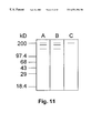
Classifications
-
- C—CHEMISTRY; METALLURGY
- C07—ORGANIC CHEMISTRY
- C07K—PEPTIDES
- C07K16/00—Immunoglobulins [IGs], e.g. monoclonal or polyclonal antibodies
- C07K16/18—Immunoglobulins [IGs], e.g. monoclonal or polyclonal antibodies against material from animals or humans
- C07K16/20—Immunoglobulins [IGs], e.g. monoclonal or polyclonal antibodies against material from animals or humans from protozoa
- C07K16/205—Plasmodium
-
- A—HUMAN NECESSITIES
- A61—MEDICAL OR VETERINARY SCIENCE; HYGIENE
- A61P—SPECIFIC THERAPEUTIC ACTIVITY OF CHEMICAL COMPOUNDS OR MEDICINAL PREPARATIONS
- A61P33/00—Antiparasitic agents
-
- A—HUMAN NECESSITIES
- A61—MEDICAL OR VETERINARY SCIENCE; HYGIENE
- A61P—SPECIFIC THERAPEUTIC ACTIVITY OF CHEMICAL COMPOUNDS OR MEDICINAL PREPARATIONS
- A61P33/00—Antiparasitic agents
- A61P33/02—Antiprotozoals, e.g. for leishmaniasis, trichomoniasis, toxoplasmosis
- A61P33/06—Antimalarials
-
- C—CHEMISTRY; METALLURGY
- C07—ORGANIC CHEMISTRY
- C07K—PEPTIDES
- C07K14/00—Peptides having more than 20 amino acids; Gastrins; Somatostatins; Melanotropins; Derivatives thereof
- C07K14/435—Peptides having more than 20 amino acids; Gastrins; Somatostatins; Melanotropins; Derivatives thereof from animals; from humans
- C07K14/44—Peptides having more than 20 amino acids; Gastrins; Somatostatins; Melanotropins; Derivatives thereof from animals; from humans from protozoa
- C07K14/445—Plasmodium
-
- A—HUMAN NECESSITIES
- A61—MEDICAL OR VETERINARY SCIENCE; HYGIENE
- A61K—PREPARATIONS FOR MEDICAL, DENTAL OR TOILETRY PURPOSES
- A61K39/00—Medicinal preparations containing antigens or antibodies
-
- C—CHEMISTRY; METALLURGY
- C07—ORGANIC CHEMISTRY
- C07K—PEPTIDES
- C07K2319/00—Fusion polypeptide
-
- C—CHEMISTRY; METALLURGY
- C12—BIOCHEMISTRY; BEER; SPIRITS; WINE; VINEGAR; MICROBIOLOGY; ENZYMOLOGY; MUTATION OR GENETIC ENGINEERING
- C12N—MICROORGANISMS OR ENZYMES; COMPOSITIONS THEREOF; PROPAGATING, PRESERVING, OR MAINTAINING MICROORGANISMS; MUTATION OR GENETIC ENGINEERING; CULTURE MEDIA
- C12N2799/00—Uses of viruses
- C12N2799/02—Uses of viruses as vector
- C12N2799/021—Uses of viruses as vector for the expression of a heterologous nucleic acid
- C12N2799/023—Uses of viruses as vector for the expression of a heterologous nucleic acid where the vector is derived from a poxvirus
-
- Y—GENERAL TAGGING OF NEW TECHNOLOGICAL DEVELOPMENTS; GENERAL TAGGING OF CROSS-SECTIONAL TECHNOLOGIES SPANNING OVER SEVERAL SECTIONS OF THE IPC; TECHNICAL SUBJECTS COVERED BY FORMER USPC CROSS-REFERENCE ART COLLECTIONS [XRACs] AND DIGESTS
- Y02—TECHNOLOGIES OR APPLICATIONS FOR MITIGATION OR ADAPTATION AGAINST CLIMATE CHANGE
- Y02A—TECHNOLOGIES FOR ADAPTATION TO CLIMATE CHANGE
- Y02A50/00—TECHNOLOGIES FOR ADAPTATION TO CLIMATE CHANGE in human health protection, e.g. against extreme weather
- Y02A50/30—Against vector-borne diseases, e.g. mosquito-borne, fly-borne, tick-borne or waterborne diseases whose impact is exacerbated by climate change
-
- Y—GENERAL TAGGING OF NEW TECHNOLOGICAL DEVELOPMENTS; GENERAL TAGGING OF CROSS-SECTIONAL TECHNOLOGIES SPANNING OVER SEVERAL SECTIONS OF THE IPC; TECHNICAL SUBJECTS COVERED BY FORMER USPC CROSS-REFERENCE ART COLLECTIONS [XRACs] AND DIGESTS
- Y10—TECHNICAL SUBJECTS COVERED BY FORMER USPC
- Y10S—TECHNICAL SUBJECTS COVERED BY FORMER USPC CROSS-REFERENCE ART COLLECTIONS [XRACs] AND DIGESTS
- Y10S977/00—Nanotechnology
- Y10S977/70—Nanostructure
- Y10S977/788—Of specified organic or carbon-based composition
- Y10S977/802—Virus-based particle
- Y10S977/803—Containing biological material in its interior
- Y10S977/804—Containing nucleic acid
Definitions
- the present invention relates to novel DNA constructs comprising a vector linked to a DNA segment which encodes a protein containing a signal protein at its N-terminus and an anchor sequence at its C-terminus.
- the present invention relates to vaccines which are useful for the prevention and treatment of malaria caused by Plasmodium falciparum in humans.
- Plasmodium falciparum produces the most pathogenic of the malarias and often results in death. It is responsible for about half of the human cases of malaria found worldwide.
- the life cycle of the malaria parasite provides several stages at which interference could lead to cessation of the infective process. Included among these stages is the invasion of the erythrocyte by the merozoite.
- the merozoite represents a potentially attractive target (and perhaps the only target) from which a vaccine may be produced, because the free merozoite, although it has a limited lifetime (one to two hours) occurs earlier in the life cycle of malaria, and the emergence of later stage sexual forms, which are responsible for transmission of the disease, depends upon the erythrocytic stage.
- the general life cycle of malaria parasites is the same for human and other animal malaria parasites, thus allowing model studies to be conducted on a rodent species with accurate translation to the human parasite.
- the rodent malaria parasite strain Plasmodium berghei Anka has a pathology very similar to the FCR-3 strain of Plasmodium falciparum (a well studied variant of the human parasite).
- the blood stage of the human parasite can be grown in the laboratory (in human red cells) thus affording a system for studying the effects of antibodies/inhibitors on the invasion process, and the erythrocytic phase.
- a human becomes infected with malaria from the bite of a female Anopheles mosquito.
- the mosquito inserts its probe into a host and in so doing, injects a sporozoite form of Plasmodium falciparum , present in the saliva of the mosquito.
- the sporozoites which have been injected into the human host are cleared into a number of host tissue cells, including liver parenchyma cells (hepatocytes) and macrophages. This phase is known as the exoerythrocytic cycle because at this point in the life cycle the organism has not yet entered red blood cells.
- hepatocytes liver parenchyma cells
- macrophages macrophages.
- This phase is known as the exoerythrocytic cycle because at this point in the life cycle the organism has not yet entered red blood cells.
- sporozoites undergo a transformation into trophozoites, which incubate and undergo schizogony, rupture and liberate tissue merozoites. This process takes approximately 7-10 days and, depending upon species, may repeat itself several times, during which time the host feels no effects. In Plasmodium falciparum , this repetition does not occur.
- the liver or other tissue cells burst open and release numerous merozoites into the bloodstream.
- red blood cells where they enter the erythrocytic phase of the life cycle.
- young plasmodia have a red nucleus and a ring-shaped, blue cytoplasm.
- the plasmodium divides into merozoites, which may break out of the red blood cell, enter other erythrocytes and repeat the multiplication process. This period lasts approximately 48 hours.
- the gametocytes are capable of mating only in the mosquito.
- a mosquito in order for Plasmdium falciparium to produce sporozoites for infecting a second human host, a mosquito must first bite a human host carrying gametocytes. These gametocytes mature into macrogametes, mate in the mosquito's stomach and produce a zygote.
- the zygote (ookinete) is active and moves through the stomach or the midgut wall. Under the lining of the gut, the ookinete becomes rounded and forms a cyst called an oocyst, in which hundreds of sporozoites develop.
- Sporozoites thereafter invade the entire mosquito and many of them enter the salivary glands where they are in a favorable position to infect the next host when the mosquito feeds on its blood. The life cycle thereafter simply repeats itself in another human host.
- live vaccinia virus was used as a vaccine to eradicate smallpox successfully, and a recombinant vaccinia virus expressing viral antigens has been shown to induce a strong antibody response in immunized animals, conferring protection against disease (Arita, I., Nature , 1979, 279, 293-298). Furthermore, it has been shown in animal models that co-presentation of potential immunogens with highly immunogenic vaccinia virus proteins can elicit a strong immune response against that specific immunogen (Moss and Flexner, Annals of the New York Academy of Sciences , 86-103; Mackett and Smith, J. Gen. Virol ., 1986, 67, 2067-2082; Houard, et al., J. Gen.
- vaccinia virus vaccine has the advantage of being relatively inexpensive and easily stored, transported and delivered, features which are particularly important in the developing countries where malaria is most prevalent.
- Proteins on the surface of merozoites are good targets for an immune response and are good malaria vaccine candidates because merozoites are the only stage in the asexual blood cycle in which the parasite is exposed to the immune system.
- the 190 kD glycoprotein of Plasmodium falciparum, precursor to major merozoite surface antigen1 (MSA1), which is synthesized during schizogony, is considered a promising candidate for a blood-stage malaria vaccine (Blackman, et al., Mol. Biochem. Parasitol ., 1991, 49, 29-34; Perrin, et al., J. Exp. Med ., 1984, 160, 441-451; Siddiqui, et al., Proc. Natl. Acad.
- the high-molecular weight precursor is processed into 88 kD, 30 kD, 38 kD and 42 kD fragments which remain as complexes on the merozoite surface (Holder, et al., Parasitology , 1987, 94, 199-208; McBride and Heidrich, Mol. Biochem. Parsitol ., 1987, 23, 71-84).
- the complex is released from the membrane by cleavage of the 42 kD anchor fragment, and a 19 kD carboxyl-terminal fragment remains on the merozoite membrane and is carried into the invaded erythrocytes (Blackman, et al., supra; Blackman, et al., Mol. Biochem. Parasitol ., 1991, 49, 35-44).
- the complete MSA1 of unprocessed P. falciparum has been used to provide partial or complete protection against challenge infection (Blackman, et al., Mol. Biochem. Parasitol ., 1991, 49, 29-34; Perrin, et al., J. Exp.
- mAbs monoclonal antibodies directed against glycophorin A, the putative erythrocyte receptor for P. falciparum were prepared.
- One of these mAbs, designated 2B10 is capable of blocking the binding of MSA1 to human erythrocytes and inhibiting the invasion of P. falciparum merozoites into human erythrocytes (Su, et al. Infect. Imm ., 1993, 151, 2309).
- the anti-idiotype antibody of 2B10 recognized the C-terminal (1047-1640aa) region of MSA1 in a western blot (Su, et al., J. Immunol ., 1995) and appears to recognize the same site on glycophorin A as the merozoite.
- the present invention relates to a malaria vaccine comprising an expression vector, preferably, a vaccinia virus system which expresses a protein corresponding substantially to a specific domain of the major merozoite surface antigen 1 (MSA1) of Plasmodium falciparum or an immugenic peptide portion thereof.
- MSA1 major merozoite surface antigen 1
- the DNA coding for the MSA1 protein domain is expressed by the vaccinia virus after administration to a patient.
- the MSA1 protein or sub-fragment which is then expressed in the patient raises a humoral and/or cell-mediated response to the merozoite malaria antigen, which response provides the effect of protecting the vaccinated patient from a subsequent malaria infection.
- the vaccinia virus system continues to express antigen in the patient for a period of days, months or even years, thus reinforcing the immunogenic response of the patient to the expressed antigen.
- the MSA1 peptide antigen or immunogenic peptide portion thereof which is expressed by the expression vector vaccinia virus may also comprise a signal peptide and/or an anchor peptide sequence. It has been found that the addition of a signal and/or anchor peptide to the expressed MSA1 antigen in vaccines according to the present invention unexpectedly enhances the immunogenicity to the patient of the MSA1 protein of Plasmodium falciparum . It is an unexpected result that the inclusion of a signal and/or anchor protein with MSA1 can be expressed by a vaccinia virus system according to the present invention and the expressed peptide will produce a significantly greater immunogenic response than the MSA1 peptide alone or in combination with an adjuvant.
- Methods of inducing an immunogenic response in a patient are also contemplated by the present invention.
- a patient is administered an amount of a vaccinia virus capable of expressing the MSA1 peptide of Plasmodium falciparum such that the patient develops an immunogenic response to the expressed peptide.
- the immunogenic response generated preferably will be “substantially protective”, i.e., will protect the patient from some of the more severe symptoms and physiological states of the malaria disease, including the death of the patient from malaria.
- the present invention also relates to an immunogenic dosage form as a vaccine, for inducing an immunogenic response to the merozoite stage in the life cycle of Plasmodium falciparum .
- Methods of vaccinating a patient against a malaria infection are also contemplated by the present invention.
- a patient is vaccinated against a Plasmodium falciparum infection by administering an immunogenic response producing effective amount of a vaccinia virus capable of expressing the MSA1 peptide or an immunogenic peptide portion thereof of Plasmodium falciparum in the patient.
- the present invention also relates to chimeric proteins or peptides comprising the peptide sequence corresponding to the major merozoite surface antigen 1 (MSA1) of Plasmodium falciparum or an immugenic peptide portion thereof in combination with a signal sequence and/or anchor sequence, more preferably both a signal sequence and an anchor sequence.
- MSA1 major merozoite surface antigen 1
- FIGS. 1A and 1B show a diagrammatic representation of the construction of recombinant vaccinia viruses incorporating the sequences corresponding to MSA1C-(Si,A), MSA1C-(Si,nA), MSA1C-(nSi,A) and MSA1C-(nSi,nA).
- FIGS. 2-5 represent the gene sequences for MSA1C-(Si,A), MSA1C-(Si,nA), MSA1C-(nSi,A) and MSA1C-(nSi,nA).
- FIG. 6 is a diagrammatic representation of four recombinant vaccinia viruses of the present invention: rv-MSA1-(Si,A); rv-MSA1-(Si,nA); rv-MSA1-(nSi,A); rv-MSA1-(nSi,nA).
- This figure shows the schematic of the genome of recombinant vaccinia viruses expressing the different MSA1 constructs.
- Tk l and TK R right and left regions of the vaccinia virsu thymidine kinase gene; LacZ, beta-galacosidase gene.
- Si signal region of MSA1; nSi: no signal region of MSA1; A: anchor region of MSAa1; nA: no anchor region.
- FIG. 7 is a diagrammatic representation of the results obtained from Western blot analysis of recombinant vaccinia virus expressed proteins using anti-MSA1C-A mouse serum as a probe, of proteins expressed from BSC-1 cells infected with vaccinia virus.
- A pellet of rV.V-MSA1C(Si,nA) infected cells;
- B supernatant of rV.V-MSA1C(Si,nA) infected cells;
- C pellet of rV.V-MSA1C(Si,A) infected cells;
- D supernatant of rV.V-MSA1C(Si,A) infected cells;
- E supernatant of rV.V-MSA1C(Si,A) infected cells;
- pellet of rV.V-MSA1C(nSi,nA) infected cells F. supernatant of rV.V-MSA1C(nSi,nA) infected cells; G. pellet of rV.V-MSA1C(nSi,A) infected cells; H. supernatant of of rV.V-MSA1C(nSi,A) infected cells; I. pellet of WR infected cells; J. supernatant of WR infected cells.
- FIGS. 8A-8J show a diagrammatic representation of results obtained from immunofluorescence staining of non-permeabilized infected cells.
- HeLa cells seeded on pre-treated coverslips for 48 hours were infected with recombinant and wild-type vaccinia virus at an M.O.I. of 5.
- the cells were probed with anti-MSA1C-A serum and laveled with FITC goat anti-mouse.
- A,B rV.V-MSA1C(Si,nA) infected cells
- C,D rV.V-MSA1C(Si,A) infected cells
- E,F rV.V-MSA1C(nSi,nA) infected cells
- G,H rV.V-MSA1C(nSi,A) infected cells
- I,J WR infected cells.
- FIG. 9 is a diagrammatic representation of results obtained in rabbits immunized intradermally with the recombinant vaccinia viruses of the present invention.
- Five rabbits were immunized intradermally with recombinant and wild-type vaccinia virus, and one rabbit was immunized intravenously with rV-MSA1C-(Si,A).
- the I.D. immunizations occurred on days 0, 21, 47 and 68, and the I.V. immunizations occurred on days 0, 47 an 68.
- Blood samples taken on days 33, 54, and 78 were analyzed for antibodies to the trpE-MSA1C-A protein (C-terminus of MSA1 expressed and purified from E. coli ) using ELISA.
- FIG. 10 is a diagrammatic representation of results obtained in Balb/c mice which were inoculated with vaccinia viruses of the present invention.
- Balb/c mice were inoculated I.P. with recombinant and wild-type vaccinia virus on days 0, 21, 42 and 63.
- Blood samples taken on days 11, 31, 51, 73 93, 123, 147, 167 and 187 were analyzed for antibodies to trpE-MSA1C-A protein (C-terminus of MSA1 expressed and purified from E. coli ) using ELISA.
- Sera were analyzed in duplicate: ( ⁇ ) rV-MSA1C-(Si,A); ( ⁇ ) (rV-MSA1C-(Si,nA); (X)(rV-MSA1C-(nSi,A); ( ⁇ ) (rV-MSA1C-(nSi,nA); (+) WR.
- FIG. 11 is a diagrammatic representation of the results obtained from a Western blot analysis of the recognition of a 190 kD of P. falciparum by vaccinia virus induced mouse antibodies.
- Schizont stage parasites were lysed by boiling in sample buffer and loaded onto a 4-20% Tris-glycine gradient gel, and the proteins were transferred to a PVDF membrane by electrophoresis. These blots were then probed with anti-MSA1C-(Si,A), anti-MSA1C-A or anti-WR antibodies and labelled with phasphatase-conjugated goat anti-mouse IgG.
- chimeric protein “chimeric peptide” or “chimeric peptide sequence” is used to describe the non-natural peptide sequences according to the present invention which comprise the expressed protein or peptide and an anchor peptide and/or a signal peptide.
- chimeric peptides generally are synthetic peptides produced by an expression vector which contain a desired target protein or peptide (either MSA1 or an immunogenic peptide portion thereof) in combination with another peptide sequence (either an anchor or signal peptide sequence).
- the term “patient” is used to describe an animal, including a mammal and especially including a human patient which is administered a dosage form of an expression vector or chimeric protein according to the present invention.
- the expression vector encodes for MSA1 or an immunogenic peptide portion therof and expresses the encoded protein or peptide in the patient.
- expression vector is used to describe the means by which nucleic acid, including DNA, cDNA, RNA or variants thereof, more preferably DNA fragments, encoding for a specific peptide or protein, may be introduced into the patient or the patient's tissue in order to express or produce the desired protein.
- Such vectors include any vectors into which a nucleic acid sequence encoding for the desired MSA1 protein or immunogenic peptide fragment thereof, anchor peptide sequence and/or, signal protein sequence may be inserted (along with any required or optional operational elements) into a host organism and replicated. Expression vectors may also be used to simply produce chimeric peptide in culture for isolation.
- Preferred vectors are those which are capable of expressing the peptide or protein sequences in mammalian cells and whose restriction sites are well known and which contain the required operational elements for expression of the desired protein or peptide sequence.
- the vector is preferably a vaccinia virus vector, adenovirus vector or herpes virus vector which has the capacity to infect a mammalian cell and express or synthesize proteins utilizing the host's biosynthetic mechanism.
- the viral vector used for delivery should optimally be one which infects cells but which does not cause lysis due to replication (i.e., an attenuated or partially disabled virus selected from among adenovirus, vaccinia virus and herpes viruses, among similar types).
- the vector will infect the host cells and, using the host cells' biosynthetic pathways, synthesize encoded protein or peptide fragment.
- Any immunizing vehicle which has a detailed genetic and human use history may be used as the expression vector in the present invention.
- the preferred expression vector is a viral vector, more preferably, a vaccinia virus vector, for example, as described by Earl and Moss, Current Protocols in Molecular Biology , 1993, 16.17.1-16.17.16) and Smith and Moss, Gene , 1983, 25, 21-28.
- any vaccinia or other viral vector which may be used in the above-described manner may be appropriate for use in the present invention.
- the expression vector should contain at least one promoter, at least one operator, at least one terminator codon, and any other sequences which are necessary for the efficient transcription and subsequent translation of the nucleic acid from the vector.
- the expression vectors will advantageously comprise at least one origin of replication which is recognized by the host organism along with at least one selectable marker and at least one promoter sequence capable of initiating transcription of the nucleic acid (preferably, DNA) sequence.
- vaccines comprise an expression vector, preferably a vaccinia virus system which expresses an antigenic protein after administration of the vaccinia virus system to an animal, such as a mammal.
- Vaccines may also comprise chimeric peptides comprising MSA1 or an immunogenic peptide portion thereof in combination with a signal peptide sequence and/or an anchor peptide sequence.
- the method of administering the vaccine(s) according to the present invention may vary and include intravenous, buccal, oral, transdermal and nasal, among others, but intramuscular or subcutaneous administration is the most common method of administration.
- MSA1 protein or “MSA1 peptide” is used to describe the major merozoite surface antigen 1 of the merozoite stage of Plasmodium falciparum or immunogenic peptide portions thereof.
- MSA1 is the major surface protein of the merozoite stage of Plasmodium falciparum . It is a 190 kD glycoprotein which is synthesized during schizogony life-cycle stage. The high molecular weight precursor is processed into 88 kD, 30 kD, 38 kD and 42 kD fragments which remain as complexes on the merozoite surfaces.
- the complex is released from the merozoite membrane by cleavage of the 42 kD anchor fragments and a 19 kD carboxy-terminal fragment remains on the merozoite membrane and is carried into the invaded erythrocytes.
- the complete MSA1 gene sequence is available on the data base UNDP/WORLDBANK/WHO/TDR Malaria Sequence.
- the preferred portion of MSA1 for expression in the vaccinia virus system according to the present invention is a carboxy-terminal region (corresponding to amino acids 1047 to 1640) of MSA1.
- the expressed protein may be MSA1 or any portion thereof, preferably such that the MSA1 portion contains at least the carboxy-terminal region of MSA1.
- carboxy-terminal region of MSA1 and “carboxy-terminal MSA1 peptide” are used to describe that portion of the MSA1 protein corresponding to amino acids 1047 to 1640 which is the preferred expressed antigen peptide sequence in the present vaccines. It represents a preferred target for the development of humoral and/or cell mediated response because of the degree of specificity of the immune response which can be elicited against such a protein segment.
- the expression of the C-terminal MSA1 peptide according to the instant invention produces a specific immune reactivity which becomes less specific as more of the MSA1 protein is incorporated into the vaccinia virus system.
- the above terms to describe the C-terminal MSA1 peptide include not only the 593 amino acid peptide referred to above, but any peptide substantially corresponding to that 593 amino acid peptide.
- Any expressed peptide which substantially corresponds to the MSA1 protein or an immunogenic peptide portion of the MSA1 protein and, preferably, also contains a peptide corresponding to at least an immunogenic portion of the C-terminal MSA1 peptide, may be used in the present vaccines.
- expressed peptides corresponding to MSA1 protein and/or an immunogenic peptide portion thereof in combination with a signal sequence or anchor sequence may also be used in the present invention.
- signal peptide “signal sequence” or “signal protein” is used to describe a 7-30 unit amino acid peptide sequence, preferably about a 15-26 unit amino acid peptide sequence, which is generally found at or near the N-terminus of the expressed protein or peptide which is used in the present invention in order to substantially enhance the biological activity of the protein or peptide expressed in the patient according to the present invention.
- Signal sequences generally contain hydrophobic peptide sequences of between about 7 and 30 amino acid units, more preferably, about 15 to 26 amino acid units, even more preferably about 16 to 24 amino acid units and most preferably about 18 to 20 amino acids units appear to be essential for the targeting of protein chains (generally, secretory proteins) to membranes within the cell.
- hydrophobic sequences are of sufficient length to cross the lipid bilayer of the cell membranes.
- Signal sequences serve as organizers for the cellular traffic of macromolecules. These proteins are believed to play a central role in the translocation of polypeptide chains across membranes.
- the incorporation of a signal protein sequence at the amino terminus of the protein or peptide sequence expressed by the vaccinated patient is associated with the substantial enhancement in the biological activity (including the therapeutic effect of immunogenicity) associated with the expressed protein or peptide.
- signal sequences which are known in the art may be used in the present invention.
- yeast or lower trophic order signal sequences clearly mammalian signal sequences are preferred for use in mammals and the specific species signal sequences are most preferred for use in the desired mammalian species to be treated.
- a human signal sequence is most preferred.
- Signal sequences for use in the present invention generally contain three regions, a first or c region at the carboxy end of the peptide (which serves as the cleavage site for a signal peptidase enzyyme), comprising about 5 to 7 amino acid residues which tend to be highly polar but uncharged; a second or h region which is N-terminal to the c region, generally about 7 to 13 amino acid residues in length and highly hydrophobic (comprised primarily of Leu, Ala, Met, Val, Ile, Phe, and Trp amino acids, but may contain an occasional Pro, Gly, Ser or Thr amino acid residue); and a third region or n-region of highly variable length and composition, but generally carrying a net positive charge contributed by the N-terminus (negative charges contributed from acidic residues are also known) and any charged residues.
- c region and the h region are between 1 and 3 amino acid residues which tend to be small and uncharged (Ala, Gly, Ser, others).
- Synthetic homopolymeric h regions comprised of amino acids selected from the group consisting of leucine, isoleucine, phenylalanine, valine, alanine and tryptophan, preferably leucine, isoleucine and phenylalanine may be used in the signal proteins according to the present invention. See generally, von Heijne, European Journal of Biochemistry , (1983), 133, pp. 17-21.
- the signal sequences which are used in the present invention preferably encompass eukaryotic signal sequences, preferably between 7 and 30 amino acid units in length, preferably between 15 and 26 units, more preferably between about 16 and 26 amino acids, even more preferably between 18 and 20 amino acid units.
- the c region of the signal peptide should be more polar and the boundary between the h and c regions between residues ⁇ 5 and ⁇ 6, or ⁇ 7 or ⁇ 8 (counting from the position of cleavage of the signal sequence- i.e., the first amino acid of the mature or expressed protein or peptide is +1) is between 1 and 3 amino acid residues which tend to be small and uncharged (Ala, Gly, Ser, others).
- Position preferences in the h/c for amino acids are as follows:
- ⁇ 10 most preferably leucine or alternatively, isoleucine, valine, alanine, or phenylalanine;
- ⁇ 9 most preferably leucine, alternatively, isoleucine, alanine, valine, phenylalanine;
- ⁇ 8 most preferably leucine, alternatively isoleucine, alanine, valine, glycine, phenylalanine;
- ⁇ 7 most preferably alanine, alternatively, leucine, isoleucine, valine, phenylalanine;
- ⁇ 6 most preferably valine, alternatively leucine, valine, isoleucine, phenylalanine, alanine;
- ⁇ 5 most preferably proline, alternatively glycine, alanine, leucine, valine;
- ⁇ 4 most preferably glycine, alternatively proline, leucine, alanine, valine;
- ⁇ 3 most preferably alanine, alternatively valine
- ⁇ 2 most preferably leucine, alternatively phenylalanine
- ⁇ 1 most preferably alanine, alternatively glycine.
- the h region may vary in length as well.
- the n region is polar, contains positively charged amino acids (predominantly lysine and arginine) and varies with the overall length of the signal peptide as described above.
- the c region extends from residues ⁇ 1 to ⁇ 5 of the signal peptide/expressed or mature protein.
- the c region is N-terminus to the expressed or mature protein
- the h region is N-terminus to c region (with a 1-3 amino acid boundary between the c and h region)
- the n region is a positively charged N-terminus to the h region.
- the n region is variable in length and generally positively charged (with a preferred charge of +2)
- the h region is hydrophobic and variable in length and the c region preferably contains about five (5-7) generally polar amino acids.
- the end of the hydrophobic domain (i.e., the boundary between the hydrophobic residues enumerated above) should preferably be at positions ⁇ 6/ ⁇ 5.
- the signal sequence should comprise a 5 to 10 unit residue initial sequence (beginning with methionine) followed by at least a seven residue sequence (as described above) and an additional amino acids from 1 to 10 residues in length.
- a typical sequence for the region noted about is:
- the signal sequence used should be characteristic of the cell type used for expression of the protein. Thus, in veterinary applications, the signal sequence most preferably used should be that of the animal to be treated. Often a signal sequence which is mammalian in character is acceptable. Most mammalian signal sequences will have significant efficacy in expressing proteins or peptides in other mammalian cells. Human signal sequences are preferably used for human applications.
- amino acid sequences corresponding to the above-described signal peptide DNA sequences are:
- anchor protein or “anchor peptide sequence” is used to describe proteins or peptides which are anchored to the external surface of the plasma membrane generally by covalent bonding to glycans containing phosphatidyl inositol. These structures to which the anchor protein or peptide is bonded are often referred to as glycosyl phosphatidylinositols or GPIs. In all cells, anchor proteins covalently bonded to GPIs are found on the external face of the plasma membrane of cells or on the lumenal surface of secretory vesicles.
- an “anchor protein” or “anchor peptide” comprises a peptide sequence preferably of about 15-35 residues in length which is generally expressed at the carboxy-terminus of the protein or peptide expressed by the expression vector according to the present invention (3′ end of the DNA sequence expressing the desired protein or peptide and carboxyl terminus of the expressed protein or peptide).
- many of the proteins or peptides which are expressed in the patient and in particular, the immunogenic proteins or peptides of vaccines according to the present invention which are expressed in the patients produce a biological or immunogenic response in the patient which is substantially enhanced when an anchor peptide is incorporated at the carboxy terminus of that protein or peptide.
- the inclusion of a signal protein at or in the proximity of the N-terminus, in addition to the anchor peptide at the carboxy-terminus of the expressed protein is associated with an unexpected enhancement in the biological effects of the expressed protein. This is especially true where the expressed protein is antigenic or immunogenic in nature.
- the carboxy-terminus of the expressed protein or peptide residue is modified by attachment of a glycolipid anchor, which serves to anchor the modified protein or peptide to the cell surface.
- the peptide residue to which the GPI anchor is added is always one of small amino acids, such as glycine, aspartic acid, asparagine, alanine, serine and cysteine. These occur at the carboxyl terminus of the protein/peptide of interest and thus can be specified by inclusion of the appropriate codons in the DNA fragment to be added to the cDNA sequence specifying the protein/peptide of interest.
- the two residues downstream of the anchor addition site are usually small.
- the cleavage/anchor addition site resides in a domain of three small amino acid residues, although the central of the three residues has less stringent steric requirements.
- additional amino acids preferably, polar ones such as lysine or arginine as well as threonine, alanine and proline
- the remainder of the addition signal sequence will contain from 15 to 35 amino acids with a hydrophobic domain at the extreme carboxyl terminus.
- This domain should extend for 15-25 amino acids and will include amino acids such as valine, leucine, isoleucine, alanine, pphenylalanine, but may also contain proline and glycine as well as tryptophan.
- a typical such sequence is as follows:
- the small sequence is in bold face with the left portion represeting the terminus of the protein and the D residue the site of GPI addition.
- the right hand portion is that cleaved during GPI addition with the underlined sequence indicating the hydrophobic terminus.
- the anchor peptide may have a cleavable N-terminal sequence, which directs the peptide to the endoplasmic reticulum and the cellular trafficking pathway where the GPI anchor is added.
- the anchor peptide also has a predominantly hydrophobic sequence at the extreme carboxy terminus which generally ranges in size from about 15 to about 35, more preferably about 15 to 30, and even more preferably about 15 to 25 amino acid residues, signals the addition of the GPI anchor and is cleaved off concurrent with GPI addition. It is the hydrophobicity rather than the sequence itself which is important for anchor addition.
- anchor addition site an amino acid residue within this hydrophilic spacer selected from the group consisting of glycine, aspartic acid, arginine, asparagine, alanine, serine and cysteine.
- the two residues downstream from the anchor addition site are also usually small amino acid residues apparently to minimize steric hindrance at the anchor addition site.
- the GPI portion is preassembled and added as a single unit to a specific amino acid residue near the carboyxl terminus of the expressed protein or peptide.
- the carboxyl terminal region may be characterized by the presence of a C-terminal signal peptide which is preferably ten to thirty amino acids in length and provides the information needed to add the GPI anchor.
- the actual amino acid residue to which the GPI structure is attached is called the omega site and this residue should be glycine, alanine, cysteine, serine, asparagine or aspartic acid.
- the overall structure of the anchor peptide may be summarized as a 15-35 amino acid peptide at the carboxyl terminus of the expressed protein or peptide.
- This anchor peptide sequence (reading from the terminus towards the amino end) begins with a hydrophobic stretch of amino acids of variable length, followed by a sequence of preferably 5-7 amino acids which contains charged residues, followed by three amino acids (either glycine or alanine at the omega +2 site); any of glycine, alanine, cysteine, serine, asparagine, aspartic acid, glutamate and threonine at the +1 omega site; and any of glycine, alanine, serine, cysteine, aspartic acid or asparagine at the omega site.
- the signal peptide sequence is generally found at the N-terminus (directly at the N-terminus or removed as much as 1,000 or more amino acids from the N-terminus) and the anchor peptide sequence is generally found at the carboxy-terminus of the expressed protein or peptide
- the signal peptide may be found at or near the carboxy terminus of the expressed target protein or peptide.
- anchor peptide DNA sequence is preferably used:
- amino acid sequence corresponding to the above-described anchor peptide DNA sequence is:
- the signal peptide sequence is generally found at the N-terminus (directly at the N-terminus or somewhat removed- by as much as 1,000 or more amino acids from the N-terminus) and the anchor peptide sequence is generally found at or in the proximity of the carboxy-terminus of the expressed protein or peptide
- the signal peptide may be found at or near the carboxy terminus and the anchor peptide may be found at or near the N-terminus of the expressed target protein or peptide.
- an effective amount refers to an amount or concentration of recombinant vaccinia virus effective to produce a protective immune or therapeutic response with respect to the disease malaria.
- an effective amount of vaccinia virus which is administered to a human patient will vary depending upon a number of factors associated with that patient, including whether the patient previously has been exposed to Plasmodium falciparum before.
- An effective amount of vaccinia virus can be determined by varying the dosage of the product and measuring the resulting cellular and humoral immune and/or therapeutic responses, prior to administration. In general terms, in humans, this amount represents approximately 10 4 to about 10 7 plaque forming units, preferably about 1 ⁇ 10 6 to about 5 ⁇ 10 6 plaque-forming units (determined by assay, as described herein). It is noted that the above described range of administered vaccinia virus is chosen to enhance the likelihood of eliciting an immunogenic response without causing a malaria infection in the vaccine recipient.
- the amount of chimeric peptide administered will be an amount or concentration of the peptide to produce a protective immune or therapeutic response with respect to the disease malaria. While this amount may vary over a considerably wide range, depending upon the immunogenicity of the chimeric peptide chosen, generally the amount of peptide administered ranges from about 0.01 micrograms (10 nanograms) to about 250 micrograms, more preferably about 0.1 microgram to about 100 micrograms, even more preferably about 1 microgram to about 25 micrograms within this range.
- the signal and anchor peptides are preferably expressed at the amino and carboxy terminus of the expressed MSA1 peptide, respectively.
- the signal peptide sequence is located upstream from the MSA1 peptide and the anchor peptide is downstream from the MSA1 peptide.
- a DNA sequence containing the genetic code for the MSA1 peptide to be expressed is obtained by chemical synthesis or other means such as biochemical isolation of available MSA1 DNA sequences and incorporated into a cloning plasmid (for example following cloning vectors: pBR322, pGEM3z, pSP70, pSE420, pRSET, lambdaZAP, all commercially available, among numerous others).
- a cloning plasmid for example following cloning vectors: pBR322, pGEM3z, pSP70, pSE420, pRSET, lambdaZAP, all commercially available, among numerous others.
- the DNA is incorporated into a transfer vector and transfected with eukaryotic cells, for example, monkey kidney cells (BSC-1 cells), and with wild-type vaccinia virus (WR) to produce recombinant vaccinia virus.
- BSC-1 cells monkey kidney cells
- WR wild-type vaccinia virus
- the recombinant vaccinia virus is then purified before amplification.
- the recombinant vaccinia virus is then administered to an animal as an immunogenic dosage form which expresses MSA1 peptide or an immunogenic portion thereof, preferably, in combination with a signal peptide and/or an anchor peptide.
- the isolated peptide sequence may then be used directly as a therapeutic or immunogenic dosage form.
- the expression vector may be administered directly to the patient where it will express the desired protein or peptide and anchor sequence and render the intended therapeutic or immunogenic effect on the patient.
- the expressed protein may be obtained from cell culture after the cells are lysed by standard protein purification procedures known in the art which may include, among others, gel electrophesis, affinity and immunoaffinity chromatography, differential precipitation, molecular sieve chromatography, isoelectric focusing and ion-exchange chromatography.
- standard protein purification procedures known in the art which may include, among others, gel electrophesis, affinity and immunoaffinity chromatography, differential precipitation, molecular sieve chromatography, isoelectric focusing and ion-exchange chromatography.
- immunoaffinity chromatography the protein or peptide may be purified by passage through a column containing a resin to which is bound antibodies which are specific for at least a portion of the protein or peptide.
- dosages will be set by the prescribing physician considering relevant factors including the age, weight and condition of the patient including, in the case of immunogenic dosage forms, whether the patient has been previously exposed to the microorganism responsible for the disease to be vaccinated against as well as the release characteristics of the expressed protein from pharmaceutical dosage forms of the present invention.
- the amount of the expressed protein which is administered according to the present invention comprises an amount effective to produce the intended effect, i.e., to obtain an immunogenic response in the patient which provides a substantially protective effect against malaria.
- the vaccine which is administered according to the present invention comprises an amount of an expression vector, preferably, a recombinant vaccinia virus effective to express sufficient MSA1 peptide to provide an immunogenic response in a patient.
- the MSA1 peptide or an immunogenic peptide sequence thereof is combined with a signal and/or anchor peptide to substantially increase the immunogenicity of the expressed MSA1 peptide compared to MSA1 peptide which does not contain a signal and/or anchor peptide.
- the immunogenic response provides a protective effect against the merozoite stage of malaria.
- the present vaccine can be injected as is, or for convenience of administration, can be added to a pharmaceutically acceptable carrier.
- suitable pharmaceutically acceptable carriers will be apparent to those skilled in the art, and include water and other polar substances, including lower molecular weight alkanols, polyalkanols such as ethylene glycol, polyethylene glycol, and propylene glycol as well as non-polar carriers.
- Dosages of recombinant vaccinia virus or chimeric protein or peptide according to the present invention which are coadministered with carriers will often be about the same as the amount administered alone (in the absence of co-administration).
- dosages will be set by the prescribing physician considering relevant factors including the age, weight and condition of the patient including whether the patient has been previously exposed to Plasmodium falciparum and the release characteristics of the vaccinia virus from pharmaceutical dosage forms of the present invention.
- the dose of vaccinia virus will depend upon the form in which it is administered.
- the vaccine will generally contain a concentration of virus ranging from about 10 4 to about 10 7 plaque forming units, preferably about 1 ⁇ 10 6 to about 5 ⁇ 10 6 plaque-forming units, depending upon the desired levels of expressed immunogenic protein.
- concentration or amount of vaccinia virus included within the present vaccine will generally fall within this range; however, the amount of recombinant vaccinia virus used in any vaccine form will depend upon the strength of the immunogenic response elicited.
- the dosage of the vaccine employed and the treatment schedule would follow practices normally employed for other vaccination or therapeutic regimens wherein this general method of treatment is employed. It is not anticipated that more than one dose of vaccine initially would be required, but the possibility of providing booster doses is anticipated.
- the dosage schedule for immunization against malaria involves the subcutaneous injection of at least about 1 ⁇ 10 6 plaque-forming units of vaccinia virus.
- a human patient is administered with an effective amount of vaccinia virus such that expressing the MSA1 peptide or an immunogenic peptide thereof, preferably in combination with a signal and/or anchor peptide.
- an immunologically effective chimeric peptide comprising the MSA1 peptide or an immunogenic portion thereof in combination with a signal peptide and/or anchor peptide will be administered.
- an additional boost of vaccinia virus or peptide may be given to promote the immunogenic response. Additional doses of vaccine may be provided to boost the initial inoculation, if needed.
- BSC-1 and Hu134TK-cells Monolayer cultures of monkey kidney cells (BSC-1) and Hu134TK-cells were grown in Dulbecco's modified Eagle's medium (D-MEM) supplemented with 10% fetal bovine serum (FBS). Monolayer cultures of Hela cells and CV-cells were grown in Dulbecco's modified Eagle's medium (D-MEM) supplemented with 10% FBS.
- D-MEM Dulbecco's modified Eagle's medium
- FBS fetal bovine serum
- P. falciparum isolates (FCR-3, a strain identical to the Wellcome strain sequenced by Holder, et al., Nature , 1985 , 317, 270-273) were maintained in human erythrocytes in RPMI 1640 medium supplemented with 25 mM Hepes, 32 mM sodium bicarbonate and 10% human serum in a 5% O 2 , 5% CO 2 , 90% N 2 environment at 37° C.
- P. falciparum strain FCR-3 was kindly donated by Dr. Isabella Quakyi, Georgetown University, Washington, D.C.
- pME-2 is an expression plasmid derived from pWRL507, in which the C-terminal of the MSA1 gene (3555-5917 bp, Wellcome Allele of MSA1) was inserted at the 3′ end of a truncated trpE gene, so that expression was controlled by the trp promoter.
- the protein was expressed using the method described by Holder, et al., Paraite Immunol ., 1988, 10, 607. The fusion protein expressed was insoluble.
- DH5 alpha competent cells (GIBCO BRL) were transformed by pME-2, and a 1 ml culture of a pME-2 positive clone was innoculated into 100 ml M9 medium containing 100 ug/ml (micrograms per ml) Amp and grown overnight. This overnight culture was added to 400 ml M9 containing 100 ug/ml Amp and 10 ug/ml indoleacrylic acid, and after 5 hr. growth at 37° C. and 250 rpm, the cells were harvested. The pellet was then rinsed in 10 ml PBS and frozen to ⁇ 20° C. The cell pellet was thawed in a 10 ml solution containing 25 mM Tris pH 8.0, 1 mM EDTA, 0.2% NP-40 and 100 ul 100 mM PMSF.
- the material was centrifuged as above to yield a futher pellet and this pellet was resuspended in a 10 ml solution of 0.5 M KSCN, 50 mM tris, 5 mM EDTA and 5 mM EGTA and re-centrifuged as above. Finally, the pellet was resuspended in 3 ml water.
- the product was anayzed by SDS-polyacrylamide gel electrophoresis (SDS-PAGE), and the gel containing the 105 kD protein of interest was then cut into approximately 1-5 mm 2 pieces.
- the fusion protein was eluted with a Bio-Rad Model 422 Electro-Eluter, and the vacuum-dried product was dissolved in PBS.
- the concentration was assayed with a BCA protein Assay Reagent Kit (PIERCE).
- Antiserum to the fusion protein was prepared by immunizing Balb/c mice intraperitoneally on two occasions 21 days apart with 100 ug of protein in the presence of Titre-Max (Vaxcel, Inc.). Antiserum was collected 10 days after the second immunization.
- pGEX-2T-P190CR1 (pGEX-2T containing the MSA1 gene from 1 to 150 bp) was used as a sample in PCR.
- the 100 ul mixture contained 100 pmol primer 1 and 100 pmol primer 2 (Table 1, below), 2.5 units Ampli Taq DNA polymerase, dNTP and 10 ul 10 ⁇ reaction buffer (PIERCE).
- the sample was overlaid with several drops of mineral oil to prevent evaporation and subjected to 30 cycles of amplification (94° C. melt, 72° C. extension, 55° C. annealling). Amplified products were identified on a 1.5% agarose gel.
- the start codon of MSA1 is at 418.
- the MSA1C-(Si,nA), MSA1C-(nSi,nA), MSA1C-(nSi,A) fragments contain the 108 bp region directly downstream from the signal sequence and an additional 2 bp on the 5′ end of the C-terminal to preserve the reading frame. Furthermore, a start codon was also added to the two fragments lacking the start-codon-containing signal sequence, a stop codon was also added to the two fragments lacking the anchor region.
- pSPORT1-MSA1C-(Si,A)(FIG. 1) was used as a template in PCR.
- MSA1C-(Si,nA) (primer 1 and primer 5, Tab. 1)
- MSA1C-(nSi,A) (primer 5 and primer 4)
- MSA1C-(nSi,nA) (primer 6 and primer 5) fragments were amplified as well with the above procedures.
- the MSA1C-A fragment was amplified by PCR, and electrophoresis in a 0.8% agarose gel indicated that the amplified fragment was about 1.8 kb.
- This fragment and pSPORT1 were digested with EcoRI and KpnI and were mixed and treated with T4 DNA ligase. The ligation products were transformed into DH5alpha competent cells. X-gal and ampicillin were used to screen the positive clones (pSPORT1-MSA1C-A, FIG. 1 ). Recombinant plasmids were prepared from positive white colonies and identification was performed with SalI and EcoRI digestion. The full sequence of the insert was determined.
- the fragment containing the signal sequence was amplified by PCR, and electrophoresis in a 1.5% agarose gel indicated that the size of the amplified fragment was about 180 bp.
- the Si and pSPORT1-MSA1C-A fragments were then digested with SalI and EcoRI, ligated and transformed, and 30 colonies were selected and screened by PCR using primer 1 and primer 2.
- Recombinant plasmids pSPORT1-MSA1C-(Si,A), FIG. 1) were identified with SalI and EcoRI digestion and were sequenced, and the correct reading frame was established.
- pSPORT1-MSA1C-(Si,A) was then used as a template to amplify MSA1C-(Si,nA), MSA1C-(nSi,A) and MSA1C-(nSi,nA).
- FIG. 1 diagrams the construction of recombinant vaccinia viruses incorporating the sequences corresponding to MSA1C-(Si,A), MSA1C-(Si,nA), MSA1C-(nSi,A) and MSA1C-(nSi,nA).
- the MSA1C-terminal fragment containing the anchor region was inserted into the pSPORT1 plasmid using the marked EcoRI and KpnI sites, creating the pSPORT1-MSA1C-A plasmid.
- the fragment containing the signal region (FSi) and the 108 bp downstream was PCR amplified with a SALIU site on the 5′ end and then inserted into the pSPORT1-MSA1C-A plasmid to produce pSPORT1-, SA1C-(Si,A) plasmid.
- the entire MSA1C-(Si,A) fragment (from the SalI site to the KpnI site) was then removed and inserted into the pSC65 vector to make the final pSC65-MSA1C-(Si,A) transfer vector.
- the other three recombinant transfer vectors (pSC65-MSA1C-(nSi,nA), pSC65-MSA1C-(nSi,A) and pSC65-MSA1C-(Si,nA)) were produced by adding the desired SalI or KpnI sites where necessary by PCR amplification. The amplified fragments were then inserted into the pSC65 plasmid. In all four cases, the insertion site is adjacent to the synthetic early/late promoter (pS-E/L). Tk L and TK R , right and left regions of the vaccinia virus thymidine kinase gene; LacZ, beta-galactosidase gene.
- FIGS. 2-5 are the DNA (gene) sequences for MSA1C-(Si,A), MSA1C-(Si,nA), MSA1C-(nSi,A) and MSA1C-(nSi,nA).
- Monolayers of BSC-1 cells were grown to 90% confluence in a six-well plate, media was removed, and the cells were infected with wild-type vaccinia virus (WR) at 0.1-1 plaque forming units(pfu)/cell for 1 hour. The virus inoculum was removed, and the monolayer was washed twice with OptiMEM (GIBCO BRL) serum-free medium.
- WR wild-type vaccinia virus
- OptiMEM OptiMEM
- Opti-MEM 1 ml of Opti-MEM was added to the infected monolayers and mixed gently with 50 ul of lipofectin-DNA complex (5 ug of recombinant pSC65 was diluted to 25 ul with sterile distilled water, 15 ug of lipofectin reagent was diluted to 25 ul with sterile distilled water, and the solution was gently mixed in a polystyrene tube and allowed to stand for 15 minutes at room temperature). After 5 hours incubation at 37° C., the medium was replaced with 3 ml E-MEM supplemented with 2% FBS and incubated for another 48 hours. After removal of the medium, cells were harvested by scraping into 1 ml of E-MEM supplemented with 2% FBS.
- the virus was released by three cycles of freeze-thawing at 37° C. After removal of medium, 1 ml of diluted freeze-thawed trans-fection mixture (sonication 30 seconds at 4° C. before adding) was added to monolayers of Hu 134 TK-grown to 90% confluence in a six-well plate, and the virus was left for 1 hour at 37° C.
- the infected cells were overlaid with E-MEM supplemented with 2% FBS containing 1% low-melting-point agarose and 25 ug/ml of bromodeoxyuridine.
- BSC-1 cells were infected with recombinant vaccinia virus, and the medium was removed from infected tissue-culture plates 24 hours postinfection.
- the cells were fixed with a 1:1 acetone:methanol mixture for 2 minutes, the wells were washed with 1 ml of PBS, and then anti-MSA1C-A serum diluted 1:200 in PBS containing 2% FBS was added to the wells, 1 ml/well.
- the six-well plate was incubated at room temperature for 1 hour, rocking gently, after which the wells were washed twice with 1 ml of PBS.
- Anti-mouse-peroxidase diluted 1:1000 in PBS with 2% FBS was added to each well, and the plate was incubated for 45 minutes at room temperature. After washing twice with PBS, 0.5 ml of substrate solution was added (the substrate solution was made by dissolving a pinch of dianisidine in 500 ul of absolute ethanol, vortexing, and warming it for 5-10 minutes, then centrifuging it for 30 seconds and adding 200 ul of substrate solution to 10 ml of PBS plus 10 ul of 30% H 2 O 2 ). The plate was then left for 5-10 minutes at room temperature.
- HeLa cells were seeded on pre-treated coverslips for 48 h, after which the cells were infected at an M.O.I. of 5 in a volume of 0.25 ml of D-MEM uith 10% FBS. These were overlaid with 1.5 ml D-MEM with 10% FBS 1-2 h postinfection. The cells were then washed with PBS and fixed for 15 min in 3% paraformaldehyde in PBS. After being washed in PBS, the coverslips were incubated in 50 mM ammonium chloride in PBS for 10 min at room temperature.
- Confluent six-well plates of BSC-1 cells were infected with recombinant vaccinia virus in 1 ml medium at an M.O.I of 5. Cells from each well were harvested 24 hours postinfection. After centrifugation at 8000 rpm for 5 min, the supernatant was concentrated to 10-15 ul with a microcon 30 (Amicon, Inc.), and the cell pellet was resuspended in PBS to a final volume of 200 ul.
- the blot was blocked with 5% BSA in Tris-HCl (200 mM, 0.85%) for 2 h at room temperature before addition of a 1:500 dilution of anti-MSA1C-A serum. After 2 hours incubation the blot was washed three times in Tris-HCl, pH 7.4 for 10 min per wash. A 1:7500 dilution in Tris-HCl, pH 7.4 of alkaline phosphatase-conjugated goat-anti-mouse IgG (Promega) was added, and the blot was incubated for 90 min at room temperature before being washed four times in Tris-HCl, pH 7.4 for 10 min each wash. Western blue stabilized substrate for alkaline phosphatase (Promega) was added, and after about 5 minutes, the blot was washed with water to stop development.
- HeLa S3 cells in E-MEM supplemented with 2% FBS were incubated in a 162 cm 2 flask at 37° C. for 18 h, after which the cells were infected with small stock viruses at 37° C. for 48 h and pelleted by centrifugation for 5 min at 1800 g.
- the infected cells were resuspended in 10 mM Tris-HCl, pH 9.0 and homogenized with 30-40 strokes (while still on ice).
- the mixture was centrifuged for 5 min at 300 g to remove nuclei, and the pellet (removed supernatant was kept was well) was resuspended in 10 mM Tris-HCl, pH 9.0 and centrifuged again (supernatants were pooled).
- the sonicated supernatant was layered onto a cushion of 36% sucrose in a Beckman SW27 centrifuge tube and spun at 35000 rpm for 45 min at 4° C., after which the virus pellet was resuspended in 1 ml of 1mM Tris-HCl, pH 9.0.
- the wild-type virus was further purified by continuous sucrose gradient centrifugation.
- Monolayers of BSA-1 cells were grown to 95% confluence in a six-well plate, virus stocks were sonicated, and 10 fold serial dilutions(10 ⁇ 7 -10 ⁇ 9 ) of the virus in E-MEM supplemented with 2% FBS were made.
- the BSC-1 cells were infected with 1 ml of virus diluted to 10 ⁇ 7 ,10 ⁇ 8 and 10 ⁇ 9 in duplicate. After 48 h of incubation, the medium was removed and 0.5 ml of crystal violet solution was added. After another 15 min incubation at room temperature, the plates were washed with water, and the plaques were counted after drying.
- a ninety-six-well microtiter plate (IMMULON 2, Dynatech Laboratories Inc, VA) was coated with 100 ul/well of a 5 ul/ml solution of MSA1C-A in Tris-NaCl (0.02,0.85%), pH 7.4 and kept overnight at 4° C.
- the antigen-coated plate was then washed with Tris-NaCl and blocked with 150 ul 1% BSA in Tris-NaCl, pH 7.4 for 2 h at 37° C. After washing with Tris-NaCl, pH 7.4, 100 ul of serial 10-fold dilutions (diluted in Tris-NaCl containing 1% BSA and 0.05% Tween 20) of sera were added to the wells in duplicate.
- the plate was rewashed with Tris-Nacl containing 0.05% Tween 20 and phosphatase-labeled IgG was added. After another 2 h incubation at 37° C., the plate was washed with Tris-NaCl containing 0.05% Tween 20 and developed by adding alkaline phosphatase substrate in diethanolamine buffer, pH 9.8. The plate was scanned in a Dynatch ELISA scanner at 405 nm.
- Purified WR vaccinia virus was diluted in Tris-NaCl, pH 7.4 to a final concentration of approximately 5 ⁇ 10 6 pfu/ml and 100 ul volumes were dispensed into the wells of a 96-well plate (IMMULON 2, Dynatech Laboratories Inc) and left for 2 h at 37° C. The buffer was removed and virus was inactivated with 50 ul of 10% paraformaldehyde for 10 min at 4° C. The plates were then blocked just as those in the first group and developed using the procedure described above.
- P. falciparum parasites were cultured in human erythrocytes to a parasitemia of 8% at the schizont stage, at which point they were washed five times in PBS (10 ⁇ volume). Small samples were then taken, smeared on a glass slide and fixed in cold methanol. After being washed in PBS, the samples were probed with antiserum from mice immunized with the recombinant and wild-type viruses. After this probe, the samples were probed with FITC goat-anti-mouse antibody and mounted in 5% DABCO/Mowiol.
- P. falciparum parasites were synchronized twice with 5% sorbitol (Diana), and after 24 hours a mixture of trophozoites and schizonts at 3% parasitemia and 3% hematocrit was transfered to 96-well culture plates (170 ul/well). Anti-serum, pre-immune serum or serum-free medium was added to the parasites in a volume of 30 ul to make a final volume of 200 ul/well, and the plates were incubated at 37° C. in an airtight chamber equilibrated for 5 min with 5% O 2 , 5% CO 2 and 90% N 2 . After 22-24 hours, cell morphology was verified to make sure that all schizonts had burst.
- % ⁇ ⁇ inhibition ( pre - 0 ⁇ ⁇ hr ) - ( T - 0 ⁇ ⁇ hr ) ( pre - 0 ⁇ ⁇ hr ) ⁇ 100 ⁇ %
- T is the parasitemia of tested serum
- pre is the parasitemia of preimmune serum
- 0 hr is the starting parasitemia
- FIG. 1 The construction of the recombinant vaccinia virus is described in FIG. 1 .
- Four recombinant vaccinia virus transfer plasmids were made: pSC65-MSA1C-(Si,A), which is recombinant pSC65 in which the MSA1 peptide containing the signal and anchor regions of MSA1 has been inserted into the SalI and KpnI site; pSC65-MSA1C-(Si,nA) (the same except the MSA1 contains the signal without the anchor); pSC65-MSA1C-(nSi,A) (the same except the MSA1 contains the anchor without the signal) and pSC65-MSA1C-(nSi,nA) (the same except the MSA1 contains neither the signal nor the anchor).
- the vaccinia virus transfer vector, pSC65 has a synthetic compound early/late promotor (pSE/L) so that the foreign genes controlled by the promotor are expressed throughout the virus growth cycle.
- the SalI, BglII, StuI, AatI, KpnI, SmaI, XmaI and PacI sites are located just downstream of the pSE/L for insertion of a foreign gene, and there are E.
- coli beta-galactosidase gene sequences (controlled by the p7.5 vaccinia virus promotor which has early and late vaccinia virus transcriptional regulatory signals) and vaccinia TK gene sequences flanking the entire pSE/L and foreign gene region to direct homologous recombinantion into the TK locus of the vaccinia virus genome.
- BSC-1 cells were infected with wild-type (WR) vaccinia virus and then were transfected with recombinant pSC65. Serial dilutions of progeny virus were then applied to monolayers of Hu 134 TK-cells in the presence of BrdU to select TK-recombinant virus plaques. These were then distinguished from spontaneous TK-mutants by addition of X-gal to the low-melting-point agar overlay. Plaques that stained blue due to expression of beta-galactosidase were picked and then plaque-purified a second time prior to preparation of virus stocks.
- the four recombinant vaccinia viruses were named as rV.V-MSA1C(Si,A); rV.V-MSA1C-(Si,nA); rV.V-MSA1C-(nSi,A) and rV.V-MSA1C(nSi,nA) (FIG. 6 ).
- BSC-1 cells were infected with rV.V-N,ISA1C-(Si,A), rV.V-MSA1C(Si,nA), rV.V-MSA1C-(nSi,A) and rV.V-MSA1C-(nSi,nA), and the infected cells were fixed with acetone/methanol 24 hours post-infection, after which the expressed proteins were labeled by anti-mouse-peroxidase. The results indicated that the cells infected by the recombinant vaccinia virus expressed the C-terminal protein.
- BSC-1 cells were infected with rV.V-MSA1C-(Si,A), rV.V-MSA1C(Si,nA), rV.V-MSA1C-(nSi,A), rV.V-MSA1C-(nSi,nA) and WR viruses. These cells were then harvested 24 hours postinfection, and the cell pellets and 50-times concentrated supernatants were run on an 8% Tris-glycine-SDS gel. The blot was labled with anti-MSA1C-A mouse serum and then probed by alkaline phosphatase-conjugated goat anti-mouse IgG.
- FIG. 7 shows a Western blot analysis, using the anti-MSA1C-A mouse serum as a probe, of proteins expressed from BSC-1 cells infected with vaccinia virus.
- FIG. 8 shows indirect immunofluorescence staining of recombinant vaccinia-infected cells.
- the ELISA titers of rV.V-MSA1C-(Si,A) and rV.VMSA1C-(Si,nA) were 5-10 times greater than that of rV.V-MSA1C-(nSi,nA) and rV.V-MSA1C-(nSi,A) after the fourth inoculation.
- One rabbit was inoculated intravenously on day 0 with rV.V-MSA1C-(Si,A) and reinoculated on days 47 and 68, and the ELISA titer of i.v. rV.V-MSA1C-(Si,A) was 5 times that of i.d.
- rV.V-MSA1C-(Si,A) and rV.V-MSA1C-(Si,nA) stimulated a level of the C-terminal-specific antibodies that was 5-10-fold greater than the level induced by rV.V-MSA1C-(nSi,nA) and rV.V-MSA1C-(nSi,A) after the third inoculation.
- the ELISA titer of rV.V-MSA1C-(Si,A) was about 1:10000 and lasted about 3 months (FIG. 10 ).
- Wild type (WR) vaccinia virus coated on a 96-well plate was probed with a serial 10-fold dilution of mouse antibodies raised against rV.V-MSA1C-(Si,A), rV.V-MSA1C-(Si,nA), rV.V-MSA1C-(nSi,nA), rV.V-MSA1C(nSi,A) and WR, and then probed by alkaline phosphatase-labeled goat antimouse IgG.
- the results showed that the anti-WR virus antibody titers of the recombinant and the WR viruses were almost the same after the second inoculation.
- mice immunized with the recombinant and WR viruses were successfully infected by the viruses (FIG. 10 ).
- CBA/J mice were also immunized with rV.V-MSA1C-(Si,A), rV.V-MSA1C-(Si,nA), rV.V-MSA1C-(nSi,nA), rV.V-MSA1C-(nSi,A) and WR viruses.
- the resulting antibody titers were similar to those of the Balb/c mice although slightly lower (data not shown).
- Schizont stage parasites resuspended in 1 ⁇ sample buffer were boiled for 5 minutes and loaded onto a 4-20% Tris-glycine gradient gel, and the proteins were transferred to a PVDF membrane by electrophoresis.
- the blots were then probed by anti-MSA1C-(Si,A) and anti-MSA1C-A and labeled by alkaline phosphatase-conjugated goat anti-mouse IgG.
- the 190 kD protein was recognized by anti-MSA1C-A in a Western blot, so we have concluded that the 190 kD protein is MSA1.
- Anti-MSA1C-(Si,A) recognized the 190 kD protein (MSA1) as well. (FIG. 11)
- the data demonstrate therefore that the recombinant vaccinia virus correctly expresses the MSA1 fragment in mice.
- the C-terminal region of the MSA1 protein was inserted into the vaccinia virus to investigate the immune response produced by this antigen.
- Four variations of the MSA1 fragment were used to create constructs which contained neither, one or both of the original MSA1 N-terminal signal sequence and the original C-terminal anchor sequence.
- the apparent molecular weight of the MSAIC-(Si,A) construct is less than expected and may be due to proteolytic cleavage of the protein during export.
- the two constructs with the signal sequence are expressed on the cell surface, whereas the two constructs without the signal sequence are not.
- These data indicate that the signal sequence is necessary for proper export of the expressed recombinant protein to the cell surface.
- the data show that the signal region alone is sufficient for cell surface expression. It is possible that once the protein has entered the secretory pathway, because of the signal sequence, there is resultant cell surface expression. The strength and duration of this cell surface expression appears to be greater when the protein contains the anchor region in addition to the signal region and this may be due to hydrophobic interactions. Data indicate also that the protein is not secreted externally and thus presumably remains bound to the cell surface. However, the construct with only the signal sequence elicits a weaker immune response than does the construct with both the signal and anchor regions. The anchor region may be necessary for proper protein conformation which is necessary for a high immune response.
- Immunofluoresence data illustrate that antibodies induced by the MSA1C-(Si,nA) and MSA1C-(Si,A) constructs recognize the parasite in vitro. This indicates that the recombinant protein expressed by the vaccinia virus is indeed very similar, if not identical, to the native parasite MSA1 C-terminus. Antibodies targeting the vaccinia viruses indicate that all four recombinant virus constructs as well as WR were administered at approximately the same level. Therefore the differences of the antibody responses of these four constructs is due to the primary sequence of the protein, not varying levels of vaccinia virus.
- the recombinant protein containing both the signal and anchor regions elicits the greatest immune response and this suggests that both the signal and anchor sequences are advantageous for optimal expression of the MSA-1 C-terminal region.
- antibodies to the protein containing both the signal and anchor sequences most effectively inhibit invasion of erythorcytes by parasites. Although 62% inhibition of invasion may seem only a partial inhibition, these invasion assays were conducted with a 3% starting parasitemia, whereas most other studies are done at approximately 0.3%. A higher starting parasitemia may reduce the inhibition because of the large numbers of merozoites released.
- MSA1C-(Si,A) construct expresses a functional protein which possesses the proper signal sequence and the proper anchor sequence for correct cell surface expression. Furthermore, the antibodies produced by mice and rabbits immunized uith the recombinant vaccinia virus are able to block parasite invasion in vitro.
Abstract
The present invention relates to an expression vector which expresses a malaria MSA1 peptide in combination with a signal peptide and anchor peptide in a host animal. The MSA1 peptide is combined with a signal peptide and anchor peptide for expression. Chimeric peptides being expressed with both signal peptides and anchor peptides were the most effective in eliciting an immunogenic response from a vaccinated host.
Description
This application is a §371 of international application PCT/US97/01395 filed on Jan. 29, 1996, which is a continuation-in-part application of Ser. No. 08/593,006, filed Jan. 29, 1996, now abandoned.
The present invention relates to novel DNA constructs comprising a vector linked to a DNA segment which encodes a protein containing a signal protein at its N-terminus and an anchor sequence at its C-terminus.
More particularly, the present invention relates to vaccines which are useful for the prevention and treatment of malaria caused by Plasmodium falciparum in humans.
This work was supported by a DARPA grant. The government retains certain rights in the invention.
Preventing or treating malaria has long been a challenging health problem, particularly in developing countries, and the rapid development of drug resistance in the parasite has enhanced the need for the development of a malaria vaccine. Although there has been steady progress over the last decade, several problems still must be overcome, including selection of an appropriate delivery system vehicle and antigen carrier.
Although malaria was believed to have subsided after World War II, recent outbreaks suggest that the disease is on the rise. Malaria is again the leading cause of morbidity/mortality globally and presents an increasing threat in at risk environments. Estimates are that 300 million new cases of malaria occur each year, with mortality of approximately 1% of infected individuals. Prophylactic medications used to prevent the disease have been rendered ineffective by the emergence of drug-resistant strains of the parasite worldwide. Complete vector protection is simply not possible and all attempts to eradicate the relevant species of mosquito have failed.
Four species of protozoa of the genus Plasmodium are found in man. The four species include: Plasmodium vivax, Plasmodium malariae, Plasmodium falciparum and Plasmodium ovale. Of these, Plasmodium falciparum produces the most pathogenic of the malarias and often results in death. It is responsible for about half of the human cases of malaria found worldwide.
In malaria, the disease is such that infection followed by recovery does not confer meaningful protection to the individual despite a significant antibody response to several of the parasite proteins. The conventional wisdom has been that the parasite either does not possess antigens suitable for the development of a protective response or has evolved mechanisms which allow it to escape the host immune system. Because recent evidence has shown that immune protection is possible using irradiated sporozoites, the latter hypothesis described above is the more reasonable explanation.
The life cycle of the malaria parasite provides several stages at which interference could lead to cessation of the infective process. Included among these stages is the invasion of the erythrocyte by the merozoite. The merozoite represents a potentially attractive target (and perhaps the only target) from which a vaccine may be produced, because the free merozoite, although it has a limited lifetime (one to two hours) occurs earlier in the life cycle of malaria, and the emergence of later stage sexual forms, which are responsible for transmission of the disease, depends upon the erythrocytic stage.
The general life cycle of malaria parasites is the same for human and other animal malaria parasites, thus allowing model studies to be conducted on a rodent species with accurate translation to the human parasite. For example, the rodent malaria parasite strain Plasmodium berghei Anka has a pathology very similar to the FCR-3 strain of Plasmodium falciparum (a well studied variant of the human parasite). In addition, the blood stage of the human parasite can be grown in the laboratory (in human red cells) thus affording a system for studying the effects of antibodies/inhibitors on the invasion process, and the erythrocytic phase.
In the life cycle of the malaria parasite, a human becomes infected with malaria from the bite of a female Anopheles mosquito. The mosquito inserts its probe into a host and in so doing, injects a sporozoite form of Plasmodium falciparum, present in the saliva of the mosquito.
The sporozoites which have been injected into the human host are cleared into a number of host tissue cells, including liver parenchyma cells (hepatocytes) and macrophages. This phase is known as the exoerythrocytic cycle because at this point in the life cycle the organism has not yet entered red blood cells. After entering hepatocytes, sporozoites undergo a transformation into trophozoites, which incubate and undergo schizogony, rupture and liberate tissue merozoites. This process takes approximately 7-10 days and, depending upon species, may repeat itself several times, during which time the host feels no effects. In Plasmodium falciparum, this repetition does not occur. After the incubation period, the liver or other tissue cells burst open and release numerous merozoites into the bloodstream.
Shortly thereafter, certain of these blood borne merozoites invade red blood cells, where they enter the erythrocytic phase of the life cycle. Within the red blood cells, young plasmodia have a red nucleus and a ring-shaped, blue cytoplasm. The plasmodium divides into merozoites, which may break out of the red blood cell, enter other erythrocytes and repeat the multiplication process. This period lasts approximately 48 hours.
During this same 48 hour period of the erythrocytic cycle, male and female gametocytes are formed in the red blood cells. These gametocytes also burst out of the red blood cells along with the merozoites. It is during this period that the human host experiences the symptoms associated with malaria. The merozoites which burst forth from the red blood cells live for only a few hours in the bloodstream. The gametocytes live for several days or more in the host's bloodstream.
The gametocytes are capable of mating only in the mosquito. Thus, in order for Plasmdium falciparium to produce sporozoites for infecting a second human host, a mosquito must first bite a human host carrying gametocytes. These gametocytes mature into macrogametes, mate in the mosquito's stomach and produce a zygote. The zygote (ookinete) is active and moves through the stomach or the midgut wall. Under the lining of the gut, the ookinete becomes rounded and forms a cyst called an oocyst, in which hundreds of sporozoites develop. Sporozoites thereafter invade the entire mosquito and many of them enter the salivary glands where they are in a favorable position to infect the next host when the mosquito feeds on its blood. The life cycle thereafter simply repeats itself in another human host.
During the life cycle of Plasmdium falciparium, inhibition of invasion of the erythrocyte by the merozoite may be a key to developing an effective vaccine for malaria. Once the parasite has gained entry into the red cell, exposure to the immune system is gone.
In the past, live vaccinia virus was used as a vaccine to eradicate smallpox successfully, and a recombinant vaccinia virus expressing viral antigens has been shown to induce a strong antibody response in immunized animals, conferring protection against disease (Arita, I., Nature, 1979, 279, 293-298). Furthermore, it has been shown in animal models that co-presentation of potential immunogens with highly immunogenic vaccinia virus proteins can elicit a strong immune response against that specific immunogen (Moss and Flexner, Annals of the New York Academy of Sciences, 86-103; Mackett and Smith, J. Gen. Virol., 1986, 67, 2067-2082; Houard, et al., J. Gen. Virol., 1995, 76, 421-423; Fujii, et al., J. Gen. Virol., 1994, 75, 1339-1344; Rodrigues, et al. J. Immunol., 1994, 153, 4636-4648). Therefore, the utilization of live recombinant vaccinia virus as a vaccine might overcome many problems of antigen expression and delivery presently encountered in the preparation of recombinant proteins in E. coli, yeast or insect expression systems. A panel of transfer vectors have been constructed that allow insertion of foreign genes into several sites within the 180 kb vaccinia virus genome (Earl and Moss, Current Protocols in Molecular Biology, 1993, 16.17.1-16.17.16). Also, it has been reported that >25 kb of foreign DNA can be inserted into the vaccinia virus genome (Smith and Moss, Gene, 1983, 25, 21-28). The correct processing (Chakrabarta, et al., Nature, 1986, 320, 535-537) and the appropriate post-translational modification (Hu, et al., Nature, 1986, 320, 537-540; Ball, et al., Proc. Natl. Acad. Sci. USA, 1986, 83, 246-250; de la Salle, et al., Nature, 1985, 316, 268-270), transport and secretion (Ball, et al., Proc. Natl. Acad. Scienc. USA, 1986, 83, 246-250 and Langford, et al., Mol. Cell. Biol. 1986, 6, 3191-3199) are dictated by the primary structure of the expressed protein. In addition, a recombinant vaccinia virus vaccine has the advantage of being relatively inexpensive and easily stored, transported and delivered, features which are particularly important in the developing countries where malaria is most prevalent.
Proteins on the surface of merozoites are good targets for an immune response and are good malaria vaccine candidates because merozoites are the only stage in the asexual blood cycle in which the parasite is exposed to the immune system. The 190 kD glycoprotein of Plasmodium falciparum, precursor to major merozoite surface antigen1 (MSA1), which is synthesized during schizogony, is considered a promising candidate for a blood-stage malaria vaccine (Blackman, et al., Mol. Biochem. Parasitol., 1991, 49, 29-34; Perrin, et al., J. Exp. Med., 1984, 160, 441-451; Siddiqui, et al., Proc. Natl. Acad. Sci. USA, 1987, 84, 3014-3018; Perrin, et al., Immunol. Rev., 1982, 61, 245-269). The high-molecular weight precursor is processed into 88 kD, 30 kD, 38 kD and 42 kD fragments which remain as complexes on the merozoite surface (Holder, et al., Parasitology, 1987, 94, 199-208; McBride and Heidrich, Mol. Biochem. Parsitol., 1987, 23, 71-84). The complex is released from the membrane by cleavage of the 42 kD anchor fragment, and a 19 kD carboxyl-terminal fragment remains on the merozoite membrane and is carried into the invaded erythrocytes (Blackman, et al., supra; Blackman, et al., Mol. Biochem. Parasitol., 1991, 49, 35-44). The complete MSA1 of unprocessed P. falciparum has been used to provide partial or complete protection against challenge infection (Blackman, et al., Mol. Biochem. Parasitol., 1991, 49, 29-34; Perrin, et al., J. Exp. Med., 1984, 160, 441-451; Siddiqui, et al., Proc. Natl. Acad. Sci. USA, 1987, 84, 3014-3018), and it is highly immunogenic in humans (Perrin, et al., Immunol. Rev., 1982, 61, 245-269). Rabbit antibody against the C-terminal processing fragment of MSA1, as expressed in baculovirus, strongly inhibits parasite growth in vitro. These antibodies were able to inhibit homologous and heterologous parasites with similar degrees of efficiency (Hui, et al., Infect. Imm., 1993, 61, 3403-3411).
In prior work at Georgetown University, a series of monoclonal antibodies (mAbs) directed against glycophorin A, the putative erythrocyte receptor for P. falciparum were prepared. One of these mAbs, designated 2B10 is capable of blocking the binding of MSA1 to human erythrocytes and inhibiting the invasion of P. falciparum merozoites into human erythrocytes (Su, et al. Infect. Imm., 1993, 151, 2309). The anti-idiotype antibody of 2B10 recognized the C-terminal (1047-1640aa) region of MSA1 in a western blot (Su, et al., J. Immunol., 1995) and appears to recognize the same site on glycophorin A as the merozoite.
The present invention relates to a malaria vaccine comprising an expression vector, preferably, a vaccinia virus system which expresses a protein corresponding substantially to a specific domain of the major merozoite surface antigen 1 (MSA1) of Plasmodium falciparum or an immugenic peptide portion thereof.
In this preferred vaccinia virus system, the DNA coding for the MSA1 protein domain is expressed by the vaccinia virus after administration to a patient. The MSA1 protein or sub-fragment which is then expressed in the patient raises a humoral and/or cell-mediated response to the merozoite malaria antigen, which response provides the effect of protecting the vaccinated patient from a subsequent malaria infection. In preferred embodiments according to the present invention, the vaccinia virus system continues to express antigen in the patient for a period of days, months or even years, thus reinforcing the immunogenic response of the patient to the expressed antigen.
The MSA1 peptide antigen or immunogenic peptide portion thereof which is expressed by the expression vector vaccinia virus may also comprise a signal peptide and/or an anchor peptide sequence. It has been found that the addition of a signal and/or anchor peptide to the expressed MSA1 antigen in vaccines according to the present invention unexpectedly enhances the immunogenicity to the patient of the MSA1 protein of Plasmodium falciparum. It is an unexpected result that the inclusion of a signal and/or anchor protein with MSA1 can be expressed by a vaccinia virus system according to the present invention and the expressed peptide will produce a significantly greater immunogenic response than the MSA1 peptide alone or in combination with an adjuvant. It is also an unexpected result that the inclusion of a signal and anchor sequence in the MSA1 peptide sequence expressed by the vaccinia virus will produce an immunogenic response which may be as much as 100 fold greater than the immnogenic response which is produced by the MSA1 peptide which does not contain a signal or anchor peptide sequence.
Methods of inducing an immunogenic response in a patient are also contemplated by the present invention. In this method, a patient is administered an amount of a vaccinia virus capable of expressing the MSA1 peptide of Plasmodium falciparum such that the patient develops an immunogenic response to the expressed peptide. The immunogenic response generated preferably will be “substantially protective”, i.e., will protect the patient from some of the more severe symptoms and physiological states of the malaria disease, including the death of the patient from malaria.
The present invention also relates to an immunogenic dosage form as a vaccine, for inducing an immunogenic response to the merozoite stage in the life cycle of Plasmodium falciparum. Methods of vaccinating a patient against a malaria infection are also contemplated by the present invention. In this method, a patient is vaccinated against a Plasmodium falciparum infection by administering an immunogenic response producing effective amount of a vaccinia virus capable of expressing the MSA1 peptide or an immunogenic peptide portion thereof of Plasmodium falciparum in the patient.
The present invention also relates to chimeric proteins or peptides comprising the peptide sequence corresponding to the major merozoite surface antigen 1 (MSA1) of Plasmodium falciparum or an immugenic peptide portion thereof in combination with a signal sequence and/or anchor sequence, more preferably both a signal sequence and an anchor sequence.
FIGS. 1A and 1B show a diagrammatic representation of the construction of recombinant vaccinia viruses incorporating the sequences corresponding to MSA1C-(Si,A), MSA1C-(Si,nA), MSA1C-(nSi,A) and MSA1C-(nSi,nA).
FIGS. 2-5 represent the gene sequences for MSA1C-(Si,A), MSA1C-(Si,nA), MSA1C-(nSi,A) and MSA1C-(nSi,nA).
FIG. 6 is a diagrammatic representation of four recombinant vaccinia viruses of the present invention: rv-MSA1-(Si,A); rv-MSA1-(Si,nA); rv-MSA1-(nSi,A); rv-MSA1-(nSi,nA). This figure shows the schematic of the genome of recombinant vaccinia viruses expressing the different MSA1 constructs. Tkl and TKR, right and left regions of the vaccinia virsu thymidine kinase gene; LacZ, beta-galacosidase gene. Si: signal region of MSA1; nSi: no signal region of MSA1; A: anchor region of MSAa1; nA: no anchor region.
FIG. 7 is a diagrammatic representation of the results obtained from Western blot analysis of recombinant vaccinia virus expressed proteins using anti-MSA1C-A mouse serum as a probe, of proteins expressed from BSC-1 cells infected with vaccinia virus. A. pellet of rV.V-MSA1C(Si,nA) infected cells; B. supernatant of rV.V-MSA1C(Si,nA) infected cells; C. pellet of rV.V-MSA1C(Si,A) infected cells; D. supernatant of rV.V-MSA1C(Si,A) infected cells; E. pellet of rV.V-MSA1C(nSi,nA) infected cells; F. supernatant of rV.V-MSA1C(nSi,nA) infected cells; G. pellet of rV.V-MSA1C(nSi,A) infected cells; H. supernatant of of rV.V-MSA1C(nSi,A) infected cells; I. pellet of WR infected cells; J. supernatant of WR infected cells.
FIGS. 8A-8J show a diagrammatic representation of results obtained from immunofluorescence staining of non-permeabilized infected cells. HeLa cells seeded on pre-treated coverslips for 48 hours were infected with recombinant and wild-type vaccinia virus at an M.O.I. of 5. The cells were probed with anti-MSA1C-A serum and laveled with FITC goat anti-mouse. A,B: rV.V-MSA1C(Si,nA) infected cells; C,D: rV.V-MSA1C(Si,A) infected cells; E,F: rV.V-MSA1C(nSi,nA) infected cells; G,H: rV.V-MSA1C(nSi,A) infected cells; I,J: WR infected cells.
FIG. 9 is a diagrammatic representation of results obtained in rabbits immunized intradermally with the recombinant vaccinia viruses of the present invention. Five rabbits were immunized intradermally with recombinant and wild-type vaccinia virus, and one rabbit was immunized intravenously with rV-MSA1C-(Si,A). The I.D. immunizations occurred on days 0, 21, 47 and 68, and the I.V. immunizations occurred on days 0, 47 an 68. Blood samples taken on days 33, 54, and 78 were analyzed for antibodies to the trpE-MSA1C-A protein (C-terminus of MSA1 expressed and purified from E. coli) using ELISA. Sera were analyzed in duplicate. (+)rV-MSA1C-(Si,A); (Δ)rV-MSA1C-(Si,A); (∘)rV-MSA1C-(Si,nA); (X)rV-MSA1C-(nSi,nA); (⋄)rV-MSA1C-(nSi,A); (∫)WR.
FIG. 10 is a diagrammatic representation of results obtained in Balb/c mice which were inoculated with vaccinia viruses of the present invention. Balb/c mice were inoculated I.P. with recombinant and wild-type vaccinia virus on days 0, 21, 42 and 63. Blood samples taken on days 11, 31, 51, 73 93, 123, 147, 167 and 187 were analyzed for antibodies to trpE-MSA1C-A protein (C-terminus of MSA1 expressed and purified from E. coli) using ELISA. Sera were analyzed in duplicate: (∫) rV-MSA1C-(Si,A); (∘) (rV-MSA1C-(Si,nA); (X)(rV-MSA1C-(nSi,A); (⋄) (rV-MSA1C-(nSi,nA); (+) WR.
FIG. 11 is a diagrammatic representation of the results obtained from a Western blot analysis of the recognition of a 190 kD of P. falciparum by vaccinia virus induced mouse antibodies. Schizont stage parasites were lysed by boiling in sample buffer and loaded onto a 4-20% Tris-glycine gradient gel, and the proteins were transferred to a PVDF membrane by electrophoresis. These blots were then probed with anti-MSA1C-(Si,A), anti-MSA1C-A or anti-WR antibodies and labelled with phasphatase-conjugated goat anti-mouse IgG. A. anti-MSA1C(Si,A); B. anti-MSA1C-A; C. anti-WR.
The following terms shall be used throughout the instant specification in an effort to describe the present invention.
The term “chimeric protein”, “chimeric peptide” or “chimeric peptide sequence” is used to describe the non-natural peptide sequences according to the present invention which comprise the expressed protein or peptide and an anchor peptide and/or a signal peptide. As noted by the use of this term, chimeric peptides generally are synthetic peptides produced by an expression vector which contain a desired target protein or peptide (either MSA1 or an immunogenic peptide portion thereof) in combination with another peptide sequence (either an anchor or signal peptide sequence).
The term “patient” is used to describe an animal, including a mammal and especially including a human patient which is administered a dosage form of an expression vector or chimeric protein according to the present invention. In the present invention, the expression vector encodes for MSA1 or an immunogenic peptide portion therof and expresses the encoded protein or peptide in the patient.
The term “expression vector” is used to describe the means by which nucleic acid, including DNA, cDNA, RNA or variants thereof, more preferably DNA fragments, encoding for a specific peptide or protein, may be introduced into the patient or the patient's tissue in order to express or produce the desired protein. Such vectors include any vectors into which a nucleic acid sequence encoding for the desired MSA1 protein or immunogenic peptide fragment thereof, anchor peptide sequence and/or, signal protein sequence may be inserted (along with any required or optional operational elements) into a host organism and replicated. Expression vectors may also be used to simply produce chimeric peptide in culture for isolation. Preferred vectors are those which are capable of expressing the peptide or protein sequences in mammalian cells and whose restriction sites are well known and which contain the required operational elements for expression of the desired protein or peptide sequence. In the present invention, the vector is preferably a vaccinia virus vector, adenovirus vector or herpes virus vector which has the capacity to infect a mammalian cell and express or synthesize proteins utilizing the host's biosynthetic mechanism. In such cases, the viral vector used for delivery should optimally be one which infects cells but which does not cause lysis due to replication (i.e., an attenuated or partially disabled virus selected from among adenovirus, vaccinia virus and herpes viruses, among similar types).
According to the vector approach in the present invention, the vector will infect the host cells and, using the host cells' biosynthetic pathways, synthesize encoded protein or peptide fragment. Any immunizing vehicle which has a detailed genetic and human use history may be used as the expression vector in the present invention. The preferred expression vector is a viral vector, more preferably, a vaccinia virus vector, for example, as described by Earl and Moss, Current Protocols in Molecular Biology, 1993, 16.17.1-16.17.16) and Smith and Moss, Gene, 1983, 25, 21-28. However, any vaccinia or other viral vector which may be used in the above-described manner may be appropriate for use in the present invention.
In order to express the desired protein or peptide sequence, the expression vector should contain at least one promoter, at least one operator, at least one terminator codon, and any other sequences which are necessary for the efficient transcription and subsequent translation of the nucleic acid from the vector. These operational elements are well known to those of ordinary skill in the art. In preferred embodiments according to the present invention, the expression vectors will advantageously comprise at least one origin of replication which is recognized by the host organism along with at least one selectable marker and at least one promoter sequence capable of initiating transcription of the nucleic acid (preferably, DNA) sequence.
The term “vaccine” is used throughout the specification to describe a preparation intended for active immunological prophylaxis. In the present invention, vaccines comprise an expression vector, preferably a vaccinia virus system which expresses an antigenic protein after administration of the vaccinia virus system to an animal, such as a mammal. Vaccines may also comprise chimeric peptides comprising MSA1 or an immunogenic peptide portion thereof in combination with a signal peptide sequence and/or an anchor peptide sequence. The method of administering the vaccine(s) according to the present invention may vary and include intravenous, buccal, oral, transdermal and nasal, among others, but intramuscular or subcutaneous administration is the most common method of administration.
The term “MSA1 protein” or “MSA1 peptide” is used to describe the major merozoite surface antigen 1 of the merozoite stage of Plasmodium falciparum or immunogenic peptide portions thereof. MSA1 is the major surface protein of the merozoite stage of Plasmodium falciparum. It is a 190 kD glycoprotein which is synthesized during schizogony life-cycle stage. The high molecular weight precursor is processed into 88 kD, 30 kD, 38 kD and 42 kD fragments which remain as complexes on the merozoite surfaces. The complex is released from the merozoite membrane by cleavage of the 42 kD anchor fragments and a 19 kD carboxy-terminal fragment remains on the merozoite membrane and is carried into the invaded erythrocytes. The complete MSA1 gene sequence is available on the data base UNDP/WORLDBANK/WHO/TDR Malaria Sequence. The preferred portion of MSA1 for expression in the vaccinia virus system according to the present invention is a carboxy-terminal region (corresponding to amino acids 1047 to 1640) of MSA1. In the present invention, the expressed protein may be MSA1 or any portion thereof, preferably such that the MSA1 portion contains at least the carboxy-terminal region of MSA1.
The terms “carboxy-terminal region of MSA1” and “carboxy-terminal MSA1 peptide” are used to describe that portion of the MSA1 protein corresponding to amino acids 1047 to 1640 which is the preferred expressed antigen peptide sequence in the present vaccines. It represents a preferred target for the development of humoral and/or cell mediated response because of the degree of specificity of the immune response which can be elicited against such a protein segment. In the present invention, the expression of the C-terminal MSA1 peptide according to the instant invention produces a specific immune reactivity which becomes less specific as more of the MSA1 protein is incorporated into the vaccinia virus system. In the present invention, the above terms to describe the C-terminal MSA1 peptide include not only the 593 amino acid peptide referred to above, but any peptide substantially corresponding to that 593 amino acid peptide.
The following DNA sequence corresponding to the C-terminus is preferably used in the present invention:
TTGAATTC ACTTAATAAC CCAAAGCATG TATTACAAAA CTTTTCTGTT TTCTTTAACA AAAAAAAAGA AGCTGAAATA GCAGAAACTG AAAACACATT AGAAAACACA AAAATATTAT TGAAACATTA TAAAGGACTT GTTAAATATT ATAATGGTGA ATCATCTCCA TTAAAAACTT TAAGTGAAGA ATCAATTCAA ACAGAAGATA ATTATGCCAG TTTAGAAAAC TTTAAAGTAT TAAGTAAATT AGAAGGAAAA TTAAAGGATA ATTTAAATTT AGAAAAGAAA AAATTATCAT ACTTATCAAG TGGATTACAT CATTTAATTG CTGAATTAAA AGAAGTAATA AAAAATAAAA ATTATACAGG TAATTCTCCA AGTGAAAATA ATACGGATGT TAACAATGCA TTAGAATCTT ACAAAAAATT TCTCCCAGAA GGAACAGATG TTGCAACAGT TGTAAGTGAA AGTGGATCCG ACACATTAGA ACAAAGTCAA CCAAAGAAAC CAGCATCAAC TCATGTAGGA GCAGAGTCTA ACACAATAAC AACATCACAA AATGTCGATG ATGAAGTAGA TGACGTAATC ATAGTACCTA TATTTGGAGA ATCCGAAGAA GATTATGATG ATTTAGGACA AGTAGTAACA GGAGAAGCAG TAACTCCTTC CGTAATTGAT AACATACTTT CTAAAATTGA AAATGAATAT GAGGTTTTAT ATTTAAAACC TTTAGCAGGT GTTTATAGAA GTTTAAAAAA ACAATTAGAA AATAACGTTA TGACATTTAA TGTTAATGTT AAGGATATTT TAAATTCACG ATTTAATAAA CGTGAAAATT TCAAAAATGT TTTAGAATCA GATTTAATTC CATATAAAGA TTTAACATCA AGTAATTATG TTGTCAAAGA TCCATATAAA TTTCTTAATA AAGAAAAAAG AGATAAATTC TTAAGCAGTT ATAATTATAT TAAGGATTCA ATAGATACGC ATATAAATTT TGCAAATGAT GTTCTTGGAT ATTATAAAAT ATTATCCGAA AAATATAAAT CAGATTTAGA TTCAATTAAA AAATATATCA ACGACAAACA AGGTGAAAAT GAGAAATACC TTCCCTTTTT AAACAATATT GAGACCTTAT ATAAAACAGT TAATGATAAA ATTGATTTAT TTGTAATTCA TTTAGAAGCA AAAGTTCTAA ATTATACATA TGAGAAATCA AACGTAGAAG TTAAAATAAA AGAACTTAAT TACTTAAAAA CAATTCAAGA CAAATTGGCA GATTTTAAAA AAAATAACAA TTTCGTTGGA ATTGCTGATT TATCAACAGA TTATAACCAT AATAACTTAT TGACAAAGTT CCTTAGTACA GGTATGGTTT TTGAAAATCT TGCTAAAACC GTTTTATCTA ATTTACTTGA TGGAAACTTG CAAGGTATGT TAAACATTTC ACAACACCAA TGCGTAAAAA AACAATGTCC ACAAAATTCT GGATGTTTCA GACATTTAGA TGAAAGAGAA GAATGTAAAT GTTTATTAAA TTACAAACAA GAAGGTGATA AATGTGTTGA AAATCCAAAT CCTACTTGTA ACGAAAATAA
TGGTGGATGT GATGCAGATG CCAAATGTAC CGAAGAAGAT TCAGGTAGCA ACGGAAAGAA AATCACATGT GAATGTACTA AACCTGATTC TTATCCACTT TTCGATGGTA TTTTCTGCAT TTCCTCTAAC TTCTTAGGAA TATCATTCTT ATTAATACTC ATGTTAATAT TATACAGTTT CATTTAA (Sequence ID. No.1).
The amino acid sequence corresponding to the above-described C-terminus of MSA1 is:
KLNSLNNPKHVLQNFSVFFNKKKEAEIAETENTLENTKILLKHYKGLVKYYNGE SSPLKTLSEESIQTEDNYASLENFKVLSKLEGKLKDNLNLEKKKLSYLSSGLHHLIAELKEV IKNKNYTGNSPSENNTDVNNALESYKKFLPEGTDVATVVSESGSDTLEQSQPKKPASTHVGA ESNTITTSQNVDDEVDDVIIVPIFGESEEDYDDLGQVVTGEAVTPSVIDNILSKIENEYEVL YLKPLAGVYRSLKKQLENNVMTFNVNVKDILNSRFNKRENFKNVLESDLIPYKDLTSSNYVV KDPYKFLNKEKRDKFLSSYNYIKDSIDTDINFANDVLGYYKILSEKYKSDLDSIKKYINDKQ GENEKYLPFLNNIETLYKTVNDKIDLFVIHLEAKVLNYTYEKSNVEVKIKELNYLKTIQDKL ADFKKNNNFVGIADLSTDYNHNNLLTKFLSTGMVFENLAKTVLSNLLDGNLQGMLNISQHQC VKKQCPQNSGCFRHLDEREECKCLLNYKQEGDKCVENPNPTCNENNGGCDADAKCTEEDSGS NGKKITCECTKPDSYPLFDGIFCSSSNFLGISFLLILMLILYSFI(Sequence ID. No.2).
Any expressed peptide which substantially corresponds to the MSA1 protein or an immunogenic peptide portion of the MSA1 protein and, preferably, also contains a peptide corresponding to at least an immunogenic portion of the C-terminal MSA1 peptide, may be used in the present vaccines. Of course, expressed peptides corresponding to MSA1 protein and/or an immunogenic peptide portion thereof in combination with a signal sequence or anchor sequence may also be used in the present invention.
The term “signal peptide” “signal sequence” or “signal protein” is used to describe a 7-30 unit amino acid peptide sequence, preferably about a 15-26 unit amino acid peptide sequence, which is generally found at or near the N-terminus of the expressed protein or peptide which is used in the present invention in order to substantially enhance the biological activity of the protein or peptide expressed in the patient according to the present invention. Signal sequences generally contain hydrophobic peptide sequences of between about 7 and 30 amino acid units, more preferably, about 15 to 26 amino acid units, even more preferably about 16 to 24 amino acid units and most preferably about 18 to 20 amino acids units appear to be essential for the targeting of protein chains (generally, secretory proteins) to membranes within the cell. These hydrophobic sequences are of sufficient length to cross the lipid bilayer of the cell membranes. Signal sequences serve as organizers for the cellular traffic of macromolecules. These proteins are believed to play a central role in the translocation of polypeptide chains across membranes. In the present invention, the incorporation of a signal protein sequence at the amino terminus of the protein or peptide sequence expressed by the vaccinated patient is associated with the substantial enhancement in the biological activity (including the therapeutic effect of immunogenicity) associated with the expressed protein or peptide. In the present invention, signal sequences which are known in the art may be used in the present invention. For example, although it may be possible to utilize yeast or lower trophic order signal sequences, clearly mammalian signal sequences are preferred for use in mammals and the specific species signal sequences are most preferred for use in the desired mammalian species to be treated. Thus, in providing for an expressed protein or polypeptide in humans, a human signal sequence is most preferred.
Signal sequences for use in the present invention generally contain three regions, a first or c region at the carboxy end of the peptide (which serves as the cleavage site for a signal peptidase enzyyme), comprising about 5 to 7 amino acid residues which tend to be highly polar but uncharged; a second or h region which is N-terminal to the c region, generally about 7 to 13 amino acid residues in length and highly hydrophobic (comprised primarily of Leu, Ala, Met, Val, Ile, Phe, and Trp amino acids, but may contain an occasional Pro, Gly, Ser or Thr amino acid residue); and a third region or n-region of highly variable length and composition, but generally carrying a net positive charge contributed by the N-terminus (negative charges contributed from acidic residues are also known) and any charged residues. Between the c region and the h region are between 1 and 3 amino acid residues which tend to be small and uncharged (Ala, Gly, Ser, others). Synthetic homopolymeric h regions comprised of amino acids selected from the group consisting of leucine, isoleucine, phenylalanine, valine, alanine and tryptophan, preferably leucine, isoleucine and phenylalanine may be used in the signal proteins according to the present invention. See generally, von Heijne, European Journal of Biochemistry, (1983), 133, pp. 17-21.
The signal sequences which are used in the present invention preferably encompass eukaryotic signal sequences, preferably between 7 and 30 amino acid units in length, preferably between 15 and 26 units, more preferably between about 16 and 26 amino acids, even more preferably between 18 and 20 amino acid units. In the present invention, the c region of the signal peptide should be more polar and the boundary between the h and c regions between residues −5 and −6, or −7 or −8 (counting from the position of cleavage of the signal sequence- i.e., the first amino acid of the mature or expressed protein or peptide is +1) is between 1 and 3 amino acid residues which tend to be small and uncharged (Ala, Gly, Ser, others). Position preferences in the h/c for amino acids are as follows:
−10 most preferably leucine or alternatively, isoleucine, valine, alanine, or phenylalanine;
−9 most preferably leucine, alternatively, isoleucine, alanine, valine, phenylalanine;
−8 most preferably leucine, alternatively isoleucine, alanine, valine, glycine, phenylalanine;
−7 most preferably alanine, alternatively, leucine, isoleucine, valine, phenylalanine;
−6 most preferably valine, alternatively leucine, valine, isoleucine, phenylalanine, alanine;
−5 most preferably proline, alternatively glycine, alanine, leucine, valine;
−4 most preferably glycine, alternatively proline, leucine, alanine, valine;
−3 most preferably alanine, alternatively valine;
−2 most preferably leucine, alternatively phenylalanine;
−1 most preferably alanine, alternatively glycine.
In the signal sequences used in the present invention, the h region may vary in length as well. The n region is polar, contains positively charged amino acids (predominantly lysine and arginine) and varies with the overall length of the signal peptide as described above. The c region extends from residues −1 to −5 of the signal peptide/expressed or mature protein. In terms of location of the c, h and n regions, the c region is N-terminus to the expressed or mature protein, the h region is N-terminus to c region (with a 1-3 amino acid boundary between the c and h region) and the n region is a positively charged N-terminus to the h region. In sum, the n region is variable in length and generally positively charged (with a preferred charge of +2), the h region is hydrophobic and variable in length and the c region preferably contains about five (5-7) generally polar amino acids.
The end of the hydrophobic domain (i.e., the boundary between the hydrophobic residues enumerated above) should preferably be at positions −6/−5. Overall, the signal sequence should comprise a 5 to 10 unit residue initial sequence (beginning with methionine) followed by at least a seven residue sequence (as described above) and an additional amino acids from 1 to 10 residues in length. A typical sequence for the region noted about is:
ILLLLAV.
The signal sequence used should be characteristic of the cell type used for expression of the protein. Thus, in veterinary applications, the signal sequence most preferably used should be that of the animal to be treated. Often a signal sequence which is mammalian in character is acceptable. Most mammalian signal sequences will have significant efficacy in expressing proteins or peptides in other mammalian cells. Human signal sequences are preferably used for human applications.
In the present invention, the following signal peptide DNA sequences are preferably used:
ATG AAGATCATAT TCTTTTTATG TTCATTTCTT TTTTTTATTA TAAATACACA ATGTG(Sequence ID. No.3);
ATG AAGATCATAT TCTTTTTATG TTCATTTCTT TTTTTTATTA TAAATACACA ATGTGTAACA CATGAAAGTT ATCAAGAACT TGTCAAAAAA CTAGAAGCTT TAGAAGATGC AGTATTGACA GGTTATAGTT TATTTCAAAA GGAAAAAATG GTATTAAATG AA(Sequence ID. No.5).
The amino acid sequences corresponding to the above-described signal peptide DNA sequences are:
MKIIFFLCSFLFFIINTQC(Sequence ID. No.4); and
MKIIFFLCSFLFFIINTQCVTHESYQELVKKLEALEDAVLTGYSLFQKEKMVLNE(Sequence ID. No.6).
The term “anchor protein” or “anchor peptide sequence” is used to describe proteins or peptides which are anchored to the external surface of the plasma membrane generally by covalent bonding to glycans containing phosphatidyl inositol. These structures to which the anchor protein or peptide is bonded are often referred to as glycosyl phosphatidylinositols or GPIs. In all cells, anchor proteins covalently bonded to GPIs are found on the external face of the plasma membrane of cells or on the lumenal surface of secretory vesicles.
In the present invention an “anchor protein” or “anchor peptide” comprises a peptide sequence preferably of about 15-35 residues in length which is generally expressed at the carboxy-terminus of the protein or peptide expressed by the expression vector according to the present invention (3′ end of the DNA sequence expressing the desired protein or peptide and carboxyl terminus of the expressed protein or peptide).
In the present invention, many of the proteins or peptides which are expressed in the patient and in particular, the immunogenic proteins or peptides of vaccines according to the present invention which are expressed in the patients produce a biological or immunogenic response in the patient which is substantially enhanced when an anchor peptide is incorporated at the carboxy terminus of that protein or peptide. The inclusion of a signal protein at or in the proximity of the N-terminus, in addition to the anchor peptide at the carboxy-terminus of the expressed protein, is associated with an unexpected enhancement in the biological effects of the expressed protein. This is especially true where the expressed protein is antigenic or immunogenic in nature.
The carboxy-terminus of the expressed protein or peptide residue is modified by attachment of a glycolipid anchor, which serves to anchor the modified protein or peptide to the cell surface. The peptide residue to which the GPI anchor is added is always one of small amino acids, such as glycine, aspartic acid, asparagine, alanine, serine and cysteine. These occur at the carboxyl terminus of the protein/peptide of interest and thus can be specified by inclusion of the appropriate codons in the DNA fragment to be added to the cDNA sequence specifying the protein/peptide of interest. In addition, the two residues downstream of the anchor addition site are usually small.
The cleavage/anchor addition site resides in a domain of three small amino acid residues, although the central of the three residues has less stringent steric requirements. In order to be certain that functionally or immunologically important amino acids at or near the carboxyl terminus of the protein/peptide target are not compromised, several additional amino acids (preferably, polar ones such as lysine or arginine as well as threonine, alanine and proline) to make up a total of up to 10 residues are inserted in such an orientation so that the small, polar segment is at the carboxyl terminus. The remainder of the addition signal sequence will contain from 15 to 35 amino acids with a hydrophobic domain at the extreme carboxyl terminus. This domain should extend for 15-25 amino acids and will include amino acids such as valine, leucine, isoleucine, alanine, pphenylalanine, but may also contain proline and glycine as well as tryptophan. A typical such sequence is as follows:
TACDLAPPAGTTD(Sequence ID. No.23)AAHPGRSVVPALLPLLAGTLLLLETATAP(Sequence ID. No.24).
The small sequence is in bold face with the left portion represeting the terminus of the protein and the D residue the site of GPI addition. The right hand portion is that cleaved during GPI addition with the underlined sequence indicating the hydrophobic terminus.
In the present invention, the anchor peptide may have a cleavable N-terminal sequence, which directs the peptide to the endoplasmic reticulum and the cellular trafficking pathway where the GPI anchor is added. As described above, the anchor peptide also has a predominantly hydrophobic sequence at the extreme carboxy terminus which generally ranges in size from about 15 to about 35, more preferably about 15 to 30, and even more preferably about 15 to 25 amino acid residues, signals the addition of the GPI anchor and is cleaved off concurrent with GPI addition. It is the hydrophobicity rather than the sequence itself which is important for anchor addition. Essentially any hydrophobic amino acid sequence of at least about 15 to about 35, more preferably about 15 to 30 amino acid residues would be capable of directing the addition of a GPI anchor. Anchor addition is generally a transamidation reaction in which the free ethanolamine amino group of the GPI precursor attacks (by way of nucleophilic addition) a peptide bond at the target amino acid, which becomes the C-terminal amino acid.
Generally, in the expressed anchor peptide sequence, just upstream of the hydrophobic sequence to which the GPI anchor is added is a hydrophilic spacer (usually about 5-10 residues) which contains hydrophilic amino acids. The residue to which the GPI anchor is added (the “anchor addition site”) is an amino acid residue within this hydrophilic spacer selected from the group consisting of glycine, aspartic acid, arginine, asparagine, alanine, serine and cysteine. In addition, the two residues downstream from the anchor addition site are also usually small amino acid residues apparently to minimize steric hindrance at the anchor addition site.
Preferably, the GPI portion is preassembled and added as a single unit to a specific amino acid residue near the carboyxl terminus of the expressed protein or peptide. Thus, the carboxyl terminal region may be characterized by the presence of a C-terminal signal peptide which is preferably ten to thirty amino acids in length and provides the information needed to add the GPI anchor. The actual amino acid residue to which the GPI structure is attached is called the omega site and this residue should be glycine, alanine, cysteine, serine, asparagine or aspartic acid. The omega +1 site (towards the carboxyl terminus of the expressed, unprocessed protein) preferably is selected from glycine, alanine, cysteine, serine, asparagine, aspartic acid, glutamate and threonine. The omega +2 site is alanine or glycine. The omega +2 site is followed by a hinge or spacer of ideally 5 to 7 amino acids that preferably contains charged amino acids and proline; this is followed in turn by a preferably hydrophobic sequence of amino acids which terminate the carboxyl signal peptide.
The overall structure of the anchor peptide may be summarized as a 15-35 amino acid peptide at the carboxyl terminus of the expressed protein or peptide. This anchor peptide sequence (reading from the terminus towards the amino end) begins with a hydrophobic stretch of amino acids of variable length, followed by a sequence of preferably 5-7 amino acids which contains charged residues, followed by three amino acids (either glycine or alanine at the omega +2 site); any of glycine, alanine, cysteine, serine, asparagine, aspartic acid, glutamate and threonine at the +1 omega site; and any of glycine, alanine, serine, cysteine, aspartic acid or asparagine at the omega site.
It is noted that in the present invention, while the signal peptide sequence is generally found at the N-terminus (directly at the N-terminus or removed as much as 1,000 or more amino acids from the N-terminus) and the anchor peptide sequence is generally found at the carboxy-terminus of the expressed protein or peptide, the signal peptide may be found at or near the carboxy terminus of the expressed target protein or peptide.
In the present invention, anchor sequences which are known in the art may be used in the present invention. For example, although it may be possible to utilize yeast or lower trophic order anchor sequences, clearly mammalian anchor sequences are preferred for use in mammals and the specific species signal sequences are most preferred for use in the desired mammalian species to be treated. Thus, in providing for an expressed protein or polypeptide in humans, a human anchor sequence is most preferred.
In the present invention, the following anchor peptide DNA sequence is preferably used:
TTCTTAGGAA TATCATTCTT ATTAATACTC ATGTTAATAT TATCCAGTTT CATTTAA(Sequence ID. No.7).
The amino acid sequence corresponding to the above-described anchor peptide DNA sequence is:
FLGISFLLILMLILYSFI(Sequence ID. No.8).
In experiments which evidence the utility of the broader invention, the sequences which appear in FIGS. 2-5 were incorporated into vaccinia virus and were expressed in experimental animals.
It is noted that in the present invention, while the signal peptide sequence is generally found at the N-terminus (directly at the N-terminus or somewhat removed- by as much as 1,000 or more amino acids from the N-terminus) and the anchor peptide sequence is generally found at or in the proximity of the carboxy-terminus of the expressed protein or peptide, the signal peptide may be found at or near the carboxy terminus and the anchor peptide may be found at or near the N-terminus of the expressed target protein or peptide.
The term “effective amount” refers to an amount or concentration of recombinant vaccinia virus effective to produce a protective immune or therapeutic response with respect to the disease malaria. In general, an effective amount of vaccinia virus which is administered to a human patient will vary depending upon a number of factors associated with that patient, including whether the patient previously has been exposed to Plasmodium falciparum before. An effective amount of vaccinia virus can be determined by varying the dosage of the product and measuring the resulting cellular and humoral immune and/or therapeutic responses, prior to administration. In general terms, in humans, this amount represents approximately 104 to about 107 plaque forming units, preferably about 1×10⁶ to about 5×10⁶ plaque-forming units (determined by assay, as described herein). It is noted that the above described range of administered vaccinia virus is chosen to enhance the likelihood of eliciting an immunogenic response without causing a malaria infection in the vaccine recipient.
In the case where the chimeric peptide is administered instead of the expression vector in order to facilitate an immunogenic response, the amount of chimeric peptide administered will be an amount or concentration of the peptide to produce a protective immune or therapeutic response with respect to the disease malaria. While this amount may vary over a considerably wide range, depending upon the immunogenicity of the chimeric peptide chosen, generally the amount of peptide administered ranges from about 0.01 micrograms (10 nanograms) to about 250 micrograms, more preferably about 0.1 microgram to about 100 micrograms, even more preferably about 1 microgram to about 25 micrograms within this range.
The present invention contemplates, as a preferred embodiment, the incorporation of both a signal peptide and anchor peptide along with the MSA1peptide into the expressed peptide has been found to be particularly advantageous in producing an immunogenic response to the MSA1 peptide. The signal peptide sequence is generally incorporated into the immunogenic peptide at or near the amino end of the MSA1 peptide or related antigenic peptide and the anchor peptide is generally incorporated at or near the carboxyl end of the MSA1 peptide. The immunogenic peptide is expressed by the vaccinia virus accordingly and will contain a signal peptide aequence and/or an anchor peptide sequence. Thus, in the present invention, the signal and anchor peptides are preferably expressed at the amino and carboxy terminus of the expressed MSA1 peptide, respectively. Generally, the signal peptide sequence is located upstream from the MSA1 peptide and the anchor peptide is downstream from the MSA1 peptide.
In the method of preparing vaccinia virus which leads to expression of MSA1 peptide, preferably MSA1 peptide containing a signal and/or anchor peptide, by the vaccine recipient, any method which is capable of incorporating a sequence of DNA containing genetic material for the expression of the MSA1 peptide and optionally, a signal peptide and/or an anchor peptide, may be used. The method which is used in the present invention is well known in the art. Accordingly, in the present invention, a DNA sequence containing the genetic code for the MSA1 peptide to be expressed is obtained by chemical synthesis or other means such as biochemical isolation of available MSA1 DNA sequences and incorporated into a cloning plasmid (for example following cloning vectors: pBR322, pGEM3z, pSP70, pSE420, pRSET, lambdaZAP, all commercially available, among numerous others). The appropriate DNA sequence is cloned, isolated, for example, using agarose gel electrophoresis and then incorporated into an amplification vector and amplified by a standard polymerase chain reaction technique for a sufficient number of cycles to obtain a desired quantity of DNA (depending upon the amount of DNA desired, from about 5 cycles to about 40 cycles or more). A signal peptide sequence and/or anchor peptide sequence may be incorporated into a vector containing the MSA1 peptide and, after identification (selection and screening) of the appropriate DNA fragments in positive clones by PCR and endonuclease digestion, amplified accordingly using the same techniques.
After amplification, the DNA is incorporated into a transfer vector and transfected with eukaryotic cells, for example, monkey kidney cells (BSC-1 cells), and with wild-type vaccinia virus (WR) to produce recombinant vaccinia virus. The recombinant vaccinia virus is then purified before amplification. After amplification and in some cases further purification, the recombinant vaccinia virus is then administered to an animal as an immunogenic dosage form which expresses MSA1 peptide or an immunogenic portion thereof, preferably, in combination with a signal peptide and/or an anchor peptide.
Alternatively, once a nucleic acid sequence encoding immunogenic chimeric protein is present in a suitable expression vector, the expression vector may be used for the purpose of expressing the immunogenic chimeric protein in a suitable eukaryotic cell system, for example, to promote the production of the desired peptide sequence outside of the host animal. Such eukaryotic cell systems include, for example, HeLa, L929, T2 or RMA-2, preferably T2 or RMA-S. In this method, the cells which contain the expression vector(s) are grown and then lysed in order to isolate synthetic peptides which contain the desired protein or peptide sequence in combination with the anchor peptide sequence and/or the signal sequence. The isolated peptide sequence may then be used directly as a therapeutic or immunogenic dosage form. Alternatively and preferably, the expression vector may be administered directly to the patient where it will express the desired protein or peptide and anchor sequence and render the intended therapeutic or immunogenic effect on the patient.
The expressed protein may be obtained from cell culture after the cells are lysed by standard protein purification procedures known in the art which may include, among others, gel electrophesis, affinity and immunoaffinity chromatography, differential precipitation, molecular sieve chromatography, isoelectric focusing and ion-exchange chromatography. In the case of immunoaffinity chromatography, the protein or peptide may be purified by passage through a column containing a resin to which is bound antibodies which are specific for at least a portion of the protein or peptide.
The expressed protein or peptide containing a signal peptide sequence and/or an anchor peptide sequence, which is obtained from cell culture, may be administered in pure or substantially pure form to a patient in need of such therapy by purifying the crude lysate from cell culture. Preferably, the expressed protein is administered in pharmaceutical dosage form as a composition or formulation comprising an immunogenically effective amount of the expressed protein containing anchor peptide sequence and/or signal peptide sequence, in combination with a pharmaceutically acceptable additive, carrier or excipient. The formulations may be delivered in unit dosage form prepared by known methods in the art. The amount of expressed protein or peptide administered will vary depending upon the pharmokinetic parameters, severity of the disease treated or immunogenic response desired. Of course, dosages will be set by the prescribing physician considering relevant factors including the age, weight and condition of the patient including, in the case of immunogenic dosage forms, whether the patient has been previously exposed to the microorganism responsible for the disease to be vaccinated against as well as the release characteristics of the expressed protein from pharmaceutical dosage forms of the present invention.
The amount of the expressed protein which is administered according to the present invention comprises an amount effective to produce the intended effect, i.e., to obtain an immunogenic response in the patient which provides a substantially protective effect against malaria.
Alternatively and preferably, the vaccine which is administered according to the present invention comprises an amount of an expression vector, preferably, a recombinant vaccinia virus effective to express sufficient MSA1 peptide to provide an immunogenic response in a patient. Preferably, the MSA1 peptide or an immunogenic peptide sequence thereof is combined with a signal and/or anchor peptide to substantially increase the immunogenicity of the expressed MSA1 peptide compared to MSA1 peptide which does not contain a signal and/or anchor peptide. The immunogenic response provides a protective effect against the merozoite stage of malaria.
The present vaccine can be injected as is, or for convenience of administration, can be added to a pharmaceutically acceptable carrier. Suitable pharmaceutically acceptable carriers will be apparent to those skilled in the art, and include water and other polar substances, including lower molecular weight alkanols, polyalkanols such as ethylene glycol, polyethylene glycol, and propylene glycol as well as non-polar carriers.
Dosages of recombinant vaccinia virus or chimeric protein or peptide according to the present invention which are coadministered with carriers will often be about the same as the amount administered alone (in the absence of co-administration). Of course, dosages will be set by the prescribing physician considering relevant factors including the age, weight and condition of the patient including whether the patient has been previously exposed to Plasmodium falciparum and the release characteristics of the vaccinia virus from pharmaceutical dosage forms of the present invention.
In the malaria vaccine aspect of the present invention, the dose of vaccinia virus will depend upon the form in which it is administered. For example, the vaccine will generally contain a concentration of virus ranging from about 104 to about 107 plaque forming units, preferably about 1×10⁶ to about 5×10⁶ plaque-forming units, depending upon the desired levels of expressed immunogenic protein. Thus, the concentration or amount of vaccinia virus included within the present vaccine will generally fall within this range; however, the amount of recombinant vaccinia virus used in any vaccine form will depend upon the strength of the immunogenic response elicited.
In determining the amount of vaccinia virus in a given vaccine dose, the following method may be used. In certain vaccine dosage forms, standard pharmaceutical carriers as described above may be included. The ratio of virus included in the vaccine will depend on the chemical nature, solubility, and stability of the virus, as well as the dosage contemplated. For parenteral administration or injection via such parenteral routes as intraperitoneal, intramuscular, subcutaneous, intramammary or other route, sterile solutions of the vaccinia virus are prepared. Vaccines according to the present invention may also be administered intravenously. Preferably, the vaccines according to the present invention are administered via a subcutaneous route.
The dosage of the vaccine employed and the treatment schedule would follow practices normally employed for other vaccination or therapeutic regimens wherein this general method of treatment is employed. It is not anticipated that more than one dose of vaccine initially would be required, but the possibility of providing booster doses is anticipated. Preferably, the dosage schedule for immunization against malaria involves the subcutaneous injection of at least about 1×10⁶ plaque-forming units of vaccinia virus.
In the immunogenic method according to the present invention, a human patient is administered with an effective amount of vaccinia virus such that expressing the MSA1 peptide or an immunogenic peptide thereof, preferably in combination with a signal and/or anchor peptide. Alternatively, an immunologically effective chimeric peptide comprising the MSA1 peptide or an immunogenic portion thereof in combination with a signal peptide and/or anchor peptide will be administered. In certain instances, an additional boost of vaccinia virus or peptide may be given to promote the immunogenic response. Additional doses of vaccine may be provided to boost the initial inoculation, if needed.
The following examples are provided for purposes of illustration only and are not to be viewed as a limitation of the scope of the invention.
Virus and Cells
Monolayer cultures of monkey kidney cells (BSC-1) and Hu134TK-cells were grown in Dulbecco's modified Eagle's medium (D-MEM) supplemented with 10% fetal bovine serum (FBS). Monolayer cultures of Hela cells and CV-cells were grown in Dulbecco's modified Eagle's medium (D-MEM) supplemented with 10% FBS.
Wild type vaccinia virus Western Reserve (WR) was grown in BSC-1 cells.
In vitro culture of P. falciparum
P. falciparum isolates (FCR-3, a strain identical to the Wellcome strain sequenced by Holder, et al., Nature, 1985 , 317, 270-273) were maintained in human erythrocytes in RPMI 1640 medium supplemented with 25 mM Hepes, 32 mM sodium bicarbonate and 10% human serum in a 5% O2, 5% CO2, 90% N2 environment at 37° C. P. falciparum strain FCR-3 was kindly donated by Dr. Isabella Quakyi, Georgetown University, Washington, D.C.
pME-2 is an expression plasmid derived from pWRL507, in which the C-terminal of the MSA1 gene (3555-5917 bp, Wellcome Allele of MSA1) was inserted at the 3′ end of a truncated trpE gene, so that expression was controlled by the trp promoter. The protein was expressed using the method described by Holder, et al., Paraite Immunol., 1988, 10, 607. The fusion protein expressed was insoluble. DH5 alpha competent cells (GIBCO BRL) were transformed by pME-2, and a 1 ml culture of a pME-2 positive clone was innoculated into 100 ml M9 medium containing 100 ug/ml (micrograms per ml) Amp and grown overnight. This overnight culture was added to 400 ml M9 containing 100 ug/ml Amp and 10 ug/ml indoleacrylic acid, and after 5 hr. growth at 37° C. and 250 rpm, the cells were harvested. The pellet was then rinsed in 10 ml PBS and frozen to −20° C. The cell pellet was thawed in a 10 ml solution containing 25 mM Tris pH 8.0, 1 mM EDTA, 0.2% NP-40 and 100 ul 100 mM PMSF.
When the pellet was fully resuspended, lysozyme was added to a final concentration of 1 mg/ml, and the solution was placed on ice for 2 hrs. After this time, 20 ul 1M MgSO4, and 20 ul 10 mg/ml DNase were added, and the solution was again left to incubate on ice for 2 hrs. After 10 min. centrifugation at 13000 rpm, the pellet was rinsed in 10 ml of washing buffer (50 mM Tris, 5 mM EDTA, 5 mM EDGA, 1% NP-40 containing 100 ul 100 mM pMSF). The material was centrifuged as above to yield a futher pellet and this pellet was resuspended in a 10 ml solution of 0.5 M KSCN, 50 mM tris, 5 mM EDTA and 5 mM EGTA and re-centrifuged as above. Finally, the pellet was resuspended in 3 ml water. The product was anayzed by SDS-polyacrylamide gel electrophoresis (SDS-PAGE), and the gel containing the 105 kD protein of interest was then cut into approximately 1-5 mm2 pieces. The fusion protein was eluted with a Bio-Rad Model 422 Electro-Eluter, and the vacuum-dried product was dissolved in PBS. The concentration was assayed with a BCA protein Assay Reagent Kit (PIERCE). Antiserum to the fusion protein was prepared by immunizing Balb/c mice intraperitoneally on two occasions 21 days apart with 100 ug of protein in the presence of Titre-Max (Vaxcel, Inc.). Antiserum was collected 10 days after the second immunization.
(1) Amplification of MSA1 signal sequence (Si)
pGEX-2T-P190CR1 (pGEX-2T containing the MSA1 gene from 1 to 150 bp) was used as a sample in PCR. The 100 ul mixture contained 100 pmol primer 1 and 100 pmol primer 2 (Table 1, below), 2.5 units Ampli Taq DNA polymerase, dNTP and 10 ul 10× reaction buffer (PIERCE). The sample was overlaid with several drops of mineral oil to prevent evaporation and subjected to 30 cycles of amplification (94° C. melt, 72° C. extension, 55° C. annealling). Amplified products were identified on a 1.5% agarose gel.
| TABLE 1 |
| Relative Positions of Amplified Gene Fragments From PF |
| MSA1 and Primer Sequences |
| Amplied | Relative | ||
| Gene | Position in | ||
| Fragments | MSA1 Genea | Primer | Primer Sequence |
| Fragment | 418-582 | 1 | GC GTCGAC ATG AAG ATC ATA TTC TTT TTA | (Sequence ID No.9) |
| Containing | | |||
| Siqnal Sequence | ||||
| 2 | GC GAATTCAA TTC ATT TAA TAC CAT TTT TTC | (Sequence ID No.10) | ||
| EcoRI | ||||
| MSA1C-A | 3556-5337 | 3 | GC GAATTC ACT TAA TAA CCC AAA GCA TGT | (Sequence ID No.11) |
| EcoRI | ||||
| 4 | GC GGTACC TTA AAT GAA ACT GTA TAA TAT | (Sequence ID No. 12) | ||
| KpnI | ||||
| MSA1C-Si,nA | 418-582 | 1 | GC GTCGAC ATG AAG ATC ATA TTC TTT TTA | (Sequence ID No.9) |
| 3553-5280 | SaII | |||
| 5 | GC GGTACC TTA GTT AGA GGA ACT GAC GAA AAT | (Sequence ID No.13) | ||
| KpnI | ||||
| MSA1C-nSi,nA | 475-582 | 6 | GC GTCGAC ATG GTA ACA CAT GAA AGT TAT CAA | (Sequence ID No.14) |
| 3553-5280 | SaII | |||
| 5 | GC GGTACC TTA GTT AGA GGA ACT GAC GAA AAT | (Sequence ID No.13) | ||
| KpnI | ||||
| MSA1C,nSi,A | 475-582 | 6 | GC GTCGAC ATG GTA ACA CAT GAA AGT TAT CAA | (Sequence ID No.14) |
| 3553-5337 | SaII | |||
| 4 | GC GGTACC TTA AAT GAA ACT GTA TAA TAT | (Sequence ID No.12) | ||
| KpnI | ||||
| dGCG, Accession no. X02919. The start codon of MSA1 is at 418. | ||||
| The MSA1C-(Si,nA), MSA1C-(nSi,nA), MSA1C-(nSi,A) fragments contain the 108 bp region directly downstream from the signal sequence and an additional 2 bp on the 5′ end of the C-terminal to preserve the reading frame. Furthermore, a start codon was also added to the two fragments lacking the start-codon-containing signal sequence, a stop codon was also added to the two fragments lacking the anchor region. | ||||
(2) Amplification of C-Terminal of MSA1 (MSA1C-A)
pME-2 plasmid was used as a template in PCR. The 100 ul (microliter) mixture contained primers 3 and 4 (Tab. 1) and was subjected to 30 cycles of amplification (94° C. melt, 72° C. extension, 50° C. annealling). The amplified products were identified on a 0.8% agarose gel.
pSPORT1-MSA1C-(Si,A)(FIG. 1) was used as a template in PCR. MSA1C-(Si,nA) (primer 1 and primer 5, Tab. 1), MSA1C-(nSi,A) (primer 5 and primer 4), and MSA1C-(nSi,nA) (primer 6 and primer 5) fragments were amplified as well with the above procedures.
(3) Isolation, Purification and Cloning of Gene Fragments
The MSA1C-A fragment was amplified by PCR, and electrophoresis in a 0.8% agarose gel indicated that the amplified fragment was about 1.8 kb. This fragment and pSPORT1 were digested with EcoRI and KpnI and were mixed and treated with T4 DNA ligase. The ligation products were transformed into DH5alpha competent cells. X-gal and ampicillin were used to screen the positive clones (pSPORT1-MSA1C-A, FIG. 1). Recombinant plasmids were prepared from positive white colonies and identification was performed with SalI and EcoRI digestion. The full sequence of the insert was determined.
The fragment containing the signal sequence was amplified by PCR, and electrophoresis in a 1.5% agarose gel indicated that the size of the amplified fragment was about 180 bp. The Si and pSPORT1-MSA1C-A fragments were then digested with SalI and EcoRI, ligated and transformed, and 30 colonies were selected and screened by PCR using primer 1 and primer 2. Recombinant plasmids (pSPORT1-MSA1C-(Si,A), FIG. 1) were identified with SalI and EcoRI digestion and were sequenced, and the correct reading frame was established. pSPORT1-MSA1C-(Si,A) was then used as a template to amplify MSA1C-(Si,nA), MSA1C-(nSi,A) and MSA1C-(nSi,nA).
MSA1C-(Si,nA), MSA1C-(nSi,A), MSA1C-(nSi,nA) digested with SalI and EcoRI and MSA1C-(Si,A) cut from pSPORT1-MSA1C-(Si,A) were inserted separately into the SalI and KpnI sites of pSC65. Positive clones were screened by PCR and SalI and KpnI digestion, the 5′ and 3′ ends of each insert were sequenced, and the correct reading frame was established.
FIG. 1 diagrams the construction of recombinant vaccinia viruses incorporating the sequences corresponding to MSA1C-(Si,A), MSA1C-(Si,nA), MSA1C-(nSi,A) and MSA1C-(nSi,nA). Essentially, the MSA1C-terminal fragment containing the anchor region was inserted into the pSPORT1 plasmid using the marked EcoRI and KpnI sites, creating the pSPORT1-MSA1C-A plasmid. The fragment containing the signal region (FSi) and the 108 bp downstream was PCR amplified with a SALIU site on the 5′ end and then inserted into the pSPORT1-MSA1C-A plasmid to produce pSPORT1-, SA1C-(Si,A) plasmid. The entire MSA1C-(Si,A) fragment (from the SalI site to the KpnI site) was then removed and inserted into the pSC65 vector to make the final pSC65-MSA1C-(Si,A) transfer vector. The other three recombinant transfer vectors (pSC65-MSA1C-(nSi,nA), pSC65-MSA1C-(nSi,A) and pSC65-MSA1C-(Si,nA)) were produced by adding the desired SalI or KpnI sites where necessary by PCR amplification. The amplified fragments were then inserted into the pSC65 plasmid. In all four cases, the insertion site is adjacent to the synthetic early/late promoter (pS-E/L). TkL and TKR, right and left regions of the vaccinia virus thymidine kinase gene; LacZ, beta-galactosidase gene.
DNA sequencing was performed using the dideoxy nucleotide chain termination method according to the protocols for DNA sequencing with TAQ version 2.0 DNA polymerase (United States Biochem). FIGS. 2-5 are the DNA (gene) sequences for MSA1C-(Si,A), MSA1C-(Si,nA), MSA1C-(nSi,A) and MSA1C-(nSi,nA).
Monolayers of BSC-1 cells were grown to 90% confluence in a six-well plate, media was removed, and the cells were infected with wild-type vaccinia virus (WR) at 0.1-1 plaque forming units(pfu)/cell for 1 hour. The virus inoculum was removed, and the monolayer was washed twice with OptiMEM (GIBCO BRL) serum-free medium. 1 ml of Opti-MEM was added to the infected monolayers and mixed gently with 50 ul of lipofectin-DNA complex (5 ug of recombinant pSC65 was diluted to 25 ul with sterile distilled water, 15 ug of lipofectin reagent was diluted to 25 ul with sterile distilled water, and the solution was gently mixed in a polystyrene tube and allowed to stand for 15 minutes at room temperature). After 5 hours incubation at 37° C., the medium was replaced with 3 ml E-MEM supplemented with 2% FBS and incubated for another 48 hours. After removal of the medium, cells were harvested by scraping into 1 ml of E-MEM supplemented with 2% FBS. The virus was released by three cycles of freeze-thawing at 37° C. After removal of medium, 1 ml of diluted freeze-thawed trans-fection mixture (sonication 30 seconds at 4° C. before adding) was added to monolayers of Hu 134 TK-grown to 90% confluence in a six-well plate, and the virus was left for 1 hour at 37° C. The infected cells were overlaid with E-MEM supplemented with 2% FBS containing 1% low-melting-point agarose and 25 ug/ml of bromodeoxyuridine. Thirty-six hours post-infection, monolayers were overlaid with E-MEM supplemented with 2% FBS containing 1% low-melting-point agarose, 0.02% neutral red and 300 ug/ml X-gal. After the agarose had set, monolayers were incubated for 6-8 hours before plaques were stained and picked (using a sterile glass Pasteur pipette) into 1 ml of diluent and freeze-thawed 3 times. The recombinant products were plaque purified before amplification to produce small virus stocks.
BSC-1 cells were infected with recombinant vaccinia virus, and the medium was removed from infected tissue-culture plates 24 hours postinfection. The cells were fixed with a 1:1 acetone:methanol mixture for 2 minutes, the wells were washed with 1 ml of PBS, and then anti-MSA1C-A serum diluted 1:200 in PBS containing 2% FBS was added to the wells, 1 ml/well. The six-well plate was incubated at room temperature for 1 hour, rocking gently, after which the wells were washed twice with 1 ml of PBS. Anti-mouse-peroxidase diluted 1:1000 in PBS with 2% FBS was added to each well, and the plate was incubated for 45 minutes at room temperature. After washing twice with PBS, 0.5 ml of substrate solution was added (the substrate solution was made by dissolving a pinch of dianisidine in 500 ul of absolute ethanol, vortexing, and warming it for 5-10 minutes, then centrifuging it for 30 seconds and adding 200 ul of substrate solution to 10 ml of PBS plus 10 ul of 30% H2O2). The plate was then left for 5-10 minutes at room temperature.
HeLa cells were seeded on pre-treated coverslips for 48 h, after which the cells were infected at an M.O.I. of 5 in a volume of 0.25 ml of D-MEM uith 10% FBS. These were overlaid with 1.5 ml D-MEM with 10% FBS 1-2 h postinfection. The cells were then washed with PBS and fixed for 15 min in 3% paraformaldehyde in PBS. After being washed in PBS, the coverslips were incubated in 50 mM ammonium chloride in PBS for 10 min at room temperature. After the slips were again washed in PBS, anti-MSA1C-A serum diluted 1:800 in PBS was added to each well, and they were incubated for 30 min at 4° C. The coverslips were then washed three times in PBS, and the cells were incubated with FITC goat-anti-mouse for 30 min at 4° C. After being washed in PBS, the coverslips were blotted on tissue and mounted on slides in 5% DABCO/Mowiol.
Confluent six-well plates of BSC-1 cells were infected with recombinant vaccinia virus in 1 ml medium at an M.O.I of 5. Cells from each well were harvested 24 hours postinfection. After centrifugation at 8000 rpm for 5 min, the supernatant was concentrated to 10-15 ul with a microcon 30 (Amicon, Inc.), and the cell pellet was resuspended in PBS to a final volume of 200 ul. The same volume of 2× sample buffer (100 mMTris, 200 mM dithiothreitol, 4% SDS, 0.2% bromophenol blue and 20% glycerol) was added, and the resulting solution was boiled for 5 min and loaded in a 8% Tris-glycine-SDS gel. Electrophoresis was performed in Tris-glycine-SDS running buffer, and proteins were transfered to a PVDF membrane by electrophoresis in transfer buffer (25 mM Tris-HCl, 192 mM glycine, 155 methanol) at 100 V for 30 min at 4° C. The blot was blocked with 5% BSA in Tris-HCl (200 mM, 0.85%) for 2 h at room temperature before addition of a 1:500 dilution of anti-MSA1C-A serum. After 2 hours incubation the blot was washed three times in Tris-HCl, pH 7.4 for 10 min per wash. A 1:7500 dilution in Tris-HCl, pH 7.4 of alkaline phosphatase-conjugated goat-anti-mouse IgG (Promega) was added, and the blot was incubated for 90 min at room temperature before being washed four times in Tris-HCl, pH 7.4 for 10 min each wash. Western blue stabilized substrate for alkaline phosphatase (Promega) was added, and after about 5 minutes, the blot was washed with water to stop development.
(1) Purification of Vaccinia Viruses
5×10⁷-108 HeLa S3 cells in E-MEM supplemented with 2% FBS were incubated in a 162 cm2 flask at 37° C. for 18 h, after which the cells were infected with small stock viruses at 37° C. for 48 h and pelleted by centrifugation for 5 min at 1800 g. The infected cells were resuspended in 10 mM Tris-HCl, pH 9.0 and homogenized with 30-40 strokes (while still on ice). The mixture was centrifuged for 5 min at 300 g to remove nuclei, and the pellet (removed supernatant was kept was well) was resuspended in 10 mM Tris-HCl, pH 9.0 and centrifuged again (supernatants were pooled). The sonicated supernatant was layered onto a cushion of 36% sucrose in a Beckman SW27 centrifuge tube and spun at 35000 rpm for 45 min at 4° C., after which the virus pellet was resuspended in 1 ml of 1mM Tris-HCl, pH 9.0. The wild-type virus was further purified by continuous sucrose gradient centrifugation.
(2) Plaque Titration of Vaccinia Virus Stocks
Monolayers of BSA-1 cells were grown to 95% confluence in a six-well plate, virus stocks were sonicated, and 10 fold serial dilutions(10−7-10−9) of the virus in E-MEM supplemented with 2% FBS were made. The BSC-1 cells were infected with 1 ml of virus diluted to 10−7,10−8 and 10−9 in duplicate. After 48 h of incubation, the medium was removed and 0.5 ml of crystal violet solution was added. After another 15 min incubation at room temperature, the plates were washed with water, and the plaques were counted after drying.
(1) Antibody Responses to MSA1C-A
A ninety-six-well microtiter plate (IMMULON 2, Dynatech Laboratories Inc, VA) was coated with 100 ul/well of a 5 ul/ml solution of MSA1C-A in Tris-NaCl (0.02,0.85%), pH 7.4 and kept overnight at 4° C. The antigen-coated plate was then washed with Tris-NaCl and blocked with 150 ul 1% BSA in Tris-NaCl, pH 7.4 for 2 h at 37° C. After washing with Tris-NaCl, pH 7.4, 100 ul of serial 10-fold dilutions (diluted in Tris-NaCl containing 1% BSA and 0.05% Tween 20) of sera were added to the wells in duplicate. After 2 h incubation at 37° C., the plate was rewashed with Tris-Nacl containing 0.05% Tween 20 and phosphatase-labeled IgG was added. After another 2 h incubation at 37° C., the plate was washed with Tris-NaCl containing 0.05% Tween 20 and developed by adding alkaline phosphatase substrate in diethanolamine buffer, pH 9.8. The plate was scanned in a Dynatch ELISA scanner at 405 nm.
(2) Antibody Responses to Vaccinia Virus
Purified WR vaccinia virus was diluted in Tris-NaCl, pH 7.4 to a final concentration of approximately 5×10⁶ pfu/ml and 100 ul volumes were dispensed into the wells of a 96-well plate (IMMULON 2, Dynatech Laboratories Inc) and left for 2 h at 37° C. The buffer was removed and virus was inactivated with 50 ul of 10% paraformaldehyde for 10 min at 4° C. The plates were then blocked just as those in the first group and developed using the procedure described above.
P. falciparum parasites were cultured in human erythrocytes to a parasitemia of 8% at the schizont stage, at which point they were washed five times in PBS (10×volume). Small samples were then taken, smeared on a glass slide and fixed in cold methanol. After being washed in PBS, the samples were probed with antiserum from mice immunized with the recombinant and wild-type viruses. After this probe, the samples were probed with FITC goat-anti-mouse antibody and mounted in 5% DABCO/Mowiol.
Preparation of samples: Rabbit pre-immune serum or anti-serum (6.7 ml) was heat-inactivated at 56° C. for 25 minutes, and antibodies in the absorbed serum were precipitated with ammonium sulfate. After centrifugation, the pellets were dissolved in a minimal volume of PBS, then dialyzed overnight three times in PBS. Finally, the volume was adjusted to 1 ml.
P. falciparum parasites were synchronized twice with 5% sorbitol (Diana), and after 24 hours a mixture of trophozoites and schizonts at 3% parasitemia and 3% hematocrit was transfered to 96-well culture plates (170 ul/well). Anti-serum, pre-immune serum or serum-free medium was added to the parasites in a volume of 30 ul to make a final volume of 200 ul/well, and the plates were incubated at 37° C. in an airtight chamber equilibrated for 5 min with 5% O2, 5% CO2 and 90% N2. After 22-24 hours, cell morphology was verified to make sure that all schizonts had burst. Once this was verified, 4 ul of homogenized infected red blood cells from each well were fixed in 2 ml 0.01% (w/v) glutaraldehyde for 45 min at room temperature. After fixing, the cells were centrifuged at 1500 rpm at 4° C. for 10 min and then stained with 50 ug/ml propidium iodide overnight at 4° C. in darkness. Total parasitemia was determined by counting fluorescent cells using a fluorescence-activated cell sorter. 20000 cells were counted from each sample. The percentage inhibition of invasion during the 22-24 hour period was determined by the following formula: 
Here, “T” is the parasitemia of tested serum, “pre” is the parasitemia of preimmune serum and “0 hr” is the starting parasitemia.
The construction of the recombinant vaccinia virus is described in FIG. 1. Four recombinant vaccinia virus transfer plasmids were made: pSC65-MSA1C-(Si,A), which is recombinant pSC65 in which the MSA1 peptide containing the signal and anchor regions of MSA1 has been inserted into the SalI and KpnI site; pSC65-MSA1C-(Si,nA) (the same except the MSA1 contains the signal without the anchor); pSC65-MSA1C-(nSi,A) (the same except the MSA1 contains the anchor without the signal) and pSC65-MSA1C-(nSi,nA) (the same except the MSA1 contains neither the signal nor the anchor).
The vaccinia virus transfer vector, pSC65, has a synthetic compound early/late promotor (pSE/L) so that the foreign genes controlled by the promotor are expressed throughout the virus growth cycle. The SalI, BglII, StuI, AatI, KpnI, SmaI, XmaI and PacI sites are located just downstream of the pSE/L for insertion of a foreign gene, and there are E. coli beta-galactosidase gene sequences (controlled by the p7.5 vaccinia virus promotor which has early and late vaccinia virus transcriptional regulatory signals) and vaccinia TK gene sequences flanking the entire pSE/L and foreign gene region to direct homologous recombinantion into the TK locus of the vaccinia virus genome.
BSC-1 cells were infected with wild-type (WR) vaccinia virus and then were transfected with recombinant pSC65. Serial dilutions of progeny virus were then applied to monolayers of Hu 134 TK-cells in the presence of BrdU to select TK-recombinant virus plaques. These were then distinguished from spontaneous TK-mutants by addition of X-gal to the low-melting-point agar overlay. Plaques that stained blue due to expression of beta-galactosidase were picked and then plaque-purified a second time prior to preparation of virus stocks. The four recombinant vaccinia viruses were named as rV.V-MSA1C(Si,A); rV.V-MSA1C-(Si,nA); rV.V-MSA1C-(nSi,A) and rV.V-MSA1C(nSi,nA) (FIG. 6).
(1) Immunostaining of Vaccinia Recombinant Plaques
BSC-1 cells were infected with rV.V-N,ISA1C-(Si,A), rV.V-MSA1C(Si,nA), rV.V-MSA1C-(nSi,A) and rV.V-MSA1C-(nSi,nA), and the infected cells were fixed with acetone/methanol 24 hours post-infection, after which the expressed proteins were labeled by anti-mouse-peroxidase. The results indicated that the cells infected by the recombinant vaccinia virus expressed the C-terminal protein.
(2) Western Blot Analysis of Recombinant Vaccinia Virus Expressed Proteins
BSC-1 cells were infected with rV.V-MSA1C-(Si,A), rV.V-MSA1C(Si,nA), rV.V-MSA1C-(nSi,A), rV.V-MSA1C-(nSi,nA) and WR viruses. These cells were then harvested 24 hours postinfection, and the cell pellets and 50-times concentrated supernatants were run on an 8% Tris-glycine-SDS gel. The blot was labled with anti-MSA1C-A mouse serum and then probed by alkaline phosphatase-conjugated goat anti-mouse IgG. The results indicated that the infected cells expressed the C-terminal region of MSA1, and that the molecular weights of MSA1C-(Si,nA), MSA1C-(nSi,A) and MSA1C(nSi,nA) were about 70 kD, and MSA1C-(Si,A) was about 60 kD. Furthermore, none of the four expressed proteins appeared to be secreted by the cells. FIG. 7 shows a Western blot analysis, using the anti-MSA1C-A mouse serum as a probe, of proteins expressed from BSC-1 cells infected with vaccinia virus.
(3) Indirect Immunofluorescence
Immunofluorescence microscopy of cells on coverslips demonstrated that MSA1C-(Si,A) and MSA1C-(Si,nA) were expressed on the surface of infected cells, and that MSA1C-(nSi,A) and MSA1C-(nSi,nA) were not expressed on the surface of infected cells. Therefore, we have concluded that the signal region is vital for the expression of the protein on the cell surface. FIG. 8 shows indirect immunofluorescence staining of recombinant vaccinia-infected cells.
(4) Antibody Response in Rabbits
Five rabbits were inoculated intradermally with rV.V-MSA1C-(Si,A); rV.V-MSA1C-(Si,nA); rV.V-MSA1C-(nSi,nA); rV.V-MSA1C-(nSi,A) and WR on days 0, 21, 47 and 68. The ELISA titers of rV.V-MSA1C-(Si,A) and rV.VMSA1C-(Si,nA) were 5-10 times greater than that of rV.V-MSA1C-(nSi,nA) and rV.V-MSA1C-(nSi,A) after the fourth inoculation. One rabbit was inoculated intravenously on day 0 with rV.V-MSA1C-(Si,A) and reinoculated on days 47 and 68, and the ELISA titer of i.v. rV.V-MSA1C-(Si,A) was 5 times that of i.d. rV.V-MSA1C-(Si,A). Also, the ELISA titer of i.v. rV.V-MSA1C-SiA decreased suddenly after the third inoculation, possibly because the antibody neutralized the virus. The data show that rV.V-MSA1C-(Si,A) and rV.VMSA1C-(Si,nA) can induce significantly stronger antibody responses against MSA1C-A in rabbits than rV.V-MSA1C-(nSi,nA) and rV.V-MSA1C-(nSi,A), and that rV.V-MSA1C-(Si,A) can induce quicker antibody response after a second inoculation when introduced intravenously than when introduced intradermally (FIG. 9).
(5) Antibody Response in Mice
Five groups of Balb/c were inoculated i.p. with 1.0×10⁸ rV.V-MSA1C(Si,A), rV.V-MSA1C-(Si,nA), rV.V-MSA1C-(nSi,nA), rV.V-MSA1C-(nSi,A) and WR. rV.V-MSA1C-(Si,A) and rV.V-MSA1C-(Si,nA) stimulated a level of the C-terminal-specific antibodies that was 5-10-fold greater than the level induced by rV.V-MSA1C-(nSi,nA) and rV.V-MSA1C-(nSi,A) after the third inoculation. The ELISA titer of rV.V-MSA1C-(Si,A) was about 1:10000 and lasted about 3 months (FIG. 10).
Wild type (WR) vaccinia virus coated on a 96-well plate was probed with a serial 10-fold dilution of mouse antibodies raised against rV.V-MSA1C-(Si,A), rV.V-MSA1C-(Si,nA), rV.V-MSA1C-(nSi,nA), rV.V-MSA1C(nSi,A) and WR, and then probed by alkaline phosphatase-labeled goat antimouse IgG. The results showed that the anti-WR virus antibody titers of the recombinant and the WR viruses were almost the same after the second inoculation. Therefore, it appears that all of the mice immunized with the recombinant and WR viruses were successfully infected by the viruses (FIG. 10). CBA/J mice were also immunized with rV.V-MSA1C-(Si,A), rV.V-MSA1C-(Si,nA), rV.V-MSA1C-(nSi,nA), rV.V-MSA1C-(nSi,A) and WR viruses. The resulting antibody titers were similar to those of the Balb/c mice although slightly lower (data not shown).
These results indicated that the cell-surface expression of the C-terminal region and the combination of the C-terminal region and the signal and anchor peptides of MSA1 are important in the stimulation of the C-terminal-specific antibody response in rabbits and mice.
Schizont stage parasites resuspended in 1×sample buffer were boiled for 5 minutes and loaded onto a 4-20% Tris-glycine gradient gel, and the proteins were transferred to a PVDF membrane by electrophoresis. The blots were then probed by anti-MSA1C-(Si,A) and anti-MSA1C-A and labeled by alkaline phosphatase-conjugated goat anti-mouse IgG. The 190 kD protein was recognized by anti-MSA1C-A in a Western blot, so we have concluded that the 190 kD protein is MSA1. Anti-MSA1C-(Si,A) recognized the 190 kD protein (MSA1) as well. (FIG. 11) The data demonstrate therefore that the recombinant vaccinia virus correctly expresses the MSA1 fragment in mice.
Human erythrocytes infected with the P. falciparum parasite were probed with differing dilutions of the antisera produced by the mice immunized with the four recombinant viruses and the wildtype virus. MSA1C-(Si,nA) and MSA1C-(Si,A) were both positive at a dilution of 1:16, and MSA1C-(Si,A) remained positive at 1:128, while MSA1C-(nSi,nA) and MSA1C-(nSi,A) were both negative at 1:16, as compared to the WR negative control (data not shown).
| TABLE 2 |
| Inhibition of P. falciparum invasion of human |
| erythrocytes by antisera (x ± s. %) |
| Antiserum dilution in culture |
| Antisera | undiluted | 1:10 | 1:100 | 1:1000 |
| MSA1C-(Si, nA) | 54.6 ± 6.7 | 46.2 ± 9.4 | 30.9 ± 10.8 | 28.9 ± 9.7 |
| MSA1C-(Si, A) | 62.5 ± 10.2 | 62.8 ± 5.8 | 60.4 ± 11.0 | 45.7 ± 11.9 |
| MSA1C-(nSi, | 27.0 ± 10.3 | 23.7 ± 6.3 | 20.0 ± 4.6 | 22.3 ± 3.5 |
| nA) | ||||
| MSA1C-(nsi, A) | 20.2 ± 0.7 | 12.3 ± 4.9 | 25.9 ± 6.0 | 20.6 ± 5.0 |
| WR | 11.8 ± 4.0 | 8.8 ± 8.1 | 14.2 ± 1.3 | 3.8 ± 4.9 |
The Assays Were Performed in Duplicate Three Times Each.
The C-terminal region of the MSA1 protein was inserted into the vaccinia virus to investigate the immune response produced by this antigen. Four variations of the MSA1 fragment were used to create constructs which contained neither, one or both of the original MSA1 N-terminal signal sequence and the original C-terminal anchor sequence. Data from immunostaining (date not shown) and Western blot assays (FIG. 7) indicated that all four of the recombinant vaccinia virus constructs were expressed in the infected cells. The apparent molecular weight of the MSAIC-(Si,A) construct is less than expected and may be due to proteolytic cleavage of the protein during export. This size discrepency in the recombinant protein is only seen when the anchor region is present in addition to the signal sequence and it is possible that the presence of both of the two regions is necessary for the processing of the protein. The Western blot also reveals multiple bands from the MSA1C-(Si,nA) and MSA1C-(Si,A) proteins and these may be different glycosylation forms. Such a pattern is seen only in the two constructs containing the signal sequence, implying that the protein is glycosylated after entering the Golgi.
The two constructs with the signal sequence are expressed on the cell surface, whereas the two constructs without the signal sequence are not. These data indicate that the signal sequence is necessary for proper export of the expressed recombinant protein to the cell surface. Furthermore, the data show that the signal region alone is sufficient for cell surface expression. It is possible that once the protein has entered the secretory pathway, because of the signal sequence, there is resultant cell surface expression. The strength and duration of this cell surface expression appears to be greater when the protein contains the anchor region in addition to the signal region and this may be due to hydrophobic interactions. Data indicate also that the protein is not secreted externally and thus presumably remains bound to the cell surface. However, the construct with only the signal sequence elicits a weaker immune response than does the construct with both the signal and anchor regions. The anchor region may be necessary for proper protein conformation which is necessary for a high immune response.
Immunofluoresence data illustrate that antibodies induced by the MSA1C-(Si,nA) and MSA1C-(Si,A) constructs recognize the parasite in vitro. This indicates that the recombinant protein expressed by the vaccinia virus is indeed very similar, if not identical, to the native parasite MSA1 C-terminus. Antibodies targeting the vaccinia viruses indicate that all four recombinant virus constructs as well as WR were administered at approximately the same level. Therefore the differences of the antibody responses of these four constructs is due to the primary sequence of the protein, not varying levels of vaccinia virus. The recombinant protein containing both the signal and anchor regions elicits the greatest immune response and this suggests that both the signal and anchor sequences are advantageous for optimal expression of the MSA-1 C-terminal region. Furthermore, antibodies to the protein containing both the signal and anchor sequences most effectively inhibit invasion of erythorcytes by parasites. Although 62% inhibition of invasion may seem only a partial inhibition, these invasion assays were conducted with a 3% starting parasitemia, whereas most other studies are done at approximately 0.3%. A higher starting parasitemia may reduce the inhibition because of the large numbers of merozoites released.
This study has shown that the MSA1C-(Si,A) construct expresses a functional protein which possesses the proper signal sequence and the proper anchor sequence for correct cell surface expression. Furthermore, the antibodies produced by mice and rabbits immunized uith the recombinant vaccinia virus are able to block parasite invasion in vitro.
The following have been deposited with the American type Culture Collection located at 12301 Parklawn Drive, Rockville, Md. 20852 pursuant to the Budapest Treaty for the International Recognition of the Deposit of Microorganisms for the Purpose of Patent Procedure. All restrictions on the availability of the materials deposited will be irrevocably removed upon the issuance of a patent thereon.
| Microorganism | ATCC Designation | ||
| Recombinant Vaccinia Virus | VR-2518 | ||
| r.v.v-MSA.C (Si,A) | |||
While the invention has been described in its preferred embodiment, it is to be understood that the words which have been used are words of description rather than limitation and that changes may be made within the purview of the appended claims without departing from the true scope and spirit of the invention in its broader aspects.
Claims (8)
1. An immunogenic composition comprising a pharmaceutically acceptable carrier and a vaccinia virus expression vector comprising a nucleotide sequence which encodes for an immunogenic MSA1 peptide operably linked to a signal sequence and an anchor sequence, wherein said signal sequence is SEQ ID NO:6 expressed at the N-terminus of the MSA1 peptide and said anchor sequence is SEQ ID NO:8 expressed at or near the carboxy-terminus of said MSA1 peptide.
2. The immunogenic composition according to claim 1 , wherein said MSA1 peptide is a carboxy-terminal MSA1 peptide of SEQ ID NO:2.
3. A method of eliciting an immune response against a MSA1 peptide in a patient, comprising administering to a patient an effective amount of a recombinant vaccinia virus capable of expressing an immunogenic MSA1 peptide operably linked to a signal sequence and an anchor sequence, wherein said signal sequence is SEQ ID NO:6 expressed at the N-terminus of the MSA1 peptide and said anchor sequence is SEQ ID NO:8 expressed at or near the carboxy-terminus of said MSA1 peptide.
4. The method of claim 3 , wherein said MSA1 peptide is a carboxy-terminal MSA1 peptide of SEQ ID NO:2.
5. The method of claim 3 , wherein said patient is a human.
6. The method of claim 3 , wherein said vaccinia virus is administered in combination with a pharmaceutically acceptable excipient, carrier or additive.
7. An expression vector comprising a nucleotide sequence which encodes for an immunogenic MSA 1 peptide operably linked to a signal sequence and an anchor sequence, wherein said signal sequence is SEQ ID NO:6 expressed at the N-terminus of the MSA1 peptide and said anchor sequence is SEQ ID NO:8 expressed at or near the carboxy-terminus of said MSA 1 peptide.
8. The expression vector according to claim 7 , wherein said MSA1 peptide is a carboxy-terminal MSA1 peptide of SEQ ID NO:2.
Priority Applications (1)
| Application Number | Priority Date | Filing Date | Title |
|---|---|---|---|
| US09/117,415 US6551586B1 (en) | 1996-01-29 | 1998-11-27 | Malaria vaccine based upon the addition of a MSA1 peptide |
Applications Claiming Priority (3)
| Application Number | Priority Date | Filing Date | Title |
|---|---|---|---|
| US59300696A | 1996-01-29 | 1996-01-29 | |
| PCT/US1997/001395 WO1997026911A1 (en) | 1996-01-29 | 1997-01-29 | Malaria vaccine based upon the addition of a msa1 peptide |
| US09/117,415 US6551586B1 (en) | 1996-01-29 | 1998-11-27 | Malaria vaccine based upon the addition of a MSA1 peptide |
Related Parent Applications (1)
| Application Number | Title | Priority Date | Filing Date |
|---|---|---|---|
| US59300696A Continuation-In-Part | 1996-01-29 | 1996-01-29 |
Publications (1)
| Publication Number | Publication Date |
|---|---|
| US6551586B1 true US6551586B1 (en) | 2003-04-22 |
Family
ID=24372965
Family Applications (1)
| Application Number | Title | Priority Date | Filing Date |
|---|---|---|---|
| US09/117,415 Expired - Fee Related US6551586B1 (en) | 1996-01-29 | 1998-11-27 | Malaria vaccine based upon the addition of a MSA1 peptide |
Country Status (6)
| Country | Link |
|---|---|
| US (1) | US6551586B1 (en) |
| EP (1) | EP0885012A4 (en) |
| JP (1) | JP2000504223A (en) |
| KR (1) | KR19990082086A (en) |
| AU (1) | AU2249097A (en) |
| WO (1) | WO1997026911A1 (en) |
Cited By (11)
| Publication number | Priority date | Publication date | Assignee | Title |
|---|---|---|---|---|
| WO2003076570A2 (en) * | 2002-03-04 | 2003-09-18 | The United States Of America As Represented By The Secretary Of The Navy | Antigens and their use as diagnostics and vaccines against species of plasmodium |
| US20040005332A1 (en) * | 2002-04-01 | 2004-01-08 | Evelina Angov | Recombinant P. falciparum merozoite protein-142 vaccine |
| US7101556B2 (en) * | 2001-02-01 | 2006-09-05 | Second Military Medical University | Preparation and usage of plasmodium fusion antigen |
| US20090098166A1 (en) * | 2007-02-23 | 2009-04-16 | Philadelphia Health & Education Corporation, D/B/A/Drexel University College Of Medicine | Chimeric MSP-based malaria vaccine |
| US7575755B1 (en) * | 1998-09-14 | 2009-08-18 | The Walter And Eliza Hall Institute Of Medical Research | Immunogenic compositions and uses thereof |
| US8501183B2 (en) | 2002-07-26 | 2013-08-06 | The Walter And Eliza Hall Institute Of Medical Research | Immunogenic compositions and diagnostic and therapeutic uses thereof |
| US20150157700A1 (en) * | 2012-02-02 | 2015-06-11 | GanVec, Inc. | Adenoviral vector-based malaria vaccine |
| US9617560B2 (en) | 2011-10-05 | 2017-04-11 | Genvec, Inc. | Simian (gorilla) adenovirus or adenoviral vectors and methods of use |
| US9629906B2 (en) | 2011-10-05 | 2017-04-25 | Genvec, Inc. | Affenadenovirus (gorilla) or adenoviral vectors and methods of use |
| US9676824B2 (en) | 2012-05-29 | 2017-06-13 | Genvec, Inc. | Herpes simplex virus vaccine |
| US9725738B2 (en) | 2011-10-05 | 2017-08-08 | Genvec, Inc. | Affenadenovirus (gorilla) or adenoviral vectors and methods of use |
Families Citing this family (4)
| Publication number | Priority date | Publication date | Assignee | Title |
|---|---|---|---|---|
| BR9812945A (en) | 1997-10-20 | 2000-08-08 | Genzyme Transgenics Corp | Modified nucleic acid sequences and processes to increase mRNA levels and expression of cellular systems |
| KR100427513B1 (en) * | 2000-02-17 | 2004-04-30 | 주식회사 엘지생명과학 | IMMUNOASSAY, A DIAGNOSTIC REAGENT AND A DIAGNOSTIC METHOD FOR MALARIA BY DETECTION OF MALARIA-SPECIFIC IgM |
| AR027370A1 (en) * | 2000-02-17 | 2003-03-26 | Lg Chemical Ltd | IMMUNO TESTING AND REAGENT OF DIAGNOSIS FOR MALARIA, AND CORRESPONDING PREPARATION METHODS, EXPRESSION VECTORS AND TRANSFORMERS |
| AU2001262996A1 (en) * | 2000-05-08 | 2001-11-20 | Georgetown University | Malaria vaccine and methods thereof based upon novel antigenic domain of plasmodium falciparum |
Citations (11)
| Publication number | Priority date | Publication date | Assignee | Title |
|---|---|---|---|---|
| US5032520A (en) | 1985-03-29 | 1991-07-16 | National Research Development Corporation | DNA sequences encoding infectious bronchitis virus spike protein |
| US5225534A (en) | 1987-09-08 | 1993-07-06 | Hoffmann-La Roche Inc. | Recombinant malarial polypeptides |
| WO1994021680A1 (en) | 1993-03-17 | 1994-09-29 | The Government Of The United States Of America As | Immunogenic chimeras comprising nucleic acid sequences encoding endoplasmic reticulum signal sequence peptides and at least one other peptide, and their uses in vaccines and disease treatments |
| WO1994028930A1 (en) | 1993-06-11 | 1994-12-22 | Virogenetics Corporation | Malaria recombinant poxvirus vaccine |
| US5541087A (en) * | 1994-09-14 | 1996-07-30 | Fuji Immunopharmaceuticals Corporation | Expression and export technology of proteins as immunofusins |
| WO1996034105A1 (en) | 1995-04-25 | 1996-10-31 | Rmf Dictagene S.A. | Modified polypeptides for enhanced immunogenicity |
| US5585268A (en) | 1987-12-30 | 1996-12-17 | Behringwerke Aktiengesellschaft | Malaria-specific DNA sequences, expression products thereof, and the use thereof |
| WO1997030159A2 (en) | 1996-02-14 | 1997-08-21 | Institut Pasteur | Recombinant protein containing a c-terminal fragment of plasmodium msp-1 |
| US5756101A (en) * | 1991-07-01 | 1998-05-26 | Pasteur Merieux Serums Et Vaccins | Malaria recombinant poxvirus |
| US5876964A (en) * | 1997-10-16 | 1999-03-02 | Washington State University Research Foundation | Geranyl diphosphate synthase from mint |
| US5948647A (en) * | 1990-10-29 | 1999-09-07 | Chiron Corporation | Nucleic acids encoding antigen-binding sites specific for cancer antigens |
-
1997
- 1997-01-29 WO PCT/US1997/001395 patent/WO1997026911A1/en not_active Application Discontinuation
- 1997-01-29 KR KR1019980705808A patent/KR19990082086A/en not_active Application Discontinuation
- 1997-01-29 JP JP9527100A patent/JP2000504223A/en active Pending
- 1997-01-29 AU AU22490/97A patent/AU2249097A/en not_active Abandoned
- 1997-01-29 EP EP97905652A patent/EP0885012A4/en not_active Withdrawn
-
1998
- 1998-11-27 US US09/117,415 patent/US6551586B1/en not_active Expired - Fee Related
Patent Citations (12)
| Publication number | Priority date | Publication date | Assignee | Title |
|---|---|---|---|---|
| US5032520A (en) | 1985-03-29 | 1991-07-16 | National Research Development Corporation | DNA sequences encoding infectious bronchitis virus spike protein |
| US5225534A (en) | 1987-09-08 | 1993-07-06 | Hoffmann-La Roche Inc. | Recombinant malarial polypeptides |
| US5585268A (en) | 1987-12-30 | 1996-12-17 | Behringwerke Aktiengesellschaft | Malaria-specific DNA sequences, expression products thereof, and the use thereof |
| US5948647A (en) * | 1990-10-29 | 1999-09-07 | Chiron Corporation | Nucleic acids encoding antigen-binding sites specific for cancer antigens |
| US5766597A (en) * | 1991-03-07 | 1998-06-16 | Virogenetics Corporation | Malaria recombinant poxviruses |
| US5756101A (en) * | 1991-07-01 | 1998-05-26 | Pasteur Merieux Serums Et Vaccins | Malaria recombinant poxvirus |
| WO1994021680A1 (en) | 1993-03-17 | 1994-09-29 | The Government Of The United States Of America As | Immunogenic chimeras comprising nucleic acid sequences encoding endoplasmic reticulum signal sequence peptides and at least one other peptide, and their uses in vaccines and disease treatments |
| WO1994028930A1 (en) | 1993-06-11 | 1994-12-22 | Virogenetics Corporation | Malaria recombinant poxvirus vaccine |
| US5541087A (en) * | 1994-09-14 | 1996-07-30 | Fuji Immunopharmaceuticals Corporation | Expression and export technology of proteins as immunofusins |
| WO1996034105A1 (en) | 1995-04-25 | 1996-10-31 | Rmf Dictagene S.A. | Modified polypeptides for enhanced immunogenicity |
| WO1997030159A2 (en) | 1996-02-14 | 1997-08-21 | Institut Pasteur | Recombinant protein containing a c-terminal fragment of plasmodium msp-1 |
| US5876964A (en) * | 1997-10-16 | 1999-03-02 | Washington State University Research Foundation | Geranyl diphosphate synthase from mint |
Non-Patent Citations (39)
| Title |
|---|
| Arita Nature 279:293 (1979). |
| Ball et al: Proc. Natl. Acad. Sci 83:246 (1986). |
| Blackman et al. Mol.Biochem. Parastilol. 49:29 (1991). |
| Blackman et al. Mol.Biochem. Parastilol. 49:35 (1991). |
| Chakrabarti et al. Nature 320: 535 (1986). |
| Coghlan New Scientist, vol. 148 pp. 14-15, 1995. |
| Crystal Science 270: 404-407 (1995). |
| Cryz Vaccine 14: 683-687 (1996). |
| Database Biotechds, AN: 1996-13489, Hestrom et al., abstract, Nov. 1995.* * |
| de La Salle et al. Nature 316:268 (1985). |
| Earl et al. Current Protocols in Molecular Biology Units 16.1-16.2 (1993). |
| Englund Annu. Review Biochem. 62: 121 (1993). |
| Etlinger Immunology Today 1312:52-55 (1992). |
| Fox et al. Infect. Imm. 61:2309 (1993). |
| Fujii et al. J. Gen Virol 76:1339 (1995). |
| Gierasch Perspectives in Biochemistry 28:923 (1989). |
| Günzburg Molecular Medicine Today vol. 12, 9:410-417, 1995. |
| Holder et al. Parasite Immunol. 10:607 (1988). |
| Holder et al. Parasitology 94. 199 (1987). |
| Houard et al. J Gen. Virol. 76:421 (1995). |
| Hu et al. Nature 320:537 (1986). |
| Hui et al. Infection and Immunity 61:3403 (1993). |
| Kaslow D. C. et al. "Expression and Antigenicity of Plasmodium Falciparum Major Merozoite Surface Protein (MSP119) Variants Secreted From Saccharomyes Cerevisiae" Molecular aned Biochemical Parasitology, 63(2): 283-289, 1994. |
| Kumar S. et al. "Immunogencity and In Vivo Efficacy of Recombinant Plasmodium Falciparum Merozoite Surface Protein-1 in Aotus Monkeys" Molecular Medicine, 1(3): 325-332, 1995. |
| Langford et al. Mol. Cell Biol. 6:3191 (1986). |
| Longacre et al., Mol. Biochem. Parasitol., 64, 2, 191-205, 1994.* * |
| Mackett et al. J. Gen Virol. 67:2067 (1986). |
| McBride et al. Mol.Biochem. Parastilol. 23:71 (1987). |
| McCluskie et al., Molecular Medicine, 5, 287-300, 1999.* * |
| Murphy et al., Parasitology, 11, Pt 2, pp. 177-183, 1990.* * |
| Perrin et al. Immunol. Rev. 61: 245 (1982). |
| Perrin et al. J. Exp. Med 160:441 (1984). |
| Robert Whalen Emerging Infectious Diseases 2:168-175. |
| Rodrigues et al. J Immunol. 153:4636 (1994). |
| Sandhu J. S. and Kennedy J F. "Expression of the Merozoite Surface Protein GP195 in Vaccinia Virus" Vaccine, 12(1): 56-64, 1994. |
| Siddiqui et al. Proc. Natl. Acad. Sci. USA 84:3014 (1987). |
| Smith et al. Gene 25:21 (1983). |
| Stoute et al., BioDrugs, 10/2, pp. 123-136, 1998.* * |
| von Heijne Subcellular Biochemistry 22:1 (1994). |
Cited By (31)
| Publication number | Priority date | Publication date | Assignee | Title |
|---|---|---|---|---|
| US7575755B1 (en) * | 1998-09-14 | 2009-08-18 | The Walter And Eliza Hall Institute Of Medical Research | Immunogenic compositions and uses thereof |
| US8058401B2 (en) | 1998-09-14 | 2011-11-15 | The Walter And Eliza Hall Institute Of Medical Research | Immunogenic compositions and uses thereof |
| US8470343B2 (en) | 1998-09-14 | 2013-06-25 | The Walter And Eliza Hall Institute Of Medical Research | Immunogenic compositions and uses thereof |
| US20090275737A1 (en) * | 1998-09-14 | 2009-11-05 | The Walter And Eliza Hall Institute Of Medical Research | Immunogenic compositions and uses thereof |
| US7101556B2 (en) * | 2001-02-01 | 2006-09-05 | Second Military Medical University | Preparation and usage of plasmodium fusion antigen |
| WO2003076570A3 (en) * | 2002-03-04 | 2005-03-10 | Us Navy | Antigens and their use as diagnostics and vaccines against species of plasmodium |
| WO2003076570A2 (en) * | 2002-03-04 | 2003-09-18 | The United States Of America As Represented By The Secretary Of The Navy | Antigens and their use as diagnostics and vaccines against species of plasmodium |
| US20080075741A1 (en) * | 2002-04-01 | 2008-03-27 | United States Army Medical Research And Command | Recombinant P. falciparum merozoite protein-1 42 vaccine |
| US7563883B2 (en) | 2002-04-01 | 2009-07-21 | The United States Of America As Represented By The Secretary Of The Army | Recombinant P. falciparum merozoite protein-142 vaccine |
| US7256281B2 (en) | 2002-04-01 | 2007-08-14 | United States Of America As Represented By The Secretary Of The Army | Recombinant P. falciparum merozoite protein-142 vaccine |
| US8211447B2 (en) | 2002-04-01 | 2012-07-03 | The United States Of America As Represented By The Secretary Of The Army | Recombinant P. falciparum merozoite protein-142 vaccine |
| US20100028386A1 (en) * | 2002-04-01 | 2010-02-04 | United States Army Medical Research Command, | Recombinant p. falciparum merozoite protein-142 vaccine |
| WO2003084472A3 (en) * | 2002-04-01 | 2004-06-10 | Us Army | Recombinant p. falciparum merozoite protein-142 vaccine |
| US20040005332A1 (en) * | 2002-04-01 | 2004-01-08 | Evelina Angov | Recombinant P. falciparum merozoite protein-142 vaccine |
| US8501183B2 (en) | 2002-07-26 | 2013-08-06 | The Walter And Eliza Hall Institute Of Medical Research | Immunogenic compositions and diagnostic and therapeutic uses thereof |
| US8153140B2 (en) | 2007-02-23 | 2012-04-10 | Philadelphia Health And Education Corporation | Chimeric MSP-based malaria vaccine |
| US7931908B2 (en) | 2007-02-23 | 2011-04-26 | Philadelphia Health Education Corporation | Chimeric MSP-based malaria vaccine |
| US20090098166A1 (en) * | 2007-02-23 | 2009-04-16 | Philadelphia Health & Education Corporation, D/B/A/Drexel University College Of Medicine | Chimeric MSP-based malaria vaccine |
| US9725738B2 (en) | 2011-10-05 | 2017-08-08 | Genvec, Inc. | Affenadenovirus (gorilla) or adenoviral vectors and methods of use |
| US9617560B2 (en) | 2011-10-05 | 2017-04-11 | Genvec, Inc. | Simian (gorilla) adenovirus or adenoviral vectors and methods of use |
| US9629906B2 (en) | 2011-10-05 | 2017-04-25 | Genvec, Inc. | Affenadenovirus (gorilla) or adenoviral vectors and methods of use |
| US10059962B2 (en) | 2011-10-05 | 2018-08-28 | Genvec, Inc. | Simian (gorilla) adenovirus or adenoviral vectors and methods of use |
| US10260074B2 (en) | 2011-10-05 | 2019-04-16 | Genvec, Inc. | Affenadenovirus (gorilla) or adenoviral vectors and methods of use |
| US10272162B2 (en) | 2011-10-05 | 2019-04-30 | Genvec, Inc. | Affenadenovirus (gorilla) or adenoviral vectors and methods of use |
| US10787682B2 (en) | 2011-10-05 | 2020-09-29 | Genvec, Inc. | Simian (gorilla) adenovirus or adenoviral vectors and methods of use |
| US10792376B2 (en) | 2011-10-05 | 2020-10-06 | Genvec, Inc. | Affenadenovirus (gorilla) or adenoviral vectors and methods of use |
| US11034975B2 (en) | 2011-10-05 | 2021-06-15 | Genvec, Inc. | Affenadenovirus (gorilla) or adenoviral vectors and methods of use |
| US11359214B2 (en) | 2011-10-05 | 2022-06-14 | Genvec, Inc. | Simian (gorilla) adenovirus or adenoviral vectors and methods of use |
| US20150157700A1 (en) * | 2012-02-02 | 2015-06-11 | GanVec, Inc. | Adenoviral vector-based malaria vaccine |
| US9676824B2 (en) | 2012-05-29 | 2017-06-13 | Genvec, Inc. | Herpes simplex virus vaccine |
| US10125174B2 (en) | 2012-05-29 | 2018-11-13 | Genvec, Inc. | Herpes simplex virus vaccine |
Also Published As
| Publication number | Publication date |
|---|---|
| KR19990082086A (en) | 1999-11-15 |
| EP0885012A1 (en) | 1998-12-23 |
| JP2000504223A (en) | 2000-04-11 |
| AU2249097A (en) | 1997-08-20 |
| EP0885012A4 (en) | 2001-09-05 |
| WO1997026911A1 (en) | 1997-07-31 |
Similar Documents
| Publication | Publication Date | Title |
|---|---|---|
| Cesbron-Delauw et al. | Molecular characterization of a 23-kilodalton major antigen secreted by Toxoplasma gondii. | |
| US6551586B1 (en) | Malaria vaccine based upon the addition of a MSA1 peptide | |
| Langford et al. | Anchoring a secreted plasmodium antigen on the surface of recombinant vaccinia virus-infected cells increases its immunogenicity | |
| US20110020387A1 (en) | Malaria vaccine | |
| AU694142B2 (en) | Binding domains from plasmodium vivax and plasmodium falciparum erythrocyte binding proteins | |
| US5688513A (en) | Coccidiosis vaccines | |
| KR100197455B1 (en) | Recombinant coccidiosis vaccine-5-7 eimeria surface antigen | |
| Ramasamy et al. | Antibodies to a merozoite surface protein promote multiple invasion of red blood cells by malaria parasites | |
| EP0957937A1 (en) | VACCINES, ANTIBODIES, PROTEINS, GLYCOPROTEINS, DNAS AND RNAS FOR PROPHYLAXIS AND TREATMENT OF $i(CRYPTOSPORIDIUM PARVUM) INFECTIONS | |
| EP0432168B1 (en) | Genetically engineered coccidiosis vaccine | |
| BG65569B1 (en) | Chimeric gene encoding the antigene determinants of four proteins of l. infantum | |
| AU675445B2 (en) | Target antigens of transmission blocking antibodies for malaria parsites | |
| Yang et al. | Addition of the MSA1 signal and anchor sequences to the malaria merozoite surface antigen 1 C-terminal region enhances immunogenicity when expressed by recombinant vaccinia virus | |
| AU618030B2 (en) | Recombinant histidine-rich protein (37 kd antigen) of plasmodium falciparum, its preparation and use | |
| AU604711B2 (en) | Novel vaccines | |
| CA2245002A1 (en) | Malaria vaccine based upon the addition of a msa1 peptide | |
| IE910966A1 (en) | Plasmodium Sporozoite Antigen | |
| EP0433378A1 (en) | ALLELIC VARIANTS OF -i(PLASMODIUM FALCIPARUM) MEROZOITE SURFACE ANTIGEN. | |
| WO2001085927A1 (en) | Malaria vaccine and methods thereof based upon novel antigenic domain of plasmodium falciparum | |
| Uthaipibull | Plasmodium falciparum merozoite surface protein 1: Antigenicity, immunogenicity and structure | |
| PT100757A (en) | POLYPEPTIDES FOR INDUCING "IN VIVO" ANTIBODIES ABLE TO INHIBIT THE INVASION OF RED GLOBULES BY MERZOITOS OF "P. FALCIPARUM", APARENTED PRODUCTS AND ITS APPLICATION IN THE PRODUCTION OF VACCINATION COMPOSITIONS | |
| NO327859B1 (en) | Babesia vaccine as well as its preparation and substance for use in a vaccine and antibody. |
Legal Events
| Date | Code | Title | Description |
|---|---|---|---|
| AS | Assignment |
Owner name: GEORGETOWN UNIVERSITY, DISTRICT OF COLUMBIA Free format text: ASSIGNMENT OF ASSIGNORS INTEREST;ASSIGNORS:DAVIDSON, EUGENE A.;YANG, SHUTONG;REEL/FRAME:009963/0614 Effective date: 19990506 |
|
| REMI | Maintenance fee reminder mailed | ||
| LAPS | Lapse for failure to pay maintenance fees | ||
| STCH | Information on status: patent discontinuation |
Free format text: PATENT EXPIRED DUE TO NONPAYMENT OF MAINTENANCE FEES UNDER 37 CFR 1.362 |
|
| FP | Lapsed due to failure to pay maintenance fee |
Effective date: 20070422 |