US6685639B1 - High intensity focused ultrasound system for scanning and curing tumor - Google Patents
High intensity focused ultrasound system for scanning and curing tumor Download PDFInfo
- Publication number
- US6685639B1 US6685639B1 US09/600,854 US60085400A US6685639B1 US 6685639 B1 US6685639 B1 US 6685639B1 US 60085400 A US60085400 A US 60085400A US 6685639 B1 US6685639 B1 US 6685639B1
- Authority
- US
- United States
- Prior art keywords
- water
- therapeutic head
- scanning
- probe
- combined
- Prior art date
- Legal status (The legal status is an assumption and is not a legal conclusion. Google has not performed a legal analysis and makes no representation as to the accuracy of the status listed.)
- Expired - Lifetime
Links
Images


Classifications
-
- A—HUMAN NECESSITIES
- A61—MEDICAL OR VETERINARY SCIENCE; HYGIENE
- A61N—ELECTROTHERAPY; MAGNETOTHERAPY; RADIATION THERAPY; ULTRASOUND THERAPY
- A61N7/00—Ultrasound therapy
- A61N7/02—Localised ultrasound hyperthermia
-
- A—HUMAN NECESSITIES
- A61—MEDICAL OR VETERINARY SCIENCE; HYGIENE
- A61B—DIAGNOSIS; SURGERY; IDENTIFICATION
- A61B17/00—Surgical instruments, devices or methods, e.g. tourniquets
- A61B17/22—Implements for squeezing-off ulcers or the like on the inside of inner organs of the body; Implements for scraping-out cavities of body organs, e.g. bones; Calculus removers; Calculus smashing apparatus; Apparatus for removing obstructions in blood vessels, not otherwise provided for
- A61B17/225—Implements for squeezing-off ulcers or the like on the inside of inner organs of the body; Implements for scraping-out cavities of body organs, e.g. bones; Calculus removers; Calculus smashing apparatus; Apparatus for removing obstructions in blood vessels, not otherwise provided for for extracorporeal shock wave lithotripsy [ESWL], e.g. by using ultrasonic waves
- A61B17/2251—Implements for squeezing-off ulcers or the like on the inside of inner organs of the body; Implements for scraping-out cavities of body organs, e.g. bones; Calculus removers; Calculus smashing apparatus; Apparatus for removing obstructions in blood vessels, not otherwise provided for for extracorporeal shock wave lithotripsy [ESWL], e.g. by using ultrasonic waves characterised by coupling elements between the apparatus, e.g. shock wave apparatus or locating means, and the patient, e.g. details of bags, pressure control of bag on patient
-
- A—HUMAN NECESSITIES
- A61—MEDICAL OR VETERINARY SCIENCE; HYGIENE
- A61B—DIAGNOSIS; SURGERY; IDENTIFICATION
- A61B90/00—Instruments, implements or accessories specially adapted for surgery or diagnosis and not covered by any of the groups A61B1/00 - A61B50/00, e.g. for luxation treatment or for protecting wound edges
- A61B90/36—Image-producing devices or illumination devices not otherwise provided for
- A61B90/37—Surgical systems with images on a monitor during operation
- A61B2090/378—Surgical systems with images on a monitor during operation using ultrasound
-
- A—HUMAN NECESSITIES
- A61—MEDICAL OR VETERINARY SCIENCE; HYGIENE
- A61B—DIAGNOSIS; SURGERY; IDENTIFICATION
- A61B8/00—Diagnosis using ultrasonic, sonic or infrasonic waves
- A61B8/08—Detecting organic movements or changes, e.g. tumours, cysts, swellings
-
- A—HUMAN NECESSITIES
- A61—MEDICAL OR VETERINARY SCIENCE; HYGIENE
- A61B—DIAGNOSIS; SURGERY; IDENTIFICATION
- A61B90/00—Instruments, implements or accessories specially adapted for surgery or diagnosis and not covered by any of the groups A61B1/00 - A61B50/00, e.g. for luxation treatment or for protecting wound edges
- A61B90/50—Supports for surgical instruments, e.g. articulated arms
-
- B—PERFORMING OPERATIONS; TRANSPORTING
- B82—NANOTECHNOLOGY
- B82Y—SPECIFIC USES OR APPLICATIONS OF NANOSTRUCTURES; MEASUREMENT OR ANALYSIS OF NANOSTRUCTURES; MANUFACTURE OR TREATMENT OF NANOSTRUCTURES
- B82Y35/00—Methods or apparatus for measurement or analysis of nanostructures
Definitions
- This invention relates to an ultrasonic therapeutic apparatus, particularly a high intensity focused ultrasound (HIFU) scanning and treating system for tumor, which is an apparatus utilizing the sound intensity with high energy in the HIFU focal region to scan and treat the tumor tissues.
- HIFU high intensity focused ultrasound
- Ultrasonic wave is a mechanic energy with good directivity which can penetrate into the human body. People began to apply ultrasonic wave to the treatment and diagnosis of diseases long ago, then the ultrasonic physical therapeutic apparatuses such as A-ultrasonic apparatus, B-ultrasonic apparatus, etc. were made. The common feature of these apparatuses is that they produce very low sound intensity in the target tissue; usually the average intensity is below 3W/cm 2 . In the past 20 years, great achievements had been made in the field of ultrasonic diagnosis, and ultrasonic therapies were developed gradually and then very quickly; of which the progress in the development of ultrasonic thermotherapy and high intensity ultrasonic therapy is specially obvious.
- the patent invention CN91105010.8 “Ultra-high speed extrinsic ultrasonic high temperature therapeutic apparatus”—belongs to the kind of apparatus mentioned above; the outstanding point of this patent invention is that treatments with sound intensity for a certain point (e.g. the center) of the target is carried out repeatedly; then the cells and tissues of the target (the tumor) are damaged by the diffusion of heat produced at the point.
- the effect of the apparatus is poor; the following are the reasons: 1. the active cancer cells are distributed mainly on the margin of the cancer mass; but the increase of temperature on the margin is relatively low; 2.
- the mass of cancer itself is irregular in shape, the shape of heat diffusion is difficult to control and there is temperature gradient; the non-invasive temperature measuring technique for tissues in deep parts of the body, especially the measurement of temperature increase and temperature gradient within the heat diffusion volume, is a worldwide difficult problem; 3. in order to put the irregular cancer mass under complete irradiation, it is unavoidable that the large amount of normal tissues are injured; 4. B-type echo detector is used within very short intervals between therapeutic wave beams to catch echo and to examine the A-type echo image of the degree of target damage during treatment; for this purpose, special image-storing circuit and sampling-comparing circuit are used; because only two successive A-type ultrasonic images are stored, it is difficult to observe the therapeutic effects directly; also it will make the apparatus complicated. 5.
- the therapeutic head can not aim at the target to carry out scanning movement, so the range of treatment is limited; it can not be used to treat the diffused point-like, branched, stripe-shaped, or other irregular shaped targets, or targets in relatively special locations.
- CN93100813.1 shows “A Method and an Apparatus which aims at the Target of Human Tissues to Carry out Treatment”. It puts stress on the description of a method aiming at the target of human tissues; in the method, a real-time acoustic distance-measuring recording probe and a therapeutic wave beam generator probe are used, and a visible reference mark is used to determine the theoretical location of one reference point in two wave beams; by means of the relative focus F in the coordinate system and the reference mark R the predetermined displacement is made, and by causing the visible reference marks of the acoustic distance-measuring record image to coincide and then displacing the mentioned therapeutic generator until the mentioned first and second reference marks coincide, one focus F of the therapeutic wave beam and the mentioned target are made to coincide.
- the apparatus can be used to give localized treatment for prostate only and its application to other parts of human body shows no significance.
- a Europeans patent number EP0734742A2 shows the characteristics of an ultrasonic therapeutic equipment:
- the therapeutic ultrasound generator has the property of resonance oscillation.
- the driver drives the therapeutic ultrasound generator to work with the driving signal of the first grade base frequency.
- the driver drives the ultrasonic probe to get ultrasonic image within the body, and receive the reflected wave of the first grade ultrasonic wave produced by the therapeutic ultrasound generator as well as the reflected wave of the second ultrasonic wave generated by the ultrasonic probe.
- the inventor has made effort to make a perfect combination of therapeutic ultrasound and image-displaying ultrasound, in the document, 14 modes are listed.
- thermotherapy in order to avoid the disadvantages of thermotherapy, people have made a lot of researches on the treatment of cancer with high intensity focused ultrasound (HIFU); in the so called HIFU therapy, the ultrasound emitted by the probe with relatively low average sound intensity (usually several W/M 2 ) is focused or made to converge on a point of space, then a focal region with an average sound intensity over 1000W/M 2 is formed; in the region instantaneous temperature elevation (>70 degrees centigrade) occurs, and cavitation or mechanical vibration occur, so the tissues in the focal region are damaged and the disease is cured.
- HIFU high intensity focused ultrasound
- a malignant tumor tissue has three marked characteristics: 1. because it is a tissue made up of juvenile cells, it is more sensitive to ultrasonic wave than the normal tissues. 2. it exists within the human body in three forms: a. in diffused point-like distribution, b. in the shape of a big mass, c. in the shape of tree-branches; 3. the malignancy degree of the tumor cells on the margin of the tumor is higher than that in the core of the tumor.
- HIFU technique When the treatment of tumor tissue is carried out, injury of the normal tissue should be reduced as far as possible.
- the purpose of developing this invention is to provide a high intensity focused ultrasonic system for scanning and treating tumors.
- the system causes ultrasonic waves to form a space-point with high energy (focal region); the energy of the region reaches over 1000W/M 2 and the temperature instantaneously rises to >70 degrees centigrade; besides, the focal region is made to enter the tumor tissue within the human body to carry out scanning movement, guaranteeing that its locus fully covers the tumor tissues to treat the tumor.
- the high intensity focused ultrasonic system for scanning and treating tumor comprises a combined probe, a high intensity power source, a B-mode ultrasound scanner, a multi-dimensional digit-controlled motional apparatus, a vacuum degassor, a therapeutic bed, and a computer operation system, of which, the combined probe comprises a therapeutic head which generates therapeutic ultrasonic waves and a image-displaying probe of B-mode ultrasound scanner; it is mounted on a motional apparatus which is made up of three-dimensional rectangular coordinate and one or two dimensional rotational coordinate.
- the upper end of the combined probe is connected to the central hole of the therapeutic bed through an open water bag; at the middle of the therapeutic bed there is a big hole for mounting the water bag ( 2 ); the lower end of the water bag ( 2 ) is connected to the head of the combined probe ( 3 ); the combined probe ( 3 ) is mounted on the motional system ( 4 ) with three-dimensional rectangular coordinate and two dimensional rotational coordinate; the combined probe ( 3 ) is also connected to the high frequency electric power source ( 6 ); the B-mode ultrasound probe is mounted on the central axis of the combined probe and connected to the B-mode ultrasound scanner: the motional system ( 4 ) is connected to the digit-controlled scanning system; the water bag ( 2 ) is connected to the vacuum degassor ( 5 ); the computer operation system ( 9 ) is separately connected to the high frequency electric power source ( 6 ), B-mode ultrasound scanner ( 7 ), digit-controlled scanning system ( 8 ), and the vacuum degassor.
- the B-mode ultrasound probe is mounted on the central part of the therapeutic head; through adjustment, it is guaranteed that the space-point (focal region) produced by the therapeutic probe falls within the image-displaying plane of B-mode ultrasound scanner.
- the ultrasonic wave-emitting surface of the combined probe is located under the part to be treated and it is coupled to the skin by means of the open water bag; the medium used for this coupling is a kind of vacuum degassed water whose acoustic impedance is similar to that of human body and whose acoustic attenuation is little.
- the stepping electric motor of the motional apparatus mentioned above drives the tract base to move on the ball tract.
- the real location of the movement is controlled by the location sensor which takes the location signal to realize the closed-loop control of the motional system; the multi-dimensional motional apparatus is formed by the combination and repeat addition of a certain number of single dimensional motional apparatuses.
- the high frequency power source mentioned above comprises a signal source, a signal modulator, a magnifier, a matching box, a three-phase electric supply, a manual controller, an interface of computer, and a display device.
- the signal modulator is connected separately to the three-phase electric supply, signal source, interface of the computer, and the magnifier;
- the signal source is connected separately to the three-dimensional electric supply, magnifier, display device, manual controller;
- the magnifier is connected respectively to the three-phase electric supply and the matching box;
- the matching apparatus gives out 0.2-0.3 MHz high frequency continuous or pulse electric supply;
- the signal source gives out high frequency sine wave signal of low pressure small current, which is adjusted by the signal modulation electric circuit into continuous or 10-1000 Hz carrier wave signal.
- the vacuum degassor mentioned above is a vacuum degasification circulating water apparatus, which comprises a vacuum pump, a circulating water pump, a water box, a regulator of water temperature, a water bag of combined probe, and a controlling electric circuit.
- the water box is connected separately to the vacuum pump, regulator of water temperature, circulating water pump and the water bag of the combined probe;
- the controlling electric circuit is connected separately to the circulating water pump, regulator of water temperature, and the vacuum pump;
- the combined probe and water bag are connected separately to the circulating water pump.
- This apparatus conducts degasification on water, then the water is used as the medium for ultrasonic coupling.
- the multi-dimensional digit-controlled scanning system drives the stepping motor and puts the combined probe in motion to carry out two or three dimensional scanning movement, so that two or three dimensional scanning treatment or detect is performed on the target tissue within the body by the focal region (space-point) of the combined probe.
- the tumor tissue is searched for by the sector scanning plane of the B-ultrasonic detector and the range to be treated is determined by the medical worker.
- the focal region of the therapeutic head is set at a location on the sector scanning plane of the B-mode ultrasound scanner through mounting, and the position is stored in computer memory.
- the computer controlled by the medical worker, gives instructions to the digit-controlled scanning system and drives the therapeutic head to carry out the scanning movement; at the same time it gives instructions to the power source to turn on or off and adjust the strength of its power. Because the location of the focal region is in a fixed relation with the therapeutic head, and its movement parallels with that of local region, so the scanning movement of the therapeutic head outside the body guarantees that the focal region carries out scanning movement within the body.
- FIG. 1 Figure to illustrate the structure of the invention
- FIG. 2 Figure to illustrate the structure of the combined probe of the invention
- FIG. 3 Figure to illustrate the functions of the invention
- FIG. 4 Figure to illustrate the ultrasonic high frequency electric supply
- FIG. 5 Figure to illustrate the vacuum degassor
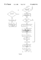
- FIG. 6 a FIG. 6 b Flow-process diagrams of the computer operating system
- the structure of the combined probe is shown by FIG. 2 .
- the image-displaying probe of the B-mode ultrasound scanner ( 1 ) is mounted on the axis of the therapeutic head to guarantee that the focal region (space-point) falls on the B-ultrasonic scanning plane. Because the sound intensity in the focal region produced by the therapeutic head is 100W/cm 2 to 10000W/cm 2 or more, the temperature of the tumor tissue in the focal region is very high (>70 degrees centigrade); the high temperature leads to degeneration and necrosis of the tissues and further increases the high sound impedance there; so strong echo light beam is produced in the B-ultrasonic image. In this way the special location of the therapeutic point can be monitored and the effective degree of the therapy can be reflected objectively by the change of grey scale.
- Piezoelectric ceramics ( 14 ) is mounted at the back end of the therapeutic head; the electric supply driving the piezoelectric ceramic to produce ultrasonic wave is provided by the cable ( 16 ).
- the whole combined probe is mounted within a shielded shell ( 13 );
- the water bag base ( 12 ) and water bag ( 2 ) are mounted at the front end of the shielded shell; on the core of probe ( 15 ) there is an ultrasonic wave lens ( 11 ) and at the center of the probe core is the B-mode ultrasound probe.
- the combined probe mentioned-above is assembled from the lower parts upwards; this assembling method can reduce the movement of body position caused by respiration because therapy is carried out under the body when the patient is in prone position; besides, the skin of human body is in direct contact with the degassed water, so the reflex injury produced when the high intensity ultrasound goes through the skin surface can be reduced.
- this assembling method is a practical design.
- the shape of the focal region ellipsoid-shaped short diameter: 1.1-5 mm long diameter: 3.5-12 mm
- the power source gives out high frequency electric supply, causing the therapeutic head to emit high intensity focused ultrasound and form a high intensity focal region, so that the tumor there is damaged.
- the multi-dimensional scanning system drives the combined probe to carry out scanning movement, causing the focal region to produce scanning locus to damage the tumor.
- the location and shape of the tumor and the change of therapeutic grey scale are all monitored by the B-mode ultrasound probe of the combined detector and observed by means of the screen of the B-mode ultrasound scanner.
- the whole process of the therapy is under to control of the computer program designed by medical workers and monitored by the computer.
- the scanning movement of the combined detector causes water level to change and the therapeutic head needs cooling, so the computer also automatically controls the circulating water apparatus.
- the key to realize the above system lies in the combined probe. Using this probe the damage within tissue can be caused by striking the skin over the points.
- the power generator is the apparatus providing therapeutic ultrasonic high frequency electric supply, the principle of which is shown by FIG. 4 .
- the signal source provides low pressure 0.2-3.5 MHz high frequency sine wave signal, which is then modulated by signal modulator into low frequency carrier signal of 10-1000 Hz or continuous wave; the modulated signal is sent to the magnifier to be duplicated, magnified and sent out. Because there is relatively great difference between the impedance of the therapeutic head and that of the power source, and different probes have different kinds of impedance, so a matching circuit is added to the location between the magnifier and the probe to guarantee the best matching between them.
- the B-mode ultrasound scanner provides image-displaying and monitoring functions of the combined probe.
- the apparatus used is the ready-made apparatus available in the market; Its main functions are the same as those B-mode ultrasound scanner used commonly. Hence, there is no need for further description.
- the ball-screw is driven by the stepping electric motor to move on the ball tract; this mode is highly accurate with little noise, and raster distance-measurement closed-loop control is used, so the influence of step-failing out produced by the stepping electric motor can be reduced.
- the vacuum degassor comprises a vacuum degassing water box, a vacuum pump, a circulating water pump, a regulator of water temperature, and a controlling part, which are shown by FIG. 5 .
- FIG. 6 The following are shown by FIG. 6 :
- the B-mode ultrasound scanner carries out topographic scanning of tissues, then three-dimensional reconstruction processing is carried out through the computer.
- the treatment plan and parameters are chosen by the medical workers according to the shape of the tumor.
- the scanning system is controlled and scanning therapy carried out according to the therapeutic margin determined by the medical workers.
- the intensity of ultrasonic irradiation is adjusted and energy is supplemented according to the change in therapeutic depth.
- HIFU system for scanning and treating tumor:
- the characteristic of this mode the location of the target tissue is found by the motional apparatus, then the therapy is carried out without movement. Within a B-ultrasonic topographic plane, one or more points can be treated separately.
- this mode is mainly used to treat the whole mass of a tumor tissue larger than 1 cm 3 .
- the characteristic of this mode After the location of the target tissue is detected by the motional apparatus, the therapy is carried out through discontinuous movement. A damaged mass is formed by single damaged points; ⁇ fraction (1/10-1/2) ⁇ of two damaged points coincide to guarantee that the tumor tissue treated is fully covered by the therapy; besides, there is proper overlapping on each tomographic scanning plane, which guarantees the whole mass of the tumor can be fully damaged.
- Continuous damaging therapy irradiation lasting for a relatively long time (e.g. 30s) is used only once; during the irradiation process, the coordinate system carries out two or three dimensional movement, causing the focal region to carry out continuous “ablation” within the body. This mode is especially suitable for the treatment of tumor tissue which is long stripe-shaped.
- the characteristic of this mode after the location of the target tissue is detected by the motional apparatus, the therapy is carried out through continuous movement.
- the total irradiation time and speed of movement are fixed, and different points of the tumor tissue all receive the same amount of ultrasonic irradiation.
- B-mode ultrasound scanner as the image-displaying apparatus.
- the technique of B-mode ultrasound scanner is mature, so its probe can be mounted easily on the core of the therapeutic head, and the focal region of the therapeutic head can be easily mounted within the B-ultrasonic sector scanning plane, which are shown by FIG. 2 .
- FIG. 2 Experiment has showed that by using this mode the location of the focal region of the therapeutic head on the sector scanning plane can be easily determined, and the computer can be made to memorize this location, then scanning movement can be carried out.
- tumor tissue is three-dimensional tissue, in order to treat it completely with scanning therapy, it is necessary to use at least three-dimensional coordinate movement.
- To treat some tissues in the human body such as liver tissue only three-dimensional rectangular coordinate scanning is needed; to treat some other tissues of the body such as the mammary tissue, the best method is using two-dimensional rectangular coordinate and one-dimensional rotational coordinate to carry out scanning.
- the mammary tumor develops on the base of the mamma, so the best thing to do is to use a manual rotational coordinate to tilt the combined probe during the treatment, usually the probe is made to include at an angle of 0-60 degrees to the perpendicular direction.
- the whole process of scanning therapy should be controlled by the computer automatically, the reasons for this are: (1) irregular scanning routes must be used for the tumor tissue is irregular; (2) During the treatment, the therapeutic depth changes; in order to guarantee that all the tumor tissues at different distances from the surface receive the same dosage of ultrasonic irradiation, energy should be supplemented automatically according to the change in depth. (3) Because the difference between the grey scale on the ultrasonic screen before and after treatment is great, the grey scale operations before and after treatment will show the objective therapeutic effects. (4) It is also important to carry out self-monitoring of the therapy.
- discontinuous working method is used. Controlled by the computer, the power source, B-mode ultrasound scanner, and the digit-controlled apparatus all work discontinuously.
- the computer After early-stage sampling of the B-ultrasound scanner, the computer memorizes the image and gives instructions to the digit-controlled apparatus to carry out movement; when the next location is reached, the computer memorizes the late-stage sampling image of the B-ultrasonic apparatus and gives instructions to the power source to turn on the machine, initiating its work; then a cycle is completed.
- the computer uses the early-stage sampling image of the second cycle and the late-stage sampling image of the first cycle, the computer performs grey scale calculation; by doing so the therapeutic effects can be judged according to the change in grey scale.
- the sector scanning plane of the B-mode ultrasound probe is used to search for the tumor tissue, and the therapeutic range is determined by the medical worker.
- the location of the focal region of the therapeutic probe is set in a position within the B-ultrasonic sector scanning plane through mounting, and the location has been memorized by the computer.
- the computer gives instructions to digit-controller, driving the therapeutic head to carry out scanning movement, at the same time, it gives instructions to the power source to turn on or off and regulate the degree of power.
- the distance between the location of the focal region and the therapeutic head is a fixed focal distance, so the movement of the therapeutic head will drive the focal region formed by the head to carry out parallel movement; therefore, the scanning movement of the head outside the body guarantees the scanning movement of the focal region within the body.
Abstract
Description
Claims (24)
Applications Claiming Priority (3)
| Application Number | Priority Date | Filing Date | Title |
|---|---|---|---|
| CN98100283A CN1058905C (en) | 1998-01-25 | 1998-01-25 | High-intensity focus supersonic tumor scanning therapy system |
| CN98100283A | 1998-01-25 | ||
| PCT/CN1998/000310 WO1999037364A1 (en) | 1998-01-25 | 1998-12-18 | A high intensity focused ultrasound system for scanning and curing tumor |
Publications (1)
| Publication Number | Publication Date |
|---|---|
| US6685639B1 true US6685639B1 (en) | 2004-02-03 |
Family
ID=5215934
Family Applications (1)
| Application Number | Title | Priority Date | Filing Date |
|---|---|---|---|
| US09/600,854 Expired - Lifetime US6685639B1 (en) | 1998-01-25 | 1998-12-18 | High intensity focused ultrasound system for scanning and curing tumor |
Country Status (9)
| Country | Link |
|---|---|
| US (1) | US6685639B1 (en) |
| EP (1) | EP1050322A4 (en) |
| JP (1) | JP3505512B2 (en) |
| KR (1) | KR100505823B1 (en) |
| CN (1) | CN1058905C (en) |
| AU (1) | AU1659499A (en) |
| CA (1) | CA2326703C (en) |
| RU (1) | RU2210409C2 (en) |
| WO (1) | WO1999037364A1 (en) |
Cited By (142)
| Publication number | Priority date | Publication date | Assignee | Title |
|---|---|---|---|---|
| US20040068186A1 (en) * | 2001-01-22 | 2004-04-08 | Kazunari Ishida | Ultrasonic therapeutic probe and ultrasonic device |
| US20040230258A1 (en) * | 2003-02-19 | 2004-11-18 | Palomar Medical Technologies, Inc. | Method and apparatus for treating pseudofolliculitis barbae |
| US20040267243A1 (en) * | 2003-06-30 | 2004-12-30 | Klotz Conrad Lee | Surgical scalpel and system particularly for use in a transverse carpal ligament surgical procedure |
| US20050038340A1 (en) * | 1998-09-18 | 2005-02-17 | University Of Washington | Use of contrast agents to increase the effectiveness of high intensity focused ultrasound therapy |
| US20050107702A1 (en) * | 2001-11-05 | 2005-05-19 | Shenxu He | External high-power focusing ultrasonic treatment |
| US20050154431A1 (en) * | 2003-12-30 | 2005-07-14 | Liposonix, Inc. | Systems and methods for the destruction of adipose tissue |
| US20050154309A1 (en) * | 2003-12-30 | 2005-07-14 | Liposonix, Inc. | Medical device inline degasser |
| US20050185769A1 (en) * | 2004-02-25 | 2005-08-25 | Pickerd John J. | Calibration method and apparatus |
| US20050203399A1 (en) * | 1999-09-17 | 2005-09-15 | University Of Washington | Image guided high intensity focused ultrasound device for therapy in obstetrics and gynecology |
| US20060052701A1 (en) * | 1998-09-18 | 2006-03-09 | University Of Washington | Treatment of unwanted tissue by the selective destruction of vasculature providing nutrients to the tissue |
| US20060064075A1 (en) * | 2002-11-25 | 2006-03-23 | Jiwei Jiang | Deaeration apparatus for deaerating water used during ultrasonic focusing tumour treatment |
| US7070565B2 (en) * | 2002-05-30 | 2006-07-04 | University Of Washington | Solid hydrogel coupling for ultrasound imaging and therapy |
| US20060189972A1 (en) * | 2005-02-02 | 2006-08-24 | Gynesonics, Inc. | Method and device for uterine fibroid treatment |
| US20060264748A1 (en) * | 2004-09-16 | 2006-11-23 | University Of Washington | Interference-free ultrasound imaging during HIFU therapy, using software tools |
| US20070004984A1 (en) * | 1997-10-31 | 2007-01-04 | University Of Washington | Method and apparatus for preparing organs and tissues for laparoscopic surgery |
| US20070010805A1 (en) * | 2005-07-08 | 2007-01-11 | Fedewa Russell J | Method and apparatus for the treatment of tissue |
| US20070041961A1 (en) * | 2005-08-17 | 2007-02-22 | University Of Washington | Ultrasound target vessel occlusion using microbubbles |
| US20070049910A1 (en) * | 2005-08-08 | 2007-03-01 | Palomar Medical Technologies, Inc. | Eye-safe photocosmetic device |
| US20070055155A1 (en) * | 2005-08-17 | 2007-03-08 | Neil Owen | Method and system to synchronize acoustic therapy with ultrasound imaging |
| US20070055156A1 (en) * | 2003-12-30 | 2007-03-08 | Liposonix, Inc. | Apparatus and methods for the destruction of adipose tissue |
| US20070073308A1 (en) * | 2000-12-28 | 2007-03-29 | Palomar Medical Technologies, Inc. | Method and apparatus for EMR treatment |
| US20070073135A1 (en) * | 2005-09-13 | 2007-03-29 | Warren Lee | Integrated ultrasound imaging and ablation probe |
| US20070092549A1 (en) * | 2003-10-31 | 2007-04-26 | Tuszynski Jack A | Water-soluble compound |
| US20070106157A1 (en) * | 2005-09-30 | 2007-05-10 | University Of Washington | Non-invasive temperature estimation technique for hifu therapy monitoring using backscattered ultrasound |
| US20070129633A1 (en) * | 2005-11-23 | 2007-06-07 | Warren Lee | Ablation array having independently activated ablation elements |
| US20070161905A1 (en) * | 2006-01-12 | 2007-07-12 | Gynesonics, Inc. | Intrauterine ultrasound and method for use |
| US20070179380A1 (en) * | 2006-01-12 | 2007-08-02 | Gynesonics, Inc. | Interventional deployment and imaging system |
| US20070191711A1 (en) * | 2006-02-15 | 2007-08-16 | Misonix, Incorporated | Liquid processing and handling apparatus and associated method for use in medical procedures |
| US20070219448A1 (en) * | 2004-05-06 | 2007-09-20 | Focus Surgery, Inc. | Method and Apparatus for Selective Treatment of Tissue |
| EP1837051A1 (en) * | 2005-01-10 | 2007-09-26 | Chongqing Haifu(Hifu)Technology Co., Ltd | High intensity focused ultrasound therapeutic apparatus and method |
| US20070239077A1 (en) * | 2006-03-09 | 2007-10-11 | Haim Azhari | Method and system for lipolysis and body contouring |
| US20070239000A1 (en) * | 2005-10-20 | 2007-10-11 | Charles Emery | Systems and methods for ultrasound applicator station keeping |
| EP1847294A1 (en) * | 2005-01-31 | 2007-10-24 | Chongqing Haifu(Hifu)Technology Co., Ltd | Focused ultrasound therapy system |
| US20070249939A1 (en) * | 2006-04-20 | 2007-10-25 | Gynesonics, Inc. | Rigid delivery systems having inclined ultrasound and curved needle |
| US20070249936A1 (en) * | 2006-04-20 | 2007-10-25 | Gynesonics, Inc. | Devices and methods for treatment of tissue |
| US20080039724A1 (en) * | 2006-08-10 | 2008-02-14 | Ralf Seip | Ultrasound transducer with improved imaging |
| US20080071165A1 (en) * | 2003-06-30 | 2008-03-20 | Depuy Products, Inc. | Imaging and therapeutic procedure for carpal tunnel syndrome |
| US20080077056A1 (en) * | 2006-09-21 | 2008-03-27 | Shuhei Kagosaki | HIFU probe for treating tissue with in-line degassing of fluid |
| US20080097207A1 (en) * | 2006-09-12 | 2008-04-24 | Siemens Medical Solutions Usa, Inc. | Ultrasound therapy monitoring with diagnostic ultrasound |
| US20080119421A1 (en) * | 2003-10-31 | 2008-05-22 | Jack Tuszynski | Process for treating a biological organism |
| WO2008080229A1 (en) * | 2007-01-05 | 2008-07-10 | Angel Science & Technology (Canada), Inc. | High intensity focused ultrasound treatment head and system |
| US20080177180A1 (en) * | 2004-08-17 | 2008-07-24 | Technion Research & Development | Ultrasonic Image-Guided Tissue-Damaging Procedure |
| US20090036774A1 (en) * | 1999-11-26 | 2009-02-05 | Therus Corporation | Controlled high efficiency lesion formation using high intensity ultrasound |
| US20090069677A1 (en) * | 2007-09-11 | 2009-03-12 | Focus Surgery, Inc. | System and method for tissue change monitoring during hifu treatment |
| US20090099544A1 (en) * | 2007-10-12 | 2009-04-16 | Gynesonics, Inc. | Methods and systems for controlled deployment of needles in tissue |
| US20090112098A1 (en) * | 2005-09-16 | 2009-04-30 | Shahram Vaezy | Thin-profile therapeutic ultrasound applicators |
| US20090216122A1 (en) * | 2008-02-22 | 2009-08-27 | Yousry Faragalla | Method and apparatus to control therapy of moving objects in living body |
| US20090287081A1 (en) * | 2008-04-29 | 2009-11-19 | Gynesonics , Inc | Submucosal fibroid ablation for the treatment of menorrhagia |
| US20100056926A1 (en) * | 2008-08-26 | 2010-03-04 | Gynesonics, Inc. | Ablation device with articulated imaging transducer |
| US20100092424A1 (en) * | 2007-11-21 | 2010-04-15 | Sanghvi Narendra T | Method of diagnosis and treatment of tumors using high intensity focused ultrasound |
| US20100160781A1 (en) * | 2008-12-09 | 2010-06-24 | University Of Washington | Doppler and image guided device for negative feedback phased array hifu treatment of vascularized lesions |
| US20100174188A1 (en) * | 2006-08-24 | 2010-07-08 | Chongqing Ronghai Medical Ultrasound Industry Ltd. | High Intensity Focused Ultrasound Therapeutic System Guided by an Imaging Device |
| US7758621B2 (en) | 1997-05-15 | 2010-07-20 | Palomar Medical Technologies, Inc. | Method and apparatus for therapeutic EMR treatment on the skin |
| US7763016B2 (en) | 1997-05-15 | 2010-07-27 | Palomar Medical Technologies, Inc. | Light energy delivery head |
| US20100191157A1 (en) * | 2009-01-27 | 2010-07-29 | Sanghvi Narendra T | Method for treating skin lesions |
| US20100204686A1 (en) * | 2002-12-20 | 2010-08-12 | Palomar Medical Technologies, Inc. | Light treatments for acne and other disorders of follicles |
| US20100204577A1 (en) * | 2005-07-13 | 2010-08-12 | Acoustx Corporation | Systems and methods for performing acoustic hemostasis of deep bleeding trauma in limbs |
| US20100234728A1 (en) * | 1999-09-17 | 2010-09-16 | University Of Washington | Ultrasound guided high intensity focused ultrasound treatment of nerves |
| US20110009734A1 (en) * | 2003-12-16 | 2011-01-13 | University Of Washington | Image guided high intensity focused ultrasound treatment of nerves |
| US20110021913A1 (en) * | 1999-10-25 | 2011-01-27 | Kona Medical, Inc. | Use of focused ultrasound for vascular sealing |
| US7942915B2 (en) | 2002-05-23 | 2011-05-17 | Palomar Medical Technologies, Inc. | Phototreatment device for use with coolants |
| US20110118598A1 (en) * | 2009-10-12 | 2011-05-19 | Michael Gertner | Targeted Inhibition of Physiologic and Pathologic Processes |
| US20110118600A1 (en) * | 2009-11-16 | 2011-05-19 | Michael Gertner | External Autonomic Modulation |
| US20110137149A1 (en) * | 2009-10-12 | 2011-06-09 | Michael Gertner | Nerve treatment system |
| US20110172527A1 (en) * | 2009-10-12 | 2011-07-14 | Michael Gertner | Systems for externally delivered energy to modulate neural structures |
| US20110178541A1 (en) * | 2008-09-12 | 2011-07-21 | Slender Medical, Ltd. | Virtual ultrasonic scissors |
| US20110184322A1 (en) * | 2010-01-22 | 2011-07-28 | Slender Medical Ltd. | Method and device for treatment of keloids and hypertrophic scars using focused ultrasound |
| US8038631B1 (en) | 2005-06-01 | 2011-10-18 | Sanghvi Narendra T | Laparoscopic HIFU probe |
| US20120029393A1 (en) * | 2010-07-30 | 2012-02-02 | General Electric Company | Compact ultrasound transducer assembly and methods of making and using the same |
| CN102397647A (en) * | 2011-11-24 | 2012-04-04 | 何伟宗 | Method and device for treating proctoptosis with ultrasonic wave and proctoptosis drug |
| US20120083686A1 (en) * | 2009-06-12 | 2012-04-05 | Koninklijke Philips Electronics N.V. | Mr imaging guided ultrasound therapy |
| US8182473B2 (en) | 1999-01-08 | 2012-05-22 | Palomar Medical Technologies | Cooling system for a photocosmetic device |
| US8206300B2 (en) | 2008-08-26 | 2012-06-26 | Gynesonics, Inc. | Ablation device with articulated imaging transducer |
| US20120207366A1 (en) * | 2009-10-13 | 2012-08-16 | Agency For Science, Technology And Research | Method and system for segmenting a liver object in an image |
| US8262574B2 (en) | 2009-02-27 | 2012-09-11 | Gynesonics, Inc. | Needle and tine deployment mechanism |
| US8328794B2 (en) | 1996-12-02 | 2012-12-11 | Palomar Medical Technologies, Inc. | System for electromagnetic radiation dermatology and head for use therewith |
| US8346347B2 (en) | 2005-09-15 | 2013-01-01 | Palomar Medical Technologies, Inc. | Skin optical characterization device |
| WO2013035035A1 (en) * | 2011-09-06 | 2013-03-14 | Koninklijke Philips Electronics N.V. | Heat exchange in fluid degassing |
| US8469904B2 (en) | 2009-10-12 | 2013-06-25 | Kona Medical, Inc. | Energetic modulation of nerves |
| US8517962B2 (en) | 2009-10-12 | 2013-08-27 | Kona Medical, Inc. | Energetic modulation of nerves |
| US8611189B2 (en) | 2004-09-16 | 2013-12-17 | University of Washington Center for Commercialization | Acoustic coupler using an independent water pillow with circulation for cooling a transducer |
| US20130345558A1 (en) * | 2011-03-02 | 2013-12-26 | Diagnostic Photonics, Inc. | Hand-Held Fixed-Focus Optical Probe |
| US8852103B2 (en) | 2011-10-17 | 2014-10-07 | Butterfly Network, Inc. | Transmissive imaging and related apparatus and methods |
| US8915948B2 (en) | 2002-06-19 | 2014-12-23 | Palomar Medical Technologies, Llc | Method and apparatus for photothermal treatment of tissue at depth |
| US8956346B2 (en) | 2010-05-14 | 2015-02-17 | Rainbow Medical, Ltd. | Reflectance-facilitated ultrasound treatment and monitoring |
| US8986231B2 (en) | 2009-10-12 | 2015-03-24 | Kona Medical, Inc. | Energetic modulation of nerves |
| US8986211B2 (en) | 2009-10-12 | 2015-03-24 | Kona Medical, Inc. | Energetic modulation of nerves |
| US8992447B2 (en) | 2009-10-12 | 2015-03-31 | Kona Medical, Inc. | Energetic modulation of nerves |
| US9028536B2 (en) | 2006-08-02 | 2015-05-12 | Cynosure, Inc. | Picosecond laser apparatus and methods for its operation and use |
| US9066679B2 (en) | 2004-08-31 | 2015-06-30 | University Of Washington | Ultrasonic technique for assessing wall vibrations in stenosed blood vessels |
| US20150374333A1 (en) * | 2004-10-06 | 2015-12-31 | Guided Therapy Systems, Llc | Systems for cosmetic treatment |
| US9242122B2 (en) | 2010-05-14 | 2016-01-26 | Liat Tsoref | Reflectance-facilitated ultrasound treatment and monitoring |
| US9510802B2 (en) | 2012-09-21 | 2016-12-06 | Guided Therapy Systems, Llc | Reflective ultrasound technology for dermatological treatments |
| US9522290B2 (en) | 2004-10-06 | 2016-12-20 | Guided Therapy Systems, Llc | System and method for fat and cellulite reduction |
| US9533175B2 (en) | 2004-10-06 | 2017-01-03 | Guided Therapy Systems, Llc | Energy based fat reduction |
| US20170014650A1 (en) * | 2014-04-17 | 2017-01-19 | Olympus Corporation | Ultrasonic treatment apparatus |
| US9667889B2 (en) | 2013-04-03 | 2017-05-30 | Butterfly Network, Inc. | Portable electronic devices with integrated imaging capabilities |
| US9694211B2 (en) | 2004-10-06 | 2017-07-04 | Guided Therapy Systems, L.L.C. | Systems for treating skin laxity |
| US9694212B2 (en) | 2004-10-06 | 2017-07-04 | Guided Therapy Systems, Llc | Method and system for ultrasound treatment of skin |
| US9707414B2 (en) | 2012-02-14 | 2017-07-18 | Rainbow Medical Ltd. | Reflectance-facilitated ultrasound treatment and monitoring |
| US9770593B2 (en) | 2012-11-05 | 2017-09-26 | Pythagoras Medical Ltd. | Patient selection using a transluminally-applied electric current |
| US9780518B2 (en) | 2012-04-18 | 2017-10-03 | Cynosure, Inc. | Picosecond laser apparatus and methods for treating target tissues with same |
| US9820798B2 (en) | 2011-09-23 | 2017-11-21 | Alan N. Schwartz | System and method for providing targeted ablation of parathyroidal tissue |
| US9827449B2 (en) | 2004-10-06 | 2017-11-28 | Guided Therapy Systems, L.L.C. | Systems for treating skin laxity |
| US9895560B2 (en) | 2004-09-24 | 2018-02-20 | Guided Therapy Systems, Llc | Methods for rejuvenating skin by heating tissue for cosmetic treatment of the face and body |
| US9919168B2 (en) | 2009-07-23 | 2018-03-20 | Palomar Medical Technologies, Inc. | Method for improvement of cellulite appearance |
| US9974982B2 (en) | 2004-10-06 | 2018-05-22 | Guided Therapy Systems, Llc | System and method for noninvasive skin tightening |
| US10004557B2 (en) | 2012-11-05 | 2018-06-26 | Pythagoras Medical Ltd. | Controlled tissue ablation |
| US10046181B2 (en) | 2004-10-06 | 2018-08-14 | Guided Therapy Systems, Llc | Energy based hyperhidrosis treatment |
| US10058342B2 (en) | 2006-01-12 | 2018-08-28 | Gynesonics, Inc. | Devices and methods for treatment of tissue |
| WO2019050533A1 (en) * | 2017-09-08 | 2019-03-14 | National Health Research Institutes | Apparatus of combining imaging ultrasonic probe and focused ultrasonic probe |
| US10245107B2 (en) | 2013-03-15 | 2019-04-02 | Cynosure, Inc. | Picosecond optical radiation systems and methods of use |
| US10342476B2 (en) | 2012-05-17 | 2019-07-09 | Alan N. Schwartz | Localization of the parathyroid |
| US10383685B2 (en) | 2015-05-07 | 2019-08-20 | Pythagoras Medical Ltd. | Techniques for use with nerve tissue |
| US10420960B2 (en) | 2013-03-08 | 2019-09-24 | Ulthera, Inc. | Devices and methods for multi-focus ultrasound therapy |
| US10434324B2 (en) | 2005-04-22 | 2019-10-08 | Cynosure, Llc | Methods and systems for laser treatment using non-uniform output beam |
| US10478249B2 (en) | 2014-05-07 | 2019-11-19 | Pythagoras Medical Ltd. | Controlled tissue ablation techniques |
| US10537304B2 (en) | 2008-06-06 | 2020-01-21 | Ulthera, Inc. | Hand wand for ultrasonic cosmetic treatment and imaging |
| US10595819B2 (en) | 2006-04-20 | 2020-03-24 | Gynesonics, Inc. | Ablation device with articulated imaging transducer |
| US10603521B2 (en) | 2014-04-18 | 2020-03-31 | Ulthera, Inc. | Band transducer ultrasound therapy |
| CN111494817A (en) * | 2020-02-26 | 2020-08-07 | 南北兄弟药业投资有限公司 | Large focal region forming system of HIFU device and focal region forming method thereof |
| US10772681B2 (en) | 2009-10-12 | 2020-09-15 | Utsuka Medical Devices Co., Ltd. | Energy delivery to intraparenchymal regions of the kidney |
| US10864385B2 (en) | 2004-09-24 | 2020-12-15 | Guided Therapy Systems, Llc | Rejuvenating skin by heating tissue for cosmetic treatment of the face and body |
| US10925579B2 (en) | 2014-11-05 | 2021-02-23 | Otsuka Medical Devices Co., Ltd. | Systems and methods for real-time tracking of a target tissue using imaging before and during therapy delivery |
| US10993770B2 (en) | 2016-11-11 | 2021-05-04 | Gynesonics, Inc. | Controlled treatment of tissue and dynamic interaction with, and comparison of, tissue and/or treatment data |
| US11045246B1 (en) | 2011-01-04 | 2021-06-29 | Alan N. Schwartz | Apparatus for effecting feedback of vaginal cavity physiology |
| WO2021150837A1 (en) | 2020-01-22 | 2021-07-29 | Massachusetts Institute Of Technology | Inducible tissue constructs and uses thereof |
| WO2021151088A1 (en) * | 2020-01-23 | 2021-07-29 | Acoustic Medsystems, Inc. | Image-guided pulsed volume focused ultrasound |
| US11207548B2 (en) | 2004-10-07 | 2021-12-28 | Guided Therapy Systems, L.L.C. | Ultrasound probe for treating skin laxity |
| US11224895B2 (en) | 2016-01-18 | 2022-01-18 | Ulthera, Inc. | Compact ultrasound device having annular ultrasound array peripherally electrically connected to flexible printed circuit board and method of assembly thereof |
| US11235179B2 (en) | 2004-10-06 | 2022-02-01 | Guided Therapy Systems, Llc | Energy based skin gland treatment |
| US11241218B2 (en) | 2016-08-16 | 2022-02-08 | Ulthera, Inc. | Systems and methods for cosmetic ultrasound treatment of skin |
| US11259825B2 (en) | 2006-01-12 | 2022-03-01 | Gynesonics, Inc. | Devices and methods for treatment of tissue |
| US11337858B2 (en) | 2011-11-21 | 2022-05-24 | Alan N. Schwartz | Ostomy pouching system |
| US11338156B2 (en) | 2004-10-06 | 2022-05-24 | Guided Therapy Systems, Llc | Noninvasive tissue tightening system |
| US11418000B2 (en) | 2018-02-26 | 2022-08-16 | Cynosure, Llc | Q-switched cavity dumped sub-nanosecond laser |
| US11678932B2 (en) | 2016-05-18 | 2023-06-20 | Symap Medical (Suzhou) Limited | Electrode catheter with incremental advancement |
| US11724133B2 (en) | 2004-10-07 | 2023-08-15 | Guided Therapy Systems, Llc | Ultrasound probe for treatment of skin |
| US11806275B2 (en) | 2011-01-04 | 2023-11-07 | Alan N. Schwartz | Penile condom catheter for facilitating urine collection and egress of urinary fluids away from the body torso |
| US11883688B2 (en) | 2004-10-06 | 2024-01-30 | Guided Therapy Systems, Llc | Energy based fat reduction |
| US11944849B2 (en) | 2018-02-20 | 2024-04-02 | Ulthera, Inc. | Systems and methods for combined cosmetic treatment of cellulite with ultrasound |
| US11969609B2 (en) | 2022-12-05 | 2024-04-30 | Ulthera, Inc. | Devices and methods for multi-focus ultrasound therapy |
Families Citing this family (70)
| Publication number | Priority date | Publication date | Assignee | Title |
|---|---|---|---|---|
| US6050943A (en) | 1997-10-14 | 2000-04-18 | Guided Therapy Systems, Inc. | Imaging, therapy, and temperature monitoring ultrasonic system |
| US7914453B2 (en) | 2000-12-28 | 2011-03-29 | Ardent Sound, Inc. | Visual imaging system for ultrasonic probe |
| CN100438941C (en) * | 2004-06-17 | 2008-12-03 | 上海交通大学 | Ultrasonic heat treatment system for mouth cavity and maxillofacial malignant tumor |
| US9011336B2 (en) | 2004-09-16 | 2015-04-21 | Guided Therapy Systems, Llc | Method and system for combined energy therapy profile |
| US7824348B2 (en) | 2004-09-16 | 2010-11-02 | Guided Therapy Systems, L.L.C. | System and method for variable depth ultrasound treatment |
| US7393325B2 (en) | 2004-09-16 | 2008-07-01 | Guided Therapy Systems, L.L.C. | Method and system for ultrasound treatment with a multi-directional transducer |
| CN100409813C (en) * | 2004-09-30 | 2008-08-13 | 重庆海扶(Hifu)技术有限公司 | Combined device for ultrasonic diagnosis and treatment |
| US7758524B2 (en) | 2004-10-06 | 2010-07-20 | Guided Therapy Systems, L.L.C. | Method and system for ultra-high frequency ultrasound treatment |
| JP5932195B2 (en) * | 2004-10-06 | 2016-06-08 | ガイデッド セラピー システムズ, エル.エル.シー. | System for controlled heat treatment of human surface tissue |
| EP2279698A3 (en) | 2004-10-06 | 2014-02-19 | Guided Therapy Systems, L.L.C. | Method and system for non-invasive cosmetic enhancement of stretch marks |
| CN100563752C (en) | 2005-01-31 | 2009-12-02 | 重庆融海超声医学工程研究中心有限公司 | The ultrasonic treatment unit of MRI guiding |
| US7571336B2 (en) | 2005-04-25 | 2009-08-04 | Guided Therapy Systems, L.L.C. | Method and system for enhancing safety with medical peripheral device by monitoring if host computer is AC powered |
| EP2007305B1 (en) * | 2006-04-11 | 2012-05-16 | Koninklijke Philips Electronics N.V. | A device for positioning an ultrasound transducer inside a mr scanner |
| US20080015436A1 (en) * | 2006-07-13 | 2008-01-17 | Misonix, Incorporated | High intensity focused ultrasound method and associated apparatus |
| JP2008022956A (en) * | 2006-07-19 | 2008-02-07 | Medical Support Co Ltd | Ultrasonic tumor therapy apparatus |
| KR100811663B1 (en) * | 2006-07-24 | 2008-03-11 | 재단법인서울대학교산학협력재단 | A Planar Applicator radiator using resonance |
| EP2059303A1 (en) * | 2006-08-30 | 2009-05-20 | Koninklijke Philips Electronics N.V. | Apparatus for thermal treatment of tissue |
| US9566454B2 (en) | 2006-09-18 | 2017-02-14 | Guided Therapy Systems, Llc | Method and sysem for non-ablative acne treatment and prevention |
| JP5661283B2 (en) * | 2006-11-20 | 2015-01-28 | コーニンクレッカ フィリップス エヌ ヴェ | System, operating method and computer-readable storage medium for displaying anatomical tree structure |
| US9216276B2 (en) | 2007-05-07 | 2015-12-22 | Guided Therapy Systems, Llc | Methods and systems for modulating medicants using acoustic energy |
| US20150174388A1 (en) | 2007-05-07 | 2015-06-25 | Guided Therapy Systems, Llc | Methods and Systems for Ultrasound Assisted Delivery of a Medicant to Tissue |
| PT2152167T (en) | 2007-05-07 | 2018-12-10 | Guided Therapy Systems Llc | Methods and systems for coupling and focusing acoustic energy using a coupler member |
| WO2009002492A1 (en) | 2007-06-25 | 2008-12-31 | Dutch Cardio Llc | Image guided plaque ablation |
| CN102164637B (en) | 2008-04-09 | 2015-08-19 | 朱利安·伊特兹科维特兹 | Comprise the medical system of percutaneous electrode |
| WO2010008834A2 (en) * | 2008-06-23 | 2010-01-21 | Angiodynamics, Inc. | Treatment devices and methods |
| CN102170938B (en) * | 2008-09-30 | 2015-01-14 | 皇家飞利浦电子股份有限公司 | System and method for ultrasound therapy treatment |
| KR101041256B1 (en) * | 2008-11-07 | 2011-06-14 | 이혜경 | Small duck smoking apparatus |
| KR101032393B1 (en) * | 2008-11-07 | 2011-05-03 | (주)킴스팜 | Small duck smoking apparatus using ceramics |
| KR101093734B1 (en) | 2009-05-08 | 2011-12-19 | 알피니언메디칼시스템 주식회사 | Head for ultrasonic treatment apparatus, ultrasonic treatment apparatus and operation method of ultrasonic treatment apparatus |
| JP6270315B2 (en) * | 2009-09-17 | 2018-01-31 | コーニンクレッカ フィリップス エヌ ヴェKoninklijke Philips N.V. | Medical ultrasonic device for temperature detection at the distal end |
| US8715186B2 (en) | 2009-11-24 | 2014-05-06 | Guided Therapy Systems, Llc | Methods and systems for generating thermal bubbles for improved ultrasound imaging and therapy |
| CN101791452B (en) * | 2010-02-26 | 2012-05-09 | 南京海克医疗设备有限公司 | Focusing ultrasonic therapeutic head with wide focal distance and high strength |
| EP2600937B8 (en) | 2010-08-02 | 2024-03-06 | Guided Therapy Systems, L.L.C. | Systems for treating acute and/or chronic injuries in soft tissue |
| US9504446B2 (en) | 2010-08-02 | 2016-11-29 | Guided Therapy Systems, Llc | Systems and methods for coupling an ultrasound source to tissue |
| US8857438B2 (en) | 2010-11-08 | 2014-10-14 | Ulthera, Inc. | Devices and methods for acoustic shielding |
| CN102728007B (en) * | 2011-03-29 | 2015-07-08 | 重庆微海软件开发有限公司 | Control system of ultrasonic treatment system |
| WO2012156863A2 (en) * | 2011-05-18 | 2012-11-22 | Koninklijke Philips Electronics N.V. | Spherical ultrasonic hifu transducer with modular cavitation sense element |
| CN102836505A (en) * | 2011-06-15 | 2012-12-26 | 黄品同 | Focusing ultrasonic cavitation treatment instrument with ultrasonic focusing positioning function |
| EP2729215A4 (en) | 2011-07-10 | 2015-04-15 | Guided Therapy Systems Llc | Methods and systems for ultrasound treatment |
| EP2731675B1 (en) | 2011-07-11 | 2023-05-03 | Guided Therapy Systems, L.L.C. | Systems and methods for coupling an ultrasound source to tissue |
| KR20130020054A (en) * | 2011-08-18 | 2013-02-27 | 삼성전자주식회사 | Method for generating ultrasound image and ultrasound system |
| CN102525596B (en) * | 2011-12-31 | 2014-09-17 | 重庆海扶医疗科技股份有限公司 | Ultrasound collection and treatment control system and image acquisition method thereof |
| US9263663B2 (en) | 2012-04-13 | 2016-02-16 | Ardent Sound, Inc. | Method of making thick film transducer arrays |
| JP6496662B2 (en) * | 2012-10-12 | 2019-04-03 | プロファウンド メディカル インク | Multi-focus ultrasound irradiation for hyperthermia treatment using magnetic resonance guided focused ultrasound |
| CN102921116B (en) * | 2012-11-12 | 2016-04-06 | 上海交通大学 | A kind of superficial tumor ultrasonic thermotherapy system of guided by magnetic resonance |
| US10561862B2 (en) | 2013-03-15 | 2020-02-18 | Guided Therapy Systems, Llc | Ultrasound treatment device and methods of use |
| CN103143125B (en) * | 2013-03-25 | 2015-12-23 | 广州多浦乐电子科技有限公司 | A kind of high-strength focusing ultrasonic therapy instrument |
| CN103230648B (en) * | 2013-04-24 | 2016-09-14 | 重庆医科大学 | Low-strength focusing ultrasonic molecular imaging and treatment system |
| KR101467511B1 (en) * | 2013-07-24 | 2014-12-02 | 알피니언메디칼시스템 주식회사 | Assistant device for high intensity focused ultrasound therapy |
| CN104107510B (en) * | 2014-07-31 | 2017-07-14 | 重庆海扶医疗科技股份有限公司 | The moving sweep device of high-strength focus supersonic therapeutic system |
| CN105879211A (en) * | 2015-01-26 | 2016-08-24 | 李明德 | Remote continuous therapeutic ultrasonic probe |
| WO2016143921A1 (en) * | 2015-03-11 | 2016-09-15 | 알피니언메디칼시스템 주식회사 | High-intensity focused ultrasound treatment head |
| RU2594806C1 (en) * | 2015-04-07 | 2016-08-20 | Валентина Николаевна Федорова | Sensor for acoustic microscanning of soft biological tissues |
| CN105251142B (en) * | 2015-11-13 | 2019-01-29 | 浙江大学 | The preventive maintenance method of high-intensity focus supersonic tumor therapeutic equipment |
| CN105435380B (en) * | 2015-11-13 | 2018-05-01 | 浙江大学 | The methods of risk assessment of HIFU Treatment equipment based on reverse heat transfer |
| US20170157366A1 (en) * | 2015-12-03 | 2017-06-08 | Benny Assif | Urinary catheters, systems and methods for use during treatment of the prostate |
| CN107260217B (en) * | 2017-07-17 | 2018-07-17 | 西安交通大学 | The passive imaging method of three-dimensional and system for brain focused ultrasonic cavitation real time monitoring |
| PL233294B1 (en) * | 2017-09-08 | 2019-09-30 | Inst Podstawowych Problemow Techniki Polskiej Akademii Nauk | Bimodal ultrasonic instrument for non-invasive destruction of solid neoplastic tumors in small animals |
| RU2664597C1 (en) * | 2017-12-12 | 2018-08-21 | Федеральное государственное бюджетное учреждение "Национальный медицинский исследовательский центр радиологии" Министерства здравоохранения Российской Федерации (ФГБУ "НМИЦ радиологии" Минздрава России) | Method of combined treatment of patients with primarily operable and locally advanced inoperable breast cancer |
| CN108939327A (en) * | 2018-08-20 | 2018-12-07 | 安徽声达愈医疗器械有限公司 | A kind of compound bifocus focused transducer and application method |
| CN109893785A (en) * | 2019-03-19 | 2019-06-18 | 深圳市声科生物医学研究院 | A kind of ultrasonic Scientific Research Platform of focusing |
| CN110664433A (en) * | 2019-11-13 | 2020-01-10 | 上海爱申科技发展股份有限公司 | B-ultrasonic monitoring movement mechanism for HIFU treatment |
| CN111408075B (en) * | 2020-03-23 | 2022-08-30 | 南京广慈医疗科技有限公司 | High-intensity focused ultrasound diagnosis and treatment system with temperature measurement function and control method thereof |
| WO2021047242A1 (en) * | 2020-06-28 | 2021-03-18 | 南通大学 | Corrector for deviation-free stereotactic device |
| CN111773567A (en) * | 2020-08-17 | 2020-10-16 | 杭州福嵩科技有限责任公司 | High-intensity focused ultrasound-puncture ablation fusion treatment equipment |
| WO2022120807A1 (en) * | 2020-12-11 | 2022-06-16 | 深圳先进技术研究院 | Immune system regulation method and ultrasonic immunotherapy device |
| CN112473026A (en) * | 2020-12-11 | 2021-03-12 | 深圳先进技术研究院 | Immune system regulation and control method and ultrasonic immunotherapy device |
| CN115591137A (en) * | 2021-07-09 | 2023-01-13 | 四川大学华西医院(Cn) | Pulse focusing ultrasonic liver regeneration treatment device |
| CN113332620B (en) * | 2021-07-12 | 2023-03-14 | 重庆融海超声医学工程研究中心有限公司 | Ultrasonic medical equipment |
| WO2023134395A1 (en) * | 2022-01-14 | 2023-07-20 | 杭州福嵩科技有限责任公司 | Sealing assembly, focused ultrasound therapy system and operation method, and semi-dry ultrasound water bag assembly |
Citations (17)
| Publication number | Priority date | Publication date | Assignee | Title |
|---|---|---|---|---|
| US3765403A (en) * | 1968-05-20 | 1973-10-16 | Holotron Corp | Ultrasonic imaging techniques and mammograph equipment |
| US4094306A (en) * | 1975-05-01 | 1978-06-13 | The Commonwealth Of Australia, C/O The Department Of Health | Apparatus for ultrasonic examination |
| US4485819A (en) * | 1980-01-21 | 1984-12-04 | Wolfgang Igl | Mechanical accessory for commercially available compound apparatuses for echo mammography |
| US4646756A (en) * | 1982-10-26 | 1987-03-03 | The University Of Aberdeen | Ultra sound hyperthermia device |
| US4893624A (en) * | 1988-06-21 | 1990-01-16 | Massachusetts Institute Of Technology | Diffuse focus ultrasound hyperthermia system |
| US4936303A (en) * | 1987-11-20 | 1990-06-26 | Ultrathermics | Ultrasonic heating apparatus and method |
| US4938217A (en) * | 1988-06-21 | 1990-07-03 | Massachusetts Institute Of Technology | Electronically-controlled variable focus ultrasound hyperthermia system |
| US5054470A (en) * | 1988-03-02 | 1991-10-08 | Laboratory Equipment, Corp. | Ultrasonic treatment transducer with pressurized acoustic coupling |
| US5247924A (en) * | 1990-05-30 | 1993-09-28 | Kabushiki Kaisha Toshiba | Shockwave generator using a piezoelectric element |
| US5409002A (en) * | 1989-07-12 | 1995-04-25 | Focus Surgery Incorporated | Treatment system with localization |
| US5501655A (en) * | 1992-03-31 | 1996-03-26 | Massachusetts Institute Of Technology | Apparatus and method for acoustic heat generation and hyperthermia |
| US5522869A (en) * | 1994-05-17 | 1996-06-04 | Burdette; Everette C. | Ultrasound device for use in a thermotherapy apparatus |
| US5590653A (en) * | 1993-03-10 | 1997-01-07 | Kabushiki Kaisha Toshiba | Ultrasonic wave medical treatment apparatus suitable for use under guidance of magnetic resonance imaging |
| US5769790A (en) * | 1996-10-25 | 1998-06-23 | General Electric Company | Focused ultrasound surgery system guided by ultrasound imaging |
| US6042556A (en) * | 1998-09-04 | 2000-03-28 | University Of Washington | Method for determining phase advancement of transducer elements in high intensity focused ultrasound |
| US6280402B1 (en) * | 1995-03-31 | 2001-08-28 | Kabushiki Kaisha Toshiba | Ultrasound therapeutic apparatus |
| US6425867B1 (en) * | 1998-09-18 | 2002-07-30 | University Of Washington | Noise-free real time ultrasonic imaging of a treatment site undergoing high intensity focused ultrasound therapy |
Family Cites Families (9)
| Publication number | Priority date | Publication date | Assignee | Title |
|---|---|---|---|---|
| FR2563725B1 (en) * | 1984-05-03 | 1988-07-15 | Dory Jacques | APPARATUS FOR EXAMINING AND LOCATING ULTRASONIC TUMORS WITH A LOCALIZED HYPERTHERMAL TREATMENT DEVICE |
| US5150712A (en) * | 1983-12-14 | 1992-09-29 | Edap International, S.A. | Apparatus for examining and localizing tumors using ultra sounds, comprising a device for localized hyperthermia treatment |
| FR2664819B1 (en) * | 1990-07-23 | 1994-04-29 | Edap Int | ULTRA-FAST EXTRACORPOREAL ULTRASONIC HYPERTHERMAL APPARATUS. |
| FR2619003B1 (en) * | 1987-08-05 | 1997-06-27 | Toshiba Kk | ULTRASONIC THERAPEUTIC TREATMENT APPARATUS |
| DE4005228A1 (en) * | 1990-02-20 | 1991-08-22 | Wolf Gmbh Richard | LITHOTRIPSY DEVICE WITH A PLANT FOR TREATING THE ACOUSTIC COUPLING MEDIUM |
| FR2686258B1 (en) * | 1992-01-21 | 1994-09-16 | Edap Int | METHOD FOR VIEWING AN ANATOMICAL TARGET FOR ITS TREATMENT WITH FOCUSED ELASTIC WAVES AND APPARATUS APPLYING THIS PROCESS TO THE TREATMENT OF PROSTATE BY HYPERTHERMIA. |
| US5520188A (en) * | 1994-11-02 | 1996-05-28 | Focus Surgery Inc. | Annular array transducer |
| DE19507478C1 (en) * | 1995-03-03 | 1996-05-15 | Siemens Ag | Therapy device for treatment with focused ultrasound |
| CN1073453C (en) * | 1996-12-27 | 2001-10-24 | 中国科学院声学研究所 | In vitro supersonic therapeutical instrument for medical purpose |
-
1998
- 1998-01-25 CN CN98100283A patent/CN1058905C/en not_active Expired - Lifetime
- 1998-12-18 CA CA002326703A patent/CA2326703C/en not_active Expired - Lifetime
- 1998-12-18 AU AU16594/99A patent/AU1659499A/en not_active Abandoned
- 1998-12-18 RU RU2000119766/14A patent/RU2210409C2/en active
- 1998-12-18 WO PCT/CN1998/000310 patent/WO1999037364A1/en not_active Application Discontinuation
- 1998-12-18 KR KR10-2000-7008118A patent/KR100505823B1/en not_active IP Right Cessation
- 1998-12-18 JP JP2000528339A patent/JP3505512B2/en not_active Expired - Lifetime
- 1998-12-18 US US09/600,854 patent/US6685639B1/en not_active Expired - Lifetime
- 1998-12-18 EP EP98960989A patent/EP1050322A4/en not_active Ceased
Patent Citations (17)
| Publication number | Priority date | Publication date | Assignee | Title |
|---|---|---|---|---|
| US3765403A (en) * | 1968-05-20 | 1973-10-16 | Holotron Corp | Ultrasonic imaging techniques and mammograph equipment |
| US4094306A (en) * | 1975-05-01 | 1978-06-13 | The Commonwealth Of Australia, C/O The Department Of Health | Apparatus for ultrasonic examination |
| US4485819A (en) * | 1980-01-21 | 1984-12-04 | Wolfgang Igl | Mechanical accessory for commercially available compound apparatuses for echo mammography |
| US4646756A (en) * | 1982-10-26 | 1987-03-03 | The University Of Aberdeen | Ultra sound hyperthermia device |
| US4936303A (en) * | 1987-11-20 | 1990-06-26 | Ultrathermics | Ultrasonic heating apparatus and method |
| US5054470A (en) * | 1988-03-02 | 1991-10-08 | Laboratory Equipment, Corp. | Ultrasonic treatment transducer with pressurized acoustic coupling |
| US4893624A (en) * | 1988-06-21 | 1990-01-16 | Massachusetts Institute Of Technology | Diffuse focus ultrasound hyperthermia system |
| US4938217A (en) * | 1988-06-21 | 1990-07-03 | Massachusetts Institute Of Technology | Electronically-controlled variable focus ultrasound hyperthermia system |
| US5409002A (en) * | 1989-07-12 | 1995-04-25 | Focus Surgery Incorporated | Treatment system with localization |
| US5247924A (en) * | 1990-05-30 | 1993-09-28 | Kabushiki Kaisha Toshiba | Shockwave generator using a piezoelectric element |
| US5501655A (en) * | 1992-03-31 | 1996-03-26 | Massachusetts Institute Of Technology | Apparatus and method for acoustic heat generation and hyperthermia |
| US5590653A (en) * | 1993-03-10 | 1997-01-07 | Kabushiki Kaisha Toshiba | Ultrasonic wave medical treatment apparatus suitable for use under guidance of magnetic resonance imaging |
| US5522869A (en) * | 1994-05-17 | 1996-06-04 | Burdette; Everette C. | Ultrasound device for use in a thermotherapy apparatus |
| US6280402B1 (en) * | 1995-03-31 | 2001-08-28 | Kabushiki Kaisha Toshiba | Ultrasound therapeutic apparatus |
| US5769790A (en) * | 1996-10-25 | 1998-06-23 | General Electric Company | Focused ultrasound surgery system guided by ultrasound imaging |
| US6042556A (en) * | 1998-09-04 | 2000-03-28 | University Of Washington | Method for determining phase advancement of transducer elements in high intensity focused ultrasound |
| US6425867B1 (en) * | 1998-09-18 | 2002-07-30 | University Of Washington | Noise-free real time ultrasonic imaging of a treatment site undergoing high intensity focused ultrasound therapy |
Cited By (314)
| Publication number | Priority date | Publication date | Assignee | Title |
|---|---|---|---|---|
| US8328794B2 (en) | 1996-12-02 | 2012-12-11 | Palomar Medical Technologies, Inc. | System for electromagnetic radiation dermatology and head for use therewith |
| US7758621B2 (en) | 1997-05-15 | 2010-07-20 | Palomar Medical Technologies, Inc. | Method and apparatus for therapeutic EMR treatment on the skin |
| US8328796B2 (en) | 1997-05-15 | 2012-12-11 | Palomar Medical Technologies, Inc. | Light energy delivery head |
| US7763016B2 (en) | 1997-05-15 | 2010-07-27 | Palomar Medical Technologies, Inc. | Light energy delivery head |
| US8109924B2 (en) | 1997-05-15 | 2012-02-07 | Palomar Medical Technologies, Inc. | Heads for dermatology treatment |
| US8002768B1 (en) | 1997-05-15 | 2011-08-23 | Palomar Medical Technologies, Inc. | Light energy delivery head |
| US7935107B2 (en) | 1997-05-15 | 2011-05-03 | Palomar Medical Technologies, Inc. | Heads for dermatology treatment |
| US20070004984A1 (en) * | 1997-10-31 | 2007-01-04 | University Of Washington | Method and apparatus for preparing organs and tissues for laparoscopic surgery |
| US9198635B2 (en) | 1997-10-31 | 2015-12-01 | University Of Washington | Method and apparatus for preparing organs and tissues for laparoscopic surgery |
| US20050038340A1 (en) * | 1998-09-18 | 2005-02-17 | University Of Washington | Use of contrast agents to increase the effectiveness of high intensity focused ultrasound therapy |
| US20060052701A1 (en) * | 1998-09-18 | 2006-03-09 | University Of Washington | Treatment of unwanted tissue by the selective destruction of vasculature providing nutrients to the tissue |
| US7722539B2 (en) | 1998-09-18 | 2010-05-25 | University Of Washington | Treatment of unwanted tissue by the selective destruction of vasculature providing nutrients to the tissue |
| US7686763B2 (en) | 1998-09-18 | 2010-03-30 | University Of Washington | Use of contrast agents to increase the effectiveness of high intensity focused ultrasound therapy |
| US8182473B2 (en) | 1999-01-08 | 2012-05-22 | Palomar Medical Technologies | Cooling system for a photocosmetic device |
| US20100234728A1 (en) * | 1999-09-17 | 2010-09-16 | University Of Washington | Ultrasound guided high intensity focused ultrasound treatment of nerves |
| US7520856B2 (en) | 1999-09-17 | 2009-04-21 | University Of Washington | Image guided high intensity focused ultrasound device for therapy in obstetrics and gynecology |
| US20050203399A1 (en) * | 1999-09-17 | 2005-09-15 | University Of Washington | Image guided high intensity focused ultrasound device for therapy in obstetrics and gynecology |
| US20080051656A1 (en) * | 1999-09-17 | 2008-02-28 | University Of Washington | Method for using high intensity focused ultrasound |
| US8197409B2 (en) | 1999-09-17 | 2012-06-12 | University Of Washington | Ultrasound guided high intensity focused ultrasound treatment of nerves |
| US20110201929A1 (en) * | 1999-09-17 | 2011-08-18 | University Of Washington | Method for using high intensity focused ultrasound |
| US7850626B2 (en) | 1999-09-17 | 2010-12-14 | University Of Washington | Method and probe for using high intensity focused ultrasound |
| US8337434B2 (en) | 1999-09-17 | 2012-12-25 | University Of Washington | Methods for using high intensity focused ultrasound and associated systems and devices |
| US20110021913A1 (en) * | 1999-10-25 | 2011-01-27 | Kona Medical, Inc. | Use of focused ultrasound for vascular sealing |
| US8137274B2 (en) | 1999-10-25 | 2012-03-20 | Kona Medical, Inc. | Methods to deliver high intensity focused ultrasound to target regions proximate blood vessels |
| US8388535B2 (en) | 1999-10-25 | 2013-03-05 | Kona Medical, Inc. | Methods and apparatus for focused ultrasound application |
| US20110178445A1 (en) * | 1999-10-25 | 2011-07-21 | Kona Medical, Inc. | Methods and devices to target vascular targets with high intensity focused ultrasound |
| US8277398B2 (en) | 1999-10-25 | 2012-10-02 | Kona Medical, Inc. | Methods and devices to target vascular targets with high intensity focused ultrasound |
| US20110178403A1 (en) * | 1999-10-25 | 2011-07-21 | Kona Medical, Inc. | Methods and devices to target vascular targets with high intensity focused ultrasound |
| US20110118602A1 (en) * | 1999-10-25 | 2011-05-19 | Kona Medical, Inc. | Methods and apparatus for focused ultrasound application |
| US8622937B2 (en) | 1999-11-26 | 2014-01-07 | Kona Medical, Inc. | Controlled high efficiency lesion formation using high intensity ultrasound |
| US20090036774A1 (en) * | 1999-11-26 | 2009-02-05 | Therus Corporation | Controlled high efficiency lesion formation using high intensity ultrasound |
| US20070073308A1 (en) * | 2000-12-28 | 2007-03-29 | Palomar Medical Technologies, Inc. | Method and apparatus for EMR treatment |
| US20040068186A1 (en) * | 2001-01-22 | 2004-04-08 | Kazunari Ishida | Ultrasonic therapeutic probe and ultrasonic device |
| US20050107702A1 (en) * | 2001-11-05 | 2005-05-19 | Shenxu He | External high-power focusing ultrasonic treatment |
| US7942916B2 (en) | 2002-05-23 | 2011-05-17 | Palomar Medical Technologies, Inc. | Phototreatment device for use with coolants and topical substances |
| US7942915B2 (en) | 2002-05-23 | 2011-05-17 | Palomar Medical Technologies, Inc. | Phototreatment device for use with coolants |
| US7070565B2 (en) * | 2002-05-30 | 2006-07-04 | University Of Washington | Solid hydrogel coupling for ultrasound imaging and therapy |
| US20060184074A1 (en) * | 2002-05-30 | 2006-08-17 | University Of Washington | Solid hydrogel coupling for ultrasound imaging and therapy |
| US10556123B2 (en) | 2002-06-19 | 2020-02-11 | Palomar Medical Technologies, Llc | Method and apparatus for treatment of cutaneous and subcutaneous conditions |
| US10500413B2 (en) | 2002-06-19 | 2019-12-10 | Palomar Medical Technologies, Llc | Method and apparatus for treatment of cutaneous and subcutaneous conditions |
| US8915948B2 (en) | 2002-06-19 | 2014-12-23 | Palomar Medical Technologies, Llc | Method and apparatus for photothermal treatment of tissue at depth |
| US7368006B2 (en) * | 2002-11-25 | 2008-05-06 | Shanghai A&S Science Technology Development Co., Ltd. | Deaeration apparatus for deaerating water used during ultrasonic focusing tumour treatment |
| US20060064075A1 (en) * | 2002-11-25 | 2006-03-23 | Jiwei Jiang | Deaeration apparatus for deaerating water used during ultrasonic focusing tumour treatment |
| US20100204686A1 (en) * | 2002-12-20 | 2010-08-12 | Palomar Medical Technologies, Inc. | Light treatments for acne and other disorders of follicles |
| US20040230258A1 (en) * | 2003-02-19 | 2004-11-18 | Palomar Medical Technologies, Inc. | Method and apparatus for treating pseudofolliculitis barbae |
| US20080071165A1 (en) * | 2003-06-30 | 2008-03-20 | Depuy Products, Inc. | Imaging and therapeutic procedure for carpal tunnel syndrome |
| US20040267243A1 (en) * | 2003-06-30 | 2004-12-30 | Klotz Conrad Lee | Surgical scalpel and system particularly for use in a transverse carpal ligament surgical procedure |
| US8419728B2 (en) | 2003-06-30 | 2013-04-16 | Depuy Products, Inc. | Surgical scalpel and system particularly for use in a transverse carpal ligament surgical procedure |
| US9636140B2 (en) | 2003-06-30 | 2017-05-02 | DePuy Synthes Products, Inc. | Surgical scalpel and system particularly for use in a transverse carpal ligament surgical procedure |
| US10398467B2 (en) | 2003-06-30 | 2019-09-03 | DePuy Synthes Products, Inc. | Surgical scalpel and system particularly for use in a transverse carpal ligament surgical procedure |
| US20080119421A1 (en) * | 2003-10-31 | 2008-05-22 | Jack Tuszynski | Process for treating a biological organism |
| US20070092549A1 (en) * | 2003-10-31 | 2007-04-26 | Tuszynski Jack A | Water-soluble compound |
| US20110009734A1 (en) * | 2003-12-16 | 2011-01-13 | University Of Washington | Image guided high intensity focused ultrasound treatment of nerves |
| US8211017B2 (en) | 2003-12-16 | 2012-07-03 | University Of Washington | Image guided high intensity focused ultrasound treatment of nerves |
| US20110040171A1 (en) * | 2003-12-16 | 2011-02-17 | University Of Washington | Image guided high intensity focused ultrasound treatment of nerves |
| US8206299B2 (en) | 2003-12-16 | 2012-06-26 | University Of Washington | Image guided high intensity focused ultrasound treatment of nerves |
| US7993289B2 (en) | 2003-12-30 | 2011-08-09 | Medicis Technologies Corporation | Systems and methods for the destruction of adipose tissue |
| US7857773B2 (en) | 2003-12-30 | 2010-12-28 | Medicis Technologies Corporation | Apparatus and methods for the destruction of adipose tissue |
| US20050154309A1 (en) * | 2003-12-30 | 2005-07-14 | Liposonix, Inc. | Medical device inline degasser |
| US20110066084A1 (en) * | 2003-12-30 | 2011-03-17 | Medicis Technologies Corporation | Apparatus and methods for the destruction of adipose tissue |
| US20050154431A1 (en) * | 2003-12-30 | 2005-07-14 | Liposonix, Inc. | Systems and methods for the destruction of adipose tissue |
| US20070055156A1 (en) * | 2003-12-30 | 2007-03-08 | Liposonix, Inc. | Apparatus and methods for the destruction of adipose tissue |
| US20050185769A1 (en) * | 2004-02-25 | 2005-08-25 | Pickerd John J. | Calibration method and apparatus |
| US20070219448A1 (en) * | 2004-05-06 | 2007-09-20 | Focus Surgery, Inc. | Method and Apparatus for Selective Treatment of Tissue |
| US20080177180A1 (en) * | 2004-08-17 | 2008-07-24 | Technion Research & Development | Ultrasonic Image-Guided Tissue-Damaging Procedure |
| US20140323863A1 (en) * | 2004-08-17 | 2014-10-30 | Technion Research And Development Foundation Ltd. | Ultrasonic image-guided tissue-damaging procedure |
| US9066679B2 (en) | 2004-08-31 | 2015-06-30 | University Of Washington | Ultrasonic technique for assessing wall vibrations in stenosed blood vessels |
| WO2006032058A3 (en) * | 2004-09-16 | 2007-11-08 | Univ Washington | Interference-free ultrasound imaging during hifu therapy, using software tools |
| US7670291B2 (en) | 2004-09-16 | 2010-03-02 | University Of Washington | Interference-free ultrasound imaging during HIFU therapy, using software tools |
| US20060264748A1 (en) * | 2004-09-16 | 2006-11-23 | University Of Washington | Interference-free ultrasound imaging during HIFU therapy, using software tools |
| US8611189B2 (en) | 2004-09-16 | 2013-12-17 | University of Washington Center for Commercialization | Acoustic coupler using an independent water pillow with circulation for cooling a transducer |
| US10328289B2 (en) | 2004-09-24 | 2019-06-25 | Guided Therapy Systems, Llc | Rejuvenating skin by heating tissue for cosmetic treatment of the face and body |
| US11590370B2 (en) | 2004-09-24 | 2023-02-28 | Guided Therapy Systems, Llc | Rejuvenating skin by heating tissue for cosmetic treatment of the face and body |
| US9895560B2 (en) | 2004-09-24 | 2018-02-20 | Guided Therapy Systems, Llc | Methods for rejuvenating skin by heating tissue for cosmetic treatment of the face and body |
| US10864385B2 (en) | 2004-09-24 | 2020-12-15 | Guided Therapy Systems, Llc | Rejuvenating skin by heating tissue for cosmetic treatment of the face and body |
| US10888717B2 (en) | 2004-10-06 | 2021-01-12 | Guided Therapy Systems, Llc | Probe for ultrasound tissue treatment |
| US10010726B2 (en) | 2004-10-06 | 2018-07-03 | Guided Therapy Systems, Llc | Ultrasound probe for treatment of skin |
| US11167155B2 (en) | 2004-10-06 | 2021-11-09 | Guided Therapy Systems, Llc | Ultrasound probe for treatment of skin |
| US11400319B2 (en) | 2004-10-06 | 2022-08-02 | Guided Therapy Systems, Llc | Methods for lifting skin tissue |
| US9694211B2 (en) | 2004-10-06 | 2017-07-04 | Guided Therapy Systems, L.L.C. | Systems for treating skin laxity |
| US9694212B2 (en) | 2004-10-06 | 2017-07-04 | Guided Therapy Systems, Llc | Method and system for ultrasound treatment of skin |
| US10238894B2 (en) | 2004-10-06 | 2019-03-26 | Guided Therapy Systems, L.L.C. | Energy based fat reduction |
| US10245450B2 (en) | 2004-10-06 | 2019-04-02 | Guided Therapy Systems, Llc | Ultrasound probe for fat and cellulite reduction |
| US11179580B2 (en) | 2004-10-06 | 2021-11-23 | Guided Therapy Systems, Llc | Energy based fat reduction |
| US9707412B2 (en) | 2004-10-06 | 2017-07-18 | Guided Therapy Systems, Llc | System and method for fat and cellulite reduction |
| US9713731B2 (en) | 2004-10-06 | 2017-07-25 | Guided Therapy Systems, Llc | Energy based fat reduction |
| US10888718B2 (en) | 2004-10-06 | 2021-01-12 | Guided Therapy Systems, L.L.C. | Ultrasound probe for treating skin laxity |
| US10252086B2 (en) | 2004-10-06 | 2019-04-09 | Guided Therapy Systems, Llc | Ultrasound probe for treatment of skin |
| US11207547B2 (en) | 2004-10-06 | 2021-12-28 | Guided Therapy Systems, Llc | Probe for ultrasound tissue treatment |
| US10610706B2 (en) | 2004-10-06 | 2020-04-07 | Guided Therapy Systems, Llc | Ultrasound probe for treatment of skin |
| US10265550B2 (en) | 2004-10-06 | 2019-04-23 | Guided Therapy Systems, L.L.C. | Ultrasound probe for treating skin laxity |
| US10046181B2 (en) | 2004-10-06 | 2018-08-14 | Guided Therapy Systems, Llc | Energy based hyperhidrosis treatment |
| US10046182B2 (en) | 2004-10-06 | 2018-08-14 | Guided Therapy Systems, Llc | Methods for face and neck lifts |
| US10610705B2 (en) | 2004-10-06 | 2020-04-07 | Guided Therapy Systems, L.L.C. | Ultrasound probe for treating skin laxity |
| US11235180B2 (en) | 2004-10-06 | 2022-02-01 | Guided Therapy Systems, Llc | System and method for noninvasive skin tightening |
| US11883688B2 (en) | 2004-10-06 | 2024-01-30 | Guided Therapy Systems, Llc | Energy based fat reduction |
| US9974982B2 (en) | 2004-10-06 | 2018-05-22 | Guided Therapy Systems, Llc | System and method for noninvasive skin tightening |
| US10525288B2 (en) | 2004-10-06 | 2020-01-07 | Guided Therapy Systems, Llc | System and method for noninvasive skin tightening |
| US10532230B2 (en) | 2004-10-06 | 2020-01-14 | Guided Therapy Systems, Llc | Methods for face and neck lifts |
| US11235179B2 (en) | 2004-10-06 | 2022-02-01 | Guided Therapy Systems, Llc | Energy based skin gland treatment |
| US10888716B2 (en) | 2004-10-06 | 2021-01-12 | Guided Therapy Systems, Llc | Energy based fat reduction |
| US10010725B2 (en) | 2004-10-06 | 2018-07-03 | Guided Therapy Systems, Llc | Ultrasound probe for fat and cellulite reduction |
| US10603519B2 (en) | 2004-10-06 | 2020-03-31 | Guided Therapy Systems, Llc | Energy based fat reduction |
| US9827449B2 (en) | 2004-10-06 | 2017-11-28 | Guided Therapy Systems, L.L.C. | Systems for treating skin laxity |
| US10960236B2 (en) | 2004-10-06 | 2021-03-30 | Guided Therapy Systems, Llc | System and method for noninvasive skin tightening |
| US9522290B2 (en) | 2004-10-06 | 2016-12-20 | Guided Therapy Systems, Llc | System and method for fat and cellulite reduction |
| US20150374333A1 (en) * | 2004-10-06 | 2015-12-31 | Guided Therapy Systems, Llc | Systems for cosmetic treatment |
| US10010724B2 (en) | 2004-10-06 | 2018-07-03 | Guided Therapy Systems, L.L.C. | Ultrasound probe for treating skin laxity |
| US11338156B2 (en) | 2004-10-06 | 2022-05-24 | Guided Therapy Systems, Llc | Noninvasive tissue tightening system |
| US9827450B2 (en) | 2004-10-06 | 2017-11-28 | Guided Therapy Systems, L.L.C. | System and method for fat and cellulite reduction |
| US11717707B2 (en) | 2004-10-06 | 2023-08-08 | Guided Therapy Systems, Llc | System and method for noninvasive skin tightening |
| US9833639B2 (en) | 2004-10-06 | 2017-12-05 | Guided Therapy Systems, L.L.C. | Energy based fat reduction |
| US9533175B2 (en) | 2004-10-06 | 2017-01-03 | Guided Therapy Systems, Llc | Energy based fat reduction |
| US9833640B2 (en) | 2004-10-06 | 2017-12-05 | Guided Therapy Systems, L.L.C. | Method and system for ultrasound treatment of skin |
| US11697033B2 (en) | 2004-10-06 | 2023-07-11 | Guided Therapy Systems, Llc | Methods for lifting skin tissue |
| US10603523B2 (en) | 2004-10-06 | 2020-03-31 | Guided Therapy Systems, Llc | Ultrasound probe for tissue treatment |
| US10010721B2 (en) | 2004-10-06 | 2018-07-03 | Guided Therapy Systems, L.L.C. | Energy based fat reduction |
| US11724133B2 (en) | 2004-10-07 | 2023-08-15 | Guided Therapy Systems, Llc | Ultrasound probe for treatment of skin |
| US11207548B2 (en) | 2004-10-07 | 2021-12-28 | Guided Therapy Systems, L.L.C. | Ultrasound probe for treating skin laxity |
| EP1837051A1 (en) * | 2005-01-10 | 2007-09-26 | Chongqing Haifu(Hifu)Technology Co., Ltd | High intensity focused ultrasound therapeutic apparatus and method |
| US20080188745A1 (en) * | 2005-01-10 | 2008-08-07 | Chongqing Haifu (Hifu) Technology Co., Ltd. | Method and Apparatus for High-Intensity Focused Ultrasound Therapy |
| EP1837051A4 (en) * | 2005-01-10 | 2009-09-09 | Chongqing Haifu Hifu Tech Co | High intensity focused ultrasound therapeutic apparatus and method |
| EP1847294A1 (en) * | 2005-01-31 | 2007-10-24 | Chongqing Haifu(Hifu)Technology Co., Ltd | Focused ultrasound therapy system |
| EP1847294A4 (en) * | 2005-01-31 | 2008-03-12 | Chongqing Haifu Hifu Tech Co | Focused ultrasound therapy system |
| US20090054772A1 (en) * | 2005-01-31 | 2009-02-26 | Chongqing Haifu(Hifu) Technology Co., Ltd. | Focused Ultrasound Therapy System |
| US7918795B2 (en) | 2005-02-02 | 2011-04-05 | Gynesonics, Inc. | Method and device for uterine fibroid treatment |
| US11419668B2 (en) | 2005-02-02 | 2022-08-23 | Gynesonics, Inc. | Method and device for uterine fibroid treatment |
| US9987080B2 (en) | 2005-02-02 | 2018-06-05 | Gynesonics, Inc. | Method and device for uterine fibroid treatment |
| US20060189972A1 (en) * | 2005-02-02 | 2006-08-24 | Gynesonics, Inc. | Method and device for uterine fibroid treatment |
| US10182862B2 (en) | 2005-02-02 | 2019-01-22 | Gynesonics, Inc. | Method and device for uterine fibroid treatment |
| US20110087100A1 (en) * | 2005-02-02 | 2011-04-14 | Gynesonics, Inc. | Method and device for uterine fibroid treatment |
| US11950837B2 (en) | 2005-02-02 | 2024-04-09 | Gynesonics, Inc. | Method and device for uterine fibroid treatment |
| US9808310B2 (en) | 2005-02-02 | 2017-11-07 | Gynesonics, Inc. | Method and device for uterine fibroid treatment |
| US10434324B2 (en) | 2005-04-22 | 2019-10-08 | Cynosure, Llc | Methods and systems for laser treatment using non-uniform output beam |
| US8038631B1 (en) | 2005-06-01 | 2011-10-18 | Sanghvi Narendra T | Laparoscopic HIFU probe |
| US20080091124A1 (en) * | 2005-07-08 | 2008-04-17 | Focus Surgery, Inc. | Method and apparatus for treatment of tissue |
| US9095695B2 (en) * | 2005-07-08 | 2015-08-04 | Focus Surgery, Inc. | Method and apparatus for treatment of tissue |
| US10293188B2 (en) | 2005-07-08 | 2019-05-21 | Focus Surgery, Inc. | Method and apparatus for the treatment of tissue |
| US20070010805A1 (en) * | 2005-07-08 | 2007-01-11 | Fedewa Russell J | Method and apparatus for the treatment of tissue |
| US20100204577A1 (en) * | 2005-07-13 | 2010-08-12 | Acoustx Corporation | Systems and methods for performing acoustic hemostasis of deep bleeding trauma in limbs |
| US20070049910A1 (en) * | 2005-08-08 | 2007-03-01 | Palomar Medical Technologies, Inc. | Eye-safe photocosmetic device |
| US20070055155A1 (en) * | 2005-08-17 | 2007-03-08 | Neil Owen | Method and system to synchronize acoustic therapy with ultrasound imaging |
| US7591996B2 (en) | 2005-08-17 | 2009-09-22 | University Of Washington | Ultrasound target vessel occlusion using microbubbles |
| US20070041961A1 (en) * | 2005-08-17 | 2007-02-22 | University Of Washington | Ultrasound target vessel occlusion using microbubbles |
| US7621873B2 (en) | 2005-08-17 | 2009-11-24 | University Of Washington | Method and system to synchronize acoustic therapy with ultrasound imaging |
| US20070073135A1 (en) * | 2005-09-13 | 2007-03-29 | Warren Lee | Integrated ultrasound imaging and ablation probe |
| US8346347B2 (en) | 2005-09-15 | 2013-01-01 | Palomar Medical Technologies, Inc. | Skin optical characterization device |
| US20090112098A1 (en) * | 2005-09-16 | 2009-04-30 | Shahram Vaezy | Thin-profile therapeutic ultrasound applicators |
| US8414494B2 (en) | 2005-09-16 | 2013-04-09 | University Of Washington | Thin-profile therapeutic ultrasound applicators |
| US8016757B2 (en) | 2005-09-30 | 2011-09-13 | University Of Washington | Non-invasive temperature estimation technique for HIFU therapy monitoring using backscattered ultrasound |
| US20070106157A1 (en) * | 2005-09-30 | 2007-05-10 | University Of Washington | Non-invasive temperature estimation technique for hifu therapy monitoring using backscattered ultrasound |
| US20110230796A1 (en) * | 2005-10-20 | 2011-09-22 | Kona Medical, Inc. | System and method for treating a therapeutic site |
| US8372009B2 (en) | 2005-10-20 | 2013-02-12 | Kona Medical, Inc. | System and method for treating a therapeutic site |
| US8167805B2 (en) | 2005-10-20 | 2012-05-01 | Kona Medical, Inc. | Systems and methods for ultrasound applicator station keeping |
| US20110230763A1 (en) * | 2005-10-20 | 2011-09-22 | Kona Medical, Inc. | System and method for treating a therapeutic site |
| US9220488B2 (en) | 2005-10-20 | 2015-12-29 | Kona Medical, Inc. | System and method for treating a therapeutic site |
| US20070239000A1 (en) * | 2005-10-20 | 2007-10-11 | Charles Emery | Systems and methods for ultrasound applicator station keeping |
| US20070129633A1 (en) * | 2005-11-23 | 2007-06-07 | Warren Lee | Ablation array having independently activated ablation elements |
| US7766833B2 (en) | 2005-11-23 | 2010-08-03 | General Electric Company | Ablation array having independently activated ablation elements |
| US20090076390A1 (en) * | 2005-11-23 | 2009-03-19 | Warren Lee | Integrated ultrasound imaging and ablation probe |
| US9517047B2 (en) | 2006-01-12 | 2016-12-13 | Gynesonics, Inc. | Interventional deployment and imaging system |
| US10058342B2 (en) | 2006-01-12 | 2018-08-28 | Gynesonics, Inc. | Devices and methods for treatment of tissue |
| US11259825B2 (en) | 2006-01-12 | 2022-03-01 | Gynesonics, Inc. | Devices and methods for treatment of tissue |
| US9357977B2 (en) | 2006-01-12 | 2016-06-07 | Gynesonics, Inc. | Interventional deployment and imaging system |
| US20070179380A1 (en) * | 2006-01-12 | 2007-08-02 | Gynesonics, Inc. | Interventional deployment and imaging system |
| US20070161905A1 (en) * | 2006-01-12 | 2007-07-12 | Gynesonics, Inc. | Intrauterine ultrasound and method for use |
| US20070191711A1 (en) * | 2006-02-15 | 2007-08-16 | Misonix, Incorporated | Liquid processing and handling apparatus and associated method for use in medical procedures |
| US9107798B2 (en) * | 2006-03-09 | 2015-08-18 | Slender Medical Ltd. | Method and system for lipolysis and body contouring |
| US20070239077A1 (en) * | 2006-03-09 | 2007-10-11 | Haim Azhari | Method and system for lipolysis and body contouring |
| US10595819B2 (en) | 2006-04-20 | 2020-03-24 | Gynesonics, Inc. | Ablation device with articulated imaging transducer |
| US7874986B2 (en) | 2006-04-20 | 2011-01-25 | Gynesonics, Inc. | Methods and devices for visualization and ablation of tissue |
| US10610197B2 (en) | 2006-04-20 | 2020-04-07 | Gynesonics, Inc. | Ablation device with articulated imaging transducer |
| US8506485B2 (en) | 2006-04-20 | 2013-08-13 | Gynesonics, Inc | Devices and methods for treatment of tissue |
| US7815571B2 (en) | 2006-04-20 | 2010-10-19 | Gynesonics, Inc. | Rigid delivery systems having inclined ultrasound and needle |
| US20070249939A1 (en) * | 2006-04-20 | 2007-10-25 | Gynesonics, Inc. | Rigid delivery systems having inclined ultrasound and curved needle |
| US20070249936A1 (en) * | 2006-04-20 | 2007-10-25 | Gynesonics, Inc. | Devices and methods for treatment of tissue |
| US10966785B2 (en) | 2006-08-02 | 2021-04-06 | Cynosure, Llc | Picosecond laser apparatus and methods for its operation and use |
| US10849687B2 (en) | 2006-08-02 | 2020-12-01 | Cynosure, Llc | Picosecond laser apparatus and methods for its operation and use |
| US11712299B2 (en) | 2006-08-02 | 2023-08-01 | Cynosure, LLC. | Picosecond laser apparatus and methods for its operation and use |
| US9028536B2 (en) | 2006-08-02 | 2015-05-12 | Cynosure, Inc. | Picosecond laser apparatus and methods for its operation and use |
| US20080039724A1 (en) * | 2006-08-10 | 2008-02-14 | Ralf Seip | Ultrasound transducer with improved imaging |
| US20100174188A1 (en) * | 2006-08-24 | 2010-07-08 | Chongqing Ronghai Medical Ultrasound Industry Ltd. | High Intensity Focused Ultrasound Therapeutic System Guided by an Imaging Device |
| US20080097207A1 (en) * | 2006-09-12 | 2008-04-24 | Siemens Medical Solutions Usa, Inc. | Ultrasound therapy monitoring with diagnostic ultrasound |
| EP2069018A4 (en) * | 2006-09-21 | 2011-03-30 | Focus Surgery Inc | Hifu degassing probe for tissue treatment |
| EP3108934A1 (en) * | 2006-09-21 | 2016-12-28 | Sonacare Medical, LLC | Hifu probe for treating tissue with in-line degassing of fluid |
| US7559905B2 (en) | 2006-09-21 | 2009-07-14 | Focus Surgery, Inc. | HIFU probe for treating tissue with in-line degassing of fluid |
| US20080077056A1 (en) * | 2006-09-21 | 2008-03-27 | Shuhei Kagosaki | HIFU probe for treating tissue with in-line degassing of fluid |
| EP2069018A2 (en) * | 2006-09-21 | 2009-06-17 | Focus Surgery, Inc. | Hifu degassing probe for tissue treatment |
| WO2008080229A1 (en) * | 2007-01-05 | 2008-07-10 | Angel Science & Technology (Canada), Inc. | High intensity focused ultrasound treatment head and system |
| US20080167555A1 (en) * | 2007-01-05 | 2008-07-10 | Min Kang Qian | High intensity focused ultrasound treatment head and system |
| US9782608B2 (en) | 2007-01-05 | 2017-10-10 | Angel Science & Technology (Canada) Inc. | High intensity focused ultrasound treatment head and system |
| US8235902B2 (en) | 2007-09-11 | 2012-08-07 | Focus Surgery, Inc. | System and method for tissue change monitoring during HIFU treatment |
| US20090069677A1 (en) * | 2007-09-11 | 2009-03-12 | Focus Surgery, Inc. | System and method for tissue change monitoring during hifu treatment |
| US8262577B2 (en) | 2007-10-12 | 2012-09-11 | Gynesonics, Inc. | Methods and systems for controlled deployment of needles in tissue |
| US8088072B2 (en) | 2007-10-12 | 2012-01-03 | Gynesonics, Inc. | Methods and systems for controlled deployment of needles in tissue |
| US11096760B2 (en) | 2007-10-12 | 2021-08-24 | Gynesonics, Inc. | Methods and systems for controlled deployment of needles in tissue |
| US11096761B2 (en) | 2007-10-12 | 2021-08-24 | Gynesonics, Inc. | Methods and systems for controlled deployment of needles in tissue |
| US11826207B2 (en) | 2007-10-12 | 2023-11-28 | Gynesonics, Inc | Methods and systems for controlled deployment of needles in tissue |
| US20090099544A1 (en) * | 2007-10-12 | 2009-04-16 | Gynesonics, Inc. | Methods and systems for controlled deployment of needles in tissue |
| US11925512B2 (en) | 2007-10-12 | 2024-03-12 | Gynesonics, Inc. | Methods and systems for controlled deployment of needles in tissue |
| US20130018260A1 (en) * | 2007-11-21 | 2013-01-17 | Focus Surgery, Inc. | Method of diagnosis and treatment of tumors using high intensity focused ultrasound |
| US20170014651A1 (en) * | 2007-11-21 | 2017-01-19 | Albert Einstein College Of Medicine, Inc. | Method of diagnosis and treatment of tumors using high intensity focused ultrasound |
| US9457202B2 (en) * | 2007-11-21 | 2016-10-04 | SonaCare Medical, LLC | Method of diagnosis and treatment of tumors using high intensity focused ultrasound |
| US20140378832A1 (en) * | 2007-11-21 | 2014-12-25 | Narendra T. Sanghvi | Method of diagnosis and treatment of tumors using high intensity focused ultrasound |
| US9907982B2 (en) * | 2007-11-21 | 2018-03-06 | Albert Einstein College Of Medicine, Inc. | Method of diagnosis and treatment of tumors using high intensity focused ultrasound |
| US20100092424A1 (en) * | 2007-11-21 | 2010-04-15 | Sanghvi Narendra T | Method of diagnosis and treatment of tumors using high intensity focused ultrasound |
| US20090216122A1 (en) * | 2008-02-22 | 2009-08-27 | Yousry Faragalla | Method and apparatus to control therapy of moving objects in living body |
| US20090287081A1 (en) * | 2008-04-29 | 2009-11-19 | Gynesonics , Inc | Submucosal fibroid ablation for the treatment of menorrhagia |
| US11123039B2 (en) | 2008-06-06 | 2021-09-21 | Ulthera, Inc. | System and method for ultrasound treatment |
| US11723622B2 (en) | 2008-06-06 | 2023-08-15 | Ulthera, Inc. | Systems for ultrasound treatment |
| US10537304B2 (en) | 2008-06-06 | 2020-01-21 | Ulthera, Inc. | Hand wand for ultrasonic cosmetic treatment and imaging |
| US20100056926A1 (en) * | 2008-08-26 | 2010-03-04 | Gynesonics, Inc. | Ablation device with articulated imaging transducer |
| US8206300B2 (en) | 2008-08-26 | 2012-06-26 | Gynesonics, Inc. | Ablation device with articulated imaging transducer |
| US20110178541A1 (en) * | 2008-09-12 | 2011-07-21 | Slender Medical, Ltd. | Virtual ultrasonic scissors |
| US20100160781A1 (en) * | 2008-12-09 | 2010-06-24 | University Of Washington | Doppler and image guided device for negative feedback phased array hifu treatment of vascularized lesions |
| US20100191157A1 (en) * | 2009-01-27 | 2010-07-29 | Sanghvi Narendra T | Method for treating skin lesions |
| US10321951B2 (en) | 2009-02-27 | 2019-06-18 | Gynesonics, Inc. | Needle and tine deployment mechanism |
| US8262574B2 (en) | 2009-02-27 | 2012-09-11 | Gynesonics, Inc. | Needle and tine deployment mechanism |
| US11564735B2 (en) | 2009-02-27 | 2023-01-31 | Gynesonics, Inc. | Needle and fine deployment mechanism |
| US20120083686A1 (en) * | 2009-06-12 | 2012-04-05 | Koninklijke Philips Electronics N.V. | Mr imaging guided ultrasound therapy |
| US9919168B2 (en) | 2009-07-23 | 2018-03-20 | Palomar Medical Technologies, Inc. | Method for improvement of cellulite appearance |
| US8295912B2 (en) | 2009-10-12 | 2012-10-23 | Kona Medical, Inc. | Method and system to inhibit a function of a nerve traveling with an artery |
| US9199097B2 (en) | 2009-10-12 | 2015-12-01 | Kona Medical, Inc. | Energetic modulation of nerves |
| US9125642B2 (en) | 2009-10-12 | 2015-09-08 | Kona Medical, Inc. | External autonomic modulation |
| US20110118598A1 (en) * | 2009-10-12 | 2011-05-19 | Michael Gertner | Targeted Inhibition of Physiologic and Pathologic Processes |
| US8715209B2 (en) | 2009-10-12 | 2014-05-06 | Kona Medical, Inc. | Methods and devices to modulate the autonomic nervous system with ultrasound |
| US20110137149A1 (en) * | 2009-10-12 | 2011-06-09 | Michael Gertner | Nerve treatment system |
| US20110172527A1 (en) * | 2009-10-12 | 2011-07-14 | Michael Gertner | Systems for externally delivered energy to modulate neural structures |
| US9005143B2 (en) | 2009-10-12 | 2015-04-14 | Kona Medical, Inc. | External autonomic modulation |
| US20110172529A1 (en) * | 2009-10-12 | 2011-07-14 | Michael Gertner | Flow directed heating of nervous structures |
| US9579518B2 (en) | 2009-10-12 | 2017-02-28 | Kona Medical, Inc. | Nerve treatment system |
| US8986231B2 (en) | 2009-10-12 | 2015-03-24 | Kona Medical, Inc. | Energetic modulation of nerves |
| US8992447B2 (en) | 2009-10-12 | 2015-03-31 | Kona Medical, Inc. | Energetic modulation of nerves |
| US9119951B2 (en) | 2009-10-12 | 2015-09-01 | Kona Medical, Inc. | Energetic modulation of nerves |
| US9174065B2 (en) | 2009-10-12 | 2015-11-03 | Kona Medical, Inc. | Energetic modulation of nerves |
| US9358401B2 (en) | 2009-10-12 | 2016-06-07 | Kona Medical, Inc. | Intravascular catheter to deliver unfocused energy to nerves surrounding a blood vessel |
| US9352171B2 (en) | 2009-10-12 | 2016-05-31 | Kona Medical, Inc. | Nerve treatment system |
| US8986211B2 (en) | 2009-10-12 | 2015-03-24 | Kona Medical, Inc. | Energetic modulation of nerves |
| US8556834B2 (en) | 2009-10-12 | 2013-10-15 | Kona Medical, Inc. | Flow directed heating of nervous structures |
| US10772681B2 (en) | 2009-10-12 | 2020-09-15 | Utsuka Medical Devices Co., Ltd. | Energy delivery to intraparenchymal regions of the kidney |
| US8517962B2 (en) | 2009-10-12 | 2013-08-27 | Kona Medical, Inc. | Energetic modulation of nerves |
| US8374674B2 (en) | 2009-10-12 | 2013-02-12 | Kona Medical, Inc. | Nerve treatment system |
| US11154356B2 (en) | 2009-10-12 | 2021-10-26 | Otsuka Medical Devices Co., Ltd. | Intravascular energy delivery |
| US8469904B2 (en) | 2009-10-12 | 2013-06-25 | Kona Medical, Inc. | Energetic modulation of nerves |
| US8512262B2 (en) | 2009-10-12 | 2013-08-20 | Kona Medical, Inc. | Energetic modulation of nerves |
| US9119952B2 (en) | 2009-10-12 | 2015-09-01 | Kona Medical, Inc. | Methods and devices to modulate the autonomic nervous system via the carotid body or carotid sinus |
| US8750585B2 (en) * | 2009-10-13 | 2014-06-10 | Agency For Science, Technology And Research | Method and system for segmenting a liver object in an image |
| US20120207366A1 (en) * | 2009-10-13 | 2012-08-16 | Agency For Science, Technology And Research | Method and system for segmenting a liver object in an image |
| US20110118600A1 (en) * | 2009-11-16 | 2011-05-19 | Michael Gertner | External Autonomic Modulation |
| US20110184322A1 (en) * | 2010-01-22 | 2011-07-28 | Slender Medical Ltd. | Method and device for treatment of keloids and hypertrophic scars using focused ultrasound |
| US9242122B2 (en) | 2010-05-14 | 2016-01-26 | Liat Tsoref | Reflectance-facilitated ultrasound treatment and monitoring |
| US8956346B2 (en) | 2010-05-14 | 2015-02-17 | Rainbow Medical, Ltd. | Reflectance-facilitated ultrasound treatment and monitoring |
| US9993666B2 (en) | 2010-05-14 | 2018-06-12 | Rainbow Medical Ltd. | Reflectance-facilitated ultrasound treatment and monitoring |
| US9795450B2 (en) | 2010-05-14 | 2017-10-24 | Rainbow Medical Ltd. | Reflectance-facilitated ultrasound treatment and monitoring |
| US20120029393A1 (en) * | 2010-07-30 | 2012-02-02 | General Electric Company | Compact ultrasound transducer assembly and methods of making and using the same |
| US11045246B1 (en) | 2011-01-04 | 2021-06-29 | Alan N. Schwartz | Apparatus for effecting feedback of vaginal cavity physiology |
| US11806275B2 (en) | 2011-01-04 | 2023-11-07 | Alan N. Schwartz | Penile condom catheter for facilitating urine collection and egress of urinary fluids away from the body torso |
| US20130345558A1 (en) * | 2011-03-02 | 2013-12-26 | Diagnostic Photonics, Inc. | Hand-Held Fixed-Focus Optical Probe |
| US10813553B2 (en) * | 2011-03-02 | 2020-10-27 | Diagnostic Photonics, Inc. | Handheld optical probe in combination with a fixed-focus fairing |
| WO2013035035A1 (en) * | 2011-09-06 | 2013-03-14 | Koninklijke Philips Electronics N.V. | Heat exchange in fluid degassing |
| US9272231B2 (en) | 2011-09-06 | 2016-03-01 | Koninklijke Phlips N.V. | Heat exchange in fluid degassing |
| RU2642286C2 (en) * | 2011-09-06 | 2018-01-24 | Конинклейке Филипс Н.В. | Heat exchange in fluid degasing |
| US11406438B2 (en) | 2011-09-23 | 2022-08-09 | Alan N. Schwartz | Instrument for therapeutically cytotoxically ablating parathyroidal tissue within a parathyroid gland |
| US9820798B2 (en) | 2011-09-23 | 2017-11-21 | Alan N. Schwartz | System and method for providing targeted ablation of parathyroidal tissue |
| US9268014B2 (en) | 2011-10-17 | 2016-02-23 | Butterfly Network, Inc. | Transmissive imaging and related apparatus and methods |
| US9155521B2 (en) | 2011-10-17 | 2015-10-13 | Butterfly Network, Inc. | Transmissive imaging and related apparatus and methods |
| US9022936B2 (en) | 2011-10-17 | 2015-05-05 | Butterfly Network, Inc. | Transmissive imaging and related apparatus and methods |
| US9149255B2 (en) | 2011-10-17 | 2015-10-06 | Butterfly Network, Inc. | Image-guided high intensity focused ultrasound and related apparatus and methods |
| US9268015B2 (en) | 2011-10-17 | 2016-02-23 | Butterfly Network, Inc. | Image-guided high intensity focused ultrasound and related apparatus and methods |
| US9247924B2 (en) | 2011-10-17 | 2016-02-02 | Butterfly Networks, Inc. | Transmissive imaging and related apparatus and methods |
| US9198637B2 (en) | 2011-10-17 | 2015-12-01 | Butterfly Network, Inc. | Transmissive imaging and related apparatus and methods |
| US8852103B2 (en) | 2011-10-17 | 2014-10-07 | Butterfly Network, Inc. | Transmissive imaging and related apparatus and methods |
| US9028412B2 (en) | 2011-10-17 | 2015-05-12 | Butterfly Network, Inc. | Transmissive imaging and related apparatus and methods |
| US9033884B2 (en) | 2011-10-17 | 2015-05-19 | Butterfly Network, Inc. | Transmissive imaging and related apparatus and methods |
| US11337858B2 (en) | 2011-11-21 | 2022-05-24 | Alan N. Schwartz | Ostomy pouching system |
| CN102397647A (en) * | 2011-11-24 | 2012-04-04 | 何伟宗 | Method and device for treating proctoptosis with ultrasonic wave and proctoptosis drug |
| US9707414B2 (en) | 2012-02-14 | 2017-07-18 | Rainbow Medical Ltd. | Reflectance-facilitated ultrasound treatment and monitoring |
| US9780518B2 (en) | 2012-04-18 | 2017-10-03 | Cynosure, Inc. | Picosecond laser apparatus and methods for treating target tissues with same |
| US10305244B2 (en) | 2012-04-18 | 2019-05-28 | Cynosure, Llc | Picosecond laser apparatus and methods for treating target tissues with same |
| US11664637B2 (en) | 2012-04-18 | 2023-05-30 | Cynosure, Llc | Picosecond laser apparatus and methods for treating target tissues with same |
| US10581217B2 (en) | 2012-04-18 | 2020-03-03 | Cynosure, Llc | Picosecond laser apparatus and methods for treating target tissues with same |
| US11095087B2 (en) | 2012-04-18 | 2021-08-17 | Cynosure, Llc | Picosecond laser apparatus and methods for treating target tissues with same |
| US10342476B2 (en) | 2012-05-17 | 2019-07-09 | Alan N. Schwartz | Localization of the parathyroid |
| US9802063B2 (en) | 2012-09-21 | 2017-10-31 | Guided Therapy Systems, Llc | Reflective ultrasound technology for dermatological treatments |
| US9510802B2 (en) | 2012-09-21 | 2016-12-06 | Guided Therapy Systems, Llc | Reflective ultrasound technology for dermatological treatments |
| US9770593B2 (en) | 2012-11-05 | 2017-09-26 | Pythagoras Medical Ltd. | Patient selection using a transluminally-applied electric current |
| US10004557B2 (en) | 2012-11-05 | 2018-06-26 | Pythagoras Medical Ltd. | Controlled tissue ablation |
| US10420960B2 (en) | 2013-03-08 | 2019-09-24 | Ulthera, Inc. | Devices and methods for multi-focus ultrasound therapy |
| US11517772B2 (en) | 2013-03-08 | 2022-12-06 | Ulthera, Inc. | Devices and methods for multi-focus ultrasound therapy |
| US10285757B2 (en) | 2013-03-15 | 2019-05-14 | Cynosure, Llc | Picosecond optical radiation systems and methods of use |
| US11446086B2 (en) | 2013-03-15 | 2022-09-20 | Cynosure, Llc | Picosecond optical radiation systems and methods of use |
| US10245107B2 (en) | 2013-03-15 | 2019-04-02 | Cynosure, Inc. | Picosecond optical radiation systems and methods of use |
| US10765478B2 (en) | 2013-03-15 | 2020-09-08 | Cynosurce, Llc | Picosecond optical radiation systems and methods of use |
| US9667889B2 (en) | 2013-04-03 | 2017-05-30 | Butterfly Network, Inc. | Portable electronic devices with integrated imaging capabilities |
| US20170014650A1 (en) * | 2014-04-17 | 2017-01-19 | Olympus Corporation | Ultrasonic treatment apparatus |
| US11351401B2 (en) | 2014-04-18 | 2022-06-07 | Ulthera, Inc. | Band transducer ultrasound therapy |
| US10603521B2 (en) | 2014-04-18 | 2020-03-31 | Ulthera, Inc. | Band transducer ultrasound therapy |
| US10478249B2 (en) | 2014-05-07 | 2019-11-19 | Pythagoras Medical Ltd. | Controlled tissue ablation techniques |
| US10925579B2 (en) | 2014-11-05 | 2021-02-23 | Otsuka Medical Devices Co., Ltd. | Systems and methods for real-time tracking of a target tissue using imaging before and during therapy delivery |
| US10383685B2 (en) | 2015-05-07 | 2019-08-20 | Pythagoras Medical Ltd. | Techniques for use with nerve tissue |
| US11224895B2 (en) | 2016-01-18 | 2022-01-18 | Ulthera, Inc. | Compact ultrasound device having annular ultrasound array peripherally electrically connected to flexible printed circuit board and method of assembly thereof |
| US11678932B2 (en) | 2016-05-18 | 2023-06-20 | Symap Medical (Suzhou) Limited | Electrode catheter with incremental advancement |
| US11241218B2 (en) | 2016-08-16 | 2022-02-08 | Ulthera, Inc. | Systems and methods for cosmetic ultrasound treatment of skin |
| US10993770B2 (en) | 2016-11-11 | 2021-05-04 | Gynesonics, Inc. | Controlled treatment of tissue and dynamic interaction with, and comparison of, tissue and/or treatment data |
| US11419682B2 (en) | 2016-11-11 | 2022-08-23 | Gynesonics, Inc. | Controlled treatment of tissue and dynamic interaction with, and comparison of, tissue and/or treatment data |
| WO2019050533A1 (en) * | 2017-09-08 | 2019-03-14 | National Health Research Institutes | Apparatus of combining imaging ultrasonic probe and focused ultrasonic probe |
| US11944849B2 (en) | 2018-02-20 | 2024-04-02 | Ulthera, Inc. | Systems and methods for combined cosmetic treatment of cellulite with ultrasound |
| US11418000B2 (en) | 2018-02-26 | 2022-08-16 | Cynosure, Llc | Q-switched cavity dumped sub-nanosecond laser |
| US11791603B2 (en) | 2018-02-26 | 2023-10-17 | Cynosure, LLC. | Q-switched cavity dumped sub-nanosecond laser |
| WO2021150300A1 (en) | 2020-01-22 | 2021-07-29 | Massachusetts Institute Of Technology | Inducible tissue constructs and uses thereof |
| WO2021150837A1 (en) | 2020-01-22 | 2021-07-29 | Massachusetts Institute Of Technology | Inducible tissue constructs and uses thereof |
| WO2021151088A1 (en) * | 2020-01-23 | 2021-07-29 | Acoustic Medsystems, Inc. | Image-guided pulsed volume focused ultrasound |
| CN111494817A (en) * | 2020-02-26 | 2020-08-07 | 南北兄弟药业投资有限公司 | Large focal region forming system of HIFU device and focal region forming method thereof |
| US11969609B2 (en) | 2022-12-05 | 2024-04-30 | Ulthera, Inc. | Devices and methods for multi-focus ultrasound therapy |
Also Published As
| Publication number | Publication date |
|---|---|
| CN1058905C (en) | 2000-11-29 |
| JP3505512B2 (en) | 2004-03-08 |
| RU2210409C2 (en) | 2003-08-20 |
| EP1050322A1 (en) | 2000-11-08 |
| EP1050322A4 (en) | 2007-11-07 |
| AU1659499A (en) | 1999-08-09 |
| JP2002500939A (en) | 2002-01-15 |
| KR100505823B1 (en) | 2005-08-04 |
| KR20010040408A (en) | 2001-05-15 |
| WO1999037364A1 (en) | 1999-07-29 |
| CA2326703C (en) | 2002-12-10 |
| CA2326703A1 (en) | 1999-07-29 |
| CN1215616A (en) | 1999-05-05 |
Similar Documents
| Publication | Publication Date | Title |
|---|---|---|
| US6685639B1 (en) | High intensity focused ultrasound system for scanning and curing tumor | |
| US9802063B2 (en) | Reflective ultrasound technology for dermatological treatments | |
| US5643179A (en) | Method and apparatus for ultrasonic medical treatment with optimum ultrasonic irradiation control | |
| US7753944B2 (en) | Ultrasonic imaging and treatment probe having an asymmetric focal zone | |
| USRE38030E1 (en) | Use of a-mode echography for monitoring the position of a patient during ultrasound therapy | |
| EP0194897B1 (en) | Ultrasound therapy system | |
| US9033886B2 (en) | Ultrasound treatment device | |
| EP2331207B1 (en) | Apparatus for treating tissues with hifu | |
| EP2364184B1 (en) | System for hifu treatment of thyroid and parathyroid | |
| US20100274161A1 (en) | Implosion techniques for ultrasound | |
| JPH05300910A (en) | Ultrasonic medical treatment system | |
| JPH06254111A (en) | Ultrasonic curing device used under mri guide | |
| JPH10328194A (en) | Ultrasonic therapy instrument | |
| WO2008025190A1 (en) | A high intensity focused ultrasound therapeutic system guided by an imaging device guided | |
| KR20190004701A (en) | Ultrasound imaging and treatment devices | |
| JPH0747079A (en) | Ultrasonic therapeutic system | |
| CN109173100B (en) | Focusing ultrasonic device for small animals with two-dimensional imaging and HIFU treatment | |
| AU2003200533B2 (en) | A High Intensity Focused Ultrasound System for Scanning and Curing Tumors | |
| JP3369504B2 (en) | Ultrasound therapy equipment | |
| JPH0576538A (en) | Ultrasonic treating device | |
| US20230293199A1 (en) | Determining a movement of an object, and therapy apparatus | |
| Chen et al. | Clinical external ultrasonic treatment devices | |
| JPH02274247A (en) | Ultrasonic radiating device for treatment | |
| CN114712736A (en) | Focused ultrasound therapy system and method of use | |
| Li et al. | Lili Chen |
Legal Events
| Date | Code | Title | Description |
|---|---|---|---|
| AS | Assignment |
Owner name: CHONGQING HIFU TECHNOLOGY CO., LTD., CHINA Free format text: ASSIGNMENT OF ASSIGNORS INTEREST;ASSIGNOR:SECOND AFFILIATED HOSPTIAL OF CHONGQING MEDICAL UNIVERSITY, THE;REEL/FRAME:011176/0173 Effective date: 20000328 |
|
| STCF | Information on status: patent grant |
Free format text: PATENTED CASE |
|
| FPAY | Fee payment |
Year of fee payment: 4 |
|
| FEPP | Fee payment procedure |
Free format text: PAYOR NUMBER ASSIGNED (ORIGINAL EVENT CODE: ASPN); ENTITY STATUS OF PATENT OWNER: SMALL ENTITY Free format text: PAT HOLDER CLAIMS SMALL ENTITY STATUS, ENTITY STATUS SET TO SMALL (ORIGINAL EVENT CODE: LTOS); ENTITY STATUS OF PATENT OWNER: SMALL ENTITY |
|
| FPAY | Fee payment |
Year of fee payment: 8 |
|
| SULP | Surcharge for late payment |
Year of fee payment: 7 |
|
| AS | Assignment |
Owner name: CHONGQING HAIFU MEDICAL TECHNOLOGY CO., LTD., CHIN Free format text: CHANGE OF NAME;ASSIGNOR:CHONGQING HIFU TECHNOLOGY CO., LTD.;REEL/FRAME:029396/0042 Effective date: 20120531 |
|
| FPAY | Fee payment |
Year of fee payment: 12 |