US7158692B2 - System and method for mining quantitive information from medical images - Google Patents
System and method for mining quantitive information from medical images Download PDFInfo
- Publication number
- US7158692B2 US7158692B2 US10/271,916 US27191602A US7158692B2 US 7158692 B2 US7158692 B2 US 7158692B2 US 27191602 A US27191602 A US 27191602A US 7158692 B2 US7158692 B2 US 7158692B2
- Authority
- US
- United States
- Prior art keywords
- image
- images
- initial contour
- segmentation
- contour
- Prior art date
- Legal status (The legal status is an assumption and is not a legal conclusion. Google has not performed a legal analysis and makes no representation as to the accuracy of the status listed.)
- Expired - Lifetime, expires
Links
- 238000000034 method Methods 0.000 title claims description 133
- 238000005065 mining Methods 0.000 title description 11
- 238000003384 imaging method Methods 0.000 claims abstract description 27
- 238000000605 extraction Methods 0.000 claims abstract description 23
- 238000004891 communication Methods 0.000 claims abstract description 15
- 230000002093 peripheral effect Effects 0.000 claims abstract description 7
- 230000011218 segmentation Effects 0.000 claims description 84
- 238000010191 image analysis Methods 0.000 claims description 31
- 241000270295 Serpentes Species 0.000 claims description 25
- 238000013474 audit trail Methods 0.000 claims description 15
- 238000003709 image segmentation Methods 0.000 claims description 12
- 238000010422 painting Methods 0.000 claims description 8
- 238000005070 sampling Methods 0.000 claims description 8
- 238000012550 audit Methods 0.000 claims 2
- 210000001519 tissue Anatomy 0.000 description 31
- 230000003902 lesion Effects 0.000 description 25
- 201000006417 multiple sclerosis Diseases 0.000 description 21
- 239000003814 drug Substances 0.000 description 20
- 229940079593 drug Drugs 0.000 description 20
- 206010028980 Neoplasm Diseases 0.000 description 14
- 210000004556 brain Anatomy 0.000 description 10
- 210000003734 kidney Anatomy 0.000 description 9
- 238000007726 management method Methods 0.000 description 9
- 230000008569 process Effects 0.000 description 9
- 238000004458 analytical method Methods 0.000 description 8
- 238000002591 computed tomography Methods 0.000 description 8
- 238000012549 training Methods 0.000 description 8
- 238000005259 measurement Methods 0.000 description 7
- 230000008859 change Effects 0.000 description 6
- 238000002059 diagnostic imaging Methods 0.000 description 6
- 210000003484 anatomy Anatomy 0.000 description 5
- 201000010099 disease Diseases 0.000 description 5
- 208000037265 diseases, disorders, signs and symptoms Diseases 0.000 description 5
- 238000007876 drug discovery Methods 0.000 description 5
- 238000011002 quantification Methods 0.000 description 5
- 208000002223 abdominal aortic aneurysm Diseases 0.000 description 4
- 208000007474 aortic aneurysm Diseases 0.000 description 4
- 238000007405 data analysis Methods 0.000 description 4
- 230000002950 deficient Effects 0.000 description 4
- 230000006870 function Effects 0.000 description 4
- 230000002452 interceptive effect Effects 0.000 description 4
- 230000007170 pathology Effects 0.000 description 4
- 238000011160 research Methods 0.000 description 4
- 238000012360 testing method Methods 0.000 description 4
- 238000002604 ultrasonography Methods 0.000 description 4
- 208000024827 Alzheimer disease Diseases 0.000 description 3
- 206010061818 Disease progression Diseases 0.000 description 3
- 238000007476 Maximum Likelihood Methods 0.000 description 3
- 238000013459 approach Methods 0.000 description 3
- 210000001175 cerebrospinal fluid Anatomy 0.000 description 3
- 238000003759 clinical diagnosis Methods 0.000 description 3
- JXSJBGJIGXNWCI-UHFFFAOYSA-N diethyl 2-[(dimethoxyphosphorothioyl)thio]succinate Chemical group CCOC(=O)CC(SP(=S)(OC)OC)C(=O)OCC JXSJBGJIGXNWCI-UHFFFAOYSA-N 0.000 description 3
- 230000005750 disease progression Effects 0.000 description 3
- 230000003993 interaction Effects 0.000 description 3
- 230000002085 persistent effect Effects 0.000 description 3
- 210000002307 prostate Anatomy 0.000 description 3
- 210000004761 scalp Anatomy 0.000 description 3
- 238000012800 visualization Methods 0.000 description 3
- 241001465754 Metazoa Species 0.000 description 2
- 201000011510 cancer Diseases 0.000 description 2
- 210000000845 cartilage Anatomy 0.000 description 2
- 238000007796 conventional method Methods 0.000 description 2
- 230000007812 deficiency Effects 0.000 description 2
- 238000001514 detection method Methods 0.000 description 2
- 238000010586 diagram Methods 0.000 description 2
- 238000009509 drug development Methods 0.000 description 2
- 238000012362 drug development process Methods 0.000 description 2
- 210000001320 hippocampus Anatomy 0.000 description 2
- 210000003127 knee Anatomy 0.000 description 2
- 238000002595 magnetic resonance imaging Methods 0.000 description 2
- 238000012986 modification Methods 0.000 description 2
- 230000004048 modification Effects 0.000 description 2
- 239000002547 new drug Substances 0.000 description 2
- 201000008482 osteoarthritis Diseases 0.000 description 2
- 239000003973 paint Substances 0.000 description 2
- 230000001575 pathological effect Effects 0.000 description 2
- 238000013439 planning Methods 0.000 description 2
- 238000002600 positron emission tomography Methods 0.000 description 2
- 238000012545 processing Methods 0.000 description 2
- 238000003672 processing method Methods 0.000 description 2
- 230000009467 reduction Effects 0.000 description 2
- 230000001105 regulatory effect Effects 0.000 description 2
- 238000012552 review Methods 0.000 description 2
- 238000012546 transfer Methods 0.000 description 2
- 238000010200 validation analysis Methods 0.000 description 2
- 206010060862 Prostate cancer Diseases 0.000 description 1
- 208000000236 Prostatic Neoplasms Diseases 0.000 description 1
- 210000001188 articular cartilage Anatomy 0.000 description 1
- 238000004422 calculation algorithm Methods 0.000 description 1
- 238000004364 calculation method Methods 0.000 description 1
- 239000003560 cancer drug Substances 0.000 description 1
- 230000000747 cardiac effect Effects 0.000 description 1
- 230000015556 catabolic process Effects 0.000 description 1
- 210000000038 chest Anatomy 0.000 description 1
- 238000012398 clinical drug development Methods 0.000 description 1
- 239000003086 colorant Substances 0.000 description 1
- 230000000295 complement effect Effects 0.000 description 1
- 238000010276 construction Methods 0.000 description 1
- 238000013523 data management Methods 0.000 description 1
- 238000007418 data mining Methods 0.000 description 1
- 230000003247 decreasing effect Effects 0.000 description 1
- 238000006731 degradation reaction Methods 0.000 description 1
- 238000012217 deletion Methods 0.000 description 1
- 230000037430 deletion Effects 0.000 description 1
- 230000009977 dual effect Effects 0.000 description 1
- 230000002708 enhancing effect Effects 0.000 description 1
- 230000004927 fusion Effects 0.000 description 1
- 210000004884 grey matter Anatomy 0.000 description 1
- 238000009499 grossing Methods 0.000 description 1
- 210000003128 head Anatomy 0.000 description 1
- 230000008676 import Effects 0.000 description 1
- 230000000977 initiatory effect Effects 0.000 description 1
- 230000007246 mechanism Effects 0.000 description 1
- 238000012544 monitoring process Methods 0.000 description 1
- 108700039855 mouse a Proteins 0.000 description 1
- 238000000491 multivariate analysis Methods 0.000 description 1
- 210000004165 myocardium Anatomy 0.000 description 1
- 210000003739 neck Anatomy 0.000 description 1
- 230000004770 neurodegeneration Effects 0.000 description 1
- 208000015122 neurodegenerative disease Diseases 0.000 description 1
- 239000000902 placebo Substances 0.000 description 1
- 229940068196 placebo Drugs 0.000 description 1
- 238000003825 pressing Methods 0.000 description 1
- 238000009877 rendering Methods 0.000 description 1
- 238000007619 statistical method Methods 0.000 description 1
- 230000001360 synchronised effect Effects 0.000 description 1
- 238000013185 thoracic computed tomography Methods 0.000 description 1
- 238000012559 user support system Methods 0.000 description 1
- 210000004885 white matter Anatomy 0.000 description 1
Images


Classifications
-
- G—PHYSICS
- G16—INFORMATION AND COMMUNICATION TECHNOLOGY [ICT] SPECIALLY ADAPTED FOR SPECIFIC APPLICATION FIELDS
- G16H—HEALTHCARE INFORMATICS, i.e. INFORMATION AND COMMUNICATION TECHNOLOGY [ICT] SPECIALLY ADAPTED FOR THE HANDLING OR PROCESSING OF MEDICAL OR HEALTHCARE DATA
- G16H50/00—ICT specially adapted for medical diagnosis, medical simulation or medical data mining; ICT specially adapted for detecting, monitoring or modelling epidemics or pandemics
- G16H50/70—ICT specially adapted for medical diagnosis, medical simulation or medical data mining; ICT specially adapted for detecting, monitoring or modelling epidemics or pandemics for mining of medical data, e.g. analysing previous cases of other patients
-
- G—PHYSICS
- G06—COMPUTING; CALCULATING OR COUNTING
- G06F—ELECTRIC DIGITAL DATA PROCESSING
- G06F18/00—Pattern recognition
-
- G—PHYSICS
- G16—INFORMATION AND COMMUNICATION TECHNOLOGY [ICT] SPECIALLY ADAPTED FOR SPECIFIC APPLICATION FIELDS
- G16Z—INFORMATION AND COMMUNICATION TECHNOLOGY [ICT] SPECIALLY ADAPTED FOR SPECIFIC APPLICATION FIELDS, NOT OTHERWISE PROVIDED FOR
- G16Z99/00—Subject matter not provided for in other main groups of this subclass
-
- G—PHYSICS
- G06—COMPUTING; CALCULATING OR COUNTING
- G06V—IMAGE OR VIDEO RECOGNITION OR UNDERSTANDING
- G06V2201/00—Indexing scheme relating to image or video recognition or understanding
- G06V2201/03—Recognition of patterns in medical or anatomical images
Definitions
- This invention is directed to the field of medical imaging in general and particularly is directed towards the use of medical imaging in clinical diagnosis, clinical trials and clinical research, including drug discovery and clinical trials of drugs and more particularly focuses on quantitative information extraction from medical images from one or more modalities, tracking of this information over time, and archiving and mining of this information.
- Imaging devices such as Magnetic Resonance (MR), computed tomography (CT), positron emission tomography (PET), and ultrasound, continue to improve in resolution and contrast, they are increasingly being used for these applications.
- MR Magnetic Resonance
- CT computed tomography
- PET positron emission tomography
- ultrasound ultrasound
- the size and shape of an anatomical structure and its changes in size and shape over time inferred from medical images is often used an indicator of a disease condition. For instance, lesion size and its growth measured on CT images is indicative of cancer, a rapid enlargement of the prostate measured from ultrasound images is indicative of prostate cancer, and the shrinking of the hippocampus measured from MR images is associated with the onset of Alzheimer's Disease.
- the goal is to measure the shapes and sizes and the changes in the shapes and sizes of anatomical and pathological structures for a large collection of patient image data.
- the efficacy of a drug to treat multiple sclerosis is measured by the reduction in the sizes of the MS lesions on MR images of the brain.
- the conventional practice is to extract size and shape information from images in most of these applications using either rough estimation based on appearance or painstaking manual outlining of anatomy or pathology on sequences of images. Both these currently practiced methods suffer from lack of accuracy and precision (due to inter-observer variability.) In addition, the latter method has a severe disadvantage in terms of the cost and time taken to extract size and shape information. Moreover, manual tracking of shapes and sizes in serial data is extremely difficult to do manually.
- the conventional methods are deficient in that they do not allow the combination of various segmentation methods, registration methods, feature extraction methods, and database storage and retrieval.
- the conventional methods are further deficient in that features that have been extracted cannot be saved back into the database, linked, quantified and queried upon. Based upon these deficiencies alone none of these systems can perform a dual purpose as an image extracting and image mining system. Conventionally, two separate systems are employed that are not accessible or linked to each other concurrently.
- a patient may undergo multiple scans at different time points to monitor disease or treatment progression.
- a system which can register these images and measure and compare disease progress can aid in the proper treatment planning of this patient.
- prior art is also deficient in that prior art systems do not allow quantitative tracking of shapes and sizes of anatomy or pathology over time. Furthermore, the prior art systems are deficient in that they do not provide a fast, automatic, multimodal registration method or process.
- the present invention comprises an Imaging System for quantitative information extraction from multi-modal medical images such as CT, MR, ultrasound and PET, for tracking of this information over time, and for archiving and mining of this information that overcome the deficiencies of the prior art. Furthermore, the system uniquely combines various image segmentation methods, image registration methods, image feature extraction methods, audit trail recording, and database storage and retrieval methods and routines into seamless environment capable of enhancing clinical diagnosis and research.
- the present invention has been made in view of the above circumstances and has as an aspect a system for multi-modality image registration and quantitative feature extraction for multiple patients and/or multiple time points, wherein all phases of the registration, segmentation and quantitative feature extraction can be performed concurrently.
- a further aspect of the present invention can be characterized as several methods for segmenting or identifying regions of interest within the image.
- the methods for segmenting include 2D and 3D seedfill, multi-channel segmentation, live-wire contouring, snakes, manual tools and combinations of these tools.
- the present invention can be characterized according to one aspect of the present invention as a system for multi-modality image registration and quantitative feature extraction for multiple patients or one patient at multiple time points.
- the system includes an imaging workstation having a data processor and memory in communication with a database server.
- the data processor capable of inputting and outputting data and instructions to peripheral devices and operating pursuant to a software product and accepts instructions from a graphical user interface capable of interfacing with and navigating the imaging software product.
- the imaging software product is capable of instructing the data processor, to register images, segment images and to extract features from images and provide instructions to store and retrieve one or more registered images, segmented images, quantitative image features and quantitative image data and from the database server and to record an audit trail of significant user interactions with the system.
- FIG. 1 depicts a schematic diagram of the imaging system architecture of the present invention
- FIG. 2 is a flowchart illustrating a drug study process wherein the imaging system is being used
- FIG. 3 depicts two dimensional overlays in a series of images representing a tumor
- FIG. 4 depicts corresponding three dimensional overlays shown in a Multi-planar reformatted image set of the two-dimensional overlays of FIG. 3 ;
- FIG. 5 depicts an edge mode representation of the tumor as shown in FIG. 3 ;
- FIG. 6 depicts a 2 ⁇ 3 layout of an image series, of the present invention.
- FIG. 7 illustrates a layout change process in accordance with the invention
- FIG. 8 depicts a 2 ⁇ 2 image layout displaying a truncated larger image
- FIG. 9 depicts the 2 ⁇ 2 image layout of FIG. 9 , wherein the window has been expanded to show the entire enlarged image without truncation
- FIG. 10 is a diagram illustrating the segmentation module tools in accordance with the invention.
- FIG. 11 is a flowchart illustrating a seeded region growing method in accordance with the invention.
- FIG. 12 is a flowchart illustrating a live-wire segmentation method in accordance with the invention.
- FIG. 13 depicts the application of the live-wire tool on image slices 83 , 87 and 91 to delineate a structure (the abdominal aortic aneurysm), of the present invention
- FIG. 14 depicts the application of the shape-based Interpolation tool on image slices, 83 , 87 and 91 to automatically delineate the structure of all the slices between slices 83 , 87 , and 91 , of the present invention
- FIG. 15 depicts the use of the use of seeded region growing and the detection of regions outside the ventricle
- FIG. 16 illustrates the use of the 2D Seed Fill tool to delineate the ventricle in the brain in 3 of the 9 image slices;
- FIG. 17 illustrates the use of the shape-based interpolation to delineate the ventricles on all the 9 image slices
- FIGS. 18(A) and 18(B) illustrates the use of the erase connected tool of the present invention
- FIG. 19 is a flowchart illustrating a multi-channel tissue segmentation method in accordance with the invention.
- FIG. 20 illustrates a multi-channel series of images, wherein each row represents one channel of information while each column represents a unique slice position in 3D space of the present invention
- FIG. 21 illustrates the use of the seeded region growing method used to mark an example MS lesion on the FLAIR sequence
- FIG. 22 illustrates that by using the previously marked region, of FIG. 26 , as training, the multi-channel tissue segmentation method locates all MS lesions in the entire image sequence;
- FIG. 23 is a flowchart of a snakes segmentation method in accordance with the invention.
- FIGS. 24(A) and 24(B) depict the initial and final contour of a kidney using the snake method
- FIGS. 25(A)–25(L) depict a kidney with contouring on 3 slices of 12 outlined using a 2D seed tool of the present invention
- FIGS. 26(A)–26(L) depict a kidney outlined on all 12 images using a conventional shape-based interpolation tool
- FIGS. 27(A)–27(L) depict the resulting corrected image obtained by utilizing the snake method of the present invention.
- the present invention can be characterized according to one aspect of the present invention as a system for multi-modality image registration and quantitative feature extraction for multiple patients.
- the system includes an imaging workstation having a data processor and memory in communication with a database server.
- the data processor is capable of inputting and outputting data and instructions to peripheral devices and operating pursuant to a software product and accepts instructions from a graphical user interface capable of interfacing with and navigating the imaging software product.
- the instructions may also be stored and executed from a batch file.
- the imaging software product is capable of instructing the data processor, to register images, segment images and to extract features from images and provide instructions to store and retrieve one or more registered images, segmented images, quantitative image features, quantitative image data and audit trail information from the database server.
- Embodiments of the present invention provide improved methods and systems for facilitating the registration, segmentation and quantitative feature extraction retrieval and storage of images and image sets.
- This invention can be used for analyzing a single set of images or comparing sets of images as might be generated by repeated scans over time of a patient or multiple scans and patients in a clinical trial. Without limiting the scope of the invention the embodiment of the invention will be described in terms of a clinical trial.
- medical images are being increasingly used to study disease progression and to test the efficacy of new drugs.
- Quantitative features extracted from two-dimensional (2D) and three-dimensional (3D) medical images have been used as surrogate endpoints in clinical trials such as cancer, multiple sclerosis (MS), osteo-arthritis, and Alzheimer's disease.
- MS multiple sclerosis
- Alzheimer's disease Alzheimer's disease.
- the total brain volume and total hippocampus volume measured from MR images of the brain is used as an indicator of disease progression.
- the Image Analysis System of the present invention comprises a unique combination of an imaging workstation including imaging tools, such as, image segmentation methods, image registration methods, quantitative feature extraction tools, an integrated database management system and database, reporting tools, and automatic audit trail capabilities.
- imaging tools such as, image segmentation methods, image registration methods, quantitative feature extraction tools, an integrated database management system and database, reporting tools, and automatic audit trail capabilities.
- the Image Analysis System of the present invention is a general-purpose software product for 2D and 3D quantitative medical image analysis uniquely designed for drug and clinical trials.
- the application of this software product significantly reduces the time taken for lesion and tissue quantification compared to manual delineation. The reduction of time when using this system is described further in the following papers which are incorporated herein by reference:
- Medical images are also increasingly being used to extract quantitative endpoints, such as the size of a tumor, in clinical trials of drugs and devices.
- High resolution, three-dimensional computed tomography (CT) or magnetic resonance (MR) images provide unique views of anatomy and pathology permitting quantitative measurements that are not otherwise possible.
- CT computed tomography
- MR magnetic resonance
- the quantitative nature and the relatively low variability in computing these end-points makes the use of images very attractive in clinical trials. Reducing the variability of an end-point reduces the sample size required resulting in significantly cheaper and faster randomized clinical trials.
- a subject is typically scanned at different time points resulting in longitudinal image data sets.
- the ability to consistently interpret across the set of the scans is one of the most important requirements in clinical trials. Changes in patient positioning and alternative image acquisition setup may result in different image orientations compared to the baseline scan. Three-dimensional image registration simplifies the interpretation and correlation of findings between such studies.
- An audit trail is a secure, computer generated, time-stamped electronic record that allows reconstruction of the course of events relating to the creation, modification, and deletion of an electronic record. While the images themselves must not be altered during the trial, the analysis of the images and, in particular, the generation of the segmentation overlays are considered electronic records which will be generated and modified by one or more users. Thus, an image processing system designed to support drug trials should automatically record the audit trail and ensure the security and integrity of both the data and the audit trail.
- the present invention addresses the above needs and shortcomings of the prior art and comprises a computer workstation with an attendant software system for computer-assisted quantification of 2D and 3D medical images that significantly improves consistency of and reduces the time required for image quantification.
- the present system and method for mining quantitative information from medical images provides capabilities for registering and segmenting medical images in three-dimensions.
- “Just-Enough-interaction” i.e. semi-automatic
- it assists the user in segmenting and contouring tissues such as gray matter, white matter, cerebral spinal fluid (CSF), tumor lesions, multiple sclerosis lesions, myocardium, and prostate.
- an Image Analysis System 100 of the present invention comprises an image system 105 that comprises a workstation 110 , an image analysis module 120 , which, in a preferred embodiment, is a software product comprised of a data registration module 125 , a data segmentation module 130 , and a feature extraction or quantification module 135 .
- Each module may include one or more instructions which cause the computing resources to perform one or more operations/routines in order to implement the particular function of the module.
- the image analysis modules may also be implemented as one or more hardware circuits or pieces of hardware, such as one or more ASICs or may be a combination of hardware and software combined to provide the image analysis functionality.
- the Image Analysis System 100 further comprises a database management system 165 with an advanced radiology workstation graphical user interface (GUI) 115 within the image system 105 and an audit trail module 136 as shown.
- GUI graphical user interface
- the workstation 110 shown is a well known computing device and the invention is not limited to any particular computing device and may be implemented on any computing device, such as a mainframe computer, a personal computer. etc.
- the workstation shown includes the well known computing resource (which are not shown in FIG. 1 ) such as one or more processors, memory, one or more persistent storage devices, a display device and one or more peripherals, such as a keyboard, a mouse, etc.
- a user accesses workstation 110 via GUI 115 to access image data 140 via a communications network 155 , such as a WAN, LAN or INTERNET, or global network from a data base server 165 via a secure link 160 which uses a well known communications protocol that will not be described herein.
- the database server 165 is populated with images and relevant data from administrator 175 via a secure communications link 170 which uses a well known communications protocol that will not be described herein.
- the system may also include an image source 156 connected to the communications network 155 which may be used to generate external image data.
- the user imports the image data 140 into the image system 105 where the image is analyzed via the image analysis module 120 .
- the processed images 145 (which may include one or more of segmented images, registered images and quantitative features) are exported from the Image System 105 via the secure link 160 through the communications network 155 to the database server 165 .
- the information is stored and quantified in the database server 165 for efficient and fast retrieval for the generation of reports, data mining and/or data analysis, regulatory submission or exportation to a client server or workstation 180 .
- audit trail data may be also exported from the image system 105 via the link 160 and stored in the database server so that the audit trail data is easily accessible.
- data may be transferred to the subsystems using a system other than the WAN/LAN/Internet such as disk or CD.
- the user interface GUI 115 is responsible for displaying and manipulating the images based on the user's commands.
- the image analysis module 120 includes the image segmentation module 130 , the image registration module 125 , and the feature extraction module 135 . Each of these modules may implement one or more methods/routines/methods to achieve the desired results of image segmentation, image registration and feature extraction, respectively.
- the user interface 115 invokes these functions when a user selects the functions and displays the results of these modules to the user.
- the user interface 115 also contains the instructions to retrieve the images from the database 165 and store both the image analysis and feature extraction results in the database 165 .
- a standard off-the-shelf database management system (DBMS) is used to store all the audit trail data, image data, segmented images, and the extracted features, such as volumes of structures, etc.
- a standard DBMS offers many built in advantages such as, backups, redundancy, security, multi-user support, and client-server accessibility.
- the Open database connectivity (ODBC) protocol is used to communicate between the GUI 115 and the database 165 —thus, any database management system, such as SQL Server (Microsoft Corp.) or Oracle (Oracle Inc.), can be utilized by the present invention to store the data and the invention is not limited to any particular DBMS.
- the Structured Query Language is employed to store and retrieve data from the database, although any type of a structured query language could be utilized.
- the SQL queries form the core of the powerful reporting mechanism provided by this system.
- the results of an SQL query forms a report that the system 105 outputs in various forms. These reports can be displayed in a tabular form in the GUI 115 , copied and pasted into other applications, saved as HTML files, PDF files, GIF, TIF, JPEG, etc., for web reporting, or exported into SAS transport files for transfer into statistical analysis packages such as S-PLUS (Insightful Corp.) or SAS (SAS Institute). Since all the DICOM header information and all the quantitative measurement information are stored in the database, the resulting reports can be very sophisticated. For example, a report can be generated showing all the subjects in the database where a tumor decreased in size over time by more than 50%.
- the database is populated directly by DICOM files from DBMS 165 .
- DICOM files imported into the GUI 115 are parsed and stored in the database.
- the database schema was designed based on the DICOM image information model, where each Patient contains one or more studies, each study contains one or more series, and each series contains one or more images.
- the database schema can be modified, as appreciated by persons of ordinary skill in the art, to facilitate a multitude of various image information models as needed.
- An audit trail recording significant database and GUI 115 events is automatically recorded by the GUI 115 and stored in the database 165 .
- the database structure used in the present invention accommodates a standard well known DICOM format.
- the DICOM format includes a header which contains information about the images, and the image data.
- the header may have a variety of information about the study such as patient name, imaging modality, and image characteristics such as the pixel size.
- the system may determine the size of a tumor since the tumor has been segmented in the image, each pixel size of the image is known from the DICOM header and the number of pixels in the segmented image multiplied by the pixel size is equal to the size of the tumor.
- the Image Analysis System parses the DICOM header information and populates the corresponding fields in the database.
- the present invention comprises a flexible, radiology-oriented GUI 115 .
- Many of the GUI 115 features are those seen in standard radiology workstations. For example, multiple images in a series can be displayed in a flexible layout with a user-configurable number of rows and columns. The users can rapidly navigate the series of images using a Cine-mode navigation tool or a single-step navigation tool.
- the system reconstructs the image series data into a 3D volume. This reconstructed volume can be displayed in a multi-planar rendering (MPR) view where sagittal, axial, and coronal views of the same region are viewed together.
- MPR multi-planar rendering
- a feature common to both systems is the ability to support the transfer and reading of images stored in the DICOM (Digital Image Communications in Medicine) format. Also, both systems require the ability to review the images by domain experts with varying degrees of computer expertise—thus, an easy to use radiology oriented graphical user interface (GUI) is important. In addition, the ability to visualize the data in both 2D and 3D modes can assist in the proper interpretation of the images. Also, both systems require some form of a database management system in order to handle large quantities of data.
- DICOM Digital Image Communications in Medicine
- Imaging during clinical trials has additional emphasis on examination of longitudinal data (which is data from different time intervals and structures are compared over a time interval). Often it is the differences in features, such as tumor volume, between time intervals that are most important. Thus, the software must provide for tracking and registration of longitudinal data. In addition, there must be the ability to quantify differences between data sets. This ability to track differences between data sets has direct application to the patient care setting in the area of treatment planning or disease monitoring, where multiple studies acquired at different time points are used.
- An additional aspect in the present Image Analysis System 100 is the ability to find the appropriate balance between the amount of user interaction and full automation. While full automation is desirable, there are several reasons why it may not be practically applied for a drug trial. First, it is essential to have an expert in the analysis loop because a trained clinician can integrate a variety of information into the analysis that is not typically available to image processing methods. For example, the physician may have knowledge of the patient's history or the typical progression of the disease that can assist in the final analysis. Second, the validation of a fully automatic system is far more difficult than one which has oversight by a domain expert. Finally, fully automatic image processing methods are not perfect and can sometimes fail. Experts can often identify these failures and can easily correct the errant results. On the other extreme, completely manual methods are often subjective and result in poor intra-user and inter-user consistency. In addition manual methods are generally slow and tedious.
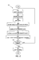
- FIG. 2 is a flowchart of a study process 200 and the example will be described using that flowchart.
- a pharmaceutical company with an MS drug typically recruits hundreds of patients with MS in Phase 3 of a clinical trial. In a randomized way, about half the patients are given the drug and other half are given a placebo. These patients undergo an MRI scan every three months while they are on treatment. Multiple MR image sequences with different MR acquisition parameters are acquired during each visit.
- the first goal of the data analysis is to measure the total MS lesion volume from the image sequences for each patient at each time point that they are scanned.
- the second goal of data analysis is to mine all the MS volume information to determine whether the MS drug is effective.
- the Image Analysis System 100 of the present invention can be used for both the above data analysis tasks.
- the following steps are involved in the MS lesion volume computation task:
- Image registration is applied to align the different sequences to one another (step 204 );
- the multi-channel tissue classification/segmentation method is applied to find all the MS lesions in the entire image sequence (step 208 );
- the automatically segmented and identified lesions may be manually edited by an expert to correct any errors (step 210 );
- This process is repeated (segmentation) for other image data sets (Step 216 ) and then the database can be queried (step 217 ) to locate the studies that have a particular set of characteristics.
- the image registration method 125 has three primary uses. First, registration can be used to align images acquired using different imaging modalities. Each modality may give different information regarding anatomy, pathology or function. Aligning this information assists the user in visualizing and interpreting the results. Without registration it is often difficult to spatially correlate findings from one image set to another image set acquired with different spatial orientations.
- the second use of registration serves as a preliminary step to multi-channel tissue segmentation. Even image series acquired during a single MRI exam can be mis-registered and small movements by the subject can result in the degradation of multi-channel tissue segmentation and failure to detect small structures.
- the third use of registration is for aligning images acquired at different time points. Aligning the images permits easier visualization and correlation of the changes. In addition, any overlays or segmentations generated for the image stack can be transformed during the registration process permitting quantification of the differences. Without registration, the user would be responsible for visually determining the spatial concordance of the segmentations.
- any number of commercially available image segmentation methods can be incorporated into and work with the Image Analysis System. Following the concept of “just-enough-interaction”, these methods combine the superior ability of a trained user to recognize structures of interest with the superior ability of computers to precisely and rapidly delineate these structures. Manual editing tools are available for editing the results of the segmentation method, should the expert user deem them necessary.
- segmentation methods implemented in the present system, and described in the following subsections are: seeded region growing, live-wire delineation, multi-channel segmentation, shape based interpolation, snakes, snakes used in conjunction with other tools and manual segmentation tools.
- the Image Analysis System is capable of extracting pertinent quantitative information, such as tissue volume, area, lesion diameter, and average pixel intensity. Using the registration tools comprising an integral part of the system, these quantitative measurements can be tracked in serial data.
- the Image Analysis System further comprises a registration method capable of significantly faster registration of 3D image data. The registration method is described more fully in U.S. patent application Ser. No. 10/063,834, filed on May 16, 2002 and entitled “SYSTEM AND METHOD FOR DETERMINING CONVERGENCE OF IMAGE SET REGISTRATION” which is owned by the same assignee as the present invention and is incorporated herein by reference.
- the present invention further integrates the database management system 165 (DBMS), as shown in FIG. 1 , that stores both the image data and the quantitative information extracted from the image data.
- DBMS database management system 165
- the database management system 165 not only enables persistent storage of image and other patient data, but also lets the system be used as an advanced medical imaging mining platform.
- the system also provides manual painting tools to initialize the segmentation methods or to correct the automatic or semi-automatic segmentation results.
- the user can paint regions in any of the 2D or 3D views.
- Various line and polygon drawing tools are also provided for manual annotations.
- An additional aspect of the present system is that the painting can be done in 2D and in 3D and both views are always synchronized.
- Two-dimensional overlays are interpolated to create three-dimensional overlays and three-dimensional overlays are sampled to create two-dimensional overlays.
- the image segmentations are displayed as color overlays on the original images. These overlays can be displayed in a region mode or in an edge mode. In the region-mode the entire overlay region is showed filled with a selected color as shown in FIG. 3 . In the edge-mode, only the boundaries of the region are displayed as overlaid on the original image as shown in FIG. 4 . Both display modes are useful in different scenarios and the present system allows the user to rapidly switch between the two modes. The user can vary the degree of translucency of the regions as well as the borders from opaque to transparent.
- the aspects of the Image Analysis System 100 such as 3D and 2D painting, and region and edge-mode display, are accomplished through the use of a data structure and associated methods to represent the overlays.
- the overlays are represented as 8-bit images (as opposed to representing them as polygons which is common). Therefore, an overlay can have up to 255 different colors (or unique regions);
- Every overlay layer is defined by two data structures—a two dimensional image stack (for display in stacked-mode) and a three-dimensional volume (for display in volume or MPR mode);
- An image interpolation method is used to create the 3D volume from the 2D stack of images—simply linear interpolation between corresponding image pixels is used in this method as is well known and the invention may be used with various other well known methods.
- a sub-sampling method is used to extract 2D image stacks from the 3D volume as is well known and the invention may use various well known methods to accomplish this result.
- Each 2D image in the image stack saves its location in the 3D volume and that location is used to extract a slice from the 3D volume; and
- FIGS. 3 and 5 are displayed in a 3 ⁇ 3 layout of the images in the series—3 rows and 3 columns.
- the Image Analysis System provides the user with the flexibility to change the layout to any desired layout—for example a 2 ⁇ 3 layout is shown in the FIG. 6 or a 2 ⁇ 2 layout shown in FIG. 4 .
- An aspect of this feature is the way in which the layout change is initiated from the GUI 115 .
- the layout is fixed at 2 ⁇ 2, 3 ⁇ 3, etc. or the user is required to explicitly type in the number of rows and number of columns of images to display. In the present system this is accomplished by the user interface 115 as follows (and as shown in FIG. 7 ):
- This small layout preview window looks like a preview window 302 shown in FIG. 7 ;
- Yet another aspect of the graphical user interface 115 is a flexible splitter between the various image panes.
- the size of the different panes within a multi-image window are fixed and the user does not have the flexibility of changing the sizes of the panes. It is sometimes very useful to have different panes of different sizes within a display window.
- a flexible splitter 610 between the panes allows the user to display the different panes in different sizes as shown in FIG. 9 .
- the flexible splitter allows the user to display the entire top-left image and still view the other three images without changing the total window size.
- the segmentation module in the present system provides an array of tools capable of being used in combination for image segmentation.
- the segmentation module 130 may include manual segmentation tools, a seeded region growing tool 400 , a live-wire tool 402 , a shape-based interpolation tool 404 , a connected eraser tool 406 , a multi and single tissue classification tool 408 , combination tools 410 and a snakes tool 412 . Each of these tools will be described below in more detail.
- the system also provides manual painting tools to initialize the segmentation methods or to correct the automatic segmentation results.
- the user can paint regions in any of the 2D or 3D views using a paintbrush tool with a user-variable size. Or various regions can be delineated with manual polygon tool.
- Various line, point and polygon and ellipse drawing tools are also provided for manual annotations. These annotation tools automatically calculate the distances or areas of the objects.
- Seeded region growing also referred to as seed-fill
- FIG. 11 Seeded region growing, also referred to as seed-fill
- the user clicks somewhere on the object to be delineated as shown in FIG. 17 which describes a seeded region growing method 440 .
- the method results in the delineation of the entire object, either in 2D or in 3D.
- the seeded region-growing method permits the user to place a seed point within regions to be segmented by clicking on the image as shown in step 442 .
- the method then segments a connected region around the seed point in step 444 where all the pixels (x) in this region satisfy the following criterion of equation (2) as follows: ( m ⁇ r ) ⁇ f ( x ) ⁇ ( m+r ) EQ(2)
- f(x) is the intensity value of pixel x
- m is the mean intensity of the region computed from a small neighborhood around the seed point
- r is a user-adjustable range value.
- the user can then adjust the threshold of the seed fill to fine tune the end results in step 446 .
- This adjustment essentially changes the limits (m ⁇ r) and (m+r) in EQ 2.
- Some options for threshold adjustment include changing r only, having separate upper and lower bounds, permitting the independent adjustment of both m and r.
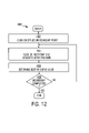
- Live-wire is an interactive tracing tool used for precise delineation of structure boundaries with minimal user effort.
- the live-wire tool (a live wire method 460 is described with respect to FIG. 12 ) allows the user to rapidly delineate an object of interest by clicking only on a few selected points on the boundary of the object.
- the user gets interactive feedback about the location of the boundary as they move the mouse.
- the user starts tracing with live-wire by clicking on any point on a structure boundary in step 462 .
- an active polygon connects the current cursor location with the last-clicked mouse point along the strong gradient edges in the image in step 464 .
- the method traces the precise boundary of the structure.
- the live-wire method is a graph-matching method that computes a best-fit (least cost) curve in step 466 between the most recent mouse position and last clicked vertex.
- the cost of the curve is defined by the proximity of the curve to the high gradient points on the image. The closer the curve is to strong edges in the image, the lower the cost of the curve.
- a shape-based interpolation method is used to rapidly create 3D segmentation from a few 2D segmentations by interpolation.
- the user draws two-dimensional contours on selected image slices in a 3D sequence of images.
- the shape-based interpolation tool interpolates the contours between the slices on which the contours are drawn. Such a feature is useful in tasks such as vessel segmentation where the shape change between several image slices is gradual and can be well-approximated using shape-based interpolation.
- the combination of the live-wire and shape-based interpolation tool provides a powerful three-dimensional segmentation method.
- the user can outline a structure using live-wire on two or more images in a series of images and can apply the shape-based interpolation tool to interpolate between the slices.
- FIG. 13 illustrates the use of the live-wire tool on the image slices 83 , 87 , and 91 , to delineate a structure (the abdominal aortic aneurysm).
- FIG. 14 illustrates the use of the shape-based Interpolation tool used to automatically delineate the structure all the slices between slices 83 , 87 , and 91 .
- the combination of 2D seeded region growing and shape-based interpolation also provides powerful three-dimensional segmentation.
- the user can outline a structure using shape-based interpolation on two or more images in a series of images. Then they can apply the shape-based interpolation tool to interpolate between the slices.
- the use of this method is necessitated in cases where the 3D seeded region growing is not applicable.
- FIG. 15 depicts the result of 3D region growing and it results in the detection of the cerebro-spinal fluid outside the brain in addition to the ventricle, therefore, 3D region growing would not be the optimal tool for this particular problem.
- FIG. 16 and FIG. 17 depict the approach of the present invention wherein a 2D region growing is used on 3 slices and the result is interpolated to the other slices in the image sequence.
- FIG. 16 illustrates the use of the 2D Seed Fill tool to delineate the ventricle in the brain in 3 of the 9 image slices and
- FIG. 17 illustrates the use of the shape-based interpolation to delineate the ventricles on all the 9 image slices.
- the connected eraser is similar to the region-growing tool, except it is used for erasing regions previously drawn.
- FIGS. 18(A) and (B) An example of using the erase connected tool to correct the results of tissue classification or other segmentation methods is shown in FIGS. 18(A) and (B).
- FIG. 18(A) depicts the result of tissue classification on one of the images, wherein the scalp area is falsely labeled as a lesions also. By simply clicking on a region of the scalp using the erase connected tool, the entire scalp area is erased as shown in FIG. 18(B) .
- the connected erase tool allows the user to quickly edit the results of tissue classification.
- the tissue classification tool in the present system enables the identification of one or more tissue types in an entire image sequence.
- the user first identifies key tissue types as exemplars.
- the method uses these exemplars as training data and identifies the trained tissue types in the entire image sequence. This method works with single channel data and it also works with multi-channel data.
- multi-channel segmentation Since they use the same methods we will simply use the term multi-channel segmentation to generically refer to both methods. While the seeded region growing method finds regions that are spatially limited and connected to the seed point, the tissue segmentation method is useful for delineating tissues that are distributed throughout the image series.
- This tool uses multivariate statistics to combine multiple channels of information, such as T1 and T2-weighted MR series. Combining multiple channels of data can increase the discrimination between different tissue types, thus leading to improved segmentation. If multiple series are present for the same study and the series give complementary pieces of information, all of these series can and should be used for tissue classification to give more accurate results.
- a multi-channel tissue segmentation method 470 in accordance with the invention will be described with reference to FIG. 19 .
- the first step in multi-channel tissue segmentation is to provide training data in step 472 .
- This training data consists of annotations of exemplar regions for one or more tissue types present on the image.
- the user can use the manual painting tools and/or any of the computer-assisted segmentation tools to provide regions for training.
- multi-dimensional Gaussian probability models are generated for each tissue types in the training data in step 474 .
- the user can either specify a probability threshold value on a single Gaussian model or can apply a maximum likelihood (ML) classifier.
- the model may be used to classify the remaining pixels in the image set.
- An optional Markov Random Field (MRF) filter can be used to further refine the ML results in step 476 .
- MRF Markov Random Field
- a size filter is provided where all connected regions smaller than the user-specified size threshold are removed in step 477 . This filter can be used to improve the results by eliminating small, segmented regions which may result from image noise.
- FIG. 20 illustrates a multi-channel series of images, wherein each row represents one channel of information while each column represents a unique slice position in 3D space.
- the top row is a FLAIR MR sequence
- the middle row is the Proton Density (PD) sequence
- the bottom row is the T2-weighted sequence.
- FIG. 21 illustrates the use of the seeded region growing method used to mark an example MS lesion on the FLAIR sequence. This region is used to train the multi-channel segmentation method.
- FIG. 22 illustrates that by using the previously marked region, of FIG. 21 , as training, the multi-channel tissue segmentation method locates all MS lesions in the entire image sequence. These lesions are labeled on all the channels that were used in the method.
- a segmentation method called “snakes” is also incorporated into the present invention.
- the snakes method will be described with reference to FIG. 23 .
- the snakes method functionally performs the following steps in its segmentation method.
- the user specifies an initial contour (either manually or as an output of another segmentation method).
- this initial contour is usually somewhat close to the desired final contour, although it may deviate substantially, but still provide the desired result of defining the boundaries of the structure under going segmentation.
- the snakes method 420 takes as input the initial contour and the underlying grayscale image (See step 422 ) and produces a final contour which lies along the edges of desired structures while having a smooth appearance.
- radial lines are drawn normal to each vertex of the initial contour in step 424 and the contour is restricted to move only along these radial directions.
- the contour is permitted to move along additional lines, while being restricted in another.
- a sampling of the image along these radial directions is performed and one-dimensional derivatives of the image are computed along the radial lines.
- the position of the contour is iteratively adjusted in step 426 to have its contour on this radial line such that the total energy of the contour is minimized.
- v is the contour
- v S is the first derivative of the contour
- v SS is the second derivative of the contour
- g is the negative derivative of the input image evaluated along the contour.
- Alpha, beta, and lambda are weighting parameters for each term respectively.
- EQ. (1) beginning with A promotes close tracking of the contour to the edges on the image.
- the term beginning with ⁇ promotes smoothing of the contour and the term beginning with ⁇ promotes the contour length to be relatively short.
- the radial representation of the contour is converted back to a Cartesian coordinate representation and displayed to the user. An illustration of the above process will now be provided.
- FIG. 24(A) depicts a initial contour for the kidney specified manually by the user.
- FIG. 24(B) depicts the final contour of the kidney after the application of the snake method. Note that the contour fits the edges of the kidney while remaining smooth.
- segmentation tools are generally known. However, the segmentation tools may be combined together. The use of the shape based interpolation tool in conjunction with other 2D methods has already been discussed. Here are some other unique scenarios created by the combination of these tools:
- An additional aspect of the present invention is the combination of the snakes in conjunction with shape-based interpolation.
- 3D structures can be segmented much faster than conventionally, utilizing 2D techniques.
- the synergy provided by this aspect of the present invention provides a method and system which is very powerful and hence useful in the segmentation of 3D structures in medical images.
- the method provides for the utilization of a 2D seedfill, live-wire, or similar method to delineate a structure on multiple images, such as every third image.
- 3 of 9 images have a structure delineated.
- a shape-based interpolation technique is utilized to obtain approximate delineations on the remaining 6 images. Since the kidney is not a linear varying structure the shape-based interpolation did not perform very well. However, these approximate delineations of the remaining 6 images are then used as initial contours for the Snakes method.
- the snakes method modifies the contours to fit the boundaries of the desired structure and result in much more accurate delineations on the 6 intermediate images.
- FIGS. 25(A)–25(L) depict a kidney with an initial contour on 3 slices of 12 outlined using a 2D seed tool.
- FIGS. 26(A)–26(L) depict a kidney outlined on all 12 images using a shape-based interpolation tool.
- FIGS. 27(A)–27(L) depict the resulting corrected image obtained by utilizing the snake method of the present invention.
- Another approach is to use the segmentation results from another time point or a baseline scan to provide the initial contours for the current time point.
- the snake algorithm can then be used to automatically adjust the boundaries to account for the differences in the lesion or tissue that occurred between scans.
- a variation of this would be for use in template matching segmentation.
- template matching a reference image is carefully segmented. Then registration (usually deformable registration) between the reference image and the new test image to be classified is performed. Once the deformation required to transform the reference image to the test image is determined this deformation can be applied to the segmentation overlays to classify the new image. Snakes could then be applied to fine-tune the end results.
- Another combination tool is to combine seed fill with multi-channel segmentation.
- the user clicks on a region of interest in one channel of data to initiate seeded region growing.
- the results of the seeded region growing are then automatically used for the initiation of the multi-channel segmentation. This results in one click multi-channel segmentation.
- Volume Report Volumes of all labeled regions for all image series in the database
- Area Report 2D Areas of all labeled regions for all images in the database
Abstract
Description
(m−r)≦f(x)≦(m+r) EQ(2)
E(ν)=Σ(α∥νS∥2+β∥νSS∥2 +λg(ν)) EQ.(1)
-
- Measurements of tumor volume and tumor cross-sectional diameters from CT and MR images of brain, head, neck, and chest.
- Multiple sclerosis (MS) lesion load measurement from MR images of the brain.
- Articular cartilage volume and thickness measurement from MR images of the knee.
- Delineation of abdominal aortic aneurysm (AAA) from thoracic CT images.
- Delineation of brain structures for neuro-degenerative diseases on MR images.
- Delineation of prostate from trans-rectal ultrasound images.
- Delineation of the heart wall (endocardial and epicardial contours) from cardiac MR images.
- Assessment of patients with multiple studies or where quantitative information is needed for proper clinical care.
Claims (47)
E(ν)=Σ(α∥νS∥2 +βν 38∥2 +λg(ν)).
E(ν)=Σ(α∥νS∥2+β∥νSS∥2 +λg(ν)).
E(ν)=Σ(α∥νS∥2+β∥νSS∥2 +λg(ν)).
E(ν)=Σ(α∥νS∥2 +β∥v SS∥2 +λg(ν)).
Priority Applications (1)
| Application Number | Priority Date | Filing Date | Title |
|---|---|---|---|
| US10/271,916 US7158692B2 (en) | 2001-10-15 | 2002-10-15 | System and method for mining quantitive information from medical images |
Applications Claiming Priority (3)
| Application Number | Priority Date | Filing Date | Title |
|---|---|---|---|
| US32984301P | 2001-10-15 | 2001-10-15 | |
| US32982701P | 2001-10-15 | 2001-10-15 | |
| US10/271,916 US7158692B2 (en) | 2001-10-15 | 2002-10-15 | System and method for mining quantitive information from medical images |
Publications (2)
| Publication Number | Publication Date |
|---|---|
| US20030174872A1 US20030174872A1 (en) | 2003-09-18 |
| US7158692B2 true US7158692B2 (en) | 2007-01-02 |
Family
ID=28046385
Family Applications (1)
| Application Number | Title | Priority Date | Filing Date |
|---|---|---|---|
| US10/271,916 Expired - Lifetime US7158692B2 (en) | 2001-10-15 | 2002-10-15 | System and method for mining quantitive information from medical images |
Country Status (1)
| Country | Link |
|---|---|
| US (1) | US7158692B2 (en) |
Cited By (68)
| Publication number | Priority date | Publication date | Assignee | Title |
|---|---|---|---|---|
| US20030088177A1 (en) * | 2001-09-05 | 2003-05-08 | Virtualscopics, Llc | System and method for quantitative assessment of neurological diseases and the change over time of neurological diseases |
| US20050049994A1 (en) * | 2003-08-21 | 2005-03-03 | Microsoft Corporation | Systems and methods for the implementation of a base schema for organizing units of information manageable by a hardware/software interface system |
| US20050105788A1 (en) * | 2003-11-19 | 2005-05-19 | Matthew William Turek | Methods and apparatus for processing image data to aid in detecting disease |
| US20050276455A1 (en) * | 2004-06-01 | 2005-12-15 | Marta Fidrich | Systems and methods for segmenting an organ in a plurality of images |
| US20060056701A1 (en) * | 2004-03-02 | 2006-03-16 | Gozde Unal | Joint segmentation and registration of images for object detection |
| US20060100502A1 (en) * | 2004-06-23 | 2006-05-11 | Chen David T | Anatomical visualization and measurement system |
| US20060159341A1 (en) * | 2003-06-13 | 2006-07-20 | Vladimir Pekar | 3D image segmentation |
| US20060257053A1 (en) * | 2003-06-16 | 2006-11-16 | Boudreau Alexandre J | Segmentation and data mining for gel electrophoresis images |
| US20070064984A1 (en) * | 2005-09-19 | 2007-03-22 | General Electric Company | System and method for dynamic configuration of PACS workstation displays |
| US20070116334A1 (en) * | 2005-11-22 | 2007-05-24 | General Electric Company | Method and apparatus for three-dimensional interactive tools for semi-automatic segmentation and editing of image objects |
| US20070116335A1 (en) * | 2005-11-23 | 2007-05-24 | General Electric Company | Method and apparatus for semi-automatic segmentation technique for low-contrast tubular shaped objects |
| US20070122039A1 (en) * | 2005-11-29 | 2007-05-31 | Microsoft Corporation | Segmentation of objects by minimizing global-local variational energy |
| US20070127795A1 (en) * | 2005-11-23 | 2007-06-07 | Lau Denny W | System and method for linking current and previous images based on anatomy |
| US20070127790A1 (en) * | 2005-11-14 | 2007-06-07 | General Electric Company | System and method for anatomy labeling on a PACS |
| US20070135705A1 (en) * | 2005-12-08 | 2007-06-14 | Lorenz Christine H | System and method for image based physiological monitoring of cardiovascular function |
| US20070230761A1 (en) * | 2005-12-12 | 2007-10-04 | Lutz Gundel | Method and device for visualizing a sequence of tomographic image data records |
| US20070292012A1 (en) * | 2006-06-16 | 2007-12-20 | Siemens Medical Solutions Usa, Inc. | Clinical Trial Data Processing System |
| US20080008366A1 (en) * | 2006-06-20 | 2008-01-10 | Vladimir Desh | Simultaneous visualization, analysis and navigation of multi-modality medical imaging data |
| US20080008401A1 (en) * | 2006-06-14 | 2008-01-10 | Koninklijke Philips Electronics, N.V. | Multi-modality medical image viewing |
| US20080018669A1 (en) * | 2006-07-18 | 2008-01-24 | General Electric Company | method and system for integrated image zoom and montage |
| US20080059241A1 (en) * | 2006-09-01 | 2008-03-06 | Siemens Medical Solutions Usa, Inc. | Interface Between Clinical and Research Information Systems |
| US20080137929A1 (en) * | 2004-06-23 | 2008-06-12 | Chen David T | Anatomical visualization and measurement system |
| US20080137921A1 (en) * | 2006-12-07 | 2008-06-12 | Simon Richard A | Analyzing lesions in a medical digital image |
| US20080143718A1 (en) * | 2006-12-19 | 2008-06-19 | Ray Lawrence A | Method for segmentation of lesions |
| US20080170769A1 (en) * | 2006-12-15 | 2008-07-17 | Stefan Assmann | Method and image processing system for producing result images of an examination object |
| US20080226161A1 (en) * | 2007-03-12 | 2008-09-18 | Jeffrey Kimball Tidd | Determining Edgeless Areas in a Digital Image |
| US20080253631A1 (en) * | 2007-04-11 | 2008-10-16 | Fujifilm Corporation | Apparatus and program for assisting report generation |
| US20080281182A1 (en) * | 2007-05-07 | 2008-11-13 | General Electric Company | Method and apparatus for improving and/or validating 3D segmentations |
| US20090043172A1 (en) * | 2006-06-02 | 2009-02-12 | Koninklijke Philips Electronics N. V. | Multi-modal imaging system and workstation with support for structured hypothesis testing |
| US20090094247A1 (en) * | 2007-10-03 | 2009-04-09 | Fredlund John R | Image storage system, device and method |
| US20090112882A1 (en) * | 2007-10-30 | 2009-04-30 | Guy Maresh | Methods, systems, and devices for managing medical images and records |
| US20090157171A1 (en) * | 2007-12-18 | 2009-06-18 | Searete Llc, A Limited Liability Corporation Of The State Of Delaware | Treatment indications informed by a priori implant information |
| US20090157058A1 (en) * | 2007-12-18 | 2009-06-18 | Searete Llc, A Limited Liability Corporation Of The State Of Delaware | Circulatory monitoring systems and methods |
| US20090287101A1 (en) * | 2008-05-13 | 2009-11-19 | Searete Llc, A Limited Liability Corporation Of The State Of Delaware | Circulatory monitoring systems and methods |
| US20090287191A1 (en) * | 2007-12-18 | 2009-11-19 | Searete Llc, A Limited Liability Corporation Of The State Of Delaware | Circulatory monitoring systems and methods |
| US20090287094A1 (en) * | 2008-05-15 | 2009-11-19 | Seacrete Llc, A Limited Liability Corporation Of The State Of Delaware | Circulatory monitoring systems and methods |
| US20090287109A1 (en) * | 2008-05-14 | 2009-11-19 | Searete Llc, A Limited Liability Corporation Of The State Of Delaware | Circulatory monitoring systems and methods |
| US20090292212A1 (en) * | 2008-05-20 | 2009-11-26 | Searete Llc, A Limited Corporation Of The State Of Delaware | Circulatory monitoring systems and methods |
| US20090326362A1 (en) * | 2004-12-15 | 2009-12-31 | Koninklijke Philips Electronics N.V. | Registration of multi-modality images |
| US20100021027A1 (en) * | 2008-07-25 | 2010-01-28 | Thomas Hartkens | Image data management systems |
| US20100061606A1 (en) * | 2008-08-11 | 2010-03-11 | Siemens Corporate Research, Inc. | Method and system for data dependent multi phase visualization |
| US20100145720A1 (en) * | 2008-12-05 | 2010-06-10 | Bruce Reiner | Method of extracting real-time structured data and performing data analysis and decision support in medical reporting |
| US20100246957A1 (en) * | 2007-11-02 | 2010-09-30 | Koninklijke Philips Electronics N.V. | Enhanced coronary viewing |
| US20100275145A1 (en) * | 2007-12-14 | 2010-10-28 | Koninklijke Philips Electronics N.V. | Labeling a segmented object |
| US20110016150A1 (en) * | 2009-07-20 | 2011-01-20 | Engstroem Jimmy | System and method for tagging multiple digital images |
| US20110028825A1 (en) * | 2007-12-03 | 2011-02-03 | Dataphysics Research, Inc. | Systems and methods for efficient imaging |
| US20110188718A1 (en) * | 2008-07-25 | 2011-08-04 | Derek Lionel Glendon Hill | Image data management systems |
| US20120008848A1 (en) * | 2010-07-08 | 2012-01-12 | Orthosize Llc | Method and Device for Digital Image Templating |
| US20120032953A1 (en) * | 2009-03-31 | 2012-02-09 | Koninklijke Philips Electronics N.V. | Automated contrast enhancement for contouring |
| US8232535B2 (en) | 2005-05-10 | 2012-07-31 | Tomotherapy Incorporated | System and method of treating a patient with radiation therapy |
| US20120197656A1 (en) * | 2011-01-28 | 2012-08-02 | Burton Lang | Radiation therapy knowledge exchange |
| US20120194505A1 (en) * | 2011-01-31 | 2012-08-02 | Orthosize Llc | Digital Image Templating |
| US8442287B2 (en) | 2005-07-22 | 2013-05-14 | Tomotherapy Incorporated | Method and system for evaluating quality assurance criteria in delivery of a treatment plan |
| US8636670B2 (en) | 2008-05-13 | 2014-01-28 | The Invention Science Fund I, Llc | Circulatory monitoring systems and methods |
| US8693744B2 (en) | 2010-05-03 | 2014-04-08 | Mim Software, Inc. | Systems and methods for generating a contour for a medical image |
| US8805035B2 (en) | 2010-05-03 | 2014-08-12 | Mim Software, Inc. | Systems and methods for contouring a set of medical images |
| US8892184B2 (en) | 2010-10-18 | 2014-11-18 | Siemens Medical Solutions Usa, Inc. | Systems and methods for reducing interference in a dual modality imaging system |
| US9171344B2 (en) | 2007-10-30 | 2015-10-27 | Onemednet Corporation | Methods, systems, and devices for managing medical images and records |
| US20160092748A1 (en) * | 2014-09-30 | 2016-03-31 | Kabushiki Kaisha Toshiba | Medical data processing apparatus and method |
| US20160155236A1 (en) * | 2014-11-28 | 2016-06-02 | Kabushiki Kaisha Toshiba | Apparatus and method for registering virtual anatomy data |
| US9443633B2 (en) | 2013-02-26 | 2016-09-13 | Accuray Incorporated | Electromagnetically actuated multi-leaf collimator |
| CN106934807A (en) * | 2015-12-31 | 2017-07-07 | 深圳迈瑞生物医疗电子股份有限公司 | A kind of medical image analysis method, system and Medical Devices |
| US9760677B2 (en) | 2009-04-29 | 2017-09-12 | Onemednet Corporation | Methods, systems, and devices for managing medical images and records |
| US10004564B1 (en) | 2016-01-06 | 2018-06-26 | Paul Beck | Accurate radiographic calibration using multiple images |
| US10010372B1 (en) | 2016-01-06 | 2018-07-03 | Paul Beck | Marker Positioning Apparatus |
| US10052076B2 (en) | 2011-04-26 | 2018-08-21 | Koninklijke Philips N.V. | Diagnostic brain imaging |
| US10886029B2 (en) * | 2017-11-08 | 2021-01-05 | International Business Machines Corporation | 3D web-based annotation |
| US11813418B2 (en) | 2019-08-22 | 2023-11-14 | Becton, Dickinson And Company | Echogenic balloon dilation catheter and balloon thereof |
Families Citing this family (95)
| Publication number | Priority date | Publication date | Assignee | Title |
|---|---|---|---|---|
| US7155043B2 (en) * | 2001-11-21 | 2006-12-26 | Confirma, Incorporated | User interface having analysis status indicators |
| GB2382509B (en) * | 2001-11-23 | 2003-10-08 | Voxar Ltd | Handling of image data created by manipulation of image data sets |
| US7123766B2 (en) * | 2002-02-11 | 2006-10-17 | Cedara Software Corp. | Method and system for recognizing and selecting a region of interest in an image |
| WO2003071779A1 (en) * | 2002-02-19 | 2003-08-28 | Siemens Corporate Research, Inc. | System and method for generating movie loop display from medical image data |
| WO2003083779A2 (en) * | 2002-04-03 | 2003-10-09 | Segami S.A.R.L. | Image registration process |
| US7819806B2 (en) | 2002-06-07 | 2010-10-26 | Verathon Inc. | System and method to identify and measure organ wall boundaries |
| US7744534B2 (en) | 2002-06-07 | 2010-06-29 | Verathon Inc. | 3D ultrasound-based instrument for non-invasive measurement of amniotic fluid volume |
| US8221322B2 (en) | 2002-06-07 | 2012-07-17 | Verathon Inc. | Systems and methods to improve clarity in ultrasound images |
| US7041059B2 (en) * | 2002-08-02 | 2006-05-09 | Diagnostic Ultrasound Corporation | 3D ultrasound-based instrument for non-invasive measurement of amniotic fluid volume |
| GB2391625A (en) | 2002-08-09 | 2004-02-11 | Diagnostic Ultrasound Europ B | Instantaneous ultrasonic echo measurement of bladder urine volume with a limited number of ultrasound beams |
| US6676605B2 (en) * | 2002-06-07 | 2004-01-13 | Diagnostic Ultrasound | Bladder wall thickness measurement system and methods |
| US8221321B2 (en) | 2002-06-07 | 2012-07-17 | Verathon Inc. | Systems and methods for quantification and classification of fluids in human cavities in ultrasound images |
| US20060025689A1 (en) * | 2002-06-07 | 2006-02-02 | Vikram Chalana | System and method to measure cardiac ejection fraction |
| US7127093B2 (en) * | 2002-09-17 | 2006-10-24 | Siemens Corporate Research, Inc. | Integrated image registration for cardiac magnetic resonance perfusion data |
| US20040122704A1 (en) * | 2002-12-18 | 2004-06-24 | Sabol John M. | Integrated medical knowledge base interface system and method |
| US7490085B2 (en) * | 2002-12-18 | 2009-02-10 | Ge Medical Systems Global Technology Company, Llc | Computer-assisted data processing system and method incorporating automated learning |
| US20040122706A1 (en) * | 2002-12-18 | 2004-06-24 | Walker Matthew J. | Patient data acquisition system and method |
| US20040122719A1 (en) * | 2002-12-18 | 2004-06-24 | Sabol John M. | Medical resource processing system and method utilizing multiple resource type data |
| US20040122787A1 (en) * | 2002-12-18 | 2004-06-24 | Avinash Gopal B. | Enhanced computer-assisted medical data processing system and method |
| US20040122709A1 (en) * | 2002-12-18 | 2004-06-24 | Avinash Gopal B. | Medical procedure prioritization system and method utilizing integrated knowledge base |
| US20040122707A1 (en) * | 2002-12-18 | 2004-06-24 | Sabol John M. | Patient-driven medical data processing system and method |
| US20040122705A1 (en) * | 2002-12-18 | 2004-06-24 | Sabol John M. | Multilevel integrated medical knowledge base system and method |
| US20040122708A1 (en) * | 2002-12-18 | 2004-06-24 | Avinash Gopal B. | Medical data analysis method and apparatus incorporating in vitro test data |
| US20040122702A1 (en) * | 2002-12-18 | 2004-06-24 | Sabol John M. | Medical data processing system and method |
| US20040122703A1 (en) * | 2002-12-19 | 2004-06-24 | Walker Matthew J. | Medical data operating model development system and method |
| US7340081B2 (en) * | 2003-07-15 | 2008-03-04 | M2S, Inc. | Using statistical process control (SPC) to demonstrate solution convergence in a technician guided segmentation system |
| EP2189107A3 (en) * | 2003-10-31 | 2010-09-15 | Olympus Corporation | Insertion support system |
| KR100639959B1 (en) * | 2003-11-20 | 2006-10-31 | 한국전자통신연구원 | Human image processing system and method |
| US20060013462A1 (en) * | 2004-07-15 | 2006-01-19 | Navid Sadikali | Image display system and method |
| US20060036147A1 (en) * | 2004-07-20 | 2006-02-16 | Scimed Life Systems, Inc. | Systems and methods for detecting and presenting textural information from medical images |
| US20060173318A1 (en) * | 2004-07-20 | 2006-08-03 | Scimed Life Systems Inc. | Systems and methods for detecting and presenting textural information from medical images |
| WO2006042077A2 (en) * | 2004-10-09 | 2006-04-20 | Viatronix Incorporated | Sampling medical images for virtual histology |
| US8744184B2 (en) * | 2004-10-22 | 2014-06-03 | Autodesk, Inc. | Graphics processing method and system |
| US20060101075A1 (en) * | 2004-11-10 | 2006-05-11 | Peter Lehel | Method and system for prospectively acquiring ROI based data for retrospective re-analysis |
| GB2421135A (en) | 2004-12-09 | 2006-06-14 | Sony Uk Ltd | User resizable video display of catalogue entries |
| US20060136259A1 (en) * | 2004-12-17 | 2006-06-22 | General Electric Company | Multi-dimensional analysis of medical data |
| US20060136417A1 (en) * | 2004-12-17 | 2006-06-22 | General Electric Company | Method and system for search, analysis and display of structured data |
| US20060155577A1 (en) * | 2005-01-07 | 2006-07-13 | Confirma, Inc. | System and method for anatomically based processing of medical imaging information |
| US7957507B2 (en) | 2005-02-28 | 2011-06-07 | Cadman Patrick F | Method and apparatus for modulating a radiation beam |
| US7653264B2 (en) * | 2005-03-04 | 2010-01-26 | The Regents Of The University Of Michigan | Method of determining alignment of images in high dimensional feature space |
| US7804999B2 (en) * | 2005-03-17 | 2010-09-28 | Siemens Medical Solutions Usa, Inc. | Method for performing image based regression using boosting |
| US20070064987A1 (en) * | 2005-04-04 | 2007-03-22 | Esham Matthew P | System for processing imaging device data and associated imaging report information |
| EP1907984A4 (en) * | 2005-07-22 | 2009-10-21 | Tomotherapy Inc | Method and system for processing data relating to a radiation therapy treatment plan |
| KR20080049716A (en) * | 2005-07-22 | 2008-06-04 | 토모테라피 인코포레이티드 | Method and system for evaluating quality assurance criteria in delivery of a treament plan |
| US8229068B2 (en) | 2005-07-22 | 2012-07-24 | Tomotherapy Incorporated | System and method of detecting a breathing phase of a patient receiving radiation therapy |
| KR20080039920A (en) | 2005-07-22 | 2008-05-07 | 토모테라피 인코포레이티드 | System and method of evaluating dose delivered by a radiation therapy system |
| JP2009502253A (en) | 2005-07-22 | 2009-01-29 | トモセラピー・インコーポレーテッド | System and method for applying radiation therapy to a moving region of interest |
| JP5390855B2 (en) * | 2005-07-23 | 2014-01-15 | トモセラピー・インコーポレーテッド | Imaging and delivery of radiation therapy using coordinated movement of gantry and treatment table |
| US7764817B2 (en) * | 2005-08-15 | 2010-07-27 | Siemens Medical Solutions Usa, Inc. | Method for database guided simultaneous multi slice object detection in three dimensional volumetric data |
| US7483939B2 (en) * | 2005-08-25 | 2009-01-27 | General Electric Company | Medical processing system allocating resources for processing 3D to form 2D image data based on report of monitor data |
| US20070078873A1 (en) * | 2005-09-30 | 2007-04-05 | Avinash Gopal B | Computer assisted domain specific entity mapping method and system |
| US8384729B2 (en) * | 2005-11-01 | 2013-02-26 | Kabushiki Kaisha Toshiba | Medical image display system, medical image display method, and medical image display program |
| US20070118408A1 (en) * | 2005-11-23 | 2007-05-24 | General Electric Company | System and method for radiology decision support using picture-in-picture |
| JP5226181B2 (en) * | 2005-11-24 | 2013-07-03 | ジーイー・メディカル・システムズ・グローバル・テクノロジー・カンパニー・エルエルシー | Diagnostic imaging equipment |
| DE102006008509A1 (en) * | 2006-02-23 | 2007-08-02 | Siemens Ag | Conspicuousness detecting method, involves testing image with detection algorithm automatically, where image data sets are registered, in order to obtain transformations |
| US8249315B2 (en) * | 2006-05-22 | 2012-08-21 | Upmc | System and method for improved viewing and navigation of digital images |
| US7945083B2 (en) * | 2006-05-25 | 2011-05-17 | Carestream Health, Inc. | Method for supporting diagnostic workflow from a medical imaging apparatus |
| US7773807B2 (en) * | 2006-08-29 | 2010-08-10 | Siemens Medical Solutions Usa, Inc. | Seed segmentation using l∞ minimization |
| US7940977B2 (en) | 2006-10-25 | 2011-05-10 | Rcadia Medical Imaging Ltd. | Method and system for automatic analysis of blood vessel structures to identify calcium or soft plaque pathologies |
| US7983459B2 (en) | 2006-10-25 | 2011-07-19 | Rcadia Medical Imaging Ltd. | Creating a blood vessel tree from imaging data |
| US7860283B2 (en) | 2006-10-25 | 2010-12-28 | Rcadia Medical Imaging Ltd. | Method and system for the presentation of blood vessel structures and identified pathologies |
| US7940970B2 (en) | 2006-10-25 | 2011-05-10 | Rcadia Medical Imaging, Ltd | Method and system for automatic quality control used in computerized analysis of CT angiography |
| US7873194B2 (en) | 2006-10-25 | 2011-01-18 | Rcadia Medical Imaging Ltd. | Method and system for automatic analysis of blood vessel structures and pathologies in support of a triple rule-out procedure |
| US7953265B2 (en) * | 2006-11-22 | 2011-05-31 | General Electric Company | Method and system for automatic algorithm selection for segmenting lesions on pet images |
| US8270696B2 (en) * | 2007-02-09 | 2012-09-18 | The Trustees Of The University Of Pennsylvania | Image slice segmentation using midpoints of contour anchor points |
| JP4905967B2 (en) * | 2007-03-02 | 2012-03-28 | 富士フイルム株式会社 | Similar case retrieval apparatus, method, and program |
| US20080242977A1 (en) * | 2007-03-30 | 2008-10-02 | General Electric Company | Systems, methods and apparatus for longitudinal/temporal analysis of plaque lesions |
| US8167803B2 (en) | 2007-05-16 | 2012-05-01 | Verathon Inc. | System and method for bladder detection using harmonic imaging |
| US8133181B2 (en) | 2007-05-16 | 2012-03-13 | Verathon Inc. | Device, system and method to measure abdominal aortic aneurysm diameter |
| WO2009093146A1 (en) * | 2008-01-24 | 2009-07-30 | Koninklijke Philips Electronics N.V. | Interactive image segmentation |
| RU2520369C2 (en) * | 2008-06-25 | 2014-06-27 | Конинклейке Филипс Электроникс Н.В. | Device and method for localising object of interest in subject |
| KR101175426B1 (en) * | 2010-01-26 | 2012-08-20 | 삼성메디슨 주식회사 | Ultrasound system and method for providing three-dimensional ultrasound image |
| US20120078521A1 (en) * | 2010-09-27 | 2012-03-29 | General Electric Company | Apparatus, system and methods for assessing drug efficacy using holistic analysis and visualization of pharmacological data |
| JP2013542046A (en) * | 2010-11-10 | 2013-11-21 | エコーメトリックス,エルエルシー | Ultrasound image processing system and method |
| JP5961623B2 (en) * | 2010-11-19 | 2016-08-02 | コーニンクレッカ フィリップス エヌ ヴェKoninklijke Philips N.V. | Method for guiding the insertion of surgical instruments using three-dimensional ultrasound imaging |
| US9262444B2 (en) | 2010-11-24 | 2016-02-16 | General Electric Company | Systems and methods for applying series level operations and comparing images using a thumbnail navigator |
| US8983189B2 (en) * | 2011-02-08 | 2015-03-17 | Algotec Systems Ltd. | Method and systems for error correction for three-dimensional image segmentation |
| KR101185727B1 (en) * | 2011-09-14 | 2012-09-25 | 주식회사 인피니트헬스케어 | A segmentatin method of medical image and apparatus thereof |
| US20130114870A1 (en) * | 2011-11-04 | 2013-05-09 | Patents Innovations, Llc | Medical imaging for drug application analysis |
| GB201121307D0 (en) * | 2011-12-12 | 2012-01-25 | Univ Stavanger | Probability mapping for visualisation of biomedical images |
| GB2511052B (en) | 2013-02-20 | 2015-03-04 | Siemens Medical Solutions | A method for combining a plurality of image data sets into one multi-fused image |
| RU2016114861A (en) * | 2013-09-20 | 2017-10-25 | Трансмурал Биотек, С.Л. | METHODS FOR ANALYSIS OF IMAGES FOR DIAGNOSTIC OF DISEASES |
| US9002085B1 (en) | 2013-10-22 | 2015-04-07 | Eyenuk, Inc. | Systems and methods for automatically generating descriptions of retinal images |
| WO2015114484A1 (en) * | 2014-01-28 | 2015-08-06 | Koninklijke Philips N.V. | Ultrasound systems for multi-plane acquisition with single- or bi-plane real-time imaging, and methods of operation thereof |
| WO2017051945A1 (en) * | 2015-09-24 | 2017-03-30 | 주식회사 뷰노코리아 | Method and apparatus for providing medical information service on basis of disease model |
| CN108603922A (en) | 2015-11-29 | 2018-09-28 | 阿特瑞斯公司 | Automatic cardiac volume is divided |
| US10169871B2 (en) * | 2016-01-21 | 2019-01-01 | Elekta, Inc. | Systems and methods for segmentation of intra-patient medical images |
| US10687766B2 (en) * | 2016-12-14 | 2020-06-23 | Siemens Healthcare Gmbh | System to detect features using multiple reconstructions |
| US10902598B2 (en) | 2017-01-27 | 2021-01-26 | Arterys Inc. | Automated segmentation utilizing fully convolutional networks |
| US10561373B2 (en) * | 2017-01-31 | 2020-02-18 | International Business Machines Corporation | Topological evolution of tumor imagery |
| US10188361B2 (en) * | 2017-03-27 | 2019-01-29 | Siemens Healthcare Gmbh | System for synthetic display of multi-modality data |
| WO2019103912A2 (en) * | 2017-11-22 | 2019-05-31 | Arterys Inc. | Content based image retrieval for lesion analysis |
| US10878576B2 (en) | 2018-02-14 | 2020-12-29 | Elekta, Inc. | Atlas-based segmentation using deep-learning |
| US11107213B2 (en) * | 2018-12-28 | 2021-08-31 | Biosense Webster (Israel) Ltd. | Correcting medical scans |
| CN111784705B (en) * | 2020-06-24 | 2024-04-02 | 上海联影医疗科技股份有限公司 | Method, device, equipment and storage medium for delineating region of interest |
Citations (2)
| Publication number | Priority date | Publication date | Assignee | Title |
|---|---|---|---|---|
| US5970499A (en) * | 1997-04-11 | 1999-10-19 | Smith; Kurt R. | Method and apparatus for producing and accessing composite data |
| US6430430B1 (en) * | 1999-04-29 | 2002-08-06 | University Of South Florida | Method and system for knowledge guided hyperintensity detection and volumetric measurement |
-
2002
- 2002-10-15 US US10/271,916 patent/US7158692B2/en not_active Expired - Lifetime
Patent Citations (2)
| Publication number | Priority date | Publication date | Assignee | Title |
|---|---|---|---|---|
| US5970499A (en) * | 1997-04-11 | 1999-10-19 | Smith; Kurt R. | Method and apparatus for producing and accessing composite data |
| US6430430B1 (en) * | 1999-04-29 | 2002-08-06 | University Of South Florida | Method and system for knowledge guided hyperintensity detection and volumetric measurement |
Cited By (131)
| Publication number | Priority date | Publication date | Assignee | Title |
|---|---|---|---|---|
| US20030088177A1 (en) * | 2001-09-05 | 2003-05-08 | Virtualscopics, Llc | System and method for quantitative assessment of neurological diseases and the change over time of neurological diseases |
| US20060159341A1 (en) * | 2003-06-13 | 2006-07-20 | Vladimir Pekar | 3D image segmentation |
| US7536041B2 (en) * | 2003-06-13 | 2009-05-19 | Koninklijke Philips Electronics N.V. | 3D image segmentation |
| US20060257053A1 (en) * | 2003-06-16 | 2006-11-16 | Boudreau Alexandre J | Segmentation and data mining for gel electrophoresis images |
| US20050049994A1 (en) * | 2003-08-21 | 2005-03-03 | Microsoft Corporation | Systems and methods for the implementation of a base schema for organizing units of information manageable by a hardware/software interface system |
| US7739316B2 (en) | 2003-08-21 | 2010-06-15 | Microsoft Corporation | Systems and methods for the implementation of base schema for organizing units of information manageable by a hardware/software interface system |
| US7346203B2 (en) * | 2003-11-19 | 2008-03-18 | General Electric Company | Methods and apparatus for processing image data to aid in detecting disease |
| US20050105788A1 (en) * | 2003-11-19 | 2005-05-19 | Matthew William Turek | Methods and apparatus for processing image data to aid in detecting disease |
| US20060056701A1 (en) * | 2004-03-02 | 2006-03-16 | Gozde Unal | Joint segmentation and registration of images for object detection |
| US7388973B2 (en) * | 2004-06-01 | 2008-06-17 | General Electric Company | Systems and methods for segmenting an organ in a plurality of images |
| US20050276455A1 (en) * | 2004-06-01 | 2005-12-15 | Marta Fidrich | Systems and methods for segmenting an organ in a plurality of images |
| US20060100502A1 (en) * | 2004-06-23 | 2006-05-11 | Chen David T | Anatomical visualization and measurement system |
| US20110251474A1 (en) * | 2004-06-23 | 2011-10-13 | Chen David T | Anatomical visualization and measurement system |
| US20080137929A1 (en) * | 2004-06-23 | 2008-06-12 | Chen David T | Anatomical visualization and measurement system |
| US7805177B2 (en) * | 2004-06-23 | 2010-09-28 | M2S, Inc. | Method for determining the risk of rupture of a blood vessel |
| US7899516B2 (en) | 2004-06-23 | 2011-03-01 | M2S, Inc. | Method and apparatus for determining the risk of rupture of a blood vessel using the contiguous element defined area |
| US8108024B2 (en) * | 2004-12-15 | 2012-01-31 | Koninklijke Philips Electronics N.V. | Registration of multi-modality images |
| US20090326362A1 (en) * | 2004-12-15 | 2009-12-31 | Koninklijke Philips Electronics N.V. | Registration of multi-modality images |
| US8232535B2 (en) | 2005-05-10 | 2012-07-31 | Tomotherapy Incorporated | System and method of treating a patient with radiation therapy |
| US8442287B2 (en) | 2005-07-22 | 2013-05-14 | Tomotherapy Incorporated | Method and system for evaluating quality assurance criteria in delivery of a treatment plan |
| US20070064984A1 (en) * | 2005-09-19 | 2007-03-22 | General Electric Company | System and method for dynamic configuration of PACS workstation displays |
| US20070127790A1 (en) * | 2005-11-14 | 2007-06-07 | General Electric Company | System and method for anatomy labeling on a PACS |
| US7590440B2 (en) * | 2005-11-14 | 2009-09-15 | General Electric Company | System and method for anatomy labeling on a PACS |
| US7773786B2 (en) * | 2005-11-22 | 2010-08-10 | General Electric Company | Method and apparatus for three-dimensional interactive tools for semi-automatic segmentation and editing of image objects |
| US20070116334A1 (en) * | 2005-11-22 | 2007-05-24 | General Electric Company | Method and apparatus for three-dimensional interactive tools for semi-automatic segmentation and editing of image objects |
| US20070127795A1 (en) * | 2005-11-23 | 2007-06-07 | Lau Denny W | System and method for linking current and previous images based on anatomy |
| US20070116335A1 (en) * | 2005-11-23 | 2007-05-24 | General Electric Company | Method and apparatus for semi-automatic segmentation technique for low-contrast tubular shaped objects |
| US7747050B2 (en) * | 2005-11-23 | 2010-06-29 | General Electric Company | System and method for linking current and previous images based on anatomy |
| US7565000B2 (en) * | 2005-11-23 | 2009-07-21 | General Electric Company | Method and apparatus for semi-automatic segmentation technique for low-contrast tubular shaped objects |
| US7706610B2 (en) * | 2005-11-29 | 2010-04-27 | Microsoft Corporation | Segmentation of objects by minimizing global-local variational energy |
| US20070122039A1 (en) * | 2005-11-29 | 2007-05-31 | Microsoft Corporation | Segmentation of objects by minimizing global-local variational energy |
| US20070135705A1 (en) * | 2005-12-08 | 2007-06-14 | Lorenz Christine H | System and method for image based physiological monitoring of cardiovascular function |
| US8577441B2 (en) * | 2005-12-08 | 2013-11-05 | Siemens Medical Solutions Usa, Inc. | System and method for image based physiological monitoring of cardiovascular function |
| US7751605B2 (en) * | 2005-12-12 | 2010-07-06 | Siemens Aktiengesellschaft | Method and device for visualizing a sequence of tomographic image data records |
| US20070230761A1 (en) * | 2005-12-12 | 2007-10-04 | Lutz Gundel | Method and device for visualizing a sequence of tomographic image data records |
| US8862201B2 (en) | 2006-06-02 | 2014-10-14 | Koninklijke Philips N.V. | Multi-modal imaging system and workstation with support for structured hypothesis testing |
| US20090043172A1 (en) * | 2006-06-02 | 2009-02-12 | Koninklijke Philips Electronics N. V. | Multi-modal imaging system and workstation with support for structured hypothesis testing |
| US8280483B2 (en) * | 2006-06-14 | 2012-10-02 | Koninklijke Philips Electronics N.V. | Multi-modality medical image viewing |
| US10096382B2 (en) | 2006-06-14 | 2018-10-09 | Koninklijke Philips N.V. | Multi-modality medical image viewing |
| US8971986B2 (en) | 2006-06-14 | 2015-03-03 | Koninklijke Philips N.V. | Multi-modality medical image viewing |
| US20080008401A1 (en) * | 2006-06-14 | 2008-01-10 | Koninklijke Philips Electronics, N.V. | Multi-modality medical image viewing |
| US20070292012A1 (en) * | 2006-06-16 | 2007-12-20 | Siemens Medical Solutions Usa, Inc. | Clinical Trial Data Processing System |
| US7860287B2 (en) | 2006-06-16 | 2010-12-28 | Siemens Medical Solutions Usa, Inc. | Clinical trial data processing system |
| US20080008366A1 (en) * | 2006-06-20 | 2008-01-10 | Vladimir Desh | Simultaneous visualization, analysis and navigation of multi-modality medical imaging data |
| US20080018669A1 (en) * | 2006-07-18 | 2008-01-24 | General Electric Company | method and system for integrated image zoom and montage |
| US20080059241A1 (en) * | 2006-09-01 | 2008-03-06 | Siemens Medical Solutions Usa, Inc. | Interface Between Clinical and Research Information Systems |
| US7773791B2 (en) * | 2006-12-07 | 2010-08-10 | Carestream Health, Inc. | Analyzing lesions in a medical digital image |
| US20080137921A1 (en) * | 2006-12-07 | 2008-06-12 | Simon Richard A | Analyzing lesions in a medical digital image |
| US8363916B2 (en) * | 2006-12-15 | 2013-01-29 | Siemens Aktiengesllschaft | Method and image processing system for producing result images of an examination object |
| US20080170769A1 (en) * | 2006-12-15 | 2008-07-17 | Stefan Assmann | Method and image processing system for producing result images of an examination object |
| US7844087B2 (en) | 2006-12-19 | 2010-11-30 | Carestream Health, Inc. | Method for segmentation of lesions |
| US20080143718A1 (en) * | 2006-12-19 | 2008-06-19 | Ray Lawrence A | Method for segmentation of lesions |
| US7929762B2 (en) * | 2007-03-12 | 2011-04-19 | Jeffrey Kimball Tidd | Determining edgeless areas in a digital image |
| US20080226161A1 (en) * | 2007-03-12 | 2008-09-18 | Jeffrey Kimball Tidd | Determining Edgeless Areas in a Digital Image |
| US8077946B2 (en) * | 2007-04-11 | 2011-12-13 | Fujifilm Corporation | Apparatus and program for assisting report generation |
| US20080253631A1 (en) * | 2007-04-11 | 2008-10-16 | Fujifilm Corporation | Apparatus and program for assisting report generation |
| US20080281182A1 (en) * | 2007-05-07 | 2008-11-13 | General Electric Company | Method and apparatus for improving and/or validating 3D segmentations |
| US20090094247A1 (en) * | 2007-10-03 | 2009-04-09 | Fredlund John R | Image storage system, device and method |
| US8150807B2 (en) * | 2007-10-03 | 2012-04-03 | Eastman Kodak Company | Image storage system, device and method |
| US8090596B2 (en) | 2007-10-30 | 2012-01-03 | Onemednet Corporation | Methods, systems, and devices for transferring medical files from a source facility to a destination facility |
| US8065166B2 (en) | 2007-10-30 | 2011-11-22 | Onemednet Corporation | Methods, systems, and devices for managing medical images and records |
| US8386278B2 (en) | 2007-10-30 | 2013-02-26 | Onemednet Corporation | Methods, systems, and devices for managing transfer of medical files |
| US9171344B2 (en) | 2007-10-30 | 2015-10-27 | Onemednet Corporation | Methods, systems, and devices for managing medical images and records |
| US20110238449A1 (en) * | 2007-10-30 | 2011-09-29 | Onemednet Corporation | Methods, systems, and devices for managing medical files |
| US8195483B2 (en) | 2007-10-30 | 2012-06-05 | Onemednet Corporation | Methods, systems, and devices for controlling a permission-based workflow process for transferring medical files |
| US20090112882A1 (en) * | 2007-10-30 | 2009-04-30 | Guy Maresh | Methods, systems, and devices for managing medical images and records |
| US8099307B2 (en) | 2007-10-30 | 2012-01-17 | Onemednet Corporation | Methods, systems, and devices for managing medical files |
| US20110231327A1 (en) * | 2007-10-30 | 2011-09-22 | Onemednet Corporation | Methods, systems, and devices for verifying and approving government required release forms |
| US8121870B2 (en) | 2007-10-30 | 2012-02-21 | Onemednet Corporation | Methods, systems, and devices for verifying and approving government required release forms |
| US20110238448A1 (en) * | 2007-10-30 | 2011-09-29 | Onemednet Corporation | Methods, systems, and devices for controlling a permission-based workflow process for transferring medical files |
| US20110238450A1 (en) * | 2007-10-30 | 2011-09-29 | Onemednet Corporation | Methods, systems, and devices for transferring medical files from a source facility to a destination facility |
| US8131569B2 (en) | 2007-10-30 | 2012-03-06 | Onemednet Corporation | Methods, systems, and devices for modifying medical files |
| US8108228B2 (en) | 2007-10-30 | 2012-01-31 | Onemednet Corporation | Methods, systems, and devices for transferring medical files |
| US20100246957A1 (en) * | 2007-11-02 | 2010-09-30 | Koninklijke Philips Electronics N.V. | Enhanced coronary viewing |
| US8605096B2 (en) * | 2007-11-02 | 2013-12-10 | Koninklijke Philips N.V. | Enhanced coronary viewing |
| US20110028825A1 (en) * | 2007-12-03 | 2011-02-03 | Dataphysics Research, Inc. | Systems and methods for efficient imaging |
| US8612890B2 (en) * | 2007-12-14 | 2013-12-17 | Koninklijke Philips N.V. | Labeling a segmented object |
| US20100275145A1 (en) * | 2007-12-14 | 2010-10-28 | Koninklijke Philips Electronics N.V. | Labeling a segmented object |
| US20090157058A1 (en) * | 2007-12-18 | 2009-06-18 | Searete Llc, A Limited Liability Corporation Of The State Of Delaware | Circulatory monitoring systems and methods |
| US8317776B2 (en) | 2007-12-18 | 2012-11-27 | The Invention Science Fund I, Llc | Circulatory monitoring systems and methods |
| US20090287120A1 (en) * | 2007-12-18 | 2009-11-19 | Searete Llc, A Limited Liability Corporation Of The State Of Delaware | Circulatory monitoring systems and methods |
| US8409132B2 (en) | 2007-12-18 | 2013-04-02 | The Invention Science Fund I, Llc | Treatment indications informed by a priori implant information |
| US8870813B2 (en) | 2007-12-18 | 2014-10-28 | The Invention Science Fund I, Llc | Circulatory monitoring systems and methods |
| US20090157171A1 (en) * | 2007-12-18 | 2009-06-18 | Searete Llc, A Limited Liability Corporation Of The State Of Delaware | Treatment indications informed by a priori implant information |
| US20090287191A1 (en) * | 2007-12-18 | 2009-11-19 | Searete Llc, A Limited Liability Corporation Of The State Of Delaware | Circulatory monitoring systems and methods |
| US8403881B2 (en) | 2007-12-18 | 2013-03-26 | The Invention Science Fund I, Llc | Circulatory monitoring systems and methods |
| US20090156988A1 (en) * | 2007-12-18 | 2009-06-18 | Searete Llc, A Limited Liability Corporation Of The State Of Delaware | Circulatory monitoring systems and methods |
| US9717896B2 (en) | 2007-12-18 | 2017-08-01 | Gearbox, Llc | Treatment indications informed by a priori implant information |
| US20090157056A1 (en) * | 2007-12-18 | 2009-06-18 | Searete Llc, A Limited Liability Corporation Of The State Of Delaware | Circulatory monitoring systems and methods |
| US20090157057A1 (en) * | 2007-12-18 | 2009-06-18 | Searete LLC, a liability corporation of the State of Delaware | Circulatory monitoring systems and methods |
| US20090287101A1 (en) * | 2008-05-13 | 2009-11-19 | Searete Llc, A Limited Liability Corporation Of The State Of Delaware | Circulatory monitoring systems and methods |
| US8636670B2 (en) | 2008-05-13 | 2014-01-28 | The Invention Science Fund I, Llc | Circulatory monitoring systems and methods |
| US20090287109A1 (en) * | 2008-05-14 | 2009-11-19 | Searete Llc, A Limited Liability Corporation Of The State Of Delaware | Circulatory monitoring systems and methods |
| US20090287094A1 (en) * | 2008-05-15 | 2009-11-19 | Seacrete Llc, A Limited Liability Corporation Of The State Of Delaware | Circulatory monitoring systems and methods |
| US20090292212A1 (en) * | 2008-05-20 | 2009-11-26 | Searete Llc, A Limited Corporation Of The State Of Delaware | Circulatory monitoring systems and methods |
| US8625866B2 (en) * | 2008-07-25 | 2014-01-07 | Ixico Limited | Image data management systems |
| US20110188718A1 (en) * | 2008-07-25 | 2011-08-04 | Derek Lionel Glendon Hill | Image data management systems |
| US20100021027A1 (en) * | 2008-07-25 | 2010-01-28 | Thomas Hartkens | Image data management systems |
| US20100061606A1 (en) * | 2008-08-11 | 2010-03-11 | Siemens Corporate Research, Inc. | Method and system for data dependent multi phase visualization |
| US8755635B2 (en) * | 2008-08-11 | 2014-06-17 | Siemens Aktiengesellschaft | Method and system for data dependent multi phase visualization |
| US20100145720A1 (en) * | 2008-12-05 | 2010-06-10 | Bruce Reiner | Method of extracting real-time structured data and performing data analysis and decision support in medical reporting |
| US8948483B2 (en) * | 2009-03-31 | 2015-02-03 | Koninklijke Philips N.V. | Automated contrast enhancement for contouring |
| US20120032953A1 (en) * | 2009-03-31 | 2012-02-09 | Koninklijke Philips Electronics N.V. | Automated contrast enhancement for contouring |
| US9760677B2 (en) | 2009-04-29 | 2017-09-12 | Onemednet Corporation | Methods, systems, and devices for managing medical images and records |
| US20110016150A1 (en) * | 2009-07-20 | 2011-01-20 | Engstroem Jimmy | System and method for tagging multiple digital images |
| US8693744B2 (en) | 2010-05-03 | 2014-04-08 | Mim Software, Inc. | Systems and methods for generating a contour for a medical image |
| US8805035B2 (en) | 2010-05-03 | 2014-08-12 | Mim Software, Inc. | Systems and methods for contouring a set of medical images |
| US9792525B2 (en) | 2010-05-03 | 2017-10-17 | Mim Software Inc. | Systems and methods for contouring a set of medical images |
| US8908937B2 (en) * | 2010-07-08 | 2014-12-09 | Biomet Manufacturing, Llc | Method and device for digital image templating |
| US20120008848A1 (en) * | 2010-07-08 | 2012-01-12 | Orthosize Llc | Method and Device for Digital Image Templating |
| US8892184B2 (en) | 2010-10-18 | 2014-11-18 | Siemens Medical Solutions Usa, Inc. | Systems and methods for reducing interference in a dual modality imaging system |
| US11481728B2 (en) * | 2011-01-28 | 2022-10-25 | Varian Medical Systems, Inc. | Radiation therapy knowledge exchange |
| US10332225B2 (en) * | 2011-01-28 | 2019-06-25 | Varian Medical Systems International Ag | Radiation therapy knowledge exchange |
| CN109473179A (en) * | 2011-01-28 | 2019-03-15 | 瓦里安医疗系统公司 | Radiotherapy knowledge exchange |
| US20120197656A1 (en) * | 2011-01-28 | 2012-08-02 | Burton Lang | Radiation therapy knowledge exchange |
| US8917290B2 (en) * | 2011-01-31 | 2014-12-23 | Biomet Manufacturing, Llc | Digital image templating |
| US20120194505A1 (en) * | 2011-01-31 | 2012-08-02 | Orthosize Llc | Digital Image Templating |
| US10052076B2 (en) | 2011-04-26 | 2018-08-21 | Koninklijke Philips N.V. | Diagnostic brain imaging |
| US9443633B2 (en) | 2013-02-26 | 2016-09-13 | Accuray Incorporated | Electromagnetically actuated multi-leaf collimator |
| US20160092748A1 (en) * | 2014-09-30 | 2016-03-31 | Kabushiki Kaisha Toshiba | Medical data processing apparatus and method |
| US9779505B2 (en) * | 2014-09-30 | 2017-10-03 | Toshiba Medical Systems Corporation | Medical data processing apparatus and method |
| US20160155236A1 (en) * | 2014-11-28 | 2016-06-02 | Kabushiki Kaisha Toshiba | Apparatus and method for registering virtual anatomy data |
| US9563979B2 (en) * | 2014-11-28 | 2017-02-07 | Toshiba Medical Systems Corporation | Apparatus and method for registering virtual anatomy data |
| CN106934807A (en) * | 2015-12-31 | 2017-07-07 | 深圳迈瑞生物医疗电子股份有限公司 | A kind of medical image analysis method, system and Medical Devices |
| CN106934807B (en) * | 2015-12-31 | 2022-03-01 | 深圳迈瑞生物医疗电子股份有限公司 | Medical image analysis method and system and medical equipment |
| US10149724B2 (en) | 2016-01-06 | 2018-12-11 | Paul Beck | Accurate radiographic calibration using multiple images |
| US10010372B1 (en) | 2016-01-06 | 2018-07-03 | Paul Beck | Marker Positioning Apparatus |
| US10004564B1 (en) | 2016-01-06 | 2018-06-26 | Paul Beck | Accurate radiographic calibration using multiple images |
| US10886029B2 (en) * | 2017-11-08 | 2021-01-05 | International Business Machines Corporation | 3D web-based annotation |
| US10896762B2 (en) | 2017-11-08 | 2021-01-19 | International Business Machines Corporation | 3D web-based annotation |
| US11813418B2 (en) | 2019-08-22 | 2023-11-14 | Becton, Dickinson And Company | Echogenic balloon dilation catheter and balloon thereof |
Also Published As
| Publication number | Publication date |
|---|---|
| US20030174872A1 (en) | 2003-09-18 |
Similar Documents
| Publication | Publication Date | Title |
|---|---|---|
| US7158692B2 (en) | System and method for mining quantitive information from medical images | |
| US6901277B2 (en) | Methods for generating a lung report | |
| US7130457B2 (en) | Systems and graphical user interface for analyzing body images | |
| Ganser et al. | A deformable digital brain atlas system according to Talairach and Tournoux | |
| US9020235B2 (en) | Systems and methods for viewing and analyzing anatomical structures | |
| US9280815B2 (en) | Comparison workflow automation by registration | |
| CN106204733B (en) | Liver and kidney CT image combined three-dimensional construction system | |
| EP1344187B1 (en) | Methods and apparatus for processing medical images | |
| US20030028401A1 (en) | Customizable lung report generator | |
| US7346199B2 (en) | Anatomic triangulation | |
| Slomka et al. | Automated three-dimensional quantification of myocardial perfusion and brain SPECT | |
| WO2002093292A2 (en) | Methods and apparatus for calculating and presenting the probabilistic functional maps of the human brain | |
| Stalidis et al. | Model-based processing scheme for quantitative 4-D cardiac MRI analysis | |
| CN106096322A (en) | Liver and kidney medical image coprocessing system | |
| Grossmann et al. | Pelvis Runner: Visualizing Pelvic Organ Variability in a Cohort of Radiotherapy Patients. | |
| Pathak et al. | Quantitative image analysis: software systems in drug development trials | |
| EP3828836A1 (en) | Method and data processing system for providing a two-dimensional unfolded image of at least one tubular structure | |
| EP1969563A2 (en) | Cross-time inspection method for medical diagnosis | |
| Suganthi et al. | Interactive Visualization for Understanding and Analyzing Medical Data | |
| US20230222748A1 (en) | Method for visualizing at least a zone of an object in at least one interface | |
| de Araujo Buck et al. | 3-D segmentation of medical structures by integration of raycasting with anatomic knowledge | |
| DeSoto et al. | Multiplanar imaging system for stereotaxic neurosurgery | |
| Kruggel et al. | Image processing in the neurologic sciences | |
| Sibomana et al. | Volumic image analysis application bundle for UNIX workstations | |
| Khapli et al. | CBIR system for biomedical images: challenges and open issues |
Legal Events
| Date | Code | Title | Description |
|---|---|---|---|
| AS | Assignment |
Owner name: INSIGHTFUL CORPORATION, A DELAWARE CORPORATION, WA Free format text: ASSIGNMENT OF ASSIGNORS INTEREST;ASSIGNORS:CHALANA, VIKRAM;FOGARASI, STEPHEN;NG, LYDIA;AND OTHERS;REEL/FRAME:014046/0010;SIGNING DATES FROM 20021227 TO 20030401 |
|
| STCF | Information on status: patent grant |
Free format text: PATENTED CASE |
|
| FPAY | Fee payment |
Year of fee payment: 4 |
|
| FEPP | Fee payment procedure |
Free format text: PAT HOLDER NO LONGER CLAIMS SMALL ENTITY STATUS, ENTITY STATUS SET TO UNDISCOUNTED (ORIGINAL EVENT CODE: STOL); ENTITY STATUS OF PATENT OWNER: LARGE ENTITY |
|
| REFU | Refund |
Free format text: REFUND - PAYMENT OF MAINTENANCE FEE, 8TH YR, SMALL ENTITY (ORIGINAL EVENT CODE: R2552); ENTITY STATUS OF PATENT OWNER: LARGE ENTITY |
|
| FEPP | Fee payment procedure |
Free format text: PAYOR NUMBER ASSIGNED (ORIGINAL EVENT CODE: ASPN); ENTITY STATUS OF PATENT OWNER: LARGE ENTITY |
|
| FPAY | Fee payment |
Year of fee payment: 8 |
|
| AS | Assignment |
Owner name: JPMORGAN CHASE BANK., N.A., AS COLLATERAL AGENT, ILLINOIS Free format text: SECURITY INTEREST;ASSIGNORS:TIBCO SOFTWARE INC.;TIBCO KABIRA LLC;NETRICS.COM LLC;REEL/FRAME:034536/0438 Effective date: 20141205 Owner name: JPMORGAN CHASE BANK., N.A., AS COLLATERAL AGENT, I Free format text: SECURITY INTEREST;ASSIGNORS:TIBCO SOFTWARE INC.;TIBCO KABIRA LLC;NETRICS.COM LLC;REEL/FRAME:034536/0438 Effective date: 20141205 |
|
| AS | Assignment |
Owner name: TIBCO SOFTWARE INC., CALIFORNIA Free format text: MERGER AND CHANGE OF NAME;ASSIGNORS:INSIGHTFUL CORPORATION;TIBCO SOFTWARE INC.;REEL/FRAME:038163/0291 Effective date: 20080904 |
|
| MAFP | Maintenance fee payment |
Free format text: PAYMENT OF MAINTENANCE FEE, 12TH YEAR, LARGE ENTITY (ORIGINAL EVENT CODE: M1553) Year of fee payment: 12 |
|
| AS | Assignment |
Owner name: KKR LOAN ADMINISTRATION SERVICES LLC, AS COLLATERAL AGENT, NEW YORK Free format text: SECURITY AGREEMENT;ASSIGNOR:TIBCO SOFTWARE INC.;REEL/FRAME:052115/0318 Effective date: 20200304 |
|
| AS | Assignment |
Owner name: JPMORGAN CHASE BANK, N.A., AS COLLATERAL AGENT, ILLINOIS Free format text: SECURITY AGREEMENT;ASSIGNOR:TIBCO SOFTWARE INC.;REEL/FRAME:054275/0975 Effective date: 20201030 |
|
| AS | Assignment |
Owner name: TIBCO SOFTWARE INC., CALIFORNIA Free format text: RELEASE (REEL 054275 / FRAME 0975);ASSIGNOR:JPMORGAN CHASE BANK, N.A.;REEL/FRAME:056176/0398 Effective date: 20210506 |
|
| AS | Assignment |
Owner name: TIBCO SOFTWARE INC., CALIFORNIA Free format text: RELEASE (REEL 034536 / FRAME 0438);ASSIGNOR:JPMORGAN CHASE BANK, N.A.;REEL/FRAME:061574/0963 Effective date: 20220930 |
|
| AS | Assignment |
Owner name: TIBCO SOFTWARE INC., CALIFORNIA Free format text: RELEASE REEL 052115 / FRAME 0318;ASSIGNOR:KKR LOAN ADMINISTRATION SERVICES LLC;REEL/FRAME:061588/0511 Effective date: 20220930 |
|
| AS | Assignment |
Owner name: GOLDMAN SACHS BANK USA, AS COLLATERAL AGENT, NEW YORK Free format text: SECOND LIEN PATENT SECURITY AGREEMENT;ASSIGNORS:TIBCO SOFTWARE INC.;CITRIX SYSTEMS, INC.;REEL/FRAME:062113/0001 Effective date: 20220930 Owner name: WILMINGTON TRUST, NATIONAL ASSOCIATION, AS NOTES COLLATERAL AGENT, DELAWARE Free format text: PATENT SECURITY AGREEMENT;ASSIGNORS:TIBCO SOFTWARE INC.;CITRIX SYSTEMS, INC.;REEL/FRAME:062113/0470 Effective date: 20220930 Owner name: BANK OF AMERICA, N.A., AS COLLATERAL AGENT, NORTH CAROLINA Free format text: PATENT SECURITY AGREEMENT;ASSIGNORS:TIBCO SOFTWARE INC.;CITRIX SYSTEMS, INC.;REEL/FRAME:062112/0262 Effective date: 20220930 |
|
| AS | Assignment |
Owner name: CLOUD SOFTWARE GROUP, INC., FLORIDA Free format text: CHANGE OF NAME;ASSIGNOR:TIBCO SOFTWARE INC.;REEL/FRAME:062714/0634 Effective date: 20221201 |
|
| AS | Assignment |
Owner name: CLOUD SOFTWARE GROUP, INC. (F/K/A TIBCO SOFTWARE INC.), FLORIDA Free format text: RELEASE AND REASSIGNMENT OF SECURITY INTEREST IN PATENT (REEL/FRAME 062113/0001);ASSIGNOR:GOLDMAN SACHS BANK USA, AS COLLATERAL AGENT;REEL/FRAME:063339/0525 Effective date: 20230410 Owner name: CITRIX SYSTEMS, INC., FLORIDA Free format text: RELEASE AND REASSIGNMENT OF SECURITY INTEREST IN PATENT (REEL/FRAME 062113/0001);ASSIGNOR:GOLDMAN SACHS BANK USA, AS COLLATERAL AGENT;REEL/FRAME:063339/0525 Effective date: 20230410 Owner name: WILMINGTON TRUST, NATIONAL ASSOCIATION, AS NOTES COLLATERAL AGENT, DELAWARE Free format text: PATENT SECURITY AGREEMENT;ASSIGNORS:CLOUD SOFTWARE GROUP, INC. (F/K/A TIBCO SOFTWARE INC.);CITRIX SYSTEMS, INC.;REEL/FRAME:063340/0164 Effective date: 20230410 |