US7285097B2 - Technique to confirm correct positioning with respect to arterial wall - Google Patents
Technique to confirm correct positioning with respect to arterial wall Download PDFInfo
- Publication number
- US7285097B2 US7285097B2 US10/798,784 US79878404A US7285097B2 US 7285097 B2 US7285097 B2 US 7285097B2 US 79878404 A US79878404 A US 79878404A US 7285097 B2 US7285097 B2 US 7285097B2
- Authority
- US
- United States
- Prior art keywords
- distal end
- introducer
- lumen
- core pin
- channel
- Prior art date
- Legal status (The legal status is an assumption and is not a legal conclusion. Google has not performed a legal analysis and makes no representation as to the accuracy of the status listed.)
- Expired - Fee Related, expires
Links
Images


Classifications
-
- A—HUMAN NECESSITIES
- A61—MEDICAL OR VETERINARY SCIENCE; HYGIENE
- A61B—DIAGNOSIS; SURGERY; IDENTIFICATION
- A61B17/00—Surgical instruments, devices or methods, e.g. tourniquets
- A61B17/0057—Implements for plugging an opening in the wall of a hollow or tubular organ, e.g. for sealing a vessel puncture or closing a cardiac septal defect
-
- A—HUMAN NECESSITIES
- A61—MEDICAL OR VETERINARY SCIENCE; HYGIENE
- A61B—DIAGNOSIS; SURGERY; IDENTIFICATION
- A61B17/00—Surgical instruments, devices or methods, e.g. tourniquets
- A61B17/04—Surgical instruments, devices or methods, e.g. tourniquets for suturing wounds; Holders or packages for needles or suture materials
- A61B17/0401—Suture anchors, buttons or pledgets, i.e. means for attaching sutures to bone, cartilage or soft tissue; Instruments for applying or removing suture anchors
-
- A—HUMAN NECESSITIES
- A61—MEDICAL OR VETERINARY SCIENCE; HYGIENE
- A61B—DIAGNOSIS; SURGERY; IDENTIFICATION
- A61B17/00—Surgical instruments, devices or methods, e.g. tourniquets
- A61B2017/00017—Electrical control of surgical instruments
- A61B2017/00022—Sensing or detecting at the treatment site
-
- A—HUMAN NECESSITIES
- A61—MEDICAL OR VETERINARY SCIENCE; HYGIENE
- A61B—DIAGNOSIS; SURGERY; IDENTIFICATION
- A61B17/00—Surgical instruments, devices or methods, e.g. tourniquets
- A61B17/0057—Implements for plugging an opening in the wall of a hollow or tubular organ, e.g. for sealing a vessel puncture or closing a cardiac septal defect
- A61B2017/00646—Type of implements
- A61B2017/00659—Type of implements located only on one side of the opening
-
- A—HUMAN NECESSITIES
- A61—MEDICAL OR VETERINARY SCIENCE; HYGIENE
- A61B—DIAGNOSIS; SURGERY; IDENTIFICATION
- A61B17/00—Surgical instruments, devices or methods, e.g. tourniquets
- A61B17/04—Surgical instruments, devices or methods, e.g. tourniquets for suturing wounds; Holders or packages for needles or suture materials
- A61B17/0401—Suture anchors, buttons or pledgets, i.e. means for attaching sutures to bone, cartilage or soft tissue; Instruments for applying or removing suture anchors
- A61B2017/0417—T-fasteners
-
- A—HUMAN NECESSITIES
- A61—MEDICAL OR VETERINARY SCIENCE; HYGIENE
- A61B—DIAGNOSIS; SURGERY; IDENTIFICATION
- A61B17/00—Surgical instruments, devices or methods, e.g. tourniquets
- A61B17/04—Surgical instruments, devices or methods, e.g. tourniquets for suturing wounds; Holders or packages for needles or suture materials
- A61B2017/0496—Surgical instruments, devices or methods, e.g. tourniquets for suturing wounds; Holders or packages for needles or suture materials for tensioning sutures
-
- A—HUMAN NECESSITIES
- A61—MEDICAL OR VETERINARY SCIENCE; HYGIENE
- A61B—DIAGNOSIS; SURGERY; IDENTIFICATION
- A61B90/00—Instruments, implements or accessories specially adapted for surgery or diagnosis and not covered by any of the groups A61B1/00 - A61B50/00, e.g. for luxation treatment or for protecting wound edges
- A61B90/06—Measuring instruments not otherwise provided for
- A61B2090/064—Measuring instruments not otherwise provided for for measuring force, pressure or mechanical tension
Definitions
- the invention relates to sealing punctures in tissues of living bodies.
- the invention can be used, for example, when sealing punctures in the walls of arteries (such as following an angio or PTCA procedure) or other blood vessels.
- Background and various details of such techniques can be found in application Ser. No. 09/704,726 entitled “Sealing Device and Wound Closure Device” and filed on Nov. 3, 2000 by Dan ⁇ kerfeldt et al., now U.S. Pat. Nos. 6,508,828; 5,613,974 (Assigned to Perclose, Inc.); and U.S. Pat. No. 6,090,130 (assigned to Kensey Nash Corporation). The entire contents of this application and these two patents are incorporated herein by reference.
- the seal or anchor is deployed as close to the puncture hole as possible. If the seal or anchor is deployed too deep in the artery, the risk increases that the seal or anchor will be caught upstream in the artery before being seated on the puncture hole and/or cause injury to the inside of the artery wall.
- An introducer is normally 10-15 cm long, and during cauterization it is fully inserted.
- a seal needs to have a diameter larger than the introducer, e.g. >3 mm.
- the femoral artery inside diameter is normally 5-10 mm in humans, and it is difficult to increase the seal width to more than 5 mm because, if the seal width is bigger, it is difficult to fit the seal into the arterial lumen without affecting the circularity of the lumen too much.
- the length of the seal can however be increased to achieve high pull out strength.
- the FEMOSEALTM seal (described in U.S. Pat. No. 6,508,828) and Kensey Nash ANGIOSEALTM seal anchors (described in 6,090,130) have a length of 10 mm, and consequently can the position and direction of the seal or anchor inserted in the artery are not guided. As discussed above, the seal or anchor can also be caught in an artery branch upstream.
- the ANGIOSEALTM seal technique employs an anchor that can move around in the artery as its inner member. The anchor does not perform a sealing function (and is not a “seal” as this term is used in this patent specification) but instead anchors an outer member and the outer member performs the sealing function.
- the ANGIOSEALTM seal technique solves the problem of detecting the position of the various components relative to the vessel wall by detecting the vessel wall by introducing an indicator through the introducer.
- This is a tube that extends 3 cm distal of the introducer tip with a side hole positioned 1 mm distal from the introducer tip.
- the tip By pulling the introducer back and forth, the tip can be positioned at a desired position from the vessel wall by looking at blood dripping out from the indicator. This can be done without losing the entrance into the artery.
- the anchor can be deployed near the puncture hole, 1 cm upstream, and the risk of getting the anchor caught upstream is reduced.
- a technique used by Perclose is similar in that a channel through the device, with a side hole, is provided to visually detect blood emerging from the device handle to indicate the device position within the artery.
- the invention addresses and solves two problems which occur in the sealing of punctures in blood vessels.
- the first problem is to detect the arterial wall in order to position the introducer tip at the correct location inside the vessel.
- a second problem is to confirm that an inner seal itself is correctly positioned and is performing its sealing function.
- the position of a distal end of an introducer assembly in tissue is determined using a pressure sensor.
- the pressure sensor is connected to the proximal end of the introducer assembly.
- the introducer assembly has a fluid path between its distal end and its proximal end. Measured blood pressure is outputted as an indication of the position of the distal end of the introducer assembly in the tissue.
- Proper positioning of an inner seal is confirmed by placing the introducer assembly such that its distal end is in tissue outside a puncture in a blood vessel wall and observing a characteristic of blood at the proximal end of the introducer assembly.
- a waveform of the blood pressure at the distal end of the introducer assembly may be displayed on a display to provide additional information to a surgeon as to the relative position of the components with respect to various tissues.
- a pressure transducer is not needed to confirm that the puncture is sealed, since the flow of blood can be observed from an output port in the introducer if the puncture is not sealed.
- the output port can be, for example, a hole in the proximal end of the introducer, a clear tube connected to the proximal end of the introducer, or the like.
- providing a pressure transducer or pressure sensor, or a similar device allows generation of pressure waveforms, or of a pulsation or pulsating signal that can be sensed, and thus provides additional information to the surgeon.
- a pulsation or pulsating or oscillating signal or waveform guarantees that the distal end of the introducer has been safely inserted into an artery. If an introducer end by accident instead is introduced into a vein or is positioned in a puncture canal, the pressure transducer or sensor would indicate a non-pulsating signal or output, or at least a less pulsating signal or output, which readily can be distinguished from a signal/output arising from the markedly pulsating pressure prevailing in an artery.
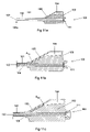
- FIGS. 1 to 7 illustrate the design and operation of a first embodiment of the invention
- FIG. 8 illustrates a second embodiment of the invention
- FIG. 9 illustrates a third embodiment of the invention.
- FIG. 10 illustrates a fourth embodiment of the invention
- FIGS. 11 a to 11 e illustrate the design and operation of a fifth embodiment of the invention.
- FIG. 12 illustrates a sixth embodiment of the invention
- FIG. 13 illustrates a seventh embodiment of the invention.
- the invention provides an improved technique to detect the position of an introducer assembly in a blood vessel or tissue.
- an embodiment of the invention electronically (or optically) quantitatively measures the presence, amount (for example, absolute pressure), and/or waveform of blood pressure in the introducer, as opposed to merely visually detecting the presence of blood, as in the prior art.
- a standard bedside blood pressure transducer is connected to the introducer's sidearm and the pressure (in for example mmHg)is displayed on a lab monitor.
- blood pressure is digitally sensed by a thumb or fingers or visually observed.
- the pulsation of blood can be observed as an oscillating blood column or be digitally sensed or displayed by a moving needle or pointer.
- the invention also provides a technique to detect the proper sealing of a puncture in a blood vessel wall.
- this technique after deploying an inner seal, the blood pressure in the tissue immediately outside of the seal is measured. If the puncture is not sealed (if, for example, the inner seal is caught upstream), significant blood pressure will still be indicated, and the seal can then be manipulated and twisted until it is released in the artery and can then be positioned properly.
- the measured pressure waveform is displayed on the monitor to give the surgeon information as to the positioning of the various components relative to one another and relative to the various tissues.
- the pressure waveform can be analyzed electronically to provide the surgeon with further information.
- the invention can serve additional purposes. After deployment of the inner seal in the artery, the device is withdrawn until a resistance is felt. At that point, the inner seal should be seated over the inside of the puncture and the outer seal can then be deployed without risk of being deployed inside the artery. If the inner seal is caught upstream in the artery, without the invention, the surgeon may misinterpret the resistance (when the device is pulled) as an indication that the inner seal is seated over the puncture hole. However, with the invention, the fact that the inner seal is caught upstream in the artery will be detected by reading the pressure on the monitor.
- FIGS. 1 to 14 illustrate various embodiments of the invention. These figures illustrate use of the invention in conjunction with the FEMOSEALTM seal. However, the invention can be used in connection with a wide variety of seals other than the FEMOSEALTM seal.
- FIGS. 1 and 2 illustrate an apparatus 1 having an inner seal 2 , a pusher 3 (with a tip 25 ), and a suture 4 .
- FIG. 3 illustrates a sidearm 9 (with a stopcock in a closed position), an introducer 10 , a hemostatic valve 11 , and a tube 12 , positioned with respect to artery wall 7 in tissue 14 .
- Inner seal 2 performs two distinct functions. First, inner seal 2 seals the puncture in the blood vessel wall. Second, seal 2 holds an outer seal (not shown) in place.
- the above-identified patent application describes these components in further detail.
- FIG. 4 also shows a display 23 to display pressure waveforms to the surgeon.
- the display can be any type of display or monitor.
- An analysis circuit 21 is also provided, which analyses the pressure data to output additional information to the surgeon.
- FIG. 5 shows the inner seal 2 properly seated to a puncture hole.
- transducer 13 senses essentially no normal arterial pressure and no normal pressure waveforms in the tissue 14 ′ immediately outside of the seal.
- the surgeon can confirm that seal 2 is properly seated (that is, that seal 2 mates with the inner wall of the blood vessel in a leak tight fashion). If the sealing were incomplete, a pressure will still be present, but at a lower level, and this indicates the need for harder tightening of the sealing elements.
- the technique shown in FIG. 5 can also be employed to determine whether a seal which seals the puncture from outside the artery is properly positioned.
- FIG. 6 shows seal 2 caught in an upstream branch.
- blood pressure will still be indicated on the display (or monitor), and the seal can then be manipulated and twisted until it is released from the branch and can be seated to the puncture hole.
- the surgeon pulls on sutures 4 the surgeon feels significant resistance and may wrongly believe (without use of the invention) that the seal is properly seated.
- FIG. 7 illustrates examples of waveforms displayed on display 23 .
- Waveform 20 corresponds to the situation when an inner seal is inside the blood vessel and an introducer is positioned as shown in FIG. 4 .
- Waveform 22 corresponds to the situation when the inner seal is positioned as shown in FIG. 5 .
- Waveform 21 corresponds to the situation when the sealing is incomplete.
- FIGS. 8 , 9 , and 10 show second, third, and fourth embodiments of the invention.
- FIG. 8 shows an introducer 10 with a core pin 27 inserted.
- the core pin (a dilator) has one channel 26 (or an axial grove) that communicates a tip opening 26 a with the sidearm 9 .
- the detector 30 is similar to transducer 13 and equipped with a display or audible indication of pressure, for example, an audible indication which varies as pressure varies.
- the detector is connectable, or adaptable, to the sidearm 9 .
- FIG. 9 shows an introducer 33 with a core pin 27 A inserted.
- the introducer has a sidehole 28 at a desired distance from the tip.
- the introducer can be positioned in a single operation by monitoring the pressure on detector 30 , and the length that the introducer protrudes into the vessel is defined by the length from the sidehole to the introducer tip.
- the introducer can be positioned by positioning hole 28 just inside the vessel wall by monitoring pressure on detector 30 .
- the technique shown in FIG. 10 is similar to the technique shown in FIG. 5 , except that in FIG. 10 , proper sealing of the inner seal 2 is determined by observing the absence of blood flow from an output port (or drip channel) 32 in an attachment 31 .
- a hemostatic valve 111 is provided near the proximal end of lumen 102 .
- an opening 107 is provided at the very distal end 101 a of lumen 102 .
- the opening is (in this embodiment) circular in shape, has a diameter equal to the diameter of lumen 102 , and lies in a plane perpendicular to the longitudinal axis of the lumen 102 .
- the opening 107 allows use of a removable member (such as core pins 27 and 27 A in FIGS. 8 and 9 ) which passes through the lumen 102 .
- a removable member can, for example, provide for sensing of pressure at a specific location (based on the geometry of the removable member) as well as allows for unobstructed passage of other instruments through lumen 102 .
- FIGS. 11 b - d The proximal portion of the pressure transducer 100 according to FIG. 11 a is shown in FIGS. 11 b - d , and the function of the pressure transducer 100 will now be described with reference to these figures, starting with FIG. 11 b .
- Blood 106 entering into the lumen 102 of the introducer sheath 101 will be diverted into the canal 105 . Due to the counter-pressure created by the air trapped in the chamber 104 , the blood meniscus will only reach into the canal 105 to an extent defined by this counter-pressure, i.e. until the blood pressure in an artery (or a vein or another body lumen) is in equilibrium with the air pressure in the chamber 104 .
- the blood meniscus in the canal 105 will oscillate or pulsate (herein the terms oscillate and pulsate have the same meaning) between two extremes, which in FIG. 11 b and FIG. 11 c have been indicated by A min and A max , respectively.
- a min and A max correspond preferably to the diastolic and systolic blood pressures, respectively.
- a normal value for the diastolic pressure may be within 70-80 mmHg, whereas the systolic pressure may be as high as 140 mmHg.
- the total volume of the introducer lumen 102 , the canal 105 , and the chamber 104 is dimensioned such that the expected minimal diastolic pressure corresponds to a blood meniscus at A min , and the expected maximal systolic pressure corresponds to a blood meniscus at A max .
- the exact positioning of A min and A max is, however, not very crucial, the important thing being that an oscillating blood meniscus can be observed.
- At least the portion of the housing 103 that covers the canal 105 should be made from a transparent material, such as plastic or glass or the like.
- the rest of the housing 103 can be made from a transparent material, to thereby make it possible to observe when and if blood enters into the lumen 102 of the introducer sheath 101 and/or into the chamber 104 .
- FIG. 11 d a situation is illustrated wherein the blood pressure is so high that blood 106 enters into the chamber 104 . Even in this situation, the counter-pressure caused by the air trapped inside the chamber 104 will, however, eventually create equilibrium and the blood meniscus will oscillate between two extremes. In other words, the pressure transducer 100 will also for extremely high blood pressures provide an oscillating blood column, which indicates that the distal end of the introducer sheath 101 is within an artery.
- the occurrence of blood within the chamber 104 will in itself guarantee that the distal end of the introducer sheath 101 is within an artery, because the blood pressure within an artery is the highest blood pressure in a human body and consequently all other blood pressures will be considerably lower, in practice so low that blood from other vessels or lumens will not be able to extend all the way into the chamber 104 .
- FIG. 11 e shows a pressure transducer 100 ′, which schematically illustrates a way to eliminate this non-linearity.
- the pressure transducer 100 ′ of FIG. 11 e is identical with the pressure transducer 100 shown in FIGS.
- a canal 105 ′ which connects a lumen 102 ′ of an introducer sheath 101 ′ to a chamber 104 ′, has been given a shape which tapers towards the chamber 104 ′.
- a given change in blood pressure which always corresponds to a given change in the volume of the air trapped in front of the blood meniscus in the canal 105 ′—can be made to correspond to the same positional change of the blood meniscus throughout the canal 105 ′.
- a linear display of the blood pressure has thereby been accomplished.
- blood 106 entering into the introducer sheath 101 completely fills the lumen 102 of the introducer 101 , and fills in particular the portion of the introducer lumen 102 which is proximal of the entrance to the canal 105 . If, however, air trapped in the proximal portion of the introducer sheath 101 would create a counter-pressure that is high enough to prevent the blood from reaching into the canal 105 , no oscillating blood meniscus would, of course, be seen in the canal 105 . To ensure that blood can reach to the beginning of the canal independently of the actual blood pressure, a small gas (air) leak can be provided at, for example, the proximal end of the introducer sheath 101 .
- air air
- FIG. 12 discloses a further embodiment of a pressure transducer 200 according to the present invention.
- the pressure transducer 200 comprises an introducer sheath 201 having a lumen 202 .
- a housing 203 is provided at the proximal end of the introducer 201 .
- the housing 203 surrounds a proximal portion of the introducer sheath 201 , and encloses a first chamber 204 , in which gas (air) is confined.
- the first chamber 204 is in fluid communication with the lumen 202 of the introducer 201 by a capillary or canal 205 .
- the housing 203 further encloses a second chamber 206 , in which the proximal end of the introducer lumen 202 ends.
- the volume of the first chamber has in FIG. 12 been designated by V 1
- the volume of the second chamber is designated by V 2 .
- FIG. 13 Another embodiment of a pressure transducer 300 according to the present invention is shown in FIG. 13 .
- the pressure transducer 300 comprises an introducer sheath 301 having a lumen 302 , and a housing 303 .
- the housing 303 which is provided at the proximal portion of the introducer sheath 301 , encloses a chamber 304 , which is in fluid communication with the introducer lumen 302 by a canal 305 .
- a resilient membrane 306 constitutes an outer wall of the chamber 304 .
- the distal end of the introducer sheath 301 is introduced into an artery, which causes blood to flow into the chamber 304 .
- the blood inside the chamber 304 will then pulsate between the diastolic and systolic pressures prevailing inside the artery. This pulsation will be transferred to the resilient membrane 306 ; and by placing a finger 307 on the resilient membrane 306 , an operator can sense the pulsation and thereby determine that the distal end of the introducer sheath 301 actually is inside an artery (and not in a vein or other bodily lumen or tissue).
- a pressure transducer 400 comprises an introducer sheath 401 having a lumen 402 , a housing 403 , which encloses a chamber 404 , which is in fluid communication with the lumen 402 by a canal 405 , and a pointer 406 .
- a resilient membrane 407 is arranged inside the chamber 404 , and is by a linkage 408 connected to the pointer 406 . Blood having entered into the chamber 404 will hereby pulsate against the resilient membrane 407 , and the linkage 408 will then transfer the pulsation to the pointer 406 , to thereby provide an operator with a visual indication of the pulsating blood pressure.
- pressure transducers illustrated in conjunction with FIG. 11 to FIG. 14 represent closed systems, which means that there is no spillage of blood out from the pressure transducer. This is an advantage over devices that utilize a blood flow out from the device in order to indicate that an introducer is located inside an artery.
- the pressure transducers according to FIGS. 11-14 can be used for locating an artery as well as for verifying that an inner seal is correctly positioned against an artery wall, and for details regarding these different aspects of the invention, the reader is referred to the discussion above.
Abstract
Description
Claims (17)
Priority Applications (1)
| Application Number | Priority Date | Filing Date | Title |
|---|---|---|---|
| US10/798,784 US7285097B2 (en) | 2001-01-12 | 2004-03-12 | Technique to confirm correct positioning with respect to arterial wall |
Applications Claiming Priority (4)
| Application Number | Priority Date | Filing Date | Title |
|---|---|---|---|
| US26089501P | 2001-01-12 | 2001-01-12 | |
| US10/042,247 US6682489B2 (en) | 2001-01-12 | 2002-01-11 | Technique to confirm correct positioning of arterial wall sealing device |
| US10/704,556 US7044916B2 (en) | 2001-01-12 | 2003-11-12 | Technique to confirm correct positioning of arterial wall sealing device |
| US10/798,784 US7285097B2 (en) | 2001-01-12 | 2004-03-12 | Technique to confirm correct positioning with respect to arterial wall |
Related Parent Applications (1)
| Application Number | Title | Priority Date | Filing Date |
|---|---|---|---|
| US10/704,556 Continuation-In-Part US7044916B2 (en) | 2001-01-12 | 2003-11-12 | Technique to confirm correct positioning of arterial wall sealing device |
Publications (2)
| Publication Number | Publication Date |
|---|---|
| US20040243007A1 US20040243007A1 (en) | 2004-12-02 |
| US7285097B2 true US7285097B2 (en) | 2007-10-23 |
Family
ID=32912880
Family Applications (1)
| Application Number | Title | Priority Date | Filing Date |
|---|---|---|---|
| US10/798,784 Expired - Fee Related US7285097B2 (en) | 2001-01-12 | 2004-03-12 | Technique to confirm correct positioning with respect to arterial wall |
Country Status (1)
| Country | Link |
|---|---|
| US (1) | US7285097B2 (en) |
Cited By (26)
| Publication number | Priority date | Publication date | Assignee | Title |
|---|---|---|---|---|
| US20100121358A1 (en) * | 2008-06-06 | 2010-05-13 | Vital Access Corporation | Tissue management methods, apparatus, and systems |
| US20100191166A1 (en) * | 2009-01-29 | 2010-07-29 | Vital Access Corporation | Subcutaneous vascular access ports and related systems and methods |
| US20110178413A1 (en) * | 2010-01-19 | 2011-07-21 | Schmitt Joseph M | Intravascular optical coherence tomography system with pressure monitoring interface and accessories |
| US20110213309A1 (en) * | 2009-01-29 | 2011-09-01 | Vital Access Corporation | Vascular access ports and related methods |
| US20110213414A1 (en) * | 2008-02-15 | 2011-09-01 | Mcguckin Jr James F | Vascular hole closure device |
| US8961420B2 (en) | 2010-04-01 | 2015-02-24 | Siemens Medical Solutions Usa, Inc. | System for cardiac condition detection and characterization |
| US8968361B2 (en) | 2008-02-15 | 2015-03-03 | Rex Medical, L.P. | Vascular hole closure device |
| US9226738B2 (en) | 2008-02-15 | 2016-01-05 | Rex Medical, L.P. | Vascular hole closure delivery device |
| US9282955B2 (en) | 2009-02-20 | 2016-03-15 | Boston Scientific Scimed, Inc. | Tissue puncture closure device |
| US9295458B2 (en) | 2008-02-15 | 2016-03-29 | Rex Medical, L.P. | Vascular hole closure delivery device |
| US9301740B2 (en) | 2010-02-11 | 2016-04-05 | Boston Scientific Scimed, Inc. | Automatic vascular closure deployment devices and methods |
| US9463005B2 (en) | 2008-02-15 | 2016-10-11 | Rex Medical, L.P. | Vascular hole closure device |
| US9554785B2 (en) | 2012-12-21 | 2017-01-31 | Essential Medical, Inc. | Vascular locating systems and methods of use |
| US9782155B2 (en) | 2008-02-15 | 2017-10-10 | Rex Medical, L.P. | Vascular hole closure device |
| US9968345B2 (en) | 1999-09-13 | 2018-05-15 | Rex Medical, L.P. | Vascular hole closure device |
| US10154835B2 (en) | 2013-05-09 | 2018-12-18 | Essential Medical, Inc. | Vascular closure device with conforming plug member |
| US10307070B2 (en) | 2014-04-04 | 2019-06-04 | St. Jude Medical Coordination Center Bvba | Intravascular pressure and flow data diagnostic systems, devices, and methods |
| US10383611B2 (en) | 2011-10-25 | 2019-08-20 | Essential Medical, Inc. | Instrument and methods for surgically closing percutaneous punctures |
| US10648918B2 (en) | 2011-08-03 | 2020-05-12 | Lightlab Imaging, Inc. | Systems, methods and apparatus for determining a fractional flow reserve (FFR) based on the minimum lumen area (MLA) and the constant |
| US11197952B2 (en) | 2009-01-29 | 2021-12-14 | Advent Access Pte. Ltd. | Vascular access ports and related methods |
| US11241154B2 (en) | 2011-05-31 | 2022-02-08 | Lightlab Imaging, Inc. | Multimodal imaging system, apparatus, and methods |
| US11350919B2 (en) | 2019-02-19 | 2022-06-07 | Teleflex Life Sciences Limited | Puncture locating system with blood pulsation indicator |
| US11364024B2 (en) | 2013-12-23 | 2022-06-21 | Teleflex Life Sciences Limited | Vascular closure device |
| US11419592B2 (en) | 2013-03-15 | 2022-08-23 | Teleflex Life Sciences Limited | Vascular closure devices and methods of use |
| US11504105B2 (en) | 2019-01-25 | 2022-11-22 | Rex Medical L.P. | Vascular hole closure device |
| US11576663B2 (en) | 2015-06-26 | 2023-02-14 | Teleflex Life Sciences Limited | Vascular closure device with removable guide member |
Families Citing this family (11)
| Publication number | Priority date | Publication date | Assignee | Title |
|---|---|---|---|---|
| US6964661B2 (en) * | 2003-04-02 | 2005-11-15 | Boston Scientific Scimed, Inc. | Endovenous ablation mechanism with feedback control |
| US8442620B2 (en) * | 2008-10-01 | 2013-05-14 | Pacesetter, Inc. | Implantable lead/electrode delivery measurement and feedback system |
| US8375553B2 (en) | 2009-02-20 | 2013-02-19 | Boston Scientific Scimed, Inc. | Locking element for vascular closure device |
| US8292918B2 (en) | 2009-02-20 | 2012-10-23 | Boston Scientific Scimed, Inc. | Composite plug for arteriotomy closure and method of use |
| US8317824B2 (en) | 2009-02-20 | 2012-11-27 | Boston Scientific Scimed, Inc. | Tissue puncture closure device |
| US8052914B2 (en) | 2009-02-20 | 2011-11-08 | Boston Scientific Scimed, Inc. | Modified plug for arteriotomy closure |
| US9913634B2 (en) | 2009-02-20 | 2018-03-13 | Boston Scientific Scimed, Inc. | Locking element for vascular closure device |
| US20110071479A1 (en) * | 2009-09-23 | 2011-03-24 | Tyco Healthcare Group Lp | Blood Vessel Access Instrument with Vessel Entry Indicator |
| US20120022562A1 (en) * | 2010-07-23 | 2012-01-26 | Boston Scientific Scimed, Inc. | Device to detect internal bleeding |
| US8597340B2 (en) | 2010-09-17 | 2013-12-03 | Boston Scientific Scimed, Inc. | Torque mechanism actuated bioabsorbable vascular closure device |
| US8758402B2 (en) | 2010-12-17 | 2014-06-24 | Boston Scientific Scimed, Inc. | Tissue puncture closure device |
Citations (20)
| Publication number | Priority date | Publication date | Assignee | Title |
|---|---|---|---|---|
| US5290231A (en) * | 1990-06-13 | 1994-03-01 | Dlp, Inc. | Auto-inflating catheter cuff |
| US5395330A (en) * | 1990-06-13 | 1995-03-07 | Dlp, Inc. | Auto-inflating catheter cuff |
| US5431639A (en) | 1993-08-12 | 1995-07-11 | Boston Scientific Corporation | Treating wounds caused by medical procedures |
| US5456694A (en) * | 1994-05-13 | 1995-10-10 | Stentco, Inc. | Device for delivering and deploying intraluminal devices |
| US5613974A (en) | 1992-12-10 | 1997-03-25 | Perclose, Inc. | Apparatus and method for vascular closure |
| WO1998007372A2 (en) | 1996-08-21 | 1998-02-26 | Hemodynamics, Inc. | Surface opening adhesive sealer |
| WO1998019605A1 (en) | 1996-11-05 | 1998-05-14 | Li, Shu-Tung | Soft tissue closure systems |
| US5766151A (en) * | 1991-07-16 | 1998-06-16 | Heartport, Inc. | Endovascular system for arresting the heart |
| US5766206A (en) * | 1993-09-03 | 1998-06-16 | Cordis Corporation | Method and device for hemostatic treatment following catheterization |
| US5795325A (en) * | 1991-07-16 | 1998-08-18 | Heartport, Inc. | Methods and apparatus for anchoring an occluding member |
| WO1998040016A2 (en) | 1997-03-12 | 1998-09-17 | Advanced Closure Systems, Inc. | Universal introducer |
| US5855559A (en) | 1997-02-14 | 1999-01-05 | Tricardia, Inc. | Hemostatic agent delivery device having built-in pressure sensor |
| US6007563A (en) | 1991-11-08 | 1999-12-28 | Kensey Nash Corporation | Method of deploying percutaneous puncture closure |
| US6090130A (en) | 1991-11-08 | 2000-07-18 | Kensey Nash Corporation | Hemostatic puncture closure system including blood vessel locator and method of use |
| US6159178A (en) * | 1998-01-23 | 2000-12-12 | Heartport, Inc. | Methods and devices for occluding the ascending aorta and maintaining circulation of oxygenated blood in the patient when the patient's heart is arrested |
| US6179827B1 (en) * | 1998-03-16 | 2001-01-30 | Chase Medical | Catheter having integral expandable/collapsible lumen |
| US6193670B1 (en) | 1997-02-14 | 2001-02-27 | Tricardia, Llc | Hemostatic agent delivery device having built-in pressure sensor |
| US6302898B1 (en) * | 1994-06-24 | 2001-10-16 | Advanced Closure Systems, Inc. | Devices for sealing punctures in body vessels |
| US6508828B1 (en) | 2000-11-03 | 2003-01-21 | Radi Medical Systems Ab | Sealing device and wound closure device |
| US6682489B2 (en) * | 2001-01-12 | 2004-01-27 | Radi Medical Systems Ab | Technique to confirm correct positioning of arterial wall sealing device |
-
2004
- 2004-03-12 US US10/798,784 patent/US7285097B2/en not_active Expired - Fee Related
Patent Citations (29)
| Publication number | Priority date | Publication date | Assignee | Title |
|---|---|---|---|---|
| US5395330A (en) * | 1990-06-13 | 1995-03-07 | Dlp, Inc. | Auto-inflating catheter cuff |
| US5290231A (en) * | 1990-06-13 | 1994-03-01 | Dlp, Inc. | Auto-inflating catheter cuff |
| US5814016A (en) * | 1991-07-16 | 1998-09-29 | Heartport, Inc. | Endovascular system for arresting the heart |
| US5766151A (en) * | 1991-07-16 | 1998-06-16 | Heartport, Inc. | Endovascular system for arresting the heart |
| US5795325A (en) * | 1991-07-16 | 1998-08-18 | Heartport, Inc. | Methods and apparatus for anchoring an occluding member |
| US6090130A (en) | 1991-11-08 | 2000-07-18 | Kensey Nash Corporation | Hemostatic puncture closure system including blood vessel locator and method of use |
| US6007563A (en) | 1991-11-08 | 1999-12-28 | Kensey Nash Corporation | Method of deploying percutaneous puncture closure |
| US6350274B1 (en) * | 1992-05-11 | 2002-02-26 | Regen Biologics, Inc. | Soft tissue closure systems |
| US5613974A (en) | 1992-12-10 | 1997-03-25 | Perclose, Inc. | Apparatus and method for vascular closure |
| US5431639A (en) | 1993-08-12 | 1995-07-11 | Boston Scientific Corporation | Treating wounds caused by medical procedures |
| US5766206A (en) * | 1993-09-03 | 1998-06-16 | Cordis Corporation | Method and device for hemostatic treatment following catheterization |
| US5456694A (en) * | 1994-05-13 | 1995-10-10 | Stentco, Inc. | Device for delivering and deploying intraluminal devices |
| US5569296A (en) * | 1994-05-13 | 1996-10-29 | Stentco, Inc. | Method for delivering and deploying intraluminal devices |
| US6302898B1 (en) * | 1994-06-24 | 2001-10-16 | Advanced Closure Systems, Inc. | Devices for sealing punctures in body vessels |
| WO1998007372A2 (en) | 1996-08-21 | 1998-02-26 | Hemodynamics, Inc. | Surface opening adhesive sealer |
| WO1998019605A1 (en) | 1996-11-05 | 1998-05-14 | Li, Shu-Tung | Soft tissue closure systems |
| US6193670B1 (en) | 1997-02-14 | 2001-02-27 | Tricardia, Llc | Hemostatic agent delivery device having built-in pressure sensor |
| US5855559A (en) | 1997-02-14 | 1999-01-05 | Tricardia, Inc. | Hemostatic agent delivery device having built-in pressure sensor |
| US6733515B1 (en) * | 1997-03-12 | 2004-05-11 | Neomend, Inc. | Universal introducer |
| WO1998040016A2 (en) | 1997-03-12 | 1998-09-17 | Advanced Closure Systems, Inc. | Universal introducer |
| US6350252B2 (en) * | 1998-01-23 | 2002-02-26 | Heartport, Inc. | Methods and devices for occluding the ascending aorta and maintaining circulation of oxygenated blood in the patient when the patient's heart is arrested |
| US6159178A (en) * | 1998-01-23 | 2000-12-12 | Heartport, Inc. | Methods and devices for occluding the ascending aorta and maintaining circulation of oxygenated blood in the patient when the patient's heart is arrested |
| US6589206B1 (en) * | 1998-01-23 | 2003-07-08 | Heartport, Inc. | Methods and devices for occluding the ascending aorta and maintaining circulation of oxygenated blood in the patient when the patient's heart is arrested |
| US6592547B2 (en) * | 1998-01-23 | 2003-07-15 | Grimes Kevin V | Methods and devices for occluding the ascending aorta and maintaining circulation of oxygenated blood in the patient when the patient's heart is arrested |
| US6179827B1 (en) * | 1998-03-16 | 2001-01-30 | Chase Medical | Catheter having integral expandable/collapsible lumen |
| US6508828B1 (en) | 2000-11-03 | 2003-01-21 | Radi Medical Systems Ab | Sealing device and wound closure device |
| US6682489B2 (en) * | 2001-01-12 | 2004-01-27 | Radi Medical Systems Ab | Technique to confirm correct positioning of arterial wall sealing device |
| US20040098046A1 (en) | 2001-01-12 | 2004-05-20 | Radi Medical Systems Ab | Technique to confirm correct positioning of arterial wall sealing device |
| US20040172059A1 (en) | 2001-01-12 | 2004-09-02 | Radi Medical Systems Ab | Technique to confirm correct positioning of arterial wall sealing device |
Non-Patent Citations (1)
| Title |
|---|
| U.S. Appl. No. 10/756,765, filed Jan. 14, 2004, Egnelov et al. |
Cited By (65)
| Publication number | Priority date | Publication date | Assignee | Title |
|---|---|---|---|---|
| US9968345B2 (en) | 1999-09-13 | 2018-05-15 | Rex Medical, L.P. | Vascular hole closure device |
| US10108646B2 (en) | 2008-02-15 | 2018-10-23 | Rex Medical, L.P. | Vascular hole closure delivery device |
| US9339261B2 (en) | 2008-02-15 | 2016-05-17 | Rex Medical, L.P. | Vascular hole closure delivery device |
| US11123059B2 (en) | 2008-02-15 | 2021-09-21 | Rex Medical, L.P. | Vascular hole closure delivery device |
| US11064986B2 (en) | 2008-02-15 | 2021-07-20 | Rex Medical, L.P. | Vascular hole closure device |
| US20110213414A1 (en) * | 2008-02-15 | 2011-09-01 | Mcguckin Jr James F | Vascular hole closure device |
| US11020104B2 (en) | 2008-02-15 | 2021-06-01 | Rex Medical L.P. | Vascular hole closure delivery device |
| US10390807B2 (en) | 2008-02-15 | 2019-08-27 | Rex Medical, L.P. | Vascular hole closure device |
| US8920462B2 (en) | 2008-02-15 | 2014-12-30 | Rex Medical, L.P. | Vascular hole closure device |
| US10390808B2 (en) | 2008-02-15 | 2019-08-27 | Rex Medical, L.P | Vascular hole closure device |
| US10342524B2 (en) | 2008-02-15 | 2019-07-09 | Rex Medical, L.P. | Vascular hole closure device |
| US9924930B2 (en) | 2008-02-15 | 2018-03-27 | Rex Medical, L.P. | Vascular hole closure device |
| US9463005B2 (en) | 2008-02-15 | 2016-10-11 | Rex Medical, L.P. | Vascular hole closure device |
| US9782155B2 (en) | 2008-02-15 | 2017-10-10 | Rex Medical, L.P. | Vascular hole closure device |
| US10098621B2 (en) | 2008-02-15 | 2018-10-16 | Rex Medical, Lp. | Vascular hole closure delivery device |
| US8968361B2 (en) | 2008-02-15 | 2015-03-03 | Rex Medical, L.P. | Vascular hole closure device |
| US9943300B2 (en) | 2008-02-15 | 2018-04-17 | Rex Medical, L.P. | Vascular hole closure device |
| US10004486B2 (en) | 2008-02-15 | 2018-06-26 | Rex Medical, L.P. | Vascular hole closure delivery device |
| US9226738B2 (en) | 2008-02-15 | 2016-01-05 | Rex Medical, L.P. | Vascular hole closure delivery device |
| US11369354B2 (en) | 2008-02-15 | 2022-06-28 | Rex Medical L.P. | Vascular hole closure delivery device |
| US9295458B2 (en) | 2008-02-15 | 2016-03-29 | Rex Medical, L.P. | Vascular hole closure delivery device |
| US20100121358A1 (en) * | 2008-06-06 | 2010-05-13 | Vital Access Corporation | Tissue management methods, apparatus, and systems |
| US9039717B2 (en) | 2008-06-06 | 2015-05-26 | Vital Access Corporation | Tissue management apparatus for vascular access |
| US11134950B2 (en) | 2008-06-06 | 2021-10-05 | Advent Access Pte. Ltd. | Methods of attaching an implant to a vessel |
| US8409228B2 (en) | 2008-06-06 | 2013-04-02 | Duane D. Blatter | Tissue management methods, apparatus, and systems |
| US9072880B2 (en) | 2009-01-29 | 2015-07-07 | Vital Access Corporation | Subcutaneous vascular access ports and related systems and methods |
| US10226564B2 (en) | 2009-01-29 | 2019-03-12 | Advent Access Pte. Ltd. | Vascular access port systems and methods |
| US20100191166A1 (en) * | 2009-01-29 | 2010-07-29 | Vital Access Corporation | Subcutaneous vascular access ports and related systems and methods |
| US10894120B2 (en) | 2009-01-29 | 2021-01-19 | Advent Access Pte. Ltd. | Vascular access port systems and methods |
| US10773010B2 (en) | 2009-01-29 | 2020-09-15 | Advent Access Pte. Ltd. | Subcutaneous vascular access ports and related systems and methods |
| US9968726B2 (en) | 2009-01-29 | 2018-05-15 | Df Vital Holdings, Llc | Methods for implanting subcutaneous vascular access ports |
| US9179901B2 (en) | 2009-01-29 | 2015-11-10 | Vital Access Corporation | Vascular access ports and related methods |
| US9033931B2 (en) | 2009-01-29 | 2015-05-19 | Vital Access Corporation | Subcutaneous vascular access ports having attachment features |
| US9603988B2 (en) | 2009-01-29 | 2017-03-28 | Vital Access Corporation | Subcutaneous vascular access ports |
| US20110213309A1 (en) * | 2009-01-29 | 2011-09-01 | Vital Access Corporation | Vascular access ports and related methods |
| US11197952B2 (en) | 2009-01-29 | 2021-12-14 | Advent Access Pte. Ltd. | Vascular access ports and related methods |
| US8337464B2 (en) | 2009-01-29 | 2012-12-25 | Vital Access Corporation | Vascular access ports and related methods |
| US10265458B2 (en) | 2009-01-29 | 2019-04-23 | Advent Access Pte. Ltd. | Vascular access ports and related methods |
| US20100191191A1 (en) * | 2009-01-29 | 2010-07-29 | Vital Access Corporation | Subcutaneous vascular access ports and related systems, methods, and implantation features |
| US8337465B2 (en) | 2009-01-29 | 2012-12-25 | Vital Access Corporation | Subcutaneous vascular access ports and related systems, methods, and implantation features |
| US9282955B2 (en) | 2009-02-20 | 2016-03-15 | Boston Scientific Scimed, Inc. | Tissue puncture closure device |
| US20110178413A1 (en) * | 2010-01-19 | 2011-07-21 | Schmitt Joseph M | Intravascular optical coherence tomography system with pressure monitoring interface and accessories |
| US8478384B2 (en) | 2010-01-19 | 2013-07-02 | Lightlab Imaging, Inc. | Intravascular optical coherence tomography system with pressure monitoring interface and accessories |
| US8676299B2 (en) | 2010-01-19 | 2014-03-18 | Lightlab Imaging, Inc. | Method of determining pressure in a vessel as measured by an optical pressure transducer in an optical coherence tomography system |
| US11058308B2 (en) | 2010-01-19 | 2021-07-13 | Lightlab Imaging, Inc. | Intravascular pressure sensing devices and methods |
| US9301740B2 (en) | 2010-02-11 | 2016-04-05 | Boston Scientific Scimed, Inc. | Automatic vascular closure deployment devices and methods |
| US8961420B2 (en) | 2010-04-01 | 2015-02-24 | Siemens Medical Solutions Usa, Inc. | System for cardiac condition detection and characterization |
| US11241154B2 (en) | 2011-05-31 | 2022-02-08 | Lightlab Imaging, Inc. | Multimodal imaging system, apparatus, and methods |
| US10648918B2 (en) | 2011-08-03 | 2020-05-12 | Lightlab Imaging, Inc. | Systems, methods and apparatus for determining a fractional flow reserve (FFR) based on the minimum lumen area (MLA) and the constant |
| US11589855B2 (en) | 2011-10-25 | 2023-02-28 | Teleflex Life Sciences Limited | Instrument and methods for surgically closing percutaneous punctures |
| US10485524B2 (en) | 2011-10-25 | 2019-11-26 | Essential Medical, Inc. | Instrument and methods for surgically closing percutaneous punctures |
| US10383611B2 (en) | 2011-10-25 | 2019-08-20 | Essential Medical, Inc. | Instrument and methods for surgically closing percutaneous punctures |
| US9554785B2 (en) | 2012-12-21 | 2017-01-31 | Essential Medical, Inc. | Vascular locating systems and methods of use |
| US10182804B2 (en) | 2012-12-21 | 2019-01-22 | Essential Medical, Inc. | Vascular locating systems and methods of use |
| US10835225B2 (en) | 2012-12-21 | 2020-11-17 | Arrow International, Inc. | Vascular locating systems and methods of use |
| US11759191B2 (en) | 2012-12-21 | 2023-09-19 | Teleflex Life Sciences Limited | Vascular locating systems and methods of use |
| US11419592B2 (en) | 2013-03-15 | 2022-08-23 | Teleflex Life Sciences Limited | Vascular closure devices and methods of use |
| US10154835B2 (en) | 2013-05-09 | 2018-12-18 | Essential Medical, Inc. | Vascular closure device with conforming plug member |
| US11364024B2 (en) | 2013-12-23 | 2022-06-21 | Teleflex Life Sciences Limited | Vascular closure device |
| US11779320B2 (en) | 2013-12-23 | 2023-10-10 | Teleflex Life Sciences Limited | Vascular closure device |
| US11559218B2 (en) | 2014-04-04 | 2023-01-24 | St. Jude Medical Coordination Center Bvba | Intravascular pressure and flow data diagnostic systems, devices, and methods |
| US10307070B2 (en) | 2014-04-04 | 2019-06-04 | St. Jude Medical Coordination Center Bvba | Intravascular pressure and flow data diagnostic systems, devices, and methods |
| US11576663B2 (en) | 2015-06-26 | 2023-02-14 | Teleflex Life Sciences Limited | Vascular closure device with removable guide member |
| US11504105B2 (en) | 2019-01-25 | 2022-11-22 | Rex Medical L.P. | Vascular hole closure device |
| US11350919B2 (en) | 2019-02-19 | 2022-06-07 | Teleflex Life Sciences Limited | Puncture locating system with blood pulsation indicator |
Also Published As
| Publication number | Publication date |
|---|---|
| US20040243007A1 (en) | 2004-12-02 |
Similar Documents
| Publication | Publication Date | Title |
|---|---|---|
| US7285097B2 (en) | Technique to confirm correct positioning with respect to arterial wall | |
| US6682489B2 (en) | Technique to confirm correct positioning of arterial wall sealing device | |
| US5935083A (en) | Device for body fluid pressure measurement | |
| US20070123781A1 (en) | Surgical anastomosis leak detection system | |
| US10463838B2 (en) | Vascular access methods and devices | |
| US6224585B1 (en) | Catheter system | |
| EP0664687B2 (en) | Vessel position locating device | |
| JP2661799B2 (en) | Surgical depth measuring instrument and method | |
| AU697232B2 (en) | In situ calibration system for sensors located in a physiologic line | |
| US4901731A (en) | Single sensor pressure differential device | |
| US4384470A (en) | Method and apparatus for testing blood vessel catheters | |
| US20150025379A1 (en) | Diagnostic catheter using a vacuum for tissue positioning | |
| JP2004216156A (en) | Device for visually indicating blood pressure | |
| CA2566631A1 (en) | Locator and delivery device and method of use | |
| US3720201A (en) | Disposable body fluid pressure monitor | |
| US5687718A (en) | Device for continuously detecting blood parameters | |
| EP2598015B1 (en) | Endoscopic pressure detection assembly | |
| WO2012083227A1 (en) | Tissue access, pressure monitoring | |
| US3890842A (en) | Disposable manometer | |
| EP3838119B1 (en) | System for automatically detecting a clinically relevant leak | |
| JPH0448162Y2 (en) |
Legal Events
| Date | Code | Title | Description |
|---|---|---|---|
| AS | Assignment |
Owner name: RADI MEDICAL SYSTEMS AB, SWEDEN Free format text: ASSIGNMENT OF ASSIGNORS INTEREST;ASSIGNORS:TENERZ, LARS;AKERFELDT, DAN;REEL/FRAME:015611/0492 Effective date: 20040414 |
|
| STCF | Information on status: patent grant |
Free format text: PATENTED CASE |
|
| FEPP | Fee payment procedure |
Free format text: PAYOR NUMBER ASSIGNED (ORIGINAL EVENT CODE: ASPN); ENTITY STATUS OF PATENT OWNER: LARGE ENTITY |
|
| CC | Certificate of correction | ||
| CC | Certificate of correction | ||
| FEPP | Fee payment procedure |
Free format text: PAT HOLDER NO LONGER CLAIMS SMALL ENTITY STATUS, ENTITY STATUS SET TO UNDISCOUNTED (ORIGINAL EVENT CODE: STOL); ENTITY STATUS OF PATENT OWNER: LARGE ENTITY |
|
| FEPP | Fee payment procedure |
Free format text: PAYOR NUMBER ASSIGNED (ORIGINAL EVENT CODE: ASPN); ENTITY STATUS OF PATENT OWNER: LARGE ENTITY Free format text: PAYER NUMBER DE-ASSIGNED (ORIGINAL EVENT CODE: RMPN); ENTITY STATUS OF PATENT OWNER: LARGE ENTITY |
|
| FPAY | Fee payment |
Year of fee payment: 4 |
|
| AS | Assignment |
Owner name: ST. JUDE MEDICAL SYSTEMS AB, SWEDEN Free format text: CHANGE OF NAME;ASSIGNOR:RADI MEDICAL SYSTEMS AB;REEL/FRAME:034796/0153 Effective date: 20091127 |
|
| AS | Assignment |
Owner name: ST. JUDE MEDICAL COORDINATION CENTER BVBA, BELGIUM Free format text: ASSIGNMENT OF ASSIGNORS INTEREST;ASSIGNOR:ST. JUDE MEDICAL SYSTEMS AB;REEL/FRAME:035169/0705 Effective date: 20140923 |
|
| FPAY | Fee payment |
Year of fee payment: 8 |
|
| FEPP | Fee payment procedure |
Free format text: MAINTENANCE FEE REMINDER MAILED (ORIGINAL EVENT CODE: REM.); ENTITY STATUS OF PATENT OWNER: LARGE ENTITY |
|
| LAPS | Lapse for failure to pay maintenance fees |
Free format text: PATENT EXPIRED FOR FAILURE TO PAY MAINTENANCE FEES (ORIGINAL EVENT CODE: EXP.); ENTITY STATUS OF PATENT OWNER: LARGE ENTITY |
|
| STCH | Information on status: patent discontinuation |
Free format text: PATENT EXPIRED DUE TO NONPAYMENT OF MAINTENANCE FEES UNDER 37 CFR 1.362 |
|
| FP | Lapsed due to failure to pay maintenance fee |
Effective date: 20191023 |