US7311107B2 - Navigating and maneuvering of an in vivo vehicle by extracorporeal devices - Google Patents
Navigating and maneuvering of an in vivo vehicle by extracorporeal devices Download PDFInfo
- Publication number
- US7311107B2 US7311107B2 US10/686,535 US68653503A US7311107B2 US 7311107 B2 US7311107 B2 US 7311107B2 US 68653503 A US68653503 A US 68653503A US 7311107 B2 US7311107 B2 US 7311107B2
- Authority
- US
- United States
- Prior art keywords
- vehicle
- magnet
- magnetic field
- magnetic
- detector
- Prior art date
- Legal status (The legal status is an assumption and is not a legal conclusion. Google has not performed a legal analysis and makes no representation as to the accuracy of the status listed.)
- Expired - Fee Related, expires
Links
- 238000001727 in vivo Methods 0.000 title claims abstract description 23
- 230000005291 magnetic effect Effects 0.000 claims abstract description 160
- 230000033001 locomotion Effects 0.000 claims abstract description 53
- 239000013598 vector Substances 0.000 claims abstract description 22
- 230000005672 electromagnetic field Effects 0.000 claims abstract description 17
- 230000001483 mobilizing effect Effects 0.000 claims abstract description 9
- 239000011159 matrix material Substances 0.000 claims description 32
- 239000000523 sample Substances 0.000 claims description 31
- 239000000463 material Substances 0.000 claims description 24
- 238000005259 measurement Methods 0.000 claims description 24
- 230000005355 Hall effect Effects 0.000 claims description 22
- 230000004913 activation Effects 0.000 claims description 15
- 230000008859 change Effects 0.000 claims description 15
- 230000005415 magnetization Effects 0.000 claims description 15
- 235000014676 Phragmites communis Nutrition 0.000 claims description 14
- 239000000696 magnetic material Substances 0.000 claims description 10
- 239000003302 ferromagnetic material Substances 0.000 claims description 9
- 210000000056 organ Anatomy 0.000 claims description 9
- 230000003213 activating effect Effects 0.000 claims description 6
- 238000003384 imaging method Methods 0.000 claims description 6
- 238000001356 surgical procedure Methods 0.000 claims description 6
- 230000003287 optical effect Effects 0.000 claims description 5
- 230000036962 time dependent Effects 0.000 claims description 5
- 238000012544 monitoring process Methods 0.000 claims description 4
- 210000003484 anatomy Anatomy 0.000 claims description 3
- 239000000843 powder Substances 0.000 claims description 3
- 239000003792 electrolyte Substances 0.000 claims description 2
- 230000001960 triggered effect Effects 0.000 claims 9
- 230000001939 inductive effect Effects 0.000 claims 1
- 238000000034 method Methods 0.000 abstract description 24
- 238000001514 detection method Methods 0.000 abstract description 20
- 239000003981 vehicle Substances 0.000 description 197
- 230000006870 function Effects 0.000 description 16
- 238000010586 diagram Methods 0.000 description 12
- 210000001072 colon Anatomy 0.000 description 9
- 238000004364 calculation method Methods 0.000 description 8
- 238000002405 diagnostic procedure Methods 0.000 description 7
- 230000036961 partial effect Effects 0.000 description 7
- 239000003990 capacitor Substances 0.000 description 6
- 238000012377 drug delivery Methods 0.000 description 6
- 230000000694 effects Effects 0.000 description 6
- 230000002572 peristaltic effect Effects 0.000 description 6
- 210000001519 tissue Anatomy 0.000 description 6
- 238000005070 sampling Methods 0.000 description 5
- 239000011248 coating agent Substances 0.000 description 4
- 238000000576 coating method Methods 0.000 description 4
- 230000005684 electric field Effects 0.000 description 4
- 238000001839 endoscopy Methods 0.000 description 4
- 210000001035 gastrointestinal tract Anatomy 0.000 description 4
- 238000010562 histological examination Methods 0.000 description 4
- 239000006249 magnetic particle Substances 0.000 description 4
- 208000035473 Communicable disease Diseases 0.000 description 3
- 208000026350 Inborn Genetic disease Diseases 0.000 description 3
- 210000001015 abdomen Anatomy 0.000 description 3
- 230000003321 amplification Effects 0.000 description 3
- 230000008901 benefit Effects 0.000 description 3
- 150000001875 compounds Chemical class 0.000 description 3
- 239000004020 conductor Substances 0.000 description 3
- 238000012258 culturing Methods 0.000 description 3
- 230000007423 decrease Effects 0.000 description 3
- 230000007547 defect Effects 0.000 description 3
- 238000003745 diagnosis Methods 0.000 description 3
- 230000005294 ferromagnetic effect Effects 0.000 description 3
- 230000004907 flux Effects 0.000 description 3
- 230000002496 gastric effect Effects 0.000 description 3
- 208000016361 genetic disease Diseases 0.000 description 3
- 230000002458 infectious effect Effects 0.000 description 3
- 230000003993 interaction Effects 0.000 description 3
- 239000007788 liquid Substances 0.000 description 3
- 238000002595 magnetic resonance imaging Methods 0.000 description 3
- 230000003211 malignant effect Effects 0.000 description 3
- 238000003199 nucleic acid amplification method Methods 0.000 description 3
- 210000002784 stomach Anatomy 0.000 description 3
- 238000002604 ultrasonography Methods 0.000 description 3
- 230000003187 abdominal effect Effects 0.000 description 2
- 230000006978 adaptation Effects 0.000 description 2
- 229910045601 alloy Inorganic materials 0.000 description 2
- 239000000956 alloy Substances 0.000 description 2
- 210000000621 bronchi Anatomy 0.000 description 2
- KPLQYGBQNPPQGA-UHFFFAOYSA-N cobalt samarium Chemical compound [Co].[Sm] KPLQYGBQNPPQGA-UHFFFAOYSA-N 0.000 description 2
- 238000007796 conventional method Methods 0.000 description 2
- 230000006378 damage Effects 0.000 description 2
- 238000002059 diagnostic imaging Methods 0.000 description 2
- 238000002357 laparoscopic surgery Methods 0.000 description 2
- 210000002429 large intestine Anatomy 0.000 description 2
- 239000002502 liposome Substances 0.000 description 2
- 238000007726 management method Methods 0.000 description 2
- 238000004519 manufacturing process Methods 0.000 description 2
- 238000012986 modification Methods 0.000 description 2
- 230000004048 modification Effects 0.000 description 2
- 210000003205 muscle Anatomy 0.000 description 2
- 239000002245 particle Substances 0.000 description 2
- 229910000938 samarium–cobalt magnet Inorganic materials 0.000 description 2
- 230000011664 signaling Effects 0.000 description 2
- 210000000813 small intestine Anatomy 0.000 description 2
- 239000007787 solid Substances 0.000 description 2
- 239000000126 substance Substances 0.000 description 2
- 230000001225 therapeutic effect Effects 0.000 description 2
- 208000000913 Kidney Calculi Diseases 0.000 description 1
- 206010029148 Nephrolithiasis Diseases 0.000 description 1
- QJVKUMXDEUEQLH-UHFFFAOYSA-N [B].[Fe].[Nd] Chemical compound [B].[Fe].[Nd] QJVKUMXDEUEQLH-UHFFFAOYSA-N 0.000 description 1
- 230000009471 action Effects 0.000 description 1
- 239000007864 aqueous solution Substances 0.000 description 1
- 238000003491 array Methods 0.000 description 1
- 239000011324 bead Substances 0.000 description 1
- 230000000740 bleeding effect Effects 0.000 description 1
- 239000008280 blood Substances 0.000 description 1
- 210000004369 blood Anatomy 0.000 description 1
- 230000017531 blood circulation Effects 0.000 description 1
- 210000004204 blood vessel Anatomy 0.000 description 1
- 238000013276 bronchoscopy Methods 0.000 description 1
- 239000002775 capsule Substances 0.000 description 1
- 239000002800 charge carrier Substances 0.000 description 1
- 201000001883 cholelithiasis Diseases 0.000 description 1
- 238000004891 communication Methods 0.000 description 1
- 239000012141 concentrate Substances 0.000 description 1
- 230000007812 deficiency Effects 0.000 description 1
- 230000001079 digestive effect Effects 0.000 description 1
- 210000003238 esophagus Anatomy 0.000 description 1
- 238000000605 extraction Methods 0.000 description 1
- 230000002550 fecal effect Effects 0.000 description 1
- 210000003608 fece Anatomy 0.000 description 1
- 239000012530 fluid Substances 0.000 description 1
- 208000001130 gallstones Diseases 0.000 description 1
- 230000036541 health Effects 0.000 description 1
- 210000003767 ileocecal valve Anatomy 0.000 description 1
- 208000015181 infectious disease Diseases 0.000 description 1
- 238000011835 investigation Methods 0.000 description 1
- 230000004807 localization Effects 0.000 description 1
- 210000003750 lower gastrointestinal tract Anatomy 0.000 description 1
- 210000004072 lung Anatomy 0.000 description 1
- 229910001004 magnetic alloy Inorganic materials 0.000 description 1
- 239000006247 magnetic powder Substances 0.000 description 1
- 229910052751 metal Inorganic materials 0.000 description 1
- 239000002184 metal Substances 0.000 description 1
- 239000004005 microsphere Substances 0.000 description 1
- 239000011146 organic particle Substances 0.000 description 1
- 230000001575 pathological effect Effects 0.000 description 1
- 210000004197 pelvis Anatomy 0.000 description 1
- 230000000704 physical effect Effects 0.000 description 1
- 238000002600 positron emission tomography Methods 0.000 description 1
- 210000000664 rectum Anatomy 0.000 description 1
- 230000004044 response Effects 0.000 description 1
- 230000035945 sensitivity Effects 0.000 description 1
- 238000005245 sintering Methods 0.000 description 1
- 239000000243 solution Substances 0.000 description 1
- 238000002560 therapeutic procedure Methods 0.000 description 1
- 238000003325 tomography Methods 0.000 description 1
- 230000001131 transforming effect Effects 0.000 description 1
- 210000002438 upper gastrointestinal tract Anatomy 0.000 description 1
- 210000001835 viscera Anatomy 0.000 description 1
- 230000000007 visual effect Effects 0.000 description 1
Images


Classifications
-
- A—HUMAN NECESSITIES
- A61—MEDICAL OR VETERINARY SCIENCE; HYGIENE
- A61B—DIAGNOSIS; SURGERY; IDENTIFICATION
- A61B1/00—Instruments for performing medical examinations of the interior of cavities or tubes of the body by visual or photographical inspection, e.g. endoscopes; Illuminating arrangements therefor
- A61B1/04—Instruments for performing medical examinations of the interior of cavities or tubes of the body by visual or photographical inspection, e.g. endoscopes; Illuminating arrangements therefor combined with photographic or television appliances
- A61B1/041—Capsule endoscopes for imaging
-
- A—HUMAN NECESSITIES
- A61—MEDICAL OR VETERINARY SCIENCE; HYGIENE
- A61B—DIAGNOSIS; SURGERY; IDENTIFICATION
- A61B1/00—Instruments for performing medical examinations of the interior of cavities or tubes of the body by visual or photographical inspection, e.g. endoscopes; Illuminating arrangements therefor
- A61B1/00147—Holding or positioning arrangements
- A61B1/00158—Holding or positioning arrangements using magnetic field
-
- A—HUMAN NECESSITIES
- A61—MEDICAL OR VETERINARY SCIENCE; HYGIENE
- A61B—DIAGNOSIS; SURGERY; IDENTIFICATION
- A61B1/00—Instruments for performing medical examinations of the interior of cavities or tubes of the body by visual or photographical inspection, e.g. endoscopes; Illuminating arrangements therefor
- A61B1/273—Instruments for performing medical examinations of the interior of cavities or tubes of the body by visual or photographical inspection, e.g. endoscopes; Illuminating arrangements therefor for the upper alimentary canal, e.g. oesophagoscopes, gastroscopes
-
- A—HUMAN NECESSITIES
- A61—MEDICAL OR VETERINARY SCIENCE; HYGIENE
- A61B—DIAGNOSIS; SURGERY; IDENTIFICATION
- A61B34/00—Computer-aided surgery; Manipulators or robots specially adapted for use in surgery
- A61B34/70—Manipulators specially adapted for use in surgery
- A61B34/73—Manipulators for magnetic surgery
-
- A—HUMAN NECESSITIES
- A61—MEDICAL OR VETERINARY SCIENCE; HYGIENE
- A61B—DIAGNOSIS; SURGERY; IDENTIFICATION
- A61B5/00—Measuring for diagnostic purposes; Identification of persons
- A61B5/06—Devices, other than using radiation, for detecting or locating foreign bodies ; determining position of probes within or on the body of the patient
- A61B5/061—Determining position of a probe within the body employing means separate from the probe, e.g. sensing internal probe position employing impedance electrodes on the surface of the body
- A61B5/062—Determining position of a probe within the body employing means separate from the probe, e.g. sensing internal probe position employing impedance electrodes on the surface of the body using magnetic field
-
- A—HUMAN NECESSITIES
- A61—MEDICAL OR VETERINARY SCIENCE; HYGIENE
- A61B—DIAGNOSIS; SURGERY; IDENTIFICATION
- A61B5/00—Measuring for diagnostic purposes; Identification of persons
- A61B5/48—Other medical applications
- A61B5/4836—Diagnosis combined with treatment in closed-loop systems or methods
- A61B5/4839—Diagnosis combined with treatment in closed-loop systems or methods combined with drug delivery
-
- A—HUMAN NECESSITIES
- A61—MEDICAL OR VETERINARY SCIENCE; HYGIENE
- A61B—DIAGNOSIS; SURGERY; IDENTIFICATION
- A61B90/00—Instruments, implements or accessories specially adapted for surgery or diagnosis and not covered by any of the groups A61B1/00 - A61B50/00, e.g. for luxation treatment or for protecting wound edges
- A61B90/39—Markers, e.g. radio-opaque or breast lesions markers
- A61B2090/3954—Markers, e.g. radio-opaque or breast lesions markers magnetic, e.g. NMR or MRI
Definitions
- the present invention relates to a device and system for mobilizing, rotating and maneuvering of an in vivo vehicle by extracorporeal devices, and in particular to mobilizing, and controlling the movement of an in vivo vehicle by remote activation of force and moment of magnetic or electric fields.
- Imaging techniques employed in modern medical practice are X-ray, ultrasound (US), magnetic resonance imaging (MRI), computerized tomography (CT) and positron emission tomography (PET). These methods rely on measuring and recording physical parameters of internal body parts, and transforming these parameters into informative images. These methods require expensive equipment, not all of which is available in many small and medium size medical centers (especially MRI). These diagnostic procedures also require several skilled practitioners (doctors, nurses and operators) to perform the procedure and interpret the image outputs. All of these factors together lead to relatively high costs of such procedures to the medical health insurance system, while their frequency of use is increasing.
- Direct visual observation of internal body organs like blood vessels, the gastrointestinal tract (GI), lungs, pelvis and abdomen, have significant advantages over indirect diagnostic imaging mainly because it allows real time observation, and the possibility of obtaining a sample for histological examination.
- the most common method of directly examining the upper or lower GI tract, as well as for examining other body cavities, is endoscopy.
- the physician has a real time image (either directly or via an external monitor) of the surface or lumen under investigation.
- the picture recorded in the endoscopic procedure is produced by optical and electro-optical instruments that are inserted into the body in the form of a long, semiflexible tube.
- An alternative to endoscopy is a method which employs a wireless vehicle inside the body capable of gathering and transmitting image data to outside the body.
- a wireless vehicle inside the body capable of gathering and transmitting image data to outside the body.
- Such a method is taught by U.S. Pat. Nos. 4,278,077, 5,217,449, 5,604,531, and 6,240,312 which describe in vivo camera systems for examination of internal body lumens.
- Such an imaging and transmitting device can be any in vivo vehicle that can transmit information outside the body. The movement of such devices depends on external direct aiming (via endoscope or catheter), or, more commonly, on natural movement such as blood flow or peristaltic motion of the digestive muscles.
- peristaltic bowel movement Relying on peristaltic bowel movement has an inherent disadvantage.
- the peristaltic movement occurs only if the colon is filled with some fecal content.
- the colon is filled or partially filled with feces, the observation capability is dramatically reduced. Emptying the colon before inserting the vehicle significantly reduces the peristaltic bowel movement, therefore limiting the vehicle movement.
- relying only on the peristaltic movement restricts the area under observation, especially in large spaces such as the stomach and colon.
- Another significant disadvantage of a passively driven in vivo video device is the fact that the capsule is constantly transmitting pictures for as long as it is in the body, even when it is not needed. Such continuous operation is inefficient and consumes a lot of energy.
- passive devices have the disadvantage of the lack of control over the movement and general behavior of the device within the body.
- An external operator cannot easily control such movement, nor can the operator easily manage the behavior of the device within the body. Therefore, the device may enter an undesirable location, and/or otherwise behave in a less than optimal manner for the type of diagnostic procedure which is being performed.
- the background art does not teach or suggest a device or system for actively controlling the movement of an in vivo vehicle introduced into a subject.
- the background art also does not teach or suggest such control which is based on changes in the magnetic field or electric field vectors produced by an external electromagnetic source, by extracorporeal devices equipped with electromagnetic sources or both of these types of control.
- the background art also does not teach or suggest a system or method for controlling an in vivo vehicle via changes in the magnetic or electric field vectors from outside of the body, without a direct mechanical or physical connection to the vehicle.
- the background art does not teach or suggest an algorithm for estimating the position and orientation of the vehicle.
- the background art also does not teach or suggest that combinations of force and field measurements can be used to calculate the vehicle position and orientation.
- the present invention overcomes these deficiencies of the background art by providing a device and a system for mobilizing, rotating and maneuvering an in vivo vehicle introduced into a subject by extracorporeal devices which control the position and motion of such a vehicle by detection and modulation of the magnitude and direction of magnetic field vector of the vehicle.
- This invention preferably induces magnetic field changes with specific characteristics over time. The changes that result from the vehicle movement are measured and used to calculate the location and/or movement and/or orientation of the vehicle.
- One exemplary embodiment of the present invention is to use one or more pulses in the electromagnetic field (if used) to induce magnetic field changes.
- These pulse(s) preferably are time dependent, for calculating the location and/or movement of the vehicle. Since the response of the magnetic material of the vehicle to these pulse(s) is linear according to the activating pulse(s), and therefore has the same time dependence, the signal can be separated from the noise for locating the vehicle.
- the present invention provides a device and a system for mobilizing, rotating and maneuvering an in vivo vehicle by remote activation of force and moment of applied magnetic or electromagnetic fields.
- Application of force and moment on the magnet (or material that is magnetized or magnetizable) which is associated with the vehicle enables remote control of the vehicle's movement along all axes and all rotations around any given axis.
- a vehicle which is introduced into a luminary space of the body and which consists of an element that may be controlled by a controlling device outside of the body.
- the vehicle can also travel passively through the body lumen via peristaltic motion.
- the present invention discloses the use of an external magnetic or electromagnetic field for activation, generation of electrical force, electromotive force (emf) and magnetic flux changes which move, rotate, monitor and direct the magnet-containing vehicle in different directions in order to perform various kinds of tasks as described herein below.
- emf electromotive force
- the magnetic system of the in vivo vehicle may optionally and preferably be implemented according to one of the following configurations: as an integral part of the original vehicle; as an integral part of the original vehicle, but upon introduction into the subject, the magnet is released from the vehicle as a tethered object to the vehicle, in which the connecting element between the vehicle and the magnet can again optionally serve as an antenna; and as at least a partial exterior coating of the vehicle.
- the magnetic system of the vehicle may optionally feature sintering magnetic material or bonded material.
- This bonded magnetic material may optionally compose part of a biodegradable container, magnetic powder or magnetic particles, which dissolve(s) with time, thereby allowing the removal of the magnetic substance from the body.
- the bond is preferably dissolvable, dispersible or otherwise soluble in an aqueous solution.
- the magnetic material is preferably in a powder form. The attractive force of the individual particles of powder is very low and they can move freely, thereby being capable of changing geometric dimensions according to the dimensions of the surrounding structure.
- a magnetic field inside the vehicle is preferably not produced; this feature protects the internal sub-systems of the vehicle.
- one or more capacitors can be installed into the body of the vehicle, which can be charged by an internal battery.
- movement of the vehicle is achieved by applying an electric field on the electric charge of the capacitor(s). This may be accomplished with or without a permanent magnet in the extracorporeal controlling unit.
- the present invention also relates to the combination between an external magnetic field and an internal permanent magnet integrated into the vehicle which would enable, in addition to the guidance and monitoring of the vehicle, other uses such as: measurement of the vehicle's location (via a tracking system) by calculating the changes in magnitude and orientation of the magnetic field vector produced by the in vivo vehicle, and the changes in the force exerted on the coils of the tracking system (if present); and performing triggering actions such as activating and/or initializing and/or shutting down activities of the vehicle's systems, in which the triggering activities may optionally be based on vehicle location or on any other meaningful parameter during the diagnostic procedure.
- One advantage of the present invention is its ability to control the direction and speed of the vehicle in large spaces, i.e. the stomach, small intestine, colon and other abdominal as well as pelvic spaces.
- it is possible to accurately control the position of the vehicle in an empty space, thus enabling a clear field of observation of the lumen and surfaces.
- the present invention also enables the guidance of one or more vehicles to a specific anatomical area when a more focused observation is required.
- the vehicle is able to report its position while inside the subject and can be easily detected upon passing a particular location in the body of the subject (patient), for example when exiting the body of the subject.
- This embodiment may optionally be implemented with at least one reed switch, which is a device that is sensitive to magnetic fields, and transmits a signal upon sensing such a field.
- the reed switch becomes activated, the vehicle is near that switch, such that if the reed switch is optionally placed near the location of interest, the vehicle can optionally be detected as it passes that location (for example, as it exits the body).
- the system includes an array of reed switches that map the body or a portion thereof, thereby define the location of the vehicle in the body.
- a two-dimensional array of such switches may optionally be placed on (adjacent but external to) the patient's body. As the vehicle moves through the body, certain reed switches are activated. The geometrical center of the activated switches represents the position of the vehicle.
- timing of vehicle functions can optionally be accomplished by one or more of: time measurement; measuring the change in the pH and/or the concentration of electrolytes in the vehicle's immediate environment; pressure changes in the vehicle's immediate environment (i.e. the muscle of the ileo-cecal valve, local pressure changes, and so forth); and through an outside element, such as a reed switch for example. Pressure changes may optionally be measured through a pressure sensitive capacitor or resistor, for example.
- other functions can be included in the repertoire of the vehicle's utilities. These functions may optionally include one or more of: histology and sampling; fluid concentration sampling; local surgical procedures; and drug delivery. These functions are optionally and more preferably performed by activation of the function in the vehicle, most preferably through the external control system of the present invention (as described in greater detail below), which would in turn activate some type of mechanical, electronic, electrical, optical, or chemical component(s) or combination thereof to perform the function.
- hard magnetic or ferromagnetic beads or particles can be coated with a pharmaceutical compound for concentrated delivery to a specific body part via the extracorporeal control system.
- This particulate drug delivery system could optionally be injected into the blood or into an appropriate location and concentrated in that location for optimal effect via detection and modulation of the magnetic field vectors of the magnetic particles.
- magnet includes soft and hard magnets, magnetic material, material that is magnetized and material that is magnetizable.
- the phrase “managing the vehicle” includes at least one of maneuvering, locating, mobilizing, controlling, monitoring (the vehicle) and activating at least one vehicle function.
- FIG. 1 is a general schematic diagram for depicting the elements of the system and their layout
- FIG. 2 is a schematic diagram for depicting the vehicle's magnetic system connected to the vehicle by a connecting element
- FIG. 3 is a schematic diagram for depicting the vehicle's magnetic system inside the vehicle
- FIGS. 4A-G are schematic diagrams for depicting several possible configurations of the vehicle's magnetic system inside the vehicle or coating the outer surface of the vehicle;
- FIG. 5 is a schematic diagram for depicting the system for controlling and maneuvering the vehicle in the subject's colon;
- FIG. 6 is a schematic diagram for depicting the detection and motion control systems combined in a single element, showing that the field generator units may also optionally be used as detectors, while the field generating/detector units are preferably distributed on a flexible material and are preferably connected to the computerized control unit;
- FIG. 7 is a schematic diagram for depicting the detection and motion control subsystems as separate elements distributed on a flexible material, while the detection and motion control subsystems are preferably connected to separate computerized control units;
- FIG. 8 is a schematic diagram for depicting a system for detecting the vehicle inside the body via Hall effect probes or pressure detectors;
- FIG. 9 is a block diagram outlining the interactions of various parts of the extracorporeal generator and detector units.
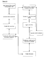
- FIG. 10 is a flow chart of the steps involved in measuring and calculating the magnetic field vector of the vehicle and subsequently activating the vehicle.
- FIG. 11 is a schematic diagram of the calculation of the vector between the detecting element and the vehicle.
- FIG. 12 is an outline picture of a solenoid for amplifying the force in the system.
- FIG. 13 depicts the layout of the control coils around the permanent magnet for the system, when amplifying its force.
- FIG. 14 is a closeup of the two paired coils, mounted on ferromagnetic material, which are connected to the permanent magnet at different angles.
- FIG. 15 depicts the system when a 3 rd coil is added to each pair to break the symmetry of paired coils connected to the permanent magnet.
- the present invention discloses a device and a system for mobilizing, rotating and maneuvering an in vivo vehicle introduced into a subject by extracorporeal devices.
- the present invention provides a device and a system for mobilizing, rotating and maneuvering an in vivo vehicle by remote activation of force and moment of an applied magnetic or electromagnetic field.
- a magnet optionally made of any suitable biologically compatible magnetic, magnetized or magnetizable material, is installed inside or coats the outside of a vehicle, or is attached to it by a connecting element. If the magnet is located within a sealed portion of the vehicle or is otherwise sealed, then optionally the magnet may be constructed of a less biologically compatible, or even a biologically incompatible, material.
- the magnet is constructed of an alloy
- the magnetic alloy is composed of one or more of Neodymium-Boron-Iron (NdBFe), Samarium Cobalt (SmCo) or other similar compounds.
- the magnet can optionally be made of any hard or soft permanent magnetic (e.g. ferromagnetic) material that is magnetized under the influence of a magnetic or an electromagnetic field.
- the magnet's mass and physical properties include magnetization direction and magnitude that enable the movement, rotation and maneuvering of the vehicle.
- an extracorporeal magnetic or electromagnetic field is preferably used.
- This magnetic source consists of a permanent magnet, an electromagnet or an electromagnet with soft magnetic material or any combination thereof.
- the extracorporeal electromagnet may be composed of several coils that create a magnetic field or fields in various directions and with various gradients.
- each coil in the electromagnet can be operated separately.
- each group of coils in the electromagnet can optionally be operated synchronously and also separately. Different currents can feed each coil and/or each group of coils.
- the force vector (magnitude and direction) applied on the external magnetic field source is measured, and used as feedback to control the status between the in vivo permanent magnet and the external magnetic field source.
- the same coils can optionally and preferably be used to calculate the vehicle's location.
- the current density that can be achieved in a resistive coil is much smaller than the equivalent effective current density on the surface of the permanent magnet, as is explained in detail herein below.
- a strong current density is required. Due to the abovementioned property of coils, there is a restriction on the current density that can be achieved. This restriction on the strength of the electrical current may restrict the ability to move or control the vehicle inside a body lumen.
- This equation represents the force calculation of a coil on the axis. The sum in the equation is over the number of full rounds in the coil.
- the symbol a i represents the distance of the center of the i th wire from the axis, and z i represents the distance of the i th wire from the object on which the force is acting.
- the symbol I represents the current in the wire forming the coil.
- the distance from the axis is measured in units of a i .
- the above equation behaves like the function a i /z i 4 and decreases rapidly with the distance. Therefore, although increasing the radius of the coils will increase the force, the greater distance from the body will decrease it much more rapidly, and the force will not be strong enough. For example, for a coil of radius 5 mm, which has current density of less than 5 ampere/mm 2 , the force created is practically limited to a height of 4 mm from the base of the coil.
- the small size of the coil means that there is a limitation on the force that the coil can impose on the vehicle.
- a small imposed force requires the vehicle to be in short distance from the coil matrix.
- a force amplifier is needed in order to enlarge the force a small coil can create.
- the force amplification can be achieved in several ways:
- An optional solution to the above mentioned problem is to use a matrix of coils, which can be activated and deactivated, and thus control the movement of the vehicle.
- the vehicle will move to the geometric center of the activated coils.
- the coils composing the matrix must be of small dimension, namely having a size in the order of millimeters, and not centimeters.
- the size of the pixel i.e. location discretization
- the size of the pixel is half of the coil diameter. If a localization is required within 1 cm, the coil size radius is limited to 1 cm, for example. Therefore, the discretization size determines the coil size. In other words the size of the coils determines the “pixel” size of the vehicle.
- the matrix of coils can also be used in order to move a permanent magnet, which then moves to the center of the activated coils, and creates equivalent effective high current density at this point.
- the equivalent effective high current density then amplifies the force created by the coils on the vehicle.
- Cylindrical magnets are chosen as they are the most efficient way to utilize the space, but the magnet can optionally be ring shaped, a polygon or a polygonal ring.
- the cylindrical magnets are magnetized in the axial direction.
- the formula for calculating the force created by the cylindrical magnets is the same as equation 1, only that the sum is performed only on the cylindrical surface.
- space discretization is achieved by dividing the surface to regions of the magnetic field.
- a surface pixel is defined by a region having magnetic field in the same direction, when all its nearest neighboring (non diagonal) pixels have a magnetic field in the opposite direction. In this way, each magnet has a magnetization opposite to the magnetization of its nearest neighbors.
- the movable permanent magnet On the movable permanent magnet, several coils are attached and are wound in opposite directions (at least one coil is wound clockwise and at least one other coil is wound counterclockwise). In this way, the electromagnetic fields caused by each pair of coils are in the same direction, and thus enhance each other.
- the pair of coils are mounted on ferromagnetic material, increasing the magnetic field, and thus also the force in the system, to an even greater extent.
- the movement of the vehicle in the body lumen is caused by a change in the direction of the current in the coils attach to the driving magnet. It is possible to add a third coil to each pair to break the symmetry of coils, making it possible to give preference to one of the pairs of coils, thus creating a preferred direction of movement.
- a coil is added which is not placed below the magnet center.
- the added coil will experience non axial force, which will create a movement. Once the system moves, the force on the major coils will no longer be axial, and the added symmetry breaking coils can be shut down.
- the symmetry breaking coils are used, and in later stages preferably the inertia serves as a tool for breaking the symmetry in the system.
- Hall-effect probes are added to monitor the movement of the force amplifier relative to the matrix of cylindrical magnets.
- a Hall effect probe makes use of the known phenomenon (discovered by E. H. Hall in the 19 th century), such that when an electric current flows through a conductor in a magnetic field, the magnetic field exerts a transverse force on the moving charge carriers. A buildup of charge at the sides of the conductor balances this magnetic influence, producing a measurable voltage between the two sides of the conductor, which is proportional to the magnetic field.
- the Hall-effect probe thus monitors magnetic field changes and can be used to trigger a change of the current in the coils, when they pass from pixel to pixel. It can also be used for counting the number of pixels that the system moves.
- the current direction in the coils pair changes every time the coils move a distance similar to the distance between two magnets in the matrix, as for example in a linear motor.
- the Hall-effect probe measures the field changes, and is used for changing the current in the coils.
- the measurement and tracking functions of the extracorporeal navigating system may optionally and preferably be accomplished by: an electromagnet used for maneuvering the vehicle; a coil or more preferably a set of coils, capable of measuring magnetic field strength in a plurality of, but more preferably all, directions; Hall effect probes; a pressure measuring device; or a combination thereof, in which these elements are more preferably connected to a computerized control system.
- a Hall effect probe measures the magnetic field strength. This probe changes the electrical potential on the device when exposed to a magnetic field.
- the pressure-measuring device may optionally be implemented as follows. When an electrical current in a coil is exposed to a magnetic field, a force acts on the coil. The coil creates pressure on its physical support, which can be measured. Calibration of the system enables the pressure to be translated to a measurement of the magnetic field, which can then optionally and preferably be used to calculate the vehicle position, for example.
- the principle feature of the invention is the ability, via detection and generation of magnetic field vectors, to guide and maneuver the vehicle by an extracorporeal control unit, without the requirement for a physical connection between the vehicle and the control unit.
- the extracorporeal guidance aiming may optionally be performed in one or more ways, for example by following a pre-determined and/or programmed route, according to the anatomical structure of the organ in which the vehicle is situated.
- the software program preferably receives feedback from the tracking system.
- the software program can optionally and preferably correct mistakes, can limit the force applied on the vehicle to avoid damage to the tissue, and more preferably may activate procedures to re-locate the vehicle if it gets lost.
- real time guidance performed by the operator, according to information received from the vehicle.
- real time guidance may optionally be performed according to information received from any other imaging system (X-rays, US, MRI, CT, etc), which can optionally be gathered before or during the diagnostic procedure involving the present invention, or any combination thereof.
- imaging system X-rays, US, MRI, CT, etc
- the mode of external remote guidance and/or monitoring may also activate or enable other functions.
- the external remote guidance preferably enables the navigation of the vehicle in different directions in the observed area.
- the in vivo vehicle has one or more preferred but optional features which allow it to be moved, turned, diverted and aimed at any angle.
- the electromagnetic receiving/signaling system of the vehicle may be composed of an electric dipole element or a magnetic element which forms an integral part of the vehicle or may be a separate element tethered to the vehicle by a connecting element.
- the magnetic element may be composed of a permanent magnetic ring or disk inserted into the vehicle, a permanent magnetic coating covering part or all of the exterior of the vehicle or a bonded magnetic material coating part or all of the vehicle's exterior.
- the magnetic element may be composed of ferromagnetic material which may coat part or the entirety of the vehicle's exterior.
- the magnetic element may be composed of a permanent magnet, bonded and/or ferromagnetic materials.
- the direction of the magnetic field of the magnetic element inside the vehicle may be axial, or parallel to the diameter (diametrical) in a case where the vehicle's geometry is round i.e. cylinder, disk, and/or ring shaped or a combination thereof.
- the magnetic element is connected to the vehicle upon introduction into the patient, but is released from the vehicle at a certain stage in the diagnostic procedure and remains connected to the vehicle.
- the tethered magnet is introduced into the patient as such.
- the vehicle may also contain an electric circuit such that force can be applied to the vehicle through the electronic circuit when the vehicle is in a magnetic field.
- the electric circuit can be installed in the vehicle in place of the permanent magnet or in addition to it.
- the vehicle may also contain capacitors which may be charged by a power source. The capacitor or capacitors can be installed in the vehicle in place of the permanent magnet or in addition to it.
- a method for detecting the location of the vehicle and more preferably for also controlling the movement of the vehicle is provided.
- a matrix of detectors are attached to the patient or placed close to the patient.
- the matrix can be composed of Hall effect probes, coils which serve as probes or combinations of these elements.
- the magnetic field may optionally be measured with a Hall effect probe, as previously described.
- the field may optionally be measured according to the force acting on a coil when current flows through it, or alternatively by measuring the potential on a coil when a time dependent magnetic field is applied to it. This measurement enables the magnetic field caused by the vehicle to be calculated, such that the position of the vehicle can be calculated.
- the probe detects at least one directional component of the magnetic field at any point of the matrix but may also be able to detect up to three directional components of the magnetic field.
- a matrix composed of Hall effect probes it is possible to detect both time dependent and stationary magnetic fields.
- a matrix composed of coils only a time dependent field can be detected directly, while the stationary field can be calculated from the force measurements.
- five parameters should preferably be calculated, namely three coordinates with respect to the detector matrix center and two orientation angles of the magnetization with respect to coordinate system defined on the detection matrix. At least five measurements of the magnetic field of the vehicle are needed to extract these parameters providing that the distance and the orientation of detectors within the matrix and the magnitude and the direction of the magnetization vector with respect to the vehicle are known. The extraction of the five parameters is done by best fit of the known formula of the magnetic filed at a point of distance (d x , d y , d z ) from a magnetic dipole.
- (x m ,y m ,z m ) be a position vector of a detector m in a coordinate system in which center of the matrix is in the origin; (x,y,z) be the position vector of the vehicle in the same coordinate system; and R m be the Euclidian distance of the vehicle from detector m.
- R m ⁇ square root over (( x m ⁇ x ) 2 +( y m ⁇ y ) 2 +( z m ⁇ z ) 2 ) ⁇ square root over (( x m ⁇ x ) 2 +( y m ⁇ y ) 2 +( z m ⁇ z ) 2 ) ⁇ square root over (( x m ⁇ x ) 2 +( y m ⁇ y ) 2 +( z m ⁇ z ) 2 ) ⁇ equation 2 Denoting the z component of the vehicle's magnetization by m z the x, y, z components of the magnetic field at the point m are, respectively
- the unknown parameters are x, y, z, m z and m y ; the measured quantities are B x m , B y m , B z m .
- the best estimation of x, y, z, m z , m y , and m x and the error in the estimated value are then calculated.
- M ⁇ square root over ( m z 2 +m y 2 +m x 2 ) ⁇ equation 6
- M is the measured vehicle magnetization.
- FIG. 1 illustrates the major elements of the system and their layout including an extracorporeal navigation system 18 for guiding an in vivo vehicle 19 within the patient 17 .
- extracorporeal navigation system 18 is optionally in direct physical contact with at least a portion of patient 17 although this is not necessary, as physical proximity is sufficient.
- vehicle 19 could optionally be used for diagnostic imaging techniques and/or other medical procedures.
- FIG. 2 depicts the tethered magnetic element configuration 20 of the vehicle where a separate magnetic element 12 is tethered to vehicle 19 by a connecting element 13 .
- magnetic element includes any type of magnet, which as previously described may include one or more of a magnet (whether soft or hard), magnetized material or magnetizable material, or a combination thereof.
- Connecting element 13 is an exemplary tether for magnetic element 12 , which is preferably flexible but alternatively is rigid.
- Connecting element 13 and vehicle 19 may optionally be constructed of a metal, an alloy, a plastic or a combination of materials, but may not necessarily be constructed of the same or similar materials.
- Connecting element 13 and/or vehicle 19 may optionally be from hundreds of microns to a few millimeters in length, although it should be noted that size is not necessarily a limiting factor. Rather, the dimensions of connecting element 13 and/or vehicle 19 are optionally and preferably chosen according to the dimensions of the body space or spaces in which vehicle 19 travels.
- Magnetic element 12 may optionally be smaller than vehicle 19 .
- One advantage of this embodiment is that magnetic element 12 does not need to fit within vehicle 19 , such that a larger size of magnetic element 12 may optionally be used.
- FIG. 3 depicts an integrated vehicle configuration 30 , where a magnetic element 22 is integrated into the body of vehicle 19 .
- FIGS. 4 a - 4 f in which several types of vehicles are shown. It should be noted that the same reference numbers denote the same or similar elements.
- a vehicle 200 preferably features an inserted magnet 204 , optionally in the form of a ring or disk, which is more preferably permanently installed.
- a magnetization direction 202 is shown.
- the electromagnetic receiving/signaling system of a vehicle 210 may optionally be composed of a magnetic dipole element 212 .
- vehicle 210 is powered by an electromagnetic field imposed on the vehicle from the extracorporeal device.
- a vehicle 220 may optionally feature a magnetic element 222 which is implemented as a partial or full covering of the exterior of vehicle 220 , or even as a partial or full exterior structure for vehicle 220 .
- the degree of the magnetic field which is generated or which is capable of being generated by magnetic element 222 may optionally and preferably be varied in a plurality of different portions of vehicle 220 . Each portion may optionally have a different direction of magnetization in order to optimize the control of the movement of vehicle 220 .
- a vehicle 230 may optionally feature a partial or full exterior structure 221 made from bonded material, optionally and more preferably with inserted magnet 204 , again optionally in the form of a ring or disk, which again is more preferably permanently installed.
- a vehicle 240 is shown with partial or full exterior structure 221 made from bonded material optionally as the sole magnetic element.
- the bond can optionally be made of dissolvable or non-dissolvable material, and can also optionally partially fill the interior volume of the vehicle.
- FIG. 4 f a vehicle 250 is shown with a partial or full exterior structure 252 made from a permanently magnetic or ferromagnetic material.
- FIG. 4 g shows a vehicle 260 with a partial or full exterior structure 253 made from a ferromagnetic material.
- FIG. 5 describes an exemplary system 50 according to the present invention for controlling and maneuvering the vehicle in the subject's colon.
- system of the present invention in the GI tract, this is for the purposes of illustration only and is not intended to be limiting in any way, as the present invention is suitable for use in any bodily cavity, space, vessel, organ or other non-solid section of the body.
- Vehicle 51 can be maneuvered within colon 54 , and can optionally and preferably be focused on a particular field of view 52 .
- Vehicle 51 is preferably guided by one, and more preferably a plurality of external guidance elements 53 as shown.
- Each external guidance element 53 could optionally be a coil, reed switch, or Hall effect probe, for example. If a plurality of external guidance elements 53 is used, then vehicle 51 can more easily be located.
- the plurality of external guidance elements 53 is preferably distributed about the body of the patient (not shown) and then calibrated. The location of external guidance elements 53 and their number depends at least partially upon the accuracy of management of vehicle 51 that is desired and the activity to be performed.
- FIG. 6 illustrates the detection and motion control systems combined in a single component 60 .
- the field generator units and detector units are contained in one element 61 , such that the magnetic field is both produced and detected by element 61 .
- the field generator units can optionally operate on the principle of magnetic flux or electromagnetic field production.
- the field generating/detector units (elements 61 ) are preferably distributed on a flexible material 65 and are more preferably connected via power and information buses 62 to a computer control unit 64 .
- Flexible material 65 may optionally be in the form of a blanket or sheet which can be wrapped around at least a portion of the patient.
- FIG. 7 depicts a different configuration for the detection and motion control subsystems as a separated system 70 , such that the magnetic field is generated and detected by different components of system 70 .
- a field generation subsystem computer control unit 71 and a detecting subsystem computer control unit 72 are located in different locations in system 70 .
- a field generating element 73 can optionally operate on the principle of production of a magnetic flux or electromagnetic field.
- Field generating element 73 and a detecting element 74 of which a plurality of each such element are shown for the purposes of description only, are connected to their respective computer control units by power and information buses 62 . This implementation is preferred to avoid cross-talk between the generation and detection of the magnetic field, and may also optionally provide greater sensitivity.
- FIG. 8 illustrates a prototype detector unit 80 .
- One or more measuring devices 81 are connected to one or more types of field detectors 82 . These detectors may include Hall effect probes, pressure detectors, devices for measuring Doppler effects or devices for measuring laser Doppler effects.
- Measuring device(s) 81 are preferably connected to a switching or indicating device (not shown).
- the switching or indicating device may optionally be composed of one or more devices such as an individual reed switch or arrays of reed switches, flip switch, electromagnetic, electronic optical or mechanical flag type indicator, LED or memory device which can respond to a signal above or below preset thresholds to locate the vehicle, activate a function or turn off a certain function of the vehicle.
- Measuring devices 81 and field detectors 82 are preferably attached to a flexible sheet 83 , for being wrapped around at least a portion of the patient for example.
- Flexible sheet 83 may also optionally be implemented as a belt and/or as a rigged board of less flexible material for holding these components.
- FIG. 9 depicts a block diagram outlining the interactions of various parts of the extracorporeal generator and detector units in an exemplary system 90 according to the present invention.
- a computer 92 preferably features a display 94 for displaying information to the user about the operation of system 90 , more preferably as a graphical user interface (GUI).
- GUI graphical user interface
- the user is preferably able to send one or more commands to computer 92 for controlling the behavior of system 90 through a user interface 100 , which is optionally and more preferably implemented as a joystick.
- a detection control unit 96 preferably receives one or more commands from computer 92 for controlling one or more detection elements 106 .
- Each detection element 106 is preferably capable of detecting a magnetic field, and may optionally be implemented as previously described.
- Detection control unit 96 optionally and more preferably sends data to computer 92 concerning signals and/or data received from detection element 106 .
- a magnetic generator control unit 102 is also preferably in communication with computer 92 and also preferably receives one or more commands from computer 92 for controlling the function of one or more magnetic sources 104 .
- Magnetic sources 104 include a magnet and may optionally be implemented as previously described, for example as one material and/or component, or a plurality of materials and/or components.
- a power supply 98 optionally supplies power to computer 92 , detection control unit 96 and magnetic generator control unit 102 , and may optionally also supply power to one or both of detection elements 106 or magnetic sources 104 .
- Power supply 98 may optionally be implemented as a plurality of such power supplies (not shown).
- FIG. 10 illustrates a flow chart of the stages involved in measuring and calculating the magnetic field vector of the vehicle and subsequently activating the vehicle.
- Application of the magnetic field is performed by the generator unit and detection of the signals emitted from the vehicle is performed by the detection unit.
- the calculations are performed by the computer, which preferably has sufficient power to integrate many complex signals simultaneously.
- the magnetic field is applied and/or an existing magnetic field is detected.
- the detector units in the extracorporeal device preferably detect magnetic signals from the vehicle to locate the vehicle.
- the vector force is calculated for the magnetic field vectors of the vehicle.
- the x, y, z, m x , m y , m z parameters are estimated (see previous equations for a description).
- the current is measured and the force F m is then calculated.
- one or more of the vehicle position, movement and orientation are preferably determined.
- the operator commands are preferably translated in relation to the location of the vehicle, and more specifically are translated in relation to the magnetic field of the vehicle.
- the vector force to be applied to perform the operator command is calculated and the force is then applied.
- at least one of a new vehicle position, movement and orientation is preferably determined. This information is preferably then displayed to the operator.
- FIG. 11 provides a schematic description 110 of the calculation of the vector between a plurality of extracorporeal detecting elements 120 and a vehicle 112 .
- vehicle 112 is situated in a body lumen or cavity 114 .
- the vehicle's magnetization vector 122 is detected by detector elements 120 through the skin 118 via interactions with the detector units' magnetic field vectors 116 .
- These signals are preferably processed according to the procedure outlined in FIG. 10 .
- the present invention is conceived as being a viable alternative to endoscopy, especially in diagnostic or therapeutic procedures in the esophagus, stomach, small intestine, large intestine and rectum.
- the invention allows the medical personnel to directly observe the epithelial lining of the GI tract and to carry out medical procedures such as tissue sample collection for histological examination, liquid sampling for microscopic examination and culturing and small surgical procedures such as removal of suspicious polyps in the large intestine. These procedures can be carried out with less danger to the patient since no physical connection between the vehicle and the extracorporeal navigating device is necessary.
- the invention is useful for carrying out the above functions in the diagnosis of or as part of the treatment of malignant, nonmalignant, infectious, and genetic diseases as well as birth or developmental defects.
- the present invention is conceived as being a viable alternative to bronchoscopy.
- the invention allows the medical personnel to directly observe the epithelial lining of the bronchi and to carry out medical procedures such as tissue sample collection for histological examination, liquid sampling for microscopic examination and culturing and small surgical procedures. These procedures can be carried out with less discomfort and danger to the patient since no physical connection between the vehicle and the extracorporeal navigating device is necessary.
- the invention is useful for carrying out the above functions in the diagnosis of or as part of the treatment of malignant, nonmalignant, infectious, and genetic diseases as well as birth or developmental defects.
- the present invention is conceived as being an additional tool used in laparoscopy or as a viable alternative to laparoscopy.
- the vehicle under the control of the extracorporeal navigating device, can perform small surgical procedures.
- the invention could therefore be launched from a laparoscopic instrument into the abdomen to perform a certain task or could actually be used in place of the laparoscope in certain indications such as the destruction of kidney stones, gallstones, or other pathological crystalline deposits in other organs.
- the invention could be used for directly observing organs and tissues in the abdominal space and for carrying out medical procedures such as tissue sample collection for histological examination, liquid sampling for microscopic examination and culturing and surgical procedures, as part of the diagnosis or treatment of malignant, nonmalignant, infectious, and genetic diseases as well as birth or developmental defects.
- the present invention is conceived as being a means for directly controlling and optimizing drug delivery to a specified tissue or organ.
- the pharmaceutical compound can be encapsulated into liposomes or any other suitable form for delivery to the target organ and magnetic particles can be impregnated into the structure of the delivery device in such a way so that the extracorporeal navigating system can concentrate them into the desired location.
- magnetic particles can be coated with the therapeutic or structures containing the therapeutic such as liposomes or microspheres and these can serve as the drug delivery vehicle.
- Such a drug delivery system could be administered orally, intravenously or parenterally depending on the indication as determined by those skilled in the art.
- FIG. 12 describes two solenoids 300 each with maximal radius of 5 mm and length of 10 mm.
- Each solenoid is built of 30 turns 302 of cross-section 1 mm 2 .
- the dotted line 301 is the axis of symmetry.
- the maximal radius of the coil determines the pixel size of the vehicle. A distance smaller than the radius of a coil cannot be specified, due to space discretization, and therefore the coil radius cannot be larger than 5 mm for a pixel size of 1 cm.
- the small size of the coil results in a limitation on the force that the coil can impose on the vehicle.
- a small imposed force requires the vehicle to be in short distance from the coil matrix.
- An optional method of force amplification is to use a matrix of coils, which can be activated and deactivated and thus used in order to move a permanent magnet.
- the permanent magnet moves to the center of the switched on coils, and creates an equivalent effective high current density at this point.
- the equivalent effective high current density then amplifies the force created by the coils on the vehicle.
- the vehicle will move to the geometric center of the activated coils.
- the coils composing the matrix must be of small dimension, namely having a size in the order of millimeters, and not centimeters. Therefore, the discretization size determines the coil size.
- the size of the coils also determines the “pixel” size of the vehicle.
- the coil size is less than about 5 mm, such that the pixel size is also preferably less than about 5 mm.
- the present invention may have an extracorporeal electromagnet of several coils creating a magnetic field, which may cause a problem due to low current density in these coils.
- a possible way of amplifying the force on this electromagnetic field is by using a matrix of cylindrical permanent magnets arranged on a grid.
- Cylindrical magnets are chosen as they are the most efficient way to utilize the space, but the magnet can optionally be ring shaped, a polygon or a polygonal ring; a magnet having any such suitable shape and geometry is herein termed an “annular magnet”.
- the cylindrical magnets are magnetized in the axial direction, and this is due to the fact that an axial magnet is like a solenoid (as mentioned in hereinabove), which produces the same field in all directions.
- the formula for calculating the force created by the cylindrical magnets is the same as equation 1, except that the sum is only on the cylindrical surface.
- space discretization is achieved by dividing the surface into regions of the magnetic field.
- a region having magnetic field in the one direction, when all its nearest neighboring pixels have a magnetic field in the opposite direction defines a surface pixel.
- the change of directions is achieved by placing magnets of opposite direction in neighboring regions, such that each magnet has a magnetization opposite to the magnetization of its nearest neighbors (non diagonal neighbors).
- FIG. 13 on the movable permanent magnet 305 , several coils 306 are attached.
- a pair of coils is placed at every 90° around the permanent magnet 305 , as displayed in the figure.
- the reason for this choice of angle is once again because of space utilization.
- the angle also determines the direction, and therefore it is most efficient to create a grid that has two orthogonal directions (90°), but it is possible to create a grid in which the two directions are not orthogonal (for example having an angle of 65.4°). If the direction chosen is not orthogonal, the coils will have the same angle between them, yet the force will be smaller.
- the two coils that create a pair of coils as depicted in FIG.
- the force is axial, and therefore changing the current in the coils would once again cause an axial force and there would be no movement.
- the extra coil mentioned above is added in order for movement to start. This coil is preferably not placed below the magnet's center; therefore it will experience non-axial force, which will create a movement. Once the system moves, the force on the major coils will no longer be axial, and the symmetry breaking coils can be shut down. In this stage, the inertia serves as a tool for breaking the symmetry between the coils.
- the movement of the force amplifier relative to the matrix of cylindrical magnets is monitored thereby.
- the Hall-effect probe monitors field changes hence can be used to trigger a change of the current in the coils when they pass from pixel to pixel.
- the Hall-effect probe can be used for counting the number of pixels that the vehicle moves. Knowing the size of a region with a magnetic field in one direction, the number of changes of field direction is sufficient to determine the distance. More specifically, the distance equals the number of field changes multiplied by the length of each pixel.
- the direction in which the vehicle is moving can also be determined according to the current direction in the coils.
- the current direction in the coils pair changes every time the coils move a distance similar to the distance between two magnets in the matrix, just as it is done in linear motors.
- the coils Once the coils enter a new pixel, they have a current in the direction which creates an attraction force to the center of the pixel, and once they cross the pixel center the current direction changes and the coils repulse from the pixel center and are attracted by the next pixel center, because in the next pixel the magnetic field is in the opposite direction to the pixel the coil are currently in. This continues until the coils reach the goal destination, where the current stops changing, and the coils stop at the center of the pixel where the force is axial.
- the Hall-effect probe measures the field changes, and is used for changing the current in the coils.
Abstract
Description
R m=√{square root over ((x m −x)2+(y m −y)2+(z m −z)2)}{square root over ((x m −x)2+(y m −y)2+(z m −z)2)}{square root over ((x m −x)2+(y m −y)2+(z m −z)2)}
Denoting the z component of the vehicle's magnetization by mz the x, y, z components of the magnetic field at the point m are, respectively
Similar equations for my, and mx can be written by changing z to y and z to x in the above equations.
The unknown parameters are x, y, z, mz and my; the measured quantities are Bx m, By m, Bz m. Using a best fit algorithm the best estimation of x, y, z, mz, my, and mx and the error in the estimated value are then calculated. Then from the knowledge of the magnetization of the vehicle consistency can be checked i.e.
M=√{square root over (m z 2 +m y 2 +m x 2)} equation 6
M is the measured vehicle magnetization.
{right arrow over (F)} m =
where the ds integral is performed along the coil. The current in the coil may change from coil to coil in the matrix to get the best measurement. Then a best-fit algorithm is used to estimate position and orientation of the vehicle from equation 7. Combinations of force and field measurement can be used to calculate the vehicle position and orientation. In the preferred embodiment the number of measurements is larger then the number of unknowns, as this should reduce the error. Minimally, at least as many measurements as unknowns are required for the calculation.
Claims (72)
Priority Applications (1)
| Application Number | Priority Date | Filing Date | Title |
|---|---|---|---|
| US10/686,535 US7311107B2 (en) | 2001-04-18 | 2003-10-16 | Navigating and maneuvering of an in vivo vehicle by extracorporeal devices |
Applications Claiming Priority (5)
| Application Number | Priority Date | Filing Date | Title |
|---|---|---|---|
| IL142682 | 2001-04-18 | ||
| IL14268201A IL142682A0 (en) | 2001-04-18 | 2001-04-18 | Method for externally navigating intra-lumina capsule |
| US33155901P | 2001-11-19 | 2001-11-19 | |
| PCT/IL2002/000286 WO2002082979A2 (en) | 2001-04-18 | 2002-04-08 | Navigating and maneuvering of an in vivo vechicle by extracorporeal devices |
| US10/686,535 US7311107B2 (en) | 2001-04-18 | 2003-10-16 | Navigating and maneuvering of an in vivo vehicle by extracorporeal devices |
Related Parent Applications (1)
| Application Number | Title | Priority Date | Filing Date |
|---|---|---|---|
| PCT/IL2002/000286 Continuation-In-Part WO2002082979A2 (en) | 2001-04-18 | 2002-04-08 | Navigating and maneuvering of an in vivo vechicle by extracorporeal devices |
Publications (2)
| Publication Number | Publication Date |
|---|---|
| US20040138552A1 US20040138552A1 (en) | 2004-07-15 |
| US7311107B2 true US7311107B2 (en) | 2007-12-25 |
Family
ID=26324023
Family Applications (1)
| Application Number | Title | Priority Date | Filing Date |
|---|---|---|---|
| US10/686,535 Expired - Fee Related US7311107B2 (en) | 2001-04-18 | 2003-10-16 | Navigating and maneuvering of an in vivo vehicle by extracorporeal devices |
Country Status (4)
| Country | Link |
|---|---|
| US (1) | US7311107B2 (en) |
| EP (1) | EP1383416A2 (en) |
| AU (1) | AU2002307762A1 (en) |
| WO (1) | WO2002082979A2 (en) |
Cited By (65)
| Publication number | Priority date | Publication date | Assignee | Title |
|---|---|---|---|---|
| US20080058989A1 (en) * | 2006-04-13 | 2008-03-06 | Board Of Regents Of The University Of Nebraska | Surgical camera robot |
| US20080300458A1 (en) * | 2004-06-21 | 2008-12-04 | Byung Kyu Kim | Capsule Type Endoscope Control System |
| US20090030306A1 (en) * | 2005-04-18 | 2009-01-29 | Yoshitaka Miyoshi | Endoscope Shape Detecting Apparatus |
| US7772796B2 (en) | 2003-07-08 | 2010-08-10 | Board Of Regents Of The University Of Nebraska | Robotic devices with agent delivery components and related methods |
| US20100268025A1 (en) * | 2007-11-09 | 2010-10-21 | Amir Belson | Apparatus and methods for capsule endoscopy of the esophagus |
| US20110060189A1 (en) * | 2004-06-30 | 2011-03-10 | Given Imaging Ltd. | Apparatus and Methods for Capsule Endoscopy of the Esophagus |
| US7960935B2 (en) | 2003-07-08 | 2011-06-14 | The Board Of Regents Of The University Of Nebraska | Robotic devices with agent delivery components and related methods |
| US8343171B2 (en) | 2007-07-12 | 2013-01-01 | Board Of Regents Of The University Of Nebraska | Methods and systems of actuation in robotic devices |
| US8679096B2 (en) | 2007-06-21 | 2014-03-25 | Board Of Regents Of The University Of Nebraska | Multifunctional operational component for robotic devices |
| US8891924B2 (en) | 2012-04-26 | 2014-11-18 | Bio-Medical Engineering (HK) Limited | Magnetic-anchored robotic system |
| US8894633B2 (en) | 2009-12-17 | 2014-11-25 | Board Of Regents Of The University Of Nebraska | Modular and cooperative medical devices and related systems and methods |
| US8968267B2 (en) | 2010-08-06 | 2015-03-03 | Board Of Regents Of The University Of Nebraska | Methods and systems for handling or delivering materials for natural orifice surgery |
| US8974440B2 (en) | 2007-08-15 | 2015-03-10 | Board Of Regents Of The University Of Nebraska | Modular and cooperative medical devices and related systems and methods |
| US9010214B2 (en) | 2012-06-22 | 2015-04-21 | Board Of Regents Of The University Of Nebraska | Local control robotic surgical devices and related methods |
| US9060781B2 (en) | 2011-06-10 | 2015-06-23 | Board Of Regents Of The University Of Nebraska | Methods, systems, and devices relating to surgical end effectors |
| US20150177378A1 (en) * | 2012-07-24 | 2015-06-25 | Valeo Schalter And Sensoren Gmbh | Alternative installation of a concealed ultrasonic sensor in the motor vehicle |
| US9078662B2 (en) | 2012-07-03 | 2015-07-14 | Ethicon Endo-Surgery, Inc. | Endoscopic cap electrode and method for using the same |
| US9089353B2 (en) | 2011-07-11 | 2015-07-28 | Board Of Regents Of The University Of Nebraska | Robotic surgical devices, systems, and related methods |
| US9259288B2 (en) | 2009-04-14 | 2016-02-16 | Covidien Lp | Apparatus and system for performing surgery |
| US9277957B2 (en) | 2012-08-15 | 2016-03-08 | Ethicon Endo-Surgery, Inc. | Electrosurgical devices and methods |
| US9339285B2 (en) | 2013-03-12 | 2016-05-17 | Levita Magnetics International Corp. | Grasper with magnetically-controlled positioning |
| US9375268B2 (en) | 2007-02-15 | 2016-06-28 | Ethicon Endo-Surgery, Inc. | Electroporation ablation apparatus, system, and method |
| US9498292B2 (en) | 2012-05-01 | 2016-11-22 | Board Of Regents Of The University Of Nebraska | Single site robotic device and related systems and methods |
| US9545290B2 (en) | 2012-07-30 | 2017-01-17 | Ethicon Endo-Surgery, Inc. | Needle probe guide |
| US9572623B2 (en) | 2012-08-02 | 2017-02-21 | Ethicon Endo-Surgery, Inc. | Reusable electrode and disposable sheath |
| US9579088B2 (en) | 2007-02-20 | 2017-02-28 | Board Of Regents Of The University Of Nebraska | Methods, systems, and devices for surgical visualization and device manipulation |
| US9743987B2 (en) | 2013-03-14 | 2017-08-29 | Board Of Regents Of The University Of Nebraska | Methods, systems, and devices relating to robotic surgical devices, end effectors, and controllers |
| US9770305B2 (en) | 2012-08-08 | 2017-09-26 | Board Of Regents Of The University Of Nebraska | Robotic surgical devices, systems, and related methods |
| US9844391B2 (en) | 2009-02-06 | 2017-12-19 | Levita Magnetics International Corp. | Remote traction and guidance system for mini-invasive surgery |
| US9883910B2 (en) | 2011-03-17 | 2018-02-06 | Eticon Endo-Surgery, Inc. | Hand held surgical device for manipulating an internal magnet assembly within a patient |
| US9888966B2 (en) | 2013-03-14 | 2018-02-13 | Board Of Regents Of The University Of Nebraska | Methods, systems, and devices relating to force control surgical systems |
| US10004558B2 (en) | 2009-01-12 | 2018-06-26 | Ethicon Endo-Surgery, Inc. | Electrical ablation devices |
| US10010370B2 (en) | 2013-03-14 | 2018-07-03 | Levita Magnetics International Corp. | Magnetic control assemblies and systems therefor |
| US10098527B2 (en) | 2013-02-27 | 2018-10-16 | Ethidcon Endo-Surgery, Inc. | System for performing a minimally invasive surgical procedure |
| US10098691B2 (en) | 2009-12-18 | 2018-10-16 | Ethicon Endo-Surgery, Inc. | Surgical instrument comprising an electrode |
| US10105141B2 (en) | 2008-07-14 | 2018-10-23 | Ethicon Endo-Surgery, Inc. | Tissue apposition clip application methods |
| US10179033B2 (en) | 2012-04-26 | 2019-01-15 | Bio-Medical Engineering (HK) Limited | Magnetic-anchored robotic system |
| US10206709B2 (en) | 2012-05-14 | 2019-02-19 | Ethicon Llc | Apparatus for introducing an object into a patient |
| US10258406B2 (en) | 2011-02-28 | 2019-04-16 | Ethicon Llc | Electrical ablation devices and methods |
| US10278761B2 (en) | 2011-02-28 | 2019-05-07 | Ethicon Llc | Electrical ablation devices and methods |
| US10314603B2 (en) | 2008-11-25 | 2019-06-11 | Ethicon Llc | Rotational coupling device for surgical instrument with flexible actuators |
| US10314649B2 (en) | 2012-08-02 | 2019-06-11 | Ethicon Endo-Surgery, Inc. | Flexible expandable electrode and method of intraluminal delivery of pulsed power |
| US10335024B2 (en) | 2007-08-15 | 2019-07-02 | Board Of Regents Of The University Of Nebraska | Medical inflation, attachment and delivery devices and related methods |
| US10342561B2 (en) | 2014-09-12 | 2019-07-09 | Board Of Regents Of The University Of Nebraska | Quick-release end effectors and related systems and methods |
| US10376322B2 (en) | 2014-11-11 | 2019-08-13 | Board Of Regents Of The University Of Nebraska | Robotic device with compact joint design and related systems and methods |
| US10537348B2 (en) | 2014-01-21 | 2020-01-21 | Levita Magnetics International Corp. | Laparoscopic graspers and systems therefor |
| US10582973B2 (en) | 2012-08-08 | 2020-03-10 | Virtual Incision Corporation | Robotic surgical devices, systems, and related methods |
| US10667883B2 (en) | 2013-03-15 | 2020-06-02 | Virtual Incision Corporation | Robotic surgical devices, systems, and related methods |
| US10702347B2 (en) | 2016-08-30 | 2020-07-07 | The Regents Of The University Of California | Robotic device with compact joint design and an additional degree of freedom and related systems and methods |
| US10722319B2 (en) | 2016-12-14 | 2020-07-28 | Virtual Incision Corporation | Releasable attachment device for coupling to medical devices and related systems and methods |
| US10751136B2 (en) | 2016-05-18 | 2020-08-25 | Virtual Incision Corporation | Robotic surgical devices, systems and related methods |
| US10779882B2 (en) | 2009-10-28 | 2020-09-22 | Ethicon Endo-Surgery, Inc. | Electrical ablation devices |
| US10806538B2 (en) | 2015-08-03 | 2020-10-20 | Virtual Incision Corporation | Robotic surgical devices, systems, and related methods |
| US10905511B2 (en) | 2015-04-13 | 2021-02-02 | Levita Magnetics International Corp. | Grasper with magnetically-controlled positioning |
| US10966700B2 (en) | 2013-07-17 | 2021-04-06 | Virtual Incision Corporation | Robotic surgical devices, systems and related methods |
| US11013564B2 (en) | 2018-01-05 | 2021-05-25 | Board Of Regents Of The University Of Nebraska | Single-arm robotic device with compact joint design and related systems and methods |
| US11020137B2 (en) | 2017-03-20 | 2021-06-01 | Levita Magnetics International Corp. | Directable traction systems and methods |
| US11051894B2 (en) | 2017-09-27 | 2021-07-06 | Virtual Incision Corporation | Robotic surgical devices with tracking camera technology and related systems and methods |
| US11173617B2 (en) | 2016-08-25 | 2021-11-16 | Board Of Regents Of The University Of Nebraska | Quick-release end effector tool interface |
| US11284958B2 (en) | 2016-11-29 | 2022-03-29 | Virtual Incision Corporation | User controller with user presence detection and related systems and methods |
| US11357595B2 (en) | 2016-11-22 | 2022-06-14 | Board Of Regents Of The University Of Nebraska | Gross positioning device and related systems and methods |
| US11413025B2 (en) | 2007-11-26 | 2022-08-16 | Attractive Surgical, Llc | Magnaretractor system and method |
| US11583354B2 (en) | 2015-04-13 | 2023-02-21 | Levita Magnetics International Corp. | Retractor systems, devices, and methods for use |
| US11883065B2 (en) | 2012-01-10 | 2024-01-30 | Board Of Regents Of The University Of Nebraska | Methods, systems, and devices for surgical access and insertion |
| US11903658B2 (en) | 2019-01-07 | 2024-02-20 | Virtual Incision Corporation | Robotically assisted surgical system and related devices and methods |
Families Citing this family (62)
| Publication number | Priority date | Publication date | Assignee | Title |
|---|---|---|---|---|
| JP2004041709A (en) * | 2002-05-16 | 2004-02-12 | Olympus Corp | Capsule medical care device |
| US20060155174A1 (en) * | 2002-12-16 | 2006-07-13 | Arkady Glukhovsky | Device, system and method for selective activation of in vivo sensors |
| JP4091004B2 (en) * | 2003-02-04 | 2008-05-28 | オリンパス株式会社 | Medical device guidance system |
| JP4012097B2 (en) * | 2003-03-06 | 2007-11-21 | オリンパス株式会社 | Capsule type medical device collection device |
| US20070043263A1 (en) * | 2003-04-03 | 2007-02-22 | Wakefield Glenn M | Simultaneous magnetic control of multiple objects |
| US20040199054A1 (en) * | 2003-04-03 | 2004-10-07 | Wakefield Glenn Mark | Magnetically propelled capsule endoscopy |
| US20040204645A1 (en) * | 2003-04-10 | 2004-10-14 | Vahid Saadat | Scope position and orientation feedback device |
| WO2005000101A2 (en) | 2003-06-12 | 2005-01-06 | University Of Utah Research Foundation | Apparatus, systems and methods for diagnosing carpal tunnel syndrome |
| US7126303B2 (en) | 2003-07-08 | 2006-10-24 | Board Of Regents Of The University Of Nebraska | Robot for surgical applications |
| US7042184B2 (en) | 2003-07-08 | 2006-05-09 | Board Of Regents Of The University Of Nebraska | Microrobot for surgical applications |
| US7354398B2 (en) * | 2003-07-18 | 2008-04-08 | Pentax Corporation | Capsule-type device and capsule-type device controlling system |
| JP4150663B2 (en) * | 2003-12-25 | 2008-09-17 | オリンパス株式会社 | In-subject position detection system |
| JP4373204B2 (en) * | 2003-12-26 | 2009-11-25 | オリンパス株式会社 | In-subject position detection system |
| US7751866B2 (en) * | 2004-03-08 | 2010-07-06 | Olympus Corporation | Detecting system of position and posture of capsule medical device |
| DE102004023527A1 (en) * | 2004-05-13 | 2005-12-08 | Osypka, Peter, Dr.-Ing. | measuring device |
| US7605852B2 (en) | 2004-05-17 | 2009-10-20 | Micron Technology, Inc. | Real-time exposure control for automatic light control |
| WO2006095420A1 (en) * | 2005-03-09 | 2006-09-14 | Olympus Corporation | Device to be introduced into subject and system to be introduced into subject |
| US20080193543A1 (en) * | 2005-05-17 | 2008-08-14 | Brown University Research Foundation | Drug Delivery Formulations For Targeted Delivery |
| CN101282677B (en) | 2005-10-05 | 2010-07-28 | 奥林巴斯医疗株式会社 | Capsule type medical device, its guidance system and guidance method and examinee insertion device |
| EP1972253A4 (en) * | 2005-12-27 | 2010-03-03 | Olympus Medical Systems Corp | Encapsulated medical device guiding system and its control method |
| US7988618B2 (en) * | 2006-01-11 | 2011-08-02 | Olympus Medical Systems Corp | Medical procedure via natural opening |
| US9084547B2 (en) | 2006-03-30 | 2015-07-21 | Given Imaging Ltd. | System and method for checking the status of an in-vivo imaging device |
| DE102006019987A1 (en) * | 2006-04-26 | 2007-10-31 | Siemens Ag | Endoscopic capsule for investigation of body openings, has induction coil with elongation along one axis, and magnetic element having magnetic dipole moment aligned perpendicular to longitudinal axis of induction coil |
| US20070255086A1 (en) * | 2006-04-26 | 2007-11-01 | Nehls Robert J | Medical Device Including Magnetic Particles |
| DK2023814T3 (en) * | 2006-05-16 | 2016-03-29 | Fraunhofer Ges Forschung | A device for contact-defined movement of the at least one magnetic body |
| US8568286B2 (en) * | 2006-06-14 | 2013-10-29 | Cardiac Pacemakers, Inc. | Methods to position therapeutic agents using a magnetic field |
| JP2009541018A (en) * | 2006-06-23 | 2009-11-26 | コーニンクレッカ フィリップス エレクトロニクス エヌ ヴィ | Drug administration system |
| WO2008018076A2 (en) * | 2006-08-10 | 2008-02-14 | Given Imaging Ltd. | System and method for in vivo imaging |
| CN101505648B (en) * | 2006-09-06 | 2011-05-04 | 奥林巴斯株式会社 | Medical device control system |
| US20080139884A1 (en) * | 2006-12-06 | 2008-06-12 | Myers William D | Medical examination system with endoscopic probe |
| JP5269348B2 (en) * | 2007-05-21 | 2013-08-21 | オリンパス株式会社 | Position detection system and method of operating the position detection system |
| EP2392249B1 (en) * | 2008-06-19 | 2013-12-04 | Olympus Medical Systems Corp. | Magnetically guiding system and magnetically guiding method |
| DE102008036290A1 (en) * | 2008-08-04 | 2010-02-11 | Olympus Medical Systems Corp. | Method for applying a force to an endoscopy capsule |
| DE102009013354B4 (en) * | 2009-03-16 | 2011-02-17 | Siemens Aktiengesellschaft | Coil system, medical device and method for non-contact magnetic navigation of a magnetic body in a workspace |
| US8830648B2 (en) | 2009-09-11 | 2014-09-09 | Eth Zurich | Magnetic manipulation and navigation system for a magnetic element |
| US20110115891A1 (en) * | 2009-11-13 | 2011-05-19 | Ethicon Endo-Surgery, Inc. | Energy delivery apparatus, system, and method for deployable medical electronic devices |
| US8945010B2 (en) | 2009-12-23 | 2015-02-03 | Covidien Lp | Method of evaluating constipation using an ingestible capsule |
| DE102009060514A1 (en) * | 2009-12-23 | 2011-06-30 | Siemens Aktiengesellschaft, 80333 | Coil system and method for non-contact magnetic navigation of a magnetic body in a working space |
| DE102009060608A1 (en) * | 2009-12-23 | 2011-06-30 | Siemens Aktiengesellschaft, 80333 | Coil system and method for non-contact magnetic navigation of a magnetic body in a working space |
| US20110184440A1 (en) * | 2010-01-26 | 2011-07-28 | Saldinger Pierre F | Magnetically manipulable surgical mesh and apparatus for the manipulation thereof |
| US8922633B1 (en) | 2010-09-27 | 2014-12-30 | Given Imaging Ltd. | Detection of gastrointestinal sections and transition of an in-vivo device there between |
| US8965079B1 (en) | 2010-09-28 | 2015-02-24 | Given Imaging Ltd. | Real time detection of gastrointestinal sections and transitions of an in-vivo device therebetween |
| EP2648599A4 (en) * | 2010-12-08 | 2016-09-07 | Given Imaging Ltd | Magnetically maneuverable in-vivo device |
| US8944067B2 (en) * | 2010-12-15 | 2015-02-03 | Kci Licensing, Inc. | Targeted delivery of magnetically tagged active agents in combination with negative pressure wound therapy |
| EP2633831A1 (en) * | 2012-02-28 | 2013-09-04 | ETH Zurich | Magnetic navigation system with soft magnetic core electromagnets for operation in the non-linear regime |
| JP5458225B1 (en) * | 2012-05-07 | 2014-04-02 | オリンパスメディカルシステムズ株式会社 | Guidance device |
| US9585600B2 (en) * | 2012-10-02 | 2017-03-07 | Covidien Lp | Magnetic field viewing film for tracking in-situ surgical applications |
| CN103054542B (en) * | 2012-12-20 | 2015-02-18 | 深圳市资福技术有限公司 | System for controlling capsule endoscope motion |
| KR101293282B1 (en) | 2013-01-11 | 2013-08-09 | 아주대학교산학협력단 | Surgical retractor |
| US20140288416A1 (en) * | 2013-03-22 | 2014-09-25 | University Of Utah Research Foundation | Manipulation of an untethered magentic device with a magnet actuator |
| CN105208911B (en) * | 2013-06-05 | 2017-08-18 | 切克-卡普有限公司 | The location estimation of imaging capsule in the gastrointestinal tract |
| CN105377104B (en) * | 2013-06-27 | 2018-12-28 | 基文影像公司 | For moving the method and system of the vivo devices in gastrointestinal tract |
| US9324145B1 (en) | 2013-08-08 | 2016-04-26 | Given Imaging Ltd. | System and method for detection of transitions in an image stream of the gastrointestinal tract |
| CN105451631B (en) * | 2013-08-29 | 2018-05-18 | 基文影像公司 | For the system and method for operating coil power optimization |
| WO2015157727A2 (en) * | 2014-04-10 | 2015-10-15 | Avendo Imaging Systems | Tethered endoscope |
| CN106659366B (en) * | 2014-08-21 | 2018-10-09 | 奥林巴斯株式会社 | Guide device and encapsulated medical device guiding system |
| EP4085866A3 (en) * | 2015-06-04 | 2023-01-18 | Endomagnetics Ltd. | Marker materials and forms for magnetic marker localization |
| ITUB20152953A1 (en) * | 2015-08-06 | 2017-02-06 | Tony Sabatini | APPARATUS FOR THE TREATMENT OF INTESTINAL STIPSI IN THE PRESENCE OF FECALOMES |
| CN106618640A (en) * | 2017-02-24 | 2017-05-10 | 北京汇影互联科技有限公司 | Tracer and system for assisting to obtain intestinal ultrasound image and transmission speed |
| CN109859593A (en) * | 2018-12-17 | 2019-06-07 | 扬州维森视觉技术有限公司 | A kind of change internal diameter simulation bronchus endoscope and image processing device |
| JP7434539B2 (en) * | 2019-10-07 | 2024-02-20 | 朝日インテック株式会社 | Intracardiac catheter device and method of use thereof |
| CN111831050B (en) * | 2020-06-30 | 2023-06-13 | 河南省交通规划设计研究院股份有限公司 | Magnetic induction self-adaptive regulation and control assembly pavement, construction process and control method thereof |
Citations (13)
| Publication number | Priority date | Publication date | Assignee | Title |
|---|---|---|---|---|
| US4278077A (en) | 1978-07-27 | 1981-07-14 | Olympus Optical Co., Ltd. | Medical camera system |
| US4689591A (en) * | 1985-01-04 | 1987-08-25 | Oxford Magnet Technology Limited | Magnet assemblies for use in magnetic resonance imaging |
| US5217449A (en) | 1990-12-11 | 1993-06-08 | Miyarisan Kabushiki Kaisha | Medical capsule and apparatus for activating the same |
| DE4313843A1 (en) | 1993-04-27 | 1994-11-24 | Stm Medtech Starnberg | Device for endoscopic exploration of the body |
| EP0667115A1 (en) | 1994-01-17 | 1995-08-16 | State Of Israel - Ministry Of Defence | An "in vivo" video camera system |
| US5558091A (en) | 1993-10-06 | 1996-09-24 | Biosense, Inc. | Magnetic determination of position and orientation |
| US5638819A (en) | 1995-08-29 | 1997-06-17 | Manwaring; Kim H. | Method and apparatus for guiding an instrument to a target |
| US5681260A (en) | 1989-09-22 | 1997-10-28 | Olympus Optical Co., Ltd. | Guiding apparatus for guiding an insertable body within an inspected object |
| WO1999018852A1 (en) | 1997-10-16 | 1999-04-22 | The Regents Of The University Of California | Magnetically directable remote guidance systems, and methods of use thereof |
| US6083163A (en) * | 1997-01-21 | 2000-07-04 | Computer Aided Surgery, Inc. | Surgical navigation system and method using audio feedback |
| US6240312B1 (en) | 1997-10-23 | 2001-05-29 | Robert R. Alfano | Remote-controllable, micro-scale device for use in in vivo medical diagnosis and/or treatment |
| US20010051766A1 (en) * | 1999-03-01 | 2001-12-13 | Gazdzinski Robert F. | Endoscopic smart probe and method |
| US6950689B1 (en) * | 1998-08-03 | 2005-09-27 | Boston Scientific Scimed, Inc. | Dynamically alterable three-dimensional graphical model of a body region |
-
2002
- 2002-04-08 EP EP02761949A patent/EP1383416A2/en not_active Withdrawn
- 2002-04-08 WO PCT/IL2002/000286 patent/WO2002082979A2/en not_active Application Discontinuation
- 2002-04-08 AU AU2002307762A patent/AU2002307762A1/en not_active Abandoned
-
2003
- 2003-10-16 US US10/686,535 patent/US7311107B2/en not_active Expired - Fee Related
Patent Citations (14)
| Publication number | Priority date | Publication date | Assignee | Title |
|---|---|---|---|---|
| US4278077A (en) | 1978-07-27 | 1981-07-14 | Olympus Optical Co., Ltd. | Medical camera system |
| US4689591A (en) * | 1985-01-04 | 1987-08-25 | Oxford Magnet Technology Limited | Magnet assemblies for use in magnetic resonance imaging |
| US5681260A (en) | 1989-09-22 | 1997-10-28 | Olympus Optical Co., Ltd. | Guiding apparatus for guiding an insertable body within an inspected object |
| US5217449A (en) | 1990-12-11 | 1993-06-08 | Miyarisan Kabushiki Kaisha | Medical capsule and apparatus for activating the same |
| DE4313843A1 (en) | 1993-04-27 | 1994-11-24 | Stm Medtech Starnberg | Device for endoscopic exploration of the body |
| US5558091A (en) | 1993-10-06 | 1996-09-24 | Biosense, Inc. | Magnetic determination of position and orientation |
| US5604531A (en) | 1994-01-17 | 1997-02-18 | State Of Israel, Ministry Of Defense, Armament Development Authority | In vivo video camera system |
| EP0667115A1 (en) | 1994-01-17 | 1995-08-16 | State Of Israel - Ministry Of Defence | An "in vivo" video camera system |
| US5638819A (en) | 1995-08-29 | 1997-06-17 | Manwaring; Kim H. | Method and apparatus for guiding an instrument to a target |
| US6083163A (en) * | 1997-01-21 | 2000-07-04 | Computer Aided Surgery, Inc. | Surgical navigation system and method using audio feedback |
| WO1999018852A1 (en) | 1997-10-16 | 1999-04-22 | The Regents Of The University Of California | Magnetically directable remote guidance systems, and methods of use thereof |
| US6240312B1 (en) | 1997-10-23 | 2001-05-29 | Robert R. Alfano | Remote-controllable, micro-scale device for use in in vivo medical diagnosis and/or treatment |
| US6950689B1 (en) * | 1998-08-03 | 2005-09-27 | Boston Scientific Scimed, Inc. | Dynamically alterable three-dimensional graphical model of a body region |
| US20010051766A1 (en) * | 1999-03-01 | 2001-12-13 | Gazdzinski Robert F. | Endoscopic smart probe and method |
Cited By (125)
| Publication number | Priority date | Publication date | Assignee | Title |
|---|---|---|---|---|
| US7772796B2 (en) | 2003-07-08 | 2010-08-10 | Board Of Regents Of The University Of Nebraska | Robotic devices with agent delivery components and related methods |
| US7960935B2 (en) | 2003-07-08 | 2011-06-14 | The Board Of Regents Of The University Of Nebraska | Robotic devices with agent delivery components and related methods |
| US9403281B2 (en) | 2003-07-08 | 2016-08-02 | Board Of Regents Of The University Of Nebraska | Robotic devices with arms and related methods |
| US20080300458A1 (en) * | 2004-06-21 | 2008-12-04 | Byung Kyu Kim | Capsule Type Endoscope Control System |
| US9968290B2 (en) | 2004-06-30 | 2018-05-15 | Given Imaging Ltd. | Apparatus and methods for capsule endoscopy of the esophagus |
| US20110060189A1 (en) * | 2004-06-30 | 2011-03-10 | Given Imaging Ltd. | Apparatus and Methods for Capsule Endoscopy of the Esophagus |
| US20090030306A1 (en) * | 2005-04-18 | 2009-01-29 | Yoshitaka Miyoshi | Endoscope Shape Detecting Apparatus |
| US9125560B2 (en) * | 2005-04-18 | 2015-09-08 | Olympus Corporation | Endoscope shape detecting apparatus |
| US20080058989A1 (en) * | 2006-04-13 | 2008-03-06 | Board Of Regents Of The University Of Nebraska | Surgical camera robot |
| US10376323B2 (en) | 2006-06-22 | 2019-08-13 | Board Of Regents Of The University Of Nebraska | Multifunctional operational component for robotic devices |
| US8834488B2 (en) | 2006-06-22 | 2014-09-16 | Board Of Regents Of The University Of Nebraska | Magnetically coupleable robotic surgical devices and related methods |
| US10307199B2 (en) | 2006-06-22 | 2019-06-04 | Board Of Regents Of The University Of Nebraska | Robotic surgical devices and related methods |
| US10959790B2 (en) | 2006-06-22 | 2021-03-30 | Board Of Regents Of The University Of Nebraska | Multifunctional operational component for robotic devices |
| US8968332B2 (en) | 2006-06-22 | 2015-03-03 | Board Of Regents Of The University Of Nebraska | Magnetically coupleable robotic surgical devices and related methods |
| US9375268B2 (en) | 2007-02-15 | 2016-06-28 | Ethicon Endo-Surgery, Inc. | Electroporation ablation apparatus, system, and method |
| US10478248B2 (en) | 2007-02-15 | 2019-11-19 | Ethicon Llc | Electroporation ablation apparatus, system, and method |
| US9579088B2 (en) | 2007-02-20 | 2017-02-28 | Board Of Regents Of The University Of Nebraska | Methods, systems, and devices for surgical visualization and device manipulation |
| US9179981B2 (en) | 2007-06-21 | 2015-11-10 | Board Of Regents Of The University Of Nebraska | Multifunctional operational component for robotic devices |
| US8679096B2 (en) | 2007-06-21 | 2014-03-25 | Board Of Regents Of The University Of Nebraska | Multifunctional operational component for robotic devices |
| US8828024B2 (en) | 2007-07-12 | 2014-09-09 | Board Of Regents Of The University Of Nebraska | Methods, systems, and devices for surgical access and procedures |
| US10695137B2 (en) | 2007-07-12 | 2020-06-30 | Board Of Regents Of The University Of Nebraska | Methods, systems, and devices for surgical access and procedures |
| US8343171B2 (en) | 2007-07-12 | 2013-01-01 | Board Of Regents Of The University Of Nebraska | Methods and systems of actuation in robotic devices |
| US9956043B2 (en) | 2007-07-12 | 2018-05-01 | Board Of Regents Of The University Of Nebraska | Methods, systems, and devices for surgical access and procedures |
| US10335024B2 (en) | 2007-08-15 | 2019-07-02 | Board Of Regents Of The University Of Nebraska | Medical inflation, attachment and delivery devices and related methods |
| US8974440B2 (en) | 2007-08-15 | 2015-03-10 | Board Of Regents Of The University Of Nebraska | Modular and cooperative medical devices and related systems and methods |
| US20100268025A1 (en) * | 2007-11-09 | 2010-10-21 | Amir Belson | Apparatus and methods for capsule endoscopy of the esophagus |
| US11413026B2 (en) | 2007-11-26 | 2022-08-16 | Attractive Surgical, Llc | Magnaretractor system and method |
| US11413025B2 (en) | 2007-11-26 | 2022-08-16 | Attractive Surgical, Llc | Magnaretractor system and method |
| US11399834B2 (en) | 2008-07-14 | 2022-08-02 | Cilag Gmbh International | Tissue apposition clip application methods |
| US10105141B2 (en) | 2008-07-14 | 2018-10-23 | Ethicon Endo-Surgery, Inc. | Tissue apposition clip application methods |
| US10314603B2 (en) | 2008-11-25 | 2019-06-11 | Ethicon Llc | Rotational coupling device for surgical instrument with flexible actuators |
| US10004558B2 (en) | 2009-01-12 | 2018-06-26 | Ethicon Endo-Surgery, Inc. | Electrical ablation devices |
| US9844391B2 (en) | 2009-02-06 | 2017-12-19 | Levita Magnetics International Corp. | Remote traction and guidance system for mini-invasive surgery |
| US9974546B2 (en) | 2009-02-06 | 2018-05-22 | Levita Magnetics International Corp. | Remote traction and guidance system for mini-invasive surgery |
| US9259288B2 (en) | 2009-04-14 | 2016-02-16 | Covidien Lp | Apparatus and system for performing surgery |
| US10779882B2 (en) | 2009-10-28 | 2020-09-22 | Ethicon Endo-Surgery, Inc. | Electrical ablation devices |
| US8894633B2 (en) | 2009-12-17 | 2014-11-25 | Board Of Regents Of The University Of Nebraska | Modular and cooperative medical devices and related systems and methods |
| US10098691B2 (en) | 2009-12-18 | 2018-10-16 | Ethicon Endo-Surgery, Inc. | Surgical instrument comprising an electrode |
| US8968267B2 (en) | 2010-08-06 | 2015-03-03 | Board Of Regents Of The University Of Nebraska | Methods and systems for handling or delivering materials for natural orifice surgery |
| US10278761B2 (en) | 2011-02-28 | 2019-05-07 | Ethicon Llc | Electrical ablation devices and methods |
| US10258406B2 (en) | 2011-02-28 | 2019-04-16 | Ethicon Llc | Electrical ablation devices and methods |
| US9883910B2 (en) | 2011-03-17 | 2018-02-06 | Eticon Endo-Surgery, Inc. | Hand held surgical device for manipulating an internal magnet assembly within a patient |
| US9060781B2 (en) | 2011-06-10 | 2015-06-23 | Board Of Regents Of The University Of Nebraska | Methods, systems, and devices relating to surgical end effectors |
| US10350000B2 (en) | 2011-06-10 | 2019-07-16 | Board Of Regents Of The University Of Nebraska | Methods, systems, and devices relating to surgical end effectors |
| US11065050B2 (en) | 2011-06-10 | 2021-07-20 | Board Of Regents Of The University Of Nebraska | Methods, systems, and devices relating to surgical end effectors |
| US11832871B2 (en) | 2011-06-10 | 2023-12-05 | Board Of Regents Of The University Of Nebraska | Methods, systems, and devices relating to surgical end effectors |
| US9757187B2 (en) | 2011-06-10 | 2017-09-12 | Board Of Regents Of The University Of Nebraska | Methods, systems, and devices relating to surgical end effectors |
| US11909576B2 (en) | 2011-07-11 | 2024-02-20 | Board Of Regents Of The University Of Nebraska | Robotic surgical devices, systems, and related methods |
| US9089353B2 (en) | 2011-07-11 | 2015-07-28 | Board Of Regents Of The University Of Nebraska | Robotic surgical devices, systems, and related methods |
| US11032125B2 (en) | 2011-07-11 | 2021-06-08 | Board Of Regents Of The University Of Nebraska | Robotic surgical devices, systems and related methods |
| US11595242B2 (en) | 2011-07-11 | 2023-02-28 | Board Of Regents Of The University Of Nebraska | Robotic surgical devices, systems and related methods |
| US10111711B2 (en) | 2011-07-11 | 2018-10-30 | Board Of Regents Of The University Of Nebraska | Robotic surgical devices, systems, and related methods |
| US11883065B2 (en) | 2012-01-10 | 2024-01-30 | Board Of Regents Of The University Of Nebraska | Methods, systems, and devices for surgical access and insertion |
| US9020640B2 (en) | 2012-04-26 | 2015-04-28 | Bio-Medical Engineering (HK) Limited | Magnetic-anchored robotic system |
| US10065323B2 (en) | 2012-04-26 | 2018-09-04 | Bio-Medical Engineering (HK) Limited | Magnetic-anchored robotic system |
| US10179033B2 (en) | 2012-04-26 | 2019-01-15 | Bio-Medical Engineering (HK) Limited | Magnetic-anchored robotic system |
| US8891924B2 (en) | 2012-04-26 | 2014-11-18 | Bio-Medical Engineering (HK) Limited | Magnetic-anchored robotic system |
| US9789613B2 (en) | 2012-04-26 | 2017-10-17 | Bio-Medical Engineering (HK) Limited | Magnetic-anchored robotic system |
| US9498292B2 (en) | 2012-05-01 | 2016-11-22 | Board Of Regents Of The University Of Nebraska | Single site robotic device and related systems and methods |
| US10219870B2 (en) | 2012-05-01 | 2019-03-05 | Board Of Regents Of The University Of Nebraska | Single site robotic device and related systems and methods |
| US11529201B2 (en) | 2012-05-01 | 2022-12-20 | Board Of Regents Of The University Of Nebraska | Single site robotic device and related systems and methods |
| US11819299B2 (en) | 2012-05-01 | 2023-11-21 | Board Of Regents Of The University Of Nebraska | Single site robotic device and related systems and methods |
| US10206709B2 (en) | 2012-05-14 | 2019-02-19 | Ethicon Llc | Apparatus for introducing an object into a patient |
| US11284918B2 (en) | 2012-05-14 | 2022-03-29 | Cilag GmbH Inlernational | Apparatus for introducing a steerable camera assembly into a patient |
| US11484374B2 (en) | 2012-06-22 | 2022-11-01 | Board Of Regents Of The University Of Nebraska | Local control robotic surgical devices and related methods |
| US10470828B2 (en) | 2012-06-22 | 2019-11-12 | Board Of Regents Of The University Of Nebraska | Local control robotic surgical devices and related methods |
| US9010214B2 (en) | 2012-06-22 | 2015-04-21 | Board Of Regents Of The University Of Nebraska | Local control robotic surgical devices and related methods |
| US9078662B2 (en) | 2012-07-03 | 2015-07-14 | Ethicon Endo-Surgery, Inc. | Endoscopic cap electrode and method for using the same |
| US9788888B2 (en) | 2012-07-03 | 2017-10-17 | Ethicon Endo-Surgery, Inc. | Endoscopic cap electrode and method for using the same |
| US9377531B2 (en) * | 2012-07-24 | 2016-06-28 | Valeo Schalter Und Sensoren Gmbh | Alternative installation of a concealed ultrasonic sensor in the motor vehicle |
| US20150177378A1 (en) * | 2012-07-24 | 2015-06-25 | Valeo Schalter And Sensoren Gmbh | Alternative installation of a concealed ultrasonic sensor in the motor vehicle |
| US9545290B2 (en) | 2012-07-30 | 2017-01-17 | Ethicon Endo-Surgery, Inc. | Needle probe guide |
| US10492880B2 (en) | 2012-07-30 | 2019-12-03 | Ethicon Llc | Needle probe guide |
| US10314649B2 (en) | 2012-08-02 | 2019-06-11 | Ethicon Endo-Surgery, Inc. | Flexible expandable electrode and method of intraluminal delivery of pulsed power |
| US9572623B2 (en) | 2012-08-02 | 2017-02-21 | Ethicon Endo-Surgery, Inc. | Reusable electrode and disposable sheath |
| US10582973B2 (en) | 2012-08-08 | 2020-03-10 | Virtual Incision Corporation | Robotic surgical devices, systems, and related methods |
| US9770305B2 (en) | 2012-08-08 | 2017-09-26 | Board Of Regents Of The University Of Nebraska | Robotic surgical devices, systems, and related methods |
| US11617626B2 (en) | 2012-08-08 | 2023-04-04 | Board Of Regents Of The University Of Nebraska | Robotic surgical devices, systems and related methods |
| US11051895B2 (en) | 2012-08-08 | 2021-07-06 | Board Of Regents Of The University Of Nebraska | Robotic surgical devices, systems, and related methods |
| US10624704B2 (en) | 2012-08-08 | 2020-04-21 | Board Of Regents Of The University Of Nebraska | Robotic devices with on board control and related systems and devices |
| US11832902B2 (en) | 2012-08-08 | 2023-12-05 | Virtual Incision Corporation | Robotic surgical devices, systems, and related methods |
| US9788885B2 (en) | 2012-08-15 | 2017-10-17 | Ethicon Endo-Surgery, Inc. | Electrosurgical system energy source |
| US9277957B2 (en) | 2012-08-15 | 2016-03-08 | Ethicon Endo-Surgery, Inc. | Electrosurgical devices and methods |
| US10342598B2 (en) | 2012-08-15 | 2019-07-09 | Ethicon Llc | Electrosurgical system for delivering a biphasic waveform |
| US11484191B2 (en) | 2013-02-27 | 2022-11-01 | Cilag Gmbh International | System for performing a minimally invasive surgical procedure |
| US10098527B2 (en) | 2013-02-27 | 2018-10-16 | Ethidcon Endo-Surgery, Inc. | System for performing a minimally invasive surgical procedure |
| US10130381B2 (en) | 2013-03-12 | 2018-11-20 | Levita Magnetics International Corp. | Grasper with magnetically-controlled positioning |
| US11357525B2 (en) | 2013-03-12 | 2022-06-14 | Levita Magnetics International Corp. | Grasper with magnetically-controlled positioning |
| US9339285B2 (en) | 2013-03-12 | 2016-05-17 | Levita Magnetics International Corp. | Grasper with magnetically-controlled positioning |
| US11806097B2 (en) | 2013-03-14 | 2023-11-07 | Board Of Regents Of The University Of Nebraska | Methods, systems, and devices relating to robotic surgical devices, end effectors, and controllers |
| US10603121B2 (en) | 2013-03-14 | 2020-03-31 | Board Of Regents Of The University Of Nebraska | Methods, systems, and devices relating to robotic surgical devices, end effectors, and controllers |
| US10743949B2 (en) | 2013-03-14 | 2020-08-18 | Board Of Regents Of The University Of Nebraska | Methods, systems, and devices relating to force control surgical systems |
| US9888966B2 (en) | 2013-03-14 | 2018-02-13 | Board Of Regents Of The University Of Nebraska | Methods, systems, and devices relating to force control surgical systems |
| US9743987B2 (en) | 2013-03-14 | 2017-08-29 | Board Of Regents Of The University Of Nebraska | Methods, systems, and devices relating to robotic surgical devices, end effectors, and controllers |
| US10010370B2 (en) | 2013-03-14 | 2018-07-03 | Levita Magnetics International Corp. | Magnetic control assemblies and systems therefor |
| US10667883B2 (en) | 2013-03-15 | 2020-06-02 | Virtual Incision Corporation | Robotic surgical devices, systems, and related methods |
| US11633253B2 (en) | 2013-03-15 | 2023-04-25 | Virtual Incision Corporation | Robotic surgical devices, systems, and related methods |
| US10966700B2 (en) | 2013-07-17 | 2021-04-06 | Virtual Incision Corporation | Robotic surgical devices, systems and related methods |
| US11826032B2 (en) | 2013-07-17 | 2023-11-28 | Virtual Incision Corporation | Robotic surgical devices, systems and related methods |
| US11730476B2 (en) | 2014-01-21 | 2023-08-22 | Levita Magnetics International Corp. | Laparoscopic graspers and systems therefor |
| US10537348B2 (en) | 2014-01-21 | 2020-01-21 | Levita Magnetics International Corp. | Laparoscopic graspers and systems therefor |
| US11576695B2 (en) | 2014-09-12 | 2023-02-14 | Virtual Incision Corporation | Quick-release end effectors and related systems and methods |
| US10342561B2 (en) | 2014-09-12 | 2019-07-09 | Board Of Regents Of The University Of Nebraska | Quick-release end effectors and related systems and methods |
| US10376322B2 (en) | 2014-11-11 | 2019-08-13 | Board Of Regents Of The University Of Nebraska | Robotic device with compact joint design and related systems and methods |
| US11406458B2 (en) | 2014-11-11 | 2022-08-09 | Board Of Regents Of The University Of Nebraska | Robotic device with compact joint design and related systems and methods |
| US10905511B2 (en) | 2015-04-13 | 2021-02-02 | Levita Magnetics International Corp. | Grasper with magnetically-controlled positioning |
| US11751965B2 (en) | 2015-04-13 | 2023-09-12 | Levita Magnetics International Corp. | Grasper with magnetically-controlled positioning |
| US11583354B2 (en) | 2015-04-13 | 2023-02-21 | Levita Magnetics International Corp. | Retractor systems, devices, and methods for use |
| US10806538B2 (en) | 2015-08-03 | 2020-10-20 | Virtual Incision Corporation | Robotic surgical devices, systems, and related methods |
| US11872090B2 (en) | 2015-08-03 | 2024-01-16 | Virtual Incision Corporation | Robotic surgical devices, systems, and related methods |
| US11826014B2 (en) | 2016-05-18 | 2023-11-28 | Virtual Incision Corporation | Robotic surgical devices, systems and related methods |
| US10751136B2 (en) | 2016-05-18 | 2020-08-25 | Virtual Incision Corporation | Robotic surgical devices, systems and related methods |
| US11173617B2 (en) | 2016-08-25 | 2021-11-16 | Board Of Regents Of The University Of Nebraska | Quick-release end effector tool interface |
| US10702347B2 (en) | 2016-08-30 | 2020-07-07 | The Regents Of The University Of California | Robotic device with compact joint design and an additional degree of freedom and related systems and methods |
| US11813124B2 (en) | 2016-11-22 | 2023-11-14 | Board Of Regents Of The University Of Nebraska | Gross positioning device and related systems and methods |
| US11357595B2 (en) | 2016-11-22 | 2022-06-14 | Board Of Regents Of The University Of Nebraska | Gross positioning device and related systems and methods |
| US11284958B2 (en) | 2016-11-29 | 2022-03-29 | Virtual Incision Corporation | User controller with user presence detection and related systems and methods |
| US10722319B2 (en) | 2016-12-14 | 2020-07-28 | Virtual Incision Corporation | Releasable attachment device for coupling to medical devices and related systems and methods |
| US11786334B2 (en) | 2016-12-14 | 2023-10-17 | Virtual Incision Corporation | Releasable attachment device for coupling to medical devices and related systems and methods |
| US11020137B2 (en) | 2017-03-20 | 2021-06-01 | Levita Magnetics International Corp. | Directable traction systems and methods |
| US11051894B2 (en) | 2017-09-27 | 2021-07-06 | Virtual Incision Corporation | Robotic surgical devices with tracking camera technology and related systems and methods |
| US11504196B2 (en) | 2018-01-05 | 2022-11-22 | Board Of Regents Of The University Of Nebraska | Single-arm robotic device with compact joint design and related systems and methods |
| US11013564B2 (en) | 2018-01-05 | 2021-05-25 | Board Of Regents Of The University Of Nebraska | Single-arm robotic device with compact joint design and related systems and methods |
| US11950867B2 (en) | 2018-01-05 | 2024-04-09 | Board Of Regents Of The University Of Nebraska | Single-arm robotic device with compact joint design and related systems and methods |
| US11903658B2 (en) | 2019-01-07 | 2024-02-20 | Virtual Incision Corporation | Robotically assisted surgical system and related devices and methods |
Also Published As
| Publication number | Publication date |
|---|---|
| AU2002307762A1 (en) | 2002-10-28 |
| WO2002082979A2 (en) | 2002-10-24 |
| WO2002082979A3 (en) | 2003-02-06 |
| EP1383416A2 (en) | 2004-01-28 |
| US20040138552A1 (en) | 2004-07-15 |
Similar Documents
| Publication | Publication Date | Title |
|---|---|---|
| US7311107B2 (en) | Navigating and maneuvering of an in vivo vehicle by extracorporeal devices | |
| Ciuti et al. | Frontiers of robotic endoscopic capsules: a review | |
| Than et al. | A review of localization systems for robotic endoscopic capsules | |
| JP5908981B2 (en) | Imaging probe and method for obtaining position and / or orientation information | |
| US6516213B1 (en) | Method and apparatus to estimate location and orientation of objects during magnetic resonance imaging | |
| Wang et al. | A localization method using 3-axis magnetoresistive sensors for tracking of capsule endoscope | |
| Karargyris et al. | OdoCapsule: next-generation wireless capsule endoscopy with accurate lesion localization and video stabilization capabilities | |
| WO2005063121A1 (en) | System for sensing position in subject | |
| US20080167525A1 (en) | Magnetically Propelled Capsule Endoscopy | |
| JP4422476B2 (en) | In-subject position detection system | |
| US20070021654A1 (en) | Magnetically navigable endoscopy capsule with a sensor for acquiring a physiological variable | |
| JP2005296112A (en) | System for displaying position inside subject | |
| JP2005192632A (en) | Subject interior moving state detecting system | |
| US20070038063A1 (en) | Method for determining the position and orientation of an endoscopy capsule guided through an examination object by using a navigating magnetic field generated by means of a navigation device | |
| CN100469310C (en) | Position detection system for a medical device and medical-device guidance system | |
| JP2012152607A (en) | Introduction device within subject | |
| JP2004215992A (en) | Detecting device for position and posture of medical insertion instrument into body cavity and detecting method thereof | |
| US20070015960A1 (en) | Device for localizing, influencing and guiding of tracking bodies, and method for operating a marking device | |
| JP5430799B2 (en) | Display system | |
| Islam et al. | A novel and compatible sensing coil for a capsule in wireless capsule endoscopy for real time localization | |
| JP2019535339A (en) | System and method for localization and imaging of magnetic potential lesions | |
| CA3147453A1 (en) | Method and system for determining properties in a vessel | |
| WO2005065523A1 (en) | System for sensing position in subject | |
| JP2002113018A (en) | System for detecting internal body position | |
| JP2016106009A (en) | Imaging probe and method for obtaining position and/or orientation information |
Legal Events
| Date | Code | Title | Description |
|---|---|---|---|
| AS | Assignment |
Owner name: BBMS LTD., ISRAEL Free format text: ASSIGNMENT OF ASSIGNORS INTEREST;ASSIGNORS:HAREL, ALEX;DAN, UZU;KATZENELSON, EHUD;AND OTHERS;REEL/FRAME:014887/0592;SIGNING DATES FROM 20031013 TO 20031023 |
|
| STCF | Information on status: patent grant |
Free format text: PATENTED CASE |
|
| FEPP | Fee payment procedure |
Free format text: PAYOR NUMBER ASSIGNED (ORIGINAL EVENT CODE: ASPN); ENTITY STATUS OF PATENT OWNER: SMALL ENTITY |
|
| AS | Assignment |
Owner name: NATIONAL INSTITUTES OF HEALTH (NIH), U.S. DEPT. OF Free format text: CONFIRMATORY LICENSE;ASSIGNOR:UNIVERSITY OF CALIFORNIA;REEL/FRAME:024699/0936 Effective date: 20040428 |
|
| FPAY | Fee payment |
Year of fee payment: 4 |
|
| AS | Assignment |
Owner name: NANOMED TARGETING SYSTEMS INC., OKLAHOMA Free format text: ASSIGNMENT OF ASSIGNORS INTEREST;ASSIGNOR:BBMS LTD.;REEL/FRAME:035391/0926 Effective date: 20150319 |
|
| FPAY | Fee payment |
Year of fee payment: 8 |
|
| FEPP | Fee payment procedure |
Free format text: MAINTENANCE FEE REMINDER MAILED (ORIGINAL EVENT CODE: REM.); ENTITY STATUS OF PATENT OWNER: SMALL ENTITY |
|
| LAPS | Lapse for failure to pay maintenance fees |
Free format text: PATENT EXPIRED FOR FAILURE TO PAY MAINTENANCE FEES (ORIGINAL EVENT CODE: EXP.); ENTITY STATUS OF PATENT OWNER: SMALL ENTITY |
|
| STCH | Information on status: patent discontinuation |
Free format text: PATENT EXPIRED DUE TO NONPAYMENT OF MAINTENANCE FEES UNDER 37 CFR 1.362 |
|
| FP | Lapsed due to failure to pay maintenance fee |
Effective date: 20191225 |