US7585328B2 - Minimally invasive knee arthroplasty - Google Patents
Minimally invasive knee arthroplasty Download PDFInfo
- Publication number
- US7585328B2 US7585328B2 US10/984,208 US98420804A US7585328B2 US 7585328 B2 US7585328 B2 US 7585328B2 US 98420804 A US98420804 A US 98420804A US 7585328 B2 US7585328 B2 US 7585328B2
- Authority
- US
- United States
- Prior art keywords
- patella
- knee
- medial
- proximal
- border
- Prior art date
- Legal status (The legal status is an assumption and is not a legal conclusion. Google has not performed a legal analysis and makes no representation as to the accuracy of the status listed.)
- Active, expires
Links
Images


Classifications
-
- A—HUMAN NECESSITIES
- A61—MEDICAL OR VETERINARY SCIENCE; HYGIENE
- A61B—DIAGNOSIS; SURGERY; IDENTIFICATION
- A61B17/00—Surgical instruments, devices or methods, e.g. tourniquets
- A61B17/56—Surgical instruments or methods for treatment of bones or joints; Devices specially adapted therefor
- A61B17/58—Surgical instruments or methods for treatment of bones or joints; Devices specially adapted therefor for osteosynthesis, e.g. bone plates, screws, setting implements or the like
-
- A—HUMAN NECESSITIES
- A61—MEDICAL OR VETERINARY SCIENCE; HYGIENE
- A61B—DIAGNOSIS; SURGERY; IDENTIFICATION
- A61B17/00—Surgical instruments, devices or methods, e.g. tourniquets
- A61B17/56—Surgical instruments or methods for treatment of bones or joints; Devices specially adapted therefor
- A61B2017/564—Methods for bone or joint treatment
Definitions
- the invention relates to a method of arthroplasty on a knee joint of a patient. More particularly, it relates to minimally invasive surgical techniques used to perform arthroplasty on a knee joint of a patient.
- Knee replacement surgery or arthroplasty typically involves a resurfacing of the knee joint in which the femur, or thigh bone, and/or tibia, or shinbone, are covered with artificial materials such as metal or plastic thereby replacing the irregular surfaces caused by the arthritis with smooth surfaces.
- the undersurface of the patella, or knee cap may also be replaced with artificial materials.
- the actual knee replacement procedure involved either general or epidural anesthesia with a four to six day hospitalization with the surgery itself taking about two hours.
- TKR total knee replacements
- partial knee replacement also called a unicompartmental knee replacement
- knee revision A TKR resurfaces the knee joint by removing the diseased bone and cartilage.
- the end of the long bones of the knee joint, the femur and the tibia, are removed and replaced with artificial materials.
- the portion of the femur that is removed is usually replaced with metal (e.g. titanium or zirconium), while the portion of the tibia that is removed is usually replaced with plastic.
- the replacement components may be either cemented in place or inserted in a cementless manner.
- a partial knee replacement involves installing an implant on one side of the knee, rather than over the entire surface of the knee joint.
- a unicondylar knee replacement is done if part of the knee joint is damaged by arthritis and the other compartments have healthy, normal cartilage at surgery.
- a knee revision involves fixing problems with an existing knee replacement. Knee replacements fail over time and require revision. This revision procedure may be more complex than a total knee replacement for a number of reasons including because removing the existing implant may weaken the bone and damage the surrounding ligaments supporting the knee. Because of these problems a special implant may be used or a bone graft may be required to reconstruct deficient areas.
- Methods of the present invention provide an anteromedial approach to knee arthroplasty to minimize the learning curve for surgeons while minimizing operation recovery time, blood loss, soft tissue damage, and incision length.
- the methods generally result in a faster, less painful return to function for the patient with a relatively small scar.
- One embodiment of the method of the present invention involves an arthrotomy made over the anterior aspects of the knee along the medial border of the patella. The position of this incision allows the knee arthroplasty to be performed without cutting the quad tendon and without everting, or turning outward, the patella.
- the knee arthroplasty is performed using a minimally invasive surgical technique and may be performed through an incision smaller than 10 inches, and preferably smaller than 5 inches.
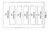
- FIG. 1 illustrates the steps for performing an arthroplasty in accordance with one embodiment of the present invention.
- FIG. 2 illustrates the steps for performing an arthrotomy in accordance with one embodiment of the present invention.
- FIG. 3 illustrates the steps for performing an exposure during an arthroplasty in accordance with one embodiment of the present invention.
- FIG. 4 illustrates an exemplary surgical method with respect to the anatomy of a knee in accordance with one embodiment of the present invention.
- Surgical methods of the present invention include an anteromedial approach to arthroplasty on a knee joint of a patient.
- These methods of arthroplasty include an arthrotomy, or surgical incision into the knee joint, made over the anterior aspects of the knee along the medial border of the patella.
- the arthrotomy includes a dissection from medial to the tibial tubercle and around the medial border of the patella and up to the proximal border of the patella.
- the arthrotomy may further include identifying the distal extend of the vastus medialis, determining the orientation of the fibers, making an oblique cut into the vastus medialis, and separating the muscle fibers.
- the arthrotomy is followed by an exposure step.
- Different embodiments of the present invention involve combinations of one or more of: retracting the patella laterally to expose the knee, excising the fat pad both medially and laterally and leaving a small amount of fat deep under the patella tendon, dividing the anterior horn of the medial meniscus, dissecting around the proximal medial tibia, placing a retractor on the proximal medial tibia, releasing the proximal soft tissue attachments extending around the proximal media tibia, and making a small window along the anterior surface of the distal femur.
- one embodiment of a method of arthroplasty on a knee joint of a patient in accordance with the present invention includes positioning the patients leg 102 , making one or more incisions 104 , performing an anteromedial arthrotomy 106 , an exposure 108 , among other steps 110 .
- the following paragraphs provides examples of each of these steps.
- a sandbag may be placed across from the contra-lateral ankle when positioning the patient on the operating table.
- a longitudinal incision is made over the anterior aspects of the knee along the medial border of the patella.
- the incision extends approximately from the proximal tibial tubercle to the proximal extent of the patella to one finger's breadth proximal to the patella.
- FIGS. 2 and 4 respectively, begin the dissection 401 medial to the tibial tubercle 402 , as recited in block 202 . Extend the dissection 401 around the medial border 406 of the patella 404 , as recited in block 204 . The arthrotomy extends up to the proximal border of the patella 404 , as is also recited in block 204 .
- the distal extend of the vastus medialis 408 is identified and the orientation 410 of the fibers 409 is determined, as recited in block 206 .
- An oblique cut is made into the vastus medialis 408 , as shown in block 208 and the muscle fibers 409 are then spread, as recited in block, 210 , approximately 2 centimeters.
- the patella is retracted laterally 302 .
- the fat pad is excised both medially and laterally leaving a small amount of fat deep under the patella tendon 304 .
- the anterior horn of the medial meniscus is divided 306 and dissection is carried around the proximal medial tibia 308 using electrocautery and boxed osteotome or other surgical knife.
- a retractor is placed on the proximal medial tibia 310 .
- the proximal soft tissue attachments extending around the proximal medial tibia are released 312 in a standard fashion.
- a small window is made along the anterior surface of the distal femur 314 with the use of electrocautery to reference the anterior cortex.
Abstract
A anteromedial approach to arthroplasty on a knee joint of a patient. The method of arthroplasty includes an arthrotomy that includes dissecting from medial to the tibial tubercle and around the medial border of the patella and up to the proximal border of the patella.
Description
This application claims the benefit of U.S. Ser. No. 60/518,018 filed Nov. 6, 2003 entitled “Minimally Invasive Knee Arthroplasty Surgical Technique and Related Instrumentation,” incorporated herein by this reference.
The invention relates to a method of arthroplasty on a knee joint of a patient. More particularly, it relates to minimally invasive surgical techniques used to perform arthroplasty on a knee joint of a patient.
Arthritis of the knees can wear away and destroy the surfaces of the knee joint and ultimately lead to conditions that require a knee replacement. In osteoarthritis, which is usually caused by old age, angular deformity, or old fractures, the surfaces of the knee gradually wear out. Likewise systemic arthritis affects the synovium or membrane tissue in the joint destroying the surface of the joint. In both osteoarthritis and systemic arthritis, when the surface of the joint is worn away, walking and other activities become difficult and painful. In many cases, knee replacements are performed to address these issues.
Knee replacement surgery or arthroplasty typically involves a resurfacing of the knee joint in which the femur, or thigh bone, and/or tibia, or shinbone, are covered with artificial materials such as metal or plastic thereby replacing the irregular surfaces caused by the arthritis with smooth surfaces. The undersurface of the patella, or knee cap, may also be replaced with artificial materials. Historically, the actual knee replacement procedure involved either general or epidural anesthesia with a four to six day hospitalization with the surgery itself taking about two hours.
There are three general types of knee replacements: total knee replacements (TKR), partial knee replacement (also called a unicompartmental knee replacement), and knee revision. A TKR resurfaces the knee joint by removing the diseased bone and cartilage. The end of the long bones of the knee joint, the femur and the tibia, are removed and replaced with artificial materials. The portion of the femur that is removed is usually replaced with metal (e.g. titanium or zirconium), while the portion of the tibia that is removed is usually replaced with plastic. The replacement components may be either cemented in place or inserted in a cementless manner.
A partial knee replacement involves installing an implant on one side of the knee, rather than over the entire surface of the knee joint. A unicondylar knee replacement is done if part of the knee joint is damaged by arthritis and the other compartments have healthy, normal cartilage at surgery.
A knee revision involves fixing problems with an existing knee replacement. Knee replacements fail over time and require revision. This revision procedure may be more complex than a total knee replacement for a number of reasons including because removing the existing implant may weaken the bone and damage the surrounding ligaments supporting the knee. Because of these problems a special implant may be used or a bone graft may be required to reconstruct deficient areas.
Traditional instrumentation and surgical techniques used in open arthroplasty surgery are not easily adapted for use in minimally invasive knee surgery. Specifically, using traditional instrumentation with smaller incisions can lead to stretching of the skin and misalignment of the knee replacement components due to lack of visibility. Using nontraditional surgical techniques, on the other hand, provides the disadvantage of requiring the surgeon to re-learn the surgery and can lead to longer operative times, higher complication rates, and higher surgical costs.
Methods of the present invention provide an anteromedial approach to knee arthroplasty to minimize the learning curve for surgeons while minimizing operation recovery time, blood loss, soft tissue damage, and incision length. The methods generally result in a faster, less painful return to function for the patient with a relatively small scar. One embodiment of the method of the present invention involves an arthrotomy made over the anterior aspects of the knee along the medial border of the patella. The position of this incision allows the knee arthroplasty to be performed without cutting the quad tendon and without everting, or turning outward, the patella.
In some embodiments of the present invention the knee arthroplasty is performed using a minimally invasive surgical technique and may be performed through an incision smaller than 10 inches, and preferably smaller than 5 inches.
Surgical methods of the present invention include an anteromedial approach to arthroplasty on a knee joint of a patient. These methods of arthroplasty include an arthrotomy, or surgical incision into the knee joint, made over the anterior aspects of the knee along the medial border of the patella. In one embodiment, the arthrotomy includes a dissection from medial to the tibial tubercle and around the medial border of the patella and up to the proximal border of the patella. The arthrotomy may further include identifying the distal extend of the vastus medialis, determining the orientation of the fibers, making an oblique cut into the vastus medialis, and separating the muscle fibers.
In one embodiment of the present method, the arthrotomy is followed by an exposure step. Different embodiments of the present invention involve combinations of one or more of: retracting the patella laterally to expose the knee, excising the fat pad both medially and laterally and leaving a small amount of fat deep under the patella tendon, dividing the anterior horn of the medial meniscus, dissecting around the proximal medial tibia, placing a retractor on the proximal medial tibia, releasing the proximal soft tissue attachments extending around the proximal media tibia, and making a small window along the anterior surface of the distal femur.
In one embodiment of the present invention, prior to an arthrotomy step, there is a step of making a longitudinal incision over the anterior aspect of the knee along the medial border of the patella extending approximately from the proximal tibia tubercle to the proximal extent of the patella to approximately the patella.
In one embodiment of the invention, prior to making the incision over the anterior aspect of the knee along the medial border of the patella, there is a first step of positioning the leg on the operating table so that the knee is flexed 70° to 90°.
Referring to FIG. 1 , one embodiment of a method of arthroplasty on a knee joint of a patient in accordance with the present invention includes positioning the patients leg 102, making one or more incisions 104, performing an anteromedial arthrotomy 106, an exposure 108, among other steps 110. The following paragraphs provides examples of each of these steps.
1. Leg Position
During the procedure the knee is flexed to 70° to 90°. Hyperextension is used only intermittently for specific parts of the procedure, such as insertion of a tibial component. In order to aid in holding the leg, a sandbag may be placed across from the contra-lateral ankle when positioning the patient on the operating table.
2. Incision
With the leg fully extended, a longitudinal incision is made over the anterior aspects of the knee along the medial border of the patella. The incision extends approximately from the proximal tibial tubercle to the proximal extent of the patella to one finger's breadth proximal to the patella.
3. Arthrotomy 200
As recited and shown in FIGS. 2 and 4 , respectively, begin the dissection 401 medial to the tibial tubercle 402, as recited in block 202. Extend the dissection 401 around the medial border 406 of the patella 404, as recited in block 204. The arthrotomy extends up to the proximal border of the patella 404, as is also recited in block 204.
The distal extend of the vastus medialis 408 is identified and the orientation 410 of the fibers 409 is determined, as recited in block 206. An oblique cut is made into the vastus medialis 408, as shown in block 208 and the muscle fibers 409 are then spread, as recited in block, 210, approximately 2 centimeters.
4. Exposure
As shown in FIG. 3 , with the leg extended, the patella is retracted laterally 302. The fat pad is excised both medially and laterally leaving a small amount of fat deep under the patella tendon 304. The anterior horn of the medial meniscus is divided 306 and dissection is carried around the proximal medial tibia 308 using electrocautery and boxed osteotome or other surgical knife. A retractor is placed on the proximal medial tibia 310. The proximal soft tissue attachments extending around the proximal medial tibia are released 312 in a standard fashion.
A small window is made along the anterior surface of the distal femur 314 with the use of electrocautery to reference the anterior cortex.
5. Other Steps
Other steps as required are carried out to complete the knee arthroplasty.
Modifications, adaptations, changes, deletions, and additions may be made to various embodiments of the present invention as disclosed in this document without departing from the scope or spirit of the invention.
Claims (18)
1. A method of arthroplasty on a knee joint of a patient comprising an arthrotomy comprising dissecting beginning at a position medial to a tibial tubercle, extending to a medial border of a patella, up to a proximal border of the patella, and obliquely into the vastus medialis in a direction of fibers of the vastus medialis.
2. The method of claim 1 wherein the method further comprises identifying the distal extend of the vastus medialis and the direction of the fibers of the vastus medialis prior to dissecting obliquely into the vastus medialis in the direction of the fibers.
3. The method of claim 2 wherein the method further comprises separating the muscle fibers.
4. The method of claim 3 further comprising retracting the patella laterally to expose the knee.
5. The method of claim 3 further comprising retracting the patella laterally without everting the patella to expose the knee.
6. The method of claim 4 further comprising excising the fat pad both medially and laterally and leaving a small amount of fat deep under the patella tendon.
7. The method of claim 6 further comprising dividing the anterior horn of the medial meniscus and dissecting around the proximal medial tibia.
8. The method of claim 7 further comprising placing a retractor on the proximal soft tissue attachments extending around the proximal medial tibia.
9. The method of claim 8 further comprising making a small window along the anterior surface of the distal femur.
10. The method of claim 9 further comprising, prior to the arthrotomy, making a longitudinal incision over an anterior aspect of the knee along the medial border of the patella extending approximately from the tibial tubercle to the border of the patella to approximately the patella.
11. The method of claim 10 further comprising, during preparation of the bones, positioning a leg of the patient on an operating table so that the knee is flexed 70° to 90°.
12. The method of claim 10 further comprising, prior to making the longitudinal incision in the knee, positioning a leg of the patient on an operating table so that the knee is flexed 70° to 90°.
13. A method of arthroplasty on a knee joint of a patient comprising:
an arthrotomy step comprising dissecting beginning at a position medial to a tibial tubercle and extending to a medial border of a patella and up to a proximal border of the patella, identifying the distal extend of the vastus medialis and determining the orientation of the fibers, extending the dissection with an oblique cut into the vastus medialis in a direction of the fibers and separating the muscle fibers; and
an exposure step comprising retracting the patella laterally to expose the knee, excising the fat pad both medially and laterally and leaving a small amount of fat deep under the patella tendon, dividing the anterior horn of the medial meniscus and dissecting around the proximal medial tibia, placing a retractor on the proximal soft tissue attachments extending around the proximal medial tibia, and making a small window along the anterior surface of the distal femur.
14. The method of claim 13 further comprising, prior to the arthrotomy, making a longitudinal incision over an anterior aspect of the knee along the medial border of the patella extending approximately from the tibial tubercle to the border of the patella to approximately the patella.
15. The method of claim 14 further comprising, during preparation of the bones, positioning a leg of the patient on an operating table so that the knee is flexed 70° to 90°.
16. The method of claim 14 further comprising, prior to making the longitudinal incision in the knee, positioning a leg of the patient on an operating table so that the knee is flexed 70° to 90°.
17. A method of arthroplasty on a knee joint of a patient comprising:
identifying the distal extend of the vastus medialis and determining the orientation of the fibers;
dissecting from medial to a tibial tubercle and extending to a medial border of a patella, up to a proximal border of the patella, and obliquely into the vastus medialis in a direction of the muscle fibers; and
separating the muscle fibers.
18. The method of claim 17 further comprising:
retracting the patella laterally to expose the knee;
excising the fat pad both medially and laterally and leaving a small amount of fat deep under the patella tendon;
dividing the anterior horn of the medial meniscus and dissecting around the proximal medial tibia;
placing a retractor on the proximal soft tissue attachments extending around the proximal medial tibia; and
making a small window along the anterior surface of the distal femur.
Priority Applications (1)
| Application Number | Priority Date | Filing Date | Title |
|---|---|---|---|
| US10/984,208 US7585328B2 (en) | 2003-11-06 | 2004-11-08 | Minimally invasive knee arthroplasty |
Applications Claiming Priority (2)
| Application Number | Priority Date | Filing Date | Title |
|---|---|---|---|
| US51801803P | 2003-11-06 | 2003-11-06 | |
| US10/984,208 US7585328B2 (en) | 2003-11-06 | 2004-11-08 | Minimally invasive knee arthroplasty |
Publications (2)
| Publication Number | Publication Date |
|---|---|
| US20070051378A1 US20070051378A1 (en) | 2007-03-08 |
| US7585328B2 true US7585328B2 (en) | 2009-09-08 |
Family
ID=37828929
Family Applications (1)
| Application Number | Title | Priority Date | Filing Date |
|---|---|---|---|
| US10/984,208 Active 2028-01-30 US7585328B2 (en) | 2003-11-06 | 2004-11-08 | Minimally invasive knee arthroplasty |
Country Status (1)
| Country | Link |
|---|---|
| US (1) | US7585328B2 (en) |
Cited By (14)
| Publication number | Priority date | Publication date | Assignee | Title |
|---|---|---|---|---|
| US8568486B2 (en) | 2010-07-24 | 2013-10-29 | Zimmer, Inc. | Asymmetric tibial components for a knee prosthesis |
| US8591594B2 (en) | 2010-09-10 | 2013-11-26 | Zimmer, Inc. | Motion facilitating tibial components for a knee prosthesis |
| US8628580B2 (en) | 2010-07-24 | 2014-01-14 | Zimmer, Inc. | Tibial prosthesis |
| US8740985B1 (en) | 2012-11-30 | 2014-06-03 | Smith & Nephew, Inc. | Knee prosthesis |
| US8758444B2 (en) | 2011-11-21 | 2014-06-24 | Zimmer, Inc. | Tibial baseplate with asymmetric placement of fixation structures |
| US9381090B2 (en) | 2010-07-24 | 2016-07-05 | Zimmer, Inc. | Asymmetric tibial components for a knee prosthesis |
| US10188530B2 (en) | 2010-12-17 | 2019-01-29 | Zimmer, Inc. | Provisional tibial prosthesis system |
| US10278827B2 (en) | 2015-09-21 | 2019-05-07 | Zimmer, Inc. | Prosthesis system including tibial bearing component |
| US10675153B2 (en) | 2017-03-10 | 2020-06-09 | Zimmer, Inc. | Tibial prosthesis with tibial bearing component securing feature |
| US10835380B2 (en) | 2018-04-30 | 2020-11-17 | Zimmer, Inc. | Posterior stabilized prosthesis system |
| US10898337B2 (en) | 2011-11-18 | 2021-01-26 | Zimmer, Inc. | Tibial bearing component for a knee prosthesis with improved articular characteristics |
| US11324598B2 (en) | 2013-08-30 | 2022-05-10 | Zimmer, Inc. | Method for optimizing implant designs |
| US11324599B2 (en) | 2017-05-12 | 2022-05-10 | Zimmer, Inc. | Femoral prostheses with upsizing and downsizing capabilities |
| US11426282B2 (en) | 2017-11-16 | 2022-08-30 | Zimmer, Inc. | Implants for adding joint inclination to a knee arthroplasty |
Citations (7)
| Publication number | Priority date | Publication date | Assignee | Title |
|---|---|---|---|---|
| US5122144A (en) | 1989-09-26 | 1992-06-16 | Kirschner Medical Corporation | Method and instrumentation for unicompartmental total knee arthroplasty |
| US5860980A (en) | 1997-09-15 | 1999-01-19 | Axelson, Jr.; Stuart L. | Surgical apparatus for use in total knee arthroplasty and surgical methods for using said apparatus |
| US6342075B1 (en) | 2000-02-18 | 2002-01-29 | Macarthur A. Creig | Prosthesis and methods for total knee arthroplasty |
| US20030130665A1 (en) * | 2000-03-10 | 2003-07-10 | Pinczewski Leo Arieh | Method of arthroplastly on a knee joint and apparatus for use in same |
| US20030153978A1 (en) * | 2002-02-08 | 2003-08-14 | Whiteside Biomechanics, Inc. | Apparatus and method of ligament balancing and component fit check in total knee arthroplasty |
| US20040153066A1 (en) * | 2003-02-03 | 2004-08-05 | Coon Thomas M. | Apparatus for knee surgery and method of use |
| US6984248B2 (en) * | 2001-01-16 | 2006-01-10 | Hyde Jr Edward R | Transosseous core approach and instrumentation for joint replacement and repair |
-
2004
- 2004-11-08 US US10/984,208 patent/US7585328B2/en active Active
Patent Citations (10)
| Publication number | Priority date | Publication date | Assignee | Title |
|---|---|---|---|---|
| US5122144A (en) | 1989-09-26 | 1992-06-16 | Kirschner Medical Corporation | Method and instrumentation for unicompartmental total knee arthroplasty |
| US5860980A (en) | 1997-09-15 | 1999-01-19 | Axelson, Jr.; Stuart L. | Surgical apparatus for use in total knee arthroplasty and surgical methods for using said apparatus |
| US6342075B1 (en) | 2000-02-18 | 2002-01-29 | Macarthur A. Creig | Prosthesis and methods for total knee arthroplasty |
| US20030130665A1 (en) * | 2000-03-10 | 2003-07-10 | Pinczewski Leo Arieh | Method of arthroplastly on a knee joint and apparatus for use in same |
| US20060167460A1 (en) * | 2000-03-10 | 2006-07-27 | Pinczewski Leo A | Method of arthroplastly on a knee joint and apparatus for use in same |
| US20070239167A1 (en) * | 2000-03-10 | 2007-10-11 | Smith & Nephew, Inc. | Method of arthroplasty on a knee joint and apparatus for use in same |
| US7371240B2 (en) * | 2000-03-10 | 2008-05-13 | Smith & Nephew, Inc. | Method of arthroplasty on a knee joint and apparatus for use in same |
| US6984248B2 (en) * | 2001-01-16 | 2006-01-10 | Hyde Jr Edward R | Transosseous core approach and instrumentation for joint replacement and repair |
| US20030153978A1 (en) * | 2002-02-08 | 2003-08-14 | Whiteside Biomechanics, Inc. | Apparatus and method of ligament balancing and component fit check in total knee arthroplasty |
| US20040153066A1 (en) * | 2003-02-03 | 2004-08-05 | Coon Thomas M. | Apparatus for knee surgery and method of use |
Non-Patent Citations (3)
| Title |
|---|
| Microplasty(TM) Minimally Invasive Knee Technique, Information for Health Care Professionals http://www.biomet.com/surgeons/products/microplasty/knee- technique, 20 pages (Oct. 13, 2004). |
| NexGen(R) Complete Knee Solution MIS Quad-Sparing(TM) Instrumentation Zimmer. 02 pages (Apr. 2, 2008) http//:www.zimmer.com/ctl?prcat=M3&prod=y&template=MP&action=1&op=global&id=9403&pr=Y. |
| Orthopedics: Advancements in Minimally Invasive Total Knee Arthroplasty, Alfred J. Tria, Jr., MD, Orthopedics(R) Aug. 2003 Supplement, pp. 1-10 http://www.orthobluejournal.com/supp/0803/tria.asp (Oct. 12, 2004). |
Cited By (39)
| Publication number | Priority date | Publication date | Assignee | Title |
|---|---|---|---|---|
| US10543099B2 (en) | 2010-07-24 | 2020-01-28 | Zimmer, Inc. | Tibial prosthesis |
| US10195041B2 (en) | 2010-07-24 | 2019-02-05 | Zimmer, Inc. | Asymmetric tibial components for a knee prosthesis |
| US11224519B2 (en) | 2010-07-24 | 2022-01-18 | Zimmer, Inc. | Asymmetric tibial components for a knee prosthesis |
| US8613775B2 (en) | 2010-07-24 | 2013-12-24 | Zimmer, Inc. | Asymmetric tibial components for a knee prosthesis |
| US8628580B2 (en) | 2010-07-24 | 2014-01-14 | Zimmer, Inc. | Tibial prosthesis |
| US9861490B2 (en) | 2010-07-24 | 2018-01-09 | Zimmer, Inc. | Asymmetric tibial components for a knee prosthesis |
| US8568486B2 (en) | 2010-07-24 | 2013-10-29 | Zimmer, Inc. | Asymmetric tibial components for a knee prosthesis |
| US8764840B2 (en) | 2010-07-24 | 2014-07-01 | Zimmer, Inc. | Tibial prosthesis |
| US9192480B2 (en) | 2010-07-24 | 2015-11-24 | Zimmer, Inc. | Asymmetric tibial components for a knee prosthesis |
| US9283082B2 (en) | 2010-07-24 | 2016-03-15 | Zimmer, Inc. | Methods related to seating of bearing component on tibial tray |
| US9295557B2 (en) | 2010-07-24 | 2016-03-29 | Zimmer, Inc. | Asymmetric tibial components for a knee prosthesis |
| US10470889B2 (en) | 2010-07-24 | 2019-11-12 | Zimmer, Inc. | Asymmetric tibial components for a knee prosthesis |
| US9381090B2 (en) | 2010-07-24 | 2016-07-05 | Zimmer, Inc. | Asymmetric tibial components for a knee prosthesis |
| US9918844B2 (en) | 2010-07-24 | 2018-03-20 | Zimmer, Inc. | Tibial prosthesis with a fixed bearing component |
| US8574304B2 (en) | 2010-07-24 | 2013-11-05 | Zimmer, Inc. | Asymmetric tibial components for a knee prosthesis |
| US9763796B2 (en) | 2010-07-24 | 2017-09-19 | Zimmer, Inc. | Asymmetric tibial components for a knee prosthesis |
| US9763794B2 (en) | 2010-07-24 | 2017-09-19 | Zimmer, Inc. | Tibial prosthesis |
| US11471288B2 (en) | 2010-09-10 | 2022-10-18 | Zimmer, Inc. | Motion facilitating tibial components for a knee prosthesis |
| US9763795B2 (en) | 2010-09-10 | 2017-09-19 | Zimmer, Inc. | Motion facilitating tibial components for a knee prosthesis |
| US8591594B2 (en) | 2010-09-10 | 2013-11-26 | Zimmer, Inc. | Motion facilitating tibial components for a knee prosthesis |
| US9314343B2 (en) | 2010-09-10 | 2016-04-19 | Zimmer, Inc. | Motion facilitating tibial components for a knee prosthesis |
| US10413415B2 (en) | 2010-09-10 | 2019-09-17 | Zimmer, Inc. | Motion facilitating tibial components for a knee prosthesis |
| US10188530B2 (en) | 2010-12-17 | 2019-01-29 | Zimmer, Inc. | Provisional tibial prosthesis system |
| US10898337B2 (en) | 2011-11-18 | 2021-01-26 | Zimmer, Inc. | Tibial bearing component for a knee prosthesis with improved articular characteristics |
| US10265181B2 (en) | 2011-11-21 | 2019-04-23 | Zimmer, Inc. | Tibial baseplate with asymmetric placement of fixation structures |
| US9707089B2 (en) | 2011-11-21 | 2017-07-18 | Zimmer, Inc. | Tibial baseplate with asymmetric placement of fixation structures |
| US9308096B2 (en) | 2011-11-21 | 2016-04-12 | Zimmer, Inc. | Tibial baseplate with asymmetric placement of fixation structures |
| US8758444B2 (en) | 2011-11-21 | 2014-06-24 | Zimmer, Inc. | Tibial baseplate with asymmetric placement of fixation structures |
| US8740985B1 (en) | 2012-11-30 | 2014-06-03 | Smith & Nephew, Inc. | Knee prosthesis |
| US9398958B2 (en) | 2012-11-30 | 2016-07-26 | Smith & Nephew, Inc. | Knee prosthesis |
| US11324598B2 (en) | 2013-08-30 | 2022-05-10 | Zimmer, Inc. | Method for optimizing implant designs |
| US11160659B2 (en) | 2015-09-21 | 2021-11-02 | Zimmer, Inc. | Prosthesis system including tibial bearing component |
| US10278827B2 (en) | 2015-09-21 | 2019-05-07 | Zimmer, Inc. | Prosthesis system including tibial bearing component |
| US10675153B2 (en) | 2017-03-10 | 2020-06-09 | Zimmer, Inc. | Tibial prosthesis with tibial bearing component securing feature |
| US11547571B2 (en) | 2017-03-10 | 2023-01-10 | Zimmer, Inc. | Tibial prosthesis with tibial bearing component securing feature |
| US11324599B2 (en) | 2017-05-12 | 2022-05-10 | Zimmer, Inc. | Femoral prostheses with upsizing and downsizing capabilities |
| US11426282B2 (en) | 2017-11-16 | 2022-08-30 | Zimmer, Inc. | Implants for adding joint inclination to a knee arthroplasty |
| US10835380B2 (en) | 2018-04-30 | 2020-11-17 | Zimmer, Inc. | Posterior stabilized prosthesis system |
| US11911279B2 (en) | 2018-04-30 | 2024-02-27 | Zimmer, Inc. | Posterior stabilized prosthesis system |
Also Published As
| Publication number | Publication date |
|---|---|
| US20070051378A1 (en) | 2007-03-08 |
Similar Documents
| Publication | Publication Date | Title |
|---|---|---|
| US6197064B1 (en) | Prosthetic implant | |
| US9339280B2 (en) | Spot facing trochlear groove | |
| US20050043807A1 (en) | Two-thirds prosthetic arthroplasty | |
| US10448960B2 (en) | Femoral finishing guide | |
| US11903651B2 (en) | System and method for pre-operatively determining desired alignment of a knee joint | |
| US10076419B2 (en) | Method of implanting a unicondylar knee prosthesis | |
| US20220330956A1 (en) | Tibial cut guide | |
| US7585328B2 (en) | Minimally invasive knee arthroplasty | |
| US20100222782A1 (en) | Spot facing trochlear groove | |
| Windsor et al. | Exposure in revision total knee arthroplasty: the femoral peel | |
| US10426504B1 (en) | Methods and systems for femoral condylar resection arthroplasty of the knee | |
| US10918396B1 (en) | Methods and systems for femoral condylar resection arthroplasty of the knee | |
| Liddle et al. | Surgery of the Knee | |
| Bindelglass et al. | Technique for total knee arthroplasty using simultaneous cementing of components | |
| Chauhan | Computer-assisted minimally invasive total knee arthroplasty | |
| GB2598580A (en) | Knee replacement bone preparation guide | |
| Marmor | Surgical technique of unicompartmental replacement | |
| David et al. | Surgery of the knee | |
| Mont et al. | Lateral approach to total knee arthroplasty: minimal soft tissue invasion | |
| Scott | Robert Brigham Unicondylar Knee surgical technique | |
| Benabdeslam et al. | Interest Of Navigation Surgery In Total Knee Arthroplasty | |
| AU2004203891A1 (en) | Two Thirds Prosthetic Arthroplastry |
Legal Events
| Date | Code | Title | Description |
|---|---|---|---|
| FEPP | Fee payment procedure |
Free format text: PAYOR NUMBER ASSIGNED (ORIGINAL EVENT CODE: ASPN); ENTITY STATUS OF PATENT OWNER: LARGE ENTITY |
|
| STCF | Information on status: patent grant |
Free format text: PATENTED CASE |
|
| CC | Certificate of correction | ||
| FPAY | Fee payment |
Year of fee payment: 4 |
|
| FPAY | Fee payment |
Year of fee payment: 8 |
|
| MAFP | Maintenance fee payment |
Free format text: PAYMENT OF MAINTENANCE FEE, 12TH YEAR, LARGE ENTITY (ORIGINAL EVENT CODE: M1553); ENTITY STATUS OF PATENT OWNER: LARGE ENTITY Year of fee payment: 12 |