US7658748B2 - Right retrieval mechanism - Google Patents
Right retrieval mechanism Download PDFInfo
- Publication number
- US7658748B2 US7658748B2 US10/668,445 US66844503A US7658748B2 US 7658748 B2 US7658748 B2 US 7658748B2 US 66844503 A US66844503 A US 66844503A US 7658748 B2 US7658748 B2 US 7658748B2
- Authority
- US
- United States
- Prior art keywords
- center
- arms
- occlusion device
- center post
- floating
- Prior art date
- Legal status (The legal status is an assumption and is not a legal conclusion. Google has not performed a legal analysis and makes no representation as to the accuracy of the status listed.)
- Expired - Lifetime
Links
- 230000007246 mechanism Effects 0.000 title claims description 13
- HWLDNSXPUQTBOD-UHFFFAOYSA-N platinum-iridium alloy Chemical compound [Ir].[Pt] HWLDNSXPUQTBOD-UHFFFAOYSA-N 0.000 claims description 14
- 229910001000 nickel titanium Inorganic materials 0.000 claims description 12
- 208000025339 heart septal defect Diseases 0.000 claims description 9
- HZEWFHLRYVTOIW-UHFFFAOYSA-N [Ti].[Ni] Chemical compound [Ti].[Ni] HZEWFHLRYVTOIW-UHFFFAOYSA-N 0.000 claims 6
- 238000011065 in-situ storage Methods 0.000 abstract description 6
- 230000002792 vascular Effects 0.000 abstract description 3
- 238000000034 method Methods 0.000 description 28
- 230000007547 defect Effects 0.000 description 23
- 239000000463 material Substances 0.000 description 16
- 239000006260 foam Substances 0.000 description 8
- 230000002787 reinforcement Effects 0.000 description 7
- 238000001356 surgical procedure Methods 0.000 description 7
- 230000000747 cardiac effect Effects 0.000 description 6
- 238000010030 laminating Methods 0.000 description 5
- 239000000853 adhesive Substances 0.000 description 4
- 230000001070 adhesive effect Effects 0.000 description 4
- HLXZNVUGXRDIFK-UHFFFAOYSA-N nickel titanium Chemical compound [Ti].[Ti].[Ti].[Ti].[Ti].[Ti].[Ti].[Ti].[Ti].[Ti].[Ti].[Ni].[Ni].[Ni].[Ni].[Ni].[Ni].[Ni].[Ni].[Ni].[Ni].[Ni].[Ni].[Ni].[Ni] HLXZNVUGXRDIFK-UHFFFAOYSA-N 0.000 description 4
- 238000007493 shaping process Methods 0.000 description 4
- RTAQQCXQSZGOHL-UHFFFAOYSA-N Titanium Chemical compound [Ti] RTAQQCXQSZGOHL-UHFFFAOYSA-N 0.000 description 3
- 208000001910 Ventricular Heart Septal Defects Diseases 0.000 description 3
- 208000013914 atrial heart septal defect Diseases 0.000 description 3
- 230000001010 compromised effect Effects 0.000 description 3
- 229910052751 metal Inorganic materials 0.000 description 3
- 239000002184 metal Substances 0.000 description 3
- 239000010936 titanium Substances 0.000 description 3
- 229910052719 titanium Inorganic materials 0.000 description 3
- 208000008883 Patent Foramen Ovale Diseases 0.000 description 2
- 240000007643 Phytolacca americana Species 0.000 description 2
- 239000004372 Polyvinyl alcohol Substances 0.000 description 2
- 208000007536 Thrombosis Diseases 0.000 description 2
- 230000008901 benefit Effects 0.000 description 2
- 238000002788 crimping Methods 0.000 description 2
- 206010016256 fatigue Diseases 0.000 description 2
- 238000005304 joining Methods 0.000 description 2
- 208000003278 patent ductus arteriosus Diseases 0.000 description 2
- 229920002451 polyvinyl alcohol Polymers 0.000 description 2
- 230000002829 reductive effect Effects 0.000 description 2
- 230000003014 reinforcing effect Effects 0.000 description 2
- 238000007789 sealing Methods 0.000 description 2
- 238000003466 welding Methods 0.000 description 2
- 229920004934 Dacron® Polymers 0.000 description 1
- WSFSSNUMVMOOMR-UHFFFAOYSA-N Formaldehyde Chemical compound O=C WSFSSNUMVMOOMR-UHFFFAOYSA-N 0.000 description 1
- HTTJABKRGRZYRN-UHFFFAOYSA-N Heparin Chemical compound OC1C(NC(=O)C)C(O)OC(COS(O)(=O)=O)C1OC1C(OS(O)(=O)=O)C(O)C(OC2C(C(OS(O)(=O)=O)C(OC3C(C(O)C(O)C(O3)C(O)=O)OS(O)(=O)=O)C(CO)O2)NS(O)(=O)=O)C(C(O)=O)O1 HTTJABKRGRZYRN-UHFFFAOYSA-N 0.000 description 1
- 230000002159 abnormal effect Effects 0.000 description 1
- 238000005452 bending Methods 0.000 description 1
- 239000000560 biocompatible material Substances 0.000 description 1
- 239000008280 blood Substances 0.000 description 1
- 210000004369 blood Anatomy 0.000 description 1
- 230000017531 blood circulation Effects 0.000 description 1
- 210000004204 blood vessel Anatomy 0.000 description 1
- 208000028831 congenital heart disease Diseases 0.000 description 1
- 238000010276 construction Methods 0.000 description 1
- 230000003247 decreasing effect Effects 0.000 description 1
- 238000009826 distribution Methods 0.000 description 1
- 230000000694 effects Effects 0.000 description 1
- 238000007667 floating Methods 0.000 description 1
- 239000006261 foam material Substances 0.000 description 1
- 238000002695 general anesthesia Methods 0.000 description 1
- 230000005802 health problem Effects 0.000 description 1
- 210000002837 heart atrium Anatomy 0.000 description 1
- 238000010438 heat treatment Methods 0.000 description 1
- 229960002897 heparin Drugs 0.000 description 1
- 229920000669 heparin Polymers 0.000 description 1
- 230000002401 inhibitory effect Effects 0.000 description 1
- 208000014674 injury Diseases 0.000 description 1
- 238000003780 insertion Methods 0.000 description 1
- 230000037431 insertion Effects 0.000 description 1
- 210000004971 interatrial septum Anatomy 0.000 description 1
- 210000005246 left atrium Anatomy 0.000 description 1
- 210000005240 left ventricle Anatomy 0.000 description 1
- 238000003754 machining Methods 0.000 description 1
- 230000009972 noncorrosive effect Effects 0.000 description 1
- 230000036407 pain Effects 0.000 description 1
- 229920000642 polymer Polymers 0.000 description 1
- 230000008569 process Effects 0.000 description 1
- 238000011084 recovery Methods 0.000 description 1
- 230000008439 repair process Effects 0.000 description 1
- 230000004044 response Effects 0.000 description 1
- 210000005245 right atrium Anatomy 0.000 description 1
- 210000005241 right ventricle Anatomy 0.000 description 1
- 230000006641 stabilisation Effects 0.000 description 1
- 238000011105 stabilization Methods 0.000 description 1
- 239000010935 stainless steel Substances 0.000 description 1
- 229910001220 stainless steel Inorganic materials 0.000 description 1
- 210000000115 thoracic cavity Anatomy 0.000 description 1
- 238000012876 topography Methods 0.000 description 1
- 230000008733 trauma Effects 0.000 description 1
Images


Classifications
-
- A—HUMAN NECESSITIES
- A61—MEDICAL OR VETERINARY SCIENCE; HYGIENE
- A61B—DIAGNOSIS; SURGERY; IDENTIFICATION
- A61B17/00—Surgical instruments, devices or methods, e.g. tourniquets
- A61B17/0057—Implements for plugging an opening in the wall of a hollow or tubular organ, e.g. for sealing a vessel puncture or closing a cardiac septal defect
- A61B2017/00575—Implements for plugging an opening in the wall of a hollow or tubular organ, e.g. for sealing a vessel puncture or closing a cardiac septal defect for closure at remote site, e.g. closing atrial septum defects
- A61B2017/00592—Elastic or resilient implements
-
- A—HUMAN NECESSITIES
- A61—MEDICAL OR VETERINARY SCIENCE; HYGIENE
- A61B—DIAGNOSIS; SURGERY; IDENTIFICATION
- A61B17/00—Surgical instruments, devices or methods, e.g. tourniquets
- A61B17/0057—Implements for plugging an opening in the wall of a hollow or tubular organ, e.g. for sealing a vessel puncture or closing a cardiac septal defect
- A61B2017/00575—Implements for plugging an opening in the wall of a hollow or tubular organ, e.g. for sealing a vessel puncture or closing a cardiac septal defect for closure at remote site, e.g. closing atrial septum defects
- A61B2017/00606—Implements H-shaped in cross-section, i.e. with occluders on both sides of the opening
-
- A—HUMAN NECESSITIES
- A61—MEDICAL OR VETERINARY SCIENCE; HYGIENE
- A61B—DIAGNOSIS; SURGERY; IDENTIFICATION
- A61B17/00—Surgical instruments, devices or methods, e.g. tourniquets
- A61B17/0057—Implements for plugging an opening in the wall of a hollow or tubular organ, e.g. for sealing a vessel puncture or closing a cardiac septal defect
- A61B2017/00575—Implements for plugging an opening in the wall of a hollow or tubular organ, e.g. for sealing a vessel puncture or closing a cardiac septal defect for closure at remote site, e.g. closing atrial septum defects
- A61B2017/00623—Introducing or retrieving devices therefor
Definitions
- This invention relates to an occlusion device for the closure of physical apertures, such as vascular or septal apertures. More specifically, this invention relates to an occlusion device for the heart that can be retrieved, reloaded, and redeployed in situ.
- the heart is generally comprised of four chambers: the left and right atrium and the left and right ventricle. Separating the left and right sides of the heart are two walls, or septa. The wall between the two atria is the interatrial septum, and the wall between the two ventricles is the interventricular septum.
- ASDs atrial septal defects
- VSDs ventricular septal defects
- each of these defects is generally an aperture, flap, or hole in the septum which allows blood to shunt between chambers in the heart where there is no blood flow in a normal, healthy heart. This abnormal shunt can cause a variety of health problems.
- occlusion devices To avoid the risks and discomfort associated with open heart surgery, modern occlusion devices have been developed that are small, implantable, and capable of being delivered to the heart through a catheter. Rather than surgery, a catheter inserted into a major blood vessel allows an occlusion device to be deployed by moving the device through the catheter to the treatment site within the body. This procedure is performed in a cardiac cathlab and avoids the risks, pain, and long recovery associated with open heart surgery.
- occlusion devices capable of being inserted via a catheter including button devices, collapsible umbrella-like structures, and plug-like devices. These modern occlusion devices can repair a wide range of cardiac defects, including patent foramen ovale, patent ductus arteriosus, atrial septal defects, ventricular septal defects, and may occlude other cardiac and non-cardiac apertures.
- One form of occlusion device generally has a left side, a right side, and a center section. Once the occluder is deployed, the left side sits in the left side of the patient's heart and the right side sits in the right side of the patient's heart. The occluder's center section extends through the center of the defect. The left and right sides occlude the aperture on the respective sides of the patient's septum.
- a physician loads the device into a catheter, advances the device via catheter to the treatment site, and deploys the device at the treatment site. Loading the device into the catheter must be done by hand, and requires a high degree of manual dexterity and takes time. Once the device is loaded, it is maneuvered through the catheter to the treatment site within the body. In the event the device is not deployed properly or effectively, it must be retrieved and the procedure must be performed again. Some of these devices are retrievable via catheter, but many require open heart surgery to be retrieved. Once retrieved, many types of occlusion devices cannot be redeployed. In such instances, a new occlusion device must be used, increasing the cost of the procedure.
- retrieval may require insertion of a larger diameter catheter.
- a larger diameter catheter may be needed because the device may not readily resume the compact shape it had before deployment.
- the device may be compromised from the stress of withdrawing it back into the catheter, even if a larger diameter catheter has been used. As such, it may not be possible to reuse the retrieved occlusion device.
- devices retrieved via catheter do not necessarily reload for redeployment when they are retrieved. Even if the device can be re-used, it must be completely removed from the catheter to be properly reloaded. Thus, even if the device is retrievable, the device must be pulled back through the catheter, removed, and reloaded. Once again, just as with the loading procedure, reloading is time consuming and requires high manual dexterity. This adds extra time to the procedure, and creates wear and tear on the device. Often, the retrieved device cannot be reused because it has been damaged by the retrieval process. If the device cannot be reused, a new device must be used, adding to the cost of the procedure.
- This invention relates to an occlusion device for the closure of physical apertures, such as vascular or septal defects. More specifically, this invention relates to a loading system for an occlusion device that allows the device be retrieved, reloaded, and redeployed in situ.
- the loading system comprises a plurality of puller arms and a floating center post to allow the device to automatically collapse for loading.
- the floating center post may be designed to work with a fixed center post present in the occlusion device. The design allows for easy loading as well as remote reloading and redeployment.
- the present invention When the present invention is retrieved, it reloads and is ready for immediate redeployment.
- the device does not need to be pulled back through the catheter so that the physician can reload it for deployment.
- the present design makes initial loading of the device easier. Because the device can be remotely retrieved and reloaded, and then redeployed, it reduces procedure time, cost, and risk to the patient.
- FIG. 1 is a perspective view of the top of an occlusion device.
- FIG. 2 is a perspective view of the bottom of an occlusion device.
- FIGS. 3 a - 3 e are a diagrammatic series showing how the device may be loaded or reloaded.
- FIG. 4 is a perspective view of a floating center post and a fixed center post.
- FIG. 5 is a perspective view of an end cap in place on a hoop.
- FIG. 6 a is a top view of an end cap which demonstrates one method of closing a hoop.
- FIG. 6 b is a top view of a coupler which demonstrates one method of closing a hoop.
- FIG. 7 is a top view of an occlusion device having a hoop which demonstrates how a sail is attached to the support frame and hoop.
- FIG. 8 is a perspective view of a foam patch in place on an end cap in an occlusion device.
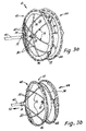
- FIG. 1 is a top perspective view of an occlusion device 10 .
- the occlusion device 10 comprises a fixed center post 12 , a floating center post 14 , a right fixation device 16 , wire arms 18 , puller arms 20 , and end caps 22 .
- the right fixation device 16 is connected to the center post 12 and comprises six wire arms 18 which are capped with the end caps 22 .
- the puller arms 20 are threaded through holes in the floating center post 14 .
- the ends of the puller arms 20 are also capped with end caps 22 .
- the occlusion device 10 also includes a right sheet 24 , a left sheet 26 , and wire hoops 28 .
- the hoops 28 surround the perimeter of the right and left sheets 24 , 26 and pass through holes 30 in the end caps 22 .
- a grasping knob 32 is located at the tip of the floating center post 14 .
- FIG. 2 is a bottom perspective view of the occlusion device 10 .
- FIG. 2 illustrates the center post 12 , the left sheet 26 , and a left fixation device 34 .
- the left fixation device 34 also comprises six arms 18 which are capped by end caps 22 .
- Also visible in FIG. 2 is the right sheet 24 , the hoops 28 , the floating center post 14 , the grasping knob 32 , and the puller arms 20 .
- the left fixation device 34 is located on an inner side of the left sheet 26 .
- the device 10 is not so limited, and the fixation devices 16 , 34 may be located on the outer side of the sheets 24 , 26 , on the inner side of the sheets 24 , 26 , or any combination thereof.
- the right and left fixation devices 16 , 34 are connected to the fixed center post 12 .
- One method of connecting the right and left fixation devices 16 , 34 to the fixed post 12 is to provide the fixed center post 12 with drill holes through which the fixation devices 16 , 34 pass.
- the fixation devices 16 , 34 may be formed of three wires. The three wires create the six arms 18 because the post 12 divides each wire into two arms 18 when the wire passes through the center post 12 .
- the end caps 22 provide a location for connecting the ends of the arms 18 to the hoops 28 .
- the end caps 22 located at the distal ends of the arms 16 serve to minimize damage to surrounding tissue once the device 10 is inserted.
- the sheets 24 , 26 are connected to the device 10 at the fixation devices 16 , 34 and at the hoops 28 .
- the sheets 24 , 26 may be connected to the fixation devices 16 , 34 using any suitable method.
- One method of attaching the sheets 24 , 26 to the fixation devices 16 , 34 is to suture the sheets 24 , 26 to the arms 16 of the fixation devices 16 , 34 .
- the sheets 24 , 26 may attach to the hoops 28 by folding each sheet 24 , 26 over the perimeter of the hoop 28 and then securing the sheets 24 , 26 in place.
- a variety of securing methods may be used such as suturing, heat treating, or laminating. Methods of attaching the sheets 24 , 26 to the hoops 28 are described more fully in FIGS. 7 and 8 .
- the hoops 28 attach to the device at the end caps 22 .
- the end caps 22 are provided with drilled holes 30 through which the hoops 28 can pass for attachment.
- the hoops 28 are constructed of a single, heat shaped wire which is threaded through the end caps 22 and then secured. Methods of securing the ends of the hoops 28 are described more fully in FIGS. 6 a and 6 b.
- the knob 32 on the floating center post 14 is configured to allow the device 10 to be grasped by a forceps as it is guided through the catheter.
- the method of attachment to a delivery device is not so limited.
- the knob 32 may be modified as needed to attach to any delivery device.
- the knob 32 may be fitted with threads so that it may be screwed onto a delivery device that is outfitted with threads. Only one side of the device 10 requires a knob 32 or other mechanism to attach to a delivery device.
- the device 10 is configured to be deployed through a catheter, and more specifically, the device 10 is constructed so that the fixation devices 16 , 34 are easily collapsible about the fixed center post 12 to allow the device 10 to be inserted through a catheter. Due to this construction, the device 10 can be folded such that the right fixation device 16 is folded backwards in the axial direction 36 , and the left fixation device 34 is folded in an opposite axial direction 38 , which allows the folded device 10 to fit into a small diameter catheter.
- the right and left sheets 24 , 26 attached to the fixation devices 16 , 34 , collapse as the fixation devices 16 , 34 are folded.
- the hoops 28 are also flexible and are configured to collapse.
- the fixation devices 16 , 34 , the hoops 28 , and the sheets 24 , 26 unfold to form a seal around each side of the defect.
- the fixation devices 16 , 36 are made of a suitable material capable of shape memory, such as nickel-titanium alloy, commonly called Nitinol. Nitinol is preferably used because it is commercially available, very elastic, non-corrosive and has a fatigue life greater than that of stainless steel.
- one embodiment of the present invention comprises making the wire fixation devices 16 , 34 of stranded wire or cables.
- the wire arms 18 are preferably subjected to precise pre-shaping to give them a “shape memory.”
- the pre-shaping can be done using any suitable method, such as machining, heat treating, or both.
- the shape memory helps to hold the strands together when the arms 18 are formed of stranded wire or cable, and can be used to add pretension to the arms 18 so that they “remember” their shape even after undergoing a strong deformation when the device 10 is passed through a catheter.
- the end caps 22 may further serve to prevent potential unraveling of the arms 18 when the arms 18 are formed of stranded wire or cable.
- the support hoops 28 are also made of a suitable material capable of shape memory, such as nickel-titanium alloy, like Nitinol.
- the hoops 28 may be constructed of a single wire or stranded wire.
- the diameter of the wire that is used to form the support hoops 28 must be small enough so that the hoops 28 are flexible enough to collapse when the device 10 is being loaded or retrieved.
- the wire must be stiff enough to allow the hoops 28 to lie as flat possible against the patient's septum to create an effective seal.
- the support hoops 28 may also be heat shaped or machine shaped so that the hoops 28 have shape memory to ensure that the hoops 28 resume the proper shape once the hoops 28 leave the catheter.
- Another advantage of pre-shaping the hoops 28 using heat is to ensure that the hoops 28 are properly sized. If the hoops 28 are too large or too small for the device 10 , the hoops 28 may pucker or may cause the sheets 24 , 26 to pucker. If the hoops 28 or sheets 24 , 26 pucker they cannot lie flat against the septum and therefore do not seal as effectively as the sheets 24 , 26 can if the sheets 24 , 26 lie flat and hug the septum.
- the support hoops 28 allow the device 10 to hug the tissue surrounding the defect, creating a uniform seal around the opening of the defect, which improves the sealing capabilities of the occlusion device 10 .
- the hoops 28 further serve to reduce the potential for increased pressure on surrounding tissue caused by one or more of the arms 18 . More specifically, once deployed, the arms 18 of the fixation devices 16 , 34 exert pressure on the hoops 28 and sheets 24 , 26 to form a seal around the defect. Without the hoops 28 , the highest points of pressure are the six or eight pressure points where the tips 22 of the arms 18 may press against the tissue surrounding the defect. Given the uneven topography of the heart, some arms 18 may put more pressure on surrounding tissue than others.
- the support hoops 28 help distribute pressure more evenly around a continuous circle, decreasing the possibility that increased pressure will be exerted at any one contact point. By distributing pressure more evenly, the risk that one or more of the arms 18 will poke through tissue or poke through the defect is greatly reduced.
- the sheets 24 , 26 are preferably formed of a medical grade polymer.
- One suitable material is DACRON®.
- the sheets 24 , 26 are formed of a high density polyvinyl alcohol (PVA) foam, such as that offered under the trademark IVALON®.
- PVA polyvinyl alcohol
- the sheets 24 , 26 may be treated with a thrombosis-inhibiting material.
- One such suitable material is heparin.
- the size of the sheets 24 , 26 may vary to accommodate various sizes of defects. In some instances, it may be desirable to form the sheets 24 , 26 so that they are not both the same size. For instance, one sheet and its associated fixation device can be made smaller than the corresponding sheet and its associated fixation device. This is particularly useful in situations where the occlusion device 10 is to be placed at a location in the heart which is close to other nearby cardiac structures. Making the sheets 24 , 26 different sizes may assist in providing optimal occlusion of a defect, without affecting other structures of the heart which may be nearby.
- the other parts of the stabilization device 10 are likewise formed of suitable materials. More specifically, the center posts 12 , 14 may be formed of platinum-iridium or titanium, and the end caps 22 may be formed of titanium.
- the puller arms 20 may be made of Nitinol, platinum-iridium, or titanium. However, the invention is not limited to these materials and any suitably biocompatible materials will suffice.
- the arms 18 vary slightly in length. To allow the device 10 to be retrievable and to ensure that the device 10 fits into as small a diameter catheter as possible, it is important that the arms 18 are not of a length that results in the end caps 22 clustering at the same location when loaded inside the catheter. If the end caps 22 are clustered in the same location when the device 10 is inside the catheter, the device 10 may become too bulky to allow it to be easily moved through the catheter. Thus, varying the arm 18 length allows the device 10 to fit more easily into a small diameter catheter when the device 10 is folded because the end caps 22 do not cluster. In addition, though shown with six arms 18 , the device 10 is not so limited. Rather, the device 10 may be comprised of four arms, or may be comprised of anywhere from five, six, eight, ten, or even more arms.
- the puller arms 20 are connected to the floating center post 14 by passing the arms 20 through holes drilled into the floating center post 14 .
- the ends of the puller arms 20 are then inserted into end caps 22 through which the hoop 28 has been threaded. Inserting the ends of the puller arms 20 into the end caps 22 connects the puller arms 20 to the hoop 28 .
- the number of puller arms 20 may be varied. In order to keep the mass of the device 10 minimal, however, the device 10 preferably has a small number of puller arms 20 .
- the device shown in FIGS. 1 and 2 has four puller arms 20 .
- the arms are spread out along the hoop so that they may collapse the hoop 28 .
- the arms should be arranged so that each arm 20 pulls on a different location on the hoop 28 so that the hoop 28 collapses.
- the puller arms 20 may be offset from one another by 45°.
- the device 10 is not so limited and the puller arms 20 may be offset from one another by anywhere from 5° to 180°.
- the device 10 needs only to be graspable on the right side, which is attached to the delivery or retrieval device. For this reason, only one set of puller arms 20 is required. If desired, additional sets of puller arms 20 may be added, but the addition would increase the mass of the device 10 .
- the puller arms 20 are preferably thinner than the arms of the fixation devices 16 , 34 and do not add much mass to the device 10 so the device 10 may fit through a small diameter catheter.
- the puller arms 20 do not experience constant stress once the device 10 is deployed, as the arms 18 of the fixation devices 16 , 34 do.
- the puller arms 20 must be strong enough to withstand stress during delivery and loading. Because the puller arms 20 must be flexible and do not need to exert constant pressure after the device 10 is deployed like the arms 18 of the fixation devices 16 , 34 do, the puller arms 18 may be constructed of lighter material.
- the puller arms 20 must be long enough to be capable of collapsing the hoop 28 but their length should be kept to a minimum so that they lie as close as possible to the septum after the device 10 is deployed and in place.
- One way to ensure that the puller arms 20 lie close to the septum is to heat shape the puller arms 20 so that they lie flat once the device 10 is deployed and in place.
- the puller arms 20 allow the device 10 to be loaded or reloaded for deployment.
- the right side (the side that sits in the right side of the patient's heart) of the device must be folded backwards 36 , with the hoops 28 facing axially away from the left side.
- the puller arms 20 transmit a pulling force from the knob 32 on the floating center post 14 to the hoop 28 to pull the right side backwards 34 to pull back the right side so that the right side is ready for loading and subsequent deployment.
- Another feature of the occlusion device 10 is that it is fully retrievable. In situations where the occlusion device 10 is not properly deployed and must be retrieved into the catheter, it is possible to withdraw the occlusion device 10 back into the catheter by grasping the knob 32 of the floating center section 14 and pulling. When the pulling arms 20 are pulled by the floating center section 14 , the right side of the device 10 collapses and can be withdrawn into a catheter. Subsequently, the device 10 may be deployed or redeployed.
- FIG. 3 a - 3 e are diagrammatic views which demonstrate the automatic loading system of the invention. Though FIGS. 3 a through 3 e show how to load a device, the same procedure applies if the device 10 must be retrieved, reloaded, and/or redeployed in situ.
- Shown from left to right in FIG. 3 a is a delivery device 40 , and the occlusion device 10 comprising the fixed center post 12 , the floating center post 14 , puller arms 20 , the fixation devices 16 , 34 , sheets 24 , 26 , and endcaps 22 .
- the occlusion device 10 comprises a right side 42 , a left side 44 , and a right and left hoop 46 , 48 .
- the right side 42 is so named because it is configured for placement in the right side of a patient's heart once the occlusion device 10 is deployed.
- the left side 42 is so named because it is configured for placement in the left side of a patient's heart once the occlusion device 10 is deployed.
- FIG. 3 a shows the occlusion device 10 before the occlusion device 10 is loaded into a catheter.
- Both the left and right sides 42 , 44 of the device 10 are shaped to bias the edges of the sheets 24 , 26 inward. As a result, each side 42 , 44 assumes an umbrella-like shape.
- shaping each side 42 , 44 with an inward bias helps ensure the sheets 24 , 26 fit snugly against the septum around the defect. This pressure exerted by the two sides 42 , 44 creates a seal around the defect.
- the floating center post 14 sits slightly up and away from the right side 42 of the device and the puller arms 20 are slightly curved.
- Delivery device 40 includes a grasping mechanism (not shown) for engaging and holding knob 32 , so that arial force can be applied to device 10 through delivery device 40 .
- FIG. 3 b shows the first step of loading the occlusion device 10 into a catheter.
- the delivery device 40 is pulled into a catheter in the direction of arrow 50 .
- the floating center post 14 engages the puller arms 20 because they are attached to the floating center post 14 .
- the puller arms 20 are attached to the right hoop 46 via end caps 22 .
- the pulling arms 20 can cause the right hoop 46 to pull away from the left side 44 , and cause the right hoop 46 to begin to collapse backwards, towards the direction of the pulling force 50 .
- a physician pulls on the delivery device 40 which pulls on the floating center post 14 .
- the puller arms 20 begin to lose curvature as the floating center post 14 is pulled away from the right side 42 of the device 10 .
- the puller arms 20 straighten and start to pull on the hoop 46 .
- the puller arms 20 are attached to the hoop 46 via end caps 22 , so the force on the puller arms 20 transmits to the end caps 22 and thus, to the right hoop 44 .
- the arms 20 begin to pull on the hoop 46 and pull the hoop 46 backwards, away from the left side 44 .
- the fixation devices 16 , 34 flex back also because they are attached to the hoop 46 at the end caps 22 .
- the pulling force continues to be transmitted from the delivery device 40 through the floating center post 14 and puller arms 20 to the right hoop 46 .
- the center post 14 is farther away and therefore the arms 20 pull more on the device right side 42 of the device 10 .
- the force from the puller arms 20 collapses the right hoop 46 further.
- the hoop 46 begins to buckle and collapse and the diameter of the hoop 46 is reduced. Because the arms 18 and hoop 46 are formed of flexible material, as the puller arms 20 move in the direction of the arrow 50 , the arms 18 of the fixation devices begin to bend.
- the arms 18 no longer bias the sheet 24 forward, but begin pulling on the center post 14 causing the center post 14 to move away from the fixed center post 12 .
- the puller arms 20 which are connected to the center post 14 and the right hoop 46 , start to fold the right side 42 of the device 10 in the direction of the arrow 50 .
- the left side 44 remains expanded.
- the physician grasps the left side 44 , holding it stationary, the delivery device 40 is pulled to collapse the right side 42 . If the device 10 were being removed and reloaded in situ, the left side 44 would be cupping the septum and thus, the left side 44 would not immediately respond to the pulling force because it would be held in place by the septum.
- FIG. 3 d shows the right side 42 fully collapsed and ready to be pulled into a catheter 50 .
- the continued pulling force has collapsed the right hoop 46 further, increasing the buckling of the right hoop 46 .
- the puller arms 20 are fully extended and are now almost horizontal. Because the arms 16 and sheet 24 are flexible, the arms 16 and sheet 24 collapse as well.
- the pulling force has collapsed the right hoop 46 to the point that the right side 42 of the device 10 is small enough to be pulled into a catheter 50 .
- FIG. 3 e shows the device 10 loaded into the catheter 50 .
- the right side 42 of the device 10 is pulled into the catheter 50
- the left side 44 has trailed and is pulled into the catheter 50 also.
- the left side 44 collapses backwards as it enters into the catheter 50 because the perimeter of the catheter 50 puts pressure on the fixation devices 34 of the left side 44 as the device 10 is pulled into the catheter 50 .
- the fixation devices 34 of the left side 44 collapse as the left side 44 is pulled into the catheter 50 .
- the device 10 is ready for deployment.
- the design of the present invention allows the device 10 to be loaded or reloaded easily, which has many advantages.
- the physician does not need to manually collapse the device 10 to ready it for deployment, which makes initial loading easier. If the device 10 has been deployed and must be retrieved and redeployed, the physician can retrieve, reload, and redeploy the device without first having to maneuver the device 10 out of the body to be reloaded. Most devices do not reload when retrieved and thus, must be maneuvered back out of the patient's body to be reloaded for redeployment. When the device 10 is pulled back through the catheter, out of the body, reloaded and redeployed, the integrity of the device 10 may be compromised.
- the present invention can save time, cost, and additional risk to the patient.
- FIG. 4 shows the floating center post 14 and the fixed center post 12 in greater detail. Shown is the floating center post 14 , holes 60 in the floating center post 14 through which puller arms pass, the fixed center post 12 , holes 62 in the fixed center post 12 , through which fixation devices pass, the grasping knob 32 , a pin 64 , and a lumen 66 .
- the pin 64 on the fixed post 12 fits into the lumen 66 on the floating post 14 .
- a delivery forceps or other delivery device attaches to the grasping knob 32 on the floating center post 14 .
- the delivery device pushes the device 10 using the floating center post 14 and the fixed center post 12 .
- the floating center post 14 is equipped with a lumen 66 that fits over the pin 64 extending from the fixed center post 12 .
- the lumen 66 connects with the pin 64 of the fixed center post 12 .
- connection between the floating center post 14 and fixed center post 12 improves the navigability of the device 10 .
- the connection of the lumen 66 to the pin 64 improves navigability by making it easier to “steer” the device 10 when the physician advances and maneuvers the device 10 through the body to the treatment site.
- the floating center post 14 separates from the fixed center post 12 .
- the floating center post 14 remains connected to the device by the puller arms 20 , which are threaded through holes in the floating center post 14 .
- the puller arms 20 also attach to the device 10 at the end caps 22 which are located at the hoops 28 . Therefore, the device 10 moves as a whole even though the two center posts 12 , 14 are not connected.
- FIG. 5 shows an enlarged perspective view of an end cap 22 in place on a hoop. Shown is an end cap 22 , a portion of a support hoop 70 , a portion of a wire arm 72 , which may be either a puller arm or a wire support arm, a hole 74 , and an end cavity 76 .
- the support hoop 70 passes through holes 74 drilled crosswise through the end cap 22 .
- the arm 72 is inserted into an end cavity 76 located at the base of the end cap 22 .
- the end caps 22 cap the ends of arms 72 to protect tissue and prevent unraveling of the wire arms 72 if the arms 72 are stranded.
- the tip of the end cap 22 is rounded to reduce the potential for trauma to the tissue surrounding a defect.
- the end caps 22 also serve as an attachment site for the support hoop 40 , the fixation devices 16 , 34 , and the puller arms 20 . By providing a link between the wire support arms 72 and the support hoop 70 , the end caps 22 assist in providing better distribution of pressure once the device 10 is deployed and is exerting pressure on the tissue surrounding a defect.
- the end caps 22 also serve to connect the puller arms 20 to the support hoop 40 , so that the arms 20 may exert pressure to collapse the device 10 when the device 10 must be loaded or reloaded into a catheter.
- FIGS. 6 a and 6 b are top plan views of a portion of the support hoop 70 .
- FIGS. 6 a and 6 b show two examples of how a support hoop can be closed so that it forms a circle.
- FIG. 6 a shows an end cap 22 , a portion of a support hoop 70 , a portion of a wire arm 74 , ends of the support hoop 80 , and an end cavity 76 .
- the support hoop 70 is typically formed of a single wire. To form the hoop 70 , the ends of the wire must be connected in order to form a 360° hoop.
- the support hoop 70 may be connected after it has been threaded through the end caps 22 . In FIG. 6 a , the ends of the support hoop 80 meet after passing through holes 74 in the end cap 22 so that the ends of the wire hoop 80 are connected inside the end cap 22 .
- the ends of the support hoop 80 are secured in the end cap 22 by any suitable method, such as crimping, welding, or adhesive.
- FIG. 6 b shows a second example of how a support hoop may be closed.
- FIG. 6 b shows a portion of a support hoop 70 which has been closed inside a coupler 82 . Shown is a portion of a support hoop 70 , ends of the support hoop 80 , and a coupler 82 .
- the coupler 82 is a small hollow tube with a diameter slightly larger than that of the wire used to construct the support hoop 70 .
- the ends of the support hoop 80 are inserted in the coupler 82 where they meet.
- the coupler 82 can then be crimped or welded so that the ends of the support hoop 80 remain inside the coupler 82 .
- Other possible methods of joining the ends of the support hoop 80 may include crimping the ends 80 together or welding the ends 80 together without the use of an end cap 22 or coupler 82 .
- FIG. 7 is a top view of an occlusion device 10 demonstrating how the right and left sheets 24 , 26 may be attached to the device 10 . Shown is the floating center post 14 , right wire fixation devices 16 comprising wire arms 18 , end caps 22 , the right sheet 24 , and the support hoop 28 . Also shown are sutures 62 and a reinforcement edge material 90 .
- the sutures 92 may be used to attach the right sheet 24 to the wire arms 18 .
- the second sheet 26 may be attached to the wire arms 18 in the same manner.
- the sutures 92 traverse the wire arms 18 , attaching the sheet 24 to the arms 16 .
- the foam sheets may be connected to the arms 18 using any suitable method, such as bonding, adhesive, heat treating, or laminating.
- the diameter of the right sheet 24 is slightly larger than that of the support hoop 28 .
- the portion of the right sheet 24 which extends beyond the support hoop 28 constitutes the reinforcement edge material 90 .
- the reinforcement edge material 90 allows this portion of the sheet to be folded over the support hoop 28 to form a reinforced edge of double material around the perimeter of the device 10 . Once the reinforcement edge material 90 has been folded over the support hoop 28 , it can be held in place such as suturing, bonding, adhesive, heat treating, laminating, or other suitable method.
- the reinforced edge 90 is created using a separate sheet of foam formed in a ring.
- the foam ring is sized to allow it to fold over the perimeter of the device 10 and support hoop 28 .
- the foam ring may be attached to the sheet 24 using any suitable method such as suturing, bonding, adhesive, heat treating, or laminating.
- the reinforcement edge material 90 covers the exposed edges of the occlusion device 10 .
- the reinforcement edge 90 acts as a cushion between the exposed metal edges of the occlusion device 10 and the tissue surrounding the defect, providing extra protection from pressure that the device 10 exerts on the tissue.
- the reinforced edge 90 also secures the sheets 24 , 26 to the device 10 .
- the wire arms 18 must bend to accommodate the contours of the heart. Because the sheets 24 , 26 are sewn to the wire arms 18 , the sheets 24 , 26 must accommodate the bending of the wire arms 18 . In locations where some of the wire arms 18 are bent by the contours of the heart, a portion of the sheets 24 , 26 may be stretched so that it experiences constant tension. This tension may cause the sheets 24 , 26 to tear, especially where the sutures 92 are located. If the sheets 24 , 26 tear, the sealing ability of the occlusion device 10 may be compromised.
- the reinforced edge 90 helps to prevent the first and second sheets 24 , 26 from tearing at the areas where the sheets 24 , 26 are attached to the device 10 or are sutured. Because the reinforcement edge 90 overlaps the hoop 28 and is then affixed to the rest of the sheet 24 , 26 , it adds an additional 360° of continuous attachment of the sheets 24 , 26 to the frame of the device 10 reducing the likelihood of tearing or detachment.
- the additional foam material along the perimeter of the device helps to distribute the tension on the sheets 24 , 26 along a continuum, instead of focusing tension at discrete attachment sites like the suture points.
- FIG. 8 demonstrates an alternative method of reinforcing attachment of the foam sheets to the occlusion device 10 .
- a patch 100 Shown is a patch 100 , a portion of a support hoop 70 , a portion of a wire arm 72 , and an end cap 22 .
- the patch 100 is constructed of foam and is configured to fit over the end cap 22 .
- the patch 100 is shaped like a cross which enables it to cover both sides of the end cap 22 and a small portion of the support hoop 70 where the hoop 70 extends out of the end cap 22 .
- the patch 100 may be secured by sutures, heat treatment, laminating, or another suitable method.
- the patch 100 also acts as a cushion between the metal end caps 22 of the occlusion device 10 and the tissue surrounding the defect, providing extra protection from pressure that the device 10 exerts on the tissue.
- the patch 100 also reduces the amount of metal to tissue contact.
Abstract
Description
Claims (38)
Priority Applications (3)
| Application Number | Priority Date | Filing Date | Title |
|---|---|---|---|
| US10/668,445 US7658748B2 (en) | 2003-09-23 | 2003-09-23 | Right retrieval mechanism |
| PCT/US2004/030164 WO2005032335A2 (en) | 2003-09-23 | 2004-09-15 | Right retrieval mechanism |
| EP04784130A EP1670363A4 (en) | 2003-09-23 | 2004-09-15 | Right retrieval mechanism |
Applications Claiming Priority (1)
| Application Number | Priority Date | Filing Date | Title |
|---|---|---|---|
| US10/668,445 US7658748B2 (en) | 2003-09-23 | 2003-09-23 | Right retrieval mechanism |
Publications (2)
| Publication Number | Publication Date |
|---|---|
| US20050065548A1 US20050065548A1 (en) | 2005-03-24 |
| US7658748B2 true US7658748B2 (en) | 2010-02-09 |
Family
ID=34313485
Family Applications (1)
| Application Number | Title | Priority Date | Filing Date |
|---|---|---|---|
| US10/668,445 Expired - Lifetime US7658748B2 (en) | 2003-09-23 | 2003-09-23 | Right retrieval mechanism |
Country Status (3)
| Country | Link |
|---|---|
| US (1) | US7658748B2 (en) |
| EP (1) | EP1670363A4 (en) |
| WO (1) | WO2005032335A2 (en) |
Cited By (36)
| Publication number | Priority date | Publication date | Assignee | Title |
|---|---|---|---|---|
| US20050192627A1 (en) * | 2003-10-10 | 2005-09-01 | Whisenant Brian K. | Patent foramen ovale closure devices, delivery apparatus and related methods and systems |
| US20060200196A1 (en) * | 2005-01-28 | 2006-09-07 | Lifetech Scientific Inc. | Heart Septal Defect Occlusion Devices With Adjustable Length Tether Adapting To The Unique Anatomy Of The Patient |
| US20080215089A1 (en) * | 2006-09-21 | 2008-09-04 | Williams Michael S | Stomach wall closure devices |
| US20090292310A1 (en) * | 2007-09-13 | 2009-11-26 | Dara Chin | Medical device for occluding a heart defect and a method of manufacturing the same |
| US20110066180A1 (en) * | 2005-01-28 | 2011-03-17 | Weijun Zeng | Heart septal defect occlusion device |
| US20110087270A1 (en) * | 2009-10-13 | 2011-04-14 | E-Pacing, Inc. | Vasculature Closure Devices and Methods |
| US20110288581A1 (en) * | 2010-05-19 | 2011-11-24 | Paul Jr Ram H | Devices and methods useful for sealing bodily openings |
| US20120071918A1 (en) * | 2008-03-07 | 2012-03-22 | Zahid Amin | Heart Occlusion Devices |
| US20120116447A1 (en) * | 2010-05-19 | 2012-05-10 | Cleon Stanley | Devices and methods useful for sealing bodily openings |
| US20120150218A1 (en) * | 2007-09-13 | 2012-06-14 | Robert Tyler Sandgren | Medical device for occluding a heart defect and a method of manufacturing the same |
| US20140081314A1 (en) * | 2005-12-01 | 2014-03-20 | Atritech, Inc. | Method and apparatus for delivering an implant without bias to a left atrial appendage |
| US8728155B2 (en) | 2011-03-21 | 2014-05-20 | Cephea Valve Technologies, Inc. | Disk-based valve apparatus and method for the treatment of valve dysfunction |
| US8870948B1 (en) | 2013-07-17 | 2014-10-28 | Cephea Valve Technologies, Inc. | System and method for cardiac valve repair and replacement |
| US20150094759A1 (en) * | 2012-06-11 | 2015-04-02 | E-Pacing, Inc. | Vasculature closure devices and methods |
| US9119607B2 (en) | 2008-03-07 | 2015-09-01 | Gore Enterprise Holdings, Inc. | Heart occlusion devices |
| US20160228109A1 (en) * | 2015-02-10 | 2016-08-11 | Vascular Solution, Inc. | Closure device for sealing percutaneous opening in a vessel |
| US9439757B2 (en) | 2014-12-09 | 2016-09-13 | Cephea Valve Technologies, Inc. | Replacement cardiac valves and methods of use and manufacture |
| US9474517B2 (en) | 2008-03-07 | 2016-10-25 | W. L. Gore & Associates, Inc. | Heart occlusion devices |
| US9724082B2 (en) | 2013-03-15 | 2017-08-08 | Cook Medical Technologies Llc | Delivery system for tissue opening closures |
| US9770232B2 (en) | 2011-08-12 | 2017-09-26 | W. L. Gore & Associates, Inc. | Heart occlusion devices |
| US9808230B2 (en) | 2014-06-06 | 2017-11-07 | W. L. Gore & Associates, Inc. | Sealing device and delivery system |
| US9943298B2 (en) | 2012-10-19 | 2018-04-17 | Cook Medical Technologies Llc | Vascular closure with shape memory characteristic |
| US9949728B2 (en) | 2007-04-05 | 2018-04-24 | W.L. Gore & Associates, Inc. | Septal closure device with centering mechanism |
| US10070850B2 (en) | 2012-10-19 | 2018-09-11 | Cook Medical Technologies Llc | Vascular closure with multiple connections |
| US10085731B2 (en) | 2013-07-15 | 2018-10-02 | E-Pacing, Inc. | Vasculature closure devices and methods |
| US10143552B2 (en) | 2015-05-14 | 2018-12-04 | Cephea Valve Technologies, Inc. | Replacement mitral valves |
| US10368990B2 (en) | 2017-01-23 | 2019-08-06 | Cephea Valve Technologies, Inc. | Replacement mitral valves |
| US10470881B2 (en) | 2015-05-14 | 2019-11-12 | Cephea Valve Technologies, Inc. | Replacement mitral valves |
| US10758216B2 (en) | 2013-03-14 | 2020-09-01 | Cook Medical Technologies Llc | Internal closure systems and devices |
| US10792025B2 (en) | 2009-06-22 | 2020-10-06 | W. L. Gore & Associates, Inc. | Sealing device and delivery system |
| US10806437B2 (en) | 2009-06-22 | 2020-10-20 | W. L. Gore & Associates, Inc. | Sealing device and delivery system |
| US10828019B2 (en) | 2013-01-18 | 2020-11-10 | W.L. Gore & Associates, Inc. | Sealing device and delivery system |
| US10849607B2 (en) | 2012-12-13 | 2020-12-01 | Cook Medical Technologies Llc | Vascular closure device suture tension mechanism |
| US10849746B2 (en) | 2015-05-14 | 2020-12-01 | Cephea Valve Technologies, Inc. | Cardiac valve delivery devices and systems |
| US11331187B2 (en) | 2016-06-17 | 2022-05-17 | Cephea Valve Technologies, Inc. | Cardiac valve delivery devices and systems |
| US11375988B2 (en) | 2003-07-14 | 2022-07-05 | W. L. Gore & Associates, Inc. | Patent foramen ovale (PFO) closure device with linearly elongating petals |
Families Citing this family (65)
| Publication number | Priority date | Publication date | Assignee | Title |
|---|---|---|---|---|
| US7674222B2 (en) * | 1999-08-09 | 2010-03-09 | Cardiokinetix, Inc. | Cardiac device and methods of use thereof |
| US8246671B2 (en) * | 1999-08-09 | 2012-08-21 | Cardiokinetix, Inc. | Retrievable cardiac devices |
| US9694121B2 (en) | 1999-08-09 | 2017-07-04 | Cardiokinetix, Inc. | Systems and methods for improving cardiac function |
| US10307147B2 (en) | 1999-08-09 | 2019-06-04 | Edwards Lifesciences Corporation | System for improving cardiac function by sealing a partitioning membrane within a ventricle |
| US8377114B2 (en) | 1999-08-09 | 2013-02-19 | Cardiokinetix, Inc. | Sealing and filling ventricular partitioning devices to improve cardiac function |
| US8529430B2 (en) * | 2002-08-01 | 2013-09-10 | Cardiokinetix, Inc. | Therapeutic methods and devices following myocardial infarction |
| US9332993B2 (en) | 2004-08-05 | 2016-05-10 | Cardiokinetix, Inc. | Devices and methods for delivering an endocardial device |
| US7399271B2 (en) * | 2004-01-09 | 2008-07-15 | Cardiokinetix, Inc. | Ventricular partitioning device |
| US10064696B2 (en) | 2000-08-09 | 2018-09-04 | Edwards Lifesciences Corporation | Devices and methods for delivering an endocardial device |
| US8398537B2 (en) * | 2005-06-10 | 2013-03-19 | Cardiokinetix, Inc. | Peripheral seal for a ventricular partitioning device |
| US9078660B2 (en) * | 2000-08-09 | 2015-07-14 | Cardiokinetix, Inc. | Devices and methods for delivering an endocardial device |
| US9332992B2 (en) | 2004-08-05 | 2016-05-10 | Cardiokinetix, Inc. | Method for making a laminar ventricular partitioning device |
| US7338514B2 (en) | 2001-06-01 | 2008-03-04 | St. Jude Medical, Cardiology Division, Inc. | Closure devices, related delivery methods and tools, and related methods of use |
| US7976564B2 (en) * | 2002-05-06 | 2011-07-12 | St. Jude Medical, Cardiology Division, Inc. | PFO closure devices and related methods of use |
| US8372112B2 (en) * | 2003-04-11 | 2013-02-12 | St. Jude Medical, Cardiology Division, Inc. | Closure devices, related delivery methods, and related methods of use |
| US20040267306A1 (en) | 2003-04-11 | 2004-12-30 | Velocimed, L.L.C. | Closure devices, related delivery methods, and related methods of use |
| US8480706B2 (en) | 2003-07-14 | 2013-07-09 | W.L. Gore & Associates, Inc. | Tubular patent foramen ovale (PFO) closure device with catch system |
| ES2436596T3 (en) | 2003-07-14 | 2014-01-03 | W.L. Gore & Associates, Inc. | Oval foramen tubular permeable closure device (FOP) with retention system |
| US8257389B2 (en) * | 2004-05-07 | 2012-09-04 | W.L. Gore & Associates, Inc. | Catching mechanisms for tubular septal occluder |
| US8162974B2 (en) * | 2006-02-02 | 2012-04-24 | Boston Scientific Scimed, Inc. | Occlusion apparatus, system, and method |
| US20070244494A1 (en) * | 2006-04-18 | 2007-10-18 | Downing Stephen W | Methods and devices for treating atrial septal defects |
| US20080051830A1 (en) * | 2006-08-24 | 2008-02-28 | Boston Scientific Scimed, Inc. | Occluding device and method |
| US8745845B2 (en) * | 2006-11-07 | 2014-06-10 | Dc Devices, Inc. | Methods for mounting a prosthesis onto a delivery device |
| US9232997B2 (en) | 2006-11-07 | 2016-01-12 | Corvia Medical, Inc. | Devices and methods for retrievable intra-atrial implants |
| US10413284B2 (en) | 2006-11-07 | 2019-09-17 | Corvia Medical, Inc. | Atrial pressure regulation with control, sensing, monitoring and therapy delivery |
| US20110257723A1 (en) | 2006-11-07 | 2011-10-20 | Dc Devices, Inc. | Devices and methods for coronary sinus pressure relief |
| US10624621B2 (en) | 2006-11-07 | 2020-04-21 | Corvia Medical, Inc. | Devices and methods for the treatment of heart failure |
| US8882697B2 (en) | 2006-11-07 | 2014-11-11 | Dc Devices, Inc. | Apparatus and methods to create and maintain an intra-atrial pressure relief opening |
| US8617205B2 (en) | 2007-02-01 | 2013-12-31 | Cook Medical Technologies Llc | Closure device |
| WO2008094706A2 (en) * | 2007-02-01 | 2008-08-07 | Cook Incorporated | Closure device and method of closing a bodily opening |
| US8915958B2 (en) * | 2007-06-08 | 2014-12-23 | St. Jude Medical, Inc. | Devices for transcatheter prosthetic heart valve implantation and access closure |
| US8366741B2 (en) * | 2007-09-13 | 2013-02-05 | Cardia, Inc. | Occlusion device with centering arm |
| US9943302B2 (en) * | 2008-08-12 | 2018-04-17 | Covidien Lp | Medical device for wound closure and method of use |
| US20100069930A1 (en) * | 2008-09-16 | 2010-03-18 | VentralFix, Inc. | Method and apparatus for minimally invasive delivery, tensioned deployment and fixation of secondary material prosthetic devices in patient body tissue, including hernia repair within the patient's herniation site |
| US10219796B2 (en) * | 2009-02-21 | 2019-03-05 | Farideh Roshanali | Device for percutaneous transcathertral closure of atrial septal defect by deploying pericardial patch |
| US8029534B2 (en) * | 2009-03-16 | 2011-10-04 | Cook Medical Technologies Llc | Closure device with string retractable umbrella |
| CH701269A1 (en) * | 2009-06-10 | 2010-12-15 | Carag Ag | Occluder. |
| US9381006B2 (en) | 2009-06-22 | 2016-07-05 | W. L. Gore & Associates, Inc. | Sealing device and delivery system |
| US9757107B2 (en) | 2009-09-04 | 2017-09-12 | Corvia Medical, Inc. | Methods and devices for intra-atrial shunts having adjustable sizes |
| US9642993B2 (en) | 2011-12-22 | 2017-05-09 | Corvia Medical, Inc. | Methods and devices for intra-atrial shunts having selectable flow rates |
| AU2010315535A1 (en) | 2009-10-26 | 2012-05-03 | Cardiokinetix, Inc. | Ventricular volume reduction |
| CA2785041A1 (en) * | 2010-01-29 | 2011-08-04 | Dc Devices, Inc. | Devices and methods for reducing venous pressure |
| EP2627265B8 (en) | 2010-10-15 | 2019-02-20 | Cook Medical Technologies LLC | Occlusion device for blocking fluid flow through bodily passages |
| EP2524653A1 (en) * | 2011-05-17 | 2012-11-21 | Carag AG | Occluder |
| US9005155B2 (en) | 2012-02-03 | 2015-04-14 | Dc Devices, Inc. | Devices and methods for treating heart failure |
| EP3281608B1 (en) | 2012-02-10 | 2020-09-16 | CVDevices, LLC | Medical product comprising a frame and visceral pleura |
| US10588611B2 (en) | 2012-04-19 | 2020-03-17 | Corvia Medical Inc. | Implant retention attachment and method of use |
| US9649480B2 (en) | 2012-07-06 | 2017-05-16 | Corvia Medical, Inc. | Devices and methods of treating or ameliorating diastolic heart failure through pulmonary valve intervention |
| US20140114346A1 (en) * | 2012-10-23 | 2014-04-24 | Medtronic, Inc. | Transapical Entry Point Closure Device |
| CA2900862C (en) | 2013-02-11 | 2017-10-03 | Cook Medical Technologies Llc | Expandable support frame and medical device |
| US9775636B2 (en) | 2013-03-12 | 2017-10-03 | Corvia Medical, Inc. | Devices, systems, and methods for treating heart failure |
| US10004512B2 (en) * | 2014-01-29 | 2018-06-26 | Cook Biotech Incorporated | Occlusion device and method of use thereof |
| US10675450B2 (en) | 2014-03-12 | 2020-06-09 | Corvia Medical, Inc. | Devices and methods for treating heart failure |
| CA2955389C (en) | 2014-07-23 | 2023-04-04 | Corvia Medical, Inc. | Devices and methods for treating heart failure |
| WO2016048802A1 (en) | 2014-09-28 | 2016-03-31 | Cardiokinetix, Inc. | Apparatuses for treating cardiac dysfunction |
| US20180303469A1 (en) * | 2015-07-10 | 2018-10-25 | Jose Pablo Diaz Jimenez | Medical device for closing fistulas, instrument for the positioning thereof, and method for the positioning and extraction of the device using said instrument |
| US10058426B2 (en) * | 2016-07-20 | 2018-08-28 | Abbott Cardiovascular Systems Inc. | System for tricuspid valve repair |
| US10420565B2 (en) | 2016-11-29 | 2019-09-24 | Abbott Cardiovascular Systems Inc. | Cinch and post for tricuspid valve repair |
| US10548614B2 (en) | 2016-11-29 | 2020-02-04 | Evalve, Inc. | Tricuspid valve repair system |
| US10952852B2 (en) | 2017-02-24 | 2021-03-23 | Abbott Cardiovascular Systems Inc. | Double basket assembly for valve repair |
| DE102018107407A1 (en) | 2017-03-28 | 2018-10-04 | Edwards Lifesciences Corporation | POSITIONING, INSERTING AND RETRIEVING IMPLANTABLE DEVICES |
| US10993807B2 (en) | 2017-11-16 | 2021-05-04 | Medtronic Vascular, Inc. | Systems and methods for percutaneously supporting and manipulating a septal wall |
| US11534303B2 (en) | 2020-04-09 | 2022-12-27 | Evalve, Inc. | Devices and systems for accessing and repairing a heart valve |
| JP2023512663A (en) * | 2020-01-24 | 2023-03-28 | パッチクランプ メドテック, インコーポレイテッド | Tissue repair and sealing device with removable graft and fastener assembly and method for using same |
| EP3912563A3 (en) * | 2020-05-21 | 2022-01-26 | St. Jude Medical, Cardiology Division, Inc. | Biomaterial occluder delivery mechanism |
Citations (31)
| Publication number | Priority date | Publication date | Assignee | Title |
|---|---|---|---|---|
| DE233303C (en) | 1911-04-05 | |||
| US3874388A (en) * | 1973-02-12 | 1975-04-01 | Ochsner Med Found Alton | Shunt defect closure system |
| US4007743A (en) | 1975-10-20 | 1977-02-15 | American Hospital Supply Corporation | Opening mechanism for umbrella-like intravascular shunt defect closure device |
| DD233303A1 (en) | 1984-12-28 | 1986-02-26 | Univ Berlin Humboldt | CLOSURE BODY FOR BLOOD OBJECTS AND METHOD FOR ITS INTRODUCTION |
| EP0362113A1 (en) | 1988-08-29 | 1990-04-04 | Eleftherios B. Sideris | Buttoned device for the transvenous occlusion of intracardiac defects |
| US5108420A (en) * | 1991-02-01 | 1992-04-28 | Temple University | Aperture occlusion device |
| US5171259A (en) | 1990-04-02 | 1992-12-15 | Kanji Inoue | Device for nonoperatively occluding a defect |
| EP0541063A2 (en) | 1991-11-05 | 1993-05-12 | The Children's Medical Center Corporation | Improved occluder for repair of cardiac and vascular defects |
| DE4222291C1 (en) | 1992-07-07 | 1994-01-20 | Krmek Mirko | Prosthesis for closing atrial or ventricular-septal defect - comprises two equally shaped units of elastic sprung material with six radially running arms spaced apart by equal edges |
| US5284488A (en) | 1992-12-23 | 1994-02-08 | Sideris Eleftherios B | Adjustable devices for the occlusion of cardiac defects |
| GB2269321A (en) | 1992-08-05 | 1994-02-09 | Nat Heart & Lung Inst | Implantable occluder device |
| US5334137A (en) | 1992-02-21 | 1994-08-02 | Eagle Vision, Inc. | Lacrimal fluid control device |
| US5334217A (en) | 1992-01-21 | 1994-08-02 | Regents Of The University Of Minnesota | Septal defect closure device |
| US5382260A (en) | 1992-10-30 | 1995-01-17 | Interventional Therapeutics Corp. | Embolization device and apparatus including an introducer cartridge and method for delivering the same |
| US5397331A (en) | 1991-11-25 | 1995-03-14 | Cook Incorporated | Supporting device and apparatus for inserting the device |
| US5433727A (en) | 1994-08-16 | 1995-07-18 | Sideris; Eleftherios B. | Centering buttoned device for the occlusion of large defects for occluding |
| US5451235A (en) | 1991-11-05 | 1995-09-19 | C.R. Bard, Inc. | Occluder and method for repair of cardiac and vascular defects |
| US5634936A (en) | 1995-02-06 | 1997-06-03 | Scimed Life Systems, Inc. | Device for closing a septal defect |
| US5649950A (en) | 1992-01-22 | 1997-07-22 | C. R. Bard | System for the percutaneous transluminal front-end loading delivery and retrieval of a prosthetic occluder |
| US5702421A (en) * | 1995-01-11 | 1997-12-30 | Schneidt; Bernhard | Closure device for closing a vascular opening, such as patent ductus arteriosus |
| US5709707A (en) * | 1995-10-30 | 1998-01-20 | Children's Medical Center Corporation | Self-centering umbrella-type septal closure device |
| US5725552A (en) | 1994-07-08 | 1998-03-10 | Aga Medical Corporation | Percutaneous catheter directed intravascular occlusion devices |
| US5733294A (en) * | 1996-02-28 | 1998-03-31 | B. Braun Medical, Inc. | Self expanding cardiovascular occlusion device, method of using and method of making the same |
| US5741297A (en) | 1996-08-28 | 1998-04-21 | Simon; Morris | Daisy occluder and method for septal defect repair |
| US5853422A (en) * | 1996-03-22 | 1998-12-29 | Scimed Life Systems, Inc. | Apparatus and method for closing a septal defect |
| US5904703A (en) | 1996-05-08 | 1999-05-18 | Bard Connaught | Occluder device formed from an open cell foam material |
| US6174322B1 (en) | 1997-08-08 | 2001-01-16 | Cardia, Inc. | Occlusion device for the closure of a physical anomaly such as a vascular aperture or an aperture in a septum |
| US6206907B1 (en) * | 1999-05-07 | 2001-03-27 | Cardia, Inc. | Occlusion device with stranded wire support arms |
| US6261309B1 (en) * | 1998-11-02 | 2001-07-17 | Datascope Investment Corp. | Collapsible hemostatic plug |
| US20010044634A1 (en) * | 2000-03-10 | 2001-11-22 | Don Michael T. Anthony | Vascular embolism prevention device employing filters |
| US6379368B1 (en) | 1999-05-13 | 2002-04-30 | Cardia, Inc. | Occlusion device with non-thrombogenic properties |
Family Cites Families (5)
| Publication number | Priority date | Publication date | Assignee | Title |
|---|---|---|---|---|
| US5797960A (en) * | 1993-02-22 | 1998-08-25 | Stevens; John H. | Method and apparatus for thoracoscopic intracardiac procedures |
| EP1284663A4 (en) * | 2000-05-18 | 2007-04-18 | Emphasys Medical Inc | Bronchiopulmonary occlusion devices and lung volume reduction methods |
| US6740331B1 (en) * | 2000-08-25 | 2004-05-25 | Global Gene Therapies, Inc. | Apparatus for the delivery of drugs or gene therapy into a patient's vasculature and methods of use |
| WO2003053493A2 (en) | 2001-12-19 | 2003-07-03 | Nmt Medical, Inc. | Septal occluder and associated methods |
| ES2436596T3 (en) * | 2003-07-14 | 2014-01-03 | W.L. Gore & Associates, Inc. | Oval foramen tubular permeable closure device (FOP) with retention system |
-
2003
- 2003-09-23 US US10/668,445 patent/US7658748B2/en not_active Expired - Lifetime
-
2004
- 2004-09-15 WO PCT/US2004/030164 patent/WO2005032335A2/en active Application Filing
- 2004-09-15 EP EP04784130A patent/EP1670363A4/en not_active Withdrawn
Patent Citations (35)
| Publication number | Priority date | Publication date | Assignee | Title |
|---|---|---|---|---|
| DE233303C (en) | 1911-04-05 | |||
| US3874388A (en) * | 1973-02-12 | 1975-04-01 | Ochsner Med Found Alton | Shunt defect closure system |
| US4007743A (en) | 1975-10-20 | 1977-02-15 | American Hospital Supply Corporation | Opening mechanism for umbrella-like intravascular shunt defect closure device |
| DD233303A1 (en) | 1984-12-28 | 1986-02-26 | Univ Berlin Humboldt | CLOSURE BODY FOR BLOOD OBJECTS AND METHOD FOR ITS INTRODUCTION |
| EP0362113A1 (en) | 1988-08-29 | 1990-04-04 | Eleftherios B. Sideris | Buttoned device for the transvenous occlusion of intracardiac defects |
| US4917089A (en) | 1988-08-29 | 1990-04-17 | Sideris Eleftherios B | Buttoned device for the transvenous occlusion of intracardiac defects |
| US5171259A (en) | 1990-04-02 | 1992-12-15 | Kanji Inoue | Device for nonoperatively occluding a defect |
| US5108420A (en) * | 1991-02-01 | 1992-04-28 | Temple University | Aperture occlusion device |
| US5425744A (en) | 1991-11-05 | 1995-06-20 | C. R. Bard, Inc. | Occluder for repair of cardiac and vascular defects |
| EP0541063A2 (en) | 1991-11-05 | 1993-05-12 | The Children's Medical Center Corporation | Improved occluder for repair of cardiac and vascular defects |
| EP0541063B1 (en) | 1991-11-05 | 1998-09-02 | The Children's Medical Center Corporation | Improved occluder for repair of cardiac and vascular defects |
| US5451235A (en) | 1991-11-05 | 1995-09-19 | C.R. Bard, Inc. | Occluder and method for repair of cardiac and vascular defects |
| US5397331A (en) | 1991-11-25 | 1995-03-14 | Cook Incorporated | Supporting device and apparatus for inserting the device |
| US5334217A (en) | 1992-01-21 | 1994-08-02 | Regents Of The University Of Minnesota | Septal defect closure device |
| US5649950A (en) | 1992-01-22 | 1997-07-22 | C. R. Bard | System for the percutaneous transluminal front-end loading delivery and retrieval of a prosthetic occluder |
| US5334137A (en) | 1992-02-21 | 1994-08-02 | Eagle Vision, Inc. | Lacrimal fluid control device |
| DE4222291C1 (en) | 1992-07-07 | 1994-01-20 | Krmek Mirko | Prosthesis for closing atrial or ventricular-septal defect - comprises two equally shaped units of elastic sprung material with six radially running arms spaced apart by equal edges |
| GB2269321A (en) | 1992-08-05 | 1994-02-09 | Nat Heart & Lung Inst | Implantable occluder device |
| US5382260A (en) | 1992-10-30 | 1995-01-17 | Interventional Therapeutics Corp. | Embolization device and apparatus including an introducer cartridge and method for delivering the same |
| US5284488A (en) | 1992-12-23 | 1994-02-08 | Sideris Eleftherios B | Adjustable devices for the occlusion of cardiac defects |
| US5725552A (en) | 1994-07-08 | 1998-03-10 | Aga Medical Corporation | Percutaneous catheter directed intravascular occlusion devices |
| US5433727A (en) | 1994-08-16 | 1995-07-18 | Sideris; Eleftherios B. | Centering buttoned device for the occlusion of large defects for occluding |
| US5702421A (en) * | 1995-01-11 | 1997-12-30 | Schneidt; Bernhard | Closure device for closing a vascular opening, such as patent ductus arteriosus |
| US5634936A (en) | 1995-02-06 | 1997-06-03 | Scimed Life Systems, Inc. | Device for closing a septal defect |
| US5709707A (en) * | 1995-10-30 | 1998-01-20 | Children's Medical Center Corporation | Self-centering umbrella-type septal closure device |
| US5733294A (en) * | 1996-02-28 | 1998-03-31 | B. Braun Medical, Inc. | Self expanding cardiovascular occlusion device, method of using and method of making the same |
| US5853422A (en) * | 1996-03-22 | 1998-12-29 | Scimed Life Systems, Inc. | Apparatus and method for closing a septal defect |
| US6024756A (en) | 1996-03-22 | 2000-02-15 | Scimed Life Systems, Inc. | Method of reversibly closing a septal defect |
| US5904703A (en) | 1996-05-08 | 1999-05-18 | Bard Connaught | Occluder device formed from an open cell foam material |
| US5741297A (en) | 1996-08-28 | 1998-04-21 | Simon; Morris | Daisy occluder and method for septal defect repair |
| US6174322B1 (en) | 1997-08-08 | 2001-01-16 | Cardia, Inc. | Occlusion device for the closure of a physical anomaly such as a vascular aperture or an aperture in a septum |
| US6261309B1 (en) * | 1998-11-02 | 2001-07-17 | Datascope Investment Corp. | Collapsible hemostatic plug |
| US6206907B1 (en) * | 1999-05-07 | 2001-03-27 | Cardia, Inc. | Occlusion device with stranded wire support arms |
| US6379368B1 (en) | 1999-05-13 | 2002-04-30 | Cardia, Inc. | Occlusion device with non-thrombogenic properties |
| US20010044634A1 (en) * | 2000-03-10 | 2001-11-22 | Don Michael T. Anthony | Vascular embolism prevention device employing filters |
Cited By (81)
| Publication number | Priority date | Publication date | Assignee | Title |
|---|---|---|---|---|
| US11375988B2 (en) | 2003-07-14 | 2022-07-05 | W. L. Gore & Associates, Inc. | Patent foramen ovale (PFO) closure device with linearly elongating petals |
| US20050192627A1 (en) * | 2003-10-10 | 2005-09-01 | Whisenant Brian K. | Patent foramen ovale closure devices, delivery apparatus and related methods and systems |
| US20060200196A1 (en) * | 2005-01-28 | 2006-09-07 | Lifetech Scientific Inc. | Heart Septal Defect Occlusion Devices With Adjustable Length Tether Adapting To The Unique Anatomy Of The Patient |
| US7828818B2 (en) * | 2005-01-28 | 2010-11-09 | Lifetech Scientific Inc. | Heart septal defect occlusion devices with adjustable length tether adapting to the unique anatomy of the patient |
| US20110066180A1 (en) * | 2005-01-28 | 2011-03-17 | Weijun Zeng | Heart septal defect occlusion device |
| US8366743B2 (en) | 2005-01-28 | 2013-02-05 | Lifetech Scientific (Shenzhen) Co., Ltd | Heart septal defect occlusion device |
| US20140081314A1 (en) * | 2005-12-01 | 2014-03-20 | Atritech, Inc. | Method and apparatus for delivering an implant without bias to a left atrial appendage |
| US10076335B2 (en) | 2005-12-01 | 2018-09-18 | Atritech, Inc. | Apparatus for delivering an implant without bias to a left atrial appendage |
| US10898198B2 (en) | 2005-12-01 | 2021-01-26 | Atritech, Inc. | Apparatus for delivering an implant without bias to a left atrial appendage |
| US20080215089A1 (en) * | 2006-09-21 | 2008-09-04 | Williams Michael S | Stomach wall closure devices |
| US10485525B2 (en) | 2007-04-05 | 2019-11-26 | W.L. Gore & Associates, Inc. | Septal closure device with centering mechanism |
| US9949728B2 (en) | 2007-04-05 | 2018-04-24 | W.L. Gore & Associates, Inc. | Septal closure device with centering mechanism |
| US20120150218A1 (en) * | 2007-09-13 | 2012-06-14 | Robert Tyler Sandgren | Medical device for occluding a heart defect and a method of manufacturing the same |
| US20090292310A1 (en) * | 2007-09-13 | 2009-11-26 | Dara Chin | Medical device for occluding a heart defect and a method of manufacturing the same |
| US9138213B2 (en) * | 2008-03-07 | 2015-09-22 | W.L. Gore & Associates, Inc. | Heart occlusion devices |
| US10278705B2 (en) | 2008-03-07 | 2019-05-07 | W. L. Gore & Associates, Inc. | Heart occlusion devices |
| US9474517B2 (en) | 2008-03-07 | 2016-10-25 | W. L. Gore & Associates, Inc. | Heart occlusion devices |
| US20120071918A1 (en) * | 2008-03-07 | 2012-03-22 | Zahid Amin | Heart Occlusion Devices |
| US9119607B2 (en) | 2008-03-07 | 2015-09-01 | Gore Enterprise Holdings, Inc. | Heart occlusion devices |
| US11589853B2 (en) | 2009-06-22 | 2023-02-28 | W. L. Gore & Associates, Inc. | Sealing device and delivery system |
| US10806437B2 (en) | 2009-06-22 | 2020-10-20 | W. L. Gore & Associates, Inc. | Sealing device and delivery system |
| US11596391B2 (en) | 2009-06-22 | 2023-03-07 | W. L. Gore & Associates, Inc. | Sealing device and delivery system |
| US10792025B2 (en) | 2009-06-22 | 2020-10-06 | W. L. Gore & Associates, Inc. | Sealing device and delivery system |
| US11564672B2 (en) | 2009-06-22 | 2023-01-31 | W. L. Gore & Associates, Inc. | Sealing device and delivery system |
| US20110087270A1 (en) * | 2009-10-13 | 2011-04-14 | E-Pacing, Inc. | Vasculature Closure Devices and Methods |
| US8845682B2 (en) * | 2009-10-13 | 2014-09-30 | E-Pacing, Inc. | Vasculature closure devices and methods |
| US9277904B2 (en) * | 2010-05-19 | 2016-03-08 | Cook Medical Technologies Llc | Devices and methods useful for sealing bodily openings |
| US8932325B2 (en) * | 2010-05-19 | 2015-01-13 | Cook Medical Technologies Llc | Devices and methods useful for sealing bodily openings |
| US10441256B2 (en) | 2010-05-19 | 2019-10-15 | Cook Medical Technologies Llc | Devices and methods useful for sealing bodily openings |
| US20120116447A1 (en) * | 2010-05-19 | 2012-05-10 | Cleon Stanley | Devices and methods useful for sealing bodily openings |
| US20110288581A1 (en) * | 2010-05-19 | 2011-11-24 | Paul Jr Ram H | Devices and methods useful for sealing bodily openings |
| US8728155B2 (en) | 2011-03-21 | 2014-05-20 | Cephea Valve Technologies, Inc. | Disk-based valve apparatus and method for the treatment of valve dysfunction |
| US11931252B2 (en) | 2011-03-21 | 2024-03-19 | Cephea Valve Technologies, Inc. | Disk-based valve apparatus and method for the treatment of valve dysfunction |
| US10456255B2 (en) | 2011-03-21 | 2019-10-29 | Cephea Valve Technologies, Inc. | Disk-based valve apparatus and method for the treatment of valve dysfunction |
| US9770232B2 (en) | 2011-08-12 | 2017-09-26 | W. L. Gore & Associates, Inc. | Heart occlusion devices |
| US20150094759A1 (en) * | 2012-06-11 | 2015-04-02 | E-Pacing, Inc. | Vasculature closure devices and methods |
| US9943298B2 (en) | 2012-10-19 | 2018-04-17 | Cook Medical Technologies Llc | Vascular closure with shape memory characteristic |
| US11317901B2 (en) | 2012-10-19 | 2022-05-03 | Cook Medical Technologies Llc | Vascular closure with multiple connections |
| US10070850B2 (en) | 2012-10-19 | 2018-09-11 | Cook Medical Technologies Llc | Vascular closure with multiple connections |
| US10729416B2 (en) | 2012-10-19 | 2020-08-04 | Cook Medical Technologies Llc | Vascular closure with shape memory characteristic |
| US10849607B2 (en) | 2012-12-13 | 2020-12-01 | Cook Medical Technologies Llc | Vascular closure device suture tension mechanism |
| US10828019B2 (en) | 2013-01-18 | 2020-11-10 | W.L. Gore & Associates, Inc. | Sealing device and delivery system |
| US11771408B2 (en) | 2013-01-18 | 2023-10-03 | W. L. Gore & Associates, Inc. | Sealing device and delivery system |
| US10758216B2 (en) | 2013-03-14 | 2020-09-01 | Cook Medical Technologies Llc | Internal closure systems and devices |
| US9724082B2 (en) | 2013-03-15 | 2017-08-08 | Cook Medical Technologies Llc | Delivery system for tissue opening closures |
| US10085731B2 (en) | 2013-07-15 | 2018-10-02 | E-Pacing, Inc. | Vasculature closure devices and methods |
| US8870948B1 (en) | 2013-07-17 | 2014-10-28 | Cephea Valve Technologies, Inc. | System and method for cardiac valve repair and replacement |
| US10154906B2 (en) | 2013-07-17 | 2018-12-18 | Cephea Valve Technologies, Inc. | System and method for cardiac valve repair and replacement |
| US10149761B2 (en) | 2013-07-17 | 2018-12-11 | Cephea Valve Technlologies, Inc. | System and method for cardiac valve repair and replacement |
| US11510780B2 (en) | 2013-07-17 | 2022-11-29 | Cephea Valve Technologies, Inc. | System and method for cardiac valve repair and replacement |
| US9554899B2 (en) | 2013-07-17 | 2017-01-31 | Cephea Valve Technologies, Inc. | System and method for cardiac valve repair and replacement |
| US9561103B2 (en) | 2013-07-17 | 2017-02-07 | Cephea Valve Technologies, Inc. | System and method for cardiac valve repair and replacement |
| US10624742B2 (en) | 2013-07-17 | 2020-04-21 | Cephea Valve Technologies, Inc. | System and method for cardiac valve repair and replacement |
| US9808230B2 (en) | 2014-06-06 | 2017-11-07 | W. L. Gore & Associates, Inc. | Sealing device and delivery system |
| US10368853B2 (en) | 2014-06-06 | 2019-08-06 | W. L. Gore & Associates, Inc. | Sealing device and delivery system |
| US11298116B2 (en) | 2014-06-06 | 2022-04-12 | W. L. Gore & Associates, Inc. | Sealing device and delivery system |
| US9439757B2 (en) | 2014-12-09 | 2016-09-13 | Cephea Valve Technologies, Inc. | Replacement cardiac valves and methods of use and manufacture |
| US11147665B2 (en) | 2014-12-09 | 2021-10-19 | Cepha Valve Technologies, Inc. | Replacement cardiac valves and methods of use and manufacture |
| US10548721B2 (en) | 2014-12-09 | 2020-02-04 | Cephea Valve Technologies, Inc. | Replacement cardiac valves and methods of use and manufacture |
| US10433953B2 (en) | 2014-12-09 | 2019-10-08 | Cephea Valve Technologies, Inc. | Replacement cardiac valves and methods of use and manufacture |
| US9492273B2 (en) | 2014-12-09 | 2016-11-15 | Cephea Valve Technologies, Inc. | Replacement cardiac valves and methods of use and manufacture |
| US10869755B2 (en) | 2014-12-09 | 2020-12-22 | Cephea Valve Technologies, Inc. | Replacement cardiac valves and methods of use and manufacture |
| US10016188B2 (en) * | 2015-02-10 | 2018-07-10 | Teleflex Innovation S.à.r.l. | Closure device for sealing percutaneous opening in a vessel |
| CN107249475A (en) * | 2015-02-10 | 2017-10-13 | 血管解决方案公司 | Closing device for closing the percutaneous opening in vascular |
| US10722225B2 (en) | 2015-02-10 | 2020-07-28 | Teleflex Life Sciences Limited | Closure device for sealing percutaneous opening in a vessel |
| US20160228109A1 (en) * | 2015-02-10 | 2016-08-11 | Vascular Solution, Inc. | Closure device for sealing percutaneous opening in a vessel |
| JP2018508255A (en) * | 2015-02-10 | 2018-03-29 | テレフレックス イノベーションズ エス.アー.エール.エル. | Closure device for sealing a percutaneous opening in a vessel |
| CN107249475B (en) * | 2015-02-10 | 2020-06-12 | 泰利福生命科学有限公司 | Closure device for closing a percutaneous opening in a vessel |
| US11786373B2 (en) | 2015-05-14 | 2023-10-17 | Cephea Valve Technologies, Inc. | Cardiac valve delivery devices and systems |
| US11617646B2 (en) | 2015-05-14 | 2023-04-04 | Cephea Valve Technologies, Inc. | Replacement mitral valves |
| US10143552B2 (en) | 2015-05-14 | 2018-12-04 | Cephea Valve Technologies, Inc. | Replacement mitral valves |
| US10470881B2 (en) | 2015-05-14 | 2019-11-12 | Cephea Valve Technologies, Inc. | Replacement mitral valves |
| US10555808B2 (en) | 2015-05-14 | 2020-02-11 | Cephea Valve Technologies, Inc. | Replacement mitral valves |
| US10849746B2 (en) | 2015-05-14 | 2020-12-01 | Cephea Valve Technologies, Inc. | Cardiac valve delivery devices and systems |
| US11331187B2 (en) | 2016-06-17 | 2022-05-17 | Cephea Valve Technologies, Inc. | Cardiac valve delivery devices and systems |
| US10368990B2 (en) | 2017-01-23 | 2019-08-06 | Cephea Valve Technologies, Inc. | Replacement mitral valves |
| US11090158B2 (en) | 2017-01-23 | 2021-08-17 | Cephea Valve Technologies, Inc. | Replacement mitral valves |
| US11633278B2 (en) | 2017-01-23 | 2023-04-25 | Cephea Valve Technologies, Inc. | Replacement mitral valves |
| US11058535B2 (en) | 2017-01-23 | 2021-07-13 | Cephea Valve Technologies, Inc. | Replacement mitral valves |
| US10828153B2 (en) | 2017-01-23 | 2020-11-10 | Cephea Valve Technologies, Inc. | Replacement mitral valves |
| US10568737B2 (en) | 2017-01-23 | 2020-02-25 | Cephea Valve Technologies, Inc. | Replacement mitral valves |
Also Published As
| Publication number | Publication date |
|---|---|
| US20050065548A1 (en) | 2005-03-24 |
| WO2005032335A2 (en) | 2005-04-14 |
| EP1670363A2 (en) | 2006-06-21 |
| WO2005032335A3 (en) | 2005-10-06 |
| EP1670363A4 (en) | 2010-11-17 |
Similar Documents
| Publication | Publication Date | Title |
|---|---|---|
| US7658748B2 (en) | Right retrieval mechanism | |
| US8366741B2 (en) | Occlusion device with centering arm | |
| US7582104B2 (en) | Daisy design for occlusion device | |
| US7144410B2 (en) | ASD closure device with self centering arm network | |
| US6960220B2 (en) | Hoop design for occlusion device | |
| US7905901B2 (en) | Self-centering occlusion device | |
| US6436088B2 (en) | Method and apparatus for closing a subcutaneous tissue opening | |
| US8192478B2 (en) | System for improving cardiac function | |
| JP3712630B2 (en) | Septal defect closure device | |
| EP0947165B1 (en) | Vascular closure device and catheter | |
| US7749238B2 (en) | Occlusion device with flexible polymeric connector | |
| US20010041914A1 (en) | Tissue patch deployment catheter | |
| US7691115B2 (en) | Occlusion device with flexible fabric connector | |
| US7972361B2 (en) | Occlusion device with flexible spring connector | |
| US7927351B2 (en) | Occlusion device with flexible wire connector | |
| AU2011213776A1 (en) | A system for improving cardiac function |
Legal Events
| Date | Code | Title | Description |
|---|---|---|---|
| AS | Assignment |
Owner name: CARDIA, INC., MINNESOTA Free format text: ASSIGNMENT OF ASSIGNORS INTEREST;ASSIGNORS:MARINO, JOSEPH A.;CORCORAN, MICHAEL P.;REEL/FRAME:014545/0460 Effective date: 20030919 Owner name: CARDIA, INC.,MINNESOTA Free format text: ASSIGNMENT OF ASSIGNORS INTEREST;ASSIGNORS:MARINO, JOSEPH A.;CORCORAN, MICHAEL P.;REEL/FRAME:014545/0460 Effective date: 20030919 |
|
| FEPP | Fee payment procedure |
Free format text: PAYOR NUMBER ASSIGNED (ORIGINAL EVENT CODE: ASPN); ENTITY STATUS OF PATENT OWNER: SMALL ENTITY |
|
| STCF | Information on status: patent grant |
Free format text: PATENTED CASE |
|
| CC | Certificate of correction | ||
| FPAY | Fee payment |
Year of fee payment: 4 |
|
| FEPP | Fee payment procedure |
Free format text: MAINTENANCE FEE REMINDER MAILED (ORIGINAL EVENT CODE: REM.) |
|
| AS | Assignment |
Owner name: ENCORE MEDICAL INC., MINNESOTA Free format text: ASSIGNMENT OF ASSIGNORS INTEREST;ASSIGNOR:CARDIA, INC.;REEL/FRAME:044301/0964 Effective date: 20171204 |
|
| FEPP | Fee payment procedure |
Free format text: 7.5 YR SURCHARGE - LATE PMT W/IN 6 MO, SMALL ENTITY (ORIGINAL EVENT CODE: M2555) |
|
| MAFP | Maintenance fee payment |
Free format text: PAYMENT OF MAINTENANCE FEE, 8TH YR, SMALL ENTITY (ORIGINAL EVENT CODE: M2552) Year of fee payment: 8 |
|
| FEPP | Fee payment procedure |
Free format text: 11.5 YR SURCHARGE- LATE PMT W/IN 6 MO, SMALL ENTITY (ORIGINAL EVENT CODE: M2556); ENTITY STATUS OF PATENT OWNER: SMALL ENTITY |
|
| MAFP | Maintenance fee payment |
Free format text: PAYMENT OF MAINTENANCE FEE, 12TH YR, SMALL ENTITY (ORIGINAL EVENT CODE: M2553); ENTITY STATUS OF PATENT OWNER: SMALL ENTITY Year of fee payment: 12 |
|
| AS | Assignment |
Owner name: MERIT MEDICAL SYSTEMS, INC, UTAH Free format text: SECURITY INTEREST;ASSIGNOR:ENCORE MEDICAL, INC.;REEL/FRAME:065706/0070 Effective date: 20231106 |