US7723499B2 - Specific binding agents of human angiopoietin-2 - Google Patents
Specific binding agents of human angiopoietin-2 Download PDFInfo
- Publication number
- US7723499B2 US7723499B2 US11/343,583 US34358306A US7723499B2 US 7723499 B2 US7723499 B2 US 7723499B2 US 34358306 A US34358306 A US 34358306A US 7723499 B2 US7723499 B2 US 7723499B2
- Authority
- US
- United States
- Prior art keywords
- con4
- amino acid
- seq
- absent
- peptide
- Prior art date
- Legal status (The legal status is an assumption and is not a legal conclusion. Google has not performed a legal analysis and makes no representation as to the accuracy of the status listed.)
- Active, expires
Links
- 239000011230 binding agent Substances 0.000 title description 83
- 230000009870 specific binding Effects 0.000 title description 61
- 101000924533 Homo sapiens Angiopoietin-2 Proteins 0.000 title description 3
- 102000047825 human ANGPT2 Human genes 0.000 title description 3
- 108090000765 processed proteins & peptides Proteins 0.000 claims abstract description 397
- 102000004196 processed proteins & peptides Human genes 0.000 claims abstract description 231
- 125000000539 amino acid group Chemical group 0.000 claims description 140
- 210000004027 cell Anatomy 0.000 claims description 124
- 229920001184 polypeptide Polymers 0.000 claims description 120
- 230000007935 neutral effect Effects 0.000 claims description 103
- 230000027455 binding Effects 0.000 claims description 76
- 125000003275 alpha amino acid group Chemical group 0.000 claims description 62
- 230000002209 hydrophobic effect Effects 0.000 claims description 47
- 150000003839 salts Chemical class 0.000 claims description 36
- 230000002378 acidificating effect Effects 0.000 claims description 34
- 108091033319 polynucleotide Proteins 0.000 claims description 32
- 102000040430 polynucleotide Human genes 0.000 claims description 32
- 239000002157 polynucleotide Substances 0.000 claims description 32
- 229910052698 phosphorus Inorganic materials 0.000 claims description 25
- 241000588724 Escherichia coli Species 0.000 claims description 24
- 229910052721 tungsten Inorganic materials 0.000 claims description 23
- 239000013604 expression vector Substances 0.000 claims description 17
- 229910052740 iodine Inorganic materials 0.000 claims description 15
- 229910052720 vanadium Inorganic materials 0.000 claims description 14
- 108091028043 Nucleic acid sequence Proteins 0.000 claims description 13
- 229910052727 yttrium Inorganic materials 0.000 claims description 12
- 150000007523 nucleic acids Chemical group 0.000 claims description 11
- 229910052757 nitrogen Inorganic materials 0.000 claims description 10
- 229910052700 potassium Inorganic materials 0.000 claims description 9
- 229910052717 sulfur Inorganic materials 0.000 claims description 8
- 229910052731 fluorine Inorganic materials 0.000 claims description 6
- 210000001236 prokaryotic cell Anatomy 0.000 claims description 3
- 210000003527 eukaryotic cell Anatomy 0.000 claims description 2
- 238000000034 method Methods 0.000 abstract description 129
- 238000011282 treatment Methods 0.000 abstract description 36
- 108090000623 proteins and genes Proteins 0.000 description 137
- 102000004169 proteins and genes Human genes 0.000 description 125
- 235000018102 proteins Nutrition 0.000 description 118
- 206010028980 Neoplasm Diseases 0.000 description 102
- 235000001014 amino acid Nutrition 0.000 description 100
- 229940024606 amino acid Drugs 0.000 description 89
- 150000001413 amino acids Chemical class 0.000 description 89
- 239000000203 mixture Substances 0.000 description 88
- 239000002953 phosphate buffered saline Substances 0.000 description 76
- LOKCTEFSRHRXRJ-UHFFFAOYSA-I dipotassium trisodium dihydrogen phosphate hydrogen phosphate dichloride Chemical compound P(=O)(O)(O)[O-].[K+].P(=O)(O)([O-])[O-].[Na+].[Na+].[Cl-].[K+].[Cl-].[Na+] LOKCTEFSRHRXRJ-UHFFFAOYSA-I 0.000 description 70
- 239000000243 solution Substances 0.000 description 60
- 239000003981 vehicle Substances 0.000 description 58
- 150000001875 compounds Chemical class 0.000 description 52
- 230000000694 effects Effects 0.000 description 50
- 239000011324 bead Substances 0.000 description 49
- 239000003795 chemical substances by application Substances 0.000 description 48
- -1 polyethylene Polymers 0.000 description 45
- 101100481408 Danio rerio tie2 gene Proteins 0.000 description 42
- 101100481410 Mus musculus Tek gene Proteins 0.000 description 42
- 238000003556 assay Methods 0.000 description 37
- 230000014509 gene expression Effects 0.000 description 37
- PEDCQBHIVMGVHV-UHFFFAOYSA-N Glycerine Chemical compound OCC(O)CO PEDCQBHIVMGVHV-UHFFFAOYSA-N 0.000 description 35
- 208000037265 diseases, disorders, signs and symptoms Diseases 0.000 description 34
- 239000013598 vector Substances 0.000 description 34
- 239000012634 fragment Substances 0.000 description 32
- 230000004927 fusion Effects 0.000 description 32
- 230000005764 inhibitory process Effects 0.000 description 32
- 201000010099 disease Diseases 0.000 description 31
- 239000000047 product Substances 0.000 description 31
- 238000006467 substitution reaction Methods 0.000 description 31
- 239000002202 Polyethylene glycol Substances 0.000 description 30
- 238000006243 chemical reaction Methods 0.000 description 30
- 238000010828 elution Methods 0.000 description 30
- 125000005647 linker group Chemical group 0.000 description 30
- 229920001223 polyethylene glycol Polymers 0.000 description 30
- 108091003079 Bovine Serum Albumin Proteins 0.000 description 29
- 108020004414 DNA Proteins 0.000 description 28
- 238000000746 purification Methods 0.000 description 27
- 229940098773 bovine serum albumin Drugs 0.000 description 26
- 241000894006 Bacteria Species 0.000 description 25
- 230000012010 growth Effects 0.000 description 25
- 239000008194 pharmaceutical composition Substances 0.000 description 25
- 210000001519 tissue Anatomy 0.000 description 25
- FAPWRFPIFSIZLT-UHFFFAOYSA-M Sodium chloride Chemical compound [Na+].[Cl-] FAPWRFPIFSIZLT-UHFFFAOYSA-M 0.000 description 24
- 210000000988 bone and bone Anatomy 0.000 description 24
- 238000002965 ELISA Methods 0.000 description 22
- 230000037396 body weight Effects 0.000 description 22
- 201000011510 cancer Diseases 0.000 description 22
- 239000000523 sample Substances 0.000 description 22
- DHMQDGOQFOQNFH-UHFFFAOYSA-N Glycine Chemical compound NCC(O)=O DHMQDGOQFOQNFH-UHFFFAOYSA-N 0.000 description 21
- ZMANZCXQSJIPKH-UHFFFAOYSA-N Triethylamine Chemical compound CCN(CC)CC ZMANZCXQSJIPKH-UHFFFAOYSA-N 0.000 description 21
- 102000037865 fusion proteins Human genes 0.000 description 21
- 108020001507 fusion proteins Proteins 0.000 description 21
- 238000004458 analytical method Methods 0.000 description 20
- 239000000872 buffer Substances 0.000 description 20
- 241000699670 Mus sp. Species 0.000 description 19
- 230000033115 angiogenesis Effects 0.000 description 19
- 239000007788 liquid Substances 0.000 description 18
- 102000005962 receptors Human genes 0.000 description 18
- 108020003175 receptors Proteins 0.000 description 18
- 241001465754 Metazoa Species 0.000 description 17
- 241000700159 Rattus Species 0.000 description 17
- 239000000427 antigen Substances 0.000 description 17
- 108091007433 antigens Proteins 0.000 description 17
- 102000036639 antigens Human genes 0.000 description 17
- 230000000921 morphogenic effect Effects 0.000 description 17
- QTBSBXVTEAMEQO-UHFFFAOYSA-N Acetic acid Chemical compound CC(O)=O QTBSBXVTEAMEQO-UHFFFAOYSA-N 0.000 description 16
- 239000000499 gel Substances 0.000 description 16
- 238000011534 incubation Methods 0.000 description 16
- 229920000642 polymer Polymers 0.000 description 16
- 238000002360 preparation method Methods 0.000 description 16
- 239000000126 substance Substances 0.000 description 16
- 238000005406 washing Methods 0.000 description 16
- 241000699666 Mus <mouse, genus> Species 0.000 description 15
- 108091034117 Oligonucleotide Proteins 0.000 description 15
- XSQUKJJJFZCRTK-UHFFFAOYSA-N Urea Chemical compound NC(N)=O XSQUKJJJFZCRTK-UHFFFAOYSA-N 0.000 description 15
- 238000002823 phage display Methods 0.000 description 15
- 239000013612 plasmid Substances 0.000 description 15
- 125000002924 primary amino group Chemical group [H]N([H])* 0.000 description 15
- 108010090804 Streptavidin Proteins 0.000 description 14
- 150000001720 carbohydrates Chemical class 0.000 description 14
- 238000009472 formulation Methods 0.000 description 14
- 230000004048 modification Effects 0.000 description 14
- 238000012986 modification Methods 0.000 description 14
- 239000011780 sodium chloride Substances 0.000 description 14
- 239000004475 Arginine Substances 0.000 description 13
- KDXKERNSBIXSRK-YFKPBYRVSA-N L-lysine Chemical compound NCCCC[C@H](N)C(O)=O KDXKERNSBIXSRK-YFKPBYRVSA-N 0.000 description 13
- KDXKERNSBIXSRK-UHFFFAOYSA-N Lysine Natural products NCCCCC(N)C(O)=O KDXKERNSBIXSRK-UHFFFAOYSA-N 0.000 description 13
- 239000012491 analyte Substances 0.000 description 13
- ODKSFYDXXFIFQN-UHFFFAOYSA-N arginine Natural products OC(=O)C(N)CCCNC(N)=N ODKSFYDXXFIFQN-UHFFFAOYSA-N 0.000 description 13
- 230000004071 biological effect Effects 0.000 description 13
- UQLDLKMNUJERMK-UHFFFAOYSA-L di(octadecanoyloxy)lead Chemical compound [Pb+2].CCCCCCCCCCCCCCCCCC([O-])=O.CCCCCCCCCCCCCCCCCC([O-])=O UQLDLKMNUJERMK-UHFFFAOYSA-L 0.000 description 13
- 239000006228 supernatant Substances 0.000 description 13
- 238000012384 transportation and delivery Methods 0.000 description 13
- 230000004614 tumor growth Effects 0.000 description 13
- 102000007056 Recombinant Fusion Proteins Human genes 0.000 description 12
- 108010008281 Recombinant Fusion Proteins Proteins 0.000 description 12
- 210000004899 c-terminal region Anatomy 0.000 description 12
- 235000018417 cysteine Nutrition 0.000 description 12
- 238000010790 dilution Methods 0.000 description 12
- 239000012895 dilution Substances 0.000 description 12
- 235000011187 glycerol Nutrition 0.000 description 12
- 230000002401 inhibitory effect Effects 0.000 description 12
- 239000011159 matrix material Substances 0.000 description 12
- 238000006386 neutralization reaction Methods 0.000 description 12
- 239000002773 nucleotide Substances 0.000 description 12
- 125000003729 nucleotide group Chemical group 0.000 description 12
- 239000008188 pellet Substances 0.000 description 12
- 239000000758 substrate Substances 0.000 description 12
- LENZDBCJOHFCAS-UHFFFAOYSA-N tris Chemical compound OCC(N)(CO)CO LENZDBCJOHFCAS-UHFFFAOYSA-N 0.000 description 12
- 108091032973 (ribonucleotides)n+m Proteins 0.000 description 11
- 102000005720 Glutathione transferase Human genes 0.000 description 11
- 108010070675 Glutathione transferase Proteins 0.000 description 11
- 239000004472 Lysine Substances 0.000 description 11
- 239000007983 Tris buffer Substances 0.000 description 11
- 206010003246 arthritis Diseases 0.000 description 11
- 239000003153 chemical reaction reagent Substances 0.000 description 11
- XUJNEKJLAYXESH-UHFFFAOYSA-N cysteine Natural products SCC(N)C(O)=O XUJNEKJLAYXESH-UHFFFAOYSA-N 0.000 description 11
- 239000003446 ligand Substances 0.000 description 11
- 235000018977 lysine Nutrition 0.000 description 11
- 230000001225 therapeutic effect Effects 0.000 description 11
- 230000001131 transforming effect Effects 0.000 description 11
- 229920002307 Dextran Polymers 0.000 description 10
- ZDXPYRJPNDTMRX-VKHMYHEASA-N L-glutamine Chemical compound OC(=O)[C@@H](N)CCC(N)=O ZDXPYRJPNDTMRX-VKHMYHEASA-N 0.000 description 10
- TWRXJAOTZQYOKJ-UHFFFAOYSA-L Magnesium chloride Chemical compound [Mg+2].[Cl-].[Cl-] TWRXJAOTZQYOKJ-UHFFFAOYSA-L 0.000 description 10
- 229920001213 Polysorbate 20 Polymers 0.000 description 10
- 108010073929 Vascular Endothelial Growth Factor A Proteins 0.000 description 10
- 102000005789 Vascular Endothelial Growth Factors Human genes 0.000 description 10
- 108010019530 Vascular Endothelial Growth Factors Proteins 0.000 description 10
- 239000005557 antagonist Substances 0.000 description 10
- 235000014633 carbohydrates Nutrition 0.000 description 10
- 125000003178 carboxy group Chemical group [H]OC(*)=O 0.000 description 10
- 238000002347 injection Methods 0.000 description 10
- 239000007924 injection Substances 0.000 description 10
- 125000001360 methionine group Chemical group N[C@@H](CCSC)C(=O)* 0.000 description 10
- 238000011580 nude mouse model Methods 0.000 description 10
- 235000010486 polyoxyethylene sorbitan monolaurate Nutrition 0.000 description 10
- 239000000256 polyoxyethylene sorbitan monolaurate Substances 0.000 description 10
- 239000007787 solid Substances 0.000 description 10
- XLYOFNOQVPJJNP-UHFFFAOYSA-N water Chemical compound O XLYOFNOQVPJJNP-UHFFFAOYSA-N 0.000 description 10
- GHASVSINZRGABV-UHFFFAOYSA-N Fluorouracil Chemical compound FC1=CNC(=O)NC1=O GHASVSINZRGABV-UHFFFAOYSA-N 0.000 description 9
- ONIBWKKTOPOVIA-BYPYZUCNSA-N L-Proline Chemical compound OC(=O)[C@@H]1CCCN1 ONIBWKKTOPOVIA-BYPYZUCNSA-N 0.000 description 9
- WHUUTDBJXJRKMK-VKHMYHEASA-N L-glutamic acid Chemical compound OC(=O)[C@@H](N)CCC(O)=O WHUUTDBJXJRKMK-VKHMYHEASA-N 0.000 description 9
- 241000124008 Mammalia Species 0.000 description 9
- 241001529936 Murinae Species 0.000 description 9
- 241000699660 Mus musculus Species 0.000 description 9
- ONIBWKKTOPOVIA-UHFFFAOYSA-N Proline Natural products OC(=O)C1CCCN1 ONIBWKKTOPOVIA-UHFFFAOYSA-N 0.000 description 9
- 201000004681 Psoriasis Diseases 0.000 description 9
- JLCPHMBAVCMARE-UHFFFAOYSA-N [3-[[3-[[3-[[3-[[3-[[3-[[3-[[3-[[3-[[3-[[3-[[5-(2-amino-6-oxo-1H-purin-9-yl)-3-[[3-[[3-[[3-[[3-[[3-[[5-(2-amino-6-oxo-1H-purin-9-yl)-3-[[5-(2-amino-6-oxo-1H-purin-9-yl)-3-hydroxyoxolan-2-yl]methoxy-hydroxyphosphoryl]oxyoxolan-2-yl]methoxy-hydroxyphosphoryl]oxy-5-(5-methyl-2,4-dioxopyrimidin-1-yl)oxolan-2-yl]methoxy-hydroxyphosphoryl]oxy-5-(6-aminopurin-9-yl)oxolan-2-yl]methoxy-hydroxyphosphoryl]oxy-5-(6-aminopurin-9-yl)oxolan-2-yl]methoxy-hydroxyphosphoryl]oxy-5-(6-aminopurin-9-yl)oxolan-2-yl]methoxy-hydroxyphosphoryl]oxy-5-(6-aminopurin-9-yl)oxolan-2-yl]methoxy-hydroxyphosphoryl]oxyoxolan-2-yl]methoxy-hydroxyphosphoryl]oxy-5-(5-methyl-2,4-dioxopyrimidin-1-yl)oxolan-2-yl]methoxy-hydroxyphosphoryl]oxy-5-(4-amino-2-oxopyrimidin-1-yl)oxolan-2-yl]methoxy-hydroxyphosphoryl]oxy-5-(5-methyl-2,4-dioxopyrimidin-1-yl)oxolan-2-yl]methoxy-hydroxyphosphoryl]oxy-5-(5-methyl-2,4-dioxopyrimidin-1-yl)oxolan-2-yl]methoxy-hydroxyphosphoryl]oxy-5-(6-aminopurin-9-yl)oxolan-2-yl]methoxy-hydroxyphosphoryl]oxy-5-(6-aminopurin-9-yl)oxolan-2-yl]methoxy-hydroxyphosphoryl]oxy-5-(4-amino-2-oxopyrimidin-1-yl)oxolan-2-yl]methoxy-hydroxyphosphoryl]oxy-5-(4-amino-2-oxopyrimidin-1-yl)oxolan-2-yl]methoxy-hydroxyphosphoryl]oxy-5-(4-amino-2-oxopyrimidin-1-yl)oxolan-2-yl]methoxy-hydroxyphosphoryl]oxy-5-(6-aminopurin-9-yl)oxolan-2-yl]methoxy-hydroxyphosphoryl]oxy-5-(4-amino-2-oxopyrimidin-1-yl)oxolan-2-yl]methyl [5-(6-aminopurin-9-yl)-2-(hydroxymethyl)oxolan-3-yl] hydrogen phosphate Polymers Cc1cn(C2CC(OP(O)(=O)OCC3OC(CC3OP(O)(=O)OCC3OC(CC3O)n3cnc4c3nc(N)[nH]c4=O)n3cnc4c3nc(N)[nH]c4=O)C(COP(O)(=O)OC3CC(OC3COP(O)(=O)OC3CC(OC3COP(O)(=O)OC3CC(OC3COP(O)(=O)OC3CC(OC3COP(O)(=O)OC3CC(OC3COP(O)(=O)OC3CC(OC3COP(O)(=O)OC3CC(OC3COP(O)(=O)OC3CC(OC3COP(O)(=O)OC3CC(OC3COP(O)(=O)OC3CC(OC3COP(O)(=O)OC3CC(OC3COP(O)(=O)OC3CC(OC3COP(O)(=O)OC3CC(OC3COP(O)(=O)OC3CC(OC3COP(O)(=O)OC3CC(OC3COP(O)(=O)OC3CC(OC3COP(O)(=O)OC3CC(OC3CO)n3cnc4c(N)ncnc34)n3ccc(N)nc3=O)n3cnc4c(N)ncnc34)n3ccc(N)nc3=O)n3ccc(N)nc3=O)n3ccc(N)nc3=O)n3cnc4c(N)ncnc34)n3cnc4c(N)ncnc34)n3cc(C)c(=O)[nH]c3=O)n3cc(C)c(=O)[nH]c3=O)n3ccc(N)nc3=O)n3cc(C)c(=O)[nH]c3=O)n3cnc4c3nc(N)[nH]c4=O)n3cnc4c(N)ncnc34)n3cnc4c(N)ncnc34)n3cnc4c(N)ncnc34)n3cnc4c(N)ncnc34)O2)c(=O)[nH]c1=O JLCPHMBAVCMARE-UHFFFAOYSA-N 0.000 description 9
- 238000000540 analysis of variance Methods 0.000 description 9
- 230000015572 biosynthetic process Effects 0.000 description 9
- 238000005119 centrifugation Methods 0.000 description 9
- 230000029087 digestion Effects 0.000 description 9
- 239000000539 dimer Substances 0.000 description 9
- 229960002949 fluorouracil Drugs 0.000 description 9
- 238000001727 in vivo Methods 0.000 description 9
- 238000003780 insertion Methods 0.000 description 9
- 230000037431 insertion Effects 0.000 description 9
- 230000003993 interaction Effects 0.000 description 9
- FZWBNHMXJMCXLU-BLAUPYHCSA-N isomaltotriose Chemical compound O[C@@H]1[C@@H](O)[C@H](O)[C@@H](CO)O[C@@H]1OC[C@@H]1[C@@H](O)[C@H](O)[C@@H](O)[C@@H](OC[C@@H](O)[C@@H](O)[C@H](O)[C@@H](O)C=O)O1 FZWBNHMXJMCXLU-BLAUPYHCSA-N 0.000 description 9
- 102000039446 nucleic acids Human genes 0.000 description 9
- 108020004707 nucleic acids Proteins 0.000 description 9
- 235000013930 proline Nutrition 0.000 description 9
- 238000000159 protein binding assay Methods 0.000 description 9
- 238000012216 screening Methods 0.000 description 9
- YBJHBAHKTGYVGT-ZKWXMUAHSA-N (+)-Biotin Chemical compound N1C(=O)N[C@@H]2[C@H](CCCCC(=O)O)SC[C@@H]21 YBJHBAHKTGYVGT-ZKWXMUAHSA-N 0.000 description 8
- 102000004190 Enzymes Human genes 0.000 description 8
- 108090000790 Enzymes Proteins 0.000 description 8
- DCXYFEDJOCDNAF-REOHCLBHSA-N L-asparagine Chemical compound OC(=O)[C@@H](N)CC(N)=O DCXYFEDJOCDNAF-REOHCLBHSA-N 0.000 description 8
- ROHFNLRQFUQHCH-YFKPBYRVSA-N L-leucine Chemical compound CC(C)C[C@H](N)C(O)=O ROHFNLRQFUQHCH-YFKPBYRVSA-N 0.000 description 8
- 108010067902 Peptide Library Proteins 0.000 description 8
- WCUXLLCKKVVCTQ-UHFFFAOYSA-M Potassium chloride Chemical compound [Cl-].[K+] WCUXLLCKKVVCTQ-UHFFFAOYSA-M 0.000 description 8
- 239000002253 acid Substances 0.000 description 8
- 239000002246 antineoplastic agent Substances 0.000 description 8
- 230000001580 bacterial effect Effects 0.000 description 8
- 239000004202 carbamide Substances 0.000 description 8
- 229910052799 carbon Inorganic materials 0.000 description 8
- 230000008859 change Effects 0.000 description 8
- 238000012875 competitive assay Methods 0.000 description 8
- 230000000875 corresponding effect Effects 0.000 description 8
- 238000012217 deletion Methods 0.000 description 8
- 230000037430 deletion Effects 0.000 description 8
- 239000003814 drug Substances 0.000 description 8
- 229940088598 enzyme Drugs 0.000 description 8
- 125000003588 lysine group Chemical group [H]N([H])C([H])([H])C([H])([H])C([H])([H])C([H])([H])C([H])(N([H])[H])C(*)=O 0.000 description 8
- 238000004519 manufacturing process Methods 0.000 description 8
- 229930182817 methionine Natural products 0.000 description 8
- 150000002482 oligosaccharides Chemical class 0.000 description 8
- 125000001151 peptidyl group Chemical group 0.000 description 8
- 238000011160 research Methods 0.000 description 8
- 238000003786 synthesis reaction Methods 0.000 description 8
- DCXYFEDJOCDNAF-UHFFFAOYSA-N Asparagine Natural products OC(=O)C(N)CC(N)=O DCXYFEDJOCDNAF-UHFFFAOYSA-N 0.000 description 7
- 241000283707 Capra Species 0.000 description 7
- 108020004705 Codon Proteins 0.000 description 7
- 108091035707 Consensus sequence Proteins 0.000 description 7
- FBPFZTCFMRRESA-FSIIMWSLSA-N D-Glucitol Natural products OC[C@H](O)[C@H](O)[C@@H](O)[C@H](O)CO FBPFZTCFMRRESA-FSIIMWSLSA-N 0.000 description 7
- FBPFZTCFMRRESA-JGWLITMVSA-N D-glucitol Chemical compound OC[C@H](O)[C@@H](O)[C@H](O)[C@H](O)CO FBPFZTCFMRRESA-JGWLITMVSA-N 0.000 description 7
- KCXVZYZYPLLWCC-UHFFFAOYSA-N EDTA Chemical compound OC(=O)CN(CC(O)=O)CCN(CC(O)=O)CC(O)=O KCXVZYZYPLLWCC-UHFFFAOYSA-N 0.000 description 7
- 239000004471 Glycine Substances 0.000 description 7
- AYFVYJQAPQTCCC-GBXIJSLDSA-N L-threonine Chemical compound C[C@@H](O)[C@H](N)C(O)=O AYFVYJQAPQTCCC-GBXIJSLDSA-N 0.000 description 7
- ZDZOTLJHXYCWBA-VCVYQWHSSA-N N-debenzoyl-N-(tert-butoxycarbonyl)-10-deacetyltaxol Chemical compound O([C@H]1[C@H]2[C@@](C([C@H](O)C3=C(C)[C@@H](OC(=O)[C@H](O)[C@@H](NC(=O)OC(C)(C)C)C=4C=CC=CC=4)C[C@]1(O)C3(C)C)=O)(C)[C@@H](O)C[C@H]1OC[C@]12OC(=O)C)C(=O)C1=CC=CC=C1 ZDZOTLJHXYCWBA-VCVYQWHSSA-N 0.000 description 7
- 125000000217 alkyl group Chemical group 0.000 description 7
- 229960001230 asparagine Drugs 0.000 description 7
- 235000009582 asparagine Nutrition 0.000 description 7
- HVYWMOMLDIMFJA-DPAQBDIFSA-N cholesterol group Chemical group [C@@H]1(CC[C@H]2[C@@H]3CC=C4C[C@@H](O)CC[C@]4(C)[C@H]3CC[C@]12C)[C@H](C)CCCC(C)C HVYWMOMLDIMFJA-DPAQBDIFSA-N 0.000 description 7
- 238000002648 combination therapy Methods 0.000 description 7
- 239000003636 conditioned culture medium Substances 0.000 description 7
- 238000004132 cross linking Methods 0.000 description 7
- 125000000151 cysteine group Chemical group N[C@@H](CS)C(=O)* 0.000 description 7
- 230000007423 decrease Effects 0.000 description 7
- 230000013595 glycosylation Effects 0.000 description 7
- 238000006206 glycosylation reaction Methods 0.000 description 7
- 230000001900 immune effect Effects 0.000 description 7
- 108010082117 matrigel Proteins 0.000 description 7
- 108091005601 modified peptides Proteins 0.000 description 7
- 229920001542 oligosaccharide Polymers 0.000 description 7
- 230000026731 phosphorylation Effects 0.000 description 7
- 238000006366 phosphorylation reaction Methods 0.000 description 7
- 238000010149 post-hoc-test Methods 0.000 description 7
- 239000000600 sorbitol Substances 0.000 description 7
- 239000000725 suspension Substances 0.000 description 7
- 239000003826 tablet Substances 0.000 description 7
- 229940063683 taxotere Drugs 0.000 description 7
- 108010009906 Angiopoietins Proteins 0.000 description 6
- 102000009840 Angiopoietins Human genes 0.000 description 6
- 208000009386 Experimental Arthritis Diseases 0.000 description 6
- QNAYBMKLOCPYGJ-REOHCLBHSA-N L-alanine Chemical compound C[C@H](N)C(O)=O QNAYBMKLOCPYGJ-REOHCLBHSA-N 0.000 description 6
- COLNVLDHVKWLRT-QMMMGPOBSA-N L-phenylalanine Chemical compound OC(=O)[C@@H](N)CC1=CC=CC=C1 COLNVLDHVKWLRT-QMMMGPOBSA-N 0.000 description 6
- DNIAPMSPPWPWGF-UHFFFAOYSA-N Propylene glycol Chemical compound CC(O)CO DNIAPMSPPWPWGF-UHFFFAOYSA-N 0.000 description 6
- KZSNJWFQEVHDMF-UHFFFAOYSA-N Valine Natural products CC(C)C(N)C(O)=O KZSNJWFQEVHDMF-UHFFFAOYSA-N 0.000 description 6
- 241000700605 Viruses Species 0.000 description 6
- 235000004279 alanine Nutrition 0.000 description 6
- AVKUERGKIZMTKX-NJBDSQKTSA-N ampicillin Chemical compound C1([C@@H](N)C(=O)N[C@H]2[C@H]3SC([C@@H](N3C2=O)C(O)=O)(C)C)=CC=CC=C1 AVKUERGKIZMTKX-NJBDSQKTSA-N 0.000 description 6
- 229960000723 ampicillin Drugs 0.000 description 6
- 210000004204 blood vessel Anatomy 0.000 description 6
- 239000002775 capsule Substances 0.000 description 6
- 238000000576 coating method Methods 0.000 description 6
- 229940127089 cytotoxic agent Drugs 0.000 description 6
- 150000002148 esters Chemical class 0.000 description 6
- 125000000291 glutamic acid group Chemical group N[C@@H](CCC(O)=O)C(=O)* 0.000 description 6
- ZDXPYRJPNDTMRX-UHFFFAOYSA-N glutamine Natural products OC(=O)C(N)CCC(N)=O ZDXPYRJPNDTMRX-UHFFFAOYSA-N 0.000 description 6
- 235000004554 glutamine Nutrition 0.000 description 6
- ZRALSGWEFCBTJO-UHFFFAOYSA-N guanidine group Chemical group NC(=N)N ZRALSGWEFCBTJO-UHFFFAOYSA-N 0.000 description 6
- 238000004128 high performance liquid chromatography Methods 0.000 description 6
- 210000003000 inclusion body Anatomy 0.000 description 6
- 208000027866 inflammatory disease Diseases 0.000 description 6
- 238000002372 labelling Methods 0.000 description 6
- 239000000463 material Substances 0.000 description 6
- 210000000056 organ Anatomy 0.000 description 6
- 239000000546 pharmaceutical excipient Substances 0.000 description 6
- 230000008569 process Effects 0.000 description 6
- 238000012163 sequencing technique Methods 0.000 description 6
- 210000002966 serum Anatomy 0.000 description 6
- 241000894007 species Species 0.000 description 6
- 238000010561 standard procedure Methods 0.000 description 6
- 230000014616 translation Effects 0.000 description 6
- QKNYBSVHEMOAJP-UHFFFAOYSA-N 2-amino-2-(hydroxymethyl)propane-1,3-diol;hydron;chloride Chemical compound Cl.OCC(N)(CO)CO QKNYBSVHEMOAJP-UHFFFAOYSA-N 0.000 description 5
- UAIUNKRWKOVEES-UHFFFAOYSA-N 3,3',5,5'-tetramethylbenzidine Chemical compound CC1=C(N)C(C)=CC(C=2C=C(C)C(N)=C(C)C=2)=C1 UAIUNKRWKOVEES-UHFFFAOYSA-N 0.000 description 5
- 229920001817 Agar Polymers 0.000 description 5
- 241001244729 Apalis Species 0.000 description 5
- 201000009030 Carcinoma Diseases 0.000 description 5
- 206010009944 Colon cancer Diseases 0.000 description 5
- 102000053602 DNA Human genes 0.000 description 5
- 239000006144 Dulbecco’s modified Eagle's medium Substances 0.000 description 5
- 108010010803 Gelatin Proteins 0.000 description 5
- WQZGKKKJIJFFOK-GASJEMHNSA-N Glucose Natural products OC[C@H]1OC(O)[C@H](O)[C@@H](O)[C@@H]1O WQZGKKKJIJFFOK-GASJEMHNSA-N 0.000 description 5
- WHUUTDBJXJRKMK-UHFFFAOYSA-N Glutamic acid Natural products OC(=O)C(N)CCC(O)=O WHUUTDBJXJRKMK-UHFFFAOYSA-N 0.000 description 5
- 102000009786 Immunoglobulin Constant Regions Human genes 0.000 description 5
- 108010009817 Immunoglobulin Constant Regions Proteins 0.000 description 5
- FFEARJCKVFRZRR-BYPYZUCNSA-N L-methionine Chemical compound CSCC[C@H](N)C(O)=O FFEARJCKVFRZRR-BYPYZUCNSA-N 0.000 description 5
- LRQKBLKVPFOOQJ-YFKPBYRVSA-N L-norleucine Chemical compound CCCC[C@H]([NH3+])C([O-])=O LRQKBLKVPFOOQJ-YFKPBYRVSA-N 0.000 description 5
- 239000006137 Luria-Bertani broth Substances 0.000 description 5
- 240000004808 Saccharomyces cerevisiae Species 0.000 description 5
- 206010039491 Sarcoma Diseases 0.000 description 5
- 229920002684 Sepharose Polymers 0.000 description 5
- 239000004098 Tetracycline Substances 0.000 description 5
- 108060008682 Tumor Necrosis Factor Proteins 0.000 description 5
- 239000008272 agar Substances 0.000 description 5
- 239000011543 agarose gel Substances 0.000 description 5
- 230000003321 amplification Effects 0.000 description 5
- 230000008901 benefit Effects 0.000 description 5
- 239000000969 carrier Substances 0.000 description 5
- 230000010261 cell growth Effects 0.000 description 5
- 230000001413 cellular effect Effects 0.000 description 5
- 238000003776 cleavage reaction Methods 0.000 description 5
- 238000004590 computer program Methods 0.000 description 5
- 230000003247 decreasing effect Effects 0.000 description 5
- 238000001212 derivatisation Methods 0.000 description 5
- 238000004520 electroporation Methods 0.000 description 5
- 229920000159 gelatin Polymers 0.000 description 5
- 239000008273 gelatin Substances 0.000 description 5
- 235000019322 gelatine Nutrition 0.000 description 5
- 235000011852 gelatine desserts Nutrition 0.000 description 5
- 239000008103 glucose Substances 0.000 description 5
- 125000000404 glutamine group Chemical group N[C@@H](CCC(N)=O)C(=O)* 0.000 description 5
- 229910052739 hydrogen Inorganic materials 0.000 description 5
- 239000001257 hydrogen Substances 0.000 description 5
- 238000000338 in vitro Methods 0.000 description 5
- 208000032839 leukemia Diseases 0.000 description 5
- 150000002632 lipids Chemical class 0.000 description 5
- 229910001629 magnesium chloride Inorganic materials 0.000 description 5
- 239000002609 medium Substances 0.000 description 5
- 201000001441 melanoma Diseases 0.000 description 5
- 239000000178 monomer Substances 0.000 description 5
- 238000003199 nucleic acid amplification method Methods 0.000 description 5
- 238000004091 panning Methods 0.000 description 5
- 238000010647 peptide synthesis reaction Methods 0.000 description 5
- 230000002829 reductive effect Effects 0.000 description 5
- 230000004044 response Effects 0.000 description 5
- 230000007017 scission Effects 0.000 description 5
- 230000008685 targeting Effects 0.000 description 5
- 238000012360 testing method Methods 0.000 description 5
- 229960002180 tetracycline Drugs 0.000 description 5
- 229930101283 tetracycline Natural products 0.000 description 5
- 235000019364 tetracycline Nutrition 0.000 description 5
- 229940124597 therapeutic agent Drugs 0.000 description 5
- 102000003390 tumor necrosis factor Human genes 0.000 description 5
- 230000002792 vascular Effects 0.000 description 5
- 229920003169 water-soluble polymer Polymers 0.000 description 5
- IJGRMHOSHXDMSA-UHFFFAOYSA-N Atomic nitrogen Chemical compound N#N IJGRMHOSHXDMSA-UHFFFAOYSA-N 0.000 description 4
- 102000014914 Carrier Proteins Human genes 0.000 description 4
- FBPFZTCFMRRESA-KVTDHHQDSA-N D-Mannitol Chemical compound OC[C@@H](O)[C@@H](O)[C@H](O)[C@H](O)CO FBPFZTCFMRRESA-KVTDHHQDSA-N 0.000 description 4
- 238000001712 DNA sequencing Methods 0.000 description 4
- QSJXEFYPDANLFS-UHFFFAOYSA-N Diacetyl Chemical compound CC(=O)C(C)=O QSJXEFYPDANLFS-UHFFFAOYSA-N 0.000 description 4
- AOJJSUZBOXZQNB-TZSSRYMLSA-N Doxorubicin Chemical compound O([C@H]1C[C@@](O)(CC=2C(O)=C3C(=O)C=4C=CC=C(C=4C(=O)C3=C(O)C=21)OC)C(=O)CO)[C@H]1C[C@H](N)[C@H](O)[C@H](C)O1 AOJJSUZBOXZQNB-TZSSRYMLSA-N 0.000 description 4
- 201000009273 Endometriosis Diseases 0.000 description 4
- 241000282412 Homo Species 0.000 description 4
- UFHFLCQGNIYNRP-UHFFFAOYSA-N Hydrogen Chemical compound [H][H] UFHFLCQGNIYNRP-UHFFFAOYSA-N 0.000 description 4
- CKLJMWTZIZZHCS-REOHCLBHSA-N L-aspartic acid Chemical compound OC(=O)[C@@H](N)CC(O)=O CKLJMWTZIZZHCS-REOHCLBHSA-N 0.000 description 4
- AGPKZVBTJJNPAG-WHFBIAKZSA-N L-isoleucine Chemical compound CC[C@H](C)[C@H](N)C(O)=O AGPKZVBTJJNPAG-WHFBIAKZSA-N 0.000 description 4
- QIVBCDIJIAJPQS-VIFPVBQESA-N L-tryptophane Chemical compound C1=CC=C2C(C[C@H](N)C(O)=O)=CNC2=C1 QIVBCDIJIAJPQS-VIFPVBQESA-N 0.000 description 4
- OUYCCCASQSFEME-QMMMGPOBSA-N L-tyrosine Chemical compound OC(=O)[C@@H](N)CC1=CC=C(O)C=C1 OUYCCCASQSFEME-QMMMGPOBSA-N 0.000 description 4
- ROHFNLRQFUQHCH-UHFFFAOYSA-N Leucine Natural products CC(C)CC(N)C(O)=O ROHFNLRQFUQHCH-UHFFFAOYSA-N 0.000 description 4
- 229930195725 Mannitol Natural products 0.000 description 4
- 206010027476 Metastases Diseases 0.000 description 4
- 229910019142 PO4 Inorganic materials 0.000 description 4
- 102100024616 Platelet endothelial cell adhesion molecule Human genes 0.000 description 4
- 239000004365 Protease Substances 0.000 description 4
- RADKZDMFGJYCBB-UHFFFAOYSA-N Pyridoxal Chemical compound CC1=NC=C(CO)C(C=O)=C1O RADKZDMFGJYCBB-UHFFFAOYSA-N 0.000 description 4
- 108020004511 Recombinant DNA Proteins 0.000 description 4
- 206010038923 Retinopathy Diseases 0.000 description 4
- MTCFGRXMJLQNBG-UHFFFAOYSA-N Serine Natural products OCC(N)C(O)=O MTCFGRXMJLQNBG-UHFFFAOYSA-N 0.000 description 4
- NYTOUQBROMCLBJ-UHFFFAOYSA-N Tetranitromethane Chemical compound [O-][N+](=O)C([N+]([O-])=O)([N+]([O-])=O)[N+]([O-])=O NYTOUQBROMCLBJ-UHFFFAOYSA-N 0.000 description 4
- AYFVYJQAPQTCCC-UHFFFAOYSA-N Threonine Natural products CC(O)C(N)C(O)=O AYFVYJQAPQTCCC-UHFFFAOYSA-N 0.000 description 4
- 239000004473 Threonine Substances 0.000 description 4
- 101710120037 Toxin CcdB Proteins 0.000 description 4
- QIVBCDIJIAJPQS-UHFFFAOYSA-N Tryptophan Natural products C1=CC=C2C(CC(N)C(O)=O)=CNC2=C1 QIVBCDIJIAJPQS-UHFFFAOYSA-N 0.000 description 4
- YRKCREAYFQTBPV-UHFFFAOYSA-N acetylacetone Chemical compound CC(=O)CC(C)=O YRKCREAYFQTBPV-UHFFFAOYSA-N 0.000 description 4
- 239000000556 agonist Substances 0.000 description 4
- 125000000637 arginyl group Chemical group N[C@@H](CCCNC(N)=N)C(=O)* 0.000 description 4
- 125000000613 asparagine group Chemical group N[C@@H](CC(N)=O)C(=O)* 0.000 description 4
- 239000002585 base Substances 0.000 description 4
- WQZGKKKJIJFFOK-VFUOTHLCSA-N beta-D-glucose Chemical compound OC[C@H]1O[C@@H](O)[C@H](O)[C@@H](O)[C@@H]1O WQZGKKKJIJFFOK-VFUOTHLCSA-N 0.000 description 4
- UCMIRNVEIXFBKS-UHFFFAOYSA-N beta-alanine Chemical compound NCCC(O)=O UCMIRNVEIXFBKS-UHFFFAOYSA-N 0.000 description 4
- 230000001588 bifunctional effect Effects 0.000 description 4
- 108091008324 binding proteins Proteins 0.000 description 4
- 229960002685 biotin Drugs 0.000 description 4
- 235000020958 biotin Nutrition 0.000 description 4
- 239000011616 biotin Substances 0.000 description 4
- 230000000903 blocking effect Effects 0.000 description 4
- 150000001718 carbodiimides Chemical class 0.000 description 4
- 238000010367 cloning Methods 0.000 description 4
- 239000011248 coating agent Substances 0.000 description 4
- 238000010276 construction Methods 0.000 description 4
- 238000010168 coupling process Methods 0.000 description 4
- 238000005859 coupling reaction Methods 0.000 description 4
- 238000011161 development Methods 0.000 description 4
- 230000018109 developmental process Effects 0.000 description 4
- 238000010494 dissociation reaction Methods 0.000 description 4
- 230000005593 dissociations Effects 0.000 description 4
- 239000008298 dragée Substances 0.000 description 4
- 238000002474 experimental method Methods 0.000 description 4
- 238000003306 harvesting Methods 0.000 description 4
- 201000011066 hemangioma Diseases 0.000 description 4
- HNDVDQJCIGZPNO-UHFFFAOYSA-N histidine Natural products OC(=O)C(N)CC1=CN=CN1 HNDVDQJCIGZPNO-UHFFFAOYSA-N 0.000 description 4
- 150000002463 imidates Chemical class 0.000 description 4
- 238000002513 implantation Methods 0.000 description 4
- 230000006698 induction Effects 0.000 description 4
- 239000003112 inhibitor Substances 0.000 description 4
- 229910052500 inorganic mineral Inorganic materials 0.000 description 4
- 238000007912 intraperitoneal administration Methods 0.000 description 4
- 238000004255 ion exchange chromatography Methods 0.000 description 4
- 210000003734 kidney Anatomy 0.000 description 4
- 238000012004 kinetic exclusion assay Methods 0.000 description 4
- 239000002502 liposome Substances 0.000 description 4
- 210000004072 lung Anatomy 0.000 description 4
- 208000002780 macular degeneration Diseases 0.000 description 4
- HQKMJHAJHXVSDF-UHFFFAOYSA-L magnesium stearate Chemical compound [Mg+2].CCCCCCCCCCCCCCCCCC([O-])=O.CCCCCCCCCCCCCCCCCC([O-])=O HQKMJHAJHXVSDF-UHFFFAOYSA-L 0.000 description 4
- 239000000594 mannitol Substances 0.000 description 4
- 235000010355 mannitol Nutrition 0.000 description 4
- 238000013507 mapping Methods 0.000 description 4
- 239000012528 membrane Substances 0.000 description 4
- 235000010755 mineral Nutrition 0.000 description 4
- 239000011707 mineral Substances 0.000 description 4
- 230000003647 oxidation Effects 0.000 description 4
- 238000007254 oxidation reaction Methods 0.000 description 4
- 238000007911 parenteral administration Methods 0.000 description 4
- 230000001575 pathological effect Effects 0.000 description 4
- OJUGVDODNPJEEC-UHFFFAOYSA-N phenylglyoxal Chemical compound O=CC(=O)C1=CC=CC=C1 OJUGVDODNPJEEC-UHFFFAOYSA-N 0.000 description 4
- 235000021317 phosphate Nutrition 0.000 description 4
- 229920000036 polyvinylpyrrolidone Polymers 0.000 description 4
- 239000001267 polyvinylpyrrolidone Substances 0.000 description 4
- 235000013855 polyvinylpyrrolidone Nutrition 0.000 description 4
- 239000001103 potassium chloride Substances 0.000 description 4
- 235000011164 potassium chloride Nutrition 0.000 description 4
- 238000012545 processing Methods 0.000 description 4
- NGVDGCNFYWLIFO-UHFFFAOYSA-N pyridoxal 5'-phosphate Chemical compound CC1=NC=C(COP(O)(O)=O)C(C=O)=C1O NGVDGCNFYWLIFO-UHFFFAOYSA-N 0.000 description 4
- 230000002441 reversible effect Effects 0.000 description 4
- FSYKKLYZXJSNPZ-UHFFFAOYSA-N sarcosine Chemical compound C[NH2+]CC([O-])=O FSYKKLYZXJSNPZ-UHFFFAOYSA-N 0.000 description 4
- 230000028327 secretion Effects 0.000 description 4
- 238000002415 sodium dodecyl sulfate polyacrylamide gel electrophoresis Methods 0.000 description 4
- 239000002904 solvent Substances 0.000 description 4
- 238000003860 storage Methods 0.000 description 4
- 238000007920 subcutaneous administration Methods 0.000 description 4
- KZNICNPSHKQLFF-UHFFFAOYSA-N succinimide Chemical group O=C1CCC(=O)N1 KZNICNPSHKQLFF-UHFFFAOYSA-N 0.000 description 4
- 235000000346 sugar Nutrition 0.000 description 4
- 238000013268 sustained release Methods 0.000 description 4
- 239000012730 sustained-release form Substances 0.000 description 4
- 150000003522 tetracyclines Chemical class 0.000 description 4
- 238000002560 therapeutic procedure Methods 0.000 description 4
- YNJBWRMUSHSURL-UHFFFAOYSA-N trichloroacetic acid Chemical compound OC(=O)C(Cl)(Cl)Cl YNJBWRMUSHSURL-UHFFFAOYSA-N 0.000 description 4
- 125000001493 tyrosinyl group Chemical group [H]OC1=C([H])C([H])=C(C([H])=C1[H])C([H])([H])C([H])(N([H])[H])C(*)=O 0.000 description 4
- 230000000007 visual effect Effects 0.000 description 4
- WFDIJRYMOXRFFG-UHFFFAOYSA-N Acetic anhydride Chemical compound CC(=O)OC(C)=O WFDIJRYMOXRFFG-UHFFFAOYSA-N 0.000 description 3
- GUBGYTABKSRVRQ-XLOQQCSPSA-N Alpha-Lactose Chemical compound O[C@@H]1[C@@H](O)[C@@H](O)[C@@H](CO)O[C@H]1O[C@@H]1[C@@H](CO)O[C@H](O)[C@H](O)[C@H]1O GUBGYTABKSRVRQ-XLOQQCSPSA-N 0.000 description 3
- 206010003210 Arteriosclerosis Diseases 0.000 description 3
- 201000001320 Atherosclerosis Diseases 0.000 description 3
- 206010006187 Breast cancer Diseases 0.000 description 3
- 208000026310 Breast neoplasm Diseases 0.000 description 3
- PTOAARAWEBMLNO-KVQBGUIXSA-N Cladribine Chemical compound C1=NC=2C(N)=NC(Cl)=NC=2N1[C@H]1C[C@H](O)[C@@H](CO)O1 PTOAARAWEBMLNO-KVQBGUIXSA-N 0.000 description 3
- 102000004127 Cytokines Human genes 0.000 description 3
- 108090000695 Cytokines Proteins 0.000 description 3
- 150000008574 D-amino acids Chemical class 0.000 description 3
- 102000012410 DNA Ligases Human genes 0.000 description 3
- 108010061982 DNA Ligases Proteins 0.000 description 3
- 206010012442 Dermatitis contact Diseases 0.000 description 3
- 206010012689 Diabetic retinopathy Diseases 0.000 description 3
- BWGNESOTFCXPMA-UHFFFAOYSA-N Dihydrogen disulfide Chemical compound SS BWGNESOTFCXPMA-UHFFFAOYSA-N 0.000 description 3
- LFQSCWFLJHTTHZ-UHFFFAOYSA-N Ethanol Chemical compound CCO LFQSCWFLJHTTHZ-UHFFFAOYSA-N 0.000 description 3
- 229910052693 Europium Inorganic materials 0.000 description 3
- 102000018233 Fibroblast Growth Factor Human genes 0.000 description 3
- 108050007372 Fibroblast Growth Factor Proteins 0.000 description 3
- 201000008808 Fibrosarcoma Diseases 0.000 description 3
- 208000032612 Glial tumor Diseases 0.000 description 3
- 206010018338 Glioma Diseases 0.000 description 3
- 108010017213 Granulocyte-Macrophage Colony-Stimulating Factor Proteins 0.000 description 3
- 102100039620 Granulocyte-macrophage colony-stimulating factor Human genes 0.000 description 3
- 241000238631 Hexapoda Species 0.000 description 3
- PMMYEEVYMWASQN-DMTCNVIQSA-N Hydroxyproline Chemical compound O[C@H]1CN[C@H](C(O)=O)C1 PMMYEEVYMWASQN-DMTCNVIQSA-N 0.000 description 3
- 108060003951 Immunoglobulin Proteins 0.000 description 3
- ODKSFYDXXFIFQN-BYPYZUCNSA-P L-argininium(2+) Chemical compound NC(=[NH2+])NCCC[C@H]([NH3+])C(O)=O ODKSFYDXXFIFQN-BYPYZUCNSA-P 0.000 description 3
- KZSNJWFQEVHDMF-BYPYZUCNSA-N L-valine Chemical compound CC(C)[C@H](N)C(O)=O KZSNJWFQEVHDMF-BYPYZUCNSA-N 0.000 description 3
- GUBGYTABKSRVRQ-QKKXKWKRSA-N Lactose Natural products OC[C@H]1O[C@@H](O[C@H]2[C@H](O)[C@@H](O)C(O)O[C@@H]2CO)[C@H](O)[C@@H](O)[C@H]1O GUBGYTABKSRVRQ-QKKXKWKRSA-N 0.000 description 3
- 206010058467 Lung neoplasm malignant Diseases 0.000 description 3
- OKKJLVBELUTLKV-UHFFFAOYSA-N Methanol Chemical compound OC OKKJLVBELUTLKV-UHFFFAOYSA-N 0.000 description 3
- 208000005927 Myosarcoma Diseases 0.000 description 3
- 230000004988 N-glycosylation Effects 0.000 description 3
- 125000000729 N-terminal amino-acid group Chemical group 0.000 description 3
- 206010029113 Neovascularisation Diseases 0.000 description 3
- 206010029260 Neuroblastoma Diseases 0.000 description 3
- 239000004677 Nylon Substances 0.000 description 3
- 206010033128 Ovarian cancer Diseases 0.000 description 3
- 108091005804 Peptidases Proteins 0.000 description 3
- 102000035195 Peptidases Human genes 0.000 description 3
- 101710182846 Polyhedrin Proteins 0.000 description 3
- 238000012300 Sequence Analysis Methods 0.000 description 3
- 241000256251 Spodoptera frugiperda Species 0.000 description 3
- 229920002472 Starch Polymers 0.000 description 3
- CZMRCDWAGMRECN-UGDNZRGBSA-N Sucrose Chemical compound O[C@H]1[C@H](O)[C@@H](CO)O[C@@]1(CO)O[C@@H]1[C@H](O)[C@@H](O)[C@H](O)[C@@H](CO)O1 CZMRCDWAGMRECN-UGDNZRGBSA-N 0.000 description 3
- 229930006000 Sucrose Natural products 0.000 description 3
- 238000002835 absorbance Methods 0.000 description 3
- 238000010521 absorption reaction Methods 0.000 description 3
- 230000021736 acetylation Effects 0.000 description 3
- 238000006640 acetylation reaction Methods 0.000 description 3
- 206010064930 age-related macular degeneration Diseases 0.000 description 3
- 125000003295 alanine group Chemical group N[C@@H](C)C(=O)* 0.000 description 3
- 125000003545 alkoxy group Chemical group 0.000 description 3
- 150000001412 amines Chemical class 0.000 description 3
- 125000003277 amino group Chemical group 0.000 description 3
- 238000010171 animal model Methods 0.000 description 3
- 230000000340 anti-metabolite Effects 0.000 description 3
- 230000010056 antibody-dependent cellular cytotoxicity Effects 0.000 description 3
- 229940100197 antimetabolite Drugs 0.000 description 3
- 239000002256 antimetabolite Substances 0.000 description 3
- 239000007864 aqueous solution Substances 0.000 description 3
- 208000011775 arteriosclerosis disease Diseases 0.000 description 3
- SQVRNKJHWKZAKO-UHFFFAOYSA-N beta-N-Acetyl-D-neuraminic acid Natural products CC(=O)NC1C(O)CC(O)(C(O)=O)OC1C(O)C(O)CO SQVRNKJHWKZAKO-UHFFFAOYSA-N 0.000 description 3
- 210000004369 blood Anatomy 0.000 description 3
- 239000008280 blood Substances 0.000 description 3
- 201000008275 breast carcinoma Diseases 0.000 description 3
- 230000015556 catabolic process Effects 0.000 description 3
- 230000000973 chemotherapeutic effect Effects 0.000 description 3
- 239000002299 complementary DNA Substances 0.000 description 3
- 208000010247 contact dermatitis Diseases 0.000 description 3
- 229920001577 copolymer Polymers 0.000 description 3
- 230000008878 coupling Effects 0.000 description 3
- OOTFVKOQINZBBF-UHFFFAOYSA-N cystamine Chemical compound CCSSCCN OOTFVKOQINZBBF-UHFFFAOYSA-N 0.000 description 3
- 229940099500 cystamine Drugs 0.000 description 3
- 230000034994 death Effects 0.000 description 3
- 238000006731 degradation reaction Methods 0.000 description 3
- 238000003745 diagnosis Methods 0.000 description 3
- 239000003085 diluting agent Substances 0.000 description 3
- 208000035475 disorder Diseases 0.000 description 3
- PMMYEEVYMWASQN-UHFFFAOYSA-N dl-hydroxyproline Natural products OC1C[NH2+]C(C([O-])=O)C1 PMMYEEVYMWASQN-UHFFFAOYSA-N 0.000 description 3
- 239000000975 dye Substances 0.000 description 3
- 238000005516 engineering process Methods 0.000 description 3
- OGPBJKLSAFTDLK-UHFFFAOYSA-N europium atom Chemical compound [Eu] OGPBJKLSAFTDLK-UHFFFAOYSA-N 0.000 description 3
- 238000011156 evaluation Methods 0.000 description 3
- 230000001747 exhibiting effect Effects 0.000 description 3
- 239000000284 extract Substances 0.000 description 3
- 239000012091 fetal bovine serum Substances 0.000 description 3
- 229940126864 fibroblast growth factor Drugs 0.000 description 3
- 239000000945 filler Substances 0.000 description 3
- 239000007850 fluorescent dye Substances 0.000 description 3
- 210000002683 foot Anatomy 0.000 description 3
- 230000006870 function Effects 0.000 description 3
- 125000003630 glycyl group Chemical group [H]N([H])C([H])([H])C(*)=O 0.000 description 3
- 239000003102 growth factor Substances 0.000 description 3
- 239000001963 growth medium Substances 0.000 description 3
- 201000002222 hemangioblastoma Diseases 0.000 description 3
- UUVWYPNAQBNQJQ-UHFFFAOYSA-N hexamethylmelamine Chemical compound CN(C)C1=NC(N(C)C)=NC(N(C)C)=N1 UUVWYPNAQBNQJQ-UHFFFAOYSA-N 0.000 description 3
- 229940088597 hormone Drugs 0.000 description 3
- 239000005556 hormone Substances 0.000 description 3
- 238000009396 hybridization Methods 0.000 description 3
- 229960002591 hydroxyproline Drugs 0.000 description 3
- 230000003463 hyperproliferative effect Effects 0.000 description 3
- 230000002163 immunogen Effects 0.000 description 3
- 230000005847 immunogenicity Effects 0.000 description 3
- 102000018358 immunoglobulin Human genes 0.000 description 3
- 229940072221 immunoglobulins Drugs 0.000 description 3
- 230000001965 increasing effect Effects 0.000 description 3
- 239000000543 intermediate Substances 0.000 description 3
- 229960000310 isoleucine Drugs 0.000 description 3
- AGPKZVBTJJNPAG-UHFFFAOYSA-N isoleucine Natural products CCC(C)C(N)C(O)=O AGPKZVBTJJNPAG-UHFFFAOYSA-N 0.000 description 3
- 239000008101 lactose Substances 0.000 description 3
- 239000000314 lubricant Substances 0.000 description 3
- 238000012423 maintenance Methods 0.000 description 3
- 210000004962 mammalian cell Anatomy 0.000 description 3
- 239000003550 marker Substances 0.000 description 3
- 230000009401 metastasis Effects 0.000 description 3
- TWXDDNPPQUTEOV-FVGYRXGTSA-N methamphetamine hydrochloride Chemical compound Cl.CN[C@@H](C)CC1=CC=CC=C1 TWXDDNPPQUTEOV-FVGYRXGTSA-N 0.000 description 3
- 235000013336 milk Nutrition 0.000 description 3
- 239000008267 milk Substances 0.000 description 3
- 210000004080 milk Anatomy 0.000 description 3
- 238000010369 molecular cloning Methods 0.000 description 3
- 201000002077 muscle cancer Diseases 0.000 description 3
- 239000013642 negative control Substances 0.000 description 3
- 230000001613 neoplastic effect Effects 0.000 description 3
- 230000003472 neutralizing effect Effects 0.000 description 3
- 229920001778 nylon Polymers 0.000 description 3
- 210000001672 ovary Anatomy 0.000 description 3
- 230000002018 overexpression Effects 0.000 description 3
- 239000000816 peptidomimetic Substances 0.000 description 3
- 239000000825 pharmaceutical preparation Substances 0.000 description 3
- 125000001997 phenyl group Chemical group [H]C1=C([H])C([H])=C(*)C([H])=C1[H] 0.000 description 3
- COLNVLDHVKWLRT-UHFFFAOYSA-N phenylalanine Natural products OC(=O)C(N)CC1=CC=CC=C1 COLNVLDHVKWLRT-UHFFFAOYSA-N 0.000 description 3
- NBIIXXVUZAFLBC-UHFFFAOYSA-K phosphate Chemical compound [O-]P([O-])([O-])=O NBIIXXVUZAFLBC-UHFFFAOYSA-K 0.000 description 3
- 239000010452 phosphate Substances 0.000 description 3
- 238000002264 polyacrylamide gel electrophoresis Methods 0.000 description 3
- 229920000136 polysorbate Polymers 0.000 description 3
- 239000011541 reaction mixture Substances 0.000 description 3
- 238000011084 recovery Methods 0.000 description 3
- 230000009467 reduction Effects 0.000 description 3
- 238000006722 reduction reaction Methods 0.000 description 3
- 201000009410 rhabdomyosarcoma Diseases 0.000 description 3
- 125000003607 serino group Chemical group [H]N([H])[C@]([H])(C(=O)[*])C(O[H])([H])[H] 0.000 description 3
- 239000003381 stabilizer Substances 0.000 description 3
- 235000019698 starch Nutrition 0.000 description 3
- 125000001424 substituent group Chemical group 0.000 description 3
- 239000005720 sucrose Substances 0.000 description 3
- 239000000454 talc Substances 0.000 description 3
- 229910052623 talc Inorganic materials 0.000 description 3
- 235000012222 talc Nutrition 0.000 description 3
- OUYCCCASQSFEME-UHFFFAOYSA-N tyrosine Natural products OC(=O)C(N)CC1=CC=C(O)C=C1 OUYCCCASQSFEME-UHFFFAOYSA-N 0.000 description 3
- 210000004291 uterus Anatomy 0.000 description 3
- 239000004474 valine Substances 0.000 description 3
- DGVVWUTYPXICAM-UHFFFAOYSA-N β‐Mercaptoethanol Chemical compound OCCS DGVVWUTYPXICAM-UHFFFAOYSA-N 0.000 description 3
- FXYPGCIGRDZWNR-UHFFFAOYSA-N (2,5-dioxopyrrolidin-1-yl) 3-[[3-(2,5-dioxopyrrolidin-1-yl)oxy-3-oxopropyl]disulfanyl]propanoate Chemical compound O=C1CCC(=O)N1OC(=O)CCSSCCC(=O)ON1C(=O)CCC1=O FXYPGCIGRDZWNR-UHFFFAOYSA-N 0.000 description 2
- FPVKHBSQESCIEP-UHFFFAOYSA-N (8S)-3-(2-deoxy-beta-D-erythro-pentofuranosyl)-3,6,7,8-tetrahydroimidazo[4,5-d][1,3]diazepin-8-ol Natural products C1C(O)C(CO)OC1N1C(NC=NCC2O)=C2N=C1 FPVKHBSQESCIEP-UHFFFAOYSA-N 0.000 description 2
- 125000004178 (C1-C4) alkyl group Chemical group 0.000 description 2
- FDKXTQMXEQVLRF-ZHACJKMWSA-N (E)-dacarbazine Chemical compound CN(C)\N=N\c1[nH]cnc1C(N)=O FDKXTQMXEQVLRF-ZHACJKMWSA-N 0.000 description 2
- OYRSKXCXEFLTEY-UHFFFAOYSA-N 1-[2-[2-[2-[2-(2,5-dioxopyrrol-1-yl)ethoxy]ethoxy]ethoxy]ethyl]pyrrole-2,5-dione Chemical compound O=C1C=CC(=O)N1CCOCCOCCOCCN1C(=O)C=CC1=O OYRSKXCXEFLTEY-UHFFFAOYSA-N 0.000 description 2
- 125000003287 1H-imidazol-4-ylmethyl group Chemical group [H]N1C([H])=NC(C([H])([H])[*])=C1[H] 0.000 description 2
- NHJVRSWLHSJWIN-UHFFFAOYSA-N 2,4,6-trinitrobenzenesulfonic acid Chemical compound OS(=O)(=O)C1=C([N+]([O-])=O)C=C([N+]([O-])=O)C=C1[N+]([O-])=O NHJVRSWLHSJWIN-UHFFFAOYSA-N 0.000 description 2
- 150000003923 2,5-pyrrolediones Chemical class 0.000 description 2
- FUOOLUPWFVMBKG-UHFFFAOYSA-N 2-Aminoisobutyric acid Chemical compound CC(C)(N)C(O)=O FUOOLUPWFVMBKG-UHFFFAOYSA-N 0.000 description 2
- JQPFYXFVUKHERX-UHFFFAOYSA-N 2-hydroxy-2-cyclohexen-1-one Natural products OC1=CCCCC1=O JQPFYXFVUKHERX-UHFFFAOYSA-N 0.000 description 2
- WRMNZCZEMHIOCP-UHFFFAOYSA-N 2-phenylethanol Chemical compound OCCC1=CC=CC=C1 WRMNZCZEMHIOCP-UHFFFAOYSA-N 0.000 description 2
- VJINKBZUJYGZGP-UHFFFAOYSA-N 3-(1-aminopropylideneamino)propyl-trimethylazanium Chemical compound CCC(N)=NCCC[N+](C)(C)C VJINKBZUJYGZGP-UHFFFAOYSA-N 0.000 description 2
- BIGBDMFRWJRLGJ-UHFFFAOYSA-N 3-benzyl-1,5-didiazoniopenta-1,4-diene-2,4-diolate Chemical compound [N-]=[N+]=CC(=O)C(C(=O)C=[N+]=[N-])CC1=CC=CC=C1 BIGBDMFRWJRLGJ-UHFFFAOYSA-N 0.000 description 2
- NLPWSMKACWGINL-UHFFFAOYSA-N 4-azido-2-hydroxybenzoic acid Chemical compound OC(=O)C1=CC=C(N=[N+]=[N-])C=C1O NLPWSMKACWGINL-UHFFFAOYSA-N 0.000 description 2
- 108010025188 Alcohol oxidase Proteins 0.000 description 2
- 201000003076 Angiosarcoma Diseases 0.000 description 2
- 241001156002 Anthonomus pomorum Species 0.000 description 2
- CIWBSHSKHKDKBQ-JLAZNSOCSA-N Ascorbic acid Chemical compound OC[C@H](O)[C@H]1OC(=O)C(O)=C1O CIWBSHSKHKDKBQ-JLAZNSOCSA-N 0.000 description 2
- 241000201370 Autographa californica nucleopolyhedrovirus Species 0.000 description 2
- 102000004506 Blood Proteins Human genes 0.000 description 2
- 108010017384 Blood Proteins Proteins 0.000 description 2
- VTYYLEPIZMXCLO-UHFFFAOYSA-L Calcium carbonate Chemical compound [Ca+2].[O-]C([O-])=O VTYYLEPIZMXCLO-UHFFFAOYSA-L 0.000 description 2
- 241000282472 Canis lupus familiaris Species 0.000 description 2
- 101710132601 Capsid protein Proteins 0.000 description 2
- DLGOEMSEDOSKAD-UHFFFAOYSA-N Carmustine Chemical compound ClCCNC(=O)N(N=O)CCCl DLGOEMSEDOSKAD-UHFFFAOYSA-N 0.000 description 2
- 241000282693 Cercopithecidae Species 0.000 description 2
- JWBOIMRXGHLCPP-UHFFFAOYSA-N Chloditan Chemical compound C=1C=CC=C(Cl)C=1C(C(Cl)Cl)C1=CC=C(Cl)C=C1 JWBOIMRXGHLCPP-UHFFFAOYSA-N 0.000 description 2
- HEDRZPFGACZZDS-UHFFFAOYSA-N Chloroform Chemical compound ClC(Cl)Cl HEDRZPFGACZZDS-UHFFFAOYSA-N 0.000 description 2
- 208000005243 Chondrosarcoma Diseases 0.000 description 2
- 101710094648 Coat protein Proteins 0.000 description 2
- CMSMOCZEIVJLDB-UHFFFAOYSA-N Cyclophosphamide Chemical compound ClCCN(CCCl)P1(=O)NCCCO1 CMSMOCZEIVJLDB-UHFFFAOYSA-N 0.000 description 2
- UHDGCWIWMRVCDJ-CCXZUQQUSA-N Cytarabine Chemical compound O=C1N=C(N)C=CN1[C@H]1[C@@H](O)[C@H](O)[C@@H](CO)O1 UHDGCWIWMRVCDJ-CCXZUQQUSA-N 0.000 description 2
- IGXWBGJHJZYPQS-SSDOTTSWSA-N D-Luciferin Chemical compound OC(=O)[C@H]1CSC(C=2SC3=CC=C(O)C=C3N=2)=N1 IGXWBGJHJZYPQS-SSDOTTSWSA-N 0.000 description 2
- CYCGRDQQIOGCKX-UHFFFAOYSA-N Dehydro-luciferin Natural products OC(=O)C1=CSC(C=2SC3=CC(O)=CC=C3N=2)=N1 CYCGRDQQIOGCKX-UHFFFAOYSA-N 0.000 description 2
- 238000012286 ELISA Assay Methods 0.000 description 2
- 241000196324 Embryophyta Species 0.000 description 2
- LYCAIKOWRPUZTN-UHFFFAOYSA-N Ethylene glycol Chemical compound OCCO LYCAIKOWRPUZTN-UHFFFAOYSA-N 0.000 description 2
- 102000003972 Fibroblast growth factor 7 Human genes 0.000 description 2
- 108090000385 Fibroblast growth factor 7 Proteins 0.000 description 2
- 108090000368 Fibroblast growth factor 8 Proteins 0.000 description 2
- 102000003956 Fibroblast growth factor 8 Human genes 0.000 description 2
- 241000724791 Filamentous phage Species 0.000 description 2
- BJGNCJDXODQBOB-UHFFFAOYSA-N Fivefly Luciferin Natural products OC(=O)C1CSC(C=2SC3=CC(O)=CC=C3N=2)=N1 BJGNCJDXODQBOB-UHFFFAOYSA-N 0.000 description 2
- SXRSQZLOMIGNAQ-UHFFFAOYSA-N Glutaraldehyde Chemical compound O=CCCCC=O SXRSQZLOMIGNAQ-UHFFFAOYSA-N 0.000 description 2
- 102100021181 Golgi phosphoprotein 3 Human genes 0.000 description 2
- 102000006771 Gonadotropins Human genes 0.000 description 2
- 108010086677 Gonadotropins Proteins 0.000 description 2
- 108010017080 Granulocyte Colony-Stimulating Factor Proteins 0.000 description 2
- 102000004269 Granulocyte Colony-Stimulating Factor Human genes 0.000 description 2
- 208000001258 Hemangiosarcoma Diseases 0.000 description 2
- 208000017604 Hodgkin disease Diseases 0.000 description 2
- 208000010747 Hodgkins lymphoma Diseases 0.000 description 2
- MHAJPDPJQMAIIY-UHFFFAOYSA-N Hydrogen peroxide Chemical compound OO MHAJPDPJQMAIIY-UHFFFAOYSA-N 0.000 description 2
- DGAQECJNVWCQMB-PUAWFVPOSA-M Ilexoside XXIX Chemical compound C[C@@H]1CC[C@@]2(CC[C@@]3(C(=CC[C@H]4[C@]3(CC[C@@H]5[C@@]4(CC[C@@H](C5(C)C)OS(=O)(=O)[O-])C)C)[C@@H]2[C@]1(C)O)C)C(=O)O[C@H]6[C@@H]([C@H]([C@@H]([C@H](O6)CO)O)O)O.[Na+] DGAQECJNVWCQMB-PUAWFVPOSA-M 0.000 description 2
- SIKJAQJRHWYJAI-UHFFFAOYSA-N Indole Chemical compound C1=CC=C2NC=CC2=C1 SIKJAQJRHWYJAI-UHFFFAOYSA-N 0.000 description 2
- 101000668058 Infectious salmon anemia virus (isolate Atlantic salmon/Norway/810/9/99) RNA-directed RNA polymerase catalytic subunit Proteins 0.000 description 2
- 206010061218 Inflammation Diseases 0.000 description 2
- 108010002350 Interleukin-2 Proteins 0.000 description 2
- 102000000588 Interleukin-2 Human genes 0.000 description 2
- FBOZXECLQNJBKD-ZDUSSCGKSA-N L-methotrexate Chemical compound C=1N=C2N=C(N)N=C(N)C2=NC=1CN(C)C1=CC=C(C(=O)N[C@@H](CCC(O)=O)C(O)=O)C=C1 FBOZXECLQNJBKD-ZDUSSCGKSA-N 0.000 description 2
- 208000018142 Leiomyosarcoma Diseases 0.000 description 2
- 102000004058 Leukemia inhibitory factor Human genes 0.000 description 2
- 108090000581 Leukemia inhibitory factor Proteins 0.000 description 2
- 108010000817 Leuprolide Proteins 0.000 description 2
- 208000006552 Lewis Lung Carcinoma Diseases 0.000 description 2
- GQYIWUVLTXOXAJ-UHFFFAOYSA-N Lomustine Chemical compound ClCCN(N=O)C(=O)NC1CCCCC1 GQYIWUVLTXOXAJ-UHFFFAOYSA-N 0.000 description 2
- DDWFXDSYGUXRAY-UHFFFAOYSA-N Luciferin Natural products CCc1c(C)c(CC2NC(=O)C(=C2C=C)C)[nH]c1Cc3[nH]c4C(=C5/NC(CC(=O)O)C(C)C5CC(=O)O)CC(=O)c4c3C DDWFXDSYGUXRAY-UHFFFAOYSA-N 0.000 description 2
- 206010025323 Lymphomas Diseases 0.000 description 2
- CSNNHWWHGAXBCP-UHFFFAOYSA-L Magnesium sulfate Chemical compound [Mg+2].[O-][S+2]([O-])([O-])[O-] CSNNHWWHGAXBCP-UHFFFAOYSA-L 0.000 description 2
- 101710125418 Major capsid protein Proteins 0.000 description 2
- 206010027406 Mesothelioma Diseases 0.000 description 2
- IMTUWVJPCQPJEE-IUCAKERBSA-N Met-Lys Chemical compound CSCC[C@H](N)C(=O)N[C@H](C(O)=O)CCCCN IMTUWVJPCQPJEE-IUCAKERBSA-N 0.000 description 2
- NQTADLQHYWFPDB-UHFFFAOYSA-N N-Hydroxysuccinimide Chemical class ON1C(=O)CCC1=O NQTADLQHYWFPDB-UHFFFAOYSA-N 0.000 description 2
- CHJJGSNFBQVOTG-UHFFFAOYSA-N N-methyl-guanidine Natural products CNC(N)=N CHJJGSNFBQVOTG-UHFFFAOYSA-N 0.000 description 2
- KSPIYJQBLVDRRI-UHFFFAOYSA-N N-methylisoleucine Chemical compound CCC(C)C(NC)C(O)=O KSPIYJQBLVDRRI-UHFFFAOYSA-N 0.000 description 2
- 208000015914 Non-Hodgkin lymphomas Diseases 0.000 description 2
- 101710141454 Nucleoprotein Proteins 0.000 description 2
- 208000008589 Obesity Diseases 0.000 description 2
- 241000283973 Oryctolagus cuniculus Species 0.000 description 2
- 229930012538 Paclitaxel Natural products 0.000 description 2
- 206010061332 Paraganglion neoplasm Diseases 0.000 description 2
- 101710178358 Peptidoglycan-associated lipoprotein Proteins 0.000 description 2
- ISWSIDIOOBJBQZ-UHFFFAOYSA-N Phenol Chemical compound OC1=CC=CC=C1 ISWSIDIOOBJBQZ-UHFFFAOYSA-N 0.000 description 2
- 102000003666 Placenta Growth Factor Human genes 0.000 description 2
- 108010082093 Placenta Growth Factor Proteins 0.000 description 2
- 108010038512 Platelet-Derived Growth Factor Proteins 0.000 description 2
- 102000010780 Platelet-Derived Growth Factor Human genes 0.000 description 2
- 239000004698 Polyethylene Substances 0.000 description 2
- 101710083689 Probable capsid protein Proteins 0.000 description 2
- 206010060862 Prostate cancer Diseases 0.000 description 2
- 108010076504 Protein Sorting Signals Proteins 0.000 description 2
- 208000025747 Rheumatic disease Diseases 0.000 description 2
- 241000283984 Rodentia Species 0.000 description 2
- 108010077895 Sarcosine Proteins 0.000 description 2
- 108010071390 Serum Albumin Proteins 0.000 description 2
- 102000007562 Serum Albumin Human genes 0.000 description 2
- CDBYLPFSWZWCQE-UHFFFAOYSA-L Sodium Carbonate Chemical compound [Na+].[Na+].[O-]C([O-])=O CDBYLPFSWZWCQE-UHFFFAOYSA-L 0.000 description 2
- 241000282887 Suidae Species 0.000 description 2
- 108010090091 TIE-2 Receptor Proteins 0.000 description 2
- NKANXQFJJICGDU-QPLCGJKRSA-N Tamoxifen Chemical compound C=1C=CC=CC=1C(/CC)=C(C=1C=CC(OCCN(C)C)=CC=1)/C1=CC=CC=C1 NKANXQFJJICGDU-QPLCGJKRSA-N 0.000 description 2
- 108010006785 Taq Polymerase Proteins 0.000 description 2
- GWEVSGVZZGPLCZ-UHFFFAOYSA-N Titan oxide Chemical compound O=[Ti]=O GWEVSGVZZGPLCZ-UHFFFAOYSA-N 0.000 description 2
- 241000723873 Tobacco mosaic virus Species 0.000 description 2
- 241000255985 Trichoplusia Species 0.000 description 2
- 239000007984 Tris EDTA buffer Substances 0.000 description 2
- JXLYSJRDGCGARV-WWYNWVTFSA-N Vinblastine Natural products O=C(O[C@H]1[C@](O)(C(=O)OC)[C@@H]2N(C)c3c(cc(c(OC)c3)[C@]3(C(=O)OC)c4[nH]c5c(c4CCN4C[C@](O)(CC)C[C@H](C3)C4)cccc5)[C@@]32[C@H]2[C@@]1(CC)C=CCN2CC3)C JXLYSJRDGCGARV-WWYNWVTFSA-N 0.000 description 2
- DPXJVFZANSGRMM-UHFFFAOYSA-N acetic acid;2,3,4,5,6-pentahydroxyhexanal;sodium Chemical compound [Na].CC(O)=O.OCC(O)C(O)C(O)C(O)C=O DPXJVFZANSGRMM-UHFFFAOYSA-N 0.000 description 2
- 230000010933 acylation Effects 0.000 description 2
- 238000005917 acylation reaction Methods 0.000 description 2
- 208000009956 adenocarcinoma Diseases 0.000 description 2
- 230000002411 adverse Effects 0.000 description 2
- 238000001042 affinity chromatography Methods 0.000 description 2
- 150000001299 aldehydes Chemical class 0.000 description 2
- 229940100198 alkylating agent Drugs 0.000 description 2
- 239000002168 alkylating agent Substances 0.000 description 2
- 229960000473 altretamine Drugs 0.000 description 2
- 230000009435 amidation Effects 0.000 description 2
- 238000007112 amidation reaction Methods 0.000 description 2
- ROBVIMPUHSLWNV-UHFFFAOYSA-N aminoglutethimide Chemical compound C=1C=C(N)C=CC=1C1(CC)CCC(=O)NC1=O ROBVIMPUHSLWNV-UHFFFAOYSA-N 0.000 description 2
- 229960003437 aminoglutethimide Drugs 0.000 description 2
- BFNBIHQBYMNNAN-UHFFFAOYSA-N ammonium sulfate Chemical compound N.N.OS(O)(=O)=O BFNBIHQBYMNNAN-UHFFFAOYSA-N 0.000 description 2
- 229910052921 ammonium sulfate Inorganic materials 0.000 description 2
- 235000011130 ammonium sulphate Nutrition 0.000 description 2
- 210000004102 animal cell Anatomy 0.000 description 2
- RDOXTESZEPMUJZ-UHFFFAOYSA-N anisole Chemical compound COC1=CC=CC=C1 RDOXTESZEPMUJZ-UHFFFAOYSA-N 0.000 description 2
- 239000003242 anti bacterial agent Substances 0.000 description 2
- 230000001772 anti-angiogenic effect Effects 0.000 description 2
- 230000000118 anti-neoplastic effect Effects 0.000 description 2
- 229940088710 antibiotic agent Drugs 0.000 description 2
- 238000011319 anticancer therapy Methods 0.000 description 2
- 229940009098 aspartate Drugs 0.000 description 2
- WPYMKLBDIGXBTP-UHFFFAOYSA-N benzoic acid Chemical compound OC(=O)C1=CC=CC=C1 WPYMKLBDIGXBTP-UHFFFAOYSA-N 0.000 description 2
- 239000012620 biological material Substances 0.000 description 2
- 210000001124 body fluid Anatomy 0.000 description 2
- 239000010839 body fluid Substances 0.000 description 2
- 238000006664 bond formation reaction Methods 0.000 description 2
- 210000004556 brain Anatomy 0.000 description 2
- 201000008274 breast adenocarcinoma Diseases 0.000 description 2
- 125000001246 bromo group Chemical group Br* 0.000 description 2
- 239000007975 buffered saline Substances 0.000 description 2
- RYYVLZVUVIJVGH-UHFFFAOYSA-N caffeine Chemical compound CN1C(=O)N(C)C(=O)C2=C1N=CN2C RYYVLZVUVIJVGH-UHFFFAOYSA-N 0.000 description 2
- 239000011575 calcium Substances 0.000 description 2
- 239000001768 carboxy methyl cellulose Substances 0.000 description 2
- 230000021523 carboxylation Effects 0.000 description 2
- 238000006473 carboxylation reaction Methods 0.000 description 2
- 150000001244 carboxylic acid anhydrides Chemical class 0.000 description 2
- 238000006555 catalytic reaction Methods 0.000 description 2
- 238000004113 cell culture Methods 0.000 description 2
- 230000004663 cell proliferation Effects 0.000 description 2
- 229920002678 cellulose Polymers 0.000 description 2
- 239000001913 cellulose Substances 0.000 description 2
- 230000003196 chaotropic effect Effects 0.000 description 2
- 238000002512 chemotherapy Methods 0.000 description 2
- 125000001309 chloro group Chemical group Cl* 0.000 description 2
- 229940107161 cholesterol Drugs 0.000 description 2
- 208000023819 chronic asthma Diseases 0.000 description 2
- 208000037976 chronic inflammation Diseases 0.000 description 2
- 208000037893 chronic inflammatory disorder Diseases 0.000 description 2
- 230000001886 ciliary effect Effects 0.000 description 2
- DQLATGHUWYMOKM-UHFFFAOYSA-L cisplatin Chemical compound N[Pt](N)(Cl)Cl DQLATGHUWYMOKM-UHFFFAOYSA-L 0.000 description 2
- 229960004316 cisplatin Drugs 0.000 description 2
- 238000011509 clonal analysis Methods 0.000 description 2
- 230000035602 clotting Effects 0.000 description 2
- 230000000295 complement effect Effects 0.000 description 2
- 230000009918 complex formation Effects 0.000 description 2
- 238000012790 confirmation Methods 0.000 description 2
- 230000021615 conjugation Effects 0.000 description 2
- 238000013270 controlled release Methods 0.000 description 2
- ORTQZVOHEJQUHG-UHFFFAOYSA-L copper(II) chloride Chemical compound Cl[Cu]Cl ORTQZVOHEJQUHG-UHFFFAOYSA-L 0.000 description 2
- 201000000159 corneal neovascularization Diseases 0.000 description 2
- 230000002596 correlated effect Effects 0.000 description 2
- 239000006184 cosolvent Substances 0.000 description 2
- 239000003431 cross linking reagent Substances 0.000 description 2
- 230000009260 cross reactivity Effects 0.000 description 2
- ATDGTVJJHBUTRL-UHFFFAOYSA-N cyanogen bromide Chemical compound BrC#N ATDGTVJJHBUTRL-UHFFFAOYSA-N 0.000 description 2
- 125000004122 cyclic group Chemical group 0.000 description 2
- OILAIQUEIWYQPH-UHFFFAOYSA-N cyclohexane-1,2-dione Chemical compound O=C1CCCCC1=O OILAIQUEIWYQPH-UHFFFAOYSA-N 0.000 description 2
- 229960004397 cyclophosphamide Drugs 0.000 description 2
- 230000006378 damage Effects 0.000 description 2
- STQGQHZAVUOBTE-VGBVRHCVSA-N daunorubicin Chemical compound O([C@H]1C[C@@](O)(CC=2C(O)=C3C(=O)C=4C=CC=C(C=4C(=O)C3=C(O)C=21)OC)C(C)=O)[C@H]1C[C@H](N)[C@H](O)[C@H](C)O1 STQGQHZAVUOBTE-VGBVRHCVSA-N 0.000 description 2
- 230000001419 dependent effect Effects 0.000 description 2
- 230000002074 deregulated effect Effects 0.000 description 2
- 238000013461 design Methods 0.000 description 2
- 235000014113 dietary fatty acids Nutrition 0.000 description 2
- SWSQBOPZIKWTGO-UHFFFAOYSA-N dimethylaminoamidine Natural products CN(C)C(N)=N SWSQBOPZIKWTGO-UHFFFAOYSA-N 0.000 description 2
- 231100000673 dose–response relationship Toxicity 0.000 description 2
- 229960004679 doxorubicin Drugs 0.000 description 2
- 239000003937 drug carrier Substances 0.000 description 2
- 239000000839 emulsion Substances 0.000 description 2
- 210000002889 endothelial cell Anatomy 0.000 description 2
- 230000002708 enhancing effect Effects 0.000 description 2
- 125000004185 ester group Chemical group 0.000 description 2
- 229930195729 fatty acid Natural products 0.000 description 2
- 239000000194 fatty acid Substances 0.000 description 2
- 239000010685 fatty oil Substances 0.000 description 2
- 238000001943 fluorescence-activated cell sorting Methods 0.000 description 2
- MKXKFYHWDHIYRV-UHFFFAOYSA-N flutamide Chemical compound CC(C)C(=O)NC1=CC=C([N+]([O-])=O)C(C(F)(F)F)=C1 MKXKFYHWDHIYRV-UHFFFAOYSA-N 0.000 description 2
- 229960002074 flutamide Drugs 0.000 description 2
- 238000005194 fractionation Methods 0.000 description 2
- 238000004108 freeze drying Methods 0.000 description 2
- 125000000524 functional group Chemical group 0.000 description 2
- BTCSSZJGUNDROE-UHFFFAOYSA-N gamma-aminobutyric acid Chemical compound NCCCC(O)=O BTCSSZJGUNDROE-UHFFFAOYSA-N 0.000 description 2
- 238000001502 gel electrophoresis Methods 0.000 description 2
- 238000001641 gel filtration chromatography Methods 0.000 description 2
- 230000000762 glandular Effects 0.000 description 2
- 229930195712 glutamate Natural products 0.000 description 2
- 229940049906 glutamate Drugs 0.000 description 2
- 229960002989 glutamic acid Drugs 0.000 description 2
- RWSXRVCMGQZWBV-WDSKDSINSA-N glutathione Chemical compound OC(=O)[C@@H](N)CCC(=O)N[C@@H](CS)C(=O)NCC(O)=O RWSXRVCMGQZWBV-WDSKDSINSA-N 0.000 description 2
- HHLFWLYXYJOTON-UHFFFAOYSA-N glyoxylic acid Chemical compound OC(=O)C=O HHLFWLYXYJOTON-UHFFFAOYSA-N 0.000 description 2
- XLXSAKCOAKORKW-AQJXLSMYSA-N gonadorelin Chemical compound C([C@@H](C(=O)NCC(=O)N[C@@H](CC(C)C)C(=O)N[C@@H](CCCNC(N)=N)C(=O)N1[C@@H](CCC1)C(=O)NCC(N)=O)NC(=O)[C@H](CO)NC(=O)[C@H](CC=1C2=CC=CC=C2NC=1)NC(=O)[C@H](CC=1N=CNC=1)NC(=O)[C@H]1NC(=O)CC1)C1=CC=C(O)C=C1 XLXSAKCOAKORKW-AQJXLSMYSA-N 0.000 description 2
- 239000002622 gonadotropin Substances 0.000 description 2
- 230000003394 haemopoietic effect Effects 0.000 description 2
- 201000009277 hairy cell leukemia Diseases 0.000 description 2
- 230000036541 health Effects 0.000 description 2
- 206010073071 hepatocellular carcinoma Diseases 0.000 description 2
- 210000000548 hind-foot Anatomy 0.000 description 2
- 125000002887 hydroxy group Chemical group [H]O* 0.000 description 2
- 230000033444 hydroxylation Effects 0.000 description 2
- 238000005805 hydroxylation reaction Methods 0.000 description 2
- 238000010191 image analysis Methods 0.000 description 2
- 239000012642 immune effector Substances 0.000 description 2
- 230000003053 immunization Effects 0.000 description 2
- 238000002649 immunization Methods 0.000 description 2
- 229940121354 immunomodulator Drugs 0.000 description 2
- 230000001024 immunotherapeutic effect Effects 0.000 description 2
- 238000009169 immunotherapy Methods 0.000 description 2
- 238000007901 in situ hybridization Methods 0.000 description 2
- 230000001939 inductive effect Effects 0.000 description 2
- 208000000509 infertility Diseases 0.000 description 2
- 230000036512 infertility Effects 0.000 description 2
- 230000004054 inflammatory process Effects 0.000 description 2
- 238000001802 infusion Methods 0.000 description 2
- 230000002452 interceptive effect Effects 0.000 description 2
- 238000001990 intravenous administration Methods 0.000 description 2
- 238000001155 isoelectric focusing Methods 0.000 description 2
- JVTAAEKCZFNVCJ-UHFFFAOYSA-N lactic acid Chemical compound CC(O)C(O)=O JVTAAEKCZFNVCJ-UHFFFAOYSA-N 0.000 description 2
- 201000010260 leiomyoma Diseases 0.000 description 2
- GFIJNRVAKGFPGQ-LIJARHBVSA-N leuprolide Chemical compound CCNC(=O)[C@@H]1CCCN1C(=O)[C@H](CCCNC(N)=N)NC(=O)[C@H](CC(C)C)NC(=O)[C@@H](CC(C)C)NC(=O)[C@@H](NC(=O)[C@H](CO)NC(=O)[C@H](CC=1C2=CC=CC=C2NC=1)NC(=O)[C@H](CC=1N=CNC=1)NC(=O)[C@H]1NC(=O)CC1)CC1=CC=C(O)C=C1 GFIJNRVAKGFPGQ-LIJARHBVSA-N 0.000 description 2
- 229960004338 leuprorelin Drugs 0.000 description 2
- 230000000670 limiting effect Effects 0.000 description 2
- 206010024627 liposarcoma Diseases 0.000 description 2
- 238000011068 loading method Methods 0.000 description 2
- 230000007774 longterm Effects 0.000 description 2
- 201000005296 lung carcinoma Diseases 0.000 description 2
- 208000012804 lymphangiosarcoma Diseases 0.000 description 2
- 239000006166 lysate Substances 0.000 description 2
- 235000019359 magnesium stearate Nutrition 0.000 description 2
- 230000003211 malignant effect Effects 0.000 description 2
- 238000005259 measurement Methods 0.000 description 2
- GLVAUDGFNGKCSF-UHFFFAOYSA-N mercaptopurine Chemical compound S=C1NC=NC2=C1NC=N2 GLVAUDGFNGKCSF-UHFFFAOYSA-N 0.000 description 2
- 108020004999 messenger RNA Proteins 0.000 description 2
- 229960000485 methotrexate Drugs 0.000 description 2
- YCXSYMVGMXQYNT-UHFFFAOYSA-N methyl 3-[(4-azidophenyl)disulfanyl]propanimidate Chemical compound COC(=N)CCSSC1=CC=C(N=[N+]=[N-])C=C1 YCXSYMVGMXQYNT-UHFFFAOYSA-N 0.000 description 2
- RMAHPRNLQIRHIJ-UHFFFAOYSA-N methyl carbamimidate Chemical compound COC(N)=N RMAHPRNLQIRHIJ-UHFFFAOYSA-N 0.000 description 2
- NEGQCMNHXHSFGU-UHFFFAOYSA-N methyl pyridine-2-carboximidate Chemical compound COC(=N)C1=CC=CC=N1 NEGQCMNHXHSFGU-UHFFFAOYSA-N 0.000 description 2
- 230000011987 methylation Effects 0.000 description 2
- 238000007069 methylation reaction Methods 0.000 description 2
- LXCFILQKKLGQFO-UHFFFAOYSA-N methylparaben Chemical compound COC(=O)C1=CC=C(O)C=C1 LXCFILQKKLGQFO-UHFFFAOYSA-N 0.000 description 2
- 239000011859 microparticle Substances 0.000 description 2
- 230000003278 mimic effect Effects 0.000 description 2
- CFCUWKMKBJTWLW-BKHRDMLASA-N mithramycin Chemical compound O([C@@H]1C[C@@H](O[C@H](C)[C@H]1O)OC=1C=C2C=C3C[C@H]([C@@H](C(=O)C3=C(O)C2=C(O)C=1C)O[C@@H]1O[C@H](C)[C@@H](O)[C@H](O[C@@H]2O[C@H](C)[C@H](O)[C@H](O[C@@H]3O[C@H](C)[C@@H](O)[C@@](C)(O)C3)C2)C1)[C@H](OC)C(=O)[C@@H](O)[C@@H](C)O)[C@H]1C[C@@H](O)[C@H](O)[C@@H](C)O1 CFCUWKMKBJTWLW-BKHRDMLASA-N 0.000 description 2
- 229960001156 mitoxantrone Drugs 0.000 description 2
- KKZJGLLVHKMTCM-UHFFFAOYSA-N mitoxantrone Chemical compound O=C1C2=C(O)C=CC(O)=C2C(=O)C2=C1C(NCCNCCO)=CC=C2NCCNCCO KKZJGLLVHKMTCM-UHFFFAOYSA-N 0.000 description 2
- 102000035118 modified proteins Human genes 0.000 description 2
- 108091005573 modified proteins Proteins 0.000 description 2
- 238000002703 mutagenesis Methods 0.000 description 2
- 231100000350 mutagenesis Toxicity 0.000 description 2
- 230000035772 mutation Effects 0.000 description 2
- 208000001611 myxosarcoma Diseases 0.000 description 2
- 229930014626 natural product Natural products 0.000 description 2
- 210000004498 neuroglial cell Anatomy 0.000 description 2
- 210000000440 neutrophil Anatomy 0.000 description 2
- FEMOMIGRRWSMCU-UHFFFAOYSA-N ninhydrin Chemical compound C1=CC=C2C(=O)C(O)(O)C(=O)C2=C1 FEMOMIGRRWSMCU-UHFFFAOYSA-N 0.000 description 2
- 230000036963 noncompetitive effect Effects 0.000 description 2
- 235000020824 obesity Nutrition 0.000 description 2
- 201000008968 osteosarcoma Diseases 0.000 description 2
- 229960001592 paclitaxel Drugs 0.000 description 2
- 208000003154 papilloma Diseases 0.000 description 2
- 208000007312 paraganglioma Diseases 0.000 description 2
- 230000036961 partial effect Effects 0.000 description 2
- 230000007170 pathology Effects 0.000 description 2
- FPVKHBSQESCIEP-JQCXWYLXSA-N pentostatin Chemical compound C1[C@H](O)[C@@H](CO)O[C@H]1N1C(N=CNC[C@H]2O)=C2N=C1 FPVKHBSQESCIEP-JQCXWYLXSA-N 0.000 description 2
- 235000020030 perry Nutrition 0.000 description 2
- 239000012071 phase Substances 0.000 description 2
- 239000008363 phosphate buffer Substances 0.000 description 2
- UEZVMMHDMIWARA-UHFFFAOYSA-M phosphonate Chemical compound [O-]P(=O)=O UEZVMMHDMIWARA-UHFFFAOYSA-M 0.000 description 2
- 210000002826 placenta Anatomy 0.000 description 2
- BASFCYQUMIYNBI-UHFFFAOYSA-N platinum Chemical compound [Pt] BASFCYQUMIYNBI-UHFFFAOYSA-N 0.000 description 2
- 229960003171 plicamycin Drugs 0.000 description 2
- 229920000747 poly(lactic acid) Polymers 0.000 description 2
- 229920002401 polyacrylamide Polymers 0.000 description 2
- 229920000573 polyethylene Polymers 0.000 description 2
- 229920001451 polypropylene glycol Polymers 0.000 description 2
- 229920001282 polysaccharide Polymers 0.000 description 2
- 239000005017 polysaccharide Substances 0.000 description 2
- 150000004804 polysaccharides Chemical class 0.000 description 2
- 208000007232 portal hypertension Diseases 0.000 description 2
- 230000001376 precipitating effect Effects 0.000 description 2
- QELSKZZBTMNZEB-UHFFFAOYSA-N propylparaben Chemical compound CCCOC(=O)C1=CC=C(O)C=C1 QELSKZZBTMNZEB-UHFFFAOYSA-N 0.000 description 2
- 210000002307 prostate Anatomy 0.000 description 2
- 235000019419 proteases Nutrition 0.000 description 2
- 230000004850 protein–protein interaction Effects 0.000 description 2
- 230000002685 pulmonary effect Effects 0.000 description 2
- 229960003581 pyridoxal Drugs 0.000 description 2
- 235000008164 pyridoxal Nutrition 0.000 description 2
- 239000011674 pyridoxal Substances 0.000 description 2
- 235000007682 pyridoxal 5'-phosphate Nutrition 0.000 description 2
- 239000011589 pyridoxal 5'-phosphate Substances 0.000 description 2
- 229960001327 pyridoxal phosphate Drugs 0.000 description 2
- 150000003230 pyrimidines Chemical class 0.000 description 2
- 230000005855 radiation Effects 0.000 description 2
- 150000003254 radicals Chemical class 0.000 description 2
- 238000003127 radioimmunoassay Methods 0.000 description 2
- 238000003259 recombinant expression Methods 0.000 description 2
- 230000001105 regulatory effect Effects 0.000 description 2
- 125000006853 reporter group Chemical group 0.000 description 2
- 108091008146 restriction endonucleases Proteins 0.000 description 2
- 238000012552 review Methods 0.000 description 2
- 230000000552 rheumatic effect Effects 0.000 description 2
- 206010039073 rheumatoid arthritis Diseases 0.000 description 2
- XMVJITFPVVRMHC-UHFFFAOYSA-N roxarsone Chemical group OC1=CC=C([As](O)(O)=O)C=C1[N+]([O-])=O XMVJITFPVVRMHC-UHFFFAOYSA-N 0.000 description 2
- 239000012146 running buffer Substances 0.000 description 2
- YGSDEFSMJLZEOE-UHFFFAOYSA-N salicylic acid Chemical compound OC(=O)C1=CC=CC=C1O YGSDEFSMJLZEOE-UHFFFAOYSA-N 0.000 description 2
- 238000005070 sampling Methods 0.000 description 2
- 238000010187 selection method Methods 0.000 description 2
- 230000035945 sensitivity Effects 0.000 description 2
- SQVRNKJHWKZAKO-OQPLDHBCSA-N sialic acid Chemical compound CC(=O)N[C@@H]1[C@@H](O)C[C@@](O)(C(O)=O)OC1[C@H](O)[C@H](O)CO SQVRNKJHWKZAKO-OQPLDHBCSA-N 0.000 description 2
- 238000009097 single-agent therapy Methods 0.000 description 2
- 210000003491 skin Anatomy 0.000 description 2
- 235000015424 sodium Nutrition 0.000 description 2
- 235000019812 sodium carboxymethyl cellulose Nutrition 0.000 description 2
- 229920001027 sodium carboxymethylcellulose Polymers 0.000 description 2
- GEHJYWRUCIMESM-UHFFFAOYSA-L sodium sulfite Chemical compound [Na+].[Na+].[O-]S([O-])=O GEHJYWRUCIMESM-UHFFFAOYSA-L 0.000 description 2
- 239000007790 solid phase Substances 0.000 description 2
- 230000003595 spectral effect Effects 0.000 description 2
- 206010041823 squamous cell carcinoma Diseases 0.000 description 2
- 230000006641 stabilisation Effects 0.000 description 2
- 238000011105 stabilization Methods 0.000 description 2
- 239000008107 starch Substances 0.000 description 2
- 210000000130 stem cell Anatomy 0.000 description 2
- 238000010254 subcutaneous injection Methods 0.000 description 2
- 239000007929 subcutaneous injection Substances 0.000 description 2
- 229940124530 sulfonamide Drugs 0.000 description 2
- 125000004434 sulfur atom Chemical group 0.000 description 2
- 239000004094 surface-active agent Substances 0.000 description 2
- 239000000375 suspending agent Substances 0.000 description 2
- 230000008961 swelling Effects 0.000 description 2
- 239000008399 tap water Substances 0.000 description 2
- 235000020679 tap water Nutrition 0.000 description 2
- 238000002626 targeted therapy Methods 0.000 description 2
- RCINICONZNJXQF-MZXODVADSA-N taxol Chemical compound O([C@@H]1[C@@]2(C[C@@H](C(C)=C(C2(C)C)[C@H](C([C@]2(C)[C@@H](O)C[C@H]3OC[C@]3([C@H]21)OC(C)=O)=O)OC(=O)C)OC(=O)[C@H](O)[C@@H](NC(=O)C=1C=CC=CC=1)C=1C=CC=CC=1)O)C(=O)C1=CC=CC=C1 RCINICONZNJXQF-MZXODVADSA-N 0.000 description 2
- 125000000341 threoninyl group Chemical group [H]OC([H])(C([H])([H])[H])C([H])(N([H])[H])C(*)=O 0.000 description 2
- WYWHKKSPHMUBEB-UHFFFAOYSA-N tioguanine Chemical compound N1C(N)=NC(=S)C2=C1N=CN2 WYWHKKSPHMUBEB-UHFFFAOYSA-N 0.000 description 2
- 230000001988 toxicity Effects 0.000 description 2
- 231100000419 toxicity Toxicity 0.000 description 2
- 238000013518 transcription Methods 0.000 description 2
- 230000035897 transcription Effects 0.000 description 2
- 230000009466 transformation Effects 0.000 description 2
- 238000011269 treatment regimen Methods 0.000 description 2
- 230000005747 tumor angiogenesis Effects 0.000 description 2
- 210000004881 tumor cell Anatomy 0.000 description 2
- 241000701447 unidentified baculovirus Species 0.000 description 2
- 238000011144 upstream manufacturing Methods 0.000 description 2
- 230000008728 vascular permeability Effects 0.000 description 2
- JXLYSJRDGCGARV-XQKSVPLYSA-N vincaleukoblastine Chemical compound C([C@@H](C[C@]1(C(=O)OC)C=2C(=CC3=C([C@]45[C@H]([C@@]([C@H](OC(C)=O)[C@]6(CC)C=CCN([C@H]56)CC4)(O)C(=O)OC)N3C)C=2)OC)C[C@@](C2)(O)CC)N2CCC2=C1NC1=CC=CC=C21 JXLYSJRDGCGARV-XQKSVPLYSA-N 0.000 description 2
- 238000001262 western blot Methods 0.000 description 2
- GMKMEZVLHJARHF-UHFFFAOYSA-N (2R,6R)-form-2.6-Diaminoheptanedioic acid Natural products OC(=O)C(N)CCCC(N)C(O)=O GMKMEZVLHJARHF-UHFFFAOYSA-N 0.000 description 1
- KIUKXJAPPMFGSW-DNGZLQJQSA-N (2S,3S,4S,5R,6R)-6-[(2S,3R,4R,5S,6R)-3-Acetamido-2-[(2S,3S,4R,5R,6R)-6-[(2R,3R,4R,5S,6R)-3-acetamido-2,5-dihydroxy-6-(hydroxymethyl)oxan-4-yl]oxy-2-carboxy-4,5-dihydroxyoxan-3-yl]oxy-5-hydroxy-6-(hydroxymethyl)oxan-4-yl]oxy-3,4,5-trihydroxyoxane-2-carboxylic acid Chemical compound CC(=O)N[C@H]1[C@H](O)O[C@H](CO)[C@@H](O)[C@@H]1O[C@H]1[C@H](O)[C@@H](O)[C@H](O[C@H]2[C@@H]([C@@H](O[C@H]3[C@@H]([C@@H](O)[C@H](O)[C@H](O3)C(O)=O)O)[C@H](O)[C@@H](CO)O2)NC(C)=O)[C@@H](C(O)=O)O1 KIUKXJAPPMFGSW-DNGZLQJQSA-N 0.000 description 1
- JNYAEWCLZODPBN-JGWLITMVSA-N (2r,3r,4s)-2-[(1r)-1,2-dihydroxyethyl]oxolane-3,4-diol Chemical class OC[C@@H](O)[C@H]1OC[C@H](O)[C@H]1O JNYAEWCLZODPBN-JGWLITMVSA-N 0.000 description 1
- LNAZSHAWQACDHT-XIYTZBAFSA-N (2r,3r,4s,5r,6s)-4,5-dimethoxy-2-(methoxymethyl)-3-[(2s,3r,4s,5r,6r)-3,4,5-trimethoxy-6-(methoxymethyl)oxan-2-yl]oxy-6-[(2r,3r,4s,5r,6r)-4,5,6-trimethoxy-2-(methoxymethyl)oxan-3-yl]oxyoxane Chemical compound CO[C@@H]1[C@@H](OC)[C@H](OC)[C@@H](COC)O[C@H]1O[C@H]1[C@H](OC)[C@@H](OC)[C@H](O[C@H]2[C@@H]([C@@H](OC)[C@H](OC)O[C@@H]2COC)OC)O[C@@H]1COC LNAZSHAWQACDHT-XIYTZBAFSA-N 0.000 description 1
- VEVRNHHLCPGNDU-MUGJNUQGSA-N (2s)-2-amino-5-[1-[(5s)-5-amino-5-carboxypentyl]-3,5-bis[(3s)-3-amino-3-carboxypropyl]pyridin-1-ium-4-yl]pentanoate Chemical compound OC(=O)[C@@H](N)CCCC[N+]1=CC(CC[C@H](N)C(O)=O)=C(CCC[C@H](N)C([O-])=O)C(CC[C@H](N)C(O)=O)=C1 VEVRNHHLCPGNDU-MUGJNUQGSA-N 0.000 description 1
- XMQUEQJCYRFIQS-YFKPBYRVSA-N (2s)-2-amino-5-ethoxy-5-oxopentanoic acid Chemical compound CCOC(=O)CC[C@H](N)C(O)=O XMQUEQJCYRFIQS-YFKPBYRVSA-N 0.000 description 1
- 125000000229 (C1-C4)alkoxy group Chemical group 0.000 description 1
- 125000004209 (C1-C8) alkyl group Chemical group 0.000 description 1
- WHBMMWSBFZVSSR-GSVOUGTGSA-N (R)-3-hydroxybutyric acid Chemical compound C[C@@H](O)CC(O)=O WHBMMWSBFZVSSR-GSVOUGTGSA-N 0.000 description 1
- CXWGKAYMVASWDQ-UHFFFAOYSA-N 1,2-dithiane Chemical compound C1CCSSC1 CXWGKAYMVASWDQ-UHFFFAOYSA-N 0.000 description 1
- 102100025573 1-alkyl-2-acetylglycerophosphocholine esterase Human genes 0.000 description 1
- IXPNQXFRVYWDDI-UHFFFAOYSA-N 1-methyl-2,4-dioxo-1,3-diazinane-5-carboximidamide Chemical compound CN1CC(C(N)=N)C(=O)NC1=O IXPNQXFRVYWDDI-UHFFFAOYSA-N 0.000 description 1
- VSNHCAURESNICA-NJFSPNSNSA-N 1-oxidanylurea Chemical compound N[14C](=O)NO VSNHCAURESNICA-NJFSPNSNSA-N 0.000 description 1
- IIZPXYDJLKNOIY-JXPKJXOSSA-N 1-palmitoyl-2-arachidonoyl-sn-glycero-3-phosphocholine Chemical compound CCCCCCCCCCCCCCCC(=O)OC[C@H](COP([O-])(=O)OCC[N+](C)(C)C)OC(=O)CCC\C=C/C\C=C/C\C=C/C\C=C/CCCCC IIZPXYDJLKNOIY-JXPKJXOSSA-N 0.000 description 1
- BFPYWIDHMRZLRN-UHFFFAOYSA-N 17alpha-ethynyl estradiol Natural products OC1=CC=C2C3CCC(C)(C(CC4)(O)C#C)C4C3CCC2=C1 BFPYWIDHMRZLRN-UHFFFAOYSA-N 0.000 description 1
- BLCJBICVQSYOIF-UHFFFAOYSA-N 2,2-diaminobutanoic acid Chemical compound CCC(N)(N)C(O)=O BLCJBICVQSYOIF-UHFFFAOYSA-N 0.000 description 1
- ZRPAUEVGEGEPFQ-UHFFFAOYSA-N 2-[4-[2-(2,3-dihydro-1H-inden-2-ylamino)pyrimidin-5-yl]pyrazol-1-yl]-1-(2,4,6,7-tetrahydrotriazolo[4,5-c]pyridin-5-yl)ethanone Chemical compound C1C(CC2=CC=CC=C12)NC1=NC=C(C=N1)C=1C=NN(C=1)CC(=O)N1CC2=C(CC1)NN=N2 ZRPAUEVGEGEPFQ-UHFFFAOYSA-N 0.000 description 1
- IOOMXAQUNPWDLL-UHFFFAOYSA-N 2-[6-(diethylamino)-3-(diethyliminiumyl)-3h-xanthen-9-yl]-5-sulfobenzene-1-sulfonate Chemical compound C=12C=CC(=[N+](CC)CC)C=C2OC2=CC(N(CC)CC)=CC=C2C=1C1=CC=C(S(O)(=O)=O)C=C1S([O-])(=O)=O IOOMXAQUNPWDLL-UHFFFAOYSA-N 0.000 description 1
- GOJUJUVQIVIZAV-UHFFFAOYSA-N 2-amino-4,6-dichloropyrimidine-5-carbaldehyde Chemical group NC1=NC(Cl)=C(C=O)C(Cl)=N1 GOJUJUVQIVIZAV-UHFFFAOYSA-N 0.000 description 1
- OYIFNHCXNCRBQI-UHFFFAOYSA-N 2-aminoadipic acid Chemical compound OC(=O)C(N)CCCC(O)=O OYIFNHCXNCRBQI-UHFFFAOYSA-N 0.000 description 1
- RDFMDVXONNIGBC-UHFFFAOYSA-N 2-aminoheptanoic acid Chemical compound CCCCCC(N)C(O)=O RDFMDVXONNIGBC-UHFFFAOYSA-N 0.000 description 1
- JUQLUIFNNFIIKC-UHFFFAOYSA-N 2-aminopimelic acid Chemical compound OC(=O)C(N)CCCCC(O)=O JUQLUIFNNFIIKC-UHFFFAOYSA-N 0.000 description 1
- HZLCGUXUOFWCCN-UHFFFAOYSA-N 2-hydroxynonadecane-1,2,3-tricarboxylic acid Chemical compound CCCCCCCCCCCCCCCCC(C(O)=O)C(O)(C(O)=O)CC(O)=O HZLCGUXUOFWCCN-UHFFFAOYSA-N 0.000 description 1
- CTRPRMNBTVRDFH-UHFFFAOYSA-N 2-n-methyl-1,3,5-triazine-2,4,6-triamine Chemical compound CNC1=NC(N)=NC(N)=N1 CTRPRMNBTVRDFH-UHFFFAOYSA-N 0.000 description 1
- BRMWTNUJHUMWMS-UHFFFAOYSA-N 3-Methylhistidine Natural products CN1C=NC(CC(N)C(O)=O)=C1 BRMWTNUJHUMWMS-UHFFFAOYSA-N 0.000 description 1
- PECYZEOJVXMISF-UHFFFAOYSA-N 3-aminoalanine Chemical compound [NH3+]CC(N)C([O-])=O PECYZEOJVXMISF-UHFFFAOYSA-N 0.000 description 1
- FWMNVWWHGCHHJJ-SKKKGAJSSA-N 4-amino-1-[(2r)-6-amino-2-[[(2r)-2-[[(2r)-2-[[(2r)-2-amino-3-phenylpropanoyl]amino]-3-phenylpropanoyl]amino]-4-methylpentanoyl]amino]hexanoyl]piperidine-4-carboxylic acid Chemical compound C([C@H](C(=O)N[C@H](CC(C)C)C(=O)N[C@H](CCCCN)C(=O)N1CCC(N)(CC1)C(O)=O)NC(=O)[C@H](N)CC=1C=CC=CC=1)C1=CC=CC=C1 FWMNVWWHGCHHJJ-SKKKGAJSSA-N 0.000 description 1
- BVPWJMCABCPUQY-UHFFFAOYSA-N 4-amino-5-chloro-2-methoxy-N-[1-(phenylmethyl)-4-piperidinyl]benzamide Chemical compound COC1=CC(N)=C(Cl)C=C1C(=O)NC1CCN(CC=2C=CC=CC=2)CC1 BVPWJMCABCPUQY-UHFFFAOYSA-N 0.000 description 1
- NMUSYJAQQFHJEW-UHFFFAOYSA-N 5-Azacytidine Natural products O=C1N=C(N)N=CN1C1C(O)C(O)C(CO)O1 NMUSYJAQQFHJEW-UHFFFAOYSA-N 0.000 description 1
- NMUSYJAQQFHJEW-KVTDHHQDSA-N 5-azacytidine Chemical compound O=C1N=C(N)N=CN1[C@H]1[C@H](O)[C@H](O)[C@@H](CO)O1 NMUSYJAQQFHJEW-KVTDHHQDSA-N 0.000 description 1
- 229940117976 5-hydroxylysine Drugs 0.000 description 1
- STQGQHZAVUOBTE-UHFFFAOYSA-N 7-Cyan-hept-2t-en-4,6-diinsaeure Natural products C1=2C(O)=C3C(=O)C=4C(OC)=CC=CC=4C(=O)C3=C(O)C=2CC(O)(C(C)=O)CC1OC1CC(N)C(O)C(C)O1 STQGQHZAVUOBTE-UHFFFAOYSA-N 0.000 description 1
- HBAQYPYDRFILMT-UHFFFAOYSA-N 8-[3-(1-cyclopropylpyrazol-4-yl)-1H-pyrazolo[4,3-d]pyrimidin-5-yl]-3-methyl-3,8-diazabicyclo[3.2.1]octan-2-one Chemical class C1(CC1)N1N=CC(=C1)C1=NNC2=C1N=C(N=C2)N1C2C(N(CC1CC2)C)=O HBAQYPYDRFILMT-UHFFFAOYSA-N 0.000 description 1
- 208000010400 APUDoma Diseases 0.000 description 1
- 244000215068 Acacia senegal Species 0.000 description 1
- QTBSBXVTEAMEQO-UHFFFAOYSA-M Acetate Chemical compound CC([O-])=O QTBSBXVTEAMEQO-UHFFFAOYSA-M 0.000 description 1
- 208000007876 Acrospiroma Diseases 0.000 description 1
- 208000024893 Acute lymphoblastic leukemia Diseases 0.000 description 1
- 208000014697 Acute lymphocytic leukaemia Diseases 0.000 description 1
- 208000031261 Acute myeloid leukaemia Diseases 0.000 description 1
- 108010024223 Adenine phosphoribosyltransferase Proteins 0.000 description 1
- 208000003200 Adenoma Diseases 0.000 description 1
- 206010001233 Adenoma benign Diseases 0.000 description 1
- 208000006468 Adrenal Cortex Neoplasms Diseases 0.000 description 1
- WQVFQXXBNHHPLX-ZKWXMUAHSA-N Ala-Ala-His Chemical compound C[C@H](N)C(=O)N[C@@H](C)C(=O)N[C@@H](Cc1cnc[nH]1)C(O)=O WQVFQXXBNHHPLX-ZKWXMUAHSA-N 0.000 description 1
- NBTGEURICRTMGL-WHFBIAKZSA-N Ala-Gly-Ser Chemical compound C[C@H](N)C(=O)NCC(=O)N[C@@H](CO)C(O)=O NBTGEURICRTMGL-WHFBIAKZSA-N 0.000 description 1
- YYAVDNKUWLAFCV-ACZMJKKPSA-N Ala-Ser-Gln Chemical compound [H]N[C@@H](C)C(=O)N[C@@H](CO)C(=O)N[C@@H](CCC(N)=O)C(O)=O YYAVDNKUWLAFCV-ACZMJKKPSA-N 0.000 description 1
- 108010088751 Albumins Proteins 0.000 description 1
- 102000009027 Albumins Human genes 0.000 description 1
- 102000002260 Alkaline Phosphatase Human genes 0.000 description 1
- 108020004774 Alkaline Phosphatase Proteins 0.000 description 1
- QGZKDVFQNNGYKY-UHFFFAOYSA-N Ammonia Chemical compound N QGZKDVFQNNGYKY-UHFFFAOYSA-N 0.000 description 1
- USFZMSVCRYTOJT-UHFFFAOYSA-N Ammonium acetate Chemical compound N.CC(O)=O USFZMSVCRYTOJT-UHFFFAOYSA-N 0.000 description 1
- 239000005695 Ammonium acetate Substances 0.000 description 1
- 208000000058 Anaplasia Diseases 0.000 description 1
- 241000024188 Andala Species 0.000 description 1
- 102100022987 Angiogenin Human genes 0.000 description 1
- 208000005034 Angiolymphoid Hyperplasia with Eosinophilia Diseases 0.000 description 1
- 108010048154 Angiopoietin-1 Proteins 0.000 description 1
- 102000009088 Angiopoietin-1 Human genes 0.000 description 1
- 108010048036 Angiopoietin-2 Proteins 0.000 description 1
- 102000009075 Angiopoietin-2 Human genes 0.000 description 1
- 102100033402 Angiopoietin-4 Human genes 0.000 description 1
- 108020005544 Antisense RNA Proteins 0.000 description 1
- 101001010152 Aplysia californica Probable glutathione transferase Proteins 0.000 description 1
- BHSYMWWMVRPCPA-CYDGBPFRSA-N Arg-Arg-Ile Chemical compound CC[C@H](C)[C@@H](C(O)=O)NC(=O)[C@H](CCCN=C(N)N)NC(=O)[C@@H](N)CCCN=C(N)N BHSYMWWMVRPCPA-CYDGBPFRSA-N 0.000 description 1
- 208000022211 Arteriovenous Malformations Diseases 0.000 description 1
- XSGBIBGAMKTHMY-WHFBIAKZSA-N Asn-Asp-Gly Chemical compound [H]N[C@@H](CC(N)=O)C(=O)N[C@@H](CC(O)=O)C(=O)NCC(O)=O XSGBIBGAMKTHMY-WHFBIAKZSA-N 0.000 description 1
- KHCNTVRVAYCPQE-CIUDSAMLSA-N Asn-Lys-Asn Chemical compound [H]N[C@@H](CC(N)=O)C(=O)N[C@@H](CCCCN)C(=O)N[C@@H](CC(N)=O)C(O)=O KHCNTVRVAYCPQE-CIUDSAMLSA-N 0.000 description 1
- AEZCCDMZZJOGII-DCAQKATOSA-N Asn-Met-Leu Chemical compound [H]N[C@@H](CC(N)=O)C(=O)N[C@@H](CCSC)C(=O)N[C@@H](CC(C)C)C(O)=O AEZCCDMZZJOGII-DCAQKATOSA-N 0.000 description 1
- ZELQAFZSJOBEQS-ACZMJKKPSA-N Asp-Asn-Glu Chemical compound [H]N[C@@H](CC(O)=O)C(=O)N[C@@H](CC(N)=O)C(=O)N[C@@H](CCC(O)=O)C(O)=O ZELQAFZSJOBEQS-ACZMJKKPSA-N 0.000 description 1
- BKXPJCBEHWFSTF-ACZMJKKPSA-N Asp-Gln-Asp Chemical compound [H]N[C@@H](CC(O)=O)C(=O)N[C@@H](CCC(N)=O)C(=O)N[C@@H](CC(O)=O)C(O)=O BKXPJCBEHWFSTF-ACZMJKKPSA-N 0.000 description 1
- RATOMFTUDRYMKX-ACZMJKKPSA-N Asp-Glu-Cys Chemical compound C(CC(=O)O)[C@@H](C(=O)N[C@@H](CS)C(=O)O)NC(=O)[C@H](CC(=O)O)N RATOMFTUDRYMKX-ACZMJKKPSA-N 0.000 description 1
- 108010024976 Asparaginase Proteins 0.000 description 1
- 241000416162 Astragalus gummifer Species 0.000 description 1
- 208000023275 Autoimmune disease Diseases 0.000 description 1
- 108090001008 Avidin Proteins 0.000 description 1
- NOWKCMXCCJGMRR-UHFFFAOYSA-N Aziridine Chemical class C1CN1 NOWKCMXCCJGMRR-UHFFFAOYSA-N 0.000 description 1
- 208000010839 B-cell chronic lymphocytic leukemia Diseases 0.000 description 1
- 208000032791 BCR-ABL1 positive chronic myelogenous leukemia Diseases 0.000 description 1
- 108010081589 Becaplermin Proteins 0.000 description 1
- 206010061692 Benign muscle neoplasm Diseases 0.000 description 1
- 208000035821 Benign schwannoma Diseases 0.000 description 1
- 239000005711 Benzoic acid Substances 0.000 description 1
- 102100026189 Beta-galactosidase Human genes 0.000 description 1
- BVKZGUZCCUSVTD-UHFFFAOYSA-M Bicarbonate Chemical compound OC([O-])=O BVKZGUZCCUSVTD-UHFFFAOYSA-M 0.000 description 1
- 208000003609 Bile Duct Adenoma Diseases 0.000 description 1
- 206010005003 Bladder cancer Diseases 0.000 description 1
- 108010006654 Bleomycin Proteins 0.000 description 1
- BTBUEUYNUDRHOZ-UHFFFAOYSA-N Borate Chemical compound [O-]B([O-])[O-] BTBUEUYNUDRHOZ-UHFFFAOYSA-N 0.000 description 1
- 241000283690 Bos taurus Species 0.000 description 1
- 208000003174 Brain Neoplasms Diseases 0.000 description 1
- 102000004219 Brain-derived neurotrophic factor Human genes 0.000 description 1
- 108090000715 Brain-derived neurotrophic factor Proteins 0.000 description 1
- 208000000529 Branchioma Diseases 0.000 description 1
- FVLVBPDQNARYJU-XAHDHGMMSA-N C[C@H]1CCC(CC1)NC(=O)N(CCCl)N=O Chemical compound C[C@H]1CCC(CC1)NC(=O)N(CCCl)N=O FVLVBPDQNARYJU-XAHDHGMMSA-N 0.000 description 1
- 101100228200 Caenorhabditis elegans gly-5 gene Proteins 0.000 description 1
- 101100191768 Caenorhabditis elegans pbs-4 gene Proteins 0.000 description 1
- OYPRJOBELJOOCE-UHFFFAOYSA-N Calcium Chemical compound [Ca] OYPRJOBELJOOCE-UHFFFAOYSA-N 0.000 description 1
- UXVMQQNJUSDDNG-UHFFFAOYSA-L Calcium chloride Chemical compound [Cl-].[Cl-].[Ca+2] UXVMQQNJUSDDNG-UHFFFAOYSA-L 0.000 description 1
- 206010007270 Carcinoid syndrome Diseases 0.000 description 1
- 201000000274 Carcinosarcoma Diseases 0.000 description 1
- 241000701489 Cauliflower mosaic virus Species 0.000 description 1
- 208000007389 Cementoma Diseases 0.000 description 1
- 206010008263 Cervical dysplasia Diseases 0.000 description 1
- 206010008342 Cervix carcinoma Diseases 0.000 description 1
- 108010008951 Chemokine CXCL12 Proteins 0.000 description 1
- GHXZTYHSJHQHIJ-UHFFFAOYSA-N Chlorhexidine Chemical compound C=1C=C(Cl)C=CC=1NC(N)=NC(N)=NCCCCCCN=C(N)N=C(N)NC1=CC=C(Cl)C=C1 GHXZTYHSJHQHIJ-UHFFFAOYSA-N 0.000 description 1
- 239000005496 Chlorsulfuron Substances 0.000 description 1
- 108010049048 Cholera Toxin Proteins 0.000 description 1
- 102000009016 Cholera Toxin Human genes 0.000 description 1
- 206010008642 Cholesteatoma Diseases 0.000 description 1
- 201000005262 Chondroma Diseases 0.000 description 1
- 201000009047 Chordoma Diseases 0.000 description 1
- 208000016216 Choristoma Diseases 0.000 description 1
- 208000010833 Chronic myeloid leukaemia Diseases 0.000 description 1
- KRKNYBCHXYNGOX-UHFFFAOYSA-K Citrate Chemical compound [O-]C(=O)CC(O)(CC([O-])=O)C([O-])=O KRKNYBCHXYNGOX-UHFFFAOYSA-K 0.000 description 1
- 101000904177 Clupea pallasii Gonadoliberin-1 Proteins 0.000 description 1
- 206010053567 Coagulopathies Diseases 0.000 description 1
- 108091026890 Coding region Proteins 0.000 description 1
- 108700010070 Codon Usage Proteins 0.000 description 1
- 241001429175 Colitis phage Species 0.000 description 1
- 108010035532 Collagen Proteins 0.000 description 1
- 102000008186 Collagen Human genes 0.000 description 1
- 208000001333 Colorectal Neoplasms Diseases 0.000 description 1
- 239000000055 Corticotropin-Releasing Hormone Substances 0.000 description 1
- 208000009798 Craniopharyngioma Diseases 0.000 description 1
- 241000699802 Cricetulus griseus Species 0.000 description 1
- 108010069514 Cyclic Peptides Proteins 0.000 description 1
- 102000001189 Cyclic Peptides Human genes 0.000 description 1
- 229920000858 Cyclodextrin Polymers 0.000 description 1
- NAPULYCVEVVFRB-HEIBUPTGSA-N Cys-Thr-Thr Chemical compound C[C@@H](O)[C@@H](C(O)=O)NC(=O)[C@H]([C@@H](C)O)NC(=O)[C@@H](N)CS NAPULYCVEVVFRB-HEIBUPTGSA-N 0.000 description 1
- 201000005171 Cystadenoma Diseases 0.000 description 1
- LEVWYRKDKASIDU-QWWZWVQMSA-N D-cystine Chemical compound OC(=O)[C@H](N)CSSC[C@@H](N)C(O)=O LEVWYRKDKASIDU-QWWZWVQMSA-N 0.000 description 1
- WQZGKKKJIJFFOK-QTVWNMPRSA-N D-mannopyranose Chemical compound OC[C@H]1OC(O)[C@@H](O)[C@@H](O)[C@@H]1O WQZGKKKJIJFFOK-QTVWNMPRSA-N 0.000 description 1
- 229920002271 DEAE-Sepharose Polymers 0.000 description 1
- 108010014303 DNA-directed DNA polymerase Proteins 0.000 description 1
- 102000016928 DNA-directed DNA polymerase Human genes 0.000 description 1
- 102000004163 DNA-directed RNA polymerases Human genes 0.000 description 1
- 108090000626 DNA-directed RNA polymerases Proteins 0.000 description 1
- 108010092160 Dactinomycin Proteins 0.000 description 1
- WEAHRLBPCANXCN-UHFFFAOYSA-N Daunomycin Natural products CCC1(O)CC(OC2CC(N)C(O)C(C)O2)c3cc4C(=O)c5c(OC)cccc5C(=O)c4c(O)c3C1 WEAHRLBPCANXCN-UHFFFAOYSA-N 0.000 description 1
- 239000004375 Dextrin Substances 0.000 description 1
- 229920001353 Dextrin Polymers 0.000 description 1
- 102100024746 Dihydrofolate reductase Human genes 0.000 description 1
- 108010016626 Dipeptides Proteins 0.000 description 1
- 208000007033 Dysgerminoma Diseases 0.000 description 1
- 208000003468 Ehrlich Tumor Carcinoma Diseases 0.000 description 1
- LVGKNOAMLMIIKO-UHFFFAOYSA-N Elaidinsaeure-aethylester Natural products CCCCCCCCC=CCCCCCCCC(=O)OCC LVGKNOAMLMIIKO-UHFFFAOYSA-N 0.000 description 1
- 206010014733 Endometrial cancer Diseases 0.000 description 1
- 206010014759 Endometrial neoplasm Diseases 0.000 description 1
- 108010067770 Endopeptidase K Proteins 0.000 description 1
- 101800004490 Endothelin-1 Proteins 0.000 description 1
- 102400000686 Endothelin-1 Human genes 0.000 description 1
- 206010014967 Ependymoma Diseases 0.000 description 1
- 102400001368 Epidermal growth factor Human genes 0.000 description 1
- 101800003838 Epidermal growth factor Proteins 0.000 description 1
- YQYJSBFKSSDGFO-UHFFFAOYSA-N Epihygromycin Natural products OC1C(O)C(C(=O)C)OC1OC(C(=C1)O)=CC=C1C=C(C)C(=O)NC1C(O)C(O)C2OCOC2C1O YQYJSBFKSSDGFO-UHFFFAOYSA-N 0.000 description 1
- 241000283086 Equidae Species 0.000 description 1
- 102000003951 Erythropoietin Human genes 0.000 description 1
- 108090000394 Erythropoietin Proteins 0.000 description 1
- 241001646716 Escherichia coli K-12 Species 0.000 description 1
- 241000701959 Escherichia virus Lambda Species 0.000 description 1
- 208000000461 Esophageal Neoplasms Diseases 0.000 description 1
- BFPYWIDHMRZLRN-SLHNCBLASA-N Ethinyl estradiol Chemical compound OC1=CC=C2[C@H]3CC[C@](C)([C@](CC4)(O)C#C)[C@@H]4[C@@H]3CCC2=C1 BFPYWIDHMRZLRN-SLHNCBLASA-N 0.000 description 1
- 208000006168 Ewing Sarcoma Diseases 0.000 description 1
- 239000001116 FEMA 4028 Substances 0.000 description 1
- 108010014172 Factor V Proteins 0.000 description 1
- 108010074860 Factor Xa Proteins 0.000 description 1
- 102000009109 Fc receptors Human genes 0.000 description 1
- 108010087819 Fc receptors Proteins 0.000 description 1
- 241000282326 Felis catus Species 0.000 description 1
- 108090000386 Fibroblast Growth Factor 1 Proteins 0.000 description 1
- 102100031706 Fibroblast growth factor 1 Human genes 0.000 description 1
- 102100028412 Fibroblast growth factor 10 Human genes 0.000 description 1
- 108090001047 Fibroblast growth factor 10 Proteins 0.000 description 1
- 108090000381 Fibroblast growth factor 4 Proteins 0.000 description 1
- 102000003969 Fibroblast growth factor 4 Human genes 0.000 description 1
- 102000003967 Fibroblast growth factor 5 Human genes 0.000 description 1
- 108090000380 Fibroblast growth factor 5 Proteins 0.000 description 1
- 108090000382 Fibroblast growth factor 6 Proteins 0.000 description 1
- 102000003968 Fibroblast growth factor 6 Human genes 0.000 description 1
- 102100037665 Fibroblast growth factor 9 Human genes 0.000 description 1
- 108090000367 Fibroblast growth factor 9 Proteins 0.000 description 1
- 206010053717 Fibrous histiocytoma Diseases 0.000 description 1
- 208000022072 Gallbladder Neoplasms Diseases 0.000 description 1
- 208000007569 Giant Cell Tumors Diseases 0.000 description 1
- 201000005618 Glomus Tumor Diseases 0.000 description 1
- 102000053187 Glucuronidase Human genes 0.000 description 1
- 108010060309 Glucuronidase Proteins 0.000 description 1
- 108010024636 Glutathione Proteins 0.000 description 1
- AEMRFAOFKBGASW-UHFFFAOYSA-M Glycolate Chemical compound OCC([O-])=O AEMRFAOFKBGASW-UHFFFAOYSA-M 0.000 description 1
- 239000000579 Gonadotropin-Releasing Hormone Substances 0.000 description 1
- BLCLNMBMMGCOAS-URPVMXJPSA-N Goserelin Chemical compound C([C@@H](C(=O)N[C@H](COC(C)(C)C)C(=O)N[C@@H](CC(C)C)C(=O)N[C@@H](CCCN=C(N)N)C(=O)N1[C@@H](CCC1)C(=O)NNC(N)=O)NC(=O)[C@H](CO)NC(=O)[C@H](CC=1C2=CC=CC=C2NC=1)NC(=O)[C@H](CC=1NC=NC=1)NC(=O)[C@H]1NC(=O)CC1)C1=CC=C(O)C=C1 BLCLNMBMMGCOAS-URPVMXJPSA-N 0.000 description 1
- 108010069236 Goserelin Proteins 0.000 description 1
- 208000005234 Granulosa Cell Tumor Diseases 0.000 description 1
- 229920000084 Gum arabic Polymers 0.000 description 1
- 208000035773 Gynandroblastoma Diseases 0.000 description 1
- 208000002927 Hamartoma Diseases 0.000 description 1
- 208000002125 Hemangioendothelioma Diseases 0.000 description 1
- 208000006050 Hemangiopericytoma Diseases 0.000 description 1
- 102000003745 Hepatocyte Growth Factor Human genes 0.000 description 1
- 108090000100 Hepatocyte Growth Factor Proteins 0.000 description 1
- 208000021519 Hodgkin lymphoma Diseases 0.000 description 1
- 101001126417 Homo sapiens Platelet-derived growth factor receptor alpha Proteins 0.000 description 1
- 101000871708 Homo sapiens Proheparin-binding EGF-like growth factor Proteins 0.000 description 1
- 101000955962 Homo sapiens Vacuolar protein sorting-associated protein 51 homolog Proteins 0.000 description 1
- 108010001336 Horseradish Peroxidase Proteins 0.000 description 1
- 102000002265 Human Growth Hormone Human genes 0.000 description 1
- 108010000521 Human Growth Hormone Proteins 0.000 description 1
- 239000000854 Human Growth Hormone Substances 0.000 description 1
- VEXZGXHMUGYJMC-UHFFFAOYSA-N Hydrochloric acid Chemical compound Cl VEXZGXHMUGYJMC-UHFFFAOYSA-N 0.000 description 1
- CPELXLSAUQHCOX-UHFFFAOYSA-N Hydrogen bromide Chemical compound Br CPELXLSAUQHCOX-UHFFFAOYSA-N 0.000 description 1
- DOMWKUIIPQCAJU-LJHIYBGHSA-N Hydroxyprogesterone caproate Chemical compound C1CC2=CC(=O)CC[C@]2(C)[C@@H]2[C@@H]1[C@@H]1CC[C@@](C(C)=O)(OC(=O)CCCCC)[C@@]1(C)CC2 DOMWKUIIPQCAJU-LJHIYBGHSA-N 0.000 description 1
- 108010091358 Hypoxanthine Phosphoribosyltransferase Proteins 0.000 description 1
- 102100029098 Hypoxanthine-guanine phosphoribosyltransferase Human genes 0.000 description 1
- 206010021143 Hypoxia Diseases 0.000 description 1
- XDXDZDZNSLXDNA-TZNDIEGXSA-N Idarubicin Chemical compound C1[C@H](N)[C@H](O)[C@H](C)O[C@H]1O[C@@H]1C2=C(O)C(C(=O)C3=CC=CC=C3C3=O)=C3C(O)=C2C[C@@](O)(C(C)=O)C1 XDXDZDZNSLXDNA-TZNDIEGXSA-N 0.000 description 1
- XDXDZDZNSLXDNA-UHFFFAOYSA-N Idarubicin Natural products C1C(N)C(O)C(C)OC1OC1C2=C(O)C(C(=O)C3=CC=CC=C3C3=O)=C3C(O)=C2CC(O)(C(C)=O)C1 XDXDZDZNSLXDNA-UHFFFAOYSA-N 0.000 description 1
- 102100026120 IgG receptor FcRn large subunit p51 Human genes 0.000 description 1
- 101710177940 IgG receptor FcRn large subunit p51 Proteins 0.000 description 1
- IOVUXUSIGXCREV-DKIMLUQUSA-N Ile-Leu-Phe Chemical compound CC[C@H](C)[C@H](N)C(=O)N[C@@H](CC(C)C)C(=O)N[C@H](C(O)=O)CC1=CC=CC=C1 IOVUXUSIGXCREV-DKIMLUQUSA-N 0.000 description 1
- 235000003332 Ilex aquifolium Nutrition 0.000 description 1
- 241000209027 Ilex aquifolium Species 0.000 description 1
- 108010058683 Immobilized Proteins Proteins 0.000 description 1
- 102000008394 Immunoglobulin Fragments Human genes 0.000 description 1
- 108010021625 Immunoglobulin Fragments Proteins 0.000 description 1
- 108090000723 Insulin-Like Growth Factor I Proteins 0.000 description 1
- 102000004218 Insulin-Like Growth Factor I Human genes 0.000 description 1
- 108090001117 Insulin-Like Growth Factor II Proteins 0.000 description 1
- 102000048143 Insulin-Like Growth Factor II Human genes 0.000 description 1
- 102000006992 Interferon-alpha Human genes 0.000 description 1
- 108010047761 Interferon-alpha Proteins 0.000 description 1
- 108010002352 Interleukin-1 Proteins 0.000 description 1
- 102000000589 Interleukin-1 Human genes 0.000 description 1
- 108090000174 Interleukin-10 Proteins 0.000 description 1
- 108090000177 Interleukin-11 Proteins 0.000 description 1
- 108010065805 Interleukin-12 Proteins 0.000 description 1
- 108090000176 Interleukin-13 Proteins 0.000 description 1
- 102000013691 Interleukin-17 Human genes 0.000 description 1
- 108050003558 Interleukin-17 Proteins 0.000 description 1
- 108010002386 Interleukin-3 Proteins 0.000 description 1
- 108090000978 Interleukin-4 Proteins 0.000 description 1
- 108010002616 Interleukin-5 Proteins 0.000 description 1
- 108090001005 Interleukin-6 Proteins 0.000 description 1
- 108010002586 Interleukin-7 Proteins 0.000 description 1
- 108090001007 Interleukin-8 Proteins 0.000 description 1
- 102000004890 Interleukin-8 Human genes 0.000 description 1
- 108010002335 Interleukin-9 Proteins 0.000 description 1
- 208000002263 Intracranial Arteriovenous Malformations Diseases 0.000 description 1
- 208000009164 Islet Cell Adenoma Diseases 0.000 description 1
- LPHGQDQBBGAPDZ-UHFFFAOYSA-N Isocaffeine Natural products CN1C(=O)N(C)C(=O)C2=C1N(C)C=N2 LPHGQDQBBGAPDZ-UHFFFAOYSA-N 0.000 description 1
- 239000007836 KH2PO4 Substances 0.000 description 1
- 208000007766 Kaposi sarcoma Diseases 0.000 description 1
- 208000008839 Kidney Neoplasms Diseases 0.000 description 1
- SNDPXSYFESPGGJ-BYPYZUCNSA-N L-2-aminopentanoic acid Chemical compound CCC[C@H](N)C(O)=O SNDPXSYFESPGGJ-BYPYZUCNSA-N 0.000 description 1
- AGPKZVBTJJNPAG-UHNVWZDZSA-N L-allo-Isoleucine Chemical compound CC[C@@H](C)[C@H](N)C(O)=O AGPKZVBTJJNPAG-UHNVWZDZSA-N 0.000 description 1
- SNDPXSYFESPGGJ-UHFFFAOYSA-N L-norVal-OH Natural products CCCC(N)C(O)=O SNDPXSYFESPGGJ-UHFFFAOYSA-N 0.000 description 1
- FEWJPZIEWOKRBE-JCYAYHJZSA-L L-tartrate(2-) Chemical compound [O-]C(=O)[C@H](O)[C@@H](O)C([O-])=O FEWJPZIEWOKRBE-JCYAYHJZSA-L 0.000 description 1
- TYYLDKGBCJGJGW-UHFFFAOYSA-N L-tryptophan-L-tyrosine Natural products C=1NC2=CC=CC=C2C=1CC(N)C(=O)NC(C(O)=O)CC1=CC=C(O)C=C1 TYYLDKGBCJGJGW-UHFFFAOYSA-N 0.000 description 1
- 108010054278 Lac Repressors Proteins 0.000 description 1
- 239000012741 Laemmli sample buffer Substances 0.000 description 1
- 240000007472 Leucaena leucocephala Species 0.000 description 1
- 235000010643 Leucaena leucocephala Nutrition 0.000 description 1
- 201000004462 Leydig Cell Tumor Diseases 0.000 description 1
- 206010024612 Lipoma Diseases 0.000 description 1
- 108060001084 Luciferase Proteins 0.000 description 1
- 239000005089 Luciferase Substances 0.000 description 1
- 206010025219 Lymphangioma Diseases 0.000 description 1
- 208000004138 Lymphangiomyoma Diseases 0.000 description 1
- 208000031422 Lymphocytic Chronic B-Cell Leukemia Diseases 0.000 description 1
- 102000008072 Lymphokines Human genes 0.000 description 1
- 108010074338 Lymphokines Proteins 0.000 description 1
- 108010046938 Macrophage Colony-Stimulating Factor Proteins 0.000 description 1
- 102000007651 Macrophage Colony-Stimulating Factor Human genes 0.000 description 1
- 208000008095 Malignant Carcinoid Syndrome Diseases 0.000 description 1
- 206010026673 Malignant Pleural Effusion Diseases 0.000 description 1
- 206010025538 Malignant ascites Diseases 0.000 description 1
- 208000032271 Malignant tumor of penis Diseases 0.000 description 1
- 101710175625 Maltose/maltodextrin-binding periplasmic protein Proteins 0.000 description 1
- 208000000172 Medulloblastoma Diseases 0.000 description 1
- 208000010153 Mesonephroma Diseases 0.000 description 1
- AFVFQIVMOAPDHO-UHFFFAOYSA-N Methanesulfonic acid Chemical compound CS(O)(=O)=O AFVFQIVMOAPDHO-UHFFFAOYSA-N 0.000 description 1
- 101100261636 Methanothermobacter marburgensis (strain ATCC BAA-927 / DSM 2133 / JCM 14651 / NBRC 100331 / OCM 82 / Marburg) trpB2 gene Proteins 0.000 description 1
- 208000034819 Mobility Limitation Diseases 0.000 description 1
- 208000034578 Multiple myelomas Diseases 0.000 description 1
- 102000016943 Muramidase Human genes 0.000 description 1
- 108010014251 Muramidase Proteins 0.000 description 1
- 101000780064 Mus musculus Angiogenin-2 Proteins 0.000 description 1
- 208000007727 Muscle Tissue Neoplasms Diseases 0.000 description 1
- 241001049988 Mycobacterium tuberculosis H37Ra Species 0.000 description 1
- 208000033761 Myelogenous Chronic BCR-ABL Positive Leukemia Diseases 0.000 description 1
- 208000033776 Myeloid Acute Leukemia Diseases 0.000 description 1
- 201000004458 Myoma Diseases 0.000 description 1
- 125000000534 N(2)-L-lysino group Chemical group [H]OC(=O)[C@@]([H])(N([H])[*])C([H])([H])C([H])([H])C(C([H])([H])N([H])[H])([H])[H] 0.000 description 1
- DTERQYGMUDWYAZ-ZETCQYMHSA-N N(6)-acetyl-L-lysine Chemical compound CC(=O)NCCCC[C@H]([NH3+])C([O-])=O DTERQYGMUDWYAZ-ZETCQYMHSA-N 0.000 description 1
- JDHILDINMRGULE-LURJTMIESA-N N(pros)-methyl-L-histidine Chemical compound CN1C=NC=C1C[C@H](N)C(O)=O JDHILDINMRGULE-LURJTMIESA-N 0.000 description 1
- YRYOXRMDHALAFL-UHFFFAOYSA-N N-(3-oxohexanoyl)homoserine lactone Chemical compound CCCC(=O)CC(=O)NC1CCOC1=O YRYOXRMDHALAFL-UHFFFAOYSA-N 0.000 description 1
- 108010062010 N-Acetylmuramoyl-L-alanine Amidase Proteins 0.000 description 1
- JJIHLJJYMXLCOY-BYPYZUCNSA-N N-acetyl-L-serine Chemical compound CC(=O)N[C@@H](CO)C(O)=O JJIHLJJYMXLCOY-BYPYZUCNSA-N 0.000 description 1
- SQVRNKJHWKZAKO-PFQGKNLYSA-N N-acetyl-beta-neuraminic acid Chemical compound CC(=O)N[C@@H]1[C@@H](O)C[C@@](O)(C(O)=O)O[C@H]1[C@H](O)[C@H](O)CO SQVRNKJHWKZAKO-PFQGKNLYSA-N 0.000 description 1
- YPIGGYHFMKJNKV-UHFFFAOYSA-N N-ethylglycine Chemical compound CC[NH2+]CC([O-])=O YPIGGYHFMKJNKV-UHFFFAOYSA-N 0.000 description 1
- 108010065338 N-ethylglycine Proteins 0.000 description 1
- PYUSHNKNPOHWEZ-YFKPBYRVSA-N N-formyl-L-methionine Chemical compound CSCC[C@@H](C(O)=O)NC=O PYUSHNKNPOHWEZ-YFKPBYRVSA-N 0.000 description 1
- KZNQNBZMBZJQJO-UHFFFAOYSA-N N-glycyl-L-proline Natural products NCC(=O)N1CCCC1C(O)=O KZNQNBZMBZJQJO-UHFFFAOYSA-N 0.000 description 1
- AKCRVYNORCOYQT-YFKPBYRVSA-N N-methyl-L-valine Chemical compound CN[C@@H](C(C)C)C(O)=O AKCRVYNORCOYQT-YFKPBYRVSA-N 0.000 description 1
- 238000011887 Necropsy Methods 0.000 description 1
- 108010025020 Nerve Growth Factor Proteins 0.000 description 1
- 102000015336 Nerve Growth Factor Human genes 0.000 description 1
- 102000007339 Nerve Growth Factor Receptors Human genes 0.000 description 1
- 108010032605 Nerve Growth Factor Receptors Proteins 0.000 description 1
- 201000004404 Neurofibroma Diseases 0.000 description 1
- 208000005890 Neuroma Diseases 0.000 description 1
- 108090000742 Neurotrophin 3 Proteins 0.000 description 1
- 102100029268 Neurotrophin-3 Human genes 0.000 description 1
- 102000003683 Neurotrophin-4 Human genes 0.000 description 1
- 108090000099 Neurotrophin-4 Proteins 0.000 description 1
- 239000000020 Nitrocellulose Substances 0.000 description 1
- 206010030155 Oesophageal carcinoma Diseases 0.000 description 1
- 108700020796 Oncogene Proteins 0.000 description 1
- 240000007594 Oryza sativa Species 0.000 description 1
- 235000007164 Oryza sativa Nutrition 0.000 description 1
- 208000001132 Osteoporosis Diseases 0.000 description 1
- 206010061535 Ovarian neoplasm Diseases 0.000 description 1
- MUBZPKHOEPUJKR-UHFFFAOYSA-N Oxalic acid Chemical compound OC(=O)C(O)=O MUBZPKHOEPUJKR-UHFFFAOYSA-N 0.000 description 1
- 206010061902 Pancreatic neoplasm Diseases 0.000 description 1
- 108090000526 Papain Proteins 0.000 description 1
- 239000005662 Paraffin oil Substances 0.000 description 1
- 208000034038 Pathologic Neovascularization Diseases 0.000 description 1
- 208000037273 Pathologic Processes Diseases 0.000 description 1
- 241001494479 Pecora Species 0.000 description 1
- 208000002471 Penile Neoplasms Diseases 0.000 description 1
- 206010034299 Penile cancer Diseases 0.000 description 1
- 208000008469 Peptic Ulcer Diseases 0.000 description 1
- 108010081690 Pertussis Toxin Proteins 0.000 description 1
- DMEYUTSDVRCWRS-ULQDDVLXSA-N Phe-Lys-Arg Chemical compound NC(=N)NCCC[C@@H](C(O)=O)NC(=O)[C@H](CCCCN)NC(=O)[C@@H](N)CC1=CC=CC=C1 DMEYUTSDVRCWRS-ULQDDVLXSA-N 0.000 description 1
- KIQUCMUULDXTAZ-HJOGWXRNSA-N Phe-Tyr-Tyr Chemical compound N[C@@H](Cc1ccccc1)C(=O)N[C@@H](Cc1ccc(O)cc1)C(=O)N[C@@H](Cc1ccc(O)cc1)C(O)=O KIQUCMUULDXTAZ-HJOGWXRNSA-N 0.000 description 1
- OAICVXFJPJFONN-UHFFFAOYSA-N Phosphorus Chemical compound [P] OAICVXFJPJFONN-UHFFFAOYSA-N 0.000 description 1
- 101100124346 Photorhabdus laumondii subsp. laumondii (strain DSM 15139 / CIP 105565 / TT01) hisCD gene Proteins 0.000 description 1
- 208000002163 Phyllodes Tumor Diseases 0.000 description 1
- 206010071776 Phyllodes tumour Diseases 0.000 description 1
- 241000235648 Pichia Species 0.000 description 1
- 208000007641 Pinealoma Diseases 0.000 description 1
- 206010035226 Plasma cell myeloma Diseases 0.000 description 1
- 208000007452 Plasmacytoma Diseases 0.000 description 1
- 102100030485 Platelet-derived growth factor receptor alpha Human genes 0.000 description 1
- 102100026547 Platelet-derived growth factor receptor beta Human genes 0.000 description 1
- 101710164680 Platelet-derived growth factor receptor beta Proteins 0.000 description 1
- 101710103506 Platelet-derived growth factor subunit A Proteins 0.000 description 1
- 102100037596 Platelet-derived growth factor subunit A Human genes 0.000 description 1
- 102100040990 Platelet-derived growth factor subunit B Human genes 0.000 description 1
- 101710103494 Platelet-derived growth factor subunit B Proteins 0.000 description 1
- 229920003171 Poly (ethylene oxide) Polymers 0.000 description 1
- 229920002594 Polyethylene Glycol 8000 Polymers 0.000 description 1
- 229920000954 Polyglycolide Polymers 0.000 description 1
- 108010039918 Polylysine Proteins 0.000 description 1
- 239000004743 Polypropylene Substances 0.000 description 1
- 239000004372 Polyvinyl alcohol Substances 0.000 description 1
- 208000006664 Precursor Cell Lymphoblastic Leukemia-Lymphoma Diseases 0.000 description 1
- 241000288906 Primates Species 0.000 description 1
- 241001415846 Procellariidae Species 0.000 description 1
- RJKFOVLPORLFTN-LEKSSAKUSA-N Progesterone Chemical class C1CC2=CC(=O)CC[C@]2(C)[C@@H]2[C@@H]1[C@@H]1CC[C@H](C(=O)C)[C@@]1(C)CC2 RJKFOVLPORLFTN-LEKSSAKUSA-N 0.000 description 1
- 102100033762 Proheparin-binding EGF-like growth factor Human genes 0.000 description 1
- 208000000236 Prostatic Neoplasms Diseases 0.000 description 1
- 101710151715 Protein 7 Proteins 0.000 description 1
- 108010029485 Protein Isoforms Proteins 0.000 description 1
- 102000001708 Protein Isoforms Human genes 0.000 description 1
- 108010014608 Proto-Oncogene Proteins c-kit Proteins 0.000 description 1
- 102000016971 Proto-Oncogene Proteins c-kit Human genes 0.000 description 1
- 108010089836 Proto-Oncogene Proteins c-met Proteins 0.000 description 1
- 102000008022 Proto-Oncogene Proteins c-met Human genes 0.000 description 1
- 208000011191 Pulmonary vascular disease Diseases 0.000 description 1
- 238000012181 QIAquick gel extraction kit Methods 0.000 description 1
- 208000034541 Rare lymphatic malformation Diseases 0.000 description 1
- 101500026845 Rattus norvegicus C3-beta-c Proteins 0.000 description 1
- 101500026849 Rattus norvegicus C3a anaphylatoxin Proteins 0.000 description 1
- 206010038389 Renal cancer Diseases 0.000 description 1
- 206010038802 Reticuloendothelial system stimulated Diseases 0.000 description 1
- 201000000582 Retinoblastoma Diseases 0.000 description 1
- 206010038933 Retinopathy of prematurity Diseases 0.000 description 1
- 208000005678 Rhabdomyoma Diseases 0.000 description 1
- 108010083644 Ribonucleases Proteins 0.000 description 1
- 102000006382 Ribonucleases Human genes 0.000 description 1
- 108010039491 Ricin Proteins 0.000 description 1
- 229920005654 Sephadex Polymers 0.000 description 1
- 239000012507 Sephadex™ Substances 0.000 description 1
- QMCDMHWAKMUGJE-IHRRRGAJSA-N Ser-Phe-Val Chemical compound [H]N[C@@H](CO)C(=O)N[C@@H](CC1=CC=CC=C1)C(=O)N[C@@H](C(C)C)C(O)=O QMCDMHWAKMUGJE-IHRRRGAJSA-N 0.000 description 1
- DKGRNFUXVTYRAS-UBHSHLNASA-N Ser-Ser-Trp Chemical compound [H]N[C@@H](CO)C(=O)N[C@@H](CO)C(=O)N[C@@H](CC1=CNC2=C1C=CC=C2)C(O)=O DKGRNFUXVTYRAS-UBHSHLNASA-N 0.000 description 1
- 208000003274 Sertoli cell tumor Diseases 0.000 description 1
- 208000002669 Sex Cord-Gonadal Stromal Tumors Diseases 0.000 description 1
- 208000000453 Skin Neoplasms Diseases 0.000 description 1
- UIIMBOGNXHQVGW-DEQYMQKBSA-M Sodium bicarbonate-14C Chemical compound [Na+].O[14C]([O-])=O UIIMBOGNXHQVGW-DEQYMQKBSA-M 0.000 description 1
- DWAQJAXMDSEUJJ-UHFFFAOYSA-M Sodium bisulfite Chemical compound [Na+].OS([O-])=O DWAQJAXMDSEUJJ-UHFFFAOYSA-M 0.000 description 1
- 208000021712 Soft tissue sarcoma Diseases 0.000 description 1
- 229920002125 Sokalan® Polymers 0.000 description 1
- 244000061456 Solanum tuberosum Species 0.000 description 1
- 235000002595 Solanum tuberosum Nutrition 0.000 description 1
- 102000004584 Somatomedin Receptors Human genes 0.000 description 1
- 108010017622 Somatomedin Receptors Proteins 0.000 description 1
- 101000857870 Squalus acanthias Gonadoliberin Proteins 0.000 description 1
- 235000021355 Stearic acid Nutrition 0.000 description 1
- 208000005718 Stomach Neoplasms Diseases 0.000 description 1
- 102100021669 Stromal cell-derived factor 1 Human genes 0.000 description 1
- QAOWNCQODCNURD-UHFFFAOYSA-L Sulfate Chemical compound [O-]S([O-])(=O)=O QAOWNCQODCNURD-UHFFFAOYSA-L 0.000 description 1
- QAOWNCQODCNURD-UHFFFAOYSA-N Sulfuric acid Chemical compound OS(O)(=O)=O QAOWNCQODCNURD-UHFFFAOYSA-N 0.000 description 1
- 208000031673 T-Cell Cutaneous Lymphoma Diseases 0.000 description 1
- 108010076818 TEV protease Proteins 0.000 description 1
- 102000012753 TIE-2 Receptor Human genes 0.000 description 1
- 206010043276 Teratoma Diseases 0.000 description 1
- 208000024313 Testicular Neoplasms Diseases 0.000 description 1
- 206010057644 Testis cancer Diseases 0.000 description 1
- PDMMFKSKQVNJMI-BLQWBTBKSA-N Testosterone propionate Chemical compound C1CC2=CC(=O)CC[C@]2(C)[C@@H]2[C@@H]1[C@@H]1CC[C@H](OC(=O)CC)[C@@]1(C)CC2 PDMMFKSKQVNJMI-BLQWBTBKSA-N 0.000 description 1
- FOCVUCIESVLUNU-UHFFFAOYSA-N Thiotepa Chemical compound C1CN1P(N1CC1)(=S)N1CC1 FOCVUCIESVLUNU-UHFFFAOYSA-N 0.000 description 1
- 108010000499 Thromboplastin Proteins 0.000 description 1
- 102000002262 Thromboplastin Human genes 0.000 description 1
- 102000036693 Thrombopoietin Human genes 0.000 description 1
- 108010041111 Thrombopoietin Proteins 0.000 description 1
- 102000006601 Thymidine Kinase Human genes 0.000 description 1
- 108020004440 Thymidine kinase Proteins 0.000 description 1
- 102100031372 Thymidine phosphorylase Human genes 0.000 description 1
- 108700023160 Thymidine phosphorylases Proteins 0.000 description 1
- 208000024770 Thyroid neoplasm Diseases 0.000 description 1
- 229920001615 Tragacanth Polymers 0.000 description 1
- GSEJCLTVZPLZKY-UHFFFAOYSA-N Triethanolamine Chemical compound OCCN(CCO)CCO GSEJCLTVZPLZKY-UHFFFAOYSA-N 0.000 description 1
- DTQVDTLACAAQTR-UHFFFAOYSA-M Trifluoroacetate Chemical compound [O-]C(=O)C(F)(F)F DTQVDTLACAAQTR-UHFFFAOYSA-M 0.000 description 1
- 235000021307 Triticum Nutrition 0.000 description 1
- 244000098338 Triticum aestivum Species 0.000 description 1
- 241000364021 Tulsa Species 0.000 description 1
- 102100033732 Tumor necrosis factor receptor superfamily member 1A Human genes 0.000 description 1
- 101710187743 Tumor necrosis factor receptor superfamily member 1A Proteins 0.000 description 1
- 102100033733 Tumor necrosis factor receptor superfamily member 1B Human genes 0.000 description 1
- 101710187830 Tumor necrosis factor receptor superfamily member 1B Proteins 0.000 description 1
- ARJASMXQBRNAGI-YESZJQIVSA-N Tyr-Leu-Pro Chemical compound CC(C)C[C@@H](C(=O)N1CCC[C@@H]1C(=O)O)NC(=O)[C@H](CC2=CC=C(C=C2)O)N ARJASMXQBRNAGI-YESZJQIVSA-N 0.000 description 1
- 208000025865 Ulcer Diseases 0.000 description 1
- 101710100170 Unknown protein Proteins 0.000 description 1
- 208000007097 Urinary Bladder Neoplasms Diseases 0.000 description 1
- 102000004504 Urokinase Plasminogen Activator Receptors Human genes 0.000 description 1
- 108010042352 Urokinase Plasminogen Activator Receptors Proteins 0.000 description 1
- 208000006105 Uterine Cervical Neoplasms Diseases 0.000 description 1
- 208000002495 Uterine Neoplasms Diseases 0.000 description 1
- 206010046798 Uterine leiomyoma Diseases 0.000 description 1
- PAPWZOJOLKZEFR-AVGNSLFASA-N Val-Arg-Lys Chemical compound CC(C)[C@@H](C(=O)N[C@@H](CCCN=C(N)N)C(=O)N[C@@H](CCCCN)C(=O)O)N PAPWZOJOLKZEFR-AVGNSLFASA-N 0.000 description 1
- 208000032594 Vascular Remodeling Diseases 0.000 description 1
- 229940122803 Vinca alkaloid Drugs 0.000 description 1
- 208000004354 Vulvar Neoplasms Diseases 0.000 description 1
- 208000008383 Wilms tumor Diseases 0.000 description 1
- 208000027418 Wounds and injury Diseases 0.000 description 1
- 240000008042 Zea mays Species 0.000 description 1
- 235000005824 Zea mays ssp. parviglumis Nutrition 0.000 description 1
- 235000002017 Zea mays subsp mays Nutrition 0.000 description 1
- 230000001594 aberrant effect Effects 0.000 description 1
- 230000002159 abnormal effect Effects 0.000 description 1
- 238000004847 absorption spectroscopy Methods 0.000 description 1
- 235000010489 acacia gum Nutrition 0.000 description 1
- 239000000205 acacia gum Substances 0.000 description 1
- 239000008351 acetate buffer Substances 0.000 description 1
- 239000003929 acidic solution Substances 0.000 description 1
- 150000007513 acids Chemical class 0.000 description 1
- 229930183665 actinomycin Natural products 0.000 description 1
- 230000003213 activating effect Effects 0.000 description 1
- 230000004913 activation Effects 0.000 description 1
- 239000004480 active ingredient Substances 0.000 description 1
- 125000002252 acyl group Chemical group 0.000 description 1
- 239000003470 adrenal cortex hormone Substances 0.000 description 1
- 230000001780 adrenocortical effect Effects 0.000 description 1
- 239000000443 aerosol Substances 0.000 description 1
- 230000009824 affinity maturation Effects 0.000 description 1
- 238000001261 affinity purification Methods 0.000 description 1
- 238000003314 affinity selection Methods 0.000 description 1
- 235000010419 agar Nutrition 0.000 description 1
- 229940040563 agaric acid Drugs 0.000 description 1
- 238000012867 alanine scanning Methods 0.000 description 1
- 108010024078 alanyl-glycyl-serine Proteins 0.000 description 1
- 235000010443 alginic acid Nutrition 0.000 description 1
- 229920000615 alginic acid Polymers 0.000 description 1
- 239000000783 alginic acid Substances 0.000 description 1
- 229960001126 alginic acid Drugs 0.000 description 1
- 150000004781 alginic acids Chemical class 0.000 description 1
- 229910001508 alkali metal halide Inorganic materials 0.000 description 1
- 150000008045 alkali metal halides Chemical class 0.000 description 1
- 229940045714 alkyl sulfonate alkylating agent Drugs 0.000 description 1
- 150000008052 alkyl sulfonates Chemical class 0.000 description 1
- 125000002947 alkylene group Chemical group 0.000 description 1
- 208000010029 ameloblastoma Diseases 0.000 description 1
- 150000001408 amides Chemical class 0.000 description 1
- 238000003277 amino acid sequence analysis Methods 0.000 description 1
- 229940124277 aminobutyric acid Drugs 0.000 description 1
- 229940126575 aminoglycoside Drugs 0.000 description 1
- 235000019257 ammonium acetate Nutrition 0.000 description 1
- 229940043376 ammonium acetate Drugs 0.000 description 1
- 239000001166 ammonium sulphate Substances 0.000 description 1
- 239000003098 androgen Substances 0.000 description 1
- 229940030486 androgens Drugs 0.000 description 1
- 239000002870 angiogenesis inducing agent Substances 0.000 description 1
- 230000002491 angiogenic effect Effects 0.000 description 1
- 108010072788 angiogenin Proteins 0.000 description 1
- 201000009431 angiokeratoma Diseases 0.000 description 1
- 208000000252 angiomatosis Diseases 0.000 description 1
- 108010069801 angiopoietin 4 Proteins 0.000 description 1
- 210000003423 ankle Anatomy 0.000 description 1
- 238000000137 annealing Methods 0.000 description 1
- 239000004410 anthocyanin Substances 0.000 description 1
- 235000010208 anthocyanin Nutrition 0.000 description 1
- 229930002877 anthocyanin Natural products 0.000 description 1
- 150000004636 anthocyanins Chemical class 0.000 description 1
- RGHILYZRVFRRNK-UHFFFAOYSA-N anthracene-1,2-dione Chemical class C1=CC=C2C=C(C(C(=O)C=C3)=O)C3=CC2=C1 RGHILYZRVFRRNK-UHFFFAOYSA-N 0.000 description 1
- 229940045799 anthracyclines and related substance Drugs 0.000 description 1
- 230000002280 anti-androgenic effect Effects 0.000 description 1
- 230000001093 anti-cancer Effects 0.000 description 1
- 230000003466 anti-cipated effect Effects 0.000 description 1
- 229940046836 anti-estrogen Drugs 0.000 description 1
- 230000001833 anti-estrogenic effect Effects 0.000 description 1
- 230000000259 anti-tumor effect Effects 0.000 description 1
- 239000000051 antiandrogen Substances 0.000 description 1
- 229940030495 antiandrogen sex hormone and modulator of the genital system Drugs 0.000 description 1
- 230000000890 antigenic effect Effects 0.000 description 1
- 229940045687 antimetabolites folic acid analogs Drugs 0.000 description 1
- 239000004599 antimicrobial Substances 0.000 description 1
- 239000003080 antimitotic agent Substances 0.000 description 1
- 229940034982 antineoplastic agent Drugs 0.000 description 1
- 229940045719 antineoplastic alkylating agent nitrosoureas Drugs 0.000 description 1
- 239000003963 antioxidant agent Substances 0.000 description 1
- 235000006708 antioxidants Nutrition 0.000 description 1
- 238000013459 approach Methods 0.000 description 1
- 239000013011 aqueous formulation Substances 0.000 description 1
- 239000008346 aqueous phase Substances 0.000 description 1
- 230000005744 arteriovenous malformation Effects 0.000 description 1
- 235000010323 ascorbic acid Nutrition 0.000 description 1
- 239000011668 ascorbic acid Substances 0.000 description 1
- 229960005070 ascorbic acid Drugs 0.000 description 1
- 235000003704 aspartic acid Nutrition 0.000 description 1
- 108010038633 aspartylglutamate Proteins 0.000 description 1
- 239000012131 assay buffer Substances 0.000 description 1
- 238000002820 assay format Methods 0.000 description 1
- 239000005667 attractant Substances 0.000 description 1
- 229960002756 azacitidine Drugs 0.000 description 1
- VSRXQHXAPYXROS-UHFFFAOYSA-N azanide;cyclobutane-1,1-dicarboxylic acid;platinum(2+) Chemical compound [NH2-].[NH2-].[Pt+2].OC(=O)C1(C(O)=O)CCC1 VSRXQHXAPYXROS-UHFFFAOYSA-N 0.000 description 1
- LMEKQMALGUDUQG-UHFFFAOYSA-N azathioprine Chemical compound CN1C=NC([N+]([O-])=O)=C1SC1=NC=NC2=C1NC=N2 LMEKQMALGUDUQG-UHFFFAOYSA-N 0.000 description 1
- 229960002170 azathioprine Drugs 0.000 description 1
- 210000000270 basal cell Anatomy 0.000 description 1
- 230000009286 beneficial effect Effects 0.000 description 1
- 208000021592 benign granular cell tumor Diseases 0.000 description 1
- 229960000686 benzalkonium chloride Drugs 0.000 description 1
- 235000010233 benzoic acid Nutrition 0.000 description 1
- 229960004365 benzoic acid Drugs 0.000 description 1
- CADWTSSKOVRVJC-UHFFFAOYSA-N benzyl(dimethyl)azanium;chloride Chemical compound [Cl-].C[NH+](C)CC1=CC=CC=C1 CADWTSSKOVRVJC-UHFFFAOYSA-N 0.000 description 1
- 108010005774 beta-Galactosidase Proteins 0.000 description 1
- 229940000635 beta-alanine Drugs 0.000 description 1
- OQFSQFPPLPISGP-UHFFFAOYSA-N beta-carboxyaspartic acid Natural products OC(=O)C(N)C(C(O)=O)C(O)=O OQFSQFPPLPISGP-UHFFFAOYSA-N 0.000 description 1
- WHGYBXFWUBPSRW-FOUAGVGXSA-N beta-cyclodextrin Chemical compound OC[C@H]([C@H]([C@@H]([C@H]1O)O)O[C@H]2O[C@@H]([C@@H](O[C@H]3O[C@H](CO)[C@H]([C@@H]([C@H]3O)O)O[C@H]3O[C@H](CO)[C@H]([C@@H]([C@H]3O)O)O[C@H]3O[C@H](CO)[C@H]([C@@H]([C@H]3O)O)O[C@H]3O[C@H](CO)[C@H]([C@@H]([C@H]3O)O)O3)[C@H](O)[C@H]2O)CO)O[C@@H]1O[C@H]1[C@H](O)[C@@H](O)[C@@H]3O[C@@H]1CO WHGYBXFWUBPSRW-FOUAGVGXSA-N 0.000 description 1
- 235000011175 beta-cyclodextrine Nutrition 0.000 description 1
- 229960004853 betadex Drugs 0.000 description 1
- 230000000975 bioactive effect Effects 0.000 description 1
- 238000006065 biodegradation reaction Methods 0.000 description 1
- 230000008827 biological function Effects 0.000 description 1
- 229920001222 biopolymer Polymers 0.000 description 1
- OYVAGSVQBOHSSS-UAPAGMARSA-O bleomycin A2 Chemical class N([C@H](C(=O)N[C@H](C)[C@@H](O)[C@H](C)C(=O)N[C@@H]([C@H](O)C)C(=O)NCCC=1SC=C(N=1)C=1SC=C(N=1)C(=O)NCCC[S+](C)C)[C@@H](O[C@H]1[C@H]([C@@H](O)[C@H](O)[C@H](CO)O1)O[C@@H]1[C@H]([C@@H](OC(N)=O)[C@H](O)[C@@H](CO)O1)O)C=1N=CNC=1)C(=O)C1=NC([C@H](CC(N)=O)NC[C@H](N)C(N)=O)=NC(N)=C1C OYVAGSVQBOHSSS-UAPAGMARSA-O 0.000 description 1
- 238000004820 blood count Methods 0.000 description 1
- 230000036772 blood pressure Effects 0.000 description 1
- 238000010241 blood sampling Methods 0.000 description 1
- 229940077737 brain-derived neurotrophic factor Drugs 0.000 description 1
- 229910052794 bromium Inorganic materials 0.000 description 1
- 208000003362 bronchogenic carcinoma Diseases 0.000 description 1
- 239000004067 bulking agent Substances 0.000 description 1
- DQXBYHZEEUGOBF-UHFFFAOYSA-N but-3-enoic acid;ethene Chemical compound C=C.OC(=O)CC=C DQXBYHZEEUGOBF-UHFFFAOYSA-N 0.000 description 1
- 125000004744 butyloxycarbonyl group Chemical group 0.000 description 1
- VJEONQKOZGKCAK-UHFFFAOYSA-N caffeine Natural products CN1C(=O)N(C)C(=O)C2=C1C=CN2C VJEONQKOZGKCAK-UHFFFAOYSA-N 0.000 description 1
- 229960001948 caffeine Drugs 0.000 description 1
- 210000000459 calcaneus Anatomy 0.000 description 1
- 229910052791 calcium Inorganic materials 0.000 description 1
- 229910000019 calcium carbonate Inorganic materials 0.000 description 1
- 239000001110 calcium chloride Substances 0.000 description 1
- 229910001628 calcium chloride Inorganic materials 0.000 description 1
- 239000001506 calcium phosphate Substances 0.000 description 1
- 229910000389 calcium phosphate Inorganic materials 0.000 description 1
- 235000011010 calcium phosphates Nutrition 0.000 description 1
- 229940041514 candida albicans extract Drugs 0.000 description 1
- FPPNZSSZRUTDAP-UWFZAAFLSA-N carbenicillin Chemical compound N([C@H]1[C@H]2SC([C@@H](N2C1=O)C(O)=O)(C)C)C(=O)C(C(O)=O)C1=CC=CC=C1 FPPNZSSZRUTDAP-UWFZAAFLSA-N 0.000 description 1
- 229960003669 carbenicillin Drugs 0.000 description 1
- 125000004432 carbon atom Chemical group C* 0.000 description 1
- 229960004562 carboplatin Drugs 0.000 description 1
- UHBYWPGGCSDKFX-UHFFFAOYSA-N carboxyglutamic acid Chemical compound OC(=O)C(N)CC(C(O)=O)C(O)=O UHBYWPGGCSDKFX-UHFFFAOYSA-N 0.000 description 1
- 208000005761 carcinoid heart disease Diseases 0.000 description 1
- 238000012754 cardiac puncture Methods 0.000 description 1
- 229960005243 carmustine Drugs 0.000 description 1
- 238000005341 cation exchange Methods 0.000 description 1
- 230000021164 cell adhesion Effects 0.000 description 1
- 230000022534 cell killing Effects 0.000 description 1
- 230000010307 cell transformation Effects 0.000 description 1
- 210000002421 cell wall Anatomy 0.000 description 1
- 229920002301 cellulose acetate Polymers 0.000 description 1
- 210000001175 cerebrospinal fluid Anatomy 0.000 description 1
- 201000010881 cervical cancer Diseases 0.000 description 1
- 239000002738 chelating agent Substances 0.000 description 1
- 239000007795 chemical reaction product Substances 0.000 description 1
- 239000003638 chemical reducing agent Substances 0.000 description 1
- 230000031902 chemoattractant activity Effects 0.000 description 1
- 239000005482 chemotactic factor Substances 0.000 description 1
- 229960004630 chlorambucil Drugs 0.000 description 1
- JCKYGMPEJWAADB-UHFFFAOYSA-N chlorambucil Chemical compound OC(=O)CCCC1=CC=C(N(CCCl)CCCl)C=C1 JCKYGMPEJWAADB-UHFFFAOYSA-N 0.000 description 1
- 229960003260 chlorhexidine Drugs 0.000 description 1
- 229910052801 chlorine Inorganic materials 0.000 description 1
- VJYIFXVZLXQVHO-UHFFFAOYSA-N chlorsulfuron Chemical compound COC1=NC(C)=NC(NC(=O)NS(=O)(=O)C=2C(=CC=CC=2)Cl)=N1 VJYIFXVZLXQVHO-UHFFFAOYSA-N 0.000 description 1
- 235000012000 cholesterol Nutrition 0.000 description 1
- 201000005217 chondroblastoma Diseases 0.000 description 1
- 238000011210 chromatographic step Methods 0.000 description 1
- 238000004587 chromatography analysis Methods 0.000 description 1
- 208000032852 chronic lymphocytic leukemia Diseases 0.000 description 1
- 150000001860 citric acid derivatives Chemical class 0.000 description 1
- 229960002436 cladribine Drugs 0.000 description 1
- 208000009060 clear cell adenocarcinoma Diseases 0.000 description 1
- 230000010405 clearance mechanism Effects 0.000 description 1
- 230000015271 coagulation Effects 0.000 description 1
- 238000005345 coagulation Methods 0.000 description 1
- 229920001436 collagen Polymers 0.000 description 1
- 210000001072 colon Anatomy 0.000 description 1
- 238000004040 coloring Methods 0.000 description 1
- 238000004440 column chromatography Methods 0.000 description 1
- 238000004891 communication Methods 0.000 description 1
- 230000009137 competitive binding Effects 0.000 description 1
- 230000002860 competitive effect Effects 0.000 description 1
- 239000003184 complementary RNA Substances 0.000 description 1
- 239000008139 complexing agent Substances 0.000 description 1
- 239000002131 composite material Substances 0.000 description 1
- 238000013329 compounding Methods 0.000 description 1
- 210000002808 connective tissue Anatomy 0.000 description 1
- 239000000470 constituent Substances 0.000 description 1
- 238000007796 conventional method Methods 0.000 description 1
- 235000005822 corn Nutrition 0.000 description 1
- 210000004087 cornea Anatomy 0.000 description 1
- 210000003683 corneal stroma Anatomy 0.000 description 1
- IDLFZVILOHSSID-OVLDLUHVSA-N corticotropin Chemical compound C([C@@H](C(=O)N[C@@H](CO)C(=O)N[C@@H](CCSC)C(=O)N[C@@H](CCC(O)=O)C(=O)N[C@@H](CC=1NC=NC=1)C(=O)N[C@@H](CC=1C=CC=CC=1)C(=O)N[C@@H](CCCNC(N)=N)C(=O)N[C@@H](CC=1C2=CC=CC=C2NC=1)C(=O)NCC(=O)N[C@@H](CCCCN)C(=O)N1[C@@H](CCC1)C(=O)N[C@@H](C(C)C)C(=O)NCC(=O)N[C@@H](CCCCN)C(=O)N[C@@H](CCCCN)C(=O)N[C@@H](CCCNC(N)=N)C(=O)N[C@@H](CCCNC(N)=N)C(=O)N1[C@@H](CCC1)C(=O)N[C@@H](C(C)C)C(=O)N[C@@H](CCCCN)C(=O)N[C@@H](C(C)C)C(=O)N[C@@H](CC=1C=CC(O)=CC=1)C(=O)N1[C@@H](CCC1)C(=O)N[C@@H](CC(N)=O)C(=O)NCC(=O)N[C@@H](C)C(=O)N[C@@H](CCC(O)=O)C(=O)N[C@@H](CC(O)=O)C(=O)N[C@@H](CCC(O)=O)C(=O)N[C@@H](CO)C(=O)N[C@@H](C)C(=O)N[C@@H](CCC(O)=O)C(=O)N[C@@H](C)C(=O)N[C@@H](CC=1C=CC=CC=1)C(=O)N1[C@@H](CCC1)C(=O)N[C@@H](CC(C)C)C(=O)N[C@@H](CCC(O)=O)C(=O)N[C@@H](CC=1C=CC=CC=1)C(O)=O)NC(=O)[C@@H](N)CO)C1=CC=C(O)C=C1 IDLFZVILOHSSID-OVLDLUHVSA-N 0.000 description 1
- 229960000258 corticotropin Drugs 0.000 description 1
- 239000013058 crude material Substances 0.000 description 1
- 239000013078 crystal Substances 0.000 description 1
- JLYVRXJEQTZZBE-UHFFFAOYSA-N ctk1c6083 Chemical compound NP(N)(N)=S JLYVRXJEQTZZBE-UHFFFAOYSA-N 0.000 description 1
- 229960003280 cupric chloride Drugs 0.000 description 1
- 201000007241 cutaneous T cell lymphoma Diseases 0.000 description 1
- 208000002445 cystadenocarcinoma Diseases 0.000 description 1
- 229960003067 cystine Drugs 0.000 description 1
- 229960000684 cytarabine Drugs 0.000 description 1
- 102000003675 cytokine receptors Human genes 0.000 description 1
- 108010057085 cytokine receptors Proteins 0.000 description 1
- 230000001461 cytolytic effect Effects 0.000 description 1
- 229960003901 dacarbazine Drugs 0.000 description 1
- 229960000975 daunorubicin Drugs 0.000 description 1
- 230000032459 dedifferentiation Effects 0.000 description 1
- 239000003405 delayed action preparation Substances 0.000 description 1
- YSMODUONRAFBET-UHFFFAOYSA-N delta-DL-hydroxylysine Natural products NCC(O)CCC(N)C(O)=O YSMODUONRAFBET-UHFFFAOYSA-N 0.000 description 1
- CFCUWKMKBJTWLW-UHFFFAOYSA-N deoliosyl-3C-alpha-L-digitoxosyl-MTM Natural products CC=1C(O)=C2C(O)=C3C(=O)C(OC4OC(C)C(O)C(OC5OC(C)C(O)C(OC6OC(C)C(O)C(C)(O)C6)C5)C4)C(C(OC)C(=O)C(O)C(C)O)CC3=CC2=CC=1OC(OC(C)C1O)CC1OC1CC(O)C(O)C(C)O1 CFCUWKMKBJTWLW-UHFFFAOYSA-N 0.000 description 1
- 238000001514 detection method Methods 0.000 description 1
- 230000001627 detrimental effect Effects 0.000 description 1
- UREBDLICKHMUKA-CXSFZGCWSA-N dexamethasone Chemical compound C1CC2=CC(=O)C=C[C@]2(C)[C@]2(F)[C@@H]1[C@@H]1C[C@@H](C)[C@@](C(=O)CO)(O)[C@@]1(C)C[C@@H]2O UREBDLICKHMUKA-CXSFZGCWSA-N 0.000 description 1
- 229960003957 dexamethasone Drugs 0.000 description 1
- 235000019425 dextrin Nutrition 0.000 description 1
- 235000005911 diet Nutrition 0.000 description 1
- 230000000378 dietary effect Effects 0.000 description 1
- RGLYKWWBQGJZGM-ISLYRVAYSA-N diethylstilbestrol Chemical compound C=1C=C(O)C=CC=1C(/CC)=C(\CC)C1=CC=C(O)C=C1 RGLYKWWBQGJZGM-ISLYRVAYSA-N 0.000 description 1
- 229960000452 diethylstilbestrol Drugs 0.000 description 1
- 230000004069 differentiation Effects 0.000 description 1
- 238000009792 diffusion process Methods 0.000 description 1
- 102000038379 digestive enzymes Human genes 0.000 description 1
- 108091007734 digestive enzymes Proteins 0.000 description 1
- 108020001096 dihydrofolate reductase Proteins 0.000 description 1
- 238000007865 diluting Methods 0.000 description 1
- ZPWVASYFFYYZEW-UHFFFAOYSA-L dipotassium hydrogen phosphate Chemical compound [K+].[K+].OP([O-])([O-])=O ZPWVASYFFYYZEW-UHFFFAOYSA-L 0.000 description 1
- 229910000396 dipotassium phosphate Inorganic materials 0.000 description 1
- 150000002016 disaccharides Chemical class 0.000 description 1
- 238000004090 dissolution Methods 0.000 description 1
- 239000012153 distilled water Substances 0.000 description 1
- 230000006334 disulfide bridging Effects 0.000 description 1
- LOZWAPSEEHRYPG-UHFFFAOYSA-N dithiane Natural products C1CSCCS1 LOZWAPSEEHRYPG-UHFFFAOYSA-N 0.000 description 1
- VHJLVAABSRFDPM-QWWZWVQMSA-N dithiothreitol Chemical compound SC[C@@H](O)[C@H](O)CS VHJLVAABSRFDPM-QWWZWVQMSA-N 0.000 description 1
- 229940079593 drug Drugs 0.000 description 1
- 239000000890 drug combination Substances 0.000 description 1
- 238000012377 drug delivery Methods 0.000 description 1
- 239000003596 drug target Substances 0.000 description 1
- 230000002526 effect on cardiovascular system Effects 0.000 description 1
- 239000012636 effector Substances 0.000 description 1
- 238000002635 electroconvulsive therapy Methods 0.000 description 1
- 238000001962 electrophoresis Methods 0.000 description 1
- 230000013020 embryo development Effects 0.000 description 1
- 239000003995 emulsifying agent Substances 0.000 description 1
- 238000005538 encapsulation Methods 0.000 description 1
- 230000002357 endometrial effect Effects 0.000 description 1
- 230000003511 endothelial effect Effects 0.000 description 1
- 239000003623 enhancer Substances 0.000 description 1
- 230000002255 enzymatic effect Effects 0.000 description 1
- 238000006911 enzymatic reaction Methods 0.000 description 1
- 229940116977 epidermal growth factor Drugs 0.000 description 1
- 210000002919 epithelial cell Anatomy 0.000 description 1
- YSMODUONRAFBET-UHNVWZDZSA-N erythro-5-hydroxy-L-lysine Chemical compound NC[C@H](O)CC[C@H](N)C(O)=O YSMODUONRAFBET-UHNVWZDZSA-N 0.000 description 1
- 229940105423 erythropoietin Drugs 0.000 description 1
- 201000004101 esophageal cancer Diseases 0.000 description 1
- 229960001842 estramustine Drugs 0.000 description 1
- FRPJXPJMRWBBIH-RBRWEJTLSA-N estramustine Chemical compound ClCCN(CCCl)C(=O)OC1=CC=C2[C@H]3CC[C@](C)([C@H](CC4)O)[C@@H]4[C@@H]3CCC2=C1 FRPJXPJMRWBBIH-RBRWEJTLSA-N 0.000 description 1
- 229960004750 estramustine phosphate Drugs 0.000 description 1
- ADFOJJHRTBFFOF-RBRWEJTLSA-N estramustine phosphate Chemical compound ClCCN(CCCl)C(=O)OC1=CC=C2[C@H]3CC[C@](C)([C@H](CC4)OP(O)(O)=O)[C@@H]4[C@@H]3CCC2=C1 ADFOJJHRTBFFOF-RBRWEJTLSA-N 0.000 description 1
- 229940011871 estrogen Drugs 0.000 description 1
- 239000000262 estrogen Substances 0.000 description 1
- 239000000328 estrogen antagonist Substances 0.000 description 1
- 235000019441 ethanol Nutrition 0.000 description 1
- BEFDCLMNVWHSGT-UHFFFAOYSA-N ethenylcyclopentane Chemical compound C=CC1CCCC1 BEFDCLMNVWHSGT-UHFFFAOYSA-N 0.000 description 1
- 229960002568 ethinylestradiol Drugs 0.000 description 1
- LVGKNOAMLMIIKO-QXMHVHEDSA-N ethyl oleate Chemical compound CCCCCCCC\C=C/CCCCCCCC(=O)OCC LVGKNOAMLMIIKO-QXMHVHEDSA-N 0.000 description 1
- 229940093471 ethyl oleate Drugs 0.000 description 1
- 239000005038 ethylene vinyl acetate Substances 0.000 description 1
- VJJPUSNTGOMMGY-MRVIYFEKSA-N etoposide Chemical compound COC1=C(O)C(OC)=CC([C@@H]2C3=CC=4OCOC=4C=C3[C@@H](O[C@H]3[C@@H]([C@@H](O)[C@@H]4O[C@H](C)OC[C@H]4O3)O)[C@@H]3[C@@H]2C(OC3)=O)=C1 VJJPUSNTGOMMGY-MRVIYFEKSA-N 0.000 description 1
- 229960005420 etoposide Drugs 0.000 description 1
- 238000001704 evaporation Methods 0.000 description 1
- 230000008020 evaporation Effects 0.000 description 1
- 238000002270 exclusion chromatography Methods 0.000 description 1
- 239000013613 expression plasmid Substances 0.000 description 1
- 238000000605 extraction Methods 0.000 description 1
- 150000004665 fatty acids Chemical class 0.000 description 1
- 210000005002 female reproductive tract Anatomy 0.000 description 1
- 230000001605 fetal effect Effects 0.000 description 1
- 229940098448 fibroblast growth factor 7 Drugs 0.000 description 1
- 206010016629 fibroma Diseases 0.000 description 1
- 238000001914 filtration Methods 0.000 description 1
- 239000000796 flavoring agent Substances 0.000 description 1
- ODKNJVUHOIMIIZ-RRKCRQDMSA-N floxuridine Chemical compound C1[C@H](O)[C@@H](CO)O[C@H]1N1C(=O)NC(=O)C(F)=C1 ODKNJVUHOIMIIZ-RRKCRQDMSA-N 0.000 description 1
- GIUYCYHIANZCFB-FJFJXFQQSA-N fludarabine phosphate Chemical compound C1=NC=2C(N)=NC(F)=NC=2N1[C@@H]1O[C@H](COP(O)(O)=O)[C@@H](O)[C@@H]1O GIUYCYHIANZCFB-FJFJXFQQSA-N 0.000 description 1
- 229960005304 fludarabine phosphate Drugs 0.000 description 1
- MHMNJMPURVTYEJ-UHFFFAOYSA-N fluorescein-5-isothiocyanate Chemical compound O1C(=O)C2=CC(N=C=S)=CC=C2C21C1=CC=C(O)C=C1OC1=CC(O)=CC=C21 MHMNJMPURVTYEJ-UHFFFAOYSA-N 0.000 description 1
- 229960001751 fluoxymesterone Drugs 0.000 description 1
- YLRFCQOZQXIBAB-RBZZARIASA-N fluoxymesterone Chemical compound C1CC2=CC(=O)CC[C@]2(C)[C@]2(F)[C@@H]1[C@@H]1CC[C@](C)(O)[C@@]1(C)C[C@@H]2O YLRFCQOZQXIBAB-RBZZARIASA-N 0.000 description 1
- 150000002224 folic acids Chemical class 0.000 description 1
- 230000037406 food intake Effects 0.000 description 1
- 230000005714 functional activity Effects 0.000 description 1
- 210000000232 gallbladder Anatomy 0.000 description 1
- 201000010175 gallbladder cancer Diseases 0.000 description 1
- 108010063718 gamma-glutamylaspartic acid Proteins 0.000 description 1
- 201000008361 ganglioneuroma Diseases 0.000 description 1
- 206010017758 gastric cancer Diseases 0.000 description 1
- 230000002496 gastric effect Effects 0.000 description 1
- 210000001035 gastrointestinal tract Anatomy 0.000 description 1
- 238000002523 gelfiltration Methods 0.000 description 1
- SDUQYLNIPVEERB-QPPQHZFASA-N gemcitabine Chemical compound O=C1N=C(N)C=CN1[C@H]1C(F)(F)[C@H](O)[C@@H](CO)O1 SDUQYLNIPVEERB-QPPQHZFASA-N 0.000 description 1
- 229960005277 gemcitabine Drugs 0.000 description 1
- 238000012252 genetic analysis Methods 0.000 description 1
- 230000002068 genetic effect Effects 0.000 description 1
- 210000004602 germ cell Anatomy 0.000 description 1
- 239000011521 glass Substances 0.000 description 1
- 208000005017 glioblastoma Diseases 0.000 description 1
- 201000005626 glomangioma Diseases 0.000 description 1
- 125000002791 glucosyl group Chemical group C1([C@H](O)[C@@H](O)[C@H](O)[C@H](O1)CO)* 0.000 description 1
- 235000013922 glutamic acid Nutrition 0.000 description 1
- 239000004220 glutamic acid Substances 0.000 description 1
- 229960003180 glutathione Drugs 0.000 description 1
- 229960002913 goserelin Drugs 0.000 description 1
- 239000008187 granular material Substances 0.000 description 1
- 238000000227 grinding Methods 0.000 description 1
- 244000144993 groups of animals Species 0.000 description 1
- 229940093915 gynecological organic acid Drugs 0.000 description 1
- 229910052736 halogen Inorganic materials 0.000 description 1
- 150000002367 halogens Chemical class 0.000 description 1
- 201000010536 head and neck cancer Diseases 0.000 description 1
- 208000014829 head and neck neoplasm Diseases 0.000 description 1
- 210000002216 heart Anatomy 0.000 description 1
- 238000003505 heat denaturation Methods 0.000 description 1
- 201000005133 hidradenoma Diseases 0.000 description 1
- 101150113423 hisD gene Proteins 0.000 description 1
- 201000009379 histiocytoid hemangioma Diseases 0.000 description 1
- 201000000284 histiocytoma Diseases 0.000 description 1
- 201000008298 histiocytosis Diseases 0.000 description 1
- 210000004124 hock Anatomy 0.000 description 1
- 238000000265 homogenisation Methods 0.000 description 1
- 229920001519 homopolymer Polymers 0.000 description 1
- 102000052216 human VPS51 Human genes 0.000 description 1
- 210000005260 human cell Anatomy 0.000 description 1
- 229920002674 hyaluronan Polymers 0.000 description 1
- 229960003160 hyaluronic acid Drugs 0.000 description 1
- 239000000017 hydrogel Substances 0.000 description 1
- 125000004435 hydrogen atom Chemical group [H]* 0.000 description 1
- 229960002163 hydrogen peroxide Drugs 0.000 description 1
- 229920001477 hydrophilic polymer Polymers 0.000 description 1
- 238000004191 hydrophobic interaction chromatography Methods 0.000 description 1
- WGCNASOHLSPBMP-UHFFFAOYSA-N hydroxyacetaldehyde Natural products OCC=O WGCNASOHLSPBMP-UHFFFAOYSA-N 0.000 description 1
- 229910052588 hydroxylapatite Inorganic materials 0.000 description 1
- 238000012872 hydroxylapatite chromatography Methods 0.000 description 1
- 229950000801 hydroxyprogesterone caproate Drugs 0.000 description 1
- 235000010979 hydroxypropyl methyl cellulose Nutrition 0.000 description 1
- 239000001866 hydroxypropyl methyl cellulose Substances 0.000 description 1
- 229920003088 hydroxypropyl methyl cellulose Polymers 0.000 description 1
- UFVKGYZPFZQRLF-UHFFFAOYSA-N hydroxypropyl methyl cellulose Chemical compound OC1C(O)C(OC)OC(CO)C1OC1C(O)C(O)C(OC2C(C(O)C(OC3C(C(O)C(O)C(CO)O3)O)C(CO)O2)O)C(CO)O1 UFVKGYZPFZQRLF-UHFFFAOYSA-N 0.000 description 1
- 206010020718 hyperplasia Diseases 0.000 description 1
- 230000007954 hypoxia Effects 0.000 description 1
- 229960000908 idarubicin Drugs 0.000 description 1
- 229960001101 ifosfamide Drugs 0.000 description 1
- HOMGKSMUEGBAAB-UHFFFAOYSA-N ifosfamide Chemical compound ClCCNP1(=O)OCCCN1CCCl HOMGKSMUEGBAAB-UHFFFAOYSA-N 0.000 description 1
- 238000005286 illumination Methods 0.000 description 1
- 238000003384 imaging method Methods 0.000 description 1
- 230000028993 immune response Effects 0.000 description 1
- 210000000987 immune system Anatomy 0.000 description 1
- 208000026278 immune system disease Diseases 0.000 description 1
- 238000003018 immunoassay Methods 0.000 description 1
- 230000016784 immunoglobulin production Effects 0.000 description 1
- 239000000367 immunologic factor Substances 0.000 description 1
- 238000001114 immunoprecipitation Methods 0.000 description 1
- 230000001976 improved effect Effects 0.000 description 1
- 238000000099 in vitro assay Methods 0.000 description 1
- 238000011065 in-situ storage Methods 0.000 description 1
- 238000010348 incorporation Methods 0.000 description 1
- PZOUSPYUWWUPPK-UHFFFAOYSA-N indole Natural products CC1=CC=CC2=C1C=CN2 PZOUSPYUWWUPPK-UHFFFAOYSA-N 0.000 description 1
- RKJUIXBNRJVNHR-UHFFFAOYSA-N indolenine Natural products C1=CC=C2CC=NC2=C1 RKJUIXBNRJVNHR-UHFFFAOYSA-N 0.000 description 1
- 239000000411 inducer Substances 0.000 description 1
- 239000003701 inert diluent Substances 0.000 description 1
- 208000015181 infectious disease Diseases 0.000 description 1
- 231100000535 infertility Toxicity 0.000 description 1
- 230000008595 infiltration Effects 0.000 description 1
- 238000001764 infiltration Methods 0.000 description 1
- 208000014674 injury Diseases 0.000 description 1
- 102000028416 insulin-like growth factor binding Human genes 0.000 description 1
- 108091022911 insulin-like growth factor binding Proteins 0.000 description 1
- 238000001361 intraarterial administration Methods 0.000 description 1
- 238000000185 intracerebroventricular administration Methods 0.000 description 1
- 238000007918 intramuscular administration Methods 0.000 description 1
- 238000007913 intrathecal administration Methods 0.000 description 1
- 238000007914 intraventricular administration Methods 0.000 description 1
- UWKQSNNFCGGAFS-XIFFEERXSA-N irinotecan Chemical compound C1=C2C(CC)=C3CN(C(C4=C([C@@](C(=O)OC4)(O)CC)C=4)=O)C=4C3=NC2=CC=C1OC(=O)N(CC1)CCC1N1CCCCC1 UWKQSNNFCGGAFS-XIFFEERXSA-N 0.000 description 1
- 208000028867 ischemia Diseases 0.000 description 1
- 201000002529 islet cell tumor Diseases 0.000 description 1
- RGXCTRIQQODGIZ-UHFFFAOYSA-O isodesmosine Chemical compound OC(=O)C(N)CCCC[N+]1=CC(CCC(N)C(O)=O)=CC(CCC(N)C(O)=O)=C1CCCC(N)C(O)=O RGXCTRIQQODGIZ-UHFFFAOYSA-O 0.000 description 1
- 239000000644 isotonic solution Substances 0.000 description 1
- 208000018937 joint inflammation Diseases 0.000 description 1
- 229930027917 kanamycin Natural products 0.000 description 1
- SBUJHOSQTJFQJX-NOAMYHISSA-N kanamycin Chemical compound O[C@@H]1[C@@H](O)[C@H](O)[C@@H](CN)O[C@@H]1O[C@H]1[C@H](O)[C@@H](O[C@@H]2[C@@H]([C@@H](N)[C@H](O)[C@@H](CO)O2)O)[C@H](N)C[C@@H]1N SBUJHOSQTJFQJX-NOAMYHISSA-N 0.000 description 1
- 229960000318 kanamycin Drugs 0.000 description 1
- 229930182823 kanamycin A Natural products 0.000 description 1
- 201000010982 kidney cancer Diseases 0.000 description 1
- 101150109249 lacI gene Proteins 0.000 description 1
- 239000004922 lacquer Substances 0.000 description 1
- 239000004310 lactic acid Substances 0.000 description 1
- 235000014655 lactic acid Nutrition 0.000 description 1
- 238000004989 laser desorption mass spectroscopy Methods 0.000 description 1
- 235000010445 lecithin Nutrition 0.000 description 1
- 239000000787 lecithin Substances 0.000 description 1
- 229940067606 lecithin Drugs 0.000 description 1
- 230000003902 lesion Effects 0.000 description 1
- 210000000265 leukocyte Anatomy 0.000 description 1
- 238000011694 lewis rat Methods 0.000 description 1
- 238000012417 linear regression Methods 0.000 description 1
- 230000029226 lipidation Effects 0.000 description 1
- 238000004811 liquid chromatography Methods 0.000 description 1
- 210000004185 liver Anatomy 0.000 description 1
- 201000007270 liver cancer Diseases 0.000 description 1
- 208000019423 liver disease Diseases 0.000 description 1
- 208000014018 liver neoplasm Diseases 0.000 description 1
- 229960002247 lomustine Drugs 0.000 description 1
- 201000005202 lung cancer Diseases 0.000 description 1
- 210000005265 lung cell Anatomy 0.000 description 1
- 208000020816 lung neoplasm Diseases 0.000 description 1
- 210000001165 lymph node Anatomy 0.000 description 1
- 239000008176 lyophilized powder Substances 0.000 description 1
- 239000004325 lysozyme Substances 0.000 description 1
- 235000010335 lysozyme Nutrition 0.000 description 1
- 229960000274 lysozyme Drugs 0.000 description 1
- 229910052943 magnesium sulfate Inorganic materials 0.000 description 1
- 208000015486 malignant pancreatic neoplasm Diseases 0.000 description 1
- 230000007246 mechanism Effects 0.000 description 1
- 229960004961 mechlorethamine Drugs 0.000 description 1
- HAWPXGHAZFHHAD-UHFFFAOYSA-N mechlorethamine Chemical compound ClCCN(C)CCCl HAWPXGHAZFHHAD-UHFFFAOYSA-N 0.000 description 1
- 230000001404 mediated effect Effects 0.000 description 1
- 229960002985 medroxyprogesterone acetate Drugs 0.000 description 1
- PSGAAPLEWMOORI-PEINSRQWSA-N medroxyprogesterone acetate Chemical compound C([C@@]12C)CC(=O)C=C1[C@@H](C)C[C@@H]1[C@@H]2CC[C@]2(C)[C@@](OC(C)=O)(C(C)=O)CC[C@H]21 PSGAAPLEWMOORI-PEINSRQWSA-N 0.000 description 1
- 229960004296 megestrol acetate Drugs 0.000 description 1
- RQZAXGRLVPAYTJ-GQFGMJRRSA-N megestrol acetate Chemical compound C1=C(C)C2=CC(=O)CC[C@]2(C)[C@@H]2[C@@H]1[C@@H]1CC[C@@](C(C)=O)(OC(=O)C)[C@@]1(C)CC2 RQZAXGRLVPAYTJ-GQFGMJRRSA-N 0.000 description 1
- 229960001924 melphalan Drugs 0.000 description 1
- SGDBTWWWUNNDEQ-LBPRGKRZSA-N melphalan Chemical compound OC(=O)[C@@H](N)CC1=CC=C(N(CCCl)CCCl)C=C1 SGDBTWWWUNNDEQ-LBPRGKRZSA-N 0.000 description 1
- 238000002844 melting Methods 0.000 description 1
- 230000008018 melting Effects 0.000 description 1
- 206010027191 meningioma Diseases 0.000 description 1
- 229960001428 mercaptopurine Drugs 0.000 description 1
- 210000000716 merkel cell Anatomy 0.000 description 1
- 208000004197 mesenchymoma Diseases 0.000 description 1
- GMKMEZVLHJARHF-SYDPRGILSA-N meso-2,6-diaminopimelic acid Chemical compound [O-]C(=O)[C@@H]([NH3+])CCC[C@@H]([NH3+])C([O-])=O GMKMEZVLHJARHF-SYDPRGILSA-N 0.000 description 1
- 208000011831 mesonephric neoplasm Diseases 0.000 description 1
- 230000001394 metastastic effect Effects 0.000 description 1
- 206010061289 metastatic neoplasm Diseases 0.000 description 1
- UZKWTJUDCOPSNM-UHFFFAOYSA-N methoxybenzene Substances CCCCOC=C UZKWTJUDCOPSNM-UHFFFAOYSA-N 0.000 description 1
- 229920000609 methyl cellulose Polymers 0.000 description 1
- 235000010270 methyl p-hydroxybenzoate Nutrition 0.000 description 1
- 239000004292 methyl p-hydroxybenzoate Substances 0.000 description 1
- 235000010981 methylcellulose Nutrition 0.000 description 1
- 239000001923 methylcellulose Substances 0.000 description 1
- 229960002216 methylparaben Drugs 0.000 description 1
- HPNSFSBZBAHARI-UHFFFAOYSA-N micophenolic acid Natural products OC1=C(CC=C(C)CCC(O)=O)C(OC)=C(C)C2=C1C(=O)OC2 HPNSFSBZBAHARI-UHFFFAOYSA-N 0.000 description 1
- 244000005700 microbiome Species 0.000 description 1
- 239000003094 microcapsule Substances 0.000 description 1
- 238000000386 microscopy Methods 0.000 description 1
- 239000004005 microsphere Substances 0.000 description 1
- 230000005012 migration Effects 0.000 description 1
- 238000013508 migration Methods 0.000 description 1
- 229960000350 mitotane Drugs 0.000 description 1
- 201000010225 mixed cell type cancer Diseases 0.000 description 1
- 208000029638 mixed neoplasm Diseases 0.000 description 1
- 238000002156 mixing Methods 0.000 description 1
- 238000012544 monitoring process Methods 0.000 description 1
- HDZGCSFEDULWCS-UHFFFAOYSA-N monomethylhydrazine Chemical compound CNN HDZGCSFEDULWCS-UHFFFAOYSA-N 0.000 description 1
- 210000000865 mononuclear phagocyte system Anatomy 0.000 description 1
- 229910000402 monopotassium phosphate Inorganic materials 0.000 description 1
- 150000002772 monosaccharides Chemical class 0.000 description 1
- 210000004877 mucosa Anatomy 0.000 description 1
- 210000003205 muscle Anatomy 0.000 description 1
- 229960000951 mycophenolic acid Drugs 0.000 description 1
- HPNSFSBZBAHARI-RUDMXATFSA-N mycophenolic acid Chemical compound OC1=C(C\C=C(/C)CCC(O)=O)C(OC)=C(C)C2=C1C(=O)OC2 HPNSFSBZBAHARI-RUDMXATFSA-N 0.000 description 1
- 201000005962 mycosis fungoides Diseases 0.000 description 1
- 201000004130 myoblastoma Diseases 0.000 description 1
- 208000009091 myxoma Diseases 0.000 description 1
- 210000004897 n-terminal region Anatomy 0.000 description 1
- 229940053128 nerve growth factor Drugs 0.000 description 1
- 208000007538 neurilemmoma Diseases 0.000 description 1
- 208000029986 neuroepithelioma Diseases 0.000 description 1
- 229940032018 neurotrophin 3 Drugs 0.000 description 1
- 229940097998 neurotrophin 4 Drugs 0.000 description 1
- 229920001220 nitrocellulos Polymers 0.000 description 1
- 230000009871 nonspecific binding Effects 0.000 description 1
- 239000003956 nonsteroidal anti androgen Substances 0.000 description 1
- 231100000252 nontoxic Toxicity 0.000 description 1
- 230000003000 nontoxic effect Effects 0.000 description 1
- 238000010899 nucleation Methods 0.000 description 1
- QIQXTHQIDYTFRH-UHFFFAOYSA-N octadecanoic acid Chemical compound CCCCCCCCCCCCCCCCCC(O)=O QIQXTHQIDYTFRH-UHFFFAOYSA-N 0.000 description 1
- OQCDKBAXFALNLD-UHFFFAOYSA-N octadecanoic acid Natural products CCCCCCCC(C)CCCCCCCCC(O)=O OQCDKBAXFALNLD-UHFFFAOYSA-N 0.000 description 1
- 208000004128 odontoma Diseases 0.000 description 1
- 238000011275 oncology therapy Methods 0.000 description 1
- 230000003287 optical effect Effects 0.000 description 1
- 210000004789 organ system Anatomy 0.000 description 1
- 150000007524 organic acids Chemical class 0.000 description 1
- 235000005985 organic acids Nutrition 0.000 description 1
- 150000002894 organic compounds Chemical class 0.000 description 1
- 239000003960 organic solvent Substances 0.000 description 1
- 239000003791 organic solvent mixture Substances 0.000 description 1
- 230000008520 organization Effects 0.000 description 1
- 208000008798 osteoma Diseases 0.000 description 1
- 230000002611 ovarian Effects 0.000 description 1
- 210000002741 palatine tonsil Anatomy 0.000 description 1
- 210000000496 pancreas Anatomy 0.000 description 1
- 201000002528 pancreatic cancer Diseases 0.000 description 1
- 208000008443 pancreatic carcinoma Diseases 0.000 description 1
- 208000022102 pancreatic neuroendocrine neoplasm Diseases 0.000 description 1
- FJKROLUGYXJWQN-UHFFFAOYSA-N papa-hydroxy-benzoic acid Natural products OC(=O)C1=CC=C(O)C=C1 FJKROLUGYXJWQN-UHFFFAOYSA-N 0.000 description 1
- 229940055729 papain Drugs 0.000 description 1
- 235000019834 papain Nutrition 0.000 description 1
- 239000002245 particle Substances 0.000 description 1
- 230000009054 pathological process Effects 0.000 description 1
- 230000006320 pegylation Effects 0.000 description 1
- 230000035515 penetration Effects 0.000 description 1
- XYJRXVWERLGGKC-UHFFFAOYSA-D pentacalcium;hydroxide;triphosphate Chemical compound [OH-].[Ca+2].[Ca+2].[Ca+2].[Ca+2].[Ca+2].[O-]P([O-])([O-])=O.[O-]P([O-])([O-])=O.[O-]P([O-])([O-])=O XYJRXVWERLGGKC-UHFFFAOYSA-D 0.000 description 1
- 229960002340 pentostatin Drugs 0.000 description 1
- 208000011906 peptic ulcer disease Diseases 0.000 description 1
- 230000003239 periodontal effect Effects 0.000 description 1
- 108010083127 phage repressor proteins Proteins 0.000 description 1
- 239000008177 pharmaceutical agent Substances 0.000 description 1
- 150000003013 phosphoric acid derivatives Chemical class 0.000 description 1
- 239000011574 phosphorus Substances 0.000 description 1
- BZQFBWGGLXLEPQ-REOHCLBHSA-N phosphoserine Chemical compound OC(=O)[C@@H](N)COP(O)(O)=O BZQFBWGGLXLEPQ-REOHCLBHSA-N 0.000 description 1
- 238000000206 photolithography Methods 0.000 description 1
- 230000035790 physiological processes and functions Effects 0.000 description 1
- 239000002504 physiological saline solution Substances 0.000 description 1
- 239000000049 pigment Substances 0.000 description 1
- 239000006187 pill Substances 0.000 description 1
- 208000024724 pineal body neoplasm Diseases 0.000 description 1
- 201000004123 pineal gland cancer Diseases 0.000 description 1
- 230000007505 plaque formation Effects 0.000 description 1
- 239000013600 plasmid vector Substances 0.000 description 1
- 108010000685 platelet-derived growth factor AB Proteins 0.000 description 1
- 238000007747 plating Methods 0.000 description 1
- 229910052697 platinum Inorganic materials 0.000 description 1
- 229920001983 poloxamer Polymers 0.000 description 1
- 229920000191 poly(N-vinyl pyrrolidone) Polymers 0.000 description 1
- 229920001200 poly(ethylene-vinyl acetate) Polymers 0.000 description 1
- 108010087782 poly(glycyl-alanyl) Proteins 0.000 description 1
- 229920001583 poly(oxyethylated polyols) Polymers 0.000 description 1
- 229920000728 polyester Polymers 0.000 description 1
- 229920000232 polyglycine polymer Polymers 0.000 description 1
- 239000004633 polyglycolic acid Substances 0.000 description 1
- 229920002338 polyhydroxyethylmethacrylate Polymers 0.000 description 1
- 239000004626 polylactic acid Substances 0.000 description 1
- 229920000656 polylysine Polymers 0.000 description 1
- 235000010482 polyoxyethylene sorbitan monooleate Nutrition 0.000 description 1
- 239000000244 polyoxyethylene sorbitan monooleate Substances 0.000 description 1
- 210000004896 polypeptide structure Anatomy 0.000 description 1
- 229920001155 polypropylene Polymers 0.000 description 1
- 229940068977 polysorbate 20 Drugs 0.000 description 1
- 229940068968 polysorbate 80 Drugs 0.000 description 1
- 229920000053 polysorbate 80 Polymers 0.000 description 1
- 229940068965 polysorbates Drugs 0.000 description 1
- 229920002451 polyvinyl alcohol Polymers 0.000 description 1
- 239000013641 positive control Substances 0.000 description 1
- 230000004481 post-translational protein modification Effects 0.000 description 1
- 230000002516 postimmunization Effects 0.000 description 1
- 230000001323 posttranslational effect Effects 0.000 description 1
- GNSKLFRGEWLPPA-UHFFFAOYSA-M potassium dihydrogen phosphate Chemical compound [K+].OP(O)([O-])=O GNSKLFRGEWLPPA-UHFFFAOYSA-M 0.000 description 1
- OXCMYAYHXIHQOA-UHFFFAOYSA-N potassium;[2-butyl-5-chloro-3-[[4-[2-(1,2,4-triaza-3-azanidacyclopenta-1,4-dien-5-yl)phenyl]phenyl]methyl]imidazol-4-yl]methanol Chemical compound [K+].CCCCC1=NC(Cl)=C(CO)N1CC1=CC=C(C=2C(=CC=CC=2)C2=N[N-]N=N2)C=C1 OXCMYAYHXIHQOA-UHFFFAOYSA-N 0.000 description 1
- 230000003389 potentiating effect Effects 0.000 description 1
- 239000000843 powder Substances 0.000 description 1
- 239000002244 precipitate Substances 0.000 description 1
- 238000001556 precipitation Methods 0.000 description 1
- 239000002243 precursor Substances 0.000 description 1
- XOFYZVNMUHMLCC-ZPOLXVRWSA-N prednisone Chemical compound O=C1C=C[C@]2(C)[C@H]3C(=O)C[C@](C)([C@@](CC4)(O)C(=O)CO)[C@@H]4[C@@H]3CCC2=C1 XOFYZVNMUHMLCC-ZPOLXVRWSA-N 0.000 description 1
- 229960004618 prednisone Drugs 0.000 description 1
- 238000002953 preparative HPLC Methods 0.000 description 1
- 239000003755 preservative agent Substances 0.000 description 1
- 230000002265 prevention Effects 0.000 description 1
- 150000003141 primary amines Chemical group 0.000 description 1
- 208000025638 primary cutaneous T-cell non-Hodgkin lymphoma Diseases 0.000 description 1
- 230000001023 pro-angiogenic effect Effects 0.000 description 1
- CPTBDICYNRMXFX-UHFFFAOYSA-N procarbazine Chemical compound CNNCC1=CC=C(C(=O)NC(C)C)C=C1 CPTBDICYNRMXFX-UHFFFAOYSA-N 0.000 description 1
- 229960000624 procarbazine Drugs 0.000 description 1
- 239000000583 progesterone congener Substances 0.000 description 1
- 230000000750 progressive effect Effects 0.000 description 1
- 230000035755 proliferation Effects 0.000 description 1
- 230000002035 prolonged effect Effects 0.000 description 1
- 108010017378 prolyl aminopeptidase Proteins 0.000 description 1
- 230000001737 promoting effect Effects 0.000 description 1
- 239000003380 propellant Substances 0.000 description 1
- 235000010232 propyl p-hydroxybenzoate Nutrition 0.000 description 1
- 239000004405 propyl p-hydroxybenzoate Substances 0.000 description 1
- 229960003415 propylparaben Drugs 0.000 description 1
- 201000005825 prostate adenocarcinoma Diseases 0.000 description 1
- 201000001514 prostate carcinoma Diseases 0.000 description 1
- 230000004952 protein activity Effects 0.000 description 1
- 210000004777 protein coat Anatomy 0.000 description 1
- 230000009145 protein modification Effects 0.000 description 1
- 238000001742 protein purification Methods 0.000 description 1
- 239000012460 protein solution Substances 0.000 description 1
- 230000017854 proteolysis Effects 0.000 description 1
- 230000002797 proteolythic effect Effects 0.000 description 1
- 150000003212 purines Chemical class 0.000 description 1
- 238000003908 quality control method Methods 0.000 description 1
- 238000011002 quantification Methods 0.000 description 1
- 230000002285 radioactive effect Effects 0.000 description 1
- 230000009257 reactivity Effects 0.000 description 1
- 230000008707 rearrangement Effects 0.000 description 1
- 238000010188 recombinant method Methods 0.000 description 1
- 230000007115 recruitment Effects 0.000 description 1
- 238000005932 reductive alkylation reaction Methods 0.000 description 1
- 230000006884 regulation of angiogenesis Effects 0.000 description 1
- 238000007634 remodeling Methods 0.000 description 1
- 230000013878 renal filtration Effects 0.000 description 1
- 230000008439 repair process Effects 0.000 description 1
- 230000001850 reproductive effect Effects 0.000 description 1
- 229920005989 resin Polymers 0.000 description 1
- 239000011347 resin Substances 0.000 description 1
- 239000011369 resultant mixture Substances 0.000 description 1
- 230000000717 retained effect Effects 0.000 description 1
- 238000004366 reverse phase liquid chromatography Methods 0.000 description 1
- 238000012340 reverse transcriptase PCR Methods 0.000 description 1
- PYWVYCXTNDRMGF-UHFFFAOYSA-N rhodamine B Chemical compound [Cl-].C=12C=CC(=[N+](CC)CC)C=C2OC2=CC(N(CC)CC)=CC=C2C=1C1=CC=CC=C1C(O)=O PYWVYCXTNDRMGF-UHFFFAOYSA-N 0.000 description 1
- 238000002702 ribosome display Methods 0.000 description 1
- 235000009566 rice Nutrition 0.000 description 1
- 229960004889 salicylic acid Drugs 0.000 description 1
- 229940043230 sarcosine Drugs 0.000 description 1
- 206010039667 schwannoma Diseases 0.000 description 1
- 239000006152 selective media Substances 0.000 description 1
- 229960003440 semustine Drugs 0.000 description 1
- 239000004017 serum-free culture medium Substances 0.000 description 1
- 235000011803 sesame oil Nutrition 0.000 description 1
- 239000008159 sesame oil Substances 0.000 description 1
- 230000019491 signal transduction Effects 0.000 description 1
- 201000000849 skin cancer Diseases 0.000 description 1
- 239000002002 slurry Substances 0.000 description 1
- 150000003384 small molecules Chemical class 0.000 description 1
- 239000011734 sodium Substances 0.000 description 1
- 229910052708 sodium Inorganic materials 0.000 description 1
- 235000010413 sodium alginate Nutrition 0.000 description 1
- 239000000661 sodium alginate Substances 0.000 description 1
- 229940005550 sodium alginate Drugs 0.000 description 1
- 229910000029 sodium carbonate Inorganic materials 0.000 description 1
- 239000001509 sodium citrate Substances 0.000 description 1
- NLJMYIDDQXHKNR-UHFFFAOYSA-K sodium citrate Chemical compound O.O.[Na+].[Na+].[Na+].[O-]C(=O)CC(O)(CC([O-])=O)C([O-])=O NLJMYIDDQXHKNR-UHFFFAOYSA-K 0.000 description 1
- 229940079827 sodium hydrogen sulfite Drugs 0.000 description 1
- 235000010267 sodium hydrogen sulphite Nutrition 0.000 description 1
- 229940001482 sodium sulfite Drugs 0.000 description 1
- 235000010265 sodium sulphite Nutrition 0.000 description 1
- 239000007901 soft capsule Substances 0.000 description 1
- 239000007909 solid dosage form Substances 0.000 description 1
- 239000012439 solid excipient Substances 0.000 description 1
- 238000010532 solid phase synthesis reaction Methods 0.000 description 1
- 238000005063 solubilization Methods 0.000 description 1
- 230000007928 solubilization Effects 0.000 description 1
- 238000000527 sonication Methods 0.000 description 1
- 235000010199 sorbic acid Nutrition 0.000 description 1
- 239000004334 sorbic acid Substances 0.000 description 1
- 229940075582 sorbic acid Drugs 0.000 description 1
- 238000001179 sorption measurement Methods 0.000 description 1
- 125000006850 spacer group Chemical group 0.000 description 1
- 238000001228 spectrum Methods 0.000 description 1
- 210000000952 spleen Anatomy 0.000 description 1
- 230000010473 stable expression Effects 0.000 description 1
- 238000010186 staining Methods 0.000 description 1
- 239000008117 stearic acid Substances 0.000 description 1
- 238000011146 sterile filtration Methods 0.000 description 1
- 239000008223 sterile water Substances 0.000 description 1
- 230000001954 sterilising effect Effects 0.000 description 1
- 238000004659 sterilization and disinfection Methods 0.000 description 1
- 150000003431 steroids Chemical class 0.000 description 1
- 201000011549 stomach cancer Diseases 0.000 description 1
- 238000012916 structural analysis Methods 0.000 description 1
- 229960002317 succinimide Drugs 0.000 description 1
- 150000005846 sugar alcohols Chemical class 0.000 description 1
- 150000008163 sugars Chemical class 0.000 description 1
- 239000013589 supplement Substances 0.000 description 1
- 230000000153 supplemental effect Effects 0.000 description 1
- 238000001356 surgical procedure Methods 0.000 description 1
- 230000004083 survival effect Effects 0.000 description 1
- 230000002459 sustained effect Effects 0.000 description 1
- 208000024891 symptom Diseases 0.000 description 1
- 239000006188 syrup Substances 0.000 description 1
- 235000020357 syrup Nutrition 0.000 description 1
- 230000009885 systemic effect Effects 0.000 description 1
- 229960001603 tamoxifen Drugs 0.000 description 1
- 229940095064 tartrate Drugs 0.000 description 1
- NRUKOCRGYNPUPR-QBPJDGROSA-N teniposide Chemical compound COC1=C(O)C(OC)=CC([C@@H]2C3=CC=4OCOC=4C=C3[C@@H](O[C@H]3[C@@H]([C@@H](O)[C@@H]4O[C@@H](OC[C@H]4O3)C=3SC=CC=3)O)[C@@H]3[C@@H]2C(OC3)=O)=C1 NRUKOCRGYNPUPR-QBPJDGROSA-N 0.000 description 1
- 229960001278 teniposide Drugs 0.000 description 1
- 201000003120 testicular cancer Diseases 0.000 description 1
- 210000001550 testis Anatomy 0.000 description 1
- 229960001712 testosterone propionate Drugs 0.000 description 1
- 208000001644 thecoma Diseases 0.000 description 1
- RTKIYNMVFMVABJ-UHFFFAOYSA-L thimerosal Chemical compound [Na+].CC[Hg]SC1=CC=CC=C1C([O-])=O RTKIYNMVFMVABJ-UHFFFAOYSA-L 0.000 description 1
- 229940033663 thimerosal Drugs 0.000 description 1
- 238000004809 thin layer chromatography Methods 0.000 description 1
- 150000003573 thiols Chemical class 0.000 description 1
- 229960001196 thiotepa Drugs 0.000 description 1
- YSMODUONRAFBET-WHFBIAKZSA-N threo-5-hydroxy-L-lysine Chemical compound NC[C@@H](O)CC[C@H](N)C(O)=O YSMODUONRAFBET-WHFBIAKZSA-N 0.000 description 1
- 229960003766 thrombin (human) Drugs 0.000 description 1
- 208000008732 thymoma Diseases 0.000 description 1
- 210000001541 thymus gland Anatomy 0.000 description 1
- 201000002510 thyroid cancer Diseases 0.000 description 1
- 229960003087 tioguanine Drugs 0.000 description 1
- 238000012090 tissue culture technique Methods 0.000 description 1
- 239000010936 titanium Substances 0.000 description 1
- 239000004408 titanium dioxide Substances 0.000 description 1
- 239000012443 tonicity enhancing agent Substances 0.000 description 1
- 230000000699 topical effect Effects 0.000 description 1
- 239000003053 toxin Substances 0.000 description 1
- 231100000765 toxin Toxicity 0.000 description 1
- 235000010487 tragacanth Nutrition 0.000 description 1
- 239000000196 tragacanth Substances 0.000 description 1
- 229940116362 tragacanth Drugs 0.000 description 1
- FGMPLJWBKKVCDB-UHFFFAOYSA-N trans-L-hydroxy-proline Natural products ON1CCCC1C(O)=O FGMPLJWBKKVCDB-UHFFFAOYSA-N 0.000 description 1
- 230000005026 transcription initiation Effects 0.000 description 1
- 230000005030 transcription termination Effects 0.000 description 1
- 230000002103 transcriptional effect Effects 0.000 description 1
- 238000010361 transduction Methods 0.000 description 1
- 230000026683 transduction Effects 0.000 description 1
- 230000001052 transient effect Effects 0.000 description 1
- 238000013519 translation Methods 0.000 description 1
- 230000032258 transport Effects 0.000 description 1
- ODLHGICHYURWBS-LKONHMLTSA-N trappsol cyclo Chemical compound CC(O)COC[C@H]([C@H]([C@@H]([C@H]1O)O)O[C@H]2O[C@@H]([C@@H](O[C@H]3O[C@H](COCC(C)O)[C@H]([C@@H]([C@H]3O)O)O[C@H]3O[C@H](COCC(C)O)[C@H]([C@@H]([C@H]3O)O)O[C@H]3O[C@H](COCC(C)O)[C@H]([C@@H]([C@H]3O)O)O[C@H]3O[C@H](COCC(C)O)[C@H]([C@@H]([C@H]3O)O)O3)[C@H](O)[C@H]2O)COCC(O)C)O[C@@H]1O[C@H]1[C@H](O)[C@@H](O)[C@@H]3O[C@@H]1COCC(C)O ODLHGICHYURWBS-LKONHMLTSA-N 0.000 description 1
- 238000011277 treatment modality Methods 0.000 description 1
- 150000003626 triacylglycerols Chemical class 0.000 description 1
- QORWJWZARLRLPR-UHFFFAOYSA-H tricalcium bis(phosphate) Chemical compound [Ca+2].[Ca+2].[Ca+2].[O-]P([O-])([O-])=O.[O-]P([O-])([O-])=O QORWJWZARLRLPR-UHFFFAOYSA-H 0.000 description 1
- 239000013638 trimer Substances 0.000 description 1
- 229960001099 trimetrexate Drugs 0.000 description 1
- NOYPYLRCIDNJJB-UHFFFAOYSA-N trimetrexate Chemical compound COC1=C(OC)C(OC)=CC(NCC=2C(=C3C(N)=NC(N)=NC3=CC=2)C)=C1 NOYPYLRCIDNJJB-UHFFFAOYSA-N 0.000 description 1
- GPRLSGONYQIRFK-MNYXATJNSA-N triton Chemical compound [3H+] GPRLSGONYQIRFK-MNYXATJNSA-N 0.000 description 1
- 229960000281 trometamol Drugs 0.000 description 1
- 208000029387 trophoblastic neoplasm Diseases 0.000 description 1
- 101150081616 trpB gene Proteins 0.000 description 1
- 101150111232 trpB-1 gene Proteins 0.000 description 1
- 239000012137 tryptone Substances 0.000 description 1
- 108010044292 tryptophyltyrosine Proteins 0.000 description 1
- 230000036269 ulceration Effects 0.000 description 1
- 241000701161 unidentified adenovirus Species 0.000 description 1
- 241001515965 unidentified phage Species 0.000 description 1
- 150000003672 ureas Chemical class 0.000 description 1
- 201000005112 urinary bladder cancer Diseases 0.000 description 1
- VBEQCZHXXJYVRD-GACYYNSASA-N uroanthelone Chemical compound C([C@@H](C(=O)N[C@H](C(=O)N[C@@H](CS)C(=O)N[C@@H](CC(N)=O)C(=O)N[C@@H](CS)C(=O)N[C@H](C(=O)N[C@@H]([C@@H](C)CC)C(=O)NCC(=O)N[C@@H](CC=1C=CC(O)=CC=1)C(=O)N[C@@H](CO)C(=O)NCC(=O)N[C@@H](CC(O)=O)C(=O)N[C@@H](CCCNC(N)=N)C(=O)N[C@@H](CS)C(=O)N[C@@H](CCC(N)=O)C(=O)N[C@@H]([C@@H](C)O)C(=O)N[C@@H](CCCNC(N)=N)C(=O)N[C@@H](CC(O)=O)C(=O)N[C@@H](CC(C)C)C(=O)N[C@@H](CCCNC(N)=N)C(=O)N[C@@H](CC=1C2=CC=CC=C2NC=1)C(=O)N[C@@H](CC=1C2=CC=CC=C2NC=1)C(=O)N[C@@H](CCC(O)=O)C(=O)N[C@@H](CC(C)C)C(=O)N[C@@H](CCCNC(N)=N)C(O)=O)C(C)C)[C@@H](C)O)NC(=O)[C@H](CO)NC(=O)[C@H](CC(O)=O)NC(=O)[C@H](CC(C)C)NC(=O)[C@H](CO)NC(=O)[C@H](CCC(O)=O)NC(=O)[C@@H](NC(=O)[C@H](CC=1NC=NC=1)NC(=O)[C@H](CCSC)NC(=O)[C@H](CS)NC(=O)[C@@H](NC(=O)CNC(=O)CNC(=O)[C@H](CC(N)=O)NC(=O)[C@H](CC(C)C)NC(=O)[C@H](CS)NC(=O)[C@H](CC=1C=CC(O)=CC=1)NC(=O)CNC(=O)[C@H](CC(O)=O)NC(=O)[C@H](CC=1C=CC(O)=CC=1)NC(=O)[C@H](CO)NC(=O)[C@H](CO)NC(=O)[C@H]1N(CCC1)C(=O)[C@H](CS)NC(=O)CNC(=O)[C@H]1N(CCC1)C(=O)[C@H](CC=1C=CC(O)=CC=1)NC(=O)[C@H](CO)NC(=O)[C@@H](N)CC(N)=O)C(C)C)[C@@H](C)CC)C1=CC=C(O)C=C1 VBEQCZHXXJYVRD-GACYYNSASA-N 0.000 description 1
- 206010046766 uterine cancer Diseases 0.000 description 1
- 206010046885 vaginal cancer Diseases 0.000 description 1
- 208000013139 vaginal neoplasm Diseases 0.000 description 1
- 230000006444 vascular growth Effects 0.000 description 1
- 239000004066 vascular targeting agent Substances 0.000 description 1
- 210000005166 vasculature Anatomy 0.000 description 1
- 235000015112 vegetable and seed oil Nutrition 0.000 description 1
- 239000008158 vegetable oil Substances 0.000 description 1
- 210000003462 vein Anatomy 0.000 description 1
- 210000003501 vero cell Anatomy 0.000 description 1
- 229960003048 vinblastine Drugs 0.000 description 1
- 229960004528 vincristine Drugs 0.000 description 1
- OGWKCGZFUXNPDA-XQKSVPLYSA-N vincristine Chemical compound C([N@]1C[C@@H](C[C@]2(C(=O)OC)C=3C(=CC4=C([C@]56[C@H]([C@@]([C@H](OC(C)=O)[C@]7(CC)C=CCN([C@H]67)CC5)(O)C(=O)OC)N4C=O)C=3)OC)C[C@@](C1)(O)CC)CC1=C2NC2=CC=CC=C12 OGWKCGZFUXNPDA-XQKSVPLYSA-N 0.000 description 1
- OGWKCGZFUXNPDA-UHFFFAOYSA-N vincristine Natural products C1C(CC)(O)CC(CC2(C(=O)OC)C=3C(=CC4=C(C56C(C(C(OC(C)=O)C7(CC)C=CCN(C67)CC5)(O)C(=O)OC)N4C=O)C=3)OC)CN1CCC1=C2NC2=CC=CC=C12 OGWKCGZFUXNPDA-UHFFFAOYSA-N 0.000 description 1
- GBABOYUKABKIAF-GHYRFKGUSA-N vinorelbine Chemical compound C1N(CC=2C3=CC=CC=C3NC=22)CC(CC)=C[C@H]1C[C@]2(C(=O)OC)C1=CC([C@]23[C@H]([C@]([C@H](OC(C)=O)[C@]4(CC)C=CCN([C@H]34)CC2)(O)C(=O)OC)N2C)=C2C=C1OC GBABOYUKABKIAF-GHYRFKGUSA-N 0.000 description 1
- 229960002066 vinorelbine Drugs 0.000 description 1
- 239000008215 water for injection Substances 0.000 description 1
- 239000001993 wax Substances 0.000 description 1
- 238000005303 weighing Methods 0.000 description 1
- 239000000080 wetting agent Substances 0.000 description 1
- 238000012447 xenograft mouse model Methods 0.000 description 1
- 239000012138 yeast extract Substances 0.000 description 1
- 238000001086 yeast two-hybrid system Methods 0.000 description 1
Images


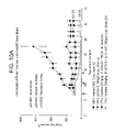


Classifications
-
- C—CHEMISTRY; METALLURGY
- C07—ORGANIC CHEMISTRY
- C07K—PEPTIDES
- C07K14/00—Peptides having more than 20 amino acids; Gastrins; Somatostatins; Melanotropins; Derivatives thereof
- C07K14/001—Peptides having more than 20 amino acids; Gastrins; Somatostatins; Melanotropins; Derivatives thereof by chemical synthesis
-
- A—HUMAN NECESSITIES
- A61—MEDICAL OR VETERINARY SCIENCE; HYGIENE
- A61K—PREPARATIONS FOR MEDICAL, DENTAL OR TOILETRY PURPOSES
- A61K47/00—Medicinal preparations characterised by the non-active ingredients used, e.g. carriers or inert additives; Targeting or modifying agents chemically bound to the active ingredient
- A61K47/50—Medicinal preparations characterised by the non-active ingredients used, e.g. carriers or inert additives; Targeting or modifying agents chemically bound to the active ingredient the non-active ingredient being chemically bound to the active ingredient, e.g. polymer-drug conjugates
- A61K47/51—Medicinal preparations characterised by the non-active ingredients used, e.g. carriers or inert additives; Targeting or modifying agents chemically bound to the active ingredient the non-active ingredient being chemically bound to the active ingredient, e.g. polymer-drug conjugates the non-active ingredient being a modifying agent
- A61K47/68—Medicinal preparations characterised by the non-active ingredients used, e.g. carriers or inert additives; Targeting or modifying agents chemically bound to the active ingredient the non-active ingredient being chemically bound to the active ingredient, e.g. polymer-drug conjugates the non-active ingredient being a modifying agent the modifying agent being an antibody, an immunoglobulin or a fragment thereof, e.g. an Fc-fragment
-
- A—HUMAN NECESSITIES
- A61—MEDICAL OR VETERINARY SCIENCE; HYGIENE
- A61P—SPECIFIC THERAPEUTIC ACTIVITY OF CHEMICAL COMPOUNDS OR MEDICINAL PREPARATIONS
- A61P11/00—Drugs for disorders of the respiratory system
- A61P11/06—Antiasthmatics
-
- A—HUMAN NECESSITIES
- A61—MEDICAL OR VETERINARY SCIENCE; HYGIENE
- A61P—SPECIFIC THERAPEUTIC ACTIVITY OF CHEMICAL COMPOUNDS OR MEDICINAL PREPARATIONS
- A61P15/00—Drugs for genital or sexual disorders; Contraceptives
-
- A—HUMAN NECESSITIES
- A61—MEDICAL OR VETERINARY SCIENCE; HYGIENE
- A61P—SPECIFIC THERAPEUTIC ACTIVITY OF CHEMICAL COMPOUNDS OR MEDICINAL PREPARATIONS
- A61P17/00—Drugs for dermatological disorders
- A61P17/06—Antipsoriatics
-
- A—HUMAN NECESSITIES
- A61—MEDICAL OR VETERINARY SCIENCE; HYGIENE
- A61P—SPECIFIC THERAPEUTIC ACTIVITY OF CHEMICAL COMPOUNDS OR MEDICINAL PREPARATIONS
- A61P19/00—Drugs for skeletal disorders
-
- A—HUMAN NECESSITIES
- A61—MEDICAL OR VETERINARY SCIENCE; HYGIENE
- A61P—SPECIFIC THERAPEUTIC ACTIVITY OF CHEMICAL COMPOUNDS OR MEDICINAL PREPARATIONS
- A61P19/00—Drugs for skeletal disorders
- A61P19/02—Drugs for skeletal disorders for joint disorders, e.g. arthritis, arthrosis
-
- A—HUMAN NECESSITIES
- A61—MEDICAL OR VETERINARY SCIENCE; HYGIENE
- A61P—SPECIFIC THERAPEUTIC ACTIVITY OF CHEMICAL COMPOUNDS OR MEDICINAL PREPARATIONS
- A61P19/00—Drugs for skeletal disorders
- A61P19/08—Drugs for skeletal disorders for bone diseases, e.g. rachitism, Paget's disease
-
- A—HUMAN NECESSITIES
- A61—MEDICAL OR VETERINARY SCIENCE; HYGIENE
- A61P—SPECIFIC THERAPEUTIC ACTIVITY OF CHEMICAL COMPOUNDS OR MEDICINAL PREPARATIONS
- A61P27/00—Drugs for disorders of the senses
- A61P27/02—Ophthalmic agents
-
- A—HUMAN NECESSITIES
- A61—MEDICAL OR VETERINARY SCIENCE; HYGIENE
- A61P—SPECIFIC THERAPEUTIC ACTIVITY OF CHEMICAL COMPOUNDS OR MEDICINAL PREPARATIONS
- A61P29/00—Non-central analgesic, antipyretic or antiinflammatory agents, e.g. antirheumatic agents; Non-steroidal antiinflammatory drugs [NSAID]
-
- A—HUMAN NECESSITIES
- A61—MEDICAL OR VETERINARY SCIENCE; HYGIENE
- A61P—SPECIFIC THERAPEUTIC ACTIVITY OF CHEMICAL COMPOUNDS OR MEDICINAL PREPARATIONS
- A61P3/00—Drugs for disorders of the metabolism
- A61P3/04—Anorexiants; Antiobesity agents
-
- A—HUMAN NECESSITIES
- A61—MEDICAL OR VETERINARY SCIENCE; HYGIENE
- A61P—SPECIFIC THERAPEUTIC ACTIVITY OF CHEMICAL COMPOUNDS OR MEDICINAL PREPARATIONS
- A61P35/00—Antineoplastic agents
-
- A—HUMAN NECESSITIES
- A61—MEDICAL OR VETERINARY SCIENCE; HYGIENE
- A61P—SPECIFIC THERAPEUTIC ACTIVITY OF CHEMICAL COMPOUNDS OR MEDICINAL PREPARATIONS
- A61P43/00—Drugs for specific purposes, not provided for in groups A61P1/00-A61P41/00
-
- A—HUMAN NECESSITIES
- A61—MEDICAL OR VETERINARY SCIENCE; HYGIENE
- A61P—SPECIFIC THERAPEUTIC ACTIVITY OF CHEMICAL COMPOUNDS OR MEDICINAL PREPARATIONS
- A61P7/00—Drugs for disorders of the blood or the extracellular fluid
- A61P7/04—Antihaemorrhagics; Procoagulants; Haemostatic agents; Antifibrinolytic agents
-
- A—HUMAN NECESSITIES
- A61—MEDICAL OR VETERINARY SCIENCE; HYGIENE
- A61P—SPECIFIC THERAPEUTIC ACTIVITY OF CHEMICAL COMPOUNDS OR MEDICINAL PREPARATIONS
- A61P9/00—Drugs for disorders of the cardiovascular system
-
- A—HUMAN NECESSITIES
- A61—MEDICAL OR VETERINARY SCIENCE; HYGIENE
- A61P—SPECIFIC THERAPEUTIC ACTIVITY OF CHEMICAL COMPOUNDS OR MEDICINAL PREPARATIONS
- A61P9/00—Drugs for disorders of the cardiovascular system
- A61P9/10—Drugs for disorders of the cardiovascular system for treating ischaemic or atherosclerotic diseases, e.g. antianginal drugs, coronary vasodilators, drugs for myocardial infarction, retinopathy, cerebrovascula insufficiency, renal arteriosclerosis
-
- A—HUMAN NECESSITIES
- A61—MEDICAL OR VETERINARY SCIENCE; HYGIENE
- A61P—SPECIFIC THERAPEUTIC ACTIVITY OF CHEMICAL COMPOUNDS OR MEDICINAL PREPARATIONS
- A61P9/00—Drugs for disorders of the cardiovascular system
- A61P9/14—Vasoprotectives; Antihaemorrhoidals; Drugs for varicose therapy; Capillary stabilisers
-
- C—CHEMISTRY; METALLURGY
- C07—ORGANIC CHEMISTRY
- C07K—PEPTIDES
- C07K14/00—Peptides having more than 20 amino acids; Gastrins; Somatostatins; Melanotropins; Derivatives thereof
-
- C—CHEMISTRY; METALLURGY
- C07—ORGANIC CHEMISTRY
- C07K—PEPTIDES
- C07K16/00—Immunoglobulins [IGs], e.g. monoclonal or polyclonal antibodies
- C07K16/18—Immunoglobulins [IGs], e.g. monoclonal or polyclonal antibodies against material from animals or humans
- C07K16/22—Immunoglobulins [IGs], e.g. monoclonal or polyclonal antibodies against material from animals or humans against growth factors ; against growth regulators
-
- C—CHEMISTRY; METALLURGY
- C07—ORGANIC CHEMISTRY
- C07K—PEPTIDES
- C07K7/00—Peptides having 5 to 20 amino acids in a fully defined sequence; Derivatives thereof
-
- C—CHEMISTRY; METALLURGY
- C07—ORGANIC CHEMISTRY
- C07K—PEPTIDES
- C07K7/00—Peptides having 5 to 20 amino acids in a fully defined sequence; Derivatives thereof
- C07K7/04—Linear peptides containing only normal peptide links
- C07K7/06—Linear peptides containing only normal peptide links having 5 to 11 amino acids
-
- C—CHEMISTRY; METALLURGY
- C07—ORGANIC CHEMISTRY
- C07K—PEPTIDES
- C07K7/00—Peptides having 5 to 20 amino acids in a fully defined sequence; Derivatives thereof
- C07K7/04—Linear peptides containing only normal peptide links
- C07K7/08—Linear peptides containing only normal peptide links having 12 to 20 amino acids
-
- C—CHEMISTRY; METALLURGY
- C07—ORGANIC CHEMISTRY
- C07K—PEPTIDES
- C07K2317/00—Immunoglobulins specific features
- C07K2317/70—Immunoglobulins specific features characterized by effect upon binding to a cell or to an antigen
- C07K2317/73—Inducing cell death, e.g. apoptosis, necrosis or inhibition of cell proliferation
-
- C—CHEMISTRY; METALLURGY
- C07—ORGANIC CHEMISTRY
- C07K—PEPTIDES
- C07K2317/00—Immunoglobulins specific features
- C07K2317/90—Immunoglobulins specific features characterized by (pharmaco)kinetic aspects or by stability of the immunoglobulin
- C07K2317/92—Affinity (KD), association rate (Ka), dissociation rate (Kd) or EC50 value
-
- C—CHEMISTRY; METALLURGY
- C07—ORGANIC CHEMISTRY
- C07K—PEPTIDES
- C07K2319/00—Fusion polypeptide
- C07K2319/30—Non-immunoglobulin-derived peptide or protein having an immunoglobulin constant or Fc region, or a fragment thereof, attached thereto
-
- C—CHEMISTRY; METALLURGY
- C07—ORGANIC CHEMISTRY
- C07K—PEPTIDES
- C07K2319/00—Fusion polypeptide
- C07K2319/32—Fusion polypeptide fusions with soluble part of a cell surface receptor, "decoy receptors"
Definitions
- the present invention relates to specific binding agents that recognize and bind to angiopoietin-2 (Ang-2). More specifically, the invention relates to the production, diagnostic use, and therapeutic use of the specific binding agents and fragments thereof, which specifically bind Ang-2.
- Ang-2 angiopoietin-2
- Angiogenesis the formation of new blood vessels from existing ones, is essential to many physiological and pathological processes. Normally, angiogenesis is tightly regulated by pro- and anti-angiogenic factors, but in the case of diseases such as cancer, ocular neovascular diseases, arthritis, and psoriasis, the process can go awry. Folkman, J., Nat. Med., 1:27-31 (1995).
- diseases known to be associated with deregulated or undesired angiogenesis include, but are not limited to, ocular neovascularisation, such as retinopathies (including diabetic retinopathy), age-related macular degeneration, psoriasis, hemangioblastoma, hemangioma, arteriosclerosis, inflammatory disease, such as a rheumatoid or rheumatic inflammatory disease, especially arthritis (including rheumatoid arthritis), or other chronic inflammatory disorders, such as chronic asthma, arterial or post-transplantational atherosclerosis, endometriosis, and neoplastic diseases, for example so-called solid tumors and liquid (or hematopoietic) tumors (such as leukemias and lymphomas).
- retinopathies including diabetic retinopathy
- psoriasis hemangioblastoma
- hemangioma hemangioma
- arteriosclerosis inflammatory
- Tie-2 receptor tyrosine kinase referred to as “Tie-2” or “Tie-2R” (also referred to as “ORK”
- murine Tie-2 is also referred to as “tek”
- angiopoietins There are 4 known angiopoietins; angiopoietin-1 (“Ang-1”) through angiopoietin-4 (“Ang-4”).
- Tie-2 ligands These angiopoietins are also referred to as “Tie-2 ligands”.
- Te-2 ligands (Davis, S., et al., Cell, 87:1161-1169 [1996]; Grosios, K., et al., Cytogenet Cell Genet, 84:118-120 [1999]; Holash, J., et al., Investigative Ophthalmology & Visual Science, 42:1617-1625 [1999]; Koblizek, T. I., et al., Current Biology, 8:529-532 [1998]; Lin, P., et al., Proc Natl Acad Sci USA, 95:8829-8834 [1998]; Maisonpierre, P.
- the phenotypes of mouse Tie-2 and Ang-1 knockouts are similar and suggest that Ang-1-stimulated Tie-2 phosphorylation mediates remodeling and stabilization of developing vessels in utero through maintenance of endothelial cell-support cell adhesion (Dumont, D. J., et al., Genes & Development, 8:1897-1909 [1994]; Sato, T. N., et al., Nature, 376:70-74 [1995]; Suri, C., et al., [1996], supra).
- Ang-1 in vessel stabilization is thought to be conserved in the adult, where it is expressed widely and constitutively (Hanahan, D., Science, 277:48-50 [1997]; Zagzag, D., et al., Experimental Neurology, 159:391-400 [1999]).
- Ang-2 expression is primarily limited to sites of vascular remodeling, where it is thought to block Ang-1 function, thereby inducing a state of vascular plasticity conducive to angiogenesis (Hanahan, D., [1997], supra; Holash, J., et al., Science, 284:1994-1998 [1999]; Maisonpierre, P. C., et al., [1997], supra).
- Ang-2 may be involved in tumor angiogenesis.
- Ahmad et al. Cancer Res., 61:1255-1259 [2001]
- Ang-2 over-expression describes Ang-2 over-expression and show that it is purportedly associated with an increase in tumor growth in a mouse xenograft model. See also Etoh et al., supra, and Tanaka et al., supra, wherein data is presented purportedly associating Ang-2 over expression with tumor hypervascularity.
- Yu et al. Am. J. Path., 158:563-570 [2001] report data to show that overexpression of Ang-2 in Lewis lung carcinoma and TA3 mammary carcinoma cells purportedly prolonged the survival of mice injected with the corresponding transfectants.
- An effective anti-Ang-2 therapy might benefit a vast population of cancer patients because most solid tumors require neovascularization to grow beyond 1-2 millimeters in diameter. Such therapy might have wider application in other angiogenesis-associated diseases as well, such as retinopathies, arthritis, and psoriasis.
- agents of the present invention take the form of peptibodies, i.e., peptides fused to other molecules such as an Fc domain of an antibody, where the peptide moiety specifically binds to Ang-2.
- the present invention is directed in one embodiment to peptides (also referred to as polypeptides herein) that bind to Ang-2. Also embodied in the present invention are variants and derivatives of such peptides.
- the peptides and variants and derivatives thereof of the present invention are attached to vehicles.
- the peptides may be fused to Fc domains, thereby providing peptibodies.
- the peptibodies comprise at least one peptide of, for example, SEQ ID NO:3-SEQ ID NO:6, or SEQ ID NO:76-SEQ ID NO: 157, as well as variants and derivatives thereof.
- the peptides may comprise at least one peptide according to the formulae set forth in SEQ ID NO:65-SEQ ID NO:75, and SEQ ID NO:158.
- the invention provides nucleic acid molecules encoding the specific binding agents, and variants and derivatives thereof.
- the invention provides nucleic acid molecules encoding the peptibodies, as well as variants and derivatives thereof.
- nucleic acid molecules include SEQ ID NO:33-SEQ ID NO:53.
- the invention provides a method of decreasing a tumor by administering an effective amount of the specific binding agents of the present invention to a subject in need thereof.
- the invention also provides a method of inhibiting angiogenesis in a subject, comprising administering an effective amount of the specific binding agents of the present invention to a subject in need thereof.
- the invention further provides a method of treating cancer in a subject, comprising an effective amount of the specific binding agents of the present invention to a subject in need thereof.
- the invention also relates to a polypeptide capable of binding Ang-2 wherein the polypeptide comprises the amino acid sequence WDPWT (SEQ ID NO: 65), and wherein the polypeptide is from 5 to 50 amino acids in length, as well as physiologically acceptable salts thereof.
- the polypeptide can also comprise the amino acid sequence:
- WDPWTC SEQ ID NO: 66
- physiologically acceptable salts thereof can comprise the amino acid sequence:
- polypeptide can further comprise the amino acid sequence:
- the invention relates to a polypeptide capable of binding Ang-2 comprising an amino acid sequence of the formula:
- the invention further relates to a polypeptide capable of binding Ang-2 comprising an amino acid sequence of the formula:
- b 1 is absent, or P, or T; b 2 is absent, or I, or N; b 3 is absent, or R, or I; b 4 is Q; b 5 is E; b 6 is E; b 8 is D or E; b 15 is D or E;
- b 16 is H; b 17 is M; b 18 is absent, or W, or P; b 19 is absent, or G, or E; and b 20 is absent, or V, or K.
- the invention preferably relates to a polypeptide comprising at least one amino acid sequence selected from the group consisting of SEQ ID NO: 4, and SEQ ID NO: 76 to SEQ ID NO: 118, inclusive, wherein the polypeptide is capable of binding to Ang-2, as well as physiologically acceptable salts thereof.
- the peptide sequences are set forth below:
- PEPTIDE SEQUENCE Con4-44 76 PIRQEECDWDPWTCEHMWEV Con4-40 77 TNIQEECEWDPWTCDHMPGK Con4-4 78 WYEQDACEWDPWTCEHMAEV Con4-31 79 NRLQEVCEWDPWTCEHMENV Con4-C5 80 AATQEECEWDPWTCEHMPRS Con4-42 81 LRHQEGCEWDPWTCEHMFDW Con4-35 82 VPRQKDCEWDPWTCEHMYVG Con4-43 83 SISHEECEWDPWTCEHMQVG Con4-49 84 WAAQEECEWDPWTCEHMGRM Con4-27 85 TWPQDKCEWDPWTCEHMGST Con4-48 86 GHSQEECGWDPWTCEHMGTS Con4-46 87 QHWQEECEWDPWTCDHMPSK Con4-41 88 NVRQEKCE
- peptides and/or peptibodies may contain the prefix “TN”, “TN8”, or “TN12”, and that this prefix may or may not be present for a given peptibody.
- TN8-Con4 and “Con4” are used interchangeably herein.
- the invention relates to a composition of matter having the formula: (X 1 ) a —F 1 —(X 2 ) b
- composition of matter is of the formulae: X 1 —F 1 or F 1 —X 2 and physiologically acceptable salts thereof, where X 1 , F 1 , and X 2 are as defined herein.
- composition of matter is of the formula: F 1 -(L 1 ) c -P 1 and physiologically acceptable salts thereof, where L 1 , F 1 , and P 1 are as defined herein.
- composition of matter is of the formula: F 1 -(L 1 ) c -P 1 -(L 2 ) d -P 2 and physiologically acceptable salts thereof, where L 1 , F 1 , P 1 , P 2 , and c and d are as defined herein.
- composition of matter is of the formula: P 1 -(L 1 ) c F 1 -(L 2 ) d -P 2 and physiologically acceptable salts thereof.
- F 1 is an Fc domain or fragment thereof.
- the invention further relates to a polypeptide capable of binding Ang-2 comprising an amino acid sequence of the formula:
- the invention further relates to a polypeptide capable of binding Ang-2 comprising an amino acid sequence of the formula:
- the polypeptide comprises at least one amino acid sequence selected from the group consisting of SEQ ID NO: 6, and SEQ ID NO: 119 to SEQ ID NO: 142, inclusive, wherein the polypeptide is capable of binding to Ang-2.
- SEQ ID NO: 6, and SEQ ID NOS: 119-142 are set forth below:
- the invention also relates to a a polypeptide capable of binding Ang-2 comprising an amino acid sequence of the formula:)
- the invention further relates to a polypeptide capable of binding Ang-2 comprising an amino acid sequence of the formula:
- the invention relates to a polypeptide comprising at least one amino acid sequence selected from the group consisting of SEQ ID NO: 3, and SEQ ID NO: 143 to SEQ ID NO: 148, inclusive, wherein the polypeptide is capable of binding to Ang-2, and physiologically acceptable salts thereof.
- SEQ ID NO: 3, and SEQ ID NO: 143 to SEQ ID NO: 148 are as follows.
- the invention relates to a polypeptide capable of binding Ang-2 comprising an amino acid sequence of the formula:
- the invention further relates to a polypeptide capable of binding Ang-2 comprising an amino acid sequence of the formula:
- the invention relates to a polypeptide comprising at least one amino acid sequence selected from the group consisting of SEQ ID NO: 5, or SEQ ID NO: 149 to SEQ ID NO: 157 inclusive, wherein said polypeptide is capable of binding to Ang-2, and physiologically acceptable salts thereof.
- SEQ ID NO: 5, and SEQ ID NO: 149 to SEQ ID NO: 157 are set forth below.
- the invention relates to a a composition of matter having the formula: (X 1 ) q —F 1 —(X 2 ) r
- P 1 , P 2 , P 3 , and P 4 each independently comprise a polypeptide selected from the group consisting of:
- L 1 , L 2 , L 3 , and L 4 are each independently linkers; and q, r, s, t, u, and v are each independently 0 or 1, provided that at least one of q and r is 1; and physiologically acceptable salts thereof.
- the invention further relates to a fusion polypeptide comprising at least one peptide described as described herein and a vehicle, wherein the fusion polypeptide is capable of binding to Ang-2, and physiologically acceptable salts thereof.
- the vehicle is preferably at least one of an Fc domain, polyethylene glycol, a lipid, a cholesterol group, a carbohydrate, and an oligosaccharide.
- suitable vehicles such as albumin and the like, will be appreciated by those skilled in the art, and are encompassed within the scope of the invention.
- a given molecule can be inserted, for example, between the peptide and vehicle portions of the specific binding agents, or inserted within the peptide portion itself, while retaining the desired activity of specific binding agent.
- Other molecules suitable for insertion in this fashion will be appreciated by those skilled in the art, and are encompassed within the scope of the invention. This includes insertion of, for example, a desired molecule in between two consecutive amino acids, optionally joined by a suitable linker.
- Suitable linker molecules can be added if desired. It will further be appreciated that the molecule can be inserted in a number of locations on the molecule, including on suitable side chains, between the vehicle and peptide sequence as follows:
- the invention relates to a polynucleotide encoding the specific binding agents (including, but not limited to peptides and/or peptibodies) of the invention, as described herein.
- specific binding agents including, but not limited to peptides and/or peptibodies
- the amino acid sequence is known, the corresponding nucleotide sequence(s) can be readily determined using known techniques. See for example Suzuki, D., An Introduction to Genetic Analysis , W.H. Freeman Pub. Co. (1986).
- Exemplary nucleotide sequences encoding peptides of the invention are set forth below.
- more than one codon can encode for a given amino acid, and therefore the invention relates to any nucleotide sequence which encodes the peptides and/or peptibodies of the invention.
- the invention relates to expression vectors comprising at least one polynucleotide of the invention.
- the invention relates to host cells comprising the expression vector. It will be appreciated that the host cells are preferably prokaryotic cells (such as E. coli cells) or eukaryotic cells.
- the invention also relates to a pharmaceutical composition
- a pharmaceutical composition comprising an effective amount of a composition as described herein, in admixture with a pharmaceutically acceptable carrier.
- the invention also relates to a method of inhibiting undesired angiogenesis in a mammal comprising administering a therapeutically effective amount of a polypeptide or composition as described herein.
- the invention also relates to a method of modulating angiogenesis in a mammal comprising administering a therapeutically effective amount of a polypeptide or composition as described herein.
- the invention further relates to a method of inhibiting tumor growth characterized by undesired angiogenesis in a mammal comprising administering a therapeutically effective amount of a polypeptide or composition as described herein.
- the invention relates to a method of treating cancer in a mammal comprising administering a therapeutically effective amount of a polypeptide or composition as described herein, and a chemotherapeutic agent.
- the chemotherapeutic agent is at least one of 5-FU, CPT-11, and Taxotere. It will be appreciated, however, that other suitable chemotherapeutic agents and other cancer therapies can be used.
- the invention also relates to a method of modulating at least one of vascular permeability or plasma leakage in a mammal comprising administering a therapeutically effective amount of a polypeptide or composition as described herein.
- the invention further relates to a method of treating at least one of ocular neovascular disease, obesity, hemangioblastoma, hemangioma, arteriosclerosis, inflammatory disease, inflammatory disorders, atherosclerosis, endometriosis, neoplastic disease, bone-related disease, or psoriasis in a mammal comprising administering a therapeutically effective amount of a polypeptide or composition as described herein.
- the specific binding agents of the invention can be used to treat a number of diseases associated with deregulated or undesired angiogenesis.
- diseases include, but are not limited to, ocular neovascularisation, such as retinopathies (including diabetic retinopathy and age-related macular degeneration) psoriasis, hemangioblastoma, hemangioma, arteriosclerosis, inflammatory disease, such as a rheumatoid or rheumatic inflammatory disease, especially arthritis (including rheumatoid arthritis), or other chronic inflammatory disorders, such as chronic asthma, arterial or post-transplantational atherosclerosis, endometriosis, and neoplastic diseases, for example so-called solid tumors and liquid tumors (such as leukemias).
- Additional diseases which can be treated by administration of the specific binding agents will be apparent to those skilled in the art.
- additional diseases include, but are not limited to, obesity, vascular permeability, plasma leakage, and bone-related disorders, including osteoporosis.
- the invention further relates to methods of treating these diseases associated with deregulated or undesired angiogenesis.
- FIG. 1 depicts a graph of tumor volume (y-axis) versus time (x-axis) in A-431 tumor bearing mice treated with peptibody TN8-Con4-C of the present invention, or with phosphate buffered saline (PBS). Details are described in the Examples.
- FIG. 2 depicts a graph of peptibody concentration (y-axis) versus time post-dose (x-axis) in wildtype mice treated with a 50 ⁇ g dose of either 2xCon4-C, L1-7-N, or L1-21-N peptibody. Details are described in the Examples.
- FIG. 3 depicts a graph of tumor volume (y-axis) versus time (x-axis) in A431 tumor bearing mice treated with peptibody 2xCon4-C according to the present invention, or with phosphate buffered saline (PBS) or control peptibody. Details are described in the Examples.
- PBS phosphate buffered saline
- FIG. 4 depicts a graph representing in vitro growth of cultured A431 cells treated with peptibody Con4-C according to the present invention, control peptibody, or untreated. Details are described in the Examples.
- FIG. 5 depicts a graph of tumor volume (y-axis) versus time (x-axis) in Colo205 tumor cells treated with peptibody Con4-C, peptibody L1-7-N, peptibody L1-21-N, or peptibody 2xCon4-C according to the present invention, or with phosphate buffered saline (PBS), anti-Ang-2 antibody (Ab536), or Fc. Details are described in the Examples.
- PBS phosphate buffered saline
- Ab536 anti-Ang-2 antibody
- FIG. 6 depicts a graph of tumor volume (y-axis) versus time (x-axis) in Colo205 xenograft tumor bearing mice treated with varying doses of peptibody 2xCon4-C according to the present invention, or with phosphate buffered saline (PBS) or Fc. Details are described in the Examples.
- PBS phosphate buffered saline
- FIG. 7 depicts a graph of tumor volume (y-axis) versus time (x-axis) in Colo205 xenograft tumor bearing mice treated with peptibody 2xCon4-C according to the present invention, or with control peptibodies.
- FIG. 7 also depicts a graph of CD31 stained area/total tumor area for these peptibodies. Details are described in the Examples.
- FIG. 8 depicts a graph of tumor volume (y-axis) versus time (x-axis) in Colo205 xenograft tumor bearing mice treated with peptibody 2xCon4-C according to the present invention, or with phosphate buffered saline (PBS) or control peptibody. Details are described in the Examples. This graph shows that anti-Ang-2 peptibodies are capable of inhibiting Colo205 tumor growth irrespective of when dosing begins.
- PBS phosphate buffered saline
- FIG. 9 depicts a summary of complete response (CR) rates obtained in female nude mice using antibody Ab536 or with peptibody 2xCon4-C, in both the A431 and Colo-205 xenograft models. Details are described in the Examples.
- FIG. 10A depicts a graph of tumor volume (y-axis) versus time (x-axis) in Colo205 xenograft tumor bearing mice treated with peptibody 2xCon4-C according to the present invention, or a combination of 2xCon4-C and taxotere, or with phosphate buffered saline (PBS), or with PBS plus taxotere. Details are described in the Examples.
- FIG. 10B depicts a graph of tumor volume (y-axis) versus time (x-axis) in Colo205 xenograft tumor bearing mice treated with peptibody 2xCon4-C according to the present invention, or a combination of 2xCon4-C and 5-FU, or with phosphate buffered saline (PBS), or with PBS plus 5-FU. Details are described in the Examples.
- FIG. 11A depicts a graph of paw swelling levels (AUC ⁇ SE) in an adjuvant-induced arthritis model in rats treated with peptibody 2xCon4-C according to the present invention, or with phosphate buffered saline (PBS), or with control peptibody, or normal or arthritis controls. Details are described in the Examples.
- FIG. 11B depicts a graph of paw bone mineral density (BMD) in an adjuvant-induced arthritis model in rats treated with peptibody 2xCon4-C according to the present invention, or with phosphate buffered saline (PBS), or with control peptibody, or normal or arthritis controls. Details are described in the Examples.
- BMD paw bone mineral density
- FIG. 11C depicts a graph of change in body weight in an adjuvant-induced arthritis model in rats treated with peptibody 2xCon4-C according to the present invention, or with phosphate buffered saline (PBS), or with control peptibody, or normal or arthritis controls. Details are described in the Examples.
- FIG. 12 depicts two graphs depicting inhibition of VEGF-induced corneal angiogenesis in rats.
- the first graph depicts number of blood vessels measured in rats treated with bovine serum albumin (BSA), VEGF plus phosphate buffered saline (PBS), or VEGF plus peptibody Con4-C of the invention.
- the second graph depicts blood vessel area (mm 2 ) in rats treated with BSA, VEGF plus phosphate buffered saline (PBS), or VEGF plus peptibody Con4-C of the invention. Details are described in the Examples.
- FIGS. 13A , 13 B, and 13 C depict epitope mapping data (O.D. 370) for full-length human Ang-2 (hAng-2), to the N-terminus of hAng-2, and to the C-terminus of hAng-2, respectively, for peptibodies TN8-Con4-C, L1-7-N, and 12-9-3-C according to the invention, as well as for control peptibody, Tie2-Fc, C2B8, or 5B12. Details are described in the Examples.
- FIG. 14 depicts binding affinity (K D ) of the 2xCon-4-C peptibody according to the invention, using the Sapidyne KinExA assay. Details are described in the Examples.
- Standard techniques may be used for recombinant DNA molecule, protein, and antibody production, as well as for tissue culture and cell transformation. Enzymatic reactions and purification techniques are typically performed according to the manufacturer's specifications or as commonly accomplished in the art using conventional procedures such as those set forth in Sambrook et al. (Molecular Cloning: A Laboratory Manual. Cold Spring Harbor Laboratory Press, Cold Spring Harbor, N.Y. [1989]), or as described herein. Unless specific definitions are provided, the nomenclature utilized in connection with, and the laboratory procedures and techniques of analytical chemistry, synthetic organic chemistry, and medicinal and pharmaceutical chemistry described herein are those well known and commonly used in the art. Standard techniques may be used for chemical syntheses, chemical analyses, pharmaceutical preparation, formulation, and delivery, and treatment of patients.
- Ang-2 refers to the polypeptide set forth in FIG. 6 of U.S. Pat. No. 6,166,185 (“Tie-2 ligand-2”) or fragments thereof as well as related polypeptides which include allelic variants, splice variants, derivatives, substitution, deletions, and/or insertion variants, fusion peptides and polypeptides, and interspecies homologs.
- the Ang-2 polypeptide may or may not include additional terminal residues, e.g., leader sequences, targeting sequences, amino terminal methionine, amino terminal methionine and lysine residues, and/or tag or fusion proteins sequences, depending on the manner in which it is prepared.
- biologically active when used in relation to Ang-2 or an Ang-2 specific binding agent refers to a peptide or polypeptide having at least one activity characteristic of Ang-2 or of an Ang-2 specific binding agent.
- a specific binding agent of Ang-2 may have agonist, antagonist, or neutralizing or blocking activity with respect to at least one biological activity of Ang-2.
- specific binding agent refers to a molecule, preferably a proteinaceous molecule, that specifically binds Ang-2, and variants and derivatives thereof, as defined herein.
- a specific binding agent may be a protein, peptide, nucleic acid, carbohydrate, lipid, or small molecular weight compound which binds preferentially to Ang-2.
- the specific binding agent according to the present invention is a peptide or a peptibody, as well as fragments, variants or derivatives thereof, either alone or in combination with other amino acid sequences, provided by known techniques. Such techniques include, but are not limited to enzymatic cleavage, chemical cleavage, peptide synthesis or recombinant techniques.
- the anti-Ang-2 specific binding agents of the present invention are capable of binding portions of Ang-2 that modulate, e.g., inhibit or promote, the biological activity of Ang-2 and/or other Ang-2-associated activities.
- variants include those peptides and polypeptides wherein amino acid residues are inserted into, deleted from and/or substituted into the naturally occurring (or at least a known) amino acid sequence for the binding agent.
- variants of the invention include fusion proteins as described below.
- “Derivatives” include those binding agents that have been chemically modified in some manner distinct from insertion, deletion, or substitution variants.
- Specifically binds Ang-2 refers to the ability of a specific binding agent (such as a peptibody, or peptide portion thereof) of the present invention to recognize and bind mature, full-length or partial-length human Ang-2 polypeptide, or an ortholog thereof, such that its affinity (as determined by, e.g., Affinity ELISA or BIAcore assays as described herein) or its neutralization capability (as determined by e.g., Neutralization ELISA assays described herein, or similar assays) is at least 10 times as great, but optionally 50 times as great, 100, 250 or 500 times as great, or even at least 1000 times as great as the affinity or neutralization capability of the same for any other angiopoietin or other peptide or polypeptide, wherein the peptide portion of the peptibody is first fused to a human Fc moiety for evaluation in such assay.
- a specific binding agent such as a peptibody, or
- epitope refers to that portion of any molecule capable of being recognized by and bound by a specific binding agent, e.g., a peptibody, at one or more of the binding agent's antigen binding regions.
- a specific binding agent e.g., a peptibody
- Epitopes usually consist of chemically active surface groupings of molecules, such as for example, amino acids or carbohydrate side chains, and have specific three-dimensional structural characteristics as well as specific charge characteristics. Epitopes as used herein may be contiguous or non-contiguous.
- inhibiting and/or neutralizing epitope is an epitope, which when bound by a specific binding agent such as a peptibody, results in the loss of (or at least the decrease in) biological activity of the molecule, cell, or organism containing such epitope, in vivo, in vitro, or in situ.
- the neutralizing epitope is located on or is associated with a biologically active region of Ang-2.
- activating epitope is an epitope, which when bound by a specific binding agent of the invention, such as an antibody, results in activation, or at least maintenance of a biologically active conformation, of Ang-2.
- polypeptide fragment refers to a peptide or polypeptide which comprises less than a complete, intact peptibody.
- naturally occurring when used in connection with biological materials such as nucleic acid molecules, polypeptides, host cells, and the like, refers to those which are found in nature and not modified by a human being.
- isolated when used in relation to Ang-2 or to a specific binding agent of Ang-2 refers to a compound that is free from at least one contaminating polypeptide or compound that is found in its natural environment, and preferably substantially free from any other contaminating mammalian polypeptides that would interfere with its therapeutic or diagnostic use.
- mature when used in relation to Ang-2 peptibody or a fragment thereof, or to any other proteinaceous specific binding agent of Ang-2 refers to a peptide or a polypeptide lacking a leader or signal sequence.
- the “mature” peptide or polypeptide may also include additional amino acid residues (but still lack a leader sequence) such as an amino terminal methionine, or one or more methionine and lysine residues.
- additional amino acid residues but still lack a leader sequence
- a peptide or polypeptide produced in this manner may be utilized with or without these additional amino acid residues having been removed.
- Ang-2 refers to an amount of a specific binding agent that is useful or necessary to support an observable change in the level of one or more biological activities of Ang-2.
- the change may be either an increase or decrease in the level of Ang-2 activity.
- the change is a decrease in Ang-2 activity.
- peptibodies refers to a molecule comprising an antibody Fc domain attached to at least one peptide.
- the production of peptibodies is generally described in PCT publication WO 00/24782, published May 4, 2000.
- variants include those molecules such as peptides or peptide-vehicle combinations such as peptibodies of the present invention wherein amino acid residues are inserted into, deleted from and/or substituted into amino acid sequence for such molecules.
- variants having one or more amino acids inserted include fusion proteins as described below.
- “Derivatives” include those peptides and/or peptide-vehicle combinations such as peptibodies that have been chemically modified in some manner distinct from insertion, deletion, or substitution variants.
- fragment refers to a peptide or peptide-vehicle combination that comprises less than the full-length amino acid sequence of such peptides and/or peptide-vehicle combinations. Such a fragment may arise, for example, from a truncation at the amino terminus, a truncation at the carboxy-terminus, and/or an internal deletion of a residue(s) from the amino acid sequence of the peptide or peptide-vehicle combination. Fragments may result from alternative RNA splicing or from in vivo or in vitro protease activity.
- Such fragments may also be constructed by chemical peptide synthesis methods, or by modifying a polynucleotide encoding a peptide, peptide-vehicle combination, or an Fc portion and/or peptide portion of a peptibody.
- Fc refers to one type of vehicle of the present invention, and comprises the sequence of a non-antigen-binding fragment of an antibody resulting from the proteolytic digestion of a whole antibody, whether in monomeric or multimeric form.
- the source of the Fc in the present invention is preferably fully human Fc, and may be any of the immunoglobulins, although IgG1 and IgG2 are preferred. However, Fc molecules that are partially human, or obtained from non-human species are also included herein.
- Fc's are made up of monomeric polypeptides that may be linked into dimeric or multimeric forms by covalent (i.e., disulfide bonds) and non-covalent association.
- the number of intermolecular disulfide bonds between monomeric subunits of native Fc molecules ranges from 1 to 4 depending on class (e.g., IgG, IgA, IgE) or subclass (e.g., IgG1, IgG2, IgG3, IgA1, IgGA2).
- class e.g., IgG, IgA, IgE
- subclass e.g., IgG1, IgG2, IgG3, IgA1, IgGA2
- One example of a native Fc is a disulfide-bonded dimer resulting from papain digestion of an IgG [see Ellison et al. (1982), Nucl. Acids. Res. 10: 4071-9].
- native Fc as used herein is generic to the monomeric, dimeric, and multimeric forms.
- Fc domain encompasses native Fc and Fc variant molecules and sequences as defined above. As with Fc variants and native Fc's, the term “Fc domain” includes molecules in monomeric or multimeric form, whether digested from whole antibody or produced by other means.
- multimer as applied to Fc domains or molecules comprising Fc domains refers to molecules having two or more polypeptide chains associated covalently, noncovalently, or by both covalent and non-covalent interactions.
- IgG molecules typically form dimers; IgM, pentamers; IgD, dimers; and IgA, monomers, dimers, trimers, or tetramers. Multimers may be formed by exploiting the sequence and resulting activity of the native Ig source of the Fc or by derivatizing (as defined below) such a native Fc.
- dimer as applied to Fc domains or molecules comprising Fc domains refers to molecules having two polypeptide chains associated covalently or non-covalently.
- vehicle refers to a molecule that prevents degradation and/or increases half-life, reduces toxicity, reduces immunogenicity, or increases biological activity of a therapeutic protein.
- exemplary vehicles include an Fc domain as well as a linear polymer (e.g., polyethylene glycol (PEG), polylysine, dextran, etc.); a branched-chain polymer (See, for example, U.S. Pat. No. 4,289,872 to Denkenwalter et al., issued Sep. 15, 1981; U.S. Pat. No. 5,229,490 to Tam, issued Jul. 20, 1993; WO 93/21259 by Frechet et al., published 28 Oct.
- lipid a lipid
- a cholesterol group such as a steroid
- carbohydrate or oligosaccharide or any natural or synthetic protein, polypeptide or peptide that binds to a salvage receptor.
- Vehicles are further described hereinafter.
- the terms “derivatizing” and “derivative” or “derivatized” comprise processes and resulting compounds respectively in which (1) the compound has a cyclic portion; for example, cross-linking between cysteinyl residues within the compound; (2) the compound is cross-linked or has a cross-linking site; for example, the compound has a cysteinyl residue and thus forms cross-linked dimers in culture or in vivo; (3) one or more peptidyl linkage is replaced by a non-peptidyl linkage; (4) the N-terminus is replaced by —NRR1, NRC(O)R1, —NRC(O)OR1, —NRS(O)2R1, —NHC(O)NHR, a succinimide group, or substituted or unsubstituted benzyloxycarbonyl-NH—, wherein R and R1 and the ring substituents are as defined hereinafter; (5) the C-terminus is replaced by —C(O)R2 or —NR3R4
- peptide refers to molecules of about 3 to about 75 amino acids, with molecules of about 5 to 50 amino acids preferred, 8 to 40 more preferred, and those of about 10 to 25 amino acids most preferred.
- Peptides may be naturally occurring or artificial (i.e., non-naturally occurring) amino acid sequences.
- Exemplary peptides may be generated by any of the methods set forth herein, such as carried in a peptide library (e.g., a phage display library), generated by chemical synthesis, derived by digestion of proteins, or generated using recombinant DNA techniques.
- pharmaceutically active means that a substance so described is determined to have activity that affects a medical parameter (e.g., blood pressure, blood cell count, cholesterol level) or disease state (e.g., cancer, autoimmune disorders, etc.).
- a medical parameter e.g., blood pressure, blood cell count, cholesterol level
- disease state e.g., cancer, autoimmune disorders, etc.
- antagonist peptide refers to a peptide that blocks or in some way interferes with the biological activity of the associated protein of interest, or has biological activity comparable to a known antagonist or inhibitor of the associated protein of interest.
- Ang-2-antagonist peptide comprises peptides that can be identified or derived as having Ang-2-antagonistic characteristics.
- physiologically acceptable salts of the compounds of this invention are also encompassed herein.
- physiologically acceptable salts is meant any salts that are known or later discovered to be pharmaceutically acceptable. Some specific examples are: acetate; trifluoroacetate; hydrohalides, such as hydrochloride and hydrobromide; sulfate; citrate; tartrate; glycolate; and oxalate, mesylate, and phosphate.
- One aspect of the present invention relates to development of Ang-2 peptibodies.
- the interaction of a protein ligand with its receptor often takes place at a relatively large interface.
- the bulk of the protein ligand merely displays the binding epitopes in the right topology or serves functions unrelated to binding.
- molecules of only “peptide” length can bind to the receptor protein of a given large protein ligand.
- Such peptides may mimic the bioactivity of the large protein ligand (“peptide agonists”) or, through competitive binding, inhibit the bioactivity of the large protein ligand (“peptide antagonists”).
- Phage display technology has emerged as a powerful method in identifying such peptide agonists and antagonists. See, for example, Scott et al. Science 249: 386 (1990); Devlin et al., Science 249: 404 (1990); U.S. Pat. No. 5,223,409, issued Jun. 29, 1993; U.S. Pat. No. 5,733,731, issued Mar. 31, 1998; U.S. Pat. No. 5,498,530, issued Mar. 12, 1996; U.S. Pat. No. 5,432,018, issued Jul. 11, 1995; U.S. Pat. No. 5,338,665, issued Aug. 16, 1994; U.S. Pat. No. 5,922,545, issued Jul. 13, 1999; WO 96/40987, published Dec.
- peptide phage display libraries random peptide sequences can be displayed by fusion with coat proteins of filamentous phage.
- the displayed peptides can be affinity-eluted against an antibody-immobilized extracellular domain of a receptor, if desired.
- the retained phage may be enriched by successive rounds of affinity purification and repropagation.
- the best binding peptides may be sequenced to identify key residues within one or more structurally related families of peptides. See, e.g., Cwirla et al., Science 276: 1696-9 (1997), in which two distinct families were identified.
- the peptide sequences may also suggest which residues may be safely replaced by alanine scanning or by mutagenesis at the DNA level. Mutagenesis libraries may be created and screened to further optimize the sequence of the best binders. Lowman, Ann. Rev. Biophys. Biomol. Struct. 26: 401-24 (1997).
- Structural analysis of protein-protein interaction may also be used to suggest peptides that mimic the binding activity of large protein ligands.
- the crystal structure may suggest the identity and relative orientation of critical residues of the large protein ligand, from which a peptide may be designed. See, e.g., Takasaki et al., Nature Biotech 15: 1266-70 (1997).
- These analytical methods may also be used to investigate the interaction between a receptor protein and peptides selected by phage display, which may suggest further modification of the peptides to increase binding affinity.
- E. coli display Another E. coli -based method allows display on the cell's outer membrane by fusion with a peptidoglycan-associated lipoprotein (PAL).
- PAL peptidoglycan-associated lipoprotein
- these and related methods are collectively referred to as “ E. coli display.”
- translation of random RNA is halted prior to ribosome release, resulting in a library of polypeptides with their associated RNA still attached.
- this and related methods are collectively referred to as “ribosome display.”
- Other methods employ chemical linkage of peptides to RNA.
- RNA-peptide screening Chemically derived peptide libraries have been developed in which peptides are immobilized on stable, non-biological materials, such as polyethylene rods or solvent-permeable resins. Another chemically derived peptide library uses photolithography to scan peptides immobilized on glass slides. Hereinafter, these and related methods are collectively referred to as “chemical-peptide screening.” Chemical-peptide screening may be advantageous in that it allows use of D-amino acids and other unnatural analogues, as well as non-peptide elements. Both biological and chemical methods are reviewed in Wells and Lowman, Curr. Opin. Biotechnol., 3: 355-62 (1992).
- Peptides identified by phage display library screening have been regarded as “leads” in development of therapeutic agents rather than as therapeutic agents themselves. Like other proteins and peptides, they would likely be rapidly removed in vivo either by renal filtration, by cellular clearance mechanisms in the reticuloendothelial system, or by proteolytic degradation [Francis, (supra)].
- the art presently uses peptides to validate drug targets or as scaffolds for design of organic compounds that might not have been as easily or as quickly identified through chemical library screening [Lowman, (supra); Kay et al., (supra)]. The art would benefit from a process by which such peptides could more readily yield therapeutic agents against angiogenesis.
- the peptide may be attached to a vehicle through the peptide's N-terminus or C-terminus.
- vehicle-peptide molecules of this invention may be described by the following five formulae and multimers thereof:
- F 1 is a vehicle (preferably an Fc domain);
- X 1 and X 2 are each independently selected from -(L 1 ) c -P 1 , -(L 1 ) c -P 1 -(L 2 ) d -P 2 , -(L 1 ) c -P 1 -(L 2 ) d -P 2 -(L 3 ) e -P 3 , and -(L 1 ) c -P 1 -(L 2 ) d -P 2 -(L 3 ) e -P 3 -(L 4 ) f -P 4
- P 1 , P 2 , P 3 , and P 4 are each independently sequences of pharmacologically active peptides as described herein;
- L 1 , L 2 , L 3 , and L 4 are each independently linkers
- a”, “b”, “c”, “d”, “e”, and “f” are each independently 0 or 1, provided that at least one of “a” and “b” is 1.
- the present invention contemplates peptides that selectively bind or specifically bind to Ang-2. Any number of such peptides may be used in conjunction with the present invention. Phage display, in particular, is useful in generating peptides for use in the present invention as has been shown that affinity selection from libraries of random peptides can be used to identify peptide ligands for any site of any gene product. Dedman et al., J. Biol. Chem. 268: 23025-30 (1993).
- the peptides in this invention may be prepared by any of the methods disclosed in the art. Single letter amino acid abbreviations are used.
- the “X” in any sequence means that any of the 20 naturally occurring amino acid residues, or any non-naturally occurring amino acids (described below under “Variants”), may be present. Any of these peptides may be linked in tandem (i.e., sequentially), with or without linkers, and tandem-linked examples are provided in the table. Linkers are listed as “L” and may be any of the linkers described herein. Tandem repeats and linkers are shown separated by dashes for clarity.
- Any peptide containing a cysteinyl residue may be cross-linked with another Cys-containing peptide, either or both of which may be linked to a vehicle. Any peptide having more than one Cys residue may form an intrapeptide disulfide bond, as well. Any of these peptides may be derivatized as described herein.
- the capping amino group is —NH 2 .
- amino acid residues are substituted by moieties other than amino acid residues, the substitutions are denoted by S, which signifies any of the moieties described in Bhatnagar et al., J. Med. Chem. 39: 3814-9 (1996), and Cuthbertson et al., J. Med. Chem. 40: 2876-82 (1997), which are incorporated by reference. All peptides are linked through peptide bonds unless otherwise noted.
- this invention provides for at least one peptide to be attached to at least one vehicle (F 1 , F 2 ) through the N-terminus, C-terminus or a side chain of one of the amino acid residues of the peptide(s).
- vehicle F 1 , F 2
- Multiple vehicles may also be used; e.g., Fc's at each terminus or an Fc at a terminus and a PEG group at the other terminus or a side chain.
- Fc domain is one preferred vehicle.
- the Fc domain may be fused to the N or C termini of the peptides or at both the N and C termini.
- Fc variants are suitable vehicles within the scope of this invention.
- a native Fc may be extensively modified to form an Fc variant in accordance with this invention, provided binding to the salvage receptor is maintained. See, for example WO 97/34631 and WO 96/32478.
- One may remove these sites by, for example, substituting or deleting residues, inserting residues into the site, or truncating portions containing the site.
- the inserted or substituted residues may also be altered amino acids, such as peptidomimetics or D-amino acids.
- Fc variants may be desirable for a number of reasons, several of which are described below.
- Exemplary Fc variants include molecules and sequences in which:
- cysteine-containing segment at the N-terminus may be truncated or cysteine residues may be deleted or substituted with other amino acids (e.g., alanyl, seryl). Even when cysteine residues are removed, the single chain Fc domains can still form a dimeric Fc domain that is held together non-covalently.
- a native Fc is modified to make it more compatible with a selected host cell. For example, one may remove the PA sequence near the N-terminus of a typical native Fc, which may be recognized by a digestive enzyme in E. coli such as proline iminopeptidase. One may also add an N-terminal methionyl residue, especially when the molecule is expressed recombinantly in a bacterial cell such as E. coli.
- a portion of the N-terminus of a native Fc is removed to prevent N-terminal heterogeneity when expressed in a selected host cell. For this purpose, one may delete any of the first 20 amino acid residues at the N-terminus, particularly those at positions 1, 2, 3, 4 and 5.
- Residues that are typically glycosylated may confer cytolytic response. Such residues may be deleted or substituted with unglycosylated residues (e.g., alanine).
- Sites involved in interaction with complement such as the C1q binding site, are removed. For example, one may delete or substitute the EKK sequence of human IgG1. Complement recruitment may not be advantageous for the molecules of this invention and so may be avoided with such an Fc variant.
- a native Fc may have sites for interaction with certain white blood cells that are not required for the fusion molecules of the present invention and so may be removed.
- ADCC site is removed.
- ADCC sites are known in the art. See, for example, Molec. Immunol. 29 (5):633-9 (1992) with regard to ADCC sites in IgG1. These sites, as well, are not required for the fusion molecules of the present invention and so may be removed.
- the native Fc When the native Fc is derived from a non-human antibody, the native Fc may be humanized. Typically, to humanize a native Fc, one will substitute selected residues in the non-human native Fc with residues that are normally found in human native Fc. Techniques for antibody humanization are well known in the art.
- An alternative vehicle would be a protein, polypeptide, peptide, antibody, antibody fragment, or small molecule (e.g., a peptidomimetic compound) capable of binding to a salvage receptor.
- a polypeptide as described in U.S. Pat. No. 5,739,277, issued Apr. 14, 1998 to Presta et al.
- Peptides could also be selected by phage display for binding to the FcRn salvage receptor.
- salvage receptor-binding compounds are also included within the meaning of “vehicle” and are within the scope of this invention.
- Such vehicles should be selected for increased half-life (e.g., by avoiding sequences recognized by proteases) and decreased immunogenicity (e.g., by favoring non-immunogenic sequences, as discovered in antibody humanization).
- PCT Patent Cooperation Treaty
- WO 96/11953 entitled “N-Terminally Chemically Modified Protein Compositions and Methods,” herein incorporated by reference in its entirety.
- This PCT publication discloses, among other things, the selective attachment of water soluble polymers to the N-terminus of proteins.
- a preferred polymer vehicle is polyethylene glycol (PEG).
- the PEG group may be of any convenient molecular weight and may be linear or branched.
- the average molecular weight of the PEG will preferably range from about 2 kiloDalton (“kDa”) to about 100 kDa, more preferably from about 5 kDa to about 50 kDa, most preferably from about 5 kDa to about 10 kDa.
- the PEG groups will generally be attached to the compounds of the invention via acylation or reductive alkylation through a reactive group on the PEG moiety (e.g., an aldehyde, amino, thiol, or ester group) to a reactive group on the inventive compound (e.g., an aldehyde, amino, or ester group).
- a reactive group on the PEG moiety e.g., an aldehyde, amino, thiol, or ester group
- a reactive group on the inventive compound e.g., an aldehyde, amino, or ester group
- a useful strategy for the PEGylation of synthetic peptides consists of combining, through forming a conjugate linkage in solution, a peptide and a PEG moiety, each bearing a special functionality that is mutually reactive toward the other.
- the peptides can be easily prepared with conventional solid phase synthesis as known in the art.
- the peptides are “preactivated” with an appropriate functional group at a specific site.
- the precursors are purified and fully characterized prior to reacting with the PEG moiety.
- Ligation of the peptide with PEG usually takes place in aqueous phase and can be easily monitored by reverse phase analytical HPLC.
- the PEGylated peptides can be easily purified by preparative HPLC and characterized by analytical HPLC, amino acid analysis and laser desorption mass spectrometry.
- Polysaccharide polymers are another type of water soluble polymer which may be used for protein modification.
- Dextrans are polysaccharide polymers comprised of individual subunits of glucose predominantly linked by a1-6 linkages. The dextran itself is available in many molecular weight ranges, and is readily available in molecular weights from about 1 kDa to about 70 kDa.
- Dextran is a suitable water-soluble polymer for use in the present invention as a vehicle by itself or in combination with another vehicle (e.g., Fc). See, for example, WO 96/11953 and WO 96/05309. The use of dextran conjugated to therapeutic or diagnostic immunoglobulins has been reported; see, for example, European Patent Publication No. 0 315 456, which is hereby incorporated by reference. Dextran of about 1 kDa to about 20 kDa is preferred when dextran is used as a vehicle in accordance with the present invention.
- linker is optional. When present, its chemical structure is not critical, since it serves primarily as a spacer.
- the linker is preferably made up of amino acids linked together by peptide bonds.
- the linker is made up of from 1 to 20 amino acids linked by peptide bonds, wherein the amino acids are selected from the 20 naturally occurring amino acids.
- one or more of these amino acids may be glycosylated, as is well understood by those in the art.
- the 1 to 20 amino acids are selected from glycine, alanine, proline, asparagine, glutamine, and lysine.
- a linker is made up of a majority of amino acids that are sterically unhindered, such as glycine and alanine.
- preferred linkers are polyglycines (particularly (Gly) 5 , (Gly) 8 ), poly(Gly-Ala), and polyalanines.
- Combinations of Gly and Ala are also preferred as is the linker referred to herein as K1 and having an amino acid sequence set forth in the Examples herein.
- Non-peptide linkers are also possible.
- These alkyl linkers may further be substituted by any non-sterically hindering group such as lower alkyl (e.g., C 1 -C 6 ) lower acyl, halogen (e.g., Cl, Br), CN, NH 2 , phenyl, etc.
- An exemplary non-peptide linker is a PEG linker, and has a molecular weight of 100 to 5000 kDa, preferably 100 to 500 kDa.
- the peptide linkers may be altered to form derivatives in the same manner as described above.
- variants and derivatives of the specific binding agents are included within the scope of the present invention. Included within variants are insertional, deletional, and substitutional variants. It is understood that a particular specific binding agent of the present invention may contain one, two or all three types of variants. Insertional and substitutional variants may contain natural amino acids, unconventional amino acids (as set forth below), or both.
- Insertion variants are provided wherein one or more amino acid residues, either naturally occurring or unconventional amino acids, supplement a peptide or a peptibody amino acid sequence. Insertions may be located at either or both termini of the protein, or may be positioned within internal regions of the peptibody amino acid sequence. Insertional variants with additional residues at either or both termini can include, for example, fusion proteins and proteins including amino acid tags or labels. Insertion variants include peptides and peptibodies wherein one or more amino acid residues are added to the peptide or peptibody amino acid sequence, or fragment thereof.
- Variant products of the invention also include mature peptides and peptibodies wherein leader or signal sequences are removed, and the resulting proteins having additional amino terminal residues, which amino acids may be natural or non-natural.
- Specific binding agents such as peptibodies
- Variants having additional Met, Met-Lys, Lys residues are particularly useful for enhanced recombinant protein production in bacterial host cells.
- the invention also embraces specific binding agent variants having additional amino acid residues that arise from use of specific expression systems.
- use of commercially available vectors that express a desired polypeptide as part of glutathione-S-transferase (GST) fusion product provides the desired polypeptide having an additional glycine residue at amino acid position-1 after cleavage of the GST component from the desired polypeptide.
- GST glutathione-S-transferase
- Variants which result from expression in other vector systems are also contemplated, including those wherein poly-histidine tags are incorporated into the amino acid sequence, generally at the carboxy and/or amino terminus of the sequence.
- Insertional variants also include fusion proteins wherein the amino and/or carboxy termini of the peptide or peptibody is fused to another polypeptide, a fragment thereof or amino acids which are not generally recognized to be part of any specific protein sequence.
- fusion proteins are immunogenic polypeptides, proteins with long circulating half lives, such as immunoglobulin constant regions, marker proteins, proteins or polypeptides that facilitate purification of the desired peptide or peptibody, and polypeptide sequences that promote formation of multimeric proteins (such as leucine zipper motifs that are useful in dimer formation/stability).
- This type of insertional variant generally has all or a substantial portion of the native molecule, linked at the N- or C-terminus, to all or a portion of a second polypeptide.
- fusion proteins typically employ leader sequences from other species to permit the recombinant expression of a protein in a heterologous host.
- Another useful fusion protein includes the addition of an immunologically active domain, such as an antibody epitope, to facilitate purification of the fusion protein. Inclusion of a cleavage site at or near the fusion junction will facilitate removal of the extraneous polypeptide after purification.
- Other useful fusions include linking of functional domains, such as active sites from enzymes, glycosylation domains, cellular targeting signals or transmembrane regions.
- GST glutathione-S-transferase
- NEB maltose binding protein
- FLAG FLAG system
- 6xHis 6xHis system
- both the FLAG system and the 6xHis system add only short sequences, both of which are known to be poorly antigenic and which do not adversely affect folding of a polypeptide to its native conformation.
- Another N-terminal fusion that is contemplated to be useful is the fusion of a Met-Lys dipeptide at the N-terminal region of the protein or peptides. Such a fusion may produce beneficial increases in protein expression or activity.
- fusion partners produce polypeptide hybrids where it is desirable to excise the fusion partner from the desired peptide or peptibody.
- the fusion partner is linked to the recombinant peptibody by a peptide sequence containing a specific recognition sequence for a protease. Examples of suitable sequences are those recognized by the Tobacco Etch Virus protease (Life Technologies, Gaithersburg, Md.) or Factor Xa (New England Biolabs, Beverley, Mass.).
- the invention also provides fusion polypeptides which comprises all or part of a peptibody or peptide of the present invention, in combination with truncated tissue factor (tTF).
- tTF is a vascular targeting agent consisting of a truncated form of a human coagulation-inducing protein that acts as a tumor blood vessel clotting agent, as described U.S. Pat. Nos. 5,877,289; 6,004,555; 6,132,729; 6,132,730; 6,156,321; and European Patent No. EP 0988056.
- the fusion of tTF to the anti-Ang-2 peptibody or peptide, or fragments thereof facilitates the delivery of anti-Ang-2 to target cells.
- the invention provides deletion variants wherein one or more amino acid residues in a peptide or peptibody are removed.
- Deletions can be effected at one or both termini of the peptibody, or from removal of one or more residues within the peptibody amino acid sequence.
- Deletion variants necessarily include all fragments of a peptide or peptibody.
- the invention provides substitution variants of peptides and peptibodies of the invention.
- Substitution variants include those peptides and peptibodies wherein one or more amino acid residues are removed and replaced with one or more alternative amino acids, which amino acids may be naturally occurring or non-naturally occurring.
- Substitutional variants generate peptides or peptibodies that are “similar” to the original peptide or peptibody, in that the two molecules have a certain percentage of amino acids that are identical.
- Substitution variants include substitutions of 1, 2, 3, 4, 5, 6, 7, 8, 9, 10, 15, 20, 25, 30, amino acids within a peptide or peptibody, wherein the number of substitutions may be up to ten percent or more, of the amino acids of the peptide or peptibody.
- the substitutions are conservative in nature, however, the invention embraces substitutions that are also non-conservative and also includes unconventional amino acids.
- Preferred methods to determine the relatedness or percent identity of two peptides or polypeptides, or a polypeptide and a peptide are designed to give the largest match between the sequences tested. Methods to determine identity are described in publicly available computer programs. Preferred computer program methods to determine identity between two sequences include, but are not limited to, the GCG program package, including GAP (Devereux et al., Nucl. Acid. Res., 12:387 (1984); Genetics Computer Group, University of Wisconsin, Madison, Wis., BLASTP, BLASTN, and FASTA (Altschul et al., J. Mol. Biol., 215:403-410 (1990)).
- the BLASTX program is publicly available from the National Center for Biotechnology Information (NCBI) and other sources (BLAST Manual, Altschul et al. NCB/NLM/NIH Bethesda, Md. 20894; Altschul et al., supra (1990)).
- NCBI National Center for Biotechnology Information
- the well-known Smith Waterman algorithm may also be used to determine identity.
- the selected alignment method will result in an alignment that spans at least ten percent of the full length of the target polypeptide being compared, i.e., at least 40 contiguous amino acids where sequences of at least 400 amino acids are being compared, 30 contiguous amino acids where sequences of at least 300 to about 400 amino acids are being compared, at least 20 contiguous amino acids where sequences of 200 to about 300 amino acids are being compared, and at least 10 contiguous amino acids where sequences of about 100 to 200 amino acids are being compared.
- GAP Genetics Computer Group, University of Wisconsin, Madison, Wis.
- two polypeptides for which the percent sequence identity is to be determined are aligned for optimal matching of their respective amino acids (the “matched span”, as determined by the algorithm).
- a gap opening penalty which is typically calculated as 3 ⁇ the average diagonal; the “average diagonal” is the average of the diagonal of the comparison matrix being used; the “diagonal” is the score or number assigned to each perfect amino acid match by the particular comparison matrix
- a gap extension penalty which is usually 1/10 times the gap opening penalty
- a comparison matrix such as PAM 250 or BLOSUM 62
- a standard comparison matrix (see Dayhoff et al., Atlas of Protein Sequence and Structure, 5(3)(1978) for the PAM 250 comparison matrix; Henikoff et al., Proc. Natl. Acad. Sci USA, 89:10915-10919 (1992) for the BLOSUM 62 comparison matrix) is also used by the algorithm.
- the parameters for a polypeptide sequence comparison include the following:
- the GAP program may be useful with the above parameters.
- the aforementioned parameters are the default parameters for polypeptide comparisons (along with no penalty for end gaps) using the GAP algorithm.
- the parameters for polynucleotide molecule sequence (as opposed to an amino acid sequence) comparisons include the following:
- the GAP program may also be useful with the above parameters.
- the aforementioned parameters are the default parameters for polynucleotide molecule comparisons.
- gap opening penalties may be used, including those set forth in the Program Manual, Wisconsin Package, Version 9, September, 1997.
- the particular choices to be made will be apparent to those of skill in the art and will depend on the specific comparison to be made, such as DNA-to-DNA, protein-to-protein, protein-to-DNA; and additionally, whether the comparison is between given pairs of sequences (in which case GAP or BestFit are generally preferred) or between one sequence and a large database of sequences (in which case FASTA or BLASTA are preferred).
- the amino acids may have either L or D stereochemistry (except for Gly, which is neither L nor D) and the polypeptides and compositions of the present invention may comprise a combination of stereochemistries. However, the L stereochemistry is preferred.
- the invention also provides reverse molecules wherein the amino terminal to carboxy terminal sequence of the amino acids is reversed. For example, the reverse of a molecule having the normal sequence X 1 -X 2 -X 3 would be X 3 -X 2 -X 1 .
- the invention also provides retro-reverse molecules wherein, as above, the amino terminal to carboxy terminal sequence of amino acids is reversed and residues that are normally “L” enantiomers are altered to the “D” stereoisomer form.
- Stereoisomers e.g., D-amino acids of the twenty conventional amino acids, unnatural amino acids such as ⁇ -, ⁇ -disubstituted amino acids, N-alkyl amino acids, lactic acid, and other unconventional amino acids may also be suitable components for polypeptides of the present invention.
- unconventional amino acids include, without limitation: aminoadipic acid, beta-alanine, beta-aminopropionic acid, aminobutyric acid, piperidinic acid, aminocaprioic acid, aminoheptanoic acid, aminoisobutyric acid, aminopimelic acid, diaminobutyric acid, desmosine, diaminopimelic acid, diaminopropionic acid, N-ethylglycine, N-ethylaspargine, hyroxylysine, allo-hydroxylysine, hydroxyproline, isodesmosine, allo-isoleucine, N-methylglycine, sarcosine, N-methylisoleucine, N-methylvaline, norvaline, norleucine, orithine, 4-hydroxyproline, ⁇ -carboxyglutamate, ⁇ -N,N,N-trimethyllysine, ⁇ -N-acetyllysine, O-phosphoserine, N-acety
- the left-hand end of single-stranded polynucleotide sequences is the 5′ end; the left-hand direction of double-stranded polynucleotide sequences is referred to as the 5′ direction.
- the direction of 5′ to 3′ addition of nascent RNA transcripts is referred to as the transcription direction; sequence regions on the DNA strand having the same sequence as the RNA and which are 5′ to the 5′ end of the RNA transcript are referred to as “upstream sequences”; sequence regions on the DNA strand having the same sequence as the RNA and which are 3′ to the 3′ end of the RNA transcript are referred to as “downstream sequences”.
- amino acid residues can be divided into classes based on their common side chain properties:
- Conservative amino acid substitutions may encompass unconventional amino acid residues, which are typically incorporated by chemical peptide synthesis rather than by synthesis in biological systems. These include, without limitation, peptidomimetics and other reversed or inverted forms of amino acid moieties. Non-conservative substitutions may involve the exchange of a member of one of these classes for a member from another class.
- the hydropathic index of amino acids may be considered.
- Each amino acid has been assigned a hydropathic index on the basis of its hydrophobicity and charge characteristics. They are: isoleucine (+4.5); valine (+4.2); leucine (+3.8); phenylalanine (+2.8); cysteine/cystine (+2.5); methionine (+1.9); alanine (+1.8); glycine ( ⁇ 0.4); threonine ( ⁇ 0.7); serine ( ⁇ 0.8); tryptophan ( ⁇ 0.9); tyrosine ( ⁇ 1.3); proline ( ⁇ 1.6); histidine ( ⁇ 3.2); glutamate ( ⁇ 3.5); glutamine ( ⁇ 3.5); aspartate ( ⁇ 3.5); asparagine ( ⁇ 3.5); lysine ( ⁇ 3.9); and arginine ( ⁇ 4.5).
- hydropathic amino acid index in conferring interactive biological function on a protein is understood in the art. Kyte et al., J. Mol. Biol., 157:105-131 (1982). It is known that certain amino acids may be substituted for other amino acids having a similar hydropathic index or score and still retain a similar biological activity. In making changes based upon the hydropathic index, in certain embodiments, the substitution of amino acids whose hydropathic indices are within ⁇ 2 is included. In certain embodiments, those which are within ⁇ 1 are included, and in certain embodiments, those within ⁇ 0.5 are included.
- the substitution of like amino acids can be made effectively on the basis of hydrophilicity, particularly where the biologically functional peptibody or peptide thereby created is intended for use in immunological embodiments, as in the present case.
- the greatest local average hydrophilicity of a protein as governed by the hydrophilicity of its adjacent amino acids, correlates with its immunogenicity and antigenicity, i.e., with a biological property of the protein.
- hydrophilicity values have been assigned to these amino acid residues: arginine (+3.0); lysine (+3.0); aspartate (+3.0 ⁇ 1); glutamate (+3.0 ⁇ 1); serine (+0.3); asparagine (+0.2); glutamine (+0.2); glycine (0); threonine ( ⁇ 0.4); proline ( ⁇ 0.5 ⁇ 1); alanine ( ⁇ 0.5); histidine ( ⁇ 0.5); cysteine ( ⁇ 1.0); methionine ( ⁇ 1.3); valine ( ⁇ 1.5); leucine ( ⁇ 1.8); isoleucine ( ⁇ 1.8); tyrosine ( ⁇ 2.3); phenylalanine ( ⁇ 2.5) and tryptophan ( ⁇ 3.4).
- the substitution of amino acids whose hydrophilicity values are within ⁇ 2 is included, in certain embodiments, those which are within ⁇ 1 are included, and in certain embodiments, those within ⁇ 0.5 are included.
- polypeptide structure A skilled artisan will be able to determine suitable variants of the polypeptide as set forth herein using well-known techniques.
- one skilled in the art may identify suitable areas of the molecule that may be changed without destroying activity by targeting regions not believed to be important for activity.
- even areas that may be important for biological activity or for structure may be subject to conservative amino acid substitutions without destroying the biological activity or without adversely affecting the polypeptide structure.
- One skilled in the art can also analyze the three-dimensional structure and amino acid sequence in relation to that structure in similar polypeptides. In view of such information, one skilled in the art may predict the alignment of amino acid residues of an antibody with respect to its three dimensional structure. In certain embodiments, one skilled in the art may choose not to make radical changes to amino acid residues predicted to be on the surface of the protein, since such residues may be involved in important interactions with other molecules. Moreover, one skilled in the art may generate test variants containing a single amino acid substitution at each desired amino acid residue. The variants can then be screened using activity assays known to those skilled in the art. Such variants could be used to gather information about suitable variants.
- One method of predicting secondary structure is based upon homology modeling. For example, two polypeptides or proteins which have a sequence identity of greater than 30%, or similarity greater than 40% often have similar structural topologies.
- the recent growth of the protein structural database (PDB) has provided enhanced predictability of secondary structure, including the potential number of folds within a polypeptide's or protein's structure. See Holm et al., Nucl. Acid. Res., 27(1):244-247 (1999). It has been suggested (Brenner et al., Curr. Op. Struct. Biol., 7(3):369-376 (1997)) that there are a limited number of folds in a given polypeptide or protein and that once a critical number of structures have been resolved, structural prediction will become dramatically more accurate.
- Additional methods of predicting secondary structure include “threading” (Jones, D., Curr. Opin. Struct. Biol., 7(3):377-87 (1997); Sippl et al., Structure, 4(1):15-19 (1996)), “profile analysis” (Bowie et al., Science, 253:164-170 (1991); Gribskov et al., Meth. Enzym., 183:146-159 (1990); Gribskov et al., Proc. Nat. Acad. Sci., 84(13):4355-4358 (1987)), and “evolutionary linkage” (See Holm, supra (1999), and Brenner, supra (1997)).
- peptibody variants include glycosylation variants wherein one or more glycosylation sites, such as a N-linked glycosylation site, has been added to the peptibody.
- An N-linked glycosylation site is characterized by the sequence: Asn-X-Ser or Asn-X-Thr, wherein the amino acid residue designated as X may be any amino acid residue except proline.
- the substitution or addition of amino acid residues to create this sequence provides a potential new site for the addition of an N-linked carbohydrate chain. Alternatively, substitutions which eliminate this sequence will remove an existing N-linked carbohydrate chain.
- a rearrangement of N-linked carbohydrate chains wherein one or more N-linked glycosylation sites (typically those that are naturally occurring) are eliminated and one or more new N-linked sites are created.
- the invention also provides “derivatives” that include peptibodies bearing modifications other than, or in addition to, insertions, deletions, or substitutions of amino acid residues.
- the modifications are covalent in nature, and include for example, chemical bonding with polymers, lipids, other organic, and inorganic moieties.
- Derivatives of the invention may be prepared to increase circulating half-life of a peptibody, or may be designed to improve targeting capacity for the peptibody to desired cells, tissues, or organs.
- modifications of individual amino acids may be introduced into the polypeptides or compositions of the invention by reacting targeted amino acid residues of the peptide with an organic derivatizing agent that is capable of reacting with selected side chains or terminal residues.
- an organic derivatizing agent that is capable of reacting with selected side chains or terminal residues.
- Lysinyl and amino terminal residues may be reacted with succinic or other carboxylic acid anhydrides. Derivatization with these agents has the effect of reversing the charge of the lysinyl residues.
- Other suitable reagents for derivatizing alpha-amino-containing residues include imidoesters such as methyl picolinimidate; pyridoxal phosphate; pyridoxal; chloroborohydride; trinitrobenzenesulfonic acid; O-methylisourea; 2,4 pentanedione; and transaminase-catalyzed reaction with glyoxylate.
- Arginyl residues may be modified by reaction with one or several conventional reagents, among them phenylglyoxal, 2,3-butanedione, 1,2-cyclohexanedione, and ninhydrin. Derivatization of arginine residues requires that the reaction be performed in alkaline conditions because of the high pKa of the guanidine functional group. Furthermore, these reagents may react with the groups of lysine as well as the arginine guanidino group.
- Carboxyl side groups may be selectively modified by reaction with carbodiimides (R′—N ⁇ C ⁇ N—R′) such as 1-cyclohexyl-3-(2-morpholinyl-(4-ethyl) carbodiimide or 1-ethyl-3-(4-azonia-4,4-dimethylpentyl) carbodiimide.
- carbodiimides R′—N ⁇ C ⁇ N—R′
- aspartyl and glutamyl residues may be converted to asparaginyl and glutaminyl residues by reaction with ammonium ions.
- Glutaminyl and asparaginyl residues are frequently deamidated to the corresponding glutamyl and aspartyl residues. Alternatively, these residues may be deamidated under mildly acidic conditions. Either form of these residues falls within the scope of this invention.
- Derivatization with bifunctional agents is useful for cross-linking the peptides or their functional derivatives to a water-insoluble support matrix or to other macromolecular carriers.
- Commonly used cross-linking agents include, e.g., 1,1-bis(diazoacetyl)-2-phenylethane, glutaraldehyde, N-hydroxysuccinimide esters, for example, esters with 4-azidosalicylic acid, homobifunctional imidoesters, including disuccinimidyl esters such as 3,3′-dithiobis (succinimidylpropionate), and bifunctional maleimides such as bis-N-maleimido-1,8-octane.
- Derivatizing agents such as methyl-3-[(p-azidophenyl)dithio]propioimidate yield photoactivatable intermediates that are capable of forming crosslinks in the presence of light.
- reactive water-insoluble matrices such as cyanogen bromide-activated carbohydrates and the reactive substrates described in U.S. Pat. Nos. 3,969,287; 3,691,016; 4,195,128; 4,247,642; 4,229,537; and 4,330,440 may be employed for protein immobilization.
- Such derivatized moieties preferably improve one or more characteristics including anti-angiogenic activity, solubility, absorption, biological half life, and the like of the compounds.
- derivatized moieties may result in compounds that have the same, or essentially the same, characteristics and/or properties of the compound that is not derivatized.
- the moieties may alternatively eliminate or attenuate any undesirable side effect of the compounds and the like.
- Compounds of the present invention may be changed at the DNA level, as well.
- the DNA sequence of any portion of the compound may be changed to codons more compatible with the chosen host cell.
- optimized codons are known in the art. Codons may be substituted to eliminate restriction sites or to include silent restriction sites, which may aid in processing of the DNA in the selected host cell.
- the vehicle, linker and peptide DNA sequences may be modified to include any of the foregoing sequence changes. Thus, all modifications, substitution, derivitizations, etc. discussed herein apply equally to all aspects of the present invention, including but not limited to peptides, peptide dimers and multimers, linkers, and vehicles.
- One skilled in the art can also analyze the three-dimensional structure and amino acid sequence in relation to that structure in similar polypeptides. In view of that information, one skilled in the art may predict the alignment of amino acid residues of a peptide with respect to its three dimensional structure. One skilled in the art may choose not to make radical changes to amino acid residues predicted to be on the surface of the protein, since such residues may be involved in important interactions with other molecules. Moreover, one skilled in the art may generate test variants containing a single amino acid substitution at each desired amino acid residue. The variants can then be screened using activity assays know to those skilled in the art. Such data could be used to gather information about suitable variants.
- One method of predicting secondary structure is based upon homology modeling. For example, two polypeptides or proteins which have a sequence identity of greater than 30%, or similarity greater than 40% often have similar structural topologies.
- the recent growth of the protein structural data base (PDB) has provided enhanced predictability of secondary structure, including the potential number of folds within a polypeptide's or protein's structure. See Holm et al., Nucl. Acid. Res., 27(1): 244-247 (1999). It has been suggested (Brenner et al., Curr. Op. Struct. Biol., 7(3): 369-376 (1997)) that there are a limited number of folds in a given polypeptide or protein and that once a critical number of structures have been resolved, structural prediction will gain dramatically in accuracy.
- Additional methods of predicting secondary structure include “threading” (Jones, D., Curr. Opin. Struct. Biol., 7(3): 377-87 (1997); Sippl et al., Structure, 4(1): 15-9 (1996)), “profile analysis” (Bowie et al., Science, 253: 164-170 (1991); Gribskov et al., Meth. Enzym., 183: 146-159 (1990); Gribskov et al., Proc. Nat. Acad. Sci., 84(13): 4355-8 (1987)), and “evolutionary linkage” (See Home, supra, and Brenner, supra).
- the invention further embraces derivative specific binding agents, e.g. peptibodies, covalently modified to include one or more water soluble polymer attachments, such as polyethylene glycol, polyoxyethylene glycol, or polypropylene glycol, as described U.S. Pat. Nos. 4,640,835; 4,496,689; 4,301,144; 4,670,417; 4,791,192; and 4,179,337.
- derivative specific binding agents e.g. peptibodies
- water soluble polymer attachments such as polyethylene glycol, polyoxyethylene glycol, or polypropylene glycol
- Still other useful polymers known in the art include monomethoxy-polyethylene glycol, dextran, cellulose, or other carbohydrate based polymers, poly-(N-vinyl pyrrolidone)-polyethylene glycol, propylene glycol homopolymers, a polypropylene oxide/ethylene oxide co-polymer, polyoxyethylated polyols (e.g., glycerol) and polyvinyl alcohol, as well as mixtures of these polymers.
- Particularly preferred are peptibodies covalently modified with polyethylene glycol (PEG) subunits.
- Water-soluble polymers may be bonded at specific positions, for example at the amino terminus of the peptibodies, or randomly attached to one or more side chains of the polypeptide.
- PEG for improving the therapeutic capacity for specific binding agents, e.g. peptibodies, and for humanized antibodies in particular, is described in U.S. Pat. No. 6,133,426 to Gonzales et al., issued Oct. 17, 2000.
- the invention also contemplates derivatizing the peptide and/or vehicle portion of the compounds.
- Such derivatives may improve the solubility, absorption, biological half-life, and the like of the compounds.
- the moieties may alternatively eliminate or attenuate any undesirable side-effect of the compounds and the like.
- Exemplary derivatives include compounds in which:
- the compound or some portion thereof is cyclic.
- the peptide portion may be modified to contain two or more Cys residues (e.g., in the linker), which could cyclize by disulfide bond formation.
- the compound is cross-linked or is rendered capable of cross-linking between molecules.
- the peptide portion may be modified to contain one Cys residue and thereby be able to form an intermolecular disulfide bond with a like molecule.
- the compound may also be cross-linked through its C-terminus.
- One or more peptidyl [—C(O)NR—] linkages (bonds) is replaced by a non-peptidyl linkage.
- Exemplary non-peptidyl linkages are —CH 2 -carbamate [—CH 2 —OC(O)NR—], phosphonate, —CH 2 -sulfonamide [—CH 2 —S(O) 2 NR—], urea [—NHC(O)NH—], —CH 2 -secondary amine, and alkylated peptide [—C(O)NR 6 — wherein R 6 is lower alkyl].
- the N-terminus is derivatized. Typically, the N-terminus may be acylated or modified to a substituted amine.
- Exemplary N-terminal derivative groups include —NRR 1 (other than —NH 2 ), —NRC(O)R 1 , —NRC(O)OR 1 , —NRS(O) 2 R 1 , —NHC(O)NHR 1 , succinimide, or benzyloxycarbonyl-NH— (CBZ-NH—), wherein R and R1 are each independently hydrogen or lower alkyl and wherein the phenyl ring may be substituted with 1 to 3 substituents selected from the group consisting of C 1 -C 4 alkyl, C 1 -C 4 alkoxy, chloro, and bromo.
- the free C-terminus is derivatized. Typically, the C-terminus is esterified or amidated. For example, one may use methods described in the art to add (NH—CH 2 —CH 2 —NH 2 ) 2 to compounds of this invention at the C-terminus. Likewise, one may use methods described in the art to add —NH 2 to compounds of this invention at the C-terminus.
- Exemplary C-terminal derivative groups include, for example, —C(O)R 2 wherein R 2 is lower alkoxy or —NR 3 R 4 wherein R 3 and R 4 are independently hydrogen or C 1 -C 8 alkyl (preferably C 1 -C 4 alkyl).
- a disulfide bond is replaced with another, preferably more stable, cross-linking moiety (e.g., an alkylene). See, e.g., Bhatnagar (supra); Alberts et al., Thirteenth Am. Pep. Symp., 357-9 (1993).
- One or more individual amino acid residues is modified.
- Various derivatizing agents are known to react specifically with selected side chains or terminal residues, as described in detail below.
- Lysinyl residues and amino terminal residues may be reacted with succinic or other carboxylic acid anhydrides, which reverse the charge of the lysinyl residues.
- suitable reagents for derivatizing alpha-amino-containing residues include imidoesters such as methyl picolinimidate; pyridoxal phosphate; pyridoxal; chloroborohydride; trinitrobenzenesulfonic acid; O-methylisourea; 2,4 pentanedione; and transaminase-catalyzed reaction with glyoxylate.
- Arginyl residues may be modified by reaction with any one or combination of several conventional reagents, including phenylglyoxal, 2,3-butanedione, 1,2-cyclohexanedione, and ninhydrin. Derivatization of arginyl residues requires that the reaction be performed in alkaline conditions because of the high pKa of the guanidine functional group. Furthermore, these reagents may react with the groups of lysine as well as the arginine epsilon-amino group.
- Carboxyl side chain groups may be selectively modified by reaction with carbodiimides (R′—N ⁇ C ⁇ N—R′) such as 1-cyclohexyl-3-(2-morpholinyl-(4-ethyl) carbodiimide or 1-ethyl-3-(4-azonia-4,4-dimethylpentyl) carbodiimide.
- carbodiimides R′—N ⁇ C ⁇ N—R′
- aspartyl and glutamyl residues may be converted to asparaginyl and glutaminyl residues by reaction with ammonium ions.
- Glutaminyl and asparaginyl residues may be deamidated to the corresponding glutamyl and aspartyl residues. Alternatively, these residues are deamidated under mildly acidic conditions. Either form of these residues falls within the scope of this invention.
- Cysteinyl residues can be replaced by amino acid residues or other moieties either to eliminate disulfide bonding or, conversely, to stabilize cross-linking. See, e.g., Bhatnagar, (supra).
- Derivatization with bifunctional agents is useful for cross-linking the peptides or their functional derivatives to a water-insoluble support matrix or to other macromolecular vehicles.
- Commonly used cross-linking agents include, e.g., 1,1-bis(diazoacetyl)-2-phenylethane, glutaraldehyde, N-hydroxysuccinimide esters, for example, esters with 4-azidosalicylic acid, homobifunctional imidoesters, including disuccinimidyl esters such as 3,3′-dithiobis(succinimidylpropionate), and bifunctional maleimides such as bis-N-maleimido-1,8-octane.
- Derivatizing agents such as methyl-3-[(p-azidophenyl)dithio]propioimidate yield photoactivatable intermediates that are capable of forming crosslinks in the presence of light.
- reactive water-insoluble matrices such as cyanogen bromide-activated carbohydrates and the reactive substrates described in U.S. Pat. Nos. 3,969,287; 3,691,016; 4,195,128; 4,247,642; 4,229,537; and 4,330,440 are employed for protein immobilization.
- Carbohydrate (oligosaccharide) groups may conveniently be attached to sites that are known to be glycosylation sites in proteins.
- O-linked oligosaccharides are attached to serine (Ser) or threonine (Thr) residues while N-linked oligosaccharides are attached to asparagine (Asn) residues when they are part of the sequence Asn-X-Ser/Thr, where X can be any amino acid except proline.
- X is preferably one of the 19 naturally occurring amino acids other than proline.
- the structures of N-linked and O-linked oligosaccharides and the sugar residues found in each type are different.
- sialic acid is usually the terminal residue of both N-linked and O-linked oligosaccharides and, by virtue of its negative charge, may confer acidic properties to the glycosylated compound.
- site(s) may be incorporated in the linker of the compounds of this invention and are preferably glycosylated by a cell during recombinant production of the polypeptide compounds (e.g., in mammalian cells such as CHO, BHK, COS). However, such sites may further be glycosylated by synthetic or semi-synthetic procedures known in the art.
- Compounds of the present invention may be changed at the DNA level, as well.
- the DNA sequence of any portion of the compound may be changed to codons more compatible with the chosen host cell.
- optimized codons are known in the art. Codons may be substituted to eliminate restriction sites or to include silent restriction sites, which may aid in processing of the DNA in the selected host cell.
- the vehicle, linker and peptide DNA sequences may be modified to include any of the foregoing sequence changes.
- One embodiment of the present invention includes “affinity matured” peptides and peptibodies. This procedure contemplates increasing the affinity or the bio-activity of the peptides and peptibodies of the present invention using phage display or other selection technologies. Based on a consensus sequence (which is generated for a collection of related peptides), directed secondary phage display libraries can be generated in which the “core” amino acids (determined from the consensus sequence) are held constant or are biased in frequency of occurrence. Alternatively, an individual peptide sequence can be used to generate a biased, directed phage display library. Panning of such libraries can yield peptides (which can be converted to peptibodies) with enhanced binding to Ang-2 or with enhanced bio-activity.
- non-peptide analogs of peptides that provide a stabilized structure or lessened biodegradation, are also contemplated.
- Peptide mimetic analogs can be prepared based on a selected inhibitory peptide by replacement of one or more residues by nonpeptide moieties.
- the nonpeptide moieties permit the peptide to retain its natural confirmation, or stabilize a preferred, e.g., bioactive, confirmation which retains the ability to recognize and bind Ang-2.
- the resulting analog/mimetic exhibits increased binding affinity for Ang-2.
- One example of methods for preparation of nonpeptide mimetic analogs from peptides is described in Nachman et al., Regul. Pept.
- the peptides of the invention can be modified, for instance, by glycosylation, amidation, carboxylation, or phosphorylation, or by the creation of acid addition salts, amides, esters, in particular C-terminal esters, and N-acyl derivatives of the peptides of the invention.
- the peptibodies also can be modified to create peptide derivatives by forming covalent or noncovalent complexes with other moieties. Covalently-bound complexes can be prepared by linking the chemical moieties to functional groups on the side chains of amino acids comprising the peptibodies, or at the N- or C-terminus.
- the peptides can be conjugated to a reporter group, including, but not limited to a radiolabel, a fluorescent label, an enzyme (e.g., that catalyzes a colorimetric or fluorometric reaction), a substrate, a solid matrix, or a carrier (e.g., biotin or avidin).
- a reporter group including, but not limited to a radiolabel, a fluorescent label, an enzyme (e.g., that catalyzes a colorimetric or fluorometric reaction), a substrate, a solid matrix, or a carrier (e.g., biotin or avidin).
- the invention accordingly provides a molecule comprising a peptibody molecule, wherein the molecule preferably further comprises a reporter group selected from the group consisting of a radiolabel, a fluorescent label, an enzyme, a substrate, a solid matrix, and a carrier.
- Such labels are well known to those of skill in the art, e.g., biotin labels
- the peptides of the present invention can be generated using a wide variety of techniques known in the art. For example, such peptides can be synthesized in solution or on a solid support in accordance with conventional techniques. Various automatic synthesizers are commercially available and can be used in accordance with known protocols. See, for example, Stewart and Young (supra); Tam et al., J. Am. Chem. Soc., 105:6442, (1983); Merrifield, Science 232:341-347 (1986); Barany and Merrifield, The Peptides , Gross and Meienhofer, eds, Academic Press, New York, 1-284; Barany et al., Int. J. Pep. Protein Res., 30:705-739 (1987); and U.S. Pat. No. 5,424,398, each incorporated herein by reference.
- Solid phase peptide synthesis methods use a copoly(styrene-divinylbenzene) containing 0.1-1.0 mM amines/g polymer. These methods for peptide synthesis use butyloxycarbonyl (t-BOC) or 9-fluorenylmethyloxy-carbonyl(FMOC) protection of alpha-amino groups. Both methods involve stepwise syntheses whereby a single amino acid is added at each step starting from the C-terminus of the peptide (See, Coligan et al., Curr. Prot. Immunol ., Wiley Interscience, 1991, Unit 9).
- the synthetic peptide can be deprotected to remove the t-BOC or FMOC amino acid blocking groups and cleaved from the polymer by treatment with acid at reduced temperature (e.g., liquid HF-10% anisole for about 0.25 to about 1 hours at 0° C.).
- acid at reduced temperature e.g., liquid HF-10% anisole for about 0.25 to about 1 hours at 0° C.
- the peptides are extracted from the polymer with 1% acetic acid solution that is then lyophilized to yield the crude material. This can normally be purified by such techniques as gel filtration on Sephadex G-15 using 5% acetic acid as a solvent.
- Lyophilization of appropriate fractions of the column will yield the homogeneous peptides or peptide derivatives, which can then be characterized by such standard techniques as amino acid analysis, thin layer chromatography, high performance liquid chromatography, ultraviolet absorption spectroscopy, molar rotation, solubility, and quantitated by the solid phase Edman degradation.
- phage display libraries can be prepared from sets of amino acids as described herein. Phage display can be particularly effective in identifying peptides useful according to the invention. Briefly, one prepares a phage library (using e.g. ml 13, fd, or lambda phage), displaying inserts from 4 to about 80 amino acid residues. The inserts may represent, for example, a completely degenerate or biased array. One then can select phage-bearing inserts that bind to the desired antigen. This process can be repeated through several cycles of reselection of phage that bind to the desired antigen. Repeated rounds lead to enrichment of phage bearing particular sequences.
- DNA sequence analysis can be conducted to identify the sequences of the expressed peptides.
- the minimal linear portion of the sequence that binds to the desired antigen can be determined.
- nucleic acid molecule encoding each such peptide and peptibody can be generated using standard recombinant DNA procedures.
- the nucleotide sequence of such DNA molecules can be manipulated as appropriate without changing the amino acid sequence they encode to account for the degeneracy of the nucleic acid code as well as to account for codon preference in particular host cells.
- Recombinant DNA techniques are a convenient method for preparing full length peptibodies and other large proteinaceous specific binding agents of the present invention, or fragments thereof.
- a DNA molecule encoding the peptibody or fragment may be inserted into an expression vector, which can in turn be inserted into a host cell for production of the antibody or fragment.
- a DNA molecule encoding a peptide or peptibody can be obtained using procedures described herein in the Examples. Probes and typical hybridization conditions are those such as set forth in Ausubel et al. (Current Protocols in Molecular Biology, Current Protocols Press [1994]). After hybridization, the probed blot can be washed at a suitable stringency, depending on such factors as probe size, expected homology of probe to clone, the type of library being screened, and the number of clones being screened. Examples of high stringency screening are 0.1 ⁇ SSC, and 0.1 percent SDS at a temperature between 50-65° C.
- Yeast two-hybrid screening methods also may be used to identify peptides of the invention that bind to the Ang-2.
- antigen or a fragment thereof, can be used to screen peptide libraries, including phage display libraries, to identify and select Ang-2 binding agents, e.g. peptibodies, of the present invention.
- expression vector/host systems may be utilized to contain and express the peptides of the invention.
- These systems include but are not limited to microorganisms such as bacteria transformed with recombinant bacteriophage, plasmid or cosmid DNA expression vectors; yeast transformed with yeast expression vectors; insect cell systems infected with virus expression vectors (e.g., baculovirus); plant cell systems transfected with virus expression vectors (e.g., cauliflower mosaic virus, CaMV; tobacco mosaic virus, TMV) or transformed with bacterial expression vectors (e.g., Ti or pBR322 plasmid); or animal cell systems.
- microorganisms such as bacteria transformed with recombinant bacteriophage, plasmid or cosmid DNA expression vectors; yeast transformed with yeast expression vectors; insect cell systems infected with virus expression vectors (e.g., baculovirus); plant cell systems transfected with virus expression vectors (e.g., cauliflower mosaic virus, CaMV; tobacco mosaic virus, T
- Mammalian cells that are useful in recombinant protein productions include but are not limited to VERO cells, HeLa cells, Chinese hamster ovary (CHO) cell lines, COS cells (such as COS-7), W138, BHK, HepG2, 3T3, RIN, MDCK, A549, PCI2, K562 and 293 cells. Exemplary protocols for the recombinant expression of the peptides are described herein below.
- expression vector refers to a plasmid, phage, virus or vector, for expressing a polypeptide from a DNA (RNA) sequence.
- An expression vector can comprise a transcriptional unit comprising an assembly of (1) a genetic element or elements having a regulatory role in gene expression, for example, promoters or enhancers, (2) a structural or sequence that encodes the binding agent which is transcribed into mRNA and translated into protein, and (3) appropriate transcription initiation and termination sequences.
- Structural units intended for use in yeast or eukaryotic expression systems preferably include a leader sequence enabling extracellular secretion of translated protein by a host cell.
- recombinant protein when expressed without a leader or transport sequence, it may include an amino terminal methionyl residue. This residue may or may not be subsequently cleaved from the expressed recombinant protein to provide a final peptide product.
- the peptides may be recombinantly expressed in yeast using a commercially available expression system, e.g., the Pichia Expression System (Invitrogen, San Diego, Calif.), following the manufacturer's instructions.
- This system also relies on the pre-pro-alpha sequence to direct secretion, but transcription of the insert is driven by the alcohol oxidase (AOX1) promoter upon induction by methanol.
- AOX1 alcohol oxidase
- the secreted peptide is purified from the yeast growth medium by, e.g., the methods used to purify the peptide from bacterial and mammalian cell supernatants.
- the cDNA encoding the peptide may be cloned into the baculovirus expression vector pVL1393 (PharMingen, San Diego, Calif.).
- This vector can be used according to the manufacturer's directions (PharMingen) to infect Spodoptera frugiperda cells in sF9 protein-free media and to produce recombinant protein.
- the recombinant protein can be purified and concentrated from the media using a heparin-Sepharose column (Pharmacia).
- the peptide may be expressed in an insect system.
- Insect systems for protein expression are well known to those of skill in the art.
- Autographa californica nuclear polyhedrosis virus (AcNPV) can be used as a vector to express foreign genes in Spodoptera frugiperda cells or in Trichoplusia larvae .
- the peptide coding sequence can be cloned into a nonessential region of the virus, such as the polyhedrin gene, and placed under control of the polyhedrin promoter. Successful insertion of the peptide will render the polyhedrin gene inactive and produce recombinant virus lacking coat protein coat.
- the recombinant viruses can be used to infect S.
- the DNA sequence encoding the peptide can be amplified by PCR and cloned into an appropriate vector for example, pGEX-3X (Pharmacia).
- the pGEX vector is designed to produce a fusion protein comprising glutathione-S-transferase (GST), encoded by the vector, and a protein encoded by a DNA fragment inserted into the vector's cloning site.
- GST glutathione-S-transferase
- the primers for PCR can be generated to include for example, an appropriate cleavage site.
- the recombinant fusion protein may then be cleaved from the GST portion of the fusion protein.
- the pGEX-3X/specific binding agent peptide construct is transformed into E. coli XL-1 Blue cells (Stratagene, La Jolla Calif.), and individual transformants isolated and grown. Plasmid DNA from individual transformants can be purified and partially sequenced using an automated sequencer to confirm the presence of the desired specific binding agent encoding nucleic acid insert in the proper orientation.
- compositions of the present invention are those in which a peptibody is conjugated to any anti-tumor peptide such as tumor necrosis factor (TNF).
- TNF tumor necrosis factor
- the TNF-specific binding agent peptides chimeras are generated as recombinant fusions with peptide-encoding sequences fused in frame to TNF (Novagen, Madison, Wis.) encoding sequences.
- Peptide-TNF cDNA can be cloned into pET-11b vector (Novagen) and the expression of TNF-peptides in BL21 E. coli can be induced according to the pET11b manufacturer's instruction.
- Soluble TNF-peptides can be purified from bacterial lysates by ammonium sulfate preparation, hydrophobic interaction chromatography on Phenyl-Sepharose 6 Fast Flow, ion exchange chromatography on DEAE-Sepharose Fast Flow and gel filtration chromatography on Sephacryl-S-300 HR.
- the fusion protein which may be produced as an insoluble inclusion body in the bacteria, can be purified as follows. Host cells can be sacrificed by centrifugation; washed in 0.15 M NaCl, 10 mM Tris, pH 8, 1 mM EDTA; and treated with 0.1 mg/ml lysozyme (Sigma, St. Louis, Mo.) for 15 minutes at room temperature. The lysate can be cleared by sonication, and cell debris can be pelleted by centrifugation for 10 minutes at 12,000 ⁇ g.
- the fusion protein-containing pellet can be resuspended in 50 mM Tris, pH 8, and 10 mM EDTA, layered over 50% glycerol, and centrifuged for 30 minutes at 6000 ⁇ g.
- the pellet can be resuspended in standard phosphate buffered saline solution (PBS) free of Mg++ and Ca++.
- PBS phosphate buffered saline solution
- the fusion protein can be further purified by fractionating the resuspended pellet in a denaturing SDS-PAGE (Sambrook et al., supra).
- the gel can be soaked in 0.4 M KCl to visualize the protein, which can be excised and electroeluted in gel-running buffer lacking SDS.
- the GST/fusion protein is produced in bacteria as a soluble protein, it can be purified using the GST Purification Module (Pharmacia).
- the fusion protein may be subjected to digestion to cleave the GST from the peptide of the invention.
- the digestion reaction (20-40 mg fusion protein, 20-30 units human thrombin (4000 U/mg, Sigma) in 0.5 ml PBS can be incubated 16-48 hrs at room temperature and loaded on a denaturing SDS-PAGE gel to fractionate the reaction products.
- the gel can be soaked in 0.4 M KCl to visualize the protein bands.
- the identity of the protein band corresponding to the expected molecular weight of the peptide can be confirmed by amino acid sequence analysis using an automated sequencer (Applied Biosystems Model 473A, Foster City, Calif.). Alternatively, the identity can be confirmed by performing HPLC and/or mass spectometry of the peptides.
- a DNA sequence encoding the peptide can be cloned into a plasmid containing a desired promoter and, optionally, a leader sequence [Better et al., Science 240:1041-43 (1988)].
- the sequence of this construct can be confirmed by automated sequencing.
- the plasmid can then be transformed into E. coli strain MC1061 using standard procedures employing CaCl 2 incubation and heat shock treatment of the bacteria (Sambrook et al., supra).
- the transformed bacteria can be grown in LB medium supplemented with carbenicillin, and production of the expressed protein can be induced by growth in a suitable medium.
- the leader sequence can effect secretion of the peptide and be cleaved during secretion.
- the secreted recombinant protein can be purified from the bacterial culture media by the methods described herein below.
- Mammalian host systems for the expression of the recombinant protein are well known to those of skill in the art.
- Host cell strains can be chosen for a particular ability to process the expressed protein or produce certain post-translation modifications that will be useful in providing protein activity.
- modifications of the protein include, but are not limited to, acetylation, carboxylation, glycosylation, phosphorylation, lipidation and acylation.
- Different host cells such as CHO, HeLa, MDCK, 293, W138, and the like have specific cellular machinery and characteristic mechanisms for such post-translational activities and can be chosen to ensure the correct modification and processing of the introduced, foreign protein.
- the transformed cells be used for long-term, high-yield protein production and as such stable expression is desirable.
- the cells can be allowed to grow for 1-2 days in an enriched media before they are switched to selective media.
- the selectable marker is designed to confer resistance to selection and its presence allows growth and recovery of cells that successfully express the introduced sequences. Resistant clumps of stably transformed cells can be proliferated using tissue culture techniques appropriate to the cell.
- selection systems can be used to recover the cells that have been transformed for recombinant protein production.
- selection systems include, but are not limited to, HSV thymidine kinase, hypoxanthine-guanine phosphoribosyltransferase and adenine phosphoribosyltransferase genes, in tk-, hgprt- or aprt-cells, respectively.
- anti-metabolite resistance can be used as the basis of selection for DHFR which confers resistance to methotrexate; gpt which confers resistance to mycophenolic acid; neo which confers resistance to the aminoglycoside G418 and confers resistance to chlorsulfuron; and hygro which confers resistance to hygromycin.
- Additional selectable genes that may be useful include trpB, which allows cells to utilize indole in place of tryptophan, or hisD, which allows cells to utilize histinol in place of histidine.
- Markers that give a visual indication for identification of transformants include anthocyanins, ⁇ -glucuronidase and its substrate, GUS, and luciferase and its substrate, luciferin.
- the specific binding agents such as the peptides and/or peptibodies of this invention may need to be “refolded” and oxidized into a proper tertiary structure and generating disulfide linkages in order to be biologically active.
- Refolding can be accomplished using a number of procedures well known in the art. Such methods include, for example, exposing the solubilized polypeptide agent to a pH usually above 7 in the presence of a chaotropic agent.
- the selection of chaotrope is similar to the choices used for inclusion body solubilization, however a chaotrope is typically used at a lower concentration.
- An exemplary chaotropic agent is guanidine.
- the refolding/oxidation solution will also contain a reducing agent plus its oxidized form in a specific ratio to generate a particular redox potential which allows for disulfide shuffling to occur for the formation of cysteine bridges.
- Some commonly used redox couples include cysteine/cystamine, glutathione/dithiobisGSH, cupric chloride, dithiothreitol DTT/dithiane DTT, and 2-mercaptoethanol (bME)/dithio-bME.
- a co-solvent may be used to increase the efficiency of the refolding.
- cosolvents include glycerol, polyethylene gluycol of various molecular weights, and arginine.
- Protein purification techniques are well known to those of skill in the art. These techniques involve, at one level, the crude fractionation of the proteinaceous and non-proteinaceous fractions. Having separated the peptide and/or peptibody from other proteins, the peptide or polypeptide of interest can be further purified using chromatographic and electrophoretic techniques to achieve partial or complete purification (or purification to homogeneity).
- Analytical methods particularly suited to the preparation of peptibodies and peptides or the present invention are ion-exchange chromatography, exclusion chromatography; polyacrylamide gel electrophoresis; isoelectric focusing. A particularly efficient method of purifying peptides is fast protein liquid chromatography or even HPLC.
- Certain aspects of the present invention concern the purification, and in particular embodiments, the substantial purification, of a peptibody or peptide of the present invention.
- the term “purified peptibody or peptide” as used herein, is intended to refer to a composition, isolatable from other components, wherein the peptibody or peptide is purified to any degree relative to its naturally-obtainable state.
- a purified peptide or peptibody therefore also refers to a peptibody or peptide that is free from the environment in which it may naturally occur.
- purified will refer to a peptide or peptibody composition that has been subjected to fractionation to remove various other components, and which composition substantially retains its expressed biological activity. Where the term “substantially purified” is used, this designation will refer to a peptide or peptibody composition in which the peptibody or peptide forms the major component of the composition, such as constituting about 50%, about 60%, about 70%, about 80%, about 90%, about 95% or more of the proteins in the composition.
- a preferred method for assessing the purity of a peptide or peptibody fraction is to calculate the binding activity of the fraction, to compare it to the binding activity of the initial extract, and to thus calculate the degree of purification, herein assessed by a “-fold purification number.”
- the actual units used to represent the amount of binding activity will, of course, be dependent upon the particular assay technique chosen to follow the purification and whether or not the peptibody or peptide exhibits a detectable binding activity.
- Partial purification may be accomplished by using fewer purification steps in combination, or by utilizing different forms of the same general purification scheme. For example, it is appreciated that a cation-exchange column chromatography performed utilizing an HPLC apparatus will generally result in a greater “-fold” purification than the same technique utilizing a low-pressure chromatography system. Methods exhibiting a lower degree of relative purification may have advantages in total recovery of the peptide or peptibody, or in maintaining binding activity of the peptide or peptibody.
- Immunological binding assays typically utilize a capture agent to bind specifically to and often immobilize the analyte target antigen.
- the capture agent is a moiety that specifically binds to the analyte.
- the capture agent is a peptide or peptibody or fragment thereof that specifically binds Ang-2.
- Immunological binding assays frequently utilize a labeling agent that will signal the existence of the bound complex formed by the capture agent and antigen.
- the labeling agent can be one of the molecules comprising the bound complex; i.e. it can be a labeled specific binding agent or a labeled anti-specific binding agent antibody.
- the labeling agent can be a third molecule, commonly another antibody, which binds to the bound complex.
- the labeling agent can be, for example, an anti-specific binding agent antibody bearing a label.
- the second antibody, specific for the bound complex may lack a label, but can be bound by a fourth molecule specific to the species of antibodies which the second antibody is a member of.
- the second antibody can be modified with a detectable moiety, such as biotin, which can then be bound by a fourth molecule, such as enzyme-labeled streptavidin.
- a detectable moiety such as biotin
- a fourth molecule such as enzyme-labeled streptavidin.
- Other proteins capable of specifically binding immunoglobulin constant regions such as protein A or protein G may also be used as the labeling agent. These binding proteins are normal constituents of the cell walls of streptococcal bacteria and exhibit a strong non-immunogenic reactivity with immunoglobulin constant regions from a variety of species. Akerstrom, J. Immunol., 135:2589-2542 (1985); Chaubert, Mod. Pathol., 10:585-591 (1997).
- incubation and/or washing steps may be required after each combination of reagents. Incubation steps can vary from about 5 seconds to several hours, preferably from about 5 minutes to about 24 hours. However, the incubation time will depend upon the assay format, analyte, volume of solution, concentrations, and the like. Usually, the assays will be carried out at ambient temperature, although they can be conducted over a range of temperatures.
- Immunological binding assays can be of the non-competitive type. These assays have an amount of captured analyte that is directly measured.
- the capture agent antibody or peptibody
- the capture agent can be bound directly to a solid substrate where it is immobilized. These immobilized capture agents then capture (bind to) antigen present in the test sample.
- the protein thus immobilized is then bound to a labeling agent, such as a second antibody having a label.
- the second antibody lacks a label, but can be bound by a labeled antibody specific for antibodies of the species from which the second antibody is derived.
- the second antibody also can be modified with a detectable moiety, such as biotin, to which a third labeled molecule can specifically bind, such as streptavidin.
- a detectable moiety such as biotin
- streptavidin See Harlow and Lane, Antibodies, A Laboratory Manual, Ch 14, Cold Spring Harbor Laboratory, NY (1988), incorporated herein by reference.
- Immunological binding assays can be of the competitive type.
- the amount of analyte present in the sample is measure indirectly by measuring the amount of an added analyte displaced, or competed away, from a capture agent (antibody or peptibody) by the analyte present in the sample.
- a capture agent antibody or peptibody
- a known amount of analyte, usually labeled is added to the sample and the sample is then contacted with the capture agent.
- the amount of labeled analyze bound to the antibody is inversely proportional to the concentration of analyte present in the sample (See, Harlow and Lane, Antibodies, A Laboratory Manual, Ch 14, pp. 579-583, supra).
- the capture agent is immobilized on a solid substrate.
- the amount of protein bound to the capture agent may be determined either by measuring the amount of protein present in a protein/antibody complex, or alternatively by measuring the amount of remaining uncomplexed protein.
- the amount of protein may be detected by providing a labeled protein. Harlow and Lane (supra).
- hapten inhibition is utilized.
- a known analyte is immobilized on a solid substrate.
- a known amount of antibody is added to the sample, and the sample is contacted with the immobilized analyte.
- the amount of antibody bound to the immobilized analyte is inversely proportional to the amount of analyte present in the sample.
- the amount of immobilized antibody may be detected by detecting either the immobilized fraction of antibody or the fraction that remains in solution. Detection may be direct where the antibody is labeled or indirect by the subsequent addition of a labeled moiety that specifically binds to the antibody as described above.
- the competitive binding assays can be used for cross-reactivity determinations to permit a skilled artisan to determine if a protein or enzyme complex which is recognized by a peptibody of the invention is the desired protein and not a cross-reacting molecule or to determine whether the peptibody is specific for the antigen and does not bind unrelated antigens.
- antigen can be immobilized to a solid support and an unknown protein mixture is added to the assay, which will compete with the binding of the peptibodies to the immobilized protein.
- the competing molecule also binds one or more antigens unrelated to the antigen.
- the ability of the proteins to compete with the binding of the peptibodies to the immobilized antigen is compared to the binding by the same protein that was immobilized to the solid support to determine the cross-reactivity of the protein mix.
- the present invention also provides Western blot methods to detect or quantify the presence of Ang-2 in a sample.
- the technique generally comprises separating sample proteins by gel electrophoresis on the basis of molecular weight and transferring the proteins to a suitable solid support, such as nitrocellulose filter, a nylon filter, or derivatized nylon filter.
- a suitable solid support such as nitrocellulose filter, a nylon filter, or derivatized nylon filter.
- the sample is incubated with peptibodies or fragments thereof that specifically bind Ang-2 and the resulting complex is detected.
- peptibodies may be directly labeled or alternatively may be subsequently detected using labeled antibodies that specifically bind to the peptibody.
- the derivative binding agents, such as peptides and peptibodies or fragments thereof, of the present invention are useful for the diagnosis of conditions or diseases characterized by expression of Ang-2 or subunits, or in assays to monitor patients being treated with inducers of Ang-2, its fragments, agonists or inhibitors of Ang-2 activity.
- Diagnostic assays for Ang-2 include methods utilizing a peptibody and a label to detect Ang-2 in human body fluids or extracts of cells or tissues.
- the peptibodies of the present invention can be used with or without modification.
- the peptibodies will be labeled by attaching, e.g., a label or a reporter molecule.
- a wide variety of labels and reporter molecules are known, some of which have been already described herein.
- the present invention is useful for diagnosis of human disease.
- a variety of protocols for measuring Ang-2 proteins using peptibodies specific for the respective protein are known in the art. Examples include enzyme-linked immunosorbent assay (ELISA), radioimmunoassay (RIA) and fluorescence activated cell sorting (FACS).
- ELISA enzyme-linked immunosorbent assay
- RIA radioimmunoassay
- FACS fluorescence activated cell sorting
- a two-site, monoclonal-based immunoassay utilizing monoclonal antibodies reactive to two non-interfering epitopes on Ang-2 is preferred, but a competitive binding assay can be employed. These assays are described, for example, in Maddox et al., J. Exp. Med., 158:1211 (1983).
- normal or standard values for human Ang-2 expression are usually established. This determination can be accomplished by combining body fluids or cell extracts from normal subjects, preferably human, with a peptibody to Ang-2, under conditions suitable for complex formation that are well known in the art. The amount of standard complex formation can be quantified by comparing the binding of the peptibodies to known quantities of Ang-2 protein, with both control and disease samples. Then, standard values obtained from normal samples can be compared with values obtained from samples from subjects potentially affected by disease. Deviation between standard and subject values suggests a role for Ang-2 in the disease state.
- peptibodies or peptides of the present invention typically will be labeled with a detectable moiety.
- the detectable moiety can be any one that is capable of producing, either directly or indirectly, a detectable signal.
- the detectable moiety may be a radioisotope, such as 3 H, 14 C, 32 P, 35 S, or 125 I, a fluorescent or chemiluminescent compound, such as fluorescein isothiocyanate, rhodamine, or luciferin; or an enzyme, such as alkaline phosphatase, ⁇ -galactosidase, or horseradish peroxidase. Bayer et al., Meth. Enz., 184: 138-163, (1990).
- the present invention provides a binding agent such as a peptide, peptibody, or fragment, variant or derivative thereof that binds to Ang-2 that is useful for the treatment of human diseases and pathological conditions.
- a binding agent such as a peptide, peptibody, or fragment, variant or derivative thereof that binds to Ang-2 that is useful for the treatment of human diseases and pathological conditions.
- Agents that modulate Ang-2 binding activity, or other cellular activity, may be used in combination with other therapeutic agents to enhance their therapeutic effects or decrease potential side effects.
- the present invention provides reagents and methods useful for treating diseases and conditions characterized by undesirable or aberrant levels of Ang-2 activity in a cell.
- diseases include cancers, and other hyperproliferative conditions, such as hyperplasia, psoriasis, contact dermatitis, immunological disorders, and infertility.
- the present invention also provides methods of treating cancer in an animal, including humans, comprising administering to the animal an effective amount of a specific binding agent, such as a peptibody, that inhibits or decreases Ang-2 activity.
- a specific binding agent such as a peptibody
- the invention is further directed to methods of inhibiting cancer cell growth, including processes of cellular proliferation, invasiveness, and metastasis in biological systems.
- Methods include use of a compound of the invention as an inhibitor of cancer cell growth.
- the methods are employed to inhibit or reduce cancer cell growth, invasiveness, metastasis, or tumor incidence in living animals, such as mammals.
- Methods of the invention are also readily adaptable for use in assay systems, e.g., assaying cancer cell growth and properties thereof, as well as identifying compounds that affect cancer cell growth.
- the cancers treatable by methods of the present invention preferably occur in mammals.
- Mammals include, for example, humans and other primates, as well as pet or companion animals such as dogs and cats, laboratory animals such as rats, mice and rabbits, and farm animals such as horses, pigs, sheep, and cattle.
- Tumors or neoplasms include growths of tissue cells in which the multiplication of the cells is uncontrolled and progressive. Some such growths are benign, but others are termed malignant and may lead to death of the organism. Malignant neoplasms or cancers are distinguished from benign growths in that, in addition to exhibiting aggressive cellular proliferation, they may invade surrounding tissues and metastasize. Moreover, malignant neoplasms are characterized in that they show a greater loss of differentiation (greater dedifferentiation), and of their organization relative to one another and their surrounding tissues. This property is also called “anaplasia.”
- Neoplasms treatable by the present invention also include solid tumors, i.e., carcinomas and sarcomas.
- Carcinomas include those malignant neoplasms derived from epithelial cells that infiltrate (invade) the surrounding tissues and give rise to metastases.
- Adenocarcinomas are carcinomas derived from glandular tissue, or which form recognizable glandular structures.
- Another broad category or cancers includes sarcomas, which are tumors whose cells are embedded in a fibrillar or homogeneous substance like embryonic connective tissue.
- the invention also enables treatment of cancers of the myeloid or lymphoid systems, including leukemias, lymphomas and other cancers that typically do not present as a tumor mass, but are distributed in the vascular or lymphoreticular systems.
- the type of cancer or tumor cells amenable to treatment according to the invention include, for example, ACTH-producing tumor, acute lymphocytic leukemia, acute nonlymphocytic leukemia, cancer of the adrenal cortex, bladder cancer, brain cancer, breast cancer, cervical cancer, chronic lymphocytic leukemia, chronic myelocytic leukemia, colorectal cancer, cutaneous T-cell lymphoma, endometrial cancer, esophageal cancer, Ewing's sarcoma, gallbladder cancer, hairy cell leukemia, head and neck cancer, Hodgkin's lymphoma, Kaposi's sarcoma, kidney cancer, liver cancer, lung cancer (small and non-small cell), malignant peritoneal effusion, malignant pleural effusion, melanoma, mesothelioma, multiple myeloma, neuroblastoma, glioma, non-Hodgkin's lymphoma, osteosarcoma, ova
- the invention is particularly illustrated herein in reference to treatment of certain types of experimentally defined cancers.
- standard state-of-the-art in vitro and in vivo models have been used. These methods can be used to identify agents that can be expected to be efficacious in in vivo treatment regimens.
- the method of the invention is not limited to the treatment of these tumor types, but extends to any solid tumor derived from any organ system. Cancers whose invasiveness or metastasis is associated with Ang-2 expression or activity are especially susceptible to being inhibited or even induced to regress by means of the invention.
- the invention can also be practiced by including with a compound of the invention such as a peptibody in combination with another anti-cancer chemotherapeutic agent, such as any conventional chemotherapeutic agent.
- a compound of the invention such as a peptibody in combination with another anti-cancer chemotherapeutic agent, such as any conventional chemotherapeutic agent.
- the combination of a specific binding agent with such other agents can potentiate the chemotherapeutic protocol.
- Numerous chemotherapeutic protocols will present themselves in the mind of the skilled practitioner as being capable of incorporation into the method of the invention. Any chemotherapeutic agent can be used, including alkylating agents, antimetabolites, hormones and antagonists, radioisotopes, as well as natural products.
- the compound of the invention can be administered with antibiotics such as doxorubicin and other anthracycline analogs, nitrogen mustards such as cyclophosphamide, pyrimidine analogs such as 5-fluorouracil, cisplatin, hydroxyurea, taxol and its natural and synthetic derivatives, and the like.
- antibiotics such as doxorubicin and other anthracycline analogs
- nitrogen mustards such as cyclophosphamide
- pyrimidine analogs such as 5-fluorouracil, cisplatin
- hydroxyurea taxol and its natural and synthetic derivatives, and the like.
- the compound in the case of mixed tumors, such as adenocarcinoma of the breast, where the tumors include gonadotropin-dependent and gonadotropin-independent cells
- the compound in conjunction with leuprolide or goserelin (synthetic peptide analogs of LH-RH).
- antineoplastic protocols include the use of a tetracycline compound with another treatment modality, e.g., surgery, radiation, etc., also referred to herein as “adjunct antineoplastic modalities.”
- another treatment modality e.g., surgery, radiation, etc.
- the method of the invention can be employed with such conventional regimens with the benefit of reducing side effects and enhancing efficacy.
- the present invention thus provides compositions and methods useful for the treatment of a wide variety of cancers, including solid tumors and leukemias.
- Types of cancer that may be treated include, but are not limited to: adenocarcinoma of the breast, prostate, and colon; all forms of bronchogenic carcinoma of the lung; myeloid; melanoma; hepatoma; neuroblastoma; papilloma; apudoma; choristoma; branchioma; malignant carcinoid syndrome; carcinoid heart disease; carcinoma (e.g., Walker, basal cell, basosquamous, Brown-Pearce, ductal, Ehrlich tumor, Krebs 2, merkel cell, mucinous, non-small cell lung, oat cell, papillary, scirrhous, bronchiolar, bronchogenic, squamous cell, and transitional cell); histiocytic disorders; leukemia; histiocytosis malignant; Hod
- cancers may also be treated: adenoma; cholangioma; cholesteatoma; cyclindroma; cystadenocarcinoma; cystadenoma; granulosa cell tumor; gynandroblastoma; hepatoma; hidradenoma; islet cell tumor; Leydig cell tumor; papilloma; Sertoli cell tumor; theca cell tumor; leiomyoma; leiomyosarcoma; myoblastoma; myoma; myosarcoma; rhabdomyoma; rhabdomyosarcoma; ependymoma; ganglioneuroma; glioma; medulloblastoma; meningioma; neurilemmoma; neuroblastoma; neuroepithelioma; neurofibroma; neuroma; paraganglioma; paraganglioma
- Another aspect of the present invention is using the materials and methods of the present invention to prevent and/or treat any hyperproliferative condition of the skin including psoriasis and contact dermatitis or other hyperproliferative diseases. It has been demonstrated that patients with psoriasis and contact dermatitis have elevated Ang-2 activity within these lesions [Ogoshi et al., J. Inv. Dermatol., 110:818-23 (1998)].
- specific binding agents specific for Ang-2 will be used in combination with other pharmaceutical agents to treat humans that express these clinical symptoms.
- the specific binding agents can be delivered using any of the various carriers through routes of administration described herein and others that are well known to those of skill in the art.
- retinopathies including diabetic retinopathy and age-related macular degeneration
- angiogenesis is involved
- disorders/diseases of the female reproductive tract such as endometriosis, uterine fibroids, and other such conditions associated with dysfunctional vascular proliferation (including endometrial microvascular growth) during the female reproductive cycle.
- Still another aspect of the present invention relates to treating abnormal vascular growth including cerebral arteriovenous malformations (AVMs) gastrointestinal mucosal injury and repair, ulceration of the gastroduodenal mucosa in patients with a history of peptic ulcer disease, including ischemia resulting from stroke, a wide spectrum of pulmonary vascular disorders in liver disease and portal hypertension in patients with nonhepatic portal hypertension.
- AVMs cerebral arteriovenous malformations
- Another aspect of present invention is the prevention of cancers utilizing the compositions and methods provided by the present invention.
- Such reagents will include specific binding agents such as peptibodies against Ang-2.
- compositions of Ang-2 specific binding agents such as peptibodies are within the scope of the present invention.
- Pharmaceutical compositions comprising antibodies are described in detail in, for example, U.S. Pat. No. 6,171,586, to Lam et al., issued Jan. 9, 2001.
- Such compositions comprise a therapeutically or prophylactically effective amount of a specific binding agent, such as an antibody, or a fragment, variant, derivative or fusion thereof as described herein, in admixture with a pharmaceutically acceptable agent.
- pharmaceutical compositions comprise antagonist specific binding agents that modulate partially or completely at least one biological activity of Ang-2 in admixture with a pharmaceutically acceptable agent.
- the specific binding agents will be sufficiently purified for administration to an animal.
- the pharmaceutical composition may contain formulation materials for modifying, maintaining or preserving, for example, the pH, osmolarity, viscosity, clarity, color, isotonicity, odor, sterility, stability, rate of dissolution or release, adsorption or penetration of the composition.
- formulation materials for modifying, maintaining or preserving for example, the pH, osmolarity, viscosity, clarity, color, isotonicity, odor, sterility, stability, rate of dissolution or release, adsorption or penetration of the composition.
- Suitable formulation materials include, but are not limited to, amino acids (such as glycine, glutamine, asparagine, arginine or lysine); antimicrobials; antioxidants (such as ascorbic acid, sodium sulfite or sodium hydrogen-sulfite); buffers (such as borate, bicarbonate, Tris-HCl, citrates, phosphates, other organic acids); bulking agents (such as mannitol or glycine), chelating agents [such as ethylenediamine tetraacetic acid (EDTA)]; complexing agents (such as caffeine, polyvinylpyrrolidone, beta-cyclodextrin or hydroxypropyl-beta-cyclodextrin); fillers; monosaccharides; disaccharides and other carbohydrates (such as glucose, mannose, or dextrins); proteins (such as serum albumin, gelatin or immunoglobulins); coloring; flavoring and diluting agents; emulsifying agents; hydrophil
- compositions will be determined by one skilled in the art depending upon, for example, the intended route of administration, delivery format, and desired dosage. See for example, Remington's Pharmaceutical Sciences, supra. Such compositions may influence the physical state, stability, rate of in vivo release, and rate of in vivo clearance of the specific binding agent.
- the primary vehicle or carrier in a pharmaceutical composition may be either aqueous or non-aqueous in nature.
- a suitable vehicle or carrier may be water for injection, physiological saline solution or artificial cerebrospinal fluid, possibly supplemented with other materials common in compositions for parenteral administration.
- Neutral buffered saline or saline mixed with serum albumin are further exemplary vehicles.
- Other exemplary pharmaceutical compositions comprise Tris buffer of about pH 7.0-8.5, or acetate buffer of about pH 4.0-5.5, which may further include sorbitol or a suitable substitute therefore.
- binding agent compositions may be prepared for storage by mixing the selected composition having the desired degree of purity with optional formulation agents (Remington's Pharmaceutical Sciences, supra) in the form of a lyophilized cake or an aqueous solution. Further, the binding agent product may be formulated as a lyophilizate using appropriate excipients such as sucrose.
- compositions can be selected for parenteral delivery.
- compositions may be selected for inhalation or for enteral delivery such as orally, aurally, opthalmically, rectally, or vaginally.
- enteral delivery such as orally, aurally, opthalmically, rectally, or vaginally.
- preparation of such pharmaceutically acceptable compositions is within the skill of the art.
- the formulation components are present in concentrations that are acceptable to the site of administration.
- buffers are used to maintain the composition at physiological pH or at slightly lower pH, typically within a pH range of from about 5 to about 8.
- the therapeutic compositions for use in this invention may be in the form of a pyrogen-free, parenterally acceptable aqueous solution comprising the desired specific binding agent in a pharmaceutically acceptable vehicle.
- a particularly suitable vehicle for parenteral injection is sterile distilled water in which a binding agent is formulated as a sterile, isotonic solution, properly preserved.
- Yet another preparation can involve the formulation of the desired molecule with an agent, such as injectable microspheres, bio-erodible particles, polymeric compounds (polylactic acid, polyglycolic acid), beads, or liposomes, that provides for the controlled or sustained release of the product which may then be delivered via a depot injection.
- Hyaluronic acid may also be used, and this may have the effect of promoting sustained duration in the circulation.
- Other suitable means for the introduction of the desired molecule include implantable drug delivery devices.
- compositions suitable for parenteral administration may be formulated in aqueous solutions, preferably in physiologically compatible buffers such as Hanks' solution, ringer's solution, or physiologically buffered saline.
- Aqueous injection suspensions may contain substances that increase the viscosity of the suspension, such as sodium carboxymethyl cellulose, sorbitol, or dextran.
- suspensions of the active compounds may be prepared as appropriate oily injection suspensions.
- Suitable lipophilic solvents or vehicles include fatty oils, such as sesame oil, or synthetic fatty acid esters, such as ethyl oleate, triglycerides, or liposomes.
- Non-lipid polycationic amino polymers may also be used for delivery.
- the suspension may also contain suitable stabilizers or agents to increase the solubility of the compounds and allow for the preparation of highly concentrated solutions.
- a pharmaceutical composition may be formulated for inhalation.
- a binding agent may be formulated as a dry powder for inhalation.
- Polypeptide or nucleic acid molecule inhalation solutions may also be formulated with a propellant for aerosol delivery.
- solutions may be nebulized. Pulmonary administration is further described in PCT Application No. PCT/US94/001875, which describes pulmonary delivery of chemically modified proteins.
- binding agent molecules that are administered in this fashion can be formulated with or without those carriers customarily used in the compounding of solid dosage forms such as tablets and capsules.
- a capsule may be designed to release the active portion of the formulation at the point in the gastrointestinal tract when bioavailability is maximized and pre-systemic degradation is minimized.
- Additional agents can be included to facilitate absorption of the binding agent molecule. Diluents, flavorings, low melting point waxes, vegetable oils, lubricants, suspending agents, tablet disintegrating agents, and binders may also be employed.
- compositions for oral administration can also be formulated using pharmaceutically acceptable carriers well known in the art in dosages suitable for oral administration.
- Such carriers enable the pharmaceutical compositions to be formulated as tablets, pills, dragees, capsules, liquids, gels, syrups, slurries, suspensions, and the like, for ingestion by the patient.
- compositions for oral use can be obtained through combining active compounds with solid excipient and processing the resultant mixture of granules (optionally, after grinding) to obtain tablets or dragee cores.
- auxiliaries can be added, if desired.
- Suitable excipients include carbohydrate or protein fillers, such as sugars, including lactose, sucrose, mannitol, and sorbitol; starch from corn, wheat, rice, potato, or other plants; cellulose, such as methyl cellulose, hydroxypropylmethyl-cellulose, or sodium carboxymethylcellulose; gums, including arabic and tragacanth; and proteins, such as gelatin and collagen.
- disintegrating or solubilizing agents may be added, such as the cross-linked polyvinyl pyrrolidone, agar, and alginic acid or a salt thereof, such as sodium alginate.
- Dragee cores may be used in conjunction with suitable coatings, such as concentrated sugar solutions, which may also contain gum arabic, talc, polyvinylpyrrolidone, carbopol gel, polyethylene glycol, and/or titanium dioxide, lacquer solutions, and suitable organic solvents or solvent mixtures.
- suitable coatings such as concentrated sugar solutions, which may also contain gum arabic, talc, polyvinylpyrrolidone, carbopol gel, polyethylene glycol, and/or titanium dioxide, lacquer solutions, and suitable organic solvents or solvent mixtures.
- Dyestuffs or pigments may be added to the tablets or dragee coatings for product identification or to characterize the quantity of active compound, i.e., dosage.
- compositions that can be used orally also include push-fit capsules made of gelatin, as well as soft, sealed capsules made of gelatin and a coating, such as glycerol or sorbitol.
- Push-fit capsules can contain active ingredients mixed with fillers or binders, such as lactose or starches, lubricants, such as talc or magnesium stearate, and, optionally, stabilizers.
- the active compounds may be dissolved or suspended in suitable liquids, such as fatty oils, liquid, or liquid polyethylene glycol with or without stabilizers.
- Another pharmaceutical composition may involve an effective quantity of binding agent in a mixture with non-toxic excipients that are suitable for the manufacture of tablets.
- Suitable excipients include, but are not limited to, inert diluents, such as calcium carbonate, sodium carbonate or bicarbonate, lactose, or calcium phosphate; or binding agents, such as starch, gelatin, or acacia; or lubricating agents such as magnesium stearate, stearic acid, or talc.
- sustained- or controlled-delivery formulations include formulations involving binding agent molecules in sustained- or controlled-delivery formulations.
- Techniques for formulating a variety of other sustained- or controlled-delivery means such as liposome carriers, bio-erodible microparticles or porous beads and depot injections, are also known to those skilled in the art. See for example, PCT/US93/00829 that describes controlled release of porous polymeric microparticles for the delivery of pharmaceutical compositions.
- sustained-release preparations include semipermeable polymer matrices in the form of shaped articles, e.g. films, or microcapsules. Sustained release matrices may include polyesters, hydrogels, polylactides (U.S. Pat. No.
- Sustained-release compositions also include liposomes, which can be prepared by any of several methods known in the art. See e.g., Eppstein et al., Proc. Natl. Acad. Sci . (USA), 82:3688-3692 (1985); EP 36,676; EP 88,046; EP 143,949.
- compositions to be used for in vivo administration typically must be sterile. This may be accomplished by filtration through sterile filtration membranes. Where the composition is lyophilized, sterilization using this method may be conducted either prior to or following lyophilization and reconstitution.
- the composition for parenteral administration may be stored in lyophilized form or in solution.
- parenteral compositions generally are placed into a container having a sterile access port, for example, an intravenous solution bag or vial having a stopper pierceable by a hypodermic injection needle.
- the pharmaceutical composition may be stored in sterile vials as a solution, suspension, gel, emulsion, solid, or a dehydrated or lyophilized powder.
- Such formulations may be stored either in a ready-to-use form or in a form (e.g., lyophilized) requiring reconstitution prior to administration.
- kits for producing a single-dose administration unit may each contain both a first container having a dried protein and a second container having an aqueous formulation. Also included within the scope of this invention are kits containing single and multi-chambered pre-filled syringes (e.g., liquid syringes and lyosyringes).
- An effective amount of a pharmaceutical composition to be employed therapeutically will depend, for example, upon the therapeutic context and objectives.
- One skilled in the art will appreciate that the appropriate dosage levels for treatment will thus vary depending, in part, upon the molecule delivered, the indication for which the binding agent molecule is being used, the route of administration, and the size (body weight, body surface or organ size) and condition (the age and general health) of the patient. Accordingly, the clinician may titer the dosage and modify the route of administration to obtain the optimal therapeutic effect.
- a typical dosage may range from about 0.1 mg/kg to up to about 100 mg/kg or more, depending on the factors mentioned above. In other embodiments, the dosage may range from 0.1 mg/kg up to about 100 mg/kg; or 1 mg/kg up to about 100 mg/kg; or 5 mg/kg up to about 100 mg/kg.
- the therapeutically effective dose can be estimated initially either in cell culture assays or in animal models such as mice, rats, rabbits, dogs, pigs, or monkeys.
- animal models such as mice, rats, rabbits, dogs, pigs, or monkeys.
- An animal model may also be used to determine the appropriate concentration range and route of administration. Such information can then be used to determine useful doses and routes for administration in humans.
- Dosage and administration are adjusted to provide sufficient levels of the active compound or to maintain the desired effect. Factors that may be taken into account include the severity of the disease state, the general health of the subject, the age, weight, and gender of the subject, time and frequency of administration, drug combination(s), reaction sensitivities, and response to therapy. Long-acting pharmaceutical compositions may be administered every 3 to 4 days, every week, or biweekly depending on the half-life and clearance rate of the particular formulation.
- the frequency of dosing will depend upon the pharmacokinetic parameters of the binding agent molecule in the formulation used.
- a composition is administered until a dosage is reached that achieves the desired effect.
- the composition may therefore be administered as a single dose, or as multiple doses (at the same or different concentrations/dosages) over time, or as a continuous infusion. Further refinement of the appropriate dosage is routinely made. Appropriate dosages may be ascertained through use of appropriate dose-response data.
- the route of administration of the pharmaceutical composition is in accord with known methods, e.g. orally, through injection by intravenous, intraperitoneal, intracerebral (intra-parenchymal), intracerebroventricular, intramuscular, intra-ocular, intraarterial, intraportal, intralesional routes, intramedullary, intrathecal, intraventricular, transdermal, subcutaneous, intraperitoneal, intranasal, enteral, topical, sublingual, urethral, vaginal, or rectal means, by sustained release systems or by implantation devices.
- the compositions may be administered by bolus injection or continuously by infusion, or by implantation device.
- the composition may be administered locally via implantation of a membrane, sponge, or another appropriate material on to which the desired molecule has been absorbed or encapsulated.
- a membrane, sponge, or another appropriate material on to which the desired molecule has been absorbed or encapsulated may be used.
- the device may be implanted into any suitable tissue or organ, and delivery of the desired molecule may be via diffusion, timed-release bolus, or continuous administration.
- compositions in an ex vivo manner.
- cells, tissues, or organs that have been removed from the patient are exposed to the pharmaceutical compositions after which the cells, tissues and/or organs are subsequently implanted back into the patient.
- a binding agent of the present invention such as a peptibody can be delivered by implanting certain cells that have been genetically engineered, using methods such as those described herein, to express and secrete the polypeptide.
- Such cells may be animal or human cells, and may be autologous, heterologous, or xenogeneic.
- the cells may be immortalized.
- the cells may be encapsulated to avoid infiltration of surrounding tissues.
- the encapsulation materials are typically biocompatible, semi-permeable polymeric enclosures or membranes that allow the release of the protein product(s) but prevent the destruction of the cells by the patient's immune system or by other detrimental factors from the surrounding tissues.
- Specific binding agents of the invention such as peptibodies can be utilized in combination with other therapeutics in the treatment of diseases associated with Ang-2 expression.
- these other therapeutics include, for example radiation treatment, chemotherapy, and targeted therapies such as HerceptinTM, RituxanTM, GleevecTM, and the like. Additional combination therapies not specifically listed herein are also within the scope of the present invention.
- Chemotherapy treatment can employ anti-neoplastic agents including, for example, alkylating agents including: nitrogen mustards, such as mechlor-ethamine, cyclophosphamide, ifosfamide, melphalan and chlorambucil; nitrosoureas, such as carmustine (BCNU), lomustine (CCNU), and semustine (methyl-CCNU); ethylenimines/methylmelamine such as thriethylenemelamine (TEM), triethylene, thiophosphoramide (thiotepa), hexamethylmelamine (HMM, altretamine); alkyl sulfonates such as busulfan; triazines such as dacarbazine (DTIC); antimetabolites including folic acid analogs such as methotrexate and trimetrexate, pyrimidine analogs such as 5-fluorouracil, fluorodeoxyuridine, gemcitabine, cytosine arabinoside (AraC, c
- Combination therapy with growth factors can include cytokines, lymphokines, growth factors, or other hematopoietic factors such as M-CSF, GM-CSF, TNF, IL-1, IL-2, IL-3, IL-4, IL-5, IL-6, IL-7, IL-8, IL-9, IL-10, IL-11, IL-12, IL-13, IL-14, IL-15, IL-16, IL-17, IL-18, IFN, TNF0, TNF1, TNF2, G-CSF, Meg-CSF, GM-CSF, thrombopoietin, stem cell factor, and erythropoietin.
- compositions can include known angiopoietins, for example Ang-1, -2, -4, -Y, and/or the human Ang-like polypeptide, and/or vascular endothelial growth factor (VEGF).
- Growth factors include angiogenin, bone morphogenic protein-1, bone morphogenic protein-2, bone morphogenic protein-3, bone morphogenic protein-4, bone morphogenic protein-5, bone morphogenic protein-6, bone morphogenic protein-7, bone morphogenic protein-8, bone morphogenic protein-9, bone morphogenic protein-10, bone morphogenic protein-11, bone morphogenic protein-12, bone morphogenic protein-13, bone morphogenic protein-14, bone morphogenic protein-15, bone morphogenic protein receptor IA, bone morphogenic protein receptor IB, brain derived neurotrophic factor, ciliary neutrophic factor, ciliary neutrophic factor receptor, cytokine-induced neutrophil chemotactic factor 1, cytokine-induced neutrophil, chemotactic factor 2, cytokine-
- Immunotherapeutics generally rely on the use of immune effector cells and molecules to target and destroy cancer cells.
- the immune effectors may be, for example, a peptibody of the present invention that recognizes some marker on the surface of a target cell.
- the peptibody alone may serve as an effector of therapy or it may recruit other cells to actually effect cell killing.
- the peptibody may also be conjugated to a drug or toxin (chemotherapeutic, radionuclide, ricin A chain, cholera toxin, pertussis toxin, etc.) and thus may merely serve as a targeting agent.
- mutant forms of Ang-2 may be targeted by immunotherapy either peptibodies or peptibody conjugates of the invention. It is particularly contemplated that the peptibody compositions of the invention may be used in a combined therapy approach in conjunction with Ang-2 targeted therapy.
- Ang-2 expression was examined in normal and pathological tissue using in situ hybridization. Fragments of the human (Genbank Accession Number: AF004327, nucleotides 1274-1726) and murine (Genbank Accession Number: AF004326, nucleotides 1135-1588) Ang-2 sequences were amplified by reverse transcriptase-PCR from human or murine fetal lung cDNA, cloned into the pGEM-T plasmid and verified by sequencing. 33 P-labeled antisense RNA probes were transcribed from linearized plasmid templates using 33 P-UTP and RNA polymerase.
- Blocks of formaldehyde-fixed, paraffin-embedded tissues were sectioned at 5 ⁇ m and collected on charged slides. Prior to in situ hybridization, tissues were permeabilized with 0.2M HCL, followed by digestion with Proteinase K, and acetylation with triethanolamine and acetic anhydride. Sections were hybridized with the radio labeled probe overnight at 55° C. then subjected to RNase digestion and a high stringency wash in about 0.1 ⁇ SSC at 55° C. Slides were dipped in Kodak NTB2 emulsion, exposed at 4° C. for 2-3 weeks, developed, and counterstained. Sections were examined with dark field and standard illumination to allow simultaneous evaluation of tissue morphology and hybridization signal.
- Ang-2 expression is restricted to the few tissues containing angiogenic vasculature, such as the ovary, placenta, and uterus.
- No Ang-2 expression was detectable in normal adult human heart, brain, kidney, liver, lung, pancreas, spleen, muscle, tonsil, thymus, appendix, lymph node, gall bladder, prostate or testis.
- kidneys displayed prominent Ang-2 expression in the vasa recta. To determine whether this expression was a remnant of embryonic development, this experiment was repeated on kidneys derived from mice ranging in age up to one-year-old using the murine Ang-2 probe and conditions described above. Ang-2 expression was observed to decrease during neonatal development, but was still evident in kidneys of one-year-old mice.
- Ang-2 expression was also detected in virtually all tumor types tested, including, primary human tumors such as colon carcinoma (5 cases), breast carcinoma (10 cases), lung carcinoma (8 cases), glioblastoma (1 case), metastatic human tumors such as breast carcinoma (2 cases), lung carcinoma (2 cases) and ovarian carcinoma (2 cases) which had metastasized to brain, and rodent tumor models such as C6 (rat glioma), HT29 (human colon carcinoma), Colo-205 (human colon carcinoma), HCT116 (human colon carcinoma), A431 (human epidermoid carcinoma), A673 (human rhabdomyosarcoma), HT1080 (human fibrosarcoma), PC-3 (human prostate carcinoma), B16F10 (murine melanoma), MethA (murine sarcoma), and Lewis lung carcinoma mets. Additionally, Ang-2 expression was detected in neovessels growing into a Matrigel plug in response to VEGF and in a mouse hypoxia model of retinopathy of prematurity.
- Molecular assays (Affinity ELISA, Neutralization ELISA, and BIAcore) were developed to assess direct peptibody binding to Ang-2 and related family members, and the effect of peptibodies on the Ang-2:Tie-2 interaction. These in vitro assays are described as follows.
- human Ang-2 was obtained from conditioned media of human 293T cells transfected with full length human Ang-2 DNA and cultured in serum free Dulbecco's Modified Eagle Medium (DMEM) containing about 50 micrograms per ml of bovine serum albumin (BSA).
- DMEM Dulbecco's Modified Eagle Medium
- Ang-2 was added to each well and the plates were incubated about 2 hours, after which the plates were washed with phosphate buffered saline (PBS) containing about 0.1 percent Tween-20 four times. The wells were then blocked using about 250 microliters per well of about 5 percent BSA in PBS, and the plates were incubated at room temperature for about 2 hours. After incubation, excess blocking solution was discarded, and about 100 microliters of each candidate anti-Ang-2 peptibody was added to a well in a dilution series starting at a concentration of about 40 nanomolar and then serially diluting 4-fold in PBS containing about 1 percent BSA.
- PBS phosphate buffered saline
- Microtiter plates to which human Ang-2 polypeptide was bound were prepared as described for the Affinity ELISA.
- Candidate anti-Ang-2 peptibodies were titrated from 1000 nM to 0.2 pM in 4-fold dilutions in a solution of PBS containing about 1% BSA and about 1 nM Tie-2 (provided as a Tie-2-Fc molecule where the Tie-2 portion contains only the soluble extracellular portion of the molecule; R&D Systems, catalog number 313-TI). After about 100 microliters of the antibody/Tie-2 solution was added to each well, the plates were incubated overnight at room temperature, and then washed five times in PBS containing about 0.1 percent Tween-20.
- Binding assays were carried out by first capturing about 100 Ru of each candidate anti-Ang-2 peptibody to the immobilized Protein G, after which various concentrations (0-100 nM) of huAng-2 or mAng-2 were injected over the bound antibody surface at a flow rate of 50 ⁇ l/min for 3 minutes.
- Peptibody binding kinetic parameters including k a (association rate constant), k d (dissociation rate constant) and K D (dissociation equilibrium constant) were determined using the BIA evaluation 3.1 computer program (Biacore, Inc.). Lower dissociation equilibrium constants indicated greater affinity of the peptibody for Ang-2.
- the biotinylated Ang-2 protein (Biotinylated Recombinant Human Angiopoietin-2, R&D Systems, Inc.; catalog number BT 623) was immobilized on the Streptavidin Dynabeads (Dynal, Lake Success, N.Y.) at a concentration of about 4 ⁇ g of the biotinylated Ang-2 protein per 100 ⁇ l of the bead stock from the manufacturer for all three rounds of selection.
- Ang-2 antigen
- Tie-2 receptor
- the coating concentration was reduced to about 1 ⁇ g of biotinylated Ang-2 protein per 50 ⁇ l of the bead stock for the third round of selection.
- PBS phosphate buffer saline
- the biotinylated Ang-2 protein was added to the washed beads at the above concentration and incubated with rotation for 1 hour at room temperature, followed by a few hours to an overnight incubation at 4° C. with rotation.
- Ang-2-coated beads were then blocked by adding BSA to about 1% final concentration and incubating overnight at 4° C. with rotation.
- the resulting Ang-2 coated beads were then washed five times with PBS before being subjected to the selection procedures.
- TN8-IX Three filamentous phage libraries, designated as “TN8-IX” (5 ⁇ 10 9 independent transformants), “TN 12-I” (1.4 ⁇ 10 9 independent transformants), and “Linear” (2.3 ⁇ 10 9 independent transformants) (all from Dyax Corp.), were used to select for Ang-2 binding phage. Each library was then subjected to either non-specific elution, Ang-2 elution, and receptor elution (Tie-2).
- the phage supernatant after the last negative selection step was added to the Ang-2 coated beads (section 1A). This mixture was incubated with rotation for one to two hours at room temperature, allowing phage to bind to the target protein. After the supernatant was discarded, the beads were washed about ten times with PBST followed by two washes with PBS.
- Min A salts solution 60 mM K 2 HPO 4 , 33 mM KH 2 PO 4 , 7.6 mM (NH 4 )SO 4 , and 1.7 mM sodium citrate was added to the beads. This bead mixture was added directly to a concentrated bacteria sample for infection (see below section 3A and 3B).
- the bound phage were eluted from the magnetic beads by adding 100 ⁇ l of 1 pM, 0.1 nM, and 10 nM recombinant Ang-2 protein (Recombinant Human Angiopoietin-2, R&D Systems, Inc., Minneapolis, Minn.) successively with a 30-minute incubation for each condition. The remaining phage were eluted non-specifically (section 2D). The eluted phage from 10 nM and non-specific elutions were combined, and they were subjected to the third round of selection (see Section 4, below).
- the bound phage were eluted from the magnetic beads by adding about 1 nM recombinant Ang-2 protein, and 10 nM recombinant Ang-2 protein successively with a 30-minute incubation for each condition.
- the phage were eluted with 1 ml of 100 mM triethylamine solution (Sigma, St. Louis, Mo.) for about 10 minutes on a rotator.
- the pH of the phage-containing the triethylamine solution was neutralized with 0.5 ml of 1 M Tris-HCl (pH 7.5).
- the remaining phage were eluted by adding beads to the bacteria (section 2D).
- the bound phage were eluted from the magnetic beads by adding about 100 ⁇ l of 1 pM, 0.1 nM, and 10 nM recombinant Tie-2 protein (Recombinant Human Tie-2-Fc Chimera, R&D Systems, Inc., Minneapolis, Minn.) successively with a 30-minute incubation for each condition. The remaining phage were eluted non-specifically (section 2D). The eluted phage from 10 nM and non-specific elutions were combined and they were subjected to the third round of selection (see below section 4).
- the bound phage were eluted from the magnetic beads by adding about 1 nM of recombinant Ang-2 protein, and 10 nM recombinant Tie-2 protein successively with a 30-minute incubation for each condition.
- the phage were eluted with 1 ml of 100 mM triethylamine solution (Sigma, St. Louis, Mo.) for 10 minutes on a rotator.
- the pH of the phage containing the triethylamine solution was neutralized with 0.5 ml of 1 M Tris-HCl (pH 7.5).
- the remaining phage were eluted by adding beads to the bacteria (section 2D).
- Fresh E. Coli . (XL-1 Blue MRF′) culture was grown to an OD 600 of about 0.5 in LB media containing about 12.5 ⁇ g/ml tetracycline. For each panning condition, about 20 ml of this culture was chilled on ice and centrifuged. The bacteria pellet was resuspended in about 1 ml of the Min A Salts solution.
- Each mixture from each different elution method set forth above was added to a concentrated bacteria sample (section 3A) and incubated at about 37° C. for about 15 minutes.
- Approximately 2 ml of NZCYM media (2XNZCYM, 50 ⁇ g/ml Ampicillin) was added to each mixture and incubated at about 37° C. for 15 minutes.
- the resulting 4 ml solution was plated on a large NZCYM agar plate containing about 50 ⁇ g/ml Ampicillin and incubated overnight at 37° C.
- Each bacteria/phage mixture was grown overnight on a large NZCYM agar plate (section 3B), after which they were scraped off into about 35 ml of LB media. The agar plate was further rinsed with additional 35 ml of LB media. The resulting bacteria/phage mixture in LB media was centrifuged to pellet the bacteria away. Approximately 50 ml of the phage supernatant was then transferred to a fresh tube, and about 12.5 ml of PEG solution (20% PEG8000, 3.5M ammonium acetate) was added and incubated on ice for 2 hours to precipitate phage.
- PEG solution 20% PEG8000, 3.5M ammonium acetate
- the precipitated phage were centrifuged down and resuspended in 6 ml of the phage resuspension buffer (250 mM NaCl, 100 mM Tris pH8, 1 mM EDTA). This phage solution was further purified by centrifuging away the remaining bacteria and precipitating the phage for the second time by adding about 1.5 ml of the PEG solution. After a centrifugation step, the phage pellet was resuspended in about 400 ⁇ l of PBS. This solution was subjected to a final centrifugation to rid the solution of any remaining bacterial debris. The resulting phage preparation was titered using standard plaque forming assays.
- the amplified phage preparation (about 10 10 pfu) from the first round (section 3C) was used as the input phage to perform the selection and amplification steps (sections 2 and 3).
- phage from 10 nM and non-specific elutions were combined and amplified for the third round of selection.
- the amplified phage preparation (about 10 9 pfu) from the 2 nd round in turn was used as the input phage to perform 3 rd round of selection and amplification (sections 2 and 3).
- a small fraction of the eluted phage was plated out as in the plaque formation assay (section 3C). Individual plaques were picked and placed into 96 well microtiter plates containing 100 ⁇ l of TE buffer in each well. These master plates were incubated at 4° C. overnight to allow phage to elute into the TE buffer.
- the phage clones were analyzed by phage ELISA and DNA sequencing. The sequences were ranked based on the combined results from these two assays.
- An XL-1 Blue MRF′ culture was grown until OD 600 reached about 0.5. About thirty ⁇ l of this culture was aliquoted into each well of a 96-well microtiter plate. About 10 ⁇ l of eluted phage (section 4) was added to each well and allowed to infect bacteria for about 15 minutes at room temperature. About 100 ⁇ l of LB media containing approximately 12.5 ⁇ g/ml of tetracycline and approximately 50 ⁇ g/ml of ampicillin were added to each well. The microtiter plate was then incubated with shaking overnight at about 37° C.
- the recombinant Ang-2 protein (about 1 ⁇ g/ml in PBS) was allowed to bind to the 96 well Maxisorp plates (NUNC) overnight at about 4° C.
- the pure streptavidin was coated onto a separate Maxisorp plate at about 2 ⁇ g/ml in PBS.
- the liquid was discarded from the Maxisorp plates, and the wells were washed about three times with PBST.
- the HRP-conjugated anti-M 13 antibody (Amersham Pharmacia Biotech) was diluted to about 1:7,500, and about 100 ⁇ l of the diluted solution was added to each well of the Maxisorp plates for an approximately 1 hour incubation at room temperature. The liquid was again discarded and the wells were washed about five times with PBST. About 100 ⁇ l of TMB substrate (Sigma) was then added to each well, and the reaction was stopped with about 50 ⁇ l of the 5N H 2 SO 4 solution. The OD 450 was read on a spectrophotometer (Molecular Devices).
- the sequencing template was prepared using PCR.
- the following oligonucleotide pair was used to amplify an approximately 500 nucleotide fragment:
- thermocycler GeneAmp PCR System 9700, Applied Biosystems
- the PCR product from each reaction was purified using the QIAquick Multiwell PCR Purification kit (Qiagen), following the manufacturer's protocol. Purified PCR product was then assayed by running about 10 ⁇ l of each PCR reaction mix with about 1 ⁇ l of dye (10 ⁇ BBXS agarose gel loading dye) on a 1% agarose gel. The remaining product was then sequenced using the ABI 377 Sequencer (Perkin Elmer) following the manufacturer recommended protocol.
- peptide sequences that were translated from variable nucleotide sequences were correlated to ELISA data.
- the clones that showed a high OD 450 in the Ang-2 coated wells and a low OD 450 in the streptavidin coated wells were given a higher priority ranking.
- the sequences that occurred multiple times were also given a high priority ranking.
- Candidate sequences were chosen based on these criteria for further analysis as peptides or peptibodies.
- the peptides identified as binding to Ang-2 are set forth in Table 3 below.
- modified peptides selected as potentially inhibitory to Ang-2:Tie-2 binding were used to construct fusion proteins in which either a monomer of each peptide or a tandem dimer of each peptide (with a linker between the monomer units) was fused in-frame to DNA encoding a linker followed by the Fc region of human IgG1.
- Each modified peptide was constructed by annealing pairs of oligonucleotides (“oligos”) to generate a polynucleotide duplex encoding the peptide together with a linker comprised, depending on the peptide, of either five glycine residues, eight glycine residues or one lysine residue; these constructs were generates as NdeI to XhoI fragments.
- oligos oligonucleotides
- Expression plasmid pAMG21 (ATCC No. 98113) is derived from expression vector pCFM 1656 (ATCC No. 69576) and the expression vector system described in U.S. Pat. No. 4,710,473, by following the procedure described in published International Patent Application WO 00/24782 (see the portion of Example 2 therein extending from pages 100-103, as well as FIGS. 17A and 17B ).
- the Fc N-terminal vector was created using E. coli strain 3788, pAMG21 Tpo_Gly5_Fc monomer, as a template. Information on the cloning of this strain can be found in WO 00/24782 (See Example 2 and FIG. 10 therein).
- a 5′ PCR primer (described further below) was designed to remove the Tpo peptide sequence in pAMG Tpo Gly5 and replace it with a polylinker containing ApaLI and XhoI sites.
- strain 3788 as a template, PCR was performed with Expand Long Polymerase, using the oligonucleotide of SEQ ID NO: 8, below, as the 5′ primer and a universal 3′ primer, SEQ ID NO: 9, below.
- the resulting PCR product was gel purified and digested with restriction enzymes NdeI and BsrGI. Both the plasmid and the polynucleotide encoding the peptide of interest together with its linker were gel purified using Qiagen (Chatsworth, Calif.) gel purification spin columns. The plasmid and insert were then ligated using standard ligation procedures, and the resulting ligation mixture was transformed into E. coli cells (strain 2596). Single clones were selected and DNA sequencing was performed. A correct clone was identified and this was used as a vector source for the modified peptides described herein.
- N-terminal peptibodies N-terminal fusions to Fc
- C-terminal peptibodies C-terminal fusion products
- the Fe C-terminal vector for modified peptides was created using E. coli strain 3728, pAMG21 Fc_Gly5_Tpo monomer, as a template. Information on the cloning of this strain can be found in WO 00/24782 (See Example 2 and FIG. 7 therein).
- a 3′ PCR primer (SEQ ID NO: 10) was designed to remove the Tpo peptide sequence and to replace it with a polylinker containing ApaLI and XhoI sites.
- strain 3728 as a template, PCR was performed with Expand Long Polymerase using a universal 5′ primer (SEQ ID NO: 11) and the aforementioned 3′ primer.
- the resulting PCR product was gel purified and digested with restriction enzymes BsrGI and BamHI. Both the plasmid and the polynucleotide encoding each peptides of interest with its linker were gel purified via Qiagen gel purification spin columns. The plasmid and insert were then ligated using standard ligation procedures, and the resulting ligation mixture was transformed into E. coli (strain 2596) cells. Single clones were selected and DNA sequencing was performed. A correct clone was identified and used as a source of vector for modified peptides described herein.
- Host strain #2596 used for expressing Fc-peptide fusion proteins, is an E. coli K-12 strain modified to contain the lux promoter, and both the temperature sensitive lambda repressor c1857s7 in the early ebg region and the lacI Q repressor in the late ebg region. The presence of these two repressor genes allows the use of this host with a variety of expression systems
- the ATCC designation for this strain is 202174.
- the oxidation was allowed to proceed for two days at 4° C., allowing formation of the disulfide-linked compound (i.e., Fc-peptide homdimer).
- Fc-peptide homdimer disulfide-linked compound
- this same protocol was followed with the addition of the following three folding conditions: (1) 2 M urea, 50 mM Tris, 160 mM arginine, 4 mM cysteine, 1 mM cystamine, pH 8.5; (2) 4 M urea, 20% glycerol, 50 mM Tris, 160 mM arginine, 2 mM cysteine, pH 8.5; and (3) 4 M urea, 20% glycerol, 50 mM Tris, 160 mM arginine, 4 mM cysteine, 1 mM cystamine, pH 8.5.
- the refolded protein was dialyzed against 1.5 M urea, 50 mM NaCl, 50 mM Tris, pH 9.0. The pH of this mixture was lowered to pH 5 with acetic acid. The precipitate was removed by centrifugation, and the supernatant was adjusted to a pH of from 5 to 6.5, depending on the isoelectric point of each fusion product.
- the protein was filtered and loaded at 4° C. onto an SP-Sepharose HP column equilibrated in 20 mM NaAc, 50 mM NaCl at the pH determined for each construct. The protein was eluted using a 20-column volume linear gradient in the same buffer ranging from 50 mM NaCl to 500 mM NaCl. The peak was pooled and filtered.
- Fc refers to the human Fc IgG1 sequence. Column two sets forth the amino acid sequence of the peptibody. The Fc portion thereof is labeled “Fc”, and is as set forth in SEQ ID NO: 60 below. It will be appreciated that where a label is used, for example, “Con4” or “Con-4”, this refers to the Con-4 peptide, whereas use of the suffix “C”, “(C)”, or “-C”; or “N”, “(N)”, or “-N” thereon indicates that the molecule is a peptibody as described herein.
- the suffixes “N”, “(N)”, or “-N” in a peptibody name indicate that the Ang-2-binding peptide (or peptides) is/are N-terminal to the Fc domain
- the suffixes “C”, “(C)” or “-C” indicate that the Ang-2-binding peptide (or peptides) is/are C-terminal to the Fc domain.
- 2xCon4 (C) 1K as defined in SEQ ID NO: 25, may also be referred to without the “1 K” suffix herein.
- amino acid sequence of the Fc portion of each peptibody is as follows (from amino terminus to carboxyl terminus):
- amino acid sequence of negative control peptibody 4883 is as follows (the Fc portion is underlined, the linker is “GGGGG”, and the peptide portion is in bold):
- No Inhibition refers to those compounds which when tested using the neutralization ELISA assay under the conditions described herein exhibited an IC 50 value of greater than 1000 nM, which was the highest concentration at which these compounds were screened. While significant inhibitory qualities were not observed for the molecules labeled as exhibiting “no inhibition”, it will be appreciated that those molecules may in fact demonstrate inhibitory qualities under different assay conditions, or in different assays. In a preferred embodiment, it will be appreciated that the invention relates to peptibodies that have inhibitory qualities using the assays described herein.
- Peptibody (Pb) Affinities for hAng-2 and mAng-2 hAng-2 mAng-2 K D k a k d K D k a k d Peptibody (nM) (1/Ms) (1/s) Pb L1 (N) 3.1 2.9 ⁇ 10 5 9.1 ⁇ 10 ⁇ 4 0.42 5.6 ⁇ 10 5 2.3 ⁇ 10 ⁇ 4 Con4 (N) 0.67 3.3 ⁇ 10 5 2.2 ⁇ 10 ⁇ 4 0.60 7.3 ⁇ 10 5 4.4 ⁇ 10 ⁇ 4 TN12-9 8.2 1.2 ⁇ 10 5 1.0 ⁇ 10 ⁇ 3 0.32 7.2 ⁇ 10 5 2.3 ⁇ 10 ⁇ 4 (N)
- Ang-2 peptibody, TN8-Con4-C was administered subcutaneously to A431 tumor-bearing mice at a once-per-day schedule 72 hours after tumor challenge.
- the doses of peptibody used were 1000, 200, 40 and 8 ug/mouse/day. A total of 20 doses was given to all animals. Tumor volumes and body weights were recorded three times/week. At the end of the study, animals were sacrificed, and their sera were collected for measuring peptibody levels by ELISA. Tumors and a panel of normal tissues were collected from all groups.
- FIG. 1 The results are shown in FIG. 1 .
- All four doses of Ang-2 peptibody inhibited tumor growth as compared to vehicle controls (p ⁇ 0.0001 vs. vehicle control using repeated measure ANOVA).
- tumors in the control group continued to grow at a much greater rate.
- Treatment with this peptibody had no significant effect on terminal body weights, organ weights or hematology parameters of the animals treated at the above doses.
- Epicurian Coli® XL1-Blue MRF′ electroporation competent cells (Stratagene #200158) were purchased from Stratagene (Stratagene Cloning Systems, La Jolla, Calif.).
- PCR was performed using Extend Long Template PCR Systems (Roche Diagnostics Corp., Indianapolis, Ind.) with 1 ⁇ g of pCES1 vector (TargetQuest Inc.) as a template.
- PCR mixture volume was 100 ⁇ l which contained 1 ⁇ PCR buffer, 200 nM of each of the two primers: 5′-CAAACGAATGGATCCTCATTAAAGCCAGA-3′ (SEQ ID NO: 244) and 5′-GGTGGTGCGGCCGCACTCGAGACTGTTGAAAGTTGTTTAGCA-3′ (SEQ ID NO: 245), 200 nM dNTP, and 3 units (U) of Tag DNA polymerase.
- the TRIO -Thermoblock (Biometra) PCR system was run as follows: 94° C. for 5 minutes; 30 cycles of 94° C. for 30 seconds, 50° C. for 30 seconds, 72° C. for 45 seconds; and 72° C. for 10 minutes; cool to 4° C.
- PCR products were then run on a 1% agarose gel and purified with QIAGEN Spin Column (QIAGEN Inc., Valencia, Calif.) according to the manufacturer's protocols.
- a second PCR reaction was performed with 5 ⁇ l of PCR products and 200 nM of each of the two primer 5′-CAAACGAATGGATCCTCATTAAAGCCAGA-3′ (SEQ ID NO: 246) and 5′-AACACAAAAGTGCACAGGGTGGAGGTGGTGGTGCGGCCGCACT-3′ (SEQ ID NO: 247) under the same PCR conditions as described above.
- the PCR products and original pCES1 vector were then digested separately in a 100 ⁇ l reaction containing 1 ⁇ NEB2 buffer, 60 U of ApaLI (New England Biolabs, Beverly, Mass.), 60 U of BamHI (New England Biolabs) at 37° C. for 1 hour.
- the digested DNA was then purified using a QIAGEN Spin Column and ligated together in a 40 ⁇ l reaction containing 1 ⁇ ligation buffer and 40 U of T4 DNA ligase (New England Biolabs) at room temperature overnight.
- the vectors were transfected into E. coli and incubated at 37° C. overnight. Isolated single colonies were selected and plasmid was then purified using a QIAGEN Spin Column. The correct insert was confirmed by DNA sequencing.
- modified pCES1 vector DNA was transformed into 40 ⁇ l of electrocompetent XL1-blue E. coli (from section 1A above) using the Gene Pulser II (BIO-RAD, Hercules, Calif.) set at 2500V, 25 PF, and 200 ohms.
- the transformed bacteria sample was then transferred immediately into a tube containing 960 ⁇ l of SOC (2% tryptone, 0.5% yeast extract, 10 mM NaCl, 2.5 mM KCl, 20 mM glucose, 10 mM MgSO 4 , 10 mM MgCl 2 ), and the culture was allowed to grow at 37° C. with shaking for 1 hour.
- the cells were then spread onto the 2xYTAGT (2xYT with 100 ug/ml ampicillin, 12.5 ug/ml tetracycline and 2% glucose) agar plate and incubated at 37° C. overnight. A single colony was confirmed by sequencing and used to inoculate 2 liters of 2xYTAGT media at 37° C. with shaking overnight.
- the plasmid vector DNA was purified with QIAGEN Plasmid Maxi Kit according to the manufacturer's protocols.
- N indicates that each of the four nucleotides (A, T, C, and G) are equally represented during oligo synthesis
- K indicates that nucleotides G and T were equally represented during oligo synthesis.
- Lower case letters represent a mixture of 91% of the indicated base and 3% of each of the other three nucleotides.
- Each of these oligonucleotides was used as templates in PCR.
- thermocycler GeneAmp PCR System 9700, Applied Biosystems
- the thermocycler was used to run the following program: 94° C. for 5 minutes; 25 cycles of (94° C. for 30 seconds, 52.5° C. for 60 seconds, 72° C. for 30 seconds); 72° C. for 10 minutes; cool to 4° C.
- the free nucleotides were then removed using the QIAquick PCR Purification Kit (QIAGEN Inc. Cat#28104) according to the manufacturer's protocols.
- PCR products (section 1E) were digested in a 1200 ⁇ l reaction that contained 1 ⁇ NEB buffer2, 750 U of ApaLI, and 750 U of XhoI at 37° C. overnight.
- the digested DNA was separated on a pre-made 3% agarose gel (Embi Tec).
- the DNA band of interest from each reaction was cut from the gel and extracted with COSTAR Spin-X centrifuge tube filter, 0.22 ⁇ m cellulose acetate (Corning Inc., Cat# 8160).
- the 450 ⁇ l ligation reaction contained the linearized vector (section 1D) and each digested library PCR product (section 1F) at 1:5 molar ratio, 1 ⁇ NEB ligation buffer, and 20,000 U of the T4 DNA ligase at 16° C. overnight.
- the ligated products were incubated at 65° C. for 20 minutes to inactivate the T4 DNA ligase and further incubated with 100 U NotI at 37° C. for 2 hours to minimize vector self-ligation.
- the ligated products were then purified by a standard phenol/chloroform extraction (Molecular Cloning: A Laboratory Manual, Maniatis et al., 3 rd Edition, Cold Spring Harbor Laboratory Press, 2000) and resuspended in 120 ⁇ l of H 2 O.
- the cells were centrifuged at 4000 rpm for 15 minutes at 4° C. The cell pellets were then resuspended in 12 ml of 15% glycerol/2xYT and stored at ⁇ 80° C. This was the primary stock of the libraries. Titers showed library sizes of 5.0 ⁇ 10 9 (library number 20), 3.3 ⁇ 10 10 (library number 94), 4.7 ⁇ 10 9 (library number 25), 5.0 ⁇ 10 9 (library number 26), 3.0 ⁇ 10 9 (library number 27), and 4.2 ⁇ 10 9 (library number 99) independent transformants.
- the helper phage were allowed to infect bacteria at 37° C. for 30 minutes without shaking and 30 minutes with slow shaking.
- the infected cells were centrifuged with 5000 rpm for 15 minutes at 4° C.
- the cell pellets were resuspended in the same volume (from section 2A above) with the 2xYTAK media (2YT with 100 ug/ml ampicillin and 40 ug/ml kanamycin).
- the phagemid production was allowed to occur at 30° C. overnight while shaking.
- the bacteria cultures from section 2B above were centrifuged at 5000 rpm for 15 minutes at 4° C. The supernatants were then transferred into new bottles, and 0.2 volume of 20% PEG/2.5M NaCl were added and incubated on ice for 1 hour to precipitate the phagemids. Precipitated phagemids were centrifuged at 10,000 rpm for 30 minutes at 4° C. and carefully resuspended with 100 ml of cold PBS. The phagemid solution was further purified by centrifuging away the remaining cells with 4000 rpm for 10 minutes at 4° C. and precipitating the phagemids by adding 0.2 volume of 20% PEG/2.5M NaCl.
- the phagemids were centrifuged at 10,000 rpm for 30 minutes at 4° C., and the phagemid pellets resuspended with 18 ml of cold PBS. Six ml of 60% glycerol solution was added to the phagemid solution for storage at ⁇ 80° C.
- the phagemid titers were determined by a standard procedure (Molecular Cloning, Maniatis et al 3 rd Edition).
- the biotinylated Ang-2 (from section 3A above) was immobilized on the Dynabead M-280 Streptavidin (DYNAL, Lake Success, N.Y.) at a concentration of 2000 ng Ang-2 protein per 100 ⁇ l of the bead stock from the manufacturer. After drawing the beads to one side of a tube using a magnet and pipetting away the liquid, the beads were washed twice with phosphate buffer saline (PBS) and resuspended in PBS. The biotinylated Ang-2 protein was added to the washed beads at the above concentration and incubated with rotation for 1 hour at room temperature. The Ang-2 coated beads were then blocked by adding BSA to 2% final concentration and incubating overnight at 4° C. with rotation. The resulting Ang-2 coated beads were then washed twice with PBST (PBS with 0.05% Tween-20) before being subjected to the selection procedures.
- PBST PBS with 0.05% Tween-20
- phagemids from section 2C above were blocked for one hour with 1 ml of PBS containing 2% BSA.
- the blocked phagemid sample was subjected to three negative selection steps by adding it to blank beads (same beads as section 3A but with no Ang-2 protein coating), and this mixture was incubated at room temperature for 15 minutes with rotation.
- the phagemid containing supernatant was drawn out using magnet and transferred to a second tube containing blank beads (the same beads as described in section 3A above but without Ang-2 protein coated thereon), and this mixture incubated at room temperature for 15 minutes with rotation.
- the procedure was repeated.
- the phagemid containing supernatant was then drawn out using magnet and transferred to a new tube containing Ang-2 protein coated beads (from section 3A), and the mixture was incubated at room temperature for 1 hour with rotation. After the supernatant was discarded, the phagemid-bound-beads were washed 10 times with 2% milk-PBS; 10 times with 2% BSA-PBS; 10 times with PBST and twice with PBS. The phagemids were then allowed to elute in 1 ml of 100 mM triethylamine solution (Sigma, St. Louis, Mo.) for 10 minutes on a rotator.
- the pH of the phagemid containing solution was neutralized by adding 0.5 ml of 1 M Tris-HCl (pH 7.5).
- the resulting phagemids were used to infect 10 ml of freshly grown XL1-Blue MRF′ bacteria (OD 600 about 0.5) at 37° C. for 30 minutes without shaking and 30 minutes with slow shaking. All of the infected XL1-BLUE MRF′ cells were then plated on a 15 ⁇ 15 cm 2xYTAG plate and incubated at 30° C. overnight.
- the second round selection was performed as outlined in section 3B to 3C except for the following. About 100-fold library equivalent phagemids resulting from section 3C was used as the input phagemid. The amount of biotinylated Ang-2 protein (section 3A) coat onto the Dynabead M-280 Streptavidin was decreased to 20 ng. The phage-bound-beads were then washed 10 times with 2% milk-PBS; 10 times with 2% BSA-PBS; 10 times with PBST, where the final wash involved 60 minutes incubation at room temperature in PBST. The beads were washed twice with PBS. The elution conditions were same as the first round (section 3B).
- the third round selection was performed as outlined in section 3B to 3C above except the following.
- About 10 fold library equivalent phagemids resulting from section 3D was used as the input phagemid.
- About 2 ng of biotinylated Ang-2 protein (from section 3A) was used to coat onto the Dynabead M-280 Streptavidin.
- the phage-bound-beads were washed 10 times with 2% milk-PBS; 10 times with 2% BSA-PBS; 10 times with PBST, where the final wash involved 60 minutes incubation at room temperature in PBST.
- the beads were washed twice with PBS.
- the elution conditions were same as the first round (section 3B).
- the fourth round selection was performed as outlined in section 3B to 3C above except for the following.
- Library equivalent phagemids resulting from section 3E were used as the input phagemid.
- the amount of biotinylated Ang-2 protein (section 3A) coat onto the Dynabead M-280 Streptavidin was decreased to 0.4 ng for libraries 25, 26, and 27.
- the coating amount was kept as the third round at 2 ng.
- the library 99 was not carried to the fourth round selection step.
- the elution conditions were same as the first round (section 3B).
- Human Ang-2 protein was coated onto the 96 well Maxisorp plate (NUNC) at 1 ⁇ g/ml in 1 ⁇ PBS at 4° C. overnight.
- NUNC 96 well Maxisorp plate
- 2% BSA Sigma
- the overnight cell cultures were centrifuged at 2000 rpm for 10 minutes at 4° C.
- Ten ⁇ l of supernatant from each well was transferred to a new 96 well plate which containing BSA/PBS solution to dilute the supernatant at 1:10.
- the resulting mixtures were incubated for 1 hour at room temperature with shaking to block the phagemids.
- the Ang-2 protein coated plate was blocked with 400 ⁇ l of 2% BSA/PBS solution per well for 1 hour at room temperature while shaking. The BSA solution was discarded, and each well was washed three times with PBS solution. After the last washing step, 100 ⁇ l of blocked phagemid solutions was added to each well of the Ang-2 protein coated plate as well as the control plate and incubated for 1 hour at room temperature with shaking. The liquid was discarded, and each well was washed three times with PBST solution.
- PCR reaction was performed using 1 ⁇ l of bacteria from each well of the master plate (section 4A) as a template.
- the volume of each PCR mixture was 50 ⁇ l which contains 1 ⁇ PCR buffer, 300 nM of each of the two primers:
- the GeneAmp PCR System 9700 (Applied Biosystems) was used to run the following program: 94° C. for 5 minutes; 40 cycles of (94° C. for 45 seconds, 55° C. for 45 seconds, 72° C. for 90 seconds); 72° C. for 10 minutes; cool to 4° C.
- the PCR products were purified with QIAquick 96 PCR Purification Kit (QIAGEN Inc.) according to the manufacturer's directions.
- the peptide sequences that were translated from nucleotide sequences were correlated to ELISA data.
- the clones that showed high OD reading in the Ang-2 coated wells and low OD reading in the BSA coated wells were considered more important.
- the sequences that occurred multiple times were also considered important.
- Twenty four peptide sequences from library 20, 26 peptide sequences from library 94, 7 peptide sequences from library 25, 18 peptide sequences from library 26, 6 peptide sequences from library 27, and 4 peptide sequences from library 99 were chosen for further analysis and peptibody generation.
- the human, murine, cyno, and rat Ang-2 and human and murine Ang-1 conditioned media were diluted in DMEM/50 ⁇ g/ml BSA as follows: hAng-2-1:64 dilution; mAng-2-1:64 dilution; rat Ang-2-undiluted; cyno Ang-2-1:32 dilution; hAng-1-1:4 dilution; and mAng-1-1:4 dilution.
- each of these conditioned media was diluted was determined by their ability to bind 1 nM hTie2-Fc (provided as a Tie-2-Fc molecule where the Tie-2 portion contains only the soluble extracellular portion of the molecule; R&D Systems, catalog number 313-TI) at 50% of maximally achievable binding (i.e., plateau).
- Microtiter plates were coated with 100 ⁇ l of the diluted conditioned media.
- candidate anti-Ang-2 peptibodies were titrated from 62.5 nM to 0.015 pM in 4-fold dilutions in a solution of PBS containing about 1% BSA and about 1 nM Tie-2 (provided as a Tie-2-Fc molecule where the Tie-2 portion contains only the soluble extracellular portion of the molecule; R&D Systems, catalog number 313-TI).
- candidate anti-Ang-2 peptibodies were titrated from 1000 nM to 0.2 pM in 4-fold dilutions in a solution of PBS containing about 1% BSA and about 1 nM Tie-2 (provided as a Tie-2-Fc molecule where the Tie-2 portion contains only the soluble extracellular portion of the molecule; R&D Systems, catalog number 313-TI).
- Blood samples were collected for each anti-Ang2 peptibody concentration measurement predose, and at 1, 2, 4, 8, 16, 24, 48, 72, 96, 120, 144, 168, 216, 264, 312, and 336 hours after dose administration for the SC and IV groups. Additional samples were collected at 5 and 30 minutes postdose for IV groups. Two animals were bled per time point, and animals were sacrificed after sampling. Blood (approximately 0.50 mL) was collected from a cardiac puncture into polypropylene Microtainer® serum separator tubes. Samples were kept on ice for approximately 20 minutes or until clot formation occurred. Serum was separated from the blood samples by centrifugation for approximately 10 minutes at 2-8° C., and stored at approximately ⁇ 70° C. until assayed.
- Samples were measured using a verified time resolved fluorescence (TRF) assay with a lower limit of quantification (LLOQ) of 100 ng/mL.
- TRF time resolved fluorescence
- LLOQ lower limit of quantification
- NUNC fluoroMaxisorp microtiter plates were coated with recombinant mouse Ang-2 protein. The plates were then blocked with a protein solution to reduce nonspecific binding.
- Standards, quality controls and unknown samples were prepared in 10% mouse serum assay buffer and pipetted into wells of microtiter plates. The peptibodies were bound specifically to the immobilized Ang-2.
- the composite mean concentration-time data for each group were subjected to noncompartmental analysis using WinNonlin Professional (Version 3.3, Pharsight Corp., Mountain View, Calif.).
- the nominal sampling times were used for PK analysis, as samples were collected within 10% of the nominal time. All concentration values less than the LLOQ were set to zero before PK analysis.
- the following PK parameters were estimated:
- AUC ( 0 - ⁇ ) AUC ( 0 - last ) + Predicted ⁇ ⁇ C last k el
- mice Female nude mice were injected subcutaneously with 1 ⁇ 10 7 A431 cells on study day 0. At day 3, the Ang-2 peptibody 2xCon4-C was administered subcutaneously at a dose of 200 ⁇ g/mouse/day. Tumor volumes and body weights were recorded at regular intervals, as shown in the figure. Significant differences in tumor growth were observed between the Ang-2 peptibody-treated group versus vehicle control and control peptibody (p ⁇ 0.0001 vs. each control using repeated measure ANOVA, with Scheffe's post hoc test). Treatment with this peptibody had no significant effect on body weights. The results are set forth in FIG. 3 .
- A431 cells were seeded in 96-well tissue culture plates at 2000 cells per well, in 200 ⁇ l of DMEM supplemented with 10% fetal bovine serum (FBS). The medium was then aspirated 16 hours post seeding. The following were then added back into the wells and set up in triplicate: 100 ⁇ l per well of DMEM, 10% FBS, 1 mg/ml negative control peptibody 4883 or peptibody TN8-Con4. The same set-ups were repeated on 5 plates. Medium from one plate was aspirated at 24, 48, 72, 96, and 120 hours post treatment. One hundred 100 ⁇ l of 10% trichloroacetic acid (TCA) per well were then added, and the plates were then stored at 4° C.
- TCA trichloroacetic acid
- mice Female nude mice were injected subcutaneously with 2 ⁇ 10 6 Colo-205 cells plus Matrigel (2:1) on study day 0. At day 3, the Ang-2 peptibodies L1-7-N, L1-21-N, Con4-C, and 2xCon4-C were administered subcutaneously at the dose of 14 ⁇ g/mouse, twice a week. Anti-Ang-2 antibody Ab536, 47 ⁇ g/mouse, three times a week, was administered as a positive control. Tumor volumes and body weights were recorded at regular intervals.
- Tumors prepared for image analysis were bisected coronally and one-half snap frozen in OCT (Sakura Finetek USA Inc., Torrance, Calif.). Cryo-sections were immunohistochemically stained using anti-mouse CD31 (catalogue #553370, BD PharMingen, San Diego, Calif.) at 2 ⁇ g/ml, with DAB as the chromogen. The tumor sections were digitally photographed at 20 ⁇ objective magnification. Four “compass-point” fields per tumor were captured, with ten tumors per treatment group. A MetaMorph (Universal Imaging Corporation, Downington, Pa.) image analysis system was used to threshold for the CD31 stained blood vessels within the images. The areas of CD31 positive staining were expressed as a ratio of the total tumor tissue within each field. The results are set forth in FIG. 7 .
- CR complete response
- mice Female nude mice were injected subcutaneously with 2 ⁇ 10 6 Colo-205 cells plus Matrigel (2:1) on study day 0. At study day 14, treatments were started with a) 350 ⁇ g/mouse, s.c. twice a week, of the Ang-2 peptibody 2xCon4-C, b) 20 mg/kg qw ⁇ 3 i.p. of taxotere, or c) a combination of both. Tumor volumes and body weights were recorded at regular intervals. Significant differences in tumor growth were observed between all treatment groups versus vehicle control (p ⁇ 0.0001 using repeated measure ANOVA, with Scheffe's post hoc test). In addition, the combination therapy group was significantly different than either one of the monotherapy agents (p ⁇ 0.0001 vs.
- mice Female nude mice were injected subcutaneously with 2 ⁇ 10 6 Colo-205 cells plus Matrigel (2:1) on study day 0. At study day 14 started treatments with a) 350 ⁇ g/mouse, s.c. twice a week, of the Ang-2 peptibody 2xCon4-C, b) 50 mg/kg qdx5 i.p. of 5-FU, or c) a combination of both. Tumor volumes and body weights were recorded at regular intervals, as shown.
- mice Male Lewis rats (120-130 g, Charles River, Wilmington Mass.) were housed two per filter-capped cage in an environmentally controlled room (temperature 23 ⁇ 2° C., relative humidity 50 ⁇ 20%) on a 12-hourlight/dark cycle. Animals were fed a commercial rodent chow (Formulation 8640; Tek Lab, Madison, Wis.) and received filter-purified tap water ad libitum. Dietary calcium and phosphorus contents were 1.2% and 1.0%, respectively.
- Adjuvant arthritis was induced by a single 0.5 mg injection of heat-killed Mycobacterium tuberculosis H37Ra (Difco Laboratories, Detroit, Mich.) suspended in 0.05 mL paraffin oil (Crescent Chemical Co., Hauppauge, N.Y.) intradermally at the base of the tail.
- the clinical onset of arthritis was at day 9 as indicated by hind paw swelling and ambulatory difficulties.
- treatments were given as daily subcutaneous injections beginning at day 9 after immunization (prior to onset of arthritis) and continuing through day 18.
- total body weight was determined daily during the 9-day treatment regimen as a supplemental endpoint because body weight loss has been shown to parallel the progression of joint inflammation in this arthritis model. Animals were sacrificed under CO 2 on day 18.
- BMD bone mineral density
- the binding epitope of anti-Ang-2 antibodies and peptibodies was determined by their ability to bind the three versions of human hAng-2 by ELISA according to the following protocol: a high-binding 96-well assay plate was coated with 100 ⁇ l of conditioned media per well, and incubated at 37° C. for 1 hour. Conditioned media was aspirated, and the plate was blocked with 200 ⁇ l per well of 5% BSA in PBS at room temperature for 1 hour. The blocking solution was then aspirated. 100 ⁇ l per well of antibody, peptibody, or Tie2-Fc was added at 1 ⁇ g/ml in 1% BSA in PBS, and incubated at room temperature for 1 hour.
- the wells were washed 4 times with 200 ⁇ l of 0.1% Tween in PBS. 100 ⁇ l per well of HRP-conjugated goat anti-human IgG or goat anti-mouse IgG were added, and incubated at room temperature for 45 minutes. The wells were then washed with 200 ⁇ l of 0.1% Tween in PBS 4 times. 100 ⁇ l per well of TMB substrate was then added. O.D. was read at 370 nm.
- FIG. 13 a The results are set forth in FIG. 13 a , FIG. 13 b , and FIG. 13 c.
- binding affinity was also evaluated using a Sepidyne KinExA assay.
- Binding of 2xCON4-C (Pb5714) to huAng-2 was tested on KinExA (Sapidyne, Boise, Id.). Reacti-Gel 6 ⁇ beads (Pierce, Rockford, Ill.) were pre-coated with huAng-2 and blocked with BSA. 10 pM and 30 pM of 2xCON4-C samples were incubated with various concentrations (0.3 pM-3 nM) of huAng-2 at room temperature for 8 hours before run through the huAng-2-coated beads. The amount of the bead-bound peptibody was quantified by fluorescent (Cy5) labeled goat anti-human-Fc antibody (Jackson Immuno Research, West Grove, Pa.). The binding signal is proportional to the concentration of free peptibody at equilibrium.
- K D The dissociation equilibrium constant (K D ) was obtained from nonlinear regression of the competition curves using a dual-curve one-site homogeneous binding model (KinEXTM software). K D was then determined to be approximately 2 pM for 2xCON4-C binding with huAng-2.
- peptibody 2xCon4 was shown to have ⁇ 2 pM affinity for hAng-2.
- L1-7 peptide was synthesized with a 431 ABI synthesizer using a standard coupling protocol and double coupling from residue 14 (met) to the N-term residue 1 (Cys), numbering from the N-terminus to the C-terminus.
- a solution of 0.8 mg of L1-7 peptide in 400 ⁇ L of buffer 1 (20 mM phosphate, 5 mM EDTA, pH 16.5 ) was treated with 13.5 mg of methoxy-poly(ethylene glycol)-maleimide (MW 5 KDa; Shearwater Corp.); 0.27 ml of a 50.0 mg/mL solution in buffer 1.
- the reaction mixture was incubated at 4° C. overnight, then diluted with 1.6 mL of buffer A (20 mM Tris hydrochloride, pH 7.2) and dialyzed in a Slide-A-Lyzer cassette (3500 MWCO, Pierce) against the same buffer.
- the dialyzed reaction mixture was purified by ion exchange chromatography on a 1.0 mL HiTrap Q Sepharose HP column (Amersham Biosciences Corp.). The product peak was eluted in two 1.0 mL fractions via a gradient from 100% buffer A to 100% buffer B (buffer A+0.5 M NaCl) over 40 column volumes. The combined product fractions were concentrated to 250 ⁇ L containing 0.23 mg protein/mL with a Microsep 1K Centrifugal Device (Pall Life Sciences).
- the dialyzed reaction mixture was purified by ion exchange chromatography on a 1.0 mL HiTrap Q Sepharose HP column (Amersham Biosciences Corp.).
- the dimeric product peak was eluted in three 1.0 mL fractions via a gradient from 100% buffer A to 100% buffer B (buffer A+0.5 M NaCl) over 40 column volumes.
- the combined product fractions were concentrated to 550 ⁇ L containing 0.12 mg protein/mL with a Microsep 1K Centrifugal Device (Pall Life Sciences).
- the dialyzed reaction mixture was purified by ion exchange chromatography on a 5.0 mL HiTrap Q Sepharose HP column (Amersham Biosciences Corp.). The product peak was eluted in three 3.0 mL fractions via a gradient from 100% buffer A to 100% buffer B (buffer A+0.5 M NaCl) over 40 column volumes. The combined product fractions were concentrated to 850 ⁇ L containing 0.24 mg protein/mL with two Microsep 1K Centrifugal Devices (Pall Life Sciences).
- the IC 50 for inhibition of hAng2:hTie2-Fc interaction for the L1-7 free and PEGylated peptides were determined by the Neutralization ELISA as described in Example 2.
- the Neutralization ELISA microtiter plates to which human Ang-2 polypeptide was bound were prepared as described in Example 2 for the Affinity ELISA.
- Candidate anti-Ang-2 L1-7 PEGylated and Free petides were titrated from 100 nM to 0.2 pM in 4-fold dilutions in a solution of PBS containing about 1% BSA and about 1 nM Tie-2 (provided as a Tie-2-Fc molecule where the Tie-2 portion contains only the soluble extracellular portion of the molecule; R&D Systems, catalog number 313-TI). After about 100 microliters of the antibody/Tie-2 solution was added to each well, the plates were incubated overnight at room temperature, and then washed five times in PBS containing about 0.1 percent Tween-20.
- L1-7 peptides (C-GGGGG-AQ-TNFMPMDDLEQRLYEQFILQQG-LE) (SEQ ID NO: 359) included: an N-terminal Cysteine for coupling to PEG; and a 5Gly linker. AQ and LE flanking sequences were present both in the original phage clone and in the peptibody.
- the hAng-2:Tie2 Inhibition IC 50 results were as follows:
Abstract
Disclosed are peptides that bind to Ang-2. Also disclosed are peptibodies comprising the peptides, methods of making such peptides and peptibodies, and methods of treatment using such peptides and peptibodies.
Description
This application is a divisional of U.S. application Ser. No. 10/269,695 filed Oct. 10, 2002, now U.S. Pat. No. 7,138,370, which claims the benefit of U.S. Provisional Application Ser. No. 60/414,155, filed Sep. 27, 2002, and U.S. Provisional Application Ser. No. 60/328,624 filed Oct. 11, 2001, which are incorporated herein by reference in their entirety.
The present invention relates to specific binding agents that recognize and bind to angiopoietin-2 (Ang-2). More specifically, the invention relates to the production, diagnostic use, and therapeutic use of the specific binding agents and fragments thereof, which specifically bind Ang-2.
Angiogenesis, the formation of new blood vessels from existing ones, is essential to many physiological and pathological processes. Normally, angiogenesis is tightly regulated by pro- and anti-angiogenic factors, but in the case of diseases such as cancer, ocular neovascular diseases, arthritis, and psoriasis, the process can go awry. Folkman, J., Nat. Med., 1:27-31 (1995).
There are a number of diseases known to be associated with deregulated or undesired angiogenesis. Such diseases include, but are not limited to, ocular neovascularisation, such as retinopathies (including diabetic retinopathy), age-related macular degeneration, psoriasis, hemangioblastoma, hemangioma, arteriosclerosis, inflammatory disease, such as a rheumatoid or rheumatic inflammatory disease, especially arthritis (including rheumatoid arthritis), or other chronic inflammatory disorders, such as chronic asthma, arterial or post-transplantational atherosclerosis, endometriosis, and neoplastic diseases, for example so-called solid tumors and liquid (or hematopoietic) tumors (such as leukemias and lymphomas). Other diseases associated with undesired angiogenesis will be apparent to those skilled in the art.
Although many signal transduction systems have been implicated in the regulation of angiogenesis, one of the best-characterized and most endothelial cell-selective systems involves the Tie-2 receptor tyrosine kinase (referred to as “Tie-2” or “Tie-2R” (also referred to as “ORK”); murine Tie-2 is also referred to as “tek”) and its ligands, the angiopoietins (Gale, N. W. and Yancopoulos, G. D., Genes Dev. 13:1055-1066 [1999]). There are 4 known angiopoietins; angiopoietin-1 (“Ang-1”) through angiopoietin-4 (“Ang-4”). These angiopoietins are also referred to as “Tie-2 ligands”. (Davis, S., et al., Cell, 87:1161-1169 [1996]; Grosios, K., et al., Cytogenet Cell Genet, 84:118-120 [1999]; Holash, J., et al., Investigative Ophthalmology & Visual Science, 42:1617-1625 [1999]; Koblizek, T. I., et al., Current Biology, 8:529-532 [1998]; Lin, P., et al., Proc Natl Acad Sci USA, 95:8829-8834 [1998]; Maisonpierre, P. C., et al., Science, 277:55-60 [1997]; Papapetropoulos, A., et al., Lab Invest, 79:213-223 [1999]; Sato, T. N., et al., Nature, 375:70-74 [1998]; Shyu, K. G., et al., Circulation, 98:2081-2087 [1998]; Suri, C., et al., Cell, 87:1171-1180 [1996]; Suri, C., et al., Science, 282:468-471 [1998]; Valenzuela, D. M., et al., Proceedings of the National Academy of Sciences of the USA, 96:1904-1909 [1999]; Witzenbichler, B., et al., J Biol Chem, 273:18514-18521 [1998]). Whereas Ang-1 binding to Tie-2 stimulates receptor phosphorylation in cultured endothelial cells, Ang-2 has been observed to both agonize and antagonize Tie-2 receptor phosphorylation (Davis, S., et al., [1996], supra; Maisonpierre, P. C., et al., [1997], supra; Kim, I., J. H. Kim, et al., Oncogene 19(39): 4549-4552 (2000); Teichert-Kuliszewska, K., P. C. Maisonpierre, et al., Cardiovascular Research 49(3): 659-70 (2001)).
The phenotypes of mouse Tie-2 and Ang-1 knockouts are similar and suggest that Ang-1-stimulated Tie-2 phosphorylation mediates remodeling and stabilization of developing vessels in utero through maintenance of endothelial cell-support cell adhesion (Dumont, D. J., et al., Genes & Development, 8:1897-1909 [1994]; Sato, T. N., et al., Nature, 376:70-74 [1995]; Suri, C., et al., [1996], supra). The role of Ang-1 in vessel stabilization is thought to be conserved in the adult, where it is expressed widely and constitutively (Hanahan, D., Science, 277:48-50 [1997]; Zagzag, D., et al., Experimental Neurology, 159:391-400 [1999]). In contrast, Ang-2 expression is primarily limited to sites of vascular remodeling, where it is thought to block Ang-1 function, thereby inducing a state of vascular plasticity conducive to angiogenesis (Hanahan, D., [1997], supra; Holash, J., et al., Science, 284:1994-1998 [1999]; Maisonpierre, P. C., et al., [1997], supra).
Numerous published studies have purportedly demonstrated vessel-selective Ang-2 expression in disease states associated with angiogenesis. These pathological conditions include, for example, psoriasis, macular degeneration, and cancer (Bunone, G., et al., American Journal of Pathology, 155:1967-1976 [1999]; Etoh, T., et al., Cancer Research, 61:2145-2153 [2001]; Hangai, M., et al., Investigative Ophthalmology & Visual Science, 42:1617-1625 [2001]; Holash, J., et al., [1999] supra; Kuroda, K., et al., Journal of Investigative Dermatology, 116:713-720 [2001]; Otani, A., et al., Investigative Ophthalmology & Visual Science, 40:1912-1920 [1999]; Stratmann, A., et al., American Journal of Pathology, 153:1459-1466 [1998]; Tanaka, S., et al., J Clin Invest, 103:34-345 [1999]; Yoshida, Y., et al., International Journal of Oncology, 15:1221-1225 [1999]; Yuan, K., et al., Journal of Periodontal Research, 35:165-171 [2000]; Zagzag, D., et al., [1999] supra). Most of these studies have focused on cancer, in which many tumor types appear to display vascular Ang-2 expression. In contrast with its expression in pathological angiogenesis, Ang-2 expression in normal tissues is extremely limited (Maisonpierre, P. C., et al., [1997], supra; Mezquita, J., et al., Biochemical and Biophysical Research Communications, 260:492-498 [1999]). In the normal adult, the three main sites of angiogenesis are the ovary, placenta, and uterus; these are the primary tissues in normal (i.e., non-cancerous) tissues in which Ang-2 mRNA has been detected.
Certain functional studies suggest that Ang-2 may be involved in tumor angiogenesis. Ahmad et al. (Cancer Res., 61:1255-1259 [2001]) describe Ang-2 over-expression and show that it is purportedly associated with an increase in tumor growth in a mouse xenograft model. See also Etoh et al., supra, and Tanaka et al., supra, wherein data is presented purportedly associating Ang-2 over expression with tumor hypervascularity. However, in contrast, Yu et al. (Am. J. Path., 158:563-570 [2001]) report data to show that overexpression of Ang-2 in Lewis lung carcinoma and TA3 mammary carcinoma cells purportedly prolonged the survival of mice injected with the corresponding transfectants.
In the past few years, various publications have suggested Ang-1, Ang-2 and/or Tie-2 as a possible target for anticancer therapy. For example, U.S. Pat. Nos. 6,166,185, 5,650,490, and 5,814,464 each disclose the concept of anti-Tie-2 ligand antibodies and receptor bodies. Lin et al. (Proc. Natl. Acad. Sci USA, 95:8829-8834 [1998]) injected an adenovirus expressing soluble Tie-2 into mice; the soluble Tie-2 purportedly decreased the number and size of the tumors developed by the mice. In a related study, Lin et al (J. Clin. Invest., 100:2072-2078 [1997]) injected a soluble form of Tie-2 into rats; this compound purportedly reduced tumor size in the rats. Siemeister et al. (Cancer Res., 59:3185-3189 [1999]) generated human melanoma cell lines expressing the extracellular domain of Tie-2, injected these cell lines into nude mice, and concluded that soluble Tie-2 purportedly resulted in a “significant inhibition” of tumor growth and tumor angiogenesis. In view of this information, and given that both Ang-1 and Ang-2 bind to Tie-2, it is not clear from these studies whether Ang-1, Ang-2, or Tie-2 would be an attractive target for anti-cancer therapy.
The fusion of certain peptides to a stable plasma protein such as an Ig constant region to improve the half-life of these molecules has been described in, for example, PCT publication WO 00/24782, published May 4, 2000.
The fusion of a protein or fragment thereof to a stable plasma protein such as an Ig constant region to improve the half-life of these molecules has been variously described (see, for example, U.S. Pat. No. 5,480,981; Zheng et al., J. Immunol., 154:5590-5600, (1995); Fisher et al., N. Engl. J. Med., 334:1697-1702, (1996); Van Zee, K. et al., J. Immunol., 156:2221-2230, (1996); U.S. Pat. No. 5,808,029, issued Sep. 15, 1998; Capon et al., Nature, 337:525-531, (1989); Harvill et al., Immunotech., 1:95-105, (1995); WO 97/23614, published Jul. 3, 1997; PCT/US 97/23183, filed Dec. 11, 1997; Linsley, J. Exp. Med., 174:561-569, (1991); WO 95/21258, published Aug. 10, 1995).
An effective anti-Ang-2 therapy might benefit a vast population of cancer patients because most solid tumors require neovascularization to grow beyond 1-2 millimeters in diameter. Such therapy might have wider application in other angiogenesis-associated diseases as well, such as retinopathies, arthritis, and psoriasis.
There is an undeveloped need to identify new agents that specifically recognize and bind Ang-2. Such agents would be useful for diagnostic screening and therapeutic intervention in disease states that are associated with Ang-2 activity.
Accordingly, it is an object of the present invention to provide specific binding agents of Ang-2 that modulate Ang-2 activity. Such agents of the present invention take the form of peptibodies, i.e., peptides fused to other molecules such as an Fc domain of an antibody, where the peptide moiety specifically binds to Ang-2.
The present invention is directed in one embodiment to peptides (also referred to as polypeptides herein) that bind to Ang-2. Also embodied in the present invention are variants and derivatives of such peptides.
In another embodiment, the peptides and variants and derivatives thereof of the present invention are attached to vehicles.
In another embodiment, the peptides may be fused to Fc domains, thereby providing peptibodies. Optionally, the peptibodies comprise at least one peptide of, for example, SEQ ID NO:3-SEQ ID NO:6, or SEQ ID NO:76-SEQ ID NO: 157, as well as variants and derivatives thereof. Further, the peptides may comprise at least one peptide according to the formulae set forth in SEQ ID NO:65-SEQ ID NO:75, and SEQ ID NO:158.
In yet another embodiment, the invention provides nucleic acid molecules encoding the specific binding agents, and variants and derivatives thereof.
In still another embodiment, the invention provides nucleic acid molecules encoding the peptibodies, as well as variants and derivatives thereof. Optionally, such nucleic acid molecules include SEQ ID NO:33-SEQ ID NO:53.
In still another embodiment, the invention provides a method of decreasing a tumor by administering an effective amount of the specific binding agents of the present invention to a subject in need thereof. The invention also provides a method of inhibiting angiogenesis in a subject, comprising administering an effective amount of the specific binding agents of the present invention to a subject in need thereof. The invention further provides a method of treating cancer in a subject, comprising an effective amount of the specific binding agents of the present invention to a subject in need thereof.
The invention also relates to a polypeptide capable of binding Ang-2 wherein the polypeptide comprises the amino acid sequence WDPWT (SEQ ID NO: 65), and wherein the polypeptide is from 5 to 50 amino acids in length, as well as physiologically acceptable salts thereof. The polypeptide can also comprise the amino acid sequence:
| WDPWTC | (SEQ ID NO: 66) |
and physiologically acceptable salts thereof. Additionally, the polypeptide can comprise the amino acid sequence:
| Cz 2 WDPWT | (SEQ ID NO: 67) |
wherein z2 is an acidic or neutral polar amino acid residue, and physiologically acceptable salts thereof. The polypeptide can further comprise the amino acid sequence:
| Cz 2 WDPWTC | (SEQ ID NO: 68) |
wherein z2 is an acidic or neutral polar amino acid residue, and physiologically acceptable salts thereof.
In another embodiment, the invention relates to a polypeptide capable of binding Ang-2 comprising an amino acid sequence of the formula:
| a1a2a3 Ca 5 WDPWTCa12a13a14 | (SEQ ID NO: 69) |
wherein:
-
- a1, a2, and a3 are each independently amino acid residues;
- a5 is an amino acid residue;
- a12 is absent or an amino acid residue;
- a13 is absent or a neutral hydrophobic, neutral polar, or a basic amino acid residue;
- a14 is a neutral hydrophobic or neutral polar amino acid residue;
and physiologically acceptable salts thereof. In a preferred embodiment: - a1 is V, I, P, W, G, S, Q, N, E, K, R, or H;
- a2 is V, P, M, G, S, Q, D, E, K, R, or H;
- a3 is A, V, P, M, F, T, G, D, E, K, or H;
- a8 is A, V, G, Q, N, D, or E;
- a12 is S, Q, N, D, E, K, or R;
- a13 is L, T, or H; and
- a14 is V, L, I, W, or M.
In a more preferred embodiment, a1 is Q; a2 is E; a3 is E; a5 is D or E; a12 is D or E; a13 is H; and a14 is M.
It will be appreciated that the use of lower case letters with superscripted numbers herein (such as a1 and b1) are intended to identify amino acid positions, and are not meant to indicate the single letter abbreviations for a given amino acid. Single letter amino acid abbreviations are given in upper case letters herein.
The invention further relates to a polypeptide capable of binding Ang-2 comprising an amino acid sequence of the formula:
| (SEQ ID NO: 70) |
| b1b2b3b4b5b6 Cb 8 WDPWTCb15b16b17b18b19b20 |
wherein:
-
- b1 is absent or an amino acid residue;
- b2 is absent or a neutral hydrophobic, neutral polar, or a basic amino acid residue;
- b3, b4, b5, and b6 are each independently absent or amino acid residues;
- b8 is an amino acid residue;
- b15 is absent or an amino acid residue;
- b16 is absent or a neutral hydrophobic, neutral polar, or a basic amino acid residue;
- b17 is absent or a neutral hydrophobic or neutral polar amino acid residue;
- b18, b19, and b20 are each independently absent or amino acid residues; and physiologically acceptable salts thereof. In a preferred embodiment:
- b1 is absent, or A, V, L, P, W, F, T, G, S, Q, N, K, R, or H;
- b2 is absent, or A, V, L, I, P, W, M, T, G, S, Y, N, K, R, or H;
- b3 is absent, or A, L, I, P, W, M, T, G, S, Q, N, E, R, or H;
- b4 is V, 1, P, W, G, S, Q, N, E, K, R, or H;
- b5 is V, P, M, G, S, Q, D, E, K, R, or H;
- b6 is A, V, P, M, F, T, G, D, E, K, or H;
- b8 is A, V, G, Q, N, D, or E;
- b15 is S, Q, N, D, E, K, or R;
- b16 is L, T, or H;
- b17 is V, L, I, W, or M;
- b18 is absent, or A, V, L, P, W, F, T, G, Y, Q, D, E, or R;
- b19 is absent, or V, L, I, P, T, G, S, Y, Q, N, D, E, or R; and
- b20 is absent, or V, L, P, W, M, T, G, S, Y, Q, N, D, K, or R.
In a more preferred embodiment, b1 is absent, or P, or T; b2 is absent, or I, or N; b3 is absent, or R, or I; b4 is Q; b5 is E; b6 is E; b8 is D or E; b15 is D or E;
b16 is H; b17 is M; b18 is absent, or W, or P; b19 is absent, or G, or E; and b20 is absent, or V, or K.
It will also be appreciated that the invention preferably relates to a polypeptide comprising at least one amino acid sequence selected from the group consisting of SEQ ID NO: 4, and SEQ ID NO: 76 to SEQ ID NO: 118, inclusive, wherein the polypeptide is capable of binding to Ang-2, as well as physiologically acceptable salts thereof. The peptide sequences are set forth below:
| TABLE 1 | |||
| PEPTIDE | SEQ ID NO. | PEPTIDE SEQUENCE | |
| Con4-44 | 76 | PIRQEECDWDPWTCEHMWEV | |
| Con4-40 | 77 | TNIQEECEWDPWTCDHMPGK | |
| Con4-4 | 78 | WYEQDACEWDPWTCEHMAEV | |
| Con4-31 | 79 | NRLQEVCEWDPWTCEHMENV | |
| Con4- |
80 | AATQEECEWDPWTCEHMPRS | |
| Con4-42 | 81 | LRHQEGCEWDPWTCEHMFDW | |
| Con4-35 | 82 | VPRQKDCEWDPWTCEHMYVG | |
| Con4-43 | 83 | SISHEECEWDPWTCEHMQVG | |
| Con4-49 | 84 | WAAQEECEWDPWTCEHMGRM | |
| Con4-27 | 85 | TWPQDKCEWDPWTCEHMGST | |
| Con4-48 | 86 | GHSQEECGWDPWTCEHMGTS | |
| Con4-46 | 87 | QHWQEECEWDPWTCDHMPSK | |
| Con4-41 | 88 | NVRQEKCEWDPWTCEHMPVR | |
| Con4-36 | 89 | KSGQVECNWDPWTCEHMPRN | |
| Con4-34 | 90 | VKTQEHCDWDPWTCEHMREW | |
| Con4-28 | 91 | AWGQEGCDWDPWTCEHMLPM | |
| Con4-39 | 92 | PVNQEDCEWDPWTCEHMPPM | |
| Con4-25 | 93 | RAPQEDCEWDPWTCAHMDIK | |
| Con4-50 | 94 | HGQNMECEWDPWTCEHMFRY | |
| Con4-38 | 95 | PRLQEECVWDPWTCEHMPLR | |
| Con4-29 | 96 | RTTQEKCEWDPWTCEHMESQ | |
| Con4-47 | 97 | QTSQEDCVWDPWTCDHMVSS | |
| Con4-20 | 98 | QVIGRPCEWDPWTCEHLEGL | |
| Con4-45 | 99 | WAQQEECAWDPWTCDHMVGL | |
| Con4-37 | 100 | LPGQEDCEWDPWTCEHMVRS | |
| Con4-33 | 101 | PMNQVECDWDPWTCEHMPRS | |
| AC2-Con4 | 102 | FGWSHGCEWDPWTCEHMGST | |
| Con4-32 | 103 | KSTQDDCDWDPWTCEHMVGP | |
| Con4-17 | 104 | GPRISTCQWDPWTCEHMDQL | |
| Con4-8 | 105 | STIGDMCEWDPWTCAHMQVD | |
| AC4-Con4 | 106 | VLGGQGCEWDPWTCRLLQGW | |
| Con4-1 | 107 | VLGGQGCQWDPWTCSHLEDG | |
| Con4-C1 | 108 | TTIGSMCEWDPWTCAHMQGG | |
| Con4-21 | 109 | TKGKSVCQWDPWTCSHMQSG | |
| Con4-C2 | 110 | TTIGSMCQWDPWTCAHMQGG | |
| Con4-18 | 111 | WVNEVVCEWDPWTCNHWDTP | |
| Con4-19 | 112 | VVQVGMCQWDPWTCKHMRLQ | |
| Con4-16 | 113 | AVGSQTCEWDPWTCAHLVEV | |
| Con4-11 | 114 | QGMKMFCEWDPWTCAHIVYR | |
| Con4-C4 | 115 | TTIGSMCQWDPWTCEHMQGG | |
| Con4-23 | 116 | TSQRVGCEWDPWTCQHLTYT | |
| Con4-15 | 117 | QWSWPPCEWDPWTCQTVWPS | |
| Con4-9 | 118 | GTSPSFCQWDPWTCSHMVQG | |
| TN8-Con4* | 4 | QEECEWDPWTCEHM | |
It will be appreciated that certain peptides and/or peptibodies may contain the prefix “TN”, “TN8”, or “TN12”, and that this prefix may or may not be present for a given peptibody. Thus, for example, the terms “TN8-Con4” and “Con4” are used interchangeably herein.
In another embodiment, the invention relates to a composition of matter having the formula:
(X1)a—F1—(X2)b
(X1)a—F1—(X2)b
-
- and multimers thereof, wherein:
- F1 is a vehicle;
- X1 and X2 are each independently selected from
- -(L1)c-P1;
- -(L1)c-P1-(L2)d-P2;
- -(L1)c-P1-(L2)d-P2-(L3)c-P3; and
- -(L1)c-P1-(L2)d-P2-(L3)e-P3-(L4)f-P4;
- wherein one or more of P1, P2, P3, and P4 each independently comprise a polypeptide as described herein. For example, in a preferred embodiment, P1, P2, P3, and P4 can each independently comprise a polypeptide of
- SEQ ID NO: 3 to SEQ ID NO: 6, and/or SEQ ID NO: 76 to SEQ ID NO: 157.
In another embodiment, the composition of matter is of the formulae:
X1—F1
or
F1—X2
and physiologically acceptable salts thereof, where X1, F1, and X2 are as defined herein. In another embodiment, the composition of matter is of the formula:
F1-(L1)c-P1
and physiologically acceptable salts thereof, where L1, F1, and P1 are as defined herein. In yet another embodiment, the composition of matter is of the formula:
F1-(L1)c-P1-(L2)d-P2
and physiologically acceptable salts thereof, where L1, F1, P1, P2, and c and d are as defined herein. In still another embodiment the composition of matter is of the formula:
P1-(L1)cF1-(L2)d-P2
and physiologically acceptable salts thereof. In a preferred embodiment, F1 is an Fc domain or fragment thereof.
The invention further relates to a polypeptide capable of binding Ang-2 comprising an amino acid sequence of the formula:
| Pc2 Dc4 Lc6c7c8 LY | (SEQ ID NO: 71) |
wherein
-
- c2 is a neutral hydrophobic amino acid residue
- c4 is a A, D, or E
- c6 is an acidic amino acid residue
- c7 is an amino acid residue; and
- c8 is a neutral hydrophobic, neutral polar, or basic amino acid residue;
and physiologically acceptable salts thereof. In a preferred embodiment, c2 is L or M. In another preferred embodiment, c6 is D or E.
The invention further relates to a polypeptide capable of binding Ang-2 comprising an amino acid sequence of the formula:
| (SEQ ID NO: 72) |
| d1d2d3d4 Pd6 Dd8 Ld10d11d12 LY d15d16d17d18d19d20d21 |
| d22 |
wherein,
-
- d1 is absent, or an amino acid residue;
- d2 is absent, or a neutral polar, acidic, or a basic amino acid residue;
- d3 is absent, or a neutral hydrophobic or neutral polar amino acid residue;
- d4 is absent, or an amino acid residue;
- d6 is a neutral hydrophobic amino acid residue;
- d8 is a A, D, or E;
- d10 is an acidic amino acid residue;
- d11 is an amino acid residue;
- d12 is a neutral hydrophobic, neutral polar, or basic amino acid residue;
- d15 is absent, or a neutral polar, acidic, or a basic amino acid residue;
- d16 is absent, or a neutral polar, acidic, or a basic amino acid residue;
- d17 is absent, or a neutral hydrophobic, or neutral polar amino acid residue;
- d18 is absent, or a neutral hydrophobic, or neutral polar amino acid residue;
- d19 is absent, or a neutral hydrophobic, neutral polar, or basic amino acid residue;
- d20 is absent, or an amino acid residue;
- d2 is absent, or a neutral polar, acidic, or a basic amino acid residue;
- d22 is absent, or a neutral hydrophobic, neutral polar, or basic amino acid residue;
and physiologically acceptable salts thereof. In a preferred embodiment: - d1 is T, S, Q, R, or H;
- d2 is T, Q, N, or K;
- d3 is F;
- d4 is M, Q, E, or K;
- d6 is L or M;
- d8 is D or E;
- d10 is E;
- d11 is Q or E;
- d12 is T or R;
- d15 Y, D, E, or K;
- d16 is Q;
- d17 is W or F;
- d18 is L, I, M, or T;
- d19 is L, F, or Y;
- d20 is Q, D, or E;
- d21 is absent, Q, or H;
- d22 is absent, A, L, G, S, or R.
In a preferred embodiment, the polypeptide comprises at least one amino acid sequence selected from the group consisting of SEQ ID NO: 6, and SEQ ID NO: 119 to SEQ ID NO: 142, inclusive, wherein the polypeptide is capable of binding to Ang-2. SEQ ID NO: 6, and SEQ ID NOS: 119-142 are set forth below:
| Peptide | SEQ ID NO. | Peptide Sequence | |
| L1-1 | 119 | QNYKPLDELDATLYEHFIFHYT | |
| L1-2 | 120 | LNFTPLDELEQTLYEQWTLQQS | |
| L1-3 | 121 | TKFNPLDELEQTLYEQWTLQHQ | |
| L1-4 | 122 | VKFKPLDALEQTLYEHWMFQQA | |
| L1-5 | 123 | VKYKPLDELDEILYEQQTFQER | |
| L1-7 | 124 | TNFMPMDDLEQRLYEQFILQQG | |
| L1-9 | 125 | SKFKPLDELEQTLYEQWTLQHA | |
| L1-10 | 126 | QKFQPLDELEQTLYEQFMLQQA | |
| L1-11 | 127 | QNFKPMDELEDTLYKQFLFQHS | |
| L1-12 | 128 | YKFTPLDDLEQTLYEQWTLQHV | |
| L1-13 | 129 | QEYEPLDELDETLYNQWMFHQR | |
| L1-14 | 130 | SNFMPLDELEQTLYEQFMLQHQ | |
| L1-15 | 131 | QKYQPLDELDKTLYDQFMLQQG | |
| L1-16 | 132 | QKFQPLDELEETLYKQWTLQQR | |
| L1-17 | 133 | VKYKPLDELDEWLYHQFTLHHQ | |
| L1-18 | 134 | QKFMPLDELDEILYEQFMFQQS | |
| L1-19 | 135 | QTFQPLDDLEEYLYEQWIRRYH | |
| L1-20 | 136 | EDYMPLDALDAQLYEQFILLHG | |
| L1-21 | 137 | HTFQPLDELEETLYYQWLYDQL | |
| L1-22 | 138 | YKFNPMDELEQTLYEEFLFQHA | |
| AC6-L1 | 139 | TNYKPLDELDATLYEHWILQHS | |
| L1- |
140 | QKFKPLDELEQTLYEQWTLQQR | |
| L1-C2 | 141 | TKFQPLDELDQTLYEQWTLQQR | |
| L1-C3 | 142 | | |
| L1 | |||
| 6 | KFNPLDELEETLYEQFTFQQ | ||
The invention also relates to a a polypeptide capable of binding Ang-2 comprising an amino acid sequence of the formula:)
| RPe3e4e5e6e7 G | (SEQ ID NO: 73) |
wherein
-
- e3 is a neutral polar amino acid residue;
- e4 is an acidic amino acid residue;
- e5 is a neutral polar or an acidic amino acid residue;
- e6 is a neutral hydrophobic amino acid residue;
- e7 is a neutral hydrophobic amino acid residue;
and physiologically acceptable salts thereof. In a preferred embodiment, e3 is Y or C. In another preferred embodiment, e4 is D or E. In still another preferred embodiment, e6 is I or M.
The invention further relates to a polypeptide capable of binding Ang-2 comprising an amino acid sequence of the formula:
| (SEQ ID NO: 74) |
| f1f2f3f4 RPf7f8f9f10f11 Gf13f14f15f16f17f18f19f20 |
wherein,
-
- f1 is a neutral hydrophobic or neutral polar amino acid residue;
- f2 is a neutral hydrophobic or neutral polar amino acid residue;
- f3 is a neutral polar or acidic amino acid residue;
- f4 is a neutral hydrophobic or neutral polar amino acid residue;
- f7 is a neutral polar amino acid residue;
- f8 is an acidic amino acid residue;
- f9 is a neutral polar or acidic amino acid residue;
- f10 is a neutral hydrophobic amino acid residue;
- f11 is a neutral hydrophobic amino acid residue;
- f13 is a neutral hydrophobic or neutral polar amino acid residue;
- f14 is a neutral hydrophobic or neutral polar amino acid residue;
- f15 is a neutral polar amino acid residue;
- f16 is a neutral polar amino acid residue;
- f17 is a neutral polar or acidic amino acid residue;
- f18 is a neutral hydrophobic or basic amino acid residue;
- f19 is a neutral hydrophobic or neutral polar amino acid residue; and
- f20 is a neutral hydrophobic or neutral polar amino acid residue;
- and physiologically acceptable salts thereof.
In a preferred embodiment:
-
- f1 is S, A, or G;
- f2 is G, Q, or P;
- f3 is Q, G, or D;
- f4 is L, M, or Q;
- f7 is C or Y;
- f8 is E or D;
- f9 is E, G, or D;
- f10 is I or M;
- f11 is F or L;
- f13 is C or W;
- f14 is G or P;
- f15 T or N;
- f16 is Q, Y, or K;
- f17 is N, D, or Q;
- f18 is L, V, W, or R;
- f19 is A, Q, Y, or I; and
- f20 is L, A, G, or V.
In a more preferred embodiment, the invention relates to a polypeptide comprising at least one amino acid sequence selected from the group consisting of SEQ ID NO: 3, and SEQ ID NO: 143 to SEQ ID NO: 148, inclusive, wherein the polypeptide is capable of binding to Ang-2, and physiologically acceptable salts thereof. SEQ ID NO: 3, and SEQ ID NO: 143 to SEQ ID NO: 148 are as follows.
| Peptide | SEQ ID NO. | Sequence |
| Con1-1 | 143 | AGGMRPYDGMLGWPNYDVQA | |
| Con1-2 | 144 | QTWDDPCMHILGPVTWRRCI | |
| Con1-3 | 145 | APGQRPYDGMLGWPTYQRIV | |
| Con1-4 | 146 | SGQLRPCEEIFGCGTQNLAL | |
| Con1-5 | 147 | FGDKRPLECMFGGPIQLCPR | |
| Con1-6 | 148 | | |
| Con1 | |||
| 3 | KRPCEEIFGGCTYQ | ||
In still another aspect, the invention relates to a polypeptide capable of binding Ang-2 comprising an amino acid sequence of the formula:
| Cg 2 Gg 4 g 5 DPFTg 10 GCg13 | (SEQ ID NO: 75) |
wherein
-
- g2 is an acidic amino acid residue;
- g4 is a neutral hydrophobic amino acid residue;
- g5 is E, D, or Q;
- g10 is a neutral hydrophobic or neutral polar amino acid residue;
- g13 is an acidic residue;
and physiologically acceptable salts thereof. In a preferred embodiment, g2 is E or D. In another preferred embodiment, g4 is V or M. In yet another embodiment, g10 is F or Q. In still another embodiment, g13 is D or E.
The invention further relates to a polypeptide capable of binding Ang-2 comprising an amino acid sequence of the formula:
| (SEQ ID NO: 158) |
| h1h2h3h4 Ch6 Gh8h9 DPFTh14 GCh17h18h19h20 |
wherein,
-
- h1 is absent or a neutral hydrophobic, neutral polar, or a basic amino acid residue;
- h2 is a neutral hydrophobic or neutral polar amino acid residue;
- h3 is an acidic amino acid residue;
- h4 is a neutral hydrophobic or neutral polar amino acid residue;
- h6 is an acidic amino acid residue;
- h8 is a neutral hydrophobic amino acid residue;
- h9 is E, D, or Q;
- h14 is a neutral hydrophobic or neutral polar amino acid residue;
- h17 is an acidic amino acid residue;
- h18 is a neutral hydrophobic, neutral polar, or a basic amino acid residue;
- h19 is a neutral hydrophobic or neutral polar amino acid residue; and
- h20 is absent or an amino acid residue;
- and physiologically acceptable salts thereof.
In a preferred embodiment,
-
- h1 is absent, or A, L, M, G, K, or H;
- h2 is L, F, or Q;
- h3 is D or E;
- h4 is W or Y;
- h6 is D or E;
- h8 is V or M;
- h14 is F or Q;
- h17 is D or E;
- h18 is M, Y, N, or K;
- h19 is L or Q; and
- h20 is absent or M, T, G, S, D, K, or R.
In a more preferred embodiment, the invention relates to a polypeptide comprising at least one amino acid sequence selected from the group consisting of SEQ ID NO: 5, or SEQ ID NO: 149 to SEQ ID NO: 157 inclusive, wherein said polypeptide is capable of binding to Ang-2, and physiologically acceptable salts thereof. SEQ ID NO: 5, and SEQ ID NO: 149 to SEQ ID NO: 157 are set forth below.
| Peptide | SEQ ID NO: | Sequence |
| 12-9-1 | 149 | GFEYCDGMEDPFTFGCDKQT | |
| 12-9-2 | 150 | KLEYCDGMEDPFTQGCDNQS | |
| 12-9-3 | 151 | LQEWCEGVEDPFTFGCEKQR | |
| 12-9-4 | 152 | AQDYCEGMEDPFTFGCEMQK | |
| 12-9-5 | 153 | LLDYCEGVQDPFTFGCENLD | |
| 12-9-6 | 154 | HQEYCEGMEDPFTFGCEYQG | |
| 12-9-7 | 155 | MLDYCEGMDDPFTFGCDKQM | |
| 12-9-C2 | 156 | LQDYCEGVEDPFTFGCENQR | |
| 12-9-C1 | 157 | LQDYCEGVEDPFTFGCEKQR | |
| 12-9 | 5 | FDYCEGVEDPFTFGCDNH | |
In a highly preferred embodiment, the invention relates to a a composition of matter having the formula:
(X1)q—F1—(X2)r
(X1)q—F1—(X2)r
-
- and multimers thereof, wherein:
- F1 is a vehicle;
- X1 and X2 are each independently selected from
- -(L1)s-P1;
- -(L1)s-P1-(L2)t-P3;
- -(L1)s-P1-(L2)t-P2-(L3)u-P3; and
- -(L1)s-P1-(L2)t-P2-(L3)uP3-(L4)v-P4;
wherein one or more of P1, P2, P3, and P4 each independently comprise a polypeptide selected from the group consisting of:
(a) the amino acid sequence WDPWT (SEQ ID NO: 65), wherein said polypeptide is from 5 to 50 amino acids in length;
(b) the amino acid sequence WDPWTC (SEQ ID NO: 66);
(c) the amino acid sequence Cz2WDPWT (SEQ ID NO: 67), wherein z2 is an acidic or neutral polar amino acid residue;
(d) the amino acid sequence Cz2WDPWTC (SEQ ID NO: 68), wherein z2 is an acidic or neutral polar amino acid residue;
(e) the amino acid sequence Pc2Dc4Lc6c7c8LY (SEQ ID NO: 71) wherein c2 is a neutral hydrophobic amino acid residue; c4 is A, D, or E; c6 is an acidic amino acid residue; c7 is an amino acid residue; and c8 is a neutral hydrophobic, neutral polar, or basic amino acid residue;
(f) the amino acid sequence RPe3e4e5e6e7G (SEQ ID NO: 73) wherein e3 is a neutral polar amino acid residue; e4 is an acidic amino acid residue; e is a neutral polar or an acidic amino acid residue; e6 is a neutral hydrophobic amino acid residue; and e7 is a neutral hydrophobic amino acid residue;
(g) the amino acid sequence Cg2Gg4g5DPFTg10GCg13 (SEQ ID NO: 75) wherein g2 is an acidic amino acid residue; g4 is a neutral hydrophobic amino acid residue; g5 is a neutral polar or an acidic amino acid residue; g10 is a neutral hydrophobic or neutral polar amino acid residue; and g13 is an acidic residue;
(h) A polypeptide of SEQ ID NO: 1;
(i) A polypeptide of SEQ ID NO: 2; and
(j) A polypeptide of SEQ ID NO: 7;
wherein L1, L2, L3, and L4 are each independently linkers; and q, r, s, t, u, and v are each independently 0 or 1, provided that at least one of q and r is 1; and physiologically acceptable salts thereof.
It will be appreciated that the invention further relates to a fusion polypeptide comprising at least one peptide described as described herein and a vehicle, wherein the fusion polypeptide is capable of binding to Ang-2, and physiologically acceptable salts thereof. In the fusion polypeptide, the vehicle is preferably at least one of an Fc domain, polyethylene glycol, a lipid, a cholesterol group, a carbohydrate, and an oligosaccharide. Other suitable vehicles, such as albumin and the like, will be appreciated by those skilled in the art, and are encompassed within the scope of the invention.
One skilled in the art will recognize that various molecules can be inserted into specific binding agent structure. Thus a given molecule can be inserted, for example, between the peptide and vehicle portions of the specific binding agents, or inserted within the peptide portion itself, while retaining the desired activity of specific binding agent. One can readily insert for example, molecules such as an Fc domain or fragment thereof, polyethylene glycol or other related molecules such as dextran, a fatty acid, a lipid, a cholesterol group, a small carbohydrate, a peptide, a cyotoxic agent, a chemotherapeutic agent, a detectable moiety as described herein (including fluorescent agents, radiolabels such as radioisotopes), an oligosaccharide, oligonucleotide, a polynucleotide, interference (or other) RNA, enzymes, hormones, or the like. Other molecules suitable for insertion in this fashion will be appreciated by those skilled in the art, and are encompassed within the scope of the invention. This includes insertion of, for example, a desired molecule in between two consecutive amino acids, optionally joined by a suitable linker. By way of example, in the Con4(C) peptibody sequence:
| M-Fc-GGGGGAQQEECEWDPWTCEHMLE | (SEQ ID NO:23) |
one skilled in the art could readily insert a desired molecule between, for example, the two adjacent glutamine (“QQ”) residues to achieve a desired structure and/or function, while retaining the ability of the peptide to bind Ang-2. Thus, this sequence could be modified as follows:
| M-Fc-GGGGGAQ-[molecule]-QEECEWDPWTCEHMLE |
Suitable linker molecules can be added if desired. It will further be appreciated that the molecule can be inserted in a number of locations on the molecule, including on suitable side chains, between the vehicle and peptide sequence as follows:
| M-Fc-[molecule]-GGGGGAQQEECEWDPWTCEHMLE |
or in any other location desired by one skilled in the art. Other suitable embodiments will be apparent to those skilled in the art.
In still another embodiment, the invention relates to a polynucleotide encoding the specific binding agents (including, but not limited to peptides and/or peptibodies) of the invention, as described herein. One skilled in the art will appreciate that where the amino acid sequence is known, the corresponding nucleotide sequence(s) can be readily determined using known techniques. See for example Suzuki, D., An Introduction to Genetic Analysis, W.H. Freeman Pub. Co. (1986). Exemplary nucleotide sequences encoding peptides of the invention are set forth below. One skilled in the art will recognize that more than one codon can encode for a given amino acid, and therefore the invention relates to any nucleotide sequence which encodes the peptides and/or peptibodies of the invention.
| Seq. | ||||
| Id | Exemplary DNA | |||
| Peptide | No. | Peptide Sequence | Sequence | |
| Con4-44 | 76 | PIRQEECDWDPWTCEHMWEV | ccgatccgtcaggaagaatgcga | |
| ctgggacccgtggacctgcgaac | ||||
| acatgtgggaagtt | ||||
| (SEQ ID NO: 159) | ||||
| Con4-40 | 77 | TNIQEECEWDPWTCDHMPGK | accaacatccaggaagaatgcga | |
| atgggacccgtggacctgcgacc | ||||
| acatgccgggtaaa | ||||
| (SEQ ID NO: 160) | ||||
| Con4-4 | 78 | WYEQDACEWDPWTCEHMAEV | tggtacgaacaggacgcttgcga | |
| atgggacccgtggacctgcgaac | ||||
| acatggctgaagtt | ||||
| (SEQ ID NO: 161) | ||||
| Con4-31 | 79 | NRLQEVCEWDPWTCEHMENV | aaccgtctgcaggaagtttgcgaa | |
| tgggacccgtggacctgcgaaca | ||||
| catggaaaacgtt | ||||
| (SEQ ID NO: 162) | ||||
| Con4-C5 | 80 | AATQEECEWDPWTCEHMPRS | gctgctacccaggaagaatgcga | |
| atgggacccgtggacctgcgaac | ||||
| acatgccgcgttcc | ||||
| (SEQ ID NO: 163) | ||||
| Con4-42 | 81 | LRHQEGCEWDPWTCEHMFDW | ctgcgtcaccaggaaggttgcga | |
| atgggacccgtggacctgcgaac | ||||
| acatgttcgactgg | ||||
| (SEQ ID NO: 164) | ||||
| Con4-35 | 82 | VPRQKDCEWDPWTCEHMYVG | gttccgcgtcagaaagactgcga | |
| atgggacccgtggacctgcgaac | ||||
| acatgtacgttggt | ||||
| (SEQ ID NO: 165) | ||||
| Con4-43 | 83 | SISHEECEWDPWTCEHMQVG | tccatctcccacgaagaatgcgaa | |
| tgggacccgtggacctgcgaaca | ||||
| catgcaggttggt | ||||
| (SEQ ID NO: 360) | ||||
| Con4-49 | 84 | WAAQEECEWDPWTCEHMGRM | tgggctgctcaggaagaatgcga | |
| atgggatccgtggacttgcgaaca | ||||
| catgggtcgtatg | ||||
| (SEQ ID NO: 166) | ||||
| Con4-27 | 85 | TWPQDKCEWDPWTCEHMGST | acttggccgcaggacaaatgcga | |
| atgggatccgtggacttgcgaaca | ||||
| catgggttctact | ||||
| (SEQ ID NO: 167) | ||||
| Con4-48 | 86 | GHSQEECGWDPWTCEHMGTS | ggtcactcccaggaagaatgcgg | |
| ttgggacccgtggacctgcgaac | ||||
| acatgggtacgtcc | ||||
| (SEQ ID NO: 168) | ||||
| Con4-46 | 87 | QHWQEECEWDPWTCDHMPSK | cagcactggcaggaagaatgcga | |
| atgggacccgtggacctgcgacc | ||||
| acatgccgtccaaa | ||||
| (SEQ ID NO: 169) | ||||
| Con4-41 | 88 | NVRQEKCEWDPWTCEHMPVR | aacgttcgtcaggaaaaatgcgaa | |
| tgggacccgtggacctgcgaaca | ||||
| catgccggttcgt | ||||
| (SEQ ID NO: 170) | ||||
| Con4-36 | 89 | KSGQVECNWDPWTCEHMPRN | aaatccggtcaggttgaatgcaac | |
| tgggacccgtggacctgcgaaca | ||||
| catgccgcgtaac | ||||
| (SEQ ID NO: 171) | ||||
| Con4-34 | 90 | VKTQEHCDWDPWTCEHMREW | gttaaaacccaggaacactgcga | |
| ctgggacccgtggacctgcgaac | ||||
| acatgcgtgaatgg | ||||
| (SEQ ID NO: 172) | ||||
| Con4-28 | 91 | AWGQEGCDWDPWTCEHMLPM | gcttggggtcaggaaggttgcga | |
| ctgggacccgtggacctgcgaac | ||||
| acatgctgccgatg | ||||
| (SEQ ID NO: 173) | ||||
| Con4-39 | 92 | PVNQEDCEWDPWTCEHMPPM | ccggttaaccaggaagactgcga | |
| atgggacccgtggacctgcgaac | ||||
| acatgccgccgatg | ||||
| (SEQ ID NO: 174) | ||||
| Con4-25 | 93 | RAPQEDCEWDPWTCAHMDIK | cgtgctccgcaggaagactgcga | |
| atgggacccgtggacctgcgctc | ||||
| acatggacatcaaa | ||||
| (SEQ ID NO: 175) | ||||
| Con4-50 | 94 | HGQNMECEWDPWTCEHMFRY | cacggtcagaacatggaatgcga | |
| atgggacccgtggacctgcgaac | ||||
| acatgttccgttac | ||||
| (SEQ ID NO: 176) | ||||
| Con4-38 | 95 | PRLQEECVWDPWTCEHMPLR | ccgcgtctgcaggaagaatgcgtt | |
| tgggacccgtggacctgcgaaca | ||||
| catgccgctgcgt | ||||
| (SEQ ID NO: 177) | ||||
| Con4-29 | 96 | RTTQEKCEWDPWTCEHMESQ | cgtaccacccaggaaaaatgcga | |
| atgggacccgtggacctgcgaac | ||||
| acatggaatcccag | ||||
| (SEQ ID NO: 178) | ||||
| Con4-47 | 97 | QTSQEDCVWDPWTCDHMVSS | cagacctcccaggaagactgcgtt | |
| tgggacccgtggacctgcgacca | ||||
| catggtttcctcc | ||||
| (SEQ ID NO: 179) | ||||
| Con4-20 | 98 | QVIGRPCEWDPWTCEHLEGL | caggttatcggtcgtccgtgcgaa | |
| tgggacccgtggacctgcgaaca | ||||
| cctggaaggtctg | ||||
| (SEQ ID NO: 180) | ||||
| Con4-45 | 99 | WAQQEECAWDPWTCDHMVGL | tgggctcagcaggaagaatgcgc | |
| ttgggacccgtggacctgcgacc | ||||
| acatggttggtctg | ||||
| (SEQ ID NO: 181) | ||||
| Con4-37 | 100 | LPGQEDCEWDPWTCEHMVRS | ctgccgggtcaggaagactgcga | |
| atgggacccgtggacctgcgaac | ||||
| acatggttcgttcc | ||||
| (SEQ ID NO: 182) | ||||
| Con4-33 | 101 | PMNQVECDWDPWTCEHMPRS | ccgatgaaccaggttgaatgcga | |
| ctgggacccgtggacctgcgaac | ||||
| acatgccgcgttcc | ||||
| (SEQ ID NO: 183) | ||||
| AC2- | 102 | FGWSHGCEWDPWTCEHMGST | ttcggttggtctcacggttgcgaat | |
| Con4 | gggatccgtggacttgcgaacac | |||
| atgggttctacc | ||||
| (SEQ ID NO: 184) | ||||
| Con4-32 | 103 | KSTQDDCDWDPWTCEHMVGP | aaatccacccaggacgactgcga | |
| ctgggacccgtggacctgcgaac | ||||
| acatggttggtccg | ||||
| (SEQ ID NO: 185) | ||||
| Con4-17 | 104 | GPRISTCQWDPWTCEHMDQL | ggtccgcgtatctccacctgccag | |
| tgggacccgtggacctgcgaaca | ||||
| catggaccagctg | ||||
| (SEQ ID NO: 186) | ||||
| Con4-8 | 105 | STIGDMCEWDPWTCAHMQVD | tccaccatcggtgacatgtgcgaa | |
| tgggacccgtggacctgcgctca | ||||
| catgcaggttgac | ||||
| (SEQ ID NO: 187) | ||||
| AC4- | 106 | VLGGQGCEWDPWTCRLLQGW | gttctgggtggtcagggttgcgaa | |
| Con4 | tgggacccgtggacctgccgtctg | |||
| ctgcagggttgg (SEQ ID | ||||
| (SEQ ID NO: 188) | ||||
| Con4-1 | 107 | VLGGQGCQWDPWTCSHLEDG | gttctgggtggtcagggttgccag | |
| tgggacccgtggacctgctccca | ||||
| cctggaagacggt | ||||
| (SEQ ID NO: 189) | ||||
| Con4- | 108 | TTIGSMCEWDPWTCAHMQGG | accaccatcggttccatgtgcgaa | |
| C1 | tgggacccgtggacctgcgctca | |||
| catgcagggtggt | ||||
| (SEQ ID NO: 190) | ||||
| Con4-21 | 109 | TKGKSVCQWDPWTCSHMQSG | accaaaggtaaatccgtttgccag | |
| tgggacccgtggacctgctccca | ||||
| catgcagtccggt | ||||
| (SEQ ID NO: 191) | ||||
| Con4- | 110 | TTIGSMCQWDPWTCAHMQGG | accaccatcggttccatgtgccag | |
| C2 | tgggacccgtggacctgcgctca | |||
| catgcagggtggt | ||||
| (SEQ ID NO: 192) | ||||
| Con4-18 | 111 | WVNEVVCEWDPWTCNHWDTP | tgggttaacgaagttgtttgcgaat | |
| gggacccgtggacctgcaaccac | ||||
| tgggacaccccg | ||||
| (SEQ ID NO: 193) | ||||
| Con4-19 | 112 | VVQVGMCQWDPWTCKHMRLQ | gttgttcaggttggtatgtgccagt | |
| gggacccgtggacctgcaaacac | ||||
| atgcgtctgcag | ||||
| (SEQ ID NO: 194) | ||||
| Con4-16 | 113 | AVGSQTCEWDPWTCAHLVEV | gctgttggttcccagacctgcgaat | |
| gggacccgtggacctgcgctcac | ||||
| ctggttgaagtt | ||||
| (SEQ ID NO: 195) | ||||
| Con4-11 | 114 | QGMKMFCEWDPWTCAHIVYR | cagggtatgaaaatgttctgcgaat | |
| gggacccgtggacctgcgctcac | ||||
| atcgtttaccgt | ||||
| (SEQ ID NO: 196) | ||||
| Con4- | 115 | TTIGSMCQWDPWTCEHMQGG | accaccatcggttccatgtgccag | |
| C4 | tgggacccgtggacctgcgaaca | |||
| catgcagggtggt | ||||
| (SEQ ID NO: 197) | ||||
| Con4-23 | 116 | TSQRVGCEWDPWTCQHLTYT | acctcccagcgtgttggttgcgaat | |
| gggacccgtggacctgccagcac | ||||
| ctgacctacacc | ||||
| (SEQ ID NO: 198) | ||||
| Con4-15 | 117 | QWSWPPCEWDPWTCQTVWPS | cagtggtcctggccgccgtgcga | |
| atgggacccgtggacctgccaga | ||||
| ccgtttggccgtcc | ||||
| (SEQ ID NO: 199) | ||||
| Con4-9 | 118 | GTSPSFCQWDPWTCSHMVQG | ggtacctccccgtccttctgccagt | |
| gggacccgtggacctgctcccac | ||||
| atggttcagggt | ||||
| (SEQ ID NO: 200) | ||||
| TN8- | 4 | QEECEWDPWTCEHM | caggaagaatgcgaatgggaccc | |
| Con4 | atggacttgcgaacacatg | |||
| (SEQ ID NO: 201) | ||||
| L1-1 | 119 | QNYKPLDELDATLYEHFIFHYT | cagaactacaaaccgctggacga | |
| actggacgctaccctgtacgaaca | ||||
| cttcatcttccactacacc | ||||
| (SEQ ID NO: 202) | ||||
| L1-2 | 120 | LNFTPLDELEQTLYEQWTLQQS | ctgaacttcaccccgctggacgaa | |
| ctggaacagaccctgtacgaaca | ||||
| gtggaccctgcagcagtcc | ||||
| (SEQ ID NO: 203) | ||||
| L1-3 | 121 | TKFNPLDELEQTLYEQWTLQHQ | accaaattcaacccgctggacga | |
| actggaacagaccctgtacgaac | ||||
| agtggaccctgcagcaccag | ||||
| (SEQ ID NO: 204) | ||||
| L1-4 | 122 | VKFKPLDALEQTLYEHWMFQQA | gttaaattcaaaccgctggacgct | |
| ctggaacagaccctgtacgaaca | ||||
| ctggatgttccagcaggct | ||||
| (SEQ ID NO: 205) | ||||
| L1-5 | 123 | VKYKPLDELDEILYEQQTFQER | gttaaatacaaaccgctggacgaa | |
| ctggacgaaatcctgtacgaacag | ||||
| cagaccttccaggaacgt | ||||
| (SEQ ID NO: 206) | ||||
| L1-7 | 124 | TNFMPMDDLEQRLYEQFILQQG | accaacttcatgccgatggacgac | |
| ctggaacagcgtctgtacgaaca | ||||
| gttcatcctgcagcagggt | ||||
| (SEQ ID NO: 207) | ||||
| L1-9 | 125 | SKFKPLDELEQTLYEQWTLQHA | tccaaattcaaaccgctggacgaa | |
| ctggaacagaccctgtacgaaca | ||||
| gtggaccctgcagcacgct | ||||
| (SEQ ID NO: 208) | ||||
| L1-10 | 126 | QKFQPLDELEQTLYEQFMLQQA | cagaaattccagccgctggacga | |
| actggaacagaccctgtacgaac | ||||
| agttcatgctgcagcaggct | ||||
| (SEQ ID NO: 209) | ||||
| L1-11 | 127 | QNFKPMDELEDTLYKQFLFQHS | cagaacttcaaaccgatggacga | |
| attggaagacaccctgtacaaaca | ||||
| gttcctgttccagcactcc | ||||
| (SEQ ID NO: 210) | ||||
| L1-12 | 128 | YKFTPLDDLEQTLYEQWTLQHV | tacaaattcaccccgctggacgac | |
| ctggaacagaccctgtacgaaca | ||||
| gtggaccctgcagcacgtt | ||||
| (SEQ ID NO: 211) | ||||
| L1-13 | 129 | QEYEPLDELDETLYNQWMFHQR | caggaatacgaaccgctggacga | |
| actggacgaaaccctgtacaacc | ||||
| agtggatgttccaccagcgt | ||||
| (SEQ ID NO: 212) | ||||
| L1-14 | 130 | SNFMPLDELEQTLYEQFMLQHQ | tccaacttcatgccgctggacgaa | |
| ctggaacagaccctgtacgaaca | ||||
| gttcatgctgcagcaccag | ||||
| (SEQ ID NO: 213) | ||||
| L1-15 | 131 | QKYQPLDELDKTLYDQFMLQQG | cagaaataccagccgctggacga | |
| actggacaaaaccctgtacgatca | ||||
| gttcatgctgcagcagggt | ||||
| (SEQ ID NO: 214) | ||||
| L1-16 | 132 | QKFQPLDELEETLYKQWTLQQR | cagaaattccagccgctggacga | |
| actggaagaaaccctgtacaaac | ||||
| agtggaccctgcagcagcgt | ||||
| (SEQ ID NO: 215) | ||||
| L1-17 | 133 | VKYKPLDELDEWLYHQFTLHHQ | gttaaatacaaaccgctggacgaa | |
| ctggacgaatggctgtaccacca | ||||
| gttcaccctgcaccaccag | ||||
| (SEQ ID NO: 216) | ||||
| L1-18 | 134 | QKFMPLDELDEILYEQFMFQQS | cagaaattcatgccgctggacgaa | |
| ctggacgaaatcctgtacgaacag | ||||
| ttcatgttccagcagtccc | ||||
| (SEQ ID NO: 217) | ||||
| L1-19 | 135 | QTFQPLDDLEEYLYEQWIRRYH | cagaccttccagccgctggacga | |
| cctggaagaatacttgtacgaaca | ||||
| gtggatccgtcgttaccac | ||||
| (SEQ ID NO: 218) | ||||
| L1-20 | 136 | EDYMPLDALDAQLYEQFILLHG | gaagactacatgccgctggacgc | |
| tctggacgctcagctgtacgaaca | ||||
| gttcatcctgctgcacggt | ||||
| (SEQ ID NO: 219) | ||||
| L1-21 | 137 | HTFQPLDELEETLYYQWLYDQL | cacaccttccagccgctggacga | |
| actggaagaaaccctgtactacca | ||||
| gtggctgtacgaccagctg | ||||
| (SEQ ID NO: 220) | ||||
| L1-22 | 138 | YKFNPMDELEQTLYEEFLFQHA | tacaaattcaacccgatggacgaa | |
| ctggaacagaccctgtacgaaga | ||||
| attcctgttccagcacgct | ||||
| (SEQ ID NO: 221) | ||||
| AC6-L1 | 139 | TNYKPLDELDATLYEHWILQHS | accaactacaaaccgctggacga | |
| actggacgctaccctgtacgaaca | ||||
| ctggatcctgcagcactcc | ||||
| (SEQ ID NO: 222) | ||||
| L1-C1 | 140 | QKFKPLDELEQTLYEQWTLQQR | cagaaattcaaaccgctggacga | |
| actggaacagaccctgtacgaac | ||||
| agtggaccctgcagcagcgt | ||||
| (SEQ ID NO: 223) | ||||
| L1-C2 | 141 | TKFQPLDELDQTLYEQWTLQQR | accaaattccagccgctggacga | |
| actggaccagaccctgtacgaac | ||||
| agtggaccctgcagcagcgt | ||||
| (SEQ ID NO: 224) | ||||
| L1-C3 | 142 | TNFQPLDELDQTLYEQWTLQQR | accaacttccagccgctggacga | |
| actggaccagaccctgtacgaac | ||||
| agtggaccctgcagcagcgt | ||||
| (SEQ ID NO: 225) | ||||
| L1 | 6 | KFNPLDELEETLYEQFTFQQ | aaattcaacccgctggacgagctg | |
| gaagagactctgtacgaacagttt | ||||
| acttttcaacag | ||||
| (SEQ ID NO: 226) | ||||
| Con1-1 | 143 | AGGMRPYDGMLGWPNYDVQA | gctggtggtatgcgtccgtacgac | |
| ggtatgctgggttggccgaactac | ||||
| gacgttcaggct | ||||
| (SEQ ID NO: 227) | ||||
| Con1-2 | 144 | QTWDDPCMHILGPVTWRRCI | cagacttgggacgatccgtgcatg | |
| cacattctgggtccggttacttggc | ||||
| gtcgttgcatc | ||||
| (SEQ ID NO: 228) | ||||
| Con1-3 | 145 | APGQRPYDGMLGWPTYQRIV | gctccgggtcagcgtccgtacga | |
| cggtatgctgggttggccgaccta | ||||
| ccagcgtatcgtt | ||||
| (SEQ ID NO: 229) | ||||
| Con1-4 | 146 | SGQLRPCEEIFGCGTQNLAL | tccggtcagctgcgtccgtgcgaa | |
| gaaatcttcggttgcggtacccag | ||||
| aacctggctctg | ||||
| (SEQ ID NO: 230) | ||||
| Con1-5 | 147 | FGDKRPLECMFGGPIQLCPR | ttcggtgacaaacgtccgctggaa | |
| tgcatgttcggtggtccgatccag | ||||
| ctgtgcccgcgt | ||||
| (SEQ ID NO: 231) | ||||
| Con1-6 | 148 | GQDLRPCEDMFGCGTKDWYG | ggtcaggacctgcgtccgtgcga | |
| agacatgttcggttgcggtaccaa | ||||
| agactggtacggt | ||||
| (SEQ ID NO: 232) | ||||
| 12-9-1 | 149 | GFEYCDGMEDPFTFGCDKQT | ggtttcgaatactgcgacggtatg | |
| gaagacccgttcaccttcggttgc | ||||
| gacaaacagacc | ||||
| (SEQ ID NO: 233) | ||||
| 12-9-2 | 150 | KLEYCDGMEDPFTQGCDNQS | aaactggaatactgcgacggtatg | |
| gaagacccgttcacccagggttg | ||||
| cgacaaccagtcc | ||||
| (SEQ ID NO: 234) | ||||
| 12-9-3 | 151 | LQEWCEGVEDPFTFGCEKQR | ctgcaggaatggtgcgaaggtgtt | |
| gaagacccgttcaccttcggttgc | ||||
| gaaaaacagcgt | ||||
| (SEQ ID NO: 235) | ||||
| 12-9-4 | 152 | AQDYCEGMEDPFTFGCEMQK | gctcaggactactgcgaaggtatg | |
| gaagacccgttcaccttcggttgc | ||||
| gaaatgcagaaa | ||||
| (SEQ ID NO: 236) | ||||
| 12-9-5 | 153 | LLDYCEGVQDPFTFGCENLD | ctgctggactactgcgaaggtgtt | |
| caggacccgttcaccttcggttgc | ||||
| gaaaacctggac | ||||
| (SEQ ID NO: 237) | ||||
| 12-9-6 | 154 | HQEYCEGMEDPFTFGCEYQG | caccaggaatactgcgaaggtat | |
| ggaagacccgttcaccttcggttg | ||||
| cgaataccagggt | ||||
| (SEQ ID NO: 238) | ||||
| 12-9-7 | 155 | MLDYCEGMDDPFTFGCDKQM | atgctggactactgcgaaggtatg | |
| gacgacccgttcaccttcggttgc | ||||
| gacaaacagatg | ||||
| (SEQ ID NO: 239) | ||||
| 12-9-C2 | 156 | LQDYCEGVEDPFTFGCENQR | ctgcaggactactgcgaaggtgtt | |
| gaagacccgttcaccttcggttgc | ||||
| gaaaaccagcgt | ||||
| (SEQ ID NO: 240) | ||||
| 12-9-C1 | 157 | LQDYCEGVEDPFTFGCEKQR | ctgcaggactactgcgaaggtgtt | |
| gaagacccgttcaccttcggttgc | ||||
| gaaaaacagcgt | ||||
| (SEQ ID NO: 241) | ||||
| 12-9 | 5 | FDYCEGVEDPFTFGCDNH | ttcgactactgcgaaggtgttgaa | |
| gacccgttcactttcggctgtgata | ||||
| accac (SEQ ID NO: 242) | ||||
In still another embodiment, the invention relates to expression vectors comprising at least one polynucleotide of the invention. In another embodiment, the invention relates to host cells comprising the expression vector. It will be appreciated that the host cells are preferably prokaryotic cells (such as E. coli cells) or eukaryotic cells.
The invention also relates to a pharmaceutical composition comprising an effective amount of a composition as described herein, in admixture with a pharmaceutically acceptable carrier.
The invention also relates to a method of inhibiting undesired angiogenesis in a mammal comprising administering a therapeutically effective amount of a polypeptide or composition as described herein. The invention also relates to a method of modulating angiogenesis in a mammal comprising administering a therapeutically effective amount of a polypeptide or composition as described herein. The invention further relates to a method of inhibiting tumor growth characterized by undesired angiogenesis in a mammal comprising administering a therapeutically effective amount of a polypeptide or composition as described herein. Additionally, the invention relates to a method of treating cancer in a mammal comprising administering a therapeutically effective amount of a polypeptide or composition as described herein, and a chemotherapeutic agent. In a preferred embodiment, the chemotherapeutic agent is at least one of 5-FU, CPT-11, and Taxotere. It will be appreciated, however, that other suitable chemotherapeutic agents and other cancer therapies can be used.
The invention also relates to a method of modulating at least one of vascular permeability or plasma leakage in a mammal comprising administering a therapeutically effective amount of a polypeptide or composition as described herein. The invention further relates to a method of treating at least one of ocular neovascular disease, obesity, hemangioblastoma, hemangioma, arteriosclerosis, inflammatory disease, inflammatory disorders, atherosclerosis, endometriosis, neoplastic disease, bone-related disease, or psoriasis in a mammal comprising administering a therapeutically effective amount of a polypeptide or composition as described herein.
It will be appreciated that the specific binding agents of the invention can be used to treat a number of diseases associated with deregulated or undesired angiogenesis. Such diseases include, but are not limited to, ocular neovascularisation, such as retinopathies (including diabetic retinopathy and age-related macular degeneration) psoriasis, hemangioblastoma, hemangioma, arteriosclerosis, inflammatory disease, such as a rheumatoid or rheumatic inflammatory disease, especially arthritis (including rheumatoid arthritis), or other chronic inflammatory disorders, such as chronic asthma, arterial or post-transplantational atherosclerosis, endometriosis, and neoplastic diseases, for example so-called solid tumors and liquid tumors (such as leukemias). Additional diseases which can be treated by administration of the specific binding agents will be apparent to those skilled in the art. Such additional diseases include, but are not limited to, obesity, vascular permeability, plasma leakage, and bone-related disorders, including osteoporosis. Thus, the invention further relates to methods of treating these diseases associated with deregulated or undesired angiogenesis.
Other embodiments of this invention will be readily apparent from the disclosure provided herewith.
The section headings are used herein for organizational purposes only, and are not to be construed as in any way limiting the subject matter described.
Standard techniques may be used for recombinant DNA molecule, protein, and antibody production, as well as for tissue culture and cell transformation. Enzymatic reactions and purification techniques are typically performed according to the manufacturer's specifications or as commonly accomplished in the art using conventional procedures such as those set forth in Sambrook et al. (Molecular Cloning: A Laboratory Manual. Cold Spring Harbor Laboratory Press, Cold Spring Harbor, N.Y. [1989]), or as described herein. Unless specific definitions are provided, the nomenclature utilized in connection with, and the laboratory procedures and techniques of analytical chemistry, synthetic organic chemistry, and medicinal and pharmaceutical chemistry described herein are those well known and commonly used in the art. Standard techniques may be used for chemical syntheses, chemical analyses, pharmaceutical preparation, formulation, and delivery, and treatment of patients.
The terms used throughout this specification are defined as follows, unless otherwise limited in specific instances.
The term “Ang-2” refers to the polypeptide set forth in FIG. 6 of U.S. Pat. No. 6,166,185 (“Tie-2 ligand-2”) or fragments thereof as well as related polypeptides which include allelic variants, splice variants, derivatives, substitution, deletions, and/or insertion variants, fusion peptides and polypeptides, and interspecies homologs. The Ang-2 polypeptide may or may not include additional terminal residues, e.g., leader sequences, targeting sequences, amino terminal methionine, amino terminal methionine and lysine residues, and/or tag or fusion proteins sequences, depending on the manner in which it is prepared.
The term “biologically active” when used in relation to Ang-2 or an Ang-2 specific binding agent refers to a peptide or polypeptide having at least one activity characteristic of Ang-2 or of an Ang-2 specific binding agent. A specific binding agent of Ang-2 may have agonist, antagonist, or neutralizing or blocking activity with respect to at least one biological activity of Ang-2.
The term “specific binding agent” refers to a molecule, preferably a proteinaceous molecule, that specifically binds Ang-2, and variants and derivatives thereof, as defined herein. A specific binding agent may be a protein, peptide, nucleic acid, carbohydrate, lipid, or small molecular weight compound which binds preferentially to Ang-2. In a preferred embodiment, the specific binding agent according to the present invention is a peptide or a peptibody, as well as fragments, variants or derivatives thereof, either alone or in combination with other amino acid sequences, provided by known techniques. Such techniques include, but are not limited to enzymatic cleavage, chemical cleavage, peptide synthesis or recombinant techniques. The anti-Ang-2 specific binding agents of the present invention are capable of binding portions of Ang-2 that modulate, e.g., inhibit or promote, the biological activity of Ang-2 and/or other Ang-2-associated activities.
The term “variants,” as used herein, include those peptides and polypeptides wherein amino acid residues are inserted into, deleted from and/or substituted into the naturally occurring (or at least a known) amino acid sequence for the binding agent. Variants of the invention include fusion proteins as described below.
“Derivatives” include those binding agents that have been chemically modified in some manner distinct from insertion, deletion, or substitution variants.
“Specifically binds Ang-2” refers to the ability of a specific binding agent (such as a peptibody, or peptide portion thereof) of the present invention to recognize and bind mature, full-length or partial-length human Ang-2 polypeptide, or an ortholog thereof, such that its affinity (as determined by, e.g., Affinity ELISA or BIAcore assays as described herein) or its neutralization capability (as determined by e.g., Neutralization ELISA assays described herein, or similar assays) is at least 10 times as great, but optionally 50 times as great, 100, 250 or 500 times as great, or even at least 1000 times as great as the affinity or neutralization capability of the same for any other angiopoietin or other peptide or polypeptide, wherein the peptide portion of the peptibody is first fused to a human Fc moiety for evaluation in such assay.
The term “epitope” refers to that portion of any molecule capable of being recognized by and bound by a specific binding agent, e.g., a peptibody, at one or more of the binding agent's antigen binding regions. Epitopes usually consist of chemically active surface groupings of molecules, such as for example, amino acids or carbohydrate side chains, and have specific three-dimensional structural characteristics as well as specific charge characteristics. Epitopes as used herein may be contiguous or non-contiguous.
The term “inhibiting and/or neutralizing epitope” is an epitope, which when bound by a specific binding agent such as a peptibody, results in the loss of (or at least the decrease in) biological activity of the molecule, cell, or organism containing such epitope, in vivo, in vitro, or in situ. In the context of the present invention, the neutralizing epitope is located on or is associated with a biologically active region of Ang-2. Alternatively, the term “activating epitope” is an epitope, which when bound by a specific binding agent of the invention, such as an antibody, results in activation, or at least maintenance of a biologically active conformation, of Ang-2.
The term “peptibody fragment” refers to a peptide or polypeptide which comprises less than a complete, intact peptibody.
The term “naturally occurring” when used in connection with biological materials such as nucleic acid molecules, polypeptides, host cells, and the like, refers to those which are found in nature and not modified by a human being.
The term “isolated” when used in relation to Ang-2 or to a specific binding agent of Ang-2 refers to a compound that is free from at least one contaminating polypeptide or compound that is found in its natural environment, and preferably substantially free from any other contaminating mammalian polypeptides that would interfere with its therapeutic or diagnostic use.
The term “mature” when used in relation to Ang-2 peptibody or a fragment thereof, or to any other proteinaceous specific binding agent of Ang-2 refers to a peptide or a polypeptide lacking a leader or signal sequence. When a binding agent of the invention is expressed, for example, in a prokaryotic host cell, the “mature” peptide or polypeptide may also include additional amino acid residues (but still lack a leader sequence) such as an amino terminal methionine, or one or more methionine and lysine residues. A peptide or polypeptide produced in this manner may be utilized with or without these additional amino acid residues having been removed.
The terms “effective amount” and “therapeutically effective amount” when used in relation to a specific binding agent of Ang-2 refers to an amount of a specific binding agent that is useful or necessary to support an observable change in the level of one or more biological activities of Ang-2. The change may be either an increase or decrease in the level of Ang-2 activity. Preferably, the change is a decrease in Ang-2 activity.
The term “peptibody” refers to a molecule comprising an antibody Fc domain attached to at least one peptide. The production of peptibodies is generally described in PCT publication WO 00/24782, published May 4, 2000.
The term “variants,” as used herein, include those molecules such as peptides or peptide-vehicle combinations such as peptibodies of the present invention wherein amino acid residues are inserted into, deleted from and/or substituted into amino acid sequence for such molecules. Variants having one or more amino acids inserted include fusion proteins as described below.
“Derivatives” include those peptides and/or peptide-vehicle combinations such as peptibodies that have been chemically modified in some manner distinct from insertion, deletion, or substitution variants.
The term “fragment” refers to a peptide or peptide-vehicle combination that comprises less than the full-length amino acid sequence of such peptides and/or peptide-vehicle combinations. Such a fragment may arise, for example, from a truncation at the amino terminus, a truncation at the carboxy-terminus, and/or an internal deletion of a residue(s) from the amino acid sequence of the peptide or peptide-vehicle combination. Fragments may result from alternative RNA splicing or from in vivo or in vitro protease activity. Such fragments may also be constructed by chemical peptide synthesis methods, or by modifying a polynucleotide encoding a peptide, peptide-vehicle combination, or an Fc portion and/or peptide portion of a peptibody.
The term “Fc” refers to one type of vehicle of the present invention, and comprises the sequence of a non-antigen-binding fragment of an antibody resulting from the proteolytic digestion of a whole antibody, whether in monomeric or multimeric form. The source of the Fc in the present invention is preferably fully human Fc, and may be any of the immunoglobulins, although IgG1 and IgG2 are preferred. However, Fc molecules that are partially human, or obtained from non-human species are also included herein. Fc's are made up of monomeric polypeptides that may be linked into dimeric or multimeric forms by covalent (i.e., disulfide bonds) and non-covalent association. The number of intermolecular disulfide bonds between monomeric subunits of native Fc molecules ranges from 1 to 4 depending on class (e.g., IgG, IgA, IgE) or subclass (e.g., IgG1, IgG2, IgG3, IgA1, IgGA2). One example of a native Fc is a disulfide-bonded dimer resulting from papain digestion of an IgG [see Ellison et al. (1982), Nucl. Acids. Res. 10: 4071-9]. The term “native Fc” as used herein is generic to the monomeric, dimeric, and multimeric forms.
The term “Fc domain” encompasses native Fc and Fc variant molecules and sequences as defined above. As with Fc variants and native Fc's, the term “Fc domain” includes molecules in monomeric or multimeric form, whether digested from whole antibody or produced by other means.
The term “multimer” as applied to Fc domains or molecules comprising Fc domains refers to molecules having two or more polypeptide chains associated covalently, noncovalently, or by both covalent and non-covalent interactions. IgG molecules typically form dimers; IgM, pentamers; IgD, dimers; and IgA, monomers, dimers, trimers, or tetramers. Multimers may be formed by exploiting the sequence and resulting activity of the native Ig source of the Fc or by derivatizing (as defined below) such a native Fc.
The term “dimer” as applied to Fc domains or molecules comprising Fc domains refers to molecules having two polypeptide chains associated covalently or non-covalently.
The term “vehicle” refers to a molecule that prevents degradation and/or increases half-life, reduces toxicity, reduces immunogenicity, or increases biological activity of a therapeutic protein. Exemplary vehicles include an Fc domain as well as a linear polymer (e.g., polyethylene glycol (PEG), polylysine, dextran, etc.); a branched-chain polymer (See, for example, U.S. Pat. No. 4,289,872 to Denkenwalter et al., issued Sep. 15, 1981; U.S. Pat. No. 5,229,490 to Tam, issued Jul. 20, 1993; WO 93/21259 by Frechet et al., published 28 Oct. 1993); a lipid; a cholesterol group (such as a steroid); a carbohydrate or oligosaccharide; or any natural or synthetic protein, polypeptide or peptide that binds to a salvage receptor. Vehicles are further described hereinafter.
The terms “derivatizing” and “derivative” or “derivatized” comprise processes and resulting compounds respectively in which (1) the compound has a cyclic portion; for example, cross-linking between cysteinyl residues within the compound; (2) the compound is cross-linked or has a cross-linking site; for example, the compound has a cysteinyl residue and thus forms cross-linked dimers in culture or in vivo; (3) one or more peptidyl linkage is replaced by a non-peptidyl linkage; (4) the N-terminus is replaced by —NRR1, NRC(O)R1, —NRC(O)OR1, —NRS(O)2R1, —NHC(O)NHR, a succinimide group, or substituted or unsubstituted benzyloxycarbonyl-NH—, wherein R and R1 and the ring substituents are as defined hereinafter; (5) the C-terminus is replaced by —C(O)R2 or —NR3R4 wherein R2, R3 and R4 are as defined hereinafter; and (6) compounds in which individual amino acid moieties are modified through treatment with agents capable of reacting with selected side chains or terminal residues. Derivatives are further described hereinafter.
The term “peptide” refers to molecules of about 3 to about 75 amino acids, with molecules of about 5 to 50 amino acids preferred, 8 to 40 more preferred, and those of about 10 to 25 amino acids most preferred. Peptides may be naturally occurring or artificial (i.e., non-naturally occurring) amino acid sequences. Exemplary peptides may be generated by any of the methods set forth herein, such as carried in a peptide library (e.g., a phage display library), generated by chemical synthesis, derived by digestion of proteins, or generated using recombinant DNA techniques.
The term “pharmacologically active” means that a substance so described is determined to have activity that affects a medical parameter (e.g., blood pressure, blood cell count, cholesterol level) or disease state (e.g., cancer, autoimmune disorders, etc.).
The terms “antagonist peptide” or “inhibitor peptide” refer to a peptide that blocks or in some way interferes with the biological activity of the associated protein of interest, or has biological activity comparable to a known antagonist or inhibitor of the associated protein of interest. Thus, the term “Ang-2-antagonist peptide” comprises peptides that can be identified or derived as having Ang-2-antagonistic characteristics.
Additionally, physiologically acceptable salts of the compounds of this invention are also encompassed herein. By “physiologically acceptable salts” is meant any salts that are known or later discovered to be pharmaceutically acceptable. Some specific examples are: acetate; trifluoroacetate; hydrohalides, such as hydrochloride and hydrobromide; sulfate; citrate; tartrate; glycolate; and oxalate, mesylate, and phosphate.
Peptibodies
One aspect of the present invention relates to development of Ang-2 peptibodies. The interaction of a protein ligand with its receptor often takes place at a relatively large interface. However, as demonstrated for human growth hormone and its receptor, only a few key residues at the interface contribute to most of the binding energy. Clackson et al., Science 267: 383-6 (1995). The bulk of the protein ligand merely displays the binding epitopes in the right topology or serves functions unrelated to binding. Thus, molecules of only “peptide” length (generally 2 to 40 amino acids) can bind to the receptor protein of a given large protein ligand. Such peptides may mimic the bioactivity of the large protein ligand (“peptide agonists”) or, through competitive binding, inhibit the bioactivity of the large protein ligand (“peptide antagonists”).
Phage display technology has emerged as a powerful method in identifying such peptide agonists and antagonists. See, for example, Scott et al. Science 249: 386 (1990); Devlin et al., Science 249: 404 (1990); U.S. Pat. No. 5,223,409, issued Jun. 29, 1993; U.S. Pat. No. 5,733,731, issued Mar. 31, 1998; U.S. Pat. No. 5,498,530, issued Mar. 12, 1996; U.S. Pat. No. 5,432,018, issued Jul. 11, 1995; U.S. Pat. No. 5,338,665, issued Aug. 16, 1994; U.S. Pat. No. 5,922,545, issued Jul. 13, 1999; WO 96/40987, published Dec. 19, 1996; and WO 98/15833, published Apr. 16, 1998 (each of which is incorporated by reference). In peptide phage display libraries, random peptide sequences can be displayed by fusion with coat proteins of filamentous phage. The displayed peptides can be affinity-eluted against an antibody-immobilized extracellular domain of a receptor, if desired. The retained phage may be enriched by successive rounds of affinity purification and repropagation. The best binding peptides may be sequenced to identify key residues within one or more structurally related families of peptides. See, e.g., Cwirla et al., Science 276: 1696-9 (1997), in which two distinct families were identified. The peptide sequences may also suggest which residues may be safely replaced by alanine scanning or by mutagenesis at the DNA level. Mutagenesis libraries may be created and screened to further optimize the sequence of the best binders. Lowman, Ann. Rev. Biophys. Biomol. Struct. 26: 401-24 (1997).
Structural analysis of protein-protein interaction may also be used to suggest peptides that mimic the binding activity of large protein ligands. In such an analysis, the crystal structure may suggest the identity and relative orientation of critical residues of the large protein ligand, from which a peptide may be designed. See, e.g., Takasaki et al., Nature Biotech 15: 1266-70 (1997). These analytical methods may also be used to investigate the interaction between a receptor protein and peptides selected by phage display, which may suggest further modification of the peptides to increase binding affinity.
Other methods compete with phage display in peptide research. A peptide library can be fused to the carboxyl terminus of the lac repressor and expressed in E. coli. Another E. coli-based method allows display on the cell's outer membrane by fusion with a peptidoglycan-associated lipoprotein (PAL). Hereinafter, these and related methods are collectively referred to as “E. coli display.” In another method, translation of random RNA is halted prior to ribosome release, resulting in a library of polypeptides with their associated RNA still attached. Hereinafter, this and related methods are collectively referred to as “ribosome display.” Other methods employ chemical linkage of peptides to RNA. See, for example, Roberts and Szostak, Proc Natl Acad Sci USA, 94: 12297-303 (1997). Hereinafter, this and related methods are collectively referred to as “RNA-peptide screening.” Chemically derived peptide libraries have been developed in which peptides are immobilized on stable, non-biological materials, such as polyethylene rods or solvent-permeable resins. Another chemically derived peptide library uses photolithography to scan peptides immobilized on glass slides. Hereinafter, these and related methods are collectively referred to as “chemical-peptide screening.” Chemical-peptide screening may be advantageous in that it allows use of D-amino acids and other unnatural analogues, as well as non-peptide elements. Both biological and chemical methods are reviewed in Wells and Lowman, Curr. Opin. Biotechnol., 3: 355-62 (1992).
Conceptually, one may discover peptide mimetics of any protein using phage display and the other methods mentioned above. These methods have been used for epitope mapping, for identification of critical amino acids in protein-protein interactions, and as leads for the discovery of new therapeutic agents. See, e.g., Cortese et al., Curr. Opin. Biotech. 7: 616-21 (1996). Peptide libraries are now being used most often in immunological studies, such as epitope mapping. See Kreeger, The Scientist 10(13):19-20(1996).
Peptides identified by phage display library screening have been regarded as “leads” in development of therapeutic agents rather than as therapeutic agents themselves. Like other proteins and peptides, they would likely be rapidly removed in vivo either by renal filtration, by cellular clearance mechanisms in the reticuloendothelial system, or by proteolytic degradation [Francis, (supra)]. As a result, the art presently uses peptides to validate drug targets or as scaffolds for design of organic compounds that might not have been as easily or as quickly identified through chemical library screening [Lowman, (supra); Kay et al., (supra)]. The art would benefit from a process by which such peptides could more readily yield therapeutic agents against angiogenesis.
Structure of Peptibodies
In the compositions of matter prepared in accordance with this invention, the peptide may be attached to a vehicle through the peptide's N-terminus or C-terminus. Thus, vehicle-peptide molecules of this invention may be described by the following five formulae and multimers thereof:
| (X1)a − F1 − (X2)b | (FORMULA I) | ||
| X1 − F1 | (FORMULA II) | ||
| F1 − X2 | (FORMULA III) | ||
| F1 − (L1)c − P1 | (FORMULA IV) | ||
| F1 − (L1)c − P1 − (L2)d − P2 | (FORMULA V) | ||
wherein:
F1 is a vehicle (preferably an Fc domain);
X1 and X2 are each independently selected from -(L1)c-P1, -(L1)c-P1-(L2)d-P2, -(L1)c-P1-(L2)d-P2-(L3)e-P3, and -(L1)c-P1-(L2)d-P2-(L3)e-P3-(L4)f-P4
P1, P2, P3, and P4 are each independently sequences of pharmacologically active peptides as described herein;
L1, L2, L3, and L4 are each independently linkers; and
“a”, “b”, “c”, “d”, “e”, and “f” are each independently 0 or 1, provided that at least one of “a” and “b” is 1.
Peptides
The present invention contemplates peptides that selectively bind or specifically bind to Ang-2. Any number of such peptides may be used in conjunction with the present invention. Phage display, in particular, is useful in generating peptides for use in the present invention as has been shown that affinity selection from libraries of random peptides can be used to identify peptide ligands for any site of any gene product. Dedman et al., J. Biol. Chem. 268: 23025-30 (1993).
The peptides in this invention may be prepared by any of the methods disclosed in the art. Single letter amino acid abbreviations are used. The “X” in any sequence (and throughout this specification, unless specified otherwise in a particular instance) means that any of the 20 naturally occurring amino acid residues, or any non-naturally occurring amino acids (described below under “Variants”), may be present. Any of these peptides may be linked in tandem (i.e., sequentially), with or without linkers, and tandem-linked examples are provided in the table. Linkers are listed as “L” and may be any of the linkers described herein. Tandem repeats and linkers are shown separated by dashes for clarity. Any peptide containing a cysteinyl residue may be cross-linked with another Cys-containing peptide, either or both of which may be linked to a vehicle. Any peptide having more than one Cys residue may form an intrapeptide disulfide bond, as well. Any of these peptides may be derivatized as described herein. For derivatives in which the carboxyl terminus may be capped with an amino group, the capping amino group is —NH2. For derivatives in which amino acid residues are substituted by moieties other than amino acid residues, the substitutions are denoted by S, which signifies any of the moieties described in Bhatnagar et al., J. Med. Chem. 39: 3814-9 (1996), and Cuthbertson et al., J. Med. Chem. 40: 2876-82 (1997), which are incorporated by reference. All peptides are linked through peptide bonds unless otherwise noted.
Vehicles
In one embodiment, this invention provides for at least one peptide to be attached to at least one vehicle (F1, F2) through the N-terminus, C-terminus or a side chain of one of the amino acid residues of the peptide(s). Multiple vehicles may also be used; e.g., Fc's at each terminus or an Fc at a terminus and a PEG group at the other terminus or a side chain.
An Fc domain is one preferred vehicle. The Fc domain may be fused to the N or C termini of the peptides or at both the N and C termini.
As noted above, Fc variants are suitable vehicles within the scope of this invention. A native Fc may be extensively modified to form an Fc variant in accordance with this invention, provided binding to the salvage receptor is maintained. See, for example WO 97/34631 and WO 96/32478. In such Fc variants, one may remove one or more sites of a native Fc that provide structural features or functional activity not required by the fusion molecules of this invention. One may remove these sites by, for example, substituting or deleting residues, inserting residues into the site, or truncating portions containing the site. The inserted or substituted residues may also be altered amino acids, such as peptidomimetics or D-amino acids. Fc variants may be desirable for a number of reasons, several of which are described below. Exemplary Fc variants include molecules and sequences in which:
1. Sites involved in disulfide bond formation are removed. Such removal may avoid reaction with other cysteine-containing proteins present in the host cell used to produce the molecules of the invention. For this purpose, the cysteine-containing segment at the N-terminus may be truncated or cysteine residues may be deleted or substituted with other amino acids (e.g., alanyl, seryl). Even when cysteine residues are removed, the single chain Fc domains can still form a dimeric Fc domain that is held together non-covalently.
2. A native Fc is modified to make it more compatible with a selected host cell. For example, one may remove the PA sequence near the N-terminus of a typical native Fc, which may be recognized by a digestive enzyme in E. coli such as proline iminopeptidase. One may also add an N-terminal methionyl residue, especially when the molecule is expressed recombinantly in a bacterial cell such as E. coli.
3. A portion of the N-terminus of a native Fc is removed to prevent N-terminal heterogeneity when expressed in a selected host cell. For this purpose, one may delete any of the first 20 amino acid residues at the N-terminus, particularly those at positions 1, 2, 3, 4 and 5.
4. One or more glycosylation sites are removed. Residues that are typically glycosylated (e.g., asparagine) may confer cytolytic response. Such residues may be deleted or substituted with unglycosylated residues (e.g., alanine).
5. Sites involved in interaction with complement, such as the C1q binding site, are removed. For example, one may delete or substitute the EKK sequence of human IgG1. Complement recruitment may not be advantageous for the molecules of this invention and so may be avoided with such an Fc variant.
6. Sites are removed that affect binding to Fc receptors other than a salvage receptor. A native Fc may have sites for interaction with certain white blood cells that are not required for the fusion molecules of the present invention and so may be removed.
7. The ADCC site is removed. ADCC sites are known in the art. See, for example, Molec. Immunol. 29 (5):633-9 (1992) with regard to ADCC sites in IgG1. These sites, as well, are not required for the fusion molecules of the present invention and so may be removed.
8. When the native Fc is derived from a non-human antibody, the native Fc may be humanized. Typically, to humanize a native Fc, one will substitute selected residues in the non-human native Fc with residues that are normally found in human native Fc. Techniques for antibody humanization are well known in the art.
An alternative vehicle would be a protein, polypeptide, peptide, antibody, antibody fragment, or small molecule (e.g., a peptidomimetic compound) capable of binding to a salvage receptor. For example, one could use as a vehicle a polypeptide as described in U.S. Pat. No. 5,739,277, issued Apr. 14, 1998 to Presta et al. Peptides could also be selected by phage display for binding to the FcRn salvage receptor. Such salvage receptor-binding compounds are also included within the meaning of “vehicle” and are within the scope of this invention. Such vehicles should be selected for increased half-life (e.g., by avoiding sequences recognized by proteases) and decreased immunogenicity (e.g., by favoring non-immunogenic sequences, as discovered in antibody humanization).
As noted above, polymer vehicles may also be used for F1 and F2. Various means for attaching chemical moieties useful as vehicles are currently available, see, e.g., Patent Cooperation Treaty (“PCT”) International Publication No. WO 96/11953, entitled “N-Terminally Chemically Modified Protein Compositions and Methods,” herein incorporated by reference in its entirety. This PCT publication discloses, among other things, the selective attachment of water soluble polymers to the N-terminus of proteins.
A preferred polymer vehicle is polyethylene glycol (PEG). The PEG group may be of any convenient molecular weight and may be linear or branched. The average molecular weight of the PEG will preferably range from about 2 kiloDalton (“kDa”) to about 100 kDa, more preferably from about 5 kDa to about 50 kDa, most preferably from about 5 kDa to about 10 kDa. The PEG groups will generally be attached to the compounds of the invention via acylation or reductive alkylation through a reactive group on the PEG moiety (e.g., an aldehyde, amino, thiol, or ester group) to a reactive group on the inventive compound (e.g., an aldehyde, amino, or ester group).
A useful strategy for the PEGylation of synthetic peptides consists of combining, through forming a conjugate linkage in solution, a peptide and a PEG moiety, each bearing a special functionality that is mutually reactive toward the other. The peptides can be easily prepared with conventional solid phase synthesis as known in the art. The peptides are “preactivated” with an appropriate functional group at a specific site. The precursors are purified and fully characterized prior to reacting with the PEG moiety. Ligation of the peptide with PEG usually takes place in aqueous phase and can be easily monitored by reverse phase analytical HPLC. The PEGylated peptides can be easily purified by preparative HPLC and characterized by analytical HPLC, amino acid analysis and laser desorption mass spectrometry.
Polysaccharide polymers are another type of water soluble polymer which may be used for protein modification. Dextrans are polysaccharide polymers comprised of individual subunits of glucose predominantly linked by a1-6 linkages. The dextran itself is available in many molecular weight ranges, and is readily available in molecular weights from about 1 kDa to about 70 kDa. Dextran is a suitable water-soluble polymer for use in the present invention as a vehicle by itself or in combination with another vehicle (e.g., Fc). See, for example, WO 96/11953 and WO 96/05309. The use of dextran conjugated to therapeutic or diagnostic immunoglobulins has been reported; see, for example, European Patent Publication No. 0 315 456, which is hereby incorporated by reference. Dextran of about 1 kDa to about 20 kDa is preferred when dextran is used as a vehicle in accordance with the present invention.
Linkers
Any “linker” group is optional. When present, its chemical structure is not critical, since it serves primarily as a spacer. The linker is preferably made up of amino acids linked together by peptide bonds. Thus, in preferred embodiments, the linker is made up of from 1 to 20 amino acids linked by peptide bonds, wherein the amino acids are selected from the 20 naturally occurring amino acids. One or more of these amino acids may be glycosylated, as is well understood by those in the art. In a more preferred embodiment, the 1 to 20 amino acids are selected from glycine, alanine, proline, asparagine, glutamine, and lysine. Even more preferably, a linker is made up of a majority of amino acids that are sterically unhindered, such as glycine and alanine. Thus, preferred linkers are polyglycines (particularly (Gly)5, (Gly)8), poly(Gly-Ala), and polyalanines. Combinations of Gly and Ala are also preferred as is the linker referred to herein as K1 and having an amino acid sequence set forth in the Examples herein.
Non-peptide linkers are also possible. For example, alkyl linkers such as —NH—(CH2)s-C(O)—, wherein s=2-20 can be used. These alkyl linkers may further be substituted by any non-sterically hindering group such as lower alkyl (e.g., C1-C6) lower acyl, halogen (e.g., Cl, Br), CN, NH2, phenyl, etc. An exemplary non-peptide linker is a PEG linker, and has a molecular weight of 100 to 5000 kDa, preferably 100 to 500 kDa. The peptide linkers may be altered to form derivatives in the same manner as described above.
Variants and Derivatives
Variants and derivatives of the specific binding agents are included within the scope of the present invention. Included within variants are insertional, deletional, and substitutional variants. It is understood that a particular specific binding agent of the present invention may contain one, two or all three types of variants. Insertional and substitutional variants may contain natural amino acids, unconventional amino acids (as set forth below), or both.
In one example, insertional variants are provided wherein one or more amino acid residues, either naturally occurring or unconventional amino acids, supplement a peptide or a peptibody amino acid sequence. Insertions may be located at either or both termini of the protein, or may be positioned within internal regions of the peptibody amino acid sequence. Insertional variants with additional residues at either or both termini can include, for example, fusion proteins and proteins including amino acid tags or labels. Insertion variants include peptides and peptibodies wherein one or more amino acid residues are added to the peptide or peptibody amino acid sequence, or fragment thereof.
Variant products of the invention also include mature peptides and peptibodies wherein leader or signal sequences are removed, and the resulting proteins having additional amino terminal residues, which amino acids may be natural or non-natural. Specific binding agents (such as peptibodies) with an additional methionyl residue at amino acid position-1 (Met−1-peptibody) are contemplated, as are specific binding agents with additional methionine and lysine residues at positions-2 and -1 (Met=2-Lys−1-). Variants having additional Met, Met-Lys, Lys residues (or one or more basic residues, in general) are particularly useful for enhanced recombinant protein production in bacterial host cells.
The invention also embraces specific binding agent variants having additional amino acid residues that arise from use of specific expression systems. For example, use of commercially available vectors that express a desired polypeptide as part of glutathione-S-transferase (GST) fusion product provides the desired polypeptide having an additional glycine residue at amino acid position-1 after cleavage of the GST component from the desired polypeptide. Variants which result from expression in other vector systems are also contemplated, including those wherein poly-histidine tags are incorporated into the amino acid sequence, generally at the carboxy and/or amino terminus of the sequence.
Insertional variants also include fusion proteins wherein the amino and/or carboxy termini of the peptide or peptibody is fused to another polypeptide, a fragment thereof or amino acids which are not generally recognized to be part of any specific protein sequence. Examples of such fusion proteins are immunogenic polypeptides, proteins with long circulating half lives, such as immunoglobulin constant regions, marker proteins, proteins or polypeptides that facilitate purification of the desired peptide or peptibody, and polypeptide sequences that promote formation of multimeric proteins (such as leucine zipper motifs that are useful in dimer formation/stability).
This type of insertional variant generally has all or a substantial portion of the native molecule, linked at the N- or C-terminus, to all or a portion of a second polypeptide. For example, fusion proteins typically employ leader sequences from other species to permit the recombinant expression of a protein in a heterologous host. Another useful fusion protein includes the addition of an immunologically active domain, such as an antibody epitope, to facilitate purification of the fusion protein. Inclusion of a cleavage site at or near the fusion junction will facilitate removal of the extraneous polypeptide after purification. Other useful fusions include linking of functional domains, such as active sites from enzymes, glycosylation domains, cellular targeting signals or transmembrane regions.
There are various commercially available fusion protein expression systems that may be used in the present invention. Particularly useful systems include but are not limited to the glutathione-S-transferase (GST) system (Pharmacia), the maltose binding protein system (NEB, Beverley, Mass.), the FLAG system (IBI, New Haven, Conn.), and the 6xHis system (Qiagen, Chatsworth, Calif.). These systems are capable of producing recombinant peptides and/or peptibodies bearing only a small number of additional amino acids, which are unlikely to significantly affect the activity of the peptide or peptibody. For example, both the FLAG system and the 6xHis system add only short sequences, both of which are known to be poorly antigenic and which do not adversely affect folding of a polypeptide to its native conformation. Another N-terminal fusion that is contemplated to be useful is the fusion of a Met-Lys dipeptide at the N-terminal region of the protein or peptides. Such a fusion may produce beneficial increases in protein expression or activity.
Other fusion systems produce polypeptide hybrids where it is desirable to excise the fusion partner from the desired peptide or peptibody. In one embodiment, the fusion partner is linked to the recombinant peptibody by a peptide sequence containing a specific recognition sequence for a protease. Examples of suitable sequences are those recognized by the Tobacco Etch Virus protease (Life Technologies, Gaithersburg, Md.) or Factor Xa (New England Biolabs, Beverley, Mass.).
The invention also provides fusion polypeptides which comprises all or part of a peptibody or peptide of the present invention, in combination with truncated tissue factor (tTF). tTF is a vascular targeting agent consisting of a truncated form of a human coagulation-inducing protein that acts as a tumor blood vessel clotting agent, as described U.S. Pat. Nos. 5,877,289; 6,004,555; 6,132,729; 6,132,730; 6,156,321; and European Patent No. EP 0988056. The fusion of tTF to the anti-Ang-2 peptibody or peptide, or fragments thereof facilitates the delivery of anti-Ang-2 to target cells.
In another aspect, the invention provides deletion variants wherein one or more amino acid residues in a peptide or peptibody are removed. Deletions can be effected at one or both termini of the peptibody, or from removal of one or more residues within the peptibody amino acid sequence. Deletion variants necessarily include all fragments of a peptide or peptibody.
In still another aspect, the invention provides substitution variants of peptides and peptibodies of the invention. Substitution variants include those peptides and peptibodies wherein one or more amino acid residues are removed and replaced with one or more alternative amino acids, which amino acids may be naturally occurring or non-naturally occurring. Substitutional variants generate peptides or peptibodies that are “similar” to the original peptide or peptibody, in that the two molecules have a certain percentage of amino acids that are identical. Substitution variants include substitutions of 1, 2, 3, 4, 5, 6, 7, 8, 9, 10, 15, 20, 25, 30, amino acids within a peptide or peptibody, wherein the number of substitutions may be up to ten percent or more, of the amino acids of the peptide or peptibody. In one aspect, the substitutions are conservative in nature, however, the invention embraces substitutions that are also non-conservative and also includes unconventional amino acids.
Identity and similarity of related peptides and peptibodies can be readily calculated by known methods. Such methods include, but are not limited to, those described in Computational Molecular Biology, Lesk, A. M., ed., Oxford University Press, New York (1988); Biocomputing: Informatics and Genome Projects, Smith, D. W., ed., Academic Press, New York (1993); Computer Analysis of Sequence Data, Part 1, Griffin, A. M., and Griffin, H. G., eds., Humana Press, New Jersey (1994); Sequence Analysis in Molecular Biology, von Heinje, G., Academic Press (1987); Sequence Analysis Primer, Gribskov, M. and Devereux, J., eds., M. Stockton Press, New York (1991); and Carillo et al., SIAM J. Applied Math., 48:1073 (1988).
Preferred methods to determine the relatedness or percent identity of two peptides or polypeptides, or a polypeptide and a peptide, are designed to give the largest match between the sequences tested. Methods to determine identity are described in publicly available computer programs. Preferred computer program methods to determine identity between two sequences include, but are not limited to, the GCG program package, including GAP (Devereux et al., Nucl. Acid. Res., 12:387 (1984); Genetics Computer Group, University of Wisconsin, Madison, Wis., BLASTP, BLASTN, and FASTA (Altschul et al., J. Mol. Biol., 215:403-410 (1990)). The BLASTX program is publicly available from the National Center for Biotechnology Information (NCBI) and other sources (BLAST Manual, Altschul et al. NCB/NLM/NIH Bethesda, Md. 20894; Altschul et al., supra (1990)). The well-known Smith Waterman algorithm may also be used to determine identity.
Certain alignment schemes for aligning two amino acid sequences may result in the matching of only a short region of the two sequences, and this small aligned region may have very high sequence identity even though there is no significant relationship between the two full-length sequences. Accordingly, in certain embodiments, the selected alignment method (GAP program) will result in an alignment that spans at least ten percent of the full length of the target polypeptide being compared, i.e., at least 40 contiguous amino acids where sequences of at least 400 amino acids are being compared, 30 contiguous amino acids where sequences of at least 300 to about 400 amino acids are being compared, at least 20 contiguous amino acids where sequences of 200 to about 300 amino acids are being compared, and at least 10 contiguous amino acids where sequences of about 100 to 200 amino acids are being compared.
For example, using the computer algorithm GAP (Genetics Computer Group, University of Wisconsin, Madison, Wis.), two polypeptides for which the percent sequence identity is to be determined are aligned for optimal matching of their respective amino acids (the “matched span”, as determined by the algorithm). In certain embodiments, a gap opening penalty (which is typically calculated as 3× the average diagonal; the “average diagonal” is the average of the diagonal of the comparison matrix being used; the “diagonal” is the score or number assigned to each perfect amino acid match by the particular comparison matrix) and a gap extension penalty (which is usually 1/10 times the gap opening penalty), as well as a comparison matrix such as PAM 250 or BLOSUM 62 are used in conjunction with the algorithm. In certain embodiments, a standard comparison matrix (see Dayhoff et al., Atlas of Protein Sequence and Structure, 5(3)(1978) for the PAM 250 comparison matrix; Henikoff et al., Proc. Natl. Acad. Sci USA, 89:10915-10919 (1992) for the BLOSUM 62 comparison matrix) is also used by the algorithm.
In certain embodiments, the parameters for a polypeptide sequence comparison include the following:
Algorithm: Needleman et al., J. Mol. Biol., 48:443-453 (1970);
Comparison matrix: BLOSUM 62 from Henikoff et al., supra (1992);
Gap Penalty: 12
Gap Length Penalty: 4
Threshold of Similarity: 0
The GAP program may be useful with the above parameters. In certain embodiments, the aforementioned parameters are the default parameters for polypeptide comparisons (along with no penalty for end gaps) using the GAP algorithm.
In certain embodiments, the parameters for polynucleotide molecule sequence (as opposed to an amino acid sequence) comparisons include the following:
Algorithm: Needleman et al., supra (1970);
Comparison matrix: matches=+10, mismatch=0
Gap Penalty: 50
Gap Length Penalty: 3
The GAP program may also be useful with the above parameters. The aforementioned parameters are the default parameters for polynucleotide molecule comparisons.
Other exemplary algorithms, gap opening penalties, gap extension penalties, comparison matrices, thresholds of similarity, etc. may be used, including those set forth in the Program Manual, Wisconsin Package, Version 9, September, 1997. The particular choices to be made will be apparent to those of skill in the art and will depend on the specific comparison to be made, such as DNA-to-DNA, protein-to-protein, protein-to-DNA; and additionally, whether the comparison is between given pairs of sequences (in which case GAP or BestFit are generally preferred) or between one sequence and a large database of sequences (in which case FASTA or BLASTA are preferred).
As used herein, the twenty conventional amino acids and their abbreviations follow conventional usage. See Immunology—A Synthesis (2nd Edition, E. S. Golub and D. R. Gren, Eds., Sinauer Associates, Sunderland, Mass. (1991)), which is incorporated herein by reference for any purpose.
The amino acids may have either L or D stereochemistry (except for Gly, which is neither L nor D) and the polypeptides and compositions of the present invention may comprise a combination of stereochemistries. However, the L stereochemistry is preferred. The invention also provides reverse molecules wherein the amino terminal to carboxy terminal sequence of the amino acids is reversed. For example, the reverse of a molecule having the normal sequence X1-X2-X3 would be X3-X2-X1. The invention also provides retro-reverse molecules wherein, as above, the amino terminal to carboxy terminal sequence of amino acids is reversed and residues that are normally “L” enantiomers are altered to the “D” stereoisomer form.
Stereoisomers (e.g., D-amino acids) of the twenty conventional amino acids, unnatural amino acids such as α-, α-disubstituted amino acids, N-alkyl amino acids, lactic acid, and other unconventional amino acids may also be suitable components for polypeptides of the present invention. Examples of unconventional amino acids include, without limitation: aminoadipic acid, beta-alanine, beta-aminopropionic acid, aminobutyric acid, piperidinic acid, aminocaprioic acid, aminoheptanoic acid, aminoisobutyric acid, aminopimelic acid, diaminobutyric acid, desmosine, diaminopimelic acid, diaminopropionic acid, N-ethylglycine, N-ethylaspargine, hyroxylysine, allo-hydroxylysine, hydroxyproline, isodesmosine, allo-isoleucine, N-methylglycine, sarcosine, N-methylisoleucine, N-methylvaline, norvaline, norleucine, orithine, 4-hydroxyproline, γ-carboxyglutamate, ε-N,N,N-trimethyllysine, ε-N-acetyllysine, O-phosphoserine, N-acetylserine, N-formylmethionine, 3-methylhistidine, 5-hydroxylysine, σ-N-methylarginine, and other similar amino acids and amino acids (e.g., 4-hydroxyproline).
Similarly, unless specified otherwise, the left-hand end of single-stranded polynucleotide sequences is the 5′ end; the left-hand direction of double-stranded polynucleotide sequences is referred to as the 5′ direction. The direction of 5′ to 3′ addition of nascent RNA transcripts is referred to as the transcription direction; sequence regions on the DNA strand having the same sequence as the RNA and which are 5′ to the 5′ end of the RNA transcript are referred to as “upstream sequences”; sequence regions on the DNA strand having the same sequence as the RNA and which are 3′ to the 3′ end of the RNA transcript are referred to as “downstream sequences”.
It will be appreciated that amino acid residues can be divided into classes based on their common side chain properties:
-
- 1. Neutral Hydrophobic: Alanine (Ala; A), Valine (Val; V), Leucine (Leu; L), Isoleucine (Ile; I), Proline (Pro; P), Tryptophan (Trp; W), Phenylalanine (Phe; F), and Methionine (Met, M).
- 2. Neutral Polar: Glycine (Gly; G); Serine (Ser; S), Threonine (Thr; T), Tyrosine (Tyr; Y), Cysteine (Cys; C), Glutamine (Glu; Q), Asparagine (Asn; N), and Norleucine.
- 3. Acidic: Aspartic Acid (Asp; D), Glutamic Acid (Glu; E);
- 4) Basic: Lysine (Lys; K), Arginine (Arg; R), Histidine (His; H).
See Lewin, B., Genes V, Oxford University Press (1994), p. 11.
Conservative amino acid substitutions may encompass unconventional amino acid residues, which are typically incorporated by chemical peptide synthesis rather than by synthesis in biological systems. These include, without limitation, peptidomimetics and other reversed or inverted forms of amino acid moieties. Non-conservative substitutions may involve the exchange of a member of one of these classes for a member from another class.
In making such changes, according to certain embodiments, the hydropathic index of amino acids may be considered. Each amino acid has been assigned a hydropathic index on the basis of its hydrophobicity and charge characteristics. They are: isoleucine (+4.5); valine (+4.2); leucine (+3.8); phenylalanine (+2.8); cysteine/cystine (+2.5); methionine (+1.9); alanine (+1.8); glycine (−0.4); threonine (−0.7); serine (−0.8); tryptophan (−0.9); tyrosine (−1.3); proline (−1.6); histidine (−3.2); glutamate (−3.5); glutamine (−3.5); aspartate (−3.5); asparagine (−3.5); lysine (−3.9); and arginine (−4.5).
The importance of the hydropathic amino acid index in conferring interactive biological function on a protein is understood in the art. Kyte et al., J. Mol. Biol., 157:105-131 (1982). It is known that certain amino acids may be substituted for other amino acids having a similar hydropathic index or score and still retain a similar biological activity. In making changes based upon the hydropathic index, in certain embodiments, the substitution of amino acids whose hydropathic indices are within ±2 is included. In certain embodiments, those which are within ±1 are included, and in certain embodiments, those within ±0.5 are included.
It is also understood in the art that the substitution of like amino acids can be made effectively on the basis of hydrophilicity, particularly where the biologically functional peptibody or peptide thereby created is intended for use in immunological embodiments, as in the present case. In certain embodiments, the greatest local average hydrophilicity of a protein, as governed by the hydrophilicity of its adjacent amino acids, correlates with its immunogenicity and antigenicity, i.e., with a biological property of the protein.
The following hydrophilicity values have been assigned to these amino acid residues: arginine (+3.0); lysine (+3.0); aspartate (+3.0±1); glutamate (+3.0±1); serine (+0.3); asparagine (+0.2); glutamine (+0.2); glycine (0); threonine (−0.4); proline (−0.5±1); alanine (−0.5); histidine (−0.5); cysteine (−1.0); methionine (−1.3); valine (−1.5); leucine (−1.8); isoleucine (−1.8); tyrosine (−2.3); phenylalanine (−2.5) and tryptophan (−3.4). In making changes based upon similar hydrophilicity values, in certain embodiments, the substitution of amino acids whose hydrophilicity values are within ±2 is included, in certain embodiments, those which are within ±1 are included, and in certain embodiments, those within ±0.5 are included. One may also identify epitopes from primary amino acid sequences on the basis of hydrophilicity. These regions are also referred to as “epitopic core regions.”
Exemplary amino acid substitutions are set forth in Table 2 below.
| TABLE 2 |
| Amino Acid Substitutions |
| Original | Exemplary | Preferred | ||
| Residues | Substitutions | Substitutions | ||
| Ala | Val, Leu, Ile | Val | ||
| Arg | Lys, Gln, Asn | Lys | ||
| Asn | Gln, Glu, Asp | Gln | ||
| Asp | Glu, Gln, Asp | Glu | ||
| Cys | Ser, Ala | Ser | ||
| Gln | Asn, Glu, Asp | Asn | ||
| Glu | Asp, Gln, Asn | Asp | ||
| Gly | Pro, Ala | Ala | ||
| His | Asn, Gln, Lys, Arg | Arg | ||
| Ile | Leu, Val, Met, Ala, Phe, | Leu | ||
| Norleucine | ||||
| Leu | Norleucine, Ile, Val, Met, | Ile | ||
| Ala, Phe | ||||
| Lys | Arg, 1,4 Diamino-butyric | Arg | ||
| Acid, Gln, Asn | ||||
| Met | Leu, Phe, Ile | Leu | ||
| Phe | Leu, Val, Ile, Ala, Tyr | Leu | ||
| Pro | Ala | Gly | ||
| Ser | Thr, Ala, Cys | Thr | ||
| Thr | Ser | Ser | ||
| Trp | Tyr, Phe | Tyr | ||
| Tyr | Trp, Phe, Thr, Ser | Phe | ||
| Val | Ile, Met, Leu, Phe, Ala, | Leu | ||
| Norleucine | ||||
A skilled artisan will be able to determine suitable variants of the polypeptide as set forth herein using well-known techniques. In certain embodiments, one skilled in the art may identify suitable areas of the molecule that may be changed without destroying activity by targeting regions not believed to be important for activity. In certain embodiments, one can identify residues and portions of the molecules that are conserved among similar peptides or polypeptides. In certain embodiments, even areas that may be important for biological activity or for structure may be subject to conservative amino acid substitutions without destroying the biological activity or without adversely affecting the polypeptide structure.
Additionally, one skilled in the art can review structure-function studies identifying residues in similar polypeptides that are important for activity or structure. In view of such a comparison, one can predict the importance of amino acid residues in a protein that correspond to amino acid residues which are important for activity or structure in similar proteins. One skilled in the art may opt for chemically similar amino acid substitutions for such predicted important amino acid residues.
One skilled in the art can also analyze the three-dimensional structure and amino acid sequence in relation to that structure in similar polypeptides. In view of such information, one skilled in the art may predict the alignment of amino acid residues of an antibody with respect to its three dimensional structure. In certain embodiments, one skilled in the art may choose not to make radical changes to amino acid residues predicted to be on the surface of the protein, since such residues may be involved in important interactions with other molecules. Moreover, one skilled in the art may generate test variants containing a single amino acid substitution at each desired amino acid residue. The variants can then be screened using activity assays known to those skilled in the art. Such variants could be used to gather information about suitable variants. For example, if one discovered that a change to a particular amino acid residue resulted in destroyed, undesirably reduced, or unsuitable activity, variants with such a change may be avoided. In other words, based on information gathered from such routine experiments, one skilled in the art can readily determine the amino acids where further substitutions should be avoided either alone or in combination with other mutations.
A number of scientific publications have been devoted to the prediction of secondary structure. See Moult J., Curr. Op. in Biotech., 7(4):422-427 (1996), Chou et al., Biochemistry, 13(2):222-245 (1974); Chou et al., Biochemistry, 113(2):211-222 (1974); Chou et al., Adv. Enzymol. Relat. Areas Mol. Biol., 47:45-148 (1978); Chou et al., Ann. Rev. Biochem., 47:251-276 and Chou et al., Biophys. J., 26:367-384 (1979). Moreover, computer programs are currently available to assist with predicting secondary structure. One method of predicting secondary structure is based upon homology modeling. For example, two polypeptides or proteins which have a sequence identity of greater than 30%, or similarity greater than 40% often have similar structural topologies. The recent growth of the protein structural database (PDB) has provided enhanced predictability of secondary structure, including the potential number of folds within a polypeptide's or protein's structure. See Holm et al., Nucl. Acid. Res., 27(1):244-247 (1999). It has been suggested (Brenner et al., Curr. Op. Struct. Biol., 7(3):369-376 (1997)) that there are a limited number of folds in a given polypeptide or protein and that once a critical number of structures have been resolved, structural prediction will become dramatically more accurate.
Additional methods of predicting secondary structure include “threading” (Jones, D., Curr. Opin. Struct. Biol., 7(3):377-87 (1997); Sippl et al., Structure, 4(1):15-19 (1996)), “profile analysis” (Bowie et al., Science, 253:164-170 (1991); Gribskov et al., Meth. Enzym., 183:146-159 (1990); Gribskov et al., Proc. Nat. Acad. Sci., 84(13):4355-4358 (1987)), and “evolutionary linkage” (See Holm, supra (1999), and Brenner, supra (1997)).
In certain embodiments, peptibody variants include glycosylation variants wherein one or more glycosylation sites, such as a N-linked glycosylation site, has been added to the peptibody. An N-linked glycosylation site is characterized by the sequence: Asn-X-Ser or Asn-X-Thr, wherein the amino acid residue designated as X may be any amino acid residue except proline. The substitution or addition of amino acid residues to create this sequence provides a potential new site for the addition of an N-linked carbohydrate chain. Alternatively, substitutions which eliminate this sequence will remove an existing N-linked carbohydrate chain. Also provided is a rearrangement of N-linked carbohydrate chains wherein one or more N-linked glycosylation sites (typically those that are naturally occurring) are eliminated and one or more new N-linked sites are created.
The invention also provides “derivatives” that include peptibodies bearing modifications other than, or in addition to, insertions, deletions, or substitutions of amino acid residues. Preferably, the modifications are covalent in nature, and include for example, chemical bonding with polymers, lipids, other organic, and inorganic moieties. Derivatives of the invention may be prepared to increase circulating half-life of a peptibody, or may be designed to improve targeting capacity for the peptibody to desired cells, tissues, or organs.
Exemplary derivatives include moieties wherein one or more of the following modifications have been made:
-
- One or more of the peptidyl [—C(O)NR—] linkages (bonds) have been replaced by a non-peptidyl linkage such as a —CH2-carbamate linkage [—CH2—OC(O)NR—]; a phosphonate linkage; a —CH2—sulfonamide [—CH2—S(O)2NR—] linkage; a urea [—NHC(O)NH—] linkage; a —CH2-secondary amine linkage; or an alkylated peptidyl linkage [—C(O)NR6— where R6 is lower alkyl];
- Peptides wherein the N-terminus is derivatized to a —NRR1 group; to a —NRC(O)R group; to a —NRC(O)OR group; to a —NRS(O)2R group; to a —NHC(O)NHR group, where R and R1 are hydrogen or lower alkyl, with the proviso that R and R1 are not both hydrogen; to a succinimide group; to a benzyloxycarbonyl-NH— (CBZ-NH—) group; or to a benzyloxycarbonyl-NH— group having from 1 to 3 substituents on the phenyl ring selected from the group consisting of lower alkyl, lower alkoxy, chloro, and bromo; and
- Peptides wherein the free C terminus is derivatized to —C(O)R2 where R2 is selected from the group consisting of lower alkoxy and —NR3R4 where R3 and R4 are independently selected from the group consisting of hydrogen and lower alkyl. By “lower” is meant a group having from 1 to 6 carbon atoms.
Additionally, modifications of individual amino acids may be introduced into the polypeptides or compositions of the invention by reacting targeted amino acid residues of the peptide with an organic derivatizing agent that is capable of reacting with selected side chains or terminal residues. The following are exemplary:
Lysinyl and amino terminal residues may be reacted with succinic or other carboxylic acid anhydrides. Derivatization with these agents has the effect of reversing the charge of the lysinyl residues. Other suitable reagents for derivatizing alpha-amino-containing residues include imidoesters such as methyl picolinimidate; pyridoxal phosphate; pyridoxal; chloroborohydride; trinitrobenzenesulfonic acid; O-methylisourea; 2,4 pentanedione; and transaminase-catalyzed reaction with glyoxylate.
Arginyl residues may be modified by reaction with one or several conventional reagents, among them phenylglyoxal, 2,3-butanedione, 1,2-cyclohexanedione, and ninhydrin. Derivatization of arginine residues requires that the reaction be performed in alkaline conditions because of the high pKa of the guanidine functional group. Furthermore, these reagents may react with the groups of lysine as well as the arginine guanidino group.
The specific modification of tyrosyl residues per se has been studied extensively, with particular interest in introducing spectral labels into tyrosyl residues by reaction with aromatic diazonium compounds or tetranitromethane. Most commonly, N-acetylimidizole and tetranitromethane may be used to form O-acetyl tyrosyl species and 3-nitro derivatives, respectively.
Carboxyl side groups (aspartyl or glutamyl) may be selectively modified by reaction with carbodiimides (R′—N═C═N—R′) such as 1-cyclohexyl-3-(2-morpholinyl-(4-ethyl) carbodiimide or 1-ethyl-3-(4-azonia-4,4-dimethylpentyl) carbodiimide. Furthermore, aspartyl and glutamyl residues may be converted to asparaginyl and glutaminyl residues by reaction with ammonium ions.
Glutaminyl and asparaginyl residues are frequently deamidated to the corresponding glutamyl and aspartyl residues. Alternatively, these residues may be deamidated under mildly acidic conditions. Either form of these residues falls within the scope of this invention.
Derivatization with bifunctional agents is useful for cross-linking the peptides or their functional derivatives to a water-insoluble support matrix or to other macromolecular carriers. Commonly used cross-linking agents include, e.g., 1,1-bis(diazoacetyl)-2-phenylethane, glutaraldehyde, N-hydroxysuccinimide esters, for example, esters with 4-azidosalicylic acid, homobifunctional imidoesters, including disuccinimidyl esters such as 3,3′-dithiobis (succinimidylpropionate), and bifunctional maleimides such as bis-N-maleimido-1,8-octane. Derivatizing agents such as methyl-3-[(p-azidophenyl)dithio]propioimidate yield photoactivatable intermediates that are capable of forming crosslinks in the presence of light. Alternatively, reactive water-insoluble matrices such as cyanogen bromide-activated carbohydrates and the reactive substrates described in U.S. Pat. Nos. 3,969,287; 3,691,016; 4,195,128; 4,247,642; 4,229,537; and 4,330,440 may be employed for protein immobilization.
Other possible modifications include hydroxylation of proline and lysine, phosphorylation of hydroxyl groups of seryl or threonyl residues, oxidation of the sulfur atom in Cys, methylation of the alpha-amino groups of lysine, arginine, and histidine side chains (Creighton, T. E., Proteins: Structure and Molecule Properties, W. H. Freeman & Co., San Francisco, pp. 79-86 (1983)), acetylation of the N-terminal amine, and, in some instances, amidation of the C-terminal carboxyl groups.
Such derivatized moieties preferably improve one or more characteristics including anti-angiogenic activity, solubility, absorption, biological half life, and the like of the compounds. Alternatively, derivatized moieties may result in compounds that have the same, or essentially the same, characteristics and/or properties of the compound that is not derivatized. The moieties may alternatively eliminate or attenuate any undesirable side effect of the compounds and the like.
Compounds of the present invention may be changed at the DNA level, as well. The DNA sequence of any portion of the compound may be changed to codons more compatible with the chosen host cell. For E. coli, which is the preferred host cell, optimized codons are known in the art. Codons may be substituted to eliminate restriction sites or to include silent restriction sites, which may aid in processing of the DNA in the selected host cell. The vehicle, linker and peptide DNA sequences may be modified to include any of the foregoing sequence changes. Thus, all modifications, substitution, derivitizations, etc. discussed herein apply equally to all aspects of the present invention, including but not limited to peptides, peptide dimers and multimers, linkers, and vehicles.
Additionally, one skilled in the art can review structure-function studies identifying residues in similar peptides that are important for activity or structure. In view of such a comparison, one can predict the importance of amino acid residues in a peptide that correspond to amino acid residues that are important for activity or structure in similar peptides. One skilled in the art may opt for chemically similar amino acid substitutions for such predicted important amino acid residues of the peptides.
One skilled in the art can also analyze the three-dimensional structure and amino acid sequence in relation to that structure in similar polypeptides. In view of that information, one skilled in the art may predict the alignment of amino acid residues of a peptide with respect to its three dimensional structure. One skilled in the art may choose not to make radical changes to amino acid residues predicted to be on the surface of the protein, since such residues may be involved in important interactions with other molecules. Moreover, one skilled in the art may generate test variants containing a single amino acid substitution at each desired amino acid residue. The variants can then be screened using activity assays know to those skilled in the art. Such data could be used to gather information about suitable variants. For example, if one discovered that a change to a particular amino acid residue resulted in destroyed, undesirably reduced, or unsuitable activity, variants with such a change would be avoided. In other words, based on information gathered from such routine experiments, one skilled in the art can readily determine the amino acids where further substitutions should be avoided either alone or in combination with other mutations.
A number of scientific publications have been devoted to the prediction of secondary structure. See Moult J., Curr. Op. in Biotech., 7(4): 422-427 (1996), Chou et al., Biochemistry, 13(2): 222-245 (1974); Chou et al., Biochemistry, 113(2): 211-222 (1974); Chou et al., Adv. Enzymol. Relat. Areas Mol. Biol., 47: 45-148 (1978); Chou et al., Ann. Rev. Biochem., 47: 251-276 and Chou et al., Biophys. J., 26: 367-384 (1979). Moreover, computer programs are currently available to assist with predicting secondary structure. One method of predicting secondary structure is based upon homology modeling. For example, two polypeptides or proteins which have a sequence identity of greater than 30%, or similarity greater than 40% often have similar structural topologies. The recent growth of the protein structural data base (PDB) has provided enhanced predictability of secondary structure, including the potential number of folds within a polypeptide's or protein's structure. See Holm et al., Nucl. Acid. Res., 27(1): 244-247 (1999). It has been suggested (Brenner et al., Curr. Op. Struct. Biol., 7(3): 369-376 (1997)) that there are a limited number of folds in a given polypeptide or protein and that once a critical number of structures have been resolved, structural prediction will gain dramatically in accuracy.
Additional methods of predicting secondary structure include “threading” (Jones, D., Curr. Opin. Struct. Biol., 7(3): 377-87 (1997); Sippl et al., Structure, 4(1): 15-9 (1996)), “profile analysis” (Bowie et al., Science, 253: 164-170 (1991); Gribskov et al., Meth. Enzym., 183: 146-159 (1990); Gribskov et al., Proc. Nat. Acad. Sci., 84(13): 4355-8 (1987)), and “evolutionary linkage” (See Home, supra, and Brenner, supra).
The invention further embraces derivative specific binding agents, e.g. peptibodies, covalently modified to include one or more water soluble polymer attachments, such as polyethylene glycol, polyoxyethylene glycol, or polypropylene glycol, as described U.S. Pat. Nos. 4,640,835; 4,496,689; 4,301,144; 4,670,417; 4,791,192; and 4,179,337. Still other useful polymers known in the art include monomethoxy-polyethylene glycol, dextran, cellulose, or other carbohydrate based polymers, poly-(N-vinyl pyrrolidone)-polyethylene glycol, propylene glycol homopolymers, a polypropylene oxide/ethylene oxide co-polymer, polyoxyethylated polyols (e.g., glycerol) and polyvinyl alcohol, as well as mixtures of these polymers. Particularly preferred are peptibodies covalently modified with polyethylene glycol (PEG) subunits. Water-soluble polymers may be bonded at specific positions, for example at the amino terminus of the peptibodies, or randomly attached to one or more side chains of the polypeptide. The use of PEG for improving the therapeutic capacity for specific binding agents, e.g. peptibodies, and for humanized antibodies in particular, is described in U.S. Pat. No. 6,133,426 to Gonzales et al., issued Oct. 17, 2000.
The invention also contemplates derivatizing the peptide and/or vehicle portion of the compounds. Such derivatives may improve the solubility, absorption, biological half-life, and the like of the compounds. The moieties may alternatively eliminate or attenuate any undesirable side-effect of the compounds and the like. Exemplary derivatives include compounds in which:
1. The compound or some portion thereof is cyclic. For example, the peptide portion may be modified to contain two or more Cys residues (e.g., in the linker), which could cyclize by disulfide bond formation.
2. The compound is cross-linked or is rendered capable of cross-linking between molecules. For example, the peptide portion may be modified to contain one Cys residue and thereby be able to form an intermolecular disulfide bond with a like molecule. The compound may also be cross-linked through its C-terminus.
3. One or more peptidyl [—C(O)NR—] linkages (bonds) is replaced by a non-peptidyl linkage. Exemplary non-peptidyl linkages are —CH2-carbamate [—CH2—OC(O)NR—], phosphonate, —CH2-sulfonamide [—CH2—S(O)2NR—], urea [—NHC(O)NH—], —CH2-secondary amine, and alkylated peptide [—C(O)NR6— wherein R6 is lower alkyl].
4. The N-terminus is derivatized. Typically, the N-terminus may be acylated or modified to a substituted amine. Exemplary N-terminal derivative groups include —NRR1 (other than —NH2), —NRC(O)R1, —NRC(O)OR1, —NRS(O)2R1, —NHC(O)NHR1, succinimide, or benzyloxycarbonyl-NH— (CBZ-NH—), wherein R and R1 are each independently hydrogen or lower alkyl and wherein the phenyl ring may be substituted with 1 to 3 substituents selected from the group consisting of C1-C4 alkyl, C1-C4 alkoxy, chloro, and bromo.
5. The free C-terminus is derivatized. Typically, the C-terminus is esterified or amidated. For example, one may use methods described in the art to add (NH—CH2—CH2—NH2)2 to compounds of this invention at the C-terminus. Likewise, one may use methods described in the art to add —NH2 to compounds of this invention at the C-terminus. Exemplary C-terminal derivative groups include, for example, —C(O)R2 wherein R2 is lower alkoxy or —NR3R4 wherein R3 and R4 are independently hydrogen or C1-C8 alkyl (preferably C1-C4 alkyl).
6. A disulfide bond is replaced with another, preferably more stable, cross-linking moiety (e.g., an alkylene). See, e.g., Bhatnagar (supra); Alberts et al., Thirteenth Am. Pep. Symp., 357-9 (1993).
7. One or more individual amino acid residues is modified. Various derivatizing agents are known to react specifically with selected side chains or terminal residues, as described in detail below.
Lysinyl residues and amino terminal residues may be reacted with succinic or other carboxylic acid anhydrides, which reverse the charge of the lysinyl residues. Other suitable reagents for derivatizing alpha-amino-containing residues include imidoesters such as methyl picolinimidate; pyridoxal phosphate; pyridoxal; chloroborohydride; trinitrobenzenesulfonic acid; O-methylisourea; 2,4 pentanedione; and transaminase-catalyzed reaction with glyoxylate.
Arginyl residues may be modified by reaction with any one or combination of several conventional reagents, including phenylglyoxal, 2,3-butanedione, 1,2-cyclohexanedione, and ninhydrin. Derivatization of arginyl residues requires that the reaction be performed in alkaline conditions because of the high pKa of the guanidine functional group. Furthermore, these reagents may react with the groups of lysine as well as the arginine epsilon-amino group.
Specific modification of tyrosyl residues has been studied extensively, with particular interest in introducing spectral labels into tyrosyl residues by reaction with aromatic diazonium compounds or tetranitromethane. Most commonly, N-acetylimidizole and tetranitromethane are used to form O-acetyl tyrosyl species and 3-nitro derivatives, respectively.
Carboxyl side chain groups (aspartyl or glutamyl) may be selectively modified by reaction with carbodiimides (R′—N═C═N—R′) such as 1-cyclohexyl-3-(2-morpholinyl-(4-ethyl) carbodiimide or 1-ethyl-3-(4-azonia-4,4-dimethylpentyl) carbodiimide. Furthermore, aspartyl and glutamyl residues may be converted to asparaginyl and glutaminyl residues by reaction with ammonium ions.
Glutaminyl and asparaginyl residues may be deamidated to the corresponding glutamyl and aspartyl residues. Alternatively, these residues are deamidated under mildly acidic conditions. Either form of these residues falls within the scope of this invention.
Cysteinyl residues can be replaced by amino acid residues or other moieties either to eliminate disulfide bonding or, conversely, to stabilize cross-linking. See, e.g., Bhatnagar, (supra).
Derivatization with bifunctional agents is useful for cross-linking the peptides or their functional derivatives to a water-insoluble support matrix or to other macromolecular vehicles. Commonly used cross-linking agents include, e.g., 1,1-bis(diazoacetyl)-2-phenylethane, glutaraldehyde, N-hydroxysuccinimide esters, for example, esters with 4-azidosalicylic acid, homobifunctional imidoesters, including disuccinimidyl esters such as 3,3′-dithiobis(succinimidylpropionate), and bifunctional maleimides such as bis-N-maleimido-1,8-octane. Derivatizing agents such as methyl-3-[(p-azidophenyl)dithio]propioimidate yield photoactivatable intermediates that are capable of forming crosslinks in the presence of light. Alternatively, reactive water-insoluble matrices such as cyanogen bromide-activated carbohydrates and the reactive substrates described in U.S. Pat. Nos. 3,969,287; 3,691,016; 4,195,128; 4,247,642; 4,229,537; and 4,330,440 are employed for protein immobilization.
Carbohydrate (oligosaccharide) groups may conveniently be attached to sites that are known to be glycosylation sites in proteins. Generally, O-linked oligosaccharides are attached to serine (Ser) or threonine (Thr) residues while N-linked oligosaccharides are attached to asparagine (Asn) residues when they are part of the sequence Asn-X-Ser/Thr, where X can be any amino acid except proline. X is preferably one of the 19 naturally occurring amino acids other than proline. The structures of N-linked and O-linked oligosaccharides and the sugar residues found in each type are different. One type of sugar that is commonly found on both is N-acetylneuraminic acid (referred to as sialic acid). Sialic acid is usually the terminal residue of both N-linked and O-linked oligosaccharides and, by virtue of its negative charge, may confer acidic properties to the glycosylated compound. Such site(s) may be incorporated in the linker of the compounds of this invention and are preferably glycosylated by a cell during recombinant production of the polypeptide compounds (e.g., in mammalian cells such as CHO, BHK, COS). However, such sites may further be glycosylated by synthetic or semi-synthetic procedures known in the art.
Other possible modifications include hydroxylation of proline and lysine, phosphorylation of hydroxyl groups of seryl or threonyl residues, oxidation of the sulfur atom in Cys, methylation of the alpha-amino groups of lysine, arginine, and histidine side chains [Creighton, Proteins: Structure and Molecule Properties (W. H. Freeman & Co., San Francisco), pp. 79-86 (1983)].
Compounds of the present invention may be changed at the DNA level, as well. The DNA sequence of any portion of the compound may be changed to codons more compatible with the chosen host cell. For E. coli, which is the preferred host cell, optimized codons are known in the art. Codons may be substituted to eliminate restriction sites or to include silent restriction sites, which may aid in processing of the DNA in the selected host cell. The vehicle, linker and peptide DNA sequences may be modified to include any of the foregoing sequence changes.
Affinity Maturation
One embodiment of the present invention includes “affinity matured” peptides and peptibodies. This procedure contemplates increasing the affinity or the bio-activity of the peptides and peptibodies of the present invention using phage display or other selection technologies. Based on a consensus sequence (which is generated for a collection of related peptides), directed secondary phage display libraries can be generated in which the “core” amino acids (determined from the consensus sequence) are held constant or are biased in frequency of occurrence. Alternatively, an individual peptide sequence can be used to generate a biased, directed phage display library. Panning of such libraries can yield peptides (which can be converted to peptibodies) with enhanced binding to Ang-2 or with enhanced bio-activity.
Non-Peptide Analogs/Protein Mimetics
Furthermore, non-peptide analogs of peptides that provide a stabilized structure or lessened biodegradation, are also contemplated. Peptide mimetic analogs can be prepared based on a selected inhibitory peptide by replacement of one or more residues by nonpeptide moieties. Preferably, the nonpeptide moieties permit the peptide to retain its natural confirmation, or stabilize a preferred, e.g., bioactive, confirmation which retains the ability to recognize and bind Ang-2. In one aspect, the resulting analog/mimetic exhibits increased binding affinity for Ang-2. One example of methods for preparation of nonpeptide mimetic analogs from peptides is described in Nachman et al., Regul. Pept. 57:359-370 (1995). If desired, the peptides of the invention can be modified, for instance, by glycosylation, amidation, carboxylation, or phosphorylation, or by the creation of acid addition salts, amides, esters, in particular C-terminal esters, and N-acyl derivatives of the peptides of the invention. The peptibodies also can be modified to create peptide derivatives by forming covalent or noncovalent complexes with other moieties. Covalently-bound complexes can be prepared by linking the chemical moieties to functional groups on the side chains of amino acids comprising the peptibodies, or at the N- or C-terminus.
In particular, it is anticipated that the peptides can be conjugated to a reporter group, including, but not limited to a radiolabel, a fluorescent label, an enzyme (e.g., that catalyzes a colorimetric or fluorometric reaction), a substrate, a solid matrix, or a carrier (e.g., biotin or avidin). The invention accordingly provides a molecule comprising a peptibody molecule, wherein the molecule preferably further comprises a reporter group selected from the group consisting of a radiolabel, a fluorescent label, an enzyme, a substrate, a solid matrix, and a carrier. Such labels are well known to those of skill in the art, e.g., biotin labels are particularly contemplated. The use of such labels is well known to those of skill in the art and is described in, e.g., U.S. Pat. Nos. 3,817,837; 3,850,752; 3,996,345; and 4,277,437. Other labels that will be useful include but are not limited to radioactive labels, fluorescent labels and chemiluminescent labels. U.S. patents concerning use of such labels include, for example, U.S. Pat. Nos. 3,817,837; 3,850,752; 3,939,350; and 3,996,345. Any of the peptibodies of the present invention may comprise one, two, or more of any of these labels.
Methods of Making Peptides
The peptides of the present invention can be generated using a wide variety of techniques known in the art. For example, such peptides can be synthesized in solution or on a solid support in accordance with conventional techniques. Various automatic synthesizers are commercially available and can be used in accordance with known protocols. See, for example, Stewart and Young (supra); Tam et al., J. Am. Chem. Soc., 105:6442, (1983); Merrifield, Science 232:341-347 (1986); Barany and Merrifield, The Peptides, Gross and Meienhofer, eds, Academic Press, New York, 1-284; Barany et al., Int. J. Pep. Protein Res., 30:705-739 (1987); and U.S. Pat. No. 5,424,398, each incorporated herein by reference.
Solid phase peptide synthesis methods use a copoly(styrene-divinylbenzene) containing 0.1-1.0 mM amines/g polymer. These methods for peptide synthesis use butyloxycarbonyl (t-BOC) or 9-fluorenylmethyloxy-carbonyl(FMOC) protection of alpha-amino groups. Both methods involve stepwise syntheses whereby a single amino acid is added at each step starting from the C-terminus of the peptide (See, Coligan et al., Curr. Prot. Immunol., Wiley Interscience, 1991, Unit 9). On completion of chemical synthesis, the synthetic peptide can be deprotected to remove the t-BOC or FMOC amino acid blocking groups and cleaved from the polymer by treatment with acid at reduced temperature (e.g., liquid HF-10% anisole for about 0.25 to about 1 hours at 0° C.). After evaporation of the reagents, the peptides are extracted from the polymer with 1% acetic acid solution that is then lyophilized to yield the crude material. This can normally be purified by such techniques as gel filtration on Sephadex G-15 using 5% acetic acid as a solvent. Lyophilization of appropriate fractions of the column will yield the homogeneous peptides or peptide derivatives, which can then be characterized by such standard techniques as amino acid analysis, thin layer chromatography, high performance liquid chromatography, ultraviolet absorption spectroscopy, molar rotation, solubility, and quantitated by the solid phase Edman degradation.
Other methods, such as selecting peptides from a phage display library, are also available. Libraries can be prepared from sets of amino acids as described herein. Phage display can be particularly effective in identifying peptides useful according to the invention. Briefly, one prepares a phage library (using e.g. ml 13, fd, or lambda phage), displaying inserts from 4 to about 80 amino acid residues. The inserts may represent, for example, a completely degenerate or biased array. One then can select phage-bearing inserts that bind to the desired antigen. This process can be repeated through several cycles of reselection of phage that bind to the desired antigen. Repeated rounds lead to enrichment of phage bearing particular sequences. DNA sequence analysis can be conducted to identify the sequences of the expressed peptides. The minimal linear portion of the sequence that binds to the desired antigen can be determined. One can repeat the procedure using a biased library containing inserts containing part or all of the minimal linear portion plus one or more additional degenerate residues upstream or downstream thereof. These techniques may identify peptides of the invention with still greater binding affinity for Ang-2 than agents already identified herein.
Regardless of the manner in which the peptides are prepared, a nucleic acid molecule encoding each such peptide and peptibody can be generated using standard recombinant DNA procedures. The nucleotide sequence of such DNA molecules can be manipulated as appropriate without changing the amino acid sequence they encode to account for the degeneracy of the nucleic acid code as well as to account for codon preference in particular host cells.
Recombinant DNA techniques are a convenient method for preparing full length peptibodies and other large proteinaceous specific binding agents of the present invention, or fragments thereof. A DNA molecule encoding the peptibody or fragment may be inserted into an expression vector, which can in turn be inserted into a host cell for production of the antibody or fragment.
Generally, a DNA molecule encoding a peptide or peptibody can be obtained using procedures described herein in the Examples. Probes and typical hybridization conditions are those such as set forth in Ausubel et al. (Current Protocols in Molecular Biology, Current Protocols Press [1994]). After hybridization, the probed blot can be washed at a suitable stringency, depending on such factors as probe size, expected homology of probe to clone, the type of library being screened, and the number of clones being screened. Examples of high stringency screening are 0.1×SSC, and 0.1 percent SDS at a temperature between 50-65° C.
Yeast two-hybrid screening methods also may be used to identify peptides of the invention that bind to the Ang-2. Thus, antigen, or a fragment thereof, can be used to screen peptide libraries, including phage display libraries, to identify and select Ang-2 binding agents, e.g. peptibodies, of the present invention.
Alternatively, a variety of expression vector/host systems may be utilized to contain and express the peptides of the invention. These systems include but are not limited to microorganisms such as bacteria transformed with recombinant bacteriophage, plasmid or cosmid DNA expression vectors; yeast transformed with yeast expression vectors; insect cell systems infected with virus expression vectors (e.g., baculovirus); plant cell systems transfected with virus expression vectors (e.g., cauliflower mosaic virus, CaMV; tobacco mosaic virus, TMV) or transformed with bacterial expression vectors (e.g., Ti or pBR322 plasmid); or animal cell systems. Mammalian cells that are useful in recombinant protein productions include but are not limited to VERO cells, HeLa cells, Chinese hamster ovary (CHO) cell lines, COS cells (such as COS-7), W138, BHK, HepG2, 3T3, RIN, MDCK, A549, PCI2, K562 and 293 cells. Exemplary protocols for the recombinant expression of the peptides are described herein below.
The term “expression vector” refers to a plasmid, phage, virus or vector, for expressing a polypeptide from a DNA (RNA) sequence. An expression vector can comprise a transcriptional unit comprising an assembly of (1) a genetic element or elements having a regulatory role in gene expression, for example, promoters or enhancers, (2) a structural or sequence that encodes the binding agent which is transcribed into mRNA and translated into protein, and (3) appropriate transcription initiation and termination sequences. Structural units intended for use in yeast or eukaryotic expression systems preferably include a leader sequence enabling extracellular secretion of translated protein by a host cell. Alternatively, where recombinant protein is expressed without a leader or transport sequence, it may include an amino terminal methionyl residue. This residue may or may not be subsequently cleaved from the expressed recombinant protein to provide a final peptide product.
For example, the peptides may be recombinantly expressed in yeast using a commercially available expression system, e.g., the Pichia Expression System (Invitrogen, San Diego, Calif.), following the manufacturer's instructions. This system also relies on the pre-pro-alpha sequence to direct secretion, but transcription of the insert is driven by the alcohol oxidase (AOX1) promoter upon induction by methanol.
The secreted peptide is purified from the yeast growth medium by, e.g., the methods used to purify the peptide from bacterial and mammalian cell supernatants.
Alternatively, the cDNA encoding the peptide may be cloned into the baculovirus expression vector pVL1393 (PharMingen, San Diego, Calif.). This vector can be used according to the manufacturer's directions (PharMingen) to infect Spodoptera frugiperda cells in sF9 protein-free media and to produce recombinant protein. The recombinant protein can be purified and concentrated from the media using a heparin-Sepharose column (Pharmacia).
Alternatively, the peptide may be expressed in an insect system. Insect systems for protein expression are well known to those of skill in the art. In one such system, Autographa californica nuclear polyhedrosis virus (AcNPV) can be used as a vector to express foreign genes in Spodoptera frugiperda cells or in Trichoplusia larvae. The peptide coding sequence can be cloned into a nonessential region of the virus, such as the polyhedrin gene, and placed under control of the polyhedrin promoter. Successful insertion of the peptide will render the polyhedrin gene inactive and produce recombinant virus lacking coat protein coat. The recombinant viruses can be used to infect S. frugiperda cells or Trichoplusia larvae in which the peptide is expressed. Smith et al., J. Virol. 46: 584 (1983); Engelhard et al., Proc. Nat. Acad. Sci. (USA) 91: 3224-7 (1994).
In another example, the DNA sequence encoding the peptide can be amplified by PCR and cloned into an appropriate vector for example, pGEX-3X (Pharmacia). The pGEX vector is designed to produce a fusion protein comprising glutathione-S-transferase (GST), encoded by the vector, and a protein encoded by a DNA fragment inserted into the vector's cloning site. The primers for PCR can be generated to include for example, an appropriate cleavage site. Where the fusion moiety is used solely to facilitate expression or is otherwise not desirable as an attachment to the peptide of interest, the recombinant fusion protein may then be cleaved from the GST portion of the fusion protein. The pGEX-3X/specific binding agent peptide construct is transformed into E. coli XL-1 Blue cells (Stratagene, La Jolla Calif.), and individual transformants isolated and grown. Plasmid DNA from individual transformants can be purified and partially sequenced using an automated sequencer to confirm the presence of the desired specific binding agent encoding nucleic acid insert in the proper orientation.
Certain peptide compositions of the present invention are those in which a peptibody is conjugated to any anti-tumor peptide such as tumor necrosis factor (TNF). In a particularly preferred method, the TNF-specific binding agent peptides chimeras are generated as recombinant fusions with peptide-encoding sequences fused in frame to TNF (Novagen, Madison, Wis.) encoding sequences. Peptide-TNF cDNA can be cloned into pET-11b vector (Novagen) and the expression of TNF-peptides in BL21 E. coli can be induced according to the pET11b manufacturer's instruction. Soluble TNF-peptides can be purified from bacterial lysates by ammonium sulfate preparation, hydrophobic interaction chromatography on Phenyl-Sepharose 6 Fast Flow, ion exchange chromatography on DEAE-Sepharose Fast Flow and gel filtration chromatography on Sephacryl-S-300 HR.
The fusion protein, which may be produced as an insoluble inclusion body in the bacteria, can be purified as follows. Host cells can be sacrificed by centrifugation; washed in 0.15 M NaCl, 10 mM Tris, pH 8, 1 mM EDTA; and treated with 0.1 mg/ml lysozyme (Sigma, St. Louis, Mo.) for 15 minutes at room temperature. The lysate can be cleared by sonication, and cell debris can be pelleted by centrifugation for 10 minutes at 12,000×g. The fusion protein-containing pellet can be resuspended in 50 mM Tris, pH 8, and 10 mM EDTA, layered over 50% glycerol, and centrifuged for 30 minutes at 6000×g. The pellet can be resuspended in standard phosphate buffered saline solution (PBS) free of Mg++ and Ca++. The fusion protein can be further purified by fractionating the resuspended pellet in a denaturing SDS-PAGE (Sambrook et al., supra). The gel can be soaked in 0.4 M KCl to visualize the protein, which can be excised and electroeluted in gel-running buffer lacking SDS. If the GST/fusion protein is produced in bacteria as a soluble protein, it can be purified using the GST Purification Module (Pharmacia).
The fusion protein may be subjected to digestion to cleave the GST from the peptide of the invention. The digestion reaction (20-40 mg fusion protein, 20-30 units human thrombin (4000 U/mg, Sigma) in 0.5 ml PBS can be incubated 16-48 hrs at room temperature and loaded on a denaturing SDS-PAGE gel to fractionate the reaction products. The gel can be soaked in 0.4 M KCl to visualize the protein bands. The identity of the protein band corresponding to the expected molecular weight of the peptide can be confirmed by amino acid sequence analysis using an automated sequencer (Applied Biosystems Model 473A, Foster City, Calif.). Alternatively, the identity can be confirmed by performing HPLC and/or mass spectometry of the peptides.
Alternatively, a DNA sequence encoding the peptide can be cloned into a plasmid containing a desired promoter and, optionally, a leader sequence [Better et al., Science 240:1041-43 (1988)]. The sequence of this construct can be confirmed by automated sequencing. The plasmid can then be transformed into E. coli strain MC1061 using standard procedures employing CaCl2 incubation and heat shock treatment of the bacteria (Sambrook et al., supra). The transformed bacteria can be grown in LB medium supplemented with carbenicillin, and production of the expressed protein can be induced by growth in a suitable medium. If present, the leader sequence can effect secretion of the peptide and be cleaved during secretion.
The secreted recombinant protein can be purified from the bacterial culture media by the methods described herein below.
Mammalian host systems for the expression of the recombinant protein are well known to those of skill in the art. Host cell strains can be chosen for a particular ability to process the expressed protein or produce certain post-translation modifications that will be useful in providing protein activity. Such modifications of the protein include, but are not limited to, acetylation, carboxylation, glycosylation, phosphorylation, lipidation and acylation. Different host cells such as CHO, HeLa, MDCK, 293, W138, and the like have specific cellular machinery and characteristic mechanisms for such post-translational activities and can be chosen to ensure the correct modification and processing of the introduced, foreign protein.
It is preferable that the transformed cells be used for long-term, high-yield protein production and as such stable expression is desirable. Once such cells are transformed with vectors that contain selectable markers along with the desired expression cassette, the cells can be allowed to grow for 1-2 days in an enriched media before they are switched to selective media. The selectable marker is designed to confer resistance to selection and its presence allows growth and recovery of cells that successfully express the introduced sequences. Resistant clumps of stably transformed cells can be proliferated using tissue culture techniques appropriate to the cell.
A number of selection systems can be used to recover the cells that have been transformed for recombinant protein production. Such selection systems include, but are not limited to, HSV thymidine kinase, hypoxanthine-guanine phosphoribosyltransferase and adenine phosphoribosyltransferase genes, in tk-, hgprt- or aprt-cells, respectively. Also, anti-metabolite resistance can be used as the basis of selection for DHFR which confers resistance to methotrexate; gpt which confers resistance to mycophenolic acid; neo which confers resistance to the aminoglycoside G418 and confers resistance to chlorsulfuron; and hygro which confers resistance to hygromycin. Additional selectable genes that may be useful include trpB, which allows cells to utilize indole in place of tryptophan, or hisD, which allows cells to utilize histinol in place of histidine. Markers that give a visual indication for identification of transformants include anthocyanins, β-glucuronidase and its substrate, GUS, and luciferase and its substrate, luciferin.
Purification and Refolding of Specific Binding Agents
In some cases, the specific binding agents such as the peptides and/or peptibodies of this invention may need to be “refolded” and oxidized into a proper tertiary structure and generating disulfide linkages in order to be biologically active. Refolding can be accomplished using a number of procedures well known in the art. Such methods include, for example, exposing the solubilized polypeptide agent to a pH usually above 7 in the presence of a chaotropic agent. The selection of chaotrope is similar to the choices used for inclusion body solubilization, however a chaotrope is typically used at a lower concentration. An exemplary chaotropic agent is guanidine. In most cases, the refolding/oxidation solution will also contain a reducing agent plus its oxidized form in a specific ratio to generate a particular redox potential which allows for disulfide shuffling to occur for the formation of cysteine bridges. Some commonly used redox couples include cysteine/cystamine, glutathione/dithiobisGSH, cupric chloride, dithiothreitol DTT/dithiane DTT, and 2-mercaptoethanol (bME)/dithio-bME. In many instances, a co-solvent may be used to increase the efficiency of the refolding. Commonly used cosolvents include glycerol, polyethylene gluycol of various molecular weights, and arginine.
It may be desirable to purify the peptides and peptibodies of the present invention. Protein purification techniques are well known to those of skill in the art. These techniques involve, at one level, the crude fractionation of the proteinaceous and non-proteinaceous fractions. Having separated the peptide and/or peptibody from other proteins, the peptide or polypeptide of interest can be further purified using chromatographic and electrophoretic techniques to achieve partial or complete purification (or purification to homogeneity). Analytical methods particularly suited to the preparation of peptibodies and peptides or the present invention are ion-exchange chromatography, exclusion chromatography; polyacrylamide gel electrophoresis; isoelectric focusing. A particularly efficient method of purifying peptides is fast protein liquid chromatography or even HPLC.
Certain aspects of the present invention concern the purification, and in particular embodiments, the substantial purification, of a peptibody or peptide of the present invention. The term “purified peptibody or peptide” as used herein, is intended to refer to a composition, isolatable from other components, wherein the peptibody or peptide is purified to any degree relative to its naturally-obtainable state. A purified peptide or peptibody therefore also refers to a peptibody or peptide that is free from the environment in which it may naturally occur.
Generally, “purified” will refer to a peptide or peptibody composition that has been subjected to fractionation to remove various other components, and which composition substantially retains its expressed biological activity. Where the term “substantially purified” is used, this designation will refer to a peptide or peptibody composition in which the peptibody or peptide forms the major component of the composition, such as constituting about 50%, about 60%, about 70%, about 80%, about 90%, about 95% or more of the proteins in the composition.
Various methods for quantifying the degree of purification of the peptide or peptibody will be known to those of skill in the art in light of the present disclosure. These include, for example, determining the specific binding activity of an active fraction, or assessing the amount of peptide or peptibody within a fraction by SDS/PAGE analysis. A preferred method for assessing the purity of a peptide or peptibody fraction is to calculate the binding activity of the fraction, to compare it to the binding activity of the initial extract, and to thus calculate the degree of purification, herein assessed by a “-fold purification number.” The actual units used to represent the amount of binding activity will, of course, be dependent upon the particular assay technique chosen to follow the purification and whether or not the peptibody or peptide exhibits a detectable binding activity.
Various techniques suitable for use in purification will be well known to those of skill in the art. These include, for example, precipitation with ammonium sulphate, PEG, antibodies (immunoprecipitation) and the like or by heat denaturation, followed by centrifugation; chromatography steps such as affinity chromatography (e.g., Protein-A-Sepharose), ion exchange, gel filtration, reverse phase, hydroxylapatite and affinity chromatography; isoelectric focusing; gel electrophoresis; and combinations of such and other techniques. As is generally known in the art, it is believed that the order of conducting the various purification steps may be changed, or that certain steps may be omitted, and still result in a suitable method for the preparation of a substantially purified specific binding agent.
There is no general requirement that the peptide or peptibody of the present invention always be provided in its most purified state. Indeed, it is contemplated that less substantially specific binding agent products will have utility in certain embodiments. Partial purification may be accomplished by using fewer purification steps in combination, or by utilizing different forms of the same general purification scheme. For example, it is appreciated that a cation-exchange column chromatography performed utilizing an HPLC apparatus will generally result in a greater “-fold” purification than the same technique utilizing a low-pressure chromatography system. Methods exhibiting a lower degree of relative purification may have advantages in total recovery of the peptide or peptibody, or in maintaining binding activity of the peptide or peptibody.
It is known that the migration of a peptide or polypeptide can vary, sometimes significantly, with different conditions of SDS/PAGE [Capaldi et al., Biochem. Biophys. Res. Comm., 76: 425 (1977)]. It will therefore be appreciated that under differing electrophoresis conditions, the apparent molecular weights of purified or partially purified specific binding agent expression products may vary.
Binding Assays
Immunological binding assays typically utilize a capture agent to bind specifically to and often immobilize the analyte target antigen. The capture agent is a moiety that specifically binds to the analyte. In one embodiment of the present invention, the capture agent is a peptide or peptibody or fragment thereof that specifically binds Ang-2. These immunological binding assays are well known in the art [Asai, ed., Methods in Cell Biology, Vol. 37, Antibodies in Cell Biology, Academic Press, Inc., New York (1993)].
Immunological binding assays frequently utilize a labeling agent that will signal the existence of the bound complex formed by the capture agent and antigen. The labeling agent can be one of the molecules comprising the bound complex; i.e. it can be a labeled specific binding agent or a labeled anti-specific binding agent antibody. Alternatively, the labeling agent can be a third molecule, commonly another antibody, which binds to the bound complex. The labeling agent can be, for example, an anti-specific binding agent antibody bearing a label. The second antibody, specific for the bound complex, may lack a label, but can be bound by a fourth molecule specific to the species of antibodies which the second antibody is a member of. For example, the second antibody can be modified with a detectable moiety, such as biotin, which can then be bound by a fourth molecule, such as enzyme-labeled streptavidin. Other proteins capable of specifically binding immunoglobulin constant regions, such as protein A or protein G may also be used as the labeling agent. These binding proteins are normal constituents of the cell walls of streptococcal bacteria and exhibit a strong non-immunogenic reactivity with immunoglobulin constant regions from a variety of species. Akerstrom, J. Immunol., 135:2589-2542 (1985); Chaubert, Mod. Pathol., 10:585-591 (1997).
Throughout the assays, incubation and/or washing steps may be required after each combination of reagents. Incubation steps can vary from about 5 seconds to several hours, preferably from about 5 minutes to about 24 hours. However, the incubation time will depend upon the assay format, analyte, volume of solution, concentrations, and the like. Usually, the assays will be carried out at ambient temperature, although they can be conducted over a range of temperatures.
A. Non-Competitive Binding Assays:
Immunological binding assays can be of the non-competitive type. These assays have an amount of captured analyte that is directly measured. For example, in one preferred “sandwich” assay, the capture agent (antibody or peptibody) can be bound directly to a solid substrate where it is immobilized. These immobilized capture agents then capture (bind to) antigen present in the test sample. The protein thus immobilized is then bound to a labeling agent, such as a second antibody having a label. In another preferred “sandwich” assay, the second antibody lacks a label, but can be bound by a labeled antibody specific for antibodies of the species from which the second antibody is derived. The second antibody also can be modified with a detectable moiety, such as biotin, to which a third labeled molecule can specifically bind, such as streptavidin. See Harlow and Lane, Antibodies, A Laboratory Manual, Ch 14, Cold Spring Harbor Laboratory, NY (1988), incorporated herein by reference.
B. Competitive Binding Assays:
Immunological binding assays can be of the competitive type. The amount of analyte present in the sample is measure indirectly by measuring the amount of an added analyte displaced, or competed away, from a capture agent (antibody or peptibody) by the analyte present in the sample. In one preferred competitive binding assay, a known amount of analyte, usually labeled, is added to the sample and the sample is then contacted with the capture agent. The amount of labeled analyze bound to the antibody is inversely proportional to the concentration of analyte present in the sample (See, Harlow and Lane, Antibodies, A Laboratory Manual, Ch 14, pp. 579-583, supra).
In another preferred competitive binding assay, the capture agent is immobilized on a solid substrate. The amount of protein bound to the capture agent may be determined either by measuring the amount of protein present in a protein/antibody complex, or alternatively by measuring the amount of remaining uncomplexed protein. The amount of protein may be detected by providing a labeled protein. Harlow and Lane (supra).
Yet another preferred competitive binding assay, hapten inhibition is utilized. Here, a known analyte is immobilized on a solid substrate. A known amount of antibody is added to the sample, and the sample is contacted with the immobilized analyte. The amount of antibody bound to the immobilized analyte is inversely proportional to the amount of analyte present in the sample. The amount of immobilized antibody may be detected by detecting either the immobilized fraction of antibody or the fraction that remains in solution. Detection may be direct where the antibody is labeled or indirect by the subsequent addition of a labeled moiety that specifically binds to the antibody as described above.
C. Utilization of Competitive Binding Assays:
The competitive binding assays can be used for cross-reactivity determinations to permit a skilled artisan to determine if a protein or enzyme complex which is recognized by a peptibody of the invention is the desired protein and not a cross-reacting molecule or to determine whether the peptibody is specific for the antigen and does not bind unrelated antigens. In assays of this type, antigen can be immobilized to a solid support and an unknown protein mixture is added to the assay, which will compete with the binding of the peptibodies to the immobilized protein. The competing molecule also binds one or more antigens unrelated to the antigen. The ability of the proteins to compete with the binding of the peptibodies to the immobilized antigen is compared to the binding by the same protein that was immobilized to the solid support to determine the cross-reactivity of the protein mix.
D. Other Binding Assays
The present invention also provides Western blot methods to detect or quantify the presence of Ang-2 in a sample. The technique generally comprises separating sample proteins by gel electrophoresis on the basis of molecular weight and transferring the proteins to a suitable solid support, such as nitrocellulose filter, a nylon filter, or derivatized nylon filter. The sample is incubated with peptibodies or fragments thereof that specifically bind Ang-2 and the resulting complex is detected. These peptibodies may be directly labeled or alternatively may be subsequently detected using labeled antibodies that specifically bind to the peptibody.
Diagnostic Assays
The derivative binding agents, such as peptides and peptibodies or fragments thereof, of the present invention are useful for the diagnosis of conditions or diseases characterized by expression of Ang-2 or subunits, or in assays to monitor patients being treated with inducers of Ang-2, its fragments, agonists or inhibitors of Ang-2 activity. Diagnostic assays for Ang-2 include methods utilizing a peptibody and a label to detect Ang-2 in human body fluids or extracts of cells or tissues. The peptibodies of the present invention can be used with or without modification. In a preferred diagnostic assay, the peptibodies will be labeled by attaching, e.g., a label or a reporter molecule. A wide variety of labels and reporter molecules are known, some of which have been already described herein. In particular, the present invention is useful for diagnosis of human disease.
A variety of protocols for measuring Ang-2 proteins using peptibodies specific for the respective protein are known in the art. Examples include enzyme-linked immunosorbent assay (ELISA), radioimmunoassay (RIA) and fluorescence activated cell sorting (FACS). A two-site, monoclonal-based immunoassay utilizing monoclonal antibodies reactive to two non-interfering epitopes on Ang-2 is preferred, but a competitive binding assay can be employed. These assays are described, for example, in Maddox et al., J. Exp. Med., 158:1211 (1983).
In order to provide a basis for diagnosis, normal or standard values for human Ang-2 expression are usually established. This determination can be accomplished by combining body fluids or cell extracts from normal subjects, preferably human, with a peptibody to Ang-2, under conditions suitable for complex formation that are well known in the art. The amount of standard complex formation can be quantified by comparing the binding of the peptibodies to known quantities of Ang-2 protein, with both control and disease samples. Then, standard values obtained from normal samples can be compared with values obtained from samples from subjects potentially affected by disease. Deviation between standard and subject values suggests a role for Ang-2 in the disease state.
For diagnostic applications, in certain embodiments peptibodies or peptides of the present invention typically will be labeled with a detectable moiety. The detectable moiety can be any one that is capable of producing, either directly or indirectly, a detectable signal. For example, the detectable moiety may be a radioisotope, such as 3H, 14C, 32P, 35S, or 125I, a fluorescent or chemiluminescent compound, such as fluorescein isothiocyanate, rhodamine, or luciferin; or an enzyme, such as alkaline phosphatase, β-galactosidase, or horseradish peroxidase. Bayer et al., Meth. Enz., 184: 138-163, (1990).
Diseases
The present invention provides a binding agent such as a peptide, peptibody, or fragment, variant or derivative thereof that binds to Ang-2 that is useful for the treatment of human diseases and pathological conditions. Agents that modulate Ang-2 binding activity, or other cellular activity, may be used in combination with other therapeutic agents to enhance their therapeutic effects or decrease potential side effects.
In one aspect, the present invention provides reagents and methods useful for treating diseases and conditions characterized by undesirable or aberrant levels of Ang-2 activity in a cell. These diseases include cancers, and other hyperproliferative conditions, such as hyperplasia, psoriasis, contact dermatitis, immunological disorders, and infertility.
The present invention also provides methods of treating cancer in an animal, including humans, comprising administering to the animal an effective amount of a specific binding agent, such as a peptibody, that inhibits or decreases Ang-2 activity. The invention is further directed to methods of inhibiting cancer cell growth, including processes of cellular proliferation, invasiveness, and metastasis in biological systems. Methods include use of a compound of the invention as an inhibitor of cancer cell growth. Preferably, the methods are employed to inhibit or reduce cancer cell growth, invasiveness, metastasis, or tumor incidence in living animals, such as mammals. Methods of the invention are also readily adaptable for use in assay systems, e.g., assaying cancer cell growth and properties thereof, as well as identifying compounds that affect cancer cell growth.
The cancers treatable by methods of the present invention preferably occur in mammals. Mammals include, for example, humans and other primates, as well as pet or companion animals such as dogs and cats, laboratory animals such as rats, mice and rabbits, and farm animals such as horses, pigs, sheep, and cattle.
Tumors or neoplasms include growths of tissue cells in which the multiplication of the cells is uncontrolled and progressive. Some such growths are benign, but others are termed malignant and may lead to death of the organism. Malignant neoplasms or cancers are distinguished from benign growths in that, in addition to exhibiting aggressive cellular proliferation, they may invade surrounding tissues and metastasize. Moreover, malignant neoplasms are characterized in that they show a greater loss of differentiation (greater dedifferentiation), and of their organization relative to one another and their surrounding tissues. This property is also called “anaplasia.”
Neoplasms treatable by the present invention also include solid tumors, i.e., carcinomas and sarcomas. Carcinomas include those malignant neoplasms derived from epithelial cells that infiltrate (invade) the surrounding tissues and give rise to metastases. Adenocarcinomas are carcinomas derived from glandular tissue, or which form recognizable glandular structures. Another broad category or cancers includes sarcomas, which are tumors whose cells are embedded in a fibrillar or homogeneous substance like embryonic connective tissue. The invention also enables treatment of cancers of the myeloid or lymphoid systems, including leukemias, lymphomas and other cancers that typically do not present as a tumor mass, but are distributed in the vascular or lymphoreticular systems.
The type of cancer or tumor cells amenable to treatment according to the invention include, for example, ACTH-producing tumor, acute lymphocytic leukemia, acute nonlymphocytic leukemia, cancer of the adrenal cortex, bladder cancer, brain cancer, breast cancer, cervical cancer, chronic lymphocytic leukemia, chronic myelocytic leukemia, colorectal cancer, cutaneous T-cell lymphoma, endometrial cancer, esophageal cancer, Ewing's sarcoma, gallbladder cancer, hairy cell leukemia, head and neck cancer, Hodgkin's lymphoma, Kaposi's sarcoma, kidney cancer, liver cancer, lung cancer (small and non-small cell), malignant peritoneal effusion, malignant pleural effusion, melanoma, mesothelioma, multiple myeloma, neuroblastoma, glioma, non-Hodgkin's lymphoma, osteosarcoma, ovarian cancer, ovarian (germ cell) cancer, pancreatic cancer, penile cancer, prostate cancer, retinoblastoma, skin cancer, soft tissue sarcoma, squamous cell carcinomas, stomach cancer, testicular cancer, thyroid cancer, trophoblastic neoplasms, uterine cancer, vaginal cancer, cancer of the vulva, and Wilms' tumor.
The invention is particularly illustrated herein in reference to treatment of certain types of experimentally defined cancers. In these illustrative treatments, standard state-of-the-art in vitro and in vivo models have been used. These methods can be used to identify agents that can be expected to be efficacious in in vivo treatment regimens. However, it will be understood that the method of the invention is not limited to the treatment of these tumor types, but extends to any solid tumor derived from any organ system. Cancers whose invasiveness or metastasis is associated with Ang-2 expression or activity are especially susceptible to being inhibited or even induced to regress by means of the invention.
The invention can also be practiced by including with a compound of the invention such as a peptibody in combination with another anti-cancer chemotherapeutic agent, such as any conventional chemotherapeutic agent. The combination of a specific binding agent with such other agents can potentiate the chemotherapeutic protocol. Numerous chemotherapeutic protocols will present themselves in the mind of the skilled practitioner as being capable of incorporation into the method of the invention. Any chemotherapeutic agent can be used, including alkylating agents, antimetabolites, hormones and antagonists, radioisotopes, as well as natural products. For example, the compound of the invention can be administered with antibiotics such as doxorubicin and other anthracycline analogs, nitrogen mustards such as cyclophosphamide, pyrimidine analogs such as 5-fluorouracil, cisplatin, hydroxyurea, taxol and its natural and synthetic derivatives, and the like. As another example, in the case of mixed tumors, such as adenocarcinoma of the breast, where the tumors include gonadotropin-dependent and gonadotropin-independent cells, the compound can be administered in conjunction with leuprolide or goserelin (synthetic peptide analogs of LH-RH). Other antineoplastic protocols include the use of a tetracycline compound with another treatment modality, e.g., surgery, radiation, etc., also referred to herein as “adjunct antineoplastic modalities.” Thus, the method of the invention can be employed with such conventional regimens with the benefit of reducing side effects and enhancing efficacy.
The present invention thus provides compositions and methods useful for the treatment of a wide variety of cancers, including solid tumors and leukemias. Types of cancer that may be treated include, but are not limited to: adenocarcinoma of the breast, prostate, and colon; all forms of bronchogenic carcinoma of the lung; myeloid; melanoma; hepatoma; neuroblastoma; papilloma; apudoma; choristoma; branchioma; malignant carcinoid syndrome; carcinoid heart disease; carcinoma (e.g., Walker, basal cell, basosquamous, Brown-Pearce, ductal, Ehrlich tumor, Krebs 2, merkel cell, mucinous, non-small cell lung, oat cell, papillary, scirrhous, bronchiolar, bronchogenic, squamous cell, and transitional cell); histiocytic disorders; leukemia; histiocytosis malignant; Hodgkin's disease; immunoproliferative small lung cell carcinoma; non-Hodgkin's lymphoma; plasmacytoma; reticuloendotheliosis; melanoma; chondroblastoma; chondroma; chondrosarcoma; fibroma; fibrosarcoma; giant cell tumors; histiocytoma; lipoma; liposarcoma; mesothelioma; myxoma; myxosarcoma; osteoma; osteosarcoma; chordoma; craniopharyngioma; dysgerminoma; hamartoma; mesenchymoma; mesonephroma; myosarcoma; ameloblastoma; cementoma; odontoma; teratoma; thymoma; tophoblastic tumor. Further, the following types of cancers may also be treated: adenoma; cholangioma; cholesteatoma; cyclindroma; cystadenocarcinoma; cystadenoma; granulosa cell tumor; gynandroblastoma; hepatoma; hidradenoma; islet cell tumor; Leydig cell tumor; papilloma; Sertoli cell tumor; theca cell tumor; leiomyoma; leiomyosarcoma; myoblastoma; myoma; myosarcoma; rhabdomyoma; rhabdomyosarcoma; ependymoma; ganglioneuroma; glioma; medulloblastoma; meningioma; neurilemmoma; neuroblastoma; neuroepithelioma; neurofibroma; neuroma; paraganglioma; paraganglioma nonchromaffin; angiokeratoma; angiolymphoid hyperplasia with eosinophilia; angioma sclerosing; angiomatosis; glomangioma; hemangioendothelioma; hemangioma; hemangiopericytoma; hemangiosarcoma; lymphangioma; lymphangiomyoma; lymphangiosarcoma; pinealoma; carcinosarcoma; chondrosarcoma; cystosarcoma phyllodes; fibrosarcoma; hemangiosarcoma; leiomyosarcoma; leukosarcoma; liposarcoma; lymphangiosarcoma; myosarcoma; myxosarcoma; ovarian carcinoma; rhabdomyosarcoma; sarcoma; neoplasms; nerofibromatosis; and cervical dysplasia.
Another aspect of the present invention is using the materials and methods of the present invention to prevent and/or treat any hyperproliferative condition of the skin including psoriasis and contact dermatitis or other hyperproliferative diseases. It has been demonstrated that patients with psoriasis and contact dermatitis have elevated Ang-2 activity within these lesions [Ogoshi et al., J. Inv. Dermatol., 110:818-23 (1998)]. Preferably, specific binding agents specific for Ang-2 will be used in combination with other pharmaceutical agents to treat humans that express these clinical symptoms. The specific binding agents can be delivered using any of the various carriers through routes of administration described herein and others that are well known to those of skill in the art.
Other aspects of the present invention include treating various retinopathies (including diabetic retinopathy and age-related macular degeneration) in which angiogenesis is involved, as well as disorders/diseases of the female reproductive tract such as endometriosis, uterine fibroids, and other such conditions associated with dysfunctional vascular proliferation (including endometrial microvascular growth) during the female reproductive cycle.
Still another aspect of the present invention relates to treating abnormal vascular growth including cerebral arteriovenous malformations (AVMs) gastrointestinal mucosal injury and repair, ulceration of the gastroduodenal mucosa in patients with a history of peptic ulcer disease, including ischemia resulting from stroke, a wide spectrum of pulmonary vascular disorders in liver disease and portal hypertension in patients with nonhepatic portal hypertension.
Another aspect of present invention is the prevention of cancers utilizing the compositions and methods provided by the present invention. Such reagents will include specific binding agents such as peptibodies against Ang-2.
Pharmaceutical Compositions
Pharmaceutical compositions of Ang-2 specific binding agents such as peptibodies are within the scope of the present invention. Pharmaceutical compositions comprising antibodies are described in detail in, for example, U.S. Pat. No. 6,171,586, to Lam et al., issued Jan. 9, 2001. Such compositions comprise a therapeutically or prophylactically effective amount of a specific binding agent, such as an antibody, or a fragment, variant, derivative or fusion thereof as described herein, in admixture with a pharmaceutically acceptable agent. In a preferred embodiment, pharmaceutical compositions comprise antagonist specific binding agents that modulate partially or completely at least one biological activity of Ang-2 in admixture with a pharmaceutically acceptable agent. Typically, the specific binding agents will be sufficiently purified for administration to an animal.
The pharmaceutical composition may contain formulation materials for modifying, maintaining or preserving, for example, the pH, osmolarity, viscosity, clarity, color, isotonicity, odor, sterility, stability, rate of dissolution or release, adsorption or penetration of the composition. Suitable formulation materials include, but are not limited to, amino acids (such as glycine, glutamine, asparagine, arginine or lysine); antimicrobials; antioxidants (such as ascorbic acid, sodium sulfite or sodium hydrogen-sulfite); buffers (such as borate, bicarbonate, Tris-HCl, citrates, phosphates, other organic acids); bulking agents (such as mannitol or glycine), chelating agents [such as ethylenediamine tetraacetic acid (EDTA)]; complexing agents (such as caffeine, polyvinylpyrrolidone, beta-cyclodextrin or hydroxypropyl-beta-cyclodextrin); fillers; monosaccharides; disaccharides and other carbohydrates (such as glucose, mannose, or dextrins); proteins (such as serum albumin, gelatin or immunoglobulins); coloring; flavoring and diluting agents; emulsifying agents; hydrophilic polymers (such as polyvinylpyrrolidone); low molecular weight polypeptides; salt-forming counterions (such as sodium); preservatives (such as benzalkonium chloride, benzoic acid, salicylic acid, thimerosal, phenethyl alcohol, methylparaben, propylparaben, chlorhexidine, sorbic acid or hydrogen peroxide); solvents (such as glycerin, propylene glycol or polyethylene glycol); sugar alcohols (such as mannitol or sorbitol); suspending agents; surfactants or wetting agents (such as pluronics, PEG, sorbitan esters, polysorbates such as polysorbate 20, polysorbate 80, triton, tromethamine, lecithin, cholesterol, tyloxapal); stability enhancing agents (sucrose or sorbitol); tonicity enhancing agents (such as alkali metal halides (preferably sodium or potassium chloride, mannitol sorbitol); delivery vehicles; diluents; excipients and/or pharmaceutical adjuvants. (Remington's Pharmaceutical Sciences, 18th Edition, A. R. Gennaro, ed., Mack Publishing Company, 1990).
The optimal pharmaceutical composition will be determined by one skilled in the art depending upon, for example, the intended route of administration, delivery format, and desired dosage. See for example, Remington's Pharmaceutical Sciences, supra. Such compositions may influence the physical state, stability, rate of in vivo release, and rate of in vivo clearance of the specific binding agent.
The primary vehicle or carrier in a pharmaceutical composition may be either aqueous or non-aqueous in nature. For example, a suitable vehicle or carrier may be water for injection, physiological saline solution or artificial cerebrospinal fluid, possibly supplemented with other materials common in compositions for parenteral administration. Neutral buffered saline or saline mixed with serum albumin are further exemplary vehicles. Other exemplary pharmaceutical compositions comprise Tris buffer of about pH 7.0-8.5, or acetate buffer of about pH 4.0-5.5, which may further include sorbitol or a suitable substitute therefore. In one embodiment of the present invention, binding agent compositions may be prepared for storage by mixing the selected composition having the desired degree of purity with optional formulation agents (Remington's Pharmaceutical Sciences, supra) in the form of a lyophilized cake or an aqueous solution. Further, the binding agent product may be formulated as a lyophilizate using appropriate excipients such as sucrose.
The pharmaceutical compositions can be selected for parenteral delivery. Alternatively, the compositions may be selected for inhalation or for enteral delivery such as orally, aurally, opthalmically, rectally, or vaginally. The preparation of such pharmaceutically acceptable compositions is within the skill of the art.
The formulation components are present in concentrations that are acceptable to the site of administration. For example, buffers are used to maintain the composition at physiological pH or at slightly lower pH, typically within a pH range of from about 5 to about 8.
When parenteral administration is contemplated, the therapeutic compositions for use in this invention may be in the form of a pyrogen-free, parenterally acceptable aqueous solution comprising the desired specific binding agent in a pharmaceutically acceptable vehicle. A particularly suitable vehicle for parenteral injection is sterile distilled water in which a binding agent is formulated as a sterile, isotonic solution, properly preserved. Yet another preparation can involve the formulation of the desired molecule with an agent, such as injectable microspheres, bio-erodible particles, polymeric compounds (polylactic acid, polyglycolic acid), beads, or liposomes, that provides for the controlled or sustained release of the product which may then be delivered via a depot injection. Hyaluronic acid may also be used, and this may have the effect of promoting sustained duration in the circulation. Other suitable means for the introduction of the desired molecule include implantable drug delivery devices.
In another aspect, pharmaceutical formulations suitable for parenteral administration may be formulated in aqueous solutions, preferably in physiologically compatible buffers such as Hanks' solution, ringer's solution, or physiologically buffered saline. Aqueous injection suspensions may contain substances that increase the viscosity of the suspension, such as sodium carboxymethyl cellulose, sorbitol, or dextran. Additionally, suspensions of the active compounds may be prepared as appropriate oily injection suspensions. Suitable lipophilic solvents or vehicles include fatty oils, such as sesame oil, or synthetic fatty acid esters, such as ethyl oleate, triglycerides, or liposomes. Non-lipid polycationic amino polymers may also be used for delivery. Optionally, the suspension may also contain suitable stabilizers or agents to increase the solubility of the compounds and allow for the preparation of highly concentrated solutions.
In another embodiment, a pharmaceutical composition may be formulated for inhalation. For example, a binding agent may be formulated as a dry powder for inhalation. Polypeptide or nucleic acid molecule inhalation solutions may also be formulated with a propellant for aerosol delivery. In yet another embodiment, solutions may be nebulized. Pulmonary administration is further described in PCT Application No. PCT/US94/001875, which describes pulmonary delivery of chemically modified proteins.
It is also contemplated that certain formulations may be administered orally. In one embodiment of the present invention, binding agent molecules that are administered in this fashion can be formulated with or without those carriers customarily used in the compounding of solid dosage forms such as tablets and capsules. For example, a capsule may be designed to release the active portion of the formulation at the point in the gastrointestinal tract when bioavailability is maximized and pre-systemic degradation is minimized. Additional agents can be included to facilitate absorption of the binding agent molecule. Diluents, flavorings, low melting point waxes, vegetable oils, lubricants, suspending agents, tablet disintegrating agents, and binders may also be employed.
Pharmaceutical compositions for oral administration can also be formulated using pharmaceutically acceptable carriers well known in the art in dosages suitable for oral administration. Such carriers enable the pharmaceutical compositions to be formulated as tablets, pills, dragees, capsules, liquids, gels, syrups, slurries, suspensions, and the like, for ingestion by the patient.
Pharmaceutical preparations for oral use can be obtained through combining active compounds with solid excipient and processing the resultant mixture of granules (optionally, after grinding) to obtain tablets or dragee cores. Suitable auxiliaries can be added, if desired. Suitable excipients include carbohydrate or protein fillers, such as sugars, including lactose, sucrose, mannitol, and sorbitol; starch from corn, wheat, rice, potato, or other plants; cellulose, such as methyl cellulose, hydroxypropylmethyl-cellulose, or sodium carboxymethylcellulose; gums, including arabic and tragacanth; and proteins, such as gelatin and collagen. If desired, disintegrating or solubilizing agents may be added, such as the cross-linked polyvinyl pyrrolidone, agar, and alginic acid or a salt thereof, such as sodium alginate.
Dragee cores may be used in conjunction with suitable coatings, such as concentrated sugar solutions, which may also contain gum arabic, talc, polyvinylpyrrolidone, carbopol gel, polyethylene glycol, and/or titanium dioxide, lacquer solutions, and suitable organic solvents or solvent mixtures. Dyestuffs or pigments may be added to the tablets or dragee coatings for product identification or to characterize the quantity of active compound, i.e., dosage.
Pharmaceutical preparations that can be used orally also include push-fit capsules made of gelatin, as well as soft, sealed capsules made of gelatin and a coating, such as glycerol or sorbitol. Push-fit capsules can contain active ingredients mixed with fillers or binders, such as lactose or starches, lubricants, such as talc or magnesium stearate, and, optionally, stabilizers. In soft capsules, the active compounds may be dissolved or suspended in suitable liquids, such as fatty oils, liquid, or liquid polyethylene glycol with or without stabilizers.
Another pharmaceutical composition may involve an effective quantity of binding agent in a mixture with non-toxic excipients that are suitable for the manufacture of tablets. By dissolving the tablets in sterile water, or other appropriate vehicle, solutions can be prepared in unit dose form. Suitable excipients include, but are not limited to, inert diluents, such as calcium carbonate, sodium carbonate or bicarbonate, lactose, or calcium phosphate; or binding agents, such as starch, gelatin, or acacia; or lubricating agents such as magnesium stearate, stearic acid, or talc.
Additional pharmaceutical compositions will be evident to those skilled in the art, including formulations involving binding agent molecules in sustained- or controlled-delivery formulations. Techniques for formulating a variety of other sustained- or controlled-delivery means, such as liposome carriers, bio-erodible microparticles or porous beads and depot injections, are also known to those skilled in the art. See for example, PCT/US93/00829 that describes controlled release of porous polymeric microparticles for the delivery of pharmaceutical compositions. Additional examples of sustained-release preparations include semipermeable polymer matrices in the form of shaped articles, e.g. films, or microcapsules. Sustained release matrices may include polyesters, hydrogels, polylactides (U.S. Pat. No. 3,773,919, EP 58,481), copolymers of L-glutamic acid and gamma ethyl-L-glutamate [Sidman et al., Biopolymers, 22:547-556 (1983)], poly (2-hydroxyethyl-methacrylate) [Langer et al., J. Biomed. Mater. Res., 15:167-277, (1981)] and [Langer et al., Chem. Tech., 12:98-105(1982)], ethylene vinyl acetate (Langer et al., supra) or poly-D(−)-3-hydroxybutyric acid (EP 133,988). Sustained-release compositions also include liposomes, which can be prepared by any of several methods known in the art. See e.g., Eppstein et al., Proc. Natl. Acad. Sci. (USA), 82:3688-3692 (1985); EP 36,676; EP 88,046; EP 143,949.
The pharmaceutical composition to be used for in vivo administration typically must be sterile. This may be accomplished by filtration through sterile filtration membranes. Where the composition is lyophilized, sterilization using this method may be conducted either prior to or following lyophilization and reconstitution. The composition for parenteral administration may be stored in lyophilized form or in solution. In addition, parenteral compositions generally are placed into a container having a sterile access port, for example, an intravenous solution bag or vial having a stopper pierceable by a hypodermic injection needle.
Once the pharmaceutical composition has been formulated, it may be stored in sterile vials as a solution, suspension, gel, emulsion, solid, or a dehydrated or lyophilized powder. Such formulations may be stored either in a ready-to-use form or in a form (e.g., lyophilized) requiring reconstitution prior to administration.
In a specific embodiment, the present invention is directed to kits for producing a single-dose administration unit. The kits may each contain both a first container having a dried protein and a second container having an aqueous formulation. Also included within the scope of this invention are kits containing single and multi-chambered pre-filled syringes (e.g., liquid syringes and lyosyringes).
An effective amount of a pharmaceutical composition to be employed therapeutically will depend, for example, upon the therapeutic context and objectives. One skilled in the art will appreciate that the appropriate dosage levels for treatment will thus vary depending, in part, upon the molecule delivered, the indication for which the binding agent molecule is being used, the route of administration, and the size (body weight, body surface or organ size) and condition (the age and general health) of the patient. Accordingly, the clinician may titer the dosage and modify the route of administration to obtain the optimal therapeutic effect. A typical dosage may range from about 0.1 mg/kg to up to about 100 mg/kg or more, depending on the factors mentioned above. In other embodiments, the dosage may range from 0.1 mg/kg up to about 100 mg/kg; or 1 mg/kg up to about 100 mg/kg; or 5 mg/kg up to about 100 mg/kg.
For any compound, the therapeutically effective dose can be estimated initially either in cell culture assays or in animal models such as mice, rats, rabbits, dogs, pigs, or monkeys. An animal model may also be used to determine the appropriate concentration range and route of administration. Such information can then be used to determine useful doses and routes for administration in humans.
The exact dosage will be determined in light of factors related to the subject requiring treatment. Dosage and administration are adjusted to provide sufficient levels of the active compound or to maintain the desired effect. Factors that may be taken into account include the severity of the disease state, the general health of the subject, the age, weight, and gender of the subject, time and frequency of administration, drug combination(s), reaction sensitivities, and response to therapy. Long-acting pharmaceutical compositions may be administered every 3 to 4 days, every week, or biweekly depending on the half-life and clearance rate of the particular formulation.
The frequency of dosing will depend upon the pharmacokinetic parameters of the binding agent molecule in the formulation used. Typically, a composition is administered until a dosage is reached that achieves the desired effect. The composition may therefore be administered as a single dose, or as multiple doses (at the same or different concentrations/dosages) over time, or as a continuous infusion. Further refinement of the appropriate dosage is routinely made. Appropriate dosages may be ascertained through use of appropriate dose-response data.
The route of administration of the pharmaceutical composition is in accord with known methods, e.g. orally, through injection by intravenous, intraperitoneal, intracerebral (intra-parenchymal), intracerebroventricular, intramuscular, intra-ocular, intraarterial, intraportal, intralesional routes, intramedullary, intrathecal, intraventricular, transdermal, subcutaneous, intraperitoneal, intranasal, enteral, topical, sublingual, urethral, vaginal, or rectal means, by sustained release systems or by implantation devices. Where desired, the compositions may be administered by bolus injection or continuously by infusion, or by implantation device.
Alternatively or additionally, the composition may be administered locally via implantation of a membrane, sponge, or another appropriate material on to which the desired molecule has been absorbed or encapsulated. Where an implantation device is used, the device may be implanted into any suitable tissue or organ, and delivery of the desired molecule may be via diffusion, timed-release bolus, or continuous administration.
In some cases, it may be desirable to use pharmaceutical compositions in an ex vivo manner. In such instances, cells, tissues, or organs that have been removed from the patient are exposed to the pharmaceutical compositions after which the cells, tissues and/or organs are subsequently implanted back into the patient.
In other cases, a binding agent of the present invention such as a peptibody can be delivered by implanting certain cells that have been genetically engineered, using methods such as those described herein, to express and secrete the polypeptide. Such cells may be animal or human cells, and may be autologous, heterologous, or xenogeneic. Optionally, the cells may be immortalized. In order to decrease the chance of an immunological response, the cells may be encapsulated to avoid infiltration of surrounding tissues. The encapsulation materials are typically biocompatible, semi-permeable polymeric enclosures or membranes that allow the release of the protein product(s) but prevent the destruction of the cells by the patient's immune system or by other detrimental factors from the surrounding tissues.
Combination Therapy
Specific binding agents of the invention such as peptibodies can be utilized in combination with other therapeutics in the treatment of diseases associated with Ang-2 expression. These other therapeutics include, for example radiation treatment, chemotherapy, and targeted therapies such as Herceptin™, Rituxan™, Gleevec™, and the like. Additional combination therapies not specifically listed herein are also within the scope of the present invention.
Chemotherapy treatment can employ anti-neoplastic agents including, for example, alkylating agents including: nitrogen mustards, such as mechlor-ethamine, cyclophosphamide, ifosfamide, melphalan and chlorambucil; nitrosoureas, such as carmustine (BCNU), lomustine (CCNU), and semustine (methyl-CCNU); ethylenimines/methylmelamine such as thriethylenemelamine (TEM), triethylene, thiophosphoramide (thiotepa), hexamethylmelamine (HMM, altretamine); alkyl sulfonates such as busulfan; triazines such as dacarbazine (DTIC); antimetabolites including folic acid analogs such as methotrexate and trimetrexate, pyrimidine analogs such as 5-fluorouracil, fluorodeoxyuridine, gemcitabine, cytosine arabinoside (AraC, cytarabine), 5-azacytidine, 2,2′-difluorodeoxycytidine, purine analogs such as 6-mercaptopurine, 6-thioguanine, azathioprine, 2′-deoxycoformycin (pentostatin), erythrohydroxynonyladenine (EHNA), fludarabine phosphate, and 2-chlorodeoxyadenosine (cladribine, 2-CdA); natural products including antimitotic drugs such as paclitaxel, vinca alkaloids including vinblastine (VLB), vincristine, and vinorelbine, taxotere, estramustine, and estramustine phosphate; ppipodophylotoxins such as etoposide and teniposide; antibiotics such as actimomycin D, daunomycin (rubidomycin), doxorubicin, mitoxantrone, idarubicin, bleomycins, plicamycin (mithramycin), mitomycinC, and actinomycin; enzymes such as L-asparaginase; biological response modifiers such as interferon-alpha, IL-2, G-CSF and GM-CSF; miscellaneous agents including platinum coordination complexes such as cisplatin and carboplatin, anthracenediones such as mitoxantrone, substituted urea such as hydroxyurea, methylhydrazine derivatives including N-methylhydrazine (MIH) and procarbazine, adrenocortical suppressants such as mitotane (o,p′-DDD) and aminoglutethimide; hormones and antagonists including adrenocorticosteroid antagonists such as prednisone and equivalents, dexamethasone and aminoglutethimide; progestin such as hydroxyprogesterone caproate, medroxyprogesterone acetate and megestrol acetate; estrogen such as diethylstilbestrol and ethinyl estradiol equivalents; antiestrogen such as tamoxifen; androgens including testosterone propionate and fluoxymesterone/equivalents; antiandrogens such as flutamide, gonadotropin-releasing hormone analogs and leuprolide; and non-steroidal antiandrogens such as flutamide.
Combination therapy with growth factors can include cytokines, lymphokines, growth factors, or other hematopoietic factors such as M-CSF, GM-CSF, TNF, IL-1, IL-2, IL-3, IL-4, IL-5, IL-6, IL-7, IL-8, IL-9, IL-10, IL-11, IL-12, IL-13, IL-14, IL-15, IL-16, IL-17, IL-18, IFN, TNF0, TNF1, TNF2, G-CSF, Meg-CSF, GM-CSF, thrombopoietin, stem cell factor, and erythropoietin. Other are compositions can include known angiopoietins, for example Ang-1, -2, -4, -Y, and/or the human Ang-like polypeptide, and/or vascular endothelial growth factor (VEGF). Growth factors include angiogenin, bone morphogenic protein-1, bone morphogenic protein-2, bone morphogenic protein-3, bone morphogenic protein-4, bone morphogenic protein-5, bone morphogenic protein-6, bone morphogenic protein-7, bone morphogenic protein-8, bone morphogenic protein-9, bone morphogenic protein-10, bone morphogenic protein-11, bone morphogenic protein-12, bone morphogenic protein-13, bone morphogenic protein-14, bone morphogenic protein-15, bone morphogenic protein receptor IA, bone morphogenic protein receptor IB, brain derived neurotrophic factor, ciliary neutrophic factor, ciliary neutrophic factor receptor, cytokine-induced neutrophil chemotactic factor 1, cytokine-induced neutrophil, chemotactic factor 2, cytokine-induced neutrophil chemotactic factor 2, endothelial cell growth factor, endothelin 1, epidermal growth factor, epithelial-derived neutrophil attractant, fibroblast growth factor 4, fibroblast growth factor 5, fibroblast growth factor 6, fibroblast growth factor 7, fibroblast growth factor 8, fibroblast growth factor 8b, fibroblast growth factor 8c, fibroblast growth factor 9, fibroblast growth factor 10, fibroblast growth factor acidic, fibroblast growth factor basic, glial cell line-derived neutrophic factor receptor-1, glial cell line-derived neutrophic factor receptor-2, growth related protein, growth related protein-1, growth related protein-2, growth related protein-3, heparin binding epidermal growth factor, hepatocyte growth factor, hepatocyte growth factor receptor, insulin-like growth factor I, insulin-like growth factor receptor, insulin-like growth factor II, insulin-like growth factor binding protein, keratinocyte growth factor, leukemia inhibitory factor, leukemia inhibitory factor receptor-1, nerve growth factor nerve growth factor receptor, neurotrophin-3, neurotrophin-4, placenta growth factor, placenta growth factor 2, platelet-derived endothelial cell growth factor, platelet derived growth factor, platelet derived growth factor A chain, platelet derived growth factor AA, platelet derived growth factor AB, platelet derived growth factor B chain, platelet derived growth factor BB, platelet derived growth factor receptor-1, platelet derived growth factor receptor-2, pre-B cell growth stimulating factor, stem cell factor, stem cell factor receptor, transforming growth factor-1, transforming growth factor-2, transforming growth factor-1, transforming growth factor-1.2, transforming growth factor-2, transforming growth factor-3, transforming growth factor-5, latent transforming growth factor-1, transforming growth factor-1 binding protein I, transforming growth factor-1 binding protein II, transforming growth factor-1 binding protein III, tumor necrosis factor receptor type I, tumor necrosis factor receptor type II, urokinase-type plasminogen activator receptor, vascular endothelial growth factor, and chimeric proteins and biologically or immunologically active fragments thereof.
Immunotherapeutics
Immunotherapeutics generally rely on the use of immune effector cells and molecules to target and destroy cancer cells. The immune effectors may be, for example, a peptibody of the present invention that recognizes some marker on the surface of a target cell. The peptibody alone may serve as an effector of therapy or it may recruit other cells to actually effect cell killing. The peptibody may also be conjugated to a drug or toxin (chemotherapeutic, radionuclide, ricin A chain, cholera toxin, pertussis toxin, etc.) and thus may merely serve as a targeting agent.
According to the present invention, mutant forms of Ang-2 may be targeted by immunotherapy either peptibodies or peptibody conjugates of the invention. It is particularly contemplated that the peptibody compositions of the invention may be used in a combined therapy approach in conjunction with Ang-2 targeted therapy.
Passive immunotherapy has proved to be particularly effective against a number of cancers. See, for example, WO 98/39027.
The following examples are intended for illustration purposes only, and should not be construed as limiting the scope of the invention in any way.
Ang-2 expression was examined in normal and pathological tissue using in situ hybridization. Fragments of the human (Genbank Accession Number: AF004327, nucleotides 1274-1726) and murine (Genbank Accession Number: AF004326, nucleotides 1135-1588) Ang-2 sequences were amplified by reverse transcriptase-PCR from human or murine fetal lung cDNA, cloned into the pGEM-T plasmid and verified by sequencing. 33P-labeled antisense RNA probes were transcribed from linearized plasmid templates using 33P-UTP and RNA polymerase. Blocks of formaldehyde-fixed, paraffin-embedded tissues were sectioned at 5 μm and collected on charged slides. Prior to in situ hybridization, tissues were permeabilized with 0.2M HCL, followed by digestion with Proteinase K, and acetylation with triethanolamine and acetic anhydride. Sections were hybridized with the radio labeled probe overnight at 55° C. then subjected to RNase digestion and a high stringency wash in about 0.1×SSC at 55° C. Slides were dipped in Kodak NTB2 emulsion, exposed at 4° C. for 2-3 weeks, developed, and counterstained. Sections were examined with dark field and standard illumination to allow simultaneous evaluation of tissue morphology and hybridization signal.
The results indicated that in the normal postnatal human, Ang-2 expression is restricted to the few tissues containing angiogenic vasculature, such as the ovary, placenta, and uterus. No Ang-2 expression was detectable in normal adult human heart, brain, kidney, liver, lung, pancreas, spleen, muscle, tonsil, thymus, appendix, lymph node, gall bladder, prostate or testis. In five-week-old mouse (but not adult monkey or human), kidneys displayed prominent Ang-2 expression in the vasa recta. To determine whether this expression was a remnant of embryonic development, this experiment was repeated on kidneys derived from mice ranging in age up to one-year-old using the murine Ang-2 probe and conditions described above. Ang-2 expression was observed to decrease during neonatal development, but was still evident in kidneys of one-year-old mice.
Ang-2 expression was also detected in virtually all tumor types tested, including, primary human tumors such as colon carcinoma (5 cases), breast carcinoma (10 cases), lung carcinoma (8 cases), glioblastoma (1 case), metastatic human tumors such as breast carcinoma (2 cases), lung carcinoma (2 cases) and ovarian carcinoma (2 cases) which had metastasized to brain, and rodent tumor models such as C6 (rat glioma), HT29 (human colon carcinoma), Colo-205 (human colon carcinoma), HCT116 (human colon carcinoma), A431 (human epidermoid carcinoma), A673 (human rhabdomyosarcoma), HT1080 (human fibrosarcoma), PC-3 (human prostate carcinoma), B16F10 (murine melanoma), MethA (murine sarcoma), and Lewis lung carcinoma mets. Additionally, Ang-2 expression was detected in neovessels growing into a Matrigel plug in response to VEGF and in a mouse hypoxia model of retinopathy of prematurity.
Molecular assays (Affinity ELISA, Neutralization ELISA, and BIAcore) were developed to assess direct peptibody binding to Ang-2 and related family members, and the effect of peptibodies on the Ang-2:Tie-2 interaction. These in vitro assays are described as follows.
Affinity ELISA
For the initial screening of candidate anti-Ang-2 peptibodies, purified human Ang-2 (R&D Systems, Inc; catalog number 623-AN; Ang-2 is provided as a mixture of 2 truncated versions) or murine Ang-2 polypeptide (prepared as described above) were used. For confirmatory binding assays, human Ang-2 was obtained from conditioned media of human 293T cells transfected with full length human Ang-2 DNA and cultured in serum free Dulbecco's Modified Eagle Medium (DMEM) containing about 50 micrograms per ml of bovine serum albumin (BSA).
Using microtiter plates, approximately 100 microliters per well of Ang-2 was added to each well and the plates were incubated about 2 hours, after which the plates were washed with phosphate buffered saline (PBS) containing about 0.1 percent Tween-20 four times. The wells were then blocked using about 250 microliters per well of about 5 percent BSA in PBS, and the plates were incubated at room temperature for about 2 hours. After incubation, excess blocking solution was discarded, and about 100 microliters of each candidate anti-Ang-2 peptibody was added to a well in a dilution series starting at a concentration of about 40 nanomolar and then serially diluting 4-fold in PBS containing about 1 percent BSA. The plates were then incubated overnight at room temperature. After incubation, plates were washed with PBS containing about 0.1 percent Tween-20. Washing was repeated four additional times, after which about 100 microliters per well of goat anti-human IgG(Fc)-HRP (Pierce Chemical Co., catalog # 31416) previously diluted 1:5000 in PBS containing 1 percent BSA was added. Plated were incubated approximately 1 hour at room temperature. Plates were then washed five times in PBS containing about 0.1 percent Tween-20, after which about 100 microliters per well of TMB (3,3′,5,5′-Tetramethylbenzidine Liquid Substrate System; Sigma Chemical Company, St. Louis, Mo., catalog number T8665) substrate was added and plates were incubated about 5-15 minutes until blue color developed. Absorbance was then read in a spectrophotometer at about 370 nm.
Neutralization ELISA
Microtiter plates to which human Ang-2 polypeptide was bound were prepared as described for the Affinity ELISA. Candidate anti-Ang-2 peptibodies were titrated from 1000 nM to 0.2 pM in 4-fold dilutions in a solution of PBS containing about 1% BSA and about 1 nM Tie-2 (provided as a Tie-2-Fc molecule where the Tie-2 portion contains only the soluble extracellular portion of the molecule; R&D Systems, catalog number 313-TI). After about 100 microliters of the antibody/Tie-2 solution was added to each well, the plates were incubated overnight at room temperature, and then washed five times in PBS containing about 0.1 percent Tween-20. After washing, about 100 microliters per well of anti-Tie-2 antibody (Pharmingen Inc., catalog # 557039) was added to a final concentration of about 1 microgram per ml, and the plates were incubated about 1 hour at room temperature. Next, about 100 microliters per well of goat anti-mouse-IgG-HRP (Pierce Chemical CO., catalog # 31432) was added at a dilution of 1:10,000 in PBS containing about 1 percent BSA. Plates were incubated at room temperature for about 1 hour, after which they were washed five times with PBS containing about 0.1 percent Tween-20. About 100 microliters per well of TMB substrate (described above) was then added and color was allowed to develop. Absorbance was then read in a spectrophotomer at 370 nm.
Affinity BIAcore
An affinity analysis of each candidate Ang-2 peptibody was performed on a BIAcore®2000 (Biacore, Inc., Piscataway, N.J.) with PBS and 0.005 percent P20 surfactant (Biacore, Inc.) as running buffer. Recombinant Protein G (Repligen, Needham, Mass.) was immobilized to a research grade CM5 sensor chip (Biacore, Inc.) via primary amine groups using the Amine Coupling Kit (Biacore, Inc.) according to the manufacturer's suggested protocol.
Binding assays were carried out by first capturing about 100 Ru of each candidate anti-Ang-2 peptibody to the immobilized Protein G, after which various concentrations (0-100 nM) of huAng-2 or mAng-2 were injected over the bound antibody surface at a flow rate of 50 μl/min for 3 minutes. Peptibody binding kinetic parameters including ka (association rate constant), kd (dissociation rate constant) and KD (dissociation equilibrium constant) were determined using the BIA evaluation 3.1 computer program (Biacore, Inc.). Lower dissociation equilibrium constants indicated greater affinity of the peptibody for Ang-2.
1. Ang-2-Coated Magnetic Bead Preparation
A. Ang-2 Immobilization on Magnetic Beads
For non-specific elution, the biotinylated Ang-2 protein (Biotinylated Recombinant Human Angiopoietin-2, R&D Systems, Inc.; catalog number BT 623) was immobilized on the Streptavidin Dynabeads (Dynal, Lake Success, N.Y.) at a concentration of about 4 μg of the biotinylated Ang-2 protein per 100 μl of the bead stock from the manufacturer for all three rounds of selection. For antigen (Ang-2) and receptor (Tie-2) elutions, 2 μg of biotinylated Ang-2 protein was immobilized on 50 μl of the Streptavidin Dynabeads for the second rounds of selection. The coating concentration was reduced to about 1 μg of biotinylated Ang-2 protein per 50 μl of the bead stock for the third round of selection. By drawing the beads to one side of a tube using a magnet and pipetting away the liquid, the beads were washed five times with the phosphate buffer saline (PBS) and resuspended in PBS. The biotinylated Ang-2 protein was added to the washed beads at the above concentration and incubated with rotation for 1 hour at room temperature, followed by a few hours to an overnight incubation at 4° C. with rotation. Ang-2-coated beads were then blocked by adding BSA to about 1% final concentration and incubating overnight at 4° C. with rotation. The resulting Ang-2 coated beads were then washed five times with PBS before being subjected to the selection procedures.
B. Negative Selection Bead Preparation
Additional beads were also prepared for negative selections. For each panning condition, 500 μl of the bead stock from the manufacturer was subjected to the above procedure (section 1A) except that the incubation step with biotinylated Ang-2 was omitted. In the last washing step, the beads were divided into five 100 μl aliquots.
2. Selection of Ang-2 Binding Phage
A. Overall strategy
Three filamentous phage libraries, designated as “TN8-IX” (5×10⁹ independent transformants), “TN 12-I” (1.4×10⁹ independent transformants), and “Linear” (2.3×10⁹ independent transformants) (all from Dyax Corp.), were used to select for Ang-2 binding phage. Each library was then subjected to either non-specific elution, Ang-2 elution, and receptor elution (Tie-2). Nine different panning conditions were carried out for Ang-2 (TN8-IX using the non-specific elution method, TN8-IX using the Ang-2 elution method, TN8-IX using the Tie-2 elution method, TN12-I using the non-specific elution method, TN12-I using the Ang-2 elution method, and TN 12-I using the Tie-2 elution method, Linear using the non-specific elution method, Linear using the Ang-2 elution method, and Linear using the Tie-2 elution method). For all three libraries, the phage from the first round of selection were eluted only in a non-specific manner for further rounds of selection. The Ang-2 and Tie-2 elutions were used in the second and third rounds of selection. For the Linear library, the selection was carried to only the second round for the Ang-2 and Tie-2 elutions.
B. Negative Selection
For each panning condition, about 100 random library equivalents for TN8-IX and TN 12-I libraries (about 5×10¹¹ pfu for TN8-IX, and about 1.4×10¹¹ pfu for TN12-I) and about 10 random library equivalents for the linear library (about 1×10¹¹ pfu) were aliquoted from the library stock and diluted to about 400 μl with PBST (PBS with 0.05% Tween-20). After the last washing, liquid was drawn out from the first 100 μl aliquot of the beads prepared for negative selection (section 1B), the approximately 400 μl diluted library stock was added to the beads. The resulting mixture was incubated for about 10 minutes at room temperature with rotation. The phage supernatant was drawn out using the magnet and added to the second 100 μl aliquot for another negative selection step. In this way, five negative selection steps were performed.
C. Selection using the Ang-2 Protein Coated Beads
The phage supernatant after the last negative selection step (section 1B) was added to the Ang-2 coated beads (section 1A). This mixture was incubated with rotation for one to two hours at room temperature, allowing phage to bind to the target protein. After the supernatant was discarded, the beads were washed about ten times with PBST followed by two washes with PBS.
D. Non-Specific Elution
After the final washing liquid was drawn out (section 2C), about 1 ml of Min A salts solution (60 mM K2HPO4, 33 mM KH2PO4, 7.6 mM (NH4)SO4, and 1.7 mM sodium citrate) was added to the beads. This bead mixture was added directly to a concentrated bacteria sample for infection (see below section 3A and 3B).
E. Antigen (Ang-2) Elution of Bound Phage
For round 2, after the last washing step (section 2C), the bound phage were eluted from the magnetic beads by adding 100 μl of 1 pM, 0.1 nM, and 10 nM recombinant Ang-2 protein (Recombinant Human Angiopoietin-2, R&D Systems, Inc., Minneapolis, Minn.) successively with a 30-minute incubation for each condition. The remaining phage were eluted non-specifically (section 2D). The eluted phage from 10 nM and non-specific elutions were combined, and they were subjected to the third round of selection (see Section 4, below).
For round 3, after the last washing step (section 2C), the bound phage were eluted from the magnetic beads by adding about 1 nM recombinant Ang-2 protein, and 10 nM recombinant Ang-2 protein successively with a 30-minute incubation for each condition. In addition, the phage were eluted with 1 ml of 100 mM triethylamine solution (Sigma, St. Louis, Mo.) for about 10 minutes on a rotator. The pH of the phage-containing the triethylamine solution was neutralized with 0.5 ml of 1 M Tris-HCl (pH 7.5). After the last elution with 100 mM triethylamine solution, the remaining phage were eluted by adding beads to the bacteria (section 2D).
F. Receptor (Tie-2) Elution of Bound Phage
For round 2, after the last washing step (section 2C), the bound phage were eluted from the magnetic beads by adding about 100 μl of 1 pM, 0.1 nM, and 10 nM recombinant Tie-2 protein (Recombinant Human Tie-2-Fc Chimera, R&D Systems, Inc., Minneapolis, Minn.) successively with a 30-minute incubation for each condition. The remaining phage were eluted non-specifically (section 2D). The eluted phage from 10 nM and non-specific elutions were combined and they were subjected to the third round of selection (see below section 4).
For round 3, after the last washing step (section 2C), the bound phage were eluted from the magnetic beads by adding about 1 nM of recombinant Ang-2 protein, and 10 nM recombinant Tie-2 protein successively with a 30-minute incubation for each condition. In addition, the phage were eluted with 1 ml of 100 mM triethylamine solution (Sigma, St. Louis, Mo.) for 10 minutes on a rotator. The pH of the phage containing the triethylamine solution was neutralized with 0.5 ml of 1 M Tris-HCl (pH 7.5). After the last elution with 100 mM triethylamine solution, the remaining phage were eluted by adding beads to the bacteria (section 2D).
3. Amplification
A. Preparation of Plating Cells
Fresh E. Coli. (XL-1 Blue MRF′) culture was grown to an OD600 of about 0.5 in LB media containing about 12.5 μg/ml tetracycline. For each panning condition, about 20 ml of this culture was chilled on ice and centrifuged. The bacteria pellet was resuspended in about 1 ml of the Min A Salts solution.
B. Transduction
Each mixture from each different elution method set forth above (sections 2D, 2E and 2F) was added to a concentrated bacteria sample (section 3A) and incubated at about 37° C. for about 15 minutes. Approximately 2 ml of NZCYM media (2XNZCYM, 50 μg/ml Ampicillin) was added to each mixture and incubated at about 37° C. for 15 minutes. The resulting 4 ml solution was plated on a large NZCYM agar plate containing about 50 μg/ml Ampicillin and incubated overnight at 37° C.
C. Phage Harvesting
Each bacteria/phage mixture was grown overnight on a large NZCYM agar plate (section 3B), after which they were scraped off into about 35 ml of LB media. The agar plate was further rinsed with additional 35 ml of LB media. The resulting bacteria/phage mixture in LB media was centrifuged to pellet the bacteria away. Approximately 50 ml of the phage supernatant was then transferred to a fresh tube, and about 12.5 ml of PEG solution (20% PEG8000, 3.5M ammonium acetate) was added and incubated on ice for 2 hours to precipitate phage. The precipitated phage were centrifuged down and resuspended in 6 ml of the phage resuspension buffer (250 mM NaCl, 100 mM Tris pH8, 1 mM EDTA). This phage solution was further purified by centrifuging away the remaining bacteria and precipitating the phage for the second time by adding about 1.5 ml of the PEG solution. After a centrifugation step, the phage pellet was resuspended in about 400 μl of PBS. This solution was subjected to a final centrifugation to rid the solution of any remaining bacterial debris. The resulting phage preparation was titered using standard plaque forming assays.
4. Additional Selection and Amplification
In the second round, the amplified phage preparation (about 1010 pfu) from the first round (section 3C) was used as the input phage to perform the selection and amplification steps (sections 2 and 3). For the Ang-2 and Tie-2 elutions, phage from 10 nM and non-specific elutions were combined and amplified for the third round of selection. The amplified phage preparation (about 109 pfu) from the 2nd round in turn was used as the input phage to perform 3rd round of selection and amplification (sections 2 and 3). After the elution steps (sections 2D, 2E, and 2F) of the 3rd round, a small fraction of the eluted phage was plated out as in the plaque formation assay (section 3C). Individual plaques were picked and placed into 96 well microtiter plates containing 100 μl of TE buffer in each well. These master plates were incubated at 4° C. overnight to allow phage to elute into the TE buffer.
5. Clonal Analysis
The phage clones were analyzed by phage ELISA and DNA sequencing. The sequences were ranked based on the combined results from these two assays.
A. Phage ELISA
An XL-1 Blue MRF′ culture was grown until OD600 reached about 0.5. About thirty μl of this culture was aliquoted into each well of a 96-well microtiter plate. About 10 μl of eluted phage (section 4) was added to each well and allowed to infect bacteria for about 15 minutes at room temperature. About 100 μl of LB media containing approximately 12.5 μg/ml of tetracycline and approximately 50 μg/ml of ampicillin were added to each well. The microtiter plate was then incubated with shaking overnight at about 37° C. The recombinant Ang-2 protein (about 1 μg/ml in PBS) was allowed to bind to the 96 well Maxisorp plates (NUNC) overnight at about 4° C. As a control, the pure streptavidin was coated onto a separate Maxisorp plate at about 2 μg/ml in PBS.
On the following day, liquid in the protein coated Maxisorp plates was discarded, and each well was blocked with about 300 μl of 5% milk solution at about 4° C. overnight (alternatively, 1 hour at room temperature). The milk solution was then discarded, and the wells were washed three times with the PBST solution. After the last washing step, about 50 μl of PBST-4% milk was added to each well of the protein coated Maxisorp plates. About 50 μl of overnight cultures from each well in the 96 well microtiter plate was transferred to the corresponding wells of the Ang-2 coated plates as well as the control streptavidin coated plates. The 100 μl mixture in the each type of plate was incubated for bout 1 hour at room temperature. The liquid was discarded from the Maxisorp plates, and the wells were washed about three times with PBST. The HRP-conjugated anti-M 13 antibody (Amersham Pharmacia Biotech) was diluted to about 1:7,500, and about 100 μl of the diluted solution was added to each well of the Maxisorp plates for an approximately 1 hour incubation at room temperature. The liquid was again discarded and the wells were washed about five times with PBST. About 100 μl of TMB substrate (Sigma) was then added to each well, and the reaction was stopped with about 50 μl of the 5N H2SO4 solution. The OD450 was read on a spectrophotometer (Molecular Devices).
B. Sequencing of the Phage Clones
For each phage clone, the sequencing template was prepared using PCR. The following oligonucleotide pair was used to amplify an approximately 500 nucleotide fragment:
| (SEQ ID NO:54) |
| Primer 1: 5′-CGGCGCAACTATCGGTATCAAGCTG-3′ | ||
| (SEQ ID NO:55) |
| Primer 2: 5′-CATGTACCGTAACACTGAGTTTCGTC-3′ |
The following mixture was prepared for each clone:
| Reagents | Volume (μL)/Tube | ||
| dH2O | 26.25 | ||
| 50 |
10 | ||
| 10× PCR Buffer (w/o MgCl2) | 5 | ||
| 25 |
4 | ||
| 10 |
1 | ||
| 100 |
0.25 | ||
| 100 |
0.25 | ||
| Taq polymerase | 0.25 | ||
| Phage in TE (section 4) | 3 | ||
| |
50 | ||
For PCR, a thermocycler (GeneAmp PCR System 9700, Applied Biosystems) was used to run the following program: 94° C. for 5 minutes; (94° C. for 30 sec, 55° C. for 30 sec, 72° C. for 45 sec.)×30 cycles; 72° C. for 7 minutes; cool to 4° C. The PCR product from each reaction was purified using the QIAquick Multiwell PCR Purification kit (Qiagen), following the manufacturer's protocol. Purified PCR product was then assayed by running about 10 μl of each PCR reaction mix with about 1 μl of dye (10×BBXS agarose gel loading dye) on a 1% agarose gel. The remaining product was then sequenced using the ABI 377 Sequencer (Perkin Elmer) following the manufacturer recommended protocol.
6. Sequence Ranking and Consensus Sequence Determination
A. Sequence Ranking and Analysis
The peptide sequences that were translated from variable nucleotide sequences (section 5B) were correlated to ELISA data. The clones that showed a high OD450 in the Ang-2 coated wells and a low OD450 in the streptavidin coated wells were given a higher priority ranking. The sequences that occurred multiple times were also given a high priority ranking. Candidate sequences were chosen based on these criteria for further analysis as peptides or peptibodies.
B. Consensus Sequence Determination
Three different classes of consensus motifs were generated from the TN8-IX library as follows:
| K R P C E E X W G G C X Y X | (SEQ ID NO:56) | ||
| K R P C E E X F G G C X Y X | (SEQ ID NO:57) | ||
| X X X C X D X Y W Y C X X X | (SEQ ID NO:61) | ||
| X X X C X D X Y T Y C X X X | (SEQ ID NO:62) | ||
| X X X C X D X F W Y C X X X | (SEQ ID NO:63) | ||
| X X X C X D X F T Y C X X X | (SEQ ID NO:64) | ||
| X X X C X W D P W T C E X M | (SEQ ID NO:58) |
One consensus motif was generated from the TN 12-1 library:
| W S X C A W F X G X X X X X C R R X | (SEQ ID NO:59) |
For all consensus motif sequences, the underlined “core amino acid sequences” from each consensus sequence were obtained by determining the most frequently occurring amino acid in each position. “X” refers to any naturally occurring amino acid. The two cysteines adjacent to the core sequences were fixed amino acids in the TN8-IX and TN12-I libraries.
The peptides identified as binding to Ang-2 are set forth in Table 3 below.
| TABLE 3 | |
| Ang-2 Bindining Peptides | |
| Peptide | Seq Id No. | Sequence | ||
| TN8-8 | 1 | KRPCEEMWGGCNYD | |||
| TN8-14 | 2 | HQICKWDPWTCKHW | |||
| TN8- |
3 | KRPCEEIFGGCTYQ | |||
| TN8- |
4 | QEECEWDPWTCEHM | |||
| TN12-9 | 5 | | |||
| L1 | |||||
| 6 | | ||||
| C17 | |||||
| 7 | QYGCDGFLYGCMIN | ||||
The modified peptides selected as potentially inhibitory to Ang-2:Tie-2 binding (see Table 3) were used to construct fusion proteins in which either a monomer of each peptide or a tandem dimer of each peptide (with a linker between the monomer units) was fused in-frame to DNA encoding a linker followed by the Fc region of human IgG1. Each modified peptide was constructed by annealing pairs of oligonucleotides (“oligos”) to generate a polynucleotide duplex encoding the peptide together with a linker comprised, depending on the peptide, of either five glycine residues, eight glycine residues or one lysine residue; these constructs were generates as NdeI to XhoI fragments. These duplex polynucleotide molecules were ligated into the vector (pAMG21-Fc N-terminal, described further below) containing the human Fc gene, which had been previously digested with NdeI and XhoI. The resulting ligation mixtures were transformed by electroporation into E. coli strain 2596 cells (GM221, described further below) using standard procedures. Clones were screened for the ability to produce the recombinant protein product and to possess the gene fusion having a correct nucleotide sequence. A single such clone was selected for each of the modified peptides (i.e., Fc-peptide fusion products).
Construction of pAMG21-Fc N-Terminal Vector
pAMG21
Expression plasmid pAMG21 (ATCC No. 98113) is derived from expression vector pCFM 1656 (ATCC No. 69576) and the expression vector system described in U.S. Pat. No. 4,710,473, by following the procedure described in published International Patent Application WO 00/24782 (see the portion of Example 2 therein extending from pages 100-103, as well as FIGS. 17A and 17B ).
Fc N-terminal Vector
The Fc N-terminal vector was created using E. coli strain 3788, pAMG21 Tpo_Gly5_Fc monomer, as a template. Information on the cloning of this strain can be found in WO 00/24782 (See Example 2 and FIG. 10 therein). A 5′ PCR primer (described further below) was designed to remove the Tpo peptide sequence in pAMG Tpo Gly5 and replace it with a polylinker containing ApaLI and XhoI sites. Using strain 3788 as a template, PCR was performed with Expand Long Polymerase, using the oligonucleotide of SEQ ID NO: 8, below, as the 5′ primer and a universal 3′ primer, SEQ ID NO: 9, below. The resulting PCR product was gel purified and digested with restriction enzymes NdeI and BsrGI. Both the plasmid and the polynucleotide encoding the peptide of interest together with its linker were gel purified using Qiagen (Chatsworth, Calif.) gel purification spin columns. The plasmid and insert were then ligated using standard ligation procedures, and the resulting ligation mixture was transformed into E. coli cells (strain 2596). Single clones were selected and DNA sequencing was performed. A correct clone was identified and this was used as a vector source for the modified peptides described herein.
| 5′Primer: |
| (SEQ ID NO: 8) |
| | |
| TGGAGGCGGTGGGGACA | |
| 3′ Primer: |
| (SEQ ID NO: 9) |
| GGTCATTACTGGACCGGATC |
In addition to making these modified peptides as N-terminal fusions to Fc (N-terminal peptibodies), some of them were also made as C-terminal fusion products (C-terminal peptibodies). The vector used for making the C-terminal fusions is described below.
Construction of Fe C-terminal Vector
The Fe C-terminal vector for modified peptides was created using E. coli strain 3728, pAMG21 Fc_Gly5_Tpo monomer, as a template. Information on the cloning of this strain can be found in WO 00/24782 (See Example 2 and FIG. 7 therein). A 3′ PCR primer (SEQ ID NO: 10) was designed to remove the Tpo peptide sequence and to replace it with a polylinker containing ApaLI and XhoI sites. Using strain 3728 as a template, PCR was performed with Expand Long Polymerase using a universal 5′ primer (SEQ ID NO: 11) and the aforementioned 3′ primer. The resulting PCR product was gel purified and digested with restriction enzymes BsrGI and BamHI. Both the plasmid and the polynucleotide encoding each peptides of interest with its linker were gel purified via Qiagen gel purification spin columns. The plasmid and insert were then ligated using standard ligation procedures, and the resulting ligation mixture was transformed into E. coli (strain 2596) cells. Single clones were selected and DNA sequencing was performed. A correct clone was identified and used as a source of vector for modified peptides described herein.
| 5′ Primer: |
| (SEQ ID NO: 10) |
| CGTACAGGTTTACGCAAGAAAATGG | |
| 3′ Primer: |
| (SEQ ID NO: 11) |
| TTTGTTGGATCCATTACTCGAGTTTTTTTGCGGCCGCTTTCTGTGCACCA | |
| CCACCTCCACCTTTAC |
GM221 (#2596). Host strain #2596, used for expressing Fc-peptide fusion proteins, is an E. coli K-12 strain modified to contain the lux promoter, and both the temperature sensitive lambda repressor c1857s7 in the early ebg region and the lacIQ repressor in the late ebg region. The presence of these two repressor genes allows the use of this host with a variety of expression systems The ATCC designation for this strain is 202174.
Expression in E. coli. Cultures of each of the pAMG21-Fc fusion constructs in E. coli GM221 were grown at 37° C. in Terrific Broth medium (See Tartof and Hobbs, “Improved media for growing plasmid and cosmid clones”, Bethesda Research Labs Focus, Volume 9, page 12, 1987, cited in aforementioned Sambrook et al. reference). Induction of gene product expression from the luxPR promoter was achieved following the addition of the synthetic autoinducer, N-(3-oxohexanoyl)-DL-homoserine lactone, to the culture medium to a final concentration of 20 nanograms per milliliter (ng/ml). Cultures were incubated at 37° C. for an additional six hours. The bacterial cultures were then examined by microscopy for the presence of inclusion bodies and collected by centrifugation. Refractile inclusion bodies were observed in induced cultures, indicating that the Fc-fusions were most likely produced in the insoluble fraction in E. coli. Cell pellets were lysed directly by resuspension in Laemmli sample buffer containing 10% β-mercaptoethanol and then analyzed by SDS-PAGE. In most cases, an intense coomassie-stained band of the appropriate molecular weight was observed on an SDS-PAGE gel.
Purification. Cells were broken in water (1/10) using high pressure homogenization (two passes at 14,000 PSI), and inclusion bodies were harvested by centrifugation (4000 RPM in a J-6B centrifuge, for one hour). Inclusion bodies were solubilized in 6 M guanidine, 50 mM Tris, 10 mM DTT, pH 8.5, for one hour at a 1/10 ratio. For linear peptides fused to Fc, the solubilized mixture was diluted twenty-five times into 2 M urea, 50 mM Tris, 160 mM arginine, 2 mM cysteine, pH 8.5. The oxidation was allowed to proceed for two days at 4° C., allowing formation of the disulfide-linked compound (i.e., Fc-peptide homdimer). For cyclic peptides fused to Fc, this same protocol was followed with the addition of the following three folding conditions: (1) 2 M urea, 50 mM Tris, 160 mM arginine, 4 mM cysteine, 1 mM cystamine, pH 8.5; (2) 4 M urea, 20% glycerol, 50 mM Tris, 160 mM arginine, 2 mM cysteine, pH 8.5; and (3) 4 M urea, 20% glycerol, 50 mM Tris, 160 mM arginine, 4 mM cysteine, 1 mM cystamine, pH 8.5. The refolded protein was dialyzed against 1.5 M urea, 50 mM NaCl, 50 mM Tris, pH 9.0. The pH of this mixture was lowered to pH 5 with acetic acid. The precipitate was removed by centrifugation, and the supernatant was adjusted to a pH of from 5 to 6.5, depending on the isoelectric point of each fusion product. The protein was filtered and loaded at 4° C. onto an SP-Sepharose HP column equilibrated in 20 mM NaAc, 50 mM NaCl at the pH determined for each construct. The protein was eluted using a 20-column volume linear gradient in the same buffer ranging from 50 mM NaCl to 500 mM NaCl. The peak was pooled and filtered.
The peptibodies generated using the procedures above are set forth in Table 4 below.
| TABLE 4 | ||
| Peptibody | Peptibody Sequence | |
| L1 (N) | MGAQKFNPLDELEETLYEQFTFQQLEGGGGG-Fc | |
| (SEQ ID NO:12) | ||
| L1 (N) WT | MKFNPLDELEETLYEQFTFQQLEGGGGG-Fc | |
| (SEQ ID NO:13) | ||
| L1 (N) 1K WT | MKFNPLDELEETLYEQFTFQQGSGSATGGSGSTASS | |
| GSGSATHLEGGGGG-Fc | ||
| (SEQ ID NO:14) | ||
| 2xL1 (N) | MGAQKFNPLDELEETLYEQFTFQQGGGGGGGGKFNP | |
| LKELEETLYEQFTFQQLEGGGGG-Fc | ||
| (SEQ ID NO:15) | ||
| 2xL1 (N) WT | MKFNPLDELEETLYEQFTFQQGGGGGGGKFNPLDEL | |
| EETLYEQFTFQQLEGGGGG-Fc | ||
| (SEQ ID NO:16) | ||
| Con4 (N) | MGAQQEECEWDPWTCEHMLEGGGGG-Fc | |
| (SEQ ID NO:17) | ||
| Con4 (N) 1K-WT | MQEECEWDPWTCEHMGSGSATGGSGSTASSGSGSAT | |
| HLEGGGGG-Fc | ||
| (SEQ ID NO:18) | ||
| 2xCon4 (N) 1K | MGAQQEECEWDPWTCEHMGSGSATGGSGSTASSGSG | |
| SATHQEECEWDPWTCEHMLEGGGGG-Fc | ||
| (SEQ ID NO:19) | ||
| L1 (C) | M-Fc-GGGGGAQKFNPLDELEETLYEQFTFQQLE | |
| (SEQ ID NO:20) | ||
| L1 (C) 1K | M-Fc- | |
| GGGGGAQGSGSATGGSGSTASSGSGSATHKFNPLDE | ||
| LEETLYEQFTFQQLE | ||
| (SEQ ID NO:21) | ||
| 2xL1 (C) | M-Fc- | |
| GGGGGAQKFNPLDELEETLYEQFTFQQGGGGGGGGK | ||
| FNPLDELEETLYEQFTFQQLE | ||
| (SEQ ID NO:22) | ||
| Con4 (C) | M-Fc-GGGGGAQQEECEWDPWTCEHMLE | |
| (SEQ ID NO:23) | ||
| Con4 (C) 1K | M-Fc- | |
| GGGGGAQGSGSATGGSGSTASSGSGSATHQEECEWD | ||
| PWTCEHMLE (SEQ ID NO:24) | ||
| 2xCon4 (C) 1K | M-Fc- | |
| GGGGGAQQEECEWDPWTCEHMGSGSATGGSGSTASS | ||
| GSGSATHQEECEWDPWTCEHMLE | ||
| (SEQ ID NO:25) | ||
| Con4-L1 (N) | MGAQEECEWDPWTCEHMGGGGGGGGKFNPLDELEET | |
| LYEQFTFQQGSGSATGGSGSTASSGSGSATHLEGGG | ||
| GG-Fc (SEQ ID NO:26) | ||
| Con4-L1 (C) | M-Fc- | |
| GGGGGAQGSGSATGGSGSTASSGSGSATHKFNPLDE | ||
| LEETLYEQFTFQQGGGGGQEECEWDPWTCEHMLE | ||
| (SEQ ID NO:27) | ||
| TN-12-9 (N) | MGAQ-FDYCEGVEDPFTFGCDNHLE-GGGGG-Fc | |
| (SEQ ID NO:28) | ||
| C17(N) | MGAQ-QYGCDGFLYGCMINLE-GGGGG-Fc | |
| (SEQ ID NO:29) | ||
| TN8-8 (N) | MGAQ-KRPCEEMWGGCNYDLEGGGGG-Fc | |
| (SEQ ID NO:30) | ||
| TN8-14 (N) | MGAQ-HQICKWDPWTCKHWLEGGGGG-Fc | |
| (SEQ ID NO:31) | ||
| Con1 (N) | MGAQ-KRPCEEIFGGCTYQLEGGGGG-Fc | |
| (SEQ ID NO:32) | ||
In Table 4, “Fc” refers to the human Fc IgG1 sequence. Column two sets forth the amino acid sequence of the peptibody. The Fc portion thereof is labeled “Fc”, and is as set forth in SEQ ID NO: 60 below. It will be appreciated that where a label is used, for example, “Con4” or “Con-4”, this refers to the Con-4 peptide, whereas use of the suffix “C”, “(C)”, or “-C”; or “N”, “(N)”, or “-N” thereon indicates that the molecule is a peptibody as described herein. The suffixes “N”, “(N)”, or “-N” in a peptibody name indicate that the Ang-2-binding peptide (or peptides) is/are N-terminal to the Fc domain, and the suffixes “C”, “(C)” or “-C” indicate that the Ang-2-binding peptide (or peptides) is/are C-terminal to the Fc domain. Furthermore, 2xCon4 (C) 1K, as defined in SEQ ID NO: 25, may also be referred to without the “1 K” suffix herein.
The amino acid sequence of the Fc portion of each peptibody is as follows (from amino terminus to carboxyl terminus):
| (SEQ ID NO: 60) |
| DKTHTCPPCPAPELLGGPSVFLFPPKPKDTLMISRTPEVTCVVVDVSHED | |
| PEVKFNWYVDGVEVHNAKTKPREEQYNSTYRVVSVLTVLHQDWLNGKEYK | |
| CKVSNKALPAPIEKTISKAKGQPREPQVYTLPPSRDELTKNQVSLTCLVK | |
| GFYPSDIAVEWESNGQPENNYKTTPPVLDSDGSFFLYSKLTVDKSRWQQG | |
| NVFSCSVMHEALHNHYTQKSLSLSPGK |
The DNA sequence (SEQ ID Nos: 33-53) encoding peptibodies corresponding to peptibody SEQ ID NOs: 12-32, respectively, in Table 4) is set forth below:
| SEQ ID NO: 33 |
| ATGGGTGCACAGAAATTCAACCCGCTGGACGAACTGGAAGAAACTCTGTA | |
| CGAACAGTTCACTTTCCAGCAGCTCGAGGGTGGAGGCGGTGGGGACAAAA | |
| CTCACACATGTCCACCTTGCCCAGCACCTGAACTCCTGGGGGGACCGTCA | |
| GTTTTCCTCTTCCCCCCAAAACCCAAGGACACCCTCATGATCTCCCGGAC | |
| CCCTGAGGTCACATGCGTGGTGGTGGACGTGAGCCACGAAGACCCTGAGG | |
| TCAAGTTCAACTGGTACGTGGACGGCGTGGAGGTGCATAATGCCAAGACA | |
| AAGCCGCGGGAGGAGCAGTACAACAGCACGTACCGTGTGGTCAGCGTCCT | |
| CACCGTCCTGCACCAGGACTGGCTGAATGGCAAGGAGTACAAGTGCAAGG | |
| TCTCCAACAAAGCCCTCCCAGCCCCCATCGAGAAAACCATCTCCAAAGCC | |
| AAAGGGCAGCCCCGAGAACCACAGGTGTACACCCTGCCCCCATCCCGGGA | |
| TGAGCTGACCAAGAACCAGGTCAGCCTGACCTGCCTGGTCAAAGGCTTCT | |
| ATCCCAGCGACATCGCCGTGGAGTGGGAGAGCAATGGGCAGCCGGAGAAC | |
| AACTACAAGACCACGCCTCCCGTGCTGGACTCCGACGGCTCCTTCTTCCT | |
| CTACAGCAAGCTCACCGTGGACAAGAGCAGGTGGCAGCAGGGGAACGTCT | |
| TCTCATGCTCCGTGATGCATGAGGCTCTGCACAACCACTACACGCAGAAG | |
| AGCCTCTCCCTGTCTCCGGGTAAATAATGGATCC | |
| SEQ ID NO:34 |
| ATGAAATTCAACCCGCTGGACGAACTGGAAGAAACTCTGTACGAACAGTT | |
| CACTTTCCAGCAGCTCGAGGGTGGAGGCGGTGGGGACAAAACTCACACAT | |
| GTCCACCTTGCCCAGCACCTGAACTCCTGGGGGGACCGTCAGTTTTCCTC | |
| TTCCCCCCAAAACCCAAGGACACCCTCATGATCTCCCGGACCCCTGAGGT | |
| CACATGCGTGGTGGTGGACGTGAGCCACGAAGACCCTGAGGTCAAGTTCA | |
| ACTGGTACGTGGACGGCGTGGAGGTGCATAATGCCAAGACAAAGCCGCGG | |
| GAGGAGCAGTACAACAGCACGTACCGTGTGGTCAGCGTCCTCACCGTCCT | |
| GCACCAGGACTGGCTGAATGGCAAGGAGTACAAGTGCAAGGTCTCCAACA | |
| AAGCCCTCCCAGCCCCCATCGAGAAAACCATCTCCAAAGCCAAAGGGCAG | |
| CCCCGAGAACCACAGGTGTACACCCTGCCCCCATCCCGGGATGAGCTGAC | |
| CAAGAACCAGGTCAGCCTGACCTGCCTGGTCAAAGGCTTCTATCCCAGCG | |
| ACATCGCCGTGGAGTGGGAGAGCAATGGGCAGCCGGAGAACAACTACAAG | |
| ACCACGCCTCCCGTGCTGGACTCCGACGGCTCCTTCTTCCTCTACAGCAA | |
| GCTCACCGTGGACAAGAGCAGGTGGCAGCAGGGGAACGTCTTCTCATGCT | |
| CCGTGATGCATGAGGCTCTGCACAACCACTACACGCAGAAGAGCCTCTCC | |
| CTGTCTCCGGGTAAATAA | |
| SEQ ID NO:35 |
| ATGAAATTCAACCCGCTGGACGAACTGGAAGAAACTCTGTACGAACAGTT | |
| CACTTTCCAGCAGGGATCCGGTTCTGCTACTGGTGGTTCCGGCTCCACCG | |
| CAAGCTCTGGTTCAGGCAGTGCGACTCATCTCGAGGGTGGAGGCGGTGGG | |
| GACAAAACTCACACATGTCCACCTTGCCCAGCACCTGAACTCCTGGGGGG | |
| ACCGTCAGTTTTCCTCTTCCCCCCAAAACCCAAGGACACCCTCATGATCT | |
| CCCGGACCCCTGAGGTCACATGCGTGGTGGTGGACGTGAGCCACGAAGAC | |
| CCTGAGGTCAAGTTCAACTGGTACGTGGACGGCGTGGAGGTGCATAATGC | |
| CAAGACAAAGCCGCGGGAGGAGCAGTACAACAGCACGTACCGTGTGGTCA | |
| GCGTCCTCACCGTCCTGCACCAGGACTGGCTGAATGGCAAGGAGTACAAG | |
| TGCAAGGTCTCCAACAAAGCCCTCCCAGCCCCCATCGAGAAAACCATCTC | |
| CAAAGCCAAAGGGCAGCCCCGAGAACCACAGGTGTACACCCTGCCCCCAT | |
| CCCGGGATGAGCTGACCAAGAACCAGGTCAGCCTGACCTGCCTGGTCAAA | |
| GGCTTCTATCCCAGCGACATCGCCGTGGAGTGGGAGAGCAATGGGCAGCC | |
| GGAGAACAACTACAAGACCACGCCTCCCGTGCTGGACTCCGACGGCTCCT | |
| TCTTCCTCTACAGCAAGCTCACCGTGGACAAGAGCAGGTGGCAGCAGGGG | |
| AACGTCTTCTCATGCTCCGTGATGCATGAGGCTCTGCACAACCACTACAC | |
| GCAGAAGAGCCTCTCCCTGTCTCCGGGTAAATAA | |
| SEQ ID NO:36 |
| ATGGGTGCACAGAAATTCAACCCGCTGGACGAACTGGAAGAAACTCTGTA | |
| CGAACAGTTCACTTTCCAGCAGGGTGGTGGTGGTGGTGGCGGTGGTAAGT | |
| TCAACCCACTGGATGAGCTGGAAGAGACTCTGTATGAACAGTTCACTTTC | |
| CAGCAACTCGAGGGTGGAGGCGGTGGGGACAAAACTCACACATGTCCACC | |
| TTGCCCAGCACCTGAACTCCTGGGGGGACCGTCAGTTTTCCTCTTCCCCC | |
| CAAAACCCAAGGACACCCTCATGATCTCCCGGACCCCTGAGGTCACATGC | |
| GTGGTGGTGGACGTGAGCCACGAAGACCCTGAGGTCAAGTTCAACTGGTA | |
| CGTGGACGGCGTGGAGGTGCATAATGCCAAGACAAAGCCGCGGGAGGAGC | |
| AGTACAACAGCACGTACCGTGTGGTCAGCGTCCTCACCGTCCTGCACCAG | |
| GACTGGCTGAATGGCAAGGAGTACAAGTGCAAGGTCTCCAACAAAGCCCT | |
| CCCAGCCCCCATCGAGAAAACCATCTCCAAAGCCAAAGGGCAGCCCCGAG | |
| AACCACAGGTGTACACCCTGCCCCCATCCCGGGATGAGCTGACCAAGAAC | |
| CAGGTCAGCCTGACCTGCCTGGTCAAAGGCTTCTATCCCAGCGACATCGC | |
| CGTGGAGTGGGAGAGCAATGGGCAGCCGGAGAACAACTACAAGACCACGC | |
| CTCCCGTGCTGGACTCCGACGGCTCCTTCTTCCTCTACAGCAAGCTCACC | |
| GTGGACAAGAGCAGGTGGCAGCAGGGGAACGTCTTCTCATGCTCCGTGAT | |
| GCATGAGGCTCTGCACAACCACTACACGCAGAAGAGCCTCTCCCTGTCTC | |
| CGGGTAAATAA | |
| SEQ ID NO:37 |
| ATGAAATTCAACCCGCTGGACGAACTGGAAGAAACTCTGTACGAACAGTT | |
| CACTTTCCAGCAGGGTGGTGGTGGTGGCGGTGGTAAGTTCAACCCACTGG | |
| ATGAGCTGGAAGAGACTCTGTATGAACAGTTCACTTTCCAGCAACTCGAG | |
| GGTGGAGGCGGTGGGGACAAAACTCACACATGTCCACCTTGCCCAGCACC | |
| TGAACTCCTGGGGGGACCGTCAGTTTTCCTCTTCCCCCCAAAACCCAAGG | |
| ACACCCTCATGATCTCCCGGACCCCTGAGGTCACATGCGTGGTGGTGGAC | |
| GTGAGCCACGAAGACCCTGAGGTCAAGTTCAACTGGTACGTGGACGGCGT | |
| GGAGGTGCATAATGCCAAGACAAAGCCGCGGGAGGAGCAGTACAACAGCA | |
| CGTACCGTGTGGTCAGCGTCCTCACCGTCCTGCACCAGGACTGGCTGAAT | |
| GGCAAGGAGTACAAGTGCAAGGTCTCCAACAAAGCCCTCCCAGCCCCCAT | |
| CGAGAAAACCATCTCCAAAGCCAAAGGGCAGCCCCGAGAACCACAGGTGT | |
| ACACCCTGCCCCCATCCCGGGATGAGCTGACCAAGAACCAGGTCAGCCTG | |
| ACCTGCCTGGTCAAAGGCTTCTATCCCAGCGACATCGCCGTGGAGTGGGA | |
| GAGCAATGGGCAGCCGGAGAACAACTACAAGACCACGCCTCCCGTGCTGG | |
| ACTCCGACGGCTCCTTCTTCCTCTACAGCAAGCTCACCGTGGACAAGAGC | |
| AGGTGGCAGCAGGGGAACGTCTTCTCATGCTCCGTGATGCATGAGGCTCT | |
| GCACAACCACTACACGCAGAAGAGCCTCTCCCTGTCTCCGGGTAAATAA | |
| SEQ ID NO:38 |
| ATGGGTGCACAGCAGGAAGAATGCGAATGGGACCCATGGACTTGCGAACA | |
| CATGCTCGAGGGTGGAGGCGGTGGGGACAAAACTCACACATGTCCACCTT | |
| GCCCAGCACCTGAACTCCTGGGGGGACCGTCAGTTTTCCTCTTCCCCCCA | |
| AAACCCAAGGACACCCTCATGATCTCCCGGACCCCTGAGGTCACATGCGT | |
| GGTGGTGGACGTGAGCCACGAAGACCCTGAGGTCAAGTTCAACTGGTACG | |
| TGGACGGCGTGGAGGTGCATAATGCCAAGACAAAGCCGCGGGAGGAGCAG | |
| TACAACAGCACGTACCGTGTGGTCAGCGTCCTCACCGTCCTGCACCAGGA | |
| CTGGCTGAATGGCAAGGAGTACAAGTGCAAGGTCTCCAACAAAGCCCTCC | |
| CAGCCCCCATCGAGAAAACCATCTCCAAAGCCAAAGGGCAGCCCCGAGAA | |
| CCACAGGTGTACACCCTGCCCCCATCCCGGGATGAGCTGACCAAGAACCA | |
| GGTCAGCCTGACCTGCCTGGTCAAAGGCTTCTATCCCAGCGACATCGCCG | |
| TGGAGTGGGAGAGCAATGGGCAGCCGGAGAACAACTACAAGACCACGCCT | |
| CCCGTGCTGGACTCCGACGGCTCCTTCTTCCTCTACAGCAAGCTCACCGT | |
| GGACAAGAGCAGGTGGCAGCAGGGGAACGTCTTCTCATGCTCCGTGATGC | |
| ATGAGGCTCTGCACAACCACTACACGCAGAAGAGCCTCTCCCTGTCTCCG | |
| GGTAAATAA | |
| SEQ ID NO:39 |
| ATGCAGGAAGAATGCGAATGGGACCCATGGACTTGCGAACACATGGGATC | |
| CGGTTCTGCTACTGGTGGTTCCGGCTCCACCGCAAGCTCTGGTTCAGGCA | |
| GTGCGACTCATCTCGAGGGTGGAGGCGGTGGGGACAAAACTCACACATGT | |
| CCACCTTGCCCAGCACCTGAACTCCTGGGGGGACCGTCAGTTTTCCTCTT | |
| CCCCCCAAAACCCAAGGACACCCTCATGATCTCCCGGACCCCTGAGGTCA | |
| CATGCGTGGTGGTGGACGTGAGCCACGAAGACCCTGAGGTCAAGTTCAAC | |
| TGGTACGTGGACGGCGTGGAGGTGCATAATGCCAAGACAAAGCCGCGGGA | |
| GGAGCAGTACAACAGCACGTACCGTGTGGTCAGCGTCCTCACCGTCCTGC | |
| ACCAGGACTGGCTGAATGGCAAGGAGTACAAGTGCAAGGTCTCCAACAAA | |
| GCCCTCCCAGCCCCCATCGAGAAAACCATCTCCAAAGCCAAAGGGCAGCC | |
| CCGAGAACCACAGGTGTACACCCTGCCCCCATCCCGGGATGAGCTGACCA | |
| AGAACCAGGTCAGCCTGACCTGCCTGGTCAAAGGCTTCTATCCCAGCGAC | |
| ATCGCCGTGGAGTGGGAGAGCAATGGGCAGCCGGAGAACAACTACAAGAC | |
| CACGCCTCCCGTGCTGGACTCCGACGGCTCCTTCTTCCTCTACAGCAAGC | |
| TCACCGTGGACAAGAGCAGGTGGCAGCAGGGGAACGTCTTCTCATGCTCC | |
| GTGATGCATGAGGCTCTGCACAACCACTACACGCAGAAGAGCCTCTCCCT | |
| GTCTCCGGGTAAATAA | |
| SEQ ID NO:40 |
| ATGGGTGCACAGCAGGAAGAATGCGAATGGGACCCATGGACTTGCGAACA | |
| CATGGGATCCGGTTCTGCTACTGGTGGTTCCGGCTCCACCGCAAGCTCTG | |
| GTTCAGGCAGTGCGACTCATCAGGAAGAATGCGAATGGGACCCATGGACT | |
| TGCGAACACATGCTCGAGGGTGGAGGCGGTGGGGACAAAACTCACACATG | |
| TCCACCTTGCCCAGCACCTGAACTCCTGGGGGGACCGTCAGTTTTCCTCT | |
| TCCCCCCAAAACCCAAGGACACCCTCATGATCTCCCGGACCCCTGAGGTC | |
| ACATGCGTGGTGGTGGACGTGAGCCACGAAGACCCTGAGGTCAAGTTCAA | |
| CTGGTACGTGGACGGCGTGGAGGTGCATAATGCCAAGACAAAGCCGCGGG | |
| AGGAGCAGTACAACAGCACGTACCGTGTGGTCAGCGTCCTCACCGTCCTG | |
| CACCAGGACTGGCTGAATGGCAAGGAGTACAAGTGCAAGGTCTCCAACAA | |
| AGCCCTCCCAGCCCCCATCGAGAAAACCATCTCCAAAGCCAAAGGGCAGC | |
| CCCGAGAACCACAGGTGTACACCCTGCCCCCATCCCGGGATGAGCTGACC | |
| AAGAACCAGGTCAGCCTGACCTGCCTGGTCAAAGGCTTCTATCCCAGCGA | |
| CATCGCCGTGGAGTGGGAGAGCAATGGGCAGCCGGAGAACAACTACAAGA | |
| CCACGCCTCCCGTGCTGGACTCCGACGGCTCCTTCTTCCTCTACAGCAAG | |
| CTCACCGTGGACAAGAGCAGGTGGCAGCAGGGGAACGTCTTCTCATGCTC | |
| CGTGATGCATGAGGCTCTGCACAACCACTACACGCAGAAGAGCCTCTCCC | |
| TGTCTCCGGGTAAATAA | |
| SEQ ID NO:41 |
| ATGGACAAAACTCACACATGTCCACCTTGCCCAGCACCTGAACTCCTGGG | |
| GGGACCGTCAGTTTTCCTCTTCCCCCCAAAACCCAAGGACACCCTCATGA | |
| TCTCCCGGACCCCTGAGGTCACATGCGTGGTGGTGGACGTGAGCCACGAA | |
| GACCCTGAGGTCAAGTTCAACTGGTACGTGGACGGCGTGGAGGTGCATAA | |
| TGCCAAGACAAAGCCGCGGGAGGAGCAGTACAACAGCACGTACCGTGTGG | |
| TCAGCGTCCTCACCGTCCTGCACCAGGACTGGCTGAATGGCAAGGAGTAC | |
| AAGTGCAAGGTCTCCAACAAAGCCCTCCCAGCCCCCATCGAGAAAACCAT | |
| CTCCAAAGCCAAAGGGCAGCCCCGAGAACCACAGGTGTACACCCTGCCCC | |
| CATCCCGGGATGAGCTGACCAAGAACCAGGTCAGCCTGACCTGCCTGGTC | |
| AAAGGCTTCTATCCCAGCGACATCGCCGTGGAGTGGGAGAGCAATGGGCA | |
| GCCGGAGAACAACTACAAGACCACGCCTCCCGTGCTGGACTCCGACGGCT | |
| CCTTCTTCCTCTACAGCAAGCTCACCGTGGACAAGAGCAGGTGGCAGCAG | |
| GGGAACGTCTTCTCATGCTCCGTGATGCATGAGGCTCTGCACAACCACTA | |
| CACGCAGAAGAGCCTCTCCCTGTCTCCGGGTAAAGGTGGAGGTGGTGGTG | |
| CACAGAAATTCAACCCGCTGGACGAGCTGGAAGAGACTCTGTACGAACAG | |
| TTTACTTTTCAACAGCTCGAGTAA | |
| SEQ ID NO:42 |
| ATGGACAAAACTCACACATGTCCACCTTGCCCAGCACCTGAACTCCTGGG | |
| GGGACCGTCAGTTTTCCTCTTCCCCCCAAAACCCAAGGACACCCTCATGA | |
| TCTCCCGGACCCCTGAGGTCACATGCGTGGTGGTGGACGTGAGCCACGAA | |
| GACCCTGAGGTCAAGTTCAACTGGTACGTGGACGGCGTGGAGGTGCATAA | |
| TGCCAAGACAAAGCCGCGGGAGGAGCAGTACAACAGCACGTACCGTGTGG | |
| TCAGCGTCCTCACCGTCCTGCACCAGGACTGGCTGAATGGCAAGGAGTAC | |
| AAGTGCAAGGTCTCCAACAAAGCCCTCCCAGCCCCCATCGAGAAAACCAT | |
| CTCCAAAGCCAAAGGGCAGCCCCGAGAACCACAGGTGTACACCCTGCCCC | |
| CATCCCGGGATGAGCTGACCAAGAACCAGGTCAGCCTGACCTGCCTGGTC | |
| AAAGGCTTCTATCCCAGCGACATCGCCGTGGAGTGGGAGAGCAATGGGCA | |
| GCCGGAGAACAACTACAAGACCACGCCTCCCGTGCTGGACTCCGACGGCT | |
| CCTTCTTCCTCTACAGCAAGCTCACCGTGGACAAGAGCAGGTGGCAGCAG | |
| GGGAACGTCTTCTCATGCTCCGTGATGCATGAGGCTCTGCACAACCACTA | |
| CACGCAGAAGAGCCTCTCCCTGTCTCCGGGTAAAGGTGGAGGTGGTGGTG | |
| CACAGGGATCCGGTTCTGCTACTGGTGGTTCCGGCTCCACCGCAAGCTCT | |
| GGTTCAGGCAGTGCGACTCATAAATTCAACCCGCTGGACGAACTGGAAGA | |
| AACTCTGTACGAACAGTTCACTTTCCAGCAACTCGAGTAA | |
| SEQ ID NO:43 |
| ATGGACAAAACTCACACATGTCCACCTTGCCCAGCACCTGAACTCCTGGG | |
| GGGACCGTCAGTTTTCCTCTTCCCCCCAAAACCCAAGGACACCCTCATGA | |
| TCTCCCGGACCCCTGAGGTCACATGCGTGGTGGTGGACGTGAGCCACGAA | |
| GACCCTGAGGTCAAGTTCAACTGGTACGTGGACGGCGTGGAGGTGCATAA | |
| TGCCAAGACAAAGCCGCGGGAGGAGCAGTACAACAGCACGTACCGTGTGG | |
| TCAGCGTCCTCACCGTCCTGCACCAGGACTGGCTGAATGGCAAGGAGTAC | |
| AAGTGCAAGGTCTCCAACAAAGCCCTCCCAGCCCCCATCGAGAAAACCAT | |
| CTCCAAAGCCAAAGGGCAGCCCCGAGAACCACAGGTGTACACCCTGCCCC | |
| CATCCCGGGATGAGCTGACCAAGAACCAGGTCAGCCTGACCTGCCTGGTC | |
| AAAGGCTTCTATCCCAGCGACATCGCCGTGGAGTGGGAGAGCAATGGGCA | |
| GCCGGAGAACAACTACAAGACCACGCCTCCCGTGCTGGACTCCGACGGCT | |
| CCTTCTTCCTCTACAGCAAGCTCACCGTGGACAAGAGCAGGTGGCAGCAG | |
| GGGAACGTCTTCTCATGCTCCGTGATGCATGAGGCTCTGCACAACCACTA | |
| CACGCAGAAGAGCCTCTCCCTGTCTCCGGGTAAAGGTGGAGGTGGTGGTG | |
| CACAGAAATTCAACCCGCTGGACGAACTGGAAGAAACTCTGTACGAACAG | |
| TTCACTTTCCAGCAGGGTGGTGGTGGTGGTGGCGGTGGTAAGTTCAACCC | |
| ACTGGATGAGCTGGAAGAGACTCTGTATGAACAGTTCACTTTCCAGCAAC | |
| TCGAGTAA | |
| SEQ ID NO:44 |
| ATGGACAAAACTCACACATGTCCACCTTGCCCAGCACCTGAACTCCTGGG | |
| GGGACCGTCAGTTTTCCTCTTCCCCCCAAAACCCAAGGACACCCTCATGA | |
| TCTCCCGGACCCCTGAGGTCACATGCGTGGTGGTGGACGTGAGCCACGAA | |
| GACCCTGAGGTCAAGTTCAACTGGTACGTGGACGGCGTGGAGGTGCATAA | |
| TGCCAAGACAAAGCCGCGGGAGGAGCAGTACAACAGCACGTACCGTGTGG | |
| TCAGCGTCCTCACCGTCCTGCACCAGGACTGGCTGAATGGCAAGGAGTAC | |
| AAGTGCAAGGTCTCCAACAAAGCCCTCCCAGCCCCCATCGAGAAAACCAT | |
| CTCCAAAGCCAAAGGGCAGCCCCGAGAACCACAGGTGTACACCCTGCCCC | |
| CATCCCGGGATGAGCTGACCAAGAACCAGGTCAGCCTGACCTGCCTGGTC | |
| AAAGGCTTCTATCCCAGCGACATCGCCGTGGAGTGGGAGAGCAATGGGCA | |
| GCCGGAGAACAACTACAAGACCACGCCTCCCGTGCTGGACTCCGACGGCT | |
| CCTTCTTCCTCTACAGCAAGCTCACCGTGGACAAGAGCAGGTGGCAGCAG | |
| GGGAACGTCTTCTCATGCTCCGTGATGCATGAGGCTCTGCACAACCACTA | |
| CACGCAGAAGAGCCTCTCCCTGTCTCCGGGTAAAGGTGGAGGTGGTGGTG | |
| CACAGCAGGAAGAATGCGAATGGGACCCATGGACTTGCGAACACATGCTC | |
| GAGTAA | |
| SEQ ID NO:45 |
| ATGGACAAAACTCACACATGTCCACCTTGCCCAGCACCTGAACTCCTGGG | |
| GGGACCGTCAGTTTTCCTCTTCCCCCCAAAACCCAAGGACACCCTCATGA | |
| TCTCCCGGACCCCTGAGGTCACATGCGTGGTGGTGGACGTGAGCCACGAA | |
| GACCCTGAGGTCAAGTTCAACTGGTACGTGGACGGCGTGGAGGTGCATAA | |
| TGCCAAGACAAAGCCGCGGGAGGAGCAGTACAACAGCACGTACCGTGTGG | |
| TCAGCGTCCTCACCGTCCTGCACCAGGACTGGCTGAATGGCAAGGAGTAC | |
| AAGTGCAAGGTCTCCAACAAAGCCCTCCCAGCCCCCATCGAGAAAACCAT | |
| CTCCAAAGCCAAAGGGCAGCCCCGAGAACCACAGGTGTACACCCTGCCCC | |
| CATCCCGGGATGAGCTGACCAAGAACCAGGTCAGCCTGACCTGCCTGGTC | |
| AAAGGCTTCTATCCCAGCGACATCGCCGTGGAGTGGGAGAGCAATGGGCA | |
| GCCGGAGAACAACTACAAGACCACGCCTCCCGTGCTGGACTCCGACGGCT | |
| CCTTCTTCCTCTACAGCAAGCTCACCGTGGACAAGAGCAGGTGGCAGCAG | |
| GGGAACGTCTTCTCATGCTCCGTGATGCATGAGGCTCTGCACAACCACTA | |
| CACGCAGAAGAGCCTCTCCCTGTCTCCGGGTAAAGGTGGAGGTGGTGGTG | |
| CACAGGGATCCGGTTCTGCTACTGGTGGTTCCGGCTCCACCGCAAGCTCT | |
| GGTTCAGGCAGTGCGACTCATCAGGAAGAATGCGAATGGGACCCATGGAC | |
| TTGCGAACACATGCTCGAGTAA | |
| SEQ ID NO:46 |
| ATGGACAAAACTCACACATGTCCACCTTGCCCAGCACCTGAACTCCTGGG | |
| GGGACCGTCAGTTTTCCTCTTCCCCCCAAAACCCAAGGACACCCTCATGA | |
| TCTCCCGGACCCCTGAGGTCACATGCGTGGTGGTGGACGTGAGCCACGAA | |
| GACCCTGAGGTCAAGTTCAACTGGTACGTGGACGGCGTGGAGGTGCATAA | |
| TGCCAAGACAAAGCCGCGGGAGGAGCAGTACAACAGCACGTACCGTGTGG | |
| TCAGCGTCCTCACCGTCCTGCACCAGGACTGGCTGAATGGCAAGGAGTAC | |
| AAGTGCAAGGTCTCCAACAAAGCCCTCCCAGCCCCCATCGAGAAAACCAT | |
| CTCCAAAGCCAAAGGGCAGCCCCGAGAACCACAGGTGTACACCCTGCCCC | |
| CATCCCGGGATGAGCTGACCAAGAACCAGGTCAGCCTGACCTGCCTGGTC | |
| AAAGGCTTCTATCCCAGCGACATCGCCGTGGAGTGGGAGAGCAATGGGCA | |
| GCCGGAGAACAACTACAAGACCACGCCTCCCGTGCTGGACTCCGACGGCT | |
| CCTTCTTCCTCTACAGCAAGCTCACCGTGGACAAGAGCAGGTGGCAGCAG | |
| GGGAACGTCTTCTCATGCTCCGTGATGCATGAGGCTCTGCACAACCACTA | |
| CACGCAGAAGAGCCTCTCCCTGTCTCCGGGTAAAGGTGGAGGTGGTGGTG | |
| CACAGCAGGAAGAATGCGAATGGGACCCATGGACTTGCGAACACATGGGA | |
| TCCGGTTCTGCTACTGGTGGTTCCGGCTCCACCGCAAGCTCTGGTTCAGG | |
| CAGCGCGACTCATCAGGAAGAATGCGAATGGGACCCATGGACTTGCGAAC | |
| ACATGCTCGAGTAA | |
| SEQ ID NO:47 |
| ATGGGTGCACAGGAAGAATGCGAATGGGACCCATGGACTTGCGAACACAT | |
| GGGTGGTGGTGGTGGTGGCGGTGGTAAATTCAACCCGCTGGACGAACTGG | |
| AAGAAACTCTGTACGAACAGTTCACTTTCCAGCAGGGATCCGGTTCTGCT | |
| ACTGGTGGTTCCGGCTCCACCGCAAGCTCTGGTTCAGGCAGTGCGACTCA | |
| TCTCGAGGGTGGAGGCGGTGGgGACAAAACTCACACATGTCCACCTTGCC | |
| CAGCACCTGAACTCCTGGGGGGACCGTCAGTTTTCCTCTTCCCCCCAAAA | |
| CCCAAGGACACCCTCATGATCTCCCGGACCCCTGAGGTCACATGCGTGGT | |
| GGTGGACGTGAGCCACGAAGACCCTGAGGTCAAGTTCAACTGGTACGTGG | |
| ACGGCGTGGAGGTGCATAATGCCAAGACAAAGCCGCGGGAGGAGCAGTAC | |
| AACAGCACGTACCGTGTGGTCAGCGTCCTCACCGTCCTGCACCAGGACTG | |
| GCTGAATGGCAAGGAGTACAAGTGCAAGGTCTCCAACAAAGCCCTCCCAG | |
| CCCCCATCGAGAAAACCATCTCCAAAGCCAAAGGGCAGCCCCGAGAACCA | |
| CAGGTGTACACCCTGCCCCCATCCCGGGATGAGCTGACCAAGAACCAGGT | |
| CAGCCTGACCTGCCTGGTCAAAGGCTTCTATCCCAGCGACATCGCCGTGG | |
| AGTGGGAGAGCAATGGGCAGCCGGAGAACAACTACAAGACCACGCCTCCC | |
| GTGCTGGACTCCGACGGCTCCTTCTTCCTCTACAGCAAGCTCACCGTGGA | |
| CAAGAGCAGGTGGCAGCAGGGGAACGTCTTCTCATGCTCCGTGATGCATG | |
| AGGCTCTGCACAACCACTACACGCAGAAGAGCCTCTCCCTGTCTCCGGGT | |
| AAATAA | |
| SEQ ID NO:48 |
| ATGGACAAAACTCACACATGTCCACCTTGCCCAGCACCTGAACTCCTGGG | |
| GGGACCGTCAGTTTTCCTCTTCCCCCCAAAACCCAAGGACACCCTCATGA | |
| TCTCCCGGACCCCTGAGGTCACATGCGTGGTGGTGGACGTGAGCCACGAA | |
| GACCCTGAGGTCAAGTTCAACTGGTACGTGGACGGCGTGGAGGTGCATAA | |
| TGCCAAGACAAAGCCGCGGGAGGAGCAGTACAACAGCACGTACCGTGTGG | |
| TCAGCGTCCTCACCGTCCTGCACCAGGACTGGCTGAATGGCAAGGAGTAC | |
| AAGTGCAAGGTCTCCAACAAAGCCCTCCCAGCCCCCATCGAGAAAACCAT | |
| CTCCAAAGCCAAAGGGCAGCCCCGAGAACCACAGGTGTACACCCTGCCCC | |
| CATCCCGGGATGAGCTGACCAAGAACCAGGTCAGCCTGACCTGCCTGGTC | |
| AAAGGCTTCTATCCCAGCGACATCGCCGTGGAGTGGGAGAGCAATGGGCA | |
| GCCGGAGAACAACTACAAGACCACGCCTCCCGTGCTGGACTCCGACGGCT | |
| CCTTCTTCCTCTACAGCAAGCTCACCGTGGACAAGAGCAGGTGGCAGCAG | |
| GGGAACGTCTTCTCATGCTCCGTGATGCATGAGGCTCTGCACAACCACTA | |
| CACGCAGAAGAGCCTCTCCCTGTCTCCGGGTAAAGGTGGAGGTGGTGGTG | |
| CACAGGGATCCGGTTCTGCTACTGGTGGTTCCGGCTCCACCGCAAGCTCT | |
| GGTTCAGGCAGTGCGACTCATAAATTCAACCCGCTGGACGAACTGGAAGA | |
| AACTCTGTACGAACAGTTCACTTTCCAGCAGGGTGGTGGCGGTGGTCAGG | |
| AAGAATGCGAATGGGACCCATGGACTTGCGAACACATGCTCGAGTAA | |
| SEQ ID NO:49 |
| ATGGGTGCACAGTTCGACTACTGCGAAGGTGTTGAAGACCCGTTCACTTT | |
| CGGTTGCGACAACCACCTCGAGGGTGGAGGCGGTGGGGACAAAACTCACA | |
| CATGTCCACCTTGCCCAGCACCTGAACTCCTGGGGGGACCGTCAGTTTTC | |
| CTCTTCCCCCCAAAACCCAAGGACACCCTCATGATCTCCCGGACCCCTGA | |
| GGTCACATGCGTGGTGGTGGACGTGAGCCACGAAGACCCTGAGGTCAAGT | |
| TCAACTGGTACGTGGACGGCGTGGAGGTGCATAATGCCAAGACAAAGCCG | |
| CGGGAGGAGCAGTACAACAGCACGTACCGTGTGGTCAGCGTCCTCACCGT | |
| CCTGCACCAGGACTGGCTGAATGGCAAGGAGTACAAGTGCAAGGTCTCCA | |
| ACAAAGCCCTCCCAGCCCCCATCGAGAAAACCATCTCCAAAGCCAAAGGG | |
| CAGCCCCGAGAACCACAGGTGTACACCCTGCCCCCATCCCGGGATGAGCT | |
| GACCAAGAACCAGGTCAGCCTGACCTGCCTGGTCAAAGGCTTCTATCCCA | |
| GCGACATCGCCGTGGAGTGGGAGAGCAATGGGCAGCCGGAGAACAACTAC | |
| AAGACCACGCCTCCCGTGCTGGACTCCGACGGCTCCTTCTTCCTCTACAG | |
| CAAGCTCACCGTGGACAAGAGCAGGTGGCAGCAGGGGAACGTCTTCTCAT | |
| GCTCCGTGATGCATGAGGCTCTGCACAACCACTACACGCAGAAGAGCCTC | |
| TCCCTGTCTCCGGGTAAATAA | |
| SEQ ID NO:50 |
| ATGGGTGCACAGCAGTACGGTTGCGACGGTTTTCTGTACGGTTGCATGAT | |
| CAACCTCGAGGGTGGAGCCGGTGGGGACAAAACTCACACATGTCCACCTT | |
| GCCCAGCACCTGAACTCCTGGGGGGACCGTCAGTTTTCCTCTTCCCCCCA | |
| AAACCCAAGGACACCCTCATGATCTCCCGGACCCCTGAGGTCACATGCGT | |
| GGTGGTGGACGTGAGCCACGAAGACCCTGAGGTCAAGTTCAACTGGTACG | |
| TGGACGGCGTGGAGGTGCATAATGCCAAGACAAAGCCGCGGGAGGAGCAG | |
| TACAACAGCACGTACCGTGTGGTCAGCGTCCTCACCGTCCTGCACCAGGA | |
| CTGGCTGAATGGCAAGGAGTACAAGTGCAAGGTCTCCAACAAAGCCCTCC | |
| CAGCCCCCATCGAGAAAACCATCTCCAAAGCCAAAGGGCAGCCCCGAGAA | |
| CCACAGGTGTACACCCTGCCCCCATCCCGGGATGAGCTGACCAAGAACCA | |
| GGTCAGCCTGACCTGCCTGGTCAAAGGCTTCTATCCCAGCGACATCGCCG | |
| TGGAGTGGGAGAGCAATGGGCAGCCGGAGAACAACTACAAGACCACGCCT | |
| CCCGTGCTGGACTCCGACGGCTCCTTCTTCCTCTACAGCAAGCTCACCGT | |
| GGACAAGAGCAGGTGGCAGCAGGGGAACGTCTTCTCATGCTCCGTGATGC | |
| ATGAGGCTCTGCACAACCACTACACGCAGAAGAGCCTCTCCCTGTCTCCG | |
| GGTAAATAA | |
| SEQ ID NO:51 |
| ATGGGTGCACAGAAACGCCCATGCGAAGAAATGTGGGGTGGTTGCAACTA | |
| CGACCTCGAGGGTGGAGGCGGTGGGGACAAAACTCACACATGTCCACCTT | |
| GCCCAGCACCTGAACTCCTGGGGGGACCGTCAGTTTTCCTCTTCCCCCCA | |
| AAACCCAAGGACACCCTCATGATCTCCCGGACCCCTGAGGTCACATGCGT | |
| GGTGGTGGACGTGAGCCACGAAGACCCTGAGGTCAAGTTCAACTGGTACG | |
| TGGACGGCGTGGAGGTGCATAATGCCAAGACAAAGCCGCGGGAGGAGCAG | |
| TACAACAGCACGTACCGTGTGGTCAGCGTCCTCACCGTCCTGCACCAGGA | |
| CTGGCTGAATGGCAAGGAGTACAAGTGCAAGGTCTCCAACAAAGCCCTCC | |
| CAGCCCCCATCGAGAAAACCATCTCCAAAGCCAAAGGGCAGCCCCGAGAA | |
| CCACAGGTGTACACCCTGCCCCCATCCCGGGATGAGCTGACCAAGAACCA | |
| GGTCAGCCTGACCTGCCTGGTCAAAGGCTTCTATCCCAGCGACATCGCCG | |
| TGGAGTGGGAGAGCAATGGGCAGCCGGAGAACAACTACAAGACCACGCCT | |
| CCCGTGCTGGACTCCGACGGCTCCTTCTTCCTCTACAGCAAGCTCACCGT | |
| GGACAAGAGCAGGTGGCAGCAGGGGAACGTCTTCTCATGCTCCGTGATGC | |
| ATGAGGCTCTGCACAACCACTACACGCAGAAGAGCCTCTCCCTGTCTCCG | |
| GGTAAATAA | |
| SEQ ID NO:52 |
| ATGGGTGCACAGCACCAGATCTGCAAATGGGACCCGTGGACCTGCAAACA | |
| CTGGCTCGAGGGTGGAGGCGGTGGGGACAAAACTCACACATGTCCACCTT | |
| GCCCAGCACCTGAACTCCTGGGGGGACCGTCAGTTTTCCTCTTCCCCCCA | |
| AAACCCAAGGACACCCTCATGATCTCCCGGACCCCTGAGGTCACATGCGT | |
| GGTGGTGGACGTGAGCCACGAAGACCCTGAGGTCAAGTTCAACTGGTACG | |
| TGGACGGCGTGGAGGTGCATAATGCCAAGACAAAGCCGCGGGAGGAGCAG | |
| TACAACAGCACGTACCGTGTGGTCAGCGTCCTCACCGTCCTGCACCAGGA | |
| CTGGCTGAATGGCAAGGAGTACAAGTGCAAGGTCTCCAACAAAGCCCTCC | |
| CAGCCCCCATCGAGAAAACCATCTCCAAAGCCAAAGGGCAGCCCCGAGAA | |
| CCACAGGTGTACACCCTGCCCCCATCCCGGGATGAGCTGACCAAGAACCA | |
| GGTCAGCCTGACCTGCCTGGTCAAAGGCTTCTATCCCAGCGACATCGCCG | |
| TGGAGTGGGAGAGCAATGGGCAGCCGGAGAACAACTACAAGACCACGCCT | |
| CCCGTGCTGGACTCCGACGGCTCCTTCTTCCTCTACAGCAAGCTCACCGT | |
| GGACAAGAGCAGGTGGCAGCAGGGGAACGTCTTCTCATGCTCCGTGATGC | |
| ATGAGGCTCTGCACAACCACTACACGCAGAAGAGCCTCTCCCTGTCTCCG | |
| GGTAAATAA | |
| SEQ ID NO:53 |
| ATGGGTGCACAGAAACGTCCATGCGAAGAAATCTTCGGTGGTTGCACCTA | |
| CCAGCTCGAGGGTGGAGGCGGTGGGGACAAAACTCACACATGTCCACCTT | |
| GCCCAGCACCTGAACTCCTGGGGGGACCGTCAGTTTTCCTCTTCCCCCCA | |
| AAACCCAAGGACACCCTCATGATCTCCCGGACCCCTGAGGTCACATGCGT | |
| GGTGGTGGACGTGAGCCACGAAGACCCTGAGGTCAAGTTCAACTGGTACG | |
| TGGACGGCGTGGAGGTGCATAATGCCAAGACAAAGCCGCGGGAGGAGCAG | |
| TACAACAGCACGTACCGTGTGGTCAGCGTCCTCACCGTCCTGCACCAGGA | |
| CTGGCTGAATGGCAAGGAGTACAAGTGCAAGGTCTCCAACAAAGCCCTCC | |
| CAGCCCCCATCGAGAAAACCATCTCCAAAGCCAAAGGGCAGCCCCGAGAA | |
| CCACAGGTGTACACCCTGCCCCCATCCCGGGATGAGCTGACCAAGAACCA | |
| CGTCAGCCTGACCTGCCTGGTCAAAGGCTTCTATCCCAGCGACATCGCCG | |
| TGGAGTGGGAGAGCAATGGGCAGCCGGAGAACAACTACAAGACCACGCCT | |
| CCCGTGCTGGACTCCGACGGCTCCTTCTTCCTCTACAGCAAGCTCACCGT | |
| GGACAAGAGCAGGTGGCAGCAGGGGAACGTCTTCTCATGCTCCGTGATGC | |
| ATGAGGCTCTGCACAACCACTACACGCAGAAGAGCCTCTCCCTGTCTCCG | |
| GGTAAATAA |
Fourteen of the peptibodies were tested using the neutralization ELISA, and three of the peptibodies were tested using the affinity ELISA. The results are set forth in Table 5.
| TABLE 5 | ||||
| hAng-2 | mAng-2 | |||
| |
|
|
hAng-1 |
| Peptibody | (nM) | (nM) | IC 50 (nM) | (nM) | IC 50 (nM) | EC 50 (nM) |
| 2× Con4 (C) 1K | 0.04 | 0.02 | ||||
| Con4-L1 (C) | 0.05 | 0.04 | ||||
| Con4 (C) | 0.20 | 0.30 | ||||
| 2× L1 (N) | 0.65 | 0.80 | ||||
| Con4 (N) | 0.85 | 0.03 | 0.72 | 0.07 | No Inhibition | No Binding |
| 2× L1 (C) | 0.90 | 1.0 | ||||
| Con4 (N) 1K- | 1.9 | |||||
| WT | ||||||
| L1 (N) | 6 | 11 | No Inhibition | |||
| C17 (N) | 9 | 13 | No Inhibition | |||
| 12-9 (N) | 21 | 7.7 | No Inhibition | |||
| Con1 (N) | 26 | ~200 | No Inhibition | |||
| 8-14 (N) | 45 | 33 | No Inhibition | |||
| L1 (C) | 65 | 37 | ||||
| 8-8 (N) | 80 | ~700 | No Inhibition | |||
| Negative | No | No | No Inhibition | No | No Inhibition | No Binding |
| Control | Inhibition | Binding | Binding | |||
| Peptibody 4883 | ||||||
The amino acid sequence of negative control peptibody 4883 is as follows (the Fc portion is underlined, the linker is “GGGGG”, and the peptide portion is in bold):
| (SEQ ID NO: 243) |
| MDKTHTCPPCPAPELLGGPSVFLFPPKPKDTLMISRTPEVTCVVVDVSHE | |
| DPEVKFNWYVDGVEVHNAKTKPREEQYNSTYRVVSVLTVLHQDWLNGKEY | |
| KCKVSNKALPAPIEKTISKAKGQPREPQVYTLPPSRDELTKNQVSLTCLV | |
| KGFYPSDIAVEWESNGQPENNYKTTPPVLDSDGSFFLYSKLTVDKSRWQQ | |
| GNVFSCSVMHEALHNHYTQKSLSLSPGK-GGGGG-CTAGYHWNSDCECCR | |
| RN |
It will be appreciated that use of the term “No Inhibition” herein is not meant to indicate that the compounds have no inhibitory qualities. Rather, “No Inhibition” as used herein refers to those compounds which when tested using the neutralization ELISA assay under the conditions described herein exhibited an IC50 value of greater than 1000 nM, which was the highest concentration at which these compounds were screened. While significant inhibitory qualities were not observed for the molecules labeled as exhibiting “no inhibition”, it will be appreciated that those molecules may in fact demonstrate inhibitory qualities under different assay conditions, or in different assays. In a preferred embodiment, it will be appreciated that the invention relates to peptibodies that have inhibitory qualities using the assays described herein.
Two of the peptibodies were tested using the affinity BIAcore assay (as described in Example 2). The results are set forth in Table 6 below.
| TABLE 6 |
| Peptibody (Pb) Affinities for hAng-2 and mAng-2 |
| hAng-2 | mAng-2 |
| KD | ka | kd | KD | ka | kd | |
| Peptibody | (nM) | (1/Ms) | (1/s) | (nM) | (1/Ms) | (1/s) |
| Pb L1 (N) | 3.1 | 2.9 × 10⁵ | 9.1 × 10⁻⁴ | 0.42 | 5.6 × 10⁵ | 2.3 × 10⁻⁴ |
| Con4 (N) | 0.67 | 3.3 × 10⁵ | 2.2 × 10⁻⁴ | 0.60 | 7.3 × 10⁵ | 4.4 × 10⁻⁴ |
| TN12-9 | 8.2 | 1.2 × 10⁵ | 1.0 × 10⁻³ | 0.32 | 7.2 × 10⁵ | 2.3 × 10⁻⁴ |
| (N) | ||||||
Ang-2 peptibody, TN8-Con4-C, was administered subcutaneously to A431 tumor-bearing mice at a once-per-day schedule 72 hours after tumor challenge. The doses of peptibody used were 1000, 200, 40 and 8 ug/mouse/day. A total of 20 doses was given to all animals. Tumor volumes and body weights were recorded three times/week. At the end of the study, animals were sacrificed, and their sera were collected for measuring peptibody levels by ELISA. Tumors and a panel of normal tissues were collected from all groups.
The results are shown in FIG. 1 . As can be seen, significant differences in tumor growth were observed between the Ang-2 peptibody treated group and vehicle control. All four doses of Ang-2 peptibody inhibited tumor growth as compared to vehicle controls (p<0.0001 vs. vehicle control using repeated measure ANOVA). In contrast, tumors in the control group continued to grow at a much greater rate. Treatment with this peptibody had no significant effect on terminal body weights, organ weights or hematology parameters of the animals treated at the above doses.
A. Electrocompetent E. coli Cells
Epicurian Coli® XL1-Blue MRF′ electroporation competent cells (Stratagene #200158) were purchased from Stratagene (Stratagene Cloning Systems, La Jolla, Calif.).
B. Modification of pCES1 Vector
PCR was performed using Extend Long Template PCR Systems (Roche Diagnostics Corp., Indianapolis, Ind.) with 1 μg of pCES1 vector (TargetQuest Inc.) as a template. PCR mixture volume was 100 μl which contained 1×PCR buffer, 200 nM of each of the two primers: 5′-CAAACGAATGGATCCTCATTAAAGCCAGA-3′ (SEQ ID NO: 244) and 5′-GGTGGTGCGGCCGCACTCGAGACTGTTGAAAGTTGTTTAGCA-3′ (SEQ ID NO: 245), 200 nM dNTP, and 3 units (U) of Tag DNA polymerase. The TRIO -Thermoblock (Biometra) PCR system was run as follows: 94° C. for 5 minutes; 30 cycles of 94° C. for 30 seconds, 50° C. for 30 seconds, 72° C. for 45 seconds; and 72° C. for 10 minutes; cool to 4° C.
The PCR products were then run on a 1% agarose gel and purified with QIAGEN Spin Column (QIAGEN Inc., Valencia, Calif.) according to the manufacturer's protocols. A second PCR reaction was performed with 5 μl of PCR products and 200 nM of each of the two primer 5′-CAAACGAATGGATCCTCATTAAAGCCAGA-3′ (SEQ ID NO: 246) and 5′-AACACAAAAGTGCACAGGGTGGAGGTGGTGGTGCGGCCGCACT-3′ (SEQ ID NO: 247) under the same PCR conditions as described above.
The PCR products and original pCES1 vector were then digested separately in a 100 μl reaction containing 1× NEB2 buffer, 60 U of ApaLI (New England Biolabs, Beverly, Mass.), 60 U of BamHI (New England Biolabs) at 37° C. for 1 hour. The digested DNA was then purified using a QIAGEN Spin Column and ligated together in a 40 μl reaction containing 1× ligation buffer and 40 U of T4 DNA ligase (New England Biolabs) at room temperature overnight.
The vectors were transfected into E. coli and incubated at 37° C. overnight. Isolated single colonies were selected and plasmid was then purified using a QIAGEN Spin Column. The correct insert was confirmed by DNA sequencing.
C. Preparation of Vector DNA
One microgram of modified pCES1 vector DNA (from section 1B above) was transformed into 40 μl of electrocompetent XL1-blue E. coli (from section 1A above) using the Gene Pulser II (BIO-RAD, Hercules, Calif.) set at 2500V, 25 PF, and 200 ohms. The transformed bacteria sample was then transferred immediately into a tube containing 960 μl of SOC (2% tryptone, 0.5% yeast extract, 10 mM NaCl, 2.5 mM KCl, 20 mM glucose, 10 mM MgSO4, 10 mM MgCl2), and the culture was allowed to grow at 37° C. with shaking for 1 hour.
The cells were then spread onto the 2xYTAGT (2xYT with 100 ug/ml ampicillin, 12.5 ug/ml tetracycline and 2% glucose) agar plate and incubated at 37° C. overnight. A single colony was confirmed by sequencing and used to inoculate 2 liters of 2xYTAGT media at 37° C. with shaking overnight. The plasmid vector DNA was purified with QIAGEN Plasmid Maxi Kit according to the manufacturer's protocols.
D. Digestion of Vector DNA
Total about 2000 micrograms of vector DNA (from section 1C above) was digested in 5000 μl reaction containing 1× NEB buffer2, 300 U of ApaLI, and 300 U of XhoI at 37° C. overnight. The restriction digest reaction was incubated overnight at 37° C. and analyzed in a pre-made 0.8% agarose gel (Embi Tec, San Diego, Calif.). The linearized vector DNA was then excised from the gel and extracted with QIAquick Gel Extraction Kit (QIAGEN Inc.) according to the manufacturer's directions.
E. Preparation of Library Oligonucleotides
Six library oligonucleotides (1 fixed and 5 doped) were designed based on the sequences that derived from the results described above. The one fixed library oligonucleotides was:
| (SEQ ID NO: 248) |
| 5′-CACAGTGCACAGGGTNNKNNKNNKNNKNNKNNKNNKSARTGGGATCC | |
| GTGGASCNNKNNKNNKNNKNNKNNKNNKCATTCTCTCGAGATCA-3′ | |
| (library number 20); |
and two of the 70% doped library oligonucleotides were as follows:
| (SEQ ID NO: 249) |
| 5′- | |
| CACAGTGCACAGGGTNNKNNKNNKaaKcgKccKNNKgaKgaKatKttKgg | |
| KggKNNKacKtaKcaKNNKNNKNNKCATTCTCCTCTCGAGATCA-3′; | |
| (library number 27); | |
| (SEQ ID NO: 250) |
| 5′- | |
| CACAGTGCACAGGGTNNKaaKttKaaKccKctKgaKgaKctKgaKgaKac | |
| KctKtaKgaKcaKttKacKttKcaKcaKNNKCATTCTCTCGAGATCA- | |
| 3′; | |
| (library number 99); |
Lower case letters represent a mixture of 70% of the indicated base and 10% of each of the other three nucleotides). The other three of the 91% doped library oligonucleotides were as follows:
| (SEQ ID NO: 251) |
| 5′-CACAGTGCACAGGGTNNKNNKNNKcaKgaKgaKTGCgaKtgKgaKcc | |
| KtgKacKTGCgaKcaKatKNNKNNKNNKCATTCTCTCGAGATC A-3′; | |
| (library number 94); | |
| (SEQ ID NO: 252) |
| 5′-CACAGTGCACAGGGTNNKttKgaKtaKNNKgaKggKgtKgaKgaKcc | |
| KttKacKttKggKNNKgaKaaKcaKNNKCATTCTCTCGAGATCA-3′; | |
| (library number 25); | |
| and | |
| (SEQ ID NO: 253) |
| 5′-CACAGTGCACAGGGTNNKaaKttKaaKccKctKgaKgaKctKgaKga | |
| KacKctKtaKgaKcaKttKacKttKcaKCaKNNKCATTCTCTCGAGATC | |
| A-3′; | |
| (library number 26); |
For the oligos above, those skilled in the art will appreciate that “N” indicates that each of the four nucleotides (A, T, C, and G) are equally represented during oligo synthesis, and “K” indicates that nucleotides G and T were equally represented during oligo synthesis. Lower case letters represent a mixture of 91% of the indicated base and 3% of each of the other three nucleotides. Each of these oligonucleotides was used as templates in PCR.
Expand High Fidelity PCR System kit (Roche Diagnostics Corp.) was used for the PCR reactions. Each library oligo was amplified in a ninety six well 50 μl PCR reaction which contained 1 nM of a library oligonucleotide, 1×PCR buffer, 300 nM of each of the primers:
| 5′-CACAGTGCACAGGGT-3′; | (SEQ ID NO: 254) | ||
| and | |||
| 5′-TGATCTCGAGAGAATG-3′,; | (SEQ ID NO: 255) |
200 μM dNTP, 1.5 mM MgCl2, and 350 U of the Expand polymerase. The thermocycler (GeneAmp PCR System 9700, Applied Biosystems) was used to run the following program: 94° C. for 5 minutes; 25 cycles of (94° C. for 30 seconds, 52.5° C. for 60 seconds, 72° C. for 30 seconds); 72° C. for 10 minutes; cool to 4° C. The free nucleotides were then removed using the QIAquick PCR Purification Kit (QIAGEN Inc. Cat#28104) according to the manufacturer's protocols.
F. Digestion of Library Oligonucleotides
For each library the PCR products (section 1E) were digested in a 1200 μl reaction that contained 1× NEB buffer2, 750 U of ApaLI, and 750 U of XhoI at 37° C. overnight. The digested DNA was separated on a pre-made 3% agarose gel (Embi Tec). The DNA band of interest from each reaction was cut from the gel and extracted with COSTAR Spin-X centrifuge tube filter, 0.22 μm cellulose acetate (Corning Inc., Cat# 8160).
G. Ligation of Vector with Library Oligonucleotides
The 450 μl ligation reaction contained the linearized vector (section 1D) and each digested library PCR product (section 1F) at 1:5 molar ratio, 1× NEB ligation buffer, and 20,000 U of the T4 DNA ligase at 16° C. overnight. The ligated products were incubated at 65° C. for 20 minutes to inactivate the T4 DNA ligase and further incubated with 100 U NotI at 37° C. for 2 hours to minimize vector self-ligation. The ligated products were then purified by a standard phenol/chloroform extraction (Molecular Cloning: A Laboratory Manual, Maniatis et al., 3rd Edition, Cold Spring Harbor Laboratory Press, 2000) and resuspended in 120 μl of H2O.
H. Electroporation Transformation
For each library, twelve electroporation reactions were performed. For each transformation, 10 μl of the ligated vector DNA (section 1 G) and 300 μl of XL1-BLUE MRF′ cells (section 1A) were mixed in a 0.2-cm cuvette (BIO-RAD). The resulting mixture was pulsed by the Gene Pulser II setting at 2500 V, 25 uF, and 200 ohms. The transformed bacteria from the twelve electroporation reactions were then combined and transferred into a flask containing 26 ml of SOC for incubation at 37° C. for 1 hour. The cells were added to 450 ml 2xYTAG and grown at 37° C. with shaking for 5 hours. The cells were centrifuged at 4000 rpm for 15 minutes at 4° C. The cell pellets were then resuspended in 12 ml of 15% glycerol/2xYT and stored at −80° C. This was the primary stock of the libraries. Titers showed library sizes of 5.0×10⁹ (library number 20), 3.3×10¹⁰ (library number 94), 4.7×10⁹ (library number 25), 5.0×10⁹ (library number 26), 3.0×10⁹ (library number 27), and 4.2×10⁹ (library number 99) independent transformants.
A. Making Secondary Stock of the Libraries
From the primary library cell stock (from section 1H above), sufficient cells to cover a 10× excess of each library size were used to inoculate 2xYTAGT (2YT with 100 ug/ml ampicillin, 12.5 ug/ml tetracycline and 2% glucose) media so that the starting OD600 was 0.1. The cultures were allowed to grow at 37° C. with shaking for several hours until the OD600=0.5. A one-tenth aliquot from each library was taken out and grown up in separate flasks for another two hours at 37° C. These sub-cultures were then centrifuged at 4000 rpm using a Beckman JA-14 rotor for 10 minutes at 4° C., and the bacteria pellets resuspended in 7.0 ml (for each library) of 15% glycerol/2xYT for storage at −80° C.
B. Phage Induction
M13KO7 helper phage aliquots (Amersham Pharmacia Biotech) were added to the remaining bacteria cultures at OD600=0.5 (from Section 2A above) to the final concentration of 3×10⁹ pfu/ml. The helper phage were allowed to infect bacteria at 37° C. for 30 minutes without shaking and 30 minutes with slow shaking. The infected cells were centrifuged with 5000 rpm for 15 minutes at 4° C. The cell pellets were resuspended in the same volume (from section 2A above) with the 2xYTAK media (2YT with 100 ug/ml ampicillin and 40 ug/ml kanamycin). The phagemid production was allowed to occur at 30° C. overnight while shaking.
C. Harvest of Phage
The bacteria cultures from section 2B above were centrifuged at 5000 rpm for 15 minutes at 4° C. The supernatants were then transferred into new bottles, and 0.2 volume of 20% PEG/2.5M NaCl were added and incubated on ice for 1 hour to precipitate the phagemids. Precipitated phagemids were centrifuged at 10,000 rpm for 30 minutes at 4° C. and carefully resuspended with 100 ml of cold PBS. The phagemid solution was further purified by centrifuging away the remaining cells with 4000 rpm for 10 minutes at 4° C. and precipitating the phagemids by adding 0.2 volume of 20% PEG/2.5M NaCl. The phagemids were centrifuged at 10,000 rpm for 30 minutes at 4° C., and the phagemid pellets resuspended with 18 ml of cold PBS. Six ml of 60% glycerol solution was added to the phagemid solution for storage at −80° C. The phagemid titers were determined by a standard procedure (Molecular Cloning, Maniatis et al 3rd Edition).
A. Immobilization of Ang-2 on Magnetic Beads
The biotinylated Ang-2 (from section 3A above) was immobilized on the Dynabead M-280 Streptavidin (DYNAL, Lake Success, N.Y.) at a concentration of 2000 ng Ang-2 protein per 100 μl of the bead stock from the manufacturer. After drawing the beads to one side of a tube using a magnet and pipetting away the liquid, the beads were washed twice with phosphate buffer saline (PBS) and resuspended in PBS. The biotinylated Ang-2 protein was added to the washed beads at the above concentration and incubated with rotation for 1 hour at room temperature. The Ang-2 coated beads were then blocked by adding BSA to 2% final concentration and incubating overnight at 4° C. with rotation. The resulting Ang-2 coated beads were then washed twice with PBST (PBS with 0.05% Tween-20) before being subjected to the selection procedures.
B. Selection Using the Ang-2 Coated Beads
About 1000-fold library equivalent phagemids (from section 2C above) were blocked for one hour with 1 ml of PBS containing 2% BSA. The blocked phagemid sample was subjected to three negative selection steps by adding it to blank beads (same beads as section 3A but with no Ang-2 protein coating), and this mixture was incubated at room temperature for 15 minutes with rotation. The phagemid containing supernatant was drawn out using magnet and transferred to a second tube containing blank beads (the same beads as described in section 3A above but without Ang-2 protein coated thereon), and this mixture incubated at room temperature for 15 minutes with rotation.
The procedure was repeated. The phagemid containing supernatant was then drawn out using magnet and transferred to a new tube containing Ang-2 protein coated beads (from section 3A), and the mixture was incubated at room temperature for 1 hour with rotation. After the supernatant was discarded, the phagemid-bound-beads were washed 10 times with 2% milk-PBS; 10 times with 2% BSA-PBS; 10 times with PBST and twice with PBS. The phagemids were then allowed to elute in 1 ml of 100 mM triethylamine solution (Sigma, St. Louis, Mo.) for 10 minutes on a rotator. The pH of the phagemid containing solution was neutralized by adding 0.5 ml of 1 M Tris-HCl (pH 7.5). The resulting phagemids were used to infect 10 ml of freshly grown XL1-Blue MRF′ bacteria (OD600 about 0.5) at 37° C. for 30 minutes without shaking and 30 minutes with slow shaking. All of the infected XL1-BLUE MRF′ cells were then plated on a 15×15 cm 2xYTAG plate and incubated at 30° C. overnight.
C. Induction and Harvesting of Phage
A 10 ml aliquot of 2xYTAGT media was added to the plate (from section 3B) to resuspend XL1-BLUE MRF′ cells. All XL1-BLUE MRF′ cells were collected in a tube, and a 250 μl aliquot of these cells was added to 25 ml of 2xYTAGT and grown at 37° C. until OD600=0.5. The M13KO7 helper phage were added to a final concentration of 3×10⁹ cfu/ml and incubated at 37° C. for 30 minutes without shaking and 30 minutes with slow shaking. The cells were centrifuged with 5000 rpm for 10 minute at 4° C. and resuspended with 25 ml of 2xYTAK. These bacteria were allowed to grow at 30° C. overnight with shaking. The induced phagemids were harvest and purified as in section 2C.
D. Second Round Selection
The second round selection was performed as outlined in section 3B to 3C except for the following. About 100-fold library equivalent phagemids resulting from section 3C was used as the input phagemid. The amount of biotinylated Ang-2 protein (section 3A) coat onto the Dynabead M-280 Streptavidin was decreased to 20 ng. The phage-bound-beads were then washed 10 times with 2% milk-PBS; 10 times with 2% BSA-PBS; 10 times with PBST, where the final wash involved 60 minutes incubation at room temperature in PBST. The beads were washed twice with PBS. The elution conditions were same as the first round (section 3B).
E. Third Round Selection
The third round selection was performed as outlined in section 3B to 3C above except the following. About 10 fold library equivalent phagemids resulting from section 3D was used as the input phagemid. About 2 ng of biotinylated Ang-2 protein (from section 3A) was used to coat onto the Dynabead M-280 Streptavidin. The phage-bound-beads were washed 10 times with 2% milk-PBS; 10 times with 2% BSA-PBS; 10 times with PBST, where the final wash involved 60 minutes incubation at room temperature in PBST. The beads were washed twice with PBS. The elution conditions were same as the first round (section 3B).
F. Fourth Round Selection
The fourth round selection was performed as outlined in section 3B to 3C above except for the following. Library equivalent phagemids resulting from section 3E were used as the input phagemid. The amount of biotinylated Ang-2 protein (section 3A) coat onto the Dynabead M-280 Streptavidin was decreased to 0.4 ng for libraries 25, 26, and 27. For libraries 20 and 94, the coating amount was kept as the third round at 2 ng. The library 99 was not carried to the fourth round selection step. The elution conditions were same as the first round (section 3B).
A. Preparation of Master Plate
Single colonies from the second round selection were picked and inoculated into 96 well plates containing 120 μl of 2xYTAGT per well. The 96 well plates were incubated in 30° C. shaker for overnight. Forty microliters of 60% glycerol were added per well for storage at −80° C.
B. Phagemid ELISA
About 2 μl aliquots of cells from the master plate (from section 4A above) were inoculated into a fresh Costar® 96 well plate (Corning incorporated, Corning, N.Y., cat. #9794) which contained 100 μl of 2xYTAGT per well, and this new plate of cells was grown at 37° C. until approximate OD600=0.5.
Forty μl of 2xYTAGT containing M 13KO7 helper phage (1.5×10¹³ cfu/ml) was added to each well, and the 96 well plate was incubated at 37° C. for 30 minutes without shaking and another 30 minutes with slow shaking. The plate was centrifuged at 2000 rpm (Beckman CS-6R tabletop centrifuge) for 10 minutes at 4° C. The supernatants were removed from the wells, and each cell pellet was resuspended using 150 μl of 2xYTAK per well. The plate was incubated at 30° C. overnight for phagemid expression.
Human Ang-2 protein was coated onto the 96 well Maxisorp plate (NUNC) at 1 μg/ml in 1×PBS at 4° C. overnight. As a control, 2% BSA (Sigma) was coated onto a separate Maxisorp plate. On the following day, the overnight cell cultures were centrifuged at 2000 rpm for 10 minutes at 4° C. Ten μl of supernatant from each well was transferred to a new 96 well plate which containing BSA/PBS solution to dilute the supernatant at 1:10. The resulting mixtures were incubated for 1 hour at room temperature with shaking to block the phagemids. Meanwhile, the Ang-2 protein coated plate was blocked with 400 μl of 2% BSA/PBS solution per well for 1 hour at room temperature while shaking. The BSA solution was discarded, and each well was washed three times with PBS solution. After the last washing step, 100 μl of blocked phagemid solutions was added to each well of the Ang-2 protein coated plate as well as the control plate and incubated for 1 hour at room temperature with shaking. The liquid was discarded, and each well was washed three times with PBST solution. One hundred μl of the HRP-conjugated anti-M13 mAb (Amersham Pharmacia Biotech) at 15,000 dilution was added to each well of the Ang-2 protein coated and control plates, and these plates were incubated for 1 hour at room temperature with shaking. The liquid was discarded again, and each well was washed three times with PBST solution. One hundred μl of LumiGLO chemiluminescent substrates (Kirkegaard & Perry Laboratories, Gaithersburg, Md.) was added to the wells, and each well was read by Luminoskan Ascent DLRearly machine (Labsystems, Franklin, Mass.).
C. Sequencing of the Phage Clones
PCR reaction was performed using 1 μl of bacteria from each well of the master plate (section 4A) as a template. The volume of each PCR mixture was 50 μl which contains 1×PCR buffer, 300 nM of each of the two primers:
| (SEQ ID NO: 256) | |
| 5′-GTTAGCTCACTCATTAGGCAC-3′ and | |
| (SEQ ID NO: 257) | |
| 5′-GTACCGTAACACTGAGTTTCG-3′,; |
200 μM dNTP, 2 mM MgCl2, and 2.5 U taq DNA polymerase (Roche Molecular Biochemicals). The GeneAmp PCR System 9700 (Applied Biosystems) was used to run the following program: 94° C. for 5 minutes; 40 cycles of (94° C. for 45 seconds, 55° C. for 45 seconds, 72° C. for 90 seconds); 72° C. for 10 minutes; cool to 4° C. The PCR products were purified with QIAquick 96 PCR Purification Kit (QIAGEN Inc.) according to the manufacturer's directions.
All purified PCR products were sequenced with primer 5′-TTACACTTTATGCTTCCG-3′ (SEQ ID NO: 258) using the ABI 3770 Sequencer (Perkin Elmer) according to the manufacturer's directions.
The peptide sequences that were translated from nucleotide sequences (from section 4C above) were correlated to ELISA data. The clones that showed high OD reading in the Ang-2 coated wells and low OD reading in the BSA coated wells were considered more important. The sequences that occurred multiple times were also considered important. Twenty four peptide sequences from library 20, 26 peptide sequences from library 94, 7 peptide sequences from library 25, 18 peptide sequences from library 26, 6 peptide sequences from library 27, and 4 peptide sequences from library 99 were chosen for further analysis and peptibody generation. Additionally, eleven consensus sequences from libraries 20 and 94, three consensus sequences from libraries 26 and 99, and two from library 25 were deduced and used to generate peptibodies. The peptibodies in Table 7 were evaluated using the Neutralization ELISA protocol described in Example 10 herein. The results are shown in Table 7.
| TABLE 7 | |||
| Con4 Derived | |||
| Affinity- | |||
| Matured | hAng-2:Tie2 | Peptibody Sequence | |
| Pbs | IC50 (nM) | (Seq Id No:) | |
| Con4-44 (C) | 0.09 | M-Fc-GGGGGAQ- | |
| PIRQEECDWDPWTCEHMWEV-LE | |||
| (SEQ ID NO: 259) | |||
| Con4-40 (C) | 0.10 | M-Fc-GGGGGAQ- | |
| TNIQEECEWDPWTCDHMPGK-LE | |||
| (SEQ ID NO: 260) | |||
| Con4-4 (C) | 0.12 | M-Fc-GGGGGAQ- | |
| WYEQDACEWDPWTCEHMAEV-LE | |||
| (SEQ ID NO: 261) | |||
| Con4-31 (C) | 0.16 | M-Fc-GGGGGAQ- | |
| NRLQEVCEWDPWTCEHMENV-LE | |||
| (SEQ ID NO: 262) | |||
| Con4-C5 (C) | 0.16 | M-Fc-GGGGGAQ- | |
| AATQEECEWDPWTCEHMPRS-LE | |||
| (SEQ ID NO: 263) | |||
| Con4-42 (C) | 0.17 | M-Fc-GGGGGAQ- | |
| LRHQEGCEWDPWTCEHMFDW-LE | |||
| (SEQ ID NO: 264) | |||
| Con4-35 (C) | 0.18 | M-Fc-GGGGGAQ- | |
| VPRQKDCEWDPWTCEHMYVG-LE | |||
| (SEQ ID NO: 265) | |||
| Con4-43 (C) | 0.18 | M-Fc-GGGGGAQ- | |
| SISHEECEWDPWTCEHMQVG-LE | |||
| (SEQ ID NO: 266) | |||
| Con4-49 (C) | 0.19 | M-Fc-GGGGGAQ- | |
| WAAQEECEWDPWTCEHMGRM-LE | |||
| (SEQ ID NO: 267) | |||
| Con4-27 (C) | 0.22 | M-Fc-GGGGGAQ- | |
| TWPQDKCEWDPWTCEHMGST-LE | |||
| (SEQ ID NO: 268) | |||
| Con4-48 (C) | 0.26 | M-Fc-GGGGGAQ- | |
| GHSQEECGWDPWTCEHMGTS-LE | |||
| (SEQ ID NO: 269) | |||
| Con4-46 (C) | 0.26 | M-Fc-GGGGGAQ- | |
| QHWQEECEWDPWTCDHMPSK-LE | |||
| (SEQ ID NO: 270) | |||
| Con4-41 (C) | 0.26 | M-Fc-GGGGGAQ- | |
| NVRQEKCEWDPWTCEHMPVR-LE | |||
| (SEQ ID NO: 271) | |||
| Con4-36 (C) | 0.28 | M-Fc-GGGGGAQ- | |
| KSGQVECNWDPWTCEHMPRN-LE | |||
| (SEQ ID NO: 272) | |||
| Con4-34 (C) | 0.28 | M-Fc-GGGGGAQ- | |
| VKTQEHCDWDPWTCEHMREW-LE | |||
| (SEQ ID NO: 273) | |||
| Con4-28 (C) | 0.30 | M-Fc-GGGGGAQ- | |
| AWGQEGCDWDPWTCEHMLPM-LE | |||
| (SEQ ID NO: 274) | |||
| Con4-39 (C) | 0.30 | M-Fc-GGGGGAQ- | |
| PVNQEDCEWDPWTCEHMPPM-LE | |||
| (SEQ ID NO: 275) | |||
| Con4-25 (C) | 0.31 | M-Fc-GGGGGAQ- | |
| RAPQEDCEWDPWTCAHMDIK-LE | |||
| (SEQ ID NO: 276) | |||
| Con4-50 (C) | 0.38 | M-Fc-GGGGGAQ- | |
| HGQNMECEWDPWTCEHMFRY-LE | |||
| (SEQ ID NO: 277) | |||
| Con4-38 (C) | 0.40 | M-Fc-GGGGGAQ- | |
| PRLQEECVWDPWTCEHMPLR-LE | |||
| (SEQ ID NO: 278) | |||
| Con4-29 (C) | 0.41 | M-Fc-GGGGGAQ- | |
| RTTQEKCEWDPWTCEHMESQ-LE | |||
| (SEQ ID NO: 279) | |||
| Con4-47 (C) | 0.44 | M-Fc-GGGGGAQ- | |
| QTSQEDCVWDPWTCDHMVSS-LE | |||
| (SEQ ID NO: 280) | |||
| Con4-20 (C) | 0.48 | M-Fc-GGGGGAQ- | |
| QVIGRPCEWDPWTCEHLEGL-LE | |||
| (SEQ ID NO: 281) | |||
| Con4-45 (C) | 0.48 | M-Fc-GGGGGAQ- | |
| WAQQEECAWDPWTCDHMVGL-LE | |||
| (SEQ ID NO: 282) | |||
| Con4-37 (C) | 0.49 | M-Fc-GGGGGAQ- | |
| LPGQEDCEWDPWTCEHMVRS-LE | |||
| (SEQ ID NO: 283) | |||
| Con4-33 (C) | 0.52 | M-Fc-GGGGGAQ- | |
| PMNQVECDWDPWTCEHMPRS-LE | |||
| (SEQ ID NO: 284) | |||
| AC2-Con4 (C) | 0.52 | M-Fc-GGGGGAQ- | |
| FGWSHGCEWDPWTCEHMGST-LE | |||
| (SEQ ID NO: 285) | |||
| Con4-32 (C) | 0.75 | M-Fc-GGGGGAQ- | |
| KSTQDDCDWDPWTCEHMVGP-LE | |||
| (SEQ ID NO: 286) | |||
| Con4-17 (C) | 0.96 | M-Fc-GGGGGAQ- | |
| GPRISTCQWDPWTCEHMDQL-LE | |||
| (SEQ ID NO: 287) | |||
| Con4-8 (C) | 1.20 | M-Fc-GGGGGAQ- | |
| STIGDMCEWDPWTCAHMQVD-LE | |||
| (SEQ ID NO: 288) | |||
| AC4-Con4 (C) | 1.54 | M-Fc-GGGGGAQ- | |
| VLGGQGCEWDPWTCRLLQGW-LE | |||
| (SEQ ID NO: 289) | |||
| Con4-1 (C) | 2.47 | M-Fc-GGGGGAQ- | |
| VLGGQGCQWDPWTCSHLEDG-LE | |||
| (SEQ ID NO: 290) | |||
| Con4-C1 (C) | 2.75 | M-Fc-GGGGGAQ- | |
| TTIGSMCEWDPWTCAHMQGG-LE | |||
| (SEQ ID NO: 291) | |||
| Con4-21 (C) | 3.21 | M-Fc-GGGGGAQ- | |
| TKGKSVCQWDPWTCSHMQSG-LE | |||
| (SEQ ID NO: 292) | |||
| Con4-C2 (C) | 3.75 | M-Fc-GGGGGAQ- | |
| TTIGSMCQWDPWTCAHMQGG-LE | |||
| (SEQ ID NO: 293) | |||
| Con4-18 (C) | 4.80 | M-Fc-GGGGGAQ- | |
| WVNEVVCEWDPWTCNHWDTP-LE | |||
| (SEQ ID NO: 294) | |||
| Con4-19 (C) | 5.76 | M-Fc-GGGGGAQ- | |
| VVQVGMCQWDPWTCKHMRLQ-LE | |||
| (SEQ ID NO: 295) | |||
| Con4-16 (C) | 6.94 | M-Fc-GGGGGAQ- | |
| AVGSQTCEWDPWTCAHLVEV-LE | |||
| (SEQ ID NO: 296) | |||
| Con4-11 (C) | 9.70 | M-Fc-GGGGGAQ- | |
| QGMKMFCEWDPWTCAHIVYR-LE | |||
| (SEQ ID NO: 297) | |||
| Con4-C4 (C) | 9.80 | M-Fc-GGGGGAQ- | |
| TTIGSMCQWDPWTCEHMQGG-LE | |||
| (SEQ ID NO: 298) | |||
| Con4-23 (C) | 9.88 | M-Fc-GGGGGAQ- | |
| TSQRVGCEWDPWTCQHLTYT-LE | |||
| (SEQ ID NO: 299) | |||
| Con4-15 (C) | 15.00 | M-Fc-GGGGGAQ- | |
| QWSWPPCEWDPWTCQTVWPS-LE | |||
| (SEQ ID NO: 300) | |||
| Con4-9 (C) | 20.11 | M-Fc-GGGGGAQ- | |
| GTSPSFCQWDPWTCSHMVQG-LE | |||
| (SEQ ID NO: 301) | |||
| Con4-10 (C) | 86.61 | M-Fc-GGGGGAQ- | |
| TQGLHQCEWDPWTCKVLWPS-LE | |||
| (SEQ ID NO: 302) | |||
| Con4-22 (C) | 150.00 | M-Fc-GGGGGAQ- | |
| VWRSQVCQWDPWTCNLGGDW-LE | |||
| (SEQ ID NO: 303) | |||
| Con4-3 (C) | 281.50 | M-Fc-GGGGGAQ- | |
| DKILEECQWDPWTCQFFYGA-LE | |||
| (SEQ ID NO: 304) | |||
| Con4-5 (C) | No | M-Fc-GGGGGAQ- | |
| Inhibition | ATFARQCQWDPWTCALGGNW-LE | ||
| (SEQ ID NO: 305) | |||
| Con4-30 (C) | No | M-Fc-GGGGGAQ- | |
| Inhibition | GPAQEECEWDPWTCEPLPLM-LE | ||
| (SEQ ID NO: 306) | |||
| Con4-26 (C) | No | M-Fc-GGGGGAQ- | |
| Inhibition | RPEDMCSQWDPWTWHLQGYC-LE | ||
| (SEQ ID NO: 307) | |||
| Con4-7 (C) | No | M-Fc-GGGGGAQ- | |
| Inhibition | LWQLAVCQWDPQTCDHMGAL-LE | ||
| (SEQ ID NO: 308) | |||
| Con4-12 (C) | No | M-Fc-GGGGGAQ- | |
| Inhibition | TQLVSLCEWDPWTCRLLDGW-LE | ||
| (SEQ ID NO: 309) | |||
| Con4-13 (C) | No | M-Fc-GGGGGAQ- | |
| Inhibition | MGGAGRCEWDPWTCQLLQGW-LE | ||
| (SEQ ID NO: 310) | |||
| Con4-14 (C) | No | M-Fc-GGGGGAQ- | |
| Inhibition | MFLPNECQWDPWTCSNLPEA-LE | ||
| (SEQ ID NO: 311) | |||
| Con4-2 (C) | No | M-Fc-GGGGGAQ- | |
| Inhibition | FGWSHGCEWDPWTCRLLQGW-LE | ||
| (SEQ ID NO: 312) | |||
| Con4-6 (C) | No | M-Fc-GGGGGAQ- | |
| Inhibition | WPQTEGCQWDPWTCRLLHGW-LE | ||
| (SEQ ID NO: 313) | |||
| Con4-24 (C) | No | M-Fc-GGGGGAQ- | |
| Inhibition | PDTRQGCQWDPWTCRLYGMW-LE | ||
| (SEQ ID NO: 314) | |||
| AC1-Con4 (C) | No | M-Fc-GGGGGAQ- | |
| Inhibition | TWPQDKCEWDPWTCRLLQGW-LE | ||
| (SEQ ID NO: 315) | |||
| AC3-Con4 (C) | No | M-Fc-GGGGGAQ- | |
| Inhibition | DKILEECEWDPWTCRLLQGW-LE | ||
| (SEQ ID NO: 316) | |||
| AC5-Con4 (C) | No | M-Fc-GGGGGAQ- | |
| Inhibition | AATQEECEWDPWTCRLLQGW-LE | ||
| (SEQ ID NO: 317) | |||
| L1 Derived | |||
| Affinity- | |||
| Matured | hAng-2:Tie2 | Peptibody Sequence | |
| Pbs | IC50 (nM) | (Seq Id No:) | |
| L1-7 (N) | 0.03 | MGAQ- | |
| TNFMPMDDLEQRLYEQFILQQG- | |||
| LEGGGGG-Fc | |||
| (SEQ ID NO: 318) | |||
| AC6-L1 (N) | 0.03 | MGAQ- | |
| TNYKPLDELDATLYEHWILQHS | |||
| LEGGGGG-Fc | |||
| (SEQ ID NO: 319) | |||
| L1-15 (N) | 0.04 | MGAQ- | |
| QKYQPLDELDKTLYDQFMLQQG | |||
| LEGGGGG-Fc | |||
| (SEQ ID NO: 320) | |||
| L1-2 (N) | 0.04 | MGAQ- | |
| LNFTPLDELEQTLYEQWTLQQS | |||
| LEGGGGG-Fc | |||
| (SEQ ID NO: 321) | |||
| L1-10 (N) | 0.05 | MGAQ- | |
| QKFQPLDELEQTLYEQFMLQQA | |||
| LEGGGGG-Fc | |||
| (SEQ ID NO: 322) | |||
| L1-13 (N) | 0.05 | MGAQ- | |
| QEYEPLDELDETLYNQWMFHQR | |||
| LEGGGGG-Fc | |||
| (SEQ ID NO: 323) | |||
| L1-5 (N) | 0.05 | MGAQ- | |
| VKYKPLDELDEILYEQQTFQER | |||
| LEGGGGG-Fc | |||
| (SEQ ID NO: 324) | |||
| L1-C2 (N) | 0.05 | MGAQ- | |
| TKFQPLDELDQTLYEQWTLQQR | |||
| LEGGGGG-Fc | |||
| (SEQ ID NO: 325) | |||
| L1-C3 (N) | 0.06 | MGAQ- | |
| TNFQPLDELDQTLYEQWTLQQR | |||
| LEGGGGG-Fc | |||
| (SEQ ID NO: 326) | |||
| L1-11 (N) | 0.07 | MGAQ- | |
| QNFKPMDELEDTLYKQFLFQHS | |||
| LEGGGGG-Fc | |||
| (SEQ ID NO: 327) | |||
| L1-17 (N) | 0.08 | MGAQ- | |
| VKYKPLDELDEWLYHQFTLHHQ | |||
| LEGGGGG-Fc | |||
| (SEQ ID NO: 328) | |||
| L1-12 (N) | 0.08 | MGAQ- | |
| YKFTPLDDLEQTLYEQWTLQHV | |||
| LEGGGGG-Fc | |||
| L1-1 (N) | 0.08 | (SEQ ID NO: 329) | |
| MGAQ- | |||
| QNYKPLDELDATLYEHFIFHYT | |||
| LEGGGGG-Fc | |||
| (SEQ ID NO: 330) | |||
| L1-4 (N) | 0.08 | MGAQ- | |
| VKFKPLDALEQTLYEHWMFQQA | |||
| LEGGGGG-Fc | |||
| (SEQ ID NO: 331) | |||
| L1-20 (N) | 0.09 | MGAQ- | |
| EDYMPLDALDAQLYEQFILLHG | |||
| LEGGGGG-Fc | |||
| (SEQ ID NO: 332) | |||
| L1-22 (N) | 0.09 | MGAQ- | |
| YKFNPMDELEQTLYEEFLFQHA | |||
| LEGGGGG-Fc | |||
| (SEQ ID NO: 333) | |||
| L1-14 (N) | 0.11 | MGAQ- | |
| SNFMPLDELEQTLYEQFMLQHQ | |||
| LEGGGGG-Fc | |||
| (SEQ ID NO: 334) | |||
| L1-16 (N) | 0.11 | MGAQ- | |
| QKFQPLDELEETLYKQWTLQQR | |||
| LEGGGGG-Fc | |||
| (SEQ ID NO: 335) | |||
| L1-18 (N) | 0.16 | MGAQ- | |
| QKFMPLDELDEILYEQFMFQQS | |||
| LEGGGGG-Fc | |||
| (SEQ ID NO: 336) | |||
| L1-3 (N) | 0.16 | MGAQ- | |
| TKFNPLDELEQTLYEQWTLQHQ | |||
| LEGGGGG-Fc | |||
| (SEQ ID NO: 337) | |||
| L1-21 (N) | 0.17 | MGAQ- | |
| HTFQPLDELEETLYYQWLYDQL | |||
| LEGGGGG-Fc | |||
| (SEQ ID NO: 338) | |||
| L1-C1 (N) | 0.56 | MGAQ- | |
| QKFKPLDELEQTLYEQWTLQQR | |||
| LEGGGGG-Fc | |||
| (SEQ ID NO: 339) | |||
| L1-19 (N) | 1.26 | MGAQ- | |
| QTFQPLDDLEEYLYEQWIRRYH | |||
| LEGGGGG-Fc | |||
| (SEQ ID NO: 340) | |||
| L1-9 (N) | 1.62 | MGAQ- | |
| SRFKPLDELEQTLYEQWTLQHA | |||
| LEGGGGG-Fc | |||
| (SEQ ID NO. 341) | |||
| Con1 Derived | |||
| Affinity- | |||
| Matured | hAng-2:Tie2 | Peptibody Sequence | |
| Pbs | IC50 (nM) | (Seq Id No:) | |
| Con1-4 (C) | 1.68 | M-Fc-GGGGGAQ- | |
| SGQLRPCEEIFGCGTQNLAL-LE | |||
| (SEQ ID NO: 342) | |||
| Con1-1 (C) | 3.08 | M-Fc-GGGGGAQ- | |
| AGGMRPYDGMLGWPNYDVQA-LE | |||
| (SEQ ID NO: 343) | |||
| Con1-6 (C) | 8.60 | M-Fc-GGGGGAQ- | |
| GQDLRPCEDMFGCGTKDWYG-LE | |||
| (SEQ ID NO: 344) | |||
| Con1-3 (C) | 16.42 | M-Fc-GGGGGAQ- | |
| APGQRPYDGMLGWPTYQRIV-LE | |||
| (SEQ ID NO: 345) | |||
| Con1-2 (C) | No | M-Fc-GGGGGAQ- | |
| Inhibition | QTWDDPCMHILGPVTWRRCI-LE | ||
| (SEQ ID NO: 346) | |||
| Con1-5 (C) | No | M-Fc-GGGGGAQ- | |
| Inhibition | FGDKRPLECMFGGPIQLCPR-LE | ||
| (SEQ ID NO: 347) | |||
| Parent: | 26.00 | M-Fc-GGGGGAQ- | |
| Con1 (C) | KRPCEEIFGGCTYQ-LE | ||
| (SEQ ID NO: 348) | |||
| 12-9 Derived | |||
| Affinity- | |||
| Matured | hAng-2:Tie2 | Peptibody Sequence | |
| Pbs | IC50 (nM) | (Seq Id No:) | |
| 12-9-3 (C) | 0.81 | M-Fc-GGGGGAQ- | |
| LQEWCEGVEDPFTFGCEKQR-LE | |||
| (SEQ ID NO: 349) | |||
| 12-9-7 (C) | 0.93 | M-Fc-GGGGGAQ- | |
| MLDYCEGMDDPFTFGCDKQM-LE | |||
| (SEQ ID NO: 350) | |||
| 12-9-6 (C) | 0.95 | M-Fc-GGGGGAQ- | |
| HQEYCEGMEDPFTFGCEYQG-LE | |||
| (SEQ ID NO: 351) | |||
| 12-9-C2 (C) | 1.41 | M-Fc-GGGGGAQ- | |
| LQDYCEGVEDPFTFGCENQR-LE | |||
| (SEQ ID NO: 352) | |||
| 12-9-5 (C) | 1.56 | M-Fc-GGGGGAQ- | |
| LLDYCEGVQDPFTFGCENLD-LE | |||
| (SEQ ID NO: 353) | |||
| 12-9-1 (C) | 1.84 | M-Fc-GGGGGAQ- | |
| GFEYCDGMEDPFTFGCDKQT-LE | |||
| (SEQ ID NO: 354) | |||
| 12-9-4 (C) | 2.05 | M-Fc-GGGGGAQ- | |
| AQDYCEGMEDPFTFGCEMQK-LE | |||
| (SEQ ID NO: 355) | |||
| 12-9-C1 (C) | 2.68 | M-Fc-GGGGGAQ- | |
| LQDYCEGVEDPFTFGCEKQR-LE | |||
| (SEQ ID NO: 356) | |||
| 12-9-2 (C) | 8.42 | M-Fc-GGGGGAQ- | |
| KLEYCDGMEDPFTQGCDNQS-LE | |||
| (SEQ ID NO: 357) | |||
| Parent: | 15.00 | M-Fc-GGGGGAQ- | |
| 12-9 (C) | FDYCEGVEDPFTFGCDNH-LE | ||
| (SEQ ID NO: 358) | |||
Six samples of anti-Ang2 peptibodies were tested for their binding activity to huAng2 (R&D Systems, BNO12103A) on BIAcore. Protein G was immobilized to a CM5 chip according to the standard amine-coupling protocol (BIAcore Inc.), and the peptibodies were then injected over a protein G surface for capturing (RL˜100 Ru). To test binding between hAng2 and the captured peptibody, 0.3 nM to 40 nM of huAng2 was injected over the captured peptibody surfaces, and binding sensorgrams were analyzed using BIAevaluation 3.0 (BIAcore Inc.). Table 8 summarizes the results of this experiment.
| TABLE 8 | ||||
| Peptibody | Lot # | KD (M) | ka (1/Ms) | kd (1/s) |
| Con4-44 (C) | 011702 | 2.1E−10 | 2.9E+05 | 5.9E−05 |
| L1-7 (N) | 022102 | 2.4E−10 | 3.7E+05 | 8.7E−05 |
| L1-10 (N) | 021302 | 7.7E−10 | 1.5E+05 | 1.1E−04 |
| L1-21 (N) | 021802 | 2.4E−10 | 5.6E+05 | 1.4E−04 |
| Con4 (C) | 33456-77 | 3.8E−10 | 5.3E+05 | 2.0E−04 |
| 2× Con4 (C) 1K | 092501 | 3.4E−10 | 4.8E+05 | 1.6E−04 |
The human, murine, cyno, and rat Ang-2 and human and murine Ang-1 conditioned media were diluted in DMEM/50 μg/ml BSA as follows: hAng-2-1:64 dilution; mAng-2-1:64 dilution; rat Ang-2-undiluted; cyno Ang-2-1:32 dilution; hAng-1-1:4 dilution; and mAng-1-1:4 dilution.
The extent to which each of these conditioned media was diluted was determined by their ability to bind 1 nM hTie2-Fc (provided as a Tie-2-Fc molecule where the Tie-2 portion contains only the soluble extracellular portion of the molecule; R&D Systems, catalog number 313-TI) at 50% of maximally achievable binding (i.e., plateau). Microtiter plates were coated with 100 μl of the diluted conditioned media. For Ang-2 neutralization ELISAs, candidate anti-Ang-2 peptibodies were titrated from 62.5 nM to 0.015 pM in 4-fold dilutions in a solution of PBS containing about 1% BSA and about 1 nM Tie-2 (provided as a Tie-2-Fc molecule where the Tie-2 portion contains only the soluble extracellular portion of the molecule; R&D Systems, catalog number 313-TI). For Ang-1 neutralization ELISAs, candidate anti-Ang-2 peptibodies were titrated from 1000 nM to 0.2 pM in 4-fold dilutions in a solution of PBS containing about 1% BSA and about 1 nM Tie-2 (provided as a Tie-2-Fc molecule where the Tie-2 portion contains only the soluble extracellular portion of the molecule; R&D Systems, catalog number 313-TI).
After about 100 microliters of the peptibody/Tie-2 solution was added to each well, the plates were incubated overnight at room temperature, and then washed five times in PBS containing about 0.1 percent Tween-20. After washing, about 100 microliters per well of anti-Tie-2 antibody (Pharmingen Inc., catalog #557039) was added to a final concentration of about 1 microgram per ml, and the plates were incubated about 1 hour at room temperature. Next, about 100 microliters per well of goat anti-mouse-IgG-HRP (Pierce Chemical Co., catalog #31432) was added at a dilution of 1:10,000 in PBS containing about 1% BSA.
Plates were incubated at room temperature for about 1 hour, after which they were washed five times with PBS containing about 0.1 percent Tween-20. About 100 microliters per well of TMB substrate (SIGMA, catalog # T8665) was then added and blue color was allowed to develop. Absorbance was then read in a spectrophotomer at 370 nm. The results are set forth in Table 9 below.
| TABLE 9 |
| Peptibody-Mediated Neutralization of Angiopoietin:Tie2 Interactions |
| hAng-2 | mAng-2 | rAng-2 | cAng-2 | hAng-1 | mAng-1 | |
| IC50 | IC50 | IC50 | IC50 | IC50 | IC50 | |
| Peptibody | (nM) | (nM) | (nM) | (nM) | (nM) | (nM) |
| 2× Con4 (C) | 0.026 | 0.035 | 0.024 | 0.047 | 3.0 | 3.2 |
| Con4 (C) | 0.197 | 0.289 | 0.236 | 0.540 | 200 | 300 |
| Con4-44 (C) | 0.08 | 0.16 | 0.22 | — | 43 | — |
| Con4-40 (C) | 0.20 | 0.27 | 0.35 | — | >1000 | — |
| L1-7 (N) | 0.046 | 0.063 | 0.035 | 0.108 | >1000 | >1000 |
| L1-21 (N) | 0.179 | 0.249 | 0.204 | 0.608 | >1000 | >1000 |
| L1-10 (N) | 0.06 | 0.06 | 0.06 | — | >1000 | — |
Study Design
Male CD-1 mice, weighing 20-30 g, were randomly divided into each peptibody treatment group (2xCon4-C, L1-7-N, and L1-21-N). Animals received a single IV bolus (n=38/group) or a single SC administration of 50 μg peptibody (n=34/group). The injections were done via the tail vein and under the skin over the shoulders for IV and SC administrations, respectively.
Blood Sampling and Analytical Methods
Blood samples were collected for each anti-Ang2 peptibody concentration measurement predose, and at 1, 2, 4, 8, 16, 24, 48, 72, 96, 120, 144, 168, 216, 264, 312, and 336 hours after dose administration for the SC and IV groups. Additional samples were collected at 5 and 30 minutes postdose for IV groups. Two animals were bled per time point, and animals were sacrificed after sampling. Blood (approximately 0.50 mL) was collected from a cardiac puncture into polypropylene Microtainer® serum separator tubes. Samples were kept on ice for approximately 20 minutes or until clot formation occurred. Serum was separated from the blood samples by centrifugation for approximately 10 minutes at 2-8° C., and stored at approximately −70° C. until assayed. Samples were measured using a verified time resolved fluorescence (TRF) assay with a lower limit of quantification (LLOQ) of 100 ng/mL. NUNC fluoroMaxisorp microtiter plates were coated with recombinant mouse Ang-2 protein. The plates were then blocked with a protein solution to reduce nonspecific binding. Standards, quality controls and unknown samples were prepared in 10% mouse serum assay buffer and pipetted into wells of microtiter plates. The peptibodies were bound specifically to the immobilized Ang-2. After washing away any unbound substances (Kirkegaard & Perry Laboratories Inc.), a biotinylated goat anti-Human IgG (H+ L) monoclonal antibody (Jackson ImmunoResearch Laboratories Inc.) was added to the wells. Following a wash step to remove any unbound biotinylated monoclonal antibody, europium labelled streptavidin was added to the wells. After washing off the unbound streptavidin europium, the bound europium was released from the streptavidin with an acidic solution pipetted into each well. Fluorescent signal was generated and read in the Wallac's fluorometric reader. The assay range for the analysis of anti-Ang-2 peptibody in mouse serum is 0.078-5 μg/mL.
Pharmacokinetic Analysis
The composite mean concentration-time data for each group were subjected to noncompartmental analysis using WinNonlin Professional (Version 3.3, Pharsight Corp., Mountain View, Calif.). The nominal sampling times were used for PK analysis, as samples were collected within 10% of the nominal time. All concentration values less than the LLOQ were set to zero before PK analysis. The following PK parameters were estimated:
-
- Terminal half-life (t1/2) was calculated as
-
- where kel was the first-order terminal rate constant estimated via linear regression of the terminal log-linear decay phase.
- The area under the serum concentration-time curve (AUC(0-last)) was estimated using the linear/log trapezoidal method from
time 0 to last, the time of the last quantifiable concentration (C1ast). - The area under the curve from
time 0 to infinity (AUC(0-∞)) was estimated as the sum of the corresponding AUC(0-last) and the predicted Clast/kel values:
-
- Absolute bioavailability (F) after SC administration was calculated as:
The results are set forth in FIG. 2 .
Female nude mice were injected subcutaneously with 1×10⁷A431 cells on study day 0. At day 3, the Ang-2 peptibody 2xCon4-C was administered subcutaneously at a dose of 200 μg/mouse/day. Tumor volumes and body weights were recorded at regular intervals, as shown in the figure. Significant differences in tumor growth were observed between the Ang-2 peptibody-treated group versus vehicle control and control peptibody (p<0.0001 vs. each control using repeated measure ANOVA, with Scheffe's post hoc test). Treatment with this peptibody had no significant effect on body weights. The results are set forth in FIG. 3 .
A431 cells were seeded in 96-well tissue culture plates at 2000 cells per well, in 200 μl of DMEM supplemented with 10% fetal bovine serum (FBS). The medium was then aspirated 16 hours post seeding. The following were then added back into the wells and set up in triplicate: 100 μl per well of DMEM, 10% FBS, 1 mg/ml negative control peptibody 4883 or peptibody TN8-Con4. The same set-ups were repeated on 5 plates. Medium from one plate was aspirated at 24, 48, 72, 96, and 120 hours post treatment. One hundred 100 μl of 10% trichloroacetic acid (TCA) per well were then added, and the plates were then stored at 4° C. All of the plates were collected when the last plate had been in 10% TCA for a minimum of 4 hours. The 10% TCA was shaken out, and the wells were rinsed 5 times with tap water. The cells were then stained with 100 μl 0.4% sulforhodamine B (Sigma S-9012) in 1% acetic acid (Sigma A-6283) for 10 minutes at room temperature, and then washed 5 times with 1% acetic acid. The plates were then air dried. The dye was solubilized with 300 μl 20 mM unbuffered Tris (pH>10) for 2 hours on a rotary shaker. Optical density (OD) was then read at 540 nm on a microtiter plate reader. The results are set forth in FIG. 4 .
Female nude mice were injected subcutaneously with 2×10⁶ Colo-205 cells plus Matrigel (2:1) on study day 0. At day 3, the Ang-2 peptibodies L1-7-N, L1-21-N, Con4-C, and 2xCon4-C were administered subcutaneously at the dose of 14 μg/mouse, twice a week. Anti-Ang-2 antibody Ab536, 47 μg/mouse, three times a week, was administered as a positive control. Tumor volumes and body weights were recorded at regular intervals.
Significant differences in tumor growth were observed between each one of the Ang-2 peptibody treated group versus vehicle control and control peptibody (p<0.0001 vs. each control using repeated measure ANOVA, with Scheffe's post hoc test). Treatment with these peptibodies had no significant effect on body weights (results not shown). The results are set forth in FIG. 5 .
Female nude mice were injected subcutaneously with 2×10⁶ Colo-205 cells plus Matrigel (2:1) on study day 0. At day 3, the Ang-2 peptibody 2xCon4-C was administered subcutaneously at the doses of 14, 2.8, and 0.56 μg/mouse, twice a week. Tumor volumes and body weights were recorded at regular intervals, as shown. Significant differences in tumor growth were observed between the two higher doses of the Ang-2 peptibody treated group versus vehicle control and control peptibody (p=0.003 for the intermediate dose and p<0.0001 for the high dose, using repeated measure ANOVA, with Scheffe's post hoc test). Treatment with these peptibodies had no significant effect on body weights. The dashed line represent a reduction of the total n of the group, from 10 to 9 mice, due to the death of one mouse for unknown reasons. The results are set forth in FIG. 6 .
Anti-Ang-2 peptibodies vs. Colo-205 Xenograft Tumors
Female nude mice were injected subcutaneously with 2×10⁶ Colo-205 cells plus Matrigel (2:1) on study day 0. At day 3, Ang-2 peptibody 2xCon4-C or control peptibody were administered subcutaneously at the dose of 350 μg/day. Tumors from groups treated with control peptibody (as described in Table 5) were harvested either at Day 14 (size-matched control) or Day 18 (time-matched control). Tumors from 2xCon4(C) treated group were then harvested at Day 18. Tumor volumes were recorded at regular intervals, as shown. Significant differences in tumor growth were observed between the time-matched control group and the 2xCon4-C treated group (p=0.0154 by repeated measure ANOVA, with Scheffe's post hoc test). Treatment with these peptibodies had no significant effect on body weight.
Tumors prepared for image analysis were bisected coronally and one-half snap frozen in OCT (Sakura Finetek USA Inc., Torrance, Calif.). Cryo-sections were immunohistochemically stained using anti-mouse CD31 (catalogue #553370, BD PharMingen, San Diego, Calif.) at 2 μg/ml, with DAB as the chromogen. The tumor sections were digitally photographed at 20× objective magnification. Four “compass-point” fields per tumor were captured, with ten tumors per treatment group. A MetaMorph (Universal Imaging Corporation, Downington, Pa.) image analysis system was used to threshold for the CD31 stained blood vessels within the images. The areas of CD31 positive staining were expressed as a ratio of the total tumor tissue within each field. The results are set forth in FIG. 7 .
Female nude mice were injected subcutaneously with 2×10⁶ Colo-205 cells plus Matrigel (2:1) on study day 0. Treatment with 350 μg/mouse, s.c. twice a week, of the Ang-2 peptibody 2xCon4-C, or equivalent control peptibody started either at study day 3, 10 or 15. Tumor volumes and body weights were recorded at regular intervals. Significant differences in tumor growth were observed between all Ang-2 peptibody treated group versus vehicle control (p=0.089 for day 15 group and p<0.0001 for day 3 and 10 groups, using repeated measure ANOVA, with Scheffe's post hoc test). Treatment with these peptibodies had no significant effect on body weights. The results are set forth in FIG. 8 (body weights not shown).
A summary of complete response (CR) rates was obtained using antibody Ab536 at 47 μg/female nude mouse, administered intraperitoneally three times a week, or with peptibody 2xCon4(C), given subcutaneously at multiple dosing schedules in different long term studies (≧10 weeks of dosing) in both the A431 and Colo-205 xenograft models. CR as used herein refers to an outcome in which no measurable tumor remained following treatment. The results are set forth in FIG. 9 .
Female nude mice were injected subcutaneously with 2×10⁶ Colo-205 cells plus Matrigel (2:1) on study day 0. At study day 14, treatments were started with a) 350 μg/mouse, s.c. twice a week, of the Ang-2 peptibody 2xCon4-C, b) 20 mg/kg qw×3 i.p. of taxotere, or c) a combination of both. Tumor volumes and body weights were recorded at regular intervals. Significant differences in tumor growth were observed between all treatment groups versus vehicle control (p<0.0001 using repeated measure ANOVA, with Scheffe's post hoc test). In addition, the combination therapy group was significantly different than either one of the monotherapy agents (p<0.0001 vs. 2xCon-4-C and p=0.0122 vs taxotere). The dashed line represents a reduction of the total n of the group, from 10 to 9 mice, due to the death of one mouse for unknown reasons. Treatment with these peptibodies had no significant effect on body weights. The results are set forth in FIG. 10 a.
Female nude mice were injected subcutaneously with 2×10⁶ Colo-205 cells plus Matrigel (2:1) on study day 0. At study day 14 started treatments with a) 350 μg/mouse, s.c. twice a week, of the Ang-2 peptibody 2xCon4-C, b) 50 mg/kg qdx5 i.p. of 5-FU, or c) a combination of both. Tumor volumes and body weights were recorded at regular intervals, as shown.
Significant differences in tumor growth were observed between all treatment groups versus vehicle control (p<0.0001 using repeated measure ANOVA, with Scheffe's post hoc test). In addition, the combination therapy group was significantly different than either one of the monotherapy agents (p=0.0375 vs. 2xCon-4-C and p=0.0453 vs. 5-FU). A transient reduction in body weight was observed in the 5-FU treated group (18% at study day 20) as well as with the combination therapy group (16% at study day 20), followed by a complete recovery of the body weights. The results are set forth in FIG. 10 b.
Male Lewis rats (120-130 g, Charles River, Wilmington Mass.) were housed two per filter-capped cage in an environmentally controlled room (temperature 23 ±2° C., relative humidity 50±20%) on a 12-hourlight/dark cycle. Animals were fed a commercial rodent chow (Formulation 8640; Tek Lab, Madison, Wis.) and received filter-purified tap water ad libitum. Dietary calcium and phosphorus contents were 1.2% and 1.0%, respectively.
Adjuvant arthritis was induced by a single 0.5 mg injection of heat-killed Mycobacterium tuberculosis H37Ra (Difco Laboratories, Detroit, Mich.) suspended in 0.05 mL paraffin oil (Crescent Chemical Co., Hauppauge, N.Y.) intradermally at the base of the tail. The clinical onset of arthritis was at day 9 as indicated by hind paw swelling and ambulatory difficulties. Except in the 2xCon4(c) treated group (which was treated from Day 1 after immunization), treatments were given as daily subcutaneous injections beginning at day 9 after immunization (prior to onset of arthritis) and continuing through day 18.
Clinical Monitoring of Adjuvant Arthritis.
The progression of inflammation was assessed clinically by the intermittent measurement of hind paw volume using water plethysmography according to the methods described by Feige et al., Cellular Molec. Life Sci., 57:1457-1470 (2000). Inhibition of paw inflammation was calculated based on the area under the curve (AUC) using the trapezoidal rule according to the formula:
[1−{(Treated AdA)−normal)/(Untreated AdA−normal)}]×100
[1−{(Treated AdA)−normal)/(Untreated AdA−normal)}]×100
In addition, total body weight was determined daily during the 9-day treatment regimen as a supplemental endpoint because body weight loss has been shown to parallel the progression of joint inflammation in this arthritis model. Animals were sacrificed under CO2 on day 18.
Loss of bone mineral density (BMD) was examined at necropsy (day 18 post immunization). Hind paws were removed at the fur line (just proximal to the ankle (hock)), immersed in 70% ethanol, and then scanned in horizontal orientation using a fan beam X-ray densitometer (Model QDR-4500A; Hologic, Waltham, Mass.). See Feige et al., supra. After the scan, a rectangular box (29×25 mm) centered at the calcaneus was positioned to delineate the site to be analyzed, and proprietary algorithms (Hologic software) calculated bone area, bone mineral content, and bone mineral density.
All results were expressed as the mean±standard error. A p value of 0.05 was used to delineate significant differences between groups. A Kruskal-Wallis ANOVA and a Mann-Whitney U. test using commercial statistical software (Statsoft v3.0; Statsoft, Tulsa, Okla.) were performed on the clinical data (continuous variables).
The results are set forth in FIGS. 11 a, 11 b, and 11 c, respectively.
Ang-2 peptibody CON4(C) was evaluated in the corneal model of angiogenesis in rats. Angiogenesis was induced by implanting a VEGF— (or BSA control) soaked nylon disc into the corneal stroma (n=8/group). Peptibody TN8CON4-C was administered by sub-cutaneous injection at 1.0 or 0.1 mg/rat/day for seven days. Two other groups of animals were treated with the same dose of negative control peptibody 4883. All groups were pre-treated with a single loading dose of either 3.0 or 0.3 mg that was three times the maintenance dose of 1.0 or 0.1 mg (see figure). After seven days of treatment, two vascular endpoints were determined from each digital image of the rat cornea: the number of vessels intersecting the mid-point between the disc and the limbus, and the blood vessel area. Treatment with TN8CON4-C significantly inhibited VEGF-induced angiogenesis in a dose-dependent manner (p<0.04), whereas treatment with the control peptibody had no significant effect on either end-point. There was no evidence of overt toxicity based on body weights of the treated animals. The results are set forth in FIG. 12 .
Full-length (amino acids 1-495), N-terminal (amino acids 1-254) and C-terminal (amino acids 255-495) human Ang-2 (hAng-2) proteins were cloned into a CMV-driven mammalian expression vector with C-terminal 6xHis tags. The three resultant constructs plus a vector control were transiently expressed into 293T cells. Conditioned media were then collected from the transfected cells, and the expression level of Ang-2 in the media was estimated by anti-6xhis ELISA and Western blotting.
The binding epitope of anti-Ang-2 antibodies and peptibodies was determined by their ability to bind the three versions of human hAng-2 by ELISA according to the following protocol: a high-binding 96-well assay plate was coated with 100 μl of conditioned media per well, and incubated at 37° C. for 1 hour. Conditioned media was aspirated, and the plate was blocked with 200 μl per well of 5% BSA in PBS at room temperature for 1 hour. The blocking solution was then aspirated. 100 μl per well of antibody, peptibody, or Tie2-Fc was added at 1 μg/ml in 1% BSA in PBS, and incubated at room temperature for 1 hour. The wells were washed 4 times with 200 μl of 0.1% Tween in PBS. 100 μl per well of HRP-conjugated goat anti-human IgG or goat anti-mouse IgG were added, and incubated at room temperature for 45 minutes. The wells were then washed with 200 μl of 0.1% Tween in PBS 4 times. 100 μl per well of TMB substrate was then added. O.D. was read at 370 nm.
The results are set forth in FIG. 13 a, FIG. 13 b, and FIG. 13 c.
Due to certain sensitivity limitations inherent in the BiaCore assay, binding affinity was also evaluated using a Sepidyne KinExA assay.
Binding of 2xCON4-C (Pb5714) to huAng-2 was tested on KinExA (Sapidyne, Boise, Id.). Reacti-Gel 6× beads (Pierce, Rockford, Ill.) were pre-coated with huAng-2 and blocked with BSA. 10 pM and 30 pM of 2xCON4-C samples were incubated with various concentrations (0.3 pM-3 nM) of huAng-2 at room temperature for 8 hours before run through the huAng-2-coated beads. The amount of the bead-bound peptibody was quantified by fluorescent (Cy5) labeled goat anti-human-Fc antibody (Jackson Immuno Research, West Grove, Pa.). The binding signal is proportional to the concentration of free peptibody at equilibrium.
The dissociation equilibrium constant (KD) was obtained from nonlinear regression of the competition curves using a dual-curve one-site homogeneous binding model (KinEX™ software). KD was then determined to be approximately 2 pM for 2xCON4-C binding with huAng-2.
As is shown in FIG. 14 , using the KinExA assay peptibody 2xCon4 was shown to have ˜2 pM affinity for hAng-2.
L1-7 peptide was synthesized with a 431 ABI synthesizer using a standard coupling protocol and double coupling from residue 14 (met) to the N-term residue 1 (Cys), numbering from the N-terminus to the C-terminus.
Conjugation of L1-7 Peptide with Methoxy-poly(ethylene glycol)-maleimide; MW: 5 KDa; termed “mPEG5K-(L1-7 Peptide)”
A solution of 0.8 mg of L1-7 peptide in 400 μL of buffer 1 (20 mM phosphate, 5 mM EDTA, pH16.5) was treated with 13.5 mg of methoxy-poly(ethylene glycol)-maleimide (MW=5 KDa; Shearwater Corp.); 0.27 ml of a 50.0 mg/mL solution in buffer 1. The reaction mixture was incubated at 4° C. overnight, then diluted with 1.6 mL of buffer A (20 mM Tris hydrochloride, pH 7.2) and dialyzed in a Slide-A-Lyzer cassette (3500 MWCO, Pierce) against the same buffer. The dialyzed reaction mixture was purified by ion exchange chromatography on a 1.0 mL HiTrap Q Sepharose HP column (Amersham Biosciences Corp.). The product peak was eluted in two 1.0 mL fractions via a gradient from 100% buffer A to 100% buffer B (buffer A+0.5 M NaCl) over 40 column volumes. The combined product fractions were concentrated to 250 μL containing 0.23 mg protein/mL with a Microsep 1K Centrifugal Device (Pall Life Sciences).
Conjugation of L1-7 Peptide with 1,11-bis-maleimidotetraethyleneglycol, termed “PEO4(L1-7 Peptide)2”
A solution of 1.0 mg of L1-7 peptide in 500 μL of buffer 1 (20 mM phosphate, 5 mM EDTA, pH 6.5) was treated with 0.0375 mg of 1,11-bis-maleimidotetraethyleneglycol (Pierce) (0.375 mL of a 0.1 mg/mL solution in buffer 1). The reaction mixture was incubated at 4° C. for 3.33 hrs, then dialyzed in a Slide-A-Lyzer cassette (3500 MWCO, Pierce) against buffer A (20 mM Tris hydrochloride, pH 7.2). The dialyzed reaction mixture was purified by ion exchange chromatography on a 1.0 mL HiTrap Q Sepharose HP column (Amersham Biosciences Corp.). The dimeric product peak was eluted in three 1.0 mL fractions via a gradient from 100% buffer A to 100% buffer B (buffer A+0.5 M NaCl) over 40 column volumes. The combined product fractions were concentrated to 550 μL containing 0.12 mg protein/mL with a Microsep 1K Centrifugal Device (Pall Life Sciences).
Conjugation of L1-7 Peptide with Poly(ethylene glycol)-bis-maleimide: MW 3.4 KDa: termed “PEG3.4K(L1-7 Peptide)2”
A solution of 3.0 mg of L1-7 Peptide in 1.5 mL of buffer 1 (20 mM phosphate, 5 mM EDTA, pH 6.5) was treated with 1.125 mg of poly(ethylene glycol)-bis-maleimide (MW=3.4 KDa, Shearwater Corp.); 0.563 mL of a 2.0 mg/mL solution in buffer 1. The reaction mixture was incubated at 4° C. for overnight, then dialyzed in a Slide-A-Lyzer cassette (3500 MWCO, Pierce) against buffer A (20 mM Tris hydrochloride, pH 7.2). The dialyzed reaction mixture was purified by ion exchange chromatography on a 5.0 mL HiTrap Q Sepharose HP column (Amersham Biosciences Corp.). The product peak was eluted in three 3.0 mL fractions via a gradient from 100% buffer A to 100% buffer B (buffer A+0.5 M NaCl) over 40 column volumes. The combined product fractions were concentrated to 850 μL containing 0.24 mg protein/mL with two Microsep 1K Centrifugal Devices (Pall Life Sciences).
MALDI-TOF mass spectroscopy results were as follows:
| Sample# | Identity | Exp. MS | Obs. |
| 1 | L1-7 (unPEGylated Peptide) | 3,545 | 3,538.7 |
| 2 | mPEG5K-(L1-7 Peptide) | 8,500 | 8,851 |
| 3 | PEO4(L1-7 Peptide)2 | 7,443 | 7,446.29 |
| 4 | PEG3.4K(L1-7 Peptide)2 | 10,550 | 10,552 |
| 6,882.61 | |||
| 3,550.13 | |||
It will be appreciated that the subscripted “2” for the PEG3.4K(L1-7 Peptide) and PEO4(L1-7 Peptide) indicates that there are two peptides per polymer chain, one located on each end of the polymer.
IC50 Determination
The IC50 for inhibition of hAng2:hTie2-Fc interaction for the L1-7 free and PEGylated peptides were determined by the Neutralization ELISA as described in Example 2. For the Neutralization ELISA, microtiter plates to which human Ang-2 polypeptide was bound were prepared as described in Example 2 for the Affinity ELISA. Candidate anti-Ang-2 L1-7 PEGylated and Free petides were titrated from 100 nM to 0.2 pM in 4-fold dilutions in a solution of PBS containing about 1% BSA and about 1 nM Tie-2 (provided as a Tie-2-Fc molecule where the Tie-2 portion contains only the soluble extracellular portion of the molecule; R&D Systems, catalog number 313-TI). After about 100 microliters of the antibody/Tie-2 solution was added to each well, the plates were incubated overnight at room temperature, and then washed five times in PBS containing about 0.1 percent Tween-20. After washing, about 100 microliters per well of anti-Tie-2 antibody (Pharmingen Inc., catalog # 557039) was added to a final concentration of about 1 microgram per ml, and the plates were incubated about 1 hour at room temperature. Next, about 100 microliters per well of goat anti-mouse-IgG-HRP (Pierce Chemical CO., catalog # 31432) was added at a dilution of 1:10,000 in PBS containing about 1 percent BSA. Plates were incubated at room temperature for about 1 hour, after which they were washed five times with PBS containing about 0.1 percent Tween-20. About 100 microliters per well of TMB substrate (described above) was then added and color was allowed to develop. Absorbance was then read in a spectrophotomer at 370 nm.
L1-7 peptides (C-GGGGG-AQ-TNFMPMDDLEQRLYEQFILQQG-LE) (SEQ ID NO: 359) included: an N-terminal Cysteine for coupling to PEG; and a 5Gly linker. AQ and LE flanking sequences were present both in the original phage clone and in the peptibody. The hAng-2:Tie2 Inhibition IC50 results were as follows:
| Peptide | IC50 (nM) | ||
| L1-7 Peptide | 0.49 | ||
| mPEG5K-(L1-7 Peptide) | 11.7 | ||
| PEO4(L1-7 Peptide)2 | 0.064 | ||
| PEG3.4K(L1-7 Peptide)2 | 0.058 | ||
Claims (24)
1. An isolated polynucleotide encoding a polypeptide capable of binding Ang-2, wherein said polypeptide comprises the amino acid sequence
wherein z2 is an acidic or neutral polar amino acid residue, and physiologically acceptable salts thereof.
2. An isolated polynucleotide according to claim 1 , wherein said polypeptide further comprises the amino acid sequence
wherein z2 is an acidic or neutral polar amino acid residue, and physiologically acceptable salts thereof.
3. An isolated polynucleotide encoding a polypeptide capable of binding Ang-2 comprising an amino acid sequence of the formula:
wherein:
a1, a2, and a3 are each independently amino acid residues;
a5 is an amino acid residue;
a12 is absent or an amino acid residue;
a13 is absent or a neutral hydrophobic, neutral polar, or a basic amino acid residue;
a14 is a neutral hydrophobic or neutral polar amino acid residue;
and physiologically acceptable salts thereof.
4. An isolated polynucleotide encoding the polypeptide according to claim 3 wherein:
a1 is V, I, P, W, G, S, Q, N, E, K, R, or H;
a2 is V, P, M, G, S, Q, D, E, K, R, or H;
a3 is A, V, P, M, F, T, G, D, E, K, or H;
a8 is A, V, G, Q, N, D, or E;
a12 is S, Q, N, D, E, K, or R;
a13 is L, T, or H;
a14 is V, L, I, W, or M;
and physiologically acceptable salts thereof.
5. An isolated polynucleotide encoding the polypeptide according to claim 3 wherein:
a1 is Q;
a2 is E;
a3 is E;
a5 is D or E;
a12 is D or E;
a13 is H; and
a14 is M;
and physiologically acceptable salts thereof.
6. An isolated polynucleotide encoding a polypeptide capable of binding Ang-2 comprising an amino acid sequence of the formula:
wherein:
b1 is absent or an amino acid residue;
b2 is absent or a neutral hydrophobic, neutral polar, or a basic amino acid residue;
b3, b4, b5, and b6 are each independently absent or amino acid residues;
b8 is an amino acid residue;
b15 is absent or an amino acid residue;
b16 is absent or a neutral hydrophobic, neutral polar, or a basic amino acid residue;
b17 is absent or a neutral hydrophobic or neutral polar amino acid residue;
b18, b19, and b20 are each independently absent or amino acid residues;
and physiologically acceptable salts thereof.
7. An isolated polynucleotide encoding the polypeptide according to claim 6 wherein:
b1 is absent, or A, V, L, P, W, F, T, G, S, Q, N, K, R, or H;
b2 is absent, or A, V, L, I, P, W, M, T, G, S, Y, N, K, R, or H;
b3 is absent, or A, L, I, P, W, M, T, G, S, Q, N, E, R, or H;
b4 is V, I, P, W, G, S, Q, N, E, K, R, or H;
b5 is V, P, M, G, S, Q, D, E, K, R, or H;
b6 is A, V, P, M, F, T, G, D, E, K, or H;
b8 is A, V, G, Q, N, D, or E;
b15 is S, Q, N, D, E, K, or R;
b16 is L, T, or H;
b17 is V, L, I, W, or M;
b18 is absent, or A, V, L, P, W, F, T, G, Y, Q, D, E, or R;
b19 is absent, or V, L, I, P, T, G, S, Y, Q, N, D, E, or R; and
b20 is absent, or V, L, P, W, M, T, G, S, Y, Q, N, D, K, or R;
and physiologically acceptable salts thereof.
8. An isolated polynucleotide encoding the polypeptide according to claim 6 wherein:
b1 is absent, or P, or T;
b2 is absent, or I, or N;
b3 is absent, or R, or I;
b4 Q;
b5 is E;
b6 is E;
b8 is D or E;
b15 is D or E;
b16 is H;
b17 is M;
b18 is absent, or W, or P;
b19 is absent, or G, or E; and
b20 is absent, or V, or K;
and physiologically acceptable salts thereof.
9. An isolated polynucleotide encoding a polypeptide comprising at least one amino acid sequence selected from the group consisting of SEQ ID NO: 4, and SEQ ID NO: 76 to SEQ ID NO: 118, inclusive, wherein said polypeptide is capable of binding to Ang-2, and physiologically acceptable salts thereof;
10. An isolated polynucleotide encoding a Fc-fusion polypeptide capable of binding Ang-2, said Fc-fusion polypeptide comprising the amino acid sequence set forth in SEQ ID NO: 25.
11. The polynucleotide of claim 10 , wherein said Fc in SEQ ID NO:25 is a human IgG1 Fc.
12. The polynucleotide of claim 10 , wherein said Fc in SEQ ID NO:25 has the amino acid sequence set forth in SEQ ID NO:60.
13. An isolated polynucleotide having a nucleic acid sequence as set forth in SEQ ID NO:46.
14. An isolated polynucleotide encoding 2xCon4(C)1K peptibody as set forth in SEQ ID NO:25.
15. An isolated polynucleotide encoding a Fc-fusion polypeptide capable of binding Ang-2, said Fc-fusion polypeptide comprising the amino acid sequence set forth in SEQ ID NO: 19.
16. The polynucleotide of claim 15 , wherein said Fc in SEQ ID NO:19 is a human IgG1 Fc.
17. The polynucleotide of claim 15 , wherein said Fc in SEQ ID NO:19 has the amino acid sequence set forth in SEQ ID NO:60.
18. An isolated polynucleotide having a nucleic acid sequence as set forth in SEQ ID NO:40.
19. An isolated polynucleotide encoding 2xCon4(N)1K peptibody as set forth in SEO ID NO:19.
20. An expression vector comprising the polynucleotide of claims 1 -19.
21. A host cell comprising the expression vector of claim 20 .
22. The host cell of claim 21 , wherein said host cell is a prokaryotic cell.
23. The host cell of claim 22 , wherein said prokaryotic cell is an E. coli.
24. The host cell of claim 21 , wherein said host cell is a eukaryotic cell.
Priority Applications (6)
| Application Number | Priority Date | Filing Date | Title |
|---|---|---|---|
| US11/343,583 US7723499B2 (en) | 2001-10-11 | 2006-01-30 | Specific binding agents of human angiopoietin-2 |
| US11/952,738 US8129331B2 (en) | 2001-10-11 | 2007-12-07 | Specific binding agents of human angiopoietin-2 |
| US12/729,498 US20100286060A1 (en) | 2001-10-11 | 2010-03-23 | Specific binding agents of human angiopoeitin-2 |
| US13/348,845 US9200040B2 (en) | 2001-10-11 | 2012-01-12 | Specific binding agents of human angiopoietin-2 |
| US13/435,810 US20130158234A1 (en) | 2001-10-11 | 2012-03-30 | Specific binding agents of human angiopoietin-2 |
| US14/290,531 US20140275479A1 (en) | 2001-10-11 | 2014-05-29 | Specific binding agents of human angiopoeitin-2 |
Applications Claiming Priority (4)
| Application Number | Priority Date | Filing Date | Title |
|---|---|---|---|
| US32862401P | 2001-10-11 | 2001-10-11 | |
| US41415502P | 2002-09-27 | 2002-09-27 | |
| US10/269,695 US7138370B2 (en) | 2001-10-11 | 2002-10-10 | Specific binding agents of human angiopoietin-2 |
| US11/343,583 US7723499B2 (en) | 2001-10-11 | 2006-01-30 | Specific binding agents of human angiopoietin-2 |
Related Parent Applications (1)
| Application Number | Title | Priority Date | Filing Date |
|---|---|---|---|
| US10/269,695 Division US7138370B2 (en) | 2001-10-11 | 2002-10-10 | Specific binding agents of human angiopoietin-2 |
Related Child Applications (3)
| Application Number | Title | Priority Date | Filing Date |
|---|---|---|---|
| US11/952,738 Continuation US8129331B2 (en) | 2001-10-11 | 2007-12-07 | Specific binding agents of human angiopoietin-2 |
| US12/729,498 Continuation US20100286060A1 (en) | 2001-10-11 | 2010-03-23 | Specific binding agents of human angiopoeitin-2 |
| US13/348,845 Division US9200040B2 (en) | 2001-10-11 | 2012-01-12 | Specific binding agents of human angiopoietin-2 |
Publications (2)
| Publication Number | Publication Date |
|---|---|
| US20060122370A1 US20060122370A1 (en) | 2006-06-08 |
| US7723499B2 true US7723499B2 (en) | 2010-05-25 |
Family
ID=27402210
Family Applications (7)
| Application Number | Title | Priority Date | Filing Date |
|---|---|---|---|
| US10/269,695 Active 2024-06-20 US7138370B2 (en) | 2001-10-11 | 2002-10-10 | Specific binding agents of human angiopoietin-2 |
| US11/343,583 Active 2025-03-11 US7723499B2 (en) | 2001-10-11 | 2006-01-30 | Specific binding agents of human angiopoietin-2 |
| US11/952,738 Active 2025-11-06 US8129331B2 (en) | 2001-10-11 | 2007-12-07 | Specific binding agents of human angiopoietin-2 |
| US12/729,498 Abandoned US20100286060A1 (en) | 2001-10-11 | 2010-03-23 | Specific binding agents of human angiopoeitin-2 |
| US13/348,845 Expired - Fee Related US9200040B2 (en) | 2001-10-11 | 2012-01-12 | Specific binding agents of human angiopoietin-2 |
| US13/435,810 Abandoned US20130158234A1 (en) | 2001-10-11 | 2012-03-30 | Specific binding agents of human angiopoietin-2 |
| US14/290,531 Abandoned US20140275479A1 (en) | 2001-10-11 | 2014-05-29 | Specific binding agents of human angiopoeitin-2 |
Family Applications Before (1)
| Application Number | Title | Priority Date | Filing Date |
|---|---|---|---|
| US10/269,695 Active 2024-06-20 US7138370B2 (en) | 2001-10-11 | 2002-10-10 | Specific binding agents of human angiopoietin-2 |
Family Applications After (5)
| Application Number | Title | Priority Date | Filing Date |
|---|---|---|---|
| US11/952,738 Active 2025-11-06 US8129331B2 (en) | 2001-10-11 | 2007-12-07 | Specific binding agents of human angiopoietin-2 |
| US12/729,498 Abandoned US20100286060A1 (en) | 2001-10-11 | 2010-03-23 | Specific binding agents of human angiopoeitin-2 |
| US13/348,845 Expired - Fee Related US9200040B2 (en) | 2001-10-11 | 2012-01-12 | Specific binding agents of human angiopoietin-2 |
| US13/435,810 Abandoned US20130158234A1 (en) | 2001-10-11 | 2012-03-30 | Specific binding agents of human angiopoietin-2 |
| US14/290,531 Abandoned US20140275479A1 (en) | 2001-10-11 | 2014-05-29 | Specific binding agents of human angiopoeitin-2 |
Country Status (26)
| Country | Link |
|---|---|
| US (7) | US7138370B2 (en) |
| EP (5) | EP2157097A1 (en) |
| JP (3) | JP4573238B2 (en) |
| KR (2) | KR100976915B1 (en) |
| CN (3) | CN101812118A (en) |
| AT (1) | ATE444967T1 (en) |
| AU (2) | AU2002365179B2 (en) |
| BR (1) | BRPI0213223B8 (en) |
| CA (2) | CA2767061C (en) |
| CY (1) | CY1110571T1 (en) |
| DE (1) | DE60233955D1 (en) |
| DK (1) | DK1434791T3 (en) |
| EA (1) | EA008248B1 (en) |
| ES (3) | ES2334118T3 (en) |
| HK (3) | HK1076113A1 (en) |
| HU (1) | HUP0402162A3 (en) |
| IL (2) | IL161016A0 (en) |
| ME (2) | ME00185B (en) |
| MX (1) | MXPA04003342A (en) |
| NO (1) | NO336004B1 (en) |
| NZ (4) | NZ554022A (en) |
| PL (1) | PL224701B1 (en) |
| PT (1) | PT1434791E (en) |
| RS (1) | RS51898B (en) |
| SI (1) | SI1434791T1 (en) |
| WO (1) | WO2003057134A2 (en) |
Cited By (13)
| Publication number | Priority date | Publication date | Assignee | Title |
|---|---|---|---|---|
| US20110027286A1 (en) * | 2009-07-29 | 2011-02-03 | Regeneron Pharmaceuticals, Inc. | High Affinity Human Antibodies to Human Angiopoietin-2 |
| WO2013003606A1 (en) | 2011-06-29 | 2013-01-03 | Amgen Inc. | Predictive biomarker of survival in the treatment of renal cell carcinoma |
| US8454960B2 (en) | 2008-01-03 | 2013-06-04 | The Scripps Research Institute | Multispecific antibody targeting and multivalency through modular recognition domains |
| US8557243B2 (en) | 2008-01-03 | 2013-10-15 | The Scripps Research Institute | EFGR antibodies comprising modular recognition domains |
| US8557242B2 (en) | 2008-01-03 | 2013-10-15 | The Scripps Research Institute | ERBB2 antibodies comprising modular recognition domains |
| US8574577B2 (en) | 2008-01-03 | 2013-11-05 | The Scripps Research Institute | VEGF antibodies comprising modular recognition domains |
| US8980268B2 (en) | 2009-07-29 | 2015-03-17 | Regeneron Pharamceuticals, Inc. | Methods for treating cancer by administering an anti-Ang-2 antibody |
| US9371379B2 (en) | 2009-07-29 | 2016-06-21 | Regeneron Pharmaceuticals, Inc. | Methods for treating malaria by administering an antibody that specifically binds angiopoietin-2 (ang-2) |
| WO2016209972A1 (en) | 2015-06-26 | 2016-12-29 | Amgen Inc. | Biomarker of survival in the treatment of renal cell carcinoma with a vegfr inhibitor and an ang2 inhibitor |
| US9676833B2 (en) | 2010-07-15 | 2017-06-13 | Zyngenia, Inc. | Ang-2-binding modular recognition domain complexes and pharmaceutical compositions thereof |
| US10030051B2 (en) | 2008-01-03 | 2018-07-24 | The Scripps Research Institute | Antibody targeting through a modular recognition domain |
| US10150800B2 (en) | 2013-03-15 | 2018-12-11 | Zyngenia, Inc. | EGFR-binding modular recognition domains |
| US10526381B2 (en) | 2011-05-24 | 2020-01-07 | Zygenia, Inc. | Multivalent and monovalent multispecific complexes and their uses |
Families Citing this family (186)
| Publication number | Priority date | Publication date | Assignee | Title |
|---|---|---|---|---|
| EP1124961B9 (en) | 1998-10-23 | 2010-07-21 | Kirin-Amgen Inc. | Thrombopoietic compounds |
| US6660843B1 (en) * | 1998-10-23 | 2003-12-09 | Amgen Inc. | Modified peptides as therapeutic agents |
| US7205275B2 (en) * | 2001-10-11 | 2007-04-17 | Amgen Inc. | Methods of treatment using specific binding agents of human angiopoietin-2 |
| US7138370B2 (en) * | 2001-10-11 | 2006-11-21 | Amgen Inc. | Specific binding agents of human angiopoietin-2 |
| US7737260B2 (en) * | 2003-11-13 | 2010-06-15 | Hanmi Pharm. Co., Ltd | Protein complex using an immunoglobulin fragment and method for the preparation thereof |
| US7432331B2 (en) | 2002-12-31 | 2008-10-07 | Nektar Therapeutics Al, Corporation | Hydrolytically stable maleimide-terminated polymers |
| MEP31408A (en) | 2003-07-18 | 2010-10-10 | Abgenix Inc | Specific binding agents to hepatocyte growth factor |
| US7605120B2 (en) * | 2003-10-22 | 2009-10-20 | Amgen Inc. | Antagonists of the brandykinin B1 receptor |
| US8110665B2 (en) | 2003-11-13 | 2012-02-07 | Hanmi Holdings Co., Ltd. | Pharmaceutical composition comprising an immunoglobulin FC region as a carrier |
| US20060080092A1 (en) * | 2004-07-28 | 2006-04-13 | Sherman Edward S | Telecommunication device and method |
| DK1797127T3 (en) * | 2004-09-24 | 2017-10-02 | Amgen Inc | Modified Fc molecules |
| WO2006138181A2 (en) | 2005-06-14 | 2006-12-28 | Amgen Inc. | Self-buffering protein formulations |
| WO2008020827A2 (en) * | 2005-08-01 | 2008-02-21 | Biogen Idec Ma Inc. | Altered polypeptides, immunoconjugates thereof, and methods related thereto |
| US8008453B2 (en) | 2005-08-12 | 2011-08-30 | Amgen Inc. | Modified Fc molecules |
| ES2618785T3 (en) | 2005-10-31 | 2017-06-22 | Oncomed Pharmaceuticals, Inc. | Compositions and methods for treating cancer based on human FZD receptors |
| US7723477B2 (en) | 2005-10-31 | 2010-05-25 | Oncomed Pharmaceuticals, Inc. | Compositions and methods for inhibiting Wnt-dependent solid tumor cell growth |
| NZ569787A (en) * | 2005-12-15 | 2011-07-29 | Astrazeneca Ab | Combination of angiopoietin-2 antagonist and of VEGF-A, KDR and/or FLT1 antagonist for treating cancer |
| AR059066A1 (en) * | 2006-01-27 | 2008-03-12 | Amgen Inc | COMBINATIONS OF THE ANGIOPOYETINE INHIBITOR -2 (ANG2) AND THE VASCULAR ENDOTELIAL GROWTH FACTOR INHIBITOR (VEGF) |
| AR059193A1 (en) * | 2006-01-31 | 2008-03-12 | Bayer Schering Pharma Ag | MODULATION OF THE ACTIVITY OF MDL-1 FOR THE TREATMENT OF INFLAMMATORY DISEASES |
| US7873029B2 (en) * | 2006-03-06 | 2011-01-18 | At&T Intellectual Property I, L.P. | System and method of providing multimedia communication services |
| JO3324B1 (en) * | 2006-04-21 | 2019-03-13 | Amgen Inc | Lyophilized Therapeutic Peptibody Formulations |
| US7981425B2 (en) | 2006-06-19 | 2011-07-19 | Amgen Inc. | Thrombopoietic compounds |
| WO2008051383A2 (en) * | 2006-10-19 | 2008-05-02 | Amgen Inc. | Use of alcohol co-solvents to improve pegylation reaction yields |
| KR101309948B1 (en) * | 2006-11-10 | 2013-09-23 | 씨오브이엑스 테크놀로지스 아일랜드 리미티드 | Anti-angiogenic compounds |
| EP2738257A1 (en) | 2007-05-22 | 2014-06-04 | Amgen Inc. | Compositions and methods for producing bioactive fusion proteins |
| ES2650224T3 (en) | 2007-08-21 | 2018-01-17 | Amgen, Inc. | Human C-FMS antigen binding proteins |
| EP2197421A1 (en) * | 2007-08-31 | 2010-06-23 | Amgen, Inc | Solid-state protein formulation |
| US8383114B2 (en) * | 2007-09-27 | 2013-02-26 | Amgen Inc. | Pharmaceutical formulations |
| EP3381445B1 (en) | 2007-11-15 | 2023-10-25 | Amgen Inc. | Aqueous formulation of antibody stablised by antioxidants for parenteral administration |
| US20090162359A1 (en) | 2007-12-21 | 2009-06-25 | Christian Klein | Bivalent, bispecific antibodies |
| US9266967B2 (en) | 2007-12-21 | 2016-02-23 | Hoffmann-La Roche, Inc. | Bivalent, bispecific antibodies |
| JO2913B1 (en) | 2008-02-20 | 2015-09-15 | امجين إنك, | Antibodies directed to angiopoietin-1 and angiopoietin-2 and uses thereof |
| AU2009262199B2 (en) | 2008-06-27 | 2012-08-09 | Amgen Inc. | Ang-2 inhibition to treat multiple sclerosis |
| CN105079805A (en) | 2008-09-26 | 2015-11-25 | 昂考梅德药品有限公司 | Frizzled-binding agents and uses thereof |
| US8268314B2 (en) | 2008-10-08 | 2012-09-18 | Hoffmann-La Roche Inc. | Bispecific anti-VEGF/anti-ANG-2 antibodies |
| US8133979B2 (en) | 2008-12-16 | 2012-03-13 | Hoffmann-La Roche Inc. | Antibodies against human angiopoietin 2 |
| US20120121591A1 (en) | 2009-03-20 | 2012-05-17 | Amgen Inc. | SELECTIVE AND POTENT PEPTIDE INHIBITORS OF Kv1.3 |
| CN102369215B (en) | 2009-04-02 | 2015-01-21 | 罗切格利卡特公司 | Multispecific antibodies comprising full length antibodies and single chain fab fragments |
| PT2417156E (en) | 2009-04-07 | 2015-04-29 | Roche Glycart Ag | Trivalent, bispecific antibodies |
| US9676845B2 (en) | 2009-06-16 | 2017-06-13 | Hoffmann-La Roche, Inc. | Bispecific antigen binding proteins |
| CN102482321B (en) | 2009-06-22 | 2015-06-17 | 安姆根有限公司 | Refolding proteins using a chemically controlled redox state |
| JP2012531428A (en) | 2009-06-25 | 2012-12-10 | アムジエン・インコーポレーテツド | Capture and purification process for proteins expressed in non-mammalian systems |
| TWI513465B (en) | 2009-06-25 | 2015-12-21 | Regeneron Pharma | Method of treating cancer with dll4 antagonist and chemotherapeutic agent |
| US20120302737A1 (en) | 2009-09-16 | 2012-11-29 | Genentech, Inc. | Coiled coil and/or tether containing protein complexes and uses thereof |
| US20120183546A1 (en) * | 2009-09-23 | 2012-07-19 | Amgen Inc. | Treatment of ovarian cancer using a specific binding agent of human angiopoietin-2 in combination with a taxane |
| US9662271B2 (en) | 2009-10-23 | 2017-05-30 | Amgen Inc. | Vial adapter and system |
| TWI535445B (en) | 2010-01-12 | 2016-06-01 | 安可美德藥物股份有限公司 | Wnt antagonists and methods of treatment and screening |
| JO3183B1 (en) | 2010-01-29 | 2018-03-08 | Regeneron Pharma | Methods of treating autoimmune diseases with dll4 antagonists |
| TW201138821A (en) | 2010-03-26 | 2011-11-16 | Roche Glycart Ag | Bispecific antibodies |
| TWI426920B (en) | 2010-03-26 | 2014-02-21 | Hoffmann La Roche | Bispecific, bivalent anti-vegf/anti-ang-2 antibodies |
| CN102971337B (en) | 2010-04-01 | 2016-09-21 | 昂考梅德药品有限公司 | FZ combines medicament and application thereof |
| CA2796633C (en) | 2010-04-23 | 2020-10-27 | Genentech, Inc. | Production of heteromultimeric proteins |
| EA024052B1 (en) | 2010-06-07 | 2016-08-31 | Эмджен Инк. | Drug delivery device |
| EP2603525A1 (en) | 2010-08-13 | 2013-06-19 | F.Hoffmann-La Roche Ag | Antibodies to il-1beta and il-18, for treatment of disease |
| BR112013001847A2 (en) | 2010-08-24 | 2016-05-31 | Hoffmann La Roche | bispecific antibody, method of preparation of bispecific antibody, trivalent bispecific antibody, methods and pharmaceutical composition |
| SG188591A1 (en) | 2010-09-22 | 2013-04-30 | Amgen Inc | Carrier immunoglobulins and uses thereof |
| SG191153A1 (en) | 2010-12-23 | 2013-07-31 | Hoffmann La Roche | Polypeptide-polynucleotide-complex and its use in targeted effector moiety delivery |
| CA2825064C (en) | 2011-02-04 | 2022-08-30 | Genentech, Inc. | Fc variants and methods for their production |
| US10689447B2 (en) | 2011-02-04 | 2020-06-23 | Genentech, Inc. | Fc variants and methods for their production |
| WO2012109624A2 (en) | 2011-02-11 | 2012-08-16 | Zyngenia, Inc. | Monovalent and multivalent multispecific complexes and uses thereof |
| WO2012116926A1 (en) | 2011-02-28 | 2012-09-07 | F. Hoffmann-La Roche Ag | Antigen binding proteins |
| KR101572338B1 (en) | 2011-02-28 | 2015-11-26 | 에프. 호프만-라 로슈 아게 | Monovalent antigen binding proteins |
| US9480624B2 (en) | 2011-03-31 | 2016-11-01 | Amgen Inc. | Vial adapter and system |
| JP6038884B2 (en) | 2011-04-20 | 2016-12-07 | アムゲン・インコーポレーテッド | Automatic injection device |
| US9605083B2 (en) | 2011-08-16 | 2017-03-28 | Emory University | JAML specific binding agents, antibodies, and uses related thereto |
| WO2013055958A1 (en) | 2011-10-11 | 2013-04-18 | Genentech, Inc. | Improved assembly of bispecific antibodies |
| DK3045189T3 (en) | 2011-10-14 | 2018-06-18 | Amgen Inc | Injector and mounting method |
| KR102091294B1 (en) | 2011-10-26 | 2020-04-16 | 암젠 인크 | Methods of reducing or eliminating protein modification and degradation arising from exposure to uv light |
| JP2015504675A (en) * | 2012-01-10 | 2015-02-16 | ザ リージェンツ オブ ザ ユニバーシティ オブ コロラド,ア ボディー コーポレイトTHE REGENTS OF THE UNIVERSITY OF COLORADO,a body corporate | Compositions, methods, and uses of alpha-1 antitrypsin fusion molecules |
| NZ627859A (en) * | 2012-01-23 | 2015-09-25 | Regeneron Pharma | Stabilized formulations containing anti-ang2 antibodies |
| JP6486686B2 (en) | 2012-02-10 | 2019-03-20 | ジェネンテック, インコーポレイテッド | Single chain antibodies and other heteromultimers |
| AR090263A1 (en) | 2012-03-08 | 2014-10-29 | Hoffmann La Roche | COMBINED ANTIBODY THERAPY AGAINST HUMAN CSF-1R AND USES OF THE SAME |
| WO2014001325A1 (en) | 2012-06-27 | 2014-01-03 | F. Hoffmann-La Roche Ag | Method for making antibody fc-region conjugates comprising at least one binding entity that specifically binds to a target and uses thereof |
| RU2639287C2 (en) | 2012-06-27 | 2017-12-20 | Ф. Хоффманн-Ля Рош Аг | Method for selection and obtaining of highly selective and multispecific targeting groups with specified properties, including at least two different binding groups, and their applications |
| EP3495387B1 (en) | 2012-07-13 | 2021-09-01 | Roche Glycart AG | Bispecific anti-vegf/anti-ang-2 antibodies and their use in the treatment of ocular vascular diseases |
| CA2880929A1 (en) * | 2012-09-07 | 2014-03-13 | Sanofi | Fusion proteins for treating a metabolic syndrome |
| KR101967345B1 (en) | 2012-10-18 | 2019-04-09 | 삼성전자주식회사 | Peptides for inhibition of binding between angiopoietin-2 and integrin and uses thereof |
| WO2014066328A1 (en) | 2012-10-23 | 2014-05-01 | Oncomed Pharmaceuticals, Inc. | Methods of treating neuroendocrine tumors using wnt pathway-binding agents |
| AR093445A1 (en) | 2012-11-14 | 2015-06-10 | Regeneron Pharma | METHODS TO TREAT OVARY CANCER WITH DLL4 ANTAGONISTS |
| EP4234694A3 (en) | 2012-11-21 | 2023-09-06 | Amgen Inc. | Drug delivery device |
| WO2014108854A1 (en) | 2013-01-09 | 2014-07-17 | Fusimab Ltd. | Monospecific anti-hgf and anti-ang2 antibodies and bispecific anti-hgf/anti-ang2 antibodies |
| CN105073195A (en) | 2013-02-04 | 2015-11-18 | 昂科梅德制药有限公司 | Methods and monitoring of treatment with a Wnt pathway inhibitor |
| US9168300B2 (en) | 2013-03-14 | 2015-10-27 | Oncomed Pharmaceuticals, Inc. | MET-binding agents and uses thereof |
| TWI580451B (en) | 2013-03-15 | 2017-05-01 | 安美基公司 | Cassette for an injector and method of using an autoinjector apparatus having an autoinjector and a cassette |
| US10492990B2 (en) | 2013-03-15 | 2019-12-03 | Amgen Inc. | Drug cassette, autoinjector, and autoinjector system |
| AU2014238267B2 (en) | 2013-03-22 | 2019-08-15 | Amgen Inc. | Injector and method of assembly |
| KR102131371B1 (en) | 2013-07-02 | 2020-07-08 | 삼성전자주식회사 | Ang-2 specific antibodies and uses thereof |
| EP3381940B1 (en) | 2013-07-29 | 2022-09-07 | Samsung Electronics Co., Ltd. | Anti-ang2 antibody |
| KR102196450B1 (en) | 2013-09-17 | 2020-12-30 | 삼성전자주식회사 | Anticancer composition containing an anti-Ang2 antibody inducing binding to Tie2 receptor |
| CA2922912A1 (en) | 2013-10-11 | 2015-04-16 | F. Hoffmann-La Roche Ag | Multispecific domain exchanged common variable light chain antibodies |
| MX2016005315A (en) | 2013-10-24 | 2016-08-11 | Amgen Inc | Drug delivery system with temperature-sensitive control. |
| MX2016005312A (en) | 2013-10-24 | 2016-08-11 | Amgen Inc | Injector and method of assembly. |
| WO2015090234A1 (en) * | 2013-12-19 | 2015-06-25 | Beijing Anxinhuaide Biotech. Co., Ltd | Improving pharmacokinetic profile for angiopoietin-2 inhibiting polypeptide or thymalfasin |
| KR102206029B1 (en) | 2014-01-27 | 2021-01-20 | 삼성전자주식회사 | Antibody specifically binding to Ang-2 and use thereof |
| US10994112B2 (en) | 2014-02-05 | 2021-05-04 | Amgen Inc. | Drug delivery system with electromagnetic field generator |
| HRP20231139T1 (en) | 2014-05-06 | 2024-01-05 | F. Hoffmann - La Roche Ag | Production of heteromultimeric proteins using mammalian cells |
| WO2015171777A1 (en) | 2014-05-07 | 2015-11-12 | Amgen Inc. | Autoinjector with shock reducing elements |
| AR100270A1 (en) * | 2014-05-19 | 2016-09-21 | Lilly Co Eli | ANTIBODIES ANG2 |
| JP6817074B2 (en) | 2014-06-03 | 2021-01-20 | アムジエン・インコーポレーテツド | Controllable drug delivery system and usage |
| US9616114B1 (en) | 2014-09-18 | 2017-04-11 | David Gordon Bermudes | Modified bacteria having improved pharmacokinetics and tumor colonization enhancing antitumor activity |
| US10695506B2 (en) | 2014-10-14 | 2020-06-30 | Amgen Inc. | Drug injection device with visual and audio indicators |
| ES2764111T3 (en) | 2014-12-03 | 2020-06-02 | Hoffmann La Roche | Multispecific antibodies |
| ES2898469T3 (en) | 2014-12-19 | 2022-03-07 | Amgen Inc | Medication administration device with proximity sensor |
| ES2785311T3 (en) | 2014-12-19 | 2020-10-06 | Amgen Inc | Mobile button drug delivery device or user interface field |
| JP6484345B2 (en) | 2015-02-17 | 2019-03-20 | アムジエン・インコーポレーテツド | Drug delivery device with fixation and / or return assisted by vacuum |
| US11806509B2 (en) | 2015-02-27 | 2023-11-07 | Amgen Inc. | Drug delivery device having a needle guard mechanism with a turnable threshold of resistance to needle guard movement |
| US10857229B2 (en) | 2015-04-30 | 2020-12-08 | Amgen Inc. | Treatment of ovarian cancer in patients with ascites using a specific binding agent of human angiopoietin-2 in combination with a taxane |
| ES2964640T3 (en) | 2015-08-13 | 2024-04-08 | Amgen Inc | Depth filtration loaded with antigen-binding proteins |
| WO2017039786A1 (en) | 2015-09-02 | 2017-03-09 | Amgen Inc. | Syringe assembly adapter for a syringe |
| US11351308B2 (en) | 2015-12-09 | 2022-06-07 | Amgen Inc. | Auto-injector with signaling cap |
| US11154661B2 (en) | 2016-01-06 | 2021-10-26 | Amgen Inc. | Auto-injector with signaling electronics |
| EP4035711A1 (en) | 2016-03-15 | 2022-08-03 | Amgen Inc. | Reducing probability of glass breakage in drug delivery devices |
| ES2906782T3 (en) * | 2016-03-15 | 2022-04-20 | Idp Discovery Pharma S L | Peptides with antineoplastic activity |
| US11541168B2 (en) | 2016-04-29 | 2023-01-03 | Amgen Inc. | Drug delivery device with messaging label |
| US11389588B2 (en) | 2016-05-02 | 2022-07-19 | Amgen Inc. | Syringe adapter and guide for filling an on-body injector |
| CN109310756B (en) * | 2016-05-13 | 2022-06-28 | 奥美药业有限公司 | Novel angiopoietin 2, VEGF bispecific antagonists |
| US11059885B2 (en) * | 2016-05-13 | 2021-07-13 | Askgene Pharma Inc. | Angiopoietin 2, VEGF dual antagonists |
| CA3018426A1 (en) | 2016-05-13 | 2017-11-16 | Amgen Inc. | Vial sleeve assembly |
| EP3458988B1 (en) | 2016-05-16 | 2023-10-18 | Amgen Inc. | Data encryption in medical devices with limited computational capability |
| EP3465124A1 (en) | 2016-06-03 | 2019-04-10 | Amgen Inc. | Impact testing apparatuses and methods for drug delivery devices |
| WO2018004842A1 (en) | 2016-07-01 | 2018-01-04 | Amgen Inc. | Drug delivery device having minimized risk of component fracture upon impact events |
| WO2018034784A1 (en) | 2016-08-17 | 2018-02-22 | Amgen Inc. | Drug delivery device with placement detection |
| WO2018081234A1 (en) | 2016-10-25 | 2018-05-03 | Amgen Inc. | On-body injector |
| US11129906B1 (en) | 2016-12-07 | 2021-09-28 | David Gordon Bermudes | Chimeric protein toxins for expression by therapeutic bacteria |
| US11180535B1 (en) | 2016-12-07 | 2021-11-23 | David Gordon Bermudes | Saccharide binding, tumor penetration, and cytotoxic antitumor chimeric peptides from therapeutic bacteria |
| WO2018128939A1 (en) | 2017-01-05 | 2018-07-12 | Gensun Biopharma Inc. | Checkpoint regulator antagonists |
| AU2018210301A1 (en) | 2017-01-17 | 2019-08-01 | Amgen Inc. | Injection devices and related methods of use and assembly |
| US11369736B2 (en) | 2017-02-17 | 2022-06-28 | Amgen Inc. | Cannula insertion and retraction mechanisms |
| MX2019009625A (en) | 2017-02-17 | 2019-10-09 | Amgen Inc | Drug delivery device with sterile fluid flowpath and related method of assembly. |
| MX2019010544A (en) | 2017-03-06 | 2019-10-21 | Amgen Inc | Drug delivery device with activation prevention feature. |
| JP2020509839A (en) | 2017-03-07 | 2020-04-02 | アムジエン・インコーポレーテツド | Needle insertion due to overpressure |
| JP2020509837A (en) | 2017-03-09 | 2020-04-02 | アムジエン・インコーポレーテツド | Insertion mechanism for drug delivery device |
| US11957883B2 (en) | 2017-03-28 | 2024-04-16 | Amgen Inc. | Plunger rod and syringe assembly system and method |
| CA3061982A1 (en) | 2017-06-08 | 2018-12-13 | Amgen Inc. | Syringe assembly for a drug delivery device and method of assembly |
| EP3634546A1 (en) | 2017-06-08 | 2020-04-15 | Amgen Inc. | Torque driven drug delivery device |
| JP7195276B2 (en) | 2017-06-22 | 2022-12-23 | アムジエン・インコーポレーテツド | Collision/shock reduction due to device activation |
| EP3641861A1 (en) | 2017-06-23 | 2020-04-29 | Amgen Inc. | Electronic drug delivery device comprising a cap activated by a switch assembly |
| JP7408398B2 (en) | 2017-07-14 | 2024-01-05 | アムジエン・インコーポレーテツド | Needle insertion and retraction system with dual torsion spring system |
| WO2019018169A1 (en) | 2017-07-21 | 2019-01-24 | Amgen Inc. | Gas permeable sealing member for drug container and methods of assembly |
| JP7242562B2 (en) | 2017-07-25 | 2023-03-20 | アムジエン・インコーポレーテツド | Drug delivery device with container access system and associated method of assembly |
| US11617837B2 (en) | 2017-07-25 | 2023-04-04 | Amgen Inc. | Drug delivery device with gear module and related method of assembly |
| US20200164155A1 (en) | 2017-08-09 | 2020-05-28 | Amgen Inc. | Hydraulic-pneumatic pressurized chamber drug delivery system |
| MA49897A (en) | 2017-08-18 | 2020-06-24 | Amgen Inc | ON-BODY INJECTOR WITH STERILE ADHESIVE PATCH |
| US11103636B2 (en) | 2017-08-22 | 2021-08-31 | Amgen Inc. | Needle insertion mechanism for drug delivery device |
| MA50611A (en) | 2017-10-04 | 2020-08-12 | Amgen Inc | FLOW ADAPTER FOR A DRUG DELIVERY DEVICE |
| EP3691716B1 (en) | 2017-10-06 | 2023-11-29 | Amgen Inc. | Drug delivery device with interlock assembly and related method of assembly |
| US11464903B2 (en) | 2017-10-09 | 2022-10-11 | Amgen Inc. | Drug delivery device with drive assembly and related method of assembly |
| EP3703779A1 (en) | 2017-11-03 | 2020-09-09 | Amgen Inc. | Systems and approaches for sterilizing a drug delivery device |
| EP3707075A1 (en) | 2017-11-06 | 2020-09-16 | Amgen Inc. | Fill-finish assemblies and related methods |
| US20200261648A1 (en) | 2017-11-06 | 2020-08-20 | Amgen Inc. | Drug delivery device with placement and flow sensing |
| MA50557A (en) | 2017-11-10 | 2020-09-16 | Amgen Inc | PISTONS FOR DRUG DELIVERY DEVICES |
| WO2019099322A1 (en) | 2017-11-16 | 2019-05-23 | Amgen Inc. | Autoinjector with stall and end point detection |
| WO2019099324A1 (en) | 2017-11-16 | 2019-05-23 | Amgen Inc. | Door latch mechanism for drug delivery device |
| US10835685B2 (en) | 2018-05-30 | 2020-11-17 | Amgen Inc. | Thermal spring release mechanism for a drug delivery device |
| US11083840B2 (en) | 2018-06-01 | 2021-08-10 | Amgen Inc. | Modular fluid path assemblies for drug delivery devices |
| JP2021529556A (en) | 2018-06-29 | 2021-11-04 | ジェンサン バイオファーマ、 インコーポレイテッドGensun Biopharma, Inc. | Antitumor antagonist |
| CA3103682A1 (en) | 2018-07-24 | 2020-01-30 | Amgen Inc. | Delivery devices for administering drugs |
| CN112469454B (en) | 2018-07-24 | 2024-01-26 | 安进公司 | Delivery device for administering a drug |
| US20210260279A1 (en) | 2018-07-24 | 2021-08-26 | Amgen Inc. | Hybrid drug delivery devices with optional grip portion and related method of preparation |
| WO2020023336A1 (en) | 2018-07-24 | 2020-01-30 | Amgen Inc. | Hybrid drug delivery devices with grip portion |
| MA53320A (en) | 2018-07-31 | 2021-11-03 | Amgen Inc | FLUID PATH ASSEMBLY FOR DRUG DELIVERY DEVICE |
| JP2022500095A (en) | 2018-09-24 | 2022-01-04 | アムジエン・インコーポレーテツド | Intervention dosing system and method |
| EP3856283A1 (en) | 2018-09-28 | 2021-08-04 | Amgen Inc. | Muscle wire escapement activation assembly for a drug delivery device |
| WO2020072577A1 (en) | 2018-10-02 | 2020-04-09 | Amgen Inc. | Injection systems for drug delivery with internal force transmission |
| EP3860686A1 (en) | 2018-10-05 | 2021-08-11 | Amgen Inc. | Drug delivery device having dose indicator |
| EP3866889A1 (en) | 2018-10-15 | 2021-08-25 | Amgen Inc. | Platform assembly process for drug delivery device |
| AU2019361919A1 (en) | 2018-10-15 | 2021-03-18 | Amgen Inc. | Drug delivery device having damping mechanism |
| AU2019365102A1 (en) | 2018-10-23 | 2021-04-29 | Amgen Inc. | Automatic calibration and automatic maintenance of Raman spectroscopic models for real-time predictions |
| EP3873566A1 (en) | 2018-11-01 | 2021-09-08 | Amgen Inc. | Drug delivery devices with partial drug delivery member retraction |
| MA54057A (en) | 2018-11-01 | 2022-02-09 | Amgen Inc | DRUG DELIVERY ELEMENT PARTIAL RETRACTION DRUG DELIVERY DEVICES |
| WO2020092056A1 (en) | 2018-11-01 | 2020-05-07 | Amgen Inc. | Drug delivery devices with partial needle retraction |
| MX2021012557A (en) | 2019-04-24 | 2021-11-12 | Amgen Inc | Syringe sterilization verification assemblies and methods. |
| KR102400401B1 (en) * | 2019-05-22 | 2022-05-24 | (주)셀인바이오 | Anti-inflammatory peptide and anti-inflammatory composition |
| CN114340735A (en) | 2019-06-28 | 2022-04-12 | 璟尚生物制药公司 | Antitumor antagonist composed of mutated TGF beta 1-RII extracellular domain and immunoglobulin scaffold |
| AU2020337250A1 (en) | 2019-08-23 | 2022-03-03 | Amgen Inc. | Drug delivery device with configurable needle shield engagement components and related methods |
| KR102195957B1 (en) * | 2019-11-19 | 2020-12-28 | 삼성전자주식회사 | Anti-Ang2 antibody inducing binding to Tie2 receptor |
| KR102312922B1 (en) * | 2019-11-19 | 2021-10-13 | 삼성전자주식회사 | Anti-Ang2 antibody inducing binding to Tie2 receptor |
| WO2021158469A1 (en) | 2020-02-03 | 2021-08-12 | Amgen Inc. | Multivariate bracketing approach for sterile filter validation |
| EP4143220A2 (en) * | 2020-04-30 | 2023-03-08 | Immune Targeting Inc. | Activatable il2 composition and methods of use |
| CN112126671B (en) * | 2020-08-18 | 2021-08-31 | 中山大学附属第五医院 | Application of streptococcus agalactiae streptococcus agalactiae in treating endometriosis |
| EP4201957A1 (en) * | 2020-08-19 | 2023-06-28 | Pharmabcine Inc. | Modified antibody and method for manufacturing same |
| CA3212382A1 (en) * | 2021-03-19 | 2022-09-22 | Ronald D. Seidel Iii | T-cell modulatory polypeptides and methods of use thereof |
| WO2022246055A1 (en) | 2021-05-21 | 2022-11-24 | Amgen Inc. | Method of optimizing a filling recipe for a drug container |
| US20220389120A1 (en) | 2021-06-03 | 2022-12-08 | Gensun Biopharma Inc. | Multispecific antagonists |
| TW202326113A (en) | 2021-10-27 | 2023-07-01 | 美商安進公司 | Deep learning-based prediction using spectroscopy |
| WO2023173084A1 (en) | 2022-03-11 | 2023-09-14 | University Of Rochester | Cyclopeptibodies and uses thereof |
Citations (29)
| Publication number | Priority date | Publication date | Assignee | Title |
|---|---|---|---|---|
| US5116964A (en) | 1989-02-23 | 1992-05-26 | Genentech, Inc. | Hybrid immunoglobulins |
| WO1995013387A1 (en) | 1993-11-12 | 1995-05-18 | MAX-PLANCK-Gesellschaft zur Förderung der Wissenschaften e.V. | Tie-2, a novel receptor tyrosine kinase |
| US5428130A (en) | 1989-02-23 | 1995-06-27 | Genentech, Inc. | Hybrid immunoglobulins |
| WO1995021866A1 (en) | 1994-02-10 | 1995-08-17 | Ludwig Institute For Cancer Research | An immunointeractive molecule which binds the tie2/tek receptor extracellular domain |
| US5447860A (en) | 1992-06-26 | 1995-09-05 | Immunex Corporation | Tyrosine kinase |
| US5521073A (en) | 1994-10-07 | 1996-05-28 | Regeneron Pharmaceuticals, Inc. | TIE-2 ligand, and method of making |
| WO1996031598A1 (en) | 1995-04-06 | 1996-10-10 | Regeneron Pharmaceuticals, Inc. | Tie-2 ligands, methods of making and uses thereof |
| US5650490A (en) | 1994-10-07 | 1997-07-22 | Regeneron Pharmaceuticals, Inc. | Tie-2 ligand 2 |
| WO1998005779A1 (en) | 1996-08-02 | 1998-02-12 | Regeneron Pharmaceuticals, Inc. | Modified tie-2-receptor ligands |
| US5814464A (en) | 1994-10-07 | 1998-09-29 | Regeneron Pharma | Nucleic acids encoding TIE-2 ligand-2 |
| US5879672A (en) | 1994-10-07 | 1999-03-09 | Regeneron Pharmaceuticals, Inc. | Tie-2 ligand 1 |
| WO1999033865A1 (en) | 1997-12-24 | 1999-07-08 | University Of Utah Research Foundation | Contryphan peptides |
| WO1999043801A1 (en) | 1998-02-26 | 1999-09-02 | Cancer Research Campaign Technology Limited | Anti-angiogenic vaccines: substances and methods relating thereto |
| US5955291A (en) | 1992-01-09 | 1999-09-21 | Alitalo; Kari | Antibodies recognizing tie receptor tyrosine kinase and uses thereof |
| US5972338A (en) | 1997-09-19 | 1999-10-26 | Genentech, Inc. | Tie ligands homologues |
| WO2000006195A1 (en) | 1998-07-31 | 2000-02-10 | University Of Southern California | Targeting pharmaceutical agents to injured tissues |
| US6030831A (en) | 1997-09-19 | 2000-02-29 | Genetech, Inc. | Tie ligand homologues |
| WO2000023082A1 (en) | 1998-10-19 | 2000-04-27 | Yeda Research And Development Co. Ltd. | Treatment of systemic lupus erythematosus by down-regulating the autoimmune response to autoantigens |
| WO2000024782A2 (en) | 1998-10-23 | 2000-05-04 | Amgen Inc. | Modified peptides, comprising an fc domain, as therapeutic agents |
| WO2000057901A1 (en) | 1999-03-26 | 2000-10-05 | Regeneron Pharmaceuticals, Inc. | Modulation of vascular permeability by means of tie2 receptor activators |
| WO2000075323A1 (en) | 1999-06-07 | 2000-12-14 | Immunex Corporation | Tek antagonists |
| WO2000077037A2 (en) | 1999-06-15 | 2000-12-21 | Genentech, Inc. | Secreted and transmembrane polypeptides and nucleic acids encoding the same |
| WO2001062891A2 (en) | 2000-02-24 | 2001-08-30 | Human Genome Sciences, Inc. | 207 human secreted proteins |
| US6291646B1 (en) | 1988-01-22 | 2001-09-18 | Zymogenetics, Inc. | Dimerized polypeptide fusions |
| WO2001071042A2 (en) | 2000-03-23 | 2001-09-27 | Pe Corporation (Ny) | Detection kits, such as nucleic acid arrays, for detecting the expression of 10,000 or more drosophila genes and uses thereof |
| US6455035B1 (en) | 1999-03-26 | 2002-09-24 | Regeneron Pharmaceuticals, Inc. | Angiopoietins and methods of use thereof |
| WO2003030833A2 (en) | 2001-10-11 | 2003-04-17 | Amgen Inc. | Angiopoietin-2 specific binding agents |
| WO2003057134A2 (en) | 2001-10-11 | 2003-07-17 | Amgen, Inc. | Specific binding agents of human angiopoietin-2 |
| US20030236193A1 (en) | 2001-10-11 | 2003-12-25 | Amgen Inc. | Methods of treatment using specific binding agents of human angiopoietin-2 |
Family Cites Families (62)
| Publication number | Priority date | Publication date | Assignee | Title |
|---|---|---|---|---|
| US3773919A (en) | 1969-10-23 | 1973-11-20 | Du Pont | Polylactide-drug mixtures |
| US3691016A (en) | 1970-04-17 | 1972-09-12 | Monsanto Co | Process for the preparation of insoluble enzymes |
| NL154598B (en) | 1970-11-10 | 1977-09-15 | Organon Nv | PROCEDURE FOR DETERMINING AND DETERMINING LOW MOLECULAR COMPOUNDS AND PROTEINS THAT CAN SPECIFICALLY BIND THESE COMPOUNDS AND TEST PACKAGING. |
| US3817837A (en) | 1971-05-14 | 1974-06-18 | Syva Corp | Enzyme amplification assay |
| CA1023287A (en) | 1972-12-08 | 1977-12-27 | Boehringer Mannheim G.M.B.H. | Process for the preparation of carrier-bound proteins |
| US4179337A (en) | 1973-07-20 | 1979-12-18 | Davis Frank F | Non-immunogenic polypeptides |
| US3939350A (en) | 1974-04-29 | 1976-02-17 | Board Of Trustees Of The Leland Stanford Junior University | Fluorescent immunoassay employing total reflection for activation |
| US3996345A (en) | 1974-08-12 | 1976-12-07 | Syva Company | Fluorescence quenching with immunological pairs in immunoassays |
| US4195128A (en) | 1976-05-03 | 1980-03-25 | Bayer Aktiengesellschaft | Polymeric carrier bound ligands |
| US4330440A (en) | 1977-02-08 | 1982-05-18 | Development Finance Corporation Of New Zealand | Activated matrix and method of activation |
| CA1093991A (en) | 1977-02-17 | 1981-01-20 | Hideo Hirohara | Enzyme immobilization with pullulan gel |
| US4229537A (en) | 1978-02-09 | 1980-10-21 | New York University | Preparation of trichloro-s-triazine activated supports for coupling ligands |
| US4263428A (en) | 1978-03-24 | 1981-04-21 | The Regents Of The University Of California | Bis-anthracycline nucleic acid function inhibitors and improved method for administering the same |
| US4277437A (en) | 1978-04-05 | 1981-07-07 | Syva Company | Kit for carrying out chemically induced fluorescence immunoassay |
| US4289872A (en) | 1979-04-06 | 1981-09-15 | Allied Corporation | Macromolecular highly branched homogeneous compound based on lysine units |
| JPS6023084B2 (en) | 1979-07-11 | 1985-06-05 | 味の素株式会社 | blood substitute |
| IE52535B1 (en) | 1981-02-16 | 1987-12-09 | Ici Plc | Continuous release pharmaceutical compositions |
| US4640835A (en) | 1981-10-30 | 1987-02-03 | Nippon Chemiphar Company, Ltd. | Plasminogen activator derivatives |
| EP0088046B1 (en) | 1982-02-17 | 1987-12-09 | Ciba-Geigy Ag | Lipids in the aqueous phase |
| HUT35524A (en) | 1983-08-02 | 1985-07-29 | Hoechst Ag | Process for preparing pharmaceutical compositions containing regulatory /regulative/ peptides providing for the retarded release of the active substance |
| US4710473A (en) | 1983-08-10 | 1987-12-01 | Amgen, Inc. | DNA plasmids |
| DE3474511D1 (en) | 1983-11-01 | 1988-11-17 | Terumo Corp | Pharmaceutical composition containing urokinase |
| US4496689A (en) | 1983-12-27 | 1985-01-29 | Miles Laboratories, Inc. | Covalently attached complex of alpha-1-proteinase inhibitor with a water soluble polymer |
| DE3675588D1 (en) | 1985-06-19 | 1990-12-20 | Ajinomoto Kk | HAEMOGLOBIN TIED TO A POLY (ALKENYLENE OXIDE). |
| US4791192A (en) | 1986-06-26 | 1988-12-13 | Takeda Chemical Industries, Ltd. | Chemically modified protein with polyethyleneglycol |
| US5229490A (en) | 1987-05-06 | 1993-07-20 | The Rockefeller University | Multiple antigen peptide system |
| ATE106249T1 (en) | 1987-11-05 | 1994-06-15 | Hybritech Inc | POLYSACCHARIDE MODIFIED IMMUNOGLOBULINS WITH REDUCED IMMUNOGENIC POTENTIAL OR IMPROVED PHARMACOKINETICS. |
| US5223409A (en) | 1988-09-02 | 1993-06-29 | Protein Engineering Corp. | Directed evolution of novel binding proteins |
| EP0939121B2 (en) | 1989-09-12 | 2007-12-26 | AHP Manufacturing B.V. | TNF-binding proteins |
| US5723286A (en) | 1990-06-20 | 1998-03-03 | Affymax Technologies N.V. | Peptide library and screening systems |
| US6565841B1 (en) | 1991-03-15 | 2003-05-20 | Amgen, Inc. | Pulmonary administration of granulocyte colony stimulating factor |
| US5270170A (en) | 1991-10-16 | 1993-12-14 | Affymax Technologies N.V. | Peptide library and screening method |
| US5733731A (en) | 1991-10-16 | 1998-03-31 | Affymax Technologies N.V. | Peptide library and screening method |
| WO1993015722A1 (en) | 1992-02-07 | 1993-08-19 | Syntex (Usa) Inc. | Controlled delivery of pharmaceuticals from preformed porous microparticles |
| US5877289A (en) | 1992-03-05 | 1999-03-02 | The Scripps Research Institute | Tissue factor compositions and ligands for the specific coagulation of vasculature |
| US6004555A (en) | 1992-03-05 | 1999-12-21 | Board Of Regents, The University Of Texas System | Methods for the specific coagulation of vasculature |
| ATE380243T1 (en) | 1992-03-13 | 2007-12-15 | Biomerieux Bv | EPSTEIN-BARR VIRUS RELATED PEPTIDES AND NUCLEIC ACID SEGMENTS |
| ATE156158T1 (en) | 1992-04-14 | 1997-08-15 | Cornell Res Foundation Inc | MACROMOLECULES BASED ON DENDRITIC POLYMERS AND METHOD FOR THE PRODUCTION |
| ATE311895T1 (en) | 1992-05-26 | 2005-12-15 | Immunex Corp | NEW CYTOKINE THAT BINDS CD30 |
| US5922545A (en) | 1993-10-29 | 1999-07-13 | Affymax Technologies N.V. | In vitro peptide and antibody display libraries |
| JPH09509054A (en) | 1994-02-01 | 1997-09-16 | アメリカ合衆国 | Fusion protein containing antibody portion and non-antibody portion |
| US6309853B1 (en) | 1994-08-17 | 2001-10-30 | The Rockfeller University | Modulators of body weight, corresponding nucleic acids and proteins, and diagnostic and therapeutic uses thereof |
| US5824784A (en) | 1994-10-12 | 1998-10-20 | Amgen Inc. | N-terminally chemically modified protein compositions and methods |
| US5854202A (en) | 1995-01-24 | 1998-12-29 | Dedhar; Shoukat | Peptide fragments of calreticulin, peptide mimetics thereof, and pharmaceutical compostions comprising same |
| US6096871A (en) | 1995-04-14 | 2000-08-01 | Genentech, Inc. | Polypeptides altered to contain an epitope from the Fc region of an IgG molecule for increased half-life |
| US5739277A (en) | 1995-04-14 | 1998-04-14 | Genentech Inc. | Altered polypeptides with increased half-life |
| US5670110A (en) | 1995-12-21 | 1997-09-23 | The Procter & Gamble Company | Method for making three-dimensional macroscopically-expanded webs having improved functional surfaces |
| US6369027B1 (en) | 1995-12-22 | 2002-04-09 | Amgen Inc. | Osteoprotegerin |
| CA2249195A1 (en) | 1996-03-18 | 1997-09-25 | Board Of Regents, The University Of Texas System | Immunoglobin-like domains with increased half lives |
| ATE230850T1 (en) | 1996-10-08 | 2003-01-15 | Bisys B V U | METHOD AND MEANS FOR SELECTING PEPTIDES AND PROTEINS WITH SPECIFIC AFFINITY FOR A TARGET MOLECULE |
| WO1998031394A2 (en) | 1997-01-22 | 1998-07-23 | Board Of Regents, The University Of Texas System | Tissue factor methods and compositions for coagulation and tumor treatment |
| US6133426A (en) | 1997-02-21 | 2000-10-17 | Genentech, Inc. | Humanized anti-IL-8 monoclonal antibodies |
| WO1998039027A2 (en) | 1997-03-05 | 1998-09-11 | John Wayne Cancer Institute | Sialyl lewis antigens as targets for immunotherapy |
| US6171586B1 (en) | 1997-06-13 | 2001-01-09 | Genentech, Inc. | Antibody formulation |
| US6521593B1 (en) * | 1999-02-01 | 2003-02-18 | Childrens Hospital Los Angeles | Methods for inhibiting brain tumor growth |
| CA2383592A1 (en) * | 1999-03-31 | 2000-10-05 | Curagen Corporation | 2384891 acids including open reading frames encoding polypeptides; orfx |
| AU2001238347A1 (en) * | 2000-02-28 | 2001-09-12 | Hyseq, Inc. | Novel nucleic acids and polypeptides |
| AU4347701A (en) * | 2000-03-01 | 2001-09-12 | Corixa Corp | Compositions and methods for the detection, diagnosis and therapy of hematological malignancies |
| EP1160321A1 (en) | 2000-05-31 | 2001-12-05 | Sanofi-Synthelabo | Kidney Injury Novel Gene-1: Isolation and therapeutic applications |
| JP5782185B2 (en) | 2012-06-01 | 2015-09-24 | 日本電信電話株式会社 | Packet transfer processing method and packet transfer processing device |
| US9300829B2 (en) | 2014-04-04 | 2016-03-29 | Canon Kabushiki Kaisha | Image reading apparatus and correction method thereof |
| EP3035305B1 (en) | 2014-12-18 | 2016-11-16 | Axis AB | Enclosure and arrangement for recess mounting of a camera or camera head |
-
2002
- 2002-10-10 US US10/269,695 patent/US7138370B2/en active Active
- 2002-10-11 PT PT02804105T patent/PT1434791E/en unknown
- 2002-10-11 EP EP09011712A patent/EP2157097A1/en not_active Withdrawn
- 2002-10-11 EP EP09004305A patent/EP2070944B1/en not_active Expired - Lifetime
- 2002-10-11 AT AT02804105T patent/ATE444967T1/en active
- 2002-10-11 ES ES02804105T patent/ES2334118T3/en not_active Expired - Lifetime
- 2002-10-11 ES ES10075639T patent/ES2402918T3/en not_active Expired - Lifetime
- 2002-10-11 CN CN201010130022A patent/CN101812118A/en active Pending
- 2002-10-11 SI SI200230859T patent/SI1434791T1/en unknown
- 2002-10-11 CN CN028246519A patent/CN1596266B/en not_active Expired - Lifetime
- 2002-10-11 PL PL372671A patent/PL224701B1/en unknown
- 2002-10-11 EP EP10075643.6A patent/EP2311849B1/en not_active Expired - Lifetime
- 2002-10-11 AU AU2002365179A patent/AU2002365179B2/en not_active Expired
- 2002-10-11 ME MEP-2008-320A patent/ME00185B/en unknown
- 2002-10-11 CA CA2767061A patent/CA2767061C/en not_active Expired - Lifetime
- 2002-10-11 NZ NZ554022A patent/NZ554022A/en not_active IP Right Cessation
- 2002-10-11 CA CA2462610A patent/CA2462610C/en not_active Expired - Lifetime
- 2002-10-11 CN CN200910159451.9A patent/CN101787072B/en not_active Expired - Lifetime
- 2002-10-11 JP JP2003557493A patent/JP4573238B2/en not_active Expired - Lifetime
- 2002-10-11 DK DK02804105.1T patent/DK1434791T3/en active
- 2002-10-11 BR BRPI0213223A patent/BRPI0213223B8/en not_active IP Right Cessation
- 2002-10-11 NZ NZ554021A patent/NZ554021A/en not_active IP Right Cessation
- 2002-10-11 HU HU0402162A patent/HUP0402162A3/en unknown
- 2002-10-11 ME MEP-320/08A patent/MEP32008A/en unknown
- 2002-10-11 DE DE60233955T patent/DE60233955D1/en not_active Expired - Lifetime
- 2002-10-11 KR KR1020047005380A patent/KR100976915B1/en active IP Right Grant
- 2002-10-11 RS YU37304A patent/RS51898B/en unknown
- 2002-10-11 NZ NZ554023A patent/NZ554023A/en not_active IP Right Cessation
- 2002-10-11 IL IL16101602A patent/IL161016A0/en unknown
- 2002-10-11 MX MXPA04003342A patent/MXPA04003342A/en active IP Right Grant
- 2002-10-11 WO PCT/US2002/032657 patent/WO2003057134A2/en active Application Filing
- 2002-10-11 NZ NZ543687A patent/NZ543687A/en not_active IP Right Cessation
- 2002-10-11 EP EP10075639A patent/EP2316845B1/en not_active Expired - Lifetime
- 2002-10-11 EP EP02804105A patent/EP1434791B1/en not_active Expired - Lifetime
- 2002-10-11 EA EA200400526A patent/EA008248B1/en unknown
- 2002-10-11 KR KR1020107006456A patent/KR20100038238A/en not_active Application Discontinuation
- 2002-10-11 ES ES09004305T patent/ES2396272T3/en not_active Expired - Lifetime
-
2004
- 2004-03-22 IL IL161016A patent/IL161016A/en active IP Right Grant
- 2004-05-10 NO NO20041917A patent/NO336004B1/en not_active IP Right Cessation
-
2005
- 2005-09-15 HK HK05108100.5A patent/HK1076113A1/en not_active IP Right Cessation
-
2006
- 2006-01-30 US US11/343,583 patent/US7723499B2/en active Active
-
2007
- 2007-12-07 US US11/952,738 patent/US8129331B2/en active Active
-
2009
- 2009-01-28 JP JP2009016351A patent/JP2009273457A/en active Pending
- 2009-06-23 AU AU2009202513A patent/AU2009202513C1/en not_active Expired
- 2009-11-30 HK HK09111209.5A patent/HK1133017A1/en not_active IP Right Cessation
-
2010
- 2010-01-07 CY CY20101100021T patent/CY1110571T1/en unknown
- 2010-03-10 JP JP2010052612A patent/JP5432777B2/en not_active Expired - Lifetime
- 2010-03-23 US US12/729,498 patent/US20100286060A1/en not_active Abandoned
- 2010-12-30 HK HK10112268.8A patent/HK1145692A1/en not_active IP Right Cessation
-
2012
- 2012-01-12 US US13/348,845 patent/US9200040B2/en not_active Expired - Fee Related
- 2012-03-30 US US13/435,810 patent/US20130158234A1/en not_active Abandoned
-
2014
- 2014-05-29 US US14/290,531 patent/US20140275479A1/en not_active Abandoned
Patent Citations (35)
| Publication number | Priority date | Publication date | Assignee | Title |
|---|---|---|---|---|
| US6291646B1 (en) | 1988-01-22 | 2001-09-18 | Zymogenetics, Inc. | Dimerized polypeptide fusions |
| US6323323B1 (en) | 1988-01-22 | 2001-11-27 | Zymogenetics, Inc. | Ligand-binding, dimerized polypeptide fusions |
| US5116964A (en) | 1989-02-23 | 1992-05-26 | Genentech, Inc. | Hybrid immunoglobulins |
| US5428130A (en) | 1989-02-23 | 1995-06-27 | Genentech, Inc. | Hybrid immunoglobulins |
| US5455165A (en) | 1989-02-23 | 1995-10-03 | Genentech, Inc. | Expression vector encoding hybrid immunoglobulins |
| US5514582A (en) | 1989-02-23 | 1996-05-07 | Genentech, Inc. | Recombinant DNA encoding hybrid immunoglobulins |
| US5955291A (en) | 1992-01-09 | 1999-09-21 | Alitalo; Kari | Antibodies recognizing tie receptor tyrosine kinase and uses thereof |
| US5447860A (en) | 1992-06-26 | 1995-09-05 | Immunex Corporation | Tyrosine kinase |
| WO1995013387A1 (en) | 1993-11-12 | 1995-05-18 | MAX-PLANCK-Gesellschaft zur Förderung der Wissenschaften e.V. | Tie-2, a novel receptor tyrosine kinase |
| WO1995021866A1 (en) | 1994-02-10 | 1995-08-17 | Ludwig Institute For Cancer Research | An immunointeractive molecule which binds the tie2/tek receptor extracellular domain |
| US5521073A (en) | 1994-10-07 | 1996-05-28 | Regeneron Pharmaceuticals, Inc. | TIE-2 ligand, and method of making |
| US5643755A (en) | 1994-10-07 | 1997-07-01 | Regeneron Pharmaceuticals Inc. | Nucleic acid encoding tie-2 ligand |
| US5650490A (en) | 1994-10-07 | 1997-07-22 | Regeneron Pharmaceuticals, Inc. | Tie-2 ligand 2 |
| US5814464A (en) | 1994-10-07 | 1998-09-29 | Regeneron Pharma | Nucleic acids encoding TIE-2 ligand-2 |
| US5879672A (en) | 1994-10-07 | 1999-03-09 | Regeneron Pharmaceuticals, Inc. | Tie-2 ligand 1 |
| US6166185A (en) | 1994-10-07 | 2000-12-26 | Regeneron Pharmaceuticals, Inc. | Antibodies to human TIE-2 ligands |
| WO1996031598A1 (en) | 1995-04-06 | 1996-10-10 | Regeneron Pharmaceuticals, Inc. | Tie-2 ligands, methods of making and uses thereof |
| WO1998005779A1 (en) | 1996-08-02 | 1998-02-12 | Regeneron Pharmaceuticals, Inc. | Modified tie-2-receptor ligands |
| US5972338A (en) | 1997-09-19 | 1999-10-26 | Genentech, Inc. | Tie ligands homologues |
| US6030831A (en) | 1997-09-19 | 2000-02-29 | Genetech, Inc. | Tie ligand homologues |
| WO1999033865A1 (en) | 1997-12-24 | 1999-07-08 | University Of Utah Research Foundation | Contryphan peptides |
| WO1999043801A1 (en) | 1998-02-26 | 1999-09-02 | Cancer Research Campaign Technology Limited | Anti-angiogenic vaccines: substances and methods relating thereto |
| WO2000006195A1 (en) | 1998-07-31 | 2000-02-10 | University Of Southern California | Targeting pharmaceutical agents to injured tissues |
| WO2000023082A1 (en) | 1998-10-19 | 2000-04-27 | Yeda Research And Development Co. Ltd. | Treatment of systemic lupus erythematosus by down-regulating the autoimmune response to autoantigens |
| WO2000024782A2 (en) | 1998-10-23 | 2000-05-04 | Amgen Inc. | Modified peptides, comprising an fc domain, as therapeutic agents |
| US6455035B1 (en) | 1999-03-26 | 2002-09-24 | Regeneron Pharmaceuticals, Inc. | Angiopoietins and methods of use thereof |
| WO2000057901A1 (en) | 1999-03-26 | 2000-10-05 | Regeneron Pharmaceuticals, Inc. | Modulation of vascular permeability by means of tie2 receptor activators |
| WO2000075323A1 (en) | 1999-06-07 | 2000-12-14 | Immunex Corporation | Tek antagonists |
| WO2000077037A2 (en) | 1999-06-15 | 2000-12-21 | Genentech, Inc. | Secreted and transmembrane polypeptides and nucleic acids encoding the same |
| WO2001062891A2 (en) | 2000-02-24 | 2001-08-30 | Human Genome Sciences, Inc. | 207 human secreted proteins |
| WO2001071042A2 (en) | 2000-03-23 | 2001-09-27 | Pe Corporation (Ny) | Detection kits, such as nucleic acid arrays, for detecting the expression of 10,000 or more drosophila genes and uses thereof |
| WO2003030833A2 (en) | 2001-10-11 | 2003-04-17 | Amgen Inc. | Angiopoietin-2 specific binding agents |
| WO2003057134A2 (en) | 2001-10-11 | 2003-07-17 | Amgen, Inc. | Specific binding agents of human angiopoietin-2 |
| US20030229023A1 (en) | 2001-10-11 | 2003-12-11 | Oliner Jonathan Daniel | Specific binding agents of human angiopoietin-2 |
| US20030236193A1 (en) | 2001-10-11 | 2003-12-25 | Amgen Inc. | Methods of treatment using specific binding agents of human angiopoietin-2 |
Non-Patent Citations (15)
| Title |
|---|
| Carmeliet, et al. "Angiogenesis in cancer and other diseases" Nature 407(6801):249-57 (2000). |
| Chao et al. (2000), Genomic Sequence for Arabidopsis thaliana BAC F1K23 from Chromosome I. Acc. No. Q9SHQe, EMBL/GenBank/DDBJ database, EMBL; AC007508, AAF 24543, Accessed Dec. 10, 2003. |
| Connell et al. (2001), Ashley Publications Ltd. ISSN 1354-3776, pp. 1171-1203. |
| Coxon et al. (2002), "Inhibition of interleukin-1 but not tumor necrosis factor suppresses neovascularization in rat models of corneal angiogenesis and adjuvant arthritis," Arthritis Rheum. 46:2604-2612. |
| Feige et al. (2000), "Anti-interleukin-1 and anti-tumor necrosis factor-alpha synergistically inhibit adjuvant arthritis in Lewis rats," Cell Mol. Life Sci. 57:1457-1470. |
| Feige et al. (2000), "Anti-interleukin-1 and anti-tumor necrosis factor-α synergistically inhibit adjuvant arthritis in Lewis rats," Cell Mol. Life Sci. 57:1457-1470. |
| Lin Pengnian et al. "Inhibition of tumor angiogensis using a soluble receptor establishes a role for Tie2 in pathologic vascular growth" J. Clin. Invest. 100(8): 2072-2078 (1997). |
| Neto et al. 'Shotgun Sequencing of the Human Transcriptome With ORF Expressed Sequence Tags.' PNAS. vol. 97, No. 7, pp. 3491-3496. Mar. 28, 2000; attached sequences obtained from GenCore. * |
| Oliner, Jonathan, et al. "Suppression of angiogenesis and tumor growth by selective inhibition of angiopoietin-2" Cancer Cell 6(5) 507-516 (2004). |
| Peacock et al. (1992), "Angiogenesis inhibition suppresses collagen arthritis," J. Exp. Med. 175:1135-1138. |
| Peacock et al. (1995), "A novel angiogenesis inhibitor suppresses rat adjuvant arthritis," Cell Immunol. 160:178-184. |
| Putapomtip et al. Diversity in the Thrombospondin-Related Adhesive Protein Gene (TRAP-of Plasmodium Vivax, Gene 268, pp. 97-104 (2001). * |
| Siemeister, et al. "Two independent mechanisms essential for tumor angiogensis: inhibition of human melanoma xenograft growth by interfering with either the vascular endothelial growth factor receptor pathway or the tie-2 pathway" Cancer Res. 59(13): 3185-3191 (Jul. 1, 1999). |
| Syed et al. (2001), "The Effects of Angiopoietin-1 and -2 on Tumor Growth and Angiogenesis in Human Colon Cancer," Cancer Research 61:1255-1259. |
| Walsh et al. (2001), "Angiogenesis in the pathogenesis of inflammatory joint and lung diseases," Arthritis Res. 3:147-153. |
Cited By (18)
| Publication number | Priority date | Publication date | Assignee | Title |
|---|---|---|---|---|
| US8574577B2 (en) | 2008-01-03 | 2013-11-05 | The Scripps Research Institute | VEGF antibodies comprising modular recognition domains |
| US10030051B2 (en) | 2008-01-03 | 2018-07-24 | The Scripps Research Institute | Antibody targeting through a modular recognition domain |
| US8454960B2 (en) | 2008-01-03 | 2013-06-04 | The Scripps Research Institute | Multispecific antibody targeting and multivalency through modular recognition domains |
| US8557243B2 (en) | 2008-01-03 | 2013-10-15 | The Scripps Research Institute | EFGR antibodies comprising modular recognition domains |
| US8557242B2 (en) | 2008-01-03 | 2013-10-15 | The Scripps Research Institute | ERBB2 antibodies comprising modular recognition domains |
| US9371379B2 (en) | 2009-07-29 | 2016-06-21 | Regeneron Pharmaceuticals, Inc. | Methods for treating malaria by administering an antibody that specifically binds angiopoietin-2 (ang-2) |
| US8980268B2 (en) | 2009-07-29 | 2015-03-17 | Regeneron Pharamceuticals, Inc. | Methods for treating cancer by administering an anti-Ang-2 antibody |
| US8987420B2 (en) | 2009-07-29 | 2015-03-24 | Regeneron Pharmaceuticals, Inc. | High affinity human antibodies to human angiopoietin-2 |
| US20110027286A1 (en) * | 2009-07-29 | 2011-02-03 | Regeneron Pharmaceuticals, Inc. | High Affinity Human Antibodies to Human Angiopoietin-2 |
| US9926369B2 (en) | 2009-07-29 | 2018-03-27 | Regeneron Pharmaceuticals, Inc. | Methods for treating hepatocellular cancer by administering an anti-Ang-2 antibody |
| US9938339B2 (en) | 2009-07-29 | 2018-04-10 | Regeneron Pharmaceuticals, Inc. | Method for treating an eye disease or disorder by administering an antibody that binds angiopoietin-2 |
| US10875911B2 (en) | 2009-07-29 | 2020-12-29 | Regeneron Pharmaceuticals, Inc. | High affinity human antibodies to human angiopoietin-2 |
| US9676833B2 (en) | 2010-07-15 | 2017-06-13 | Zyngenia, Inc. | Ang-2-binding modular recognition domain complexes and pharmaceutical compositions thereof |
| US10087222B2 (en) | 2010-07-15 | 2018-10-02 | Zyngenia, Inc. | Polynucleotides encoding angiopoietin-2 (ang-2) binding polypeptides |
| US10526381B2 (en) | 2011-05-24 | 2020-01-07 | Zygenia, Inc. | Multivalent and monovalent multispecific complexes and their uses |
| WO2013003606A1 (en) | 2011-06-29 | 2013-01-03 | Amgen Inc. | Predictive biomarker of survival in the treatment of renal cell carcinoma |
| US10150800B2 (en) | 2013-03-15 | 2018-12-11 | Zyngenia, Inc. | EGFR-binding modular recognition domains |
| WO2016209972A1 (en) | 2015-06-26 | 2016-12-29 | Amgen Inc. | Biomarker of survival in the treatment of renal cell carcinoma with a vegfr inhibitor and an ang2 inhibitor |
Also Published As
Similar Documents
| Publication | Publication Date | Title |
|---|---|---|
| US7723499B2 (en) | Specific binding agents of human angiopoietin-2 | |
| CA2522248C (en) | Methods of treatment using specific binding agents of human angiopoietin-2 | |
| AU2006200982C1 (en) | Specific binding agents of human angiopoietin-2 | |
| BRPI0216042B1 (en) | Polypeptide capable of binding to ANG-2, expression vector, host cell, pharmaceutical composition, and use of said polypeptides. |
Legal Events
| Date | Code | Title | Description |
|---|---|---|---|
| STCF | Information on status: patent grant |
Free format text: PATENTED CASE |
|
| FPAY | Fee payment |
Year of fee payment: 4 |
|
| MAFP | Maintenance fee payment |
Free format text: PAYMENT OF MAINTENANCE FEE, 8TH YEAR, LARGE ENTITY (ORIGINAL EVENT CODE: M1552) Year of fee payment: 8 |
|
| MAFP | Maintenance fee payment |
Free format text: PAYMENT OF MAINTENANCE FEE, 12TH YEAR, LARGE ENTITY (ORIGINAL EVENT CODE: M1553); ENTITY STATUS OF PATENT OWNER: LARGE ENTITY Year of fee payment: 12 |