US7741091B2 - Methods of producing hyaluronic acid and chimeric and hybrid glycosaminoglycan polymers - Google Patents
Methods of producing hyaluronic acid and chimeric and hybrid glycosaminoglycan polymers Download PDFInfo
- Publication number
- US7741091B2 US7741091B2 US11/698,311 US69831107A US7741091B2 US 7741091 B2 US7741091 B2 US 7741091B2 US 69831107 A US69831107 A US 69831107A US 7741091 B2 US7741091 B2 US 7741091B2
- Authority
- US
- United States
- Prior art keywords
- pmhas
- glcua
- udp
- hyaluronic acid
- glcnac
- Prior art date
- Legal status (The legal status is an assumption and is not a legal conclusion. Google has not performed a legal analysis and makes no representation as to the accuracy of the status listed.)
- Expired - Fee Related, expires
Links
Images


Classifications
-
- C—CHEMISTRY; METALLURGY
- C08—ORGANIC MACROMOLECULAR COMPOUNDS; THEIR PREPARATION OR CHEMICAL WORKING-UP; COMPOSITIONS BASED THEREON
- C08B—POLYSACCHARIDES; DERIVATIVES THEREOF
- C08B37/00—Preparation of polysaccharides not provided for in groups C08B1/00 - C08B35/00; Derivatives thereof
- C08B37/006—Heteroglycans, i.e. polysaccharides having more than one sugar residue in the main chain in either alternating or less regular sequence; Gellans; Succinoglycans; Arabinogalactans; Tragacanth or gum tragacanth or traganth from Astragalus; Gum Karaya from Sterculia urens; Gum Ghatti from Anogeissus latifolia; Derivatives thereof
- C08B37/0063—Glycosaminoglycans or mucopolysaccharides, e.g. keratan sulfate; Derivatives thereof, e.g. fucoidan
-
- A—HUMAN NECESSITIES
- A61—MEDICAL OR VETERINARY SCIENCE; HYGIENE
- A61K—PREPARATIONS FOR MEDICAL, DENTAL OR TOILETRY PURPOSES
- A61K47/00—Medicinal preparations characterised by the non-active ingredients used, e.g. carriers or inert additives; Targeting or modifying agents chemically bound to the active ingredient
- A61K47/30—Macromolecular organic or inorganic compounds, e.g. inorganic polyphosphates
- A61K47/36—Polysaccharides; Derivatives thereof, e.g. gums, starch, alginate, dextrin, hyaluronic acid, chitosan, inulin, agar or pectin
-
- A—HUMAN NECESSITIES
- A61—MEDICAL OR VETERINARY SCIENCE; HYGIENE
- A61L—METHODS OR APPARATUS FOR STERILISING MATERIALS OR OBJECTS IN GENERAL; DISINFECTION, STERILISATION OR DEODORISATION OF AIR; CHEMICAL ASPECTS OF BANDAGES, DRESSINGS, ABSORBENT PADS OR SURGICAL ARTICLES; MATERIALS FOR BANDAGES, DRESSINGS, ABSORBENT PADS OR SURGICAL ARTICLES
- A61L24/00—Surgical adhesives or cements; Adhesives for colostomy devices
- A61L24/04—Surgical adhesives or cements; Adhesives for colostomy devices containing macromolecular materials
- A61L24/08—Polysaccharides
-
- A—HUMAN NECESSITIES
- A61—MEDICAL OR VETERINARY SCIENCE; HYGIENE
- A61L—METHODS OR APPARATUS FOR STERILISING MATERIALS OR OBJECTS IN GENERAL; DISINFECTION, STERILISATION OR DEODORISATION OF AIR; CHEMICAL ASPECTS OF BANDAGES, DRESSINGS, ABSORBENT PADS OR SURGICAL ARTICLES; MATERIALS FOR BANDAGES, DRESSINGS, ABSORBENT PADS OR SURGICAL ARTICLES
- A61L29/00—Materials for catheters, medical tubing, cannulae, or endoscopes or for coating catheters
- A61L29/08—Materials for coatings
- A61L29/085—Macromolecular materials
-
- C—CHEMISTRY; METALLURGY
- C12—BIOCHEMISTRY; BEER; SPIRITS; WINE; VINEGAR; MICROBIOLOGY; ENZYMOLOGY; MUTATION OR GENETIC ENGINEERING
- C12N—MICROORGANISMS OR ENZYMES; COMPOSITIONS THEREOF; PROPAGATING, PRESERVING, OR MAINTAINING MICROORGANISMS; MUTATION OR GENETIC ENGINEERING; CULTURE MEDIA
- C12N9/00—Enzymes; Proenzymes; Compositions thereof; Processes for preparing, activating, inhibiting, separating or purifying enzymes
- C12N9/10—Transferases (2.)
- C12N9/1048—Glycosyltransferases (2.4)
- C12N9/1051—Hexosyltransferases (2.4.1)
-
- C—CHEMISTRY; METALLURGY
- C12—BIOCHEMISTRY; BEER; SPIRITS; WINE; VINEGAR; MICROBIOLOGY; ENZYMOLOGY; MUTATION OR GENETIC ENGINEERING
- C12P—FERMENTATION OR ENZYME-USING PROCESSES TO SYNTHESISE A DESIRED CHEMICAL COMPOUND OR COMPOSITION OR TO SEPARATE OPTICAL ISOMERS FROM A RACEMIC MIXTURE
- C12P19/00—Preparation of compounds containing saccharide radicals
- C12P19/26—Preparation of nitrogen-containing carbohydrates
- C12P19/28—N-glycosides
-
- A—HUMAN NECESSITIES
- A61—MEDICAL OR VETERINARY SCIENCE; HYGIENE
- A61K—PREPARATIONS FOR MEDICAL, DENTAL OR TOILETRY PURPOSES
- A61K9/00—Medicinal preparations characterised by special physical form
- A61K9/0012—Galenical forms characterised by the site of application
- A61K9/0053—Mouth and digestive tract, i.e. intraoral and peroral administration
- A61K9/006—Oral mucosa, e.g. mucoadhesive forms, sublingual droplets; Buccal patches or films; Buccal sprays
Definitions
- the present invention relates to methodology for the production of polymers, such as oligosaccharides, by a glycosaminoglycan synthase and, more particularly, polymer production utilizing glycosaminoglycan synthases from Pasteurella multocida.
- oligosaccharides small sugar chains
- Complex sugars cause biological effects by binding to target proteins including enzymes and receptors.
- Methodologies to synthesize many compounds, however, and to test for potency and selectivity are limiting steps in drug discovery.
- the presently claimed and disclosed invention also relates to a chemoenzymatic synthesis methodology to create both pure, chimeric, and hybrid polymers, such as oligosaccharides, composed of hyaluronan, chondroitin, keratan, dermatan, heparin units, and combinations thereof (e.g., “chimeric or hybrid” polymers).
- a library of distinct oligosaccharides is used in parallel using a microarray format or a microtiter plate, for example.
- Target proteins implicated in diseases are thereafter tested for their ability to bind to microarrays or microtiter plates and thus have potential as therapeutic agents.
- Polysaccharides are large carbohydrate molecules comprising from about 25 sugar units to thousands of sugar units. Oligosaccharides are smaller carbohydrate molecules comprising less than about 25 sugar units. Animals, plants, fungi and bacteria produce an enormous variety of polysaccharide structures that are involved in numerous important biological functions such as structural elements, energy storage, and cellular interaction mediation. Often, the polysaccharide's biological function is due to the interaction of the polysaccharide with proteins such as receptors and growth factors.
- glycosaminoglycan class of polysaccharides and oligosaccharides which includes heparin, chondroitin, dermatan, keratan, and hyaluronic acid, plays major roles in determining cellular behavior (e.g., migration, adhesion) as well as the rate of cell proliferation in mammals.
- These polysaccharides and oligosaccharides are, therefore, essential for the correct formation and maintenance of the organs of the human body.
- pathogenic bacteria and fungi also take advantage of the polysaccharide's role in cellular communication.
- pathogenic microbes form polysaccharide surface coatings or capsules that are identical or chemically similar to host molecules.
- Group A & C Streptococcus and Type A Pasteurella multocida produce authentic hyaluronic acid capsules
- other Pasteurella multocida (Type F and D) and pathogenic Escherichia coli (K4 and K5) are known to make capsules composed of polymers very similar to chondroitin and heparin.
- the pathogenic microbes form the polysaccharide surface coatings or capsules because such a coating is nonimmunogenic and protects the bacteria from host defenses, thereby providing the equivalent of molecular camouflage.
- Enzymes alternatively called synthases, synthetases, or transferases, catalyze the polymerization of polysaccharides found in living organisms. Many of the known enzymes also polymerize activated sugar nucleotides. The most prevalent sugar donors contain UDP, but ADP, GDP, and CMP are also used depending on (1) the particular sugar to be transferred and (2) the organism. Many types of polysaccharides are found at, or outside of, the cell surface. Accordingly, most of the synthase activity is typically associated with either the plasma membrane on the cell periphery or the Golgi apparatus membranes that are involved in secretion. In general, these membrane-bound synthase proteins are difficult to manipulate by typical procedures, and only a few enzymes have been identified after biochemical purification.
- polysaccharides are commercially harvested from many sources, such as xanthan from bacteria, carrageenans from seaweed, and gums from trees. This substantial industry supplies thousands of tons of these raw materials for a multitude of consumer products ranging from ice cream desserts to skin cream cosmetics. Vertebrate tissues and pathogenic bacteria are the sources of more exotic polysaccharides utilized in the medical field—e.g., as surgical aids, vaccines, and anticoagulants. For example, two glycosaminoglycan polysaccharides, heparin from pig intestinal mucosa and hyaluronic acid from rooster combs, are employed in several applications including clot prevention and eye surgery, respectively.
- Polysaccharides extracted from bacterial capsules are utilized to vaccinate both children and adults against disease with varying levels of success.
- chemical modification e.g., hydrolysis, sulfation, and deacetylation
- the synthetic control and the reproducibility of large-scale reactions are not always successful.
- such polysaccharides are only available having a large molecular weight distribution, and oligosaccharides of the same repeat units are not available.
- Some of the current methods for designing and constructing carbohydrate polymers in vitro utilize: (i) difficult, multistep sugar chemistry, or (ii) reactions driven by transferase enzymes involved in biosynthesis, or (iii) reactions harnessing carbohydrate degrading enzymes catalyzing transglycosylation or hydrolysis.
- the latter two methods are often restricted by the specificity and the properties of the available naturally occurring enzymes. Many of these enzymes are neither particularly abundant nor stable but are almost always expensive. Overall, the procedures currently employed yield polymers containing between 2 and about 12 sugars. Unfortunately, many of the physical and biological properties of polysaccharides do not become apparent until the polymer contains 25, 100, or even thousands of monomers.
- Hyaluronic acid or “HA” is a linear polysaccharide of the glycosaminoglycan class and is composed of up to thousands of ⁇ (1,4)GlcUA- ⁇ (1,3)GlcNAc repeats.
- HA is a major structural element of the extracellular matrix and plays roles in adhesion and recognition.
- HA has a high negative charge density and numerous hydroxyl groups, therefore, the molecule assumes an extended and hydrated conformation in solution.
- the viscoelastic properties of cartilage and synovial fluid are, in part, the result of the physical properties of the HA polysaccharide.
- HA also interacts with proteins such as CD44, RHAMM, and fibrinogen thereby influencing many natural processes such as angiogenesis, cancer, cell motility, wound healing, and cell adhesion.
- HA has been widely used as a viscoelastic replacement for the vitreous humor of the eye in ophthalmic surgery during implantation of intraocular lenses in cataract patients.
- HA injection directly into joints is also used to alleviate pain associated with arthritis.
- Chemically cross-linked gels and films are also utilized to prevent deleterious adhesions after abdominal surgery.
- adsorbed HA coatings also improve the biocompatibility of medical devices such as catheters and sensors by reducing fouling and tissue abrasion.
- HA is also made by certain microbes that cause disease in humans and animals.
- Some bacterial pathogens namely Gram-negative Pasteurella multocida Type A and Gram-positive Streptococcus Group A and C, produce an extracellular HA capsule which protects the microbes from host defenses such as phagocytosis.
- Mutant bacteria that do not produce HA capsules are 10 2 - and 10 3 -fold less virulent in comparison to the encapsulated strains.
- the Paramecium bursaria Chlorella virus (PBCV-1) directs the algal host cells to produce a HA surface coating early in infection.
- HAS HA synthases
- UlP-GlcUA and UDP-GlcNAc sugar nucleotide precursors in the presence of a divalent Mn, Mg, or Co ion to polymerize long chains of HA.
- the HASs are membrane proteins localized to the lipid bilayer at the cell surface. During HA biosynthesis, the HA polymer is transported across the bilayer into the extracellular space. In all HASs, a single species of polypeptide catalyzes the transfer of two distinct sugars. In contrast, the vast majority of other known glycosyltransferases transfer only one monosaccharide.
- HasA (or spHAS) from Group A Streptococcus pyogenes was the first HA synthase to be described at the molecular level.
- the various vertebrate homologs ( Xenopus DG42 or XIHAS1; murine and human HAS1, HAS2, and HAS3) and the viral enzyme, A98R, are quite similar at the amino acid level to certain regions of the HasA polypeptide chain ( ⁇ 30% identity overall) and were discovered only after the sequence of spHAS was disclosed in 1994. At least 7 short motifs (5-9 residues) interspersed throughout these Class I enzymes are identical or quite conserved. The evolutionary relationship among these HA synthases from such dissimilar sources is not clear at present.
- the enzymes are predicted to have a similar overall topology in the bilayer: membrane-associated regions at the amino and the carboxyl termini flank a large cytoplasmic central domain ( ⁇ 200 amino acids).
- the amino terminal region appears to contain two transmembrane segments, while the carboxyl terminal region appears to contain three to five membrane-associated or transmembrane segments, depending on the species. Very little of these HAS polypeptide chains are expected to be exposed to the outside of the cell.
- HA made in mammalian cells was reported to have a covalently attached UDP group as measured by an incorporation of low amounts of radioactivity derived from 32 P-labeled UDP-sugar into an anionic polymer. This data implied that the last sugar was transferred to the reducing end of the polymer. Thus, it remains unclear if these rather similar HAS polypeptides from vertebrates and streptococci actually utilize different reaction pathways.
- the Class II HAS has many useful catalytic properties including the ability to elongate exogenous acceptors at the non-reducing end with HA chains.
- the homologous chondroitin synthase, pmCS also is useful, but it adds chondroitin chains to the acceptor's non-reducing terminus.
- the present invention provides a method for the production of polysaccharides or oligosaccharides of HA, chondroitin, and chimeric or hybrid molecules incorporating both HA and chondroitin.
- the present invention also encompasses the use of modified synthases that are controllable single-action catalysts that allow step-wise synthesis of polymers.
- modified synthases that are controllable single-action catalysts that allow step-wise synthesis of polymers.
- the present invention also encompasses the methodology of polysaccharide or oligosaccharide polymer grafting, i.e., HA or chondroitin, using either a hyaluronan synthase (pmHAS) or a chondroitin synthase (pmCS) or a heparin synthase (pmHS, PgIA) from various types of P. multocida .
- a hyaluronan synthase a chondroitin synthase (pmCS) or a heparin synthase (pmHS, PgIA) from various types of P. multocida .
- Modified versions of the pmHAS or pmCS or pmHS, or PgIA enzymes can also be utilized to graft on polysaccharides of various size and composition.
- Such grafting methodologies have uses, but are not limited thereto, for the production of sugar libraries (both natural and chimeric or hybrid), protein-testing or cell
- a unique HA synthase, pmHAS, from the fowl cholera pathogen, Type A P. multocida has been identified and cloned and is disclosed and claimed in co-pending U.S. Ser. No. 09/283,402, filed Apr. 1, 1999, and entitled “DNA Encoding Hyaluronan Synthase From Pasteurella Multocida and Methods,” the contents of which are hereby expressly incorporated herein in their entirety.
- Expression of this single, 972-residue protein allows Escherichia coli host cells to produce HA capsules in vivo; normally E. coli does not make HA. Extracts of recombinant E. coli , when supplied with the appropriate UDP-sugars, make HA in vitro.
- the pmHAS is an authentic HA synthase.
- a unique chondroitin synthase, pmCS, from Type F P. multocida has been identified and cloned and is disclosed and claimed in co-pending U.S. Ser. No. 09/842,484, filed Apr. 25, 2002, and entitled “Chondroitin Synthase Gene and Methods of Making and Using Same”, the contents of which are hereby expressly incorporated herein in their entirety.
- Expression of the catalytically active portion (residues 1-704) of this single, 965-residue protein allows E. coli host cells to produce an enzyme that will polymerize chondroitin chains.
- Laboratory strains of E. coli normally do not make chondroitin. Extracts of recombinant E. coli , when supplied with the appropriate UDP-sugars, make chondroitin in vitro.
- the pmCS is an authentic chondroitin synthase.
- pmHAS recombinant pmHAS, pmHS, PgIA, and pmCS synthases add sugars to the nonreducing end of a growing polymer chain.
- the correct monosaccharides are added sequentially in a stepwise fashion to the nascent chain or a suitable exogenous HA or chondroitin oligosaccharide acceptor molecule.
- the pmHAS sequence is significantly different from the other known HA synthases. There appears to be only two short potential sequence motifs ([D/N]DGS[S/T]; DSD[D/T]Y) in common between pmHAS and the Group A HAS-spHAS.
- pmHAS is about twice as long as any other HAS enzyme.
- pmHAS When the pmHAS is given long elongation reaction times, HA polymers of at least 400 sugars long are formed.
- recombinant versions of pmHAS and pmCS produced in certain foreign hosts also have the ability to extend exogenously supplied HA or chondroitin oligosaccharides with long HA and chondroitin polymers in vitro, respectively.
- the recombinant pmHS and PgIA enzymes produced in a foreign host have the ability to extend HA, chondroitin, or heparin oligosaccharides with long heparosan chains in vitro. See e.g., U.S. Provisional Application No. 60/350,642 filed Jan.
- pmHAS or pmCS or pmHS or PgIA are supplied with functional acceptor oligosaccharides, total HA, chondroitin and heparin biosynthesis is increased up to 50-fold over reactions without the exogenous oligosaccharide.
- the native versions of the pmHAS, pmCS, pmHS, and PgIA enzymes isolated from P. multocida do not perform such elongation reactions with exogenous acceptor (or perform with very low efficiency) due to the presence of a nascent HA, chondroitin, or heparin chain in the natural host.
- the nature of the polymer retention mechanism of the pmHAS, pmCS, pmHS, and PgIA polypeptide might be the causative factor for this activity: i.e. a HA- or chondroitin- or heparin-binding site may exist that holds onto the HA or chondroitin or heparin chain during polymerization. Small HA or chondroitin or heparin oligosaccharides supplied by the hand of man are also capable of occupying this site of the recombinant enzyme and thereafter be extended into longer polysaccharide chains.
- a soluble recombinant form of the HAS enzyme from P. multocida called pmHAS 1-703 comprises residues 1-703 of the 972 residues of the native pmHAS enzyme.
- pmHAS 1-703 can be mass-produced in E. coli and purified by chromatography.
- the pmHAS 1-703 enzyme retains the ability of the parent enzyme to add onto either a long HA polymer, a short HA primer, a long chondroitin polymer, a short chondroitin primer, a short chondroitin polymer, as well as other exogenous acceptors.
- the chondroitin chain may also be sulfated.
- the purified pmHAS 1-703 enzyme is stable in an optimized buffer for days on ice and for hours at normal reaction temperatures.
- One formulation of the optimal buffer consists of 1M ethylene glycol, 0.1-0.2 M ammonium sulfate, 50 mM Tris, pH 7.2, and protease inhibitors which also allow the stability and specificity at typical reaction conditions for sugar transfer.
- pmHAS 1-703 will also add a HA polymer onto plastic beads with an immobilized short HA primer or any other substrate capable of having an acceptor molecule or acceptor group thereon.
- pmCS, pmHAS, pnHS, and PgIA possess two separate glycosyltransferase sites. Protein truncation studies demonstrated that residues 1-117 of pmHAS can be deleted without affecting catalytic activity; similar truncation of the homologous pmCS, pmHS, and PgIA enzymes may also be preferred.
- the carboxyl-terminal boundary of the GlcUA-transferase of pmHAS resides within residues 686-703 and within residues 686-704 of pmCS. These sites each contain a DGS and DXD motif; all aspartate residues of these motifs are essential for HA synthase activity.
- Type F P. multocida synthesizes an unsulfated chondroitin ( ⁇ 3N-acetylgalactosamine [GalNAc]- ⁇ 4GlcUA) capsule.
- Domain swapping between pmHAS and the homologous chondroitin synthase, pmCS has been performed.
- a chimeric or hybrid enzyme consisting of residues 1427 of pmHAS and residues 421-704 of pmCS was an active HA synthase.
- the converse chimeric or hybrid enzyme consisting of residues 1-420 of pmCS and residues 428-703 of pmHAS was an active chondroitin synthase.
- the present invention encompasses methods of producing a variety of unique biocompatible molecules and coatings based on polysaccharides.
- Polysaccharides especially those of the glycosaminoglycan class, serve numerous roles in the body as structural elements and signaling molecules.
- the present invention also incorporates the propensity of certain recombinant enzymes, when prepared in a virgin state, to utilize various acceptor molecules as the seed for further polymer growth: naturally occurring forms of the enzyme or existing living wild-type host organisms do not display this ability.
- the present invention results in (a) the production of hybrid oligosaccharides or polysaccharides and (b) the formation of polysaccharide coatings.
- hybrid polymers can serve as “molecular glue”—i.e., when two cell types or other biomaterials interact with each half of a hybrid molecule, then each of the two phases are bridged.
- Such polysaccharide coatings are useful for integrating a foreign object within a surrounding tissue matrix.
- a prosthetic device is more firmly attached to the body when the device is coated with a naturally adhesive polysaccharide.
- the device's artificial components could be masked by the biocompatible coating to reduce immunoreactivity or inflammation.
- Another aspect of the present invention is the coating or grafting of GAGs onto various drug delivery matrices or bioadhesives or suitable medicaments to improve and/or alter delivery, half-life, persistence, targeting and/or toxicity.
- pmHAS and pmCS and pmHS and PgIA appear distinct from most other known HA and chondroitin and heparin synthases based on differences in sequence, topology in the membrane, and/or putative reaction mechanism.
- pmHAS the 972-residue membrane-associated hyaluronan synthase, catalyzes the transfer of both GlcNAc and GlcUA to form an HA polymer.
- pmHAS and pmCS mutants have been analyzed.
- pmHAS 1-703 is a soluble, active HA synthase suggesting that the carboxyl-terminus is involved in membrane association of the native enzyme.
- pmHAS 1-650 is inactive as a HA synthase, but retains GlcNAc-transferase activity.
- Pasteurella multocida Type F the minor fowl cholera pathogen, produces an extracellular polysaccharide capsule that is a putative virulence factor.
- the capsule of Pasteurella multocida Type F was removed by treating microbes with chondroitin AC lyase. It was found by acid hydrolysis that the polysaccharide contained galactosamine and glucuronic acid.
- a Type F polysaccharide synthase was molecularly cloned and its enzymatic activity was characterized.
- the 965-residue enzyme, called pmCS is 90% identical at the nucleotide and the amino acid level to the hyaluronan synthase, pmHAS, from P. multocida Type A.
- pmCS 965-residue enzyme
- a recombinant Escherichia coli -derived, truncated, soluble version of pmCS was shown to catalyze the repetitive addition of sugars from UDP-GalNAc and UDP-GlcUA to chondroitin oligosaccharide acceptors in vitro.
- pmCS a single polypeptide species
- PgIA heparin synthases
- oligosaccharide sugars that have been given the name “NanoHA.”
- nanoHA signifies the very small molecular size of these oligosaccharides as well the precise step-wise control of synthesis.
- HA oligosaccharide treatment of cancer cell lines in culture reduces their rate of proliferation.
- HA oligosaccharides are also very promising in an in vivo assay for tumor growth and metastasis.
- mice were injected with an invasive and virulent tumor cell line, and the progression of disease (e.g., general health, number of tumors, size of tumors) was monitored at a 10 day timepoint.
- Treatment with HA oligosaccharides greatly reduced the number and the size of tumors.
- Untreated animals required euthanasia within 2-4 weeks because of tremendous tumor growth.
- Various cancer cell lines including melanoma, glioma, carcinomas from lung, breast and ovary, are susceptible to the therapeutic action of HA oligosaccharides.
- HA-oligosaccharides for therapeutics are natural by-products that occur in small amounts in the healthy human body during the degradation of HA polymer; no untoward innate toxicity, antigenicity, or allergenic concerns are obvious.
- the major current problem facing the development of the HA-based sugar therapeutics is that only very small amounts can be prepared by the currently known and used technologies.
- HA chains composed of 10 to 14 sugars.
- the two current competing state-of-the-art techniques for creating the desired HA-oligosaccharides are extremely limited and will not allow the medical potential of the sugars to be achieved.
- Small HA molecules are presently made either by: (1) partially depolymerizing costly large polymers with degradative enzymes or with chemical means (e.g., heat, acid, sonification), or (2) highly demanding organic chemistry-based carbohydrate synthesis.
- the former method is difficult to control, inefficient, costly, and is in a relatively stagnant development stage.
- the enzyme wants to degrade the polymer to the 4 sugar end stage product, but this sugar is inactive for cancer treatment.
- the partial depolymerization method only yields fragments of the original HA polymer and is essentially useless for creating novel sugars beyond simple derivatizations (e.g., esterifying some fraction of GlcUA residues in an indiscriminate fashion).
- Chemical synthesis may suffice in theory to make novel sugars, but the strategy needs to be customized for adding a new sugar, plus the problems with side-reactions/isomerization and the ultimate oligosaccharide size still pose the same trouble as described earlier.
- Another distinct method using the degradative enzymes to generate small molecules by “running in reverse” on mixtures of two polymers (e.g. HA and chondroitin) has some potential for novel GAG oligosaccharide synthesis.
- FIG. 1 is a graphical representation showing that an HA tetramer stimulates pmHAS polymerization.
- FIG. 2 is a graphical plot showing that HA polymerization is effected by HA oligosaccharides.
- FIG. 3 is a graphical plot showing HA tetramer elongation into larger polymers by pmHAS.
- FIG. 4 is a graphical representation of a thin layer chromatography analysis of pmHAS extension of HA tetramer.
- FIG. 5 is a graphical representation of thin layer chromatography analysis of the early stages of HA elongation.
- FIG. 6 is an electrophoresis gel showing the purification of pmHAS 1-703 .
- FIG. 7 is a pictorial representation of the pmHAS truncation mutants.
- FIG. 8 is a graphical representation of a mutant combination assay.
- FIG. 9 is a tabular representation showing enzyme activity of the pmHAS-D mutants.
- FIG. 10 is a graphical representation of a high-throughput assay for pmHAS mutants.
- FIG. 10A is a Western Blot showing the hybridization of the pmCS gene with the KfoC gene.
- FIG. 11 is a Western Blot analysis showing the expression of pmHAS and its truncated forms. Either whole cell lysates (pmHAS 437-972 , pmHAS 1-567 , and pmHAS 152-756 ) or membrane preparations (pmHAS 437-756 , pmHAS 1-567 , r1-972, n1-972) or B-Per extract (pmHAS 1-703 ) were analyzed by Western blot (r, recombinant from E. coli ; n, native from P-1059). The bars on the left denote the position of molecular weight standards (from top to bottom: 112, 95, 55, and 29 kDa).
- FIG. 12 is a pictorial representation of domains A1 and A2 of pmHAS.
- A The approximate relative positions of domain A1 and A2 in pmHAS and pmHAS 1-703 .
- B Partial alignment of the amino acid sequences of the two domains (residue 161-267 and 443-547). The aspartate residues mutated in our studies were marked with *. Identical residues are in bold.
- FIG. 13 is a graphical representation of the complementation of the HAS activity of mutant enzymes in vitro.
- HAS enzyme assays with HA-derived acceptor were performed in the presence of either wild type pmHAS 1-703 alone, or D196 mutant alone, or D477 mutant alone or in the presence of both D196 and D477 mutants, for either 25 minutes (open bars) or 1.5 hours (solid bars).
- FIG. 14 is a pictorial representation of a model of the two putative glycosyltransferase sites and the potential membrane association region of the pmHAS polypeptide.
- FIG. 15 is a sequence alignment of pmCS and pmHAS.
- the two Pasteurella GAG synthases are highly homologous. Identical residues are denoted with the hyphen. The numbering scheme corresponds to the slightly longer pmHAS sequence.
- the putative A1 (residues 161-267) and A2 (residues 443-547) domains correspond to regions important for hexosamine transferase or for glucuronic acid transferase activity, respectively (33). Most sequence differences are found in the amino-terminal half of the polypeptides.
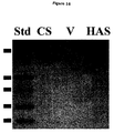
- FIG. 16 is a Western Blot Analysis of Truncated Recombinant Pasteurella GAG Synthases. Immunoreactive bands at the predicted size of 80 kDa correspond to pmCS 1-704 (CS) or pmHAS 1-703 (HAS). No similar band is seen for the vector control (V). Prestained standards (Std) are shown for size comparison (from top to bottom: 95.5, 55, 43, 36, 29 kDa).
- FIG. 17 is a graphical plot of gel filtration analysis of radiolabeled polymer synthesized in vitro.
- the pmCS 1-704 extract (1 mg total protein) was incubated with chondroitin acceptor oligosaccharide (5 mg), UDP-[ 14 C]GlcUA and UDP-[ 3 H]GalNAc (580 mM, 0.16 mCi each) in a reaction volume of 200 ml for 30 min.
- the reaction product was split into five aliquots and treated with various GAG glycosidases as described in Experimental Procedures .
- FIG. 18 is a pictorial representation of a model of the two putative glycosyltransferase sites of pmHAS and pmCS.
- PmHAS and pmCS contain two distinct and relatively independent glycosyltransferase sites. Each site possesses a DGS and a DXD amino acid motif.
- a WGGED motif is found near the junction of the two domains, and is involved in hexosamine-transferase activity.
- the carboxyl-terminus is involved in membrane association (MEM ASSOC), but is not required for catalytic activity.
- Residues 1-117 appear dispensable for catalysis of sugar transfer but may contain structure scaffolding or play other roles.
- FIG. 19 graphically depicts Sequence Similarity of pmHS with KfiA and KfiC.
- Elements of the Pasteurella heparosan synthase, HS1 (containing residues 91-240) and HS2 (containing residues 441-540) are very similar to portions of two proteins from the E coli K5 capsular locus (A, residues 75-172 of KfiA; C, residues 262-410 of KfiC) as shown by this modified Multalin alignment (ref. 21; numbering scheme corresponds to the pmHS sequence).
- the HS1 and HS2 elements may be important for hexosamine transferase or for glucuronic acid transferase activities, respectively.
- FIG. 20 depicts pmHS Activity Dependence on Acceptor and Enzyme Concentration.
- Various amounts of crude membranes containing the full-length enzyme, pmHS1-617 were incubated in 50 ⁇ l of buffer containing 50 mM Tris, pH 7.2, 10 mM MgCl 2 , 10 mM MnCl 2 , 500 ⁇ M UDP-[ 14 C]GlcUA (0.15 ⁇ Ci), and 500 ⁇ M UDP-GlcNAc.
- Three parallel sets of reactions were performed with either no acceptor (circles) or two concentrations of heparosan polymer acceptor (uronic acid: 0.6 ⁇ g, squares; 1.7 ⁇ g, triangles).
- FIG. 21 Gel Filtration Analysis of Radiolabeled Polymer Synthesized in vitro.
- the crude membranes containing pmHS (0.7 mg total protein) were incubated with UDP-[ 14 C]GlcUA and UDP-[ 3 H]GlcNAc (each 500 ⁇ M, 0.4 ⁇ Ci) in a 200 ⁇ l reaction volume either in the presence (top panel) or absence (bottom panel) of acceptor polymer (1 ⁇ g uronic acid).
- FIG. 22(A-D) graphically depicts the alignment of the pmHS (two clones: A2, B10) with PgIA, KfiA, KfiC, and DcbF.
- FIG. 23 depicts chimeric constructs of pm-EG, pm-FH, pm-IK, and pm-JL. PCR-overlap-extension was performed.
- Pm-EG contains residues 1-265 from pmHAS and residues 259-704 from pmCS and is a GlcUA-Tase.
- Pm-FH contains residues 1-258 from pmCS and residues 266-703 from pmHAS and is an active chondrotin synthase.
- Pm-IK contains residues 1-221 from pmHAS and residues 215-704 from pmCS and is a Glc-UA-Tase.
- Pm-JL contains residues 1-214 from pmCS and residues 222-703 from pmHAS and is an active HA synthase.
- the switch of Gal-NAc-transferring activity into GlcNAc-transferring activity indicated that 222-265 of pmHAS and possibly the corresponding residues 215-258 of pmCS play critical role in the selectivity between binding and/or transferring of GalNAc and GlcNAc substrate.
- FIG. 24 depicts a comparison of partial primary sequences of pmHAS and different pmCSs.
- Primary sequences of presumably chondroitin synthases from different Type F Pasteruella multocida were obtained by directly sequencing the products of colony-lysis PCR.
- the MULTALIN alignment indicates that most of the differences between pmHAS and pmCS are conserved among these independent strains.
- Residues that were substituted in site-mutagenesis studies were underlined.
- the mutant polypeptides contain a single or combination of different mutations, indicated by star(s). None of these mutations changes the specificity of the original enzymes.
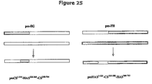
- FIG. 25 depicts chimeric constructs of pmHAS 1-221 -CS 215-258 -HAS 266-703 and pmCS 1-214 -HAS 222-265 -CS 258-704 .
- Pm-FH and pPm7A DNA were used to create pmHAS 1-221 CS 215-258 -HAS 266-703 .
- a very interesting result was that pmCS 1-214 -HASM 222-265 -CS 258-704 can transfer both GalNAc and GlcNAc to HA oligomer acceptor; this enzyme displays relaxed sugar specificity.
- FIG. 26 depicts a summary of enzyme activities of chimeric proteins.
- the enzymes are drawn as bars. Black bars represent pmCS.
- White bars represent pmHAS. +, active; ⁇ , inactive.
- PmCHC represents pmCS 1-214 -HAS 222-265 -CS 258-704 .
- the roles of the two domains are confirmed and we have localized a 44-residue region critical for distinguishing C4 epimers of the hexosamine precursor.
- FIG. 27 depicts a mass spectra of F-HA12 product. This fluorescent HA oligosaccharide was synthesized using the twin bioreactor scheme. A peak with the predicted mass is apparent; no shorter HA11 sugar is observed.
- FIG. 28 is a pictorial representation of a hyalose biocatalytic scheme depicting the step-wise addition of sugars.
- GAGs are linear polysaccharides composed of repeating disaccharide units containing a derivative of an amino sugar (either glucosamine or galactosamine).
- Hyaluronan [HA] chondroitin, and heparan sulfate/heparin contain a uronic acid as the other component of the disaccharide repeat while keratan contains a galactose.
- the GAGs are summarized in Table I.
- Vertebrates may contain all four types of GAGs, but the polysaccharide chain is often further modified after sugar polymerization.
- One or more modifications including O-sulfation of certain hydroxyls, deacetylation and subsequent N-sulfation, or epimerization of glucuronic acid to iduronic acid are found in most GAGs except HA.
- An amazing variety of distinct structures have been reported for chondroitin sulfate and heparan sulfate/heparin even within a single polymer chain.
- a few clever pathogenic microbes also produce unmodified GAG chains; the bacteria use extracellular polysaccharide coatings as molecular camouflage to avoid host defenses.
- chondroitin and heparan sulfate/heparin chains in vertebrates are initially synthesized by elongation of a xylose-containing linkage tetrasaccharide attached to a variety of proteins. Keratan is either O-linked or N-linked to certain proteins depending on the particular molecule.
- HA and all of the known bacterial GAGs are not part of the classification of proteins known as glycoproteins. All GAGs except HA are found covalently linked to a core protein, and such combination is referred to as a proteoglycan.
- Glycoproteins are usually much smaller than proteoglycans and only contain from 1-60% carbohydrate by weight in the form of numerous relatively short, branched oligosaccharide chains, whereas a proteoglycan can contain as much as 95% carbohydrate by weight.
- the core protein in a proteoglycan is also usually a glycoprotein, therefore usually contains other oligosaccharide chains besides the GAGs.
- GAGs and their derivatives are currently used in the medical field as ophthalmic and viscoelastic supplements, adhesion surgical aids to prevent post-operative adhesions, catheter and device coatings, and anticoagulants.
- Other current or promising future applications include anti-cancer medications, tissue engineering matrices, immune and neural cell modulators, and drug targeting agents.
- GAGs Complex carbohydrates, such as GAGs, are information rich molecules.
- a major purpose of the sugars that make up GAGs is to allow communication between cells and extracellular components of multicellular organisms.
- certain proteins bind to particular sugar chains in a very selective fashion.
- a protein may simply adhere to the sugar, but quite often the protein's intrinsic activity may be altered and/or the protein transmits a signal to the cell to modulate its behavior.
- heparin binding to inhibitory proteins helps shuts down the clotting response.
- HA binds to cells via the CD44 receptor that stimulates the cells to migrate and to proliferate.
- oligosaccharides can be used to either (a) substitute for the polymer or (b) to inhibit the polymer's action depending on the particular system.
- HA polysaccharide plays structural roles in the eye, skin, and joint synovium. Large HA polymers ( ⁇ 10 6 Da) also stimulate cell motility and proliferation. On the other hand, shorter HA polymers ( ⁇ 10 4 Da) often have the opposite effect.
- HA-oligosaccharides composed of 10 to 14 sugars [HA 10-14 ] have promise for inhibition of cancer cell growth and metastasis.
- mice injected with various invasive and virulent tumor cell lines (melanoma, glioma, carcinomas from lung, breast and ovary) develop a number of large tumors and die within weeks. Treatment with HA oligosaccharides greatly reduced the number and the size of tumors. Metastasis, the escape of cancer cells throughout the body, is one of the biggest fears of both the ailing patient and the physician.
- HA or HA-like oligosaccharides appear to serve as a supplemental treatment to inhibit cancer growth and metatasis.
- the preliminary mode of action of the HA-oligosaccharide sugars is thought to be mediated by binding or interacting with one of several important HA-binding proteins (probably CD44 or RHAM) in the mammalian body.
- CD44 important HA-binding proteins
- One proposed scenario for the anticancer action of HA-oligosaccharides is that multiple CD44 protein molecules in a cancer cell can bind simultaneously to a long HA polymer. This multivalent HA binding causes CD44 activation (perhaps mediated by dimerization or a receptor patching event) that triggers cancer cell activation and migration.
- each CD44 molecule individually binds a different HA molecule in a monovalent manner such that no dimerization/patching event occurs. Thus no activation signal is transmitted to the cell.
- the optimal HA-sugar size is 10 to 14 sugars. Although this size may be based more upon the size of HA currently available for testing rather than biological functionality—i.e. now that HA molecules and HA-like derivatives ⁇ 10 sugars are available according to the methodologies of the present invention, the optimal HA size or oligosaccharide composition may be found to be different.
- HA-oligosaccharides for therapeutics are that these sugar molecules are natural by-products that can occur in small amounts in the healthy human body during the degradation of HA polymer; no untoward innate toxicity, antigenicity, or allergenic concerns are obvious.
- HA oligosaccharides are the stimulation of blood vessel formation and the stimulation of dendritic cell maturation. Enhancement of wound-healing and resupplying cardiac oxygenation may be additional applications that harness the ability of HA oligosaccharides to cause endothelial cells to form tubes and sprout new vessels.
- Dendritic cells possess adjuvant activity in stimulating specific CD4 and CD8 T cell responses. Therefore, dendritic cells are targets in vaccine development strategies for the prevention and treatment of infections, allograft reactions, allergic and autoimmune diseases, and cancer.
- Heparin interacts with many proteins in the body, but two extremely interesting classes are coagulation cascade proteins and growth factors.
- Antithrombin III [ATIII] and certain other hemostasis proteins are 100,000-fold more potent inhibitors of blood clotting when complexed with heparin. Indeed, heparin is so potent it must be used in a hospital setting and requires careful monitoring in order to avoid hemorrhage. Newer, processed lower molecular weight forms of heparin are safer, but this material is still a complex mixture. It has been shown that a particular pentasaccharide (5 sugars long) found in heparin is responsible for the ATIII-anticoagulant effect.
- pentasaccharide since heparin is a very heterogeneous polymer, it is difficult to isolate the pentasaccharide (5 sugars long) in a pure state.
- the pentasaccharide can also be prepared in a conventional chemical synthesis involving ⁇ 50 to 60 steps. However, altering the synthesis or preparing an assortment of analogs in parallel is not always feasible—either chemically or financially.
- VEGF vascular endothelial growth factor
- HBEGF heparen-binding epidermal growth factor
- FGF fibroblast growth factor
- Chondroitin is the most abundant GAG in the human body, but all of its specific biological roles are not yet clear. Phenomenon such as neural cell outgrowth appears to be modulated by chondroitin. Both stimulatory and inhibitory effects have been noted depending on the chondroitin form and the cell type. Therefore, chondroitin or similar molecules are of utility in re-wiring synaptic connections after degenerative diseases (e.g., Alzheimer's) or paralytic trauma.
- the epimerized form of chondroitin (GlcUA converted to the C5 isomer, iduronic acid or IdoUA), dermatan, selectively inhibits certain coagulation proteins such as heparin cofactor II. By modulating this protein in the coagulation pathway instead of ATIII, dermatan appears to allow for a larger safety margin than heparin treatment for reduction of thrombi or clots that provoke strokes and heart attacks.
- GAG/protein interactions are not yet clear due to (a) the heterogeneity of GAGs (in part due to their biosynthesis pathway) and (b) the difficulty in analyzing long polysaccharides and membrane receptor proteins at the molecular level. Fortunately, many short oligosaccharides have biological activities that serve to assist research pursuits as well as to treat disease in the near future.
- HexNAc GlcNAc or GalNAc.
- n the degree of polymerization, ranges from about 25 to about 10,000. If the GAG is polymerized by a single polypeptide, the enzyme is called a synthase or co-polymerase.
- P. multocida infects fowl, swine, and cattle as well as many wildlife species.
- the enzymes are: a HA synthase, or (pmHAS); a chondroitin synthase, or (pmCS); and two heparosan synthases, or (pmHS and PgIA).
- pmHAS a HA synthase
- pmCS chondroitin synthase
- PgIA two heparosan synthases
- pmHAS is unique in comparison to all other existing HA synthases of Streptococcus bacteria, humans and an algal virus. Specifically, recombinant pmHAS can elongate exogeneously-supplied short HA chains (e.g., 2-4 sugars) into longer HA chains (e.g., 3 to 150 sugars). The pmHAS synthase has been shown to add monosaccharides one at a time in a step-wise fashion to the growing chain. The pmHAS enzyme's 17, sugar transfer specificity results in the repeating sugar backbone of the GAG chain.
- the pmCS enzyme which is about 90% identical at the amino acid level to pmHAS, performs the same synthesis reactions but transfers GalNAc instead of GlcNAc.
- the pmCS enzyme was described and enabled in copending U.S. Ser. No. 09/842,484.
- the pmHS and PgIA enzymes are not very similar at the amino acid level to pmHAS, but perform the similar synthesis reactions; the composition of sugars is identical but the linkages differ because heparosan is Beta4GlcUA-alpha4GlcNAc.
- the pmHS and PgIA enzymes were described and enabled in copending U.S. Ser. No. 10/142,143.
- pmHAS possesses two independent catalytic sites in one polypeptide. Mutants were created that transferred only GlcUA, and distinct mutants were also created that transferred only GlcNAc. These mutants cannot polymerize HA chains individually, but if the two types of mutants are mixed together in the same reaction with an acceptor molecule, then polymerization was rescued.
- the chondroitin synthase, pmCS has a similar sequence and similar two-domain structure.
- the heparosan synthases, pmHS and PgIA also contain regions for the two active sites. Single action mutants have also been created for the chondroitin synthase, pmCS, and are described hereinafter in detail.
- the Pasteurella GAG synthases are very specific glycosyltransferases with respect to the sugar transfer reaction; only the correct monosaccharide from the authentic UDP-sugar is added onto acceptors.
- the epimers or other closely structurally related precursor molecules e.g., UDP-glucose
- the GAG synthases do, however, utilize certain heterologous acceptor sugars. For example, pmHAS will elongate short chondroitin acceptors with long HA chains. pmHS will also add long heparosan chains onto HA acceptor oligosaccharides as well as heparin oligosaccharides (see hereinbelow). Therefore, the presently claimed and disclosed invention encompasses a wide range of hybrid or chimeric GAG oligosaccharides prepared utilizing these P. multocida GAG catalysts.
- nucleic acid segment and “DNA segment” are used interchangeably and refer to a DNA molecule which has been isolated free of total genomic DNA of a particular species. Therefore, a “purified” DNA or nucleic acid segment as used herein, refers to a DNA segment which contains a Hyaluronate Synthase (“HAS”) coding sequence or Chondroitin Synthase (“CS”) coding sequence or Heparin/Heparosan Synthase (“HS”) coding sequence yet is isolated away from, or purified free from, unrelated genomic DNA, for example, total Pasteurella multocida . Included within the term “DNA segment”, are DNA segments and smaller fragments of such segments, and also recombinant vectors, including, for example, plasmids, cosmids, phage, viruses, and the like.
- HAS Hyaluronate Synthase
- CS Chondroitin Synthase
- HS Heparin/Heparosan Synthase
- a DNA segment comprising an isolated or purified pmHAS or pmCS or pmHS or PgIA gene refers to a DNA segment including HAS or CS or HS coding sequences isolated substantially away from other naturally occurring genes or protein encoding sequences.
- the term “gene” is used for simplicity to refer to a functional protein-, polypeptide- or peptide-encoding unit. As will be understood by those in the art, this functional term includes genomic sequences, cDNA sequences or combinations thereof.
- isolated substantially away from other coding sequences means that the gene of interest, in this case pmHAS or pmCS or pmHS or PgIA forms the significant part of the coding region of the DNA segment, and that the DNA segment does not contain other non-relevant large portions of naturally-occurring coding DNA, such as large chromosomal fragments or other functional genes or DNA coding regions. Of course, this refers to the DNA segment as originally isolated, and does not exclude genes or coding regions later added to, or intentionally left in, the segment by the hand of man.
- prokaryotic sources Due to certain advantages associated with the use of prokaryotic sources, one will likely realize the most advantages upon isolation of the HAS or CS or HS gene from the prokaryote P. multocida .
- One such advantage is that, typically, eukaryotic genes may require significant post-transcriptional modifications that can only be achieved in a eukaryotic host. This will tend to limit the applicability of any eukaryotic HAS or CS or HS gene that is obtained.
- those of ordinary skill in the art will likely realize additional advantages in terms of time and ease of genetic manipulation where a prokaryotic enzyme gene is sought to be employed.
- DNA sequences in accordance with the present invention will further include genetic control regions which allow the expression of the sequence in a selected recombinant host.
- the genetic control region may be native to the cell from which the gene was isolated, or may be native to the recombinant host cell, or may be an exaggerous segment that is compatible with and recognized by the transcriptional machinery of the selected recbominant host cell.
- the nature of the control region employed will generally vary depending on the particular use (e.g., cloning host) envisioned.
- the invention concerns isolated DNA segments and recombinant vectors incorporating DNA sequences which encode a pmHAS or pmCS or pmHS or PgIA gene, that includes within its amino acid sequence an amino acid sequence in accordance with SEQ ID NO:2, 4, 6, 8, 9, or 70, respectively.
- the invention concerns isolated DNA segments and recombinant vectors incorporating DNA sequences which encode a gene that includes within its nucleic acid sequence an amino acid sequence encoding HAS or CS or HS pepetides or peptide fragment thereof, and in particular to a HAS or CS or HS peptide or peptide fragment thereof, corresponding to Pasteurella multocida HAS or CS or HS.
- DNA segment or vector encodes a full length HAS or CS or HS protein, or is intended for use in expressing the HAS or CS or HS protein
- preferred sequences are those which are essentially as set forth in SEQ ID NO:1, 3, 5, 7, 69, or 71, respectively.
- Truncated pmHAS gene (such as, but not limited to, pmHAS 1-703 , SEQ ID NO:71) also falls within the definition of preferred sequences as set forth above. For instance, at the carboxyl terminus, approximately 270-272 amino acids may be removed from the sequence and still have a functioning HAS. Those of ordinary skill in the art would appreciate that simple amino acid removal from either end of the pmHAS sequence can be accomplished. The truncated versions of the sequence (as disclosed hereinafter) simply have to be checked for HAS activity in order to determine if such a truncated sequence is still capable of producing HAS.
- the other GAG synthases disclosed and claimed herein are also amenable to truncation or alteration with preservation of activity and such truncated or alternated GAG synthases also fall within the scope of the present invention.
- Nucleic acid segments having HAS or CS or HS activity may be isolated by the methods described herein.
- the term “a sequence essentially as set forth in SEQ ID NO:X” means that the sequence substantially corresponds to a portion of SEQ ID NO:X and has relatively few amino acids or codons encoding amino acids which are not identical to, or a biologically functional equivalent of, the amino acids or codons encoding amino acids of SEQ ID NO:X.
- biologically functional equivalent is well understood in the art and is further defined in detail herein, as a gene having a sequence essentially as set forth in SEQ ID NO:X, and that is associated with the ability of prokaryotes to produce HA or a hyaluronic acid or chondroitin or heparin polymer in vitro or in vivo.
- X refers to either SEQ ID NO: 1, 2, 3, 4, 5, 6, 7, 8, 9, 69, 70 or 71 or any additional sequences set forth herein, such as the truncated or mutated versions of pmHAS 1-703 that are contained generally in SEQ ID NOS:10-60.
- nucleic acid sequence or an amino acid could make substitutions and changes to the nucleic acid sequence without changing its functionality (specific examples of such changes are given hereinafter and are generally set forth in SEQ ID NOS:10-60).
- a substituted nucleic acid segment may be highly identical and retain its enzymatic activity with regard to its unadulterated parent, and yet still fail to hybridize thereto.
- present application discloses 4 enzymes and numerous mutants of these enzymes that still retain at least 50% of the enzymatic activity of the unmutated parent enzyme—i.e., 1 ⁇ 2 of the dual action transferase activity of the unadulterated parent.
- the invention discloses nucleic acid segments encoding an enzymatically active HAS or CS or HS from P. multocida -pmHAS, pmCS, pmHS, and PgIA, respectively.
- P. multocida -pmHAS, pmCS, pmHS, and PgIA substitutions can be made to the pmHAS or pmCS or pmHS or PgIA nucleic acid segments listed in SEQ ID NO:1, 3, 5, 7, 69, and 71, respectively, without deviating outside the scope and claims of the present invention. Indeed, such changes have been made and are described hereinafter with respect to the mutants produced. Standardized and accepted functionally equivalent amino acid substitutions are presented in Table II.
- Another preferred embodiment of the present invention is a purified nucleic acid segment that encodes a protein in accordance with SEQ ID NO:1 or 3 or 5 or 7 or 71, respectively, further defined as a recombinant vector.
- the term “recombinant vector” refers to a vector that has been modified to contain a nucleic acid segment that encodes an HAS or CS or HS protein, or fragment thereof.
- the recombinant vector may be further defined as an expression vector comprising a promoter operatively linked to said HAS- or CS- or HS-encoding nucleic acid segment.
- a further preferred embodiment of the present invention is a host cell, made recombinant with a recombinant vector comprising an HAS or CS or HS gene.
- the preferred recombinant host cell may be a prokaryotic cell.
- the recombinant host cell is an eukaryotic cell.
- the term “engineered” or “recombinant” cell is intended to refer to a cell into which a recombinant gene, such as a gene encoding HAS or CS or HS, has been introduced mechanically or by the hand of man. Therefore, engineered cells are distinguishable from naturally occurring cells which do not contain a recombinantly introduced gene.
- Engineered cells are thus cells having a gene or genes introduced through the hand of man.
- Recombinantly introduced genes will either be in the form of a cDNA gene, a copy of a genomic gene, or will include genes positioned adjacent to a promoter associated or not naturally associated with the particular introduced gene.
- the HAS- or CS- or HS-encoding DNA segments further include DNA sequences, known in the art functionally as origins of replication or “replicons”, which allow replication of contiguous sequences by the particular host.
- origins of replication or “replicons” allow the preparation of extrachromosomally localized and replicating chimeric or hybrid segments or plasmids, to which HAS- or CS- or HS-encoding DNA sequences are ligated.
- the employed origin is one capable of replication in bacterial hosts suitable for biotechnology applications.
- the isolation and use of other replication origins such as the SV40, polyoma or bovine papilloma virus origins, which may be employed for cloning or expression in a number of higher organisms, are well known to those of ordinary skill in the art.
- the invention may thus be defined in terms of a recombinant transformation vector which includes the HAS- or CS- or HS-coding gene sequence together with an appropriate replication origin and under the control of selected control regions.
- HAS or CS or HS gene or cDNA may be obtained which contain full complements of genes or cDNAs from a number of sources, including other strains of Pasteurella or from eukaryotic sources, such as cDNA libraries.
- Virtually any molecular cloning approach may be employed for the generation of DNA fragments in accordance with the present invention.
- the only limitation generally on the particular method employed for DNA isolation is that the isolated nucleic acids should encode a biologically functional equivalent HAS or CS or HS.
- Typical useful vectors include plasmids and phages for use in prokaryotic organisms and even viral vectors for use in eukaryotic organisms. Examples include pKK223-3, pSA3, recombinant lambda, SV40, polyoma, adenovirus, bovine papilloma virus and retroviruses.
- vectors capable of replication in both biotechnologically useful Gram-positive or Gram-negative bacteria e.g. Bacillus, Lactococcus , or E. coli .
- Vectors such as these allow one to perform clonal colony selection in an easily manipulated host such as E. coli , followed by subsequent transfer back into a food grade Lactococcus or Bacillus strain for production of hyaluronan or chondroitin or heparin polymer.
- the recombinant vector is employed to make the functional GAG synthase for in vitro use.
- GRAS generally recognized as safe
- Another procedure to further augment HAS or CS or HS gene copy number is the insertion of multiple copies of the gene into the plasmid.
- Another technique would include integrating at least one copy of the HAS or CS or HS gene into chromosomal DNA. This extra amplification would be especially feasible, since the bacterial HAS or CS or HS gene size is small.
- the chromosomal DNA-ligated vector is employed to transfect the host that is selected for clonal screening purposes such as E. coli , through the use of a vector that is capable of expressing the inserted DNA in the chosen host.
- the invention concerns isolated DNA segments and recombinant vectors that include within their sequence a nucleic acid sequence essentially as set forth in SEQ ID NO:1,3,5,7,69, or 71.
- the term “essentially as set forth” in SEQ ID NO: 1,3,5,7,69, or 71 is used in the same sense as described above and means that the nucleic acid sequence substantially corresponds to a portion of SEQ ID NO: 1,3,5,7,69, or 71 and has relatively few codons which are not identical, or functionally equivalent, to the codons of SEQ ID NO: 1,3,5,7,69, or 71.
- codons that encode the same amino acid such as the six codons for arginine or serine, and also refers to codons that encode biologically equivalent amino acids, as set forth in Table II.
- amino acid and nucleic acid sequences may include additional residues, such as additional N- or C-terminal amino acids or 5′ or 3′ nucleic acid sequences, and yet still be essentially as set forth in one of the sequences disclosed herein, so long as the sequence meets the criteria set forth above, including the maintenance of biological protein activity where protein expression and enzyme activity is concerned.
- the addition of terminal sequences particularly applies to nucleic acid sequences which may, for example, include various non-coding sequences flanking either of the 5′ or 3′ portions of the coding region or may include various internal sequences, which are known to occur within genes.
- residues may be removed from the N- or C-terminal amino acids and yet still be essentially as set forth in one of the sequences disclosed herein, so long as the sequence meets the criteria set forth above, as well.
- sequences which have between about 40% and about 99%; or more preferably, between about 80% and about 90%; or even more preferably, between about 90% and about 99% identity to the nucleotides of SEQ ID NO:1, 3, 5, 7, 69 or 71 will be sequences which are “essentially as set forth” in SEQ ID NO:1, 3, 5, 7, 69 or 71.
- Sequences which are essentially the same as those set forth in SEQ ID NO:1, 3, 5, 7, 69 or 71 may also be functionally defined as sequences which are capable of hybridizing to a nucleic acid segment containing the complement of SEQ ID NO:1, 3, 5, 7, 69 or 71 under “standard stringent hybridization conditions,” “moderately stringent hybridization conditions,” “less stringent hybridization conditions,” or “low stringency hybridization conditions.” Suitable “standard” or “less stringent” hybridization conditions will be well known to those of skill in the art and are clearly set forth hereinbelow. In a preferred embodiment, standard stringent hybridization conditions or less stringent hybridization conditions are utilized.
- standard stringent hybridization conditions “moderately stringent conditions,” and “less stringent hybridization conditions” or “low stringency hybridization conditions” are used herein, describe those conditions under which substantially complementary nucleic acid segments will form standard Watson-Crick base-pairing and thus “hybridize” to one another.
- a number of factors are known that determine the specificity of binding or hybridization, such as pH; temperature; salt concentration; the presence of agents, such as formamide and dimethyl sulfoxide; the length of the segments that are hybridizing; and the like.
- There are various protocols for standard hybridization experiments Depending on the relative similarity of the target DNA and the probe or query DNA, then the hybridization is performed under stringent, moderate, or under low or less stringent conditions.
- the hybridizing portion of the hybridizing nucleic acids is typically at least about 14 nucleotides in length, and preferably between about 14 and about 100 nucleotides in length.
- the hybridizing portion of the hybridizing nucleic acid is at least about 60%, e.g., at least about 80% or at least about 90%, identical to a portion or all of a nucleic acid sequence encoding a HAS or chondroitin or heparin synthase or its complement, such as SEQ ID NO:1,3,5,7,69, or 71 or the complement thereof.
- Hybridization of the oligonucleotide probe to a nucleic acid sample typically is performed under standard or stringent hybridization conditions.
- Nucleic acid duplex or hybrid stability is expressed as the melting temperature or T m , which is the temperature at which a probe nucleic acid sequence dissociates from a target DNA. This melting temperature is used to define the required stringency conditions. If sequences are to be identified that are related and substantially identical to the probe, rather than identical, then it is useful to first establish the lowest temperature at which only homologous hybridization occurs with a particular concentration of salt (e.g., SSC, SSPE, or HPB). Then, assuming that 1% mismatching results in a 1° C.
- salt e.g., SSC, SSPE, or HPB
- the change in T m can be between about 0.5° C. and about 1.5° C. per 1% mismatch.
- standard stringent hybridization conditions include hybridizing at about 68° C. in 5 ⁇ SSC/5 ⁇ Denhardt's solution/1.0% SDS, followed with washing in 0.2 ⁇ SSC/0.1% SDS at room temperature or hybridizing in 1.8 ⁇ HPB at about 30° C. to about 45° C. followed by washing a 0.2-0.5 ⁇ HPB at about 45° C.
- Moderately stringent conditions include hybridizing as described above in 5 ⁇ SSC/5 ⁇ Denhardt's solution 1% SDS washing in 3 ⁇ SSC at 42° C.
- the parameters of salt concentration and temperature can be varied to achieve the optimal level of identity between the probe and the target nucleic acid. Additional guidance regarding such conditions is readily available in the art, for example, by Sambrook et al., 1989, Molecular Cloning, A Laboratory Manual, (Cold Spring Harbor Press, N.Y.); and Ausubel et al. (eds.), 1995, Current Protocols in Molecular Biology, (John Wiley & Sons, N.Y.).
- low stringency protocols include: (A) hybridizing in 5 ⁇ SSC, 5 ⁇ Denhardts reagent, 30% formamide at about 30° C. for about 20 hours followed by washing twice in 2 ⁇ SSC, 0.1% SDS at about 30° C. for about 15 min followed by 0.5 ⁇ SSC, 0.1% SDS at about 30° C. for about 30 min (FEMS Microbiology Letters, 2000, vol. 193, p. 99-103); (B) hybridizing in 5 ⁇ SSC at about 45° C. overnight followed by washing with 2 ⁇ SSC, then by 0.7 ⁇ SSC at about 55° C. (J. Viological Methods, 1990, vol. 30, p. 141-150); or (C) hybridizing in 1.8XHPB at about 30° C. to about 45° C.; followed by washing in 1 ⁇ HPB at 23° C.
- the present invention also encompasses DNA segments which are complementary, or essentially complementary, to the sequences set forth in SEQ ID NO:1 or 3 or 5 or 7 or 69 or 71.
- Nucleic acid sequences which are “complementary” are those which are capable of base-pairing according to the standard Watson-Crick complementarity rules.
- the sequence 5′-ATAGCG-3′ is complementary to the sequence 5′-CGCTAT-3′′ because when the two sequences are aligned, each “T” is able to base-pair with an “A”, which each “G” is able to base pair with a “C”.
- complementary sequences means nucleic acid sequences which are substantially complementary, as may be assessed by the nucleotide comparison set forth above, or as defined as being capable of hybridizing to the nucleic acid segment of SEQ ID NO:1,3,5,7, or 69, or 71 under standard stringent, moderately stringent, or less stringent hybridizing conditions.
- nucleic acid segments of the present invention may be combined with other DNA sequences, such as promoters, polyadenylation signals, additional restriction enzyme sites, multiple cloning sites, epitope tags, polyhistidine regions, other coding segments, and the like, such that their overall length may vary considerably. It is therefore contemplated that a nucleic acid fragment of almost any length may be employed, with the total length preferably being limited by the ease of preparation and use in the intended recombinant DNA protocol.
- this invention is not limited to the particular amino acid and nucleic acid sequences of SEQ ID NO:1, 2, 3, 4, 5, 6, 7, 8, 9, 69, 70, or 71.
- Recombinant vectors and isolated DNA segments may therefore variously include the HAS or CS or HS coding regions themselves, coding regions bearing selected alterations or modifications in the basic coding region, or they may encode larger polypeptides which nevertheless include HAS or CS or HS coding regions or may encode biologically functional equivalent proteins or peptides which have variant amino acid sequences.
- the DNA segments of the present invention encompass DNA segments encoding biologically functional equivalent HAS or CS or HS proteins and peptides. Such sequences may arise as a consequence of codon redundancy and functional equivalency which are known to occur naturally within nucleic acid sequences and the proteins thus encoded. Alternatively, functionally equivalent proteins or peptides may be created via the application of recombinant DNA technology, in which changes in the protein structure may be engineered, based on considerations of the properties of the amino acids being exchanged.
- Changes designed by man may be introduced through the application of site-directed mutagenesis techniques, e.g., to introduce improvements to the enzyme activity or to antigenicity of the HAS or CS or HS protein or to test HAS or CS or HS mutants in order to examine HAS or CS or HS activity at the molecular level or to produce HAS or CS or HS mutants having changed or novel enzymatic activity and/or sugar substrate specificity.
- site-directed mutagenesis techniques e.g., to introduce improvements to the enzyme activity or to antigenicity of the HAS or CS or HS protein or to test HAS or CS or HS mutants in order to examine HAS or CS or HS activity at the molecular level or to produce HAS or CS or HS mutants having changed or novel enzymatic activity and/or sugar substrate specificity.
- polysaccharide polymers must be of a certain length before their physical or biological properties become apparent. Often the polysaccharide must comprise at least 20-100 sugar units. Certain enzymes that react with exogenous polymers have been previously available, but typically add only one sugar unit. The unique enzymes described in the present invention, (e.g. pmHAS, pmCS, pmHS, and PgIA) form polymers of at least 100-400 sugar units in length. Thus, one embodiment of the presently claimed and disclosed invention, results in long, defined linear polymers composed of only natural glycosidic linkages.
- the four known glycosaminoglycan synthesizing enzymes from Pasteurella multocida bacteria normally make polymers similar to or identical to vertebrate polymers. These bacteria employ the polysaccharide, either HA (Type A bacteria), chondroitin (Type F bacteria), or heparosan (unsulfated, unepimerized heparin—Type D bacteria) as an extracellular coating to serve as molecular camouflage.
- Native enzymes normally make polymer chains of a single type of sugar repeat. If a recombinant HAS or CS or HS enzyme is employed, however, the enzyme can be forced to work on an exogenous functional acceptor molecule.
- the recombinant enzyme may be incubated with a polymer acceptor, and the recombinant enzyme will then elongate the acceptor with UDP-sugar precursors.
- the known native enzymes do not perform this reaction since they already contain a growing polymer chain that was formed in the living cell; enzyme preparations from native cells retain the polymer following isolation.
- pmHAS (SEQ ID NO:2), a 972 amino acid residue protein from Pasteurella multocida , is made in a functional state in recombinant Escherichia coli .
- the pmHAS gene is given in SEQ ID NO:1.
- Other functional derivatives of pmHAS for example an enzyme called pmHAS 1-703 (SEQ ID NO:9) and the pmHAS 1-703 gene (SEQ ID NO:71), have been produced which are soluble.
- the soluble form can be prepared in larger quantities and in a purer state than the naturally occurring full-length enzyme.
- the preferred E. coli strains do not have an UDP-Glc dehydrogenase and therefore the recombinant enzyme does not make HA chain in the foreign host.
- the enzyme is in a “virgin” state since the empty acceptor site can be occupied with foreign polymers.
- the recombinant enzyme may be incubated in a mixture comprising from about 10 to about 50 mM Tris pH 7.2, from about 20 mM MnCl 2 , from about 0.1 to about 15 mM UDP-GlcUA, from about 0.1 to about 15 mM UDP-GlcNAc, and a suitable acceptor at about 30° C. for from about 1 to about 180 minutes.
- Suitable acceptors can be any functional acceptor, such as a glycosaminoglycan acceptor or sugar acceptor, for example, but not by limitation, short HA chains (two or more sugar units such as HA 4 ) or short chondroitin sulfate chains (5 sugar units) or long chondroitin sulfate chains ( ⁇ 10 2 sugar units) or short or long heparosan chains.
- HA 4 two or more sugar units
- chondroitin sulfate chains 5 sugar units
- long chondroitin sulfate chains ⁇ 10 2 sugar units
- heparosan chains elongates the foreign acceptors (i.e. long or short chondroitin or heparosan oligosaccharides) at their nonreducing termini with authentic HA chains.
- the length of the HA chain added onto the acceptor is controlled by altering the concentration of UDP-sugars and/or the reaction time.
- Immobilized acceptors such as beads or other solid objects with bound acceptor oligosaccharides, can also be extended by the pmHAS enzyme using UDP-sugars. In this manner, the pmHAS enzyme (or its derivatives) can be used to attach polysaccharide chains to any suitable acceptor molecule.
- Type A P. multocida produces HA capsule [GlcUA-GlcNAc repeats] and possesses the pmHAS enzyme.
- Type F P. multocida produces a chondroitin or chondroitin-like polymer capsule [GlcUA-GalNAc repeats].
- the DNA encoding an open reading frame (GenBank accession #AF195517) that is 90% identical to pmHAS at the protein level has been cloned; this enzyme is called PmCS, the P. multocida chondroitin synthase.
- the amino acid sequence of pmCS is set forth in Seq ID NO:4 and the pmCS gene sequence is set forth in SEQ ID NO:3.
- hybrid polysaccharide materials composed of HA, chondroitin and heparin cannot be formed by any other existing process without (1) leaving unnatural residues and/or (2) producing undesirable crosslinking reactions.
- the chimeric or hybrid polysaccharide materials can serve as biocompatible molecular glue for cell/cell interactions in artificial tissues or organs and the HA/chondroitin/heparin hybrid mimics natural proteoglycans that normally contain an additional protein intermediate between polymer chains. The present invention, therefore, obviates the requirement for a protein intermediary.
- a recombinant HA/chondroitin/heparin chimeric or hybrid polysaccharide is desirous since molecules from animal sources are potentially immunogenic—the chimeric or hybrid polysaccharide, however, would not appear as “foreign” to the host, thus no immune response is generated.
- the recombinant polymers can be made free of adventitious agents (e.g., prions, viruses etc.)
- An intrinsic and essential feature of polysaccharide synthesis is the repetitive addition of sugar monomer units to the growing polymer.
- the glycosyltransferase remains. in association with the nascent chain.
- This feature is particularly relevant for HA biosynthesis as the HA polysaccharide product, in all known cases, is transported out of the cell; if the polymer was released, then the HAS would not have another chance to elongate that particular molecule.
- Three possible mechanisms for maintaining the growing polymer chain at the active site of the enzyme are immediately obvious. First, the enzyme possesses a carbohydrate polymer binding pocket or cleft. Second, the nascent chain is covalently attached to the enzyme during its synthesis. Third, the enzyme binds to the nucleotide base or the lipid moiety of the precursor while the nascent polymer chain is still covalently attached.
- pmHAS The HAS activity of the native pmHAS enzyme found in P. multocida membrane preparations is not stimulated by the addition of HA oligosaccharides; theoretically, the endogenous nascent HA chain initiated in vivo renders the exogenously supplied acceptor unnecessary.
- recombinant pmHAS produced in an E. coli strain that lacks the UDP-GlcUA precursor, and thus lacks a nascent HA chain is able to bind and to elongate exogenous HA oligosaccharides.
- a nascent HA chain there are three likely means for a nascent HA chain to be held at or near the active site. In the case of pmHAS, it appears that a HA-binding site exists near or at the sugar transferase catalytic site.
- oligosaccharides that vary in size and composition are used to discern the nature of the interaction between pmHAS and the sugar chain. For example, it appears that the putative HA-polymer binding pocket of pmHAS will bind and elongate at least an intact HA disaccharide with increased efficiency occurring when a trisaccharide is used (reduced tetramer or a synthetic trisaccharide). Oligosaccharide binding to pmHAS appears to be somewhat selective because the heparosan pentamer, which only differs in the glycosidic linkages from HA-derived oligosaccharides, does not serve as an acceptor. However, chondroitin [GlcUA-GalNAc repeat] does serve as an acceptor for pmHAS.
- Both heparin and chondroitin are synthesized by the addition of sugar units to the nonreducing end of the polymer chain.
- the glycosyltransferases initiate chain elongation on at least primer monosachharides [more preferably tetrasaccharides such as xylose-galactose-galactose-GlcUA] that are attached to serine residues of proteoglycan core molecules.
- enzyme extracts transfer a single sugar to exogenously added heparin or chondroitin oligosaccharides; unfortunately, the subsequent sugar of the disaccharide unit is usually not added and processive elongation to longer polymers does not occur.
- Recombinant pmHAS adds single monosaccharides in a sequential fashion to the nonreducing termini of the nascent HA chain. Elongation of HA polymers containing hundreds of sugars has been demonstrated in vitro. The simultaneous formation of the disaccharide repeat unit is not necessary for generating the alternating structure of the HA molecule. The intrinsic specificity and fidelity of each half-reaction (e.g. GlcUA added to a GlcNAc residue or vice versa) apparently is sufficient to synthesize authentic HA chains.
- a great technical benefit resulting from the alternating disaccharide structure of HA is that the reaction can be dissected by controlling the availability of UDP-sugar nucleotides. By omitting or supplying precursors in a reaction mixture, the glycosyltransferase may be stopped and started at different stages of synthesis of the heteropolysaccharide. In contrast, there is no facile way to control in a step-wise fashion the glycosyltransferase enzymes that produce important homopolysaccharides such as chitin, cellulose, starch, and glycogen.
- pmHAS 1-650 (SEQ. ID NO:10) can only add single GlcNAc sugars onto the non-reducing end (i.e., HA tetrasaccharide [GlcNAc-GlcUA-GlcNAc-GlcUA]) of an acceptor (i.e. forms the HA pentamer).
- a mutant has been created and called pmHAS 1-703 -D477N (SEQ.
- membrane preparations from recombinant E coli containing a pmHAS protein had HA synthase activity as judged by incorporation of radiolabel from UDP-[ 14 C]GlcUA into polymer when co-incubated with both UDP-GlcNAc and Mn ion. Due to the similarity at the amino acid level of pmHAS to several lipopolysaccharide transferases, it was hypothesized that HA oligosaccharides serve as acceptors for GlcUA and GlcNAc transfer.
- pmHAS 1-703 has been shown to add sugars onto a chondroitin pentamer acceptor.
- the pmHAS 1-703 and reagents were prepared in the same manner as shown in FIG. 1 , except that a chondroitin pentamer was used as the acceptor molecule.
- the results of this experiment are shown in TABLE III.
- the pmHAS 1-703 can utilize molecules other than the naturally occurring acceptors or primer molecules as the basis for forming a polysaccharide polymer chain.
- the HA polymerizing activity of recombinant pmHAS 1-703 is dependent on the simultaneous incubation with both UDP-sugar precursors and a Mn 2+ ion.
- the level of incorporation is dependent on protein concentration, on HA oligosaccharide concentration, and on incubation time as shown in FIG. 2 .
- FIG. 2 two parallel reactions containing pmHAS 1-703 with even-numbered HA oligosaccharides (105 ⁇ g membrane protein/point with a mixture of HA hexamer, octamer, and decamer, 4.4.
- HA synthesized in the presence or the absence of HA oligosaccharides is sensitive to HA lyase (>95% destroyed) and has a molecular weight of ⁇ 1-5 ⁇ 10 4 Da ( ⁇ 50-250 monosaccharides). No requirement for a lipid-linked intermediate was observed as neither bacitracin (0.5 mg/ml) nor tunicamycin (0.2 mg/ml) alter the level of incorporation in comparison to parallel reactions with no inhibitor.
- the starting tetramer elutes in the included volume (Vi arrow).
- Membranes 190 ⁇ g total protein
- UDP-GlcUA 200 ⁇ M
- UDP-GlcNAc 600 ⁇ M
- radiolabeled 3 H-HA tetramer 1.1 ⁇ 10 5 dpm
- solid squares As a negative control, a parallel reaction containing all the components except for UDP-GlcNAc was analyzed (open squares). The small primer was elongated into higher molecular weight product if both precursors were supplied. In a parallel reaction without UDP-GlcNAc, the elution profile of the labeled tetramer is not altered.
- the activity of the native pmHAS 1-703 from P. multocida membranes is not stimulated by the addition of HA oligosaccharides under similar conditions.
- the native pmHAS 1-703 enzyme has an attached or bound nascent HA chain that is initiated in the bacterium prior to membrane isolation.
- the recombinant enzyme lacks such a nascent HA chain since the E. coli host does not produce the UDP-GlcUA precursor needed to make HA polysaccharide. Therefore, the exogenous HA-derived oligosaccharide has access to the active site of pmHAS 1-703 and can be elongated.
- the tetramer from bovine testicular hyaluronidase digests of HA terminates at the nonreducing end with a GlcUA residue and this molecule served as an acceptor for HA elongation by pmHAS 1-703 .
- the tetramer and hexamer oligosaccharides produced by the action of Streptomyces HA lyase did not stimulate HA polymerization as shown in FIG. 1 ; “unsHA4/6”.
- the terminal unsaturated sugar is missing the C4 hydroxyl of GlcUA which would normally be extended by the HA synthase.
- Neither Yeast-derived recombinant Group A HasA nor recombinant DG42 produced elongated HA-derived oligosaccharides into larger polymers.
- the addition of HA tetramer (or a series of longer oligosaccharides) did not significantly stimulate nor inhibit the incorporation of radiolabeled UDP-sugar precursors into HA ( ⁇ 5% of control value) by these Class I HA synthases.
- the HAS activity of HasA or DG42 was not affected by the addition of chitin-derived oligosaccharides.
- the various recombinant enzymes were tested for their ability to convert HA tetramer into molecular weight products.
- the reactions contained radiolabeled HA tetramer (5-8 ⁇ 10 5 dpm), 750 ⁇ M UDP-GlcNAc, 360 ⁇ M UDP-GlcUA, 20 mM XCl 2 , 50 mM Tris, pH 7-7.6 (the respective X cation and pH values used for each enzyme were: pmHAS 1-703 , Mn/7.2 ; Xenopous DG42, Mg/7.6; Group A streptococcal HasA, Mg/7.0), and enzyme (units/reaction listed).
- FIG. 4 demonstrates that pmHAS 1-703 elongated the HA-derived tetramer by a single sugar unit if the next appropriate UDP-sugar precursor was available in the reaction mixture.
- GlcNAc derived from UDP-GlcNAc was added onto the GlcUA residue at the nonreducing terminus of the tetramer acceptor to form a pentamer.
- inclusion of only UDP-GlcUA did not alter the mobility of the oligosaccharide.
- pmHAS extended an HA tetramer.
- radiolabeled HA tetramer HA4 8 ⁇ 10 3 dpm 3 H
- a GlcUA at the nonreducing terminus was incubated with various combinations of UDP-sugars (A, 360 ⁇ M UDP-GlcUA; N, 750 ⁇ M UDP-GlcNAc; 0, no UDP-sugar), and pmHAS (55 ⁇ g membrane protein) in assay buffer for 60 minutes.
- the reactions (7 ⁇ l total) were terminated by heating at 95° C. for 1 minute and clarified by centrifugation.
- Portions (2.5 ⁇ l) of the supernatant were spotted onto the application zone of a silica TLC plate and developed with solvent (1.25:1:1 butanol/acetic acid/water).
- the beginning of the analytical layer is marked by an arrow.
- the positions of odd-numbered HA oligosaccharides (S lane) are marked as number of monosaccharide units.
- the autoradiogram of FIG. 4 (4 day exposure) shows the single addition of a GlcNAc sugar onto the HA tetramer acceptor to form a pentamer when only the subsequent precursor is supplied (N).
- HA-derived oligosaccharides with either GlcUA or GlcNAc at the nonreducing terminus served as acceptors for pmHAS 1-703 ( FIG. 5 ).
- radiolabeled HA pentamer HA5, 5 ⁇ 10 3 dpm 3 H
- HA tetramer HA4, 25 ⁇ 10 3 dpm 3 H
- the pmHAS 1-703 enzyme transfers individual monosaccharides sequentially during a polymerization reaction.
- Membrane preparations containing recombinant pmHAS (GenBankAF036004) (SEQ. ID NOS: 1 and 2) were isolated from E. coli SURE(pPmHAS). Membrane preparations containing native pmHAS were obtained from the P. multocida strain P-1059 (ATCC #15742). pmHAS was assayed in 50 mM Tris, pH 7.2, 20 mM MnC1 2 , and UDP-sugars (UDP-[ 14 C]GlcUA, 0.3 ⁇ Ci/mmol, NEN and UDP-GlcNAc) at 30° C. The reaction products were analyzed by various chromatographic methods as described below. Membrane preparations containing other recombinant HAS enzymes, Group A streptococcal HasA or Xenopus DG42 produced in the yeast Saccharomyces cerevisiae , were prepared.
- Uronic acid was quantitated by the carbazole method.
- the latter enzyme employs an elimination mechanism to cleave the chain resulting in an unsaturated GlcUA residue at the nonreducing terminus of each fragment.
- oligosaccharides that are structurally similar to HA were also tested in HAS assays.
- the structure of heparosan pentamer derived from the E. coli K5 capsular polysaccharide is ⁇ 4GlcUA- ⁇ 4GlcNAc; this carbohydrate has the same composition as HA but the glycosidic linkages between the monosaccharides are different.
- the chitin-derived oligosaccharides, chitotetraose and chitopentaose are ⁇ 4GlcNAc polymers made of 4 or 5 monosaccharides, respectively.
- oligosaccharides were radiolabeled by reduction with 4 to 6 equivalents of sodium borotritide (20 mM, NEN; 0.2 ⁇ Ci/mmol) in 15 mM NaOH at 30° C. for 2 hrs. 3 H-oligosaccharides were desalted on a P-2 column in 0.2 M ammonium formate to remove unincorporated tritium and lyophilized. Some labeled oligosaccharides were further purified preparatively by paper chromatography with Whatman 1 developed in pyridine/ethyl acetate/acetic acid/H 2 O (5:5:1:3) before use as an acceptor.
- An anti-pmHAS monospecific antibody reagent has also been identified that routinely monitors the protein by Western blots or immunoassays; this reagent can be used to normalize protein expression levels.
- the DNA inserts encoding the enzyme sequence from interesting mutants picked up in screens can be subcloned and completely sequenced to verify and to identify the mutation site.
- pmHAS normally a 972-residue membrane protein
- Table V A series of truncated versions of pmHAS (normally a 972-residue membrane protein) were created and are tabulated (with functionality) in Table V that produce proteins with altered physical properties (i.e., proteins that are more conducive to high-level expression and purification) and altered function (i.e., single transferase activity).
- Polymerase chain reaction [PCR] was used to amplify a portion of the pmHAS gene using a primer corresponding to the authentic N-terminus sequence and a primer corresponding to an internal coding region which ended in a stop codon.
- the coding regions for the truncated proteins were cloned into an Escherichia coli expression plasmid (pKK223-3; Pharmacia) under control of the tac promoter. The DNA sequence was verified by automated sequencing.
- the truncation series was generated and tested for activity. All proteins were made at the expected molecular weight, but not all proteins were active.
- pmHAS 1-703 a new 80-kDa protein, named pmHAS 1-703 , was produced in large quantities. Furthermore, functional pmHAS 1-703 was present in the soluble fraction of the cell lysate; thus allowing for rapid extraction and assay of the enzyme.
- pmHAS 1-703 was purified by sequential chromatography steps shown in FIG. 6 . In FIG. 6 , a soluble, active form of the HA synthase was constructed with molecular biological techniques. The recombinant enzyme from E. coli was purified by conventional chromatography with yields of up to 20 mg/liter of cell culture. FIG.
- the pmHAS 1-703 is highly active and at least 95% pure as assessed by denaturing polyacrylamide gel electrophoresis. Mass spectrometric analysis indicates that the pmHAS 1-703 is the desired protein due to the close agreement of the calculated and the observed mass values. A buffer system has also been developed to stabilize the enzymatic activity in the range of 0° to 37° C.
- the mutant enzymes are useful for adding on a single GlcNAc or a single GlcUA onto the appropriate acceptor oligosaccharide. It appears that pmHAS 1-703 has two domains or two modules for transferring each sugar. One of ordinary skill in the art, given this specification, would be able to shift or to combine various domains to create new polysaccharide synthases capable of producing new polysaccharides with altered structures. Within such use, a variety of grafting techniques arise which utilize pmHAS 1-703 as the prototype. A graphical representation of each mutant as it relates to the pmHAS 1-703 sequence is shown in FIG. 7 .
- FIG. 8 is a graphical representation of an experiment where combining two single action mutants rescued dual enzymatic activity.
- HAS enzyme assays were performed in the presence of wild type pmHAS 1-703 alone, D196 mutant alone, D477 mutant alone, or in the presence of both D196 and D477 mutants. Equal amounts of each enzyme were tested with a small amount of HA acceptor sugar in the typical reaction buffer at 30° C. Two time points were measured (cross-hatched, 25 minutes; black, 1.5 hours) for each assay. The two mutants work together to make HA polymer; by itself, a single mutant cannot make HA polymer. Enzyme activity of the pmHAS 1-703 mutants is shown in FIG. 9 .
- Extracts of the mutants were used for all three kinds of assays: for HA polymer production, for GlcUA-Tase activity and for GlcNAc-Tase activity.
- Equivalent amounts of pmHAS 1-703 proteins were assayed. The activities were indicated as the percentage of the activity of wild type pmHAS 1-703 .
- HA, chondroitin, heparin, or chimeric or hybrid molecules that include any or all of the previous GAGs may be attached to other substrates by using the polymer grafting technology of the presently claimed and disclosed invention.
- additional substrates may be metal or metalized—i.e., having a metal coating on the surface of a second material (or a laminate material) such as plastic or silica.
- the metal substrate may be, but is not limited to: gold, copper, stainless steel, nickel, aluminum, titanium, vanadium, chromium, thermosensitive metal alloys, and combinations thereof to name but a few.
- any metal could be used as the substrate as long as it had a surface layer capable of having an activated surface or activated surface group with a functional acceptor molecule.
- gold is an exceptional metal substrate to which a functional acceptor may be attached by using the polymer grafting technology of the presently claimed and disclosed invention.
- This biologically inert metal acts to elongate the functional acceptor as well as to create a glycosidic bond between the functional acceptor and at least one of GlcUA and GlcNAc.
- the procedure for elongating a functional acceptor includes: providing a functional acceptor having at least two sugar units selected from the group consisting of GlcUA, GlcNAc, and hexosamine, and attaching the functional acceptor to a substrate such as gold; providing a hyaluronic acid synthase capable of elongating the functional acceptor, wherein the hyaluronic acid synthase has an amino acid sequence encoded by a nucleotide sequence capable of hybridizing under standard conditions to a nucleotide sequence encoding the hyaluronic acid synthase, such as pmHAS pr pmHAS 1-703 ; and providing UDP-GlcUA and UDP-GlcNAc sugars such that the hyaluronic acid synthase elongates the functional acceptor.
- substrates include, but are not limited to, silica, silicon, glass, polymers, organic compounds, metals and combinations thereof.
- metals that may act as a substrate include, but are not limited to, copper, stainless steel, nickel, aluminum, titanium, thermosensitive alloys and combinations thereof.
- the procedure for creating a glycosidic bond between a functional acceptor and at least one of GlcUA and GlcNAc includes: providing a hyaluronic acid synthase capable of making a glycosidic bond between a functional acceptor and at least one of GlcUA and GlcNAc wherein the functional acceptor has at least two sugar units selected from the group consisting of GlcUA, GlcNAc, and hexosamine, and wherein the hyaluronic acid synthase has an amino acid sequence encoded by a nucleotide sequence capable of hybridizing under standard conditions to the nucleotide sequence encoding hyaluronic acid synthase, such as pmHAS pr pmHAS 1-703 , and wherein the functional acceptor is attached to a substrate such as gold; and incubating the hyaluronic acid synthase with at least one of UDP-GlcUA and UDP-GlcNAc in the presence of the functional acceptor
- substrates include, but are not limited to, silica, silicon, glass, polymers, organic compounds, metals and combinations thereof.
- metals that may act as a substrate include, but are not limited to, copper, stainless steel, nickel, aluminum, titanium, thermosensitive alloys and combinations thereof.
- the procedure for attachment of HA, chondroitin, heparin, or chimeric or hybrid molecule chains onto metal is the same as any other type of substrate and includes: combining the metal particle-NHS ester i.e. activated metal surface (NanoGold from NanoProbes, Inc.) and 10 molar equivalents of amino-HA4 (reactive pmHAS 1-703 acceptor oligosaccharide) in 0.03 M borate buffer, pH 8.5, 20% DMSO for 2 hours at 20° C.; separating the free unused acceptor from the metal particle-HA4 product by P-2 (BioRad) gel filtration column in TBS buffer (50 mM Tris, pH 7.5, 0.15 M NaCl), harvesting the void gold peak; adding the metal particle-HA4 to the reaction with below components at a final concentration of: 1 M ethylene glycol, 50 mM Tris, pH 7.2, 15 mM MnCl 2 , 0.05 mM UDP-[ 14 C]Glc
- the E coli host cells containing the pmHAS 1-703 cloned into the expression vector pKK223-3 are grown on Luria broth (LB) plates with ampillicin at 30° C.
- a colony is used to seed a 40 ml starter culture in enriched LB broth (1.0 g LB broth powder [Difco], 0.4 g Casamino acids, 40 ml water) with ampicillin in a 250 ml Erlenmeyer flask. After overnight culture, the starter is split and used to inoculate four 2 L Erlenmeyer flasks with 400 ml enriched LB broth containing Ampicillin and Carbinicillin and trace elements.
- the inducer IPTG is added to a concentration of 0.2 mM. After 1 hour, fructose is added to 12.8 mM. After overnight growth, cells are harvested by centrifugation, and frozen at ⁇ 80° C. The cells are extracted with 30 ml of lysis buffer (1% n-Octyl-b-D-Thioglucopyranoside, 1 M ethylene glycol, 50 mM HEPES, pH 7, Pepstatin (14.6 uM), Leupeptin (20 uM), E-64 (2 uM), AEBSF (0.4 mM), Benzamidine (2 mM), DNAse/RNAse (1 mg of ea/ml).
- lysis buffer 1% n-Octyl-b-D-Thioglucopyranoside, 1 M ethylene glycol, 50 mM HEPES, pH 7, Pepstatin (14.6 uM), Leupeptin (20 uM), E-64 (2 uM), AEBSF (0.4 mM
- the suspension is stirred at 4° C. for 1 hour; the cells are removed by centrifugation at 3,000 ⁇ g, for 30 min @ 4° C.
- the supernatant containing the pmHAS 1-703 is clarified by high-speed centrifugation at 30,000 ⁇ g and applied to a dye affinity column.
- the pmHAS 1-703 is eluted with a salt gradient.
- the relevant fractions with enzyme are pooled and dialyzed into a reaction buffer for use in polymer grafting.
- pmHAS catalyst (pmHAS 1-703 -1 microgram) was mixed with either (a) no polymer acceptor or with various amounts of chondroitin sulfate (shark, Sigma)—either (b) 5 or (c) 25 micrograms—in a 20 ul reaction. All reactions contained 50 mM Tris, pH 7.2, 1 M ethylene glycol, 20 mM MnCl2, 2.5 mM UDP-GlcUA. Half of the reactions also contained 2.5 mM UDP-GlcNAc; for grafting of long HA chains, both UDP-sugars need to be present.
- polymer grafting comprises incubating all components together for 16 hours at 20° C.; removing the unincorporated HA precursor sugars with ultrafiltration with a Micron 3 (3,000 Da MW cutoff; Amicon) and repeated washing with TBS buffer; and counting the retained samples (e.g., big polymers and particles) by liquid scintillation counting for both 3 H and 14 C labels.
- Various formulations, buffers, and procedures can be used for polymer grafting.
- HA was grafted by pmHAS 1-703 onto gold particles via the HA4 acceptor—i.e. both sugars were added to the gold particles in presence of enzyme; and (2) along with previous demonstrations of attaching HA to various disparate entities (including glass, plastic, polymers, and organic molecules) by polymer grafting technology, one of ordinary skill in the art may attach HA onto any substrate by attaching the initial acceptor to the target and then contacting the acceptor-target complex with pmHAS 1-703 and UDP-sugars.
- the polysaccharide surface coatings of the present invention improve the biocompatibility of the artificial material, lengthen the lifetime of the device in the cellular environment, and encourage natural interactions with host tissues.
- polyacrylamide beads have been coated with the HA polymer using pmHAS 1-703 as the catalyst.
- aminoethyl-beads were chemically primed with HA oligosaccharide (a mixture of 4, 6, and 8 sugars long) by reductive amination. Beads, HA oligosaccharide, and 70 mM NaCNBH 4 in 0.2 M borate buffer, pH 9, were incubated at 42° C. for 2 days. The beads were washed with high and low salt buffers before use in the next step.
- an authentic HA oligosaccharide primer was chemically coupled to a polyacrylamide surface and then this primer was further elongated using the pmHAS 1-703 enzyme and UDP-sugars.
- the reaction conditions can be optimized by one of ordinary skill in the art. For example, the mode of substrate modification, buffer conditions, HA elongation reaction time and stoichiometry can be varied to take into account any single or multiple reaction variation. The resulting coatings can then be evaluated for efficacy and use.
- HA oligosaccharide acceptor or primer molecule The critical structural elements of the HA oligosaccharide acceptor or primer molecule are currently being tested and identified.
- the smallest acceptor molecule with activity tested thus far is an HA disaccharide, although it is anticipated that molecules as short as a monosaccharides will be suitable for use with the present invention.
- the HA polymer produced was quantitated by paper chromatography (polymer at the origin of the paper strip) and liquid scintillation counting (Jing and DeAngelis, 2000, Glycobiology, 10, p. 883-889).
- pmHAS 1-703 enzyme has some flexibility with respect to the identity of the hexosamine group; i.e. other isomers will substitute for the GlcNAc sugar.
- chondroitin pentamer [GalNAc-GlcUA-GalNAc-GlcUA-GalNAc]
- a synthetic molecule consisting of several hydroxyl groups, a pair of negatively charged groups (corresponding to the carboxyl groups of GlcUA sugar), and hydrophobic patches (analog of the carbon-rich side of the sugar ring) will work as a functional primer for pmHAS.
- Such an approach is not unprecedented as the polymerization of heparin, a glycosaminoglycan, can be primed with a rather simple aromatic xyloside instead of a complex proteoglycan core in vertebrate cells.
- HA oligosaccharides can visualize potential molecular shape. However, some proteins distort the sugar chains upon binding, thus making computer modeling somewhat more complicated.
- the most efficacious method of finding an artificial primer is a combinatorial chemistry approach. Closely related series of molecules are screened by high-throughput assay methodologies in order to detect HA elongation. pmHAS 1-703 is then tested for the ability to add an HA polymer onto synthetic primer candidates in a typical 96-well plate format. For example, a series of synthetic peptides (1 to 8 residues) terminating with a GlcNAc group using conventional F moc chemistry can be generated.
- Such peptides are particularly promising because they can adopt a variety of conformations and fit within the pmHAS 1-703 HA-binding pocket via an induced fit mechanism.
- Synthetic peptide chemistry is also much less cumbersome than carbohydrate chemistry.
- the amino acids are chosen with the goal of mimicking the properties of the GlcNAcGlcUA sugar repeats of HA.
- glutamate or asparatate may be used as a substitute for the acid group of GlcUA
- glutamine or asparagine may be used as a substitute for the amide group of GlcNAc
- Serine, threonine, or tyrosine can be used as substitutes for the hydroxyl groups and sugar rings in general.
- the peptide library terminates with a GlcNAc or GlcUA sugar group so that the demands on the pmHAS 1-703 enzyme's binding site and catalytic center are not overly burdensome.
- the present invention also encompasses the development of a mutant version of pmHAS that utilizes a simpler molecule than an HA oligosaccharide as a primer.
- Chitopentaose ( ⁇ 1,4-GlcNAc homopolymer) is one such variant primer.
- Native pmHAS does not utilize chitopentaose as a primer, but a mutant pmHAS but a mutant pmHAS as disclosed herein, elongates chitopentaose, a more readily available substance.
- the chitopentaose primer is attached to the solid phase by reductive amination to an amino-containing plate or to a carrier protein (albumin) for immobilization on a normal plastic plate.
- a carrier protein albumin
- Other potential non-sugar mimics contemplated for use are short poly(ethleneglycol)-based copolymers containing styrene, sulfonate, acrylate, and/or benzoate groups.
- Photoaffinity labeling is used to cross-link a radioactive HA oligosaccharide analog containing an aryl azide to the pmHAS 1-703 protein.
- the binding site of the pmHAS 1-703 protein is obtained through peptide mapping and Edman sequencing. With this information, mutants are prepared with alterations at the binding site. In the chitopentaose example, removal of some of the basic residues of the HA-binding site (which normally contact the carboxylate of GlcUA) and substitution of neutral polar residues would be chosen.
- a variety of site-directed mutants using a mutagenic oligonucleotide with mixed bases at certain positions have been generated.
- An assay has been designed to measure successful HA elongation reactions in a 96-well format ( FIG. 10 ).
- the assay is shown in FIG. 10 in a graphical representation. Utilizing this assay many mutants can be screened in parallel. This screening method is facilitated by the fact that (i) a protocol to readily extract functional recombinant pmHAS 1-703 from E. coli cultures in a 96-well plate format with minimal processing exists and (ii) sensitive methods to detect HA on solid-phase microtiter plates exists. Cultures and extracts are transferred in parallel with multi-channel pipettes.
- the extracts are stable at ⁇ 80° C. for long-time storage.
- specificity of a HA-binding protein [HABP] probe biotinylated aggrecan, is capitalized upon. This probe binds elongated HA chains with high affinity but not small HA primers (4-6 sugars long).
- the bound HABP probe is detected by virtue of the biotin tag that is measured with fluorescent, radiolabeled, or enzyme-conjugated avidin (a biotin-binding protein).
- biotin tag that is measured with fluorescent, radiolabeled, or enzyme-conjugated avidin (a biotin-binding protein).
- avidin a biotin-binding protein
- radioactive sugar incorporation from UDP-sugars onto the wells will signify HA elongation; this method is more direct than HABP.
- radioactive sugar incorporation from UDP-[ 3 H]GlcNAC or UDP-[ 14 C]GlcUA is measured instead of using the HABP probe.
- the majority of mutants and primers will not possess desirable characteristics, but the high-throughput screen allows those rare target molecules that facilitate the HA-coating process to be easily identified.
- the pmHAS 1-703 polypeptide contains duplicated sequence elements that are sugar-transfer sites; one site transfers a GlcNAc sugar and the other site transfers a GlcUA sugar to form the alternating HA polymer backbone. If a aspartate residue (D196) in the first domain, is mutated, then the enzyme only transfers GlcUA. On the other hand, if a residue (D477) in the second domain, is mutated, then the enzyme only transfers GlcNAc. Thus the pmHAS 1-703 enzyme has been molecularly dissected into its two catalytic components. Based on the protein sequence, the chondroitin synthase (pmCS) and the heparin synthases (pmHS and PgIA), also have 2 domains that can be modified and mutated as well to give distinct sugar transferases.
- pmHAS membrane protein found in nature a high expression level ( ⁇ 10% of protein) soluble protein.
- This alteration of pmHAS allows both (i) the generation of more catalyst and (ii) the purification of catalyst by standard chromatographic means.
- Several strategies have been developed to purify milligram-level quantities of pmHAS mutant proteins by conventional protein chromatography. 90-95% pure enzyme was obtained in one or two steps. All phases of purification are readily scaled up. Soluble versions of the chondroitin synthase, pmCS, and the heparin synthases pmHS and PgIA, have also been made.
- the pmHAS 1-703 enzyme was found to respond very favorably with a linear increase in reaction rate when tested with the high UDP-sugar concentrations (10-15 mM) predicted to be useful for “industrial” scale synthesis; the presence of two similar UDP-sugars simultaneously does not cause cross-inhibition (data not shown).
- a property of many enzymes is that their reaction products or downstream metabolites often regulate the catalysis rate. In the live cell, this control makes sense because if sufficient product is made, then it is not logical to consume more starting materials. In biotechnology, however, this feedback inhibition prematurely shuts the enzyme system down reducing yields.
- HA synthases from both Streptococcus bacteria and human are turned off or inhibited by low levels of the unavoidable by-product of HA synthesis, UDP (0-5% activity at 0.1-0.4 mM).
- UDP unavoidable by-product of HA synthesis
- pmHAS 1-703 is not very susceptible to UDP inhibition (approximately 60% activity at 15 mM). This fortunate circumstance allows higher production yields because UDP does not need to be vigorously removed during the reaction.
- pmHAS single 972-residue pmHAS protein has allowed Escherichia coli host cells to produce HA capsules in vivo; normally E. coli does not make HA.
- the deduced pmHAS sequence is very different from the other known HA synthases. There appears to be only two short potential sequence motifs ([D/N]DGS[S/T]; DSD[D/T]Y) in common between pmHAS and Group A spHAS. Instead, a portion of the central region of the pmHAS enzyme is more homologous to the amino termini of other bacterial glycosyltransferases that produce different capsular polysaccharides or lipopolysaccharides.
- pmHAS is about twice as long as any other HAS enzyme.
- pmHAS (and pmCS and pmHS and the PgIA) enzymes add sugars to the nonreducing end of the growing polymer chain.
- the correct monosaccharides are added in a sequential stepwise fashion to the nascent chain.
- glycosaminoglycan polysaccharides heparin and chondroitin
- both heparin and chondroitin are synthesized by addition of sugar units to the nonreducing end of the polymer chain.
- the glycosyltransferases initiate chain elongation on primers such as tetrasaccharides [xylose-galactose-galactose-GlcUA] that are attached to serine residues of proteoglycan core molecules.
- enzyme extracts transfer a single sugar to exogenously added heparin or chondroitin oligosaccharides; unfortunately, the subsequent sugar of the disaccharide unit is usually not added and processive elongation to longer polymers does not occur. Therefore it is likely that some component is altered or missing in the in vitro system.
- a single enzyme EXT1 or 2 (isozymes) transfers both GlcUA and GlcNAc sugars to the glycosaminoglycan chain based on co-purification or expression studies, described hereinbelow.
- coli K4 kfoC gene DNA DNA.
- a commercial Southem blot kit (Dig Hi-Prime, Roche) was used to label restriction fragments containing pmCS with digoxigenin probe. This probe was used to analyze a Southern blot ( FIG. 10A ) containing a PstI/EcoRI digest of Type F Pasteurella multocida genomic DNA (a positive control; P lane), a PCR product of the kfoC gene (corresponding to product of Ninomiya et al, 2002; lane K), or Lambda HindIII standard (lane L). The hybridization was carried out at 37° C. overnight in the manufacturer's buffer (Dig Easy Hyb) at 37° C. overnight.
- the blot was washed with 2 ⁇ SSC, 0.1% SDS at 30° C. for 15 min twice, then for 30 min in 0.5 ⁇ SSC, 0.1% SDS at 30° C. before using the manufacturer's Dig-antibody protocol for colorimetric detection.
- the kfoC band is apparent (KfoC black arrow) as well as the native Pasteurella gene (white arrow). No spurious hybridization signals were seen from other irrelevant DNA species. Therefore, the knowledge of the pmCS sequence can be used to identify other chondroitin synthase candidates by known standard methodology.
- HASs hyaluronan synthases
- HAS polypeptides from certain pathogenic bacteria, vertebrates, and an algal virus have been shown to be both selective ⁇ -GlcNAc-transferases and ⁇ -GlcUA-transferases by molecular genetic and/or biochemical methods. Transformation of a HAS gene on a plasmid into foreign hosts that normally do not synthesize HA conferred the recombinant cells with the ability to produce HA polymer in vivo or their extracts to synthesize HA in vitro. Immunopurified Streptococcal HAS protein or mouse HAS1 protein produced HA in vitro when supplied with the appropriate UDP-sugar precursors.
- Certain potential sequence motifs of 5-14 amino acid residues in length are similar among the Group A and C Streptococcus bacteria, vertebrate, and viral HA synthases (Class I).
- the unique HAS from Type A Pasteurella multocida bacteria (Class II) has a set of motifs that are more similar to sequences found in other glycosyltransferases that make other bacterial capsular polysaccharides or lipopolysaccharides, but possesses 2 motifs in common with the Class I HA synthases. These conserved residues between the two classes are involved in catalysis or substrate binding.
- a family of glycosyltransferases that synthesize ⁇ -linked polysaccharides has been proposed based largely on amino acid sequence comparisons and knowledge of the transferase reactions.
- Two types of domain, named “A” and “B”, have been tentatively identified by hydrophobic cluster analysis.
- One or two of these putative domains may exist within a single polypeptide depending on the enzyme.
- Proteins with domains A and B appear to be associated with processive polymerization.
- nonprocessive enzymes only appear to possess domain A.
- pmHAS an enzyme with two Domain A units, single sugars are added individually to the nascent HA chain; the fidelity of the two transferase activities yields the disaccharide repeats of the polymer.
- the two distinct transferase activities of the pmHAS enzyme were dissected by molecular genetic means. The results indicate that the pmHAS polypeptide contains two relatively independent transferase sites.
- the protein was truncated at the amino- and/or the carboxyl-termini. Polymerase chain reaction with primers corresponding to various internal sequences was used to generate a series of recombinant proteins for expression (Table X).
- the truncated polypeptides were expressed well in E. coli and the experimentally determined molecular weight corresponded to the predicted size ( FIG. 11 ).
- In vitro assays were utilized to assess the HA synthase activity, or the two half-reactions, either GlcNAc-Tase or GlcUA-Tase, that comprise HA polymerization (Table X). Some of the truncations were inactive.
- pmHAS 1 -756 SEQ ID NO:20
- pmHAS 1-703 (SEQ ID NO:9), which lacks a larger portion of the carboxyl terminus, retained HAS activity but was transformed into a cytoplasmic protein accounting for up to ⁇ 10% of the total cellular protein.
- the carboxyl-terminus especially residues 703-756, is responsible for the association of native pmHAS with the membrane.
- pmHAS 1-650 (SEQ ID NO: 10) was still expressed at a high level as a soluble protein, yet was inactive as a HA synthase.
- pmHAS 1-650 was capable of transferring GlcNAc to the nonreducing terminal GlcUA of HA-derived oligosaccharides.
- pmHAS 1-650 did not transfer GlcUA to HA oligosaccharides, which terminated with a GlcNAc residue. Thus residues 650-703 are required, either directly or indirectly, for transferring GlcUA to the HA chain.
- pmHAS 1-567 SEQ ID NO:21
- pmHAS 152-756 SEQ ID NO: 15 were insoluble, inactive proteins.
- pmHAS motifs similar to two out of the seven putative conserved motifs of Class I HASs; these motifs contain DGS and DxD sequences.
- the pmHAS sequence has a duplication of a ⁇ 100-residue long element in the regions from residue 161-267 and from residue 443-547 with these conserved motifs.
- domain A1 and domain A2 are two elements of pmHAS that contain the conserved motif. This nomenclature is based on the similarity of these pmHAS domains to the “A” domain proposed for other glycosyltransferases that make ⁇ -linked carbohydrates.
- pmHAS 1-703 mutants Specific activities of various pmHAS 1-703 mutants. Equivalent amounts of pmHAS 1-703 proteins (based on Western blot) were assayed. The specific activities (average of duplicate determinations) are indicated as the percentage of the wild-type sequence pmHAS 1-703 (set as 100%). The specific activities (picomoles of monosaccharide transfer/mg of protein/min) for wild-type enzyme in the three different assays were: HAS, 37; GlcNAc-Tase, 63; GlcUA-Tase, 76.
- pmHAS 1-703 domain A1 mutants containing D196E, D196K or D196N maintained high levels of GlcUA-transferase activity.
- pmHAS 1-703 domain A2 mutants containing D477E, D477K or D477N had high levels of GlcNAc-transferase activity implying that the two aspartate residues were critical for HA synthase function.
- domain A1 is the GlcNAc-transferase
- domain A2 is the GlcUA-transferase.
- the typical level of radiolabel incorporation at the saturating UDP-sugar concentration using 1 mg of total protein/assay point was 500-1000 dpm [ 14 C]GlcA or 200-800 dpm [ 3 H]GlcNAc for the UDP-GlcNAc or UDP-GlcUA K M values, respectively. ND, not done.
- the domain A1 and the domain A2 mutants fulfill the complete function of a HAS even if present on separate polypeptide molecules if the mutants are mixed together in the same reaction.
- the standard HA synthesis assay was performed with extracts containing either the truncated wild-type sequence pmHAS 1-703 enzyme, or a GlcNAc-Tase mutant enzyme (D196N) alone, or a GlcUA-Tase mutant enzyme (D477K) alone, or a mixture of the two mutant enzymes. These two mutants were selected as they were the least active in the HA synthase assay (Table XI).
- the amount and the rate of HAS activity of the combination of the two mutants was similar to the parallel reaction containing the wild-type pmHAS 1-703 . Without HA oligosaccharide acceptor, the wild-type pmHAS 1-703 enzyme could still make HA, albeit with lower efficiency (2 ⁇ g total protein in 3 hr assay incorporated 220 dpm). The combination of the two mutant extracts, however, did not make detectable amounts of HA polymer in absence of the HA acceptor (incorporation ⁇ 4 dpm).
- HAS native HASs
- Class I HASs which include the streptococcal , vertebrate and viral enzymes, have similar predicted topology with five to seven membrane-associated regions in the membrane bilayer.
- pmHAS the only member of Class II, is predicted to have two transmembrane helices by some computer analysis programs (TmPRED), while other algorithms (SOSUI) classify the enzyme as a soluble protein.
- TmPRED computer analysis programs
- SOSUI computer analysis programs
- the majority of the pmHAS polypeptide chain is not predicted to be associated with the membrane based on its amino acid sequence alone. After removal of the residues from 703 to 756, a membrane-associated form of pmHAS was transformed into a soluble cytoplasmic protein.
- pmHAS carboxyl terminus
- FIG. 14 the carboxyl terminus is required to target or to bind pmHAS to the membrane.
- this region of pmHAS is predicted neither to be membrane-associated (TmPRED, SOSUI, and HMMTOP) nor serve as a site for a post-translational lipidation (as assessed by PROSITE) based on its sequence. It can be deduced, however, that some of the residues in the region of residues 703-972 of pmHAS interact with another membrane bilayer-associated protein to mediate the localization to the membrane.
- the data from the activity analyses of the truncated versions and the point mutants of pmHAS shows that two relatively independent active sites on one polypeptide are responsible for the alternating addition of GlcUA and GlcNAc monosaccharides during HA polymerization.
- the selective disruption of either the GlcNAc-transferase or the GlcUA-transferase does not perturb significantly the remaining transferase activity as measured by K M values.
- the successful rescue of HA synthase activity in vitro by mixing two different mutants supports the two-site model.
- the simplest explanation for the reconstitution of HA synthase activity is that the nascent HA chain is extended by one functional transferase, released, and extended by the other transferase in a repetitive fashion.
- pmHAS polypeptides form a dimer to create the active HA synthase species in vivo.
- Preliminary data from radiation inactivation studies of native or recombinant full-length pmHAS yields a target size of ⁇ 110 kDa for the functional size of the HA synthase which corresponds to the mass of one pmHAS monomer.
- An alternative model in which a single transferase site on the polypeptide is responsible for transferring both of the GlcNAc and GlcUA monosaccharides in an alternating fashion also seems much less likely in view of the mutagenesis data and the domain organization of pmHAS.
- the exact demarcation of the domain A1 and A2 boundaries requires further analysis by molecular biological, biochemical, and possibly, structural means.
- a series of truncated polypeptides were generated by amplifying the pPm7A insert (DeAngelis et al., 1998) by 13 cycles of PCR with Taq polymerase (Fisher) and synthetic oligonucleotide primers corresponding to various portions of the pmHAS open reading frame.
- the primers contained EcoRI and PstI restriction sites to facilitate cloning into the expression plasmid pKK223-3 (tac promoter; Pharmacia).
- the resulting recombinant constructs were transformed into E coli TOP 10F′ cells (Invitrogen) and maintained on Luria-Bertani media with ampicillin selection.
- Mutations were made using the QuickChange site-directed mutagenesis method (Stratagene) with the plasmid pKK/pmHAS DNA as template. The sequences of the mutant open reading frames were verified by automated DNA sequencing (Oklahoma State University Recombinant DNA/Protein Resource Facility).
- Membrane preparations containing recombinant full length pmHAS, pmHAS 437-972 , pmHAS 437-756 , pmHAS 1-756 , pmHAS 1-567 and pmHAS 152-756 were isolated from E coli as described.
- soluble truncated pmHAS proteins, pmHAS 1-703 , pmHAS 1-650 , and pmHAS 1-703 -derived mutants cells were extracted with B-PerTM II Bacterial Protein Extraction Reagent (Pierce) according to the manufacturer's instruction except that the procedure was performed at 7° C. in the presence of protease inhibitors.
- Membrane preparations of P. multocida P-1059 (ATCC 15742) were made as described.
- membrane preparations were suspended in RIPA buffer (1% NP-40, 1% sodium deoxycholate and 0.1% SDS in 50 mM Tris, pH 7.2) for 20 minutes at room temperature. After centrifugation at 20,000 ⁇ g for 10 minutes, the supernatants were saved and the pellets were resuspended in RIPA buffer. The supernatants and the pellets were analyzed by SDS-polyacrylamide gel electrophoresis and Western blot analysis as described later.
- Three assays were designed to detect either (a) the polymerization of long HA chains, or (b) the addition of a single GlcNAc to a GlcUA-terminated a HA oligosaccharide acceptor, or (c) the addition of a single GlcUA to a GlcNAc-terminated HA oligosaccharide acceptor.
- GlcNAc-transferase activity was assayed for 4 minutes in the same buffer system with even-numbered HA oligosaccharides but with only one precursor sugar, 0.3 mM UDP-[ 3 H]GlcNAc (0.2 ⁇ Ci; NEN).
- reaction products were separated from substrates by descending paper (Whatman 3M) chromatography with ethanol/1 M ammonium acetate, pH 5.5, development solvent (65:35 for the HAS and GlcUA-Tase assays; 75:25 for the GlcNAc-Tase assay).
- HAS assay the origin of the paper strip was eluted with water and the incorporation of radioactive sugars into HA polymer was detected by liquid scintillation counting with BioSafe II cocktail (RPI).
- RPI BioSafe II cocktail
- the origin and the downstream 6 cm of the strip were counted in 2 cm pieces. All assays were adjusted to be linear with regard to incubation time and to protein concentration.
- the UDP-sugar concentration was titrated in the half-assay reactions (0-2000 ⁇ M UDP-GlcNAc or 0-1260 ⁇ M UDP-GlcUA) and 6-fold more HA oligosaccharide acceptor was utilized.
- Membranes and extracts were analyzed using standard 8% polyacrylamide SDS gels. Following electrophoresis, proteins were transferred with a semi-dry apparatus to nitrocellulose membranes (S&S) and detected with a monospecific antibody directed against a synthetic peptide corresponding to residues 526 to 543 of pmHAS.
- S&S semi-dry apparatus to nitrocellulose membranes
- the peptide, acetyl-LDSDDYLEPDAVELCLKE-amide (SEQ ID NO: 22) (Quantum) was coupled to ovalbumin to form the initial immunogen for injection into female New Zealand white rabbits (HTI Bioscience protocols). In the subsequent boosts, free peptide was utilized.
- the specific antipeptide IgG was purified from ammonium sulfate fractionated sera (after third boost) using an immobilized peptide column (internal cysteine coupled to Iodoacetyl beads; Pierce).
- the desired IgG was eluted with 0.1 M glycine, pH 2.5, neutralized, and exchanged into phosphate-buffered saline.
- Immunoreactive bands on Western blots were detected with a protein A-alkaline phosphatase conjugate and were visualized with 5-bromo-4-chloro-3-indolyl phosphate and nitroblue tetrazolium reagent.
- HA polymers The size of HA polymers was analyzed by chromatography on a Phenomenex PolySep-GFC-P 3000 column (300 ⁇ 7.8 mm) eluted with 0.2 M sodium nitrate at 0.6 ml/min on a Waters 600E system.
- the column was standardized with various size fluorescent dextrans (580, 50, and 12 kDa). Radioactive components were detected with a LB508 Radioflow Detector (EG & G Berthold) and Zinsser cocktail (1.8 ml/min).
- these 3 minute reactions contained twice the UDP-sugar concentrations, 0.06 ⁇ Ci UDP-[ 14 C]GlcUA, and 0.25 ⁇ g even-numbered HA oligosaccharide. Also, addition of ethylenediamine tetracetic acid (final conc. 22 mM) and boiling (2 min) was employed to terminate the reactions instead of addition of SDS.
- chondroitin [ ⁇ (1,4)GlcUA- ⁇ (1,3)GalNAc] n heparin/heparan [ ⁇ (1,4)GlcUA- ⁇ (1,4)GlcNAc] n
- chondroitin synthase from P. multocida is described and enabled in copending U.S. Ser. No. 09/842,484 which is expressly incorporated herein in its entirety by reference.
- the studies of GAG biosynthesis have been instrumental in understanding polysaccharide production in general.
- the HA synthases were the first GAG glycosyltransferases to be identified at the molecular level. These enzymes utilize UDP-sugar nucleotide substrates to produce large polymers containing thousands of disaccharide repeats.
- the genes encoding bacterial, vertebrate, and viral HAS enzymes have also been cloned. In all these cases, expression studies demonstrated that transformation with DNA encoding a single HAS polypeptide conferred the ability of foreign hosts to synthesize HA. Except for pmHAS, these proteins have similar amino acid sequences and predicted topology in the membrane.
- CHSY1 human chondroitin synthase
- GenBankTM accession number AB023207 which transferred not only the glucuronic acid (GlcUA) from UDP-[(14)C]GlcUA but also N-acetylgalactosamine (GalNAc) from UDP-[(3)H]GalNAc to the polymer chondroitin.
- coli K4 has been enzymatically characterized, and recently the gene KfoC encoding the relevant glycosyltransferases was identified; this enzyme is approximately 60% identical to pmCS (Ninomiya T, Sugiura N, Tawada A, Sugimoto K, Watanabe H, Kimata K. J Biol Chem 2002 Jun. 14:277(24):21567-75, which is expressly incorporated herein in its entirety by reference).
- Escherichia coli strain K4 produces the K4 antigen, a capsule polysaccharide consisting of a chondroitin backbone (GlcUA ⁇ (1-3)-GalNAc ⁇ (14)) to which beta-fructose is linked at position C-3 of the GlcUA residue.
- the region 2 of the K4 capsular gene cluster essential for biosynthesis of the polysaccharide was cloned, and identified a gene encoding a bifunctional glycosyltransferase that polymerizes the chondroitin backbone.
- the enzyme containing two conserved glycosyltransferase sites, showed 61% identity at the amino acid level to the chondroitin synthase from Pasteurella multocida .
- the soluble enzyme expressed in a bacterial expression system transferred GalNAc and GlcUA residues alternately, and polymerized the chondroitin chain up to a molecular mass of 20 kDa when chondroitin sulfate hexasaccharide was used as an acceptor.
- KfoC absolutely required acceptors of chondroitin sulfate polymers and oligosaccharides at least longer than a tetrasaccharide.
- chondroitin polymers and oligosaccharides and hyaluronan polymers and oligosaccharides served as acceptors for chondroitin polymerization, but dermatan sulfate and heparin did not.
- Type A P. multocida the major fowl cholera pathogen, makes a capsule that is sensitive to hyaluronidase.
- NMR structural studies of capsular extracts confirmed that HA was the major polysaccharide present.
- a specific HA-binding protein, aggrecan also interacts with HA from Type A P. multocida .
- multocida types a swine pathogen, Type D, and a minor fowl cholera pathogen, Type F, produce polymers that are chondroitin or chondroitin-like based on the observation that their capsules are degraded by Flavobacterium chondroitin AC lyase. After enzymatic removal of the capsule, both types were more readily phagocytosed by neutrophils in vitro.
- the capsule of Type D cells, but not Type F cells is also reported to be degraded by heparinase III.
- the monosaccharide composition of the P. multocida Type F polysaccharide and the DNA sequence information of the Type A HA biosynthesis locus is described and was used to obtain the homologous region from the Type F chromosome.
- pmCS the first chondroitin synthase to be identified and molecularly cloned from any source is hereinafter described.
- P. multocida strains P-4679 and P-3695, were obtained from Dr. Richard Rimler (USDA, Ames, Iowa). These strains were isolated from turkeys with fowl cholera. P-4679 had a slightly larger capsule than P-3695 as judged by light microscopy and India ink staining. The latter strain also possessed an endogenous uncharacterized plasmid.
- the anionic polymer in the capsule of Type F bacteria was purified by urea extraction and cetylpyridinium chloride precipitation.
- P-4679 was grown in complete defined media (150 ml) with mild shaking overnight at 37° C.
- Cells were harvested by centrifugation (3,000 ⁇ g, 10 min) and washed twice with 0.1 M NaCl by repeated centrifugation and resuspension.
- the capsule was removed by extraction with 3 ml of 8 M urea for 8 min at 98° C.
- the cells were removed by high-speed centrifugation (15,000 ⁇ g, 10 min) and the urea solution was extracted with one volume of chloroform thrice at 22° C.
- GAGs in the aqueous extract were precipitated by the addition of cetylpyridinium chloride (1% w/v final concentration). After standing for 10 min, the precipitate was collected by high-speed centrifugation and redissolved in 2.5 M NaCl. The mixture was clarified by high-speed centrifugation and the supernatant was precipitated with 3 vol of ethanol. The precipitate was washed with 70% ethanol, dried slightly, and resuspended in 2.5 M NaCl. The ethanol precipitation procedure was repeated and the pellet was redissolved in water. Another round of ethanol precipitation (2 vol) was performed. The final GAG pellet was dissolved in water. The yield (0.6 mg uronic acid) was determined with the carbazole assay for uronic acid using a glucuronolactone standard.
- the monosaccharide composition of the GAG extract was determined by acid hydrolysis (2 N HCl, 4 hrs, 100°) and high pH anion exchange chromatography.
- the hydrolyzate was repeatedly diluted in water and dried under vacuum to remove HCl, then mixed with a rhamnose standard, and clarified using a 0.2 ⁇ m spin filter.
- Portions of the hydrolyzate ( ⁇ 5 nmoles of uronic acid) were injected onto a PA-1 column (Dionex) equilibrated with 12 mM NaOH. After isocratic elution (25 min) to separate the neutral sugars, a gradient of sodium acetate (0 to 0.18 M in 30 min) was utilized to separate the acidic sugars.
- Eluted compounds were detected by pulsed amperometric detection.
- the Type F sample was spiked with known monosaccharide standards or authentic chondroitin sulfate C (derived from shark cartilage) hydrolyzate. HA and heparin hydrolyzate standards were also run. Retention times relative to the rhamnose internal standard were calculated.
- Type F chromosomal DNA (0.1 ⁇ g) served as a template in PCR reactions (20 ⁇ l) utilizing oligonucleotide primers corresponding to various regions of the Type A capsule locus genes. After 40 cycles of PCR (94° C. 30 s; 42° C. 30 s; 72° C. 4 min) with Taq DNA polymerase in the supplied buffer (Fisher), the samples were separated by agarose gel electrophoresis.
- the product from a PCR reaction was cloned into a TA vector (Invitrogen) according to the manufacturer guidelines.
- the plasmid was analyzed by cycle sequencing (ThermoSequenase system with 33 P-terminators, Amersham) with the Pm10 or the Pm21 primer.
- the preliminary sequence data from the PCR product derived from Type F DNA was highly homologous to the sequence of the Type A locus. Therefore, the 3.6-kb insert was excised from the plasmid, gel-purified, and labeled with digoxigenin (High Prime system, Boehringer Mannheim) to serve as a hybridization probe.
- a lambda library of Sau3A partially digested P-4679 DNA ( ⁇ 4-9 kb average length insert) was made using the BamHI-cleaved “Zap Express” vector system (Stratagene).
- the plaque lifts were screened by hybridization (5 ⁇ SSC, 50° C.; 16 hrs) with the digoxigenin-labeled probe using the manufacturer guidelines for colorimetric development.
- E. coli XLI-Blue MRF′ was co-infected with the purified, individual positive lambda clones and ExAssist helper phage to yield phagemids.
- the resulting phagemids were transfected into E coli XLOLR cells to recover the plasmids. Sequence analysis of the plasmids revealed a novel open reading frame, which we called pmCS, with high homology to pmHAS.
- pmCS ORF a portion of the pmCS ORF (residues 1-704) in the insert of one of the excised lambda clones, pPmF4A, was amplified by 20 cycles of PCR with Taq polymerase.
- the sense primer corresponded to the sequence at the deduced amino terminus of the ORF and the antisense primer encoded the new carboxyl terminus followed by an artificial stop codon.
- the resulting PCR product was purified and concentrated using GeneClean.
- This insert was cloned using the pETBlue-1 Acceptor system (Novagen) according to the manufacturer's instructions.
- the Taq-generated single A overhang is used to facilitate the cloning of the open reading frame downstream of the T7 promoter and the ribosome binding site of the vector.
- the ligated products were transformed into E. coli NovaBlue and plated on LB carbenicillin (50 ⁇ g/ml) under conditions for blue/white screening. White or light blue colonies were analyzed by restriction digestion.
- Log phase cultures were induced with ⁇ -isopropylthiogalactoside (0.2 mM final) for 5 hrs.
- the cells were harvested by centrifugation, frozen, and extracted for 20 min with a mild detergent (bper II reagent, Pierce) at 7° C. in the presence of a broad-range protease inhibitor cocktail.
- the cells were removed by centrifugation and the soluble extract was used as the source of CS enzyme for in vitro assays.
- a monospecific polyclonal antibody was generated against a synthetic peptide (SEQ ID NO:25) (acetyl-LDSDDYLEPDAVELCLKEF-amide) corresponding to residues 526 to 544 of the pmHAS protein.
- SEQ ID NO:25 synthetic peptide
- the bPer extracts of various recombinant E coli strains were heated at 42° C. for 10 min in sample buffer before loading. After electrophoresis, semi-dry transfer to a nitrocellulose membrane was performed.
- the Western blots were blocked with bovine serum albumin and incubated with the affinity-purified antibody before detection with a Protein A-alkaline phosphatase conjugate and colorimetric development with bromochloroindolyl phosphate and nitro blue tetrazolium.
- the enzyme concentrations and digestion buffers were: Flavobacterium chondroitin AC lyase, 1 milliunit/ ⁇ l, 50 mM Tris-acetate, pH 7.5; Proteus chondroitin AC lyase, 1 milliunit/ ⁇ l, 50 mM Tris-acetate, pH 8; Streptomyces HA lyase, 266 milliunits/ ⁇ l, 50 mM sodium acetate, pH 5.4.
- Type F capsule was removed from bacterial cells by treatment with chondroitin AC lyase.
- chondroitin AC lyase a fragment of the specific HA-binding protein, aggrecan, in the HA-TEST assay (Pharmacia) did not cross-react with extracts of the Type F polymer, but readily detected the HA in parallel extracts from Type A bacteria (data not shown).
- Acid hydrolysis and monosaccharide analysis of the Type F polymer showed that it contained the sugars galactosamine and GlcUA (Table XIII).
- PCR products were obtained utilizing Type F chromosomal DNA as a template and various oligonucleotide primers corresponding to the Type A capsule locus.
- a 3.6 kb PCR product which contained large portions of the Type F KfaA homolog (a putative polysaccharide transporter of E. coli ) and the putative pmCS gene, was used as a hybridization probe to obtain an intact P. multocida capsular locus from a lambda library. Two positively hybridizing plaques were found after screening ⁇ 10 4 plaques, and this phage were converted into plasmids.
- pmCS SEQ ID NO: 4
- pmHAS Type A HA synthase
- the central portion of both the pmCS and the pmHAS polypeptides (residues 430-530) is most homologous to bacterial glycosyltransferases from a wide variety of genera, including Streptococcus, Vibrio, Neisseria and Staphylococcus , which form exopolysaccharides or the carbohydrate portions of lipopolysaccharides.
- the most notable sequence similarities are the DGSTD and the DXDD motifs. Directly downstream of the pmCS gene, a putative UDP-glucose dehydrogenase gene was found.
- P. multocida Type F produces a chondroitin or chondroitin-like capsule.
- the glycosyltransferase responsible for polymerizing the chondroitin backbone component of the capsular polysaccharide has also been molecularly cloned.
- the pmCS enzyme appears to be a close homolog of the pmHAS enzyme. In pmHAS one domain, called A1, is responsible for GlcNAc transfer and the other domain, called A2, is responsible for GlcUA transfer. Comparison of the pmHAS and the pmCS sequences reveals that the majority of the sequence differences exist in the A1 domain. The pmCS enzyme transfers a different hexosamine, GalNAc, thus this observation is consistent with the two-domain structure for pmHAS.
- Mutant enzymes derived from the soluble pmCS 1-704 parental dual-action chondroitin synthase were also created with the ability to elongate HA or chondroitin-based oligosaccharides by adding a single ⁇ 3-GalNAc monosaccharide to the non-reducing terminus.
- the mutants were formed by targeting the DXD motif in Domain A2 (also found in pmHAS) by site-directed mutagenesis (same general procedure as with pmHAS); the two aspartate (D) groups were converted into asparagine (N) residues forming the “NXN” mutants. It was predicted by the Applicants that this genetic procedure would eliminate the GlcUA-transferase of Domain A2 and leave the remaining GalNAc-transferase of Domain A1 intact.
- the NXN mutants could not, however, make long chondroitin chains when assayed in a different system that only detected the addition of both GlcUA and GalNAc.
- This system utilizes leech hayluronidase-generated HA8-12mer oligosaccharide (this acceptor has a non-reducing end GlcNAc; 1.5 ug), 15 mM UDP-GlcUA, 0.1 mM UDP-[ 3 H]GalNAc (4.4 ⁇ 10 5 dpm) in 20 ⁇ L reaction mixtures containing 50 mM Tris, pH 7.2, 1 M ethylene glycol, 0.1 M ammonium sulfate, 10 mM MnCl 2 .
- Extracts containing either the wild-type pmCS 1-704 (CS-WT) or the NXN mutant extracts were assayed for 120 minutes at 30° C. After the reaction, the labeled polymer produced was quantitated by paper chromatography (polymer at the origin of the paper strip) and liquid scintillation counting.
- the NXN mutants (3 different clones: 2, 3, or 7) do not display high incorporation in this assay because these single-action enzymes cannot add the required GlcUA to the acceptor terminus: without prior GlcUA transfer, the radioactive GalNAc is never added.
- the parental dual-action pmCS enzyme can perform GlcUA addition thus allowing the radioactive GalNAc to be added; furthermore, multiple rounds of GlcUA and GalNAc addition are possible with wild-type enzyme yielding a very high signal.
- controllable single-action enzymes are useful for bioreactor systems for oligosaccharide syntheses or for construction of sugar libraries.
- the bacterial chondroitin synthase and the putative mammalian are not related based on sequence comparisons. Both bacterial pmCS and the vertebrate chondroitin synthase utilize UDP-sugars to extend acceptor carbohydrates in vitro. In most cases, the mammalian enzyme in cell-free extracts, however, does not produce long chondroitin chains and only the half-reaction (e.g., adding a single GlcUA to a GalNAc-terminated oligosaccharide or vice versa) is readily observed in vitro. In vertebrate tissues, other enzymes modify chondroitin extensively by sulfation and/or epimerization.
- pmHAS and pmCS both utilize two relatively independent glycosyltransferase sites. Other sequence motifs are also discussed with respect to their roles in polysaccharide biosynthesis. Hereinafter is the analysis of truncated pmHAS proteins used to delineate essential regions.
- pmHAS 46-703 SEQ ID NO:27, pmHAS 72-703 SEQ ID NO:28, pmHAS 96-703 SEQ ID NO: 29 and pmHAS 118-703 SEQ ID NO:30 were produced in E. coli .
- the experimentally determined molecular weights corresponded to the predicted sizes.
- the truncated versions pmHAS 46-703 and pmHAS 72-703 were as active as pmHAS 1-703 , a soluble polypeptide with complete HAS activity.
- pmHAS 118-703 expressed better than pmHAS 96-703 and still elongated HA chains. Therefore, further deletion beyond residue 72 appears to affect the overall folding efficiency of the entire polypeptide. Observation of lower molecular weight degradation bands derived from pmHAS 118-703 on Western blots also suggests that improper folding occurs to some extent. Overall, these findings suggest that the amino-terminal 117 residues are not required for HA synthase activity.
- pmHAS 1-650 (SEQ ID NO:10) lostitsGlcUA-transferase activity.
- pmHAS 1-668 SEQ ID NO: 31 and pmHAS 1-686 SEQ ID NO: 32 were created. Both mutants also could not polymerize HA due to the loss of GlcUA-transferase activity, indicating that the carboxyl-terminal boundary of the GlcUA-transferase resides between residues 686 and 703.
- the DXD motif is found in many glycosyltransferases.
- pmHAS has two DXD motifs, one in domain A1 and another in domain A2 ( FIG. 12 ).
- X-ray crystallography of the Bacillus SpsA protein/UDP-complex suggests that the DXD motif is involved in binding metal ion coordinated with the beta phosphate and the ribose moiety of the UDP-sugar.
- the involvement of the individual aspartate residues of DXD in pmHAS therefore, is characterized.
- the aspartate residues (residue 247, 249, 527 or 529; FIG. 12 ) of the two DXD motifs of pmHAS 1-703 were mutated in the two domains.
- Mutants were produced containing the following changes in domain A1-D247E (SEQ ID NO:33), D247N (SEQ ID NO:34), D247K (SEQ ID NO:35), D249E (SEQ ID NO:36), D249N (SEQ ID NO:37), or D249K (SEQ ID NO:38) and in domain A2-D527N (SEQ ID NO:39), D527E (SEQ ID NO:40), D527K (SEQ ID NO:41), D529E (SEQ ID NO:42), D529N (SEQ ID NO:43), or D529K (SEQ ID NO:44).
- mutants with D247N, D249K, D529E and D527K were found to also have a mutation of D7021 that did not affect HAS activity. All of the mutant proteins were produced at similar levels in soluble form. In vitro assays were utilized to assess the HA synthase activity (e.g. polymerization of long HA chains), or the two half-reactions, either GlcNAc-transferase or GlcUA-transferase activity. All of the mutants were inactive as HA synthases except D529E which had only 10% of the wild type activity (Table XVI).
- pmHAS 1-703 DXD mutants Specific activities of the various pmHAS 1-703 DXD mutants. Equivalent amounts of pmHAS 1-703 proteins (based on Western blot) were assayed. The specific activities are indicated as the percentage of the wild-type sequence pmHAS 1-703 (set as 100%). The specific activities for wild-type enzyme in the three assays were 6-34 picomole of monosaccharide transfer/mg/min. The DXD motif of each domain is involved in HA polymerization.
- domain A1 mutants maintained high levels of GlcUA-transferase activity.
- the enzymes containing mutations at position 527 or 529 had high levels of GlcNAc-transferase activity. Therefore, all of the four aspartate residues were critical for HA synthase function.
- the two DXD motifs of pmHAS are predicted to be involved in metal ion binding based on the SpsA structure. Experiments were designed to examine (a) if other metal ions could rescue mutant activity and (b) if the two separate active sites have similar metal ion preference. The presence of Co 2+ , Mg 2+ or Ca 2+ did not convert the DXD mutants into functional HASs. GlcNAc-transferase or GlcUA-transferase assays were performed with wild-type pmHAS 1-703 in the presence of 20 mM Mn 2+ , Co 2+ or Mg 2+ .
- both Ds are mutated to Ns (asparagines): one D can be changed to N but the resulting mutant enzyme may retain some “sloppiness”—i.e. the enzyme may incorporate both natural sugars. As such, it may be preferred to mutate both Ds of the DXD motif to Ns in order to truly “kill” or knock-out the enzymatic activity of the domain.
- pmHAS polypeptide sequence there is a segment similar to portions of mammalian UDP-GalNAc: polypeptide N-acetylgalactosaminyltransferases (ppGaNTransferases) that catalyzes the initial step for making the oligosaccharide moiety on O-linked glycoproteins.
- the W366GGED370 motif which resides between the putative domain A1 and domain A2, does not exist in the sequences of other HA synthases from Streptococcus , vertebrates, or Chlorella virus.
- E369 or D370 were mutated.
- E369D (SEQ ID NO:45), E369Q (SEQ ID NO:46), E369H (SEQ ID NO:47), D370E (SEQ ID NO:48), D370N (SEQ ID NO:49), or D370K (SEQ ID NO:50). All the mutants were expressed at comparable levels with the wild type enzyme.
- pmHAS 1-703 WGGED mutants Specific activities of the pmHAS 1-703 WGGED mutants. Equivalent amounts of pmHAS 1-703 proteins (based on Western blot) were assayed. The activities are indicated as the percentage of the wild type pmHAS 1-703 (100%). The WGGED motif is involved in the transfer of GlcNAc.
- a combination of two DGS motif mutants fulfill the complete function of a HAS when mixed together in the same reaction along with a HA oligosaccharide acceptor.
- the standard HA synthesis activity assay was performed with several different combinations of DXD or WGGED mutants.
- the chondroitin synthase, pCS, from Type F P. multocida is about 90% identical to pmHAS at the protein level.
- the majority of sequence differences exist in the vicinity of the domain A1 of pmHAS while their carboxyl-terminal halves are almost identical (described hereinabove). This is to be expected because the carboxyl-terminal half of pmHAS contains domain A2 which has the GlcUA-transferase active site.
- the pmCS also possesses two separate transferase sites with respect to pmCS, but the amino-terminal half is a GalNAc-transferase while the carboxyl-terminal half is a GlcUA-transferase.
- the DXD motif is conserved in many glycosyltransferases from different families and the aspartates have been shown to be crucial for activity in enzymes whose function and sequences are highly divergent.
- pmHAS possesses a DXD motif in both domain A1 and domain A2. Mutagenesis of any of these four aspartates indicates that they are involved in HA polymerization in agreement with the presumed critical role of the motif. Mutation of the domain A1 DXD results in the loss of only GlcNAc-transferase activity while mutation of the domain A2 DXD results in the loss of only GlcUA-transferase activity.
- the DXD motif is now known as a nucleotide-binding element.
- the first aspartate forms a hydrogen bond with the ribose ring and the second aspartate coordinates with the metal cation bound to the phosphate to assist leaving group departure.
- DXD motif in nucleotide binding and in metal ion interaction is supported by several other available glycosyltransferase structures which were solved later, including bovine ⁇ 4-galactosytransferase, rabbit N-acetylglucosaminyltransferase I (in which the motif is in the form of EDD and the last aspartate, D213, makes the only direct interaction with the bound Mn 2+ ), and human ⁇ 1,3-glucuronyltansferase I.
- a retaining enzyme, bovine ⁇ 1,3-galactosyltransferase contains a DXD motif with a similar structure for UDP-binding.
- each transferase site contains a set of UDP-precursor-binding sites and catalytic residues.
- the two DXD motifs of each site are similar but not identical.
- the two half-activities of pmHAS prefer Mn 2+ , but the two sites differ in their relative preference for Co 2+ and Mg 2+ .
- the underlying reason for this selectivity is not known, but it can be speculated that various metal ions confer different coordination angles and geometry to the sugar nucleotide/enzyme binding site complex. Indeed, the X-ray crystal structure of SpsA showed that the two phosphate groups of UDP are ordered differently in the presence of Mn 2+ or Mg 2+ .
- the WGGED motif was first noted among ⁇ 4-galactosyltransferases and a similar motif, WGXEXXE, was found among UDP-GalNAc:polypeptide N-acetylgalactosaminyltransferases. Residues in this Gal/GalNAcT motif have been shown to be essential for enzyme activity.
- the X-ray crystal structure of bovine ⁇ 4-galactosyltransferase showed that E317D residues in WGGE317D segment are located at the bottom of the proposed UDP-Gal binding pocket.
- Saxena proposed two types of putative domains, Domain A and Domain B, among many beta-glycosyltransferases that use nucleotide diphospho sugars as donors.
- processive enzymes which add a number of sugar residues without releasing the nascent chain, possess both Domains A and B, while those enzymes that add a single sugar residue have only Domain A.
- Domain A resides in the N-terminal half of the polypeptide and possesses two invariant Asp residues
- Domain B resides in the C-terminal half and with an invariant Asp residue along with a characteristic QXXRW motif.
- Saxena et al. hypothesized that the production of heteropolysaccharides with alternating sugar residues, such as HA, is fulfilled by specializing Domain A for one sugar and Domain B for a different sugar.
- pmHAS The only known member of Class II HA synthases, pmHAS, possesses two tandem copies of Domain A and does not contain Domain B. Data from the activity analysis of the truncated versions and the point mutants of pmHAS indicate that two active sites coexist in one polypeptide. Overall, pmHAS appears to be a polypeptide with two coordinated but intrinsically nonprocessive activities. Support for this characterization is found in the pmHAS mutant in vitro complementation study; two distinct polypeptide molecules can act together to polymerize HA chains in a rapid fashion. The HA chain must be released by one mutant to be acted on by the other mutant. The distinct Class I HA synthases, however, do not appear to release the nascent chain during synthesis.
- pmCS is 90% identical to pmHAS and possesses two similar sets of putative nucleotide-binding elements. Therefore, pmCS utilizes the same structural organization and general catalytic mechanism as pmHAS. Dissection of the two transferase activities in pmHAS provides direct evidence for a two-active center model ( FIG. 18 ).
- the E. coli K4 chondroitin polymerase (named a “polymerase” rather than “synthase” due to its apparent absolute requirement for an acceptor chain), KfoC, was recently reported (Ninomiya, et al., 2002). This protein is about 60% identical to pmHAS and pmCS, and thus probably utilizes similar motifs and domains.
- FT85 eukaryotic glycosyltransferase
- This bifunctional glycosyltransferase mediates the ordered addition of ⁇ 1,3-linked Gal and ⁇ 1,2-linked Fuc to the Skp1 glycomoiety.
- the overall architecture of FT85 resembles pmHAS in that it contains two glycosyltransferase domains.
- the pmHAS or the pmCS polypeptide engages with the polysaccharide export apparatus.
- other proteins may help maintain the interaction of the transferase with the elongating GAG chain.
- the catalytic reaction mechanism and/or the intrinsic nature of pmHAS or pmCS are probably not the major chain retaining mechanisms.
- glycosyltransferases have been identified at DNA level, but the knowledge about their donor and acceptor specificity is limited to empirical testing and/or identification of their natural reaction products. Enzymes even within the same family can have rather broad range of donor and acceptor specificity, making it more difficult to identify the selectivity determinants. There are several X-ray crystal structures for glycosyltransferases, but these static snapshots of catalysis have not provided a clear interpretation of the mechanism of substrate specificity. Although Q289 of E. coli MurG is suggested to play a role in discriminating between UDP-GlcNAc and UDP-GalNAc, this result is limited and the contribution of a single residue might not be sufficient.
- Hyaluronan and chondroitin are polysaccharide chains composed of disaccharide repeats that differ at only one sugar; HA contains GlcNAc while chondroitin contains GalNAc, the C4 epimer of GlcNAc.
- the molecular cloning and the ability to dissect the highly homologous pmHAS and pmCS enzymes provides a potential strategy to identify the specific determinants that differentiate GlcNAc and GalNAc during catalysis of sugar transfer.
- Molecular biology reagents were from Promega unless noted. Custom oligonucleotides were from The Great American Gene Company. Oligonucleotides for mutagenesis were trityl purified. All other reagents were the highest grade available from either Sigma or Fisher unless otherwise noted.
- Truncated polypeptides were generated by amplifying the pPm7A insert by 13 cycles of PCR with Taq polymerase (Fisher) and synthetic oligonucleotide primers corresponding to various portions of the pmHAS open reading frame. Except for the construction of pmHAS 1-668 and pmHAS 1-668 , the primers contained EcoRI and PstI restriction sites to facilitate cloning into the expression plasmid pKK223-3 (tac promoter; Pharmacia). The resulting recombinant constructs were transformed into E. coli TOP 10F′ cells (Invitrogen) and maintained on Luria-Bertani media with ampicillin selection.
- pmHAS 1 686 and pmHAS 1-668 were cloned into pETBlue-1 plasmid and expressed in Tuner (DE3)pLacI cells (Novagen) according to manufacturing instructions; these cells were maintained on Luria-Bertani media with carbenicillin and chloramphenicol selection.
- Point mutations were made using the QuickChange site-directed mutagenesis method (Stratagene) with the plasmid pKK223/pmHAS 1-703 DNA as template. The sequences of the mutant open reading frames were verified by automated DNA sequencing (Oklahoma State University Recombinant DNA/Protein Resource Facility).
- E. coli were grown in Luria-Bertani media with drug selection until OD 600 was 0.3-0.6 when cells were induced with 0.5 mM isopropyl-1-thio- ⁇ -D-galactoside. Cells were harvested 5 hours after induction.
- E. coli TOP10F′ cell cells were extracted with B-PerTM II Bacterial Protein Extraction Reagent (an octylthioglucoside-based solution; Pierce) according to the manufacturer's instruction except that the procedure was performed at 7° C. in the presence of protease inhibitors.
- Tuner(DE3)pLacI lysis by ultrasonication followed by subcellular fractionation was performed and the supernatant after centrifugation at 100,000 ⁇ g was used.
- chondroitin synthase assay the same conditions as the HA synthase assay were used except that the other hexosamine precursor, UDP-GalNAc, was employed and there is no ammonium sulfate or ethylene glycol in the assay system.
- GalNAc-transferase activity was assayed under the same conditions as the GlcNAc-transferase assay except that 0.3 mM UDP-[ 3 H]GalNAc (0.2 ⁇ Ci; NEN) was used instead of UDP-[ 3 H]GlcNAc. Reactions were terminated by the addition of SDS to 2% (w/v).
- reaction products were separated from substrates by descending paper (Whatman 3M) chromatography with ethanol/1 M ammonium acetate, pH 5.5, development solvent (65:35 for the HAS, chondroitin synthase, and GlcUA-transferase assays; 75:25 for GlcNAc-transferase and GalNAc-transferase assay). All assays were adjusted to be linear with regard to incubation time and to protein concentration. Radiolabeled products were quantitated by liquid scintillation counting (Biosafe II, Research Products International).
- pmHAS polypeptides in membranes and extracts were analyzed using standard 8% polyacrylamide SDS gels and Western blotting utilizing a monospecific antibody directed against a synthetic peptide corresponding to residues 526 to 543 of pmHAS (acetyl-LDSDDYLEPDAVELCLKE-amide) as described hereinabove.
- the DNA encoding different segments of pmHAS-D or pmCS were generated by amplifying the pPm7A insert or pPmF4A insert, respectively, by 15 cycles of PCR with Taq polymerase (Fisher) and synthetic oligonucleotide primers corresponding to various portions of the pmHAS-D or pmCS open reading frame. Each internal primer contained overlaps with the other segment to allow joining of the two desired segments.
- the forward and reverse primers for pmCS residue 1-420 were P1 and P2.
- the chimeric or hybrid synthases were created by 15 cycles of PCR with the gel-purified (GeneClean; Biol 01) segments and outer primers (pm-AC used A and C segments with primer P1 and P4; pm-BD used B and D segments with primer P1 and P5).
- the purified PCR products were cloned into pETBlue-1 vector and the chimeric or hybrid proteins were expressed in Tuner(DE3)pLacI cells (Novagen).
- Pasteurella multocida Type D a causative agent of atrophic rhinitis in swine and pasteurellosis in other domestic animals, produces an extracellular polysaccharide capsule that is a putative virulence factor. It has been reported that the capsule of Type D was removed by treating microbes with heparin lyase III.
- a 617-residue enzyme, pmHS (SEQ ID NOS: 5 and 70), and a 651-residue enzyme, PgIA (SEQ ID NO: 8), which are both authentic heparosan (unsulfated, unepimerized heparin) synthase enzymes have been molecularly cloned and are presently claimed and disclosed in copending U.S. application Ser. No. 10/142,143.
- Recombinant Escherichia co/1-derived pmHS or PglA catalyzes the polymerization of the monosaccharides from UDP-GlcNAc and UDP-GlcUA. Other structurally related sugar nucleotides do not substitute.
- Synthase activity was stimulated about 7- to 25-fold by the addition of an exogenous polymer acceptor. Molecules composed of ⁇ 500 to 3,000 sugar residues were produced in vitro. The polysaccharide was sensitive to the action of heparin lyase III but resistant to hyaluronan lyase.
- the sequence of pmHS enzyme is not very similar to the vertebrate heparin/heparan sulfate glycosyltransferases, EXT1/2 (SEQ ID NOS: 65/66), or to other Pasteurella glycosaminoglycan synthases that produce hyaluronan or chondroitin.
- pmHS and PgIA enzymes are the first microbial dual-action glycosyltransferase to be described that form a polysaccharide composed of P4GlcUA- ⁇ 4GlcNAc disaccharide repeats.
- heparosan biosynthesis in E. coli K5 requires at least two separate polypeptides, KfiA and KfiC, to catalyze the same polymerization reaction.
- Glycosaminoglycans are long linear polysaccharides consisting of disaccharide repeats that contain an amino sugar.
- Heparin/heparan [ ⁇ 4GlcUA- ⁇ 4GlcNAc] n
- chondroitin [ ⁇ 4GlcUA- ⁇ 3GalNAc] n
- one or more modifications including O-sulfation of certain hydroxyls, deacetylation and subsequent N-sulfation, or epimerization of glucuronic acid to iduronic acid are found in most GAGs except HA.
- the invasiveness and pathogenicity of certain Escherichia coli strains has been attributed to their polysaccharide capsule.
- Two E. coli capsular types, K5 and K4 make polymers composed of GAG-like polymers.
- the K5 capsular material is a polysaccharide called heparosan, N-acetylheparosan, or desulfoheparin, which are identical to mammalian heparin/heparin sulfate except that the bacterial polymer is unsulfated and there is no epimerization of GlcUA to iduronic acid.
- the E. coli K4 polymer is an unsulfated chondroitin backbone decorated with fructose side-branches on the C3 position of the GlcUA residues.
- the E. coli K5 capsule biosynthesis locus contains the open reading frames KfiA-D (also called Kfa in some reports; GenBank Accession Number X77617).
- KfiC was stated to be a dual-action glycosyltransferase responsible for the alternating addition of both GlcUA and GlcNAc to the heparosan chain.
- KfiA was actually the ⁇ GlcNAc-transferase required for Heparosan polymerization.
- KfiA and KfiC the ⁇ GlcUA-transferase
- KfiB Another deduced protein in the operon, KfiB, was suggested to stabilize the enzymatic complex during elongation in vivo, but perhaps not participate directly in catalysis.
- the identity and the sequence of the E coli K4 capsular glycosyltransferase(s) has recently been reported.
- This enzyme, KfoC is approximately 60% identical to the Pasteurella chondroitin synthase (pmCS) (and hybridized to pmCS under standard straingency hybridization conditions) and is also a dual-action enzyme.
- pmCS Pasteurella chondroitin synthase
- P. multocida isolates produce GAG or GAG-like molecules as assessed by enzymatic degradation and removal of the capsule of living bacterial cells.
- Carter Type A P. multocida the major causative agent of fowl cholera and pasteurellosis, makes an HA capsule.
- Type F P. multocida the minor fowl cholera pathogen, produces a capsule composed of an unsulfated chondroitin sensitive to Flavobacterium chondroitin AC lyase.
- the membranes+UDP-[ 14 C]GlcUA (300 pM; 1.5 ⁇ 10 5 dpm)+various combinations of the 2 nd sugar (UDP-GlcNAc, 900 ⁇ M) and/or EDTA chelator (45 mM) were mixed in 50 mM Tris, pH 7.2 with 20 mM MnCl 2 and 20 mM MgCl 2 reaction buffer. All reactions were performed at 30 degrees Celsius for 2.5 hours. The incorporation was measured by paper chromatography as disclosed in DeAngelis et al., 1996. The results of this co-polymerization activity are summarized in Table XX.
- Type D P. multocida strain P-3881 has a metal-dependent enzyme that copolymerized both heparin precursors into a polymer.
- Type D P. multocida HS requires either manganese or magnesium ion for enzymatic activity.
- the ⁇ 0.3-kb amplicon was generated using Taq DNA polymerase (Fisher), gel-purified, and labeled with digoxigenin (High Prime system, Boehringer Mannheim).
- a lambda library of Sau3A partially digested P-3881 DNA ( ⁇ 4-9 kb average length insert) was made using the BamHI-cleaved ⁇ Zap ExpressTM vector system (Stratagene). The plaque lifts were screened by hybridization (5 ⁇ SSC, 50° C.; 16 hrs) with the digoxigenin-labeled probe using the manufacturer guidelines for colorimetric development. E. coli XLI-Blue MRF′ was co-infected with the purified, individual positive lambda clones and ExAssist helper phage to yield phagemids. The resulting phagemids were transfected into E. coli XLOLR cells to recover the plasmids.
- the pmHS ORF (617 amino acids) was amplified from the various Type D genomic DNA template by 18 cycles of PCR with Taq polymerase.
- the sense primer (ATGAGCTTATTTAAACGTGCTACTGAGC—SEQ ID NO:58) corresponded to the sequence at the deduced amino terminus of the ORF and the antisense primer (TTTACTCGTTATAAAAAGATAAACACGGAATAAG—SEQ ID NO:59) encoded the carboxyl terminus including the stop codon.
- pmHS a truncated version of pmHS was produced by PCR with the same sense primer but a different antisense primer (TATATTTACAGCAGTATCATTTTCTAAAGG—SEQ ID NO:60) to yield a predicted 501-residue protein, DcbF (SEQ ID NO:61) (GenBank Accession Number AAK17905); this variant corresponds to residues 1-497 of pmHS followed by the residues TFRK.
- the amplicons were cloned using the pETBlue-1 Acceptor system (Novagen) according to the manufacturer's instructions.
- the Taq-generated single A overhang is used to facilitate the cloning of the open reading frame downstream of the T7 promoter and the ribosome binding site of the vector.
- the ligated products were transformed into E. coli NovaBlue and plated on LB carbenicillin (50 ⁇ g/ml) and tetracycline (13 ⁇ g/ml) under conditions for blue/white screening. White colonies were analyzed by PCR-based screening and by restriction digestion. Plasmids with the desired ORF were transformed into E.
- coli Tuner the T7 RNA polymerase-containing expression host, and maintained on LB media with carbenicillin and chloramphenicol (34 ⁇ g/ml) at 30° C.
- Mid-log phase cultures were induced with P-isopropylthiogalactoside (0.2 mM final) for 5 hrs.
- the cells were harvested by centrifugation, frozen, and membranes were prepared according to a cold lysozyme/sonication method except 0.1 mM mercaptoethanol was included during the sonication steps.
- Membrane pellets were suspended in 50 mM Tris, pH 7.2, 0.1 mM EDTA and protease inhibitors.
- a Type D acceptor polymer processed by extended ultrasonication of a capsular polysaccharide preparation isolated by cetylpyridinium chloride precipitation of the spent Type D culture media
- the reaction products were separated from substrates by descending paper (Whatman 3M) chromatography with ethanol/1 M ammonium acetate, pH 5.5, development solvent (65:35).
- the origin of the paper strip was cut out, eluted with water, and the incorporation of radioactive sugars into polymer was detected by liquid scintillation counting with BioSafe II cocktail (RPI).
- pmHS metal preference was assessed by comparing the signal from a “no metal” control reaction (0.5 mM EDTA) to reactions containing 10 to 20 mM manganese, magnesium, or cobalt chloride.
- a “no metal” control reaction 0.5 mM EDTA
- various UDP-sugars UDP-GalNAc, UDPGalUA, or UDP-Glc
- the data from the recombinant construct containing pmHS gene from the P-4058 strain is presented, but the results were similar to constructs derived from the P-934 or P-5695 strains.
- Type D P. multocida Heparosan Synthase A PCR product which contained a portion of the Type D UDP-glucose dehydrogenase gene was used as a hybridization probe to obtain the rest of the Type D P. multocida capsular locus from a lambda library.
- pmHS a functional heparosan synthase, which we named pmHS, in several distinct Type D strains from different host organisms isolated around the world (i.e. A2 clone SEQ ID NOS:5 and 6; bioclone SEQ ID NOS:69 and 70). In every case, an open reading frame of 617 residues with very similar amino acid sequence (98-99% identical) was obtained.
- the gene annotated as DcbF (SEQ ID NO:62) is truncated at the carboxyl terminus in comparison to the presently claimed and described P. multocida HS clones.
- the truncated (T) or the full-length (FL) open reading frames of DcbF were cloned into the expression system pETBlue-1 vector, as described hereinabove.
- Membranes isolated from the same host strain, E. coli Tuner with the various recombinant plasmids were tested in HS assays with both radiolabeled UDP-sugars. The results of these experiments are summarized in Table XXIII.
- PgIA GeneBank Accession Number AAK024908
- pmHS pmHS 73% identical in the major overlapping region
- SEQ ID NO:7 is not located in the putative capsule locus. This group made no annotation of the function of PgIA.
- SEQ ID NO:8 also polymerizes GlcUA and GlcNAc residues to form heparosan.
- a Type D strain and a Type F strain also appear to contain a homologous PgIA gene as shown by PCR and activity analysis.
- pglA gene was amplified from chromosomal templates prepared by method of Pitcher et al ( Letters in Applied Microbiology, 1989 which is expressly incorporated herein by reference in its entirety). PCR with Taq polymerase (18 cycles) using custom flanking oligonucleotide primers that correspond to the region of the start codon and the stop codon of pglA.
- Clones were streak isolated, small cultures were grown, and preparations of the plasmid DNA were made.
- the plasmids were transformed into the expression host, E. coli Tuner (Novagen), and selected on LB with carbenicillin and chloramphenicol.
- PglA is a dual-action synthase capable of sugar biosynthesis as shown by functional expression of activity of one recombinant gene in a foreign host that normally does not make GlcUA/GlcNAc polymers.
- the relaxed specificity of UDP-sugar incorporation of PgIA should be of use for the design and production of new polymers with altered characteristics.
- Table XXV shows PgIA Sugar Specificity test results.
- the experiments summarized in Table XXV are similar to the experiments summarized in Table XXIV (with less enzyme) except that other UDP-sugars that are not normally found in heparin or heparosan were also tested (note ⁇ 60 minute incubation times, 50 ⁇ l reactions).
- the Type A and the Type D enzymes behave in a similar fashion with relaxed sugar specificity in this test.
- the PgIA system can add a glucose instead of a GlcNAc sugar.
- the ability to co-polymerize the sugars that compose the authentic heparin backbone were tested by performing two parallel reactions:
- Type D PglA-D7 [ 3 H]GlcNAc Incorporated into Polymer (dpm) None 570 UDP-GlcUA 13,500 UDP-GalUA 530 UDP-Glc 500 [ 14 C]GlcUA Incorporated into Polymer (dpm) None 60 UDP-GlcNAc 6,500 UDP-GalNAc 40 UDP-Glc 660
- P. multocida Type F-derived recombinant pglA is thus also a heparosan synthase. As shown in the following Table XXVII, the Type F PgIA can incorporate the authentic heparin sugars.
- the pglA homolog of P. multocida Type F strain P-4218 was amplified with flanking primers as described for the Type A and D strains.
- the ORF was subcloned into the pETBlue-1 system in E coli Tuner cells for use as a source of membrane preparations as described.
- Three independent clones (F 3,4,18) were assayed under standard HS assay measuring radiolabeled sugar incorporation with paper chromatography.
- a negative control, membranes from “Blank” vector and a positive control, the Type D pglA clone D7, were tested in parallel. Reactions plus/minus the Type D polymer acceptor were assayed.
- pmHS is a naturally occurring fusion of two different glycosyltransferase domains.
- the pmHS is a dual action enzyme that alone makes heparin/heparosan polymers because both sugar transferase sites exist in one polypeptide enzyme.
- Cobalt was a poorer cofactor ( ⁇ 30% maximal).
- the addition of the heparosan polymer acceptor increased sugar incorporation catalyzed by pmHS at least 7- to 25-fold in comparison to parallel reactions without acceptor ( FIG. 20 ) in analogy to observations of pmHAS and pmCS.
- the acceptor stimulation of activity is due to the lower efficiency or slower rate of initiation of a new polymer chain in comparison to the elongation stage in vitro.
- the exogenous acceptor sugar associates with the recombinant enzyme's binding site for the nascent chain and then is elongated rapidly.
- pmHAS Pasteurella hyaluronan synthase
- pmHS heparosan synthase
- pmHAS and pmCS both perform the task of polymerizing the identical monosaccharides; HA and heparin only differ with respect to their linkages.
- the creation of different anomeric linkages probably requires very distinct active sites due to the disparity between a retaining (to form ⁇ -linkages) and an inverting (to form ⁇ -linkages) transfer mechanism.
- EXT1 SEQ ID NO:65
- EXT 2 SEQ ID NO:66
- pmHS1 and pmHS2 Two distinct regions of pmHS (pmHS1 and pmHS2) are similar to the E. coli K5 KfiA or KfiC proteins suggesting the limits of the sugar transfer domains ( FIG. 19 ).
- GlcUA transfer and GlcNAc transfer occur at the amino and carboxyl portions of pmHS, respectively.
- the pmHS protein may be the result of the fusion of two ancestral single-action enzymes. The efficiency and convenience of combining the two required enzyme activities into a single polypeptide seems clear, but as a counterexample, the E. coli KfiA and KfiC proteins remain separate entities.
- pglA a gene with no reported function from a Type A isolate is ( ⁇ 70%) similar to the pmHS gene of a Type D strain.
- PgIA from Type A or D or F strains also appear to be heparosan synthases, as shown hereinabove. It is quite puzzling that the Type A strain would have a heparosan synthase as well as the known HA synthase.
- the major Type A capsular polymer was shown to be HA, but in retrospect, a small amount of heparosan would be difficult or impossible to detect in these characterization studies.
- a possible scenario for the presence of a heparosan synthase in the Type A bacteria is that the pglA gene is repressed or silent and not expressed in this host under laboratory conditions.
- the pglA gene could also be a cryptic remnant from an ancestral organism (i.e., before Types A and D and F diverged) that has been maintained and the gene product is still functional.
- Another interesting possibility is that in Type A organisms either the pmHAS or the pglA gene is utilized at different times depending on conditions or stage of infection; using different capsular polymers could serve as a phase-shift mechanism.
- Comparisons of the two known sets of heparin/heparosan biosynthesis enzymes from the E. coli K5 Kfi locus, the PgIA enzyme, and the pmHS from Type D capsular locus, allows for the initial assessment and bioinformatic prediction of new enzymes based on the amino acid sequence data.
- Motif I corresponds to the GlcUA transferase portion of the enzyme
- Motif II corresponds to the GlcNAc transferase portion of the enzyme.
- X any residue; parentheses enclose a subset of potential residues [separated by a slash] that may be at a particular position (e.g.,—(K/R) indicates that either K or R may be found at the position—i.e. there are semiconserved residues at that position.
- the consensus X spacing is shown with the number of residues in subscript (e.g., X 12-17 ), but there are weaker constraints on these particular residues, thus spacing may be longer or shorter.
- conserved residues may be slightly different in a few places especially if a chemically similar amino acid is substituted (e.g., K for a R, or E for a D).
- K for a R
- E for a D
- the confidence in this predictive method is very high, but even a 70-50% match level without excessive gap introduction (e.g., altered spacing between conserved residues) or rearrangements (miss-positioning with respect to order of appearance in the amino to carboxyl direction) would also be considered to be within the scope of these motifs.
- Bacteria-derived heparosan may be converted by epimerization and sulfation into a polymer that resembles the mammalian heparin and heparan sulfate because all the modifying enzymes have been identified 3 .
- sulfation with chemical reagents SO 3 , chlorosulfonic acid
- sulfo-transferases i.e., 2-O-GlcUA-sulfotransferase, etc.
- PAPs precursor is possible.
- N-sulfation can be done by using either chemical means (hydrazinolysis and subsequent N-sulfation) or enzymatic means with dual function deacetylase/N-sulfotransferase.
- epimerization can be performed enzymatically with heparin epimerase or chemically with super-critical carbon dioxide.
- the art is replete with articles, methods, and procedures for sulfating and epimerizing heparosan to form heparin.
- Example include Leali, et al., Fibroblast Growth Factor-2 Antagonist Activity and Angiostatic Capacity of Sulfated E. coli KS Polysaccharide Derivatives, J. Biol. Chem., Vol.
- Heparin obtained through the process of sulfating and epimerizing Heparosan is contemplated as falling within the scope of the presently disclosed and claimed invention.
- pmHS or PgIA may be a more economical and useful sources of heparosan than E. coli K5 for several reasons.
- pmHS and PgIA have a higher intrinsic biosynthetic capacity for capsule production.
- the Pasteurella capsule radius often exceeds the cell diameter when observed by light microscopy of India ink-prepared cells.
- visualization of the meager E coli K5 capsule often requires electron microscopy. From a safety standpoint, E. coli K5 is a human pathogen, while Type D Pasteurella has only been reported to cause disease in animals.
- pmHS or PgIA dual action synthases
- KfiA and C and probably KfiB
- the in vitro properties of pmHS and pglA are also superior; these enzymes can make large chains in vitro either with or without an exogenous acceptor sugar, but KfiA and KfiC do not.
- pmHS and PglA expands the known GAG biosynthesis repertoire of P. multocida .
- this widespread species produces HA, heparosan, or chondroitin.
- pmHAS molecular directionality of pmHAS synthesis was unique in comparison to all other existing HA synthases of Streptococcus , bacteria, humans or an algal virus.
- recombinant pmHAS can elongate exogenously supplied functional acceptors (described herein) into longer glycosaminoglycans.
- the pmHAS synthase adds monosaccharides one at a time in a step-wise fashion to the growing chain.
- the pmHAS′ extraordinarily transfer specificity results in the repeating sugar backbone of the GAG chain.
- pmCS enzyme which is 90% identical at the amino acid level to pmHAS, performs the same synthesis reactions but incorporates GalNAc instead of GlcNAc.
- the pmHS and PgIA enzymes can also add heparosan chains onto exogenous supplied functional acceptors such as long or short heparosan polymers.
- the method of action for pmHAS is that pmHAS possesses two independent active sites in one polypeptide.
- the Pasteurella GAG synthases are very specific glycosyltransferases with respect to the sugar transfer reaction: usually only the authentic sugar is added onto acceptors.
- the epimers or closely structurally related molecules e.g., UDP-glucose
- these GAG synthases from Pasteurella do utilize heterologous acceptor sugars.
- pmHAS elongates short chondroitin acceptors with HA chains.
- pmHS adds heparosan chains onto HA acceptor oligosaccharides.
- a diverse range of hybrid of chimeric or hybrid GAG oligosaccharides can be made with the disclosed GAG synthases (i.e., pmHAS, pmCS, pmHS, and PgIA).
- the chemoenzymatic methodology can be used in either a liquid-phase synthesis of soluble, free sugars or in a solid-phase synthesis to build sugars on surfaces (as disclosed hereinafter).
- chimeric or hybrid GAGS can be made using the Pasteurella GAG synthases of the presently claimed and disclosed invention.
- Table XXVIII synthetic di- and tri-saccharides of heparosan, chondroitin and HA can be elongated.
- Naturally derived HA tetramers can also be elongated.
- the reducing end is not required to be in a free state (aglycons are not a problem), therefore, the reducing end can serve as the tether site onto a surface, drug, or other synthetic or natural molecule.
- Exemplary compounds that can be made using the Pasteurella GAGs of the presently claimed and disclosed invention include, but are not limited to:
- pm-EG and pm-IK are not dual-action enzymes and do not have either pmHAS or pmCS activities.
- pm-FH which possesses pmCS residues 1-258, is an active pmCS, although its remaining part is from pmHAS residues 266-703.
- pmHAS sequence is replaced by pmHAS sequence as in pm-JL enzyme construct (which possesses pmCS residues 1-214 at the amino-terminal and pmHAS residues 222-703 at the carboxyl-terminal)
- the enzyme is converted into a catalyst with HAS activity.
- pmHAS 1-703 DNA for 1243-, 1243/G244/L245-containing mutants
- pmCS 1-704 DNA for Y216-, L220-, or C221-containing mutants
- results from enzymatic assays showed that all pmCS 1-704 mutants transfer GalNAc instead of GlcNAc and all pmHAS 1-703 mutants transfer GlcNAc instead of GalNAc. This finding indicates that none of the residues that we tested here are sufficient alone to switch the sugar transfer specificity between pmHAS and pmCS.
- pmCS 1-214 -HAS 222-265 -CS 258-704 transfers both GlcNAc and GalNAc.
- pmCS 1-214 -HAS 222-265 -CS 258-704 could transfer both GlcNAc and GalNAc with preference for UDP-GalNAc as judged by HAS assay and CS assay, supporting our conclusion that this region in pmHAS and pmCS plays a critical role in determination of sugar substrate specificity.
- pmCS 1-214 -HAS 222-265 -CS 258-704 clone that possesses an additional mutation of I243V; this clone lost GlcNAc-transferring activity and was switched back into a chondroitin synthase. This finding suggests that I243 in pmHAS, and probably V236 in pmCS, plays important yet unknown roles in the determination of sugar substrate specificity.
- pmCS 1-214 -HAS 222-265 -CS 258-704 could transfer sugars other than GlcNAc and GalNAc
- different sugar substrates including UDP-glucose, UDP-galactose, UDP-mannose, UDP-xylose and UDP-glucosamine (GlcN), along with isotope-labeled GlcUA and HA oligosaccharide acceptor, were included when performing the polymerization assay.
- pmHAS is unique compared to all other known HASs.
- Type A Pasteurella multocida produces a hyaluronan [HA] capsule to enhance infection.
- the 972-residue hyaluronan synthase, pmHAS polymerizes the linear HA polysaccharide chain composed of GlcNAc and GlcUA.
- PmHAS possesses two separate glycosyltransferase sites. Protein truncation studies demonstrated that residues 1-117 can be deleted without affecting catalytic activity. The carboxyl-terminal boundary of the GlcUA-transferase resides within residues 686-703. Both sites contain a D ⁇ D motif. All four aspartate residues are essential for HA synthase activity.
- Type F P. multocida synthesizes an unsulfated chondroitin GalNAc and GlcUA capsule. Domain swapping between pmHAS and the homologous chondroitin synthase, pmCS, was performed. A chimeric or hybrid enzyme consisting of residues 1-427 of pmHAS and residues 421-704 of pmCS was an active HA synthase. On the other hand, the converse chimeric or hybrid enzyme consisting of residues 1-420 of pmCS and residues 428-703 of pmHAS was an active chondroitin synthase. Overall, these findings support the model of two independent transferase sites within a single polypeptide as well as further delineate the site boundaries.
- pmHAS utilizes two separate glycosyltransferase sites to catalyze the transfer of GlcNAc and GlcUA to form the HA polymer.
- pmHAS sequence there is a pair of duplicated domains which are similar to the “Domain A” proposed by Saxena.
- Both domains of pmHAS possess a short sequence motif containing DGS that is conserved among many ⁇ -glycosyltransferases. Changing the aspartate in either motif to asparagines, glutamate, or lysine significantly reduced or eliminated the HAS activity.
- the D196 mutants and the D477 mutants maintain high level of GlcUA-transferase and GlcNAc-transferase activity, respectively.
- pmCS contains 965 amino acid residues and is about 90% identical to pmHAS.
- a soluble recombinant Escherichia coli -derived pmCS 1-704 catalyzes the repetitive addition of sugars from UDP-GalNAc and UDP-GlcUA to chondroitin oligosaccharide acceptors in vitro.
- pmHAS 46-703 pmHAS 72-703 , pmHAS 96-703 and pmHAS 1-703
- the truncated versions pmHAS 46-703 and pmHAS 72-403 were as active as pmHAS 1-703 , a soluble polypeptide with complete HAS activity.
- PmHAS 96-703 expressed at a very low level compared with other constructs but was active.
- PmHAS 118-703 expressed better than pmHAS 96-703 and still elongated HA chains. Therefore, it is probable that further deletion beyond residue 72 affected the overall folding efficiency of the entire polypeptide.
- pmHAS 1-650 loses its GlcUA-transferase activity.
- pmHAS 1-668 and pmHAS 1-686 were created. Both mutants also could not polymerize HA due to the loss of GlcUA-transferase activity, indicating that the carboxyl-terminal boundary of the GlcUA-transferase resides between residues 686 and 703.
- Molecular biology reagents were from Promega unless noted. Custom oligonucleotides were from The Great American Gene Company. Oligonucleotides for mutagenesis were trityl purified. All other reagents were the highest grade available from either Sigma or Fisher unless otherwise noted.
- Truncated polypeptides were generated by amplifying the pPm7A insert by 13 cycles of PCR with Taq polymerase (Fisher) and synthetic oligonucleotide primers corresponding to various portions of the pmHAS open reading frame. Except for the construction of pmHAS 1-686 and pmHAS 1-668 , the primers contained EcoRI and PstI restriction sites to facilitate cloning into the expression plasmid pKK223-3 (tac promoter; Pharmacia). The resulting recombinant constructs were transformed into E. coli TOP 10F′ cells (Invitrogen) and maintained on Luria-Bertani media with ampicillin selection.
- pmHAS 1-686 and pmHAS 1-686 were cloned into pETBlue-1 plasmid and expressed in Tuner (DE3)pLacI cells (Novagen) according to manufacturing instructions; these cells were maintained on Luria-Bertani media with carbenicillin and chloramphenicol selection.
- Point mutations were made using the QuickChange site-directed mutagenesis method (Stratagene) with the plasmid pKK223/pmHAS 1-703 DNA as template. The sequences of the mutant open reading frames were verified by automated DNA sequencing (Oklahoma State University Recombinant DNA/Protein Resource Facility).
- E coli Recombinant E coli were grown in Luria-Bertani media with drug selection until OD 600 was 0.3-0.6 when cells were induced with 0.5 mM isopropyl-1-thio- ⁇ -D-galactoside. Cells were harvested 5 hours after induction.
- E. coli TOP10F′ cell cells were extracted with B-PerTM II Bacterial Protein Extraction Reagent (an octylthioglucoside-based solution; Pierce) according to the manufacturer's instruction except that the procedure was performed at 7 C C in the presence of protease inhibitors.
- Tuner(DE3)pLacI lysis by ultrasonication followed by subcellular fractionation was performed and the supernatant after centrifugation at 100,000 C g was used.
- chondroitin synthase assay the same conditions as the HA synthase assay were used except that the other hexosamine precursor, UDP-GalNAc, was employed and there is no ammonium sulfate or ethylene glycol in the assay system.
- GalNAc-transferase activity was assayed under the same conditions as the GlcNAc-transferase assay except that 0.3 mM UDP-[ 3 H]GalNAc (0.2 C Ci; NEN) was used instead of UDP-[ 3 H]GlcNAc. Reactions were terminated by the addition of SDS to 2% (w/v).
- reaction products were separated from substrates by descending paper (Whatman 3M) chromatography with ethanol/1 M ammonium acetate, pH 5.5, development solvent (65:35 for the HAS, chondroitin synthase, and GlcUA-transferase assays; 75:25 for GlcNAc-transferase and GalNAc-transferase assay). All assays were adjusted to be linear with regard to incubation time and to protein concentration. Radiolabeled products were quantitated by liquid scintillation counting (Biosafe II, Research Products International).
- pmHAS polypeptides in membranes and extracts were analyzed using standard 8% polyacrylamide SDS gels and Western blotting utilizing a monospecific antibody directed against a synthetic peptide corresponding to residues 526 to 543 of pmHAS (acetyl-LDSDDYLEPDAVELCLKE-amide) as described hereinabove.
- the DNA encoding different segments of pmHAS or pmCS were generated by amplifying the pPm7A insert (DeAngelis et al., 1998) or pPmF4A insert (DeAngelis and Padgett-McCue, 2000), respectively, by 15 cycles of PCR with Taq polymerase (Fisher) and synthetic oligonucleotide primers corresponding to various portions of the pmHAS or pmCS open reading frame. Each internal primer contained overlaps with the other segment to allow joining of the two desired segments.
- the forward and reverse primers for pmCS residue 1-420 were P1 and P2.
- the chimeric or hybrid synthases were created by 15 cycles of PCR with the gel-purified (GeneClean; Bio101) segments and outer primers (pm-AC used A and C segments with primer P1 and P4; pm-BD used B and D segments with primer P1 and P5).
- the purified PCR products were cloned into pETBlue-1 vector and the chimeric or hybrid proteins were expressed in Tuner(DE3)pLacI cells (Novagen). We sequenced the complete open reading frames of multiple clones of both constructs.
- pmHAS low expression level
- 972-residue pmHAS membrane protein found in nature a high expression level ( ⁇ 10% of total cell protein) 703-residue soluble protein.
- This alteration of pmHAS allows both (i) the generation of more catalyst and (ii) the purification of catalyst by standard chromatographic means.
- Several strategies have been developed to purify milligram-level quantities of pmHAS mutant proteins by conventional protein chromatography. We obtain 90-95% pure enzyme in one to three steps.
- a soluble version of the chondroitin synthase, pmCS has also been produced by truncating residues 705-965 from the 965 residue pmCS.
- Microarrays are emerging as powerful, high-throughput tools in genomics and proteomics research.
- Sugar-based microarrays are generated (and are encompassed by the presently claimed and disclosed invention) to test a wide variety of novel GAG oligosaccharides for interaction with essential proteins.
- HA polymers have been applied to a glass slide compatible for analysis with conventional DNA-based microarray detection instrumentation.
- the pmHAS enzyme was used to add medium-sized radioactive HA polymer chains to glass slides as well.
- a two-step process was used to form a “spot” containing a polymer on the sugar array: (i) a short acceptor or primer oligosaccharide is chemically attached to an activated glass substrate, and (ii) the acceptor is elongated enzymatically with anyone of the Pasteurella GAG synthases (e.g., pmHAS, pmCS, pmHS, and PgIA) and UDP-sugar precursors.
- the acceptor sugar was a hyaluronan tetramer modified at the reducing end with a free amino group by reductive amination of the tetramer with diaminoethane. The tetramer was coupled to commercially available activated glass slides.
- a reaction mixture containing pmHAS 1-703 enzyme, UDP-[ 14 C]GlcUA, UDP-[ 3 H]GlcNAc, 10 mM MnCl 2 , 50 mM Tris, pH 7.2, 1 M ethylene glycol was incubated on the spot coupled with the HA tetramer.
- a similar spot without acceptor sugar was also tested.
- the enzyme mixture was removed and the slide was washed extensively to remove any free radiolabeled sugar.
- the radiolabeled sugars attached to the glass slide were measured by liquid scintillation counting. Both GlcNAc and GlcUA, the sugar components of the HA chain, were shown to be added to the glass slide in a specific fashion.
- the use of any functional acceptor and the pmHAS, pmCS, or pmHS enzymes and/or derivatives of these enzymes can be used to create a GAG sugar microarray.
- Acceptor sugars were shown to be elongated by the P. multocida GAG synthases if supplied in a free state in a liquid solution or covalently immobilized to a surface.
- a method for production of defined lead molecules in larger amounts for animal or cultured cell tests is required.
- Molecules with good output from the microarray e.g., bind strongly or selectively
- Any molecule used in the microarray and that is of particular interest may be manufactured in the bioreactor format as described herein.
- a series of bioreactors is employed with each containing a different genetically engineered catalyst capable of only single sugar transfer e.g. pmHAS domain A2 mutant, capable of transferring ⁇ 3GlcNAc; pmCS domain A2 mutant, capable of transferring ⁇ 3GalNAc; pmHS mutant, capable of transferring ⁇ 4GlcNAc; or pmHAS domain A1 mutant, capable of transferring ⁇ 4GlcUA.
- Percolating the acceptor sugar through various bioreactors produces the desired GAG-oligosaccharides. Multiple passes through the bioreactors results in longer oligosaccharide chains.
- Oligosaccharide synthesis may occur in either the solid phase (for microarray construction) or in the liquid phase (for bioreactor synthesis).
- Oligosaccharide potency is improved by the presently claimed and disclosed invention.
- Novel oligosaccharides that readily form the special binding shape or conformation recognized by a particular GAG-binding protein (without the prior assistance of the binding protein) is produced according to the methodology of the presently claimed and disclosed invention, then such novel oligosaccharides will bind with even higher affinity than the regular GAG-oligosaccharide.
- This model draws upon numerous examples of protein/ligand interactions in which a complementary fit yields high affinity binding.
- Oligosaccharide selectivity is also improved by the presently claimed and disclosed invention. Since the novel molecules bind strongly to one class of GAG-binding protein, but not to the other classes, then more specific drugs can be created. From a practical standpoint, less side-effects or undesirable consequences would be expected if such a selective therapeutic is developed.
- the scientific rationale for selectivity is based on NMR-based structural analyses of HA-binding proteins, such as TG-6 link module and CD44, in the act of binding small HA-oligosaccharides. Each protein binds HA, but the bound sugar molecule exists in different shapes or conformations for the two proteins. Different critical elements of the sugar chain are bound by each of the proteins.
- the methodology of the presently disclosed and claimed invention includes the creation of specifically designed GAG oligosaccharides with distinct properties optimized for medical uses.
- the process of the presently claimed and disclosed invention builds the sugar chain in a step-wise process, thus allowing a particular sugar to be added at various desired points in the chain.
- a library of related GAG compounds is synthesized in a microarray format. DNA-based microarrays have been used with much success and protein-based arrays have recently been reported, but this technology has not yet been reported for carbohydrate analyses.
- a slide is produced according to the methodology of the present invention with a series of spots each containing molecules of a single unique member of a family of related oligosaccharides. Each sugar differs in chain size (e.g., 6- to 5-mers) and/or sequence.
- One array includes a library composed of a numerous HA/chondroitin-like hybrids. Combinations of GalNAc or GlcNAc are placed at various locations of the oligosaccharide chain.
- One example of a novel sugar is a HA-like 10-mer variant with a GalNAc sugar substituting for the penultimate sugar that is normally a GlcNAc. Each isomer adopts a different preferred set of conformations from that of HA 10 .
- An additional array embodiment includes (a) HA/chondroitin-like hybrid libraries with heparin-like sugar substitutions and (b) libraries of sulfated and/or epimerized heparin and chondroitin sugars.
- the array devices are made by chemically linking the acceptor (e.g., amino-HA4) to epoxy- or N-hydroxysuccinimide-glass (NoAb, Inc.) and then elongating the acceptor with various combinations of substrate and the appropriate enzyme (e.g. UDP-GlcNAc+pmHAS-D or UDP-GalNAc+pmCS or UDP-GlcUA+pmHAS-D).
- the acceptor e.g., amino-HA4
- N-hydroxysuccinimide-glass NoAb, Inc.
- the appropriate enzyme e.g. UDP-GlcNAc+pmHAS-D or UDP-GalNAc+pmCS or UDP-GlcUA+pmHAS-D.
- the reaction at each step is controlled by limiting the UDP-sugar that is available with the chosen enzyme.
- the slide is washed and the next sugar is coupled.
- GAG linkages that may be prepared include those listed hereinabove.
- the initial optimization of sugar elongation is monitored by measuring incorporation of radiolabeled sugars (e.g. 3 H-GlcNAc, 14 C-GlcUA).
- Manual application of the reaction mixture to the solid substrate may be employed at this stage (1-2 mm spots produced with 0.5-2 ⁇ l reaction mixtures).
- Each oligosaccharide spot is prepared in duplicate to make sure that even spotting and processing is performed.
- the use of larger spots may avoid problems associated with stray signals caused by dust, scratches, or uneven spotting.
- Automated printing may be used if identical microarrays or higher density arrays are desired: this technology is adapted to facilitate multiple incubation and washing steps.
- sugar libraries may be manufactured in 96-well or 384-well or 1536-well microtiter plates to counter sensitivity problems.
- the GAG synthase technology of the presently claimed and disclosed invention also works on plastic surfaces and many types of activated plastic plates with a high surface capacity are commercially available.
- an activated 96-well plate “Protein Immobilizer” (Exiqon, Denmark) suitable for covalently binding HA4-amine acceptor may be (and has been) used to form the sugar library in a microtiter format.
- the HA4-amine acceptor in a non-amine buffer 50 mM Hepes, pH 7-9) is added to all of the activated wells. After 30-60 min, the well is rinsed with water, quenched with Tris buffer, blocked with albumin, and is then ready for multiple steps of synthase-catalyzed sugar addition.
- each well will have a different structure dictated by the choice and the exact sequence of steps with the synthase/transferases and UDP-sugars utilized in the construction.
- Heparin a sulfated heparosan
- the sulfotransferases and PAPS, the sulfate donor, are incubated with various immobilized oligosaccharides. Only small amounts of the modification enzymes are required at this stage because each spot contains only a small amount of sugar. The precise sugar by sugar control of the sulfation positions along a particular oligosaccharide chain is not possible in this scenario because the sulfotransferase will add a sulfate onto any available, appropriate hydroxyl or amino group. But if desired, an oligosaccharide with a block of sulfated groups can be prepared; for example, the first third, the first half, or the entire oligosaccharide can be sulfated.
- a series of differently sulfated molecules is useful especially when compared in parallel. Treating some spots with multiple sulfotransferases is used to increase the array diversity.
- the activity of certain sulfotransferases depends on the preexisting sulfation or epimerization state of the sugar and this array design must proceed in an appropriate manner.
- the epimerization of GAGs results in the conversion of GlcUA groups into IdoUA groups.
- the IdoUA allows more conformational flexibility and certain proteins require this modification for high affinity binding.
- the heparin epimerase gene (Crawford, et al.) has been identified, but the chondroitin epimerase gene is still unknown (but functional extracts are available to those of ordinary skill in the art).
- the heparin epimerase converts heparin or heparosan into more IdoUA-rich polymer and the chondroitin epimerase will convert chondroitin into dermatan.
- a gradient of IdoUA residues can be formed by adding epimerase at different times in the oligosaccharide synthesis.
- the microarray slide is incubated with one of the various GAG-binding proteins of interest in a buffer to prevent non-specific adsorption.
- a fluorescent detection reagent e.g., labeled antibody or avidin
- the GeneTac2000 instrument for example, is set up for detection of cyanine dyes (e.g., Cy5, Cy3).
- rare earth fluorophores e.g., europium, terbium
- the simultaneous testing of multiple proteins uses detection reagents with distinct fluorophores with non-overlapping emission spectra.
- radioactive or enzyme-based are also useful for detection of protein binding.
- HA-binding proteins Activity screens with HA-binding proteins are facilitated by the availability of aggrecan (biotinylated form; Calbiochem) and CD44 (IgG-fusion) in tagged forms, and antibodies to RHAMM and TG-6 link module.
- aggrecan biotinylated form; Calbiochem
- CD44 IgG-fusion
- useful antibody reagents for the heparin-binding targets antithrombin III, fibroblast growth factor 2, and vascular endothelium growth factor as well as the chondroitin/dermatan target heparin cofactor II are available.
- Specificity of binding is gauged by observing the position of the spots (the sequence and the composition of the oligosaccharide are known for each spot in the array) that bind the various proteins. Incubations are also conducted in the presence of soluble natural GAGs. In the array described hereinabove, for example, HA polymer or pure HA 10 oligosaccharide is used as a competitor molecule to gauge the specificity of the interaction. If a HA-binding protein truly binds with higher affinity to the novel sugar, then the signal from its spot does not fade significantly in the presence of HA.
- a strong interaction between oligosaccharide and protein is due to a better bonding network (e.g. more bonds or better geometry or higher overlap).
- the protein and microarray is incubated at more stringent, harsher conditions (e.g., higher temperature, and/or buffers with suboptimal pH or salt concentrations); these conditions break or weaken the noncovalent bonds holding the complex together.
- harsher conditions e.g., higher temperature, and/or buffers with suboptimal pH or salt concentrations
- a salt or pH gradient is used to elute the bound material from the resin.
- the output of this potency assay is to determine which spot retains the protein either longer or under more harsh conditions. Duplicate arrays tested under optimal or stringent conditions are compared in parallel.
- microtiter plates have a higher surface capacity thus more oligosaccharide is available per assay. Also, the plates have bigger working volumes (50 to 400 ⁇ l/well) to allow incubations with more reagents.
- the microtiter plates are also suitable for signal amplification using reporter enzymes such as peroxidase or alkaline phosphatase conjugates in order to provide chemoluminescent, fluorescent or calorimetric readout.
- Automated microplate readers are not quite as high-throughput as the microarray readers, but the automated microplate readers still serve as useful substitutes for drug discovery processes. Utilizing the microarrays of the presently disclosed and claimed invention.
- Hexamers are the largest GAG oligosaccharides reported to have been chemically synthesized. Typically, as the sugar chain gets longer, the chemical synthesis rate and yield plummets. For certain protein/GAG interactions, a hexamer may be sufficient, but quite often slightly longer forms of the same GAG have increased activity or potency. The speed of transfer to the oligosaccharide acceptor actually increases as the chain gets bigger than 6 sugars utilizing the GAG synthases of the presently claimed and disclosed invention.
- Monosaccharides have many potential reactive functional groups and thus many combinations of sugars are possible. Therefore all conventional syntheses involve the coupling of protected sugar donor subunits and protected sugar acceptor subunits. Protecting groups are required because only one functional group from each of the two compounds will be coupled. At the conclusion of the reaction in a chemical synthesis, all of the protecting groups must be removed, but this process is not perfect; as a result, a fraction of the product molecules retain these unnatural moieties. Also, monosaccharides can be connected with either ⁇ or ⁇ anomeric linkages. In a chemical synthesis, a 1:10 to 1:100 ratio of the desired anomer is considered a good reaction.
- a distinct method for producing novel GAG sugars uses certain degradative enzymes to generate small molecules by “running in reverse” (transglycosylation) on mixtures of HA and chondroitin polymers.
- This method has some potential for novel synthesis, but it only makes a very limited scope of products with a block pattern (no single or specifically spaced substitutions possible) using slow reactions that cannot easily be customized or controlled.
- heparin-like compounds cannot be made by this reaction because the appropriate enzyme is not known—i.e. heparin lysase will not transglycosylate.
- FIG. 27 Large-scale synthesis mediated by catalysts can be performed in a variety of formats. Perhaps the most useful and advantageous method is the catalytic bioreactor format: FIG. 27 .
- processing often involves passing the starting material through a reactor column packed with catalyst. This column serves to hold or to immobilize the catalyst (often an extremely expensive material) so that it can contact all of the starting material in a serial fashion. After the reaction occurs in the column bed, the product exits the column.
- a good column i.e., one that does not lose the catalyst or does not allow the catalyst to fail
- pmHAS 1-703 The biocatalytic system for sugar synthesis of the presently claimed and disclosed invention was designed with several issues in mind. First the pmHAS 1-703 enzyme and its mutant derivatives were tested to see if they could be immobilized to a bead suitable for use in a column. Chemistry was found that will allow virtually 100% of the purified enzyme to be attached to a bead with minimal loss of catalytic activity. Generally, pmHAS 1-703 is purified (as described hereinabove) transferred into a non-amine buffer (Hepes, pH 7-8), and mixed 2 hours with NHS-ester activated beads (agarose). The enzyme couples to the bead. Residual NHS-esters are quenched with Tris. The beads are stored in typical buffer.
- a non-amine buffer Hepes, pH 7-8
- NHS-ester activated beads agarose
- the beads with wild-type dual-action pmHAS 1-703 made long HA polymer chains.
- the immobilized enzyme was extremely stable and retained catalytic function even if maintained at useful functional temperatures (i.e., 30° C.) for at least a week in reaction buffer.
- Two bioreactors were prepared with immobilized mutant pmHAS 1-703 enzymes (described hereinabove). One column only transferred GlcNAc while the other column transferred only GlcUA.
- a series of fluorescent HA oligosaccharides were prepared utilizing these bioreactors.
- a fluorescent HA4 [F-HA4] acceptor was first made in a two-step chemical synthesis. The HA4 tetrasaccharide is coupled by reductive amination with diaminobutane and NaCN ⁇ H 4 in borate buffer. The resulting amino-HA4 is coupled to fluorescein-NHS (oregon green, molecular-probes) in borate buffer.
- this fluorescent reagent has an added bonus for use as a probe.
- the fluorescent tag allows for sensitive visualization of the location and the fate (e.g. stick to cell surface, internalized, etc.) of HA oligosaccharides or chimeric or hybrid molecules on live cancer cells.
- the fluorescent tag allows for sensitive visualization of the location and the fate (e.g., stick to cell surface, internalized, etc.) of HA oligosaccharides or chimeric or hybrid molecules on live cancer cells.
- Microarrays are emerging as powerful, high-throughput tools in genomics and proteomics research.
- Sugar-based microarrays utilizing the processes of the present invention can be used to test a wide variety of novel oligosaccharides for interaction with proteins essential for tissue integrity or recognition/signaling events.
- Information from such screening microarrays leads directly to the development of second generation sugars having increased potency and/or increased selectivity.
- second generation sugars are also synthesized in the bioreactor.
- HA polymers have been applied to a glass slide (described hereinbelow) compatible for analysis with conventional microarray detection instrumentation.
- the individual sugars would be added in a controlled, stepwise fashion to build custom oligosaccharides.
- the natural wild-type enzyme catalysts (pmHAS 1-703 , pmCS, pmHS, or PgIA) adds sugars to a long HA polymer chain or chondroitin chain or heparin chain without stopping resulting in HA chondroitin or heparin chain lengths that cannot be controlled (and are not unique). Therefore, the two important catalytic activities of pmHAS 1-703 , pmCS 1-70 , pmHS, and PgIA, (i.e., GlcNAc transfer, GalNAc, and GlcUA transfer) were dissected in order to make two controllable enzymes each of which can only add one type of sugar in a step-wise fashion. By utilizing these mutant enzymes there is no need to purify or to separate the intermediate products between each sugar addition step (resulting in significant time and cost savings). Useful mutants have been produced that form single-action catalysts.
- Biomaterials also play a pivotal role in the field of tissue engineering.
- Biomimetic synthetic polymers have been created to elicit specific cellular functions and to direct cell-cell interactions both in implants that are initially cell-free, which may serve as matrices to conduct tissue regeneration, and in implants to support cell transplantation.
- Biomimetic approaches have been based on polymers endowed with bioadhesive receptor-binding peptides and mono- and oligosaccharides. These materials have been patterned in two- and three-dimensions to generate model multicellular tissue architectures, and this approach may be useful in future efforts to generate complex organizations of multiple cell types.
- Natural polymers have also played an important role in these efforts, and recombinant polymers that combine the beneficial aspects of natural polymers with many of the desirable features of synthetic polymers have been designed and produced.
- Biomaterials have been employed to conduct and accelerate otherwise naturally occurring phenomena, such as tissue regeneration in wound healing in the otherwise healthy subject; to induce cellular responses that might not be normally present, such as healing in a diseased subject or the generation of a new vascular bed to receive a subsequent cell transplant; and to block natural phenomena, such as the immune rejection of cell transplants from other species or the transmission of growth factor signals that stimulate scar formation.
- bioadhesive drug delivery systems were based on so-called mucoadhesive polymers, i.e. natural or synthetic macromolecules, often already well accepted and used as pharmaceutical excipients for other purposes, which show the remarkable ability to ‘stick’ to humid or wet mucosal tissue surfaces. While these novel dosage forms were mainly expected to allow for a possible prolongation, better localization or intensified contact to mucosal tissue surfaces, it had to be realized that these goals were often not so easily accomplished, at least not by means of such relatively straightforward technology.
- mucoadhesive polymers were found to display other, possibly even more important biological activities, namely to inhibit proteolytic enzymes and/or to modulate the permeability of usually tight epithelial tissue barriers. Such features were found to be particularly useful in the context of peptide and protein drug delivery.
- bioadhesive controlled drug delivery is to localize a delivery device within the body to enhance the drug absorption process in a site-specific manner. Bioadhesion is affected by the synergistic action of the biological environment, the properties of the polymeric controlled release device, and the presence of the drug itself. The delivery site and the device design are dictated by the drug's molecular structure and its pharmacological behavior.
- fibrin glue a fibrin “glue” and compositions which include one or more types of fibrin glue in combination with a medicament have been studied.
- the viscosity of the fibrin glue was increased with sodium hyaluronate and the glue was applied to a microvascular anastomosis in rats.
- the femoral artery of each rat was anastomosed with three conventional sutures and then sealed with the fibrin glue.
- the typical properties of the bioadhesive fibrin system described above ensue from its physiological properties. Filling the wound enhances natural biological processes of healing.
- the tissue reaction to the applied tissue fibrin coagulum is favorable.
- the treated parenchymatous organs, liver and spleen heal with a smooth scar.
- the number of adhesions in the peritoneal cavity in all known treated experimental animals after treatment of the spleen was similar. Fewer adhesions are also observed when using a bioadhesive for repairing liver injuries in rabbits.
- the macroscopic appearance of the scar was similar; the scar was less visible in the liver parenchyma.
- the histological appearance was similar.
- the bioadhesive did not damage the tissue surrounding the parenchyma and did not act as a foreign body. These results confirm the biocompatibility of the fibrin glue as well as tissue tolerance and satisfactory healing without a reaction to the bioadhesive. After healing the bioadhesive is typically replaced by natural fibrous tissue.
- fibrin glue nor its essential component fibrinogen is widely used in the United States at the present time because of the general risks and problems of infection from pooled blood products contaminated with lipid-enveloped viruses such as HIV, associated with AIDS, and the hepatitis causing viruses such as HBV and HCV, as well as cytomegalovirus (CMV), Epstein-Barr virus, and the herpes simplex viruses in fibrinogen preparations.
- CMV cytomegalovirus
- Epstein-Barr virus Epstein-Barr virus
- herpes simplex viruses in fibrinogen preparations.
- one embodiment of the present invention is the use of sutures or bandages with HA-chains grafted on the surface or throughout the material in combination with the fibrinogen glue.
- the immobilized HA does not diffuse away as in current formulations, but rather remains at the wound site to enhance and stimulate healing.
- Bioadhesive lattices of water-swollen poly(acrylic acid) nano-and microparticles have been synthesized using an inverse (W/O) emulsion polymerization method. They are stabilized by a co-emulsifier system consisting of SpanTM 80 and TweenTM 80 dispersed in aliphatic hydrocarbons.
- the initial polymerization medium contains emulsion droplets and inverse micelles which solubilize a part of the monomer solution.
- the polymerization is then initiated by free radicals, and particle dispersions with a narrow size distribution are obtained. The particle size is dependent on the type of radical initiator used.
- microparticles are obtained in the size range of 1 to 10 micrometer, indicating that these microparticles originate from the emulsion droplets since the droplet sizes of the W/O emulsion show similar distribution.
- lipophilic radical initiators such as azobis-isobutyronitrile
- nanoparticles are generated with diameters in the range of 80 to 150 nm, due to the limited solubility of oligomeric poly(acrylic acid) chains in the lipophilic continuous phase.
- These poly(acrylic acid) micro- and nanoparticles yielded excellent bioadhesive properties in an in-vitro assay and may, therefore, be suitable for the encapsulation of peptides and other hydrophilic drugs.
- HA or chondroitin chains would be the natural substitute for poly(acrylic-acid) based materials.
- HA is a negatively-charged polymer as is poly(acrylic-acid), but HA is a naturally occurring molecule in the vertebrate body and would not invoke an immune response like a poly(acrylic-acid) material.
- bioadhesion technologies offer some new perspectives.
- the original idea of oral bioadhesive drug delivery systems was to prolong and/or to intensify the contact between controlled-release dosage forms and the stomach or gut mucosa.
- the results obtained during the past decade using existing pharmaceutical polymers for such purposes were rather disappointing.
- the encountered difficulties were mainly related to the physiological peculiarities of GI mucus. Nevertheless, research in this area has also shed new light on the potential of mucoadhesive polymers.
- mucoadhesive polymers poly(acrylic acid)
- proteolytic enzymes a group consisting of pyruvate, pyruvate, pyruvate, pyruvate, pyruvate, pyruvate, pyruvate, pyruvate, pyruvate, pyruvate, pyruvate, pyruvate, pyruvate, pyruvate, hematomadhesive grafthelial cells.
- mucoadhesive polymers may therefore be considered as a novel class of multifunctional macromolecules with a number of desirable properties for their use as biologically active drug delivery adjuvants.
- HA or other glycosaminoglycan polysaccharides are used.
- HA is known to interact with numerous proteins (i.e., RHAMM, CD44) found throughout the healthy and diseased body, then naturally occurring adhesive interactions can be utilized to effect targeting, stabilization, or other pharmacological parameters.
- RHAMM proteins
- CD44 proteins found throughout the healthy and diseased body
- chondroitin interacts with a different subset of proteins (i.e., platelet factor 4, thrombin); it is likely that this polymer will yield properties distinct from HA and widen the horizon of this technology.
- bioadhesion probably may be better achieved using specific bioadhesive molecules. Ideally, these bind to surface structures of the epithelial cells themselves rather than to mucus by receptor-ligand-like interactions. Such compounds possibly can be found in the future among plant lectins, novel synthetic polymers, and bacterial or viral adhesion/invasion factors. Apart from the plain fixation of drug carriers within the GI lumen, direct bioadhesive contact to the apical cell membrane possibly can be used to induce active transport processes by membrane-derived vesicles (endo- and transcytosis).
- Bioadhesive systems are presently playing a major role in the medical and biological fields because of their ability to maintain a dosage form at a precise body-site for a prolonged period of time over which the active principle is progressively released. Additional uses for bioadhesives include: bioadhesives/mucoadhesives in drug delivery to the gastrointestinal tract; nanoparticles as a gastroadhesive drug delivery system; mucoadhesive buccal patches for peptide delivery; bioadhesive dosage forms for buccal/gingival administration; semisolid dosage forms as buccal bioadhesives; bioadhesive dosage forms for nasal administration; ocular bioadhesive delivery systems; nanoparticles as bioadhesive ocular drug delivery systems; and bioadhesive dosage forms for vaginal and intrauterine applications.
- the bioadhesive may also contain liposomes.
- Liposomes are unilamellar or multilamellar lipid vesicles which entrap a significant fraction of aqueous solution.
- the vesicular microreservoirs of liposomes can contain a variety of water-soluble materials, which are thus suspended within the emulsion.
- the preparation of liposomes and the variety of uses of liposomes in biological systems has been disclosed in U.S. Pat. Nos. 4,708,861, 4,224,179, and 4,235,871.
- Liposomes are generally formed by mixing long chain carboxylic acids, amines, and cholesterol, as well as phospholipids, in aqueous buffers. The organic components spontaneously form multilamellar bilayer structures called liposomes. Depending on their composition and storage conditions, liposomes exhibit varying stabilities. Liposomes serve as models of cell membranes and also are used as drug delivery systems.
- liposomes are used as drug delivery vehicles as drug delivery vehicles.
- liposomes as entities which circulate in blood, to be taken up by certain cells or tissues in which their degradation would slowly release their internal aqueous drug-containing contents.
- some liposomes have been Atailored@ by binding specific antibodies or antigens to the outer surface.
- Liposomes have also been devised as controlled release systems for the delivery of their contents in vivo.
- Compositions in which liposomes containing biologically active agents are maintained and immobilized in polymer matrices, such as methylcellulose, collagen and agarose, for sustained release of the liposome contents, are described in U.S. Pat. No. 4,708,861 toffy et al.
- the present invention contemplates a bioadhesive comprising HA or chondroitin or heparin produced from pmHAS, pmCS, pmHS, or PglA.
- the present invention also contemplates a composition containing a bioadhesive comprising HA or chondroitin or heparin produced from pmHAS, pmCS, pmHS, or PgIA and an effective amount of a medicament, wherein the medicament can be entrapped or grafted directly within the HA or chondroitin or heparin bioadhesive or be suspended within a liposome which is entrapped or grafted within the HA or chondroitin or heparin bioadhesive.
- compositions are especially suited to the controlled release of medicaments.
- Such compositions are useful on the tissues, skin, and mucus membranes (mucosa) of an animal body, such as that of a human, to which the compositions adhere.
- the compositions so adhered to the mucosa, skin, or other tissue slowly release the treating agent to the contacted body area for relatively long periods of time, and cause the treating agent to be sorbed (absorbed or adsorbed) at least at the vicinity of the contacted body area.
- Such time periods are longer than the time of release for a similar composition that does not include the HA bioadhesive.
- the treating agents useful herein are selected generally from the classes of medicinal agents and cosmetic agents. Substantially any agent of these two classes of materials that is a solid at ambient temperatures may be used in a composition or method of the present invention. Treating agents that are liquid at ambient temperatures, e.g. nitroglycerine, can be used in a composition of this invention, but are not preferred because of the difficulties presented in their formulation.
- the treating agent may be used singly or as a mixture of two or more such agents.
- One or more adjuvants may also be included with a treating agent, and when so used, an adjuvant is included in the meaning of the phrase “treating agent” or “medicament.”
- exemplary of useful adjuvants are chelating agents such as EDTA that bind calcium ions and assist in passage of medicinal agents through the mucosa and into the blood stream.
- chelating agents such as EDTA that bind calcium ions and assist in passage of medicinal agents through the mucosa and into the blood stream.
- Another illustrative group of adjuvants are the quaternary nitrogen-containing compounds such as benzalkonium chloride that also assist medicinal agents in passing through the mucosa and into the blood stream.
- the treating agent is present in the compositions of this invention in an amount that is sufficient to prevent, cure and/or treat a condition for a desired period of time for which the composition of this invention is to be administered, and such an amount is referred herein as “an effective amount.”
- an effective amount is that amount which in a composition of this invention provides a sufficient amount of the treating agent to provide the requisite activity of treating agent in or on the body of the treated animal for the desired period of time, and is typically less than that amount usually used.
- the second principle ingredient of this embodiment of the present invention is a bioadhesive comprising an amount of hyaluronic acid (HA) from pmHAS or chondroitin from PmCS or heparin from pmHS or PgIA.
- HA hyaluronic acid
Abstract
The present invention relates to methodology for polymer grafting by a polysaccharide synthase and, more particularly, polymer grafting using the hyaluronate or chondroitin or heparin/heparosan synthases from Pasteurella multocida, in order to create a variety of glycosaminoglycan oligosaccharides having a natural or chimeric or hybrid sugar structure.
Description
This application is a continuation of U.S. Ser. No. 10/195,908, filed Jul. 15, 2002, now abandoned; which claims benefit under 35 U.S.C. 119(e) of U.S. Ser. No. 60/305,263, filed Jul. 13, 2001.
Said application U.S. Ser. No. 10/195,908 is also a continuation-in-part of U.S. Ser. No. 09/437,277, filed Nov. 10, 1999, now U.S. Pat. No. 6,444,447, issued Sep. 3, 2002; which claims benefit under 35 U.S.C. 119(e) of U.S. Ser. No. 60/107,929, filed Nov. 11, 1998.
Said application U.S. Ser. No. 10/195,908 is also a continuation-in-part of U.S. Ser. No. 09/283,402, filed Apr. 1, 1999, now abandoned; which claims benefit under 35 U.S.C. 119(e) of U.S. Ser. No. 60/080,414, filed Apr. 2, 1998.
Said application U.S. Ser. No. 10/195,908 is also a continuation-in-part of U.S. Ser. No. 09/842,484, filed Apr. 25, 2001, now abandoned; which claims benefit under 35 U.S.C. 119(e) of U.S. Ser. No. 60/199,538, filed Apr. 25, 2000.
Said application U.S. Ser. No. 10/195,908 is also a continuation-in-part of U.S. Ser. No. 10/142,143, filed May 8, 2002; now U.S. Pat. No. 7,307,159 which claims benefit under 35 U.S.C. 119(e) of U.S. Ser. No. 60/289,554, filed May 8, 2001
Said application U.S. Ser. No. 10/195,908 also claims benefit under 35 U.S.C. 119(e) of U.S. Ser. No. 60/350,642, filed Jan. 22, 2002; U.S. Ser. No. 60/345,497, filed Nov. 9, 2001; and U.S. Ser. No. 60/391,787, filed Jun. 20, 2002.
The contents of each of the above-referenced patents and patent applications are hereby expressly incorporated herein by reference in their entirety.
This application was supported in part by a grant from the National Science Foundation (grant number 9876193). The United States Government may have rights in and to this application by virtue of this funding.
1. Field of the Invention
The present invention relates to methodology for the production of polymers, such as oligosaccharides, by a glycosaminoglycan synthase and, more particularly, polymer production utilizing glycosaminoglycan synthases from Pasteurella multocida.
Various oligosaccharides (small sugar chains) show potential as non-toxic therapeutic agents to modulate blood coagulation, cancer metastasis, or cell growth. Complex sugars cause biological effects by binding to target proteins including enzymes and receptors. Methodologies to synthesize many compounds, however, and to test for potency and selectivity are limiting steps in drug discovery. As such, the presently claimed and disclosed invention also relates to a chemoenzymatic synthesis methodology to create both pure, chimeric, and hybrid polymers, such as oligosaccharides, composed of hyaluronan, chondroitin, keratan, dermatan, heparin units, and combinations thereof (e.g., “chimeric or hybrid” polymers). A library of distinct oligosaccharides is used in parallel using a microarray format or a microtiter plate, for example. Target proteins implicated in diseases are thereafter tested for their ability to bind to microarrays or microtiter plates and thus have potential as therapeutic agents.
2. Description of the Related Art
Polysaccharides are large carbohydrate molecules comprising from about 25 sugar units to thousands of sugar units. Oligosaccharides are smaller carbohydrate molecules comprising less than about 25 sugar units. Animals, plants, fungi and bacteria produce an enormous variety of polysaccharide structures that are involved in numerous important biological functions such as structural elements, energy storage, and cellular interaction mediation. Often, the polysaccharide's biological function is due to the interaction of the polysaccharide with proteins such as receptors and growth factors. The glycosaminoglycan class of polysaccharides and oligosaccharides, which includes heparin, chondroitin, dermatan, keratan, and hyaluronic acid, plays major roles in determining cellular behavior (e.g., migration, adhesion) as well as the rate of cell proliferation in mammals. These polysaccharides and oligosaccharides are, therefore, essential for the correct formation and maintenance of the organs of the human body.
Several species of pathogenic bacteria and fungi also take advantage of the polysaccharide's role in cellular communication. These pathogenic microbes form polysaccharide surface coatings or capsules that are identical or chemically similar to host molecules. For instance, Group A & C Streptococcus and Type A Pasteurella multocida produce authentic hyaluronic acid capsules, and other Pasteurella multocida (Type F and D) and pathogenic Escherichia coli (K4 and K5) are known to make capsules composed of polymers very similar to chondroitin and heparin. The pathogenic microbes form the polysaccharide surface coatings or capsules because such a coating is nonimmunogenic and protects the bacteria from host defenses, thereby providing the equivalent of molecular camouflage.
Enzymes alternatively called synthases, synthetases, or transferases, catalyze the polymerization of polysaccharides found in living organisms. Many of the known enzymes also polymerize activated sugar nucleotides. The most prevalent sugar donors contain UDP, but ADP, GDP, and CMP are also used depending on (1) the particular sugar to be transferred and (2) the organism. Many types of polysaccharides are found at, or outside of, the cell surface. Accordingly, most of the synthase activity is typically associated with either the plasma membrane on the cell periphery or the Golgi apparatus membranes that are involved in secretion. In general, these membrane-bound synthase proteins are difficult to manipulate by typical procedures, and only a few enzymes have been identified after biochemical purification.
A larger number of synthases have been cloned and sequenced at the nucleotide level using “reverse genetic” approaches in which the gene or the complementary DNA (cDNA) was obtained before the protein was characterized. Despite this sequence information, the molecular details concerning the three-dimensional native structures, the active sites, and the mechanisms of catalytic action of the polysaccharide synthases, in general, are very limited or absent. For example, the catalytic mechanism for glycogen synthesis is not yet known in detail even though the enzyme was discovered decades ago. In another example, it is still a matter of debate whether most of the enzymes that produce heteropolysaccharides utilize one UDP-sugar binding site to transfer both precursors, or alternatively, if there exists two dedicated regions for each substrate.
A wide variety of polysaccharides are commercially harvested from many sources, such as xanthan from bacteria, carrageenans from seaweed, and gums from trees. This substantial industry supplies thousands of tons of these raw materials for a multitude of consumer products ranging from ice cream desserts to skin cream cosmetics. Vertebrate tissues and pathogenic bacteria are the sources of more exotic polysaccharides utilized in the medical field—e.g., as surgical aids, vaccines, and anticoagulants. For example, two glycosaminoglycan polysaccharides, heparin from pig intestinal mucosa and hyaluronic acid from rooster combs, are employed in several applications including clot prevention and eye surgery, respectively. Polysaccharides extracted from bacterial capsules (e.g., various Streptococcus pneumoniae strains) are utilized to vaccinate both children and adults against disease with varying levels of success. However, for the most part, one must use the existing structures found in the raw materials as obtained from nature. In many of the older industrial processes, chemical modification (e.g., hydrolysis, sulfation, and deacetylation) is used to alter the structure and properties of the native polysaccharide. However, the synthetic control and the reproducibility of large-scale reactions are not always successful. Additionally, such polysaccharides are only available having a large molecular weight distribution, and oligosaccharides of the same repeat units are not available.
Some of the current methods for designing and constructing carbohydrate polymers in vitro utilize: (i) difficult, multistep sugar chemistry, or (ii) reactions driven by transferase enzymes involved in biosynthesis, or (iii) reactions harnessing carbohydrate degrading enzymes catalyzing transglycosylation or hydrolysis. The latter two methods are often restricted by the specificity and the properties of the available naturally occurring enzymes. Many of these enzymes are neither particularly abundant nor stable but are almost always expensive. Overall, the procedures currently employed yield polymers containing between 2 and about 12 sugars. Unfortunately, many of the physical and biological properties of polysaccharides do not become apparent until the polymer contains 25, 100, or even thousands of monomers.
As stated above, polysaccharides are the most abundant biomaterials on earth, yet many of the molecular details of their biosynthesis and function are not clear. Hyaluronic acid or “HA” is a linear polysaccharide of the glycosaminoglycan class and is composed of up to thousands of β(1,4)GlcUA-β(1,3)GlcNAc repeats. In vertebrates, HA is a major structural element of the extracellular matrix and plays roles in adhesion and recognition. HA has a high negative charge density and numerous hydroxyl groups, therefore, the molecule assumes an extended and hydrated conformation in solution. The viscoelastic properties of cartilage and synovial fluid are, in part, the result of the physical properties of the HA polysaccharide. HA also interacts with proteins such as CD44, RHAMM, and fibrinogen thereby influencing many natural processes such as angiogenesis, cancer, cell motility, wound healing, and cell adhesion.
There are numerous medical applications of HA. For example, HA has been widely used as a viscoelastic replacement for the vitreous humor of the eye in ophthalmic surgery during implantation of intraocular lenses in cataract patients. HA injection directly into joints is also used to alleviate pain associated with arthritis. Chemically cross-linked gels and films are also utilized to prevent deleterious adhesions after abdominal surgery. Other researchers using other methods have demonstrated that adsorbed HA coatings also improve the biocompatibility of medical devices such as catheters and sensors by reducing fouling and tissue abrasion.
HA is also made by certain microbes that cause disease in humans and animals. Some bacterial pathogens, namely Gram-negative Pasteurella multocida Type A and Gram-positive Streptococcus Group A and C, produce an extracellular HA capsule which protects the microbes from host defenses such as phagocytosis. Mutant bacteria that do not produce HA capsules are 102- and 103-fold less virulent in comparison to the encapsulated strains. Furthermore, the Paramecium bursaria Chlorella virus (PBCV-1) directs the algal host cells to produce a HA surface coating early in infection.
The various HA synthases (“HAS”), the enzymes that polymerize HA, utilize UlP-GlcUA and UDP-GlcNAc sugar nucleotide precursors in the presence of a divalent Mn, Mg, or Co ion to polymerize long chains of HA. The HA chains can be quite large (n=102 to 104). In particular, the HASs are membrane proteins localized to the lipid bilayer at the cell surface. During HA biosynthesis, the HA polymer is transported across the bilayer into the extracellular space. In all HASs, a single species of polypeptide catalyzes the transfer of two distinct sugars. In contrast, the vast majority of other known glycosyltransferases transfer only one monosaccharide.
HasA (or spHAS) from Group A Streptococcus pyogenes was the first HA synthase to be described at the molecular level. The various vertebrate homologs (Xenopus DG42 or XIHAS1; murine and human HAS1, HAS2, and HAS3) and the viral enzyme, A98R, are quite similar at the amino acid level to certain regions of the HasA polypeptide chain (˜30% identity overall) and were discovered only after the sequence of spHAS was disclosed in 1994. At least 7 short motifs (5-9 residues) interspersed throughout these Class I enzymes are identical or quite conserved. The evolutionary relationship among these HA synthases from such dissimilar sources is not clear at present. The enzymes are predicted to have a similar overall topology in the bilayer: membrane-associated regions at the amino and the carboxyl termini flank a large cytoplasmic central domain (˜200 amino acids). The amino terminal region appears to contain two transmembrane segments, while the carboxyl terminal region appears to contain three to five membrane-associated or transmembrane segments, depending on the species. Very little of these HAS polypeptide chains are expected to be exposed to the outside of the cell.
With respect to the reaction pathway utilized by this group of enzymes, mixed findings have been reported from indirect experiments. The Group A streptococcal enzyme was reported to add sugars to the nonreducing terminus of the growing chain as determined by selective labeling and degradation studies. Using a similar approach, however, two laboratories working with the enzyme preparations from mammalian cells concluded that the new sugars were added to the reducing end of the nascent chain. In comparing these various studies, the analysis of the enzymatically-released sugars from the streptococcal system added more rigorous support for their interpretation. In another type of experiment, HA made in mammalian cells was reported to have a covalently attached UDP group as measured by an incorporation of low amounts of radioactivity derived from 32P-labeled UDP-sugar into an anionic polymer. This data implied that the last sugar was transferred to the reducing end of the polymer. Thus, it remains unclear if these rather similar HAS polypeptides from vertebrates and streptococci actually utilize different reaction pathways.
On the other hand, the Class II HAS, pmHAS, has many useful catalytic properties including the ability to elongate exogenous acceptors at the non-reducing end with HA chains. The homologous chondroitin synthase, pmCS, also is useful, but it adds chondroitin chains to the acceptor's non-reducing terminus.
To facilitate the development of biotechnological medical improvements, the present invention provides a method for the production of polysaccharides or oligosaccharides of HA, chondroitin, and chimeric or hybrid molecules incorporating both HA and chondroitin.
The present invention also encompasses the use of modified synthases that are controllable single-action catalysts that allow step-wise synthesis of polymers. An advantage of these mutant enzymes is that during synthesis of oligosaccharides, the intermediates do not need to be purified at every step, and each individual step can be driven to completion.
The present invention also encompasses the methodology of polysaccharide or oligosaccharide polymer grafting, i.e., HA or chondroitin, using either a hyaluronan synthase (pmHAS) or a chondroitin synthase (pmCS) or a heparin synthase (pmHS, PgIA) from various types of P. multocida. Modified versions of the pmHAS or pmCS or pmHS, or PgIA enzymes (whether genetically or chemically modified) can also be utilized to graft on polysaccharides of various size and composition. Such grafting methodologies have uses, but are not limited thereto, for the production of sugar libraries (both natural and chimeric or hybrid), protein-testing or cell-testing in microarray or microtiter plate formats.
A unique HA synthase, pmHAS, from the fowl cholera pathogen, Type A P. multocida, has been identified and cloned and is disclosed and claimed in co-pending U.S. Ser. No. 09/283,402, filed Apr. 1, 1999, and entitled “DNA Encoding Hyaluronan Synthase From Pasteurella Multocida and Methods,” the contents of which are hereby expressly incorporated herein in their entirety. Expression of this single, 972-residue protein allows Escherichia coli host cells to produce HA capsules in vivo; normally E. coli does not make HA. Extracts of recombinant E. coli, when supplied with the appropriate UDP-sugars, make HA in vitro. Thus, the pmHAS is an authentic HA synthase.
A unique chondroitin synthase, pmCS, from Type F P. multocida, has been identified and cloned and is disclosed and claimed in co-pending U.S. Ser. No. 09/842,484, filed Apr. 25, 2002, and entitled “Chondroitin Synthase Gene and Methods of Making and Using Same”, the contents of which are hereby expressly incorporated herein in their entirety. Expression of the catalytically active portion (residues 1-704) of this single, 965-residue protein allows E. coli host cells to produce an enzyme that will polymerize chondroitin chains. Laboratory strains of E. coli normally do not make chondroitin. Extracts of recombinant E. coli, when supplied with the appropriate UDP-sugars, make chondroitin in vitro. Thus, the pmCS is an authentic chondroitin synthase.
Two unique heparin synthases, PgIA and pmHS, from Type A, D, and F P. multocida and Type D P. multocida, respectively, have been identified and cloned and are disclosed and claimed in co-pending U.S. Ser. No. 10/142,143, filed May 8, 2002, and entitled “Heparin/Heparosan Synthase from P. multocida and Methods of Making and Using Same”, the contents of which are hereby expressly incorporated herein in their entirety. Expression of these single 652-residue and 617-residue, respectively, proteins allows E. coli host cells to produce enzymes that polymerize heparosan chains. Laboratory strains of E. coli normally do not make heparin. Extracts of recombinant E. coli, when supplied with the appropriate UDP-sugars, make heparin in vitro. Thus, the PgIA and the pmHS are authentic heparin synthases.
It has also been determined that the recombinant pmHAS, pmHS, PgIA, and pmCS synthases add sugars to the nonreducing end of a growing polymer chain. The correct monosaccharides are added sequentially in a stepwise fashion to the nascent chain or a suitable exogenous HA or chondroitin oligosaccharide acceptor molecule. The pmHAS sequence, however, is significantly different from the other known HA synthases. There appears to be only two short potential sequence motifs ([D/N]DGS[S/T]; DSD[D/T]Y) in common between pmHAS and the Group A HAS-spHAS. Instead, a portion of the central region of the pmHAS is more homologous to the amino termini of other bacterial glycosyltransferases that produce different capsular polysaccharides or lipopolysaccharides. Furthermore, pmHAS is about twice as long as any other HAS enzyme.
When the pmHAS is given long elongation reaction times, HA polymers of at least 400 sugars long are formed. Unlike the Class I HA synthases, recombinant versions of pmHAS and pmCS produced in certain foreign hosts also have the ability to extend exogenously supplied HA or chondroitin oligosaccharides with long HA and chondroitin polymers in vitro, respectively. The recombinant pmHS and PgIA enzymes produced in a foreign host have the ability to extend HA, chondroitin, or heparin oligosaccharides with long heparosan chains in vitro. See e.g., U.S. Provisional Application No. 60/350,642 filed Jan. 22, 2002, the contents of which are expressly incorporated herein by reference in their entirety. If recombinant versions of pmHAS or pmCS or pmHS or PgIA are supplied with functional acceptor oligosaccharides, total HA, chondroitin and heparin biosynthesis is increased up to 50-fold over reactions without the exogenous oligosaccharide. The native versions of the pmHAS, pmCS, pmHS, and PgIA enzymes isolated from P. multocida do not perform such elongation reactions with exogenous acceptor (or perform with very low efficiency) due to the presence of a nascent HA, chondroitin, or heparin chain in the natural host. The nature of the polymer retention mechanism of the pmHAS, pmCS, pmHS, and PgIA polypeptide might be the causative factor for this activity: i.e. a HA- or chondroitin- or heparin-binding site may exist that holds onto the HA or chondroitin or heparin chain during polymerization. Small HA or chondroitin or heparin oligosaccharides supplied by the hand of man are also capable of occupying this site of the recombinant enzyme and thereafter be extended into longer polysaccharide chains.
Most membrane proteins are relatively difficult to study due to their insolubility in aqueous solution, and the native HASs, CSs, HSs, and PgIAs are no exception. The HAS enzyme from Group A and C Streptococcus bacteria has been detergent-solubilized and purified in an active state in small quantities. Once isolated in a relatively pure state, the streptococcal enzyme has very limited stability. A soluble recombinant form of the HAS enzyme from P. multocida called pmHAS1-703 comprises residues 1-703 of the 972 residues of the native pmHAS enzyme. pmHAS1-703 can be mass-produced in E. coli and purified by chromatography. The pmHAS1-703 enzyme retains the ability of the parent enzyme to add onto either a long HA polymer, a short HA primer, a long chondroitin polymer, a short chondroitin primer, a short chondroitin polymer, as well as other exogenous acceptors. The chondroitin chain may also be sulfated. Furthermore, the purified pmHAS1-703 enzyme is stable in an optimized buffer for days on ice and for hours at normal reaction temperatures. One formulation of the optimal buffer consists of 1M ethylene glycol, 0.1-0.2 M ammonium sulfate, 50 mM Tris, pH 7.2, and protease inhibitors which also allow the stability and specificity at typical reaction conditions for sugar transfer. For the reaction UDP-sugars and divalent manganese (10-20 mM) are added. pmHAS1-703 will also add a HA polymer onto plastic beads with an immobilized short HA primer or any other substrate capable of having an acceptor molecule or acceptor group thereon.
pmCS, pmHAS, pnHS, and PgIA possess two separate glycosyltransferase sites. Protein truncation studies demonstrated that residues 1-117 of pmHAS can be deleted without affecting catalytic activity; similar truncation of the homologous pmCS, pmHS, and PgIA enzymes may also be preferred. The carboxyl-terminal boundary of the GlcUA-transferase of pmHAS resides within residues 686-703 and within residues 686-704 of pmCS. These sites each contain a DGS and DXD motif; all aspartate residues of these motifs are essential for HA synthase activity. D196, D247 and D249 mutants possessed only GlcUA-transferase activity while D477, D527 and D529 mutants possessed only GlcNAc-transferase activity. These results further confirm our previous assignment of the active sites within the synthase polypeptide. The WGGED sequence motif appears to be involved in GlcNAc-transferase activity because E396 mutants and D370 mutants possessed only GlcUA-transferase activity. The highly homologous (90% identical) pmCS can also be mutated in the same fashion. For example, mutating the homologous DXD motif in the GlcUA site of pmCS results in an enzyme with only GalNAc-transferase activity.
Type F P. multocida synthesizes an unsulfated chondroitin (β3N-acetylgalactosamine [GalNAc]-β4GlcUA) capsule. Domain swapping between pmHAS and the homologous chondroitin synthase, pmCS, has been performed. A chimeric or hybrid enzyme consisting of residues 1427 of pmHAS and residues 421-704 of pmCS was an active HA synthase. On the other hand, the converse chimeric or hybrid enzyme consisting of residues 1-420 of pmCS and residues 428-703 of pmHAS was an active chondroitin synthase. Overall, these findings support the model of two independent transferase sites within a single polypeptide as well as further delineate the site boundaries of both enzymes. The hexosamine-transferase site resides in the N-terminal domain while the GlcUA-transferase site resides in the COOH-terminal domain of these GAG synthases.
The present invention encompasses methods of producing a variety of unique biocompatible molecules and coatings based on polysaccharides. Polysaccharides, especially those of the glycosaminoglycan class, serve numerous roles in the body as structural elements and signaling molecules. By grafting or making hybrid molecules composed of more than one polymer backbone, it is possible to meld distinct physical and biological properties into a single molecule without resorting to unnatural chemical reactions or residues. The present invention also incorporates the propensity of certain recombinant enzymes, when prepared in a virgin state, to utilize various acceptor molecules as the seed for further polymer growth: naturally occurring forms of the enzyme or existing living wild-type host organisms do not display this ability. Thus, the present invention results in (a) the production of hybrid oligosaccharides or polysaccharides and (b) the formation of polysaccharide coatings. Such hybrid polymers can serve as “molecular glue”—i.e., when two cell types or other biomaterials interact with each half of a hybrid molecule, then each of the two phases are bridged.
Such polysaccharide coatings are useful for integrating a foreign object within a surrounding tissue matrix. For example, a prosthetic device is more firmly attached to the body when the device is coated with a naturally adhesive polysaccharide. Additionally, the device's artificial components could be masked by the biocompatible coating to reduce immunoreactivity or inflammation. Another aspect of the present invention is the coating or grafting of GAGs onto various drug delivery matrices or bioadhesives or suitable medicaments to improve and/or alter delivery, half-life, persistence, targeting and/or toxicity.
Recombinant pmHAS, pmCS, pmHS, and PgIA elongate exogenous functional oligosaccharide acceptors to form long or short polymers in vitro; thus far no other Class I HA synthase has displayed this capability. The directionality of synthesis was established definitively by testing the ability of pmHAS and pmCS and pmHS and PgIA to elongate defined oligosaccharide derivatives. The non-reducing end sugar addition allows the reducing end to be modified for other purposes; the addition of GAG chains to small molecules, polymers, or surfaces is thus readily performed. Analysis of the initial stages of synthesis demonstrated that pmHAS and pmCS and pmHS and PgIA added single monosaccharide units sequentially. Apparently the fidelity of the individual sugar transfer reactions is sufficient to generate the authentic repeating structure of HA or chondroitin or heparin. Therefore, simultaneous addition of disaccharide block units is not required as hypothesized in some recent models of polysaccharide biosynthesis. pmHAS and pmCS and pmHS and PgIA appear distinct from most other known HA and chondroitin and heparin synthases based on differences in sequence, topology in the membrane, and/or putative reaction mechanism.
As mentioned previously, pmHAS, the 972-residue membrane-associated hyaluronan synthase, catalyzes the transfer of both GlcNAc and GlcUA to form an HA polymer. In order to define the catalytic and membrane-associated domains, pmHAS and pmCS mutants have been analyzed. pmHAS1-703 is a soluble, active HA synthase suggesting that the carboxyl-terminus is involved in membrane association of the native enzyme. pmHAS1-650 is inactive as a HA synthase, but retains GlcNAc-transferase activity. Within the pmHAS sequence, there is a duplicated domain containing a short motif, DGS or Asp-Gly-Ser, that is conserved among many glycosyltransferases. Changing this aspartate in either domain to asparagine, glutamate, or lysine reduced the HA synthase activity to low levels. The mutants substituted at residue 196 possessed GlcUA-transferase activity while those substituted at residue 477 possessed GlcNAc-transferase activity. The Michaelis constants of the functional transferase activity of the various mutants, a measure of the apparent affinity of the enzymes for the precursors, were similar to wild-type values. Furthermore, mixing D196N and D477K mutant proteins in the same reaction allowed HA polymerization at levels similar to the wild-type enzyme. These results provide the first direct evidence that the synthase polypeptide utilizes two separate glycosyltransferase sites. Likewise, pmCS mutants were made and tested having the same functionality and sequence similarity to the mutants created for pmHAS.
Pasteurella multocida Type F, the minor fowl cholera pathogen, produces an extracellular polysaccharide capsule that is a putative virulence factor. As outlined in U.S. Ser. No. 09/842,484, filed Apr. 25, 2002, and entitled “Chondroitin Synthase Gene and Methods of Making and Using Same”, the contents of which are hereby expressly incorporated herein in their entirety, the capsule of Pasteurella multocida Type F was removed by treating microbes with chondroitin AC lyase. It was found by acid hydrolysis that the polysaccharide contained galactosamine and glucuronic acid. A Type F polysaccharide synthase was molecularly cloned and its enzymatic activity was characterized. The 965-residue enzyme, called pmCS, is 90% identical at the nucleotide and the amino acid level to the hyaluronan synthase, pmHAS, from P. multocida Type A. A recombinant Escherichia coli-derived, truncated, soluble version of pmCS (residues 1-704) was shown to catalyze the repetitive addition of sugars from UDP-GalNAc and UDP-GlcUA to chondroitin oligosaccharide acceptors in vitro. Other structurally related sugar nucleotide precursors did not substitute in the elongation reaction. Polymer molecules composed of ˜103 sugar residues were produced as measured by gel filtration chromatography. The polysaccharide synthesized in vitro was sensitive to the action of chondroitin AC lyase but resistant to the action of hyaluronan lyase. This was the first report identifying a glycosyltransferase that forms a polysaccharide composed of chondroitin disaccharide repeats, [β(1,4)GlcUA-β(1,3)GalNAc]n. In analogy to known hyaluronan synthases, a single polypeptide species, pmCS, possesses both transferase activities. The heparin synthases, pmHS and PgIA, from P. multocida, also are single polypeptide specie that possesses both transferase activities to catalyze heparin/heparosan.
A major impetus for synthesizing HA and chondroitin and HA/chondroitin chimeric or hybrid oligosaccharides composed of 10 to 14 sugars according to the methodologies of the presently disclosed and claimed invention was triggered by reports demonstrating that these types of sugars have astounding effects on cellular behavior including the inhibition of cancer cell growth and metastasis (migration from the initial tumor). Dr. Bryan Toole of Tufts University has initially characterized the anticancer effects of HA sugars. See e.g. U.S. Pat. No. 5,902,795, the contents of which are hereby expressly incorporated herein in their entirety by reference. Unfortunately, Dr. Toole utilized primitive methods to prepare tiny amounts of the active sugars; therefore the animal studies conducted by Dr. Toole are limited by the quantity of oligosaccharide available by traditional means. The presently disclosed and claimed invention provides for the synthesis of oligosaccharide sugars that have been given the name “NanoHA.” The term nanoHA signifies the very small molecular size of these oligosaccharides as well the precise step-wise control of synthesis.
Metastasis, the escape of cancer cells throughout the body, is one of the biggest fears of both the ailing patient and the physician, and this area is a well studied application with respect to HA involvement. nanoHA will serve as a supplemental treatment to inhibit cancer growth and metatasis in conjunction with existing cancertherapies. Large and economically available quantities of pure and exact molecular weight oligosaccharides of HA, chondroitin, heparin and HA/chondroitin/heparin chimeric or hybrid oligosaccharides have never been available.
HA oligosaccharide treatment of cancer cell lines in culture reduces their rate of proliferation. HA oligosaccharides are also very promising in an in vivo assay for tumor growth and metastasis. In the reported assay, mice were injected with an invasive and virulent tumor cell line, and the progression of disease (e.g., general health, number of tumors, size of tumors) was monitored at a 10 day timepoint. Treatment with HA oligosaccharides greatly reduced the number and the size of tumors. Untreated animals required euthanasia within 2-4 weeks because of tremendous tumor growth. Various cancer cell lines, including melanoma, glioma, carcinomas from lung, breast and ovary, are susceptible to the therapeutic action of HA oligosaccharides.
A very desirable attribute of HA-oligosaccharides for therapeutics is that these sugar molecules are natural by-products that occur in small amounts in the healthy human body during the degradation of HA polymer; no untoward innate toxicity, antigenicity, or allergenic concerns are obvious. The major current problem facing the development of the HA-based sugar therapeutics is that only very small amounts can be prepared by the currently known and used technologies.
The most promising initial target oligosaccharides for inhibition of cancer metastasis are HA chains composed of 10 to 14 sugars. The two current competing state-of-the-art techniques for creating the desired HA-oligosaccharides are extremely limited and will not allow the medical potential of the sugars to be achieved. Small HA molecules are presently made either by: (1) partially depolymerizing costly large polymers with degradative enzymes or with chemical means (e.g., heat, acid, sonification), or (2) highly demanding organic chemistry-based carbohydrate synthesis. The former method is difficult to control, inefficient, costly, and is in a relatively stagnant development stage. For example, the enzyme wants to degrade the polymer to the 4 sugar end stage product, but this sugar is inactive for cancer treatment. The use of acid hydrolysis also removes a fraction of the acetyl groups from the GlcNAc groups thereby introducing a positive charge into an otherwise anionic molecule. The latter method, chemical synthesis, involves steps with low to moderate repetitive yield and has never been reported for a HA-oligosaccharide longer than 6 sugars in length. Also racemization (e.g., production of the wrong isomer) during chemical synthesis creates inactive or harmful molecules; the inclusion of the wrong isomer in a therapeutic preparation in the past has had tragic consequences as evidenced by the birth defects spawned by the drug Thalidomide. As sugars contain many similar reactive hydroxyl groups, in order to affect proper coupling between two sugars in a chemical synthesis, most hydroxyl groups must be blocked or protected. At the conclusion of the reaction, all of the protecting groups must be removed, but this process is not perfect; as a result, a fraction of the product molecules retain these unnatural moieties. The issues of racemization and side-products from chemical synthesis are not problems for the high-fidelity enzyme catalysts of the presently claimed and disclosed invention.
The partial depolymerization method only yields fragments of the original HA polymer and is essentially useless for creating novel sugars beyond simple derivatizations (e.g., esterifying some fraction of GlcUA residues in an indiscriminate fashion). Chemical synthesis may suffice in theory to make novel sugars, but the strategy needs to be customized for adding a new sugar, plus the problems with side-reactions/isomerization and the ultimate oligosaccharide size still pose the same trouble as described earlier. Another distinct method using the degradative enzymes to generate small molecules by “running in reverse” on mixtures of two polymers (e.g. HA and chondroitin) has some potential for novel GAG oligosaccharide synthesis. However, this technology can make only a very limited scope of products with a block pattern (no single or specifically spaced substitutions possible) using slow reactions that cannot easily be customized or controlled. No other technology is as versatile as the presently claimed and disclosed biocatalytic system with respect to flexibility of final oligosaccharide structure in the 3 to 25 sugar size range. Novel, “designer” molecules can be prepared with minimal re-tooling by use of the appropriate hyaluronic acid or chondroitin or heparin enzyme catalysts and substrates.
Before explaining at least one embodiment of the invention in detail, it is to be understood that the invention is not limited in its application to the details of construction and the arrangements of the components set forth in the following description or illustrated in the drawings. The invention is capable of other embodiments or of being practiced or carried out in various ways. Also, it is to be understood that the phraseology and terminology employed herein is for purpose of description and should not be regarded as limiting.
Glycosaminoglycans (“GAGs”) are linear polysaccharides composed of repeating disaccharide units containing a derivative of an amino sugar (either glucosamine or galactosamine). Hyaluronan [HA], chondroitin, and heparan sulfate/heparin contain a uronic acid as the other component of the disaccharide repeat while keratan contains a galactose. The GAGs are summarized in Table I.
| TABLE I | |||
| Post-Polymerization | |||
| Disaccharide | Modifications | ||
| Polymer | Repeat | Vertebrates | Bacteria |
| Hyaluronan | β3GlcNAc β4GlcUA | none | none |
| Chondroitin | β3GalNAc β4GlcUA | O-sulfated/ | none |
| epimerized | |||
| Heparin/heparan | α4GlcNAc β4GlcUA | O,N-sulfated/ | none |
| epimerized | |||
| Keratan | β4GlcNAc β3Gal | O-sulfated | not reported |
Vertebrates may contain all four types of GAGs, but the polysaccharide chain is often further modified after sugar polymerization. One or more modifications including O-sulfation of certain hydroxyls, deacetylation and subsequent N-sulfation, or epimerization of glucuronic acid to iduronic acid are found in most GAGs except HA. An amazing variety of distinct structures have been reported for chondroitin sulfate and heparan sulfate/heparin even within a single polymer chain. A few clever pathogenic microbes also produce unmodified GAG chains; the bacteria use extracellular polysaccharide coatings as molecular camouflage to avoid host defenses. The chondroitin and heparan sulfate/heparin chains in vertebrates are initially synthesized by elongation of a xylose-containing linkage tetrasaccharide attached to a variety of proteins. Keratan is either O-linked or N-linked to certain proteins depending on the particular molecule. HA and all of the known bacterial GAGs are not part of the classification of proteins known as glycoproteins. All GAGs except HA are found covalently linked to a core protein, and such combination is referred to as a proteoglycan. Glycoproteins are usually much smaller than proteoglycans and only contain from 1-60% carbohydrate by weight in the form of numerous relatively short, branched oligosaccharide chains, whereas a proteoglycan can contain as much as 95% carbohydrate by weight. The core protein in a proteoglycan is also usually a glycoprotein, therefore usually contains other oligosaccharide chains besides the GAGs.
GAGs and their derivatives are currently used in the medical field as ophthalmic and viscoelastic supplements, adhesion surgical aids to prevent post-operative adhesions, catheter and device coatings, and anticoagulants. Other current or promising future applications include anti-cancer medications, tissue engineering matrices, immune and neural cell modulators, and drug targeting agents.
Complex carbohydrates, such as GAGs, are information rich molecules. A major purpose of the sugars that make up GAGs is to allow communication between cells and extracellular components of multicellular organisms. Typically, certain proteins bind to particular sugar chains in a very selective fashion. A protein may simply adhere to the sugar, but quite often the protein's intrinsic activity may be altered and/or the protein transmits a signal to the cell to modulate its behavior. For example, in the blood coagulation cascade, heparin binding to inhibitory proteins helps shuts down the clotting response. In another case, HA binds to cells via the CD44 receptor that stimulates the cells to migrate and to proliferate. Even though long GAG polymers (i.e., >102 Da) are found naturally in the body, typically the protein's binding site interacts with a stretch of 4 to 10 monosaccharides. Therefore, oligosaccharides can be used to either (a) substitute for the polymer or (b) to inhibit the polymer's action depending on the particular system.
HA polysaccharide plays structural roles in the eye, skin, and joint synovium. Large HA polymers (˜106 Da) also stimulate cell motility and proliferation. On the other hand, shorter HA polymers (˜104 Da) often have the opposite effect. HA-oligosaccharides composed of 10 to 14 sugars [HA10-14] have promise for inhibition of cancer cell growth and metastasis. In an in vivo assay, mice injected with various invasive and virulent tumor cell lines (melanoma, glioma, carcinomas from lung, breast and ovary) develop a number of large tumors and die within weeks. Treatment with HA oligosaccharides greatly reduced the number and the size of tumors. Metastasis, the escape of cancer cells throughout the body, is one of the biggest fears of both the ailing patient and the physician. HA or HA-like oligosaccharides appear to serve as a supplemental treatment to inhibit cancer growth and metatasis.
The preliminary mode of action of the HA-oligosaccharide sugars is thought to be mediated by binding or interacting with one of several important HA-binding proteins (probably CD44 or RHAM) in the mammalian body. One proposed scenario for the anticancer action of HA-oligosaccharides is that multiple CD44 protein molecules in a cancer cell can bind simultaneously to a long HA polymer. This multivalent HA binding causes CD44 activation (perhaps mediated by dimerization or a receptor patching event) that triggers cancer cell activation and migration. However, if the cancer cell is flooded with small HA-oligosaccharides, then each CD44 molecule individually binds a different HA molecule in a monovalent manner such that no dimerization/patching event occurs. Thus no activation signal is transmitted to the cell. Currently, it is believed that the optimal HA-sugar size is 10 to 14 sugars. Although this size may be based more upon the size of HA currently available for testing rather than biological functionality—i.e. now that HA molecules and HA-like derivatives <10 sugars are available according to the methodologies of the present invention, the optimal HA size or oligosaccharide composition may be found to be different.
It has also been shown that treatment with certain anti-CD44 antibodies or CD44-antisense nucleic acid prevents the growth and metastasis of cancer cells in a fashion similar to HA-oligosaccharides; in comparison to the sugars, however, these protein-based and nucleic acid-based reagents are somewhat difficult to deliver in the body and/or may have long-term negative effects. A very desirable attribute of HA-oligosaccharides for therapeutics is that these sugar molecules are natural by-products that can occur in small amounts in the healthy human body during the degradation of HA polymer; no untoward innate toxicity, antigenicity, or allergenic concerns are obvious.
Other emerging areas for the potential therapeutic use of HA oligosaccharides are the stimulation of blood vessel formation and the stimulation of dendritic cell maturation. Enhancement of wound-healing and resupplying cardiac oxygenation may be additional applications that harness the ability of HA oligosaccharides to cause endothelial cells to form tubes and sprout new vessels. Dendritic cells possess adjuvant activity in stimulating specific CD4 and CD8 T cell responses. Therefore, dendritic cells are targets in vaccine development strategies for the prevention and treatment of infections, allograft reactions, allergic and autoimmune diseases, and cancer.
Heparin interacts with many proteins in the body, but two extremely interesting classes are coagulation cascade proteins and growth factors. Antithrombin III [ATIII] and certain other hemostasis proteins are 100,000-fold more potent inhibitors of blood clotting when complexed with heparin. Indeed, heparin is so potent it must be used in a hospital setting and requires careful monitoring in order to avoid hemorrhage. Newer, processed lower molecular weight forms of heparin are safer, but this material is still a complex mixture. It has been shown that a particular pentasaccharide (5 sugars long) found in heparin is responsible for the ATIII-anticoagulant effect. But since heparin is a very heterogeneous polymer, it is difficult to isolate the pentasaccharide (5 sugars long) in a pure state. The pentasaccharide can also be prepared in a conventional chemical synthesis involving ˜50 to 60 steps. However, altering the synthesis or preparing an assortment of analogs in parallel is not always feasible—either chemically or financially.
Many growth factors, including VEGF (vascular endothelial growth factor), HBEGF (heparen-binding epidermal growth factor), and FGF (fibroblast growth factor), bind to cells by interacting simultaneously with the growth factor receptor and a cell-surface heparin proteoglycan; without the heparin moiety, the potency of the growth factor plummets. Cell proliferation is modulated in part by heparin; therefore, diseases such as cancer and atherosclerosis are potential targets. Abnormal or unwanted proliferation would be curtailed if the growth factor was prevented from stimulating target disease-state cells by interacting with a heparin-like oligosaccharide analog instead of a surface-bound receptor. Alternatively, in certain cases, the heparin oligosaccharides alone have been shown to have stimulatory effects.
Chondroitin is the most abundant GAG in the human body, but all of its specific biological roles are not yet clear. Phenomenon such as neural cell outgrowth appears to be modulated by chondroitin. Both stimulatory and inhibitory effects have been noted depending on the chondroitin form and the cell type. Therefore, chondroitin or similar molecules are of utility in re-wiring synaptic connections after degenerative diseases (e.g., Alzheimer's) or paralytic trauma. The epimerized form of chondroitin (GlcUA converted to the C5 isomer, iduronic acid or IdoUA), dermatan, selectively inhibits certain coagulation proteins such as heparin cofactor II. By modulating this protein in the coagulation pathway instead of ATIII, dermatan appears to allow for a larger safety margin than heparin treatment for reduction of thrombi or clots that provoke strokes and heart attacks.
Many details of GAG/protein interactions are not yet clear due to (a) the heterogeneity of GAGs (in part due to their biosynthesis pathway) and (b) the difficulty in analyzing long polysaccharides and membrane receptor proteins at the molecular level. Fortunately, many short oligosaccharides have biological activities that serve to assist research pursuits as well as to treat disease in the near future. Conventional chemical synthesis of short GAG oligosaccharides is possible, but the list of roadblocks includes: (i) difficult multi-step syntheses that employ toxic catalysts, (ii) very low yield or high failure rates with products longer than ˜6 monosaccharides, (iii) imperfect control of stereoselectivity (e.g., wrong anomer) and regioselectivity (e.g., wrong attachment site), and (iv) the possibility for residual protection groups (non-carbohydrate moieties) in the final product.
Chemoenzymatic synthesis, however, employing catalytic glycosyltransferases with exquisite control and superb efficiency is currently being developed by several universities and companies. A major obstacle is the production of useful catalyst because the vast majority of glycosyltransferases are rare membrane proteins that are not particularly robust. In the copending applications referenced herein and in the presently claimed and disclosed invention, several practical catalysts from Pasteurella bacteria that allow for the synthesis of the three most important human GAGs (i.e., the three known acidic GAGs) are described and enabled (e.g., HA, chondroitin, and heparin).
All of the known HA, chondroitin and heparan sulfate/heparin glycosyltransferase enzymes that synthesize the alternating sugar repeat backbones in microbes and in vertebrates utilize UDP-sugar precursors and divalent metal cofactors (e.g., magnesium, cobalt and/or manganese ion) near neutral pH according to the overall reaction:
nUDP-GlcUA+nUDP-HexNAc→2nUDP+[GlcUA-HexNAc]n
nUDP-GlcUA+nUDP-HexNAc→2nUDP+[GlcUA-HexNAc]n
where HexNAc=GlcNAc or GalNAc. Depending on the specific GAG and the particular organism or tissue examined, the degree of polymerization, n, ranges from about 25 to about 10,000. If the GAG is polymerized by a single polypeptide, the enzyme is called a synthase or co-polymerase.
As outlined in copending and incorporated by reference in the “Cross-Reference” section of this application hereinabove, the present applicant(s) have discovered four new dual-action enzyme catalysts from distinct isolates of the Gram-negative bacterium Pasteurella multocida using various molecular biology strategies. P. multocida infects fowl, swine, and cattle as well as many wildlife species. The enzymes are: a HA synthase, or (pmHAS); a chondroitin synthase, or (pmCS); and two heparosan synthases, or (pmHS and PgIA). To date, no keratan synthase from any source has been identified or reported in the literature.
In copending U.S. Ser. No. 09/283,402, the molecular directionality of pmHAS synthesis was disclosed and claimed. pmHAS is unique in comparison to all other existing HA synthases of Streptococcus bacteria, humans and an algal virus. Specifically, recombinant pmHAS can elongate exogeneously-supplied short HA chains (e.g., 2-4 sugars) into longer HA chains (e.g., 3 to 150 sugars). The pmHAS synthase has been shown to add monosaccharides one at a time in a step-wise fashion to the growing chain. The pmHAS enzyme's exquisite sugar transfer specificity results in the repeating sugar backbone of the GAG chain. The pmCS enzyme, which is about 90% identical at the amino acid level to pmHAS, performs the same synthesis reactions but transfers GalNAc instead of GlcNAc. The pmCS enzyme was described and enabled in copending U.S. Ser. No. 09/842,484. The pmHS and PgIA enzymes are not very similar at the amino acid level to pmHAS, but perform the similar synthesis reactions; the composition of sugars is identical but the linkages differ because heparosan is Beta4GlcUA-alpha4GlcNAc. The pmHS and PgIA enzymes were described and enabled in copending U.S. Ser. No. 10/142,143.
The explanation for the step-wise addition of sugars to the GAG chain during biosynthesis was determined by analyzing mutants of the pmHAS enzyme. pmHAS possesses two independent catalytic sites in one polypeptide. Mutants were created that transferred only GlcUA, and distinct mutants were also created that transferred only GlcNAc. These mutants cannot polymerize HA chains individually, but if the two types of mutants are mixed together in the same reaction with an acceptor molecule, then polymerization was rescued. The chondroitin synthase, pmCS, has a similar sequence and similar two-domain structure. The heparosan synthases, pmHS and PgIA, also contain regions for the two active sites. Single action mutants have also been created for the chondroitin synthase, pmCS, and are described hereinafter in detail.
The Pasteurella GAG synthases are very specific glycosyltransferases with respect to the sugar transfer reaction; only the correct monosaccharide from the authentic UDP-sugar is added onto acceptors. The epimers or other closely structurally related precursor molecules (e.g., UDP-glucose) are not utilized. The GAG synthases do, however, utilize certain heterologous acceptor sugars. For example, pmHAS will elongate short chondroitin acceptors with long HA chains. pmHS will also add long heparosan chains onto HA acceptor oligosaccharides as well as heparin oligosaccharides (see hereinbelow). Therefore, the presently claimed and disclosed invention encompasses a wide range of hybrid or chimeric GAG oligosaccharides prepared utilizing these P. multocida GAG catalysts.
As used herein, the term “nucleic acid segment” and “DNA segment” are used interchangeably and refer to a DNA molecule which has been isolated free of total genomic DNA of a particular species. Therefore, a “purified” DNA or nucleic acid segment as used herein, refers to a DNA segment which contains a Hyaluronate Synthase (“HAS”) coding sequence or Chondroitin Synthase (“CS”) coding sequence or Heparin/Heparosan Synthase (“HS”) coding sequence yet is isolated away from, or purified free from, unrelated genomic DNA, for example, total Pasteurella multocida. Included within the term “DNA segment”, are DNA segments and smaller fragments of such segments, and also recombinant vectors, including, for example, plasmids, cosmids, phage, viruses, and the like.
Similarly, a DNA segment comprising an isolated or purified pmHAS or pmCS or pmHS or PgIA gene refers to a DNA segment including HAS or CS or HS coding sequences isolated substantially away from other naturally occurring genes or protein encoding sequences. In this respect, the term “gene” is used for simplicity to refer to a functional protein-, polypeptide- or peptide-encoding unit. As will be understood by those in the art, this functional term includes genomic sequences, cDNA sequences or combinations thereof. “Isolated substantially away from other coding sequences” means that the gene of interest, in this case pmHAS or pmCS or pmHS or PgIA forms the significant part of the coding region of the DNA segment, and that the DNA segment does not contain other non-relevant large portions of naturally-occurring coding DNA, such as large chromosomal fragments or other functional genes or DNA coding regions. Of course, this refers to the DNA segment as originally isolated, and does not exclude genes or coding regions later added to, or intentionally left in, the segment by the hand of man.
Due to certain advantages associated with the use of prokaryotic sources, one will likely realize the most advantages upon isolation of the HAS or CS or HS gene from the prokaryote P. multocida. One such advantage is that, typically, eukaryotic genes may require significant post-transcriptional modifications that can only be achieved in a eukaryotic host. This will tend to limit the applicability of any eukaryotic HAS or CS or HS gene that is obtained. Moreover, those of ordinary skill in the art will likely realize additional advantages in terms of time and ease of genetic manipulation where a prokaryotic enzyme gene is sought to be employed. These additional advantages include (a) the ease of isolation of a prokaryotic gene because of the relatively small size of the genome and, therefore, the reduced amount of screening of the corresponding genomic library and (b) the ease of manipulation because the overall size of the coding region of a prokaryotic gene is significantly smaller due to the absence of introns. Furthermore, if the product of the pmHAS or pmCS or pmHS or PgIA gene (i.e., the enzyme) requires posttranslational modifications, these would best be achieved in a similar prokaryotic cellular environment (host) from which the gene was derived.
Preferably, DNA sequences in accordance with the present invention will further include genetic control regions which allow the expression of the sequence in a selected recombinant host. The genetic control region may be native to the cell from which the gene was isolated, or may be native to the recombinant host cell, or may be an exaggerous segment that is compatible with and recognized by the transcriptional machinery of the selected recbominant host cell. Of course, the nature of the control region employed will generally vary depending on the particular use (e.g., cloning host) envisioned. In particular embodiments, the invention concerns isolated DNA segments and recombinant vectors incorporating DNA sequences which encode a pmHAS or pmCS or pmHS or PgIA gene, that includes within its amino acid sequence an amino acid sequence in accordance with SEQ ID NO:2, 4, 6, 8, 9, or 70, respectively. Moreover, in other particular embodiments, the invention concerns isolated DNA segments and recombinant vectors incorporating DNA sequences which encode a gene that includes within its nucleic acid sequence an amino acid sequence encoding HAS or CS or HS pepetides or peptide fragment thereof, and in particular to a HAS or CS or HS peptide or peptide fragment thereof, corresponding to Pasteurella multocida HAS or CS or HS. For example, where the DNA segment or vector encodes a full length HAS or CS or HS protein, or is intended for use in expressing the HAS or CS or HS protein, preferred sequences are those which are essentially as set forth in SEQ ID NO:1, 3, 5, 7, 69, or 71, respectively.
Truncated pmHAS gene (such as, but not limited to, pmHAS1-703, SEQ ID NO:71) also falls within the definition of preferred sequences as set forth above. For instance, at the carboxyl terminus, approximately 270-272 amino acids may be removed from the sequence and still have a functioning HAS. Those of ordinary skill in the art would appreciate that simple amino acid removal from either end of the pmHAS sequence can be accomplished. The truncated versions of the sequence (as disclosed hereinafter) simply have to be checked for HAS activity in order to determine if such a truncated sequence is still capable of producing HAS. The other GAG synthases disclosed and claimed herein are also amenable to truncation or alteration with preservation of activity and such truncated or alternated GAG synthases also fall within the scope of the present invention.
Nucleic acid segments having HAS or CS or HS activity may be isolated by the methods described herein. The term “a sequence essentially as set forth in SEQ ID NO:X” means that the sequence substantially corresponds to a portion of SEQ ID NO:X and has relatively few amino acids or codons encoding amino acids which are not identical to, or a biologically functional equivalent of, the amino acids or codons encoding amino acids of SEQ ID NO:X. The term “biologically functional equivalent” is well understood in the art and is further defined in detail herein, as a gene having a sequence essentially as set forth in SEQ ID NO:X, and that is associated with the ability of prokaryotes to produce HA or a hyaluronic acid or chondroitin or heparin polymer in vitro or in vivo. In the above examples “X” refers to either SEQ ID NO: 1, 2, 3, 4, 5, 6, 7, 8, 9, 69, 70 or 71 or any additional sequences set forth herein, such as the truncated or mutated versions of pmHAS1-703 that are contained generally in SEQ ID NOS:10-60.
The art is replete with examples of practitioner's ability to make structural changes to a nucleic acid segment (i.e. encoding conserved or semi-conserved amino acid substitutions) and still preserve its enzymatic or functional activity when expressed. See for special example of literature attesting to such: (1) Risler et al. “Amino Acid Substitutions in Structurally Related Proteins. A Pattern Recognition Approach.” J. Mol. Biol. 204:1019-1029 (1988) [“ . . . according to the observed exchangeability of amino acid side chains, only four groups could be delineated; (I) IIe and Val; (ii) Leu and Met, (iii) Lys, Arg, and Gin, and (iv) Tyr and Phe.”]; (2) Niefind et al. “Amino Acid Similarity Coefficients for Protein Modeling and Sequence Alignment Derived from Main-Chain Folding Anoles.” J. Mol. Biol. 219:481-497 (1991) [similarity parameters allow amino acid substitutions to be designed]; and (3) Overington et al. “Environment-Specific Amino Acid Substitution Tables: Tertiary Templates and Prediction of Protein Folds,” Protein Science 1:216-226 (1992) [“Analysis of the pattern of observed substitutions as a function of local environment shows that there are distinct patterns. . . . ” Compatible changes can be made.]
These references and countless others, indicate that one of ordinary skill in the art, given a nucleic acid sequence or an amino acid, could make substitutions and changes to the nucleic acid sequence without changing its functionality (specific examples of such changes are given hereinafter and are generally set forth in SEQ ID NOS:10-60). Also, a substituted nucleic acid segment may be highly identical and retain its enzymatic activity with regard to its unadulterated parent, and yet still fail to hybridize thereto. Additionally, the present application discloses 4 enzymes and numerous mutants of these enzymes that still retain at least 50% of the enzymatic activity of the unmutated parent enzyme—i.e., ½ of the dual action transferase activity of the unadulterated parent. As such, variations of the sequences and enzymes that fall within the above-defined functional limitations have been disclosed and enabled. One of ordinary skill in the art, given the present specification, would be able to identify, isolate, create, and test DNA sequences and/or enzymes that produce natural or chimeric or hybrid GAG molecules. As such, the presently claimed and disclosed invention should not be regarded as being solely limited to the specific sequences disclosed herein.
The invention discloses nucleic acid segments encoding an enzymatically active HAS or CS or HS from P. multocida-pmHAS, pmCS, pmHS, and PgIA, respectively. One of ordinary skill in the art would appreciate that substitutions can be made to the pmHAS or pmCS or pmHS or PgIA nucleic acid segments listed in SEQ ID NO:1, 3, 5, 7, 69, and 71, respectively, without deviating outside the scope and claims of the present invention. Indeed, such changes have been made and are described hereinafter with respect to the mutants produced. Standardized and accepted functionally equivalent amino acid substitutions are presented in Table II.
| TABLE II | |
| Conservative and Semi- | |
| Amino Acid Group | Conservative Substitutions |
| NonPolar R Groups | Alanine, Valine, Leucine, Isoleucine, |
| Proline, Methionine, Phenylalanine, | |
| Tryptophan | |
| Polar, but uncharged, R Groups | Glycine, Serine, Threonine, Cysteine, |
| Asparagine, Glutamine | |
| Negatively Charged R Groups | Aspartic Acid, Glutamic Acid |
| Positively Charged R Groups | Lysine, Arginine, Histidine |
Another preferred embodiment of the present invention is a purified nucleic acid segment that encodes a protein in accordance with SEQ ID NO:1 or 3 or 5 or 7 or 71, respectively, further defined as a recombinant vector. As used herein, the term “recombinant vector” refers to a vector that has been modified to contain a nucleic acid segment that encodes an HAS or CS or HS protein, or fragment thereof. The recombinant vector may be further defined as an expression vector comprising a promoter operatively linked to said HAS- or CS- or HS-encoding nucleic acid segment.
A further preferred embodiment of the present invention is a host cell, made recombinant with a recombinant vector comprising an HAS or CS or HS gene. The preferred recombinant host cell may be a prokaryotic cell. In another embodiment, the recombinant host cell is an eukaryotic cell. As used herein, the term “engineered” or “recombinant” cell is intended to refer to a cell into which a recombinant gene, such as a gene encoding HAS or CS or HS, has been introduced mechanically or by the hand of man. Therefore, engineered cells are distinguishable from naturally occurring cells which do not contain a recombinantly introduced gene. Engineered cells are thus cells having a gene or genes introduced through the hand of man. Recombinantly introduced genes will either be in the form of a cDNA gene, a copy of a genomic gene, or will include genes positioned adjacent to a promoter associated or not naturally associated with the particular introduced gene.
In preferred embodiments, the HAS- or CS- or HS-encoding DNA segments further include DNA sequences, known in the art functionally as origins of replication or “replicons”, which allow replication of contiguous sequences by the particular host. Such origins allow the preparation of extrachromosomally localized and replicating chimeric or hybrid segments or plasmids, to which HAS- or CS- or HS-encoding DNA sequences are ligated. In more preferred instances, the employed origin is one capable of replication in bacterial hosts suitable for biotechnology applications. However, for more versatility of cloned DNA segments, it may be desirable to alternatively or even additionally employ origins recognized by other host systems whose use is contemplated (such as in a shuttle vector).
The isolation and use of other replication origins such as the SV40, polyoma or bovine papilloma virus origins, which may be employed for cloning or expression in a number of higher organisms, are well known to those of ordinary skill in the art. In certain embodiments, the invention may thus be defined in terms of a recombinant transformation vector which includes the HAS- or CS- or HS-coding gene sequence together with an appropriate replication origin and under the control of selected control regions.
Thus, it will be appreciated by those of skill in the art that other means may be used to obtain the HAS or CS or HS gene or cDNA, in light of the present disclosure. For example, polymerase chain reaction or RT-PCR produced DNA fragments may be obtained which contain full complements of genes or cDNAs from a number of sources, including other strains of Pasteurella or from eukaryotic sources, such as cDNA libraries. Virtually any molecular cloning approach may be employed for the generation of DNA fragments in accordance with the present invention. Thus, the only limitation generally on the particular method employed for DNA isolation is that the isolated nucleic acids should encode a biologically functional equivalent HAS or CS or HS.
Once the DNA has been isolated, it is ligated together with a selected vector. Virtually any cloning vector can be employed to realize advantages in accordance with the invention. Typical useful vectors include plasmids and phages for use in prokaryotic organisms and even viral vectors for use in eukaryotic organisms. Examples include pKK223-3, pSA3, recombinant lambda, SV40, polyoma, adenovirus, bovine papilloma virus and retroviruses. However, it is believed that particular advantages will ultimately be realized where vectors capable of replication in both biotechnologically useful Gram-positive or Gram-negative bacteria (e.g. Bacillus, Lactococcus, or E. coli) are employed.
Vectors such as these, exemplified by the pSA3 vector of Dao and Ferretti or the pAT19 vector of Trieu-Cuot, et al., allow one to perform clonal colony selection in an easily manipulated host such as E. coli, followed by subsequent transfer back into a food grade Lactococcus or Bacillus strain for production of hyaluronan or chondroitin or heparin polymer. In another embodiment, the recombinant vector is employed to make the functional GAG synthase for in vitro use. These are benign and well studied organisms used in the production of certain foods and biotechnology products and are recognized as GRAS (generally recognized as safe) organisms. These are advantageous in that one can augment the Lactococcus or Bacillus strain's ability to synthesize HA or chondroitin or heparin through gene dosing (i.e., providing extra copies of the HAS or CS or HS gene by amplification) and/or inclusion of additional genes to increase the availability of HA or chondroitin or heparin precursors. The inherent ability of a bacterium to synthesize HA or chondroitin or heparin can also be augmented through the formation of extra copies, or amplification, of the plasmid that carries the HAS or CS or HS gene. This amplification can account for up to a 10-fold increase in plasmid copy number and, therefore, the HAS or CS or HS gene copy number.
Another procedure to further augment HAS or CS or HS gene copy number is the insertion of multiple copies of the gene into the plasmid. Another technique would include integrating at least one copy of the HAS or CS or HS gene into chromosomal DNA. This extra amplification would be especially feasible, since the bacterial HAS or CS or HS gene size is small. In some scenarios, the chromosomal DNA-ligated vector is employed to transfect the host that is selected for clonal screening purposes such as E. coli, through the use of a vector that is capable of expressing the inserted DNA in the chosen host.
In certain other embodiments, the invention concerns isolated DNA segments and recombinant vectors that include within their sequence a nucleic acid sequence essentially as set forth in SEQ ID NO:1,3,5,7,69, or 71. The term “essentially as set forth” in SEQ ID NO: 1,3,5,7,69, or 71 is used in the same sense as described above and means that the nucleic acid sequence substantially corresponds to a portion of SEQ ID NO: 1,3,5,7,69, or 71 and has relatively few codons which are not identical, or functionally equivalent, to the codons of SEQ ID NO: 1,3,5,7,69, or 71. The term “functionally equivalent codon” is used herein to refer to codons that encode the same amino acid, such as the six codons for arginine or serine, and also refers to codons that encode biologically equivalent amino acids, as set forth in Table II.
It will also be understood that amino acid and nucleic acid sequences may include additional residues, such as additional N- or C-terminal amino acids or 5′ or 3′ nucleic acid sequences, and yet still be essentially as set forth in one of the sequences disclosed herein, so long as the sequence meets the criteria set forth above, including the maintenance of biological protein activity where protein expression and enzyme activity is concerned. The addition of terminal sequences particularly applies to nucleic acid sequences which may, for example, include various non-coding sequences flanking either of the 5′ or 3′ portions of the coding region or may include various internal sequences, which are known to occur within genes. Furthermore, residues may be removed from the N- or C-terminal amino acids and yet still be essentially as set forth in one of the sequences disclosed herein, so long as the sequence meets the criteria set forth above, as well.
Allowing for the degeneracy of the genetic code as well as conserved and semi-conserved substitutions, sequences which have between about 40% and about 99%; or more preferably, between about 80% and about 90%; or even more preferably, between about 90% and about 99% identity to the nucleotides of SEQ ID NO:1, 3, 5, 7, 69 or 71 will be sequences which are “essentially as set forth” in SEQ ID NO:1, 3, 5, 7, 69 or 71. Sequences which are essentially the same as those set forth in SEQ ID NO:1, 3, 5, 7, 69 or 71 may also be functionally defined as sequences which are capable of hybridizing to a nucleic acid segment containing the complement of SEQ ID NO:1, 3, 5, 7, 69 or 71 under “standard stringent hybridization conditions,” “moderately stringent hybridization conditions,” “less stringent hybridization conditions,” or “low stringency hybridization conditions.” Suitable “standard” or “less stringent” hybridization conditions will be well known to those of skill in the art and are clearly set forth hereinbelow. In a preferred embodiment, standard stringent hybridization conditions or less stringent hybridization conditions are utilized.
The terms “standard stringent hybridization conditions,” “moderately stringent conditions,” and “less stringent hybridization conditions” or “low stringency hybridization conditions” are used herein, describe those conditions under which substantially complementary nucleic acid segments will form standard Watson-Crick base-pairing and thus “hybridize” to one another. A number of factors are known that determine the specificity of binding or hybridization, such as pH; temperature; salt concentration; the presence of agents, such as formamide and dimethyl sulfoxide; the length of the segments that are hybridizing; and the like. There are various protocols for standard hybridization experiments. Depending on the relative similarity of the target DNA and the probe or query DNA, then the hybridization is performed under stringent, moderate, or under low or less stringent conditions.
The hybridizing portion of the hybridizing nucleic acids is typically at least about 14 nucleotides in length, and preferably between about 14 and about 100 nucleotides in length. The hybridizing portion of the hybridizing nucleic acid is at least about 60%, e.g., at least about 80% or at least about 90%, identical to a portion or all of a nucleic acid sequence encoding a HAS or chondroitin or heparin synthase or its complement, such as SEQ ID NO:1,3,5,7,69, or 71 or the complement thereof. Hybridization of the oligonucleotide probe to a nucleic acid sample typically is performed under standard or stringent hybridization conditions. Nucleic acid duplex or hybrid stability is expressed as the melting temperature or Tm, which is the temperature at which a probe nucleic acid sequence dissociates from a target DNA. This melting temperature is used to define the required stringency conditions. If sequences are to be identified that are related and substantially identical to the probe, rather than identical, then it is useful to first establish the lowest temperature at which only homologous hybridization occurs with a particular concentration of salt (e.g., SSC, SSPE, or HPB). Then, assuming that 1% mismatching results in a 1° C. decrease in the Tm, the temperature of the final wash in the hybridization reaction is reduced accordingly (for example, if sequences having >95% identity with the probe are sought, the final wash temperature is decreased by about 5° C.). In practice, the change in Tm can be between about 0.5° C. and about 1.5° C. per 1% mismatch. Examples of standard stringent hybridization conditions include hybridizing at about 68° C. in 5×SSC/5× Denhardt's solution/1.0% SDS, followed with washing in 0.2×SSC/0.1% SDS at room temperature or hybridizing in 1.8×HPB at about 30° C. to about 45° C. followed by washing a 0.2-0.5×HPB at about 45° C. Moderately stringent conditions include hybridizing as described above in 5×SSC/5× Denhardt's solution 1% SDS washing in 3×SSC at 42° C. The parameters of salt concentration and temperature can be varied to achieve the optimal level of identity between the probe and the target nucleic acid. Additional guidance regarding such conditions is readily available in the art, for example, by Sambrook et al., 1989, Molecular Cloning, A Laboratory Manual, (Cold Spring Harbor Press, N.Y.); and Ausubel et al. (eds.), 1995, Current Protocols in Molecular Biology, (John Wiley & Sons, N.Y.). Several examples of low stringency protocols include: (A) hybridizing in 5×SSC, 5× Denhardts reagent, 30% formamide at about 30° C. for about 20 hours followed by washing twice in 2×SSC, 0.1% SDS at about 30° C. for about 15 min followed by 0.5×SSC, 0.1% SDS at about 30° C. for about 30 min (FEMS Microbiology Letters, 2000, vol. 193, p. 99-103); (B) hybridizing in 5×SSC at about 45° C. overnight followed by washing with 2×SSC, then by 0.7×SSC at about 55° C. (J. Viological Methods, 1990, vol. 30, p. 141-150); or (C) hybridizing in 1.8XHPB at about 30° C. to about 45° C.; followed by washing in 1×HPB at 23° C.
Naturally, the present invention also encompasses DNA segments which are complementary, or essentially complementary, to the sequences set forth in SEQ ID NO:1 or 3 or 5 or 7 or 69 or 71. Nucleic acid sequences which are “complementary” are those which are capable of base-pairing according to the standard Watson-Crick complementarity rules. For example, the sequence 5′-ATAGCG-3′ is complementary to the sequence 5′-CGCTAT-3″ because when the two sequences are aligned, each “T” is able to base-pair with an “A”, which each “G” is able to base pair with a “C”. As used herein, the term “complementary sequences” means nucleic acid sequences which are substantially complementary, as may be assessed by the nucleotide comparison set forth above, or as defined as being capable of hybridizing to the nucleic acid segment of SEQ ID NO:1,3,5,7, or 69, or 71 under standard stringent, moderately stringent, or less stringent hybridizing conditions.
The nucleic acid segments of the present invention, regardless of the length of the coding sequence itself, may be combined with other DNA sequences, such as promoters, polyadenylation signals, additional restriction enzyme sites, multiple cloning sites, epitope tags, polyhistidine regions, other coding segments, and the like, such that their overall length may vary considerably. It is therefore contemplated that a nucleic acid fragment of almost any length may be employed, with the total length preferably being limited by the ease of preparation and use in the intended recombinant DNA protocol.
Naturally, it will also be understood that this invention is not limited to the particular amino acid and nucleic acid sequences of SEQ ID NO:1, 2, 3, 4, 5, 6, 7, 8, 9, 69, 70, or 71. Recombinant vectors and isolated DNA segments may therefore variously include the HAS or CS or HS coding regions themselves, coding regions bearing selected alterations or modifications in the basic coding region, or they may encode larger polypeptides which nevertheless include HAS or CS or HS coding regions or may encode biologically functional equivalent proteins or peptides which have variant amino acid sequences.
The DNA segments of the present invention encompass DNA segments encoding biologically functional equivalent HAS or CS or HS proteins and peptides. Such sequences may arise as a consequence of codon redundancy and functional equivalency which are known to occur naturally within nucleic acid sequences and the proteins thus encoded. Alternatively, functionally equivalent proteins or peptides may be created via the application of recombinant DNA technology, in which changes in the protein structure may be engineered, based on considerations of the properties of the amino acids being exchanged. Changes designed by man may be introduced through the application of site-directed mutagenesis techniques, e.g., to introduce improvements to the enzyme activity or to antigenicity of the HAS or CS or HS protein or to test HAS or CS or HS mutants in order to examine HAS or CS or HS activity at the molecular level or to produce HAS or CS or HS mutants having changed or novel enzymatic activity and/or sugar substrate specificity.
Traditionally, chemical or physical treatments of polysaccharides were required to join two dissimilar materials. For example, a reactive nucleophile group of one polymer or surface was exposed to an activated acceptor group of the other material. Two main problems exist with this approach, however. First, the control of the chemical reaction cannot be refined, and differences in temperature and level of activation often result in a distribution of several final products that vary from lot to lot preparation. For instance, several chains may be cross-linked in a few random, ill-defined areas, and the resulting sample is not homogenous. Second, the use of chemical reactions to join molecules often leaves an unnatural or nonbiological residue at the junction of biomaterials. For example, the use of an amine and an activated carboxyl group would result in an amide linkage. This inappropriate residue buried in a carbohydrate may pose problems with biological systems such as the subsequent production of degradation products which accumulate to toxic levels or the triggering of an immune response.
Use of pmHAS for Polymer Grafting and Oligosaccharide Production.
Most polysaccharide polymers must be of a certain length before their physical or biological properties become apparent. Often the polysaccharide must comprise at least 20-100 sugar units. Certain enzymes that react with exogenous polymers have been previously available, but typically add only one sugar unit. The unique enzymes described in the present invention, (e.g. pmHAS, pmCS, pmHS, and PgIA) form polymers of at least 100-400 sugar units in length. Thus, one embodiment of the presently claimed and disclosed invention, results in long, defined linear polymers composed of only natural glycosidic linkages.
The four known glycosaminoglycan synthesizing enzymes from Pasteurella multocida bacteria normally make polymers similar to or identical to vertebrate polymers. These bacteria employ the polysaccharide, either HA (Type A bacteria), chondroitin (Type F bacteria), or heparosan (unsulfated, unepimerized heparin—Type D bacteria) as an extracellular coating to serve as molecular camouflage. Native enzymes normally make polymer chains of a single type of sugar repeat. If a recombinant HAS or CS or HS enzyme is employed, however, the enzyme can be forced to work on an exogenous functional acceptor molecule. For instance, the recombinant enzyme may be incubated with a polymer acceptor, and the recombinant enzyme will then elongate the acceptor with UDP-sugar precursors. The known native enzymes do not perform this reaction since they already contain a growing polymer chain that was formed in the living cell; enzyme preparations from native cells retain the polymer following isolation.
pmHAS (SEQ ID NO:2), a 972 amino acid residue protein from Pasteurella multocida, is made in a functional state in recombinant Escherichia coli. The pmHAS gene is given in SEQ ID NO:1. Other functional derivatives of pmHAS, for example an enzyme called pmHAS1-703 (SEQ ID NO:9) and the pmHAS1-703 gene (SEQ ID NO:71), have been produced which are soluble. The soluble form can be prepared in larger quantities and in a purer state than the naturally occurring full-length enzyme. The preferred E. coli strains do not have an UDP-Glc dehydrogenase and therefore the recombinant enzyme does not make HA chain in the foreign host. Therefore, the enzyme is in a “virgin” state since the empty acceptor site can be occupied with foreign polymers. For example, the recombinant enzyme may be incubated in a mixture comprising from about 10 to about 50 mM Tris pH 7.2, from about 20 mM MnCl2, from about 0.1 to about 15 mM UDP-GlcUA, from about 0.1 to about 15 mM UDP-GlcNAc, and a suitable acceptor at about 30° C. for from about 1 to about 180 minutes. Suitable acceptors can be any functional acceptor, such as a glycosaminoglycan acceptor or sugar acceptor, for example, but not by limitation, short HA chains (two or more sugar units such as HA4) or short chondroitin sulfate chains (5 sugar units) or long chondroitin sulfate chains (˜102 sugar units) or short or long heparosan chains. In the case of the latter acceptors, pmHAS (or its derivatives), then elongates the foreign acceptors (i.e. long or short chondroitin or heparosan oligosaccharides) at their nonreducing termini with authentic HA chains. The length of the HA chain added onto the acceptor is controlled by altering the concentration of UDP-sugars and/or the reaction time. Immobilized acceptors, such as beads or other solid objects with bound acceptor oligosaccharides, can also be extended by the pmHAS enzyme using UDP-sugars. In this manner, the pmHAS enzyme (or its derivatives) can be used to attach polysaccharide chains to any suitable acceptor molecule.
Type A P. multocida produces HA capsule [GlcUA-GlcNAc repeats] and possesses the pmHAS enzyme. On the other hand, Type F P. multocida produces a chondroitin or chondroitin-like polymer capsule [GlcUA-GalNAc repeats]. The DNA encoding an open reading frame (GenBank accession #AF195517) that is 90% identical to pmHAS at the protein level has been cloned; this enzyme is called PmCS, the P. multocida chondroitin synthase. The amino acid sequence of pmCS is set forth in Seq ID NO:4 and the pmCS gene sequence is set forth in SEQ ID NO:3. As the PmCS enzyme's sequence is so similar to pmHAS, one of ordinary skill in the art, given the present specification, is able to manipulate the pmCS in the same manner as that for pmHAS and any manipulation that is successful with regard to the pmHAS would be performable with the pmCS, with the exception that chondroitin chains would be grafted instead of HA. Either HA or chondroitin or heparin chains can serve as acceptors for pmCS as both acceptors serve well for pmHAS.
Such hybrid polysaccharide materials composed of HA, chondroitin and heparin cannot be formed by any other existing process without (1) leaving unnatural residues and/or (2) producing undesirable crosslinking reactions. The chimeric or hybrid polysaccharide materials can serve as biocompatible molecular glue for cell/cell interactions in artificial tissues or organs and the HA/chondroitin/heparin hybrid mimics natural proteoglycans that normally contain an additional protein intermediate between polymer chains. The present invention, therefore, obviates the requirement for a protein intermediary. A recombinant HA/chondroitin/heparin chimeric or hybrid polysaccharide, devoid of such an intermediary protein, is desirous since molecules from animal sources are potentially immunogenic—the chimeric or hybrid polysaccharide, however, would not appear as “foreign” to the host, thus no immune response is generated. Also, the recombinant polymers can be made free of adventitious agents (e.g., prions, viruses etc.)
An intrinsic and essential feature of polysaccharide synthesis is the repetitive addition of sugar monomer units to the growing polymer. The glycosyltransferase remains. in association with the nascent chain. This feature is particularly relevant for HA biosynthesis as the HA polysaccharide product, in all known cases, is transported out of the cell; if the polymer was released, then the HAS would not have another chance to elongate that particular molecule. Three possible mechanisms for maintaining the growing polymer chain at the active site of the enzyme are immediately obvious. First, the enzyme possesses a carbohydrate polymer binding pocket or cleft. Second, the nascent chain is covalently attached to the enzyme during its synthesis. Third, the enzyme binds to the nucleotide base or the lipid moiety of the precursor while the nascent polymer chain is still covalently attached.
The HAS activity of the native pmHAS enzyme found in P. multocida membrane preparations is not stimulated by the addition of HA oligosaccharides; theoretically, the endogenous nascent HA chain initiated in vivo renders the exogenously supplied acceptor unnecessary. However, recombinant pmHAS produced in an E. coli strain that lacks the UDP-GlcUA precursor, and thus lacks a nascent HA chain, is able to bind and to elongate exogenous HA oligosaccharides. As mentioned above, there are three likely means for a nascent HA chain to be held at or near the active site. In the case of pmHAS, it appears that a HA-binding site exists near or at the sugar transferase catalytic site.
Defined oligosaccharides that vary in size and composition are used to discern the nature of the interaction between pmHAS and the sugar chain. For example, it appears that the putative HA-polymer binding pocket of pmHAS will bind and elongate at least an intact HA disaccharide with increased efficiency occurring when a trisaccharide is used (reduced tetramer or a synthetic trisaccharide). Oligosaccharide binding to pmHAS appears to be somewhat selective because the heparosan pentamer, which only differs in the glycosidic linkages from HA-derived oligosaccharides, does not serve as an acceptor. However, chondroitin [GlcUA-GalNAc repeat] does serve as an acceptor for pmHAS.
To date, no other HA synthase besides pmHAS has been shown to utilize an exogenous acceptor or primer sugar. In an early study of a cell-free HA synthesis system, preparations of native Group A Streptococcal HAS were neither inhibited nor stimulated by the addition of various HA oligosaccharides including the HA tetramer derived from testicular hyaluronidase digests. These membrane preparations were isolated from cultures that were producing copious amounts of HA polysaccharide. The cells were hyaluronidase-treated to facilitate handling. Therefore, it is quite likely that the native streptococcal enzyme was isolated with a small nascent HA chain attached to or bound to the protein much as suspected in the case of the native pmHAS. Theoretically, the existing nascent chain formed in vivo would block the entry and subsequent utilization of an exogenous acceptor by the isolated enzyme in vitro. With the advent of molecularly cloned HAS genes, it is possible to prepare virgin enzymes lacking a nascent HA chain if the proper host is utilized for expression. In these tests, recombinant yeast with spHAS did not use HA acceptors proving that the Class I enzyme intrinsically cannot elongate such acceptors.
Both heparin and chondroitin, in mammalian systems, are synthesized by the addition of sugar units to the nonreducing end of the polymer chain. In vivo, the glycosyltransferases initiate chain elongation on at least primer monosachharides [more preferably tetrasaccharides such as xylose-galactose-galactose-GlcUA] that are attached to serine residues of proteoglycan core molecules. In vitro, enzyme extracts transfer a single sugar to exogenously added heparin or chondroitin oligosaccharides; unfortunately, the subsequent sugar of the disaccharide unit is usually not added and processive elongation to longer polymers does not occur. Therefore it is likely that some component is altered or missing in the in vitro system. In the case of heparin biosynthesis, a single enzyme transfers both GlcUA and GlcNAc sugars to the glycosaminoglycan chain based on co-purification or expression studies.
Recent work with the E. coli K5 KfiA and KfiC enzymes, which polymerize heparosan, indicates that a pair of proteins can transfer both sugars to the nonreducing end of acceptor molecules in vitro. Processive elongation, however, was not demonstrated in these experiments; crude cell lysates transferred a single sugar to defined even- or odd-numbered oligosaccharides.
Recombinant pmHAS adds single monosaccharides in a sequential fashion to the nonreducing termini of the nascent HA chain. Elongation of HA polymers containing hundreds of sugars has been demonstrated in vitro. The simultaneous formation of the disaccharide repeat unit is not necessary for generating the alternating structure of the HA molecule. The intrinsic specificity and fidelity of each half-reaction (e.g. GlcUA added to a GlcNAc residue or vice versa) apparently is sufficient to synthesize authentic HA chains.
A great technical benefit resulting from the alternating disaccharide structure of HA is that the reaction can be dissected by controlling the availability of UDP-sugar nucleotides. By omitting or supplying precursors in a reaction mixture, the glycosyltransferase may be stopped and started at different stages of synthesis of the heteropolysaccharide. In contrast, there is no facile way to control in a step-wise fashion the glycosyltransferase enzymes that produce important homopolysaccharides such as chitin, cellulose, starch, and glycogen.
An alternative method for controlling polymerization has been accomplished by creating mutants that only add one sugar linkage onto a short HA oligosaccharide. For example, pmHAS1-650 (SEQ. ID NO:10) can only add single GlcNAc sugars onto the non-reducing end (i.e., HA tetrasaccharide [GlcNAc-GlcUA-GlcNAc-GlcUA]) of an acceptor (i.e. forms the HA pentamer). On the other hand, a mutant has been created and called pmHAS1-703-D477N (SEQ. ID NO:11) [pmHAS residues 1-703 with an asparagine substituted for the asparatate at position 477], that transfers only a single GlcNAc residue onto the non-reducing terminal GlcUa group of the short HA oligosaccharide. If extracts of two such single-action point mutants (e.g., D477N, SEQ ID NO:11 and D196N [i.e. pmHAS residues 1-703 with an asparagine substituted for the aspartate at position 196], SEQ ID NO:12) are mixed together with an acceptor in the presence of UDP-GlcNAc and UDP-GlcUA, then significant polymerization is achieved. It is also obvious that by carrying out the steps of GlcNAc or GlcUA transfer separately and sequentially, almost any HA chain length is possible. The same is also true with regard to PmCS either alone or in combination with pmHAS as well as pmHS or PgIA either alone or in combination with pmCS and pmHAS, individually or as a group.
As stated above, membrane preparations from recombinant E coli containing a pmHAS protein had HA synthase activity as judged by incorporation of radiolabel from UDP-[14C]GlcUA into polymer when co-incubated with both UDP-GlcNAc and Mn ion. Due to the similarity at the amino acid level of pmHAS to several lipopolysaccharide transferases, it was hypothesized that HA oligosaccharides serve as acceptors for GlcUA and GlcNAc transfer. Addition of unlabeled even-numbered HA tetramer (from testicular hyaluronidase digests) to reaction mixtures with recombinant pmHAS1-703 stimulates incorporation of radiolabel from UDP-[14C]GlcUA into HA polymer by ˜20- to 60-fold in comparison to reactions without oligosaccharides as shown in FIG. 1 .
In FIG. 1 , a series of reactions containing pmHAS1-703 (30 μg total membrane protein) were incubated with UDP-[14C]GlcUA (2×10⁴ dpm, 120 μM) and UDP-GlcNAc (450 μM) in assay buffer (50 μl reaction vol) in the presence of no added sugar (none) or various oligosaccharides (HA4, 4 μg HA tetramer; unsHA4/6, 4 μg unsaturated HA “tetramer” and “hexamer”; chito4, 50 μg chitotetraose; hep5, 20 μg heparosan pentamer). After 1 hour, the reactions were analyzed by descending paper chromatography. Incorporation of radiolabel from UDP-[14C]GlcUA into high molecular weight HA is shown. The intact tetramer (HA4) served as a functional acceptor. Reactions with heparosan and chitooligosaccharides, as well as GlcNAc and/or GlcUA (not shown), incorporated as much radiolabel as parallel reactions with no acceptor. The free monosaccharides GlcUA and GlcNAc, either singly or in combination at concentrations of up to 100 μM, do not serve as acceptors; likewise, the beta-methyl glycosides of these sugars do not stimulate HAS activity.
In the same manner, pmHAS1-703 has been shown to add sugars onto a chondroitin pentamer acceptor. The pmHAS1-703 and reagents were prepared in the same manner as shown in FIG. 1 , except that a chondroitin pentamer was used as the acceptor molecule. The results of this experiment are shown in TABLE III.
| TABLE III | ||||
| Incorporation of | ||||
| Sugar | Mass | 14C-GlcUA dpm | ||
| None | not applicable | 60 | ||
| |
5 μg | 2,390 | ||
| |
20 μg | 6,690 | ||
Thus, it can be seen that the pmHAS1-703 can utilize molecules other than the naturally occurring acceptors or primer molecules as the basis for forming a polysaccharide polymer chain.
The HA polymerizing activity of recombinant pmHAS1-703 is dependent on the simultaneous incubation with both UDP-sugar precursors and a Mn2+ ion. The level of incorporation is dependent on protein concentration, on HA oligosaccharide concentration, and on incubation time as shown in FIG. 2 . In FIG. 2 , two parallel reactions containing pmHAS1-703 with even-numbered HA oligosaccharides (105 μg membrane protein/point with a mixture of HA hexamer, octamer, and decamer, 4.4. μg total; solid circles) or six-fold more pmHAS1-703 without oligosaccharide acceptor (630 μg protein/point; open circles) were compared. The enzyme preparations were added to prewarmed reaction mixtures containing UDP-[14C]GlcUA (240 μM 6×10⁴ dpm/point) and UDP-GlcNAc (600 μM) in assay buffer. At various times, 50 μl aliquots were withdrawn, terminated, and analyzed by paper chromatography. The exogenously supplied acceptor accelerated the bulk incorporation of sugar precursor into polymer product by pmHAS1-703, but the acceptor was not absolutely required.
HA synthesized in the presence or the absence of HA oligosaccharides is sensitive to HA lyase (>95% destroyed) and has a molecular weight of ˜1-5×10⁴ Da (˜50-250 monosaccharides). No requirement for a lipid-linked intermediate was observed as neither bacitracin (0.5 mg/ml) nor tunicamycin (0.2 mg/ml) alter the level of incorporation in comparison to parallel reactions with no inhibitor.
Gel filtration chromatography analysis of reactions containing recombinant pmHAS1-703, 3H-HA tetramer, UDP-GlcNAc and UDP-GlcUA show that labeled polymers from ˜0.5 to 5×10⁴ Da (25-250 monosaccharides) are made as shown in FIG. 3 . In FIG. 3 , gel filtration analysis on Sephacryl S-200 (20 ml column, 0.7 ml fractions) shows that pmHAS1-703 makes HA polysaccharide using HA tetramer acceptor and UDP-sugars. Dextrans of greater than or equal to 80 kDa (˜400 monosaccharides) elute in the void volume (Vo arrow). The starting tetramer elutes in the included volume (Vi arrow). Membranes (190 μg total protein), UDP-GlcUA (200 μM), UDP-GlcNAc (600 μM), and radiolabeled 3H-HA tetramer (1.1×10⁵ dpm) were incubated for 3 hours before gel filtration (solid squares). As a negative control, a parallel reaction containing all the components except for UDP-GlcNAc was analyzed (open squares). The small primer was elongated into higher molecular weight product if both precursors were supplied. In a parallel reaction without UDP-GlcNAc, the elution profile of the labeled tetramer is not altered.
The activity of the native pmHAS1-703 from P. multocida membranes, however, is not stimulated by the addition of HA oligosaccharides under similar conditions. The native pmHAS1-703 enzyme has an attached or bound nascent HA chain that is initiated in the bacterium prior to membrane isolation. The recombinant enzyme, on the other hand, lacks such a nascent HA chain since the E. coli host does not produce the UDP-GlcUA precursor needed to make HA polysaccharide. Therefore, the exogenous HA-derived oligosaccharide has access to the active site of pmHAS1-703 and can be elongated.
The tetramer from bovine testicular hyaluronidase digests of HA terminates at the nonreducing end with a GlcUA residue and this molecule served as an acceptor for HA elongation by pmHAS1-703. On the other hand, the tetramer and hexamer oligosaccharides produced by the action of Streptomyces HA lyase did not stimulate HA polymerization as shown in FIG. 1 ; “unsHA4/6”. As a result of the lyase eliminative cleavage, the terminal unsaturated sugar is missing the C4 hydroxyl of GlcUA which would normally be extended by the HA synthase. The lack of subsequent polymerization onto this terminal unsaturated sugar is analogous to the case of dideoxynucleotides causing chain termination if present during DNA synthesis. A closed pyranose ring at the reducing terminus was not required by pmHAS1-703 since reduction with borohydride did not affect the HA tetramer's ability to serve as an acceptor thus allowing the use of borotritide labeling to monitor the fate of oligosaccharides.
Neither Yeast-derived recombinant Group A HasA nor recombinant DG42 produced elongated HA-derived oligosaccharides into larger polymers. First, the addition of HA tetramer (or a series of longer oligosaccharides) did not significantly stimulate nor inhibit the incorporation of radiolabeled UDP-sugar precursors into HA (<5% of control value) by these Class I HA synthases. In parallel experiments, the HAS activity of HasA or DG42 was not affected by the addition of chitin-derived oligosaccharides. Second, the recombinant Class I enzymes did not elongate the radiolabeled HA tetramer in the presence of UDP-sugars (Table IV). These same preparations of enzymes, however, were highly active in the conventional HAS assay in which radiolabeled UDP-sugars were polymerized into HA.
| TABLE IV | |||||
| Incorporation of HA4 | |||||
| into polymer | |||||
| Enzyme | Unitsa | EDTA | (pmoles) | ||
| |
6b | − | 240 | ||
| + | 1.7 | ||||
| HasA | 9,800 | − | ≦0.2 | ||
| + | ≦0.2 | ||||
| DG42 | 11,500 | − | ≦0.1 | ||
| + | ≦0.3 | ||||
| apmoles of GlcUA transfer/hr in the conventional HAS assay | |||||
| bmeasured without HA tetramer; 360 units with 100 μM HA tetramer. | |||||
As shown in Table IV, the various recombinant enzymes were tested for their ability to convert HA tetramer into molecular weight products. The reactions contained radiolabeled HA tetramer (5-8×10⁵ dpm), 750 μM UDP-GlcNAc, 360 μM UDP-GlcUA, 20 mM XCl2, 50 mM Tris, pH 7-7.6 (the respective X cation and pH values used for each enzyme were: pmHAS1-703, Mn/7.2; Xenopous DG42, Mg/7.6; Group A streptococcal HasA, Mg/7.0), and enzyme (units/reaction listed). As a control, parallel reactions in which the metal ion was chelated (22 mM ethylenediaminetetraacetic acid final; EDTA column, rows with +) were tested; without free metal ion, the HAS enzymes do not catalyze polymerization. After 1 hour incubation, the reactions were terminated and subjected to descending paper chromatography. Only pmHAS1-703 could elongate HA tetramer even though all three membrane preparations were very active in the conventional HAS assay (incorporation of [14C]GlcUA from UDP-GlcUA into polymer when supplied UDP-GlcNAc).
Thin layer chromatography was utilized to monitor the pmHAS-catalyzed elongation reactions containing 3H-labeled oligosaccharides and various combinations of UDP-sugar nucleotides. FIG. 4 demonstrates that pmHAS1-703 elongated the HA-derived tetramer by a single sugar unit if the next appropriate UDP-sugar precursor was available in the reaction mixture. GlcNAc derived from UDP-GlcNAc was added onto the GlcUA residue at the nonreducing terminus of the tetramer acceptor to form a pentamer. On the other hand, inclusion of only UDP-GlcUA did not alter the mobility of the oligosaccharide. If both HA precursors are supplied, various longer products are made. In parallel reactions, control membranes prepared from host cells with a vector plasmid did not alter the mobility of the radiolabeled HA tetramer under any circumstances. In similar analyses monitored by TLC, pmHAS1-703 did not utilize labeled chitopentaose as an acceptor.
As shown in FIG. 4 , pmHAS extended an HA tetramer. In FIG. 4 , radiolabeled HA tetramer (HA4 8×10³ dpm 3H) with a GlcUA at the nonreducing terminus was incubated with various combinations of UDP-sugars (A, 360 μM UDP-GlcUA; N, 750 μM UDP-GlcNAc; 0, no UDP-sugar), and pmHAS (55 μg membrane protein) in assay buffer for 60 minutes. The reactions (7 μl total) were terminated by heating at 95° C. for 1 minute and clarified by centrifugation. Portions (2.5 μl) of the supernatant were spotted onto the application zone of a silica TLC plate and developed with solvent (1.25:1:1 butanol/acetic acid/water). The beginning of the analytical layer is marked by an arrow. The positions of odd-numbered HA oligosaccharides (S lane) are marked as number of monosaccharide units. The autoradiogram of FIG. 4 (4 day exposure) shows the single addition of a GlcNAc sugar onto the HA tetramer acceptor to form a pentamer when only the subsequent precursor is supplied (N). The mobility of the labeled tetramer is unchanged if only the inappropriate precursor, UDP-GlcUA (A), or no UDP-sugar (0) is present. If both UDP-sugars are supplied, then a ladder of products with sizes of 5, 7, 9, 11, and 13 sugars is formed (+AN). In a parallel experiment, chitopentaose (8×10⁴ dpm 3H) was tested as an acceptor substrate. Under no condition was this structurally related molecule extended by pmHAS.
HA-derived oligosaccharides with either GlcUA or GlcNAc at the nonreducing terminus served as acceptors for pmHAS1-703 (FIG. 5 ). In FIG. 5 , radiolabeled HA pentamer (HA5, 5×10³ dpm 3H) or HA tetramer (HA4, 25×103 dpm 3H) was incubated with pmHAS1-703 and various combinations of UDP-sugars (as in FIG. 4 ) for 2 or 20 minutes. Portions (1.5 μl) of the supernatant were spotted onto the TLC plate and developed in 1.5:1:1 solvent. This autoradiogram (1 mo. exposure) shows the single addition of a sugar onto an acceptor when only the appropriate precursor is supplied (HA4, N lane and HA5, A lane). If both UDP-sugars are supplied (+AN lanes), then a ladder of products with final sizes of 6, 8, and 10 sugars is formed from either HA4 or HA5 in 2 minutes. After 20 minutes, a range of odd- and even-numbered product sugars are observed in reactions with HA4 and both UDP-sugars. In the 20 minute reaction with HA5 and both UDP-sugars, the HA products are so large that they do not migrate from the application zone.
Within two minutes, 2 to 6 sugar units were added, and after 20 minutes, at least of from about 9 to about 15 sugar units were added. In the experiments with the HA tetramer and both sugars, a ladder of even- and odd-numbered products is produced at the 20 minute time point. Therefore, in combination with the results of the single UDP-sugar experiments, the pmHAS1-703 enzyme transfers individual monosaccharides sequentially during a polymerization reaction.
Methods and Reagents
Membrane preparations containing recombinant pmHAS (GenBankAF036004) (SEQ. ID NOS: 1 and 2) were isolated from E. coli SURE(pPmHAS). Membrane preparations containing native pmHAS were obtained from the P. multocida strain P-1059 (ATCC #15742). pmHAS was assayed in 50 mM Tris, pH 7.2, 20 mM MnC12, and UDP-sugars (UDP-[14C]GlcUA, 0.3 μCi/mmol, NEN and UDP-GlcNAc) at 30° C. The reaction products were analyzed by various chromatographic methods as described below. Membrane preparations containing other recombinant HAS enzymes, Group A streptococcal HasA or Xenopus DG42 produced in the yeast Saccharomyces cerevisiae, were prepared.
Uronic acid was quantitated by the carbazole method. Even-numbered HA oligosaccharides [(GlcNAc-GlcUA)n] were generated by degradation of HA (from Group A Streptococcus) with either bovine testicular hyaluronidase Type V (n=2-5) or Streptomyces hyaluroniticus HA lyase (n=2 or 3) in 30 mM sodium acetate, pH 5.2, at 30° C. overnight. The latter enzyme employs an elimination mechanism to cleave the chain resulting in an unsaturated GlcUA residue at the nonreducing terminus of each fragment. For further purification and desalting, some preparations were subjected to gel filtration with P-2 resin (BioRad) in 0.2 M ammonium formate and lyophilization. Odd-numbered HA oligosaccharides [GlcNAc(GlcUA-GlcNAc)n] ending in a GlcNAc residue were prepared by mercuric acetate-treatment of partial HA digests generated by HA lyase (n=2-7). The masses of the HA oligosaccharides were verified by matrix-assisted laser desorption ionization time-of-flight mass spectrometry. Sugars in water were mixed with an equal volume of 5 mg/ml 6-azo-2-thiothymine in 50% acetonitrile/0.1% trifluoroacetic acid, and rapidly air-dried on the target plate. The negative ions produced by pulsed nitrogen laser irradiation were analyzed in linear mode (20 kV acceleration; Perceptive Voyager).
Other oligosaccharides that are structurally similar to HA were also tested in HAS assays. The structure of heparosan pentamer derived from the E. coli K5 capsular polysaccharide is β4GlcUA-α4GlcNAc; this carbohydrate has the same composition as HA but the glycosidic linkages between the monosaccharides are different. The chitin-derived oligosaccharides, chitotetraose and chitopentaose, are β4GlcNAc polymers made of 4 or 5 monosaccharides, respectively.
Various oligosaccharides were radiolabeled by reduction with 4 to 6 equivalents of sodium borotritide (20 mM, NEN; 0.2 μCi/mmol) in 15 mM NaOH at 30° C. for 2 hrs. 3H-oligosaccharides were desalted on a P-2 column in 0.2 M ammonium formate to remove unincorporated tritium and lyophilized. Some labeled oligosaccharides were further purified preparatively by paper chromatography with Whatman 1 developed in pyridine/ethyl acetate/acetic acid/H2O (5:5:1:3) before use as an acceptor.
Paper chromatography with Whatman 3M developed in ethanol/1M ammonium acetate, pH 5.5 (65:35) was used to separate high molecular weight HA product (which remains at the origin) from UDP-sugars and small acceptor oligosaccharides. In the conventional HAS assay, radioactive UDP-sugars are polymerized into HA. To obtain the size distribution of the HA polymerization products, some samples were also separated by gel filtration chromatography with Sephacryl S-200 (Pharmacia) columns in 0.2 M NaCl, 5 mM Tris, pH 8. Columns were calibrated with dextran standards. The identity of the polymer products was assessed by sensitivity to specific HA lyase and the requirement for the simultaneous presence of both UDP-sugar precursors during the reaction. Thin layer chromatography [TLC] on high performance silica plates with application zones (Whatman) utilizing butanol/acetic acid/water (1.5:1:1 or 1.25:1:1) development solvent separated 3H-labeled oligosaccharides in reaction mixes. Radioactive molecules were visualized after impregnation with EnHance spray (NEN) and fluorography at ˜80° C.
An anti-pmHAS monospecific antibody reagent has also been identified that routinely monitors the protein by Western blots or immunoassays; this reagent can be used to normalize protein expression levels. The DNA inserts encoding the enzyme sequence from interesting mutants picked up in screens can be subcloned and completely sequenced to verify and to identify the mutation site.
A series of truncated versions of pmHAS (normally a 972-residue membrane protein) were created and are tabulated (with functionality) in Table V that produce proteins with altered physical properties (i.e., proteins that are more conducive to high-level expression and purification) and altered function (i.e., single transferase activity). Polymerase chain reaction [PCR] was used to amplify a portion of the pmHAS gene using a primer corresponding to the authentic N-terminus sequence and a primer corresponding to an internal coding region which ended in a stop codon. The coding regions for the truncated proteins were cloned into an Escherichia coli expression plasmid (pKK223-3; Pharmacia) under control of the tac promoter. The DNA sequence was verified by automated sequencing.
The truncation series was generated and tested for activity. All proteins were made at the expected molecular weight, but not all proteins were active.
| TABLE V | |||
| SEQ ID | |||
| Name | Residues of pmHAS-D | Activity | NO: |
| pmHAS437-972 | 437-972 | N.D. | 13 |
| pmHAS437-756 | 437-756 | N.D. | 14 |
| pmHAS1-756 | 1-756 | |
20 |
| pmHAS1-703 | 1-703 | |
9, 71 |
| pmHAS1-650 | 1-650 | |
10 |
| pmHAS152-756 | 152-756 | N.D. | 15 |
| N.D. - no activity detected. | |||
Analysis of induced cell cultures containing the plasmid with a 703-residue open reading frame revealed that a new 80-kDa protein, named pmHAS1-703, was produced in large quantities. Furthermore, functional pmHAS1-703 was present in the soluble fraction of the cell lysate; thus allowing for rapid extraction and assay of the enzyme. pmHAS1-703 was purified by sequential chromatography steps shown in FIG. 6 . In FIG. 6 , a soluble, active form of the HA synthase was constructed with molecular biological techniques. The recombinant enzyme from E. coli was purified by conventional chromatography with yields of up to 20 mg/liter of cell culture. FIG. 6 is a stained electrophoretic gel loaded with samples of pmHAS1-703 (marked with an arrow) during different stages of chromatography. This catalyst (and improved mutant versions) can be used to prepare HA coatings on artificial surfaces or HA extensions on suitable acceptor molecules.
The pmHAS1-703 is highly active and at least 95% pure as assessed by denaturing polyacrylamide gel electrophoresis. Mass spectrometric analysis indicates that the pmHAS1-703 is the desired protein due to the close agreement of the calculated and the observed mass values. A buffer system has also been developed to stabilize the enzymatic activity in the range of 0° to 37° C.
Site-directed mutagenesis was then used to prepare versions of pmHAS1-703 with altered enzymatic activity. Synthetic DNA oligonucleotides and multiple rounds of extension with Pfu DNA polymerase were used to add mutations to the coding region using the Quick-Change system from Stratagene. Through use of primers with mixed bases at certain positions, a wide variety of amino acid changes were generated. DNA sequencing was then employed to identify the changed residue. Several pmHAS1-703 mutants have also been obtained having altered sugar transferase activity. Similar methodology has also been used to alter the HA-acceptor binding site of pmHAS1-703.
Two positions of the pmHAS1-703 sequence were mutated in the initial trials. Conserved aspartates at residue 196 or 477 were critical for HAS activity. Results are shown in Table VI.
| TABLE VI | ||||
| SEQ. ID | ||||
| Mutation (*) | HAS Activity | GlcNActase | GlcUAtase | NO: |
| D196E | W/O | W/ | YES | 16 | |
| D196N | W/O | W/ | YES | 12 | |
| D196K | W/O | W/O | YES | 17 | |
| D477E | W/O | YES | W/ |
18 | |
| D477N | W/O | YES | W/ |
11 | |
| D477K | W/O | YES | W/O | 19 | |
| WILD TYPE | | YES | YES | 2 | |
| CONTROL | |||||
| (*) Single letter code for amino acid changes at |
|||||
| “W/O” weak (<8% of wild-type) or no activity. | |||||
The mutant enzymes are useful for adding on a single GlcNAc or a single GlcUA onto the appropriate acceptor oligosaccharide. It appears that pmHAS1-703 has two domains or two modules for transferring each sugar. One of ordinary skill in the art, given this specification, would be able to shift or to combine various domains to create new polysaccharide synthases capable of producing new polysaccharides with altered structures. Within such use, a variety of grafting techniques arise which utilize pmHAS1-703 as the prototype. A graphical representation of each mutant as it relates to the pmHAS1-703 sequence is shown in FIG. 7 .
Additionally, HA, chondroitin, heparin, or chimeric or hybrid molecules that include any or all of the previous GAGs may be attached to other substrates by using the polymer grafting technology of the presently claimed and disclosed invention. These additional substrates may be metal or metalized—i.e., having a metal coating on the surface of a second material (or a laminate material) such as plastic or silica. The metal substrate may be, but is not limited to: gold, copper, stainless steel, nickel, aluminum, titanium, vanadium, chromium, thermosensitive metal alloys, and combinations thereof to name but a few. One of ordinary skill in the art would appreciate that any metal could be used as the substrate as long as it had a surface layer capable of having an activated surface or activated surface group with a functional acceptor molecule.
In particular, gold is an exceptional metal substrate to which a functional acceptor may be attached by using the polymer grafting technology of the presently claimed and disclosed invention. This biologically inert metal acts to elongate the functional acceptor as well as to create a glycosidic bond between the functional acceptor and at least one of GlcUA and GlcNAc.
The procedure for elongating a functional acceptor includes: providing a functional acceptor having at least two sugar units selected from the group consisting of GlcUA, GlcNAc, and hexosamine, and attaching the functional acceptor to a substrate such as gold; providing a hyaluronic acid synthase capable of elongating the functional acceptor, wherein the hyaluronic acid synthase has an amino acid sequence encoded by a nucleotide sequence capable of hybridizing under standard conditions to a nucleotide sequence encoding the hyaluronic acid synthase, such as pmHAS pr pmHAS1-703; and providing UDP-GlcUA and UDP-GlcNAc sugars such that the hyaluronic acid synthase elongates the functional acceptor.
Other acceptable substrates include, but are not limited to, silica, silicon, glass, polymers, organic compounds, metals and combinations thereof. Other metals that may act as a substrate include, but are not limited to, copper, stainless steel, nickel, aluminum, titanium, thermosensitive alloys and combinations thereof.
The procedure for creating a glycosidic bond between a functional acceptor and at least one of GlcUA and GlcNAc includes: providing a hyaluronic acid synthase capable of making a glycosidic bond between a functional acceptor and at least one of GlcUA and GlcNAc wherein the functional acceptor has at least two sugar units selected from the group consisting of GlcUA, GlcNAc, and hexosamine, and wherein the hyaluronic acid synthase has an amino acid sequence encoded by a nucleotide sequence capable of hybridizing under standard conditions to the nucleotide sequence encoding hyaluronic acid synthase, such as pmHAS pr pmHAS1-703, and wherein the functional acceptor is attached to a substrate such as gold; and incubating the hyaluronic acid synthase with at least one of UDP-GlcUA and UDP-GlcNAc in the presence of the functional acceptor so as to create the glycosidic bond between the functional acceptor and at least one of GluUA and GlcNAc.
Other acceptable substrates include, but are not limited to, silica, silicon, glass, polymers, organic compounds, metals and combinations thereof. Other metals that may act as a substrate include, but are not limited to, copper, stainless steel, nickel, aluminum, titanium, thermosensitive alloys and combinations thereof.
The procedure for attachment of HA, chondroitin, heparin, or chimeric or hybrid molecule chains onto metal is the same as any other type of substrate and includes: combining the metal particle-NHS ester i.e. activated metal surface (NanoGold from NanoProbes, Inc.) and 10 molar equivalents of amino-HA4 (reactive pmHAS1-703 acceptor oligosaccharide) in 0.03 M borate buffer, pH 8.5, 20% DMSO for 2 hours at 20° C.; separating the free unused acceptor from the metal particle-HA4 product by P-2 (BioRad) gel filtration column in TBS buffer (50 mM Tris, pH 7.5, 0.15 M NaCl), harvesting the void gold peak; adding the metal particle-HA4 to the reaction with below components at a final concentration of: 1 M ethylene glycol, 50 mM Tris, pH 7.2, 15 mM MnCl2, 0.05 mM UDP-[14C]Glc-UA, 0.05 mM UDP-[3H]GlcNAc, and pmHAS1-703 enzyme extract.
The E coli host cells containing the pmHAS1-703 cloned into the expression vector pKK223-3 (Pharmacia) are grown on Luria broth (LB) plates with ampillicin at 30° C. A colony is used to seed a 40 ml starter culture in enriched LB broth (1.0 g LB broth powder [Difco], 0.4 g Casamino acids, 40 ml water) with ampicillin in a 250 ml Erlenmeyer flask. After overnight culture, the starter is split and used to inoculate four 2 L Erlenmeyer flasks with 400 ml enriched LB broth containing Ampicillin and Carbinicillin and trace elements. When the OD-600 nm reaches 0.5-0.8, the inducer IPTG is added to a concentration of 0.2 mM. After 1 hour, fructose is added to 12.8 mM. After overnight growth, cells are harvested by centrifugation, and frozen at −80° C. The cells are extracted with 30 ml of lysis buffer (1% n-Octyl-b-D-Thioglucopyranoside, 1 M ethylene glycol, 50 mM HEPES, pH 7, Pepstatin (14.6 uM), Leupeptin (20 uM), E-64 (2 uM), AEBSF (0.4 mM), Benzamidine (2 mM), DNAse/RNAse (1 mg of ea/ml). The suspension is stirred at 4° C. for 1 hour; the cells are removed by centrifugation at 3,000×g, for 30 min @ 4° C. The supernatant containing the pmHAS1-703 is clarified by high-speed centrifugation at 30,000×g and applied to a dye affinity column. The pmHAS1-703 is eluted with a salt gradient. The relevant fractions with enzyme are pooled and dialyzed into a reaction buffer for use in polymer grafting.
Creation of HA/Chondrotin sulfate chimeric or hybrid polysaccharides—The pmHAS catalyst (pmHAS1-703-1 microgram) was mixed with either (a) no polymer acceptor or with various amounts of chondroitin sulfate (shark, Sigma)—either (b) 5 or (c) 25 micrograms—in a 20 ul reaction. All reactions contained 50 mM Tris, pH 7.2, 1 M ethylene glycol, 20 mM MnCl2, 2.5 mM UDP-GlcUA. Half of the reactions also contained 2.5 mM UDP-GlcNAc; for grafting of long HA chains, both UDP-sugars need to be present. All reactions were allowed to incubate at 30° C. for 16 hours. A sample of all the reactions were analyzed on a 0.8% agarose gel in 1×TAE buffer with a DNA standard (1 kb ladder, Stratagene). After the run, the gel was stained with the dye Stains-all according to Lee and Cowman (Analytical Biochemistry, v. 219, p. 278-287. 1994). The slower running HA/CS hybrids are obvious in reactions containing both UDP-sugars and the chondroitin sulfate acceptor.
One example of polymer grafting comprises incubating all components together for 16 hours at 20° C.; removing the unincorporated HA precursor sugars with ultrafiltration with a Micron 3 (3,000 Da MW cutoff; Amicon) and repeated washing with TBS buffer; and counting the retained samples (e.g., big polymers and particles) by liquid scintillation counting for both 3H and 14C labels. Various formulations, buffers, and procedures can be used for polymer grafting.
| TABLE VII | |||
| gold particle- | PmHAS1-703 catalyst | [14C]GlcUA | |
| A4 present? | present? | [3H]GlcNAc (dpm) | (dpm) |
| Yes | Yes | 12,000 | 5,000 |
| Yes | No | 80 | 40 |
| No | Yes | 420 | 190 |
As can be observed from the above-described results (Table VII), (1) HA was grafted by pmHAS1-703 onto gold particles via the HA4 acceptor—i.e. both sugars were added to the gold particles in presence of enzyme; and (2) along with previous demonstrations of attaching HA to various disparate entities (including glass, plastic, polymers, and organic molecules) by polymer grafting technology, one of ordinary skill in the art may attach HA onto any substrate by attaching the initial acceptor to the target and then contacting the acceptor-target complex with pmHAS1-703 and UDP-sugars.
Due to the relative absence of foreign components or artificial moieties, no immunological problems occur if the polymer grafted product is introduced into the body. Depending on the particular application, the polymer length and the chain orientation can be controlled with precision. The polysaccharide surface coatings of the present invention improve the biocompatibility of the artificial material, lengthen the lifetime of the device in the cellular environment, and encourage natural interactions with host tissues.
With regard to surface coatings on solid materials, polyacrylamide beads have been coated with the HA polymer using pmHAS1-703 as the catalyst. First, aminoethyl-beads were chemically primed with HA oligosaccharide (a mixture of 4, 6, and 8 sugars long) by reductive amination. Beads, HA oligosaccharide, and 70 mM NaCNBH4 in 0.2 M borate buffer, pH 9, were incubated at 42° C. for 2 days. The beads were washed with high and low salt buffers before use in the next step. Control beads without priming sugar or with chitopentaose [(GlcNAc)5] were also prepared; beads without HA would not be expected to prime HA synthesis and the chitopentaose does not serve as an acceptor for pmHAS-D. Second, the various preparations of beads (15 μliters) were incubated with pmHAS1-703 (3 μz), 150 mM UDP-[3H]GlcNAc, 60 mM UDP-[14C]GlcUA, 20 mM MnCl2, in 50 mM Tris, pH 7.2, at 30° C. for 60 min. The beads were then washed with high and low salt buffers. Radioactivity linked to beads (corresponding to the sugars) was then measured by liquid scintillation counting and are reported in Table VIII.
| TABLE VIII | |||
| Bound GlcUA (14C | Bound GlcNAc | ||
| Bead Type | Enzyme Added? | dpm) | (3H dpm) |
| HA primer | Yes | 990 | 1140 | |
| | No | 10 | 10 | |
| Chito primer | Yes | 24 | 18 | |
| No primer | Yes | 5 | 35 | |
Only HA beads primed with the HA oligosaccharide and incubated with pmHAS1-703 incorporated the radiolabel from both UDP-sugar precursors indicating that the short HA sugar attached to the bead was elongated into a longer HA polymer by the enzyme. Thus far, no other known HA synthase possesses the desired catalytic activity to apply an HA polymer coating onto a primed substrate.
Thus, as shown above, an authentic HA oligosaccharide primer was chemically coupled to a polyacrylamide surface and then this primer was further elongated using the pmHAS1-703 enzyme and UDP-sugars. Depending on the substrate, the reaction conditions can be optimized by one of ordinary skill in the art. For example, the mode of substrate modification, buffer conditions, HA elongation reaction time and stoichiometry can be varied to take into account any single or multiple reaction variation. The resulting coatings can then be evaluated for efficacy and use.
In order to scale-up and to facilitate the biocompatible HA coating process to a level practical for medical devices in the future, (a) a new synthetic molecule that would substitute for the HA oligosaccharide with the original pmHAS1-703 enzyme will be used; or (b) a mutant form of the pmHAS1-703 enzyme that will utilize a “simpler” organic molecule as the primer will be used.
The critical structural elements of the HA oligosaccharide acceptor or primer molecule are currently being tested and identified. The smallest acceptor molecule with activity tested thus far is an HA disaccharide, although it is anticipated that molecules as short as a monosaccharides will be suitable for use with the present invention.
Chemically synthesized oligosaccharides (ref. Halkes, K. M. et al., 1998, Carbohydrate Research, 309, p. 161-174) were tested to see if they could be elongated by pmHAS1-703. Each sugar was added individually to a final concentration of 0.05 mM to a series of 50 μL reaction mixtures containing 50 mM Tris, pH 7.2, 1 M ethylene glycol, 0.1 M ammonium sulfate, 10 mM MnCl2, 800 μM UDP-GlcNAc, 600 μM UDP-[14C]GlcUA (6×10⁴ dpm), and 2.5 μg pf pmHAS1-703. After 20 minutes at 30° C., the HA polymer produced was quantitated by paper chromatography (polymer at the origin of the paper strip) and liquid scintillation counting (Jing and DeAngelis, 2000, Glycobiology, 10, p. 883-889).
| TABLE IX | |||
| Sugar* | [14C]GlcUA incorporation (dpm) | ||
| 0 | 18 | ||
| N- |
16 | ||
| AN-MP | 24 | ||
| NA- |
140 | ||
| ANA-MP | 3540 | ||
| NAN- |
250 | ||
| ANAN- |
4000 | ||
| NANA-MP | 1710 | ||
| NANAN-MP | 2620 | ||
| ANANAN-MP | 3720 | ||
| *Note: The sugar composition symbols: MP, methoxyphenyl group at the reducing end; N, GlcNAc; A, GlcUA. | |||
It is obvious that the trisaccharide ANA (GlcUA-GlcNAc-GlcUA) is sufficient for high efficiency elongation by pmHAS, but certain disaccharides such as NA (GlcNAc-GlcUA), as well as certain monosaccharides (such as N or A), are also functional acceptors albeit at a lower efficiency than the longer sugars. Of course, one skilled in that art would expect that other sugar acceptors would be possible in light of the fact that pmHAS will elongate hyaluronic acid or chondroitin or chondroitin sulfate or heparin polysaccharides. The identity of the hexosamine and the availability of the hydroxyls (e.g. sulfated) may also be altered.
Recent data suggests that the pmHAS1-703 enzyme has some flexibility with respect to the identity of the hexosamine group; i.e. other isomers will substitute for the GlcNAc sugar. For example, chondroitin pentamer [GalNAc-GlcUA-GalNAc-GlcUA-GalNAc], serves as an effective acceptor for pmHAS1-703. Therefore, a synthetic molecule consisting of several hydroxyl groups, a pair of negatively charged groups (corresponding to the carboxyl groups of GlcUA sugar), and hydrophobic patches (analog of the carbon-rich side of the sugar ring) will work as a functional primer for pmHAS. Such an approach is not unprecedented as the polymerization of heparin, a glycosaminoglycan, can be primed with a rather simple aromatic xyloside instead of a complex proteoglycan core in vertebrate cells.
Computer modeling of HA oligosaccharides can visualize potential molecular shape. However, some proteins distort the sugar chains upon binding, thus making computer modeling somewhat more complicated. The most efficacious method of finding an artificial primer is a combinatorial chemistry approach. Closely related series of molecules are screened by high-throughput assay methodologies in order to detect HA elongation. pmHAS1-703 is then tested for the ability to add an HA polymer onto synthetic primer candidates in a typical 96-well plate format. For example, a series of synthetic peptides (1 to 8 residues) terminating with a GlcNAc group using conventional Fmoc chemistry can be generated. Such peptides are particularly promising because they can adopt a variety of conformations and fit within the pmHAS1-703 HA-binding pocket via an induced fit mechanism. Synthetic peptide chemistry is also much less cumbersome than carbohydrate chemistry. One of ordinary skill in the art, given the present specification, would be capable of using the known synthetic peptide chemistry techniques.
The amino acids are chosen with the goal of mimicking the properties of the GlcNAcGlcUA sugar repeats of HA. For example, glutamate or asparatate may be used as a substitute for the acid group of GlcUA, or glutamine or asparagine may be used as a substitute for the amide group of GlcNAc. Serine, threonine, or tyrosine can be used as substitutes for the hydroxyl groups and sugar rings in general. The peptide library terminates with a GlcNAc or GlcUA sugar group so that the demands on the pmHAS1-703 enzyme's binding site and catalytic center are not overly burdensome. A vast variety of distinct peptides are made in parallel with a combinatorial approach; for example, with a hypothetical 6-7 residue peptide containing 1 to 3 different amino acids at each position, there are hundreds of possible peptides. The peptide combinatorial libraries will either be immobilized on plastic pins or plates.
The present invention also encompasses the development of a mutant version of pmHAS that utilizes a simpler molecule than an HA oligosaccharide as a primer. Chitopentaose (β1,4-GlcNAc homopolymer) is one such variant primer. Native pmHAS does not utilize chitopentaose as a primer, but a mutant pmHAS but a mutant pmHAS as disclosed herein, elongates chitopentaose, a more readily available substance. The chitopentaose primer is attached to the solid phase by reductive amination to an amino-containing plate or to a carrier protein (albumin) for immobilization on a normal plastic plate. Various mutants could then be screened for function. Other potential non-sugar mimics contemplated for use are short poly(ethleneglycol)-based copolymers containing styrene, sulfonate, acrylate, and/or benzoate groups.
Certain experiments are useful for detecting a protein's binding sites. Photoaffinity labeling is used to cross-link a radioactive HA oligosaccharide analog containing an aryl azide to the pmHAS1-703 protein. The binding site of the pmHAS1-703 protein is obtained through peptide mapping and Edman sequencing. With this information, mutants are prepared with alterations at the binding site. In the chitopentaose example, removal of some of the basic residues of the HA-binding site (which normally contact the carboxylate of GlcUA) and substitution of neutral polar residues would be chosen. As described above, a variety of site-directed mutants using a mutagenic oligonucleotide with mixed bases at certain positions have been generated. Such a mixed-base approach economizes on the number of custom oligonucleotides and transformations required. A high-throughput screen is then used to assess the ability of the mutant pmHAS to elongate the synthetic primer with a HA chain. An empirical approach can also be used to randomly mutate pmHAS1-703 (either chemical mutagens or with a passage through a mutator strain) and then screen.
An assay has been designed to measure successful HA elongation reactions in a 96-well format (FIG. 10 ). The assay is shown in FIG. 10 in a graphical representation. Utilizing this assay many mutants can be screened in parallel. This screening method is facilitated by the fact that (i) a protocol to readily extract functional recombinant pmHAS1-703 from E. coli cultures in a 96-well plate format with minimal processing exists and (ii) sensitive methods to detect HA on solid-phase microtiter plates exists. Cultures and extracts are transferred in parallel with multi-channel pipettes. HAS activity produced by 10-30 μl of induced cell culture (with an absorbance=1 at 600 nm) is routinely detected and the wells have a working volume of 200-300 μl, thus multiple assays or detection of low HA production is possible. Other components in the cell lysate do not interfere with the HAS assay. The extracts are stable at ˜80° C. for long-time storage. For detection of HA elongation, specificity of a HA-binding protein [HABP] probe, biotinylated aggrecan, is capitalized upon. This probe binds elongated HA chains with high affinity but not small HA primers (4-6 sugars long). The bound HABP probe is detected by virtue of the biotin tag that is measured with fluorescent, radiolabeled, or enzyme-conjugated avidin (a biotin-binding protein). Alternatively, radioactive sugar incorporation from UDP-sugars onto the wells will signify HA elongation; this method is more direct than HABP.
In order to identify enzymes with low activities or reactions with poor primers, radioactive sugar incorporation (from UDP-[3H]GlcNAC or UDP-[14C]GlcUA) is measured instead of using the HABP probe. Of course, the majority of mutants and primers will not possess desirable characteristics, but the high-throughput screen allows those rare target molecules that facilitate the HA-coating process to be easily identified.
The pmHAS1-703 polypeptide contains duplicated sequence elements that are sugar-transfer sites; one site transfers a GlcNAc sugar and the other site transfers a GlcUA sugar to form the alternating HA polymer backbone. If a aspartate residue (D196) in the first domain, is mutated, then the enzyme only transfers GlcUA. On the other hand, if a residue (D477) in the second domain, is mutated, then the enzyme only transfers GlcNAc. Thus the pmHAS1-703 enzyme has been molecularly dissected into its two catalytic components. Based on the protein sequence, the chondroitin synthase (pmCS) and the heparin synthases (pmHS and PgIA), also have 2 domains that can be modified and mutated as well to give distinct sugar transferases.
Further mutagenesis transformed the low expression level (˜0.1% of protein) pmHAS membrane protein found in nature to a high expression level (˜10% of protein) soluble protein. This alteration of pmHAS allows both (i) the generation of more catalyst and (ii) the purification of catalyst by standard chromatographic means. Several strategies have been developed to purify milligram-level quantities of pmHAS mutant proteins by conventional protein chromatography. 90-95% pure enzyme was obtained in one or two steps. All phases of purification are readily scaled up. Soluble versions of the chondroitin synthase, pmCS, and the heparin synthases pmHS and PgIA, have also been made.
The pmHAS1-703 enzyme was found to respond very favorably with a linear increase in reaction rate when tested with the high UDP-sugar concentrations (10-15 mM) predicted to be useful for “industrial” scale synthesis; the presence of two similar UDP-sugars simultaneously does not cause cross-inhibition (data not shown). A property of many enzymes is that their reaction products or downstream metabolites often regulate the catalysis rate. In the live cell, this control makes sense because if sufficient product is made, then it is not logical to consume more starting materials. In biotechnology, however, this feedback inhibition prematurely shuts the enzyme system down reducing yields. HA synthases from both Streptococcus bacteria and human are turned off or inhibited by low levels of the unavoidable by-product of HA synthesis, UDP (0-5% activity at 0.1-0.4 mM). On the other hand, pmHAS1-703 is not very susceptible to UDP inhibition (approximately 60% activity at 15 mM). This fortunate circumstance allows higher production yields because UDP does not need to be vigorously removed during the reaction.
Expression of the single 972-residue pmHAS protein has allowed Escherichia coli host cells to produce HA capsules in vivo; normally E. coli does not make HA. Overall, the deduced pmHAS sequence is very different from the other known HA synthases. There appears to be only two short potential sequence motifs ([D/N]DGS[S/T]; DSD[D/T]Y) in common between pmHAS and Group A spHAS. Instead, a portion of the central region of the pmHAS enzyme is more homologous to the amino termini of other bacterial glycosyltransferases that produce different capsular polysaccharides or lipopolysaccharides. Furthermore, pmHAS is about twice as long as any other HAS enzyme. Using the pmHAS enzyme, it was found that pmHAS (and pmCS and pmHS and the PgIA) enzymes add sugars to the nonreducing end of the growing polymer chain. Likewise, it was shown that the correct monosaccharides are added in a sequential stepwise fashion to the nascent chain. These results form the theoretical and scientific underpinnings of the presently claimed and disclosed invention.
In the case of the biosynthesis of the other glycosaminoglycan polysaccharides, heparin and chondroitin, some details of the vertebrate enzymes are available. Both heparin and chondroitin are synthesized by addition of sugar units to the nonreducing end of the polymer chain. In vivo, the glycosyltransferases initiate chain elongation on primers such as tetrasaccharides [xylose-galactose-galactose-GlcUA] that are attached to serine residues of proteoglycan core molecules. In vitro, enzyme extracts transfer a single sugar to exogenously added heparin or chondroitin oligosaccharides; unfortunately, the subsequent sugar of the disaccharide unit is usually not added and processive elongation to longer polymers does not occur. Therefore it is likely that some component is altered or missing in the in vitro system. In the case of heparin biosynthesis, a single enzyme, EXT1 or 2 (isozymes), transfers both GlcUA and GlcNAc sugars to the glycosaminoglycan chain based on co-purification or expression studies, described hereinbelow.
Recent work with the E coli K5 KfiA and KfiC enzyme complex, which together polymerizes heparosan, differ from the hereinafter described pmHS and PgIA, which are both single proteins that can transfer both sugars to the nonreducing end of acceptor molecules in vitro. In 2002, an E. coli K4 enzyme, called KfoC which is 60% identical to pmCS and that hybridizes to pmCS, SEQ ID NO:3, under standard stringency hybridizations conditions, was described as being a chondroitin polymerase that adds on chains to chondroitin acceptors. In particular, the present applicants used the pmCS gene DNA as a hybridization probe for detecting other chondroitin synthase genes and in particular, the E. coli K4 kfoC gene DNA. In general, a commercial Southem blot kit (Dig Hi-Prime, Roche) was used to label restriction fragments containing pmCS with digoxigenin probe. This probe was used to analyze a Southern blot (FIG. 10A ) containing a PstI/EcoRI digest of Type F Pasteurella multocida genomic DNA (a positive control; P lane), a PCR product of the kfoC gene (corresponding to product of Ninomiya et al, 2002; lane K), or Lambda HindIII standard (lane L). The hybridization was carried out at 37° C. overnight in the manufacturer's buffer (Dig Easy Hyb) at 37° C. overnight. The blot was washed with 2×SSC, 0.1% SDS at 30° C. for 15 min twice, then for 30 min in 0.5×SSC, 0.1% SDS at 30° C. before using the manufacturer's Dig-antibody protocol for colorimetric detection. The kfoC band is apparent (KfoC black arrow) as well as the native Pasteurella gene (white arrow). No spurious hybridization signals were seen from other irrelevant DNA species. Therefore, the knowledge of the pmCS sequence can be used to identify other chondroitin synthase candidates by known standard methodology.
Preparation of Mutant PmHAS Enzymes.
One of the early dogmas of glycobiology was that one glycosyltransferase protein transfers only one specific sugar, the “one enzyme, one linkage” hypothesis of Hagopian and Eylar (1968). However, several enzymes consisting of a single type of polypeptide chain have been demonstrated or suspected to catalyze the transfer of at least two distinct sugars. Perhaps the most documented examples are the hyaluronan synthases [HASs] that polymerize a polysaccharide composed of repeating disaccharide β1,4GlcUAβ1,3GlcNAc units. The HAS polypeptides from certain pathogenic bacteria, vertebrates, and an algal virus have been shown to be both selective β-GlcNAc-transferases and β-GlcUA-transferases by molecular genetic and/or biochemical methods. Transformation of a HAS gene on a plasmid into foreign hosts that normally do not synthesize HA conferred the recombinant cells with the ability to produce HA polymer in vivo or their extracts to synthesize HA in vitro. Immunopurified Streptococcal HAS protein or mouse HAS1 protein produced HA in vitro when supplied with the appropriate UDP-sugar precursors.
Certain potential sequence motifs of 5-14 amino acid residues in length are similar among the Group A and C Streptococcus bacteria, vertebrate, and viral HA synthases (Class I). The unique HAS from Type A Pasteurella multocida bacteria (Class II) has a set of motifs that are more similar to sequences found in other glycosyltransferases that make other bacterial capsular polysaccharides or lipopolysaccharides, but possesses 2 motifs in common with the Class I HA synthases. These conserved residues between the two classes are involved in catalysis or substrate binding. As described hereinabove, a specific residue in a Class 1 motif, (S/G)GPL(G/S)xY(R/K), is associated with the β3-GlcUA-transferase catalytic activity of a HA synthase. Substitution of the leucine at position 314 of mouse HAS1 with a valine also results in a loss of HA synthase activity, but the mutant protein can still make chitin-like GlcNAc-polymers in vitro.
Work on the E. coli K5 two enzyme complex KfiA and KfiC that synthesize the structurally related heparosan polysaccharide [α1,4GlcUAβ1,4GlcNAc] has suggested that these proteins coordinate the transfer of two different monosaccharides to the appropriate acceptor oligosaccharide; KfiA is the GlcNAc-transferase while KfiC is the Glc-UA transferase. However, repeated polymerization to an acceptor chain has not been demonstrated in vitro; only one GlcNAc or one GlcUA monosaccharide can be transferred to a GlcUA-terminating or a GlcNAc-terminating oligosaccharide, respectively.
A family of glycosyltransferases that synthesize β-linked polysaccharides has been proposed based largely on amino acid sequence comparisons and knowledge of the transferase reactions. Two types of domain, named “A” and “B”, have been tentatively identified by hydrophobic cluster analysis. One or two of these putative domains may exist within a single polypeptide depending on the enzyme. Proteins with domains A and B appear to be associated with processive polymerization. On the other hand, nonprocessive enzymes only appear to possess domain A. These observations have led to mechanistic hypotheses invoking multiple binding sites for nucleotide-sugar precursors and simultaneous disaccharide formation by enzymes such as cellulose synthase and HA synthase. In the case of pmHAS, an enzyme with two Domain A units, single sugars are added individually to the nascent HA chain; the fidelity of the two transferase activities yields the disaccharide repeats of the polymer. As described hereinabove, the two distinct transferase activities of the pmHAS enzyme were dissected by molecular genetic means. The results indicate that the pmHAS polypeptide contains two relatively independent transferase sites.
In order, to identify the important domains of the 972-residue pmHAS polypeptide, the protein was truncated at the amino- and/or the carboxyl-termini. Polymerase chain reaction with primers corresponding to various internal sequences was used to generate a series of recombinant proteins for expression (Table X).
| TABLE X | |||
| Enzyme Activity | |||
| GlcNAc- | GLCUA- | ||||
| Protein* | Localization | HAS | Tase | Tase | SEQ ID NO: |
| -972 | Membrane | + | + | + | 2 |
| 437-972 | Inclusion body | − | − | − | 13 |
| 437-756 | Inclusion body | − | − | − | 14 |
| 1-756 | Membrane | + | + | + | 20 |
| 1-703 | Soluble | + | + | + | 9 |
| 1-650 | Soluble | − | + | − | 10 |
| 1-567 | Inclusion body | − | − | − | 21 |
| 152-756 | Inclusion body | − | − | − | 15 |
| +, active; | |||||
| −, inactive | |||||
| *The different truncated proteins are described by their constituent amino acid residues. | |||||
The truncated polypeptides were expressed well in E. coli and the experimentally determined molecular weight corresponded to the predicted size (FIG. 11 ). In vitro assays were utilized to assess the HA synthase activity, or the two half-reactions, either GlcNAc-Tase or GlcUA-Tase, that comprise HA polymerization (Table X). Some of the truncations were inactive. pmHAS1-756 (SEQ ID NO:20), which lacks the carboxyl-terminal 216 amino acid residues, was an active HA synthase and, for the most part, membrane-associated. An interesting observation was that pmHAS1-703 (SEQ ID NO:9), which lacks a larger portion of the carboxyl terminus, retained HAS activity but was transformed into a cytoplasmic protein accounting for up to ˜10% of the total cellular protein. Thus the carboxyl-terminus, especially residues 703-756, is responsible for the association of native pmHAS with the membrane. With the further deletion from carboxyl-terminus, pmHAS1-650 (SEQ ID NO: 10) was still expressed at a high level as a soluble protein, yet was inactive as a HA synthase. However, pmHAS1-650 was capable of transferring GlcNAc to the nonreducing terminal GlcUA of HA-derived oligosaccharides. As expected from the lack of HAS activity, pmHAS1-650 did not transfer GlcUA to HA oligosaccharides, which terminated with a GlcNAc residue. Thus residues 650-703 are required, either directly or indirectly, for transferring GlcUA to the HA chain. pmHAS1-567 (SEQ ID NO:21), with a further truncation at the carboxyl terminus, and pmHAS152-756 (SEQ ID NO: 15) were insoluble, inactive proteins. These latter mutant proteins are likely to be misfolded inclusion bodies as they were not dissolved by a buffer containing the detergents NP-40, sodium deoxycholate and SDS unless boiled; in contrast, full-length pmHAS was readily solubilized by this buffer at room temperature.
Site-directed Mutagenesis of pmHAS1-703
Based on similarities in the amino acid sequence and predicted topology, two families of HASs have been proposed. The only member of Class II, pmHAS, possesses motifs similar to two out of the seven putative conserved motifs of Class I HASs; these motifs contain DGS and DxD sequences. The pmHAS sequence has a duplication of a ˜100-residue long element in the regions from residue 161-267 and from residue 443-547 with these conserved motifs. These two elements of pmHAS that contain the conserved motif are named domain A1 and domain A2, respectively. This nomenclature is based on the similarity of these pmHAS domains to the “A” domain proposed for other glycosyltransferases that make β-linked carbohydrates. FIG. 12 shows the amino acid alignment of the two putative domains and their relative position in pmHAS1-703. The above truncation results show that the GlcNAc-transferase activity can be separated from the HA synthase activity of pmHAS. Therefore, the domain A1 is responsible for the GlcNAc-transferase function of HA synthase while domain A2 is responsible for GlcUA-transferase activity. pmHAS1-703, a short polypeptide with complete HAS activity, was subjected to site-directed mutagenesis in order to further refine the results. We mutated the conserved aspartate residues ( residue 196 and 477; underlined, FIG. 12 ) of the two DGS motifs in the two domains were mutated.
Six different mutants were produced containing the following changes: domain A1-D196E, D196N, D196K, and domain A2-D477N, D477E, D477K. Upon sequence verification of the complete open reading frame, it was found that mutants with D196K, D196N, or D477N also had spontaneous mutation of D7021. As it was the penultimate residue of pmHAS1-703, and as pmHAS1-650 was a functional GlcNAc-Tase, this undesired mutation does not greatly affect the interpretation of the results of the desired point mutations (as the results below demonstrate, the mutants with substitutions at D196 or D477 sharing the same D7021 mutation had different transferase activities supporting this conclusion). All of the mutant proteins were produced at similar levels. All of the mutants were either inactive or made long HA polymer with low efficiency as measured by the full HAS assay (Table XI).
| TABLE XI | ||
| Enzyme Specific Activity | ||
| GlcNAc- | |||||
| Mutants | HAS | Tase | GlcUA- | ||
| D477N |
| 2 | 200% | 2% | ||
| D477K | 0.3 | 70% | 2 | |
| D477E | ||||
| 4 | 50% | 4% | ||
| D196N | 0.1 | 0% | 74% | |
| D196K | 0.01 | 3% | 100% | |
| D196E | 0.3 | 7% | 60% | |
Specific activities of various pmHAS1-703 mutants. Equivalent amounts of pmHAS1-703 proteins (based on Western blot) were assayed. The specific activities (average of duplicate determinations) are indicated as the percentage of the wild-type sequence pmHAS1-703 (set as 100%). The specific activities (picomoles of monosaccharide transfer/mg of protein/min) for wild-type enzyme in the three different assays were: HAS, 37; GlcNAc-Tase, 63; GlcUA-Tase, 76.
However, pmHAS1-703 domain A1 mutants containing D196E, D196K or D196N maintained high levels of GlcUA-transferase activity. On the other hand, pmHAS1-703 domain A2 mutants containing D477E, D477K or D477N had high levels of GlcNAc-transferase activity implying that the two aspartate residues were critical for HA synthase function. Thus, two distinct transferase domains exist in the pmHAS enzyme; domain A1 is the GlcNAc-transferase and domain A2 is the GlcUA-transferase.
KM Analysis of Mutants
In order to detect potential interaction or cross-talk between the two putative domains of pmHAS, the apparent affinity of the wild-type and the pmHAS1-703 mutants were compared for the UDP-GlcNAc or for the UDP-GlcUA substrates by measuring their Michaelis constants (KM) for the functional transferase activity. Titration of the UDP-sugars in the half assays for the GlcUA and GlcNAc transferases were performed (Table XII).
| TABLE XII | |||
| Enzyme | KM for UDP-GlcNAc (mM) | KM for UDP-GlcUA (mM) | |
| D477K | +/−40 | | |
| D477E | |||
| 150 +/− 30 | ND | ||
| D196K | ND | 115 +/− 45 | |
| | ND | 140 +/− 35 | |
| KM values for UDP-sugar precursors of pmHAS1-703 and mutant proteins. The results ± standard deviation is shown. The apparent affinities of the functional glycosyltransferase activities of the various enzymes are similar. The typical level of radiolabel incorporation at the saturating UDP-sugar concentration using 1 mg of total protein/assay point was 500-1000 dpm [14C]GlcA or 200-800 dpm [3H]GlcNAc for the UDP-GlcNAc or UDP-GlcUA KM values, respectively. | |||
| ND, not done. | |||
The results indicate that the KM values of the domain A1 or A2 mutants were not very different from the wild-type sequence pmHAS1-703. Thus, the functional disruption of one glycosyltransferase domain of pmHAS does not affect greatly the other domain.
Complementation of HAS Activity with Two Mutant Proteins in vitro
The domain A1 and the domain A2 mutants fulfill the complete function of a HAS even if present on separate polypeptide molecules if the mutants are mixed together in the same reaction. The standard HA synthesis assay was performed with extracts containing either the truncated wild-type sequence pmHAS1-703 enzyme, or a GlcNAc-Tase mutant enzyme (D196N) alone, or a GlcUA-Tase mutant enzyme (D477K) alone, or a mixture of the two mutant enzymes. These two mutants were selected as they were the least active in the HA synthase assay (Table XI). Equivalent amounts of wild-type pmHAS1-703 polypeptide (2 μg of total protein) or mutant pmHAS1-703 polypeptide (based on Western blot analysis) were used for these assays. In the mixture, the same amount of each mutant polypeptide was added (equivalent to 4 μg of total protein of wild-type extract). The D196N mutant alone or the D477K mutant alone did not produce detectable amounts of HA chains (FIG. 13 ), but when the mutant polypeptides were incubated together, along with a HA oligosaccharide acceptor (4-10 sugars long), longer HA polymers were made. The amount and the rate of HAS activity of the combination of the two mutants was similar to the parallel reaction containing the wild-type pmHAS1-703. Without HA oligosaccharide acceptor, the wild-type pmHAS1-703 enzyme could still make HA, albeit with lower efficiency (2 μg total protein in 3 hr assay incorporated 220 dpm). The combination of the two mutant extracts, however, did not make detectable amounts of HA polymer in absence of the HA acceptor (incorporation ≦4 dpm). These results suggest that in the presence of HA oligosaccharide acceptor, the two kinds of transferases could work together and sequentially transfer GlcNAc and GlcUA monosaccharides to an existing HA chain in an alternating fashion. Apparently chain initiation requires two active transferases to be present on the same polypeptide.
Gel filtration chromatography studies were performed to analyze the size of the HA products polymerized by reaction mixtures containing either the wild-type pmHAS1-703 or a combination of the GlcUA-transferase and the GlcNAc-transferase mutant enzymes. The results show that the size distribution of the HA products from either reaction were similar; polymers with an average peak size of ˜28-30 kDa (˜150 sugars) were detected after a three minute incubation. Therefore, the two individual mutant transferase polypeptides worked together with almost the same efficiency as the wild-type enzyme consisting of a single polypeptide chain.
Putative Membrane Localization or Anchor Domain of pmHAS
All known native HASs are found in membrane preparations upon lysis and fractionation of cells. Class I HASs, which include the streptococcal, vertebrate and viral enzymes, have similar predicted topology with five to seven membrane-associated regions in the membrane bilayer. On the other hand, pmHAS, the only member of Class II, is predicted to have two transmembrane helices by some computer analysis programs (TmPRED), while other algorithms (SOSUI) classify the enzyme as a soluble protein. In any case, the majority of the pmHAS polypeptide chain is not predicted to be associated with the membrane based on its amino acid sequence alone. After removal of the residues from 703 to 756, a membrane-associated form of pmHAS was transformed into a soluble cytoplasmic protein. The most simplistic hypothesis is that the carboxyl terminus is required to target or to bind pmHAS to the membrane (FIG. 14 ). However, this region of pmHAS is predicted neither to be membrane-associated (TmPRED, SOSUI, and HMMTOP) nor serve as a site for a post-translational lipidation (as assessed by PROSITE) based on its sequence. It can be deduced, however, that some of the residues in the region of residues 703-972 of pmHAS interact with another membrane bilayer-associated protein to mediate the localization to the membrane.
Role of Conserved Aspartate Residues in the DGS Motif of pmHAS
Several acidic aspartate and/or glutamate residues are conserved in the putative β-glycosyltransferase family members which utilize nucleotide-sugars as precursors to make polysaccharides. These carboxylate sidechains are likely to play roles as catalytic acids or bases, in precursor binding, or in complexing essential metal ions. The amino acid sequence of the pmHAS polypeptide is distinct from the sequences of Class I HASs. All known HASs, however, share the DGS sequence motif containing an aspartate residue that appears to also be conserved among other-glycosyltransferase family members. Presently after the aspartate residue of the DGS motif was mutated in domain A1 or domain A2, these pmHAS mutants almost completely lost HA synthase function by losing the ability to transfer either GlcNAc or GlcUA, respectively. The X-ray crystal structure of the putative UDP-sugar transferase SpsA of Bacillus subtilis with bound UDP recently became available. One region of this protein (residues 1-117) has some similarity to the pmHAS A1 and A2 domains (32% and 31% identity, respectively; Multalin). Their results show that the aspartate residue in the DGS motif is involved in binding the UDP-sugar by interacting with the N3 group of the uracil ring. Unfortunately, the sugar transferase specificity of the SpsA enzyme is currently unknown. The pmHAS aspartate mutants lost the ability to bind one of the precursor sugars and, therefore, do not incorporate the monosaccharide into the HA chain. The mutant proteins with asparagine or glutamate substitutions at D196 or D477 were inactive or very poor transferases, indicating that both the size and the charge of the aspartate side chain of this motif is very important for interaction with uracil.
The data from the activity analyses of the truncated versions and the point mutants of pmHAS shows that two relatively independent active sites on one polypeptide are responsible for the alternating addition of GlcUA and GlcNAc monosaccharides during HA polymerization. The selective disruption of either the GlcNAc-transferase or the GlcUA-transferase does not perturb significantly the remaining transferase activity as measured by KM values. Likewise, the successful rescue of HA synthase activity in vitro by mixing two different mutants supports the two-site model. The simplest explanation for the reconstitution of HA synthase activity is that the nascent HA chain is extended by one functional transferase, released, and extended by the other transferase in a repetitive fashion.
It is not believed that two pmHAS polypeptides form a dimer to create the active HA synthase species in vivo. Preliminary data from radiation inactivation studies of native or recombinant full-length pmHAS yields a target size of ˜110 kDa for the functional size of the HA synthase which corresponds to the mass of one pmHAS monomer. An alternative model in which a single transferase site on the polypeptide is responsible for transferring both of the GlcNAc and GlcUA monosaccharides in an alternating fashion also seems much less likely in view of the mutagenesis data and the domain organization of pmHAS. The exact demarcation of the domain A1 and A2 boundaries requires further analysis by molecular biological, biochemical, and possibly, structural means.
The precise identification of the putative HA acceptor-binding site of pmHAS has not yet been defined, but as judged by the activity of the various mutants in the half-reaction assays, the site probably resides in the first 650 residues. This acceptor binding site is probably responsible for maintaining the nonreducing terminus of the nascent HA chain in close proximity to the residues involved in transferase activity. The number and nature of residues required for polymer binding awaits analysis.
As disclosed herein, the molecular dissection of a glycosyltransferase enzyme that normally forms a repeating heteropolysaccharide into its two functional constituent transferases is demonstrated. In the case of pmHAS it is shown that the glycobiology dogma of “one transferase protein, one linkage” does not hold; a single polypeptide can contain two separate UDP-sugar binding and transferase sites. As pmHAS is rather distinct from the Class I enzymes, it is unclear as yet which lessons may be applied to the Class I enzymes. It is likely, however, that the DGS motif is responsible in part for UDP-sugar binding in both classes of HAS.
Materials and Methods
Molecular biology reagents were from Promega unless noted. Custom oligonucleotides were from The Great American Gene Company. All other reagents were the highest grade available from either Sigma or Fisher unless otherwise noted.
A series of truncated polypeptides were generated by amplifying the pPm7A insert (DeAngelis et al., 1998) by 13 cycles of PCR with Taq polymerase (Fisher) and synthetic oligonucleotide primers corresponding to various portions of the pmHAS open reading frame. The primers contained EcoRI and PstI restriction sites to facilitate cloning into the expression plasmid pKK223-3 (tac promoter; Pharmacia). The resulting recombinant constructs were transformed into E coli TOP 10F′ cells (Invitrogen) and maintained on Luria-Bertani media with ampicillin selection. Mutations were made using the QuickChange site-directed mutagenesis method (Stratagene) with the plasmid pKK/pmHAS DNA as template. The sequences of the mutant open reading frames were verified by automated DNA sequencing (Oklahoma State University Recombinant DNA/Protein Resource Facility).
Membrane preparations containing recombinant full length pmHAS, pmHAS437-972, pmHAS437-756, pmHAS1-756, pmHAS1-567 and pmHAS152-756 were isolated from E coli as described. For soluble truncated pmHAS proteins, pmHAS1-703, pmHAS1-650, and pmHAS1-703-derived mutants, cells were extracted with B-Per™ II Bacterial Protein Extraction Reagent (Pierce) according to the manufacturer's instruction except that the procedure was performed at 7° C. in the presence of protease inhibitors. Membrane preparations of P. multocida P-1059 (ATCC 15742) were made as described. In order to test whether the truncated recombinant polypeptides were formed as insoluble inclusion bodies, membrane preparations were suspended in RIPA buffer (1% NP-40, 1% sodium deoxycholate and 0.1% SDS in 50 mM Tris, pH 7.2) for 20 minutes at room temperature. After centrifugation at 20,000×g for 10 minutes, the supernatants were saved and the pellets were resuspended in RIPA buffer. The supernatants and the pellets were analyzed by SDS-polyacrylamide gel electrophoresis and Western blot analysis as described later.
Three assays were designed to detect either (a) the polymerization of long HA chains, or (b) the addition of a single GlcNAc to a GlcUA-terminated a HA oligosaccharide acceptor, or (c) the addition of a single GlcUA to a GlcNAc-terminated HA oligosaccharide acceptor. The complete HAS activity was assayed in 50 mM Tris, pH 7.2, 20 mM MnCl2, 0.1 M (NH4)2SO4, 1 M ethylene glycol, 0.12 mM UDP-[14C]GlcUA (0.01 μCi; NEN), 0.3 mM UDP-GlcNAc, and even-numbered HA oligosaccharides (1 μg uronic acid) derived from testicular hyaluronidase [(GlcNAc-GlcUA)n; n=4-10] at 30° C. for 25 minutes in a reaction volume of 50 μl. GlcNAc-transferase activity was assayed for 4 minutes in the same buffer system with even-numbered HA oligosaccharides but with only one precursor sugar, 0.3 mM UDP-[3H]GlcNAc (0.2 μCi; NEN). GlcUA-transferase activity was assayed for 4 minutes in the same buffer system but with only 0.12 mM UDP-[14C]GlcUA (0.02 μCi) and odd-numbered HA oligosaccharides [GlcNAc(GlcUA-GlcNAc)n; n=7-20] (3.5 μg uronic acid) prepared by mercuric acetate treatment of Streptomyces HA lyase digests. Reactions were terminated by the addition of SDS to 2% (w/v). The reaction products were separated from substrates by descending paper (Whatman 3M) chromatography with ethanol/1 M ammonium acetate, pH 5.5, development solvent (65:35 for the HAS and GlcUA-Tase assays; 75:25 for the GlcNAc-Tase assay). For the HAS assay, the origin of the paper strip was eluted with water and the incorporation of radioactive sugars into HA polymer was detected by liquid scintillation counting with BioSafe II cocktail (RPI). For the half-assay reactions, the origin and the downstream 6 cm of the strip were counted in 2 cm pieces. All assays were adjusted to be linear with regard to incubation time and to protein concentration. For the KM studies, the UDP-sugar concentration was titrated in the half-assay reactions (0-2000 μM UDP-GlcNAc or 0-1260 μM UDP-GlcUA) and 6-fold more HA oligosaccharide acceptor was utilized.
Membranes and extracts were analyzed using standard 8% polyacrylamide SDS gels. Following electrophoresis, proteins were transferred with a semi-dry apparatus to nitrocellulose membranes (S&S) and detected with a monospecific antibody directed against a synthetic peptide corresponding to residues 526 to 543 of pmHAS. The peptide, acetyl-LDSDDYLEPDAVELCLKE-amide (SEQ ID NO: 22) (Quantum), was coupled to ovalbumin to form the initial immunogen for injection into female New Zealand white rabbits (HTI Bioscience protocols). In the subsequent boosts, free peptide was utilized. The specific antipeptide IgG was purified from ammonium sulfate fractionated sera (after third boost) using an immobilized peptide column (internal cysteine coupled to Iodoacetyl beads; Pierce). The desired IgG was eluted with 0.1 M glycine, pH 2.5, neutralized, and exchanged into phosphate-buffered saline. Immunoreactive bands on Western blots were detected with a protein A-alkaline phosphatase conjugate and were visualized with 5-bromo-4-chloro-3-indolyl phosphate and nitroblue tetrazolium reagent.
The size of HA polymers was analyzed by chromatography on a Phenomenex PolySep-GFC-P 3000 column (300×7.8 mm) eluted with 0.2 M sodium nitrate at 0.6 ml/min on a Waters 600E system. The column was standardized with various size fluorescent dextrans (580, 50, and 12 kDa). Radioactive components were detected with a LB508 Radioflow Detector (EG & G Berthold) and Zinsser cocktail (1.8 ml/min). In comparison to the full HAS assay using paper chromatography described above, these 3 minute reactions contained twice the UDP-sugar concentrations, 0.06 μCi UDP-[14C]GlcUA, and 0.25 μg even-numbered HA oligosaccharide. Also, addition of ethylenediamine tetracetic acid (final conc. 22 mM) and boiling (2 min) was employed to terminate the reactions instead of addition of SDS.
P. multocida Chondroitin Synthase pmCS
As mentioned previously, chondroitin [β(1,4)GlcUA-β(1,3)GalNAc]n, heparin/heparan [α(1,4)GlcUA-β(1,4)GlcNAc]n, and hyaluronan [β(1,4)GlcUA-β(1,3)GlcNAc]n are the three most prevalent GAGs found in humans. In the former two polymers, usually n=20 to 100 while in the case of HA, n=103-4. Chondroitin and heparin/heparan, but not HA, are synthesized as glycoproteins and are sulfated at various positions in vertebrates. A substantial fraction of the GlcUA residues of heparin are epimerized to form iduronic acid. Many lower animals possess these same GAGs or very similar molecules. A chondroitin synthase from P. multocida (pmCS) is described and enabled in copending U.S. Ser. No. 09/842,484 which is expressly incorporated herein in its entirety by reference.
The studies of GAG biosynthesis have been instrumental in understanding polysaccharide production in general. The HA synthases were the first GAG glycosyltransferases to be identified at the molecular level. These enzymes utilize UDP-sugar nucleotide substrates to produce large polymers containing thousands of disaccharide repeats. The genes encoding bacterial, vertebrate, and viral HAS enzymes have also been cloned. In all these cases, expression studies demonstrated that transformation with DNA encoding a single HAS polypeptide conferred the ability of foreign hosts to synthesize HA. Except for pmHAS, these proteins have similar amino acid sequences and predicted topology in the membrane.
The biochemical study of chondroitin biosynthesis in vertebrates was initiated in the 1960s. Only recently have the mammalian enzymes for elongation of the polysaccharide backbone of chondroitin been tentatively identified by biochemical means. An 80-kDa GlcUA-transferase found in vertebrate cartilage and liver was implicated in the biosynthesis of the chondroitin backbone by photoaffinity labeling with an azidoUDP-GlcUA probe. A preparation from bovine serum with the appropriate GalNAc- and GlcUA-transferase activities in vitro was obtained by conventional chromatography, but several bands on SDS-polyacrylamide gels (including a few migrating ˜80 kDa) were observed. The gene has been designated CHSY1 (Kitagawa H, Uyama T, Sugahara K. J Biol Chem 2001 Oct. 19:276(42):38721-6), which is expressly incorporated herein in its entirety by reference). The expression of a soluble recombinant form of the CHSY1 (human chondroitin synthase) protein in COS-1 cells produced an active enzyme, GenBank™ accession number AB023207 which transferred not only the glucuronic acid (GlcUA) from UDP-[(14)C]GlcUA but also N-acetylgalactosamine (GalNAc) from UDP-[(3)H]GalNAc to the polymer chondroitin. Identification of the reaction products demonstrated that the enzyme was chondroitin synthase, with both β1,3-GlcUA transferase and β1,4-GalNAc transferase activities. It was demonstrated that analogous to Pasteurella chondroitin synthase, pmCS, and human heparan sulfate polymerases, the single polypeptide chondroitin synthase possesses two glycosyltransferase activities required for chain polymerization.
With respect to related microbial GAG synthases other than the HASs, the E. coli K5 glycosyltransferase two enzyme complexes, KfiC and KfiA, that synthesizes heparosan has been identified by genetic and biochemical means. In contrast to the HASs, it appears that KfiA, a GlcNAc-tase and KfiC a GlcUA-tase, alternate transferring a sugar to the disaccharide repeat of the growing polymer chain. The chondroitin-backbone synthesizing enzyme of E. coli K4 has been enzymatically characterized, and recently the gene KfoC encoding the relevant glycosyltransferases was identified; this enzyme is approximately 60% identical to pmCS (Ninomiya T, Sugiura N, Tawada A, Sugimoto K, Watanabe H, Kimata K. J Biol Chem 2002 Jun. 14:277(24):21567-75, which is expressly incorporated herein in its entirety by reference).
Escherichia coli strain K4 produces the K4 antigen, a capsule polysaccharide consisting of a chondroitin backbone (GlcUA β(1-3)-GalNAc β(14)) to which beta-fructose is linked at position C-3 of the GlcUA residue. The region 2 of the K4 capsular gene cluster essential for biosynthesis of the polysaccharide was cloned, and identified a gene encoding a bifunctional glycosyltransferase that polymerizes the chondroitin backbone. The enzyme, containing two conserved glycosyltransferase sites, showed 61% identity at the amino acid level to the chondroitin synthase from Pasteurella multocida. The soluble enzyme expressed in a bacterial expression system transferred GalNAc and GlcUA residues alternately, and polymerized the chondroitin chain up to a molecular mass of 20 kDa when chondroitin sulfate hexasaccharide was used as an acceptor. KfoC absolutely required acceptors of chondroitin sulfate polymers and oligosaccharides at least longer than a tetrasaccharide. In addition, chondroitin polymers and oligosaccharides and hyaluronan polymers and oligosaccharides served as acceptors for chondroitin polymerization, but dermatan sulfate and heparin did not.
As mentioned above, many P. multocida isolates produce GAG or GAG-like molecules as assessed by enzymatic degradation and removal of the capsule of living bacterial cells. Type A P. multocida, the major fowl cholera pathogen, makes a capsule that is sensitive to hyaluronidase. Subsequent NMR structural studies of capsular extracts confirmed that HA was the major polysaccharide present. A specific HA-binding protein, aggrecan, also interacts with HA from Type A P. multocida. Two other distinct P. multocida types, a swine pathogen, Type D, and a minor fowl cholera pathogen, Type F, produce polymers that are chondroitin or chondroitin-like based on the observation that their capsules are degraded by Flavobacterium chondroitin AC lyase. After enzymatic removal of the capsule, both types were more readily phagocytosed by neutrophils in vitro. The capsule of Type D cells, but not Type F cells, is also reported to be degraded by heparinase III.
Hereinafter the monosaccharide composition of the P. multocida Type F polysaccharide and the DNA sequence information of the Type A HA biosynthesis locus is described and was used to obtain the homologous region from the Type F chromosome. Thus, pmCS, the first chondroitin synthase to be identified and molecularly cloned from any source is hereinafter described.
Experimental Procedures
Unless otherwise noted, all chemicals were from Sigma or Fisher, and all molecular biology reagents were from Promega. The wild-type encapsulated Type F P. multocida strains, P-4679 and P-3695, were obtained from Dr. Richard Rimler (USDA, Ames, Iowa). These strains were isolated from turkeys with fowl cholera. P-4679 had a slightly larger capsule than P-3695 as judged by light microscopy and India ink staining. The latter strain also possessed an endogenous uncharacterized plasmid.
The anionic polymer in the capsule of Type F bacteria was purified by urea extraction and cetylpyridinium chloride precipitation. P-4679 was grown in complete defined media (150 ml) with mild shaking overnight at 37° C. Cells were harvested by centrifugation (3,000×g, 10 min) and washed twice with 0.1 M NaCl by repeated centrifugation and resuspension. The capsule was removed by extraction with 3 ml of 8 M urea for 8 min at 98° C. The cells were removed by high-speed centrifugation (15,000×g, 10 min) and the urea solution was extracted with one volume of chloroform thrice at 22° C. GAGs in the aqueous extract were precipitated by the addition of cetylpyridinium chloride (1% w/v final concentration). After standing for 10 min, the precipitate was collected by high-speed centrifugation and redissolved in 2.5 M NaCl. The mixture was clarified by high-speed centrifugation and the supernatant was precipitated with 3 vol of ethanol. The precipitate was washed with 70% ethanol, dried slightly, and resuspended in 2.5 M NaCl. The ethanol precipitation procedure was repeated and the pellet was redissolved in water. Another round of ethanol precipitation (2 vol) was performed. The final GAG pellet was dissolved in water. The yield (0.6 mg uronic acid) was determined with the carbazole assay for uronic acid using a glucuronolactone standard.
The monosaccharide composition of the GAG extract was determined by acid hydrolysis (2 N HCl, 4 hrs, 100°) and high pH anion exchange chromatography. The hydrolyzate was repeatedly diluted in water and dried under vacuum to remove HCl, then mixed with a rhamnose standard, and clarified using a 0.2 μm spin filter. Portions of the hydrolyzate (˜5 nmoles of uronic acid) were injected onto a PA-1 column (Dionex) equilibrated with 12 mM NaOH. After isocratic elution (25 min) to separate the neutral sugars, a gradient of sodium acetate (0 to 0.18 M in 30 min) was utilized to separate the acidic sugars. Eluted compounds were detected by pulsed amperometric detection. In parallel runs, the Type F sample was spiked with known monosaccharide standards or authentic chondroitin sulfate C (derived from shark cartilage) hydrolyzate. HA and heparin hydrolyzate standards were also run. Retention times relative to the rhamnose internal standard were calculated.
Preliminary data from Southern blot analysis using pmHAS hybridization probes suggested that the Type A and the Type F microbes were very homologous at the capsule locus. PCR was utilized to verify these findings. Type F chromosomal DNA (0.1 μg) served as a template in PCR reactions (20 μl) utilizing oligonucleotide primers corresponding to various regions of the Type A capsule locus genes. After 40 cycles of PCR (94° C. 30 s; 42° C. 30 s; 72° C. 4 min) with Taq DNA polymerase in the supplied buffer (Fisher), the samples were separated by agarose gel electrophoresis. Many primer pairs, but not all, amplified Type F DNA to yield products of the predicted size assuming that Type A and Type F loci were homologous. Two primers (Pm10, 5=-CACTGTCTAACTTTATTGTTAGCC-3=(SEQ ID NO: 23); Pm21, 5=-TTTTTAACGAATAGGCTGTC-3=(SEQ ID NO:24)) were chosen to amplify a 3.6 kb portion of the Type F locus predicted to contain the DNA encoding carboxyl-terminal half of the KfaA homolog and the amino-terminal portion of the putative polysaccharide synthase. The product from a PCR reaction (26 cycles) was cloned into a TA vector (Invitrogen) according to the manufacturer guidelines. The plasmid was analyzed by cycle sequencing (ThermoSequenase system with 33P-terminators, Amersham) with the Pm10 or the Pm21 primer. The preliminary sequence data from the PCR product derived from Type F DNA was highly homologous to the sequence of the Type A locus. Therefore, the 3.6-kb insert was excised from the plasmid, gel-purified, and labeled with digoxigenin (High Prime system, Boehringer Mannheim) to serve as a hybridization probe.
A lambda library of Sau3A partially digested P-4679 DNA (˜4-9 kb average length insert) was made using the BamHI-cleaved “Zap Express” vector system (Stratagene). The plaque lifts were screened by hybridization (5×SSC, 50° C.; 16 hrs) with the digoxigenin-labeled probe using the manufacturer guidelines for colorimetric development. E. coli XLI-Blue MRF′ was co-infected with the purified, individual positive lambda clones and ExAssist helper phage to yield phagemids. The resulting phagemids were transfected into E coli XLOLR cells to recover the plasmids. Sequence analysis of the plasmids revealed a novel open reading frame, which we called pmCS, with high homology to pmHAS.
In previous studies with pmHAS, it was found that a functional, soluble enzyme would be created if a portion of the carboxyl terminus was truncated by molecular genetic means. Therefore, a portion of the pmCS ORF (residues 1-704) in the insert of one of the excised lambda clones, pPmF4A, was amplified by 20 cycles of PCR with Taq polymerase. The sense primer corresponded to the sequence at the deduced amino terminus of the ORF and the antisense primer encoded the new carboxyl terminus followed by an artificial stop codon. The resulting PCR product was purified and concentrated using GeneClean. This insert was cloned using the pETBlue-1 Acceptor system (Novagen) according to the manufacturer's instructions. The Taq-generated single A overhang is used to facilitate the cloning of the open reading frame downstream of the T7 promoter and the ribosome binding site of the vector. The ligated products were transformed into E. coli NovaBlue and plated on LB carbenicillin (50 μg/ml) under conditions for blue/white screening. White or light blue colonies were analyzed by restriction digestion. A clone containing a plasmid with the desired truncated ORF, pPm-CS1-704, was transformed into E coli Tuner, the T7 RNA polymerase-containing expression host, and maintained on LB media with carbenicillin and chloramphenicol (34 μg/ml) at 30° C. Log phase cultures were induced with β-isopropylthiogalactoside (0.2 mM final) for 5 hrs. The cells were harvested by centrifugation, frozen, and extracted for 20 min with a mild detergent (bper II reagent, Pierce) at 7° C. in the presence of a broad-range protease inhibitor cocktail. The cells were removed by centrifugation and the soluble extract was used as the source of CS enzyme for in vitro assays.
A monospecific polyclonal antibody was generated against a synthetic peptide (SEQ ID NO:25) (acetyl-LDSDDYLEPDAVELCLKEF-amide) corresponding to residues 526 to 544 of the pmHAS protein. The bPer extracts of various recombinant E coli strains were heated at 42° C. for 10 min in sample buffer before loading. After electrophoresis, semi-dry transfer to a nitrocellulose membrane was performed. The Western blots were blocked with bovine serum albumin and incubated with the affinity-purified antibody before detection with a Protein A-alkaline phosphatase conjugate and colorimetric development with bromochloroindolyl phosphate and nitro blue tetrazolium.
Incorporation of radiolabeled monosaccharides from UDP-[14C]GlcUA and/or UDP-[3H]GalNAc precursors (NEN) was used to monitor chondroitin synthase activity. Samples were usually assayed in a buffer containing 50 mM Tris, pH 7.2, 20 mM MnCl2, 0.1 M (NH4)2SO4, 1 M ethylene glycol, 0-0.6 mM UDP-GlcUA, and 0-0.6 mM UDP-GalNAc in the presence of a chondroitin-6-sulfate acceptor oligosaccharide, GalNAc-6-SO4[GlcUA-GalNAc-6-SO4]n (n=1 or 2; gift of Dr. Geetha Sugumaran), at 30° C. The reaction products were separated from ubstrates by descending paper (Whatman 3M) chromatography with ethanol/1 M ammonium acetate, pH 5.5, development solvent (65:35). The origin of the paper strip was cut out, eluted with water, and the incorporation of radioactive sugars into HA polymer was detected by liquid scintillation counting with BioSafe II cocktail (RPI). To test the transfer specificity of pmCS1-704 (SEQ ID NO:26), various UDP-sugars (UDP-GlcNAc, UDP-GalUA, UDP-Glc) were substituted for the authentic chondroitin precursors.
Gel filtration chromatography was used to analyze the size distribution of the labeled polymers. Separations were performed with a Polysep-GFC-P 5000 column (300×7.8 mm; Phenomenex) eluted with 0.2 M sodium nitrate at 0.6 ml/min. Radioactivity was monitored with an in-line Radioflow LB508 detector (EG & G Berthold) using Unisafe I cocktail (1.8 ml/min; Zinsser). The column was standardized with fluorescein-labeled dextrans of various sizes. To identify the radiolabeled polymers, portions of some reactions were dialyzed into water (3 kDa cutoff) and the high molecular weight product was digested with various glycolytic enzymes for 7 hours at 37° C. The enzyme concentrations and digestion buffers were: Flavobacterium chondroitin AC lyase, 1 milliunit/μl, 50 mM Tris-acetate, pH 7.5; Proteus chondroitin AC lyase, 1 milliunit/μl, 50 mM Tris-acetate, pH 8; Streptomyces HA lyase, 266 milliunits/μl, 50 mM sodium acetate, pH 5.4.
Previous work by others had shown that the Type F capsule was removed from bacterial cells by treatment with chondroitin AC lyase. We found that a fragment of the specific HA-binding protein, aggrecan, in the HA-TEST assay (Pharmacia) did not cross-react with extracts of the Type F polymer, but readily detected the HA in parallel extracts from Type A bacteria (data not shown). Acid hydrolysis and monosaccharide analysis of the Type F polymer showed that it contained the sugars galactosamine and GlcUA (Table XIII).
| TABLE XIII | ||
| Polysaccharides | ||
| Sugar | C | C/F mix | F | HA | HEP | ||
| Retention Time Relative to Rhamnose |
| glucosamine | ND* | ND | ND | 1.38 | 1.38 | |
| galactosamine | 1.14 | 1.12 | 1.12 | ND | ND |
| Retention Time (min) |
| uronic acid | 14.87 | 14.87 | 14.87 | 14.85 | 14.58 | ||
| *NOT DETECTED | |||||||
| Monosaccharide Composition of Type F Polymer and Various GAGs. Acid hydrolysis and high pH ion exchange chromatography were utilized to determine the sugar components of the Type F polymer (F). The polysaccharides chondroitin sulfate C (C), hyaluronan (HA), and heparin (HEP), and pure monosaccharides were used as standards. Under these hydrolysis conditions, deacetylation and desulfation as well as the desired fragmentation of glycosidic bonds occur. Retention times relative to the internal standard rhamnose elution time (10.7 min; set to 1) are presented for the relevant hexosamines. Acidic sugars were eluted with a sodium acetate gradient; the retention time of the major uronic acid peak from the start of the gradient is presented. Type F polysaccharide and chondroitin sulfate possess the identical monosaccharide composition, galactosamine and glucuronic acid. | |||||||
The ion exchange profile of the chondroitin sulfate C hydrolyzate was indistinguishable from the Type F hydrolysate; mixing experiments demonstrated that the component peaks migrated identically. No other sugars were detected in the Type F polymer including glucosamine, mannose, galactose, glucose, and fucose. Hydrolysates of the HA and heparin standards clearly contained glucosamine but not galactosamine. Preliminary NMR studies are consistent that the amino sugar of the Type F polymer is present in an acetylated form (NAc CH3 chemical shift at 2.02 ppm in D2O; University of Georgia Complex Carbohydrate Research Center).
PCR products were obtained utilizing Type F chromosomal DNA as a template and various oligonucleotide primers corresponding to the Type A capsule locus. A 3.6 kb PCR product, which contained large portions of the Type F KfaA homolog (a putative polysaccharide transporter of E. coli) and the putative pmCS gene, was used as a hybridization probe to obtain an intact P. multocida capsular locus from a lambda library. Two positively hybridizing plaques were found after screening ˜104 plaques, and this phage were converted into plasmids. We found that both plasmids contained a novel open reading frame of 965 residues, which we named pmCS (SEQ ID NO: 4), that was highly homologous to the Type A HA synthase, pmHAS (FIG. 15 ). The level of identity was 90% at both the DNA and protein levels. The differences in amino acid sequence were mainly localized to several regions of the polypeptide in the amino terminal half of the molecules. There is an excellent overall alignment of the enzymes (according to Multalin) except for a 7-residue insertion in the pmHAS sequence in the position corresponding to residue 53 of the pmCS sequence.
The central portion of both the pmCS and the pmHAS polypeptides (residues 430-530) is most homologous to bacterial glycosyltransferases from a wide variety of genera, including Streptococcus, Vibrio, Neisseria and Staphylococcus, which form exopolysaccharides or the carbohydrate portions of lipopolysaccharides. The most notable sequence similarities are the DGSTD and the DXDD motifs. Directly downstream of the pmCS gene, a putative UDP-glucose dehydrogenase gene was found. Therefore, the relative gene order [KfaA homolog—polysaccharide synthase gene—UDP-glucose dehydrogenase gene] in this portion of the Pasteurella Type F capsule operon is the same as that found in Type A.
Western blot analysis using a monospecific antipeptide antibody was used to detect the production of pmCS1-704 (SEQ ID NO: 26) or pmHAS1-703 (SEQ ID NO:9) polypeptide (FIG. 16 ). Both enzymes contain a sequence that corresponds exactly to the synthetic peptide used to generate the antibody. Extracts derived from E. coli Tuner cells containing the pmCS1-704 plasmid contained an immunoreactive band of the appropriate size (i.e. predicted to be 80 kDa), but this band was not present in samples from cells with the vector alone control. The use of soluble pmCS1-704 protein provided increased expression levels and facilitated preparation of enzyme in comparison to use of the native-length membrane protein.
Extracts derived from E. coli Tuner cells containing the pPmCS1-704 plasmid, but not samples from cells with the vector alone, synthesized polymer in vitro when supplied with both UDP-GlcUA and UDP-GalNAc simultaneously (Table XIV).
| TABLE XIV | ||||
| Second Sugar nucleotide present | Incorporation of first sugar | |||
| dpm % | [14C]GlcUA | [3H]GalNAc | ||
| None | 60 (0.9) | 250 (7.5) | ||
| UDP-GlcUA | ND | 3,310 (100) | ||
| UDP-GalUA | ND | 315 (9.5) | ||
| UDP-GalNAc | 6,590 (100) | ND | ||
| UDP-GlcNAc | 85 (1.2) | ND | ||
| UDP-Glc | 60 (0.9) | 370 (11) | ||
| ND, not determined. | ||||
Transferase Specificity of Recombinant pmCS1-704 for Sugar Nucleotides. Crude bPer extract (150 mg of total protein) was incubated in 50 ml of assay buffer containing 0.5 mg of chondroitin oligosaccharide acceptor for 20 min either with UDP-[14C]GlcUA or UDP-[3H]GalNAc. The radiolabeled sugar (300 mM, 0.04 mCi) was used in the presence of the indicated second unlabeled sugar nucleotide (600 mM). The incorporation into polymer was assessed by paper chromatography. The relative percentage of incorporation in comparison to the assay containing the authentic precursor (set to 100%) is shown in parentheses. A representative experiment is shown. The recombinant pmCS1-704 incorporated only the authentic chondroitin precursors into polysaccharide.
No incorporation of radiolabeled [14C]GlcUA into polymer was observed if UDP-GalNAc was omitted, or if UDP-GlcNAc was substituted for UDP-GalNAc. Conversely, in experiments using UDP-[3H]GalNAc, substantial incorporation of radiolabel into polymer was only noted when UDP-GlcUA was also present. UDP-GalUA or UDP-Glc did not substitute for UDP-GlcUA. No polymerization or transferase activity was detected if the divalent metal ions were chelated with EDTA. The addition of the chondroitin oligosaccharide acceptor increased sugar incorporation catalyzed by pmCS1-704 at least 50- to 100-fold in comparison to parallel reactions without acceptor in analogy to observations of pmHAS.
Analysis by gel filtration chromatography indicated that recombinant pmCS produced polymer chains of ˜103 monosaccharides long (˜100 to 400 kDa) in vitro. Radioactivity from both labeled GlcUA and GalNAc sugars co-migrated as a single peak (FIG. 17A ). No radiolabel was incorporated into high molecular weight polymer if both UDP-sugars were not present during the assay. The identity of the polymer as chondroitin was verified by its sensitivity to Flavobacterium or Proteus chondroitin AC lyase (FIG. 17B ) and its resistance to the action of Streptomyces HA lyase (FIG. 17C ).
P. multocida Type F produces a chondroitin or chondroitin-like capsule. The glycosyltransferase responsible for polymerizing the chondroitin backbone component of the capsular polysaccharide has also been molecularly cloned. The pmCS enzyme appears to be a close homolog of the pmHAS enzyme. In pmHAS one domain, called A1, is responsible for GlcNAc transfer and the other domain, called A2, is responsible for GlcUA transfer. Comparison of the pmHAS and the pmCS sequences reveals that the majority of the sequence differences exist in the A1 domain. The pmCS enzyme transfers a different hexosamine, GalNAc, thus this observation is consistent with the two-domain structure for pmHAS.
Mutant enzymes derived from the soluble pmCS1-704 parental dual-action chondroitin synthase were also created with the ability to elongate HA or chondroitin-based oligosaccharides by adding a single β3-GalNAc monosaccharide to the non-reducing terminus. The mutants were formed by targeting the DXD motif in Domain A2 (also found in pmHAS) by site-directed mutagenesis (same general procedure as with pmHAS); the two aspartate (D) groups were converted into asparagine (N) residues forming the “NXN” mutants. It was predicted by the Applicants that this genetic procedure would eliminate the GlcUA-transferase of Domain A2 and leave the remaining GalNAc-transferase of Domain A1 intact.
Several independent clones producing mutant pmCS1-704 NXN enzyme were assayed individually for the ability to transfer [3H]GalNAc to HA oligosaccharides using UDP-GalNAc in analogy to pmHAS transferring [3H]GalNAc to HA oligosaccharides using UDP-GlcNAc as described hereinabove. The NXN mutants could transfer a single GalNAc sugar like the wild-type sequence pmCS1-704 enzyme.
The NXN mutants could not, however, make long chondroitin chains when assayed in a different system that only detected the addition of both GlcUA and GalNAc. This system utilizes leech hayluronidase-generated HA8-12mer oligosaccharide (this acceptor has a non-reducing end GlcNAc; 1.5 ug), 15 mM UDP-GlcUA, 0.1 mM UDP-[3H]GalNAc (4.4×10⁵ dpm) in 20 μL reaction mixtures containing 50 mM Tris, pH 7.2, 1 M ethylene glycol, 0.1 M ammonium sulfate, 10 mM MnCl2. Extracts containing either the wild-type pmCS1-704 (CS-WT) or the NXN mutant extracts were assayed for 120 minutes at 30° C. After the reaction, the labeled polymer produced was quantitated by paper chromatography (polymer at the origin of the paper strip) and liquid scintillation counting. The NXN mutants (3 different clones: 2, 3, or 7) do not display high incorporation in this assay because these single-action enzymes cannot add the required GlcUA to the acceptor terminus: without prior GlcUA transfer, the radioactive GalNAc is never added. (See Table XV.) In contrast, the parental dual-action pmCS enzyme can perform GlcUA addition thus allowing the radioactive GalNAc to be added; furthermore, multiple rounds of GlcUA and GalNAc addition are possible with wild-type enzyme yielding a very high signal. Overall, such controllable single-action enzymes are useful for bioreactor systems for oligosaccharide syntheses or for construction of sugar libraries.
| TABLE XV | |||
| Enzyme | [3H]GalNAc (dpm) | ||
| |
2 | ||
| CS-NXN-2 | 141 | ||
| CS-NXN-3 | 152 | ||
| CS-NXN-7 | 242 | ||
| CS-WT | 173,000 | ||
The bacterial chondroitin synthase and the putative mammalian are not related based on sequence comparisons. Both bacterial pmCS and the vertebrate chondroitin synthase utilize UDP-sugars to extend acceptor carbohydrates in vitro. In most cases, the mammalian enzyme in cell-free extracts, however, does not produce long chondroitin chains and only the half-reaction (e.g., adding a single GlcUA to a GalNAc-terminated oligosaccharide or vice versa) is readily observed in vitro. In vertebrate tissues, other enzymes modify chondroitin extensively by sulfation and/or epimerization.
Additional pmHAS Mutants
pmHAS and pmCS both utilize two relatively independent glycosyltransferase sites. Other sequence motifs are also discussed with respect to their roles in polysaccharide biosynthesis. Hereinafter is the analysis of truncated pmHAS proteins used to delineate essential regions.
In order to analyze the contribution of the amino terminal region of pmHAS, various recombinant truncated polypeptides (pmHAS46-703 SEQ ID NO:27, pmHAS72-703 SEQ ID NO:28, pmHAS96-703 SEQ ID NO: 29 and pmHAS118-703 SEQ ID NO:30) were produced in E. coli. The experimentally determined molecular weights corresponded to the predicted sizes. The truncated versions pmHAS46-703 and pmHAS72-703 were as active as pmHAS1-703, a soluble polypeptide with complete HAS activity. pmHAS96-703 expressed at a very low level compared with other constructs but was active. pmHAS118-703 expressed better than pmHAS96-703 and still elongated HA chains. Therefore, further deletion beyond residue 72 appears to affect the overall folding efficiency of the entire polypeptide. Observation of lower molecular weight degradation bands derived from pmHAS118-703 on Western blots also suggests that improper folding occurs to some extent. Overall, these findings suggest that the amino-terminal 117 residues are not required for HA synthase activity.
It was discussed herein above that pmHAS1-650 (SEQ ID NO:10) lostitsGlcUA-transferase activity. To further delineate the GlcUA-transferase domain within the carboxyl terminal region, two slightly longer mutants, pmHAS1-668 SEQ ID NO: 31 and pmHAS1-686 SEQ ID NO: 32 were created. Both mutants also could not polymerize HA due to the loss of GlcUA-transferase activity, indicating that the carboxyl-terminal boundary of the GlcUA-transferase resides between residues 686 and 703.
Others of ordinary skill in the art have used hydrophobic cluster analysis to identify two types of domains conserved in a variety of β-linked glycosyltransferases that use nucleotide diphospho sugar as donors, termed Domain A and Domain B. Characterization of two conserved DGS motifs in the two A domains of pmHAS indicate that the two aspartate residues are essential for HAS activity. The existence of a third potential DGS sequence motif in pmHAS is also located at position 563-565. In order to determine if this motif is critical for synthase activity in the same manner as the other two DGS motifs, D563 of pmHAS1-703 was mutated into a glutamate, asparagine or lysine residue. All of the mutants behaved like wild-type pmHAS1-703 indicating that the third motif DGS is not essential for the catalytic activity of pmHAS. This also demonstrates that certain residues may be changed, but the enzyme remains a functional synthase—i.e., with respet to the “functionality” language of the hereafter appended claims.
The DXD motif is found in many glycosyltransferases. pmHAS has two DXD motifs, one in domain A1 and another in domain A2 (FIG. 12 ). X-ray crystallography of the Bacillus SpsA protein/UDP-complex suggests that the DXD motif is involved in binding metal ion coordinated with the beta phosphate and the ribose moiety of the UDP-sugar. The involvement of the individual aspartate residues of DXD in pmHAS, therefore, is characterized. The aspartate residues ( residue 247, 249, 527 or 529; FIG. 12 ) of the two DXD motifs of pmHAS1-703 were mutated in the two domains. Mutants were produced containing the following changes in domain A1-D247E (SEQ ID NO:33), D247N (SEQ ID NO:34), D247K (SEQ ID NO:35), D249E (SEQ ID NO:36), D249N (SEQ ID NO:37), or D249K (SEQ ID NO:38) and in domain A2-D527N (SEQ ID NO:39), D527E (SEQ ID NO:40), D527K (SEQ ID NO:41), D529E (SEQ ID NO:42), D529N (SEQ ID NO:43), or D529K (SEQ ID NO:44). Upon sequence verification of the complete open reading frame, mutants with D247N, D249K, D529E and D527K were found to also have a mutation of D7021 that did not affect HAS activity. All of the mutant proteins were produced at similar levels in soluble form. In vitro assays were utilized to assess the HA synthase activity (e.g. polymerization of long HA chains), or the two half-reactions, either GlcNAc-transferase or GlcUA-transferase activity. All of the mutants were inactive as HA synthases except D529E which had only 10% of the wild type activity (Table XVI).
| TABLE XVI | ||
| Specific Activity | ||
| GlcNAc- | GlcUA- | ||||
| Enzyme | HAS | Transferase | Transferase | ||
| D247N | <0.1 | <0.1% | 110% | ||
| D247K | <0.1 | <0.1% | 130% | ||
| D247E | <0.1 | <0.1% | 90% | ||
| D249K | <0.1 | <0.1% | 100% | ||
| D249E | <0.1 | <0.1% | 105% | ||
| D527K | <0.1 | 115% | <0.1% | ||
| D527E | <0.1 | 120% | 0.1% | ||
| D529N | <0.1 | 230% | <0.1 | ||
| D529K | |||||
| 5% | 360% | <0.1% | |||
| D529E | 10% | 110% | 15% | ||
Specific activities of the various pmHAS1-703 DXD mutants. Equivalent amounts of pmHAS1-703 proteins (based on Western blot) were assayed. The specific activities are indicated as the percentage of the wild-type sequence pmHAS1-703 (set as 100%). The specific activities for wild-type enzyme in the three assays were 6-34 picomole of monosaccharide transfer/mg/min. The DXD motif of each domain is involved in HA polymerization.
As predicted, the enzymes containing mutations at position 247 or 249 (domain A1 mutants) maintained high levels of GlcUA-transferase activity. On the other hand, the enzymes containing mutations at position 527 or 529 (domain A2 mutants) had high levels of GlcNAc-transferase activity. Therefore, all of the four aspartate residues were critical for HA synthase function. These results confirm the model of two distinct transferase sites in a single pmHAS polypeptide; domain A1 is essential for GlcNAc-transferase activity and domain A2 is essential for GlcUA-transferase activity.
The two DXD motifs of pmHAS are predicted to be involved in metal ion binding based on the SpsA structure. Experiments were designed to examine (a) if other metal ions could rescue mutant activity and (b) if the two separate active sites have similar metal ion preference. The presence of Co2+, Mg2+ or Ca2+ did not convert the DXD mutants into functional HASs. GlcNAc-transferase or GlcUA-transferase assays were performed with wild-type pmHAS1-703 in the presence of 20 mM Mn2+, Co2+ or Mg 2+. Although the highest activities were obtained in the presence of 20 mM of Mn2+, the GlcNAc-transferase activity preferred Co2+ over Mg2+ while the GlcUA-transferase activity preferred Mg2+ over Co2+ (Table XVII).
| TABLE XVII | |||
| Specific Activity | |||
| GlcNAc- | GlcUA- | |||
| Transferase | Transferase |
| Enzyme | Co2+ | Mg2+ | Co2+ | Mg2+ | ||
| D247N | 15% | 52 | ||||
| D247K | ||||||
| 1% | 37% | |||||
| D247E | 9% | 55% | ||||
| D249N | 14% | 58% | ||||
| D249K | 10% | 46% | ||||
| D527E | 87% | 27% | ||||
| D529N | 75% | 59% | ||||
| Wt | 77% | 39% | 18% | 66% | ||
Metal ion preference of the GlcNAc-transferases and the GlcA-transferase activities. Equivalent amounts of wild type pmHAS1-703 protein (wt) or DXD mutants were assayed in the presence of 20 mM of Mn2+, Co2+ or Mg2+. The activities are indicated as the percentage of their activities in the presence of Mn2+ (set as 100%). Overall, Mn2+ is the best cofactor, but in its absence, the GlcNAc-transferase preferred Co2+ while the GlcUA-transferase preferred Mg2+. The active sites of domain A1 and A2 are similar yet distinct.
Similar results were obtained when assays were performed with the pmHAS1-703 mutants that have only a single transferase activity. In a preferred embodiment, both Ds (aspartates) are mutated to Ns (asparagines): one D can be changed to N but the resulting mutant enzyme may retain some “sloppiness”—i.e. the enzyme may incorporate both natural sugars. As such, it may be preferred to mutate both Ds of the DXD motif to Ns in order to truly “kill” or knock-out the enzymatic activity of the domain.
In the pmHAS polypeptide sequence, there is a segment similar to portions of mammalian UDP-GalNAc: polypeptide N-acetylgalactosaminyltransferases (ppGaNTransferases) that catalyzes the initial step for making the oligosaccharide moiety on O-linked glycoproteins. The W366GGED370 motif, which resides between the putative domain A1 and domain A2, does not exist in the sequences of other HA synthases from Streptococcus, vertebrates, or Chlorella virus. To study the function of the WGGED motif in pmHAS, E369 or D370 were mutated. Six different mutants were produced each containing one of the following changes, E369D (SEQ ID NO:45), E369Q (SEQ ID NO:46), E369H (SEQ ID NO:47), D370E (SEQ ID NO:48), D370N (SEQ ID NO:49), or D370K (SEQ ID NO:50). All the mutants were expressed at comparable levels with the wild type enzyme. Based on the results of the HAS assays and the two half assays, mutation at either of these two sites resulted in the loss of only GlcNAc-transferase activity, but not the GlcUA-transferase activity (Table XVIII), suggesting that the WGGED motif in pmHAS-D is essential for GlcNAc-transferase activity.
| TABLE XVIII | ||
| Specific Activity | ||
| GlcNAc- | GlcUA- | ||||
| Enzyme | HAS | Transferase | Transferase | ||
| D370N | <0.1 | 1% | 80% | ||
| D370K | <0.1 | 2% | 80 | ||
| D370E | |||||
| 1% | <0.1% | 105% | |||
| E369H | <0.1 | 5% | 130% | ||
| E369D | <0.1 | 1% | 55 | ||
| E369Q | |||||
| 1% | 1% | 60% | |||
Specific activities of the pmHAS1-703WGGED mutants. Equivalent amounts of pmHAS1-703 proteins (based on Western blot) were assayed. The activities are indicated as the percentage of the wild type pmHAS1-703 (100%). The WGGED motif is involved in the transfer of GlcNAc.
As described hereinabove, a combination of two DGS motif mutants, D196N, a GlcUA-transferase and D477K, a GlcNAc-transferase, fulfill the complete function of a HAS when mixed together in the same reaction along with a HA oligosaccharide acceptor. Hereinafter the standard HA synthesis activity assay was performed with several different combinations of DXD or WGGED mutants. One GlcNAc-transferase mutant enzyme (a D527 or D529 mutant) and one GlcUA-transferase mutant enzyme (a D247, D249, E370, or D369 mutant) were combined in these tests. When the mutant polypeptides were incubated together, along with a HA oligosaccharide acceptor (4-10 sugars long), HA polymers were made. This demonstration further enables the proposition that two independent transferase sites sequentially transfer GlcNAc and GlcUA monosaccharides to an existing HA chain in an alternating fashion.
The chondroitin synthase, pCS, from Type F P. multocida is about 90% identical to pmHAS at the protein level. The majority of sequence differences exist in the vicinity of the domain A1 of pmHAS while their carboxyl-terminal halves are almost identical (described hereinabove). This is to be expected because the carboxyl-terminal half of pmHAS contains domain A2 which has the GlcUA-transferase active site. The pmCS also possesses two separate transferase sites with respect to pmCS, but the amino-terminal half is a GalNAc-transferase while the carboxyl-terminal half is a GlcUA-transferase. Thus, swapping the carboxyl-terminal GlcUA-transferase site between pmHAS and pmCS does not affect the sugar polymerizing activity. On the other hand, swapping of the amino-half of either pmHAS or pmCS changes the hexosamine transfer specificity. In order to test such “swapping” abilities, domain swapping between pmHAS and pmCS was performed by the PCR-overlapping-extension method (as described in Horton et al., 1989, which is expressly incorporated herein by reference in its entirety). The active truncated versions of the synthases, pmCS1-704 and pmHAS1-703, were used as the starting materials for the construction. Residues 427/428 of pmHAS and the equivalent site of pmCS, residues 420/421, were chosen as the initial splicing site based on comparisons of the amino acid sequences of pmHAS, pmCS and other GlcNAc-transferases.
The combination of residues 1-427 from pmHAS and residues 421-704 from pmCS (pmAC construct: SEQ ID NO:51) resulted in an active HAS. The opposite combination, consisting of residues 1-420 from pmCS and residues 428-703 from pmHAS (pmBD construct: SEQ ID NO:52), resulted in an active chondroitin synthase (Table XIX).
| TABLE XIX | ||||
| Enzyme | ||||
| Synthase | Chondroitin | HA synthase | ||
| pmHAS1-703 | − | + | ||
| pmCS1-704 | + | − | ||
| pm-AC | − | + | ||
| pm-BD | + | − | ||
| Activity of chimeric or hybrid Pasteurella synthases. The wild type enzymes and the chimeric or hybrid constructs (pm-AC, pmHAS1-427-pmCS421-704; pm-BD, pmCS1-420-pmHAS428-703) were tested in the HA or the chondroitin synthase assays. Domain A1 is responsible for hexosamine transfer and domain A2 is responsible for GlcUA transfer. | ||||
This finding indicates that the domain A1 dictates hexosamine transfer specificity. Also, the source of the GlcUA-transferase domain A2 does not affect the specificity of either the GalNAc-transferase or the GlcNAc-transferase activity. The two single-action transferase sites of pmHAS and pmCS are relatively independent.
The DXD motif is conserved in many glycosyltransferases from different families and the aspartates have been shown to be crucial for activity in enzymes whose function and sequences are highly divergent. pmHAS possesses a DXD motif in both domain A1 and domain A2. Mutagenesis of any of these four aspartates indicates that they are involved in HA polymerization in agreement with the presumed critical role of the motif. Mutation of the domain A1 DXD results in the loss of only GlcNAc-transferase activity while mutation of the domain A2 DXD results in the loss of only GlcUA-transferase activity.
Although the importance of the DXD motif was previously hypothesized, its function was not clear until very recently. Based on an X-ray crystal structure of SpsA, a family 2 glycosyltransferase, the DXD motif is now known as a nucleotide-binding element. The first aspartate forms a hydrogen bond with the ribose ring and the second aspartate coordinates with the metal cation bound to the phosphate to assist leaving group departure. The involvement of the DXD motif in nucleotide binding and in metal ion interaction is supported by several other available glycosyltransferase structures which were solved later, including bovine β4-galactosytransferase, rabbit N-acetylglucosaminyltransferase I (in which the motif is in the form of EDD and the last aspartate, D213, makes the only direct interaction with the bound Mn2+), and human β1,3-glucuronyltansferase I. A retaining enzyme, bovine β1,3-galactosyltransferase, contains a DXD motif with a similar structure for UDP-binding.
In the case of pmHAS, which possesses two separate transferase sites each with a DXD motif, each transferase site contains a set of UDP-precursor-binding sites and catalytic residues. The two DXD motifs of each site are similar but not identical. The two half-activities of pmHAS prefer Mn2+, but the two sites differ in their relative preference for Co2+ and Mg2+. The underlying reason for this selectivity is not known, but it can be speculated that various metal ions confer different coordination angles and geometry to the sugar nucleotide/enzyme binding site complex. Indeed, the X-ray crystal structure of SpsA showed that the two phosphate groups of UDP are ordered differently in the presence of Mn2+ or Mg2+.
The WGGED motif was first noted among β4-galactosyltransferases and a similar motif, WGXEXXE, was found among UDP-GalNAc:polypeptide N-acetylgalactosaminyltransferases. Residues in this Gal/GalNAcT motif have been shown to be essential for enzyme activity. The X-ray crystal structure of bovine β4-galactosyltransferase showed that E317D residues in WGGE317D segment are located at the bottom of the proposed UDP-Gal binding pocket. It was speculated that the E or the D residue was a good candidate for making the nucleophilic attack on the 4-hydroxyl group of the acceptor substrate GlcNAc ring. The assignment of the role of catalytic base to an E or D residue is supported by structural studies on several other glycosyltransferases. There is only one WGGED motif in pmHAS. The GlcNAc-transferase, but not the GlcUA-transferase, activity of pmHAS depends on the WGGED motif. The homologous pmCS enzyme also possesses this motif. The WGGED motif plays the same role in the hexosamine transfer reaction of the Pasteurella synthases as it does in the Gal-/GalNAc-transferases.
Saxena proposed two types of putative domains, Domain A and Domain B, among many beta-glycosyltransferases that use nucleotide diphospho sugars as donors. Saxena noticed that processive enzymes, which add a number of sugar residues without releasing the nascent chain, possess both Domains A and B, while those enzymes that add a single sugar residue have only Domain A. In general, Domain A resides in the N-terminal half of the polypeptide and possesses two invariant Asp residues, while Domain B resides in the C-terminal half and with an invariant Asp residue along with a characteristic QXXRW motif. Saxena, et al. hypothesized that the production of heteropolysaccharides with alternating sugar residues, such as HA, is fulfilled by specializing Domain A for one sugar and Domain B for a different sugar.
The only known member of Class II HA synthases, pmHAS, possesses two tandem copies of Domain A and does not contain Domain B. Data from the activity analysis of the truncated versions and the point mutants of pmHAS indicate that two active sites coexist in one polypeptide. Overall, pmHAS appears to be a polypeptide with two coordinated but intrinsically nonprocessive activities. Support for this characterization is found in the pmHAS mutant in vitro complementation study; two distinct polypeptide molecules can act together to polymerize HA chains in a rapid fashion. The HA chain must be released by one mutant to be acted on by the other mutant. The distinct Class I HA synthases, however, do not appear to release the nascent chain during synthesis.
PmCS is 90% identical to pmHAS and possesses two similar sets of putative nucleotide-binding elements. Therefore, pmCS utilizes the same structural organization and general catalytic mechanism as pmHAS. Dissection of the two transferase activities in pmHAS provides direct evidence for a two-active center model (FIG. 18 ). The E. coli K4 chondroitin polymerase (named a “polymerase” rather than “synthase” due to its apparent absolute requirement for an acceptor chain), KfoC, was recently reported (Ninomiya, et al., 2002). This protein is about 60% identical to pmHAS and pmCS, and thus probably utilizes similar motifs and domains. Another case of the “one polypeptide, two active center” model is the eukaryotic glycosyltransferase FT85, an enzyme involved in the glycosylation of Skp1 protein in Dictyostelium. This bifunctional glycosyltransferase mediates the ordered addition of β1,3-linked Gal and α1,2-linked Fuc to the Skp1 glycomoiety. The overall architecture of FT85 resembles pmHAS in that it contains two glycosyltransferase domains.
In the live bacterium, the pmHAS or the pmCS polypeptide engages with the polysaccharide export apparatus. In order to retain the nascent chain during polymerization in vivo, other proteins may help maintain the interaction of the transferase with the elongating GAG chain. The catalytic reaction mechanism and/or the intrinsic nature of pmHAS or pmCS are probably not the major chain retaining mechanisms.
In recent years, a large number of glycosyltransferases have been identified at DNA level, but the knowledge about their donor and acceptor specificity is limited to empirical testing and/or identification of their natural reaction products. Enzymes even within the same family can have rather broad range of donor and acceptor specificity, making it more difficult to identify the selectivity determinants. There are several X-ray crystal structures for glycosyltransferases, but these static snapshots of catalysis have not provided a clear interpretation of the mechanism of substrate specificity. Although Q289 of E. coli MurG is suggested to play a role in discriminating between UDP-GlcNAc and UDP-GalNAc, this result is limited and the contribution of a single residue might not be sufficient. Hyaluronan and chondroitin are polysaccharide chains composed of disaccharide repeats that differ at only one sugar; HA contains GlcNAc while chondroitin contains GalNAc, the C4 epimer of GlcNAc. The molecular cloning and the ability to dissect the highly homologous pmHAS and pmCS enzymes provides a potential strategy to identify the specific determinants that differentiate GlcNAc and GalNAc during catalysis of sugar transfer.
Reagents and Methods
Molecular biology reagents were from Promega unless noted. Custom oligonucleotides were from The Great American Gene Company. Oligonucleotides for mutagenesis were trityl purified. All other reagents were the highest grade available from either Sigma or Fisher unless otherwise noted.
Truncated polypeptides were generated by amplifying the pPm7A insert by 13 cycles of PCR with Taq polymerase (Fisher) and synthetic oligonucleotide primers corresponding to various portions of the pmHAS open reading frame. Except for the construction of pmHAS1-668 and pmHAS1-668, the primers contained EcoRI and PstI restriction sites to facilitate cloning into the expression plasmid pKK223-3 (tac promoter; Pharmacia). The resulting recombinant constructs were transformed into E. coli TOP 10F′ cells (Invitrogen) and maintained on Luria-Bertani media with ampicillin selection. The DNA encoding pmHAS1 686 and pmHAS1-668 were cloned into pETBlue-1 plasmid and expressed in Tuner (DE3)pLacI cells (Novagen) according to manufacturing instructions; these cells were maintained on Luria-Bertani media with carbenicillin and chloramphenicol selection.
Point mutations were made using the QuickChange site-directed mutagenesis method (Stratagene) with the plasmid pKK223/pmHAS1-703 DNA as template. The sequences of the mutant open reading frames were verified by automated DNA sequencing (Oklahoma State University Recombinant DNA/Protein Resource Facility).
Recombinant E. coli were grown in Luria-Bertani media with drug selection until OD600 was 0.3-0.6 when cells were induced with 0.5 mM isopropyl-1-thio-β-D-galactoside. Cells were harvested 5 hours after induction. For soluble truncated proteins and pmHAS1-703-derived mutants expressed in E. coli TOP10F′ cell, cells were extracted with B-Per™ II Bacterial Protein Extraction Reagent (an octylthioglucoside-based solution; Pierce) according to the manufacturer's instruction except that the procedure was performed at 7° C. in the presence of protease inhibitors. For proteins expressed in Tuner(DE3)pLacI, lysis by ultrasonication followed by subcellular fractionation was performed and the supernatant after centrifugation at 100,000×g was used.
Five assays were designed to detect either (a) the polymerization of long HA chains, (b) the addition of a single GlcNAc to a GlcUA-terminated HA oligosaccharide acceptor, (c) the addition of a single GlcUA to a GlcNAc-terminated HA oligosaccharide acceptor, (d) the polymerization of long chondroitin chains, or (e) the addition of a single GalNAc to a GlcUA-terminated HA oligosaccharide acceptor. The first three assays were described hereinabove. For the chondroitin synthase assay, the same conditions as the HA synthase assay were used except that the other hexosamine precursor, UDP-GalNAc, was employed and there is no ammonium sulfate or ethylene glycol in the assay system. GalNAc-transferase activity was assayed under the same conditions as the GlcNAc-transferase assay except that 0.3 mM UDP-[3H]GalNAc (0.2 μCi; NEN) was used instead of UDP-[3H]GlcNAc. Reactions were terminated by the addition of SDS to 2% (w/v). The reaction products were separated from substrates by descending paper (Whatman 3M) chromatography with ethanol/1 M ammonium acetate, pH 5.5, development solvent (65:35 for the HAS, chondroitin synthase, and GlcUA-transferase assays; 75:25 for GlcNAc-transferase and GalNAc-transferase assay). All assays were adjusted to be linear with regard to incubation time and to protein concentration. Radiolabeled products were quantitated by liquid scintillation counting (Biosafe II, Research Products International).
The pmHAS polypeptides in membranes and extracts were analyzed using standard 8% polyacrylamide SDS gels and Western blotting utilizing a monospecific antibody directed against a synthetic peptide corresponding to residues 526 to 543 of pmHAS (acetyl-LDSDDYLEPDAVELCLKE-amide) as described hereinabove.
The DNA encoding different segments of pmHAS-D or pmCS were generated by amplifying the pPm7A insert or pPmF4A insert, respectively, by 15 cycles of PCR with Taq polymerase (Fisher) and synthetic oligonucleotide primers corresponding to various portions of the pmHAS-D or pmCS open reading frame. Each internal primer contained overlaps with the other segment to allow joining of the two desired segments. The forward and reverse primers for pmHAS residue 1427 (A segment) were P1=5′-ATGAACACATTATCACAAGCAATAAAAGC-3′ (SEQ ID NO:53) and P2=5′-GCGAATCTTCTATTGGTAAAAGYTTTC-3′ (SEQ ID NO:54) (Y=C/T), respectively. The forward and reverse primers for pmCS residue 421-704 (C segment) were P3=5′-CTTTTACCAATAGAAGATTCGCATAT-3′ (SEQ ID NO:55) and P4=5′-GAAGACGTCTTAGGCATCTTTATTCTGAATGAG-3′ (SEQ ID NO:56), respectively. The forward and reverse primers for pmCS residue 1-420 (D segment) were P1 and P2. The forward and reverse primers for pmHAS residue 428-703 (B segment) were P3 and P5=5′-GGGAATTCTGCAGTTAAATATCTTTTAAGATATCAATCTCTTC-3′ (SEQ ID NO:57), respectively. The chimeric or hybrid synthases were created by 15 cycles of PCR with the gel-purified (GeneClean; Biol 01) segments and outer primers (pm-AC used A and C segments with primer P1 and P4; pm-BD used B and D segments with primer P1 and P5). The purified PCR products were cloned into pETBlue-1 vector and the chimeric or hybrid proteins were expressed in Tuner(DE3)pLacI cells (Novagen). The complete open reading frames of multiple clones of both constructs were sequenced. A pmAC construct that was perfect, was found but both of the two pmBD constructs that we had sequenced completely had secondary undesired mutations (#1, E695 and 1697F; #2, 1302V). However, these mutations were in different locations and the enzyme transferase activities were identical. Several other pmBD clones have the identical phenotype but their complete sequences were not determined.
pmHS and PgIA Identification and Molecular Cloning.
As stated hereinabove, Pasteurella multocida Type D, a causative agent of atrophic rhinitis in swine and pasteurellosis in other domestic animals, produces an extracellular polysaccharide capsule that is a putative virulence factor. It has been reported that the capsule of Type D was removed by treating microbes with heparin lyase III. A 617-residue enzyme, pmHS (SEQ ID NOS: 5 and 70), and a 651-residue enzyme, PgIA (SEQ ID NO: 8), which are both authentic heparosan (unsulfated, unepimerized heparin) synthase enzymes have been molecularly cloned and are presently claimed and disclosed in copending U.S. application Ser. No. 10/142,143. Recombinant Escherichia co/1-derived pmHS or PglA catalyzes the polymerization of the monosaccharides from UDP-GlcNAc and UDP-GlcUA. Other structurally related sugar nucleotides do not substitute. Synthase activity was stimulated about 7- to 25-fold by the addition of an exogenous polymer acceptor. Molecules composed of ˜500 to 3,000 sugar residues were produced in vitro. The polysaccharide was sensitive to the action of heparin lyase III but resistant to hyaluronan lyase. The sequence of pmHS enzyme is not very similar to the vertebrate heparin/heparan sulfate glycosyltransferases, EXT1/2 (SEQ ID NOS: 65/66), or to other Pasteurella glycosaminoglycan synthases that produce hyaluronan or chondroitin. Certain motifs do exist however, between the pmHS, pgIA, and KfiA (SEQ ID NO:65) and KfiC (SEQ ID NO:64) thereby leading to deduced amino acid motifs that are conserved throughout this class of GAG synthases for the production of heparin/heparosan. The pmHS and PgIA enzymes are the first microbial dual-action glycosyltransferase to be described that form a polysaccharide composed of P4GlcUA-α4GlcNAc disaccharide repeats. In contrast, heparosan biosynthesis in E. coli K5 requires at least two separate polypeptides, KfiA and KfiC, to catalyze the same polymerization reaction.
Glycosaminoglycans [GAGs] are long linear polysaccharides consisting of disaccharide repeats that contain an amino sugar. Heparin/heparan [β4GlcUA-α4GlcNAc]n, chondroitin [β4GlcUA-β3GalNAc]n, and hyaluronan [β4GlcUA-β3GlcNAc]n are three prevalent GAGs and the only known acidic GAGs. In the former two polymers, usually n=20 to 100 while in the case of HA, typically n=103-4. In vertebrates, one or more modifications including O-sulfation of certain hydroxyls, deacetylation and subsequent N-sulfation, or epimerization of glucuronic acid to iduronic acid are found in most GAGs except HA.
The invasiveness and pathogenicity of certain Escherichia coli strains has been attributed to their polysaccharide capsule. Two E. coli capsular types, K5 and K4, make polymers composed of GAG-like polymers. The K5 capsular material is a polysaccharide called heparosan, N-acetylheparosan, or desulfoheparin, which are identical to mammalian heparin/heparin sulfate except that the bacterial polymer is unsulfated and there is no epimerization of GlcUA to iduronic acid. The E. coli K4 polymer is an unsulfated chondroitin backbone decorated with fructose side-branches on the C3 position of the GlcUA residues.
The E. coli K5 capsule biosynthesis locus contains the open reading frames KfiA-D (also called Kfa in some reports; GenBank Accession Number X77617). At first, KfiC was stated to be a dual-action glycosyltransferase responsible for the alternating addition of both GlcUA and GlcNAc to the heparosan chain. However, a later report by the same group reported that another protein, KfiA, was actually the αGlcNAc-transferase required for Heparosan polymerization. Therefore, at least these two enzymes, KfiA and KfiC, the βGlcUA-transferase, work in concert to form the disaccharide repeat and the first report, that KfiC was a dual-action enzyme, was in error. Another deduced protein in the operon, KfiB, was suggested to stabilize the enzymatic complex during elongation in vivo, but perhaps not participate directly in catalysis. The identity and the sequence of the E coli K4 capsular glycosyltransferase(s) has recently been reported. This enzyme, KfoC, is approximately 60% identical to the Pasteurella chondroitin synthase (pmCS) (and hybridized to pmCS under standard straingency hybridization conditions) and is also a dual-action enzyme.
Many P. multocida isolates produce GAG or GAG-like molecules as assessed by enzymatic degradation and removal of the capsule of living bacterial cells. Carter Type A P. multocida, the major causative agent of fowl cholera and pasteurellosis, makes an HA capsule. A single polypeptide, the HA synthase, pmHAS, polymerizes the HA chain by transferring both GlcUA and GlcNAc. Type F P. multocida, the minor fowl cholera pathogen, produces a capsule composed of an unsulfated chondroitin sensitive to Flavobacterium chondroitin AC lyase. Again, a dual-action chondroitin synthase, pmCS, polymerizes the chondroitin chain. The capsule of another distinct P. multocida, Type D, was reported to be sensitive to heparin lyase III which thereby led to the presently claimed and disclosed invention—the identification and characterization of pmHS(P. multocida heparin/heparosan synthase) and PgIA, the first and only known bacterial dual-action heparosan synthases.
Prior to recombinantly obtaining the pmHS gene and heterologously expressing it in a recombinant system, activity assays of P. multocida Type D enzymes were completed. Native membranes were prepared from a wild-type encapsulated Type D strain (P-3881; DeAngelis et al., 1996, the entirety of which is expressly incorporated herein in its entirety). The membranes were tested for in vitro sugar incorporation monitored by paper chromatography analysis. Characterization of the ability to co-polymerize the two sugars and utilize metal ions was performed. First, detection of co-polymerization activity of the Type D P. multocida strain was determined in vitro. The membranes+UDP-[14C]GlcUA (300 pM; 1.5×10⁵ dpm)+various combinations of the 2nd sugar (UDP-GlcNAc, 900 μM) and/or EDTA chelator (45 mM) were mixed in 50 mM Tris, pH 7.2 with 20 mM MnCl2 and 20 mM MgCl2 reaction buffer. All reactions were performed at 30 degrees Celsius for 2.5 hours. The incorporation was measured by paper chromatography as disclosed in DeAngelis et al., 1996. The results of this co-polymerization activity are summarized in Table XX.
| TABLE XX | ||||
| UDP-GlcNAc Added? | EDTA Added? | Incorporation (dpm) | ||
| No | No | 520 | ||
| Yes | No | 9150 | ||
| | Yes | 35 | ||
| Yes | Yes | 160 | ||
Thus, it is apparent that the Type D P. multocida strain P-3881 has a metal-dependent enzyme that copolymerized both heparin precursors into a polymer.
Second, the metal requirement of the Type D P. multocida HS activity was tested in vitro. Membranes+UDP-[14C]GlcUA+UDP-GlcNAc and buffer without the metals were mixed in a similar fashion as the preceding experiment except that various metals or EDTA (20 mM) were added as noted in Table XXI. The results of this metal specificity are summarized in Table XXI.
| TABLE XXI | |||
| Metal | dpm | ||
| None | 13 | ||
| Mg | 2960 | ||
| Mn | 3070 | ||
| Mn + |
3000 | ||
| |
120 | ||
Thus, it is apparent that the Type D P. multocida HS requires either manganese or magnesium ion for enzymatic activity.
Further, the sugar specificity of the Type D P. multocida strain was determined in vitr in similar experiments. The ability to co-polymerize the sugars that compose the authentic backbone was tested by performing two parallel reactions:
UDP-[14C]GlcUA+various combinations of 2nd UDP-sugars.
UDP-[3H]GlcNAc+various combinations of 2nd UDP-sugars
The results of these experiments are summarized in Table XXII. Significant 14C-GlcUA incorporation required UDP-GlcNAc and, conversely, significant 3H-GlcNAc incorporation required UDP-GlcUA; the enzyme copolymerizes the polysaccharide chain with both authentic heparin UDP-sugar precursors.
| TABLE XXII |
| A. Hexosamine- |
| 2nd Sugar Added | 14C | ||
| None | |||
| 330 | |||
| UDP-GlcNAc | 2290 | ||
| UDP-GalNAc | 2790 | ||
| UDP- |
450 | ||
| B. |
| 2nd Sugar Added | 3H | ||
| None | |||
| 170 | |||
| UDP- |
1000 | ||
| UDP- |
290 | ||
| UDP-Glc | 185 | ||
It should be added that the above-described results show that the native Type D P. multocida membrane enzymes have relaxed hexosamine transfer specificity in vitro. Such relaxed hexosamine transfer specificity is an advantage for syntheses where the UDP-sugar supplied can be manipulated. In such a manner, novel and non-naturally occurring polymers can be created. These novel, non-naturally occurring polymers have significant utility and novel biological properties.
Experimental Procedures for Isolating HS Genes and Testing Function
Materials and Pasteurella Strains—Unless otherwise noted, all chemicals were from Sigma or Fisher, and all molecular biology reagents were from Promega. The wild-type encapsulated Type D P. multocida isolates, P-934 (swine), P-3881 (bovine), P-4058 (rabbit), and P-5695 (swine), were obtained from the USDA collection (Ames, Iowa). The strains were grown in brain heart infusion (Difco) at 37° C.
Analysis of Genomic DNA and Isolation of Capsule Biosynthesis Locus DNA—Preliminary data from Southern blot analysis using pmHAS-based hybridization probes (12) suggested that the Type A synthase and the putative Type D synthase were not very similar at the DNA level. However, PCR suggested that the UDP-glucose dehydrogenase genes, which encode an enzyme that produces the UDP-GlcUA precursor required for both HA and heparin biosynthesis, were very homologous. In most encapsulated bacteria, the precursor-forming enzymes and the transferases are located in the same operon. To make a hybridization probe predicted to detect the capsule locus, Type D chromosomal DNA served as a template in PCR reactions utilizing degenerate oligonucleotide primers (sense: GARTTYBTIMRIGARGGIAARGCIYTITAYGAY (SEQ ID NO:58); antisense: RCARTAICClCCRTAICCRAAISWXGGRTTRTTRTARTG (SEQ ID NO:59), where I=inosine; R=A or G; S=C or G; W=A or T; Y=C or T) corresponding to a conserved central region in many known UDP-glucose dehydrogenase genes. The ˜0.3-kb amplicon was generated using Taq DNA polymerase (Fisher), gel-purified, and labeled with digoxigenin (High Prime system, Boehringer Mannheim).
A lambda library of Sau3A partially digested P-3881 DNA (˜4-9 kb average length insert) was made using the BamHI-cleaved λZap Express™ vector system (Stratagene). The plaque lifts were screened by hybridization (5×SSC, 50° C.; 16 hrs) with the digoxigenin-labeled probe using the manufacturer guidelines for colorimetric development. E. coli XLI-Blue MRF′ was co-infected with the purified, individual positive lambda clones and ExAssist helper phage to yield phagemids. The resulting phagemids were transfected into E. coli XLOLR cells to recover the plasmids. Sequence analysis of the plasmids using a variety of custom primers as well as the GPS-1 Genome Priming System (New England Biolabs) revealed a novel open reading frame, which we called pmHS (DNA sequence facilities at Oklahoma State University and University of Oklahoma HSC). We amplified and sequenced the ORF from several highly encapsulated isolates (see hereinbelow); very similar sequences were obtained.
Expression of Recombinant P. multocida Heparosan Synthase—The pmHS ORF (617 amino acids) was amplified from the various Type D genomic DNA template by 18 cycles of PCR with Taq polymerase. For constructing the full-length enzyme, the sense primer (ATGAGCTTATTTAAACGTGCTACTGAGC—SEQ ID NO:58) corresponded to the sequence at the deduced amino terminus of the ORF and the antisense primer (TTTACTCGTTATAAAAAGATAAACACGGAATAAG—SEQ ID NO:59) encoded the carboxyl terminus including the stop codon. In addition, a truncated version of pmHS was produced by PCR with the same sense primer but a different antisense primer (TATATTTACAGCAGTATCATTTTCTAAAGG—SEQ ID NO:60) to yield a predicted 501-residue protein, DcbF (SEQ ID NO:61) (GenBank Accession Number AAK17905); this variant corresponds to residues 1-497 of pmHS followed by the residues TFRK.
The amplicons were cloned using the pETBlue-1 Acceptor system (Novagen) according to the manufacturer's instructions. The Taq-generated single A overhang is used to facilitate the cloning of the open reading frame downstream of the T7 promoter and the ribosome binding site of the vector. The ligated products were transformed into E. coli NovaBlue and plated on LB carbenicillin (50 μg/ml) and tetracycline (13 μg/ml) under conditions for blue/white screening. White colonies were analyzed by PCR-based screening and by restriction digestion. Plasmids with the desired ORF were transformed into E. coli Tuner, the T7 RNA polymerase-containing expression host, and maintained on LB media with carbenicillin and chloramphenicol (34 μg/ml) at 30° C. Mid-log phase cultures were induced with P-isopropylthiogalactoside (0.2 mM final) for 5 hrs. The cells were harvested by centrifugation, frozen, and membranes were prepared according to a cold lysozyme/sonication method except 0.1 mM mercaptoethanol was included during the sonication steps. Membrane pellets were suspended in 50 mM Tris, pH 7.2, 0.1 mM EDTA and protease inhibitors.
Assays for Heparosan Synthase Activity—Incorporation of radiolabeled monosaccharides from UDP-[14C]GlcUA and/or UDP-[3H]GlcNAc precursors (NEN) was used to monitor heparosan synthase activity. Samples were assayed in a buffer containing 50 mM Tris, pH 7.2, 10 mM MgCl2, 10 mM MnCl2, 0-0.6 mM UDP-GlcUA, and 0-0.6 mM UDP-GlcNAc at 30° C. Depending on the experiment, a Type D acceptor polymer processed by extended ultrasonication of a capsular polysaccharide preparation (isolated by cetylpyridinium chloride precipitation of the spent Type D culture media) was also added to the reaction mixture. The reaction products were separated from substrates by descending paper (Whatman 3M) chromatography with ethanol/1 M ammonium acetate, pH 5.5, development solvent (65:35). The origin of the paper strip was cut out, eluted with water, and the incorporation of radioactive sugars into polymer was detected by liquid scintillation counting with BioSafe II cocktail (RPI).
The metal preference of pmHS was assessed by comparing the signal from a “no metal” control reaction (0.5 mM EDTA) to reactions containing 10 to 20 mM manganese, magnesium, or cobalt chloride. To test the transfer specificity of pmHS, various UDP-sugars (UDP-GalNAc, UDPGalUA, or UDP-Glc) were substituted for the authentic heparosan precursors. The data from the recombinant construct containing pmHS gene from the P-4058 strain is presented, but the results were similar to constructs derived from the P-934 or P-5695 strains.
Size Analysis and Enzymatic Degradation of Labeled Polymers—Gel filtration chromatography was used to analyze the size distribution of the labeled polymers. Separations were performed with a Polysep-GFC-P 4000 column (300×7.8 mm; Phenomenex) eluted with 0.2 M sodium nitrate at 0.6 ml/min. Radioactivity was monitored with an in-line Radioflow LB508 detector (EG & G Berthold; 500 μl flow cell) using Unisafe I cocktail (1.8 ml/min; Zinsser). The column was standardized with fluorescein-labeled dextrans of various sizes. To further characterize the radiolabeled polymers, depolymerization tests with specific glycosidases was performed. The high molecular weight product was purified by paper chromatography. The origin of the strips was washed with 80% ethanol, air-dried, then extracted with water. The water extract was lyophilized, resuspended in a small volume of water and split into three aliquots. Two aliquots were treated with glycolytic enzymes for 2 days at 37° C. (Flavobacterium heparin lyase III, 6.7 mU/il, 50 mM sodium phosphate, pH 7.6, or Streptomyces HA lyase, 333 milliunits/il, 50 mM sodium acetate, pH 5.8). The last aliquot was mock-treated without enzyme in acetate buffer. The aliquots were quenched with SDS, subjected to paper chromatography, and the radiolabel at the origin was measured by liquid scintillation counting.
Results
Molecular Cloning of the Type D P. multocida Heparosan Synthase—A PCR product which contained a portion of the Type D UDP-glucose dehydrogenase gene was used as a hybridization probe to obtain the rest of the Type D P. multocida capsular locus from a lambda library. We found a functional heparosan synthase, which we named pmHS, in several distinct Type D strains from different host organisms isolated around the world (i.e. A2 clone SEQ ID NOS:5 and 6; bioclone SEQ ID NOS:69 and 70). In every case, an open reading frame of 617 residues with very similar amino acid sequence (98-99% identical) was obtained. In the latter stages of our experiments, another group deposited a sequence from the capsular locus of a Type D organism in GenBank 15. In their annotation, the carboxyl terminus of the pmHS homolog is truncated and mutated to form a 501-residue protein that was called DcbF (GenBank Accession Number AAK17905) (SEQ ID NOS:61 and 62). No functional role for the protein except “glycosyltransferase” was described and no activity experiments were performed. As described herein, membranes or cell lysates prepared from E coli with the recombinant dcbF gene do not possess heparosan synthase activity. The gene annotated as DcbF (SEQ ID NO:62) is truncated at the carboxyl terminus in comparison to the presently claimed and described P. multocida HS clones. The truncated (T) or the full-length (FL) open reading frames of DcbF were cloned into the expression system pETBlue-1 vector, as described hereinabove. Membranes isolated from the same host strain, E. coli Tuner with the various recombinant plasmids were tested in HS assays with both radiolabeled UDP-sugars. The results of these experiments are summarized in Table XXIII.
| TABLE XXIII | ||||
| [14C]GlcUA Incorp. | [3H]GlcNAc Incorp. | |||
| Clone | (dpm) | (dpm) | ||
| |
160 | 40 | ||
| B1(FL) | 710(*) | 1040(*) | ||
| 012(T) | 40 | 265 | ||
| 013(T) | 70 | 1610 | ||
| 019(T) | 55 | 1105 | ||
| N2(T) | 70 | 1910 | ||
| N4(T) | 70 | 880 | ||
| N5(T) | 80 | 650 | ||
Five-fold less FL enzyme than T enzymes were tested in these parallel assays. At most, only a single GlcNAc sugar is added to the exogenously supplied acceptor in the truncated enzymes (T). Full-length HS from Type D P. multocida, however, adds both sugars (*) to the nascent chain. Thus, the previously annotated and deposited DcbF gene is not a functional heparosan synthase.
Another deduced gene was recently uncovered by the University of Minnesota in their Type A P. multocida genome project, called PgIA (GenBank Accession Number AAK02498), encoding 651 amino acids that are similar to pmHS (73% identical in the major overlapping region). However, the PgIA gene (SEQ ID NO:7) is not located in the putative capsule locus. This group made no annotation of the function of PgIA. Our studies show that this PgIA protein (SEQ ID NO:8) also polymerizes GlcUA and GlcNAc residues to form heparosan. We also found that a Type D strain and a Type F strain also appear to contain a homologous PgIA gene as shown by PCR and activity analysis.
As mentioned before, during the pmHS cloning project in the present Applicant(s)' laboratory, investigators at the Univ. of Minnesota published the complete genome of a Pasteurella multocida isolate. The fragments of the presently claimed and disclosed pmHS gene were utilized as the query in a BLAST search against this P. multocida genome. A gene annotated as pgla, but with no ascribed, predicted or demonstrated function was found to be very similar to the pmHS gene. The pglA gene is not in the main capsule locus found by either the DeAngelis or the Adler groups. The pglA open reading frame was obtained from two different encapsulated strains: Type A (P-1059 from a turkey—this strain is not the same as the Univ. of Minnesota strain—clones denoted as “A”) and Type D (P-3881 from a cow—clones denoted as “D”). The pglA gene was amplified from chromosomal templates prepared by method of Pitcher et al (Letters in Applied Microbiology, 1989 which is expressly incorporated herein by reference in its entirety). PCR with Taq polymerase (18 cycles) using custom flanking oligonucleotide primers that correspond to the region of the start codon and the stop codon of pglA. An appropriate size amplicon corresponding to the pglA gene was found in both Type A and D strains; this result was rather unexpected if one considers that the capsular compositions are HA and N-acetylheparosan polysaccharides, for Type A and Type D strains, respectively. The resulting ˜1.9 kilobase PCR amplicons were ligated into an expression vector, pETBlue-1 (Novagen), transformed into the cloning host, E coli Novablue (Novagen), and selected on LB carbenicillin and tetracycline plates at 30° C. The colonies were screened for the presence of insert in the proper orientation by PCR with a combination of vector and insert primers. Clones were streak isolated, small cultures were grown, and preparations of the plasmid DNA were made. The plasmids were transformed into the expression host, E. coli Tuner (Novagen), and selected on LB with carbenicillin and chloramphenicol.
After streak isolation, small cultures were grown at 30° C. as the starting inoculum (1:100) for larger cultures (50 ml) for protein expression and activity assay. These cultures were grown in the same LB supplemented with 1% casein amino acids and trace element solution with vigorous shaking (250 rpm) at 30° C. The cells were grown to mid-logarithmic phase (2.5 hours), induced with 0.5 mm IPTG, and grown for 4.5 hours. Cells were collected by centrifugation and frozen at −80° C. overnight. The membrane preparations were isolated by cold lysozyme/ultrasonication method of DeAngelis et. al (J. Biol. Chem., 1998; pmHAS isolation the contents of which are expressly incorporated herein in their entirety) except that 0.1 mM mercaptoethanol was used as the reducing agent. The membranes were assayed for radioactive sugar incorporation and descending paper chromatography (according to the methodology of DeAngelis and Padget-McCue, J. Biol. Chem., 2000, the contents of which are expressly incorporated herein in their entirety).
In general, a mixture with membranes, 50 mM Tris, pH 7.2, 10 mM MgCl2, 10 mM MnCl2, 0.4 mM UDP-[3H]GlcNAc, 0.2 mM UDP-[14C]GlcUA, and heparin oligosaccharide acceptor (2 μg uronic acid) were incubated at 30° C. for 2.5 hours before analysis by paper chromatography. As expected for a polysaccharide synthase, both sugars were incorporated into polymer (Table XXIV). Negative controls using membranes from a plasmid with an irrelevant control insert did not show incorporation. Therefore, PglA is a dual-action synthase capable of sugar biosynthesis as shown by functional expression of activity of one recombinant gene in a foreign host that normally does not make GlcUA/GlcNAc polymers. The relaxed specificity of UDP-sugar incorporation of PgIA should be of use for the design and production of new polymers with altered characteristics.
| TABLE XXIV |
| In vitro incorporation of sugar by membranes containing recombinant |
| pglA. |
| CLONE | [3H]GlcNAc (dpm) | [14C]GlcUA (dpm) | ||
| PglA-A2 | 50,400 | 54,900 | ||
| PglA-A4 | 39,100 | 41,000 | ||
| PglA-D4 | 32,500 | 34,200 | ||
| PglA-D7 | 44,800 | 46,600 | ||
The typical background for negative controls is less than 200 dpm incorporation. Type A and Type D isolates have the PgIA, a synthase that incorporates both GlcUA and GlcNAc sugars. (A=Type A; D=Type D; #=independent clone number).
Table XXV shows PgIA Sugar Specificity test results. The experiments summarized in Table XXV are similar to the experiments summarized in Table XXIV (with less enzyme) except that other UDP-sugars that are not normally found in heparin or heparosan were also tested (note −60 minute incubation times, 50 μl reactions). The Type A and the Type D enzymes behave in a similar fashion with relaxed sugar specificity in this test. The PgIA system can add a glucose instead of a GlcNAc sugar. The ability to co-polymerize the sugars that compose the authentic heparin backbone were tested by performing two parallel reactions:
UDP-[14C]GlcUA+various combinations of 2nd UDP-sugars.
UDP-[3H]GlcNAc+various combinations of 2nd UDP-sugars.
| |
|||
| 2nd Sugar | |||
| Panel I. Type A PglA-A2 |
| [3H]GlcNAc Incorporated into Polymer (dpm) | ||
| |
450 | |
| UDP-GlcUA | 12,900 | |
| UDP- |
400 | |
| UDP-Glc | 430 | |
| [14C]GlcUA Incorporated into Polymer (dpm) | ||
| |
60 | |
| UDP-GlcNAc | 7,700 | |
| UDP- |
60 | |
| UDP-Glc | 985 |
| Panel II. Type D PglA-D7 |
| [3H]GlcNAc Incorporated into Polymer (dpm) | |||
| |
570 | ||
| UDP-GlcUA | 13,500 | ||
| UDP- |
530 | ||
| UDP-Glc | 500 | ||
| [14C]GlcUA Incorporated into Polymer (dpm) | |||
| |
60 | ||
| UDP-GlcNAc | 6,500 | ||
| UDP- |
40 | ||
| UDP-Glc | 660 | ||
| TABLE XXVI |
| Acceptor Usage of PglA from Types A and D |
| The Type A and the Type D clones were tested for |
| stimulation by addition of the Type D polysaccharide |
| acceptor (described hereinbefore with respect to pmHS). |
| Weaker stimulation of activity by acceptor on PglA was |
| observed in comparison to pmHS (comparison is not shown here). |
| [14C-GlcUA] incorporation |
| Clone | Acceptor | NO Acceptor | ||
| A2 | 1560 | 1210 | ||
| D7 | 1240 | 1080 | ||
P. multocida Type F-derived recombinant pglA is thus also a heparosan synthase. As shown in the following Table XXVII, the Type F PgIA can incorporate the authentic heparin sugars.
| TABLE XXVII |
| Activity of pglA from Type F |
| Membranes | Acceptor | 3H-GlcNAc (dpm) | 14C-GlcUA (dpm) |
| |
0 | 8 | 8 |
| |
+ | 7100 | 3100 |
| |
0 | 6100 | 3800 |
| |
+ | 11000 | 6400 |
| |
0 | 20000 | 10000 |
| |
+ | 23000 | 12000 |
| |
0 | 36000 | 17000 |
The pglA homolog of P. multocida Type F strain P-4218 was amplified with flanking primers as described for the Type A and D strains. The ORF was subcloned into the pETBlue-1 system in E coli Tuner cells for use as a source of membrane preparations as described. Three independent clones ( F 3,4,18) were assayed under standard HS assay measuring radiolabeled sugar incorporation with paper chromatography. A negative control, membranes from “Blank” vector and a positive control, the Type D pglA clone D7, were tested in parallel. Reactions plus/minus the Type D polymer acceptor were assayed.
The next best heterologous matches for the pmHS enzyme in the Genbank database are KfiA and KfiC proteins from E coli K5; these two proteins work together to make the heparosan polymer. There is a good overall alignment of the enzyme sequences if smaller portions of pmHS ORF are aligned separately with KfiA (pmHS2, SEQ ID NO:63)and KfiC (pmHS1, SEQ ID NO:64) (FIG. 19 ). The MULTALIN alignment program (Corpet, 1988) identified regions that were very similar. Some of the most notable sequence similarities occur in the regions containing variants of the D×D amino acid sequence motif. Indeed, the first 1-360 residues of pmHS1 align with an approximate 38% identity to the E. coli KfiC, a single action GlcUA-transferase, while the 361-617 residues of pmHS2 align with an approximate 31% identity to the E coli KfiA, a GlcNAc-transferase. Thus, the pmHS is a naturally occurring fusion of two different glycosyltransferase domains. The pmHS is a dual action enzyme that alone makes heparin/heparosan polymers because both sugar transferase sites exist in one polypeptide enzyme.
Heterologous Expression of a Functional P. multocida Heparosan Synthase Membrane extracts derived from E. coli Tuner cells containing the plasmid encoding pmHS, but not samples from cells with the vector alone, synthesized polymer in vitro when supplied with both UDP-GlcUA and UDP-GlcNAc simultaneously. The identity of the polymer as heparosan was verified by its sensitivity to Flavobacterium heparin lyase III (˜97% destroyed after treatment) and its resistance to the action of Streptomyces HA lyase. No substantial incorporation of radiolabeled [14C]GlcUA into polymer was observed if UDP-GlcNAc was omitted, or if UDP-GalNAc was substituted for UDP-GlcNAc. Conversely, in experiments using UDP-[3H]GlcNAc, substantial incorporation of radiolabel into polymer was only noted when UDP-GlcUA was also present. UDP-GalUA or UDP-Glc did not substitute for UDP-GlcUA. No polymerization or transferase activity was detected if the divalent metal ions were chelated with EDTA. Maximal activity was observed in reactions that contained Mn ion, but Mg also supported substantial incorporation (65%-85% maximal). Cobalt was a poorer cofactor (˜30% maximal). The addition of the heparosan polymer acceptor increased sugar incorporation catalyzed by pmHS at least 7- to 25-fold in comparison to parallel reactions without acceptor (FIG. 20 ) in analogy to observations of pmHAS and pmCS. The acceptor stimulation of activity is due to the lower efficiency or slower rate of initiation of a new polymer chain in comparison to the elongation stage in vitro. The exogenous acceptor sugar associates with the recombinant enzyme's binding site for the nascent chain and then is elongated rapidly.
Analysis by gel filtration chromatography indicated that recombinant pmHS produced long polymer chains (˜1-3×10³ monosaccharides or ˜200-600 kDa) in vitro without acceptor (FIG. 21 ). If acceptor polymer was supplied to parallel reaction mixtures, then higher levels of shorter chains (˜0.1-2×10³ monosaccharides or ˜20-400 kDa added to acceptor) were more rapidly produced. Radioactivity from both labeled GlcUA and GlcNAc sugars co-migrated as a single peak in all chromatography profiles. Some chains also appear to be initiated de novo in reactions with acceptor as evidenced by the small peak of higher molecular weight material near the void volume. Apparently, once pmHS either starts a new chain or binds an existing chain, then rapid elongation is performed.
We found in parallel tests that membranes or lysates prepared from recombinant cells containing the predicted Dcb gene (SEQ ID NO:62), a truncated version of pmHS, in the same expression vector, do not exhibit heparosan synthase activity. Even with large amounts of total protein, repeated polymerization was not observed and no significant radiolabel incorporation above background levels was noted.
We have molecularly cloned a dual-action glycosyltransferase responsible for polymerizing the heparosan backbone component of the Type D P. multocida capsular polysaccharide. As discussed earlier, the first 497 residues of the pmHS protein are virtually identical to the hypothetical DcbF sequence. We have sequenced the DNA from the equivalent P-934 isolate obtained from the same USDA collection as reported, as well as several other Type D strains, but our results do not agree with the dcbF open reading frame. The Adler group appears to have made a sequencing error that resulted in a frame-shift mutation; a conceptual premature termination led to the creation of the erroneously truncated dcbF annotation. Recently, we have determined that the Pasteurella hyaluronan synthase, pmHAS, contains two active sites in a single polypeptide by generating mutants that transfer only GlcUA or only GlcNAc. Interestingly, mixing the two different mutant pmHAS proteins reconstituted the HA synthase activity. We hypothesized that one domain, called A1, is responsible for GlcNAc transfer and the other domain, called A2, is responsible for GlcUA transfer. The chondroitin synthase, pmCS, transfers a different hexosamine, GalNAc, and also appears to contain a similar two-domain structure14. The amino acid sequence of the heparosan synthase, pmHS, however, is very different from other Pasteurella GAG synthases, pmHAS and pmCS. The pmHAS and pmHS enzymes both perform the task of polymerizing the identical monosaccharides; HA and heparin only differ with respect to their linkages. The creation of different anomeric linkages probably requires very distinct active sites due to the disparity between a retaining (to form α-linkages) and an inverting (to form β-linkages) transfer mechanism. The putative dual-action vertebrate heparin synthases, EXT1 (SEQ ID NO:65) and EXT 2 (SEQ ID NO:66), also appear to have two transferase domains, but the amino acid sequences are not similar to pmHS. Thus, by aligning pglA, pmHS (B10 and A2 clones), KfiA, or KfiC, deduced amino acid sequence motifs have been identified. Such motifs are listed below and the alignment is shown in FIG. 22A-D .
Two distinct regions of pmHS (pmHS1 and pmHS2) are similar to the E. coli K5 KfiA or KfiC proteins suggesting the limits of the sugar transfer domains (FIG. 19 ). On the basis of sequence similarity, if the Kfi studies are correct, then GlcUA transfer and GlcNAc transfer occur at the amino and carboxyl portions of pmHS, respectively. The pmHS protein may be the result of the fusion of two ancestral single-action enzymes. The efficiency and convenience of combining the two required enzyme activities into a single polypeptide seems clear, but as a counterexample, the E. coli KfiA and KfiC proteins remain separate entities. Interestingly, pglA, a gene with no reported function from a Type A isolate is (˜70%) similar to the pmHS gene of a Type D strain. In parallel expression experiments, PgIA from Type A or D or F strains also appear to be heparosan synthases, as shown hereinabove. It is quite puzzling that the Type A strain would have a heparosan synthase as well as the known HA synthase. The major Type A capsular polymer was shown to be HA, but in retrospect, a small amount of heparosan would be difficult or impossible to detect in these characterization studies. A possible scenario for the presence of a heparosan synthase in the Type A bacteria is that the pglA gene is repressed or silent and not expressed in this host under laboratory conditions. The pglA gene could also be a cryptic remnant from an ancestral organism (i.e., before Types A and D and F diverged) that has been maintained and the gene product is still functional. Another interesting possibility is that in Type A organisms either the pmHAS or the pglA gene is utilized at different times depending on conditions or stage of infection; using different capsular polymers could serve as a phase-shift mechanism.
Comparisons of the two known sets of heparin/heparosan biosynthesis enzymes from the E. coli K5 Kfi locus, the PgIA enzyme, and the pmHS from Type D capsular locus, allows for the initial assessment and bioinformatic prediction of new enzymes based on the amino acid sequence data. The closer the match (% identity) in a single polypeptide for the two sequence motifs described hereinafter (corresponding to the critical elements of the GlcUA-transferase and the GlcNAc-transferase), the higher the probability that the query enzyme is a new heparin/heparosan synthase (a single dual-action enzyme). The closer the match (% identity) in two polypeptides (especially if encoded in the same operon or transcriptional unit) for the two sequence motifs, the higher the probability that the query enzymes are a pair of single-action glycosyltransferases. Thus, one of ordinary skill in the art would appreciate that given the following motifs, one would be able to ascertain and ascribe a probable heparin synthase function to a newly discovered enzyme and then test this ascribed function in a manner to confirm the enzymatic activity. Thus, single dual-action enzymes possessing enzymatic activity to produce heparin/heparosan and having at least one of the two disclosed motifs are contemplated as being encompassed by the presently claimed and disclosed invention.
Motif I: (SEQ ID NO:67)
QTYXN(L/I)EX4DDX(S/T)(S/T)D(K/N)(T/S)X6IAX(S/T)(S/T)(S/T)(K/R)V(K/R)X6NXGXYX16 FQDXDDX(C/S)H(H/P)ERIXR
Motif II: (SEQ ID NO:68)
(K/R)DXGKFIX12-17DDDI(R/I)YPXDYX3MX40-50 VNXLGTGTV
Motif I corresponds to the GlcUA transferase portion of the enzyme, while Motif II corresponds to the GlcNAc transferase portion of the enzyme. With respect to the motifs: X=any residue; parentheses enclose a subset of potential residues [separated by a slash] that may be at a particular position (e.g.,—(K/R) indicates that either K or R may be found at the position—i.e. there are semiconserved residues at that position.
The consensus X spacing is shown with the number of residues in subscript (e.g., X12-17), but there are weaker constraints on these particular residues, thus spacing may be longer or shorter. Conserved residues may be slightly different in a few places especially if a chemically similar amino acid is substituted (e.g., K for a R, or E for a D). Overall, at the 90% match level, the confidence in this predictive method is very high, but even a 70-50% match level without excessive gap introduction (e.g., altered spacing between conserved residues) or rearrangements (miss-positioning with respect to order of appearance in the amino to carboxyl direction) would also be considered to be within the scope of these motifs. One of ordinary skill in the art, given the present specification, general knowledge of the art, as well as the extensive literature of sequence similarity and sequence statistics (e.g., the BLAST information website at www.ncbi.nim.mih.gov), would appreciate the ability of a practitioner to identify potential new heparin/heparosan synthases based upon sequence similarity or adherence to the motifs presented herein and thereafter test for functionality by means of heterozologous expression, to name but one example.
Bacteria-derived heparosan may be converted by epimerization and sulfation into a polymer that resembles the mammalian heparin and heparan sulfate because all the modifying enzymes have been identified3. In general, sulfation with chemical reagents (SO3, chlorosulfonic acid) or sulfo-transferases (i.e., 2-O-GlcUA-sulfotransferase, etc.) And PAPs precursor is possible. N-sulfation can be done by using either chemical means (hydrazinolysis and subsequent N-sulfation) or enzymatic means with dual function deacetylase/N-sulfotransferase. For creation of iduronic acid, epimerization can be performed enzymatically with heparin epimerase or chemically with super-critical carbon dioxide. The art is replete with articles, methods, and procedures for sulfating and epimerizing heparosan to form heparin. Example, include Leali, et al., Fibroblast Growth Factor-2 Antagonist Activity and Angiostatic Capacity of Sulfated E. coli KS Polysaccharide Derivatives, J. Biol. Chem., Vol. 276, No. 41, Oct. 12, 2001, pp. 37900-902; Esko, et al., Molecular Diversity of Heparin Sulfate, J. Clin. Invest. 108: 169-173 (2001); and Crawford, et al., Cloning, Golgi Localization, and Enzyme Activity of the Full-Length Heparin/Heparosan Sulfate-Glucuronic Acid C5-Epimerase, J. Biol. Chem., Vol. 276, No. 24, Jun. 15, 2001, pp. 21530-543, the contents of each being hereby expressly incorporated by reference in their entirety. Thus, given the present specification which discloses and teaches methods for the recombinant production of Heparosan, one of ordinary skill in the art would be capable of producing Heparin therefrom. As such, Heparin obtained through the process of sulfating and epimerizing Heparosan is contemplated as falling within the scope of the presently disclosed and claimed invention.
pmHS or PgIA (or an improved recombinant version) may be a more economical and useful sources of heparosan than E. coli K5 for several reasons. pmHS and PgIA have a higher intrinsic biosynthetic capacity for capsule production. The Pasteurella capsule radius often exceeds the cell diameter when observed by light microscopy of India ink-prepared cells. On the other hand, visualization of the meager E coli K5 capsule often requires electron microscopy. From a safety standpoint, E. coli K5 is a human pathogen, while Type D Pasteurella has only been reported to cause disease in animals. Furthermore, with respect to recombinant gene manipulation to create better production hosts, the benefits of handling only a single gene encoding pmHS or PgIA, dual action synthases, in comparison to utilizing KfiA and C (and probably KfiB) are obvious. The in vitro properties of pmHS and pglA are also superior; these enzymes can make large chains in vitro either with or without an exogenous acceptor sugar, but KfiA and KfiC do not.
The discovery of pmHS and PglA expands the known GAG biosynthesis repertoire of P. multocida. Depending on the Carter capsular type, this widespread species produces HA, heparosan, or chondroitin.
pmHS and PgIA Polymer Grafting and Use of Chimeric or Hybrid or Mutant Transferases.
As mentioned hereinabove, it was first discovered and disclosed that the molecular directionality of pmHAS synthesis was unique in comparison to all other existing HA synthases of Streptococcus, bacteria, humans or an algal virus. Specifically, recombinant pmHAS can elongate exogenously supplied functional acceptors (described herein) into longer glycosaminoglycans. The pmHAS synthase adds monosaccharides one at a time in a step-wise fashion to the growing chain. The pmHAS′ exquisite sugar transfer specificity results in the repeating sugar backbone of the GAG chain. The pmCS enzyme, which is 90% identical at the amino acid level to pmHAS, performs the same synthesis reactions but incorporates GalNAc instead of GlcNAc. The pmHS and PgIA enzymes can also add heparosan chains onto exogenous supplied functional acceptors such as long or short heparosan polymers. The method of action for pmHAS is that pmHAS possesses two independent active sites in one polypeptide.
The Pasteurella GAG synthases (pmHAS, pmCS, pmHS and PgIA) are very specific glycosyltransferases with respect to the sugar transfer reaction: usually only the authentic sugar is added onto acceptors. The epimers or closely structurally related molecules (e.g., UDP-glucose) are not utilized. However, these GAG synthases from Pasteurella do utilize heterologous acceptor sugars. For example, pmHAS elongates short chondroitin acceptors with HA chains. Additionally, pmHS adds heparosan chains onto HA acceptor oligosaccharides. Thus, a diverse range of hybrid of chimeric or hybrid GAG oligosaccharides can be made with the disclosed GAG synthases (i.e., pmHAS, pmCS, pmHS, and PgIA). The chemoenzymatic methodology can be used in either a liquid-phase synthesis of soluble, free sugars or in a solid-phase synthesis to build sugars on surfaces (as disclosed hereinafter).
| TABLE XXVIII |
| Acceptor Sugar Usage of pmHS Test |
| PmHS | Vector |
| 3H-GlcNAc | 14C-GlcUA | 3H-GlcNAc | 14C-GlcUA |
| Acceptor Sugar | (dpm) | (dpm) |
| None | 690 | 580 | 55 | 60 |
| Type D | 4400 | 4500 | 80 | 60 |
| (0.38 μg) | ||||
| sonicated | ||||
| |
570 | 560 | 50 | 65 |
| (10 μg) | ||||
| porcine | ||||
| HA4 | 5900 | 6500 | 85 | 65 |
| (12.5 μg) | ||||
| |
2200 | 2600 | 60 | 75 |
| (0.5 μg) | ||||
| HA4–10 | 7400 | 6900 | 75 | 70 |
| (25 μg) | ||||
| HA4–10 | 2300 | 2200 | 120 | 70 |
| (1 μg) | ||||
| HA4 leech | 880 | 670 | 45 | 85 |
| (12.5 μg) | ||||
| HA8-14 leech | 1100 | 1000 | 70 | 90 |
| (25 μg) | ||||
| Hep2 | 1800 | 1700 | 70 | 95 |
| (1 μg) | ||||
| Hep3 | 5800 | 5600 | 55 | 75 |
| (25 μg) | ||||
| Hep3 | 9700 | 10000 | 45 | 90 |
| (1 μg) | ||||
Synthase activity assays (2.5 hours, 30° C.) with subsequent paper chromatography separations and liquid scintillation counting of the origin zone. Typical reaction buffer (Tris & Mn ion; DeAngelis & White 2001) contained both radioactive UDP-GlcNAc and UDP-GlcUA and various acceptor sugars (as noted in table). Unless noted, the HA was from testicular Haase digestions (Leech means leech HAase). Hep2 or Hep2 are synthetic heparosan disaccharide or trisaccharide analogs, respectively (Haller & Boons, 2001). Recombinant E. coli derived membranes from cell with plasmids containing pmHS gene or no insert (vector). With no membranes and no acceptor sugar, the background was 70 and 35 dpm, respectively.
Thus, chimeric or hybrid GAGS can be made using the Pasteurella GAG synthases of the presently claimed and disclosed invention. As shown in Table XXVIII, synthetic di- and tri-saccharides of heparosan, chondroitin and HA can be elongated. Naturally derived HA tetramers can also be elongated. The reducing end is not required to be in a free state (aglycons are not a problem), therefore, the reducing end can serve as the tether site onto a surface, drug, or other synthetic or natural molecule. Exemplary compounds that can be made using the Pasteurella GAGs of the presently claimed and disclosed invention include, but are not limited to:
-
- HA-C CS-HA C-HA HA-HP C-HP
- HA-C-HA CS-HA-C C-HA-C HA-C-HP
- CS-HA-HP C-HA-HP
and so forth, and one of ordinary skill in the art given this specification would appreciate and be able to construct any number of chimeric or hybrid GAG molecules using the Pasteurella GAG synthases disclosed and claimed herein. With respect to the above-referenced chimeric or hybrid GAGs, HA=hyaluronan; C=chondroitin; CS=chondroitin sulfate; and HP=heparosan or heparin like molecules.
The C-terminal halves of pmHAS and pmCS (the putative GlcUA-transferase) can be switched and the sugar-transfer specificity for GlcNAc and GalNAc is not disturbed. This finding suggested that the hexosamine specificity determinants of the enzymes between GlcNAc- and GlcUA-transfer are located in their amino-terminal halves. To define the critical residues or regions that specify sugar transfer, further domain swapping was performed by PCR-overlap-extension (FIG. 23 ).
Certain chimeric or hybrid constructs, such as pm-EG and pm-IK (FIG. 23 ), are not dual-action enzymes and do not have either pmHAS or pmCS activities. But pm-FH, which possesses pmCS residues 1-258, is an active pmCS, although its remaining part is from pmHAS residues 266-703. When more of the pmCS sequence is replaced by pmHAS sequence as in pm-JL enzyme construct (which possesses pmCS residues 1-214 at the amino-terminal and pmHAS residues 222-703 at the carboxyl-terminal), the enzyme is converted into a catalyst with HAS activity. The conversion of GalNAc-transferring activity into GlcNAc-transferring activity indicated that residues 222-265 of pmHAS and probably the corresponding residues 215-258 of pmCS play critical role in the selectivity between binding and/or transferring of GalNAc and GlcNAc substrate.
Site-directed mutagenesis of region HAS222-265/CS215-258: none of the residues tested in this region are sufficient alone to switch the sugar transfer specificity between pmHAS and pmCS. In the above identified regions, there are 14 residues that are different between pmHAS and pmCS. We checked the primary sequences of the predicted chondroitin synthases from several independent type F Pasteurella multocida in the region of 215 to 258. Based on the comparison of these amino acid sequences, most of the differences between pmHAS and pmCS are conserved among those independent strains (FIG. 24 ). To identify possible critical individual residues that might be important for the selectivity between GalNAc and GlcNAc substrate, we utilized site-directed mutagenesis to change a single or multiple residues in this region. We used either pmHAS1-703 DNA (for 1243-, 1243/G244/L245-containing mutants) or pmCS1-704 DNA (for Y216-, L220-, or C221-containing mutants) as templates and replaced the target residue(s) with the corresponding one(s) in the other enzyme (FIG. 24 ). Results from enzymatic assays showed that all pmCS1-704 mutants transfer GalNAc instead of GlcNAc and all pmHAS1-703 mutants transfer GlcNAc instead of GalNAc. This finding indicates that none of the residues that we tested here are sufficient alone to switch the sugar transfer specificity between pmHAS and pmCS.
Domain swapping between pmHAS and pmCS: pmCS1-214-HAS222-265-CS258-704 transfers both GlcNAc and GalNAc.
Based on the above studies, we hypothesized that additional residues in the 44-residues region were important for the selectivity between GalNAc and GlcNAc transferase. To prove our hypothesis, this region was swapped between pmHAS1-703 and pmCS1-704 by PCR-overlap-extension. Pm-EG and pPmF4A (a C library clone containing pmCS gene locus) DNAs were used to create pmCS1-214-HAS222-265-CS258-704. Pm-FH and pPm7A (a C library clone containing pmHAS gene locus) DNAs were used to create pmHAS1-221-CS215-258-HAS266-703 (FIG. 25 ). PmHAS1-221-CS215-258-HAS266-703 did not express. Interestingly, pmCS1-214-HAS222-265-CS258-704 could transfer both GlcNAc and GalNAc with preference for UDP-GalNAc as judged by HAS assay and CS assay, supporting our conclusion that this region in pmHAS and pmCS plays a critical role in determination of sugar substrate specificity. We also obtained a pmCS1-214-HAS222-265-CS258-704 clone that possesses an additional mutation of I243V; this clone lost GlcNAc-transferring activity and was switched back into a chondroitin synthase. This finding suggests that I243 in pmHAS, and probably V236 in pmCS, plays important yet unknown roles in the determination of sugar substrate specificity.
In order to examine whether pmCS1-214-HAS222-265-CS258-704 could transfer sugars other than GlcNAc and GalNAc, different sugar substrates, including UDP-glucose, UDP-galactose, UDP-mannose, UDP-xylose and UDP-glucosamine (GlcN), along with isotope-labeled GlcUA and HA oligosaccharide acceptor, were included when performing the polymerization assay. The results demonstrated that pmCS1-214-HAS222-265-CS258-704 will use UDP-GlcNAc, UDP-GalNAc, or UDP-glucosamine Table XXIX. This observation indicated that although swapping of the small region between pmCS and pmHAS resulted in relaxation of substrate selectivity, the enzyme is not so promiscuous that all UDP-sugars will substitute.
We exploited the possibility that the chimeric or hybrid enzyme could synthesize hybrid polymers with a blend of HA- and chondroitin-like sugars. We performed reactions containing 3H-UDP-GalNAc, 14C-UDP-GlcNAc, UDP-GlcUA and HA acceptor. The ratio of the incorporation of 3H-GalNAc and 14C-GlcNAc changed according to the UDP-sugar ratio in the reaction mixture included in the reaction. Gel filtration analysis of the polymerization products demonstrated that the molecules contain both 3H and 14C. The characterization of all the chimeric or hybrid proteins is summarized in FIG. 26 .
| TABLE XXIX |
| Sugar substrate specificity of pmCS1-214-HAS222-265-CS258-704 |
| Standard polymerization assay were performed in the |
| presence of isotope-labeled GlcUA, HA oligosaccharide acceptor, |
| and one of the following sugar substrates. The sugar incorporation was |
| indicated as the percentage of the incorporation of UDP-GalNAc. |
| PmCS1-214-HAS222-265-CS258-704 |
| can transfer GalNAc, GlcNAc, and Glucosamine. |
| substrate sugar | incorporation | ||
| UDP- |
100% | ||
| UDP-GlcNAc | 28% | ||
| UDP- |
2% | ||
| UDP-Galactose | not detectable | ||
| UDP-Glucose | not detectable | ||
| UDP-Mannose | not detectable | ||
| UDP-Xylose | not detectable | ||
Thus, pmHAS is unique compared to all other known HASs.
Truncation analysis of pmHAS has identified a carboxyl-terminal region that appears to be responsible for the membrane association of pmHAS. Site-directed mutagenesis studies focused on several conserved motifs indicated that these conserved residues are critical for function. Evidence is provided that pmHAS and pmCS each contain two separate glycosyltransferase sites (FIG. 18 ). Thus the novel “one polypeptide, two active sites” theory has been confirmed. A 44-residue region of the enzymes has been demonstrated to be critical for sugar-transfer specificity. Based on this discovery, an enzyme that can transfer GalNAc, GlcN, and GlcNAc has been engineered.
Type A Pasteurella multocida produces a hyaluronan [HA] capsule to enhance infection. The 972-residue hyaluronan synthase, pmHAS, polymerizes the linear HA polysaccharide chain composed of GlcNAc and GlcUA. PmHAS possesses two separate glycosyltransferase sites. Protein truncation studies demonstrated that residues 1-117 can be deleted without affecting catalytic activity. The carboxyl-terminal boundary of the GlcUA-transferase resides within residues 686-703. Both sites contain a D×D motif. All four aspartate residues are essential for HA synthase activity. D247 and D249 mutants possessed only GlcUA-transferase activity while D527 and D529 mutants possessed only GlcNAc-transferase activity. These results further confirm our previous assignment of the active sites within the synthase polypeptide. The WGGED sequence motif appears to be involved in GlcNAc-transferase activity because E396 mutants and D370 mutants possessed only GlcUA-transferase activity.
Type F P. multocida synthesizes an unsulfated chondroitin GalNAc and GlcUA capsule. Domain swapping between pmHAS and the homologous chondroitin synthase, pmCS, was performed. A chimeric or hybrid enzyme consisting of residues 1-427 of pmHAS and residues 421-704 of pmCS was an active HA synthase. On the other hand, the converse chimeric or hybrid enzyme consisting of residues 1-420 of pmCS and residues 428-703 of pmHAS was an active chondroitin synthase. Overall, these findings support the model of two independent transferase sites within a single polypeptide as well as further delineate the site boundaries.
pmHAS utilizes two separate glycosyltransferase sites to catalyze the transfer of GlcNAc and GlcUA to form the HA polymer. Within the pmHAS sequence, there is a pair of duplicated domains which are similar to the “Domain A” proposed by Saxena. Both domains of pmHAS possess a short sequence motif containing DGS that is conserved among many β-glycosyltransferases. Changing the aspartate in either motif to asparagines, glutamate, or lysine significantly reduced or eliminated the HAS activity. However, the D196 mutants and the D477 mutants maintain high level of GlcUA-transferase and GlcNAc-transferase activity, respectively.
pmCS contains 965 amino acid residues and is about 90% identical to pmHAS. A soluble recombinant Escherichia coli-derived pmCS1-704 catalyzes the repetitive addition of sugars from UDP-GalNAc and UDP-GlcUA to chondroitin oligosaccharide acceptors in vitro.
In order to analyze the contribution of the amino terminal region of pmHAS, various recombinant truncated polypeptides were produced (pmHAS46-703, pmHAS72-703, pmHAS96-703 and pmHAS1-703) in E. coli. The truncated versions pmHAS46-703 and pmHAS72-403 were as active as pmHAS1-703, a soluble polypeptide with complete HAS activity. PmHAS96-703 expressed at a very low level compared with other constructs but was active. PmHAS118-703 expressed better than pmHAS96-703 and still elongated HA chains. Therefore, it is probable that further deletion beyond residue 72 affected the overall folding efficiency of the entire polypeptide. Observation of lower molecular weight degradation bands derived from pmHAS118-703 on Western blots also suggests that improper folding occurs to some extent. Overall, these findings suggest that the amino-terminal 117 residues are not required for HA synthase activity.
pmHAS1-650 loses its GlcUA-transferase activity. To further delineate the GlcUA-transferase domain within the carboxyl terminal region, two slightly longer mutants, pmHAS1-668 and pmHAS1-686 were created. Both mutants also could not polymerize HA due to the loss of GlcUA-transferase activity, indicating that the carboxyl-terminal boundary of the GlcUA-transferase resides between residues 686 and 703.
Materials and Methods
Molecular biology reagents were from Promega unless noted. Custom oligonucleotides were from The Great American Gene Company. Oligonucleotides for mutagenesis were trityl purified. All other reagents were the highest grade available from either Sigma or Fisher unless otherwise noted.
Truncated polypeptides were generated by amplifying the pPm7A insert by 13 cycles of PCR with Taq polymerase (Fisher) and synthetic oligonucleotide primers corresponding to various portions of the pmHAS open reading frame. Except for the construction of pmHAS1-686 and pmHAS1-668, the primers contained EcoRI and PstI restriction sites to facilitate cloning into the expression plasmid pKK223-3 (tac promoter; Pharmacia). The resulting recombinant constructs were transformed into E. coli TOP 10F′ cells (Invitrogen) and maintained on Luria-Bertani media with ampicillin selection. The DNA encoding pmHAS1-686 and pmHAS1-686 were cloned into pETBlue-1 plasmid and expressed in Tuner (DE3)pLacI cells (Novagen) according to manufacturing instructions; these cells were maintained on Luria-Bertani media with carbenicillin and chloramphenicol selection.
Point mutations were made using the QuickChange site-directed mutagenesis method (Stratagene) with the plasmid pKK223/pmHAS1-703 DNA as template. The sequences of the mutant open reading frames were verified by automated DNA sequencing (Oklahoma State University Recombinant DNA/Protein Resource Facility).
Recombinant E coli were grown in Luria-Bertani media with drug selection until OD600 was 0.3-0.6 when cells were induced with 0.5 mM isopropyl-1-thio-β-D-galactoside. Cells were harvested 5 hours after induction. For soluble truncated proteins and pmHAS1-703-derived mutants expressed in E. coli TOP10F′ cell, cells were extracted with B-Per™ II Bacterial Protein Extraction Reagent (an octylthioglucoside-based solution; Pierce) according to the manufacturer's instruction except that the procedure was performed at 7CC in the presence of protease inhibitors. For proteins expressed in Tuner(DE3)pLacI, lysis by ultrasonication followed by subcellular fractionation was performed and the supernatant after centrifugation at 100,000C g was used.
Five assays were designed to detect either (a) the polymerization of long HA chains, (b) the addition of a single GlcNAc to a GlcUA-terminated HA oligosaccharide acceptor, (c) the addition of a single GlcUA to a GlcNAc-terminated HA oligosaccharide acceptor, (d) the polymerization of long chondroitin chains, or (e) the addition of a single GalNAc to a GlcUA-terminated HA oligosaccharide acceptor. The first three assays were described previously (Jing and DeAngelis, 2000). For the chondroitin synthase assay, the same conditions as the HA synthase assay were used except that the other hexosamine precursor, UDP-GalNAc, was employed and there is no ammonium sulfate or ethylene glycol in the assay system. GalNAc-transferase activity was assayed under the same conditions as the GlcNAc-transferase assay except that 0.3 mM UDP-[3H]GalNAc (0.2 CCi; NEN) was used instead of UDP-[3H]GlcNAc. Reactions were terminated by the addition of SDS to 2% (w/v). The reaction products were separated from substrates by descending paper (Whatman 3M) chromatography with ethanol/1 M ammonium acetate, pH 5.5, development solvent (65:35 for the HAS, chondroitin synthase, and GlcUA-transferase assays; 75:25 for GlcNAc-transferase and GalNAc-transferase assay). All assays were adjusted to be linear with regard to incubation time and to protein concentration. Radiolabeled products were quantitated by liquid scintillation counting (Biosafe II, Research Products International).
The pmHAS polypeptides in membranes and extracts were analyzed using standard 8% polyacrylamide SDS gels and Western blotting utilizing a monospecific antibody directed against a synthetic peptide corresponding to residues 526 to 543 of pmHAS (acetyl-LDSDDYLEPDAVELCLKE-amide) as described hereinabove.
The DNA encoding different segments of pmHAS or pmCS were generated by amplifying the pPm7A insert (DeAngelis et al., 1998) or pPmF4A insert (DeAngelis and Padgett-McCue, 2000), respectively, by 15 cycles of PCR with Taq polymerase (Fisher) and synthetic oligonucleotide primers corresponding to various portions of the pmHAS or pmCS open reading frame. Each internal primer contained overlaps with the other segment to allow joining of the two desired segments. The forward and reverse primers for pmHAS residue 1-427 (A segment) were P1=5′-ATGAACACATTATCACAAGCAATAAAAGC-3′ and P2=5′-GCGAATCTTCTATTGGTAAAAGYTTTC-3′ (Y=C/T), respectively. The forward and reverse primers for pmCS residue 421-704 (C segment) were P3=5′-CTTTTACCAATAGAAGATTCGCATAT-3′ and P4=5′-GAAGACGTCTTAGGCATCTTTATTCTGAATGAG-3′, respectively. The forward and reverse primers for pmCS residue 1-420 (D segment) were P1 and P2. The forward and reverse primers for pmHAS residue 428-703 (B segment) were P3 and P5=5′-GGGAATTCTGCAGTTAAATATCTTTTAAGATATCAATCTCTTC-3′, respectively. The chimeric or hybrid synthases were created by 15 cycles of PCR with the gel-purified (GeneClean; Bio101) segments and outer primers (pm-AC used A and C segments with primer P1 and P4; pm-BD used B and D segments with primer P1 and P5). The purified PCR products were cloned into pETBlue-1 vector and the chimeric or hybrid proteins were expressed in Tuner(DE3)pLacI cells (Novagen). We sequenced the complete open reading frames of multiple clones of both constructs. We found a pm-AC construct that was perfect, but both of the two pm-BD constructs that we had sequenced completely had secondary undesired mutations (#1, E695 and I697F; #2, I302V). However, these mutations were in different locations and the enzyme transferase activities were identical. Several other pm-BD clones have the identical phenotype but their complete sequences were not determined.
Microarrays and Bioreactors Utilizing GAGs.
Production and Purification of a Practical GAG Synthase Catalyst.
Mutagenesis transformed the low expression level (˜0.1% of membrane protein) 972-residue pmHAS membrane protein found in nature to a high expression level (˜10% of total cell protein) 703-residue soluble protein. This alteration of pmHAS allows both (i) the generation of more catalyst and (ii) the purification of catalyst by standard chromatographic means. Several strategies have been developed to purify milligram-level quantities of pmHAS mutant proteins by conventional protein chromatography. We obtain 90-95% pure enzyme in one to three steps. A soluble version of the chondroitin synthase, pmCS, has also been produced by truncating residues 705-965 from the 965 residue pmCS.
Solid-Phase Synthesis of Oligosaccharides.
Microarrays are emerging as powerful, high-throughput tools in genomics and proteomics research. Sugar-based microarrays are generated (and are encompassed by the presently claimed and disclosed invention) to test a wide variety of novel GAG oligosaccharides for interaction with essential proteins. HA polymers have been applied to a glass slide compatible for analysis with conventional DNA-based microarray detection instrumentation. The pmHAS enzyme was used to add medium-sized radioactive HA polymer chains to glass slides as well.
A two-step process was used to form a “spot” containing a polymer on the sugar array: (i) a short acceptor or primer oligosaccharide is chemically attached to an activated glass substrate, and (ii) the acceptor is elongated enzymatically with anyone of the Pasteurella GAG synthases (e.g., pmHAS, pmCS, pmHS, and PgIA) and UDP-sugar precursors. The acceptor sugar was a hyaluronan tetramer modified at the reducing end with a free amino group by reductive amination of the tetramer with diaminoethane. The tetramer was coupled to commercially available activated glass slides. Several chemistries were investigated but epoxy-activated glass and N-hydroxysuccinimide [NHS] glass were optimal. The chemical coupling step was performed at room temperature in 0.1 M sodium borate buffer, pH 8.5, for 40 min. The solution was removed, the remaining activated groups on the glass were quenched with 0.1 M Tris, pH 8.0, and the slide was washed with water. For the second step, enzymatic elongation, we employed radiolabeled sugars were employed to monitor the immobilization of both HA sugars on to the glass. A reaction mixture containing pmHAS1-703 enzyme, UDP-[14C]GlcUA, UDP-[3H]GlcNAc, 10 mM MnCl2, 50 mM Tris, pH 7.2, 1 M ethylene glycol was incubated on the spot coupled with the HA tetramer. As a negative control, a similar spot without acceptor sugar was also tested. After 40 min, the enzyme mixture was removed and the slide was washed extensively to remove any free radiolabeled sugar. The radiolabeled sugars attached to the glass slide were measured by liquid scintillation counting. Both GlcNAc and GlcUA, the sugar components of the HA chain, were shown to be added to the glass slide in a specific fashion. Thus, the use of any functional acceptor and the pmHAS, pmCS, or pmHS enzymes and/or derivatives of these enzymes can be used to create a GAG sugar microarray.
Synthesis of GAG Compound in Bioreactor Format.
Acceptor sugars were shown to be elongated by the P. multocida GAG synthases if supplied in a free state in a liquid solution or covalently immobilized to a surface. To be truly useful, a method for production of defined lead molecules in larger amounts for animal or cultured cell tests is required. Molecules with good output from the microarray (e.g., bind strongly or selectively) are subsequently synthesized in the bioreactor format and then assayed for activity. Any molecule used in the microarray and that is of particular interest may be manufactured in the bioreactor format as described herein.
A series of bioreactors is employed with each containing a different genetically engineered catalyst capable of only single sugar transfer e.g. pmHAS domain A2 mutant, capable of transferring β3GlcNAc; pmCS domain A2 mutant, capable of transferring β3GalNAc; pmHS mutant, capable of transferring α4GlcNAc; or pmHAS domain A1 mutant, capable of transferring β4GlcUA. Percolating the acceptor sugar through various bioreactors produces the desired GAG-oligosaccharides. Multiple passes through the bioreactors results in longer oligosaccharide chains. The major technical and cost benefits of this process is that the challenging purification of the intermediate oligosaccharide products and unused UDP-sugar after each step is not required due to the synthases' exquisite transfer specificity. Furthermore, each step is driven to completion after long reaction time by recirculation on a given column before applying on the next column.
Two bioreactors were prepared with immobilized mutant pmHAS enzymes. One column only transferred GlcNAc (i.e. pmHAS1-703) while the other column transferred only GlcUA (i.e., pmHAS1-703 mutant D196N). As an easily monitorable test, a series of fluorescent HA oligosaccharides were prepared for use with the bioreactors. Larger oligosaccharides were made as expected. A desirable HA dodecamer (12-mer) molecule was made, the appropriate size for anticancer activity, in a single afternoon. The identity of the product was verified by the most rigorous analytical method, mass spectrometry.
Experimental Design and Methods.
Oligosaccharide synthesis may occur in either the solid phase (for microarray construction) or in the liquid phase (for bioreactor synthesis).
Oligosaccharide potency is improved by the presently claimed and disclosed invention. Novel oligosaccharides that readily form the special binding shape or conformation recognized by a particular GAG-binding protein (without the prior assistance of the binding protein) is produced according to the methodology of the presently claimed and disclosed invention, then such novel oligosaccharides will bind with even higher affinity than the regular GAG-oligosaccharide. This model draws upon numerous examples of protein/ligand interactions in which a complementary fit yields high affinity binding.
Oligosaccharide selectivity is also improved by the presently claimed and disclosed invention. Since the novel molecules bind strongly to one class of GAG-binding protein, but not to the other classes, then more specific drugs can be created. From a practical standpoint, less side-effects or undesirable consequences would be expected if such a selective therapeutic is developed. The scientific rationale for selectivity is based on NMR-based structural analyses of HA-binding proteins, such as TG-6 link module and CD44, in the act of binding small HA-oligosaccharides. Each protein binds HA, but the bound sugar molecule exists in different shapes or conformations for the two proteins. Different critical elements of the sugar chain are bound by each of the proteins. In a specific example of the desired selectivity requirements for a potential GAG-based drug, certain forms of CD44 found in cancer cells have been strongly implicated in more virulent, invasive disease; thus perturbing this system with oligosaccharides is a logical and natural use of the oligosaccharides of the presently disclosed and claimed invention. On the other hand, a different system such as aggrecan, a type of proteoglycan which is critical for stabilizing and organizing normal tissues (especially cartilage and connective tissue), should not be perturbed or weakened. Therefore, a CD44-binding oligosaccharide that does not interact with aggrecan is a more desirable therapeutic agent.
Synthesis of Sugar-Based Microarrays.
The methodology of the presently disclosed and claimed invention includes the creation of specifically designed GAG oligosaccharides with distinct properties optimized for medical uses. The process of the presently claimed and disclosed invention builds the sugar chain in a step-wise process, thus allowing a particular sugar to be added at various desired points in the chain. A library of related GAG compounds is synthesized in a microarray format. DNA-based microarrays have been used with much success and protein-based arrays have recently been reported, but this technology has not yet been reported for carbohydrate analyses. A slide is produced according to the methodology of the present invention with a series of spots each containing molecules of a single unique member of a family of related oligosaccharides. Each sugar differs in chain size (e.g., 6- to 5-mers) and/or sequence.
One array includes a library composed of a numerous HA/chondroitin-like hybrids. Combinations of GalNAc or GlcNAc are placed at various locations of the oligosaccharide chain. One example of a novel sugar is a HA-like 10-mer variant with a GalNAc sugar substituting for the penultimate sugar that is normally a GlcNAc. Each isomer adopts a different preferred set of conformations from that of HA10. An additional array embodiment includes (a) HA/chondroitin-like hybrid libraries with heparin-like sugar substitutions and (b) libraries of sulfated and/or epimerized heparin and chondroitin sugars.
The array devices are made by chemically linking the acceptor (e.g., amino-HA4) to epoxy- or N-hydroxysuccinimide-glass (NoAb, Inc.) and then elongating the acceptor with various combinations of substrate and the appropriate enzyme (e.g. UDP-GlcNAc+pmHAS-D or UDP-GalNAc+pmCS or UDP-GlcUA+pmHAS-D). The reaction at each step is controlled by limiting the UDP-sugar that is available with the chosen enzyme. After a coupling step, the slide is washed and the next sugar is coupled. GAG linkages that may be prepared include those listed hereinabove.
The initial optimization of sugar elongation is monitored by measuring incorporation of radiolabeled sugars (e.g. 3H-GlcNAc, 14C-GlcUA). Manual application of the reaction mixture to the solid substrate may be employed at this stage (1-2 mm spots produced with 0.5-2 μl reaction mixtures). Each oligosaccharide spot is prepared in duplicate to make sure that even spotting and processing is performed. The use of larger spots may avoid problems associated with stray signals caused by dust, scratches, or uneven spotting. Automated printing may be used if identical microarrays or higher density arrays are desired: this technology is adapted to facilitate multiple incubation and washing steps. Additionally, sugar libraries may be manufactured in 96-well or 384-well or 1536-well microtiter plates to counter sensitivity problems. The GAG synthase technology of the presently claimed and disclosed invention also works on plastic surfaces and many types of activated plastic plates with a high surface capacity are commercially available.
For example, an activated 96-well plate, “Protein Immobilizer” (Exiqon, Denmark) suitable for covalently binding HA4-amine acceptor may be (and has been) used to form the sugar library in a microtiter format. The HA4-amine acceptor in a non-amine buffer (50 mM Hepes, pH 7-9) is added to all of the activated wells. After 30-60 min, the well is rinsed with water, quenched with Tris buffer, blocked with albumin, and is then ready for multiple steps of synthase-catalyzed sugar addition. Basically, a synthase or transferase plus the appropriate UDP-sugar is added to the well, incubated, rinsed, then the next synthase or transferase plus the appropriate UDP-sugar is added and so on. In analogy to the spots of the microarray slide, each well will have a different structure dictated by the choice and the exact sequence of steps with the synthase/transferases and UDP-sugars utilized in the construction.
Post-Polymerization Modification of GAGs—Sulfation.
Heparin, a sulfated heparosan, interacts with high affinity to several distinct proteins. In many cases if heparin is desulfated, then much of its binding activity is lost. Similarly, most chondroitin molecules in humans are sulfated. Sulfates are added to the polymer chain during biosynthesis in a wide variety of patterns. The sulfate addition to any particular hydroxyl or amine group is catalyzed by distinct sulfotransferases in vivo. All of the relevant sulfation enzymes have been cloned and overexpressed. Active extracts or overexpression clones may be obtained from many sources known to those of ordinary skill in the art. The sulfotransferases and PAPS, the sulfate donor, are incubated with various immobilized oligosaccharides. Only small amounts of the modification enzymes are required at this stage because each spot contains only a small amount of sugar. The precise sugar by sugar control of the sulfation positions along a particular oligosaccharide chain is not possible in this scenario because the sulfotransferase will add a sulfate onto any available, appropriate hydroxyl or amino group. But if desired, an oligosaccharide with a block of sulfated groups can be prepared; for example, the first third, the first half, or the entire oligosaccharide can be sulfated. For microarray purposes, a series of differently sulfated molecules is useful especially when compared in parallel. Treating some spots with multiple sulfotransferases is used to increase the array diversity. The activity of certain sulfotransferases depends on the preexisting sulfation or epimerization state of the sugar and this array design must proceed in an appropriate manner.
Epimerization.
The epimerization of GAGs results in the conversion of GlcUA groups into IdoUA groups. The IdoUA allows more conformational flexibility and certain proteins require this modification for high affinity binding. The heparin epimerase gene (Crawford, et al.) has been identified, but the chondroitin epimerase gene is still unknown (but functional extracts are available to those of ordinary skill in the art). The heparin epimerase converts heparin or heparosan into more IdoUA-rich polymer and the chondroitin epimerase will convert chondroitin into dermatan. As with sulfation, a gradient of IdoUA residues can be formed by adding epimerase at different times in the oligosaccharide synthesis.
After the desired syntheses (i.e., array build-up), the microarray slide is incubated with one of the various GAG-binding proteins of interest in a buffer to prevent non-specific adsorption. After washing away the unbound target protein, a fluorescent detection reagent (e.g., labeled antibody or avidin) is used to locate the bound protein on an oligosaccharide spot. The GeneTac2000 instrument, for example, is set up for detection of cyanine dyes (e.g., Cy5, Cy3). In the near future, rare earth fluorophores (e.g., europium, terbium) may be more suitable in specific applications due to their improved photostability, larger Stokes shifts, and narrower emission spectra. The simultaneous testing of multiple proteins uses detection reagents with distinct fluorophores with non-overlapping emission spectra. Alternatively, radioactive or enzyme-based (colorimetric or chemoluminescent monitoring) are also useful for detection of protein binding.
Activity screens with HA-binding proteins are facilitated by the availability of aggrecan (biotinylated form; Calbiochem) and CD44 (IgG-fusion) in tagged forms, and antibodies to RHAMM and TG-6 link module. Likewise, useful antibody reagents for the heparin-binding targets antithrombin III, fibroblast growth factor 2, and vascular endothelium growth factor as well as the chondroitin/dermatan target heparin cofactor II are available.
Specificity of binding is gauged by observing the position of the spots (the sequence and the composition of the oligosaccharide are known for each spot in the array) that bind the various proteins. Incubations are also conducted in the presence of soluble natural GAGs. In the array described hereinabove, for example, HA polymer or pure HA10 oligosaccharide is used as a competitor molecule to gauge the specificity of the interaction. If a HA-binding protein truly binds with higher affinity to the novel sugar, then the signal from its spot does not fade significantly in the presence of HA.
A strong interaction between oligosaccharide and protein is due to a better bonding network (e.g. more bonds or better geometry or higher overlap). To compare binding potency of the various sugars on the array, the protein and microarray is incubated at more stringent, harsher conditions (e.g., higher temperature, and/or buffers with suboptimal pH or salt concentrations); these conditions break or weaken the noncovalent bonds holding the complex together. For example, in many affinity chromatography purifications of proteins, a salt or pH gradient is used to elute the bound material from the resin. The output of this potency assay is to determine which spot retains the protein either longer or under more harsh conditions. Duplicate arrays tested under optimal or stringent conditions are compared in parallel.
Poor fluorescence signals in glass slide microarray screening trials can be overcome by using 96-well or 384-well or 1536-well microtiter plates. The plates have a higher surface capacity thus more oligosaccharide is available per assay. Also, the plates have bigger working volumes (50 to 400 μl/well) to allow incubations with more reagents. The microtiter plates are also suitable for signal amplification using reporter enzymes such as peroxidase or alkaline phosphatase conjugates in order to provide chemoluminescent, fluorescent or calorimetric readout. Automated microplate readers are not quite as high-throughput as the microarray readers, but the automated microplate readers still serve as useful substitutes for drug discovery processes. Utilizing the microarrays of the presently disclosed and claimed invention.
Carbohydrate synthesis is a difficult and demanding undertaking. Hexamers are the largest GAG oligosaccharides reported to have been chemically synthesized. Typically, as the sugar chain gets longer, the chemical synthesis rate and yield plummets. For certain protein/GAG interactions, a hexamer may be sufficient, but quite often slightly longer forms of the same GAG have increased activity or potency. The speed of transfer to the oligosaccharide acceptor actually increases as the chain gets bigger than 6 sugars utilizing the GAG synthases of the presently claimed and disclosed invention.
Monosaccharides have many potential reactive functional groups and thus many combinations of sugars are possible. Therefore all conventional syntheses involve the coupling of protected sugar donor subunits and protected sugar acceptor subunits. Protecting groups are required because only one functional group from each of the two compounds will be coupled. At the conclusion of the reaction in a chemical synthesis, all of the protecting groups must be removed, but this process is not perfect; as a result, a fraction of the product molecules retain these unnatural moieties. Also, monosaccharides can be connected with either α or β anomeric linkages. In a chemical synthesis, a 1:10 to 1:100 ratio of the desired anomer is considered a good reaction. However, if a dodecamer is made with a 1:100 ratio, then only ˜90% of the reaction products is the desired sugar with a wide variety of contaminating analogs. In the case of longer sugars, the cumulative effect is even greater. The issues of residual protection groups, racemization, and side-products from chemical synthesis are not problematic for the high-fidelity GAG synthase catalysts of the presently disclosed and claimed invention.
A distinct method for producing novel GAG sugars uses certain degradative enzymes to generate small molecules by “running in reverse” (transglycosylation) on mixtures of HA and chondroitin polymers. This method has some potential for novel synthesis, but it only makes a very limited scope of products with a block pattern (no single or specifically spaced substitutions possible) using slow reactions that cannot easily be customized or controlled. At this point, heparin-like compounds cannot be made by this reaction because the appropriate enzyme is not known—i.e. heparin lysase will not transglycosylate.
Immobilization of Functional Enzymes.
Large-scale synthesis mediated by catalysts can be performed in a variety of formats. Perhaps the most useful and advantageous method is the catalytic bioreactor format: FIG. 27 . For example, processing often involves passing the starting material through a reactor column packed with catalyst. This column serves to hold or to immobilize the catalyst (often an extremely expensive material) so that it can contact all of the starting material in a serial fashion. After the reaction occurs in the column bed, the product exits the column. A good column (i.e., one that does not lose the catalyst or does not allow the catalyst to fail) allows repetitive (multiple use allows cost-savings) or continuous reactions to occur.
The biocatalytic system for sugar synthesis of the presently claimed and disclosed invention was designed with several issues in mind. First the pmHAS1-703 enzyme and its mutant derivatives were tested to see if they could be immobilized to a bead suitable for use in a column. Chemistry was found that will allow virtually 100% of the purified enzyme to be attached to a bead with minimal loss of catalytic activity. Generally, pmHAS1-703 is purified (as described hereinabove) transferred into a non-amine buffer (Hepes, pH 7-8), and mixed 2 hours with NHS-ester activated beads (agarose). The enzyme couples to the bead. Residual NHS-esters are quenched with Tris. The beads are stored in typical buffer.
The beads with wild-type dual-action pmHAS1-703 made long HA polymer chains. The mutant versions of pmHAS1-703 possessing only a single functional transfer site transferred only one type of sugar. Furthermore, the immobilized enzyme was extremely stable and retained catalytic function even if maintained at useful functional temperatures (i.e., 30° C.) for at least a week in reaction buffer.
Laboratory-Scale Synthesis with Bioreactors.
Two bioreactors were prepared with immobilized mutant pmHAS1-703 enzymes (described hereinabove). One column only transferred GlcNAc while the other column transferred only GlcUA. As an easily monitorable test, a series of fluorescent HA oligosaccharides were prepared utilizing these bioreactors. As a feedstock, a fluorescent HA4 [F-HA4] acceptor was first made in a two-step chemical synthesis. The HA4 tetrasaccharide is coupled by reductive amination with diaminobutane and NaCNβH4 in borate buffer. The resulting amino-HA4 is coupled to fluorescein-NHS (oregon green, molecular-probes) in borate buffer. This acceptor and the two required UDP-sugars, UDP-GlcNAc and UDP-GlcUA (0.8 mM each), together in a suitable reaction buffer (1 M ethylene glycol, 10 mM MnCl2, 50 mM Tris, pH 7.2) were applied to the two enzyme columns in a repetitive fashion 8 times (4 cycles each column). Samples of the reaction mixture were analyzed by thin layer chromatography at every step. Larger oligosaccharides were observed to be made as expected. A desirable nanoHA molecule, a F-HA12 sugar, was made in a single afternoon. The identity of this F-HA12 product was verified by the most rigorous analytical method, maldi-tof mass spectrometry FIG. 28 . The theoretical molecular weight for the F-HA12 sugar agreed with the observed experimental molecular weight (2731.8 Da).
In addition to being a sensitive test molecule for the synthesis process of the presently claimed and disclosed invention, this fluorescent reagent has an added bonus for use as a probe. The fluorescent tag allows for sensitive visualization of the location and the fate (e.g. stick to cell surface, internalized, etc.) of HA oligosaccharides or chimeric or hybrid molecules on live cancer cells. The fluorescent tag allows for sensitive visualization of the location and the fate (e.g., stick to cell surface, internalized, etc.) of HA oligosaccharides or chimeric or hybrid molecules on live cancer cells.
Synthesis of Microarrays.
Microarrays are emerging as powerful, high-throughput tools in genomics and proteomics research. Sugar-based microarrays utilizing the processes of the present invention can be used to test a wide variety of novel oligosaccharides for interaction with proteins essential for tissue integrity or recognition/signaling events. Information from such screening microarrays leads directly to the development of second generation sugars having increased potency and/or increased selectivity. These second generation sugars are also synthesized in the bioreactor. HA polymers have been applied to a glass slide (described hereinbelow) compatible for analysis with conventional microarray detection instrumentation. For oligosaccharide production, the individual sugars would be added in a controlled, stepwise fashion to build custom oligosaccharides.
Preparation of nanoHA Catalysts.
Normally, the natural wild-type enzyme catalysts (pmHAS1-703, pmCS, pmHS, or PgIA) adds sugars to a long HA polymer chain or chondroitin chain or heparin chain without stopping resulting in HA chondroitin or heparin chain lengths that cannot be controlled (and are not unique). Therefore, the two important catalytic activities of pmHAS1-703, pmCS1-70, pmHS, and PgIA, (i.e., GlcNAc transfer, GalNAc, and GlcUA transfer) were dissected in order to make two controllable enzymes each of which can only add one type of sugar in a step-wise fashion. By utilizing these mutant enzymes there is no need to purify or to separate the intermediate products between each sugar addition step (resulting in significant time and cost savings). Useful mutants have been produced that form single-action catalysts.
Biomaterials and Methods of Making Same.
Biomaterials also play a pivotal role in the field of tissue engineering. Biomimetic synthetic polymers have been created to elicit specific cellular functions and to direct cell-cell interactions both in implants that are initially cell-free, which may serve as matrices to conduct tissue regeneration, and in implants to support cell transplantation. Biomimetic approaches have been based on polymers endowed with bioadhesive receptor-binding peptides and mono- and oligosaccharides. These materials have been patterned in two- and three-dimensions to generate model multicellular tissue architectures, and this approach may be useful in future efforts to generate complex organizations of multiple cell types. Natural polymers have also played an important role in these efforts, and recombinant polymers that combine the beneficial aspects of natural polymers with many of the desirable features of synthetic polymers have been designed and produced. Biomaterials have been employed to conduct and accelerate otherwise naturally occurring phenomena, such as tissue regeneration in wound healing in the otherwise healthy subject; to induce cellular responses that might not be normally present, such as healing in a diseased subject or the generation of a new vascular bed to receive a subsequent cell transplant; and to block natural phenomena, such as the immune rejection of cell transplants from other species or the transmission of growth factor signals that stimulate scar formation.
Approximately 10 years ago, the concept of bioadhesion was introduced into the pharmaceutical literature and has since stimulated much research and development both in academia and in industry. The first generation of bioadhesive drug delivery systems (BBDS) were based on so-called mucoadhesive polymers, i.e. natural or synthetic macromolecules, often already well accepted and used as pharmaceutical excipients for other purposes, which show the remarkable ability to ‘stick’ to humid or wet mucosal tissue surfaces. While these novel dosage forms were mainly expected to allow for a possible prolongation, better localization or intensified contact to mucosal tissue surfaces, it had to be realized that these goals were often not so easily accomplished, at least not by means of such relatively straightforward technology. However, although not always convincing as a “glue”, some of the mucoadhesive polymers were found to display other, possibly even more important biological activities, namely to inhibit proteolytic enzymes and/or to modulate the permeability of usually tight epithelial tissue barriers. Such features were found to be particularly useful in the context of peptide and protein drug delivery.
The primary goal of bioadhesive controlled drug delivery is to localize a delivery device within the body to enhance the drug absorption process in a site-specific manner. Bioadhesion is affected by the synergistic action of the biological environment, the properties of the polymeric controlled release device, and the presence of the drug itself. The delivery site and the device design are dictated by the drug's molecular structure and its pharmacological behavior.
One such bioadhesive known in the art is a fibrin “glue” and compositions which include one or more types of fibrin glue in combination with a medicament have been studied. For example, in order to test the effect on the handling properties of two component fibrin glue, the viscosity of the fibrin glue was increased with sodium hyaluronate and the glue was applied to a microvascular anastomosis in rats. The femoral artery of each rat was anastomosed with three conventional sutures and then sealed with the fibrin glue. Three glues with different viscosities were tested: original Tisseel fibrin glue (Immuno AG, Vienna); Tisseel with 0.9% sodium chloride added to the fibrinogen component; and Tisseel with a high molecular weight sodium hyaluronate (10 mg/ml, Healon, Pharmacia, Sweden) added to the fibrinogen component. The increased viscosity of the fibrin glue to which hyaluronate had been added resulted in a significantly higher patency rate 20 minutes after completion of the anastomosis (p<0.01), and reduced the amount of fibrin that entered the vessels. Wadstrom et al. “Fibrin glue (Tisseel) added with sodium hyaluronate in microvascular anastomosing.” Scand J Plast Reconstr Surg Hand Surg 1993 December:27(4):257-61.
The typical properties of the bioadhesive fibrin system described above ensue from its physiological properties. Filling the wound enhances natural biological processes of healing. The tissue reaction to the applied tissue fibrin coagulum is favorable. The treated parenchymatous organs, liver and spleen, heal with a smooth scar. The number of adhesions in the peritoneal cavity in all known treated experimental animals after treatment of the spleen was similar. Fewer adhesions are also observed when using a bioadhesive for repairing liver injuries in rabbits. The macroscopic appearance of the scar was similar; the scar was less visible in the liver parenchyma. The histological appearance was similar. The bioadhesive did not damage the tissue surrounding the parenchyma and did not act as a foreign body. These results confirm the biocompatibility of the fibrin glue as well as tissue tolerance and satisfactory healing without a reaction to the bioadhesive. After healing the bioadhesive is typically replaced by natural fibrous tissue.
Despite the effectiveness and successful use of the fibrin glue by medical practitioners in Europe, neither fibrin glue nor its essential component fibrinogen is widely used in the United States at the present time because of the general risks and problems of infection from pooled blood products contaminated with lipid-enveloped viruses such as HIV, associated with AIDS, and the hepatitis causing viruses such as HBV and HCV, as well as cytomegalovirus (CMV), Epstein-Barr virus, and the herpes simplex viruses in fibrinogen preparations. Thus, a naturally occurring or recombinantly produced bioadhesive which is not derived from pooled blood sources is actively being sought. The bioadhesive of the present invention fulfills such a need.
For example, one embodiment of the present invention is the use of sutures or bandages with HA-chains grafted on the surface or throughout the material in combination with the fibrinogen glue. The immobilized HA does not diffuse away as in current formulations, but rather remains at the wound site to enhance and stimulate healing.
Organic materials have also been postulated for use as bioadhesives. Bioadhesive lattices of water-swollen poly(acrylic acid) nano-and microparticles have been synthesized using an inverse (W/O) emulsion polymerization method. They are stabilized by a co-emulsifier system consisting of Span™ 80 and Tween™ 80 dispersed in aliphatic hydrocarbons. The initial polymerization medium contains emulsion droplets and inverse micelles which solubilize a part of the monomer solution. The polymerization is then initiated by free radicals, and particle dispersions with a narrow size distribution are obtained. The particle size is dependent on the type of radical initiator used. With water-soluble initiators, for example ammonium persulfate, microparticles are obtained in the size range of 1 to 10 micrometer, indicating that these microparticles originate from the emulsion droplets since the droplet sizes of the W/O emulsion show similar distribution. When lipophilic radical initiators, such as azobis-isobutyronitrile, are used, almost exclusively nanoparticles are generated with diameters in the range of 80 to 150 nm, due to the limited solubility of oligomeric poly(acrylic acid) chains in the lipophilic continuous phase. These poly(acrylic acid) micro- and nanoparticles yielded excellent bioadhesive properties in an in-vitro assay and may, therefore, be suitable for the encapsulation of peptides and other hydrophilic drugs.
In the present invention, HA or chondroitin chains would be the natural substitute for poly(acrylic-acid) based materials. HA is a negatively-charged polymer as is poly(acrylic-acid), but HA is a naturally occurring molecule in the vertebrate body and would not invoke an immune response like a poly(acrylic-acid) material.
The interest in realizing ‘true’ bioadhesion continues: instead of mucoadhesive polymers, plant or bacterial lectins, i.e. adhesion molecules which specifically bind to sugar moieties of the epithelial cell membrane, are now widely being investigated as drug delivery adjuvants. These second-generation bioadhesives not only provide for cellular binding, but also for subsequent endo- and transcytosis. This makes the novel, specifically bioadhesive molecules particularly interesting for the controlled delivery of DNA/RNA molecules in the context of antisense or gene therapy.
For the efficient delivery of peptides, proteins, and other biopharmaceuticals by nonparenteral routes, in particular via the gastrointestinal, or GI, tract, novel concepts are needed to overcome significant enzymatic and diffusional barriers. In this context, bioadhesion technologies offer some new perspectives. The original idea of oral bioadhesive drug delivery systems was to prolong and/or to intensify the contact between controlled-release dosage forms and the stomach or gut mucosa. However, the results obtained during the past decade using existing pharmaceutical polymers for such purposes were rather disappointing. The encountered difficulties were mainly related to the physiological peculiarities of GI mucus. Nevertheless, research in this area has also shed new light on the potential of mucoadhesive polymers. First, one important class of mucoadhesive polymers, poly(acrylic acid), could be identified as a potent inhibitor of proteolytic enzymes. Second, there is increasing evidence that the interaction between various types of bio(muco)adhesive polymers and epithelial cells has direct influence on the permeability of mucosal epithelia. Rather than being just adhesives, mucoadhesive polymers may therefore be considered as a novel class of multifunctional macromolecules with a number of desirable properties for their use as biologically active drug delivery adjuvants.
In the present invention, HA or other glycosaminoglycan polysaccharides are used. As HA is known to interact with numerous proteins (i.e., RHAMM, CD44) found throughout the healthy and diseased body, then naturally occurring adhesive interactions can be utilized to effect targeting, stabilization, or other pharmacological parameters. Similarly, chondroitin interacts with a different subset of proteins (i.e., platelet factor 4, thrombin); it is likely that this polymer will yield properties distinct from HA and widen the horizon of this technology.
In order to overcome the problems related to GI mucus and to allow longer lasting fixation within the GI lumen, bioadhesion probably may be better achieved using specific bioadhesive molecules. Ideally, these bind to surface structures of the epithelial cells themselves rather than to mucus by receptor-ligand-like interactions. Such compounds possibly can be found in the future among plant lectins, novel synthetic polymers, and bacterial or viral adhesion/invasion factors. Apart from the plain fixation of drug carriers within the GI lumen, direct bioadhesive contact to the apical cell membrane possibly can be used to induce active transport processes by membrane-derived vesicles (endo- and transcytosis). The nonspecific interaction between epithelia and some mucoadhesive polymers induces a temporary loosening of the tight intercellular junctions, which is suitable for the rapid absorption of smaller peptide drugs along the paracellular pathway. In contrast, specific endo- and transcytosis may ultimately allow the selectively enhanced transport of very large bioactive molecules (polypeptides, polysaccharides, or polynucleotides) or drug carriers across tight clusters of polarized epi- or endothelial cells, whereas the formidable barrier function of such tissues against all other solutes remains intact.
Bioadhesive systems are presently playing a major role in the medical and biological fields because of their ability to maintain a dosage form at a precise body-site for a prolonged period of time over which the active principle is progressively released. Additional uses for bioadhesives include: bioadhesives/mucoadhesives in drug delivery to the gastrointestinal tract; nanoparticles as a gastroadhesive drug delivery system; mucoadhesive buccal patches for peptide delivery; bioadhesive dosage forms for buccal/gingival administration; semisolid dosage forms as buccal bioadhesives; bioadhesive dosage forms for nasal administration; ocular bioadhesive delivery systems; nanoparticles as bioadhesive ocular drug delivery systems; and bioadhesive dosage forms for vaginal and intrauterine applications.
The bioadhesive may also contain liposomes. Liposomes are unilamellar or multilamellar lipid vesicles which entrap a significant fraction of aqueous solution. The vesicular microreservoirs of liposomes can contain a variety of water-soluble materials, which are thus suspended within the emulsion. The preparation of liposomes and the variety of uses of liposomes in biological systems has been disclosed in U.S. Pat. Nos. 4,708,861, 4,224,179, and 4,235,871. Liposomes are generally formed by mixing long chain carboxylic acids, amines, and cholesterol, as well as phospholipids, in aqueous buffers. The organic components spontaneously form multilamellar bilayer structures called liposomes. Depending on their composition and storage conditions, liposomes exhibit varying stabilities. Liposomes serve as models of cell membranes and also are used as drug delivery systems.
Most attempts to use liposomes as drug delivery vehicles have envisioned liposomes as entities which circulate in blood, to be taken up by certain cells or tissues in which their degradation would slowly release their internal aqueous drug-containing contents. In an effort to aid in their up-take by a given target tissue, some liposomes have been Atailored@ by binding specific antibodies or antigens to the outer surface. Liposomes have also been devised as controlled release systems for the delivery of their contents in vivo. Compositions in which liposomes containing biologically active agents are maintained and immobilized in polymer matrices, such as methylcellulose, collagen and agarose, for sustained release of the liposome contents, are described in U.S. Pat. No. 4,708,861 to Popescu et al.
In this manner, the present invention contemplates a bioadhesive comprising HA or chondroitin or heparin produced from pmHAS, pmCS, pmHS, or PglA. The present invention also contemplates a composition containing a bioadhesive comprising HA or chondroitin or heparin produced from pmHAS, pmCS, pmHS, or PgIA and an effective amount of a medicament, wherein the medicament can be entrapped or grafted directly within the HA or chondroitin or heparin bioadhesive or be suspended within a liposome which is entrapped or grafted within the HA or chondroitin or heparin bioadhesive. These compositions are especially suited to the controlled release of medicaments. Such compositions are useful on the tissues, skin, and mucus membranes (mucosa) of an animal body, such as that of a human, to which the compositions adhere. The compositions so adhered to the mucosa, skin, or other tissue slowly release the treating agent to the contacted body area for relatively long periods of time, and cause the treating agent to be sorbed (absorbed or adsorbed) at least at the vicinity of the contacted body area. Such time periods are longer than the time of release for a similar composition that does not include the HA bioadhesive.
The treating agents useful herein are selected generally from the classes of medicinal agents and cosmetic agents. Substantially any agent of these two classes of materials that is a solid at ambient temperatures may be used in a composition or method of the present invention. Treating agents that are liquid at ambient temperatures, e.g. nitroglycerine, can be used in a composition of this invention, but are not preferred because of the difficulties presented in their formulation. The treating agent may be used singly or as a mixture of two or more such agents.
One or more adjuvants may also be included with a treating agent, and when so used, an adjuvant is included in the meaning of the phrase “treating agent” or “medicament.” Exemplary of useful adjuvants are chelating agents such as EDTA that bind calcium ions and assist in passage of medicinal agents through the mucosa and into the blood stream. Another illustrative group of adjuvants are the quaternary nitrogen-containing compounds such as benzalkonium chloride that also assist medicinal agents in passing through the mucosa and into the blood stream.
The treating agent is present in the compositions of this invention in an amount that is sufficient to prevent, cure and/or treat a condition for a desired period of time for which the composition of this invention is to be administered, and such an amount is referred herein as “an effective amount.” As is well known, particularly in the medicinal arts, effective amounts of medicinal agents vary with the particular agent involved, the condition being treated and the rate at which the composition containing the medicinal agent is eliminated from the body, as well as varying with the animal in which it is being used, and the body weight of that animal. Consequently, effective amounts of treating agents may not be defined for each agent. Thus, an effective amount is that amount which in a composition of this invention provides a sufficient amount of the treating agent to provide the requisite activity of treating agent in or on the body of the treated animal for the desired period of time, and is typically less than that amount usually used.
Inasmuch as amounts of particular treating agents in the blood stream that are suitable for treating particular conditions are generally known, as are suitable amounts of treating agents used in cosmetics, it is a relatively easy laboratory task to formulate a series of controlled release compositions of this invention containing a range of such treating agent for a particular composition of this invention.
The second principle ingredient of this embodiment of the present invention is a bioadhesive comprising an amount of hyaluronic acid (HA) from pmHAS or chondroitin from PmCS or heparin from pmHS or PgIA. Such a glycosaminoglycan bioadhesive made from a HA or chondroitin or heparin chain directly polymerized onto a molecule with the desired pharmacological property or a HA or chondroitin or heparin chain polymerized onto a matrix or liposome which in turn contains or binds the medicament.
Although the foregoing invention has been described in detail by way of illustration and example for purposes of clarity of understanding, it will be obvious to those skilled in the art that certain changes and modifications may be practiced without departing from the spirit and scope thereof, as described in this specification and as defined in the appended claims below.
The following references, to the extent that they provide exemplary procedural or other details supplementary to those set forth herein, are specifically incorporated herein by reference in their entirety as though set forth herein particular.
- Altschul, S. F., W. Gish, W. Miller, E. W. Meyers and D. J. Lipman. (1990) J. Mol. Biol. 215, 403-410.
- Arrecubieta, C., T. C. Hammarton, B. Barrett, S. Chareonsudjai, N. Hodson, D. Rainey, and I. S. Roberts. (2001) The transport of the
Group 2 capsular polysaccharides across the periplasmic space in Escherichia coli. J. Biol. Chem., 276, 4245-4250. - Asplund, T., J. Brinck, M. Suzuki, M. J. Briskin, and P. Heldin. (1998) Biochim. Biophys. Acta. 1380, 377-388.
- Atkinson, E. M. and S. R. Long. (1992) Mol. Plant-Microbe Interact. 5, 439-442.
- Ausubel, F. M., R. Brent, R. E. Kingston, D. D. Moore, J. G. Seidman, J. A. Smith, and K. Struhl. (1995) Short Protocols in Molecular Biology, 3rd Ed., John Wiley & Sons Inc., New York.
- Azad, A. K., J. G. Coote, and R. Parton. Construction of conjugative shuttle and suicide vectors for Pateurella haemolytica and P. Multocida. Gene 1994; 145: 81-5.
- Bertram, J., M. Stratz, and P. Durre. Natural transfer of conjugative transposon Tn916 between Gram-positive and Gram-negative bacteria. J. Bact 1991; 173: 443-8.
- Bitter, T. and H. M. Muir. (1962) Anal. Biochem. 4, 330-334.
- Bradford, M. M., (1976) Anal. Biochem. 72, 248-254.
- Breton, C. and A. Imberty. (1999) Curr. Opin. Struc. Biol. 9, 563-571.
- Bulawa, C. E. (1992) Mol. Cell. Biol. 12, 1764-1776.
- Busch, C., F. Hofmann, J. Seizer, S. Munro, S. Jeckel, and K. Aktories. (1998) A common motif of eukaryotic glycosyltransferases is essential for the enzyme activities of large clostridial cytotoxins. J. Biol. Chem., 273, 19566-19572.
- Campbell, R. E., S. C. Mosimann, I. van de Rijn, M. E. Tanner, and N. C. J. Strynadke. (2000) Biochemistry 39, 7012-7023.
- Carter, G. R. and E. Annau. (1953) Am. J. Vet Res. 14, 475-478.
- Charnock, S. J. and G. J. Davies. Structure of the nucleotide-diphospho-sugar transferases, spsA from Bacillus subtilis, in native and nucleotide complexed forms. Biochemistry, 38, 6380-6385, 1999.
- Charnock, S. J. and G. J. Davies. (1999) Structure of the nucleotide-diphospho-sugar transferases, spsA from Bacillus subtilis, in native and nucleotide-complexed forms. Biochemistry, 38, 6380-6385.
- Chung, J. Y., I. Wilkie, J. D. Boyce, K. M. Townsend, A. J. Frost, M. Ghoddusi, and B. Adler. (2001) Role of capsule in the pathogenesis of fowl cholera caused by Pasteurella multocida Serogroup A. Infect. Immun., 69, 2487-2492.
- Cleary, P. P., and A. Larkin. (1979) Hyaluronic acid capsule: strategy for oxygen resistance in Group A Streptococci. J. Bacteriol, 140, 1090-1097.
- Corpet, F. (1998) Nucleic Acids Res. 16, 10881-10890.
- Crater, D. L., and I. van de Rijn. (1995) J. Biol. Chem. 270, 18452-18458.
- DeAngelis, P. L., M. H. Graves, and J. L. Van Etten, unpublished results.
- DeAngelis, P. L., J. Papaconstantinou, and P. H. Weigel. Isolation of a Streptococcus pyogenes gene locus that directs hylauronan biosynthesis in acapsular mutants and in heterologous bacteria. J. Biol. Chem., 268, 14568-14571, 1993.
- DeAngelis, P. L., J. Papaconstantinou, and P. H. Weigel. Molecular cloning, identification and sequence of the hyaluronan synthase gene from Group A Streptococcus pyogenes. J. Biol. Chem., 268, 19181-19184, 1993.
- DeAngelis, P. L., N. Yang, and P. H. Weigel. (1994) Biochem. Biophys. Res. Commun. 199, 1-10.
- DeAngelis, P. L. and P. H. Weigel. Immunochemical confirmation of the primary structure of streptococcal hyaluronan synthase and synthesis of high molecular weight product by the recombinant enzyme. Biochemistry, 33, 9033-9039, 1994.
- DeAngelis, P. L. and P. H. Weigel. (1994) Diagn. Microbio. Infect Dis. 20, 77-80.
- DeAngelis, P. L., W. Jing, M. V. Graves, D. E. Burbank, and J. L. Van Etten. Hyaluronan synthase of chlorella virus PBCV-1. Science, 278, 1800-1803, 1997.
- DeAngelis, P. L. Hyaluronan synthases: fascinating glycosyltransferases from vertebrates, bacterial pathogens and algal viruses. Cell. Mol. Life Sci., 56, 670-682, 1999.
- DeAngelis, P. L. Microbial glycosoaminoglycan glycosyltransferases. Glycobiology. 12(1):9R-16R. Review. 2002.
- DeAngelis, P. L., and C. L. White. Identification and molecular cloning of a heparosan synthase from Pasteurella multocida type D. J. Biol. Chem. 277(9):7209-13, 2002.
- DeAngelis, P. L. Polysachharide labeling with N-methylisatioic anyhydride: generation of ultraviolet chromophores and blue fluorophores. Anal. Biochem. 284(1):167-9, 2000.
- DeAngelis, P. L. and A. J. Padgett-McCue. Identification and molecular cloning of a chondroitin synthase from Pasteurelia multocida type F. J. Biol. Chem. 275(31):24124-9, 2000.
- DeAngelis, P. L. Molecular directionality of polysaccharide polymerization by the Pasteurella multocida hyaluronan synthase. J. Biol. Chem. 274(37); 26557-62, 1999.
- DeAngelis, P. L. J. Biol. Chem. 274, 26557-26562, 1999.
- DeAngelis P. L. Transposon Tn916 insetional mutagenesis of Pasteurella multocida and direct sequencing of disruption site. Microb. Pathog. 24(4):203-9, 1998.
- DeAngelis, P. L., W. Jing, R. R. Drake, and A. M. Achyuthan. Identification and molecular cloning of a unique hyaluronan synthase from Pasturella multocida. J. Biol. Chem. 273(14):8454-8, 1998.
- DeAngelis, P. L., W. Jing, M. V. Graves, D. E. Burbank, and J. L. Van Etten. Hyaluronan synthase of chlorella virus PBCV-1. Science. 278(5344):1800-3, 1997.
- DeAngelis, P. L., and A. M. Achyuthan. Yeast-derived recombinant DG42 protein of Xenopus can synthesize hyaluronan in vitro. J. Biol. Chem. 271(39):23657-60, 1996.
- DeAngelis, P. L. Enzymological characterization of the Pasteurella multocida hyaluronic acid synthase. Biochemistry. 35(30):9768-71, 1996.
- DeLuca, S. and J. E. Silbert. (1968) J. Biol. Chem. 243, 2725-2729.
- Doughtery, B. A., and I. van de Rijn. (1994) Molecular characterization of hasA from an operon required for hyaluronic acid synthesis in Group A Streptococci. J. Biol. Chem., 269, 169-175.
- Drake, C. R., I. S. Roberts, B. Jann, K. Jann, and G. J. Boulnois (1990) Molecular cloning and expression of the genes encoding the Escherichia coli K4 capsular polysaccharide, a fructose-substituted chondroitin. FEMS Microbiol. Lett., 54, 227-230.
- Duncan, G., C. McCormick, and F. Tufaro. (2001) The link between heparan sulfate and hereditary bone disease: finding a function for the EXT family of the putative tumor suppressor proteins. J. Clin. Invest., 108, 511-516.
- Ehtesham, N. Z., and S. E. Hasnain. Direct in-gel hybridization without blotting, using nick-translated cloned DNA probe. BioTechniques 1991; 11: 718-21.
- Esko, J. D. and U. Lindahl. (2001) Molecular diversity of heparan sulfate. J. Clin. Invest. 108, 169-173.
- Finke, A., D. Bronne, A. V. Nikolaev, B. Jann, and K. Jann. (1991) Biosynthesis of the Escherichia coli K5 polysaccharide, a representative of group II capsular polysaccharides: polymerization in vitro and characterization of the product. J. Bacteriol., 173, 4088-4094.
- Fleischmann, R. D., M. D. Adams, and O. White, et al. Whole genome random sequencing and assembly of Haemophilus influenzae Rd. Science 1995; 269: 496-512.
- Franke, A. E. and D. B. Clewell. Evidence for chromosome-borne resistance transposon in Streptococcus faecalis capable of >conjugal=transfer in the absence of conjugative plasmid. J. Bact. 1981; 145: 494-502.
- Gastinel, L. N., C. Cambillau, and Y. Bourne. (1999) EMBO J. 18, 3546-3557.
- Gastinel, L. N., C. Bignon, A. K. Misra, O. Hindsgaul, J. H. Shaper, and D. H. Joziasse. (2001) EMBO J. 20, 638-649.
- Gawron-Burke, C. And D. B. Clewell. Regeneration of insertionally inactivated streptococcal DNA fragments after excision of transposon Tn916 in Escherichia coli: strategy for targeting and cloning of genes from gram positive bacteria. J. Bact. 1984; 159: 214-21.
- Gherezghiher, T., M. C. Koss, R. E. Nordquist, and C. P. Wilkinson. (1987) J. Chromatogr. 413, 9-15.
- Gietz, R. D., R. H. Schiestl, A. R. Willems, and R. A. Woods. (1995)
Yeast 11, 355-360. - Graves, M. H., D. D. Landstein, J. L. Van Etten, unpublished results.
- Griffiths, G., N. J. Cook, E. Gottfridson, T. Lind, K. Lidholt, and I. S. Roberts. Characterization of the glycosyltransferase enzyme from the Escherichia coli K5 capsule gene cluster and identification and characterization of the flucuronyl active site. J. Biol. Chem., 273, 11752-11757, 1998.
- Hagopian, A. and E. H. Eylar. Glycoprotein biosynthesis: studies on the receptor specificity of the polypeptidyl: N-acetylgalactosaminyl transferase from bovine submaxillary glands. Arch. Biochim. Biophys., 128, 422-433.
- Hall, N. A. and A. D. Patrick. (1989) Anal. Biochem. 178, 378-384.
- Hansen, L. M. and D. C. Hirch. (1989) Vet. Microbiol. 21, 177-184.
- Hardingham, T. E. and A. J. Fosang. (1992) FASEB J. 6, 861-870.
- Harmon, B. G., J. Glisson, K. S. Latimer, W. L. Stephens, and J. C. Nunnally. (1991) Am. J. Vet. Res. 52, 1507-1511.
- Hascall, V. C. and G. K. Hascall. (1981) in Cell Biology of Extracellular Matrix (Hay, E. D., ed) pp. 39-78, Plenum Publishing Corp. New York.
- Heldermon, C. D., P. L. DeAngelis, and P. H. Weigel. (1997)
Glycobiology 7, 1032. - Heldermon, C., P. L. DeAngelis, and P. H. Weigel. (2001) Topological organization of the hyaluronan synthase from Streptococcus pyogenes. J. Biol. Chem., 276, 2037-2046.
- Hempel, J., J. Perozich, H. Romavacek, A. Hinich, I. Kuo, and D. S. Feingold. (1994) Protein Sci. 3, 1074-1080.
- Herias, M. V., T. Midtvedt, L. A. Hanson, and A. E. Wold. (1997) Escherichia coli K5 capsule expression enhances colonization of the large intestine in the gnotobiotic rat. Infect. Immun., 65, 531-536.
- Hodson, N., G. Griffiths, N. Cook, M. Pourhossein, E. Gottfridson, T. Lind, K. Lindholt, and I. S. Roberts. (2000) Identification that KfiA, a protein essential for the biosynthesis of the Escherichia coli K5 capsular polysaccharide, is an alpha-UDP-GlcNAc glycosyltransferase. The formation of a membrane-associated K5 biosynthetic complex requires KfiA, KfiB, and KfiC. J. Biol. Chem., 275, 27311-27315.
- Hofmann, K. and W. Stoffel. (1993) Biol. Chem. Hoppe-Seyler 347, 166 (abstr.)
- Holland, J., K. J. Towner, and P. Williams. Tn916 insertion mutagenesis in Escherichia coli and Haemophilus influenzae type b following conjugative transfer. J. Gen. Microb. 1992; 138: 509-515.
- Husmann, L. K., D. L. Yung, S. K. Hollingshead, and J. R. Scott. (1997) Role of putative virulence factors of Streptococcus pyogenes in mouse models of long-term throat colonization and pneumonia. Infect. Immun., 65, 1422-1430.
- Itano, N., T. Sawai, M. Yoshida, P. Lenas, Y. Yamada, M. Imagawa, T. Shinomura, M. Hamaguchi, Y. Yoshida, Y. Ohnuki, S. Miyauchi, A. P. Spicer, J. A. McDonald, and K. Kimata. (1999) J. Biol. Chem. 274, 25085-25092.
- Jablonski, L., N. Sriranganathan, S. M. Bole, and G. R. Carter. Conditions for transformation of Pasteurella multocida by electroporation. Microb. Pathog. 1992; 12: 63-8.
- Jing, W. and P. L. DeAngelis. Dissection of the two transferase activities of the Pasturella multocida hyaluronan synthase: two active sites exist in one polypeptide. Glycobiology. 10(9):883-9, 2000.
- Kass, E. H. and C. V. Seastone. (1944) J. Exp. Med. 79, 319-330. Kathariou, S., P. Metz, H. Hof, and W. Goebel. Tn916 induced mutations in the hemolysin determinant affecting virulence of Listeria monocytogenes. J. Bact. 1987; 169: 1291-7.
- Kauc, L. and S. H. Goodgal. Introduction of transposon Tn916 DNA into Haemophilus influenzae and Haemophilus parainfluenzae. J. Bact. 1989; 171: 6625-8.
- Kendall, F. E., M. Heidelberger, and M. H. Dawson. (1937) A serologically inactive polysaccharide elaborated by mucoid strains of Group A Streptococcus. J. Biol. Chem., 118, 61-69.
- Kitagawa, H., T. Uyama, and K. Sugahara. (2001) Molecular cloning and expression of a human chondroitin synthase. J. Biol. Chem., 276, 38721-38726.
- Knudson, C. B. and W. Knudson (1993) FASEB. J. 7, 1233-1241.
- Koyama, M., W. Helbert, T. Imai, J. Sugiyama, and B. Henrissat. (1997) Proc. Natl. Acad. Sci. U.S.A. 94, 9091-9095.
- Kroll, J. S., B. Loynds, L. N. Brophy, and E. R. Moxon. (1990) Mol. Microbiol. 4, 1853-1862.
- Kumari, K. and P. H. Weigel. (1997) Molecular cloning, expression, and characterization of the authentic hyaluronan synthase from Group C Streptococcus equisimilis. J. Biol. Chem., 272, 32539-32546.
- Kumari, K., Tlapak-Simmons, V. L., B. A. Baggenstoss, and P. H. Weigel. (2002) J. Biol. Chem. 277, 13943-13951.
- Landstein, D., P. L. DeAngelis, and J: L. Van Etten, in preparation.
- Laurent, T. C., and J. R. E. Fraser. (1992) FASEB J. 6, 2397-2404.
- Lee, C. J. (1987) Bacterial capsular polysaccharides-biochemistry, immunity and vaccine. Mol. Immunol., 24, 1005-1019.
- Lee, M. D. and A. D. Henk. Tn10 insertional mutagenesis in Pasteurella multocida. Vet. Microbiol. 1996; 50:143-8.
- Li, J., D. M. Rancour, M. L. Allende, C. A. Worth, D. S. Darling, J. B. Gilbert, A. K. Menon and W. W. Young Jr. (2001) The D×D motif is required for GM2 synthase activity but is not critical for nucleotide binding. Glycobiology, 11, 217-229.
- Lidholt, K. (1997) Biochem. Soc. Trans. 25, 866-870.
- Lidholt, K. and M. Fjelstad. (1997) Biosynthesis of the Escherichia coli K4 capsule polysaccharide. A parallel system for sutdies of gylcosyl-transferases in chondroitin formation. J. Biol. Chem. 272, 2682-2687.
- Lidholt, K. and U. Lindahl. (1992) Biochem J 287, 21-29.
- Lindahl, U. and M. Hook. (1978) Annu. Rev. Biochem. 47, 385-417.
- Lind, T., U. Lindahl, and K. Lidholt. (1993) J. Biol. Chem. 268, 20705-20708.
- Lind, T., Tufaro, F., McCormick, C., Lindahl, U., and K. Lidholt. (1998) J. Biol. Chem. 273, 11752-11757.
- Ludwigs, U., A. Elgavish, J. D. Esko, E. Meexan, and L. Roden. Reaction of unsaturated uronic acid residues with mercuric salts. Cleavage of the hyaluronic acid disaccharide 2-acetamido-2-deoxy-3-O-(β-D-gluco-4-enepyranosyluronic acid)-D-glucose. Biochem. J., 245, 795-804, 1987.
- Marks, D. L., M. Dominguez, K. Wu, and R. E. Pagano. (2001) J. Biol. Chem. 276, 26492-26498.
- Markovitz, A., J. A. Cifonelli, and A. Dorfman. (1959) J. Biol. Chem. 234, 2343-2350.
- May, B. J., Q. Zhang, L. Li, M. L. Paustian, T. S. Whittam, and V. Kapur. (2001) Proc. Natl. Acad. Sci. U.S.A. 98, 3460-3465.
- Meyer, M. F., and G. Kreil (1996) Proc. Natl. Acad. Sci. U.S.A. 93, 4543-4547.
- Morera, S., A. Imberty, U. Aschke-Sannenborn, P. S. Freemont, J. Janin, and W. Ruger. (1999) J. Mol. Biol. 311, 569-577.
- Morera, S. L. Lariviere, J. Kurzeck, U. Aschke-Sannenborn, P. S. Freemont, J. Janin, and W. Ruger. (2001) J. Mol. Biol., 311, 569-577.
- Nnalue, N. A., and B. A. Stocker. Transfer and properties of some natural and suicide replicons in Pasteurella multocida. J. Gen. Microb. 1989; 135: 3345-52.
- Nnalue, N. A. Tn7 inserts in both orientations at a single chromosomal location and apparently forms co-integrates in Pasteurella multocida. Mol. Microbiol. 1990; 4: 107-17.
- Norgren, M. and J. R. Scott. The presence of conjugative transposon Tn916 in the recipient strain does not impede transfer of a second copy of the element. J. Bact. 1991; 173: 319-324.
- Ohya, T. and Y. Kaneko. (1970) Biochim. Biophys. Acta 198, 607-609.
- Ohman. (1987) Bacterial virulence versus host resistance in the urinary tracts of mice. Infect Immun., 55, 1224-1232.
- Pedersen, L. C., K. Tsuchida, H. Kitagawa, K. Sugahara, T. A. Darden, and M. Negishi. (2000) Heparan/chondroitin sulfate biosynthesis. Structure and mechanism of human glucuronyltransferase I. J. Biol. Chem., 275, 34580-34585.
- Persson, K., H. D. Ly, M. Dieckelmann, W. W. Wakarchuk, S. G. Withers, and N. C. J. Strynadka. (2001) Nat. Struct. Biol. 8, 166-175.
- Petit, C., G. P. Rigg, C. Pazzani, A. Smith, V. Sieberth, M. Stevens, G. Boulnois, K. Jann, and I. S. Roberts.
Region 2 of the Escherichia coli K5 capsule gene cluster encoding proteins for the biosynthesis of the K5 polysaccharide. Mol. Microbiol., 17, 611-620. - Prehm, P. (1983) Biochem. J. 211, 181-189.
- Prehm, P. (1983) Biochem. J. 211, 191-198.
- Pruimboom, I. M., R. B. Rimler, and M. R. Ackerman. (1999) Enhanced adhesion of Pasteurella multocida to cultured turkey peripheral blood monocytes. Infect Immun., 67, 1292-1296.
- Pummill, P. E., and P. L. DeAngelis. Evaluation of Critical Structural Elements of UDP-Sugar Substrates and Certain Cysteine Residues of a Vertebrate Hyaluronan Synthase. J. Biol. Chem. 277(24):21610-6, 2002.
- Pummill P. E., A. M. Achyuthan, and P. L. DeAngelis. Enzymological characterization of recombinant xenopus DG42, a vertebrate hyaluronan synthase. J. Biol. Chem. 273(9):4976-81, 1998.
- Quinn, A. W., and K. P. Sing. (1957) Proc. Soc. Exp. Biol. Med. 95, 290-294.
- Radominska, A. and R. R. Drake. (1994) Methods Enzymol. 230, 330-339.
- Rahemtulla, F. and S. Lovtrup. (1975) Comp. Biochem. Physiol. 50B, 631-635.
- Ramakrishnan, B. and P. Qasba. (2001) J. Mol. Biol. 310, 205-218.
- Rimler, R. B. (1994) Presumptive identification of Pasteurella multocida Serogroups A, D and F by capsule depolymerisation with mucopolysaccharidases. Vet. Rec. 134, 191-192.
- Rimler, R. B. and K. R. Rhodes. (1987) J. Clin. Microbiol. 25, 615-618.
- Rimler, R. B. (1994) Vet Rec. 134, 191-192.
- Rimler, R. B., K. B. Register, T. Magyar, and M. R. Ackermann. (1995) Vet. Microbiol. 47, 287-294.
- Roberts, I. S. (1996) The biochemistry and genetics of capsular polysaccharide production in bacteria. Annu. Rev. Microbiol. 50, 285-315.
- Roberts, I. S., R. Mounfford, R. Hodge, K. B. Jann, and G. Boulnois. (1988) J. Bacteriol. 170, 1305-1310.
- Roden, L. (1980) in The Biochemistry of Glycoproteins and Proteoglycans (Lennarz, W. J., ed) pp. 267-371, Plenum Publishing Corp. New York.
- Rodriguez, M. L, B. Jann, and K. Jann. (1988) Structure and serological characteristics of the capsular K4 antigen of Escherichia coli O5: K4: H4, a fructose-containing polysaccharide with a chondroitin backbone. Eur. J. Biochem. 177, 117-124.
- Rohozinski, J., L. E. Girton, and J. L. Van Etten. Virology 168, 363 (1989).
- Rosa, F., T. D. Sargent, M. L. Rebbert, G. S. Michaels, M. Jamrich, H. Grunz, E. Jonas, J. A. Winkles, and I. B. Dawid. (1988) Dev. Biol. 129, 114-123.
- Rosner, H., H. D. Grimmecke, Y. A. Knirel, and A. S. Shashkov. (1992) Carbohydr. Res. 223, 329-333.
- Sambrook, J., E. F. Fritshc, and T. Maniatis. Molecular Cloning:A Laboratory Manual, 2nd edn. Cold Spring Harbor, N.Y.: Cold Spring Laboratory Press. 1989.
- Saxena, I. M., R. M. Brown, M. Fevre, R. A. Geremia, and B. Henrissat. Multidomain architecture of β-glycosyl transferases: implications for mechanism of action. J. Bacteriol., 177, 1419-1424, 1995.
- Scott, J. R. Sex and the single circle: conjugative transposition. J. Bact 1992; 174: 6005-10.
- Semino, C. E. and P. W. Robbins. (1995) Proc. Natl. Acad. Sci. U.S.A. 92, 3498-3501.
- Semino, C. E., C. A. Specht, A. Raimondi, and P. W. Robbins. (1996) Proc. Natl. Acad. Sci. U.S.A. 93, 4548-4553.
- Senghas, E., J. M Jones, M. Yamamoto, C. Gawron-Burke, and D. B. Clewell. Genetic organization of the bacterial conjugative transposon Tn916. J. Bact 1988; 170: 245-9.
- Semino, C. E., C. A. Specht, A. Raimondi, and P. W. Robbins. (1996) Proc. Natl. Acad. Sci. U.S.A. 93, 4548-4553.
- Silbert, J. E. and S. DeLuca. (1968) J. Biol. Chem. 243, 2725-2729.
- Silver, R. P., K. Prior, C. Nsahlai, and L. F. Wright. (2001) ABC transporters and the export of capsular polysaccharides from gram-negative bacteria. Res. Microbiol., 152, 357-364.
- Skrdla, M. P., D. E. Burbank, Y. Xia, R. H. Meints, and J. L. Van Etten. Virology 135, 308 (1984); I.-N Wang et al., Proc. Natl. Acad. Sci. U.S.A. 90, 3840 (1993).
- Snipes, K. P., G. Y. Ghazikhanian, and D. C. Hirch. (1986) Avian Dis. 31, 254-259.
- Spicer, A. P. and J. A. McDonald. (1998) J. Biol. Chem. 273, 1923-1932.
- Stoolmiller, A. C. and A. Dorfman. (1969) The biosynthesis of hyaluronic acid by Streptococcus. J. Biol. Chem. 244, 236-346.
- Sugahara, K., N. B. Schwartz and A. Dorfman. (1979) Biosynthesis of hyaluronic acid by Streptococcus. J. Biol. Chem. 254, 6252-6261.
- Sugarman, G., M. Katsman, P. Sunthankar, and R. R. Drake. (1997) J. Biol. Chem. 272, 14399-14403.
- Sunthankar, P. I. Pastuszak, A. Rooke, A. D. Elbein, I. van de Rijn, W. M. Canfield, and R. R. Drake. (1998) Synthesis of 5-azido-UDP-N-acetylhexosamine photoaffinity analogs and radiolabeled UDP-N-acetylhexosamines. Anal. Biochem., 258(2): 195-201.
- Svanborg-Eden, C., L. Hagberg, R. Hull, S. Hull, K. E. Magnusson, and L. Tarbouriech, N., S. J. Charnock, and G. J. Davies. (2001) J. Mol. Biol. 314, 655-661.
- Taylor, K. A., and J. G. Buchanan-Smith. (1992) Anal. Biochem. 201, 190-196.
- Telser, A., H. C. Robinson, and A. Dorfman. (1965) Proc. Natl. Acad. Sci. U.S.A. 54, 912-919.
- Tengblad, A. (1980) Biochem. J. 185, 101-105.
- Tlapak-Simmons, V. L., E. S. Kempner, B. A. Baggenstoss, and P. H. Weigel. (1998) The active streptococcal hyaluronan synthases (HASs) contain a single HAS monomer and multiple cardiolipin molecules. J. Biol. Chem., 273, 26100-26109.
- Tlapak-Simmons, V. L., B. A. Baggenstoss, K. Kumari, C. Heldermon, and P. H. Weigel. (1999) J. Biol. Chem. 274, 4246-4253.
- Townsend, K. M., J. D. Boyce, J. Y. Chung, A. J. Frost, and B. Adler. (2001) Genetic organization of Pasteurella multocida cap loci and develpment of a multiplex capsular PCR typing system. J. Clin. Microbiol., 39, 924-929.
- Tsuchida, K., T. Lind, H. Kitagawa, U. Lindahl, K. Sugahara, and K. Lindholt. (1999) Eur. J. Biochem. 264, 461-467.
- Unligil, U. M. and J. M. Rini. (2000) Curr. Opin. Struct. Biol. 10, 510-517.
- Unligil, U. M., S. Zhou, S. Yuwaraj, M. Sarkar, H. Schachter, and J. M. Rini. (2000) EMBO J. 19, 5269-5280.
- van de Rijn, I. and R. R. Drake (1992) J. Biol. Chem. 267, 24302-24306.
- van de Rijn, I. and R. E. Kessler. (1980) Infect Immun. 27, 444-448.
- Van Etten, J. L., D. E. Burbank, A. M. Schuster, and R. H. Meints, Virology, 140, 135 (1985).
- Vann, W. F., M. A. Schmidt, B. Jann, and K. Jann. (1981) The structure of the capsular polysaccharide (K5 antigen) of urinary-tract-inefective Escherichia coli 010: K5: H4. A polymer similar to desulfo-heparin. Eur. J. Biochem. 116, 359-364.
- Varki, A. (1996) Proc. Natl. Acad. Sci. U.S.A. 93, 4523-4525.
- Vimr, E. R., W. Aaronson, and R. P. Silver. (1989) J. Bacteriol. 171, 1106-1117.
- Vrielink, A., W. Ruger, H. P. C. Driessen, and P. S. Freemont. (1994) EMBO J. 15, 3413-3422.
- Watson, D. A. and D. M. Musher. Interruption of capsule production in
Streptococcus pneumoniae serotype 3 by insertion of transposon Tn916. Infect. Immun. 1990; 58: 3135-8. - Weigel, P. H., V. C. Hascall, and M. Tammi. Hyalruonan synthases. J. Biol. Chem., 272, 13997-14000, 1997.
- Wessels, M. R., A. E. Moses, J. B. Goldberg, and T. J. DiCesare. (1991) Proc. Natl. Acad. Sci. U.S.A. 88, 8317-8321.
- Wessels, M. R., J. B. Goldberg, A. E. Moses, and T. J. DiCesare. (1994) Effects on virulence of mutations in a locus essential for hyaluronic acid capsule expression in Group A Streptococci. Infect. Immun., 62, 433-441.
- Whitfield, C. and I. S. Roberts. (1999) Structure, assembly and regulation of expression of capsules in Escherichia coli. Mol. Microbiol., 31, 1307-1319.
- Wiggins, C. A. R., and S. Munro. (1998) Proc. Natl. Acad. Sci. U.S.A. 95, 7945-7950.
- Wilson, K. Preparation of genomic DNA from bacteria. In: Ausbel F M, Brent R, Kingston R E, et al., Eds. Current Protocols in Molecular Biology. New York: Wiley Interscience Publishing, 1987:2.4.1-2.4.5.
- Wilson, M. A., M. J. Morgan, and G. E. Barger. Comparison of DNA fingerprinting and serotyping for identification of avian Pasteurella multocida isolates. J. Clin. Microbiol. 1993; 31: 255-9.
- Yamada, T., T. Higashiyama, and T. Fukuda, Appl. Environ. Microbiol. 57, 3433 (1991).
- Yoshida, M., N. Itano, Y. Yamada, and K. Kimata. In vitro synthesis of hyaluronan by a single protein derived from mouse HAS1 gene and characterization of amino acid residues essential for the activity. J. Biol. Chem., 255, 497-506, 2000.
- Zhang, Y., P. Strasser, R. Grabherr, and J. L. Van Eften. Virology 202, 1079 (1994).
Claims (10)
1. A method for enzymatically producing a hyaluronic acid polymer in vitro, wherein the hyaluronic acid polymer is composed of less than about 150 sugars, comprising the steps of:
providing a functional acceptor, wherein the functional acceptor has at least two sugar units selected from the group consisting of uronic acid and hexosamine;
providing a hyaluronic acid synthase capable of elongating the functional acceptor, wherein the hyaluronic acid synthase is selected from the group consisting of:
(a) a hyaluronic acid synthase having the amino acid sequence as set forth in SEQ ID NO:2;
(b) a hyaluronic acid synthase encoded by the nucleotide sequence as set forth in SEQ ID NO:1;
(c) a truncated form of (a) encoded by the nucleotide sequence as set forth in any of SEQ ID NOS:20, 27-30 and 71;
(d) a truncated form of (a) having the amino acid sequence as set forth in SEQ ID NO:9;
(e) a mutated form of (a) encoded by the nucleotide sequence as set forth in SEQ ID NO:42; and
(f) a hyaluronic acid synthase encoded by a nucleotide sequence capable of hybridizing to the complement of the nucleotide sequence of SEQ ID NO:1 under hybridization conditions selected from the group consisting of:
(i) 5×SSC/5× Denhardt's solution/1.0% SDS at 68° C., followed with washing in 0.2×SSC/0.1% SDS at room temperature;
(ii) 1.8×HPB at about 30° C. to about 45° C. followed by washing in 0.2-0.5×HPB at about 45° C.; and
(iii) 5×SSC/5× Denhardt's solution/1% SDS at 68° C., followed with washing in 3×SSC at 42° C.; and
providing UDP-GlcUA and UDP-GlcNAc sugars such that the hyaluronic acid synthase elongates the functional acceptor to provide a hyaluronic acid polymer wherein the hyaluronic acid polymer is composed of less than about 150 sugars.
2. The method of claim 1 wherein, in the step of providing a functional acceptor, uronic acid is further defined as a uronic acid selected from the group consisting of GlcUA, IdoUA, and GalUA, and hexosamine is further defined as a hexosamine selected from the group consisting of GlcNAc, GalNAc, GlcN and GalN.
3. The method of claim 1 , wherein the hyaluronic acid polymer is composed of from about 50 to about 150 sugars.
4. The method of claim 1 , wherein the hyaluronic acid polymer is composed of from about 15 to 50 sugars.
5. The method of claim 1 , wherein the hyaluronic acid polymer is composed of from about 10 to 14 sugars.
6. The method of claim 1 wherein, in the step of providing the functional acceptor, the functional acceptor has at least three sugar units.
7. The method of claim 1 wherein the hyaluronic acid synthase is immobilized and the at least one of UDP-GlcUA and UDP-GlcNAc are in liquid phase.
8. The method of claim 1 wherein the functional acceptor is immobilized and the at least one of UDP-GlcUA and UDP-GlcNAc and the hyaluronic acid synthase are in liquid phase.
9. The method of claim 1 , further comprising the step of providing a divalent metal ion selected from the group consisting of manganese, magnesium, cobalt, nickel and combinations thereof, and wherein the method occurs in a buffer having a pH of from about 6 to about 8.
10. A method for producing a chimeric or hybrid glycosaminoglycan having a non-natural structure, wherein the chimeric or hybrid glycosaminoglycan comprises at least one sugar not found in a single naturally occurring glycosaminoglycan, the method comprising the steps of:
providing a functional acceptor, wherein the functional acceptor has at least two sugar units selected from the group consisting of uronic acid and hexosamine, and wherein at least one of the at least two sugar units is a sugar unit other than GlcUA and GlcNAc;
providing at least one hyaluronic acid synthase selected from the group consisting of:
(a) a hyaluronic acid synthase having the amino acid sequence as set forth in SEQ ID NO:2;
(b) a hyaluronic acid synthase encoded by the nucleotide sequence as set forth in SEQ ID NO:1;
(c) a truncated form of (a) encoded by the nucleotide sequence as set forth in any of SEQ ID NOS:20, 27-30 and 71;
(d) a truncated form of (a) having the amino acid sequence as set forth in SEQ ID NO:9;
(e) a mutated form of (a) encoded by the nucleotide sequence as set forth in SEQ ID NO:42;
(f) a hyaluronic acid synthase encoded by a nucleotide sequence capable of hybridizing to the complement of the nucleotide sequence of SEQ ID NQ:1 under hybridization conditions selected from the group consisting of:
(i) 5×SSC/5× Denhardt's solution/1.0% SDS at 68° C., followed with washing in 0.2×SSC/0.1% SDS at room temperature;
(ii) 1.8×HPB at about 30° C. to about 45° C. followed by washing in 0.2-0.5×HPB at about 45° C.; and
(iii) 5×SSC/5× Denhardt's solution/1% SDS at 68° C., followed with washing in 3×SSC at 42° C.; and
(g) at least one modified hyaluronic acid synthase, wherein the at least one modified hyaluronic acid synthase is a single action glycosyltransferase capable of adding only one of GlcUA or GlcNAc to a functional acceptor, and wherein the at least one modified hyaluronic acid synthase is selected from the group consisting of:
(i) a modified hyaluronic acid synthase having the nucleotide sequence as set forth in any of SEQ ID NQS:10-12, 16-19, 31-41, and 43-50; and
(ii) a modified hyaluronic acid synthase encoded by a nucleotide sequence capable of hybridizing to the complement of the nucleotide sequence of SEQ ID NO:1 under the hybridization conditions of (f);
providing at least one of UDP-GlcUA and UDP-GlcNAc such that the at least one hyaluronic acid synthase elongates the functional acceptor in a manner so as to provide a chimeric or hybrid glycosaminoglycan having a non-natural structure whereby the chimeric or hybrid glycosaminoglycan comprises at least one sugar not found in a naturally occurring hyaluronic acid polymer.
Priority Applications (1)
| Application Number | Priority Date | Filing Date | Title |
|---|---|---|---|
| US11/698,311 US7741091B2 (en) | 1998-04-02 | 2007-01-25 | Methods of producing hyaluronic acid and chimeric and hybrid glycosaminoglycan polymers |
Applications Claiming Priority (14)
| Application Number | Priority Date | Filing Date | Title |
|---|---|---|---|
| US8041498P | 1998-04-02 | 1998-04-02 | |
| US10792998P | 1998-11-11 | 1998-11-11 | |
| US28340299A | 1999-04-01 | 1999-04-01 | |
| US09/437,277 US6444447B1 (en) | 1998-11-11 | 1999-11-10 | Polymer grafting by polysaccharide synthases |
| US19953800P | 2000-04-25 | 2000-04-25 | |
| US09/842,484 US20030104601A1 (en) | 1999-04-01 | 2001-04-25 | Chondroitin synthase gene and methods of making and using same |
| US28955401P | 2001-05-08 | 2001-05-08 | |
| US30526301P | 2001-07-13 | 2001-07-13 | |
| US34549701P | 2001-11-09 | 2001-11-09 | |
| US35064202P | 2002-01-22 | 2002-01-22 | |
| US10/142,143 US7307159B2 (en) | 2001-05-08 | 2002-05-08 | Heparin/heparosan synthase from P. multocida and methods of making and using same |
| US39178702P | 2002-06-20 | 2002-06-20 | |
| US10/195,908 US20060188966A1 (en) | 1998-04-02 | 2002-07-15 | Natural, chimeric and hybrid glycosaminoglycan polymers and methods of making and using same |
| US11/698,311 US7741091B2 (en) | 1998-04-02 | 2007-01-25 | Methods of producing hyaluronic acid and chimeric and hybrid glycosaminoglycan polymers |
Related Parent Applications (2)
| Application Number | Title | Priority Date | Filing Date |
|---|---|---|---|
| US09/437,277 Continuation-In-Part US6444447B1 (en) | 1998-04-02 | 1999-11-10 | Polymer grafting by polysaccharide synthases |
| US10/195,908 Continuation US20060188966A1 (en) | 1998-04-02 | 2002-07-15 | Natural, chimeric and hybrid glycosaminoglycan polymers and methods of making and using same |
Publications (2)
| Publication Number | Publication Date |
|---|---|
| US20070117188A1 US20070117188A1 (en) | 2007-05-24 |
| US7741091B2 true US7741091B2 (en) | 2010-06-22 |
Family
ID=46321559
Family Applications (2)
| Application Number | Title | Priority Date | Filing Date |
|---|---|---|---|
| US10/195,908 Abandoned US20060188966A1 (en) | 1998-04-02 | 2002-07-15 | Natural, chimeric and hybrid glycosaminoglycan polymers and methods of making and using same |
| US11/698,311 Expired - Fee Related US7741091B2 (en) | 1998-04-02 | 2007-01-25 | Methods of producing hyaluronic acid and chimeric and hybrid glycosaminoglycan polymers |
Family Applications Before (1)
| Application Number | Title | Priority Date | Filing Date |
|---|---|---|---|
| US10/195,908 Abandoned US20060188966A1 (en) | 1998-04-02 | 2002-07-15 | Natural, chimeric and hybrid glycosaminoglycan polymers and methods of making and using same |
Country Status (1)
| Country | Link |
|---|---|
| US (2) | US20060188966A1 (en) |
Cited By (7)
| Publication number | Priority date | Publication date | Assignee | Title |
|---|---|---|---|---|
| US20060002981A1 (en) * | 2004-06-30 | 2006-01-05 | Advanced Medical Optics, Inc. | Hyaluronic acid in the enhancement of lens regeneration |
| US8658593B2 (en) | 2011-06-06 | 2014-02-25 | The Cleveland Clinic Foundation | Treatment of extracellular matrix to reduce inflammation |
| US9616114B1 (en) | 2014-09-18 | 2017-04-11 | David Gordon Bermudes | Modified bacteria having improved pharmacokinetics and tumor colonization enhancing antitumor activity |
| US11129906B1 (en) | 2016-12-07 | 2021-09-28 | David Gordon Bermudes | Chimeric protein toxins for expression by therapeutic bacteria |
| US11180535B1 (en) | 2016-12-07 | 2021-11-23 | David Gordon Bermudes | Saccharide binding, tumor penetration, and cytotoxic antitumor chimeric peptides from therapeutic bacteria |
| US11473068B2 (en) | 2019-01-15 | 2022-10-18 | Optimvia, Llc | Engineered aryl sulfate-dependent enzymes |
| US11542534B2 (en) | 2019-07-09 | 2023-01-03 | Optimvia, Llc | Methods for synthesizing anticoagulant polysaccharides |
Families Citing this family (14)
| Publication number | Priority date | Publication date | Assignee | Title |
|---|---|---|---|---|
| US7091008B1 (en) | 1994-07-01 | 2006-08-15 | The Board Of Regents Of The University Of Oklahoma | Hyaluronan synthase genes and expression thereof in Bacillus hosts |
| KR20010031682A (en) | 1997-10-31 | 2001-04-16 | 존 팔머 | Hyaluronan synthase gene and uses thereof |
| US7307159B2 (en) * | 2001-05-08 | 2007-12-11 | The Board Of Regents Of The University Of Oklahoma | Heparin/heparosan synthase from P. multocida and methods of making and using same |
| US8580290B2 (en) | 2001-05-08 | 2013-11-12 | The Board Of Regents Of The University Of Oklahoma | Heparosan-based biomaterials and coatings and methods of production and use thereof |
| EP1872791A1 (en) * | 2006-06-30 | 2008-01-02 | Institut Pasteur | Use of bacterial polysaccharides for biofilm inhibition |
| PL2173360T3 (en) * | 2007-03-30 | 2016-01-29 | The Board Of Regents For The Univ Of Oklahoma | Heparosan-based biomaterials and coatings and methods of production and use thereof |
| US9687559B2 (en) | 2008-03-19 | 2017-06-27 | The Board Of Regents Of The University Of Oklahoma | Heparosan polymers and methods of making and using same for the enhancement of therapeutics |
| US9925209B2 (en) | 2008-03-19 | 2018-03-27 | The Board Of Regents Of The University Of Oklahoma | Heparosan-polypeptide and heparosan-polynucleotide drug conjugates and methods of making and using same |
| US8309306B2 (en) * | 2008-11-12 | 2012-11-13 | Nodality, Inc. | Detection composition |
| US20100163601A1 (en) * | 2008-12-31 | 2010-07-01 | General Electric Company | Friction weld vibration quality monitoring system |
| WO2011028668A2 (en) * | 2009-09-01 | 2011-03-10 | Zhenyu Wang | K5 heparosan fermentation and purification |
| CA2869128C (en) | 2012-03-30 | 2022-07-26 | The Board Of Regents Of The University Of Oklahoma | High molecular weight heparosan polymers and methods of production and use thereof |
| US20160143943A1 (en) | 2013-07-08 | 2016-05-26 | Denka Company Limited | Core-Shell Crosslinked Hyaluronic Acid Gel Particles, Production Method for Same, and Medical Material |
| WO2015120223A1 (en) * | 2014-02-06 | 2015-08-13 | New York University | Method for separating hyaluronan and quantifying its molecular mass distribution in biological samples |
Citations (60)
| Publication number | Priority date | Publication date | Assignee | Title |
|---|---|---|---|---|
| US4224179A (en) | 1977-08-05 | 1980-09-23 | Battelle Memorial Institute | Process for the preparation of liposomes in aqueous solution |
| US4235871A (en) | 1978-02-24 | 1980-11-25 | Papahadjopoulos Demetrios P | Method of encapsulating biologically active materials in lipid vesicles |
| EP0036776A2 (en) | 1980-03-24 | 1981-09-30 | Genentech, Inc. | A method of creating an expression plasmid |
| US4511478A (en) | 1983-11-10 | 1985-04-16 | Genetic Systems Corporation | Polymerizable compounds and methods for preparing synthetic polymers that integrally contain polypeptides |
| US4517295A (en) | 1983-02-18 | 1985-05-14 | Diagnostic, Inc. | Hyaluronic acid from bacterial culture |
| EP0144019A2 (en) | 1983-11-25 | 1985-06-12 | Miles Inc. | Ultrapure hyaluronic acid and method of making it |
| US4585754A (en) | 1984-01-09 | 1986-04-29 | Valcor Scientific, Ltd. | Stabilization of proteins and peptides by chemical binding with chondroitin |
| EP0195303A1 (en) | 1985-03-19 | 1986-09-24 | ENIRICERCHE S.p.A. | Plasmid vectors for expression in escherichia coli and/or bacillus subtilis |
| US4615697A (en) | 1983-11-14 | 1986-10-07 | Bio-Mimetics, Inc. | Bioadhesive compositions and methods of treatment therewith |
| JPS61257169A (en) | 1985-05-10 | 1986-11-14 | Daiken:Kk | Production of tea of angelica keiskei koidzumi, umbelliferae |
| JPS6232893A (en) | 1985-08-01 | 1987-02-12 | Kyowa Hakko Kogyo Co Ltd | Production of hyaluronic acid |
| EP0244757A2 (en) | 1986-05-01 | 1987-11-11 | Kabushiki Kaisha Yakult Honsha | Novel production process of hyaluronic acid and bacterium strain therefor as well as cosmetic composition containing hyaluronic acid |
| US4708861A (en) | 1984-02-15 | 1987-11-24 | The Liposome Company, Inc. | Liposome-gel compositions |
| JPS6394988A (en) | 1986-10-08 | 1988-04-26 | Meiji Seika Kaisha Ltd | Production of hyaluronic acid |
| EP0266578A2 (en) | 1986-10-09 | 1988-05-11 | Mitsubishi Rayon Co., Ltd. | Method of producing hyaluronic acid |
| US4780414A (en) | 1985-01-18 | 1988-10-25 | Bio-Technology General Corp. | Method of producing high molecular weight sodium hyallronate by fermentation of streptococcus |
| US4784990A (en) | 1985-01-18 | 1988-11-15 | Bio-Technology General Corporation | High molecular weight sodium hyaluronate |
| EP0300035A1 (en) | 1987-01-30 | 1989-01-25 | Harvard College | Periplasmic protease mutants of escherichia coli. |
| US4801539A (en) | 1984-05-25 | 1989-01-31 | Shiseido Company Ltd. | Fermentation method for producing hyaluronic acid |
| US4822867A (en) | 1987-02-02 | 1989-04-18 | Semih Erhan | Protein polymer grafts |
| US4983392A (en) | 1983-11-14 | 1991-01-08 | Bio-Mimetics, Inc. | Bioadhesive compositions and methods of treatment therewith |
| US4990601A (en) | 1986-10-17 | 1991-02-05 | Protan S/A | Modification of alginates or other uronic acid compounds by treatment with CO2 |
| WO1991003559A1 (en) | 1989-08-29 | 1991-03-21 | Board Of Regents, The University Of Texas System | Dna encoding hyaluronate synthase |
| US5008253A (en) | 1988-05-02 | 1991-04-16 | Crinos Industria Farmacobiologica S.P.A. | Sulfoamino derivatives of chondroitin sulfates of dermatan sulfate and of hyaluronic acid and their pharmacological properties |
| US5071751A (en) | 1984-09-04 | 1991-12-10 | Chisso Corporation | Process for preparing hyaluronic acid |
| JPH0480202A (en) | 1990-07-24 | 1992-03-13 | Seikagaku Kogyo Co Ltd | Glycosaminoglycan bonded to phospholipid |
| JPH04124854A (en) | 1990-09-17 | 1992-04-24 | Nikko Kyodo Co Ltd | Manufacture of semiconductor device |
| GB2249315A (en) | 1990-10-23 | 1992-05-06 | Chisso Corp | A process for producing an aqueous solution of sodium hyaluronate |
| JPH04134854A (en) | 1990-09-26 | 1992-05-08 | Shimadzu Corp | Method for wiring between ic chips |
| US5171689A (en) | 1984-11-08 | 1992-12-15 | Matsushita Electric Industrial Co., Ltd. | Solid state bio-sensor |
| US5217743A (en) | 1992-02-07 | 1993-06-08 | Paradigm Biotechnologies Partnership | Biomaterials of enhanced biocompatibility |
| WO1994000463A2 (en) | 1992-06-19 | 1994-01-06 | M.u.r.s.t. Italian Ministry for Universities and Scientific and Technological Research | Production of hyaluronic acid by transeformed microorganisms |
| US5314876A (en) | 1991-11-28 | 1994-05-24 | Elf Sanofi | High molecular mass N,O-sulphated heparosans, process for their preparation and the pharmaceutical compositions which contain them |
| US5337747A (en) | 1989-10-06 | 1994-08-16 | Frederic Neftel | Implantable device for estimating glucose levels |
| WO1995024497A2 (en) | 1994-03-11 | 1995-09-14 | Fidia Advanced Biopolymers S.R.L. | Enzymatic preparation of polysaccharides |
| US5473034A (en) | 1994-03-18 | 1995-12-05 | Hyogo Prefectural Government | Method for producing protein-synthetic polymer conjugate and said conjugate produced thereby |
| US5472704A (en) | 1991-05-30 | 1995-12-05 | Recordati S.A., Chemical And Pharmaceutical Company | Pharmaceutical controlled-release composition with bioadhesive properties |
| WO1995033067A1 (en) | 1994-05-26 | 1995-12-07 | Pharmacia Ab | Method and means for the production of hyaluronic acid |
| JPH0838336A (en) | 1994-08-01 | 1996-02-13 | Matsushita Electric Ind Co Ltd | Cover for electric carpet |
| US5510418A (en) | 1988-11-21 | 1996-04-23 | Collagen Corporation | Glycosaminoglycan-synthetic polymer conjugates |
| US5607694A (en) | 1994-02-17 | 1997-03-04 | New York Blood Center, Inc. | Biologic bioadhesive compositions containing fibrin glue and liposomes, methods of preparation and use |
| US5610241A (en) | 1996-05-07 | 1997-03-11 | Cornell Research Foundation, Inc. | Reactive graft polymer with biodegradable polymer backbone and method for preparing reactive biodegradable polymers |
| US5622850A (en) | 1992-05-27 | 1997-04-22 | Novo Nordisk Biotech, Inc. | Recombinant methods for the production of a bacillus alkaline protease |
| WO1997020061A1 (en) | 1995-11-30 | 1997-06-05 | The Scripps Research Institute | Synthesis of hyaluronic acid |
| US5876433A (en) | 1996-05-29 | 1999-03-02 | Ethicon, Inc. | Stent and method of varying amounts of heparin coated thereon to control treatment |
| US5948900A (en) | 1994-05-16 | 1999-09-07 | Uab Research Foundation | Streptococcus pneumoniae capsular polysaccharide genes and flanking regions |
| US5958899A (en) | 1994-11-04 | 1999-09-28 | Inalco S.P.A. | Polysaccharides having a high iduronic acid content |
| WO2000027437A2 (en) | 1998-11-11 | 2000-05-18 | The Board Of Regents Of The University Of Oklahoma | Polymer grafting by polysaccharide synthases |
| US6120536A (en) | 1995-04-19 | 2000-09-19 | Schneider (Usa) Inc. | Medical devices with long term non-thrombogenic coatings |
| US6156373A (en) | 1999-05-03 | 2000-12-05 | Scimed Life Systems, Inc. | Medical device coating methods and devices |
| US6162797A (en) | 1996-05-10 | 2000-12-19 | Inalco S.P.A. | Derivatives of K5 polysaccharide having high anticoagulant activity |
| WO2001002597A1 (en) | 1999-07-02 | 2001-01-11 | Inalco S.P.A. | Process for the preparation of the polysaccharides k4 and k5 from escherichia coli |
| WO2001080810A2 (en) | 1999-11-10 | 2001-11-01 | Angelis Paul L De | Chondroitin synthase gene and methods of making and using same |
| US6423514B1 (en) | 1996-04-22 | 2002-07-23 | Millennium Pharmaceuticals, Inc. | Mammalian hyaluronan synthases, nucleic acids and uses thereof |
| WO2003012099A1 (en) | 2001-08-01 | 2003-02-13 | The New Industry Research Organization | Chondroitin synthase |
| EP1304338A1 (en) | 2001-10-22 | 2003-04-23 | Ibsa Institut Biochimique S.A. | Process for the preparation of Chondroitin sulfates from K4 Polysaccharide and obtained products |
| US7223571B2 (en) * | 1998-04-02 | 2007-05-29 | The Board Of Regents Of The Universtiy Of Oklahoma | Targeted glycosaminoglycan polymers by polymer grafting and methods of making and using same |
| JP4124854B2 (en) | 1998-02-25 | 2008-07-23 | 本田技研工業株式会社 | Exterior plastic parts for vehicles |
| JP4134854B2 (en) | 2003-09-08 | 2008-08-20 | コニカミノルタビジネステクノロジーズ株式会社 | Post-processing method, post-processing apparatus, and image forming apparatus |
| JP4158796B2 (en) | 2005-09-15 | 2008-10-01 | トヨタ自動車株式会社 | Driving support device |
-
2002
- 2002-07-15 US US10/195,908 patent/US20060188966A1/en not_active Abandoned
-
2007
- 2007-01-25 US US11/698,311 patent/US7741091B2/en not_active Expired - Fee Related
Patent Citations (70)
| Publication number | Priority date | Publication date | Assignee | Title |
|---|---|---|---|---|
| US4224179A (en) | 1977-08-05 | 1980-09-23 | Battelle Memorial Institute | Process for the preparation of liposomes in aqueous solution |
| US4235871A (en) | 1978-02-24 | 1980-11-25 | Papahadjopoulos Demetrios P | Method of encapsulating biologically active materials in lipid vesicles |
| EP0036776A2 (en) | 1980-03-24 | 1981-09-30 | Genentech, Inc. | A method of creating an expression plasmid |
| US4517295A (en) | 1983-02-18 | 1985-05-14 | Diagnostic, Inc. | Hyaluronic acid from bacterial culture |
| US4511478A (en) | 1983-11-10 | 1985-04-16 | Genetic Systems Corporation | Polymerizable compounds and methods for preparing synthetic polymers that integrally contain polypeptides |
| US4983392A (en) | 1983-11-14 | 1991-01-08 | Bio-Mimetics, Inc. | Bioadhesive compositions and methods of treatment therewith |
| US4615697A (en) | 1983-11-14 | 1986-10-07 | Bio-Mimetics, Inc. | Bioadhesive compositions and methods of treatment therewith |
| US4782046A (en) | 1983-11-25 | 1988-11-01 | Mobay Corporation | Ultrapure hyaluronic acid and method of making it |
| EP0144019A2 (en) | 1983-11-25 | 1985-06-12 | Miles Inc. | Ultrapure hyaluronic acid and method of making it |
| US4585754A (en) | 1984-01-09 | 1986-04-29 | Valcor Scientific, Ltd. | Stabilization of proteins and peptides by chemical binding with chondroitin |
| US4708861A (en) | 1984-02-15 | 1987-11-24 | The Liposome Company, Inc. | Liposome-gel compositions |
| US4801539A (en) | 1984-05-25 | 1989-01-31 | Shiseido Company Ltd. | Fermentation method for producing hyaluronic acid |
| US5071751A (en) | 1984-09-04 | 1991-12-10 | Chisso Corporation | Process for preparing hyaluronic acid |
| US5171689A (en) | 1984-11-08 | 1992-12-15 | Matsushita Electric Industrial Co., Ltd. | Solid state bio-sensor |
| US4784990A (en) | 1985-01-18 | 1988-11-15 | Bio-Technology General Corporation | High molecular weight sodium hyaluronate |
| US4780414A (en) | 1985-01-18 | 1988-10-25 | Bio-Technology General Corp. | Method of producing high molecular weight sodium hyallronate by fermentation of streptococcus |
| EP0195303A1 (en) | 1985-03-19 | 1986-09-24 | ENIRICERCHE S.p.A. | Plasmid vectors for expression in escherichia coli and/or bacillus subtilis |
| JPS61257169A (en) | 1985-05-10 | 1986-11-14 | Daiken:Kk | Production of tea of angelica keiskei koidzumi, umbelliferae |
| JPS6232893A (en) | 1985-08-01 | 1987-02-12 | Kyowa Hakko Kogyo Co Ltd | Production of hyaluronic acid |
| EP0244757A2 (en) | 1986-05-01 | 1987-11-11 | Kabushiki Kaisha Yakult Honsha | Novel production process of hyaluronic acid and bacterium strain therefor as well as cosmetic composition containing hyaluronic acid |
| US5023175A (en) | 1986-05-01 | 1991-06-11 | Kabushiki Kaisha Yakult Honsha | Novel production process of hyaluronic acid and bacterium strain therefor |
| JPS6394988A (en) | 1986-10-08 | 1988-04-26 | Meiji Seika Kaisha Ltd | Production of hyaluronic acid |
| EP0266578A2 (en) | 1986-10-09 | 1988-05-11 | Mitsubishi Rayon Co., Ltd. | Method of producing hyaluronic acid |
| US4990601A (en) | 1986-10-17 | 1991-02-05 | Protan S/A | Modification of alginates or other uronic acid compounds by treatment with CO2 |
| EP0300035A1 (en) | 1987-01-30 | 1989-01-25 | Harvard College | Periplasmic protease mutants of escherichia coli. |
| US4822867A (en) | 1987-02-02 | 1989-04-18 | Semih Erhan | Protein polymer grafts |
| US5008253A (en) | 1988-05-02 | 1991-04-16 | Crinos Industria Farmacobiologica S.P.A. | Sulfoamino derivatives of chondroitin sulfates of dermatan sulfate and of hyaluronic acid and their pharmacological properties |
| US5510418A (en) | 1988-11-21 | 1996-04-23 | Collagen Corporation | Glycosaminoglycan-synthetic polymer conjugates |
| WO1991003559A1 (en) | 1989-08-29 | 1991-03-21 | Board Of Regents, The University Of Texas System | Dna encoding hyaluronate synthase |
| US5015577A (en) | 1989-08-29 | 1991-05-14 | Board Of Regents, The University Of Texas System | DNA encoding hyaluronate synthase |
| USRE37336E1 (en) | 1989-08-29 | 2001-08-21 | The Board Of Regents Of The University Of Oklahoma | Method for providing hyaluronic acid |
| US5337747A (en) | 1989-10-06 | 1994-08-16 | Frederic Neftel | Implantable device for estimating glucose levels |
| JPH0480202A (en) | 1990-07-24 | 1992-03-13 | Seikagaku Kogyo Co Ltd | Glycosaminoglycan bonded to phospholipid |
| JPH04124854A (en) | 1990-09-17 | 1992-04-24 | Nikko Kyodo Co Ltd | Manufacture of semiconductor device |
| JPH04134854A (en) | 1990-09-26 | 1992-05-08 | Shimadzu Corp | Method for wiring between ic chips |
| JPH04158796A (en) | 1990-10-23 | 1992-06-01 | Chisso Corp | Production of aqueous solution of sodium hyaluronate |
| GB2249315A (en) | 1990-10-23 | 1992-05-06 | Chisso Corp | A process for producing an aqueous solution of sodium hyaluronate |
| US5472704A (en) | 1991-05-30 | 1995-12-05 | Recordati S.A., Chemical And Pharmaceutical Company | Pharmaceutical controlled-release composition with bioadhesive properties |
| US5314876A (en) | 1991-11-28 | 1994-05-24 | Elf Sanofi | High molecular mass N,O-sulphated heparosans, process for their preparation and the pharmaceutical compositions which contain them |
| US5384398A (en) | 1991-11-28 | 1995-01-24 | Elf Sanofi | High molecular mass N,O-sulphated heparosans, process for their preparation and the pharmaceutical compositions which contain them |
| US5217743A (en) | 1992-02-07 | 1993-06-08 | Paradigm Biotechnologies Partnership | Biomaterials of enhanced biocompatibility |
| US5622850A (en) | 1992-05-27 | 1997-04-22 | Novo Nordisk Biotech, Inc. | Recombinant methods for the production of a bacillus alkaline protease |
| WO1994000463A2 (en) | 1992-06-19 | 1994-01-06 | M.u.r.s.t. Italian Ministry for Universities and Scientific and Technological Research | Production of hyaluronic acid by transeformed microorganisms |
| US5607694A (en) | 1994-02-17 | 1997-03-04 | New York Blood Center, Inc. | Biologic bioadhesive compositions containing fibrin glue and liposomes, methods of preparation and use |
| US5631019A (en) | 1994-02-17 | 1997-05-20 | New York Blood Center, Inc. | Biologic bioadhesive compositions containing fibrin glue and liposomes, methods of preparation and use |
| US5651982A (en) | 1994-02-17 | 1997-07-29 | New York Blood Center, Inc. | Biologic bioadhesive compositions containing fibrin glue and liposomes, methods of preparation and use |
| WO1995024497A2 (en) | 1994-03-11 | 1995-09-14 | Fidia Advanced Biopolymers S.R.L. | Enzymatic preparation of polysaccharides |
| US5473034A (en) | 1994-03-18 | 1995-12-05 | Hyogo Prefectural Government | Method for producing protein-synthetic polymer conjugate and said conjugate produced thereby |
| US5948900A (en) | 1994-05-16 | 1999-09-07 | Uab Research Foundation | Streptococcus pneumoniae capsular polysaccharide genes and flanking regions |
| WO1995033067A1 (en) | 1994-05-26 | 1995-12-07 | Pharmacia Ab | Method and means for the production of hyaluronic acid |
| JPH0838336A (en) | 1994-08-01 | 1996-02-13 | Matsushita Electric Ind Co Ltd | Cover for electric carpet |
| US5958899A (en) | 1994-11-04 | 1999-09-28 | Inalco S.P.A. | Polysaccharides having a high iduronic acid content |
| US6120536A (en) | 1995-04-19 | 2000-09-19 | Schneider (Usa) Inc. | Medical devices with long term non-thrombogenic coatings |
| WO1997020061A1 (en) | 1995-11-30 | 1997-06-05 | The Scripps Research Institute | Synthesis of hyaluronic acid |
| US6423514B1 (en) | 1996-04-22 | 2002-07-23 | Millennium Pharmaceuticals, Inc. | Mammalian hyaluronan synthases, nucleic acids and uses thereof |
| US5610241A (en) | 1996-05-07 | 1997-03-11 | Cornell Research Foundation, Inc. | Reactive graft polymer with biodegradable polymer backbone and method for preparing reactive biodegradable polymers |
| US6162797A (en) | 1996-05-10 | 2000-12-19 | Inalco S.P.A. | Derivatives of K5 polysaccharide having high anticoagulant activity |
| US5876433A (en) | 1996-05-29 | 1999-03-02 | Ethicon, Inc. | Stent and method of varying amounts of heparin coated thereon to control treatment |
| JP4124854B2 (en) | 1998-02-25 | 2008-07-23 | 本田技研工業株式会社 | Exterior plastic parts for vehicles |
| US7223571B2 (en) * | 1998-04-02 | 2007-05-29 | The Board Of Regents Of The Universtiy Of Oklahoma | Targeted glycosaminoglycan polymers by polymer grafting and methods of making and using same |
| WO2000027437A2 (en) | 1998-11-11 | 2000-05-18 | The Board Of Regents Of The University Of Oklahoma | Polymer grafting by polysaccharide synthases |
| US6444447B1 (en) * | 1998-11-11 | 2002-09-03 | The Board Of Regents Of The University Of Oklahoma | Polymer grafting by polysaccharide synthases |
| US6156373A (en) | 1999-05-03 | 2000-12-05 | Scimed Life Systems, Inc. | Medical device coating methods and devices |
| WO2001002597A1 (en) | 1999-07-02 | 2001-01-11 | Inalco S.P.A. | Process for the preparation of the polysaccharides k4 and k5 from escherichia coli |
| WO2001080810A2 (en) | 1999-11-10 | 2001-11-01 | Angelis Paul L De | Chondroitin synthase gene and methods of making and using same |
| WO2003012099A1 (en) | 2001-08-01 | 2003-02-13 | The New Industry Research Organization | Chondroitin synthase |
| US20030100534A1 (en) | 2001-10-22 | 2003-05-29 | Giorgio Zoppetti | Process for the preparation of chondroitin sulfates from K4 polysaccharide and obtained products |
| EP1304338A1 (en) | 2001-10-22 | 2003-04-23 | Ibsa Institut Biochimique S.A. | Process for the preparation of Chondroitin sulfates from K4 Polysaccharide and obtained products |
| JP4134854B2 (en) | 2003-09-08 | 2008-08-20 | コニカミノルタビジネステクノロジーズ株式会社 | Post-processing method, post-processing apparatus, and image forming apparatus |
| JP4158796B2 (en) | 2005-09-15 | 2008-10-01 | トヨタ自動車株式会社 | Driving support device |
Non-Patent Citations (208)
Cited By (17)
| Publication number | Priority date | Publication date | Assignee | Title |
|---|---|---|---|---|
| US8802651B2 (en) | 2004-06-30 | 2014-08-12 | Abbott Medical Optics Inc. | Hyaluronic acid in the enhancement of lens regeneration |
| US20060002981A1 (en) * | 2004-06-30 | 2006-01-05 | Advanced Medical Optics, Inc. | Hyaluronic acid in the enhancement of lens regeneration |
| US8658593B2 (en) | 2011-06-06 | 2014-02-25 | The Cleveland Clinic Foundation | Treatment of extracellular matrix to reduce inflammation |
| US9012395B2 (en) | 2011-06-06 | 2015-04-21 | The Cleveland Clinic Foundation | Treatment of extracellular matrix to reduce inflammation |
| US11633435B1 (en) | 2014-09-18 | 2023-04-25 | David Gordon Bermudes | Modified bacteria having improved pharmacokinetics and tumor colonization enhancing antitumor activity |
| US9616114B1 (en) | 2014-09-18 | 2017-04-11 | David Gordon Bermudes | Modified bacteria having improved pharmacokinetics and tumor colonization enhancing antitumor activity |
| US10449237B1 (en) | 2014-09-18 | 2019-10-22 | David Gordon Bermudes | Modified bacteria having improved pharmacokinetics and tumor colonization enhancing antitumor activity |
| US10729731B1 (en) | 2014-09-18 | 2020-08-04 | David Gordon Bermudes | Modified bacteria having improved pharmacokinetics and tumor colonization enhancing antitumor activity |
| US10828356B1 (en) | 2014-09-18 | 2020-11-10 | David Gordon Bermudes | Modified bacteria having improved pharmacokinetics and tumor colonization enhancing antitumor activity |
| US11813295B1 (en) | 2014-09-18 | 2023-11-14 | Theobald Therapeutics LLC | Modified bacteria having improved pharmacokinetics and tumor colonization enhancing antitumor activity |
| US11129906B1 (en) | 2016-12-07 | 2021-09-28 | David Gordon Bermudes | Chimeric protein toxins for expression by therapeutic bacteria |
| US11180535B1 (en) | 2016-12-07 | 2021-11-23 | David Gordon Bermudes | Saccharide binding, tumor penetration, and cytotoxic antitumor chimeric peptides from therapeutic bacteria |
| US11572549B2 (en) | 2019-01-15 | 2023-02-07 | Optimvia, Llc | Engineered aryl sulfate-dependent enzymes |
| US11572550B2 (en) | 2019-01-15 | 2023-02-07 | Optimvia, Llc | Engineered aryl sulfate-dependent enzymes |
| US11473068B2 (en) | 2019-01-15 | 2022-10-18 | Optimvia, Llc | Engineered aryl sulfate-dependent enzymes |
| US11692180B2 (en) | 2019-01-15 | 2023-07-04 | Optimvia, Llc | Engineered aryl sulfate-dependent enzymes |
| US11542534B2 (en) | 2019-07-09 | 2023-01-03 | Optimvia, Llc | Methods for synthesizing anticoagulant polysaccharides |
Also Published As
| Publication number | Publication date |
|---|---|
| US20060188966A1 (en) | 2006-08-24 |
| US20070117188A1 (en) | 2007-05-24 |
Similar Documents
| Publication | Publication Date | Title |
|---|---|---|
| US7741091B2 (en) | Methods of producing hyaluronic acid and chimeric and hybrid glycosaminoglycan polymers | |
| US7579173B2 (en) | Targeted glycosaminoglycan polymers by polymer grafting and methods of making and using the same | |
| US9200024B2 (en) | Production of defined monodisperse heparosan polymers and unnatural polymers with polysaccharide synthases | |
| US6444447B1 (en) | Polymer grafting by polysaccharide synthases | |
| US7534589B2 (en) | Polymer grafting by polysaccharide synthases | |
| US20050164984A1 (en) | Chondroitin synthase gene and methods of making and using same | |
| US20060105431A1 (en) | Polymer grafting by polysaccharide synthases using artificial sugar acceptors | |
| WO2001080810A2 (en) | Chondroitin synthase gene and methods of making and using same | |
| US20080108110A1 (en) | Targeted glycosaminoglycan polymers by polymer grafting and methods of making and using same | |
| WO2003029261A9 (en) | Glycosaminoglycan polymers and methods of making and using same | |
| AU2013237670B2 (en) | Targeted glycosaminoglycan polymers by polymer grafting and methods of making and using same | |
| WO2004032830A2 (en) | Targeted glycosaminoglycan polymers by polymer grafting and methods of making and using same | |
| CA2536016C (en) | Targeted glycosaminoglycan polymers by polymer grafting and methods of making and using same | |
| EP2690178B1 (en) | Targeted glycosaminoglycan polymers by polymer grafting and methods of making and using same | |
| US7060469B2 (en) | Polymer grafting by polysaccharide synthases | |
| US20160053290A1 (en) | Production of Defined Monodisperse Heparosan Polymers and Unnatural Polymers with Polysaccharide Synthases | |
| AU774722B2 (en) | Polymer grafting by polysaccharide synthases | |
| Class et al. | Patent application title: Production of Defined Monodisperse Heparosan Polymers and Unnatural Polymers with Polysaccharide Synthases Inventors: Paul L. Deangelis (Edmond, OK, US) Paul L. Deangelis (Edmond, OK, US) Alison Sismey-Ragatz (Cassville, WI, US) |
Legal Events
| Date | Code | Title | Description |
|---|---|---|---|
| CC | Certificate of correction | ||
| REMI | Maintenance fee reminder mailed | ||
| FPAY | Fee payment |
Year of fee payment: 4 |
|
| SULP | Surcharge for late payment | ||
| FEPP | Fee payment procedure |
Free format text: MAINTENANCE FEE REMINDER MAILED (ORIGINAL EVENT CODE: REM.) |
|
| LAPS | Lapse for failure to pay maintenance fees |
Free format text: PATENT EXPIRED FOR FAILURE TO PAY MAINTENANCE FEES (ORIGINAL EVENT CODE: EXP.) |
|
| STCH | Information on status: patent discontinuation |
Free format text: PATENT EXPIRED DUE TO NONPAYMENT OF MAINTENANCE FEES UNDER 37 CFR 1.362 |
|
| FP | Lapsed due to failure to pay maintenance fee |
Effective date: 20180622 |