US7759059B2 - Nucleic acid binding proteins - Google Patents
Nucleic acid binding proteins Download PDFInfo
- Publication number
- US7759059B2 US7759059B2 US11/515,369 US51536906A US7759059B2 US 7759059 B2 US7759059 B2 US 7759059B2 US 51536906 A US51536906 A US 51536906A US 7759059 B2 US7759059 B2 US 7759059B2
- Authority
- US
- United States
- Prior art keywords
- quadruplet
- base
- helix
- nucleic acid
- zinc finger
- Prior art date
- Legal status (The legal status is an assumption and is not a legal conclusion. Google has not performed a legal analysis and makes no representation as to the accuracy of the status listed.)
- Expired - Fee Related, expires
Links
Images


Classifications
-
- C—CHEMISTRY; METALLURGY
- C40—COMBINATORIAL TECHNOLOGY
- C40B—COMBINATORIAL CHEMISTRY; LIBRARIES, e.g. CHEMICAL LIBRARIES
- C40B40/00—Libraries per se, e.g. arrays, mixtures
- C40B40/02—Libraries contained in or displayed by microorganisms, e.g. bacteria or animal cells; Libraries contained in or displayed by vectors, e.g. plasmids; Libraries containing only microorganisms or vectors
-
- C—CHEMISTRY; METALLURGY
- C07—ORGANIC CHEMISTRY
- C07K—PEPTIDES
- C07K1/00—General methods for the preparation of peptides, i.e. processes for the organic chemical preparation of peptides or proteins of any length
- C07K1/04—General methods for the preparation of peptides, i.e. processes for the organic chemical preparation of peptides or proteins of any length on carriers
- C07K1/047—Simultaneous synthesis of different peptide species; Peptide libraries
-
- C—CHEMISTRY; METALLURGY
- C07—ORGANIC CHEMISTRY
- C07K—PEPTIDES
- C07K14/00—Peptides having more than 20 amino acids; Gastrins; Somatostatins; Melanotropins; Derivatives thereof
- C07K14/435—Peptides having more than 20 amino acids; Gastrins; Somatostatins; Melanotropins; Derivatives thereof from animals; from humans
- C07K14/46—Peptides having more than 20 amino acids; Gastrins; Somatostatins; Melanotropins; Derivatives thereof from animals; from humans from vertebrates
- C07K14/47—Peptides having more than 20 amino acids; Gastrins; Somatostatins; Melanotropins; Derivatives thereof from animals; from humans from vertebrates from mammals
- C07K14/4701—Peptides having more than 20 amino acids; Gastrins; Somatostatins; Melanotropins; Derivatives thereof from animals; from humans from vertebrates from mammals not used
- C07K14/4702—Regulators; Modulating activity
-
- C—CHEMISTRY; METALLURGY
- C12—BIOCHEMISTRY; BEER; SPIRITS; WINE; VINEGAR; MICROBIOLOGY; ENZYMOLOGY; MUTATION OR GENETIC ENGINEERING
- C12N—MICROORGANISMS OR ENZYMES; COMPOSITIONS THEREOF; PROPAGATING, PRESERVING, OR MAINTAINING MICROORGANISMS; MUTATION OR GENETIC ENGINEERING; CULTURE MEDIA
- C12N15/00—Mutation or genetic engineering; DNA or RNA concerning genetic engineering, vectors, e.g. plasmids, or their isolation, preparation or purification; Use of hosts therefor
- C12N15/09—Recombinant DNA-technology
- C12N15/10—Processes for the isolation, preparation or purification of DNA or RNA
- C12N15/1034—Isolating an individual clone by screening libraries
- C12N15/1037—Screening libraries presented on the surface of microorganisms, e.g. phage display, E. coli display
Definitions
- Protein-nucleic acid recognition is a commonplace phenomenon which is central to a large number of biomolecular control mechanisms which regulate the functioning of eukaryotic and prokaryotic cells.
- protein-DNA interactions form the basis of the regulation of gene expression and are thus one of the subjects most widely studied by molecular biologists.
- DNA-binding proteins contain independently folded domains for the recognition of DNA, and these domains in turn belong to a large number of structural families, such as the leucine zipper, the “helix-turn-helix” and zinc finger families.
- a syllabic code is a code which relies on more than one feature of the binding protein to specify binding to a particular base, the features being combinable in the forms of “syllables”, or complex instructions, to define each specific contact.
- the present invention provides a more complete code which permits the selection of any nucleic acid sequence as the target sequence, and the design of a specific nucleic acid-binding protein which will bind thereto. Moreover, the invention provides a method by which a zinc finger protein specific for any given nucleic acid sequence may be designed and optimised. The present invention therefore concerns a recognition code which has been elucidated for the interactions of classical zinc fingers with nucleic acid. In this case a pattern of rules is provided which covers binding to all nucleic acid sequences.
- a method for preparing a nucleic acid binding protein of the Cys2-His2 zinc finger class capable of binding to a nucleic acid quadruplet in a target nucleic acid sequence wherein binding to base 4 of the quadruplet by an ⁇ -helical zinc finger nucleic acid binding motif in the protein is determined as follows:
- binding to base 4 of the quadruplet by an ⁇ -helical zinc finger nucleic acid binding motif in the protein is additionally determined as follows:
- the quadruplets specified in the present invention are overlapping, such that, when read 3′ to 5′ on the ⁇ strand of the nucleic acid, base 4 of the first quadruplet is base 1 of the second, and so on. Accordingly, in the present application, the bases of each quadruplet are referred by number, from 1 to 4, 1 being the 3′ base and 4 being the 5′ base.
- nucleic acid-binding residue positions of zinc fingers are numbered from the first residue in the ⁇ -helix of the finger, ranging from +1 to +9. “ ⁇ 1” refers to the residue in the framework structure immediately preceding the ⁇ -helix in a Cys2-His2 zinc finger polypeptide.
- Residues referred to as “++2” are residues present in an adjacent (C-terminal) finger. They reflect the synergistic cooperation between position +2 on base 1 and position +6 of the preceding (N-terminal) finger on base 4 of the preceding (3′) quadruplet, which is the same base due to the overlap. Where there is no C-terminal adjacent finger, “++” interactions do not operate.
- Cys2-His2 zinc finger binding proteins bind to target nucleic acid sequences via ⁇ -helical zinc metal atom co-ordinated binding motifs known as zinc fingers.
- Each zinc finger in a zinc finger nucleic acid binding protein is responsible for determining binding to a nucleic acid quadruplet in a nucleic acid binding sequence.
- there are 2 or more zinc fingers for example 2, 3, 4, 5 or 6 zinc fingers, in each binding protein.
- amino acid particularly in the context where “any amino acid” is referred to, means any sort of natural or artificial amino acid or amino acid analogue that may be employed in protein construction according to methods known in the art.
- amino acid particularly in the context where “any amino acid” is referred to, means any sort of natural or artificial amino acid or amino acid analogue that may be employed in protein construction according to methods known in the art.
- any specific amino acid referred to herein may be replaced by a functional analogue thereof, particularly an artificial functional analogue.
- the nomenclature used herein therefore specifically comprises within its scope functional analogues of the defined amino acids.
- the ⁇ -helix of a zinc finger binding protein aligns antiparallel to the nucleic acid strand, such that the primary nucleic acid sequence is arranged 3′ to 5′ in order to correspond with the N terminal to C-terminal sequence of the zinc finger. Since nucleic acid sequences are conventionally written 5′ to 3′, and amino acid sequences N-terminus to C-terminus, the result is that when a nucleic acid sequence and a zinc finger protein are aligned according to convention, the primary interaction of the zinc finger is with the ⁇ strand of the nucleic acid, since it is this strand which is aligned 3′ to 5′. These conventions are followed in the nomenclature used herein.
- the invention provides a solution to a problem hitherto unaddressed in the art, by permitting the rational design of polypeptides which will bind nucleic acid quadruplets whose 5′ residue is other than G.
- the invention provides for the first time a solution for the design of polypeptides for binding quadruplets containing 5′ A or C.
- Position +6 in the ⁇ -helix is generally responsible for the interaction with the base 4 of a given quadruplet in the target.
- an A at base 4 interacts with a Glutamine (Gln or Q) at position +6, while a C at base 4 will interact with any amino acid provided that position ++2 is not Aspartic acid (Asp or D).
- the present invention concerns a method for preparing nucleic acid binding proteins which are capable of binding nucleic acid.
- the solutions provided by the invention will result in a functional nucleic acid binding molecule
- naturally-occurring zinc finger nucleic acid binding molecules may not follow some or all of the rules provided herein.
- the aim of the invention is to permit the design of the nucleic acid binding molecules on the basis of nucleic acid sequence, and not the converse.
- the rules in certain instances, provide for a number of possibilities for any given residue. In other instances, alternative residues to those given may be possible.
- the present invention does not seek to provide every solution for the design of a binding protein for a given target nucleic acid. It does, however, provide for the first time a complete solution allowing a functional nucleic acid binding protein to be constructed for any given nucleic acid quadruplet.
- the invention provides a method for preparing a nucleic acid binding protein of the Cys2-His2 zinc finger class capable of binding to a nucleic acid quadruplet in a target nucleic acid sequence, wherein binding to each base of the quadruplet by an ⁇ -helical zinc finger nucleic acid binding motif in the protein is determined as follows:
- position +2 in the helix is responsible for determining the binding to base 1 of the quadruplet. In doing so, it cooperates synergistically with position +6, which determines binding at base 4 in the quadruplet, bases 1 and 4 being overlapping in adjacent quadruplets.
- a zinc finger binding motif is a structure well known to those in the art and defined in, for example, Miller et al., (1985) EMBO J. 4:1609-1614; Berg (1988) PNAS (USA) 85:99-102; Lee et al., (1989) Science 245:635-637; see International patent applications WO 96/06166 and WO 96/32475, corresponding to U.S. Ser. No. 08/422,107, incorporated herein by reference.
- nucleic acid refers to both RNA and DNA, constructed from natural nucleic acid bases or synthetic bases, or mixtures thereof.
- binding proteins of the invention are DNA binding proteins.
- a preferred zinc finger framework has the structure (SEQ ID NO: 24): X 0-2 CX 1-5 CX 9-14 HX 3-6 H / C (A)
- zinc finger nucleic acid binding motifs may be represented as motifs having the following primary structure (SEQ ID NO:3): X a CX 2-4 CX 2-3 FX c XXXXLXXHXXX b H-linker (B) ⁇ 1 1 2 3 4 5 6 7 8 9 wherein X (including X a , X b and X c ) is any amino acid.
- X 2-4 and X 2-3 refer to the presence of 2 or 4, or 2 or 3, amino acids, respectively.
- the Cys and His residues, which together co-ordinate the zinc metal atom, are marked in bold text and are usually invariant, as is the Leu residue at position ⁇ 4 in the ⁇ -helix.
- Modifications to this representation may occur or be effected without necessarily abolishing zinc finger function, by insertion, mutation or deletion of amino acids.
- the second His residue may be replaced by Cys (Krizek et al., (1991) J. Am. Chem. Soc. 113:4518-4523) and that Leu at +4 can in some circumstances be replaced with Arg.
- the Phe residue before X c may be replaced by any aromatic other than Trp.
- experiments have shown that departure from the preferred structure and residue assignments for the zinc finger are tolerated and may even prove beneficial in binding to certain nucleic acid sequences.
- structures (A) and (B) above are taken as an exemplary structure representing all zinc finger structures of the Cys2-His2 type.
- X a is F/Y-X or P-F/Y-X.
- X is any amino acid.
- X is E, K, T or S. Less preferred but also envisaged are Q, V, A and P. The remaining amino acids remain possible.
- X 2-4 consists of two amino acids rather than four.
- the first of these amino acids may be any amino acid, but S, E, K, T, P and R are preferred.
- P or R is preferred.
- the second of these amino acids is preferably E, although any amino acid may be used.
- X b is T or I.
- X c is S or T.
- X 2-3 is G-K-A, G-K-C, G-K-S or G-K-G.
- X 2-3 is G-K-A, G-K-C, G-K-S or G-K-G.
- departures from the preferred residues are possible, for example in the form of M-R-N or M-R.
- the linker is T-G-E-K (SEQ ID NO:4) or T-G-E-K-P (SEQ ID NO:5).
- amino acids ⁇ 1, +2, +2, +3 and +6 amino acids ⁇ 1, +2, +2, +3 and +6.
- Amino acids +4 and +7 are largely invariant.
- the remaining amino acids may be essentially any amino acids.
- position +9 is occupied by Arg or Lys.
- positions +1, +5 and +8 are not hydrophobic amino acids, that is to say are not Phe, Trp or Tyr.
- the invention allows the definition of every residue in a zinc finger nucleic acid binding motif which will bind specifically to a given nucleic acid quadruplet.
- the code provided by the present invention is not entirely rigid; certain choices are provided. For example, positions +1, +5 and +8 may have any amino acid allocation, whilst other positions may have certain options: for example, the present rules provide that, for binding to a central T residue, any one of Ala, Ser or Val may be used at +3. In its broadest sense, therefore, the present invention provides a very large number of proteins which are capable of binding to every defined target nucleic acid quadruplet.
- the number of possibilities may be significantly reduced.
- the non-critical residues +1, +5 and +8 may be occupied by the residues Lys, Thr and Gln respectively as a default option.
- the first-given option may be employed as a default.
- the code according to the present invention allows the design of a single, defined polypeptide (a “default” polypeptide) which will bind to its target quadruplet.
- a method for preparing a nucleic acid binding protein of the Cys2-His2 zinc finger class capable of binding to a target nucleic acid sequence comprising the steps of:
- naturally occurring zinc fingers may be selected from those fingers for which the nucleic acid binding specificity is known.
- these may be the fingers for which a crystal structure has been resolved: namely Zif 268 (Elrod-Erickson et al., (1996) Structure 4:1171-1180), GLI (Pavletich and Pabo, (1993) Science 261:1701-1707), Tramtrack (Fairall et al., (1993) Nature 366:483-487) and YY1 (Houbaviy et al., (1996) PNAS (USA) 93:13577-13582).
- the naturally occurring zinc finger 2 in Zif 268 makes an excellent starting point from which to engineer a zinc finger and is preferred.
- Consensus zinc finger structures may be prepared by comparing the sequences of known zinc fingers, irrespective of whether their binding domain is known.
- the consensus structure is selected from the group consisting of the consensus structure P Y K C P E C G K S F S Q K S D L V K H Q R T H T G (SEQ ID NO:6), and the consensus structure P Y K C S E C G K A F S Q K S N L T R H Q R I H T G E K P (SEQ ID NO:7).
- TGEK SEQ ID NO:4
- TGEKP TGEKP
- the mutation of the finger in order to modify its specificity to bind to the target nucleic acid may be directed to residues known to affect binding to bases at which the natural and desired targets differ. Otherwise, mutation of the model fingers should be concentrated upon residues ⁇ 1, +2, +3 and +6 as provided for in the foregoing rules.
- the rules provided by the present invention may be supplemented by physical or virtual modelling of the protein/nucleic acid interface in order to assist in residue selection.
- Zinc finger binding motifs designed according to the invention may be combined into nucleic acid binding proteins having a multiplicity of zinc fingers.
- the proteins have at least two zinc fingers.
- zinc finger binding proteins commonly have at least three zinc fingers, although two-zinc finger proteins such as Tramtrack are known. The presence of at least three zinc fingers is preferred.
- Binding proteins may be constructed by joining the required fingers end to end, N-terminus to C-terminus. Preferably, this is effected by joining together the relevant nucleic acid coding sequences encoding the zinc fingers to produce a composite coding sequence encoding the entire binding protein.
- the invention therefore provides a method for producing a nucleic acid binding protein as defined above, wherein the nucleic acid binding protein is constructed by recombinant DNA technology, the method comprising the steps of:
- leader peptide may be added to the N-terminal finger.
- the leader peptide is MAEEKP (SEC) ID NO:8).
- vector refers to discrete elements that are used to introduce heterologous nucleic acid into cells for either expression or replication thereof. Selection and use of such vehicles are well within the skill of the person of ordinary skill in the art. Many vectors are available, and selection of appropriate vector will depend on the intended use of the vector, i.e. whether it is to be used for DNA amplification or for nucleic acid expression, the size of the DNA to be inserted into the vector, and the host cell to be transformed with the vector. Each vector contains various components depending on its function (amplification of DNA or expression of DNA) and the host cell for which it is compatible.
- the vector components generally include, but are not limited to, one or more of the following: an origin of replication, one or more marker genes, an enhancer element, a promoter, a transcription termination sequence and a signal sequence.
- Both expression and cloning vectors generally contain nucleic acid sequence that enable the vector to replicate in one or more selected host cells.
- this sequence is one that enables the vector to replicate independently of the host chromosomal DNA, and includes origins of replication or autonomously replicating sequences.
- origins of replication or autonomously replicating sequences are well known for a variety of bacteria, yeast and viruses.
- the origin of replication from the plasmid pBR322 is suitable for most Gram-negative bacteria, the 2 ⁇ plasmid origin is suitable for yeast, and various viral origins (e.g. SV 40, polyoma, adenovirus) are useful for cloning vectors in mammalian cells.
- the origin of replication component is not needed for mammalian expression vectors unless these are used in mammalian cells competent for high level DNA replication, such as COS cells.
- Most expression vectors are shuttle vectors, i.e. they are capable of replication in at least one class of organisms but can be transfected into another class of organisms for expression.
- a vector is cloned in E. coli and then the same vector is transfected into yeast or mammalian cells even though it is not capable of replicating independently of the host cell chromosome.
- DNA may also be replicated by insertion into the host genome.
- the recovery of genomic DNA encoding the nucleic acid binding protein is more complex than that of exogenously replicated vector because restriction enzyme digestion is required to excise nucleic acid binding protein DNA.
- DNA an be amplified by PCR and be directly transfected into the host cells without any replication component.
- an expression and cloning vector may contain a selection gene also referred to as selectable marker.
- This gene encodes a protein necessary for the survival or growth of transformed host cells grown in a selective culture medium. Host cells not transformed with the vector containing the selection gene will not survive in the culture medium.
- Typical selection genes encode proteins that confer resistance to antibiotics and other toxins, e.g. ampicillin, neomycin, methotrexate or tetracycline, complement auxotrophic deficiencies, or supply critical nutrients not available from complex media.
- any marker gene can be used which facilitates the selection for transformants due to the phenotypic expression of the marker gene.
- Suitable markers for yeast are, for example, those conferring resistance to antibiotics G418, hygromycin or bleomycin, or provide for prototrophy in an auxotrophic yeast mutant, for example the URA3, LEU2, LYS2, TRP1, or HIS3 gene.
- E. coli genetic marker and an E. coli origin of replication are advantageously included. These can be obtained from E. coli plasmids, such as pBR322, Bluescript ⁇ vector or a pUC plasmid, e.g. pUC18 or pUC19, which contain both E. coli replication origin and E. coli genetic marker conferring resistance to antibiotics, such as ampicillin.
- Suitable selectable markers for mammalian cells are those that enable the identification of cells competent to take up nucleic acid binding protein nucleic acid, such as dihydrofolate reductase (DHFR, methotrexate resistance), thymidine kinase, or genes conferring resistance to G418 or hygromycin.
- DHFR dihydrofolate reductase
- thymidine kinase or genes conferring resistance to G418 or hygromycin.
- the mammalian cell transformants are placed under selection pressure which only those transformants which have taken up and are expressing the marker are uniquely adapted to survive.
- selection pressure can be imposed by culturing the transformants under conditions in which the pressure is progressively increased, thereby leading to amplification (at its chromosomal integration site) of both the selection gene and the linked DNA that encodes the nucleic acid binding protein.
- Amplification is the process by which genes in greater demand for the production of a protein critical for growth, together with closely associated genes which may encode a desired protein, are reiterated in tandem within the chromosomes of recombinant cells. Increased quantities of desired protein are usually synthesised from thus amplified DNA.
- Expression and cloning vectors usually contain a promoter that is recognised by the host organism and is operably linked to nucleic acid binding protein encoding nucleic acid. Such a promoter may be inducible or constitutive.
- the promoters are operably linked to DNA encoding the nucleic acid binding protein by removing the promoter from the source DNA by restriction enzyme digestion and inserting the isolated promoter sequence into the vector. Both the native nucleic acid binding protein promoter sequence and many heterologous promoters may be used to direct amplification and/or expression of nucleic acid binding protein encoding DNA.
- Promoters suitable for use with prokaryotic hosts include, for example, the ⁇ -lactamase and lactose promoter systems, alkaline phosphatase, the tryptophan (trp) promoter system and hybrid promoters such as the tac promoter.
- Their nucleotide sequences have been published, thereby enabling the skilled worker operably to ligate them to DNA encoding nucleic acid binding protein, using linkers or adapters to supply any required restriction sites.
- Promoters for use in bacterial systems will also generally contain a Shine-Delgarno sequence operably linked to the DNA encoding the nucleic acid binding protein.
- Preferred expression vectors are bacterial expression vectors which comprise a promoter of a bacteriophage such as phagex or T7 which is capable of functioning in the bacteria.
- the nucleic acid encoding the fusion protein may be transcribed from the vector by T7 RNA polymerase (Studier et al, Methods in Enzymol. 185; 60-89, 1990).
- T7 RNA polymerase In the E. coli BL21(DE3) host strain, used in conjunction with pET vectors, the T7 RNA polymerase is produced from the ⁇ -lysogen DE3 in the host bacterium, and its expression is under the control of the IPTG inducible lac UV5 promoter. This system has been employed successfully for over-production of many proteins.
- the polymerase gene may be introduced on a lambda phase by infection with an int-phage such as the CE6 phage which is commercially available (Novagen, Madison, USA).
- other vectors include vectors containing the lambda PL promoter such as PLEX (Invitrogen, NL), vectors containing the trc promoters such as pTrcHisXpressTM (Invitrogen) or pTrc99 (Pharmacia Biotech SE) or vectors containing the tac promoter such as pKK223-3 (Pharmacia Biotech) or PMAL (New England Biolabs, Mass. USA).
- the nucleic acid binding protein gene according to the invention preferably includes a secretion sequence in order to facilitate secretion of the polypeptide from bacterial hosts, such that it will be produced as a soluble native peptide rather than in an inclusion body.
- the peptide may be recovered from the bacterial periplasmic space, or the culture medium, as appropriate.
- Suitable promoting sequences for use with yeast hosts may be regulated or constitutive and are preferably derived from a highly expressed yeast gene, especially a Saccharomyces cerevisiae gene.
- hybrid promoters comprising upstream activation sequences (UAS) of one yeast gene and downstream promoter elements including a functional TATA box of another yeast gene
- a hybrid promoter including the UAS(s) of the yeast PH05 gene and downstream promoter elements including a functional TATA box of the yeast GAP gene PH05-GAP hybrid promoter
- a suitable constitutive PHO5 promoter is e.g. a shortened acid phosphatase PH05 promoter devoid of the upstream regulatory elements (UAS) such as the PH05 ( ⁇ 173) promoter element starting at nucleotide ⁇ 173 and ending at nucleotide ⁇ 9 of the PH05 gene.
- Nucleic acid binding protein gene transcription from vectors in mammalian hosts may be controlled by promoters derived from the genomes of viruses such as polyoma virus, adenovirus, fowlpox virus, bovine papilloma virus, avian sarcoma virus, cytomegalovirus (CMV), a retrovirus and Simian Virus 40 (SV40), from heterologous mammalian promoters such as the actin promoter or a very strong promoter, e.g. a ribosomal protein promoter, and from the promoter normally associated with nucleic acid binding protein sequence, provided such promoters are compatible with the host cell systems.
- viruses such as polyoma virus, adenovirus, fowlpox virus, bovine papilloma virus, avian sarcoma virus, cytomegalovirus (CMV), a retrovirus and Simian Virus 40 (SV40)
- heterologous mammalian promoters such as
- Enhancers are relatively orientation and position independent. Many enhancer sequences are known from mammalian genes (e.g. elastase and globin). However, typically one will employ an enhancer from a eukaryotic cell virus. Examples include the SV40 enhancer on the late side of the replication origin (bp 100-270) and the CMV early promoter enhancer. The enhancer may be spliced into the vector at a position 5′ or 3′ to nucleic acid binding protein DNA, but is preferably located at a site 5′ from the promoter.
- a eukaryotic expression vector encoding a nucleic acid binding protein according to the invention may comprise a locus control region (LCR).
- LCRs are capable of directing high-level integration site independent expression of transgenes integrated into host cell chromatin, which is of importance especially where the nucleic acid binding protein gene is to be expressed in the context of a permanently-transfected eukaryotic cell line in which chromosomal integration of the vector has occurred, or in transgenic animals.
- Eukaryotic vectors may also contain sequences necessary for the termination of transcription and for stabilising the mRNA. Such sequences are commonly available from the 5′ and 3′ untranslated regions of eukaryotic or viral DNAs or cDNAs. These regions contain nucleotide segments transcribed as polyadenylated fragments in the untranslated portion of the mRNA encoding nucleic acid binding protein.
- An expression vector includes any vector capable of expressing nucleic acid binding protein nucleic acids that are operatively linked with regulatory sequences, such as promoter regions, that are capable of expression of such DNAs.
- an expression vector refers to a recombinant DNA or RNA construct, such as a plasmid, a phage, recombinant virus or other vector, that upon introduction into an appropriate host cell, results in expression of the cloned DNA.
- Appropriate expression vectors are well known to those with ordinary skill in the art and include those that are replicable in eukaryotic and/or prokaryotic cells and those that remain episomal or those which integrate into the host cell genome.
- DNAs encoding nucleic acid binding protein may be inserted into a vector suitable for expression of cDNAs in mammalian cells, e.g. a CMV enhancer-based vector such as pEVRF (Matthias, et al., (1989) NAR 17, 6418).
- a CMV enhancer-based vector such as pEVRF (Matthias, et al., (1989) NAR 17, 6418).
- transient expression usually involves the use of an expression vector that is able to replicate efficiently in a host cell, such that the host cell accumulates many copies of the expression vector, and, in turn, synthesises high levels of nucleic acid binding protein.
- transient expression systems are useful e.g. for identifying nucleic acid binding protein mutants, to identify potential phosphorylation sites, or to characterise functional domains of the protein.
- Plasmids employs conventional ligation techniques. Isolated plasmids or DNA fragments are cleaved, tailored, and religated in the form desired to generate the plasmids required. If desired, analysis to confirm correct sequences in the constructed plasmids is performed in a known fashion. Suitable methods for constructing expression vectors, preparing in vitro transcripts, introducing DNA into host cells, and performing analyses for assessing nucleic acid binding protein expression and function are known to those skilled in the art.
- Gene presence, amplification and/or expression may be measured in a sample directly, for example, by conventional Southern blotting, Northern blotting to quantitate the transcription of mRNA, dot blotting (DNA or RNA analysis), or in situ hybridisation, using an appropriately labelled probe which may be based on a sequence provided herein. Those skilled in the art will readily envisage how these methods may be modified, if desired.
- cells containing the above-described nucleic acids are provided.
- host cells such as prokarvote, yeast and higher eukaryote cells may be used for replicating DNA and producing the nucleic acid binding protein.
- Suitable prokaryotes include eubacteria, such as Gram-negative or Gram-positive organisms, such as E. coli , e.g. E. coli K-12 strains. DH5a and HB101, or Bacilli .
- Further hosts suitable for the nucleic acid binding protein encoding vectors include eukaryotic microbes such as filamentous fungi or yeast, e.g. Saccharomyces cerevisiae .
- Higher eukaryotic cells include insect and vertebrate cells, particularly mammalian cells including human cells or nucleated cells from other mulicellular organisms.
- mammalian host cell lines are epithelial or fibroblastic cell lines such as Chinese hamster ovary (CHO) cells, NIH 3T3 cells, HeLa cells or 293T cells.
- the host cells referred to in this disclosure comprise cells in in vitro culture as well as cells that are within a host animal.
- DNA may be stably incorporated into cells or may be transiently expressed using methods known in the art.
- Stably transfected mammalian cells may be prepared by transfecting cells with an expression vector having a selectable marker gene, and growing the transfected cells under conditions selective for cells expressing the marker gene. To prepare transient transfectants, mammalian cells are transfected with a reporter gene to monitor transfection efficiency.
- the cells should be transfected with a sufficient amount of the nucleic acid binding protein-encoding nucleic acid to form the nucleic acid binding protein.
- the precise amounts of DNA encoding the nucleic acid binding protein may be empirically determined and optimised for a particular cell and assay.
- Host cells are transfected or, preferably, transformed with the above-captioned expression or cloning vectors of this invention and cultured in conventional nutrient media modified as appropriate for inducing promoters, selecting transformants, or amplifying the genes encoding the desired sequences.
- Heterologous DNA may be introduced into host cells by any method known in the art, such as transfection with a vector encoding a heterologous DNA by the calcium phosphate coprecipitation technique or by electroporation. Numerous methods of transfection are known to the skilled worker in the field. Successful transfection is generally recognised when any indication of the operation of this vector occurs in the host cell. Transformation is achieved using standard techniques appropriate to the particular host cells used.
- Transfected or transformed cells are cultured using media and culturing methods known in the art, preferably under conditions, whereby the nucleic acid binding protein encoded by the DNA is expressed.
- media and culturing methods known in the art, preferably under conditions, whereby the nucleic acid binding protein encoded by the DNA is expressed.
- suitable media is known to those in the art, so that they can be readily prepared.
- Suitable culturing media are also commercially available.
- the invention also provides means by which the binding of the protein designed according to the rules can be improved by randomising the proteins and selecting for improved binding.
- the present invention represents an improvement of the method set forth in WO 96/06166.
- zinc finger molecules designed according to the invention may be subjected to limited randomisation and subsequent selection, such as by phage display, in order to optimise the binding characteristics of the molecule.
- the method according to the invention comprises the further steps of randomising the sequence of the zinc finger binding motifs at selected sites, screening the randomised molecules obtained and selecting the molecules having the most advantageous properties. Generally, those molecules showing higher affinity and/or specificity of the target nucleic acid sequence are selected.
- Mutagenesis and screening of target nucleic acid molecules may be achieved by any suitable means.
- the mutagenesis is performed at the nucleic acid level, for example by synthesising novel genes encoding mutant proteins and expressing these to obtain a variety of different proteins.
- existing genes can be themselves mutated, such by site-directed or random mutagenesis, in order to obtain the desired mutant genes.
- Mutations may be performed by any method known to those of skill in the art. Preferred, however, is site-directed mutagenesis of a nucleic acid sequence encoding the protein of interest.
- a number of methods for site-directed mutagenesis are known in the art, from methods employing single-stranded phage such as M13 to PCR-based techniques (see “PCR Protocols: A guide to methods and applications”, M. A. Innis. D. H. Gelfand, J. J. Sninsky, T. J. White (eds.). Academic Press, New York, 1990).
- the commercially available Altered Site II Mutagenesis System (Promega) may be employed, according to the directions given by the manufacturer.
- Screening of the proteins produced by mutant genes is preferably performed by expressing the genes and assaying the binding ability of the protein product.
- a simple and advantageously rapid method by which this may be accomplished is by phage display, in which the mutant polypeptides are expressed as fusion proteins with the coat proteins of filamentous bacteriophage, such as the minor coat protein pII of bacteriophage m13 or gene III of bacteriophage Fd, and displayed on the capsid of bacteriophage transformed with the mutant genes.
- the target nucleic acid sequence is used as a probe to bind directly to the protein on the phage surface and select the phage possessing advantageous mutants, by affinity purification.
- the phage are then amplified by passage through a bacterial host, and subjected to further rounds of selection and amplification in order to enrich the mutant pool for the desired phage and eventually isolate the preferred clone(s).
- Detailed methodology for phage display is known in the art and set forth, for example, in U.S. Pat. No. 5,223,409; Choo and Klug, (1995) Current Opinions in Biotechnology 6:431-436; Smith, (1985) Science 228:1315-1317; and McCafferty et al., (1990) Nature 348:552-554; all incorporated herein by reference.
- Vector systems and kits for phage display are available commercially, for example from Pharmacia.
- Randomisation of the zinc finger binding motifs produced according to the invention is preferably directed to those residues where the code provided herein gives a choice of residues. For example, therefore, positions +1, +5 and +8 are advantageously randomised, whilst preferably avoiding hydrophobic amino acids; positions involved in binding to the nucleic acid, notably ⁇ 1, +2, +3 and +6, may be randomised also, preferably within the choices provided by the rules of the present invention.
- the “default” protein produced according to the rules provided by the invention can be improved by subjecting the protein to one or more rounds of randomisation and selection within the specified parameters.
- nucleic acid binding proteins according to the invention may be employed in a wide variety of applications, including diagnostics and as research tools. Advantageously, they may be employed as diagnostic tools for identifying the presence of nucleic acid molecules in a complex mixture. nucleic acid binding molecules according to the invention can differentiate single base pair changes in target nucleic acid molecules.
- the invention provides a method for determining the presence of a target nucleic acid molecule, comprising the steps of:
- the nucleic acid binding molecules of the invention can be incorporated into an ELISA assay.
- phage displaying the molecules of the invention can be used to detect the presence of the target nucleic acid, and visualised using enzyme-linked anti-phage antibodies.
- markers for display might include the fluorescent proteins (A. B. Cubitt, et al., (1995) Trends Biochem Sci. 20, 448-455; T. T. Yang, et al., (1996) Gene 173, 19-23), or an enzyme such as alkaline phosphatase which has been previously displayed on gIII (J. McCafferty, R. H. Jackson, D. J.
- the invention provides nucleic acid binding proteins which can be engineered with extraordinar specificity.
- the invention lends itself, therefore, to the design of any molecule of which specific nucleic acid binding is required.
- the proteins according to the invention may be employed in the manufacture of chimeric restriction enzymes, in which a nucleic acid cleaving domain is fused to a nucleic acid binding domain comprising a zinc finger as described herein.
- FIGS. 1A , B, and C illustrate the design of a zinc finger binding protein specific (C) for a G12V mutant ras oncogene (A) and interaction of the zinc finger protein with the target site (B);
- FIG. 2 illustrates the binding specificity of the binding protein for the oncogene as opposed to the wild-type ras sequence
- FIG. 3 illustrates the results of an ELISA assay performed using the anti-ras binding protein with both wild-type and mutant target nucleic acid sequences
- FIG. 4 illustrates interactions between the Zif268 DNA-binding domain and DNA.
- (b) View of Zif268 finger 3 bound to DNA, showing the possibility of interaction with both DNA strands. Co-ordinates from Pavletich & Pabo, (1991) Science 252:809-817.
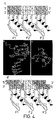
- FIG. 5 shows the amino acid sequences of the three finger constructs used in this study, including wild-type Zif268 and four variants selected from a phage display library in which finger 2 is randomized. Boxed regions indicate the varied regions in each construct. The conserved zinc chelating residues of the zinc fingers are underlined. The aspartate in position 2 of finger 3 and the alanine to which it is mutated in this study are both circled;
- FIGS. 6A-E portions of SEQ ID NOS: 19-23 in order of appearance show the binding site signatures of the middle finger before and after alanine mutagenesis in position 2 of finger 3.
- the ELISA signal (A 450 -A 650 ) showing interaction of zinc finger phage with each of five (A-E) positionally randomized DNA library plotted vertically. From the pattern of binding to these libraries, one or a small number of binding sites can be read off and these are written on the right of the figure.
- Mutagenesis of position 2 in finger 3 can change the binding specificity for the middle triplet of the Zif268 binding site. In such cases, changes are noted for base 5, but not bases 6 and 7 of the DNA binding site (see FIG. 4A ); and
- FIGS. 7A and B depict the apparent equilibrium binding curves showing the effect of replacing Asp2 in finger 3 by Ala for (A) Zif268 DNA-binding domain (consensus binding site used: 5′-GCG TGG GCG-3′); and (B) F2-Arg construct (consensus binding site used: 5′-GCG GTG GCG-3′). Wild-type and mutant constructs are denoted by ‘wt’ and ‘mut’ respectively.
- the target selected for the zinc finger nucleic acid binding protein is the activating point mutation of the human EJ bladder carcinoma ras oncogene, which was the first DNA lesion reported to confer transforming properties on a cellular proto-oncogene. Since the original discovery, ras gene mutations have been found to occur at high frequencies in a variety of human cancers and are established targets for the diagnosis of oncogenesis at early stages of tumour growth.
- the EJ bladder carcinoma mutation is a single nucleotide change in codon 12 of H-ras, which results in a mutation from GGC to GTC at this position.
- a zinc finger peptide is designed to bind a 10 bp DNA site assigned in the noncoding strand of the mutant ras gene, such that three fingers contact ‘anticodons’ 10, 11 and 12 in series, as shown in FIG. 1 , plus the 5′ preceding G (on the +strand of the DNA).
- the first finger of the designer lead peptide is designed according to the rules set forth herein starting from a Zif268 finger 2 model to bind the quadruplet 5′-GCCG-3′, which corresponds to ‘anticodon’ 10 of the designated binding site plus one 3′ base.
- the finger has the following sequence: FQCRICMRNFSDRSSLTRHTRTHTGEKP (SEQ ID NO:9) ⁇ 1 1 2 3 4 5 6 7 8 9
- a DNA coding sequence encoding this polypeptide is constructed from synthesised oligonucleotides.
- the second and third fingers of the DNA-binding domain are direct repeats of this first finger, but in which the third ⁇ -helical residue which contacts base 3 of a quadruplet, +3, is mutated according to recognition rules, to histidine in finger 2 and asparagines in finger 3, such that the specificity of these fingers is predicted to be 5′-GGCG-3′ (includes ‘anticodon’ 11) and 5′-GACG-3′ (includes ‘anticodon’ 12) respectively.
- the second and third finger polypeptides have the sequences FQCRICMRNFSDRSHLTRHTRTHTGEKP (SEQ ID NO:10) and FQCRICMRNFSDRSNLTRHTRTHTGEK (SEQ ID NO:11) respectively.
- the DNA-binding domain is able to bind the mutated ras sequence with an apparent K d of 17 nM, and to discriminate strongly against the wild-type sequence.
- the first finger of the lead peptide could contact cytosine using one of Asp, Glu, Ser or Thr in the third ⁇ -helix position.
- Selections from this mini-library are over one round of phage binding to 5 nM mutant DNA oligo in 100 ⁇ l PBS containing 50 ⁇ M ZnCl 2 , 2% (w/v) fat-free dried milk (Marvel) and 1% (v/v) Tween®-20 detergent, with 1 ⁇ g poly dIdC as competitor, followed by six washes with PBS containing 50 ⁇ M ZnCl 2 and 1% (v/v) Tween®-20 detergent. Bound phage are eluted with 0.1M triethylamine for 3 mins, and immediately transferred to an equal volume of 1M Tris-Cl pH 7.4.
- a single round of randomisation and selection is found to be sufficient to improve the affinity of the lead zinc finger peptide to this standard.
- a small library of mutants is constructed with limited variations specifically in the third ⁇ -helical position (+3) of finger 1 of the designed peptide. Selection from this library yields an optimised DNA-binding domain with asparagine at the variable position, which is able to bind the mutant ras sequence with an apparent K d of 3 nM, i.e. equal to that of the wild-type Zif268 DNA-binding domain ( FIG. 2 ).
- the selection of asparagine at this position to bind opposite a cytosine is an unexpected deviation from the recognition rules, which normally pair asparagine with adenine.
- asparagine is, however, consistent with physical considerations of the protein-DNA interface.
- asparagine has been observed to bridge a base-pair step in the major groove of DNA, for example in the co-crystal structures of the GCN4 DNA-binding domain.
- a number of different base-pair steps provide the correct stereochemical pairings of hydrogen bond donors and acceptors which could satisfy asparagine, including the underlined step G CC of ras ‘anticodon’ 10.
- the sequence-dependent deformation of ras DNA could account for our observation that wild-type and EJ ras gene fragments have different electrophoretic mobility in polyacrylamide gels, since the wild-type ras gene has two GGC sequences 5 bp apart and hence out of helical phase (resulting in no net bend), while the EJ mutation affects one of these GGC sequences.
- the optimised DNA-binding domain displayed on phage is applied in the diagnosis of the activating point mutation of the EJ ras oncogene.
- Bacterial culture supernatant containing the diagnostic phage is diluted 1:1 with PBS containing 50 ⁇ M ZnCl 2 , 4% (w/v) fat-free dried milk (Marvel) and 2% (v/v) Tween®-20 detergent.
- Biotinylated oligonucleotides (7.5 pmol) containing double stranded DNA comprising codons 8-16 from the wild type or the point-mutated ras gene are added to 50 ⁇ l of the diluted phage and incubated for 1 h at 20° C. In the experiment shown in FIG.
- bound phage are captured with 0.5 mg streptavidin coated paramagnetic beads (Dynal)—however streptavidin coated microtitre plates (Boehringer Mannheim) can also be used without alteration to the protocol. Unbound phage are removed by washing the beads 6 times with PBS containing 50 ⁇ M ZnCl 2 and 1% (v/v) Tween®-20 detergent. The beads are subsequently incubated for 1 h at RT with anti-M13 IgG conjugated to horseradish peroxidase (Pharmacia Biotech) diluted 1:5000 in PBS containing 50 ⁇ M ZnCl 2 and 2% (w/v) fat-free dried milk (Marvel).
- Dynal streptavidin coated paramagnetic beads
- Unbound phage are removed by washing the beads 6 times with PBS containing 50 ⁇ M ZnCl 2 and 1% (v/v) Tween®-20 detergent. The beads are subsequently incubated for 1 h at RT with anti
- Excess antibody is removed by washing 6 times with PBS containing 50 ⁇ M ZnCl 2 and 0.05% (v/v) Tween®-20 detergent, and 3 times with PBS containing 50 ⁇ M ZnCl 2 .
- the ELISA is developed with 0.1 mg/ml tetramethylbenzidine (Sigma) in 0.1M sodium acetate pH5.4 containing 2 ⁇ l of fresh 30% hydrogen peroxide per 10 ml buffer, and after approximately 1 min, stopped with an equal volume of 2M H 2 SO 4 .
- the reaction produces a yellow colour which is quantitated by subtracting the absorbance at 650 nm from the absorbance at 450 nm.
- Phage are retained specifically by DNA bearing the mutant, but not the wild-type ras sequence, allowing the detection of the point mutation by ELISA ( FIG. 3 ).
- the sequence of the HIV TAR is known (Jones and Peterlin, (1994) Ann. Rev. Biochem. 63:717-743).
- a sequence with the TAT region is identified and a zinc finger polypeptide designed to bind thereto.
- the selected sequence is 5′-AGA GAG CTC-3′, which is the complement of nucleotides +34 to +42 of HIV.
- the corresponding amino acids required in fingers 1, 2 and 3 of a zinc finger binding protein are determined according to the rules set forth above, as follows:
- the framework of the polypeptide is taken from the Zif 268 middle finger.
- the sequence of the entire polypeptide is shown in SEQ. ID. No. 2.
- Residues +2 and +6 of finger 3 are partially selected by randomisation and phage display selection. At position 2, two triplets are used, GAT and GGT, coding for Asp or Gly. Position +6 was randomised. In these positions, the residues Gly and Val are selected.
- the methodology employed is as follows: colony PCR is performed with one primer containing a single mismatch to create the required randomisations in finger 3. Cloning of PCR product in phage vector is as described previously (Choo, Y. & Klug, A. (1994) Proc. Natl. Acad. Sci. USA 91, 11163-11167; Choo, Y. & Klug, A. (1994) Proc. Natl. Acad. Sci.
- forward and backward PCR primers contained unique restriction sites for Not I or Sfi I respectively and amplified an approximately 300 base pair region encompassing three zinc fingers.
- PCR products are digested with Sfi I and Not I to create cohesive ends and are ligated to 100 ng of similarly digested fd-Tet-SN vector.
- Electrocompetent TG1 cells are transformed with the recombinant vector. Single colonies of tranformants are grown overnight in 2 ⁇ TY containing 50 ⁇ M ZnCl 2 15 ⁇ g/ml tetracycline. Single stranded DNA is prepared from phage in the culture supernatant and sequenced with Sequenase 2.0 (United States Biochemical).
- the polypeptide designed according to the invention is then tested for binding to HIV DNA and positive results are obtained.
- Alanine mutagenesis of the Asp2 in finger 3 is carried out on the wild-type Zif268 DNA-binding domain and four related peptides isolated from the phage display library as follows (see also FIG. 5 ):
- E. coli TG1 cells are tranfected with fd phage displaying zinc fingers. Colony PCR is performed with one primer containing a single mismatch to create the Asp to Ala change in finger 3.
- Cloning of PCR product in phage vector is as described previously (Choo, Y. & Klug, A. (1994) Proc. Natl. Acad. Sci. USA 91, 11163-11167; Choo, Y. & Klug, A. (1994) Proc. Natl. Acad. Sci. USA 91, 11168-11172). Briefly, forward and backward PCR primers contained unique restriction sites for Not I or Sfi I respectively and amplified an approximately 300 base pair region encompassing three zinc fingers.
- PCR products are digested with Sfi I and Not I to create cohesive ends and are ligated to 100 ng of similarly digested fd-Tet-SN vector. Electrocompetent TG1 cells are transformed with the recombinant vector. Single colonies of tranformants are grown overnight in 2 ⁇ TY containing 50 ⁇ M ZnCl 2 15 ⁇ g/ml tetracycline. Single stranded DNA is prepared from phage in the culture supernatant and sequenced with Sequenase 2.0 (United States Biochemical).
- the peptides are chosen for this experiment on the basis of the identity of the residue at position 6 of the middle finger.
- Peptide F2-Arg which contains Arg at position 6 of finger 2
- Peptide F2-Arg which contains Arg at position 6 of finger 2
- the peptide F2-Gly with Gly at position 6 would be expected to lose all specificity at the 5′ position of the ‘middle’ triplet following alanine mutagenesis in finger 3.
- each middle finger is assessed before and after the alanine mutation in finger 3 by the ‘binding site signature’ method (Choo and Kug, 1994).
- This procedure involves screening each zinc finger phage for binding to 12 DNA libraries, each based on the DNA binding site of Zif768 but containing one fixed and two randomised nucleotide positions in the ‘middle’ triplet.
- Each of the possible 64 ‘middle’ triplets is present in a unique combination of three of these positionally randomised libraries; for example the triplet GAT would be found in the GNN, NAN and NNT libraries only. Hence the pattern of binding to these reveals the sequence-specificity of the middle finger.
- Binding is allowed to proceed for one hour at 20° C. Unbound phage are removed by washing 6 times with PBS/Zn containing 1% Tween®-20 detergent, then washing 3 times with PBS/Zn. Bound phage are detected by ELISA with horseradish peroxidase-conjugated anti-M13 IgG (Pharmacia Biotech) and quantitated using SOFTMAX 2.32 (Molecular Devices).
- FIG. 6 shows that deleting Asp2 from finger 3 generally alters the pattern of acceptable bases, in the ‘middle’ triplet, which is conventionally regarded as the binding site for finger 2.
- the mutation affects binding at the 5′ position, while the specificity at the middle and 3′ position remains unchanged.
- the mutation generally leads to a broadening of specificity, for instance in Zif268 where removal of Asp2 in finger 3 results in a protein which is unable to discriminate the 5′ base of the middle triplet ( FIG. 6 a ).
- the expectation that a new 5′ base-specificity for the mutants might correlate to the identity of position 6 in finger 2 is not borne out.
- F2-Gly would be expected to lose sequence discrimination but, although specificity is adversely affected, a slight preference for T is discernible ( FIG. 6 b ).
- F2-Val and F2-Asn which might have been expected to acquire specificity for one nucleotide, instead have their specificities altered by the mutation ( FIG.
- F2-Arg continues to specify guanine in the 5′ position of the middle triplet regardless of the mutation in finger 3 ( FIG. 3 e ).
- the specificity is derived from the strong interaction between guanine and Arg6 in finger 2. This contact has been observed a number of times in zinc finger co-crystal structures (Pavletich, N. P. & Pabo, C. O. (1993) Science 261, 1701-1707; Fairall, L., Schwabe, J. W. R., Chapman, L., Finch, J. T. & Rhodes, D. (1993) Nature (London) 366, 483-487; Fairall, L., Schwabe, J.
- FIG. 6 shows that Asp2 of Zif268 finger 3 specifically excludes adenine and cytosine from the 5′ position of the middle triplet. When this interaction is deleted, one or both of these bases become acceptable.
- Bound phage are detected by ELISA with horseradish peroxidase-conjugated anti-M13 IgG (Pharmacia Biotech) and quantitated using SOFTMAX 2.32 (Molecular Devices). Binding data are plotted and analysed using Kaleidagraph (Abelbeck Software).
- Both mutants show approximately a four-fold reduction in affinity for their respective binding sites under the conditions used.
- the reduction is likely a direct result of abolishing contacts from Asp2, rather than a consequence of changes in binding specificity at the 5′ position of the middle triplet, since the mutant Zif268 loses all specificity while F2-Arg registers no change in specificity.
- two stabilising interactions are abolished: an intramolecular buttressing interaction with Arg-1 on finger 3 and also the intermolecular contact with the secondary DNA strand.
Abstract
Description
- a) if
base 4 in the quadruplet is A, then position +6 in the α-helix is Gln and ++2 is not Asp; - b) if
base 4 in the quadruplet is C, then position +6 in the α-helix may be any residue, as long as position ++2 in the α-helix is not Asp.
- c) if
base 4 in the quadruplet is G, then position +6 in the α-helix is Arg; or position +6 is Ser or Thr and position ++2 is Asp; - d) if
base 4 in the quadruplet is T, then position +6 in the α-helix is Ser or Thr and position ++2 is Asp.
- a) if
base 4 in the quadruplet is G, then position +6 in the α-helix is Arg; or position +6 is Ser or Thr and position ++2 is Asp; - b) if
base 4 in the quadruplet is A, then position +6 in the α-helix is Gln and ++2 is not Asp; - c) if
base 4 in the quadruplet is T, then position +6 in the α-helix is Ser or Thr and position ++2 is Asp; - d) if
base 4 in the quadruplet is C, then position +6 in the α-helix may be any amino acid, provided that position ++2 in the α-helix is not Asp; - e) if
base 3 in the quadruplet is G, then position +3 in the α-helix is His; - f) if
base 3 in the quadruplet is A, then position +3 in the α-helix is Asn; - g) if
base 3 in the quadruplet is T, then position +3 in the α-helix is Ala, Ser or Val; provided that if it is Ala, then one of the residues at −1 or +6 is a small residue; - h) if
base 3 in the quadruplet is C, then position +3 in the α-helix is Ser, Asp, Glu, Leu, Thr or Val; - i) if
base 2 in the quadruplet is G, then position −1 in the α-helix is Arg; - j) if
base 2 in the quadruplet is A, then position −1 in the α-helix is Gln; - k) if
base 2 in the quadruplet is T, then position −1 in the α-helix is Asn or Gin; - l) if
base 2 in the quadruplet is C, then position −1 in the α-helix is Asp; - m) if
base 1 in the quadruplet is G, then position +2 is Asp; - n) if
base 1 in the quadruplet is A, then position +2 is not Asp; - o) if
base 1 in the quadruplet is C, then position +2 is not Asp; - p) if
base 1 in the quadruplet is T, then position +2 is Ser or Thr.
X0-2CX1-5CX9-14HX3-6 H/C (A)
XaCX2-4CX2-3FXcXXXXLXXHXXXbH-linker (B)
−1 1 2 3 4 5 6 7 8 9
wherein X (including Xa, Xb and Xc) is any amino acid. X2-4 and X2-3 refer to the presence of 2 or 4, or 2 or 3, amino acids, respectively. The Cys and His residues, which together co-ordinate the zinc metal atom, are marked in bold text and are usually invariant, as is the Leu residue at position ÷4 in the α-helix.
- a) selecting a model zinc finger domain from the group consisting of naturally occurring zinc fingers and consensus zinc fingers; and
- b) mutating one or more of positions −1, +2, +3 and +6 of the finger as required according to the rules set forth above.
- a) preparing a nucleic acid coding sequence encoding two or more zinc finger binding motifs as defined above, placed N-terminus to C-terminus;
- b) inserting the nucleic acid sequence into a suitable expression vector; and
- c) expressing the nucleic acid sequence in a host organism in order to obtain the nucleic acid binding protein.
- a) preparing a nucleic acid binding protein by the method set forth above which is specific for the target nucleic acid molecule;
- b) exposing a test system comprising the target nucleic acid molecule to the nucleic acid binding protein under conditions which promote binding, and removing any nucleic acid binding protein which remains unbound;
- c) detecting the presence of the nucleic acid binding protein in the test system.
FQCRICMRNFSDRSSLTRHTRTHTGEKP (SEQ ID NO:9)
−1 1 2 3 4 5 6 7 8 9
FQCRICMRNFSDRSHLTRHTRTHTGEKP (SEQ ID NO:10)
and
FQCRICMRNFSDRSNLTRHTRTHTGEK (SEQ ID NO:11)
respectively.
-
-
target 5′-AGA-3′ - Position −1 Gln
-
Position + 2 Gly -
Position + 3 His -
Position + 6 Val
-
-
-
target 5′-GAG-3′ - Position −1 Arg
-
Position + 2 Ser -
Position + 3 Asn -
Position + 6 Arg
-
-
-
target 5′-CTC-3′ - Position −1 Asp
-
Position + 3 Ser -
Position + 6 Glu
-
Claims (21)
−1 1 2 3 4 5 6 7 8 9
Priority Applications (1)
| Application Number | Priority Date | Filing Date | Title |
|---|---|---|---|
| US11/515,369 US7759059B2 (en) | 1997-05-23 | 2006-08-31 | Nucleic acid binding proteins |
Applications Claiming Priority (6)
| Application Number | Priority Date | Filing Date | Title |
|---|---|---|---|
| GBGB9710809.6 | 1997-05-23 | ||
| GBGB9710809.6A GB9710809D0 (en) | 1997-05-23 | 1997-05-23 | Nucleic acid binding proteins |
| US09/424,488 US6866997B1 (en) | 1997-05-23 | 1998-05-26 | Nucleic acid binding proteins |
| PCT/GB1998/001516 WO1998053060A1 (en) | 1997-05-23 | 1998-05-26 | Nucleic acid binding proteins |
| US10/853,437 US7241574B2 (en) | 1997-05-23 | 2004-05-24 | Nucleic acid binding proteins |
| US11/515,369 US7759059B2 (en) | 1997-05-23 | 2006-08-31 | Nucleic acid binding proteins |
Related Parent Applications (1)
| Application Number | Title | Priority Date | Filing Date |
|---|---|---|---|
| US10/853,437 Continuation US7241574B2 (en) | 1997-05-23 | 2004-05-24 | Nucleic acid binding proteins |
Publications (2)
| Publication Number | Publication Date |
|---|---|
| US20070161014A1 US20070161014A1 (en) | 2007-07-12 |
| US7759059B2 true US7759059B2 (en) | 2010-07-20 |
Family
ID=10813050
Family Applications (8)
| Application Number | Title | Priority Date | Filing Date |
|---|---|---|---|
| US09/424,488 Expired - Lifetime US6866997B1 (en) | 1997-05-23 | 1998-05-26 | Nucleic acid binding proteins |
| US09/424,487 Expired - Lifetime US6746838B1 (en) | 1997-05-23 | 1998-05-26 | Nucleic acid binding proteins |
| US10/832,735 Expired - Fee Related US7241573B2 (en) | 1997-05-23 | 2004-04-26 | Nucleic acid binding proteins |
| US10/853,437 Expired - Fee Related US7241574B2 (en) | 1997-05-23 | 2004-05-24 | Nucleic acid binding proteins |
| US11/486,962 Expired - Fee Related US8617807B2 (en) | 1997-05-23 | 2006-07-14 | Nucleic acid binding proteins |
| US11/515,369 Expired - Fee Related US7759059B2 (en) | 1997-05-23 | 2006-08-31 | Nucleic acid binding proteins |
| US11/514,850 Expired - Fee Related US8618024B2 (en) | 1997-05-23 | 2006-08-31 | Nucleic acid binding polypeptide library |
| US11/514,671 Expired - Fee Related US7700523B2 (en) | 1997-05-23 | 2006-09-01 | Nucleic acid binding polypeptide library |
Family Applications Before (5)
| Application Number | Title | Priority Date | Filing Date |
|---|---|---|---|
| US09/424,488 Expired - Lifetime US6866997B1 (en) | 1997-05-23 | 1998-05-26 | Nucleic acid binding proteins |
| US09/424,487 Expired - Lifetime US6746838B1 (en) | 1997-05-23 | 1998-05-26 | Nucleic acid binding proteins |
| US10/832,735 Expired - Fee Related US7241573B2 (en) | 1997-05-23 | 2004-04-26 | Nucleic acid binding proteins |
| US10/853,437 Expired - Fee Related US7241574B2 (en) | 1997-05-23 | 2004-05-24 | Nucleic acid binding proteins |
| US11/486,962 Expired - Fee Related US8617807B2 (en) | 1997-05-23 | 2006-07-14 | Nucleic acid binding proteins |
Family Applications After (2)
| Application Number | Title | Priority Date | Filing Date |
|---|---|---|---|
| US11/514,850 Expired - Fee Related US8618024B2 (en) | 1997-05-23 | 2006-08-31 | Nucleic acid binding polypeptide library |
| US11/514,671 Expired - Fee Related US7700523B2 (en) | 1997-05-23 | 2006-09-01 | Nucleic acid binding polypeptide library |
Country Status (9)
| Country | Link |
|---|---|
| US (8) | US6866997B1 (en) |
| EP (5) | EP0983350B1 (en) |
| JP (4) | JP2002502238A (en) |
| AT (2) | ATE393822T1 (en) |
| AU (3) | AU737756B2 (en) |
| CA (3) | CA2290886C (en) |
| DE (2) | DE69839424T2 (en) |
| GB (1) | GB9710809D0 (en) |
| WO (3) | WO1998053057A1 (en) |
Families Citing this family (527)
| Publication number | Priority date | Publication date | Assignee | Title |
|---|---|---|---|---|
| US6140466A (en) | 1994-01-18 | 2000-10-31 | The Scripps Research Institute | Zinc finger protein derivatives and methods therefor |
| US7262055B2 (en) | 1998-08-25 | 2007-08-28 | Gendaq Limited | Regulated gene expression in plants |
| GB9824544D0 (en) | 1998-11-09 | 1999-01-06 | Medical Res Council | Screening system |
| JP4118327B2 (en) | 1994-08-20 | 2008-07-16 | ゲンダック・リミテッド | Improvements in or related to binding proteins for DNA recognition |
| GB9710809D0 (en) * | 1997-05-23 | 1997-07-23 | Medical Res Council | Nucleic acid binding proteins |
| US6410248B1 (en) | 1998-01-30 | 2002-06-25 | Massachusetts Institute Of Technology | General strategy for selecting high-affinity zinc finger proteins for diverse DNA target sites |
| JP4309051B2 (en) | 1998-03-02 | 2009-08-05 | マサチューセッツ インスティテュート オブ テクノロジー | Polyzinc finger protein with improved linker |
| JP2002506640A (en) * | 1998-03-17 | 2002-03-05 | ジェンダック・リミテッド | Nucleic acid binding protein |
| US20050086718A1 (en) | 1999-03-23 | 2005-04-21 | Mendel Biotechnology, Inc. | Plant transcriptional regulators of abiotic stress |
| US7897843B2 (en) | 1999-03-23 | 2011-03-01 | Mendel Biotechnology, Inc. | Transcriptional regulation of plant biomass and abiotic stress tolerance |
| US7070934B2 (en) | 1999-01-12 | 2006-07-04 | Sangamo Biosciences, Inc. | Ligand-controlled regulation of endogenous gene expression |
| US7013219B2 (en) | 1999-01-12 | 2006-03-14 | Sangamo Biosciences, Inc. | Regulation of endogenous gene expression in cells using zinc finger proteins |
| US6534261B1 (en) | 1999-01-12 | 2003-03-18 | Sangamo Biosciences, Inc. | Regulation of endogenous gene expression in cells using zinc finger proteins |
| US6453242B1 (en) * | 1999-01-12 | 2002-09-17 | Sangamo Biosciences, Inc. | Selection of sites for targeting by zinc finger proteins and methods of designing zinc finger proteins to bind to preselected sites |
| US6599692B1 (en) | 1999-09-14 | 2003-07-29 | Sangamo Bioscience, Inc. | Functional genomics using zinc finger proteins |
| US20030104526A1 (en) | 1999-03-24 | 2003-06-05 | Qiang Liu | Position dependent recognition of GNN nucleotide triplets by zinc fingers |
| US7030215B2 (en) | 1999-03-24 | 2006-04-18 | Sangamo Biosciences, Inc. | Position dependent recognition of GNN nucleotide triplets by zinc fingers |
| US6794136B1 (en) | 2000-11-20 | 2004-09-21 | Sangamo Biosciences, Inc. | Iterative optimization in the design of binding proteins |
| EP1181360A1 (en) * | 1999-05-28 | 2002-02-27 | Sangamo Biosciences Inc. | Molecular switches |
| AU4940700A (en) * | 1999-05-28 | 2000-12-18 | Sangamo Biosciences, Inc. | Gene switches |
| GB9915126D0 (en) | 1999-06-30 | 1999-09-01 | Imp College Innovations Ltd | Control of gene expression |
| US7943731B1 (en) | 1999-08-11 | 2011-05-17 | Massachusetts Institute Of Technology | Dimerizing peptides |
| AU1765601A (en) | 1999-11-17 | 2001-05-30 | Luc Adam | Plant biochemistry-related genes |
| EP1950306A1 (en) | 1999-11-17 | 2008-07-30 | Mendel Biotechnology, Inc. | Environmental stress tolerance genes |
| ATE309536T1 (en) | 1999-12-06 | 2005-11-15 | Sangamo Biosciences Inc | METHODS OF USING RANDOMIZED ZINC FINGER PROTEIN LIBRARIES TO IDENTIFY GENE FUNCTIONS |
| AU2001226935B2 (en) | 2000-01-24 | 2006-06-22 | Gendaq Limited | Nucleic acid binding polypeptides characterized by flexible linkers connected nucleic acid binding modules |
| US20020061512A1 (en) * | 2000-02-18 | 2002-05-23 | Kim Jin-Soo | Zinc finger domains and methods of identifying same |
| GB2365011A (en) | 2000-04-28 | 2002-02-13 | Sangamo Biosciences Inc | Methods for the characterisation of regulatory DNA sequences |
| CA2407695C (en) * | 2000-04-28 | 2015-03-31 | Sangamo Biosciences, Inc. | Methods for binding an exogenous molecule to cellular chromatin |
| US7923542B2 (en) | 2000-04-28 | 2011-04-12 | Sangamo Biosciences, Inc. | Libraries of regulatory sequences, methods of making and using same |
| WO2001084148A2 (en) | 2000-04-28 | 2001-11-08 | Sangamo Biosciences, Inc. | Pharmacogenomics and identification of drug targets by reconstruction of signal transduction pathways based on sequences of accessible regions |
| AU2001257331A1 (en) | 2000-04-28 | 2001-11-12 | Sangamo Biosciences, Inc. | Methods for designing exogenous regulatory molecules |
| US6492117B1 (en) | 2000-07-12 | 2002-12-10 | Gendaq Limited | Zinc finger polypeptides capable of binding DNA quadruplexes |
| WO2002008286A2 (en) * | 2000-07-21 | 2002-01-31 | Syngenta Participations Ag | Zinc finger domain recognition code and uses thereof |
| WO2002015675A1 (en) | 2000-08-22 | 2002-02-28 | Mendel Biotechnology, Inc. | Genes for modifying plant traits iv |
| US7067317B2 (en) | 2000-12-07 | 2006-06-27 | Sangamo Biosciences, Inc. | Regulation of angiogenesis with zinc finger proteins |
| AU2884102A (en) | 2000-12-07 | 2002-06-18 | Sangamo Biosciences Inc | Regulation of angiogenesis with zinc finger proteins |
| EP1353941B1 (en) | 2001-01-22 | 2013-03-13 | Sangamo BioSciences, Inc. | Modified zinc finger binding proteins |
| US7947469B2 (en) | 2001-01-22 | 2011-05-24 | Gendaq, Ltd. | Modulation of HIV infection |
| JP4128955B2 (en) * | 2001-08-17 | 2008-07-30 | トゥールゲン・インコーポレイテッド | Zinc finger domain library |
| EP1435779A4 (en) | 2001-09-24 | 2005-06-01 | Sangamo Biosciences Inc | Modulation of stem cells using zinc finger proteins |
| KR20040065235A (en) * | 2001-12-07 | 2004-07-21 | 주식회사 툴젠 | Phenotypic screen of chimeric proteins |
| US7262054B2 (en) | 2002-01-22 | 2007-08-28 | Sangamo Biosciences, Inc. | Zinc finger proteins for DNA binding and gene regulation in plants |
| CN100575485C (en) | 2002-01-23 | 2009-12-30 | 犹他大学研究基金会 | Use the directed karyomit(e) mutagenesis of Zinc finger nuclease |
| PT1504108E (en) | 2002-02-01 | 2013-06-12 | Oxford Biomedica Ltd | Lentiviral vector |
| AU2003215094B2 (en) * | 2002-02-07 | 2008-05-29 | The Scripps Research Institute | Zinc finger libraries |
| US7786344B2 (en) | 2002-07-26 | 2010-08-31 | Basf Plant Science Gmbh | Selection method |
| US7361635B2 (en) | 2002-08-29 | 2008-04-22 | Sangamo Biosciences, Inc. | Simultaneous modulation of multiple genes |
| AU2003260973A1 (en) * | 2002-09-05 | 2004-03-29 | Toolgen, Inc. | Bioinformatics analysis of cellular effects of artificial transcription factors |
| EP2806025B1 (en) | 2002-09-05 | 2019-04-03 | California Institute of Technology | Use of zinc finger nucleases to stimulate gene targeting |
| EP2270166A3 (en) | 2002-09-18 | 2011-08-10 | Mendel Biotechnology, Inc. | Polynucleotides and polypeptides in plants |
| GB0228289D0 (en) | 2002-12-04 | 2003-01-08 | Genome Inst Of Singapore Nat U | Method |
| US8409861B2 (en) | 2003-08-08 | 2013-04-02 | Sangamo Biosciences, Inc. | Targeted deletion of cellular DNA sequences |
| EP2927318B1 (en) | 2003-08-08 | 2020-05-20 | Sangamo Therapeutics, Inc. | Methods and compositions for targeted cleavage and recombination |
| US20120196370A1 (en) | 2010-12-03 | 2012-08-02 | Fyodor Urnov | Methods and compositions for targeted genomic deletion |
| US11311574B2 (en) | 2003-08-08 | 2022-04-26 | Sangamo Therapeutics, Inc. | Methods and compositions for targeted cleavage and recombination |
| US7888121B2 (en) | 2003-08-08 | 2011-02-15 | Sangamo Biosciences, Inc. | Methods and compositions for targeted cleavage and recombination |
| AU2004274957B2 (en) | 2003-09-19 | 2009-02-05 | Sangamo Therapeutics, Inc. | Engineered zinc finger proteins for regulation of gene expression |
| US7329725B1 (en) * | 2003-10-29 | 2008-02-12 | Nastech Pharmaceutical Company Inc. | Phage displayed Trp cage ligands |
| US7972854B2 (en) | 2004-02-05 | 2011-07-05 | Sangamo Biosciences, Inc. | Methods and compositions for targeted cleavage and recombination |
| AU2005233583B2 (en) | 2004-04-08 | 2011-02-03 | Sangamo Therapeutics, Inc. | Methods and compositions for modulating cardiac contractility |
| EP1732614B1 (en) * | 2004-04-08 | 2008-12-24 | Sangamo Biosciences Inc. | Compositions for treating neuropathic and neurodegenerative conditions |
| AR049390A1 (en) * | 2004-06-09 | 2006-07-26 | Wyeth Corp | ANTIBODIES AGAINST HUMAN INTERLEUQUINE-13 AND USES OF THE SAME |
| US7501121B2 (en) | 2004-06-17 | 2009-03-10 | Wyeth | IL-13 binding agents |
| US20080131962A1 (en) | 2006-05-25 | 2008-06-05 | Sangamo Biosciences, Inc. | Engineered cleavage half-domains |
| WO2006033859A2 (en) | 2004-09-16 | 2006-03-30 | Sangamo Biosciences, Inc. | Compositions and methods for protein production |
| EP1645633B1 (en) | 2004-10-05 | 2011-09-21 | SunGene GmbH | Constitutive expression cassettes for regulation of plant expression |
| EP1655364A3 (en) | 2004-11-05 | 2006-08-02 | BASF Plant Science GmbH | Expression cassettes for seed-preferential expression in plants |
| EP1662000B1 (en) | 2004-11-25 | 2011-03-30 | SunGene GmbH | Expression cassettes for guard cell-preferential expression in plants |
| EP1666599A3 (en) | 2004-12-04 | 2006-07-12 | SunGene GmbH | Expression cassettes for mesophyll- and/or epidermis-preferential expression in plants |
| EP2163634A1 (en) | 2004-12-08 | 2010-03-17 | SunGene GmbH | Expression cassettes for vascular tissue-preferential expression in plants |
| EP1669456A3 (en) | 2004-12-11 | 2006-07-12 | SunGene GmbH | Expression cassettes for meristem-preferential expression in plants |
| WO2006078431A2 (en) | 2004-12-22 | 2006-07-27 | The University Of Iowa Research Foundation | Compositions and methods related to modified retroviral vectors for restricted, site specific integration |
| CN103184218A (en) | 2005-02-09 | 2013-07-03 | 巴斯福植物科学有限公司 | Expression cassettes for regulation of expression in monocotyledonous plants |
| DE102005008765B4 (en) * | 2005-02-25 | 2016-06-16 | Siemens Healthcare Gmbh | Bacteriophage-based contrast agent, composition containing it, its use and process for its preparation |
| US20090031440A1 (en) | 2005-02-26 | 2009-01-29 | Basf Plant Science Gmbh | Expression Cassettes for Seed-Preferential Expression in Plants |
| WO2006094106A2 (en) | 2005-02-28 | 2006-09-08 | Sangamo Biosciences, Inc. | Anti-angiogenic methods and compositions |
| US7790873B2 (en) | 2005-05-10 | 2010-09-07 | Basf Plant Science Gmbh | Expression cassettes for seed-preferential expression in plants |
| CA2615532C (en) | 2005-07-26 | 2016-06-28 | Sangamo Biosciences, Inc. | Targeted integration and expression of exogenous nucleic acid sequences |
| US7919583B2 (en) * | 2005-08-08 | 2011-04-05 | Discovery Genomics, Inc. | Integration-site directed vector systems |
| US7704953B2 (en) * | 2006-02-17 | 2010-04-27 | Mdrna, Inc. | Phage displayed cell binding peptides |
| AU2007267874B2 (en) | 2006-05-25 | 2012-03-15 | Sangamo Therapeutics, Inc. | Methods and compositions for gene inactivation |
| KR101439568B1 (en) | 2006-08-11 | 2014-09-12 | 다우 아그로사이언시즈 엘엘씨 | Zinc finger nuclease-mediated homologous recombination |
| JP5188504B2 (en) * | 2006-11-13 | 2013-04-24 | サンガモ バイオサイエンシズ インコーポレイテッド | Methods and compositions for modification of the human glucocorticoid receptor locus |
| WO2008067840A1 (en) | 2006-12-08 | 2008-06-12 | Swetree Technologies Ab | Plants having improved growth characteristics and method for making the same |
| KR101520507B1 (en) | 2006-12-14 | 2015-05-29 | 다우 아그로사이언시즈 엘엘씨 | Optimized non-canonical zinc finger proteins |
| CA2675926A1 (en) | 2007-02-16 | 2008-08-21 | Basf Plant Science Gmbh | Nucleic acid sequences for regulation of embryo-specific expression in monocotyledonous plants |
| ATE489465T1 (en) | 2007-04-26 | 2010-12-15 | Sangamo Biosciences Inc | TARGETED INTEGRATION INTO THE PPP1R12C POSITION |
| CA2692453C (en) | 2007-07-12 | 2018-01-09 | Trevor Collingwood | Methods and compositions for inactivating alpha 1,6 fucosyltransferase (fut8) gene expression |
| CN101878307B (en) | 2007-09-27 | 2017-07-28 | 陶氏益农公司 | Using sour 3 phosphate synthase genes of 5 enolpyrul-shikimates as the engineered zinc finger of target |
| AU2008305567B2 (en) | 2007-09-27 | 2014-04-10 | Sangamo Therapeutics, Inc. | Rapid in vivo identification of biologically active nucleases |
| US11235026B2 (en) | 2007-09-27 | 2022-02-01 | Sangamo Therapeutics, Inc. | Methods and compositions for modulating PD1 |
| US8563314B2 (en) | 2007-09-27 | 2013-10-22 | Sangamo Biosciences, Inc. | Methods and compositions for modulating PD1 |
| US8936936B2 (en) | 2007-10-25 | 2015-01-20 | Sangamo Biosciences, Inc. | Methods and compositions for targeted integration |
| BRPI0820114B1 (en) | 2007-12-03 | 2022-10-04 | Syngenta Participations Ag | METHOD OF INCREASE THE SENSITIVITY OF AN ESCHERICHIA COLI-DERIVED PHYTASE TO A PEPSIN |
| BR122017021063B1 (en) | 2007-12-28 | 2018-12-18 | Swetree Technologies Ab | method for producing a transgenic evergreen woody plant showing increased growth measured by trunk dry weight compared to its wild type |
| DE102008013623A1 (en) * | 2008-03-10 | 2009-09-24 | Freie Universität Berlin | Pharmaceutical composition for the diagnosis or treatment of zinc finger protein associated diseases |
| EP2281050B1 (en) | 2008-04-14 | 2014-04-02 | Sangamo BioSciences, Inc. | Linear donor constructs for targeted integration |
| AU2009239333A1 (en) | 2008-04-21 | 2009-10-29 | Danziger Innovations Ltd. | Plant viral expression vectors and use of same for generating genotypic variations in plant genomes |
| CN102027118B (en) | 2008-04-30 | 2014-07-30 | 桑比欧公司 | Neural regenerating cells with alterations in DNA methylation |
| JP2011521643A (en) | 2008-05-28 | 2011-07-28 | サンガモ バイオサイエンシーズ, インコーポレイテッド | Composition for linking a DNA binding domain and a cleavage domain |
| EP2130836B1 (en) * | 2008-06-03 | 2013-02-13 | Fraunhofer-Gesellschaft zur Förderung der angewandten Forschung e.V. | Means and methods for producing zinc fingers and concatemers thereof |
| CA2726768C (en) | 2008-06-10 | 2016-09-06 | Sangamo Biosciences, Inc. | Methods and compositions for generation of bax- and bak-deficient cell lines |
| JP5908725B2 (en) | 2008-08-22 | 2016-04-26 | サンガモ バイオサイエンシーズ, インコーポレイテッド | Methods and compositions for targeted single-strand cleavage and targeted integration |
| JP5756016B2 (en) | 2008-10-29 | 2015-07-29 | サンガモ バイオサイエンシーズ, インコーポレイテッド | Method and composition for inactivating expression of glutamine synthetase gene |
| CN103882053A (en) | 2008-11-03 | 2014-06-25 | 瑞典树木科技公司 | Vegetabile Material, Plants And A Method Of Producing A Plant Having Altered Lignin Properties |
| EP2352369B1 (en) | 2008-12-04 | 2017-04-26 | Sangamo BioSciences, Inc. | Genome editing in rats using zinc-finger nucleases |
| DK2370575T3 (en) | 2008-12-17 | 2018-02-05 | Dow Agrosciences Llc | TARGETED INTEGRATION IN THE ZP15 LOCUS |
| US8551945B2 (en) | 2009-02-04 | 2013-10-08 | Sangamo Biosciences, Inc. | Methods and compositions for treating neuropathies |
| CA2755192C (en) | 2009-03-20 | 2018-09-11 | Sangamo Biosciences, Inc. | Modification of cxcr4 using engineered zinc finger proteins |
| WO2010117464A1 (en) | 2009-04-09 | 2010-10-14 | Sangamo Biosciences, Inc. | Targeted integration into stem cells |
| US8772008B2 (en) | 2009-05-18 | 2014-07-08 | Sangamo Biosciences, Inc. | Methods and compositions for increasing nuclease activity |
| CA2765488C (en) | 2009-06-30 | 2018-01-02 | Sangamo Biosciences, Inc. | Rapid screening of biologically active nucleases and isolation of nuclease-modified cells |
| US20120171771A1 (en) | 2009-07-08 | 2012-07-05 | Cellular Dynamics International, Inc. | Modified ips cells having a mutant form of a human immunodeficiency virus (hiv) cellular entry gene |
| EP2727600B1 (en) | 2009-07-28 | 2019-03-27 | Sangamo Therapeutics, Inc. | Zinc finger fusion proteins for repressing a huntington gene |
| US20120178647A1 (en) * | 2009-08-03 | 2012-07-12 | The General Hospital Corporation | Engineering of zinc finger arrays by context-dependent assembly |
| EP2462230B1 (en) * | 2009-08-03 | 2015-07-15 | Recombinetics, Inc. | Methods and compositions for targeted gene modification |
| JP5940977B2 (en) | 2009-08-11 | 2016-06-29 | サンガモ バイオサイエンシーズ, インコーポレイテッド | Homozygous organisms by targeted modification |
| US9476060B2 (en) | 2009-10-21 | 2016-10-25 | Danziger Innovations Ltd. | Generating genotypic variations in plant genomes by gamete infection |
| BR122019025207B1 (en) | 2009-10-22 | 2022-10-04 | Sangamo Biosciences, Inc. | NON-NATURALLY OCCURRING ZINC FINGER PROTEIN AND FUSION PROTEIN |
| US8956828B2 (en) | 2009-11-10 | 2015-02-17 | Sangamo Biosciences, Inc. | Targeted disruption of T cell receptor genes using engineered zinc finger protein nucleases |
| EP2504439B1 (en) | 2009-11-27 | 2016-03-02 | BASF Plant Science Company GmbH | Optimized endonucleases and uses thereof |
| US9420770B2 (en) | 2009-12-01 | 2016-08-23 | Indiana University Research & Technology Corporation | Methods of modulating thrombocytopenia and modified transgenic pigs |
| CA2787494C (en) | 2010-01-22 | 2019-09-17 | Dow Agrosciences Llc | Targeted genomic alteration |
| PT2534173T (en) | 2010-02-08 | 2019-10-31 | Sangamo Therapeutics Inc | Engineered cleavage half-domains |
| WO2011100058A1 (en) | 2010-02-09 | 2011-08-18 | Sangamo Biosciences, Inc. | Targeted genomic modification with partially single-stranded donor molecules |
| US8771985B2 (en) | 2010-04-26 | 2014-07-08 | Sangamo Biosciences, Inc. | Genome editing of a Rosa locus using zinc-finger nucleases |
| EP3636766A1 (en) | 2010-05-03 | 2020-04-15 | Sangamo Therapeutics, Inc. | Compositions for linking zinc finger modules |
| WO2011146121A1 (en) | 2010-05-17 | 2011-11-24 | Sangamo Biosciences, Inc. | Novel dna-binding proteins and uses thereof |
| US8945868B2 (en) | 2010-07-21 | 2015-02-03 | Sangamo Biosciences, Inc. | Methods and compositions for modification of a HLA locus |
| EP3489359A1 (en) | 2010-07-23 | 2019-05-29 | Sigma Aldrich Co. LLC | Genome editing using targeting endonucleases and single-stranded nucleic acids |
| AU2011312562B2 (en) | 2010-09-27 | 2014-10-09 | Sangamo Therapeutics, Inc. | Methods and compositions for inhibiting viral entry into cells |
| RU2608643C2 (en) | 2010-10-12 | 2017-01-23 | Дзе Чилдрен`З Хоспитал Оф Филадельфия | Methods and compositions for treating hemophilia b |
| US20130316391A1 (en) | 2010-12-29 | 2013-11-28 | Sigmaaldrich Co., LLC | Cells having disrupted expression of proteins involved in adme and toxicology processes |
| WO2012094132A1 (en) | 2011-01-05 | 2012-07-12 | Sangamo Biosciences, Inc. | Methods and compositions for gene correction |
| CA2828196A1 (en) | 2011-03-18 | 2012-09-27 | Basf Plant Science Company Gmbh | Promoters for regulating expression in plants |
| US20140186340A1 (en) | 2011-04-08 | 2014-07-03 | Gilead Biologics, Inc. | Methods and Compositions for Normalization of Tumor Vasculature by Inhibition of LOXL2 |
| CN103781900A (en) | 2011-06-30 | 2014-05-07 | 西格马-奥尔德里奇有限责任公司 | Cells deficient in CMP-N-acetylneuraminic acid hydroxylase and/or glycoprotein alpha-1,3-galactosyltransferase |
| CA2844144A1 (en) | 2011-07-05 | 2013-01-10 | The General Hospital Corporation | Rna-yy1 interactions |
| CA2841541C (en) | 2011-07-25 | 2019-11-12 | Sangamo Biosciences, Inc. | Methods and compositions for alteration of a cystic fibrosis transmembrane conductance regulator (cftr) gene |
| US9394545B2 (en) | 2011-09-21 | 2016-07-19 | Sangamo Biosciences, Inc. | Methods and compositions for regulation of transgene expression |
| WO2013059507A1 (en) | 2011-10-20 | 2013-04-25 | University Of Iowa Research Foundation | N-demethyase genes and uses thereof |
| CA3099582A1 (en) | 2011-10-27 | 2013-05-02 | Sangamo Biosciences, Inc. | Methods and compositions for modification of the hprt locus |
| CA2854819C (en) | 2011-11-16 | 2022-07-19 | Sangamo Biosciences, Inc. | Modified dna-binding proteins and uses thereof |
| ES2733248T3 (en) | 2012-01-11 | 2019-11-28 | Sigma Aldrich Co Llc | Production of recombinant proteins with simple glycoforms |
| CN104254337A (en) | 2012-01-27 | 2014-12-31 | 桑比欧公司 | Methods and compositions for modulating angiogenesis and vasculogenesis |
| US10570378B2 (en) | 2012-02-28 | 2020-02-25 | Sigma-Aldrich Co. Llc | Targeted histone acetylation |
| US8841260B2 (en) | 2012-02-29 | 2014-09-23 | Sangamo Biosciences, Inc. | Methods and compositions for treating Huntington's Disease |
| WO2013158309A2 (en) | 2012-04-18 | 2013-10-24 | The Board Of Trustees Of The Leland Stanford Junior University | Non-disruptive gene targeting |
| AU2013204327B2 (en) | 2012-04-20 | 2016-09-01 | Aviagen | Cell transfection method |
| CA2872124C (en) | 2012-05-02 | 2022-05-03 | Dow Agrosciences Llc | Plant with targeted modification of the endogenous malate dehydrogenase gene |
| CA2871524C (en) | 2012-05-07 | 2021-07-27 | Sangamo Biosciences, Inc. | Methods and compositions for nuclease-mediated targeted integration of transgenes |
| WO2013169398A2 (en) | 2012-05-09 | 2013-11-14 | Georgia Tech Research Corporation | Systems and methods for improving nuclease specificity and activity |
| WO2013187957A1 (en) | 2012-06-15 | 2013-12-19 | University Of Iowa Research Foundation | Caffeine responsive promoters and regulatory genes |
| JP6329537B2 (en) | 2012-07-11 | 2018-05-23 | サンガモ セラピューティクス, インコーポレイテッド | Methods and compositions for delivery of biological agents |
| AU2013289206B2 (en) | 2012-07-11 | 2018-08-09 | Sangamo Therapeutics, Inc. | Methods and compositions for the treatment of lysosomal storage diseases |
| US10648001B2 (en) | 2012-07-11 | 2020-05-12 | Sangamo Therapeutics, Inc. | Method of treating mucopolysaccharidosis type I or II |
| RS60838B1 (en) | 2012-08-29 | 2020-10-30 | Sangamo Therapeutics Inc | Methods and compositions for treatment of a genetic condition |
| US9937205B2 (en) | 2012-09-04 | 2018-04-10 | The Trustees Of The University Of Pennsylvania | Inhibition of diacylglycerol kinase to augment adoptive T cell transfer |
| UA119135C2 (en) | 2012-09-07 | 2019-05-10 | ДАУ АГРОСАЙЄНСІЗ ЕлЕлСі | Engineered transgene integration platform (etip) for gene targeting and trait stacking |
| UA118090C2 (en) | 2012-09-07 | 2018-11-26 | ДАУ АГРОСАЙЄНСІЗ ЕлЕлСі | Fad2 performance loci and corresponding target site specific binding proteins capable of inducing targeted breaks |
| AU2013312538B2 (en) | 2012-09-07 | 2019-01-24 | Corteva Agriscience Llc | FAD3 performance loci and corresponding target site specific binding proteins capable of inducing targeted breaks |
| DK2906684T3 (en) | 2012-10-10 | 2020-09-28 | Sangamo Therapeutics Inc | T-CELL MODIFIING COMPOUNDS AND USES THEREOF |
| EP2929017A4 (en) | 2012-12-05 | 2016-09-28 | Sangamo Biosciences Inc | Methods and compositions for regulation of metabolic disorders |
| CN105378088A (en) | 2012-12-13 | 2016-03-02 | 陶氏益农公司 | Precision gene targeting to a particular locus in maize |
| PT2963113T (en) | 2013-02-14 | 2020-02-14 | Univ Osaka | Method for isolating specific genomic region using molecule binding specifically to endogenous dna sequence |
| AU2014218621B2 (en) | 2013-02-25 | 2019-11-07 | Sangamo Therapeutics, Inc. | Methods and compositions for enhancing nuclease-mediated gene disruption |
| EP2975942B1 (en) | 2013-03-21 | 2018-08-08 | Sangamo Therapeutics, Inc. | Targeted disruption of t cell receptor genes using engineered zinc finger protein nucleases |
| EP3679785A3 (en) | 2013-04-05 | 2020-09-16 | Dow AgroSciences LLC | Methods and compositions for integration of an exogenous sequence within the genome of plants |
| WO2014182700A1 (en) | 2013-05-10 | 2014-11-13 | Sangamo Biosciences, Inc. | Delivery methods and compositions for nuclease-mediated genome engineering |
| WO2014186435A2 (en) | 2013-05-14 | 2014-11-20 | University Of Georgia Research Foundation, Inc. | Compositions and methods for reducing neointima formation |
| EP2997146A4 (en) | 2013-05-15 | 2017-04-26 | Sangamo BioSciences, Inc. | Methods and compositions for treatment of a genetic condition |
| CA2920899C (en) | 2013-08-28 | 2023-02-28 | Sangamo Biosciences, Inc. | Compositions for linking dna-binding domains and cleavage domains |
| DK3441468T3 (en) | 2013-10-17 | 2021-07-26 | Sangamo Therapeutics Inc | Delivery methods and compositions for nuclease-mediated genome manipulation |
| WO2015057976A1 (en) | 2013-10-17 | 2015-04-23 | Sangamo Biosciences, Inc. | Delivery methods and compositions for nuclease-mediated genome engineering in hematopoietic stem cells |
| WO2015066643A1 (en) | 2013-11-04 | 2015-05-07 | Dow Agrosciences Llc | Optimal soybean loci |
| EP3066109A4 (en) | 2013-11-04 | 2017-11-29 | Dow AgroSciences LLC | Optimal soybean loci |
| UA120502C2 (en) | 2013-11-04 | 2019-12-26 | Дау Агросайєнсиз Елелсі | Optimal maize loci |
| EP3066110B1 (en) | 2013-11-04 | 2021-12-29 | Corteva Agriscience LLC | Optimal maize loci |
| MX2016006106A (en) | 2013-11-11 | 2016-12-09 | Sangamo Biosciences Inc | Methods and compositions for treating huntington's disease. |
| ES2895069T3 (en) | 2013-11-13 | 2022-02-17 | Childrens Medical Center | Nuclease-mediated regulation of gene expression |
| US9932607B2 (en) | 2013-11-15 | 2018-04-03 | The Board Of Trustees Of The Leland Stanford Junior University | Site-specific integration of transgenes into human cells |
| CA2931848A1 (en) | 2013-12-09 | 2015-06-18 | Sangamo Biosciences, Inc. | Methods and compositions for genome engineering |
| US10774338B2 (en) | 2014-01-16 | 2020-09-15 | The Regents Of The University Of California | Generation of heritable chimeric plant traits |
| RS60514B1 (en) | 2014-02-03 | 2020-08-31 | Sangamo Therapeutics Inc | Methods and compositions for treatment of a beta thalessemia |
| WO2015127439A1 (en) | 2014-02-24 | 2015-08-27 | Sangamo Biosciences, Inc. | Methods and compositions for nuclease-mediated targeted integration |
| TW201538518A (en) | 2014-02-28 | 2015-10-16 | Dow Agrosciences Llc | Root specific expression conferred by chimeric gene regulatory elements |
| SG10201807208RA (en) | 2014-03-04 | 2018-09-27 | Sigma Aldrich Co Llc | Viral resistant cells and uses thereof |
| ES2879373T3 (en) | 2014-03-18 | 2021-11-22 | Sangamo Therapeutics Inc | Methods and compositions for the regulation of zinc finger protein expression |
| JP2017509328A (en) | 2014-03-21 | 2017-04-06 | ザ ボード オブ トラスティーズ オブ ザ レランド スタンフォード ジュニア ユニバーシティー | Genome editing without nucleases |
| US10507232B2 (en) | 2014-04-02 | 2019-12-17 | University Of Florida Research Foundation, Incorporated | Materials and methods for the treatment of latent viral infection |
| WO2015164748A1 (en) | 2014-04-24 | 2015-10-29 | Sangamo Biosciences, Inc. | Engineered transcription activator like effector (tale) proteins |
| AU2015255877B2 (en) | 2014-05-08 | 2020-03-26 | Chdi Foundation, Inc. | Methods and compositions for treating huntington's disease |
| US11918695B2 (en) | 2014-05-09 | 2024-03-05 | Yale University | Topical formulation of hyperbranched polymer-coated particles |
| US10272019B2 (en) | 2014-05-09 | 2019-04-30 | Yale University | Topical formulation of hyperbranched polyglycerol-coated particles thereof |
| EP3142707A4 (en) | 2014-05-13 | 2018-02-21 | Sangamo Therapeutics, Inc. | Methods and compositions for prevention or treatment of a disease |
| WO2015188056A1 (en) | 2014-06-05 | 2015-12-10 | Sangamo Biosciences, Inc. | Methods and compositions for nuclease design |
| AR100874A1 (en) | 2014-06-16 | 2016-11-09 | Consejo Nac De Investig Científicas Y Técnicas (Conicet) | CHEMICAL GENES AND PROTEINS OF OXIDATIVE RESISTANCE, AND TRANSGENIC PLANTS THAT INCLUDE THE SAME |
| EP3166964A1 (en) | 2014-07-08 | 2017-05-17 | Vib Vzw | Means and methods to increase plant yield |
| NZ744832A (en) | 2014-07-14 | 2023-07-28 | Univ Washington State | Nanos2 knock-out that ablates germline cells |
| CA2954414A1 (en) | 2014-07-15 | 2016-01-21 | Juno Therapeutics, Inc. | Engineered cells for adoptive cell therapy |
| US11278572B2 (en) | 2014-07-18 | 2022-03-22 | The United States Of America, As Represented By The Secretary, Department Of Health And Human Services | Reducing CXCR4 expression and/or function to enhance engraftment of hematopoietic stem cells |
| US9816074B2 (en) | 2014-07-25 | 2017-11-14 | Sangamo Therapeutics, Inc. | Methods and compositions for modulating nuclease-mediated genome engineering in hematopoietic stem cells |
| US9757420B2 (en) | 2014-07-25 | 2017-09-12 | Sangamo Therapeutics, Inc. | Gene editing for HIV gene therapy |
| WO2016019144A2 (en) | 2014-07-30 | 2016-02-04 | Sangamo Biosciences, Inc. | Gene correction of scid-related genes in hematopoietic stem and progenitor cells |
| EP3194570B1 (en) | 2014-09-16 | 2021-06-30 | Sangamo Therapeutics, Inc. | Methods and compositions for nuclease-mediated genome engineering and correction in hematopoietic stem cells |
| US10889834B2 (en) | 2014-12-15 | 2021-01-12 | Sangamo Therapeutics, Inc. | Methods and compositions for enhancing targeted transgene integration |
| WO2016118726A2 (en) | 2015-01-21 | 2016-07-28 | Sangamo Biosciences, Inc. | Methods and compositions for identification of highly specific nucleases |
| WO2016123100A1 (en) | 2015-01-26 | 2016-08-04 | Fate Therapeutics, Inc. | Methods and compositions for inducing hematopoietic cell differentiation |
| US10676726B2 (en) | 2015-02-09 | 2020-06-09 | Duke University | Compositions and methods for epigenome editing |
| US20180094243A1 (en) | 2015-04-03 | 2018-04-05 | Dana-Farber Cancer Institute, Inc. | Composition and methods of genome editing of b-cells |
| RU2017137599A (en) | 2015-04-15 | 2019-05-15 | ДАУ АГРОСАЙЕНСИЗ ЭлЭлСи | VEGETABLE PROMOTER FOR THE TRANSGEN EXPRESSION |
| KR20170136573A (en) | 2015-04-15 | 2017-12-11 | 다우 아그로사이언시즈 엘엘씨 | Plant promoters for transgen expression |
| WO2016178207A1 (en) | 2015-05-04 | 2016-11-10 | Ramot At Tel-Aviv University Ltd. | Methods and kits for fragmenting dna |
| US10179918B2 (en) | 2015-05-07 | 2019-01-15 | Sangamo Therapeutics, Inc. | Methods and compositions for increasing transgene activity |
| KR20170141217A (en) | 2015-05-12 | 2017-12-22 | 상가모 테라퓨틱스, 인코포레이티드 | Nuclease-mediated regulation of gene expression |
| JP6949728B2 (en) | 2015-05-29 | 2021-10-13 | ジュノー セラピューティクス インコーポレイテッド | Compositions and Methods for Modulating Inhibitory Interactions in Genetically Engineered Cells |
| US9957501B2 (en) | 2015-06-18 | 2018-05-01 | Sangamo Therapeutics, Inc. | Nuclease-mediated regulation of gene expression |
| CA2991301A1 (en) | 2015-07-13 | 2017-01-19 | Sangamo Therapeutics, Inc. | Delivery methods and compositions for nuclease-mediated genome engineering |
| MA42895A (en) | 2015-07-15 | 2018-05-23 | Juno Therapeutics Inc | MODIFIED CELLS FOR ADOPTIVE CELL THERAPY |
| WO2017023570A1 (en) | 2015-08-06 | 2017-02-09 | The Curators Of The University Of Missouri | Pathogen-resistant animals having modified cd163 genes |
| EP3337908A4 (en) | 2015-08-18 | 2019-01-23 | The Broad Institute, Inc. | Methods and compositions for altering function and structure of chromatin loops and/or domains |
| WO2017049136A1 (en) * | 2015-09-18 | 2017-03-23 | President And Fellows Of Harvard College | Nucleic acid frameworks for structural determination |
| TW201718862A (en) | 2015-09-22 | 2017-06-01 | Dow Agrosciences Llc | Plant promoter and 3' UTR for transgene expression |
| TW201718861A (en) | 2015-09-22 | 2017-06-01 | 道禮責任有限公司 | Plant promoter and 3'UTR for transgene expression |
| MX2018003491A (en) | 2015-09-23 | 2018-09-06 | Sangamo Therapeutics Inc | Htt repressors and uses thereof. |
| CN108473997B (en) | 2015-10-22 | 2022-04-12 | 美国陶氏益农公司 | Plant promoters for transgene expression |
| KR20180066263A (en) | 2015-11-04 | 2018-06-18 | 페이트 세러퓨틱스, 인코포레이티드 | Method and composition for inducing hematopoietic differentiation |
| US20170121726A1 (en) | 2015-11-04 | 2017-05-04 | Dow Agrosciences Llc | Plant promoter for transgene expression |
| SG11201803144WA (en) | 2015-11-04 | 2018-05-30 | Fate Therapeutics Inc | Genomic engineering of pluripotent cells |
| WO2017087708A1 (en) | 2015-11-19 | 2017-05-26 | The Brigham And Women's Hospital, Inc. | Lymphocyte antigen cd5-like (cd5l)-interleukin 12b (p40) heterodimers in immunity |
| AU2016361350B2 (en) | 2015-11-23 | 2023-04-06 | Sangamo Therapeutics, Inc. | Methods and compositions for engineering immunity |
| AU2016374253B2 (en) | 2015-12-18 | 2021-10-21 | Sangamo Therapeutics, Inc. | Targeted disruption of the MHC cell receptor |
| CA3008413A1 (en) | 2015-12-18 | 2017-06-22 | Sangamo Therapeutics, Inc. | Targeted disruption of the t cell receptor |
| EA201891619A1 (en) | 2016-01-11 | 2019-02-28 | Те Борд Оф Трастиз Оф Те Лилэнд Стэнфорд Джуниор Юниверсити | CHEMERAL PROTEINS AND METHODS OF REGULATING GENE EXPRESSION |
| EA201891614A1 (en) | 2016-01-11 | 2019-02-28 | Те Борд Оф Трастиз Оф Те Лилэнд Стэнфорд Джуниор Юниверсити | CHEMICAL PROTEINS AND METHODS OF IMMUNOTHERAPY |
| BR112018014288A2 (en) | 2016-01-15 | 2018-12-18 | Univ Minnesota | methods and compositions for the treatment of neurological disease |
| CA3011049A1 (en) | 2016-02-02 | 2017-08-10 | Sangamo Therapeutics, Inc. | Compositions for linking dna-binding domains and cleavage domains |
| US20200308590A1 (en) | 2016-02-16 | 2020-10-01 | Yale University | Compositions and methods for treatment of cystic fibrosis |
| JP2019508037A (en) | 2016-02-16 | 2019-03-28 | イェール ユニバーシティーYale Universit | Compositions for enhancing targeted gene editing and methods of use thereof |
| WO2017165655A1 (en) | 2016-03-23 | 2017-09-28 | Dana-Farber Cancer Institute, Inc. | Methods for enhancing the efficiency of gene editing |
| US20190117799A1 (en) | 2016-04-01 | 2019-04-25 | The Brigham And Women's Hospital, Inc. | Stimuli-responsive nanoparticles for biomedical applications |
| US11410746B2 (en) | 2016-04-27 | 2022-08-09 | Massachusetts Institute Of Technology | Stable nanoscale nucleic acid assemblies and methods thereof |
| WO2017189914A1 (en) | 2016-04-27 | 2017-11-02 | Massachusetts Institute Of Technology | Sequence-controlled polymer random access memory storage |
| CN110291402B (en) | 2016-06-27 | 2023-09-01 | 朱诺治疗学股份有限公司 | Method for identifying peptide epitopes, molecules binding such epitopes and related uses |
| MA45491A (en) | 2016-06-27 | 2019-05-01 | Juno Therapeutics Inc | CMH-E RESTRICTED EPITOPES, BINDING MOLECULES AND RELATED METHODS AND USES |
| US11471462B2 (en) | 2016-06-27 | 2022-10-18 | The Broad Institute, Inc. | Compositions and methods for detecting and treating diabetes |
| JP7033583B2 (en) | 2016-07-13 | 2022-03-10 | バーテックス ファーマシューティカルズ インコーポレイテッド | Methods, compositions and kits for increasing genome editing efficiency |
| CA3030587A1 (en) | 2016-07-15 | 2018-01-18 | Salk Institute For Biological Studies | Methods and compositions for genome editing in non-dividing cells |
| WO2018015936A2 (en) | 2016-07-21 | 2018-01-25 | Maxcyte, Inc. | Methods and compositions for modifying genomic dna |
| JP7325104B2 (en) | 2016-07-27 | 2023-08-14 | ケース ウェスタン リザーブ ユニバーシティ | Compounds and methods for promoting myelination |
| JP7066126B2 (en) | 2016-08-09 | 2022-05-13 | ブイアイビー ブイゼットダブリュ | Cellulose synthase inhibitors and mutant plants |
| CA2941315C (en) | 2016-08-12 | 2018-03-06 | Api Labs Inc. | High thebaine poppy and methods of producing the same |
| IL264792B2 (en) | 2016-08-24 | 2023-10-01 | Sangamo Therapeutics Inc | Engineered target-specific zinc finger nucleases |
| RS62758B1 (en) | 2016-08-24 | 2022-01-31 | Sangamo Therapeutics Inc | Regulation of gene expression using engineered nucleases |
| WO2018049009A2 (en) | 2016-09-07 | 2018-03-15 | Sangamo Therapeutics, Inc. | Modulation of liver genes |
| US11072660B2 (en) | 2016-10-03 | 2021-07-27 | Juno Therapeutics, Inc. | HPV-specific binding molecules |
| CA3038520A1 (en) | 2016-10-03 | 2018-04-12 | Dow Agrosciences Llc | Plant promoter for transgene expression |
| CA3038508A1 (en) | 2016-10-03 | 2018-04-12 | Dow Agrosciences Llc | Plant promoter for transgene expression |
| ES2811500T3 (en) | 2016-10-04 | 2021-03-12 | Prec Biosciences Inc | Costimulatory domains for use in genetically modified cells |
| WO2018067826A1 (en) | 2016-10-05 | 2018-04-12 | Cellular Dynamics International, Inc. | Generating mature lineages from induced pluripotent stem cells with mecp2 disruption |
| CN110381963A (en) | 2016-10-13 | 2019-10-25 | 朱诺治疗学股份有限公司 | It is related to the immunotherapy method and composition of tryptophan metabolic pathway regulator |
| GB201617559D0 (en) | 2016-10-17 | 2016-11-30 | University Court Of The University Of Edinburgh The | Swine comprising modified cd163 and associated methods |
| AU2017345430A1 (en) | 2016-10-20 | 2019-05-02 | Sangamo Therapeutics, Inc. | Methods and compositions for the treatment of Fabry disease |
| WO2018081138A1 (en) | 2016-10-24 | 2018-05-03 | Yale University | Biodegradable contraceptive implants |
| CA3041668A1 (en) | 2016-10-31 | 2018-05-03 | Sangamo Therapeutics, Inc. | Gene correction of scid-related genes in hematopoietic stem and progenitor cells |
| CN110214184A (en) | 2016-12-01 | 2019-09-06 | 桑格摩生物治疗股份有限公司 | Tau protein regulator and the method and composition delivered for it |
| EP3548622A1 (en) | 2016-12-02 | 2019-10-09 | Juno Therapeutics, Inc. | Engineered b cells and related compositions and methods |
| MA46998A (en) | 2016-12-05 | 2019-10-09 | Juno Therapeutics Inc | PRODUCTION OF MODIFIED CELLS FOR ADOPTIVE CELL THERAPY |
| CA3046199A1 (en) | 2016-12-08 | 2018-06-14 | Case Western Reserve University | Methods and compositions for enhancing functional myelin production |
| US20200085758A1 (en) | 2016-12-16 | 2020-03-19 | The Brigham And Women's Hospital, Inc. | Co-delivery of nucleic acids for simultaneous suppression and expression of target genes |
| US20190390241A1 (en) | 2017-01-24 | 2019-12-26 | Sigma-Aldrich Co. Llc | Viral resistant cells and culture systems |
| AU2018213044A1 (en) | 2017-01-26 | 2019-07-11 | The Regents Of The University Of California | Targeted gene demethylation in plants |
| AU2018224387A1 (en) | 2017-02-22 | 2019-09-05 | Crispr Therapeutics Ag | Compositions and methods for gene editing |
| US20200113821A1 (en) | 2017-04-04 | 2020-04-16 | Yale University | Compositions and methods for in utero delivery |
| US20200071773A1 (en) | 2017-04-12 | 2020-03-05 | Massachusetts Eye And Ear Infirmary | Tumor signature for metastasis, compositions of matter methods of use thereof |
| WO2018191520A1 (en) | 2017-04-12 | 2018-10-18 | The Broad Institute, Inc. | Respiratory and sweat gland ionocytes |
| MX2019010286A (en) | 2017-04-20 | 2019-10-21 | Univ Oregon Health & Science | Human gene correction. |
| US20200384115A1 (en) | 2017-04-21 | 2020-12-10 | The Broad Institute , Inc. | Targeted delivery to beta cells |
| US11655275B2 (en) | 2017-05-03 | 2023-05-23 | Sangamo Therapeutics, Inc. | Methods and compositions for modification of a cystic fibrosis transmembrane conductance regulator (CFTR) gene |
| WO2018204777A2 (en) | 2017-05-05 | 2018-11-08 | The Broad Institute, Inc. | Methods for identification and modification of lncrna associated with target genotypes and phenotypes |
| EP3634493A1 (en) | 2017-05-08 | 2020-04-15 | Precision BioSciences, Inc. | Nucleic acid molecules encoding an engineered antigen receptor and an inhibitory nucleic acid molecule and methods of use thereof |
| EP3638218A4 (en) | 2017-06-14 | 2021-06-09 | The Broad Institute, Inc. | Compositions and methods targeting complement component 3 for inhibiting tumor growth |
| US11512287B2 (en) | 2017-06-16 | 2022-11-29 | Sangamo Therapeutics, Inc. | Targeted disruption of T cell and/or HLA receptors |
| CN111328290A (en) | 2017-06-26 | 2020-06-23 | 博德研究所 | CRISPR/CAS-adenine deaminase-based compositions, systems, and methods for targeted nucleic acid editing |
| WO2019005957A1 (en) | 2017-06-30 | 2019-01-03 | Precision Biosciences, Inc. | Genetically-modified t cells comprising a modified intron in the t cell receptor alpha gene |
| EP3692057B9 (en) | 2017-10-03 | 2023-10-04 | Precision BioSciences, Inc. | Modified epidermal growth factor receptor peptides for use in genetically-modified cells |
| EP3692063A1 (en) | 2017-10-03 | 2020-08-12 | Juno Therapeutics, Inc. | Hpv-specific binding molecules |
| EP3692152A4 (en) | 2017-10-04 | 2021-12-01 | The Broad Institute, Inc. | Methods and compositions for altering function and structure of chromatin loops and/or domains |
| WO2019089982A1 (en) | 2017-11-01 | 2019-05-09 | Juno Therapeutics, Inc. | Method of assessing activity of recombinant antigen receptors |
| EP3704238B1 (en) | 2017-11-01 | 2024-01-03 | Precision Biosciences, Inc. | Engineered nucleases that target human and canine factor viii genes as a treatment for hemophilia a |
| WO2019094725A2 (en) | 2017-11-09 | 2019-05-16 | Sangamo Therapeutics, Inc. | Genetic modification of cytokine inducible sh2-containing protein (cish) gene |
| US10940171B2 (en) | 2017-11-10 | 2021-03-09 | Massachusetts Institute Of Technology | Microbial production of pure single stranded nucleic acids |
| WO2019094983A1 (en) | 2017-11-13 | 2019-05-16 | The Broad Institute, Inc. | Methods and compositions for treating cancer by targeting the clec2d-klrb1 pathway |
| BR112020009663A2 (en) | 2017-11-17 | 2020-11-10 | Iovance Biotherapeutics, Inc. | method for the expansion of tumor infiltrating lymphocytes (tils) in a therapeutic population of tils, method for the treatment of an individual with cancer, composition |
| US10953036B2 (en) | 2017-11-20 | 2021-03-23 | University Of Georgia Research Foundation, Inc. | Compositions and methods of modulating HIF-2A to improve muscle generation and repair |
| JP2021503885A (en) | 2017-11-22 | 2021-02-15 | アイオバンス バイオセラピューティクス,インコーポレイテッド | Expanded culture of peripheral blood lymphocytes (PBL) from peripheral blood |
| EP4083192A1 (en) | 2017-12-22 | 2022-11-02 | Fate Therapeutics, Inc. | Enhanced immune effector cells and use thereof |
| EP3740479A1 (en) | 2018-01-17 | 2020-11-25 | Vertex Pharmaceuticals Incorporated | Dna-pk inhibitors |
| CN111757876B (en) | 2018-01-17 | 2024-03-22 | 沃泰克斯药物股份有限公司 | DNA-PK inhibitors |
| ES2940786T3 (en) | 2018-01-17 | 2023-05-11 | Vertex Pharma | Quinoxalinone Compounds, Compositions, Methods, and Kits to Increase Genome Editing Efficiency |
| US11401512B2 (en) | 2018-02-08 | 2022-08-02 | Sangamo Therapeutics, Inc. | Engineered target specific nucleases |
| CN111954715A (en) | 2018-03-29 | 2020-11-17 | 菲特治疗公司 | Engineered immune effector cells and uses thereof |
| BR112020020073A2 (en) | 2018-04-05 | 2021-01-05 | Juno Therapeutics Inc | T CELLS THAT EXPRESS A RECOMBINANT RECEIVER, RELATED POLINUCLEOTIDS AND METHODS |
| SG11202009284TA (en) | 2018-04-05 | 2020-10-29 | Juno Therapeutics Inc | T cell receptors and engineered cells expressing same |
| CN112585276A (en) | 2018-04-05 | 2021-03-30 | 朱诺治疗学股份有限公司 | Methods of producing cells expressing recombinant receptors and related compositions |
| JP2021520781A (en) | 2018-04-06 | 2021-08-26 | チルドレンズ メディカル センター コーポレーションChildren’S Medical Center Corporation | Compositions and Methods for Somatic Cell Reprogramming and Imprinting Modulation |
| CN112313246A (en) | 2018-04-18 | 2021-02-02 | 桑格摩生物治疗股份有限公司 | Zinc finger protein compositions for modulating Huntingtin (HTT) |
| PL3560330T3 (en) | 2018-04-24 | 2022-09-19 | KWS SAAT SE & Co. KGaA | Plants with improved digestibility and marker haplotypes |
| US11957695B2 (en) | 2018-04-26 | 2024-04-16 | The Broad Institute, Inc. | Methods and compositions targeting glucocorticoid signaling for modulating immune responses |
| KR20210005138A (en) | 2018-04-27 | 2021-01-13 | 이오반스 바이오테라퓨틱스, 인크. | Closure method for expansion and gene editing of tumor infiltrating lymphocytes and their use in immunotherapy |
| WO2019210268A2 (en) | 2018-04-27 | 2019-10-31 | The Broad Institute, Inc. | Sequencing-based proteomics |
| WO2019213660A2 (en) | 2018-05-04 | 2019-11-07 | The Broad Institute, Inc. | Compositions and methods for modulating cgrp signaling to regulate innate lymphoid cell inflammatory responses |
| KR20210008502A (en) | 2018-05-11 | 2021-01-22 | 크리스퍼 테라퓨틱스 아게 | Methods and compositions for treating cancer |
| US11690921B2 (en) | 2018-05-18 | 2023-07-04 | Sangamo Therapeutics, Inc. | Delivery of target specific nucleases |
| WO2019232542A2 (en) | 2018-06-01 | 2019-12-05 | Massachusetts Institute Of Technology | Methods and compositions for detecting and modulating microenvironment gene signatures from the csf of metastasis patients |
| GB201809273D0 (en) | 2018-06-06 | 2018-07-25 | Vib Vzw | Novel mutant plant cinnamoyl-coa reductase proteins |
| CN112384063A (en) | 2018-06-07 | 2021-02-19 | 以色列国家农业部、农村发展农业研究组织·沃尔卡尼中心 | Methods for regeneration and transformation of cannabis |
| CN112424365A (en) | 2018-06-07 | 2021-02-26 | 以色列国家农业部、农村发展农业研究组织·沃尔卡尼中心 | Nucleic acid constructs and methods of use thereof |
| EP3830278A4 (en) | 2018-08-01 | 2022-05-25 | University of Georgia Research Foundation, Inc. | Compositions and methods for improving embryo development |
| US20210317429A1 (en) | 2018-08-20 | 2021-10-14 | The Broad Institute, Inc. | Methods and compositions for optochemical control of crispr-cas9 |
| AU2019326408A1 (en) | 2018-08-23 | 2021-03-11 | Sangamo Therapeutics, Inc. | Engineered target specific base editors |
| MX2021002266A (en) | 2018-08-31 | 2021-05-27 | Univ Yale | Compositions and methods for enhancing triplex and nuclease-based gene editing. |
| US20220098613A1 (en) | 2018-09-12 | 2022-03-31 | Fred Hutchinson Cancer Research Center | Reducing cd33 expression to selectively protect therapeutic cells |
| EP4234570A3 (en) | 2018-09-18 | 2023-12-13 | Sangamo Therapeutics, Inc. | Programmed cell death 1 (pd1) specific nucleases |
| EP3852813A4 (en) | 2018-09-18 | 2022-06-22 | VNV Newco Inc. | Arc-based capsids and uses thereof |
| WO2020077236A1 (en) | 2018-10-12 | 2020-04-16 | The Broad Institute, Inc. | Method for extracting nuclei or whole cells from formalin-fixed paraffin-embedded tissues |
| JP2022505139A (en) | 2018-10-15 | 2022-01-14 | フォンダッツィオーネ・テレソン | Genome editing methods and constructs |
| WO2020081730A2 (en) | 2018-10-16 | 2020-04-23 | Massachusetts Institute Of Technology | Methods and compositions for modulating microenvironment |
| CN113272421A (en) | 2018-11-05 | 2021-08-17 | 艾欧凡斯生物治疗公司 | Method for generating tumor infiltrating lymphocytes and use thereof in immunotherapy |
| EP3877512A2 (en) | 2018-11-05 | 2021-09-15 | Iovance Biotherapeutics, Inc. | Selection of improved tumor reactive t-cells |
| WO2020096989A1 (en) | 2018-11-05 | 2020-05-14 | Iovance Biotherapeutics, Inc. | Treatment of nsclc patients refractory for anti-pd-1 antibody |
| MX2021004775A (en) | 2018-11-05 | 2021-06-08 | Iovance Biotherapeutics Inc | Expansion of tils utilizing akt pathway inhibitors. |
| SG11202104631SA (en) | 2018-11-07 | 2021-06-29 | Crispr Therapeutics Ag | Anti-ptk7 immune cell cancer therapy |
| AU2019376903A1 (en) | 2018-11-07 | 2021-05-20 | Crispr Therapeutics Ag | Anti-LIV1 immune cell cancer therapy |
| AU2019377892A1 (en) | 2018-11-07 | 2021-05-13 | Crispr Therapeutics Ag | Anti-CD33 immune cell cancer therapy |
| WO2020112195A1 (en) | 2018-11-30 | 2020-06-04 | Yale University | Compositions, technologies and methods of using plerixafor to enhance gene editing |
| EA202191555A1 (en) | 2018-12-05 | 2021-09-03 | Вертекс Фармасьютикалз Инкорпорейтед | GENE EDITING SYSTEMS FOR EDITING THE TRANSMEMBRANE FLUISCIDOSIS REGULATOR (CFTR) GENE |
| GB201820109D0 (en) | 2018-12-11 | 2019-01-23 | Vib Vzw | Plants with a lignin trait and udp-glycosyltransferase mutation |
| CN109627290B (en) * | 2018-12-12 | 2022-03-29 | 华南理工大学 | Alpha spiral self-assembly short peptide and application thereof in protein purification |
| US11384344B2 (en) | 2018-12-17 | 2022-07-12 | The Broad Institute, Inc. | CRISPR-associated transposase systems and methods of use thereof |
| EP3898949A1 (en) | 2018-12-19 | 2021-10-27 | Iovance Biotherapeutics, Inc. | Methods of expanding tumor infiltrating lymphocytes using engineered cytokine receptor pairs and uses thereof |
| EP3898661A1 (en) | 2018-12-21 | 2021-10-27 | Precision BioSciences, Inc. | Genetic modification of the hydroxyacid oxidase 1 gene for treatment of primary hyperoxaluria |
| US11739156B2 (en) | 2019-01-06 | 2023-08-29 | The Broad Institute, Inc. Massachusetts Institute of Technology | Methods and compositions for overcoming immunosuppression |
| WO2020146805A1 (en) | 2019-01-11 | 2020-07-16 | Acuitas Therapeutics, Inc. | Lipids for lipid nanoparticle delivery of active agents |
| WO2020154595A1 (en) | 2019-01-24 | 2020-07-30 | Massachusetts Institute Of Technology | Nucleic acid nanostructure platform for antigen presentation and vaccine formulations formed therefrom |
| EP3917571A4 (en) | 2019-01-31 | 2022-10-12 | Agency For Science, Technology And Research | Cnx/erp57 inhibitor for use in the treatment or prevention of cancer |
| EP3826664A4 (en) | 2019-02-06 | 2022-10-26 | Sangamo Therapeutics, Inc. | Method for the treatment of mucopolysaccharidosis type i |
| WO2020172343A2 (en) | 2019-02-19 | 2020-08-27 | Massachusetts Institute Of Technology | Methods for treating injuries |
| MX2021010288A (en) | 2019-03-01 | 2021-09-23 | Iovance Biotherapeutics Inc | Expansion of tumor infiltrating lymphocytes from liquid tumors and therapeutic uses thereof. |
| CN114174327A (en) | 2019-03-08 | 2022-03-11 | 黑曜石疗法公司 | Compositions and methods for adjustable modulation of CD40L |
| US20220154282A1 (en) | 2019-03-12 | 2022-05-19 | The Broad Institute, Inc. | Detection means, compositions and methods for modulating synovial sarcoma cells |
| US20220152115A1 (en) | 2019-03-13 | 2022-05-19 | The Broad Institute, Inc. | Microglial progenitors for regeneration of functional microglia in the central nervous system and therapeutics uses thereof |
| US20220143148A1 (en) | 2019-03-14 | 2022-05-12 | The Broad Institute, Inc. | Compositions and methods for modulating cgrp signaling to regulate intestinal innate lymphoid cells |
| EP3942023A1 (en) | 2019-03-18 | 2022-01-26 | The Broad Institute, Inc. | Compositions and methods for modulating metabolic regulators of t cell pathogenicity |
| WO2020191069A1 (en) | 2019-03-18 | 2020-09-24 | The Broad Institute, Inc. | Modulation of type 2 immunity by targeting clec-2 signaling |
| EP3946384A1 (en) | 2019-04-02 | 2022-02-09 | Sangamo Therapeutics, Inc. | Methods for the treatment of beta-thalassemia |
| EP4332115A2 (en) | 2019-04-03 | 2024-03-06 | Precision Biosciences, Inc. | Genetically-modified immune cells comprising a microrna-adapted shrna (shrnamir) |
| EP3947646A1 (en) | 2019-04-05 | 2022-02-09 | Precision BioSciences, Inc. | Methods of preparing populations of genetically-modified immune cells |
| SG11202111443SA (en) | 2019-04-23 | 2021-11-29 | Sangamo Therapeutics Inc | Modulators of chromosome 9 open reading frame 72 gene expression and uses thereof |
| AU2020265741A1 (en) | 2019-05-01 | 2021-11-25 | Editas Medicine, Inc. | Cells expressing a recombinant receptor from a modified TGFBR2 Locus, related polynucleotides and methods |
| CA3136742A1 (en) | 2019-05-01 | 2020-11-05 | Juno Therapeutics, Inc. | Cells expressing a chimeric receptor from a modified cd247 locus, related polynucleotides and methods |
| UY38693A (en) | 2019-05-13 | 2020-12-31 | Kws Saat Se & Co Kgaa | DROUGHT TOLERANCE IN CORN |
| US20220249559A1 (en) | 2019-05-13 | 2022-08-11 | Iovance Biotherapeutics, Inc. | Methods and compositions for selecting tumor infiltrating lymphocytes and uses of the same in immunotherapy |
| US20220249701A1 (en) | 2019-05-14 | 2022-08-11 | The Broad Institute, Inc. | Compositions and methods for targeting multinucleated cells |
| AR118995A1 (en) | 2019-05-25 | 2021-11-17 | Kws Saat Se & Co Kgaa | HAPLOID INDUCTION ENHANCER |
| WO2020243371A1 (en) | 2019-05-28 | 2020-12-03 | Massachusetts Institute Of Technology | Methods and compositions for modulating immune responses |
| US20220243178A1 (en) | 2019-05-31 | 2022-08-04 | The Broad Institute, Inc. | Methods for treating metabolic disorders by targeting adcy5 |
| EP3983435A4 (en) | 2019-06-13 | 2023-03-08 | Allogene Therapeutics, Inc. | Anti-talen antibodies and uses thereof |
| US11905532B2 (en) | 2019-06-25 | 2024-02-20 | Massachusetts Institute Of Technology | Compositions and methods for molecular memory storage and retrieval |
| WO2020261219A1 (en) | 2019-06-27 | 2020-12-30 | Crispr Therapeutics Ag | Use of chimeric antigen receptor t cells and nk cell inhibitors for treating cancer |
| WO2021016608A1 (en) | 2019-07-25 | 2021-01-28 | Precision Biosciences, Inc. | Compositions and methods for sequential stacking of nucleic acid sequences into a genomic locus |
| JP2022543112A (en) | 2019-08-01 | 2022-10-07 | サナ バイオテクノロジー,インコーポレイテッド | DUX4-expressing cells and their uses |
| EP3772542A1 (en) | 2019-08-07 | 2021-02-10 | KWS SAAT SE & Co. KGaA | Modifying genetic variation in crops by modulating the pachytene checkpoint protein 2 |
| US20220282333A1 (en) | 2019-08-13 | 2022-09-08 | The General Hospital Corporation | Methods for predicting outcomes of checkpoint inhibition and treatment thereof |
| EP4017526A1 (en) | 2019-08-20 | 2022-06-29 | Precision BioSciences, Inc. | Lymphodepletion dosing regimens for cellular immunotherapies |
| WO2021035170A1 (en) | 2019-08-21 | 2021-02-25 | Precision Biosciences, Inc. | Compositions and methods for tcr reprogramming using fusion proteins |
| AU2020336302A1 (en) | 2019-08-23 | 2022-03-03 | Sana Biotechnology, Inc. | CD24 expressing cells and uses thereof |
| CA3149421A1 (en) | 2019-08-30 | 2021-03-04 | Yale University | Compositions and methods for delivery of nucleic acids to cells |
| US20220298501A1 (en) | 2019-08-30 | 2022-09-22 | The Broad Institute, Inc. | Crispr-associated mu transposase systems |
| US20210079347A1 (en) | 2019-09-06 | 2021-03-18 | Crispr Therapeutics Ag | Genetically engineered t cells having improved persistence in culture |
| CN114728037A (en) | 2019-10-02 | 2022-07-08 | 桑格摩生物治疗股份有限公司 | Zinc finger protein transcription factor for prion disease treatment |
| CA3156258A1 (en) | 2019-10-02 | 2021-04-08 | Sangamo Therapeutics, Inc. | Zinc finger protein transcription factors for repressing alpha-synuclein expression |
| US11793787B2 (en) | 2019-10-07 | 2023-10-24 | The Broad Institute, Inc. | Methods and compositions for enhancing anti-tumor immunity by targeting steroidogenesis |
| EP4045522A1 (en) | 2019-10-17 | 2022-08-24 | KWS SAAT SE & Co. KGaA | Enhanced disease resistance of crops by downregulation of repressor genes |
| EP4048295A1 (en) | 2019-10-25 | 2022-08-31 | Iovance Biotherapeutics, Inc. | Gene editing of tumor infiltrating lymphocytes and uses of same in immunotherapy |
| US11844800B2 (en) | 2019-10-30 | 2023-12-19 | Massachusetts Institute Of Technology | Methods and compositions for predicting and preventing relapse of acute lymphoblastic leukemia |
| US20220411479A1 (en) | 2019-10-30 | 2022-12-29 | Precision Biosciences, Inc. | Cd20 chimeric antigen receptors and methods of use for immunotherapy |
| TW202132565A (en) | 2019-11-01 | 2021-09-01 | 美商聖加莫治療股份有限公司 | Gin recombinase variants |
| CN115279419A (en) | 2019-11-01 | 2022-11-01 | 美商圣加莫治疗股份有限公司 | Compositions and methods for genome engineering |
| EP4069285A1 (en) | 2019-12-06 | 2022-10-12 | Precision BioSciences, Inc. | Methods for cancer immunotherapy, using lymphodepletion regimens and cd19, cd20 or bcma allogeneic car t cells |
| CA3161104A1 (en) | 2019-12-11 | 2021-06-17 | Cecile Chartier-Courtaud | Processes for the production of tumor infiltrating lymphocytes (tils) and methods of using the same |
| US20230000063A1 (en) | 2019-12-13 | 2023-01-05 | Chugai Seiyaku Kabushiki Kaisha | System for detecting extracellular purinergic receptor ligand, and non-human animal having the system introduced thereinto |
| JP2023509770A (en) | 2020-01-08 | 2023-03-09 | オブシディアン セラピューティクス, インコーポレイテッド | Compositions and methods for regulatable control of transcription |
| CN115210251A (en) | 2020-01-22 | 2022-10-18 | 桑格摩生物治疗股份有限公司 | Zinc finger protein transcription factor for repressing TAU expression |
| CA3166153A1 (en) | 2020-01-28 | 2021-08-05 | The Broad Institute, Inc. | Base editors, compositions, and methods for modifying the mitochondrial genome |
| WO2021158915A1 (en) | 2020-02-06 | 2021-08-12 | Precision Biosciences, Inc. | Recombinant adeno-associated virus compositions and methods for producing and using the same |
| JP2023520997A (en) | 2020-03-25 | 2023-05-23 | サナ バイオテクノロジー,インコーポレイテッド | Hypoimmunogenic nerve cells for the treatment of neurological disorders and conditions |
| US20230263121A1 (en) | 2020-03-31 | 2023-08-24 | Elo Life Systems | Modulation of endogenous mogroside pathway genes in watermelon and other cucurbits |
| US20230203469A1 (en) | 2020-04-02 | 2023-06-29 | Takeda Pharmaceutical Company Limited | Adamts13 variant, compositions, and uses thereof |
| US20230167436A1 (en) * | 2020-04-30 | 2023-06-01 | New York University | Compositions and methods for identification of zinc fingers |
| TW202208617A (en) | 2020-05-04 | 2022-03-01 | 美商艾歐凡斯生物治療公司 | Processes for production of tumor infiltrating lymphocytes and uses of the same in immunotherapy |
| JP2023525513A (en) | 2020-05-06 | 2023-06-16 | セレクティス ソシエテ アノニム | Methods for targeted insertion of exogenous sequences in the cellular genome |
| EP4146797A1 (en) | 2020-05-06 | 2023-03-15 | Orchard Therapeutics (Europe) Limited | Treatment for neurodegenerative diseases |
| EP4146284A1 (en) | 2020-05-06 | 2023-03-15 | Cellectis S.A. | Methods to genetically modify cells for delivery of therapeutic proteins |
| US20230183664A1 (en) | 2020-05-11 | 2023-06-15 | Precision Biosciences, Inc. | Self-limiting viral vectors encoding nucleases |
| KR20230022868A (en) | 2020-05-13 | 2023-02-16 | 주노 쎄러퓨티크스 인코퍼레이티드 | Method for producing donor-batch cells expressing a recombinant acceptor |
| WO2021236852A1 (en) | 2020-05-20 | 2021-11-25 | Sana Biotechnology, Inc. | Methods and compositions for treatment of viral infections |
| AR122206A1 (en) | 2020-05-29 | 2022-08-24 | Kws Saat Se & Co Kgaa | INDUCTION OF HAPLOIDS IN PLANTS |
| WO2021247836A1 (en) | 2020-06-03 | 2021-12-09 | Board Of Regents, The University Of Texas System | Methods for targeting shp-2 to overcome resistance |
| JP2023530234A (en) | 2020-06-05 | 2023-07-14 | ザ・ブロード・インスティテュート・インコーポレイテッド | Compositions and methods for treating neoplasms |
| KR20230042283A (en) | 2020-06-26 | 2023-03-28 | 주노 테라퓨틱스 게엠베하 | Engineered T cells conditionally expressing recombinant receptors, related polynucleotides and methods |
| WO2022035793A1 (en) | 2020-08-10 | 2022-02-17 | Precision Biosciences, Inc. | Antibodies and fragments specific for b-cell maturation antigen and uses thereof |
| MX2023001831A (en) | 2020-08-13 | 2023-06-29 | Sana Biotechnology Inc | Methods of treating sensitized patients with hypoimmunogenic cells, and associated methods and compositions. |
| WO2022046760A2 (en) | 2020-08-25 | 2022-03-03 | Kite Pharma, Inc. | T cells with improved functionality |
| MX2023002480A (en) | 2020-08-31 | 2023-05-18 | Univ Yale | Compositions and methods for delivery of nucleic acids to cells. |
| MX2023003365A (en) | 2020-09-23 | 2023-03-29 | Crispr Therapeutics Ag | Genetically engineered t cells with regnase-1 and/or tgfbrii disruption have improved functionality and persistence. |
| CN116261594A (en) | 2020-09-25 | 2023-06-13 | 桑格摩生物治疗股份有限公司 | Zinc finger fusion proteins for nucleobase editing |
| AU2021353073A1 (en) | 2020-10-02 | 2023-05-25 | Sangamo Therapeutics, Inc. | Novel zinc finger protein transcription factors for repressing alpha-synuclein expression |
| WO2022076606A1 (en) | 2020-10-06 | 2022-04-14 | Iovance Biotherapeutics, Inc. | Treatment of nsclc patients with tumor infiltrating lymphocyte therapies |
| JP2023546359A (en) | 2020-10-06 | 2023-11-02 | アイオバンス バイオセラピューティクス,インコーポレイテッド | Treatment of NSCLC patients with tumor-infiltrating lymphocyte therapy |
| WO2022076547A1 (en) | 2020-10-07 | 2022-04-14 | Precision Biosciences, Inc. | Lipid nanoparticle compositions |
| CR20230175A (en) | 2020-10-23 | 2023-07-26 | Elo Life Systems Inc | Methods for producing vanilla plants with improved flavor and agronomic production |
| CN116802203A (en) | 2020-11-04 | 2023-09-22 | 朱诺治疗学股份有限公司 | Cells expressing chimeric receptors from modified constant CD3 immunoglobulin superfamily chain loci, related polynucleotides and methods |
| US20240000051A1 (en) | 2020-11-16 | 2024-01-04 | Pig Improvement Company Uk Limited | Influenza a-resistant animals having edited anp32 genes |
| US11661459B2 (en) | 2020-12-03 | 2023-05-30 | Century Therapeutics, Inc. | Artificial cell death polypeptide for chimeric antigen receptor and uses thereof |
| MX2023006395A (en) | 2020-12-03 | 2023-06-15 | Century Therapeutics Inc | Genetically engineered cells and uses thereof. |
| WO2022133140A1 (en) | 2020-12-17 | 2022-06-23 | Iovance Biotherapeutics, Inc. | Treatment with tumor infiltrating lymphocyte therapies in combination with ctla-4 and pd-1 inhibitors |
| JP2024500403A (en) | 2020-12-17 | 2024-01-09 | アイオバンス バイオセラピューティクス,インコーポレイテッド | Treatment of cancer with tumor-infiltrating lymphocytes |
| US20220193134A1 (en) | 2020-12-23 | 2022-06-23 | Crispr Therapeutics Ag | Co-use of lenalidomide with car-t cells |
| KR20230137900A (en) | 2020-12-31 | 2023-10-05 | 사나 바이오테크놀로지, 인크. | Methods and compositions for modulating CAR-T activity |
| EP4277989A2 (en) | 2021-01-12 | 2023-11-22 | March Therapeutics, Inc. | Context-dependent, double-stranded dna-specific deaminases and uses thereof |
| EP4284823A1 (en) | 2021-01-28 | 2023-12-06 | Precision BioSciences, Inc. | Modulation of tgf beta signaling in genetically-modified eukaryotic cells |
| TW202241508A (en) | 2021-01-29 | 2022-11-01 | 美商艾歐凡斯生物治療公司 | Cytokine associated tumor infiltrating lymphocytes compositions and methods |
| WO2022189967A1 (en) | 2021-03-09 | 2022-09-15 | Crispr Therapeutics Ag | Genetically engineered t cells with ptpn2 knockout have improved functionality and anti-tumor activity |
| EP4308691A1 (en) | 2021-03-19 | 2024-01-24 | Iovance Biotherapeutics, Inc. | Methods for tumor infiltrating lymphocyte (til) expansion related to cd39/cd69 selection and gene knockout in tils |
| KR20230158573A (en) | 2021-03-22 | 2023-11-20 | 주노 쎄러퓨티크스 인코퍼레이티드 | Methods for Assessing Potency of Viral Vector Particles |
| AU2022253223A1 (en) | 2021-04-07 | 2023-09-28 | Century Therapeutics, Inc. | Combined artificial cell death/reporter system polypeptide for chimeric antigen receptor cell and uses thereof |
| WO2022216514A1 (en) | 2021-04-07 | 2022-10-13 | Century Therapeutics, Inc. | Compositions and methods for generating gamma-delta t cells from induced pluripotent stem cells |
| US20220333074A1 (en) | 2021-04-07 | 2022-10-20 | Century Therapeutics, Inc. | Compositions and Methods for Generating Alpha-Beta T Cells from Induced Pluripotent Stem Cells |
| JP2024515189A (en) | 2021-04-19 | 2024-04-05 | アイオバンス バイオセラピューティクス,インコーポレイテッド | Chimeric costimulatory receptors, chemokine receptors, and their uses in cellular immunotherapy - Patents.com |
| WO2022226316A1 (en) | 2021-04-22 | 2022-10-27 | Precision Biosciences, Inc. | Compositions and methods for generating male sterile plants |
| CN117460820A (en) | 2021-05-05 | 2024-01-26 | 富士胶片细胞动力公司 | Methods and compositions relating to iPSC-derived microglia |
| WO2022245754A1 (en) | 2021-05-17 | 2022-11-24 | Iovance Biotherapeutics, Inc. | Pd-1 gene-edited tumor infiltrating lymphocytes and uses of same in immunotherapy |
| CN117881777A (en) | 2021-05-26 | 2024-04-12 | 富士胶片细胞动力公司 | Method for preventing rapid gene silencing in pluripotent stem cells |
| IL308097A (en) | 2021-05-27 | 2023-12-01 | Sana Biotechnology Inc | Hypoimmunogenic cells comprising engineered hla-e or hla-g |
| AU2022309875A1 (en) | 2021-07-14 | 2024-01-25 | Sana Biotechnology, Inc. | Altered expression of y chromosome-linked antigens in hypoimmunogenic cells |
| WO2023009716A1 (en) | 2021-07-28 | 2023-02-02 | Iovance Biotherapeutics, Inc. | Treatment of cancer patients with tumor infiltrating lymphocyte therapies in combination with kras inhibitors |
| WO2023010133A2 (en) | 2021-07-30 | 2023-02-02 | Tune Therapeutics, Inc. | Compositions and methods for modulating expression of frataxin (fxn) |
| CA3227105A1 (en) | 2021-07-30 | 2023-02-02 | Tune Therapeutics, Inc. | Compositions and methods for modulating expression of methyl-cpg binding protein 2 (mecp2) |
| WO2023006933A1 (en) | 2021-07-30 | 2023-02-02 | KWS SAAT SE & Co. KGaA | Plants with improved digestibility and marker haplotypes |
| WO2023019226A1 (en) | 2021-08-11 | 2023-02-16 | Sana Biotechnology, Inc. | Genetically modified cells for allogeneic cell therapy |
| AU2022327174A1 (en) | 2021-08-11 | 2024-02-15 | Sana Biotechnology, Inc. | Inducible systems for altering gene expression in hypoimmunogenic cells |
| WO2023019227A1 (en) | 2021-08-11 | 2023-02-16 | Sana Biotechnology, Inc. | Genetically modified cells for allogeneic cell therapy to reduce complement-mediated inflammatory reactions |
| IL310691A (en) | 2021-08-11 | 2024-04-01 | Sana Biotechnology Inc | Genetically modified primary cells for allogeneic cell therapy |
| AU2022325955A1 (en) | 2021-08-11 | 2024-02-08 | Sana Biotechnology, Inc. | Genetically modified cells for allogeneic cell therapy to reduce instant blood mediated inflammatory reactions |
| CA3231278A1 (en) | 2021-09-10 | 2023-03-16 | Deepika Rajesh | Compositions of induced pluripotent stem cell-derived cells and methods of use thereof |
| WO2023042079A1 (en) | 2021-09-14 | 2023-03-23 | Crispr Therapeutics Ag | Genetically engineered immune cells having a disrupted cd83 gene |
| WO2023064872A1 (en) | 2021-10-14 | 2023-04-20 | Precision Biosciences, Inc. | Combinations of anti-bcma car t cells and gamma secretase inhibitors |
| WO2023070003A1 (en) | 2021-10-19 | 2023-04-27 | Precision Biosciences, Inc. | Gene editing methods for treating alpha-1 antitrypsin (aat) deficiency |
| WO2023070043A1 (en) | 2021-10-20 | 2023-04-27 | Yale University | Compositions and methods for targeted editing and evolution of repetitive genetic elements |
| US20230159890A1 (en) | 2021-10-20 | 2023-05-25 | University Of Rochester | Treatment with genetically modified cells, and genetically modified cells per se, with increased competitive advantage and/or decreased competitive disadvantage |
| AU2022370552A1 (en) | 2021-10-20 | 2024-03-28 | University Of Rochester | Compositions and methods for treating myelin deficiency by rejuvenating glial progenitor cells |
| CA3234231A1 (en) | 2021-10-21 | 2023-04-27 | Vertex Pharmaceuticals Incorporated | Hypoimmune cells |
| WO2023069790A1 (en) | 2021-10-22 | 2023-04-27 | Sana Biotechnology, Inc. | Methods of engineering allogeneic t cells with a transgene in a tcr locus and associated compositions and methods |
| WO2023076880A1 (en) | 2021-10-25 | 2023-05-04 | Board Of Regents, The University Of Texas System | Foxo1-targeted therapy for the treatment of cancer |
| AR127482A1 (en) | 2021-10-27 | 2024-01-31 | Iovance Biotherapeutics Inc | SYSTEMS AND METHODS TO COORDINATE THE MANUFACTURE OF CELLS FOR PATIENT-SPECIFIC IMMUNOTHERAPY |
| US20240034992A1 (en) | 2021-10-29 | 2024-02-01 | FUJIFILM Cellular Dynamics, Inc. | Dopaminergic neurons comprising mutations and methods of use thereof |
| WO2023081900A1 (en) | 2021-11-08 | 2023-05-11 | Juno Therapeutics, Inc. | Engineered t cells expressing a recombinant t cell receptor (tcr) and related systems and methods |
| AR127610A1 (en) | 2021-11-09 | 2024-02-14 | Amgen Inc | METHOD OF PRODUCTION OF AN ANTIBODY-PEPTIDE CONJUGATE |
| WO2023086847A1 (en) | 2021-11-10 | 2023-05-19 | Encodia, Inc. | Methods for barcoding macromolecules in individual cells |
| WO2023091910A1 (en) | 2021-11-16 | 2023-05-25 | Precision Biosciences, Inc. | Methods for cancer immunotherapy |
| WO2023093862A1 (en) | 2021-11-26 | 2023-06-01 | Epigenic Therapeutics Inc. | Method of modulating pcsk9 and uses thereof |
| WO2023102550A2 (en) | 2021-12-03 | 2023-06-08 | The Broad Institute, Inc. | Compositions and methods for efficient in vivo delivery |
| WO2023105244A1 (en) | 2021-12-10 | 2023-06-15 | Pig Improvement Company Uk Limited | Editing tmprss2/4 for disease resistance in livestock |
| GB202118058D0 (en) | 2021-12-14 | 2022-01-26 | Univ Warwick | Methods to increase yields in crops |
| WO2023111913A1 (en) | 2021-12-15 | 2023-06-22 | Crispr Therapeutics Ag | Engineered anti-liv1 cell with regnase-1 and/or tgfbrii disruption |
| WO2023122722A1 (en) | 2021-12-22 | 2023-06-29 | Sangamo Therapeutics, Inc. | Novel zinc finger fusion proteins for nucleobase editing |
| US20230346836A1 (en) | 2021-12-22 | 2023-11-02 | Crispr Therapeutics Ag | Genetically engineered t cells with disrupted casitas b-lineage lymphoma proto-oncogene-b (cblb) and uses thereof |
| WO2023122337A1 (en) | 2021-12-23 | 2023-06-29 | Sana Biotechnology, Inc. | Chimeric antigen receptor (car) t cells for treating autoimmune disease and associated methods |
| WO2023129937A1 (en) | 2021-12-29 | 2023-07-06 | Century Therapeutics, Inc. | Genetically engineered cells having anti-cd19 / anti-cd22 chimeric antigen receptors, and uses thereof |
| WO2023131616A1 (en) | 2022-01-05 | 2023-07-13 | Vib Vzw | Means and methods to increase abiotic stress tolerance in plants |
| WO2023131637A1 (en) | 2022-01-06 | 2023-07-13 | Vib Vzw | Improved silage grasses |
| WO2023137472A2 (en) | 2022-01-14 | 2023-07-20 | Tune Therapeutics, Inc. | Compositions, systems, and methods for programming t cell phenotypes through targeted gene repression |
| WO2023137471A1 (en) | 2022-01-14 | 2023-07-20 | Tune Therapeutics, Inc. | Compositions, systems, and methods for programming t cell phenotypes through targeted gene activation |
| WO2023144199A1 (en) | 2022-01-26 | 2023-08-03 | Vib Vzw | Plants having reduced levels of bitter taste metabolites |
| WO2023150557A1 (en) | 2022-02-01 | 2023-08-10 | University Of Rochester | Methods of generating a population of neurons from human glial progenitor cells and genetic constructs for carrying out such methods |
| WO2023150553A1 (en) | 2022-02-01 | 2023-08-10 | University Of Rochester | Gpr17 promoter-based targeting and transduction of glial progenitor cells |
| WO2023154578A1 (en) | 2022-02-14 | 2023-08-17 | Sana Biotechnology, Inc. | Methods of treating patients exhibiting a prior failed therapy with hypoimmunogenic cells |
| WO2023158836A1 (en) | 2022-02-17 | 2023-08-24 | Sana Biotechnology, Inc. | Engineered cd47 proteins and uses thereof |
| TW202340457A (en) | 2022-02-28 | 2023-10-16 | 美商凱特製藥公司 | Allogeneic therapeutic cells |
| US20230279376A1 (en) | 2022-03-01 | 2023-09-07 | Crispr Therapeutics Ag | Methods and compositions for treating angiopoietin-like 3 (angptl3) related conditions |
| WO2023168397A1 (en) | 2022-03-04 | 2023-09-07 | Sigma-Aldrich Co. Llc | Metabolic selection via the asparagine biosynthesis pathway |
| WO2023173123A1 (en) | 2022-03-11 | 2023-09-14 | Sana Biotechnology, Inc. | Genetically modified cells and compositions and uses thereof |
| US20230293646A1 (en) | 2022-03-21 | 2023-09-21 | Crispr Therapeutics Ag | Methods and compositions for treating lipoprotein-related diseases |
| WO2023180968A1 (en) | 2022-03-23 | 2023-09-28 | Crispr Therapeutics Ag | Anti-cd19 car-t cells with multiple gene edits and therapeutic uses thereof |
| US20230331841A1 (en) | 2022-03-23 | 2023-10-19 | Crispr Therapeutics Ag | Anti-cd83 car-t cells with regnase-1 and/or tgfbrii disruption |
| WO2023192872A1 (en) | 2022-03-28 | 2023-10-05 | Massachusetts Institute Of Technology | Rna scaffolded wireframe origami and methods thereof |
| WO2023196818A1 (en) | 2022-04-04 | 2023-10-12 | The Regents Of The University Of California | Genetic complementation compositions and methods |
| WO2023196877A1 (en) | 2022-04-06 | 2023-10-12 | Iovance Biotherapeutics, Inc. | Treatment of nsclc patients with tumor infiltrating lymphocyte therapies |
| WO2023201369A1 (en) | 2022-04-15 | 2023-10-19 | Iovance Biotherapeutics, Inc. | Til expansion processes using specific cytokine combinations and/or akti treatment |
| WO2023213831A1 (en) | 2022-05-02 | 2023-11-09 | Fondazione Telethon Ets | Homology independent targeted integration for gene editing |
| WO2023220608A1 (en) | 2022-05-10 | 2023-11-16 | Iovance Biotherapeutics, Inc. | Treatment of cancer patients with tumor infiltrating lymphocyte therapies in combination with an il-15r agonist |
| EP4278891A1 (en) | 2022-05-20 | 2023-11-22 | KWS SAAT SE & Co. KGaA | Clubroot resistance and markers in brassica |
| WO2023230613A1 (en) | 2022-05-27 | 2023-11-30 | The Broad Institute, Inc. | Improved mitochondrial base editors and methods for editing mitochondrial dna |
| WO2023240147A1 (en) | 2022-06-08 | 2023-12-14 | Century Therapeutics, Inc. | Genetically engineered cells expressing cd16 variants and nkg2d and uses thereof |
| WO2023240212A2 (en) | 2022-06-08 | 2023-12-14 | Century Therapeutics, Inc. | Genetically engineered cells having anti-cd133 / anti-egfr chimeric antigen receptors, and uses thereof |
| WO2023240169A1 (en) | 2022-06-08 | 2023-12-14 | Century Therapeutics, Inc. | Immunoeffector cells derived from induced pluripotent stem cells genetically engineered with membrane bound il12 and uses thereof |
| WO2023242827A2 (en) | 2022-06-17 | 2023-12-21 | Crispr Therapeutics Ag | LIPID NANOPARTICLES (LNPs)-BASED OCULAR DELIVERY |
| WO2023248145A1 (en) | 2022-06-21 | 2023-12-28 | Crispr Therapeutics Ag | Compositions and methods for treating human immunodeficiency virus |
| WO2023248147A1 (en) | 2022-06-21 | 2023-12-28 | Crispr Therapeutics Ag | Methods and compositions for in vivo editing of stem cells |
| WO2023250511A2 (en) | 2022-06-24 | 2023-12-28 | Tune Therapeutics, Inc. | Compositions, systems, and methods for reducing low-density lipoprotein through targeted gene repression |
| US20240003871A1 (en) | 2022-06-29 | 2024-01-04 | FUJIFILM Cellular Dynamics, Inc. | Ipsc-derived astrocytes and methods of use thereof |
| WO2024003786A1 (en) | 2022-06-29 | 2024-01-04 | Crispr Therapeutics Ag | Chimeric antigen receptor targeting gpc-3 and immune cells expressing such for therapeutic uses |
| GB2621813A (en) | 2022-06-30 | 2024-02-28 | Univ Newcastle | Preventing disease recurrence in Mitochondrial replacement therapy |
| WO2024015881A2 (en) | 2022-07-12 | 2024-01-18 | Tune Therapeutics, Inc. | Compositions, systems, and methods for targeted transcriptional activation |
| WO2024013514A2 (en) | 2022-07-15 | 2024-01-18 | Pig Improvement Company Uk Limited | Gene edited livestock animals having coronavirus resistance |
| WO2024020597A1 (en) | 2022-07-22 | 2024-01-25 | The Johns Hopkins University | Dendrimer-enabled targeted intracellular crispr/cas system delivery and gene editing |
| WO2024023801A2 (en) | 2022-07-29 | 2024-02-01 | Crispr Therapeutics Ag | Genetically engineered immune cells having disrupted transporter associated with antigen processing-1 (tap-1) gene |
| WO2024023802A2 (en) | 2022-07-29 | 2024-02-01 | Crispr Therapeutics Ag | Genetically engineered immune cells having disrupted transporter associated with antigen processing-2 (tap-2) gene |
| WO2024023804A2 (en) | 2022-07-29 | 2024-02-01 | Crispr Therapeutics Ag | Genetically engineered immune cells having disrupted transporter associated with antigen processing binding protein (tapbp) gene |
| US20240067969A1 (en) | 2022-08-19 | 2024-02-29 | Tune Therapeutics, Inc. | Compositions, systems, and methods for regulation of hepatitis b virus through targeted gene repression |
| WO2024042199A1 (en) | 2022-08-26 | 2024-02-29 | KWS SAAT SE & Co. KGaA | Use of paired genes in hybrid breeding |
| WO2024064642A2 (en) | 2022-09-19 | 2024-03-28 | Tune Therapeutics, Inc. | Compositions, systems, and methods for modulating t cell function |
| WO2024062388A2 (en) | 2022-09-20 | 2024-03-28 | Crispr Therapeutics Ag | Genetically engineered immune cells expressing chimeric antigen receptor targeting cd20 |
| WO2024073692A1 (en) | 2022-09-30 | 2024-04-04 | Sigma-Aldrich Co. Llc | Metabolic selection via the glycine-formate biosynthesis pathway |
| WO2024073686A1 (en) | 2022-09-30 | 2024-04-04 | Sigma-Aldrich Co. Llc | Metabolic selection via the serine biosynthesis pathway |
Citations (48)
| Publication number | Priority date | Publication date | Assignee | Title |
|---|---|---|---|---|
| US4990607A (en) | 1989-03-14 | 1991-02-05 | The Rockefeller University | Alteration of gene expression in plants |
| US5096815A (en) | 1989-01-06 | 1992-03-17 | Protein Engineering Corporation | Generation and selection of novel dna-binding proteins and polypeptides |
| US5096814A (en) | 1984-03-23 | 1992-03-17 | Kernforschungsanlage Juelich Gmbh | Macroporous and microporous inorganic carrier for immobilization of cells |
| US5198346A (en) | 1989-01-06 | 1993-03-30 | Protein Engineering Corp. | Generation and selection of novel DNA-binding proteins and polypeptides |
| US5223409A (en) | 1988-09-02 | 1993-06-29 | Protein Engineering Corp. | Directed evolution of novel binding proteins |
| US5243041A (en) | 1991-08-22 | 1993-09-07 | Fernandez Pol Jose A | DNA vector with isolated CDNA gene encoding metallopanstimulin |
| US5302519A (en) | 1991-09-09 | 1994-04-12 | Fred Hutchinson Cancer Research Center | Method of producing a Mad polypeptide |
| US5324638A (en) | 1992-05-13 | 1994-06-28 | Sloan-Kettering Institute For Cancer Research | Brain transcription factor, nucleic acids encoding same and uses thereof |
| US5324819A (en) | 1988-04-08 | 1994-06-28 | Stryker Corporation | Osteogenic proteins |
| US5324818A (en) | 1991-08-21 | 1994-06-28 | The Regents Of The University Of Michigan | Proteins useful in the regulation of κB-containing genes |
| US5340739A (en) | 1988-07-13 | 1994-08-23 | Brigham & Women's Hospital | Hematopoietic cell specific transcriptional regulatory elements of serglycin and uses thereof |
| US5348864A (en) | 1991-01-25 | 1994-09-20 | E. R. Squibb & Sons, Inc. | Mouse vav proto-oncogene DNA and protein sequences |
| US5350840A (en) | 1989-11-13 | 1994-09-27 | Massachusetts Institute Of Technology | Localization and characterization of the Wilms' tumor gene |
| US5356802A (en) | 1992-04-03 | 1994-10-18 | The Johns Hopkins University | Functional domains in flavobacterium okeanokoites (FokI) restriction endonuclease |
| US5376530A (en) | 1987-12-16 | 1994-12-27 | Institut Pasteur | Steroid/thyroid hormone receptor-related gene, which is inappropriately expressed in human heptocellular carcinoma, and which is a retinoic acid receptor |
| WO1995019431A1 (en) | 1994-01-18 | 1995-07-20 | The Scripps Research Institute | Zinc finger protein derivatives and methods therefor |
| US5436150A (en) | 1992-04-03 | 1995-07-25 | The Johns Hopkins University | Functional domains in flavobacterium okeanokoities (foki) restriction endonuclease |
| US5487994A (en) | 1992-04-03 | 1996-01-30 | The Johns Hopkins University | Insertion and deletion mutants of FokI restriction endonuclease |
| WO1996006110A1 (en) | 1994-08-18 | 1996-02-29 | Gilman, Michael, Z. | Composite dna-binding proteins and materials and methods relating thereto |
| WO1996006166A1 (en) | 1994-08-20 | 1996-02-29 | Medical Research Council | Improvements in or relating to binding proteins for recognition of dna |
| US5498530A (en) | 1991-10-16 | 1996-03-12 | Affymax Technologies, N.V. | Peptide library and screening method |
| WO1996011267A1 (en) | 1994-10-07 | 1996-04-18 | Deutsches Krebsforschungszentrum Stiftung des öffentlichen Rechts | Dna coding for a zinc finger protein, a zinc finger protein and the use thereof |
| WO1996020951A1 (en) | 1994-12-29 | 1996-07-11 | Massachusetts Institute Of Technology | Chimeric dna-binding proteins |
| WO1996032475A2 (en) | 1995-04-12 | 1996-10-17 | University Of Washington | Methods for preparing dna-binding proteins |
| US5578483A (en) | 1988-12-23 | 1996-11-26 | The Salk Institute For Biological Studies | Receptor transcription-repression activity compositions and methods |
| US5597693A (en) | 1989-03-17 | 1997-01-28 | The Salk Institute For Biological Studies | Hormone response element compositions and assay |
| US5639592A (en) | 1990-09-21 | 1997-06-17 | The Salk Institute For Biological Studies | Functional antagonism between proto-oncoprotein c-Jun and hormone receptors |
| WO1997027212A1 (en) | 1996-01-23 | 1997-07-31 | Rigel Pharmaceuticals, Inc. | Methods for screening for transdominant intracellular effector peptides and rna molecules |
| US5674738A (en) | 1986-08-22 | 1997-10-07 | Roche Molecular Systems, Inc. | DNA encoding thermostable nucleic acid polymerase enzyme from thermus species Z05 |
| US5702914A (en) | 1994-12-21 | 1997-12-30 | The Salk Institute For Biological Studies | Use of reporter genes for retinoid receptor screening assays having novel retinoid-associated response elements |
| US5789538A (en) | 1995-02-03 | 1998-08-04 | Massachusetts Institute Of Technology | Zinc finger proteins with high affinity new DNA binding specificities |
| US5792640A (en) | 1992-04-03 | 1998-08-11 | The Johns Hopkins University | General method to clone hybrid restriction endonucleases using lig gene |
| EP0875567A2 (en) | 1997-04-30 | 1998-11-04 | Basf Aktiengesellschaft | Mycbinding Zinc-finger proteins, preparation and use thereof |
| WO1998053060A1 (en) | 1997-05-23 | 1998-11-26 | Gendaq Limited | Nucleic acid binding proteins |
| WO1998053059A1 (en) | 1997-05-23 | 1998-11-26 | Medical Research Council | Nucleic acid binding proteins |
| WO1998054311A1 (en) | 1997-05-27 | 1998-12-03 | The Scripps Research Institute | Zinc finger protein derivatives and methods therefor |
| US5869618A (en) | 1990-05-25 | 1999-02-09 | Lippman; Marc E. | Antibodies to ligand growth factors |
| US5871907A (en) | 1991-05-15 | 1999-02-16 | Medical Research Council | Methods for producing members of specific binding pairs |
| US5916794A (en) | 1992-04-03 | 1999-06-29 | Johns Hopkins University | Methods for inactivating target DNA and for detecting conformational change in a nucleic acid |
| WO1999036553A2 (en) | 1998-01-15 | 1999-07-22 | Ariad Gene Therapeutics, Inc. | Regulation of biological events using multimeric chimeric proteins |
| US5939538A (en) | 1996-10-25 | 1999-08-17 | Immusol Incorporated | Methods and compositions for inhibiting HIV infection of cells by cleaving HIV co-receptor RNA |
| WO1999041371A1 (en) | 1998-02-13 | 1999-08-19 | Genetrace Systems, Inc. | Use of ribozymes for functionating genes |
| WO1999042474A2 (en) | 1998-02-20 | 1999-08-26 | Genome Dynamics, Inc. | Method for designing dna-binding proteins of the zinc-finger class |
| WO1999045132A1 (en) | 1998-03-02 | 1999-09-10 | Massachusetts Institute Of Technology | Poly zinc finger proteins with improved linkers |
| WO1999047656A2 (en) | 1998-03-17 | 1999-09-23 | Gendaq Limited | Nucleic acid binding proteins |
| WO1999048909A2 (en) | 1998-01-30 | 1999-09-30 | Greisman Harvey A | A general strategy for selecting high-affinity zinc finger proteins for diverse dna target sites |
| US5972615A (en) | 1998-01-21 | 1999-10-26 | Urocor, Inc. | Biomarkers and targets for diagnosis, prognosis and management of prostate disease |
| US6001885A (en) | 1996-09-02 | 1999-12-14 | Centre International De Recherches Dermatologiques | Retinoid inhibition of expression of VEGF |
Family Cites Families (2)
| Publication number | Priority date | Publication date | Assignee | Title |
|---|---|---|---|---|
| US5871902A (en) | 1994-12-09 | 1999-02-16 | The Gene Pool, Inc. | Sequence-specific detection of nucleic acid hybrids using a DNA-binding molecule or assembly capable of discriminating perfect hybrids from non-perfect hybrids |
| EP1017393A4 (en) | 1997-09-05 | 2002-01-23 | Merck & Co Inc | Pyrazinone thrombin inhibitors |
-
1997
- 1997-05-23 GB GBGB9710809.6A patent/GB9710809D0/en active Pending
-
1998
- 1998-05-26 WO PCT/GB1998/001510 patent/WO1998053057A1/en active IP Right Grant
- 1998-05-26 CA CA2290886A patent/CA2290886C/en not_active Expired - Lifetime
- 1998-05-26 EP EP98922964A patent/EP0983350B1/en not_active Expired - Lifetime
- 1998-05-26 WO PCT/GB1998/001516 patent/WO1998053060A1/en active IP Right Grant
- 1998-05-26 JP JP55015398A patent/JP2002502238A/en active Pending
- 1998-05-26 AU AU75422/98A patent/AU737756B2/en not_active Expired
- 1998-05-26 JP JP55015898A patent/JP2001527417A/en active Pending
- 1998-05-26 US US09/424,488 patent/US6866997B1/en not_active Expired - Lifetime
- 1998-05-26 EP EP98922967A patent/EP0983351B1/en not_active Expired - Lifetime
- 1998-05-26 AT AT98922964T patent/ATE393822T1/en not_active IP Right Cessation
- 1998-05-26 EP EP98922963.8A patent/EP0983349B1/en not_active Expired - Lifetime
- 1998-05-26 DE DE69839424T patent/DE69839424T2/en not_active Expired - Lifetime
- 1998-05-26 US US09/424,487 patent/US6746838B1/en not_active Expired - Lifetime
- 1998-05-26 EP EP08008212A patent/EP1975233A1/en not_active Ceased
- 1998-05-26 CA CA002290717A patent/CA2290717C/en not_active Expired - Lifetime
- 1998-05-26 DE DE69839425T patent/DE69839425T2/en not_active Expired - Lifetime
- 1998-05-26 AT AT98922967T patent/ATE393823T1/en not_active IP Right Cessation
- 1998-05-26 EP EP08008211A patent/EP1982998A1/en not_active Withdrawn
- 1998-05-26 AU AU75426/98A patent/AU732017B2/en not_active Expired
- 1998-05-26 AU AU75423/98A patent/AU7542398A/en not_active Abandoned
- 1998-05-26 WO PCT/GB1998/001512 patent/WO1998053058A1/en active IP Right Grant
- 1998-05-26 CA CA002290720A patent/CA2290720C/en not_active Expired - Lifetime
-
2004
- 2004-04-26 US US10/832,735 patent/US7241573B2/en not_active Expired - Fee Related
- 2004-05-24 US US10/853,437 patent/US7241574B2/en not_active Expired - Fee Related
-
2006
- 2006-07-14 US US11/486,962 patent/US8617807B2/en not_active Expired - Fee Related
- 2006-08-31 US US11/515,369 patent/US7759059B2/en not_active Expired - Fee Related
- 2006-08-31 US US11/514,850 patent/US8618024B2/en not_active Expired - Fee Related
- 2006-09-01 US US11/514,671 patent/US7700523B2/en not_active Expired - Fee Related
-
2008
- 2008-10-29 JP JP2008278701A patent/JP2009079063A/en active Pending
-
2012
- 2012-06-07 JP JP2012129566A patent/JP5795749B2/en not_active Expired - Lifetime
Patent Citations (59)
| Publication number | Priority date | Publication date | Assignee | Title |
|---|---|---|---|---|
| US5096814A (en) | 1984-03-23 | 1992-03-17 | Kernforschungsanlage Juelich Gmbh | Macroporous and microporous inorganic carrier for immobilization of cells |
| US5674738A (en) | 1986-08-22 | 1997-10-07 | Roche Molecular Systems, Inc. | DNA encoding thermostable nucleic acid polymerase enzyme from thermus species Z05 |
| US5376530A (en) | 1987-12-16 | 1994-12-27 | Institut Pasteur | Steroid/thyroid hormone receptor-related gene, which is inappropriately expressed in human heptocellular carcinoma, and which is a retinoic acid receptor |
| US5324819A (en) | 1988-04-08 | 1994-06-28 | Stryker Corporation | Osteogenic proteins |
| US5340739A (en) | 1988-07-13 | 1994-08-23 | Brigham & Women's Hospital | Hematopoietic cell specific transcriptional regulatory elements of serglycin and uses thereof |
| US5403484A (en) | 1988-09-02 | 1995-04-04 | Protein Engineering Corporation | Viruses expressing chimeric binding proteins |
| US5223409A (en) | 1988-09-02 | 1993-06-29 | Protein Engineering Corp. | Directed evolution of novel binding proteins |
| US5578483A (en) | 1988-12-23 | 1996-11-26 | The Salk Institute For Biological Studies | Receptor transcription-repression activity compositions and methods |
| US5096815A (en) | 1989-01-06 | 1992-03-17 | Protein Engineering Corporation | Generation and selection of novel dna-binding proteins and polypeptides |
| US5198346A (en) | 1989-01-06 | 1993-03-30 | Protein Engineering Corp. | Generation and selection of novel DNA-binding proteins and polypeptides |
| US4990607A (en) | 1989-03-14 | 1991-02-05 | The Rockefeller University | Alteration of gene expression in plants |
| US5597693A (en) | 1989-03-17 | 1997-01-28 | The Salk Institute For Biological Studies | Hormone response element compositions and assay |
| US5350840A (en) | 1989-11-13 | 1994-09-27 | Massachusetts Institute Of Technology | Localization and characterization of the Wilms' tumor gene |
| US5869618A (en) | 1990-05-25 | 1999-02-09 | Lippman; Marc E. | Antibodies to ligand growth factors |
| US5639592A (en) | 1990-09-21 | 1997-06-17 | The Salk Institute For Biological Studies | Functional antagonism between proto-oncoprotein c-Jun and hormone receptors |
| US5348864A (en) | 1991-01-25 | 1994-09-20 | E. R. Squibb & Sons, Inc. | Mouse vav proto-oncogene DNA and protein sequences |
| US5871907A (en) | 1991-05-15 | 1999-02-16 | Medical Research Council | Methods for producing members of specific binding pairs |
| US5324818A (en) | 1991-08-21 | 1994-06-28 | The Regents Of The University Of Michigan | Proteins useful in the regulation of κB-containing genes |
| US5243041A (en) | 1991-08-22 | 1993-09-07 | Fernandez Pol Jose A | DNA vector with isolated CDNA gene encoding metallopanstimulin |
| US5302519A (en) | 1991-09-09 | 1994-04-12 | Fred Hutchinson Cancer Research Center | Method of producing a Mad polypeptide |
| US5498530A (en) | 1991-10-16 | 1996-03-12 | Affymax Technologies, N.V. | Peptide library and screening method |
| US5916794A (en) | 1992-04-03 | 1999-06-29 | Johns Hopkins University | Methods for inactivating target DNA and for detecting conformational change in a nucleic acid |
| US5792640A (en) | 1992-04-03 | 1998-08-11 | The Johns Hopkins University | General method to clone hybrid restriction endonucleases using lig gene |
| US5487994A (en) | 1992-04-03 | 1996-01-30 | The Johns Hopkins University | Insertion and deletion mutants of FokI restriction endonuclease |
| US5356802A (en) | 1992-04-03 | 1994-10-18 | The Johns Hopkins University | Functional domains in flavobacterium okeanokoites (FokI) restriction endonuclease |
| US5436150A (en) | 1992-04-03 | 1995-07-25 | The Johns Hopkins University | Functional domains in flavobacterium okeanokoities (foki) restriction endonuclease |
| US5324638A (en) | 1992-05-13 | 1994-06-28 | Sloan-Kettering Institute For Cancer Research | Brain transcription factor, nucleic acids encoding same and uses thereof |
| WO1995019431A1 (en) | 1994-01-18 | 1995-07-20 | The Scripps Research Institute | Zinc finger protein derivatives and methods therefor |
| WO1996006110A1 (en) | 1994-08-18 | 1996-02-29 | Gilman, Michael, Z. | Composite dna-binding proteins and materials and methods relating thereto |
| US6013453A (en) | 1994-08-20 | 2000-01-11 | Medical Research Council | Binding proteins for recognition of DNA |
| US6007988A (en) | 1994-08-20 | 1999-12-28 | Medical Research Council | Binding proteins for recognition of DNA |
| WO1996006166A1 (en) | 1994-08-20 | 1996-02-29 | Medical Research Council | Improvements in or relating to binding proteins for recognition of dna |
| WO1996011267A1 (en) | 1994-10-07 | 1996-04-18 | Deutsches Krebsforschungszentrum Stiftung des öffentlichen Rechts | Dna coding for a zinc finger protein, a zinc finger protein and the use thereof |
| US5702914A (en) | 1994-12-21 | 1997-12-30 | The Salk Institute For Biological Studies | Use of reporter genes for retinoid receptor screening assays having novel retinoid-associated response elements |
| WO1996020951A1 (en) | 1994-12-29 | 1996-07-11 | Massachusetts Institute Of Technology | Chimeric dna-binding proteins |
| US5789538A (en) | 1995-02-03 | 1998-08-04 | Massachusetts Institute Of Technology | Zinc finger proteins with high affinity new DNA binding specificities |
| WO1996032475A2 (en) | 1995-04-12 | 1996-10-17 | University Of Washington | Methods for preparing dna-binding proteins |
| WO1997027213A1 (en) | 1996-01-23 | 1997-07-31 | The Board Of Trustees Of The Leland Stanford Junior University | Methods for screening for transdominant effector peptides and rna molecules |
| WO1997027212A1 (en) | 1996-01-23 | 1997-07-31 | Rigel Pharmaceuticals, Inc. | Methods for screening for transdominant intracellular effector peptides and rna molecules |
| US6001885A (en) | 1996-09-02 | 1999-12-14 | Centre International De Recherches Dermatologiques | Retinoid inhibition of expression of VEGF |
| US5939538A (en) | 1996-10-25 | 1999-08-17 | Immusol Incorporated | Methods and compositions for inhibiting HIV infection of cells by cleaving HIV co-receptor RNA |
| EP0875567A2 (en) | 1997-04-30 | 1998-11-04 | Basf Aktiengesellschaft | Mycbinding Zinc-finger proteins, preparation and use thereof |
| US6746838B1 (en) | 1997-05-23 | 2004-06-08 | Gendaq Limited | Nucleic acid binding proteins |
| WO1998053058A1 (en) | 1997-05-23 | 1998-11-26 | Gendaq Limited | Nucleic acid binding proteins |
| US7241573B2 (en) | 1997-05-23 | 2007-07-10 | Gendaq Ltd. | Nucleic acid binding proteins |
| US20070077227A1 (en) | 1997-05-23 | 2007-04-05 | Gendaq Limited | Nucleic acid binding proteins |
| US6866997B1 (en) * | 1997-05-23 | 2005-03-15 | Gendaq Limited | Nucleic acid binding proteins |
| US7241574B2 (en) | 1997-05-23 | 2007-07-10 | Gendaq Ltd. | Nucleic acid binding proteins |
| WO1998053057A1 (en) | 1997-05-23 | 1998-11-26 | Gendaq Limited | Nucleic acid binding polypeptide library |
| WO1998053059A1 (en) | 1997-05-23 | 1998-11-26 | Medical Research Council | Nucleic acid binding proteins |
| WO1998053060A1 (en) | 1997-05-23 | 1998-11-26 | Gendaq Limited | Nucleic acid binding proteins |
| WO1998054311A1 (en) | 1997-05-27 | 1998-12-03 | The Scripps Research Institute | Zinc finger protein derivatives and methods therefor |
| WO1999036553A2 (en) | 1998-01-15 | 1999-07-22 | Ariad Gene Therapeutics, Inc. | Regulation of biological events using multimeric chimeric proteins |
| US5972615A (en) | 1998-01-21 | 1999-10-26 | Urocor, Inc. | Biomarkers and targets for diagnosis, prognosis and management of prostate disease |
| WO1999048909A2 (en) | 1998-01-30 | 1999-09-30 | Greisman Harvey A | A general strategy for selecting high-affinity zinc finger proteins for diverse dna target sites |
| WO1999041371A1 (en) | 1998-02-13 | 1999-08-19 | Genetrace Systems, Inc. | Use of ribozymes for functionating genes |
| WO1999042474A2 (en) | 1998-02-20 | 1999-08-26 | Genome Dynamics, Inc. | Method for designing dna-binding proteins of the zinc-finger class |
| WO1999045132A1 (en) | 1998-03-02 | 1999-09-10 | Massachusetts Institute Of Technology | Poly zinc finger proteins with improved linkers |
| WO1999047656A2 (en) | 1998-03-17 | 1999-09-23 | Gendaq Limited | Nucleic acid binding proteins |
Non-Patent Citations (147)
Also Published As
Similar Documents
| Publication | Publication Date | Title |
|---|---|---|
| US7759059B2 (en) | Nucleic acid binding proteins | |
| US6977154B1 (en) | Nucleic acid binding proteins | |
| US6733970B2 (en) | Screening system for zinc finger polypeptides for a desired binding ability | |
| USRE42150E1 (en) | Binding proteins for recognition of DNA | |
| WO1998053059A1 (en) | Nucleic acid binding proteins |
Legal Events
| Date | Code | Title | Description |
|---|---|---|---|
| STCF | Information on status: patent grant |
Free format text: PATENTED CASE |
|
| FEPP | Fee payment procedure |
Free format text: PAYOR NUMBER ASSIGNED (ORIGINAL EVENT CODE: ASPN); ENTITY STATUS OF PATENT OWNER: LARGE ENTITY |
|
| FPAY | Fee payment |
Year of fee payment: 4 |
|
| MAFP | Maintenance fee payment |
Free format text: PAYMENT OF MAINTENANCE FEE, 8TH YEAR, LARGE ENTITY (ORIGINAL EVENT CODE: M1552) Year of fee payment: 8 |
|
| AS | Assignment |
Owner name: UNITED KINGDOM RESEARCH AND INNOVATION, UNITED KINGDOM Free format text: ASSIGNMENT OF ASSIGNORS INTEREST;ASSIGNOR:MEDICAL RESEARCH COUNCIL;REEL/FRAME:046469/0108 Effective date: 20180401 Owner name: UNITED KINGDOM RESEARCH AND INNOVATION, UNITED KIN Free format text: ASSIGNMENT OF ASSIGNORS INTEREST;ASSIGNOR:MEDICAL RESEARCH COUNCIL;REEL/FRAME:046469/0108 Effective date: 20180401 |
|
| FEPP | Fee payment procedure |
Free format text: MAINTENANCE FEE REMINDER MAILED (ORIGINAL EVENT CODE: REM.); ENTITY STATUS OF PATENT OWNER: LARGE ENTITY |
|
| LAPS | Lapse for failure to pay maintenance fees |
Free format text: PATENT EXPIRED FOR FAILURE TO PAY MAINTENANCE FEES (ORIGINAL EVENT CODE: EXP.); ENTITY STATUS OF PATENT OWNER: LARGE ENTITY |
|
| STCH | Information on status: patent discontinuation |
Free format text: PATENT EXPIRED DUE TO NONPAYMENT OF MAINTENANCE FEES UNDER 37 CFR 1.362 |
|
| FP | Lapsed due to failure to pay maintenance fee |
Effective date: 20220720 |