US7807374B2 - Methods and kits for diagnosing or monitoring autoimmune and chronic inflammatory diseases - Google Patents
Methods and kits for diagnosing or monitoring autoimmune and chronic inflammatory diseases Download PDFInfo
- Publication number
- US7807374B2 US7807374B2 US12/254,550 US25455008A US7807374B2 US 7807374 B2 US7807374 B2 US 7807374B2 US 25455008 A US25455008 A US 25455008A US 7807374 B2 US7807374 B2 US 7807374B2
- Authority
- US
- United States
- Prior art keywords
- autoimmune
- cells
- chronic inflammatory
- inflammatory disease
- methylation
- Prior art date
- Legal status (The legal status is an assumption and is not a legal conclusion. Google has not performed a legal analysis and makes no representation as to the accuracy of the status listed.)
- Expired - Fee Related
Links
- 208000037976 chronic inflammation Diseases 0.000 title claims abstract description 240
- 208000037893 chronic inflammatory disorder Diseases 0.000 title claims abstract description 239
- 230000001363 autoimmune Effects 0.000 title claims abstract description 228
- 208000037979 autoimmune inflammatory disease Diseases 0.000 title claims abstract description 220
- 238000000034 method Methods 0.000 title claims abstract description 182
- 238000012544 monitoring process Methods 0.000 title abstract description 38
- 102100025221 CD70 antigen Human genes 0.000 claims abstract description 187
- 101000934356 Homo sapiens CD70 antigen Proteins 0.000 claims abstract description 186
- 230000011987 methylation Effects 0.000 claims abstract description 173
- 238000007069 methylation reaction Methods 0.000 claims abstract description 173
- 108010029697 CD40 Ligand Proteins 0.000 claims abstract description 80
- 102100032937 CD40 ligand Human genes 0.000 claims abstract description 79
- 201000000596 systemic lupus erythematosus Diseases 0.000 claims abstract description 57
- 206010039073 rheumatoid arthritis Diseases 0.000 claims abstract description 28
- 150000007523 nucleic acids Chemical group 0.000 claims description 107
- 239000000523 sample Substances 0.000 claims description 68
- 108091034117 Oligonucleotide Proteins 0.000 claims description 67
- 108020004414 DNA Proteins 0.000 claims description 58
- 239000003153 chemical reaction reagent Substances 0.000 claims description 38
- 102100022297 Integrin alpha-X Human genes 0.000 claims description 29
- 101000851376 Homo sapiens Tumor necrosis factor receptor superfamily member 8 Proteins 0.000 claims description 28
- 102100036857 Tumor necrosis factor receptor superfamily member 8 Human genes 0.000 claims description 28
- KHGNFPUMBJSZSM-UHFFFAOYSA-N Perforine Natural products COC1=C2CCC(O)C(CCC(C)(C)O)(OC)C2=NC2=C1C=CO2 KHGNFPUMBJSZSM-UHFFFAOYSA-N 0.000 claims description 27
- 229930192851 perforin Natural products 0.000 claims description 27
- 108091028043 Nucleic acid sequence Proteins 0.000 claims description 26
- 210000001519 tissue Anatomy 0.000 claims description 20
- 239000012472 biological sample Substances 0.000 claims description 13
- 210000004369 blood Anatomy 0.000 claims description 10
- 239000008280 blood Substances 0.000 claims description 10
- 108091008146 restriction endonucleases Proteins 0.000 claims description 7
- 208000024891 symptom Diseases 0.000 claims description 6
- 210000002700 urine Anatomy 0.000 claims description 6
- 108091026898 Leader sequence (mRNA) Proteins 0.000 claims description 5
- 238000002493 microarray Methods 0.000 claims description 5
- 210000002966 serum Anatomy 0.000 claims description 5
- 238000003752 polymerase chain reaction Methods 0.000 claims description 4
- 210000001185 bone marrow Anatomy 0.000 claims description 3
- 210000002381 plasma Anatomy 0.000 claims description 3
- 238000000159 protein binding assay Methods 0.000 claims description 3
- 210000001175 cerebrospinal fluid Anatomy 0.000 claims description 2
- 239000013068 control sample Substances 0.000 claims description 2
- 238000001976 enzyme digestion Methods 0.000 claims description 2
- 210000003722 extracellular fluid Anatomy 0.000 claims description 2
- 108090000623 proteins and genes Proteins 0.000 abstract description 249
- 230000014509 gene expression Effects 0.000 abstract description 132
- 102000004169 proteins and genes Human genes 0.000 abstract description 113
- 239000000203 mixture Substances 0.000 abstract description 42
- 208000023275 Autoimmune disease Diseases 0.000 abstract description 29
- 210000001744 T-lymphocyte Anatomy 0.000 description 164
- 210000004027 cell Anatomy 0.000 description 127
- 150000001875 compounds Chemical class 0.000 description 101
- 239000003550 marker Substances 0.000 description 90
- 102000039446 nucleic acids Human genes 0.000 description 81
- 108020004707 nucleic acids Proteins 0.000 description 81
- 206010025135 lupus erythematosus Diseases 0.000 description 80
- 230000000694 effects Effects 0.000 description 68
- 108020004999 messenger RNA Proteins 0.000 description 65
- 102100036011 T-cell surface glycoprotein CD4 Human genes 0.000 description 56
- 239000012634 fragment Substances 0.000 description 54
- 239000003112 inhibitor Substances 0.000 description 54
- -1 CD11a Proteins 0.000 description 53
- 108091032973 (ribonucleotides)n+m Proteins 0.000 description 49
- 230000027455 binding Effects 0.000 description 47
- 238000012360 testing method Methods 0.000 description 42
- 238000003556 assay Methods 0.000 description 41
- 208000037265 diseases, disorders, signs and symptoms Diseases 0.000 description 41
- 210000003719 b-lymphocyte Anatomy 0.000 description 40
- 201000010099 disease Diseases 0.000 description 39
- 230000001965 increasing effect Effects 0.000 description 38
- 239000003814 drug Substances 0.000 description 37
- 241001465754 Metazoa Species 0.000 description 36
- 230000000692 anti-sense effect Effects 0.000 description 36
- 230000007067 DNA methylation Effects 0.000 description 34
- 229940079593 drug Drugs 0.000 description 33
- JLCPHMBAVCMARE-UHFFFAOYSA-N [3-[[3-[[3-[[3-[[3-[[3-[[3-[[3-[[3-[[3-[[3-[[5-(2-amino-6-oxo-1H-purin-9-yl)-3-[[3-[[3-[[3-[[3-[[3-[[5-(2-amino-6-oxo-1H-purin-9-yl)-3-[[5-(2-amino-6-oxo-1H-purin-9-yl)-3-hydroxyoxolan-2-yl]methoxy-hydroxyphosphoryl]oxyoxolan-2-yl]methoxy-hydroxyphosphoryl]oxy-5-(5-methyl-2,4-dioxopyrimidin-1-yl)oxolan-2-yl]methoxy-hydroxyphosphoryl]oxy-5-(6-aminopurin-9-yl)oxolan-2-yl]methoxy-hydroxyphosphoryl]oxy-5-(6-aminopurin-9-yl)oxolan-2-yl]methoxy-hydroxyphosphoryl]oxy-5-(6-aminopurin-9-yl)oxolan-2-yl]methoxy-hydroxyphosphoryl]oxy-5-(6-aminopurin-9-yl)oxolan-2-yl]methoxy-hydroxyphosphoryl]oxyoxolan-2-yl]methoxy-hydroxyphosphoryl]oxy-5-(5-methyl-2,4-dioxopyrimidin-1-yl)oxolan-2-yl]methoxy-hydroxyphosphoryl]oxy-5-(4-amino-2-oxopyrimidin-1-yl)oxolan-2-yl]methoxy-hydroxyphosphoryl]oxy-5-(5-methyl-2,4-dioxopyrimidin-1-yl)oxolan-2-yl]methoxy-hydroxyphosphoryl]oxy-5-(5-methyl-2,4-dioxopyrimidin-1-yl)oxolan-2-yl]methoxy-hydroxyphosphoryl]oxy-5-(6-aminopurin-9-yl)oxolan-2-yl]methoxy-hydroxyphosphoryl]oxy-5-(6-aminopurin-9-yl)oxolan-2-yl]methoxy-hydroxyphosphoryl]oxy-5-(4-amino-2-oxopyrimidin-1-yl)oxolan-2-yl]methoxy-hydroxyphosphoryl]oxy-5-(4-amino-2-oxopyrimidin-1-yl)oxolan-2-yl]methoxy-hydroxyphosphoryl]oxy-5-(4-amino-2-oxopyrimidin-1-yl)oxolan-2-yl]methoxy-hydroxyphosphoryl]oxy-5-(6-aminopurin-9-yl)oxolan-2-yl]methoxy-hydroxyphosphoryl]oxy-5-(4-amino-2-oxopyrimidin-1-yl)oxolan-2-yl]methyl [5-(6-aminopurin-9-yl)-2-(hydroxymethyl)oxolan-3-yl] hydrogen phosphate Polymers Cc1cn(C2CC(OP(O)(=O)OCC3OC(CC3OP(O)(=O)OCC3OC(CC3O)n3cnc4c3nc(N)[nH]c4=O)n3cnc4c3nc(N)[nH]c4=O)C(COP(O)(=O)OC3CC(OC3COP(O)(=O)OC3CC(OC3COP(O)(=O)OC3CC(OC3COP(O)(=O)OC3CC(OC3COP(O)(=O)OC3CC(OC3COP(O)(=O)OC3CC(OC3COP(O)(=O)OC3CC(OC3COP(O)(=O)OC3CC(OC3COP(O)(=O)OC3CC(OC3COP(O)(=O)OC3CC(OC3COP(O)(=O)OC3CC(OC3COP(O)(=O)OC3CC(OC3COP(O)(=O)OC3CC(OC3COP(O)(=O)OC3CC(OC3COP(O)(=O)OC3CC(OC3COP(O)(=O)OC3CC(OC3COP(O)(=O)OC3CC(OC3CO)n3cnc4c(N)ncnc34)n3ccc(N)nc3=O)n3cnc4c(N)ncnc34)n3ccc(N)nc3=O)n3ccc(N)nc3=O)n3ccc(N)nc3=O)n3cnc4c(N)ncnc34)n3cnc4c(N)ncnc34)n3cc(C)c(=O)[nH]c3=O)n3cc(C)c(=O)[nH]c3=O)n3ccc(N)nc3=O)n3cc(C)c(=O)[nH]c3=O)n3cnc4c3nc(N)[nH]c4=O)n3cnc4c(N)ncnc34)n3cnc4c(N)ncnc34)n3cnc4c(N)ncnc34)n3cnc4c(N)ncnc34)O2)c(=O)[nH]c1=O JLCPHMBAVCMARE-UHFFFAOYSA-N 0.000 description 32
- 238000009396 hybridization Methods 0.000 description 31
- 206010003246 arthritis Diseases 0.000 description 30
- 238000001514 detection method Methods 0.000 description 30
- 239000002773 nucleotide Substances 0.000 description 30
- 125000003729 nucleotide group Chemical group 0.000 description 30
- 238000011282 treatment Methods 0.000 description 30
- 102100028467 Perforin-1 Human genes 0.000 description 27
- 241000219061 Rheum Species 0.000 description 27
- 108010056995 Perforin Proteins 0.000 description 26
- 239000003795 chemical substances by application Substances 0.000 description 25
- 230000006870 function Effects 0.000 description 25
- 230000000295 complement effect Effects 0.000 description 23
- 239000013615 primer Substances 0.000 description 22
- 108090000765 processed proteins & peptides Proteins 0.000 description 22
- 239000000047 product Substances 0.000 description 22
- 238000002560 therapeutic procedure Methods 0.000 description 21
- 239000000306 component Substances 0.000 description 20
- 108700019146 Transgenes Proteins 0.000 description 19
- 238000004458 analytical method Methods 0.000 description 19
- 238000012546 transfer Methods 0.000 description 19
- RPTUSVTUFVMDQK-UHFFFAOYSA-N Hidralazin Chemical compound C1=CC=C2C(NN)=NN=CC2=C1 RPTUSVTUFVMDQK-UHFFFAOYSA-N 0.000 description 18
- 238000006243 chemical reaction Methods 0.000 description 18
- 230000002018 overexpression Effects 0.000 description 18
- 230000001105 regulatory effect Effects 0.000 description 18
- 108091026890 Coding region Proteins 0.000 description 17
- 102000004190 Enzymes Human genes 0.000 description 17
- 108090000790 Enzymes Proteins 0.000 description 17
- 241000282414 Homo sapiens Species 0.000 description 17
- 108091081024 Start codon Proteins 0.000 description 17
- 230000003247 decreasing effect Effects 0.000 description 17
- 230000004048 modification Effects 0.000 description 17
- 238000012986 modification Methods 0.000 description 17
- 102000040430 polynucleotide Human genes 0.000 description 17
- 108091033319 polynucleotide Proteins 0.000 description 17
- 239000002157 polynucleotide Substances 0.000 description 17
- 102000004196 processed proteins & peptides Human genes 0.000 description 17
- 239000013598 vector Substances 0.000 description 17
- 108010047620 Phytohemagglutinins Proteins 0.000 description 15
- 239000000427 antigen Substances 0.000 description 15
- 230000003993 interaction Effects 0.000 description 15
- 230000001885 phytohemagglutinin Effects 0.000 description 15
- 229920001184 polypeptide Polymers 0.000 description 15
- 239000000243 solution Substances 0.000 description 15
- 230000003321 amplification Effects 0.000 description 14
- 108091007433 antigens Proteins 0.000 description 14
- 102000036639 antigens Human genes 0.000 description 14
- 230000015572 biosynthetic process Effects 0.000 description 14
- 238000003199 nucleic acid amplification method Methods 0.000 description 14
- 238000003786 synthesis reaction Methods 0.000 description 14
- 102100024193 Mitogen-activated protein kinase 1 Human genes 0.000 description 13
- 230000007423 decrease Effects 0.000 description 13
- 230000005764 inhibitory process Effects 0.000 description 13
- 230000037361 pathway Effects 0.000 description 13
- 239000000758 substrate Substances 0.000 description 13
- 230000009261 transgenic effect Effects 0.000 description 13
- 210000001266 CD8-positive T-lymphocyte Anatomy 0.000 description 12
- 230000002255 enzymatic effect Effects 0.000 description 12
- 238000002474 experimental method Methods 0.000 description 12
- XXSMGPRMXLTPCZ-UHFFFAOYSA-N hydroxychloroquine Chemical compound ClC1=CC=C2C(NC(C)CCCN(CCO)CC)=CC=NC2=C1 XXSMGPRMXLTPCZ-UHFFFAOYSA-N 0.000 description 12
- 238000000338 in vitro Methods 0.000 description 12
- 238000001727 in vivo Methods 0.000 description 12
- 238000002372 labelling Methods 0.000 description 12
- 238000004519 manufacturing process Methods 0.000 description 12
- 239000008194 pharmaceutical composition Substances 0.000 description 12
- REQCZEXYDRLIBE-UHFFFAOYSA-N procainamide Chemical compound CCN(CC)CCNC(=O)C1=CC=C(N)C=C1 REQCZEXYDRLIBE-UHFFFAOYSA-N 0.000 description 12
- 229960000244 procainamide Drugs 0.000 description 12
- 230000001225 therapeutic effect Effects 0.000 description 12
- 230000014621 translational initiation Effects 0.000 description 12
- 108091003079 Bovine Serum Albumin Proteins 0.000 description 11
- 150000001413 amino acids Chemical class 0.000 description 11
- 239000002299 complementary DNA Substances 0.000 description 11
- 238000003745 diagnosis Methods 0.000 description 11
- 239000003968 dna methyltransferase inhibitor Substances 0.000 description 11
- 229960004171 hydroxychloroquine Drugs 0.000 description 11
- 230000014616 translation Effects 0.000 description 11
- 108091092195 Intron Proteins 0.000 description 10
- FBOZXECLQNJBKD-ZDUSSCGKSA-N L-methotrexate Chemical compound C=1N=C2N=C(N)N=C(N)C2=NC=1CN(C)C1=CC=C(C(=O)N[C@@H](CCC(O)=O)C(O)=O)C=C1 FBOZXECLQNJBKD-ZDUSSCGKSA-N 0.000 description 10
- 230000000875 corresponding effect Effects 0.000 description 10
- OPTASPLRGRRNAP-UHFFFAOYSA-N cytosine Chemical compound NC=1C=CNC(=O)N=1 OPTASPLRGRRNAP-UHFFFAOYSA-N 0.000 description 10
- 238000009472 formulation Methods 0.000 description 10
- 239000000499 gel Substances 0.000 description 10
- 229960000485 methotrexate Drugs 0.000 description 10
- 230000008569 process Effects 0.000 description 10
- 238000012216 screening Methods 0.000 description 10
- 239000007787 solid Substances 0.000 description 10
- 230000000638 stimulation Effects 0.000 description 10
- 238000001890 transfection Methods 0.000 description 10
- 238000013519 translation Methods 0.000 description 10
- 108060003951 Immunoglobulin Proteins 0.000 description 9
- 238000000636 Northern blotting Methods 0.000 description 9
- HEMHJVSKTPXQMS-UHFFFAOYSA-M Sodium hydroxide Chemical compound [OH-].[Na+] HEMHJVSKTPXQMS-UHFFFAOYSA-M 0.000 description 9
- 239000011324 bead Substances 0.000 description 9
- 230000009266 disease activity Effects 0.000 description 9
- 238000000684 flow cytometry Methods 0.000 description 9
- 229960002474 hydralazine Drugs 0.000 description 9
- 102000018358 immunoglobulin Human genes 0.000 description 9
- 238000011503 in vivo imaging Methods 0.000 description 9
- RTGDFNSFWBGLEC-SYZQJQIISA-N mycophenolate mofetil Chemical compound COC1=C(C)C=2COC(=O)C=2C(O)=C1C\C=C(/C)CCC(=O)OCCN1CCOCC1 RTGDFNSFWBGLEC-SYZQJQIISA-N 0.000 description 9
- 229960004866 mycophenolate mofetil Drugs 0.000 description 9
- 229940126190 DNA methyltransferase inhibitor Drugs 0.000 description 8
- FAPWRFPIFSIZLT-UHFFFAOYSA-M Sodium chloride Chemical compound [Na+].[Cl-] FAPWRFPIFSIZLT-UHFFFAOYSA-M 0.000 description 8
- 241000700605 Viruses Species 0.000 description 8
- 230000001419 dependent effect Effects 0.000 description 8
- 210000004408 hybridoma Anatomy 0.000 description 8
- 230000005291 magnetic effect Effects 0.000 description 8
- 230000004044 response Effects 0.000 description 8
- 239000007790 solid phase Substances 0.000 description 8
- 241000894007 species Species 0.000 description 8
- 239000000126 substance Substances 0.000 description 8
- RWQNBRDOKXIBIV-UHFFFAOYSA-N thymine Chemical compound CC1=CNC(=O)NC1=O RWQNBRDOKXIBIV-UHFFFAOYSA-N 0.000 description 8
- 238000013518 transcription Methods 0.000 description 8
- 230000035897 transcription Effects 0.000 description 8
- 108020000948 Antisense Oligonucleotides Proteins 0.000 description 7
- LSNNMFCWUKXFEE-UHFFFAOYSA-M Bisulfite Chemical compound OS([O-])=O LSNNMFCWUKXFEE-UHFFFAOYSA-M 0.000 description 7
- 108020004705 Codon Proteins 0.000 description 7
- 241000282412 Homo Species 0.000 description 7
- 206010028980 Neoplasm Diseases 0.000 description 7
- 108010033737 Pokeweed Mitogens Proteins 0.000 description 7
- 239000002202 Polyethylene glycol Substances 0.000 description 7
- 108020005038 Terminator Codon Proteins 0.000 description 7
- 125000000217 alkyl group Chemical group 0.000 description 7
- 239000000074 antisense oligonucleotide Substances 0.000 description 7
- 238000012230 antisense oligonucleotides Methods 0.000 description 7
- 238000012512 characterization method Methods 0.000 description 7
- 230000017858 demethylation Effects 0.000 description 7
- 238000010520 demethylation reaction Methods 0.000 description 7
- 238000011161 development Methods 0.000 description 7
- 239000002158 endotoxin Substances 0.000 description 7
- UYTPUPDQBNUYGX-UHFFFAOYSA-N guanine Chemical compound O=C1NC(N)=NC2=C1N=CN2 UYTPUPDQBNUYGX-UHFFFAOYSA-N 0.000 description 7
- 230000007246 mechanism Effects 0.000 description 7
- 239000002609 medium Substances 0.000 description 7
- 125000002496 methyl group Chemical group [H]C([H])([H])* 0.000 description 7
- 201000006417 multiple sclerosis Diseases 0.000 description 7
- 229920001223 polyethylene glycol Polymers 0.000 description 7
- 238000012545 processing Methods 0.000 description 7
- 238000011160 research Methods 0.000 description 7
- 238000000926 separation method Methods 0.000 description 7
- 239000006228 supernatant Substances 0.000 description 7
- WKKCYLSCLQVWFD-UHFFFAOYSA-N 1,2-dihydropyrimidin-4-amine Chemical compound N=C1NCNC=C1 WKKCYLSCLQVWFD-UHFFFAOYSA-N 0.000 description 6
- 230000006429 DNA hypomethylation Effects 0.000 description 6
- 208000037065 Subacute sclerosing leukoencephalitis Diseases 0.000 description 6
- 206010042297 Subacute sclerosing panencephalitis Diseases 0.000 description 6
- 108700009124 Transcription Initiation Site Proteins 0.000 description 6
- 108091023040 Transcription factor Proteins 0.000 description 6
- 102000040945 Transcription factor Human genes 0.000 description 6
- 230000004913 activation Effects 0.000 description 6
- 238000000540 analysis of variance Methods 0.000 description 6
- 238000001369 bisulfite sequencing Methods 0.000 description 6
- 230000001413 cellular effect Effects 0.000 description 6
- 210000000349 chromosome Anatomy 0.000 description 6
- 239000000975 dye Substances 0.000 description 6
- 239000003623 enhancer Substances 0.000 description 6
- 238000003384 imaging method Methods 0.000 description 6
- 230000002163 immunogen Effects 0.000 description 6
- 239000000463 material Substances 0.000 description 6
- 230000001404 mediated effect Effects 0.000 description 6
- 238000000520 microinjection Methods 0.000 description 6
- 239000002829 mitogen activated protein kinase inhibitor Substances 0.000 description 6
- 230000008488 polyadenylation Effects 0.000 description 6
- 238000004393 prognosis Methods 0.000 description 6
- 238000000746 purification Methods 0.000 description 6
- 230000001177 retroviral effect Effects 0.000 description 6
- 230000002441 reversible effect Effects 0.000 description 6
- 241001430294 unidentified retrovirus Species 0.000 description 6
- 238000005406 washing Methods 0.000 description 6
- 108020005029 5' Flanking Region Proteins 0.000 description 5
- 241000894006 Bacteria Species 0.000 description 5
- 102100026189 Beta-galactosidase Human genes 0.000 description 5
- 108020004635 Complementary DNA Proteins 0.000 description 5
- 102100022339 Integrin alpha-L Human genes 0.000 description 5
- 108010064548 Lymphocyte Function-Associated Antigen-1 Proteins 0.000 description 5
- 229940124647 MEK inhibitor Drugs 0.000 description 5
- 108700026244 Open Reading Frames Proteins 0.000 description 5
- 108091093037 Peptide nucleic acid Proteins 0.000 description 5
- 239000012980 RPMI-1640 medium Substances 0.000 description 5
- 238000002105 Southern blotting Methods 0.000 description 5
- 210000000662 T-lymphocyte subset Anatomy 0.000 description 5
- ISAKRJDGNUQOIC-UHFFFAOYSA-N Uracil Chemical compound O=C1C=CNC(=O)N1 ISAKRJDGNUQOIC-UHFFFAOYSA-N 0.000 description 5
- 230000001580 bacterial effect Effects 0.000 description 5
- 108010005774 beta-Galactosidase Proteins 0.000 description 5
- 229940098773 bovine serum albumin Drugs 0.000 description 5
- 238000004113 cell culture Methods 0.000 description 5
- 238000003776 cleavage reaction Methods 0.000 description 5
- 230000021615 conjugation Effects 0.000 description 5
- 229940104302 cytosine Drugs 0.000 description 5
- LOKCTEFSRHRXRJ-UHFFFAOYSA-I dipotassium trisodium dihydrogen phosphate hydrogen phosphate dichloride Chemical compound P(=O)(O)(O)[O-].[K+].P(=O)(O)([O-])[O-].[Na+].[Na+].[Cl-].[K+].[Cl-].[Na+] LOKCTEFSRHRXRJ-UHFFFAOYSA-I 0.000 description 5
- 238000001962 electrophoresis Methods 0.000 description 5
- 230000004927 fusion Effects 0.000 description 5
- 238000003018 immunoassay Methods 0.000 description 5
- 229940055742 indium-111 Drugs 0.000 description 5
- APFVFJFRJDLVQX-AHCXROLUSA-N indium-111 Chemical compound [111In] APFVFJFRJDLVQX-AHCXROLUSA-N 0.000 description 5
- 230000001939 inductive effect Effects 0.000 description 5
- 229920006008 lipopolysaccharide Polymers 0.000 description 5
- 239000002502 liposome Substances 0.000 description 5
- 210000001161 mammalian embryo Anatomy 0.000 description 5
- 238000002483 medication Methods 0.000 description 5
- GLVAUDGFNGKCSF-UHFFFAOYSA-N mercaptopurine Chemical compound S=C1NC=NC2=C1NC=N2 GLVAUDGFNGKCSF-UHFFFAOYSA-N 0.000 description 5
- 230000009871 nonspecific binding Effects 0.000 description 5
- 238000007899 nucleic acid hybridization Methods 0.000 description 5
- 239000002953 phosphate buffered saline Substances 0.000 description 5
- 230000010076 replication Effects 0.000 description 5
- 150000003839 salts Chemical class 0.000 description 5
- 230000007017 scission Effects 0.000 description 5
- 230000008685 targeting Effects 0.000 description 5
- FZWGECJQACGGTI-UHFFFAOYSA-N 2-amino-7-methyl-1,7-dihydro-6H-purin-6-one Chemical compound NC1=NC(O)=C2N(C)C=NC2=N1 FZWGECJQACGGTI-UHFFFAOYSA-N 0.000 description 4
- UPZPJIPCOOTVON-UHFFFAOYSA-N 3-methyl-1,2-dihydropyrimidin-4-imine Chemical compound CN1CNC=CC1=N UPZPJIPCOOTVON-UHFFFAOYSA-N 0.000 description 4
- OVONXEQGWXGFJD-UHFFFAOYSA-N 4-sulfanylidene-1h-pyrimidin-2-one Chemical compound SC=1C=CNC(=O)N=1 OVONXEQGWXGFJD-UHFFFAOYSA-N 0.000 description 4
- 229930024421 Adenine Natural products 0.000 description 4
- CMSMOCZEIVJLDB-UHFFFAOYSA-N Cyclophosphamide Chemical compound ClCCN(CCCl)P1(=O)NCCCO1 CMSMOCZEIVJLDB-UHFFFAOYSA-N 0.000 description 4
- 102000053602 DNA Human genes 0.000 description 4
- KCXVZYZYPLLWCC-UHFFFAOYSA-N EDTA Chemical compound OC(=O)CN(CC(O)=O)CCN(CC(O)=O)CC(O)=O KCXVZYZYPLLWCC-UHFFFAOYSA-N 0.000 description 4
- ZHNUHDYFZUAESO-UHFFFAOYSA-N Formamide Chemical compound NC=O ZHNUHDYFZUAESO-UHFFFAOYSA-N 0.000 description 4
- 108700039691 Genetic Promoter Regions Proteins 0.000 description 4
- 108060001084 Luciferase Proteins 0.000 description 4
- 241000699666 Mus <mouse, genus> Species 0.000 description 4
- NWIBSHFKIJFRCO-WUDYKRTCSA-N Mytomycin Chemical compound C1N2C(C(C(C)=C(N)C3=O)=O)=C3[C@@H](COC(N)=O)[C@@]2(OC)[C@@H]2[C@H]1N2 NWIBSHFKIJFRCO-WUDYKRTCSA-N 0.000 description 4
- QPCDCPDFJACHGM-UHFFFAOYSA-N N,N-bis{2-[bis(carboxymethyl)amino]ethyl}glycine Chemical compound OC(=O)CN(CC(O)=O)CCN(CC(=O)O)CCN(CC(O)=O)CC(O)=O QPCDCPDFJACHGM-UHFFFAOYSA-N 0.000 description 4
- 239000000020 Nitrocellulose Substances 0.000 description 4
- 108010004729 Phycoerythrin Proteins 0.000 description 4
- 206010038997 Retroviral infections Diseases 0.000 description 4
- 206010043376 Tetanus Diseases 0.000 description 4
- 229960000643 adenine Drugs 0.000 description 4
- 150000001408 amides Chemical group 0.000 description 4
- 238000010171 animal model Methods 0.000 description 4
- 238000013459 approach Methods 0.000 description 4
- 238000001574 biopsy Methods 0.000 description 4
- 239000000872 buffer Substances 0.000 description 4
- 210000003169 central nervous system Anatomy 0.000 description 4
- 230000002860 competitive effect Effects 0.000 description 4
- 229960004397 cyclophosphamide Drugs 0.000 description 4
- 229940127089 cytotoxic agent Drugs 0.000 description 4
- 231100000599 cytotoxic agent Toxicity 0.000 description 4
- 239000003085 diluting agent Substances 0.000 description 4
- 238000007877 drug screening Methods 0.000 description 4
- 238000004520 electroporation Methods 0.000 description 4
- 238000005516 engineering process Methods 0.000 description 4
- 239000012894 fetal calf serum Substances 0.000 description 4
- 108020001507 fusion proteins Proteins 0.000 description 4
- 102000037865 fusion proteins Human genes 0.000 description 4
- 230000002068 genetic effect Effects 0.000 description 4
- PCHJSUWPFVWCPO-UHFFFAOYSA-N gold Chemical compound [Au] PCHJSUWPFVWCPO-UHFFFAOYSA-N 0.000 description 4
- JYGXADMDTFJGBT-VWUMJDOOSA-N hydrocortisone Chemical compound O=C1CC[C@]2(C)[C@H]3[C@@H](O)C[C@](C)([C@@](CC4)(O)C(=O)CO)[C@@H]4[C@@H]3CCC2=C1 JYGXADMDTFJGBT-VWUMJDOOSA-N 0.000 description 4
- FDGQSTZJBFJUBT-UHFFFAOYSA-N hypoxanthine Chemical compound O=C1NC=NC2=C1NC=N2 FDGQSTZJBFJUBT-UHFFFAOYSA-N 0.000 description 4
- 230000016784 immunoglobulin production Effects 0.000 description 4
- 229940072221 immunoglobulins Drugs 0.000 description 4
- CGIGDMFJXJATDK-UHFFFAOYSA-N indomethacin Chemical compound CC1=C(CC(O)=O)C2=CC(OC)=CC=C2N1C(=O)C1=CC=C(Cl)C=C1 CGIGDMFJXJATDK-UHFFFAOYSA-N 0.000 description 4
- 230000002401 inhibitory effect Effects 0.000 description 4
- 230000002427 irreversible effect Effects 0.000 description 4
- 239000007788 liquid Substances 0.000 description 4
- 239000012528 membrane Substances 0.000 description 4
- 125000001570 methylene group Chemical group [H]C([H])([*:1])[*:2] 0.000 description 4
- 229920001220 nitrocellulos Polymers 0.000 description 4
- 238000002966 oligonucleotide array Methods 0.000 description 4
- 229910052760 oxygen Inorganic materials 0.000 description 4
- 229960003330 pentetic acid Drugs 0.000 description 4
- 210000003819 peripheral blood mononuclear cell Anatomy 0.000 description 4
- 125000004437 phosphorous atom Chemical group 0.000 description 4
- 229910052698 phosphorus Inorganic materials 0.000 description 4
- 239000000843 powder Substances 0.000 description 4
- 238000001556 precipitation Methods 0.000 description 4
- 229960004618 prednisone Drugs 0.000 description 4
- XOFYZVNMUHMLCC-ZPOLXVRWSA-N prednisone Chemical compound O=C1C=C[C@]2(C)[C@H]3C(=O)C[C@](C)([C@@](CC4)(O)C(=O)CO)[C@@H]4[C@@H]3CCC2=C1 XOFYZVNMUHMLCC-ZPOLXVRWSA-N 0.000 description 4
- 238000002360 preparation method Methods 0.000 description 4
- 230000002285 radioactive effect Effects 0.000 description 4
- 230000006798 recombination Effects 0.000 description 4
- 238000005215 recombination Methods 0.000 description 4
- 239000011780 sodium chloride Substances 0.000 description 4
- 239000000725 suspension Substances 0.000 description 4
- 239000003053 toxin Substances 0.000 description 4
- 231100000765 toxin Toxicity 0.000 description 4
- 108700012359 toxins Proteins 0.000 description 4
- 229940035893 uracil Drugs 0.000 description 4
- NMUSYJAQQFHJEW-UHFFFAOYSA-N 5-Azacytidine Natural products O=C1N=C(N)N=CN1C1C(O)C(O)C(CO)O1 NMUSYJAQQFHJEW-UHFFFAOYSA-N 0.000 description 3
- NMUSYJAQQFHJEW-KVTDHHQDSA-N 5-azacytidine Chemical compound O=C1N=C(N)N=CN1[C@H]1[C@H](O)[C@H](O)[C@@H](CO)O1 NMUSYJAQQFHJEW-KVTDHHQDSA-N 0.000 description 3
- LRSASMSXMSNRBT-UHFFFAOYSA-N 5-methylcytosine Chemical compound CC1=CNC(=O)N=C1N LRSASMSXMSNRBT-UHFFFAOYSA-N 0.000 description 3
- DCPSTSVLRXOYGS-UHFFFAOYSA-N 6-amino-1h-pyrimidine-2-thione Chemical compound NC1=CC=NC(S)=N1 DCPSTSVLRXOYGS-UHFFFAOYSA-N 0.000 description 3
- FVFVNNKYKYZTJU-UHFFFAOYSA-N 6-chloro-1,3,5-triazine-2,4-diamine Chemical compound NC1=NC(N)=NC(Cl)=N1 FVFVNNKYKYZTJU-UHFFFAOYSA-N 0.000 description 3
- 102000007469 Actins Human genes 0.000 description 3
- 108010085238 Actins Proteins 0.000 description 3
- 201000001320 Atherosclerosis Diseases 0.000 description 3
- 241000972773 Aulopiformes Species 0.000 description 3
- 230000020955 B cell costimulation Effects 0.000 description 3
- 230000003844 B-cell-activation Effects 0.000 description 3
- 241000283707 Capra Species 0.000 description 3
- 102000014914 Carrier Proteins Human genes 0.000 description 3
- 108010078791 Carrier Proteins Proteins 0.000 description 3
- 229920002307 Dextran Polymers 0.000 description 3
- 241000206602 Eukaryota Species 0.000 description 3
- GYHNNYVSQQEPJS-OIOBTWANSA-N Gallium-67 Chemical compound [67Ga] GYHNNYVSQQEPJS-OIOBTWANSA-N 0.000 description 3
- 206010061218 Inflammation Diseases 0.000 description 3
- 239000005089 Luciferase Substances 0.000 description 3
- 108060004795 Methyltransferase Proteins 0.000 description 3
- 102000016397 Methyltransferase Human genes 0.000 description 3
- 239000004677 Nylon Substances 0.000 description 3
- 108020005187 Oligonucleotide Probes Proteins 0.000 description 3
- 229910019142 PO4 Inorganic materials 0.000 description 3
- 108010043958 Peptoids Proteins 0.000 description 3
- 206010035226 Plasma cell myeloma Diseases 0.000 description 3
- RYYWUUFWQRZTIU-UHFFFAOYSA-N Thiophosphoric acid Chemical class OP(O)(S)=O RYYWUUFWQRZTIU-UHFFFAOYSA-N 0.000 description 3
- 206010067584 Type 1 diabetes mellitus Diseases 0.000 description 3
- 125000000539 amino acid group Chemical group 0.000 description 3
- 230000000890 antigenic effect Effects 0.000 description 3
- QVGXLLKOCUKJST-UHFFFAOYSA-N atomic oxygen Chemical compound [O] QVGXLLKOCUKJST-UHFFFAOYSA-N 0.000 description 3
- 229960002756 azacitidine Drugs 0.000 description 3
- 230000008901 benefit Effects 0.000 description 3
- 210000004952 blastocoel Anatomy 0.000 description 3
- 210000001109 blastomere Anatomy 0.000 description 3
- 201000011510 cancer Diseases 0.000 description 3
- 230000007910 cell fusion Effects 0.000 description 3
- 230000004700 cellular uptake Effects 0.000 description 3
- 239000002738 chelating agent Substances 0.000 description 3
- 238000000975 co-precipitation Methods 0.000 description 3
- 238000004891 communication Methods 0.000 description 3
- 239000000356 contaminant Substances 0.000 description 3
- 238000012937 correction Methods 0.000 description 3
- 230000002596 correlated effect Effects 0.000 description 3
- 230000008878 coupling Effects 0.000 description 3
- 238000010168 coupling process Methods 0.000 description 3
- 238000005859 coupling reaction Methods 0.000 description 3
- 231100000433 cytotoxic Toxicity 0.000 description 3
- 239000002254 cytotoxic agent Substances 0.000 description 3
- 230000001472 cytotoxic effect Effects 0.000 description 3
- 238000012217 deletion Methods 0.000 description 3
- 230000037430 deletion Effects 0.000 description 3
- 239000005547 deoxyribonucleotide Substances 0.000 description 3
- 229960002086 dextran Drugs 0.000 description 3
- 238000002405 diagnostic procedure Methods 0.000 description 3
- 239000002552 dosage form Substances 0.000 description 3
- 231100000673 dose–response relationship Toxicity 0.000 description 3
- 230000002708 enhancing effect Effects 0.000 description 3
- 230000007613 environmental effect Effects 0.000 description 3
- 210000003527 eukaryotic cell Anatomy 0.000 description 3
- 239000012091 fetal bovine serum Substances 0.000 description 3
- MHMNJMPURVTYEJ-UHFFFAOYSA-N fluorescein-5-isothiocyanate Chemical compound O1C(=O)C2=CC(N=C=S)=CC=C2C21C1=CC=C(O)C=C1OC1=CC(O)=CC=C21 MHMNJMPURVTYEJ-UHFFFAOYSA-N 0.000 description 3
- 229940006110 gallium-67 Drugs 0.000 description 3
- 210000004602 germ cell Anatomy 0.000 description 3
- 239000010931 gold Substances 0.000 description 3
- 229910052737 gold Inorganic materials 0.000 description 3
- 239000001257 hydrogen Substances 0.000 description 3
- 229910052739 hydrogen Inorganic materials 0.000 description 3
- 230000003053 immunization Effects 0.000 description 3
- 238000002649 immunization Methods 0.000 description 3
- 238000011534 incubation Methods 0.000 description 3
- 230000004054 inflammatory process Effects 0.000 description 3
- 230000000977 initiatory effect Effects 0.000 description 3
- 239000007924 injection Substances 0.000 description 3
- 238000002347 injection Methods 0.000 description 3
- 150000002500 ions Chemical class 0.000 description 3
- 230000000670 limiting effect Effects 0.000 description 3
- 239000011159 matrix material Substances 0.000 description 3
- 229960001428 mercaptopurine Drugs 0.000 description 3
- 238000007855 methylation-specific PCR Methods 0.000 description 3
- 201000000050 myeloid neoplasm Diseases 0.000 description 3
- 239000002777 nucleoside Substances 0.000 description 3
- 150000003833 nucleoside derivatives Chemical class 0.000 description 3
- 229920001778 nylon Polymers 0.000 description 3
- 239000002751 oligonucleotide probe Substances 0.000 description 3
- 239000001301 oxygen Substances 0.000 description 3
- 230000036961 partial effect Effects 0.000 description 3
- 239000002245 particle Substances 0.000 description 3
- HMFHBZSHGGEWLO-UHFFFAOYSA-N pentofuranose Chemical group OCC1OC(O)C(O)C1O HMFHBZSHGGEWLO-UHFFFAOYSA-N 0.000 description 3
- 210000005259 peripheral blood Anatomy 0.000 description 3
- 239000011886 peripheral blood Substances 0.000 description 3
- NBIIXXVUZAFLBC-UHFFFAOYSA-K phosphate Chemical compound [O-]P([O-])([O-])=O NBIIXXVUZAFLBC-UHFFFAOYSA-K 0.000 description 3
- 239000010452 phosphate Substances 0.000 description 3
- 239000013641 positive control Substances 0.000 description 3
- 238000002600 positron emission tomography Methods 0.000 description 3
- 238000003757 reverse transcription PCR Methods 0.000 description 3
- 238000012552 review Methods 0.000 description 3
- 235000019515 salmon Nutrition 0.000 description 3
- 230000011664 signaling Effects 0.000 description 3
- 229910000162 sodium phosphate Inorganic materials 0.000 description 3
- 239000003381 stabilizer Substances 0.000 description 3
- 238000010186 staining Methods 0.000 description 3
- 230000004936 stimulating effect Effects 0.000 description 3
- 238000006467 substitution reaction Methods 0.000 description 3
- 238000002198 surface plasmon resonance spectroscopy Methods 0.000 description 3
- 239000003826 tablet Substances 0.000 description 3
- 229940124597 therapeutic agent Drugs 0.000 description 3
- 239000002562 thickening agent Substances 0.000 description 3
- 229940113082 thymine Drugs 0.000 description 3
- 238000003146 transient transfection Methods 0.000 description 3
- 241000701161 unidentified adenovirus Species 0.000 description 3
- 238000011870 unpaired t-test Methods 0.000 description 3
- 238000011144 upstream manufacturing Methods 0.000 description 3
- 239000003981 vehicle Substances 0.000 description 3
- YBJHBAHKTGYVGT-ZKWXMUAHSA-N (+)-Biotin Chemical compound N1C(=O)N[C@@H]2[C@H](CCCCC(=O)O)SC[C@@H]21 YBJHBAHKTGYVGT-ZKWXMUAHSA-N 0.000 description 2
- WHTVZRBIWZFKQO-AWEZNQCLSA-N (S)-chloroquine Chemical compound ClC1=CC=C2C(N[C@@H](C)CCCN(CC)CC)=CC=NC2=C1 WHTVZRBIWZFKQO-AWEZNQCLSA-N 0.000 description 2
- HPZMWTNATZPBIH-UHFFFAOYSA-N 1-methyladenine Chemical compound CN1C=NC2=NC=NC2=C1N HPZMWTNATZPBIH-UHFFFAOYSA-N 0.000 description 2
- RFLVMTUMFYRZCB-UHFFFAOYSA-N 1-methylguanine Chemical compound O=C1N(C)C(N)=NC2=C1N=CN2 RFLVMTUMFYRZCB-UHFFFAOYSA-N 0.000 description 2
- PNDPGZBMCMUPRI-HVTJNCQCSA-N 10043-66-0 Chemical compound [131I][131I] PNDPGZBMCMUPRI-HVTJNCQCSA-N 0.000 description 2
- YSAJFXWTVFGPAX-UHFFFAOYSA-N 2-[(2,4-dioxo-1h-pyrimidin-5-yl)oxy]acetic acid Chemical compound OC(=O)COC1=CNC(=O)NC1=O YSAJFXWTVFGPAX-UHFFFAOYSA-N 0.000 description 2
- WYMDDFRYORANCC-UHFFFAOYSA-N 2-[[3-[bis(carboxymethyl)amino]-2-hydroxypropyl]-(carboxymethyl)amino]acetic acid Chemical compound OC(=O)CN(CC(O)=O)CC(O)CN(CC(O)=O)CC(O)=O WYMDDFRYORANCC-UHFFFAOYSA-N 0.000 description 2
- TVZGACDUOSZQKY-LBPRGKRZSA-N 4-aminofolic acid Chemical compound C1=NC2=NC(N)=NC(N)=C2N=C1CNC1=CC=C(C(=O)N[C@@H](CCC(O)=O)C(O)=O)C=C1 TVZGACDUOSZQKY-LBPRGKRZSA-N 0.000 description 2
- OIVLITBTBDPEFK-UHFFFAOYSA-N 5,6-dihydrouracil Chemical compound O=C1CCNC(=O)N1 OIVLITBTBDPEFK-UHFFFAOYSA-N 0.000 description 2
- RYVNIFSIEDRLSJ-UHFFFAOYSA-N 5-(hydroxymethyl)cytosine Chemical compound NC=1NC(=O)N=CC=1CO RYVNIFSIEDRLSJ-UHFFFAOYSA-N 0.000 description 2
- ZLAQATDNGLKIEV-UHFFFAOYSA-N 5-methyl-2-sulfanylidene-1h-pyrimidin-4-one Chemical compound CC1=CNC(=S)NC1=O ZLAQATDNGLKIEV-UHFFFAOYSA-N 0.000 description 2
- UJBCLAXPPIDQEE-UHFFFAOYSA-N 5-prop-1-ynyl-1h-pyrimidine-2,4-dione Chemical compound CC#CC1=CNC(=O)NC1=O UJBCLAXPPIDQEE-UHFFFAOYSA-N 0.000 description 2
- PEHVGBZKEYRQSX-UHFFFAOYSA-N 7-deaza-adenine Chemical compound NC1=NC=NC2=C1C=CN2 PEHVGBZKEYRQSX-UHFFFAOYSA-N 0.000 description 2
- HCGHYQLFMPXSDU-UHFFFAOYSA-N 7-methyladenine Chemical compound C1=NC(N)=C2N(C)C=NC2=N1 HCGHYQLFMPXSDU-UHFFFAOYSA-N 0.000 description 2
- KDCGOANMDULRCW-UHFFFAOYSA-N 7H-purine Chemical compound N1=CNC2=NC=NC2=C1 KDCGOANMDULRCW-UHFFFAOYSA-N 0.000 description 2
- MSSXOMSJDRHRMC-UHFFFAOYSA-N 9H-purine-2,6-diamine Chemical compound NC1=NC(N)=C2NC=NC2=N1 MSSXOMSJDRHRMC-UHFFFAOYSA-N 0.000 description 2
- LRFVTYWOQMYALW-UHFFFAOYSA-N 9H-xanthine Chemical compound O=C1NC(=O)NC2=C1NC=N2 LRFVTYWOQMYALW-UHFFFAOYSA-N 0.000 description 2
- HRPVXLWXLXDGHG-UHFFFAOYSA-N Acrylamide Chemical compound NC(=O)C=C HRPVXLWXLXDGHG-UHFFFAOYSA-N 0.000 description 2
- GFFGJBXGBJISGV-UHFFFAOYSA-N Adenine Chemical compound NC1=NC=NC2=C1N=CN2 GFFGJBXGBJISGV-UHFFFAOYSA-N 0.000 description 2
- 206010002556 Ankylosing Spondylitis Diseases 0.000 description 2
- 208000006820 Arthralgia Diseases 0.000 description 2
- 206010003827 Autoimmune hepatitis Diseases 0.000 description 2
- 241000283690 Bos taurus Species 0.000 description 2
- 108010046080 CD27 Ligand Proteins 0.000 description 2
- 201000002829 CREST Syndrome Diseases 0.000 description 2
- 101100297347 Caenorhabditis elegans pgl-3 gene Proteins 0.000 description 2
- OYPRJOBELJOOCE-UHFFFAOYSA-N Calcium Chemical compound [Ca] OYPRJOBELJOOCE-UHFFFAOYSA-N 0.000 description 2
- 206010007559 Cardiac failure congestive Diseases 0.000 description 2
- 206010008609 Cholangitis sclerosing Diseases 0.000 description 2
- 206010009900 Colitis ulcerative Diseases 0.000 description 2
- 208000011231 Crohn disease Diseases 0.000 description 2
- 102100036279 DNA (cytosine-5)-methyltransferase 1 Human genes 0.000 description 2
- 108010014303 DNA-directed DNA polymerase Proteins 0.000 description 2
- 102000016928 DNA-directed DNA polymerase Human genes 0.000 description 2
- 238000002965 ELISA Methods 0.000 description 2
- YQYJSBFKSSDGFO-UHFFFAOYSA-N Epihygromycin Natural products OC1C(O)C(C(=O)C)OC1OC(C(=C1)O)=CC=C1C=C(C)C(=O)NC1C(O)C(O)C2OCOC2C1O YQYJSBFKSSDGFO-UHFFFAOYSA-N 0.000 description 2
- GHASVSINZRGABV-UHFFFAOYSA-N Fluorouracil Chemical compound FC1=CNC(=O)NC1=O GHASVSINZRGABV-UHFFFAOYSA-N 0.000 description 2
- GYHNNYVSQQEPJS-YPZZEJLDSA-N Gallium-68 Chemical compound [68Ga] GYHNNYVSQQEPJS-YPZZEJLDSA-N 0.000 description 2
- 108700028146 Genetic Enhancer Elements Proteins 0.000 description 2
- 206010072579 Granulomatosis with polyangiitis Diseases 0.000 description 2
- 208000003807 Graves Disease Diseases 0.000 description 2
- 208000015023 Graves' disease Diseases 0.000 description 2
- 208000030836 Hashimoto thyroiditis Diseases 0.000 description 2
- 206010019280 Heart failures Diseases 0.000 description 2
- 101100005713 Homo sapiens CD4 gene Proteins 0.000 description 2
- 108010001336 Horseradish Peroxidase Proteins 0.000 description 2
- 238000009015 Human TaqMan MicroRNA Assay kit Methods 0.000 description 2
- UGQMRVRMYYASKQ-UHFFFAOYSA-N Hypoxanthine nucleoside Natural products OC1C(O)C(CO)OC1N1C(NC=NC2=O)=C2N=C1 UGQMRVRMYYASKQ-UHFFFAOYSA-N 0.000 description 2
- 102100034343 Integrase Human genes 0.000 description 2
- 102000000588 Interleukin-2 Human genes 0.000 description 2
- 108010002350 Interleukin-2 Proteins 0.000 description 2
- 241000124008 Mammalia Species 0.000 description 2
- 101100208721 Mus musculus Usp5 gene Proteins 0.000 description 2
- HYVABZIGRDEKCD-UHFFFAOYSA-N N(6)-dimethylallyladenine Chemical compound CC(C)=CCNC1=NC=NC2=C1N=CN2 HYVABZIGRDEKCD-UHFFFAOYSA-N 0.000 description 2
- 238000012408 PCR amplification Methods 0.000 description 2
- 241001494479 Pecora Species 0.000 description 2
- 201000004681 Psoriasis Diseases 0.000 description 2
- 241000283984 Rodentia Species 0.000 description 2
- MEFKEPWMEQBLKI-AIRLBKTGSA-N S-adenosyl-L-methioninate Chemical compound O[C@@H]1[C@H](O)[C@@H](C[S+](CC[C@H](N)C([O-])=O)C)O[C@H]1N1C2=NC=NC(N)=C2N=C1 MEFKEPWMEQBLKI-AIRLBKTGSA-N 0.000 description 2
- 206010039710 Scleroderma Diseases 0.000 description 2
- 108091081021 Sense strand Proteins 0.000 description 2
- 208000021386 Sjogren Syndrome Diseases 0.000 description 2
- 108020004459 Small interfering RNA Proteins 0.000 description 2
- 108010090804 Streptavidin Proteins 0.000 description 2
- 230000006044 T cell activation Effects 0.000 description 2
- GKLVYJBZJHMRIY-OUBTZVSYSA-N Technetium-99 Chemical compound [99Tc] GKLVYJBZJHMRIY-OUBTZVSYSA-N 0.000 description 2
- IQFYYKKMVGJFEH-XLPZGREQSA-N Thymidine Chemical compound O=C1NC(=O)C(C)=CN1[C@@H]1O[C@H](CO)[C@@H](O)C1 IQFYYKKMVGJFEH-XLPZGREQSA-N 0.000 description 2
- 108060008682 Tumor Necrosis Factor Proteins 0.000 description 2
- 201000006704 Ulcerative Colitis Diseases 0.000 description 2
- 208000027207 Whipple disease Diseases 0.000 description 2
- 230000001594 aberrant effect Effects 0.000 description 2
- 230000002159 abnormal effect Effects 0.000 description 2
- 239000002253 acid Substances 0.000 description 2
- 239000012190 activator Substances 0.000 description 2
- 239000004480 active ingredient Substances 0.000 description 2
- 229960001570 ademetionine Drugs 0.000 description 2
- 239000002671 adjuvant Substances 0.000 description 2
- 238000007818 agglutination assay Methods 0.000 description 2
- 125000003342 alkenyl group Chemical group 0.000 description 2
- 125000000304 alkynyl group Chemical group 0.000 description 2
- 229960003896 aminopterin Drugs 0.000 description 2
- 230000002022 anti-cellular effect Effects 0.000 description 2
- 229940121363 anti-inflammatory agent Drugs 0.000 description 2
- 239000002260 anti-inflammatory agent Substances 0.000 description 2
- 210000000628 antibody-producing cell Anatomy 0.000 description 2
- 239000002246 antineoplastic agent Substances 0.000 description 2
- 239000007864 aqueous solution Substances 0.000 description 2
- 229960002170 azathioprine Drugs 0.000 description 2
- LMEKQMALGUDUQG-UHFFFAOYSA-N azathioprine Chemical compound CN1C=NC([N+]([O-])=O)=C1SC1=NC=NC2=C1NC=N2 LMEKQMALGUDUQG-UHFFFAOYSA-N 0.000 description 2
- 102000023732 binding proteins Human genes 0.000 description 2
- 108091008324 binding proteins Proteins 0.000 description 2
- 230000003115 biocidal effect Effects 0.000 description 2
- 230000033228 biological regulation Effects 0.000 description 2
- 210000002459 blastocyst Anatomy 0.000 description 2
- 210000001124 body fluid Anatomy 0.000 description 2
- 230000037396 body weight Effects 0.000 description 2
- 239000011575 calcium Substances 0.000 description 2
- 229910052791 calcium Inorganic materials 0.000 description 2
- 239000001506 calcium phosphate Substances 0.000 description 2
- 229910000389 calcium phosphate Inorganic materials 0.000 description 2
- 235000011010 calcium phosphates Nutrition 0.000 description 2
- 239000002775 capsule Substances 0.000 description 2
- 239000000969 carrier Substances 0.000 description 2
- 230000008859 change Effects 0.000 description 2
- 229960003677 chloroquine Drugs 0.000 description 2
- WHTVZRBIWZFKQO-UHFFFAOYSA-N chloroquine Natural products ClC1=CC=C2C(NC(C)CCCN(CC)CC)=CC=NC2=C1 WHTVZRBIWZFKQO-UHFFFAOYSA-N 0.000 description 2
- HVYWMOMLDIMFJA-DPAQBDIFSA-N cholesterol Chemical group C1C=C2C[C@@H](O)CC[C@]2(C)[C@@H]2[C@@H]1[C@@H]1CC[C@H]([C@H](C)CCCC(C)C)[C@@]1(C)CC2 HVYWMOMLDIMFJA-DPAQBDIFSA-N 0.000 description 2
- 238000004587 chromatography analysis Methods 0.000 description 2
- 230000009918 complex formation Effects 0.000 description 2
- 239000006071 cream Substances 0.000 description 2
- 238000012258 culturing Methods 0.000 description 2
- 125000000753 cycloalkyl group Chemical group 0.000 description 2
- 230000006378 damage Effects 0.000 description 2
- 238000007405 data analysis Methods 0.000 description 2
- 125000002637 deoxyribonucleotide group Chemical group 0.000 description 2
- 230000029087 digestion Effects 0.000 description 2
- 208000035475 disorder Diseases 0.000 description 2
- 230000003828 downregulation Effects 0.000 description 2
- 239000003937 drug carrier Substances 0.000 description 2
- 235000013601 eggs Nutrition 0.000 description 2
- 230000008030 elimination Effects 0.000 description 2
- 238000003379 elimination reaction Methods 0.000 description 2
- 210000002257 embryonic structure Anatomy 0.000 description 2
- 239000003995 emulsifying agent Substances 0.000 description 2
- 239000000839 emulsion Substances 0.000 description 2
- 150000002148 esters Chemical class 0.000 description 2
- 239000013604 expression vector Substances 0.000 description 2
- 239000000796 flavoring agent Substances 0.000 description 2
- 239000007850 fluorescent dye Substances 0.000 description 2
- 229960002949 fluorouracil Drugs 0.000 description 2
- 235000013355 food flavoring agent Nutrition 0.000 description 2
- 238000001502 gel electrophoresis Methods 0.000 description 2
- 238000001476 gene delivery Methods 0.000 description 2
- 238000010353 genetic engineering Methods 0.000 description 2
- RWSXRVCMGQZWBV-WDSKDSINSA-N glutathione Chemical compound OC(=O)[C@@H](N)CCC(=O)N[C@@H](CS)C(=O)NCC(O)=O RWSXRVCMGQZWBV-WDSKDSINSA-N 0.000 description 2
- 230000012010 growth Effects 0.000 description 2
- 125000001475 halogen functional group Chemical group 0.000 description 2
- 230000036541 health Effects 0.000 description 2
- 125000005842 heteroatom Chemical group 0.000 description 2
- 230000006801 homologous recombination Effects 0.000 description 2
- 238000002744 homologous recombination Methods 0.000 description 2
- 229960000890 hydrocortisone Drugs 0.000 description 2
- 239000002596 immunotoxin Substances 0.000 description 2
- 230000002637 immunotoxin Effects 0.000 description 2
- 231100000608 immunotoxin Toxicity 0.000 description 2
- 229940051026 immunotoxin Drugs 0.000 description 2
- 238000011885 in vitro diagnostic (IVD) kits Methods 0.000 description 2
- 229960000905 indomethacin Drugs 0.000 description 2
- 208000015181 infectious disease Diseases 0.000 description 2
- 210000004969 inflammatory cell Anatomy 0.000 description 2
- 238000007913 intrathecal administration Methods 0.000 description 2
- 238000007914 intraventricular administration Methods 0.000 description 2
- 238000002955 isolation Methods 0.000 description 2
- 210000001503 joint Anatomy 0.000 description 2
- 108010045069 keyhole-limpet hemocyanin Proteins 0.000 description 2
- 210000003734 kidney Anatomy 0.000 description 2
- 230000002147 killing effect Effects 0.000 description 2
- 208000032839 leukemia Diseases 0.000 description 2
- 238000001638 lipofection Methods 0.000 description 2
- 229920002521 macromolecule Polymers 0.000 description 2
- 210000002540 macrophage Anatomy 0.000 description 2
- 238000002595 magnetic resonance imaging Methods 0.000 description 2
- 238000012423 maintenance Methods 0.000 description 2
- 210000004962 mammalian cell Anatomy 0.000 description 2
- 238000005259 measurement Methods 0.000 description 2
- 230000010534 mechanism of action Effects 0.000 description 2
- 238000002844 melting Methods 0.000 description 2
- 230000008018 melting Effects 0.000 description 2
- 229960000901 mepacrine Drugs 0.000 description 2
- 229960004857 mitomycin Drugs 0.000 description 2
- 238000010369 molecular cloning Methods 0.000 description 2
- 125000004573 morpholin-4-yl group Chemical group N1(CCOCC1)* 0.000 description 2
- 206010028417 myasthenia gravis Diseases 0.000 description 2
- 210000004940 nucleus Anatomy 0.000 description 2
- 210000000287 oocyte Anatomy 0.000 description 2
- 238000007427 paired t-test Methods 0.000 description 2
- 230000005298 paramagnetic effect Effects 0.000 description 2
- 238000007911 parenteral administration Methods 0.000 description 2
- 239000000546 pharmaceutical excipient Substances 0.000 description 2
- 239000002831 pharmacologic agent Substances 0.000 description 2
- 150000004713 phosphodiesters Chemical class 0.000 description 2
- 230000004962 physiological condition Effects 0.000 description 2
- 239000013612 plasmid Substances 0.000 description 2
- 201000006292 polyarteritis nodosa Diseases 0.000 description 2
- 230000002028 premature Effects 0.000 description 2
- 239000003755 preservative agent Substances 0.000 description 2
- 201000000742 primary sclerosing cholangitis Diseases 0.000 description 2
- 210000001938 protoplast Anatomy 0.000 description 2
- GPKJTRJOBQGKQK-UHFFFAOYSA-N quinacrine Chemical compound C1=C(OC)C=C2C(NC(C)CCCN(CC)CC)=C(C=CC(Cl)=C3)C3=NC2=C1 GPKJTRJOBQGKQK-UHFFFAOYSA-N 0.000 description 2
- 239000000941 radioactive substance Substances 0.000 description 2
- 238000003753 real-time PCR Methods 0.000 description 2
- 230000000717 retained effect Effects 0.000 description 2
- 208000010157 sclerosing cholangitis Diseases 0.000 description 2
- 238000007423 screening assay Methods 0.000 description 2
- 230000028327 secretion Effects 0.000 description 2
- 238000012163 sequencing technique Methods 0.000 description 2
- 230000019491 signal transduction Effects 0.000 description 2
- 210000003491 skin Anatomy 0.000 description 2
- 150000003384 small molecules Chemical class 0.000 description 2
- 230000009870 specific binding Effects 0.000 description 2
- 238000010561 standard procedure Methods 0.000 description 2
- 238000007619 statistical method Methods 0.000 description 2
- 150000003431 steroids Chemical class 0.000 description 2
- UCSJYZPVAKXKNQ-HZYVHMACSA-N streptomycin Chemical compound CN[C@H]1[C@H](O)[C@@H](O)[C@H](CO)O[C@H]1O[C@@H]1[C@](C=O)(O)[C@H](C)O[C@H]1O[C@@H]1[C@@H](NC(N)=N)[C@H](O)[C@@H](NC(N)=N)[C@H](O)[C@H]1O UCSJYZPVAKXKNQ-HZYVHMACSA-N 0.000 description 2
- 239000000829 suppository Substances 0.000 description 2
- 229940056501 technetium 99m Drugs 0.000 description 2
- 230000001988 toxicity Effects 0.000 description 2
- 231100000419 toxicity Toxicity 0.000 description 2
- 230000002103 transcriptional effect Effects 0.000 description 2
- QORWJWZARLRLPR-UHFFFAOYSA-H tricalcium bis(phosphate) Chemical compound [Ca+2].[Ca+2].[Ca+2].[O-]P([O-])([O-])=O.[O-]P([O-])([O-])=O QORWJWZARLRLPR-UHFFFAOYSA-H 0.000 description 2
- 239000001226 triphosphate Substances 0.000 description 2
- 235000011178 triphosphate Nutrition 0.000 description 2
- 102000003390 tumor necrosis factor Human genes 0.000 description 2
- 230000003827 upregulation Effects 0.000 description 2
- 230000003612 virological effect Effects 0.000 description 2
- XLYOFNOQVPJJNP-UHFFFAOYSA-N water Substances O XLYOFNOQVPJJNP-UHFFFAOYSA-N 0.000 description 2
- 238000001262 western blot Methods 0.000 description 2
- NNJPGOLRFBJNIW-HNNXBMFYSA-N (-)-demecolcine Chemical compound C1=C(OC)C(=O)C=C2[C@@H](NC)CCC3=CC(OC)=C(OC)C(OC)=C3C2=C1 NNJPGOLRFBJNIW-HNNXBMFYSA-N 0.000 description 1
- YMXHPSHLTSZXKH-RVBZMBCESA-N (2,5-dioxopyrrolidin-1-yl) 5-[(3as,4s,6ar)-2-oxo-1,3,3a,4,6,6a-hexahydrothieno[3,4-d]imidazol-4-yl]pentanoate Chemical compound C([C@H]1[C@H]2NC(=O)N[C@H]2CS1)CCCC(=O)ON1C(=O)CCC1=O YMXHPSHLTSZXKH-RVBZMBCESA-N 0.000 description 1
- YIMATHOGWXZHFX-WCTZXXKLSA-N (2r,3r,4r,5r)-5-(hydroxymethyl)-3-(2-methoxyethoxy)oxolane-2,4-diol Chemical compound COCCO[C@H]1[C@H](O)O[C@H](CO)[C@H]1O YIMATHOGWXZHFX-WCTZXXKLSA-N 0.000 description 1
- BHQCQFFYRZLCQQ-UHFFFAOYSA-N (3alpha,5alpha,7alpha,12alpha)-3,7,12-trihydroxy-cholan-24-oic acid Natural products OC1CC2CC(O)CCC2(C)C2C1C1CCC(C(CCC(O)=O)C)C1(C)C(O)C2 BHQCQFFYRZLCQQ-UHFFFAOYSA-N 0.000 description 1
- QGVQZRDQPDLHHV-DPAQBDIFSA-N (3s,8s,9s,10r,13r,14s,17r)-10,13-dimethyl-17-[(2r)-6-methylheptan-2-yl]-2,3,4,7,8,9,11,12,14,15,16,17-dodecahydro-1h-cyclopenta[a]phenanthrene-3-thiol Chemical compound C1C=C2C[C@@H](S)CC[C@]2(C)[C@@H]2[C@@H]1[C@@H]1CC[C@H]([C@H](C)CCCC(C)C)[C@@]1(C)CC2 QGVQZRDQPDLHHV-DPAQBDIFSA-N 0.000 description 1
- 125000000008 (C1-C10) alkyl group Chemical group 0.000 description 1
- FYADHXFMURLYQI-UHFFFAOYSA-N 1,2,4-triazine Chemical class C1=CN=NC=N1 FYADHXFMURLYQI-UHFFFAOYSA-N 0.000 description 1
- SATCOUWSAZBIJO-UHFFFAOYSA-N 1-methyladenine Natural products N=C1N(C)C=NC2=C1NC=N2 SATCOUWSAZBIJO-UHFFFAOYSA-N 0.000 description 1
- HWPZZUQOWRWFDB-UHFFFAOYSA-N 1-methylcytosine Chemical compound CN1C=CC(N)=NC1=O HWPZZUQOWRWFDB-UHFFFAOYSA-N 0.000 description 1
- WJNGQIYEQLPJMN-IOSLPCCCSA-N 1-methylinosine Chemical compound C1=NC=2C(=O)N(C)C=NC=2N1[C@@H]1O[C@H](CO)[C@@H](O)[C@H]1O WJNGQIYEQLPJMN-IOSLPCCCSA-N 0.000 description 1
- WUAPFZMCVAUBPE-NJFSPNSNSA-N 188Re Chemical compound [188Re] WUAPFZMCVAUBPE-NJFSPNSNSA-N 0.000 description 1
- UHUHBFMZVCOEOV-UHFFFAOYSA-N 1h-imidazo[4,5-c]pyridin-4-amine Chemical compound NC1=NC=CC2=C1N=CN2 UHUHBFMZVCOEOV-UHFFFAOYSA-N 0.000 description 1
- HLYBTPMYFWWNJN-UHFFFAOYSA-N 2-(2,4-dioxo-1h-pyrimidin-5-yl)-2-hydroxyacetic acid Chemical compound OC(=O)C(O)C1=CNC(=O)NC1=O HLYBTPMYFWWNJN-UHFFFAOYSA-N 0.000 description 1
- QSHACTSJHMKXTE-UHFFFAOYSA-N 2-(2-aminopropyl)-7h-purin-6-amine Chemical compound CC(N)CC1=NC(N)=C2NC=NC2=N1 QSHACTSJHMKXTE-UHFFFAOYSA-N 0.000 description 1
- PIINGYXNCHTJTF-UHFFFAOYSA-N 2-(2-azaniumylethylamino)acetate Chemical group NCCNCC(O)=O PIINGYXNCHTJTF-UHFFFAOYSA-N 0.000 description 1
- SGAKLDIYNFXTCK-UHFFFAOYSA-N 2-[(2,4-dioxo-1h-pyrimidin-5-yl)methylamino]acetic acid Chemical compound OC(=O)CNCC1=CNC(=O)NC1=O SGAKLDIYNFXTCK-UHFFFAOYSA-N 0.000 description 1
- SVBOROZXXYRWJL-UHFFFAOYSA-N 2-[(4-oxo-2-sulfanylidene-1h-pyrimidin-5-yl)methylamino]acetic acid Chemical compound OC(=O)CNCC1=CNC(=S)NC1=O SVBOROZXXYRWJL-UHFFFAOYSA-N 0.000 description 1
- FBUTXZSKZCQABC-UHFFFAOYSA-N 2-amino-1-methyl-7h-purine-6-thione Chemical compound S=C1N(C)C(N)=NC2=C1NC=N2 FBUTXZSKZCQABC-UHFFFAOYSA-N 0.000 description 1
- BFSVOASYOCHEOV-UHFFFAOYSA-N 2-diethylaminoethanol Chemical compound CCN(CC)CCO BFSVOASYOCHEOV-UHFFFAOYSA-N 0.000 description 1
- XMSMHKMPBNTBOD-UHFFFAOYSA-N 2-dimethylamino-6-hydroxypurine Chemical compound N1C(N(C)C)=NC(=O)C2=C1N=CN2 XMSMHKMPBNTBOD-UHFFFAOYSA-N 0.000 description 1
- 125000004200 2-methoxyethyl group Chemical group [H]C([H])([H])OC([H])([H])C([H])([H])* 0.000 description 1
- SMADWRYCYBUIKH-UHFFFAOYSA-N 2-methyl-7h-purin-6-amine Chemical compound CC1=NC(N)=C2NC=NC2=N1 SMADWRYCYBUIKH-UHFFFAOYSA-N 0.000 description 1
- 108020005065 3' Flanking Region Proteins 0.000 description 1
- 108020005345 3' Untranslated Regions Proteins 0.000 description 1
- KOLPWZCZXAMXKS-UHFFFAOYSA-N 3-methylcytosine Chemical compound CN1C(N)=CC=NC1=O KOLPWZCZXAMXKS-UHFFFAOYSA-N 0.000 description 1
- GJAKJCICANKRFD-UHFFFAOYSA-N 4-acetyl-4-amino-1,3-dihydropyrimidin-2-one Chemical compound CC(=O)C1(N)NC(=O)NC=C1 GJAKJCICANKRFD-UHFFFAOYSA-N 0.000 description 1
- FWMNVWWHGCHHJJ-SKKKGAJSSA-N 4-amino-1-[(2r)-6-amino-2-[[(2r)-2-[[(2r)-2-[[(2r)-2-amino-3-phenylpropanoyl]amino]-3-phenylpropanoyl]amino]-4-methylpentanoyl]amino]hexanoyl]piperidine-4-carboxylic acid Chemical compound C([C@H](C(=O)N[C@H](CC(C)C)C(=O)N[C@H](CCCCN)C(=O)N1CCC(N)(CC1)C(O)=O)NC(=O)[C@H](N)CC=1C=CC=CC=1)C1=CC=CC=C1 FWMNVWWHGCHHJJ-SKKKGAJSSA-N 0.000 description 1
- 108020003589 5' Untranslated Regions Proteins 0.000 description 1
- MQJSSLBGAQJNER-UHFFFAOYSA-N 5-(methylaminomethyl)-1h-pyrimidine-2,4-dione Chemical compound CNCC1=CNC(=O)NC1=O MQJSSLBGAQJNER-UHFFFAOYSA-N 0.000 description 1
- LQLQRFGHAALLLE-UHFFFAOYSA-N 5-bromouracil Chemical compound BrC1=CNC(=O)NC1=O LQLQRFGHAALLLE-UHFFFAOYSA-N 0.000 description 1
- KELXHQACBIUYSE-UHFFFAOYSA-N 5-methoxy-1h-pyrimidine-2,4-dione Chemical compound COC1=CNC(=O)NC1=O KELXHQACBIUYSE-UHFFFAOYSA-N 0.000 description 1
- UDZRZGNQQSUDNP-UHFFFAOYSA-N 6-(aminomethyl)-5-methoxy-2-sulfanylidene-1H-pyrimidin-4-one Chemical compound COC=1C(NC(NC=1CN)=S)=O UDZRZGNQQSUDNP-UHFFFAOYSA-N 0.000 description 1
- HSPHKCOAUOJLIO-UHFFFAOYSA-N 6-(aziridin-1-ylamino)-1h-pyrimidin-2-one Chemical compound N1C(=O)N=CC=C1NN1CC1 HSPHKCOAUOJLIO-UHFFFAOYSA-N 0.000 description 1
- KXBCLNRMQPRVTP-UHFFFAOYSA-N 6-amino-1,5-dihydroimidazo[4,5-c]pyridin-4-one Chemical compound O=C1NC(N)=CC2=C1N=CN2 KXBCLNRMQPRVTP-UHFFFAOYSA-N 0.000 description 1
- QNNARSZPGNJZIX-UHFFFAOYSA-N 6-amino-5-prop-1-ynyl-1h-pyrimidin-2-one Chemical compound CC#CC1=CNC(=O)N=C1N QNNARSZPGNJZIX-UHFFFAOYSA-N 0.000 description 1
- CKOMXBHMKXXTNW-UHFFFAOYSA-N 6-methyladenine Chemical compound CNC1=NC=NC2=C1N=CN2 CKOMXBHMKXXTNW-UHFFFAOYSA-N 0.000 description 1
- VHTUHGNVVZPWGO-UHFFFAOYSA-N 7-(2-hydroxyethyl)-1,3-dimethyl-8-(pyridin-3-ylmethyl)purine-2,6-dione Chemical compound OCCN1C=2C(=O)N(C)C(=O)N(C)C=2N=C1CC1=CC=CN=C1 VHTUHGNVVZPWGO-UHFFFAOYSA-N 0.000 description 1
- LOSIULRWFAEMFL-UHFFFAOYSA-N 7-deazaguanine Chemical compound O=C1NC(N)=NC2=C1CC=N2 LOSIULRWFAEMFL-UHFFFAOYSA-N 0.000 description 1
- HRYKDUPGBWLLHO-UHFFFAOYSA-N 8-azaadenine Chemical compound NC1=NC=NC2=NNN=C12 HRYKDUPGBWLLHO-UHFFFAOYSA-N 0.000 description 1
- LPXQRXLUHJKZIE-UHFFFAOYSA-N 8-azaguanine Chemical compound NC1=NC(O)=C2NN=NC2=N1 LPXQRXLUHJKZIE-UHFFFAOYSA-N 0.000 description 1
- 229960005508 8-azaguanine Drugs 0.000 description 1
- SWJYOKZMYFJUOY-KQYNXXCUSA-N 9-[(2r,3r,4s,5r)-3,4-dihydroxy-5-(hydroxymethyl)oxolan-2-yl]-6-(methylamino)-7h-purin-8-one Chemical compound OC1=NC=2C(NC)=NC=NC=2N1[C@@H]1O[C@H](CO)[C@@H](O)[C@H]1O SWJYOKZMYFJUOY-KQYNXXCUSA-N 0.000 description 1
- 241000701242 Adenoviridae Species 0.000 description 1
- 229920000936 Agarose Polymers 0.000 description 1
- 102100027211 Albumin Human genes 0.000 description 1
- 108010088751 Albumins Proteins 0.000 description 1
- 102000002260 Alkaline Phosphatase Human genes 0.000 description 1
- 108020004774 Alkaline Phosphatase Proteins 0.000 description 1
- 102100040410 Alpha-methylacyl-CoA racemase Human genes 0.000 description 1
- 108010044434 Alpha-methylacyl-CoA racemase Proteins 0.000 description 1
- 101710154825 Aminoglycoside 3'-phosphotransferase Proteins 0.000 description 1
- 102000000412 Annexin Human genes 0.000 description 1
- 108050008874 Annexin Proteins 0.000 description 1
- 208000003343 Antiphospholipid Syndrome Diseases 0.000 description 1
- 108020005544 Antisense RNA Proteins 0.000 description 1
- 206010003178 Arterial thrombosis Diseases 0.000 description 1
- 206010003445 Ascites Diseases 0.000 description 1
- 101000669426 Aspergillus restrictus Ribonuclease mitogillin Proteins 0.000 description 1
- 241000271566 Aves Species 0.000 description 1
- DWRXFEITVBNRMK-UHFFFAOYSA-N Beta-D-1-Arabinofuranosylthymine Natural products O=C1NC(=O)C(C)=CN1C1C(O)C(O)C(CO)O1 DWRXFEITVBNRMK-UHFFFAOYSA-N 0.000 description 1
- 102000004506 Blood Proteins Human genes 0.000 description 1
- 108010017384 Blood Proteins Proteins 0.000 description 1
- 102100021390 C-terminal-binding protein 1 Human genes 0.000 description 1
- 101150019620 CAD gene Proteins 0.000 description 1
- 241000282465 Canis Species 0.000 description 1
- 241000282472 Canis lupus familiaris Species 0.000 description 1
- 241000700198 Cavia Species 0.000 description 1
- 239000004380 Cholic acid Substances 0.000 description 1
- 108010077544 Chromatin Proteins 0.000 description 1
- 208000006545 Chronic Obstructive Pulmonary Disease Diseases 0.000 description 1
- 108091033380 Coding strand Proteins 0.000 description 1
- 206010009944 Colon cancer Diseases 0.000 description 1
- 208000001333 Colorectal Neoplasms Diseases 0.000 description 1
- 102100032768 Complement receptor type 2 Human genes 0.000 description 1
- 239000004971 Cross linker Substances 0.000 description 1
- 102000004127 Cytokines Human genes 0.000 description 1
- 108090000695 Cytokines Proteins 0.000 description 1
- FBPFZTCFMRRESA-FSIIMWSLSA-N D-Glucitol Natural products OC[C@H](O)[C@H](O)[C@@H](O)[C@H](O)CO FBPFZTCFMRRESA-FSIIMWSLSA-N 0.000 description 1
- FBPFZTCFMRRESA-JGWLITMVSA-N D-glucitol Chemical compound OC[C@H](O)[C@@H](O)[C@H](O)[C@H](O)CO FBPFZTCFMRRESA-JGWLITMVSA-N 0.000 description 1
- 108010009540 DNA (Cytosine-5-)-Methyltransferase 1 Proteins 0.000 description 1
- 102100024812 DNA (cytosine-5)-methyltransferase 3A Human genes 0.000 description 1
- 108050002829 DNA (cytosine-5)-methyltransferase 3A Proteins 0.000 description 1
- 102000012410 DNA Ligases Human genes 0.000 description 1
- 108010061982 DNA Ligases Proteins 0.000 description 1
- 238000000018 DNA microarray Methods 0.000 description 1
- 239000003155 DNA primer Substances 0.000 description 1
- 238000001712 DNA sequencing Methods 0.000 description 1
- 108090000626 DNA-directed RNA polymerases Proteins 0.000 description 1
- 102000004163 DNA-directed RNA polymerases Human genes 0.000 description 1
- NNJPGOLRFBJNIW-UHFFFAOYSA-N Demecolcine Natural products C1=C(OC)C(=O)C=C2C(NC)CCC3=CC(OC)=C(OC)C(OC)=C3C2=C1 NNJPGOLRFBJNIW-UHFFFAOYSA-N 0.000 description 1
- 241000702421 Dependoparvovirus Species 0.000 description 1
- 102100034581 Dihydroorotase Human genes 0.000 description 1
- 102000016607 Diphtheria Toxin Human genes 0.000 description 1
- 108010053187 Diphtheria Toxin Proteins 0.000 description 1
- 241000196324 Embryophyta Species 0.000 description 1
- 102100031780 Endonuclease Human genes 0.000 description 1
- 108010042407 Endonucleases Proteins 0.000 description 1
- 241000792859 Enema Species 0.000 description 1
- 241000283073 Equus caballus Species 0.000 description 1
- 108010008165 Etanercept Proteins 0.000 description 1
- 108700024394 Exon Proteins 0.000 description 1
- 108060002716 Exonuclease Proteins 0.000 description 1
- 241000282324 Felis Species 0.000 description 1
- 229920001917 Ficoll Polymers 0.000 description 1
- PXGOKWXKJXAPGV-UHFFFAOYSA-N Fluorine Chemical compound FF PXGOKWXKJXAPGV-UHFFFAOYSA-N 0.000 description 1
- 241000233866 Fungi Species 0.000 description 1
- 208000034826 Genetic Predisposition to Disease Diseases 0.000 description 1
- SXRSQZLOMIGNAQ-UHFFFAOYSA-N Glutaraldehyde Chemical compound O=CCCCC=O SXRSQZLOMIGNAQ-UHFFFAOYSA-N 0.000 description 1
- 108010024636 Glutathione Proteins 0.000 description 1
- 108010070675 Glutathione transferase Proteins 0.000 description 1
- 102100021184 Golgi membrane protein 1 Human genes 0.000 description 1
- NYHBQMYGNKIUIF-UUOKFMHZSA-N Guanosine Chemical group C1=NC=2C(=O)NC(N)=NC=2N1[C@@H]1O[C@H](CO)[C@@H](O)[C@H]1O NYHBQMYGNKIUIF-UUOKFMHZSA-N 0.000 description 1
- 101150009006 HIS3 gene Proteins 0.000 description 1
- 102100029100 Hematopoietic prostaglandin D synthase Human genes 0.000 description 1
- 108090001101 Hepsin Proteins 0.000 description 1
- 102000004989 Hepsin Human genes 0.000 description 1
- 229920000209 Hexadimethrine bromide Polymers 0.000 description 1
- 102100038970 Histone-lysine N-methyltransferase EZH2 Human genes 0.000 description 1
- 101000941929 Homo sapiens Complement receptor type 2 Proteins 0.000 description 1
- 101000931098 Homo sapiens DNA (cytosine-5)-methyltransferase 1 Proteins 0.000 description 1
- 101001040742 Homo sapiens Golgi membrane protein 1 Proteins 0.000 description 1
- 101000882127 Homo sapiens Histone-lysine N-methyltransferase EZH2 Proteins 0.000 description 1
- 101001046683 Homo sapiens Integrin alpha-L Proteins 0.000 description 1
- 101000935040 Homo sapiens Integrin beta-2 Proteins 0.000 description 1
- 101000987581 Homo sapiens Perforin-1 Proteins 0.000 description 1
- 101150003028 Hprt1 gene Proteins 0.000 description 1
- 108010091358 Hypoxanthine Phosphoribosyltransferase Proteins 0.000 description 1
- 102100029098 Hypoxanthine-guanine phosphoribosyltransferase Human genes 0.000 description 1
- 108010058683 Immobilized Proteins Proteins 0.000 description 1
- 206010062016 Immunosuppression Diseases 0.000 description 1
- UGQMRVRMYYASKQ-KQYNXXCUSA-N Inosine Chemical compound O[C@@H]1[C@H](O)[C@@H](CO)O[C@H]1N1C2=NC=NC(O)=C2N=C1 UGQMRVRMYYASKQ-KQYNXXCUSA-N 0.000 description 1
- 229930010555 Inosine Natural products 0.000 description 1
- 101710203526 Integrase Proteins 0.000 description 1
- 102100025390 Integrin beta-2 Human genes 0.000 description 1
- ZCYVEMRRCGMTRW-AHCXROLUSA-N Iodine-123 Chemical compound [123I] ZCYVEMRRCGMTRW-AHCXROLUSA-N 0.000 description 1
- FFEARJCKVFRZRR-BYPYZUCNSA-N L-methionine Chemical compound CSCC[C@H](N)C(O)=O FFEARJCKVFRZRR-BYPYZUCNSA-N 0.000 description 1
- 241000283953 Lagomorpha Species 0.000 description 1
- 208000019693 Lung disease Diseases 0.000 description 1
- 108700018351 Major Histocompatibility Complex Proteins 0.000 description 1
- PEEHTFAAVSWFBL-UHFFFAOYSA-N Maleimide Chemical compound O=C1NC(=O)C=C1 PEEHTFAAVSWFBL-UHFFFAOYSA-N 0.000 description 1
- WAEMQWOKJMHJLA-UHFFFAOYSA-N Manganese(2+) Chemical compound [Mn+2] WAEMQWOKJMHJLA-UHFFFAOYSA-N 0.000 description 1
- FQISKWAFAHGMGT-SGJOWKDISA-M Methylprednisolone sodium succinate Chemical compound [Na+].C([C@@]12C)=CC(=O)C=C1[C@@H](C)C[C@@H]1[C@@H]2[C@@H](O)C[C@]2(C)[C@@](O)(C(=O)COC(=O)CCC([O-])=O)CC[C@H]21 FQISKWAFAHGMGT-SGJOWKDISA-M 0.000 description 1
- 241000711408 Murine respirovirus Species 0.000 description 1
- 101000735361 Mus musculus Poly(rC)-binding protein 2 Proteins 0.000 description 1
- 241000699670 Mus sp. Species 0.000 description 1
- RTGDFNSFWBGLEC-UHFFFAOYSA-N Mycophenolate mofetil Chemical compound COC1=C(C)C=2COC(=O)C=2C(O)=C1CC=C(C)CCC(=O)OCCN1CCOCC1 RTGDFNSFWBGLEC-UHFFFAOYSA-N 0.000 description 1
- SGSSKEDGVONRGC-UHFFFAOYSA-N N(2)-methylguanine Chemical compound O=C1NC(NC)=NC2=C1N=CN2 SGSSKEDGVONRGC-UHFFFAOYSA-N 0.000 description 1
- NQTADLQHYWFPDB-UHFFFAOYSA-N N-Hydroxysuccinimide Chemical compound ON1C(=O)CCC1=O NQTADLQHYWFPDB-UHFFFAOYSA-N 0.000 description 1
- PYUSHNKNPOHWEZ-YFKPBYRVSA-N N-formyl-L-methionine Chemical compound CSCC[C@@H](C(O)=O)NC=O PYUSHNKNPOHWEZ-YFKPBYRVSA-N 0.000 description 1
- 102000003945 NF-kappa B Human genes 0.000 description 1
- 108010057466 NF-kappa B Proteins 0.000 description 1
- 108091061960 Naked DNA Proteins 0.000 description 1
- 241001045988 Neogene Species 0.000 description 1
- 108020003217 Nuclear RNA Proteins 0.000 description 1
- 102000043141 Nuclear RNA Human genes 0.000 description 1
- 101710163270 Nuclease Proteins 0.000 description 1
- 108020004711 Nucleic Acid Probes Proteins 0.000 description 1
- 108091005461 Nucleic proteins Proteins 0.000 description 1
- 229910004679 ONO2 Inorganic materials 0.000 description 1
- REYJJPSVUYRZGE-UHFFFAOYSA-N Octadecylamine Chemical compound CCCCCCCCCCCCCCCCCCN REYJJPSVUYRZGE-UHFFFAOYSA-N 0.000 description 1
- 108700020796 Oncogene Proteins 0.000 description 1
- 241000283973 Oryctolagus cuniculus Species 0.000 description 1
- 208000012868 Overgrowth Diseases 0.000 description 1
- 239000008118 PEG 6000 Substances 0.000 description 1
- 229930182555 Penicillin Natural products 0.000 description 1
- JGSARLDLIJGVTE-MBNYWOFBSA-N Penicillin G Chemical compound N([C@H]1[C@H]2SC([C@@H](N2C1=O)C(O)=O)(C)C)C(=O)CC1=CC=CC=C1 JGSARLDLIJGVTE-MBNYWOFBSA-N 0.000 description 1
- 108010067902 Peptide Library Proteins 0.000 description 1
- 108010002747 Pfu DNA polymerase Proteins 0.000 description 1
- ABLZXFCXXLZCGV-UHFFFAOYSA-N Phosphorous acid Chemical class OP(O)=O ABLZXFCXXLZCGV-UHFFFAOYSA-N 0.000 description 1
- 108091000080 Phosphotransferase Proteins 0.000 description 1
- 241000276498 Pollachius virens Species 0.000 description 1
- 229920002584 Polyethylene Glycol 6000 Polymers 0.000 description 1
- 108010039918 Polylysine Proteins 0.000 description 1
- 229920001213 Polysorbate 20 Polymers 0.000 description 1
- 239000004793 Polystyrene Substances 0.000 description 1
- 241000288906 Primates Species 0.000 description 1
- 101000762949 Pseudomonas aeruginosa (strain ATCC 15692 / DSM 22644 / CIP 104116 / JCM 14847 / LMG 12228 / 1C / PRS 101 / PAO1) Exotoxin A Proteins 0.000 description 1
- CZPWVGJYEJSRLH-UHFFFAOYSA-N Pyrimidine Chemical compound C1=CN=CN=C1 CZPWVGJYEJSRLH-UHFFFAOYSA-N 0.000 description 1
- 108010066717 Q beta Replicase Proteins 0.000 description 1
- 238000012341 Quantitative reverse-transcriptase PCR Methods 0.000 description 1
- 108010092799 RNA-directed DNA polymerase Proteins 0.000 description 1
- 108091030071 RNAI Proteins 0.000 description 1
- 241000700159 Rattus Species 0.000 description 1
- 108020004511 Recombinant DNA Proteins 0.000 description 1
- 108700008625 Reporter Genes Proteins 0.000 description 1
- 108010083644 Ribonucleases Proteins 0.000 description 1
- 102000006382 Ribonucleases Human genes 0.000 description 1
- 108090000829 Ribosome Inactivating Proteins Proteins 0.000 description 1
- 108010039491 Ricin Proteins 0.000 description 1
- 241000282849 Ruminantia Species 0.000 description 1
- 230000018199 S phase Effects 0.000 description 1
- 229920002684 Sepharose Polymers 0.000 description 1
- 108020004688 Small Nuclear RNA Proteins 0.000 description 1
- 102000039471 Small Nuclear RNA Human genes 0.000 description 1
- DWAQJAXMDSEUJJ-UHFFFAOYSA-M Sodium bisulfite Chemical compound [Na+].OS([O-])=O DWAQJAXMDSEUJJ-UHFFFAOYSA-M 0.000 description 1
- 238000000692 Student's t-test Methods 0.000 description 1
- UCKMPCXJQFINFW-UHFFFAOYSA-N Sulphide Chemical compound [S-2] UCKMPCXJQFINFW-UHFFFAOYSA-N 0.000 description 1
- 230000020385 T cell costimulation Effects 0.000 description 1
- 101710137500 T7 RNA polymerase Proteins 0.000 description 1
- 108010006785 Taq Polymerase Proteins 0.000 description 1
- 108091036066 Three prime untranslated region Proteins 0.000 description 1
- 102000006601 Thymidine Kinase Human genes 0.000 description 1
- 108020004440 Thymidine kinase Proteins 0.000 description 1
- 101710120037 Toxin CcdB Proteins 0.000 description 1
- 208000024780 Urticaria Diseases 0.000 description 1
- 241000700618 Vaccinia virus Species 0.000 description 1
- 206010047115 Vasculitis Diseases 0.000 description 1
- 206010047249 Venous thrombosis Diseases 0.000 description 1
- 241000251539 Vertebrata <Metazoa> Species 0.000 description 1
- 229940122803 Vinca alkaloid Drugs 0.000 description 1
- 108700005077 Viral Genes Proteins 0.000 description 1
- 108010027570 Xanthine phosphoribosyltransferase Proteins 0.000 description 1
- 241000021375 Xenogenes Species 0.000 description 1
- 101001001642 Xenopus laevis Serine/threonine-protein kinase pim-3 Proteins 0.000 description 1
- VWQVUPCCIRVNHF-OUBTZVSYSA-N Yttrium-90 Chemical compound [90Y] VWQVUPCCIRVNHF-OUBTZVSYSA-N 0.000 description 1
- RLXCFCYWFYXTON-JTTSDREOSA-N [(3S,8S,9S,10R,13S,14S,17R)-3-hydroxy-10,13-dimethyl-17-[(2R)-6-methylheptan-2-yl]-2,3,4,7,8,9,11,12,14,15,16,17-dodecahydro-1H-cyclopenta[a]phenanthren-16-yl] N-hexylcarbamate Chemical group C1C=C2C[C@@H](O)CC[C@]2(C)[C@@H]2[C@@H]1[C@@H]1CC(OC(=O)NCCCCCC)[C@H]([C@H](C)CCCC(C)C)[C@@]1(C)CC2 RLXCFCYWFYXTON-JTTSDREOSA-N 0.000 description 1
- HMNZFMSWFCAGGW-XPWSMXQVSA-N [3-[hydroxy(2-hydroxyethoxy)phosphoryl]oxy-2-[(e)-octadec-9-enoyl]oxypropyl] (e)-octadec-9-enoate Chemical compound CCCCCCCC\C=C\CCCCCCCC(=O)OCC(COP(O)(=O)OCCO)OC(=O)CCCCCCC\C=C\CCCCCCCC HMNZFMSWFCAGGW-XPWSMXQVSA-N 0.000 description 1
- 238000009825 accumulation Methods 0.000 description 1
- DPXJVFZANSGRMM-UHFFFAOYSA-N acetic acid;2,3,4,5,6-pentahydroxyhexanal;sodium Chemical compound [Na].CC(O)=O.OCC(O)C(O)C(O)C(O)C=O DPXJVFZANSGRMM-UHFFFAOYSA-N 0.000 description 1
- XVIYCJDWYLJQBG-UHFFFAOYSA-N acetic acid;adamantane Chemical compound CC(O)=O.C1C(C2)CC3CC1CC2C3 XVIYCJDWYLJQBG-UHFFFAOYSA-N 0.000 description 1
- 239000011149 active material Substances 0.000 description 1
- 238000011374 additional therapy Methods 0.000 description 1
- 239000000654 additive Substances 0.000 description 1
- 108700010877 adenoviridae proteins Proteins 0.000 description 1
- 239000003463 adsorbent Substances 0.000 description 1
- 239000000443 aerosol Substances 0.000 description 1
- 238000001042 affinity chromatography Methods 0.000 description 1
- 239000011543 agarose gel Substances 0.000 description 1
- 238000011256 aggressive treatment Methods 0.000 description 1
- 230000001476 alcoholic effect Effects 0.000 description 1
- 125000001931 aliphatic group Chemical group 0.000 description 1
- 150000001336 alkenes Chemical class 0.000 description 1
- 125000005083 alkoxyalkoxy group Chemical group 0.000 description 1
- 125000002877 alkyl aryl group Chemical group 0.000 description 1
- 125000005600 alkyl phosphonate group Chemical group 0.000 description 1
- 229940100198 alkylating agent Drugs 0.000 description 1
- 239000002168 alkylating agent Substances 0.000 description 1
- 108010001818 alpha-sarcin Proteins 0.000 description 1
- 230000004075 alteration Effects 0.000 description 1
- 230000001668 ameliorated effect Effects 0.000 description 1
- 125000005122 aminoalkylamino group Chemical group 0.000 description 1
- 150000008064 anhydrides Chemical class 0.000 description 1
- 210000004102 animal cell Anatomy 0.000 description 1
- 229940045799 anthracyclines and related substance Drugs 0.000 description 1
- 239000003242 anti bacterial agent Substances 0.000 description 1
- 230000003110 anti-inflammatory effect Effects 0.000 description 1
- 230000000340 anti-metabolite Effects 0.000 description 1
- 230000001139 anti-pruritic effect Effects 0.000 description 1
- 230000000259 anti-tumor effect Effects 0.000 description 1
- 230000005875 antibody response Effects 0.000 description 1
- 238000009175 antibody therapy Methods 0.000 description 1
- 239000003430 antimalarial agent Substances 0.000 description 1
- 229940033495 antimalarials Drugs 0.000 description 1
- 229940100197 antimetabolite Drugs 0.000 description 1
- 239000002256 antimetabolite Substances 0.000 description 1
- 229940041181 antineoplastic drug Drugs 0.000 description 1
- 239000003963 antioxidant agent Substances 0.000 description 1
- 239000003908 antipruritic agent Substances 0.000 description 1
- 239000007900 aqueous suspension Substances 0.000 description 1
- 150000008209 arabinosides Chemical class 0.000 description 1
- 125000003710 aryl alkyl group Chemical group 0.000 description 1
- 125000003118 aryl group Chemical group 0.000 description 1
- FIVPIPIDMRVLAY-UHFFFAOYSA-N aspergillin Natural products C1C2=CC=CC(O)C2N2C1(SS1)C(=O)N(C)C1(CO)C2=O FIVPIPIDMRVLAY-UHFFFAOYSA-N 0.000 description 1
- RYXHOMYVWAEKHL-OUBTZVSYSA-N astatine-211 Chemical compound [211At] RYXHOMYVWAEKHL-OUBTZVSYSA-N 0.000 description 1
- 208000006673 asthma Diseases 0.000 description 1
- 239000003212 astringent agent Substances 0.000 description 1
- 239000012298 atmosphere Substances 0.000 description 1
- 230000005784 autoimmunity Effects 0.000 description 1
- 239000012752 auxiliary agent Substances 0.000 description 1
- 230000037429 base substitution Effects 0.000 description 1
- IQFYYKKMVGJFEH-UHFFFAOYSA-N beta-L-thymidine Natural products O=C1NC(=O)C(C)=CN1C1OC(CO)C(O)C1 IQFYYKKMVGJFEH-UHFFFAOYSA-N 0.000 description 1
- 230000001588 bifunctional effect Effects 0.000 description 1
- 239000011230 binding agent Substances 0.000 description 1
- 230000000975 bioactive effect Effects 0.000 description 1
- 230000004071 biological effect Effects 0.000 description 1
- 230000008827 biological function Effects 0.000 description 1
- 239000012620 biological material Substances 0.000 description 1
- 230000008236 biological pathway Effects 0.000 description 1
- 230000005540 biological transmission Effects 0.000 description 1
- 229920001222 biopolymer Polymers 0.000 description 1
- 229960002685 biotin Drugs 0.000 description 1
- 235000020958 biotin Nutrition 0.000 description 1
- 239000011616 biotin Substances 0.000 description 1
- 238000007413 biotinylation Methods 0.000 description 1
- 230000006287 biotinylation Effects 0.000 description 1
- 239000012503 blood component Substances 0.000 description 1
- 210000001772 blood platelet Anatomy 0.000 description 1
- 238000009534 blood test Methods 0.000 description 1
- 238000010804 cDNA synthesis Methods 0.000 description 1
- 238000004364 calculation method Methods 0.000 description 1
- 150000001718 carbodiimides Chemical class 0.000 description 1
- 239000001768 carboxy methyl cellulose Substances 0.000 description 1
- 230000015556 catabolic process Effects 0.000 description 1
- 230000003197 catalytic effect Effects 0.000 description 1
- 125000002091 cationic group Chemical group 0.000 description 1
- 238000000423 cell based assay Methods 0.000 description 1
- 230000032823 cell division Effects 0.000 description 1
- 230000007969 cellular immunity Effects 0.000 description 1
- 208000015114 central nervous system disease Diseases 0.000 description 1
- 208000010353 central nervous system vasculitis Diseases 0.000 description 1
- 230000009920 chelation Effects 0.000 description 1
- 150000005829 chemical entities Chemical class 0.000 description 1
- 239000007795 chemical reaction product Substances 0.000 description 1
- 238000002512 chemotherapy Methods 0.000 description 1
- 229960004630 chlorambucil Drugs 0.000 description 1
- JCKYGMPEJWAADB-UHFFFAOYSA-N chlorambucil Chemical compound OC(=O)CCCC1=CC=C(N(CCCl)CCCl)C=C1 JCKYGMPEJWAADB-UHFFFAOYSA-N 0.000 description 1
- 235000012000 cholesterol Nutrition 0.000 description 1
- 235000019416 cholic acid Nutrition 0.000 description 1
- BHQCQFFYRZLCQQ-OELDTZBJSA-N cholic acid Chemical compound C([C@H]1C[C@H]2O)[C@H](O)CC[C@]1(C)[C@@H]1[C@@H]2[C@@H]2CC[C@H]([C@@H](CCC(O)=O)C)[C@@]2(C)[C@@H](O)C1 BHQCQFFYRZLCQQ-OELDTZBJSA-N 0.000 description 1
- 229960002471 cholic acid Drugs 0.000 description 1
- 210000003483 chromatin Anatomy 0.000 description 1
- 230000002759 chromosomal effect Effects 0.000 description 1
- 230000001684 chronic effect Effects 0.000 description 1
- 230000006020 chronic inflammation Effects 0.000 description 1
- 238000010367 cloning Methods 0.000 description 1
- 239000000701 coagulant Substances 0.000 description 1
- 238000004040 coloring Methods 0.000 description 1
- 239000003184 complementary RNA Substances 0.000 description 1
- 239000002131 composite material Substances 0.000 description 1
- 230000001010 compromised effect Effects 0.000 description 1
- 239000007859 condensation product Substances 0.000 description 1
- 239000012059 conventional drug carrier Substances 0.000 description 1
- 238000007796 conventional method Methods 0.000 description 1
- RYGMFSIKBFXOCR-AKLPVKDBSA-N copper-67 Chemical compound [67Cu] RYGMFSIKBFXOCR-AKLPVKDBSA-N 0.000 description 1
- IDLFZVILOHSSID-OVLDLUHVSA-N corticotropin Chemical compound C([C@@H](C(=O)N[C@@H](CO)C(=O)N[C@@H](CCSC)C(=O)N[C@@H](CCC(O)=O)C(=O)N[C@@H](CC=1NC=NC=1)C(=O)N[C@@H](CC=1C=CC=CC=1)C(=O)N[C@@H](CCCNC(N)=N)C(=O)N[C@@H](CC=1C2=CC=CC=C2NC=1)C(=O)NCC(=O)N[C@@H](CCCCN)C(=O)N1[C@@H](CCC1)C(=O)N[C@@H](C(C)C)C(=O)NCC(=O)N[C@@H](CCCCN)C(=O)N[C@@H](CCCCN)C(=O)N[C@@H](CCCNC(N)=N)C(=O)N[C@@H](CCCNC(N)=N)C(=O)N1[C@@H](CCC1)C(=O)N[C@@H](C(C)C)C(=O)N[C@@H](CCCCN)C(=O)N[C@@H](C(C)C)C(=O)N[C@@H](CC=1C=CC(O)=CC=1)C(=O)N1[C@@H](CCC1)C(=O)N[C@@H](CC(N)=O)C(=O)NCC(=O)N[C@@H](C)C(=O)N[C@@H](CCC(O)=O)C(=O)N[C@@H](CC(O)=O)C(=O)N[C@@H](CCC(O)=O)C(=O)N[C@@H](CO)C(=O)N[C@@H](C)C(=O)N[C@@H](CCC(O)=O)C(=O)N[C@@H](C)C(=O)N[C@@H](CC=1C=CC=CC=1)C(=O)N1[C@@H](CCC1)C(=O)N[C@@H](CC(C)C)C(=O)N[C@@H](CCC(O)=O)C(=O)N[C@@H](CC=1C=CC=CC=1)C(O)=O)NC(=O)[C@@H](N)CO)C1=CC=C(O)C=C1 IDLFZVILOHSSID-OVLDLUHVSA-N 0.000 description 1
- 238000009223 counseling Methods 0.000 description 1
- 239000013078 crystal Substances 0.000 description 1
- 125000001995 cyclobutyl group Chemical group [H]C1([H])C([H])([H])C([H])(*)C1([H])[H] 0.000 description 1
- 239000002619 cytotoxin Substances 0.000 description 1
- 230000034994 death Effects 0.000 description 1
- 230000002950 deficient Effects 0.000 description 1
- 238000006731 degradation reaction Methods 0.000 description 1
- 229960005052 demecolcine Drugs 0.000 description 1
- 238000000432 density-gradient centrifugation Methods 0.000 description 1
- CFCUWKMKBJTWLW-UHFFFAOYSA-N deoliosyl-3C-alpha-L-digitoxosyl-MTM Natural products CC=1C(O)=C2C(O)=C3C(=O)C(OC4OC(C)C(O)C(OC5OC(C)C(O)C(OC6OC(C)C(O)C(C)(O)C6)C5)C4)C(C(OC)C(=O)C(O)C(C)O)CC3=CC2=CC=1OC(OC(C)C1O)CC1OC1CC(O)C(O)C(C)O1 CFCUWKMKBJTWLW-UHFFFAOYSA-N 0.000 description 1
- KXGVEGMKQFWNSR-UHFFFAOYSA-N deoxycholic acid Natural products C1CC2CC(O)CCC2(C)C2C1C1CCC(C(CCC(O)=O)C)C1(C)C(O)C2 KXGVEGMKQFWNSR-UHFFFAOYSA-N 0.000 description 1
- 230000000779 depleting effect Effects 0.000 description 1
- 201000001981 dermatomyositis Diseases 0.000 description 1
- 238000003795 desorption Methods 0.000 description 1
- 230000001627 detrimental effect Effects 0.000 description 1
- 229960000633 dextran sulfate Drugs 0.000 description 1
- 239000012502 diagnostic product Substances 0.000 description 1
- 238000010586 diagram Methods 0.000 description 1
- 238000001085 differential centrifugation Methods 0.000 description 1
- BFMYDTVEBKDAKJ-UHFFFAOYSA-L disodium;(2',7'-dibromo-3',6'-dioxido-3-oxospiro[2-benzofuran-1,9'-xanthene]-4'-yl)mercury;hydrate Chemical compound O.[Na+].[Na+].O1C(=O)C2=CC=CC=C2C21C1=CC(Br)=C([O-])C([Hg])=C1OC1=C2C=C(Br)C([O-])=C1 BFMYDTVEBKDAKJ-UHFFFAOYSA-L 0.000 description 1
- 238000009826 distribution Methods 0.000 description 1
- NAGJZTKCGNOGPW-UHFFFAOYSA-N dithiophosphoric acid Chemical class OP(O)(S)=S NAGJZTKCGNOGPW-UHFFFAOYSA-N 0.000 description 1
- 125000005414 dithiopyridyl group Chemical group 0.000 description 1
- 239000006196 drop Substances 0.000 description 1
- 238000007878 drug screening assay Methods 0.000 description 1
- 210000001671 embryonic stem cell Anatomy 0.000 description 1
- 239000007920 enema Substances 0.000 description 1
- 229940079360 enema for constipation Drugs 0.000 description 1
- 239000005447 environmental material Substances 0.000 description 1
- 230000007515 enzymatic degradation Effects 0.000 description 1
- 210000003743 erythrocyte Anatomy 0.000 description 1
- 235000020774 essential nutrients Nutrition 0.000 description 1
- 229960000403 etanercept Drugs 0.000 description 1
- VJJPUSNTGOMMGY-MRVIYFEKSA-N etoposide Chemical compound COC1=C(O)C(OC)=CC([C@@H]2C3=CC=4OCOC=4C=C3[C@@H](O[C@H]3[C@@H]([C@@H](O)[C@@H]4O[C@H](C)OC[C@H]4O3)O)[C@@H]3[C@@H]2C(OC3)=O)=C1 VJJPUSNTGOMMGY-MRVIYFEKSA-N 0.000 description 1
- 229960005420 etoposide Drugs 0.000 description 1
- 102000013165 exonuclease Human genes 0.000 description 1
- 239000012467 final product Substances 0.000 description 1
- 238000001917 fluorescence detection Methods 0.000 description 1
- 239000006260 foam Substances 0.000 description 1
- RJOJUSXNYCILHH-UHFFFAOYSA-N gadolinium(3+) Chemical compound [Gd+3] RJOJUSXNYCILHH-UHFFFAOYSA-N 0.000 description 1
- 239000007789 gas Substances 0.000 description 1
- 238000001641 gel filtration chromatography Methods 0.000 description 1
- 238000012817 gel-diffusion technique Methods 0.000 description 1
- 238000002523 gelfiltration Methods 0.000 description 1
- 238000003209 gene knockout Methods 0.000 description 1
- 230000009368 gene silencing by RNA Effects 0.000 description 1
- FIVPIPIDMRVLAY-RBJBARPLSA-N gliotoxin Chemical compound C1C2=CC=C[C@H](O)[C@H]2N2[C@]1(SS1)C(=O)N(C)[C@@]1(CO)C2=O FIVPIPIDMRVLAY-RBJBARPLSA-N 0.000 description 1
- 229960003180 glutathione Drugs 0.000 description 1
- 125000003827 glycol group Chemical group 0.000 description 1
- 101150106093 gpt gene Proteins 0.000 description 1
- 239000008187 granular material Substances 0.000 description 1
- 208000035474 group of disease Diseases 0.000 description 1
- 239000003102 growth factor Substances 0.000 description 1
- 239000001963 growth medium Substances 0.000 description 1
- 230000035931 haemagglutination Effects 0.000 description 1
- 125000000623 heterocyclic group Chemical group 0.000 description 1
- 125000000592 heterocycloalkyl group Chemical group 0.000 description 1
- 239000008241 heterogeneous mixture Substances 0.000 description 1
- 230000001744 histochemical effect Effects 0.000 description 1
- 229940088597 hormone Drugs 0.000 description 1
- 239000005556 hormone Substances 0.000 description 1
- 230000003100 immobilizing effect Effects 0.000 description 1
- 210000002865 immune cell Anatomy 0.000 description 1
- 230000028993 immune response Effects 0.000 description 1
- 230000000951 immunodiffusion Effects 0.000 description 1
- 238000000760 immunoelectrophoresis Methods 0.000 description 1
- 238000010166 immunofluorescence Methods 0.000 description 1
- 230000005847 immunogenicity Effects 0.000 description 1
- 238000003364 immunohistochemistry Methods 0.000 description 1
- 238000012308 immunohistochemistry method Methods 0.000 description 1
- 238000001114 immunoprecipitation Methods 0.000 description 1
- 230000001506 immunosuppresive effect Effects 0.000 description 1
- 239000003018 immunosuppressive agent Substances 0.000 description 1
- 229940125721 immunosuppressive agent Drugs 0.000 description 1
- 230000001771 impaired effect Effects 0.000 description 1
- 230000006872 improvement Effects 0.000 description 1
- 238000003017 in situ immunoassay Methods 0.000 description 1
- 238000000099 in vitro assay Methods 0.000 description 1
- 238000005462 in vivo assay Methods 0.000 description 1
- 238000010348 incorporation Methods 0.000 description 1
- 239000012678 infectious agent Substances 0.000 description 1
- 208000027866 inflammatory disease Diseases 0.000 description 1
- 230000002757 inflammatory effect Effects 0.000 description 1
- 238000001802 infusion Methods 0.000 description 1
- 239000003999 initiator Substances 0.000 description 1
- 229960003786 inosine Drugs 0.000 description 1
- 238000003780 insertion Methods 0.000 description 1
- 230000037431 insertion Effects 0.000 description 1
- 230000010354 integration Effects 0.000 description 1
- 238000012482 interaction analysis Methods 0.000 description 1
- 239000000138 intercalating agent Substances 0.000 description 1
- 230000002452 interceptive effect Effects 0.000 description 1
- 238000001361 intraarterial administration Methods 0.000 description 1
- 238000007917 intracranial administration Methods 0.000 description 1
- 238000010255 intramuscular injection Methods 0.000 description 1
- 239000007927 intramuscular injection Substances 0.000 description 1
- 238000007912 intraperitoneal administration Methods 0.000 description 1
- 239000007928 intraperitoneal injection Substances 0.000 description 1
- 238000001990 intravenous administration Methods 0.000 description 1
- 238000011835 investigation Methods 0.000 description 1
- XMBWDFGMSWQBCA-YPZZEJLDSA-N iodane Chemical compound [125IH] XMBWDFGMSWQBCA-YPZZEJLDSA-N 0.000 description 1
- 229940044173 iodine-125 Drugs 0.000 description 1
- 238000004255 ion exchange chromatography Methods 0.000 description 1
- 125000001449 isopropyl group Chemical group [H]C([H])([H])C([H])(*)C([H])([H])[H] 0.000 description 1
- 235000015110 jellies Nutrition 0.000 description 1
- 230000002045 lasting effect Effects 0.000 description 1
- 229960000681 leflunomide Drugs 0.000 description 1
- VHOGYURTWQBHIL-UHFFFAOYSA-N leflunomide Chemical compound O1N=CC(C(=O)NC=2C=CC(=CC=2)C(F)(F)F)=C1C VHOGYURTWQBHIL-UHFFFAOYSA-N 0.000 description 1
- 230000003902 lesion Effects 0.000 description 1
- 210000000265 leukocyte Anatomy 0.000 description 1
- 239000003446 ligand Substances 0.000 description 1
- GZQKNULLWNGMCW-PWQABINMSA-N lipid A (E. coli) Chemical group O1[C@H](CO)[C@@H](OP(O)(O)=O)[C@H](OC(=O)C[C@@H](CCCCCCCCCCC)OC(=O)CCCCCCCCCCCCC)[C@@H](NC(=O)C[C@@H](CCCCCCCCCCC)OC(=O)CCCCCCCCCCC)[C@@H]1OC[C@@H]1[C@@H](O)[C@H](OC(=O)C[C@H](O)CCCCCCCCCCC)[C@@H](NC(=O)C[C@H](O)CCCCCCCCCCC)[C@@H](OP(O)(O)=O)O1 GZQKNULLWNGMCW-PWQABINMSA-N 0.000 description 1
- 150000002632 lipids Chemical group 0.000 description 1
- 239000007791 liquid phase Substances 0.000 description 1
- 239000003589 local anesthetic agent Substances 0.000 description 1
- 229960005015 local anesthetics Drugs 0.000 description 1
- 230000007774 longterm Effects 0.000 description 1
- 239000006210 lotion Substances 0.000 description 1
- 239000000314 lubricant Substances 0.000 description 1
- 210000004072 lung Anatomy 0.000 description 1
- 210000001165 lymph node Anatomy 0.000 description 1
- 230000001926 lymphatic effect Effects 0.000 description 1
- 108010026228 mRNA guanylyltransferase Proteins 0.000 description 1
- 238000007898 magnetic cell sorting Methods 0.000 description 1
- 238000009115 maintenance therapy Methods 0.000 description 1
- 206010025482 malaise Diseases 0.000 description 1
- 229960001924 melphalan Drugs 0.000 description 1
- SGDBTWWWUNNDEQ-LBPRGKRZSA-N melphalan Chemical compound OC(=O)[C@@H](N)CC1=CC=C(N(CCCl)CCCl)C=C1 SGDBTWWWUNNDEQ-LBPRGKRZSA-N 0.000 description 1
- 210000004379 membrane Anatomy 0.000 description 1
- 210000005060 membrane bound organelle Anatomy 0.000 description 1
- 230000004060 metabolic process Effects 0.000 description 1
- 229910052751 metal Inorganic materials 0.000 description 1
- 239000002184 metal Substances 0.000 description 1
- 239000013528 metallic particle Substances 0.000 description 1
- 150000002739 metals Chemical class 0.000 description 1
- 229930182817 methionine Natural products 0.000 description 1
- IZAGSTRIDUNNOY-UHFFFAOYSA-N methyl 2-[(2,4-dioxo-1h-pyrimidin-5-yl)oxy]acetate Chemical compound COC(=O)COC1=CNC(=O)NC1=O IZAGSTRIDUNNOY-UHFFFAOYSA-N 0.000 description 1
- 150000004702 methyl esters Chemical class 0.000 description 1
- 229960004584 methylprednisolone Drugs 0.000 description 1
- HPNSFSBZBAHARI-UHFFFAOYSA-N micophenolic acid Natural products OC1=C(CC=C(C)CCC(O)=O)C(OC)=C(C)C2=C1C(=O)OC2 HPNSFSBZBAHARI-UHFFFAOYSA-N 0.000 description 1
- 239000004530 micro-emulsion Substances 0.000 description 1
- 244000005700 microbiome Species 0.000 description 1
- 210000004939 midgestation embryo Anatomy 0.000 description 1
- CFCUWKMKBJTWLW-BKHRDMLASA-N mithramycin Chemical compound O([C@@H]1C[C@@H](O[C@H](C)[C@H]1O)OC=1C=C2C=C3C[C@H]([C@@H](C(=O)C3=C(O)C2=C(O)C=1C)O[C@@H]1O[C@H](C)[C@@H](O)[C@H](O[C@@H]2O[C@H](C)[C@H](O)[C@H](O[C@@H]3O[C@H](C)[C@@H](O)[C@@](C)(O)C3)C2)C1)[C@H](OC)C(=O)[C@@H](O)[C@@H](C)O)[C@H]1C[C@@H](O)[C@H](O)[C@@H](C)O1 CFCUWKMKBJTWLW-BKHRDMLASA-N 0.000 description 1
- 239000003226 mitogen Substances 0.000 description 1
- 238000002156 mixing Methods 0.000 description 1
- 239000000178 monomer Substances 0.000 description 1
- 210000004400 mucous membrane Anatomy 0.000 description 1
- 210000003097 mucus Anatomy 0.000 description 1
- 230000035772 mutation Effects 0.000 description 1
- 229960000951 mycophenolic acid Drugs 0.000 description 1
- HPNSFSBZBAHARI-RUDMXATFSA-N mycophenolic acid Chemical compound OC1=C(C\C=C(/C)CCC(O)=O)C(OC)=C(C)C2=C1C(=O)OC2 HPNSFSBZBAHARI-RUDMXATFSA-N 0.000 description 1
- XJVXMWNLQRTRGH-UHFFFAOYSA-N n-(3-methylbut-3-enyl)-2-methylsulfanyl-7h-purin-6-amine Chemical compound CSC1=NC(NCCC(C)=C)=C2NC=NC2=N1 XJVXMWNLQRTRGH-UHFFFAOYSA-N 0.000 description 1
- 239000006199 nebulizer Substances 0.000 description 1
- 101150091879 neo gene Proteins 0.000 description 1
- 201000008383 nephritis Diseases 0.000 description 1
- 238000007857 nested PCR Methods 0.000 description 1
- 125000001893 nitrooxy group Chemical group [O-][N+](=O)O* 0.000 description 1
- 229940021182 non-steroidal anti-inflammatory drug Drugs 0.000 description 1
- 239000012457 nonaqueous media Substances 0.000 description 1
- 210000000633 nuclear envelope Anatomy 0.000 description 1
- 239000002853 nucleic acid probe Substances 0.000 description 1
- 230000001293 nucleolytic effect Effects 0.000 description 1
- 125000003835 nucleoside group Chemical group 0.000 description 1
- 239000002674 ointment Substances 0.000 description 1
- 229940124276 oligodeoxyribonucleotide Drugs 0.000 description 1
- 239000003605 opacifier Substances 0.000 description 1
- 230000003287 optical effect Effects 0.000 description 1
- 210000000056 organ Anatomy 0.000 description 1
- 230000008816 organ damage Effects 0.000 description 1
- 239000003960 organic solvent Substances 0.000 description 1
- 125000001181 organosilyl group Chemical group [SiH3]* 0.000 description 1
- 230000003204 osmotic effect Effects 0.000 description 1
- 238000012261 overproduction Methods 0.000 description 1
- 230000020477 pH reduction Effects 0.000 description 1
- 125000000913 palmityl group Chemical group [H]C([*])([H])C([H])([H])C([H])([H])C([H])([H])C([H])([H])C([H])([H])C([H])([H])C([H])([H])C([H])([H])C([H])([H])C([H])([H])C([H])([H])C([H])([H])C([H])([H])C([H])([H])C([H])([H])[H] 0.000 description 1
- 230000008506 pathogenesis Effects 0.000 description 1
- 230000007119 pathological manifestation Effects 0.000 description 1
- 239000013610 patient sample Substances 0.000 description 1
- 230000035515 penetration Effects 0.000 description 1
- 229940049954 penicillin Drugs 0.000 description 1
- ONTNXMBMXUNDBF-UHFFFAOYSA-N pentatriacontane-17,18,19-triol Chemical compound CCCCCCCCCCCCCCCCC(O)C(O)C(O)CCCCCCCCCCCCCCCC ONTNXMBMXUNDBF-UHFFFAOYSA-N 0.000 description 1
- 239000000816 peptidomimetic Substances 0.000 description 1
- 239000008255 pharmaceutical foam Substances 0.000 description 1
- 230000003285 pharmacodynamic effect Effects 0.000 description 1
- 239000012071 phase Substances 0.000 description 1
- 150000003904 phospholipids Chemical class 0.000 description 1
- 150000008298 phosphoramidates Chemical class 0.000 description 1
- 102000020233 phosphotransferase Human genes 0.000 description 1
- 239000000902 placebo Substances 0.000 description 1
- 229940068196 placebo Drugs 0.000 description 1
- 229940072689 plaquenil Drugs 0.000 description 1
- 229960003171 plicamycin Drugs 0.000 description 1
- 229920000768 polyamine Polymers 0.000 description 1
- 229920000656 polylysine Polymers 0.000 description 1
- 229920000642 polymer Polymers 0.000 description 1
- 102000054765 polymorphisms of proteins Human genes 0.000 description 1
- 239000000256 polyoxyethylene sorbitan monolaurate Substances 0.000 description 1
- 235000010486 polyoxyethylene sorbitan monolaurate Nutrition 0.000 description 1
- 229920002223 polystyrene Polymers 0.000 description 1
- 238000010837 poor prognosis Methods 0.000 description 1
- 230000001124 posttranscriptional effect Effects 0.000 description 1
- 239000002244 precipitate Substances 0.000 description 1
- 239000002243 precursor Substances 0.000 description 1
- 239000002987 primer (paints) Substances 0.000 description 1
- 230000001737 promoting effect Effects 0.000 description 1
- 238000013197 protein A assay Methods 0.000 description 1
- 230000002685 pulmonary effect Effects 0.000 description 1
- 150000003212 purines Chemical class 0.000 description 1
- 238000011002 quantification Methods 0.000 description 1
- 238000000163 radioactive labelling Methods 0.000 description 1
- 238000003127 radioimmunoassay Methods 0.000 description 1
- 239000000376 reactant Substances 0.000 description 1
- 239000011541 reaction mixture Substances 0.000 description 1
- 230000009257 reactivity Effects 0.000 description 1
- 230000008707 rearrangement Effects 0.000 description 1
- 230000000306 recurrent effect Effects 0.000 description 1
- 238000000611 regression analysis Methods 0.000 description 1
- 125000006853 reporter group Chemical group 0.000 description 1
- 229920005989 resin Polymers 0.000 description 1
- 239000011347 resin Substances 0.000 description 1
- 230000004043 responsiveness Effects 0.000 description 1
- 238000012340 reverse transcriptase PCR Methods 0.000 description 1
- WUAPFZMCVAUBPE-IGMARMGPSA-N rhenium-186 Chemical compound [186Re] WUAPFZMCVAUBPE-IGMARMGPSA-N 0.000 description 1
- 238000012502 risk assessment Methods 0.000 description 1
- 238000005185 salting out Methods 0.000 description 1
- 238000009738 saturating Methods 0.000 description 1
- SIXSYDAISGFNSX-NJFSPNSNSA-N scandium-47 Chemical compound [47Sc] SIXSYDAISGFNSX-NJFSPNSNSA-N 0.000 description 1
- 238000003345 scintillation counting Methods 0.000 description 1
- 230000035945 sensitivity Effects 0.000 description 1
- 238000013207 serial dilution Methods 0.000 description 1
- 239000012679 serum free medium Substances 0.000 description 1
- 238000007493 shaping process Methods 0.000 description 1
- 229910052709 silver Inorganic materials 0.000 description 1
- 239000004332 silver Substances 0.000 description 1
- 239000002356 single layer Substances 0.000 description 1
- 238000002603 single-photon emission computed tomography Methods 0.000 description 1
- 235000019812 sodium carboxymethyl cellulose Nutrition 0.000 description 1
- 229920001027 sodium carboxymethylcellulose Polymers 0.000 description 1
- 235000010267 sodium hydrogen sulphite Nutrition 0.000 description 1
- 239000002689 soil Substances 0.000 description 1
- 239000006104 solid solution Substances 0.000 description 1
- 239000000600 sorbitol Substances 0.000 description 1
- 238000001179 sorption measurement Methods 0.000 description 1
- 238000001228 spectrum Methods 0.000 description 1
- 210000000952 spleen Anatomy 0.000 description 1
- 210000004989 spleen cell Anatomy 0.000 description 1
- 239000007921 spray Substances 0.000 description 1
- 238000003153 stable transfection Methods 0.000 description 1
- 210000000130 stem cell Anatomy 0.000 description 1
- 238000003860 storage Methods 0.000 description 1
- 229960005322 streptomycin Drugs 0.000 description 1
- 238000007920 subcutaneous administration Methods 0.000 description 1
- 239000007929 subcutaneous injection Substances 0.000 description 1
- 125000001424 substituent group Chemical group 0.000 description 1
- IIACRCGMVDHOTQ-UHFFFAOYSA-N sulfamic acid Chemical group NS(O)(=O)=O IIACRCGMVDHOTQ-UHFFFAOYSA-N 0.000 description 1
- 150000003456 sulfonamides Chemical group 0.000 description 1
- BDHFUVZGWQCTTF-UHFFFAOYSA-M sulfonate Chemical compound [O-]S(=O)=O BDHFUVZGWQCTTF-UHFFFAOYSA-M 0.000 description 1
- 150000003457 sulfones Chemical group 0.000 description 1
- 150000003462 sulfoxides Chemical class 0.000 description 1
- 229910052717 sulfur Inorganic materials 0.000 description 1
- 230000020382 suppression by virus of host antigen processing and presentation of peptide antigen via MHC class I Effects 0.000 description 1
- 230000004083 survival effect Effects 0.000 description 1
- 235000020357 syrup Nutrition 0.000 description 1
- 239000006188 syrup Substances 0.000 description 1
- 230000009885 systemic effect Effects 0.000 description 1
- 238000012353 t test Methods 0.000 description 1
- 150000003568 thioethers Chemical class 0.000 description 1
- 125000003396 thiol group Chemical group [H]S* 0.000 description 1
- RYYWUUFWQRZTIU-UHFFFAOYSA-K thiophosphate Chemical compound [O-]P([O-])([O-])=S RYYWUUFWQRZTIU-UHFFFAOYSA-K 0.000 description 1
- 238000003161 three-hybrid assay Methods 0.000 description 1
- 229940104230 thymidine Drugs 0.000 description 1
- 238000003325 tomography Methods 0.000 description 1
- 238000011200 topical administration Methods 0.000 description 1
- 230000000699 topical effect Effects 0.000 description 1
- 230000005030 transcription termination Effects 0.000 description 1
- 238000010361 transduction Methods 0.000 description 1
- 230000026683 transduction Effects 0.000 description 1
- 230000001052 transient effect Effects 0.000 description 1
- 230000005945 translocation Effects 0.000 description 1
- ZMANZCXQSJIPKH-UHFFFAOYSA-O triethylammonium ion Chemical compound CC[NH+](CC)CC ZMANZCXQSJIPKH-UHFFFAOYSA-O 0.000 description 1
- 125000000876 trifluoromethoxy group Chemical group FC(F)(F)O* 0.000 description 1
- 125000002023 trifluoromethyl group Chemical group FC(F)(F)* 0.000 description 1
- UNXRWKVEANCORM-UHFFFAOYSA-N triphosphoric acid Chemical compound OP(O)(=O)OP(O)(=O)OP(O)(O)=O UNXRWKVEANCORM-UHFFFAOYSA-N 0.000 description 1
- 125000000430 tryptophan group Chemical group [H]N([H])C(C(=O)O*)C([H])([H])C1=C([H])N([H])C2=C([H])C([H])=C([H])C([H])=C12 0.000 description 1
- 238000003160 two-hybrid assay Methods 0.000 description 1
- 238000005199 ultracentrifugation Methods 0.000 description 1
- 125000002948 undecyl group Chemical group [H]C([*])([H])C([H])([H])C([H])([H])C([H])([H])C([H])([H])C([H])([H])C([H])([H])C([H])([H])C([H])([H])C([H])([H])C([H])([H])[H] 0.000 description 1
- 241001515965 unidentified phage Species 0.000 description 1
- 230000002792 vascular Effects 0.000 description 1
- 210000005166 vasculature Anatomy 0.000 description 1
- 210000000605 viral structure Anatomy 0.000 description 1
- 239000013603 viral vector Substances 0.000 description 1
- 230000003442 weekly effect Effects 0.000 description 1
- 239000000080 wetting agent Substances 0.000 description 1
- 229940075420 xanthine Drugs 0.000 description 1
- 210000005253 yeast cell Anatomy 0.000 description 1
- 210000004340 zona pellucida Anatomy 0.000 description 1
Images


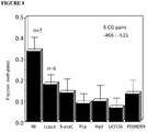


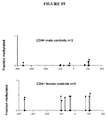


Classifications
-
- C—CHEMISTRY; METALLURGY
- C12—BIOCHEMISTRY; BEER; SPIRITS; WINE; VINEGAR; MICROBIOLOGY; ENZYMOLOGY; MUTATION OR GENETIC ENGINEERING
- C12Q—MEASURING OR TESTING PROCESSES INVOLVING ENZYMES, NUCLEIC ACIDS OR MICROORGANISMS; COMPOSITIONS OR TEST PAPERS THEREFOR; PROCESSES OF PREPARING SUCH COMPOSITIONS; CONDITION-RESPONSIVE CONTROL IN MICROBIOLOGICAL OR ENZYMOLOGICAL PROCESSES
- C12Q1/00—Measuring or testing processes involving enzymes, nucleic acids or microorganisms; Compositions therefor; Processes of preparing such compositions
- C12Q1/68—Measuring or testing processes involving enzymes, nucleic acids or microorganisms; Compositions therefor; Processes of preparing such compositions involving nucleic acids
- C12Q1/6876—Nucleic acid products used in the analysis of nucleic acids, e.g. primers or probes
- C12Q1/6883—Nucleic acid products used in the analysis of nucleic acids, e.g. primers or probes for diseases caused by alterations of genetic material
-
- C—CHEMISTRY; METALLURGY
- C12—BIOCHEMISTRY; BEER; SPIRITS; WINE; VINEGAR; MICROBIOLOGY; ENZYMOLOGY; MUTATION OR GENETIC ENGINEERING
- C12Q—MEASURING OR TESTING PROCESSES INVOLVING ENZYMES, NUCLEIC ACIDS OR MICROORGANISMS; COMPOSITIONS OR TEST PAPERS THEREFOR; PROCESSES OF PREPARING SUCH COMPOSITIONS; CONDITION-RESPONSIVE CONTROL IN MICROBIOLOGICAL OR ENZYMOLOGICAL PROCESSES
- C12Q2600/00—Oligonucleotides characterized by their use
- C12Q2600/106—Pharmacogenomics, i.e. genetic variability in individual responses to drugs and drug metabolism
-
- C—CHEMISTRY; METALLURGY
- C12—BIOCHEMISTRY; BEER; SPIRITS; WINE; VINEGAR; MICROBIOLOGY; ENZYMOLOGY; MUTATION OR GENETIC ENGINEERING
- C12Q—MEASURING OR TESTING PROCESSES INVOLVING ENZYMES, NUCLEIC ACIDS OR MICROORGANISMS; COMPOSITIONS OR TEST PAPERS THEREFOR; PROCESSES OF PREPARING SUCH COMPOSITIONS; CONDITION-RESPONSIVE CONTROL IN MICROBIOLOGICAL OR ENZYMOLOGICAL PROCESSES
- C12Q2600/00—Oligonucleotides characterized by their use
- C12Q2600/136—Screening for pharmacological compounds
-
- C—CHEMISTRY; METALLURGY
- C12—BIOCHEMISTRY; BEER; SPIRITS; WINE; VINEGAR; MICROBIOLOGY; ENZYMOLOGY; MUTATION OR GENETIC ENGINEERING
- C12Q—MEASURING OR TESTING PROCESSES INVOLVING ENZYMES, NUCLEIC ACIDS OR MICROORGANISMS; COMPOSITIONS OR TEST PAPERS THEREFOR; PROCESSES OF PREPARING SUCH COMPOSITIONS; CONDITION-RESPONSIVE CONTROL IN MICROBIOLOGICAL OR ENZYMOLOGICAL PROCESSES
- C12Q2600/00—Oligonucleotides characterized by their use
- C12Q2600/154—Methylation markers
-
- C—CHEMISTRY; METALLURGY
- C12—BIOCHEMISTRY; BEER; SPIRITS; WINE; VINEGAR; MICROBIOLOGY; ENZYMOLOGY; MUTATION OR GENETIC ENGINEERING
- C12Q—MEASURING OR TESTING PROCESSES INVOLVING ENZYMES, NUCLEIC ACIDS OR MICROORGANISMS; COMPOSITIONS OR TEST PAPERS THEREFOR; PROCESSES OF PREPARING SUCH COMPOSITIONS; CONDITION-RESPONSIVE CONTROL IN MICROBIOLOGICAL OR ENZYMOLOGICAL PROCESSES
- C12Q2600/00—Oligonucleotides characterized by their use
- C12Q2600/156—Polymorphic or mutational markers
-
- C—CHEMISTRY; METALLURGY
- C12—BIOCHEMISTRY; BEER; SPIRITS; WINE; VINEGAR; MICROBIOLOGY; ENZYMOLOGY; MUTATION OR GENETIC ENGINEERING
- C12Q—MEASURING OR TESTING PROCESSES INVOLVING ENZYMES, NUCLEIC ACIDS OR MICROORGANISMS; COMPOSITIONS OR TEST PAPERS THEREFOR; PROCESSES OF PREPARING SUCH COMPOSITIONS; CONDITION-RESPONSIVE CONTROL IN MICROBIOLOGICAL OR ENZYMOLOGICAL PROCESSES
- C12Q2600/00—Oligonucleotides characterized by their use
- C12Q2600/158—Expression markers
Definitions
- the present invention relates to compositions and methods for diagnosing, monitoring and/or treating an autoimmune or chronic inflammatory disease.
- the present invention provides methods for diagnosing, monitoring and treating an autoimmune disease (e.g., rheumatoid arthritis) or chronic inflammatory disease (e.g., systemic lupus erythematosus) based on detecting or altering (e.g., altering expression or methylation status of) autoimmune or chronic inflammatory disease markers (e.g., CD70 and CD40L).
- the present invention also provides kits for detecting methylation status of autoimmune or chronic inflammatory disease markers (e.g., CD70 and CD40L) and for diagnosing, monitoring and/or treating autoimmune or chronic inflammatory diseases.
- Autoimmune diseases are generally understood to be diseases where the target of the disease is “self” or “self antigen.”
- diseases that are believed to involve T cell immunity directed to self antigens, including, for example, multiple sclerosis (MS), Type I diabetes, and rheumatoid arthritis (RA).
- MS multiple sclerosis
- RA rheumatoid arthritis
- RA is a chronic inflammatory disorder characterized by joint pain.
- the course of the disease is variable, but can be both debilitating and mutilating. According to conservative estimates approximately 50,000,000 individuals are afflicted with RA worldwide. Those individuals are not only subjected to life-long disability and misery, but as current evidence suggests, their life expectancy is compromised as well.
- SLE Systemic lupus erythematosus
- SLE is a chronic inflammatory disease that can affect various parts of the body including skin, blood, kidneys, and joints. SLE may manifest as a mild disease or be serious and life-threatening. More than 16,000 cases of SLE are reported in the United States each year, with up to 1.5 million cases diagnosed. Although SLE can occur at any age, and in either sex, it has been found to occur 10-15 times more frequently in women.
- SLE is characterized by the production of auto-antibodies having specificity for a wide range of self-antigens.
- SLE auto-antibodies mediate organ damage by directly binding to host tissues and by forming immune complexes that deposit in vascular tissues and activate various immune cells.
- SLE induced damage to the host targets the skin, kidneys, vasculature, joints, various blood elements, and the central nervous system (CNS).
- CNS central nervous system
- the present invention relates to compositions and methods for diagnosing, monitoring and/or treating an autoimmune or chronic inflammatory disease.
- the present invention provides methods for diagnosing, monitoring and treating an autoimmune disease (e.g., rheumatoid arthritis) or chronic inflammatory disease (e.g., systemic lupus erythematosus) based on detecting or altering (e.g., altering expression or methylation status of) autoimmune or chronic inflammatory disease markers (e.g., CD70 and CD40L).
- the present invention also provides kits for detecting methylation status of autoimmune or chronic inflammatory disease markers (e.g., CD70 and CD40L) and for diagnosing, monitoring and/or treating autoimmune or chronic inflammatory diseases.
- the present invention provides a method for detecting methylation status of CD70 in a subject, comprising providing a biological sample from the subject, wherein the biological sample comprises CD70 and exposing the sample to reagents for detecting methylation status of CD70.
- the reagents detect methylation status of the 5′ untranslated region of CD70.
- the 5′ untranslated region comprises the ⁇ 338 to ⁇ 515 (e.g., ⁇ 466 to ⁇ 515) region of CD70.
- the biological sample is selected from the group comprising a bone marrow sample, a blood sample, a serum sample, a platelet sample, a nucleic acid sample, a DNA sample, a tissue sample, a urine sample, and purified or filtered forms thereof.
- the detecting comprises use of a polymerase chain reaction.
- the detecting comprises differential antibody binding.
- the detecting comprises restriction enzyme digestion.
- the detecting comprises use of oligonucleotide binding assays.
- the detecting comprises use of a microarray.
- the detecting comprises use of bisulfite sequencing.
- the present invention also provides a method for detecting methylation status of CD70 in a subject, comprising providing a biological sample from a subject, wherein the biological sample comprises the 5′ untranslated CD70 region and detecting methylation status of the ⁇ 466 to ⁇ 515 region of the 5′ untranslated CD70 region in the biological sample.
- the analyzed portion of the 5′ untranslated CD70 region is from ⁇ 338 to ⁇ 466.
- the present invention is not limited by the region analyzed. For example, as described below and shown in the figures, numerous additional differentially methylated regions find use with the methods of the present invention.
- the present invention additionally provides a method of diagnosing or monitoring an autoimmune or chronic inflammatory disease in a subject, comprising: providing nucleic acid from a subject and detecting the methylation status of CD70 in the nucleic acid.
- the method detects the methylation status of the ⁇ 338 to ⁇ 515 (e.g., ⁇ 446 to ⁇ 515) region of the 5′ untranslated CD70 region.
- the method further detects the methylation status of perforin.
- the method further detects the methylation status of CD11a.
- the method detects the methylation status of IgE FCR ⁇ 1.
- the method detects the methylation status of CD30.
- the method detects the methylation status of CD11c. In some embodiments, the methylation status of CD40L is detected. In some embodiments, the method detects the methylation status of two or more of perforin, CD11a, CD30, CD11c, CD40L and IgE FCR ⁇ 1. In some embodiments, the chronic inflammatory disease is systemic lupus erythematosis (SLE). In some embodiments, PCR is used for detection.
- the present invention provides a method of diagnosing or detecting an autoimmune or chronic inflammatory disease in a subject comprising detecting, individually or in combination, the methylation status of CD70, CD11a, CD30, CD11c, CD40L and IgE FCR ⁇ 1.
- the present invention additionally provides a method of diagnosing or monitoring an autoimmune or chronic inflammatory disease in a subject, comprising: providing nucleic acid from a subject and detecting the methylation status of CD40L in the nucleic acid.
- the method detects the methylation status of the 125 to ⁇ 400 (e.g., 125 to ⁇ 110 or the ⁇ 350 to ⁇ 400) region of the 5′ untranslated CD40L region.
- the method further detects the methylation status of perforin.
- the method further detects the methylation status of CD11a.
- the method detects the methylation status of IgE FCR ⁇ 1.
- the method detects the methylation status of CD30. In still other embodiments, the method detects the methylation status of CD11c. In some embodiments, the methylation status of CD70 is detected. In some embodiments, the method detects the methylation status of two or more of perforin, CD11a, CD30, CD11c, CD70 and IgE FCR ⁇ 1.
- the chronic inflammatory disease is systemic lupus erythematosis (SLE).
- PCR is used for detection.
- the autoimmune disease is rheumatoid arthritis.
- the kit comprises reagents and/or instructions for diagnosing or monitoring an autoimmune or chronic inflammatory disease based on methylation status of CD11a. In still further embodiments, the kit comprises instructions and/or reagents for diagnosing or monitoring an autoimmune or chronic inflammatory disease based on methylation status of IgE FCR ⁇ 1. In still further embodiments, the kit comprises instructions and/or reagents for diagnosing or monitoring an autoimmune or chronic inflammatory disease based on methylation status of CD11c and/or CD40L. In still further embodiments, the kit comprises instructions and/or reagents for diagnosing or monitoring an autoimmune or chronic inflammatory disease based on methylation status of CD30.
- the present invention also provides a kit for detecting gene expression associated with SLE, comprising reagents for detecting methylation status of CD70 and a positive control that indicates test results for CD70 methylation status.
- the kit comprises instructions for using the kit for detecting methylation status of CD70.
- the kit comprises instructions for diagnosing or monitoring SLE based on methylation status of CD70.
- the instructions comprise instructions required by the U.S. Food and Drug Administration for in vitro diagnostic kits.
- the kit comprises instructions and/or reagents for diagnosing or monitoring SLE based on methylation status of perforin.
- the kit comprises instructions and/or reagents for diagnosing or monitoring SLE based on methylation status of CD11a. In still other embodiments, the kit comprises instructions and/or reagent for diagnosing or monitoring SLE based on methylation status of IgE FCR ⁇ 1. In still other embodiments, the kit comprises instructions and/or reagent for diagnosing or monitoring SLE based on methylation status of CD30. In still other embodiments, the kit comprises instructions and/or reagent for diagnosing or monitoring SLE based on methylation status of CD11c. In some embodiments, the kit comprises instructions for diagnosing or monitoring SLE based on methylation status of two or more of perforin, CD11a, CD30, CD11c, CD40L and IgE FCR ⁇ 1.
- FIG. 1 shows the effect of DNA methylation inhibition on CD70 expression.
- FIG. 2 shows increased CD70 expression induced by DNA methylation inhibitors.
- FIG. 3 shows increased B cell costimulation by polyclonal T cells treated with DNA methylation inhibitors, and reversal with anti-CD70.
- FIG. 4 shows increased B cell costimulation by cloned T cells treated with DNA methylation inhibitors, and reversal with anti-CD70.
- FIG. 5 shows overexpression of CD70 on T cells from patients with systemic lupus erythematosus (SLE).
- FIG. 6 shows anti-CD70 inhibition of IgG synthesis induced by lupus T cells.
- FIG. 7 shows methylation status of the CD70 promoter in CD4+ T cells.
- FIG. 8 shows the effect of lupus and DNA methylation inhibitors on a regulatory element in the CD70 promoter.
- FIG. 9 shows Dnmt and ERK pathway inhibitors increase CD70 mRNA in CD4+ T cells.
- FIG. 10 shows the CD70 (TNFSF7) promoter and 5′ flanking region sequence and relevant features.
- the filled circles represent the potentially methylatable CG pairs, and the broken arrow the putative transcription start site.
- the locations of potential transcription factor binding sites and CAAT boxes are also shown.
- FIG. 11 shows CD70 promoter activity.
- A Shows activity of a 1018 bp fragment ( ⁇ 996 to +52) cloned into pGL3-Basic while
- B shows activity of the fragments spanning the indicated regions.
- the results of pGL3-Basic constructs containing the promoter fragments are normalized to the paired empty vector control (black bars) and represent the mean ⁇ SEM of 2 independent experiments.
- FIG. 12 shows CD70 promoter methylation patterns in CD4+ and CD8+ T cells.
- A CD4+ T cells.
- B CD8+ T cells.
- FIG. 13 shows CD70 promoter methylation patterns in CD4+ T cells treated with DNA methylation inhibitors: (A) non-treated controls; (B) 5-azaC; (C) Pca; (D) U0126; (E) PD98059; and (F) Hyd.
- FIG. 14 shows the average methylation of the ⁇ 515 to ⁇ 423 sequence affected by treatment with DNA methylation inhibitors.
- FIG. 15 shows the effect of regional methylation on TNFSF7 promoter function.
- FIG. 16 shows CD70 mRNA levels in CD4+ T cells from lupus patients and controls.
- FIG. 17 shows CD70 promoter methylation in CD4+ T cells from lupus patients and controls.
- A-C of the region from ⁇ 1000 to ⁇ 200;
- D of the region between ⁇ 515 and ⁇ 423.
- FIG. 18 shows CD40L methylation patterns.
- FIG. 19 shows the CD40L promoter methylation in healthy men and women.
- FIG. 20 shows the CD40L promoter is demethylated in CD4+ T cells from a woman with active lupus.
- FIG. 21 shows the CD40L Promoter is demethylated in women with lupus.
- FIG. 22 depicts CD40L promoter map.
- autoimmune disease refers generally to diseases which are characterized as having a component of self-recognition.
- autoimmune diseases include, but are not limited to, Autoimmune hepatitis, Multiple Sclerosis, Systemic Lupus Erythematosus, Myasthenia Gravis, Type I diabetes, Rheumatoid Arthritis, Psoriasis, Hashimoto's Thyroiditis, Grave's disease, Ankylosing Spondylitis Sjogrens Disease, CREST syndrome, Scleroderma and many more.
- Most autoimmune diseases are also chronic inflammatory diseases. This is defined as a disease process associated with long-term (>6 months) activation of inflammatory cells (leukocytes).
- the chronic inflammation leads to damage of patient organs or tissues.
- Many diseases are chronic inflammatory disorders, but are not know to have an autoimmune basis. For example, Atherosclerosis, Congestive Heart Failure, Crohn's disease, Ulcerative Colitis, Polyarteritis nodosa, Whipple's Disease, Primary Sclerosing Cholangitis and many more.
- Mild disease encompasses symptoms that may be function-altering and/or comfort-altering, but are neither immediately organ-threatening nor life-threatening.
- Severe disease entails organ-threatening and/or life-threatening symptoms.
- severe autoimmune disease is often associated with clinical manifestations such as nephritis, vasculitis, central nervous system disease, premature atherosclerosis or lung disease, or combinations thereof, that require aggressive treatment and may be associated with premature death.
- Anti-phospholipid antibody syndrome is often associated with arterial or venous thrombosis.
- autoimmune or chronic inflammatory disease markers e.g., CD70 or CD40L
- any clinical parameters of an autoimmune or inflammatory disease enables the use of an autoimmune or chronic inflammatory disease marker (e.g., CD70 or CD40L) methylation assay as part of a diagnostic battery for that disease or group of diseases.
- disease activity refers to whether the pathological manifestations of the disease are fulminant, quiescent, or in a state between these two extremes.
- a patient suffering from SLE having active disease could be diagnosed or monitored through detecting a hypomethylated form of an autoimmune or chronic inflammatory disease marker (e.g., CD70 or CD40L) described in the present invention, whereas a patient having inactive disease would manifest comparatively higher or normal levels of autoimmune or chronic inflammatory disease markers (e.g., CD70 or CD40L) methylation.
- an autoimmune or chronic inflammatory disease marker e.g., CD70 or CD40L
- epitopope refers to that portion of an antigen that makes contact with a particular antibody.
- an antigenic determinant may compete with the intact antigen (i.e., the “immunogen” used to elicit the immune response) for binding to an antibody.
- telomere binding when used in reference to the interaction of an antibody and a protein or peptide means that the interaction is dependent upon the presence of a particular structure (i.e., the antigenic determinant or epitope) on the protein; in other words the antibody is recognizing and binding to a specific protein structure rather than to proteins in general. For example, if an antibody is specific for epitope “A,” the presence of a protein containing epitope A (or free, unlabelled A) in a reaction containing labeled “A” and the antibody will reduce the amount of labeled A bound to the antibody.
- non-specific binding and “background binding” when used in reference to the interaction of an antibody and a protein or peptide refer to an interaction that is not dependent on the presence of a particular structure (i.e., the antibody is binding to proteins in general rather that a particular structure such as an epitope).
- the term “subject” refers to any animal (e.g., a mammal), including, but not limited to, humans, non-human primates, rodents, and the like, which is to be the recipient of a particular treatment.
- the terms “subject” and “patient” are used interchangeably herein in reference to a human subject.
- the term “subject suspected of having autoimmune or chronic inflammatory disease” refers to a subject that presents one or more symptoms indicative of an autoimmune or chronic inflammatory disease (e.g., hives or joint pain) or is being screened for an autoimmune or chronic inflammatory disease (e.g., during a routine physical).
- a subject suspected of having an autoimmune or chronic inflammatory disease may also have one or more risk factors.
- a subject suspected of having an autoimmune or chronic inflammatory disease has generally not been tested for autoimmune or chronic inflammatory disease.
- a “subject suspected of having autoimmune or chronic inflammatory disease” encompasses an individual who has received an initial diagnosis but for whom the severity of the autoimmune or chronic inflammatory disease is not known. The term further includes people who once had autoimmune or chronic inflammatory disease but whose symptoms have ameliorated.
- the term “subject at risk for autoimmune or chronic inflammatory disease” refers to a subject with one or more risk factors for developing an autoimmune or chronic inflammatory disease.
- Risk factors include, but are not limited to, gender, age, genetic predisposition, environmental expose, previous incidents of autoimmune or chronic inflammatory disease, preexisting non-autoimmune or chronic inflammatory diseases, and lifestyle.
- the term “characterizing autoimmune or chronic inflammatory disease in subject” refers to the identification of one or more properties of a sample in a subject, including but not limited to, the presence of calcified tissue and the subject's prognosis.
- Autoimmune or chronic inflammatory disease may be characterized by the identification of the expression of one or more autoimmune or chronic inflammatory disease marker genes, including but not limited to, the autoimmune or chronic inflammatory disease markers disclosed herein.
- autoimmune or chronic inflammatory disease marker genes refers to a gene whose expression level and/or whose methylation status, or other characteristic, alone or in combination with other genes, is correlated with autoimmune or chronic inflammatory disease or prognosis of autoimmune or chronic inflammatory disease.
- the correlation may relate to either an increased or decreased expression, or an increased or decreased methylation, of the gene.
- the expression or low levels of methylation (e.g., as compared to normal, healthy controls) of the gene may be indicative of autoimmune or chronic inflammatory disease, or lack of expression or high levels of methylation (e.g., as compared to normal, healthy controls) of the gene may be correlated with poor prognosis in an autoimmune or chronic inflammatory disease patient.
- Autoimmune or chronic inflammatory disease marker expression and methylation status may be characterized using any suitable method, including but not limited to, those described in illustrative Examples 1-14 below.
- a reagent that specifically detects expression levels refers to reagents used to detect the expression of one or more genes and the term “a reagent that specifically detects methylation status” refers to reagents used to detect the methylation status of one or more genes (e.g., including but not limited to, the autoimmune and chronic inflammatory disease markers of the present invention).
- suitable reagents include but are not limited to, nucleic acid probes capable of specifically hybridizing to the gene of interest, PCR primers capable of specifically amplifying the gene of interest, PCR primers that function in the context of a methylation sensitive PCR reaction, and antibodies capable of specifically binding to proteins expressed by the gene of interest.
- suitable reagents include but are not limited to, nucleic acid probes capable of specifically hybridizing to the gene of interest, PCR primers capable of specifically amplifying the gene of interest, PCR primers that function in the context of a methylation sensitive PCR reaction, and antibodies capable of specifically binding to proteins expressed by the gene of interest.
- detecting a decreased or increased expression relative to non-autoimmune or chronic inflammatory disease control refers to measuring the level of expression of a gene (e.g., the level of mRNA or protein) relative to the level in a non-autoimmune or chronic inflammatory disease control sample.
- Gene expression can be measured using any suitable method, including but not limited to, those described herein.
- the term “detecting a change in gene expression (e.g., of CD70, IgE FCR ⁇ 1, CD30, CD40L or CD11c) in said autoimmune or chronic inflammatory disease sample in the presence of said test compound relative to the absence of said test compound” refers to measuring an altered level of expression (e.g., increased or decreased) in the presence of a test compound relative to the absence of the test compound.
- Gene expression can be measured using any suitable method, including but not limited to, those described in Examples 1-14 below.
- the term “instructions for using said kit for detecting autoimmune or chronic inflammatory disease in said subject” includes instructions for using the reagents contained in the kit for the detection and characterization of autoimmune or chronic inflammatory disease in a sample from a subject.
- the instructions further comprise the statement of intended use required by the U.S. Food and Drug Administration (FDA) in labeling in vitro diagnostic products.
- FDA U.S. Food and Drug Administration
- autoimmune or chronic inflammatory disease expression profile map refers to a presentation of expression levels of genes in a particular type of autoimmune or chronic inflammatory disease.
- the map may be presented as a graphical representation (e.g., on paper or on a computer screen), a physical representation (e.g., a gel or array) or a digital representation stored in computer memory.
- maps are generated from pooled samples comprising samples from a plurality of patients with the same type of autoimmune or chronic inflammatory disease.
- computer memory and “computer memory device” refer to any storage media readable by a computer processor.
- Examples of computer memory include, but are not limited to, RAM, ROM, computer chips, digital video disc (DVDs), compact discs (CDs), hard disk drives (HDD), and magnetic tape.
- computer readable medium refers to any device or system for storing and providing information (e.g., data and instructions) to a computer processor.
- Examples of computer readable media include, but are not limited to, DVDs, CDs, hard disk drives, magnetic tape and servers for streaming media over networks.
- processor and “central processing unit” or “CPU” are used interchangeably and refer to a device that is able to read a program from a computer memory (e.g., ROM or other computer memory) and perform a set of steps according to the program.
- a computer memory e.g., ROM or other computer memory
- the term “providing a prognosis” refers to providing information regarding the impact of the presence of autoimmune or chronic inflammatory disease (e.g., as determined by the diagnostic methods of the present invention) on a subject's future health (e.g., expected morbidity or mortality).
- the term “subject diagnosed with an autoimmune or chronic inflammatory disease” refers to a subject who has been tested and found to have autoimmune or chronic inflammatory disease.
- the autoimmune or chronic inflammatory disease may be diagnosed using any suitable method, including but not limited to, biopsy, x-ray, blood test, and the diagnostic methods of the present invention.
- initial diagnosis refers to results of initial autoimmune or chronic inflammatory disease diagnosis (e.g. the presence or absence of autoimmune or chronic inflammatory disease). An initial diagnosis does not include information about the severity of the autoimmune or chronic inflammatory disease.
- non-human animals refers to all non-human animals including, but are not limited to, vertebrates such as rodents, non-human primates, ovines, bovines, ruminants, lagomorphs, porcines, caprines, equines, canines, felines, aves, etc.
- gene transfer system refers to any means of delivering a composition comprising a nucleic acid sequence to a cell or tissue.
- gene transfer systems include, but are not limited to, vectors (e.g., retroviral, adenoviral, adeno-associated viral, and other nucleic acid-based delivery systems), microinjection of naked nucleic acid, polymer-based delivery systems (e.g., liposome-based and metallic particle-based systems), biolistic injection, and the like.
- viral gene transfer system refers to gene transfer systems comprising viral elements (e.g., intact viruses, modified viruses and viral components such as nucleic acids or proteins) to facilitate delivery of the sample to a desired cell or tissue.
- viral elements e.g., intact viruses, modified viruses and viral components such as nucleic acids or proteins
- adenovirus gene transfer system refers to gene transfer systems comprising intact or altered viruses belonging to the family Adenoviridae.
- site-specific recombination target sequences refers to nucleic acid sequences that provide recognition sequences for recombination factors and the location where recombination takes place.
- nucleic acid molecule refers to any nucleic acid containing molecule, including but not limited to, DNA or RNA.
- the term encompasses sequences that include any of the known base analogs of DNA and RNA including, but not limited to, 4-acetylcytosine, 8-hydroxy-N6-methyladenosine, aziridinylcytosine, pseudoisocytosine, 5-(carboxyhydroxylmethyl) uracil, 5-fluorouracil, 5-bromouracil, 5-carboxymethylaminomethyl-2-thiouracil, 5-carboxymethylaminomethyluracil, dihydrouracil, inosine, N6-isopentenyladenine, 1-methyladenine, 1-methylpseudouracil, 1-methylguanine, 1-methylinosine, 2,2-dimethylguanine, 2-methyladenine, 2-methylguanine, 3-methylcytosine, 5-methylcytosine, N6-methyladenine,
- gene refers to a nucleic acid (e.g., DNA) sequence that comprises coding sequences necessary for the production of a polypeptide, precursor, or RNA (e.g., rRNA, tRNA).
- the polypeptide can be encoded by a full length coding sequence or by any portion of the coding sequence so long as the desired activity or functional properties (e.g., enzymatic activity, ligand binding, signal transduction, immunogenicity, etc.) of the full-length or fragment are retained.
- the term also encompasses the coding region of a structural gene and the sequences located adjacent to the coding region on both the 5′ and 3′ ends for a distance of about 1 kb or more on either end such that the gene corresponds to the length of the full-length mRNA. Sequences located 5′ of the coding region and present on the mRNA are referred to as 5′ non-translated sequences. Sequences located 3′ or downstream of the coding region and present on the mRNA are referred to as 3′ non-translated sequences.
- the term “gene” encompasses both cDNA and genomic forms of a gene.
- a genomic form or clone of a gene contains the coding region interrupted with non-coding sequences termed “introns” or “intervening regions” or “intervening sequences.”
- Introns are segments of a gene that are transcribed into nuclear RNA (mRNA); introns may contain regulatory elements such as enhancers. Introns are removed or “spliced out” from the nuclear or primary transcript; introns therefore are absent in the messenger RNA (mRNA) transcript.
- mRNA messenger RNA
- heterologous gene refers to a gene that is not in its natural environment.
- a heterologous gene includes a gene from one species introduced into another species.
- a heterologous gene also includes a gene native to an organism that has been altered in some way (e.g., mutated, added in multiple copies, linked to non-native regulatory sequences, etc).
- Heterologous genes are distinguished from endogenous genes in that the heterologous gene sequences are typically joined to DNA sequences that are not found naturally associated with the gene sequences in the chromosome or are associated with portions of the chromosome not found in nature (e.g., genes expressed in loci where the gene is not normally expressed).
- RNA expression refers to the process of converting genetic information encoded in a gene into RNA (e.g., mRNA, rRNA, tRNA, or snRNA) through “transcription” of the gene (i.e., via the enzymatic action of an RNA polymerase), and for protein encoding genes, into protein through “translation” of mRNA.
- Gene expression can be regulated at many stages in the process. “Up-regulation” or “activation” refers to regulation that increases the production of gene expression products (i.e., RNA or protein), while “down-regulation” or “repression” refers to regulation that decrease production. Molecules (e.g., transcription factors) that are involved in up-regulation or down-regulation are often called “activators” and “repressors,” respectively.
- genomic forms of a gene may also include sequences located on both the 5′ and 3′ end of the sequences that are present on the RNA transcript. These sequences are referred to as “flanking” sequences or regions (these flanking sequences are located 5′ or 3′ to the non-translated sequences present on the mRNA transcript).
- the 5′ flanking region may contain regulatory sequences such as promoters and enhancers that control or influence the transcription of the gene.
- the 3′ flanking region may contain sequences that direct the termination of transcription, post-transcriptional cleavage and polyadenylation.
- wild-type refers to a gene or gene product isolated from a naturally occurring source.
- a wild-type gene is that which is most frequently observed in a population and is thus arbitrarily designed the “normal” or “wild-type” form of the gene.
- modified or mutant refers to a gene or gene product that displays modifications in sequence and or functional properties (i.e., altered characteristics, e.g., hypomethylation) when compared to the wild-type gene or gene product. It is noted that naturally occurring mutants can be isolated; these are identified by the fact that they have altered characteristics (including altered nucleic acid sequences) when compared to the wild-type gene or gene product.
- DNA molecules are said to have “5′ ends” and “3′ ends” because mononucleotides are reacted to make oligonucleotides or polynucleotides in a manner such that the 5′ phosphate of one mononucleotide pentose ring is attached to the 3′ oxygen of its neighbor in one direction via a phosphodiester linkage.
- an end of an oligonucleotides or polynucleotide referred to as the “5′ end” if its 5′ phosphate is not linked to the 3′ oxygen of a mononucleotide pentose ring and as the “3′ end” if its 3′ oxygen is not linked to a 5′ phosphate of a subsequent mononucleotide pentose ring.
- the promoter and enhancer elements that direct transcription of a linked gene are generally located 5′ or upstream of the coding region. However, enhancer elements can exert their effect even when located 3′ of the promoter element and the coding region. Transcription termination and polyadenylation signals are located 3′ or downstream of the coding region.
- regulatory element refers to a genetic element that controls some aspect of the expression of nucleic acid sequences.
- a promoter is a regulatory element that facilitates the initiation of transcription of an operably linked coding region.
- Other regulatory elements include splicing signals, polyadenylation signals, termination signals, etc.
- methylation status refers to the presence or absence of methylation within a gene, specifically, to the presence or absence of methylation of deoxycytosine (dC) bases in CG pairs within a gene, the presence of which serves as one of the mechanisms by which gene expression is suppressed (See, e.g., Attwood et al., Cell Mol Life Sci 59, 241 (2002)).
- dC deoxycytosine
- nucleic acid molecule encoding As used herein, the terms “nucleic acid molecule encoding,” “DNA sequence encoding,” and “DNA encoding” refer to the order or sequence of deoxyribonucleotides along a strand of deoxyribonucleic acid. The order of these deoxyribonucleotides determines the order of amino acids along the polypeptide (protein) chain. The DNA sequence thus codes for the amino acid sequence.
- an oligonucleotide having a nucleotide sequence encoding a gene and “polynucleotide having a nucleotide sequence encoding a gene,” means a nucleic acid sequence comprising the coding region of a gene or in other words the nucleic acid sequence that encodes a gene product.
- the coding region may be present in a cDNA, genomic DNA or RNA form.
- the oligonucleotide or polynucleotide may be single-stranded (i.e., the sense strand) or double-stranded.
- Suitable control elements such as enhancers/promoters, splice junctions, polyadenylation signals, etc. may be placed in close proximity to the coding region of the gene if needed to permit proper initiation of transcription and/or correct processing of the primary RNA transcript.
- the coding region utilized in the expression vectors of the present invention may contain endogenous enhancers/promoters, splice junctions, intervening sequences, polyadenylation signals, etc. or a combination of both endogenous and exogenous control elements.
- oligonucleotide refers to a short length of single-stranded polynucleotide chain. Oligonucleotides are typically less than 200 residues long (e.g., between 15 and 100), however, as used herein, the term is also intended to encompass longer polynucleotide chains. Oligonucleotides are often referred to by their length. For example a 24 residue oligonucleotide is referred to as a “24-mer”. Oligonucleotides can form secondary and tertiary structures by self-hybridizing or by hybridizing to other polynucleotides. Such structures can include, but are not limited to, duplexes, hairpins, cruciforms, bends, and triplexes.
- complementarity are used in reference to polynucleotides (i.e., a sequence of nucleotides) related by the base-pairing rules. For example, for the sequence “5′-A-G-T-3′,” is complementary to the sequence “3′-T-C-A-5′.” Complementarity may be “partial,” in which only some of the nucleic acids' bases are matched according to the base pairing rules. Or, there may be “complete” or “total” complementarity between the nucleic acids. The degree of complementarity between nucleic acid strands has significant effects on the efficiency and strength of hybridization between nucleic acid strands. This is of particular importance in amplification reactions, as well as detection methods that depend upon binding between nucleic acids.
- a partially complementary sequence is a nucleic acid molecule that at least partially inhibits a completely complementary nucleic acid molecule from hybridizing to a target nucleic acid is “substantially homologous.”
- the inhibition of hybridization of the completely complementary sequence to the target sequence may be examined using a hybridization assay (Southern or Northern blot, solution hybridization and the like) under conditions of low stringency.
- a substantially homologous sequence or probe will compete for and inhibit the binding (i.e., the hybridization) of a completely homologous nucleic acid molecule to a target under conditions of low stringency.
- low stringency conditions are such that non-specific binding is permitted; low stringency conditions require that the binding of two sequences to one another be a specific (i.e., selective) interaction.
- the absence of non-specific binding may be tested by the use of a second target that is substantially non-complementary (e.g., less than about 30% identity); in the absence of non-specific binding the probe will not hybridize to the second non-complementary target.
- substantially homologous refers to any probe that can hybridize to either or both strands of the double-stranded nucleic acid sequence under conditions of low stringency as described above.
- a gene may produce multiple RNA species that are generated by differential splicing of the primary RNA transcript.
- cDNAs that are splice variants of the same gene will contain regions of sequence identity or complete homology (representing the presence of the same exon or portion of the same exon on both cDNAs) and regions of complete non-identity (for example, representing the presence of exon “A” on cDNA 1 wherein cDNA 2 contains exon “B” instead). Because the two cDNAs contain regions of sequence identity they will both hybridize to a probe derived from the entire gene or portions of the gene containing sequences found on both cDNAs; the two splice variants are therefore substantially homologous to such a probe and to each other.
- substantially homologous refers to any probe that can hybridize (i.e., it is the complement of) the single-stranded nucleic acid sequence under conditions of low stringency as described above.
- hybridization is used in reference to the pairing of complementary nucleic acids. Hybridization and the strength of hybridization (i.e., the strength of the association between the nucleic acids) is impacted by such factors as the degree of complementary between the nucleic acids, stringency of the conditions involved, the T m of the formed hybrid, and the G:C ratio within the nucleic acids. A single molecule that contains pairing of complementary nucleic acids within its structure is said to be “self-hybridized.”
- T m is used in reference to the “melting temperature.”
- the melting temperature is the temperature at which a population of double-stranded nucleic acid molecules becomes half dissociated into single strands.
- stringency is used in reference to the conditions of temperature, ionic strength, and the presence of other compounds such as organic solvents, under which nucleic acid hybridizations are conducted.
- low stringency conditions a nucleic acid sequence of interest will hybridize to its exact complement, sequences with single base mismatches, closely related sequences (e.g., sequences with 90% or greater homology), and sequences having only partial homology (e.g., sequences with 50-90% homology).
- intermediate stringency conditions a nucleic acid sequence of interest will hybridize only to its exact complement, sequences with single base mismatches, and closely relation sequences (e.g., 90% or greater homology).
- a nucleic acid sequence of interest will hybridize only to its exact complement, and (depending on conditions such a temperature) sequences with single base mismatches. In other words, under conditions of high stringency the temperature can be raised so as to exclude hybridization to sequences with single base mismatches.
- “High stringency conditions” when used in reference to nucleic acid hybridization comprise conditions equivalent to binding or hybridization at 42° C. in a solution consisting of 5 ⁇ SSPE (43.8 g/l NaCl, 6.9 g/l NaH 2 PO 4 H 2 O and 1.85 g/l EDTA, pH adjusted to 7.4 with NaOH), 0.5% SDS, 5 ⁇ Denhardt's reagent and 100 ⁇ g/ml denatured salmon sperm DNA followed by washing in a solution comprising 0.1 ⁇ SSPE, 1.0% SDS at 42° C. when a probe of about 500 nucleotides in length is employed.
- “Medium stringency conditions” when used in reference to nucleic acid hybridization comprise conditions equivalent to binding or hybridization at 42° C. in a solution consisting of 5 ⁇ SSPE (43.8 g/l NaCl, 6.9 g/l NaH 2 PO 4 H 2 O and 1.85 g/l EDTA, pH adjusted to 7.4 with NaOH), 0.5% SDS, 5 ⁇ Denhardt's reagent and 100 ⁇ g/ml denatured salmon sperm DNA followed by washing in a solution comprising 1.0 ⁇ SSPE, 1.0% SDS at 42° C. when a probe of about 500 nucleotides in length is employed.
- Low stringency conditions comprise conditions equivalent to binding or hybridization at 42° C. in a solution consisting of 5 ⁇ SSPE (43.8 g/l NaCl, 6.9 g/l NaH 2 PO 4 H 2 O and 1.85 g/l EDTA, pH adjusted to 7.4 with NaOH), 0.1% SDS, 5 ⁇ Denhardt's reagent (50 ⁇ Denhardt's contains per 500 ml: 5 g Ficoll (Type 400, Pharamcia), 5 g BSA (Fraction V; Sigma)) and 100 ⁇ g/ml denatured salmon sperm DNA followed by washing in a solution comprising 5 ⁇ SSPE, 0.1% SDS at 42° C. when a probe of about 500 nucleotides in length is employed.
- 5 ⁇ SSPE 43.8 g/l NaCl, 6.9 g/l NaH 2 PO 4 H 2 O and 1.85 g/l EDTA, pH adjusted to 7.4 with NaOH
- 5 ⁇ Denhardt's reagent 50 ⁇ D
- low stringency conditions factors such as the length and nature (DNA, RNA, base composition) of the probe and nature of the target (DNA, RNA, base composition, present in solution or immobilized, etc.) and the concentration of the salts and other components (e.g., the presence or absence of formamide, dextran sulfate, polyethylene glycol) are considered and the hybridization solution may be varied to generate conditions of low stringency hybridization different from, but equivalent to, the above listed conditions.
- conditions that promote hybridization under conditions of high stringency e.g., increasing the temperature of the hybridization and/or wash steps, the use of formamide in the hybridization solution, etc.
- “Amplification” is a special case of nucleic acid replication involving template specificity. It is to be contrasted with non-specific template replication (i.e., replication that is template-dependent but not dependent on a specific template). Template specificity is here distinguished from fidelity of replication (i.e., synthesis of the proper polynucleotide sequence) and nucleotide (ribo- or deoxyribo-) specificity. Template specificity is frequently described in terms of “target” specificity. Target sequences are “targets” in the sense that they are sought to be sorted out from other nucleic acid. Amplification techniques have been designed primarily for this sorting out.
- Amplification enzymes are enzymes that, under conditions they are used, will process only specific sequences of nucleic acid in a heterogeneous mixture of nucleic acid.
- MDV-1 RNA is the specific template for the replicase (Kacian et al., Proc. Natl. Acad. Sci. USA 69:3038 (1972)).
- Other nucleic acids will not be replicated by this amplification enzyme.
- this amplification enzyme has a stringent specificity for its own promoters (Chamberlin et al., Nature 228:227 (1970)).
- T4 DNA ligase the enzyme will not ligate the two oligonucleotides or polynucleotides, where there is a mismatch between the oligonucleotide or polynucleotide substrate and the template at the ligation junction (Wu and Wallace, Genomics 4:560 (1989)).
- Taq and Pfu polymerases by virtue of their ability to function at high temperature, are found to display high specificity for the sequences bounded and thus defined by the primers; the high temperature results in thermodynamic conditions that favor primer hybridization with the target sequences and not hybridization with non-target sequences (H. A. Erlich (ed.), PCR Technology, Stockton Press (1989)).
- amplifiable nucleic acid is used in reference to nucleic acids that may be amplified by any amplification method. It is contemplated that “amplifiable nucleic acid” will usually comprise “sample template.”
- sample template refers to nucleic acid originating from a sample that is analyzed for the presence of “target.”
- background template is used in reference to nucleic acid other than sample template that may or may not be present in a sample. Background template is most often inadvertent. It may be the result of carryover, or it may be due to the presence of nucleic acid contaminants sought to be purified away from the sample. For example, nucleic acids from organisms other than those to be detected may be present as background in a test sample.
- the term “primer” refers to an oligonucleotide, whether occurring naturally as in a purified restriction digest or produced synthetically, that is capable of acting as a point of initiation of synthesis when placed under conditions in which synthesis of a primer extension product that is complementary to a nucleic acid strand is induced, (i.e., in the presence of nucleotides and an inducing agent such as DNA polymerase and at a suitable temperature and pH).
- the primer is preferably single stranded for maximum efficiency in amplification, but may alternatively be double stranded. If double stranded, the primer is first treated to separate its strands before being used to prepare extension products.
- the primer is an oligodeoxyribonucleotide.
- the primer must be sufficiently long to prime the synthesis of extension products in the presence of the inducing agent. The exact lengths of the primers will depend on many factors, including temperature, source of primer and the use of the method.
- probe refers to an oligonucleotide (i.e., a sequence of nucleotides), whether occurring naturally as in a purified restriction digest or produced synthetically, recombinantly or by PCR amplification, that is capable of hybridizing to at least a portion of another oligonucleotide of interest.
- a probe may be single-stranded or double-stranded. Probes are useful in the detection, identification and isolation of particular gene sequences.
- any probe used in the present invention will be labeled with any “reporter molecule,” so that is detectable in any detection system, including, but not limited to enzyme (e.g., ELISA, as well as enzyme-based histochemical assays), fluorescent, radioactive, and luminescent systems. It is not intended that the present invention be limited to any particular detection system or label.
- portion when in reference to a nucleotide sequence (as in “a portion of a given nucleotide sequence”) refers to fragments of that sequence.
- the fragments may range in size from four nucleotides to the entire nucleotide sequence minus one nucleotide (10 nucleotides, 20, 30, 40, 50, 100, 200, etc.).
- amplification reagents refers to those reagents (deoxyribonucleotide triphosphates, buffer, etc.), needed for amplification except for primers, nucleic acid template and the amplification enzyme.
- amplification reagents along with other reaction components are placed and contained in a reaction vessel (test tube, microwell, etc.).
- restriction endonucleases and “restriction enzymes” refer to bacterial enzymes, each of which cut double-stranded DNA at or near a specific nucleotide sequence.
- operable combination refers to the linkage of nucleic acid sequences in such a manner that a nucleic acid molecule capable of directing the transcription of a given gene and/or the synthesis of a desired protein molecule is produced.
- operable order refers to the linkage of amino acid sequences in such a manner so that a functional protein is produced.
- isolated when used in relation to a nucleic acid, as in “an isolated oligonucleotide” or “isolated polynucleotide” refers to a nucleic acid sequence that is identified and separated from at least one component or contaminant with which it is ordinarily associated in its natural source. Isolated nucleic acid is such present in a form or setting that is different from that in which it is found in nature. In contrast, non-isolated nucleic acids as nucleic acids such as DNA and RNA found in the state they exist in nature.
- a given DNA sequence e.g., a gene
- RNA sequences such as a specific mRNA sequence encoding a specific protein
- isolated nucleic acid encoding a given protein includes, by way of example, such nucleic acid in cells ordinarily expressing the given protein where the nucleic acid is in a chromosomal location different from that of natural cells, or is otherwise flanked by a different nucleic acid sequence than that found in nature.
- the isolated nucleic acid, oligonucleotide, or polynucleotide may be present in single-stranded or double-stranded form.
- the oligonucleotide or polynucleotide will contain at a minimum the sense or coding strand (i.e., the oligonucleotide or polynucleotide may be single-stranded), but may contain both the sense and anti-sense strands (i.e., the oligonucleotide or polynucleotide may be double-stranded).
- the term “purified” or “to purify” refers to the removal of components (e.g., contaminants) from a sample.
- antibodies are purified by removal of contaminating non-immunoglobulin proteins; they are also purified by the removal of immunoglobulin that does not bind to the target molecule.
- the removal of non-immunoglobulin proteins and/or the removal of immunoglobulins that do not bind to the target molecule results in an increase in the percent of target-reactive immunoglobulins in the sample.
- recombinant polypeptides are expressed in bacterial host cells and the polypeptides are purified by the removal of host cell proteins; the percent of recombinant polypeptides is thereby increased in the sample.
- amino acid sequence and terms such as “polypeptide” or “protein” are not meant to limit the amino acid sequence to the complete, native amino acid sequence associated with the recited protein molecule.
- native protein as used herein to indicate that a protein does not contain amino acid residues encoded by vector sequences; that is, the native protein contains only those amino acids found in the protein as it occurs in nature.
- a native protein may be produced by recombinant means or may be isolated from a naturally occurring source.
- portion when in reference to a protein (as in “a portion of a given protein”) refers to fragments of that protein.
- the fragments may range in size from four amino acid residues to the entire amino acid sequence minus one amino acid.
- Southern blot refers to the analysis of DNA on agarose or acrylamide gels to fractionate the DNA according to size followed by transfer of the DNA from the gel to a solid support, such as nitrocellulose or a nylon membrane.
- the immobilized DNA is then probed with a labeled probe to detect DNA species complementary to the probe used.
- the DNA may be cleaved with restriction enzymes prior to electrophoresis. Following electrophoresis, the DNA may be partially depurinated and denatured prior to or during transfer to the solid support.
- Southern blots are a standard tool of molecular biologists (J. Sambrook et al., Molecular Cloning: A Laboratory Manual, Cold Spring Harbor Press, NY, pp 9.31-9.58 (1989)).
- Northern blot refers to the analysis of RNA by electrophoresis of RNA on agarose gels to fractionate the RNA according to size followed by transfer of the RNA from the gel to a solid support, such as nitrocellulose or a nylon membrane. The immobilized RNA is then probed with a labeled probe to detect RNA species complementary to the probe used.
- Northern blots are a standard tool of molecular biologists (J. Sambrook, et al., supra, pp 7.39-7.52 (1989)).
- the term “Western blot” refers to the analysis of protein(s) (or polypeptides) immobilized onto a support such as nitrocellulose or a membrane.
- the proteins are run on acrylamide gels to separate the proteins, followed by transfer of the protein from the gel to a solid support, such as nitrocellulose or a nylon membrane.
- the immobilized proteins are then exposed to antibodies with reactivity against an antigen of interest.
- the binding of the antibodies may be detected by various methods, including the use of radiolabeled antibodies.
- transgene refers to a foreign gene that is placed into an organism by, for example, introducing the foreign gene into newly fertilized eggs or early embryos.
- foreign gene refers to any nucleic acid (e.g., gene sequence) that is introduced into the genome of an animal by experimental manipulations and may include gene sequences found in that animal so long as the introduced gene does not reside in the same location as does the naturally occurring gene.
- vector is used in reference to nucleic acid molecules that transfer DNA segment(s) from one cell to another.
- vehicle is sometimes used interchangeably with “vector.”
- Vectors are often derived from plasmids, bacteriophages, or plant or animal viruses.
- expression vector refers to a recombinant DNA molecule containing a desired coding sequence and appropriate nucleic acid sequences necessary for the expression of the operably linked coding sequence in a particular host organism.
- Nucleic acid sequences necessary for expression in prokaryotes usually include a promoter, an operator (optional), and a ribosome binding site, often along with other sequences.
- Eukaryotic cells are known to utilize promoters, enhancers, and termination and polyadenylation signals.
- overexpression and “overexpressing” and grammatical equivalents are used in reference to levels of mRNA to indicate a level of expression approximately 3-fold higher (or greater) than that observed in a given tissue in a control or non-transgenic animal.
- Levels of mRNA are measured using any of a number of techniques known to those skilled in the art including, but not limited to Northern blot analysis. Appropriate controls are included on the Northern blot to control for differences in the amount of RNA loaded from each tissue analyzed (e.g., the amount of 28S rRNA, an abundant RNA transcript present at essentially the same amount in all tissues, present in each sample can be used as a means of normalizing or standardizing the mRNA-specific signal observed on Northern blots).
- the amount of mRNA present in the band corresponding in size to the correctly spliced transgene RNA is quantified; other minor species of RNA which hybridize to the transgene probe are not considered in the quantification of the expression of the transgenic mRNA.
- transfection refers to the introduction of foreign DNA into eukaryotic cells. Transfection may be accomplished by a variety of means known to the art including calcium phosphate-DNA co-precipitation, DEAE-dextran-mediated transfection, polybrene-mediated transfection, electroporation, microinjection, liposome fusion, lipofection, protoplast fusion, retroviral infection, and biolistics.
- calcium phosphate co-precipitation refers to a technique for the introduction of nucleic acids into a cell.
- the uptake of nucleic acids by cells is enhanced when the nucleic acid is presented as a calcium phosphate-nucleic acid co-precipitate.
- Graham and van der Eb Graham and van der Eb, Virol., 52:456 (1973)
- the original technique of Graham and van der Eb has been modified by several groups to optimize conditions for particular types of cells. The art is well aware of these numerous modifications.
- stable transfection or “stably transfected” refers to the introduction and integration of foreign DNA into the genome of the transfected cell.
- stable transfectant refers to a cell that has stably integrated foreign DNA into the genomic DNA.
- transient transfection or “transiently transfected” refers to the introduction of foreign DNA into a cell where the foreign DNA fails to integrate into the genome of the transfected cell.
- the foreign DNA persists in the nucleus of the transfected cell for several days. During this time the foreign DNA is subject to the regulatory controls that govern the expression of endogenous genes in the chromosomes.
- transient transfectant refers to cells that have taken up foreign DNA but have failed to integrate this DNA.
- selectable marker refers to the use of a gene that encodes an enzymatic activity that confers the ability to grow in medium lacking what would otherwise be an essential nutrient (e.g. the HIS3 gene in yeast cells); in addition, a selectable marker may confer resistance to an antibiotic or drug upon the cell in which the selectable marker is expressed. Selectable markers may be “dominant”; a dominant selectable marker encodes an enzymatic activity that can be detected in any eukaryotic cell line.
- dominant selectable markers examples include the bacterial aminoglycoside 3′ phosphotransferase gene (also referred to as the neo gene) that confers resistance to the drug G418 in mammalian cells, the bacterial hygromycin G phosphotransferase (hyg) gene that confers resistance to the antibiotic hygromycin and the bacterial xanthine-guanine phosphoribosyl transferase gene (also referred to as the gpt gene) that confers the ability to grow in the presence of mycophenolic acid.
- Other selectable markers are not dominant in that their use must be in conjunction with a cell line that lacks the relevant enzyme activity.
- non-dominant selectable markers include the thymidine kinase (tk) gene that is used in conjunction with tk ⁇ cell lines, the CAD gene that is used in conjunction with CAD-deficient cells and the mammalian hypoxanthine-guanine phosphoribosyl transferase (hprt) gene that is used in conjunction with hprt ⁇ cell lines.
- tk thymidine kinase
- CAD CAD gene that is used in conjunction with CAD-deficient cells
- hprt mammalian hypoxanthine-guanine phosphoribosyl transferase
- cell culture refers to any in vitro culture of cells. Included within this term are continuous cell lines (e.g., with an immortal phenotype), primary cell cultures, transformed cell lines, finite cell lines (e.g., non-transformed cells), and any other cell population maintained in vitro.
- eukaryote refers to organisms distinguishable from “prokaryotes.” It is intended that the term encompass all organisms with cells that exhibit the usual characteristics of eukaryotes, such as the presence of a true nucleus bounded by a nuclear membrane, within which lie the chromosomes, the presence of membrane-bound organelles, and other characteristics commonly observed in eukaryotic organisms. Thus, the term includes, but is not limited to such organisms as fungi, protozoa, and animals (e.g., humans).
- in vitro refers to an artificial environment and to processes or reactions that occur within an artificial environment.
- in vitro environments can consist of, but are not limited to, test tubes and cell culture.
- in vivo refers to the natural environment (e.g., an animal or a cell) and to processes or reaction that occur within a natural environment.
- test compound and “candidate compound” refer to any chemical entity, pharmaceutical, drug, and the like that is a candidate for use to treat or prevent a disease, illness, sickness, or disorder of bodily function (e.g., autoimmune and chronic inflammatory disease).
- Test compounds comprise both known and potential therapeutic compounds.
- a test compound can be determined to be therapeutic by screening using the screening methods of the present invention.
- test compounds include antisense compounds.
- sample is used in its broadest sense. In one sense, it is meant to include a specimen or culture obtained from any source, as well as biological and environmental samples.
- Biological samples may be obtained from animals (including humans) and refers to a biological material or compositions found therein, including, but not limited to, bone marrow, blood, serum, platelet, plasma, interstitial fluid, urine, cerebrospinal fluid, nucleic acid, DNA, tissue, and purified or filtered forms thereof.
- Environmental samples include environmental material such as surface matter, soil, water, crystals and industrial samples. Such examples are not however to be construed as limiting the sample types applicable to the present invention.
- the present invention relates to compositions and methods for diagnosing, monitoring and/or treating an autoimmune or chronic inflammatory disease.
- the present invention provides methods for diagnosing, monitoring and treating an autoimmune disease (e.g., rheumatoid arthritis) or chronic inflammatory disease (e.g., systemic lupus erythematosus) based on detecting or altering (e.g., altering expression or methylation status of) autoimmune or chronic inflammatory disease markers (e.g., CD70 and CD40L).
- the present invention also provides kits for detecting methylation status of autoimmune or chronic inflammatory disease markers (e.g., CD70 and CD40L) and for diagnosing, monitoring and/or treating autoimmune or chronic inflammatory diseases.
- the present invention provides markers whose expression is specifically altered in autoimmune or chronic inflammatory disease. Such markers find use in the diagnosis and characterization of autoimmune or chronic inflammatory disease.
- compositions, kits, and methods for detecting the methylation status of one or more of CD70, IgE FCR ⁇ 1, CD11a, CD30, CD40L, and CD11c for diagnostic, drug screening, research, and therapeutic applications.
- CD4+ T cell DNA hypomethylation contributes to the development of drug-induced and idiopathic systemic lupus erythematosus (SLE) and rheumatoid arthritis (RA).
- DNA methylation refers to the methylation of deoxycytosine (dC) bases in CG pairs, and it is one of the mechanisms by which gene expression is suppressed (See, e.g., Attwood et al., Cell Mol Life Sci 59, 241 (2002)).
- CD4+ T cells treated in vitro with the DNA methylation inhibitors 5-azacytidine (5-azaC), procainamide, or hydralazine become autoreactive, killing autologous or syngeneic macrophages and promoting antibody production
- 5-azacytidine (5-azaC) 5-azacytidine
- procainamide procainamide
- hydralazine become autoreactive, killing autologous or syngeneic macrophages and promoting antibody production
- Adoptive transfer of the autoreactive cells causes a lupus-like disease (See, e.g., Quddus et al., J Clin Invest 92, 38 (1993); Yung et al., Arthritis Rheum 40, 1436 (1997)).
- the autoreactivity is in part due to an overexpression of the adhesion molecule lymphocyte function-associated antigen 1 (LFA-1; CD11a/CD18) (See, e.g., Richardson et al., Arthritis Rheum 37, 1363 (1994); Yung et al., J Clin Invest 97, 2866 (1996)), and abnormal perforin expression contributes to the macrophage killing (See, e.g., Kaplan et al., Arthritis Rheum 46, S282 (2002); Lu et al., J Immunol 170, 51249 (2003)).
- LFA-1 adhesion molecule lymphocyte function-associated antigen 1
- Genomic deoxymethylcytosine (dmC) content is decreased in T cells from patients with active SLE, similar to that in T cells treated with 5-azaC, procainamide, and hydralazine (See, e.g., Richardson et al., Arthritis Rheum 33, 1665 (1990)).
- LFA-1 Overexpression of LFA-1 is observed on a CD4+, perforin expressing, cytotoxic, autoreactive lupus T cell subset with major histocompatibility complex specificity identical to that of T cells treated with DNA methylation inhibitors (See, e.g., Kaplan et al., Arthritis Rheum 46, S282 (2002); Richardson et al., Arthritis Rheum 35, 647 (1992)).
- LFA-1 and perforin regulatory sequences are demethylated in CD4+ T cells from patients with active SLE, similar to results observed in T cells treated with 5-azaC or procainamide (See, e.g., Kaplan et al., Arthritis Rheum 46, S282 (2002); Lu et al., Arthritis Rheum 46, 1282 (2002)). Together, these studies show that T cell DNA hypomethylation is important to the pathogenesis of autoimmunity in animal models and in humans with drug-induced and idiopathic lupus.
- Novel findings reported herein demonstrate methylation-sensitive genes through treating phytohemagglutinin (PHA)—stimulated human T lymphocytes with 5-azaC, and the subsequent analysis of gene expression using oligonucleotide arrays.
- PHA phytohemagglutinin
- CD70 also known as CD27 ligand (CD27L) (See Example 2, FIGS. 1A and B).
- CD11a, CD11c, CD30, IgE FCR ⁇ 1, CD40L among others.
- CD70 is a member of the tumor necrosis factor (TNF) family that is expressed on activated CD4+ and CD8+ T cells and B cells (See e.g., Lens et al., Semin Immunol 10, 491 (1998)). Adding cells transfected with CD70 increases pokeweed mitogen (PWM)—stimulated IgG synthesis in T cell-dependent B cell assays, indicating that CD70 has B cell-costimulatory functions resembling those of CD40L (See, e.g., Kobata et al., Proc Natl Acad Sci USA 92, 11249 (1995)). This shows that T cells overexpressing CD70 as a result of either DNA methylation inhibitor treatment or the DNA hypomethylation associated with lupus provide additional B cell-costimulatory signals.
- PWM pokeweed mitogen
- CD70 expression is increased on T cells treated with a panel of DNA methylation inhibitors (See Example 3, FIGS. 2A-J ).
- the DNA methylation inhibitors used included the direct DNA methyltransferase inhibitors 5-azaC and procainamide (See, e.g., Scheinbart et al., J Rheumatol 18, 530 (1991)), as well as PD98059, U0126, and hydralazine, which decrease DNA methyltransferase expression by inhibiting ERK pathway signaling (See, e.g., Deng et al., Arthritis Rheum 48, 746 (2003)).
- ERK pathway inhibition is more relevant to idiopathic SLE in humans than is direct DNA methyltransferase inhibition, because T cells from patients with active lupus have impaired ERK pathway signaling, associated with decreased DNA methyltransferase levels and hypomethylated DNA (See, e.g., Deng et al., Arthritis Rheum 44, 397 (2001)).
- T cells overexpressing CD70 overstimulate the production of IgG by B cells (See Example 4, FIGS. 3 and 4 ).
- Initial studies compared untreated polyclonal T cells with the same cells treated with a DNA methyltransferase inhibitor and a MEK inhibitor.
- the drug-treated cells enhanced PWM induced IgG secretion, and the effect was reversed with anti-CD70, indicating that T cell CD70 overexpression contributes to the increase in IgG synthesis (See Example 4, FIG. 3 ).
- B cells in the peripheral blood of patients with active lupus are abnormally activated and secrete polyclonal IgG (See Example 6, FIG. 6 ). While some of the antibodies secreted are the autoantibodies usually associated with SLE, other B cells secrete antibodies to antigens present on sheep erythrocytes and even keyhole limpet hemocyanin (See e.g., Fauci et al., Arthritis Rheum 24, 577 (1981)), suggesting that there is nonspecific polyclonal activation. T cells from patients with active lupus stimulated IgG synthesis by autologous B cells in the absence of added antigen or mitogen (Example 6, FIG.
- T cell CD70 is important for the abnormal B cell stimulation in lupus.
- CD40L See, e.g., Desai-Mehta et al., J Clin Invest 97, 2063 (1996)), and that inhibiting any of these molecules is sufficient to decrease the antibody response to normal levels.
- Demethylation of promoter regulatory elements within the CD70 promoter contributes to CD70 overexpression in CD4+ lupus T cells.
- DNA was isolated from the CD4+ T cells of 7 healthy individuals, bisulfite treated, and 1000 bp 5′ to the putative CD70 transcription start site was amplified by PCR. For each individual, 5 fragments were cloned and sequenced. Each dot on the X axis represents a potentially methylatable CG pair, and the Y axis represents the average methylation of the 35 determinations for each point (See Example 7, FIG. 7 ).
- the horizontal bar identifies a region containing 6 CG pairs that is demethylated by methylation inhibitors and in lupus (See Example 7, FIG. 7 ).
- Regulatory elements in the CD70 promoter are hypomethylated in individuals with active lupus.
- Bisulfite sequencing implicated 6 CG pairs found within the CD70 promoter in a region ⁇ 338 to ⁇ 515 (e.g., ⁇ 446 to ⁇ 515) of the transcriptional start site that were hypomethylated in lupus patients compared with healthy controls.
- the average methylation status of the 6 CG pairs for healthy versus lupus individuals is shown (See Example 7, FIG. 8 , N and Lupus, respectively).
- CD4+ T cells from 5 individuals were also stimulated with PHA, treated with the irreversible DNA methyltransferase inhibitor 5-azacytidine (5-azaC), and the methylation status of the 6 CG pairs similarly analyzed from the 25 fragments sequenced (See Example 7, FIG. 8 , 5-azaC). PHA stimulation has no effect on the methylation status of this region.
- lupus T cells T cells treated with the lupus inducing drugs Pca and Hyd, and T cells treated with either DNA methyltransferase inhibitors or ERK pathway inhibitors, all demethylate this region of the CD70 promoter (See Example 7, FIG. 8 ).
- the present invention identified methods for diagnosing and monitoring individuals with autoimmune or chronic inflammatory diseases (e.g., SLE or RA) resulting from hypomethylation of the CD70 or CD40L promoters or overexpression of CD70 or CD40L on CD4+ T cells.
- autoimmune diseases e.g., systemic lupus erythematosus (SLE)
- SLE systemic lupus erythematosus
- the present invention also provides kits for detecting methylation status of CD70, perforin, CD11a, CD11c, CD30, IgE FCR ⁇ 1, and CD40L individually, or kits for determining the methylation of a combination of two or more of CD70, perforin, CD11a, CD11c, CD30, IgE FCR ⁇ 1, CD40L, and for diagnosing or monitoring autoimmune or chronic inflammatory diseases.
- kits for detecting methylation status of CD70, perforin, CD11a, CD11c, CD30, IgE FCR ⁇ 1, and CD40L individually or kits for determining the methylation of a combination of two or more of CD70, perforin, CD11a, CD11c, CD30, IgE FCR ⁇ 1, CD40L, and for diagnosing or monitoring autoimmune or chronic inflammatory diseases.
- the present invention provides a method for detecting methylation status of CD70, perforin, CD11a, CD11c, CD30, IgE FCR ⁇ 1, and/or CD40L in a subject, comprising providing a biological sample from the subject, wherein the biological sample comprises CD70, perforin, CD11a, CD11c, CD30, IgE FCR ⁇ 1, and/or CD40L and exposing the sample to reagents for detecting methylation status of CD70, perforin, CD11a, CD11c, CD30, IgE FCR ⁇ 1, and/or CD40L alone or in combination with other lupus markers (e.g., perforin, CD11a, CD11c, CD30, IgE FCR ⁇ 1, CD40L, etc.).
- the present invention also provides methods employing IgE FCR ⁇ 1, CD 11c, CD40L, or CD30 alone or in combination with other markers for characterizing autoimmune of chronic inflammatory diseases.
- the present invention also provides a method of diagnosing or monitoring an autoimmune or chronic inflammatory disease in a subject, comprising: providing nucleic acid from a subject and detecting the methylation status of CD70, alone or in combination with other markers of autoimmune or chronic inflammatory disease (e.g., perforin, CD11a, CD30, CD11c, IgE FCR ⁇ 1, CD40L, etc.).
- markers of autoimmune or chronic inflammatory disease e.g., perforin, CD11a, CD30, CD11c, IgE FCR ⁇ 1, CD40L, etc.
- the present invention also provides methods employing IgE FCR ⁇ 1, CD11c, CD40L, or CD30 alone or in combination with other markers.
- methylation-sensitive restriction enzymes MSRE
- SB Southern Blot
- MSP methylation-specific PCR
- fluorogenic probes are used with MSP (See, e.g., Cottrell and Laird, Ann N Y Acad. Sci. 983, 120 (2003)).
- detecting comprises use of methylation sensitive PCR (See, e.g., Matsuyama et al., Nucleic Acids Research, Vol. 31, 4490-4496 (2003)).
- detecting comprises use of oligonucleotide binding assays.
- the detecting comprises use of a microarray.
- the use of colorimetric silver using DNA microarrays coupled with linker-PCR is used for detection of methylation (See, e.g., Ji et al., Clin Chim Acta. 342, 145 (2004)). It is not intended that the present invention be limited to any of these particular methods of detecting gene methylation status. Indeed, any method useful for detecting gene methylation status is contemplated to be useful in the present invention.
- the present invention provides a kit comprising reagents for detecting methylation status of one or more of CD70 perforin, CD11a, CD11c, CD30, IgE FCR ⁇ 1, and CD40L in a subject.
- the present invention also provides kits for detecting methylation status of CD70, IgE FCR ⁇ 1, CD30, CD40L or CD11c alone or in combination with other markers.
- the present invention also provides a kit for detecting gene expression associated with autoimmune or chronic inflammatory disease (e.g., SLE), comprising reagents for detecting methylation status of CD70 and/or CD40L and a positive control that indicates test results for CD70 and/or CD40L methylation status.
- a kit for detecting gene expression associated with autoimmune or chronic inflammatory disease e.g., SLE
- reagents for detecting methylation status of CD70 and/or CD40L and a positive control that indicates test results for CD70 and/or CD40L methylation status.
- TNFSF7 ⁇ 300 bp fragment of the CD70 (TNFSF7) gene possessing promoter activity was identified using deletional analysis and transient transfection of reporter constructs (See, e.g., Example 10, FIG. 11 ).
- the promoter region contains binding sites for several transcription factors including AP-1, Sp1, NF- ⁇ B and AP-2 (See, FIG. 10 ).
- Bisulfite sequencing of primary CD4+ and CD8+ T cells revealed complete demethylation of the promoter sequence, with progressively greater methylation in the more distal 5′ regions (See, e.g., Example 11, FIG. 12 ).
- the present invention provides methods of identifying or characterizing an autoimmune disease (e.g., SLE) based on methylation of the CD70 promoter (See, e.g., Example 12, FIG. 13 ). In some embodiments, hypomethylation is correlated with active disease. The present invention also provides methods for determining a subjects response to therapy.
- an autoimmune disease e.g., SLE
- hypomethylation is correlated with active disease.
- the present invention also provides methods for determining a subjects response to therapy.
- a subject can be categorized as responding favorably to therapy for autoimmune disease based on an increase in methylation of CD70 or the CD40L promoter, a decrease in expression of CD70 (e.g., decreased mRNA or transcript levels) and/or a decrease in the expression of the CD70 protein.
- the methylation status of IgE FCR ⁇ 1, CD30, CD40L or CD11c, alone or in combination with other markers, such as CD70 are used to identify or characterize autoimmune disease.
- a mechanism is not necessary to practice the present invention, and the invention is not limited to a particular mechanism, it is contemplated that, since a property common to all 5 agents is DNA methylation inhibition, that methods of the present invention function to identify or characterize autoimmune disease comprising the characterizing methylation status (e.g., demethylation) of sequences affecting gene expression (e.g., demethylation of genes involved in autoimmune disease).
- the present invention identified that all 5 agents tested during development of the present invention demethylate a sequence located within 200 bp upstream of the promoter (See, e.g., Example 13, FIGS. 13 and 17 ).
- the present invention provides methods of identifying, characterizing/monitoring, or treating a subject having or suspected of having an autoimmune disease comprising characterizing or altering the status (e.g., the methylation status or activity) of the CD 70 promoter.
- the present invention provides methods for altering (e.g., increase) methylation of genes involved in autoimmune disease (e.g., CD70) in order to treat subjects showing symptoms of autoimmune disease.
- the present invention further provides a method of identifying genes involved in autoimmune disease.
- the genes identified are aberrantly expressed due to increased or decreased methylation patterns as compared to healthy controls.
- the present invention provides methods for detection of expression of autoimmune or chronic inflammatory disease markers (e.g., SLE or RA markers).
- expression is measured directly (e.g., at the RNA or protein level).
- expression is detected in tissue samples (e.g., biopsy tissue).
- expression is detected in bodily fluids (e.g., including but not limited to, plasma, serum, whole blood, mucus, and urine).
- the present invention provides methods of identifying or characterizing an autoimmune disease (e.g., RA or SLE), or response thereof to therapy, based on the level of CD40L expression (e.g., mRNA or transcript levels).
- the present invention further provides panels and kits for the detection of markers.
- the presence of an autoimmune or chronic inflammatory disease marker is used to provide a prognosis to a subject.
- the detection of high levels of CD70 or CD40L, as compared to controls, in a sample is indicative of an autoimmune or chronic inflammatory disease that is active.
- the information provided is also used to direct the course of treatment. For example, if a subject is found to have a marker indicative of a severe state of autoimmune or chronic inflammatory disease, additional therapies (e.g., anti-inflammatories) can be started at a earlier point when they are more likely to be effective. In addition, if a subject is found to have an autoimmune or chronic inflammatory disease that is not responsive to a specific therapy, the expense and inconvenience of such therapies can be avoided.
- additional therapies e.g., anti-inflammatories
- the present invention is not limited to the markers described above. Any suitable marker that correlates with autoimmune or chronic inflammatory disease or the progression such disease may be utilized, including but not limited to, those described in the illustrative examples below (e.g., CD70, CD40L, perforin, CD11a, CD11c, CD30, IgE FCR ⁇ 1, etc). Additional markers are also contemplated to be within the scope of the present invention. Any suitable method may be utilized to identify and characterize autoimmune or chronic inflammatory disease markers suitable for use in the methods of the present invention, including but not limited to, those described in illustrative Examples 1-13 below.
- markers identified as being up or down-regulated in autoimmune or chronic inflammatory disease using the T cell stimulation and methylation pattern expression methods of the present invention are further characterized using tissue microarray, immunohistochemistry, Northern blot analysis, siRNA or antisense RNA inhibition, mutation analysis, investigation of expression with clinical outcome, as well as other methods disclosed herein.
- the present invention provides a panel for the analysis of a plurality of markers.
- the panel allows for the simultaneous analysis of multiple markers correlating with autoimmune or chronic inflammatory disease.
- a panel may include markers identified as correlating with a chronic inflammatory disease but not an autoimmune disease, an autoimmune disease but not a chronic inflammatory disease, or both, in a subject that is/are likely or not likely to respond to a given treatment.
- panels may be analyzed alone or in combination in order to provide the best possible diagnosis and prognosis. Markers for inclusion on a panel are selected by screening for their predictive value using any suitable method, including but not limited to, those described in the illustrative examples below.
- the present invention provides methylation sensitive PCR for identifying or characterizing autoimmune or chronic inflammatory disease.
- individual markers are analyzed.
- a panel of multiple markers are analyzed.
- the present invention provides an expression profile map comprising expression profiles of autoimmune or chronic inflammatory disease of various severity or prognoses. Such maps can be used for comparison with patient samples. In some embodiments comparisons are made using the method described in Example 11. However, the present invention is not limited to the method described in Example 11. Any suitable method may be utilized, including but not limited to, by computer comparison of digitized data. The comparison data is used to provide diagnoses and/or prognoses to patients.
- detection of autoimmune or chronic inflammatory disease markers is detected by measuring the expression of corresponding mRNA in a tissue or other sample (e.g., a blood sample).
- mRNA expression may be measured by any suitable method, including but not limited to, those disclosed below.
- RNA is detection by Northern blot analysis.
- Northern blot analysis involves the separation of RNA and hybridization of a complementary labeled probe.
- RNA expression is detected by enzymatic cleavage of specific structures (INVADER assay, Third Wave Technologies; See e.g., U.S. Pat. Nos. 5,846,717, 6,090,543; 6,001,567; 5,985,557; and 5,994,069; each of which is herein incorporated by reference).
- the INVADER assay detects specific nucleic acid (e.g., RNA) sequences by using structure-specific enzymes to cleave a complex formed by the hybridization of overlapping oligonucleotide probes.
- RNA is detected by hybridization to an oligonucleotide probe.
- a variety of hybridization assays using a variety of technologies for hybridization and detection are available.
- TaqMan assay PE Biosystems, Foster City, Calif.; See e.g., U.S. Pat. No. 5,538,848, which is herein incorporated by reference
- the assay is performed during a PCR reaction.
- the TaqMan assay exploits the 5′-3′ exonuclease activity of the AMPLITAQ GOLD DNA polymerase.
- a probe consisting of an oligonucleotide with a 5′-reporter dye (e.g., a fluorescent dye) and a 3′-quencher dye is included in the PCR reaction.
- a 5′-reporter dye e.g., a fluorescent dye
- a 3′-quencher dye is included in the PCR reaction.
- the 5′-3′ nucleolytic activity of the AMPLITAQ GOLD polymerase cleaves the probe between the reporter and the quencher dye.
- the separation of the reporter dye from the quencher dye results in an increase of fluorescence.
- the signal accumulates with each cycle of PCR and can be monitored with a fluorimeter.
- reverse-transcriptase PCR is used to detect the expression of RNA.
- RNA is enzymatically converted to complementary DNA or “cDNA” using a reverse transcriptase enzyme.
- the cDNA is then used as a template for a PCR reaction.
- PCR products can be detected by any suitable method, including but not limited to, gel electrophoresis and staining with a DNA specific stain or hybridization to a labeled probe.
- the quantitative reverse transcriptase PCR with standardized mixtures of competitive templates method described in U.S. Pat. Nos. 5,639,606, 5,643,765, and 5,876,978 (each of which is herein incorporated by reference) is utilized.
- gene expression of autoimmune or chronic inflammatory disease markers is detected by measuring the expression of the corresponding protein or polypeptide.
- Protein expression may be detected by any suitable method.
- proteins are detected by immunohistochemistry method of Example 5.
- proteins are detected by their binding to an antibody raised against the protein. The generation of antibodies is described below.
- Antibody binding is detected by techniques known in the art (e.g., radioimmunoassay, ELISA (enzyme-1 inked immunosorbant assay), “sandwich” immunoassays, immunoradiometric assays, gel diffusion precipitation reactions, immunodiffusion assays, in situ immunoassays (e.g., using colloidal gold, enzyme or radioisotope labels, for example), Western blots, precipitation reactions, agglutination assays (e.g., gel agglutination assays, hemagglutination assays, etc.), complement fixation assays, immunofluorescence assays, protein A assays, and immunoelectrophoresis assays, etc.
- radioimmunoassay e.g., ELISA (enzyme-1 inked immunosorbant assay), “sandwich” immunoassays, immunoradiometric assays, gel diffusion precipitation reactions, immunodiffusion assay
- antibody binding is detected by detecting a label on the primary antibody.
- the primary antibody is detected by detecting binding of a secondary antibody or reagent to the primary antibody.
- the secondary antibody is labeled. Many methods are known in the art for detecting binding in an immunoassay and are within the scope of the present invention.
- an automated detection assay is utilized.
- Methods for the automation of immunoassays include those described in U.S. Pat. Nos. 5,885,530, 4,981,785, 6,159,750, and 5,358,691, each of which is herein incorporated by reference.
- the analysis and presentation of results is also automated.
- software that generates a prognosis based on the presence or absence of a series of proteins corresponding to autoimmune or chronic inflammatory disease markers is utilized.
- a computer-based analysis program is used to translate the raw data generated by the detection assay (e.g., the presence, absence, or amount of a given marker or markers) into data of predictive value for a clinician.
- the clinician can access the predictive data using any suitable means.
- the present invention provides the further benefit that the clinician, who is not likely to be trained in genetics or molecular biology, need not understand the raw data.
- the data is presented directly to the clinician in its most useful form. The clinician is then able to immediately utilize the information in order to optimize the care of the subject.
- the present invention contemplates any method capable of receiving, processing, and transmitting the information to and from laboratories conducting the assays, information provides, medical personal, and subjects.
- a sample e.g., a biopsy or a serum or urine sample
- a profiling service e.g., clinical lab at a medical facility, genomic profiling business, etc.
- any part of the world e.g., in a country different than the country where the subject resides or where the information is ultimately used
- the subject may visit a medical center to have the sample obtained and sent to the profiling center, or subjects may collect the sample themselves (e.g., a urine sample) and directly send it to a profiling center.
- the sample comprises previously determined biological information
- the information may be directly sent to the profiling service by the subject (e.g., an information card containing the information may be scanned by a computer and the data transmitted to a computer of the profiling center using an electronic communication systems).
- the profiling service Once received by the profiling service, the sample is processed and a profile is produced (i.e., expression data), specific for the diagnostic or prognostic information desired for the subject.
- the profile data is then prepared in a format suitable for interpretation by a treating clinician.
- the prepared format may represent a diagnosis or risk assessment (e.g., likelihood of autoimmune or chronic inflammatory disease to respond to a specific therapy) for the subject, along with recommendations for particular treatment options.
- the data may be displayed to the clinician by any suitable method.
- the profiling service generates a report that can be printed for the clinician (e.g., at the point of care) or displayed to the clinician on a computer monitor.
- the information is first analyzed at the point of care or at a regional facility.
- the raw data is then sent to a central processing facility for further analysis and/or to convert the raw data to information useful for a clinician or patient.
- the central processing facility provides the advantage of privacy (all data is stored in a central facility with uniform security protocols), speed, and uniformity of data analysis.
- the central processing facility can then control the fate of the data following treatment of the subject. For example, using an electronic communication system, the central facility can provide data to the clinician, the subject, or researchers.
- the subject is able to directly access the data using the electronic communication system.
- the subject may chose further intervention or counseling based on the results.
- the data is used for research use.
- the data may be used to further optimize the inclusion or elimination of markers as useful indicators of a particular condition or severity of disease.
- kits for the detection and characterization of autoimmune or chronic inflammatory disease contain antibodies specific for an autoimmune or chronic inflammatory disease marker, in addition to detection reagents and buffers.
- the kits contain reagents specific for the detection of mRNA or cDNA (e.g., oligonucleotide probes or primers).
- the kits contain primers and reagents needed to perform methylation sensitive PCR for detection and characterization of autoimmune or chronic inflammatory disease.
- the kits contain all of the components necessary to perform a detection assay, including all controls, directions for performing assays, and any necessary software for analysis and presentation of results.
- in vivo imaging techniques are used to visualize the expression of autoimmune or chronic inflammatory disease markers in an animal (e.g., a human or non-human mammal).
- autoimmune or chronic inflammatory disease marker mRNA or protein is labeled using a labeled antibody specific for the autoimmune or chronic inflammatory disease marker.
- a specifically bound and labeled antibody can be detected in an individual using an in vivo imaging method, including, but not limited to, radionuclide imaging, positron emission tomography, computerized axial tomography, X-ray or magnetic resonance imaging method, fluorescence detection, and chemiluminescent detection. Methods for generating antibodies to the autoimmune or chronic inflammatory disease markers of the present invention are described below.
- the in vivo imaging methods of the present invention are useful in the diagnosis and characterization (e.g., response to treatment) of autoimmune or chronic inflammatory disease that express the autoimmune or chronic inflammatory disease markers of the present invention (e.g., SLE or RA). In vivo imaging is used to visualize the presence of a marker indicative of the autoimmune or chronic inflammatory disease. Such techniques allow for diagnosis without the use of an unpleasant biopsy.
- the in vivo imaging methods of the present invention are also useful for providing prognoses to autoimmune or chronic inflammatory disease patients. For example, the presence of a marker indicative of autoimmune or chronic inflammatory disease likely to respond to therapy can be detected.
- the in vivo imaging methods of the present invention can further be used to detect sites of inflammation in multiple parts of the body.
- reagents e.g., antibodies
- specific for the autoimmune or chronic inflammatory disease markers of the present invention are fluorescently labeled.
- the labeled antibodies are introduced into a subject (e.g., orally or parenterally). Fluorescently labeled antibodies are detected using any suitable method (e.g., using the apparatus described in U.S. Pat. No. 6,198,107, herein incorporated by reference).
- the present invention provides compositions (e.g., antibodies) and methods of monitoring relapsing-remitting (RR) multiple sclerosis (MS), as conventional magnetic resonance (MR) imaging (MRI) has proved to be a valuable tool to assess the lesion burden and activity over time (See, e.g., Rovaris and Fillipi, J Rehab Res Dev, Volume 39,243 (2002)).
- the present invention provides methods of in vivo assessment of lung inflammatory cell activity in patients with COPD or asthma (See, Eur Respir J Apr.; 21(4):567 (2003).
- the compositions and methods of the present invention are not limited to any particular autoimmune or chronic inflammatory disease.
- compositions and methods of the present invention find use in identifying, monitoring and/or treating a variety of autoimmune or chronic inflammatory diseases including, but not limited to Autoimmune hepatitis, Multiple Sclerosis, Systemic Lupus Erythematosus, Myasthenia Gravis, Type I diabetes, Rheumatoid Arthritis, Psoriasis, Hashimoto's Thyroiditis, Grave's disease, Ankylosing Spondylitis Sjogrens Disease, CREST syndrome, Scleroderma Atherosclerosis, Congestive Heart Failure, Crohn's disease, Ulcerative Colitis, Polyarteritis nodosa, Whipple's Disease, Primary Sclerosing Cholangitis and many more.
- autoimmune or chronic inflammatory diseases including, but not limited to Autoimmune hepatitis, Multiple Sclerosis, Systemic Lupus Erythematosus, Myasthenia Gravis, Type I diabetes, Rheumatoid Arthritis, Psori
- antibodies are radioactively labeled.
- the use of antibodies for in vivo diagnosis is well known in the art. Sumerdon et al., (Nucl. Med. Biol 17:247-254 (1990)) have described an optimized antibody-chelator for the radioimmunoscintographic imaging of tumors using Indium-111 as the label. Griffin et al., (J Clin One 9:631-640 (1991)) have described the use of this agent in detecting tumors in patients suspected of having recurrent colorectal cancer. The use of similar agents with paramagnetic ions as labels for magnetic resonance imaging is known in the art (Lauffer, Magnetic Resonance in Medicine 22:339-342 (1991)).
- Radioactive labels such as Indium-111, Technetium-99m, or Iodine-131 can be used for planar scans or single photon emission computed tomography (SPECT).
- Positron emitting labels such as Fluorine-19 can also be used for positron emission tomography (PET).
- PET positron emission tomography
- paramagnetic ions such as Gadolinium (III) or Manganese (II) can be used.
- Radioactive metals with half-lives ranging from 1 hour to 3.5 days are available for conjugation to antibodies, such as scandium-47 (3.5 days) gallium-67 (2.8 days), gallium-68 (68 minutes), technetiium-99m (6 hours), and indium-111 (3.2 days), of which gallium-67, technetium-99m, and indium-111 are preferable for gamma camera imaging, gallium-68 is preferable for positron emission tomography.
- a useful method of labeling antibodies with such radiometals is by means of a bifunctional chelating agent, such as diethylenetriaminepentaacetic acid (DTPA), as described, for example, by Khaw et al. (Science 209:295 (1980)) for In-111 and Tc-99m, and by Scheinberg et al. (Science 215:1511 (1982)).
- DTPA diethylenetriaminepentaacetic acid
- Other chelating agents may also be used, but the 1-(p-carboxymethoxybenzyl)EDTA and the carboxycarbonic anhydride of DTPA are advantageous because their use permits conjugation without affecting the antibody's immunoreactivity substantially.
- Another method for coupling DPTA to proteins is by use of the cyclic anhydride of DTPA, as described by Hnatowich et al. (Int. J. Appl. Radiat. Isot. 33:327 (1982)) for labeling of albumin with In-111, but which can be adapted for labeling of antibodies.
- a suitable method of labeling antibodies with Tc-99m which does not use chelation with DPTA is the pretinning method of Crockford et al., (U.S. Pat. No. 4,323,546, herein incorporated by reference).
- a preferred method of labeling immunoglobulins with Tc-99m is that described by Wong et al. (Int. J. Appl. Radiat. Isot., 29:251 (1978)) for plasma protein, and recently applied successfully by Wong et al. (J. Nucl. Med., 23:229 (1981)) for labeling antibodies.
- radiometals conjugated to the specific antibody it is likewise desirable to introduce as high a proportion of the radiolabel as possible into the antibody molecule without destroying its immunospecificity.
- a further improvement may be achieved by effecting radiolabeling in the presence of the specific autoimmune or chronic inflammatory disease marker of the present invention, to insure that the antigen binding site on the antibody will be protected. The antigen is separated after labeling.
- in vivo biophotonic imaging (XENOGEN, Almeda, Calif.) is utilized for in vivo imaging.
- This real-time in vivo imaging utilizes luciferase.
- the luciferase gene is incorporated into cells, microorganisms, and animals (e.g., as a fusion protein with a autoimmune and chronic inflammatory disease marker of the present invention). When active, it leads to a reaction that emits light.
- a CCD camera and software is used to capture the image and analyze it.
- the present invention provides isolated antibodies.
- the present invention provides monoclonal antibodies that specifically bind to an isolated polypeptide comprised of at least five amino acid residues of the autoimmune or chronic inflammatory disease markers described herein (e.g., CD70, CD40L, etc.). These antibodies find use in the diagnostic methods described herein.
- An antibody against an autoimmune or chronic inflammatory disease protein of the present invention may be any monoclonal or polyclonal antibody, as long as it can recognize the protein.
- Antibodies can be produced by using a protein of the present invention as the antigen according to a conventional antibody or antiserum preparation process.
- the present invention contemplates the use of both monoclonal and polyclonal antibodies. Any suitable method may be used to generate the antibodies used in the methods and compositions of the present invention, including but not limited to, those disclosed herein.
- a monoclonal antibody protein, as such, or together with a suitable carrier or diluent is administered to an animal (e.g., a mammal) under conditions that permit the production of antibodies.
- complete or incomplete Freund's adjuvant may be administered.
- the protein is administered once every 2 weeks to 6 weeks, in total, about 2 times to about 10 times.
- Animals suitable for use in such methods include, but are not limited to, primates, rabbits, dogs, guinea pigs, mice, rats, sheep, goats, etc.
- an individual animal whose antibody titer has been confirmed e.g., a mouse
- 2 days to 5 days after the final immunization, its spleen or lymph node is harvested and antibody-producing cells contained therein are fused with myeloma cells to prepare the desired monoclonal antibody producer hybridoma.
- Measurement of the antibody titer in antiserum can be carried out, for example, by reacting the labeled protein, as described hereinafter and antiserum and then measuring the activity of the labeling agent bound to the antibody.
- the cell fusion can be carried out according to known methods, for example, the method described by Koehler and Milstein (Nature 256:495 (1975)).
- a fusion promoter for example, polyethylene glycol (PEG) or Sendai virus (HVJ), preferably PEG is used.
- myeloma cells examples include NS-1, P3U1, SP2/0, AP-1 and the like.
- the proportion of the number of antibody producer cells (spleen cells) and the number of myeloma cells to be used is preferably about 1:1 to about 20:1.
- PEG preferably PEG 1000-PEG 6000
- Cell fusion can be carried out efficiently by incubating a mixture of both cells at about 20° C. to about 40° C., preferably about 30° C. to about 37° C. for about 1 minute to 10 minutes.
- a hybridoma producing the antibody e.g., against a autoimmune or chronic inflammatory disease protein or autoantibody of the present invention
- a supernatant of the hybridoma is added to a solid phase (e.g., microplate) to which antibody is adsorbed directly or together with a carrier and then an anti-immunoglobulin antibody (if mouse cells are used in cell fusion, anti-mouse immunoglobulin antibody is used) or Protein A labeled with a radioactive substance or an enzyme is added to detect the monoclonal antibody against the protein bound to the solid phase.
- a solid phase e.g., microplate
- an anti-immunoglobulin antibody if mouse cells are used in cell fusion, anti-mouse immunoglobulin antibody is used
- Protein A labeled with a radioactive substance or an enzyme is added to detect the monoclonal antibody against the protein bound to the solid phase.
- a supernatant of the hybridoma is added to a solid phase to which an anti-immunoglobulin antibody or Protein A is adsorbed and then the protein labeled with a radioactive substance or an enzyme is added to detect the monoclonal antibody against the protein bound to the solid phase.
- Selection of the monoclonal antibody can be carried out according to any known method or its modification. Normally, a medium for animal cells to which HAT (hypoxanthine, aminopterin, thymidine) are added is employed. Any selection and growth medium can be employed as long as the hybridoma can grow. For example, RPMI 1640 medium containing 1% to 20%, preferably 10% to 20% fetal bovine serum, GIT medium containing 1% to 10% fetal bovine serum, a serum free medium for cultivation of a hybridoma (SFM-101, Nissui Seiyaku) and the like can be used. Normally, the cultivation is carried out at 20° C. to 40° C., preferably 37° C.
- the antibody titer of the supernatant of a hybridoma culture can be measured according to the same manner as described above with respect to the antibody titer of the anti-protein in the antiserum.
- Separation and purification of a monoclonal antibody can be carried out according to the same manner as those of conventional polyclonal antibodies such as separation and purification of immunoglobulins, for example, salting-out, alcoholic precipitation, isoelectric point precipitation, electrophoresis, adsorption and desorption with ion exchangers (e.g., DEAE), ultracentrifugation, gel filtration, or a specific purification method wherein only an antibody is collected with an active adsorbent such as an antigen-binding solid phase, Protein A or Protein G and dissociating the binding to obtain the antibody.
- an active adsorbent such as an antigen-binding solid phase, Protein A or Protein G and dissociating the binding to obtain the antibody.
- Polyclonal antibodies may be prepared by any known method or modifications of these methods including obtaining antibodies from patients. For example, a complex of an immunogen (an antigen against the protein) and a carrier protein is prepared and an animal is immunized by the complex according to the same manner as that described with respect to the above monoclonal antibody preparation. A material containing the antibody against is recovered from the immunized animal and the antibody is separated and purified.
- an immunogen an antigen against the protein
- a carrier protein is prepared and an animal is immunized by the complex according to the same manner as that described with respect to the above monoclonal antibody preparation.
- a material containing the antibody against is recovered from the immunized animal and the antibody is separated and purified.
- any carrier protein and any mixing proportion of the carrier and a hapten can be employed as long as an antibody against the hapten, which is crosslinked on the carrier and used for immunization, is produced efficiently.
- bovine serum albumin, bovine cycloglobulin, keyhole limpet hemocyanin, etc. may be coupled to an hapten in a weight ratio of about 0.1 part to about 20 parts, preferably, about 1 part to about 5 parts per 1 part of the hapten.
- various condensing agents can be used for coupling of a hapten and a carrier.
- glutaraldehyde, carbodiimide, maleimide activated ester, activated ester reagents containing thiol group or dithiopyridyl group, and the like find use with the present invention.
- the condensation product as such or together with a suitable carrier or diluent is administered to a site of an animal that permits the antibody production.
- complete or incomplete Freund's adjuvant may be administered. Normally, the protein is administered once every 2 weeks to 6 weeks, in total, about 3 times to about 10 times.
- the polyclonal antibody is recovered from blood, ascites and the like, of an animal immunized by the above method.
- the antibody titer in the antiserum can be measured according to the same manner as that described above with respect to the supernatant of the hybridoma culture. Separation and purification of the antibody can be carried out according to the same separation and purification method of immunoglobulin as that described with respect to the above monoclonal antibody.
- the protein used herein as the immunogen is not limited to any particular type of immunogen.
- an autoimmune or chronic inflammatory disease marker of the present invention can be used as the immunogen.
- fragments of the protein may be used. Fragments may be obtained by any methods including, but not limited to expressing a fragment of the gene, enzymatic processing of the protein, chemical synthesis, and the like.
- the present invention provides drug screening assays (e.g., to screen for anti-autoimmune or anti-chronic inflammatory disease drugs).
- the screening methods of the present invention utilize autoimmune or chronic inflammatory disease markers identified using the methods of the present invention (e.g., including but not limited to, CD70, CD40L, perforin, CD11a, CD30, CD11c, and IgE FCR ⁇ 1).
- the present invention provides methods of screening for compound that alter (e.g., increase or decrease) the expression of autoimmune or chronic inflammatory disease marker genes.
- candidate compounds are antisense agents (e.g., oligonucleotides) directed against autoimmune or chronic inflammatory disease markers. See Section IV below for a discussion of antisense therapy.
- candidate compounds are antibodies that specifically bind to an autoimmune or chronic inflammatory disease marker of the present invention.
- candidate compounds are evaluated for their ability to alter autoimmune or chronic inflammatory disease marker expression by contacting a compound with a cell expressing a autoimmune or chronic inflammatory disease marker and then assaying for the effect of the candidate compounds on expression.
- the effect of candidate compounds on expression of an autoimmune or chronic inflammatory disease marker gene is assayed for by detecting the level of autoimmune or chronic inflammatory disease marker mRNA expressed by the cell.
- mRNA expression can be detected by any suitable method (e.g., by the methods discussed in Examples 8 and 12 below.
- the effect of candidate compounds on expression of autoimmune or chronic inflammatory disease marker genes is assayed by measuring the level of polypeptide encoded by the autoimmune or chronic inflammatory disease markers (See, e.g., Example 3).
- the level of polypeptide expressed can be measured using any suitable method, including but not limited to, those disclosed herein.
- the present invention provides screening methods for identifying modulators, i.e., candidate or test compounds or agents (e.g., proteins, peptides, peptidomimetics, peptoids, small molecules or other drugs) which bind to autoimmune or chronic inflammatory disease markers of the present invention, have an inhibitory (or stimulatory) effect on, for example, autoimmune or chronic inflammatory disease marker expression or autoimmune or chronic inflammatory disease markers activity, or have a stimulatory or inhibitory effect on, for example, the expression or activity of an autoimmune or chronic inflammatory disease marker substrate.
- modulators i.e., candidate or test compounds or agents (e.g., proteins, peptides, peptidomimetics, peptoids, small molecules or other drugs) which bind to autoimmune or chronic inflammatory disease markers of the present invention, have an inhibitory (or stimulatory) effect on, for example, autoimmune or chronic inflammatory disease marker expression or autoimmune or chronic inflammatory disease markers activity, or have a stimulatory or inhibitory effect on, for example, the expression
- Target gene products e.g., autoimmune or chronic inflammatory disease marker genes
- target gene products e.g., autoimmune or chronic inflammatory disease marker genes
- Compounds which inhibit the activity or expression of autoimmune or chronic inflammatory disease markers are useful in the treatment of autoimmune or chronic inflammatory disease (e.g., SLE, RA, MS, etc.)
- the invention provides assays for screening candidate or test compounds that are substrates of an autoimmune or chronic inflammatory disease marker protein or polypeptide or a biologically active portion thereof. In another embodiment, the invention provides assays for screening candidate or test compounds that bind to or modulate the activity of an autoimmune or chronic inflammatory disease marker protein or polypeptide or a biologically active portion thereof.
- test compounds of the present invention can be obtained using any of the numerous approaches in combinatorial library methods known in the art, including biological libraries; peptoid libraries (libraries of molecules having the functionalities of peptides, but with a novel, non-peptide backbone, which are resistant to enzymatic degradation but which nevertheless remain bioactive; see, e.g., Zuckennann et al., J. Med. Chem. 37: 2678-85 (1994)); spatially addressable parallel solid phase or solution phase libraries; synthetic library methods requiring deconvolution; the ‘one-bead one-compound’ library method; and synthetic library methods using affinity chromatography selection.
- the biological library and peptoid library approaches are preferred for use with peptide libraries, while the other four approaches are applicable to peptide, non-peptide oligomer or small molecule libraries of compounds (Lam (1997) Anticancer Drug Des. 12:145).
- an assay is a cell-based assay in which a cell that expresses an autoimmune or chronic inflammatory disease marker protein or biologically active portion thereof is contacted with a test compound, and the ability of the test compound to modulate the autoimmune or chronic inflammatory disease marker's activity is determined. Determining the ability of the test compound to modulate autoimmune or chronic inflammatory disease marker activity can be accomplished by monitoring, for example, B cell stimulation or changes in enzymatic activity.
- the cell for example, can be of mammalian origin.
- test compound to modulate autoimmune or chronic inflammatory disease marker binding to a compound, e.g., an autoimmune or chronic inflammatory disease marker substrate.
- a compound e.g., an autoimmune or chronic inflammatory disease marker substrate
- This can be accomplished, for example, by coupling the compound, e.g., the substrate, with a radioisotope or enzymatic label such that binding of the compound, e.g., the substrate, to an autoimmune or chronic inflammatory disease marker can be determined by detecting the labeled compound, e.g., substrate, in a complex.
- the autoimmune or chronic inflammatory disease marker is coupled with a radioisotope or enzymatic label to monitor the ability of a test compound to modulate autoimmune or chronic inflammatory disease marker binding to an autoimmune or chronic inflammatory disease markers substrate in a complex.
- compounds e.g., substrates
- compounds can be labeled with 125 I, 35 S 14 C or 3 H, either directly or indirectly, and the radioisotope detected by direct counting of radioemmission or by scintillation counting.
- compounds can be enzymatically labeled with, for example, horseradish peroxidase, alkaline phosphatase, or luciferase, and the enzymatic label detected by determination of conversion of an appropriate substrate to product.
- a compound e.g., an autoimmune or chronic inflammatory disease marker substrate
- an autoimmune or chronic inflammatory disease marker with or without the labeling of any of the interactants
- a microphysiometer can be used to detect the interaction of a compound with an autoimmune or chronic inflammatory disease marker without the labeling of either the compound or the autoimmune or chronic inflammatory disease marker (McConnell et al. Science 257:1906-1912 (1992)).
- a “microphysiometer” e.g., Cytosensor
- LAPS light-addressable potentiometric sensor
- a cell-free assay in which an autoimmune or chronic inflammatory disease marker protein or biologically active portion thereof is contacted with a test compound and the ability of the test compound to bind to the autoimmune or chronic inflammatory disease marker protein or biologically active portion thereof is evaluated.
- Preferred biologically active portions of the autoimmune or chronic inflammatory disease markers proteins to be used in assays of the present invention include fragments that participate in interactions with substrates or other proteins, e.g., fragments with high surface probability scores.
- Cell-free assays involve preparing a reaction mixture of the autoimmune or chronic inflammatory disease target gene protein and the test compound under conditions and for a time sufficient to allow the two components to interact and bind, thus forming a complex that can be removed and/or detected.
- FRET fluorescence energy transfer
- the ‘donor’ protein molecule may simply utilize the natural fluorescent energy of tryptophan residues. Labels are chosen that emit different wavelengths of light, such that the ‘acceptor’ molecule label may be differentiated from that of the ‘donor’. Since the efficiency of energy transfer between the labels is related to the distance separating the molecules, the spatial relationship between the molecules can be assessed. In a situation in which binding occurs between the molecules, the fluorescent emission of the ‘acceptor’ molecule label in 15 the assay should be maximal. An FRET binding event can be conveniently measured through standard fluorometric detection means well known in the art (e.g., using a fluorimeter).
- determining the ability of the autoimmune or chronic inflammatory disease marker proteins to bind to a target molecule can be accomplished using real-time Biomolecular Interaction Analysis (BIA) (see, e.g., Sjolander and Urbaniczky, Anal. Chem. 63:2338-2345 (1991) and Szabo et al. Curr. Opin. Struct. Biol. 5:699-705 (1995)).
- Biomolecular Interaction Analysis see, e.g., Sjolander and Urbaniczky, Anal. Chem. 63:2338-2345 (1991) and Szabo et al. Curr. Opin. Struct. Biol. 5:699-705 (1995)
- “Surface plasmon resonance” or “BIA” detects biospecific interactions in real time, without labeling any of the interactants (e.g., BIACORE).
- the target gene product or the test substance is anchored onto a solid phase.
- the target gene product/test compound complexes anchored on the solid phase can be detected at the end of the reaction.
- the target gene product can be anchored onto a solid surface, and the test compound, (which is not anchored), can be labeled, either directly or indirectly, with detectable labels discussed herein.
- Binding of a test compound to an autoimmune or chronic inflammatory disease marker protein, or interaction of an autoimmune or chronic inflammatory disease marker protein with a target molecule in the presence and absence of a candidate compound can be accomplished in any vessel suitable for containing the reactants. Examples of such vessels include microtiter plates, test tubes, and micro-centrifuge tubes.
- a fusion protein can be provided which adds a domain that allows one or both of the proteins to be bound to a matrix.
- glutathione-S-transferase-autoimmune or chronic inflammatory disease marker fusion proteins or glutathione-S-transferase/target fusion proteins can be adsorbed onto glutathione Sepharose beads (SIGMA-ALDRICH, St. Louis, Mo.) or glutathione-derivatized microtiter plates, which are then combined with the test compound or the test compound and either the non-adsorbed target protein or autoimmune or chronic inflammatory disease marker protein, and the mixture incubated under conditions conducive for complex formation (e.g., at physiological conditions for salt and pH). Following incubation, the beads or microtiter plate wells are washed to remove any unbound components, the matrix immobilized in the case of beads, complex determined either directly or indirectly, for example, as described above.
- glutathione Sepharose beads SIGMA-ALDRICH, St. Louis, Mo.
- glutathione-derivatized microtiter plates which are then combined with the test compound or the test compound and either
- the complexes can be dissociated from the matrix, and the level of autoimmune or chronic inflammatory disease markers binding or activity determined using standard techniques.
- Other techniques for immobilizing either autoimmune or chronic inflammatory disease marker proteins or a target molecule on matrices include using conjugation of biotin and streptavidin.
- Biotinylated autoimmune or chronic inflammatory disease marker protein or target molecules can be prepared from biotin-NHS (N-hydroxy-succinimide) using techniques known in the art (e.g., biotinylation kit, Pierce Chemicals, Rockford, EL), and immobilized in the wells of streptavidin-coated 96 well plates (Pierce Chemical).
- the non-immobilized component is added to the coated surface containing the anchored component. After the reaction is complete, unreacted components are removed (e.g., by washing) under conditions such that any complexes formed will remain immobilized on the solid surface.
- the detection of complexes anchored on the solid surface can be accomplished in a number of ways. Where the previously non-immobilized component is pre-labeled, the detection of label immobilized on the surface indicates that complexes were formed.
- an indirect label can be used to detect complexes anchored on the surface; e.g., using a labeled antibody specific for the immobilized component (the antibody, in turn, can be directly labeled or indirectly labeled with, e.g., a labeled anti-IgG antibody).
- This assay is performed utilizing antibodies reactive with autoimmune or chronic inflammatory disease marker protein or target molecules but which do not interfere with binding of the autoimmune or chronic inflammatory disease marker proteins to its target molecule.
- Such antibodies can be derivatized to the wells of the plate, and unbound target or autoimmune or chronic inflammatory disease marker proteins trapped in the wells by antibody conjugation.
- Methods for detecting such complexes include immunodetection of complexes using antibodies reactive with the autoimmune or chronic inflammatory disease marker protein or target molecule, as well as enzyme-linked assays which rely on detecting an enzymatic activity associated with the autoimmune or chronic inflammatory disease marker protein or target molecule.
- cell free assays can be conducted in a liquid phase.
- the reaction products are separated from unreacted components, by any of a number of standard techniques, including, but not limited to: differential centrifugation (see, for example, Rivas and Minton, Trends Biochem Sci 18:284-7 (1993)); chromatography (gel filtration chromatography, ion-exchange chromatography); electrophoresis (see, e.g., Ausubel et al., eds. Current Protocols in Molecular Biology 1999, J. Wiley: New York); and immunoprecipitation (see, for example, Ausubel et al., eds. Current Protocols in Molecular Biology 1999, J. Wiley: New York).
- the assay can include contacting the autoimmune or chronic inflammatory disease marker protein or biologically active portion thereof with a known compound that binds the autoimmune or chronic inflammatory disease marker to form an assay mixture, contacting the assay mixture with a test compound, and determining the ability of the test compound to interact with an autoimmune or chronic inflammatory disease marker protein, wherein determining the ability of the test compound to interact with an autoimmune or chronic inflammatory disease marker protein includes determining the ability of the test compound to preferentially bind to autoimmune or chronic inflammatory disease markers or biologically active portion thereof, or to modulate the activity of a target molecule, as compared to the known compound.
- autoimmune or chronic inflammatory disease markers can, in vivo, interact with one or more cellular or extracellular macromolecules, such as proteins, inhibitors of such an interaction are useful.
- a homogeneous assay can be used can be used to identify inhibitors.
- a preformed complex of the target gene product and the interactive cellular or extracellular binding partner product is prepared such that either the target gene products or their binding partners are labeled, but the signal generated by the label is quenched due to complex formation (see, e.g., U.S. Pat. No. 4,109,496, herein incorporated by reference, that utilizes this approach for immunoassays).
- the addition of a test substance that competes with and displaces one of the species from the preformed complex will result in the generation of a signal above background. In this way, test substances that disrupt target gene product-binding partner interaction can be identified.
- autoimmune or chronic inflammatory disease marker protein can be used as a “bait protein” in a two-hybrid assay or three-hybrid assay (see, e.g., U.S. Pat. No. 5,283,317; Zervos et al., Cell 72:223-232 (1993); Madura et al., J. Biol. Chem.
- autoimmune disease- or chronic inflammatory disease-binding proteins proteins, that bind to or interact with autoimmune or chronic inflammatory disease markers
- Such autoimmune or chronic inflammatory disease marker-binding proteins can be activators or inhibitors of signals by the autoimmune or chronic inflammatory disease marker proteins or targets as, for example, downstream elements of an autoimmune or chronic inflammatory disease markers-mediated signaling pathway.
- Modulators of autoimmune or chronic inflammatory disease marker expression can also be identified.
- a cell or cell free mixture is contacted with a candidate compound and the expression of autoimmune or chronic inflammatory disease marker mRNA or protein evaluated relative to the level of expression of autoimmune or chronic inflammatory disease marker mRNA or protein in the absence of the candidate compound.
- expression of autoimmune or chronic inflammatory disease marker mRNA or protein is greater in the presence of the candidate compound than in its absence, the candidate compound is identified as a stimulator of autoimmune or chronic inflammatory disease marker mRNA or protein expression.
- the candidate compound when expression of autoimmune or chronic inflammatory disease marker mRNA or protein is less (i.e., statistically significantly less) in the presence of the candidate compound than in its absence, the candidate compound is identified as an inhibitor of autoimmune or chronic inflammatory disease marker mRNA or protein expression.
- the level of autoimmune or chronic inflammatory disease marker mRNA or protein expression can be determined by methods described herein for detecting autoimmune or chronic inflammatory disease markers mRNA or protein.
- a modulating agent can be identified using a cell-based or a cell free assay, and the ability of the agent to modulate the activity of an autoimmune or chronic inflammatory disease marker protein can be confirmed in vivo, e.g., in an animal such as an animal model for a disease (e.g., an animal with lupus or arthritis) or T cells from an autoimmune or chronic inflammatory disease subject, or cells from an autoimmune or chronic inflammatory disease cell line.
- This invention further pertains to novel agents identified by the above-described screening assays (See e.g., below description of autoimmune or chronic inflammatory disease therapies). Accordingly, it is within the scope of this invention to further use an agent identified as described herein (e.g., an autoimmune or chronic inflammatory disease marker modulating agent, an antisense autoimmune or chronic inflammatory disease marker nucleic acid molecule, a siRNA molecule, an autoimmune or chronic inflammatory disease marker specific antibody, or an autoimmune or chronic inflammatory disease marker-binding partner) in an appropriate animal model (such as those described herein) to determine the efficacy, toxicity, side effects, or mechanism of action, of treatment with such an agent. Furthermore, novel agents identified by the above-described screening assays can be, e.g., used for treatments as described herein.
- an agent identified as described herein e.g., an autoimmune or chronic inflammatory disease marker modulating agent, an antisense autoimmune or chronic inflammatory disease marker nucleic acid molecule, a siRNA
- the present invention provides therapies for autoimmune or chronic inflammatory disease (e.g., SLE or RA).
- therapies target autoimmune or chronic inflammatory disease markers (e.g., including but not limited to, CD70, perforin, IgE FCR ⁇ 1, CD30, CD40L or CD11c).
- the present invention targets the expression of autoimmune or chronic inflammatory disease markers.
- the present invention employs compositions comprising oligomeric antisense compounds, particularly oligonucleotides (e.g., those identified in the drug screening methods described above), for use in modulating the function of nucleic acid molecules encoding autoimmune or chronic inflammatory disease markers of the present invention, ultimately modulating the amount of autoimmune or chronic inflammatory disease marker expressed. This is accomplished by providing antisense compounds that specifically hybridize with one or more nucleic acids encoding autoimmune or chronic inflammatory disease markers of the present invention. The specific hybridization of an oligomeric compound with its target nucleic acid interferes with the normal function of the nucleic acid.
- RNA to be interfered with include all vital functions such as, for example, translocation of the RNA to the site of protein translation, translation of protein from the RNA, splicing of the RNA to yield one or more mRNA species, and catalytic activity that may be engaged in or facilitated by the RNA.
- the overall effect of such interference with target nucleic acid function is modulation of the expression of autoimmune or chronic inflammatory disease markers of the present invention.
- modulation means either an increase (stimulation) or a decrease (inhibition) in the expression of a gene.
- expression may be inhibited to potentially prevent inflammation or arthritis.
- any means may be used to for modulation including RNAi (See, e.g., U.S. Pat. No. 6,897,069, and U.S. Pub. No. 2004-0053876, published Mar. 18, 2004, herein incorporated by reference in their entireties for all purposes).
- Targeting an antisense compound to a particular nucleic acid, in the context of the present invention, is a multistep process. The process usually begins with the identification of a nucleic acid sequence whose function is to be modulated. This may be, for example, a cellular gene (or mRNA transcribed from the gene) whose expression is associated with a particular disorder or disease state, or a nucleic acid molecule from an infectious agent.
- the target is a nucleic acid molecule encoding an autoimmune or chronic inflammatory disease marker of the present invention.
- the targeting process also includes determination of a site or sites within this gene for the antisense interaction to occur such that the desired effect, e.g., detection or modulation of expression of the protein, will result.
- a preferred intragenic site is the region encompassing the translation initiation or termination codon of the open reading frame (ORF) of the gene. Since the translation initiation codon is typically 5′-AUG (in transcribed mRNA molecules; 5′-ATG in the corresponding DNA molecule), the translation initiation codon is also referred to as the “AUG codon,” the “start codon” or the “AUG start codon”.
- translation initiation codon having the RNA sequence 5′-GUG, 5′-UUG or 5′-CUG, and 5′-AUA, 5′-ACG and 5′-CUG have been shown to function in vivo.
- the terms “translation initiation codon” and “start codon” can encompass many codon sequences, even though the initiator amino acid in each instance is typically methionine (in eukaryotes) or formylmethionine (in prokaryotes).
- Eukaryotic and prokaryotic genes may have two or more alternative start codons, any one of which may be preferentially utilized for translation initiation in a particular cell type or tissue, or under a particular set of conditions.
- start codon and “translation initiation codon” refer to the codon or codons that are used in vivo to initiate translation of an mRNA molecule transcribed from a gene encoding a tumor antigen of the present invention, regardless of the sequence(s) of such codons.
- Translation termination codon (or “stop codon”) of a gene may have one of three sequences (i.e., 5′-UAA, 5′-UAG and 5′-UGA; the corresponding DNA sequences are 5′-TAA, 5′-TAG and 5′-TGA, respectively).
- start codon region and “translation initiation codon region” refer to a portion of such an mRNA or gene that encompasses from about 25 to about 50 contiguous nucleotides in either direction (i.e., 5′ or 3′) from a translation initiation codon.
- stop codon region and “translation termination codon region” refer to a portion of such an mRNA or gene that encompasses from about 25 to about 50 contiguous nucleotides in either direction (i.e., 5′ or 3′) from a translation termination codon.
- Other target regions include the 5′ untranslated region (5′ UTR), referring to the portion of an mRNA in the 5′ direction from the translation initiation codon, and thus including nucleotides between the 5′ cap site and the translation initiation codon of an mRNA or corresponding nucleotides on the gene, and the 3′ untranslated region (3′ UTR), referring to the portion of an mRNA in the 3′ direction from the translation termination codon, and thus including nucleotides between the translation termination codon and 3′ end of an mRNA or corresponding nucleotides on the gene.
- 5′ UTR 5′ untranslated region
- 3′ UTR 3′ untranslated region
- the 5′ cap of an mRNA comprises an N7-methylated guanosine residue joined to the 5′-most residue of the mRNA via a 5′-5′ triphosphate linkage.
- the 5′ cap region of an mRNA is considered to include the 5′ cap structure itself as well as the first 50 nucleotides adjacent to the cap.
- the cap region may also be a preferred target region.
- introns regions that are excised from a transcript before it is translated.
- exons regions that are excised from a transcript before it is translated.
- mRNA splice sites i.e., intron-exon junctions
- intron-exon junctions may also be preferred target regions, and are particularly useful in situations where aberrant splicing is implicated in disease, or where an overproduction of a particular mRNA splice product is implicated in disease. Aberrant fusion junctions due to rearrangements or deletions are also preferred targets.
- introns can also be effective, and therefore preferred, target regions for antisense compounds targeted, for example, to DNA or pre-mRNA.
- target sites for antisense inhibition are identified using commercially available software programs (e.g., Biognostik, Gottingen, Germany; SysArris Software, Bangalore, India; Antisense Research Group, University of Liverpool, Liverpool, England; GeneTrove, Carlsbad, Calif.). In other embodiments, target sites for antisense inhibition are identified using the accessible site method described in PCT Publication WO 01/98537 A2, herein incorporated by reference.
- oligonucleotides are chosen that are sufficiently complementary to the target (i.e., hybridize sufficiently well and with sufficient specificity) to give the desired effect.
- antisense oligonucleotides are targeted to or near the start codon.
- hybridization with respect to antisense compositions and methods, means hydrogen bonding, which may be Watson-Crick, Hoogsteen or reversed Hoogsteen hydrogen bonding, between complementary nucleoside or nucleotide bases.
- adenine and thymine are complementary nucleobases that pair through the formation of hydrogen bonds. It is understood that the sequence of an antisense compound need not be 100% complementary to that of its target nucleic acid to be specifically hybridizable.
- An antisense compound is specifically hybridizable when binding of the compound to the target DNA or RNA molecule interferes with the normal function of the target DNA or RNA to cause a loss of utility, and there is a sufficient degree of complementarity to avoid non-specific binding of the antisense compound to non-target sequences under conditions in which specific binding is desired (i.e., under physiological conditions in the case of in vivo assays or therapeutic treatment, and in the case of in vitro assays, under conditions in which the assays are performed).
- Antisense compounds are commonly used as research reagents and diagnostics. For example, antisense oligonucleotides, which are able to inhibit gene expression with specificity, can be used to elucidate the function of particular genes. Antisense compounds are also used, for example, to distinguish between functions of various members of a biological pathway.
- antisense oligonucleotides have been employed as therapeutic moieties in the treatment of disease states in animals and man. Antisense oligonucleotides have been safely and effectively administered to humans and numerous clinical trials are presently underway. It is thus established that oligonucleotides are useful therapeutic modalities that can be configured to be useful in treatment regimes for treatment of cells, tissues, and animals, especially humans.
- antisense oligonucleotides are a preferred form of antisense compound
- the present invention comprehends other oligomeric antisense compounds, including but not limited to oligonucleotide mimetics such as are described below.
- the antisense compounds in accordance with this invention preferably comprise from about 8 to about 30 nucleobases (i.e., from about 8 to about 30 linked bases), although both longer and shorter sequences may find use with the present invention.
- Particularly preferred antisense compounds are antisense oligonucleotides, even more preferably those comprising from about 12 to about 25 nucleobases.
- oligonucleotides containing modified backbones or non-natural internucleoside linkages include those that retain a phosphorus atom in the backbone and those that do not have a phosphorus atom in the backbone.
- modified oligonucleotides that do not have a phosphorus atom in their internucleoside backbone can also be considered to be oligonucleosides.
- Preferred modified oligonucleotide backbones include, for example, phosphorothioates, chiral phosphorothioates, phosphorodithioates, phosphotriesters, aminoalkylphosphotriesters, methyl and other alkyl phosphonates including 3′-alkylene phosphonates and chiral phosphonates, phosphinates, phosphoramidates including 3′-amino phosphoramidate and aminoalkylphosphoramidates, thionophosphoramidates, thionoalkylphosphonates, thionoalkylphosphotriesters, and boranophosphates having normal 3′-5′ linkages, 2′-5′ linked analogs of these, and those having inverted polarity wherein the adjacent pairs of nucleoside units are linked 3′-5′ to 5′-3′ or 2′-5′ to 5′-2′.
- Various salts, mixed salts and free acid forms are also included.
- Preferred modified oligonucleotide backbones that do not include a phosphorus atom therein have backbones that are formed by short chain alkyl or cycloalkyl internucleoside linkages, mixed heteroatom and alkyl or cycloalkyl internucleoside linkages, or one or more short chain heteroatomic or heterocyclic internucleoside linkages.
- morpholino linkages formed in part from the sugar portion of a nucleoside
- siloxane backbones sulfide, sulfoxide and sulfone backbones
- formacetyl and thioformacetyl backbones methylene formacetyl and thioformacetyl backbones
- alkene containing backbones sulfamate backbones
- sulfonate and sulfonamide backbones amide backbones; and others having mixed N, O, S and CH 2 component parts.
- both the sugar and the internucleoside linkage (i.e., the backbone) of the nucleotide units are replaced with novel groups.
- the base units are maintained for hybridization with an appropriate nucleic acid target compound.
- an oligomeric compound an oligonucleotide mimetic that has been shown to have excellent hybridization properties, is referred to as a peptide nucleic acid (PNA).
- PNA peptide nucleic acid
- the sugar-backbone of an oligonucleotide is replaced with an amide containing backbone, in particular an aminoethylglycine backbone.
- nucleobases are retained and are bound directly or indirectly to aza nitrogen atoms of the amide portion of the backbone.
- Representative United States patents that teach the preparation of PNA compounds include, but are not limited to, U.S. Pat. Nos. 5,539,082; 5,714,331; and 5,719,262, each of which is herein incorporated by reference. Further teaching of PNA compounds can be found in Nielsen et al., Science 254:1497 (1991).
- Most preferred embodiments of the invention are oligonucleotides with phosphorothioate backbones and oligonucleosides with heteroatom backbones, and in particular —CH 2 , —NH—O—CH 2 —, —CH 2 —N(CH 3 )—O—CH 2 —(known as a methylene (methylimino) or MMI backbone), —CH 2 —O—N(CH 3 )—CH 2 —, —CH 2 —N(CH 3 )—N(CH 3 )—CH 2 —, and —O—N(CH 3 )—CH 2 —CH 2 —(wherein the native phosphodiester backbone is represented as —O—P—O—CH 2 —) of the above referenced U.S.
- Modified oligonucleotides may also contain one or more substituted sugar moieties.
- Preferred oligonucleotides comprise one of the following at the 2′ position: OH; F; O—, S—, or N-alkyl; O—, S—, or N-alkenyl; O—, S- or N-alkynyl; or O-alkyl-O-alkyl, wherein the alkyl, alkenyl and alkynyl may be substituted or unsubstituted C 1 to C 10 alkyl or C 2 to C 10 alkenyl and alkynyl.
- oligonucleotides comprise one of the following at the 2′ position: C 1 to C 10 lower alkyl, substituted lower alkyl, alkaryl, aralkyl, O-alkaryl or O-aralkyl, SH, SCH 3 , OCN, Cl, Br, CN, CF 3 , OCF 3 , SOCH 3 , SO 2 CH 3 , ONO 2 , NO 2 , N 3 , NH 2 , heterocycloalkyl, heterocycloalkaryl, aminoalkylamino, polyalkylamino, substituted silyl, an RNA cleaving group, a reporter group, an intercalator, a group for improving the pharmacokinetic properties of an oligonucleotide, or a group for improving the pharmacodynamic properties of an oligonucleotide, and other substituents having similar properties.
- a preferred modification includes 2′-methoxyethoxy (2′-O—CH 2 CH 2 OCH 3 , also known as 2′-O-(2-methoxyethyl) or 2′-MOE) (Martin et al., Helv. Chim. Acta 78:486 (1995)) i.e., an alkoxyalkoxy group.
- a further preferred modification includes 2′-dimethylaminooxyethoxy (i.e., a O(CH 2 ) 2 ON(CH 3 ) 2 group), also known as 2′-DMAOE, and 2′-dimethylaminoethoxyethoxy (also known in the art as 2′-O-dimethylaminoethoxyethyl or 2′-DMAEOE), i.e., 2′-O—CH 2 —O—CH 2 —N(CH 2 ) 2 .
- 2′-dimethylaminooxyethoxy i.e., a O(CH 2 ) 2 ON(CH 3 ) 2 group
- 2′-DMAOE 2′-dimethylaminoethoxyethoxy
- 2′-DMAEOE 2′-dimethylaminoethoxyethyl
- Oligonucleotides may also have sugar mimetics such as cyclobutyl moieties in place of the pentofuranosyl sugar.
- Oligonucleotides may also include nucleobase (often referred to in the art simply as “base”) modifications or substitutions.
- base include the purine bases adenine (A) and guanine (G), and the pyrimidine bases thymine (T), cytosine (C) and uracil (U).
- Modified nucleobases include other synthetic and natural nucleobases such as 5-methylcytosine (5-me-C), 5-hydroxymethyl cytosine, xanthine, hypoxanthine, 2-aminoadenine, 6-methyl and other alkyl derivatives of adenine and guanine, 2-propyl and other alkyl derivatives of adenine and guanine, 2-thiouracil, 2-thiothymine and 2-thiocytosine, 5-halouracil and cytosine, 5-propynyl uracil and cytosine, 6-azo uracil, cytosine and thymine, 5-uracil (pseudouracil), 4-thiouracil, 8-halo, 8-amino, 8-thiol, 8-thioalkyl, 8-hydroxyl and other 8-substituted adenines and guanines, 5-halo particularly 5-bromo, 5-trifluoromethyl and other 5-substitute
- nucleobases include those disclosed in U.S. Pat. No. 3,687,808. Certain of these nucleobases are particularly useful for increasing the binding affinity of the oligomeric compounds of the invention. These include 5-substituted pyrimidines, 6-azapyrimidines and N-2, N-6 and O-6 substituted purines, including 2-aminopropyladenine, 5-propynyluracil and 5-propynylcytosine. 5-methylcytosine substitutions have been shown to increase nucleic acid duplex stability by 0.6-1.2. degree ° C. and are presently preferred base substitutions, even more particularly when combined with 2′-O-methoxyethyl sugar modifications.
- oligonucleotides of the present invention involves chemically linking to the oligonucleotide one or more moieties or conjugates that enhance the activity, cellular distribution or cellular uptake of the oligonucleotide.
- moieties include but are not limited to lipid moieties such as a cholesterol moiety, cholic acid, a thioether, (e.g., hexyl-5-tritylthiol), a thiocholesterol, an aliphatic chain, (e.g., dodecandiol or undecyl residues), a phospholipid, (e.g., di-hexadecyl-rac-glycerol or triethylammonium 1,2-di-O-hexadecyl-rac-glycero-3-H-phosphonate), a polyamine or a polyethylene glycol chain or adamantane acetic acid, a palmityl moiety, or
- oligonucleotides containing the above-described modifications are not limited to the antisense oligonucleotides described above. Any suitable modification or substitution may be utilized.
- the present invention also includes antisense compounds that are chimeric compounds.
- “Chimeric” antisense compounds or “chimeras,” in the context of the present invention, are antisense compounds, particularly oligonucleotides, which contain two or more chemically distinct regions, each made up of at least one monomer unit, i.e., a nucleotide in the case of an oligonucleotide compound.
- oligonucleotides typically contain at least one region wherein the oligonucleotide is modified so as to confer upon the oligonucleotide increased resistance to nuclease degradation, increased cellular uptake, and/or increased binding affinity for the target nucleic acid.
- An additional region of the oligonucleotide may serve as a substrate for enzymes capable of cleaving RNA:DNA or RNA:RNA hybrids.
- RNaseH is a cellular endonuclease that cleaves the RNA strand of an RNA:DNA duplex.
- RNA target Activation of RNase H, therefore, results in cleavage of the RNA target, thereby greatly enhancing the efficiency of oligonucleotide inhibition of gene expression. Consequently, comparable results can often be obtained with shorter oligonucleotides when chimeric oligonucleotides are used, compared to phosphorothioate deoxyoligonucleotides hybridizing to the same target region.
- Cleavage of the RNA target can be routinely detected by gel electrophoresis and, if necessary, associated nucleic acid hybridization techniques known in the art.
- Chimeric antisense compounds of the present invention may be formed as composite structures of two or more oligonucleotides, modified oligonucleotides, oligonucleosides and/or oligonucleotide mimetics as described above.
- the present invention also includes pharmaceutical compositions and formulations that include the antisense compounds of the present invention as described below.
- the present invention contemplates the use of any genetic manipulation for use in modulating the expression of autoimmune or chronic inflammatory disease markers of the present invention.
- genetic manipulation include, but are not limited to, gene knockout (e.g., removing the autoimmune and chronic inflammatory disease marker gene from the chromosome using, for example, recombination), expression of antisense constructs with or without inducible promoters, and the like.
- Delivery of nucleic acid construct to cells in vitro or in vivo may be conducted using any suitable method.
- a suitable method is one that introduces the nucleic acid construct into the cell such that the desired event occurs (e.g., expression of an antisense construct).
- Plasmids carrying genetic information into cells are achieved by any of various methods including, but not limited to, directed injection of naked DNA constructs, bombardment with gold particles loaded with said constructs, and macromolecule mediated gene transfer using, for example, liposomes, biopolymers, and the like.
- Preferred methods use gene delivery vehicles derived from viruses, including, but not limited to, adenoviruses, retroviruses, vaccinia viruses, and adeno-associated viruses. Because of the higher efficiency as compared to retroviruses, vectors derived from adenoviruses are the preferred gene delivery vehicles for transferring nucleic acid molecules into host cells in vivo.
- adenoviral vectors and methods for gene transfer are described in PCT publications WO 00/12738 and WO 00/09675 and U.S. Pat. Nos. 6,033,908, 6,019,978, 6,001,557, 5,994,132, 5,994,128, 5,994,106, 5,981,225, 5,885,808, 5,872,154, 5,830,730, and 5,824,544, each of which is herein incorporated by reference in its entirety.
- Vectors may be administered to subject in a variety of ways.
- administration is via the blood or lymphatic circulation (See e.g., PCT publication 99/02685 herein incorporated by reference in its entirety).
- Exemplary dose levels of adenoviral vector are preferably 10 8 to 10 11 vector particles added to the perfusate.
- the present invention provides antibodies that target cells that express an autoimmune or chronic inflammatory disease marker of the present invention (e.g., CD70, CD40L, CD11a, CD11c, etc.). Any suitable antibody (e.g., monoclonal, polyclonal, or synthetic) may be utilized in the therapeutic methods disclosed herein.
- the antibodies used for autoimmune or chronic inflammatory disease therapy are humanized-antibodies. Methods for humanizing antibodies are well known in the art (See e.g., U.S. Pat. Nos. 6,180,370, 5,585,089, 6,054,297, and 5,565,332; each of which is herein incorporated by reference).
- the therapeutic antibodies comprise an antibody generated against an autoimmune or chronic inflammatory disease marker of the present invention, wherein the antibody is conjugated to a cytotoxic agent.
- an autoimmune or chronic inflammatory disease specific therapeutic agent is generated that does not target normal cells, thus reducing many of the detrimental side effects of traditional chemotherapy.
- the therapeutic agents will be pharmacologic agents that will serve as useful agents for attachment to antibodies, particularly cytotoxic or otherwise anticellular agents having the ability to kill or suppress the growth or cell division of autoreactive cells (e.g., autoreactive T and B cells).
- the present invention contemplates the use of any pharmacologic agent that can be conjugated to an antibody, and delivered in active form.
- Exemplary anticellular agents include chemotherapeutic agents, radioisotopes, and cytotoxins.
- the therapeutic antibodies of the present invention may include a variety of cytotoxic moieties, including but not limited to, radioactive isotopes (e.g., iodine-131, iodine-123, technicium-99m, indium-111, rhenium-188, rhenium-186, gallium-67, copper-67, yttrium-90, iodine-125 or astatine-211), hormones such as a steroid, antimetabolites such as cytosines (e.g., arabinoside, fluorouracil, methotrexate or aminopterin; an anthracycline; mitomycin C), vinca alkaloids (e.g., demecolcine; etoposide; mithramycin), and antitumor alkylating agent such as chlorambucil or melphalan.
- agents such as a coagulant, a cytokine, growth factor, bacterial endotoxin or the lipid A moiety of bacterial endotoxin.
- therapeutic agents will include plant-, fungus- or bacteria-derived toxin, such as an A chain toxins, a ribosome inactivating protein, ⁇ -sarcin, aspergillin, restrictocin, a ribonuclease, diphtheria toxin or pseudomonas exotoxin, to mention just a few examples.
- deglycosylated ricin A chain is utilized.
- agents such as these may, if desired, be successfully conjugated to an antibody, in a manner that will allow their targeting, internalization, release or presentation to blood components at the site of the targeted autoimmune or chronic inflammatory diseased cells as required using known conjugation technology (See, e.g., Ghose et al., Methods Enzymol., 93:280 (1983)).
- the present invention provides immunotoxins targeted an autoimmune or chronic inflammatory disease marker of the present invention (e.g., hepsin, pim-1, EZH2, Annexin, CTBP, GP73, and AMACR).
- Immunotoxins are conjugates of a specific targeting agent typically a tumor-directed antibody or fragment, with a cytotoxic agent, such as a toxin moiety.
- the targeting agent directs the toxin to, and thereby selectively kills, cells carrying the targeted antigen.
- therapeutic antibodies employ crosslinkers that provide high in vivo stability (Thorpe et al., Cancer Res., 48:6396 (1988)).
- antibody based therapeutics are formulated as pharmaceutical compositions as described below.
- administration of an antibody composition of the present invention results in a measurable decrease in autoimmune or chronic inflammatory disease (e.g., decrease or elimination T cell autoreactivity).
- the present invention further provides pharmaceutical compositions (e.g., comprising the antisense or antibody compounds described above).
- the pharmaceutical compositions of the present invention may be administered in a number of ways depending upon whether local or systemic treatment is desired and upon the area to be treated. Administration may be topical (including ophthalmic and to mucous membranes including vaginal and rectal delivery), pulmonary (e.g., by inhalation or insufflation of powders or aerosols, including by nebulizer; intratracheal, intranasal, epidermal and transdermal), oral or parenteral. Parenteral administration includes intravenous, intraarterial, subcutaneous, intraperitoneal or intramuscular injection or infusion; or intracranial, e.g., intrathecal or intraventricular, administration.
- compositions and formulations for topical administration may include transdermal patches, ointments, lotions, creams, gels, drops, suppositories, sprays, liquids and powders.
- Conventional pharmaceutical carriers, aqueous, powder or oily bases, thickeners and the like may be necessary or desirable.
- compositions and formulations for oral administration include powders or granules, suspensions or solutions in water or non-aqueous media, capsules, sachets or tablets. Thickeners, flavoring agents, diluents, emulsifiers, dispersing aids or binders may be desirable.
- compositions and formulations for parenteral, intrathecal or intraventricular administration may include sterile aqueous solutions that may also contain buffers, diluents and other suitable additives such as, but not limited to, penetration enhancers, carrier compounds and other pharmaceutically acceptable carriers or excipients.
- compositions of the present invention include, but are not limited to, solutions, emulsions, and liposome-containing formulations. These compositions may be generated from a variety of components that include, but are not limited to, preformed liquids, self-emulsifying solids and self-emulsifying semisolids.
- the pharmaceutical formulations of the present invention may be prepared according to conventional techniques well known in the pharmaceutical industry. Such techniques include the step of bringing into association the active ingredients with the pharmaceutical carrier(s) or excipient(s). In general the formulations are prepared by uniformly and intimately bringing into association the active ingredients with liquid carriers or finely divided solid carriers or both, and then, if necessary, shaping the product.
- compositions of the present invention may be formulated into any of many possible dosage forms such as, but not limited to, tablets, capsules, liquid syrups, soft gels, suppositories, and enemas.
- the compositions of the present invention may also be formulated as suspensions in aqueous, non-aqueous or mixed media.
- Aqueous suspensions may further contain substances that increase the viscosity of the suspension including, for example, sodium carboxymethylcellulose, sorbitol and/or dextran.
- the suspension may also contain stabilizers.
- the pharmaceutical compositions may be formulated and used as foams.
- Pharmaceutical foams include formulations such as, but not limited to, emulsions, microemulsions, creams, jellies and liposomes. While basically similar in nature these formulations vary in the components and the consistency of the final product.
- cationic lipids such as lipofectin (U.S. Pat. No. 5,705,188), cationic glycerol derivatives, and polycationic molecules, such as polylysine (WO 97/30731), also enhance the cellular uptake of oligonucleotides.
- compositions of the present invention may additionally contain other adjunct components conventionally found in pharmaceutical compositions.
- the compositions may contain additional, compatible, pharmaceutically-active materials such as, for example, antipruritics, astringents, local anesthetics or anti-inflammatory agents, or may contain additional materials useful in physically formulating various dosage forms of the compositions of the present invention, such as dyes, flavoring agents, preservatives, antioxidants, opacifiers, thickening agents and stabilizers.
- additional materials useful in physically formulating various dosage forms of the compositions of the present invention such as dyes, flavoring agents, preservatives, antioxidants, opacifiers, thickening agents and stabilizers.
- such materials when added, should not unduly interfere with the biological activities of the components of the compositions of the present invention.
- the formulations can be sterilized and, if desired, mixed with auxiliary agents, e.g., lubricants, preservatives, stabilizers, wetting agents, emulsifiers, salts for influencing osmotic pressure, buffers, colorings, flavorings and/or aromatic substances and the like which do not deleteriously interact with the nucleic acid(s) of the formulation.
- auxiliary agents e.g., lubricants, preservatives, stabilizers, wetting agents, emulsifiers, salts for influencing osmotic pressure, buffers, colorings, flavorings and/or aromatic substances and the like which do not deleteriously interact with the nucleic acid(s) of the formulation.
- Dosing is dependent on severity and responsiveness of the autoimmune or chronic inflammatory disease state to be treated, with the course of treatment lasting from several days to several months, or until a cure is effected or a diminution of the disease state is achieved.
- Optimal dosing schedules can be calculated from measurements of drug accumulation in the body of the patient. The administering physician can easily determine optimum dosages, dosing methodologies and repetition rates. Optimum dosages may vary depending on the relative potency of individual oligonucleotides, and can generally be estimated based on EC 50 s found to be effective in in vitro and in vivo animal models or based on the examples described herein.
- dosage is from 0.01 ⁇ g to 100 g per kg of body weight, and may be given once or more daily, weekly, monthly or yearly.
- the treating physician can estimate repetition rates for dosing based on measured residence times and concentrations of the drug in bodily fluids or tissues.
- the present invention contemplates the generation of transgenic animals comprising an exogenous autoimmune or chronic inflammatory disease marker gene of the present invention or mutants and variants thereof (e.g., truncations or single nucleotide polymorphisms).
- the transgenic animal displays an altered phenotype (e.g., increased or decreased presence of markers) as compared to wild-type animals. Methods for analyzing the presence or absence of such phenotypes include but are not limited to, those disclosed herein.
- the transgenic animals further display an increased or decreased inflammation or arthritis or evidence of autoimmune or chronic inflammatory disease.
- the transgenic animals of the present invention find use in drug (e.g., autoimmune or chronic inflammatory disease therapy) screens.
- drug e.g., autoimmune or chronic inflammatory disease therapy
- test compounds e.g., a drug that is suspected of being useful to treat autoimmune or chronic inflammatory disease
- control compounds e.g., a placebo
- the transgenic animals can be generated via a variety of methods.
- embryonal cells at various developmental stages are used to introduce transgenes for the production of transgenic animals. Different methods are used depending on the stage of development of the embryonal cell.
- the zygote is the best target for micro-injection. In the mouse, the male pronucleus reaches the size of approximately 20 micrometers in diameter that allows reproducible injection of 1-2 picoliters (pl) of DNA solution.
- pl picoliters
- the use of zygotes as a target for gene transfer has a major advantage in that in most cases the injected DNA will be incorporated into the host genome before the first cleavage (Brinster et al., Proc. Natl. Acad. Sci.
- retroviral infection is used to introduce transgenes into a non-human animal.
- the retroviral vector is utilized to transfect oocytes by injecting the retroviral vector into the perivitelline space of the oocyte (U.S. Pat. No. 6,080,912, incorporated herein by reference).
- the developing non-human embryo can be cultured in vitro to the blastocyst stage. During this time, the blastomeres can be targets for retroviral infection (Janenich, Proc. Natl. Acad. Sci. USA 73:1260 (1976)).
- Efficient infection of the blastomeres is obtained by enzymatic treatment to remove the zona pellucida (Hogan et al., in Manipulating the Mouse Embryo , Cold Spring Harbor Laboratory Press, Cold Spring Harbor, N.Y. (1986)).
- the viral vector system used to introduce the transgene is typically a replication-defective retrovirus carrying the transgene (Jahner et al., Proc. Natl. Acad Sci. USA 82:6927 (1985)).
- Transfection is easily and efficiently obtained by culturing the blastomeres on a monolayer of virus-producing cells (Stewart, et al., EMBO J., 6:383 (1987)). Alternatively, infection can be performed at a later stage.
- Virus or virus-producing cells can be injected into the blastocoele (Jahner et al., Nature 298:623 (1982)). Most of the founders will be mosaic for the transgene since incorporation occurs only in a subset of cells that form the transgenic animal. Further, the founder may contain various retroviral insertions of the transgene at different positions in the genome that generally will segregate in the offspring. In addition, it is also possible to introduce transgenes into the germline, albeit with low efficiency, by intrauterine retroviral infection of the midgestation embryo (Jahner et al., supra (1982)).
- retroviruses or retroviral vectors to create transgenic animals known to the art involve the micro-injection of retroviral particles or mitomycin C-treated cells producing retrovirus into the perivitelline space of fertilized eggs or early embryos (PCT Publication WO 90/08832 (1990), and Haskell and Bowen, Mol. Reprod. Dev., 40:386 (1995)).
- the transgene is introduced into embryonic stem cells and the transfected stem cells are utilized to form an embryo.
- ES cells are obtained by culturing pre-implantation embryos in vitro under appropriate conditions (Evans et al., Nature 292:154 (1981); Bradley et al., Nature 309:255 (1984); Gossler et al., Proc. Acad. Sci. USA 83:9065 (1986); and Robertson et al., Nature 322:445 (1986)).
- Transgenes can be efficiently introduced into the ES cells by DNA transfection by a variety of methods known to the art including calcium phosphate co-precipitation, protoplast or spheroplast fusion, lipofection and DEAE-dextran-mediated transfection. Transgenes may also be introduced into ES cells by retrovirus-mediated transduction or by micro-injection. Such transfected ES cells can thereafter colonize an embryo following their introduction into the blastocoel of a blastocyst-stage embryo and contribute to the germ line of the resulting chimeric animal (for review, See, Jaenisch, Science 240:1468 (1988)).
- the transfected ES cells Prior to the introduction of transfected ES cells into the blastocoel, the transfected ES cells may be subjected to various selection protocols to enrich for ES cells which have integrated the transgene assuming that the transgene provides a means for such selection.
- the polymerase chain reaction may be used to screen for ES cells that have integrated the transgene. This technique obviates the need for growth of the transfected ES cells under appropriate selective conditions prior to transfer into the blastocoel.
- homologous recombination is utilized to knock-out gene function or create deletion mutants (e.g., truncation mutants). Methods for homologous recombination are described in U.S. Pat. No. 5,614,396, incorporated herein by reference.
- Subjects of the present invention were of two groups (See, e.g., Table 1 and Table 2).
- SLE systemic lupus erythematosus
- RA rheumatoid arthritis
- SLE and RA met the American College of Rheumatology criteria for these diseases (See, e.g., Tan et al., Arthritis Rheum 25, 1271 (1982); Arnett et al., Arthritis Rheum 31, 315-324 (1987)), and SLE disease activity was assessed by the SLE-Disease Activity Index (SLEDAI) (See, e.g., Bombardier et al., Arthritis Rheum 35, 360 (1992)). Active disease was defined as a SLEDAI score ⁇ 5. Relevant clinical information regarding the study subjects is shown in Tables 1 and 2.
- CYC etanercept SLEDAI (Systemic Lupus Erythematosus Disease Activity Index); HCQ (hydroxychloroquine); MMF (mycophenolate mofetil); Pred. (prednisone); MTX (methotrexate); CNS (central nervous system); CYC (cyclophosphamide); WG (Wegener's granulomatosis).
- PBMCS Peripheral blood mononuclear cells
- T cells were then isolated by E-rosetting (See, e.g., Golbus et al., Clin Immunol Immunopathol 46, 129 (1988)).
- Purity assessed by staining with fluorescein isothiocyanate (FITC)—conjugated anti-CD3 and flow cytometry, was typically 87-94%.
- FITC fluorescein isothiocyanate
- the cells were cultured in RPMI 1640/10% fetal calf serum (FCS) supplemented with interleukin-2 (IL-2) (See, e.g., Richardson et al., Clin Immunol Immunopathol 55, 368 (1990)), in round-bottomed 5-ml culture tubes (FALCON).
- FCS fetal calf serum
- IL-2 interleukin-2
- PHA-stimulated PBMCs were cultured in RPMI 1640/10% FCS and treated with indomethacin, chloroquine, hydrocortisone, and 6-mercaptopurine (6-MP) (all from SIGMA-ALDRICH).
- TT48E a cloned, CD4+, tetanus toxoid-reactive human T cell line, was cultured as previously described (Cornacchia et al., J Immunol 140, 2197 (1988); Richardson et al., Arthritis Rheum 35, 647 (1992)).
- T cells were isolated by negative selection using magnetic beads and instructions provided by the manufacturer (Pan T cell Isolation Kit, Miltenyi), and the CD4+ or CD8+ subset was similarly isolated by magnetic cell sorting.
- Jurkat cells E6-1 were cultured as previously described (See, e.g., Cornacchia et al., J Immunol 140:2197 (1998)). Purified human CD4+ T cells were first stimulated with plate bound anti-CD3 and soluble anti-CD28.
- ADRICH 5-azaC
- SIGMA 50 ⁇ m Pca
- ADRICH 20 ⁇ m Hyd
- PROMEGA 40 ⁇ m U0126
- PROMEGA 25 ⁇ m PD98059
- RNA messenger RNA
- AFFYMETRIX U95A oligonucleotide arrays See, e.g., Lu et al., J Immunol 170, 5124 (2003).
- RT-PCR Real time reverse transcription-polymerase chain reaction
- CD70 transcripts were quantitated by real time RT-PCR using a LIGHTCYCLER(ROCHE) or a ROTOR-GENE 3000 (Corbett) according to previously published protocols (See, e.g., Lu et al., J Immunol 170, 5124 (2003); Oelke et al., Arthritis Rheum 50:1850 (2004)).
- CD70 mRNA levels were quantitated relative to ⁇ -actin transcripts (See, e.g., Lu et al., J Immunol 170, 5124 (2003)).
- primers forward, 5′-TGCTTTGGTCCCATTGGTCG-3′ (SEQ ID NO: 13) and reverse, 5′-TCCTGCTGAGGTCCTGTGTGATTC-3′ (SEQ ID NO: 14); ⁇ -actin forward: 5′-GGACTTCGAGCAAGAGATGG-3′(SEQ ID NO: 15), Reverse: 5′-AGCACTGTGTTGGCGTACAG (SEQ ID NO: 16).
- fluorochrome-conjugated monoclonal antibodies were obtained from BD PHARMINGEN (San Diego, Calif.): FITC-conjugated anti-human CD70, CD2, or isotype-matched controls; phycoerythrin (PE)—conjugated anti-CD2, CD4, and CD8; and CyChromeconjugated anti-HLA-DR, CD2, and isotype controls. Staining and multicolor flow cytometric analysis were performed (See, e.g., Hale et al., Cell Immunol 220, 51 (2002)) using saturating concentrations of antibody.
- PE phycoerythrin
- T cell and B cell costimulation assays E-rosette—purified T cells were stimulated for 16 hours with PHA and then treated with the indicated chemicals for an additional 72 hours as described above. Where indicated, T cell subsets were isolated by negative selection using magnetic beads (Miltenyi). B cells (1-4 ⁇ 10 5 ) enriched by negative selection using magnetic beads (Miltenyi) and assessed to be 70-85% pure using PE-conjugated anti-human CD21 (PHARMINGEN), were added to washed, drug-treated autologous T cells, at T cell to B cell ratios of 4:1, 2:1, 1:1, 1:2, and 1:4. Where indicated, 0.625 ⁇ g/ml of PWM (ALDRICH) was added.
- ALDRICH PE-conjugated anti-human CD21
- the cells were cultured in RPMI 1640/10% FBS/penicillin/streptomycin for 8 days in 96-well roundbottomed plates (Costar) containing a 200 ⁇ l total volume (performed in duplicate). Cells were supplemented with 50 ⁇ l of medium on day 4. Where indicated, 1 ⁇ g/ml of anti-CD70 monoclonal antibody (HNE51) (DAKO) was added to the cultures.
- TT48E cells were similarly stimulated with PHA (1 ⁇ g/ml) for 18 hours, treated with the indicated drugs for 3 days, then similarly cultured with autologous B cells for 8 days.
- the TT48E cells were pretreated with 1 ⁇ g/ml of anti-CD70 for 30 minutes at 4° C., then washed and added to the B cells, according to protocols described by others (See, e.g., Kobata et al., Proc Natl Acad Sci USA 92, 11249 (1995)).
- CD4+ T cells were similarly isolated from lupus patients by first purifying the T cells by E-rosetting, then depleting the CD8+ T cells using magnetic beads (Miltenyi). These cells were then similarly cultured with purified autologous B cells. Where indicated, the T cells were pretreated with anti-CD70.
- IgG enzyme-linked immunosorbent assays IgG was measured in the supernatants of the T cell-B cell cultures (See, e.g., Richardson et al., Clin Immunol Immunopathol 55, 368 (1990)). Briefly, 96-well flatbottomed polystyrene plates (Costar) were coated with 1 ⁇ g/ml of goat anti-human IgG (Southern Biotech) and washed. Unreacted combining sites were sealed with 3% bovine serum albumin (BSA) in phosphate buffered saline (PBS) by incubation at 4° C. for 16 hours.
- BSA bovine serum albumin
- the putative CD70 promoter was identified using the published CD70 cDNA sequence and Tfsitescan software.
- Deoxycytosine (dC) and deoxymethylcytosine (d m C) bases in the gene promoter and 5′ flanking sequences were identified by bisulfite treatment of purified DNA followed by nested PCR amplification of 3 sequential fragments to span the entire region.
- the primers were designed to avoid CG pairs and to account for the conversion of dC to dU by the bisulfite. EcoRI sites were added to the forward primers, and XbaI to the reverse, to facilitate cloning. The amplified fragments were then cloned into PBS+, and 5 clones sequenced for each fragment.
- the primers used were:
- Promoter characterization A 1018 bp fragment containing the CD70 (TNFSF7) promoter and predicted transcription start site, identified using Tfsitescan software, was amplified from primary human CD4+ T cells by PCR using the following primers, numbered relative to the predicted transcription start site:
- the forward primer contains an XhoI site at the 5′ end, and the reverse a HindIII site at the 3′ end.
- the amplified fragment was cloned into pGL3-Basic, and sequenced by the University of Michigan DNA Sequencing Core to exclude Taq error.
- TNFSF7 promoter constructs with 5′ deletions were generated by PCR amplification of genomic DNA using the following forward primers:
- the reverse primer was: TCCAAGCTTTCTACTTGCTTCAACCTG (SEQ ID NO: 18) with a HindIII site added.
- These primer combinations generated fragments of 1018 bp ( ⁇ 966 to +52), 624 bp ( ⁇ 572 to +52), and 412 bp ( ⁇ 360 to +52), respectively.
- the promoter fragments were digested with XhoI and HindIII and inserted upstream of a luc reporter gene in the pGL3 vector (PROMEGA).
- the constructs were then transfected into Jurkat cells by electroporation using previously described protocols and a previously described ⁇ -galactosidase expression construct as control (See, e.g., Lu et al., Biol Proced Online 6:189 (2004)).
- Region 3 ( ⁇ 229 to +52): ApaI and HindIII
- the 3 fragments were gel purified, methylated with SssI and S-adenosylmethionine (See, e.g., Lu et al., Biol Proced Online 6:189 (2004)), and then religated back into the reporter construct. Completeness of methylation was tested by digestion with NarI for regions 1 and 2, and EagI for region 3. Controls included a mock methylated construct, prepared by omitting the SssI. The methylated or mock methylated constructs were transfected into Jurkat cells by electroporation and expression measured relative to ⁇ -galactosidase controls (See, e.g., Lu et al., Biol Proced Online 6:189 (2004)).
- Oligonucleotide arrays were used to identify T cell genes affected by DNA methylation inhibition. Purified T cells were stimulated with PHA and treated with 2-deoxy-5-azaC as described in Materials and Methods. Three 3 days later, gene expression was compared in treated and untreated cells using oligonucleotide arrays. Overall, 118 genes reproducibly increased ⁇ 2-fold, and 12 genes decreased ⁇ 2-fold. In 2 independent experiments, CD70 expression increased 2.6 ⁇ 0.6-fold (mean ⁇ SEM) in treated cells relative to untreated controls (See FIG. 1A ). These results were confirmed using real time RT-PCR to compare CD70 mRNA levels in untreated cells and cells treated with 5-azaC and the ERK pathway inhibitor U0126.
- U0126 inhibits DNA methylation by decreasing levels of DNA methyltransferase 1 (Dnmt1) and Dnmt3a (See, e.g., Deng et al., Arthritis Rheum 48, 746 (2003)). Both drugs increased the expression of CD70 mRNA relative to that of beta-actin (See FIG. 1B ).
- the effects of DNA methylation inhibitors on T cell CD70 expression were further confirmed by treating T cells with a panel of DNA methylation inhibitors and measuring CD70 by flow cytometry.
- the panel of inhibitors used included 5-azaC, an irreversible DNA methyltransferase inhibitor (See, e.g., Glover and Leyland-Jones, Cancer Treat Rep 71, 959 (1987)) procainamide, a competitive DNA methyltransferase inhibitor (See e.g., Scheinbart et al., J Rheumatol 18, 530 (1991)), and the ERK pathway inhibitors PD98059, U0126, and hydralazine.
- histograms represent CD 70 expression on untreated ( FIG. 2E , filled histogram) versus T cells treated with 20 ⁇ M hydralazine ( FIG. 2E , open histogram).
- CD70 participates in T cell-dependent B cell stimulation (See e.g., Kobata et al., Proc Natl Acad Sci USA 92, 11249 (1995)), the effects of DNA methylation inhibitors on CD70-dependent B cell help were examined. Unfractionated T cells were stimulated with PHA, treated with 5-azaC or U0126 as above, and 3 days later, the treated cells were cultured with PWM and varying numbers of autologous B cells, with and without anti-CD70. Eight days later, total IgG in the supernatants was measured by ELISA. Optimal results were routinely observed at T cell to B cell ratios of 1:4 (see below).
- This finding is consistent with earlier reports that increasing the CD70 expression by transfection increases B cell IgG production in similar systems (See e.g., Kobata et al., Proc Natl Acad Sci USA 92, 11249 (1995)).
- the addition of anti-CD70 decreased IgG production by the treated cells (P ⁇ 0.05).
- a suppressive effect of anti-CD70 on B cells was unlikely, because stimulating purified B cells with lipopolysaccharide (LPS) then adding the same amount of anti-CD70 yielded no significant inhibition of IgG synthesis (B cells plus LPS 136 ⁇ 9 ⁇ g/ml and B cells plus LPS and anti-CD70 125 ⁇ 8 ⁇ g/ml).
- LPS lipopolysaccharide
- T cells were again treated for 3 days with 5-azaC or U0126.
- the T cells were pretreated with anti-CD70 for 30 minutes at 4° C., washed, and then cultured with autologous B cells. Since reports indicate T cells treated with DNA methylation inhibitors also induce T cell autoreactivity and that the autoreactive cells can directly stimulate B cell IgG secretion (See e.g., Richardson et al., Clin Immunol Immunopathol 55, 368 (1990)), these studies were performed without the addition of PWM.
- the cloned T cells treated with either 5-azaC or U0126 induced B cells produce greater amounts of IgG than did untreated T cells ( FIG. 4 , P ⁇ 0.05). Furthermore, pretreatment of the T cells with anti-CD70 decreased IgG synthesis, indicating a direct effect on T cells ( FIG. 4 ).
- T cells from patients with active lupus have decreased levels of total genomic dmC (See e.g., Richardson et al., Arthritis Rheum 33, 1665 (1990)), and the same CD11a and perforin sequences demethylate in lupus T cells as in T cells treated with 5-azaC (See e.g., Kaplan et all Arthritis Rheum 46, S282 (2002); Lu et al., Arthritis Rheum 46, 1282 (2002)). It was therefore sought to be determined whether CD70 is also overexpressed on lupus T cells.
- Histograms show CD70 expression on T cells from a patient with active lupus (Lupus) (SLEDAI score 12) and a matched control subject (C) ( FIG. 5A ).
- CD70 expression on PHA-stimulated normal T cells with (dark histogram) and without (light histogram) U0126 treatment is also shown ( FIG. 5B ).
- a similar pattern of overexpression was seen in lupus T cells as in the drug-treated T cells.
- CD70 expression on CD4+ and CD8+ T cells from normal controls and lupus patients was also compared. Significantly more CD4+ T cells from the lupus patients expressed CD70 than did those from the controls (P ⁇ 0.05), and relatively few CD8+ T cells expressed CD70 ( FIG. 5D ).
- CD70 is preferentially expressed on activated T cells (See e.g., Lens et al., Semin Immunol 10, 491 (1998)) and since T cells from patients with active lupus are frequently activated (See e.g., Yu et al., J Exp Med 152 89s (1980)), it was determined whether CD70 expression on T cells from patients with active lupus reflected T cell activation. Purified T cells from 4 patients with active lupus (Table 1: patients 7, 8, 10, and 11) and 4 control subjects were stained with anti-HLA-DR and anti-CD70 and analyzed by flow cytometry. CD70 was preferentially expressed on HLA-DR-negative lupus patients' T cells ( FIG. 5F , P ⁇ 0.05).
- PBMCs were stimulated with PHA, then stimulated and unstimulated cells were cultured for 24 hours in the presence or absence of graded concentrations (1-100 ⁇ M) of medications representative of the classes commonly used to treat lupus and not requiring metabolism for activation. These included indomethacin (for nonsteroidal antiinflammatory drugs), chloroquine (for antimalarials), hydrocortisone. (for steroids), and 6-MP (for azathioprine). CD70 and CD4 expression were then measured by flow cytometry. No increase in CD70 expression was seen on stimulated or unstimulated CD4+ cells. Thus, other mechanisms, such as DNA hypomethylation, could play a role.
- indomethacin for nonsteroidal antiinflammatory drugs
- chloroquine for antimalarials
- hydrocortisone for steroids
- 6-MP for azathioprine
- T cells from 3 patients with active lupus and 3 healthy controls were treated with anti-CD70 for 30 minutes at 4° C. as above, then cultured for 8 days with purified autologous B cells at varying T cell to B cell ratios without PWM.
- lupus T cells stimulated IgG synthesis significantly better (P ⁇ 0.05) than controls and that a T cell:B cell ratio of 1:4 resulted in optimal B cell activation ( FIG. 6 ).
- Demethylation of promoter regulatory elements contributes to CD70 overexpression in CD4+ lupus T cells.
- DNA was isolated from the CD4+ T cells of 7 healthy individuals, bisulfite treated, and 1000 bp 5′ to the putative CD70 transcription start site (as determined by Tfsitescan) was amplified by PCR. For each individual, 5 fragments were cloned and sequenced. Each dot on the X axis represents a potentially methylatable CG pair, and the Y axis represents the average methylation of the 35 determinations for each point ( FIG. 7 ).
- the horizontal bar identifies a region containing 6 CG pairs that is demethylated by methylation inhibitors and in lupus ( FIG. 7 ).
- CD4+ T cells from 5 individuals were also stimulated with PHA, treated with the irreversible DNA methyltransferase inhibitor 5-azacytidine (5-azaC), and the methylation status of the 6 CG pairs similarly analyzed from the 25 fragments sequenced ( FIG. 8 , 5-azaC).
- PHA stimulation has no effect on the methylation status of this region.
- Similar studies were performed on stimulated T cells treated with the MEK inhibitor PD98059 (3 donors, 15 fragments), the competitive DNA methyltransferase inhibitor procainamide (Pca, 4 donors, 20 fragments), the ERK pathway inhibitor hydralazine (Hyd, 3 donors, 15 fragments), or the MEK inhibitor U0126 (2 donors, 10 fragments) ( FIG.
- CD4+ T cells were stimulated with anti-CD3+ anti-CD28 and 18-24 hours later treated with the indicated Dnmt inhibitors (5 ⁇ m 5-azaC or 50 ⁇ m Pca) and ERK pathway inhibitors (20 ⁇ m Hyd, 40 ⁇ m U0126 or 25 ⁇ m PD98059) for 3 days.
- 3 days later CD70 transcripts were measured in untreated (See, e.g., FIG. 9 black bars) and treated ( FIG. 9 crosshatched bars) cells relative to ⁇ -actin by real time RT-PCR. Results are present the mean ⁇ SEM of the indicated number of repeats, normalized to the untreated control. Each of these drugs increase CD70 transcripts (See, e.g., FIG. 9 ).
- the CD70 (TNFSF7) promoter has not been characterized, but the TNFSF7 genomic sequence is available from the human genome database (See, e.g., NCBI accession number NT 011255).
- FIG. 10 Provided in FIG. 10 is a graphic representation of the TNFSF7 promoter with the locations of the potentially methylatable CG pairs, start site, CAAT boxes and putative transcription factor binding motifs indicated (filled circles represent the potentially methylatable CG pairs, and the broken arrow the putative transcription start site, with the locations of potential transcription factor binding sites and CAAT boxes also shown).
- CD4+ and CD8+ T cells were isolated from the peripheral blood of healthy subjects, DNA isolated, treated with bisulfite, then the region shown in FIG. 10 was amplified in 3 sequential fragments as described in Materials and Methods. Briefly, DNA was isolated from primary CD4+ T cells of healthy volunteers, treated with sodium bisulfite, the region shown in FIG. 10 amplified by PCR in 3 sequential fragments, cloned, and 5 clones from each amplified fragment were sequenced for each donor. The dots on the X axis represent the location of each CG pair, and the dot above represents the mean fraction that is methylated.
- FIG. 12A shows the average methylation of each of the 32 CG pairs in CD4+ T cells from 4 donors (bp ⁇ 211 to +29) or 8 donors (bp ⁇ 956 to ⁇ 288), thus representing a total of 20-40 determinations per CG pair.
- FIG. 12B shows a similar analysis of the same region in CD8+ T cells from 4 healthy donors, representing 20 determinations for each CG pair. In both subsets, the region from the transcription start site to ⁇ 300, corresponding to the region with promoter activity ( FIG. 11B ), is nearly completely demethylated, consistent with an active gene.
- the region from ⁇ 400 to ⁇ 700 is partially methylated, while the more distal region ( ⁇ 750 to ⁇ 1000) is nearly completely methylated.
- FIG. 13A shows the average methylation of each CG pair in the methylated region ( ⁇ 956 to ⁇ 288) in CD4+ T cells from 7 healthy controls stimulated with anti-CD3 and anti-CD28. Again, 5 cloned fragments were sequenced from each control, for a total of 35 determinations per CG pair. Compared to FIG.
- FIG. 13B shows the effect of 5-azaC on the methylation pattern of the same region in stimulated CD4+ cells from 5 healthy donors.
- the 10 CG pairs in the region between ⁇ 515 and ⁇ 423 are hypomethylated compared to controls ( FIG. 12A ).
- the same region appears to demethylate in T cells treated with Pca ( FIG. 13C ), U0126 ( FIG. 13D ), PD98059 ( FIG.
- FIG. 14 compares the average methylation for the 10 CG pairs ( ⁇ 515 to ⁇ 423) in the T cells treated with DNA methylation inhibitors relative to stimulated, untreated controls. All 5 methylation inhibitors, whether signaling inhibitors or Dnmt inhibitors, significantly decrease the overall methylation of this region.
- the transcriptional relevance of the methylation changes was determined using regional or “patch” methylation (See, Example 1).
- the 1018 bp promoter fragment was cloned into pGL3-Basic, then the regions from ⁇ 996 to ⁇ 490, ⁇ 490 to ⁇ 229, or ⁇ 229 to +52 were individually excised, methylated in vitro with SssI and S-adenosylmethionine, ligated back into the expression construct, and transfected into Jurkat cells.
- Controls included ⁇ -galactosidase transfection controls as well as mock methylated constructs, similarly generated but omitting the SssI. The results are shown in FIG. 15 .
- Results are normalized to paired mock methylated controls (black bars) similarly generated but omitting the SssI, and represent the mean ⁇ SEM of 3 independent experiments. Statistical analysis was by paired t-test, methylated vs mock methylated.
- CD70 is overexpressed on the surface of CD4+ T cells from patients with active lupus (See, e.g., Oelke et al., Arthritis Rheum 50:1850 (2004)).
- an object of the present invention was to define whether the increase was associated with an increase in CD70 mRNA levels.
- CD70 mRNA levels between patients with active and inactive lupus was not significant (1.52 ⁇ 0.74 vs 0.49 ⁇ 0.09, mean ⁇ SEM, active vs inactive). No correlation between medications and CD70 expression was observed (See, Table 1).
- FIG. 17A shows the methylation pattern in T cells from 4 healthy age and gender matched controls
- FIG. 17B shows the methylation pattern in T cells from 5 women with inactive lupus
- FIG. 17C shows the pattern in 6 women with active lupus.
- the region from ⁇ 515 to ⁇ 423, demethylated by the panel of methylation inhibitors, is also demethylated in CD4+ T cells from lupus patients with both active and inactive disease relative to controls.
- FIG. 17D compares the average methylation of the region between ⁇ 515 and ⁇ 423 across the 3 groups.
- the methylation status of the CD40L promoter was analyzed in healthy and autoimmune (e.g., SLE) subjects. Specifically, CD40L gene methylation was determined by bisulfite sequencing (See, e.g., Example 1) in T cells from healthy men and women. The methylation status of the CD40L promoter was analyzed in T cells from healthy women before and after in vitro treatment with procainamide; men and women with lupus; and T cells from healthy men and women treated with the irreversible DNA methylation inhibitor, 5-azaC (See, e.g., Examples 1 and 3).
- CD40L mRNA measured by RT-PCR See, e.g., Example 1).
- CD40L cell-surface expression was measured by flow cytometry with cell-surface expression of CD40L on stimulated T cells compared between healthy controls and men and women with lupus (See, e.g., Examples 1 and 13).
- a diagram of the CD40L promoter is depicted in FIG. 22 .
- SEQ ID NO: 1 5′-TAGGAATTCGTATAATTTAATTTTTAATAAATGTGT-3′
- SEQ ID NO: 2 5′-AACTCTAGATTATCCAACTCTAATCCATACAAAAAAAAA-3′
- SEQ ID NO: 3 5′-GTGGAATTCGGAATAGGAAGATTGAATGTTTTTTGTT-3′
- SEQ ID NO: 4 5′-CTATCTAGAAACCAACCTACCCCTCTCTAAAAATA-3′
- SEQ ID NO: 9 5′-GTT
Abstract
Description
| TABLE 1 | |||
| SLEDAI score | |||
| Patient | Age/race/sex | or diagnosis | |
| SLE patients |
| 1 | 49/W/ |
6 | |
| 2 | 28/B/ |
10 | |
| 3 | 38/B/ |
10 | MMF 2.5 gm, HCQ, Pred. 12 mg/ |
| 4 | 25/W/ |
7 | Pred. 5 mg/ |
| 5 | 23/W/ |
5 | HCQ, Pred. 5 mg/ |
| 6 | 53/W/F | 12 | Pred. 60 mg/ |
| 7 | 23/W/F | 12 | HCQ, Pred. 20 mg/ |
| 8 | 30/W/ |
8 | MMF 2.5 gm, HCQ, Pred. 20 mg/ |
| 9 | 31/W/ |
6 | |
| 10 | 24/W/ |
8 | |
| 11 | 41/W/ |
10 | Pred. 15 mg/day |
| 12 | 54/W/ |
2 | HCQ |
| 13 | 38/W/ |
0 | Pred. 1.5 mg/day |
| 14 | 43/W/ |
0 | Pred. 5 mg/day, MTX, |
| Control patients | |||
| 15 | 65/W/F | Dermatomyositis | Pred. 10 mg/day, MTX, MMF |
| 16 | 39/W/F | CNS vasculitis | Pred. 15 mg/day, CYC |
| 17 | 34/W/F | WG | Pred. 40 mg/day, CYC, etanercept |
| SLEDAI (Systemic Lupus Erythematosus Disease Activity Index); HCQ (hydroxychloroquine); MMF (mycophenolate mofetil); Pred. (prednisone); MTX (methotrexate); CNS (central nervous system); CYC (cyclophosphamide); WG (Wegener's granulomatosis). |
| TABLE 2 | ||||
| Patient | Age/Race/Gender | SLEDAI/ | Medications | |
| 1 | 32/W/ |
2 | Quin/Plaq/MTX/Pred 7.5 |
| 2 | 40/W/ |
4 | |
| 3 | 31/W/ |
4 | Quin/ |
| 4 | 56/W/ |
2 | |
| 5 | 30/H/ |
4 | MM 2.0 q/ |
| 6 | 40/B/ |
6 | MM 2.0/ |
| 7 | 47/W/ |
8 | |
| 8 | 23/W/ |
6 | MM 1.5/ |
| 9 | 54/W/ |
8 | Azathioprine/ |
| 10 | 28/B/ |
8 | Plaq/ |
| 11 | 21/W/F | 12 | Plaq/MM 1.0/ |
| 12 | 60/W/F | RA | MTX |
| 13 | 54/A/F | RA | MTX |
| 14 | 54/W/F | RA | none |
| aQuin, quinacrine: Plaq, plaquenil; MTX, methotrexate; Pred, prednisone; MM, mycophenylate mofetil. | |||
| Forward (−966): | ||
| GCTCTCGAGGTGAAAACCCATCTCTAC | (SEQ ID NO: 17) | |
| Reverse (+52): | ||
| TCCAAGCTTTCTACTTGCTTCAACCTG | (SEQ ID NO: 18) |
| F1(−966): | ||
| GCTCTCGAGGTGAAAACCCATCTCTAC | (SEQ ID NO: 17) | |
| F2(−572): | ||
| CAGCTCGAGCAACATGGTGAAACC | (SEQ ID NO: 19) | |
| F3(−321): | ||
| ATTCTCGAGTGTCTGCTGTATCC, | (SEQ ID NO: 20) | |
| all with an XhoI site added. |
| SEQ ID NO: 1: | ||
| 5′-TAGGAATTCGTATAATTTAATTTTTAATAAATGTGT-3′ | ||
| SEQ ID NO: 2: | ||
| 5′-AACTCTAGATTATCCAACTCTAATCCATACAAAAAAA-3′ | ||
| SEQ ID NO: 3: | ||
| 5′-GTGGAATTCGGAATAGGAAGATTGAATGTTTTTTGTT-3′ | ||
| SEQ ID NO: 4: | ||
| 5′-CTATCTAGAAACCAACCTACCCCTCTCTAAAAATA-3′ | ||
| SEQ ID NO: 5: | ||
| 5′-GGTGAATTCTTTAAGGTTAGGAGTTTAAGTTTAGTT-3′ | ||
| SEQ ID NO: 6: | ||
| 5′-CAATCTAGAACTACACATTTATTAAAAATTAAATTA-3′ | ||
| SEQ ID NO: 7: | ||
| 5′-GTTGAATTCGGTTAATATGGTGAAATTTTATTTTTAT-3′ | ||
| SEQ ID NO: 8: | ||
| 5′-CACTCTAGATACAACAAACATCCAAAAATTAAAAATA-3′ | ||
| SEQ ID NO: 9: | ||
| 5′-GTTGAATTCGTGAAAATTTATTTTTATTAAAAATAT-3′ | ||
| SEQ ID NO: 10: | ||
| 5′-AATTCTAGAAAAATTTCACCATATTAACCAAACTAA-3′ | ||
| SEQ ID NO: 11: | ||
| 5′-TGAGAATTCATTTTTATTAAAAATATAAAAAGTTAGT-3′ | ||
| SEQ ID NO: 12: | ||
| 5′-TTATCTAGACTAAACTTAAACTCCTAACCTTAAATA-3′ | ||
| SEQ ID NO: 13: | ||
| 5′-TGCTTTGGTCCCATTGGTCG-3′ | ||
| SEQ ID NO: 14: | ||
| 5′-TCCTGCTGAGGTCCTGTGTGATTC-3′ | ||
| SEQ ID NO: 15: | ||
| 5′-GGACTTCGAGCAAGAGATGG-3′ | ||
| SEQ ID NO: 16: | ||
| 5′-AGCACTGTGTTGGCGTACAG-3′ | ||
| SEQ ID NO: 17: | ||
| 5′-GCTCTCGAGGTGAAAACCCATCTCTAC-3′ | ||
| SEQ ID NO: 18: | ||
| 5′-TCCAAGCTTTCTACTTGCTTCAACCTG-3′ | ||
| SEQ ID NO: 19: | ||
| 5′-CAGCTCGAGCAACATGGTGAAACC-3′ | ||
| SEQ ID NO: 20: | ||
| 5′-ATTCTCGAGTGTCTGCTGTATCC-3′ |
Claims (13)
Priority Applications (1)
| Application Number | Priority Date | Filing Date | Title |
|---|---|---|---|
| US12/254,550 US7807374B2 (en) | 2005-06-01 | 2008-10-20 | Methods and kits for diagnosing or monitoring autoimmune and chronic inflammatory diseases |
Applications Claiming Priority (3)
| Application Number | Priority Date | Filing Date | Title |
|---|---|---|---|
| US68622305P | 2005-06-01 | 2005-06-01 | |
| US11/445,415 US7439024B2 (en) | 2005-06-01 | 2006-06-01 | Methods and kits for diagnosing or monitoring autoimmune and chronic inflammatory diseases |
| US12/254,550 US7807374B2 (en) | 2005-06-01 | 2008-10-20 | Methods and kits for diagnosing or monitoring autoimmune and chronic inflammatory diseases |
Related Parent Applications (1)
| Application Number | Title | Priority Date | Filing Date |
|---|---|---|---|
| US11/445,415 Division US7439024B2 (en) | 2005-06-01 | 2006-06-01 | Methods and kits for diagnosing or monitoring autoimmune and chronic inflammatory diseases |
Publications (2)
| Publication Number | Publication Date |
|---|---|
| US20090123929A1 US20090123929A1 (en) | 2009-05-14 |
| US7807374B2 true US7807374B2 (en) | 2010-10-05 |
Family
ID=37718063
Family Applications (2)
| Application Number | Title | Priority Date | Filing Date |
|---|---|---|---|
| US11/445,415 Active US7439024B2 (en) | 2005-06-01 | 2006-06-01 | Methods and kits for diagnosing or monitoring autoimmune and chronic inflammatory diseases |
| US12/254,550 Expired - Fee Related US7807374B2 (en) | 2005-06-01 | 2008-10-20 | Methods and kits for diagnosing or monitoring autoimmune and chronic inflammatory diseases |
Family Applications Before (1)
| Application Number | Title | Priority Date | Filing Date |
|---|---|---|---|
| US11/445,415 Active US7439024B2 (en) | 2005-06-01 | 2006-06-01 | Methods and kits for diagnosing or monitoring autoimmune and chronic inflammatory diseases |
Country Status (1)
| Country | Link |
|---|---|
| US (2) | US7439024B2 (en) |
Families Citing this family (1)
| Publication number | Priority date | Publication date | Assignee | Title |
|---|---|---|---|---|
| JP5151167B2 (en) * | 2007-01-31 | 2013-02-27 | 住友化学株式会社 | DNA methylation measurement method |
Citations (24)
| Publication number | Priority date | Publication date | Assignee | Title |
|---|---|---|---|---|
| US4981785A (en) | 1988-06-06 | 1991-01-01 | Ventrex Laboratories, Inc. | Apparatus and method for performing immunoassays |
| US5358692A (en) | 1993-09-02 | 1994-10-25 | Reynolds Douglas W | Tissue cassette holder |
| US5538848A (en) | 1994-11-16 | 1996-07-23 | Applied Biosystems Division, Perkin-Elmer Corp. | Method for detecting nucleic acid amplification using self-quenching fluorescence probe |
| US5552277A (en) * | 1994-07-19 | 1996-09-03 | The Johns Hopkins University School Of Medicine | Genetic diagnosis of prostate cancer |
| US5599677A (en) | 1993-12-29 | 1997-02-04 | Abbott Laboratories | Immunoassays for prostate specific antigen |
| US5639606A (en) | 1993-04-06 | 1997-06-17 | The University Of Rochester | Method for quantitative measurement of gene expression using multiplex competitive reverse transcriptase-polymerase chain reaction |
| US5643765A (en) | 1993-04-06 | 1997-07-01 | University Of Rochester | Method for quantitative measurement of gene expression using multiplex competitive reverse transcriptase-polymerase chain reaction |
| US5781146A (en) | 1996-03-11 | 1998-07-14 | Imaging Accessories, Inc. | Automatic horizontal and vertical scanning radar with terrain display |
| US5786146A (en) * | 1996-06-03 | 1998-07-28 | The Johns Hopkins University School Of Medicine | Method of detection of methylated nucleic acid using agents which modify unmethylated cytosine and distinguishing modified methylated and non-methylated nucleic acids |
| US5846717A (en) | 1996-01-24 | 1998-12-08 | Third Wave Technologies, Inc. | Detection of nucleic acid sequences by invader-directed cleavage |
| US5856094A (en) * | 1995-05-12 | 1999-01-05 | The Johns Hopkins University School Of Medicine | Method of detection of neoplastic cells |
| US5876978A (en) | 1993-04-06 | 1999-03-02 | Medical College Of Ohio | Method for quantitative measurement of gene expression using multiplex competitive reverse transcriptase-polymerase chain reaction |
| US5885530A (en) | 1996-06-28 | 1999-03-23 | Dpc Cirrus, Inc. | Automated immunoassay analyzer |
| US5985557A (en) | 1996-01-24 | 1999-11-16 | Third Wave Technologies, Inc. | Invasive cleavage of nucleic acids |
| US5994069A (en) | 1996-01-24 | 1999-11-30 | Third Wave Technologies, Inc. | Detection of nucleic acids by multiple sequential invasive cleavages |
| US6017704A (en) | 1996-06-03 | 2000-01-25 | The Johns Hopkins University School Of Medicine | Method of detection of methylated nucleic acid using agents which modify unmethylated cytosine and distinguishing modified methylated and non-methylated nucleic acids |
| US6159750A (en) | 1995-12-22 | 2000-12-12 | Abbott Laboratories | Fluorescence polarization immunoassay diagnostic method |
| US6214556B1 (en) * | 1997-11-27 | 2001-04-10 | Epigenomics Ag | Method for producing complex DNA methylation fingerprints |
| US6251594B1 (en) * | 1997-06-09 | 2001-06-26 | Usc/Norris Comprehensive Cancer Ctr. | Cancer diagnostic method based upon DNA methylation differences |
| US6264171B1 (en) * | 1997-09-04 | 2001-07-24 | Condux International, Inc. | Cable conveying apparatus and method |
| WO2001077373A2 (en) | 2000-04-06 | 2001-10-18 | Epigenomics Ag | Detection of variations in the dna methylation profile |
| US20040053876A1 (en) * | 2002-03-26 | 2004-03-18 | The Regents Of The University Of Michigan | siRNAs and uses therof |
| US6897069B1 (en) | 2004-06-08 | 2005-05-24 | Ambion, Inc. | System and method for electroporating a sample |
| WO2005118872A2 (en) | 2004-06-01 | 2005-12-15 | The Regents Of The University Of Michigan | Methods and kits for diagnosing or monitoring autoimmune and chronic inflammatory diseases |
Family Cites Families (6)
| Publication number | Priority date | Publication date | Assignee | Title |
|---|---|---|---|---|
| US5897069A (en) * | 1997-08-14 | 1999-04-27 | Zebco | Fishing reel frame |
| US6812583B2 (en) * | 2002-02-19 | 2004-11-02 | Rockwell Scientific Licensing, Llc | Electrical generator with ferrofluid bearings |
| EP2479866B1 (en) * | 2002-06-10 | 2018-07-18 | City University of Hong Kong | Planar inductive battery charger |
| EP1665505A1 (en) * | 2003-08-28 | 2006-06-07 | University Of Southampton | An electromagnetic device for converting mechanical vibrational energy into electrical energy, and manufacture thereof |
| GB2412501B (en) * | 2004-03-26 | 2007-10-31 | Univ Southampton | An electromagnetic device for converting mechanical vibrational energy into electrical energy |
| US7576454B2 (en) * | 2005-05-23 | 2009-08-18 | Ciiis, Llc | Multiple magnet coil in gap generator |
-
2006
- 2006-06-01 US US11/445,415 patent/US7439024B2/en active Active
-
2008
- 2008-10-20 US US12/254,550 patent/US7807374B2/en not_active Expired - Fee Related
Patent Citations (30)
| Publication number | Priority date | Publication date | Assignee | Title |
|---|---|---|---|---|
| US4981785A (en) | 1988-06-06 | 1991-01-01 | Ventrex Laboratories, Inc. | Apparatus and method for performing immunoassays |
| US5639606A (en) | 1993-04-06 | 1997-06-17 | The University Of Rochester | Method for quantitative measurement of gene expression using multiplex competitive reverse transcriptase-polymerase chain reaction |
| US5643765A (en) | 1993-04-06 | 1997-07-01 | University Of Rochester | Method for quantitative measurement of gene expression using multiplex competitive reverse transcriptase-polymerase chain reaction |
| US5876978A (en) | 1993-04-06 | 1999-03-02 | Medical College Of Ohio | Method for quantitative measurement of gene expression using multiplex competitive reverse transcriptase-polymerase chain reaction |
| US5358692A (en) | 1993-09-02 | 1994-10-25 | Reynolds Douglas W | Tissue cassette holder |
| US5599677A (en) | 1993-12-29 | 1997-02-04 | Abbott Laboratories | Immunoassays for prostate specific antigen |
| US5672480A (en) | 1993-12-29 | 1997-09-30 | Abbott Laboratories | Immunoassays for prostate specific antigen |
| US5552277A (en) * | 1994-07-19 | 1996-09-03 | The Johns Hopkins University School Of Medicine | Genetic diagnosis of prostate cancer |
| US5538848A (en) | 1994-11-16 | 1996-07-23 | Applied Biosystems Division, Perkin-Elmer Corp. | Method for detecting nucleic acid amplification using self-quenching fluorescence probe |
| US5856094A (en) * | 1995-05-12 | 1999-01-05 | The Johns Hopkins University School Of Medicine | Method of detection of neoplastic cells |
| US6159750A (en) | 1995-12-22 | 2000-12-12 | Abbott Laboratories | Fluorescence polarization immunoassay diagnostic method |
| US5985557A (en) | 1996-01-24 | 1999-11-16 | Third Wave Technologies, Inc. | Invasive cleavage of nucleic acids |
| US6090543A (en) | 1996-01-24 | 2000-07-18 | Third Wave Technologies, Inc. | Cleavage of nucleic acids |
| US5846717A (en) | 1996-01-24 | 1998-12-08 | Third Wave Technologies, Inc. | Detection of nucleic acid sequences by invader-directed cleavage |
| US5994069A (en) | 1996-01-24 | 1999-11-30 | Third Wave Technologies, Inc. | Detection of nucleic acids by multiple sequential invasive cleavages |
| US6001567A (en) | 1996-01-24 | 1999-12-14 | Third Wave Technologies, Inc. | Detection of nucleic acid sequences by invader-directed cleavage |
| US5781146A (en) | 1996-03-11 | 1998-07-14 | Imaging Accessories, Inc. | Automatic horizontal and vertical scanning radar with terrain display |
| US6200756B1 (en) | 1996-06-03 | 2001-03-13 | The Johns Hopkins University School Of Medicine | Methods for identifying methylation patterns in a CpG-containing nucleic acid |
| US6017704A (en) | 1996-06-03 | 2000-01-25 | The Johns Hopkins University School Of Medicine | Method of detection of methylated nucleic acid using agents which modify unmethylated cytosine and distinguishing modified methylated and non-methylated nucleic acids |
| US5786146A (en) * | 1996-06-03 | 1998-07-28 | The Johns Hopkins University School Of Medicine | Method of detection of methylated nucleic acid using agents which modify unmethylated cytosine and distinguishing modified methylated and non-methylated nucleic acids |
| US6265171B1 (en) | 1996-06-03 | 2001-07-24 | The Johns Hopkins University School Of Medicine | Method of detection of methylated nucleic acid using agents which modify unmethylated cytosine and distinguish modified methylated and non-methylated nucleic acids |
| US5885530A (en) | 1996-06-28 | 1999-03-23 | Dpc Cirrus, Inc. | Automated immunoassay analyzer |
| US6251594B1 (en) * | 1997-06-09 | 2001-06-26 | Usc/Norris Comprehensive Cancer Ctr. | Cancer diagnostic method based upon DNA methylation differences |
| US6264171B1 (en) * | 1997-09-04 | 2001-07-24 | Condux International, Inc. | Cable conveying apparatus and method |
| US6214556B1 (en) * | 1997-11-27 | 2001-04-10 | Epigenomics Ag | Method for producing complex DNA methylation fingerprints |
| WO2001077373A2 (en) | 2000-04-06 | 2001-10-18 | Epigenomics Ag | Detection of variations in the dna methylation profile |
| US20040053876A1 (en) * | 2002-03-26 | 2004-03-18 | The Regents Of The University Of Michigan | siRNAs and uses therof |
| WO2005118872A2 (en) | 2004-06-01 | 2005-12-15 | The Regents Of The University Of Michigan | Methods and kits for diagnosing or monitoring autoimmune and chronic inflammatory diseases |
| US7279281B2 (en) * | 2004-06-01 | 2007-10-09 | The Regents Of The University Of Michigan | Methods and kits for diagnosing or monitoring autoimmune and chronic inflammatory diseases |
| US6897069B1 (en) | 2004-06-08 | 2005-05-24 | Ambion, Inc. | System and method for electroporating a sample |
Non-Patent Citations (56)
| Title |
|---|
| Arnett et al., "The American Rheumatism Association 1987 revised criteria for the classification of rheumatoid arthritis," Arthritis Rheum 31, 315-324 (1987). |
| Attwood et al., "DNA methylation and the regulation of gene transcription," Cell Mol Life Sci 59, 241 (2002). |
| Attwood et al., Cell Mol Life Sci. Feb. 2002; 59(2):241-257. * |
| Bombardier et al., "Derivation of the Sledai," Arthritis Rheum 35, 360 (1992). |
| Cornacchia et al., "Hydralazine and procainamide inhibit T cell DNA methylation and induce autoreactivity," J Immunol 140, 2197 (1988). |
| Courdec et al. Nature. May-Jun. 1998; 5(3):163-75. |
| Courdec et al., "Enhancement of antitumor immunity by expression of CD70 (CD27 ligand) or CD154 (CD40 ligand) costimulatory molecules in tumor cells," Cancer Gene Ther. May-Jun. 1998;5(3):163-75. |
| de Saint Basile et al., (Eur J Immunol. Jan. 1999;29(1):367-73). * |
| Deng et al., "Decreased Ras-mitogen-activated protein kinase signaling may cause DNA hypomethylation in T lymphocytes from lupus patients," Arthritis Rheum 44, 397 (2001). |
| Deng et al., "Hydralazine may induce autoimmunity by inhibiting extracellular signal-regulated kinase pathway signaling," Arthritis Rheum 48, 746 (2003). |
| Desai-Mehta et al., "Hyperexpression of CD40 ligand by B and T cells in human lupus and its role in pathogenic autoantibody production," J Clin Invest 97, 2063 (1996). |
| Fauci et al., Polyclonally triggered B cells in the peripheral blood and bone marrow of normal individuals and in patients with systemic lupus erythematosus and primary Sjogren's syndrome Arthritis Rheum 24, 577 (1981). |
| Hale et al., "Age-related changes in mature CD4+ T cells: cell cycle analysis," Cell Immunol 220, 51 (2002). |
| Jacquot et al., "CD27/CD70 interaction contributes to the activation and the function of human autoreactive CD27+ regulatory T cells," Cell Immunol. Jul. 10, 1997;179(1):48-54. |
| Jacquot et al., CD154/CD40 and CD70/CD27 interactions have different and sequential functions in T cell-dependent B cell responses: enhancement of plasma cell differentiation by CD27 signaling,: J Immunol. Sep. 15, 1997; 159(6):2652-7. |
| Jacquot et al., J Immunol. Sep. 15, 1997;159(6):2652-7. |
| Januchowski et al., "Role of epigenetic DNA alterations in the pathogenesis of systemic lupus erythematosus," J Appl Genet. 2004;45(2):237-48. |
| Januchowski et al., J Appl Genet 2004; 45(2):237-248. |
| Kaplan et al., "Demethylation of promoter regulatory elements contributes to perforin overexpression in CD4+ lupus T cells," J Immunol. Mar. 15, 2004;172(6):3652-61. |
| Kaplan et al., Arthritis Rheum 46, S282 (2002). |
| Kaplan et al., J Immunol. Mar. 15, 2004; 172(6):3652-61. |
| Kobata et al., "CD27-CD70 interactions regulate B-cell activation by T cells," Proc Natl Acad Sci U S A 92, 11249 (1995). |
| Linker-Israeli,et al., "CD8+ lymphocytes from patients with systemic lupus erythematosus sustain, rather than suppress, spontaneous polyclonal IgG production and synergize with CD4+ cells to support autoantibody synthesis," Arthritis Rheum 33, 1216 (1990). |
| Liossis et al., (Clin Exp Rheum. Nov.-Dec. 1997;15(6):677-84). |
| Liossis et al., Clin Exp Rheum. Nov.-Dec. 1997;15(6):677-84. * |
| Lu et al., "Demethylation of ITGAL (CD11a) regulatory sequences in systemic lupus erythematosus," Arthritis Rheum 46, 1282 (2002). |
| Lu et al., "DNA methylation and chromatin structure regulate T cell perforin gene expression," J Immunol 170, 51249 (2003)). |
| Lu et al., "Methods for Analyzing the Role of DNA Methylation and Chromatin Structure in Regulating T Lymphocyte Gene Expression," Biol Proced Online 6:189 (2004). |
| Lu et al., J Immunol, May 15, 2003;170(10):5124-32. * |
| Mikovits et al., "Infection with human immunodeficiency virus type 1 upregulates DNA methyltransferase, resulting in de novo methylation of the gamma interferon (IFN-gamma) promoter and subsequent downregulation of IFN-gamma production," Mol Cell Biol. Sep. 1998;18(9):5166-77. |
| Mikovits et al., Mol and Cellular Biol. Sep. 1998; 18(9):5166-5177. |
| Morimoto et al., "CD134L engagement enhances human B cell Ig production: CD154/CD40, CD70/CD27, and CD134/CD134L interactions coordinately regulate T cell-dependent B cell responses," J Immunol. Apr. 15, 2000;164(8):4097-104. |
| Morimoto et al., J Immunol. Apr. 15, 2000;164(8):4097-104. * |
| Muegge et al., (Ann NY Acad Sci. Mar. 7, 2003;983:55-70. Review. * |
| Muegge et al., Ann NY Acad Sci. Mar. 7, 2003;983:55-70. |
| Muegge et al., Ann NY Acad Sci. Mar. 7, 2003;983:55-70. Review. |
| Muegge et al., Epigenetic control during lymphoid development and immune responses aberrant regulation, viruses, and cancer, Ann N Y Acad Sci. Mar. 2003;983:55-70. |
| Nakajima et al., "Involvement of CD70-CD27 interactions in the induction of experimental autoimmune encephalomyelitis," J Neuroimmunol. Sep. 22, 2000;109(2):188-96. |
| Nakajima et al., J Neuroimmunology 2000 109:188-196. * |
| Nakano et al., "Targeted disruption of Traf5 gene causes defects in CD40- and CD27-mediated lymphocyte activation," Proc Natl Acad Sci U S A. Aug. 17, 1999;96(17):9803-8. |
| Oelke et al., "Overexpression of CD70 and overstimulation of IgG synthesis by lupus T cells and T cells treated with DNA methylation inhibitors," Arthritis Rheum 50:1850 (2004). |
| Quddus et al., "Treating activated CD4+ T cells with either of two distinct DNA methyltransferase inhibitors, 5-azacytidine or procainamide, is sufficient to cause a lupus-like disease in syngeneic mice," J Clin Invest 92, 38 (1993). |
| Richardson et al., "CD4+ cells treated with DNA methylation inhibitors induce autologous B cell differentiation," Clin Immunol Immunopathol 55, 368 (1990). |
| Richardson et al., "Evidence for impaired T cell DNA methylation in systemic lupus erythematosus and rheumatoid arthritis.," Arthritis Rheum 33, 1665 (1990). |
| Richardson et al., "Lymphocyte function-associated antigen 1 overexpression and T cell autoreactivity," Arthritis Rheum 37, 1363 (1994). |
| Richardson et al., "Role of DNA methylation in the regulation of cell function: autoimmunity, aging and cancer," J Nutr. Aug. 2002;132(8 Suppl):2401S-2405S. |
| Richardson et al., (Arth Rheum. Nov. 1990;33(11):1665-1673). |
| Richardson et al., Arth Rheum. Nov. 1990;33(11):1665-1673. * |
| Richardson et al., Phenotypic and functional similarities between 5-azacytidine-treated T cells and a T cell subset in patients with active systemic lupus erythematosus Arthritis Rheum 35, 647 (1992). |
| Richardson, J Nutr. Aug. 2002; 132:2401S-2405S. * |
| Scheinbart et al., "Procainamide inhibits DNA methyltransferase in a human T cell line," J Rheumatol 18, 530 (1991). |
| Tan et al., "The 1982 revised criteria for the classification of systemic lupus erythematosus," Arthritis Rheum 25, 1271 (1982). |
| UniProtKB/SwissProt P29965 (CD40L-Human) (Apr. 1, 1993). * |
| Wang et al., Cell Immunol. Sep. 15, 1997;180(2):162-7. |
| Yu et al., "Two Types of Ia-Positive T Cells," J Exp Med 152 89s (1980). |
| Yung et al., "Mechanisms of drug-induced lupus. II. T cells overexpressing lymphocyte function-associated antigen 1 become autoreactive and cause a lupuslike disease in syngeneic mice," J Clin Invest 97, 2866 (1996). |
Also Published As
| Publication number | Publication date |
|---|---|
| US20070031863A1 (en) | 2007-02-08 |
| US20090123929A1 (en) | 2009-05-14 |
| US7439024B2 (en) | 2008-10-21 |
Similar Documents
| Publication | Publication Date | Title |
|---|---|---|
| US8202692B2 (en) | Expression profile of thyroid cancer | |
| US20030165954A1 (en) | Cancer profiles | |
| US7723037B2 (en) | Methods and kits for diagnosing or monitoring autoimmune and chronic inflammatory disease | |
| US20050009067A1 (en) | Expression profile of pancreatic cancer | |
| US7767393B2 (en) | Expression profile of prostate cancer | |
| US20080222741A1 (en) | Expression Profile Of Prostate Cancer | |
| US20050136493A1 (en) | AMACR cancer markers | |
| US7572601B2 (en) | Compositions and methods for determining oocyte development potential | |
| US20060292610A1 (en) | Prognostic meta signatures and uses thereof | |
| US20060134708A1 (en) | Detection and treatment of renal cancer | |
| US20110256121A1 (en) | Methods and kits for diagnosing or monitoring autoimmune and chronic inflammatory diseases | |
| JP2014533114A (en) | Biomarkers that respond to proteasome inhibitors | |
| US7794949B2 (en) | Biomarkers of chronic pelvic pain syndrome | |
| US20100297657A1 (en) | Expression profile of prostate cancer | |
| US8969340B2 (en) | Methods for treating chronic pelvic pain syndrome | |
| US7807374B2 (en) | Methods and kits for diagnosing or monitoring autoimmune and chronic inflammatory diseases | |
| US20030219802A1 (en) | Diagnosis and treatment of osteosarcoma | |
| US20080261216A1 (en) | HERV Group II Viruses In Lymphoma And Cancer | |
| US20100104502A1 (en) | Compositions and methods for detecting and treating endothelial dysfunction |
Legal Events
| Date | Code | Title | Description |
|---|---|---|---|
| AS | Assignment |
Owner name: THE REGENTS OF THE UNIVERSITY OF MICHIGAN, MICHIGA Free format text: ASSIGNMENT OF ASSIGNORS INTEREST;ASSIGNORS:RICHARDSON, BRUCE;LU, QUIANJIN;REEL/FRAME:021721/0525;SIGNING DATES FROM 20060720 TO 20060721 Owner name: THE REGENTS OF THE UNIVERSITY OF MICHIGAN, MICHIGA Free format text: ASSIGNMENT OF ASSIGNORS INTEREST;ASSIGNORS:RICHARDSON, BRUCE;LU, QUIANJIN;SIGNING DATES FROM 20060720 TO 20060721;REEL/FRAME:021721/0525 |
|
| AS | Assignment |
Owner name: REGENTS OF THE UNIVERSITY OF MICHIGAN, THE, MICHIG Free format text: RE-RECORD TO CORRECT THE NAME OF THE SECOND ASSIGNOR, PREVIOUSLY RECORDED ON REEL 021721 FRAME 0525.;ASSIGNORS:RICHARDSON, BRUCE;LU, QIANJIN;REEL/FRAME:022903/0833;SIGNING DATES FROM 20060721 TO 20081104 Owner name: THE UNITED STATES OF AMERICA AS REPRESENTED BY THE Free format text: ASSIGNMENT OF ASSIGNORS INTEREST;ASSIGNOR:THE REGENTS OF THE UNIVERSITY OF MICHIGAN;REEL/FRAME:022908/0360 Effective date: 20070516 Owner name: REGENTS OF THE UNIVERSITY OF MICHIGAN, THE, MICHIG Free format text: RE-RECORD TO CORRECT THE NAME OF THE SECOND ASSIGNOR, PREVIOUSLY RECORDED ON REEL 021721 FRAME 0525;ASSIGNORS:RICHARDSON, BRUCE;LU, QIANJIN;SIGNING DATES FROM 20060721 TO 20081104;REEL/FRAME:022903/0833 |
|
| STCF | Information on status: patent grant |
Free format text: PATENTED CASE |
|
| FEPP | Fee payment procedure |
Free format text: PAT HOLDER NO LONGER CLAIMS SMALL ENTITY STATUS, ENTITY STATUS SET TO UNDISCOUNTED (ORIGINAL EVENT CODE: STOL); ENTITY STATUS OF PATENT OWNER: SMALL ENTITY |
|
| SULP | Surcharge for late payment | ||
| AS | Assignment |
Owner name: NATIONAL INSTITUTES OF HEALTH (NIH), U.S. DEPT. OF Free format text: CONFIRMATORY LICENSE;ASSIGNOR:UNIVERSITY OF MICHIGAN;REEL/FRAME:030313/0450 Effective date: 20130429 |
|
| FEPP | Fee payment procedure |
Free format text: PAT HOLDER CLAIMS SMALL ENTITY STATUS, ENTITY STATUS SET TO SMALL (ORIGINAL EVENT CODE: LTOS); ENTITY STATUS OF PATENT OWNER: SMALL ENTITY |
|
| FPAY | Fee payment |
Year of fee payment: 4 |
|
| MAFP | Maintenance fee payment |
Free format text: PAYMENT OF MAINTENANCE FEE, 8TH YR, SMALL ENTITY (ORIGINAL EVENT CODE: M2552) Year of fee payment: 8 |
|
| CC | Certificate of correction | ||
| FEPP | Fee payment procedure |
Free format text: MAINTENANCE FEE REMINDER MAILED (ORIGINAL EVENT CODE: REM.); ENTITY STATUS OF PATENT OWNER: SMALL ENTITY |
|
| LAPS | Lapse for failure to pay maintenance fees |
Free format text: PATENT EXPIRED FOR FAILURE TO PAY MAINTENANCE FEES (ORIGINAL EVENT CODE: EXP.); ENTITY STATUS OF PATENT OWNER: SMALL ENTITY |
|
| STCH | Information on status: patent discontinuation |
Free format text: PATENT EXPIRED DUE TO NONPAYMENT OF MAINTENANCE FEES UNDER 37 CFR 1.362 |
|
| FP | Lapsed due to failure to pay maintenance fee |
Effective date: 20221005 |