US7901933B2 - Methods of processing a biological growth plate in a biological growth plate scanner - Google Patents
Methods of processing a biological growth plate in a biological growth plate scanner Download PDFInfo
- Publication number
- US7901933B2 US7901933B2 US12/877,254 US87725410A US7901933B2 US 7901933 B2 US7901933 B2 US 7901933B2 US 87725410 A US87725410 A US 87725410A US 7901933 B2 US7901933 B2 US 7901933B2
- Authority
- US
- United States
- Prior art keywords
- plate
- biological
- plate type
- scanner
- biological growth
- Prior art date
- Legal status (The legal status is an assumption and is not a legal conclusion. Google has not performed a legal analysis and makes no representation as to the accuracy of the status listed.)
- Expired - Fee Related
Links
Images


Classifications
-
- G—PHYSICS
- G06—COMPUTING; CALCULATING OR COUNTING
- G06T—IMAGE DATA PROCESSING OR GENERATION, IN GENERAL
- G06T7/00—Image analysis
- G06T7/0002—Inspection of images, e.g. flaw detection
- G06T7/0012—Biomedical image inspection
-
- G—PHYSICS
- G06—COMPUTING; CALCULATING OR COUNTING
- G06F—ELECTRIC DIGITAL DATA PROCESSING
- G06F18/00—Pattern recognition
-
- C—CHEMISTRY; METALLURGY
- C12—BIOCHEMISTRY; BEER; SPIRITS; WINE; VINEGAR; MICROBIOLOGY; ENZYMOLOGY; MUTATION OR GENETIC ENGINEERING
- C12M—APPARATUS FOR ENZYMOLOGY OR MICROBIOLOGY; APPARATUS FOR CULTURING MICROORGANISMS FOR PRODUCING BIOMASS, FOR GROWING CELLS OR FOR OBTAINING FERMENTATION OR METABOLIC PRODUCTS, i.e. BIOREACTORS OR FERMENTERS
- C12M41/00—Means for regulation, monitoring, measurement or control, e.g. flow regulation
- C12M41/30—Means for regulation, monitoring, measurement or control, e.g. flow regulation of concentration
- C12M41/36—Means for regulation, monitoring, measurement or control, e.g. flow regulation of concentration of biomass, e.g. colony counters or by turbidity measurements
-
- G—PHYSICS
- G06—COMPUTING; CALCULATING OR COUNTING
- G06V—IMAGE OR VIDEO RECOGNITION OR UNDERSTANDING
- G06V10/00—Arrangements for image or video recognition or understanding
- G06V10/10—Image acquisition
-
- G—PHYSICS
- G06—COMPUTING; CALCULATING OR COUNTING
- G06V—IMAGE OR VIDEO RECOGNITION OR UNDERSTANDING
- G06V20/00—Scenes; Scene-specific elements
- G06V20/60—Type of objects
- G06V20/69—Microscopic objects, e.g. biological cells or cellular parts
-
- G—PHYSICS
- G06—COMPUTING; CALCULATING OR COUNTING
- G06T—IMAGE DATA PROCESSING OR GENERATION, IN GENERAL
- G06T2207/00—Indexing scheme for image analysis or image enhancement
- G06T2207/30—Subject of image; Context of image processing
- G06T2207/30004—Biomedical image processing
- G06T2207/30024—Cell structures in vitro; Tissue sections in vitro
Definitions
- the invention relates to techniques for analysis of biological growth media to analyze bacteria or other biological agents in food samples, laboratory samples, and the like.
- Bio safety is a paramount concern in modern society. Testing for biological contamination in foods or other materials has become an important, and sometimes mandatory requirement for developers and distributors of food products. Biological testing is also used to identify bacteria or other agents in laboratory samples such as blood samples taken from medical patients, laboratory samples developed for experimental purposes, and other types of biological samples. Various techniques and devices can be utilized to improve biological testing and to streamline and standardize the biological testing process.
- biological growth media in the form of growth plates have been developed by 3M Company (hereafter “3M”) of St. Paul, Minn.
- 3M 3M Company
- Biological growth plates are sold by 3M under the trade name PETRIFILM plates.
- Biological growth plates can be utilized to facilitate the rapid growth and detection or enumeration of bacteria or other biological agents commonly associated with food contamination, including, for example, aerobic bacteria, E. coli , coliform, enterobacteriaceae, yeast, mold, Staphylococcus aureus, Listeria, Campylobacter , and the like.
- the use of PETRIFILM plates, or other growth media can simplify bacterial testing of food samples.
- Biological growth media can be used to identify the presence of bacteria so that corrective measures can be performed (in the case of food testing) or proper diagnosis can be made (in the case of medical use). In other applications, biological growth media may be used to rapidly grow bacteria or other biological agents in laboratory samples, e.g., for experimental purposes.
- Biological scanners refer to devices used to read or count bacterial colonies, or the amount of a particular biological agent on a biological growth medium. For example, a food sample or laboratory sample can be placed on a biological growth medium, and then the medium can be inserted into an incubation chamber. After incubation, the biological growth medium can be placed into the biological scanner for automated detection and enumeration of bacterial growth. In other words, biological scanners automate the detection and enumeration of bacteria or other biological agents on a biological growth medium, and thereby improve the biological testing process by reducing human error.
- the invention is directed to a biological scanner that automates selection of image processing profiles to scan and analyze different types of biological growth plates.
- the scanner automatically identifies the type of plate to be scanned, and then selects one of the image processing profiles appropriate for the identified plate type.
- the scanner may identify the plate type by reference to a variety of machine-readable indicators, such as optically or magnetically readable marks, carried on the plate. Accordingly, the invention also is directed to biological growth plates carrying particular indicators that permit plate type identification for selection of image processing profiles.
- the plates may be scanned in order to read or count different types of bacterial colonies, or the amount of a particular biological agent on the biological growth plate.
- the scanner identifies the plate type, e.g., upon presentation of the biological growth plate to the scanner.
- the scanner then processes the image according to an image processing profile associated with the identified plate type.
- the image processing profile may specify particular image capture conditions, such as illumination intensities, durations, and colors, for capturing images of particular plate types.
- the image capture conditions also may include camera gain, resolution, aperture, and exposure time.
- the image processing profile may specify particular image analysis criteria, such as color, shape, size and proximity criteria, for detecting or enumerating different types of bacterial colonies within a captured image.
- the scanner may apply different image capture conditions, different image analysis criteria, or both in processing an image of the biological growth plate.
- the biological scanner may select a corresponding image processing profile.
- the biological scanner may illuminate the biological growth plate using image capture conditions specified by the image processing profile and capture one or more images of the plate.
- the biological scanner then may perform an analysis of the captured image using image analysis criteria specified by the image processing profile. In this manner, the biological scanner automates the scanning and analysis of different types of biological growth plates.
- the invention provides a device comprising a memory that stores a set of image processing profiles, and an image processing device that selects one of the image processing profiles based on a plate type associated with a biological growth plate.
- the invention provides a method comprising detecting a plate type associated with a biological growth plate, selecting one of a plurality of image processing profiles based on the detected plate type, and processing an image of the biological growth plate according to the selected image processing profile.
- the invention provides a computer-readable medium comprising instructions for causing a processor to select one of a plurality of image processing profiles based on a detected plate type for a biological growth plate, and control an image processing device to process an image of the biological growth plate according to the selected image processing profile.
- the invention provides a biological growth plate comprising a plate surface to support growth of a biological agent, and a machine-readable plate type indicator that identifies a type of the biological growth plate.
- the invention provides a system comprising a biological growth plate including a machine-readable plate type indicator that identifies a plate type of the biological growth plate, and an imaging device to capture an image of the biological growth plate and process the image according to one of a plurality of image processing profiles selected based on the plate type indicator.
- automated image processing profile selection can provide a convenient and accurate technique for selecting the appropriate image processing profile.
- Automated image processing profile selection can promote the accuracy of bacterial colony counts and other analytical procedures, enhancing quality assurance.
- appropriate image capture conditions and image analysis criteria can be automatically selected and applied for each plate type.
- Automatic image processing profile selection can avoid the need for a technician to visually identify and manually enter the plate type, and thereby eliminate plate identification errors sometimes associated with human intervention.
- Analytical accuracy can be a critical health concern, particularly when testing food samples.
- automated image processing profile selection can promote efficiency and convenience, and improve workflow for laboratory technicians.
- a biological growth plate carrying a machine-readable plate type indicator that permits automated plate type identification by a biological scanner can contribute to the foregoing advantages.
- FIG. 1 is a perspective view of an exemplary biological scanner.
- FIG. 2 is another perspective view of an exemplary biological scanner.
- FIGS. 3 and 4 are top views of an exemplary growth plate bearing an indicator pattern for image processing profile selection.
- FIGS. 5A-5D are diagrams illustrating exemplary plate type indicator patterns carried by a biological growth plate for image processing profile selection.
- FIG. 6 is a block diagram illustrating a biological scanner configured for automated image processing profile selection.
- FIG. 7 is a block diagram illustrating another biological scanner configured for automated image processing profile selection.
- FIG. 8 is a block diagram illustrating the biological scanner of FIG. 6 in greater detail and depicting plate illumination hardware.
- FIG. 9 illustrates sample display content produced on a display by a biological scanner upon plate type detection.
- FIG. 10 illustrates sample display content produced on a display by a biological scanner 10 upon rejection of an automated plate type detection by a user.
- FIG. 11 illustrates sample display content produced on a display by a biological scanner upon determination of a colony count.
- FIG. 12 illustrates sample display content produced on a display by a biological scanner upon determination of a colony count and including an image of a scanned plate.
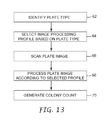
- FIG. 13 is a flow diagram illustrating a process for image processing profile selection in biological scanner.
- FIG. 14 is a flow diagram illustrating a process for image processing profile selection in a biological scanner involving detection of a plate type indicator.
- FIG. 15 is a flow diagram illustrating a process for image processing profile selection in a biological scanner involving extraction of a plate type indicator from a scanned plate image.
- FIG. 16 is a flow diagram illustrating a process that permits a user to override an automatic plate type identification by a biological scanner.
- the invention is directed to a biological scanner for biological growth plates.
- a biological growth plate can be presented to the biological scanner, which then generates an image of the plate and performs an analysis of the image to detect biological growth.
- the scanner may count or otherwise quantify an amount of biological agents that appear in the image, such as a number of bacteria colonies.
- the biological scanner automates the analysis of biological growth plates, thereby improving such analysis and reducing the possibility of human error.
- a biological scanner in accordance with the invention, also automates selection of image processing profiles to scan different types of biological growth plates and analyze plate images.
- the scanner automatically identifies the type of plate to be scanned by the scanner, and then selects one of the image processing profiles appropriate for the identified plate type.
- the image processing profiles may specify image capture conditions, image analysis criteria or a combination of both for different types of biological growth plates.
- the image processing profile may specify illumination intensities, durations, and colors for illumination of particular plate types for image capture.
- the image capture conditions also may include camera gain, resolution, aperture, and exposure time.
- the image processing profiles may specify different color, shape, size and proximity criteria in counting different types of bacterial colonies within a captured image to promote accuracy in the analytical result, e.g., a count.
- the image processing profiles may pertain both to image capture and analysis. Accuracy is critical in both food and laboratory sample test environments. For food safety, in particular, accurate results permit verification of sanitation at critical control points throughout the food processing operation, including line production, equipment and environmental testing.
- the scanner may identify the plate type by reference to a variety of machine-readable plate type indicators, such as optically or magnetically readable marks, carried on the plate. Accordingly, the invention also contemplates biological growth plates carrying a particular indicator that permits plate type identification. In addition, the invention may eliminate or reduce reliance on human judgment in making plate type identifications, thereby reducing the potential for human error and resulting inaccuracy in colony counts or other analyses.
- the invention may be useful with a variety of biological growth plates.
- the invention may be useful with different plate-like devices for growing biological agents to enable detection or enumeration of the agents, such as thin-film culture plate devices, Petri dish culture plate devices, and the like.
- the term “biological growth plate” will be used broadly herein to refer to a medium suitable for growth of biological agents to permit detection and enumeration of the agents by a scanner.
- the biological growth plate can be housed in a cassette that supports multiple plates, e.g., as described in U.S. Pat. No. 5,573,950 to Graessle et al.
- FIG. 1 is a perspective view of an exemplary biological scanner 10 .
- biological scanner 10 includes a scanner unit 12 having a drawer 14 that receives a biological growth plate (not shown in FIG. 1 ).
- Drawer 14 moves the biological growth plate into biological scanner 10 for scanning and analysis.
- Scanner 10 may incorporate features that permit automated plate type identification, and automated selection of image processing profiles based on plate type, in accordance with the invention.
- Biological scanner 10 also may include a display screen 16 to display the progress or results of analysis of the biological growth plate to a user. Alternatively or additionally, display screen may present to a user an image of the growth plate scanned by biological scanner 10 . The displayed image may be optically magnified or digitally scaled upward.
- a mounting platform 18 defines an ejection slot 20 through which the growth plate can be ejected following image capture by biological scanner 10 .
- biological scanner 10 may have a two-part design in which scanner unit 12 is mounted on mounting platform 18 . The two-part design is depicted in FIG. 1 for purposes of example, and is not intended to be required by or limiting of the inventions described herein.
- Scanner unit 12 houses an imaging device for scanning the biological growth plate and generating an image.
- the imaging device may take the form of a line scanner or an area scanner, which ordinarily will be provided in combination with an illumination system to provide front and/or back illumination of the biological growth plate.
- scanner unit 12 may house processing hardware that performs analysis of the scanned image, e.g., in order to determine the number or amount of biological agents in the growth plate. For example, upon presentation of the biological growth plate via drawer 14 , the plate may be positioned adjacent an optical platen for scanning.
- mounting platform 18 may house a conveyor that ejects the growth plate from biological scanner 10 via ejection slot 20 .
- a horizontal conveyor such as a moving belt, ejects the medium via slot 20 .
- FIG. 2 is another perspective view of biological scanner 10 .
- drawer 14 extends outward from biological scanner 10 to receive a biological growth plate 22 .
- a biological growth plate 22 may be placed on a platform 24 provided within drawer 14 .
- platform 24 may include positioning actuators such as cam levers to elevate the platform for precise positioning of growth plate 22 within biological scanner 10 .
- drawer 14 retracts into scanner unit 12 to place the biological growth plate in a scanning position, i.e., a position at which the biological growth medium is optically scanned.
- FIGS. 3 and 4 are top views of an exemplary biological growth plate 22 .
- a suitable growth plate 22 may comprise biological growth plates sold by 3M under the trade name PETRIFILM plates.
- biological growth plate 22 may comprise other biological growth media for growing particular bacteria or other biological agents.
- biological growth plate 22 carries plate type indicator 28 to facilitate automated identification of the type of biological media associated with the growth plate.
- Plate type indicator 28 presents an encoded pattern that is machine-readable.
- plate type indicator 28 takes the form of an optically readable pattern.
- FIGS. 3 and 4 depict a four-square pattern of light and dark quadrants formed in a corner margin of biological growth plate 22 .
- plate type indicator 28 defines a two-dimensional grid of cells modulated between black and white to form an encoded pattern.
- optical patterns such as characters, bar codes, two-dimensional bar codes, optical gratings, holograms, phosphorous inks and the like are conceivable.
- plate type indicator 28 may take the form of patterns that are readable by magnetic or radio frequency techniques.
- plate type indicator 28 may take the form of apertures, slots, surface contours, or the like that are readable by optical or mechanical techniques. In each case, plate type indicator 28 carries information sufficient to enable automated identification of the type of biological growth plate 22 by biological scanner 10 . Plate type indicator 28 will be described in greater detail below.
- Biological growth plates may facilitate the rapid growth and detection and enumeration of bacteria or other biological agents including, for example, aerobic bacteria, E. coli , coliform, enterobacteriaceae, yeast, mold, Staphylococcus aureus, listeria , and campylobacter , and the like.
- the use of PETRIFILM plates, or other growth media, can simplify bacterial testing of food samples.
- biological scanner 10 can further simplify such testing by providing automated plate type detection, and automated selection of image processing profiles based on the detected plate type to illuminate and/or analyze biological growth plate 22 , e.g., by counting bacterial colonies on an image of the plate.
- biological growth plate 22 defines a growth area 26 .
- a determination of whether a given sample being tested in plate 22 is acceptable, in terms of bacterial colony counts, may depend on the number of bacterial colonies per unit area. Accordingly, scanner 10 may quantify the amount of bacterial colonies per unit area on plate 22 , and may compare the amount, or “count,” to a threshold.
- the surface of biological growth plate 22 may contain one or more growth enhancing agents designed to facilitate the rapid growth of one or more types of bacteria or other biological agents.
- plate 22 After placing a sample of the material being tested, typically in liquid form, on the surface of biological growth plate 22 within growth area 26 , plate 22 can be inserted into an incubation chamber (not shown). In the incubation chamber, bacterial colonies or other biological agents being grown by growth plate 22 manifest themselves, as shown in biological growth plate 22 of FIG. 4 .
- the colonies, represented by various dots 30 on biological growth plate 22 in FIG. 4 may appear in different colors on plate 22 , facilitating automated detection and enumeration of bacterial colonies by scanner 10 .
- FIGS. 5A-5D are diagrams illustrating exemplary plate type indicator 28 carried by a biological growth plate 22 for image processing profile selection.
- plate type indicator 28 may take the form of patterns, marks, apertures, surface contours and the like, which permit optical or mechanical readability. For example, different optical patterns can be read by optical decoders, bar code scanners, optical character recognition (OCR) processors or the like. In the case of apertures or contours, mechanical styli may interact with the apertures or contours to detect different patterns and produce an electrical signal.
- plate type indicator 28 may be magnetically encoded stripes or markers or carry radio frequency identifications to permit magnetic or radio frequency readability.
- Optically readable patterns may be formed by printing or deposition of ink on the surface of biological growth plate 22 , e.g., outside of growth area 26 .
- Apertures or surface contour patterns can be formed in biological growth plate 22 by punches, stamps, embossers, die cutters and the like.
- a magnetic stripe or radio frequency identification may be affixed to the surface of biological growth plate 22 , e.g., by adhesive or lamination techniques.
- a magnetic or radio frequency indicator need not be carried on the surface of biological growth plate 22 , but may be interposed between layers of the growth plate in the event the growth plate has a multi-layer structure.
- the various plate type indicators 28 may be formed at the factory to identify the type of biological growth plate 22 .
- plate type indicator 28 may further include information that identifies a particular manufacturer, lot number, expiration date, security authorization, and the like. Such additional items of information may be important in verifying quality and suitability of biological growth plate 22 for used in biological scanner 10 .
- one or more manufacturers may be specifically validated, e.g., on the basis of plate production quality and plate performance criteria, to provide biological growth plates 22 for use in biological scanner 10 .
- biological scanner 10 may be configured to reject biological growth plates 22 that, according to plate type indicator 28 , are not associated with validated manufacturers.
- plate type indicator 28 may carry security information, such as serial number codes or the like, that serve to authenticate biological growth plate 22 and prevent fraudulent introduction of unauthorized growth plates, e.g., to thwart the food inspection or laboratory analysis process.
- security information such as serial number codes or the like
- biological growth plate 22 may carry one or more indicator patterns, in addition to plate type indicator 28 , which serve different security and quality assurance purposes.
- plate type indicator 28 may benefit from a variety of security mechanisms.
- a printed plated type indicator 28 may be printed with a particular phosphorous ink so that it can be conspicuously identified according to the wavelength of light emitted by the indicator when it is scanned.
- plate type indicators 28 may take the form of more complex patterns that carry encryption keys to unlock biological scanner 10 for operation. In this case, processor 34 in biological scanner 10 ( FIG. 6 ) would perform decryption of the pattern in order to proceed with image processing.
- plate type indicator 28 takes the form of a four-square pattern having four quadrants 29 , 31 , 33 , 35 that can be either dark or light, permitting ready optical processing.
- plate type indicator 28 has four light quadrants, and may identify a first type of biological growth plate 22 .
- plate type indicator 28 include one black quadrant, two black quadrants, and four black quadrants, respectively. Selection of the number and position of the black quadrants permits up to sixteen (2 4 ) different encoded patterns to be formed and, accordingly, up to sixteen different plate types to be identified by machine-readable plate type indicator 28 .
- different encoded patterns could represent Aerobic Count, Coliform, E. Coli, Staphylococcus aureus , Yeast and Mold, and other plate type designations.
- plate type indicator 28 may be subject to wide variation, the 4-square pattern shown in FIGS. 5A-5D provides one type of pattern that is relatively simple and easy to identify using optical pattern recognition techniques, i.e., machine vision.
- plate type indicator 28 may be scanned by a dedicated optical code reader, such as a bar code reader or custom reader. In this case, plate type indicator 28 can be scanned prior to or in parallel with scanning of biological growth plate 22 , but before processing of the growth plate image.
- plate type indicator 28 may be captured in a scanned image of biological growth plate 22 , and then extracted for image processing to identify the plate type. In this case, the plate type can be identified before further image processing of the scanned growth plate image.
- FIG. 6 is a block diagram illustrating internal operation of biological scanner 10 .
- a biological growth plate 22 is positioned within biological scanner 10 on a platform (not shown in FIG. 6 ).
- the platform places biological growth plate 22 at a desired focal plane of an imaging device 32 .
- Imaging device 32 may include illumination hardware for top and back illumination of growth plate 22 , as well as a line or area scanner that captures an image of the surface of growth plate 22 .
- Imaging device 32 may apply standard image capture conditions, or a user may specify image capture conditions. Alternatively, as will be described below, scanner 10 may automatically control image capture conditions based on an image processing profile that corresponds to a plate type.
- imaging device 32 may take the form of a two-dimensional camera, although line scanners can be used in configurations in which either the canner or biological growth plate 22 is translated relative to the other.
- image device 32 captures an image of biological growth plate 22 , or at least a growth region within the biological growth plate.
- a processor 34 controls the operation of imaging device 32 . In operation, processor 34 controls imaging device 32 to capture an image of biological growth plate 22 .
- Processor 34 receives image data representing the scanned image from imaging device 32 , and extracts or segregates a portion of the image to isolate plate type indicator 28 .
- processor 34 analyzes plate type indicator 28 to identify a plate type associated with biological growth plate 22 .
- Processor 34 then retrieves an image processing profile from image processing profile memory 36 .
- the image processing profile corresponds to the detected plate type.
- Processor 34 may take the form of a microprocessor, digital signal processor, application specific integrated circuitry (ASIC), field programmable gate array (FPGA) or other integrated or discrete logic circuitry programmed or otherwise configured to provide functionality as described herein.
- processor 34 Using the image processing profile, processor 34 loads appropriate image analysis parameters and proceeds to process the scanned image of biological growth plate 22 . In this manner, processor 34 forms an image processing device in the sense that it processes the image data obtained from biological growth plate 22 .
- the image analysis parameters may vary with the image processing profile and detected plate type, and may specify particular parameters such as colony color, size, shape and proximity criteria for analysis of the scanned image.
- the color of surrounding nutrient media may be an indicator of high colony counts.
- color may be an indicator of the type of organism.
- Adjacent objects, such as gas bubbles also may be an indicator of the type of organism. Accordingly, a variety of image processing criteria and associated parameters may be specified for various plate types. The criteria may differ according to the type of plate 22 to be analyzed, and may significantly affect colony count or other analytical results produced by biological scanner 10 .
- processor 34 Upon selection of the appropriate image processing parameters, processor 34 processes the scanned image and produces an analytical result, such as a colony count, which is presented to a user via display 16 .
- Processor 34 also may store the analytical result in memory, such as count data memory 38 , for later retrieval from scanner 10 .
- the data stored in count data memory 38 may be retrieved, for example, by a host computer that communicates with biological scanner 10 via a communication port 40 , e.g., a universal serial bus (USB) port.
- the host computer may compile analytical results for a series of biological growth plates 22 presented to biological scanner 10 for analysis.
- Automated selection of image processing profiles within biological scanner 10 can provide a convenient and accurate technique for selecting the appropriate image processing profile.
- Automated selection of image processing profiles can promote the accuracy of bacterial colony counts and other analytical procedures.
- automatic image processing profile selection can avoid the need for a technician to visually identify and manually enter the plate type. In this manner, plate identification errors sometimes associated with human intervention can be avoided. Consequently, the combination of a scanner 10 and a biological growth plate 22 that carries plate type indicator 28 can promote efficiency and workflow of laboratory technicians while enhancing analytical accuracy and, in the end, food safety and human health.
- FIG. 7 is a block diagram illustrating another biological scanner 10 ′ configured for automated image processing profile selection.
- Biological scanner 10 ′ conforms substantially to biological scanner 10 of FIG. 6 , but further includes a code reader 42 .
- code reader 42 serves as a dedicated reader to obtain plate type information.
- code reader 42 may take the form of a dedicated optical reader, bar code reader, magnetic reader, radio frequency or mechanical reader.
- code reader 42 serves to identify the plate type from plate type indicator 28 and communicates the plate type to processor 34 .
- Processor 34 selects an image processing profile from memory 36 based on the identified plate type. Imaging device 32 scans biological growth plate 22 and provides the image date to processor 34 .
- Processor 34 then applies the image processing parameters specified by the retrieved image processing profile to process the image and produce an analytical result such as colony count.
- processor 34 applies the appropriate image processing profile on an automated basis in view of the automatically identified plate type, offering enhanced accuracy, efficiency and convenience to the user.
- the invention eliminates the need for the user to enter the plate type identification manually, and reduces the likelihood of analytical error due to erroneous human input.
- FIG. 8 is a block diagram illustrating biological scanner 10 of FIG. 6 in greater detail and depicting plate illumination hardware.
- biological scanner 10 may include a front illumination system 44 and a back illumination system 46 .
- Front illumination system 44 illuminates a front side of biological growth plate 22
- back illumination system 46 illuminates a back side of the biological growth plate.
- Front and bottom illumination systems 44 , 46 may produce different illumination intensities, colors and durations on a selective basis.
- processor 34 controls front and bottom illumination systems 44 , 46 to expose biological growth plate 22 to different illumination colors.
- Front and back illumination systems 44 , 46 may incorporate LEDs as illumination sources. The LEDs can be readily controlled by processor 34 and appropriate driver circuitry to achieve desired illumination intensities and durations.
- processor 34 may control camera 43 to capture images of biological growth plate 22 during illumination with the different colors.
- processor 34 may provide coordinated control of illumination systems 44 , 46 and camera 43 to capture one or more images of biological growth plate 22 .
- Camera 43 captures one or more images of biological growth plate 22 during illumination by front illumination system 44 , back illumination system 46 or both, and may store the images in an image memory 47 .
- processor 34 may control camera gain, resolution, aperture, exposure time or the like in response to the image capture conditions specified by the image processing profile.
- processor 34 uses the stored images to perform image analysis according to the image analysis criteria specified by the image processing profile. In particular, processor 34 then may analyze the individual images or combine the multiple images to form a composite image. In some embodiments, for example, processor 34 may control illumination systems 44 , 46 to capture red, green and blue images of biological growth plate 22 and analyze the images individually or as a composite multi-color image.
- Some types of biological growth plates 22 may require illumination with a particular color, intensity and duration.
- some biological growth plates 22 may require only front or back illumination, but not both.
- an aerobic count plate may require only front illumination as well as illumination by only a single color such as red.
- an E. coli /Coliform plate may require only back illumination and a combination of red and blue illumination.
- particular intensity levels and durations may be appropriate, as well as different camera gain, resolution, aperture, and exposure time.
- processor 34 may control illumination and camera conditions in response to image capture conditions specified by an image processing profile.
- scanner 10 can be configured to select not only image analysis criteria based on plate type, but also image capture conditions to be applied to capture the image.
- scanner 10 may apply techniques similar to those described with respect to FIGS. 6 and 7 .
- a dedicated code reader may be provided to identify plate type in advance of illumination for image capture.
- processor 34 selects a corresponding image processing profile from image processing profile memory 36 and controls illumination according to image capture conditions specified in the image processing profile.
- scanner 10 may be configured to apply machine vision techniques to identify the plate type from a capture image, as discussed with respect to FIG. 6 .
- scanner 10 may apply a set of default illumination conditions to capture an initial image of biological growth plate 22 , or a portion thereof, for purposes of analyzing plate type indicator 28 for plate type identification.
- processor 34 may select a corresponding image processing profile and apply the specified image capture conditions to capture an image for analysis of biological growth.
- processor 34 may access an image capture profile specifying image capture conditions such as illumination colors, intensities and durations. Then, for analysis of a captured image, processor 34 may access a separate image analysis profile specifying image analysis criteria such as color, shape, size and proximity.
- processor 34 may present a preliminary plate type identification to a user via display 16 .
- processor 34 may permit the user to either confirm or reject the automatically identified plate type before proceeding with image capture or analysis using a corresponding image processing profile.
- the user may confirm or reject a preliminary plate type identification, for example, by actuating a pointing device or depressing regions of a touch screen. If the user believes that the automatically detected plate type identification is in error, processor 34 may permit the user to change the plate type identification.
- FIG. 9 is a sample display content produced on display 16 by biological scanner 10 upon plate type detection.
- display 16 presents a preliminary plate type identification, i.e., a plate type identification automatically made by processor 34 .
- display 16 presents two touch screen regions 52 , 54 that accept user input to indicate whether the preliminary plate type identification is confirmed or rejected, respectively, by the user.
- FIG. 10 is sample content produced by display 16 of biological scanner 10 upon rejection of the automated plate type detection by the user.
- processor 34 may drive display 16 to present an “ENTER PLATE TYPE” dialog by which the user may choose the correct plate type identification, as shown in FIG. 10 .
- Display 16 may present a vertical scroll bar menu 56 that permits the user to choose an alternative plate type identification, e.g., by depressing an appropriate touch screen region.
- processor 34 may select an alternative image processing profile containing, for example, image capture conditions, image analysis criteria, or both.
- FIG. 11 is sample content produced by display 16 of biological scanner 10 upon determination of a colony count.
- FIG. 12 is sample content produced by display 16 of biological scanner 10 upon determination of a colony count and including an image of a scanned plate.
- display 16 presents information similar to that shown in FIG. 11 , but further includes a representation 60 of the actual image scanned by biological scanner 10 from the surface of biological growth plate 22 .
- the user can view both the analytical result, such as count 58 , and a representation 60 of the scanned image.
- image representation 60 may present a sufficient amount of detail to permit the user to verify the automatically determined count.
- image representation 60 may be a lower resolution representation.
- FIG. 13 is a flow diagram illustrating a process for image processing profile selection in biological scanner 10 .
- the process may involve identifying a plate type ( 62 ) for a biological growth plate 22 presented to scanner 10 .
- the process may further involve selection of an image processing profile based on the plate type ( 64 ), either before or after scanning a plate image ( 66 ). If the image processing profile specifies image capture conditions, the image processing profile should be selected prior to scanning the plate image so that illumination conditions, camera properties or both may be controlled.
- the process further involves processing the plate image to produce an analytical result ( 68 ). In particular, the process may generate a bacterial colony count ( 70 ).
- the plate type is identified before the plate image is scanned.
- FIG. 14 is a flow diagram illustrating a process for image processing profile selection in a biological scanner involving detection of a plate type indicator.
- the process involves reading the plate type indicator carried by a biological growth plate ( 72 ), e.g., with a dedicate plate type indicator reader such as an optical reader, bar code reader, magnetic reader, radio frequency reader, mechanical reader or the like.
- a dedicate plate type indicator reader such as an optical reader, bar code reader, magnetic reader, radio frequency reader, mechanical reader or the like.
- the process involves selecting an image processing profile based on the detected plate type ( 76 ).
- the process involves scanning an image of the biological growth plate ( 78 ), and processing the plate image according to parameters specified by the selected image processing profile ( 80 ).
- the process then produces an analytical result such as a colony count ( 81 ).
- FIG. 15 is a flow diagram illustrating a process for image processing profile selection in a biological scanner involving extraction of plate type indicator from a scanned plate image.
- the process involves scanning an image of a biological growth plate ( 82 ), and extracting a plate type indicator area from the scanned image ( 84 ). The process further involves processing the extracted plate type indicator area ( 86 ) to determine the plate type ( 88 ). Upon determination of the plate type based on the plate type indicator ( 88 ), the process involves selecting an image processing profile based on the detected plate type ( 90 ).
- the process then may involve scanning the plate again ( 91 ), e.g., using image capture conditions specified by the selected image processing profile, and processing the plate image according to image analysis criteria specified by the selected image processing profile ( 92 ). The process then produces an analytical result such as a colony count ( 94 ).
- FIG. 16 is a flow diagram illustrating a process that permits a user to override an automatic plate type identification by a biological scanner.
- the process involves automatically determining a plate type associated with a biological growth plate 22 ( 96 ), and presenting the determined plate type to a user via display 16 ( 98 ).
- the process further involves accepting user input to accept or reject the automatically determined plate type ( 100 ), e.g., via touch screen input. If the plate type is not accepted by the user, the process involves accepting user input to receive a plate type from the user ( 102 ).
- the process Upon entry of a plate type by the user ( 102 ) or acceptance of an automatically determined plate type ( 100 ), the process involves selection of an image processing profile based on the plate type ( 104 ). Using the selected image processing profile, the process scans a plate image ( 105 ), processes the plate image ( 106 ) and generates a colony count ( 108 ) or some other desired analytical result.
- processor 34 executes instructions that may be stored on a computer-readable medium to carry out the processes described herein.
- the computer-readable medium may comprise random access memory (RAM) such as synchronous dynamic random access memory (SDRAM), read-only memory (ROM), non-volatile random access memory (NVRAM), electrically erasable programmable read-only memory (EEPROM), FLASH memory, magnetic or optical data storage media, and the like.
- RAM random access memory
- SDRAM synchronous dynamic random access memory
- ROM read-only memory
- NVRAM non-volatile random access memory
- EEPROM electrically erasable programmable read-only memory
- FLASH memory magnetic or optical data storage media, and the like.
Abstract
Description
Claims (4)
Priority Applications (1)
| Application Number | Priority Date | Filing Date | Title |
|---|---|---|---|
| US12/877,254 US7901933B2 (en) | 2002-11-27 | 2010-09-08 | Methods of processing a biological growth plate in a biological growth plate scanner |
Applications Claiming Priority (3)
| Application Number | Priority Date | Filing Date | Title |
|---|---|---|---|
| US10/306,579 US7298885B2 (en) | 2002-11-27 | 2002-11-27 | Biological growth plate scanner with automated image processing profile selection |
| US11/934,175 US20080064089A1 (en) | 2002-11-27 | 2007-11-02 | Biological growth plate scanner with automated image processing profile selection |
| US12/877,254 US7901933B2 (en) | 2002-11-27 | 2010-09-08 | Methods of processing a biological growth plate in a biological growth plate scanner |
Related Parent Applications (1)
| Application Number | Title | Priority Date | Filing Date |
|---|---|---|---|
| US11/934,175 Continuation US20080064089A1 (en) | 2002-11-27 | 2007-11-02 | Biological growth plate scanner with automated image processing profile selection |
Publications (2)
| Publication Number | Publication Date |
|---|---|
| US20100330610A1 US20100330610A1 (en) | 2010-12-30 |
| US7901933B2 true US7901933B2 (en) | 2011-03-08 |
Family
ID=32325729
Family Applications (3)
| Application Number | Title | Priority Date | Filing Date |
|---|---|---|---|
| US10/306,579 Expired - Lifetime US7298885B2 (en) | 2002-11-27 | 2002-11-27 | Biological growth plate scanner with automated image processing profile selection |
| US11/934,175 Abandoned US20080064089A1 (en) | 2002-11-27 | 2007-11-02 | Biological growth plate scanner with automated image processing profile selection |
| US12/877,254 Expired - Fee Related US7901933B2 (en) | 2002-11-27 | 2010-09-08 | Methods of processing a biological growth plate in a biological growth plate scanner |
Family Applications Before (2)
| Application Number | Title | Priority Date | Filing Date |
|---|---|---|---|
| US10/306,579 Expired - Lifetime US7298885B2 (en) | 2002-11-27 | 2002-11-27 | Biological growth plate scanner with automated image processing profile selection |
| US11/934,175 Abandoned US20080064089A1 (en) | 2002-11-27 | 2007-11-02 | Biological growth plate scanner with automated image processing profile selection |
Country Status (11)
| Country | Link |
|---|---|
| US (3) | US7298885B2 (en) |
| EP (2) | EP1573654B1 (en) |
| JP (2) | JP2006507837A (en) |
| KR (1) | KR101120829B1 (en) |
| CN (2) | CN100342389C (en) |
| AT (1) | ATE527618T1 (en) |
| AU (1) | AU2003295804B2 (en) |
| BR (1) | BR0316406A (en) |
| CA (1) | CA2504616A1 (en) |
| MX (1) | MXPA05005378A (en) |
| WO (1) | WO2004051554A1 (en) |
Cited By (13)
| Publication number | Priority date | Publication date | Assignee | Title |
|---|---|---|---|---|
| US20080144916A1 (en) * | 2002-11-27 | 2008-06-19 | 3M Innovative Properties Company | Loading and ejection systems for biological growth plate scanner |
| US20090135603A1 (en) * | 2002-11-27 | 2009-05-28 | 3M Innovative Properties Company | Back side plate illumination for biological growth plate scanner |
| US20090240643A1 (en) * | 2008-03-18 | 2009-09-24 | Yahoo! Inc. | System and method for detecting human judgment drift and variation control |
| US20110153220A1 (en) * | 2008-03-04 | 2011-06-23 | Bolea Phillip A | Processing of biological growth media based on measured manufacturing characteristics |
| US20110158499A1 (en) * | 2008-03-04 | 2011-06-30 | Bolea Phillip A | Information management in automated processing of biological growth media |
| US20110206263A1 (en) * | 2003-09-05 | 2011-08-25 | 3M Innovative Properties Company | Counting biological agents on biological growth plates |
| US8094916B2 (en) | 2002-11-27 | 2012-01-10 | 3M Innovative Properties Company | Biological growth plate scanner |
| US8831313B2 (en) | 2010-06-23 | 2014-09-09 | Kabushiki Kaisha N-Tech | Method for detecting microorganisms, device for detecting microorganisms and program |
| US20180150680A1 (en) * | 2012-12-20 | 2018-05-31 | 3M Innovative Properties Company | Method of detecting gas-producing microbial colonies |
| US10407654B1 (en) | 2014-03-21 | 2019-09-10 | Charm Sciences, Inc. | Growth plate devices, kits and assemblies |
| US10495563B1 (en) | 2016-04-28 | 2019-12-03 | Charm Sciences, Inc. | Plate reader observation methods and operation |
| US10563164B1 (en) | 2015-10-08 | 2020-02-18 | Charm Sciences, Inc. | Plate reader |
| US10988720B1 (en) | 2015-11-09 | 2021-04-27 | Charm Sciences, Inc. | Peel plate assembly |
Families Citing this family (48)
| Publication number | Priority date | Publication date | Assignee | Title |
|---|---|---|---|---|
| US7298885B2 (en) * | 2002-11-27 | 2007-11-20 | 3M Innovative Properties Company | Biological growth plate scanner with automated image processing profile selection |
| US7319031B2 (en) * | 2002-11-27 | 2008-01-15 | 3M Innovative Properties Company | Mounting platform for biological growth plate scanner |
| US7496225B2 (en) * | 2003-09-04 | 2009-02-24 | 3M Innovative Properties Company | Biological growth plate scanner with automated intake |
| US20080238627A1 (en) * | 2005-03-22 | 2008-10-02 | Applera Corporation | Sample carrier device incorporating radio frequency identification, and method |
| US7187286B2 (en) | 2004-03-19 | 2007-03-06 | Applera Corporation | Methods and systems for using RFID in biological field |
| JP4713261B2 (en) * | 2005-07-20 | 2011-06-29 | 株式会社カネカ | Cell culture equipment |
| JP4821279B2 (en) * | 2005-11-11 | 2011-11-24 | 株式会社ニコン | Incubator |
| EP1882948A2 (en) * | 2006-07-28 | 2008-01-30 | Qiagen GmbH | Sample processing device |
| AU2008275188B2 (en) | 2007-07-09 | 2012-01-19 | 3M Innovative Properties Company | Modular system and method for detecting microorganisms |
| CN103114038B (en) | 2007-11-20 | 2015-02-11 | 3M创新有限公司 | Sample cell and kit |
| JP5420566B2 (en) | 2007-12-21 | 2014-02-19 | スリーエム イノベイティブ プロパティズ カンパニー | Microbial system and fluid sample analysis method |
| BRPI0907064A2 (en) * | 2008-03-26 | 2015-07-07 | 3M Innovative Properties Co | "methods and systems" |
| CN102256631B (en) * | 2008-10-17 | 2014-07-23 | 3M创新有限公司 | Biological sterilization indicator, system, and methods of using same |
| JP5480559B2 (en) * | 2008-10-30 | 2014-04-23 | シスメックス株式会社 | Bacteria analyzer, bacteria analysis method, and computer program |
| EP2199956A1 (en) * | 2008-12-18 | 2010-06-23 | Siemens Aktiengesellschaft | Method and system for managing results of an analysis process on objects handled along a technical process line |
| JP5727383B2 (en) | 2008-12-31 | 2015-06-03 | スリーエム イノベイティブ プロパティズ カンパニー | Coliform bacteria detection process and kit for use in this process |
| WO2011055791A1 (en) * | 2009-11-05 | 2011-05-12 | 株式会社日立ハイテクノロジーズ | Device for harvesting bacterial colony and method therefor |
| CN102713617B (en) | 2009-12-30 | 2015-11-25 | 3M创新有限公司 | Produce the rapid detection of the mould of glucose oxidase |
| BR112012016132A2 (en) | 2009-12-30 | 2020-09-08 | 3M Innovative Properties Company | microbial detection article |
| FR2964215B1 (en) * | 2010-08-25 | 2013-06-14 | Intelligence Artificielle Applic | USE OF A DATAMATRIX CODE AND PRINTING METHOD |
| JP5780732B2 (en) * | 2010-10-04 | 2015-09-16 | マイクロバイオ株式会社 | Colony detection method, colony detection system, and colony detection program |
| CN102024259B (en) * | 2010-12-24 | 2012-06-20 | 刘安安 | Bacterial colony automatic detection method |
| JP5849976B2 (en) * | 2012-03-30 | 2016-02-03 | カシオ計算機株式会社 | Social network service system and image display method |
| JP5998744B2 (en) * | 2012-08-23 | 2016-09-28 | 大日本印刷株式会社 | Colony detection device, medium information registration system, hygiene management system, and program |
| EP2731051A1 (en) * | 2012-11-07 | 2014-05-14 | bioMérieux | Bio-imaging method |
| EP2935612B1 (en) | 2012-12-20 | 2018-01-31 | 3M Innovative Properties Company | Method of differentiating microbial colonies in an image |
| JP6070172B2 (en) * | 2012-12-25 | 2017-02-01 | 大日本印刷株式会社 | Medium information registration system, program and hygiene management system |
| JP2014135948A (en) * | 2013-01-18 | 2014-07-28 | Dainippon Printing Co Ltd | Culture medium information registration system, colony detection device, program, and health management system |
| JP2014135949A (en) * | 2013-01-18 | 2014-07-28 | Dainippon Printing Co Ltd | Culture medium information registration system, culture medium image analysis device, program, and health management system |
| JP2014140335A (en) * | 2013-01-24 | 2014-08-07 | Dainippon Printing Co Ltd | Medium image analyzing device, medium information registration system and program, and hygiene management system |
| US8921067B2 (en) | 2013-02-04 | 2014-12-30 | 3M Innovative Properties Company | Method and culture device for detecting yeasts and molds |
| JP6281231B2 (en) * | 2013-10-08 | 2018-02-21 | 大日本印刷株式会社 | Medium information registration system and colony detection system |
| JP2017506916A (en) | 2014-03-07 | 2017-03-16 | スリーエム イノベイティブ プロパティズ カンパニー | Articles and methods for detecting aerobic bacteria |
| JP5904295B2 (en) * | 2014-03-31 | 2016-04-13 | 大日本印刷株式会社 | Colony detection system, colony detection method, and program |
| JP2016158577A (en) * | 2015-03-03 | 2016-09-05 | 富士フイルム株式会社 | Cell colony detection device, cell colony detection method, and program |
| CN108350409B (en) * | 2015-12-28 | 2022-06-14 | 日本理化学开发公司 | Viable/dead bacteria state determination device and viable/dead bacteria state determination method using same |
| CN109688837B (en) * | 2016-09-20 | 2022-08-09 | 日本水产株式会社 | Method for producing caviar from which foreign matter has been removed and device for producing caviar from which foreign matter has been removed |
| WO2018061131A1 (en) * | 2016-09-28 | 2018-04-05 | オリンパス株式会社 | Cell status assessment device |
| CA3042681A1 (en) * | 2016-11-04 | 2018-05-11 | Becton, Dickinson And Company | System and method for selecting colonies |
| ES2907811T3 (en) * | 2016-11-10 | 2022-04-26 | Becton Dickinson Co | Timeline system to monitor a culture medium protocol |
| CN107644210B (en) * | 2017-09-22 | 2020-05-12 | 哈尔滨工业大学(威海) | Image processing-based microorganism quantity estimation method |
| CN108251270B (en) * | 2018-01-16 | 2022-11-15 | 上海睿度光电科技有限公司 | Equipment for covering cells with quantitative solution |
| JP7317026B2 (en) | 2018-01-30 | 2023-07-28 | ライフ テクノロジーズ コーポレーション | Instruments, devices and consumables for use in smart molecular analysis system workflows |
| FR3080211B1 (en) | 2018-04-16 | 2020-05-08 | Pinqkerton | SYSTEM AND METHOD FOR ANALYZING TEST DEVICES |
| KR102043670B1 (en) * | 2018-04-27 | 2019-11-12 | 이상인 | Automatic microorganism management incubator system |
| CN109975290A (en) * | 2018-11-30 | 2019-07-05 | 军事科学院军事医学研究院环境医学与作业医学研究所 | A kind of Bacteria Detection quick analytic instrument |
| JP6832970B2 (en) * | 2019-02-27 | 2021-02-24 | 富士フイルム株式会社 | Cell colony detectors and methods and programs |
| JP7259732B2 (en) * | 2019-12-23 | 2023-04-18 | 横河電機株式会社 | Distribution server, method and program |
Citations (95)
| Publication number | Priority date | Publication date | Assignee | Title |
|---|---|---|---|---|
| US3493772A (en) | 1967-05-29 | 1970-02-03 | Palo Alto Medical Research Fou | Bacterial counting machine and method |
| US3745090A (en) | 1970-08-04 | 1973-07-10 | Nasa | Method of detecting and counting bacteria in body fluids |
| US3811036A (en) | 1972-09-19 | 1974-05-14 | Artek Syst Corp | Micro-biological colony counter |
| US3962040A (en) | 1974-03-14 | 1976-06-08 | The United States Of America As Represented By The Department Of Health, Education And Welfare | Method and apparatus for plating and counting aerobic bacteria |
| US4118280A (en) | 1976-05-03 | 1978-10-03 | Mcdonnell Douglas Corporation | Automated microbial analyzer |
| US4146775A (en) | 1976-09-16 | 1979-03-27 | Armstrong Machine Works | Automatic control system for an electrode-type air humidifier |
| US4160601A (en) | 1978-02-24 | 1979-07-10 | Nasa | Biocontamination and particulate detection system |
| EP0008826A2 (en) | 1978-08-18 | 1980-03-19 | Metrofoss K/S | Method and apparatus for counting bacteria in a bacteria-containing suspension, method for fluorescence staining of bacteria, and method for separating bacteria from a sample |
| US4353988A (en) | 1980-11-12 | 1982-10-12 | Couse Nancy L | Grid for use in counting colonies of bacteria present in discrete areas of a spiral deposition pattern |
| EP0088601A1 (en) | 1982-03-04 | 1983-09-14 | EASTMAN KODAK COMPANY (a New Jersey corporation) | Analyzer for chemical analysis of a liquid |
| JPS6083597A (en) | 1983-10-10 | 1985-05-11 | Hitachi Electronics Eng Co Ltd | Method for testing colony |
| US4563096A (en) * | 1980-05-09 | 1986-01-07 | Internationale Octrooi Maatschappij "Octropa" B.V. | Apparatus for assisting the visual assessment of test objects having multivariate visible characteristics, and its use |
| US4591567A (en) | 1982-04-21 | 1986-05-27 | California Institute Of Technology | Recombinant DNA screening system including fixed array replicator and support |
| US4637053A (en) | 1984-05-03 | 1987-01-13 | Spiral System Instruments, Inc. | Computer assisted biological assay system |
| JPS62215383A (en) | 1986-03-17 | 1987-09-22 | Datsuku Eng Kk | Apparatus for inspecting microscopic life |
| US4720463A (en) | 1985-03-01 | 1988-01-19 | Sherwood Medical Company | Automated microbiological testing apparatus |
| US4724215A (en) | 1985-02-27 | 1988-02-09 | Sherwood Medical Company | Automated microbiological testing apparatus and method |
| EP0301600A2 (en) | 1987-07-31 | 1989-02-01 | Sumitomo Electric Industries Limited | Method for detection of the presence of undesired microorganisms |
| EP0397256A2 (en) | 1989-05-09 | 1990-11-14 | Eastman Kodak Company | Analyser featuring a circular track of cartridges centered on an incubator and method of use |
| WO1991006911A1 (en) | 1989-10-23 | 1991-05-16 | Neuromedical Systems, Inc. | Automated cytological specimen classification system and method |
| EP0429030A2 (en) | 1989-11-21 | 1991-05-29 | BEHRINGWERKE Aktiengesellschaft | Incubator for microtitration plates |
| GB2249829A (en) | 1990-11-13 | 1992-05-20 | Powergen Public Limited Compan | Measurement of carbon in ash |
| US5117467A (en) | 1988-05-23 | 1992-05-26 | Toyo Jozo Co., Ltd. | Colony counting apparatus |
| EP0193385B1 (en) | 1985-02-27 | 1992-07-22 | Sherwood Medical Company | Automated microbiological testing appparatus and method |
| WO1992012233A1 (en) | 1991-01-11 | 1992-07-23 | Medical Research Council | Transfer of biological samples |
| US5202010A (en) | 1987-11-25 | 1993-04-13 | Princeton Biochemicals, Inc. | Automated capillary electrophoresis apparatus |
| EP0547709A2 (en) | 1991-12-19 | 1993-06-23 | Johnson & Johnson Clinical Diagnostics, Inc. | Test elements and method for manufacturing thereof |
| US5270173A (en) | 1987-10-06 | 1993-12-14 | Sumitomo Electric Industries, Ltd. | Method of monitoring cell culture |
| WO1994001528A1 (en) | 1992-07-13 | 1994-01-20 | Minnesota Mining And Manufacturing Company | A technique to count objects in a scanned image |
| US5290701A (en) | 1991-08-28 | 1994-03-01 | Wilkins Judd R | Microbial detection system and process |
| JPH0698220A (en) | 1992-05-29 | 1994-04-08 | Hooya Shiyotsuto Kk | Video camera |
| JPH06109545A (en) | 1992-09-30 | 1994-04-19 | Idemitsu Petrochem Co Ltd | Color tone inspecting method |
| US5364766A (en) | 1993-05-14 | 1994-11-15 | Minnesota Mining And Manufacturing Company | Culture medium for rapid count of coliform bacteria |
| US5366873A (en) | 1992-06-03 | 1994-11-22 | Gideon Eden | Device and method for use in detecting microorganisms in a sample |
| US5372485A (en) | 1992-11-14 | 1994-12-13 | Mercedes-Benz Ag | Exhaust-gas turbocharger with divided, variable guide vanes |
| US5372936A (en) | 1989-05-12 | 1994-12-13 | Avl Photoronics Corporation | Method for detecting biological activities in a specimen |
| US5375043A (en) | 1992-07-27 | 1994-12-20 | Inoue Denki Co., Inc. | Lighting unit |
| US5428690A (en) * | 1991-09-23 | 1995-06-27 | Becton Dickinson And Company | Method and apparatus for automated assay of biological specimens |
| US5448652A (en) | 1991-09-27 | 1995-09-05 | E. I. Du Pont De Nemours And Company | Adaptive display system |
| JPH07275200A (en) | 1994-04-15 | 1995-10-24 | Asahi Optical Co Ltd | Illuminator for endoscope |
| US5510246A (en) | 1993-05-14 | 1996-04-23 | Minnesota Mining And Manufacturing Company | Method for rapid quantification of microorganism growth |
| US5539517A (en) | 1993-07-22 | 1996-07-23 | Numetrix Ltd. | Method for simultaneously measuring the spectral intensity as a function of wavelength of all the pixels of a two dimensional scene |
| US5573950A (en) * | 1994-05-11 | 1996-11-12 | Minnesota Mining And Manufacturing Company | Cassette for disposable microorganism culturing media and automated scanning system |
| US5591974A (en) | 1992-09-30 | 1997-01-07 | Westinghouse Electric Corporation | Automated collection and processing of environmental samples |
| US5671290A (en) | 1991-09-27 | 1997-09-23 | E. I. Du Pont De Nemours And Company | Method and system of separately identifying clumped homogeneous objects in an image |
| US5694478A (en) | 1994-12-15 | 1997-12-02 | Minnesota Mining And Manufacturing Company | Method and apparatus for detecting and identifying microbial colonies |
| EP0819930A2 (en) | 1996-07-19 | 1998-01-21 | Bayer Ag | Method and apparatus for screening molecules according to their individual binding affinity for at least one specified ligand |
| JPH1024283A (en) | 1996-07-10 | 1998-01-27 | Meidensha Corp | Filtration obstructing microorganism monitor |
| US5721435A (en) | 1996-04-09 | 1998-02-24 | Hewlett Packard Company | Methods and apparatus for measuring optical properties of biological and chemical substances |
| US5723308A (en) | 1993-05-14 | 1998-03-03 | Minnesota Mining And Manufacturing Company | Culture medium for rapid count of coliform bacteria |
| US5744322A (en) | 1993-12-17 | 1998-04-28 | Minnesota Mining And Manufacturing Company | Automated incubating and imaging system for a disposable microorganism culturing device and method of use |
| US5747333A (en) | 1996-10-25 | 1998-05-05 | Jencons (Scientific) Limited | Culture of micro-organisms |
| US5781311A (en) | 1994-08-24 | 1998-07-14 | Nikon Corporation | Planar light source and image reading device |
| US5787189A (en) | 1994-09-20 | 1998-07-28 | Neopath, Inc. | Biological analysis system self calibration apparatus |
| US5817475A (en) | 1996-11-15 | 1998-10-06 | Giles Scientific, Inc. | Automatic microbiological testing apparatus and method |
| EP0895086A2 (en) | 1997-08-01 | 1999-02-03 | Ortho-Clinical Diagnostics, Inc. | Detecting abnormal reactions in a red blood cell agglutination |
| US5995645A (en) | 1993-08-18 | 1999-11-30 | Applied Spectral Imaging Ltd. | Method of cancer cell detection |
| US6002789A (en) | 1997-06-24 | 1999-12-14 | Pilot Industries, Inc. | Bacteria colony counter and classifier |
| US6058209A (en) | 1991-09-27 | 2000-05-02 | E. I. Du Pont De Nemours And Company | Method for resolving redundant identifications of an object |
| DE19819144C1 (en) | 1998-04-29 | 2000-06-15 | Tga Tech Geraete Und Apparateb | Microscopic investigation of implant integration into living tissues is carried out by automatically-scanning confocal laser microscope, digitizing image plane data to assemble three dimensional display |
| US6096272A (en) | 1997-05-23 | 2000-08-01 | Becton Dickinson & Company | Automated microbiological testing apparatus and methods therefor |
| US6107054A (en) * | 1998-12-31 | 2000-08-22 | Gibbs; David | Microbiological testing apparatus and method |
| JP2000270840A (en) | 1999-03-29 | 2000-10-03 | Elmex Ltd | Colony counting system and usage thereof |
| WO2001004828A1 (en) | 1999-07-13 | 2001-01-18 | Chromavision Medical Systems, Inc. | Automated detection of objects in a biological sample |
| EP1074610A1 (en) | 1997-12-02 | 2001-02-07 | Francisco Soria Melguizo, S.A. | System for analyzing images produced by bacterial reactions |
| US6243486B1 (en) | 1996-08-30 | 2001-06-05 | Marvin Weiss | System for counting colonies of micro-organisms in petri dishes and other culture media |
| US6271022B1 (en) | 1999-03-12 | 2001-08-07 | Biolog, Inc. | Device for incubating and monitoring multiwell assays |
| JP2001242082A (en) | 2000-02-29 | 2001-09-07 | Nippon Laser & Electronics Lab | Biological sample optical scanning device |
| US20010031502A1 (en) | 1998-08-26 | 2001-10-18 | Watson Robert Malcolm | Biochip detection system |
| WO2001083673A2 (en) | 2000-04-28 | 2001-11-08 | Spiral Biotech, Inc. | Method and apparatus for viewing a culture medium having visible bacterial colonies |
| US6319668B1 (en) * | 1995-04-25 | 2001-11-20 | Discovery Partners International | Method for tagging and screening molecules |
| US20020025082A1 (en) * | 2000-08-22 | 2002-02-28 | Kaushikkar Shantanu V. | System, method, and computer software product for grid alignment of multiple scanned images |
| US6418180B1 (en) | 2001-07-19 | 2002-07-09 | Marvin Weiss | Method and apparatus for counting objects having substantially uniform size |
| WO2002066961A1 (en) | 2001-02-20 | 2002-08-29 | Cytokinetics, Inc. | Method and apparatus for automated cellular bioinformatics |
| US6459994B1 (en) * | 1998-05-29 | 2002-10-01 | Oxford Glycosciences (Uk) Ltd | Methods for computer-assisted isolation of proteins |
| JP2002538440A (en) | 1999-02-26 | 2002-11-12 | ジェネラル・スキャンニング・インコーポレイテッド | Automated imaging and analysis of microarray biochips |
| US20020167161A1 (en) * | 2001-03-15 | 2002-11-14 | Butland Charles L. | Surface labelled object and method thereof |
| US6485979B1 (en) * | 1999-08-05 | 2002-11-26 | 3M Innovative Properties Company | Electronic system for tracking and monitoring articles to be sterilized and associated method |
| US6488890B1 (en) | 1999-08-05 | 2002-12-03 | 3M Innovative Properties Company | Machine readable sterilization indicator for monitoring articles to be sterilized |
| US20020191825A1 (en) * | 1996-11-29 | 2002-12-19 | Parekh Rajesh Bhikhu | Computer-assisted methods and apparatus for identification and characterization of biomolecules in a biological sample |
| US6673315B2 (en) * | 2001-06-29 | 2004-01-06 | Biomachines, Inc. | Method and apparatus for accessing a site on a biological substrate |
| US6690470B1 (en) | 1999-11-04 | 2004-02-10 | Arcturus Engineering, Inc. | Automated laser capture microdissection |
| US6711283B1 (en) | 2000-05-03 | 2004-03-23 | Aperio Technologies, Inc. | Fully automatic rapid microscope slide scanner |
| US6737266B1 (en) * | 1999-10-01 | 2004-05-18 | 3M Innovative Properties Company | Devices and methods for microorganism detection |
| US20040101954A1 (en) | 2002-11-27 | 2004-05-27 | Graessle Josef A. | Back side plate illumination for biological growth plate scanner |
| US20040102903A1 (en) | 2002-11-27 | 2004-05-27 | Graessle Josef A. | Biological growth plate scanner |
| US20040101189A1 (en) | 2002-11-27 | 2004-05-27 | 3M Innovative Properties Company | Biological growth plate scanner with automated image processing profile selection |
| US20040101952A1 (en) | 2002-11-27 | 2004-05-27 | 3M Innovative Properties Company | Loading and ejection systems for biological growth plate scanner |
| US20040101951A1 (en) | 2002-11-27 | 2004-05-27 | Albert Vent | Mounting platform for biological growth plate scanner |
| US20050053265A1 (en) | 2003-09-04 | 2005-03-10 | 3M Innovative Properties Company | Biological growth plate scanner with automated intake |
| US20050053266A1 (en) | 2003-09-05 | 2005-03-10 | Plumb Michael R. | Counting biological agents on biological growth plates |
| US20050095665A1 (en) | 1999-04-29 | 2005-05-05 | Dade Behring, Inc. | Combined rapid susceptibility assay and microorganism identification system |
| US20050222778A1 (en) | 2003-12-30 | 2005-10-06 | Compliance Software Solutions Corp. | System, method, and computer-readable medium for collection of environmental data and generation of user report for compliance with FDA requirements |
| US7057721B2 (en) | 2002-01-10 | 2006-06-06 | Chemimage Corporation | Wide field method for detecting pathogenic microorganisms |
| US20060263258A1 (en) | 2005-02-10 | 2006-11-23 | Matthew Harris | Automated biological indicator incubator |
Family Cites Families (18)
| Publication number | Priority date | Publication date | Assignee | Title |
|---|---|---|---|---|
| US6151405A (en) * | 1996-11-27 | 2000-11-21 | Chromavision Medical Systems, Inc. | System and method for cellular specimen grading |
| KR100269125B1 (en) * | 1997-10-25 | 2000-10-16 | 윤덕용 | Image post processing method and apparatus for reducing quantization effect |
| US6178205B1 (en) * | 1997-12-12 | 2001-01-23 | Vtel Corporation | Video postfiltering with motion-compensated temporal filtering and/or spatial-adaptive filtering |
| FR2786498B1 (en) | 1998-11-27 | 2002-02-08 | Intelligence Artificielle Appl | APPARATUS FOR AUTOMATICALLY READING AN ANTIBIOGRAM |
| FR2789693B1 (en) | 1999-02-16 | 2001-05-25 | Intelligence Artificielle Appl | AUTOMATIC READING APPARATUS FOR A PETRI BOX PROVIDED WITH A LID |
| FR2789694B1 (en) | 1999-02-16 | 2003-05-16 | Intelligence Artificielle Appl | IMPROVEMENT FOR AN AUTOMATIC ANTIBIOGRAM READING APPARATUS |
| US6251624B1 (en) * | 1999-03-12 | 2001-06-26 | Akzo Nobel N.V. | Apparatus and method for detecting, quantifying and characterizing microorganisms |
| WO2000065094A2 (en) | 1999-04-22 | 2000-11-02 | The Albert Einstein College Of Medicine Of Yeshiva University | Assay of gene expression patterns by multi-fluor fish |
| US6731818B1 (en) * | 1999-06-30 | 2004-05-04 | Realnetworks, Inc. | System and method for generating video frames |
| US6649406B1 (en) * | 1999-11-23 | 2003-11-18 | 3M Innovative Properties Company | Device for propagation and storage of microorganisms |
| US6795636B1 (en) * | 2000-03-05 | 2004-09-21 | 3M Innovative Properties Company | Radiation-transmissive films on glass articles |
| US6492133B1 (en) | 2000-05-01 | 2002-12-10 | 3M Innovative Properties Company | Reflective disc assay devices, systems and methods |
| US6756225B2 (en) * | 2000-12-08 | 2004-06-29 | 3M Innovative Properties Company | Automated imaging and harvesting of colonies on thin film culture devices |
| US7450641B2 (en) * | 2001-09-14 | 2008-11-11 | Sharp Laboratories Of America, Inc. | Adaptive filtering based upon boundary strength |
| WO2002090966A1 (en) | 2001-05-10 | 2002-11-14 | Large Scale Proteomics Corporation | Automated apparatus for separating a biological sample from a two dimensional electrophoresis gel |
| WO2003014400A1 (en) | 2001-08-08 | 2003-02-20 | Applied Precision, Llc | Time-delay integration imaging of biological specimens |
| KR20030037314A (en) | 2001-11-01 | 2003-05-14 | (주)다이아칩 | Apparatus for analyzing fluorescence image of biochip |
| US7227901B2 (en) * | 2002-11-21 | 2007-06-05 | Ub Video Inc. | Low-complexity deblocking filter |
-
2002
- 2002-11-27 US US10/306,579 patent/US7298885B2/en not_active Expired - Lifetime
-
2003
- 2003-11-21 MX MXPA05005378A patent/MXPA05005378A/en active IP Right Grant
- 2003-11-21 CA CA002504616A patent/CA2504616A1/en not_active Abandoned
- 2003-11-21 EP EP03787013A patent/EP1573654B1/en not_active Revoked
- 2003-11-21 WO PCT/US2003/037386 patent/WO2004051554A1/en active Application Filing
- 2003-11-21 BR BR0316406-3A patent/BR0316406A/en not_active Application Discontinuation
- 2003-11-21 EP EP10183904A patent/EP2325778A1/en not_active Withdrawn
- 2003-11-21 KR KR1020057009535A patent/KR101120829B1/en active IP Right Grant
- 2003-11-21 CN CNB2003801042726A patent/CN100342389C/en not_active Expired - Fee Related
- 2003-11-21 AT AT03787013T patent/ATE527618T1/en not_active IP Right Cessation
- 2003-11-21 CN CNB2006101398505A patent/CN100489887C/en not_active Expired - Fee Related
- 2003-11-21 AU AU2003295804A patent/AU2003295804B2/en not_active Ceased
- 2003-11-21 JP JP2004557258A patent/JP2006507837A/en active Pending
-
2007
- 2007-11-02 US US11/934,175 patent/US20080064089A1/en not_active Abandoned
-
2010
- 2010-07-09 JP JP2010156333A patent/JP5194068B2/en not_active Expired - Fee Related
- 2010-09-08 US US12/877,254 patent/US7901933B2/en not_active Expired - Fee Related
Patent Citations (113)
| Publication number | Priority date | Publication date | Assignee | Title |
|---|---|---|---|---|
| US3493772A (en) | 1967-05-29 | 1970-02-03 | Palo Alto Medical Research Fou | Bacterial counting machine and method |
| US3745090A (en) | 1970-08-04 | 1973-07-10 | Nasa | Method of detecting and counting bacteria in body fluids |
| US3811036A (en) | 1972-09-19 | 1974-05-14 | Artek Syst Corp | Micro-biological colony counter |
| US3962040A (en) | 1974-03-14 | 1976-06-08 | The United States Of America As Represented By The Department Of Health, Education And Welfare | Method and apparatus for plating and counting aerobic bacteria |
| US4118280A (en) | 1976-05-03 | 1978-10-03 | Mcdonnell Douglas Corporation | Automated microbial analyzer |
| US4146775A (en) | 1976-09-16 | 1979-03-27 | Armstrong Machine Works | Automatic control system for an electrode-type air humidifier |
| US4160601A (en) | 1978-02-24 | 1979-07-10 | Nasa | Biocontamination and particulate detection system |
| EP0008826A2 (en) | 1978-08-18 | 1980-03-19 | Metrofoss K/S | Method and apparatus for counting bacteria in a bacteria-containing suspension, method for fluorescence staining of bacteria, and method for separating bacteria from a sample |
| US4563096A (en) * | 1980-05-09 | 1986-01-07 | Internationale Octrooi Maatschappij "Octropa" B.V. | Apparatus for assisting the visual assessment of test objects having multivariate visible characteristics, and its use |
| US4353988A (en) | 1980-11-12 | 1982-10-12 | Couse Nancy L | Grid for use in counting colonies of bacteria present in discrete areas of a spiral deposition pattern |
| EP0088601A1 (en) | 1982-03-04 | 1983-09-14 | EASTMAN KODAK COMPANY (a New Jersey corporation) | Analyzer for chemical analysis of a liquid |
| US4591567A (en) | 1982-04-21 | 1986-05-27 | California Institute Of Technology | Recombinant DNA screening system including fixed array replicator and support |
| JPS6083597A (en) | 1983-10-10 | 1985-05-11 | Hitachi Electronics Eng Co Ltd | Method for testing colony |
| US4637053A (en) | 1984-05-03 | 1987-01-13 | Spiral System Instruments, Inc. | Computer assisted biological assay system |
| US4724215A (en) | 1985-02-27 | 1988-02-09 | Sherwood Medical Company | Automated microbiological testing apparatus and method |
| US4856073A (en) | 1985-02-27 | 1989-08-08 | Sherwood Medical Company | Automated microbiological testing apparatus and method |
| EP0193385B1 (en) | 1985-02-27 | 1992-07-22 | Sherwood Medical Company | Automated microbiological testing appparatus and method |
| US4720463A (en) | 1985-03-01 | 1988-01-19 | Sherwood Medical Company | Automated microbiological testing apparatus |
| US4817785A (en) | 1985-03-01 | 1989-04-04 | Sherwood Medical Company | Automated microbiological testing apparatus |
| JPS62215383A (en) | 1986-03-17 | 1987-09-22 | Datsuku Eng Kk | Apparatus for inspecting microscopic life |
| EP0301600A2 (en) | 1987-07-31 | 1989-02-01 | Sumitomo Electric Industries Limited | Method for detection of the presence of undesired microorganisms |
| US5270173A (en) | 1987-10-06 | 1993-12-14 | Sumitomo Electric Industries, Ltd. | Method of monitoring cell culture |
| US5202010A (en) | 1987-11-25 | 1993-04-13 | Princeton Biochemicals, Inc. | Automated capillary electrophoresis apparatus |
| US5117467A (en) | 1988-05-23 | 1992-05-26 | Toyo Jozo Co., Ltd. | Colony counting apparatus |
| EP0397256A2 (en) | 1989-05-09 | 1990-11-14 | Eastman Kodak Company | Analyser featuring a circular track of cartridges centered on an incubator and method of use |
| US5372936A (en) | 1989-05-12 | 1994-12-13 | Avl Photoronics Corporation | Method for detecting biological activities in a specimen |
| WO1991006911A1 (en) | 1989-10-23 | 1991-05-16 | Neuromedical Systems, Inc. | Automated cytological specimen classification system and method |
| EP0429030A2 (en) | 1989-11-21 | 1991-05-29 | BEHRINGWERKE Aktiengesellschaft | Incubator for microtitration plates |
| GB2249829A (en) | 1990-11-13 | 1992-05-20 | Powergen Public Limited Compan | Measurement of carbon in ash |
| WO1992012233A1 (en) | 1991-01-11 | 1992-07-23 | Medical Research Council | Transfer of biological samples |
| US5290701A (en) | 1991-08-28 | 1994-03-01 | Wilkins Judd R | Microbial detection system and process |
| US5428690A (en) * | 1991-09-23 | 1995-06-27 | Becton Dickinson And Company | Method and apparatus for automated assay of biological specimens |
| US5448652A (en) | 1991-09-27 | 1995-09-05 | E. I. Du Pont De Nemours And Company | Adaptive display system |
| US5671290A (en) | 1991-09-27 | 1997-09-23 | E. I. Du Pont De Nemours And Company | Method and system of separately identifying clumped homogeneous objects in an image |
| US6058209A (en) | 1991-09-27 | 2000-05-02 | E. I. Du Pont De Nemours And Company | Method for resolving redundant identifications of an object |
| JPH05249105A (en) | 1991-12-19 | 1993-09-28 | Eastman Kodak Co | Test element and method for assembling the element |
| US5329686A (en) | 1991-12-19 | 1994-07-19 | Eastman Kodak Company | Slide frame and manufacturing process |
| EP0547709A2 (en) | 1991-12-19 | 1993-06-23 | Johnson & Johnson Clinical Diagnostics, Inc. | Test elements and method for manufacturing thereof |
| JPH0698220A (en) | 1992-05-29 | 1994-04-08 | Hooya Shiyotsuto Kk | Video camera |
| US5366873A (en) | 1992-06-03 | 1994-11-22 | Gideon Eden | Device and method for use in detecting microorganisms in a sample |
| WO1994001528A1 (en) | 1992-07-13 | 1994-01-20 | Minnesota Mining And Manufacturing Company | A technique to count objects in a scanned image |
| US5403722A (en) | 1992-07-13 | 1995-04-04 | Minnesota Mining And Manufacturing Company | Technique to count objects in a scanned image |
| US5375043A (en) | 1992-07-27 | 1994-12-20 | Inoue Denki Co., Inc. | Lighting unit |
| US5591974A (en) | 1992-09-30 | 1997-01-07 | Westinghouse Electric Corporation | Automated collection and processing of environmental samples |
| JPH06109545A (en) | 1992-09-30 | 1994-04-19 | Idemitsu Petrochem Co Ltd | Color tone inspecting method |
| US5372485A (en) | 1992-11-14 | 1994-12-13 | Mercedes-Benz Ag | Exhaust-gas turbocharger with divided, variable guide vanes |
| WO1994026926A1 (en) | 1993-05-14 | 1994-11-24 | Minnesota Mining And Manufacturing Company | Culture medium for rapid count of coliform bacteria |
| US5364766A (en) | 1993-05-14 | 1994-11-15 | Minnesota Mining And Manufacturing Company | Culture medium for rapid count of coliform bacteria |
| US5510246A (en) | 1993-05-14 | 1996-04-23 | Minnesota Mining And Manufacturing Company | Method for rapid quantification of microorganism growth |
| US5723308A (en) | 1993-05-14 | 1998-03-03 | Minnesota Mining And Manufacturing Company | Culture medium for rapid count of coliform bacteria |
| US5539517A (en) | 1993-07-22 | 1996-07-23 | Numetrix Ltd. | Method for simultaneously measuring the spectral intensity as a function of wavelength of all the pixels of a two dimensional scene |
| US5995645A (en) | 1993-08-18 | 1999-11-30 | Applied Spectral Imaging Ltd. | Method of cancer cell detection |
| US5744322A (en) | 1993-12-17 | 1998-04-28 | Minnesota Mining And Manufacturing Company | Automated incubating and imaging system for a disposable microorganism culturing device and method of use |
| JPH07275200A (en) | 1994-04-15 | 1995-10-24 | Asahi Optical Co Ltd | Illuminator for endoscope |
| JPH10500302A (en) | 1994-05-11 | 1998-01-13 | ミネソタ マイニング アンド マニュファクチャリング カンパニー | Cassette and automatic scanning system for disposable microorganism culture medium |
| US5573950A (en) * | 1994-05-11 | 1996-11-12 | Minnesota Mining And Manufacturing Company | Cassette for disposable microorganism culturing media and automated scanning system |
| US5781311A (en) | 1994-08-24 | 1998-07-14 | Nikon Corporation | Planar light source and image reading device |
| US5787189A (en) | 1994-09-20 | 1998-07-28 | Neopath, Inc. | Biological analysis system self calibration apparatus |
| US5694478A (en) | 1994-12-15 | 1997-12-02 | Minnesota Mining And Manufacturing Company | Method and apparatus for detecting and identifying microbial colonies |
| US6319668B1 (en) * | 1995-04-25 | 2001-11-20 | Discovery Partners International | Method for tagging and screening molecules |
| US5721435A (en) | 1996-04-09 | 1998-02-24 | Hewlett Packard Company | Methods and apparatus for measuring optical properties of biological and chemical substances |
| JPH1024283A (en) | 1996-07-10 | 1998-01-27 | Meidensha Corp | Filtration obstructing microorganism monitor |
| US20020137091A1 (en) | 1996-07-19 | 2002-09-26 | Klaus Luttermann | Process and device for the screening of molecules with regard to their individual binding behaviour towards at least one given ligand |
| EP0819930A2 (en) | 1996-07-19 | 1998-01-21 | Bayer Ag | Method and apparatus for screening molecules according to their individual binding affinity for at least one specified ligand |
| US6243486B1 (en) | 1996-08-30 | 2001-06-05 | Marvin Weiss | System for counting colonies of micro-organisms in petri dishes and other culture media |
| US6381353B1 (en) | 1996-08-30 | 2002-04-30 | Marvin Weiss | System for counting colonies of micro-organisms in petri dishes and other culture media |
| US5747333A (en) | 1996-10-25 | 1998-05-05 | Jencons (Scientific) Limited | Culture of micro-organisms |
| US5817475A (en) | 1996-11-15 | 1998-10-06 | Giles Scientific, Inc. | Automatic microbiological testing apparatus and method |
| US20020191825A1 (en) * | 1996-11-29 | 2002-12-19 | Parekh Rajesh Bhikhu | Computer-assisted methods and apparatus for identification and characterization of biomolecules in a biological sample |
| US6096272A (en) | 1997-05-23 | 2000-08-01 | Becton Dickinson & Company | Automated microbiological testing apparatus and methods therefor |
| US6372485B1 (en) | 1997-05-23 | 2002-04-16 | Becton Dickinson And Company | Automated microbiological testing apparatus and method therefor |
| US20020064867A1 (en) | 1997-05-23 | 2002-05-30 | Becton Dickinson Company | Automated microbiological testing apparatus and method therefor |
| US6002789A (en) | 1997-06-24 | 1999-12-14 | Pilot Industries, Inc. | Bacteria colony counter and classifier |
| EP0895086A2 (en) | 1997-08-01 | 1999-02-03 | Ortho-Clinical Diagnostics, Inc. | Detecting abnormal reactions in a red blood cell agglutination |
| US6642953B1 (en) | 1997-12-02 | 2003-11-04 | Francisco Soria Melguizo, S.A. | System for analyzing images produced by bacterial reactions |
| EP1074610A1 (en) | 1997-12-02 | 2001-02-07 | Francisco Soria Melguizo, S.A. | System for analyzing images produced by bacterial reactions |
| DE19819144C1 (en) | 1998-04-29 | 2000-06-15 | Tga Tech Geraete Und Apparateb | Microscopic investigation of implant integration into living tissues is carried out by automatically-scanning confocal laser microscope, digitizing image plane data to assemble three dimensional display |
| US6459994B1 (en) * | 1998-05-29 | 2002-10-01 | Oxford Glycosciences (Uk) Ltd | Methods for computer-assisted isolation of proteins |
| US20010031502A1 (en) | 1998-08-26 | 2001-10-18 | Watson Robert Malcolm | Biochip detection system |
| US6238879B1 (en) * | 1998-12-31 | 2001-05-29 | David L. Gibbs | Microbiological testing apparatus and method |
| US6107054A (en) * | 1998-12-31 | 2000-08-22 | Gibbs; David | Microbiological testing apparatus and method |
| JP2002538440A (en) | 1999-02-26 | 2002-11-12 | ジェネラル・スキャンニング・インコーポレイテッド | Automated imaging and analysis of microarray biochips |
| US6271022B1 (en) | 1999-03-12 | 2001-08-07 | Biolog, Inc. | Device for incubating and monitoring multiwell assays |
| JP2000270840A (en) | 1999-03-29 | 2000-10-03 | Elmex Ltd | Colony counting system and usage thereof |
| US20050095665A1 (en) | 1999-04-29 | 2005-05-05 | Dade Behring, Inc. | Combined rapid susceptibility assay and microorganism identification system |
| WO2001004828A1 (en) | 1999-07-13 | 2001-01-18 | Chromavision Medical Systems, Inc. | Automated detection of objects in a biological sample |
| US6485979B1 (en) * | 1999-08-05 | 2002-11-26 | 3M Innovative Properties Company | Electronic system for tracking and monitoring articles to be sterilized and associated method |
| US6488890B1 (en) | 1999-08-05 | 2002-12-03 | 3M Innovative Properties Company | Machine readable sterilization indicator for monitoring articles to be sterilized |
| US6737266B1 (en) * | 1999-10-01 | 2004-05-18 | 3M Innovative Properties Company | Devices and methods for microorganism detection |
| US6690470B1 (en) | 1999-11-04 | 2004-02-10 | Arcturus Engineering, Inc. | Automated laser capture microdissection |
| JP2001242082A (en) | 2000-02-29 | 2001-09-07 | Nippon Laser & Electronics Lab | Biological sample optical scanning device |
| WO2001083673A2 (en) | 2000-04-28 | 2001-11-08 | Spiral Biotech, Inc. | Method and apparatus for viewing a culture medium having visible bacterial colonies |
| US6711283B1 (en) | 2000-05-03 | 2004-03-23 | Aperio Technologies, Inc. | Fully automatic rapid microscope slide scanner |
| US20020025082A1 (en) * | 2000-08-22 | 2002-02-28 | Kaushikkar Shantanu V. | System, method, and computer software product for grid alignment of multiple scanned images |
| WO2002066961A1 (en) | 2001-02-20 | 2002-08-29 | Cytokinetics, Inc. | Method and apparatus for automated cellular bioinformatics |
| US6999607B2 (en) * | 2001-02-20 | 2006-02-14 | Cytokinetics, Inc. | Method and apparatus for automated cellular bioinformatics |
| US20020167161A1 (en) * | 2001-03-15 | 2002-11-14 | Butland Charles L. | Surface labelled object and method thereof |
| US6673315B2 (en) * | 2001-06-29 | 2004-01-06 | Biomachines, Inc. | Method and apparatus for accessing a site on a biological substrate |
| US6418180B1 (en) | 2001-07-19 | 2002-07-09 | Marvin Weiss | Method and apparatus for counting objects having substantially uniform size |
| US7057721B2 (en) | 2002-01-10 | 2006-06-06 | Chemimage Corporation | Wide field method for detecting pathogenic microorganisms |
| US20040101189A1 (en) | 2002-11-27 | 2004-05-27 | 3M Innovative Properties Company | Biological growth plate scanner with automated image processing profile selection |
| US20040101951A1 (en) | 2002-11-27 | 2004-05-27 | Albert Vent | Mounting platform for biological growth plate scanner |
| US20040101952A1 (en) | 2002-11-27 | 2004-05-27 | 3M Innovative Properties Company | Loading and ejection systems for biological growth plate scanner |
| US20040102903A1 (en) | 2002-11-27 | 2004-05-27 | Graessle Josef A. | Biological growth plate scanner |
| US20040101954A1 (en) | 2002-11-27 | 2004-05-27 | Graessle Josef A. | Back side plate illumination for biological growth plate scanner |
| US7298885B2 (en) | 2002-11-27 | 2007-11-20 | 3M Innovative Properties Company | Biological growth plate scanner with automated image processing profile selection |
| US20050053265A1 (en) | 2003-09-04 | 2005-03-10 | 3M Innovative Properties Company | Biological growth plate scanner with automated intake |
| US7496225B2 (en) | 2003-09-04 | 2009-02-24 | 3M Innovative Properties Company | Biological growth plate scanner with automated intake |
| US20050053266A1 (en) | 2003-09-05 | 2005-03-10 | Plumb Michael R. | Counting biological agents on biological growth plates |
| US7298886B2 (en) | 2003-09-05 | 2007-11-20 | 3M Innovative Properties Company | Counting biological agents on biological growth plates |
| US20080003562A1 (en) | 2003-09-05 | 2008-01-03 | 3M Innovative Properties Company | Counting biological agents on biological growth plates |
| US20050222778A1 (en) | 2003-12-30 | 2005-10-06 | Compliance Software Solutions Corp. | System, method, and computer-readable medium for collection of environmental data and generation of user report for compliance with FDA requirements |
| US20060263258A1 (en) | 2005-02-10 | 2006-11-23 | Matthew Harris | Automated biological indicator incubator |
Non-Patent Citations (10)
| Title |
|---|
| Corkidi et al.; "COVASIAM: an Image Analysis Method That Allows Detection of Confluent Microbial Colonies and Colonies of Various Sizes for Automated Counting", Applied and Environmental Microbiology, vol. 64, No. 4, Apr. 1998, pp. 1400-1404. |
| Decision on Appeal dated Dec. 2, 2008 in U.S. Appl. No. 10/306,663, filed Nov. 27, 2002 (18 pgs.). |
| Gilchrist et al., "Spiral Plate Method for Bacterial Determination", Applied Microbiology, Feb. 1973, vol. 25, No. 2, pp. 244-252. |
| Ilya et al; "Streamlined Yeast Colorimetric Reporter Activity Assays Using Scanners and Plate Readers", BioTechniques, vol. 29, No. 2, Aug. 2000. |
| K. M. Wright et al.; "Determination of Mean Growth Parameters of Bacterial Colonies Immobilized in Gelatin Gel Using a Laser Gel-cassette Scanner", International journal of Food Microbiology, 2000, pp. 75-89. |
| Kalasinsky, Kathryn S. et al.; "Raman Chemical Imaging Spectroscopy Reagentless Detection and Identification of Pathogens: Signature Development and Evaluation"; Analytical Chemistry; 2007; 79 (7) pp. 2658-2673. |
| Product brochure entitled "Efficient Batch Handling" by ProtoZONE, Synbiosis, a division of Synoptic Ltd., Cambridge, UK (4 pgs.). |
| Product brochure entitled "Petrifilm(TM) Information Management System-Reduce Operational Costs and Increase Productivity"; 3M Microbiology Products; 1999; 70-2009-1996-0; (3 pgs.). |
| Product brochure entitled "Petrifilm™ Information Management System—Reduce Operational Costs and Increase Productivity"; 3M Microbiology Products; 1999; 70-2009-1996-0; (3 pgs.). |
| Product brochure entitled "Powerful data handling for GLP conformance"by ProtoCOL, Synbiosis, a division of Synoptic Ltd, Cambridge, UK (4 pgs.). |
Cited By (19)
| Publication number | Priority date | Publication date | Assignee | Title |
|---|---|---|---|---|
| US20090135603A1 (en) * | 2002-11-27 | 2009-05-28 | 3M Innovative Properties Company | Back side plate illumination for biological growth plate scanner |
| US8759080B2 (en) | 2002-11-27 | 2014-06-24 | 3M Innovative Properties Company | Back side plate illumination for biological growth plate scanner |
| US20080144916A1 (en) * | 2002-11-27 | 2008-06-19 | 3M Innovative Properties Company | Loading and ejection systems for biological growth plate scanner |
| US8094916B2 (en) | 2002-11-27 | 2012-01-10 | 3M Innovative Properties Company | Biological growth plate scanner |
| US20110206263A1 (en) * | 2003-09-05 | 2011-08-25 | 3M Innovative Properties Company | Counting biological agents on biological growth plates |
| US8260026B2 (en) | 2003-09-05 | 2012-09-04 | 3M Innovative Properties Company | Counting biological agents on biological growth plates |
| US20110158499A1 (en) * | 2008-03-04 | 2011-06-30 | Bolea Phillip A | Information management in automated processing of biological growth media |
| US20110153220A1 (en) * | 2008-03-04 | 2011-06-23 | Bolea Phillip A | Processing of biological growth media based on measured manufacturing characteristics |
| US8417013B2 (en) | 2008-03-04 | 2013-04-09 | 3M Innovative Properties Company | Information management in automated processing of biological growth media |
| US9933446B2 (en) | 2008-03-04 | 2018-04-03 | 3M Innovative Properties Company | Processing of biological growth media based on measured manufacturing characteristics |
| US8005775B2 (en) * | 2008-03-18 | 2011-08-23 | Yahoo! Inc. | System and method for detecting human judgment drift and variation control |
| US20090240643A1 (en) * | 2008-03-18 | 2009-09-24 | Yahoo! Inc. | System and method for detecting human judgment drift and variation control |
| US8831313B2 (en) | 2010-06-23 | 2014-09-09 | Kabushiki Kaisha N-Tech | Method for detecting microorganisms, device for detecting microorganisms and program |
| US20180150680A1 (en) * | 2012-12-20 | 2018-05-31 | 3M Innovative Properties Company | Method of detecting gas-producing microbial colonies |
| US10614286B2 (en) * | 2012-12-20 | 2020-04-07 | 3M Innovative Properties Company | Method of detecting gas-producing microbial colonies |
| US10407654B1 (en) | 2014-03-21 | 2019-09-10 | Charm Sciences, Inc. | Growth plate devices, kits and assemblies |
| US10563164B1 (en) | 2015-10-08 | 2020-02-18 | Charm Sciences, Inc. | Plate reader |
| US10988720B1 (en) | 2015-11-09 | 2021-04-27 | Charm Sciences, Inc. | Peel plate assembly |
| US10495563B1 (en) | 2016-04-28 | 2019-12-03 | Charm Sciences, Inc. | Plate reader observation methods and operation |
Also Published As
| Publication number | Publication date |
|---|---|
| ATE527618T1 (en) | 2011-10-15 |
| CN100489887C (en) | 2009-05-20 |
| US20100330610A1 (en) | 2010-12-30 |
| AU2003295804B2 (en) | 2009-10-01 |
| BR0316406A (en) | 2005-10-11 |
| AU2003295804A1 (en) | 2004-06-23 |
| EP1573654B1 (en) | 2011-10-05 |
| EP1573654A1 (en) | 2005-09-14 |
| US20040101189A1 (en) | 2004-05-27 |
| CA2504616A1 (en) | 2004-06-17 |
| KR20050086878A (en) | 2005-08-30 |
| KR101120829B1 (en) | 2012-03-15 |
| US20080064089A1 (en) | 2008-03-13 |
| JP2006507837A (en) | 2006-03-09 |
| CN1940962A (en) | 2007-04-04 |
| EP1573654A4 (en) | 2007-12-12 |
| WO2004051554A1 (en) | 2004-06-17 |
| CN1717693A (en) | 2006-01-04 |
| MXPA05005378A (en) | 2005-08-26 |
| JP2010227118A (en) | 2010-10-14 |
| JP5194068B2 (en) | 2013-05-08 |
| CN100342389C (en) | 2007-10-10 |
| US7298885B2 (en) | 2007-11-20 |
| EP2325778A1 (en) | 2011-05-25 |
Similar Documents
| Publication | Publication Date | Title |
|---|---|---|
| US7901933B2 (en) | Methods of processing a biological growth plate in a biological growth plate scanner | |
| CN103748452B (en) | Bio-imaging method and system | |
| US8698881B2 (en) | Apparatus, method and article to perform assays using assay strips | |
| US8094916B2 (en) | Biological growth plate scanner | |
| JP6062059B2 (en) | Bioimaging method | |
| EP2323070B1 (en) | Counting biological agents on biological growth plates | |
| CN101981430B (en) | Spectral analysis of biological growth media | |
| US9933446B2 (en) | Processing of biological growth media based on measured manufacturing characteristics | |
| JPS63501597A (en) | Analytical methods and devices for biological specimens | |
| CN206450598U (en) | Collaurum quantitative fluorescence analysis all-in-one | |
| US6665429B1 (en) | Method and apparatus for microbiological disk recognition | |
| US8417013B2 (en) | Information management in automated processing of biological growth media | |
| EP1565723B1 (en) | Biological growth plate scanner | |
| CN106855515A (en) | Collaurum quantitative fluorescence analysis all-in-one and its control method | |
| WO2019150755A1 (en) | Image capture system | |
| WO2003076914A1 (en) | Method and apparatus to determine the presence of specific substances in a sample |
Legal Events
| Date | Code | Title | Description |
|---|---|---|---|
| STCF | Information on status: patent grant |
Free format text: PATENTED CASE |
|
| FPAY | Fee payment |
Year of fee payment: 4 |
|
| MAFP | Maintenance fee payment |
Free format text: PAYMENT OF MAINTENANCE FEE, 8TH YEAR, LARGE ENTITY (ORIGINAL EVENT CODE: M1552); ENTITY STATUS OF PATENT OWNER: LARGE ENTITY Year of fee payment: 8 |
|
| FEPP | Fee payment procedure |
Free format text: MAINTENANCE FEE REMINDER MAILED (ORIGINAL EVENT CODE: REM.); ENTITY STATUS OF PATENT OWNER: LARGE ENTITY |
|
| LAPS | Lapse for failure to pay maintenance fees |
Free format text: PATENT EXPIRED FOR FAILURE TO PAY MAINTENANCE FEES (ORIGINAL EVENT CODE: EXP.); ENTITY STATUS OF PATENT OWNER: LARGE ENTITY |
|
| STCH | Information on status: patent discontinuation |
Free format text: PATENT EXPIRED DUE TO NONPAYMENT OF MAINTENANCE FEES UNDER 37 CFR 1.362 |
|
| FP | Lapsed due to failure to pay maintenance fee |
Effective date: 20230308 |