US7918847B2 - Method and associated system for the interventional treatment of atrial fibrillation - Google Patents
Method and associated system for the interventional treatment of atrial fibrillation Download PDFInfo
- Publication number
- US7918847B2 US7918847B2 US11/214,256 US21425605A US7918847B2 US 7918847 B2 US7918847 B2 US 7918847B2 US 21425605 A US21425605 A US 21425605A US 7918847 B2 US7918847 B2 US 7918847B2
- Authority
- US
- United States
- Prior art keywords
- tissue
- atrial fibrillation
- width
- atria
- effective refractory
- Prior art date
- Legal status (The legal status is an assumption and is not a legal conclusion. Google has not performed a legal analysis and makes no representation as to the accuracy of the status listed.)
- Expired - Fee Related, expires
Links
Images


Classifications
-
- A—HUMAN NECESSITIES
- A61—MEDICAL OR VETERINARY SCIENCE; HYGIENE
- A61B—DIAGNOSIS; SURGERY; IDENTIFICATION
- A61B18/00—Surgical instruments, devices or methods for transferring non-mechanical forms of energy to or from the body
- A61B18/04—Surgical instruments, devices or methods for transferring non-mechanical forms of energy to or from the body by heating
- A61B18/12—Surgical instruments, devices or methods for transferring non-mechanical forms of energy to or from the body by heating by passing a current through the tissue to be heated, e.g. high-frequency current
- A61B18/14—Probes or electrodes therefor
- A61B18/1492—Probes or electrodes therefor having a flexible, catheter-like structure, e.g. for heart ablation
-
- A—HUMAN NECESSITIES
- A61—MEDICAL OR VETERINARY SCIENCE; HYGIENE
- A61B—DIAGNOSIS; SURGERY; IDENTIFICATION
- A61B17/00—Surgical instruments, devices or methods, e.g. tourniquets
- A61B17/22—Implements for squeezing-off ulcers or the like on the inside of inner organs of the body; Implements for scraping-out cavities of body organs, e.g. bones; Calculus removers; Calculus smashing apparatus; Apparatus for removing obstructions in blood vessels, not otherwise provided for
- A61B17/22004—Implements for squeezing-off ulcers or the like on the inside of inner organs of the body; Implements for scraping-out cavities of body organs, e.g. bones; Calculus removers; Calculus smashing apparatus; Apparatus for removing obstructions in blood vessels, not otherwise provided for using mechanical vibrations, e.g. ultrasonic shock waves
- A61B17/22012—Implements for squeezing-off ulcers or the like on the inside of inner organs of the body; Implements for scraping-out cavities of body organs, e.g. bones; Calculus removers; Calculus smashing apparatus; Apparatus for removing obstructions in blood vessels, not otherwise provided for using mechanical vibrations, e.g. ultrasonic shock waves in direct contact with, or very close to, the obstruction or concrement
-
- A—HUMAN NECESSITIES
- A61—MEDICAL OR VETERINARY SCIENCE; HYGIENE
- A61B—DIAGNOSIS; SURGERY; IDENTIFICATION
- A61B17/00—Surgical instruments, devices or methods, e.g. tourniquets
- A61B17/00234—Surgical instruments, devices or methods, e.g. tourniquets for minimally invasive surgery
- A61B2017/00238—Type of minimally invasive operation
- A61B2017/00243—Type of minimally invasive operation cardiac
-
- A—HUMAN NECESSITIES
- A61—MEDICAL OR VETERINARY SCIENCE; HYGIENE
- A61B—DIAGNOSIS; SURGERY; IDENTIFICATION
- A61B90/00—Instruments, implements or accessories specially adapted for surgery or diagnosis and not covered by any of the groups A61B1/00 - A61B50/00, e.g. for luxation treatment or for protecting wound edges
- A61B90/06—Measuring instruments not otherwise provided for
- A61B2090/061—Measuring instruments not otherwise provided for for measuring dimensions, e.g. length
-
- A—HUMAN NECESSITIES
- A61—MEDICAL OR VETERINARY SCIENCE; HYGIENE
- A61B—DIAGNOSIS; SURGERY; IDENTIFICATION
- A61B34/00—Computer-aided surgery; Manipulators or robots specially adapted for use in surgery
- A61B34/10—Computer-aided planning, simulation or modelling of surgical operations
Definitions
- Atrial fibrillation (“AF”) is the most common sustained cardiac arrhythmia in the world. There are thought to be over 2.3 million Americans that are living with atrial fibrillation. 1 In 1914, Garrey 2 theorized that a critical mass of tissue was necessary to maintain atrial fibrillation. 3 Garrey 2 theorized that multiple wavelets circulated through the atria around transitory and shifting lines of block, activating the atria in a chaotic manner that sustained atrial fibrillation. Using computer modeling, researchers 4,5 demonstrated that multiple wavelet reentry theoretically could be used as a mechanism to sustain atrial fibrillation. Others subsequently confirmed multiple wavelet reentry as a cause of atrial fibrillation in studies in animals and humans. 6,7 Weiner and Rosenbluth 8 introduced the concept of wavelength, defined as the product of refractory period and conduction velocity (“CV”), which represents the minimum path length needed for reentry in sustained atrial fibrillation.
- CV conduction velocity
- the present invention is directed to overcoming one or more of the problems set forth above and could be applied to any catheter-based or surgical intervention.
- An aspect of the present invention is a method for the treatment of atrial fibrillation.
- This method includes utilizing mathematical analysis, e.g., logistic regression, on a collection of variables selected from the group including effective refractory period, wavelength, tissue area, tissue weight, maximum tissue width, minimum tissue width, and average tissue width to design a set of lesions that would make an atria fibrillation-proof.
- the most preferred variables to analyze are tissue area, effective refractory period, and conduction velocity.
- Another aspect of the present invention is to utilize an image-guided, electrophysiologically-customized procedure.
- This procedure is preferably minimally invasive, which could include transvenous or port access, either off or on bypass to create the set of lesions that render an atria fibrillation-proof based on mathematical analysis of selected variables.
- Still another aspect of the present invention is creation of long linear lesions from the group including, but not limited to, radio frequency, microwave, laser, ultrasonic, thermal, chemical, mechanical, or cryogenic energy.
- fixation mechanism for the atrial epicardium that is preferably a unipolar device that may include suction, however, suture, clamping or balloon inflation may also be utilized.
- Still yet another aspect of the present invention is a marking mechanism to delineate a precise lesion width.
- the marking mechanism is proposed to be a dye that would be infused through the device to make it clearly visible to the observer, however, other marking devices may include clips, absorbable strips, felt, staples, and so forth.
- a device to divide the posterior pericardial reflections may include a deflectable handle that could retract the heart and also may include a cautery, ultrasound, or scissors device to divide the pericardium.
- FIG. 1 illustrates a prospective view of a left atria preparation and a right atria preparation mounted to an electrode plaque in a bath with the epicardial surface facing down;
- FIG. 2 illustrates an exemplary electrogram of a normal sinus rhythm, an atrial fibrillation in an entire preparation, a normal sinus rhythm in the preparation after a first ablation; divides the preparation in halves, and an atrial fibrillation in one half of the preparation after the first ablation;
- FIG. 3 illustrates a prospective view of an atrial preparation after completion of the study where all ablation lesions are complete
- FIG. 4 illustrates a table of geometric parameters for a left atria and a right atria with a standard deviation for each value and a P value to provide a probability that replication of the experiments would yield the same conclusion;
- FIG. 5 illustrates a table of effective refractory periods, conduction velocities, and wavelength in a left atria and a right atria for a whole preparation and all sections combined with a standard deviation for each value and a P value to provide a probability that replication of the experiments would yield the same conclusion;
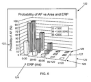
- FIG. 6 illustrates a bar graph of the probability of atrial fibrillation verus tissue surface area and effective refractory period
- FIG. 7 illustrates a line graph of the probability of atrial fibrillation verus tissue surface area versus effective refractory period
- FIG. 8 illustrates a table of univariable logistical regression for a variety of variables such as effective refractory period, wavelength, tissue area, tissue weight, maximum tissue width, minimum tissue width, and average tissue width;
- FIG. 9 illustrates a line graph of the probability of atrial fibrillation versus tissue surface area versus wavelength
- FIG. 10 illustrates a table of multivariable logistical regression for a variety of constants
- FIG. 11 illustrates a line graph of extrapolated probability of atrial fibrillation verus tissue area versus the effective refractory period.
- FIG. 12 is a labeled box representation of components of the system.
- the anatomic-based approach to the arrhythmia will be discarded, and replaced with a procedure based on the electrophysiological and geometrical characteristics of the atrial tissue in each individual patient. This procedure is individualized to the particular atrial geometry.
- a median sternotomy was performed, the heart was cradled in the pericardium, and the azygous vein was ligated and divided.
- the interatrial groove was dissected, separating the left atria and right atria. Two dissection protocols were used to isolate the left atria and right atria.
- the isolated left atria is generally indicated by numeral 10 and the right atria is generally indicated by numeral 20 .
- the atrium was dissected from the rest of the heart and divided through the superior venae cava down to the inferior vena cava to facilitate mounting of the tissue on the electrode plaque 23 .
- the right coronary artery 27 was cannulated with a catheter 28 , e.g., sixteen (16) gauge.
- the crista terminalis 26 , the right atrial appendage 24 and the tricuspid valve 22 are also shown.
- the heart lung block was removed, the lungs were cleanly dissected from the heart, and the pulmonary veins were divided one (1) centimeter from their insertion into the left atria 10 .
- the ventricles and right atria 20 were excised.
- the circumflex artery 16 was dissected up to the aortic root and distally to beyond the last atrial branch, and all of the ventricular branches were ligated.
- the circumflex artery 16 was cannulated with a catheter 17 , e.g., sixteen (16) gauge.
- the left atria 10 was divided through the right and left superior veins, unfolded, and mounted on the electrode plaque 23 .
- the pulmonary veins 18 , the mitral valve plane 16 , pacing sites 14 , releasing factor ablation lesions 12 , and the left atria area 19 are also shown.
- the appendages were ablated using bipolar radiofrequency energy.
- An illustrative example of this type of device is the AtriCureTM ablation and sensing unit with associated hand piece manufactured by AtriCure Inc., having a place of business at 6033 Schumacher Park Drive, Cincinnati, Ohio 45069.
- the hand piece or clamp created transmural lesions (less than one (1) millimeter wide) that prevents electrical conduction across the lesion.
- the epicardial surface was mounted on a flat electrode platform containing two hundred and fifty-six (256) unipolar electrodes with an interelectrode distance of five (5) millimeters.
- the atrial appendage of each preparation was placed into a slot in the electrode template that allowed the atrium to lie flat.
- the isolated atrial preparations 10 , 20 were kept in a temperature-controlled bath at 37° C. and perfused with a Krebs-Henseleit (“KH”) solution at a rate of 8 to 10 mL/min ( ⁇ 50 mm Hg).
- KH Krebs-Henseleit
- the composition of the KH solution was as follows (mm/L): Na + , 143; K + , 4.7; Cl ⁇ , 128; Ca 2+ 1.25, HCO 3 ⁇ , 25; Mg 2+ , 1.2; and dextrose, 11.1.
- the preparations were continuously superfused with KH as shown in FIG. 1 .
- pacing sites 14 were marked such that pacing was always performed from the same sites 14 .
- Pacing was conducted at 1.5 times the pacing threshold.
- the S 1 S 1 interval used for all of the pacing was 300 milliseconds.
- the effective refractory period (“ERP”) was determined at each pacing site by incrementally decreasing the S 1 S 2 interval by five (5) milliseconds until capture no longer occurred.
- the effective refractory period was defined as the shortest S 1 S 2 interval that captured the atrium.
- the effective refractory period is the period that follows effective stimulation, during which excitable tissue fails to respond to a stimulus of threshold intensity. After the baseline effective refractory period values were determined, at each marked pacing site, the solution was switched to KH solution with acetylcholine at a concentration of 10 ⁇ 3.5 M. 12
- the electrical activity of the atria was recorded in the form of electrograms as generally indicated by numeral 30 .
- the first electrogram is that of a normal sinus rhythm (“NSR”) as indicated by numeral 32 . If the preparation continuously fibrillated for more than 30 seconds, it was considered a sustained episode of atrial fibrillation shown in the second electrogram indicated by numeral 36 .
- the perfusion solution was then switched to KH alone until the arrhythmia terminated.
- the radiofrequency bipolar ablation device was used to divide the arrhythmogenic sections of the preparation.
- the effective refractory periods were again recorded at all of the pacing sites 14 , as shown in FIG.
- a lissamine green dye bolus was injected into the preparation to ensure that there were no perfusion abnormalities. Any sections of the atrium that were not well perfused were excluded from the data analysis. The atrial sections were photographed with a high-resolution digital camera and permanently fixed in a ten percent (10%) buffered formalin. The digital photos were analyzed to determine the minimum and maximum tissue height; the minimum, maximum, and average tissue width; and the area of all tissue sections created, which is generally indicated in FIG. 3 by numeral 40 . The fixed atrial sections were then dissected, and only atrial myocardium was weighed.
- Illustrative, but nonlimiting software that can analyze digital images includes Scion ImageTM, which is manufactured by the Scion Corporation, having a place of business at 82 Worman's Mill Court, Suite H, Frederick, Md. 21701 and Adobe PhotoshopTM, manufactured by Adobe Systems Incorporated, having a place of business at 345 Park Avenue, San Jose, Calif. 95110-2704.
- the electrograms recorded during pacing at 300 milliseconds were analyzed to calculate the activation sequence and the conduction velocities. 14
- the mean, maximum, and minimum conduction velocities (“CVs”) and standard deviations (“SDs”) for each section were then calculated. Wavelength was calculated as the product of the average conduction velocity and the effective refractory period of each section of the atrium. 8
- SYSTAT® manufactured by Systat Software, Inc., having a place of business at 501 Canal Blvd, Suite E, Point Richmond, Calif. 94804-2028.
- Mathematical analysis performed with the present invention utilizes a processor 212 , as shown in FIG. 12 .
- the results of the analysis of the tissue geometry for the left atria and right atria is generally indicated by numeral 50 .
- the first variable is the tissue surface area 60
- the second variable is the tissue weight 62
- the third variable is the maximum tissue width 64
- the fourth variable is the minimum tissue width 66
- the fifth variable is the average tissue width 68
- the sixth variable is the maximum tissue height 70
- the seventh variable is the minimum tissue height 72 .
- the results of the analysis of the effective refractory period, conduction velocity and wavelength in the left atria and right atria are generally indicated by numeral 80 .
- first category in the variables column 88 that is “whole preparation” 90 , which refers to either the left atria and right atria before any ablations were performed, that is, the atrium in its entirety.
- first variable 92 which is the effective refractory period without any acetylcholine (“AC”)
- second variable 94 which is the effective refractory period with acetylcholine (“AC”).
- variable 96 There a second category in the variables column 88 that is “all sections combined” 96 .
- the term “all sections” is used to describe an analysis performed when all of the varying sized sections, including the whole, were combined for that test.
- a third variable 98 which is the effective refractory period without any acetylcholine (“AC”) and a fourth variable 100 , which is the effective refractory period all sections combined with acetylcholine (“AC”).
- AC acetylcholine
- 96 is a fifth variable 102 for conduction velocity for all sections combined and a sixth variable 104 , which is the standard deviation for conduction velocity.
- the distinction between the “whole preparation” and “all sections” allows for the differentiation of innate properties of the native atria that could be artifacts from either time in the bath or the ablative procedures.
- variable 112 is an eleventh variable 118 , which is the wavelength for all sections combined without any acetylcholine (“AC”) and a twelfth variable 119 , which is the wavelength for all sections combined with acetylcholine (“AC”).
- FIGS. 4 and 5 indicate that the right atrial was significantly larger than the left atrial. However, the left atrial was significantly heavier than the right atrial. The mean maximum widths of the whole right atrial and left atrial were not statistically different.
- the right atria and the left atria preparations were compared for differences in minimum, maximum, and mean conduction velocity values and their standard deviations. There was a significant difference in the mean conduction velocity for all of the data points collected in the whole section, with the left atria having a faster mean conduction value than the right atria.
- the standard deviations for the measurement of mean conduction velocities in all of the sections were also significantly different, with the right atria having a greater standard deviation than the left atria.
- the maximum conduction velocities in the whole atrial preparations were greater in the right than the left. However, when the maximum conduction velocity values were examined in all of the sections, there was no significant difference.
- the mean wavelength was calculated by multiplying the mean conduction velocity by the effective refractory period.
- the probability of atrial fibrillation was analyzed with a univariable logistical regression being presented in FIG. 8 as generally indicated by numeral 140 .
- the variables under the variable column 142 include effective refractory period 152 , wavelength 154 , tissue area 156 , tissue weight 158 , maximum tissue width 160 , minimum tissue width 162 , and average tissue width 164 . Therefore, for each variable 152 , 154 , 156 , 158 , 160 , 162 and 164 , there were P values 144 , McFadden Rho squared values 146 , constant coefficient values 147 , variable coefficent values 148 , and values 150 for N. The results of the univariable logistical regression McFadden Rho squared values 146 that fall between 0.2 and 0.4 are considered satisfactory. 15
- decreasing effective refractory periods P ⁇ 0.001
- increasing tissue areas P ⁇ 0.001
- increasing maximum and minimum tissue widths P ⁇ 0.001
- increasing average tissue widths P ⁇ 0.001
- increasing tissue weights P ⁇ 0.001
- decreasing wavelengths P ⁇ 0.001
- FIG. 6 is a bar graph, which is generally indicated by numeral 120 , showing the correlation that increased tissue area 125 and decreased effective refractory periods 124 results in an increased probability of sustained atrial fibrillation 122 .
- the tissue area 125 is broken down into tissue area of less than 1,500 millimeters squared 128 , between 1,500 millimeters squared and 3,000 millimeters squared 127 and greater than 3,000 millimeters squared 126 .
- FIG. 7 is a line graph, which is generally indicated by numeral 130 .
- lines for effective refractory periods generally indicated by numeral 131 .
- This graph 130 demonstrates that with increasing tissue area 134 and decreasing effective refractory periods 131 , the probability of sustained atrial fibrillation increases 132 .
- FIG. 9 is a line graph, which is generally indicated by numeral 170 .
- This graph 170 demonstrates that with increasing tissue area 174 and decreasing wavelength 167 , the probability of sustained atrial fibrillation increases 172 .
- Atrial remodeling with the increase in atrial fibrosis, can slow conduction velocity and can shorten the refractory period in atria with long-standing atrial fibrillation.
- the duration of preoperative atrial fibrillation is known to be a negative predictor for the success of the previously known Maze procedure. 10
- the results of this investigation demonstrate the importance of the duration of the effective refractory period, with shorter effective refractory periods, the more likely it is to sustain atrial fibrillation.
- Multivariable logistical regression models demonstrate that increasing tissue size and decreasing effective refractory periods increase the probability of sustained atrial fibrillation, as shown in FIG. 6 .
- tissue height was a variable that did not vary during the experiments.
- the multivariable logistic regression analysis combined variables and analyzed both geometric variables and physiological variables to create models for predicting the probability of inducing sustained atrial fibrillation. A series of equations was developed from these models.
- FIG. 10 which provides a table of multivariable logistical regression analysis and is generally indicated by numeral 176 .
- This is utilized to predict the probability of a section of atria, e.g., canine, fibrillating when particular variables are entered into the equations.
- the first model presented utilized the variables of effective refractory period and tissue area.
- the data in FIGS. 6 and 7 allows for a better appreciation of the impact of effective refractory period and tissue area on the probability of inducing sustained atrial fibrillation. As the tissue area decreased and the effective refractory period increased, the probability of inducing sustained atrial fibrillation significantly decreased. However, with the same effective refractory period, as the tissue area increased, so did the probability of atrial fibrillation.
- the present experiments provide correlation of the relationship between geometric and functional electrophysiological variables and the inducibility of atrial fibrillation in the atrium.
- tissue width and effective refractory period were modulated.
- Additional variables that have not been analyzed in these experiments include modifying conduction velocity as well as the height of the tissue as well as the entire intact atrium. It is believed that this analysis also is applicable to the diseased atria. No matter what the underlying pathology, the underlying substrates are still effective refractory period, conduction velocity, geometry, and premature impulse formation. As an example, in patients with persistent atrial fibrillation, the effective refractory period is decreased.
- FIG. 11 which extrapolates the probability of atrial fibrillation versus tissue area versus effective refractory period as generally indicated by numeral 186 with altered effective refractory periods.
- This can include diseased tissue and can include patients with enlarged atria, who, although they have had a complete Maze procedure (including PV isolation), still have atrial fibrillation.
- Using this approach it may be possible to simulate the effect of different lesion sets on the inducibility of atrial fibrillation.
- first line for an effective refractory period of 150 milliseconds indicated by numeral 192 there is a first line for an effective refractory period of 150 milliseconds indicated by numeral 192 , a second line for an effective refractory period of 175 milliseconds indicated by numeral 194 , a third line for an effective refractory period of 200 milliseconds indicated by numeral 196 , a fourth line for an effective refractory period of 225 milliseconds indicated by numeral 198 , a fifth line for an effective refractory period of 250 milliseconds indicated by numeral 200 and a sixth line for an effective refractory period of 275 milliseconds indicated by numeral 202 .
- These lines 192 , 194 , 196 , 198 , 200 , and 202 are plotted in a correlation between tissue area in square centimeters 190 in relationship to the probability of atrial fibrillation 188 .
- a first line 204 that provides an estimated atrial surface area in a heart with a right and left atrial diameter of five (5) centimeters
- a second line 206 that provides an estimated atrial surface area in a heart with a right atrial diameter of five (5) centimeters and left atrial diameter of ten (10) centimeters.
- the tissue area increases 190 so does the probability of atrial fibrillation 188 .
- the data shown in FIG. 10 which is generally indicated by numeral 176 , can be used to predict atrial fibrillation.
- the first equation 210 utilizes effective refractory period and tissue area as the first and second variables, respectively.
- the second equation 212 utilizes wavelength and tissue area as the first and second variables, respectively.
- the third equation 214 utilizes effective refractory period and average tissue width as the first and second variables, respectively.
- the fourth equation 216 utilizes effective refractory period and maximum tissue width as the first and second variables, respectively.
- the fifth equation 218 utilizes wavelength and maximum tissue width as the first and second variables, respectively.
- the sixth equation 220 utilizes effective refractory period and tissue weight as the first and second variables, respectively.
- the seventh equation 222 utilizes effective refractory period and maximum conduction velocity as the first and second variables, respectively.
- An aspect of the present invention is to utilize tissue area, effective refractory period, and conduction velocity in order to design a procedure that would make it impossible for the atrium to fibrillate. Preferably, these variables would be obtained preoperatively. Either non-invasive or invasive electrophysiologic procedures can be utilized to obtain effective refractory period and conduction velocity. Atrial surface area will be obtained using either multidetector high resolution computerized axial tomography (“CAT”) scanning or magnetic resonance imaging (“MRI”).These types of measurements devices are generally indicated by numeral 202 in FIG. 12.
- CAT computerized axial tomography
- MRI magnetic resonance imaging
- a set of lesions could be designed with computer assistance that would make the atria fibrillation-proof.
- the procedure would be guided solely by geometry and the electrophysiologic properties of the tissue.
- This image-guided intervention would be customized based on the electrophysiologic characteristics of the atrial fibrillation in each individual patient.
- a number of different energy technologies could be utilized such as radio frequency, microwave, laser, ultrasound, thermal, chemical, mechanical, or cryogenic. Preferably, this would be a linear that creates long lesions.
- a nonlimiting, but illustrative, example of technology utilized to create lesions is disclosed in U.S. Pat. No. 6,932,813, which issued to Thompson, et al. on Aug. 23, 2005, which is incorporated herein by reference in its entirety.
- Another nonlimiting, but illustrative, example of technology utilized to create lesions with a catheter is disclosed in U.S. Pat. No. 6,663,622, which issued to Foley et al. on Dec. 16, 2003, which is incorporated herein by reference in its entirety.
- standard surgical techniques can also be utilized, such as scalpels in addition to the use of catheter-based techniques.
- any device would have to be firmly fixed to the atrial epicardium.
- this fixation mechanism would be a unipolar device.
- suction be used to fix the device to the epicardial surface.
- Other fixation methods include suture, clamping or balloon inflation. This would allow for the precise geometrical creation of lesions.
- Another nonlimiting, but illustrative, example of technology utilizing suction assisted ablation device having a support surface is disclosed in U.S. Pat. No. 6,558,382, which issued to Jahns et al. on May. 6, 2003, which is incorporated herein by reference in its entirety.
- These types of fixation devices are generally indicated by numeral 206 in FIG. 12.
- Another aspect of the present invention is an ablation device that incorporates a marking mechanism to delineate the precise lesion width.
- the marking mechanism is proposed to be a dye that would be infused through the device to make it clearly visible to the observer.
- Other marking devices include clips, absorbable strips, felt, staples, and so forth.
- Another nonlimiting, but illustrative, example of technology utilizing marking dye is disclosed in U.S. Pat. No. 6,669,694, which issued to Shadduck on Dec. 30, 2003, which is incorporated herein by reference in its entirety. These types of fixation devices are generally indicated by numeral 208 in FIG. 12.
- FIG. 12 Another aspect of the present invention is a device to divide the posterior pericardial reflections.
- This device could include a deflectable handle that could retract the heart and either a cautery, ultrasound or scissors device to divide the pericardium.
- a deflectable handle that could retract the heart
- either a cautery, ultrasound or scissors device to divide the pericardium.
- a nonlimiting, but illustrative, example of technology utilized to retract heart tissue is disclosed in U.S. Pat. No. 5,613,937, which issued to Garrison et al. on Mar. 25, 1997, which is incorporated herein by reference in its entirety.
- These types of mechanism for dividing posterior pericardial reflections are generally indicated by numeral 210 in FIG. 12.
Abstract
Description
- 1Go A S, Hylek E M, Phillips K A, Chang Y, Henault L E, Selby J V, Singer D E. Prevalence of diagnosed AF in adults: national implications for rhythm management and stroke prevention: the AnTicoagulation and Risk Factors In AF (ATRIA) Study. JAMA. 2001;285:2370-2375.
- 2 Garrey W. The nature of fibrillary contraction of the heart: its relation to tissue mass and form. Am J Physiol. 1914; 33:397-414.
- 3 Lewis T. The Mechanisms and Graphical Registration of the Heart Beat, 3rd ed. London, United Kingdom: Shaw and Sons; 1925.
- 4 Moe G K. On the multiple wavelet hypothesis of AF. Arch Int Pharmacodyn Ther. 1962; 140:183-188.
- 5 Moe G K, Rheinboldt W C, Abildskov J A. A computer model of AF. Am Heart J. 1964;67:200-220.
- 6 Allessie M A, Lammers W J E P, Bonke F I M, Hollen J. Experimental evaluation of Moe's multiple wavelet hypothesis of A F. In: Zipes D P, Jalife J, eds. Cardiac Electrophysiology and Arrhythmias. New York, N.Y.: Grume & Stratton; 1985:265-275.
- 7 Kennings K T, Kirchhoff C J, Smets J R, Wellens H J, Penn O C. Alesse M A. High-density mapping of electrically induced AF in humans. Circulation. 1994; 89:1665-1680.
- 8 Weiner N, Rosenbluth A. The mathematical formulation of the problem of conduction of impulses in a network of connected excitable elements, specifically in cardiac muscle. Arch Inst Cardiol Mex. 1946; 16:205-265.
- 9 Cox J L, Schuessler R B, Boineau J P. The development of the maze procedure for the treatment of AF. Semin Thorac Cardiovasc Surg. 2000; 12:2-14.
- 10 Gaynor S L, Schuessler R B, Bailey M S, Ishii Y, Boineau J P, Gleva M J, Cox J L, Damiano R J. Surgical treatment of AF: predictors of late recurrence. J Thorac Cardiovasc Surg. 2005; 129:104-111.
- 11Kosakai Y. Treatment of AF using the maze procedure: the Japanese experience. Semin Thorac Cardiovasc Surg. 2000; 12:44-52.
- 12 Schuessler R B, Grayson T M, Bromberg B I, Cox J L, Boineau J P. Cholinergically mediated tachyarrhythmias induced by a single extrastimulus in the isolated canine RA. Circ Res. 1992; 71:1254-1267.
- 13 Doshi R N, Wu T J, Yashima M, Kim Y H, Ong J J C, Cao J M, Hwang C, Yashar P, Fishbein M C, Karagueuzian H S, Chen P S. Relation between ligament of marshall and adrenergic atrial tachyarrhythmia. Circulation. 1999; 100:876-883.
- 14Bayly P V, KenKnight B H, Rogers J M, Hillsley R E, Ideker R E, Smith W M. Estimation of conduction velocity vector fields from epicardial mapping data. IEEE Trans Biomed Eng. 1998; 45:563-571.
- 15Hensher D, Johnson L W. Applied Discrete Choice Modeling. London, United Kingdom: Helm; 1981.
- 16Prasad S M, Maniar H S, Camillo C J, Schuessler R B, Boineau J P, Sundt T M, Cox J L, Damiano R J. The Cox maze III procedure for AF: long-term efficacy in patients undergoing lone versus concomitant procedures. J Thorac Cardiovasc Surg. 2003; 126:1822-1827.
- 17Falk R H. AF. N Engl J. Med. 2001; 344:1067-1077.
- 18Falk R H. Etiology and complications of AF: insights from pathology studies. Am J Cardiol. 1998; 82:10N-17N.
Claims (10)
Priority Applications (1)
| Application Number | Priority Date | Filing Date | Title |
|---|---|---|---|
| US11/214,256 US7918847B2 (en) | 2005-08-29 | 2005-08-29 | Method and associated system for the interventional treatment of atrial fibrillation |
Applications Claiming Priority (1)
| Application Number | Priority Date | Filing Date | Title |
|---|---|---|---|
| US11/214,256 US7918847B2 (en) | 2005-08-29 | 2005-08-29 | Method and associated system for the interventional treatment of atrial fibrillation |
Publications (2)
| Publication Number | Publication Date |
|---|---|
| US20070049816A1 US20070049816A1 (en) | 2007-03-01 |
| US7918847B2 true US7918847B2 (en) | 2011-04-05 |
Family
ID=37805252
Family Applications (1)
| Application Number | Title | Priority Date | Filing Date |
|---|---|---|---|
| US11/214,256 Expired - Fee Related US7918847B2 (en) | 2005-08-29 | 2005-08-29 | Method and associated system for the interventional treatment of atrial fibrillation |
Country Status (1)
| Country | Link |
|---|---|
| US (1) | US7918847B2 (en) |
Cited By (1)
| Publication number | Priority date | Publication date | Assignee | Title |
|---|---|---|---|---|
| US10354758B2 (en) | 2014-08-28 | 2019-07-16 | Siemens Healthcare Gmbh | System and method for patient-specific image-based simulation of atrial electrophysiology |
Families Citing this family (6)
| Publication number | Priority date | Publication date | Assignee | Title |
|---|---|---|---|---|
| US8617152B2 (en) * | 2004-11-15 | 2013-12-31 | Medtronic Ablation Frontiers Llc | Ablation system with feedback |
| US8657814B2 (en) * | 2005-08-22 | 2014-02-25 | Medtronic Ablation Frontiers Llc | User interface for tissue ablation system |
| US8641704B2 (en) | 2007-05-11 | 2014-02-04 | Medtronic Ablation Frontiers Llc | Ablation therapy system and method for treating continuous atrial fibrillation |
| US10912476B2 (en) | 2013-01-16 | 2021-02-09 | University Of Vermont | Catheters, systems, and related methods for mapping, minimizing, and treating cardiac fibrillation |
| US9693699B2 (en) | 2013-01-16 | 2017-07-04 | University Of Vermont | Methods and systems for mapping cardiac fibrillation |
| US11571160B2 (en) * | 2013-01-16 | 2023-02-07 | University Of Vermont | Methods and systems for wavelength mapping cardiac fibrillation and optimizing ablation lesion placement |
Citations (14)
| Publication number | Priority date | Publication date | Assignee | Title |
|---|---|---|---|---|
| US4357946A (en) * | 1980-03-24 | 1982-11-09 | Medtronic, Inc. | Epicardial pacing lead with stylet controlled helical fixation screw |
| US5613937A (en) * | 1993-02-22 | 1997-03-25 | Heartport, Inc. | Method of retracting heart tissue in closed-chest heart surgery using endo-scopic retraction |
| US5954665A (en) * | 1995-06-07 | 1999-09-21 | Biosense, Inc. | Cardiac ablation catheter using correlation measure |
| US20010034488A1 (en) * | 2000-02-09 | 2001-10-25 | Shal Policker | Method and system of automated hemodynamical detection of arrhythmias |
| US20020087183A1 (en) * | 1993-02-22 | 2002-07-04 | Boyd Stephen W. | Devices and methods for port-access multivessel coronary artery bypass surgery |
| US6558382B2 (en) * | 2000-04-27 | 2003-05-06 | Medtronic, Inc. | Suction stabilized epicardial ablation devices |
| US6663622B1 (en) | 2000-02-11 | 2003-12-16 | Iotek, Inc. | Surgical devices and methods for use in tissue ablation procedures |
| US6669694B2 (en) * | 2000-09-05 | 2003-12-30 | John H. Shadduck | Medical instruments and techniques for highly-localized thermally-mediated therapies |
| US20040181139A1 (en) * | 2001-04-27 | 2004-09-16 | Falwell Gary S. | Method and apparatus for three dimensional mapping of electrical activity in blood vessels and ablation of electrical pathways identified by the three dimension map |
| US6932813B2 (en) | 2002-05-03 | 2005-08-23 | Scimed Life Systems, Inc. | Ablation systems including insulated energy transmitting elements |
| US20050203502A1 (en) * | 2005-04-22 | 2005-09-15 | Boveja Birinder R. | Method and system for monitoring atrial fibrillation ablations with an ablation interface device |
| US20070032826A1 (en) * | 2005-08-02 | 2007-02-08 | Yitzhack Schwartz | Standardization of catheter-based treatment for atrial fibrillation |
| US20070043285A1 (en) * | 2005-08-02 | 2007-02-22 | Yitzhack Schwartz | Simulation of invasive procedures |
| US20070043296A1 (en) * | 2005-08-02 | 2007-02-22 | Yitzhack Schwartz | Guided procedures for treating atrial fibrillation |
-
2005
- 2005-08-29 US US11/214,256 patent/US7918847B2/en not_active Expired - Fee Related
Patent Citations (16)
| Publication number | Priority date | Publication date | Assignee | Title |
|---|---|---|---|---|
| US4357946A (en) * | 1980-03-24 | 1982-11-09 | Medtronic, Inc. | Epicardial pacing lead with stylet controlled helical fixation screw |
| US5613937A (en) * | 1993-02-22 | 1997-03-25 | Heartport, Inc. | Method of retracting heart tissue in closed-chest heart surgery using endo-scopic retraction |
| US20020087183A1 (en) * | 1993-02-22 | 2002-07-04 | Boyd Stephen W. | Devices and methods for port-access multivessel coronary artery bypass surgery |
| US5954665A (en) * | 1995-06-07 | 1999-09-21 | Biosense, Inc. | Cardiac ablation catheter using correlation measure |
| US20010034488A1 (en) * | 2000-02-09 | 2001-10-25 | Shal Policker | Method and system of automated hemodynamical detection of arrhythmias |
| US6663622B1 (en) | 2000-02-11 | 2003-12-16 | Iotek, Inc. | Surgical devices and methods for use in tissue ablation procedures |
| US6558382B2 (en) * | 2000-04-27 | 2003-05-06 | Medtronic, Inc. | Suction stabilized epicardial ablation devices |
| US20030167056A1 (en) * | 2000-04-27 | 2003-09-04 | Jahns Scott E. | Suction stabilized epicardial ablation devices |
| US6669694B2 (en) * | 2000-09-05 | 2003-12-30 | John H. Shadduck | Medical instruments and techniques for highly-localized thermally-mediated therapies |
| US20040181139A1 (en) * | 2001-04-27 | 2004-09-16 | Falwell Gary S. | Method and apparatus for three dimensional mapping of electrical activity in blood vessels and ablation of electrical pathways identified by the three dimension map |
| US6932813B2 (en) | 2002-05-03 | 2005-08-23 | Scimed Life Systems, Inc. | Ablation systems including insulated energy transmitting elements |
| US20050203502A1 (en) * | 2005-04-22 | 2005-09-15 | Boveja Birinder R. | Method and system for monitoring atrial fibrillation ablations with an ablation interface device |
| US20070032826A1 (en) * | 2005-08-02 | 2007-02-08 | Yitzhack Schwartz | Standardization of catheter-based treatment for atrial fibrillation |
| US20070043285A1 (en) * | 2005-08-02 | 2007-02-22 | Yitzhack Schwartz | Simulation of invasive procedures |
| US20070043296A1 (en) * | 2005-08-02 | 2007-02-22 | Yitzhack Schwartz | Guided procedures for treating atrial fibrillation |
| US7681579B2 (en) * | 2005-08-02 | 2010-03-23 | Biosense Webster, Inc. | Guided procedures for treating atrial fibrillation |
Non-Patent Citations (20)
| Title |
|---|
| Alan S. Go, MD et al., Prevalence of Diagnosed Atrial Fibrillation in Adults National Implications for Rhythm Management and Stroke Prevention: the AnTicoagulation and Risk Factors in Atrial Fibrillation (ATRIA) Study, JAMA, May 9, 2001, pp. 2370-2375, vol. 285, No. 18, American Medical Association, U.S. |
| Chen et al., Preoperative Atrial Size Predicts the Success of Radiofrequency Maze Procedure for Permanent Atrial Fibrillation in Patients Undergoing Concomitant Valvular Surgery, Jun. 2004, Chest, 125:2129-2134. * |
| Gordon K. Moe, MD et al., A computer model of atrial fibrillation, American Heart Journal, vol. 67, No. 2, Feb. 1964, pp. 200-220, New York, U.S. |
| Gordon K. Moe, On the Multiple Wavelet Hypothesis of Atrial Fibrillation, Arch. Int. Pharmacodyn., 1962, CXL, No. 1-2, pp. 183-188, New York, U.S. |
| James L. Cox et al., The Development of the Maze Procedure for the Treatment of Atrial Fibrillation, Seminars in Thoracic and Cardiovascular Surgery, vol. 12, No. 1, pp. 2-14, 2000, St. Louis, Missouri, U.S. |
| Karen Konings TS et al., Arrhythmias/Innervation/Pacing: High-Density Mapping of Electrically Induced Atrial Fibrillation in Humans, vol. 89(4), Apr. 1994, pp. 1665-1680, U.S. |
| Maurits A. Allessie et al., Experimental Evaluation of Moe's Multiple Wavelet Hypothesis of Atrial Fibrillation, Cardiac Electrophysiology and Arrhythmias, D. P. Zipes and J. Jalife, 1985 (New York: Grune & Stratton), pp. 265-276, U.S. |
| Mien-Cheng Chen, Jen-Ping Chang, and Hsueh-Wen Chang, "Preoperative Atrial Size Predicts the Success of Radiofrequency Maze Procedure for Permanent Atrial Fibrillation in Patients Undergoing Concomitant Valvular Surgery", Chest, 2004; 125; pp. 2129-2134. * |
| Norbert Wiener et al., The Mathematical Formulation of the Problem of Conduction of Impulses in a Network of Connected Excitable Elements, Specifically in Cardiac Muscle, ,Archos. Inst. Cardiol,. 1946, pp. 205-265, Mexico. |
| Philip V. Bayly et al., Estimation of Conduction Velocity Vector Fields From Epicardial Mapping Data, IEEE Transactions on Biomedical Engineering, vol. 45, No. 5, May 1998, pp. 563-571, New York,U.S. |
| Rahul N. Doshi, MD et al., Relation Between Ligament of Marshall and Adrenergic Atrial Tachyarrhythmia, 1999, pp. 876-883, American Heart Association, Inc., U.S. |
| Richard B. Schuessler et al., Cholinergically Mediated Tachyarrhythmias Induced by a Single Extrastimulus in the Isolated Canine Right Atrium, Circulation Research, vol. 71, pp. 1254-1267, 1992, Dallas, Texas, U.S. |
| Rodney H. Falk, M.D., Atrial Fibrillation; New England Journal of Medicine, Apr. 5, 2001, pp. 1067-1078, vol. 344, No. 14, Massachusetts Medical Society, U.S. |
| Rodney H. Falk, MD, Etiology and Complications of Atrial Fibrillation: Insights From Pathology Studies, American Journal of Cardiology, 1998; vol. 82 pp. 10N-17N, New Jersey, U.S. |
| Sunil M Prasad, MD et al., The Cox maze III procedure for atrial fibrillation: Long-term efficacy in patients undergoing lone versus concomitant procedures, The Journal of Thoracic and Cardiovascular Surgery, Dec. 2003, pp. 1822-1827, Washington University School of Medicine, U.S. |
| Sydney L. Gaynor, MD et al., Surgical treatment of atrial fibrillation: Predictors of late recurrence, The Journal of Thoracic and Cardiovascular Surgery, Jan. 2005, pp. 104-111, Washington University School of Medicine, U.S. |
| T. Lewis, Theory of Circus Movement and Its Application to Pure Flutter, pp. 319-324, The Mechanisms and Graphical Registration of the Heart Beat, London, UK, Shaw and Sons, Chapter 26, pp. 319-324, 1925. |
| Walter E. Garrey, The Nature of Fibrillary Contraction of the Heart-Its Relation to Tissue Mass and Form, American Journal of Physiology, 1914, vol. 33, pp. 397-414, U.S. |
| Walter E. Garrey, The Nature of Fibrillary Contraction of the Heart—Its Relation to Tissue Mass and Form, American Journal of Physiology, 1914, vol. 33, pp. 397-414, U.S. |
| Yoshio Kosakai, Treatment of Atrial Fibrillation Using the Maze Procedure: The Japanese Experience, Seminars in Thoracic and Cariovascular Surgery, vol. 12, No. 1, pp. 44-52, 2000, Perth, Australia. |
Cited By (1)
| Publication number | Priority date | Publication date | Assignee | Title |
|---|---|---|---|---|
| US10354758B2 (en) | 2014-08-28 | 2019-07-16 | Siemens Healthcare Gmbh | System and method for patient-specific image-based simulation of atrial electrophysiology |
Also Published As
| Publication number | Publication date |
|---|---|
| US20070049816A1 (en) | 2007-03-01 |
Similar Documents
| Publication | Publication Date | Title |
|---|---|---|
| US9877780B2 (en) | Methods of treating a cardiac arrhythmia by thoracoscopic production of a Cox maze III lesion set | |
| Anter et al. | Evaluation of a novel high-resolution mapping technology for ablation of recurrent scar-related atrial tachycardias | |
| Weimar et al. | The Cox-maze IV procedure for lone atrial fibrillation: a single center experience in 100 consecutive patients | |
| Abouezzeddine et al. | Relevance of endocavitary structures in ablation procedures for ventricular tachycardia | |
| CN104840196B (en) | Machine and process for the source for being automatically positioned biological rhythm disorder | |
| US7918847B2 (en) | Method and associated system for the interventional treatment of atrial fibrillation | |
| Nitta et al. | Concurrent multiple left atrial focal activations with fibrillatory conduction and right atrial focal or reentrant activation as the mechanism in atrial fibrillation | |
| Prasad et al. | Physiological consequences of bipolar radiofrequency energy on the atria and pulmonary veins: a chronic animal study | |
| Melby et al. | Efficacy and safety of right and left atrial ablations on the beating heart with irrigated bipolar radiofrequency energy: a long-term animal study | |
| Kron et al. | Management of recurrent atrial arrhythmias after minimally invasive surgical pulmonary vein isolation and ganglionic plexi ablation for atrial fibrillation | |
| Schuessler et al. | Animal studies of epicardial atrial ablation | |
| de Lima et al. | Randomized study of surgery for patients with permanent atrial fibrillation as a result of mitral valve disease | |
| Chugh et al. | Manifestations of coronary arterial injury during catheter ablation of atrial fibrillation and related arrhythmias | |
| Fromentin et al. | Prospective comparison between conventional transseptal puncture and transseptal needle puncture with radiofrequency energy | |
| Dibs et al. | Spatiotemporal characterization of atrial activation in persistent human atrial fibrillation: multisite electrogram analysis and surface electrocardiographic correlations—a pilot study | |
| Accord et al. | Post-mortem histologic evaluation of microwave lesions after epicardial pulmonary vein isolation for atrial fibrillation | |
| Lockwood et al. | Linear left atrial lesions in minimally invasive surgical ablation of persistent atrial fibrillation: techniques for assessing conduction block across surgical lesions | |
| CA2907309C (en) | Rf tissue ablation devices and methods of using the same | |
| Bonanomi et al. | A new device for beating heart bipolar radiofrequency atrial ablation | |
| Weimar et al. | Evaluation of a novel cryoablation system: in vivo testing in a chronic porcine model | |
| Melby et al. | Surgical treatment of atrial fibrillation with bipolar radiofrequency ablation: mid-term results in one hundred consecutive patients | |
| Hutchinson et al. | Contemporary tools and techniques for substrate ablation of ventricular tachycardia in structural heart disease | |
| Faustino et al. | A new approach for catheter ablation of atrial tachycardia following atrial fibrillation ablation | |
| Krishnan et al. | Catheter ablation of atrial flutter in a heart transplant recipient | |
| Ng et al. | Fundamentals of Cardiac Mapping |
Legal Events
| Date | Code | Title | Description |
|---|---|---|---|
| AS | Assignment |
Owner name: WASHINGTON UNIVERSITY, MISSOURI Free format text: ASSIGNMENT OF ASSIGNORS INTEREST;ASSIGNORS:DAMIANO JR., RALPH J.;SCHUESSLER, RICHARD B.;REEL/FRAME:016932/0988 Effective date: 20050829 |
|
| STCF | Information on status: patent grant |
Free format text: PATENTED CASE |
|
| FPAY | Fee payment |
Year of fee payment: 4 |
|
| AS | Assignment |
Owner name: NATIONAL INSTITUTES OF HEALTH (NIH), U.S. DEPT. OF Free format text: CONFIRMATORY LICENSE;ASSIGNOR:WASHINGTON UNIVERSITY;REEL/FRAME:040648/0603 Effective date: 20161116 |
|
| FEPP | Fee payment procedure |
Free format text: PAYOR NUMBER ASSIGNED (ORIGINAL EVENT CODE: ASPN); ENTITY STATUS OF PATENT OWNER: SMALL ENTITY |
|
| CC | Certificate of correction | ||
| MAFP | Maintenance fee payment |
Free format text: PAYMENT OF MAINTENANCE FEE, 8TH YR, SMALL ENTITY (ORIGINAL EVENT CODE: M2552); ENTITY STATUS OF PATENT OWNER: SMALL ENTITY Year of fee payment: 8 |
|
| FEPP | Fee payment procedure |
Free format text: MAINTENANCE FEE REMINDER MAILED (ORIGINAL EVENT CODE: REM.); ENTITY STATUS OF PATENT OWNER: SMALL ENTITY |
|
| LAPS | Lapse for failure to pay maintenance fees |
Free format text: PATENT EXPIRED FOR FAILURE TO PAY MAINTENANCE FEES (ORIGINAL EVENT CODE: EXP.); ENTITY STATUS OF PATENT OWNER: SMALL ENTITY |
|
| STCH | Information on status: patent discontinuation |
Free format text: PATENT EXPIRED DUE TO NONPAYMENT OF MAINTENANCE FEES UNDER 37 CFR 1.362 |
|
| FP | Lapsed due to failure to pay maintenance fee |
Effective date: 20230405 |