US8008073B2 - Anti-Mpl antibodies - Google Patents
Anti-Mpl antibodies Download PDFInfo
- Publication number
- US8008073B2 US8008073B2 US12/874,872 US87487210A US8008073B2 US 8008073 B2 US8008073 B2 US 8008073B2 US 87487210 A US87487210 A US 87487210A US 8008073 B2 US8008073 B2 US 8008073B2
- Authority
- US
- United States
- Prior art keywords
- seq
- antibody
- nos
- chain variable
- amino acid
- Prior art date
- Legal status (The legal status is an assumption and is not a legal conclusion. Google has not performed a legal analysis and makes no representation as to the accuracy of the status listed.)
- Expired - Fee Related
Links
Images


Classifications
-
- C—CHEMISTRY; METALLURGY
- C07—ORGANIC CHEMISTRY
- C07K—PEPTIDES
- C07K16/00—Immunoglobulins [IGs], e.g. monoclonal or polyclonal antibodies
- C07K16/18—Immunoglobulins [IGs], e.g. monoclonal or polyclonal antibodies against material from animals or humans
-
- C—CHEMISTRY; METALLURGY
- C07—ORGANIC CHEMISTRY
- C07K—PEPTIDES
- C07K16/00—Immunoglobulins [IGs], e.g. monoclonal or polyclonal antibodies
- C07K16/18—Immunoglobulins [IGs], e.g. monoclonal or polyclonal antibodies against material from animals or humans
- C07K16/28—Immunoglobulins [IGs], e.g. monoclonal or polyclonal antibodies against material from animals or humans against receptors, cell surface antigens or cell surface determinants
-
- A—HUMAN NECESSITIES
- A61—MEDICAL OR VETERINARY SCIENCE; HYGIENE
- A61P—SPECIFIC THERAPEUTIC ACTIVITY OF CHEMICAL COMPOUNDS OR MEDICINAL PREPARATIONS
- A61P43/00—Drugs for specific purposes, not provided for in groups A61P1/00-A61P41/00
-
- A—HUMAN NECESSITIES
- A61—MEDICAL OR VETERINARY SCIENCE; HYGIENE
- A61P—SPECIFIC THERAPEUTIC ACTIVITY OF CHEMICAL COMPOUNDS OR MEDICINAL PREPARATIONS
- A61P7/00—Drugs for disorders of the blood or the extracellular fluid
-
- C—CHEMISTRY; METALLURGY
- C07—ORGANIC CHEMISTRY
- C07K—PEPTIDES
- C07K2317/00—Immunoglobulins specific features
- C07K2317/20—Immunoglobulins specific features characterized by taxonomic origin
- C07K2317/24—Immunoglobulins specific features characterized by taxonomic origin containing regions, domains or residues from different species, e.g. chimeric, humanized or veneered
-
- C—CHEMISTRY; METALLURGY
- C07—ORGANIC CHEMISTRY
- C07K—PEPTIDES
- C07K2317/00—Immunoglobulins specific features
- C07K2317/30—Immunoglobulins specific features characterized by aspects of specificity or valency
- C07K2317/34—Identification of a linear epitope shorter than 20 amino acid residues or of a conformational epitope defined by amino acid residues
-
- C—CHEMISTRY; METALLURGY
- C07—ORGANIC CHEMISTRY
- C07K—PEPTIDES
- C07K2317/00—Immunoglobulins specific features
- C07K2317/50—Immunoglobulins specific features characterized by immunoglobulin fragments
- C07K2317/56—Immunoglobulins specific features characterized by immunoglobulin fragments variable (Fv) region, i.e. VH and/or VL
- C07K2317/565—Complementarity determining region [CDR]
-
- C—CHEMISTRY; METALLURGY
- C07—ORGANIC CHEMISTRY
- C07K—PEPTIDES
- C07K2317/00—Immunoglobulins specific features
- C07K2317/60—Immunoglobulins specific features characterized by non-natural combinations of immunoglobulin fragments
- C07K2317/62—Immunoglobulins specific features characterized by non-natural combinations of immunoglobulin fragments comprising only variable region components
- C07K2317/622—Single chain antibody (scFv)
-
- C—CHEMISTRY; METALLURGY
- C07—ORGANIC CHEMISTRY
- C07K—PEPTIDES
- C07K2317/00—Immunoglobulins specific features
- C07K2317/60—Immunoglobulins specific features characterized by non-natural combinations of immunoglobulin fragments
- C07K2317/62—Immunoglobulins specific features characterized by non-natural combinations of immunoglobulin fragments comprising only variable region components
- C07K2317/626—Diabody or triabody
-
- C—CHEMISTRY; METALLURGY
- C07—ORGANIC CHEMISTRY
- C07K—PEPTIDES
- C07K2317/00—Immunoglobulins specific features
- C07K2317/70—Immunoglobulins specific features characterized by effect upon binding to a cell or to an antigen
- C07K2317/75—Agonist effect on antigen
Definitions
- the present invention relates to anti-Mpl antibodies.
- Thrombopoietin is a factor that enhances the differentiation and maturation of megakaryocytes (platelet precursor cells) from hemopoietic stem cells into platelets. TPO also functions as a cytokine with an important role in the regulation of platelet number. TPO is converted into its active form through the cleavage of a TPO precursor comprising 353 amino acids.
- Mpl is a TPO receptor
- human Mpl molecules are known to exist in two forms comprising 572 and 635 amino acids.
- the human Mpl gene sequence has already been analyzed (see Non-Patent Document 1 and GenBank accession No. NM — 005373).
- Non-Patent Document 2 cytokine receptors dimerize upon ligand binding, and transduce signals into cells. It has been reported that TPO similarly binds to its own specific receptor MPL, which leads to dimerization of the receptor, thereby transducing signals into cells and exerting physiological effects.
- Antibodies exhibiting agonistic activity have been reported among those antibodies that bind to receptors having the above features.
- an antibody against the erythropoietin (EPO) receptor has been reported to substitute for erythropoietin function.
- the monovalent form (Fab) of the antibody is capable of binding to the EPO receptor but is unable to transduce signals.
- dimerization of the erythropoietin receptor via bivalent binding is assumed to be essential for signal transduction (see Non-Patent Document 3).
- Non-Patent Documents 4 and 5 Antibodies that bind to Mpl and exhibit TPO agonistic activity have also been reported (see Non-Patent Documents 4 and 5). This suggests that receptor dimerization is induced upon binding of a bivalent antibody with regards to MPL as well.
- An objective of the present invention is to provide novel anti-Mpl antibodies having TPO agonistic activity.
- the present inventors performed exhaustive research to solve the above objective.
- the present inventors prepared and purified anti-human Mpl antibody VB22B, and established a single-chain antibody expression system using genetic engineering techniques. Specifically, the variable region of anti-human Mpl antibody was first cloned, and a diabody expression vector pCXND3-VB22B db for the anti-human Mpl antibody was prepared. This pCXND3-VB22B db vector was then used to generate an expression vector pCXND3-VB22B sc(Fv)2 for anti-human Mpl antibody sc(Fv)2.
- Anti-human Mpl sc(Fv)2 was expressed in CHO-DG44 cells using the expression vector pCXND3-VB22B sc(Fv)2, and then purified from the culture supernatant.
- VB22B diabody was transiently expressed in COST cells using the above pCXND3-VB22B db vector, and then purified from the culture supernatant.
- VB22B diabody and VB22B sc(Fv)2 were evaluated for their TPO-like agonistic activities. The results showed that VB22B diabody and VB22B sc(Fv)2 exhibit higher agonistic activities compared to VB22B IgG, and thus activities equivalent to or higher than that of the natural ligand, human TPO.
- the present inventors succeeded in preparing five types of humanized VB22B sc(Fv)2.
- the TPO-like agonistic activity was also proven to be unaltered by humanization.
- the present invention provides the following (1) to (38):
- an antibody comprising a single-chain polypeptide having binding activity against TPO receptor (Mpl), wherein said antibody comprises two heavy chain variable regions and two light chain variable regions;
- an antibody that comprises a heavy chain variable region, wherein said heavy chain variable region comprises FR1, FR2, FR3, and FR4 consisting of amino acid sequences of any one of:
- an antibody comprising a light chain variable region, wherein said light chain variable region comprises FR1, FR2, FR3, and FR4 consisting of amino acid sequences of any one of:
- a heavy chain variable region which comprises FR1, FR2, FR3, and FR4 having the amino acid sequences consisting of SEQ ID NOs: 230, 232, 234, and 236, and a light chain variable region which comprises FR1, FR2, FR3, and FR4 having the amino acid sequences consisting of SEQ ID NOs: 239, 241, 243, and 245;
- a heavy chain variable region which comprises FR1, FR2, FR3, and FR4 having the amino acid sequences consisting of SEQ ID NOs: 265, 267, 269, and 271, and a light chain variable region which comprises FR1, FR2, FR3, and FR4 having the amino acid sequences consisting of SEQ ID NOs: 272, 274, 276, and 278;
- a heavy chain variable region which comprises FR1, FR2, FR3, and FR4 having the amino acid sequences consisting of SEQ ID NOs: 298, 299, 300, and 301, and a light chain variable region which comprises FR1, FR2, FR3, and FR4 having the amino acid sequences consisting of SEQ ID NOs: 302, 303, 304, and 305;
- a heavy chain variable region which comprises FR1, FR2, FR3, and FR4 having the amino acid sequences consisting of SEQ ID NOs: 298, 299, 306, and 301, and a light chain variable region which comprises FR1, FR2, FR3, and FR4 having the amino acid sequences consisting of SEQ ID NOs: 302, 307, 308, and 305;
- an antibody that comprises a heavy chain variable region, wherein said heavy chain variable region comprises the amino acid sequence of SEQ ID NO: 229, 256, 262, 289, or 295;
- polynucleotide hybridizing to the polynucleotide of (34) under stringent conditions, wherein said polynucleotide encodes an antibody having activity equivalent to that of an antibody of any one of (1) to (33);
- (36) a vector comprising the polynucleotide of (34) or (35);
- FIG. 1 demonstrates the strategy for preparing single-chain antibody sc(Fv)2.
- FIG. 2 illustrates the assessment of VB22B sc(Fv)2 binding activity using an Mpl-expressing CHO cell line. Purified VB22B sc(Fv)2 was used.
- FIG. 3 illustrates the assessment of VB22B antibody agonistic activity using BaF3-human Mpl.
- FIG. 4 illustrates the assessment of VB22B antibody agonistic activity using BaF3-monkey Mpl.
- FIG. 5 illustrates the assessment of VB22B antibody agonistic activity using M-07e.
- FIG. 6 shows the amino acid sequences of anti-human Mpl antibodies (H chains) that exhibit higher agonistic activities when converted into minibodies.
- FIG. 7 shows the amino acid sequences of anti-human Mpl antibodies (L chains) which exhibit higher agonistic activities when converted into minibodies.
- FIG. 8 illustrates the binding activity assessment of AB317 diabody using Mpl-expressing CHO cells. Both VB22B diabody (solid line) and AB317 diabody (broken line) were obtained from COS7 culture supernatants.
- FIG. 9 illustrates the agnostic activity assessment of AB324 and AB317 diabodies using BaF3-human Mpl.
- FIG. 10 illustrates the agnostic activity assessment of AB324 and AB317 diabodies using BaF3-monkey Mpl.
- FIG. 11 illustrates the agnostic activity assessment of AB324 and AB317 diabodies using BaF3-mouse Mpl.
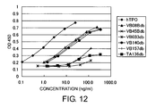
- FIG. 12 shows the agonistic activities of diabodies in BaF3-human Mpl cells.
- the X-axis shows OD at 450/655 nm, and the Y-axis represents concentration.
- FIG. 13 shows the agonistic activities of diabodies in BaF3-human Mpl (G305C) cells.
- the X-axis shows OD at 450/655 nm, and the Y-axis represents concentration.
- FIG. 14 shows the agonistic activities of TA136 db and TA136 sc(Fv)2 in BaF3-human Mpl cells.
- the X-axis shows OD at 450/655 nm and the Y-axis represents concentration.
- FIG. 15 shows the agonistic activities of TA136 db and TA136 sc(Fv)2 in BaF3-human Mpl (G305C) cells.
- the X-axis shows OD at 450/655 nm, and the Y-axis represents concentration.
- FIG. 16 shows the agonistic activities of TA136 db and TA136 sc(Fv)2 in BaF3-human Mpl (C769T) cells.
- the X-axis shows OD at 450/655 nm, and the Y-axis represents concentration.
- FIG. 17 shows the agonistic activities of TA136 db and TA136 sc(Fv)2 in BaF3-human Mpl (C823A) cells.
- the X-axis shows OD at 450/655 nm, and the Y-axis represents concentration.
- FIG. 18 shows the positions of FRs and CDRs in humanized heavy chain sequences (hVB22B p-z, hVB22B g-e, hVB22B e, hVB22B u2-wz4, and hVB22B q-wz5:VH), and humanized light chain sequences (hVB22B p-z, hVB22B g-e, hVB22B e, hVB22B u2-wz4, and hVB22B q-wz5:VL).
- FIG. 19 shows the TPO-like agonistic activities of murine VB22B sc(Fv)2, hVB22B e sc(Fv)2, and hVB22B g-e sc(Fv)2 in BaF3-human Mpl.
- the X-axis shows absorbance ratio (450 nm/655 nm), and the Y-axis represents concentration.
- FIG. 20 shows the TPO-like agonistic activities of murine VB22B sc(Fv)2, hVB22B p-z sc(Fv)2, and hVB22B u2-wz4 sc(Fv)2 in BaF3-human Mpl.
- the X-axis shows absorbance ratio (450 nm/655 nm), and the Y-axis represents concentration.
- FIG. 21 shows the TPO-like agonistic activities of murine VB22B sc(Fv)2 and hVB22B q-wz5 sc(Fv)2 in BaF3-human Mpl.
- the X-axis shows absorbance ratio (450 nm/655 nm), and the Y-axis represents concentration.
- the present invention provides antibodies that bind to the TPO receptor (Mpl).
- the antibodies of the present invention comprise all types of antibodies, including antibodies with modified amino acid sequences, such as minibodies, humanized antibodies, and chimeric antibodies; antibodies that have been modified by binding with other molecules (for example, polymers such as polyethylene glycol); and antibodies whose sugar chains have been modified.
- Mpl of the present invention may be a mutant receptor.
- a mutant receptor of the present invention is usually a receptor that exists at a frequency lower than 50%, preferably lower than 20%, more preferably lower than 10%, and even more preferably lower than 1%.
- the frequency is generally calculated using randomly selected subjects. However, the frequency may vary depending on the country, area, sex, and such. Therefore, the frequency may also be calculated, for example, within a defined country or area, such as Japan, the United States, and Europe, or calculated for one sex. When there are two or more mutations in a receptor, the frequency may be calculated for multiple mutation sites or for any one of the mutation sites. Mutant receptors are preferably evaluated by a frequency as described above.
- mutant receptors can also be evaluated, for example, by their signal transducing ability and such. Specifically, for example, when two different receptors are present, the one with stronger transducing signals upon natural ligand-binding maybe be used as a non-mutant type receptor, and the one with weaker transducing signals as a mutant receptor.
- the mutant receptors of the present invention comprise receptors that are associated with disease onset.
- the phrase “mutant receptors associated with disease onset” means that the loss of reactivity to a natural ligand becomes part of the reason that triggers disease onset.
- the mutant receptor may be a contributing factor, but not necessarily the sole factor triggering disease onset.
- Many reports have been previously published that describe the association of mutant receptors with disease onset. In addition to those that have been reported, associations of mutant receptors and disease onset can also be identified by statistical analysis methods (for example, correlation analyses).
- a p value is obtained from the ⁇ 2 value determined. Based on this p value, it can be determined whether there is a correlation between the mutant receptor and the disease. For example, when p ⁇ 0.05, the mutant receptor is considered to correlate with the disease. Mutant thrombopoietin (TPO) receptors have already been reported (Matthias Ballmaier et al., 2001, BLOOD, 97 (1), 139; and others).
- the antibodies of the present invention have agonistic activity against Mpl.
- the antibodies of the present invention comprise, for example, minibodies.
- the minibodies comprise antibody fragments lacking portions of the whole antibody (for example, whole IgG).
- the minibodies are not particularly limited as long as they have binding activity to their antigens.
- the minibodies of the present invention have higher activities compared to their corresponding whole antibodies.
- There are no particular limitations on the antibody fragments of the present invention as long as they are portions of the whole antibody, and preferably contain heavy chain variable regions (VH) and/or light chain variable regions (VL).
- VH heavy chain variable regions
- VL light chain variable regions
- the amino acid sequences of VH or VL may contain substitutions, deletions, additions and/or insertions.
- the antibody fragment may also lack portions of VH or/and VL, as long as it has binding ability to its antigen.
- the variable regions may be chimerized or humanized.
- Such antibody fragments include, for example, Fab, Fab′, F(ab′) 2 , and Fv.
- An example of a minibody includes Fab, Fab′, F(ab′) 2 , Fv, scFv (single-chain Fv), diabody, and sc(Fv)2 (single-chain (Fv)2).
- an “Fv” fragment is the smallest antibody fragment and contains a complete antigen recognition site and a binding site.
- the “Fv” fragment is a dimer (VH-VL dimer) in which a single VH and a single VL are strongly linked by a non-covalent bond.
- the three complementarity-determining regions (CDRs) of each of the variable regions interact with each other to form an antigen-binding site on the surface of the VH-VL dimer.
- Six CDRs confer the antigen-binding site of an antibody.
- a single variable region or a half of Fv containing only three CDRs specific to an antigen alone is also capable of recognizing and binding an antigen although its affinity is lower than the affinity of the entire binding site.
- scFv contains the VH and VL regions of an antibody, and these regions exist on a single polypeptide chain.
- an Fv polypeptide further contains a polypeptide linker between VH and VL, and therefore an scFv can form a structure required for antigen binding.
- linkers are not especially limited as long as they do not inhibit expression of antibody variable regions linked at both ends of the linkers.
- diabody refers to a bivalent antibody fragment constructed by gene fusion (Holliger P et al., 1993, Proc. Natl. Acad. Sci. USA 90: 6444-6448; EP 404,097; WO 93/11161 and others).
- Diabodies are dimers comprising two polypeptide chains, where each polypeptide chain comprises a VL and a VH connected with a linker short enough to prevent interaction of these two domains, for example, a linker of about five residues.
- the VL and VH encoded on the same polypeptide chain will form a dimer because the linker between them is too short to form a single-chain variable region fragment.
- the polypeptide chains form a dimer, and thus the diabody has two antigen binding sites.
- sc(Fv)2 is a single-chain minibody produced by linking two units of VH and two units of VL with linkers and such (Hudson et al., 1999, J Immunol. Methods 231:177-189). sc(Fv)2 exhibits a particularly high agonistic activity compared to the whole antibody and other minibodies sc(Fv)2 can be produced, for example, by linking two scFv molecules.
- the two VH units and two VL units are arranged in the order of VH, VL, VH, and VL ([VH]-linker-[VL]-linker-[VH]-linker-[VL]) beginning from the N terminus of a single-chain polypeptide.
- the order of the two VH units and two VL units is not limited to the above arrangement, and they may be arranged in any order. Examples of the arrangements are listed below.
- the linkers to be used for linking the variable regions of an antibody comprise arbitrary peptide linkers that can be introduced by genetic engineering, synthetic linkers, and linkers disclosed in, for example, Holliger, P. et al., Protein Engineering, 9(3), 299-305, 1996. Peptide linkers are preferred in the present invention. There are no limitations as to the length of the peptide linkers. The length can be selected accordingly by those skilled in the art depending on the purpose, and is typically 1-100 amino acids, preferably 3-50 amino acids, more preferably 5-30 amino acids, and even more preferably 12-18 amino acids (for example, 15 amino acids).
- such peptide linkers include:
- a particularly preferable sc(Fv)2 includes, for example, the sc(Fv)2 below.
- Synthetic linkers include crosslinking agents routinely used to crosslink peptides, for example, N-hydroxy succinimide (NHS), disuccinimidyl suberate (DSS), bis (succinimidyl) suberate (BS 3 ), dithiobis (succinimidyl propionate) (DSP), dithiobis(succinimidyl propionate) (DTSSP), ethylene glycol bis(succinimidyl succinate) (EGS), ethylene glycol bis(sulfosuccinimidyl succinate) (sulfo-EGS), disuccinimidyl tartrate (DST), disulfosuccinimidyl tartrate (sulfo-DST), bis[2-(succinimidoxycarbonyloxy)ethyl]sulfone (BSOCOES), and bis[2-(succinimidoxycarbonyloxy)ethyl]sulfone (s
- a preferable minibody is a diabody, even more preferably, an sc(Fv)2.
- Such a minibody can be prepared by treating an antibody with an enzyme, for example, papain or pepsin, to generate antibody fragments, or by constructing DNAs encoding those antibody fragments and introducing them into expression vectors, followed by expression in an appropriate host cell (see, for example, Co, M. S. et al., 1994, J. Immunol. 152, 2968-2976; Better, M. and Horwitz, A. H., 1989, Methods Enzymol.
- An antibody having exceedingly high agonistic activity can be prepared by reducing the molecular weight of a full-length antibody, particularly by converting it into an sc(Fv)2.
- the antibodies of the present invention comprise modified antibodies, such as chimeric antibodies and humanized antibodies that bind to Mpl. These modified antibodies can be produced by known methods.
- Chimeric antibodies are antibodies prepared by combining sequences derived from different animal species, and include for example, antibodies comprising the heavy chain and light chain variable regions of a murine antibody, and the heavy chain and light chain constant regions of a human antibody.
- Chimeric antibodies can be prepared by known methods. For example, a DNA encoding the V region of an antibody is linked to a DNA encoding the C region of a human antibody, and the construct is inserted into an expression vector and introduced into a host to produce chimeric antibodies.
- Humanized antibodies are also referred to as “reshaped human antibodies”. Such a humanized antibody is obtained by transferring the complementarity-determining region (CDR) of an antibody derived from a non-human mammal, for example mouse, to the complementarity-determining region of a human antibody, and the general gene recombination procedure for this is also known (see European Patent Application No. 125023 and WO 96/02576).
- CDR complementarity-determining region
- a DNA sequence designed to link a murine antibody CDR to the framework region (FR) of a human antibody can be synthesized by PCR, using primers prepared from several oligonucleotides containing overlapping portions of both CDR and FR terminal regions (see methods described in WO 98/13388).
- the human antibody framework region to be linked by CDR is selected in order to form a favorable antigen-binding site in the complementarity-determining region.
- Amino acids of the framework region in the antibody variable region may be substituted, as necessary, for the complementarity-determining region of the reshaped human antibody to form a suitable antigen-binding site (Sato, K. et al., 1993, Cancer Res. 53, 851-856).
- the constant region of a human antibody is used as the constant region of a chimeric antibody or humanized antibody.
- C ⁇ 1, C ⁇ 2, C ⁇ 3, and C ⁇ 4 can be used as the H chain, and C ⁇ and C ⁇ can be used as the L chain.
- the human antibody constant region may be modified to improve the antibody or the stability of the antibody production.
- chimeric antibodies comprise the variable region of an antibody from a non-human mammal and the constant region derived from a human antibody.
- humanized antibodies comprise the complementarity-determining region of an antibody from a non-human mammal, and the framework region and constant region derived from a human antibody.
- amino acids in the variable region for example, FR
- the constant region may be replaced with other amino acids, and such.
- variable regions in chimeric antibodies or that of the CDRs in humanized antibodies is not particularly limited, and may be derived from any type of animal. For example, sequences of murine antibodies, rat antibodies, rabbit antibodies, camel antibodies may be used.
- the present invention succeeded in preparing humanized antibodies having agonistic activity equivalent to that of the original murine antibody.
- a preferred humanized antibody of the present invention is an antibody comprising a heavy chain variable region that comprises the amino acid sequence of SEQ ID NO: 229 (humanized heavy chain sequence: hVB22B p-z VH), SEQ ID NO: 256 (humanized heavy chain sequence: hVB22B g-e VH), SEQ ID NO: 262 (humanized heavy chain sequence: hVB22B e VH), SEQ ID NO: 289 (humanized heavy chain sequence: hVB22B u2-wz4 VH), or SEQ ID NO: 295 (humanized heavy chain sequence: hVB22B q-wz5 VH); or an antibody comprising a light chain variable region that comprises the amino acid sequence of SEQ ID NO: 238 (humanized light chain hVB22B p-z VL), SEQ ID NO: 258 (humanized light chain hVB22B g-e VL or hVB22B e VL), SEQ ID NO: 291 (humanized light chain hVB22B
- Such antibodies include, for example, antibodies comprising the amino acid sequence of SEQ ID NO: 2, 254, 260, 287, or 293 (humanized sc(Fv)2 sequence (hVB22B p-z sc(Fv)2, hVB22B g-e sc(Fv)2, hVB22B e sc(Fv)2, hVB22B u2-wz4, or hVB22B q-wz5).
- the nucleotide sequence of hVB22B p-z VH is shown in SEQ ID NO: 228; the nucleotide sequence of hVB22B g-e VH is shown in SEQ ID NO: 255; the nucleotide sequence of hVB22B e VH is shown in SEQ ID NO: 261; the nucleotide sequence of hVB22B u2-wz4 VH is shown in SEQ ID NO: 288; the nucleotide sequence of hVB22B q-wz5 VH is shown in SEQ ID NO: 294; the nucleotide sequence of hVB22B p-z VL is shown in SEQ ID NO: 237; the nucleotide sequences of hVB22B g-e VL and hVB22B e VL are shown in SEQ ID NO: 257; the nucleotide sequence of hVB22B u2-wz4 VL is shown in SEQ ID NO: 290
- SEQ ID NO: 229 humanized heavy chain sequence: hVB22B p-z VH
- SEQ ID NO: 256 humanized heavy chain sequence: hVB22B g-e VH
- SEQ ID NO: 262 humanized heavy chain sequence: hVB22B e VH
- SEQ ID NO: 289 humanized heavy chain sequence: hVB22B u2-wz4 VH
- SEQ ID NO: 295 humanized heavy chain sequence: hVB22B q-wz5 VH
- SEQ ID NO: 238 humanized light chain sequence: hVB22Bp-zVL
- SEQ ID NO: 258 humanized light chain sequence: hVB22B g-e VL or hVB22B e VL
- SEQ ID NO: 291 humanized light chain sequence: hVB22B u2-wz4 VL
- SEQ ID NO: 297 humanized light chain sequence: hVB22B q-wz5 VL
- SEQ ID NOs of the CDRs and FRs in the hVB22B p-z VH sequence are shown below:
- SEQ ID NOs of the CDRs and FRs in the hVB22B p-z VL sequence are shown below:
- SEQ ID NOs of the CDRs and FRs in the hVB22B g-e VH sequence are shown below:
- SEQ ID NOs of the CDRs and FRs in the hVB22B g-e VL sequence are shown below:
- SEQ ID NOs of the CDRs and FRs in the hVB22B e VH sequence are shown below:
- SEQ ID NOs of the CDRs and FRs in the hVB22B e VL sequence are shown below:
- SEQ ID NOs of the CDRs and FRs in the hVB22B u2-wz4 VH sequence are shown below:
- SEQ ID NOs of the CDRs and FRs in the hVB22B u2-wz4 VL sequence are shown below:
- SEQ ID NOs of the CDRs and FRs in the hVB22B q-wz5 VH sequence are shown below:
- SEQ ID NOs of the CDRs and FRs in the hVB22B q-wz5 VL sequence are shown below:
- the CDRs and FRs in the hVB22B p-z sequence, hVB22B g-e sequence, hVB22B e sequence, hVB22B u2-wz4 sequence, and hVB22B q-wz5 sequence are shown in FIG. 18 .
- preferred humanized antibodies of the present invention include:
- preferred humanized antibodies of the present invention include:
- Chimeric antibodies and humanized antibodies exhibit lower antigenicity in the human body, and thus are expected to be useful when administered to humans for therapeutic purposes.
- the preferred antibodies of the present invention include antibodies that bind to soluble Mpl.
- soluble Mpl herein refers to Mpl molecules excluding those expressed on the cell membrane.
- a specific example of a soluble Mpl is an Mpl lacking the entire or a portion of the transmembrane domain.
- the transmembrane domain of human Mpl corresponds to amino acids 492-513 in SEQ ID NO: 123.
- An antibody that binds to soluble recombinant Mpl can be used in detailed epitope analysis and kinetic analysis of receptor-ligand binding, as well as for assessing the blood concentration and dynamic behavior of the antibody in in vivo tests.
- the preferred antibodies of the present invention include antibodies having binding activity against both human and monkey Mpl.
- the present invention also provides antibodies having agonistic activity to human Mpl and monkey Mpl.
- Antibodies having agonistic activity to both human and monkey Mpl are expected to be highly useful since the dynamic behavior and in vivo effects of the antibody, which are generally difficult to determine in human body, can be examined with monkeys.
- Such antibodies may also have binding activity or agonistic activity against Mpl from animals other than humans and monkeys (for example, mice).
- the agonistic activity can be determined by methods known to those skilled in the art, for example, by the method described below.
- the sequences for human Mpl (Palacios et al., Cell 41:727-734, (1985); GenBank Accession NO. NM — 005373), cynomolgus monkey Mpl (the nucleotide sequence and amino acid sequence are shown in SEQ ID NO: 164 and SEQ ID NO: 165, respectively), and mouse Mpl (GenBank Accession NO. NM — 010823) are already known.
- the activity can be determined using surface plasmon resonance with Biacore.
- soluble MPL-Fc protein, soluble MPL protein, or epitope peptides recognized by antibodies are immobilized onto sensor chips. Reaction rate constant can be determined by assessing the interaction between the antibody and the soluble Mpl-Fc protein, the soluble Mpl protein, or the epitope peptide recognized by the antibody.
- the proteins to be immobilized on chips are not limited in particular, and include, for example, MG10 (from Gln213 to Ala231)-GST fusion protein and Mpl-IgG Fc fusion protein described in the Examples. Since the antibodies are divalent and have two antigen-binding sites, the binding activities of these antibodies may be determined as those for the monovalent or divalent antibodies, or for mixtures of both. Any of these can be used in the present invention.
- the binding activity can be evaluated by ELISA (enzyme-linked immunosorbent assays), EIA (enzyme immunoassays), RIA (radio immunoassays), or fluorescent antibody techniques.
- a sample containing a test antibody such as purified antibody or culture supernatant of cells producing the test antibody, is added to a plate coated with an antigen to which the test antibody can bind.
- an enzyme substrate such as p-nitrophenyl phosphate is added.
- the antigen-binding activity can then be evaluated by determining the absorbance.
- the upper limit of the binding activity there is no specific limitation as to the upper limit of the binding activity; for example, the upper limit may be set within a technically feasible range by those skilled in the art. However, the technically feasible range may expand with the advancement of technology.
- the preferred antibodies of the present invention include antibodies recognizing epitopes that are recognized by any one of the antibodies indicated in (I) to (XII) below.
- the antibody of any one of (I) to (XII) is preferably a minibody.
- An antibody comprising an amino acid sequence of any one of (I) to (XII) indicated above, in which one or more amino acids have been substituted, deleted, added, and/or inserted, wherein the antibody has activity equivalent to that of the antibody of any one of (I) to (XII).
- the phrase “functionally equivalent” means that an antibody of interest has a biological or biochemical activity comparable to that of an antibody of the present invention. Such activities include, for example, binding activities and agonistic activities.
- Methods for preparing polypeptides functionally equivalent to a certain polypeptide are well known to those skilled in the art, and include methods of introducing mutations into polypeptides.
- those skilled in the art can prepare an antibody functionally equivalent to the antibodies of the present invention by introducing appropriate mutations into the antibody using site-directed mutagenesis (Hashimoto-Gotoh, T. et al. Gene 152, 271-275, (1995); Zoller, M J, and Smith, M. Methods Enzymol. 100, 468-500, (1983); Kramer, W. et al., Nucleic Acids Res. 12, 9441-9456, (1984); Kramer, W. and Fritz H J, Methods Enzymol.
- the present invention also comprises antibodies functionally equivalent to the antibodies of the present invention and comprising the amino acid sequences of these antibodies, in which one or more amino acids is mutated.
- the number of amino acids that may be mutated is not particularly restricted, so long as the activity is maintained.
- the number of amino acids that are mutated is 50 amino acids or less, preferably 30 or less, more preferably 10 or less (for example, five amino acids or less).
- the site of mutation is not particularly restricted, so long as the mutation does not result in the disruption of activity.
- Amino acid mutations may be made at one or more predicted, preferably nonessential, amino acid residues.
- a “nonessential” amino acid residue is a residue that can be altered from the wild-type sequence of a protein without altering the biological activity, whereas an “essential” amino acid residue is required for biological activity.
- An amino acid is preferably substituted for a different amino acid(s) that allows the properties of the amino acid side-chain to be conserved.
- a “conservative amino acid substitution” means a replacement of an amino acid residue belonging to one of the following groups with another amino acid in the same group having a chemically similar side chain. Groups of amino acid residues having similar side chains have been defined in the art.
- amino acid side chain properties are: hydrophobic amino acids (A, I, L, M, F, P, W, Y, and V), hydrophilic amino acids (R, D, N, C, E, Q, G, H, K, S, and T), amino acids comprising the following side chains: aliphatic side chains (G, A, V, L, I, and P); hydroxyl-containing side chains (S, T, and Y); sulfur-containing side chains (C and M); carboxylic acid- and amide-containing side chains (D, N, E, and Q); basic side chains (R, K, and H); aromatic ring-containing side chains (H, F, Y, and W) (amino acids are represented by one-letter codes in parentheses).
- hydrophobic amino acids A, I, L, M, F, P, W, Y, and V
- hydrophilic amino acids R, D, N, C, E, Q, G, H, K, S, and T
- amino acids comprising the following side chains: alipha
- a polypeptide comprising a modified amino acid sequence, in which one or more amino acid residues is deleted, added, and/or replaced with other amino acids, is known to retain its original biological activity (Mark, D. F. et al., Proc. Natl. Acad. Sci. USA 81, 5662-5666 (1984); Zoller, M. J. & Smith, M. Nucleic Acids Research 10, 6487-6500 (1982); Wang, A. et al., Science 224, 1431-1433; Dalbadie-McFarland, G. et al., Proc. Natl. Acad. Sci. USA 79, 6409-6413 (1982)).
- Fusion proteins containing antibodies that comprise the amino acid sequence of an antibody of the present invention, in which two or more amino acid residues have been added, are included in the present invention.
- the fusion protein results from a fusion between one of the above antibodies and a second peptide or protein, and is included in the present invention.
- the fusion protein can be prepared by ligating a polynucleotide encoding an antibody of the present invention and a polynucleotide encoding a second peptide or polypeptide in frame, inserting this into an expression vector, and expressing the fusion construct in a host.
- the partner peptide or polypeptide to be fused with an antibody of the present invention may be a known peptide, for example, FLAG (Hopp, T. P. et al., BioTechnology 6, 1204-1210 (1988)), 6 ⁇ His consisting of six His (histidine) residues, 10 ⁇ His, influenza hemagglutinin (HA), human c-myc fragment, VSV-GP fragment, p18HIV fragment, T7-tag, HSV-tag, E-tag, SV40 T antigen fragment, lck tag, ⁇ -tubulin fragment, B-tag, Protein C fragment.
- FLAG Hopp, T. P. et al., BioTechnology 6, 1204-1210 (1988)
- 6 ⁇ His consisting of six His (histidine) residues, 10 ⁇ His, influenza hemagglutinin (HA), human c-myc fragment, VSV-GP fragment, p18HIV fragment, T7-tag, HSV-tag, E-tag,
- partner polypeptides to be fused with the antibodies of the present invention include, for example, GST (glutathione-S-transferase), HA (influenza hemagglutinin), immunoglobulin constant region, ⁇ -galactosidase, and MBP (maltose-binding protein).
- GST glutthione-S-transferase
- HA influenza hemagglutinin
- immunoglobulin constant region e.gly available peptides or polypeptides
- MBP maltose-binding protein
- a polynucleotide encoding one of these commercially available peptides or polypeptides can be fused with a polynucleotide encoding an antibody of the present invention.
- the fusion polypeptide can be prepared by expressing the fusion construct.
- the antibodies of the present invention may differ in amino acid sequence, molecular weight, isoelectric point, presence/absence of sugar chains, and conformation depending on the cell or host producing the antibody, or purification method.
- a resulting antibody is included in the present invention, as long as it is functionally equivalent to an antibody of the present invention.
- an antibody of the present invention when expressed in prokaryotic cells, for example E. coli , a methionine residue is added to the N terminus of the original antibody amino acid sequence.
- Such antibodies are included in the present invention.
- an antibody that recognizes an epitope recognized by the antibody according to any one of (I) to (XII) indicated above is expected to have a high agonistic activity.
- Such antibodies can be prepared by methods known to those skilled in the art.
- the antibody can be prepared by, for example, determining the epitope recognized by the antibody according to any one of (I) to (XII) by conventional methods, and using a polypeptide comprising one of the epitope amino acid sequences as an immunogen.
- the antibody can be prepared by determining the epitopes of conventionally prepared antibodies and selecting an antibody that recognizes the epitope recognized by an antibody of any one of (I) to (XII).
- a particularly preferred antibody is an antibody that recognizes the epitope recognized by the antibody comprising the amino acid sequence of SEQ ID NO: 2.
- the antibody comprising the amino acid sequence of SEQ ID NO: 2 is predicted to recognize the region from Glu 26 to Leu 274, preferably the region from Ala 189 to Gly 245, more preferably the region from Gln 213 to Ala 231 of human Mpl.
- antibodies recognizing the region of amino acids 26 to 274, or amino acids 189 to 245, or amino acids 213 to 231 of human Mpl are also included in the present invention.
- Antibodies recognizing regions of amino acids 26 to 274, amino acids 189 to 245, or amino acids 213 to 231 of the human Mpl amino acid sequence (SEQ ID NO: 123) can be obtained by methods known to those skilled in the art. Such antibodies can be prepared by, for example, using a peptide comprising amino acids 26 to 274, amino acids 189 to 245, or amino acids 213 to 231 of the human Mpl amino acid sequence (SEQ ID NO: 123) as an immunogen. Alternatively, such antibodies can be prepared by determining the epitope of a conventionally prepared antibody and selecting an antibody that recognizes the same epitope recognized by an antibody of the present invention.
- the present invention provides antibodies described above in (I) to (XII).
- a preferred antibody is the one shown in (V)
- a more preferred antibody is the one shown in (VI)
- a still more preferred is the one shown in (VIII).
- the present invention also provides vectors comprising polynucleotides encoding the antibodies of the present invention, or polynucleotides which hybridize under stringent conditions to the polynucleotides of the present invention and encode antibodies having activities equivalent to those of the antibodies of the present invention.
- the polynucleotides of the present invention are polymers comprising multiple bases or base pairs of deoxyribonucleic acids (DNA) or ribonucleic acids (RNA), and are not particularly limited, as long as they encode the antibodies of the present invention. They may also contain non-natural nucleotides.
- the polynucleotides of the present invention can be used to express antibodies using genetic engineering techniques.
- the polynucleotides of this invention can also be used as probes in the screening of antibodies functionally equivalent to the antibodies of the present invention.
- DNAs that hybridize under stringent conditions to a polynucleotide encoding an antibody of the present invention, and encode antibodies having activity equivalent to those of the antibodies of the present invention can be obtained by techniques such as hybridization and gene amplification (for example, PCR), using a polynucleotide of the present invention or a portion thereof as a probe.
- Such DNAs are also included in the polynucleotides of the present invention.
- Hybridization techniques are well known to those skilled in the art (Sambrook, J et al., Molecular Cloning 2nd ed., 9.47-9.58, Cold Spring Harbor Lab. press, 1989).
- Such hybridization conditions include, for example, conditions of low stringency.
- conditions of low stringency include post-hybridization washing in 0.1 ⁇ SSC and 0.1% SDS at 42° C., and preferably in 0.1 ⁇ SSC and 0.1% SDS at 50° C. More preferable hybridization conditions include those of high stringency.
- Highly stringent conditions include, for example, washing in 5 ⁇ SSC and 0.1% SDS at 65° C. In these conditions, the higher the temperature, the higher the expectation of efficiently obtaining polynucleotides with a high homology. However, several factors, such as temperature and salt concentration, can influence hybridization stringency, and those skilled in the art can suitably select these factors to accomplish similar stringencies.
- Antibodies that are encoded by polynucleotides obtained by the hybridization and gene amplification techniques, and are functionally equivalent to the antibodies of the present invention generally exhibit high homology to the antibodies of the this invention at the amino acid level.
- the antibodies of the present invention include antibodies that are functionally equivalent to the antibodies of the present invention, and exhibit high amino acid sequence homology to the antibodies of this invention.
- the term “high homology” generally means identity at the amino acid level of at least 50% or higher, preferably 75% or higher, more preferably 85% or higher, still more preferably 95% or higher.
- Polypeptide homology can be determined by the algorithm described in the report: Wilbur, W. J. and Lipman, D. J. Proc. Natl. Acad. Sci. USA 80, 726-730 (1983).
- E. coli When E. coli is used as a host, there is no particular limitation as to the type of vector of the present invention, as long as the vector contains an “ori” responsible for its replication in E. coli and a marker gene.
- the “ori” ensures the amplification and mass production of the vector in E. coli (for example, JM109, DH5 ⁇ , HB101, and XL1Blue).
- the marker gene is used to select the E. coli transformants (for example, a drug resistance gene selected by an appropriate drug such as ampicillin, tetracycline, kanamycin, and chloramphenicol).
- the vectors include, for example, M13 vectors, pUC vectors, pBR322, pBluescript, and pCR-Script.
- pGEM-T, pDIRECT, and pT7 can also be used for the subcloning and excision of cDNAs.
- expression vectors are useful as vectors of the present invention.
- an expression vector When an expression vector is expressed, for example, in E. coli , it should have the above characteristics in order to be amplified in E. coli .
- the vector when E. coli , such as JM109, DH5 ⁇ , HB101, or XL1-Blue are used as the host cell, the vector preferably has a promoter, for example, lacZ promoter (Ward et al. (1989) Nature 341:544-546; (1992) FASEB J. 6:2422-2427), araB promoter (Better et al. (1988) Science 240:1041-1043), or T7 promoter, that allows efficient expression of the desired gene in E. coli .
- lacZ promoter Ward et al. (1989) Nature 341:544-546; (1992) FASEB J. 6:2422-2427
- araB promoter Better et al. (1988) Science 240:1041-10
- vectors include pGEX-5X-1 (Pharmacia), “QIAexpress system” (QIAGEN), pEGFP, and pET (where BL21, a strain expressing T7 RNA polymerase, is preferably used as the host).
- the vectors may comprise a signal sequence for polypeptide secretion.
- the pelB signal sequence (Lei, S. P. et al. J. Bacteriol. 169:4379 (1987)) may be used as a signal sequence for polypeptide secretion.
- calcium chloride methods or electroporation methods may be used to introduce the vector into a host cell.
- expression vectors derived from mammals e.g., pCDNA3 (Invitrogen), pEGF-BOS (Nucleic Acids Res. (1990) 18(17):5322), pEF, pCDM8), insect cells (e.g., “Bac-to-BAC baculovirus expression system” (GIBCO-BRL), pBacPAK8), plants (e.g., pMH1, pMH2), animal viruses (e.g., pHSV, pMV, pAdexLcw), retroviruses (e.g., pZIPneo), yeasts (e.g., “Pichia Expression Kit” (Invitrogen), pNV11, SP-Q01), and Bacillus subtilis (e.g., pPL608, pKTH50) may also be used as a vector of the present invention.
- mammals e.g., pCDNA3 (Invitrogen), pEGF-BOS
- the vector preferably has a promoter necessary for expression in such cells, for example, an SV40 promoter (Mulligan et al. (1979) Nature 277:108), MMLV-LTR promoter, EF1 ⁇ promoter (Mizushima et al. (1990) Nucleic Acids Res. 18:5322), CMV promoter, etc.). It is even more preferable that the vector also carries a marker gene for selecting transformants (for example, a drug-resistance gene selected by a drug such as neomycin and G418. Examples of vectors with such characteristics include pMAM, pDR2, pBK-RSV, pBK-CMV, pOPRSV, and pOP13, and such.
- CHO cells that are defective in the nucleic acid synthesis pathway are introduced with a vector containing a DHFR gene (for example, pCHOI) to compensate for the defect, and the copy number is amplified using methotrexate (MTX).
- a COS cell which carries an SV40 T antigen-expressing gene on its chromosome, can be transformed with a vector containing the SV40 replication origin (for example, pcD) for transient gene expression.
- the replication origin may be derived from polyoma virus, adenovirus, bovine papilloma virus (BPV), and such.
- the expression vector may contain, as a selection marker, aminoglycoside transferase (APH) gene, thymidine kinase (TK) gene, E. coli xanthine guanine phosphoribosyl transferase (Ecogpt) gene, dihydrofolate reductase (dhfr) gene, and such.
- APH aminoglycoside transferase
- TK thymidine kinase
- Ecogpt E. coli xanthine guanine phosphoribosyl transferase
- dhfr dihydrofolate reductase
- the vector is introduced into a host cell.
- the host cells into which the vector is introduced are not particularly limited, for example, E. coli and various animal cells are available for this purpose.
- the host cells may be used, for example, as a production system to produce and express the antibodies of the present invention.
- In vitro and in vivo production systems are available for polypeptide production systems. Production systems that use eukaryotic cells or prokaryotic cells are examples of in vitro production systems.
- Eukaryotic cells that can be used are, for example, animal cells, plant cells, and fungal cells.
- animal cells include: mammalian cells, for example, CHO (J. Exp. Med. (1995)108, 945), COS, 3T3, myeloma, BHK (baby hamster kidney), HeLa, Vero, amphibian cells such as Xenopus laevis oocytes (Valle, et al. (1981) Nature 291, 358-340), or insect cells (e.g., Sf9, Sf21, and Tn5).
- CHO-DG44, CHO-DXB11, COS7 cells, and BHK cells can be suitably used.
- Vectors can be introduced into a host cell by, for example, calcium phosphate methods, the DEAE-dextran methods, methods using cationic liposome DOTAP (Boehringer-Mannheim), electroporation methods, lipofection methods.
- Plant cells include, for example, Nicotiana tabacum -derived cells known as a protein production system. Calluses may be cultured from these cells.
- Known fungal cells include yeast cells, for example, genus Saccharomyces such as Saccharomyces cerevisiae and Saccharomyces pombe ; and filamentous fungi, for example, genus Aspergillus such as Aspergillus niger.
- Bacterial cells can be used in the prokaryotic production systems.
- Examples of bacterial cells include E. coli (for example, JM109, DH5 ⁇ , HB101 and such); and Bacillus subtilis.
- Antibodies can be obtained by transforming the cells with a polynucleotide of interest and in vitro culturing of these transformants.
- Transformants can be cultured using known methods. For example, DMEM, MEM, RPMI 1640, or IMDM may be used as the culture medium for animal cells, and may be used with or without serum supplements such as FBS or fetal calf serum (FCS). Serum-free cultures are also acceptable.
- the preferred pH is about 6 to 8 during the course of culturing. Incubation is carried out typically at a temperature of about 30 to 40° C. for about 15 to 200 hours. Medium is exchanged, aerated, or agitated, as necessary.
- production systems using animal or plant hosts may be used as systems for producing polypeptides in vivo.
- a polynucleotide of interest is introduced into an animal or plant and the polypeptide is produced in the body of the animal or plant and then recovered.
- the “hosts” of the present invention includes such animals and plants.
- Animals to be used for the production system include mammals or insects. Mammals such as goats, pigs, sheep, mice, and cattle may be used (Vicki Glaser SPECTRUM Biotechnology Applications (1993)). Alternatively, the mammals may be transgenic animals.
- a polynucleotide of interest is prepared as a fusion gene with a gene encoding a polypeptide specifically produced in milk, such as the goat ⁇ -casein gene.
- DNA fragments containing the fusion gene are injected into goat embryos, which are then introduced back to female goats.
- the desired antibody can be obtained from milk produced by the transgenic goats, which are born from the goats that received the embryos, or from their offspring.
- Appropriate hormones may be administered to increase the volume of milk containing the antibody produced by the transgenic goats (Ebert, K. M. et al., Bio/Technology 12, 699-702 (1994)).
- Insects such as silkworms
- Baculoviruses carrying a polynucleotide encoding an antibody of interest can be used to infect silkworms, and the antibody of interest can be obtained from the body fluids (Susumu, M. et al., Nature 315, 592-594 (1985)).
- Plants used in the production system include, for example, tobacco.
- a polynucleotide encoding an antibody of interest is inserted into a plant expression vector, for example, pMON 530, and then the vector is introduced into a bacterium, such as Agrobacterium tumefaciens .
- the bacteria are then used to infect tobacco such as Nicotiana tabacum , and the desired antibodies can be recovered from the leaves (Julian K.-C. Ma et al., Eur. J. Immunol. 24, 131-138 (1994)).
- the resulting antibody may be isolated from the inside or outside (such as the medium) of host cells, and purified as a substantially pure and homogenous antibody. Methods are not limited to any specific method and any standard method for isolating and purifying antibodies may be used. Polypeptides may be isolated and purified, by selecting an appropriate combination of, for example, chromatographic columns, filtration, ultrafiltration, salting out, solvent precipitation, solvent extraction, distillation, immunoprecipitation, SDS-polyacrylamide gel electrophoresis, isoelectric focusing, dialysis, recrystallization, and others.
- substantially pure polypeptide as used herein in reference to a given polypeptide means that the polypeptide is substantially free from contaminants such as other biological macromolecules, culture media (if recombinantly produced), or chemical precursors (if chemically synthesized).
- the substantially pure polypeptide is at least 75%, preferably at least about 80%, more preferably at least about 85, 90, 95, or 99% pure by dry weight. Purity can be measured by any appropriate standard method, for example by a chromatography method, polyacrylamide gel electrophoresis, or HPLC analysis.
- Chromatographies include, for example, affinity chromatographies, ion exchange chromatographies, hydrophobic chromatographies, gel filtrations, reverse-phase chromatographies, and adsorption chromatographies (Strategies for Protein Purification and Characterization: A Laboratory Course Manual. Ed Daniel R. Marshak et al., Cold Spring Harbor Laboratory Press, 1996). These chromatographies can be carried out using liquid phase chromatographies such as HPLC and FPLC. Examples of the affinity chromatography columns include protein A columns and protein G columns. Examples of the proteins A columns include Hyper D, POROS, and Sepharose F. F. (Pharmacia).
- An antibody can be modified freely and peptide portions deleted by treating the antibody with an appropriate protein modifying enzyme before or after antibody purification.
- protein modifying enzymes include, for example, trypsins, chymotrypsins, lysyl endopeptidases, protein kinases, and glucosidases.
- Antibodies that bind to Mpl can be prepared by methods known to those skilled in the art.
- monoclonal antibody-producing hybridomas can be essentially generated by known technologies as follows: immunizing animals with Mpl proteins or Mpl-expressing cells as sensitized antigens using conventional immunological methods; fusing the obtained immunocytes with known parental cells by conventional cell fusion methods; and screening for monoclonal antibody-producing cells by conventional methods.
- monoclonal antibodies can be prepared by the method below.
- Mpl protein which is used as a sensitized antigen for preparing antibodies, is prepared by expressing the Mpl gene/amino acid sequence (GenBank accession number: NM — 005373). More specifically, the gene sequence encoding Mpl is inserted into a known expression vector, which is then transfected into an appropriate host cell. The subject human Mpl protein is purified from the host cell or culture supernatant using known methods.
- the purified Mpl protein is then used as a sensitized antigen.
- a partial Mpl peptide may be used as a sensitized antigen.
- the partial peptide can also be chemically synthesized based on the amino acid sequence of human Mpl.
- the epitopes of Mpl molecule that are recognized by an anti-Mpl antibody of the present invention are not limited to a particular epitope, and may be any epitope on the Mpl molecule.
- any fragment can be used as an antigen for preparing anti-Mpl antibodies of the present invention, as long as the fragment comprises an epitope of the Mpl molecule.
- mammal There is no limitation as to the type of mammalian species to be immunized with the sensitized antigen.
- a mammal is preferably selected based on its compatibility with the parental cell to be used in cell fusion.
- rodents for example, mice, rats, and hamsters
- rabbits, and monkeys can be used.
- Animals can be immunized with a sensitized antigen by known methods such as a routine method of injecting a sensitized antigen into a mammal intraperitoneally or subcutaneously.
- the sensitized antigen is diluted appropriately with phosphate-buffered saline (PBS), physiological saline and such, and then suspended.
- PBS phosphate-buffered saline
- An adequate amount of a conventional adjuvant for example, Freund's complete adjuvant, is mixed with the suspension, as necessary.
- An emulsion is then prepared for administering to a mammal several times over a 4- to 21-day interval.
- An appropriate carrier may be used for the sensitized antigen in immunization.
- a mammal is immunized as described above. After a titer increase of target antibody in the serum is confirmed, immunocytes are collected from the mammal and then subjected to cell fusion. Spleen cells are the preferred immunocytes.
- Mammalian myeloma cells are used as the parental cells to be fused with the above immunocytes.
- Preferable myeloma cells to be used include various known cell lines, for example, P3 (P3x63Ag8.653) (Kearney J F, et al., J. Immnol. 123, 1548-1550 (1979)), P3x63Ag8U.1 (Yelton D E, et al., Current Topics in Microbiology and Immunology 81, 1-7 (1978)), NS-1 (Kohler, G. and Milstein, C. Eur. J. Immunol. 6, 511-519 (1976)), MPC-11 (Margulies, D. H.
- Cell fusions between the immunocytes and the myeloma cells as described above can be essentially carried out using known methods, for example, a method by Kohler and Milstein (Kohler, G. and Milstein, C., Methods Enzymol. 73, 3-46 (1981)).
- the above-described cell fusions are carried out, for example, in a conventional culture medium in the presence of a cell fusion-promoting agent.
- the fusion-promoting agents include, for example, polyethylene glycol (PEG) and Sendai virus (HVJ). If required, an auxiliary substance such as dimethyl sulfoxide may also be added to improve fusion efficiency.
- the ratio of immunocytes to myeloma cells may be determined at one's own discretion, preferably, for example, one myeloma cell for every one to ten immunocytes.
- Culture media to be used for the above cell fusions include, for example, media that are suitable for the growth of the above myeloma cell lines, such as RPMI 1640 media and MEM media, and other conventional culture media used for this type of cell culture.
- serum supplements such as fetal calf serum (FCS) may also be used in combination.
- Cell fusion is carried out as follows. As described above, predetermined amounts of immunocytes and myeloma cells are mixed well in the culture medium. PEG solution (for example, mean molecular weight of about 1,000-6,000) pre-heated to 37° C. is added to the cell suspension typically at a concentration of 30% to 60% (w/v), and mixed to produce fused cells (hybridomas). Then, an appropriate culture medium is successively added to the mixture, and the sample is centrifuged to remove supernatant. This treatment is repeated several times to remove the unwanted cell fusion-promoting agent and others that are unfavorable to hybridoma growth.
- PEG solution for example, mean molecular weight of about 1,000-6,000
- w/v w/v
- Screening of the resulting hybridomas can be carried out by culturing them in a conventional selective medium, for example, hypoxanthine, aminopterin, and thymidine (HAT) medium. Culturing in the above-descried HAT medium is continued for a period long enough (typically, for several days to several weeks) to kill cells (non-fused cells) other than the desired hybridomas. Then, hybridomas are screened for single-cell clones capable of producing the target antibody by conventional limiting dilution methods.
- a conventional selective medium for example, hypoxanthine, aminopterin, and thymidine (HAT) medium. Culturing in the above-descried HAT medium is continued for a period long enough (typically, for several days to several weeks) to kill cells (non-fused cells) other than the desired hybridomas.
- HAT thymidine
- preferred human antibodies having binding activity to Mpl can also be obtained by: sensitizing human lymphocytes with Mpl in vitro; and fusing the sensitized lymphocytes with human myeloma cells capable of dividing permanently (see, Examined Published Japanese Patent Application No. (JP-B) Hei 1-59878).
- JP-B Japanese Patent Application No.
- the cells producing anti-Mpl antibodies are prepared by administering Mpl as an antigen to transgenic animals comprising a repertoire of the entire human antibody genes (see, WO 94/25585, WO 93/12227, WO 92/03918, and WO 94/02602).
- the monoclonal antibody-producing hybridomas thus prepared can be passaged in a conventional culture medium, and stored in liquid nitrogen over long periods of time.
- Monoclonal antibodies can be prepared from the above-described hybridomas by, for example, a routine procedure of culturing the hybridomas and obtaining antibodies from the culture supernatants.
- monoclonal antibodies can be prepared by injecting the hybridomas into a compatible mammal; growing these hybridomas in the mammal; and obtaining antibodies from the mammal's ascites.
- the former method is suitable for preparing highly purified antibodies, while the latter is suitable for preparing antibodies on a large scale.
- Recombinant antibodies can also be prepared by: cloning an antibody gene from a hybridoma; inserting the gene into an appropriate vector; introducing the vector into a host; and producing the antibodies by using genetic recombination techniques (see, for example, Vandarrime, A. M. et al., Eur. J. Biochem. 192, 767-775, (1990)).
- an mRNA encoding the variable (V) region of anti-Mpl antibody is isolated from hybridomas producing the anti-Mpl antibodies.
- total RNAs are first prepared by conventional methods such as guanidine ultracentrifugation methods (Chirgwin, J. M. et al., Biochemistry 18, 5294-5299 (1979)), or acid guanidinium thiocyanate-phenol-chloroform (AGPC) methods (Chomczynski, P. et al., Anal. Biochem. 162, 156-159 (1987)), and then the target mRNA is prepared using an mRNA Purification Kit (Pharmacia) and such. Alternatively, the mRNA can be directly prepared using the QuickPrep mRNA Purification Kit (Pharmacia).
- a cDNA of the antibody V region is synthesized from the resulting mRNA using reverse transcriptase.
- cDNA synthesis is carried out using the AMV Reverse Transcriptase First-strand cDNA Synthesis Kit (Seikagaku Co.), or such.
- cDNA can be synthesized and amplified by the 5′-RACE method (Frohman, M. A. et al., Proc. Natl. Acad. Sci. USA 85, 8998-9002 (1988); Belyaysky, A. et al., Nucleic Acids Res. 17, 2919-2932 (1989)) using the 5′-Ampli FINDER RACE Kit (Clontech) and PCR.
- Target DNA fragments are purified from the obtained PCR products and then ligated with vector DNAs to prepare recombinant vectors.
- the vectors are introduced into E. coli and such, and colonies are selected for preparing the recombinant vector of interest.
- the target DNA nucleotide sequence is then confirmed by conventional methods such as the dideoxynucleotide chain termination method.
- the DNA is inserted into an expression vector which comprises a DNA encoding the constant region (C region) of a desired antibody.
- the method for producing anti-Mpl antibodies to be used in the present invention typically comprises the steps of: inserting an antibody gene into an expression vector, so that the gene is expressed under the regulation of expression regulatory regions, such as enhancer and promotor; and transforming host cells with the resulting vectors to express antibodies.
- polynucleotides encoding H chain and L chain are inserted into separate expression vectors and co-transfected into a host cell.
- polynucleotides encoding both H chain and L chain are inserted into a single expression vector and transfected into a host cell (see WO 94/11523).
- agonistic activity refers to an activity to induce changes in some biological activities through signal transduction into cells and such, due to the binding of an antibody to a receptor antigen.
- the biological activities include, for example, proliferation-promoting activities, proliferation activities, viability activities, differentiation-inducing activities, differentiation activities, transcriptional activities, membrane transport activities, binding activities, proteolytic activities, phosphorylation/dephosphorylation activities, oxidation/reduction activities, transfer activities, nucleolytic activities, dehydration activities, cell death-inducing activities, and apoptosis-inducing activities, but is not limited thereto.
- agonistic activity against Mpl typically refers to the activity of promoting the differentiation of megakaryocytes or their parental hemopoietic stem cells into platelets, or the activity of stimulating platelet proliferation.
- Agonistic activity can be assayed by methods known to those skilled in the art.
- the agonistic activity may be determined using the original activity or a different activity as an indicator.
- agonistic activity can be determined by a method using cell growth as an indicator as described in Examples. More specifically, an antibody whose agonistic activity is to be determined is added to cells which proliferate in an agonist-dependent manner, followed by incubation of the cells. Then, a reagent such as WST-8 which shows a coloring reaction at specific wavelengths depending on the viable cell count, is added to the culture and absorbance is measured. The agonistic activity can be determined using the measured absorbance as an indicator.
- Cells that proliferate in an agonist-dependent manner can also be prepared by methods known to those skilled in the art.
- the antigen when the antigen is a receptor capable of transducing cell growth signals, cells expressing the receptor may be used.
- a chimeric receptor consisting of the intracellular domain of a receptor that transduces cell growth signals and the extracellular domain of a receptor that does not transduce cell growth signals can be prepared for cellular expression.
- Receptors that transduce cell growth signals include, for example, G-CSF receptors, mpl, neu, GM-CSF receptors, EPO receptors, c-kit, and FLT-3.
- Cells that can be used to express a receptor include, for example, BaF3, NFS60, FDCP-1, FDCP-2, CTLL-2, DA-1, and KT-3.
- detection indicators there is no limitation as to the type of detection indicators to be used for determining agonistic activity, as long as the indicator can monitor quantitative and/or qualitative changes.
- the indicator can monitor quantitative and/or qualitative changes.
- cell-free assay indicators include enzymatic reactions, quantitative and/or qualitative changes in proteins, DNAs, or RNAs.
- enzymatic reactions include, for example, amino acid transfers, sugar transfers, dehydrations, dehydrogenations, and substrate cleavages.
- protein phosphorylations, dephosphorylations, dimerizations, multimerizations, hydrolyses, dissociations and such; DNA or RNA amplifications, cleavages, and extensions can be used as the indicator in cell-free assays.
- protein phosphorylations downstream of a signal transduction pathway may be used as a detection indicator.
- Alterations in cell phenotype for example, quantitative and/or qualitative alterations in products, alterations in growth activity, alterations in cell number, morphological alterations, or alterations in cellular properties, can be used as the indicator in cell-based assays.
- the products include, for example, secretory proteins, surface antigens, intracellular proteins, and mRNAs.
- the morphological alterations include, for example, alterations in dendrite formation and/or dendrite number, alteration in cell flatness, alteration in cell elongation/axial ratio, alterations in cell size, alterations in intracellular structure, heterogeneity/homogeneity of cell populations, and alterations in cell density. Such morphological alterations can be observed under a microscope.
- Cellular properties to be used as the indicator include anchor dependency, cytokine-dependent response, hormone dependency, drug resistance, cell motility, cell migration activity, pulsatory activity, and alteration in intracellular substances.
- Cell motility includes cell infiltration activity and cell migration activity.
- the alterations in intracellular substances include, for example, alterations in enzyme activity, mRNA levels, levels of intracellular signaling molecules such as Ca 2+ and cAMP, and intracellular protein levels.
- alterations in the cell proliferating activity induced by receptor stimulation can be used as the indicator.
- the indicators to be used in tissue-based assays include functional alterations adequate for the subject tissue.
- alterations in tissue weight, alterations in the blood system for example, alterations in blood cell counts, protein contents, or enzyme activities
- alterations in electrolyte levels for example, alterations in blood pressure or heart rate.
- the methods for measuring such detection indices are not particularly limited. For example, absorbance, luminescence, color development, fluorescence, radioactivity, fluorescence polarization, surface plasmon resonance signal, time-resolved fluorescence, mass, absorption spectrum, light scattering, and fluorescence resonance energy transfer may be used. These measurement methods are known to those skilled in the art and may be selected appropriately depending on the purpose. For example, absorption spectra can be obtained by using a conventional photometer, plate reader, or such; luminescence can be measured with a luminometer or such; and fluorescence can be measured with a fluorometer or such. Mass can be determined with a mass spectrometer.
- Radioactivity can be determined with a device such as a gamma counter depending on the type of radiation.
- Fluorescence polarization can be measured with BEACON (TaKaRa).
- Surface plasmon resonance signals can be obtained with BIACORE.
- Time-resolved fluorescence, fluorescence resonance energy transfer, or such can be measured with ARVO or such.
- a flow cytometer can also be used for measuring. It is possible to use one of the above methods to measure two or more different types of detection indices. A greater number of detection indices may also be examined by using two or more measurement methods simultaneously and/or consecutively. For example, fluorescence and fluorescence resonance energy transfer can be measured at the same time with a fluorometer.
- the present invention also provides pharmaceutical compositions comprising antibodies of this invention.
- the pharmaceutical compositions comprising antibodies of the present invention are useful for treating and/or preventing thrombocytopenia and such. Time required for the platelet count to recover to the normal level can be shortened by administering an antibody of the present invention after donation of platelet components. The amount of platelet components at the time of blood collection can be increased by pre-administering an antibody of the present invention.
- the antibodies of the present invention can be formulated by methods known to those skilled in the art.
- the antibodies can be administered parenterally by injection of a sterile solution or suspension in water or other pharmaceutically acceptable solvents.
- the antibodies can be formulated by appropriately combining with pharmaceutically-acceptable carriers or solvents, specifically, sterile water or physiological saline, vegetable oils, emulsifiers, suspending agents, surfactants, stabilizers, flavoring agents, excipients, vehicles, preservatives, binding agents, and such, and mixing at a unit dosage and form required by accepted pharmaceutical implementations.
- the amount of the thus obtained active ingredient should be within the required range.
- a sterile composition to be injected can be formulated using a vehicle such as distilled water used for injection, according to standard protocols.
- Aqueous solutions used for injections include, for example, physiological saline and isotonic solutions comprising glucose or other adjunctive agents such as D-sorbitol, D-mannose, D-mannitol, and sodium chloride. They may also be combined with an appropriate solubilizing agent such as alcohol, specifically, ethanol, polyalcohol such as propylene glycol or polyethylene glycol, or non-ionic detergent such as polysorbate 80TM or HCO-50, as necessary.
- an appropriate solubilizing agent such as alcohol, specifically, ethanol, polyalcohol such as propylene glycol or polyethylene glycol, or non-ionic detergent such as polysorbate 80TM or HCO-50, as necessary.
- Oil solutions include sesame oils and soybean oils, and can be combined with solubilizing agents such as benzyl benzoate or benzyl alcohol.
- Injection solutions may also be formulated with buffers, for example, phosphate buffers or sodium acetate buffers; analgesics, for example, procaine hydrochloride; stabilizers, for example, benzyl alcohol or phenol; or anti-oxidants.
- buffers for example, phosphate buffers or sodium acetate buffers
- analgesics for example, procaine hydrochloride
- stabilizers for example, benzyl alcohol or phenol
- anti-oxidants for example, benzyl alcohol or phenol.
- the administration is preferably carried out parenterally, specifically, by injection, intranasal administration, intrapulmonary administration, percutaneous administration, or such.
- Injections include, for example, intravenous injections, intramuscular injections, intraperitoneal injections, and subcutaneous injections.
- the injection solutions can be also administered systemically or locally.
- the administration methods can be selected properly according to the patient's age, condition, and such.
- the applied dose of a pharmaceutical composition comprising an antibody or polynucleotide encoding the antibody may be, for example, in the range of 0.0001 to 1,000 mg/kg body weight.
- the dosage may be, for example, in the range of 0.001 to 100,000 mg/kg body weight.
- the dosage is not restricted to the values described above.
- the dosage and administration methods depend on the patient's weight, age, and condition, and are appropriately selected by those skilled in the art.
- the present invention relates to methods for inducing signals in Mpl-expressing cells by using the antibodies of the present invention. More specifically, the present invention relates methods for inducing signals in Mpl-expressing cells, in which the methods comprise the step of contacting the cells with the antibodies of the present invention.
- BaF3 cell lines expressing the full-length Mpl gene were established to obtain cell lines that proliferate in a TPO-dependent manner.
- a full-length human Mpl cDNA (Palacios, R. et al., Cell, 41, 727-734 (1985)) (GenBank accession NO. NM — 005373) was amplified by PCR.
- the cDNA was cloned into a pCOS2 expression vector to construct pCOS2-hMplfull.
- the expression vector pCOS2 was constructed by removing the DHFR gene expression region from pCHOI (Hirata, Y. et al., FEBS Letter, 356, 244-248 (1994)), where the expression region of the neomycin resistance gene HEF-VH-gyl (Sato, K. et al., Mol Immunol., 31, 371-381 (1994)) is inserted.
- the cynomolgus monkey Mpl cDNA (SEQ ID NO: 164) was cloned from total RNA extracted from the bone marrow cells of cynomolgus monkey, using a SMART RACE cDNA Amplification Kit (Clontech). The resulting cynomolgus monkey cDNA was inserted into pCOS2 to construct pCOS2-monkeyMplfull.
- mouse Mpl cDNA (GenBank accession NO. NM — 010823) was amplified by PCR, and inserted into pCOS2 to construct pCOS2-mouseMplfull.
- Each vector (20 ⁇ g) prepared as described above was mixed with BaF3 cells (1 ⁇ 10 7 cells/mL) suspended in PBS in Gene Pulser cuvettes. This mixture was then pulsed at 0.33 kV and 950 ⁇ FD using a Gene Pulser II (Bio-Rad).
- the BaF3 cells introduced with the above DNAs by electroporation were added to RPMI 1640 medium (Invitrogen) containing ng/mL mouse interleukin 3 (hereinafter abbreviated as mIL-3; Peprotech), 500 ⁇ g/mLGeneticin (Invitrogen), and 10% FBS (Invitrogen), and selected to establish a human Mpl-expressing BaF3 cell line (hereinafter abbreviated as “BaF3-human Mpl”), monkey Mpl-expressing BaF3 cell line (hereinafter abbreviated as BaF3-monkey Mpl), and mouse Mpl-expressing BaF3 cell line (hereinafter abbreviated as “BaF3-mouse Mpl”). Following selection, these cells were cultured and maintained in RPMI 1640 containing 1 ng/mL rhTPO (R&D) and 10% FBS.
- RPMI 1640 medium Invitrogen
- mIL-3 mouse interleukin 3
- CHO cell lines expressing the full-length Mpl gene were established to obtain cell lines to be used for assessing binding activity by flow cytometry.
- the DHFR gene expression site from pCHOI was inserted into pCXN2 (Niwa, H. et al., Gene, 108, 193-199 (1991)) at the HindIII site to prepare a pCXND3 expression vector.
- the respective Mpl genes were amplified by PCR using pCOS2-hMplfull, pCOS2-monkeyMplfull, and pCOS2-mouseMplfull as templates, and primers with a His-tag sequence.
- PCR products were cloned into pCXND3 to construct pCXND3-hMpl-His, pCXND3-monkey Mpl-His, and pCXND3-mouse Mpl-His, respectively.
- Vectors thus prepared (25 ⁇ g each) were mixed with a PBS suspension of CHO-DG44 cells (1 ⁇ 10 7 cells/mL) in Gene Pulser cuvettes. The mixture was then pulsed at 1.5 kV and 25 ⁇ FD using Gene Pulser II (Bio-Rad). The CHO cells introduced with these DNAs by electroporation were added to CHO-S-SFMII medium (Invitrogen) containing 500 ⁇ g/mL Geneticin and 1 ⁇ HT (Invitrogen).
- CHO-human Mpl A human Mpl-expressing CHO cell line (hereinafter abbreviated as “CHO-human Mpl”), monkey Mpl-expressing CHO cell line (hereinafter abbreviated as “CHO-monkey Mpl”), and mouse Mpl-expressing CHO cell line (hereinafter abbreviated as “CHO-mouse Mpl”) were established through selection.
- CHO-human Mpl monkey Mpl-expressing CHO cell line
- CHO-mouse Mpl mouse Mpl-expressing CHO cell line
- a DNA construct encoding the extracellular region of human Mpl (Gln 26 to Trp 491) with a downstream FLAG tag was prepared.
- the construct was inserted into a pBACSurf-1 Transfer Plasmid (Novagen) between the PstI and SmaI sites to prepare pBACSurf1-hMpl-FLAG.
- Sf9 cells were transformed with 4 ⁇ g of pBACSurf1-hMpl-FLAG using the Bac-N-Blue Transfection Kit (Invitrogen).
- the culture supernatant was collected after a three-day incubation. Recombinant virus was isolated by plaque assays.
- the prepared virus stock was used to infect Sf9 cells, and the culture supernatant was collected.
- Soluble human Mpl protein was purified from the obtained culture supernatant as described below.
- the culture supernatant was loaded onto a Q Sepharose Fast Flow (Amersham Biosciences) for adsorption, and the adsorbed protein was then eluted with 50 mM Na-phosphate buffer (pH7.2) containing 0.01% (v/v) Tween 20 and 500 mM NaCl.
- the eluates were loaded onto a FLAG M2-Agarose (Sigma-Aldrich) for adsorption, the protein adsorbed was eluted with 100 mM glycine-HCl buffer (pH3.5) containing 0.01% (v/v) Tween 20.
- Human fusion protein Mpl-IgG Fc gene was prepared according to the method by Bennett et al. (Bennett, B. D. et al., J. Biol. Chem. 266, 23060-23067 (1991)).
- a nucleotide sequence encoding the extracellular region of human Mpl (Gln 26 to Trp 491) was linked to a nucleotide sequence encoding the Fc region of human IgG- ⁇ 1 (a region downstream of Asp 216).
- a BstEII sequence (amino acids: Val-Thr) was attached to the junction as a fusion linker between these two regions.
- a 19-amino acid signal peptide derived form human IgG H chain variable region was used as the signal sequence.
- the resulting human fusion protein Mpl-IgG Fc gene was cloned into pCXND3 to construct pCXND3-hMpl-Fc.
- the vector thus prepared (25 ⁇ g) was mixed with a PBS suspension of CHO-DG44 cells (1 ⁇ 10 7 cells/mL) in Gene Pulser cuvettes. The mixture was then pulsed at 1.5 kV and 25 ⁇ FD using Gene Pulser II (Bio-Rad). The CHO cells introduced with the DNA by electroporation were added to CHO-S-SFMII medium containing 500 ⁇ g/mL Geneticin and 1 ⁇ HT (Invitrogen). shMPL-Fc-expressing CHO cell line (CHO-hMpl-Fc) was then established through selection.
- Human Mpl-IgG Fc fusion protein was purified from the culture supernatant as described below.
- the culture supernatant was loaded onto a Q Sepharose Fast Flow (Amersham Biosciences) for adsorption, and then the adsorbed protein were eluted with 50 mM Na-phosphate buffer (pH7.6) containing 0.01% (v/v) Tween 20 and 1 M NaCl. After the eluates were loaded onto a HiTrap protein G HP column (Amersham Biosciences) for adsorption, the adsorbed protein was eluted with 0.1 M glycine-HCl buffer (pH2.7) containing 150 mM NaCl and 0.01% (v/v) Tween 20.
- hMpl-Fc The purified soluble Mpl protein was referred to as “hMpl-Fc”.
- MRL/MpJUmmCrj-lpr/lpr mice (hereinafter abbreviated as “MRL/lpr mice”; purchased from Charles River, Japan) were immunized; the primary immunization was carried out at eight weeks of age.
- MRL/MpJUmmCrj-lpr/lpr mice (hereinafter abbreviated as “MRL/lpr mice”; purchased from Charles River, Japan) were immunized; the primary immunization was carried out at eight weeks of age.
- an emulsion containing 100 ⁇ g of shMPL-FLAG combined with Freund's complete adjuvant H37 Ra; Beckton Dickinson
- an emulsion containing shMPL-FLAG 50 ⁇ g per mouse
- Freund's incomplete adjuvant (Beckton Dickinson) was administered subcutaneously.
- mice which have been immunized six times in total were subjected to a final injection of shMPL-FLAG (50 ⁇ g per mouse) through the caudal vein.
- Cell fusion was achieved by mixing the mouse myeloma P3-X63Ag8U1 cells (P3U1; purchased from ATCC) and mouse splenocytes using polyethylene glycol 1500 (Roche Diagnostics).
- Hybridoma selection in HAT medium began the following day and culture supernatants were obtained. Screening was carried out by ELISA, using immunoplates immobilized with shMpl-FLAG or hMpl-Fc and the assayed cell growth activity of BaF3-human Mpl as an index.
- Balb/C mice were immunized eleven times in total by administering BaF3-human Mpl (1.0 ⁇ 10 7 cells per mouse) intraperitoneally over a period of one week to five months.
- Hybridomas were similarly prepared by cell fusion, and screened using the assayed cell growth activity of BaF3-human Mpl as an index. Positive clones were isolated as single clones by limiting dilution and then cultured in a large scale. The culture supernatants were collected.
- Antibody concentrations were determined by carrying out a mouse IgG sandwich ELISA using goat anti-mouse IgG (gamma) (ZYMED) and alkaline phosphatase-goat anti-mouse IgG (gamma) (ZYMED), generating a calibration curve by GraphPad Prism (GraphPad Software; USA), and calculating the antibody concentrations from the calibration curve.
- Commercially available antibodies of the same isotype were used as standards.
- Antibody isotypes were determined by antigen-dependent ELISA using isotype-specific secondary antibodies.
- hMpl-Fc was diluted to 1 ⁇ g/mL with a coating buffer (0.1 mM NaHCO 3 , pH9.6) containing 0.02% (w/v) NaN 3 , and then added to ELISA plates. The plates were incubated overnight at 4° C. for coating. The plates were blocked with a diluent buffer (50 mM Tris-HCl (pH8.1) containing 1 mM MgCl 2 , 150 mM NaCl, 0.05% (v/v) Tween 20, 0.02% (w/v) NaN 3 , 1% (w/v) BSA).
- the binding activities of an antibody to shMpl-FLAG and hMPL-Fc were determined by ELISA.
- ELISA plates were coated with 1 ⁇ g/mL of purified shMpl-FLAG or hMPL-Fc, and blocked with a diluent buffer.
- Hybridoma culture supernatants were added to the plates, and the plates were allowed to stand at room temperature for 1 hr.
- alkaline phosphatase-labeled anti-mouse IgG antibodies (Zymed) were added to the plates. Color development was similarly carried out using the above method. Following a one-hour coloring reaction at room temperature, absorbance was measured at 405 nm and EC 50 values were computed using GraphPad Prism.
- CHO-human Mpl cells and CHO-monkey Mpl cells were harvested, and suspended in FACS Buffer (1% FBS/PBS) to a final concentration of 1 ⁇ 10 6 cells/mL.
- FACS Buffer 1% FBS/PBS
- the suspensions were aliquoted into Multiscreen (Millipore) at 100 ⁇ l/well, and the culture supernatants were removed by centrifugation. Culture supernatants diluted to 5 ⁇ g/mL were added to the plates and incubated on ice for 30 min. The cells were washed once with FACS buffer, and incubated on ice for 30 min following the addition of an FITC-labeled anti-mouse IgG antibody (Beckman Coulter).
- Agonistic activities of an antibody were evaluated using BaF3-human Mpl and BaF3-monkey Mpl which proliferate in a TPO-dependent manner.
- Cells of each cell line were suspended at 4 ⁇ 10 5 cells/ml in RPMI 1640/10% FBS (Invitrogen), and each suspension was aliquoted into a 96-well plate at 60 ⁇ l/well.
- a 40- ⁇ L aliquot of rhTPO (R&D) and hybridoma culture supernatants prepared at various concentrations was added into each well. The plates were then incubated at 37° C. under 5% CO 2 for 24 hr.
- TA136 was established from mice immunized with BaF3-human Mpl and the others were established from mice immunized with shMpl-Flag.
- Anti-human Mpl antibodies were purified from hybridoma culture supernatants as described below.
- the antibodies were eluted with 0.1 M glycine-HCl (pH2.7) Buffer. Immediately after elution, the fractions were neutralized with 1 M Tris-HCl Buffer (pH9.0), and dialyzed against PBS to replace the buffer for one day.
- a GST-fusion protein containing a partial sequence of human Mpl was constructed for VB22B epitope analysis.
- MG1 Gan26 to Trp491
- MG2 Gan26 to Leu274 regions were each amplified by PCR, and cloned into pGEX-4T-3 (Amersham Biosciences) to be expressed as GST fusion proteins.
- the resulting plasmid DNAs were transformed into DH5 ⁇ to give transformants.
- a final concentration of 1 mM IPTG was added to the transformants in their logarithmic growth phase to induce the expression of GST fusion proteins.
- the bacterial cells were harvested after two hours of incubation.
- the cells were lysed by sonication.
- the lysates were centrifuged in XL-80 Ultracentrifuge (Beckman, Rotor 70.1Ti) at 35,000 rpm for 30 min.
- the culture supernatants were removed, and then the fusion proteins were purified using GST Purification Modules (Amersham Biosciences).
- the samples were separated by 10%-SDS-PAGE, and then transferred onto a PVDF membrane.
- the membrane was analyzed by the murine antibody VB22B in Western Blotting. VB22B was found to recognize both MG-1 and MG-2, indicating that the VB22B epitope is located in the (Gln26 to Leu274) region.
- GST fusion proteins containing the respective regions of human Mpl: MG3 (Gln26 to Ala189), MG4 (Gln26 to Pro106), MG5 (Gln26 to G1u259), and MG6 (Gln26 to Gly245) were prepared and analyzed by Western blotting using the same procedure described above.
- VB22B was found to recognize MG5 and MG6, but not MG3 and MG4. This suggests that the VB22B epitope is located within the (Ala189 to Gly245) region.
- GST was fused with MG7 (Gln26 to Ala231) and MG8 (Gln26 to Pro217) to prepare GST fusion proteins.
- VB22B recognized MG7 but not MG8, suggesting that the VB22B epitope is located in the (Gln217 to Ala231) region. Furthermore, GST fusion protein containing MG10 (Gln213 to Ala231) was recognized by VB22B, suggesting that the VB22B epitope is located within the limited region of 19 amino acids between Gln213 and Ala231.
- Example 1.4 Since the anti-human Mpl antibody VB22B binds to soluble recombinant Mpl, kinetic analyses of the antigen-antibody reaction between VB22B IgG and human Mpl-IgG Fc fusion protein were carried out as described in Example 1.4.
- the Sensor Chip CM5 (Biacore) was placed in Biacore 2000 (Biacore), and human Mpl-IgG Fc fusion protein was immobilized onto the chip by amine-coupling methods. Then, 1.25 to 20 ⁇ g/mL of VB22B IgG solution was prepared using HBS-EP Buffer (Biacore), and injected over the chip surface for 2 min to reveal the binding region.
- HBS-EP Buffer was injected over the chip surface for 2 min to reveal the dissociation region.
- VB22B IgG bound to the human Mpl-IgG Fc fusion protein on the sensor chip was removed by injecting 10 mM NaOH over the sensor chip for 15 sec, and the chip was recovered.
- HBS-EP Buffer was used as the running buffer, and the flow rate was 20 ⁇ L/min.
- the reaction rate constant at each concentration was calculated from the sensorgrams.
- the dissociation constant (KD) for VB22B IgG was determined to be 1.67 ⁇ 0.713 ⁇ 10 ⁇ 9 M.
- variable region was amplified by RT-PCR using total RNA extracted from hybridomas producing anti-human Mpl antibodies.
- Total RNA was extracted from 1 ⁇ 10 7 hybridoma cells using the RNeasy Plant Mini Kit (QIAGEN).
- a 5′-terminal fragment of the gene was amplified from 1 ⁇ g of total RNA by the SMART RACE cDNA Amplification Kit (Clontech), using a synthetic oligonucleotide MHC-IgG2b (SEQ ID NO: 166) complementary to mouse IgG2b constant region or a synthetic oligonucleotide kappa (SEQ ID NO: 167) complementary to mouse K chain constant region. Reverse transcription was carried out at 42° C. for 1.5 hr.
- composition of the PCR reaction solution (50 ⁇ L in total) is shown below.
- the PCR reaction conditions were:
- PCR products were purified from agarose gel using the QIAquick Gel Extraction Kit (QIAGEN), and cloned into a pGEM-T Easy Vector (Promega). The nucleotide sequence was then determined using the ABI 3700 DNA Analyzer (Perkin Elmer).
- VB22B-VH The nucleotide sequence of cloned VB22B H chain variable region (hereinafter abbreviated as “VB22B-VH”) is shown in SEQ ID NO: 117, and its amino acid sequence is shown in SEQ ID NO: 118.
- the nucleotide sequence of the L chain variable region (hereinafter abbreviated as “VB22B-VL”) is shown in SEQ ID NO: 119, and its amino acid sequence is shown in SEQ ID NO: 120.
- VB22B diabody The gene encoding VB22B single-chain Fv (hereinafter abbreviated as “VB22B diabody”) containing a five-amino acid linker sequence was constructed, by linking a nucleotide sequence encoding a (Gly 4 Ser) 1 linker to the VB22B-VH-encoding gene at its 3′ end and to the VB22B-VL-encoding gene at its 5′ end; both of which have been amplified by PCR.
- the VB22B-VH forward primer, 70•115HF, was designed to contain an EcoRI site.
- the VB22B-VH reverse primer, 33•115HR, was designed to hybridize to a DNA encoding the C terminus of VB22B-VH, and to have a nucleotide sequence encoding the (Gly 4 Ser) 1 linker and a nucleotide sequence hybridizing to the DNA encoding the N terminus of VB22B-VL.
- the VB22B-VL forward primer, 33•115LF, (SEQ ID NO: 170) was designed to have a nucleotide sequence encoding the N terminus of VB22B-VL, a nucleotide sequence encoding the (Gly 4 Ser) 1 linker, and a nucleotide sequence encoding the C terminus of VB22B-VH.
- the VB22B-VL reverse primer, 33•115LR, was designed to hybridize to a DNA encoding the C terminus of VB22B-VL and to have a nucleotide sequence encoding a FLAG tag (Asp Tyr Lys Asp Asp Asp Asp Lys/SEQ ID NO: 172) and a NotI site.
- composition of the PCR reaction solution (50 ⁇ L in total) is shown below.
- the PCR reaction conditions were:
- the second-round PCR was carried out using aliquots of the respective PCR products according to the protocol described below.
- composition of the PCR reaction solution (50 ⁇ L in total) is shown below.
- PCR Buffer 5 ⁇ L dNTPs (dATP, dGTP, dCTP, and dTTP)
- TaKaRa 0.4 mM DNA polymerase TaKaRa Ex Taq
- First-round PCR products two types 1 ⁇ L Synthetic oligonucleotides, 70•115HF and 33•115LR 10 pmol
- reaction conditions were:
- PCR products of about 800 bp were purified from agarose gel using the QIAquick Gel Extraction Kit (QIAGEN), and then digested with EcoRI and NotI (both from TaKaRa). The resulting DNA fragments were purified using the QIAquick PCR Purification Kit (QIAGEN), and then cloned into pCXND3 to prepare pCXND3-VB22B db.
- PCR method was carried out to amplify (a) the VB22B-VH-encoding gene in which a nucleotide sequence encoding a 15-amino acid linker (Gly 4 Ser) 3 was added to its 3′ end; and (b) the VB22B-VL-encoding gene containing the identical linker nucleotide sequence added to its 5′ end.
- the desired construct was prepared by linking these amplified genes. Three new primers were designed in this construction process.
- the VB22B-VH forward primer, VB22B-fpvu, (primer A; SEQ ID NO: 173) was designed to have an EcoRI site at its 5′ end and to convert Gln22 and Leu23 of VB22B db into a PvuII site.
- the VB22B-VH reverse primer, sc-rL15, (primer B; SEQ ID NO: 174) was designed to hybridize to a DNA encoding the C terminus of VB22B-VH, and to have a nucleotide sequence encoding the (Gly 4 Ser) 3 linker, as well as a nucleotide sequence hybridizing to a DNA encoding the N terminus of VB22B-VL.
- the VB22B-VL forward primer, sc-fL15, (primer C; SEQ ID NO: 175) was designed to have a nucleotide sequence encoding the N terminus of VB22B-VL, a nucleotide sequence encoding the (Gly 4 Ser) 3 linker, and a nucleotide sequence encoding the C terminus of VB22B-VH.
- composition of the PCR reaction solution (50 ⁇ L in total) is shown below.
- reaction conditions were:
- the second-round PCR was carried out using aliquots of the respective PCR products according to the protocol described below.
- composition of the PCR reaction solution (50 ⁇ L in total) is shown below.
- PCR Buffer 5 ⁇ L dNTPs (dATP, dGTP, dCTP, and dTTP)
- TaKaRa 0.4 mM DNA polymerase TaKaRa Ex Taq
- First-round PCR product two types 1 ⁇ L Synthetic oligonucleotide, 70•115HF and 33•115LR 10 pmol
- reaction conditions were:
- PCR products of about 800 bp were purified from agarose gel using the QIAquick Gel Extraction Kit (QIAGEN), and then digested with EcoRI and NotI (both from TaKaRa). The resulting DNA fragments were purified using the QIAquick PCR Purification Kit (QIAGEN), and then cloned into pBacPAK9 (Clontech) to construct pBacPAK9-scVB22B.
- a fragment to be inserted into the PvuII site of pBacPAK9-scVB22B was prepared. Specifically, the fragment has a PvuII recognition site at both ends and a nucleotide sequence, in which a gene encoding the VB22B-VH N-terminus is linked, via a (Gly 4 Ser) 3 linker-encoding nucleotide sequence, to a gene encoding the amino acid sequence of an N terminus-deleted VB22B-VH linked to VB22B-VL via the (Gly 4 Ser) 3 linker.
- Two primers were newly designed to prepare the fragment by PCR.
- the forward primer for the fragment of interest, Fv2-f (primer E; SEQ ID NO: 176), was designed to have a PvuII site at its 5′ end and a VB22B-VH 5′-end sequence.
- the reverse primer for the fragment of interest, Fv2-r (primer F; SEQ ID NO: 177), was designed to hybridize to a DNA encoding the C terminus of VB22B-VL, and to have a PvuII site, a nucleotide sequence encoding the (Gly 4 Ser) 3 linker, and a nucleotide sequence hybridizing to a DNA encoding the N terminus of VB22B-VH.
- PCR was carried out using pBacPAK9-scVB22B as a template as described below.
- composition of the PCR reaction solution (50 ⁇ L in total) is shown below.
- reaction conditions were:
- PCR products of about 800 bp were purified from agarose gel using the QIAquick Gel Extraction Kit (QIAGEN), and then cloned into the pGEM-T Easy Vector (Promega). After sequencing, the plasmid was digested with PvuII (TaKaRa), and the fragment of interest was recovered. The recovered fragment was ligated to pBacPAK9-scVB22B pre-digested with PvuII (TaKaRa) to construct pBacPAK9-VB22B sc(Fv)2.
- the fragment of about 1,600 bp was purified from agarose gel using the QIAquick Gel Extraction Kit (QIAGEN). The fragment was then cloned into a pCXND3 expression vector to construct pCXND3-VB22B sc(Fv)2.
- a cell line stably expressing the single-chain antibody was prepared from CHO-DG44 cells as described below. Gene transfer was achieved by electroporation using a Gene Pulser II (Bio-Rad). An expression vector (25 ⁇ g) and 0.75 mL of CHO-DG44 cells suspended in PBS (1 ⁇ 10 7 cells/mL) were mixed. The resulting mixture was cooled on ice for 10 min, transferred into a cuvette, and pulsed at 1.5-kV and 25 ⁇ FD. After a ten-minute restoration period at room temperature, the electroporated cells were plated in CHO-S-SFMII medium (Invitrogen) containing 500 ⁇ g/mLGeneticin (Invitrogen). CHO cell lines expressing the single-chain antibody were established through selection. A cell line stably expressing VB22B sc(Fv)2 and its culture supernatants were obtained by this method.
- the transient expression of the single-chain antibody was achieved using COS7 cells as described below.
- An expression vector (10 ⁇ g) and 0.75 mL of COS7 cells suspended in PBS (1 ⁇ 10 7 cells/mL) were mixed. The resulting mixture was cooled on ice for 10 min, transferred into a cuvette, and then pulsed at 1.5-kV and 25 ⁇ FD. After a ten-minute restoration period at room temperature, the electroporated cells were plated in DMEM/10% FBS medium (Invitrogen). The cells were incubated overnight and then washed with PBS. CHO-S-SFMII medium was added and the cells were cultured for about three days. The culture supernatants for preparing the VB22B diabody were thus prepared.
- the culture supernatant concentration of the single-chain anti-human Mpl antibody transiently expressed in COS cells was determined using surface plasmon resonance.
- a sensor chip CM5 (Biacore) was placed in Biacore 2000 (Biacore).
- ANTI-FLAG® M2 Monoclonal Antibody (Sigma-Aldrich) was immobilized onto the chip.
- An appropriate concentration of sample was injected over the chip surface at a flow rate of 5 mL/sec, and 50 mM diethylamine was used to dissociate the bound antibody. Changes in the mass during sample injection were recorded, and the sample concentration was calculated from the calibration curve prepared using the mass changes of a standard sample.
- db12E10 (see WO 02/33073 and WO 02/33072) was used as the diabody standard, and 12E10 sc(Fv)2 which has the same gene structure as that of sc(Fv)2 was used as the sc(Fv)2 standard.
- the culture supernatants of VB22B diabody-expressing COS7 cells or CHO cells was loaded onto an Anti-Flag M2 Affinity Gel (Sigma-Aldrich) column equilibrated with a 50 mM Tris-HCl buffer (pH7.4) containing 150 mM NaCl and 0.05% Tween 20.
- the absorbed antibodies were eluted with 100 mM glycine-HCl (pH3.5).
- the fractions eluted were immediately neutralized with 1 M Tris-HCl (pH8.0), and loaded onto a HiLoad 26/60 Superdex 200 pg (Amersham Biosciences) column for gel filtration chromatography.
- PBS/0.01% Tween 20 was used in the gel filtration chromatography.
- VB22B sc(Fv)2 was purified from the culture supernatants of VB22B sc(Fv)2-expressing COST cells or CHO cells under the same conditions used for purifying the diabodies.
- a large-scale preparation of VB22B sc(Fv)2 was prepared by loading the CHO cell culture supernatants onto a Macro-Prep Ceramic Hydroxyapatite Type I (Bio-Rad) column equilibrated with a 20 mM phosphate buffer (pH6.8), and eluting the VB22B sc(Fv)2 in a stepwise manner with 250 mM phosphate buffer (pH6.8).
- the eluted fraction was concentrated on an ultrafilter, and then fractionated by gel filtration chromatography using a HiLoad 26/60 Superdex 200 pg (Amersham Biosciences) column, and a fraction corresponding to the molecular weight range of about 40 kD to 70 kD was obtained.
- the fraction was loaded onto an Anti-Flag M2 Affinity Gel column equilibrated with a 50 mM Tris-HCl buffer (pH7.4) containing 150 mM NaCl and 0.05% Tween 20.
- the absorbed antibody was eluted with 100 mM glycine-HCl (pH3.5).
- the eluted fraction was immediately neutralized with 1 M Tris-HCl (pH8.0), and loaded onto a HiLoad 26/60 Superdex 200 pg (Amersham Biosciences) column for gel filtration chromatography. 20 mM acetate buffer (pH6.0) containing 150 mM NaCl and 0.01% Tween 80 was used in the gel filtration chromatography. In each purification step, the presence of the diabody and sc(Fv)2 in the samples was confirmed by SDS-PAGE and Western blotting using an anti-Flag antibody (Sigma-Aldrich).
- CHO-human Mpl, CHO-monkey Mpl, and CHO-mouse Mpl cells were recovered and suspended in FACS buffer (1% FBS/PBS) to a final concentration of 1 ⁇ 10 6 cells/mL.
- FACS buffer 1% FBS/PBS
- Cell suspensions were aliquoted at 100- ⁇ L/well into the Multiscreen-HV Filter Plates (Millipore). After centrifugation, the supernatant was removed. An appropriate concentration of diabody or sc(Fv)2 was added into each well and incubated on ice for 30 min.
- the cells were washed once with 200 ⁇ L of FACS buffer, and incubated on ice for 30 min following the addition of 10 ⁇ g/mL ANTI-FLAG® M2 Monoclonal Antibody (Sigma-Aldrich). The cells were then washed once with 200 ⁇ L of FACS buffer, and a 100 ⁇ -diluted FITC-labeled anti-mouse IgG antibody (Beckman Coulter) was added to the plate. The plate was incubated on ice for 30 min. After centrifugation, the supernatant was removed. The cells were suspended in 400 ⁇ L of FACS Buffer, and then analyzed by flow cytometry using EPICS ELITE ESP (Beckman Coulter). An analysis gate was set on the forward and side scatters of a histogram to include viable cell populations.
- the binding activity of the purified VB22B sc(Fv)2 to various Mpl molecules expressed in CHO cells was determined ( FIG. 2 ).
- VB22B sc(Fv)2 was found to specifically bind to CHO-human Mpl and CHO-monkey Mpl but not to the host cell CHO or CHO-mouse Mpl.
- This binding characteristic of VB22B sc(Fv)2 is comparable to those of VB22B IgG, indicating that the antibody binding site remains unaltered by converting whole IgG to minibody.
- TPO-like agonistic activity was assessed using BaF3-human Mpl or BaF3-monkey Mpl that proliferate in a TPO-dependent manner.
- RPMI 1640/1% FBS fetal bovine serum
- FBS fetal bovine serum
- COST culture supernatants or purified samples were prepared, and a 40- ⁇ L aliquot was added into each well. The plates were then incubated at 37° C. under 5% CO 2 for 24 hr.
- WST-8 reagent Cell Count Reagent SF; Nacalai Tesque
- absorbance was measured at 450 nm (and at 655 nm as a control) using Benchmark Plus. After two hours of incubation, absorbance was again measured at 450 nm (and at 655 nm as a control).
- the WST-8 reagent changes colors at 450 nm in a color reaction that reflects the viable cell count.
- the TPO-like agonistic activity was assessed using the change in absorbance during the two-hour incubation as an index. EC 50 values were computed using GraphPad Prism.
- TPO-like agonistic activity was assayed using the human leukemia cell line M-07e (purchased from DSMZ) which proliferates TPO-dependently.
- M-07e cells were washed twice with RPMI 1640/1% FBS, and then suspended in RPMI 1640/10% FBS to a concentration of 5 ⁇ 10 5 cells/mL. The resulting cell suspension was aliquoted at 50- ⁇ L/well into a 96-well plate.
- concentrations of rhTPO and COS7 culture supernatants or purified samples were prepared, and a 50- ⁇ L aliquot was added into each well. The plates were then incubated at 37° C. under 5% CO 2 for 48 hr.
- VB22B IgG, VB22B diabody, and VB22B sc(Fv)2 were assayed for their TPO-like agonistic activities using BaF3-human Mpl, BaF3-monkey Mpl, and M-07e. The results are shown in FIGS. 3 , 4 , and 5 , respectively.
- the presence of bivalent antigen-binding domains in a single antibody molecule is essential for its agonistic activity.
- the distance and angle between two antigen-binding domains can also be important factors (see WO 02/33073 and WO 02/33072). Similar results were obtained for the newly isolated anti-human Mpl antibodies.
- the VB22B diabody activity was lower than that of VB22B sc(Fv)2. This suggests that the structure of a single-chain antibody is greatly altered by its molecular shape and the length of the linker sequence, which in turn changes the agonistic activity.
- Sixteen types of the single-chain anti-human Mpl antibodies were obtained, each exhibiting a high agonistic activity.
- the amino acid sequences of the H chain and L chain variable regions of the representative antibodies are shown in FIGS. 6 and 7 , respectively.
- Antibody sequence data for the humanization of VB22B sc(Fv)2 were obtained from the Kabat Database (ftp://ftp.ebi.ac.uk/pub/databases/kabat/), and homology searches were carried out independently for the H chain variable region and the L chain variable region.
- the H chain variable region was found to be highly homologous to DN13 (Smithson S. L. et al., Mol Immunol. 36, 113-124 (1999)).
- the L chain variable region was found to be highly homologous to ToP027 (Hougs L. et al., J. Immunol. 162, 224-237 (1999)).
- Humanized antibodies were prepared by inserting a complementarity-determining region (hereinafter abbreviated as “CDR”) into the framework regions (hereinafter abbreviated as “FR”) of the above antibodies.
- CDR complementarity-determining region
- FR framework regions
- the humanized antibody sc(Fv)2 was expressed in CHO-DG44 cells, and its agonistic activity was assessed using BaF3-human Mpl.
- the agonistic activity was used as an index to generate a humanized VB22B sc(Fv)2 which has agonistic activity equivalent to that of murine VB22B sc(Fv)2 by replacing one or more amino acids in its framework region.
- synthetic oligo-DNAs of approximately 50 nucleotides in length were designed as to make 20 of these nucleotides available for hybridization, and the synthetic oligo-DNAs were assembled by PCR to prepare genes that encode the respective variable regions. Using the resulting genes, sc(Fv)2 was similarly prepared by the method described in Example 2.3.
- the respective DNAs were cloned into a pCXND3 expression vector to construct expression vectors pCXND3-hVB22B p-z sc(Fv)2, pCXND3-hVB22B g-e sc(Fv)2, pCXND3-hVB22B e sc(Fv)2, pCXND3-hVB22B u2-wz4 sc(Fv)2, and pCXND3-hVB22B q-wz5 sc(Fv)2, to which the humanized VB22B sc(Fv)2 is inserted.
- the nucleotide sequences and the amino acid sequences of the fragments in each plasmid are shown below.
- the plasmids were expressed in CHO-DG44 cells and the culture supernatants were recovered by the method described in Example 2.4. Since the humanized VB22B sc(Fv)2 does not contain a Flag tag, its purification from the culture supernatant was performed using a MG10-GST fusion protein.
- MG10 (Gln213 to Ala231) is one of the epitopes recognized by VB22B, as described in Example 1.8.
- the MG10-GST fusion protein was purified using Glutathione Sepharose 4B (Amersham Biosciences) according to the supplier's protocol.
- the purified MG10-GST fusion protein was immobilized onto a HiTrap NHS-activated HP Column (Amersham Biosciences) to prepare an affinity column, according to the supplier's protocol.
- the culture supernatant of CHO cells expressing the humanized VB22B sc(Fv)2 was loaded onto the MG10-GST fusion protein-immobilized column, which has been equilibrated with 50 mM Tris-HCl (pH7.4)/150 mM NaCl/0.01% Tween 80.
- the adsorbed humanized VB22B sc(Fv)2 was eluted with 100 mM glycine-HCl (pH3.5)/0.01% Tween 80.
- the eluted fraction was neutralized with 1 M Tris-HCl (pH7.4), and was further subjected to gel filtration chromatography using a HiLoad 16/60 Superdex 200 pg (Amersham Biosciences). 20 mM citrate buffer (pH7.5) containing 300 mM NaCl and 0.01% Tween 80 was used in the gel filtration chromatography.
- the TPO-like agonistic activities of the purified samples were similarly determined using the method described in Example 2.8.
- the TPO-like agonistic activities of the purified murine VB22B sc(Fv)2, hVB22B p-z sc(Fv)2, hVB22B u2-wz4 sc(Fv)2, hVB22B q-wz5 sc(Fv)2, and humanized hVB22B e sc(Fv)2 and hVB22B g-e sc(Fv)2 were assessed in BaF3-human Mpl. The results are shown in FIGS. 19 , 20 , and 21 .
- the humanized VB22B sc(Fv)2 showed comparable agonistic activities, suggesting that the humanization has no influence on the activity.
- HBS-EP Buffer (Biacore) was used as the running buffer, and the flow rate was 20 ⁇ L/min. 5.5 to 175.0 nM of VB22B IgG solution was prepared using HBS-EP Buffer, and injected over each of the chip surfaces for 2 min to obtain the binding region at the respective concentrations. Then, dissociation region for the 2 minutes was measured. VB22B IgG bound to the MG10-GST fusion protein on the sensor chip was removed by injecting 20 mM HCl over the sensor chip for 1 min, and the chip was recovered.
- 4.7 to 150.1 nM of VB22B sc(Fv)2, 5.3 to 168.9 nM of hVB22B q-wz5 sc(Fv)2, and 4.9 to 156.8 nM of hVB22B u2-wz4 sc(Fv)2 were prepared and injected over the chip surfaces onto which MG10-GST fusion protein was immobilized, and the measurement was carried out.
- reaction rate constant was determined as that for the monovalent antibody by analysis using the Bivalent analyte model of BIAevaluation ver.3.1 software (Biacore). The above analysis was carried out in triplicates for each antibody.
- the binding rate constant (ka), dissociation rate constant (kd), and dissociation constant (KD) were determined as those for the monovalent antibody by the procedure described above.
- the constants are indicated below in Table 1.
- the dissociation constants (KD) for VB22B IgG, VB22B sc(Fv)2, hVB22B q-wz5 sc(Fv)2, and hVB22B u2-wz4 sc(Fv)2 were determined to be 1.15 ⁇ 10 ⁇ 8 M, 1.17 ⁇ 10 ⁇ 8 M, 1.36 ⁇ 10 ⁇ 8 M, and 1.02 ⁇ 10 ⁇ 8 M, respectively, showing nearly equivalent binding activities towards the MG10-GST fusion protein.
- Anti-Mpl diabodies having agonistic activity were prepared by an Autocrine Growth Selection (AGS) method (see, WO 03/91424).
- Spleens were isolated from MRL/lpr mice immunized with shMPL-Flag by the method described in Example 1.5, and homogenized in TRIZOL Reagent (Invitrogen) using a Dounce homogenizer. After chloroform addition, the homogenized sample was shaken vigorously, the aqueous phase was removed and total RNA was extracted by isopropanol precipitation. mRNA was purified using a PolyATract System 1000 (Promega). Reverse transcription of 2.5 ⁇ g mRNA was carried out at 42° C. for 50 min using the Superscript First strand synthesis system for RT-PCR (Invitrogen) and the included oligo-dT primers to prepare cDNA.
- TRIZOL Reagent Invitrogen
- composition of the PCR reaction solution (250 ⁇ L) is shown below.
- reaction conditions were:
- the H chain primer mix contained HS1 to HS19 (SEQ ID NOs: 178 to 196) and HA1 to HA4 (SEQ ID NOs: 197 to 200), which were mixed at the indicated ratios next to the sequence names in Table 2.
- the L chain primer mix contained LS1 to LS17 (SEQ ID NOs: 201 to 217), Lalambda (SEQ ID NO: 218), LA1 to LA5 (SEQ ID NOs: 219 to 222), and Lalambda (SEQ ID NO: 223).
- the respective PCR products were purified from agarose gel using the QIAquick Gel Extraction Kit (QIAGEN).
- the H chain and L chain variable regions were linked via the (Gly 4 Ser) 1 linker sequence by PCR using sc-S (SEQ ID NO: 224) and sc-AS (SEQ ID NO: 225) as described below.
- composition of the PCR reaction solution (100 ⁇ L in total) is shown below.
- the first-round PCR conditions were:
- the second-round PCR conditions were:
- the resulting product with an SfiI restriction site at both ends was purified using the QIAquick PCR Purification Kit (QIAGEN), and incubated with the SfiI restriction enzyme (TaKaRa) overnight at 50° C.
- the PCR product purified from agarose gel using the QIAquick Gel Extraction Kit (QIAGEN) was inserted into the SfiI site of the viral vector pMX/IL3ssGFPHis.
- the resulting plasmid was constructed by inserting a GFP gene, which has an EcoRI site, mouse IL-3 signal sequence and SfiI site at its 5′ end; and an SfiI site, His tag sequence, termination codon, and NotI site at its 3′ end, between the EcoRI and NotI sites on the pMX viral vector (Onishi, M. et al., Mol. Cell. Biol. 18, 3871-3879).
- the plasmid was introduced into the ElectroMAX DH10B T1 phage resistant cells (Invitrogen) by electroporation (settings: 2.5 kV, 25 ⁇ F, and 100 ⁇ ) using a Gene Pulser II (Bio-Rad).
- the cells were plated onto an LB-Agar plate containing 100 ⁇ g/mL ampicillin. After overnight incubation, 1 ⁇ 10 7 colonies were obtained. Colonies were recovered from the plate and plasmids were then extracted using the QIAGEN Plasmid Maxi Kit (QIAGEN).
- the resulting library was transfected into a packaging cell, Pt-E, (Morita, S. et al., Gene therapy 7, 1063-1066 (2003)) using FuGENE 6 (Roche Diagnostics).
- Pt-E was plated onto 6-cm dishes and cultured in DMEM/10% FBS (Invitrogen). A mixture of FuGENE 6 and the library was added to the plate the following day. The culture medium was exchanged the next day, and the culture supernatant was collected 24 hours after that. 10 ⁇ g/mL polybrene (Hexadimethrine Bromide; Sigma) and 2 ng/mL mIL-3 were added to the culture supernatant containing recombinant virus particles.
- the viral solution was used to infect the BaF3-monkey Mpl target cells.
- the cells were washed with PBS the following day, and suspended in RPMI 1640/10% FBS minus mIL-3.
- the suspension was plated onto a 96-well plate at a cell density of 1,000 cells/well.
- Autonomously replicating cell lines (AB317 and AB324) were obtained after seven days of incubation. Genomic DNAs were extracted from these cells using a DNeasy Tissue Kit (QIAGEN), and the antibody genes were amplified by PCR.
- composition of the PCR reaction solution (50 ⁇ L in total) is shown below.
- reaction conditions were:
- nucleotide sequences and the amino acid sequences of the fragments of cloned antibodies are shown below.
- the PCR primers used are a synthetic oligonucleotide complementary to the 5′ end of the diabody and containing an EcoRI site, and a synthetic oligonucleotide complementary to the nucleotide sequence of the 3′ end of the diabody and containing a FLAG tag and a NotI site.
- the PCR product thus obtained was inserted into pCXND3 between the EcoRI and NotI sites.
- the diabody was expressed transiently in COS7 cells by the method described in Example 2.4. The culture supernatant was removed and the activity of the diabody was evaluated.
- the binding activities of the diabodies were assessed by flow cytometry using CHO cells that express Mpl derived from various species ( FIG. 8 ). AB317 was proven to bind to CHO-mouse Mpl.
- the TPO-like agonistic activities of the diabodies were evaluated using BaF3-human Mpl, BaF3-monkey Mpl, and BaF3-mouse Mpl ( FIGS. 9 , 10 , and 11 ).
- AB317 had the highest agonistic activity against human, monkey, and mouse Mpl
- AB324 showed the highest agonistic activity against human and monkey Mpl.
- Nucleotide Amino acid Fragment sequence sequence Normal Mp1 gene SEQ ID NO: 246 SEQ ID NO: 123 Mutant Mp1 gene, SEQ ID NO: 247 SEQ ID NO: 248 G305C, in which C is substituted for 305th nucleotide G relative to the initiation codon Mutant Mp1 gene, SEQ ID NO: 249 SEQ ID NO: 250 C769T, in which T is substituted for 769th nucleotide C
- Mutant Mp1 gene SEQ ID NO: 251 SEQ ID NO: 252 C823A, in which A is substituted for 823rd nucleotide C
- the above-described DNA fragments were digested with EcoRI and SalI, and inserted between the EcoRI and Sail sites on the animal cell expression vector pCOS2-Ha to prepare pCOS2-hMPLfu11G305C, pCOS2-hMPLfu11C769T, and pCOS2-hMPLf
- the genes were introduced into BaF3 cells by the procedure described in Example 1.1 to establish BaF3 cell lines expressing each Mpl gene: BaF3-human MPL (G305C), BaF3-human MPL (C769T), and BaF3-human MPL (C823A). After the selection, the cells were cultured and passaged using RPMI 1640 containing 1 ng/mL mIL-3 and 10% FBS.
- expression vectors were prepared for the diabodies VB8B, VB45B, VB33, VB140, VB157, and TA136 using the same procedure described in Example 2.2.
- the prepared expression vectors were introduced into COS7 cells by the same procedure described in Example 2.4.
- the supernatant concentration of each diabody was determined by the method of Example 2.5.
- the sc(Fv)2 expression vector for TA136 was prepared by the same procedure described in Example 2.3.
- the vector was introduced into CHO-DG44 cells by the same procedure described in Example 2.4.
- sc(Fv)2 was purified from the culture supernatant thus obtained using the same method described in Example 2.6.
- the prepared diabodies and sc(Fv)2 were assayed for their agonistic activities in normal Mpl and mutant Mpl in BaF3 cells by the same procedure described in Example 2.8.
- the agonistic activities in BaF3-human Mpl and BaF3-human Mpl (G305C) were compared using the culture supernatants of cells expressing the diabodies.
- the TA136 diabody (TA136 db) was shown to have a low agonistic activity in BaF3-human Mpl cells expressing the normal Mpl gene, and a high agonistic activity in BaF3-human Mpl (G305C) cells expressing the mutant Mpl gene.
- hTPO and the rest of the diabodies did not show a high agonistic activity in BaF3-human Mpl (G305C) cells ( FIGS. 12 and 13 ).
- TA136 diabody and TA136 sc(Fv)2 were assessed using a purified sample of the diabody.
- TA136 sc(Fv)2 exhibited a higher agonistic activity in all three types of the TPO receptor mutant cell lines ( FIGS. 15 , 16 and 17 ).
- the TA136 diabody exhibited a lower activity than hTPO.
- an agonistic activity equivalent to that of hTPO was achieved by converting the diabody into sc(Fv)2 ( FIG. 14 ).
- minibodies are not attached with sugar chains, the expression systems for expressing those recombinant proteins are not limited, and minibodies can be prepared by using any of the expression systems derived from mammalian cell lines, yeasts, insect cells, and E. coli .
- minibodies have a binding affinity towards mutant TPO receptor different from that of TPO. Therefore, minibodies are expected to bind and exhibit agonistic activities against specific TPO receptor mutants, which contain mutations commonly detected in CAMT patients with thrombocytopenia and genetically mutated TPO receptors.
Abstract
Anti-human Mpl antibodies were isolated and purified. Anti-human Mpl diabodies and anti-human Mpl sc(Fv)2 were prepared using genetic engineering techniques and anti-human Mpl sc(Fv)2 was also humanized. The diabodies and sc(Fv)2 were assayed for TPO-like agonistic activity, and were found to have activities higher than those of anti-human Mpl antibodies, or activities equivalent to or higher than those of naturally-occurring human TPO ligand.
Description
This application is a divisional of U.S. application Ser. No. 10/551,504, filed Sep. 29, 2005, which is a National Stage of International Application No. PCT/JP2004/018506, filed Dec. 10, 2004, which claims the benefit of Japanese Patent Applications Serial No. 2003-415746, filed on Dec. 12, 2003, Serial No. 2004-071763, filed on Mar. 12, 2004, and Serial No. 2004-248323, filed on Aug. 27, 2004. The contents of all of the foregoing applications are hereby incorporated by reference in their entireties.
The present invention relates to anti-Mpl antibodies.
Thrombopoietin (TPO) is a factor that enhances the differentiation and maturation of megakaryocytes (platelet precursor cells) from hemopoietic stem cells into platelets. TPO also functions as a cytokine with an important role in the regulation of platelet number. TPO is converted into its active form through the cleavage of a TPO precursor comprising 353 amino acids.
Mpl is a TPO receptor, and human Mpl molecules are known to exist in two forms comprising 572 and 635 amino acids. The human Mpl gene sequence has already been analyzed (see Non-Patent Document 1 and GenBank accession No. NM—005373).
Most cytokine receptors dimerize upon ligand binding, and transduce signals into cells. It has been reported that TPO similarly binds to its own specific receptor MPL, which leads to dimerization of the receptor, thereby transducing signals into cells and exerting physiological effects (see Non-Patent Document 2).
Antibodies exhibiting agonistic activity have been reported among those antibodies that bind to receptors having the above features.
For example, an antibody against the erythropoietin (EPO) receptor has been reported to substitute for erythropoietin function. The monovalent form (Fab) of the antibody is capable of binding to the EPO receptor but is unable to transduce signals. Thus, dimerization of the erythropoietin receptor via bivalent binding is assumed to be essential for signal transduction (see Non-Patent Document 3).
Antibodies that bind to Mpl and exhibit TPO agonistic activity have also been reported (see Non-Patent Documents 4 and 5). This suggests that receptor dimerization is induced upon binding of a bivalent antibody with regards to MPL as well.
Meanwhile, a single-chain antibody (scFv) has been reported to exhibit TPO agonistic activity (see Patent Document 1). However, it has been revealed that, the underlying mechanism of scFv exhibiting TPO agonistic activity is that a part of scFv dimerizes (diabody) and this diabody becomes the actual active unit (see Patent Documents 2 to 4).
- [Patent Document 1] U.S. Pat. No. 6,342,220
- [Patent Document 2] WO 01/79494
- [Patent Document 3] WO 02/33072
- [Patent Document 4] WO 02/33073
- [Non-Patent Document 1] Palacios et al., Cell, 1985, 41, 727-734
- [Non-Patent Document 2] Souyri et al., Cell, 1990, 63, 1137-1147
- [Non-Patent Document 3] Elliott, S. et al., J. Biol. Chem., 1996, 271(40), 24691-24697
- [Non-Patent Document 4] Abe et al., Immunol. Lett., 1998, 61, 73-78
- [Non-Patent Document 5] Bijia Deng et al., Blood, 1998, 92, 1981-1988
The present invention was achieved in view of the above circumstances. An objective of the present invention is to provide novel anti-Mpl antibodies having TPO agonistic activity.
The present inventors performed exhaustive research to solve the above objective. The present inventors prepared and purified anti-human Mpl antibody VB22B, and established a single-chain antibody expression system using genetic engineering techniques. Specifically, the variable region of anti-human Mpl antibody was first cloned, and a diabody expression vector pCXND3-VB22B db for the anti-human Mpl antibody was prepared. This pCXND3-VB22B db vector was then used to generate an expression vector pCXND3-VB22B sc(Fv)2 for anti-human Mpl antibody sc(Fv)2. Anti-human Mpl sc(Fv)2 was expressed in CHO-DG44 cells using the expression vector pCXND3-VB22B sc(Fv)2, and then purified from the culture supernatant. In control experiments, VB22B diabody was transiently expressed in COST cells using the above pCXND3-VB22B db vector, and then purified from the culture supernatant.
In addition, VB22B diabody and VB22B sc(Fv)2 were evaluated for their TPO-like agonistic activities. The results showed that VB22B diabody and VB22B sc(Fv)2 exhibit higher agonistic activities compared to VB22B IgG, and thus activities equivalent to or higher than that of the natural ligand, human TPO.
Furthermore, the present inventors succeeded in preparing five types of humanized VB22B sc(Fv)2. The TPO-like agonistic activity was also proven to be unaltered by humanization.
More specifically, the present invention provides the following (1) to (38):
(1) an antibody comprising a single-chain polypeptide having binding activity against TPO receptor (Mpl), wherein said antibody comprises two heavy chain variable regions and two light chain variable regions;
(2) the antibody of (1), wherein the two heavy chain variable regions and the two light chain variable regions are arranged in the order of heavy chain variable region, light chain variable region, heavy chain variable region, and light chain variable region from the N terminus of the single-chain polypeptide;
(3) the antibody of (1) or (2), wherein the two heavy chain variable regions and the two light chain variable regions are linked by linkers;
(4) the antibody of (3), wherein the linkers comprise 15 amino acids;
(5) a chimeric antibody that binds to Mpl;
(6) the antibody of (5), which is a humanized antibody;
(7) the antibody of (5) or (6), which is a minibody;
(8) an antibody that binds to soluble Mpl;
(9) an antibody that binds to human Mpl and monkey Mpl;
(10) an antibody having agonistic activity against human Mpl and monkey Mpl;
(11) an antibody whose binding activity to soluble Mpl is KD=10−6 M or lower;
(12) an antibody whose binding activity to soluble Mpl is KD=10−7 M or lower;
(13) an antibody whose TPO agonistic activity is EC50=100 nM or lower;
(14) an antibody whose TPO agonistic activity is EC50=30 nM or lower;
(15) an antibody whose TPO agonistic activity is EC50=10 nM or lower;
(16) an antibody which comprises a heavy chain variable region, wherein said heavy chain variable regions comprises CDR1, CDR2 and CDR3 consisting of an amino acid sequence of any one of:
[1] SEQ ID NOs: 3, 4, and 5
[2] SEQ ID NOs: 6, 7, and 8
[3] SEQ ID NOs: 9, 10, and 11
[4] SEQ ID NOs: 15, 16, and 17
[5] SEQ ID NOs: 18, 19, and 20
[6] SEQ ID NOs: 21, 22, and 23
[7] SEQ ID NOs: 24, 25, and 26
[8] SEQ ID NOs: 27, 28, and 29
[9] SEQ ID NOs: 30, 31, and 32
[10] SEQ ID NOs: 33, 34, and 35
[11] SEQ ID NOs: 36, 37, and 38
[12] SEQ ID NOs: 39, 40, and 41
[13] SEQ ID NOs: 42, 43, and 44
[14] SEQ ID NOs: 48, 49, and 50
[15] SEQ ID NOs: 51, 52, and 53
[16] SEQ ID NOs: 54, 55, and 56
[17] SEQ ID NOs: 57, 58, and 59;
(17) an antibody which comprises a light chain variable region, wherein said light chain variable region comprises CDR1, CDR2 and CDR3 consisting of an amino acid sequence of any one of:
[1] SEQ ID NOs: 60, 61, and 62
[2] SEQ ID NOs: 63, 64, and 65
[3] SEQ ID NOs: 78, 79, and 80
[4] SEQ ID NOs: 84, 85, and 86
[5] SEQ ID NOs: 93, 94, and 95
[6] SEQ ID NOs: 96, 97, and 98
[7] SEQ ID NOs: 102, 103, and 104
[8] SEQ ID NOs: 108, 109, and 110
[9] SEQ ID NOs: 111, 112, and 113
[10] SEQ ID NOs: 114, 115, and 116;
(18) an antibody that comprises a heavy chain variable region and a light chain variable region of any one of:
[1] a heavy chain variable region that comprises CDR1, CDR2, and CDR3 comprising the amino acid sequences consisting of SEQ ID NOs: 3, 4, and 5, and a light chain variable region that comprises CDR1, CDR2, and CDR3 comprising the amino acid sequences consisting of SEQ ID NOs: 60, 61, and 62;
[2] a heavy chain variable region that comprises CDR1, CDR2, and CDR3 comprising the amino acid sequences consisting of SEQ ID NOs: 6, 7, and 8, and a light chain variable region that comprises CDR1, CDR2, and CDR3 comprising the amino acid sequences consisting of SEQ ID NOs: 63, 64, and 65;
[3] a heavy chain variable region that comprises CDR1, CDR2, and CDR3 comprising the amino acid sequences consisting of SEQ ID NOs: 9, 10, and 11, and a light chain variable region that comprises CDR1, CDR2, and CDR3 comprising the amino acid sequences consisting of SEQ ID NOs: 63, 64, and 65;
[4] a heavy chain variable region that comprises CDR1, CDR2, and CDR3 comprising the amino acid sequences consisting of SEQ ID NOs: 15, 16, and 17, and a light chain variable region that comprises CDR1, CDR2, and CDR3 comprising the amino acid sequences consisting of SEQ ID NOs: 63, 64, and 65;
[5] a heavy chain variable region that comprises CDR1, CDR2, and CDR3 comprising the amino acid sequences consisting of SEQ ID NOs: 18, 19, and 20, and a light chain variable region that comprises CDR1, CDR2, and CDR3 comprising the amino acid sequences consisting of SEQ ID NOs: 63, 64, and 65;
[6] a heavy chain variable region that comprises CDR1, CDR2, and CDR3 comprising the amino acid sequences consisting of SEQ ID NOs: 21, 22, and 23, and a light chain variable region that comprises CDR1, CDR2, and CDR3 comprising the amino acid sequences consisting of SEQ ID NOs: 78, 79, and 80;
[7] a heavy chain variable region that comprises CDR1, CDR2, and CDR3 comprising the amino acid sequences consisting of SEQ ID NOs: 24, 25, and 26, and a light chain variable region that comprises CDR1, CDR2, and CDR3 comprising the amino acid sequences consisting of SEQ ID NOs: 63, 64, and 65;
[8] a heavy chain variable region that comprises CDR1, CDR2, and CDR3 comprising the amino acid sequences consisting of SEQ ID NOs: 27, 28, and 29, and a light chain variable region that comprises CDR1, CDR2, and CDR3 comprising the amino acid sequences consisting of SEQ ID NOs: 84, 85, and 86;
[9] a heavy chain variable region that comprises CDR1, CDR2, and CDR3 comprising the amino acid sequences consisting of SEQ ID NOs: 30, 31, and 32, and a light chain variable region that comprises CDR1, CDR2, and CDR3 comprising the amino acid sequence consisting of SEQ ID NOs: 63, 64, and 65;
[10] a heavy chain variable region that comprises CDR1, CDR2, and CDR3 comprising the amino acid sequences consisting of SEQ ID NOs: 33, 34, and 35, and a light chain variable region that comprises CDR1, CDR2, and CDR3 comprising the amino acid sequences consisting of SEQ ID NOs: 63, 64, and 65;
[11] a heavy chain variable region that comprises CDR1, CDR2, and CDR3 comprising the amino acid sequences consisting of SEQ ID NOs: 36, 37, and 38, and a light chain variable region that comprises CDR1, CDR2, and CDR3 comprising the amino acid sequences consisting of SEQ ID NOs: 93, 94, and 95;
[12] a heavy chain variable region that comprises CDR1, CDR2, and CDR3 comprising the amino acid sequences consisting of SEQ ID NOs: 39, 40, and 41, and a light chain variable region that comprises CDR1, CDR2, and CDR3 comprising the amino acid sequences consisting of SEQ ID NOs: 96, 97, and 98;
[13] a heavy chain variable region that comprises CDR1, CDR2, and CDR3 comprising the amino acid sequences consisting of SEQ ID NOs: 42, 43, and 44, and a light chain variable region that comprises CDR1, CDR2, and CDR3 comprising the amino acid sequences consisting of SEQ ID NOs: 78, 79, and 80;
[14] a heavy chain variable region that comprises CDR1, CDR2, and CDR3 comprising the amino acid sequences consisting of SEQ ID NOs: 45, 46, and 47, and a light chain variable region that comprises CDR1, CDR2, and CDR3 comprising the amino acid sequences consisting of SEQ ID NOs: 102, 103, and 104;
[15] a heavy chain variable region that comprises CDR1, CDR2, and CDR3 comprising the amino acid sequences consisting of SEQ ID NOs: 48, 49, and 50, and a light chain variable region that comprises CDR1, CDR2, and CDR3 comprising the amino acid sequences consisting of SEQ ID NOs: 63, 64, and 65;
[16] a heavy chain variable region that comprises CDR1, CDR2, and CDR3 comprising the amino acid sequences consisting of SEQ ID NOs: 51, 52, and 53, and a light chain variable region that comprises CDR1, CDR2, and CDR3 comprising the amino acid sequences consisting of SEQ ID NOs: 108, 109, and 110,
[17] a heavy chain variable region that comprises CDR1, CDR2, and CDR3 comprising the amino acid sequences consisting of SEQ ID NOs: 54, 55, and 56, and a light chain variable region that comprises CDR1, CDR2, and CDR3 comprising the amino acid sequences consisting of SEQ ID NOs: 111, 112, and 113;
[18] a heavy chain variable region that comprises CDR1, CDR2, and CDR3 comprising the amino acid sequences consisting of SEQ ID NOs: 57, 58, and 59, and a light chain variable region that comprises CDR1, CDR2, and CDR3 each comprising the amino acid sequences consisting of SEQ ID NOs: 114, 115, and 116;
(19) an antibody that comprises a heavy chain variable region comprising the amino acid sequence of SEQ ID NO: 118;
(20) an antibody that comprises a light chain variable region comprising the amino acid sequence of SEQ ID NO: 120;
(21) an antibody that comprises a heavy chain variable region comprising the amino acid sequence of SEQ ID NO: 118 and a light chain variable region comprising the amino acid sequence of SEQ ID NO: 120;
(22) an antibody comprising the amino acid sequence of SEQ ID NO: 122 or 264;
(23) an antibody that comprises a heavy chain variable region, wherein said heavy chain variable region comprises FR1, FR2, FR3, and FR4 consisting of amino acid sequences of any one of:
[1] SEQ ID NOs: 230, 232, 234, and 236
[2] SEQ ID NOs: 265, 267, 269, and 271
[3] SEQ ID NOs: 279, 281, 283, and 285
[4] SEQ ID NOs: 298, 299, 300, and 301
[5] SEQ ID NOs: 298, 299, 306, and 301.
(24) an antibody comprising a light chain variable region, wherein said light chain variable region comprises FR1, FR2, FR3, and FR4 consisting of amino acid sequences of any one of:
[1] SEQ ID NOs: 239, 241, 243, and 245
[2] SEQ ID NOs: 272, 274, 276, and 278
[3] SEQ ID NOs: 302, 303, 304, and 305
[4] SEQ ID NOs: 302, 307, 308, and 305;
(25) an antibody that comprises a heavy chain variable region and a light chain variable region of any one of:
[1] a heavy chain variable region which comprises FR1, FR2, FR3, and FR4 having the amino acid sequences consisting of SEQ ID NOs: 230, 232, 234, and 236, and a light chain variable region which comprises FR1, FR2, FR3, and FR4 having the amino acid sequences consisting of SEQ ID NOs: 239, 241, 243, and 245;
[2] a heavy chain variable region which comprises FR1, FR2, FR3, and FR4 having the amino acid sequences consisting of SEQ ID NOs: 265, 267, 269, and 271, and a light chain variable region which comprises FR1, FR2, FR3, and FR4 having the amino acid sequences consisting of SEQ ID NOs: 272, 274, 276, and 278;
[3] a heavy chain variable region which comprises FR1, FR2, FR3, and FR4 having the amino acid sequences consisting of SEQ ID NOs: 279, 281, 283, and 285, and a light chain variable region which comprises FR1, FR2, FR3, and FR4 having the amino acid sequences consisting of SEQ ID NOs: 272, 274, 276, and 278;
[4] a heavy chain variable region which comprises FR1, FR2, FR3, and FR4 having the amino acid sequences consisting of SEQ ID NOs: 298, 299, 300, and 301, and a light chain variable region which comprises FR1, FR2, FR3, and FR4 having the amino acid sequences consisting of SEQ ID NOs: 302, 303, 304, and 305;
[5] a heavy chain variable region which comprises FR1, FR2, FR3, and FR4 having the amino acid sequences consisting of SEQ ID NOs: 298, 299, 306, and 301, and a light chain variable region which comprises FR1, FR2, FR3, and FR4 having the amino acid sequences consisting of SEQ ID NOs: 302, 307, 308, and 305;
(26) an antibody that comprises a heavy chain variable region, wherein said heavy chain variable region comprises the amino acid sequence of SEQ ID NO: 229, 256, 262, 289, or 295;
(27) an antibody that comprises a light chain variable region, wherein said light chain variable region comprises the amino acid sequence of SEQ ID NO: 238, 258, 291, or 297;
(28) an antibody that comprises a heavy chain variable region and a light chain variable region of any one of:
[1] a heavy chain variable region comprising the amino acid sequence of SEQ ID NO: 229, and a light chain variable region comprising the amino acid sequence of SEQ ID NO: 238;
[2] a heavy chain variable region comprising the amino acid sequence of SEQ ID NO: 256, and a light chain variable region comprising the amino acid sequence of SEQ ID NO: 258;
[3] a heavy chain variable region comprising the amino acid sequence of SEQ ID NO: 262, and a light chain variable region comprising the amino acid sequence of SEQ ID NO: 258;
[4] a heavy chain variable region comprising the amino acid sequence of SEQ ID NO: 289, and a light chain variable region comprising the amino acid sequence of SEQ ID NO: 291;
[5] a heavy chain variable region comprising the amino acid sequence of SEQ ID NO: 295, and a light chain variable region comprising the amino acid sequence of SEQ ID NO: 297;
(29) an antibody that comprises the amino acid sequence of SEQ ID NO: 2, 254, 260, 287, or 293;
(30) an antibody having an activity equivalent to that of an antibody of any one of (16) to (29), wherein said antibody comprises the amino acid sequence set forth in any one of (16) to (29), in which one or more amino acids have been substituted, deleted, added and/or inserted;
(31) an antibody that recognizes an epitope recognized by an antibody of any one of (16) to (30);
(32) an antibody that recognizes the region of amino acids 26 to 274 of human Mpl;
(33) an antibody of any one of (1) to (32), which has TPO agonistic activity;
-
- (34) a polynucleotide encoding an antibody of any one of (1) to (33);
(35) a polynucleotide hybridizing to the polynucleotide of (34) under stringent conditions, wherein said polynucleotide encodes an antibody having activity equivalent to that of an antibody of any one of (1) to (33);
(36) a vector comprising the polynucleotide of (34) or (35);
(37) a host cell that carries the polynucleotide of (34) or (35), or the vector of (36); and
(38) a pharmaceutical composition comprising an antibody of any one of (1) to (33).
The present invention provides antibodies that bind to the TPO receptor (Mpl).
The antibodies of the present invention comprise all types of antibodies, including antibodies with modified amino acid sequences, such as minibodies, humanized antibodies, and chimeric antibodies; antibodies that have been modified by binding with other molecules (for example, polymers such as polyethylene glycol); and antibodies whose sugar chains have been modified.
Mpl of the present invention may be a mutant receptor. A mutant receptor of the present invention is usually a receptor that exists at a frequency lower than 50%, preferably lower than 20%, more preferably lower than 10%, and even more preferably lower than 1%. The frequency is generally calculated using randomly selected subjects. However, the frequency may vary depending on the country, area, sex, and such. Therefore, the frequency may also be calculated, for example, within a defined country or area, such as Japan, the United States, and Europe, or calculated for one sex. When there are two or more mutations in a receptor, the frequency may be calculated for multiple mutation sites or for any one of the mutation sites. Mutant receptors are preferably evaluated by a frequency as described above. However, mutant receptors can also be evaluated, for example, by their signal transducing ability and such. Specifically, for example, when two different receptors are present, the one with stronger transducing signals upon natural ligand-binding maybe be used as a non-mutant type receptor, and the one with weaker transducing signals as a mutant receptor.
In one embodiment, the mutant receptors of the present invention comprise receptors that are associated with disease onset. The phrase “mutant receptors associated with disease onset” means that the loss of reactivity to a natural ligand becomes part of the reason that triggers disease onset. In the present invention, the mutant receptor may be a contributing factor, but not necessarily the sole factor triggering disease onset. Many reports have been previously published that describe the association of mutant receptors with disease onset. In addition to those that have been reported, associations of mutant receptors and disease onset can also be identified by statistical analysis methods (for example, correlation analyses). Correlation analyses, also called “case control studies”, are well known to those skilled in the art (for example, Nishimura, Y., 1991, “Statistical analysis of polymorphisms”, Saishin Igaku, 46:909-923; Oka, A. et al., 1990, Hum. Mol. Genetics 8, 2165-2170; Ota, M. et al., 1999, Am. J. Hum. Genet. 64, 1406-1410; Ozawa, A. et al., 1999, Tissue Antigens 53, 263-268). For example, the correlation between a mutant receptor and a disease can be studied by computing the frequency of the mutant receptor in patients and healthy subjects, and testing whether the patient population has a higher mutant receptor frequency. Typically, differences in frequency are evaluated using the χ-test. χ is obtained by the equation χ2=Σ(observed value−expected value)2/expected value. A p value is obtained from the χ2 value determined. Based on this p value, it can be determined whether there is a correlation between the mutant receptor and the disease. For example, when p<0.05, the mutant receptor is considered to correlate with the disease. Mutant thrombopoietin (TPO) receptors have already been reported (Matthias Ballmaier et al., 2001, BLOOD, 97 (1), 139; and others).
It is preferable that the antibodies of the present invention have agonistic activity against Mpl.
In a preferred embodiment, the antibodies of the present invention comprise, for example, minibodies.
The minibodies comprise antibody fragments lacking portions of the whole antibody (for example, whole IgG). The minibodies are not particularly limited as long as they have binding activity to their antigens. The minibodies of the present invention have higher activities compared to their corresponding whole antibodies. There are no particular limitations on the antibody fragments of the present invention as long as they are portions of the whole antibody, and preferably contain heavy chain variable regions (VH) and/or light chain variable regions (VL). The amino acid sequences of VH or VL may contain substitutions, deletions, additions and/or insertions. Furthermore, the antibody fragment may also lack portions of VH or/and VL, as long as it has binding ability to its antigen. In addition, the variable regions may be chimerized or humanized. Such antibody fragments include, for example, Fab, Fab′, F(ab′)2, and Fv. An example of a minibody includes Fab, Fab′, F(ab′)2, Fv, scFv (single-chain Fv), diabody, and sc(Fv)2 (single-chain (Fv)2).
Herein, an “Fv” fragment is the smallest antibody fragment and contains a complete antigen recognition site and a binding site. The “Fv” fragment is a dimer (VH-VL dimer) in which a single VH and a single VL are strongly linked by a non-covalent bond. The three complementarity-determining regions (CDRs) of each of the variable regions interact with each other to form an antigen-binding site on the surface of the VH-VL dimer. Six CDRs confer the antigen-binding site of an antibody. However, a single variable region (or a half of Fv containing only three CDRs specific to an antigen) alone is also capable of recognizing and binding an antigen although its affinity is lower than the affinity of the entire binding site.
scFv contains the VH and VL regions of an antibody, and these regions exist on a single polypeptide chain. Generally, an Fv polypeptide further contains a polypeptide linker between VH and VL, and therefore an scFv can form a structure required for antigen binding. See, Pluckthun “The Pharmacology of Monoclonal Antibodies” Vol. 113 (Rosenburg and Moore eds. (Springer Verlag, New York, pp. 269-315, 1994) for the review of scFv. In the present invention, linkers are not especially limited as long as they do not inhibit expression of antibody variable regions linked at both ends of the linkers.
The term “diabody” refers to a bivalent antibody fragment constructed by gene fusion (Holliger P et al., 1993, Proc. Natl. Acad. Sci. USA 90: 6444-6448; EP 404,097; WO 93/11161 and others). Diabodies are dimers comprising two polypeptide chains, where each polypeptide chain comprises a VL and a VH connected with a linker short enough to prevent interaction of these two domains, for example, a linker of about five residues. The VL and VH encoded on the same polypeptide chain will form a dimer because the linker between them is too short to form a single-chain variable region fragment. As a result, the polypeptide chains form a dimer, and thus the diabody has two antigen binding sites.
sc(Fv)2 is a single-chain minibody produced by linking two units of VH and two units of VL with linkers and such (Hudson et al., 1999, J Immunol. Methods 231:177-189). sc(Fv)2 exhibits a particularly high agonistic activity compared to the whole antibody and other minibodies sc(Fv)2 can be produced, for example, by linking two scFv molecules.
In a preferable antibody, the two VH units and two VL units are arranged in the order of VH, VL, VH, and VL ([VH]-linker-[VL]-linker-[VH]-linker-[VL]) beginning from the N terminus of a single-chain polypeptide.
The order of the two VH units and two VL units is not limited to the above arrangement, and they may be arranged in any order. Examples of the arrangements are listed below.
- [VL]-linker-[VH]-linker-[VH]-linker-[VL]
- [VH]-linker-[VL]-linker-[VL]-linker-[VH]
- [VH]-linker-[VH]-linker-[VL]-linker-[VL]
- [VL]-linker-[VL]-linker-[VH]-linker-[VH]
- [VL]-linker-[VH]-linker-[VL]-linker-[VH]
The linkers to be used for linking the variable regions of an antibody comprise arbitrary peptide linkers that can be introduced by genetic engineering, synthetic linkers, and linkers disclosed in, for example, Holliger, P. et al., Protein Engineering, 9(3), 299-305, 1996. Peptide linkers are preferred in the present invention. There are no limitations as to the length of the peptide linkers. The length can be selected accordingly by those skilled in the art depending on the purpose, and is typically 1-100 amino acids, preferably 3-50 amino acids, more preferably 5-30 amino acids, and even more preferably 12-18 amino acids (for example, 15 amino acids).
For example, such peptide linkers include:
- Ser
- Gly.Ser
- Gly.Gly.Ser
- Ser.Gly.Gly
- Gly.Gly.Gly.Ser
- Ser.Gly.Gly.Gly
- Gly.Gly.Gly.Gly.Ser
- Ser.Gly.Gly.Gly.Gly
- Gly.Gly.Gly.Gly.Gly.Ser
- Ser.Gly.Gly.Gly.Gly.Gly
- Gly.Gly.Gly.Gly.Gly.Gly.Ser
- Ser.Gly.Gly.Gly.Gly.Gly.Gly
- (Gly.Gly.Gly.Gly.Ser)n
- (Ser.Gly.Gly.Gly.Gly)n
where n is an integer of 1 or larger. The lengths and sequences of peptide linkers can be selected accordingly by those skilled in the art depending on the purpose.
In an embodiment of the present invention, a particularly preferable sc(Fv)2 includes, for example, the sc(Fv)2 below. [VH]-peptide linker (15 amino acids)-[VL]-peptide linker (15 amino acids)-[VH]-peptide linker (15 amino acids)-[VL]
Synthetic linkers (chemical crosslinking agents) include crosslinking agents routinely used to crosslink peptides, for example, N-hydroxy succinimide (NHS), disuccinimidyl suberate (DSS), bis (succinimidyl) suberate (BS3), dithiobis (succinimidyl propionate) (DSP), dithiobis(succinimidyl propionate) (DTSSP), ethylene glycol bis(succinimidyl succinate) (EGS), ethylene glycol bis(sulfosuccinimidyl succinate) (sulfo-EGS), disuccinimidyl tartrate (DST), disulfosuccinimidyl tartrate (sulfo-DST), bis[2-(succinimidoxycarbonyloxy)ethyl]sulfone (BSOCOES), and bis[2-(succinimidoxycarbonyloxy)ethyl]sulfone (sulfo-BSOCOES). These crosslinking agents are commercially available.
In general, three linkers are required to link four antibody variable regions together. The linkers to be used may be of the same type or different types. In the present invention, a preferable minibody is a diabody, even more preferably, an sc(Fv)2. Such a minibody can be prepared by treating an antibody with an enzyme, for example, papain or pepsin, to generate antibody fragments, or by constructing DNAs encoding those antibody fragments and introducing them into expression vectors, followed by expression in an appropriate host cell (see, for example, Co, M. S. et al., 1994, J. Immunol. 152, 2968-2976; Better, M. and Horwitz, A. H., 1989, Methods Enzymol. 178, 476-496; Pluckthun, A. and Skerra, A., 1989, Methods Enzymol. 178, 497-515; Lamoyi, E., 1986, Methods Enzymol. 121, 652-663; Rousseaux, J. et al., 1986, Methods Enzymol. 121, 663-669; Bird, R. E. and Walker, B. W., 1991, Trends Biotechnol. 9, 132-137).
An antibody having exceedingly high agonistic activity can be prepared by reducing the molecular weight of a full-length antibody, particularly by converting it into an sc(Fv)2.
In a preferred embodiment, the antibodies of the present invention comprise modified antibodies, such as chimeric antibodies and humanized antibodies that bind to Mpl. These modified antibodies can be produced by known methods.
Chimeric antibodies are antibodies prepared by combining sequences derived from different animal species, and include for example, antibodies comprising the heavy chain and light chain variable regions of a murine antibody, and the heavy chain and light chain constant regions of a human antibody. Chimeric antibodies can be prepared by known methods. For example, a DNA encoding the V region of an antibody is linked to a DNA encoding the C region of a human antibody, and the construct is inserted into an expression vector and introduced into a host to produce chimeric antibodies.
Humanized antibodies are also referred to as “reshaped human antibodies”. Such a humanized antibody is obtained by transferring the complementarity-determining region (CDR) of an antibody derived from a non-human mammal, for example mouse, to the complementarity-determining region of a human antibody, and the general gene recombination procedure for this is also known (see European Patent Application No. 125023 and WO 96/02576).
Specifically, a DNA sequence designed to link a murine antibody CDR to the framework region (FR) of a human antibody can be synthesized by PCR, using primers prepared from several oligonucleotides containing overlapping portions of both CDR and FR terminal regions (see methods described in WO 98/13388).
The human antibody framework region to be linked by CDR is selected in order to form a favorable antigen-binding site in the complementarity-determining region. Amino acids of the framework region in the antibody variable region may be substituted, as necessary, for the complementarity-determining region of the reshaped human antibody to form a suitable antigen-binding site (Sato, K. et al., 1993, Cancer Res. 53, 851-856).
The constant region of a human antibody is used as the constant region of a chimeric antibody or humanized antibody. For example, Cγ1, Cγ2, Cγ3, and Cγ4 can be used as the H chain, and Cκ and Cλ can be used as the L chain. The human antibody constant region may be modified to improve the antibody or the stability of the antibody production.
Generally, chimeric antibodies comprise the variable region of an antibody from a non-human mammal and the constant region derived from a human antibody. On the other hand, humanized antibodies comprise the complementarity-determining region of an antibody from a non-human mammal, and the framework region and constant region derived from a human antibody.
In addition, after a chimeric antibody or a humanized antibody is prepared, amino acids in the variable region (for example, FR) and the constant region may be replaced with other amino acids, and such.
The origin of the variable regions in chimeric antibodies or that of the CDRs in humanized antibodies is not particularly limited, and may be derived from any type of animal. For example, sequences of murine antibodies, rat antibodies, rabbit antibodies, camel antibodies may be used.
In general, it is difficult to chimerize or humanize an antibody without losing the agonistic activity of the original antibody. Nevertheless, the present invention succeeded in preparing humanized antibodies having agonistic activity equivalent to that of the original murine antibody.
A preferred humanized antibody of the present invention is an antibody comprising a heavy chain variable region that comprises the amino acid sequence of SEQ ID NO: 229 (humanized heavy chain sequence: hVB22B p-z VH), SEQ ID NO: 256 (humanized heavy chain sequence: hVB22B g-e VH), SEQ ID NO: 262 (humanized heavy chain sequence: hVB22B e VH), SEQ ID NO: 289 (humanized heavy chain sequence: hVB22B u2-wz4 VH), or SEQ ID NO: 295 (humanized heavy chain sequence: hVB22B q-wz5 VH); or an antibody comprising a light chain variable region that comprises the amino acid sequence of SEQ ID NO: 238 (humanized light chain hVB22B p-z VL), SEQ ID NO: 258 (humanized light chain hVB22B g-e VL or hVB22B e VL), SEQ ID NO: 291 (humanized light chain hVB22B u2-wz4 VL), or SEQ ID NO: 297 (humanized light chain hVB22B q-wz5 VL). In particular, a preferred antibody is an antibody comprising a heavy chain variable region and a light chain variable region of any one of (1) to (5) indicated below:
- (1) a heavy chain variable region comprising the amino acid sequence of SEQ ID NO: 229, and a light chain variable region comprising the amino acid sequence of SEQ ID NO: 238;
- (2) a heavy chain variable region comprising the amino acid sequence of SEQ ID NO: 256, and a light chain variable region comprising the amino acid sequence of SEQ ID NO: 258;
- (3) a heavy chain variable region comprising the amino acid sequence of SEQ ID NO: 262, and a light chain variable region comprising the amino acid sequence of SEQ ID NO: 258;
- (4) a heavy chain variable region comprising the amino acid sequence of SEQ ID NO: 289, and a light chain variable region comprising the amino acid sequence of SEQ ID NO: 291; and
- (5) a heavy chain variable region comprising the amino acid sequence of SEQ ID NO: 295, and a light chain variable region comprising the amino acid sequence of SEQ ID NO: 297.
Such antibodies include, for example, antibodies comprising the amino acid sequence of SEQ ID NO: 2, 254, 260, 287, or 293 (humanized sc(Fv)2 sequence (hVB22B p-z sc(Fv)2, hVB22B g-e sc(Fv)2, hVB22B e sc(Fv)2, hVB22B u2-wz4, or hVB22B q-wz5).
The nucleotide sequence of hVB22B p-z VH is shown in SEQ ID NO: 228; the nucleotide sequence of hVB22B g-e VH is shown in SEQ ID NO: 255; the nucleotide sequence of hVB22B e VH is shown in SEQ ID NO: 261; the nucleotide sequence of hVB22B u2-wz4 VH is shown in SEQ ID NO: 288; the nucleotide sequence of hVB22B q-wz5 VH is shown in SEQ ID NO: 294; the nucleotide sequence of hVB22B p-z VL is shown in SEQ ID NO: 237; the nucleotide sequences of hVB22B g-e VL and hVB22B e VL are shown in SEQ ID NO: 257; the nucleotide sequence of hVB22B u2-wz4 VL is shown in SEQ ID NO: 290; and the nucleotide sequence of hVB22B q-wz5 VL is shown in SEQ ID NO: 296.
In the amino acid sequence of SEQ ID NO: 229 (humanized heavy chain sequence: hVB22B p-z VH), SEQ ID NO: 256 (humanized heavy chain sequence: hVB22B g-e VH), SEQ ID NO: 262 (humanized heavy chain sequence: hVB22B e VH), SEQ ID NO: 289 (humanized heavy chain sequence: hVB22B u2-wz4 VH), or SEQ ID NO: 295 (humanized heavy chain sequence: hVB22B q-wz5 VH),
- amino acids 31-35 correspond to CDR1;
- amino acids 50-66 correspond to CDR2;
- amino acids 99-107 correspond to CDR3;
- amino acids 1-30 correspond to FR1;
- amino acids 36-49 correspond to FR2;
- amino acids 67-98 correspond to FR3; and
- amino acids 108-118 correspond to FR4.
In the amino acid sequence of SEQ ID NO: 238 (humanized light chain sequence: hVB22Bp-zVL), SEQ ID NO: 258 (humanized light chain sequence: hVB22B g-e VL or hVB22B e VL), SEQ ID NO: 291 (humanized light chain sequence: hVB22B u2-wz4 VL), or SEQ ID NO: 297 (humanized light chain sequence: hVB22B q-wz5 VL),
- amino acids 24-39 correspond to CDR1;
- amino acids 55-61 correspond to CDR2;
- amino acids 94-102 correspond to CDR3;
- amino acids 1-23 correspond to FR1;
- amino acids 40-54 correspond to FR2;
- amino acids 62-93 correspond to FR3; and
- amino acids 103-112 correspond to FR4.
In the present invention, SEQ ID NOs of the CDRs and FRs in the hVB22B p-z VH sequence are shown below:
- hVB22B p-z VH: FR1/SEQ ID NO: 230
- hVB22B p-z VH: CDR1/SEQ ID NO: 36
- hVB22B p-z VH: FR2/SEQ ID NO: 232
- hVB22B p-z VH: CDR2/SEQ ID NO: 37
- hVB22B p-z VH: FR3/SEQ ID NO: 234
- hVB22B p-z VH: CDR3/SEQ ID NO: 38
- hVB22B p-z VH: FR4/SEQ ID NO: 236.
In the present invention, SEQ ID NOs of the CDRs and FRs in the hVB22B p-z VL sequence are shown below:
- hVB22B p-z VL: FR1/SEQ ID NO: 239
- hVB22B p-z VL: CDR1/SEQ ID NO: 93
- hVB22B p-z VL: FR2/SEQ ID NO: 241
- hVB22B p-z VL: CDR2/SEQ ID NO: 94
- hVB22B p-z VL: FR3/SEQ ID NO: 243
- hVB22B p-z VL: CDR3/SEQ ID NO: 95
- hVB22B p-z VL: FR4/SEQ ID NO: 245.
In the present invention, SEQ ID NOs of the CDRs and FRs in the hVB22B g-e VH sequence are shown below:
- hVB22B g-e VH: FR1/SEQ ID NO: 265
- hVB22B g-e VH: CDR1/SEQ ID NO: 36
- hVB22B g-e VH: FR2/SEQ ID NO: 267
- hVB22B g-e VH: CDR2/SEQ ID NO: 37
- hVB22B g-e VH: FR3/SEQ ID NO: 269
- hVB22B g-e VH: CDR3/SEQ ID NO: 38
- hVB22B g-e VH: FR4/SEQ ID NO: 271.
In the present invention, SEQ ID NOs of the CDRs and FRs in the hVB22B g-e VL sequence are shown below:
- hVB22B g-e VL: FR1/SEQ ID NO: 272
- hVB22B g-e VL: CDR1/SEQ ID NO: 93
- hVB22B g-e VL: FR2/SEQ ID NO: 274
- hVB22B g-e VL: CDR2/SEQ ID NO: 94
- hVB22B g-e VL: FR3/SEQ ID NO: 276
- hVB22B g-e VL: CDR3/SEQ ID NO: 95
- hVB22B g-e VL: FR4/SEQ ID NO: 278.
In the present invention, SEQ ID NOs of the CDRs and FRs in the hVB22B e VH sequence are shown below:
- hVB22B e VH: FR1/SEQ ID NO: 279
- hVB22B e VH: CDR1/SEQ ID NO: 36
- hVB22B e VH: FR2/SEQ ID NQ: 281
- hVB22B e VH: CDR2/SEQ ID NO: 37
- hVB22B e VH: FR3/SEQ ID NO: 283
- hVB22B e VH: CDR3/SEQ ID NO: 38
- hVB22B e VH: FR4/SEQ ID NO: 285.
In the present invention, SEQ ID NOs of the CDRs and FRs in the hVB22B e VL sequence are shown below:
- hVB22B e VL: FR1/SEQ ID NO: 272
- hVB22B e VL: CDR1/SEQ ID NO: 93
- hVB22B e VL: FR2/SEQ ID NO: 274
- hVB22B e VL: CDR2/SEQ ID NO: 94
- hVB22B e VL: FR3/SEQ ID NO: 276
- hVB22B e VL: CDR3/SEQ ID NO: 95
- hVB22B e VL: FR4/SEQ ID NO: 278.
In the present invention, SEQ ID NOs of the CDRs and FRs in the hVB22B u2-wz4 VH sequence are shown below:
- hVB22B u2-wz4 VH: FR1/SEQ ID NO: 298
- hVB22B u2-wz4 VH: CDR1/SEQ ID NO: 36
- hVB22B u2-wz4 VH: FR2/SEQ ID NO: 299
- hVB22B u2-wz4 VH: CDR2/SEQ ID NO: 37
- hVB22B u2-wz4 VH: FR3/SEQ ID NO: 300
- hVB22B u2-wz4 VH: CDR3/SEQ ID NO: 38
- hVB22B u2-wz4 VH: FR4/SEQ ID NO: 301.
In the present invention, SEQ ID NOs of the CDRs and FRs in the hVB22B u2-wz4 VL sequence are shown below:
- hVB22B u2-wz4 VL: FR1/SEQ ID NO: 302
- hVB22B u2-wz4 VL: CDR1/SEQ ID NO: 93
- hVB22B u2-wz4 VL: FR2/SEQ ID NO: 303
- hVB22B u2-wz4 VL: CDR2/SEQ ID NO: 94
- hVB22B u2-wz4 VL: FR3/SEQ ID NO: 304
- hVB22B u2-wz4 VL: CDR3/SEQ ID NO: 95
- hVB22B u2-wz4 VL: FR4/SEQ ID NO: 305.
In the present invention, SEQ ID NOs of the CDRs and FRs in the hVB22B q-wz5 VH sequence are shown below:
- hVB22B q-wz5 VH: FR1/SEQ ID NO: 298
- hVB22B q-wz5 VH: CDR1/SEQ ID NO: 36
- hVB22B q-wz5 VH: FR2/SEQ ID NO: 299
- hVB22B q-wz5 VH: CDR2/SEQ ID NO: 37
- hVB22B q-wz5 VH: FR3/SEQ ID NO: 306
- hVB22B q-wz5 VH: CDR3/SEQ ID NO: 38
- hVB22B q-wz5 VH: FR4/SEQ ID NO: 301.
In the present invention, SEQ ID NOs of the CDRs and FRs in the hVB22B q-wz5 VL sequence are shown below:
- hVB22B q-wz5 VL: FR1/SEQ ID NO: 302
- hVB22B q-wz5 VL: CDR1/SEQ ID NO: 93
- hVB22B q-wz5 VL: FR2/SEQ ID NO: 307
- hVB22B q-wz5 VL: CDR2/SEQ ID NO: 94
- hVB22B q-wz5 VL: FR3/SEQ ID NO: 308
- hVB22B q-wz5 VL: CDR3/SEQ ID NO: 95
- hVB22B q-wz5 VL: FR4/SEQ ID NO: 305.
The CDRs and FRs in the hVB22B p-z sequence, hVB22B g-e sequence, hVB22B e sequence, hVB22B u2-wz4 sequence, and hVB22B q-wz5 sequence are shown in FIG. 18 .
In other embodiments, preferred humanized antibodies of the present invention include:
- humanized antibodies comprising a heavy chain variable region which has FR1, 2, 3, and 4 comprising amino acid sequences of any one of (1) to (5) indicated below:
- (1) SEQ ID NOs: 230, 232, 234, and 236 (hVB22B p-z: H chain FR1, 2, 3, and 4),
- (2) SEQ ID NOs: 265, 267, 269, and 271 (hVB22B g-e: H chain FR1, 2, 3, and 4),
- (3) SEQ ID NOs: 279, 281, 283, and 285 (hVB22B e: H chain FR1, 2, 3, and 4),
- (4) SEQ ID NOs: 298, 299, 300, and 301 (hVB22B u2-wz4: H chain FR1, 2, 3, and 4), and
- (5) SEQ ID NOs: 298, 299, 306, and 301 (hVB22B q-wz5: H chain FR1, 2, 3, and 4);
- humanized antibodies comprising a light chain variable region which has FR1, 2, 3, and 4 comprising amino acid sequences of any one of (1) to (4) listed below:
- (1) SEQ ID NOs: 239, 241, 243, and 245 (hVB22B p-z: L chain FR1, 2, 3, and 4),
- (2) SEQ ID NOs: 272, 274, 276, and 278 (hVB22B g-e or hVB22B e: L chain FR1, 2, 3, and 4),
- (3) SEQ ID NOs: 302, 303, 304, and 305 (hVB22B u2-wz4: L chain FR1, 2, 3, and 4), and
- (4) SEQ ID NOs: 302, 307, 308, and 305 (hVB22B q-wz5: L chain FR1, 2, 3, and 4);
- humanized antibodies comprising a heavy chain variable region which has CDR1, 2 and 3 comprising amino acid sequences according to the SEQ ID NOs listed below:
- SEQ ID NOs: 36, 37, and 38 (hVB22B p-z, hVB22B g-e, hVB22B e, hVB22B u2-wz4, or hVB22B q-wz5: H chain CDR1, 2, and 3); and
- humanized antibodies comprising a light chain variable region which has CDR1, 2 and 3 comprising amino acid sequences according to the SEQ ID NOs listed below:
- SEQ ID NOs: 93, 94, and 95 (hVB22B p-z hVB22B g-e, hVB22B e, hVB22B u2-wz4, or hVB22B q-wz5: L chain CDR1, 2, and 3).
In yet another preferred embodiment, preferred humanized antibodies of the present invention include:
- humanized antibodies comprising heavy chain and light chain variable regions of any one of (1) to (5) indicated below.
- (1) a heavy chain variable region which comprises FR1, 2, 3, and 4 comprising the amino acid sequences of SEQ ID NOs: 230, 232, 234, and 236, respectively, and a light chain variable region which comprises FR1, 2, 3, and 4 comprising the amino acid sequences of SEQ ID NOs: 239, 241, 243, and 245, respectively;
- (2) a heavy chain variable region which comprises FR1, 2, 3, and 4 comprising the amino acid sequences of SEQ ID NOs: 265, 267, 269, and 271, respectively, and a light chain variable region which comprises FR1, 2, 3, and 4 comprising the amino acid sequences of SEQ ID NOs: 272, 274, 276, and 278, respectively;
- (3) a heavy chain variable region which comprises FR1, 2, 3 and 4 comprising the amino acid sequences of SEQ ID NOs: 279, 281, 283, and 285, respectively, and a light chain variable region which comprises FR1, 2, 3, and 4 comprising the amino acid sequences of SEQ ID NOs: 272, 274, 276, and 278, respectively;
- (4) a heavy chain variable region which comprises FR1, 2, 3, and 4 comprising the amino acid sequences of SEQ ID NOs: 298, 299, 300, and 301, and a light chain variable region which comprises FR1, 2, 3, and 4 comprising the amino acid sequences of SEQ ID NOs: 302, 303, 304, and 305, respectively;
- (5) a heavy chain variable region which comprises FR1, 2, 3, and 4 comprising the amino acid sequences of SEQ ID NOs: 298, 299, 306, and 301, respectively, and a light chain variable region which comprises FR1, 2, 3, and 4 comprising the amino acid sequences of SEQ ID NOs: 302, 307, 308, and 305, respectively; and
- humanized antibodies comprising heavy chain and light chain variable regions described below:
- a heavy chain variable region which comprises CDR1, 2, and 3 comprising the amino acid sequences of SEQ ID NOs: 36, 37, and 38, respectively, and a light chain variable region which comprises CDR1, 2, and 3 comprising the amino acid sequences of SEQ ID NOs: 93, 94, and 95, respectively.
Chimeric antibodies and humanized antibodies exhibit lower antigenicity in the human body, and thus are expected to be useful when administered to humans for therapeutic purposes.
In one embodiment, the preferred antibodies of the present invention include antibodies that bind to soluble Mpl. The term “soluble Mpl” herein refers to Mpl molecules excluding those expressed on the cell membrane. A specific example of a soluble Mpl is an Mpl lacking the entire or a portion of the transmembrane domain. The transmembrane domain of human Mpl corresponds to amino acids 492-513 in SEQ ID NO: 123.
An antibody that binds to soluble recombinant Mpl can be used in detailed epitope analysis and kinetic analysis of receptor-ligand binding, as well as for assessing the blood concentration and dynamic behavior of the antibody in in vivo tests.
In one embodiment, the preferred antibodies of the present invention include antibodies having binding activity against both human and monkey Mpl. The present invention also provides antibodies having agonistic activity to human Mpl and monkey Mpl. Antibodies having agonistic activity to both human and monkey Mpl are expected to be highly useful since the dynamic behavior and in vivo effects of the antibody, which are generally difficult to determine in human body, can be examined with monkeys.
Such antibodies may also have binding activity or agonistic activity against Mpl from animals other than humans and monkeys (for example, mice).
In addition, the antibodies of the present invention include antibodies with TPO agonistic activity (agonistic activity against Mpl) of EC50=100 nM or lower, preferably EC50=30 nM or lower, more preferably EC50=10 nM or lower.
The agonistic activity can be determined by methods known to those skilled in the art, for example, by the method described below. The sequences for human Mpl (Palacios et al., Cell 41:727-734, (1985); GenBank Accession NO. NM—005373), cynomolgus monkey Mpl (the nucleotide sequence and amino acid sequence are shown in SEQ ID NO: 164 and SEQ ID NO: 165, respectively), and mouse Mpl (GenBank Accession NO. NM—010823) are already known.
In addition, the present invention includes antibodies whose binding activities to soluble Mpl are KD=10−6 M or lower, preferably KD=10−7 M or lower.
In the present invention, whether the binding activity of an antibody to soluble recombinant Mpl is KD=10−6 M or lower can be determined by methods known to those skilled in the art. For example, the activity can be determined using surface plasmon resonance with Biacore. Specifically, soluble MPL-Fc protein, soluble MPL protein, or epitope peptides recognized by antibodies are immobilized onto sensor chips. Reaction rate constant can be determined by assessing the interaction between the antibody and the soluble Mpl-Fc protein, the soluble Mpl protein, or the epitope peptide recognized by the antibody. The proteins to be immobilized on chips are not limited in particular, and include, for example, MG10 (from Gln213 to Ala231)-GST fusion protein and Mpl-IgG Fc fusion protein described in the Examples. Since the antibodies are divalent and have two antigen-binding sites, the binding activities of these antibodies may be determined as those for the monovalent or divalent antibodies, or for mixtures of both. Any of these can be used in the present invention.
The binding activity can be evaluated by ELISA (enzyme-linked immunosorbent assays), EIA (enzyme immunoassays), RIA (radio immunoassays), or fluorescent antibody techniques. For example, in enzyme immunoassays, a sample containing a test antibody, such as purified antibody or culture supernatant of cells producing the test antibody, is added to a plate coated with an antigen to which the test antibody can bind. After incubating the plate with a secondary antibody labeled with an enzyme such as alkaline phosphatase, the plate is washed and an enzyme substrate such as p-nitrophenyl phosphate is added. The antigen-binding activity can then be evaluated by determining the absorbance.
There is no specific limitation as to the upper limit of the binding activity; for example, the upper limit may be set within a technically feasible range by those skilled in the art. However, the technically feasible range may expand with the advancement of technology.
In an embodiment, the preferred antibodies of the present invention include antibodies recognizing epitopes that are recognized by any one of the antibodies indicated in (I) to (XII) below. The antibody of any one of (I) to (XII) is preferably a minibody.
- (I) Antibody comprising a VH that has CDR1, 2, and 3 comprising the amino acid sequences according to SEQ ID NOS in any one of (1) to (17) indicated below (name of each antibody and the H chain CDR contained in the antibody are indicated inside the parentheses):
- (1) SEQ ID NOs: 3, 4, and 5 (VA7: H chain CDR1, 2, and 3),
- (2) SEQ ID NOs: 6, 7, and 8 (VA130 or VB17B: H chain CDR1, 2, and 3),
- (3) SEQ ID NOs: 9, 10, and 11 (VA259: H chain CDR1, 2, and 3),
- (4) SEQ ID NOs: 15, 16, and 17 (VB12B: H chain CDR1, 2, and 3),
- (5) SEQ ID NOs: 18, 19, and 20 (VB140: H chain CDR1, 2, and 3),
- (6) SEQ ID NOs: 21, 22, and 23 (VB33: H chain CDR1, 2, and 3),
- (7) SEQ ID NOs: 24, 25, and 26 (VB45B: H chain CDR1, 2, and 3),
- (8) SEQ ID NOs: 27, 28, and 29 (VB8B: H chain CDR1, 2, and 3),
- (9) SEQ ID NOs: 30, 31, and 32 (VB115: H chain CDR1, 2, and 3),
- (10) SEQ ID NOs: 33, 34, and 35 (VB14B: H chain CDR1, 2, and 3),
- (11) SEQ ID NOs: 36, 37, and 38 (VB22B, VB4B, hVB22B p-z, hVB22B g-e, hVB22B e, hVB22B u2-wz4 or hVB22B q-wz5: H chain CDR1, 2, and 3),
- (12) SEQ ID NOs: 39, 40, and 41 (VB16: H chain CDR1, 2, and 3),
- (13) SEQ ID NOs: 42, 43, and 44 (VB157: H chain CDR1, 2, and 3),
- (14) SEQ ID NOs: 48, 49, and 50 (VB51: H chain CDR1, 2, and 3),
- (15) SEQ ID NOs: 51, 52, and 53 (AB317: H chain CDR1, 2, and 3),
- (16) SEQ ID NOs: 54, 55, and 56 (AB324: H chain CDR1, 2, and 3),
- (17) SEQ ID NOs: 57, 58, and 59 (TA136: H chain CDR1, 2, and 3).
- (II) Antibody comprising a VL which has CDR1, 2, and 3 comprising the amino acid sequences according to SEQ ID NOs in any one of (1) to (10) indicated below (name of each antibody and the L chain CDR in the antibody are indicated inside the parentheses):
- (1) SEQ ID NOs: 60, 61, and 62 (VA7: L chain CDR1, 2, and 3),
- (2) SEQ ID NOs: 63, 64, and 65 (VA130, VA259, VB17B, VB12B, VB140, VB45B, VB115, VB14B or VB51: L chain CDR1, 2, and 3),
- (3) SEQ ID NOs: 78, 79, and 80 (VB33 or VB157: L chain CDR1, 2, and 3),
- (4) SEQ ID NOs: 84, 85, and 86 (VB8B: L chain CDR1, 2, and 3),
- (5) SEQ ID NOs: 93, 94, and 95 (VB22B, hVB22B p-z, hVB22B g-e, hVB22B e, hVB22B u2-wz4 or hVB22B q-wz5: L chain CDR1, 2, and 3),
- (6) SEQ ID NOs: 96, 97, and 98 (VB16: L chain CDR1, 2, and 3),
- (7) SEQ ID NOs: 102, 103, and 104 (VB4B: L chain CDR1, 2, and 3),
- (8) SEQ ID NOs: 108, 109, and 110 (AB317: L chain CDR1, 2, and 3),
- (9) SEQ ID NOs: 111, 112, and 113 (AB324: L chain CDR1, 2, and 3),
- (10) SEQ ID NOs: 114, 115, and 116 (TA136: L chain CDR1, 2, and 3).
- (III) Antibody comprising a VH that comprises an amino acid sequence of the SEQ ID NO in any one of (1) to (24):
- (1) SEQ ID NO: 124 (VA7: VH),
- (2) SEQ ID NO: 126 (VA130: VH),
- (3) SEQ ID NO: 128 (VA259: VH),
- (4) SEQ ID NO: 130 (VB17B: VH),
- (5) SEQ ID NO: 132 (VB12B: VH),
- (6) SEQ ID NO: 134 (VB140: VH),
- (7) SEQ ID NO: 136 (VB33: VH),
- (8) SEQ ID NO: 138 (VB45B: VH),
- (9) SEQ ID NO: 140 (VB8B: VH),
- (10) SEQ ID NO: 142 (VB115: VH),
- (11) SEQ ID NO: 144 (VB14B: VH),
- (12) SEQ ID NO: 118 (VB22B: VH),
- (13) SEQ ID NO: 146 (VB16: VH),
- (14) SEQ ID NO: 148 (VB157: VH),
- (15) SEQ ID NO: 150 (VB4B: VH),
- (16) SEQ ID NO: 152 (VB51: VH),
- (17) SEQ ID NO: 155 (AB317: VH),
- (18) SEQ ID NO: 159 (AB324: VH),
- (19) SEQ ID NO: 162 (TA136: VH),
- (20) SEQ ID NO: 229 (hVB22B p-z: VH),
- (21) SEQ ID NO: 256 (hVB22B g-e: VH),
- (22) SEQ ID NO: 262 (hVB22B e: VH),
- (23) SEQ ID NO: 289 (hVB22B u2-wz4: VH),
- (24) SEQ ID NO: 295 (hVB22B q-wz5: VH).
- (IV) Antibody comprising a VL that comprises an amino acid sequence of the SEQ ID NO in any one of (1) to (18):
- (1) SEQ ID NO: 125 (VA7: VL),
- (2) SEQ ID NO: 127 (VA130, VB17B, VB12B, VB115 or VB14B: VL),
- (3) SEQ ID NO: 129 (VA259: VL),
- (4) SEQ ID NO: 135 (VB140 or VB45B: VL),
- (5) SEQ ID NO: 137 (VB33: VL),
- (6) SEQ ID NO: 141 (VB8B: VL),
- (7) SEQ ID NO: 120 (VB22B: VL),
- (8) SEQ ID NO: 147 (VB16: VL),
- (9) SEQ ID NO: 149 (VB157: VL),
- (10) SEQ ID NO: 151 (VB4B: VL),
- (11) SEQ ID NO: 153 (VB51: VL),
- (12) SEQ ID NO: 157 (AB317: VL),
- (13) SEQ ID NO: 161 (AB324: VL),
- (14) SEQ ID NO: 163 (TA136: VL),
- (15) SEQ ID NO: 238 (hVB22B p-z: VL),
- (16) SEQ ID NO: 258 (hVB22B g-e: VL or hVB22B e: VL),
- (17) SEQ ID NO: 291 (hVB22B u2-wz4: VL),
- (18) SEQ ID NO: 297 (hVB22B q-wz5: VL).
- (V) Antibody comprising a VH and VL according to any one of (1) to (18):
- (1) SEQ ID NOs: 3, 4, and 5 (VA7: H chain CDR1, 2, and 3); SEQ ID NOs: 60, 61, and 62 (VA7: L chain CDR1, 2, and 3),
- (2) SEQ ID NOs: 6, 7, and 8 (VA130 or VB17B: H chain CDR1, 2, and 3), SEQ ID NOs: 63, 64, and 65 (VA130 or VB17B: L chain CDR1, 2, and 3),
- (3) SEQ ID NOs: 9, 10, and 11 (VA259: H chain CDR1, 2, and 3); SEQ ID NOs: 66, 67, and 68 (VA259: L chain CDR1, 2, and 3),
- (4) SEQ ID NOs: 15, 16, and 17 (VB12B: H chain CDR1, 2, and 3); SEQ ID NO: 72, 73, and 74 (VB12B: L chain CDR1, 2, and 3),
- (5) SEQ ID NOs: 18, 19, and 20 (VB140: H chain CDR1, 2, and 3); SEQ ID NOs: 75, 76, and 77 (VB140: L chain CDR1, 2, and 3),
- (6) SEQ ID NOs: 21, 22, and 23 (VB33: H chain CDR1, 2, and 3); SEQ ID NOs: 78, 79, and 80 (VB33: L chain CDR1, 2, and 3),
- (7) SEQ ID NOs: 24, 25, and 26 (VB45B: H chain CDR1, 2, and 3); SEQ ID NOs: 81, 82, and 83 (VB45B: L chain CDR1, 2, and 3),
- (8) SEQ ID NOs: 27, 28, and 29 (VB8B: H chain CDR1, 2, and 3); SEQ ID
NOs: 84, 85, and 86 (VB8B: L chain CDR1, 2, and 3),
- (9) SEQ ID NOs: 30, 31, and 32 (VB115: H chain CDR1, 2, and 3); SEQ ID NOs: 87, 88, and 89 (VB115: L chain CDR1, 2, and 3),
- (10) SEQ ID NOs: 33, 34, and 35 (VB14B: H chain CDR1, 2, and 3); SEQ ID NOs: 90, 91, and 92 (VB14B: L chain CDR1, 2, and 3),
- (11) SEQ ID NOs: 36, 37, and 38 (VB22B, hVB22B p-z, hVB22B g-e, hVB22B e, hVB22B u2-wz4 or hVB22B q-wz5: H chain CDR1, 2, and 3); SEQ ID NOs: 93, 94, and 95 (VB22B, hVB22B p-z, hVB22B g-e, hVB22B e, hVB22B u2-wz4 or hVB22B q-wz5: L chain CDR1, 2, and 3),
- (12) SEQ ID NOs: 39, 40, and 41 (VB16: H chain CDR1, 2, and 3); SEQ ID NOs: 96, 97, and 98 (VB16: L chain CDR1, 2, and 3),
- (13) SEQ ID NOs: 42, 43, and 44 (VB157: H chain CDR1, 2, and 3); SEQ ID NOs: 99, 100, and 101 (VB157: L chain CDR1, 2, and 3),
- (14) SEQ ID NOs: 45, 46, and 47 (VB4B: H chain CDR1, 2, and 3); SEQ ID NOs: 102, 103, and 104 (VB4B: L chain CDR1, 2, and 3),
- (15) SEQ ID NOs: 48, 49, and 50 (VB51: H chain CDR1, 2, and 3); SEQ ID NOs: 105, 106, and 107 (VB51: L chain CDR1, 2, and 3),
- (16) SEQ ID NOs: 51, 52, and 53 (AB317: H chain CDR1, 2, and 3); SEQ ID NOs: 108, 109, and 110 (AB317: L chain CDR1, 2, and 3),
- (17) SEQ ID NOs: 54, 55, and 56 (AB324: H chain CDR1, 2, and 3); SEQ ID NOs: 111, 112, and 113 (AB324: L chain CDR1, 2, and 3),
- (18) SEQ ID NOs: 57, 58, and 59 (TA136: H chain CDR1, 2, and 3); SEQ ID NOs: 114, 115, and 116 (TA136: L chain CDR1, 2, and 3).
- (VI) Antibody comprising a VH and a VL that comprise the amino acid sequences according to SEQ ID NOs in any one of (1) to (24) indicated below:
- (1) SEQ ID NO: 124 (VA7: VH), SEQ ID NO: 125 (VA7: VL),
- (2) SEQ ID NO: 126 (VA130: VH), SEQ ID NO: 127 (VA130: VL),
- (3) SEQ ID NO: 128 (VA259: VH), SEQ ID NO: 129 (VA259: VL),
- (4) SEQ ID NO: 130 (VB17B: VH), SEQ ID NO: 127 (VB17B: VL),
- (5) SEQ ID NO: 132 (VB12B: VH), SEQ ID NO: 127 (VB12B: VL),
- (6) SEQ ID NO: 134 (VB140: VH), SEQ ID NO: 135 (VB140: VL),
- (7) SEQ ID NO: 136 (VB33: VH), SEQ ID NO: 137 (VB33: VL),
- (8) SEQ ID NO: 138 (VB45B: VH), SEQ ID NO: 135 (VB45B: VL),
- (9) SEQ ID NO: 140 (VB8B: VH), SEQ ID NO: 141 (VB8B: VL),
- (10) SEQ ID NO: 142 (VB115: VH), SEQ ID NO: 127 (VB115: VL),
- (11) SEQ ID NO: 144 (VB14B: VH), SEQ ID NO: 127 (VB14B: VL),
- (12) SEQ ID NO: 118 (VB22B: VH), SEQ ID NO: 120 (VB22B: VL),
- (13) SEQ ID NO: 146 (VB16: VH), SEQ ID NO: 147 (VB16: VL),
- (14) SEQ ID NO: 148 (VB157: VH), SEQ ID NO: 149 (VB157: VL),
- (15) SEQ ID NO: 150 (VB4B: VH), SEQ ID NO: 151 (VB4B: VL),
- (16) SEQ ID NO: 152 (VB51: VH), SEQ ID NO: 153 (VB51: VL),
- (17) SEQ ID NO: 155 (AB317: VH), SEQ ID NO: 157 (AB317: VL),
- (18) SEQ ID NO: 159 (AB324: VH), SEQ ID NO: 161 (AB324: VL),
- (19) SEQ ID NO: 162 (TA136: VH), SEQ ID NO: 163 (TA136: VL),
- (20) SEQ ID NO: 229 (hVB22B p-z: VH), SEQ ID NO: 238 (hVB22B p-z: VL),
- (21) SEQ ID NO: 256 (hVB22B g-e: VH), SEQ ID NO: 258 (hVB22B g-e: VL),
- (22) SEQ ID NO: 262 (hVB22B e: VH), SEQ ID NO: 258 (hVB22B e: VL),
- (23) SEQ ID NO: 289 (hVB22B u2-wz4: VH), SEQ ID NO: 291 (hVB22B u2-wz4: VL),
- (24) SEQ ID NO: 295 (hVB22B q-wz5: VH), SEQ ID NO: 297 (hVB22B q-wz5: VL).
- (VII) Antibody comprising the amino acid sequence of SEQ ID NO: 122 (VB22B: scFv).
- (VIII) Humanized antibody comprising an amino acid sequence according to any one of SEQ ID NO: 2 (hVB22B p-z: sc(Fv)2), SEQ ID NO: 254 (hVB22B g-e: sc(Fv)2), SEQ ID NO: 260 (hVB22Be: sc(Fv)2), SEQ ID NO: 287 (hVB22B u2-wz4: sc(Fv)2), and SEQ ID NO: 293 (hVB22B q-wz5: sc(Fv)2).
- (IX) Antibody comprising a VH which has FR1, 2, 3, and 4 comprising amino acid sequences according to SEQ ID NOs in any one of (1) to (5) indicated below:
- (1) SEQ ID NOs: 230, 232, 234, and 236 (hVB22B p-z: H chain FR1, 2, 3, and 4),
- (2) SEQ ID NOs: 265, 267, 269, and 271 (hVB22B g-e: H chain FR1, 2, 3, and 4),
- (3) SEQ ID NOs: 279, 281, 283, and 285 (hVB22B e: H chain FR1, 2, 3, and 4),
- (4) SEQ ID NOs: 298, 299, 300, and 301 (hVB22B u2-wz4: H chain FR1, 2, 3, and 4),
- (5) SEQ ID NOs: 298, 299, 306, and 301 (hVB22B q-wz5: H chain FR1, 2, 3, and 4).
- (X) Antibody comprising a VL which has FR1, 2, 3 and 4 comprising amino acid sequences according to SEQ ID NOs in any one of (1) to (4) indicated below:
- (1) SEQ ID NOs: 239, 241, 243, and 245 (hVB22B p-z: L chain FR1, 2, 3, and 4),
- (2) SEQ ID NOs: 272, 274, 276, and 278 (hVB22B g-e or hVB22B e: L chain
FR1, 2, 3, and 4),
- (3) SEQ ID NOs: 302, 303, 304, and 305 (hVB22B u2-wz4: L chain FR1, 2, 3, and 4),
- (4) SEQ ID NOs: 302, 307, 308, and 305 (hVB22B q-wz5: L chain FR1, 2, 3, and 4).
- (XI) Antibody comprising VH and VL according to any one of (1) to (5) indicated below:
- (1) VH having FR1, 2, 3, and 4 comprising the amino acid sequences of SEQ ID NOs: 230, 232, 234, and 236, respectively, and VL having FR1, 2, 3, and 4 comprising the amino acid sequences of SEQ ID NOs: 239, 241, 243, and 245, respectively;
- (2) VH having FR1, 2, 3, and 4 comprising the amino acid sequences of SEQ ID NOs: 265, 267, 269, and 271, respectively, and VL having FR1, 2, 3, and 4 comprising the amino acid sequences of SEQ ID NOs: 272, 274, 276, and 278, respectively;
- (3) VH having FR1, 2, 3, and 4 comprising the amino acid sequences of SEQ ID NOs: 279, 281, 283, and 285, respectively, and VL having FR1, 2, 3, and 4 comprising the amino acid sequences of SEQ ID NOS: 272, 274, 276, and 278, respectively;
- (4) VH having FR1, 2, 3, and 4 comprising the amino acid sequences of SEQ ID NOs: 298, 299, 300, and 301, respectively, and VL having FR1, 2, 3, and 4 comprising the amino acid sequences of SEQ ID NOs: 302, 303, 304, and 305, respectively;
- (5) VH having FR1, 2, 3, and 4 comprising the amino acid sequences of SEQ ID NOs: 298, 299, 306, and 301, respectively, and VL having FR1, 2, 3, and 4 comprising the amino acid sequences of SEQ ID NOs: 302, 307, 308, and 305, respectively.
- (XII) Antibody comprising the amino acid sequence of SEQ ID NO: 264 (VB22B: sc(Fv)2).
An antibody comprising an amino acid sequence of any one of (I) to (XII) indicated above, in which one or more amino acids have been substituted, deleted, added, and/or inserted, wherein the antibody has activity equivalent to that of the antibody of any one of (I) to (XII).
Herein, the phrase “functionally equivalent” means that an antibody of interest has a biological or biochemical activity comparable to that of an antibody of the present invention. Such activities include, for example, binding activities and agonistic activities.
Methods for preparing polypeptides functionally equivalent to a certain polypeptide are well known to those skilled in the art, and include methods of introducing mutations into polypeptides. For example, those skilled in the art can prepare an antibody functionally equivalent to the antibodies of the present invention by introducing appropriate mutations into the antibody using site-directed mutagenesis (Hashimoto-Gotoh, T. et al. Gene 152, 271-275, (1995); Zoller, M J, and Smith, M. Methods Enzymol. 100, 468-500, (1983); Kramer, W. et al., Nucleic Acids Res. 12, 9441-9456, (1984); Kramer, W. and Fritz H J, Methods Enzymol. 154, 350-367, (1987); Kunkel, T A, Proc. Natl. Acad. Sci. USA. 82, 488-492, (1985); Kunkel, Methods Enzymol. 85, 2763-2766, (1988)), or such. Amino acid mutations may occur naturally. Thus, the present invention also comprises antibodies functionally equivalent to the antibodies of the present invention and comprising the amino acid sequences of these antibodies, in which one or more amino acids is mutated. In such mutants, the number of amino acids that may be mutated is not particularly restricted, so long as the activity is maintained. Generally, the number of amino acids that are mutated is 50 amino acids or less, preferably 30 or less, more preferably 10 or less (for example, five amino acids or less). Likewise, the site of mutation is not particularly restricted, so long as the mutation does not result in the disruption of activity.
Amino acid mutations may be made at one or more predicted, preferably nonessential, amino acid residues. A “nonessential” amino acid residue is a residue that can be altered from the wild-type sequence of a protein without altering the biological activity, whereas an “essential” amino acid residue is required for biological activity. An amino acid is preferably substituted for a different amino acid(s) that allows the properties of the amino acid side-chain to be conserved. Accordingly, throughout the present application, a “conservative amino acid substitution” means a replacement of an amino acid residue belonging to one of the following groups with another amino acid in the same group having a chemically similar side chain. Groups of amino acid residues having similar side chains have been defined in the art. Examples of amino acid side chain properties are: hydrophobic amino acids (A, I, L, M, F, P, W, Y, and V), hydrophilic amino acids (R, D, N, C, E, Q, G, H, K, S, and T), amino acids comprising the following side chains: aliphatic side chains (G, A, V, L, I, and P); hydroxyl-containing side chains (S, T, and Y); sulfur-containing side chains (C and M); carboxylic acid- and amide-containing side chains (D, N, E, and Q); basic side chains (R, K, and H); aromatic ring-containing side chains (H, F, Y, and W) (amino acids are represented by one-letter codes in parentheses).
A polypeptide comprising a modified amino acid sequence, in which one or more amino acid residues is deleted, added, and/or replaced with other amino acids, is known to retain its original biological activity (Mark, D. F. et al., Proc. Natl. Acad. Sci. USA 81, 5662-5666 (1984); Zoller, M. J. & Smith, M. Nucleic Acids Research 10, 6487-6500 (1982); Wang, A. et al., Science 224, 1431-1433; Dalbadie-McFarland, G. et al., Proc. Natl. Acad. Sci. USA 79, 6409-6413 (1982)).
Fusion proteins containing antibodies that comprise the amino acid sequence of an antibody of the present invention, in which two or more amino acid residues have been added, are included in the present invention. The fusion protein results from a fusion between one of the above antibodies and a second peptide or protein, and is included in the present invention. The fusion protein can be prepared by ligating a polynucleotide encoding an antibody of the present invention and a polynucleotide encoding a second peptide or polypeptide in frame, inserting this into an expression vector, and expressing the fusion construct in a host. Some techniques known to those skilled in the art are available for this purpose. The partner peptide or polypeptide to be fused with an antibody of the present invention may be a known peptide, for example, FLAG (Hopp, T. P. et al., BioTechnology 6, 1204-1210 (1988)), 6× His consisting of six His (histidine) residues, 10× His, influenza hemagglutinin (HA), human c-myc fragment, VSV-GP fragment, p18HIV fragment, T7-tag, HSV-tag, E-tag, SV40 T antigen fragment, lck tag, α-tubulin fragment, B-tag, Protein C fragment. Other partner polypeptides to be fused with the antibodies of the present invention include, for example, GST (glutathione-S-transferase), HA (influenza hemagglutinin), immunoglobulin constant region, β-galactosidase, and MBP (maltose-binding protein). A polynucleotide encoding one of these commercially available peptides or polypeptides can be fused with a polynucleotide encoding an antibody of the present invention. The fusion polypeptide can be prepared by expressing the fusion construct.
As described below, the antibodies of the present invention may differ in amino acid sequence, molecular weight, isoelectric point, presence/absence of sugar chains, and conformation depending on the cell or host producing the antibody, or purification method. However, a resulting antibody is included in the present invention, as long as it is functionally equivalent to an antibody of the present invention. For example, when an antibody of the present invention is expressed in prokaryotic cells, for example E. coli, a methionine residue is added to the N terminus of the original antibody amino acid sequence. Such antibodies are included in the present invention.
An antibody that recognizes an epitope recognized by the antibody according to any one of (I) to (XII) indicated above is expected to have a high agonistic activity. Such antibodies can be prepared by methods known to those skilled in the art. The antibody can be prepared by, for example, determining the epitope recognized by the antibody according to any one of (I) to (XII) by conventional methods, and using a polypeptide comprising one of the epitope amino acid sequences as an immunogen. Alternatively, the antibody can be prepared by determining the epitopes of conventionally prepared antibodies and selecting an antibody that recognizes the epitope recognized by an antibody of any one of (I) to (XII).
In the present invention, a particularly preferred antibody is an antibody that recognizes the epitope recognized by the antibody comprising the amino acid sequence of SEQ ID NO: 2. The antibody comprising the amino acid sequence of SEQ ID NO: 2 is predicted to recognize the region from Glu 26 to Leu 274, preferably the region from Ala 189 to Gly 245, more preferably the region from Gln 213 to Ala 231 of human Mpl. Thus, antibodies recognizing the region of amino acids 26 to 274, or amino acids 189 to 245, or amino acids 213 to 231 of human Mpl are also included in the present invention.
Antibodies recognizing regions of amino acids 26 to 274, amino acids 189 to 245, or amino acids 213 to 231 of the human Mpl amino acid sequence (SEQ ID NO: 123) can be obtained by methods known to those skilled in the art. Such antibodies can be prepared by, for example, using a peptide comprising amino acids 26 to 274, amino acids 189 to 245, or amino acids 213 to 231 of the human Mpl amino acid sequence (SEQ ID NO: 123) as an immunogen. Alternatively, such antibodies can be prepared by determining the epitope of a conventionally prepared antibody and selecting an antibody that recognizes the same epitope recognized by an antibody of the present invention.
The present invention provides antibodies described above in (I) to (XII). In an embodiment of the present invention, a preferred antibody is the one shown in (V), a more preferred antibody is the one shown in (VI), and a still more preferred is the one shown in (VIII).
The present invention also provides vectors comprising polynucleotides encoding the antibodies of the present invention, or polynucleotides which hybridize under stringent conditions to the polynucleotides of the present invention and encode antibodies having activities equivalent to those of the antibodies of the present invention. The polynucleotides of the present invention are polymers comprising multiple bases or base pairs of deoxyribonucleic acids (DNA) or ribonucleic acids (RNA), and are not particularly limited, as long as they encode the antibodies of the present invention. They may also contain non-natural nucleotides. The polynucleotides of the present invention can be used to express antibodies using genetic engineering techniques. The polynucleotides of this invention can also be used as probes in the screening of antibodies functionally equivalent to the antibodies of the present invention. Specifically, DNAs that hybridize under stringent conditions to a polynucleotide encoding an antibody of the present invention, and encode antibodies having activity equivalent to those of the antibodies of the present invention can be obtained by techniques such as hybridization and gene amplification (for example, PCR), using a polynucleotide of the present invention or a portion thereof as a probe. Such DNAs are also included in the polynucleotides of the present invention. Hybridization techniques are well known to those skilled in the art (Sambrook, J et al., Molecular Cloning 2nd ed., 9.47-9.58, Cold Spring Harbor Lab. press, 1989). Such hybridization conditions include, for example, conditions of low stringency. Examples of conditions of low stringency include post-hybridization washing in 0.1×SSC and 0.1% SDS at 42° C., and preferably in 0.1×SSC and 0.1% SDS at 50° C. More preferable hybridization conditions include those of high stringency. Highly stringent conditions include, for example, washing in 5×SSC and 0.1% SDS at 65° C. In these conditions, the higher the temperature, the higher the expectation of efficiently obtaining polynucleotides with a high homology. However, several factors, such as temperature and salt concentration, can influence hybridization stringency, and those skilled in the art can suitably select these factors to accomplish similar stringencies.
Antibodies that are encoded by polynucleotides obtained by the hybridization and gene amplification techniques, and are functionally equivalent to the antibodies of the present invention generally exhibit high homology to the antibodies of the this invention at the amino acid level. The antibodies of the present invention include antibodies that are functionally equivalent to the antibodies of the present invention, and exhibit high amino acid sequence homology to the antibodies of this invention. The term “high homology” generally means identity at the amino acid level of at least 50% or higher, preferably 75% or higher, more preferably 85% or higher, still more preferably 95% or higher. Polypeptide homology can be determined by the algorithm described in the report: Wilbur, W. J. and Lipman, D. J. Proc. Natl. Acad. Sci. USA 80, 726-730 (1983).
When E. coli is used as a host, there is no particular limitation as to the type of vector of the present invention, as long as the vector contains an “ori” responsible for its replication in E. coli and a marker gene. The “ori” ensures the amplification and mass production of the vector in E. coli (for example, JM109, DH5α, HB101, and XL1Blue). The marker gene is used to select the E. coli transformants (for example, a drug resistance gene selected by an appropriate drug such as ampicillin, tetracycline, kanamycin, and chloramphenicol). The vectors include, for example, M13 vectors, pUC vectors, pBR322, pBluescript, and pCR-Script. In addition to the above vectors, for example, pGEM-T, pDIRECT, and pT7 can also be used for the subcloning and excision of cDNAs.
In particular, expression vectors are useful as vectors of the present invention. When an expression vector is expressed, for example, in E. coli, it should have the above characteristics in order to be amplified in E. coli. Additionally, when E. coli, such as JM109, DH5α, HB101, or XL1-Blue are used as the host cell, the vector preferably has a promoter, for example, lacZ promoter (Ward et al. (1989) Nature 341:544-546; (1992) FASEB J. 6:2422-2427), araB promoter (Better et al. (1988) Science 240:1041-1043), or T7 promoter, that allows efficient expression of the desired gene in E. coli. Other examples of the vectors include pGEX-5X-1 (Pharmacia), “QIAexpress system” (QIAGEN), pEGFP, and pET (where BL21, a strain expressing T7 RNA polymerase, is preferably used as the host).
Furthermore, the vectors may comprise a signal sequence for polypeptide secretion. When producing polypeptides into the periplasm of E. coli, the pelB signal sequence (Lei, S. P. et al. J. Bacteriol. 169:4379 (1987)) may be used as a signal sequence for polypeptide secretion. For example, calcium chloride methods or electroporation methods may be used to introduce the vector into a host cell.
In addition to E. coli, expression vectors derived from mammals (e.g., pCDNA3 (Invitrogen), pEGF-BOS (Nucleic Acids Res. (1990) 18(17):5322), pEF, pCDM8), insect cells (e.g., “Bac-to-BAC baculovirus expression system” (GIBCO-BRL), pBacPAK8), plants (e.g., pMH1, pMH2), animal viruses (e.g., pHSV, pMV, pAdexLcw), retroviruses (e.g., pZIPneo), yeasts (e.g., “Pichia Expression Kit” (Invitrogen), pNV11, SP-Q01), and Bacillus subtilis (e.g., pPL608, pKTH50) may also be used as a vector of the present invention.
In order to express proteins in animal cells such as CHO, COS, and NIH3T3 cells, the vector preferably has a promoter necessary for expression in such cells, for example, an SV40 promoter (Mulligan et al. (1979) Nature 277:108), MMLV-LTR promoter, EF1α promoter (Mizushima et al. (1990) Nucleic Acids Res. 18:5322), CMV promoter, etc.). It is even more preferable that the vector also carries a marker gene for selecting transformants (for example, a drug-resistance gene selected by a drug such as neomycin and G418. Examples of vectors with such characteristics include pMAM, pDR2, pBK-RSV, pBK-CMV, pOPRSV, and pOP13, and such.
In addition, to stably express a gene and amplify the gene copy number in cells, CHO cells that are defective in the nucleic acid synthesis pathway are introduced with a vector containing a DHFR gene (for example, pCHOI) to compensate for the defect, and the copy number is amplified using methotrexate (MTX). Alternatively, a COS cell, which carries an SV40 T antigen-expressing gene on its chromosome, can be transformed with a vector containing the SV40 replication origin (for example, pcD) for transient gene expression. The replication origin may be derived from polyoma virus, adenovirus, bovine papilloma virus (BPV), and such. Furthermore, to increase the gene copy number in host cells, the expression vector may contain, as a selection marker, aminoglycoside transferase (APH) gene, thymidine kinase (TK) gene, E. coli xanthine guanine phosphoribosyl transferase (Ecogpt) gene, dihydrofolate reductase (dhfr) gene, and such.
In the present invention, next, the vector is introduced into a host cell. The host cells into which the vector is introduced are not particularly limited, for example, E. coli and various animal cells are available for this purpose. The host cells may be used, for example, as a production system to produce and express the antibodies of the present invention. In vitro and in vivo production systems are available for polypeptide production systems. Production systems that use eukaryotic cells or prokaryotic cells are examples of in vitro production systems.
Eukaryotic cells that can be used are, for example, animal cells, plant cells, and fungal cells. Known animal cells include: mammalian cells, for example, CHO (J. Exp. Med. (1995)108, 945), COS, 3T3, myeloma, BHK (baby hamster kidney), HeLa, Vero, amphibian cells such as Xenopus laevis oocytes (Valle, et al. (1981) Nature 291, 358-340), or insect cells (e.g., Sf9, Sf21, and Tn5). In the present invention, CHO-DG44, CHO-DXB11, COS7 cells, and BHK cells can be suitably used. Among animal cells, CHO cells are particularly favorable for large-scale expression. Vectors can be introduced into a host cell by, for example, calcium phosphate methods, the DEAE-dextran methods, methods using cationic liposome DOTAP (Boehringer-Mannheim), electroporation methods, lipofection methods.
Plant cells include, for example, Nicotiana tabacum-derived cells known as a protein production system. Calluses may be cultured from these cells. Known fungal cells include yeast cells, for example, genus Saccharomyces such as Saccharomyces cerevisiae and Saccharomyces pombe; and filamentous fungi, for example, genus Aspergillus such as Aspergillus niger.
Bacterial cells can be used in the prokaryotic production systems. Examples of bacterial cells include E. coli (for example, JM109, DH5α, HB101 and such); and Bacillus subtilis.
Next, the above host cells are cultured. Antibodies can be obtained by transforming the cells with a polynucleotide of interest and in vitro culturing of these transformants. Transformants can be cultured using known methods. For example, DMEM, MEM, RPMI 1640, or IMDM may be used as the culture medium for animal cells, and may be used with or without serum supplements such as FBS or fetal calf serum (FCS). Serum-free cultures are also acceptable. The preferred pH is about 6 to 8 during the course of culturing. Incubation is carried out typically at a temperature of about 30 to 40° C. for about 15 to 200 hours. Medium is exchanged, aerated, or agitated, as necessary.
On the other hand, production systems using animal or plant hosts may be used as systems for producing polypeptides in vivo. For example, a polynucleotide of interest is introduced into an animal or plant and the polypeptide is produced in the body of the animal or plant and then recovered. The “hosts” of the present invention includes such animals and plants.
Animals to be used for the production system include mammals or insects. Mammals such as goats, pigs, sheep, mice, and cattle may be used (Vicki Glaser SPECTRUM Biotechnology Applications (1993)). Alternatively, the mammals may be transgenic animals.
For example, a polynucleotide of interest is prepared as a fusion gene with a gene encoding a polypeptide specifically produced in milk, such as the goat β-casein gene. DNA fragments containing the fusion gene are injected into goat embryos, which are then introduced back to female goats. The desired antibody can be obtained from milk produced by the transgenic goats, which are born from the goats that received the embryos, or from their offspring. Appropriate hormones may be administered to increase the volume of milk containing the antibody produced by the transgenic goats (Ebert, K. M. et al., Bio/Technology 12, 699-702 (1994)).
Insects, such as silkworms, may also be used. Baculoviruses carrying a polynucleotide encoding an antibody of interest can be used to infect silkworms, and the antibody of interest can be obtained from the body fluids (Susumu, M. et al., Nature 315, 592-594 (1985)).
Plants used in the production system include, for example, tobacco. When tobacco is used, a polynucleotide encoding an antibody of interest is inserted into a plant expression vector, for example, pMON 530, and then the vector is introduced into a bacterium, such as Agrobacterium tumefaciens. The bacteria are then used to infect tobacco such as Nicotiana tabacum, and the desired antibodies can be recovered from the leaves (Julian K.-C. Ma et al., Eur. J. Immunol. 24, 131-138 (1994)).
The resulting antibody may be isolated from the inside or outside (such as the medium) of host cells, and purified as a substantially pure and homogenous antibody. Methods are not limited to any specific method and any standard method for isolating and purifying antibodies may be used. Polypeptides may be isolated and purified, by selecting an appropriate combination of, for example, chromatographic columns, filtration, ultrafiltration, salting out, solvent precipitation, solvent extraction, distillation, immunoprecipitation, SDS-polyacrylamide gel electrophoresis, isoelectric focusing, dialysis, recrystallization, and others. The term “substantially pure” as used herein in reference to a given polypeptide means that the polypeptide is substantially free from contaminants such as other biological macromolecules, culture media (if recombinantly produced), or chemical precursors (if chemically synthesized). The substantially pure polypeptide is at least 75%, preferably at least about 80%, more preferably at least about 85, 90, 95, or 99% pure by dry weight. Purity can be measured by any appropriate standard method, for example by a chromatography method, polyacrylamide gel electrophoresis, or HPLC analysis.
Chromatographies include, for example, affinity chromatographies, ion exchange chromatographies, hydrophobic chromatographies, gel filtrations, reverse-phase chromatographies, and adsorption chromatographies (Strategies for Protein Purification and Characterization: A Laboratory Course Manual. Ed Daniel R. Marshak et al., Cold Spring Harbor Laboratory Press, 1996). These chromatographies can be carried out using liquid phase chromatographies such as HPLC and FPLC. Examples of the affinity chromatography columns include protein A columns and protein G columns. Examples of the proteins A columns include Hyper D, POROS, and Sepharose F. F. (Pharmacia).
An antibody can be modified freely and peptide portions deleted by treating the antibody with an appropriate protein modifying enzyme before or after antibody purification. Such protein modifying enzymes include, for example, trypsins, chymotrypsins, lysyl endopeptidases, protein kinases, and glucosidases.
Antibodies that bind to Mpl can be prepared by methods known to those skilled in the art.
For example, monoclonal antibody-producing hybridomas can be essentially generated by known technologies as follows: immunizing animals with Mpl proteins or Mpl-expressing cells as sensitized antigens using conventional immunological methods; fusing the obtained immunocytes with known parental cells by conventional cell fusion methods; and screening for monoclonal antibody-producing cells by conventional methods.
Specifically, monoclonal antibodies can be prepared by the method below.
First, Mpl protein, which is used as a sensitized antigen for preparing antibodies, is prepared by expressing the Mpl gene/amino acid sequence (GenBank accession number: NM—005373). More specifically, the gene sequence encoding Mpl is inserted into a known expression vector, which is then transfected into an appropriate host cell. The subject human Mpl protein is purified from the host cell or culture supernatant using known methods.
The purified Mpl protein is then used as a sensitized antigen. Alternatively, a partial Mpl peptide may be used as a sensitized antigen. In this case, the partial peptide can also be chemically synthesized based on the amino acid sequence of human Mpl.
The epitopes of Mpl molecule that are recognized by an anti-Mpl antibody of the present invention are not limited to a particular epitope, and may be any epitope on the Mpl molecule. Thus, any fragment can be used as an antigen for preparing anti-Mpl antibodies of the present invention, as long as the fragment comprises an epitope of the Mpl molecule.
There is no limitation as to the type of mammalian species to be immunized with the sensitized antigen. However, a mammal is preferably selected based on its compatibility with the parental cell to be used in cell fusion. Generally, rodents (for example, mice, rats, and hamsters), rabbits, and monkeys can be used.
Animals can be immunized with a sensitized antigen by known methods such as a routine method of injecting a sensitized antigen into a mammal intraperitoneally or subcutaneously. Specifically, the sensitized antigen is diluted appropriately with phosphate-buffered saline (PBS), physiological saline and such, and then suspended. An adequate amount of a conventional adjuvant, for example, Freund's complete adjuvant, is mixed with the suspension, as necessary. An emulsion is then prepared for administering to a mammal several times over a 4- to 21-day interval. An appropriate carrier may be used for the sensitized antigen in immunization.
A mammal is immunized as described above. After a titer increase of target antibody in the serum is confirmed, immunocytes are collected from the mammal and then subjected to cell fusion. Spleen cells are the preferred immunocytes.
Mammalian myeloma cells are used as the parental cells to be fused with the above immunocytes. Preferable myeloma cells to be used include various known cell lines, for example, P3 (P3x63Ag8.653) (Kearney J F, et al., J. Immnol. 123, 1548-1550 (1979)), P3x63Ag8U.1 (Yelton D E, et al., Current Topics in Microbiology and Immunology 81, 1-7 (1978)), NS-1 (Kohler, G. and Milstein, C. Eur. J. Immunol. 6, 511-519 (1976)), MPC-11 (Margulies, D. H. et al., Cell 8, 405-415 (1976)), SP2/0 (Shulman, M. et al., Nature 276, 269-270 (1978)), FO (deSt. Groth, S. F. et al., J. Immunol. Methods 35, 1-21 (1980)), S194 (Trowbridge, I. S., J. Exp. Med. 148, 313-323 (1978)), and R210 (Galfre, G. et al., Nature 277, 131-133 (1979)).
Cell fusions between the immunocytes and the myeloma cells as described above can be essentially carried out using known methods, for example, a method by Kohler and Milstein (Kohler, G. and Milstein, C., Methods Enzymol. 73, 3-46 (1981)).
More specifically, the above-described cell fusions are carried out, for example, in a conventional culture medium in the presence of a cell fusion-promoting agent. The fusion-promoting agents include, for example, polyethylene glycol (PEG) and Sendai virus (HVJ). If required, an auxiliary substance such as dimethyl sulfoxide may also be added to improve fusion efficiency.
The ratio of immunocytes to myeloma cells may be determined at one's own discretion, preferably, for example, one myeloma cell for every one to ten immunocytes. Culture media to be used for the above cell fusions include, for example, media that are suitable for the growth of the above myeloma cell lines, such as RPMI 1640 media and MEM media, and other conventional culture media used for this type of cell culture. In addition, serum supplements such as fetal calf serum (FCS) may also be used in combination.
Cell fusion is carried out as follows. As described above, predetermined amounts of immunocytes and myeloma cells are mixed well in the culture medium. PEG solution (for example, mean molecular weight of about 1,000-6,000) pre-heated to 37° C. is added to the cell suspension typically at a concentration of 30% to 60% (w/v), and mixed to produce fused cells (hybridomas). Then, an appropriate culture medium is successively added to the mixture, and the sample is centrifuged to remove supernatant. This treatment is repeated several times to remove the unwanted cell fusion-promoting agent and others that are unfavorable to hybridoma growth.
Screening of the resulting hybridomas can be carried out by culturing them in a conventional selective medium, for example, hypoxanthine, aminopterin, and thymidine (HAT) medium. Culturing in the above-descried HAT medium is continued for a period long enough (typically, for several days to several weeks) to kill cells (non-fused cells) other than the desired hybridomas. Then, hybridomas are screened for single-cell clones capable of producing the target antibody by conventional limiting dilution methods. In addition to the method for preparing the above-descried hybridomas by immunizing non-human animals with antigens, preferred human antibodies having binding activity to Mpl can also be obtained by: sensitizing human lymphocytes with Mpl in vitro; and fusing the sensitized lymphocytes with human myeloma cells capable of dividing permanently (see, Examined Published Japanese Patent Application No. (JP-B) Hei 1-59878). Alternatively, it is possible to obtain human antibodies against Mpl from immortalized cells producing anti-Mpl antibodies. In this method, the cells producing anti-Mpl antibodies are prepared by administering Mpl as an antigen to transgenic animals comprising a repertoire of the entire human antibody genes (see, WO 94/25585, WO 93/12227, WO 92/03918, and WO 94/02602).
The monoclonal antibody-producing hybridomas thus prepared can be passaged in a conventional culture medium, and stored in liquid nitrogen over long periods of time.
Monoclonal antibodies can be prepared from the above-described hybridomas by, for example, a routine procedure of culturing the hybridomas and obtaining antibodies from the culture supernatants. Alternatively, monoclonal antibodies can be prepared by injecting the hybridomas into a compatible mammal; growing these hybridomas in the mammal; and obtaining antibodies from the mammal's ascites. The former method is suitable for preparing highly purified antibodies, while the latter is suitable for preparing antibodies on a large scale.
Recombinant antibodies can also be prepared by: cloning an antibody gene from a hybridoma; inserting the gene into an appropriate vector; introducing the vector into a host; and producing the antibodies by using genetic recombination techniques (see, for example, Vandarrime, A. M. et al., Eur. J. Biochem. 192, 767-775, (1990)).
Specifically, an mRNA encoding the variable (V) region of anti-Mpl antibody is isolated from hybridomas producing the anti-Mpl antibodies. For mRNA isolation, total RNAs are first prepared by conventional methods such as guanidine ultracentrifugation methods (Chirgwin, J. M. et al., Biochemistry 18, 5294-5299 (1979)), or acid guanidinium thiocyanate-phenol-chloroform (AGPC) methods (Chomczynski, P. et al., Anal. Biochem. 162, 156-159 (1987)), and then the target mRNA is prepared using an mRNA Purification Kit (Pharmacia) and such. Alternatively, the mRNA can be directly prepared using the QuickPrep mRNA Purification Kit (Pharmacia).
A cDNA of the antibody V region is synthesized from the resulting mRNA using reverse transcriptase. cDNA synthesis is carried out using the AMV Reverse Transcriptase First-strand cDNA Synthesis Kit (Seikagaku Co.), or such. Alternatively, cDNA can be synthesized and amplified by the 5′-RACE method (Frohman, M. A. et al., Proc. Natl. Acad. Sci. USA 85, 8998-9002 (1988); Belyaysky, A. et al., Nucleic Acids Res. 17, 2919-2932 (1989)) using the 5′-Ampli FINDER RACE Kit (Clontech) and PCR.
Target DNA fragments are purified from the obtained PCR products and then ligated with vector DNAs to prepare recombinant vectors. The vectors are introduced into E. coli and such, and colonies are selected for preparing the recombinant vector of interest. The target DNA nucleotide sequence is then confirmed by conventional methods such as the dideoxynucleotide chain termination method.
Once a DNA encoding the V region of target anti-Mpl antibody is obtained, the DNA is inserted into an expression vector which comprises a DNA encoding the constant region (C region) of a desired antibody.
The method for producing anti-Mpl antibodies to be used in the present invention typically comprises the steps of: inserting an antibody gene into an expression vector, so that the gene is expressed under the regulation of expression regulatory regions, such as enhancer and promotor; and transforming host cells with the resulting vectors to express antibodies.
For expressing the antibody gene, polynucleotides encoding H chain and L chain, respectively, are inserted into separate expression vectors and co-transfected into a host cell. Alternatively, polynucleotides encoding both H chain and L chain are inserted into a single expression vector and transfected into a host cell (see WO 94/11523).
The term “agonistic activity” refers to an activity to induce changes in some biological activities through signal transduction into cells and such, due to the binding of an antibody to a receptor antigen. The biological activities include, for example, proliferation-promoting activities, proliferation activities, viability activities, differentiation-inducing activities, differentiation activities, transcriptional activities, membrane transport activities, binding activities, proteolytic activities, phosphorylation/dephosphorylation activities, oxidation/reduction activities, transfer activities, nucleolytic activities, dehydration activities, cell death-inducing activities, and apoptosis-inducing activities, but is not limited thereto.
The term “agonistic activity against Mpl” typically refers to the activity of promoting the differentiation of megakaryocytes or their parental hemopoietic stem cells into platelets, or the activity of stimulating platelet proliferation.
Agonistic activity can be assayed by methods known to those skilled in the art. The agonistic activity may be determined using the original activity or a different activity as an indicator.
For example, agonistic activity can be determined by a method using cell growth as an indicator as described in Examples. More specifically, an antibody whose agonistic activity is to be determined is added to cells which proliferate in an agonist-dependent manner, followed by incubation of the cells. Then, a reagent such as WST-8 which shows a coloring reaction at specific wavelengths depending on the viable cell count, is added to the culture and absorbance is measured. The agonistic activity can be determined using the measured absorbance as an indicator.
Cells that proliferate in an agonist-dependent manner can also be prepared by methods known to those skilled in the art. For example, when the antigen is a receptor capable of transducing cell growth signals, cells expressing the receptor may be used. Alternatively, when the antigen is a receptor that cannot transduce signals, a chimeric receptor consisting of the intracellular domain of a receptor that transduces cell growth signals and the extracellular domain of a receptor that does not transduce cell growth signals can be prepared for cellular expression. Receptors that transduce cell growth signals include, for example, G-CSF receptors, mpl, neu, GM-CSF receptors, EPO receptors, c-kit, and FLT-3. Cells that can be used to express a receptor include, for example, BaF3, NFS60, FDCP-1, FDCP-2, CTLL-2, DA-1, and KT-3.
There is no limitation as to the type of detection indicators to be used for determining agonistic activity, as long as the indicator can monitor quantitative and/or qualitative changes. For example, it is possible to use cell-free assay indicators, cell-based assay indicators, tissue-based assay indicators, and in vivo assay indicators. Indicators that can be used in cell-free assays include enzymatic reactions, quantitative and/or qualitative changes in proteins, DNAs, or RNAs. Such enzymatic reactions include, for example, amino acid transfers, sugar transfers, dehydrations, dehydrogenations, and substrate cleavages. Alternatively, protein phosphorylations, dephosphorylations, dimerizations, multimerizations, hydrolyses, dissociations and such; DNA or RNA amplifications, cleavages, and extensions can be used as the indicator in cell-free assays. For example, protein phosphorylations downstream of a signal transduction pathway may be used as a detection indicator. Alterations in cell phenotype, for example, quantitative and/or qualitative alterations in products, alterations in growth activity, alterations in cell number, morphological alterations, or alterations in cellular properties, can be used as the indicator in cell-based assays. The products include, for example, secretory proteins, surface antigens, intracellular proteins, and mRNAs. The morphological alterations include, for example, alterations in dendrite formation and/or dendrite number, alteration in cell flatness, alteration in cell elongation/axial ratio, alterations in cell size, alterations in intracellular structure, heterogeneity/homogeneity of cell populations, and alterations in cell density. Such morphological alterations can be observed under a microscope. Cellular properties to be used as the indicator include anchor dependency, cytokine-dependent response, hormone dependency, drug resistance, cell motility, cell migration activity, pulsatory activity, and alteration in intracellular substances. Cell motility includes cell infiltration activity and cell migration activity. The alterations in intracellular substances include, for example, alterations in enzyme activity, mRNA levels, levels of intracellular signaling molecules such as Ca2+ and cAMP, and intracellular protein levels. When a cell membrane receptor is used, alterations in the cell proliferating activity induced by receptor stimulation can be used as the indicator. The indicators to be used in tissue-based assays include functional alterations adequate for the subject tissue. In in vivo assays, alterations in tissue weight, alterations in the blood system (for example, alterations in blood cell counts, protein contents, or enzyme activities), alterations in electrolyte levels, and alterations in the circulating system (for example, alterations in blood pressure or heart rate).
The methods for measuring such detection indices are not particularly limited. For example, absorbance, luminescence, color development, fluorescence, radioactivity, fluorescence polarization, surface plasmon resonance signal, time-resolved fluorescence, mass, absorption spectrum, light scattering, and fluorescence resonance energy transfer may be used. These measurement methods are known to those skilled in the art and may be selected appropriately depending on the purpose. For example, absorption spectra can be obtained by using a conventional photometer, plate reader, or such; luminescence can be measured with a luminometer or such; and fluorescence can be measured with a fluorometer or such. Mass can be determined with a mass spectrometer. Radioactivity can be determined with a device such as a gamma counter depending on the type of radiation. Fluorescence polarization can be measured with BEACON (TaKaRa). Surface plasmon resonance signals can be obtained with BIACORE. Time-resolved fluorescence, fluorescence resonance energy transfer, or such can be measured with ARVO or such. Furthermore, a flow cytometer can also be used for measuring. It is possible to use one of the above methods to measure two or more different types of detection indices. A greater number of detection indices may also be examined by using two or more measurement methods simultaneously and/or consecutively. For example, fluorescence and fluorescence resonance energy transfer can be measured at the same time with a fluorometer.
The present invention also provides pharmaceutical compositions comprising antibodies of this invention. The pharmaceutical compositions comprising antibodies of the present invention are useful for treating and/or preventing thrombocytopenia and such. Time required for the platelet count to recover to the normal level can be shortened by administering an antibody of the present invention after donation of platelet components. The amount of platelet components at the time of blood collection can be increased by pre-administering an antibody of the present invention.
When used as pharmaceutical compositions, the antibodies of the present invention can be formulated by methods known to those skilled in the art. For example, the antibodies can be administered parenterally by injection of a sterile solution or suspension in water or other pharmaceutically acceptable solvents. For example, the antibodies can be formulated by appropriately combining with pharmaceutically-acceptable carriers or solvents, specifically, sterile water or physiological saline, vegetable oils, emulsifiers, suspending agents, surfactants, stabilizers, flavoring agents, excipients, vehicles, preservatives, binding agents, and such, and mixing at a unit dosage and form required by accepted pharmaceutical implementations. In such formulations, the amount of the thus obtained active ingredient should be within the required range.
A sterile composition to be injected can be formulated using a vehicle such as distilled water used for injection, according to standard protocols.
Aqueous solutions used for injections include, for example, physiological saline and isotonic solutions comprising glucose or other adjunctive agents such as D-sorbitol, D-mannose, D-mannitol, and sodium chloride. They may also be combined with an appropriate solubilizing agent such as alcohol, specifically, ethanol, polyalcohol such as propylene glycol or polyethylene glycol, or non-ionic detergent such as polysorbate 80™ or HCO-50, as necessary.
Oil solutions include sesame oils and soybean oils, and can be combined with solubilizing agents such as benzyl benzoate or benzyl alcohol. Injection solutions may also be formulated with buffers, for example, phosphate buffers or sodium acetate buffers; analgesics, for example, procaine hydrochloride; stabilizers, for example, benzyl alcohol or phenol; or anti-oxidants. The prepared injections are typically aliquoted into appropriate ampules.
The administration is preferably carried out parenterally, specifically, by injection, intranasal administration, intrapulmonary administration, percutaneous administration, or such. Injections include, for example, intravenous injections, intramuscular injections, intraperitoneal injections, and subcutaneous injections. The injection solutions can be also administered systemically or locally.
The administration methods can be selected properly according to the patient's age, condition, and such. The applied dose of a pharmaceutical composition comprising an antibody or polynucleotide encoding the antibody may be, for example, in the range of 0.0001 to 1,000 mg/kg body weight. Alternatively, the dosage may be, for example, in the range of 0.001 to 100,000 mg/kg body weight. However, the dosage is not restricted to the values described above. The dosage and administration methods depend on the patient's weight, age, and condition, and are appropriately selected by those skilled in the art.
Furthermore, the present invention relates to methods for inducing signals in Mpl-expressing cells by using the antibodies of the present invention. More specifically, the present invention relates methods for inducing signals in Mpl-expressing cells, in which the methods comprise the step of contacting the cells with the antibodies of the present invention.
All patents, published patent applications, and publications cited herein are incorporated by reference in their entirety.
The present invention is specifically illustrated below with reference to Examples, but it is not to be construed as being limited thereto.
1.1 Establishment of Mpl-Expressing BaF3 Cell Lines
BaF3 cell lines expressing the full-length Mpl gene were established to obtain cell lines that proliferate in a TPO-dependent manner.
A full-length human Mpl cDNA (Palacios, R. et al., Cell, 41, 727-734 (1985)) (GenBank accession NO. NM—005373) was amplified by PCR. The cDNA was cloned into a pCOS2 expression vector to construct pCOS2-hMplfull. The expression vector pCOS2 was constructed by removing the DHFR gene expression region from pCHOI (Hirata, Y. et al., FEBS Letter, 356, 244-248 (1994)), where the expression region of the neomycin resistance gene HEF-VH-gyl (Sato, K. et al., Mol Immunol., 31, 371-381 (1994)) is inserted.
The cynomolgus monkey Mpl cDNA (SEQ ID NO: 164) was cloned from total RNA extracted from the bone marrow cells of cynomolgus monkey, using a SMART RACE cDNA Amplification Kit (Clontech). The resulting cynomolgus monkey cDNA was inserted into pCOS2 to construct pCOS2-monkeyMplfull.
Then, the full-length mouse Mpl cDNA (GenBank accession NO. NM—010823) was amplified by PCR, and inserted into pCOS2 to construct pCOS2-mouseMplfull.
Each vector (20 μg) prepared as described above was mixed with BaF3 cells (1×10⁷ cells/mL) suspended in PBS in Gene Pulser cuvettes. This mixture was then pulsed at 0.33 kV and 950 μFD using a Gene Pulser II (Bio-Rad). The BaF3 cells introduced with the above DNAs by electroporation were added to RPMI 1640 medium (Invitrogen) containing ng/mL mouse interleukin 3 (hereinafter abbreviated as mIL-3; Peprotech), 500 μg/mLGeneticin (Invitrogen), and 10% FBS (Invitrogen), and selected to establish a human Mpl-expressing BaF3 cell line (hereinafter abbreviated as “BaF3-human Mpl”), monkey Mpl-expressing BaF3 cell line (hereinafter abbreviated as BaF3-monkey Mpl), and mouse Mpl-expressing BaF3 cell line (hereinafter abbreviated as “BaF3-mouse Mpl”). Following selection, these cells were cultured and maintained in RPMI 1640 containing 1 ng/mL rhTPO (R&D) and 10% FBS.
1.2 Establishment of Mpl-Expressing CHO Cell Lines
CHO cell lines expressing the full-length Mpl gene were established to obtain cell lines to be used for assessing binding activity by flow cytometry.
First, the DHFR gene expression site from pCHOI was inserted into pCXN2 (Niwa, H. et al., Gene, 108, 193-199 (1991)) at the HindIII site to prepare a pCXND3 expression vector. The respective Mpl genes were amplified by PCR using pCOS2-hMplfull, pCOS2-monkeyMplfull, and pCOS2-mouseMplfull as templates, and primers with a His-tag sequence. The PCR products were cloned into pCXND3 to construct pCXND3-hMpl-His, pCXND3-monkey Mpl-His, and pCXND3-mouse Mpl-His, respectively.
Vectors thus prepared (25 μg each) were mixed with a PBS suspension of CHO-DG44 cells (1×10⁷ cells/mL) in Gene Pulser cuvettes. The mixture was then pulsed at 1.5 kV and 25 μFD using Gene Pulser II (Bio-Rad). The CHO cells introduced with these DNAs by electroporation were added to CHO-S-SFMII medium (Invitrogen) containing 500 μg/mL Geneticin and 1× HT (Invitrogen). A human Mpl-expressing CHO cell line (hereinafter abbreviated as “CHO-human Mpl”), monkey Mpl-expressing CHO cell line (hereinafter abbreviated as “CHO-monkey Mpl”), and mouse Mpl-expressing CHO cell line (hereinafter abbreviated as “CHO-mouse Mpl”) were established through selection.
1.3 Preparation of Soluble Human Mpl Protein
To prepare soluble human Mpl protein, an expression system using insect Sf9 cells for production and secretion of the protein was constructed as described below.
A DNA construct encoding the extracellular region of human Mpl (Gln 26 to Trp 491) with a downstream FLAG tag was prepared. The construct was inserted into a pBACSurf-1 Transfer Plasmid (Novagen) between the PstI and SmaI sites to prepare pBACSurf1-hMpl-FLAG. Then, Sf9 cells were transformed with 4 μg of pBACSurf1-hMpl-FLAG using the Bac-N-Blue Transfection Kit (Invitrogen). The culture supernatant was collected after a three-day incubation. Recombinant virus was isolated by plaque assays. The prepared virus stock was used to infect Sf9 cells, and the culture supernatant was collected.
Soluble human Mpl protein was purified from the obtained culture supernatant as described below. The culture supernatant was loaded onto a Q Sepharose Fast Flow (Amersham Biosciences) for adsorption, and the adsorbed protein was then eluted with 50 mM Na-phosphate buffer (pH7.2) containing 0.01% (v/v) Tween 20 and 500 mM NaCl. After the eluates were loaded onto a FLAG M2-Agarose (Sigma-Aldrich) for adsorption, the protein adsorbed was eluted with 100 mM glycine-HCl buffer (pH3.5) containing 0.01% (v/v) Tween 20. Immediately after elution, the fraction obtained was neutralized with 1 M Tris-HCl Buffer (pH8.0) and the buffer was exchanged with PBS (−) and 0.01% (v/v) Tween 20 using PD-10 columns (Amersham Biosciences). The purified soluble Mpl protein was referred to as “shMpl-FLAG”.
1.4 Preparation of Human Mpl-IgG Fc Fusion Protein
Human fusion protein Mpl-IgG Fc gene was prepared according to the method by Bennett et al. (Bennett, B. D. et al., J. Biol. Chem. 266, 23060-23067 (1991)). A nucleotide sequence encoding the extracellular region of human Mpl (Gln 26 to Trp 491) was linked to a nucleotide sequence encoding the Fc region of human IgG-γ1 (a region downstream of Asp 216). A BstEII sequence (amino acids: Val-Thr) was attached to the junction as a fusion linker between these two regions. A 19-amino acid signal peptide derived form human IgG H chain variable region was used as the signal sequence. The resulting human fusion protein Mpl-IgG Fc gene was cloned into pCXND3 to construct pCXND3-hMpl-Fc.
The vector thus prepared (25 μg) was mixed with a PBS suspension of CHO-DG44 cells (1×10⁷ cells/mL) in Gene Pulser cuvettes. The mixture was then pulsed at 1.5 kV and 25 μFD using Gene Pulser II (Bio-Rad). The CHO cells introduced with the DNA by electroporation were added to CHO-S-SFMII medium containing 500 μg/mL Geneticin and 1× HT (Invitrogen). shMPL-Fc-expressing CHO cell line (CHO-hMpl-Fc) was then established through selection.
Human Mpl-IgG Fc fusion protein was purified from the culture supernatant as described below.
The culture supernatant was loaded onto a Q Sepharose Fast Flow (Amersham Biosciences) for adsorption, and then the adsorbed protein were eluted with 50 mM Na-phosphate buffer (pH7.6) containing 0.01% (v/v) Tween 20 and 1 M NaCl. After the eluates were loaded onto a HiTrap protein G HP column (Amersham Biosciences) for adsorption, the adsorbed protein was eluted with 0.1 M glycine-HCl buffer (pH2.7) containing 150 mM NaCl and 0.01% (v/v) Tween 20. Immediately after elution, the obtained fraction was neutralized with 1 M Tris-HCl Buffer (pH8.0) and the buffer was exchanged with PBS (−) and 0.01% (v/v) Tween 20 using PD-10 columns (Amersham Biosciences). The purified soluble Mpl protein was referred to as “hMpl-Fc”.
1.5 Immunization with shMpl-FLAG or BaF3-Human Mpl and Hybridoma Selection
MRL/MpJUmmCrj-lpr/lpr mice (hereinafter abbreviated as “MRL/lpr mice”; purchased from Charles River, Japan) were immunized; the primary immunization was carried out at eight weeks of age. For every single mouse, an emulsion containing 100 μg of shMPL-FLAG combined with Freund's complete adjuvant (H37 Ra; Beckton Dickinson), was administered subcutaneously as the primary injection. As a booster injection, an emulsion containing shMPL-FLAG (50 μg per mouse) combined with Freund's incomplete adjuvant (Beckton Dickinson) was administered subcutaneously. Three mice which have been immunized six times in total were subjected to a final injection of shMPL-FLAG (50 μg per mouse) through the caudal vein. Cell fusion was achieved by mixing the mouse myeloma P3-X63Ag8U1 cells (P3U1; purchased from ATCC) and mouse splenocytes using polyethylene glycol 1500 (Roche Diagnostics). Hybridoma selection in HAT medium began the following day and culture supernatants were obtained. Screening was carried out by ELISA, using immunoplates immobilized with shMpl-FLAG or hMpl-Fc and the assayed cell growth activity of BaF3-human Mpl as an index. In addition, Balb/C mice were immunized eleven times in total by administering BaF3-human Mpl (1.0×10⁷ cells per mouse) intraperitoneally over a period of one week to five months. Hybridomas were similarly prepared by cell fusion, and screened using the assayed cell growth activity of BaF3-human Mpl as an index. Positive clones were isolated as single clones by limiting dilution and then cultured in a large scale. The culture supernatants were collected.
1.6 Analyses of Anti-Human Mpl Antibodies
Antibody concentrations were determined by carrying out a mouse IgG sandwich ELISA using goat anti-mouse IgG (gamma) (ZYMED) and alkaline phosphatase-goat anti-mouse IgG (gamma) (ZYMED), generating a calibration curve by GraphPad Prism (GraphPad Software; USA), and calculating the antibody concentrations from the calibration curve. Commercially available antibodies of the same isotype were used as standards.
Antibody isotypes were determined by antigen-dependent ELISA using isotype-specific secondary antibodies. hMpl-Fc was diluted to 1 μg/mL with a coating buffer (0.1 mM NaHCO3, pH9.6) containing 0.02% (w/v) NaN3, and then added to ELISA plates. The plates were incubated overnight at 4° C. for coating. The plates were blocked with a diluent buffer (50 mM Tris-HCl (pH8.1) containing 1 mM MgCl2, 150 mM NaCl, 0.05% (v/v) Tween 20, 0.02% (w/v) NaN3, 1% (w/v) BSA). After the addition of hybridoma culture supernatants, the plates were allowed to stand at room temperature for 1 hr. After washing with a rinse buffer (0.05% (v/v) Tween 20 in PBS), alkaline phosphatase-labeled isotype-specific secondary antibodies were added to the plates. Then, the plates were allowed to stand at room temperature for 1 hr. Color development was carried out using SIGMA104 (Sigma-Aldrich) diluted to 1 mg/mL with a substrate buffer (50 mM NaHCO3, pH9.8) containing 10 mM MgCl2, and absorbance was measured at 405 nm using Benchmark Plus (Bio-Rad).
The binding activities of an antibody to shMpl-FLAG and hMPL-Fc were determined by ELISA. ELISA plates were coated with 1 μg/mL of purified shMpl-FLAG or hMPL-Fc, and blocked with a diluent buffer. Hybridoma culture supernatants were added to the plates, and the plates were allowed to stand at room temperature for 1 hr. Then, alkaline phosphatase-labeled anti-mouse IgG antibodies (Zymed) were added to the plates. Color development was similarly carried out using the above method. Following a one-hour coloring reaction at room temperature, absorbance was measured at 405 nm and EC50 values were computed using GraphPad Prism.
CHO-human Mpl cells and CHO-monkey Mpl cells were harvested, and suspended in FACS Buffer (1% FBS/PBS) to a final concentration of 1×10⁶ cells/mL. The suspensions were aliquoted into Multiscreen (Millipore) at 100 μl/well, and the culture supernatants were removed by centrifugation. Culture supernatants diluted to 5 μg/mL were added to the plates and incubated on ice for 30 min. The cells were washed once with FACS buffer, and incubated on ice for 30 min following the addition of an FITC-labeled anti-mouse IgG antibody (Beckman Coulter). After incubation, the mixture was centrifuged at 500 rpm for 1 min. The supernatants were removed, and then the cells were suspended in 400 μL of FACS buffer. The samples were analyzed by flow cytometry using EPICS ELITE ESP (Beckman Coulter). An analysis gate was set on the forward and side scatters of a histogram to include viable cell populations.
Agonistic activities of an antibody were evaluated using BaF3-human Mpl and BaF3-monkey Mpl which proliferate in a TPO-dependent manner. Cells of each cell line were suspended at 4×10⁵ cells/ml in RPMI 1640/10% FBS (Invitrogen), and each suspension was aliquoted into a 96-well plate at 60 μl/well. A 40-μL aliquot of rhTPO (R&D) and hybridoma culture supernatants prepared at various concentrations was added into each well. The plates were then incubated at 37° C. under 5% CO2 for 24 hr. A 10-μL aliquot of the Cell Count Reagent SF (Nacalai Tesque) was added into each well. After incubation for 2 hr, absorbance was measured at 450 nm (and at 655 nm as a control) using a Benchmark Plus. EC50 values were calculated using GraphPad Prism.
The above analysis yielded a total of 163 clones of mouse monoclonal antibodies that bind to human Mpl.
Among the anti-human Mpl antibodies to be described, TA136 was established from mice immunized with BaF3-human Mpl and the others were established from mice immunized with shMpl-Flag.
1.7 Purification of Anti-Human Mpl Antibodies
Anti-human Mpl antibodies were purified from hybridoma culture supernatants as described below.
After the culture supernatants were loaded onto HiTrap protein G HP columns (Amersham Biosciences) for adsorption, the antibodies were eluted with 0.1 M glycine-HCl (pH2.7) Buffer. Immediately after elution, the fractions were neutralized with 1 M Tris-HCl Buffer (pH9.0), and dialyzed against PBS to replace the buffer for one day.
1.8 Determination of Epitopes for the Anti-Human Mpl Antibody VB22B
Since the anti-human Mpl antibody VB22B can be used for Western blotting, a GST-fusion protein containing a partial sequence of human Mpl was constructed for VB22B epitope analysis. MG1 (Gln26 to Trp491) and MG2 (Gln26 to Leu274) regions were each amplified by PCR, and cloned into pGEX-4T-3 (Amersham Biosciences) to be expressed as GST fusion proteins. The resulting plasmid DNAs were transformed into DH5α to give transformants. A final concentration of 1 mM IPTG was added to the transformants in their logarithmic growth phase to induce the expression of GST fusion proteins. The bacterial cells were harvested after two hours of incubation. The cells were lysed by sonication. The lysates were centrifuged in XL-80 Ultracentrifuge (Beckman, Rotor 70.1Ti) at 35,000 rpm for 30 min. The culture supernatants were removed, and then the fusion proteins were purified using GST Purification Modules (Amersham Biosciences). The samples were separated by 10%-SDS-PAGE, and then transferred onto a PVDF membrane. The membrane was analyzed by the murine antibody VB22B in Western Blotting. VB22B was found to recognize both MG-1 and MG-2, indicating that the VB22B epitope is located in the (Gln26 to Leu274) region.
Then, GST fusion proteins containing the respective regions of human Mpl: MG3 (Gln26 to Ala189), MG4 (Gln26 to Pro106), MG5 (Gln26 to G1u259), and MG6 (Gln26 to Gly245) were prepared and analyzed by Western blotting using the same procedure described above. VB22B was found to recognize MG5 and MG6, but not MG3 and MG4. This suggests that the VB22B epitope is located within the (Ala189 to Gly245) region. In addition, GST was fused with MG7 (Gln26 to Ala231) and MG8 (Gln26 to Pro217) to prepare GST fusion proteins. VB22B recognized MG7 but not MG8, suggesting that the VB22B epitope is located in the (Gln217 to Ala231) region. Furthermore, GST fusion protein containing MG10 (Gln213 to Ala231) was recognized by VB22B, suggesting that the VB22B epitope is located within the limited region of 19 amino acids between Gln213 and Ala231.
1.9 Kinetic Analyses of the Antigen-Antibody Reaction for Anti-Human Mpl Antibody VB22B
Since the anti-human Mpl antibody VB22B binds to soluble recombinant Mpl, kinetic analyses of the antigen-antibody reaction between VB22B IgG and human Mpl-IgG Fc fusion protein were carried out as described in Example 1.4. The Sensor Chip CM5 (Biacore) was placed in Biacore 2000 (Biacore), and human Mpl-IgG Fc fusion protein was immobilized onto the chip by amine-coupling methods. Then, 1.25 to 20 μg/mL of VB22B IgG solution was prepared using HBS-EP Buffer (Biacore), and injected over the chip surface for 2 min to reveal the binding region. Then, HBS-EP Buffer was injected over the chip surface for 2 min to reveal the dissociation region. VB22B IgG bound to the human Mpl-IgG Fc fusion protein on the sensor chip was removed by injecting 10 mM NaOH over the sensor chip for 15 sec, and the chip was recovered. HBS-EP Buffer was used as the running buffer, and the flow rate was 20 μL/min. Using the BIAevaluation Version 3.1 (Biacore) software, the reaction rate constant at each concentration was calculated from the sensorgrams. The dissociation constant (KD) for VB22B IgG was determined to be 1.67±0.713×10⁻⁹ M.
Among the prepared anti-human Mpl antibodies, 23 types of antibodies, which exhibit higher binding activities and agonistic activities, were selected to construct expression systems for single-chain antibodies using genetic engineering techniques. An exemplary method for constructing a single-chain antibody derived from the anti-human Mpl antibody VB22B is described below.
2.1 Cloning of the Anti-Human Mpl Antibody Variable Region
The variable region was amplified by RT-PCR using total RNA extracted from hybridomas producing anti-human Mpl antibodies. Total RNA was extracted from 1×10⁷ hybridoma cells using the RNeasy Plant Mini Kit (QIAGEN).
A 5′-terminal fragment of the gene was amplified from 1 μg of total RNA by the SMART RACE cDNA Amplification Kit (Clontech), using a synthetic oligonucleotide MHC-IgG2b (SEQ ID NO: 166) complementary to mouse IgG2b constant region or a synthetic oligonucleotide kappa (SEQ ID NO: 167) complementary to mouse K chain constant region. Reverse transcription was carried out at 42° C. for 1.5 hr.
The composition of the PCR reaction solution (50 μL in total) is shown below.
| |
5 | μL | |
| 10x Universal Primer A Mix (Clontech) | 5 | μL | |
| dNTPs (dATP, dGTP, dCTP, and dTTP) (Clontech) | 0.2 | | |
| Advantage | |||
| 2 Polymerase Mix (Clontech) | 1 | μL | |
| Reverse transcription product | 2.5 | μL | |
| Synthetic oligonucleotide, MHC-IgG2b or |
10 | pmol | |
The PCR reaction conditions were:
- 94° C. (initial temperature) for 30 sec;
- five cycles of 94° C. for 5 sec and 72° C. for 3 min;
- five cycles of 94° C. for 5 sec, 70° C. for 10 sec, and 72° C. for 3 min;
- 25 cycles of 94° C. for 5 sec, 68° C. for 10 sec, and 72° C. for 3 min; and
- final extension was at 72° C. for 7 min.
The PCR products were purified from agarose gel using the QIAquick Gel Extraction Kit (QIAGEN), and cloned into a pGEM-T Easy Vector (Promega). The nucleotide sequence was then determined using the ABI 3700 DNA Analyzer (Perkin Elmer).
The nucleotide sequence of cloned VB22B H chain variable region (hereinafter abbreviated as “VB22B-VH”) is shown in SEQ ID NO: 117, and its amino acid sequence is shown in SEQ ID NO: 118. The nucleotide sequence of the L chain variable region (hereinafter abbreviated as “VB22B-VL”) is shown in SEQ ID NO: 119, and its amino acid sequence is shown in SEQ ID NO: 120.
2.2 Preparation of Expression Vectors for Anti-Human Mpl Diabodies
The gene encoding VB22B single-chain Fv (hereinafter abbreviated as “VB22B diabody”) containing a five-amino acid linker sequence was constructed, by linking a nucleotide sequence encoding a (Gly4Ser)1 linker to the VB22B-VH-encoding gene at its 3′ end and to the VB22B-VL-encoding gene at its 5′ end; both of which have been amplified by PCR.
The VB22B-VH forward primer, 70•115HF, (SEQ ID NO: 168) was designed to contain an EcoRI site. The VB22B-VH reverse primer, 33•115HR, (SEQ ID NO: 169) was designed to hybridize to a DNA encoding the C terminus of VB22B-VH, and to have a nucleotide sequence encoding the (Gly4Ser)1 linker and a nucleotide sequence hybridizing to the DNA encoding the N terminus of VB22B-VL. The VB22B-VL forward primer, 33•115LF, (SEQ ID NO: 170) was designed to have a nucleotide sequence encoding the N terminus of VB22B-VL, a nucleotide sequence encoding the (Gly4Ser)1 linker, and a nucleotide sequence encoding the C terminus of VB22B-VH. The VB22B-VL reverse primer, 33•115LR, (SEQ ID NO: 171) was designed to hybridize to a DNA encoding the C terminus of VB22B-VL and to have a nucleotide sequence encoding a FLAG tag (Asp Tyr Lys Asp Asp Asp Asp Lys/SEQ ID NO: 172) and a NotI site.
In the first round of PCR, two PCR products: one containing VB22B-VH and a linker sequence, and the other containing VB22B-VL and the identical linker sequence, were synthesized by the procedure described below.
The composition of the PCR reaction solution (50 μL in total) is shown below.
| 10x PCR Buffer (TaKaRa) | 5 | μL | |
| dNTPs (dATP, dGTP, dCTP, and dTTP) (TaKaRa) | 0.4 | mM | |
| DNA polymerase TaKaRa Ex Taq (TaKaRa) | 2.5 | units | |
| pGEM-T Easy vector comprising VB22B-VH or | 10 | ng | |
| VB22B-VL gene | |||
| Synthetic oligonucleotides, 70.115HF and | 10 | pmol | |
| 33•115HR, or 33•115LF and 33•115LR | |||
The PCR reaction conditions were:
- 94° C. (initial temperature) for 30 sec;
- five cycles of: 94° C. for 15 sec and 72° C. for 2 min;
- five cycles of 94° C. for 15 sec and 70° C. for 2 min;
- 28 cycles of 94° C. for 15 sec and 68° C. for 2 min;
- and final extension was at 72° C. for 5 min.
After the PCR products of about 400 bp were purified from agarose gel using the QIAquick Gel Extraction Kit (QIAGEN), the second-round PCR was carried out using aliquots of the respective PCR products according to the protocol described below.
The composition of the PCR reaction solution (50 μL in total) is shown below.
| 10x PCR Buffer (TaKaRa) | 5 | μL | |
| dNTPs (dATP, dGTP, dCTP, and dTTP) (TaKaRa) | 0.4 | mM | |
| DNA polymerase TaKaRa Ex Taq (TaKaRa) | 2.5 | unit | |
| First-round PCR products (two types) | 1 | μL | |
| Synthetic oligonucleotides, 70•115HF and 33• |
10 | pmol | |
The reaction conditions were:
- 94° C. (initial temperature) for 30 sec;
- five cycles of 94° C. for 15 sec and 72° C. for 2 min;
- five cycles of 94° C. for 15 sec and 70° C. for 2 min;
- 28 cycles of 94° C. for 15 sec and 68° C. for 2 min;
- and final extension was at 72° C. for 5 min.
The PCR products of about 800 bp were purified from agarose gel using the QIAquick Gel Extraction Kit (QIAGEN), and then digested with EcoRI and NotI (both from TaKaRa). The resulting DNA fragments were purified using the QIAquick PCR Purification Kit (QIAGEN), and then cloned into pCXND3 to prepare pCXND3-VB22B db.
2.3 Preparation of Expression Vectors for Anti-Human Mpl Antibody sc(Fv)2
To prepare expression plasmids for the modified antibody [sc(Fv)2] comprising two units of H chain variable region and two units of L chain variable region derived from VB22B, the above-described pCXND3-VB22B db was modified by PCR using the procedure shown below. The process for constructing the sc(Fv)2 gene is illustrated in FIG. 1 .
First, PCR method was carried out to amplify (a) the VB22B-VH-encoding gene in which a nucleotide sequence encoding a 15-amino acid linker (Gly4Ser)3 was added to its 3′ end; and (b) the VB22B-VL-encoding gene containing the identical linker nucleotide sequence added to its 5′ end. The desired construct was prepared by linking these amplified genes. Three new primers were designed in this construction process. The VB22B-VH forward primer, VB22B-fpvu, (primer A; SEQ ID NO: 173) was designed to have an EcoRI site at its 5′ end and to convert Gln22 and Leu23 of VB22B db into a PvuII site. The VB22B-VH reverse primer, sc-rL15, (primer B; SEQ ID NO: 174) was designed to hybridize to a DNA encoding the C terminus of VB22B-VH, and to have a nucleotide sequence encoding the (Gly4Ser)3 linker, as well as a nucleotide sequence hybridizing to a DNA encoding the N terminus of VB22B-VL. The VB22B-VL forward primer, sc-fL15, (primer C; SEQ ID NO: 175) was designed to have a nucleotide sequence encoding the N terminus of VB22B-VL, a nucleotide sequence encoding the (Gly4Ser)3 linker, and a nucleotide sequence encoding the C terminus of VB22B-VH.
In the first-round PCR, two PCR products: one comprising VB22B-VH and a linker sequence, and the other comprising VB22B-VL and the identical linker sequence, were synthesized by the procedure described below.
The composition of the PCR reaction solution (50 μL in total) is shown below.
| 10x PCR Buffer (TaKaRa) | 5 | μL | |
| dNTPs (dATP, dGTP, dCTP, and dTTP) (TaKaRa) | 0.4 | mM | |
| DNA polymerase TaKaRa Ex Taq (TaKaRa) | 2.5 | units | |
| pCXND3- |
10 | ng | |
| Synthetic oligonucleotides, VB22B-fpvu, sc- |
10 | pmol | |
| or sc-fL15, and 33.115LR (primer D) | |||
The reaction conditions were:
- 94° C. (initial temperature) for 30 sec;
- five cycles of 94° C. for 15 sec and 72° C. for 2 min;
- five cycles of 94° C. for 15 sec and 70° C. for 2 min;
- 28 cycles of 94° C. for 15 sec and 68° C. for 2 min;
- and final extension was at 72° C. for 5 min.
After the PCR products of about 400 bp were purified from agarose gel using the QIAquick Gel Extraction Kit (QIAGEN), the second-round PCR was carried out using aliquots of the respective PCR products according to the protocol described below.
The composition of the PCR reaction solution (50 μL in total) is shown below.
| 10x PCR Buffer (TaKaRa) | 5 | μL | |
| dNTPs (dATP, dGTP, dCTP, and dTTP) (TaKaRa) | 0.4 | mM | |
| DNA polymerase TaKaRa Ex Taq (TaKaRa) | 2.5 | units | |
| First-round PCR product (two types) | 1 | μL | |
| Synthetic oligonucleotide, 70•115HF and 33• |
10 | pmol | |
The reaction conditions were:
- 94° C. (initial temperature) for 30 sec;
- five cycles of 94° C. for 15 sec and 72° C. for 2 min;
- five cycles of 94° C. for 15 sec and 70° C. for 2 min;
- 28 cycles of 94° C. for 15 sec and 68° C. for 2 min;
- and final extension was at 72° C. for 5 min.
The PCR products of about 800 bp were purified from agarose gel using the QIAquick Gel Extraction Kit (QIAGEN), and then digested with EcoRI and NotI (both from TaKaRa). The resulting DNA fragments were purified using the QIAquick PCR Purification Kit (QIAGEN), and then cloned into pBacPAK9 (Clontech) to construct pBacPAK9-scVB22B.
A fragment to be inserted into the PvuII site of pBacPAK9-scVB22B was prepared. Specifically, the fragment has a PvuII recognition site at both ends and a nucleotide sequence, in which a gene encoding the VB22B-VH N-terminus is linked, via a (Gly4Ser)3 linker-encoding nucleotide sequence, to a gene encoding the amino acid sequence of an N terminus-deleted VB22B-VH linked to VB22B-VL via the (Gly4Ser)3 linker. Two primers were newly designed to prepare the fragment by PCR. The forward primer for the fragment of interest, Fv2-f (primer E; SEQ ID NO: 176), was designed to have a PvuII site at its 5′ end and a VB22B-VH 5′-end sequence. The reverse primer for the fragment of interest, Fv2-r (primer F; SEQ ID NO: 177), was designed to hybridize to a DNA encoding the C terminus of VB22B-VL, and to have a PvuII site, a nucleotide sequence encoding the (Gly4Ser)3 linker, and a nucleotide sequence hybridizing to a DNA encoding the N terminus of VB22B-VH. PCR was carried out using pBacPAK9-scVB22B as a template as described below.
The composition of the PCR reaction solution (50 μL in total) is shown below.
| 10x PCR Buffer (TaKaRa) | 5 | μL | |
| dNTPs (dATP, dGTP, dCTP, and dTTP) (TaKaRa) | 0.4 | mM | |
| DNA polymerase TaKaRa Ex Taq (TaKaRa) | 2.5 | units | |
| pBacPAK9- |
10 | μg | |
| Synthetic oligonucleotide, Fv2-f and Fv2- |
10 | pmol | |
The reaction conditions were:
- 94° C. (initial temperature) for 30 sec;
- five cycles of 94° C. for 15 sec and 72° C. for 2 min;
- five cycles of 94° C. for 15 sec and 70° C. for 2 min;
- 28 cycles of 94° C. for 15 sec and 68° C. for 2 min;
- and final extension was at 72° C. for 5 min.
The PCR products of about 800 bp were purified from agarose gel using the QIAquick Gel Extraction Kit (QIAGEN), and then cloned into the pGEM-T Easy Vector (Promega). After sequencing, the plasmid was digested with PvuII (TaKaRa), and the fragment of interest was recovered. The recovered fragment was ligated to pBacPAK9-scVB22B pre-digested with PvuII (TaKaRa) to construct pBacPAK9-VB22B sc(Fv)2. After the resulting vector was digested with EcoRI and NotI (both from TaKaRa), the fragment of about 1,600 bp was purified from agarose gel using the QIAquick Gel Extraction Kit (QIAGEN). The fragment was then cloned into a pCXND3 expression vector to construct pCXND3-VB22B sc(Fv)2.
2.4 Expression of Single-Chain Anti-Human Mpl Antibody in Animal Cells
A cell line stably expressing the single-chain antibody was prepared from CHO-DG44 cells as described below. Gene transfer was achieved by electroporation using a Gene Pulser II (Bio-Rad). An expression vector (25 μg) and 0.75 mL of CHO-DG44 cells suspended in PBS (1×10⁷ cells/mL) were mixed. The resulting mixture was cooled on ice for 10 min, transferred into a cuvette, and pulsed at 1.5-kV and 25 μFD. After a ten-minute restoration period at room temperature, the electroporated cells were plated in CHO-S-SFMII medium (Invitrogen) containing 500 μg/mLGeneticin (Invitrogen). CHO cell lines expressing the single-chain antibody were established through selection. A cell line stably expressing VB22B sc(Fv)2 and its culture supernatants were obtained by this method.
The transient expression of the single-chain antibody was achieved using COS7 cells as described below. An expression vector (10 μg) and 0.75 mL of COS7 cells suspended in PBS (1×10⁷ cells/mL) were mixed. The resulting mixture was cooled on ice for 10 min, transferred into a cuvette, and then pulsed at 1.5-kV and 25 μFD. After a ten-minute restoration period at room temperature, the electroporated cells were plated in DMEM/10% FBS medium (Invitrogen). The cells were incubated overnight and then washed with PBS. CHO-S-SFMII medium was added and the cells were cultured for about three days. The culture supernatants for preparing the VB22B diabody were thus prepared.
2.5 Quantitation of Single-Chain Anti-Human Mpl Antibodies in Culture Supernatants
The culture supernatant concentration of the single-chain anti-human Mpl antibody transiently expressed in COS cells was determined using surface plasmon resonance. A sensor chip CM5 (Biacore) was placed in Biacore 2000 (Biacore). ANTI-FLAG® M2 Monoclonal Antibody (Sigma-Aldrich) was immobilized onto the chip. An appropriate concentration of sample was injected over the chip surface at a flow rate of 5 mL/sec, and 50 mM diethylamine was used to dissociate the bound antibody. Changes in the mass during sample injection were recorded, and the sample concentration was calculated from the calibration curve prepared using the mass changes of a standard sample. db12E10 (see WO 02/33073 and WO 02/33072) was used as the diabody standard, and 12E10 sc(Fv)2 which has the same gene structure as that of sc(Fv)2 was used as the sc(Fv)2 standard.
2.6 Purification of Anti-Human Mpl Diabodies and Single-Chain Antibodies
The culture supernatants of VB22B diabody-expressing COS7 cells or CHO cells was loaded onto an Anti-Flag M2 Affinity Gel (Sigma-Aldrich) column equilibrated with a 50 mM Tris-HCl buffer (pH7.4) containing 150 mM NaCl and 0.05% Tween 20. The absorbed antibodies were eluted with 100 mM glycine-HCl (pH3.5). The fractions eluted were immediately neutralized with 1 M Tris-HCl (pH8.0), and loaded onto a HiLoad 26/60 Superdex 200 pg (Amersham Biosciences) column for gel filtration chromatography. PBS/0.01% Tween 20 was used in the gel filtration chromatography.
VB22B sc(Fv)2 was purified from the culture supernatants of VB22B sc(Fv)2-expressing COST cells or CHO cells under the same conditions used for purifying the diabodies. A large-scale preparation of VB22B sc(Fv)2 was prepared by loading the CHO cell culture supernatants onto a Macro-Prep Ceramic Hydroxyapatite Type I (Bio-Rad) column equilibrated with a 20 mM phosphate buffer (pH6.8), and eluting the VB22B sc(Fv)2 in a stepwise manner with 250 mM phosphate buffer (pH6.8). The eluted fraction was concentrated on an ultrafilter, and then fractionated by gel filtration chromatography using a HiLoad 26/60 Superdex 200 pg (Amersham Biosciences) column, and a fraction corresponding to the molecular weight range of about 40 kD to 70 kD was obtained. The fraction was loaded onto an Anti-Flag M2 Affinity Gel column equilibrated with a 50 mM Tris-HCl buffer (pH7.4) containing 150 mM NaCl and 0.05% Tween 20. The absorbed antibody was eluted with 100 mM glycine-HCl (pH3.5). The eluted fraction was immediately neutralized with 1 M Tris-HCl (pH8.0), and loaded onto a HiLoad 26/60 Superdex 200 pg (Amersham Biosciences) column for gel filtration chromatography. 20 mM acetate buffer (pH6.0) containing 150 mM NaCl and 0.01% Tween 80 was used in the gel filtration chromatography. In each purification step, the presence of the diabody and sc(Fv)2 in the samples was confirmed by SDS-PAGE and Western blotting using an anti-Flag antibody (Sigma-Aldrich). Specifically, obtained fractions corresponding to each peak were subjected to the electrophoresis according to the method described by Laemli, and then stained using Coomassie Brilliant Blue. As a result, single band was detected apparently at about 29 kDa for the diabody; while single band was detected apparently at about 55 kDa for sc(Fv)2.
2.7 Binding Activity Analyses of Single-Chain Anti-Human Mpl Antibodies by Flow Cytometry
CHO-human Mpl, CHO-monkey Mpl, and CHO-mouse Mpl cells were recovered and suspended in FACS buffer (1% FBS/PBS) to a final concentration of 1×10⁶ cells/mL. Cell suspensions were aliquoted at 100-μL/well into the Multiscreen-HV Filter Plates (Millipore). After centrifugation, the supernatant was removed. An appropriate concentration of diabody or sc(Fv)2 was added into each well and incubated on ice for 30 min. The cells were washed once with 200 μL of FACS buffer, and incubated on ice for 30 min following the addition of 10 μg/mL ANTI-FLAG® M2 Monoclonal Antibody (Sigma-Aldrich). The cells were then washed once with 200 μL of FACS buffer, and a 100×-diluted FITC-labeled anti-mouse IgG antibody (Beckman Coulter) was added to the plate. The plate was incubated on ice for 30 min. After centrifugation, the supernatant was removed. The cells were suspended in 400 μL of FACS Buffer, and then analyzed by flow cytometry using EPICS ELITE ESP (Beckman Coulter). An analysis gate was set on the forward and side scatters of a histogram to include viable cell populations.
The binding activity of the purified VB22B sc(Fv)2 to various Mpl molecules expressed in CHO cells was determined (FIG. 2 ). VB22B sc(Fv)2 was found to specifically bind to CHO-human Mpl and CHO-monkey Mpl but not to the host cell CHO or CHO-mouse Mpl. This binding characteristic of VB22B sc(Fv)2 is comparable to those of VB22B IgG, indicating that the antibody binding site remains unaltered by converting whole IgG to minibody.
2.8 Analyses of TPO-Like Agonistic Activity for Single-Chain Anti-Human Mpl Antibodies
TPO-like agonistic activity was assessed using BaF3-human Mpl or BaF3-monkey Mpl that proliferate in a TPO-dependent manner.
Cells from each cell line were washed twice with RPMI 1640/1% FBS (fetal bovine serum) (Invitrogen), and then suspended in RPMI 1640/10% FBS to a concentration of 4×10⁵ cells/mL. Cell suspensions were aliquoted at 60-μL/well into a 96-well plate. Various concentrations of rhTPO (R&D) and COST culture supernatants or purified samples were prepared, and a 40-μL aliquot was added into each well. The plates were then incubated at 37° C. under 5% CO2 for 24 hr. Immediately after a 10-μL aliquot of WST-8 reagent (Cell Count Reagent SF; Nacalai Tesque) was added into each well, absorbance was measured at 450 nm (and at 655 nm as a control) using Benchmark Plus. After two hours of incubation, absorbance was again measured at 450 nm (and at 655 nm as a control). The WST-8 reagent changes colors at 450 nm in a color reaction that reflects the viable cell count. The TPO-like agonistic activity was assessed using the change in absorbance during the two-hour incubation as an index. EC50 values were computed using GraphPad Prism.
TPO-like agonistic activity was assayed using the human leukemia cell line M-07e (purchased from DSMZ) which proliferates TPO-dependently. M-07e cells were washed twice with RPMI 1640/1% FBS, and then suspended in RPMI 1640/10% FBS to a concentration of 5×10⁵ cells/mL. The resulting cell suspension was aliquoted at 50-μL/well into a 96-well plate. Various concentrations of rhTPO and COS7 culture supernatants or purified samples were prepared, and a 50-μL aliquot was added into each well. The plates were then incubated at 37° C. under 5% CO2 for 48 hr. Immediately after a 10-μL aliquot of WST-8 reagent (Cell Count Reagent SF; Nacalai Tesque) was added to each well, absorbance of was measured at 450 nm (and at 655 nm as a control) using a Benchmark Plus. After four hours of incubation, absorbance was again measured at 450 nm (and at 655 nm as a control). The TPO-like agonistic activity was assayed using the change in absorbance during the four-hour incubation as an index.
Purified VB22B IgG, VB22B diabody, and VB22B sc(Fv)2 were assayed for their TPO-like agonistic activities using BaF3-human Mpl, BaF3-monkey Mpl, and M-07e. The results are shown in FIGS. 3 , 4, and 5, respectively. The presence of bivalent antigen-binding domains in a single antibody molecule is essential for its agonistic activity. The distance and angle between two antigen-binding domains can also be important factors (see WO 02/33073 and WO 02/33072). Similar results were obtained for the newly isolated anti-human Mpl antibodies. Specifically, the agonistic activities of VB22B diabody and VB22B sc(Fv)2 (EC50=61 pM and 27 pM in BaF3-human Mpl, respectively) were higher than that of VB22B IgG (EC50>30 nM in BaF3-human Mpl), and were equivalent to or higher than that of the naturally-occurring human TPO ligand (EC50=76 pM in BaF3-human Mpl). The VB22B diabody activity was lower than that of VB22B sc(Fv)2. This suggests that the structure of a single-chain antibody is greatly altered by its molecular shape and the length of the linker sequence, which in turn changes the agonistic activity. Sixteen types of the single-chain anti-human Mpl antibodies were obtained, each exhibiting a high agonistic activity. The amino acid sequences of the H chain and L chain variable regions of the representative antibodies are shown in FIGS. 6 and 7 , respectively.
2.9 Humanization of Single-Chain Anti-Human Mpl Antibody
Antibody sequence data for the humanization of VB22B sc(Fv)2 were obtained from the Kabat Database (ftp://ftp.ebi.ac.uk/pub/databases/kabat/), and homology searches were carried out independently for the H chain variable region and the L chain variable region. As a result, the H chain variable region was found to be highly homologous to DN13 (Smithson S. L. et al., Mol Immunol. 36, 113-124 (1999)). The L chain variable region was found to be highly homologous to ToP027 (Hougs L. et al., J. Immunol. 162, 224-237 (1999)). Humanized antibodies were prepared by inserting a complementarity-determining region (hereinafter abbreviated as “CDR”) into the framework regions (hereinafter abbreviated as “FR”) of the above antibodies. The humanized antibody sc(Fv)2 was expressed in CHO-DG44 cells, and its agonistic activity was assessed using BaF3-human Mpl. The agonistic activity was used as an index to generate a humanized VB22B sc(Fv)2 which has agonistic activity equivalent to that of murine VB22B sc(Fv)2 by replacing one or more amino acids in its framework region.
Specifically, synthetic oligo-DNAs of approximately 50 nucleotides in length were designed as to make 20 of these nucleotides available for hybridization, and the synthetic oligo-DNAs were assembled by PCR to prepare genes that encode the respective variable regions. Using the resulting genes, sc(Fv)2 was similarly prepared by the method described in Example 2.3. The respective DNAs were cloned into a pCXND3 expression vector to construct expression vectors pCXND3-hVB22B p-z sc(Fv)2, pCXND3-hVB22B g-e sc(Fv)2, pCXND3-hVB22B e sc(Fv)2, pCXND3-hVB22B u2-wz4 sc(Fv)2, and pCXND3-hVB22B q-wz5 sc(Fv)2, to which the humanized VB22B sc(Fv)2 is inserted. The nucleotide sequences and the amino acid sequences of the fragments in each plasmid are shown below.
| Nucleotide | Amino acid | ||
| Plasmid name | sequence | sequence | |
| hVB22B p-z sc(Fv)2 | SEQ ID NO: 1 | SEQ ID NO: 2 | |
| hVB22B g-e sc(Fv)2 | SEQ ID NO: 253 | SEQ ID NO: 254 | |
| hVB22B e sc(Fv)2 | SEQ ID NO: 259 | SEQ ID NO: 260 | |
| hVB22B u2-wz4 sc(Fv)2 | SEQ ID NO: 286 | SEQ ID NO: 287 | |
| hVB22B q-wz5 sc(Fv)2 | SEQ ID NO: 292 | SEQ ID NO: 293 | |
| Murine VB22B sc(Fv)2 | SEQ ID NO: 263 | SEQ ID NO: 264 | |
The plasmids were expressed in CHO-DG44 cells and the culture supernatants were recovered by the method described in Example 2.4. Since the humanized VB22B sc(Fv)2 does not contain a Flag tag, its purification from the culture supernatant was performed using a MG10-GST fusion protein. MG10 (Gln213 to Ala231) is one of the epitopes recognized by VB22B, as described in Example 1.8. The MG10-GST fusion protein was purified using Glutathione Sepharose 4B (Amersham Biosciences) according to the supplier's protocol. Then, the purified MG10-GST fusion protein was immobilized onto a HiTrap NHS-activated HP Column (Amersham Biosciences) to prepare an affinity column, according to the supplier's protocol. The culture supernatant of CHO cells expressing the humanized VB22B sc(Fv)2 was loaded onto the MG10-GST fusion protein-immobilized column, which has been equilibrated with 50 mM Tris-HCl (pH7.4)/150 mM NaCl/0.01% Tween 80. The adsorbed humanized VB22B sc(Fv)2 was eluted with 100 mM glycine-HCl (pH3.5)/0.01% Tween 80. Immediately after elution, the eluted fraction was neutralized with 1 M Tris-HCl (pH7.4), and was further subjected to gel filtration chromatography using a HiLoad 16/60 Superdex 200 pg (Amersham Biosciences). 20 mM citrate buffer (pH7.5) containing 300 mM NaCl and 0.01% Tween 80 was used in the gel filtration chromatography. The TPO-like agonistic activities of the purified samples were similarly determined using the method described in Example 2.8. The TPO-like agonistic activities of the purified murine VB22B sc(Fv)2, hVB22B p-z sc(Fv)2, hVB22B u2-wz4 sc(Fv)2, hVB22B q-wz5 sc(Fv)2, and humanized hVB22B e sc(Fv)2 and hVB22B g-e sc(Fv)2 were assessed in BaF3-human Mpl. The results are shown in FIGS. 19 , 20, and 21. The humanized VB22B sc(Fv)2 showed comparable agonistic activities, suggesting that the humanization has no influence on the activity.
2.10 Kinetic Analyses of the Antigen-Antibody Reaction for Anti-Human Mpl Antibodies: VB22B IgG, VB22B sc(Fv)2, and Humanized VB22B sc(Fv)2
Using the soluble recombinant Mpl-binding characteristic of anti-human Mpl antibody VB22B, kinetic analyses of the antigen-antibody reactions between the MG10 (Gln 213 to Ala 231)-GST fusion protein and each of VB22B IgG, VB22B sc(Fv)2, and humanized VB22B sc(Fv)2 were carried out as described in Example 1.8. The Sensor Chip CM5 (Biacore) was placed in Biacore 3000 (Biacore), and MG10-GST fusion protein was immobilized onto the chip by amine-coupling methods. HBS-EP Buffer (Biacore) was used as the running buffer, and the flow rate was 20 μL/min. 5.5 to 175.0 nM of VB22B IgG solution was prepared using HBS-EP Buffer, and injected over each of the chip surfaces for 2 min to obtain the binding region at the respective concentrations. Then, dissociation region for the 2 minutes was measured. VB22B IgG bound to the MG10-GST fusion protein on the sensor chip was removed by injecting 20 mM HCl over the sensor chip for 1 min, and the chip was recovered. Similarly, 4.7 to 150.1 nM of VB22B sc(Fv)2, 5.3 to 168.9 nM of hVB22B q-wz5 sc(Fv)2, and 4.9 to 156.8 nM of hVB22B u2-wz4 sc(Fv)2 were prepared and injected over the chip surfaces onto which MG10-GST fusion protein was immobilized, and the measurement was carried out.
All the antibodies used were bivalent antibodies, and thus the sensorgrams at each concentration were obtained in the presence of both monovalent and bivalent bindings. In this context, the reaction rate constant was determined as that for the monovalent antibody by analysis using the Bivalent analyte model of BIAevaluation ver.3.1 software (Biacore). The above analysis was carried out in triplicates for each antibody. The binding rate constant (ka), dissociation rate constant (kd), and dissociation constant (KD) were determined as those for the monovalent antibody by the procedure described above. The constants are indicated below in Table 1. The dissociation constants (KD) for VB22B IgG, VB22B sc(Fv)2, hVB22B q-wz5 sc(Fv)2, and hVB22B u2-wz4 sc(Fv)2 were determined to be 1.15×10⁻⁸ M, 1.17×10⁻⁸ M, 1.36×10⁻⁸ M, and 1.02×10⁻⁸ M, respectively, showing nearly equivalent binding activities towards the MG10-GST fusion protein.
| TABLE 1 |
| Kinetic analyses of the antigen-antibody reaction for anti-human |
| Mpl antibodies |
| ka (1/Ms) | kd (1/s) | KD (M) | |
| Antibody Name | [×105] | [×10−3] | [×10−8] |
| VB22B IgG | 0.96 ± 0.78 | 1.10 ± 0.01 | 1.15 ± 0.09 |
| VB22B sc(Fv)2 | 4.23 ± 0.22 | 4.91 ± 0.72 | 1.17 ± 0.23 |
| hVB22B q-wz5 sc(Fv)2 | 3.76 ± 0.38 | 5.10 ± 0.56 | 1.36 ± 0.06 |
| hVB22B u2-wz4 sc(Fv)2 | 6.08 ± 0.30 | 6.17 ± 0.23 | 1.02 ± 0.08 |
Anti-Mpl diabodies having agonistic activity were prepared by an Autocrine Growth Selection (AGS) method (see, WO 03/91424).
3.1 Construction of a Retrovirus Library
Spleens were isolated from MRL/lpr mice immunized with shMPL-Flag by the method described in Example 1.5, and homogenized in TRIZOL Reagent (Invitrogen) using a Dounce homogenizer. After chloroform addition, the homogenized sample was shaken vigorously, the aqueous phase was removed and total RNA was extracted by isopropanol precipitation. mRNA was purified using a PolyATract System 1000 (Promega). Reverse transcription of 2.5 μg mRNA was carried out at 42° C. for 50 min using the Superscript First strand synthesis system for RT-PCR (Invitrogen) and the included oligo-dT primers to prepare cDNA.
The composition of the PCR reaction solution (250 μL) is shown below.
| 10x KOD Plus Buffer (Toyobo) | 25 | |
| 2 mM dNTPs (dATP, dGTP, dCTP, and dTTP) (Toyobo) | 25 | μL |
| 2.5 mM MgSO4 (Toyobo) | 10 | μL |
| KOD Plus (Toyobo) | 7.5 | μL |
| Reverse transcription products | 25 | μL |
| Mixed primers complementary to H chain or L chain | 500 | pmol |
| variable region | ||
The reaction conditions were:
- 98° C. (initial temperature) for 3 min;
- 32 cycles of 98° C. for 20 sec, 58° C. for 20 sec, and 72° C. for 30 sec; and
- final extension was at 72° C. for 6 min.
The H chain primer mix contained HS1 to HS19 (SEQ ID NOs: 178 to 196) and HA1 to HA4 (SEQ ID NOs: 197 to 200), which were mixed at the indicated ratios next to the sequence names in Table 2. The L chain primer mix contained LS1 to LS17 (SEQ ID NOs: 201 to 217), Lalambda (SEQ ID NO: 218), LA1 to LA5 (SEQ ID NOs: 219 to 222), and Lalambda (SEQ ID NO: 223). The respective PCR products were purified from agarose gel using the QIAquick Gel Extraction Kit (QIAGEN). The H chain and L chain variable regions were linked via the (Gly4Ser)1 linker sequence by PCR using sc-S (SEQ ID NO: 224) and sc-AS (SEQ ID NO: 225) as described below.
The composition of the PCR reaction solution (100 μL in total) is shown below.
| 10x KOD Plus Buffer (Toyobo) | 10 |
| 2 mM dNTPs (dATP, dGTP, dCTP, and dTTP) (Toyobo) | 10 μL |
| 2.5 mM MgSO4 (Toyobo) | 4 μL |
| KOD Plus (Toyobo) | 2 μL |
| Fragment of H chain variable region | 4 μL |
| Fragment of L chain variable region | 4 μL |
The first-round PCR conditions were:
- 94° C. (initial temperature) for 3 min; and
- seven cycles of 94° C. for 1 min and 63° C. for 4 min.
Then, sc-S and sc-AS (25 pmol each) were added to the first-round products.
The second-round PCR conditions were:
- 30 cycles of 94° C. for 30 sec, 55° C. for 2 min, and 72° C. for 2 min; and
- final extension was at 72° C. for 6 min.
The resulting product with an SfiI restriction site at both ends was purified using the QIAquick PCR Purification Kit (QIAGEN), and incubated with the SfiI restriction enzyme (TaKaRa) overnight at 50° C. The PCR product purified from agarose gel using the QIAquick Gel Extraction Kit (QIAGEN) was inserted into the SfiI site of the viral vector pMX/IL3ssGFPHis.
The resulting plasmid was constructed by inserting a GFP gene, which has an EcoRI site, mouse IL-3 signal sequence and SfiI site at its 5′ end; and an SfiI site, His tag sequence, termination codon, and NotI site at its 3′ end, between the EcoRI and NotI sites on the pMX viral vector (Onishi, M. et al., Mol. Cell. Biol. 18, 3871-3879). The plasmid was introduced into the ElectroMAX DH10B T1 phage resistant cells (Invitrogen) by electroporation (settings: 2.5 kV, 25 μF, and 100Ω) using a Gene Pulser II (Bio-Rad). The cells were plated onto an LB-Agar plate containing 100 μg/mL ampicillin. After overnight incubation, 1×10⁷ colonies were obtained. Colonies were recovered from the plate and plasmids were then extracted using the QIAGEN Plasmid Maxi Kit (QIAGEN).
| TABLE 2 | |
| SEQ ID NO: 178 (HS1(4)) | GCCCAGCCGGCCATGGCGGAKGTRMAGCTTCAGGAGTC |
| SEQ ID NO: 179 (HS2(4)) | GCCCAGCCGGCCATGGCGGAGGTBCAGCTBCAGCAGTC |
| SEQ ID NO: 180 (HS3(3)) | GCCCAGCCGGCCATGGCGCAGGTGCAGCTGAAGSASTC |
| SEQ ID NO: 181 (HS4(4)) | GCCCAGCCGGCCATGGCGGAGGTCCARCTGCAACARTC |
| SEQ ID NO: 182 (HS5(7)) | GCCCAGCCGGCCATGGCGCAGGTYCAGCTBCAGCARTC |
| SEQ ID NO: 183 (HS6(2)) | GCCCAGCCGGCCATGGCGCAGGTYCARCTGCAGCAGTC |
| SEQ ID NO: 184 (HS7(1)) | GCCCAGCCGGCCATGGCGCAGGTCCACGTGAAGCAGTC |
| SEQ ID NO: 185 (HS8(2)) | GCCCAGCCGGCCATGGCGGAGGTGAASSTGGTGGAATC |
| SEQ ID NO: 186 (HS9(5)) | GCCCAGCCGGCCATGGCGGAVGTGAWGYTGGTGGAGTC |
| SEQ ID NO: 187 (HS10(2)) | GCCCAGCCGGCCATGGCGGAGGTGCAGSKGGTGGAGTC |
| SEQ ID NO: 188 (HS11(2)) | GCCCAGCCGGCCATGGCGGAKGTGCAMCTGGTGGAGTC |
| SEQ ID NO: 189 (HS12(2)) | GCCCAGCCGGCCATGGCGGAGGTGAAGCTGATGGARTC |
| SEQ ID NO: 190 (HS13(1)) | GCCCAGCCGGCCATGGCGGAGGTGCARCTTGTTGAGTC |
| SEQ ID NO: 191 (HS14(2)) | GCCCAGCCGGCCATGGCGGARGTRAAGCTTCTCGAGTC |
| SEQ ID NO: 192 (HS15(2)) | GCCCAGCCGGCCATGGCGGAAGTGAARSTTGAGGAGTC |
| SEQ ID NO: 193 (HS16(5)) | GCCCAGCCGGCCATGGCGCAGGTTACTCTRAAAGWGTSTG |
| SEQ ID NO: 194 (HS17(3.5)) | GCCCAGCCGGCCATGGCGCAGGTCCAACTVCAGCARCC |
| SEQ ID NO: 195 (HS18(0.7)) | GCCCAGCCGGCCATGGCGGATGTGAACTTGGAAGTGTC |
| SEQ ID NO: 196 (HS19(0.7)) | GCCCAGCCGGCCATGGCGGAGGTGAAGGTCATCGAGTC |
| SEQ ID NO: 197 (HA1(1)) | GGAGCCGCCGCCGCCCGAGGAAACGGTGACCGTGGT |
| SEQ ID NO: 198 (HA2(1)) | GGAGCCGCCGCCGCCCGAGGAGACTGTGAGAGTGGT |
| SEQ ID NO: 199 (HA3(1)) | GGAGCCGCCGCCGCCCGCAGAGACAGTGACCAGAGT |
| SEQ ID NO: 200 (HA4(1)) | GGAGCCGCCGCCGCCCGAGGAGACGGTGACTGAGGT |
| SEQ ID NO: 201 (LS1(1)) | GGCGGCGGCGGCTCCGAYATCCAGCTGACTCAGCC |
| SEQ ID NO: 202 (LS2(2)) | GGCGGCGGCGGCTCCGAYATTGTTCTCWCCCAGTC |
| SEQ ID NO: 203 (LS3(5)) | GGCGGCGGCGGCTCCGAYATTGTGMTMACTCAGTC |
| SEQ ID NO: 204 (LS4(3.5)) | GGCGGCGGCGGCTCCGAYATTGTGYTRACACAGTC |
| SEQ ID NO: 205 (LS5(4)) | GGCGGCGGCGGCTCCGAYATTGTRATGACMCAGTC |
| SEQ ID NO: 206 (LS6(7)) | GGCGGCGGCGGCTCCGAYATTMAGATRAMCCAGTC |
| SEQ ID NO: 207 (LS7(6)) | GGCGGCGGCGGCTCCGAYATTCAGATGAYDCAGTC |
| SEQ ID NO: 208 (LS8(1.5)) | GGCGGCGGCGGCTCCGAYATYCAGATGACACAGAC |
| SEQ ID NO: 209 (LS9(2)) | GGCGGCGGCGGCTCCGAYATTGTTCTCAWCCAGTC |
| SEQ ID NO: 210 (LS10(3.5)) | GGCGGCGGCGGCTCCGAYATTGWGCTSACCCAATC |
| SEQ ID NO: 211 (LS11(8)) | GGCGGCGGCGGCTCCGAYATTSTRATGACCCARTC |
| SEQ ID NO: 212 (LS12(8)) | GGCGGCGGCGGCTCCGAYRTTKTGATGACCCARAC |
| SEQ ID NO: 213 (LS13(6)) | GGCGGCGGCGGCTCCGAYATTGTGATGACBCAGKC |
| SEQ ID NO: 214 (LS14(2)) | GGCGGCGGCGGCTCCGAYATTGTGATAACYCAGGA |
| SEQ ID NO: 215 (LS15(2)) | GGCGGCGGCGGCTCCGAYATTGTGATGACCCAGWT |
| SEQ ID NO: 216 (LS16(1)) | GGCGGCGGCGGCTCCGAYATTGTGATGACACAACC |
| SEQ ID NO: 217 (LS17(1)) | GGCGGCGGCGGCTCCGAYATTTTGCTGACTCAGTC |
| SEQ ID NO: 218 (LSlambda(1)) | GGCGGCGGCGGCTCCGATGCTGTTGTGACTCAGGAATC |
| SEQ ID NO: 219 (LA1(4)) | GGAATTCGGCCCCCGAGGCCTTGATTTCCAGCTTGG |
| SEQ ID NO: 220 (LA2(4)) | GGAATTCGGCCCCCGAGGCCTTTATTTCCAGCTTGG |
| SEQ ID NO: 221 (LA4(4)) | GGAATTCGGCCCCCGAGGCCTTTATTTCCAACTTTG |
| SEQ ID NO: 222 (LA5(4)) | GGAATTCGGCCCCCGAGGCCTTCAGCTCCAGCTTGG |
| SEQ ID NO: 223 (LAlambda(1)) | GGAATTCGGCCCCCGAGGCCCCTAGGACAGTCAGTTTGG |
3.2 Establishment of Autonomously Replicating Cell Lines by the AGS Method
The resulting library was transfected into a packaging cell, Pt-E, (Morita, S. et al., Gene therapy 7, 1063-1066 (2003)) using FuGENE 6 (Roche Diagnostics). Specifically, Pt-E was plated onto 6-cm dishes and cultured in DMEM/10% FBS (Invitrogen). A mixture of FuGENE 6 and the library was added to the plate the following day. The culture medium was exchanged the next day, and the culture supernatant was collected 24 hours after that. 10 μg/mL polybrene (Hexadimethrine Bromide; Sigma) and 2 ng/mL mIL-3 were added to the culture supernatant containing recombinant virus particles. The viral solution was used to infect the BaF3-monkey Mpl target cells. The cells were washed with PBS the following day, and suspended in RPMI 1640/10% FBS minus mIL-3. The suspension was plated onto a 96-well plate at a cell density of 1,000 cells/well. Autonomously replicating cell lines (AB317 and AB324) were obtained after seven days of incubation. Genomic DNAs were extracted from these cells using a DNeasy Tissue Kit (QIAGEN), and the antibody genes were amplified by PCR.
The composition of the PCR reaction solution (50 μL in total) is shown below.
| 10x LA Taq Buffer (TaKaRa) | 5 | |
| 2 mM dNTPs (dATP, dGTP, dCTP, and dTTP) (TaKaRa) | 5 | μL |
| 2.5 mM MgCl4 (TaKaRa) | 5 | μL |
| TaKaRa LA Taq (TaKaRa) | 0.5 | μL |
| Genomic DNA | 0.5 | μg |
| AGSdbS1 (SEQ ID NO: 226) and AGSdbA1 (SEQ ID NO: | 25 | pmol |
| 227) | ||
The reaction conditions were:
- 94° C. (initial temperature) for 1 min;
- 30 cycles of 94° C. for 30 sec, 60° C. for 30 sec, and 70° C. for 1 min; and
- final extension was at 72° C. for 6 min.
The nucleotide sequences and the amino acid sequences of the fragments of cloned antibodies are shown below.
| Nucleotide | Amino acid | ||
| Fragment | sequence | sequence |
| AB317 | H chain | SEQ ID NO: 154 | SEQ ID NO: 155 |
| L chain | SEQ ID NO: 156 | SEQ ID NO: 157 | |
| AB324 | H chain | SEQ ID NO: 158 | SEQ ID NO: 159 |
| L chain | SEQ ID NO: 160 | SEQ ID NO: 161 | |
3.3 Activity Assays of the diabodies Obtained by AGS Method
Each of the anti-Mpl diabodies obtained above was inserted into the pCXND3 expression vector. The PCR primers used are a synthetic oligonucleotide complementary to the 5′ end of the diabody and containing an EcoRI site, and a synthetic oligonucleotide complementary to the nucleotide sequence of the 3′ end of the diabody and containing a FLAG tag and a NotI site. The PCR product thus obtained was inserted into pCXND3 between the EcoRI and NotI sites. The diabody was expressed transiently in COS7 cells by the method described in Example 2.4. The culture supernatant was removed and the activity of the diabody was evaluated.
The binding activities of the diabodies were assessed by flow cytometry using CHO cells that express Mpl derived from various species (FIG. 8 ). AB317 was proven to bind to CHO-mouse Mpl.
The TPO-like agonistic activities of the diabodies were evaluated using BaF3-human Mpl, BaF3-monkey Mpl, and BaF3-mouse Mpl (FIGS. 9 , 10, and 11). AB317 had the highest agonistic activity against human, monkey, and mouse Mpl, whereas AB324 showed the highest agonistic activity against human and monkey Mpl.
This proves that anti-Mpl diabodies having high agonistic activity can be obtained by the AGS method.
4.1 Establishment of BaF3 Cell Lines Introduced with the Mutant Mpl Observed in CANT Patients
Mutations on G305C (R102P), C769T (R257C), and C823A (P275T) have been reported in the Mpl gene of CANT patients. The respective expression vectors carrying the Mpl gene mutations were constructed and introduced into BaF3 cells. The following Mpl gene fragments were constructed.
| Nucleotide | Amino acid | |
| Fragment | sequence | sequence |
| Normal Mp1 gene | SEQ ID NO: 246 | SEQ ID NO: 123 |
| Mutant Mp1 gene, | SEQ ID NO: 247 | SEQ ID NO: 248 |
| G305C, in which | ||
| C is substituted | ||
| for 305th | ||
| nucleotide G | ||
| relative to the | ||
| initiation codon | ||
| Mutant Mp1 gene, | SEQ ID NO: 249 | SEQ ID NO: 250 |
| C769T, in which | ||
| T is substituted | ||
| for 769th | ||
| nucleotide C | ||
| Mutant Mp1 gene, | SEQ ID NO: 251 | SEQ ID NO: 252 |
| C823A, in which | ||
| A is substituted | ||
| for 823rd | ||
| nucleotide C | ||
The above-described DNA fragments were digested with EcoRI and SalI, and inserted between the EcoRI and Sail sites on the animal cell expression vector pCOS2-Ha to prepare pCOS2-hMPLfu11G305C, pCOS2-hMPLfu11C769T, and pCOS2-hMPLfu11C823A.
The genes were introduced into BaF3 cells by the procedure described in Example 1.1 to establish BaF3 cell lines expressing each Mpl gene: BaF3-human MPL (G305C), BaF3-human MPL (C769T), and BaF3-human MPL (C823A). After the selection, the cells were cultured and passaged using RPMI 1640 containing 1 ng/mL mIL-3 and 10% FBS.
4.2 Preparation of Anti-Human Mpl Diabody and sc(Fv)2
Among the amino acid sequences shown in FIGS. 6 and 7 , expression vectors were prepared for the diabodies VB8B, VB45B, VB33, VB140, VB157, and TA136 using the same procedure described in Example 2.2. The prepared expression vectors were introduced into COS7 cells by the same procedure described in Example 2.4. The supernatant concentration of each diabody was determined by the method of Example 2.5. The sc(Fv)2 expression vector for TA136 was prepared by the same procedure described in Example 2.3. The vector was introduced into CHO-DG44 cells by the same procedure described in Example 2.4. sc(Fv)2 was purified from the culture supernatant thus obtained using the same method described in Example 2.6.
4.3 Agonistic Activity Assays of sc(Fv)2 and the Anti-Human Mpl Diabodies
The prepared diabodies and sc(Fv)2 were assayed for their agonistic activities in normal Mpl and mutant Mpl in BaF3 cells by the same procedure described in Example 2.8. The agonistic activities in BaF3-human Mpl and BaF3-human Mpl (G305C) were compared using the culture supernatants of cells expressing the diabodies. The TA136 diabody (TA136 db) was shown to have a low agonistic activity in BaF3-human Mpl cells expressing the normal Mpl gene, and a high agonistic activity in BaF3-human Mpl (G305C) cells expressing the mutant Mpl gene. hTPO and the rest of the diabodies did not show a high agonistic activity in BaF3-human Mpl (G305C) cells (FIGS. 12 and 13 ).
In addition, the agonistic activities of the TA136 diabody and TA136 sc(Fv)2 in BaF3-human Mpl, BaF3-human Mpl (G305C), BaF3-human Mpl (C769T), and BaF3-human Mpl (C823A) cells were assessed using a purified sample of the diabody. Compared with hTPO and the TA136 diabody, TA136 sc(Fv)2 exhibited a higher agonistic activity in all three types of the TPO receptor mutant cell lines (FIGS. 15 , 16 and 17). Furthermore, it was shown that in BaF3-human Mpl cells expressing the normal Mpl gene, the TA136 diabody exhibited a lower activity than hTPO. However, an agonistic activity equivalent to that of hTPO was achieved by converting the diabody into sc(Fv)2 (FIG. 14 ).
Various clinical trials had been conducted on recombinant human TPO as a therapeutic agent for thrombocytopenia following chemotherapy. Some clinical trials reported a serious problem, namely, the production of anti-TPO antibodies due to TPO administration (Junzhi Li, et al., Blood 98, 3241-324 (2001); Saroj Vandhan-Raj. et al., Ann. Intern. Med. 132, 364-368 (2000)). Specifically, the production of neutralizing antibodies which inhibit the activity of endogenous TPO have been reported, triggering the onset of thrombocytopenia. In the present invention, the administration of agonistic minibodies against TPO receptor does not induce the production of antibodies against endogenous TPO. Reduction of the molecular weight of antibodies increases the specific activity of antibodies and shortens the half-life in blood. Thus, the effective concentration of an antibody in blood can be easily controlled, presenting an advantage in clinical applications. Accordingly, such an antibody can be used as an agent to treat thrombocytopenia more effectively than the naturally-occurring TPO or its agonistic antibodies. Since minibodies are not attached with sugar chains, the expression systems for expressing those recombinant proteins are not limited, and minibodies can be prepared by using any of the expression systems derived from mammalian cell lines, yeasts, insect cells, and E. coli. In addition, minibodies have a binding affinity towards mutant TPO receptor different from that of TPO. Therefore, minibodies are expected to bind and exhibit agonistic activities against specific TPO receptor mutants, which contain mutations commonly detected in CAMT patients with thrombocytopenia and genetically mutated TPO receptors.
Claims (33)
1. A polynucleotide encoding a single-chain antibody that binds to the thrombopoietin (TPO) receptor (Mpl), wherein said antibody comprises two heavy chain variable regions and two light chain variable regions, and at least one of the heavy chain variable regions comprises a set of CDR1, CDR2 and CDR3 sequences selected from the group consisting of:
(a) SEQ ID NOs: 27, 28, and 29;
(b) SEQ ID NOs: 36, 37, and 38; and
(c) SEQ ID NOs: 57, 58, and 59.
2. The polynucleotide of claim 1 , wherein the two heavy chain variable regions and the two light chain variable regions are arranged in the order, from the N-terminus of the single-chain antibody, of heavy chain variable region, light chain variable region, heavy chain variable region, and light chain variable region.
3. The polynucleotide of claim 1 , wherein the four variable regions are linked by linkers.
4. The polynucleotide of claim 3 , wherein each linker comprises 15 amino acids.
5. The polynucleotide of claim 1 , where the antibody is a humanized antibody.
6. The polynucleotide of claim 1 , wherein the antibody binds to soluble Mpl.
7. The polynucleotide of claim 1 , wherein the antibody binds to soluble Mpl with a KD=10−6 M or lower.
8. The polynucleotide of claim 1 , wherein the antibody binds to soluble Mpl with a KD=10−7 M or lower.
9. The polynucleotide of claim 1 , wherein the antibody has a TPO agonistic activity of EC50=100 nM or lower.
10. The polynucleotide of claim 1 , wherein the antibody has a TPO agonistic activity of EC50=30 nM or lower.
11. The polynucleotide of claim 1 , wherein the antibody has a TPO agonistic activity of EC50=10 nM or lower.
12. The polynucleotide of claim 1 , wherein at least one of the light chain variable regions comprises a set of CDR1, CDR2 and CDR3 sequences selected from the group consisting of:
(a) SEQ ID NOs: 84, 85, and 86;
(b) SEQ ID NOs: 93, 94, and 95; and
(c) SEQ ID NOs: 114, 115, and 116.
13. The polynucleotide of claim 1 , wherein the antibody comprises any one of (i) to (iii):
(i) heavy chain variable region CDR1, CDR2, and CDR3 comprising amino acid sequences consisting of SEQ ID NOs: 27, 28, and 29, respectively, and light chain variable region CDR1, CDR2, and CDR3 comprising amino acid sequences consisting of SEQ ID NOs: 84, 85, and 86, respectively;
(ii) heavy chain variable region CDR1, CDR2, and CDR3 comprising amino acid sequences consisting of SEQ ID NOs: 36, 37, and 38, respectively, and light chain variable region CDR1, CDR2, and CDR3 comprising amino acid sequences consisting of SEQ ID NOs: 93, 94, and 95, respectively; and
(iii) heavy chain variable region CDR1, CDR2, and CDR3 comprising amino acid sequences consisting of SEQ ID NOs: 57, 58, and 59, respectively, and light chain variable region CDR1, CDR2, and CDR3 comprising amino acid sequences consisting of SEQ ID NOs: 114, 115, and 116, respectively.
14. The polynucleotide of claim 1 , wherein at least one of the heavy chain variable regions comprises a set of FR1, FR2, FR3, and FR4 sequences selected from the group consisting of:
(a) SEQ ID NOs: 230, 232, 234, and 236; and
(b) SEQ ID NOs: 265, 267, 269, and 271.
15. The polynucleotide of claim 1 , wherein at least one of the light chain variable regions comprises a set of FR1, FR2, FR3, and FR4 sequences selected from the group consisting of:
(a) SEQ ID NOs: 239, 241, 243, and 245; and
(b) SEQ ID NOs: 272, 274, 276, and 278.
16. The polynucleotide of claim 1 , wherein the antibody comprises either (i) or (ii) below:
(i) heavy chain variable region FR1, FR2, FR3, and FR4 comprising amino acid sequences consisting of SEQ ID NOs: 230, 232, 234, and 236, respectively, and light chain variable region FR1, FR2, FR3, and FR4 comprising amino acid sequences consisting of SEQ ID NOs: 239, 241, 243, and 245, respectively; and
(ii) heavy chain variable region FR1, FR2, FR3, and FR4 comprising amino acid sequences consisting of SEQ ID NOs: 265, 267, 269, and 271, respectively, and light chain variable region FR1, FR2, FR3, and FR4 comprising amino acid sequences consisting of SEQ ID NOs: 272, 274, 276, and 278, respectively.
17. The polynucleotide of claim 1 , wherein at least one of said heavy chain variable regions comprises the amino acid sequence of SEQ ID NO: 140, 162, or 229.
18. The polynucleotide of claim 1 , wherein at least one of said light chain variable regions comprises the amino acid sequence of SEQ ID NO: 141, 163, or 238.
19. The polynucleotide of claim 1 , wherein:
(a) at least one of the heavy chain variable regions comprises the amino acid sequence of SEQ ID NO: 229, and at least one of the light chain variable regions comprises the amino acid sequence of SEQ ID NO: 238;
(b) at least one of the heavy chain variable regions comprises the amino acid sequence of SEQ ID NO: 140, and at least one of the light chain variable regions comprises the amino acid sequence of SEQ ID NO: 141; or
(c) at least one of the heavy chain variable regions comprises the amino acid sequence of SEQ ID NO: 162, and at least one of the light chain variable regions comprises the amino acid sequence of SEQ ID NO: 163.
20. The polynucleotide of claim 1 , wherein the antibody recognizes an epitope within the region of amino acids 26 to 274 of human Mpl (SEQ ID NO: 123).
21. The polynucleotide of claim 1 , wherein the antibody has TPO agonistic activity.
22. A vector comprising the polynucleotide of claim 1 .
23. An isolated host cell containing the polynucleotide of claim 1 .
24. An isolated host cell containing the vector of claim 22 .
25. A method of making a single-chain antibody, the method comprising culturing the cell of claim 23 in a culture medium and collecting the antibody from the cell or from the culture medium.
26. A vector comprising the polynucleotide of claim 6 .
27. An isolated host cell containing the polynucleotide of claim 6 .
28. An isolated host cell containing the vector of claim 26 .
29. A method of making a single-chain antibody, the method comprising culturing the cell of claim 27 in a culture medium and collecting the antibody from the cell or from the culture medium.
30. A vector comprising the polynucleotide of claim 13 .
31. An isolated host cell containing the polynucleotide of claim 13 .
32. An isolated host cell containing the vector of claim 30 .
33. A method of making a single-chain antibody, the method comprising culturing the cell of claim 31 in a culture medium and collecting the antibody from the cell or from the culture medium.
Priority Applications (1)
| Application Number | Priority Date | Filing Date | Title |
|---|---|---|---|
| US12/874,872 US8008073B2 (en) | 2003-12-12 | 2010-09-02 | Anti-Mpl antibodies |
Applications Claiming Priority (12)
| Application Number | Priority Date | Filing Date | Title |
|---|---|---|---|
| JP2003415746 | 2003-12-12 | ||
| JP2003415746 | 2003-12-12 | ||
| JP2003-415746 | 2003-12-12 | ||
| JP2004-071763 | 2004-03-12 | ||
| JP2004071763 | 2004-03-12 | ||
| JP2004071763 | 2004-03-12 | ||
| JP2004248323 | 2004-08-27 | ||
| JP2004-248323 | 2004-08-27 | ||
| JP2004248323 | 2004-08-27 | ||
| PCT/JP2004/018506 WO2005056604A1 (en) | 2003-12-12 | 2004-12-10 | ANTI-MPl ANTIBODY |
| US10/551,504 US7993642B2 (en) | 2003-12-12 | 2004-12-10 | Anti-MPL antibodies |
| US12/874,872 US8008073B2 (en) | 2003-12-12 | 2010-09-02 | Anti-Mpl antibodies |
Related Parent Applications (3)
| Application Number | Title | Priority Date | Filing Date |
|---|---|---|---|
| PCT/JP2004/018506 Division WO2005056604A1 (en) | 2003-12-12 | 2004-12-10 | ANTI-MPl ANTIBODY |
| US10/551,504 Division US7993642B2 (en) | 2003-12-12 | 2004-12-10 | Anti-MPL antibodies |
| US11/551,504 Division US20080094785A1 (en) | 2006-10-20 | 2006-10-20 | Communication device housing systems and methods |
Publications (2)
| Publication Number | Publication Date |
|---|---|
| US20110059488A1 US20110059488A1 (en) | 2011-03-10 |
| US8008073B2 true US8008073B2 (en) | 2011-08-30 |
Family
ID=34681972
Family Applications (2)
| Application Number | Title | Priority Date | Filing Date |
|---|---|---|---|
| US10/551,504 Expired - Fee Related US7993642B2 (en) | 2003-12-12 | 2004-12-10 | Anti-MPL antibodies |
| US12/874,872 Expired - Fee Related US8008073B2 (en) | 2003-12-12 | 2010-09-02 | Anti-Mpl antibodies |
Family Applications Before (1)
| Application Number | Title | Priority Date | Filing Date |
|---|---|---|---|
| US10/551,504 Expired - Fee Related US7993642B2 (en) | 2003-12-12 | 2004-12-10 | Anti-MPL antibodies |
Country Status (12)
| Country | Link |
|---|---|
| US (2) | US7993642B2 (en) |
| EP (1) | EP1616881A4 (en) |
| JP (1) | JP4708190B2 (en) |
| KR (1) | KR20060107572A (en) |
| AU (1) | AU2004297111A1 (en) |
| BR (1) | BRPI0417077A (en) |
| CA (1) | CA2548567A1 (en) |
| IL (1) | IL175710A0 (en) |
| MX (1) | MXPA06006289A (en) |
| NO (1) | NO20063224L (en) |
| TW (1) | TW200530269A (en) |
| WO (1) | WO2005056604A1 (en) |
Cited By (7)
| Publication number | Priority date | Publication date | Assignee | Title |
|---|---|---|---|---|
| US20070281327A1 (en) * | 2003-12-12 | 2007-12-06 | Kiyotaka Nakano | Methods of Screening for Modified Antibodies With Agonistic Activities |
| US20080206229A1 (en) * | 2003-12-12 | 2008-08-28 | Koichiro Ono | Modified Antibodies Recognizing Receptor Trimers or Higher Multimers |
| US20090028854A1 (en) * | 2005-06-10 | 2009-01-29 | Chugai Seiyaku Kabushiki Kaisha | sc(Fv)2 SITE-DIRECTED MUTANT |
| US20090117097A1 (en) * | 2005-06-10 | 2009-05-07 | Chugai Seiyaku Kabushiki Kaisha | Stabilizer for Protein Preparation Comprising Meglumine and Use Thereof |
| US20090214535A1 (en) * | 2005-06-10 | 2009-08-27 | Chugai Seiyaku Kabushiki Kaisha | Pharmaceutical Compositions Containing sc(Fv)2 |
| US20090297501A1 (en) * | 2005-03-31 | 2009-12-03 | Chugai Seiyaku Kabushiki Kaisha | Structural Isomers of sc(Fv)2 |
| US20100040600A1 (en) * | 2006-06-14 | 2010-02-18 | Chugai Seiyaku Kabushiki Kaisha | Agents for Promoting the Growth of Hematopoietic Stem Cells |
Families Citing this family (41)
| Publication number | Priority date | Publication date | Assignee | Title |
|---|---|---|---|---|
| US7696325B2 (en) * | 1999-03-10 | 2010-04-13 | Chugai Seiyaku Kabushiki Kaisha | Polypeptide inducing apoptosis |
| US20030114410A1 (en) | 2000-08-08 | 2003-06-19 | Technion Research And Development Foundation Ltd. | Pharmaceutical compositions and methods useful for modulating angiogenesis and inhibiting metastasis and tumor fibrosis |
| US20040242847A1 (en) * | 2000-10-20 | 2004-12-02 | Naoshi Fukushima | Degraded agonist antibody |
| US8034903B2 (en) * | 2000-10-20 | 2011-10-11 | Chugai Seiyaku Kabushiki Kaisha | Degraded TPO agonist antibody |
| US7658924B2 (en) * | 2001-10-11 | 2010-02-09 | Amgen Inc. | Angiopoietin-2 specific binding agents |
| ATE414105T1 (en) * | 2002-10-11 | 2008-11-15 | Chugai Pharmaceutical Co Ltd | CELL DEATH-INDUCING AGENT |
| JP2004279086A (en) * | 2003-03-13 | 2004-10-07 | Konica Minolta Holdings Inc | Radiation image conversion panel and method for manufacturing it |
| JPWO2004087763A1 (en) * | 2003-03-31 | 2006-07-27 | 中外製薬株式会社 | Modified antibody against CD22 and use thereof |
| WO2004111233A1 (en) | 2003-06-11 | 2004-12-23 | Chugai Seiyaku Kabushiki Kaisha | Process for producing antibody |
| TW200530269A (en) | 2003-12-12 | 2005-09-16 | Chugai Pharmaceutical Co Ltd | Anti-Mpl antibodies |
| WO2005056603A1 (en) * | 2003-12-12 | 2005-06-23 | Chugai Seiyaku Kabushiki Kaisha | Cell death inducing agent |
| US20080274110A1 (en) * | 2004-04-09 | 2008-11-06 | Shuji Ozaki | Cell Death-Inducing Agents |
| TW200605906A (en) * | 2004-05-11 | 2006-02-16 | Chugai Pharmaceutical Co Ltd | Remedy for thrombopenia |
| CA2593521C (en) | 2005-01-05 | 2016-06-21 | Chugai Seiyaku Kabushiki Kaisha | Cell culture method and utilization of the same |
| US10011858B2 (en) | 2005-03-31 | 2018-07-03 | Chugai Seiyaku Kabushiki Kaisha | Methods for producing polypeptides by regulating polypeptide association |
| EP1927367A4 (en) * | 2005-05-18 | 2009-08-12 | Univ Tokushima | Novel pharmaceutical using anti-hla antibody |
| BRPI0616359B8 (en) | 2005-09-30 | 2022-08-30 | Medimmune Ltd | PHARMACEUTICAL COMPOSITION, PROCESS FOR PURIFICATION OF AN IL-13 ANTIBODY AND USE OF A PHARMACEUTICAL COMPOSITION CONTAINING AN ANTIBODY |
| WO2007108559A1 (en) | 2006-03-23 | 2007-09-27 | Kirin Pharma Kabushiki Kaisha | Agonistic antibody directed against human thrombopoietin receptor |
| EP3345616A1 (en) | 2006-03-31 | 2018-07-11 | Chugai Seiyaku Kabushiki Kaisha | Antibody modification method for purifying bispecific antibody |
| US20100150927A1 (en) * | 2006-07-13 | 2010-06-17 | Chugai Seiyaku Kabushiki Kaisha | Cell death inducer |
| CL2008000719A1 (en) * | 2007-03-12 | 2008-09-05 | Univ Tokushima Chugai Seiyaku | THERAPEUTIC AGENT FOR CANCER RESISTANT TO CHEMOTHERAPEUTIC AGENTS THAT UNDERSTAND AN ANTIBODY THAT RECOGNIZES IT CLASS I AS ACTIVE INGREDIENT; PHARMACEUTICAL COMPOSITION THAT INCLUDES SUCH ANTIBODY; AND METHOD TO TREAT CANCER RESISTANT TO |
| EP2537529B1 (en) | 2007-08-02 | 2018-10-17 | Gilead Biologics, Inc. | Loxl2 inhibitory antibodies and uses thereof |
| RS57273B1 (en) * | 2007-08-29 | 2018-08-31 | Sanofi Sa | Humanized anti-cxcr5 antibodies, derivatives thereof and their uses |
| US9527828B2 (en) * | 2007-12-06 | 2016-12-27 | Nissan Chemical Industries, Ltd. | Method for expanding hematopoietic stem cells using heterocyclic compound |
| JO2913B1 (en) | 2008-02-20 | 2015-09-15 | امجين إنك, | Antibodies directed to angiopoietin-1 and angiopoietin-2 and uses thereof |
| US9107935B2 (en) | 2009-01-06 | 2015-08-18 | Gilead Biologics, Inc. | Chemotherapeutic methods and compositions |
| SG178845A1 (en) | 2009-08-21 | 2012-04-27 | Gilead Biologics Inc | Catalytic domains from lysyl oxidase and loxl2 |
| AU2010319531A1 (en) * | 2009-11-10 | 2012-05-24 | Amgen Inc. | Anti-c-MPL antibodies |
| SG183174A1 (en) | 2010-02-04 | 2012-09-27 | Gilead Biologics Inc | Antibodies that bind to lysyl oxidase-like 2 (loxl2) and methods of use therefor |
| US9255595B2 (en) * | 2011-04-29 | 2016-02-09 | Bae Systems Information And Electronic Systems Integration Inc. | Optical dome bezel |
| WO2014089493A1 (en) * | 2012-12-07 | 2014-06-12 | Vanderbilt University | Antibodies against factor xii and uses thereof |
| CN104371019B (en) * | 2013-08-13 | 2019-09-10 | 鸿运华宁(杭州)生物医药有限公司 | It is a kind of can with GLP-1R specifically bind antibody and its with the fused protein of GLP-1 |
| PT3050896T (en) | 2013-09-27 | 2021-08-24 | Chugai Pharmaceutical Co Ltd | Method for producing polypeptide heteromultimer |
| US10723793B2 (en) | 2015-06-12 | 2020-07-28 | Ludwig Institute For Cancer Research, Ltd. | TGF-β3 specific antibodies and methods and uses thereof |
| EP3398965A4 (en) | 2015-12-28 | 2019-09-18 | Chugai Seiyaku Kabushiki Kaisha | Method for promoting efficiency of purification of fc region-containing polypeptide |
| US20190112380A1 (en) | 2016-03-29 | 2019-04-18 | University Of Southern California | Chimeric antigen receptors targeting cancer |
| EP3510046A4 (en) * | 2016-09-07 | 2020-05-06 | The Regents of the University of California | Antibodies to oxidation-specific epitopes |
| WO2018222901A1 (en) * | 2017-05-31 | 2018-12-06 | Elstar Therapeutics, Inc. | Multispecific molecules that bind to myeloproliferative leukemia (mpl) protein and uses thereof |
| WO2021142269A1 (en) * | 2020-01-10 | 2021-07-15 | Dyne Therapeutics, Inc. | Muscle targeting complexes and uses thereof for modulation of genes associated with muscle atrophy |
| WO2021142331A1 (en) * | 2020-01-10 | 2021-07-15 | Dyne Therapeutics, Inc. | Muscle targeting complexes and uses thereof for modulation of genes associated with cardiac muscle disease |
| WO2023060014A1 (en) * | 2021-10-04 | 2023-04-13 | The United States Of America, As Represented By The Secretary, Department Of Health And Human Services | Anti-cmpl divalent scfv and uses thereof |
Citations (84)
| Publication number | Priority date | Publication date | Assignee | Title |
|---|---|---|---|---|
| US4946778A (en) | 1987-09-21 | 1990-08-07 | Genex Corporation | Single polypeptide chain binding molecules |
| JPH0341033A (en) | 1989-07-07 | 1991-02-21 | Kyowa Hakko Kogyo Co Ltd | Stable preparation containing motilins |
| EP0562125A1 (en) | 1991-10-11 | 1993-09-29 | Toray Industries, Inc. | Antibody composition |
| JPH07503622A (en) | 1992-12-11 | 1995-04-20 | ザ ダウ ケミカル カンパニー | Multivalent single chain antibody |
| EP0721015A1 (en) | 1993-09-03 | 1996-07-10 | Chugai Seiyaku Kabushiki Kaisha | Monoclonal antibody that induces apoptosis |
| US5618920A (en) | 1985-11-01 | 1997-04-08 | Xoma Corporation | Modular assembly of antibody genes, antibodies prepared thereby and use |
| EP0774511A1 (en) | 1990-07-10 | 1997-05-21 | Cambridge Antibody Technology Limited | Methods of producing members of specific binding pairs |
| JPH09289892A (en) | 1996-04-25 | 1997-11-11 | Yamanouchi Pharmaceut Co Ltd | C-mpl ligand agonist antibody |
| EP0811691A1 (en) | 1995-09-11 | 1997-12-10 | Kyowa Hakko Kogyo Co., Ltd. | Antibody againts alpha-chain of human interleukin 5 receptor |
| CA2272245A1 (en) | 1996-11-19 | 1998-05-28 | Roche Diagnostics Gmbh | Stable lyophilized pharmaceutical preparations of monoclonal or polyclonal antibodies |
| US5780021A (en) | 1993-03-05 | 1998-07-14 | Georgetown University | Method for treating type 1 diabetes using α-interferon and/or β-i |
| US5789554A (en) | 1994-08-12 | 1998-08-04 | Immunomedics, Inc. | Immunoconjugates and humanized antibodies specific for B-cell lymphoma and leukemia cells |
| US5837242A (en) | 1992-12-04 | 1998-11-17 | Medical Research Council | Multivalent and multispecific binding proteins, their manufacture and use |
| US5837821A (en) | 1992-11-04 | 1998-11-17 | City Of Hope | Antibody construct |
| US5885574A (en) | 1994-07-26 | 1999-03-23 | Amgen Inc. | Antibodies which activate an erythropoietin receptor |
| US5977322A (en) | 1995-06-14 | 1999-11-02 | The Regents Of The University Of California | High affinity human antibodies to tumor antigens |
| CA2331641A1 (en) | 1998-05-05 | 1999-11-11 | Deutsches Krebsforschungszentrum Stiftung Des Offentlichen Rechts | Multivalent antibody constructs |
| EP1035132A1 (en) | 1997-09-11 | 2000-09-13 | Chugai Seiyaku Kabushiki Kaisha | Monoclonal antibody inducing apoptosis |
| US6126980A (en) | 1995-02-28 | 2000-10-03 | The Procter & Gamble Company | Noncarbonated beverage products having superior microbial stability and process for preparing same |
| US6132992A (en) | 1993-02-01 | 2000-10-17 | Bristol-Myers Squibb Co. | Expression vectors encoding bispecific fusion proteins and methods of producing biologically active bispecific fusion proteins in a mammalian cell |
| US6183744B1 (en) | 1997-03-24 | 2001-02-06 | Immunomedics, Inc. | Immunotherapy of B-cell malignancies using anti-CD22 antibodies |
| JP2001506135A (en) | 1996-12-20 | 2001-05-15 | アボツト・ラボラトリーズ | Mutated human α7 acetylcholine receptor subunit, and method for producing and using the same |
| JP2001513999A (en) | 1997-08-25 | 2001-09-11 | ジェネンテク・インコーポレイテッド | Agonist antibodies to thrombopoietin receptor and their therapeutic use |
| JP2001523971A (en) | 1997-05-02 | 2001-11-27 | ジェネンテック,インコーポレーテッド | Method for producing multispecific antibodies having heteromultimers and common components |
| US20020028178A1 (en) | 2000-07-12 | 2002-03-07 | Nabil Hanna | Treatment of B cell malignancies using combination of B cell depleting antibody and immune modulating antibody related applications |
| US6361769B1 (en) | 1996-05-09 | 2002-03-26 | Pharma Pacific Pty Ltd | Stimulation of host defense mechanisms against viral challenges |
| US6368596B1 (en) | 1997-07-08 | 2002-04-09 | Board Of Regents, The University Of Texas System | Compositions and methods for homoconjugates of antibodies which induce growth arrest or apoptosis of tumor cells |
| US20020155537A1 (en) | 1995-03-01 | 2002-10-24 | Genentech, Inc. | Method for making heteromultimeric polypeptides |
| EP1262548A1 (en) | 2000-03-10 | 2002-12-04 | Chugai Seiyaku Kabushiki Kaisha | Polypeptide inducing apoptosis |
| US20020193571A1 (en) | 1996-01-08 | 2002-12-19 | Paul J. Carter | Wsx receptor agonist antibodies |
| JP2002543822A (en) | 1999-05-18 | 2002-12-24 | スミスクライン・ビーチャム・コーポレイション | Human monoclonal antibody |
| US20020197706A1 (en) | 1998-07-28 | 2002-12-26 | Cadus Pharmaceutical Corporation | Expression of g protein-coupled receptors with altered ligand binding and/or coupling properties |
| US20030082612A1 (en) | 1994-09-14 | 2003-05-01 | Interneuron Pharmacueticals, Inc. | Detection of a leptin receptor variant and methods for regulating obesity |
| JP2003515323A (en) | 1999-11-18 | 2003-05-07 | オックスフォード バイオメディカ(ユーケイ)リミテッド | Body |
| US20030103979A1 (en) | 1994-08-12 | 2003-06-05 | Leung Shui-On | Immunoconjugates and humanized antibodies specific for B-cell lymphoma and leukemia cells |
| US6579692B1 (en) | 1996-03-06 | 2003-06-17 | Chugai Seiyaku Kabushiki Kaisha | Method of screening apoptosis inducing substances |
| EP1327681A1 (en) | 2000-10-20 | 2003-07-16 | Chugai Seiyaku Kabushiki Kaisha | Degraded agonist antibody |
| EP1327680A1 (en) | 2000-10-20 | 2003-07-16 | Chugai Seiyaku Kabushiki Kaisha | Degraded tpo agonist antibody |
| US20030148409A1 (en) | 2001-10-15 | 2003-08-07 | Edmund Rossi | Direct targeting binding proteins |
| US20030202975A1 (en) | 2002-02-21 | 2003-10-30 | Tedder Thomas F. | Reagents and treatment methods for autoimmune diseases |
| US20030211108A1 (en) | 1997-09-11 | 2003-11-13 | Chugai Seiyaku Kabushiki Kaisha | Monoclonal antibody inducing apoptosis |
| EP1369431A1 (en) | 2001-10-15 | 2003-12-10 | Kirin Beer Kabushiki Kaisha | Anti-hla-dr antibody |
| US6699686B1 (en) | 1996-02-20 | 2004-03-02 | Association Pour Le Development De La Recherche En Genetique Moleculaire (Aderegem) | Modified nuclear glucocorticoid receptor, fusion protein, and DNA fragments coding for said receptor and said fusion protein |
| EP1396500A1 (en) | 2001-05-18 | 2004-03-10 | Kirin Beer Kabushiki Kaisha | Anti-trail-r antibodies |
| JP2004086862A (en) | 2002-05-31 | 2004-03-18 | Celestar Lexico-Sciences Inc | Apparatus, method and program for processing protein interaction information, and recording medium |
| US20040058393A1 (en) | 2000-04-17 | 2004-03-25 | Naoshi Fukishima | Agonist antibodies |
| US6719972B1 (en) | 1994-06-03 | 2004-04-13 | Repligen Corporation | Methods of inhibiting T cell proliferation or IL-2 accumulation with CTLA4- specific antibodies |
| US20040073013A1 (en) | 1999-03-10 | 2004-04-15 | Naoshi Fukushima | Polypeptide inducing apoptosis |
| US20040091475A1 (en) | 2000-10-20 | 2004-05-13 | Masayuki Tsuchiya | Degraded tpo agonist antibody |
| JP2004222502A (en) | 2003-01-17 | 2004-08-12 | Asahi Kasei Corp | Method for amplifying hematopoietic stem cell |
| JP2004292455A (en) | 2002-02-14 | 2004-10-21 | Chugai Pharmaceut Co Ltd | Antibody-containing solution pharmaceutical |
| US20040219643A1 (en) | 2001-06-28 | 2004-11-04 | Greg Winter | Dual-specific ligand |
| US20040242847A1 (en) | 2000-10-20 | 2004-12-02 | Naoshi Fukushima | Degraded agonist antibody |
| US20050130224A1 (en) | 2002-05-31 | 2005-06-16 | Celestar Lexico- Sciences, Inc. | Interaction predicting device |
| WO2005056604A1 (en) | 2003-12-12 | 2005-06-23 | Chugai Seiyaku Kabushiki Kaisha | ANTI-MPl ANTIBODY |
| WO2005056798A1 (en) | 2003-12-12 | 2005-06-23 | Chugai Seiyaku Kabushiki Kaisha | Method of reinforcing antibody activity |
| EP1561759A1 (en) | 2002-10-11 | 2005-08-10 | Chugai Seiyaku Kabushiki Kaisha | Cell death-inducing agent |
| JP2002544173A5 (en) | 2000-05-10 | 2005-08-25 | ||
| US20050267222A1 (en) | 2002-10-22 | 2005-12-01 | Dainippon Pharmaceutical Co. Ltd. | Stabilized composition |
| JP2005539082A (en) | 2002-09-18 | 2005-12-22 | オーソ−マクニール・フアーマシユーチカル・インコーポレーテツド | Methods for increasing production of platelets and hematopoietic stem cells |
| CN1723036A (en) | 2002-09-18 | 2006-01-18 | 奥索-麦克尼尔药品公司 | Methods of increasing platelet and hematopoietic stem cell production |
| US20060058511A1 (en) | 2002-08-27 | 2006-03-16 | Chugai Seiyaku Kabushiki Kaisha | Method for stabilizing protein solution preparation |
| US20060159673A1 (en) | 2003-01-21 | 2006-07-20 | Tetsuo Kojima | Methods of screening light chain of antibody |
| US20060189794A1 (en) | 2003-03-13 | 2006-08-24 | Masayuki Tsuchiya | Ligand having agonistic activity to mutated receptor |
| US7115373B2 (en) | 2002-06-27 | 2006-10-03 | Genox Research, Inc. | Method of testing for atopic dermatitis by measuring expression of the NOR-1 gene |
| EP1712565A1 (en) | 2003-12-12 | 2006-10-18 | Chugai Seiyaku Kabushiki Kaisha | Cell death inducing agents |
| US20060269989A1 (en) | 2003-06-11 | 2006-11-30 | Taro Miyazaki | Process for producing antibodies |
| US20070003556A1 (en) | 2003-03-31 | 2007-01-04 | Masayuki Tsuchiya | Modified antibodies against cd22 and utilization thereof |
| EP1757686A1 (en) | 2004-04-09 | 2007-02-28 | Chugai Seiyaku Kabushiki Kaisha | Cell death inducer |
| US20070087381A1 (en) | 2002-04-15 | 2007-04-19 | Tetsuo Kojima | Methods for constructing scdb libraries |
| US20070281327A1 (en) | 2003-12-12 | 2007-12-06 | Kiyotaka Nakano | Methods of Screening for Modified Antibodies With Agonistic Activities |
| EP1870458A1 (en) | 2005-03-31 | 2007-12-26 | Chugai Seiyaku Kabushiki Kaisha | sc(Fv)2 STRUCTURAL ISOMERS |
| EP1900814A1 (en) | 2005-06-10 | 2008-03-19 | Chugai Seiyaku Kabushiki Kaisha | sc(Fv)2 SITE-DIRECTED MUTANT |
| US20080107654A1 (en) | 2003-11-11 | 2008-05-08 | Chugai Seiyaku Kabushiki Kaisha | Humanized Anti-Cd47 Antibody |
| WO2008071394A1 (en) | 2006-12-11 | 2008-06-19 | F. Hoffmann-La Roche Ag | Abeta antibody parenteral formulation |
| US20080206229A1 (en) | 2003-12-12 | 2008-08-28 | Koichiro Ono | Modified Antibodies Recognizing Receptor Trimers or Higher Multimers |
| US20080248037A1 (en) | 2004-04-06 | 2008-10-09 | Genentech, Inc. | Dr5 Antibodies and Uses Thereof |
| US7456260B2 (en) | 2002-06-17 | 2008-11-25 | The United States Of America As Represented By The Secretary Of The Department Of Health And Human Services | Humanized antibody |
| US20090022687A1 (en) | 2005-05-18 | 2009-01-22 | Chugai Seiyaku Kabushiki Kaisha | Novel Pharmaceuticals That Use Anti-HLA Antibodies |
| US20090117097A1 (en) | 2005-06-10 | 2009-05-07 | Chugai Seiyaku Kabushiki Kaisha | Stabilizer for Protein Preparation Comprising Meglumine and Use Thereof |
| US20090214535A1 (en) | 2005-06-10 | 2009-08-27 | Chugai Seiyaku Kabushiki Kaisha | Pharmaceutical Compositions Containing sc(Fv)2 |
| US20100015133A1 (en) | 2005-03-31 | 2010-01-21 | Chugai Seiyaku Kabushiki Kaisha | Methods for Producing Polypeptides by Regulating Polypeptide Association |
| US20100040600A1 (en) | 2006-06-14 | 2010-02-18 | Chugai Seiyaku Kabushiki Kaisha | Agents for Promoting the Growth of Hematopoietic Stem Cells |
| US20100092457A1 (en) | 2006-08-14 | 2010-04-15 | Forerunner Pharma Research Co., Ltd. | Diagnosis and Treatment of Cancer Using Anti-Desmoglein-3 Antibodies |
Family Cites Families (11)
| Publication number | Priority date | Publication date | Assignee | Title |
|---|---|---|---|---|
| ZA936260B (en) | 1992-09-09 | 1994-03-18 | Smithkline Beecham Corp | Novel antibodies for conferring passive immunity against infection by a pathogen in man |
| JP3554355B2 (en) | 1994-03-03 | 2004-08-18 | 中外製薬株式会社 | IL-6 autocrine proliferating human myeloma cell line |
| US6013067A (en) | 1995-06-07 | 2000-01-11 | Zymogenetics, Inc. | Methods for increasing hematopoietic cells |
| JP3266005B2 (en) | 1995-09-19 | 2002-03-18 | 藤沢薬品工業株式会社 | Aerosol formulation and method for producing the same |
| JP3416035B2 (en) | 1996-09-26 | 2003-06-16 | 中外製薬株式会社 | Antibodies to human parathyroid hormone-related peptides |
| US6306393B1 (en) | 1997-03-24 | 2001-10-23 | Immunomedics, Inc. | Immunotherapy of B-cell malignancies using anti-CD22 antibodies |
| US5980893A (en) * | 1997-07-17 | 1999-11-09 | Beth Israel Deaconess Medical Center, Inc. | Agonist murine monoclonal antibody as a stimulant for megakaryocytopoiesis |
| AU2002307062A1 (en) * | 2001-04-02 | 2002-10-15 | Purdue Pharma L.P. | Thrombopoietin (tpo) synthebody for stimulation of platelet production |
| US7321026B2 (en) | 2001-06-27 | 2008-01-22 | Skytech Technology Limited | Framework-patched immunoglobulins |
| JP4651282B2 (en) | 2004-01-21 | 2011-03-16 | 田辺三菱製薬株式会社 | Method for amplifying hematopoietic stem cells and hematopoietic progenitor cells |
| US20100005133A1 (en) * | 2008-07-04 | 2010-01-07 | Samsung Electronics Co. Ltd. | Apparatus and method for generating permutation sequence in a broadband wireless communication system |
-
2004
- 2004-12-09 TW TW093138078A patent/TW200530269A/en unknown
- 2004-12-10 EP EP04820316A patent/EP1616881A4/en not_active Withdrawn
- 2004-12-10 US US10/551,504 patent/US7993642B2/en not_active Expired - Fee Related
- 2004-12-10 KR KR1020067013906A patent/KR20060107572A/en not_active Application Discontinuation
- 2004-12-10 WO PCT/JP2004/018506 patent/WO2005056604A1/en active Application Filing
- 2004-12-10 JP JP2005516202A patent/JP4708190B2/en not_active Expired - Fee Related
- 2004-12-10 MX MXPA06006289A patent/MXPA06006289A/en unknown
- 2004-12-10 CA CA002548567A patent/CA2548567A1/en not_active Abandoned
- 2004-12-10 AU AU2004297111A patent/AU2004297111A1/en not_active Abandoned
- 2004-12-10 BR BRPI0417077-6A patent/BRPI0417077A/en not_active Application Discontinuation
-
2006
- 2006-05-17 IL IL175710A patent/IL175710A0/en unknown
- 2006-07-11 NO NO20063224A patent/NO20063224L/en unknown
-
2010
- 2010-09-02 US US12/874,872 patent/US8008073B2/en not_active Expired - Fee Related
Patent Citations (115)
| Publication number | Priority date | Publication date | Assignee | Title |
|---|---|---|---|---|
| US5618920A (en) | 1985-11-01 | 1997-04-08 | Xoma Corporation | Modular assembly of antibody genes, antibodies prepared thereby and use |
| US4946778A (en) | 1987-09-21 | 1990-08-07 | Genex Corporation | Single polypeptide chain binding molecules |
| JPH0341033A (en) | 1989-07-07 | 1991-02-21 | Kyowa Hakko Kogyo Co Ltd | Stable preparation containing motilins |
| EP0437622A1 (en) | 1989-07-07 | 1991-07-24 | Kyowa Hakko Kogyo Co., Ltd. | Stable motilin preparation |
| EP0774511A1 (en) | 1990-07-10 | 1997-05-21 | Cambridge Antibody Technology Limited | Methods of producing members of specific binding pairs |
| EP0562125A1 (en) | 1991-10-11 | 1993-09-29 | Toray Industries, Inc. | Antibody composition |
| US5837821A (en) | 1992-11-04 | 1998-11-17 | City Of Hope | Antibody construct |
| US5837242A (en) | 1992-12-04 | 1998-11-17 | Medical Research Council | Multivalent and multispecific binding proteins, their manufacture and use |
| JPH07503622A (en) | 1992-12-11 | 1995-04-20 | ザ ダウ ケミカル カンパニー | Multivalent single chain antibody |
| US5892020A (en) | 1992-12-11 | 1999-04-06 | The Dow Chemical Company | Multivalent single chain antibodies |
| US5877291A (en) | 1992-12-11 | 1999-03-02 | The Dow Chemical Company | Multivalent single chain antibodies |
| US6132992A (en) | 1993-02-01 | 2000-10-17 | Bristol-Myers Squibb Co. | Expression vectors encoding bispecific fusion proteins and methods of producing biologically active bispecific fusion proteins in a mammalian cell |
| US5780021A (en) | 1993-03-05 | 1998-07-14 | Georgetown University | Method for treating type 1 diabetes using α-interferon and/or β-i |
| US6759043B2 (en) | 1993-09-03 | 2004-07-06 | Chugai Seiyaku Kabushiki Kaisha | Monoclonal antibodies having property of causing apoptosis |
| EP0721015A1 (en) | 1993-09-03 | 1996-07-10 | Chugai Seiyaku Kabushiki Kaisha | Monoclonal antibody that induces apoptosis |
| US5840344A (en) | 1993-09-03 | 1998-11-24 | Chugai Seiyaku Kabushiki Kaisha | Monoclonal antibodies having property of causing apoptosis |
| US20030147894A1 (en) | 1993-09-03 | 2003-08-07 | Chugai Seiyaku Kabushiki Kaisha | Monoclonal antibodies having property of causing apoptosis |
| US6719972B1 (en) | 1994-06-03 | 2004-04-13 | Repligen Corporation | Methods of inhibiting T cell proliferation or IL-2 accumulation with CTLA4- specific antibodies |
| US5885574A (en) | 1994-07-26 | 1999-03-23 | Amgen Inc. | Antibodies which activate an erythropoietin receptor |
| US5789554A (en) | 1994-08-12 | 1998-08-04 | Immunomedics, Inc. | Immunoconjugates and humanized antibodies specific for B-cell lymphoma and leukemia cells |
| US20030103979A1 (en) | 1994-08-12 | 2003-06-05 | Leung Shui-On | Immunoconjugates and humanized antibodies specific for B-cell lymphoma and leukemia cells |
| US20030082612A1 (en) | 1994-09-14 | 2003-05-01 | Interneuron Pharmacueticals, Inc. | Detection of a leptin receptor variant and methods for regulating obesity |
| US6126980A (en) | 1995-02-28 | 2000-10-03 | The Procter & Gamble Company | Noncarbonated beverage products having superior microbial stability and process for preparing same |
| US20020155537A1 (en) | 1995-03-01 | 2002-10-24 | Genentech, Inc. | Method for making heteromultimeric polypeptides |
| US5977322A (en) | 1995-06-14 | 1999-11-02 | The Regents Of The University Of California | High affinity human antibodies to tumor antigens |
| EP0811691A1 (en) | 1995-09-11 | 1997-12-10 | Kyowa Hakko Kogyo Co., Ltd. | Antibody againts alpha-chain of human interleukin 5 receptor |
| US20020193571A1 (en) | 1996-01-08 | 2002-12-19 | Paul J. Carter | Wsx receptor agonist antibodies |
| US6699686B1 (en) | 1996-02-20 | 2004-03-02 | Association Pour Le Development De La Recherche En Genetique Moleculaire (Aderegem) | Modified nuclear glucocorticoid receptor, fusion protein, and DNA fragments coding for said receptor and said fusion protein |
| US6579692B1 (en) | 1996-03-06 | 2003-06-17 | Chugai Seiyaku Kabushiki Kaisha | Method of screening apoptosis inducing substances |
| US20030157577A1 (en) | 1996-03-06 | 2003-08-21 | Chugai Seiyaku Kabushiki Kaisha | Method of screening apoptosis inducing substances |
| JPH09289892A (en) | 1996-04-25 | 1997-11-11 | Yamanouchi Pharmaceut Co Ltd | C-mpl ligand agonist antibody |
| US6361769B1 (en) | 1996-05-09 | 2002-03-26 | Pharma Pacific Pty Ltd | Stimulation of host defense mechanisms against viral challenges |
| CA2272245A1 (en) | 1996-11-19 | 1998-05-28 | Roche Diagnostics Gmbh | Stable lyophilized pharmaceutical preparations of monoclonal or polyclonal antibodies |
| US20080286280A1 (en) | 1996-11-19 | 2008-11-20 | Roche Diagnostics Gmbh | Stable Lyophilized Pharmaceutical Preparations of Monoclonal or polyclonal antibodies |
| US6323000B2 (en) | 1996-12-20 | 2001-11-27 | Clark A. Briggs | Variant human α7 acetylcholine receptor subunit, and methods of production and uses thereof |
| US6683157B2 (en) | 1996-12-20 | 2004-01-27 | Abbott Laboratories | Variant human α7 acetylcholine receptor subunit, and methods of production and use thereof |
| US20010006796A1 (en) | 1996-12-20 | 2001-07-05 | Clark A. Briggs | Variant human alpha7 acetylcholine receptor subnit, and methods of prod0u0ction and uses thereof |
| JP2001506135A (en) | 1996-12-20 | 2001-05-15 | アボツト・ラボラトリーズ | Mutated human α7 acetylcholine receptor subunit, and method for producing and using the same |
| US20030073161A1 (en) | 1996-12-20 | 2003-04-17 | Briggs Clark A. | Variant human alpha7 acetylcholine receptor subunit, and methods of production and use thereof |
| JP2001518930A (en) | 1997-03-24 | 2001-10-16 | イムノメディクス,インコーポレイテッド | Immunotherapy of B cell malignancy using anti-CD-22 antibody |
| US6183744B1 (en) | 1997-03-24 | 2001-02-06 | Immunomedics, Inc. | Immunotherapy of B-cell malignancies using anti-CD22 antibodies |
| JP2001523971A (en) | 1997-05-02 | 2001-11-27 | ジェネンテック,インコーポレーテッド | Method for producing multispecific antibodies having heteromultimers and common components |
| US6368596B1 (en) | 1997-07-08 | 2002-04-09 | Board Of Regents, The University Of Texas System | Compositions and methods for homoconjugates of antibodies which induce growth arrest or apoptosis of tumor cells |
| US6342220B1 (en) | 1997-08-25 | 2002-01-29 | Genentech, Inc. | Agonist antibodies |
| AU755822B2 (en) | 1997-08-25 | 2002-12-19 | Genentech Inc. | Agonist antibodies to the thrombopoietin receptor, and their therapeutic uses |
| JP2001513999A (en) | 1997-08-25 | 2001-09-11 | ジェネンテク・インコーポレイテッド | Agonist antibodies to thrombopoietin receptor and their therapeutic use |
| EP1035132A1 (en) | 1997-09-11 | 2000-09-13 | Chugai Seiyaku Kabushiki Kaisha | Monoclonal antibody inducing apoptosis |
| US20030211108A1 (en) | 1997-09-11 | 2003-11-13 | Chugai Seiyaku Kabushiki Kaisha | Monoclonal antibody inducing apoptosis |
| US20030157100A1 (en) | 1997-09-11 | 2003-08-21 | Chugai Seiyaku Kabushiki Kaisha | Monoclonal antibody inducing apoptosis |
| CA2331641A1 (en) | 1998-05-05 | 1999-11-11 | Deutsches Krebsforschungszentrum Stiftung Des Offentlichen Rechts | Multivalent antibody constructs |
| DE19819846A1 (en) | 1998-05-05 | 1999-11-11 | Deutsches Krebsforsch | Multivalent antibody constructs |
| US20020197706A1 (en) | 1998-07-28 | 2002-12-26 | Cadus Pharmaceutical Corporation | Expression of g protein-coupled receptors with altered ligand binding and/or coupling properties |
| US20040073013A1 (en) | 1999-03-10 | 2004-04-15 | Naoshi Fukushima | Polypeptide inducing apoptosis |
| JP2002543822A (en) | 1999-05-18 | 2002-12-24 | スミスクライン・ビーチャム・コーポレイション | Human monoclonal antibody |
| JP2003515323A (en) | 1999-11-18 | 2003-05-07 | オックスフォード バイオメディカ(ユーケイ)リミテッド | Body |
| EP1262548A1 (en) | 2000-03-10 | 2002-12-04 | Chugai Seiyaku Kabushiki Kaisha | Polypeptide inducing apoptosis |
| US20040058393A1 (en) | 2000-04-17 | 2004-03-25 | Naoshi Fukishima | Agonist antibodies |
| JP2002544173A5 (en) | 2000-05-10 | 2005-08-25 | ||
| US20020028178A1 (en) | 2000-07-12 | 2002-03-07 | Nabil Hanna | Treatment of B cell malignancies using combination of B cell depleting antibody and immune modulating antibody related applications |
| US20090311718A1 (en) | 2000-10-20 | 2009-12-17 | Chugai Seiyaku Kabushiki Kaisha | Degraded agonist antibody |
| AU2002210917B2 (en) | 2000-10-20 | 2006-05-18 | Chugai Seiyaku Kabushiki Kaisha | Degraded TPO agonist antibody |
| EP1327680A1 (en) | 2000-10-20 | 2003-07-16 | Chugai Seiyaku Kabushiki Kaisha | Degraded tpo agonist antibody |
| US20040091475A1 (en) | 2000-10-20 | 2004-05-13 | Masayuki Tsuchiya | Degraded tpo agonist antibody |
| EP1327681A1 (en) | 2000-10-20 | 2003-07-16 | Chugai Seiyaku Kabushiki Kaisha | Degraded agonist antibody |
| US20040242847A1 (en) | 2000-10-20 | 2004-12-02 | Naoshi Fukushima | Degraded agonist antibody |
| EP1396500A1 (en) | 2001-05-18 | 2004-03-10 | Kirin Beer Kabushiki Kaisha | Anti-trail-r antibodies |
| US20040219643A1 (en) | 2001-06-28 | 2004-11-04 | Greg Winter | Dual-specific ligand |
| EP1369431A1 (en) | 2001-10-15 | 2003-12-10 | Kirin Beer Kabushiki Kaisha | Anti-hla-dr antibody |
| US7262278B2 (en) | 2001-10-15 | 2007-08-28 | Kirin Beer Kabushiki Kaisha | Anti-HLA-DR antibody |
| US20030148409A1 (en) | 2001-10-15 | 2003-08-07 | Edmund Rossi | Direct targeting binding proteins |
| JP2004292455A (en) | 2002-02-14 | 2004-10-21 | Chugai Pharmaceut Co Ltd | Antibody-containing solution pharmaceutical |
| US20050214278A1 (en) | 2002-02-14 | 2005-09-29 | Chugai Seiyaku Kabushiki Kaisha | Antibody-containing solution pharmaceuticals |
| US20030202975A1 (en) | 2002-02-21 | 2003-10-30 | Tedder Thomas F. | Reagents and treatment methods for autoimmune diseases |
| US20040001828A1 (en) | 2002-02-21 | 2004-01-01 | Joseph Tuscano | Treatment methods using anti-CD22 antibodies |
| US20070087381A1 (en) | 2002-04-15 | 2007-04-19 | Tetsuo Kojima | Methods for constructing scdb libraries |
| US20050130224A1 (en) | 2002-05-31 | 2005-06-16 | Celestar Lexico- Sciences, Inc. | Interaction predicting device |
| JP2004086862A (en) | 2002-05-31 | 2004-03-18 | Celestar Lexico-Sciences Inc | Apparatus, method and program for processing protein interaction information, and recording medium |
| US7456260B2 (en) | 2002-06-17 | 2008-11-25 | The United States Of America As Represented By The Secretary Of The Department Of Health And Human Services | Humanized antibody |
| US7115373B2 (en) | 2002-06-27 | 2006-10-03 | Genox Research, Inc. | Method of testing for atopic dermatitis by measuring expression of the NOR-1 gene |
| US20060058511A1 (en) | 2002-08-27 | 2006-03-16 | Chugai Seiyaku Kabushiki Kaisha | Method for stabilizing protein solution preparation |
| JP2005539082A (en) | 2002-09-18 | 2005-12-22 | オーソ−マクニール・フアーマシユーチカル・インコーポレーテツド | Methods for increasing production of platelets and hematopoietic stem cells |
| CN1723036A (en) | 2002-09-18 | 2006-01-18 | 奥索-麦克尼尔药品公司 | Methods of increasing platelet and hematopoietic stem cell production |
| EP1561759A1 (en) | 2002-10-11 | 2005-08-10 | Chugai Seiyaku Kabushiki Kaisha | Cell death-inducing agent |
| US20060275301A1 (en) | 2002-10-11 | 2006-12-07 | Shuji Ozaki | Cell death-inducing agent |
| US20050267222A1 (en) | 2002-10-22 | 2005-12-01 | Dainippon Pharmaceutical Co. Ltd. | Stabilized composition |
| JP2004222502A (en) | 2003-01-17 | 2004-08-12 | Asahi Kasei Corp | Method for amplifying hematopoietic stem cell |
| US20060159673A1 (en) | 2003-01-21 | 2006-07-20 | Tetsuo Kojima | Methods of screening light chain of antibody |
| US20060189794A1 (en) | 2003-03-13 | 2006-08-24 | Masayuki Tsuchiya | Ligand having agonistic activity to mutated receptor |
| US20070003556A1 (en) | 2003-03-31 | 2007-01-04 | Masayuki Tsuchiya | Modified antibodies against cd22 and utilization thereof |
| US20060269989A1 (en) | 2003-06-11 | 2006-11-30 | Taro Miyazaki | Process for producing antibodies |
| US20080107654A1 (en) | 2003-11-11 | 2008-05-08 | Chugai Seiyaku Kabushiki Kaisha | Humanized Anti-Cd47 Antibody |
| US20080206229A1 (en) | 2003-12-12 | 2008-08-28 | Koichiro Ono | Modified Antibodies Recognizing Receptor Trimers or Higher Multimers |
| US20060222643A1 (en) | 2003-12-12 | 2006-10-05 | Hiroyuki Tsunoda | Anti-mpl antibody |
| WO2005056604A1 (en) | 2003-12-12 | 2005-06-23 | Chugai Seiyaku Kabushiki Kaisha | ANTI-MPl ANTIBODY |
| US20070281327A1 (en) | 2003-12-12 | 2007-12-06 | Kiyotaka Nakano | Methods of Screening for Modified Antibodies With Agonistic Activities |
| US20070280951A1 (en) | 2003-12-12 | 2007-12-06 | Naoki Kimura | Cell Death Inducing Agents |
| WO2005056798A1 (en) | 2003-12-12 | 2005-06-23 | Chugai Seiyaku Kabushiki Kaisha | Method of reinforcing antibody activity |
| US20080009038A1 (en) | 2003-12-12 | 2008-01-10 | Chugai Seiyaku Kabushiki Kaisha | Methods for Enhancing Antibody Activity |
| AU2004297111A1 (en) | 2003-12-12 | 2005-06-23 | Chugai Seiyaku Kabushiki Kaisha | Anti-Mpl antibody |
| EP1712565A1 (en) | 2003-12-12 | 2006-10-18 | Chugai Seiyaku Kabushiki Kaisha | Cell death inducing agents |
| JP2005204539A5 (en) | 2004-01-21 | 2007-03-08 | ||
| US20080248037A1 (en) | 2004-04-06 | 2008-10-09 | Genentech, Inc. | Dr5 Antibodies and Uses Thereof |
| EP1757686A1 (en) | 2004-04-09 | 2007-02-28 | Chugai Seiyaku Kabushiki Kaisha | Cell death inducer |
| US20080274110A1 (en) | 2004-04-09 | 2008-11-06 | Shuji Ozaki | Cell Death-Inducing Agents |
| EP1870458A1 (en) | 2005-03-31 | 2007-12-26 | Chugai Seiyaku Kabushiki Kaisha | sc(Fv)2 STRUCTURAL ISOMERS |
| US20090297501A1 (en) | 2005-03-31 | 2009-12-03 | Chugai Seiyaku Kabushiki Kaisha | Structural Isomers of sc(Fv)2 |
| US20100015133A1 (en) | 2005-03-31 | 2010-01-21 | Chugai Seiyaku Kabushiki Kaisha | Methods for Producing Polypeptides by Regulating Polypeptide Association |
| US20090022687A1 (en) | 2005-05-18 | 2009-01-22 | Chugai Seiyaku Kabushiki Kaisha | Novel Pharmaceuticals That Use Anti-HLA Antibodies |
| US20090028854A1 (en) | 2005-06-10 | 2009-01-29 | Chugai Seiyaku Kabushiki Kaisha | sc(Fv)2 SITE-DIRECTED MUTANT |
| US20090117097A1 (en) | 2005-06-10 | 2009-05-07 | Chugai Seiyaku Kabushiki Kaisha | Stabilizer for Protein Preparation Comprising Meglumine and Use Thereof |
| US20090214535A1 (en) | 2005-06-10 | 2009-08-27 | Chugai Seiyaku Kabushiki Kaisha | Pharmaceutical Compositions Containing sc(Fv)2 |
| EP1900814A1 (en) | 2005-06-10 | 2008-03-19 | Chugai Seiyaku Kabushiki Kaisha | sc(Fv)2 SITE-DIRECTED MUTANT |
| US20100040600A1 (en) | 2006-06-14 | 2010-02-18 | Chugai Seiyaku Kabushiki Kaisha | Agents for Promoting the Growth of Hematopoietic Stem Cells |
| US20100092457A1 (en) | 2006-08-14 | 2010-04-15 | Forerunner Pharma Research Co., Ltd. | Diagnosis and Treatment of Cancer Using Anti-Desmoglein-3 Antibodies |
| WO2008071394A1 (en) | 2006-12-11 | 2008-06-19 | F. Hoffmann-La Roche Ag | Abeta antibody parenteral formulation |
Non-Patent Citations (478)
| Title |
|---|
| Abe et al., "Surrogate thrombopoietin," Immunology Letters, 61:73-78 (1998). |
| Advisory Action in U.S. Appl. No. 10/399,518, mailed Nov. 7, 2008, 4 pages. |
| Advisory Action in U.S. Appl. No. 10/399,518, mailed Sep. 27, 2007, 5 pages. |
| Alexander et al., "Studies of the c-Mpl Thrombopoietin Receptor through Gene Disruption and Activation," Stem Cells, 14(suppl 1):124-132 (1996). |
| Andris-Widhopf et al., "Methods for the generation of chicken monoclonal antibody fragments by phage display," Journal of Immunological Methods, 242:159-181 (2000). |
| Arndt et al., "Factors influencing the dimer to monomer transition of an antibody single-chain Fv fragment," Biochemistry, 37(37):12918-26 (1998). |
| Arndt et al., "Factors Influencing the Dimer to Monomer Transition of an Antibody Single-Chain Fv Fragment," Biochemistry, 37:12918-12926 (1998). |
| Arndt et al., "Generation of a highly stable, internalizing anti-DC22 single-chain Fv fragment for targeting non-Hodgkin's lymphoma," Int. J. Cancer, 107(5):822-829 (2003). |
| Avent et al., "Monoclonal antibodies that recognize different membrane proteins that are deficient in Rhnull human erythrocytes. One group of antibodies reacts with a variety of cells and tissues whereas the other group is erythroid-specific," Biochem. J., 251:499-505 (1988). |
| Ballmaier et al., "c-mpl mutations are the cause of congenital amegakaryocytic thrombocytopenia," Blood, 97:139-146 (2001). |
| Bartley et al., "Identification and Cloning of a Megakaryocyte Growth and Development Factor That is a Ligand for the Cytokine Receptor Mpl," Cell, 77:1117-1124 (1994). |
| Bazil et al., "Apoptosis of human hematopoietic progenitor cells induced by crosslinking of surface CD43, the major sialoglycoprotein of leukocytes," Blood, 86:502-511 (1995). |
| Bazzoni et al., "Chimeric tumor necrosis factor receptors with constitutive signaling activity," Proc. Natl. Acad. Sci. USA, 92(12):5376-5580 (1995). |
| Beresford et al., "Binding Characteristics and Tumor Targeting of a Covalently Linked Divalent CC49 Single-Chain Antibody," Int. J. Cancer, 81:911-917 (1999). |
| Berger et al., "Inhibition of intractable nucleases with ribonucleoside-vanadyl complexes: isolation of messenger ribonucleic acid from resting lymphocytes," Biochemistry, 18(23):5143-5149 (1979). |
| Bodmer et al., "TRAIL Receptor-2 Signals Apoptosis Through FADD and Caspase-8," Nat. Cell Biol., 2:241-243 (2000). |
| Boger et al., "Cytokine receptor dimerization and activation: prospects for small molecule agonists," Bioorganic and Medicinal Chemistry, 9(3):557-562 (2001). |
| Borden et al., "Lymphokines and Cytokines as Cancer Treatment," Cancer, 65:800-814 (1990). |
| Bowie et al., "Deciphering the Message in Protein Sequences: Tolerance to Amino Acid Substitutions," Science, 247:1306-1310 (1990). |
| Brinkmann et al., "FTY720: targeting G-protein-coupled receptors for sphingosine 1-phosphate in transplantation and autoimmunity," Curr. Opin. Immunol., 14:569-575 (2002). |
| Brooke et al., "Human lymphocytes interact directly with CD47 through a novel member of the signal regulatory protein (SIRP) family," J. Immunol., 173:2562-2570 (2004). |
| Brown et al., "Integrin-associated protein (CD47) and its ligands," Trends Cell Biology, 11(3):130-135 (2001). |
| Brown et al., "Integrin-associated protein: a 50-kD plasma membrane antigen physically and functionally associated with integrins," J. Cell Biology, 111(6 Pt 1):2785-2794 (1990). |
| Bruenke et al., "A recombinant bispecific single-chain Fv antibody against HLA class II and FcgammaRIII (CD16) triggers effective lysis of lymphoma cells," Br. J. Haematol., 125:167-179 (2004). |
| Bruenke et al., "A recombinant bispecific single-chain Fv antibody against HLA class II and FcγRIII (CD16) triggers effective lysis of lymphoma cells," Br. J. Haematol., 125:167-179 (2004). |
| Buchsbaum et al., "Antitumor Efficacy of TRA-8 Anti-DR5 Monoclonal Antibody Alone or in Combination with Chemotherapy and/or Radiation Therapy in a Human Breast Cancer Model," Clin. Cancer Res., 9:3731-3741 (2003). |
| Burgess et al., "Possible dissociation of the heparin-binding and mitogenic activities of heparin-binding (acidic fibroblast) growth factor-1 from its receptor-binding activities by site-directed mutagenesis of a single lysine residue," J. Cell Biol., 111:2129-2138 (1990). |
| Burrone et al., "Stimulation of HLA-A,B,C by IFN-alpha. The derivation of Molt 4 variants and the differential expression of HLA-A,B,C subsets," The EMBO Journal, 4(11):2855-2860 (1985). |
| Burthem et al., "Hairy cell interactions with extracellular matrix: expression of specific integrin receptors and their role in the cell's response to specific adhesive proteins," Blood, 84(3):873-882 (1994). |
| Byers, "What Can Randomized Controlled Trials Tell us About Nutrition and Cancer Prevention?," CA Cancer J. Clin., 49:353-361 (1999). |
| Caldas et al., "Humanization of the anti-CD18 antibody 6.7: an unexpected effect of a framework residue in binding to antigen," Mol. Immunol., 39:941-952 (2003). |
| Cangemi et al., "IFN-alpha mediates the up-regulation of HLA class I on melanoma cells without switching proteasome to immunoproteasome," International Immunology, 15(12):1415-1421 (2005). |
| CAPLUS Accession No. 2005:547624, 2 pages (2008). |
| Caron et al., "Engineered Humanized Dimeric Forms of IgG Are More Effective Antibodies," J. Exp. Med., 176:1191-1195 (1992). |
| Carpenter et al., "Rational design of stable lyophilized protein formulations: some practical advice," Pharmaceutical Research, 14(8):969-975 (1997). |
| Carpenter et al., "Rational design of stable lyophilized protein formulations: theory and practice," Pharma Biotechnol., 13:109-133 (2001). |
| Carter, "Bispecific human IgG by design," J. Immunol. Methods, 248:7-15 (2001). |
| Casset et al., "A peptide mimetic of an anti-CD4 monoclonal antibody by rational design," Biochemical and Biophysical Research Communications, 307:198-205 (2003). |
| Cekaite et al., "Protein Arrays: A versatile toolbox for target identification and monitoring of patient immune responses," Methods Mol. Biol., 360:335-348 (2007). |
| Chatellier et al., "Functional mapping of conserved residues located at the VL and VH domain interface of a Fab," J. Mol. Biol., 264(1):1-6 (1996). |
| Chen et al., "Selection and analysis of an optimized anti-VEGF antibody: crystal structure of an affinity-matured Fab in complex with antigen," Journal of Molecular Biology, 293:865-881 (1999). |
| Chien et al., "Significant structural and functional change of an antigen-binding site by a distant amino acid substitution: Proposal of a structural mechanism," Proc. Nat. Acad. Sco. USA, 86:5532-5536 (1989). |
| Chirgwin et al., "Isolation of biologically active ribonucleic acid from sources enriched in ribonuclease," Biochemistry, 18(24):5294-5299 (1979). |
| Chowdhury et al., "Engineering scFvs for improved stability," Methods Mol. Biol., 207:237-54 (2003). |
| Chuntharapai et al. "Isotype-Dependent Inhibition of Tumor Growth In Vivo by Monoclonal Antibodies to Death Receptor 4," J. Immunol., 166:4891-4898 (2001). |
| Clackson et al., "Making antibody fragments using phage display libraries," Nature, 352:624-628 (1991). |
| Clark, "CD22, a B Cell-Specific Receptor, Mediates Adhesion and Signal Transduction," J. Immunol., 150:4715-4718 (1993). |
| Cleland et al., "A specific molar ratio of stabilizer to protein is required for storage stability of a lyophilized monoclonal antibody," Journal of Pharmaceutical Sciences, 90(3):310-321 (2001). |
| Co et al., "A Humanized Antibody Specific for the Platelet Integrin gpIIb/IIIa," J. Immunol., 152:2968-2976 (1994). |
| Cochlovius et al., "Cure of Burkitt's Lymphoma in Severe Combined Immunodeficiency Mice by T Cells, Tetravalent CD3×CD19 Tandem Diabody and CD28 Costimulation," Cancer Res., 60:4336-4341 (2000). |
| Cochlovius et al., "Treatment of human B cell lymphoma xenografts with a CD3×CD19 diabody and T cells," The Journal of Immunology, 165:888-895 (2000). |
| Cooper et al., "Transendothelial migration of neutrophils involves integrin-associated protein (CD47)," Proc. Natl. Acad. Sci. USA, 92:3978-3982 (1995). |
| Creighton, T., "Protein folding," Biochem. J., 270(1):1-16 (1990). |
| Daniel et al., "Induction of Apoptosis in Human Lymphocytes by Human Anti-HLA Class I Antibodies," Transplantation, 75:1380-1386 (2003). |
| Daniel et al., "Pathway of apoptosis induced in Jurkat T Lymphoblasts by anti-HLA Class I antibodies," Human Immunology, 65(3):189-199 (2004). |
| Davies et al., "Antibody VH domains as small recognition units," Biotechnology (N.Y.), 13(5):475-9 (1995). |
| De Felice et al., "Differential regulatory role of monomorphic and polymorphic determinants of histocompatibility leukocyte antigen class I antigens in monoclonal antibody OKT3-induced T cell proliferation," J. Immunol., 139:2683-2689 (1987). |
| De Jonge et al., "Production and Characterization of Bispecific Single-Chain Antibody Fragments," Mol. Immunol., 32:1405-1412 (1995). |
| De Leon et al., "High resolution human leukocyte antigen (HLA) class I and class II allele typing in Mexican mestizo women with sporadic breast cancer: case-control study," BMC Cancer, 9(48):1-9 (2009). |
| De Pascalis et al., "Grafting of 'abbreviated' complementary-determining regions containing specificity-determining residues essential for ligand contact to engineer a less immunogenic humanized monoclonal antibody," Journal of Immunology, 169:3076-3084 (2002). |
| De Sauvage et al., "Stimulation of Megakaryocytopoiesis and Thrombopoiesis by the c-Mpl Ligand," Nature, 369:533-538 (1994). |
| De St. Groth et al., "Production of Monoclonal Antibodies: Strategy and Tactics," Journal of Immunological Methods, 35:1-21 (1980). |
| Degli-Esposti et al., "Cloning and Characterization of TRAIL-R3, a Novel Member of the Emerging Trail Receptor Family," J. Exp. Med., 186:1165-1170 (1997). |
| DeJonge et al., "In vivo retargeting of T cell effector function by recombinant bispecific single chain Fv (anti-DC3 ×anti-idiottype) induces long term survival of the murine BCL1 lymphoma model," J. Immunol., 161(3):1454-1461 (1998). |
| Denardo et al., "Anti-HLA-DR/anti-DOTA Diabody Construction in a Modular Gene Design Platform: Bispecific Antibodies for Pretargeted Radioimmunotherapy," Cancer Biother. Radiopharm., 16:525-535 (2001). |
| Deng et al., "An Agonist Murine Monoclonal Antibody to the Human c-Mpl Receptor Stimulates Megakaryocytopoiesis," Blood, 92:1981-1988 (1998). |
| Desplancq et al., "Multimerization behaviour of single chain Fv variants for the tumour-binding antibody B72.3," Protein Engineering, 7(8):1027-1033 (1994). |
| Dillman, "Monoclonal antibodies for treating cancer," Ann. Int. Med., 11(7):592-603 (1989). |
| Dorai et al., "Mammalian cell expression of single-chain Fv (sFv) antibody proteins and their C-terminal fusions with interleukin-2 and other effector domains," Biotechnology, 12(9):890-897 (1994). |
| Dufner et al., "Harnessing phage and ribosome display for antibody optimization," Trends Biotechnol., 24(11):523-529 (2006). |
| Ebert et al., "Expression of Metallothionein II in Intestinal Metaplasia, Dysplasia, and Gastric Cancer," Cancer Res., 60:1995-2001 (2000). |
| Eijsink et al., "Rational engineering of enzyme stability," Journal of Biotechnology, 113:105-120 (2004). |
| Elliott et al., "Activation of the Erythropoietin (EPO) Receptor by Bivalent Anti-EPO Receptor Antibodies," J. Biol. Chem., 271:24691-24697 (1996). |
| Emery et al., "Osteoprotegerin Is a Receptor for the Cytotoxic Ligand TRAIL," J. Biol. Chem., 273:14363-14367 (1998). |
| European Search Report for App. Ser. No. Ep 03 75 1456, dated Apr. 4, 2006, 2 pages. |
| European Search Report for App. Ser. No. EP 04 72 4770, dated Mar. 31, 2006, 4 pages. |
| European Search Report for App. Ser. No. EP 04 72 6750, dated Feb. 4, 2008, 3 pages. |
| European Search Report for App. Ser. No. EP 04 82 0305, dated Oct. 6, 2008, 3 pages. |
| European Search Report for App. Ser. No. EP 04 82 0311, dated Jan. 28, 2009, 4 pages. |
| European Search Report for App. Ser. No. EP 04 82 0316, dated Jul. 17, 2008, 3 pages. |
| European Search Report for App. Ser. No. EP 06 73 0748, dated Apr. 22, 2009, 7 pages. |
| European Search Report for App. Ser. No. EP 06 73 0751, dated Jul. 16, 2010, 6 pages. |
| European Search Report for App. Ser. No. EP 06 74 6578, dated Jun. 25, 2009, 2 pages. |
| European Search Report for App. Ser. No. EP 06 75 7198, dated Jun. 11, 2010, 2 pages. |
| European Search Report for App. Ser. No. EP 06 76 6512, dated Nov. 30, 2009, 6 pages. |
| Ewert et al., "Biophysical properties of human antibody variable domains," J. Mol. Biol., 325:531-553 (2003). |
| Ewert et al., "Stability improvement of antibodies for extracellular and intracellular applications: CDR grafting to stable frameworks and structure-based framework engineering," Methods, 34:184-199 ( 2004). |
| Ewert et al., "Structure-based improvement of the biophysical properties of immunoglobulin VH domains with a generalizable approach," Biochemistry, 42:1517-1528 (2003). |
| Fayen et al., "Negative signaling by anti-HLA class I antibodies is dependent upon two triggering events," Int. Immunol., 10:1347-1358 (1998). |
| Felgenhauer et al. "Nucleotide Sequences of the cDNAs Encoding the V-Regions of H- and L-Chains of a Human Monoclonal Antibody Specific to HIV-1-gp41," Nucleic Acids Research, 18(16):4927 (1990). |
| Fish & Richardson P.C., Amendment and Reply to Restriction Requirement dated Jan. 5, 2011 in U.S. Appl. No. 12/304,514, filed Feb. 7, 2011, 9 pages. |
| Fish & Richardson P.C., Amendment and Response to Restriction Requirement dated Jul. 1, 2010 in U.S. Appl. No. 11/916,979, filed Nov. 30, 2010, 9 pages. |
| Fish & Richardson P.C., Amendment and Response to Restriction Requirement dated Sep. 3, 2010 in U.S. Appl. No. 11/916,351, filed Dec. 2, 2010, 9 pages. |
| Fish & Richardson P.C., Amendment and Response to Restriction Requirement mailed May 3, 2010 in U.S. Appl. No. 11/910,117, filed Nov. 2, 2010, 11 pages. |
| Fish & Richardson P.C., Amendment in Reply to Action dated Apr. 1, 2009 in U.S. Appl. No. 10/582,304, filed Jun. 30, 2009, 15 pages. |
| Fish & Richardson P.C., Amendment in Reply to Action dated Apr. 15, 2009 in U.S. Appl. No. 10/551,504, filed Aug. 14, 2009, 19 pages. |
| Fish & Richardson P.C., Amendment in Reply to Action dated Apr. 16, 2010 in U.S. Appl. No. 10/582,413, filed Oct. 15, 2010, 11 pages. |
| Fish & Richardson P.C., Amendment in Reply to Action dated Aug. 3, 2007 in U.S. 10/548,727, filed Jan. 15, 2008, 15 pages. |
| Fish & Richardson P.C., Amendment in Reply to Action dated Feb. 19, 2010 in U.S. Appl. No. 11/547,747, filed Jun. 18, 2010, 13 pages. |
| Fish & Richardson P.C., Amendment in Reply to Action dated Jan. 25, 2010 in U.S. Appl. No. 10/582,176, filed Jul. 23, 2010, 11 pages. |
| Fish & Richardson P.C., Amendment in Reply to Action dated Jan. 28, 2009 in U.S. Appl. No. 10/548,727, filed Jun. 26, 2009, 9 pages. |
| Fish & Richardson P.C., Amendment in Reply to Action dated Jun. 1, 2009 in U.S. Appl. No. 11/547,747, filed Nov. 30, 2009, 12 pages. |
| Fish & Richardson P.C., Amendment in Reply to Action dated Jun. 12, 2008 in U.S. Appl. No. 10/550,934, filed Dec. 12, 2008, 45 pages. |
| Fish & Richardson P.C., Amendment in Reply to Action dated Mar. 16, 2009 in U.S. Appl. No. 10/550,934, filed Sep. 10, 2009, 75 pages. |
| Fish & Richardson P.C., Amendment in Reply to Action dated Mar. 24, 2010 in U.S. Appl. No. 10/582,304, filed Jul. 26, 2010, 14 pages. |
| Fish & Richardson P.C., Amendment in Reply to Action dated Mar. 31, 2008 in U.S. Appl. No. 10/582,413, filed Jun. 30, 2008, 20 pages. |
| Fish & Richardson P.C., Amendment in Reply to Action dated Nov. 3, 2009 in U.S. Appl. No. 11/913,229, filed Apr. 7, 2010, 15 pages. |
| Fish & Richardson P.C., Amendment in Reply to Action dated Sep. 15, 2009 in U.S. Appl. No. 10/582,304, filed Jan. 13, 2010, 13 pages. |
| Fish & Richardson P.C., Amendment in Reply to Notice of Informal or Non-Responsive Amendment dated Oct. 20, 2008 in U.S. Appl. No. 10/582,413, filed Nov. 17, 2008, 10 pages. |
| Fish & Richardson P.C., Amendment in Reply to Office Action dated Aug. 13, 2009 in U.S. Appl. No. 10/560,098, filed Feb. 16, 2010, 14 pages. |
| Fish & Richardson P.C., Amendment in Reply to Office Action dated Aug. 8, 2007 in U.S. Appl. No. 10/530,696, filed Dec. 6, 2007, 12 pages. |
| Fish & Richardson P.C., Amendment in Reply to Office Action dated Dec. 21, 2006 in U.S. Appl. No. 10/530,696, filed Apr. 23, 2007, 16 pages. |
| Fish & Richardson P.C., Amendment in Reply to Office Action dated Sep. 11, 2008 in U.S. Appl. No. 10/560,098, filed Jun. 10, 2009, 12 pages. |
| Fish & Richardson P.C., Reply to Restriction Requirement dated Apr. 12, 2007 in U.S. Appl. No. 10/548,727, filed May 3, 2007, 1 page. |
| Fish & Richardson P.C., Response to Restriction Requirement dated Dec. 16, 2008 in U.S. Appl. No. 10/551,504, filed Dec. 23, 2008, 14 pages. |
| Fish & Richardson P.C., Response to Restriction Requirement dated Jan. 4, 2008 in U.S. Appl. No. 10/582,413, filed Feb. 4, 2008, 2 pages. |
| Fish & Richardson P.C., Response to Restriction Requirement dated Jul. 13, 2007 in U.S. Appl. No. 10/560,098, filed Aug. 10, 2007, 6 pages. |
| Fish & Richardson P.C., Response to Restriction Requirement dated Jul. 8, 2009 in U.S. Appl. No. 11/913,229, filed Aug. 4, 2009, 1 page. |
| Fish & Richardson P.C., Response to Restriction Requirement dated Jun. 27, 2008 in U.S. Appl. No. 10/551,504, filed Sep. 29, 2008, 13 pages. |
| Fish & Richardson P.C., Response to Restriction Requirement dated Mar. 11, 2009 in U.S. Appl. Ser. No. 10/582,413, filed Apr. 8, 2009, 8 pages. |
| Fish & Richardson P.C., Response to Restriction Requirement dated Nov. 20, 2008 in U.S. Appl. No. 10/582,304, filed Dec. 16, 2008, 3 pages. |
| Fish & Richardson P.C., Response to Restriction Requirement dated Nov. 21, 2007 in U.S. Appl. No. 10/550,934, filed Apr. 16, 2008, 2 pages. |
| Fish & Richardson P.C., Response to Restriction Requirement dated Oct. 19, 2006, in U.S. 10/530,696, filed Nov. 16, 2006, 1 page. |
| Fish & Richardson P.C., Response to Restriction Requirement dated Oct. 19, 2009 in U.S. Appl. No. 10/582,176, filed Nov. 4, 2009, 1 page. |
| Fish & Richardson, Amendment in Reply to Action dated Feb. 5, 2008 in U.S. Appl. No. 10/530,696, filed Aug. 5, 2008, 7 pages. |
| Fish & Richardson, Amendment in Reply to Action dated Jun. 8, 2009 in U.S. Appl. No. 10/530,696, filed Nov. 30, 2009, 10 pages. |
| Fish & Richardson, Amendment in Reply to Action dated Nov. 17, 2008 in U.S. Appl. No. 10/530,696, filed Feb. 17, 2009, 14 pages. |
| Foley & Lardner LLP, Amendment in Reply to Action dated Dec. 28, 2006 in U.S. Appl. No. 10/399,518, filed May 3, 2007, 22 pages. |
| Foley & Lardner LLP, Amendment in Reply to Action dated Feb. 17, 2009 in U.S. Appl. No. 10/399,518, filed May 18, 2009, 26 pages. |
| Foley & Lardner LLP, Amendment in Reply to Action dated Jan. 31, 2008 in U.S. Appl. No. 10/399,518, filed Apr. 30, 2008, 7 pages. |
| Foley & Lardner LLP, Amendment in Reply to Action dated Jun. 7, 2007 in U.S. Appl. No. 10/399,518, filed Sep. 7, 2007, 9 pages. |
| Foley & Lardner LLP, Amendment in Reply to Action dated Mar. 27, 2006 in U.S. Appl. No. 10/399,518, filed Sep. 26, 2006, 26 pages. |
| Foley & Lardner LLP, Amendment in Reply to Action dated Nov. 7, 2008 in U.S. Appl. No. 10/399,518, filed Oct. 23, 2008, 8 pages. |
| Foley & Lardner LLP, Amendment in Reply to Action dated Sep. 11, 2009 in U.S. Appl. No. 10/399,518, filed Dec. 11, 2009, 9 pages. |
| Foley & Lardner LLP, Response to Restriction Requirement dated Nov. 25, 2005 in U.S. Appl. No. 10/399,518, filed Dec. 23, 2005, 3 pages. |
| Foley & Lardner LLP, Supplemental Amendment in Reply to Action dated Mar. 27, 2006 in U.S. Appl. No. 10/399,518, filed Oct. 11, 2006, 11 pages. |
| Foley & Lardner LLP, Supplemental Amendment in Reply to Action dated Mar. 27, 2006 in U.S. Appl. No. 10/399,518, filed Oct. 13, 2006, 11 pages. |
| Fox et al., "Thrombopoietin expands hematopoietic stem cells after transplantation," J. Clin. Invest., 110(3):389-394 (2002). |
| Frokjaer et al., "Protein drug stability: a formulation challenge," Nature Rev Drug Discov. 4:298-306 (2005). |
| Fujimoto et al., "50-kD integrin-associated protein does not detectably influence several functions of glycoprotein IIb-IIIa complex in human platelets," Blood, 86(6):2174-2182 (1995). |
| Fukushima et al., "Apoptosis of Bone Marrow Cells Via Integrin Associated Protein by the Novel Monoclonal Antibody," Blood, 94(10):479A (1999). |
| Fukushima et al., "Enhanced hematopoiesis in vivo and in vitro by splenic stromal cells derived from the mouse with recombinant granulocyte colony-stimulating factor," Blood, 80(8):1914-1922 (1992). |
| Funaro et al., "Monoclonal antibodies and therapy of human cancers," Biotechnol. Adv., 18:385-401 (2000). |
| Galfre et al., "Preparation of monoclonal antibodies: strategies and procedures," Methods in Enymology, 73:3-46 (1981). |
| Galfre et al., "Rat×rat hybrid myelomas and a monoclonal anti-Fd portion of mouse IgG," Nature, 277:131-133 (1979). |
| Garcia-Gonzalez et al., "Purification of murine IgG3 and IgM monoclonal antibodies by euglobulin precipitation," Journal of Immunological Methods, 111:17-23 (1988). |
| Genestier et al., "Antibodies to HLA Class 1 alpha1 Domain Trigger Apoptosis of CD40-Activated Human B Lymphocytes," Blood, 90:726-735 (1997). |
| Genestier et al., "Antibodies to HLA Class 1 α1 Domain Trigger Apoptosis of CD40-Activated Human B Lymphocytes," Blood, 90:726-735 (1997). |
| Genestier et al., "Caspase-dependent Ceramide Production in Fas- and HLA Class I-mediated Peripheral T Cell Apoptosis," J. Biol. Chem., 273:5060-5066 (1998). |
| Genestier et al., "Fas-Independent Apoptosis of Activated T Cells Induced by Antibodies to the HLA Class I alpha1 Domain," Blood, 90:3629-3639 (1997). |
| Genestier et al., "Fas-Independent Apoptosis of Activated T Cells Induced by Antibodies to the HLA Class I α1 Domain," Blood, 90:3629-3639 (1997). |
| Genestier et al., "T cell sensitivity to HLA class I-mediated apoptosis is dependent on interleukin-2 and interleukin-4," Eur. J. Immunol., 27:495-499 (1997). |
| Ghetie et al., "Homodimerization of tumor-reactive monoclonal antibodies markedly increases their ability to induce growth arrest or apoptosis of tumor cells," Proc. Natl. Acad. Sci. USA, 94:7509-7514(1997). |
| Giusti et al., "Somatic diversification of S107 from an antiphosphocholine to an anti-DNA autoantibody is due to a single base change in its heavy chain variable region," Proc. Natl. Acad. Sci. USA, 84:2926-2930 (1987). |
| Goding, "Monoclonal Antibodies: Principles and Practice," Academic Press, second Ed., 125:129 (1986). |
| Goel et al., "99mTc-Labeled Divalent and Tetravalent CC49 Single-Chain Fv's: Novel Imaging Agents for Rapid In Vivo Localization of Human Colon Carcinoma," J. Nucl. Med., 42:1519-1527 (2001). |
| Goel et al., "Genetically Engineered Tetravalent Single-Chain Fv of the Pancarcinoma Monoclonal Antibody CC49: Improved Biodistribution and Potential for Therapeutic Application," Cancer Res., 60:6964-6971 (2000). |
| Goldstein et al., "Cytolytic and Cytostatic Properties of an Anti-Human FcγRI (CD64)×Epidermal Growth Factor Bispecific Fusion Protein," J. Immunol., 158:872-879 (1997). |
| Gombotz et al., "The stabilization of a human IgM monoclonal antibody with poly(vinylpyrrolidone)," Pharmaceutical Research, 11(5):624-632 (1994). |
| Goto et al., "A Novel Membrane Antigen Selectively Expressed on Terminally Differentiated Human B Cells," Blood, 84:1922-1930 (1994). |
| Granziero et al., "Adoptive immunotherapy prevents prostate cancer in a transgenic animal model," Eur. J. Immunol., 29:1127-1138 (1999). |
| Greenspan et al., "Defining epitopes: It's not as easy as it seems," Nature Biotechnology, 17:936-937 (1999). |
| Grell et al., "TR60 and TR80 tumor necrosis factor (TNF)-receptors can independently mediate cytolysis," Lymphokine and Cytokine Research, 12(3):143-148 (1993). |
| Griffith et al., "Functional Analysis of TRAIL Receptors Using Monoclonal Antibodies," J. Immunol., 162:2597-2605 (1999). |
| Gruber et al., "Efficient tumor cell lysis mediated by a bispecific single chain antibody expressed in Escherichia coli," Journal of Immunology, 152:5368-5374 (1994). |
| Güssow and Seemann, "Humanization of Monoclonal Antibodies," Methods in Enzymology, 203:99-121 (1991). |
| Higa et al., "Administration of anti-interleukin 18 antibody fails to inhibit development of dermatitis in atopic dermatitis-model mice NC/Nga," Br. J. Dermatol., 149(1):39-45 (2003). |
| Holliger el at., "Specific Killing of Lymphoma Cells by Cytotoxic T-Cells Mediated by a Bispecific Diabody," Protein Engineering, 9(3):299-305 (1996). |
| Holliger et al., "‘Diabodies’: Small bivalent and bispecific antibody fragments," Proc. Natl. Acad. Sci. USA, 90:6444-6448 (1993). |
| Holm et al., "Functional mapping and single chain construction of the anti-cytokeratin 8 monoclonal antibody TS1," Molecular Immunology, 44:1075-1084 (2007). |
| Hoogenboom et al., "Multi-subunit proteins on the surface of filamentous phage: methodologies for displaying antibody (Fab) heavy and light chains," Nucleic Acids Res., 19:4133-4137 (1991). |
| Hopp et al., "A Short Polypeptide Marker Sequence Useful for Recombinant Protein Identification and Purification," Biotechnology, 6:1204-1210 (1988). |
| Horan et al., "Dimerization of the extracellular domain of granuloycte-colony stimulating factor receptor by ligand binding: a monovalent ligand induces 2:2 complexes," Biochemistry, 35:4886-4896 (1996). |
| Hozumi and Tonegawa, "Evidence for somatic rearrangement of immunoglobulin genes coding for variable and constant regions," Proc. Natl. Acad. Sci. USA, 73(10):3628-3632 (1976). |
| Hu et al., "Minibody: A Novel Engineered Anti-Carcinoembryonic Antigen Antibody Fragment (Single-Chain Fv-CH3) Which Exhibits Rapid, High-Level Targeting of Xenografts," Cancer Res., 56:3055-3061 (1996). |
| Hudson et al., "High avidity scFv multimers; diabodies and triabodies," J. Immunol. Methods, 231:177-189 (1999). |
| Huston et al., "Protein Engineering of Antibody Binding Sites: Recovery of Specific Activity in an Anti-Digoxin Single-Chain Fv Analogue Produced in Escherichia coli," Proc. Natl. Acad. Sci. USA, 85:5879-5883 (1988). |
| Ichikawa et al., "Tumoricidal activity of a novel anti-human DR5 monoclonal antibody without hepatocyte cytotoxicity," Nat. Med., 7:954-960 (2001). |
| International Preliminary Report on Patentability for App. Ser. No. PCT/JP2003/013063, dated Feb. 6, 2004, 4 pages. |
| International Preliminary Report on Patentability for App. Ser. No. PCT/JP2004/003334, dated May 2, 2005, 6 pages. |
| International Preliminary Report on Patentability for App. Ser. No. PCT/JP2004/004696, dated Feb. 9, 2005, 10 pages. |
| International Preliminary Report on Patentability for App. Ser. No. PCT/JP2004/005152, dated Feb. 14, 2005, 6 pages. |
| International Preliminary Report on Patentability for App. Ser. No. PCT/JP2004/008585, 10 pages. |
| International Preliminary Report on Patentability for App. Ser. No. PCT/JP2004/018493, dated Dec. 20, 2005, 7 pages. |
| International Preliminary Report on Patentability for App. Ser. No. PCT/JP2004/018499, dated Jan. 26, 2006, 5 pages. |
| International Preliminary Report on Patentability for App. Ser. No. PCT/JP2004/018501, dated Nov. 4, 2005, 7 pages. |
| International Preliminary Report on Patentability for App. Ser. No. PCT/JP2004/018506, mailed Sep. 14, 2006, 9 pages. |
| International Preliminary Report on Patentability for App. Ser. No. PCT/JP2006/306800, dated Oct. 3, 2007, 6 pages. |
| International Preliminary Report on Patentability for App. Ser. No. PCT/JP2006/309890, dated Nov. 19, 2007, 5 pages. |
| International Preliminary Report on Patentability for App. Ser. No. PCT/JP2006/311575, dated Dec. 11, 2007, 5 pages. |
| International Preliminary Report on Patentability for App. Ser. No. PCT/JP2006/31160, dated Dec. 11, 2007, 8 pages. |
| International Preliminary Report on Patentability for App. Ser. No. PCT/JP2006/311625, dated Dec. 11, 2007, 4 pages. |
| International Preliminary Report on Patentability for App. Ser. No. PCT/JP2007/061850, dated Dec. 16, 2008, 8 pages. |
| International Preliminary Report on Patentability for App. Ser. No. PCT/US2006/306803, dated Oct. 3, 2007, 6 pages. |
| International Search Report for App. Ser. No. PCT/JP2003/013063, mailed Nov. 18, 2003, 2 pages. |
| International Search Report for App. Ser. No. PCT/JP2004/003334, mailed Jun. 15, 2004, 3 pages. |
| International Search Report for App. Ser. No. PCT/JP2004/004696, mailed Jul. 27, 2004, 5 pages. |
| International Search Report for App. Ser. No. PCT/JP2004/005152, mailed Jul. 20, 2004, 2 pages. |
| International Search Report for App. Ser. No. PCT/JP2004/008585, mailed Sep. 7, 2004, 4 pages. |
| International Search Report for App. Ser. No. PCT/JP2004/018493, mailed Mar. 22, 2005, 2 pages. |
| International Search Report for App. Ser. No. PCT/JP2004/018499, mailed Jan. 18, 2005, 2 pages. |
| International Search Report for App. Ser. No. PCT/JP2004/018501, mailed Mar. 29, 2005, 2 pages. |
| International Search Report for App. Ser. No. PCT/JP2004/018506, mailed Mar. 22, 2005, 3 pages. |
| International Search Report for App. Ser. No. PCT/JP2006/306800, mailed May 16, 2006, 4 pages. |
| International Search Report for App. Ser. No. PCT/JP2006/309890, mailed Jul. 18, 2006, 2 pages. |
| International Search Report for App. Ser. No. PCT/JP2006/311575, mailed Sep. 26, 2006, 3 pages. |
| International Search Report for App. Ser. No. PCT/JP2006/311600, mailed Aug. 29, 2006, 2 pages. |
| International Search Report for App. Ser. No. PCT/JP2006/311625, mailed Aug. 22, 2006, 2 pages. |
| International Search Report for App. Ser. No. PCT/JP2007/061850, mailed Aug. 7, 2007, 8 pages. |
| International Search Report for App. Ser. No. PCT/US2006/306803, mailed Jul. 11, 2006, 4 pages. |
| Interview Summary in U.S. Appl. No. 10/399,518, mailed Nov. 13, 2007, 3 pages. |
| Itoh et al., "The Polypeptide Encoded by the cDNA for Human Cell Surface Antigen Fas Can Mediate Apoptosis," Cell, 66:233-243 (1991). |
| Jäger et al., "Folding and assembly of an antibody Fv fragment, a heterodimer stabilized by antigen," Journal of Molecular Biology, 285:2005-2019 (1999). |
| Jiang et al., "A Novel Peptide Isolated from a Phage Display Peptide Library with Trastuzumab Can Mimic Antigen Epitope of HER-2," J. Biol. Chem., 280(6):4656-4662 (2005). |
| Jones et al., "Rapid PCR-Cloning of Full-Length Mouse Immunoglobulin Variable Regions," Biotechnology, 9:88-89 (1991). |
| Jung et al., "The importance of framework residues H6, H7 and H10 in antibody heavy chains: experimental evidence for a new structural subclassification of antibody V(H) domains," J. Mol. Biol., 309(3):701-16 (2001). |
| Kaushansky, "Lineage-specific hematopoietic growth factors," N. Engl. J. Med., 354(19):2034-45 (2006). |
| Kearney, et al., "A New Mouse Myeloma Cell Line That Has Lost immunoglobulin Expression But Permits the Construction of Antibody-Secreting Hybrid Cells Lines," The Journal of Immunology, 123(4):1548-1550 (1979). |
| Keen et al., "The use of serum-free medium for the production of functionally active humanized monoclonal antibody from NSO mouse myeloma cells engineered using glutamine synthetase as a selectable marker," Cytotechnology, 18(3):207-217 (Abstract) (1994). |
| Khalifa et al., "Effects on interaction kinetics of mutations at the VH-VL interface of Fabs depend on the structural context," J. Mol. Recognit., 13(3):127-39 (2000). |
| Kikuchi et al., "A bivalent single-chain Fv fragment against CD47 induces apoptosis for leukemic cells," Biochem. Biophys. Res. Commun., 315:912-918 (2004). |
| Kimura et al., "2D7 diabody bound to the α2 domain of HLA class I efficiently induces caspase-independent cell death against malignant and activated lymphoid cells," Biochem. Biophys. Res. Commun., 325:1201-1209 (2004). |
| Kipriyanov and Little, "Generation of Recombinant Antibodies," Molecular Biotechnology, 12:173-201 (1999). |
| Kipriyanov et al., "Bispecific CD3×CD19 diabody for T cell-mediated lysis of malignant human B cells," In. J. Cancer, 77:763-772 (1998). |
| Kipriyanov et al., "Bispecific tandem diabody for tumor therapy with imprived antigen binding and pharmacokinetics," Journal of Molecular Biology, 293:41-56 (1999). |
| Kipriyanov et al., "Effect of Domain Order on the Activity of Bacterially Produced Bispecific Single-chain Fv Antibodies," J. Mol. Biol., 330:99-111 (2003). |
| Klarquist Sparkman, LLP Response to Restriction Requirement dated May 26, 2009 in U.S. Appl. No. 10/582,654, filed Jun. 23, 2009, 2 pages. |
| Kohler, et al., "Derivation of specific antibody-producing tissue culture and tumor lines by cell fusion," Eur. J. Immunol., 6:511-519 (1976). |
| Kong et al., "A Single Residue, Aspartic Acid 95, in the δ Opioid Receptor Specifies Selective High Affinity Agonist Binding," The Journal of Biological Chemistry, 268(31):23056-23058 (1993). |
| Kontermann, R., "Recombinant bispecific antibodies for cancer therapy," Acta Pharmacol. Sin., 26(1):1-9 (2005). |
| Korn et al., "Recombinant bispecific antibodies for the targeting of adenoviruses to CEA-expressing tumour cells: a comparative analysis of bacterially expressed single-chain diabody and tandem scFv," The Journal of Gene Medicine, 6:642-651 (2004). |
| Kortt et al., "Dimeric and trimeric antibodies: high avidity scFvs for cancer targeting," Biomol. Eng., 18:95-108 (2001). |
| Kortt et al., "Recombinant anti-sialidase single-chain variable fragment antibody: Characterization, formation of dimmer and higher-molecular-mass multimers and the solution of the crystal structure of the single-chain variable fragment/sialidase complex," Eur. J. Biochem., 221:151-157 (1994). |
| Kortt et al., "Single-chain Fv fragments of anti-neuraminidase antibody NC10 containing five- and ten- residue linkers form dimmers and with zero-residue linker a trimer," Protein Engineering, 10(4):423-433 (1997). |
| Kozak, M., "At Least Six Nucleotides Preceding the AUG Initiator Codon Enhance Translation in Mammalian Cells," J. Mol. Biol., 196:947-950 (1987). |
| Krebber et al., "Reliable cloning of functional antibody variable domains from hybridomas and spleen cell repertoires employing a reengineered phage display system," J. Immunol. Methods, 201:35-55 (1997). |
| Kreitman et al., "Cytotoxic Activity of Disulfide-stabilized Recombinant Immunotoxin RFB4(dsFv)-PE38 (BL22) toward Fresh Malignant Cells from Patients with B-Cell Leukemias," Clin. Cancer Res., 6:1476-1487 (2000). |
| Kriangkum et al., "Bispecific and bifunctional single chain recombinant antibodies," Biomol. Eng., 18(2):31-40 (2001). |
| Kulkarni et al., "Construction of a Single-Chain Antibody Derived From 5H7, A Monoclonal Antibody Specific for a Death Signaling Domain of Human Class I Major Histocompatibility Complex," Transplant. Proc., 30:1081 (1998). |
| Kulkarni et al., "Programmed Cell Death Signaling Via Cell-Surface Expression of a Single-Chain Antibody Transgene," Transplantation, 69:1209-1217 (2000). |
| Kumar et al., "Molecular cloning and expression of the fabs of human autoantibodies in Escherichia coli," The Journal of Biological Chemistry, 275(41):35129-35136 (2000). |
| Kumar et al., "The second PDZ domain of INAD is a type I domain involved in binding to eye protein kinase C. Mutational analysis and naturally occuring variants," J. Biol. Chem., 276(27):24971-24977 (2001). |
| Kurucz et al., "Retargeting of CTL by an efficiently refolded bispecific single-chain Fv dimer produced in bacteria," The Journal of Immunology, 154:4576-4582 (1995). |
| Larrick, et al., "Polymerase Chain Reaction Using Mixed Primers: Cloning of Human Monoclonal Antibody Variable Region Genes From Single Hybridoma Cells," Biotechnology, 7:934-938 (1989). |
| Law et al., "Observations on the Effect of a Folic-Acid Anatagonist on Transplantable Lymphoid Leukemias in Mice," Journal of the National Cancer Institute, 10:179-193 (1949). |
| Lazar et al., "Transforming growth factor a: mutation of aspartic acid 47 and leucine 48 results in different biological activities," Molecular and Cellular Biology, 8:1247-1252 (1988). |
| Le Gall et al., "Effect of linker sequences between the antibody variable domains on the formation, stability and biological activity of a bispecific tandem diabody," Protein Engineering Design & Selection, 17(4):357-366 (2004). |
| Lebrun et al., "Antibodies to the Extracellular Receptor Domain Restore the Hormone-insensitive Kinase and Conformation of the Mutant Insulin Receptor Valine 382," J. Biol. Chem., 268:11272-11277 (1993). |
| Ledbetter et al., "Agonistic Activity of a CD4O-Specific Single-Chain Fv Constructed from the Variable Regions of mAb G28-5," Critical Reviews in Immunology, 17:427-435 (1997). |
| Lee et al., "Reversible dimer formation and stability of the anti-tumour single chain Fv antibody MFE-23 by neutron scattering, analytical ultracentrifugation, and NMR and FR-IR spectroscopy," J. Mol. Biol., 320:107-127 (2002). |
| Lei et al., "Characterization of the Erwinia Carotovora pelB Gene and Its Product Pectate Lyase," Journal of Bacteriology, 169:4379-4383 (1987). |
| Li et al., "The Epitope Specificity and Tissue Reactivity of Four Murine Monoclonal Anti-CD22 Antibodies," Cell. Immunol., 118:85-99 (1989). |
| Lin et al., "Structure-function relationships in glucagon: properties of highly purified des-His-1-, monoiodo-, and (des-Asn-28, Thr-29)(homoserine lactone-27)-glucagon," Biochemistry, 14:1559-1563 (1975). |
| Lindberg et al., "Molecular Cloning of Integrin-Associated Protein: An Immunoglobulin Family Member with Multiple Membrane-Spanning Domains Implicated in α84β3-Dependent Ligand Binding," The Journal of Cell Biology, 123(2):485-496, The Rockefeller University Press (1993). |
| Lindberg et al., "Rh-Related Antigen CD47 is the Signal-Transducer Integrin-Associated Protein," J. Biol. Chem., 269:1567-1570 (1994). |
| Little et al., "Of mice and men: hybridoma and recombinant antibodies," Immunol. Today, 21:364-370 (2000). |
| Liu et al., "Functional interactions between arginine-133 and aspartate-88 in the human reduced folate carrier: evidence for a charge-pair association," Biochem. J., 358:511-516 (2001). |
| Loffler, "A recombinant bispecific single-chain antibody, CD19 x CD3, induces rapid and high lymphoma-directed cytotoxicity by unstimulated T lymphocytes," Blood, 95(6):2098-2103 (2000). |
| Loffler, "A recombinant bispecific single-chain antibody, CD19×CD3, induces rapid and high lymphoma-directed cytotoxicity by unstimulated T lymphocytes," Blood, 95(6):2098-2103 (2000). |
| MacCallum et al., "Antibody-antigen independent interactions: contact analysis and binding site topography," Journal of Molecular Biology, 262:732-745 (1996). |
| Mack et al., "A small bispecific antibody construct expressed as a functional single-chain molecule with high tumor cell cytotoxicity," Proc. Natl. Acad. Sci. USA, 92(15):7021-7025 (1995). |
| Maity et al., "Equilibrium unfolding of dimeric and engineered monomeric forms of Cro (F58W) repressor and the effect of added salts: evidence for the formation of folded monomer induced by sodium perchlorate," Archives of Biochemistry and Biophysics, 434:93-107 (2005). |
| Mallender et al., "Construction, expression and activity of a bivalent bispecific single-chain antibody," J. Biol. Chem., 269(1):199-206 (1994). |
| Margulies et al., "Somatic Cell Hybridization of Mouse Myeloma Cells," Cell, 8:405-415 (1976). |
| Mariuzza et al., "The Structural Basis of Antigen-Antibody Recognition," Ann. Rev. Biophys. Biophys. Chem., 16:139-159 (1987). |
| Marsters et al., "A Novel Receptor for Apo2L/TRAIL Contains a Truncated Death Domain," Curr. Biol., 7:1003-1006 (1997). |
| Martsev et al., "Antiferritin single-chain antibody: a functional protein with incomplete folding?" FEBS Letters, 441:458-462 (1998). |
| Mateo et al., "CD47 ligation induces caspase-independent cell death in chronic lymphocytic leukemia," Nat. Med., 5(11):1277-1284 (1999). |
| Mateo et at al., "Induction of Apoptosis in B-Cells From Chronic Lymphocytic Leukemia (B-CLLs) by CD47," FASEB Journal, 12(5):A1082 (1998). |
| Matsuoka et al., "A Monoclonal Antibody to the α2 Domain of Murine Major Histocompatibility Complex Class I that Specifically Kills Activated Lymphocytes and Blocks Liver Damage in the Concanavalin A Hepatitis Model," J. Exp. Med., 198:497-503 (2003). |
| Matsuoka et al., "A Novel Type of Cell Death of Lymphocytes Induced by a Monoclonal Antibody without Participation of Complement," J. Exp. Med., 181:2007-2015 (1995). |
| Mawby et al., "Isolation and characterization of CD47 glycoprotein: a multispanning membrane protein which is the same as integrin-associated protein (IAP) and the ovarian tumor marker OA3," Biochem. J., 304:525-530 (1994). |
| McGuinness et al., "Phage diabody repertoires for selection of large number of bispecific antibody fragments," Nature Biotechnology, 14(9):1149-1154 (1996). |
| McInnes and Schett, "Cytokines in the pathogenesis of rheumatoid arthritis," Nature Reviews/Immunology, 7:429-442 (2007). |
| Medline Plus Drug Information: Dexamethasone Oral www.nlm.nih.gov/medlineplus/druginfo/meddmaster/a682792.html, downloaded Jul. 19, 2007; last revised Apr. 1, 2003 (see p. 3) (4 pages). |
| Meng et al., "The evaluation of recombinant, chimeric, tetravalent antihuman CD22 antibodies," Clinical Cancer Research, 10:1274-1281 (2004). |
| Merchant et al., "An efficient route to human bispecific IgG," Nature Biotechnology, 16:677-681 (1996). |
| Methia et al., "Oligodeoxynucleotides Antisense to the Proto-Oncogene c-Mpl Specifically Inhibit In Vitro Megakaryocytopoiesis," Blood, 82(5):1395-1401 (1993). |
| Milili et al., "The VDJ Repertoire Expressed in Human preB Cells Reflects the Selection of Bona Fide Heavy Chains," Eur. J. Immunol., 26:63-69 (1996). |
| Milligan, "G Protein-Coupled Receptor Dimerization: Function and Ligand Pharmacology," Mol. Pharm., 66:1-7 (2004). |
| Miyazaki et al., "Future Prospects of Thrombopoietin," Jpn. J. Transfusion Medicine, 46(3):311-316 (2000) [English translation included]. |
| Mizushima et al., "pEF-BOS, a Powerful Mammalian Expression Vector," Nucleic Acids Research, 18(17):5322 (1990). |
| Moore et al., "Kinetics and thermodynamics of dimer formation and dissociation for a recombinant humanized monoclonal antibody to vascular endothelial growth factor," Biochemistry, 38:13960-13967 (1999). |
| Mori et al., "Human normal hepatocytes are suspectible to apoptosis signal mediated by both TRAIL-R1 and TRAIL-R2," Cell Death and Differentiation, 11:203-207 (2004). |
| Mulligan et al., "Synthesis of Rabbit β-Globin in Cultured Monkey Kidney Cells Following Infection with a SV40 β-Globin Recombinant Genome," Nature, 277:108-114 (1979). |
| Nagayama et al., "Transient hematopoietic stem cell rescue using umbilical cord blood for a lethally irradiated nuclear accident victim," Bone Marrow Transplant. 29(3):197-204 (2002). |
| Nakamura et al., "A novel non-peptidyl human c-Mpl activator stimulates human megakaryopoiesis and thrombopoiesis," Blood, 107(11):4300-4307 (2006). Epub DOI 10.1182/blood-2005-11-4433 (2006). |
| Nakamura et al., "A Novel Non-Peptidyl Human C-Mpl Agonist, NIP-004, Stimulates Human Megakaryopoiesis and Thrombopoiesis," Blood (ASH Annual Meeting Abstracts), Abstract 3148 (2005). |
| Nakayama et al., "Thrombocytosis in preterm infants: a possible involvement of thrombopoietin receptor gene expression," Journal of Molecular Medicine, 83:316-320 (2005). |
| Ngo et al., "Computational Complexity, Protein Structure Prediction, and the Levinthal Paradox ," The Protein Folding Problem and Tertiary Structure Prediction, Merz, Jr. et al. Editors, Birkhauser Boston, 433-506 (1994). |
| Nieba et al., "Disrupting the hydrophobic patches at the antibody variable/constant domain interface: improved in vivo folding and physical characterization of an engineered scFv fragment," Protein Engineering, 10(4):435-444 (1997). |
| Nishii, "CD22 antibody therapy," Current Therapy, 20:47-50 (2001) (English translation included). |
| Nohaile et al., "Altering dimerization specificity by changes in surface electrostatics," PNAS 98(6):3109-3114 (2001). |
| O'Brien et al., "Monoclonal antibodies for the human insulin receptor stimulate intrinsic receptor-kinase activity," Biochim. Soc. Trans., 14(6):1021-1023 (1986). |
| Ohtomo et al., "Molecular Cloning and Characterization of a Surface Antigen Preferentially Overexpressed on Multiple Myeloma Cells," Biochem. Biophys. Res. Commun., 258:583-591 (1999). |
| Ohtsuka et al., "Synergistic induction of tumor cell apoptosis by death receptor antibody and chemotherapy agent through JNK/p38 and mitochondrial death pathway," Oncogene, 22:2034-2044 (2003). |
| Oka, "Development of Novel Immunotoxin Using Recombinant Alpha-Sarcin and Its Application Treatment of Hematopoietic Tumor," Sankyo Seimei Kagaku Kenkyu Shinko Zaidan Kenkyu Hokokushu, 12:46-56 (1998) (English translation included). |
| Ono et al., "The humanized anti-HM1.24 antibody effectively kills multiple myeloma cells by human effector cell-mediated cyto-toxicity," Mol. Immunol., 36:387-395 (1999). |
| Orita et al., "A novel therapeutic approach for thrombocytopenia by minibody agonist of the thrombopoietin receptor," Blood, 105:562-566 (2005). |
| Ozaki et al , "Immunotherapy of Multiple Myeloma With a Monoclonal Antibody Directed Against a Plasma Cell-Specific Antigen, HM1.24," Blood, 90:3179-3186 (1997). |
| Ozaki et al., "A Recombinant HLA Class I-Specific Single Chain Fv Diabody Induces Cell Death in Human Lymphoid Malignancies," Blood, 102:933a, Abstract No. 3474 (2003). |
| Ozaki et al., "Humanized Anti-HM1.24 Antibody Mediates Myeloma Cell Cytotoxicity That Is Enhanced by Cytokine Stimulation of Effector Cells," Blood, 93:3922-3930 (1999). |
| Palacios et al., "IL-3-dependent mouse clones that express B-220 surface antigen, contain Ig genes in germ-line configuration, and generate B lymphocutes in vivo," Cell, 41:727-734 (1985). |
| Pan et al., "An Antagonist Decoy Receptor and a Death Domain-Containing Receptor for TRAIL," Science, 277:815-818 (1997). |
| Pan et al., "The Receptor for the Cytotoxic Ligand TRAIL," Science, 276:111-113 (1997). |
| Paul, Fundamental Immunology, 3rd Edition, Raven Press, NY, Chapter 8, pp. 292-295 (1993). |
| Paul, Fundamental Immunology, Raven Press, NY, Chapter 8, p. 242 (1993). |
| Peipp et al., "Bispecific antibodies targeting cancer cells," Biochem. Soc. Trans., 30:507-511 (2002). |
| Pettersen et al., "CD47 Signals T Cell Death," J. Immunol., 7031-7040 (1999). |
| Pettersen et al., "The TCR-Binding Region of the HLA Class I α2 Domain Signals Rapid Fas-Independent Cell Death: A Direct Pathway for T Cell-Mediated Killing of Target Cells?" J. Immunol., 160:4343-4352 (1998). |
| Petterson, "CD47 and death signaling in the immune system," Apoptosis, 5:299-306 (2000). |
| Piétri-Rouxel et al., "The biochemical effect of the naturally occurring Trp64→ Arg mutation on human β3-adrenoceptor activity," Eur. J. Biochem., 247:1174-1179 (1997). |
| Plückthun et al., "New protein engineering approaches to multivalent and bispecific antibody fragments," Immunotechnology, 3:83-105 (1997). |
| Rajagopal et al., "A form of anti-Tac (Fv) which is both single-chain and disulfide stabilized: comparison with its single-chain and disulfide-stabilized homologs," Protein Engineering, 10(12):1453-1459 (1997). |
| Reff et al., "A review of modifications to recombinant antibodies: attempt to increase efficacy in oncology applications," Critical Reviews in Oncology and Hematology, 40:25-35 (2001). |
| Reinhold et al., "In vivo expression of alternatively spliced forms of integrin-associated protein (CD47)," J. Cell Science, 108:3419-3425 (1995). |
| Reiter et al., "Engineering interchain disulfide bonds into conserved framework regions of Fv fragments: improved biochemical characteristics of recombinant immunotoxins containing disulfide-stabilized Fv," Protein Enginering, 7(5):697-704 (1994). |
| Reiter et al., "Stabilization of the Fv Fragments in Recombinant Immunotoxins by Disulfide Bonds Engineered into Conserved Framework Regions," Biochemistry, 33:5451-5459 (1994). |
| Ridgway et al., "‘Knobs-into-holes’ engineering of antibody CH3 domains for heavy chain heterodimerization," Protein Eng., 9:617-621 (1996). |
| Riechmann et al., "Reshaping Human Antibodies for Therapy," Nature, 332:323-327 (1988). |
| Rossi et al., "Development of New Multivalent-bispecific Agents for Pretargeting Tumor Localization and Therapy," Clin. Cancer Res., 9:3886s-3896s (2003). |
| Roue et al. "Mitochondrial dysfunction in CD47-mediated caspase-independent cell death: ROS production in the absence of cytochrome c and AIF release," Biochimie., 85:741-746 (2003). |
| Rousch et al., "Somatostatin displayed on filamentous phage as a receptor-specific agonist," Br. I Pharmacol., 125:5-16 (1998). |
| Rozsnyay et al., "Phenylarsine oxide (PAO) blocks antigen receptor-induced calcium response and tyrosine phosphroylation of a distinct group of proteins," Immunology Lett., 37(2-3):197-205 (1993). |
| Rudikoff et al., "Single amino acid substitution altering antigen-binding specificity," Proceedings of the National Academy of Sciences, 79:1979-1983 (1982). |
| S0egal et al., "Bispecific antibodies in cancer therapy," Current Opinion in Immunology, 11:558-582 (1999). |
| Sackstein, "The lymphocyte homing receptors: gatekeepers of the multistep paradigm," Current Opinion in Hematology, 12:444-450 (2005). |
| Sal-Man et al., "Arginine mutations within a transmembrane domain of Tar, an Escherichia coli aspartate receptor, can drive monodimer dissociation and heterodimer association n vivo," Biochem. J., 385(1):29-36 (2005). |
| Sato et al., "CD22 Is Both a Positive and Negative Regulator of B Lymphocyte Antigen Receptor Signal Transduction: Altered Signaling in CD22-Deficient Mice," Immunity, 5:551-562 (1996). |
| Sato et al., "Reshaping a Human Antibody to Inhibit the Interleukin 6-Dependent Tumor Cell Growth," Cancer Research, 53:851-856 (1993). |
| Scheurle et al., "Cancer Gene Discovery Using Digital Differential Display," Cancer Res., 60:4037-4043 (2000). |
| Schickel, et al., "Gene for Integrin-Associated Protein (IAP, CD47): Physical Mapping, Genomic Structure, and Expression Studies in Skeletal Muscle," Biochem. Cell. Biol., 80(2):169-176 (2002). |
| Schmidt et al., "A bivalent single-chain antibody-toxin specific for ErbB-2 and the EGF receptor," Int. J. Cancer, 65(4):538-546 (1996). |
| Schwartz et al., "A 50-kDa Integrin-associated Protein Is Required for Integrin-regulated Calcium Entry in endothelial Cells," J. Biol. Chem., 268(27):19931-19934 (1993). |
| Schwartz et al., "A superactive insulin: [B10-aspartic acid]insulin(human)," Proc. Natl. Acad. Sci. U.S.A., 84:6408-6411 (1987). |
| Scott, "The Problem with Potency," Nature Biotechnology, 23(9):1037-1039 (2005). |
| Sekimoto et al., "A Single-Chain Fv Diabody Against Human Leukocyte Antigen-A Molecules Specifically Induces Myeloma Cell Death in the Bone Marrow Environment," Cancer Res., 67(3):1184-1192(2007). |
| Sekimoto et al., "Eradication of Human Myeloma Cells by a Recombinant HLA Class I-Specific Single Chain Fv Diabody," Blood, 102:932a, XP009106629 (Abstract #3469) (Nov. 2003) [Abstract of the American Society of Hematology 45th Annual Meeting, Dec. 6-9, 2003, San Diego, California]. |
| Shalaby et al., "Development of Humanized Bispecific Antibodies Reactive with Cytotoxic Lymphocytes and Tumor Cells Overexpressing the HER2 Protooncogene," J. Exp. Med., 175:217-225 (1992). |
| Sharma et al., "Study of IgM aggregation in serum of patients with macroglobulinemia," Clin Chem Lab Med, 38(8):759-764 (2000). |
| Sheridan et al., "Control of TRAIL-Induced Apoptosis by a Family of Signaling and Decoy Receptors," Science, 277:818-821 (1997). |
| Shigeta et al., "Sperm-immobilizing monoclonal antibody to human seminal plasma antigens," Clin. Exp. Immunol., 42:458-462 (1980). |
| Shimba et al., "Comparative thermodynamic analyses of the Fv, Fab* and Fab fragments of anti-dansyl mouse monoclonal antibody," FEBS Letters, 360:247-250 (1995). |
| Shire et al., "Challenges in the development of high protein concentration formulations," Journal of Pharmaceutical Sciences, 93(6):1390-1402 (2004). |
| Shulman et al., "A better cell line for making hybridomas secreting specific antibodies," Nature, 276:269-270 (1978). |
| Skerra, "Use of the tetracycline promoter for the tightly regulated production of a murine antibody fragment in Escherichia coli," Gene, 151:131-135 (1994). |
| Skolnick et al., "From genes to protein structure and function: novel applications of computational approaches in the genomic era," Trends in Biotechnology, 18:34-39 (2000). |
| Smith et al., "Inhibition of T Cell Activation by a Monoclonal Antibody Reactive Against the α3 Domain of Human MHC Class I Molecules," J. Immunol., 153:1054-1067 (1994). |
| Smith-Gill et al., "Contributions of immunoglobulin heavy and light chains to antibody specificity for lysozyme and two haptens," The Journal of Immunology, 139:4135-4144 (1987). |
| Song et al., "Light chain of natural antibody plays a dominant role in protein antigen binding," Biochemical and Biophysical Research Communications, 268:390-394 (2000). |
| Souyri et al., "A putative truncated cyotokine receptor gene transduced by the myeloproliferative leukemia virus immortalizes hematopoietic progenitors," Cell, 63:1137-1147 (1990). |
| Souyri, M., "Mpl: from an acute myeloproliferative virus to the isolation of the long sought thrombopoietin," Seminars in Hematology, 35(3):222-231 (1998). |
| Spaargaren et al., "Antibody-induced Dimerization Activates the Epidermal Growth Factor Receptor Tyrosine Kinase," The J. Biol. Chem., 266(3):1733-1739 (1981). |
| Stancovski et al., "Mechanistic aspects of the opposing effects of monoclonal antibodies to the ERBB2 receptor on tumor growth," Proc. Natl. Acad. Sci. USA, 88:8691-8695 (1991). |
| Stein et al., "Characterization of humanized IgG4 anti-HLA-DR monoclonal antibody that lacks effector cell functions but retains direct antilymphoma activity and increases the potency of rituximab," Blood, 108(8):2736-2744 (2006). |
| Supplementary European Search Report for App. Ser. No. EP 07 74 5133, dated May 21, 2010, 2 pages. |
| Suzuki et al., "YM477, a Novel Orally-Active Thrombopoietin Receptor Agonist," Blood (ASH Annual Meeting Abstracts), 106:Abstract 2298 (2005). |
| Tahtis et al., "Biodistribution Properties of 111 Indium-labeled C-Functionalized trans-Cyclohexyl Diethylenetriaminepentaacetic Acid Humanized 3S193 Diabody and F(ab')2 Constructs in a Breast Carcinoma Xenograft Model," Clin. Cancer Res., 7:1061-1072 (2001). |
| Tan et al., "Contributions of a highly conserved VH/VL hydrogen bonding interaction to scFv folding stability and refolding efficiency," Biophysical Journal, 75:1473-1482 (1998). |
| Tang et al., "Selection of linkers for a catalytic single-chain antibody using phage display technology", The Journal of Biological Chemistry, 271(26):15682-15686 (1996). |
| Tedder et al., "CD22, a B Lymphocyte-Specific Adhesion Molecule That Regulates Antigen Receptor Signaling," Annu. Rev. Immunol., 15:481-504 (1997). |
| Thilenius et al., "Agonist antibody and Fas ligand mediate different sensitivity to death in the signaling pathways of Fas and cytoplasmic mutants," Eur. J. Immunol., 27:1108-1114 (1997). |
| Trowbridge, I.S., "Interspecies Spleen-Myeloma Hybrid Producing Monoclonal Antibodies Against Mouse Lymphocyte Surface Glycoprotein, T200," J. Exp. Med., 148:313-323 (1978). |
| Turner et al., "Importance of the linker in expression of single-chain Fv antibody fragments: optimization of peptide sequence using phage display technology," Journal of Immunological Methods, 205:43-54 (1997). |
| USPTO Advisory Action in U.S. Appl. No. 10/530,696, mailed Dec. 14, 2007, 3 pages. |
| USPTO Advisory Action in U.S. Appl. No. 10/548,727, mailed Sep. 24, 2008, 6 pages. |
| USPTO Final Office Action in U.S. Appl. No. 10/399,518, mailed Aug. 4, 2008, 8 pages. |
| USPTO Final Office Action in U.S. Appl. No. 10/399,518, mailed Jun. 7, 2007, 13 pages. |
| USPTO Final Office Action in U.S. Appl. No. 10/399,518, mailed Sep. 11, 2009, 24 pages. |
| USPTO Final Office Action in U.S. Appl. No. 10/530,696, mailed Aug. 8, 2007, 13 pages. |
| USPTO Final Office Action in U.S. Appl. No. 10/530,696, mailed Jun. 8, 2009, 10 pages. |
| USPTO Final Office Action in U.S. Appl. No. 10/548,727, mailed Apr. 29, 2008, 23 pages. |
| USPTO Final Office Action in U.S. Appl. No. 10/550,934, mailed Mar. 16, 2009, 19 pages. |
| USPTO Final Office Action in U.S. Appl. No. 10/560,098, mailed Jun. 3, 2010, 16 pages. |
| USPTO Final Office Action in U.S. Appl. No. 10/560,098, mailed Sep. 11, 2008, 20 pages. |
| USPTO Final Office Action in U.S. Appl. No. 10/582,413, mailed Dec. 23, 2010, 12 pages. |
| USPTO Final Office Action in U.S. Appl. No. 10/582,413, mailed Jun. 25, 2009, 28 pages. |
| USPTO Final Office Action in U.S. Appl. No. 11/547,747, mailed Feb. 19, 2010, 15 pages. |
| USPTO Final Office Action in U.S. Appl. No. 11/913,229, mailed Jun. 10, 2010, 10 pages. |
| USPTO Interview Summary for App. Ser. No. 10/560,098, mailed Jun. 5, 2009, 8 pages. |
| USPTO Interview Summary in U.S. Appl. No. 10/530,696, mailed Nov. 26, 2007, 3 pages. |
| USPTO Interview Summary in U.S. Appl. No. 10/582,413, mailed Dec. 2, 2009, 4 pages. |
| USPTO Interview Summary in U.S. Appl. No. 10/582,413, mailed Dec. 24, 2008, 4 pages. |
| USPTO Interview Summary in U.S. Appl. No. 10/582,413, mailed Jun. 30, 2008, 2 pages. |
| USPTO Interview Summary in U.S. Appl. No. 10/582,413, mailed Nov. 12, 2008, 4 pages. |
| USPTO Interview Summary in U.S. Appl. No. 10/582,413, mailed Nov. 25, 2008, 4 pages. |
| USPTO Interview Summary in U.S. Appl. No. 10/582,413, mailed Oct. 27, 2009, 4 pages. |
| USPTO Non -Final Office Action in U.S. Appl. No. 10/550,934, mailed Dec. 8, 2009, 33 pages. |
| USPTO Non-Final Office Action in U.S. Appl. No. 10/399,518, mailed Dec. 28, 2006, 29 pages. |
| USPTO Non-Final Office Action in U.S. Appl. No. 10/399,518, mailed Feb. 17, 2009, 12 pages. |
| USPTO Non-Final Office Action in U.S. Appl. No. 10/399,518, mailed Jan. 31, 2008, 14 pages. |
| USPTO Non-Final Office Action in U.S. Appl. No. 10/399,518, mailed Mar. 23, 2010, 19 pages. |
| USPTO Non-Final Office Action in U.S. Appl. No. 10/399,518, mailed Mar. 27, 2006, 38 pages. |
| USPTO Non-Final Office Action in U.S. Appl. No. 10/530,696, mailed Apr. 23, 2010, 9 pages. |
| USPTO Non-Final Office Action in U.S. Appl. No. 10/530,696, mailed Dec. 21, 2006, 19 pages. |
| USPTO Non-Final Office Action in U.S. Appl. No. 10/530,696, mailed Feb. 5, 2008, 9 pages. |
| USPTO Non-Final Office Action in U.S. Appl. No. 10/530,696, mailed Nov. 17, 2008, 18 pages. |
| USPTO Non-Final Office Action in U.S. Appl. No. 10/548,727, mailed Aug. 3, 2007, 21 pages. |
| USPTO Non-Final Office Action in U.S. Appl. No. 10/548,727, mailed Jan. 28, 2009, 16 pages. |
| USPTO Non-Final Office Action in U.S. Appl. No. 10/550,934, mailed Jun. 12, 2008, 27 pages. |
| USPTO Non-Final Office Action in U.S. Appl. No. 10/551,504, mailed Apr. 15, 2009, 35 pages. |
| USPTO Non-Final Office Action in U.S. Appl. No. 10/560,098, mailed Oct. 23, 2007, 17 pages. |
| USPTO Non-Final Office Action in U.S. Appl. No. 10/582,176, mailed Jan. 25, 2010, 7 pages. |
| USPTO Non-Final Office Action in U.S. Appl. No. 10/582,304, mailed Apr. 1, 2009, 38 pages. |
| USPTO Non-Final Office Action in U.S. Appl. No. 10/582,304, mailed Mar. 24, 2010, 10 pages. |
| USPTO Non-Final Office Action in U.S. Appl. No. 10/582,304, mailed Sep. 15, 2009, 22 pages. |
| USPTO Non-Final Office Action in U.S. Appl. No. 10/582,413, mailed Apr. 16, 2010, 27 pages. |
| USPTO Non-Final Office Action in U.S. Appl. No. 10/582,413, mailed Mar. 31, 2008, 17 pages. |
| USPTO Non-Final Office Action in U.S. Appl. No. 11/547,747, mailed Jun. 1, 2009, 41 pages. |
| USPTO Non-Final Office Action in U.S. Appl. No. 11/910,117, mailed Jan. 24, 2011, 10 pages. |
| USPTO Non-Final Office Action in U.S. Appl. No. 11/913,229, mailed Nov. 3, 2009, 40 pages. |
| USPTO Non-Final Office Action in U.S. Appl. No. 11/916,351, mailed Mar. 3, 2011, 11 pages. |
| USPTO Non-Final Office Action in U.S. Appl. No. 11/916,979, mailed Jan. 21, 2011, 15 pages. |
| USPTO Non-Final Office Action in U.S. Appl. No. 11/916,981, mailed Dec. 3, 2010, 8 pages. |
| USPTO Notice of Allowance in U.S. Appl. No. 10/548,727, mailed Nov. 25, 2009, 29 pages. |
| USPTO Notice of Allowance in U.S. Appl. No. 10/551,504, mailed Dec. 16, 2010, 5 pages. |
| USPTO Notice of Allowance in U.S. Appl. No. 10/551,504, mailed Jan. 7, 2010, 13 pages. |
| USPTO Notice of Allowance in U.S. Appl. No. 10/551,504, mailed Mar. 21, 2011, 7 pages. |
| USPTO Notice of Allowance in U.S. Appl. No. 10/551,504, mailed May 26, 2010, 7 pages. |
| USPTO Notice of Informal or Non-Responsive Amendment in U.S. Appl. No. 10/582,413, mailed Oct. 20, 2008, 3 pages. |
| USPTO Office Action in U.S. Appl. No. 10/560,098, mailed Aug. 13, 2009, 21 pages. |
| USPTO Office Action in U.S. Appl. No. 10/582,654, mailed Sep. 1, 2009, 36 pages. |
| USPTO Restriction Requirement in U.S. App. Ser. No. 10/582,176, mailed Oct. 19, 2009, 6 pages. |
| USPTO Restriction Requirement in U.S. Appl. No. 10/257,864, mailed Feb. 1, 2006, 14 pages. |
| USPTO Restriction Requirement in U.S. Appl. No. 10/399,518, mailed Nov. 25, 2005, 9 pages. |
| USPTO Restriction Requirement in U.S. Appl. No. 10/530,696, mailed Oct. 19, 2006, 8 pages. |
| USPTO Restriction Requirement in U.S. Appl. No. 10/548,727, mailed Apr. 12, 2007, 6 pages. |
| USPTO Restriction Requirement in U.S. Appl. No. 10/550,934, mailed Nov. 21, 2007, 7 pages. |
| USPTO Restriction Requirement in U.S. Appl. No. 10/551,504, mailed Dec. 16, 2008, 5 pages. |
| USPTO Restriction Requirement in U.S. Appl. No. 10/551,504, mailed Jun. 27, 2008, 6 pages. |
| USPTO Restriction Requirement in U.S. Appl. No. 10/560,098, mailed Jul. 13, 2007, 9 pages. |
| USPTO Restriction Requirement in U.S. Appl. No. 10/582,304, mailed Nov. 20, 2008, 7 pages. |
| USPTO Restriction Requirement in U.S. Appl. No. 10/582,413, mailed Jan. 4, 2008, 8 pages. |
| USPTO Restriction Requirement in U.S. Appl. No. 10/582,413, mailed Mar. 11, 2009, 8 pages. |
| USPTO Restriction Requirement in U.S. Appl. No. 10/582,654, mailed May 26, 2009, 9 pages. |
| USPTO Restriction Requirement in U.S. Appl. No. 11/910,117, mailed May 3, 2010, 9 pages. |
| USPTO Restriction Requirement in U.S. Appl. No. 11/913,229, mailed Jul. 8, 2009, 6 pages. |
| USPTO Restriction Requirement in U.S. Appl. No. 11/916,351, mailed Sep. 3, 2010, 8 pages. |
| USPTO Restriction Requirement in U.S. Appl. No. 11/916,979, mailed Jul. 1, 2010, 7 pages. |
| USPTO Restriction Requirement in U.S. Appl. No. 11/916,981, mailed Mar. 31, 2010, 5 pages. |
| USPTO Restriction Requirement in U.S. Appl. No. 12/304,514, dated Jan. 5, 2011, 5 pages. |
| USPTO Restriction Requirement in U.S. Appl. No. 12/304,514, dated Mar. 22, 2011, 6 pages. |
| Vajdos et al., "Comprehensive functional maps of the antigen-binding site of an anti-ErbB2 antibody obtained with shotgun scanning mutagenesis," Journal of Molecular Biology, 320:415-428 (2002). |
| Van Den Burg et al., "Selection of mutations for increased protein stability," Curr. Opin. Biotechnol., 13(4):333-337 (2002). |
| Van Geelen et al., "Differential modulation of the TRAIL receptors and the CD95 receptor in colon carcinoma cell lines," Br. J. Cancer, 89(2):363-373 (2003). |
| Vargas-Madrazo et al., "An improved model of association for VH-VL immunoglobulin domains: asymmetries between VH and VL in the packing of some interface residues," J. Mol. Recognit., 16(3):113-20 (2003). |
| Verma et al., "Antibody engineering: Comparison of bacterial, yeast, insect and mammalian expression systems," Journal of Immunological Methods, 216:165-181 (1998). |
| Vernon-Wilson et al., "CD47 is a ligand for rat macrophage membrane signal regulatory protein SIRP (OX41) and human SIRPalpha 1," Eur. J. Immunol., 30:2130-2137 (2000). |
| Verstegen et al., "Thrombopoietin is a major limiting factor for selective outgrowth of human umbilical cord blood cells in non-obese diabetic/severe combined immunodeficient recipient mice," Br. J. Haematol., 122(5):837-846 (2003). |
| Vieille et al., "Hyperthermophilic enzymes: sources, uses, and molecular mechanisms for thermostability," Microbiology and Molecular Biology Reviews, 65(1):1-43 (2001). |
| Volkel et al., "Optimized linker sequences for the expression of monomeric and dimeric bispecific single-chain diabodies," Protein Engineering, 14(10):815-823 (2001). |
| Wakalee et al., Ann. Oncol. On-line publication (Jul. 24, 2009). |
| Walczak et al., "TRAIL-R2: A Novel Apoptosis-Mediating Receptor for TRAIL," EMBO J., 16:5386-5397 (1997). |
| Wang et al., "Instability, stabilization, and formulation of liquid protein pharmaceuticals," International Journal of Pharmaceutics, 185:129-188 (1999). |
| Wang et al., "Lyophilization and developemtn of solid protein pharmaceuticals," International Journal of Pharmaceutics, 203:1-60 (2000). |
| Wang et al., "Protein aggregation and its inhibition in biopharmaceutics," International Journal of Pharmaceutics, 289:1-30 (2005). |
| Ward et al., "Binding activities of a repertoire of single immunoglobulin variable domains secreted from Escherichia coli," Nature, 341:544-546 (1989). |
| Wells, "Perspectives in Biochemistry," Biochemistry, 29(37):8509-8517 (1990). |
| Whitlow et al., "An improved linker for single-chain Fv with reduced aggregation and enhanced proteolytic stability," Protein Engineering, 6(8):989-995 (1993). |
| Whitlow et al., "Multivalent Fvs: characterization of single-chain Fv oligomers and preparation of a bispecific Fv," Protein Eng., 7(8):1017-1026 (1994). |
| Wiley et al., "Identification and Characterization of a New Member of the TNF Family that Induces Apoptosis," Immunity, 3:673-682 (1995). |
| Winkler et al., "Changing the Antigen Binding Specificity by Single Point Mutations of an Anti-p24 (HIV-1) Antibody," J. Imm., 265:4505-4514 (2000). |
| Woodle et al., "Anti-Human Class I MHC Antibodies Induce Apoptosis by a Pathway That Is Distinct from the Fas Antigen-Mediated Pathway," J. Immunol., 158:2156-2164 (1997). |
| Woodle et al., "Anti-Human Class I α3 Domain-Specific Monoclonal Antibody Induces Programmed Cell Death in Murine Cells Expressing Human Class I MHC Transgenes," Transplant. Proc., 30:1059-1060 (1998). |
| Woodle et al., "Class I MHC Mediates Programmed Cell Death in Human Lymphoid Cells," Transplantation, 64:140-146 (1997). |
| Worn et al., "Stability engineering of antibody single-chain Fv fragments," J. Mol. Biol., 305:989-1010 (2001). |
| Wu et al., "Humanization of a murine monoclonal antibody by simultaneous optimization of framework and CDR residues," Journal of Molecular Biology, 294:151-162 (1999). |
| Wu et al., "Multimerization of a chimeric anti-CD20 single-chain Fv-Fc fusion protein is mediated through variable domain exchange," Protein Eng., 14(12):1025-33 (2001). |
| Wu et al., "Tumor localization of anti-CEA single-chain Fvs: improved targeting by non-covalent dimers," Immunotechnology, 2:21-36 (1996). |
| Xie et al., "Direct Demonstration of MuSK Involvement in Acetylcholine Receptor Clustering Through Identification of Agonist ScFv," Nature Biotechnology, 15(8):768-771 (1997). |
| Xiong et al., "Efficient inhibition of human B-cell lymphoma xenografts with an anti-CD20 X anti-CD3 bispecific diabody," Cancer Lett., 177:29-39 (2002). |
| Xu et al., "Insight into hepatocellular carcinogenesis at transcriptome level by comparing gene expression profiles of hepatocellular carcinoma with those of corresponding noncancerous liver," Proc. Natl. Acad. Sci. USA, 98:15089-15094 (2001). |
| Yagita et al., "TRAIL and its receptors as targets for cancer therapy," Cancer Sci., 95:777-783 (2004). |
| Yanabu et al., "Tyrosine phosphorylation and p72syk activation by an anti-glycoprotein 1b monoclonal antibody," Blood, 89(5):1590-1598 (1997). |
| Yarden et al., "Self-phosphorylation of epidermal growth factor receptor: evidence for a model of intermolecular allosteric activisation," Biochemistry, 26(5):1434-1442 (1987). |
| Yelton et al., "Fusion of Mouse Myeloma and Spleen Cells," Current Topics in Microbiology and Immunology, 81:1-7 (1978). |
| Zhu et al., "An efficient route to the production of an IgG-like bispecific antibody", Protein Eng., 13:361-367 (2000). |
| Zhu et al., "Remodeling domain interfaces to enhance heterodimer formation," Protein Science, 6:781-788 (1997). |
Cited By (11)
| Publication number | Priority date | Publication date | Assignee | Title |
|---|---|---|---|---|
| US20070281327A1 (en) * | 2003-12-12 | 2007-12-06 | Kiyotaka Nakano | Methods of Screening for Modified Antibodies With Agonistic Activities |
| US20080206229A1 (en) * | 2003-12-12 | 2008-08-28 | Koichiro Ono | Modified Antibodies Recognizing Receptor Trimers or Higher Multimers |
| US20090297501A1 (en) * | 2005-03-31 | 2009-12-03 | Chugai Seiyaku Kabushiki Kaisha | Structural Isomers of sc(Fv)2 |
| US9493569B2 (en) | 2005-03-31 | 2016-11-15 | Chugai Seiyaku Kabushiki Kaisha | Structural isomers of sc(Fv)2 |
| US20090028854A1 (en) * | 2005-06-10 | 2009-01-29 | Chugai Seiyaku Kabushiki Kaisha | sc(Fv)2 SITE-DIRECTED MUTANT |
| US20090117097A1 (en) * | 2005-06-10 | 2009-05-07 | Chugai Seiyaku Kabushiki Kaisha | Stabilizer for Protein Preparation Comprising Meglumine and Use Thereof |
| US20090214535A1 (en) * | 2005-06-10 | 2009-08-27 | Chugai Seiyaku Kabushiki Kaisha | Pharmaceutical Compositions Containing sc(Fv)2 |
| US8945543B2 (en) | 2005-06-10 | 2015-02-03 | Chugai Seiyaku Kabushiki Kaisha | Stabilizer for protein preparation comprising meglumine and use thereof |
| US9241994B2 (en) | 2005-06-10 | 2016-01-26 | Chugai Seiyaku Kabushiki Kaisha | Pharmaceutical compositions containing sc(Fv)2 |
| US9777066B2 (en) | 2005-06-10 | 2017-10-03 | Chugai Seiyaku Kabushiki Kaisha | Pharmaceutical compositions containing sc(Fv)2 |
| US20100040600A1 (en) * | 2006-06-14 | 2010-02-18 | Chugai Seiyaku Kabushiki Kaisha | Agents for Promoting the Growth of Hematopoietic Stem Cells |
Also Published As
| Publication number | Publication date |
|---|---|
| US20110059488A1 (en) | 2011-03-10 |
| IL175710A0 (en) | 2006-09-05 |
| CA2548567A1 (en) | 2005-06-23 |
| JP4708190B2 (en) | 2011-06-22 |
| EP1616881A1 (en) | 2006-01-18 |
| KR20060107572A (en) | 2006-10-13 |
| US7993642B2 (en) | 2011-08-09 |
| BRPI0417077A (en) | 2007-03-13 |
| JPWO2005056604A1 (en) | 2007-12-06 |
| NO20063224L (en) | 2006-07-11 |
| TW200530269A (en) | 2005-09-16 |
| AU2004297111A1 (en) | 2005-06-23 |
| US20060222643A1 (en) | 2006-10-05 |
| MXPA06006289A (en) | 2006-08-23 |
| EP1616881A4 (en) | 2008-08-27 |
| WO2005056604A1 (en) | 2005-06-23 |
Similar Documents
| Publication | Publication Date | Title |
|---|---|---|
| US8008073B2 (en) | Anti-Mpl antibodies | |
| US20170342133A1 (en) | sc(Fv)2 SITE-DIRECTED MUTANT | |
| AU2007259739B2 (en) | Agents for promoting the growth of hematopoietic stem cells | |
| AU2004296336B2 (en) | Methods for enhancing antibody activity | |
| JP4637749B2 (en) | Humanized anti-CD47 antibody | |
| JPWO2005107784A1 (en) | Thrombocytopenia treatment | |
| US20070281327A1 (en) | Methods of Screening for Modified Antibodies With Agonistic Activities | |
| EP1712565A1 (en) | Cell death inducing agents |
Legal Events
| Date | Code | Title | Description |
|---|---|---|---|
| AS | Assignment |
Owner name: CHUGAI SEIYAKU KABUSHIKI KAISHA, JAPAN Free format text: ASSIGNMENT OF ASSIGNORS INTEREST;ASSIGNORS:TSUNODA, HIROYUKI;NAKANO, KIYOTAKA;ORITA, TETSURO;AND OTHERS;SIGNING DATES FROM 20060119 TO 20060127;REEL/FRAME:025410/0100 |
|
| REMI | Maintenance fee reminder mailed | ||
| LAPS | Lapse for failure to pay maintenance fees | ||
| STCH | Information on status: patent discontinuation |
Free format text: PATENT EXPIRED DUE TO NONPAYMENT OF MAINTENANCE FEES UNDER 37 CFR 1.362 |
|
| FP | Lapsed due to failure to pay maintenance fee |
Effective date: 20150830 |