US8073253B2 - Machine learning based triple region segmentation framework using level set on PACS - Google Patents
Machine learning based triple region segmentation framework using level set on PACS Download PDFInfo
- Publication number
- US8073253B2 US8073253B2 US11/854,888 US85488807A US8073253B2 US 8073253 B2 US8073253 B2 US 8073253B2 US 85488807 A US85488807 A US 85488807A US 8073253 B2 US8073253 B2 US 8073253B2
- Authority
- US
- United States
- Prior art keywords
- image
- region
- level set
- segmentation
- initial contour
- Prior art date
- Legal status (The legal status is an assumption and is not a legal conclusion. Google has not performed a legal analysis and makes no representation as to the accuracy of the status listed.)
- Expired - Fee Related, expires
Links
- 230000011218 segmentation Effects 0.000 title claims abstract description 91
- 238000010801 machine learning Methods 0.000 title description 3
- 238000000034 method Methods 0.000 claims abstract description 55
- 238000012706 support-vector machine Methods 0.000 claims abstract description 42
- 238000000513 principal component analysis Methods 0.000 claims abstract description 35
- 238000003709 image segmentation Methods 0.000 claims abstract description 16
- 238000012545 processing Methods 0.000 claims abstract description 13
- 238000004891 communication Methods 0.000 claims abstract description 8
- 239000013598 vector Substances 0.000 claims description 15
- 230000009977 dual effect Effects 0.000 claims description 5
- 230000010354 integration Effects 0.000 claims 4
- 230000006870 function Effects 0.000 description 49
- 230000001133 acceleration Effects 0.000 description 6
- 238000002591 computed tomography Methods 0.000 description 6
- 238000013459 approach Methods 0.000 description 5
- 238000010586 diagram Methods 0.000 description 5
- 238000004458 analytical method Methods 0.000 description 4
- 230000008901 benefit Effects 0.000 description 4
- 238000000605 extraction Methods 0.000 description 4
- 230000003287 optical effect Effects 0.000 description 4
- 238000012549 training Methods 0.000 description 4
- 238000010191 image analysis Methods 0.000 description 3
- 238000012986 modification Methods 0.000 description 3
- 230000004048 modification Effects 0.000 description 3
- 210000000988 bone and bone Anatomy 0.000 description 2
- 238000003745 diagnosis Methods 0.000 description 2
- 239000000203 mixture Substances 0.000 description 2
- 230000008569 process Effects 0.000 description 2
- UPLPHRJJTCUQAY-WIRWPRASSA-N 2,3-thioepoxy madol Chemical compound C([C@@H]1CC2)[C@@H]3S[C@@H]3C[C@]1(C)[C@@H]1[C@@H]2[C@@H]2CC[C@](C)(O)[C@@]2(C)CC1 UPLPHRJJTCUQAY-WIRWPRASSA-N 0.000 description 1
- 210000004556 brain Anatomy 0.000 description 1
- 238000004195 computer-aided diagnosis Methods 0.000 description 1
- 238000001514 detection method Methods 0.000 description 1
- 238000011161 development Methods 0.000 description 1
- 230000018109 developmental process Effects 0.000 description 1
- 238000002059 diagnostic imaging Methods 0.000 description 1
- 238000013467 fragmentation Methods 0.000 description 1
- 238000006062 fragmentation reaction Methods 0.000 description 1
- 239000000463 material Substances 0.000 description 1
- 230000005055 memory storage Effects 0.000 description 1
- 230000006855 networking Effects 0.000 description 1
- 230000001575 pathological effect Effects 0.000 description 1
- 238000007781 pre-processing Methods 0.000 description 1
- 238000011160 research Methods 0.000 description 1
- 238000013432 robust analysis Methods 0.000 description 1
- 238000000926 separation method Methods 0.000 description 1
- 238000000638 solvent extraction Methods 0.000 description 1
- 238000001356 surgical procedure Methods 0.000 description 1
- 230000002123 temporal effect Effects 0.000 description 1
- 238000002604 ultrasonography Methods 0.000 description 1
Images


Classifications
-
- G—PHYSICS
- G06—COMPUTING; CALCULATING OR COUNTING
- G06T—IMAGE DATA PROCESSING OR GENERATION, IN GENERAL
- G06T7/00—Image analysis
- G06T7/10—Segmentation; Edge detection
- G06T7/12—Edge-based segmentation
-
- G—PHYSICS
- G06—COMPUTING; CALCULATING OR COUNTING
- G06F—ELECTRIC DIGITAL DATA PROCESSING
- G06F18/00—Pattern recognition
- G06F18/20—Analysing
- G06F18/24—Classification techniques
- G06F18/241—Classification techniques relating to the classification model, e.g. parametric or non-parametric approaches
- G06F18/2411—Classification techniques relating to the classification model, e.g. parametric or non-parametric approaches based on the proximity to a decision surface, e.g. support vector machines
-
- G—PHYSICS
- G06—COMPUTING; CALCULATING OR COUNTING
- G06T—IMAGE DATA PROCESSING OR GENERATION, IN GENERAL
- G06T7/00—Image analysis
- G06T7/10—Segmentation; Edge detection
- G06T7/149—Segmentation; Edge detection involving deformable models, e.g. active contour models
-
- G—PHYSICS
- G06—COMPUTING; CALCULATING OR COUNTING
- G06V—IMAGE OR VIDEO RECOGNITION OR UNDERSTANDING
- G06V10/00—Arrangements for image or video recognition or understanding
- G06V10/70—Arrangements for image or video recognition or understanding using pattern recognition or machine learning
- G06V10/74—Image or video pattern matching; Proximity measures in feature spaces
- G06V10/75—Organisation of the matching processes, e.g. simultaneous or sequential comparisons of image or video features; Coarse-fine approaches, e.g. multi-scale approaches; using context analysis; Selection of dictionaries
- G06V10/755—Deformable models or variational models, e.g. snakes or active contours
-
- G—PHYSICS
- G06—COMPUTING; CALCULATING OR COUNTING
- G06V—IMAGE OR VIDEO RECOGNITION OR UNDERSTANDING
- G06V10/00—Arrangements for image or video recognition or understanding
- G06V10/70—Arrangements for image or video recognition or understanding using pattern recognition or machine learning
- G06V10/764—Arrangements for image or video recognition or understanding using pattern recognition or machine learning using classification, e.g. of video objects
-
- G—PHYSICS
- G06—COMPUTING; CALCULATING OR COUNTING
- G06T—IMAGE DATA PROCESSING OR GENERATION, IN GENERAL
- G06T2207/00—Indexing scheme for image analysis or image enhancement
- G06T2207/10—Image acquisition modality
- G06T2207/10072—Tomographic images
- G06T2207/10081—Computed x-ray tomography [CT]
-
- G—PHYSICS
- G06—COMPUTING; CALCULATING OR COUNTING
- G06T—IMAGE DATA PROCESSING OR GENERATION, IN GENERAL
- G06T2207/00—Indexing scheme for image analysis or image enhancement
- G06T2207/20—Special algorithmic details
- G06T2207/20081—Training; Learning
-
- G—PHYSICS
- G06—COMPUTING; CALCULATING OR COUNTING
- G06T—IMAGE DATA PROCESSING OR GENERATION, IN GENERAL
- G06T2207/00—Indexing scheme for image analysis or image enhancement
- G06T2207/20—Special algorithmic details
- G06T2207/20112—Image segmentation details
- G06T2207/20161—Level set
-
- G—PHYSICS
- G06—COMPUTING; CALCULATING OR COUNTING
- G06T—IMAGE DATA PROCESSING OR GENERATION, IN GENERAL
- G06T2207/00—Indexing scheme for image analysis or image enhancement
- G06T2207/30—Subject of image; Context of image processing
- G06T2207/30004—Biomedical image processing
- G06T2207/30016—Brain
Definitions
- the present invention generally relates to medical image analysis.
- the present invention relates to use of image segmentation in computer-aided medical image analysis and diagnosis.
- Segmentation is used, among other things, to distinguish objects from background.
- Techniques such as threshold techniques, edge-based methods, region-based techniques, and connectivity-preserving relaxation methods, may be used for image segmentation.
- Threshold techniques make decisions based on local pixel information. Threshold techniques may be effective when intensity levels fall outside a range of levels in the background. Because spatial information is ignored, however, blurred region boundaries can create havoc for image segmentation.
- Edge-based methods center around contour detection. However, edge-based methods exhibit weakness in connecting broken contour lines and thus may be prone to failure in the presence of blurring.
- a region-based method typically involves partitioning an image into connected regions by grouping neighboring pixels of similar intensity levels. Adjacent regions are then merged under some criterion involving perhaps homogeneity or sharpness of region boundaries. Overly stringent merging criteria may create fragmentation; lenient criterion may overlook blurred boundaries and result in over merging.
- Triple-region segmentation relates to segmentation of images that naturally consist of three distinct regions of interest. Triple-region segmentation finds wide application in medical imaging due to the fact that most X-ray, computed tomography (CT), magnetic resonance (MR) and ultrasound images can be modeled as a triple-region segmentation problem.
- CT computed tomography
- MR magnetic resonance
- ultrasound images can be modeled as a triple-region segmentation problem.
- Level set methods are a versatile technique for medical image segmentation due to an ability to capture a topology of shapes in medical imagery.
- level set many challenges remain in the application of level set for medical image segmentation, especially clinical image segmentation, even for triple-region images.
- Multiple level set functions may be used consecutively or simultaneously to segment triple-region medical images.
- these approaches are time consuming and/or suffer from convergence problems.
- Coupled level set employs multiple level set functions simultaneously for multi-region segmentation.
- one level set function is assigned to each of the multiple regions to be segmented.
- the number of level set functions is equal to the number of regions in the image. This technique, however, assumes an initially fixed number of regions. Alternatively, the number of regions may be estimated in a preliminary stage by means of a Gaussian mixture estimate of an n image histogram. This way, the number of mixture coefficients determines the number of regions.
- level set functions are used in such a way that N regions are represented by only log2N level set functions. Unfortunately, this approach results in empty regions if log2N is a floating point number rather than an integer.
- Coupled level set functions have typically been used. While coupled level set functions have attempted to segment triple regions, coupled level set solutions suffer from slow convergence and local minimization problems. For example, coupled level set functions do not converge for some initial curves, and, for some cases, different initial contours may give different segmented results.
- Hierarchical level set segmentation employs an hierarchical approach to extend a two region segmentation method to multiple region segmentation. An image is first segmented into two regions by one level set function. Then, based on a variance analysis of each region, the program decides to further segment one or both regions. The procedure is done recursively until the whole image is properly segmented. Although compared to coupled level set, hierarchical level set has advantage through easier implementation and faster segmentation, for triple-region segmentation, it requires use of two level set segmentations consecutively. Therefore, the processing time involved is at least doubled.
- Certain embodiments of the present invention provide methods and systems for triple region image segmentation.
- Certain embodiments provide a method for triple region image segmentation on a picture archiving and communication system.
- the method includes forming an initial contour for an image including three regions using principal component analysis and a support vector machine.
- the method also includes segmenting the image into three regions using a single level set function based on the initial contour.
- Certain embodiments provide an image processing system facilitating triple region segmentation of an image.
- the system includes a pattern classifier including a support vector machine, the pattern classifier forming an initial contour for an image including three regions using principal component analysis and the support vector machine.
- the system also includes a triple region segmenter segmenting the image into three regions using a single level set function based on the initial contour.
- Certain embodiments provide a computer readable medium having a set of instructions for execution on a computer.
- the set of instructions includes a principal component analysis routine extracting and classifying features of an image to form feature vectors for an initial contour in the image.
- the set of instructions also includes a triple region segmentation routine segmenting the image into three regions using a single level set function based on the initial contour.
- FIG. 1 illustrates an example of triple region level set modeling.
- FIG. 2 illustrates a feature extraction process used in accordance with an embodiment of the present invention.
- FIG. 3 illustrates a block and flow diagram for clinical accelerated segmentation in accordance with an embodiment of the present invention.
- FIG. 4 illustrates a flow diagram for a method for triple region segmentation using a single level set function in accordance with an embodiment of the present invention.
- FIG. 5(A-F) illustrates use of a level set segmentation method with a synthesized triple-region image in accordance with an embodiment of the present invention.
- FIG. 6(A-F) illustrates use of a level set segmentation method with a synthesized triple-region image in accordance with an embodiment of the present invention.
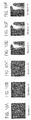
- FIG. 7(A-F) shows segmentation results on a CT head scan in accordance with an embodiment of the present invention.
- FIG. 8(A-F) shows segmentation results on a CT head scan in accordance with an embodiment of the present invention.
- FIG. 9(A-F) shows segmentation results on a CT head scan in accordance with an embodiment of the present invention.
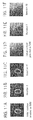
- FIG. 10(A-F) shows segmentation results on an astrophysics X-ray and on a bone X-ray in accordance with an embodiment of the present invention.
- FIG. 11(A-F) illustrates exemplary clinical acceleration results in accordance with an embodiment of the present invention.
- Certain embodiments provide triple-region segmentation using a single level set function to segment the three regions of an image.
- the level set function formulates triple-region segmentation as two dual-region segmentations. Use of one level set function enables the solution to be faster and more robust than prior, multiple level set techniques.
- a pattern classifier is combined with level set to achieve a clinical segmentation for three region medical images.
- a pattern classifier and individual principal component analysis are used to accelerate pathological level set segmentation.
- An evolution of the level set function achieves a triple-region segmentation since an initial contour provided by a support vector machine (SVM) and a principal component analysis (PCA) is very close to a final contour.
- SVM support vector machine
- PCA principal component analysis
- triple-region segmentation is handled within a two region level set framework where only a single level set function is used. Since only a single level set function is used, the segmentation is much faster and more robust than techniques using multiple level set functions.
- individual principal component analysis and a support vector machine classifier-based clinical acceleration scheme are used to accelerate the segmentation.
- the clinical acceleration scheme takes the strengths of both machine learning and the level set method while limiting their weaknesses to achieve automatic and fast clinical segmentation.
- Certain embodiments are able to successfully segment the triple-region using a single level set function. Additionally, the presently described segmentation is robust in the placement of initial contour. While still quickly converging to a final image using the clinical acceleration scheme, certain embodiments can be used during pre-processing for automatic computer aided diagnosis and surgery, for example.
- FIG. 1 illustrates an example of triple region level set modeling.
- the three regions in the images are identified as bright region (BR), grey region (GR), and dark region (DR).
- BR bright region
- GR grey region
- DR dark region
- segmentation can be obtained by minimizing an energy function given as:
- E ⁇ ( ⁇ ) ⁇ ⁇ 1 ⁇ ⁇ ⁇ ⁇ BR ⁇ ( u - c BR ) 2 ⁇ BR 2 ⁇ ⁇ d x ⁇ ⁇ d y + ⁇ ⁇ 2 ⁇ ⁇ ⁇ ⁇ GR ⁇ ( u - c GR ) 2 ⁇ GR 2 ⁇ ⁇ d x ⁇ ⁇ d y + ⁇ ⁇ 3 ⁇ ⁇ ⁇ ⁇ DR ⁇ ( u - c DR ) 2 ⁇ DR 2 ⁇ ⁇ d x ⁇ ⁇ d y , ( 1 )
- c i and ⁇ i are a mean intensity value and a variance of a region ⁇ i , respectively, and ⁇ i and u are constants.
- E E ( ⁇ ) ⁇ 1 E LAP + ⁇ 2 E GAC (3), where ⁇ i are constants, the geodesic active contour (E GAC ) and edge functional (E LAP ) are defined in Eq. 4:
- E GAC ( C ) ⁇ g ( C ) dxdy
- E LAP ( C ) ⁇ C ⁇ ,n>ds+ ⁇ ⁇ C K u
- K u is a mean curvature of the level set function
- n is a unit vector normal to the curve
- ds is an arc length of curve C.
- the level set function ⁇ is derived from the function in Eq. 5:
- 2 ), and u ⁇ ⁇ u ⁇ K u
- segmentation includes a clinical acceleration scheme, which divides the segmentation into two stages: a training stage and a clinical segmentation stage.
- a training stage first, manually chosen representative images, which consist of three regions, are segmented by the level set segmentation method. Then, the results are used to train a support vector machine (SVM) after PCA.
- SVM support vector machine
- images are first classified by the trained SVM after PCA, which provides initial contours.
- An evolution of the level set function provides a final segmentation result.
- a global PCA is used.
- PCA feature extraction may be used to analyze an image and extract features from that image.
- one or more regions 210 in a brain scan image 220 are identified for classification and analysis.
- Individual PCA 230 is performed to extract features from the regions 210 .
- Eigen feature vectors (e.g., reduced dimension Eigen feature vectors) 240 may then be applied for feature classification.
- Classification information may be fed into an SVM, for example.
- FIG. 3 illustrates a block and flow diagram 300 for clinical accelerated segmentation in accordance with an embodiment of the present invention.
- representative images 310 from a training database 320 may be transmitted to a supervised segmentation engine 330 .
- Supervised segmentation 330 provides segmented results for a pattern classifier 340 including an SVM 345 .
- the pattern classifier 340 uses supervised segmentation results in conjunction with clinical image(s) 350 to produce feature extraction and classification from the clinical image(s) 350 .
- the SVM 345 may be trained on representative library images and then used to process obtained clinical images for segmentation.
- the pattern classifier 340 uses PCA to reduce multidimensional image data sets to lower or reduced dimensions for further analysis and vector generation. For example, eigen values and/or singular values may be determined for an image data set for SVM processing.
- Pattern classification results are fed to the SVM 345 to accelerate segmentation and provide an initial contour.
- the SVM 345 processes maps input vectors representing image data from pattern classification to a higher dimensional space in one or more hyperplanes for classification, for example.
- the pattern classifier 340 and SVM 345 may be used to form an initial region contour for one or more images based on vector data, for example.
- SVM 345 output is used with a single level set function to provide triple region segmentation 360 .
- a single level set function may be used with pattern classification SVM results to segment an image into bright, dark, and grey regions. Segmentations results 370 are provided for doctor's analysis and/or further storage and/or use.
- FIG. 4 illustrates a flow diagram for a method 400 for triple region segmentation using a single level set function in accordance with an embodiment of the present invention.
- an initial contour is formed for an image using PCA and SVM.
- PCA is performed on one or more reference images which are then used to train the SVM.
- PCA is performed on one or more obtained clinical images which are then passed to the SVM to generate vectors indicating contour for regional segmentation.
- triple region segmentation is performed on one or more clinical images using a single level set function.
- a hybrid level set function that combines reduced variance, an edge integrator, and a geodesic active contour model is used to operate on the initial contour established by PCA and SVM to segment three regions (e.g., a bright region, a dark region, and a grey region) in the clinical image(s).
- One or more of the steps of the flow diagram for the method 400 may be implemented alone or in combination in hardware, firmware, and/or as a set of instructions in software, for example. Certain embodiments may be provided as a set of instructions residing on a computer-readable medium, such as a memory, hard disk, DVD, or CD, for execution on a general purpose computer or other processing device.
- a computer-readable medium such as a memory, hard disk, DVD, or CD
- Certain embodiments of the present invention may omit one or more of these steps and/or perform the steps in a different order than the order listed. For example, some steps may not be performed in certain embodiments of the present invention. As a further example, certain steps may be performed in a different temporal order, including simultaneously, than listed above.
- FIGS. 5 and 6 illustrate use of the level set segmentation method with a synthesized triple-region image.
- the original image consists of three Gaussian distributed regions.
- the proposed level set method successfully segments the grey region from the dark region and bright region with one level set function.
- one level set function is much faster and more robust.
- the segmentation method is robust in positioning the initial contour. Regardless of the initial contour placement, the level set is still able to converge to the final correct segmentation, as shown in FIGS. 5 and 6 .
- FIGS. 7 to 9 show segmentation results on CT head scan.
- FIG. 7 shows an evolution of zero contour
- FIG. 8 shows a region segmentation corresponding to zero curve evolution.
- FIG. 9 shows a contour and region evolution on another scan slice.
- FIGS. 10( a - c ) show segmentation results on an astrophysics X-ray. Even though the image is very noisy and boundaries are also very unclear, the segmentation can still be achieved.
- FIGS. 10( d - f ) show the segmentation results on a bone X-ray.
- FIG. 11 illustrates exemplary clinical acceleration results. Since the PCA and SVM are able to provide initial boundaries close to the final boundaries, it takes only few iterations to achieve the final segmentation, as shown in the comparison of FIGS. 5 , 6 , 8 and 11 . Since individual PCA is able to reduce the dimension to 10% of the original dimension, SVM training and classification are accomplished more quickly. Therefore, the above-described segmentation methods may be used in real-time or substantially real-time application.
- certain embodiments provide a framework for triple region segmentation using level set on PACS. Certain embodiments provide a clinically application level set segmentation which provide fast, robust analysis and is able to integrate into PACS without requiring support from another platform. Use of one level set function provides an increase in speed and robustness compared to existing level set methods. Additionally, certain embodiments take advantage of historical data in a PACS. Based on machine learning techniques, speed of computation may be increased to meet clinical needs.
- Certain embodiments may be implemented in a computer readable medium having a set of instructions for execution by a computer, for example.
- the computer-readable medium and its instructions may be used to provide triple region segmentation of images as described above.
- certain embodiments provide a computer readable medium having a set of instructions for execution on a computer.
- the set of instructions includes a principal component analysis routine extracting and classifying features of an image to form feature vectors for an initial contour in the image as described above.
- the set of instructions also includes a triple region segmentation routine segmenting the image into three regions using a single level set function based on the initial contour as described above.
- interface(s) and system(s) described above may be implemented alone or in combination in various forms in hardware, firmware, and/or as a set of instructions in software, for example.
- Certain embodiments may be provided as a set of instructions residing on a computer-readable medium, such as a memory or hard disk, for execution on a general purpose computer or other processing device, such as, for example, a PACS workstation or one or more dedicated processors.
- machine-readable media for carrying or having machine-executable instructions or data structures stored thereon.
- Such machine-readable media can be any available media that can be accessed by a general purpose or special purpose computer or other machine with a processor.
- machine-readable media may comprise RAM, ROM, PROM, EPROM, EEPROM, Flash, CD-ROM or other optical disk storage, magnetic disk storage or other magnetic storage devices, or any other medium which can be used to carry or store desired program code in the form of machine-executable instructions or data structures and which can be accessed by a general purpose or special purpose computer or other machine with a processor.
- Machine-executable instructions comprise, for example, instructions and data which cause a general purpose computer, special purpose computer, or special purpose processing machines to perform a certain function or group of functions.
- Certain embodiments of the invention are described in the general context of method steps which may be implemented in one embodiment by a program product including machine-executable instructions, such as program code, for example in the form of program modules executed by machines in networked environments.
- program modules include routines, programs, objects, components, data structures, etc., that perform particular tasks or implement particular abstract data types.
- Machine-executable instructions, associated data structures, and program modules represent examples of program code for executing steps of the methods disclosed herein.
- the particular sequence of such executable instructions or associated data structures represent examples of corresponding acts for implementing the functions described in such steps.
- Logical connections may include a local area network (LAN) and a wide area network (WAN) that are presented here by way of example and not limitation.
- LAN local area network
- WAN wide area network
- Such networking environments are commonplace in office-wide or enterprise-wide computer networks, intranets and the Internet and may use a wide variety of different communication protocols.
- Those skilled in the art will appreciate that such network computing environments will typically encompass many types of computer system configurations, including personal computers, hand-held devices, multi-processor systems, microprocessor-based or programmable consumer electronics, network PCs, minicomputers, mainframe computers, and the like.
- Embodiments of the invention may also be practiced in distributed computing environments where tasks are performed by local and remote processing devices that are linked (either by hardwired links, wireless links, or by a combination of hardwired or wireless links) through a communications network.
- program modules may be located in both local and remote memory storage devices.
- An exemplary system for implementing the overall system or portions of the invention might include a general purpose computing device in the form of a computer, including a processing unit, a system memory, and a system bus that couples various system components including the system memory to the processing unit.
- the system memory may include read only memory (ROM) and random access memory (RAM).
- the computer may also include a magnetic hard disk drive for reading from and writing to a magnetic hard disk, a magnetic disk drive for reading from or writing to a removable magnetic disk, and an optical disk drive for reading from or writing to a removable optical disk such as a CD ROM or other optical media.
- the drives and their associated machine-readable media provide nonvolatile storage of machine-executable instructions, data structures, program modules and other data for the computer.
Abstract
Description
where ci and σi are a mean intensity value and a variance of a region Ωi, respectively, and λi and u are constants.
where the function Min(x, y) returns the smaller value of x and y. Using Eq. 2, the triple-region segmentation is formulated to simultaneously solve two dual region segmentation problems: grey region versus dark region and grey region versus bright region.
E=E(Φ)−γ1 E LAP+γ2 E GAC (3),
where γi are constants, the geodesic active contour (EGAC) and edge functional (ELAP) are defined in Eq. 4:
E GAC(C)=∫∫g(C)dxdy
E LAP(C)=∫C <∇,n>ds+∫∫ Ω
In Eq. 4, Ku is a mean curvature of the level set function, n is a unit vector normal to the curve and ds is an arc length of curve C. Function g(x, y) is an inverse edge indicator function, which is defined as g(x, y)=α2/(α2+|∇u|2), where α is a constant and ∇ is a gradient operator.
where H(•) is a Heaviside function, div(•) is a divergence operator, and g(x, y)=α2/(α2+|∇u|2), and uζζ=Δu−Ku|∇u|.
Claims (15)
Priority Applications (1)
| Application Number | Priority Date | Filing Date | Title |
|---|---|---|---|
| US11/854,888 US8073253B2 (en) | 2006-09-29 | 2007-09-13 | Machine learning based triple region segmentation framework using level set on PACS |
Applications Claiming Priority (3)
| Application Number | Priority Date | Filing Date | Title |
|---|---|---|---|
| US82757906P | 2006-09-29 | 2006-09-29 | |
| US82782206P | 2006-10-02 | 2006-10-02 | |
| US11/854,888 US8073253B2 (en) | 2006-09-29 | 2007-09-13 | Machine learning based triple region segmentation framework using level set on PACS |
Publications (2)
| Publication Number | Publication Date |
|---|---|
| US20080080768A1 US20080080768A1 (en) | 2008-04-03 |
| US8073253B2 true US8073253B2 (en) | 2011-12-06 |
Family
ID=39261272
Family Applications (1)
| Application Number | Title | Priority Date | Filing Date |
|---|---|---|---|
| US11/854,888 Expired - Fee Related US8073253B2 (en) | 2006-09-29 | 2007-09-13 | Machine learning based triple region segmentation framework using level set on PACS |
Country Status (1)
| Country | Link |
|---|---|
| US (1) | US8073253B2 (en) |
Cited By (3)
| Publication number | Priority date | Publication date | Assignee | Title |
|---|---|---|---|---|
| US20110172514A1 (en) * | 2008-09-29 | 2011-07-14 | Koninklijke Philips Electronics N.V. | Method for increasing the robustness of computer-aided diagnosis to image processing uncertainties |
| US10467504B1 (en) * | 2019-02-08 | 2019-11-05 | Adhark, Inc. | Systems, methods, and storage media for evaluating digital images |
| US11531840B2 (en) | 2019-02-08 | 2022-12-20 | Vizit Labs, Inc. | Systems, methods, and storage media for training a model for image evaluation |
Families Citing this family (10)
| Publication number | Priority date | Publication date | Assignee | Title |
|---|---|---|---|---|
| US7088872B1 (en) * | 2002-02-14 | 2006-08-08 | Cogent Systems, Inc. | Method and apparatus for two dimensional image processing |
| US8131477B2 (en) | 2005-11-16 | 2012-03-06 | 3M Cogent, Inc. | Method and device for image-based biological data quantification |
| US8275179B2 (en) * | 2007-05-01 | 2012-09-25 | 3M Cogent, Inc. | Apparatus for capturing a high quality image of a moist finger |
| US8411916B2 (en) * | 2007-06-11 | 2013-04-02 | 3M Cogent, Inc. | Bio-reader device with ticket identification |
| JP5041229B2 (en) * | 2007-12-07 | 2012-10-03 | ソニー株式会社 | Learning device and method, recognition device and method, and program |
| US20100014755A1 (en) * | 2008-07-21 | 2010-01-21 | Charles Lee Wilson | System and method for grid-based image segmentation and matching |
| CN102496155A (en) * | 2011-10-28 | 2012-06-13 | 河海大学 | Underwater optical image processing method for optimizing C-V (chan-vese) model |
| EP3488413A4 (en) * | 2016-07-22 | 2020-02-12 | Université Laval | System and method for estimating synthetic quantitative health values from medical images |
| EP3631808A1 (en) * | 2017-05-31 | 2020-04-08 | Koninklijke Philips N.V. | Machine learning on raw medical imaging data for clinical decision support |
| CN110992370B (en) * | 2019-11-19 | 2023-07-04 | 东软医疗系统股份有限公司 | Pancreas tissue segmentation method and device and terminal equipment |
Citations (4)
| Publication number | Priority date | Publication date | Assignee | Title |
|---|---|---|---|---|
| US5527645A (en) * | 1993-04-21 | 1996-06-18 | Pati; Yagyensh C. | Systematic method for production of phase-shifting photolithographic masks |
| US5757382A (en) * | 1994-12-29 | 1998-05-26 | Daewoo Electronics Co., Ltd. | Apparatus for tracing contours of segmented regions |
| US5943441A (en) * | 1995-12-06 | 1999-08-24 | Cognex Corporation | Edge contour tracking from a first edge point |
| US7903861B2 (en) * | 2006-12-28 | 2011-03-08 | Carestream Health, Inc. | Method for classifying breast tissue density using computed image features |
-
2007
- 2007-09-13 US US11/854,888 patent/US8073253B2/en not_active Expired - Fee Related
Patent Citations (4)
| Publication number | Priority date | Publication date | Assignee | Title |
|---|---|---|---|---|
| US5527645A (en) * | 1993-04-21 | 1996-06-18 | Pati; Yagyensh C. | Systematic method for production of phase-shifting photolithographic masks |
| US5757382A (en) * | 1994-12-29 | 1998-05-26 | Daewoo Electronics Co., Ltd. | Apparatus for tracing contours of segmented regions |
| US5943441A (en) * | 1995-12-06 | 1999-08-24 | Cognex Corporation | Edge contour tracking from a first edge point |
| US7903861B2 (en) * | 2006-12-28 | 2011-03-08 | Carestream Health, Inc. | Method for classifying breast tissue density using computed image features |
Cited By (13)
| Publication number | Priority date | Publication date | Assignee | Title |
|---|---|---|---|---|
| US20110172514A1 (en) * | 2008-09-29 | 2011-07-14 | Koninklijke Philips Electronics N.V. | Method for increasing the robustness of computer-aided diagnosis to image processing uncertainties |
| US9123095B2 (en) * | 2008-09-29 | 2015-09-01 | Koninklijke Philips N.V. | Method for increasing the robustness of computer-aided diagnosis to image processing uncertainties |
| US10467504B1 (en) * | 2019-02-08 | 2019-11-05 | Adhark, Inc. | Systems, methods, and storage media for evaluating digital images |
| US11113572B2 (en) * | 2019-02-08 | 2021-09-07 | Vizit Labs, Inc. | Systems, methods, and storage media for evaluating images |
| US20220058434A1 (en) * | 2019-02-08 | 2022-02-24 | Vizit Labs, Inc. | Systems, methods, and storage media for evaluating images |
| US11531840B2 (en) | 2019-02-08 | 2022-12-20 | Vizit Labs, Inc. | Systems, methods, and storage media for training a model for image evaluation |
| US11663811B2 (en) * | 2019-02-08 | 2023-05-30 | Vizit Labs, Inc. | Systems, methods, and storage media for evaluating images |
| US20230260250A1 (en) * | 2019-02-08 | 2023-08-17 | Vizit Labs, Inc. | Systems, methods, and storage media for evaluating images |
| US11768913B2 (en) | 2019-02-08 | 2023-09-26 | Vizit Labs, Inc. | Systems, methods, and storage media for training a model for image evaluation |
| US11783567B2 (en) * | 2019-02-08 | 2023-10-10 | Vizit Labs, Inc. | Systems, methods, and storage media for evaluating images |
| US11880429B1 (en) | 2019-02-08 | 2024-01-23 | Vizit Labs, Inc. | Systems, methods, and storage media for training a model for image evaluation |
| US20240037905A1 (en) * | 2019-02-08 | 2024-02-01 | Vizit Labs, Inc. | Systems, methods, and storage media for evaluating images |
| US11922674B2 (en) * | 2019-02-08 | 2024-03-05 | Vizit Labs, Inc. | Systems, methods, and storage media for evaluating images |
Also Published As
| Publication number | Publication date |
|---|---|
| US20080080768A1 (en) | 2008-04-03 |
Similar Documents
| Publication | Publication Date | Title |
|---|---|---|
| US8073253B2 (en) | Machine learning based triple region segmentation framework using level set on PACS | |
| Keraudren et al. | Automated fetal brain segmentation from 2D MRI slices for motion correction | |
| Masood et al. | A survey on medical image segmentation | |
| US9792689B2 (en) | Posture estimating apparatus, posture estimating method and storing medium | |
| EP2320379B1 (en) | Image processing device, object tracking device, and image processing method | |
| CN106340021B (en) | Blood vessel extraction method | |
| US7221787B2 (en) | Method for automated analysis of digital chest radiographs | |
| US20160217319A1 (en) | Unified face representation for individual recognition in surveillance videos and vehicle logo super-resolution system | |
| US20070185946A1 (en) | Method and apparatus for matching portions of input images | |
| US8913817B2 (en) | Rib suppression in radiographic images | |
| US9269139B2 (en) | Rib suppression in radiographic images | |
| US10249046B2 (en) | Method and apparatus for object tracking and segmentation via background tracking | |
| US20210068676A1 (en) | Method and system for automatically generating and analyzing fully quantitative pixel-wise myocardial blood flow and myocardial perfusion reserve maps to detect ischemic heart disease using cardiac perfusion magnetic resonance imaging | |
| Zhao et al. | Down syndrome detection from facial photographs using machine learning techniques | |
| Li et al. | An automatic variational level set segmentation framework for computer aided dental X-rays analysis in clinical environments | |
| RU2654199C1 (en) | Segmentation of human tissues in computer image | |
| US7024040B1 (en) | Image processing apparatus and method, and storage medium | |
| US20140140603A1 (en) | Clavicle suppression in radiographic images | |
| Zhang et al. | Tracking-assisted weakly supervised online visual object segmentation in unconstrained videos | |
| Kirschner et al. | Active shape models unleashed | |
| Jabbar et al. | FD-stackGAN: face de-occlusion using stacked generative adversarial networks | |
| US20150093001A1 (en) | Image segmentation system and operating method thereof | |
| Li et al. | Automatic clinical image segmentation using pathological modelling, PCA and SVM | |
| Sarmiento et al. | Cardiac disease prediction from spatio-temporal motion patterns in cine-mri | |
| Li et al. | Image segmentation adapted for clinical settings by combining pattern classification and level sets |
Legal Events
| Date | Code | Title | Description |
|---|---|---|---|
| AS | Assignment |
Owner name: GENERAL ELECTRIC COMPANY, NEW YORK Free format text: ASSIGNMENT OF ASSIGNORS INTEREST;ASSIGNORS:LI, SHOU;GILL, SUKHDEEP;ROEDER, DAVID WILLIAM;AND OTHERS;REEL/FRAME:019823/0184;SIGNING DATES FROM 20070911 TO 20070912 Owner name: GENERAL ELECTRIC COMPANY, NEW YORK Free format text: ASSIGNMENT OF ASSIGNORS INTEREST;ASSIGNORS:LI, SHOU;GILL, SUKHDEEP;ROEDER, DAVID WILLIAM;AND OTHERS;SIGNING DATES FROM 20070911 TO 20070912;REEL/FRAME:019823/0184 |
|
| ZAAA | Notice of allowance and fees due |
Free format text: ORIGINAL CODE: NOA |
|
| ZAAB | Notice of allowance mailed |
Free format text: ORIGINAL CODE: MN/=. |
|
| STCF | Information on status: patent grant |
Free format text: PATENTED CASE |
|
| CC | Certificate of correction | ||
| FPAY | Fee payment |
Year of fee payment: 4 |
|
| CC | Certificate of correction | ||
| MAFP | Maintenance fee payment |
Free format text: PAYMENT OF MAINTENANCE FEE, 8TH YEAR, LARGE ENTITY (ORIGINAL EVENT CODE: M1552); ENTITY STATUS OF PATENT OWNER: LARGE ENTITY Year of fee payment: 8 |
|
| FEPP | Fee payment procedure |
Free format text: MAINTENANCE FEE REMINDER MAILED (ORIGINAL EVENT CODE: REM.); ENTITY STATUS OF PATENT OWNER: LARGE ENTITY |
|
| LAPS | Lapse for failure to pay maintenance fees |
Free format text: PATENT EXPIRED FOR FAILURE TO PAY MAINTENANCE FEES (ORIGINAL EVENT CODE: EXP.); ENTITY STATUS OF PATENT OWNER: LARGE ENTITY |
|
| STCH | Information on status: patent discontinuation |
Free format text: PATENT EXPIRED DUE TO NONPAYMENT OF MAINTENANCE FEES UNDER 37 CFR 1.362 |
|
| FP | Lapsed due to failure to pay maintenance fee |
Effective date: 20231206 |