US8758286B2 - Method for removing cytokines from blood with surface immobilized polysaccharides - Google Patents

Method for removing cytokines from blood with surface immobilized polysaccharides Download PDF

Info

Publication number
US8758286B2
US8758286B2 US12/958,355 US95835510A US8758286B2 US 8758286 B2 US8758286 B2 US 8758286B2 US 95835510 A US95835510 A US 95835510A US 8758286 B2 US8758286 B2 US 8758286B2
Authority
US
United States
Prior art keywords
blood
beads
substrate
heparin
adsorbate
Prior art date
Legal status (The legal status is an assumption and is not a legal conclusion. Google has not performed a legal analysis and makes no representation as to the accuracy of the status listed.)
Active, expires
Application number
US12/958,355
Other versions
US20110184377A1 (en
Inventor
Robert S Ward
Keith R. McCrea
Olle Larm
Lars Adolfsson
Current Assignee (The listed assignees may be inaccurate. Google has not performed a legal analysis and makes no representation or warranty as to the accuracy of the list.)
EXTHERA MEDICAL LLC
Exthera Medical Corp
Original Assignee
Exthera Medical Corp
Priority date (The priority date is an assumption and is not a legal conclusion. Google has not performed a legal analysis and makes no representation as to the accuracy of the date listed.)
Filing date
Publication date
Application filed by Exthera Medical Corp filed Critical Exthera Medical Corp
Priority to US12/958,355 priority Critical patent/US8758286B2/en
Assigned to EXTHERA MEDICAL, LLC reassignment EXTHERA MEDICAL, LLC ASSIGNMENT OF ASSIGNORS INTEREST (SEE DOCUMENT FOR DETAILS). Assignors: ADOLFSSON, LARS, LARM, OLLE, MCCREA, KEITH R., WARD, ROBERT S.
Publication of US20110184377A1 publication Critical patent/US20110184377A1/en
Assigned to EXTHERA MEDICAL CORPORATION reassignment EXTHERA MEDICAL CORPORATION ASSIGNMENT OF ASSIGNORS INTEREST (SEE DOCUMENT FOR DETAILS). Assignors: EXTHERA MEDICAL, LLC
Priority to US14/261,390 priority patent/US9408962B2/en
Application granted granted Critical
Publication of US8758286B2 publication Critical patent/US8758286B2/en
Priority to US15/199,737 priority patent/US10086126B2/en
Priority to US16/113,904 priority patent/US11123466B2/en
Active legal-status Critical Current
Adjusted expiration legal-status Critical

Links

Images

Classifications

    • AHUMAN NECESSITIES
    • A61MEDICAL OR VETERINARY SCIENCE; HYGIENE
    • A61KPREPARATIONS FOR MEDICAL, DENTAL OR TOILETRY PURPOSES
    • A61K31/00Medicinal preparations containing organic active ingredients
    • A61K31/60Salicylic acid; Derivatives thereof
    • AHUMAN NECESSITIES
    • A61MEDICAL OR VETERINARY SCIENCE; HYGIENE
    • A61KPREPARATIONS FOR MEDICAL, DENTAL OR TOILETRY PURPOSES
    • A61K31/00Medicinal preparations containing organic active ingredients
    • A61K31/70Carbohydrates; Sugars; Derivatives thereof
    • A61K31/715Polysaccharides, i.e. having more than five saccharide radicals attached to each other by glycosidic linkages; Derivatives thereof, e.g. ethers, esters
    • A61K31/716Glucans
    • A61K31/722Chitin, chitosan
    • AHUMAN NECESSITIES
    • A61MEDICAL OR VETERINARY SCIENCE; HYGIENE
    • A61KPREPARATIONS FOR MEDICAL, DENTAL OR TOILETRY PURPOSES
    • A61K31/00Medicinal preparations containing organic active ingredients
    • A61K31/70Carbohydrates; Sugars; Derivatives thereof
    • A61K31/715Polysaccharides, i.e. having more than five saccharide radicals attached to each other by glycosidic linkages; Derivatives thereof, e.g. ethers, esters
    • A61K31/726Glycosaminoglycans, i.e. mucopolysaccharides
    • A61K31/727Heparin; Heparan
    • AHUMAN NECESSITIES
    • A61MEDICAL OR VETERINARY SCIENCE; HYGIENE
    • A61KPREPARATIONS FOR MEDICAL, DENTAL OR TOILETRY PURPOSES
    • A61K31/00Medicinal preparations containing organic active ingredients
    • A61K31/70Carbohydrates; Sugars; Derivatives thereof
    • A61K31/715Polysaccharides, i.e. having more than five saccharide radicals attached to each other by glycosidic linkages; Derivatives thereof, e.g. ethers, esters
    • A61K31/726Glycosaminoglycans, i.e. mucopolysaccharides
    • A61K31/728Hyaluronic acid
    • AHUMAN NECESSITIES
    • A61MEDICAL OR VETERINARY SCIENCE; HYGIENE
    • A61MDEVICES FOR INTRODUCING MEDIA INTO, OR ONTO, THE BODY; DEVICES FOR TRANSDUCING BODY MEDIA OR FOR TAKING MEDIA FROM THE BODY; DEVICES FOR PRODUCING OR ENDING SLEEP OR STUPOR
    • A61M1/00Suction or pumping devices for medical purposes; Devices for carrying-off, for treatment of, or for carrying-over, body-liquids; Drainage systems
    • A61M1/34Filtering material out of the blood by passing it through a membrane, i.e. hemofiltration or diafiltration
    • A61M1/3472Filtering material out of the blood by passing it through a membrane, i.e. hemofiltration or diafiltration with treatment of the filtrate
    • A61M1/3486Biological, chemical treatment, e.g. chemical precipitation; treatment by absorbents
    • AHUMAN NECESSITIES
    • A61MEDICAL OR VETERINARY SCIENCE; HYGIENE
    • A61MDEVICES FOR INTRODUCING MEDIA INTO, OR ONTO, THE BODY; DEVICES FOR TRANSDUCING BODY MEDIA OR FOR TAKING MEDIA FROM THE BODY; DEVICES FOR PRODUCING OR ENDING SLEEP OR STUPOR
    • A61M1/00Suction or pumping devices for medical purposes; Devices for carrying-off, for treatment of, or for carrying-over, body-liquids; Drainage systems
    • A61M1/34Filtering material out of the blood by passing it through a membrane, i.e. hemofiltration or diafiltration
    • A61M1/3496Plasmapheresis; Leucopheresis; Lymphopheresis
    • AHUMAN NECESSITIES
    • A61MEDICAL OR VETERINARY SCIENCE; HYGIENE
    • A61MDEVICES FOR INTRODUCING MEDIA INTO, OR ONTO, THE BODY; DEVICES FOR TRANSDUCING BODY MEDIA OR FOR TAKING MEDIA FROM THE BODY; DEVICES FOR PRODUCING OR ENDING SLEEP OR STUPOR
    • A61M1/00Suction or pumping devices for medical purposes; Devices for carrying-off, for treatment of, or for carrying-over, body-liquids; Drainage systems
    • A61M1/36Other treatment of blood in a by-pass of the natural circulatory system, e.g. temperature adaptation, irradiation ; Extra-corporeal blood circuits
    • A61M1/362Other treatment of blood in a by-pass of the natural circulatory system, e.g. temperature adaptation, irradiation ; Extra-corporeal blood circuits changing physical properties of target cells by binding them to added particles to facilitate their subsequent separation from other cells, e.g. immunoaffinity
    • AHUMAN NECESSITIES
    • A61MEDICAL OR VETERINARY SCIENCE; HYGIENE
    • A61MDEVICES FOR INTRODUCING MEDIA INTO, OR ONTO, THE BODY; DEVICES FOR TRANSDUCING BODY MEDIA OR FOR TAKING MEDIA FROM THE BODY; DEVICES FOR PRODUCING OR ENDING SLEEP OR STUPOR
    • A61M1/00Suction or pumping devices for medical purposes; Devices for carrying-off, for treatment of, or for carrying-over, body-liquids; Drainage systems
    • A61M1/36Other treatment of blood in a by-pass of the natural circulatory system, e.g. temperature adaptation, irradiation ; Extra-corporeal blood circuits
    • A61M1/3621Extra-corporeal blood circuits
    • AHUMAN NECESSITIES
    • A61MEDICAL OR VETERINARY SCIENCE; HYGIENE
    • A61MDEVICES FOR INTRODUCING MEDIA INTO, OR ONTO, THE BODY; DEVICES FOR TRANSDUCING BODY MEDIA OR FOR TAKING MEDIA FROM THE BODY; DEVICES FOR PRODUCING OR ENDING SLEEP OR STUPOR
    • A61M1/00Suction or pumping devices for medical purposes; Devices for carrying-off, for treatment of, or for carrying-over, body-liquids; Drainage systems
    • A61M1/36Other treatment of blood in a by-pass of the natural circulatory system, e.g. temperature adaptation, irradiation ; Extra-corporeal blood circuits
    • A61M1/3672Means preventing coagulation
    • A61M1/3673Anticoagulant coating, e.g. Heparin coating
    • AHUMAN NECESSITIES
    • A61MEDICAL OR VETERINARY SCIENCE; HYGIENE
    • A61MDEVICES FOR INTRODUCING MEDIA INTO, OR ONTO, THE BODY; DEVICES FOR TRANSDUCING BODY MEDIA OR FOR TAKING MEDIA FROM THE BODY; DEVICES FOR PRODUCING OR ENDING SLEEP OR STUPOR
    • A61M1/00Suction or pumping devices for medical purposes; Devices for carrying-off, for treatment of, or for carrying-over, body-liquids; Drainage systems
    • A61M1/36Other treatment of blood in a by-pass of the natural circulatory system, e.g. temperature adaptation, irradiation ; Extra-corporeal blood circuits
    • A61M1/3679Other treatment of blood in a by-pass of the natural circulatory system, e.g. temperature adaptation, irradiation ; Extra-corporeal blood circuits by absorption
    • AHUMAN NECESSITIES
    • A61MEDICAL OR VETERINARY SCIENCE; HYGIENE
    • A61MDEVICES FOR INTRODUCING MEDIA INTO, OR ONTO, THE BODY; DEVICES FOR TRANSDUCING BODY MEDIA OR FOR TAKING MEDIA FROM THE BODY; DEVICES FOR PRODUCING OR ENDING SLEEP OR STUPOR
    • A61M1/00Suction or pumping devices for medical purposes; Devices for carrying-off, for treatment of, or for carrying-over, body-liquids; Drainage systems
    • A61M1/36Other treatment of blood in a by-pass of the natural circulatory system, e.g. temperature adaptation, irradiation ; Extra-corporeal blood circuits
    • A61M1/3687Chemical treatment
    • AHUMAN NECESSITIES
    • A61MEDICAL OR VETERINARY SCIENCE; HYGIENE
    • A61MDEVICES FOR INTRODUCING MEDIA INTO, OR ONTO, THE BODY; DEVICES FOR TRANSDUCING BODY MEDIA OR FOR TAKING MEDIA FROM THE BODY; DEVICES FOR PRODUCING OR ENDING SLEEP OR STUPOR
    • A61M1/00Suction or pumping devices for medical purposes; Devices for carrying-off, for treatment of, or for carrying-over, body-liquids; Drainage systems
    • A61M1/36Other treatment of blood in a by-pass of the natural circulatory system, e.g. temperature adaptation, irradiation ; Extra-corporeal blood circuits
    • A61M1/38Removing constituents from donor blood and storing or returning remainder to body, e.g. for transfusion
    • AHUMAN NECESSITIES
    • A61MEDICAL OR VETERINARY SCIENCE; HYGIENE
    • A61PSPECIFIC THERAPEUTIC ACTIVITY OF CHEMICAL COMPOUNDS OR MEDICINAL PREPARATIONS
    • A61P1/00Drugs for disorders of the alimentary tract or the digestive system
    • A61P1/04Drugs for disorders of the alimentary tract or the digestive system for ulcers, gastritis or reflux esophagitis, e.g. antacids, inhibitors of acid secretion, mucosal protectants
    • AHUMAN NECESSITIES
    • A61MEDICAL OR VETERINARY SCIENCE; HYGIENE
    • A61PSPECIFIC THERAPEUTIC ACTIVITY OF CHEMICAL COMPOUNDS OR MEDICINAL PREPARATIONS
    • A61P17/00Drugs for dermatological disorders
    • A61P17/02Drugs for dermatological disorders for treating wounds, ulcers, burns, scars, keloids, or the like
    • AHUMAN NECESSITIES
    • A61MEDICAL OR VETERINARY SCIENCE; HYGIENE
    • A61PSPECIFIC THERAPEUTIC ACTIVITY OF CHEMICAL COMPOUNDS OR MEDICINAL PREPARATIONS
    • A61P31/00Antiinfectives, i.e. antibiotics, antiseptics, chemotherapeutics
    • AHUMAN NECESSITIES
    • A61MEDICAL OR VETERINARY SCIENCE; HYGIENE
    • A61PSPECIFIC THERAPEUTIC ACTIVITY OF CHEMICAL COMPOUNDS OR MEDICINAL PREPARATIONS
    • A61P31/00Antiinfectives, i.e. antibiotics, antiseptics, chemotherapeutics
    • A61P31/04Antibacterial agents
    • AHUMAN NECESSITIES
    • A61MEDICAL OR VETERINARY SCIENCE; HYGIENE
    • A61PSPECIFIC THERAPEUTIC ACTIVITY OF CHEMICAL COMPOUNDS OR MEDICINAL PREPARATIONS
    • A61P37/00Drugs for immunological or allergic disorders
    • A61P37/02Immunomodulators
    • A61P37/06Immunosuppressants, e.g. drugs for graft rejection
    • AHUMAN NECESSITIES
    • A61MEDICAL OR VETERINARY SCIENCE; HYGIENE
    • A61MDEVICES FOR INTRODUCING MEDIA INTO, OR ONTO, THE BODY; DEVICES FOR TRANSDUCING BODY MEDIA OR FOR TAKING MEDIA FROM THE BODY; DEVICES FOR PRODUCING OR ENDING SLEEP OR STUPOR
    • A61M2202/00Special media to be introduced, removed or treated
    • A61M2202/0057Special media to be introduced, removed or treated retained by adsorption

Definitions

  • the present invention is directed to a method for removing cytokines and/or pathogens from blood or blood serum (blood) by contacting the blood with a solid, essentially non micro-porous substrate which has been surface treated with a polysaccharide adsorbent, such as heparin, heparan sulfate and/or other molecules or chemical groups (the adsorbent media or media) having a binding affinity for the cytokine or pathogen(s) to be removed (the adsorbates), and wherein the size of the interstitial channels within said media is balanced with the amount of media surface area and the surface concentration of binding sites on the media in order to provide adequate adsorptive capacity while also allowing relatively high flow rates of blood through the adsorbent media.
  • a polysaccharide adsorbent such as heparin, heparan sulfate and/or other molecules or chemical groups
  • forced convection is meant, for example, flow produced by a pressure gradient generated by a pump, by the application of external pressure to a flexible container (or internal pressure to a rigid container), by a gravity head/elevation difference, or by the difference in arterial pressure and venous pressure in the patient being treated.
  • the invention provides clinically-relevant adsorbtive capacity within the range of safe flow rates typically used in clinical extracorporeal blood circuits, e.g., in dialysis, cardiopulmonary bypass, and extra corporeal membrane oxygenation of blood.
  • the method is in direct contrast to the much slower diffusive transport of adsorbates typically required with porous adsorbent media, which require adsorbates to diffuse through a microporous membrane, and/or into microscopic pores before binding to adsorption sites on, behind, or within the media, and which therefore require very low flow rates to achieve significant separations during each passage of blood.
  • the present invention also provides a method of treating a disease by removing cytokines and/or pathogens from blood by contacting blood with an essentially nonporous substrate coated with a polysaccharaide adsorbent, such as heparin, heparan sulphate, and/or, other adsorbent materials, and a device for performing the method and treatment.
  • a wide variety of disease conditions are characterized by the presence of elevated concentrations of cytokines and/or pathogens in the blood stream. Some such conditions are treated by therapies designed to kill the pathogen, e.g. through the administration of drugs, e.g., anti-infective pharmaceuticals. Some other conditions are treated by therapies that attempt to reduce the concentration of blood-borne cytokines or pathogens in the patient. Other diseases are treated by therapies that attempt to directly remove only specific components from the patient's blood.
  • Guillian-Barre syndrome is currently understood to be an autoimmune disorder triggered by viral infection that stimulates the body's immune system to over produce antibodies or other proteins which can attack the patient's nervous system, causing increasing levels of paralysis. Most patients recover over time, though such patients appear to be susceptible to recurrance of the condition from subsequent viral infections.
  • One method for treating Guillian-Barre syndrome involves plasmapheresis to ‘clean’ the patient's blood by removing antibodies believed to be attacking the patient's nervous system.
  • Certain biologically active carbohydrates and polysaccharides can remove harmful substances from blood and biological fluids.
  • Heparin is a polysaccharide that can be isolated from mammalian tissue. It has a very specific distribution in mammalian tissue; being present only in the basophilic granules of mast cells. Since its discovery in 1916 by the American scientist McLean, heparin has been recognized for its ability to prevent blood from clotting, and for its relatively short half-life in the body. Systemic heparin, administered by injection of the free drug, has been used clinically for more than 50 years as a safe and effective blood anticoagulant and antithrombotic agent. The effects of heparin on blood coagulation/clotting diminish fairly quickly after administration is halted, making its use during surgery and other procedures effective and safe.
  • heparin's anticoagulant and antithrombogenic properties are useful during many medical procedures, for example to minimize undesirable interactions between blood and the man-made surfaces of extracorporeal circuits.
  • the administration of heparin may then be terminated.
  • the heparin concentration in the patient's blood diminishes to a safe level within a few hours because of its short half life in the body. This is particularly important following surgery when healing depends on the ability of blood to clot at the surgical site to avoid bleeding complications.
  • heparin has more recently been found to have a wide range of other functions apparently unrelated to its function as an anticoagulant.
  • heparin and/or the closely-related polysaccharide heparan sulfate which is also found in animal tissue, including the blood-contacting luminal surface of healthy blood vessels (where it may contribute to preventing circulating blood from clotting on contact with the walls of the blood vessels).
  • Some examples are antithrombin (AT), fibronectin, vitronectin, growth factors (e.g. the fibroblast growth factors, the insulin like growth factors, etc.).
  • Human serum albumin (HSA) also binds to heparin, but with a lower affinity despite its high concentration in blood.
  • the present invention does not require the use of any free, systemic heparin for efficacy, and thus may eliminate bleeding complications. This is accomplished by permanently binding the heparin or heparan sulphate to a solid substrate with high surface area, and exposing it to the blood within a cartridge or filter containing this adsorption media.
  • An object of the present invention is to provide a method for the removal of cytokines and/or pathogens from mammalian blood by contacting blood with a solid, essentially nonporous substrate coated with selective adsorbent molecules, biomolecules or chemical groups.
  • selectively adsorbent molecules may include polysaccharides, such as heparin, heparan sulphate, polyethylene imine (PEI), sialic acid, hyaluronic acid, and carbohydrates with mannose sequences.
  • One object of the invention is to provide a therapy for treating an existing disease by removing cytokines and/or pathogens from mammalian blood by contacting mammalian blood with a solid essentially nonporous substrate coated with heparin and/or other adsorbent molecules and returning the blood to the patient suffering from the disease.
  • FIG. 1 is a graph of TNF- ⁇ binding to heparinized beads.
  • FIG. 2A is a graph of the amount of TNF- ⁇ adsorbed on the heparinized crystals.
  • FIG. 2B is a graph of the amount of TNF- ⁇ adsorbed onto control crystals.
  • a first aspect of the present invention provides a method for the removal of cytokines and/or pathogens from blood, such as mammalian blood, by contacting the blood with a solid substrate e.g., coated with heparin and/or other adsorbent carbohydrates and/or polysaccharides.
  • heparin is immobilized onto the surface of the substrate.
  • the inventors have found that immobilized heparin bound to a surface is effective for removing a significant amount of cytokines and pathogens from blood.
  • the flow rates typical of extracorporeal blood circuits require that the adsorbent ‘bed’ be designed to allow relatively high flow rates to operate safely. This is in part due to the universal tendency of slow-moving or stagnant blood to form dangerous clots.
  • the substrate is designed with sufficiently large interstitial dimensions to permit a high flow rate of blood over the substrate without a large pressure drop.
  • adsorbent bed is characterized primarily by forced convection.
  • highly porous adsorbent media e.g. porous silica, sephadex, crosslinked polystyrene and other size exclusion media
  • Molecular diffusion is also required when selectively-permeable barrier membranes are used together with adsorption media, e.g., to prevent contact of the adsorption media by blood cells and/or high molecular weight solutes during affinity therapy.
  • cytokines and pathogens by heparin and/or other adsorbent molecules during convection transport is particularly effective under the relatively high-flow conditions typically employed in the (safe) operation of extracorporeal blood circuits, e.g. when measured by linear flow velocity, ⁇ 8 cm/min, preferably about ⁇ 24 cm/min, and more preferably about 24-329 cm/minute, or, when measured by flow rate, around >50 mL/minute and preferably >150 mL/minute but less than about 2000 mL/minute.
  • Adsorption within the pores of microporous media may require much lower flow rates through adsorption beds of practical size in order to achieve an adequate separation or purification, i.e. ⁇ 50 mL/min to as low as ⁇ 1 mL/min!
  • Convection kinetics and diffusion kinetics can be compared in the removal of cytokines or pathogens from flowing blood:
  • Adsorption media that depend on diffusion transport generally use very porous materials with extremely high internal surface area due to the presence of microscopic pores.
  • Media suited for convection transport generally rely on macroscopic “channels” or visible interstices between solid, essential nonporous material, such as particles, beads, fibers, reticulated foams, or optionally spiral-wound dense membranes.
  • the present invention therefore, provides a method and device for removing cytokines and/or pathogens from blood using the preferred characteristics of convection transport and its desirable, more-rapid kinetics. This is achieved by passing/flowing blood over an essentially non-microporous substrate that has been surface treated with adsorbent molecules, e.g. heparin, and which is therefore capable of binding the desired cytokine or pathogens to remove them from the blood. It is also possible to use a microporous substrate in the present invention if surface treatment renders that substrate effectively nonporous. This can occur intentionally or inadvertently, when surface treatments during media manufacturing block the pores. This converts the microporous substrate to one that does not require diffusion of adsorbate into pores to bind to the media.
  • adsorbent molecules e.g. heparin
  • Extracorporeal therapies means procedures that are conducted outside the body, such as therapies in which desired products like oxygen, blood-anticoagulants, anesthetics etc can be added to body fluids.
  • desired products like oxygen, blood-anticoagulants, anesthetics etc can be added to body fluids.
  • undesired products like naturally occurring toxins or poisons can be also removed from body fluids with specific types of extracorporeal circuits. Examples are haemodialysis and haemofiltration which represent technologies whereby blood is depleted of waste products. Adsorption on activated carbon has been used to remove blood-borne poisons, and so forth.
  • Whole blood and blood serum from mammals can be used in the present invention.
  • the amount of blood or blood serum that can be used in the claimed methods is not intended to be limited. It can range from less than 1 mL to above 1 L, up to and including the entire blood volume of the patient when continuous recirculation back to the patient is employed. One or more ‘passes’ through the adsorption bed may be used if needed.
  • the blood may be human or animal blood.
  • Adsorption media to remove cytokines or pathogens from blood are optimized according to the present invention for use in traditional extracorporeal blood circulation with flow rates >50 mL/min, and preferably between about 150 and 2000 mL/min. If measured by linear flow velocity, ⁇ 8 cm/min, preferably about ⁇ 24 cm/min and more preferably about 24-329 cm/min.
  • flow rates >50 mL/min and preferably between about 150 and 2000 mL/min. If measured by linear flow velocity, ⁇ 8 cm/min, preferably about ⁇ 24 cm/min and more preferably about 24-329 cm/min.
  • Such high flow rates create short residence times within the adsorption column and convection transport dominates over Brownian diffusive transport. This is particularly important for binding large MW proteins or cytokines such as TNF- ⁇ and larger particles such as viruses, bacteria and parasites because they diffuse very, very slowly.
  • the dominant adsorption sites available for removing cytokines and pathogens lie at the surfaces within the interstices of the media bed through which the blood flows or is delivered by forced convection.
  • the interstitial channels need to be large enough to allow the transport of red blood cells, which are an average 6 microns in diameter.
  • the interstitial channels must be several times larger than the diameter of red blood cells. This can prevent high shear rates that lead to hemolysis while simultaneously minimizing pressure drop in the blood that flows through the packed bed or cartridge.
  • the media is preferably rigid to minimize deformation that could clog the filter cartridge by compaction. Based on these preferences, an optimized rigid media balances interstitial channel size and total surface area, e.g., for efficient removal of pathogens and/or cytokines in high-flow extracorporeal blood circuits.
  • a substrate in the present invention can be used as a substrate in the present invention. All suitable substrates provide high surface area while promoting the conveyance of adsorbates to the adsorbent sites that bind them (primarily) by forced convective transport.
  • the media is typically provided packed within a container, such as a column, that is designed to hold the media so that it will not be carried away in the flowing blood (a.k.a. media migration) and permit the flow of blood past essentially all of the media's surface.
  • Useful substrates for creating the adsorption media include non-porous rigid beads, particles, or packing, reticulated foams, a rigid monolithic bed (e.g.
  • a suitable substrate for use in the present invention is one that is initially microporous but becomes essentially nonporous when the surface is treated before, during or after the creation of adsorption sites, e.g., via end-point-attached heparin.
  • the column has a macroporous structure that presents a high surface area to the blood or serum while preventing a large pressure drop and high shear rates.
  • high pressure drops should be avoided because they can shut down extracorporeal circuits equipped with automatic shut offs that respond to pressure drop.
  • the substrate may also take the form of a dense a.k.a. barrier membrane.
  • the surface of a non-porous film is modified by binding heparin, heparan sulphate or another adsorbent polysaccharide together with optional adsorbing groups not derived from heparin, heparan sulphate, or the adsorbent polysaccharide to the membrane's surface.
  • a microporous membrane may be rendered nonporous or ‘dense’ before, during or after attachment of binding sites by filling the pores with essentially non-porous material, e.g., a polymer.
  • the membrane in sheet or (hollow) fiber form may be arranged within a housing to present high surface area for blood contact that is suitable for use in the practice of the present invention.
  • One useful substrate is in the form of solid beads or particles.
  • the ‘beads’ can be made of materials that are sufficiently rigid to resist deformation/compaction under the encountered flow rates. Resistance to deformation is necessary to maintain the free volume and subsequent low pressure drop of the packed bed ‘contactor’.
  • the substantial lack of accessible pores in the bulk of the substrate eliminates the need for adsorbates to diffuse into the pores prior to adsorption.
  • the adsorption sites of the present invention are primarily on the surface of the media and are thus positioned to be accessible to adsorbates in the blood delivered to that surface largely by convective transport.
  • Suitable substrates need not be perfectly smooth on their surface since roughness produces a desirable increase in surface area for attachment of binding sites, e.g. by covalent or ionic bonding of heparin.
  • Accessible internal pores with molecular dimension are largely avoided to eliminate the need for adsorbates to diffuse into the pores before attaching to binding sites.
  • Useful beads should have sufficient size and rigidity to avoid deformation/compaction during use in the method, and have sufficient surface area to be capable of being coated with heparin for use in the method.
  • Evidence of sufficient substrate rigidity is the absence of a significant increase in pressure drop across the adsorption bed during about one hour of flow of water or saline at rates typical of clinical use: for example, ⁇ 10-50% increase relative to the initial pressure drop (measured within the first minute of flow) when measured at similar flow rate, e.g, of saline.
  • the beads or other high-surface-area substrates may be made from a number of different biocompatible materials, such as natural or synthetic polymers or non-polymeric material including glasses, ceramics and metals, that are essentially free of leachable impurities.
  • Some exemplary polymers including polyurethane, polymethylmethacrylate, polyethylene or co-polymers of ethylene and other monomers, polyethylene imine, polypropylene, and polyisobutylene.
  • useful substrates include nonporous Ultra High Molecular Weight PolyEthylene (UHMWPE).
  • UHMWPE Ultra High Molecular Weight PolyEthylene
  • Other suitable beads are polystyrene, high density and low density polyethylene, silica, polyurethane, and chitosan.
  • Polyethylene beads and other polyolefin beads are produced directly during the synthesis process and can often be used without further size reduction.
  • Other polymers may need to be ground or spray dried and classified, or otherwise processed to create beads of the desired size distribution and shape.
  • the size of the channels or interstitial space between individual beads for extracorporeal blood filtration should be optimized to prevent a high-pressure drop between the inlet and outlet of the cartridge, to permit safe passage of the blood cells between the individual beads in a high flow environment, and to provide appropriate interstitial surface area for binding of the polysaccharide adsorbent to the cytokines or pathogens in the blood.
  • an appropriate interstitial pore size is approximately 68 microns in diameter.
  • Useful beads have a size ranging from about 100 to above 500 microns in diameter.
  • the average size of the beads can be from 150 to 450 microns.
  • polyethylene beads from Polymer Technology Group (Berkeley, USA) having an average diameter of 0.3 mm are suitable.
  • the interstitial pore is a function of bead size.
  • the suitable beads are housed in a container, such as a column.
  • Reticulated foams have open cells and can be made from, for example, polyurethanes and polyethylenes. Control of pore size can be achieved by controlling the manufacturing method. In general, reticulated foams can have between 3 and 100 pores/inch and can exhibit a surface area of ⁇ 66 cm 2 /cm 3 .
  • Beads can be sintered into a monolithic porous structure through either chemical or physical means.
  • Polyethylene beads can be sintered by heating the beads above their melting temperature in a cartridge and applying pressure.
  • the resulting interstitial pore size is slightly reduced from the interstitial pore size of a packed bed of non-sintered beads of equal size. This reduction can be determined empirically and used to produce the desired final interstitial pore size.
  • a column or other housing shape can be packed with either woven or non-woven heparinized fabric or the heparin, heparan sulphate or optional non-heparin adsorption sites may be attached, e.g. by covalent, ionic or other chemical or physical bonds, after the housing has been filled with the substrate media.
  • the interstitial pore size can be controlled.
  • Useful non-woven fabrics may be in the form of felts, melt-blown, or electrostatically spun webs, having a random orientation held together by entanglement of the fibers and/or adhesion or cohesion of intersecting fibers.
  • Useful woven fabrics have a more defined and non-random structure.
  • a column can be packed with fibers or yarns made from fibers.
  • Polyethylene, and other fibers can be drawn into thin hollow or solid monofilament fibers or multifilament yarns, that can be packed into cartridges in the same way that hollow fiber membranes are installed within conventional hemodialysis cartridges or blood oxygenators.
  • originally porous hollow fibers are rendered dense or non-porous before, during or after binding heparin or other adsorbents to the outer and/or inner surfaces.
  • Dyneema Purity® from Royal DSM is a high-strength solid fiber made of UHMWPE. Dyneema can be heparinized and packed into a cartridge to provide a high-surface area support for the removal of cytokines and pathogens.
  • a spiral wound cartridge contains a thin film or membrane that is tightly wound together with optional spacer materials to prevent contact of adjacent surfaces.
  • the membrane can be made from polymers such as polyurethane, polyethylene polypropylene, polysulfone, polycarbonate, PET, PBT, etc.
  • the adsorbant polysaccharide of the invention can be bound to the surface of the solid substrate by various methods, including covalent attachment or ionic attachment.
  • the adsorption media of the present invention can comprise heparin covalently linked to the surface of the solid substrate.
  • heparin covalently linked to the desired substrate.
  • Various per se known methods can be used to attach heparin to the desired substrate, such as described in a review article by Wendel and Ziemer. (H. P Wendel and G. Ziemer, European Journal of Cardio-thoracic Surgery 16 (1999) 342-350).
  • the heparin is linked to the solid substrate by covalent end-point attachment. This method increases the safety of the device by reducing or eliminating the release of heparin from the substrate surface that could enter the blood stream. ‘Leaching’ of heparin by and into the blood is to be avoided because it can increase the risk of bleeding and heparin-induced thrombocytopenia.
  • Covalent attachment of the polysaccharide, such as heparin, to a solid substrate provides better control of parameters such as surface density and orientation of the immobilized molecules as compared to non-covalent attachment. These parameters have been shown by the inventors to be important in order to provide optimal cytokine or pathogen binding to the immobilized carbohydrate molecules.
  • the surface concentration of heparin on the solid substrate can be in the range of 1-10 ⁇ g/cm 2 .
  • Covalent end-point attachment means that the polysaccharide, such as heparin is covalently attached to the solid substrate via the terminal residue of the heparin molecule. Heparin can also be bound at multiple points. The end-point attachment is preferred.
  • beads may be hydrophilized prior to attachment of the polysaccharide, such as heparin, or other compound. Possible methods of preparing the beads include acid etching, plasma treating, and exposure to strong oxidizers such as potassium permanganate.
  • the amount of polysaccharide adsorbent per gram substrate can vary.
  • the amount of polysaccharide, such as heparin per gram bead is determined by the number of layers used and also the size of the beads. The larger the bead, the less polysaccharide, such as heparin per gram of bead is achieved.
  • One preferred amount is 2.0 ⁇ 0.5 mg heparin/g bead per the MBTH method.
  • the molecular weight of polysaccharide used in the claimed methods can vary.
  • native heparin has an average molecular weight of 22 kDa.
  • Nitric acid degraded heparin has a molecular weight of 8 kDa.
  • Heparin is a biologically active carbohydrate that can bind cytokines, pathogens, and other proteins. But heparin is best known, and most widely used as an anticoagulant that prevents blood from clotting. It has been safely used clinically for 50 years as an injectable, systemic anticoagulant. In addition, it has been used for many years by manufacturers as a coating or surface treatment for medical devices for the sole purpose of improving their safety in blood-contacting applications. This is particularly important in those devices that must expose large surface area to blood for purposes of mass transport. Examples include dialyzers and blood oxygenators.
  • the surface of the adsorption media used in the present invention incorporates heparin for two purposes: 1) heparin's ability to bind pathogens, cytokines and other blood-borne substances that contribute to disease, and 2) heparin's ability to prevent blood coagulation and related reactions upon contact with a foreign, e.g. manmade, surface.
  • heparin is a critical component of the adsorption media of the present invention because it provide both efficacy and safety during the removal of harmful substances from blood and other biological fluids.
  • heparin and heparan sulfate there are other biologically active chemical moieties including other carbohydrates that can remove harmful substances from blood and biological fluids that are not efficiently removed by immobilized heparin alone.
  • chitosan a highly cationic, positively-charged carbohydrate will bind endotoxins.
  • Other positively charged molecules such as polyethylene imine (PEI)
  • PEI polyethylene imine
  • cationic surfaces are significantly less blood compatible than heparinized surfaces and can lead to increased thrombogenicity, a dangerous condition in blood contacting devices.
  • the method of the invention prepares the adsorption bed from a mixture of heparinized media and media which is inherently thrombogenic.
  • adsorption cartridge with both heparinized surfaces and, for example, cationic surfaces (or other inherently thrombogenic surfaces), cytokines, pathogens, and endotoxins can all be safely removed from blood or biological fluid.
  • sialic acid is known to bind virus lectins, including influenza. (C, Mandal. 1990, Experientia, Vol. 46, pp. 433-439 , Sialic acid binding Lectins .)
  • a mixed cartridge of heparinized beads and sialic acid coated beads can be useful in treating patients, such as during an influenza pandemic.
  • Abdominal septic shock is usually caused by E. coli which is a gram negative bacterial.
  • Gram negative bacteria typically do not bind to heparin, and therefore it would be useful to have an adsorption column with multifunctionality to bind these bacteria in addition to cytokines and/or endotoxins.
  • Carbohydrates with mannose sequences, such as methyl ⁇ -D-mannopyranoside, are known to bind E. coli, K. pneumonia, P. aeruginosa , and Salmonella . (Ofek I., and Beachey E. H. 1, 1978, Infection and Immunity, Vol. 22, pp.
  • this embodiment is based on the concept that an antithrombogenic surface in intimate contact with, or in close proximity to a thrombogenic surface can prevent clinically significant thrombus formation that would otherwise occur if the inherently thrombogenic surface was used alone.
  • a preferred application of this invention is to blend the different adsorption media together before packing them into a cartridge or other housing. This provides intimate contact among the various surface chemistries on adjacent beads while permitting efficient manufacturing of adsorption cartridges or filters.
  • a related approach is to layer the different media in a ‘parfait-type’ arrangement within the housing such that the blood contacts the different media in series or parallel flow.
  • One arrangement of the different media within a cartridge is to position unblended antithrombogenic media at the entrance and/or the exit of the cartridge, with an optionally blended region containing the more thrombogenic media interposed between the entrance and exit regions.
  • a mixed woven, knitted, or nonwoven structure can be prepared by methods well known in the textile industry to form fabric from the mixed fiber.
  • a yarn can be prepared from finer multifilament yarn or monfilment made from two or more fibers with different surface chemistries, as long as one fiber type contains a surface that actively prevents blood clotting on contact.
  • the mixed-fiber yarn can then be used to prepare fabric for blood contact.
  • Hollow fiber or solid fiber adsorption media can be blended and used to make cartridges that resemble hollow-fiber dialyzers or oxygenators.
  • membrane or film-type adsorption media of the type that is used in a spiral-wound adsorption cartridges two or more surface chemistries may be used in close proximity to each other such that the blood must contact both surface chemistries (nearly) simultaneously. This could be done with a regular or random array of the various binding groups within the surface layer of the membrane film, or by forming a flow path for blood between two closely-spaced membrane films, one of which is antithrobogenic.
  • Another aspect of the present invention provides use of a device comprising the adsorbent modified solid substrate, the adsorbent having a binding affinity for a cytokine or pathogen, for extracorporeal removal of the cytokine or pathogen from mammalian blood.
  • a device as referred to in the use and method according to the invention may comprise a conventional device for extracorporeal treatment of blood and serum from patients, e.g. suffering from renal failure.
  • a non-limiting example of such a device which can be used in a use or a method according to the present invention, is a pediatric haemoflow dialyzer which is an extracorporeal blood filtration device for removing cytokine molecules and which is compatible with high flow rates.
  • a pediatric haemoflow dialyzer which is an extracorporeal blood filtration device for removing cytokine molecules and which is compatible with high flow rates.
  • One such device is available from Exthera Medical.
  • Other models or types of devices for extracorporeal treatment of blood or serum may also be used, such as the Prisma M10 haemofilter/dialyzer from Gambro AB, Sweden.
  • High-flow conditions can be defined as blood flow above the diffusion limit.
  • cytokine means a protein, released for instance in connection with microbial infection or immunization, selected from the group consisting of interleukins, interferons, chemokines and tumour necrosis factors.
  • examples of the cytokine(s) are vascular cell adhesion molecule (VCAM), antithrombin, Regulated on Activation Normal T Expressed and Secreted protein (RANTES), interferon, tumor necrosis factor alpha (TNF-alpha), tumor necrosis factor beta (TNF-beta), interleukin-1 (IL-1), IL-8, GRO- ⁇ and interleukin-6 (IL-6).
  • VCAM vascular cell adhesion molecule
  • RANTES Regulated on Activation Normal T Expressed and Secreted protein
  • interferon interferon
  • TNF-alpha tumor necrosis factor alpha
  • TNF-beta tumor necrosis factor beta
  • IL-1 interleukin-1
  • IL-8 GRO- ⁇ and interleuk
  • the method also provides for the selective removal of cytokines from the blood by strongly adsorbing heparin-binding molecules. Some molecules have a higher binding affinity for heparin than others. For example, TNF- ⁇ has a high affinity for heparin.
  • An additional aspect of the invention provides a method of treating a disease by removing cytokines and/or pathogens from mammalian blood by contacting mammalian blood with the solid substrate disclosed in the method above.
  • pathogens that can be removed from the blood using heparinized substrate according to the invention include:
  • Sepsis is generally considered to be a systemic response to an infection which can lead to organ failure and often death.
  • the condition can be triggered by a bacterial, viral, parasitic or fungal infection.
  • the condition is known to be particularly dangerous in hospitals where patients may already be immuno-compromised.
  • the patient experiences a so-called cytokine storm and the body's immune system attacks healthy tissue that leads to multiple organ failure in highly perfused organs. Reducing TNF- ⁇ and other inflammatory molecules will modulate the immune response and could act as an organ preservation strategy.
  • any heparin-binding pathogens in the blood can be removed which would help reduce further colonization and could reduce the amount of antibiotics needed to treat an infection. This could improve patient safety by reducing side effect risks associated with antibiotic therapy.
  • the methods of the present invention can be employed either before or after other conventional treatments, such as administration of antibiotics.
  • the extraction and reintroduction of blood may be performed in a continuous loop, which loop comprises a part of the bloodstream of the subject.
  • a cartridge that is based on convection kinetics can then be used in series with conventional extracorporeal circuits such as CPB, hemodialysis, and oxygenation.
  • PE beads with an average diameter of 0.3 mm (lot no. 180153), are supplied by the Polymer Technology Group (Berkeley, USA) and the columns (Mobicol, 1 mL) are obtained from MoBiTec (Germany). Heparin and polyethyleneimine (PEI) are purchased from Scientific Protein Laboratories (Waunakee, Wis., USA) and BASF (Ludwigshafen, Germany) respectively. All chemicals used are of analytical grade or better.
  • the polymeric surface was heparinized using the general procedure described below.
  • the polymeric surface is etched with a oxidizing agent (potassium permanganate, ammoniumperoxidisulfate) in order to introduce hydrophilic characteristics together with some reactive functional groups (—SO 3 H, —OH, —C ⁇ O, —C ⁇ C—).
  • a oxidizing agent potassium permanganate, ammoniumperoxidisulfate
  • the surface can also be etched with plasma or corona.
  • the PE-beads are etched with an oxidizing agent (potassium permanganate in sulphuric acid).
  • Reactive amino functions are introduced by treatment with a polyamine, polyethylenimine (PEI) or chitosan.
  • a polyamine polyethylenimine (PEI) or chitosan.
  • PEI polyethylenimine
  • the polyamines may be stabilized on the surface by cross linking with bifunctional reagents, such as crotonaldehyde or glutaraldehyde.
  • the coating is further stabilized by ionic cross linking with a sulfated polysaccharide (dextran sulfate or heparin). If necessary these steps are repeated and a sandwich structure is built up. Careful rinsing (water, suitable buffers) should be performed between each step. After a last addition of PEI or chitosan, end-point attachment (EPA) to the aminated surface of native heparin is done by reductive amination, utilizing the aldehyde function in the reducing terminal residue in native heparin.
  • EPA end-point attachment
  • a more reactive aldehyde function in the reducing terminal residue can be achieved by partial, nitrous degradation of heparin. This shortens the reaction time, but the immobilized heparin will have a lower molecular weight.
  • the coupling is performed in aqueous solution, by reductive amination (cyanoborohydride, CNBH 3 ⁇ ).
  • the aminated media is suspended in acetate buffer (800 ml, 0.1 M, pH 4.0) and 4.0 g nitrous acid degraded heparin (heparin from Pharmacia, Sweden) was added. After shaking for 0.5 h, NaBH 3 CN (0.4 g) was added. The reaction mixture was shaken for 24 h and then processed as above, yielding heparinized media.
  • acetate buffer 800 ml, 0.1 M, pH 4.0
  • NaBH 3 CN 0.4 g
  • 1-10 ⁇ g/cm 2 of heparin can be coupled to all hydrophilic surfaces like glass, cellulose, chitin etc, and more or less all hydrophobic polymers like polyvinyl chloride, polyethylene, polycarbonate, polystyrene, PTFE etc.
  • the resulting PE-beads, with covalently end-point attached heparin, are sterilized with ethylenoxide (ETO) and rinsed with 0.9% sodium chloride and ultra pure water.
  • ETO ethylenoxide
  • the amount heparin was determined to be 2.0 mg heparin/g bead with the MBTH method.
  • Larm 0, Larsson R, Olsson P. A new non-thrombogenic surface prepared by selective covalent binding of heparin via a modified reducing terminal residue. Biomater Med Devices Artif Organs 1983; 11: 161-173 and Riesenfeld J, Roden L. Quantitative analysis of N-sulfated, N-acetylated, and unsubstituted glucosamine amino groups in heparin and related polysaccharides. Anal Biochem 1990; 188: 383-389).
  • the polyethylene beads that are used had a mean diameter of 0.3 mm and are heparinized with a technology that guaranteed that the heparin molecules are covalently end point attached to the surface, thereby making the carbohydrate chains more accessible for proteins with affinity for heparin/heparan sulphate.
  • the mean molecular weight of the immobilized heparin was about 8 kDa, while 2 mg (equal to approximately 360 IU) was coupled to each gram of beads. The integrity of this surface was verified by the expected removal of 75% of antithrombin (AT) concentrations from the blood passed over heparinized, but not non-heparinized, beads.
  • AT antithrombin
  • the patients are previously administered with broad-spectrum antibiotics (ceftazidime or cefuroxime together with an aminoglycoside; one dose of each) and heparin (200 IU/kg body weight at the start of the dialysis).
  • broad-spectrum antibiotics ceftazidime or cefuroxime together with an aminoglycoside; one dose of each
  • heparin 200 IU/kg body weight at the start of the dialysis.
  • the blood was collected in EDTA vacuum tubes and immediately transferred to an adjacent room where 1 mL was applied to the previously prepared columns and passed through using a roller-pump at one of 1, 5 and 10 mL/min. Blood that had passed through the columns was immediately collected at the other end and cold-centrifuged (4500 G). The supernatants are subsequently collected and frozen at ⁇ 80° C. for later analysis.
  • cytokines are determined using photoluminescence with a plate reader (Multiskan Ascent). Each sample was measured in three wells, and the geometric mean used for analysis. The intraassay coefficient of variation was below 8% for all kits.
  • Coamatic antithrombin kit (Haemochrom, cat #8211991), Quantikine Human IL-6 (R&D Systems, cat #D6050), Quantikine Human IL-10 (R&D Systems, cat #D1000B), Protein C Antigen Test 96 (HL Scandinavia AB, product #H5285), Human CCL5/RANTES Quantikine (R&D Systems, cat #DRNOOB), Quantikine Human sVCAM-1 (CD106) (R&D Systems, cat #DVC00), Quantikine Human IFN-gamma (R&D Systems, cat #DIF50), Quantikine HS TNF-alpha/TNFSF1A (R&D Systems, cat #HSTA00D) and BD OptEIA Human C5
  • Cytokine Control beads Heparinized beads Before After passage passage p-value passage p-value 10 g beads/1 mL blood VCAM (ng/mL) 115.5 99.0 ( ⁇ 14%) 0.30 88.8 ( ⁇ 23%) ⁇ 0.05 IL-6 (pg/mL) 19.7 17.4 ( ⁇ 12%) 0.17 15.0 ( ⁇ 24%) 0.16 RANTES (pg/mL) 147.1 832.5 (+466%) ⁇ 0.05 156.4 (+6%) 0.61 Interferon-g (pg/mL) 340.0 296.0 ( ⁇ 13%) 0.32 287.0 ( ⁇ 16%) 0.45 TNF- ⁇ (pg/mL) 50.3 45.6 ( ⁇ 9%) 0.46 20.6 ( ⁇ 59%) ⁇ 0.01 Antithrombin (% activity) 105.0 92.5 0.10 26.0 ⁇ 0.01 1 g beads/1 mL blood VCAM (ng/mL) 115.5 99.5 ( ⁇ 14%) 0.30 89.2 ( ⁇
  • Pre- and post-column concentrations of analyzed cytokines are shown in Table 2 and FIG. 1 . Briefly, passage through the heparinized beads resulted in a significantly bigger decrease in blood VCAM and TNF as compared to non-heparinized beads.
  • the plasma was removed from the ⁇ 20 degree C. freezer and placed into a warm water bath to thaw. 1 ml sterile water was mixed with the 1 mg powdered TNF- ⁇ to reconstitute to a concentration of 1 mg/ml.
  • a Fresenius 2008K dialysis machine was set up with a “Combiset” hemodyalisis blood tubing (standard tubing used on the Fresenius machine) in a closed system set up with the seraph hemofilter in place of the Fresenius Kidney. 5 L of plasma was transferred into a 5 L reservoir bag which was connected to the arterial and venous patient lines. The dialysis machine along with tubing was primed with saline to ensure proper function and that there was no air through the closed circuit.
  • the 5 L bag of plasma was placed on a plate rocker with a Bair Hugger warming blanket wrapped around to maintain temperature throughout the procedure.
  • the pre-infusion control sample was collected and placed in liquid nitrogen for snap freezing.
  • the sample was then moved to a sample storage box and placed on dry ice.
  • 0.415 ml TNF- ⁇ was injected into the port of the reservoir bag.
  • the plasma with TNF- ⁇ was allowed to mix on the rocker for 10 minutes before the post-infusion control sample was collected.
  • the post-infusion sample was collected, snap frozen in liquid nitrogen, then moved to the sample box on dry ice.
  • the dialysis system was purged of saline and the system clock was started when the plasma was passed through the closed system.
  • the first sample was collected upstream from the filter at 5 minutes.
  • samples were collected every 5 minutes from the port immediately upstream and also immediately downstream from the Hemofilter. The samples were snap frozen in liquid nitrogen and placed in the sample storage box on dry ice.
  • the samples were collected every 10 minutes from the port immediately upstream and immediately downstream from the hemofilter.
  • the samples were snap frozen in liquid nitrogen and placed in the sample storage box on dry ice.
  • samples were collected every 20 minutes from both the upstream and downstream ports. These samples were also snap frozen in liquid nitrogen, and placed in the sample storage box on dry ice.
  • ELISAs were performed to monitor the amount of TNF- ⁇ in the plasma.
  • 10 ⁇ L of the capture antibody was diluted in 10 ⁇ L of coating buffer A and 100 ⁇ L was added to each well. The plate was covered with parafilm and stored at 4° C. overnight or over the weekend. Samples were removed from the ⁇ 80° C. freezer, logged, and allowed to thaw. Plates were removed from the 4° C. refrigerator and wells aspirated, washed once with 400 ⁇ L/well of Assay buffer, then inverted and blotted on absorbent paper towels. 300 ⁇ L of Assay buffer were added to each well and the plates were incubated for 60 minutes.
  • QCM quartz crystal microbalance
  • Polystyrene coated QCM crystals were heparinized following the method described in example 1.
  • a 120 ml solution was prepared where recombinant TNF- ⁇ was added to PBS for a final concentration of 83 ⁇ g/L.
  • the solution was then flowed through a cell containing 4° C. crystals at a rate of 50 ⁇ l/min.
  • Two crystals were heparinized and two were control crystals (untreated polystyrene).
  • the total time of the experiment was 20 hours.
  • the results for the amount of TNF- ⁇ adsorbed on the heparinized crystals and the controls are shown in FIG. 2 .
  • a maximum of 1234 ng/cm 2 of TNF- ⁇ adsorbed on the heparinized crystal and a adsorption on the control surfaces was negligible.
  • the plasma was collected at the time of death of several animals and pooled.
  • 1 ml filter syringes were packed with either 0.5 grams of PE beads heparinized following the procedure outlined in Example 1 or untreated PE beads for use as controls.
  • a top porous plate was placed on top of the beads to keep the beads from floating as saline or plasma was added to the filter syringes.
  • the filter syringes were primed using 2 ml of Tris-buffered Saline (TBS).
  • TBS Tris-buffered Saline
  • a cartridge is built using two different adsorption media. Heparinized beads are used to capture cytokines and PEI beads are used to capture endotoxins. By controlling the ratio of heparinized to PEI beads, blood compatibility can be maintained.
  • the PEI beads are made following the first two steps in Example 1.
  • a cartridge is built with heparinized beads and polyethyleneimine (PEI) coated beads.
  • PEI polyethyleneimine
  • a 300 ml adsorption column is fixed to a vertical stand.
  • 50 ml of 300 micron average heparinized beads are then added to the cartridge and allowed to settle.
  • a 50:50 mixture of PEI and heparinized beads is then added and makes up the next 200 ml.
  • the cartridge is then filled to the top with a final 50 ml of heparinized beads.
  • the cartridge is then sealed and the close packing of the beads maintain the layered structure of the adsorption media.
  • 2 ⁇ 3 rds of the beads are heparinized while 1 ⁇ 3 rd is aminated and therefore inherently thrombogenic.
  • a cartridge is built using two different adsorption media. Heparinized beads are used to capture cytokines and PEI beads are used to capture endotoxins. By controlling the ratio of heparinized to PEI beads, the overall blood compatibility of the device can be maintained.
  • a cartridge is built with heparinized beads and PEI coated beads.
  • a 300 ml adsorption column is fixed to a vertical stand.
  • 100 ml of PEI coated beads are added to 200 ml of heparinized beads and mixed thoroughly.
  • the 300 ml of beads are then added to the cartridge and sealed.
  • the close packing of the beads maintains the random mixture of beads.
  • 2 ⁇ 3 rds of the beads are heparinized while 1 ⁇ 3 rd are aminated.
  • a cartridge is built using three different adsorption media. Heparinized beads are used to capture cytokines, mannose functionalized beads are used to capture gram negative bacteria, and PEI beads are used to capture endotoxins. By controlling the ratio of heparinized beads, mannose functionalized beads, and PEI coated beads, blood compatibility can be maintained.
  • a 300 ml adsorption column is fixed to a vertical stand. 50 ml of 300 micron average heparinized beads are then added to the cartridge and allowed to settle. 200 ml of beads, with equal quantities of heparinized beads, PEI beads, and mannose functionalized beads are then added to the cartridge. The cartridge is then filled to the top with a final 50 ml of heparnized beads. The cartridge is then sealed and the close packing of the beads maintain the layered structure of the adsorption media. In this cartridge, 55.6% of the beads are heparinized, 22.2% are PEI beads, and 22.2% are mannose functionalized.

Abstract

The present invention is directed to a method for removing cytokines and/or pathogens from blood or blood serum (blood) by contacting the blood with a solid, essentially non micro-porous substrate which has been surface treated with heparin, heparan sulfate and/or other molecules or chemical groups (the adsorbent media or media) having a binding affinity for the cytokine or pathogen(s) to be removed (the adsorbates), and wherein the size of the interstitial channels within said media is balanced with the amount of media surface area and the surface concentration of binding sites on the media in order to provide adequate adsorptive capacity while also allowing relatively high flow rates of blood through the adsorbent media.

Description

This Nonprovisional application claims priority under 35 U.S.C. §119(e) on U.S. Provisional Application No. 61/265,675 filed on Dec. 1, 2009.
FIELD OF THE INVENTION
The present invention is directed to a method for removing cytokines and/or pathogens from blood or blood serum (blood) by contacting the blood with a solid, essentially non micro-porous substrate which has been surface treated with a polysaccharide adsorbent, such as heparin, heparan sulfate and/or other molecules or chemical groups (the adsorbent media or media) having a binding affinity for the cytokine or pathogen(s) to be removed (the adsorbates), and wherein the size of the interstitial channels within said media is balanced with the amount of media surface area and the surface concentration of binding sites on the media in order to provide adequate adsorptive capacity while also allowing relatively high flow rates of blood through the adsorbent media. The result is that transport of adsorbates to the binding sites on the media occurs largely by forced convection. By (forced) convection is meant, for example, flow produced by a pressure gradient generated by a pump, by the application of external pressure to a flexible container (or internal pressure to a rigid container), by a gravity head/elevation difference, or by the difference in arterial pressure and venous pressure in the patient being treated. The invention provides clinically-relevant adsorbtive capacity within the range of safe flow rates typically used in clinical extracorporeal blood circuits, e.g., in dialysis, cardiopulmonary bypass, and extra corporeal membrane oxygenation of blood. The method is in direct contrast to the much slower diffusive transport of adsorbates typically required with porous adsorbent media, which require adsorbates to diffuse through a microporous membrane, and/or into microscopic pores before binding to adsorption sites on, behind, or within the media, and which therefore require very low flow rates to achieve significant separations during each passage of blood. The present invention also provides a method of treating a disease by removing cytokines and/or pathogens from blood by contacting blood with an essentially nonporous substrate coated with a polysaccharaide adsorbent, such as heparin, heparan sulphate, and/or, other adsorbent materials, and a device for performing the method and treatment.
BACKGROUND
A wide variety of disease conditions are characterized by the presence of elevated concentrations of cytokines and/or pathogens in the blood stream. Some such conditions are treated by therapies designed to kill the pathogen, e.g. through the administration of drugs, e.g., anti-infective pharmaceuticals. Some other conditions are treated by therapies that attempt to reduce the concentration of blood-borne cytokines or pathogens in the patient. Other diseases are treated by therapies that attempt to directly remove only specific components from the patient's blood.
For example, Guillian-Barre syndrome is currently understood to be an autoimmune disorder triggered by viral infection that stimulates the body's immune system to over produce antibodies or other proteins which can attack the patient's nervous system, causing increasing levels of paralysis. Most patients recover over time, though such patients appear to be susceptible to recurrance of the condition from subsequent viral infections. One method for treating Guillian-Barre syndrome involves plasmapheresis to ‘clean’ the patient's blood by removing antibodies believed to be attacking the patient's nervous system.
Certain biologically active carbohydrates and polysaccharides can remove harmful substances from blood and biological fluids.
Heparin is a polysaccharide that can be isolated from mammalian tissue. It has a very specific distribution in mammalian tissue; being present only in the basophilic granules of mast cells. Since its discovery in 1916 by the American scientist McLean, heparin has been recognized for its ability to prevent blood from clotting, and for its relatively short half-life in the body. Systemic heparin, administered by injection of the free drug, has been used clinically for more than 50 years as a safe and effective blood anticoagulant and antithrombotic agent. The effects of heparin on blood coagulation/clotting diminish fairly quickly after administration is halted, making its use during surgery and other procedures effective and safe. That is, heparin's anticoagulant and antithrombogenic properties are useful during many medical procedures, for example to minimize undesirable interactions between blood and the man-made surfaces of extracorporeal circuits. Once the procedure is over, the administration of heparin may then be terminated. The heparin concentration in the patient's blood diminishes to a safe level within a few hours because of its short half life in the body. This is particularly important following surgery when healing depends on the ability of blood to clot at the surgical site to avoid bleeding complications. In addition to its well established and continuing use in the treatment of thromboembolic disorders, and the prevention of surface-induced thrombogenesis, heparin has more recently been found to have a wide range of other functions apparently unrelated to its function as an anticoagulant. For example, a large number of proteins in blood are now known to bind with high affinity, to heparin and/or the closely-related polysaccharide heparan sulfate which is also found in animal tissue, including the blood-contacting luminal surface of healthy blood vessels (where it may contribute to preventing circulating blood from clotting on contact with the walls of the blood vessels). Some examples are antithrombin (AT), fibronectin, vitronectin, growth factors (e.g. the fibroblast growth factors, the insulin like growth factors, etc.). Human serum albumin (HSA) also binds to heparin, but with a lower affinity despite its high concentration in blood.
Others have considered utilizing the selective adsorption properties of systemic, free heparin for hindering infections, by introducing heparin fragments and/or so-called sialic-containing fragments directly into the vascular system. This therapy was based on the assumption that these fragments would bind to the lectins on the microbes and block them so they could not bind to the receptors on the mammalian cell surface. Although this approach has been investigated by many scientists, only limited success has been reported to date. The most common problem has been bleeding complications associated with the large amounts of free heparin introduced into the blood stream, e.g., by injection, to achieve a clinically-useful reduction of pathogenic microbes. The present invention does not require the use of any free, systemic heparin for efficacy, and thus may eliminate bleeding complications. This is accomplished by permanently binding the heparin or heparan sulphate to a solid substrate with high surface area, and exposing it to the blood within a cartridge or filter containing this adsorption media.
The following references deal with issues discussed above:
  • Weber et al. (Weber V, Linsberger I, Ettenauer M, Loth F, et al. Development of specific adsorbents for human tumor necrosis factor-alpha: influence of antibody immobilization on performance and biocompatibility. Biomacromolecules 2005; 6: 1864-1870) reported significant in vitro binding of TNF using cellulose micro particles coated with a monoclonal anti-TNF antibody, while Haase et al. (Haase M, Bellomo R, Baldwin I, Haase-Fielitz A, et al. The effect of three different miniaturized blood purification devices on plasma cytokine concentration in an ex vivo model of endotoxinemia. Int J Artif Organs 2008; 31: 722-729) reported a significant reduction in IL-1ra, but not in IL-6, using a similar ex vivo methodology as ours but with a porous adsorption device. In vivo, Mariano et al. (Mariano F, Fonsato V, Lanfranco G, Pohlmeier R, et al. Tailoring high-cut-off membranes and feasible application in sepsis-associated acute renal failure: in vitro studies. Nephrol Dial Transplant 2005; 20: 1116-1126) are able to significantly reduce several circulating cytokines with hemoperfusion and a high cut-off polysulphone membrane, but also reported a loss of serum albumin. The putative clinical relevance of these findings are demonstrated by Schefold et al. (Schefold J C, von Haehling S, Corsepius M, Pohle C, et al. A novel selective extracorporeal intervention in sepsis: immunoadsorption of endotoxin, interleukin 6, and complement-activating product 5a. Shock 2007; 28: 418-425) who in a randomized study of 33 patients with septic shock are able to simultaneously reduce circulating endotoxin, IL-6, and C5a levels by selective immunoadsorption, resulting in improved organ function.
SUMMARY OF THE INVENTION
An object of the present invention is to provide a method for the removal of cytokines and/or pathogens from mammalian blood by contacting blood with a solid, essentially nonporous substrate coated with selective adsorbent molecules, biomolecules or chemical groups. Such selectively adsorbent molecules may include polysaccharides, such as heparin, heparan sulphate, polyethylene imine (PEI), sialic acid, hyaluronic acid, and carbohydrates with mannose sequences. When used prophylactically, e.g. during the collection or transfusion of banked blood, or in direct patient-to-patient transfusion of blood, the use of the present invention can also be used to lessen or eliminate the spread of disease. Thus the present invention may be used both to prevent disease, and to help cure it in previously infected patients.
One object of the invention is to provide a therapy for treating an existing disease by removing cytokines and/or pathogens from mammalian blood by contacting mammalian blood with a solid essentially nonporous substrate coated with heparin and/or other adsorbent molecules and returning the blood to the patient suffering from the disease.
The above mentioned objects are not intended to limit the scope of the invention in any way.
BRIEF DESCRIPTION OF THE DRAWINGS
FIG. 1 is a graph of TNF-α binding to heparinized beads.
FIG. 2A is a graph of the amount of TNF-α adsorbed on the heparinized crystals.
FIG. 2B is a graph of the amount of TNF-α adsorbed onto control crystals.
DETAILED DESCRIPTION 1. Removal of Cytokines or Pathogens from the Blood
A first aspect of the present invention provides a method for the removal of cytokines and/or pathogens from blood, such as mammalian blood, by contacting the blood with a solid substrate e.g., coated with heparin and/or other adsorbent carbohydrates and/or polysaccharides.
In an embodiment of this method, heparin is immobilized onto the surface of the substrate. The inventors have found that immobilized heparin bound to a surface is effective for removing a significant amount of cytokines and pathogens from blood. However, the flow rates typical of extracorporeal blood circuits require that the adsorbent ‘bed’ be designed to allow relatively high flow rates to operate safely. This is in part due to the universal tendency of slow-moving or stagnant blood to form dangerous clots. In the present invention the substrate is designed with sufficiently large interstitial dimensions to permit a high flow rate of blood over the substrate without a large pressure drop. That is, as blood is taken from a mammalian patient, it is passed over the substrate at a flow rate whereby the delivery of adsorbates to the surface of the adsorbent bed is characterized primarily by forced convection. This is in contrast to the much slower process of molecular diffusion that occurs in the use of highly porous adsorbent media (e.g. porous silica, sephadex, crosslinked polystyrene and other size exclusion media), and many other microporous media. Molecular diffusion is also required when selectively-permeable barrier membranes are used together with adsorption media, e.g., to prevent contact of the adsorption media by blood cells and/or high molecular weight solutes during affinity therapy.
The binding of cytokines and pathogens by heparin and/or other adsorbent molecules during convection transport is particularly effective under the relatively high-flow conditions typically employed in the (safe) operation of extracorporeal blood circuits, e.g. when measured by linear flow velocity, ≧8 cm/min, preferably about ≧24 cm/min, and more preferably about 24-329 cm/minute, or, when measured by flow rate, around >50 mL/minute and preferably >150 mL/minute but less than about 2000 mL/minute. Adsorption within the pores of microporous media, in contrast, may require much lower flow rates through adsorption beds of practical size in order to achieve an adequate separation or purification, i.e. <50 mL/min to as low as <1 mL/min!
It is recognized that, strictly speaking, it is ‘residence time’ on the adsorption column that needs to be much longer for a media requiring diffusive transport of adsorbates to the adsorbent site within the media, when compared to the lower residence time needed to convey an adsorbate to the binding site (on an essentially nonporous media) by forced convection. However, there are practical limits to the dimensions of a safe and effective adsorbent cartridge, column, filter, etc., especially with respect to the maximum hold-up volume of blood it can contain, and the flow velocity of blood or serum past the adsorption media. For this reason average flow rate through the adsorption device is considered to be an important design variable.
Convection kinetics and diffusion kinetics can be compared in the removal of cytokines or pathogens from flowing blood: Adsorption media that depend on diffusion transport generally use very porous materials with extremely high internal surface area due to the presence of microscopic pores. Media suited for convection transport, on the other hand, generally rely on macroscopic “channels” or visible interstices between solid, essential nonporous material, such as particles, beads, fibers, reticulated foams, or optionally spiral-wound dense membranes.
Media that rely on forced convection transport are generally more suitable for high-flow rates, while media that rely on the much slower diffusion transport are much less effective when high flow rates and shorter residence times are required. For this reason, in an extracorporeal blood purification device, an adsorption media that does not require the adsorbate to slowly diffuse into pores within the adsorbent media is much preferred. When blood is pumped through circuits fabricated from man-made materials it is a general practice to employ relatively high blood flow rates in order to prevent stagnation and reduce the risk of clotting. On the other hand, extremely high flow rates must be avoided because they can expose blood cells to high shear rates and impingement damage that can rupture or otherwise damage blood cells. The present invention, therefore, provides a method and device for removing cytokines and/or pathogens from blood using the preferred characteristics of convection transport and its desirable, more-rapid kinetics. This is achieved by passing/flowing blood over an essentially non-microporous substrate that has been surface treated with adsorbent molecules, e.g. heparin, and which is therefore capable of binding the desired cytokine or pathogens to remove them from the blood. It is also possible to use a microporous substrate in the present invention if surface treatment renders that substrate effectively nonporous. This can occur intentionally or inadvertently, when surface treatments during media manufacturing block the pores. This converts the microporous substrate to one that does not require diffusion of adsorbate into pores to bind to the media.
The claimed methods are intended to be applied primarily in extracorporeal therapies or procedures, although implantable devices are also possible “Extracorporeal therapies” means procedures that are conducted outside the body, such as therapies in which desired products like oxygen, blood-anticoagulants, anesthetics etc can be added to body fluids. Conversely, undesired products like naturally occurring toxins or poisons can be also removed from body fluids with specific types of extracorporeal circuits. Examples are haemodialysis and haemofiltration which represent technologies whereby blood is depleted of waste products. Adsorption on activated carbon has been used to remove blood-borne poisons, and so forth.
Whole blood and blood serum from mammals can be used in the present invention. The amount of blood or blood serum that can be used in the claimed methods is not intended to be limited. It can range from less than 1 mL to above 1 L, up to and including the entire blood volume of the patient when continuous recirculation back to the patient is employed. One or more ‘passes’ through the adsorption bed may be used if needed. The blood may be human or animal blood.
Adsorption media to remove cytokines or pathogens from blood are optimized according to the present invention for use in traditional extracorporeal blood circulation with flow rates >50 mL/min, and preferably between about 150 and 2000 mL/min. If measured by linear flow velocity, ≧8 cm/min, preferably about ≧24 cm/min and more preferably about 24-329 cm/min. Such high flow rates create short residence times within the adsorption column and convection transport dominates over Brownian diffusive transport. This is particularly important for binding large MW proteins or cytokines such as TNF-α and larger particles such as viruses, bacteria and parasites because they diffuse very, very slowly. In the present invention the dominant adsorption sites available for removing cytokines and pathogens lie at the surfaces within the interstices of the media bed through which the blood flows or is delivered by forced convection. To treat blood, the interstitial channels need to be large enough to allow the transport of red blood cells, which are an average 6 microns in diameter. To allow a packed adsorption cartridge to be placed into an extracorporeal circuit with high blood flow rate, the interstitial channels must be several times larger than the diameter of red blood cells. This can prevent high shear rates that lead to hemolysis while simultaneously minimizing pressure drop in the blood that flows through the packed bed or cartridge. Additionally, the media is preferably rigid to minimize deformation that could clog the filter cartridge by compaction. Based on these preferences, an optimized rigid media balances interstitial channel size and total surface area, e.g., for efficient removal of pathogens and/or cytokines in high-flow extracorporeal blood circuits.
2. The Substrate Used in the Invention
Various materials, in shape and composition, can be used as a substrate in the present invention. All suitable substrates provide high surface area while promoting the conveyance of adsorbates to the adsorbent sites that bind them (primarily) by forced convective transport. The media is typically provided packed within a container, such as a column, that is designed to hold the media so that it will not be carried away in the flowing blood (a.k.a. media migration) and permit the flow of blood past essentially all of the media's surface. Useful substrates for creating the adsorption media include non-porous rigid beads, particles, or packing, reticulated foams, a rigid monolithic bed (e.g. formed from sintered beads or particles), a column packed with woven or non woven fabric, a column packed with a yarn or solid or hollow dense (not microporous) monofilament fibers, a spiral wound cartridge formed from flat film or dense membrane, or a combination of media such as a mixed bead/fabric cartridge. A suitable substrate for use in the present invention is one that is initially microporous but becomes essentially nonporous when the surface is treated before, during or after the creation of adsorption sites, e.g., via end-point-attached heparin.
The column has a macroporous structure that presents a high surface area to the blood or serum while preventing a large pressure drop and high shear rates. In addition to the potential for damaging the blood by hemolysis, high pressure drops should be avoided because they can shut down extracorporeal circuits equipped with automatic shut offs that respond to pressure drop.
The substrate may also take the form of a dense a.k.a. barrier membrane. In this embodiment, the surface of a non-porous film is modified by binding heparin, heparan sulphate or another adsorbent polysaccharide together with optional adsorbing groups not derived from heparin, heparan sulphate, or the adsorbent polysaccharide to the membrane's surface. Alternatively, a microporous membrane may be rendered nonporous or ‘dense’ before, during or after attachment of binding sites by filling the pores with essentially non-porous material, e.g., a polymer. The membrane in sheet or (hollow) fiber form may be arranged within a housing to present high surface area for blood contact that is suitable for use in the practice of the present invention.
2.1. Beads as Substrate
One useful substrate is in the form of solid beads or particles. The ‘beads’ can be made of materials that are sufficiently rigid to resist deformation/compaction under the encountered flow rates. Resistance to deformation is necessary to maintain the free volume and subsequent low pressure drop of the packed bed ‘contactor’. The substantial lack of accessible pores in the bulk of the substrate eliminates the need for adsorbates to diffuse into the pores prior to adsorption. The adsorption sites of the present invention are primarily on the surface of the media and are thus positioned to be accessible to adsorbates in the blood delivered to that surface largely by convective transport. Suitable substrates need not be perfectly smooth on their surface since roughness produces a desirable increase in surface area for attachment of binding sites, e.g. by covalent or ionic bonding of heparin. Accessible internal pores with molecular dimension, on the other hand, are largely avoided to eliminate the need for adsorbates to diffuse into the pores before attaching to binding sites.
Various kinds of beads can be used in the invention. Useful beads should have sufficient size and rigidity to avoid deformation/compaction during use in the method, and have sufficient surface area to be capable of being coated with heparin for use in the method.
Evidence of sufficient substrate rigidity is the absence of a significant increase in pressure drop across the adsorption bed during about one hour of flow of water or saline at rates typical of clinical use: for example, <10-50% increase relative to the initial pressure drop (measured within the first minute of flow) when measured at similar flow rate, e.g, of saline.
The beads or other high-surface-area substrates may be made from a number of different biocompatible materials, such as natural or synthetic polymers or non-polymeric material including glasses, ceramics and metals, that are essentially free of leachable impurities. Some exemplary polymers including polyurethane, polymethylmethacrylate, polyethylene or co-polymers of ethylene and other monomers, polyethylene imine, polypropylene, and polyisobutylene. Examples of useful substrates include nonporous Ultra High Molecular Weight PolyEthylene (UHMWPE). Other suitable beads are polystyrene, high density and low density polyethylene, silica, polyurethane, and chitosan.
Methods for making such beads are per se known in the art. Polyethylene beads and other polyolefin beads are produced directly during the synthesis process and can often be used without further size reduction. Other polymers may need to be ground or spray dried and classified, or otherwise processed to create beads of the desired size distribution and shape.
As noted above, for use in the method of the invention, the size of the channels or interstitial space between individual beads for extracorporeal blood filtration should be optimized to prevent a high-pressure drop between the inlet and outlet of the cartridge, to permit safe passage of the blood cells between the individual beads in a high flow environment, and to provide appropriate interstitial surface area for binding of the polysaccharide adsorbent to the cytokines or pathogens in the blood. In a close packed bed of 300-micron, roughly spherical beads, an appropriate interstitial pore size is approximately 68 microns in diameter. Useful beads have a size ranging from about 100 to above 500 microns in diameter. The average size of the beads can be from 150 to 450 microns. For example, polyethylene beads from Polymer Technology Group (Berkeley, USA) having an average diameter of 0.3 mm are suitable. The interstitial pore is a function of bead size.
For use, the suitable beads are housed in a container, such as a column.
Other suitable forms of substrate are described below.
Reticulated foams have open cells and can be made from, for example, polyurethanes and polyethylenes. Control of pore size can be achieved by controlling the manufacturing method. In general, reticulated foams can have between 3 and 100 pores/inch and can exhibit a surface area of ≧66 cm2/cm3.
Beads can be sintered into a monolithic porous structure through either chemical or physical means. Polyethylene beads can be sintered by heating the beads above their melting temperature in a cartridge and applying pressure. The resulting interstitial pore size is slightly reduced from the interstitial pore size of a packed bed of non-sintered beads of equal size. This reduction can be determined empirically and used to produce the desired final interstitial pore size.
A column or other housing shape can be packed with either woven or non-woven heparinized fabric or the heparin, heparan sulphate or optional non-heparin adsorption sites may be attached, e.g. by covalent, ionic or other chemical or physical bonds, after the housing has been filled with the substrate media. By controlling the fiber denier and density of the fabric during weaving or knitting or during the creation of a non-woven web, the interstitial pore size can be controlled. Useful non-woven fabrics may be in the form of felts, melt-blown, or electrostatically spun webs, having a random orientation held together by entanglement of the fibers and/or adhesion or cohesion of intersecting fibers. Useful woven fabrics have a more defined and non-random structure.
A column can be packed with fibers or yarns made from fibers. Polyethylene, and other fibers, can be drawn into thin hollow or solid monofilament fibers or multifilament yarns, that can be packed into cartridges in the same way that hollow fiber membranes are installed within conventional hemodialysis cartridges or blood oxygenators. In the present invention originally porous hollow fibers are rendered dense or non-porous before, during or after binding heparin or other adsorbents to the outer and/or inner surfaces. Dyneema Purity® from Royal DSM is a high-strength solid fiber made of UHMWPE. Dyneema can be heparinized and packed into a cartridge to provide a high-surface area support for the removal of cytokines and pathogens.
A spiral wound cartridge contains a thin film or membrane that is tightly wound together with optional spacer materials to prevent contact of adjacent surfaces. The membrane can be made from polymers such as polyurethane, polyethylene polypropylene, polysulfone, polycarbonate, PET, PBT, etc.
2.2. Attachment of the Adsorbant Polysaccharide
The adsorbant polysaccharide of the invention can be bound to the surface of the solid substrate by various methods, including covalent attachment or ionic attachment.
The adsorption media of the present invention can comprise heparin covalently linked to the surface of the solid substrate. Various per se known methods can be used to attach heparin to the desired substrate, such as described in a review article by Wendel and Ziemer. (H. P Wendel and G. Ziemer, European Journal of Cardio-thoracic Surgery 16 (1999) 342-350). In one embodiment, the heparin is linked to the solid substrate by covalent end-point attachment. This method increases the safety of the device by reducing or eliminating the release of heparin from the substrate surface that could enter the blood stream. ‘Leaching’ of heparin by and into the blood is to be avoided because it can increase the risk of bleeding and heparin-induced thrombocytopenia.
Covalent attachment of the polysaccharide, such as heparin, to a solid substrate provides better control of parameters such as surface density and orientation of the immobilized molecules as compared to non-covalent attachment. These parameters have been shown by the inventors to be important in order to provide optimal cytokine or pathogen binding to the immobilized carbohydrate molecules. The surface concentration of heparin on the solid substrate can be in the range of 1-10 μg/cm2. Covalent end-point attachment means that the polysaccharide, such as heparin is covalently attached to the solid substrate via the terminal residue of the heparin molecule. Heparin can also be bound at multiple points. The end-point attachment is preferred.
If beads are used, they may be hydrophilized prior to attachment of the polysaccharide, such as heparin, or other compound. Possible methods of preparing the beads include acid etching, plasma treating, and exposure to strong oxidizers such as potassium permanganate.
2.3. Amount of Polysaccharide/Gram Substrate
The amount of polysaccharide adsorbent per gram substrate can vary. In one particular embodiment, if beads are used, the amount of polysaccharide, such as heparin per gram bead is determined by the number of layers used and also the size of the beads. The larger the bead, the less polysaccharide, such as heparin per gram of bead is achieved. One preferred amount is 2.0±0.5 mg heparin/g bead per the MBTH method.
The molecular weight of polysaccharide used in the claimed methods can vary. For example, native heparin has an average molecular weight of 22 kDa. Nitric acid degraded heparin has a molecular weight of 8 kDa.
3. Mixture of Beads with Different Surface Functionality
Heparin is a biologically active carbohydrate that can bind cytokines, pathogens, and other proteins. But heparin is best known, and most widely used as an anticoagulant that prevents blood from clotting. It has been safely used clinically for 50 years as an injectable, systemic anticoagulant. In addition, it has been used for many years by manufacturers as a coating or surface treatment for medical devices for the sole purpose of improving their safety in blood-contacting applications. This is particularly important in those devices that must expose large surface area to blood for purposes of mass transport. Examples include dialyzers and blood oxygenators. The surface of the adsorption media used in the present invention incorporates heparin for two purposes: 1) heparin's ability to bind pathogens, cytokines and other blood-borne substances that contribute to disease, and 2) heparin's ability to prevent blood coagulation and related reactions upon contact with a foreign, e.g. manmade, surface. Thus heparin is a critical component of the adsorption media of the present invention because it provide both efficacy and safety during the removal of harmful substances from blood and other biological fluids.
In addition to heparin and heparan sulfate, there are other biologically active chemical moieties including other carbohydrates that can remove harmful substances from blood and biological fluids that are not efficiently removed by immobilized heparin alone. For example, chitosan, a highly cationic, positively-charged carbohydrate will bind endotoxins. Other positively charged molecules, such as polyethylene imine (PEI), can also bind endotoxins. However, cationic surfaces are significantly less blood compatible than heparinized surfaces and can lead to increased thrombogenicity, a dangerous condition in blood contacting devices. (See Sagnella S., and Mai-Ngam K. 2005, Colloids and Surfaces B: Biointerfaces, Vol. 42, pp. 147-155, Chitosan based surfactant polymers designed to improve blood compatibility on biomaterials and Keuren J. F. W., Wielders S. J. H., Willems G. M., Morra M., Cahalan L., Cahalan P., and Lindhout T. 2003, Biomaterials, Vol. 24, pp. 1917-1924. Thrombogenecity of polysaccharide-coated surfaces). While it is possible to use an adsorption cartridge containing PEI, chitosan or other inherently throbogenic surfaces as a bioactive adsorbent to remove LPS or endotoxins from blood, due to the severe clotting risk, the patient would need a high dose of systemic anticoagulant. In the case of systemic heparin, this could lead to a bleeding risk and possible thrombocytopenia.
The method of the invention prepares the adsorption bed from a mixture of heparinized media and media which is inherently thrombogenic. By assembling an adsorption cartridge with both heparinized surfaces and, for example, cationic surfaces (or other inherently thrombogenic surfaces), cytokines, pathogens, and endotoxins can all be safely removed from blood or biological fluid.
Another biologically active, but thrombogenic carbohydrate is sialic acid. Sialic acid is known to bind virus lectins, including influenza. (C, Mandal. 1990, Experientia, Vol. 46, pp. 433-439, Sialic acid binding Lectins.) A mixed cartridge of heparinized beads and sialic acid coated beads can be useful in treating patients, such as during an influenza pandemic.
Abdominal septic shock is usually caused by E. coli which is a gram negative bacterial. Gram negative bacteria typically do not bind to heparin, and therefore it would be useful to have an adsorption column with multifunctionality to bind these bacteria in addition to cytokines and/or endotoxins. Carbohydrates with mannose sequences, such as methyl α-D-mannopyranoside, are known to bind E. coli, K. pneumonia, P. aeruginosa, and Salmonella. (Ofek I., and Beachey E. H. 1, 1978, Infection and Immunity, Vol. 22, pp. 247-254, Mannose Binding and Epithelial Cell Adherence of Escherichia coli and Sharon, N. 2, 1987, FEBS letters, Vol. 217, pp. 145-157Bacterial lectins, cell-cell recognition and infectious disease.)
The use of this embodiment is based on the concept that an antithrombogenic surface in intimate contact with, or in close proximity to a thrombogenic surface can prevent clinically significant thrombus formation that would otherwise occur if the inherently thrombogenic surface was used alone. In the case of adsorption media in the form beads or particles a preferred application of this invention is to blend the different adsorption media together before packing them into a cartridge or other housing. This provides intimate contact among the various surface chemistries on adjacent beads while permitting efficient manufacturing of adsorption cartridges or filters. A related approach is to layer the different media in a ‘parfait-type’ arrangement within the housing such that the blood contacts the different media in series or parallel flow. One arrangement of the different media within a cartridge is to position unblended antithrombogenic media at the entrance and/or the exit of the cartridge, with an optionally blended region containing the more thrombogenic media interposed between the entrance and exit regions. In the case of media in fiber form, a mixed woven, knitted, or nonwoven structure can be prepared by methods well known in the textile industry to form fabric from the mixed fiber. Alternatively a yarn can be prepared from finer multifilament yarn or monfilment made from two or more fibers with different surface chemistries, as long as one fiber type contains a surface that actively prevents blood clotting on contact. The mixed-fiber yarn can then be used to prepare fabric for blood contact. Hollow fiber or solid fiber adsorption media can be blended and used to make cartridges that resemble hollow-fiber dialyzers or oxygenators. For membrane or film-type adsorption media of the type that is used in a spiral-wound adsorption cartridges, two or more surface chemistries may be used in close proximity to each other such that the blood must contact both surface chemistries (nearly) simultaneously. This could be done with a regular or random array of the various binding groups within the surface layer of the membrane film, or by forming a flow path for blood between two closely-spaced membrane films, one of which is antithrobogenic.
4. Device for Use in the Methods of the Invention
Another aspect of the present invention provides use of a device comprising the adsorbent modified solid substrate, the adsorbent having a binding affinity for a cytokine or pathogen, for extracorporeal removal of the cytokine or pathogen from mammalian blood.
A device as referred to in the use and method according to the invention may comprise a conventional device for extracorporeal treatment of blood and serum from patients, e.g. suffering from renal failure.
Local blood flow patterns in blood contacting medical devices for extracorporeal circulation are known to influence clot formation via shear activation and aggregation of platelets in stagnant zones. Consequently, a device as used in the various aspects of the invention should be designed in a fashion that does not create these problems.
A device as used in some embodiments of the invention may for example have the following properties:
    • A blood flow in the range of 150-2000 ml/min, or if measured by linear flow velocity of ≧8 cm/min.
    • Low flow resistance.
    • Large surface area of substrate having carbohydrates immobilized thereto, e.g. about 0.1-1 m2.
    • Stable coating (no clinically significant leakage of carbohydrate to the blood in contact therewith).
    • Proper haemodynamic properties in the device (no stagnant zones).
    • Optimal biocompatibility.
A non-limiting example of such a device, which can be used in a use or a method according to the present invention, is a pediatric haemoflow dialyzer which is an extracorporeal blood filtration device for removing cytokine molecules and which is compatible with high flow rates. One such device is available from Exthera Medical. Other models or types of devices for extracorporeal treatment of blood or serum may also be used, such as the Prisma M10 haemofilter/dialyzer from Gambro AB, Sweden.
High-flow conditions can be defined as blood flow above the diffusion limit.
5. Cytokines
As used herein, the term “cytokine” means a protein, released for instance in connection with microbial infection or immunization, selected from the group consisting of interleukins, interferons, chemokines and tumour necrosis factors. Examples of the cytokine(s) are vascular cell adhesion molecule (VCAM), antithrombin, Regulated on Activation Normal T Expressed and Secreted protein (RANTES), interferon, tumor necrosis factor alpha (TNF-alpha), tumor necrosis factor beta (TNF-beta), interleukin-1 (IL-1), IL-8, GRO-α and interleukin-6 (IL-6).
The method also provides for the selective removal of cytokines from the blood by strongly adsorbing heparin-binding molecules. Some molecules have a higher binding affinity for heparin than others. For example, TNF-α has a high affinity for heparin.
6. Pathogens
An additional aspect of the invention provides a method of treating a disease by removing cytokines and/or pathogens from mammalian blood by contacting mammalian blood with the solid substrate disclosed in the method above. Examples of pathogens that can be removed from the blood using heparinized substrate according to the invention include:
    • Viruses—Adenovirus, Coronavirus, Dengue virus, Hepatitis B, Hepatitis C, HIV, HPV Cytomegalovirus, and others.
    • Bacteria—Bacillus anthracis, Chlamydia pneumoniaem, Listeria monocytogenes, Pseudomonas aeruginosa, Staphylococcus aureus, MRSA, Streptococcus pyrogenes, Yersinia enterocolitica, and others
    • Parasites—Giardia lambitia, plasmodium spp. and others.
See also, Chen, Y. Gotte M., Liu J., and Park P. W., Mol. Cells, 26, 415-426.
One example of a disease to be treated according to the invention is sepsis. Sepsis is generally considered to be a systemic response to an infection which can lead to organ failure and often death. The condition can be triggered by a bacterial, viral, parasitic or fungal infection. The condition is known to be particularly dangerous in hospitals where patients may already be immuno-compromised. During sepsis, the patient experiences a so-called cytokine storm and the body's immune system attacks healthy tissue that leads to multiple organ failure in highly perfused organs. Reducing TNF-α and other inflammatory molecules will modulate the immune response and could act as an organ preservation strategy. Additionally, any heparin-binding pathogens in the blood can be removed which would help reduce further colonization and could reduce the amount of antibiotics needed to treat an infection. This could improve patient safety by reducing side effect risks associated with antibiotic therapy.
The methods of the present invention can be employed either before or after other conventional treatments, such as administration of antibiotics.
7. Combining the Inventions with Additional Filtration/Separation Steps
In an embodiment of the treatment method according to the present invention, the extraction and reintroduction of blood may be performed in a continuous loop, which loop comprises a part of the bloodstream of the subject.
In a further aspect the methods described above can be combined with other methods to filter or treat mammalian blood. For example, a cartridge that is based on convection kinetics can then be used in series with conventional extracorporeal circuits such as CPB, hemodialysis, and oxygenation.
8. Examples
The various aspects of the invention are further described in the following examples. These examples are not intended to be limiting. For instance, in the present examples heparin is used. However, other carbohydrates and polysaccharide adsorbents may be used alone or in addition to the heparin-coated substrates exemplified below.
8.1. Example 1 Preparation of Heparin Column
Polyethylene (PE) beads, with an average diameter of 0.3 mm (lot no. 180153), are supplied by the Polymer Technology Group (Berkeley, USA) and the columns (Mobicol, 1 mL) are obtained from MoBiTec (Germany). Heparin and polyethyleneimine (PEI) are purchased from Scientific Protein Laboratories (Waunakee, Wis., USA) and BASF (Ludwigshafen, Germany) respectively. All chemicals used are of analytical grade or better.
Immobilization of heparin onto the beads was performed as described by Larm et al. (Larm O, Larsson R, Olsson P. A new non-thrombogenic surface prepared by selective covalent binding of heparin via a modified reducing terminal residue. Biomater Med Devices Artif Organs 1983; 11: 161-173).
The polymeric surface was heparinized using the general procedure described below.
The polymeric surface is etched with a oxidizing agent (potassium permanganate, ammoniumperoxidisulfate) in order to introduce hydrophilic characteristics together with some reactive functional groups (—SO3H, —OH, —C═O, —C═C—). The surface can also be etched with plasma or corona. For example, the PE-beads are etched with an oxidizing agent (potassium permanganate in sulphuric acid). These hydrophilized beads, inter alia containing OH-groups and double bonds, are later used as controls.
Reactive amino functions are introduced by treatment with a polyamine, polyethylenimine (PEI) or chitosan. For some purposes the polyamines may be stabilized on the surface by cross linking with bifunctional reagents, such as crotonaldehyde or glutaraldehyde.
The coating is further stabilized by ionic cross linking with a sulfated polysaccharide (dextran sulfate or heparin). If necessary these steps are repeated and a sandwich structure is built up. Careful rinsing (water, suitable buffers) should be performed between each step. After a last addition of PEI or chitosan, end-point attachment (EPA) to the aminated surface of native heparin is done by reductive amination, utilizing the aldehyde function in the reducing terminal residue in native heparin.
A more reactive aldehyde function in the reducing terminal residue can be achieved by partial, nitrous degradation of heparin. This shortens the reaction time, but the immobilized heparin will have a lower molecular weight. The coupling is performed in aqueous solution, by reductive amination (cyanoborohydride, CNBH3 ).
In this alternate method, the aminated media is suspended in acetate buffer (800 ml, 0.1 M, pH 4.0) and 4.0 g nitrous acid degraded heparin (heparin from Pharmacia, Sweden) was added. After shaking for 0.5 h, NaBH3CN (0.4 g) was added. The reaction mixture was shaken for 24 h and then processed as above, yielding heparinized media.
1-10 μg/cm2 of heparin can be coupled to all hydrophilic surfaces like glass, cellulose, chitin etc, and more or less all hydrophobic polymers like polyvinyl chloride, polyethylene, polycarbonate, polystyrene, PTFE etc.
The resulting PE-beads, with covalently end-point attached heparin, are sterilized with ethylenoxide (ETO) and rinsed with 0.9% sodium chloride and ultra pure water. The amount heparin was determined to be 2.0 mg heparin/g bead with the MBTH method. (Larm 0, Larsson R, Olsson P. A new non-thrombogenic surface prepared by selective covalent binding of heparin via a modified reducing terminal residue. Biomater Med Devices Artif Organs 1983; 11: 161-173 and Riesenfeld J, Roden L. Quantitative analysis of N-sulfated, N-acetylated, and unsubstituted glucosamine amino groups in heparin and related polysaccharides. Anal Biochem 1990; 188: 383-389).
The polyethylene beads that are used had a mean diameter of 0.3 mm and are heparinized with a technology that guaranteed that the heparin molecules are covalently end point attached to the surface, thereby making the carbohydrate chains more accessible for proteins with affinity for heparin/heparan sulphate. The mean molecular weight of the immobilized heparin was about 8 kDa, while 2 mg (equal to approximately 360 IU) was coupled to each gram of beads. The integrity of this surface was verified by the expected removal of 75% of antithrombin (AT) concentrations from the blood passed over heparinized, but not non-heparinized, beads.
These data corresponds well with the previous observations from extracorporeal lung assistance (ECLA) on septic patients using surface heparinized oxygenators published by Bindslev et al. (Bindslev L, Eklund J, Norlander O, Swedenborg J, et al. Treatment of acute respiratory failure by extracorporeal carbon dioxide elimination performed with a surface heparinized artificial lung. Anesthesiology 1987; 67: 117-120.)
8.2. Example 2 Patients
The study protocol was approved by the local ethics committee at the Karolinska University Hospital and signed informed consent was obtained from each patient. Arterial blood was drawn from the hemodialyzers of three septic (fever >38° C., chills, leukocytes >12×109 cells/L) patients (2M/1F, aged 43, 56 and 68 years; Table 1).
TABLE 1
Clinical characteristics of patients donating blood.
White
Mean blood
Body arterial Respiratory cell
Age temperature Heart rate pressure rate count
Patient Sex (years) (° C.) (beats/min) (mmHg) (breaths/min) (109/L)
1 M 43 39.2° 110 76 20 19.0
2 M 56 38.6° 90 95 18 17.5
3 F 68 38.5° 100 89 21 19.5
The patients are previously administered with broad-spectrum antibiotics (ceftazidime or cefuroxime together with an aminoglycoside; one dose of each) and heparin (200 IU/kg body weight at the start of the dialysis). The blood was collected in EDTA vacuum tubes and immediately transferred to an adjacent room where 1 mL was applied to the previously prepared columns and passed through using a roller-pump at one of 1, 5 and 10 mL/min. Blood that had passed through the columns was immediately collected at the other end and cold-centrifuged (4500 G). The supernatants are subsequently collected and frozen at −80° C. for later analysis.
8.3. Example 3 Quantitative Analysis of Cytokines
The amounts of cytokines are determined using photoluminescence with a plate reader (Multiskan Ascent). Each sample was measured in three wells, and the geometric mean used for analysis. The intraassay coefficient of variation was below 8% for all kits. We used Coamatic antithrombin kit (Haemochrom, cat #8211991), Quantikine Human IL-6 (R&D Systems, cat #D6050), Quantikine Human IL-10 (R&D Systems, cat #D1000B), Protein C Antigen Test 96 (HL Scandinavia AB, product #H5285), Human CCL5/RANTES Quantikine (R&D Systems, cat #DRNOOB), Quantikine Human sVCAM-1 (CD106) (R&D Systems, cat #DVC00), Quantikine Human IFN-gamma (R&D Systems, cat #DIF50), Quantikine HS TNF-alpha/TNFSF1A (R&D Systems, cat #HSTA00D) and BD OptEIA Human C5a ELISA Kit II (BD Biosciences, cat #557965).
Statistical Evaluation
Paired Kruskal-Wallis test was used to compare the before and after column blood concentrations of each cytokine, with a two-tailed p-value below 0.05 indicating significance. The results are summarized in Table 2.
TABLE 2
Concentrations of measured cytokines before and after blood passage
through different columns.
Cytokine Control beads Heparinized beads
Before After After
passage passage p-value passage p-value
10 g beads/1 mL blood
VCAM (ng/mL) 115.5 99.0 (−14%) 0.30 88.8 (−23%) <0.05
IL-6 (pg/mL) 19.7 17.4 (−12%) 0.17 15.0 (−24%) 0.16
RANTES (pg/mL) 147.1 832.5 (+466%) <0.05 156.4 (+6%) 0.61
Interferon-g (pg/mL) 340.0 296.0 (−13%) 0.32 287.0 (−16%) 0.45
TNF-α (pg/mL) 50.3 45.6 (−9%) 0.46 20.6 (−59%) <0.01
Antithrombin (% activity) 105.0 92.5 0.10 26.0 <0.01
1 g beads/1 mL blood
VCAM (ng/mL) 115.5 99.5 (−14%) 0.30 89.2 (−23%) <0.05
IL−6 (pg/mL) 19.7 17.5 (−11%) 0.17 15.9 (−19%) 0.16
RANTES (pg/mL) 147.1 833.0 (+466%) <0.05 156.8 (+7%) 0.61
Interferon-g (pg/mL) 340.0 296.7 (−13%) 0.32 287.5 (−15%) 0.45
TNF-α (pg/mL) 50.3 46.3 (−8%) 0.46 21.1 (−58%) <0.01
Antithrombin (% activity) 105.0 93.0 0.10 26.8 <0.01

Cytokine Binding
Pre- and post-column concentrations of analyzed cytokines are shown in Table 2 and FIG. 1. Briefly, passage through the heparinized beads resulted in a significantly bigger decrease in blood VCAM and TNF as compared to non-heparinized beads.
Impact of Bead Volume
Data obtained with a 1:1 and 1:10 blood-to-bead volume did not vary significantly (Table 2).
Impact of Flow Rate
Varying the blood flow rate from 1 up to 10 mL/min did not significantly affect the amount removed of the respective cytokines, indicating that the observed binding to the immobilized heparin molecules is a very rapid event and is clearly not dependent on diffusion kinetics.
8.4. Example 4
In this example, 5 liters of platelet poor plasma in which recombinant TNF-α had been added was tested with a clinically sized cartridge. A 300 ml cartridge was packed with heparinized PE beads as described in Example 1. The device was sterilized using ETO sterilization using the 16 hour cycle with a temperature of 50 degrees. The device was allowed to “gas out” for an additional 12 hours prior to the start of the study.
5.1 L of frozen, unfiltered porcine heparinized platelet poor plasma from Innovative Research was purchased, and stored in a −20 degree freezer until the day of use. 1 mg of Recombinant Human Tumor Necrosis Factor-α was received from Invitrogen in powder form.
The morning of the procedure the plasma was removed from the −20 degree C. freezer and placed into a warm water bath to thaw. 1 ml sterile water was mixed with the 1 mg powdered TNF-α to reconstitute to a concentration of 1 mg/ml.
A Fresenius 2008K dialysis machine was set up with a “Combiset” hemodyalisis blood tubing (standard tubing used on the Fresenius machine) in a closed system set up with the seraph hemofilter in place of the Fresenius Kidney. 5 L of plasma was transferred into a 5 L reservoir bag which was connected to the arterial and venous patient lines. The dialysis machine along with tubing was primed with saline to ensure proper function and that there was no air through the closed circuit.
The 5 L bag of plasma was placed on a plate rocker with a Bair Hugger warming blanket wrapped around to maintain temperature throughout the procedure. At this point the pre-infusion control sample was collected and placed in liquid nitrogen for snap freezing. The sample was then moved to a sample storage box and placed on dry ice. After all connections were confirmed, 0.415 ml TNF-α was injected into the port of the reservoir bag. The plasma with TNF-α was allowed to mix on the rocker for 10 minutes before the post-infusion control sample was collected. The post-infusion sample was collected, snap frozen in liquid nitrogen, then moved to the sample box on dry ice. The dialysis system was purged of saline and the system clock was started when the plasma was passed through the closed system. The first sample was collected upstream from the filter at 5 minutes.
For the 1st hour of the test run, samples were collected every 5 minutes from the port immediately upstream and also immediately downstream from the Hemofilter. The samples were snap frozen in liquid nitrogen and placed in the sample storage box on dry ice.
For the 2nd and 3rd hour of the test run the samples were collected every 10 minutes from the port immediately upstream and immediately downstream from the hemofilter. The samples were snap frozen in liquid nitrogen and placed in the sample storage box on dry ice.
For the 4th and 5th hour of the test run, samples were collected every 20 minutes from both the upstream and downstream ports. These samples were also snap frozen in liquid nitrogen, and placed in the sample storage box on dry ice.
At each sample collection, both upstream and downstream for each time point, a new needle and a new syringe was used to avoid residual TNF-α.
ELISAs were performed to monitor the amount of TNF-α in the plasma. To coat the 96-well plates, 10 μL of the capture antibody was diluted in 10 μL of coating buffer A and 100 μL was added to each well. The plate was covered with parafilm and stored at 4° C. overnight or over the weekend. Samples were removed from the −80° C. freezer, logged, and allowed to thaw. Plates were removed from the 4° C. refrigerator and wells aspirated, washed once with 400 μL/well of Assay buffer, then inverted and blotted on absorbent paper towels. 300 μL of Assay buffer were added to each well and the plates were incubated for 60 minutes. During this time, standards were prepared from the TNF-α standard included with the CytoSet by dilution in Assay buffer. Thawed samples were diluted in assay buffer. Once samples were diluted, they were replaced in the proper places in the freezer boxes and returned to the −80° C. freezer, as indicated by the log. Plates were washed again with 400 μL/well of Assay buffer, inverted, and blotted. The 100 was placed in the wells according to the ELISA template sheets. Samples and standards were run in duplicate and a standard curve was included on each plate. 8.8 μL of detection antibody was diluted in 5.49 mL Assay buffer and 50 μL was added to each well as soon as all samples and standards were added. Plates were incubated for 2 hours at room temperature on an orbital shaker. Plates were washed and aspirated 5 times as described above. The streptavidin conjugate (16 μL) was diluted in 10 mL of Assay buffer and 100 mL was added to each well. Plates were incubated 30 minutes at room temperature on the orbital shaker then washed with Assay buffer 5 times as described above. 100 μL of the TMB solution was added to each well and plates were incubated a further 30 minutes on the orbital shaker. 100 μL of the stop solution was added to each well and the plate was read at 450 nm on a Vmax plate reader within 30 minutes of adding the stop solution.
The results of the test are plotted in FIG. 1. Within 40 minutes of continuous cycling of TNF rich serum, an 80% reduction of TNF was observed, with no subsequent release. With ideal mixing, all of the TNF in the plasma would have passed over the adsorption column in 33 minutes. Removal of TNF-α through adsorption based on convection kinetics is demonstrated as the majority of TNF was captured in this time period.
8.5. Example 5
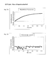
A quartz crystal microbalance (QCM) experiment was performed to attempt to normalize the uncompetitive binding capacity of TNF-α to a heparinized surface. QCM is a technique that can detect the weight of adsorbents on specially prepared crystals. The minimum detection limit is 0.5 ng/cm2. QCM detects the resonance frequency of the crystal. As the mass of the crystal increases through adsorption, the resonance frequency changes in proportion to the gain in mass. The change in mass (Δm) is described in the following equation
Δ m = - C 1 n Δ f C = 17 , 7 ngcm - 2 s - 1 n - overtone
where Δf is the change in frequency.
Polystyrene coated QCM crystals were heparinized following the method described in example 1. A 120 ml solution was prepared where recombinant TNF-α was added to PBS for a final concentration of 83 μg/L. The solution was then flowed through a cell containing 4° C. crystals at a rate of 50 μl/min. Two crystals were heparinized and two were control crystals (untreated polystyrene). The total time of the experiment was 20 hours. The results for the amount of TNF-α adsorbed on the heparinized crystals and the controls are shown in FIG. 2. A maximum of 1234 ng/cm2 of TNF-α adsorbed on the heparinized crystal and a adsorption on the control surfaces was negligible.
8.6. Example 6
A study was performed testing the removal of cytokines from plasma sampled from Bacillus anthracis infected macaques using heparinized beads. The plasma was collected at the time of death of several animals and pooled. 1 ml filter syringes were packed with either 0.5 grams of PE beads heparinized following the procedure outlined in Example 1 or untreated PE beads for use as controls. A total of three sample syringes and three control syringes were used. A top porous plate was placed on top of the beads to keep the beads from floating as saline or plasma was added to the filter syringes. The filter syringes were primed using 2 ml of Tris-buffered Saline (TBS). The 2 ml of saline was passed through the filter syringe using a 5 ml syringe. The plunger of the 5 ml syringe was then retracted and filled with 4 ml of air. The air was then passed through the filter syringe to force the remaining saline out of the beads. 0.5 ml of plasma was then pulled into the 5 ml syringe and then pressed through the filter syringe. An additional 4 ml of air was passed through the filter syringe to remove any residual plasma. After the plasma drained through the syringe, aliquots were sampled and flash froze for cytokine concentration analysis. A Luminex® Multiplex assay was used to test for GRO-α, IL-8, MIP-1β, Rantes, and TNF-β. The results are summarized in Table 3.
TABLE 3
Passage Passage
Before Through Through
Passage Control Heparin % Removed
Levels Beads % Removed Beads Heparin
Cytokine (pg/ml) (pg/ml) Control (pg/ml) Beads
GRO-α 263.5 158.5 39.8% 77.66 70.6%
IL-8 504.9 460.8 8.7% 312.4 38.1%
MIP-1β 66.2 56.4 14.8% 51.8 21.8%
RANTES 1105.3 930.3 15.8% 559.7 49.4%
TNF-β 7.3 7.9 −8.2% 4.3 41.4%
8.7. Example 7 Cartridge to Bind Cytokines and Endotoxins Using a Layered Assembly
A cartridge is built using two different adsorption media. Heparinized beads are used to capture cytokines and PEI beads are used to capture endotoxins. By controlling the ratio of heparinized to PEI beads, blood compatibility can be maintained. The PEI beads are made following the first two steps in Example 1.
In this example, a cartridge is built with heparinized beads and polyethyleneimine (PEI) coated beads. A 300 ml adsorption column is fixed to a vertical stand. 50 ml of 300 micron average heparinized beads are then added to the cartridge and allowed to settle. A 50:50 mixture of PEI and heparinized beads is then added and makes up the next 200 ml. The cartridge is then filled to the top with a final 50 ml of heparinized beads. The cartridge is then sealed and the close packing of the beads maintain the layered structure of the adsorption media. In this cartridge, ⅔rds of the beads are heparinized while ⅓rd is aminated and therefore inherently thrombogenic.
8.8. Example 8 Cartridge to Bind Cytokines and Endotoxins Using a Uniform Mixture of Beads
A cartridge is built using two different adsorption media. Heparinized beads are used to capture cytokines and PEI beads are used to capture endotoxins. By controlling the ratio of heparinized to PEI beads, the overall blood compatibility of the device can be maintained.
In this example, a cartridge is built with heparinized beads and PEI coated beads. A 300 ml adsorption column is fixed to a vertical stand. 100 ml of PEI coated beads are added to 200 ml of heparinized beads and mixed thoroughly. The 300 ml of beads are then added to the cartridge and sealed. The close packing of the beads maintains the random mixture of beads. In this cartridge, ⅔rds of the beads are heparinized while ⅓rd are aminated.
8.9. Example 9 Cartridge to Bind Cytokines, Gram Negative Bacteria, and Endotoxins Using a Layered Assembly
A cartridge is built using three different adsorption media. Heparinized beads are used to capture cytokines, mannose functionalized beads are used to capture gram negative bacteria, and PEI beads are used to capture endotoxins. By controlling the ratio of heparinized beads, mannose functionalized beads, and PEI coated beads, blood compatibility can be maintained.
A 300 ml adsorption column is fixed to a vertical stand. 50 ml of 300 micron average heparinized beads are then added to the cartridge and allowed to settle. 200 ml of beads, with equal quantities of heparinized beads, PEI beads, and mannose functionalized beads are then added to the cartridge. The cartridge is then filled to the top with a final 50 ml of heparnized beads. The cartridge is then sealed and the close packing of the beads maintain the layered structure of the adsorption media. In this cartridge, 55.6% of the beads are heparinized, 22.2% are PEI beads, and 22.2% are mannose functionalized.
The embodiments of the present invention are further described in the claims below.

Claims (31)

We claim:
1. A device for removing at least one adsorbate selected from cytokines, pathogens and toxins from blood which comprises a container, said container comprising a solid substrate of high surface area having at least one polysaccharide adsorbent on the surface thereof with a binding affinity for the adsorbate, wherein said substrate is sufficiently solid such that the adsorbate does not pass through pores in said substrate, and wherein the size of interstitial channel spaces within said substrate is large enough to permit the transport of blood cells, and the amount of interstitial surface area within said substrate is such that when flowing blood is in contact with said substrate through said device, said cytokine, pathogen or toxin binds to binding sites on said at least one polysaccharide adsorbent, becoming separated from said blood and the transport of said adsorbates from blood to adsorbent sites on said substrate is primarily by convection transport, such that adsorbate binding does not require the Brownian diffusion of adsorbates into pores within said substrate.
2. The device according to claim 1, wherein said solid substrate comprises a plurality of rigid polymer beads.
3. The device according to claim 2, wherein said rigid polymer beads are rigid polyethylene beads.
4. The device of claim 3, wherein said beads have a diameter ranging from 100 and 450 microns.
5. The device of claim 4, wherein said beads have an average diameter of 0.3 mm.
6. The device according to any one of claims 1-3, wherein at least one of said polysaccharides is a member selected from the group consisting of heparin, heparan sulfate, hyaluronic acid, sialic acid, carbohydrates with mannose sequences, and chitosan.
7. The device of claim 6, wherein the linear flow velocity of blood flowing through said device is between about 8 and 24 cm/minute.
8. The device according to any one of claims 3, 4 and 5, wherein said beads are coated with 0.5-10 mg heparin per gram of bead.
9. The device of claim 8, wherein said beads are coated with 2±0.5 mg heparin per gram of bead.
10. The device according to claim 9, wherein the heparin has a mean molecular weight of about 8 kDa.
11. The device according to claim 8, wherein the heparin is attached to the substrate by covalent end-point attachment.
12. The device according to claim 11, wherein said container further comprises a solid substrate having on the surface thereof a cationic, positively charged molecule having an affinity for the cytokine, pathogen or toxin, but being more thrombogenic than heparin.
13. The device of claim 12, wherein said cationic, positively charged molecule is chitosan or polyethylene imine.
14. A device for removing at least one adsorbate selected from cytokines, pathogens and toxins from blood which comprises a container that comprises a solid substrate comprised of a plurality of rigid polyethylene beads comprising heparin on the surface thereof with a binding affinity for the adsorbate, wherein said beads are sufficiently solid such that the adsorbate does not pass through pores in said beads, and wherein the size of interstitial channel spaces within said substrate is large enough to permit the transport of blood cells, and the amount of interstitial surface area within said substrate is such that when flowing blood is in contact with said substrate through said device, said cytokine, pathogen or toxin binds to binding sites on said heparin, becoming separated from said blood and the transport of said adsorbates from blood to adsorbent sites on said beads is primarily by convection transport, such that adsorbate binding does not require the Brownian diffusion of adsorbates into pores within said substrate and wherein said heparin is attached to the substrate by covalent end-point attachment in an amount of 0.5-10 mg heparin per gram of bead.
15. The device of claim 14, wherein the linear flow velocity of blood flowing through said device is between about 8 and 24 cm/minute.
16. The device of claim 14, wherein said beads have a diameter ranging from 100 and 450 microns.
17. The device of claim 16, wherein said beads have an average diameter of 0.3 mm.
18. The device of claim 14, wherein said beads are coated with 2±0.5 mg heparin per gram of bead.
19. The device according to claim 18, wherein the heparin has a mean molecular weight of about 8 kDa.
20. The device according to claim 14, wherein the cytokine is selected from the group consisting of IL-6, VCAM, TNF-α, GRO-α, IL-8, and RANTES.
21. The device according to any one of claims 15-19, wherein said container further comprises a solid substrate having on the surface thereof a cationic, positively charged molecule having an affinity for the cytokine, pathogen or toxin, but being more thrombogenic than heparin.
22. The device according to claim 21, wherein said cationic, positively charged molecule is chitosan or polyethylene imine.
23. A device for removing at least one adsorbate selected from cytokines, pathogens and toxins from blood which comprises a container, said container comprising a solid substrate of high surface area comprising:
i) a first portion of the solid substrate with at least one polysaccharide adsorbent on the surface, wherein said at least one polysaccharide comprises binding sites for the at least one adsorbate; and
ii) a second portion of the solid substrate having on the surface thereof a cationic, positively charged molecule being more thrombogenic than heparin, wherein said substrate is sufficiently solid such that the adsorbate does not pass through pores in said substrate, and wherein the size of interstitial channel spaces within said substrate is large enough to permit the transport of blood cells, and the amount of interstitial surface area within said substrate is such that when flowing blood is in contact with said substrate through said device, said cytokine, pathogen and toxin becoming separated from said blood and the transport of said adsorbates from blood to adsorbent sites on said substrate is primarily by convection transport, such that adsorbate binding does not require the Brownian diffusion of adsorbates into pores within said substrate.
24. The device of claim 23, wherein the at least one of said polysaccharides is a member selected from the group consisting of heparin, heparan sulfate, hyaluronic acid, sialic acid, carbohydrates with mannose sequences, and chitosan.
25. The device according to claim 24, wherein the heparin has a mean molecular weight of about 8 kDa.
26. The device of claim 23, wherein the linear flow velocity of blood flowing through said device is between about 8 and 24 cm/minute.
27. The device of claim 23, wherein said beads have a diameter ranging from 100 and 450 microns.
28. The device of claim 27, wherein said beads have an average diameter of 0.3 mm.
29. The device of claim 23, wherein said beads are coated with 2±0.5 mg heparin per gram of bead.
30. The device according to claim 23, wherein said cationic, positively charged molecule is chitosan or polyethylene imine.
31. The device according to claim 23, wherein the cytokine is selected from the group consisting of IL-6, VCAM, TNF-α, GRO-α, IL-8, and RANTES.
US12/958,355 2009-12-01 2010-12-01 Method for removing cytokines from blood with surface immobilized polysaccharides Active 2031-10-07 US8758286B2 (en)

Priority Applications (4)

Application Number Priority Date Filing Date Title
US12/958,355 US8758286B2 (en) 2009-12-01 2010-12-01 Method for removing cytokines from blood with surface immobilized polysaccharides
US14/261,390 US9408962B2 (en) 2009-12-01 2014-04-24 Methods for removing cytokines from blood with surface immobilized polysaccharides
US15/199,737 US10086126B2 (en) 2009-12-01 2016-06-30 Methods for removing cytokines from blood with surface immobilized polysaccharides
US16/113,904 US11123466B2 (en) 2009-12-01 2018-08-27 Methods for removing cytokines from blood with surface immobilized polysaccharides

Applications Claiming Priority (2)

Application Number Priority Date Filing Date Title
US26567509P 2009-12-01 2009-12-01
US12/958,355 US8758286B2 (en) 2009-12-01 2010-12-01 Method for removing cytokines from blood with surface immobilized polysaccharides

Related Child Applications (1)

Application Number Title Priority Date Filing Date
US14/261,390 Division US9408962B2 (en) 2009-12-01 2014-04-24 Methods for removing cytokines from blood with surface immobilized polysaccharides

Publications (2)

Publication Number Publication Date
US20110184377A1 US20110184377A1 (en) 2011-07-28
US8758286B2 true US8758286B2 (en) 2014-06-24

Family

ID=43731827

Family Applications (4)

Application Number Title Priority Date Filing Date
US12/958,355 Active 2031-10-07 US8758286B2 (en) 2009-12-01 2010-12-01 Method for removing cytokines from blood with surface immobilized polysaccharides
US14/261,390 Active 2031-09-22 US9408962B2 (en) 2009-12-01 2014-04-24 Methods for removing cytokines from blood with surface immobilized polysaccharides
US15/199,737 Active 2031-02-12 US10086126B2 (en) 2009-12-01 2016-06-30 Methods for removing cytokines from blood with surface immobilized polysaccharides
US16/113,904 Active 2032-05-08 US11123466B2 (en) 2009-12-01 2018-08-27 Methods for removing cytokines from blood with surface immobilized polysaccharides

Family Applications After (3)

Application Number Title Priority Date Filing Date
US14/261,390 Active 2031-09-22 US9408962B2 (en) 2009-12-01 2014-04-24 Methods for removing cytokines from blood with surface immobilized polysaccharides
US15/199,737 Active 2031-02-12 US10086126B2 (en) 2009-12-01 2016-06-30 Methods for removing cytokines from blood with surface immobilized polysaccharides
US16/113,904 Active 2032-05-08 US11123466B2 (en) 2009-12-01 2018-08-27 Methods for removing cytokines from blood with surface immobilized polysaccharides

Country Status (10)

Country Link
US (4) US8758286B2 (en)
EP (1) EP2509604B1 (en)
JP (2) JP5925693B2 (en)
KR (2) KR101876794B1 (en)
CN (2) CN106178161B (en)
AU (1) AU2010326028B2 (en)
CA (2) CA2967094C (en)
ES (1) ES2861359T3 (en)
HK (1) HK1177151A1 (en)
WO (1) WO2011068897A1 (en)

Cited By (14)

* Cited by examiner, † Cited by third party
Publication number Priority date Publication date Assignee Title
WO2015069942A1 (en) 2013-11-08 2015-05-14 Exthera Medical Corporation Methods for diagnosing infectious diseases using adsorption media
US9173989B2 (en) 2005-12-13 2015-11-03 Exthera Medical Corporation Method for extracorporeal removal of a pathogenic microbe, an inflammatory cell or an inflammatory protein from blood
US20160101229A1 (en) * 2013-06-24 2016-04-14 Exthera Medical Corporation Blood filtration system containing mannose coated substrate
US9408962B2 (en) 2009-12-01 2016-08-09 Exthera Medical Corporation Methods for removing cytokines from blood with surface immobilized polysaccharides
US9669150B2 (en) 2007-06-18 2017-06-06 Exthera Medical Corporation Device and method for restoration of the condition of blood
WO2017151797A1 (en) 2016-03-02 2017-09-08 Exthera Medical Corporation Method for treating drug intoxication
US10537280B2 (en) 2011-02-15 2020-01-21 Exthera Medical Corporation Device and method for removal of blood-borne pathogens, toxins and inflammatory cytokines
US10857283B2 (en) 2014-09-22 2020-12-08 Exthera Medical Corporation Wearable hemoperfusion device
US20210001032A1 (en) * 2015-03-27 2021-01-07 Eliaz Therapeutics, Inc. Galectin-3 plasmapheresis therapy
US11224858B1 (en) 2020-10-01 2022-01-18 Immunicom, Inc. Reduced leaching of a ligand
US11266772B2 (en) 2012-06-13 2022-03-08 Exthera Medical Corporation Use of heparin and carbohydrates to treat cancer
US11458236B2 (en) 2020-10-01 2022-10-04 Immunicom, Inc. Reduced leaching of a ligand
US11844895B2 (en) 2014-04-24 2023-12-19 Exthera Medical Corporation Method for removing bacteria from blood using high flow rate
US11911551B2 (en) 2016-03-02 2024-02-27 Exthera Medical Corporation Method for treating drug intoxication

Families Citing this family (30)

* Cited by examiner, † Cited by third party
Publication number Priority date Publication date Assignee Title
US9199016B2 (en) 2009-10-12 2015-12-01 New Health Sciences, Inc. System for extended storage of red blood cells and methods of use
US11284616B2 (en) 2010-05-05 2022-03-29 Hemanext Inc. Irradiation of red blood cells and anaerobic storage
WO2011133099A1 (en) * 2010-04-21 2011-10-27 Exthera Medical Llc Method for removal of mrsa from blood
WO2012027582A1 (en) 2010-08-25 2012-03-01 New Health Sciences Method for enhancing red blood cell quality and survival during storage
WO2012061731A1 (en) 2010-11-05 2012-05-10 New Health Sciences Irradiation of red blood cells and anaerobic storage
US20140054227A1 (en) * 2011-03-04 2014-02-27 Dic Corporation Sugar-immobilized polymer substrate for removing virus and method for removing virus
US9067004B2 (en) 2011-03-28 2015-06-30 New Health Sciences, Inc. Method and system for removing oxygen and carbon dioxide during red cell blood processing using an inert carrier gas and manifold assembly
CA2844449A1 (en) 2011-08-10 2013-02-14 New Health Sciences, Inc. Integrated leukocyte, oxygen and/or co2 depletion, and plasma separation filter device
EP2776013B8 (en) 2011-11-08 2023-08-30 The Board of Trustees of the University of Arkansas Methods and compositions for x-ray induced release from ph sensitive liposomes
US9549953B2 (en) * 2011-12-08 2017-01-24 Eliaz Therapeutics, Inc. Galectin-3 plasmapheresis therapy
KR101427700B1 (en) * 2012-06-29 2014-08-07 주식회사 아모그린텍 Cytokine Adsorbent Sheet, Manufacturing Method Thereof and Blood Filter using The same
CN104394903B (en) * 2012-12-14 2017-08-15 甘布罗伦迪亚股份公司 The purification of biofluid
CA3205441A1 (en) 2013-02-28 2014-09-04 Hemanext Inc. Gas depletion and gas addition devices for blood treatment
CN103285826A (en) * 2013-06-28 2013-09-11 天津优纳斯生物科技有限公司 Blood purification adsorbing agent for restraining grown and transfer of tumor and application thereof
US10322216B2 (en) * 2014-01-30 2019-06-18 Fresenius Hemocare Italia S.R.L. Filtering device
JP6728536B2 (en) * 2014-03-31 2020-07-22 ゾール メディカル コーポレイションZOLL Medical Corporation Hemofiltration, an inflammatory biomarker for treating post-resuscitation syndrome
CA2978940C (en) 2015-03-10 2023-10-17 New Health Sciences, Inc. Oxygen reduction disposable kits, devices and methods of use thereof
WO2016172645A1 (en) 2015-04-23 2016-10-27 New Health Sciences, Inc. Anaerobic blood storage containers
WO2016187353A1 (en) * 2015-05-18 2016-11-24 New Health Sciences, Inc. Methods for the storage of whole blood, and compositions thereof
US20180318337A1 (en) * 2015-11-04 2018-11-08 Duke University Polycationic polymers for use in treating and detecting cancer
JP6759679B2 (en) * 2016-04-25 2020-09-23 日立化成株式会社 Separator and column
CN109195632A (en) 2016-05-27 2019-01-11 新健康科学股份有限公司 Anaerobism blood storage and pathogen method for deactivating
IL263695B2 (en) * 2016-06-23 2023-03-01 Hemanext Inc Methods for managing adverse events in patient populations requiring transfusion
CN106771231B (en) * 2016-11-23 2019-02-12 江南大学 Application of the carbohydrate chip in detection salmonella or screening salmonella antagonist
CN107397961B (en) * 2017-09-05 2020-10-16 陕西师范大学 Method for improving resistance of polyethyleneimine to enterobacter coli
EP3473327A1 (en) * 2017-10-19 2019-04-24 3M Innovative Properties Company Integrated fluid treatment device, and process using such device
CA3137294A1 (en) * 2019-05-16 2020-11-19 Exthera Medical Corporation Method for modulating endothelial glycocalyx structure
US20220125381A1 (en) * 2020-10-23 2022-04-28 Boston Scientific Scimed, Inc. Medical devices and methods for monitoring and treatment with synthetic polymers exhibiting specific binding
WO2022216510A1 (en) * 2021-04-06 2022-10-13 Exthera Medical Corporation Filtration media
CN114011389B (en) * 2021-12-08 2023-10-31 广州康盛生物科技股份有限公司 Blood purification material for sepsis and preparation method and application thereof

Citations (20)

* Cited by examiner, † Cited by third party
Publication number Priority date Publication date Assignee Title
US5211850A (en) * 1991-07-26 1993-05-18 Research Medical, Inc. Plasma filter sorbent system for removal of components from blood
EP1044696A2 (en) 1999-04-12 2000-10-18 B. Braun Melsungen Ag Removal of enveloped viruses from blood, plasma or serum
US6159377A (en) * 1998-02-06 2000-12-12 Renal Tech International Llc Method of purification of physiological liquids of organism
US20020058032A1 (en) * 2000-09-26 2002-05-16 Fumiyasu Hirai Enterotoxin adsorbent, method of adsorptive removal, and adsorption apparatus
US20030021780A1 (en) * 2000-10-06 2003-01-30 Smith Terry J. Diagnosis and therapy of antibody-mediated inflammatory autoimmune disorders
US20030044769A1 (en) * 1997-03-25 2003-03-06 Eiji Ogino Adsorbent for eliminating hepatitis c virus, adsorber, and adsorption method
US6559290B1 (en) * 1996-03-18 2003-05-06 Kaneka Corporation Method for removing a chemokine
US20060093999A1 (en) * 1998-01-06 2006-05-04 Hei Derek J Adsorbing pathogen-inactivating compounds with porous particles immobilized in a matrix
WO2007069983A1 (en) 2005-12-13 2007-06-21 Exthera Ab Method for extracorporeal removal of a pathogenic microbe, an inflammatory cell or an inflammatory protein from blood
US20070218514A1 (en) * 2000-10-06 2007-09-20 Harbor-Ucla Research And Education Institute Diagnostic methods relating to Graves' disease and other autoimmune disorders
WO2007146162A2 (en) 2006-06-08 2007-12-21 University Of Pittsburgh - Of The Commonwealth System Of Higher Education Devices, systems and methods for reducing the concentration of a chemical entity in fluids
US20080021365A1 (en) * 2004-08-30 2008-01-24 Kaneka Corporation Granulocyte Adsorbent
US20080138434A1 (en) * 1997-07-30 2008-06-12 Medasorb Corporation Methods for reducing levels of pro-inflammatory or anti-inflammatory stimulators or mediators in the blood
WO2008157570A2 (en) 2007-06-18 2008-12-24 Exthera Ab Device and method for restoration of the condition of blood
US20090105194A1 (en) * 2005-05-09 2009-04-23 Universitetet For Miljo-Og Biovitenskap Process for the production of a low molecular weight heparin
WO2010029317A2 (en) 2008-09-10 2010-03-18 Ibd Column Therapies International Ab Treating inflammatory conditions
US20100069816A1 (en) * 1997-07-30 2010-03-18 Renaltech International, Llc Biocompatible devices, systems, and methods for reducing levels of proinflammatory or antiinflammatory stimulators or mediators in the blood
US20100098666A1 (en) * 2007-04-17 2010-04-22 John Fraser Wright Humanized Viral Vectors and Methods of Use Thereof
US20100216226A1 (en) 2009-02-25 2010-08-26 Searete Llc, A Limited Liability Corporation Of The State Of Delaware Device, system, and method for controllably reducing inflammatory mediators in a subject
US20100276359A1 (en) * 2007-09-12 2010-11-04 Rei Medical Co., Ltd. Adsorption column for purifying body fluid

Family Cites Families (134)

* Cited by examiner, † Cited by third party
Publication number Priority date Publication date Assignee Title
GB217812A (en) 1923-09-25 1924-06-26 John Henry Delaney Improvements relating to the production of vermiculated appearance in artificial stone and like building materials
US3782382A (en) 1972-02-03 1974-01-01 K N Enterprises Inc Means for blood administration and the like
US4103685A (en) 1976-01-05 1978-08-01 Lupien Paul J Method and apparatus for extravascular treatment of blood
JPS54127493U (en) 1978-02-25 1979-09-05
JPS6040926B2 (en) 1979-07-24 1985-09-13 三菱重工業株式会社 Automatic support device for mandrel bar
US4430496A (en) 1980-01-17 1984-02-07 Varian Associates, Inc. Strong anion exchange composition and methods
US4415665A (en) 1980-12-12 1983-11-15 Pharmacia Fine Chemicals Ab Method of covalently binding biologically active organic substances to polymeric substances
JPS5853757A (en) 1981-09-25 1983-03-30 Denki Kagaku Kogyo Kk Adsorbent for immuno-affinity chromatography and manufacture of same
SE8200751L (en) 1982-02-09 1983-08-10 Olle Larm PROCEDURE FOR COVALENT COUPLING FOR MANUFACTURE OF CONJUGATE AND REQUIRED PRODUCTS
JPS58146354A (en) 1982-02-26 1983-08-31 日機装株式会社 Production of body liquid washer
US4820302A (en) 1982-04-22 1989-04-11 Sterling Drug Inc. Bio compatible and blood compatible materials and methods
CA1221307A (en) * 1982-12-02 1987-05-05 Nobutaka Tani Adsorbent and process for preparing the same
JPS59227827A (en) * 1983-06-10 1984-12-21 Nippon Koutai Kenkyusho:Kk Chain-relating antigen originated from plasmodium, and its preparation
DE3609021A1 (en) 1985-03-25 1986-09-25 Bio-Rad Laboratories, Inc., Richmond, Calif. CHROMATOGRAPHIC SEPARATION MEDIUM FOR FAST ANALYSIS OF SMALL SAMPLES
DE3519011A1 (en) 1985-05-25 1986-11-27 Behringwerke Ag, 3550 Marburg METHOD FOR PRODUCING A MATERIAL FOR AFFINITY CHROMATOGRAPHY
CA1327963C (en) 1987-09-08 1994-03-22 Ryuichi Yokohari Autoantibody adsorbent and apparatus for removing autoantibodies using the same
EP0476721B1 (en) 1987-11-20 1995-03-01 Kanegafuchi Kagaku Kogyo Kabushiki Kaisha A method for removing serum amyloid protein
US4955870A (en) 1988-08-23 1990-09-11 Ridderheim Kristen A Hypodermic syringe with retractable needle
WO1991004086A1 (en) 1989-09-18 1991-04-04 Cuno, Incorporated Filter media and use for pyrogen removal
JPH0489500A (en) 1990-07-31 1992-03-23 Kirin Brewery Co Ltd Method for purifying substance by affinity chromatography and apparatus for purification
SE469256B (en) 1991-02-18 1993-06-14 Medscand Ab RENGOERINGSPRODUKT
WO1992017502A1 (en) 1991-03-26 1992-10-15 Otsuka Pharmaceutical Factory, Inc. Adsorbed cellular fibronectin and process for separating and purifying fibronectins
DE4217917A1 (en) 1992-05-30 1993-12-02 Job Prof Dr Med Harenberg Heparin-polymer conjugate used to inhibit coagulation in analytical tests - comprises heparin fragments bonded to amino-phenyl substd. polymer
JPH0640926A (en) 1992-07-24 1994-02-15 Chemo Sero Therapeut Res Inst Method for removing hepatitis c virus
DE4331358A1 (en) * 1992-10-12 1994-04-14 Braun Melsungen Ag Process for the quantitative selective removal or preparation of tumor necrosis factor (TNF) and / or lipopolysaccharides (LPS) from aqueous liquids
US5344455A (en) 1992-10-30 1994-09-06 Medtronic, Inc. Graft polymer articles having bioactive surfaces
US5437861A (en) 1993-03-16 1995-08-01 Applied Immune Sciences, Inc. Removal of selected factors from whole blood or its components; and prevention and treatment of septic shock syndrome
JP2643823B2 (en) 1993-03-25 1997-08-20 有限会社 エンゼル総合研究所 Adsorption material and method for producing the same
NO931809L (en) 1993-05-19 1994-11-21 Norsk Hydro As hemophilia
US5447859A (en) 1993-07-16 1995-09-05 Viagene Method for the purification or removal of retroviruses using sulfated cellulose
US5753227A (en) 1993-07-23 1998-05-19 Strahilevitz; Meir Extracorporeal affinity adsorption methods for the treatment of atherosclerosis, cancer, degenerative and autoimmune diseases
WO1995005400A1 (en) 1993-08-19 1995-02-23 Minnesota Mining And Manufacturing Company Heparin functional affinity supports
JPH07178161A (en) 1993-12-24 1995-07-18 Terumo Corp Medical instrument
WO1996025228A1 (en) 1995-02-16 1996-08-22 Kanegafuchi Kagaku Kogyo Kabushiki Kaisha ADSORBENT FOR TUMOR NECROSIS FACTOR α, METHOD OF REMOVAL BY ADSORPTION, AND ADSORPTION DEVICE USING SAID ADSORBENT
SE9501056D0 (en) 1995-03-23 1995-03-23 Probi Ab Epithelial adherent lactobacilli
US6312907B1 (en) 1995-04-24 2001-11-06 The Texas A & M University System DbpA compositions and methods of use
US6544727B1 (en) 1995-06-07 2003-04-08 Cerus Corporation Methods and devices for the removal of psoralens from blood products
ES2337852T3 (en) 1995-06-07 2010-04-29 Cerus Corporation METHODS AND DEVICES FOR THE ELIMINATION OF PSORALENES FROM BLOOD PRODUCTS.
EP0776374B1 (en) 1995-06-20 2009-12-16 Papst Licensing GmbH & Co. KG Process and device for determining the activity of enzymes in liquids, or the concentration and/or activity of inhibitors in liquids
RU2089283C1 (en) 1996-03-23 1997-09-10 Научно-исследовательская фирма "Ультрасан" Bio- and heme-compatible sorbents based on super-cross-linked styrene polymers with modified surface, method of preparation thereof (versions), and method of preparing sorbent matrix
SE507028C2 (en) 1996-08-06 1998-03-16 Medicarb Ab New medical use
AU5811498A (en) 1996-12-31 1998-07-31 Robert L. Stephon Microsphere-enhanced immunoassay for the detection of veterinary diseases
US20010009756A1 (en) 1998-01-06 2001-07-26 Derek Hei Flow devices for the reduction of compounds from biological compositions and methods of use
US20010018179A1 (en) 1998-01-06 2001-08-30 Derek J. Hei Batch devices for the reduction of compounds from biological compositions containing cells and methods of use
WO1998053829A1 (en) * 1997-05-27 1998-12-03 Akzo Nobel N.V. Use of oligosaccharide for preventing blood clotting in extracorporeal blood circuits
US6465170B2 (en) 1997-06-13 2002-10-15 Baxter Aktiengesellschaft Method for depleting viral and molecular pathogens in a biological material
EP0887061A1 (en) 1997-06-28 1998-12-30 The Procter & Gamble Company Faecal collector
US6197568B1 (en) 1997-07-29 2001-03-06 The Regents Of The University Of Michigan Method and compositions for isolation, diagnosis and treatment of polyanion-binding microorganisms
US20020197249A1 (en) * 2001-04-10 2002-12-26 Renal Tech International Devices, systems, and methods for reducing levels of pro-inflammatory or anti-inflammatory stimulators or mediators in blood products
US6053894A (en) 1997-08-04 2000-04-25 Shadd, Jr.; Daniel L. Hypodermic syringe
GB9804632D0 (en) 1998-03-05 1998-04-29 Cantab Pharma Res Virus preparations and methods
US8197430B1 (en) * 1998-05-22 2012-06-12 Biopheresis Technologies, Inc. Method and system to remove cytokine inhibitor in patients
US6248127B1 (en) 1998-08-21 2001-06-19 Medtronic Ave, Inc. Thromboresistant coated medical device
JP2000086688A (en) 1998-09-04 2000-03-28 Ngk Insulators Ltd Parbovirus-absorbing sugar-chain ligand, and prabovirus- removing device by using the same
NO984143L (en) 1998-09-09 2000-03-10 Norsk Hydro As New process for producing surface modifying substances
AU5670299A (en) 1998-10-19 2000-05-08 Cbd Technologies Ltd. Methods of concentrating microorganisms using affinity separation
US6461517B1 (en) 1998-12-22 2002-10-08 Toray Industries, Inc. Bacterial-derived component removal material
WO2000038763A1 (en) * 1998-12-24 2000-07-06 Toray Industries, Inc. Method for purifying blood and column for purifying blood
JP2000217575A (en) 1999-02-01 2000-08-08 Masashi Funayama Adsorbent of physiologically active substance, human blood cell differentiation cell and human cell
EP1110602B1 (en) 1999-04-28 2009-04-15 Toray Industries, Inc. Materials for eliminating cannabinoids and columns for the elimination of cannabinoids with the use of the same
WO2001018060A1 (en) 1999-09-08 2001-03-15 Toray Industries, Inc. Materials for extracorporeal circulation, adsorbents for diabetic complication factors, containers for eliminating diabetic complication factors and method of eliminating diabetic complication factors
SE515295C2 (en) 1999-11-23 2001-07-09 Medicarb Ab Process for the preparation of conjugates of a biologically active polysaccharide and a solid substrate, and such conjugates
JP2001178817A (en) 1999-12-24 2001-07-03 Terumo Corp Device for artificial kidney, quality evaluating device using the same and fluid circuit
US7067060B2 (en) 2000-01-11 2006-06-27 Nephros, Inc. Ionic enhanced dialysis/diafiltration system
JP2001190273A (en) 2000-01-14 2001-07-17 Jsr Corp Particle for concentrating virus, method for concentrating and detecting the virus by using the particle
GB0001450D0 (en) 2000-01-21 2000-03-08 Genpoint As Cell isolation method
US7179660B1 (en) 2000-03-06 2007-02-20 Dade Behring Marburg Gmbh Carriers coated with polysaccharides, their preparation and use
US20020164644A1 (en) 2000-04-05 2002-11-07 Ikuro Maruyama Adsorbents for high mobility group proteins and column for purifying body fluid
US7181261B2 (en) 2000-05-15 2007-02-20 Silver James H Implantable, retrievable, thrombus minimizing sensors
DE10045434B4 (en) 2000-09-14 2005-07-14 Fresenius Hemocare Gmbh Adsorbent with differently modified surface areas, process for its preparation and use thereof
US6509104B2 (en) 2000-12-05 2003-01-21 Michigan Biotechnology Institute Antithrombogenic polymer coating
US20040140265A1 (en) * 2000-12-29 2004-07-22 Lihme Allan Otto Fog Extracorporeal capturing of specific bio-macromolecular entities from extracellular body fluids
US7320757B2 (en) * 2001-01-30 2008-01-22 Kaneka Corporation Body fluid processor enabling direct hemoperfusion
DE10117043A1 (en) * 2001-04-05 2002-11-07 Gerhard Puetz Process for the elimination of potentially toxic and / or harmful substances
US20020197252A1 (en) 2001-04-10 2002-12-26 Renal Tech International Selective adsorption devices and systems
US6773412B2 (en) 2001-04-13 2004-08-10 Chf Solutions, Inc. User interface for blood treatment device
US20040248075A1 (en) 2001-10-10 2004-12-09 Koushi Yamaguchi Photocatalytic material that selectively inactivating biologically harmful substance and utilization thereof
JP2003128502A (en) 2001-10-18 2003-05-08 Noritake Co Ltd Treating material for harmful substance and method for producing the same
US8980568B2 (en) 2001-10-11 2015-03-17 Aviva Biosciences Corporation Methods and compositions for detecting non-hematopoietic cells from a blood sample
US20030148017A1 (en) * 2001-12-07 2003-08-07 Olli Tuominen Copolymer coating for a hydrophobic membrane
WO2003078023A1 (en) 2002-03-14 2003-09-25 Baxter International Inc. Compound removal device
AU2003234194A1 (en) * 2002-04-23 2003-11-10 Meir Strahilevitz Methods and devices for targeting a site in a mammal and for removing species from a mammal
WO2004008138A2 (en) 2002-07-11 2004-01-22 Upfront Chromatography A/S An extracorporeal stabilised expanded bed adsorption method for the treatment of sepsis
US7348184B2 (en) 2002-07-24 2008-03-25 Bio-Rad Laboratories, Inc. Protein interaction difference mapping
US20040182783A1 (en) 2003-03-17 2004-09-23 Walker Kimberly A. Filter and concentrator device for treatment of blood
US7207964B2 (en) 2003-03-17 2007-04-24 Hemavation, Llc Apparatus and method for down-regulating immune system mediators in blood
PT2103694E (en) 2003-08-15 2011-07-26 Univ South Florida Materials and methods for capture of pathogens and removal of aurintricarboxylic acid from a sample
US7399400B2 (en) 2003-09-30 2008-07-15 Nano-Proprietary, Inc. Nanobiosensor and carbon nanotube thin film transistors
WO2005051313A2 (en) * 2003-11-19 2005-06-09 Chiron Corporation Methods and reagents for treating, preventing and diagnosing bunyavirus infection
WO2006093524A2 (en) * 2004-07-16 2006-09-08 The General Hospital Corporation Antigen-carbohydrate conjugates
EP1790365B1 (en) * 2004-07-23 2015-11-18 Kaneka Corporation Direct hemoperfusion adsorber packed with adsorbent having water-insoluble microparticles removed therefrom, and method of obtaining direct hemoperfusion adsorber packed with adsorbent having water-insoluble microparticles removed therefrom
DE112005001901T5 (en) * 2004-08-04 2007-07-12 Aspira Biosystems Inc., Incline Village Capture and remove biomolecules from body fluids using partial molecular imprints
US8445293B2 (en) 2005-02-09 2013-05-21 Rapid Pathogen Screening, Inc. Method to increase specificity and/or accuracy of lateral flow immunoassays
DK1886704T3 (en) * 2005-03-31 2017-01-16 Toray Industries ABSORBENT AND COLUMN FOR EXTRACORPORAL CIRCULATION
CA2613078A1 (en) 2005-06-24 2007-01-04 Board Of Regents, The University Of Texas System Systems and methods including self-contained cartridges with detection systems and fluid delivery systems
WO2008045021A2 (en) * 2005-08-01 2008-04-17 Rensselaer Polytechnic Institute Blood compatible nanomaterials and methods of making and using the same
AU2006316039B2 (en) 2005-11-21 2011-08-18 Ge Healthcare Bio-Sciences Ab A method of chromatography using semi-synthetic heparin ligands
MX2008008281A (en) 2005-12-21 2008-11-06 Meso Scale Technologies Llc Assay apparatuses, methods and reagents.
CA2643140C (en) 2006-02-22 2014-12-09 Henry Ford Health System System and method for delivery of regional citrate anticoagulation to extracorporeal blood circuits
AU2007259677B2 (en) 2006-06-16 2012-07-26 Terumo Kabushiki Kaisha Blood treatment filter and blood treatment circuit
FR2902670B1 (en) * 2006-06-22 2009-04-24 Gambro Lundia Ab USE OF A SUSPENSION FOR TREATING MEDICAL MEDIA, MEDICAL MEDIA, EXCHANGER, AND ADSORPTION DEVICE COMPRISING THE MEDIUM
WO2008147382A1 (en) 2006-09-27 2008-12-04 Micronics, Inc. Integrated microfluidic assay devices and methods
MX2009004460A (en) 2006-10-26 2009-07-09 Abbott Lab Immunoassay of analytes in samples containing endogenous anti-analyte antibodies.
GB0702183D0 (en) 2007-02-05 2007-03-14 Iti Scotland Ltd Pathogen binding
US20100145317A1 (en) 2007-04-19 2010-06-10 Morris Laster Device system and method for monitoring and controlling blood analyte levels
WO2008155683A1 (en) 2007-06-18 2008-12-24 Firmenich Sa Malodor counteracting compositions and method for their use
WO2009086343A2 (en) 2007-12-31 2009-07-09 3M Innovative Properties Company Microorganism-capturing compositions and methods
US11051733B2 (en) * 2008-01-18 2021-07-06 Wake Forest University Health Sciences Isolating and purifying cells for therapy
EP2087916A1 (en) 2008-02-11 2009-08-12 ICinnovation BV Electrosorption device for the purification of blood and other fluids
ES2821448T3 (en) * 2008-06-23 2021-04-26 Temasek Polytechnic Sorbent for a dialysis device
US20100021622A1 (en) 2008-07-24 2010-01-28 National Chiao Tung University Apparatus and method for forming multilayer polymer thin film
US20100040546A1 (en) 2008-08-13 2010-02-18 Searete Llc, A Limited Liability Corporation Of The State Of Delaware Biological targeting compositions and methods of using the same
US8310415B2 (en) 2008-09-30 2012-11-13 Animas Corporation Medical devices using bistable displays
US8801922B2 (en) 2009-06-24 2014-08-12 State Of Oregon Acting By And Through The State Board Of Higher Education On Behalf Of Oregon State University Dialysis system
US20120219561A1 (en) 2009-07-31 2012-08-30 Ingeneron, Inc. Prophylaxis Against Cancer Metastasis
CN106178161B (en) 2009-12-01 2020-12-22 艾克塞拉医疗公司 Method for removing cytokines from blood using surface immobilized polysaccharides
KR101486879B1 (en) 2010-02-09 2015-01-28 엑스테라 메디컬 코퍼레이션 removal of virulence factors through extracorporeal therapy
US20130102948A1 (en) 2010-04-19 2013-04-25 Ilan K. Reich Portable blood filtration devices, systems, and methods
EP2627369B1 (en) 2010-10-15 2022-01-12 SeaStar Medical, Inc. Cytopheresic cartridge and use thereof
WO2012112724A1 (en) 2011-02-15 2012-08-23 Exthera Medical, Llc Device and method for removal of blood-borne pathogens, toxins and inflammatory cytokines
US20130131423A1 (en) 2011-04-12 2013-05-23 Tianxin Wang Methods to detect and treat diseases
EP3693041A1 (en) 2011-06-13 2020-08-12 TLA Targeted Immunotherapies AB Treating cancer
AU2012284097B2 (en) 2011-07-18 2017-08-03 President And Fellows Of Harvard College Engineered microbe-targeting molecules and uses thereof
EP2556849A1 (en) 2011-08-08 2013-02-13 Gambro Lundia AB Separation material
EP2861273B1 (en) 2012-06-13 2017-08-23 ExThera Medical Corporation Use of heparin and carbohydrates to treat cancer
WO2014085713A1 (en) 2012-11-30 2014-06-05 The Methodist Hospital Research Institute Methods and compositions for detecting tuberculosis
EP3013445B1 (en) 2013-06-24 2019-11-06 ExThera Medical Corporation Blood filtration system containing mannose coated substrate
EP3049405A4 (en) 2013-09-26 2017-03-08 Pharmakea Inc. Autotaxin inhibitor compounds
WO2015069942A1 (en) 2013-11-08 2015-05-14 Exthera Medical Corporation Methods for diagnosing infectious diseases using adsorption media
JP6040926B2 (en) 2013-12-16 2016-12-07 株式会社オートネットワーク技術研究所 Board built-in connector
BR112016024569B1 (en) 2014-04-24 2023-01-03 Exthera Medical Corporation EX VIVO METHOD FOR REMOVING BACTERIA FROM A SPECIMEN TAKEN FROM AN INDIVIDUAL WHO IS SUSPECTED TO BE INFECTED WITH THE SAME
EP3197342B1 (en) 2014-09-22 2020-08-05 ExThera Medical Corporation Wearable hemoperfusion device
RU2756512C1 (en) 2018-01-10 2021-10-01 ЭксДабл-ЮФАРМА ЛТД. Ketamine prodrugs, compositions and applications thereof
US20200023001A1 (en) 2018-07-20 2020-01-23 Northeastern University GlycoFix (Structurally And Functionally Repaired Endothelial Glycocalyx)

Patent Citations (25)

* Cited by examiner, † Cited by third party
Publication number Priority date Publication date Assignee Title
US5211850A (en) * 1991-07-26 1993-05-18 Research Medical, Inc. Plasma filter sorbent system for removal of components from blood
US6559290B1 (en) * 1996-03-18 2003-05-06 Kaneka Corporation Method for removing a chemokine
US20030044769A1 (en) * 1997-03-25 2003-03-06 Eiji Ogino Adsorbent for eliminating hepatitis c virus, adsorber, and adsorption method
US20080138434A1 (en) * 1997-07-30 2008-06-12 Medasorb Corporation Methods for reducing levels of pro-inflammatory or anti-inflammatory stimulators or mediators in the blood
US20100069816A1 (en) * 1997-07-30 2010-03-18 Renaltech International, Llc Biocompatible devices, systems, and methods for reducing levels of proinflammatory or antiinflammatory stimulators or mediators in the blood
US20060093999A1 (en) * 1998-01-06 2006-05-04 Hei Derek J Adsorbing pathogen-inactivating compounds with porous particles immobilized in a matrix
US6159377A (en) * 1998-02-06 2000-12-12 Renal Tech International Llc Method of purification of physiological liquids of organism
EP1044696A2 (en) 1999-04-12 2000-10-18 B. Braun Melsungen Ag Removal of enveloped viruses from blood, plasma or serum
US20020058032A1 (en) * 2000-09-26 2002-05-16 Fumiyasu Hirai Enterotoxin adsorbent, method of adsorptive removal, and adsorption apparatus
US20070218514A1 (en) * 2000-10-06 2007-09-20 Harbor-Ucla Research And Education Institute Diagnostic methods relating to Graves' disease and other autoimmune disorders
US20030021780A1 (en) * 2000-10-06 2003-01-30 Smith Terry J. Diagnosis and therapy of antibody-mediated inflammatory autoimmune disorders
US20080021365A1 (en) * 2004-08-30 2008-01-24 Kaneka Corporation Granulocyte Adsorbent
US20090105194A1 (en) * 2005-05-09 2009-04-23 Universitetet For Miljo-Og Biovitenskap Process for the production of a low molecular weight heparin
US20090136586A1 (en) 2005-12-13 2009-05-28 Exthera Ab Method for Extracorporeal Removal of a Pathogenic Microbe, an Inflammatory Cell or an Inflammatory Protein From Blood
WO2007069983A1 (en) 2005-12-13 2007-06-21 Exthera Ab Method for extracorporeal removal of a pathogenic microbe, an inflammatory cell or an inflammatory protein from blood
WO2007146162A2 (en) 2006-06-08 2007-12-21 University Of Pittsburgh - Of The Commonwealth System Of Higher Education Devices, systems and methods for reducing the concentration of a chemical entity in fluids
US20120040429A1 (en) 2006-06-08 2012-02-16 Federspiel William J Devices, systems and methods for reducing the concentration of a chemical entity in fluids
US20100098666A1 (en) * 2007-04-17 2010-04-22 John Fraser Wright Humanized Viral Vectors and Methods of Use Thereof
WO2008157570A2 (en) 2007-06-18 2008-12-24 Exthera Ab Device and method for restoration of the condition of blood
US20100249689A1 (en) 2007-06-18 2010-09-30 Exthera Medical Llc Device and method for restoration of the condition of blood
US20100276359A1 (en) * 2007-09-12 2010-11-04 Rei Medical Co., Ltd. Adsorption column for purifying body fluid
WO2010029317A2 (en) 2008-09-10 2010-03-18 Ibd Column Therapies International Ab Treating inflammatory conditions
US20110224645A1 (en) 2008-09-10 2011-09-15 Ith Immune Therapy Holdings Ab Treating inflammatory conditions
US20100216226A1 (en) 2009-02-25 2010-08-26 Searete Llc, A Limited Liability Corporation Of The State Of Delaware Device, system, and method for controllably reducing inflammatory mediators in a subject
US20100217173A1 (en) * 2009-02-25 2010-08-26 Searete Llc, A Limited Liability Corporation Of The State Of Delaware Device, system, and method for controllably reducing inflammatory mediators in a subject

Non-Patent Citations (22)

* Cited by examiner, † Cited by third party
Title
Bindslev, L. et al., "Treatment of acute respiratory failure by extracorporeal carbon dioxide elimination performed with a surface heparinized artificial lung," Anesthesiology, 1987, vol. 67, pp. 117-120.
Chen, Y. et al., Mol. Cells, vol. 26, pp. 415-426, Nov. 30, 2008.
Dixon, T. C. et al., "Antrax," New England Journal of Medicine, The Massachusetts Medical Society, Waltham, MA, US, Sep. 9, 1999, vol. 341, No. 11, pp. 815-826.
Haase, M. et al., "The effect of three different miniaturized blood purification devices on plasma cytokine concentration in an ex vivo model of endotoxinemia," Int J Artif Organs , 2008, vol. 31, pp. 722-729.
International Search Report issued in International Application No. PCT/US2010/025316 on May 23, 2012.
International Search Report issued in International Application No. PCT/US2011/024229 on May 30, 2011.
International Search Report; PCT/US2010/058596 mailed on Mar. 29, 2011.
International Search Report; PCT/US2012/025316 mailed on May 23, 2012.
Keuren J.F.W . et al., "Thrombogenecity of polysaccharide-coated surfaces," 2003, Biomaterials, vol. 24, pp. 1917-1924.
Larm, O. et al., "A new non-thrombogenic surface prepared by selective covalent binding of heparin via a modified reducing terminal residue," Biomater Med Devices Artif Organs, 1983, vol. 11, pp. 161-173.
Mandal, C., "Sialic acid binding Lectins," Experientia, 1990, vol. 46, pp. 433-439.
Mariano, F. et al., "Tailoring high-cut-off membranes and feasible application in sepsis-associated acute renal failure: in vitro studies," Nephrol Dial Transplant, 2005, vol. 20, pp. 1116-1126.
Ofek, I. and Beachey, E.H., "Mannose Binding and Epithelial Cell Adherence of Escherichia coli," Infection and Immunity, 1978, vol. 22, pp. 247-254.
Popova et al., "Acceleration of epithelial cell syndecan-1 shedding by anthrax hemolytic virulence factors," BMC Microbiolgoy, 2006, vol. 6:8, pp. 1-16.
Riesenfeld, J. and Roden, L., "Quantitative analysis of N-sulfated, N-acetylated, and unsubstituted glucosamine amino groups in heparin and related polysaccharides," Anal Biochem, 1990, vol. 188, pp. 383-389.
Sagnella S. and Mai-Ngam K., "Chitosan based surfactant polymers designed to improve blood compatibility on biomaterials," Colloids and Surfaces B: Biointerfaces, 2005, vol. 42, pp. 147-155.
Schefold, JC et al., "A novel selective extracorporeal intervention in sepsis: immunoadsorption of endotoxin, interleukin 6, and complement-activating product 5a," Shock, 2007, vol. 28, pp. 418-425.
Sharon, N., "Bacterial lectins, cell-cell recognition and infectious disease," FEBS letters, 1987, vol. 217, pp. 145-157.
Swartz, Morton N., "Recognition and management of antrhax: An update," New England Journal of Medicine, Nov. 29, 2001, Vol, 345, No. 22, pp. 1621-1626.
Thomas, Richard et al., "Common oligosaccharide moieties inhibit the adherence of typical and atypical respiratory pathogens," Journal of Medical Microbiology, Sep. 2004, vol. 53, No. Pt. 9, pp. 833-840.
Weber, V. et al., "Development of specific adsorbents for human tumor necrosis factor-alpha: influence of antibody immobilization on performance and biocompatibility," Biomacromolecules, 2005, vol. 6, pp. 1864-1870.
Wendel, H.P. and Ziemer, G., European Journal of Cardio-thoracic Surgery, 1999, vol. 16, pp. 342-350.

Cited By (26)

* Cited by examiner, † Cited by third party
Publication number Priority date Publication date Assignee Title
US10188783B2 (en) 2005-12-13 2019-01-29 Exthera Medical Corporation Method for extracorporeal removal of pathogenic microbe, an inflammatory cell or an inflammatory protein from blood
US9173989B2 (en) 2005-12-13 2015-11-03 Exthera Medical Corporation Method for extracorporeal removal of a pathogenic microbe, an inflammatory cell or an inflammatory protein from blood
US11065378B2 (en) 2005-12-13 2021-07-20 Exthera Medical Corporation Method for extracorporeal removal of a pathogenic microbe, an inflammatory cell or an inflammatory protein from blood
US10688239B2 (en) 2005-12-13 2020-06-23 Exthera Medical Corporation Method for extracorporeal removal of a pathogenic microbe, an inflammatory cell or an inflammatory protein from blood
US9764077B2 (en) 2005-12-13 2017-09-19 Exthera Medical Corporation Method for extracorporeal removal of pathogenic microbe, an inflammatory cell or an inflammatory protein from blood
US9669150B2 (en) 2007-06-18 2017-06-06 Exthera Medical Corporation Device and method for restoration of the condition of blood
US11123466B2 (en) 2009-12-01 2021-09-21 Exthera Medical Corporation Methods for removing cytokines from blood with surface immobilized polysaccharides
US9408962B2 (en) 2009-12-01 2016-08-09 Exthera Medical Corporation Methods for removing cytokines from blood with surface immobilized polysaccharides
US10086126B2 (en) 2009-12-01 2018-10-02 Exthera Medical Corporation Methods for removing cytokines from blood with surface immobilized polysaccharides
US10537280B2 (en) 2011-02-15 2020-01-21 Exthera Medical Corporation Device and method for removal of blood-borne pathogens, toxins and inflammatory cytokines
US11266772B2 (en) 2012-06-13 2022-03-08 Exthera Medical Corporation Use of heparin and carbohydrates to treat cancer
US10639413B2 (en) 2013-06-24 2020-05-05 Exthera Medical Corporation Blood filtration system containing mannose coated substrate
US20160101229A1 (en) * 2013-06-24 2016-04-14 Exthera Medical Corporation Blood filtration system containing mannose coated substrate
US11306346B2 (en) 2013-11-08 2022-04-19 Exthera Medical Corporation Methods for diagnosing infectious diseases using adsorption media
WO2017066627A1 (en) 2013-11-08 2017-04-20 Exthera Medical Corporation Methods for diagnosing infectious diseases using adsorption media
WO2015069942A1 (en) 2013-11-08 2015-05-14 Exthera Medical Corporation Methods for diagnosing infectious diseases using adsorption media
US10487350B2 (en) 2013-11-08 2019-11-26 Exthera Medical Corporation Methods for diagnosing infectious diseases using adsorption media
US10457974B2 (en) 2013-11-08 2019-10-29 Exthera Medical Corporation Methods for diagnosing infectious diseases using adsorption media
US11844895B2 (en) 2014-04-24 2023-12-19 Exthera Medical Corporation Method for removing bacteria from blood using high flow rate
US10857283B2 (en) 2014-09-22 2020-12-08 Exthera Medical Corporation Wearable hemoperfusion device
US20210001032A1 (en) * 2015-03-27 2021-01-07 Eliaz Therapeutics, Inc. Galectin-3 plasmapheresis therapy
WO2017151797A1 (en) 2016-03-02 2017-09-08 Exthera Medical Corporation Method for treating drug intoxication
US10786615B2 (en) 2016-03-02 2020-09-29 Exthera Medical Corporation Method for treating drug intoxication
US11911551B2 (en) 2016-03-02 2024-02-27 Exthera Medical Corporation Method for treating drug intoxication
US11224858B1 (en) 2020-10-01 2022-01-18 Immunicom, Inc. Reduced leaching of a ligand
US11458236B2 (en) 2020-10-01 2022-10-04 Immunicom, Inc. Reduced leaching of a ligand

Also Published As

Publication number Publication date
KR101876794B1 (en) 2018-07-10
JP2013512078A (en) 2013-04-11
US20110184377A1 (en) 2011-07-28
ES2861359T3 (en) 2021-10-06
WO2011068897A1 (en) 2011-06-09
CN102740859A (en) 2012-10-17
AU2010326028A1 (en) 2012-06-21
US20180361050A1 (en) 2018-12-20
CN102740859B (en) 2016-08-10
JP2016034966A (en) 2016-03-17
HK1177151A1 (en) 2013-08-16
JP5925693B2 (en) 2016-05-25
US20160331886A1 (en) 2016-11-17
US10086126B2 (en) 2018-10-02
JP6072189B2 (en) 2017-02-01
CN106178161A (en) 2016-12-07
KR101716241B1 (en) 2017-03-14
WO2011068897A9 (en) 2011-08-04
CN106178161B (en) 2020-12-22
CA2782311A1 (en) 2011-06-09
KR20150013310A (en) 2015-02-04
US9408962B2 (en) 2016-08-09
CA2782311C (en) 2017-06-27
CA2967094A1 (en) 2011-06-09
US11123466B2 (en) 2021-09-21
US20140231357A1 (en) 2014-08-21
AU2010326028B2 (en) 2014-10-02
EP2509604A1 (en) 2012-10-17
EP2509604B1 (en) 2021-03-17
CA2967094C (en) 2022-08-16
KR20120103671A (en) 2012-09-19

Similar Documents

Publication Publication Date Title
US11123466B2 (en) Methods for removing cytokines from blood with surface immobilized polysaccharides
US10537280B2 (en) Device and method for removal of blood-borne pathogens, toxins and inflammatory cytokines
JP5774607B2 (en) Removal of toxic factors by in vitro treatment
WO2013188073A1 (en) Use of heparin and carbohydrates to treat cancer
AU2014277852B2 (en) Method for removing cytokines from blood with surface immobilized polysaccharides

Legal Events

Date Code Title Description
AS Assignment

Owner name: EXTHERA MEDICAL, LLC, CALIFORNIA

Free format text: ASSIGNMENT OF ASSIGNORS INTEREST;ASSIGNORS:WARD, ROBERT S.;MCCREA, KEITH R.;LARM, OLLE;AND OTHERS;SIGNING DATES FROM 20110318 TO 20110322;REEL/FRAME:026099/0285

AS Assignment

Owner name: EXTHERA MEDICAL CORPORATION, CALIFORNIA

Free format text: ASSIGNMENT OF ASSIGNORS INTEREST;ASSIGNOR:EXTHERA MEDICAL, LLC;REEL/FRAME:032164/0338

Effective date: 20101231

STCF Information on status: patent grant

Free format text: PATENTED CASE

CC Certificate of correction
MAFP Maintenance fee payment

Free format text: PAYMENT OF MAINTENANCE FEE, 4TH YR, SMALL ENTITY (ORIGINAL EVENT CODE: M2551)

Year of fee payment: 4

MAFP Maintenance fee payment

Free format text: PAYMENT OF MAINTENANCE FEE, 8TH YR, SMALL ENTITY (ORIGINAL EVENT CODE: M2552); ENTITY STATUS OF PATENT OWNER: SMALL ENTITY

Year of fee payment: 8