US8841326B2 - Therapeutic curcumin derivatives - Google Patents
Therapeutic curcumin derivatives Download PDFInfo
- Publication number
- US8841326B2 US8841326B2 US11/478,073 US47807306A US8841326B2 US 8841326 B2 US8841326 B2 US 8841326B2 US 47807306 A US47807306 A US 47807306A US 8841326 B2 US8841326 B2 US 8841326B2
- Authority
- US
- United States
- Prior art keywords
- nmr
- mmol
- curcumin
- carbon
- ppm
- Prior art date
- Legal status (The legal status is an assumption and is not a legal conclusion. Google has not performed a legal analysis and makes no representation as to the accuracy of the status listed.)
- Active, expires
Links
- 0 [1*]c1cc([5*])c([4*])c([3*])c1[2*] Chemical compound [1*]c1cc([5*])c([4*])c([3*])c1[2*] 0.000 description 22
- DHKKONBXGAAFTB-OTYYAQKOSA-N COC1=CC(/C=C2\CCC/C(=C\C3=CC(OC)=C(O)C=C3)C2=O)=CC=C1O Chemical compound COC1=CC(/C=C2\CCC/C(=C\C3=CC(OC)=C(O)C=C3)C2=O)=CC=C1O DHKKONBXGAAFTB-OTYYAQKOSA-N 0.000 description 5
- UWILVEPRHIRCJK-KQQUZDAGSA-N O=C(/C=C/C1=CC=CN=C1)/C=C/C1=CN=CC=C1 Chemical compound O=C(/C=C/C1=CC=CN=C1)/C=C/C1=CN=CC=C1 UWILVEPRHIRCJK-KQQUZDAGSA-N 0.000 description 5
- MPDFPJQWQLJNLM-VUSYVUDMSA-N COC1=CC(/C=C/C(O)=C(\C)C(=O)/C=C/C2=CC(OC)=C(O)C=C2)=CC=C1O Chemical compound COC1=CC(/C=C/C(O)=C(\C)C(=O)/C=C/C2=CC(OC)=C(O)C=C2)=CC=C1O MPDFPJQWQLJNLM-VUSYVUDMSA-N 0.000 description 4
- SXCNUJCWUYAUNK-KQQUZDAGSA-N COC1=CC=C(OC)C(/C=C/C(=O)/C=C/C2=C(OC)C=CC(OC)=C2)=C1 Chemical compound COC1=CC=C(OC)C(/C=C/C(=O)/C=C/C2=C(OC)C=CC(OC)=C2)=C1 SXCNUJCWUYAUNK-KQQUZDAGSA-N 0.000 description 4
- RJYZAPXSNAXKPO-BTABOOAISA-N COC1=CC(/C=C/C(O)=C(\CC2=CC=CC=C2)C(=O)/C=C/C2=CC(OC)=C(O)C=C2)=CC=C1O Chemical compound COC1=CC(/C=C/C(O)=C(\CC2=CC=CC=C2)C(=O)/C=C/C2=CC(OC)=C(O)C=C2)=CC=C1O RJYZAPXSNAXKPO-BTABOOAISA-N 0.000 description 3
- ZIUSSTSXXLLKKK-KOBPDPAPSA-N COC1=CC(/C=C/C(O)=C/C(=O)/C=C/C2=CC(OC)=C(O)C=C2)=CC=C1O Chemical compound COC1=CC(/C=C/C(O)=C/C(=O)/C=C/C2=CC(OC)=C(O)C=C2)=CC=C1O ZIUSSTSXXLLKKK-KOBPDPAPSA-N 0.000 description 3
- YNVAHBUBGBLIEY-WGDLNXRISA-N O=C(/C=C/C1=CC=CC=C1O)/C=C/C1=C(O)C=CC=C1 Chemical compound O=C(/C=C/C1=CC=CC=C1O)/C=C/C1=C(O)C=CC=C1 YNVAHBUBGBLIEY-WGDLNXRISA-N 0.000 description 3
- DBAIDFYIOZZVFL-ZPUQHVIOSA-N O=C(/C=C/C1=CC=NC=C1)/C=C/C1=CC=NC=C1 Chemical compound O=C(/C=C/C1=CC=NC=C1)/C=C/C1=CC=NC=C1 DBAIDFYIOZZVFL-ZPUQHVIOSA-N 0.000 description 3
- UICNNPDZLGFJRO-UHFFFAOYSA-N CN1C(C2=CC=CC=C2)CC(=O)CC1C1=CC=CC=C1 Chemical compound CN1C(C2=CC=CC=C2)CC(=O)CC1C1=CC=CC=C1 UICNNPDZLGFJRO-UHFFFAOYSA-N 0.000 description 2
- GLXJWVWKKACLCW-NXZHAISVSA-N COC1=CC(/C=C/C(=O)/C=C/C2=CC(OC)=C(OC(C)=O)C=C2)=CC=C1OC(C)=O Chemical compound COC1=CC(/C=C/C(=O)/C=C/C2=CC(OC)=C(OC(C)=O)C=C2)=CC=C1OC(C)=O GLXJWVWKKACLCW-NXZHAISVSA-N 0.000 description 2
- PNTMRHGYQCMNCZ-SSZFMOIBSA-N COC1=CC(CC/C(O)=C/C(=O)CCC2=CC(OC)=C(O)C=C2)=CC=C1O Chemical compound COC1=CC(CC/C(O)=C/C(=O)CCC2=CC(OC)=C(O)C=C2)=CC=C1O PNTMRHGYQCMNCZ-SSZFMOIBSA-N 0.000 description 2
- KSZJPQHIPKEEMF-NXZHAISVSA-N COC1=CC=C(/C=C/C(=O)/C=C/C2=C(OC)C=C(OC)C=C2)C(OC)=C1 Chemical compound COC1=CC=C(/C=C/C(=O)/C=C/C2=C(OC)C=C(OC)C=C2)C(OC)=C1 KSZJPQHIPKEEMF-NXZHAISVSA-N 0.000 description 2
- ZMGUKFHHNQMKJI-CIOHCNBKSA-N COC1=CC=C(/C=C/C(O)=C/C(=O)/C=C/C2=CC(OC)=C(OC)C=C2)C=C1OC Chemical compound COC1=CC=C(/C=C/C(O)=C/C(=O)/C=C/C2=CC(OC)=C(OC)C=C2)C=C1OC ZMGUKFHHNQMKJI-CIOHCNBKSA-N 0.000 description 2
- DMZJFBKOUHBFQM-OCPBWFIYSA-N O=C(/C=C(O)/C=C/C1=CC=CC=C1)/C=C/C1=CC=CC=C1 Chemical compound O=C(/C=C(O)/C=C/C1=CC=CC=C1)/C=C/C1=CC=CC=C1 DMZJFBKOUHBFQM-OCPBWFIYSA-N 0.000 description 2
- CKKPHXZAMHRIFO-JOBJLJCHSA-N O=C(/C=C/C1=CC=C2C=CC=CC2=C1)/C=C/C1=CC2=C(C=CC=C2)C=C1 Chemical compound O=C(/C=C/C1=CC=C2C=CC=CC2=C1)/C=C/C1=CC2=C(C=CC=C2)C=C1 CKKPHXZAMHRIFO-JOBJLJCHSA-N 0.000 description 2
- AHFYKCFTMFLKND-WGDLNXRISA-N O=C(/C=C/C1=CC=CC=C1C(F)(F)F)/C=C/C1=C(C(F)(F)F)C=CC=C1 Chemical compound O=C(/C=C/C1=CC=CC=C1C(F)(F)F)/C=C/C1=C(C(F)(F)F)C=CC=C1 AHFYKCFTMFLKND-WGDLNXRISA-N 0.000 description 2
- GRMWHJVDGCIJSN-UHFFFAOYSA-N O=C(C1OC1C1=CC=CC=C1)C1OC1C1=CC=CC=C1 Chemical compound O=C(C1OC1C1=CC=CC=C1)C1OC1C1=CC=CC=C1 GRMWHJVDGCIJSN-UHFFFAOYSA-N 0.000 description 2
- VYXZYLSSSLXNOR-HXORTGLNSA-N BrB(Br)Br.COC1=CC=C(/C=C/C(=O)/C=C/C2=CC(CO)=C(OC)C=C2)C=C1OC.O=C(/C=C/C1=CC=C(O)C(O)=C1)/C=C/C1=CC(O)=C(O)C=C1 Chemical compound BrB(Br)Br.COC1=CC=C(/C=C/C(=O)/C=C/C2=CC(CO)=C(OC)C=C2)C=C1OC.O=C(/C=C/C1=CC=C(O)C(O)=C1)/C=C/C1=CC(O)=C(O)C=C1 VYXZYLSSSLXNOR-HXORTGLNSA-N 0.000 description 1
- NRYOQFXWLFHGHW-ZPKVWAMBSA-N BrCC1=CC=CC=C1.CC(C)(C(=O)/C=C/C1=CC=CC=C1)C(=O)/C=C/C1=CC=CC=C1.CI.O=C(/C=C(O)/C=C/C1=CC=CC=C1)/C=C/C1=CC=CC=C1.O=C(/C=C/C1=CC=CC=C1)C(CC1=CC=CC=C1)(CC1=CC=CC=C1)C(=O)/C=C/C1=CC=CC=C1 Chemical compound BrCC1=CC=CC=C1.CC(C)(C(=O)/C=C/C1=CC=CC=C1)C(=O)/C=C/C1=CC=CC=C1.CI.O=C(/C=C(O)/C=C/C1=CC=CC=C1)/C=C/C1=CC=CC=C1.O=C(/C=C/C1=CC=CC=C1)C(CC1=CC=CC=C1)(CC1=CC=CC=C1)C(=O)/C=C/C1=CC=CC=C1 NRYOQFXWLFHGHW-ZPKVWAMBSA-N 0.000 description 1
- WOXUBJCQLBAPLW-NZXPIPGFSA-N C.C.CC(=O)CC(C)=O.COC1=CC=C(/C=C/C(O)=C/C(=O)/C=C/C2=CC(CO)=C(OC)C=C2)C=C1OC.[H]C(=O)C1=CC(OC)=C(OC)C=C1 Chemical compound C.C.CC(=O)CC(C)=O.COC1=CC=C(/C=C/C(O)=C/C(=O)/C=C/C2=CC(CO)=C(OC)C=C2)C=C1OC.[H]C(=O)C1=CC(OC)=C(OC)C=C1 WOXUBJCQLBAPLW-NZXPIPGFSA-N 0.000 description 1
- MMSJFEVMVCEVKG-MEDNDPPJSA-N C/C(C(=O)/C=C/C1=CC=CC=C1)=C(O)\C=C\C1=CC=CC=C1 Chemical compound C/C(C(=O)/C=C/C1=CC=CC=C1)=C(O)\C=C\C1=CC=CC=C1 MMSJFEVMVCEVKG-MEDNDPPJSA-N 0.000 description 1
- HPRNPOACXUPVQQ-MNDPQUGUSA-N C/C(C(=O)CCC1=CC=CC=C1)=C(/O)CCC1=CC=CC=C1 Chemical compound C/C(C(=O)CCC1=CC=CC=C1)=C(/O)CCC1=CC=CC=C1 HPRNPOACXUPVQQ-MNDPQUGUSA-N 0.000 description 1
- CZPJUMCGEVJUEG-OHBYXFRFSA-N C/C=C1\CCC/C(=C\C)C1=O.CC1OC1C(=O)C1OC1C Chemical compound C/C=C1\CCC/C(=C\C)C1=O.CC1OC1C(=O)C1OC1C CZPJUMCGEVJUEG-OHBYXFRFSA-N 0.000 description 1
- HTXOUHAHPKUQEV-SCBLBUKYSA-N C/C=C1\CCC/C(=C\C)C1=O.CC1OC1C(=O)C1OC1C.CC=CC(=O)C=CC.CCCC.CCCC(=O)CCC.CCCO Chemical compound C/C=C1\CCC/C(=C\C)C1=O.CC1OC1C(=O)C1OC1C.CC=CC(=O)C=CC.CCCC.CCCC(=O)CCC.CCCO HTXOUHAHPKUQEV-SCBLBUKYSA-N 0.000 description 1
- OIKUPYQBJLSNAS-VOTSOKGWSA-N CC(=O)/C=C/C1=CC=CC=C1O Chemical compound CC(=O)/C=C/C1=CC=CC=C1O OIKUPYQBJLSNAS-VOTSOKGWSA-N 0.000 description 1
- ICWIXPHZBYBQMO-RZWKJUAESA-N CC(=O)C1=CC=C(C(=O)O)C=C1.O.O=C(O)C1=CC=C(C(=O)/C=C/C2=CC=CC=C2)C=C1.O=S(=O)(O)O.[C-]#[N+]C1=CC=C(C(C)=O)C=C1.[H]C(=O)C1=CC=CC=C1 Chemical compound CC(=O)C1=CC=C(C(=O)O)C=C1.O.O=C(O)C1=CC=C(C(=O)/C=C/C2=CC=CC=C2)C=C1.O=S(=O)(O)O.[C-]#[N+]C1=CC=C(C(C)=O)C=C1.[H]C(=O)C1=CC=CC=C1 ICWIXPHZBYBQMO-RZWKJUAESA-N 0.000 description 1
- QPERDMZGDRDXHY-LVDFRVDWSA-N CC(=O)C1=CC=CC=C1.COC(=O)C1=CC=CC=C1.O=C(/C=C(\O)C1=CC=CC=C1)C1=CC=CC=C1 Chemical compound CC(=O)C1=CC=CC=C1.COC(=O)C1=CC=CC=C1.O=C(/C=C(\O)C1=CC=CC=C1)C1=CC=CC=C1 QPERDMZGDRDXHY-LVDFRVDWSA-N 0.000 description 1
- OIAZHGLDCNHWIX-YHPRVSEPSA-N CC(=O)C1=CC=CC=C1.O=C(/C=C/C1=CC=CC=C1)C1=CC=CC=C1.[H]C(=O)C1=CC=CC=C1 Chemical compound CC(=O)C1=CC=CC=C1.O=C(/C=C/C1=CC=CC=C1)C1=CC=CC=C1.[H]C(=O)C1=CC=CC=C1 OIAZHGLDCNHWIX-YHPRVSEPSA-N 0.000 description 1
- RFNAABHGOLGCJQ-OHENRDTHSA-N CC(=O)C1=CC=CC=C1.O=C(O)C1=CC=C(/C=C/C(=O)C2=CC=CC=C2)C=C1.[H]C(=O)C1=CC=C(C(=O)O)C=C1 Chemical compound CC(=O)C1=CC=CC=C1.O=C(O)C1=CC=C(/C=C/C(=O)C2=CC=CC=C2)C=C1.[H]C(=O)C1=CC=C(C(=O)O)C=C1 RFNAABHGOLGCJQ-OHENRDTHSA-N 0.000 description 1
- VASSBUQHAZVMRZ-BQCGUVLGSA-N CC(=O)CC(C)=O.O=C(/C=C(O)/C=C/C1=CC=[V][U]=[3H]1)/C=C/C1=[3H][U]=[V]C=C1.[H]C(=O)C1=[3H][U]=[V]C=C1 Chemical compound CC(=O)CC(C)=O.O=C(/C=C(O)/C=C/C1=CC=[V][U]=[3H]1)/C=C/C1=[3H][U]=[V]C=C1.[H]C(=O)C1=[3H][U]=[V]C=C1 VASSBUQHAZVMRZ-BQCGUVLGSA-N 0.000 description 1
- WITXTXQIXXAMJS-WXUKJITCSA-N CC(C)(C(=O)/C=C/C1=CC=CC=C1)C(=O)/C=C/C1=CC=CC=C1 Chemical compound CC(C)(C(=O)/C=C/C1=CC=CC=C1)C(=O)/C=C/C1=CC=CC=C1 WITXTXQIXXAMJS-WXUKJITCSA-N 0.000 description 1
- RKBADXLTBXSAET-UHFFFAOYSA-N CC(C)(C(=O)CCC1=CC=CC=C1)C(=O)CCC1=CC=CC=C1 Chemical compound CC(C)(C(=O)CCC1=CC=CC=C1)C(=O)CCC1=CC=CC=C1 RKBADXLTBXSAET-UHFFFAOYSA-N 0.000 description 1
- IQUCQDQFBVPDSV-MWLYBZLQSA-N CC(C)=O.CC(C)=O.O=C(/C=C/C1=CC=C2C=CC=CC2=C1)/C=C/C1=CC2=CC=CC=C2C=C1.O=C(/C=C/C1=CC=CC2=C1C=CC=C2)/C=C/C1=C2C=CC=CC2=CC=C1.[H]C(=O)C1=CC2=CC=CC=C2C=C1.[H]C(=O)C1=CC=CC2=CC=CC=C21 Chemical compound CC(C)=O.CC(C)=O.O=C(/C=C/C1=CC=C2C=CC=CC2=C1)/C=C/C1=CC2=CC=CC=C2C=C1.O=C(/C=C/C1=CC=CC2=C1C=CC=C2)/C=C/C1=C2C=CC=CC2=CC=C1.[H]C(=O)C1=CC2=CC=CC=C2C=C1.[H]C(=O)C1=CC=CC2=CC=CC=C21 IQUCQDQFBVPDSV-MWLYBZLQSA-N 0.000 description 1
- BMSKJFOFFUDFEH-DYOGZISWSA-N CC(C)=O.CO.COC(=O)C1=CC=C(/C=C/C(=O)/C=C/C2=CC=C(C(=O)OC)C=C2)C=C1.O=S(Cl)Cl.[H]C(=O)C1=CC=C(C(=O)O)C=C1.[H]C(=O)C1=CC=C(C(=O)OC)C=C1 Chemical compound CC(C)=O.CO.COC(=O)C1=CC=C(/C=C/C(=O)/C=C/C2=CC=C(C(=O)OC)C=C2)C=C1.O=S(Cl)Cl.[H]C(=O)C1=CC=C(C(=O)O)C=C1.[H]C(=O)C1=CC=C(C(=O)OC)C=C1 BMSKJFOFFUDFEH-DYOGZISWSA-N 0.000 description 1
- AMDXMUFGDTYKRA-SHNYXVLRSA-N CC(C)=O.CO.COC1=C(O)C=CC(/C=C/C(=O)/C=C/C2=CC=CC=C2)=C1.COCOC1=C(OC)C=C(/C=C/C(=O)/C=C/C2=CC=CC=C2)C=C1.COCOC1=C(OC)C=C(/C=C/C(C)=O)C=C1.Cl Chemical compound CC(C)=O.CO.COC1=C(O)C=CC(/C=C/C(=O)/C=C/C2=CC=CC=C2)=C1.COCOC1=C(OC)C=C(/C=C/C(=O)/C=C/C2=CC=CC=C2)C=C1.COCOC1=C(OC)C=C(/C=C/C(C)=O)C=C1.Cl AMDXMUFGDTYKRA-SHNYXVLRSA-N 0.000 description 1
- PXYRMNAEMFZVGN-VVSFLGIISA-N CC(C)=O.CO.COC1=C(O)C=CC(/C=C/C(C)=O)=C1.COCOC1=C(OC)C=C(/C=C/C(C)=O)C=C1.Cl.[H]C(=O)C1=CC(OC)=C(OCOC)C=C1 Chemical compound CC(C)=O.CO.COC1=C(O)C=CC(/C=C/C(C)=O)=C1.COCOC1=C(OC)C=C(/C=C/C(C)=O)C=C1.Cl.[H]C(=O)C1=CC(OC)=C(OCOC)C=C1 PXYRMNAEMFZVGN-VVSFLGIISA-N 0.000 description 1
- RVTSYRFYCVSQPM-RTDMCYLJSA-N CC(C)=O.CO.COC1=CC(/C=C/C(=O)/C=C/C2=CC(OC)=C(O)C=C2)=CC=C1O.COCCl.COCOC1=CC=C(/C=C/C(=O)/C=C/C2=CC(OC)=C(OCOC)C=C2)C=C1OC.Cl.[H]C(=O)C1=CC(OC)=C(O)C=C1.[H]C(=O)C1=CC(OC)=C(OCOC)C=C1 Chemical compound CC(C)=O.CO.COC1=CC(/C=C/C(=O)/C=C/C2=CC(OC)=C(O)C=C2)=CC=C1O.COCCl.COCOC1=CC=C(/C=C/C(=O)/C=C/C2=CC(OC)=C(OCOC)C=C2)C=C1OC.Cl.[H]C(=O)C1=CC(OC)=C(O)C=C1.[H]C(=O)C1=CC(OC)=C(OCOC)C=C1 RVTSYRFYCVSQPM-RTDMCYLJSA-N 0.000 description 1
- BYLZKHOFNLSUBX-ZBFGLPQBSA-N CC(C)=O.COC1=CC=C(/C=C/C(=O)/C=C/C2=CC=CC=C2)C=C1.COC1=CC=C(/C=C/C(C)=O)C=C1 Chemical compound CC(C)=O.COC1=CC=C(/C=C/C(=O)/C=C/C2=CC=CC=C2)C=C1.COC1=CC=C(/C=C/C(C)=O)C=C1 BYLZKHOFNLSUBX-ZBFGLPQBSA-N 0.000 description 1
- MEHHOCAFQLXSBN-ZHINHYBUSA-N CC(C)=O.O=C(/C=C/C1=CC=CS1)/C=C/C1=CC=CS1.[H]C(=O)C1=CC=CS1 Chemical compound CC(C)=O.O=C(/C=C/C1=CC=CS1)/C=C/C1=CC=CS1.[H]C(=O)C1=CC=CS1 MEHHOCAFQLXSBN-ZHINHYBUSA-N 0.000 description 1
- AXPVJCGBWASDHN-MYGHTFLLSA-N CC.CC.CC.CC.CC(=O)C(=CC1=CC=CC=C1)C(C)=O.CC(=O)C(CC1=CC=CC=C1)C(C)=O.CC(=O)CC(C)=O.CCOC(C)=O.O=C(/C=C/C1=CC=CC=C1)/C(CC1=CC=CC=C1)=C(O)/C=C/C1=CC=CC=C1.[H]C(=O)C1=CC=CC=C1.[H]C(=O)C1=CC=CC=C1 Chemical compound CC.CC.CC.CC.CC(=O)C(=CC1=CC=CC=C1)C(C)=O.CC(=O)C(CC1=CC=CC=C1)C(C)=O.CC(=O)CC(C)=O.CCOC(C)=O.O=C(/C=C/C1=CC=CC=C1)/C(CC1=CC=CC=C1)=C(O)/C=C/C1=CC=CC=C1.[H]C(=O)C1=CC=CC=C1.[H]C(=O)C1=CC=CC=C1 AXPVJCGBWASDHN-MYGHTFLLSA-N 0.000 description 1
- DFKPOFOGTSURCN-MRAXWPRYSA-N CC1=CC(/C=C/C(O)=C(\C)C(=O)/C=C/C2=CC(CO)=C(O)C=C2)=CC=C1O.CC1=CC(/C=C/C(O)=C(\CC2=CC=CC=C2)C(=O)/C=C/C2=CC(CO)=C(O)C=C2)=CC=C1O.CC1=CC=C(/C=C/C(O)=C/C(=O)/C=C/C2=CC(CO)=C(CO)C=C2)C=C1C.CC1=CC=C(/C=C/C(O)=C/C(=O)/C=C/C2=CC(O)=C(CO)C=C2)C=C1O.COC1=C(/C=C/C(=O)/C=C/C2=CC=CC=C2C)C=CC=C1.COC1=CC(/C=C/C(=O)/C=C/C2=CC(CO)=C(C)C=C2)=CC=C1C.COC1=CC=C(C)C(/C=C/C(=O)/C=C/C2=C(OC)C=CC(OC)=C2)=C1.O=C(/C=C/C1=CC=CC=C1)/C=C/C1=CC=CN=C1.O=C(/C=C/C1=CC=CC=C1)/C=C/C1=CC=NC=C1.O=C(/C=C/C1=CC=CC=C1C(F)(F)F)/C=C/C1=C(C(F)(F)F)C=CC=C1.O=C=O Chemical compound CC1=CC(/C=C/C(O)=C(\C)C(=O)/C=C/C2=CC(CO)=C(O)C=C2)=CC=C1O.CC1=CC(/C=C/C(O)=C(\CC2=CC=CC=C2)C(=O)/C=C/C2=CC(CO)=C(O)C=C2)=CC=C1O.CC1=CC=C(/C=C/C(O)=C/C(=O)/C=C/C2=CC(CO)=C(CO)C=C2)C=C1C.CC1=CC=C(/C=C/C(O)=C/C(=O)/C=C/C2=CC(O)=C(CO)C=C2)C=C1O.COC1=C(/C=C/C(=O)/C=C/C2=CC=CC=C2C)C=CC=C1.COC1=CC(/C=C/C(=O)/C=C/C2=CC(CO)=C(C)C=C2)=CC=C1C.COC1=CC=C(C)C(/C=C/C(=O)/C=C/C2=C(OC)C=CC(OC)=C2)=C1.O=C(/C=C/C1=CC=CC=C1)/C=C/C1=CC=CN=C1.O=C(/C=C/C1=CC=CC=C1)/C=C/C1=CC=NC=C1.O=C(/C=C/C1=CC=CC=C1C(F)(F)F)/C=C/C1=C(C(F)(F)F)C=CC=C1.O=C=O DFKPOFOGTSURCN-MRAXWPRYSA-N 0.000 description 1
- UYTPHSQRUDNHPA-PKNBQFBNSA-N CC1=CC(C)=C(C(=O)/C=C/C2=CC=CC=C2)C=C1 Chemical compound CC1=CC(C)=C(C(=O)/C=C/C2=CC=CC=C2)C=C1 UYTPHSQRUDNHPA-PKNBQFBNSA-N 0.000 description 1
- HCEXPONNYMDYRN-NSLJXJERSA-N CC1=CC=C(/C=C/C(=O)/C=C/C2=CC(O)=C(O)C=C2)C=C1O Chemical compound CC1=CC=C(/C=C/C(=O)/C=C/C2=CC(O)=C(O)C=C2)C=C1O HCEXPONNYMDYRN-NSLJXJERSA-N 0.000 description 1
- FLPNFRTWVWLHAE-VMHAWQJVSA-N CC1=CC=C(/C=C/C(=O)/C=C/C2=CC(O)=C(O)C=C2)C=C1O.CC1=CC=CC(/C=C/C(=O)/C=C/C2=CC=CC(C)=C2)=C1.COC1=C(C)C=CC=C1/C=C/C(=O)/C=C/C1=C(OC)C(CO)=CC=C1.COC1=CC(/C=C/C(=O)C2=CC=CC=C2)=CC=C1O.COC1=CC(C)=CC(/C=C/C(=O)/C=C/C2=CC(CO)=CC(OC)=C2)=C1.COC1=CC=CC(OC)=C1/C=C/C(=O)/C=C/C1=C(CO)C=CC=C1C.O=C(/C=C/C1=CC=CC=C1O)/C=C/C1=CC=CC=C1O Chemical compound CC1=CC=C(/C=C/C(=O)/C=C/C2=CC(O)=C(O)C=C2)C=C1O.CC1=CC=CC(/C=C/C(=O)/C=C/C2=CC=CC(C)=C2)=C1.COC1=C(C)C=CC=C1/C=C/C(=O)/C=C/C1=C(OC)C(CO)=CC=C1.COC1=CC(/C=C/C(=O)C2=CC=CC=C2)=CC=C1O.COC1=CC(C)=CC(/C=C/C(=O)/C=C/C2=CC(CO)=CC(OC)=C2)=C1.COC1=CC=CC(OC)=C1/C=C/C(=O)/C=C/C1=C(CO)C=CC=C1C.O=C(/C=C/C1=CC=CC=C1O)/C=C/C1=CC=CC=C1O FLPNFRTWVWLHAE-VMHAWQJVSA-N 0.000 description 1
- RPLJZVPAMGESPC-AZZIGWIYSA-N CC1=CC=C(/C=C/C(=O)/C=C/C2=CC(O)=C(O)C=C2)C=C1O.COC(=O)C1=CC=C(/C=C/C(=O)/C=C/C2=CC=C(C(=O)CO)C=C2)C=C1.O=C(/C=C/C1=CC=CC=C1)/C=C/C1=CC=CC=C1 Chemical compound CC1=CC=C(/C=C/C(=O)/C=C/C2=CC(O)=C(O)C=C2)C=C1O.COC(=O)C1=CC=C(/C=C/C(=O)/C=C/C2=CC=C(C(=O)CO)C=C2)C=C1.O=C(/C=C/C1=CC=CC=C1)/C=C/C1=CC=CC=C1 RPLJZVPAMGESPC-AZZIGWIYSA-N 0.000 description 1
- RINVWWIWMUHYEX-DOINNPSHSA-N CC1=CC=C(/C=C/C(=O)/C=C/C2=CC(O)=C(O)C=C2)C=C1O.COC1=CC(/C=C/C(=O)/C=C/C2=CC(C)=CC=C2OC)=C(OC)C=C1.O=C(/C=C/C1=CC=CN=C1)/C=C/C1=CC=CN=C1 Chemical compound CC1=CC=C(/C=C/C(=O)/C=C/C2=CC(O)=C(O)C=C2)C=C1O.COC1=CC(/C=C/C(=O)/C=C/C2=CC(C)=CC=C2OC)=C(OC)C=C1.O=C(/C=C/C1=CC=CN=C1)/C=C/C1=CC=CN=C1 RINVWWIWMUHYEX-DOINNPSHSA-N 0.000 description 1
- UWWCZFIDDIJVNS-BMBMJRHPSA-N CC1=CC=C(/C=C/C(=O)/C=C/C2=CC(O)=C(O)C=C2)C=C1O.COC1=CC(/C=C/C(O)=C/C(=O)/C=C/C2=CC(CO)=C(CO)C=C2)=CC=C1C.COC1=CC=C(/C=C/C(O)=C/C(=O)/C=C/C2=CC(O)=C(CO)C=C2)C=C1O Chemical compound CC1=CC=C(/C=C/C(=O)/C=C/C2=CC(O)=C(O)C=C2)C=C1O.COC1=CC(/C=C/C(O)=C/C(=O)/C=C/C2=CC(CO)=C(CO)C=C2)=CC=C1C.COC1=CC=C(/C=C/C(O)=C/C(=O)/C=C/C2=CC(O)=C(CO)C=C2)C=C1O UWWCZFIDDIJVNS-BMBMJRHPSA-N 0.000 description 1
- GPAAWNJDOIZWQD-PHEQNACWSA-N CC1=CC=C(/C=C/C(=O)/C=C/C2=CC=C(C)C=C2)C=C1 Chemical compound CC1=CC=C(/C=C/C(=O)/C=C/C2=CC=C(C)C=C2)C=C1 GPAAWNJDOIZWQD-PHEQNACWSA-N 0.000 description 1
- KLSUMXUEVAJVLW-WGDLNXRISA-N CC1=CC=CC(/C=C/C(=O)/C=C/C2=CC=CC(C)=C2)=C1 Chemical compound CC1=CC=CC(/C=C/C(=O)/C=C/C2=CC=CC(C)=C2)=C1 KLSUMXUEVAJVLW-WGDLNXRISA-N 0.000 description 1
- DAYSFNFIBVUIJN-PHEQNACWSA-N CC1=CC=CC=C1/C=C/C(=O)/C=C/C1=C(C)C=CC=C1 Chemical compound CC1=CC=CC=C1/C=C/C(=O)/C=C/C1=C(C)C=CC=C1 DAYSFNFIBVUIJN-PHEQNACWSA-N 0.000 description 1
- OKZZTJQBQGKMLJ-KRYQHLSQSA-N CCOC(C)=O.O=C(/C=C/C1=CC=CC=C1)/C=C/C1=CC=CC=C1.O=C(CCC1=CC=CC=C1)CCC1=CC=CC=C1.OC(CCC1=CC=CC=C1)CCC1=CC=CC=C1 Chemical compound CCOC(C)=O.O=C(/C=C/C1=CC=CC=C1)/C=C/C1=CC=CC=C1.O=C(CCC1=CC=CC=C1)CCC1=CC=CC=C1.OC(CCC1=CC=CC=C1)CCC1=CC=CC=C1 OKZZTJQBQGKMLJ-KRYQHLSQSA-N 0.000 description 1
- CKFFREHOUHMCPN-JMQWPVDRSA-N CCOOC1=CC=C(/C=C/C(=O)/C=C/C2=CC(OC)=C(OC(C)=O)C=C2)C=C1OC Chemical compound CCOOC1=CC=C(/C=C/C(=O)/C=C/C2=CC(OC)=C(OC(C)=O)C=C2)C=C1OC CKFFREHOUHMCPN-JMQWPVDRSA-N 0.000 description 1
- HGIOXRSXMCIYFU-ZVSIBQGLSA-N CCOOC1=CC=C(/C=C/C(=O)/C=C/C2=CC=C(OC(C)=O)C=C2)C=C1 Chemical compound CCOOC1=CC=C(/C=C/C(=O)/C=C/C2=CC=C(OC(C)=O)C=C2)C=C1 HGIOXRSXMCIYFU-ZVSIBQGLSA-N 0.000 description 1
- KQDNGTMUMABOGE-UHFFFAOYSA-N CN(C(CC(C1)=O)c2cc(cccc3)c3cc2)C1c1cc2ccccc2cc1 Chemical compound CN(C(CC(C1)=O)c2cc(cccc3)c3cc2)C1c1cc2ccccc2cc1 KQDNGTMUMABOGE-UHFFFAOYSA-N 0.000 description 1
- JWTSVUUPJIIXTO-KAVGSWPWSA-N CN(C)C1=CC=C(/C=C/C(=O)/C=C/C2=CC=C(N(C)C)C=C2)C=C1 Chemical compound CN(C)C1=CC=C(/C=C/C(=O)/C=C/C2=CC=C(N(C)C)C=C2)C=C1 JWTSVUUPJIIXTO-KAVGSWPWSA-N 0.000 description 1
- GCDLKVYOZLPBOE-LJWMUMPESA-N CN.CN1C(C2=CC=C3C=CC=CC3=C2)CC(=O)CC1C1=CC2=C(C=CC=C2)C=C1.O=C(/C=C/C1=C/C=C2/C=CC=C\C2=C/1)/C=C/C1=C/C2=C(C=CC=C2)\C=C/1.[2H]CF Chemical compound CN.CN1C(C2=CC=C3C=CC=CC3=C2)CC(=O)CC1C1=CC2=C(C=CC=C2)C=C1.O=C(/C=C/C1=C/C=C2/C=CC=C\C2=C/1)/C=C/C1=C/C2=C(C=CC=C2)\C=C/1.[2H]CF GCDLKVYOZLPBOE-LJWMUMPESA-N 0.000 description 1
- JWYIIZKUGUKNIT-OWKGKWMUSA-N CNCC1=CC=C(/C=C/C(=O)/C=C(O)/C=C/C2=CC=C(N(C)C)C=C2)C=C1 Chemical compound CNCC1=CC=C(/C=C/C(=O)/C=C(O)/C=C/C2=CC=C(N(C)C)C=C2)C=C1 JWYIIZKUGUKNIT-OWKGKWMUSA-N 0.000 description 1
- RQFOQRGMJSYOHQ-CKPOQWSYSA-M CO.CO.COC1=C(O)C=CC(C(=O)/C=C/C2=CC=CC=C2)=C1.COCOC1=C(OC)C=C(C(=O)/C=C/C2=CC=CC=C2)C=C1.COCOC1=C(OC)C=C(C(C)=O)C=C1.CO[Ba]O.Cl.[H]C(=O)C1=CC=CC=C1 Chemical compound CO.CO.COC1=C(O)C=CC(C(=O)/C=C/C2=CC=CC=C2)=C1.COCOC1=C(OC)C=C(C(=O)/C=C/C2=CC=CC=C2)C=C1.COCOC1=C(OC)C=C(C(C)=O)C=C1.CO[Ba]O.Cl.[H]C(=O)C1=CC=CC=C1 RQFOQRGMJSYOHQ-CKPOQWSYSA-M 0.000 description 1
- WUTRKMJMOGVHCC-DHJHJDGDSA-M CO.CO.COC1=C(O)C=CC(C(C)=O)=C1.COC1=CC(/C=C/C(=O)C2=CC(OC)=C(O)C=C2)=CC=C1O.COCCl.COCOC1=CC=C(/C=C/C(=O)C2=CC(OC)=C(COOC)C=C2)C=C1OC.COOCC1=C(OC)C=C(C(C)=O)C=C1.CO[Ba]O.Cl.[H]C(=O)C1=CC(OC)=C(COOC)C=C1 Chemical compound CO.CO.COC1=C(O)C=CC(C(C)=O)=C1.COC1=CC(/C=C/C(=O)C2=CC(OC)=C(O)C=C2)=CC=C1O.COCCl.COCOC1=CC=C(/C=C/C(=O)C2=CC(OC)=C(COOC)C=C2)C=C1OC.COOCC1=C(OC)C=C(C(C)=O)C=C1.CO[Ba]O.Cl.[H]C(=O)C1=CC(OC)=C(COOC)C=C1 WUTRKMJMOGVHCC-DHJHJDGDSA-M 0.000 description 1
- TXSDEUGYBBJOQX-HCPMBLFKSA-N CO.COC1=CC(/C=C2\CCC/C(=C\C3=CC(OC)=C(O)C=C3)C2=O)=CC=C1O.COCOC1=CC=C(/C=C2\CCC/C(=C\C3=CC(OC)=C(OCOC)C=C3)C2=O)C=C1OC.Cl.O=C1CCCCC1.[H]C(=O)C1=CC(OC)=C(OCOC)C=C1 Chemical compound CO.COC1=CC(/C=C2\CCC/C(=C\C3=CC(OC)=C(O)C=C3)C2=O)=CC=C1O.COCOC1=CC=C(/C=C2\CCC/C(=C\C3=CC(OC)=C(OCOC)C=C3)C2=O)C=C1OC.Cl.O=C1CCCCC1.[H]C(=O)C1=CC(OC)=C(OCOC)C=C1 TXSDEUGYBBJOQX-HCPMBLFKSA-N 0.000 description 1
- VPJRXBHPBXCPQN-FNCQTZNRSA-N COC(=O)C1=CC=C(/C=C/C(=O)/C=C/C2=CC=C(C(=O)OC)C=C2)C=C1 Chemical compound COC(=O)C1=CC=C(/C=C/C(=O)/C=C/C2=CC=C(C(=O)OC)C=C2)C=C1 VPJRXBHPBXCPQN-FNCQTZNRSA-N 0.000 description 1
- OWMZGUBUDLVATO-AMMQDNIMSA-N COC1=C(O)C=CC(/C=C/C(=O)/C=C/C2=CC=CC=C2)=C1 Chemical compound COC1=C(O)C=CC(/C=C/C(=O)/C=C/C2=CC=CC=C2)=C1 OWMZGUBUDLVATO-AMMQDNIMSA-N 0.000 description 1
- SBOLHVAMAJVIIF-VQHVLOKHSA-N COC1=C(O)C=CC(C(=O)/C=C/C2=CC=CC=C2)=C1 Chemical compound COC1=C(O)C=CC(C(=O)/C=C/C2=CC=CC=C2)=C1 SBOLHVAMAJVIIF-VQHVLOKHSA-N 0.000 description 1
- HPRLJXJBLFRSPL-GZGUNBHNSA-N COC1=CC(/C=C/C(=O)/C=C/C2=CC(C)=CC=C2OC)=C(OC)C=C1.O=C(/C=C/C1=CC=C(O)C(O)=C1)/C=C/C1=CC(O)=C(O)C=C1 Chemical compound COC1=CC(/C=C/C(=O)/C=C/C2=CC(C)=CC=C2OC)=C(OC)C=C1.O=C(/C=C/C1=CC=C(O)C(O)=C1)/C=C/C1=CC(O)=C(O)C=C1 HPRLJXJBLFRSPL-GZGUNBHNSA-N 0.000 description 1
- CNXWSVJZSMYCTJ-GFULKKFKSA-N COC1=CC(/C=C/C(=O)/C=C/C2=CC(CO)=CC=C2)=CC=C1 Chemical compound COC1=CC(/C=C/C(=O)/C=C/C2=CC(CO)=CC=C2)=CC=C1 CNXWSVJZSMYCTJ-GFULKKFKSA-N 0.000 description 1
- ISIMGBQRFXXNON-FCXRPNKRSA-N COC1=CC(/C=C/C(=O)/C=C/C2=CC(OC)=C(O)C=C2)=CC=C1O Chemical compound COC1=CC(/C=C/C(=O)/C=C/C2=CC(OC)=C(O)C=C2)=CC=C1O ISIMGBQRFXXNON-FCXRPNKRSA-N 0.000 description 1
- VXKNQOUEXUHOLI-KQQUZDAGSA-N COC1=CC(/C=C/C(=O)/C=C/C2=CC(OC)=CC(OC)=C2)=CC(OC)=C1 Chemical compound COC1=CC(/C=C/C(=O)/C=C/C2=CC(OC)=CC(OC)=C2)=CC(OC)=C1 VXKNQOUEXUHOLI-KQQUZDAGSA-N 0.000 description 1
- ZMQUJBYFGUNWFO-ZZXKWVIFSA-N COC1=CC(/C=C/C(=O)C2=CC(OC)=C(O)C=C2)=CC=C1O Chemical compound COC1=CC(/C=C/C(=O)C2=CC(OC)=C(O)C=C2)=CC=C1O ZMQUJBYFGUNWFO-ZZXKWVIFSA-N 0.000 description 1
- SFDANOZEVFTUOG-VQHVLOKHSA-N COC1=CC(/C=C/C(=O)C2=CC=CC=C2)=CC=C1O Chemical compound COC1=CC(/C=C/C(=O)C2=CC=CC=C2)=CC=C1O SFDANOZEVFTUOG-VQHVLOKHSA-N 0.000 description 1
- LHXQLVVRSHFSBR-JTXQZBELSA-N COC1=CC(/C=C/C(=O)CC(=O)/C=C/C2=CC(OC)=C(O)C=C2)=CC=C1O.COC1=CC(/C=C/C2=CC(/C=C/C3=CC(OC)=C(O)C=C3)=O[H]O2)=CC=C1O Chemical compound COC1=CC(/C=C/C(=O)CC(=O)/C=C/C2=CC(OC)=C(O)C=C2)=CC=C1O.COC1=CC(/C=C/C2=CC(/C=C/C3=CC(OC)=C(O)C=C3)=O[H]O2)=CC=C1O LHXQLVVRSHFSBR-JTXQZBELSA-N 0.000 description 1
- AFWKBSMFXWNGRE-ONEGZZNKSA-N COC1=CC(/C=C/C(C)=O)=CC=C1O Chemical compound COC1=CC(/C=C/C(C)=O)=CC=C1O AFWKBSMFXWNGRE-ONEGZZNKSA-N 0.000 description 1
- KQONFCPHLCRBTR-VKAVYKQESA-N COC1=CC(CC/C(O)=C(\C)C(=O)CCC2=CC(OC)=C(O)C=C2)=CC=C1O Chemical compound COC1=CC(CC/C(O)=C(\C)C(=O)CCC2=CC(OC)=C(O)C=C2)=CC=C1O KQONFCPHLCRBTR-VKAVYKQESA-N 0.000 description 1
- PYVJWLLNEXRSMA-FCQUAONHSA-N COC1=CC(CC/C(O)=C(\CC2=CC=CC=C2)C(=O)CCC2=CC(OC)=C(O)C=C2)=CC=C1O Chemical compound COC1=CC(CC/C(O)=C(\CC2=CC=CC=C2)C(=O)CCC2=CC(OC)=C(O)C=C2)=CC=C1O PYVJWLLNEXRSMA-FCQUAONHSA-N 0.000 description 1
- UOIUXHFXWVZDEI-WTJGCYTNSA-N COC1=CC=C(/C=C/C(=O)/C=C(O)/C=C/C2=CC=C(OC)C=C2)C=C1 Chemical compound COC1=CC=C(/C=C/C(=O)/C=C(O)/C=C/C2=CC=C(OC)C=C2)C=C1 UOIUXHFXWVZDEI-WTJGCYTNSA-N 0.000 description 1
- QQWRUPMMSKUMKI-KBXRYBNXSA-N COC1=CC=C(/C=C/C(=O)/C=C/C2=CC(CO)=C(OC)C=C2)C=C1OC Chemical compound COC1=CC=C(/C=C/C(=O)/C=C/C2=CC(CO)=C(OC)C=C2)C=C1OC QQWRUPMMSKUMKI-KBXRYBNXSA-N 0.000 description 1
- IOZVKDXPBWBUKY-LQIBPGRFSA-N COC1=CC=C(/C=C/C(=O)/C=C/C2=CC=C(OC)C=C2)C=C1 Chemical compound COC1=CC=C(/C=C/C(=O)/C=C/C2=CC=C(OC)C=C2)C=C1 IOZVKDXPBWBUKY-LQIBPGRFSA-N 0.000 description 1
- KKIMSIOZBKPANE-MKICQXMISA-N COC1=CC=C(/C=C/C(=O)/C=C/C2=CC=CC=C2)C=C1 Chemical compound COC1=CC=C(/C=C/C(=O)/C=C/C2=CC=CC=C2)C=C1 KKIMSIOZBKPANE-MKICQXMISA-N 0.000 description 1
- WRRZKDVBPZBNJN-ONEGZZNKSA-N COC1=CC=C(/C=C/C(C)=O)C=C1 Chemical compound COC1=CC=C(/C=C/C(C)=O)C=C1 WRRZKDVBPZBNJN-ONEGZZNKSA-N 0.000 description 1
- JKYSHNUZMYNIEQ-KOBPDPAPSA-N COC1=CC=C(/C=C/C(O)=C/C(=O)/C=C/C2=CC(O)=C(OC)C=C2)C=C1O Chemical compound COC1=CC=C(/C=C/C(O)=C/C(=O)/C=C/C2=CC(O)=C(OC)C=C2)C=C1O JKYSHNUZMYNIEQ-KOBPDPAPSA-N 0.000 description 1
- KPEFKPXPCHCNDL-OCPBWFIYSA-N COC1=CC=CC(/C=C/C(=O)/C=C(O)/C=C/C2=CC=CC(OC)=C2OC)=C1OC Chemical compound COC1=CC=CC(/C=C/C(=O)/C=C(O)/C=C/C2=CC=CC(OC)=C2OC)=C1OC KPEFKPXPCHCNDL-OCPBWFIYSA-N 0.000 description 1
- LSNRKPCFLXRJGN-PHEQNACWSA-N COC1=CC=CC(/C=C/C(=O)/C=C/C2=CC=CC(OC)=C2OC)=C1OC Chemical compound COC1=CC=CC(/C=C/C(=O)/C=C/C2=CC=CC(OC)=C2OC)=C1OC LSNRKPCFLXRJGN-PHEQNACWSA-N 0.000 description 1
- RWGSYJMFOMFSPX-PHEQNACWSA-N COC1=CC=CC(OC)=C1/C=C/C(=O)/C=C/C1=C(OC)C=CC=C1OC Chemical compound COC1=CC=CC(OC)=C1/C=C/C(=O)/C=C/C1=C(OC)C=CC=C1OC RWGSYJMFOMFSPX-PHEQNACWSA-N 0.000 description 1
- RCZMPCUUTSDNAJ-PHEQNACWSA-N COC1=CC=CC=C1/C=C/C(=O)/C=C/C1=C(OC)C=CC=C1 Chemical compound COC1=CC=CC=C1/C=C/C(=O)/C=C/C1=C(OC)C=CC=C1 RCZMPCUUTSDNAJ-PHEQNACWSA-N 0.000 description 1
- UGDDIUYREDOFJT-OCPBWFIYSA-N COC1=CC=CC=C1/C=C/C(O)=C/C(=O)/C=C/C1=C(OC)C=CC=C1 Chemical compound COC1=CC=CC=C1/C=C/C(O)=C/C(=O)/C=C/C1=C(OC)C=CC=C1 UGDDIUYREDOFJT-OCPBWFIYSA-N 0.000 description 1
- VFLDPWHFBUODDF-FCXRPNKRSA-N COc(cc(/C=C/C(CC(/C=C/c(cc1)cc(OC)c1O)=O)=O)cc1)c1O Chemical compound COc(cc(/C=C/C(CC(/C=C/c(cc1)cc(OC)c1O)=O)=O)cc1)c1O VFLDPWHFBUODDF-FCXRPNKRSA-N 0.000 description 1
- SLNQPOQVVRYEPA-FCXRPNKRSA-N COc(cc(/C=C/C1=[O]=NOC(/C=C/c(cc2)cc(OC)c2O)=C1)cc1)c1O Chemical compound COc(cc(/C=C/C1=[O]=NOC(/C=C/c(cc2)cc(OC)c2O)=C1)cc1)c1O SLNQPOQVVRYEPA-FCXRPNKRSA-N 0.000 description 1
- RWSXRVCMGQZWBV-WDSKDSINSA-L N[C@@H](CCC(=O)N[C@@H](CS)C(=O)NCC(=O)[O-])C(=O)[O-] Chemical compound N[C@@H](CCC(=O)N[C@@H](CS)C(=O)NCC(=O)[O-])C(=O)[O-] RWSXRVCMGQZWBV-WDSKDSINSA-L 0.000 description 1
- YNAVBZALDIPCRS-QBYVAISGSA-M O.O=C(/C=C/C1=CC=CN=C1)/C=C/C1=CN=CC=C1.O=C(/C=C/C1=CC=CN=C1)/C=C/C1=CN=CC=C1.O=C(O)CC(=O)CC(=O)O.O[Na].[Cl-].[Cl-].[H]C(=O)C1=CN=CC=C1 Chemical compound O.O=C(/C=C/C1=CC=CN=C1)/C=C/C1=CN=CC=C1.O=C(/C=C/C1=CC=CN=C1)/C=C/C1=CN=CC=C1.O=C(O)CC(=O)CC(=O)O.O[Na].[Cl-].[Cl-].[H]C(=O)C1=CN=CC=C1 YNAVBZALDIPCRS-QBYVAISGSA-M 0.000 description 1
- OUPBGDBQVKRXJD-SHBMAABLSA-O O.O=C(/C=C/C1=CC=NC=C1)/C=C/C1=CC=NC=C1.O=C(/C=C/C1=CC=[NH+]C=C1)/C=C/C1=CC=[NH+]C=C1.O=C(O)CC(=O)CC(=O)O.O[Na].[Cl-].[Cl-].[H]C(=O)C1=CC=NC=C1 Chemical compound O.O=C(/C=C/C1=CC=NC=C1)/C=C/C1=CC=NC=C1.O=C(/C=C/C1=CC=[NH+]C=C1)/C=C/C1=CC=[NH+]C=C1.O=C(O)CC(=O)CC(=O)O.O[Na].[Cl-].[Cl-].[H]C(=O)C1=CC=NC=C1 OUPBGDBQVKRXJD-SHBMAABLSA-O 0.000 description 1
- YXAKCQIIROBKOP-HSSGTREWSA-N O=C(/C=C(O)/C=C/C1=CC=C(O)C=C1)/C=C/C1=CC=C(O)C=C1 Chemical compound O=C(/C=C(O)/C=C/C1=CC=C(O)C=C1)/C=C/C1=CC=C(O)C=C1 YXAKCQIIROBKOP-HSSGTREWSA-N 0.000 description 1
- CTFCDZQKGGGXRO-SDXDJHTJSA-N O=C(/C=C(\O)CCC1=CC=CC=C1)CCC1=CC=CC=C1 Chemical compound O=C(/C=C(\O)CCC1=CC=CC=C1)CCC1=CC=CC=C1 CTFCDZQKGGGXRO-SDXDJHTJSA-N 0.000 description 1
- OIUBVYQZMUKRRF-YDWXAUTNSA-N O=C(/C=C/C1=CC=C(C(F)(F)F)C=C1)/C=C/C1=CC=C(C(F)(F)F)C=C1 Chemical compound O=C(/C=C/C1=CC=C(C(F)(F)F)C=C1)/C=C/C1=CC=C(C(F)(F)F)C=C1 OIUBVYQZMUKRRF-YDWXAUTNSA-N 0.000 description 1
- KKVCZNHIJHTDFP-YDWXAUTNSA-N O=C(/C=C/C1=CC=C(Cl)C=C1)/C=C/C1=CC=C(Cl)C=C1 Chemical compound O=C(/C=C/C1=CC=C(Cl)C=C1)/C=C/C1=CC=C(Cl)C=C1 KKVCZNHIJHTDFP-YDWXAUTNSA-N 0.000 description 1
- BNHFGYIPXPENKA-YDWXAUTNSA-N O=C(/C=C/C1=CC=C(F)C=C1)/C=C/C1=CC=C(F)C=C1 Chemical compound O=C(/C=C/C1=CC=C(F)C=C1)/C=C/C1=CC=C(F)C=C1 BNHFGYIPXPENKA-YDWXAUTNSA-N 0.000 description 1
- MOPYIPOZUDHBJM-IJIVKGSJSA-N O=C(/C=C/C1=CC=C(O)C(O)=C1)/C=C/C1=CC(O)=C(O)C=C1 Chemical compound O=C(/C=C/C1=CC=C(O)C(O)=C1)/C=C/C1=CC(O)=C(O)C=C1 MOPYIPOZUDHBJM-IJIVKGSJSA-N 0.000 description 1
- FTEGUKWEUQPKIS-YDWXAUTNSA-N O=C(/C=C/C1=CC=C(O)C=C1)/C=C/C1=CC=C(O)C=C1 Chemical compound O=C(/C=C/C1=CC=C(O)C=C1)/C=C/C1=CC=C(O)C=C1 FTEGUKWEUQPKIS-YDWXAUTNSA-N 0.000 description 1
- GWSUYLOLHNLROM-FIFLTTCUSA-N O=C(/C=C/C1=CC=CC(C(F)(F)F)=C1)/C=C/C1=CC(C(F)(F)F)=CC=C1 Chemical compound O=C(/C=C/C1=CC=CC(C(F)(F)F)=C1)/C=C/C1=CC(C(F)(F)F)=CC=C1 GWSUYLOLHNLROM-FIFLTTCUSA-N 0.000 description 1
- AGUILNAYEDJFSM-FIFLTTCUSA-N O=C(/C=C/C1=CC=CC(Cl)=C1)/C=C/C1=CC(Cl)=CC=C1 Chemical compound O=C(/C=C/C1=CC=CC(Cl)=C1)/C=C/C1=CC(Cl)=CC=C1 AGUILNAYEDJFSM-FIFLTTCUSA-N 0.000 description 1
- YDABOWAAEKQQKZ-FIFLTTCUSA-N O=C(/C=C/C1=CC=CC(F)=C1)/C=C/C1=CC(F)=CC=C1 Chemical compound O=C(/C=C/C1=CC=CC(F)=C1)/C=C/C1=CC(F)=CC=C1 YDABOWAAEKQQKZ-FIFLTTCUSA-N 0.000 description 1
- WOHFCBMZGVNRBL-FIFLTTCUSA-N O=C(/C=C/C1=CC=CC(O)=C1)/C=C/C1=CC(O)=CC=C1 Chemical compound O=C(/C=C/C1=CC=CC(O)=C1)/C=C/C1=CC(O)=CC=C1 WOHFCBMZGVNRBL-FIFLTTCUSA-N 0.000 description 1
- OHEADGCNSXXYKE-YTEMWHBBSA-N O=C(/C=C/C1=CC=CC2=C1C=CC=C2)/C=C/C1=C2C=CC=CC2=CC=C1 Chemical compound O=C(/C=C/C1=CC=CC2=C1C=CC=C2)/C=C/C1=C2C=CC=CC2=CC=C1 OHEADGCNSXXYKE-YTEMWHBBSA-N 0.000 description 1
- WMJSWHBMNXSVFT-ZHWPPRNBSA-N O=C(/C=C/C1=CC=CC=C1)/C(CC1=CC=CC=C1)=C(O)/C=C/C1=CC=CC=C1 Chemical compound O=C(/C=C/C1=CC=CC=C1)/C(CC1=CC=CC=C1)=C(O)/C=C/C1=CC=CC=C1 WMJSWHBMNXSVFT-ZHWPPRNBSA-N 0.000 description 1
- WMKGGPCROCCUDY-PHEQNACWSA-N O=C(/C=C/C1=CC=CC=C1)/C=C/C1=CC=CC=C1 Chemical compound O=C(/C=C/C1=CC=CC=C1)/C=C/C1=CC=CC=C1 WMKGGPCROCCUDY-PHEQNACWSA-N 0.000 description 1
- DLEPXSVEICYPSZ-MBALSZOMSA-N O=C(/C=C/C1=CC=CC=C1)C(CC1=CC=CC=C1)(CC1=CC=CC=C1)C(=O)/C=C/C1=CC=CC=C1 Chemical compound O=C(/C=C/C1=CC=CC=C1)C(CC1=CC=CC=C1)(CC1=CC=CC=C1)C(=O)/C=C/C1=CC=CC=C1 DLEPXSVEICYPSZ-MBALSZOMSA-N 0.000 description 1
- DQFBYFPFKXHELB-VAWYXSNFSA-N O=C(/C=C/C1=CC=CC=C1)C1=CC=CC=C1 Chemical compound O=C(/C=C/C1=CC=CC=C1)C1=CC=CC=C1 DQFBYFPFKXHELB-VAWYXSNFSA-N 0.000 description 1
- OZBIBYIDJNVAOQ-WGDLNXRISA-N O=C(/C=C/C1=CC=CC=C1Cl)/C=C/C1=C(Cl)C=CC=C1 Chemical compound O=C(/C=C/C1=CC=CC=C1Cl)/C=C/C1=C(Cl)C=CC=C1 OZBIBYIDJNVAOQ-WGDLNXRISA-N 0.000 description 1
- ATKITYYWFUDIDD-WGDLNXRISA-N O=C(/C=C/C1=CC=CC=C1F)/C=C/C1=C(F)C=CC=C1 Chemical compound O=C(/C=C/C1=CC=CC=C1F)/C=C/C1=C(F)C=CC=C1 ATKITYYWFUDIDD-WGDLNXRISA-N 0.000 description 1
- IWLDEULFZPGROA-KQQUZDAGSA-N O=C(/C=C/C1=CC=CS1)/C=C/C1=CC=CS1 Chemical compound O=C(/C=C/C1=CC=CS1)/C=C/C1=CC=CS1 IWLDEULFZPGROA-KQQUZDAGSA-N 0.000 description 1
- UGTWMOFZXMEGLU-IZHYLOQSSA-N O=C(CCC1=CC=CC=C1)/C(CC1=CC=CC=C1)=C(\O)CCC1=CC=CC=C1 Chemical compound O=C(CCC1=CC=CC=C1)/C(CC1=CC=CC=C1)=C(\O)CCC1=CC=CC=C1 UGTWMOFZXMEGLU-IZHYLOQSSA-N 0.000 description 1
- AETHENWBGFFOKA-UHFFFAOYSA-N O=C(CCC1=CC=CC=C1)C(CC1=CC=CC=C1)(CC1=CC=CC=C1)C(=O)CCC1=CC=CC=C1 Chemical compound O=C(CCC1=CC=CC=C1)C(CC1=CC=CC=C1)(CC1=CC=CC=C1)C(=O)CCC1=CC=CC=C1 AETHENWBGFFOKA-UHFFFAOYSA-N 0.000 description 1
- JENANTGGBLOTIB-UHFFFAOYSA-N O=C(CCC1=CC=CC=C1)CCC1=CC=CC=C1 Chemical compound O=C(CCC1=CC=CC=C1)CCC1=CC=CC=C1 JENANTGGBLOTIB-UHFFFAOYSA-N 0.000 description 1
- BFLGEZGYYCQWRT-DHZHZOJOSA-N O=C(O)C1=CC=C(/C=C/C(=O)C2=CC=CC=C2)C=C1 Chemical compound O=C(O)C1=CC=C(/C=C/C(=O)C2=CC=CC=C2)C=C1 BFLGEZGYYCQWRT-DHZHZOJOSA-N 0.000 description 1
- XXFQFMHIIWSQAL-IZZDOVSWSA-N O=C(O)C1=CC=C(C(=O)/C=C/C2=CC=CC=C2)C=C1 Chemical compound O=C(O)C1=CC=C(C(=O)/C=C/C2=CC=CC=C2)C=C1 XXFQFMHIIWSQAL-IZZDOVSWSA-N 0.000 description 1
- CTKKGXDAWIAYSA-JSAVKQRWSA-N O=C1/C(=C/C2=CC=CC=C2)CCC/C1=C\C1=CC=CC=C1 Chemical compound O=C1/C(=C/C2=CC=CC=C2)CCC/C1=C\C1=CC=CC=C1 CTKKGXDAWIAYSA-JSAVKQRWSA-N 0.000 description 1
- BWFPAVSJACMBEU-CZGRFQMCSA-N O=C1/C(=C/C2=CC=CC=C2)CCC/C1=C\C1=CC=CC=C1.O=C1CCCCC1.[H]C(=O)C1=CC=CC=C1 Chemical compound O=C1/C(=C/C2=CC=CC=C2)CCC/C1=C\C1=CC=CC=C1.O=C1CCCCC1.[H]C(=O)C1=CC=CC=C1 BWFPAVSJACMBEU-CZGRFQMCSA-N 0.000 description 1
- YDNNOTNUWKPRNH-UHFFFAOYSA-N OC(CCC1=CC=CC=C1)CCC1=CC=CC=C1 Chemical compound OC(CCC1=CC=CC=C1)CCC1=CC=CC=C1 YDNNOTNUWKPRNH-UHFFFAOYSA-N 0.000 description 1
- HZRUQEXLHDBVHU-KPKJPENVSA-N [C-]#[N+]C1=CC=C(C(=O)/C=C/C2=CC=CC=C2)C=C1 Chemical compound [C-]#[N+]C1=CC=C(C(=O)/C=C/C2=CC=CC=C2)C=C1 HZRUQEXLHDBVHU-KPKJPENVSA-N 0.000 description 1
Images


Classifications
-
- C—CHEMISTRY; METALLURGY
- C07—ORGANIC CHEMISTRY
- C07C—ACYCLIC OR CARBOCYCLIC COMPOUNDS
- C07C49/00—Ketones; Ketenes; Dimeric ketenes; Ketonic chelates
- C07C49/20—Unsaturated compounds containing keto groups bound to acyclic carbon atoms
- C07C49/255—Unsaturated compounds containing keto groups bound to acyclic carbon atoms containing ether groups, groups, groups, or groups
-
- A—HUMAN NECESSITIES
- A61—MEDICAL OR VETERINARY SCIENCE; HYGIENE
- A61K—PREPARATIONS FOR MEDICAL, DENTAL OR TOILETRY PURPOSES
- A61K31/00—Medicinal preparations containing organic active ingredients
- A61K31/12—Ketones
-
- A—HUMAN NECESSITIES
- A61—MEDICAL OR VETERINARY SCIENCE; HYGIENE
- A61K—PREPARATIONS FOR MEDICAL, DENTAL OR TOILETRY PURPOSES
- A61K31/00—Medicinal preparations containing organic active ingredients
- A61K31/12—Ketones
- A61K31/121—Ketones acyclic
-
- A—HUMAN NECESSITIES
- A61—MEDICAL OR VETERINARY SCIENCE; HYGIENE
- A61K—PREPARATIONS FOR MEDICAL, DENTAL OR TOILETRY PURPOSES
- A61K31/00—Medicinal preparations containing organic active ingredients
- A61K31/13—Amines
- A61K31/135—Amines having aromatic rings, e.g. ketamine, nortriptyline
- A61K31/137—Arylalkylamines, e.g. amphetamine, epinephrine, salbutamol, ephedrine or methadone
-
- A—HUMAN NECESSITIES
- A61—MEDICAL OR VETERINARY SCIENCE; HYGIENE
- A61K—PREPARATIONS FOR MEDICAL, DENTAL OR TOILETRY PURPOSES
- A61K31/00—Medicinal preparations containing organic active ingredients
- A61K31/21—Esters, e.g. nitroglycerine, selenocyanates
- A61K31/215—Esters, e.g. nitroglycerine, selenocyanates of carboxylic acids
- A61K31/235—Esters, e.g. nitroglycerine, selenocyanates of carboxylic acids having an aromatic ring attached to a carboxyl group
-
- A—HUMAN NECESSITIES
- A61—MEDICAL OR VETERINARY SCIENCE; HYGIENE
- A61K—PREPARATIONS FOR MEDICAL, DENTAL OR TOILETRY PURPOSES
- A61K31/00—Medicinal preparations containing organic active ingredients
- A61K31/275—Nitriles; Isonitriles
- A61K31/277—Nitriles; Isonitriles having a ring, e.g. verapamil
-
- A—HUMAN NECESSITIES
- A61—MEDICAL OR VETERINARY SCIENCE; HYGIENE
- A61K—PREPARATIONS FOR MEDICAL, DENTAL OR TOILETRY PURPOSES
- A61K31/00—Medicinal preparations containing organic active ingredients
- A61K31/28—Compounds containing heavy metals
- A61K31/282—Platinum compounds
-
- A—HUMAN NECESSITIES
- A61—MEDICAL OR VETERINARY SCIENCE; HYGIENE
- A61K—PREPARATIONS FOR MEDICAL, DENTAL OR TOILETRY PURPOSES
- A61K31/00—Medicinal preparations containing organic active ingredients
- A61K31/33—Heterocyclic compounds
- A61K31/335—Heterocyclic compounds having oxygen as the only ring hetero atom, e.g. fungichromin
- A61K31/336—Heterocyclic compounds having oxygen as the only ring hetero atom, e.g. fungichromin having three-membered rings, e.g. oxirane, fumagillin
-
- A—HUMAN NECESSITIES
- A61—MEDICAL OR VETERINARY SCIENCE; HYGIENE
- A61K—PREPARATIONS FOR MEDICAL, DENTAL OR TOILETRY PURPOSES
- A61K31/00—Medicinal preparations containing organic active ingredients
- A61K31/33—Heterocyclic compounds
- A61K31/335—Heterocyclic compounds having oxygen as the only ring hetero atom, e.g. fungichromin
- A61K31/337—Heterocyclic compounds having oxygen as the only ring hetero atom, e.g. fungichromin having four-membered rings, e.g. taxol
-
- A—HUMAN NECESSITIES
- A61—MEDICAL OR VETERINARY SCIENCE; HYGIENE
- A61K—PREPARATIONS FOR MEDICAL, DENTAL OR TOILETRY PURPOSES
- A61K31/00—Medicinal preparations containing organic active ingredients
- A61K31/33—Heterocyclic compounds
- A61K31/395—Heterocyclic compounds having nitrogen as a ring hetero atom, e.g. guanethidine or rifamycins
- A61K31/435—Heterocyclic compounds having nitrogen as a ring hetero atom, e.g. guanethidine or rifamycins having six-membered rings with one nitrogen as the only ring hetero atom
- A61K31/44—Non condensed pyridines; Hydrogenated derivatives thereof
- A61K31/4427—Non condensed pyridines; Hydrogenated derivatives thereof containing further heterocyclic ring systems
- A61K31/444—Non condensed pyridines; Hydrogenated derivatives thereof containing further heterocyclic ring systems containing a six-membered ring with nitrogen as a ring heteroatom, e.g. amrinone
-
- A—HUMAN NECESSITIES
- A61—MEDICAL OR VETERINARY SCIENCE; HYGIENE
- A61K—PREPARATIONS FOR MEDICAL, DENTAL OR TOILETRY PURPOSES
- A61K31/00—Medicinal preparations containing organic active ingredients
- A61K31/70—Carbohydrates; Sugars; Derivatives thereof
- A61K31/7028—Compounds having saccharide radicals attached to non-saccharide compounds by glycosidic linkages
- A61K31/7034—Compounds having saccharide radicals attached to non-saccharide compounds by glycosidic linkages attached to a carbocyclic compound, e.g. phloridzin
- A61K31/704—Compounds having saccharide radicals attached to non-saccharide compounds by glycosidic linkages attached to a carbocyclic compound, e.g. phloridzin attached to a condensed carbocyclic ring system, e.g. sennosides, thiocolchicosides, escin, daunorubicin
-
- A—HUMAN NECESSITIES
- A61—MEDICAL OR VETERINARY SCIENCE; HYGIENE
- A61K—PREPARATIONS FOR MEDICAL, DENTAL OR TOILETRY PURPOSES
- A61K45/00—Medicinal preparations containing active ingredients not provided for in groups A61K31/00 - A61K41/00
- A61K45/06—Mixtures of active ingredients without chemical characterisation, e.g. antiphlogistics and cardiaca
-
- C—CHEMISTRY; METALLURGY
- C07—ORGANIC CHEMISTRY
- C07C—ACYCLIC OR CARBOCYCLIC COMPOUNDS
- C07C49/00—Ketones; Ketenes; Dimeric ketenes; Ketonic chelates
- C07C49/20—Unsaturated compounds containing keto groups bound to acyclic carbon atoms
- C07C49/227—Unsaturated compounds containing keto groups bound to acyclic carbon atoms containing halogen
- C07C49/233—Unsaturated compounds containing keto groups bound to acyclic carbon atoms containing halogen containing six-membered aromatic rings
- C07C49/235—Unsaturated compounds containing keto groups bound to acyclic carbon atoms containing halogen containing six-membered aromatic rings having unsaturation outside the aromatic rings
-
- C—CHEMISTRY; METALLURGY
- C07—ORGANIC CHEMISTRY
- C07D—HETEROCYCLIC COMPOUNDS
- C07D213/00—Heterocyclic compounds containing six-membered rings, not condensed with other rings, with one nitrogen atom as the only ring hetero atom and three or more double bonds between ring members or between ring members and non-ring members
- C07D213/02—Heterocyclic compounds containing six-membered rings, not condensed with other rings, with one nitrogen atom as the only ring hetero atom and three or more double bonds between ring members or between ring members and non-ring members having three double bonds between ring members or between ring members and non-ring members
- C07D213/04—Heterocyclic compounds containing six-membered rings, not condensed with other rings, with one nitrogen atom as the only ring hetero atom and three or more double bonds between ring members or between ring members and non-ring members having three double bonds between ring members or between ring members and non-ring members having no bond between the ring nitrogen atom and a non-ring member or having only hydrogen or carbon atoms directly attached to the ring nitrogen atom
- C07D213/24—Heterocyclic compounds containing six-membered rings, not condensed with other rings, with one nitrogen atom as the only ring hetero atom and three or more double bonds between ring members or between ring members and non-ring members having three double bonds between ring members or between ring members and non-ring members having no bond between the ring nitrogen atom and a non-ring member or having only hydrogen or carbon atoms directly attached to the ring nitrogen atom with substituted hydrocarbon radicals attached to ring carbon atoms
- C07D213/44—Radicals substituted by doubly-bound oxygen, sulfur, or nitrogen atoms, or by two such atoms singly-bound to the same carbon atom
- C07D213/46—Oxygen atoms
- C07D213/50—Ketonic radicals
-
- G—PHYSICS
- G01—MEASURING; TESTING
- G01N—INVESTIGATING OR ANALYSING MATERIALS BY DETERMINING THEIR CHEMICAL OR PHYSICAL PROPERTIES
- G01N33/00—Investigating or analysing materials by specific methods not covered by groups G01N1/00 - G01N31/00
- G01N33/48—Biological material, e.g. blood, urine; Haemocytometers
- G01N33/50—Chemical analysis of biological material, e.g. blood, urine; Testing involving biospecific ligand binding methods; Immunological testing
- G01N33/5005—Chemical analysis of biological material, e.g. blood, urine; Testing involving biospecific ligand binding methods; Immunological testing involving human or animal cells
- G01N33/5008—Chemical analysis of biological material, e.g. blood, urine; Testing involving biospecific ligand binding methods; Immunological testing involving human or animal cells for testing or evaluating the effect of chemical or biological compounds, e.g. drugs, cosmetics
- G01N33/502—Chemical analysis of biological material, e.g. blood, urine; Testing involving biospecific ligand binding methods; Immunological testing involving human or animal cells for testing or evaluating the effect of chemical or biological compounds, e.g. drugs, cosmetics for testing non-proliferative effects
- G01N33/5023—Chemical analysis of biological material, e.g. blood, urine; Testing involving biospecific ligand binding methods; Immunological testing involving human or animal cells for testing or evaluating the effect of chemical or biological compounds, e.g. drugs, cosmetics for testing non-proliferative effects on expression patterns
-
- G—PHYSICS
- G01—MEASURING; TESTING
- G01N—INVESTIGATING OR ANALYSING MATERIALS BY DETERMINING THEIR CHEMICAL OR PHYSICAL PROPERTIES
- G01N33/00—Investigating or analysing materials by specific methods not covered by groups G01N1/00 - G01N31/00
- G01N33/48—Biological material, e.g. blood, urine; Haemocytometers
- G01N33/50—Chemical analysis of biological material, e.g. blood, urine; Testing involving biospecific ligand binding methods; Immunological testing
- G01N33/5005—Chemical analysis of biological material, e.g. blood, urine; Testing involving biospecific ligand binding methods; Immunological testing involving human or animal cells
- G01N33/5008—Chemical analysis of biological material, e.g. blood, urine; Testing involving biospecific ligand binding methods; Immunological testing involving human or animal cells for testing or evaluating the effect of chemical or biological compounds, e.g. drugs, cosmetics
- G01N33/5044—Chemical analysis of biological material, e.g. blood, urine; Testing involving biospecific ligand binding methods; Immunological testing involving human or animal cells for testing or evaluating the effect of chemical or biological compounds, e.g. drugs, cosmetics involving specific cell types
- G01N33/5058—Neurological cells
-
- G—PHYSICS
- G01—MEASURING; TESTING
- G01N—INVESTIGATING OR ANALYSING MATERIALS BY DETERMINING THEIR CHEMICAL OR PHYSICAL PROPERTIES
- G01N2500/00—Screening for compounds of potential therapeutic value
- G01N2500/10—Screening for compounds of potential therapeutic value involving cells
Definitions
- the transcription factor NF- ⁇ B is an established regulator of numerous genes important in the inflammatory response. More recently, activation of NF- ⁇ B has been shown to have a role in many aspects of oncogenesis including control of apoptosis as well as regulation of cell cycling and cell migration (Yamamoto et al., J. Clin. Invest . 2001, 107, 135; Baldwin, A. S. J. Clin. Invest . 2001, 107, 241). Activated NF- ⁇ B has been observed in many cancers and is especially important in metastasis (Andela et al., Clin. Orthop. Relat. Res . 2003, 415 ( suppl ), S75).
- NF- ⁇ B is a collective name for dimeric transcription factors comprising members of the Rel family of DNA-binding proteins that recognize a common sequence motif.
- NF- ⁇ B is also commonly referred to as, for example, NF ⁇ B and NF ⁇ B, with the abbreviations being used interchangeable.
- Five members of the mammalian Rel family are known: RelA (p65), RelB, c-Rel, NF- ⁇ B1 (p50) and NF- ⁇ B2 (p52) (Baldwin, A. S., Annu. Rev. Immunol. 14, 649 (1996)).
- the five members to the NF ⁇ B family are distinguished by the presence of a Rel homology domain.
- NF ⁇ B member is retained in the cytosol as a complex, the most prevalent of which is a dimer consisting of the two subunits, p65 and p50. Any homo- and heterodimer is considered NF- ⁇ B, although the most commonly found in activated cells, RelA/NF- ⁇ B (p65/p50) heterodimer, is often referred to as “classic” NF- ⁇ B. All Rel proteins contain a Rel homology domain (RHD) that is responsible for dimer formation, nuclear translocation, sequence-specific DNA recognition, and interaction with I- ⁇ B proteins.
- RHD Rel homology domain
- FIG. 1A provides a pictorial representation of the NF- ⁇ B activation cascade.
- I- ⁇ B family of inhibitors proteins known as I- ⁇ B.
- the I- ⁇ B family includes I- ⁇ B ⁇ , I- ⁇ B ⁇ , I- ⁇ B ⁇ , I- ⁇ B ⁇ , p100, p102, and Bcl-3 (Whiteside et al., Semin. Cancer Biol. 8, 75 (1997)). All I- ⁇ Bs share three common structural features: an N-terminal regulatory domain, which is responsible for a signal-dependent I- ⁇ B proteolysis, a core domain composed of six or seven ankyrin (ANK) repeats mediating an interaction with Rel proteins, and a C-terminal domain containing a PEST motif implicated in basal I- ⁇ B turnover.
- N-terminal regulatory domain which is responsible for a signal-dependent I- ⁇ B proteolysis
- ANK ankyrin
- NF- ⁇ B In unstimulated cells, NF- ⁇ B resides in the cytoplasm as an inactive NF- ⁇ B-I- ⁇ B complex. I- ⁇ B binding hinders recognition of the NF- ⁇ B nuclear localization signal by nuclear import machinery, thus retaining NF- ⁇ B in the cytoplasm. Stimulation of cells releases active NF- ⁇ B, which is now free to enter the nucleus and activate transcription. Release of NF- ⁇ B is generally mediated by the degradation of I- ⁇ B.
- I ⁇ B kinase I ⁇ B kinase
- IKK I ⁇ B kinase
- Free NF ⁇ B is translocated to the nucleus where it binds to promoter regions of DNA resulting in the activation of a battery of genes, including pro-inflammatory genes (cytokines IL1 and TNF ⁇ ; chemokines; stress response genes; and pro-inflammatory enzymes including iNOS, COX-2 and MMP-9).
- pro-inflammatory genes cytokines IL1 and TNF ⁇ ; chemokines; stress response genes; and pro-inflammatory enzymes including iNOS, COX-2 and MMP-9.
- Compounds inhibiting the activation of NF ⁇ B can be directed at IKK or at NF ⁇ B. IKK inhibitors will prevent phosphorylation of I ⁇ B whereas direct inhibitors of NF ⁇ B may block NF ⁇ B-DNA interactions.
- I- ⁇ B processing is controlled by three large multi-protein complexes: IKK or signalsome, I- ⁇ B ubiquitin ligase, and 26S proteosome (Makarov, S. S., Mol Med Today. 6, 441-8 (2000)). Whereas the I- ⁇ B ubiquin ligase and the 26S proteosome are constitutively active, IKK activity is induced upon stimulation.
- IKK phosphorylation of two critical serine residues of I- ⁇ B.
- the phosphorylation of I- ⁇ B targets it for rapid ubiquination and proteosomal degradation.
- IKK ⁇ and IKK ⁇ that exist in a complex called the IKK signalsome. Also included in the complex are the IKK-associated protein (IKAP) and NEMO (also called IKK ⁇ ).
- IKAP IKK-associated protein
- NEMO also called IKK ⁇ .
- upstream regulators of the IKK signalsome that have been identified and could be useful “targets” for suppression of IKK expression and, ultimately, NF ⁇ B expression.
- compounds that prevent the phosphorylation of I ⁇ B may act directly on one or more members of the IKK signalsome or may inhibit upstream kinases, such as SFK or any other such family of kinases. This complicates structure-based design of potential drugs to prevent activation of NF ⁇ B, especially because crystal structures of members of the IKK signal some are not available. It is noteworthy that there are also IKK-independent pathways for activation of NF ⁇ B.
- IKK ⁇ is the canonical pathway for NF ⁇ B activation and that IKK ⁇ functions in special circumstances.
- Karin et al. (2004) Nat Rev Drug Discov 3, 17-26.
- binding to I ⁇ B ⁇ effectively sequesters NF ⁇ B in the cytoplasm
- binding to I ⁇ B ⁇ does not preclude nuclear translocation.
- the NF ⁇ B-I ⁇ B ⁇ trimeric complexes shuttle between the cytoplasm and the nucleus.
- the source of this difference is that binding of I ⁇ B ⁇ to a p50/p65 complex blocks NLS located on both NF ⁇ B subunits, whereas binding to I ⁇ B ⁇ blocks only the p65 NLS.
- NF ⁇ B-I ⁇ B ⁇ complexes contain both an exposed functional NLS and several nuclear export signals (NES) found in the N-terminal domain of I ⁇ B ⁇ and in the activation domain of p65.
- NES nuclear export signals
- the functions of both NLS and NES result in this shuttling between the cytoplasm and the nucleus.
- multiple NES seem to dominate, resulting in a primarily cytoplasmic localization of NF ⁇ B-I ⁇ B ⁇ complexes.
- LMB leptomycin B
- the complex accumulates in the nucleus. Since I ⁇ B ⁇ is the most prevalent I ⁇ B isoform, in most resting cells the majority of NF ⁇ B protein is located in the cytoplasm bound to I ⁇ B ⁇ .
- Inflammatory stimuli such as IL-1 treatment, leads to activation of IKK activity, phosphorylation of I ⁇ B ⁇ on serine 32 and 36, recognition of I ⁇ B ⁇ by the E3 ubiquitin ligase, I ⁇ B ⁇ ubiquination, degradation of I ⁇ B ⁇ by the 26S proteasome, and release of NF ⁇ B.
- IKK activity phosphorylation of I ⁇ B ⁇ on serine 32 and 36
- recognition of I ⁇ B ⁇ by the E3 ubiquitin ligase
- I ⁇ B ⁇ ubiquination I ⁇ B ⁇ by the 26S proteasome
- degradation of I ⁇ B ⁇ by the 26S proteasome the 26S proteasome
- release of NF ⁇ B The two exposed NLS on NF ⁇ B subunits then cause nuclear translocation of the transcription complex.
- numerous studies have now documented states where NF ⁇ B activation occurs in the absence of I ⁇ B ⁇ degradation.
- I- ⁇ B degradation and NF- ⁇ B nuclear translocation are necessary, but not sufficient, for the induction of NF- ⁇ B dependent transcription.
- the ability of NF- ⁇ B to initiate transcription depends on interactions with different transcriptional co-activators (Schmitz et al., J. Biol. Chem. 270, 7219-7226 (1995). Although regulated independently, the pathways controlling I- ⁇ B degradation and NF- ⁇ B transcription function act in synergy with the activation of NF- ⁇ B mediated transcription.
- NF- ⁇ B appears to play a pivotal role in both initiation and perpetuation of chronic inflammation.
- CD4 + T cells are a trigger of immune inflammation, and NF- ⁇ B appears to be an important mediator of antigen-induced T-cell activation.
- Secreted products of activated T cells and direct cell-cell contacts cause activation of macrophages, fibroblasts, and endothelial cells.
- autocrine/paracrine loops of inflammatory cytokines and growth factors are capable of maintaining the activation of non-immune cells within the lesion, thereby perpetuating the chronic inflammatory process.
- Persistent NF- ⁇ B activation has been found in many chronic inflammatory diseases, including rheumatoid arthritis, asthma, inflammatory bowel disease, ulcerative colitis, and atherosclerosis (Barnes et al., New Engl. J. Med. 336, 1066-1071 (1997).
- NF- ⁇ B is activated by a number of viral transforming proteins (Hiscott et al., J. Clin. Invest . 2001, 107, 143), and inhibition of NF- ⁇ B activation through expression of a dominant negative IKK can block cell transformation (Arsura et al., Mol. Cell Biol . 2000 20, 5381).
- NF- ⁇ B activation protects cells from apoptosis induced by cancer chemo-therapeutics and oncogenes (Barkett et al., Oncogene 1999, 18, 6910), and activation of NF- ⁇ B promotes expression of metastatic factors (Baldwin, A. S. J.
- NF ⁇ B activation results in up-regulation of cyclin D1, a cell cycle regulator that is up-regulated in many tumors.
- NF ⁇ B is constitutively expressed in many cancer cell lines.
- a number of dietary chemopreventive compounds such as flavonoids, curcumin, and reserveratol block activation of NF ⁇ B.
- IL-8 interleukin 8 which has been identified as a key factor in both angiogenesis and metastasis is very dependent on NF ⁇ B activity.
- NF- ⁇ B is active in many tumors, and expression of NF- ⁇ B-responsive genes provide cancer cells with distinct survival advantages that inhibit cancer treatment.
- NF- ⁇ B is constitutively activated in many cancer cells, and NF- ⁇ B may also be conditionally activated in both cancer cells and stromal cells by the tumor microenvironment.
- NF- ⁇ B activation is prevented by binding to inhibitor (I ⁇ B) proteins, the most prevalent being inhibitor of NF- ⁇ B alpha (I- ⁇ B ⁇ ).
- I ⁇ B inhibitor
- I- ⁇ B ⁇ inhibitor
- the release of NF- ⁇ B is triggered by phosphorylation of I- ⁇ B ⁇ on serines 32 and 36, resulting in ubiquination and degradation of I- ⁇ B ⁇ protein.
- NF- ⁇ B activation is caused by phosphorylation of I- ⁇ B ⁇ on a tyrosine residue (Tyr42) by Src family kinases (SFKs).
- SFKs Src family kinases
- NF ⁇ B was first identified as the nuclear factor in mature B-lymphocytes that binds to an 11 bp element (GGGACTTTCC) within the ⁇ -light chain gene enhancer, but it was soon realized that NF ⁇ B is not a B-cell-specific transcription factor.
- GGGACTTTCC 11 bp element
- NF ⁇ B crystal structures for use in structure-based drug design including a human NF ⁇ B-DNA structure.
- compounds that have been reported to inhibit activation of NF ⁇ B have generally been suggested to work at the level of IKK, rather than to interfere with NF ⁇ B-DNA interactions or with NF ⁇ B dimerization to prevent its interaction with DNA.
- compounds inhibiting the activation of NF ⁇ B can be directed at IKK, SFK, or other kinases at NF ⁇ B-DNA interactions.
- Kinase inhibitors will prevent phosphorylation of I ⁇ B where direct inhibitors of NF ⁇ B may block NF ⁇ B-DNA interactions.
- NF ⁇ B responsive genes can promote angiogenesis, cell motility and invasion, and block apoptotic cell death, this mechanism represents a considerable obstacle to cancer treatment. Therefore, there is a greatly felt need for development of small molecule inhibitors of NF ⁇ B expression. Particularly, but not exclusively, inhibitors of I ⁇ B ⁇ Tyr42 phosphorylation have vast potential to serve as adjuvant cancer therapeutics.
- Activator Protein-1 is another protein transcription factor found in mammalian cells. AP-1 like NF- ⁇ B is a prosurvival and pro-inflammatory protein. AP-1 is an established regulator of numerous genes important in a variety of cellular processes including cell growth regulation, differentiation and proliferation (Angel et al., Cell 1987, 49, 729-739). Growth factors, hormones, tumor promoters and oncogenes regulate AP-1 binding to DNA (Bernstein et al., Science 1989, 244, 566-569). Activated AP-1 has been shown to play a role in apoptosis, angiogenesis and metastasis (Kang et al., Am. J. Pathol .
- AP-1 is also associated with the production of metalloproteinases.
- Collagenases a class of metalloproteinases, are known to contain AP-1 response elements in their DNA promoters (Kang et al., Am. J. Pathol . 2005, 166(6), 1691-1699). The combination of these factors makes AP-1 crucial to many oncogenic processes.
- AP-1 consists of 18 dimeric combinations of the families Jun (c-Jun, JunB and JunD) and Fos (c-Fos, FosB, Fra-1 and Fra-2) (Young et al., Trends Mol. Med. 9(1), 36-41 (2003)).
- Jun-Jun homodimers and Jun-Fos heterodimers.
- Jun dimers bind tightly to AP-1 DNA recognition elements (Angel et al., Cell 49, 729-739 (1987)).
- Fos-Fos homodimers are unstable and not readily formed but can bind to DNA by forming heterodimers with Jun proteins (Ziegler et al., J. Nutr . 2004, 134, 5-10).
- the most common dimer is a heterodimer consisting of c-Jun and c-Fos. Also associated with the Jun and Fos families are Jun dimerization partners and activating transcription factors (ATF's) (Angel et al., Biochim. Biophys. Acta 1072, 129-157 (1991)).
- ATF's activating transcription factors
- c-Fos In normal tissues, the AP-1 component c-Fos is found only in small concentrations but cytosolic levels are rapidly increased when the cell is induced by mitogenic stimuli (Muller et al., Nature 1983, 304, 454-456).
- c-Jun another AP-1 component, plays an important role in the regulation of cellular proliferation (Karin et al., Curr. Opin. Cell Biol . 1997, 9(2), 240-246). When c-Jun and c-Fos become unregulated in the body, abnormal cell proliferation occurs leading to cellular transformations.
- c-Jun is known to be essential in tumor promotion in several cell lines (Jochum et al., Oncogene 2001, 20(19), 2401-2412; Orlowski et al., Trends Mol. Med . 2002, 8,385-389; Pain, Eur. J. Biochem . 1996, 236, 747-771; Karin et al., Nat. Rev. Cancer 2002, 2(4), 301-310; Dhar et al., Mol. Cell. Biochem . 2002, 234-235, 185-193).
- c-Fos is also involved in the conversion of cells from benign to malignant (Dong et al., Proc. Natl. Acad. Sci.
- the AP-1 activation cascade can be induced by TNF ⁇ , okadaic acid, 12-O-tetradecanoylphorbol-13-acetate (TPA), UV light (Young et al., Trends Mol. Med. 9(1), 36-41 (2003)), cytokines, mitogens, phorbol esters, growth factors, environmental and occupational particles, toxic metals, intracellular stresses, bacterial toxins, viral products and ionizing radiation (Fontecave et al., FEBS Lett. 421, 277-279 (1998)).
- AP-1 is activated primarily through mitogen-activated protein kinase (MAPK) cascades (Kundu et al., Mutat. Res .
- MAPK mitogen-activated protein kinase
- MAPK's are composed of MAPK itself and MAPK kinase, also called MAPK-extracellular signal regulated kinase (MEK) (Wilkinson et al., Genes Dev . 1998, 12, 1391-1397). MAPK's are activated by cytokines, hormones and stress-inducing agents (Blenis, Proc. Natl. Acad. Sci. USA 1993, 90(13), 5889-5892). In general, the same factors that stimulate NF- ⁇ B also stimulate AP-1.
- MEK MAPK-extracellular signal regulated kinase
- MAPK or MEK can phosphorylate additional kinases including extracellular regulating kinases (ERK's), c-Jun N-terminal kinase (JNK) and p38 MAPK (Baker et al., Mol. Cell. Biol . 1992, 12(10), 4694-4705; Davis, J. Biol. Chem . 1993, 268(20), 14553-14556).
- JNK activates the c-Jun protein and ERK activates a protein called Elk-1 both by phosphorylation.
- c-Jun then binds to DNA along with an ATF to activate genes that produce more of the Jun family in a positive feedback loop (Thevenin et al., J. Biol. Chem .
- Elk-1 also binds to DNA with a serum response factor (SRF) to activate genes that produce the Fos family.
- SRF serum response factor
- the Jun and Fos protein families are then activated by JNK and c-Fos-regulating kinase (FRK) respectively.
- the activated families can now dimerize, bind to DNA and activate gene expression that adversely affects cellular processes.
- FIG. 1B A pictorial representation of AP-1 activation is shown in FIG. 1B .
- AP-1 proteins and their activating kinases are related to NF- ⁇ B.
- AP-1 proteins are known to interact with the p65 subunit of NF- ⁇ B (Li et al., Mol. Carcinog. 29(3), 159-169 (2000)).
- MAPK's are known to phosphorylate I- ⁇ B (Adler et al., EMBO J. 18, 1321-1334 (1999)). Because AP-1 and NF- ⁇ B responsive genes can promote angiogenesis, cell motility and invasion, and block apoptotic cell death, activation of these genes and their products may result in cancerous or precancerous growth.
- AP-1 and NF- ⁇ B responsive genes can promote inflammation, activation of these genes and their products may result in greater inflammation in diabetics and others. Therefore, there is a greatly felt need for development of small molecule inhibitors of AP-1 or NF- ⁇ B activation.
- AD Alzheimer's disease
- APP amyloid precursor protein
- AD Alzheimer's disease
- the inflammatory response in brain is directed by activated microglia and reactive astrocytes. In normal brain, microglia are not activated. Under these conditions, neither pro-inflammatory signals nor reactive oxygen/nitrogen species (ROS/RNS) are formed. McGeer et al., (2003) Prog Neuropsychopharmacol Biol Psychiatry 2, 741-749. However, when microglia become activated in response to various insults, there is up-regulation of a number of surface receptors that promote phagocytotic activity by microglia.
- microglia also associate with amyloid plaques.
- IL1 ⁇ interleukin-1 ⁇
- TNF ⁇ tumor necrosis factor- ⁇
- ROS/RNS ROS/RNS
- Activated microglia also associate with amyloid plaques.
- Microglia isolated from AD brain can scavenge A ⁇ . Rogers et al., (2002) Glia 40, 260-269.
- the considerable literature on the role of microglia in AD suggests that activation of microglia may contribute initially to clearance of A ⁇ aggregates, but that the chronic activation of microglia observed in AD leads to the neuropathological changes in the AD brain. Griffin et al., (1998) Brain Pathol 8, 65-72.
- Activated microglia also contribute to hyperphosphorylation of tau with development of neurofibrillary tangles, as well as to recruitment of activated astrocytes into the A ⁇ plaques. Kitazawa et al., (2004) Ann NY Acad Sci 1035, 85-103.
- a ⁇ can increase the inflammatory response by activation of microglia and that the inflammatory response can contribute to A ⁇ deposition. Consequently there has been interest in hindering microglial activation as an approach to breaking this pathological cycle. Aisen (1997) Gerontology 43, 143-149. Since activation of microglial results in release of ROS/RNS, attention has focused on use of anti-oxidants such as vitamin E. There are conflicting reports of the effects of anti-oxidants on development of AD, some supporting a role for anti-oxidants (Engelhart et al., (2002) JAMA 287, 3223-3229) and others not supporting a role (Laurin et al, (2004) Am J Epidemiol 159, 959-967). Activation of microglia increases the oxidative burden in affected brain regions. However, how significant this increase is in contributing to neurodegeneration is not known. The field of anti-oxidant treatment of AD will need further controlled trials to assess this question.
- COX-2 the inducible form of cyclooxygenase found in neurons and other cells and the source of pro-inflammatory eicosenoids, is up-regulated in AD brains. Yasojima (1999) Brain Res 830, 226-236. Overexpression of human COX-2 in mice results in age-related cognitive decline as well as neuronal apoptosis and astrocyte activation. Andreasson et al., (2001) J Neurosci 21, 8198-8209. The epidemiology studies of use of COX inhibitors (i.e. NSAIDs) by AD patients suggest that NSAID therapy may be useful.
- COX inhibitors i.e. NSAIDs
- IL1 is known to affect the expression of over 90 genes including those for cytokines, cytokine receptors, tissue remodeling enzymes and adhesion molecules. O'Neill (1995) Biochim Biophys Acta 1266, 31-44. The mechanism for IL1 action involves activation of an IL1 receptor-mediated signal transduction pathway which leads to activation of NF ⁇ B.
- NF ⁇ B is involved both in up-regulation of IL1 and in expression of the multiple genes regulated by IL1.
- TNF- ⁇ Endogenous production of TNF- ⁇ in vascular tissue is accelerated in diabetes where it contributes to increased vascular permeability in diabetic neuropathy (Satoh et al., 2003, Exp Diabesity Res 4:65-71). Both TNF- ⁇ and IL-1 expression are increased in diabetic retina where chronic low-grade inflammation appears to contribute to retinopathy (Joussen et al., 2002, FASEB J 16:438-440).
- diabetic nephropathy is associated with expression of inflammation markers such as CRP, fibrinogen and IL-6, and with increased expression of adhesion molecules such as ICAM-1, which promote inflammation by increasing leukocyte adherence and infiltration (Dalla Vestra et al., 2005, J Am Soc Nephrol 16:S78-S82).
- the responses to these pro-inflammatory cytokines are especially prominent in endothelial cells (EC).
- EC endothelial cells
- NF- ⁇ B transcription factor
- Oxidative stress has consistently been shown in experimental models of diabetes (Mohamed et al., 1999, BioFactors 10:157-167). Multiple mechanisms are involved that produce oxidative stress in EC in response to hyperglycemia, including: 1) protein glycosylation leading to AGE that trigger ROS production upon binding to the AGE receptor (RAGE) (Wautier et al., 2004, Circ Res 95:233-238); 2) glucose auto-oxidation (Ceriello, 1997, Diabet Med 14:S45-S49); 3) accelerated metabolism of glucose through the aldose reductase/polyol pathway which consumes NADPH (Srivastava et al., 2005, Endocrin Rev 26:380-392); 4) uncoupling of oxidative phosphorylation and of endothelial NO synthase (eNOS) (Satoh et al., 2005, Am J Physiol Renal Physiol 288:F1144-F1152); 5) activation
- NF- ⁇ B Activation of NF- ⁇ B is often observed in response to these stresses. For example, exposure of EC to AGE generates ROS through activation of NADPH oxidase which then activates NF- ⁇ B followed by up-regulation of NF- ⁇ B-dependent cytokines and adhesion molecules (Wautier et al., 2001, Am J Physiol Endocrinol Metab 280:E685-E694). Angiotensin II can augment this process through crosstalk with the AGE-RAGE system, again involving NF- ⁇ B (Yamagishi et al., 2005, FEBS Letters 579:4265-4270).
- Glucose-induced activation of NF- ⁇ B in EC is prevented by inhibitors of PKC, suggesting that the role of PKC in triggering the expression of pro-inflammatory cytokines is through downstream activation of NF- ⁇ B (Pieper et al., 1997, J Cardiovasc Pharmacol 30:528-532).
- mitochondria-derived ROS specifically superoxide
- NF- ⁇ B NF- ⁇ B activation of NF- ⁇ B appears to be a general feature of EC that are stressed by factors related to diabetic complications, suggesting a central role for NF- ⁇ B in EC dysfunction, especially as the key regulator of pro-inflammatory cytokines, adhesion molecules and extracellular matrix components, all of which are major players in diabetic microvascular complications.
- JNK c-Jun N-terminal kinase
- IKK I- ⁇ B kinase
- JNK-1, -2, and -3 The three members of the JNK group of kinases (JNK-1, -2, and -3) belong to the MAPK family and regulate multiple activities, in part through their ability to control transcription by phosphorylating activator protein-1 (AP-1). Loss of JNK1 has been shown to prevent the development of insulin resistance and diabetes in both genetic and dietary models of obesity. Hirosumi et al., Nature, 420, 333-336 (2002).
- FIG. 2 A model of the overlapping metabolic and inflammatory signaling and sensing pathways in adipocytes and macrophages that influence diabetes and inflammation is provided by FIG. 2 .
- signals from various mediators converge on the inflammatory signaling pathways, including the kinases JNK and IKK. These pathways lead to the production of additional inflammatory mediators such as NF- ⁇ B and AP-1 through transcriptional regulation as well as to the direct inhibition of insulin signaling.
- Opposing the inflammatory pathways are transcriptional factors from the PPAR and LXR families, which promote nutrient transport and metabolism and antagonize inflammatory activity.
- Glutathione S-transferases are a superfamily of enzymes classified into eight gene families. Many GSTs are also classified as phase II detoxification enzymes that catalyze the conjugation of glutathione to a wide variety of electrophiles as the first step in elimination of xenobiotics. However, GSTs also exhibit numerous family-specific functions, some but not all of which involve glutathione. For example, GSTP1-1, which is the main member of the “pi” family and is the most widely distributed GST, is important as both a detoxification enzyme and in signal regulation through its protein-protein interactions with c-Jun N-terminal kinase (JNK), a kinase that is important in the stress response and apoptosis.
- JNK c-Jun N-terminal kinase
- up-regulation of GSTP1-1 serves to protect cells from apoptosis-inducing stress by inhibiting JNK.
- the promoter for GSTP1-1 contains NF ⁇ B-binding sites. It is important to understand that oxidative stress leads to modification of critical cysteine residues in GSTP1-1, resulting in the release of JNK and initiation of apoptosis. Therefore, it is known that tumors that over-express GSTP1-1 are resistant to stress-induced apoptosis, and the presence of GSTP1-1 assists in the prevention of apoptosis.
- Glutathione S-Transferase P1-1 thus has two distinct functions which contribute to the survival of cancer cells.
- GSTP1-1 detoxifies xenobiotic electrophiles, including some cancer drugs, by catalyzing the conjugation of glutathione, thereby contributing to drug resistance. It is involved in eliminating toxic molecules from the cell including drugs that are supposed to be assisting the cell in fighting diseases, and has been implicated in the development of drug resistance in a variety of cancers. Elevated levels of GSTP1-1 are found in numerous cancer cell lines and tumors, including, among others, breast cancers, prostate cancers, and leukemias that are resistant to a range of anti-cancer drugs.
- GSTP1-1 positive breast tumors are more aggressive than GSTP1-1 negative tumors and have a poorer prognosis.
- the MCF7 breast cancer cell line which is a GSTP1-1 expressing line, was shown to develop resistance to a number of drugs when the cells were transfected with GSTP1-1.
- ovarian cancer cell lines that over-express GSTP1-1 are resistant to taxol and doxorubicin.
- GSTP1-1 has also been used as a prognostic tool in invasive breast cancer.
- Over-expression of GSTP1-1 has been shown to be a marker of poor outcome in breast cancer and advanced non-Hodgkin's lymphoma.
- GSTP1-1 also inhibits the pro-apoptotic factor c-Jun N-terminal kinase (JNK), it promotes the pro-survival state.
- JNK pro-apoptotic factor
- GSTP1-1 may have therapeutic potential in the treatment of cancer. If GSTP1-1 can be inhibited, then known cancer therapeutics would not be eliminated from the cell. In one study, inhibition of GSTP1-1 by the glutathione conjugate of doxorubicin induces apoptosis in rat hepatoma cells. Also, ethacrynic acid, a broad-spectrum inhibitor of glutathione S-transferases, provides a therapeutic advantage when combined with other agents. Ethacrynic acid, however, is a potent diuretic; this along with its lack of GST isozyme selectivity precludes the development of ethacrynic acid as an anti-cancer therapeutic. A number of peptidomimetic inhibitors that are selective for GSTP1-1 are in various stages of development, including one in Phase III for non-small cell lung cancer and ovarian cancer.
- Nontraditional or alternative medicine is becoming an increasingly attractive approach for the treatment of various inflammatory disorders.
- alternative approaches is the use of food derivatives, which have the advantage of being relatively nontoxic.
- a number of dietary compounds such as flavonoids and curcumin block activation of NFkB (Yamamoto et al., J. Clin. Invest., 107, 135 (2001); Bharti et al., Blood 101, 1053 (2003)).
- Curcumin is a non-nutritive, non-toxic polyphenol natural product found in turmeric, a spice that has been used for centuries in India and elsewhere as an herbal medicinal treatment of wounds, jaundice, and rheumatoid arthritis (Ammon et al., Planta Med., 57, 1 (1991)).
- Curcumin is the major constituent of turmeric powder extracted from the rhizomes of the plant Curcuma longa L found in south and southeast tropical Asia (Govindaraja, V. S., Crit. Rev. Food Sci. Nutri. 12:199 (1980)). In the countries of its origin, turmeric has also been used for centuries as a traditional medicine to treat inflammatory disorders. scientistss have subsequently demonstrated the anti-inflammatory properties of curcumin (Ammon et al., Planta Med. 57:1 (1991). Curcumin also exhibits potent anti-oxidant activity, which depends upon the presence of phenolic groups in the aryl rings (Baldwin, A. S. J. Clin. Invest. 107:241 (2001)).
- curcumin In traditional Indian medicine, curcumin has been used to treat a host of ailments through topical, oral and inhalation administration, and has recently been found safe in six human trials at oral loads up to 8 grams/day for 6 months. Chainani-Wu (2003) J Altern Complement Med 9, 161-168. Most of the clinical trials of curcumin pertain to its anti-tumor activity in colon, skin, stomach, duodenal, soft palate and breast cancers. However, the mechanism of action for curcumin is not well understood.
- Curcumin derivatives have been shown to provide antitumor activity.
- the antitumor activity of curcumin derivatives is described in U.S. patent application Ser. No. 11/057,636, entitled “Method and Compounds for Cancer Treatment Utilizing NFkB as a Direct or Ultimate Target for Small Molecule Inhibitors,” filed Feb. 14, 2005, by Vander Jagt et al. and incorporated herein by reference, and U.S. patent application Ser. No. 11/373,444, entitled “Cancer Treatment Using Curcumin Derivatives,” filed Mar. 10, 2006, also by Vander Jagt et al. and incorporated herein by reference.
- Curcumin is a natural chemoprotective agent that elevates the activities of Phase 2 detoxification enzymes, while inhibiting procarcinogen activating Phase 1 enzymes. It decreases expression of several proto-oncogenes including c-jun, c-fos, and c-myc, and of particular interest, it suppresses the activation of NF ⁇ B. Related to this, curcumin has also been shown to induce apoptosis in several tumor cell lines. In addition to the down-regulation of uPA by dominant negative inhibitors of NF ⁇ B, numerous other factors, including VEGF, IL-8, and MMP-9 that contribute to angiogenesis, invasion, and metastasis are down-regulated by dominant negative inhibitors of NF ⁇ B.
- curcumin inhibits angiogenesis in vivo. Curcumin can be viewed as a lead compound that inhibits metastasis and promotes apoptosis. Other antiangiogenic properties of curcumin are also known. Shim et al. have shown that curcumin causes the irreversible inhibition of CD13/aminopeptidase N, a membrane-bound, zinc-dependent metalloproteinase that plays a key role in tumor invasion and angiogenesis. Shim et al., “Irreversible inhibition of CD13/aminopeptidase N by the antigenic agent curcumin”, Chem. Biol. 10(8): 695-704 (August 2003).
- Curcumin is known to inhibit the formation of Jun-Fos heterodimers in TPA induced cells and curcumin analogs are known to be up to 90 times more potent than curcumin (Hahm et al., Cancer Lett. 184, 89-96 (2002). It is also known that besides curcumin (turmeric), several natural products including resveratrol (peanuts and grape skins) (Manna et al., J. Immunol. 164, 6509-6519 (2000)), silymarin (artichoke) (Manna et al., J. Immunol. 163(12), 6800-6809 (1999)), oleandrin (Manna et al., Cancer Res.
- curcumin analogs exhibit their activities on JNK since it is known that both silymarin (Manna et al., J. Immunol., 163(12), 6800-6809 (1999)) and oleandrin (Manna et al., Cancer Res. 60, 3838-3847 (2000)) inhibit JNK activity.
- curcumin exhibits anti-inflammatory activity and is a potent anti-oxidant and free radical scavenger. Leu et al., (2002) Curr Med Chem Anti-Canc Agents 2, 357-370. In APP-overexpressing transgenic mice, curcumin reduced levels of oxidized proteins and inflammatory cytokine IL1 (Lim et al., (2001) J Neurosci 2, 8370-8377), thus offering a potential therapy against microglial activation in patients with Alzheimer's disease.
- Curcumin has additional activities of interest: it limits the progression of renal lesions in the STZ-diabetic rat model (Suresh Babu et al., (1998) Mol Cell Biochem 181, 87-96), and ameliorates oxidative stress-induced renal injury in mice (Okada et al., (2001) J Nutr 131, 2090-2095). Consequently, there has been extensive interest in the anti-oxidant properties of curcumin and the possibility that many of its biological activities are derived from its anti-oxidant properties. Balasubramanyam et al., (2003) J Biosci 28, 715-721.
- Curcumin also inhibits the activation of NF ⁇ B (Bharti et al., (2003) Blood 101, 1053-1062), which may explain its anti-inflammatory properties. Curcumin was shown to attenuate the plasma inflammatory cytokine surge and cardiomyocyte apoptosis following cardiac ischemia/reperfusion in experimental animals by inhibiting activation of NF ⁇ B. Yeh et al., (2005) J Surg Res 125, 109-110. Curcumin suppressed NOS induction in LPS-stimulated macrophages by inhibiting the activation of NF ⁇ B. Pan et al., (2000) Biochem Pharmacol 60, 1655-1676.
- curcumin inhibited mitogen stimulation of lymphocyte proliferation by inhibiting activation of NF ⁇ B.
- Ranjan et al. (2004) J Surg Res 121, 171-177.
- curcumin inhibits the activation of NF ⁇ B in BV2 microglia cells (Kang et al, (2004) J Pharmacol Sci 94, 325-328).
- the limited bioavailability of curcumin suggests that clinical use of this natural product will be limited and points to the need to develop curcumin analogs with improved properties including improved bioavailability.
- curcumin inhibits TNF- ⁇ -induced NF- ⁇ B activation in human myelomonoblastic leukemia cells and phorbol ester-induced c-Jun/AP-1 activation in mouse fibroblast cells (Singh et al., J. Biol. Chem. 270:24995 (1995); Huang et al., Proc. Natl. Acad. Sci. USA 88:5292 (1991).
- the molecular mechanism for NF- ⁇ B inhibition by curcumin was unclear, but involved inhibition of I- ⁇ B degradation (Kumar et al., Biochem. Pharmacol. 55:775 (1998).
- curcumin blocks intestinal endothelial cell gene expression by inhibiting the signal leading to IKK activation without directly interfering with NIK or IKK, and that blockade of IKK activation causes inhibition of I- ⁇ B phosphorylation/degradation and NF- ⁇ B activation (Jobin et al., J. Immunol. 163, 3474-83 (1999)).
- curcumin inhibits the LPS-induced production of IL-1 ⁇ and TNF ⁇ (Chan, M. M. Biochem. Pharmacol. 49, 1551 (1995)) and the IL-1 ⁇ -induced expression of IL-2 (Chaudhary, L. R.; Avioli, L. V. J. Biol. Chem.
- NF- ⁇ B is implicated in these signaling pathways (Wang et al., Cytokine 29, 245 (2005); Krunkosky et al., Free Radical Biol. Med. 35, 1158 (2003)).
- curcumin has also been shown to be a direct inhibitor of enzymes that are important in the inflammatory response, including lipoxygenase and cyclo-oxygenase (Skrzypczak-Jankun et al., J. Int. J. Mol. Med. 6, 521 (2000)).
- curcumin has been shown to have possible application in the treatment of cystic fibrosis defects caused by mutations in the gene for the cystic fibrosis transmembrane conductance regulator (CFTR), particularly for A508 mutations.
- CTR cystic fibrosis transmembrane conductance regulator
- curcumin inhibits the accumulation of amyloid ⁇ -peptide (A ⁇ ) and the formation of ⁇ -amyloid fibrils (fA ⁇ ) from A ⁇ and destabilizes preformed fA ⁇ .
- a ⁇ amyloid ⁇ -peptide
- fA ⁇ ⁇ -amyloid fibrils
- curcumin administered by intravenous (i.v.) injection lowered A ⁇ deposition in aged APP(Swedish)-transgenic mice (Tg2576), clearly demonstrating its ability to cross the blood-brain barrier in sufficient quantities to reduce amyloid burden.
- Curcumin is structurally similar to other inhibitors of A ⁇ aggregation such as Congo Red and Chrysamine G.
- NF ⁇ B and its upstream regulators, as well as AP-1 and GSTP1-1 present inviting targets for development of anti-inflammatory drugs, and curcumin represents a promising lead compound.
- Analogues of curcumin that function as small molecules inhibitors of NF ⁇ B, AP-1 and GSTP1-1 activation are highly desirable for the treatment of diseases with inflammatory symptoms or components such as Alzheimer's disease, diabetes, cystic fibrosis and cancer, and also as assistive or adjuvant agents in the chemotherapeutic treatment of cancer.
- the invention provides a method of treating a subject afflicted with a disease, wherein the method includes administering to the subject a therapeutically effective amount of a curcumin derivative.
- the curcumin derivative is a compound having Formula I (Ar 1 —L—Ar 2 ), wherein Ar 1 and Ar 2 are each independently aryl groups, and L represents a divalent linking group.
- Ar 1 is a phenyl group according to Formula II:
- Ar 2 is a phenyl group according to Formula III:
- each of R 1 -R 10 is independently selected from the group consisting of hydrogen, hydroxyl, methyl, methoxyl, dimethylamine, trifluoromethyl, chloro, fluoro, acetoxyl, cyano, and carboxymethyl.
- Ar 1 and Ar 2 are independently heteroaryl groups.
- the curcumin derivative is a compound having Formula IV (Ar 1 —L—R 11 ), wherein Ar 1 is an aryl group, L represents a divalent linking group, and R 11 is an alkyl group, a heterocyclic group, or a hydrogen.
- Ar 1 is a phenyl group according to Formula II, wherein each of R 1 -R 5 is independently selected from the group consisting of hydrogen, hydroxyl, methyl, methoxyl, dimethylamine, trifluoromethyl, chloro, fluoro, acetoxyl, cyano, and carboxymethyl.
- the divalent linking group L preferably includes an alkylene or an alkenylene including 3, 4, 5, 6, or 7 backbone carbon atoms, wherein one or more of the backbone carbon atoms form part of a carbonyl or secondary alcohol.
- the linking group L may be saturated or unsaturated; preferably, L contains at least one unsaturated carbon-carbon bond.
- L is an alkylene or an alkenylene selected from the group consisting of: —CH ⁇ CH—CHO—, —CH ⁇ CH—(CO)—CH ⁇ CH—, —CH 2 —CH 2 —(CO)—CH 2 —CH 2 —, —CH 2 —CH 2 —CH(OH)—CH 2 —CH 2 —,
- R is an alkyl or aryl group including 10 carbon atoms or less.
- the curcumin derivative of the invention may be administered as a pharmaceutical composition, optionally containing a pharmaceutically acceptable carrier.
- the method of the invention is useful for treating any disease or condition characterized by inflammation, including Alzheimer's disease, diabetes (particularly type 2 diabetes), cancer or a precancerous condition (e.g., dysplasia or hyperplasia), cystic fibrosis, rheumatoid arthritis, asthma, inflammatory bowel disease, ulcerative colitis, atherosclerosis and stroke.
- a subject afflicted with diabetes who is treated in accordance with the invention may exhibit endothelial dysfunction by one or more endothelial cells that express activated NF- ⁇ B or AP-1.
- the method of the invention is generally useful for treating any disease or condition that can be ameliorated by inhibiting the activity of NF ⁇ B, AP-1 and/or GSTP1-1.
- the composition inhibits amyloid plaque formation. In other embodiments, the composition inhibits aggregation of a plurality of A ⁇ peptides. In additional embodiments, the composition inhibits oligomerization of a plurality of A ⁇ peptides. In further embodiments, the composition decreases the cytotoxicity of an A ⁇ peptide aggregate. In yet further embodiments, the composition decreases activation of a glial cell by an A ⁇ peptide aggregate.
- the curcumin derivatives provided herein optionally inhibit the activity of the enzymes AP-1, NF- ⁇ B and/or GSTP1-1. Inhibition of enzyme activity may be observed or demonstrated by in vitro assays, in vivo, or both. Inhibition of enzyme activity may decrease inflammation, insulin resistance and/or render a cancer cell more susceptible to a chemotherapeutic agent.
- the curcumin derivative may be administered to the subject either alone or in combination with one or more other cancer drugs (e.g., chemotherapeutic agents), for example in an assistive or adjuvant capacity.
- the curcumin derivative may be administered before, concurrent with, or after the administration of the other cancer drug(s).
- the present invention provides methods for identifying an therapeutic curcumin derivative that includes contacting a cell containing NF- ⁇ B, AP-1 and/or GSTP1-1 with a curcumin derivative, contacting the cell with an activator of NF- ⁇ B, AP-1 and/or GSTP1-1, and determining the effect of the curcumin derivative on cell activation by the activator, wherein a curcumin derivative that reduces cell activation is identified as a therapeutic curcumin derivative.
- exemplary activators include TNF- ⁇ or IL-1.
- the cell is an adipocyte or endothelial cell.
- the invention provides a method for identifying a therapeutic curcumin derivative that includes contacting a brain cell comprising an inflammation activator with a curcumin derivative and determining the effect of the curcumin derivative on activation of the brain cell by the inflammation activator.
- a curcumin derivative that reduces brain cell activation when tested by this method is identified as a therapeutic curcumin derivative.
- the inflammation activator of this method may include NF- ⁇ B or AP-1.
- the brain cell is a glial cell.
- the invention provides a method for identifying a therapeutic curcumin derivative that includes contacting a solution including an A ⁇ peptide with a curcumin derivative and determining the effect of the curcumin derivative on aggregation by the A ⁇ peptide.
- a curcumin derivative that reduces aggregation of the A ⁇ peptide is identified as a therapeutic curcumin derivative.
- effect of the curcumin derivative on aggregation by the A ⁇ peptide is determined by an immunological assay.
- FIG. 1A is a pictorial representation of the NF- ⁇ B activation cascade.
- FIG. 1B is a pictorial representation of the AP-1 activation cascade.
- FIG. 2 is a pictorial representation of the inflammatory and metabolic pathways affecting insulin resistance and diabetes.
- FIG. 3A is a bar graph showing the activities of curcumin analogs including 7-carbon linker groups in the TRAP assay
- FIG. 3B is a bar graph showing the activities of curcumin analogs including 5-carbon linker groups in the TRAP assay
- FIG. 3C is a bar graph showing the activities of curcumin analogs including 3-carbon linker groups in the TRAP assay
- FIG. 4A is a bar graph showing the activities of curcumin analogs including 7-carbon linker groups in the FRAP assay
- FIG. 4B is a bar graph showing the activities of curcumin analogs including 5-carbon linker groups in the FRAP assay
- FIG. 4C is a bar graph showing the activities of curcumin analogs including 3-carbon linker groups in the FRAP assay
- FIG. 5A is a bar graph showing the activities of curcumin analogs including 7-carbon linker groups as inhibitors of the activation of NF- ⁇ B by TNF ⁇ ;
- FIG. 5B is a bar graph showing the activities of curcumin analogs including 5-carbon linker groups as inhibitors of the activation of NF- ⁇ B by TNF ⁇ ;
- FIG. 5C is a bar graph showing the activities of curcumin analogs including 3-carbon linker groups as inhibitors of the activation of NF- ⁇ B by TNF ⁇ ;
- FIG. 6A is a graph showing an IC 50 plot of varying doses of curcumin against inhibition of NF- ⁇ B activity
- FIG. 6B is a graph showing an IC 50 plot of varying doses of analog 31 against inhibition of NF- ⁇ B activity
- FIG. 6C is a graph showing an IC 50 plot of varying doses of analog 29 against inhibition of NF- ⁇ B activity
- FIG. 6D is a graph showing an IC 50 plot of varying doses of analog 38a against inhibition of NF- ⁇ B activity
- FIG. 6E is a graph showing an IC 50 plot of varying doses of analog 20q against inhibition of NF- ⁇ B activity
- FIG. 6F is a graph showing an IC 50 plot of varying doses of analog 20w against inhibition of NF- ⁇ B activity
- FIG. 6G is a graph showing an IC 50 plot of varying doses of analog 20ag against inhibition of NF- ⁇ B activity
- FIG. 6H is a graph showing an IC 50 plot of varying doses of analog 20m against inhibition of NF- ⁇ B activity
- FIG. 6I is a graph showing an IC 50 plot of varying doses of analog 6a against inhibition of NF- ⁇ B activity
- FIG. 6J is a graph showing an IC 50 plot of varying doses of analog 20v against inhibition of NF- ⁇ B activity
- FIG. 6K is a graph showing an IC 50 plot of varying doses of analog 9a against inhibition of NF- ⁇ B activity
- FIG. 6L is a graph showing an IC 50 plot of varying doses of analog 20a against inhibition of NF- ⁇ B activity
- FIG. 7 is a computer-generated image representing NF- ⁇ B (1IKN) bound to I ⁇ B;
- FIG. 8 is a computer-generated image representing NF- ⁇ B (1IKN) with I ⁇ B removed;
- FIG. 9 is a computer-generated image representing the front face of NF- ⁇ B (1IKN) with bound analogs
- FIG. 10 is a computer-generated image representing curcumin bound to NF- ⁇ B (1IKN);
- FIG. 11 is a computer-generated image representing the opposite face of NF- ⁇ B (1IKN) with bound analogs
- FIG. 12 is a computer-generated image representing NF- ⁇ B (1IKN) with MES and bound analogs
- FIG. 13 is a computer-generated image representing curcumin bound to NF- ⁇ B (1IKN) with MES;
- FIG. 14 is a computer-generated image representing NF- ⁇ B (1SVC) bound to DNA
- FIG. 15 is a computer-generated image representing NF- ⁇ B (1SVC) with DNA removed;
- FIG. 16 is a computer-generated image representing NF- ⁇ B (1SVC) with bound analogs
- FIG. 17 is a computer-generated image representing curcumin bound to NF- ⁇ B (1SVC);
- FIG. 18 is a computer-generated image representing the opposite face of NF- ⁇ B (1SVC) with bound analogs.
- FIG. 19 is a computer-generated image representing NF- ⁇ B (1SVC) with MES and bound analogs.
- FIG. 20A is a bar graph showing the activities of curcumin analogs including 7-carbon analogs active in the AP-1 assay.
- FIG. 20B is a bar graph showing the activities of curcumin analogs including 5-carbon analogs active in the AP-1 assay.
- FIG. 20C is a bar graph showing the activities of curcumin analogs including 3-carbon analogs active in the AP-1 assay.
- FIG. 21A is a bar graph showing the activities of curcumin analogs including 7-carbon analogues in the AP-1 assay.
- FIG. 21B is a bar graph showing the activities of curcumin analogs including 5-carbon analogues in the AP-1 assay.
- FIG. 21C is a bar graph showing the activities of curcumin analogs including 3-carbon analogues in the AP-1 assay.
- FIG. 22A is a graph showing an IC 50 plot of varying doses of analog 20m against inhibition of AP-1 activity
- FIG. 22B is a graph showing an IC 50 plot of varying doses of analog 31 against inhibition of AP-1 activity
- FIG. 22C is a graph showing an IC 50 plot of varying doses of analog 20o against inhibition of AP-1 activity
- FIG. 22D is a graph showing an IC 50 plot of varying doses of analog 9a against inhibition of AP-1 activity
- FIG. 22E is a graph showing an IC 50 plot of varying doses of analog 6a against inhibition of AP-1 activity
- FIG. 22F is a graph showing an IC 50 plot of varying doses of analog 20d against inhibition of AP-1 activity
- FIG. 22G is a graph showing an IC 50 plot of varying doses of analog 20c against inhibition of AP-1 activity
- FIG. 22H is a graph showing an IC 50 plot of varying doses of analog 38a against inhibition of AP-1 activity
- FIG. 22I is a graph showing an IC 50 plot of varying doses of analog 29 against inhibition of AP-1 activity
- FIG. 22J is a graph showing an IC 50 plot of varying doses of analog 20ag against inhibition of AP-1 activity
- FIG. 22K is a graph showing an IC 50 plot of varying doses of analog 20q against inhibition of AP-1 activity
- FIG. 22L is a graph showing an IC 50 plot of varying doses of curcumin against inhibition of AP-1 activity
- FIG. 23 is a computer-generated image representing AP-1 bound to DNA.
- FIG. 24 is a computer-generated image representing AP-1 with DNA removed.
- FIG. 25 is a computer-generated image representing the front face of AP-1 with analogs.
- FIG. 26 is a computer-generated image representing the opposite face of AP-1 with analogs.
- FIG. 27 is a computer-generated image representing AP-1 with MES and analogs.
- FIG. 28 is a computer generated pharmacophore model with the structure of curcumin superimposed on the model.
- FIG. 29 is a graph showing the high reactivity of curcumin with L-cysteine.
- FIG. 30 pictorially represents the design of 293T cells for high throughput screening using flow cytometry.
- FIG. 31 shows the analysis of p53 activity in HeLa cells transduced with different TR constructs as a sample of the use of flow cytometry for screening.
- FIG. 32 shows ⁇ (ADAM), ⁇ (BACE), and ⁇ secretase cleavage sites in the Amyloid Precursor Protein. Proteolytic cleavage of APP by ⁇ - and ⁇ -secretases results in A ⁇ peptide formation. Shaded, transmembrane domain.
- FIG. 34 shows that curcumin does not aggregate in an aqueous environment at concentrations that are effective in preventing A ⁇ oligomerization. Curcumin was diluted to the indicated concentrations in phosphate buffered saline, pH 7.0. Absorbance (425 nm) was measured and analyzed by linear regression analysis.
- FIG. 35 shows the effect of curcumin and curcumin-based analogs on A ⁇ (1-40) peptide oligomerization.
- Biotinylated A ⁇ (1-40) peptide was diluted from 5 mg/ml DMSO stock to 20 ⁇ g/ml in phosphate buffered saline, pH 6.0, and incubated with the indicated concentrations of curcumin or curcumin-analog for 48 h at 37° C. Reactions were mixed with Tricine sample buffer (no heating) and resolved by 10-20% Tris-Tricine gel electrophoresis. Following transfer to PVDF membranes, bound material was probed with streptavidin-HRP (1 ⁇ g/ml) and visualized by chemiluminescence detection.
- FIG. 36 shows a structural comparison between curcumin and illustrative curcumin derivatives.
- FIG. 37 shows synthesis of illustrative enone analogs of curcumin.
- FIG. 38 shows curcumin analogs, as well as estimated binding constants.
- FIG. 39 shows GSTP1-1 inhibitory activity of a curcumin analog.
- FIG. 40 shows GSTP1-1 inhibitory activity of a curcumin analog.
- FIG. 41 shows the activity of 7-carbon analogues in the GSTP1-1 assay.
- FIG. 42 shows the activity of 5-carbon analogues in the GSTP1-1 assay
- FIG. 43 shows the activity of 3-carbon analogues in the GSTP1-1 assay
- organic group is used for the purpose of this invention to mean a hydrocarbon group that is classified as an aliphatic group, cyclic group, or combination of aliphatic and cyclic groups (e.g., alkaryl and aralkyl groups).
- suitable organic groups for curcumin derivatives of this invention are those that do not interfere with the curcumin derivatives therapeutic activity.
- aliphatic group means a saturated or unsaturated linear or branched hydrocarbon group. This term is used to encompass alkyl, alkenyl, and alkynyl groups, for example.
- alkyl As used herein, the terms “alkyl”, “alkenyl”, and the prefix “alk-” are inclusive of straight chain groups and branched chain groups and cyclic groups, e.g., cycloalkyl and cycloalkenyl. Unless otherwise specified, these groups contain from 1 to 20 carbon atoms, with alkenyl groups containing from 2 to 20 carbon atoms. In some embodiments, these groups have a total of at most 10 carbon atoms, at most 8 carbon atoms, at most 6 carbon atoms, or at most 4 carbon atoms. Cyclic groups can be monocyclic or polycyclic and preferably have from 3 to 10 ring carbon atoms.
- Exemplary cyclic groups include cyclopropyl, cyclopropylmethyl, cyclopentyl, cyclohexyl, adamantyl, and substituted and unsubstituted bornyl, norbornyl, and norbornenyl.
- heterocyclic includes cycloalkyl or cycloalkenyl non-aromatic rings or ring systems that contain at least one ring heteroatom (e.g., O, S, N).
- alkylene and alkenylene are the divalent forms of the “alkyl” and “alkenyl” groups defined above.
- alkylenyl and alkenylenyl are used when “alkylene” and “alkenylene”, respectively, are substituted.
- an arylalkylenyl group comprises an alkylene moiety to which an aryl group is attached.
- haloalkyl is inclusive of groups that are substituted by one or more halogen atoms, including perfluorinated groups. This is also true of other groups that include the prefix “halo-”. Examples of suitable haloalkyl groups are chloromethyl, trifluoromethyl, and the like. Halogens are elements including chlorine, bromine, fluorine, and iodine.
- aryl as used herein includes monocyclic or polycyclic aromatic hydrocarbons or ring systems. Examples of aryl groups include phenyl, naphthyl, biphenyl, fluorenyl and indenyl. Aryl groups may be substituted or unsubstituted. Aryl groups include aromatic annulenes, fused aryl groups, and heteroaryl groups. Aryl groups are also referred to herein as aryl rings.
- heteroatom refers to the atoms O, S, or N.
- heteroaryl includes aromatic rings or ring systems that contain at least one ring heteroatom (e.g., O, S, N).
- heteroaryl includes a ring or ring system that contains 2 to 12 carbon atoms, 1 to 3 rings, 1 to 4 heteroatoms, and O, S, and/or N as the heteroatoms.
- Suitable heteroaryl groups include furyl, thienyl, pyridyl, quinolinyl, isoquinolinyl, indolyl, isoindolyl, triazolyl, pyrrolyl, tetrazolyl, imidazolyl, pyrazolyl, oxazolyl, thiazolyl, benzofuranyl, benzothiophenyl, carbazolyl, benzoxazolyl, pyrimidinyl, benzimidazolyl, quinoxalinyl, benzothiazolyl, naphthyridinyl, isoxazolyl, isothiazolyl, purinyl, quinazolinyl, pyrazinyl, 1-oxidopyridyl, pyridazinyl, triazinyl, tetrazinyl, oxadiazolyl, thiadiazolyl, and so on.
- arylene and “heteroarylene” are the divalent forms of the “aryl” and “heteroaryl” groups defined above.
- arylenyl and “heteroarylenyl” are used when “arylene” and “heteroarylene”, respectively, are substituted.
- an alkylarylenyl group comprises an arylene moiety to which an alkyl group is attached.
- fused aryl ring includes fused carbocyclic aromatic rings or ring systems.
- fused aryl rings include benzo, naphtho, fluoreno, and indeno.
- annulene refers to aryl groups that are completely conjugated monocyclic hydrocarbons. Annulenes have a general formula of C n H n , where n is an even number, or C n H n+1 , where n is an odd number. Examples of annulenes include cyclobutadiene, benzene, and cyclooctatetraene. Annulenes present in an aryl group will typically have one or more hydrogen atoms substituted with other atoms such as carbon.
- each group (or substituent) is independently selected, whether explicitly stated or not.
- each of the two R groups is independently selected.
- group and “moiety” are used to differentiate between chemical species that allow for substitution or that may be substituted and those that, in the particular embodiment of the invention, do not so allow for substitution or may not be so substituted.
- group when the term “group” is used to describe a chemical substituent, the described chemical material includes the unsubstituted group and that group with nonperoxidic O, N, S, Si, or F atoms, for example, in the chain as well as carbonyl groups or other conventional substituents.
- moiety is used to describe a chemical compound or substituent, only an unsubstituted chemical material is intended to be included.
- alkyl group is intended to include not only pure open chain saturated hydrocarbon alkyl substituents, such as methyl, ethyl, propyl, tert-butyl, and the like, but also alkyl substituents bearing further substituents known in the art, such as hydroxy, alkoxy, alkylsulfonyl, halogen atoms, cyano, nitro, amino, carboxyl, etc.
- alkyl group includes ether groups, haloalkyls, nitroalkyls, carboxyalkyls, hydroxyalkyls, sulfoalkyls, etc.
- the phrase “alkyl moiety” is limited to the inclusion of only pure open chain saturated hydrocarbon alkyl substituents, such as methyl, ethyl, propyl, tert-butyl, and the like.
- the invention is inclusive of the compounds described herein (including intermediates) in any of their pharmaceutically acceptable forms, including isomers (e.g., diastereomers and enantiomers), tautomers, salts, solvates, polymorphs, prodrugs, and the like.
- isomers e.g., diastereomers and enantiomers
- tautomers e.g., tautomers
- salts e.g., solvates, polymorphs, prodrugs, and the like.
- the term “compound” includes any or all of such forms, whether explicitly stated or not (although at times, “salts” are explicitly stated).
- Treat”, “treating”, and “treatment”, etc. refer to any action providing a benefit to a patient at risk for or afflicted with a disease, including improvement in the condition through lessening or suppression of at least one symptom, delay in progression of the disease, prevention or delay in the onset of the disease, etc.
- Curcumin derivatives of the invention can, for example, be administered prophylactically to a mammal in advance of the occurrence of disease. Prophylactic administration is effective to decrease the likelihood of the subsequent occurrence of disease in the mammal, or decrease the severity of disease that subsequently occurs. Alternatively, curcumin derivatives of the invention can, for example, be administered therapeutically to a mammal that is already afflicted by disease. In one embodiment of therapeutic administration, administration of the curcumin derivatives is effective to eliminate the disease; in another embodiment, administration of the curcumin derivatives is effective to decrease the severity of the disease or lengthen the lifespan of the mammal so afflicted.
- “Pharmaceutically acceptable” as used herein means that the compound or composition is suitable for administration to a subject to achieve the treatments described herein, without unduly deleterious side effects in light of the severity of the disease and necessity of the treatment.
- inhibitor refers to the partial or complete elimination of a potential effect, while inhibitors are compounds that have the ability to inhibit.
- the present invention provides methods for the use of curcumin derivatives to treat disease in a subject.
- the present invention also provides methods for identifying and preparing curcumin derivatives
- Curcumin (diferuloylmethane, 1,7-bis(4-hydroxy-3-methoxyphenyl)-1,6-heptadiene-3,5-dione) is a symmetrical diphenolic dienone. It exists in solution as an equilibrium mixture of the symmetrical dienone (diketo) and the keto-enol tautomer; the keto-enol form is strongly favored by intramolecular hydrogen bonding.
- Curcumin contains two aryl rings separated by an unsaturated seven carbon spacer having two carbonyls.
- the aryl rings of curcumin contain a hydroxyl group in the para position and a methoxy group in the meta position.
- Curcumin derivatives are expected to be beneficial for use in the treatment methods of the invention.
- the term “curcumin derivative” is used interchangeably with the term “curcumin analog” and “curcumin analogue” (alternative spelling) and includes, for example, curcumin derivatives, analogs, curcuminoids and chalcones.
- the curcumin derivative includes first and second aryl groups covalently attached by way of a spacer, also referred to herein as a linker or a linking group.
- the second aryl group is absent, such that the curcumin derivative contains a first aryl group and the spacer but no second aryl group at the distal end of the spacer.
- the first and/or second aryl group is a heteroaryl group.
- the first and second aryl groups may be independently substituted or unsubstituted.
- curcumin derivatives are described herein, and also in Weber et al., 2005, Bioorg. Med. Chem. 13:3811-3820; Weber et al., 2006, Biorg Med Chem 14:2450-2461, and U.S. Pat. Publ. 2001-0051184 A1, published Dec. 13, 2001 (Heng).
- Curcumin derivatives that exhibit improved pharmacokinetic properties and/or reduced toxicity are preferred.
- curcumin derivatives that include heteroaryl groups and/or unsaturated spacers are expected to impart improved pharmacokinetic properties and/or reduced toxicity to the compounds, because they are expected to be less chemically reactive in vivo.
- One example of preferred curcumin derivatives includes those include one or two carbonyl groups in the spacer region, including those derivatives that preserve the enone functionality of curcumin.
- Derivatives that include heteroaryl groups and/or unsaturated spacers are expected to be less likely to be degraded and/or form toxic adducts or intermediates under physiological conditions. Additional curcumin derivatives not encompassed by the general definition provided above may also be found in the examples and schemes provided herein.
- Curcumin derivatives of the invention are generally encompassed by Formula I: Ar 1 —L—Ar 2 (I) wherein Ar 2 is optional; L is a divalent linking group comprising an alkylene or an alkenylene that includes between 3 and 7 backbone carbon atoms, wherein one or more of the backbone carbon atoms include a carbonyl or hydroxyl moiety; and Ar 1 and Ar 2 (if Ar 2 is present) are independently aryl groups. Ar 1 and Ar 2 (if Ar 2 is present) may be unsubstituted or may optionally include one or more substituents selected from the group consisting of hydroxyl, alkyl, alkenyl, haloalkyl, alkoxy, and NR 2 , where R is hydrogen or alkyl.
- R 11 can be, for example, a heterocyclic group or an alkyl group, preferably an alkyl group having four or fewer carbon atoms, e.g., a methyl group.
- R 11 can alternately be an amine, a hydroxyl, or a hydrogen.
- Curcumin derivatives of the invention include aryl group Ar 1 , which is positioned at an end of the linker L. Curcumin derivatives of the invention may optionally include a second aryl group Ar 2 that is independently selected from Ar 1 , which is positioned at the other end of the linker L relative to Ar 1 when present.
- Preferred aryl groups include phenyl groups, naphthyl groups, thienyl groups, and pyridinium groups.
- Aryl groups Ar 1 and Ar 2 may be substituted or unsubstituted.
- substituents are selected from the group consisting of hydroxyl, halogen, alkyl, alkenyl, haloalkyl, alkoxy, amine, carboxyl, and ester substituents.
- Ar 1 can be a phenyl group according to Formula II:
- Ar 2 can be a phenyl group according to Formula III:
- R 1 -R 10 are each independently selected from the group including hydrogen (—H), hydroxyl (—OH), methyl (—CH 3 ), methoxyl (—OCH 3 ), dimethylamine (—N(CH 3 ) 2 ), chloro (—Cl), fluoro (—F), trifluoromethyl (—CF 3 ), acetoxyl, (—O(CO)CH 3 ) and carboxymethyl (—C(CO)OCH 3 ) moieties.
- the linker L is a spacer that preferably includes 3, 4, 5, 6 or 7 carbon atoms that form a linear carbon chain connecting the first and second aryl groups.
- the carbons atoms in the carbon chain that trace out shortest path between the first and optional second aryl groups are referred to herein as the “backbone” carbon atoms.
- the number of backbone carbon atoms is readily determined in straight chain alkyl groups.
- the backbone carbon atoms include the least number of ring carbons possible, e.g., 3 ring carbons in 38a.
- the number of backbone carbon atoms is used herein as a shorthand way to designate the length of the linker being used.
- a 7-carbon spacer is a divalent spacer that includes 7 backbone carbon atoms.
- Preferred embodiments of the invention include curcumin derivatives having an odd number of carbon atoms; e.g., 3, 5, and 7-carbon linking groups.
- the spacer may be substituted or unsubstituted.
- the spacer may further be saturated or unsaturated.
- the spacer contains an odd number of carbon atoms (i.e., 3, 5, or 7 carbon atoms), and at least one unsaturated carbon-carbon bond.
- the spacer may include a hydroxyl moiety in place of, or in addition to, the at least one carbonyl moiety.
- Curcumin derivatives of the invention include a linking group L that is preferably covalently attached at one end to aryl group Ar 1 .
- the linking group L may also be covalently attached at the other end to a second aryl group, Ar 2 , which is selected independently from Ar 1 .
- the linking group L is a divalent linking group that preferably includes an alkylene or an alkenylene group having between 3 and 7 backbone carbon atoms and preferably at least one carbonyl moiety.
- the linking group may be substituted or unsubstituted, and may be saturated or unsaturated.
- an unsaturated linking group includes conjugated double bonds.
- the linking group also contains an odd number of carbon atoms (i.e., 3, 5, or 7 carbon atoms), and at least one unsaturated carbon-carbon bond.
- the linking group may include a hydroxyl moiety in place of, or in addition to, the at least one carbonyl moiety.
- Table 1 shows compounds with 7-carbon linkers;
- Table 2 shows compounds with 5-carbon linkers; and
- Table 3 shows compounds with 3-carbon linkers.
- a divalent linking group includes two carbons with unfilled valencies that provide valence points where a covalent bond can be formed to an adjacent alkyl or aryl group that also includes a carbon with an unfilled valency.
- a valence point is represented in a chemical formula by a bond that is shown as not being attached to another group (e.g., CH 3 —, wherein — represents the valence point).
- the curcumin derivative lacks the second aryl group Ar 2
- the distal valence point on the linking group can be filled with any substituent of interest, preferably a short chain alkyl group or a hydrogen (H).
- R 11 in Formula IV can be, for example, a heterocyclic group or an alkyl group, preferably an alkyl group having four or fewer carbon atoms, e.g., a methyl group.
- R 11 can alternately be an amine, a hydroxyl, or a hydrogen.
- the curcumin derivatives include one or two aryl groups (Ar 1 and optionally Ar 2 ) and a linking group L that is a 7-carbon linking group (i.e., a linking group that includes 7 backbone carbon atoms).
- the 7-carbon linking group includes at least one unsaturated carbon-carbon bond.
- 7-carbon linking groups include —CH ⁇ CH—(CO)—CR ⁇ C(OH)—CH ⁇ CH—, —CH ⁇ CH—(CO)—CR 2 —(CO)—CH ⁇ CH—, and —CH ⁇ CH—(CO)—CH ⁇ C(OH)—CH ⁇ CH—.
- R includes substituent alkyl or aryl groups comprising 10 carbon atoms or less.
- R may be a methyl, ethyl, or benzyl group.
- These linking groups are the divalent forms of 4-alkyl-1,6 heptadiene-3,5-dione; 4,4-dialkyl-1,6 heptadiene-3,5-dione; and heptane-3,5-dione.
- Table 1 shows a number of examples of curcumin derivatives that include a seven carbon linker.
- the compounds shown contain two aryl rings separated by a seven carbon spacer having two carbonyls (or the equivalent keto-enol tautomer). In many, but not all, of the compounds, the spacer is unsaturated.
- the curcumin derivatives include one or two aryl groups (Ar 1 and optionally Ar 2 ) that are linked by a linking group L that is a 5-carbon linking group (i.e., a linking group that includes 5 backbone carbon atoms).
- a linking group L is a 5-carbon linking group (i.e., a linking group that includes 5 backbone carbon atoms).
- the 5-carbon linking group includes at least one unsaturated carbon-carbon bond. Examples of 5-carbon linking groups include:
- curcumin derivatives may include a cyclic linking group.
- compound 31 (1-methyl-2,6-diphenyl-4-piperidone), provided in Example 4 herein, provides a compound with a 5-carbon linking group that is bridged by a tertiary amine to form a cyclic alkylene linking group including the heteroatom nitrogen.
- Table 2 shows a number of examples of curcumin derivatives that include a five carbon linker.
- the compounds shown contain two aryl rings separated by a five carbon spacer having a single carbonyl or hydroxyl. In many, but not all, of the compounds, the spacer is unsaturated.
- the curcumin derivatives include one or two aryl groups (Ar 1 and optionally Ar 2 ) that are linked by a linking group L that is a 3-carbon linking group (i.e., a linking group that includes 3 backbone carbon atoms).
- a 3-carbon linking group i.e., a linking group that includes 3 backbone carbon atoms.
- the 3-carbon linking group includes at least one unsaturated carbon-carbon bond.
- An example of a 3-carbon linking group is —CH ⁇ CH—CH(O)—; i.e., a divalent form of propenone.
- Table 3 shows a number of examples of curcumin derivatives that include a three carbon linker.
- the compounds shown generally have an unsaturated three-carbon spacer having a single carbonyl. While most of the examples shown have two aryl groups separated by the spacer, several of the embodiments include only a single aryl group. In the examples that include only a single aryl group, a methyl group is provided at the other end of the linking group.
- Compound 52b includes the heteroatom N in place of one of the backbone carbon atoms; however, this is still considered a 3-C linker in that 3 atoms (C, N, and C) are present along the shortest bridge between the two aryl groups.
- Curcumin derivatives of the invention may include a variety of linking groups and Ar groups while retaining biological activity, so long as they provide a structure that will inhibit NK- ⁇ B, AP-1 and/or GSTP1-1 activity. Accordingly, additional curcumin analogs are contemplated. Since analogs that contain a central methylene substituent on the 7-carbon spacer have shown significant activity, analogs containing a central group other than methyl or benzyl may also exhibit significant inhibition of NF- ⁇ B, AP-1 and/or GSTP1-1 activation. These include curcumin analogs containing central methylene substituents such as ethyl, propyl, butyl, isopropyl and substituted benzyl groups as shown below:
- Additional analogs that are contemplated are those having a pyridine ring with and without a central methylene substituent on the 7-carbon spacer such as those shown below.
- Analogs without a central methylene substituent can be prepared according to Pabon's method shown in Scheme 3. The descriptions and details for this procedure are the same as described for Scheme 6.
- the analogs having a pyridine aryl ring with a central methylene substituent on the 7-carbon spacer can be synthesized using a procedure described in Scheme 1 using 2, 3 or 4-pyridine carboxaldehyde.
- curcumin analogs which have a 5-carbon spacer possess significant activity. Additional active analogs in this series may contain substituents such as hydroxy and methoxy groups on the aryl rings. Therefore, other substituents and their positions on the aryl rings may also provide significant inhibition of NF- ⁇ B, AP-1 and/or GSTP1-1 activation. Examples of these analogs are shown below:
- analogs having 3-carbon spacers were generally not as active as analogs having 7-carbon or 5-carbon spacers, additional analogs may provide significant inhibition of NF- ⁇ B, AP-1 and/or GSTP1-1 activation.
- Relatively few analogs in this series containing a heterocyclic ring on the spacer have been synthesized.
- Analogs having different substituents on the aryl rings may provide significant inhibition of NF- ⁇ B, AP-1, and/or GSTP1-1 activation.
- analogs that contain different substituents on the nitrogen of the heterocyclic ring may provide significant inhibition of NF- ⁇ B, AP-1, and/or GSTP1-1 activation. Examples of these series 3 analogs are shown below:
- curcumin derivatives of the invention that are not encompassed by the embodiments provided above may also be found in the examples and schemes provided herein.
- Treatment is the amelioration of the symptoms associated with disease. Symptoms may be reduced either by decreasing the level of the disease itself, or by decreasing the symptoms associated with the disease.
- the subject of the treatment is preferably a mammal, such as a domesticated farm animal (e.g., cow, horse, pig) or pet (e.g., dog, cat). More preferably, the subject is a human.
- one mechanism by which administration of curcumin derivatives may treat disease is through inhibition of the activity of AP-1, NF- ⁇ B and/or GSTP1-1. Inhibition of NF- ⁇ B results in a decrease in NF- ⁇ B activity, and includes direct inhibition and indirect inhibition.
- Direct inhibition is the direct effect of a curcumin derivative on NF- ⁇ B and its activity.
- one type of direct inhibition of NF- ⁇ B is a block of NF- ⁇ B DNA interactions.
- Indirect inhibition involves the effect of a curcumin derivative on other compounds involved in the regulation of NF- ⁇ B that leads to a decrease in NF- ⁇ B activity.
- IKK I ⁇ B kinases
- SFK Src family kinases
- Inhibition of AP-1 results in a decrease in AP-1 activity, and includes direct inhibition and indirect inhibition.
- Direct inhibition is the direct effect of a curcumin derivative on AP-1 (or its subunits) and its activity.
- Indirect inhibition involves the effect of a curcumin derivative on other compounds involved in the regulation of AP-1 that leads to a decrease in AP-1 activity.
- indirect inhibition of AP-1 activity may occur as a result of an affect on AP-1 activating proteins such as mitogen-activated protein kinases (MAPK) or c-Fos-regulating kinase (FRK).
- MAPK mitogen-activated protein kinases
- FRK c-Fos-regulating kinase
- Inhibition of GSTP1-1 results in a decrease GSTP1-1 activity, and includes direct inhibition and indirect inhibition.
- Direct inhibition is the direct effect of a curcumin derivative on GSTP1-1 (or its subunits) and its activity.
- Indirect inhibition involves the effect of a curcumin derivative on other compounds involved in the regulation of GSTP1-1 that leads to a decrease in GSTP1-1 activity.
- Various methods for inhibiting of GSTP1-1 are exemplified in Examples 12 and 13.
- the present invention provides a method of using curcumin derivatives to treat a subject with Alzheimer's disease.
- the present invention also provides a method of using curcumin derivatives to treat symptoms of Alzheimer's disease in a subject with Alzheimer's disease.
- Curcumin derivatives treat Alzheimer's disease through one or more biochemical mechanisms.
- administration of curcumin derivatives may treat Alzheimer's disease by inhibiting the activity of AP-1 and/or NF- ⁇ B. Decreasing the activity of AP-1 and/or NF- ⁇ B may, in turn, lead to a decrease in inflammation.
- administration of curcumin derivatives may treat Alzheimer's disease through an effect on the A ⁇ peptide, for example by inhibiting the formation of A ⁇ oligomers and fibrils, reducing A ⁇ peptide aggregation, or by reducing the A ⁇ amyloid burden of subjects with Alzheimer's disease.
- the effects of curcumin derivatives on A ⁇ peptide aggregation may include binding to A ⁇ peptide aggregates and/or effects on A ⁇ peptide conformation. Effects on A ⁇ peptide conformation include destabilization of the ⁇ -sheet conformation of A ⁇ peptide aggregates, and/or the stabilization of non-aggregated A ⁇ peptide ⁇ -helical/random coil conformation.
- the effects of curcumin derivatives may further include a decrease in the cytotoxicity of A ⁇ peptide aggregates, or a decrease in glial cell activation by A ⁇ peptide aggregates.
- Symptoms of Alzheimer's disease include, for example, formation of amyloid plaques and neurofibrillary tangles, chronic brain inflammation, glial cell activation, and cognitive decline. A number of other symptoms are known and can be readily identified by one skilled in the art.
- the present invention provides a method of using curcumin derivatives to treat a subject with type 2 diabetes.
- the present invention also provides a method of using curcumin derivatives to treat symptoms of diabetes in a subject with type 2 diabetes such as inflammation or insulin resistance.
- Symptoms of type 2 diabetes include, for example, insulin resistance and inflammation. A number of other symptoms are known and can be readily identified by one skilled in the art.
- the present invention provides a method of using curcumin derivatives to treat a subject with any disease or condition characterized by inflammation, including Alzheimer's disease, diabetes (particularly type 2 diabetes), cancer, cystic fibrosis, rheumatoid arthritis, asthma, inflammatory bowel disease, ulcerative colitis, atherosclerosis and stroke.
- any disease or condition characterized by inflammation including Alzheimer's disease, diabetes (particularly type 2 diabetes), cancer, cystic fibrosis, rheumatoid arthritis, asthma, inflammatory bowel disease, ulcerative colitis, atherosclerosis and stroke.
- the present invention pertains generally to treatment of cancer or a precancerous condition such as dysplasia or hyperplasia, by administering a curcumin derivative of the invention.
- Administration of the curcumin derivative may advantageously inhibit the activity Glutathione S-transferase P1-1 (GSTP1-1), NF ⁇ B and/or AP-1.
- GSTP1-1 Glutathione S-transferase P1-1
- NF ⁇ B NF ⁇ B
- AP-1 AP-1
- Inhibition of GSTP1-1 may occur by affecting gene transcription and/or by direct effects on enzyme activity.
- administration of the curcumin derivative is effected in combination with the administration of another chemotherapeutic agent.
- the curcumin derivative can be administered before, during of after the administration of the chemotherapeutic agent.
- Administration of the curcumin derivative is especially advantageous in cases where the cancer cells may develop or have developed resistance to the chemotherapeutic agent; and/or when the cancer cells overexpress GSTP1-1.
- Expression of GSTP1-1 may allow the cancer cell to pump out the chemotherapeutic agent, and by inhibiting the activity of GSTP1-1, the curcumin derivative may preserve or prolong the cytostatic or cytotoxic effects of the chemotherapeutic agent.
- the present invention is particularly useful for improving the effectiveness of chemotherapeutic agents by preventing GSTP1-1's inhibition of pro-apoptotic factors, particularly c-Jun N-terminal kinase (JNK).
- Curcumin was shown recently to inhibit apoptosis in cancer cells in part through its ability to inhibit the expression of GSTP1-1 mRNA and protein, which was demonstrated to be the result of inhibition of the activation of NF ⁇ B.
- This observation that compounds such as curcumin can block activation of NF ⁇ B raises the possibility that synthetic drugs can be developed that are more potent than curcumin, and that these drugs will promote apoptosis in cancer cells. These drugs could sensitize cancer cells to conventional adjuvant chemotherapy by blocking the NF ⁇ B-dependent development of the anti-apoptotic pro-survival state, and inhibit the expression of GSTP1-1.
- curcumin inhibits the GSTP1-1 catalyzed conjugation of glutathione with electrophiles.
- curcumin inhibits the proliferation of a variety of tumor cells and has anti-metastatic activity, possibly owing to its ability to induce apoptosis by inhibiting NF ⁇ B.
- Curcumin contains two alpha, beta-unsaturated carbonyl groups, one of which exists as the enol tautomer. Curcumin reacts with glutathione; this reaction is accelerated by GSTP1-1, indicating that curcumin is a substrate of GSTP1-1, albeit a poor substrate. Curcumin also inhibits GSTP1-1 in its conjugation of glutathione with other electrophiles, suggesting that curcumin is both a substrate and an inhibitor of GSTP1-1. This is consistent with the known inhibition of GSTP1-1 by the flavonoid quercetin, which, like curcumin, is also a polyphenol. Curcumin itself has low bioavailability and therefore is not a promising drug.
- analogues of curcumin with good bioavailability can be developed as new anti-cancer drugs that may inhibit the catalytic activity of GSTP1-1, thereby sensitizing cancer cells to conventional chemotherapy by drugs that normally are metabolized through GSTP1-1 catalyzed conjugation with glutathione; and/or, through inhibition of GSTP1-1 and/or NF ⁇ B the curcumin derivatives, contribute to improved chemotherapeutic drug sensitivity of cancer cells by promoting the pro-apoptotic state.
- GSTP1-1 inhibitors may limit the ability of GSTP1-1 to inactivate other cancer drugs and may prove to be synergistic when combined with important chemotherapeutic drugs including platinums, taxanes and anthracyclines.
- the invention is not limited to the types of cancer that can be treated. Cancers that can be treated include breast cancer, ovarian cancer, prostate cancer, non-Hodgkin's lymphoma, and leukemia.
- Another aspect of the invention includes methods for identifying therapeutic curcumin derivatives that may be used to treat a subject afflicted with a disease.
- Potential agents suitable for testing are referred to herein as “candidate” agents.
- the method involves exposing AP-1, NF- ⁇ B or GSTP1-1 to the candidate agent and determining whether or not its activation by an AP-1, NF- ⁇ B or GSTP1-1 activator is inhibited.
- AP-1 and NF- ⁇ B are transcription factors, their activation is most readily evaluated in a cell assay.
- AP-1 or NF- ⁇ B activation can also be evaluated in cell-free systems using techniques readily known by those skilled in the art.
- Sources for candidate agents include, for instance, chemical compound libraries, and extracts of plants and other vegetations.
- the method for identifying a therapeutic curcumin derivative involves contacting a cell containing NF- ⁇ B with a candidate curcumin derivative, contacting the cell with an NF- ⁇ B activator (e.g., TNF- ⁇ or IL-1) and determining the effect on NF- ⁇ B activation by the curcumin derivative.
- the cell contains “activatable” NF- ⁇ B; that is, the cell has or retains the capacity for NF- ⁇ B activation.
- a candidate agent that results in a decrease of NF- ⁇ B activation is accordingly identified by this method as a therapeutic curcumin derivative.
- Cells that may be used to test for a decrease of NF- ⁇ B activation include, for example, adipose and endothelial cells.
- a cell assay suitable for identifying curcumin derivatives that are useful for treating a subject with type 2 diabetes is provided by Example 3, herein.
- the method for identifying a therapeutic curcumin derivative involves contacting a cell containing AP-1 with a candidate curcumin derivative, contacting the cell with an AP-1 activator (e.g., TNF- ⁇ or phorbol 12-myristate 13-acetate) and determining the extent of the effect on AP-1 activation by the curcumin derivative.
- an AP-1 activator e.g., TNF- ⁇ or phorbol 12-myristate 13-acetate
- the cell contains “activatable” AP-1; that is, the cell has or retains the capacity for AP-1 activation.
- a candidate agent that results in a decrease of AP-1 activation is accordingly identified by this method as a therapeutic curcumin derivative.
- Cells that may be used to test for a decrease of AP-1 activation include, for example, adipose and endothelial cells.
- a cell assay suitable for identifying curcumin derivatives that are useful for treating a subject with type 2 diabetes is provided by Example 5, herein.
- the method for identifying a therapeutic curcumin derivative involves contacting a cell containing GSTP1-1 with a candidate curcumin derivative, contacting the cell with a GSTP1-1 activator (e.g., TNF- ⁇ ) and determining the extent of the effect on GSTP1-1 activation by the curcumin derivative.
- a GSTP1-1 activator e.g., TNF- ⁇
- the cell contains “activatable” GSTP1-1; that is, the cell has or retains the capacity for GSTP1-1 activation.
- a candidate agent that results in a decrease of AP-1 activation is accordingly identified by this method as a therapeutic curcumin derivative.
- a cell assay suitable for identifying curcumin derivatives that are useful for assistive or adjuvant chemotherapeutic treatment, for example, is provided by Examples 12 and 13.
- therapeutic curcumin derivatives can be identified by observing the effect of curcumin derivatives on cell activation by inflammation activators.
- Inflammation activators include compounds that stimulate cells to result in an increase in inflammation. Activated cells may, for example, release higher levels of cytokines (e.g., IL-1 or IL-6) than unactivated cells.
- cytokines e.g., IL-1 or IL-6
- inflammation activators include AP-1 and NF- ⁇ B. As AP-1 and NF- ⁇ B are transcription factors, their activation is most readily evaluated in a cell assay. However, AP-1 or NF- ⁇ B activation can also be evaluated in cell-free systems using techniques readily known by those skilled in the art.
- a therapeutic curcumin derivative that is useful as an anti-Alzheimer's agent can be identified by evaluating the effect of the candidate agent on symptoms or biochemicals associated with Alzheimer's disease neuropathology.
- a therapeutic curcumin derivative can be identified by contacting a brain cell that includes an inflammation activator with a curcumin derivative and determining the effect of the curcumin derivative on brain cell activation by the inflammation activator.
- a candidate agent that reduces brain cell activation is accordingly identified by this method as a curcumin derivative that may be used to treat a subject with Alzheimer's disease.
- Brain cells as defined herein, include neurons and glial cells. Glial cells further include microglia, which are a specialized form of macrophage cells. Cell activation may be evaluated, for example, by determining the level of cytokines (e.g., IL-1 or IL-6) released or expressed by the cells.
- cytokines e.g., IL-1 or IL-6
- Curcumin derivatives effective to treat Alzheimer's disease may be identified by directly determining their effect on the activity of NF- ⁇ B or AP-1, as detailed elsewhere herein. Evaluation of the effect on NF- ⁇ B or AP-1 can be carried out in a variety of different types of cells. For example, a cell assay involving activation of NF- ⁇ B that is suitable for identifying curcumin derivatives that are useful for treating a subject with Alzheimer's disease is provided by Example 3. An example of a cell assay involving activation of AP-1 that is suitable for identifying curcumin derivatives that are useful for treating Alzheimer's disease is provided by Example 5. Therapeutic curcumin derivatives may also be identified using high-throughput screening (HTS), as described in Example 9.
- HTS high-throughput screening
- Another method for identifying therapeutic curcumin derivatives effective to treat Alzheimer's disease includes observing the effects of curcumin derivatives on the A ⁇ peptide. These effects may include inhibition of the formation of A ⁇ oligomers and fibrils, a decrease in A ⁇ peptide aggregation, or a decrease in the A ⁇ amyloid burden of subjects with Alzheimer's disease. More specifically, the effects of curcumin derivatives on A ⁇ peptide aggregation may include binding to A ⁇ peptide aggregates and/or effects on A ⁇ peptide conformation.
- Effects on A ⁇ peptide conformation may include destabilization of the ⁇ -sheet conformation of A ⁇ peptide aggregates, and/or the stabilization of non-aggregated A ⁇ peptide ⁇ -helical/random coil conformation.
- the effects of curcumin derivatives may further include a decrease in the cytotoxicity of A ⁇ peptide aggregates, or a decrease in glial cell activation by A ⁇ peptide aggregates. These effects may be determined using a number of different assays, such as those described in Example 10.
- one method for identifying antiAlzheimer curcumin derivatives includes contacting a solution that includes an A ⁇ peptide with a curcumin derivative and determining the effect of the curcumin derivative on aggregation by the A ⁇ peptide.
- a curcumin derivative that reduces aggregation of the A ⁇ peptide is thus identified as an antiAlzheimer curcumin derivative.
- the A ⁇ peptide may include derivatives of the A ⁇ peptide such as N-terminal biotinylated A(1-40) peptide.
- the effect of the curcumin derivative on aggregation by the A ⁇ peptide can be determined using a variety of different techniques known to those skilled in the art, such as use of an immunological assay to determine the amount of aggregated A ⁇ peptide formed.
- Candidate agents may also be tested in animal models.
- the animal model is selected for the study of diabetes.
- the study of diabetes in animal models is a commonly accepted practice by those skilled in the art.
- Examples of mouse models for type 2 diabetes include C57BLKs, CBA/CaJ, NON/LtJ, and B6.HRS(BKS)-Cpe fat /J mouse strains. Further strains may be obtained from JAX Mice Diabetes & Obesity Research Models, The Jackson Laboratory (Bar Harbour, Me.). Results are typically compared between control animals treated with candidate agents and the control littermates that did not receive treatment.
- Transgenic animal models are also available and are commonly accepted as models for human disease (see, for instance, Greenberg et al., Proc. Natl. Acad. Sci. USA, 92:3439-3443 (1995)).
- Candidate agents can be used in these animal models to determine if a candidate agent decreases one or more of the symptoms associated with the diabetes such as inflammation or insulin resistance.
- the animal model is one developed by those skilled in the art for use in studies of Alzheimer's disease.
- rat and mouse models of Alzheimer's disease have been developed that overexpress the A ⁇ peptide.
- Results are typically compared between control animals treated with candidate agents and the control littermates that did not receive treatment.
- Transgenic animal models are also available and are commonly accepted as models for human disease (see, for instance, Greenberg et al., Proc. Natl. Acad. Sci. USA, 92:3439-3443 (1995)).
- Candidate agents can be used in these animal models to determine if a candidate agent decreases one or more of the symptoms associated with Alzheimer's disease, such as inflammation or A ⁇ peptide aggregation.
- the present invention provides a method for using a composition that includes one or more small molecule inhibitors of the invention to treat a subject afflicted with a disease by administering curcumin derivatives alone, or along with one or more pharmaceutically acceptable carriers.
- One or more curcumin derivatives with demonstrated biological activity can be administered to a subject in an amount alone or together with other active agents and with a pharmaceutically acceptable buffer.
- the a composition that includes one or more small molecule inhibitors of the invention can be combined with a variety of physiological acceptable carriers for delivery to a patient including a variety of diluents or excipients known to those of ordinary skill in the art. For example, for parenteral administration, isotonic saline is preferred.
- a cream including a carrier such as dimethylsulfoxide (DMSO), or other agents typically found in topical creams that do not block or inhibit activity of the peptide, can be used.
- DMSO dimethylsulfoxide
- suitable carriers include, but are not limited to, alcohol, phosphate buffered saline, and other balanced salt solutions.
- the formulations may be conveniently presented in unit dosage form and may be prepared by any of the methods well known in the art of pharmacy. Preferably, such methods include the step of bringing the active agent into association with a carrier that constitutes one or more accessory ingredients. In general, the formulations are prepared by uniformly and intimately bringing the active agent into association with a liquid carrier, a finely divided solid carrier, or both, and then, if necessary, shaping the product into the desired formulations.
- the methods of the invention include administering to a subject, preferably a mammal, and more preferably a human, the composition of the invention in an amount effective to produce the desired effect.
- the curcumin derivatives can be administered as a single dose or in multiple doses.
- Useful dosages of the active agents can be determined by comparing their in vitro activity and the in vivo activity in animal models. Methods for extrapolation of effective dosages in mice, and other animals, to humans are known in the art; for example, see U.S. Pat. No. 4,938,949.
- the agents of the present invention are preferably formulated in pharmaceutical compositions and then, in accordance with the methods of the invention, administered to a subject, such as a human patient, in a variety of forms adapted to the chosen route of administration.
- the formulations include, but are not limited to, those suitable for oral, rectal, vaginal, topical, nasal, ophthalmic, or parental (including subcutaneous, intramuscular, intraperitoneal, intratumoral, and intravenous) administration.
- Formulations suitable for parenteral administration conveniently include a sterile aqueous preparation of the active agent, or dispersions of sterile powders of the active agent, which are preferably isotonic with the blood of the recipient.
- Parenteral administration of curcumin derivatives e.g., through an I.V. drip
- Isotonic agents that can be included in the liquid preparation include sugars, buffers, and sodium chloride. Solutions of the active agent can be prepared in water, optionally mixed with a nontoxic surfactant.
- Dispersions of the active agent can be prepared in water, ethanol, a polyol (such as glycerol, propylene glycol, liquid polyethylene glycols, and the like), vegetable oils, glycerol esters, and mixtures thereof.
- the ultimate dosage form is sterile, fluid, and stable under the conditions of manufacture and storage.
- the necessary fluidity can be achieved, for example, by using liposomes, by employing the appropriate particle size in the case of dispersions, or by using surfactants.
- Sterilization of a liquid preparation can be achieved by any convenient method that preserves the bioactivity of the active agent, preferably by filter sterilization. Preferred methods for preparing powders include vacuum drying and freeze drying of the sterile injectible solutions.
- antimicrobial agents for example, antibacterial, antiviral and antifungal agents including parabens, chlorobutanol, phenol, sorbic acid, thimerosal, and the like.
- Absorption of the active agents over a prolonged period can be achieved by including agents for delaying, for example, aluminum monostearate and gelatin.
- Formulations of the present invention suitable for oral administration may be presented as discrete units such as tablets, troches, capsules, lozenges, wafers, or cachets, each containing a predetermined amount of the active agent as a powder or granules, as liposomes containing the curcumin derivatives, or as a solution or suspension in an aqueous liquor or non-aqueous liquid such as a syrup, an elixir, an emulsion, or a draught.
- Such compositions and preparations typically contain at least about 0.1 wt-% of the active agent.
- the amount of curcumin derivatives i.e., active agent
- the amount of curcumin derivatives is such that the dosage level will be effective to produce the desired result in the patient.
- Nasal spray formulations include purified aqueous solutions of the active agent with preservative agents and isotonic agents. Such formulations are preferably adjusted to a pH and isotonic state compatible with the nasal mucous membranes. Formulations for rectal or vaginal administration may be presented as a suppository with a suitable carrier such as cocoa butter, or hydrogenated fats or hydrogenated fatty carboxylic acids. Ophthalmic formulations are prepared by a similar method to the nasal spray, except that the pH and isotonic factors are preferably adjusted to match that of the eye. Topical formulations include the active agent dissolved or suspended in one or more media such as mineral oil, petroleum, polyhydroxy alcohols, or other bases used for topical pharmaceutical formulations.
- the tablets, troches, pills, capsules, and the like may also contain one or more of the following: a binder such as gum tragacanth, acacia, corn starch or gelatin; an excipient such as dicalcium phosphate; a disintegrating agent such as corn starch, potato starch, alginic acid, and the like; a lubricant such as magnesium stearate; a sweetening agent such as sucrose, fructose, lactose, or aspartame; and a natural or artificial flavoring agent.
- a binder such as gum tragacanth, acacia, corn starch or gelatin
- an excipient such as dicalcium phosphate
- a disintegrating agent such as corn starch, potato starch, alginic acid, and the like
- a lubricant such as magnesium stearate
- a sweetening agent such as sucrose, fructose, lactose, or aspartame
- Various other materials may be present as coatings or to otherwise modify the physical form of the solid unit dosage form.
- tablets, pills, or capsules may be coated with gelatin, wax, shellac, sugar, and the like.
- a syrup or elixir may contain one or more of a sweetening agent, a preservative such as methyl- or propylparaben, an agent to retard crystallization of the sugar, an agent to increase the solubility of any other ingredient, such as a polyhydric alcohol, for example glycerol or sorbitol, a dye, and flavoring agent.
- the material used in preparing any unit dosage form is substantially nontoxic in the amounts employed.
- the active agent may be incorporated into sustained-release preparations and devices.
- the curcumin derivatives of the invention can be incorporated directly into the food of the mammal's diet, as an additive, supplement, or the like.
- the invention further provides a food product containing a curcumin derivative of the invention. Any food is suitable for this purpose, although processed foods already in use as sources of nutritional supplementation or fortification, such as breads, cereals, milk, and the like, may be more convenient to use for this purpose.
- Small molecule inhibitors such as curcumin derivatives are well-suited for inhibiting AP-1 or NF- ⁇ B activity, as they are usually easily synthesized and readily taken up by mammalian cells.
- the small molecule inhibitor is derivatized or conjugated with a carrier molecule according to methods well known in the art, so as to increase targeting efficiency and/or the rate of cellular uptake, for example by being covalently linked to a ligand that binds to a cell surface receptor.
- Compounds of the invention may be synthesized by synthetic routes that include processes analogous to those well known in the chemical arts, particularly in light of the description contained herein.
- the starting materials are generally available from commercial sources such as Aldrich Chemicals (Milwaukee, Wis., USA) or are readily prepared using methods well known to those skilled in the art (e.g., prepared by methods generally described in Louis F. Fieser and Mary Fieser, Reagents for Organic Synthesis , v. 1-19, Wiley, New York, (1967-1999 ed.); Alan R. Katritsky, Otto Meth-Cohn, Charles W. Rees, Comprehensive Organic Functional Group Transformations , v 1-6, Pergamon Press, Oxford, England, (1995); Barry M.
- reaction schemes depicted below provide potential routes for synthesizing the compounds of the present invention as well as key intermediates.
- the reaction schemes depicted below provide potential routes for synthesizing the compounds of the present invention as well as key intermediates.
- EXAMPLES section below See the EXAMPLES section below.
- compounds of the present invention are prepared by reacting a pair of aryl aldehydes using an aldol reaction.
- curcumin derivatives including a 7-carbon linker may be prepared by reacting 2,4-pentanedione with a substituted arylaldehyde in an aldol-type reaction according to the procedure described by Pabon (Pabon, H. J. J. Recueil 83, 379 (1964)).
- curcumin derivatives including a 5-carbon linker may be prepared by reaction of acetone with substituted arylaldehydes in a base catalyzed aldol reaction, as described by Masuda et al. (Masuda et al., Phytochemistry 32, 1557 (1993)), and curcumin derivatives including a 3-carbon linker (also referred to as chalcones) can be prepared by reaction of a substituted arylaldehyde with a substituted aceto-aryl compound (e.g., acetophenone) in a base catalyzed aldol reaction as described by Kohler and Chadwell (Kohler, E. P.; Chadwell, H. M. Org. Synth., Coll. 1, 78 (1932)).
- a substituted arylaldehyde e.g., acetophenone
- Analogs 3a-3i contain two aryl rings separated by an unsaturated 7-carbon spacer having two carbonyls (Schemes 1 and 2). The aryl rings contain different substituents in various positions on the ring. These analogs were designed to test the importance of the type of substituent and its location on the aryl ring.
- Analogs 3a-3h, as shown in Scheme 6, were prepared following the procedure described by Pabon (Pabon, Recueil, 83, 379-386 (1964)). 2,4-Pentanedione (2) was reacted with boric anhydride to give the boron/pentanedione complex.
- Analogs 9a and 9b were then prepared from compound 8, following the procedure described by Pabon (Pabon, Recueil, 83, 379-386 (1964)), by reaction with boric anhydride under a nitrogen atmosphere to give the boron/pentanedione complex.
- the complex was then reacted with 4-hydroxy-3-methoxybenzaldehyde (1a) or benzaldehyde (1b), tributyl borate, and butylamine in an aldol type reaction followed by hydrolysis with warm dilute hydrochloric acid to give 9a and 9b respectively.
- Analogs 11b and 12b Two additional curcumin analogs, 11b and 12b, were prepared as shown in Scheme 10. Analogs 11b and 12b contain two aryl rings separated by an unsaturated 7-carbon spacer having two carbonyls. Analog 11b contains two methyl substituents attached to the central methylene carbon, whereas analog 12b contains two benzyl substituents attached to the central methylene carbon. These analogs were designed to test the importance of two substituents on the central methylene carbon. Analogs 11b and 12b were prepared by reaction of analog 3b with sodium hydroxide, tetrabutylammonium chloride and either methyl iodide (4) or benzyl bromide (10) in dichloromethane at 40° C.
- Analogs in series 2 which contain a shorter 5-carbon spacer than in curcumin, were prepared as shown in Schemes 12-27.
- Analogs 20a-20g, 20i, 20k-20ac and 20ae-20ah, as shown in Schemes 12-16 all contain two identical aryl rings separated by an unsaturated 5-carbon spacer having a single carbonyl. These analogs were designed to test the importance of the length of the spacer and the type of functional group and location of the substituent on the aryl ring.
- Analog 20a which contains the same aryl substituents as curcumin was prepared as shown in Scheme 12 following the procedure as described by Masuda (Masuda et al., Phytochemistry 32(6), 1557-1560 (1993)).
- 4-Methoxymethyloxy-3-methoxybenzaldehyde (1j) was prepared by reaction of 4-hydroxy-3-methoxybenzaldehyde (1a) with potassium carbonate and chloromethyl methyl ether (18) in a substitution reaction to protect the phenol. Protection was necessary because the aldol reaction on the phenol did not proceed, even upon heating to reflux. The formation of compound 1j was verified by proton NMR by the appearance of signals at 5.21 ppm for the methylene protons and 3.40 ppm for the methyl protons of the protecting group.
- 1,5-Bis(4-methoxymethyloxy-3-methoxyphenyl)-1,4-pentadien-3-one (20j) was prepared by reaction of compound 1j with acetone (19) and sodium hydroxide in an aldol reaction. The formation of the product was verified by proton NMR by the appearance of a pair of doublets at 7.69 ppm and 6.97 ppm with J values of 15.9 Hz for the alkene protons in the spacer.
- the final step in the preparation of analog 20a was the removal of the groups protecting the phenols. The removal of the protecting groups was accomplished by reaction of compound 20j with a catalytic amount of concentrated hydrochloric acid in methanol at 65° C. to give the phenol, analog 20a.
- the formation of the product was verified by proton NMR by the appearance of a pair of doublets in the aromatic region with J values of 15.9 Hz and 16.1 Hz for the alkene protons present in the spacer. Also observed in the proton NMR was the loss of a signal at 10.12 ppm for the aldehyde proton of the starting benzaldehyde (1ae) and a signal at 2.04 ppm for the methyl protons of acetone (19). The structure was also verified by carbon NMR by the appearance of two signals in the aromatic region for the alkene carbons. The carbon NMR also showed the loss of a signal at 30.6 ppm for the loss of the methyl carbons of acetone (19).
- Analogs 23 and 25 contain two naphthalene rings separated by an unsaturated 5-carbon spacer having a single carbonyl. These analogs were designed to test the importance of naphthalene rings.
- Compound 22 or 24 was reacted with acetone (19) and sodium hydroxide in an aldol reaction following the procedure described by Masuda (Masuda et al., Phytochemistry 32(6), 1557-1560 (1993)) to give analogs 23 and 25.
- Analog 34 contains two sulfur containing aryl rings separated by an unsaturated 5-carbon spacer having a single carbonyl. This analog was designed to test the importance of thiophene rings.
- 2-Thiophenecarboxaldehyde (33) was reacted with acetone (19) and sodium hydroxide in an aldol reaction to give analog 34 following the procedure described by Masuda (Masuda et al., Phytochemistry 32(6), 1557-1560 (1993)).
- the formation of analog 34 was verified by proton NMR by the appearance of a pair of doublets in the aromatic region with J values of 15.5 Hz for the alkene protons present in the spacer.
- compound 35j was verified by proton NMR by the appearance of a pair of doublets in the aromatic region with J values of 16.1 Hz for the alkene protons present on the tether and a signal at 2.34 ppm for the methyl protons present on the tether. Also observed in the proton NMR was the loss of a signal at 9.75 ppm for the aldehyde proton in the starting benzaldehyde (1j). Compound 35j was then reacted with a catalytic amount of concentrated hydrochloric acid to give the phenol, analog 35a. The formation of analog 35a was verified by proton NMR by the loss of signals at 3.48 ppm and 5.24 ppm for the protons of the protecting group in compound 35j. The structure was also verified by carbon NMR by the loss of signals at
- Scheme 21 describes the synthesis of analogs 35e and 35q following the procedure described by Masuda (Masuda et al., Phytochemistry 1993, 32(6), 1557-1560).
- Compound 1e or 1q was reacted with excess acetone (19) and sodium hydroxide in an aldol reaction to give analogs 35e and 35q.
- the formation of the products was verified by proton NMR by the appearance of a pair of doublets in the aromatic region with J values of 16.3-16.5 Hz for the alkene protons present on the tether and the appearance of a signal at ⁇ 2.37 ppm for the methyl protons on the tether.
- compound 36j was verified by proton NMR by the appearance of a second pair of doublets in the aromatic region for the new alkene in the spacer and the loss of a signal at 2.34 ppm for the methyl protons on the tether in compound 35j.
- Compound 36j was then reacted with a catalytic amount of concentrated hydrochloric acid to give the phenol, analog 36a.
- the formation of analog 36a was verified by proton NMR by the loss of signals at 3.5 ppm and 5.2 ppm for the protons of the protecting group in compound 36j.
- the structure was also verified by carbon NMR by the loss of signals at 56.2 ppm and 94.8 ppm for the carbons of the protecting group in compound 36j.
- Scheme 23 describes the synthesis of analog 36e.
- Analog 35e prepared as shown in Scheme 21, was reacted with benzaldehyde (1b) and sodium hydroxide in an aldol reaction following the procedure described by Masuda (Masuda et al., Phytochemistry 32(6), 1557-1560 (1993)) to give analog 36e.
- the formation of the product was verified by proton NMR by the appearance of a second pair of doublets in the aromatic region with J values of 15.9-16.1 Hz for the new alkene protons present in the spacer and the loss of a signal at 2.34 ppm for the methyl protons on the tether in analog 35e.
- the structure was also verified by carbon NMR by the appearance of two signals in the aromatic region for the new alkene carbons and the loss of a signal at 27.5 ppm for the methyl carbon on the tether in analog 35e.
- compound 38j was verified by proton NMR by the appearance of a signal at 7.74 ppm for the alkene protons on the spacer and the loss of a signal at 9.75 ppm for the aldehyde proton in the starting benzaldehyde (1j).
- Compound 38j was then reacted with a catalytic amount of concentrated hydrochloric acid to give the phenol, analog 38a.
- the formation of the product was verified by proton NMR by appearance of a signal at 5.88 ppm for the phenolic protons and the loss of signals at 3.52 ppm and 5.26 ppm for the
- Scheme 25 describes the synthesis of analog 38b following the procedure described by Masuda (Masuda et al., Phytochemistry 32(6), 1557-1560 (1993)).
- Benzaldehyde (1b) was reacted with cyclohexanone (37) and sodium hydroxide in an aldol reaction to give analog 38b.
- the formation of the product was verified by proton NMR by the appearance of a signal at 7.80 ppm for the alkene protons on the spacer and the loss of a signal at 9.94 ppm for the aldehyde proton on the starting benzaldehyde (1b).
- the structure of the product was also verified by carbon NMR by the appearance of two signals in the aromatic region for the alkene carbons on the spacer.
- Analog 39b and 40b Two additional 5-carbon spacer analogs, 39b and 40b, were prepared as shown in Scheme 26 following the procedure described by Venkateswarlu (Venkateswarlu et al., Asian J. Chem. 12(1), 141-144 (2000)).
- Analog 39b contains two identical aryl rings separated by a saturated 5-carbon spacer and was designed to test the importance of unsaturation in the spacer of series 2 analogs.
- Analog 40b was designed to test the importance of a carbonyl in the spacer.
- Analogs 39b and 40b were prepared by reduction of analog 20b with palladium on activated carbon under a hydrogen atmosphere on a Parr apparatus. A mixture containing analogs 39b and 40b was obtained and separated by chromatography.
- analog 39b was verified by proton NMR by the appearance of triplets at 2.76 ppm and 2.97 ppm for the methylene protons on the spacer. The proton NMR also showed the loss of a pair of doublets in the aromatic region for the alkene protons. The structure was also verified by carbon NMR by the appearance of signals at 29.6 ppm and 44.2 ppm for the methylene carbons on the spacer and the loss of two signals in the aromatic region for the alkene carbons on the spacer in analog 20b.
- analog 40b was verified by proton NMR by the appearance of a pentet for the proton on the carbon bearing the hydroxyl group and multiplets at 1.85 ppm and 2.77 ppm for the methylene protons on the spacer.
- the structure was also verified by carbon NMR by the appearance of signals at 32.1 ppm, 39.2 ppm and 70.8 ppm for the carbon bearing the hydroxyl group and for the methylene carbons on the spacer.
- the carbon NMR also shows the loss of two signals in the aromatic region for the alkene carbons on the spacer and the loss of a signal at 188.7 ppm for the carbonyl carbon in analog 20b.
- a mixture containing analog 42b and analog 43b was formed and the trans/trans isomer, analog 42b, was separated from the cis/cis isomer, analog 43b, through recrystallization from ethanol as described by Yadav and Kapoor (Yadav et al., Tetrahedron 52(10), 3659-3668 (1996)).
- the formation of the products was verified by proton NMR by the appearance of a pair of doublets at 3.30 ppm and 4.09 ppm for the alkane protons on the spacer in analog 42b.
- Analog 43b has a pair of doublets at 3.72 ppm and 4.18 ppm for the alkane protons on the spacer.
- Analogs in series 3, which contain a 3-carbon spacer were prepared as shown in Schemes 28-37.
- Analogs 45a and 45b contain two identical aryl rings separated by an unsaturated 3-carbon spacer having a single carbonyl and were designed to test the importance of the length of the spacer.
- Analog 45a was prepared as shown in Scheme 28 following the procedures described by Masuda (Masuda et al., Phytochemistry 32(6), 1557-1560 (1993)) and by Kohler and Chadwell (Kohler et al., Org. Synth., Coll. Vol. 178-80 (1932)).
- Scheme 31 describes the synthesis of analogs 46ak and 46al following the procedure described by Kohler and Chadwell (Kohler et al., Org. Synth., Coll. Vol. 178-80 (1932)). Acetophenone 44ak or 44al was reacted with benzaldehyde (1b) and barium hydroxide in an aldol reaction to give analogs 46ak or 46al respectively. The formation of the products was verified by proton NMR by the appearance of a pair of doublets in the aromatic region with J values of 15.9-16.1 Hz for the alkene protons on the spacer.
- Scheme 33 describes the synthesis of analog 48a following the procedure described by Takagaki (Takagaki et al., European Patent 370,461 (1990); Chem. Abstr., 113, P230963x (1990)).
- Compound 1a was reacted with 3,4-dihydropyran and pyridiniump-toluenesulfonate in a substitution reaction to give compound lam.
- the formation of the product was verified by proton NMR by the appearance of multiplets in the aliphatic region for the protecting group protons.
- Compound lam was then reacted with acetophenone (44b) and barium hydroxide in an aldol reaction to give compound 48am.
- Analogs 52b, 52c, 52e, 52aa and 52ac were prepared as shown is Scheme 36 by reaction of analogs 20b, 20c, 20e, 20aa and 20ac with methylamine (51) in a Michael addition reaction. The formation of the products was verified by proton NMR by the appearance of a pair of doublets at ⁇ 2.50 ppm and ⁇ 3.45 ppm for the protons alpha to the carbonyl and a triplet at ⁇ 2.82 ppm for the protons alpha to the amine.
- Reagent quality solvents were used without purification with the exception of ethyl acetate which was distilled from magnesium sulfate before use. Liquid benzaldehydes, acetone and acetyl acetone were distilled before use. All other reagents were obtained from commercial sources and used without further purification. All compounds isolated were greater than 95% pure by proton and carbon NMR. Column chromatographic separations were performed using EM Science type 60 silica gel (230-400 mesh). Melting points were determined on a Thomas Hoover capillary melting point apparatus and are uncorrected. NMR spectra were recorded on a Bruker AC250 (250 MHz) NMR spectrometer in CDCl 3 unless otherwise noted.
- Boric anhydride (0.49 g, 7.0 mmol) was combined with 2,4-pentanedione (2, 1.05 ml, 10.0 mmol) and stirred for 18 hr at room temperature under a nitrogen atmosphere.
- Boric anhydride (0.49 g, 7.0 mmol) was combined with 2,4-pentanedione (2, 1.05 ml, 10.0 mmol) and stirred for 18 hr at room temperature under a nitrogen atmosphere.
- Boric anhydride (0.33 g, 4.7 mmol) was combined with 2,4-pentanedione (2, 0.70 ml, 6.7 mmol) and stirred for 18 hr at room temperature.
- Boric anhydride (0.49 g, 7.0 mmol) was combined with 2,4-pentanedione (2, 1.05 ml, 10.0 mmol) and stirred for 18 hr at room temperature under a nitrogen atmosphere.
- Boric anhydride (0.49 g, 7.0 mmol) was combined with 2,4-pentanedione (2, 1.05 ml, 10.0 mmol) and stirred for 18 hr at room temperature under a nitrogen atmosphere.
- Boric anhydride (0.33 g, 4.7 mmol) was combined with 2,4-pentanedione (2, 0.70 ml, 6.7 mmol) and stirred for 18 hr at room temperature.
- Boric anhydride (0.33 g, 4.7 mmol) was combined with 2,4-pentanedione (2, 0.70 ml, 6.7 mmol) and stirred for 18 hr at room temperature.
- Butylamine (1.0 ml, 10.1 mmol) was added dropwise over 30 min and stirring was continued for 18 hr at room temperature.
- Hydrochloric acid (15 ml, 0.4 N) was warmed to 60° C., added to the mixture and stirring was continued for 1 hr.
- the resulting mixture was extracted into ethyl acetate, washed with saturated sodium chloride, dried over magnesium sulfate, filtered and evaporated to afford a solid.
- the crude solid was triturated with methanol to give 0.57 g (23%) of a purple solid: mp 207-208° C.
- Boric anhydride (0.49 g, 7.0 mmol) and 2,4-pentanedione (2, 1.05 ml, 10.0 mmol) were combined in dry ethyl acetate (10 ml) and stirred for 30 min at 40° C.
- 3,4-Dimethoxybenzaldehyde (1i, 3.32 g, 20.0 mmol) and tributyl borate (7.90 ml, 29.1 mmol) were added and stirring was continued for 30 min at 40° C.
- Butylamine (0.2 ml, 2.0 mmol) was added dropwise over 40 min and stirring was continued for 24 hr at room temperature.
- Hydrochloric acid (15 ml, 0.4 N) was warmed to 60° C., added to the mixture and stirring was continued for 4 hr.
- the resulting mixture was filtered through celite and silica gel. The filtrate was washed with saturated sodium chloride, dried over magnesium sulfate, filtered and evaporated to afford a solid.
- the crude solid was recrystallized three times from methanol to give 0.86 g (22%) of an orange solid: mp 180-183° C.
- 3-Benzyl-2,4-pentanedione (8). 3-Benzylidene-2,4-pentanedione (7, 6.50 g, 34.5 mmol) and palladium on activated carbon (0.25 g, 10%) were combined in ethyl acetate (50 ml). The mixture was placed under a hydrogen atmosphere (60 psi) on a Parr apparatus for 4 hr at room temperature. The resulting mixture was filtered through celite and the solvent evaporated to afford an oil.
- Butylamine (0.2 ml, 2.0 mmol) was added dropwise over 40 min and stirring was continued for 48 hr at room temperature.
- Hydrochloric acid (I5 ml, 0.5 N) was warmed to 60° C., added to the mixture and stirring was continued for 1 hr.
- the resulting mixture was extracted into ethyl acetate, washed with saturated sodium chloride, dried over magnesium sulfate, filtered and evaporated to afford a solid.
- the crude solid was recrystallized three times from methanol to give 2.73 g (59%) of a orange-yellow solid: mp 144-146° C.
- Methyl iodide (4, 0.21 ml, 3.4 mmol) was added and the mixture stirred for 1 hr at 40° C. The mixture was cooled to room temperature, washed with saturated sodium chloride, dried over magnesium sulfate, filtered and evaporated to afford an oil.
- 1,7-Bis(4-hydroxy-3-methoxyphenyl)-1,6-heptadiene-3,5-dione (3a, 0.55 g, 1.5 mmol) and palladium on activated carbon (0.25 g, 5%) were combined in ethyl acetate (30 ml).
- the mixture was placed under a hydrogen atmosphere (60 psi) on a Parr apparatus for 4 hr at room temperature.
- the resulting mixture was filtered through celite and the solvent evaporated to afford a solid.
- 1,7-Diphenylheptane-3,5-dione 13b.
- 1,7-Diphenyl-1,6-heptadiene-3,5-dione (3b, 0.56 g, 2.0 mmol) and palladium on activated carbon (0.25 g, 5%) were combined in ethyl acetate (40 ml).
- the mixture was placed under a hydrogen atmosphere (60 psi) on a Parr apparatus for 4 hr at room temperature.
- the resulting mixture was filtered through celite and the solvent evaporated to afford an oil.
- 4-Benzyl-1,7-bis(4-hydroxy-3-methoxyphenyl)heptane-3,5-dione (15a). 4-Benzyl-1,7-bis(4-hydroxy-3-methoxyphenyl)-1,6-heptadiene-3,5-dione (9a, 0.25 g, 0.5 mmol) and palladium on activated carbon (0.20 g, 10%) were combined in ethyl acetate (45 ml). The mixture was placed under a hydrogen atmosphere (60 psi) on a Parr apparatus for 2 hr at room temperature.
- 4-Benzyl-1,7-diphenylheptane-3,5-dione (15b). 4-Benzyl-1,7-diphenyl-1,6-heptadiene-3,5-dione (9b, 0.26 g, 0.7 mmol) and palladium on activated carbon (0.25 g, 10%) were combined in ethyl acetate (50 ml). The mixture was placed under a hydrogen atmosphere (60 psi) on a Parr apparatus for 2 hr at room temperature. The resulting mixture was filtered through celite and the filtrate was washed with saturated sodium chloride, dried over magnesium sulfate, filtered and evaporated to afford an oil.
- 4,4-Dimethyl-1,7-diphenylheptane-3,5-dione (16b). 4,4-Dimethyl-1,7-diphenyl-1,6-heptadiene-3,5-dione (11b, 0.15 g, 0.5 mmol) and palladium on activated carbon (0.20 g, 10%) were combined in ethyl acetate (25 ml). The mixture was placed under a hydrogen atmosphere (60 psi) on a Parr apparatus for 1 hr at room temperature. The resulting mixture was filtered through celite and the filtrate was washed with saturated sodium chloride, dried over magnesium sulfate, filtered and evaporated to afford an oil.
- 4,4-Dibenzyl-1,7-diphenyl-1,6-heptadiene-3,5-dione (12b, 70 mg, 0.2 mmol) and palladium on activated carbon (0.10 g, 10%) were combined in ethyl acetate (25 ml).
- the mixture was placed under a hydrogen atmosphere (60 psi) on a Parr apparatus for 5 hr at room temperature.
- the resulting mixture was filtered through celite and the filtrate was washed with saturated sodium chloride, dried over magnesium sulfate, filtered and evaporated to afford a solid.
- 1,3-Acetonedicarboxylic acid (27, 1.05 g, 7.2 mmol) was dissolved in ethanol (10 ml) and stirred for 15 min at room temperature.
- 4-Pyridinecarboxaldehyde (26, 1.37 ml, 14.4 mmol) was added dropwise and the mixture stirred for 2 hr at room temperature.
- Hydrochloric acid (5 ml) was added and the mixture stirred for 1 hr at 80° C.
- the resulting precipitate was filtered and recrystallized from water/acetone to give 0.59 g (27%) of a yellow solid: mp 247-249° C.
- 1,5-Bis(4-pyridyl)-1,4-pentadien-3-one 29.
- 1,5-Bis(4-pyridinium chloride)-1,4-pentadien-3-one (28, 0.25 g, 0.8 mmol) and sodium hydroxide (0.80 g, 20 mmol) were combined in water (20 ml) and stirred for 15 min at room temperature. The resulting mixture was extracted with ethyl acetate, washed with saturated sodium chloride, dried over magnesium sulfate, filtered and evaporated to afford a solid.
- 1,3-Acetonedicarboxylic acid (27, 1.05 g, 7.2 mmol) was dissolved in ethanol (10 ml) and stirred for 15 min at room temperature.
- 3-Pyridinecarboxaldehyde (30, 1.36 ml, 14.4 mmol) was added dropwise and the mixture stirred for 2 hr at room temperature.
- Hydrochloric acid (5 ml) was added and the mixture stirred for 1 hr at 80° C. The resulting mixture was filtered to afford a solid.
- 1,5-Bis(3-pyridyl)-1,4-pentadien-3-one (32).
- 1,5-Bis(3-pyridinium chloride)-1,4-pentadien-3-one (31, 0.50 g, 1.6 mmol) and sodium hydroxide (1.6 g, 40 mmol) were combined in water (40 ml) and stirred for 15 min at room temperature. The resulting mixture was extracted with ethyl acetate, washed with saturated sodium chloride, dried over magnesium sulfate, filtered and evaporated to afford a solid.
- 1-(4-Hydroxy-3-methoxyphenyl)-5-phenyl-1,4-pentadien-3-one (36a).
- 1-(4-Methoxymethyloxy-3-methoxyphenyl)-1-buten-3-one (35j, 1.00 g, 4.2 mmol) and benzaldehyde (1b, 0.46 ml, 4.5 mmol) were combined in ethanol (10 ml) and stirred for 15 min at room temperature.
- a solution of sodium hydroxide (0.30 g, 7.5 mmol) and water (10 ml) was added and the mixture stirred for 18 hr at room temperature.
- 1-(4-Methoxyphenyl)-1-buten-3-one (35e, 0.29 g, 1.6 mmol) was dissolved in methanol (5 ml) and stirred for 5 min at room temperature.
- a solution of sodium hydroxide (0.14 g, 3.5 mmol) and water (5 ml) was added and the mixture stirred for 30 min at room temperature.
- Benzaldehyde (1b, 0.17 ml, 1.7 mmol) was added dropwise and the mixture stirred for 18 hr at room temperature.
- 2,6-Bis(4-hydroxy-3-methoxybenzylidene)cyclohexanone 38a
- 2,6-Bis(4-methoxymethyloxy-3-methoxybenzylidene)cyclohexanone 38j, 0.49 g, 1.1 mmol
- methanol 100 ml
- Concentrated hydrochloric acid 3 drops
- the methanol was evaporated and the resulting residue extracted into ethyl acetate, washed with saturated sodium chloride, dried over magnesium sulfate, filtered and evaporated to afford a solid.
- 1,5-Diphenylpentan-3-one 39b.
- 1,5-Diphenyl-1,4-pentadien-3-one (20b, 1.00 g, 4.3 mmol) and palladium on activated carbon (0.25 g, 5%) were combined in ethyl acetate (50 ml).
- the mixture was placed under a hydrogen atmosphere (60 psi) on a Parr apparatus for 2 hr at room temperature.
- the resulting mixture was filtered through celite and the solvent evaporated to afford an oil.
- 1,5-Diphenylpentan-3-ol (40b). 1,5-Diphenyl-1,4-pentadien-3-one (20b, 1.00 g, 4.3 mmol) and palladium on activated carbon (0.25 g, 5%) were combined in ethyl acetate (50 ml). The mixture was placed under a hydrogen atmosphere (60 psi) on a Parr apparatus for 2 hr at room temperature. The resulting mixture was filtered through celite and the solvent evaporated to afford an oil. The crude oil was chromatographed on silica gel with ethyl acetate/hexane to give 0.12 g (12%) of a white solid: mp 47-49° C.
- the crude solid was chromatographed on silica gel with ethyl acetate/hexane to give a mixture of isomers 42b and 43b.
- 1,3-Bis(4-hydroxy-3-methoxyphenyl)-2-propen-1-one 45a.
- 4-Methoxymethyloxy-3-methoxyacetophenone (44j, 2.14 g, 10.2 mmol) and barium hydroxide octahydrate (3.25 g, 10.3 mmol) were combined in methanol (50 ml) and stirred for 15 min at 50° C.
- 4-Methoxymethyloxy-3-methoxybenzaldehyde (1j, 2.00 g, 10.2 mmol) was added and the mixture stirred for 18 hr at 50° C.
- 1,3-Diphenyl-propenone (45b).
- Acetophenone (44b, 1.20 ml, 10.3 mmol) and sodium hydroxide (0.40 g, 10.0 mmol) were combined in methanol (10 ml) and stirred for 30 min at room temperature.
- a solution of benzaldehyde (1b, 1.02 ml, 10.0 mmol) and methanol (10 ml) was added dropwise and the mixture stirred for 21 hr at room temperature.
- Water (25 ml) was added and the mixture neutralized with hydrochloric acid (1 N).
- 1,3-Diphenylpropane-1,3-dione 50b.
- Methanol (0.26 ml, 6.4 mmol) and sodium (0.14 g, 6.1 mmol) were combined in xylene (60 ml) and stirred for 20 min at room temperature.
- Methyl benzoate (49, 2.47 ml, 19.7 mmol) and acetophenone (0.58 ml, 5.0 mmol) were added and the mixture stirred for 6 hr at 140° C.
- the mixture was cooled to room temperature and hydrochloric acid (10 ml, 6 N) was added and stirred for 15 min.
- 2,6-Diphenyl-1-methyl-4-piperidone (52b). 1,5-Diphenyl-1,4-pentadien-3-one (20b, 4.00 g, 17.1 mmol) was dissolved in dimethyl formamide (60 ml). Methylamine (51, 6.0 ml, 70.0 mmol, 40% in water) was added and the mixture stirred for 96 hr at room temperature. The mixture was poured into water (250 ml) and stirred for 1 hr at room temperature. The resulting mixture was extracted into ethyl ether, washed with saturated sodium chloride, dried over magnesium sulfate, filtered and evaporated to afford a solid.
- the library consisting of three series of analogs examined the role of the enone functionality in aryl systems where the spacer is 7-carbons (as in curcumin), 5-carbons or 3-carbons in length. In addition, the importance of aryl ring substituents including phenolic groups was assessed as well as the importance of the central methylene hydrogens of curcumin.
- the antioxidant activities of the curcumin analogs were determined in two standard assays. There are multiple standardized methods to determine antioxidant activities, and it is recommended that at least two different procedures be used (Barclay et al., Organic Lett. 2(18), 2841-2843 (2000)). The first assay was the Total Radical-trapping Anti-oxidant Parameter assay (TRAP assay) and the second assay was the Ferric Reducing/Anti-oxidant Power assay (FRAP assay).
- TRIP Total Radical-trapping Anti-oxidant Parameter assay
- FRAP assay Ferric Reducing/Anti-oxidant Power assay
- antioxidant activity is also known as the Total radical-trapping anti-oxidant parameter assay (TRAP assay).
- TRAP assay Total radical-trapping anti-oxidant parameter assay
- ABTS 2,2′-azino-bis(3-ethylbenzthiazoline)-6-sulfonic acid
- ABTS + potassium persulfate (0.63 mM) in double distilled water, at room temperature in the dark, overnight, to generate the dark blue colored ABTS + radical cation, which has a maximum absorption at 734 nm.
- ABTS + was diluted with absolute ethanol to an absorbance of approximately 0.7 at 734 nm.
- ABTS + (1 ml) was added to curcumin or its analogs (10 ⁇ M in ethanol) and mixed by vortexing.
- the first assay determines the analogs abilities to reduce a radical cation generated from 2,2′-azino-bis(3-ethylbenzthiazoline)-6-sulfonic acid (ABTS).
- ABTS 2,2′-azino-bis(3-ethylbenzthiazoline)-6-sulfonic acid
- FIG. 3A Active analogs retaining a 7-carbon spacer as in curcumin are shown in FIG. 3A .
- Four analogs, 13a, 14a, 15a and 3h, in this series were found to be more active than curcumin (3a).
- the three best analogs, 13a, 14a and 15a not only contained phenolic groups but also contained a saturated spacer between the aryl rings. It is also evident that a central methylene substituent is favorable in analogs displaying antioxidant activity as six of the ten analogs in FIG. 3A contain a central methylene substituent.
- Active analogs in series 2 which have a 5-carbon spacer, are shown in FIG. 3B .
- Two analogs, 20af and 20q, in this series were found to be more active than curcumin. Of the six active analogs in this series, five contain phenolic groups. The best analog, 20af, is a tetraphenol.
- Active analogs in series 3 which have a 3-carbon spacer, are shown in FIG. 3C . Only one analog, 45a, in this series was found to be more active than curcumin. All five of the active analogs in this series contain phenolic groups.
- FRAP assay Ferric reducing/anti-oxidant power assay
- the ferric complex was prepared at room temperature by reaction of ferric chloride (16.7 mM) and 2,4,6-tripyridyl-s-triazine (8.33 mM) in acetate buffer (0.25 M) to pH 3.6.
- the FRAP reagent was used immediately after preparation.
- the ferric complex (1 ml) was added to curcumin or its analogs (10 ⁇ M in ethanol).
- the reaction was mixed by vortexing, allowed to stabilize for 5 min and the absorbance recorded on a Perkin Elmer UV/Vis Lambda 2S.
- the activities of curcumin and its analogs were determined by their abilities to reduce the ferric complex to a ferrous complex, monitored by the formation of the purple colored ferrous complex at 593 nm.
- the synthetic analog of ⁇ -tocopherol (vitamin E), Trolox, was used as a reference standard (10 ⁇ M in ethanol).
- the second assay used determines the analogs abilities to reduce a Fe(III) tripyridyltriazine complex to a Fe(II) tripyridyltriazine complex.
- the following FIGS. ( 3 A- 3 C) show analogs active in the FRAP assay.
- the active analogs in FIGS. 3A-3C are arranged from highly active on the left to slightly active on the right.
- Curcumin (3a) displayed the most antioxidant activity in this series.
- Analog 13a the reduced form of curcumin, also displayed potent antioxidant activity.
- Six of the eight active analogs in this series contain phenolic groups as in curcumin and four analogs contain substituents on the central methylene carbon.
- Active analogs in series 3 are shown in FIG. 4C . No analog in this series was more active than curcumin. Three of the five active analogs, 45a, 35a and 48a, in this series contain phenolic groups and a fourth, 46ad, contains an acidic carboxylic acid proton.
- Curcumin and its analogs were screened for activity against NF- ⁇ B by a cellular assay using the NF- ⁇ B stable cell line (293T/NF- ⁇ B-luc).
- the cell line is derived from human 293T embryonic kidney cells expressing the large T antigen containing a chromosomal integration of a luciferase reporter construct regulated by 6 copies of the NF- ⁇ B response element (Panomics, Inc.).
- This stable clonal cell line is obtained by co-transfection of pNF- ⁇ B-luc and pTK-hyg containing plasmids followed by the addition of hygromycin (200 ⁇ g/ml) to maintain cell selection.
- the cell line was grown in a humidified atmosphere at 37° C. in 5% CO 2 /95% air and maintained in Dulbecco's Modified Eagle's Medium (DMEM-high glucose containing 4 mM glutamine) containing fetal bovine serum (FBS, 10%), sodium pyruvate (1 mM), penicillin (100 units/ml), streptomycin (100 ⁇ g/ml) and hygromycin (100 ⁇ g/ml) to maintain cell selection (Gibco/Invitrogen).
- DMEM-high glucose containing 4 mM glutamine containing fetal bovine serum (FBS, 10%), sodium pyruvate (1 mM), penicillin (100 units/ml), streptomycin (100 ⁇ g/ml) and hygromycin (100 ⁇ g/ml) to maintain cell selection (Gibco/Invitrogen).
- DMEM-high glucose containing 4 mM glutamine containing fetal bovine serum (FBS
- the 293T/NF- ⁇ B-luc cells were re-plated 24 hr prior to treatment, into 24-well cell culture plates in media without hygromycin, to prevent it from interfering with the assay. The cells were then allowed to grow and attach, to the wells, for 24 hr in a humidified atmosphere at 37° C. in 5% CO 2 /95% air. After 24 hr, the cells had reached approximately 70% confluency and were given fresh media (1 ml) 1 hr prior to treatment with curcumin and its analogs.
- the cells were then re-given media (1 ml) with or without recombinant tumor necrosis factor alpha (TNF ⁇ , 20 ng/ml in phosphate buffered saline (PBS) at pH 7.4 containing 0.1% human serum albumin, R&D Biosciences/Clontech) followed by immediate treatment with curcumin or its analog (10 ⁇ M in DMSO).
- TNF ⁇ tumor necrosis factor alpha
- PBS phosphate buffered saline
- curcumin or its analog 10 ⁇ M in DMSO
- the cells then were placed again in a humidified atmosphere at 37° C. in 5% CO 2 /95% air for 7 hr. Plate wells were gently washed with PBS, pH 7.4, and lysed with passive lysis buffer (1x, 100 ⁇ l, Promega).
- the subsequent chemiluminescent lysates were analyzed with the Luciferase Assay System (Promega) utilizing a TD-20/20 luminometer.
- the relative light units (photons) were determined by the addition of firefly luciferase substrate (75 ⁇ l) to cell lysate (10 ⁇ l).
- the light units were then normalized to the amount of protein in the well (mg/ml) with BCA Protein Assay Kit (Pierce) and standardized to percent of control (TNF ⁇ ).
- cells were treated as above but with 15 ⁇ M analog. After gently washing to remove any dead cells, they were given media (100 ⁇ l) and CellTiter 96® AQueous One Solution reagent (20 ⁇ l) for 1 hour and read at 490 nm with a Spectromax plate reader.
- Curcumin is a known inhibitor of the NF- ⁇ B activation cascade. Therefore, modification of the structure of curcumin could lead to enhanced activity.
- the library consisting of three series of curcumin analogs were used to examine the role of the enone functionality in aryl systems where the spacer is 7-carbons (as in curcumin), 5-carbons or 3-carbons in length. In addition, the importance of aryl ring substituents was assessed.
- the NF- ⁇ B activities of curcumin and analogs were determined by a cellular firefly luciferase assay. This assay utilized a commercially available cell line (Panomics 293T-luc cellular assay) developed for screening inhibitors of NF- ⁇ B.
- This cell line is stably transfected with a luciferase reporter controlled by an NF- ⁇ B dependent promoter.
- the cell is stimulated with tumor necrosis factor alpha (TNF ⁇ ) which activates NF- ⁇ B.
- TNF ⁇ tumor necrosis factor alpha
- NF- ⁇ B then binds to one of six promoter regions on the cell's DNA leading to the production of a luciferase enzyme.
- Luciferin is added to the cell lysates and the luciferase enzyme catalyzes a cleavage of luciferin leading to the emission of light.
- FIGS. ( 4 A- 4 C) show analogs active in the NF- ⁇ B cellular assay.
- the active analogs in FIGS. 4A-4C are arranged from highly active on the left to slightly active on the right.
- Active analogs in series 1 which contain a 7-carbon spacer, are shown in FIG. 5A .
- Three analogs, 9a, 6a and 14a in this series were more active than curcumin (3a). These three analogs all contain the same aryl substituents as in curcumin.
- five of the six best analogs in this series contain a substituent on the central methylene carbon, indicating this position may be important to enhance activity.
- Three analogs also contain a saturated 7-carbon spacer indicating that saturation may be important in this series.
- Four of the six active analogs in this series have antioxidant activity. It is important to note that two analogs were active against NF- ⁇ B activation independent of antioxidant activity.
- Active analogs in series 2 which contain a 5-carbon spacer, are shown in FIG. 5B .
- Ten analogs, 29, 38a, 20v, 31, 20a, 20ag, 20q, 20w, 20m and 20o in this series were more active than curcumin.
- Eight of the ten active analogs in this series contain aryl substituents.
- Six analogs contain substituents meta to the spacer on the aryl group, indicating this position may be important for NF- ⁇ B activity.
- Analogs 29 and 31 contain pyridine rings with no substituents on the ring.
- Active analogs in series 3 which contain a 3-carbon spacer, are shown in FIG. 5C .
- Three analogs, 45a, 52b and 35a, in this series were more active than curcumin.
- Three of the seven active analogs in this series retain the same aryl substituents as in curcumin.
- Analog 52b contains a piperidone ring on the spacer, indicating this type of spacer may be important for activity.
- Two of the seven active analogs were active as antioxidants. It is important to note that five analogs were active against NF- ⁇ B independent of antioxidant activity.
- the IC 50 values for the active analogs against NF- ⁇ B were also measured.
- An IC 50 value is the concentration of the analog necessary to give 50% inhibition of NF- ⁇ B activation.
- the IC 50 plot for curcumin is shown in FIG. 6A .
- IC 50 plots for additional active analogs are shown in FIGS. 6B-6L .
- Table 5 shows IC 50 values for eight of the active analogs from the screening assay. Table 5 also shows if each analog was active as an antioxidant (+) in both the TRAP and FRAP assays. Of the IC 50 values obtained, curcumin (8.2 ⁇ M) is the least potent analog against NF- ⁇ B.
- Analogs 29 and 31 which contain pyridine rings are the most active analogs against NF- ⁇ B with IC 50 values of 3.5 and 3.4 ⁇ M. As observed in Table 5, five analogs are active against NF- ⁇ B independent of antioxidant activity. This indicates that the analogs are targeting specific proteins in the cell.
- Modeling allows the visualization of ligand-protein interactions which can identify a potential inhibitor binding site in a protein.
- the substrate binding site is known from crystal structures that contain the native substrate or a substrate analog. Binding sites are also identified through crystal structure data involving bound inhibitors. Removal of the known inhibitor and addition of a potential inhibitor can give useful information concerning inhibitor protein interactions as well as estimated inhibition constants.
- Estimated inhibition constants (K est ) can be obtained from the docking studies with the modeling program. These constants can be compared to experimentally obtained inhibition constants (K exp ). If a correlation between K est and K exp is found then a new potential inhibitor can be docked to obtain K est to determine if synthesis of the analog is warranted.
- the dockings were performed using the docking program Autodock 3.0 (Morris et al., J. Comp. Chem. 19(14), 1639-1662 (1998); Morris et al., J. Comput.-Aided Mol. Des. 10(4), 293-304 (1996)) on a cluster of Silicon Graphics workstations consisting of Octanes and O2s.
- the analogs, prepared using Sybyl 7.0 (Tripos Inc.) were drawn, assigned partial charges using the included Gasteiger-Hückel method and energy minimized using the Broyden, Fletcher, Goldfarb and Shanno (BFGS) optimization method. Minimizations were run for 10,000 iterations and all rotateable bonds were defined before docking.
- the proteins were prepared before docking in Sybyl by removing non-native substrates and water molecules. Polar hydrogens and Kollman Uni charges were added to the proteins as well. The molecules were docked in an area defined around the protein as a cube of either 60 ⁇ 60 ⁇ 60 ⁇ or 120 ⁇ 120 ⁇ 120 ⁇ .
- MES Macromolecule Encapsulating Surface
- 1IKN (Huxford et al., Cell 1998, 95, 759-770) was selected because it contains both the p50 and p65 subunits of NF- ⁇ B, the most common heterodimer. The p50 subunit was not the complete subunit. A second reason 1IKN was selected was because it contained I- ⁇ B, the natural inhibitor of NF- ⁇ B. Since I- ⁇ B is phosphorylated at serine residues 32 and 36, in the activation cascade of NF- ⁇ B, it was hoped that the crystal structure would contain these residues to see if the potential analogs blocked them from being phosphorylated.
- FIG. 7 shows the p50 and p65 NF- ⁇ B heterodimer complexed to I- ⁇ B.
- the blue protein is the p50 subunit
- the red protein is the p65 subunit
- the yellow protein is I- ⁇ B.
- Analog 12b binds to NF- ⁇ B on this newly exposed face of the molecule and the K est value is good at 2.00E-10 M. These results indicate the analogs should be blocking the NF- ⁇ B-DNA interaction. However, there is no correlation between the analogs that bind on this face of the molecule and the experimental results of these analogs. Based on the docking studies, it does not appear that the analogs block a NF- ⁇ B-DNA interaction, but it is possible that they inhibit NF- ⁇ B in another manner.
- FIG. 14 shows the p50 subunit of NF- ⁇ B bound to a small portion of DNA.
- the blue protein is the p50 subunit and the yellow segment is the DNA.
- FIG. 16 shows curcumin (3a) bound in the “small hole”.
- Curcumin and its analogues were screened for activity against AP-1 by a cellular assay using the AP-1 stable cell line (293/AP1-luc).
- the cell line is derived from human 293 embryonic kidney cells containing a chromosomal integration of a luciferase reporter construct regulated by 3 copies of the AP-1 response element (Panomics, Inc.). This cell line is obtained by co-transfection of pAP1-luc and pTK-hyg containing plasmids followed by the addition of hygromycin (200 ⁇ g/ml) to maintain cell selection.
- the cell line was grown in a humidified atmosphere at 37° C. in 5% CO 2 /95% air and maintained in Dulbecco's Modified Eagle's Medium (DMEM-high glucose containing 4 mM glutamine) containing fetal bovine serum (FBS, 10%), sodium pyruvate (1 mM), penicillin (100 units/ml), streptomycin (100 ⁇ g/ml) and hygromycin (100 ⁇ g/ml) to maintain cell selection (Gibco/Invitrogen).
- DMEM-high glucose containing 4 mM glutamine containing fetal bovine serum (FBS, 10%), sodium pyruvate (1 mM), penicillin (100 units/ml), streptomycin (100 ⁇ g/ml) and hygromycin (100 ⁇ g/ml) to maintain cell selection (Gibco/Invitrogen).
- DMEM-high glucose containing 4 mM glutamine containing fetal bovine serum (FBS
- the 293/AP1-luc cells were re-plated, 24 hr prior to treatment into, 24-well cell culture plates in media without hygromycin, to prevent it from interfering with the assay.
- the cells were then allowed to grow and attach, to the wells, for 24 hr in a humidified atmosphere at 37° C. in 5% CO 2 /95% air. After 24 hr, the cells had reached approximately 60% confluency.
- the cells were then given media (1 ml) with or without phorbol 12-myristate 13-acetate (PMA, 10 ng/ml, Calciochem) followed by immediate treatments with curcumin or analogue (15 ⁇ M in DMSO). The cells were placed again in a humidified atmosphere at 37° C.
- cells were treated as above but with 15 ⁇ M analogue. After gently washing to remove any dead cells, they were given media (100 ⁇ l) and CellTiter 96® AQueous One Solution reagent (20 ⁇ l) for 1 hour and read at 490 nm with a Spectromax plate reader.
- Curcumin is a known inhibitor of the AP-1 activation cascade. Therefore, modification of the structure of curcumin could lead to analogs with enhanced activity.
- the library consisting of three series of curcumin analogs were used to examine the role of the enone functionality in aryl systems where the spacer is 7-carbons (as in curcumin), 5-carbons or 3-carbons in length. In addition, the importance of aryl ring substituents was assessed.
- the AP-1 activities of curcumin and analogs were determined by a cellular firefly luciferase assay. This assay utilized a commercially available cell line (Panomics 293-luc cellular assay) developed for screening inhibitors of AP-1.
- This cell line is stably transfected with a luciferase reporter controlled by an AP-1 dependent promoter.
- the cell is stimulated with phorbol ester which activates AP-1.
- AP-1 then binds to one of three promoter regions on the cells DNA leading to the production of a luciferase enzyme.
- Luciferin is added to the cell lysates and the luciferase enzyme catalyzes a cleavage of luciferin leading to the emission of light.
- FIGS. 20A-C show analogs active in the AP-1 cellular assay.
- the active analogs in FIGS. 20A-C are arranged from highly active on the left to slightly active on the right. Figures containing all analogs can be found in FIGS. 21A-C .
- Active analogs in series 1, which contain a 7-carbon spacer are shown in FIG. 20A .
- Two analogs, 6a and 9a, in this series were more active than curcumin (3a). Both of these analogs contain the same aryl ring substituents as curcumin in addition to either a methyl (6a) or benzyl (9a) substituent on the central methylene carbon.
- a third active analog, 9b also contains a central methylene benzyl substituent.
- No active analogs in this series contained a saturated spacer between the aryl groups.
- Four of the seven analogs in this series display activity in both antioxidant assays. It is important to note that three analogs were active against AP-1 independent of antioxidant activity.
- Active analogs in series 2 which contain a 5-carbon spacer, are shown in FIG. 20B .
- Eleven analogs, 20m, 20ag, 31, 20c, 20w, 29, 38a, 20l, 20o, 20q and 20d, in this series were more active than curcumin.
- nine contain substituted aryl groups.
- Six analogs contain substituents ortho to the spacer on the aryl group, indicating this position may be important for AP-1 activity.
- Analogs 29 and 31 contain pyridine rings with no substituents on the ring. These two active analogs indicate that if the analogs in this series have a specific target, the target may contain a hydrogen bond donor in the area of binding. Since only three of the eleven active analogs in this series display antioxidant activities, it is suggested that these analogs are targeting a specific protein.
- Active analogs in series 3 which contain a 3-carbon spacer, are shown in FIG. 20C . No analog in this series was more active than curcumin. The active analogs in this series also exhibit good antioxidant activities. The two most active analogs, 45a and 48a, in this series displayed antioxidant activity in both antioxidant assays. Active analogs, 35q, 46ad and 46al, in this series were also active in one or the other antioxidant assay.
- the IC 50 values for the twelve active analogs as well as curcumin against AP-1 were also measured. IC 50 plots for these active analogs are shown in FIGS. 22A-L . Of the twelve best analogs against AP-1, nine of the analogs also ranked in the top twelve against NF- ⁇ B activity. Table 10 shows the IC 50 values of the nine analogs that were active against both NF- ⁇ B and AP-1. Table 10 also shows whether each analog was active as an antioxidant (+) in both the TRAP and FRAP assays.
- Analog 20m which has an ortho substituent is the most active analog against AP-1 with an IC50 value of 1.4 ⁇ M. As observed in Table 10, several analogs are active against AP-1 independent of antioxidant activity. This indicates that the analogs are targeting specific proteins in the cell. Since nine of the twelve best analogs against AP-1 are also active against NF- ⁇ B it is possible that these analogs are acting on a common target involved in both activation cascades and that the analogs are not inhibiting the AP-1 or NF- ⁇ B proteins directly.
- 1FOS When performing docking studies of the potential inhibitors against AP-1, one crystal structure (1FOS) was selected from the twenty five selections that were available. 1FOS (Glover et al., Nature 373, 257-261 (1995)) was selected because it contained the c-Jun and c-Fos heterodimer, the most common heterodimer, complexed to a segment of DNA. This crystal structure was important because it contained the exact binding site of DNA to this heterodimer. This provided information concerning the blocking of AP-1-DNA binding interactions by the analogs.
- FIG. 23 shows the c-Jun and c-Fos AP-1 heterodimer bound to a segment of DNA.
- the blue protein is the c-Jun/c-Fos heterodimer and the yellow segment is the DNA.
- FIG. 24 When the DNA is removed as shown in FIG. 24 , a “Y” shaped area is exposed. It is in this location that the analogs will bind if they are preventing an AP-1-DNA binding interaction.
- the potential inhibitors bound in the entire DNA interaction region The front side of these binding interactions is shown in FIG. 25 and the backside of these binding interactions is shown in FIG. 26 .
- the analogs that appear to be coming over the top in FIG. 26 are the same analogs as in FIG. 25 . Most of these analogs bind in the exact region as the DNA was bound. However, the analogs have mediocre K est values with analog 9b displaying the best inhibition with a K est of 6.53E-8 M as shown in Table 11. There is no correlation to the K exp results. This indicates that the analogs do not bind to the c-Jun and C-Fos heterodimer or at the very least, they do not inhibit DNA from binding to AP-1.
- the MES program was utilized on the AP-1 heterodimer to identify any potential binding areas for the potential inhibitors.
- the results of this docking are similar to those from when the MES program was not used ( FIG. 27 ).
- All of the analogs still bind to the area directly below the DNA binding area and indicate a possible inhibition of AP-1-DNA binding interactions.
- the potential inhibitors have mediocre K est values with analog 15a having a K est value of 1.26E-7 M as shown in Table 12.
- there is no correlation to the K exp results which indicates that the analogs do not bind to the c-Jun and C-Fos heterodimer or at the very least, they do not inhibit DNA from binding to AP-1.
- FIG. 28 shows a pharmacophore on which curcumin is superimposed.
- Curcumin was aligned with the pharmacophore model generated with the Catalyst program, using compounds 3a, 3e, 6a, 9a, 12b, 14a, and 14b as the training set.
- the pharmacophore model consists of two hydrophobic aromatic regions with centers 11.8 angstroms ( ⁇ ) apart and a hydrogen bond acceptor 6.2 ⁇ from the nearest hydrophogic aromatic region and 7 ⁇ from the other.
- the pharmacophore provided an excellent fit (correlation 0.9) of analogs on the 7-carbon series.
- the inability of a single pharmacophore to provide a satisfactory fit of all of the data or of the data from the 3-carbon or 5-carbon series may mean that there are several different targets for these analogs.
- Curcumin is considered a very non-toxic compound but with limited bioavailability.
- D Ranjan et al. (2004) J Surg Res 121, 171-177.
- a potential concern regarding the structure of curcumin and its analogs is the presence of one or two ⁇ , ⁇ -unsaturated ketone functional groups. Weber et al., (2005) Bioorg Med Chem 13, 3811-3820. These groups potentially serve as Michael acceptors which are chemically reactive and can form undesirable covalent modifications with biomolecules. Michael acceptors are generally thought to be poor drug compounds because of their high reactivity toward nucleophilies.
- curcumin In order to evaluate the potential of curcumin as a Michael acceptor, its reactivity toward L-cysteine as the nucleophile donor was examined. Curcumin (20 ⁇ M) was incubated with 1 mM L-cysteine in 0.1 M sodium phosphate buffer, pH 7.0, and measured curcumin's spectral properties at 425 nm as a function of time. L-Cysteine rapidly reacted with curcumin and quenched absorbance with a t 1/2 of 7.2 min., as shown in FIG. 29 . Thus, curcumin is highly reactive towards nucleophilies; this may explain the poor oral bioavailability of curcumin but also suggests that its toxicity may appear to be deceptively low owing to reactions of curcumin with dietary contents.
- Non-specific inhibition of protein drug targets by small molecule inhibitors can arise by formation of large molecular weight aggregates in an aqueous environment, which provide a microenvironment for protein adsorption that can produce apparent but false inhibition.
- McGovern et al. (2002) J Med Chem 45, 1712-1722.
- compounds with multi-aryl ring structures have a tendency to assemble into highly ordered complexes driven by the stacking of their aromatic rings.
- Compounds that aggregate may have little potential as lead compounds for drug development. Accordingly, whether curcumin aggregates in an aqueous environment was evaluated by measuring the potential aggregation of curcumin using a standard light scattering assay.
- Curcumin was diluted to 1.0-20.0 uM in 0.1 M phosphate buffered saline, pH 7.0, and its absorbance was measured at 425 nm. The absorbance profile remained linear throughout the concentration range tested, suggesting no aggregation.
- Curcumin has a broad range of biological activities, some of which may derive from its anti-oxidant activity or ability to quench free radical reactions and some that involve inhibition or inactivation of specific targets. Curcumin can scavenge superoxide radicals, hydrogen peroxide and nitric oxide, and it has been suggested that the ability of curcumin to protect against radiation damage, iron-induced hepatic damage, xanthine oxidase injury and oxidative stress depends upon the anti-oxidant and free radical-scavenging properties of curcumin (Joe et al., Crit. Rev. Food Sci. Nutr. 44, 97 (2004); Bonte et al., Planta Med. 63, 265 (1997); Reddy et al., Toxicology 107, 39 (1996); Cohly et al., Free Radical Biol. Med. 24, 49 (1998)).
- Example 2 the abilities of curcumin and derivatives to quench the pre-formed radical monocation of 2,2′-azinobis-(3-ethylbenzothiazoline-6-sulfonic acid), known as the Total Radical-trapping Anti-oxidant Parameter (TRAP) assay, and the abilities of these compounds to reduce the ferric tripyridyltriazine complex, known as the Ferric Reducing/Anti-oxidant Power (FRAP) assay, were demonstrated (Schlesier et al., Free Radical Res. 36, 177 (2002)).
- TRIP Total Radical-trapping Anti-oxidant Parameter
- FRAP Ferric Reducing/Anti-oxidant Power
- derivatives of curcumin in which the two aryl rings are separated by 7-carbon, 5-carbon or 3-carbon spacers are able to inhibit the TNF ⁇ -induced activation of AP-1 or NF- ⁇ B.
- activities can vary widely.
- the most active derivatives retain the enone functionality, although this functionality is not essential for activity.
- derivatives with the 5-carbon spacer are generally the most active. Ring substituents are not necessary but can affect activity.
- the aryl rings can be nitrogen heterocycles.
- the inhibition of TNF ⁇ -induced activation of NF- ⁇ B by curcumin and derivatives may occur at the level of the IKK complex.
- New lead compounds as inhibitors of NF- ⁇ B will be identified largely based upon the experimental data we will obtain by testing our library of curcumin analogs.
- QSAR quantitative structure-activity relationship
- LBVS ligand-based virtual screening
- 2D-fingerprints and 3D properties e.g., shape, electrostatics, pharmacophore fingerprints
- the strategies for compound selection include the following steps: 1. database assembly (‘in silico’ inventory); 2a. structural integrity verification (keep unique structures only); 2b. limited exploration of alternative chemical representations for unique structures (stereoisomers, tautomers, ionization states); 3. property and structural filtering (remove unwanted structures); 4. 3D-structure generation (for virtual screening or 3D-based similarity); 5a. clustering or statistical design for selection; 5b. similarity-based selection (if bioactives are known); 5c. receptor-based selection (if target binding site is known); 6.
- Similarity depends on the choice of molecular descriptors (Olah et al., (2004) J Comput Aided Mol Des 18, 437-449), the choice of the weighting scheme(s) and the similarity coefficient itself.
- the coefficient is typically based on Tanimoto's symmetric distance-between-patterns (Tanimoto (1961) Trans NY Acad Sci 23, 576), and on Tversky's asymmetric contrast model. Multiple types of methods are available for evaluation of chemical similarity. See P Willett, 1987, “Similarity and Clustering Techniques in Chemical Information Systems” Letchworth: Research Studies Press; and P Willett (2000) Curr Opin Biotechnol 11, 85-88.
- the Oprea group has performed LBVS searches on a set of 10,000 molecules following exactly the same methodology (i.e., composite similarity ranking using a combination of 2D- and 3D-similarity measures).
- K i 13 nM, 1 agonist
- ER ⁇ K i ⁇ 8 nM, 2 antagonists
- IL1 plays a central role in the inflammatory process and is produced in greatest quantity by microglia.
- IL1 is known to affect the expression of over 90 genes including those for cytokines, cytokine receptors, tissue remodeling enzymes and adhesion molecules (O'Neill (1995) Biochim Biophys Acta 1266, 31-44), and both IL1 up-regulation in microglia as well as the IL1 response involve signaling through NF- ⁇ B.
- BV2 microglia cell line BV2 to test the curcumin derivatives.
- the BV2 cell line has been immortalized and exhibits phenotypic and functional properties of reactive microglia. Bocchini et al., (1992) J Neurosci Res 31,616-621. We will incubate BV2 cells with varying concentrations of curcumin analogs and quantitate IL6 gene expression by real-time PCR as a measure of NF ⁇ B inhibition.
- IL6 is a good reporter for NF- ⁇ B activity in glia since it shows almost no detectable expression in resting glial cells, yet after stimulation with lipopolysaccharide (LPS) its expression markedly increases (Kang et al., (2004) J Pharmacol Sci 94, 325-328) allowing us to obtain a clear, quantitative evaluation of the effects of our library of analogs.
- LPS lipopolysaccharide
- Murine BV2 cells are grown in DMEM supplemented with 10% fetal bovine serum, 100 units/ml penicillin, 100 ug/ml streptomycin at 37° C. in 5% CO 2 /95% air. Cells will be plated in 96-well plates for assay (5 ⁇ 10 4 cells/well). When cells reach 80-90% confluency, they will be incubated with lipopolysaccharide (LPS, 0.2 ng/ml) (5) together with varying concentrations of curcumin and its analogs for 6 h at 37° C. 5% CO 2 /95% air. Total RNA will then be extracted, isolated using an RNeasy kit (Invitrogen) and quantitated by measuring absorbance at 260 nm.
- LPS lipopolysaccharide
- RT reverse transcriptase
- Primers designed to amplify ⁇ 150 bp
- TaqMan probe for IL6 transcript and NF ⁇ B transcript will be designed using Applied Biosystems Primer Express software.
- Primers and TaqMan Probe for ⁇ -actin will be used as an internal control.
- Real-time PCR values obtained for ⁇ -actin will be used to normalize values for IL6 and NF- ⁇ B expression to correct for loading or cell number differences between wells. Cycling parameters will be determined to optimize IL6, NF- ⁇ B and ⁇ -actin amplifications. Our starting parameters have been successful for amplification of many different genes currently under study: 50° C.
- the 96-well plate format will permit a high efficiency and rapid screen to accurately assess individual analogs as well as obtain quantitative data to determine individual Ki values.
- the Applied Biosystems 7000 System is capable of multiplexing 96 samples simultaneously in approximately 2 h. To quantitate the effects of analogs on IL6 and NF- ⁇ B expression, we will use the comparative C T method.
- the amount of target message (IL6 and NF- ⁇ B in activated microglial cells with analog incubation) will be normalized to the internal reference ( ⁇ -actin) and compared to the calibrator (IL6 or NF- ⁇ B in activated microglial cells without analog treatment). Since curcumin is an established inhibitor of NF- ⁇ B activity, it will serve as our positive control in all experiments.
- Panomics TransBinding NF- ⁇ B assay kit will be used.
- This kit is designed for rapid and sensitive quantization of NF- ⁇ B p50 in nuclear extracts of control and treated cells.
- the ELISA-based kit utilizes oligonucleotide with a consensus NF- ⁇ B binding site that has been immobilized on 96-well plates. Complex is detected with antibody to p50 by use of HRP-conjugated second antibody and colorimetric detection. The assay is much more sensitive and rapid than EMSA.
- HTS approaches that are part of the MLSCN of the NIH Roadmap Initiatives require development of a screening assay and then approval for inclusion of the target and assay into the MLSCN.
- curcumin or its analogs are capable of preventing the TNF ⁇ -induced activation of NF- ⁇ B, we should see quantitative reduction in fluorescence intensity due to diminished GFP expression. Since analysis by flow cytometry is fully quantitative, we will be able to accurately titrate the effectiveness of each chemical toward NFkB inhibition. Analyzing the cells with the HyperCyt® high-throughput flow cytometry platform will allow us to process this endpoint assay at rates of 20 to 40 samples/minute over a 4-fold range of fluorescence intensity.
- lentivirus-based reporter vector pTRF1-NFkB-dscGFP
- System Biosciences Manton View, Calif.
- 293T cells We plan to use 293T cells since they are known to express NF- ⁇ B which is fully responsive to TNF ⁇ activation as demonstrated by their usefulness in luciferase-based assays.
- the lentivirus-based expression system contains all of the genetic elements that are necessary for packaging, transduction, stable integration of the viral expression sequence into genomic DNA, and high level expression of the GFP reporter sequence that is completely dependent on NF ⁇ B activation.
- the first step requires generation of functional pseudoviral particles.
- the expression vector containing the NFkB binding domains and GFP reporter sequence will be co-transfected with the packaging vector into a packaging cell line, typically 293T cells.
- the expression vector is then replicated intracellularly and packaged into pseudoviral particles, which contain the coat proteins necessary for delivery to mammalian cells.
- the pseudoviral particles will then be purified and used to deliver the reporter vector to freshly plated 293T cells.
- the viral reporter vector integrates into the host genome and expresses the GFP gene following NFkB activation by TNF ⁇ .
- the 5′ LTR promoter Upon stable integration into the genome, the 5′ LTR promoter is inactivated, which prevents replication of the viral sequence and formation of competent viral particles. Dull et al., (1998) J Virol 72, 8463-8471. Also, the transduced cell no longer has the necessary genes to produce viral capsid protein providing an additional safeguard. The efficiency of lentiviral transduction is close to 100% so this system should generate an ideal GFP-based NFkB reporter vector.
- 293T/NF ⁇ B-GFP cells will be plated into 96-well cell culture microtiter plates and, after 24 h, will be incubated with 20 ng/ml recombinant TNF ⁇ together with varying concentrations of curcumin and its analogs. The cells will be incubated at 37° C. in 5% CO2/95% air for 7 hours. Flow cytometric analysis will be done as follows. Cells will be rinsed with phosphate buffered saline, pH 7.4, and detached by trypsin treatment. The 96-well microtiter plate will then be placed into the autosampler of the HyperCyt® system.
- the sampling probe then moves from one well to the next drawing cells into the HyperCyt® system using a peristaltic pump. Between wells, the continuously running pump draws a bubble of air into the sample line. This results in the generation of a tandem series of bubble-separated samples for delivery to the flow cytometer with data being collected in a single uninterrupted stream.
- the data are then optionally analyzed by proprietary software developed at the University of New Mexico by Dr. Bruce Edwards (Department of Pathology, School of Medicine). With this format, one 96-well plate can be analyzed in approximately 3 min thus achieving the speed necessary for a HTS assay.
- the procedures for development of the 293T/NF- ⁇ B-GFP cell and use of this cell in HTS are summarized in FIGS. 30 and 31 .
- a ⁇ amyloid-like plaques.
- a ⁇ a 40 or 42 amino acid hydrophobic peptide called A ⁇ (Glenner et al., 1984, Applied Pathology 2:357-369).
- Deposition of plaques is thought to begin in the entorhinal complex and hippocampus, later progressing into the neocortex (Terry, 2000, Annals of Neurology 47:421).
- the course of this disorder which can last from months to well over 10 years, is accompanied by a decrease in neural metabolic activity and increase in neural cell death.
- amyloid-producing peptide is formed through proteolytic processing of the amyloid precursor protein (APP), a single pass transmembrane protein that is cleaved by two independent aspartyl proteases ( FIG. 32 ).
- APP amyloid precursor protein
- ⁇ -secretase also knows as BACE (Vassar et al., 1999, Science 286:735-741), cleaves in the ectodomain of APP proximal to the plasma membrane (Cai et al., 2001, Nature Neuroscience 4:233-234). Following ⁇ -secretase cleavage, ⁇ -secretase performs a unique intramembrane proteolysis giving rise to the A ⁇ peptide, 40 or 42 amino acids in length depending on ⁇ -secretase cleavage site choice within the transmembrane spanning domain (Cai et al., 2001, Nature Neuroscience 4:233-234; Selkoe, 2001, Physiological Reviews 81:741-766).
- This intramembrane cleavage event requires a multi-protein complex consisting of presenilin, nicastrin, Aph-1 and Pen-2, which together are thought to constitute the ⁇ -secretase complex (Periz et al., 2004, J Neurosci Res 77:309-322).
- proteolysis can also occur by ⁇ -secretase, a member of the “A Disintegrin and Metalloproteinase-family” (ADAM) (Hooper et al., 2002, Curr Med Chem 9:1107-1119). Cleavage by this enzyme, prior to the action of ⁇ -secretase, generates a non-amyloidogenic peptide approximately 25 amino acids in length.
- a ⁇ peptide is continually produced in individuals of all ages (Funato et al., 1998, Am J Pathol 152:1633-1640; Haass et al., 1992, Nature 359:322-325; Seubert et al., 1992, Nature 359:325-327; Shoji et al., 1992, Science 258:126-129), it has no known normal function. Since the formation of A ⁇ aggregates is largely dependent on the concentration of available monomeric peptide, amyloid deposition requires a shift in the balance of peptide degradation versus accumulation (Glabe, 2000, Nat Med 6:133-134).
- Abnormal accumulation of A ⁇ also appears to exert neurotoxic effects (Emre et al., 1992, Neurobiol Aging 13:553-559; Kowall et al., 1992, Neurobiol Aging 13:537-542; Rush et al., 1992, Neurobiol Aging 13:591-594) by inducing oxidative stress (Yatin et al., 1999, Neurobiol Aging 20:325-330; discussion 339-342) and loss of calcium homeostasis (Etcheberrigaray et al., 1998, Neurobiol Dis 5:37-45; Mattson et al., 1993, Trends Neurosci 16:409-414).
- Trisomy 21 One facet of trisomy 21 (Down's Syndrome) consists of a gene dosage error for APP and results in an increase in A ⁇ amyloid burden and invariably to the neuropathology of AD (Pallister et al., 1997, Neurobiol Aging 18:97-103).
- a ⁇ peptide Once formed through ⁇ -/ ⁇ -secretase cleavage, the A ⁇ peptide is released from cells where it can aggregate and trigger events leading to neurotoxicity (Walsh et al., 2004, Neuron 44:181-193). NMR studies have provided evidence suggesting that the monomeric form of A ⁇ 1-40 peptide is initially unstructured and water-solvated between residues 1-14.
- the unstructured peptide may represent a transitional conformation from an ⁇ -helix to ⁇ -sheet (Soto et al., 1995, Neurosci Lett 200:105-108). It is widely believed that a conformational change in A ⁇ from an ⁇ -helix to random coil to ⁇ -sheet configuration is largely responsible for its extracellular aggregation and deposition (Wimley et al., 1998, Journal of Molecular Biology 277:1091-1110; Chen et al., 1997, FASEB Journal 11:817-823; Gursky et al., 2000, Biochim Biophys Acta 1476:93-102; Roher et al., 2000, Biochim Biophys Acta 1502:31-43).
- a ⁇ aggregation The initial stages of A ⁇ aggregation include oligomerization and protofibril formation.
- Kinetic studies of A ⁇ fibrillogenesis have pointed to a nucleation-dependent mechanism for A ⁇ aggregation (Jarrett et al., 1992, Biochemistry 31:12345-12352; Jarrett et al., 1993, Cell 73:1055-1058; Teplow, 1998, Amyloid 5:121-142).
- the rate of A ⁇ oligomerization is markedly increased by “seeding” the reaction with other molecular components, such as apolipoprotein E or A ⁇ dimers, to promote peptide-peptide interactions (Evans et al., 1995, Proc Natl Acad Sci USA 92:763-767).
- AD Alzheimer's disease
- Strategies include increasing cholinergic neurotransmission by administering acetylcholine esterase inhibitors (e.g. Tacrine or Donepezil) (Mayeux et al., 1999, N Engl J Med 341:1670-1679), and more recently modulation of NMDA receptor activity by Memantine (Reisberg et al., 2003, N Engl J Med 348:1333-1341).
- acetylcholine esterase inhibitors e.g. Tacrine or Donepezil
- Memantine Reisberg et al., 2003, N Engl J Med 348:1333-1341
- these therapies have shown a modest effect on slowing cognitive decline, they have yet to demonstrate any major impact on the progression of the disease.
- Other targets in AD include the secretase enzymes.
- ⁇ -secretase is an aspartyl protease
- inhibitors have been designed to mimic the protease's transition state. Benzodiazepine inhibitors have proven to be highly effective demonstrating IC 50 values as low as 0.3 nM (Seiffert et al., 2000, J Biol Chem 275:34086-34091).
- IC 50 values as low as 0.3 nM (Seiffert et al., 2000, J Biol Chem 275:34086-34091).
- inhibition of ⁇ -secretase to treat AD is problematic because this protease has important functions in normal physiologic processing of other critical substrates.
- mice deficient in any of the ⁇ -secretase subunits demonstrate a lethal phenotype due to the absence of Notch processing (De Strooper, 2003, Neuron 38:9-12).
- non-peptidic small molecules have shown much promise toward A ⁇ peptide dissolution. These molecules are typically planar, contain a scaffold of 2-4 phenyl rings and have a basic nitrogen on one of the rings. Examples include: Congo Red (Lorenzo et al., 1994, Proc Natl Acad Sci USA 91:12243-12247), naphtylazo derivatives of the dye Congo Red (Talaga, 2001, Mini Rev Med Chem 1:175-186), the well-known antibiotic rifampicin (Tomiyama et al., 1994, Biochem Biophys Res Commun 204:76-83), anthracyclone derivatives (Talaga, 2001, Mini Rev Med Chem 1:175-186), and benzofuran-based compounds (Allsop et al., 1998, Biochem Soc Trans 26:459-463).
- Congo Red Lorenzo et al., 1994, Proc Natl Acad Sci USA 91:12243-12247
- Curcumin is a polyphenolic natural product from the spice turmeric, which comes from the root of Curcuma longa of the ginger family. Ground turmeric powder as it is used in culinary preparations contains 5% curcumin. The majority of the world's curcumin (80%) is produced and consumed in India. In traditional Indian medicine, curcumin has been used to treat a host of ailments through topical, oral and inhalation administration, and has recently been found safe in six human trials at oral loads up to 8 grams/day for 6 months (Chainani-Wu, 2003, J Altern Complement Med 9:161-168). Most of the clinical trials of curcumin pertain to its anti-tumor activity in colon, skin, stomach, duodenal, soft palate and breast cancers.
- curcumin exhibits anti-inflammatory activity and is a potent anti-oxidant and free radical scavenger (Leu et al., 2002, Curr Med Chem Anti-Canc Agents 2:357-370).
- curcumin reduces levels of oxidized proteins and inflammatory cytokine interleukin-1 ⁇ (Lim et al., 2001, J Neurosci 21:8370-8377) thus offering a potential therapy against microglial activation, which is routinely detected in brains from AD patients.
- Curcumin has additional activities of interest: it limits the progression of renal lesions in the STZ-diabetic rat model (Suresh et al., 1998, Mol Cell Biochem 181:87-96), and ameliorates oxidative stress-induced renal injury in mice (Okada et al., 2001, J Nutr 131:2090-2095). Consequently, there has been extensive interest in the anti-oxidant properties of curcumin and the possibility that many of its biological activities are derived from its anti-oxidant properties (Balasubramanyam et al., 2003, J Biosci 28:715-721; Oyama et al., 1998, Eur J Pharmacol 360:65-71).
- the IC 50 for fibril inhibition is 0.8 ⁇ M and 1.0 ⁇ M for disaggregation of preformed fibrils.
- curcumin administered as a dietary supplement lowered A ⁇ deposition in aged APP(Swedish)-transgenic mice (Tg2576), clearly demonstrating its ability to cross the blood-brain barrier in sufficient quantities to reduce amyloid burden.
- Curcumin is structurally similar to other inhibitors of A ⁇ aggregation such as Congo Red and Chrysamine G, however replacing the charged moieties on these latter compounds with polar groups greatly facilitates blood-brain barrier passage (Klunk et al., 1994, Neurobiol Aging 15:691-698). This study establishes curcumin as one of the most promising lead compounds in recent years that offers real potential for reducing amyloid deposition in AD and in doing so halting or reversing disease progression.
- a key event in progression of Alzheimer's disease is aggregation of the A ⁇ peptide to form amyloid fibrillar deposits.
- a number of studies have reported on inhibitors that are effective in preventing A ⁇ aggregation. The usefulness of these inhibitors has been limited due to their toxicity or their inability to cross the blood-brain barrier.
- curcumin a non-toxic component of the spice turmeric
- Curcumin was reported to cross the blood-brain barrier when injected into the circulation and to reduce amyloid plaque burden in vivo in a transgenic mouse model. Curcumin was less effective, however, when added to the diet, due to limited oral bioavailability.
- curcumin is clearly able to disaggregate A ⁇ peptide fibrils in vitro, its effectiveness in vivo has considerable room for improvement.
- curcumin presents molecular features that make it an excellent lead compound for the development of more effective inhibitors of A ⁇ aggregation that demonstrate improved IC 50 values while maintaining or improving bioavailability.
- curcumin-analogs are biologically active and provide a neuroprotective effect against A ⁇ toxicity, using a well-characterized neuroblastoma cell line (SY5Y).
- Curcumin reactivity One potential concern regarding the structure of curcumin is the presence of two ⁇ -ketone-olefin moieties. These groups potentially serve as Michael acceptors which are a chemically reactive species that can form undesirable covalent modifications with binding targets. These types of reactions can lead to false positives in in vitro assays (Rishton, 1997, Drug Discov. Today 2:382-338). Michael acceptors are generally thought to be poor drug compounds because of their high reactivity toward nucleophiles. In order to evaluate curcumin's potential as a Michael acceptor, we examined its reactivity toward L-cysteine as a nucleophile donor.
- curcumin is highly reactive to nucleophiles, providing a possible explanation for its poor uptake by intestinal epithelium.
- a ⁇ peptide contains no cysteine residues so, although curcumin is highly reactive to this amino acid, its inactivation by thiol nucleophiles should not pose a problem in our inhibition studies.
- primary amino groups are predominantly in their protonated form and therefore weak nucleophiles that are unlikely to react to any significant extent with curcumin.
- Non-specific inhibition of drug targets by small molecule inhibitors can arise by their forming large molecular weight aggregates in an aqueous environment (McGovern et al., 2002, J Med Chem 45:1712-1722).
- compounds with multi-aryl ring structures have a tendency to assemble into highly ordered complexes driven by the stacking of their aromatic rings (Stopa et al., 1998, Biochimie 80:963-968; Stopa et al., 2003, Acta Biochim Pol 50:1213-1227).
- curcumin is a bi-phenolic molecule, we investigated if it aggregates in an aqueous environment.
- curcumin may show poor selectivity and thus have little potential as a lead compound for effective amyloid dissolution in vivo.
- Curcumin was diluted to 1.0-20.0 ⁇ M in 0.1 M phosphate buffered saline, pH 7.0, and its absorbance was measured at 425 nm. As shown in FIG. 34 , the absorbance profile remained linear throughout the concentration range tested indicating no evidence of aggregation. From these data, we conclude that curcumin's ability to inhibit A ⁇ oligomerization is due to specific interactions and not due to non-specific chemical aggregation effects.
- curcumin-based analogs to identify the functional groups responsible for curcumin's established anti-oxidant properties (Barclay et al., 2000, Org Lett 2:2841-2843; Weber et al., 2005, Bioorg Med Chem 13:3811-3820).
- the enone analogs include: 1) those that retain the 7-carbon spacer between the aryl rings, 2) those with a 5-carbon spacer, and 3) those with a 3-carbon spacer.
- analogs are also available with monoketone substitution for the native diketone structure, varying degrees of saturation to test the importance of unsaturation and addition of aryl rings to the unsaturated carbon spacer.
- Antibody detection can present disadvantages due to variability in detection depending on epitope availability of the A ⁇ oligomer when it is bound to transfer membranes. We anticipate that use of biotinylated peptide will provide us with greater sensitivity and reproducibility since antibody-epitope recognition is not required; rather very high affinity biotin-streptavidin interactions are utilized for detection.
- a ⁇ (1-40) peptide was obtained with biotin coupled to its N-terminus, since structural determinations suggest that the C-terminal residues represent a primary site for ⁇ -sheet transitions and oligomerization (Soto, 1999, Mol Med Today 5:343-350; Jarrett et al., 1993, Ann NY Acad Sci 695:144-148; Jarrett et al., 1993, Biochemistry 32:4693-4697).
- Biotin-A ⁇ (1-40) peptide was diluted to a final concentration of 20 ⁇ g/ml into phosphate buffered saline, pH 6.0 and incubated for 48 h at 37° C. in the presence or absence of selected curcumin analogs. Following incubation, the peptide solutions were centrifuged (12,000 ⁇ g, 2 min) and mixed with an equal volume of 2-fold concentrated Tricine sample buffer and fractionated by electrophoresis on 10-20% precast Tris-Tricine polyacrylamide gels.
- curcumin prevents oligomerization of N-terminal biotinylated A ⁇ (1-40) peptide in an identical manner as that shown by Yang, et al. (Yang et al., 2005, J Biol Chem 280:5892-5901) using non-biotinylated peptide.
- Ana 1 contains two aryl rings and a saturated 7-carbon spacer designed to test the importance of unsaturation in the spacer arm;
- Ana 2 contains two aryl rings and an unsaturated 3-carbon spacer to test the importance of spacer length;
- Ana 3 contains two aryl rings separated by an unsaturated 7-carbon spacer and an additional aryl ring attached to the central methylene carbon to test if additional aryl ring structures impact inhibitory effectiveness; and
- Ana 4 contains two aryl rings separated by a fully saturated 7-carbon spacer and an aryl ring attached to the central methylene carbon designed to test the importance of unsaturation in the spacer arm together with an additional aryl ring structure.
- Ana 2 also inhibited A ⁇ oligomerization with an IC 50 of 20 ⁇ M. Although not as effective as curcumin, Ana 2 only has a 3-carbon spacer between the phenolic rings as opposed to the 7-carbon spacer of curcumin suggesting that the bi-phenolic structure may provide the greatest contribution toward the inhibitory properties. These data also demonstrate flexibility in ring spacing since spacer distance can be varied between 3 and 7 carbons. Finally, Ana 3 demonstrates an IC 50 value ⁇ 2 ⁇ M and is at least as effective as curcumin in preventing A ⁇ oligomerization. Ana 3 closely resembles curcumin with the addition of an aryl ring attached at the central methylene carbon. The addition of this hydrophobic ring clearly does not interfere with the inhibitory properties of the basic curcumin structure and once accurately titrated in an ELISA-based assay, may prove more effective than curcumin.
- curcumin is poorly absorbed by the intestinal epithelium (Ammon et al., 1991, Planta Med 57:1-7; Ravindranath et al., 1980, Toxicology 16:259-265; Sharma et al., 2001, Clin Cancer Res 7:1894-1900) and this is probably the reason why ingesting large doses of curcumin are well tolerated in clinical trials (Chainani-Wu, 2003, J Altern Complement Med 9:161-168). Second, closer examination of the effects of dietary curcumin on in vivo amyloid plaque burden presented by Yang, et al.
- Curcumin is a symmetrical diphenolic dienone that exists in equilibrium between diketo and keto-enol forms.
- the antibody we will employ for this assay specifically recognizes the oligomeric conformation of the A ⁇ aggregate, but not the fibrillar or monomeric forms (Glabe, 2004, Trends Biochem Sci 29:542-547; Kayed et al., 2003, Science 300:486-489).
- Series II analogues containing a 5-carbon enone spacer, were synthesized either by base-catalyzed condensation of the appropriate aldehyde with acetone or by acid-catalyzed condensation with 3-oxo-glutaric acid (Masuda et al., 1993, Phytochemistry 32:1557-1560; Zelle et al., 1998, World Patent, 9820891).
- Series III containing a 3-carbon enone spacer were synthesized by base-catalyzed condensation of the appropriate aldehyde with a substituted acetophenone (Kohler et al., 1932, Org. Synth., Coll. Vol. 1).
- N-terminal biotinylated-A ⁇ (1-40) peptide will be dissolved in dimethylsulfoxide (DMSO) to a final concentration of 5 mg/ml and sonicated for 30 min. This material will be then centrifuged through a 0.2 ⁇ m spin-filter and stored at ⁇ 80° C. Sonication and filtration are necessary to remove any trace of undissolved seeds that may nucleate aggregate formation (Evans et al., 1995, Proc Natl Acad Sci USA 92:763-767). This preparation remains stable and aggregate-free at ⁇ 80° C.
- DMSO dimethylsulfoxide
- the peptide will be diluted to a final concentration of 20 ⁇ g/ml into phosphate buffered saline (PBS), pH 6.0 and incubated for 48 h at 37° C. in the presence or absence of varying concentrations of curcumin or curcumin-based analogs (0.01-20 ⁇ M). Following this incubation, the reactions will be centrifuged (12,000 ⁇ g, 5 min) and added to wells of streptavidin-coated 96-well plates for 1 h at 23° C.
- PBS phosphate buffered saline
- Wells will be rinsed 3 times with PBS, pH 7.2, to remove unbound biotinylated material and blocked to prevent non-specific adsorption with PBS containing 0.1% Tween-20, 5% calf serum (blocking buffer) for 1 h, 23° C. Wells will then be incubated with rabbit polyclonal antibody A11 (1 ⁇ g/ml, available from BioSource International, Camarillo, Calif.) for 1 h, 23° C., followed by rinsing with PBS containing 0.1% Tween-20 and incubated with HRP-conjugated, goat anti-rabbit secondary antibody for 1 h, 23° C.
- rabbit polyclonal antibody A11 (1 ⁇ g/ml, available from BioSource International, Camarillo, Calif.
- Unbound secondary antibody will be removed by rinsing wells with PBS, 0.1% Tween-20 and developed with peroxidase substrate, 3,3′,5,5′-tetramethylbenzidine (TMB). The reaction will be terminated by the addition of 1 N sulfuric acid and spectrophotometric readings will be taken at 450 nm for quantitation. Positive controls will include peptide incubated in the absence of inhibitors to permit fibril formation, and negative controls will consist of peptide freshly diluted just prior to adding reaction mixtures to streptavidin-coated plates. All assays will be performed using varying dilutions of each compound in order to determine IC 50 values for accurate comparison with native curcumin.
- curcumin-based analogs identified as inhibitors of A ⁇ peptide aggregation are also able to inhibit A ⁇ oligomerization. Since soluble A ⁇ oligomers are more diffusible than amyloid fibrils and viewed as playing an important role in AD pathogenesis, we will also examine if our curcumin analogs are capable of preventing the formation of peptide oligomers. Visualizing A ⁇ oligomers is best accomplished by Tris-Tricine based SDS-PAGE analysis (Yang et al., 2005, J Biol Chem 280:5892-5901).
- a ⁇ monomers ( ⁇ 4 kDa) are readily distinguished from 4-5-mers, as well as higher molecular weight oligomers that range in relative molecular masses from 44-127 kDa (Yang et al., 2005, J Biol Chem 280:5892-5901).
- biotinylated A ⁇ (1-40) peptide will be incubated in the presence or absence of varying concentrations of curcumin analogs and, following incubation, resolved by Tris-Tricine SDS-PAGE.
- Peptide and oligomers will then be transferred to PVDF membrane and probed with HRP-conjugated streptavidin, followed by chemiluminescence detection.
- HRP-conjugated streptavidin followed by chemiluminescence detection.
- curcumin analogs identified in our ELISA capture assay will also inhibit A ⁇ peptide oligomerization in a concentration-dependent manner.
- densitometric analyses we will calculate IC 50 values for oligomer inhibition and compare these values directly to fibril inhibition derived from our ELISA-based assay.
- Biotinylated-A ⁇ (1-40) peptide will be diluted to a final concentration of 20 ⁇ g/ml into PBS, pH 6.0 and incubated for 48 h at 37° C. in the presence or absence of varying concentrations of curcumin or curcumin-based analogs (0.01-20 ⁇ M). Following incubation, the reactions will be mixed with an equal volume of 2-fold concentrated Tricine sample buffer without reducing agents and separated on 10-20% Tris-Tricine SDS gels. Peptides will then be transferred to PVDF membranes and incubated with PBS containing 0.1% Tween-20, 5% calf serum for 1 h, 23° C.
- Membranes will then be incubated with HRP-conjugated streptavidin and processed for chemiluminescence detection according to manufacturer's instructions (SuperSignal, Pierce). Images will be captured using a Syngene GeneGnome system equipped with a Peltier-cooled 16-bit CCD camera. This camera has full saturation detection and a large dynamic linear range for accurate quantitation of chemluminescent signals. Densitometric analysis will be performed using Scion Image, version 4.0.2. All experiments will be done in triplicate and data will be plotted and subjected to regression analysis to determine IC50 values.
- curcumin-based analogs directly bind A ⁇ peptide aggregates and, if so, with what affinity. Curcumin has been shown to bind directly to A ⁇ fibrils and demonstrate little or no affinity for A ⁇ monomers (80). However, in that study the experimental design was such that affinity measurements could not be made. Determining binding affinity for curcumin and chemical analogs is critical to evaluating their mechanism of inhibition and for future improvements upon inhibitor design. Therefore, we will test curcumin and identified analogs that inhibit A ⁇ peptide aggregation for their abilities to bind A ⁇ fibrils and determine affinity measurements for direct comparisons.
- Biotinylated-A ⁇ (1-40) peptide will be diluted to a final concentration of 20 ⁇ g/ml into PBS, pH 6.0 and incubated for 48 h at 37° C. to permit aggregation. Reaction mixtures will then be incubated in 96-well plates pre-coated with streptavidin for 2 h at 23° C. to capture biotinylated aggregates. Wells will be rinsed 3 times with PBS, pH 7.2 to remove unbound material, followed by incubation with varying concentrations of curcumin or curcumin analogs for 2 h at 23° C.
- a two hour-incubation period is chosen since this time is insufficient for disaggregation of preformed fibrils, but sufficient to permit curcumin binding to aggregates (Yang et al., 2005, J Biol Chem 280:5892-5901). Unbound curcumin or curcumin analogs will be removed by rinsing the membrane 3 times with PBS and bound material will be quantitated by fluorescence detection using a fluorescence plate reader (excitation/emission, 355/518 nm). All points will be carried out in triplicate and data will be subjected to regression analysis to determine binding affinities.
- curcumin Since A ⁇ aggregation requires a conformational shift from ⁇ -helix/random coil to ⁇ -sheet to seed the aggregation process, and as curcumin is known to directly bind A ⁇ aggregates, we hypothesize that curcumin may bind to and de-stabilize ⁇ -sheet conformers, thereby shifting the equilibrium to a greater ⁇ -helical/random coil population. To test this hypothesis, we plan to take circular dichroism measurements of A ⁇ peptide in the presence of varying concentrations of curcumin or its chemical analogs.
- Biotinylated-A ⁇ (1-40) peptide will be diluted to a final concentration of 20 ⁇ g/ml into PBS, pH 6.0 and incubated for 48 h at 37° C. in the presence or absence of varying concentrations of curcumin or curcumin-based analogs.
- a minimum of 20 CD scans will be acquired in the range of 190-250 nm by taking points every 0.2 nm.
- a scan rate of 100 nm/min with band width of 1 nm will be used.
- Electron microscopy analysis A sample of the A ⁇ aggregates incubated in the presence or absence of curcumin or its analogs will be adsorbed to formvar-coated 400-mesh copper EM grids for 30 min. The grids will then be stained with 2% uranyl acetate and examined with a Hitachi 600 electron microscope.
- AD is ultimately a neurodegenerative disease as it is largely accepted that the progressive deposition of A ⁇ is directly toxic to neurons and increases their susceptibility to oxidative and metabolic stress, and excitotoxicity (Mattson, 1997, Physiol Rev 77:1081-1132). These insults significantly impact synaptic plasticity and markedly inhibit long-term potentiation.
- LDH release is correlated to disruption of cellular integrity and a direct measure of cell death.
- MTT a tetrazolium salt
- SH-SY5Y neuroblastoma-derived cell line
- RA retinoic acid
- SH-SY5Y cells will be induced to differentiate by supplementing media with 10 ⁇ M all-trans-retinoic acid for 7 days. Following differentiation, cells will be incubated with preformed A ⁇ oligomers (100 nM) in the presence or absence of varying concentrations of curcumin (as our positive control) or curcumin analogs for 48 h at 37° C. LDH assays will then be performed on culture media and MTT assays will be performed on cells.
- LDH assay Standard, reliable kits for measuring LDH activity are commercially available (Promega, Sigma-Aldrich and CalBiochem).
- MTT assay 500 ⁇ g/ml MTT is prepared in RPMI-1640 media without phenol red and added to cells for 2-4 h at 37° C. until purple precipitate is seen. Media is then removed and dye is extracted with acidic isopropanol (0.04 M HCl in absolute isopropanol), and absorbance is taken at 570 nm with background subtraction at 650 nm. All experimental points will be done in triplicate to establish statistical significance.
- a ⁇ aggregates have been shown to induce the production of inflammatory mediators in cultured glial cells (Araujo et al., 1992, Brain Res 569:141-145; Goodwin et al., 1995, Brain Res 692:207-214; Meda et al., 1995, Nature 374:647-650).
- Rat C6 glioma cells are a useful model for A ⁇ aggregate-induced cytokine production (Bales et al., 1998, Brain Res Mol Brain Res 57:63-72; Hu et al., 1998, Brain Res 785:195-206; Pena et al., 1995, Brain Res Mol Brain Res 34:118-126) and with these cells we will examine if curcumin analogs, identified as inhibitory for peptide aggregation, protect against glial cell activation. We will quantitate secreted levels of IL-6 from cultured C6 cells as a measure of inflammatory activation.
- Rat C6 glioma cells will be seeded in 96-well plates at a density of 5 ⁇ 10 4 cells per well and grown for 24 h. Culture media will be replaced with 25 ⁇ M A ⁇ peptide with or without varying concentrations of curcumin or curcumin analogs for 48 h. Culture supernatants will be tested for IL-6 secretion using an ELISA specific for rat IL-6 (available from BioSource, Inc.). This particular ELISA system is sensitive to ⁇ 8 ⁇ g/ml IL-6 with a working range of 30-2000 pg/ml.
- ADME screening process was carried out on curcumin analogs that we identify as effective in inhibiting A ⁇ aggregation and protective in neurotoxicity assays.
- a pre-ADME screening system is commercially available that permits early characterization of lead compounds in the drug development process.
- the assay quantitatively determines lipid permeability using artificial membranes prepared with a mixture of phospholipids designed to mimic intestinal brush-border membranes (Kansy et al., 1998, J Med Chem 41:1007-1010; Sugano et al., 2001, J Biomol Screen 6:189-196).
- passive drug diffusion results will be obtained using artificial membranes of hexane/hexadecane (Wohnsland et al., 2001, J Med Chem 44:923-930). Since our analogs are based on multi-aryl ring structure, we anticipate that each one will display a measurable permeability in these assays. We will compare these results directly to those obtained with curcumin, which will provide us with a comparative assessment with a compound that has already demonstrated some degree of bioavailability and passage of the blood-brain barrier.
- PAMPA Parallel Artificial Membrane Permeation Assay
- the “donor” plate is then nested into the “acceptor” plate ensuring that the underside of the lipid membrane is in contact with buffer.
- the plates will be incubated at 23° C. for 16-24 hours. Absorption will be measured on 100 ⁇ L/well from the “donor” plate and 250 ⁇ L/well from the “acceptor” plate. Integrity of the artificial lipid membrane will be measured by quantitating the permeability of Lucifer Yellow. This dye demonstrates poor lipid permeability and should be incapable of diffusion into the “acceptor” plate. Lucifer Yellow will be quantitated by fluorescence analysis ( ⁇ ex 425 nm, ⁇ em 528 nm).
- Each well of a 96-well “donor” plate will receive 15 ⁇ l of a 5% solution (v/v) of hexadecane in hexane. Plates are then allowed to dry for 1 h in a fume hood to ensure complete evaporation of the hexane allowing for the formation of a uniform layer of hexadecane. PBS containing 5% DMSO is added to each well of the “acceptor plate” and the hexadecane-treated “donor” plate is placed into the acceptor plate ensuring that the underside of the membrane is in contact with the buffer.
- Analogs of interest will be prepared at varying concentrations in 5% DMSO/PBS and added to the “donor” plate and incubated at 23° C. for 5 hours. Absorption is then measure for samples from both “donor” and “acceptor” plates. Finally, we will continue to improve upon the efficacy of chemical analogs of curcumin using ligand-based drug design.
- NF- ⁇ B transcription factor nuclear factor ⁇ B
- NF- ⁇ B transcription factor nuclear factor ⁇ B
- curcumin The polyphenol curcumin, which is the active compound in the spice turmeric, has been used for centuries in Asia as a medicinal for treatment of a wide variety of health problems including inflammation. More recently, curcumin has been found to inhibit activation of NF- ⁇ B.
- NF- ⁇ B is activated in endothelial cells in response to exposure to agents associated with microvascular complications and that activation of NF- ⁇ B results in up-regulation of genes associated with endothelial dysfunction.
- Human retinal endothelial cells will be exposed to agents or conditions that promote endothelial dysfunction, such as: high glucose; Advanced Glycation Endproducts; angiotensin II; and TNF- ⁇ .
- Activation of NF- ⁇ B and up-regulation of genes associated with the pro-inflammatory state (IL-1, IL-6), with extracellular matrix production (collagen, fibronectin) and with leukocyte adherence (E-selectin, ICAM-1) will be determined. Genetic manipulation of the NF- ⁇ B pathways will be used to determine if activation of NF- ⁇ B is required for the increased expression of these genes that promote endothelial dysfunction.
- curcumin and its analogs as potential therapeutics to prevent activation of NF- ⁇ B and to demonstrate that these inhibitors prevent development of endothelial dysfunction.
- curcumin analogs we have developed analog-libraries of curcumin analogs and have demonstrated that a number of these analogs are more active than the parent compounds as inhibitors of the activation of NF- ⁇ B.
- anti-oxidant activity of these analogs is not required for inhibition of NF- ⁇ B.
- Endothelial cells will be used to determine the ability of these analogs to inhibit the activation of NF- ⁇ B and to prevent the NF- ⁇ B-dependent up-regulation of genes that promote endothelial dysfunction.
- Endothelial cells by virtue of their location between the circulation and the vascular tissue, are able to detect humoral changes and to transmit this information to other vascular cells through mechanisms involving altered gene expression both in the EC and in the target cells.
- EC responses to humoral changes include altering the expression of growth factors, cytokines and adhesion molecules.
- the endothelium actively regulates vascular tone and permeability, balances coagulation and fibrinolysis, controls the composition of subendothelial matrix, regulates adhesion and extravasation of leukocytes and thereby influences inflammatory activity in vessel walls.
- EC experience high internal concentrations of glucose in response to hyperglycemia with the resulting accumulation of various glucose metabolites.
- EC up-regulate the production of matrix components, including collagen and fibronectin (Cagliero et al., 1988, J Clin Invest 82:735-738), and of pro-coagulant proteins (Boeri et al., 1989, Diabetes 38:212-218).
- EC exhibit a decrease in proliferation, fibrinolyic potential and increased apoptosis in response to high glucose (Graier et al., 1995, Eur J Pharmacol 294:221-229; Maiello et al., 1992, Diabetes 41:1009-1015), and they up-regulate expression of TGF- ⁇ 1 which likely controls the expression of many of the genes that code for extracellular matrix (Yevdokimova et al., 2004, J Diabetes Complications 18:300-308). EC also respond to changes in circulating cytokines.
- the cytokine VEGF can stimulate differentiation, survival, migration, proliferation and permeability in EC and is especially associated with diabetic retinopathy (Benjamin, 2001, Am J Pathol 158:1181-1184).
- diabetic nephropathy is associated with expression of inflammation markers such as CRP, fibrinogen and IL-6, and with increased expression of adhesion molecules such as ICAM-1, which promote inflammation by increasing leukocyte adherence and infiltration (Dalla Vestra et al., 2005, J Am Soc Nephrol 16:S78-S82).
- the response of EC to these cytokines commonly involves signaling through transcription factor NF- ⁇ B.
- oxidative stress has consistently been shown in experimental models of diabetes (Mohamed et al., 1999, BioFactors 10:157-167), and activation of NF- ⁇ B is often observed in response to these stresses.
- Curcumin A Natural Inhibitor of the Activation of NF ⁇ B
- curcumin has been used in India and Southeast Asia as a medicinal for a wide variety of conditions. Curcumin has been reported to possess antioxidant, anti-inflammatory, antiviral, and antimutagenesis activities (Araujo et al., 2001, Mem Inst Oswaldo Cruz 96:723-728). A number of recent studies of curcumin in experimental models of diabetes have reported beneficial effects of these natural products, which were assumed to be due to their anti-oxidant properties (Suranarayana et al., 2005, Invest Ophthalmol Vis Sci 46:2092-2099; Arun et al., 2002, Plant Foods Hum Ntr 57:41-52).
- curcumin can prevent the stress-induced activation of NF- ⁇ B in a variety of cells (Bremner et al., 2002, J Pharm Pharmacol 54:453-472; Aggarwal et al., 2004, Ann NY Acad Sci 1030:434-441; Shimizu et al., 2005, Mutat Res 591:147-160), which has been suggested to be the result of inhibition of IKK or of a kinase that activates IKK (Bharti et al., 2003, Blood 101:1053-1062).
- curcumin is a potent antioxidant
- the multiple biological activities of these polyphenolic natural products may reflect their general anti-oxidant properties and/or these compounds may be targeted to specific proteins, such as kinases that regulate the expression of NF- ⁇ B.
- This question is especially relevant to the issue of the role of NF- ⁇ B in EC dysfunction in response to hyperglycemia where, as discussed above, oxidative stress often appears inseparable from activation of NF- ⁇ B.
- initial studies were carried out to design libraries of analogs of curcumin, to evaluate the anti-oxidant properties of these analogs, and to test whether the ability of these analogs to prevent activation of NF- ⁇ B required retention of their anti-oxidant properties.
- curcumin can block activation of NF ⁇ B.
- synthetic compounds with enhanced activity compared to curcumin would be anticipated to exhibit anti-inflammatory activity in EC exposed to high glucose and related stresses associated with diabetic complications and to prevent the up-regulation of genes that promote EC dysfunction.
- the biological activities of curcumin are broad and have often been associated with their anti-oxidant activities.
- the suggestion that these natural products prevent the activation of NF- ⁇ B by inhibiting IKK or upstream kinases implies that there are specific targets of curcumin.
- the anti-oxidant activities of curcumin are derived from the phenolic functional groups.
- Curcumin analog library We have constructed a chemical library of curcumin analogs; these were used to identify specific functional groups responsible for curcumin's established anti-oxidant properties. These analogs, in general, retain the enone functionality of curcumin. The analogs include: 1) those that retain the 7-carbon spacer between the aryl rings as in curcumin; 2) those with a 5-carbon spacer; and 3) those with a 3-carbon spacer. In addition to carbon spacer variations, analogs were synthesized that have enone or dienone functionality, varying degrees of unsaturation in the spacer, and addition of alkyl or aryl groups to the spacer. We also have synthesized analogs with limited rotational flexibility and with heterocyclic aromatic rings.
- NF- ⁇ B activation of NF- ⁇ B is commonly observed in EC that are exposed to conditions associated with microvascular complications, and although these treatments commonly result in up-regulation of a number of pro-inflammatory genes as well as genes that promote leukocyte adhesion, endothelial permeability and matrix deposition, it remains to be demonstrated that activation of NF- ⁇ B is essential for up-regulation of these genes. Therefore, we will examine the role for NF- ⁇ B in promoting the alterations in EC gene expression associated with complications.
- NF- ⁇ B NF- ⁇ B precedes the up-regulation of genes associated with microvascular complications.
- We will utilize real-time PCR to monitor the expression of pro-inflammatory factors (IL-1, IL-6), extracellular matrix components (collagen, fibronectin), and leukocyte adhesion factors (E-selectin, ICAM-1).
- the activation of NF- ⁇ B will be analyzed by Western blots of nuclear extracts using p50/RelA-specific and p52/RelB-specific antibodies to monitor both the classical and the alternative pathways.
- EC cells will be exposed to high glucose, AGE, TNF- ⁇ , or angiotensin II to elicite stress.
- the AGE will include glucose-modified and methylglyoxal-modified albumin prepared by both short- and long-term exposure of albumin to these aldehydes. It is known that methylglyoxal is the main reactive aldehyde produced in diabetes and is the precursor to many of the known AGE (Thornalley, 2002, Int Rev Neurobiol 50:37-57; Vander Jagt et al., 2003, Chem Biol Interact 143-144:341-351). In addition, AGE that form intracellularly in EC exposed to high glucose are derived from methylglyoxal (Shinohara et al., 1998, J Clin Invest 101:1142-1147).
- AGE that are administered extracellularly and AGE that are produced intracellularly may contribute to EC dysfunction.
- AGE prepared by short-term exposure of proteins to glucose and other aldehydes elicit different effects than AGE prepared from long-term exposure (Mandl-Weber et al., 2001, Perit Dial Int 21:487-494; Schalkwijk et al., 2002, Semin Vasc Med 2:191-197).
- the experimental cellular system will utilize human retinal EC isolated from human retinas (NDR 1 , Philadelphia).
- human umbilical vein endothelial cells (HUVEC) which are commercially available (Clonetics) will be compared to retinal EC. If the results of initial experiments are similar, HUVEC will be used in place of retinal EC for convenience.
- siRNA targets will include IKK ⁇ and p50/RelA to monitor the classical pathway and IKK ⁇ and p52/RelB for the alternative pathway.
- the 96-well plate format will permit a high efficiency and rapid screen to accurately assess individual analogs as well as obtain quantitative data to determine individual K i values.
- the Applied Biosystems 7000 System is capable of multiplexing 96 samples simultaneously in approximately 2 h. Comparisons between cells treated with varying concentrations of analogs will be made by Student's t-test. Differences in p-values ⁇ 0.05 will be considered significant.
- EC will be isolated from retina dissected from human eyes obtained from NDR1 (Philadelphia). Fresh retinas are incubated in DMEM with 0.01% type 1 collagenase, filtered through nylon mesh and the endothelial cells are isolated with antibody-coated magnetic beads for endothelial cells (DynabeadsCD31, Dynal Biotech) (Su et al., 2003, Molec Vision 9:171-178).
- EC are maintained in Dulbecco's modified Eagle's medium (DMEM, low glucose formulation) supplemented with 4 mM L-glutamine, 10% fetal bovine serum (FBS), 100 units/ml penicillin, 100 ⁇ g/ml streptomycin, 0.25 ⁇ g/ml amphotericin B).
- DMEM Dulbecco's modified Eagle's medium
- FBS fetal bovine serum
- penicillin 100 ⁇ g/ml bovine serum
- streptomycin 0.25 ⁇ g/ml amphotericin B
- AGE methylglyoxal (MeG)-modified HSA and glucose-modified HSA, both short (one week)- and long (seven week)-term glycation)
- MeG is obtained as the dimethylacetal (Aldrich) because commercial MeG is contaminated with formaldehyde.
- MeG is liberated from the acetal under acid conditions and then purified by azeotropic distillation with water and standardized with the glyoxalase-I reaction, as we described (Vander Jagt et al., 2003, Chem Biol Interact 143-144:341-351).
- EC will be plated in 96-well plates for assay (5 ⁇ 10 4 cells/well). When cells reach 80-90% confluency, they will be stressed with 25 mM glucose, AGE, TNF- ⁇ or angiotensin II, together with varying concentrations of either curcumin or its analogs, for varying times depnding on the stressor, at 37° C. 5% CO 2 /95% air. Total RNA will then be extracted, isolated using an RNeasy kit (Invitrogen) and quantitated by measuring absorbance at 260 nm. One-step reverse transcriptase (RT) coupled to real time PCR analysis will be performed using an Applied Biosystems 7000 System.
- RT reverse transcriptase
- Primers designed to amplify ⁇ 150 bp
- TaqMan probe for the various transcripst will be designed using Applied Biosystems Primer Express software. Primers and TaqMan Probe for ⁇ -actin will be used as an internal control. Real-time PCR values obtained for ⁇ -actin will be used to normalize values for target expression to correct for loading or cell number differences between wells. Cycling parameters will be determined to optimize target and ⁇ -actin amplifications. Our starting parameters have been successful for amplification of many different genes currently under study: 50° C. 10 min (RT reaction), 94° C. 2 min (RT enzyme inactivation, Taq Polymerase activation), 40 cycles 92° C. 30 s, 60° C. 30 s, 72° C. 30 s.
- Activation of NF- ⁇ B by the classical pathway will be determined with the Panomics TransBinding NF- ⁇ B Assay Kit, which quantifies the activation of p50.
- This ELISA-based assay which utilizes immobilized oligonucleotide containing NF- ⁇ B consensus sites, can be used with whole cell extracts or nuclear extracts; nuclear extracts will be prepared using the Panomics Nuclear Extraction Kit.
- Activation of NF- ⁇ B by the alternative pathway will be determined by Western analysis with human RelB antibody (abI2013, Ancam) as the primary antibody (rabbit polyclonal) and HRP-conjugated goat anti-rabbit IgG as the second antibody.
- ROS determinations by FACS analysis will be carried out with dihydrorhodamine 123 (or alternatively with 2′,7′-dichlorodihydrofluorescein).
- Cells from the various experimental protocols are treated with the nonfluorescent, permeable form of the indicator which is trapped intracellularly following uptake and hydrolysis.
- After oxidation of the dye to the fluorescent form by intracellular ROS cells are analyzed by Becton Dickenson FACSCAN with excitation 495, emission 525 nm, respectively, carried out in the University New Mexico Flow Cytometry Facility.
- Cells are treated with stress-inducer for varying periods, incubated with dye for 30 min, trypsinized and then subjected to FACS, with at least 20,000 events analyzed.
- the cells were maintained in Dulbecco's Modified Eagle's Medium (DMEM—high glucose containing 4 mM glutamine) supplemented with 10% fetal bovine serum (FBS), 1 mM sodium pyruvate, 100 units/ml penicillin, 100 ⁇ g/ml streptomycin and 100 ⁇ g/ml hygromycin (Gibco/Invitrogen, Carlsbad, Calif.)) to maintain cell selection.
- DMEM Dulbecco's Modified Eagle's Medium
- FBS fetal bovine serum
- 1 mM sodium pyruvate 100 units/ml penicillin
- 100 ⁇ g/ml streptomycin 100 ⁇ g/ml hygromycin
- hygromycin Gibco/Invitrogen, Carlsbad, Calif.
- TNF- ⁇ tumor necrosis factor alpha
- R&D Biosciences/Clontech recombinant tumor necrosis factor alpha
- the cells were placed again in a humidified atmosphere at 37° C. in 5% CO 2 /95% air for 7 hours. Plate wells were gently washed with phosphate buffered saline, pH 7.4, and lysed with 1 ⁇ passive lysis buffer (Promega). The subsequent lysates were analyzed with the Luciferase Assay System (Promega) utilizing a TD-20/20 luminometer (Turner Designs). The firefly luciferase relative light units were normalized to protein (mg/ml) with BCATM Protein Assay Kit (Pierce) and standardized to TNF- ⁇ control.
- the IKK screening will be carried out as follows: Enzyme reactions are conducted in 40 mM MOPS buffer, pH7, containing ImM EDTA.
- the substrate is a commercial IKK peptide substrate (named IKKtide) from Upstate, used at 100 ⁇ M.
- the reaction is initiated with 100 ⁇ M [ ⁇ - 32 P]ATP (3000 Ci/mmol, Perkin Elmer). Reactions mixtures with and without added analog are incubated for 10 min at 30° C., transferred to P81 paper, washed with 0.75% phosphoric acid, then acetone, and counted in standard scintillation cocktail.
- Bioavailability Analogs of curcumin that are promising drug candidates from studies of inhibition of activation of NF- ⁇ B in EC cells will be examined for predicted oral bioavailability by use of the Parallel Artificial Membrane Permeability Assay (PAMPA), which uses a hexadecane-filled membrane (Millipore MultiScreen Permeability Plate) as a lipophilic barrier in a 96-well format. This is then combined with a 96-well plate reader for quantitation of material that passes through the membrane (Kansy et al., 1998, J Med Chem 41:1007-1010).
- PAMPA Parallel Artificial Membrane Permeability Assay
- Glutathione-S-Transferase P1-1 An Anticancer Drug Target
- Glutathione-S-Transferase belongs to the glutathione-S-transferase family of detoxification enzymes. It catalyzes the addition of glutathione, an important biological antioxidant, to toxic electrophiles including anti-cancer drugs.
- GSTP1-1 has been shown to be over expressed in many cancers, which leads to increased resistance to anti-cancer drugs. We propose that inhibition of GSTP1-1 will increase susceptibility of the cancer to the anti-cancer drug.
- Curcumin has been shown to inhibit GSTP1-1. Curcumin and analogues or curcumin were analyzed for their ability to inhibit GSTP1-1 activity.
- Computer modeling was used to screen 63 curcumin compounds for promising inhibitors of GSTP1-1.
- Computer modeling used a crystal structure of GSTP1-1 from the Protein Data Bank. Inhibitors were drawn using Sybyl molecular modeling software. Inhibitors were docked to GSTP1-1 using Autodock3, which estimates the binding constant of the inhibitor to the protein.
- FIG. 38 and FIG. 39 show curcumin analogues #1 and #2, respectively, and some estimated Ki binding constants from computer modeling with their molecular structures.
- Dixon plots for curcumin analogues #1 (left) and #2 (right) showing inhibition of GSTP1-1 are shown in FIG. 40 . These two analogues were shown to exhibit better inhibition than curcumin. Ki values for these analogues were similar to the predicted Ki values from computer modeling.
- curcumin analogues of curcumin were tested for inhibition of GSTP1-1.
- curcumin analogues inhibit GSTP1-1 well.
- Many analogues of curcumin inhibit GSTP1-1 activity better than curcumin itself.
- computer modeling predicted inhibition results that were in agreement with experimental results. Curcumin analogues that inhibit GSTP1-1 will be tested for their abilities to sensitize breast cancer cells to anti-cancer drugs in culture.
- Curcumin is known to inhibit GSTP1-1 activity. Therefore, it was thought that modification of the structure of curcumin could lead to enhanced activity.
- the library consisting of three series of curcumin analogues examined the role of the enone functionality in aryl systems where the spacer is 7-carbons (as in curcumin), 5-carbons or 3-carbons in length. In addition, the importance of aryl ring substituents was assessed.
- the GSTP1-1 inhibitory activities of curcumin and analogues were determined by the inhibition of the formation of product of a reaction between the electrophilic 1-chloro-2,4-dinitrobenzene (CDNB) and the nucleophilic glutathione (GSH) at 340 nm which is catalyzed by GSTP1-1.
- CDNB electrophilic 1-chloro-2,4-dinitrobenzene
- GSH nucleophilic glutathione
- Curcumin and its analogues were tested for glutathione S-transferase inhibitory activity by an enzyme assay. Glutathione S-transferase activity was measured at pH 6.5 in 1 ml volume aliquots of potassium phosphate buffer (20 mM, Fluka; Sigma/Aldrich), containing sodium chloride (100 mM, Fluka; Sigma/Aldrich), 1-chloro-2,4-dinitrobenzene (CDNB, 1 mM, Fluka; Sigma/Aldrich) and glutathione (GSH, 1 mM, Fluka; Sigma/Aldrich) followed by the addition of curcumin and analogues (10 ⁇ l).
- potassium phosphate buffer 20 mM, Fluka; Sigma/Aldrich
- sodium chloride 100 mM, Fluka; Sigma/Aldrich
- CDNB 1-chloro-2,4-dinitrobenzene
- GSH glutathione
- the reaction was initiated with recombinant glutathione S-transferase P1-1 enzyme (70 ng/ml final concentration, Calciochem) and monitored at 340 nm at 25° C. for 1 min. These activities were plotted as percent of control (GSTP1-1 activity without inhibitors).
- FIG. 41 , FIG. 42 and FIG. 43 show analogues active in the GSTP1-1 screening assay.
- the active analogues are arranged from highly active on the left to slightly active on the right.
- Active analogues in series 1 which contain a 7-carbon spacer, are shown in FIG. 41 .
- Five analogues, 6b, 3b, 12b, 3c and 9b in this series were more active than curcumin.
- Four of the active analogues in this series contain aryl groups with no substituents.
- Three of these analogues also contain a central methylene substituent with one of these analogues, 12b, containing two benzyl substituents.
- the active analogues in this series are all very hydrophobic. There is very little correlation to antioxidant activity in these analogues.
- Active analogues in series 2 which contain a 5-carbon spacer, are shown in FIG. 42 .
- Twenty analogues including 29, 20aa, 20z, 31, 20ab, 20f, 20y, 20x, 20p and 20b show better inhibitory activity than curcumin.
- Eight of the ten most active analogues contain an aryl group with a substituent in either the para or meta position.
- analogues in the 5-carbon series also follow this trend.
- Analogues in series 3 which contain a 3-carbon spacer, are shown in FIG. 43 .
- No analogue in this series was more active than curcumin.
- Many of the analogues in this series exhibit antioxidant activity.
- the most active analogues are those that retain the enone functionality, so, surprisingly, it is found that this enone functionality is very important and preferred for the inventive small molecules and its method of application. Many of the analogues are more active than curcumin. Ring substituents are not necessary for activity but can affect it. The analogues need not have the same substituents on each of the aromatic or heterocyclic rings.
Abstract
Description

and/or Ar2 is a phenyl group according to Formula III:

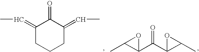
—CH═CH—(CO)—CR—C(OH)—CH═CH—, —CH═CH—(CO)—CR2—(CO)—CH═CH—, and —CH═CH—(CO)—CH—C(OH)—CH═CH—; R is an alkyl or aryl group including 10 carbon atoms or less.

Ar1—L—Ar2 (I)
wherein Ar2 is optional; L is a divalent linking group comprising an alkylene or an alkenylene that includes between 3 and 7 backbone carbon atoms, wherein one or more of the backbone carbon atoms include a carbonyl or hydroxyl moiety; and Ar1 and Ar2 (if Ar2 is present) are independently aryl groups. Ar1 and Ar2 (if Ar2 is present) may be unsubstituted or may optionally include one or more substituents selected from the group consisting of hydroxyl, alkyl, alkenyl, haloalkyl, alkoxy, and NR2, where R is hydrogen or alkyl. If Ar2 is absent, it may be replaced by a substituent R11, including hydrogen (H). R11 can be, for example, a heterocyclic group or an alkyl group, preferably an alkyl group having four or fewer carbon atoms, e.g., a methyl group. R11 can alternately be an amine, a hydroxyl, or a hydrogen.
Aryl Groups
and Ar2 can be a phenyl group according to Formula III:

Ar1—L—R11 (IV)
R11 in Formula IV can be, for example, a heterocyclic group or an alkyl group, preferably an alkyl group having four or fewer carbon atoms, e.g., a methyl group. R11 can alternately be an amine, a hydroxyl, or a hydrogen.
Curcumin Derivatives Including 7-carbon Linking Groups
—CH═CH—(CO)—CR═C(OH)—CH═CH—,
—CH═CH—(CO)—CR2—(CO)—CH═CH—, and
—CH═CH—(CO)—CH═C(OH)—CH═CH—.
| TABLE 1 |
| 7-Carbon Linker Analogs. |
| 3a |  |
| 3b |  |
| 3c |  |
| 3d |  |
| 3e |  |
| 3f |  |
| 3g |  |
| 3h |  |
| 3i |  |
| 6a |  |
| 6b |  |
| 9a |  |
| 9b |  |
| 11b |  |
| 12b |  |
| 13a |  |
| 13b |  |
| 14a |  |
| 14b |  |
| 15a |  |
| 15b |  |
| 16a |  |
| 17b |  |
Curcumin Derivatives Including 5-carbon Linking Groups

| TABLE 2 |
| 5-Carbon Linker Analogs. |
| 20a |  |
| 20b |  |
| 20c |  |
| 20d |  |
| 20e |  |
| 20f |  |
| 20g |  |
| 20i |  |
| 20k |  |
| 20l |  |
| 20m |  |
| 20n |  |
| 20o |  |
| 20p |  |
| 20q |  |
| 20r |  |
| 20s |  |
| 20t |  |
| 20u |  |
| 20v |  |
| 20w |  |
| 20x |  |
| 20y |  |
| 20z |  |
| 20aa |  |
| 20ab |  |
| 20ac |  |
| 20ae |  |
| 20af |  |
| 20ag | 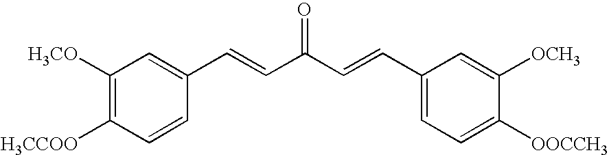 |
| 20ah |  |
| 23 |  |
| 25 |  |
| 29 |  |
| 31 |  |
| 34 |  |
| 36a |  |
| 36e |  |
| 38a |  |
| 38b |  |
| 39b |  |
| 40b |  |
| 42b |  |
| 43b |  |
Curcumin Derivatives Including 3-carbon Linking Groups
| TABLE 3 |
| 3-Carbon Linker Analogs. |
| 35a |  |
| 35e |  |
| 35q |  |
| 45a |  |
| 45b |  |
| 46a |  |
| 46ad |  |
| 46ak |  |
| 46al |  |
| 48a |  |
| 48ad |  |
| 50b |  |
| 52b |  |
Additional Curcumin Derivatives


Additional analogs that are contemplated are those having a pyridine ring with and without a central methylene substituent on the 7-carbon spacer such as those shown below. Analogs without a central methylene substituent can be prepared according to Pabon's method shown in


These new analogs can be prepared as shown in

Although analogs having 3-carbon spacers were generally not as active as analogs having 7-carbon or 5-carbon spacers, additional analogs may provide significant inhibition of NF-κB, AP-1 and/or GSTP1-1 activation. Relatively few analogs in this series containing a heterocyclic ring on the spacer have been synthesized. Analogs having different substituents on the aryl rings may provide significant inhibition of NF-κB, AP-1, and/or GSTP1-1 activation. In addition, analogs that contain different substituents on the nitrogen of the heterocyclic ring may provide significant inhibition of NF-κB, AP-1, and/or GSTP1-1 activation. Examples of these series 3 analogs are shown below:

These analogs can be synthesized as shown in

Additional curcumin derivatives of the invention that are not encompassed by the embodiments provided above may also be found in the examples and schemes provided herein.
Disease Treatment Using Curcumin Derivatives


protons of 3-benzyl-2,4-pentanedione (8). The structures were also verified by carbon NMR by the appearance of a signal at 183.3 ppm for the keto-enol carbonyl carbon. Also observed in the carbon NMR was the loss of signals at 22.9 ppm and 29.4 ppm for the methyl carbons of 3-benzyl-2,4-pentanedione (8). The carbon NMR of


Synthesis of 5-Carbon Spacer Analogs


acetone (19). The structures were also verified by carbon NMR by the appearance of two signals in the aromatic region for the alkene carbons on the spacer. The carbon NMR also showed the loss of a signal at 30.6 ppm for the methyl carbons in acetone (19).


the loss of a signal at 30.6 ppm for the loss of the methyl carbons in acetone (19).

56.2 ppm and 94.8 ppm for the carbons of the protecting group in compound 35j.


protons of the protecting group in compound 38j. The structure was also verified by carbon NMR by the loss of signals at 55.8 ppm and 95.1 ppm for the carbons of the protecting group in compound 38j.


Synthesis of 3-Carbon Spacer Analogs

and 5.30 ppm for the protons in the protecting groups in compound 45j. The structure was also verified by carbon NMR by the loss of signals at ˜57 ppm and ˜95 ppm for the carbons of the protecting groups in compound 45j.


aldehyde proton in the starting benzaldehyde (1ad). The structure of the product was also verified by carbon NMR by the appearance of two signals in the aromatic region for the alkene carbons on the spacer. Also seen in the carbon NMR was the loss of a signal at 26.0 ppm for the methyl carbon in the starting acetophenone (44b).

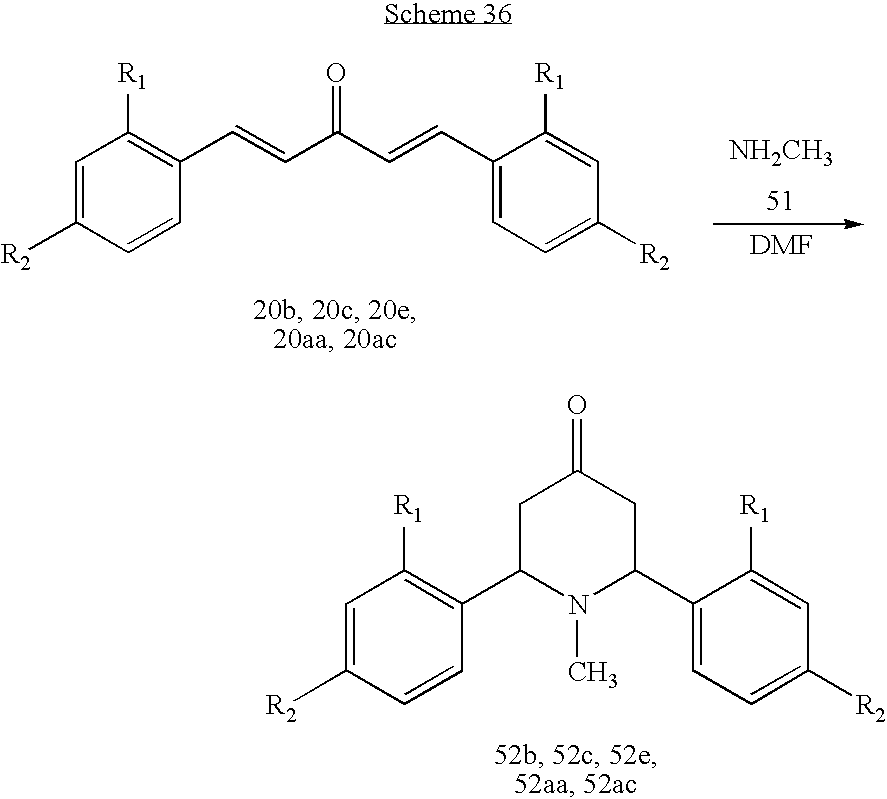

Experimental
| TABLE 5 |
| IC50 Values and Antioxidant Results for Active Analogs Against NF-κB. |
| Analog | IC50 | |||
| Structure | Number | (μM) | TRAP | FRAP |
 |
31 | 3.4 | − | − |
 |
29 | 3.5 | − | − |
 |
38a | 4.2 | + | + |
 |
20q | 4.2 | + | − |
 |
20ag | 5.4 | − | + |
 |
20m | 6.4 | − | − |
 |
6a | 6.8 | + | + |
 |
9a | 7.6 | + | + |
| TABLE 6 |
| Kest Values of Curcumin Analogs Against NF-κB Without MES. |
| 12b | 2.00E−10 | 11b | 3.31E−09 | 20q | 9.59E−09 | 46a | 3.07E−08 |
| 20ag | 3.07E−10 | 20v | 3.38E−09 | 20ah | 9.80E−09 | 20r | 3.16E−08 |
| 17b | 3.27E−10 | 52e | 3.52E−09 | 20z | 1.11E−08 | 31 | 3.66E−08 |
| 53 | 3.72E−10 | 9b | 4.09E−09 | 45a | 1.20E−08 | 29 | 4.87E−08 |
| 3i | 6.31E−10 | 36a | 4.55E−09 | 40af | 1.29E−08 | 46al | 5.03E−08 |
| 23 | 7.36E−10 | 6a | 4.62E−09 | 20x | 1.35E−08 | 46ak | 5.16E−08 |
| 20n | 7.86E−10 | 20w | 5.08E−09 | 3c | 1.39E−08 | 39b | 5.32E−08 |
| 25 | 1.16E−09 | 20o | 5.32E−09 | 20ab | 1.63E−08 | 20b | 5.82E−08 |
| 15a | 1.20E−09 | 20a | 5.42E−09 | 13b | 1.68E−08 | 20s | 6.08E−08 |
| 3d | 1.34E−09 | 20p | 5.42E−09 | 20ae | 1.75E−08 | 20f | 6.66E−08 |
| 14a | 1.34E−09 | 16b | 5.71E−09 | 52b | 1.76E−08 | 42b | 6.78E−08 |
| 9a | 1.51E−09 | 20k | 6.38E−09 | 38b | 1.87E−08 | 20l | 7.00E−08 |
| 3b | 1.94E−09 | 52aa | 6.52E−09 | 48a | 1.92E−08 | 50b | 7.12E−08 |
| 3g | 2.32E−09 | 52ac | 7.38E−09 | 20t | 2.35E−08 | 34 | 9.37E−08 |
| 38a | 2.36E−09 | 3f | 7.60E−09 | 36e | 2.39E−08 | 40b | 1.23E−07 |
| 14b | 2.38E−09 | 20d | 7.74E−09 | 20g | 2.45E−08 | 43b | 1.41E−07 |
| 20m | 2.49E−09 | 20c | 7.85E−09 | 3e | 2.54E−08 | 52l | 1.76E−07 |
| 15b | 2.89E−09 | 3h | 8.02E−09 | 20e | 2.60E−08 | 45b | 2.19E−07 |
| 3a | 2.94E−09 | 46ad | 8.96E−09 | 48ad | 2.71E−08 | 35a | 5.30E−07 |
| 6b | 3.06E−09 | 20y | 9.03E−09 | 20aa | 2.85E−08 | 35e | 1.66E−06 |
| 13a | 3.17E−09 | 20u | 9.53E−09 | 20ac | 2.90E−08 | 35q | 3.53E−06 |
| 20i | 3.29E−09 | ||||||
| TABLE 7 |
| Kest Values for NF-κB (1IKN) with MES. |
| 20ag | 7.26E−09 | 40af | 8.72E−08 | 6b | 2.19E−07 | 48ad | 7.60E−07 |
| 9a | 7.39E−09 | 14b | 8.86E−08 | 20y | 2.22E−07 | 20t | 8.03E−07 |
| 15a | 1.26E−08 | 20k | 9.43E−08 | 13c | 2.41E−07 | 20l | 8.15E−07 |
| 12b | 1.54E−08 | 3a | 9.64E−08 | 52e | 2.47E−07 | 50b | 8.72E−07 |
| 13a | 1.57E−08 | 15b | 9.69E−08 | 20p | 2.56E−07 | 46ak | 9.22E−07 |
| 17b | 1.65E−08 | 20u | 9.88E−08 | 52l | 2.57E−07 | 42b | 9.44E−07 |
| 9b | 1.93E−08 | 36a | 1.06E−07 | 20aa | 2.71E−07 | 40b | 9.64E−07 |
| 25 | 2.51E−08 | 20w | 1.29E−07 | 3e | 2.74E−07 | 29 | 1.03E−06 |
| 3d | 2.59E−08 | 3h | 1.40E−07 | 20x | 2.81E−07 | 52b | 1.08E−06 |
| 20ae | 2.65E−08 | 3g | 1.47E−07 | 20ab | 2.82E−07 | 46al | 1.14E−06 |
| 20i | 2.69E−08 | 46ad | 1.56E−07 | 20z | 3.78E−07 | 46a | 1.24E−06 |
| 38a | 3.27E−08 | 20o | 1.63E−07 | 52aa | 3.84E−07 | 20b | 1.25E−06 |
| 20a | 4.38E−08 | 20f | 1.74E−07 | 3b | 4.03E−07 | 31 | 1.33E−06 |
| 20ah | 5.27E−08 | 3i | 1.82E−07 | 20q | 4.16E−07 | 39b | 1.42E−06 |
| 20m | 5.61E−08 | 16b | 1.82E−07 | 20c | 4.46E−07 | 43b | 2.01E−06 |
| 53 | 6.06E−08 | 36e | 1.86E−07 | 3f | 4.47E−07 | 38b | 2.41E−06 |
| 14a | 6.10E−08 | 20d | 1.86E−07 | 52ac | 4.61E−07 | 45b | 4.15E−06 |
| 20v | 6.66E−08 | 20g | 1.86E−07 | 20ac | 4.92E−07 | 34 | 4.22E−06 |
| 23 | 7.21E−08 | 20e | 1.87E−07 | 20s | 5.49E−07 | 35a | 4.46E−06 |
| 11b | 7.35E−08 | 13b | 2.03E−07 | 20r | 6.43E−07 | 35e | 4.73E−06 |
| 20n | 8.21E−08 | 45a | 2.05E−07 | 48a | 6.44E−07 | 35q | 4.81E−06 |
| 6a | 8.63E−08 | ||||||
Binding to the 1SVC Form of NF-κB
| TABLE 8 |
| Kest Values for NF-κB (1SVC). |
| 9b | 3.79E−10 | 20ag | 1.32E−08 | 20w | 4.68E−08 | 20b | 1.57E−07 |
| 3g | 1.59E−09 | 20u | 1.33E−08 | 20ac | 4.70E−08 | 29 | 1.65E−07 |
| 12b | 3.30E−09 | 20o | 1.34E−08 | 53 | 4.70E−08 | 48ad | 1.85E−07 |
| 25 | 4.39E−09 | 20x | 1.42E−08 | 15a | 4.75E−08 | 40b | 1.86E−07 |
| 6b | 4.40E−09 | 11b | 1.47E−08 | 20t | 5.41E−08 | 38a | 1.87E−07 |
| 3i | 5.53E−09 | 3a | 1.57E−08 | 20r | 6.15E−08 | 46a | 1.94E−07 |
| 6a | 5.62E−09 | 36a | 1.75E−08 | 20l | 6.62E−08 | 50b | 1.94E−07 |
| 23 | 5.67E−09 | 20a | 1.87E−08 | 20n | 7.54E−08 | 45a | 2.03E−07 |
| 20e | 6.09E−09 | 13c | 1.96E−08 | 20p | 7.64E−08 | 34 | 2.89E−07 |
| 3e | 6.84E−09 | 13b | 1.97E−08 | 46ad | 7.96E−08 | 46ak | 2.96E−07 |
| 20m | 7.34E−09 | 20aa | 2.12E−08 | 46al | 8.38E−08 | 52aa | 3.43E−07 |
| 3d | 7.35E−09 | 20y | 2.22E−08 | 20c | 8.47E−08 | 38b | 4.41E−07 |
| 14b | 7.35E−09 | 20q | 2.78E−08 | 42b | 9.25E−08 | 52e | 4.42E−07 |
| 13a | 8.18E−09 | 16b | 2.83E−08 | 20s | 9.78E−08 | 45b | 5.15E−07 |
| 3h | 9.21E−09 | 17b | 2.95E−08 | 31 | 1.11E−07 | 43b | 5.65E−07 |
| 14a | 9.59E−09 | 3f | 3.00E−08 | 20z | 1.13E−07 | 52l | 5.67E−07 |
| 20ah | 9.85E−09 | 36e | 3.34E−08 | 9a | 1.18E−07 | 52ac | 6.59E−07 |
| 20d | 1.07E−08 | 20f | 3.41E−08 | 40af | 1.21E−07 | 35q | 9.89E−07 |
| 20v | 1.11E−08 | 20ab | 3.51E−08 | 15b | 1.27E−07 | 52b | 1.05E−06 |
| 20g | 1.19E−08 | 20i | 4.10E−08 | 48a | 1.45E−07 | 35a | 1.07E−06 |
| 3b | 1.30E−08 | 20k | 4.55E−08 | 39b | 1.55E−07 | 35e | 1.18E−06 |
| 20ae | 1.31E−08 | ||||||
| TABLE 9 |
| Kest Values for NF-κB (1SVC) with MES. |
| 15a | 2.27E−08 | 11b | 4.15E−07 | 20l | 8.98E−07 | 20ac | 2.12E−06 |
| 17b | 2.48E−08 | 3f | 4.22E−07 | 3b | 9.38E−07 | 40b | 2.13E−06 |
| 12b | 2.63E−08 | 40af | 4.26E−07 | 20d | 1.01E−06 | 48ad | 2.31E−06 |
| 20ag | 5.48E−08 | 3g | 4.37E−07 | 20k | 1.19E−06 | 52aa | 2.40E−06 |
| 15b | 6.30E−08 | 3e | 4.45E−07 | 52e | 1.21E−06 | 31 | 2.95E−06 |
| 38a | 7.76E−08 | 20ah | 4.51E−07 | 20e | 1.22E−06 | 20s | 2.98E−06 |
| 9a | 8.10E−08 | 3a | 4.79E−07 | 20f | 1.27E−06 | 39b | 3.04E−06 |
| 6a | 8.37E−08 | 20u | 4.82E−07 | 20g | 1.28E−06 | 46ak | 3.31E−06 |
| 53 | 9.53E−08 | 20ae | 5.00E−07 | 20ab | 1.34E−06 | 20t | 3.52E−06 |
| 3h | 1.01E−07 | 13c | 5.36E−07 | 20y | 1.34E−06 | 36e | 3.67E−06 |
| 3d | 1.29E−07 | 20o | 5.47E−07 | 52l | 1.46E−06 | 20b | 4.27E−06 |
| 9b | 1.32E−07 | 14b | 5.50E−07 | 20aa | 1.47E−06 | 52ac | 4.50E−06 |
| 23 | 1.58E−07 | 20w | 5.71E−07 | 20x | 1.55E−06 | 42b | 6.29E−06 |
| 25 | 2.36E−07 | 20a | 5.80E−07 | 20z | 1.58E−06 | 50b | 6.32E−06 |
| 3i | 2.67E−07 | 20n | 6.46E−07 | 20c | 1.70E−06 | 52b | 7.86E−06 |
| 14a | 2.78E−07 | 46ad | 6.58E−07 | 20q | 1.74E−06 | 35q | 8.52E−06 |
| 20p | 3.01E−07 | 36a | 6.79E−07 | 20r | 1.77E−06 | 45b | 9.20E−06 |
| 13a | 3.13E−07 | 46a | 6.81E−07 | 46al | 1.81E−06 | 35a | 1.11E−05 |
| 20i | 3.22E−07 | 13b | 7.62E−07 | 29 | 1.95E−06 | 35e | 1.30E−05 |
| 16b | 3.31E−07 | 6b | 7.66E−07 | 38b | 2.02E−06 | 34 | 1.84E−05 |
| 20m | 3.58E−07 | 45a | 8.30E−07 | 48a | 2.06E−06 | 43b | 1.88E−05 |
| 20v | 4.08E−07 | ||||||
| TABLE 10 |
| IC50 Values and Antioxidant Results for Active Analogs Against NF-κB and AP-1. |
| NF- | |||||
| AP-1 | κB | ||||
| Analog | IC50 | IC50 | |||
| Structure | Number | (μM) | (μM) | TRAP | FRAP |
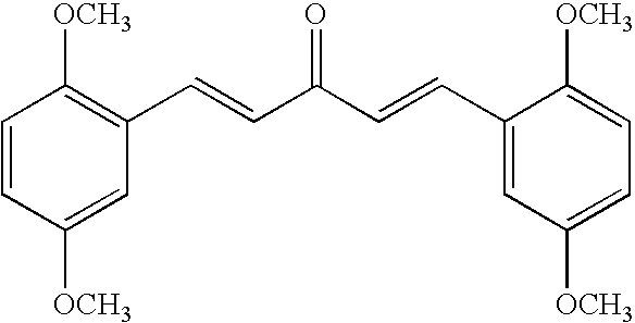 | 20m | 1.4 | 6.4 | − | − |
 | 31 | 4.1 | 3.4 | − | − |
 | 9a | 5.3 | 7.6 | + | + |
 | 6a | 6.0 | 6.7 | + | + |
 | 38a | 7.3 | 4.2 | + | + |
 | 29 | 8.2 | 3.5 | − | − |
 | 20ag | 8.3 | 5.4 | − | + |
 | 20q | 11.7 | 4.2 | + | − |
 | 3a | 12.8 | 8.2 | + | + |
Of the IC50 values obtained, curcumin (12.8 μM) is the least potent analog against AP-1.
| TABLE 11 |
| Kest Values for AP-1 (1FOS). |
| 9b | 6.53E−08 | 38a | 7.53E−07 | 20u | 2.07E−06 | 20r | 4.30E−06 |
| 12b | 9.96E−08 | 3h | 7.73E−07 | 20ah | 2.13E−06 | 13b | 4.60E−06 |
| 15a | 1.57E−07 | 20z | 8.46E−07 | 52l | 2.14E−06 | 20p | 4.90E−06 |
| 3d | 1.71E−07 | 20w | 8.98E−07 | 20t | 2.35E−06 | 45a | 5.01E−06 |
| 6a | 1.73E−07 | 20k | 9.23E−07 | 11b | 2.45E−06 | 20x | 5.14E−06 |
| 53 | 1.95E−07 | 20ac | 9.70E−07 | 36a | 2.54E−06 | 46ak | 5.25E−06 |
| 46ad | 2.37E−07 | 20g | 1.07E−06 | 20c | 2.62E−06 | 20e | 5.33E−06 |
| 20m | 2.43E−07 | 6b | 1.16E−06 | 20aa | 2.72E−06 | 39b | 5.88E−06 |
| 23 | 2.44E−07 | 20ag | 1.20E−06 | 20ab | 2.76E−06 | 46a | 6.66E−06 |
| 20d | 2.88E−07 | 20l | 1.28E−06 | 16b | 2.82E−06 | 52b | 6.83E−06 |
| 20v | 2.99E−07 | 17b | 1.28E−06 | 52e | 2.92E−06 | 42b | 6.84E−06 |
| 48ad | 4.18E−07 | 3e | 1.43E−06 | 20b | 2.93E−06 | 20f | 7.63E−06 |
| 13c | 4.38E−07 | 20ae | 1.53E−06 | 52aa | 2.96E−06 | 50b | 8.05E−06 |
| 9a | 4.49E−07 | 20y | 1.60E−06 | 29 | 3.12E−06 | 48a | 1.10E−05 |
| 3a | 5.01E−07 | 20q | 1.60E−06 | 40af | 3.24E−06 | 34 | 1.11E−05 |
| 25 | 5.31E−07 | 13a | 1.75E−06 | 20s | 3.30E−06 | 45b | 1.19E−05 |
| 15b | 5.38E−07 | 36e | 1.83E−06 | 31 | 3.38E−06 | 43b | 1.91E−05 |
| 3i | 5.42E−07 | 52ac | 1.85E−06 | 20n | 3.57E−06 | 40b | 1.99E−05 |
| 20i | 5.49E−07 | 14b | 1.86E−06 | 20a | 3.93E−06 | 35e | 5.48E−05 |
| 3g | 6.19E−07 | 3b | 1.97E−06 | 38b | 4.02E−06 | 35a | 5.53E−05 |
| 14a | 6.61E−07 | 3f | 1.99E−06 | 46al | 4.17E−06 | 35q | 1.02E−04 |
| 20o | 7.31E−07 | ||||||
| TABLE 12 |
| Kest Values for AP-1 (1FOS) with MES. |
| 15a | 1.26E−07 | 20g | 1.38E−06 | 52e | 2.79E−06 | 20e | 5.33E−06 |
| 13c | 2.05E−07 | 17b | 1.49E−06 | 20y | 2.93E−06 | 20q | 5.40E−06 |
| 23 | 2.96E−07 | 3i | 1.53E−06 | 3f | 2.99E−06 | 52b | 6.00E−06 |
| 46ad | 3.15E−07 | 3d | 1.57E−06 | 20k | 3.28E−06 | 46ak | 6.04E−06 |
| 53 | 3.43E−07 | 3h | 1.59E−06 | 20aa | 3.29E−06 | 20b | 6.33E−06 |
| 3g | 4.75E−07 | 13b | 1.74E−06 | 20x | 3.33E−06 | 20p | 6.38E−06 |
| 20d | 5.74E−07 | 12b | 1.79E−06 | 20ah | 3.40E−06 | 38b | 6.95E−06 |
| 9b | 6.11E−07 | 6a | 1.83E−06 | 11b | 3.58E−06 | 20t | 8.29E−06 |
| 25 | 6.78E−07 | 20s | 1.86E−06 | 20ac | 3.67E−06 | 34 | 8.51E−06 |
| 20z | 7.14E−07 | 52aa | 1.98E−06 | 46a | 3.76E−06 | 20f | 8.58E−06 |
| 20v | 9.80E−07 | 6b | 2.02E−06 | 14b | 3.78E−06 | 40b | 8.61E−06 |
| 38a | 1.04E−06 | 3a | 2.03E−06 | 36e | 3.79E−06 | 36a | 9.23E−06 |
| 48ad | 1.08E−06 | 14a | 2.04E−06 | 31 | 3.81E−06 | 20r | 9.23E−06 |
| 9a | 1.12E−06 | 20ae | 2.07E−06 | 20i | 3.86E−06 | 48a | 1.04E−05 |
| 20w | 1.20E−06 | 52ac | 2.13E−06 | 52l | 3.95E−06 | 50b | 1.07E−05 |
| 20ab | 1.22E−06 | 20m | 2.17E−06 | 3b | 4.00E−06 | 43b | 1.09E−05 |
| 15b | 1.22E−06 | 13a | 2.34E−06 | 29 | 4.08E−06 | 45b | 1.45E−05 |
| 20ag | 1.24E−06 | 42b | 2.58E−06 | 20a | 4.26E−06 | 45a | 1.78E−05 |
| 20o | 1.26E−06 | 20n | 2.60E−06 | 20l | 4.65E−06 | 35a | 4.51E−05 |
| 3e | 1.29E−06 | 46al | 2.74E−06 | 40af | 4.80E−06 | 35e | 4.90E−05 |
| 20u | 1.29E−06 | 20c | 2.76E−06 | 39b | 4.97E−06 | 35q | 1.10E−04 |
| 16b | 1.30E−06 | ||||||
-
- 1. Protofibril formation requires >20 Aβ molecules,
- 2. protofibril elongation involves the reversible coalescence of smaller protofibrils,
- 3. protofibril to fibril transition is the first pathogenic feature of AD detected by dye binding, and
- 4. fibril elongation process is continuous.
Because the Aβ fibril was the first amyloid entity identified by virtue of dye binding, it was naturally thought to represent the form of Aβ peptide that was neurotoxic. However, more recent evidence suggests that Aβ might be most detrimental to synaptic integrity and neuronal health when it is in the form of soluble oligomers generated during the early stages of the aggregation pathway (Hartley et al., 1999, J Neurosci 19:8876-8884; Lambert et al., 1998, Proc Natl Acad Sci USA 95:6448-6453; McLean et al., 1999, Ann Neurol 46:860-866; Walsh et al., 2002, Nature 416:535-539; Walsh et al., 2004, Protein Pept Lett 11:213-228). Microinjection of human Aβ into rats revealed that Aβ oligomers, in the absence of monomers and amyloid fibrils, inhibit long-term potentiation in the hippocampus (Walsh et al., 2002, Nature 416:535-539). Therefore, because of the complexity of the amyloid deposition process, inhibition of all forms of fibril intermediates-including oligomers and protofibrils-should be addressed when developing therapeutic intervention strategies.
| TABLE 12 | ||
| Compound | Structure | IC50 (μM) |
| 1 (curcumin) |  | 8.2 ± 0.4 |
| 2 |  | 6.7 ± 1.2 |
| 3 |  | 3.4 ± 0.2 |
| 4 |  | 4.4 ± 0.8 |
| 5 |  | 5.0 ± 0.3 |
Summary of Pertinent Initial Results

| TABLE 14 |
| Kest Values for GSTP1-1 (19GS) |
| 14a | 3.51E−09 | 6a | 1.09E−07 | 20i | 3.16E−07 | 40b | 8.56E−07 |
| 15a | 6.57E−09 | 52e | 1.13E−07 | 20k | 3.18E−07 | 46ak | 8.66E−07 |
| 15b | 9.61E−09 | 3g | 1.17E−07 | 3e | 3.22E−07 | 45b | 9.34E−07 |
| 12b | 1.26E−08 | 20l | 1.20E−07 | 16b | 3.37E−07 | 50b | 9.37E−07 |
| 9b | 3.33E−08 | 3b | 1.23E−07 | 20y | 3.74E−07 | 20z | 1.00E−06 |
| 9a | 3.33E−08 | 20o | 1.25E−07 | 20q | 3.92E−07 | 52ac | 1.08E−06 |
| 17b | 3.35E−08 | 3a | 1.36E−07 | 36a | 4.10E−07 | 31 | 1.09E−06 |
| 23 | 4.63E−08 | 13a | 1.53E−07 | 20c | 4.27E−07 | 52l | 1.14E−06 |
| 53 | 4.79E−08 | 3d | 1.64E−07 | 46a | 4.41E−07 | 20p | 1.24E−06 |
| gluta | 5.26E−08 | 20ab | 1.77E−07 | 40af | 4.55E−07 | 20aa | 1.29E−06 |
| 20ag | 5.34E−08 | 20m | 2.03E−07 | 20n | 4.68E−07 | 20b | 1.33E−06 |
| 20w | 5.67E−08 | 13b | 2.19E−07 | 6b | 4.72E−07 | 36e | 1.40E−06 |
| 20v | 5.87E−08 | 45a | 2.22E−07 | 20a | 4.86E−07 | 29 | 1.45E−06 |
| 20ae | 6.81E−08 | 20u | 2.25E−07 | 20r | 5.30E−07 | 42b | 1.57E−06 |
| 38a | 6.84E−08 | 48ad | 2.28E−07 | 20ac | 5.39E−07 | 20t | 1.97E−06 |
| 14b | 7.09E−08 | 20e | 2.54E−07 | 48a | 6.36E−07 | 39b | 2.06E−06 |
| 3i | 7.75E−08 | 13c | 2.61E−07 | 46ad | 6.38E−07 | 34 | 2.57E−06 |
| 20g | 8.88E−08 | 52aa | 2.67E−07 | 20f | 7.40E−07 | 43b | 3.25E−06 |
| 25 | 9.45E−08 | 11b | 2.68E−07 | 38b | 7.61E−07 | 35a | 4.00E−06 |
| 3f | 9.68E−08 | 46al | 2.80E−07 | 20s | 7.90E−07 | 35q | 5.83E−06 |
| 20ah | 1.05E−07 | 52b | 2.84E−07 | 20x | 8.01E−07 | 35e | 1.12E−05 |
| 3h | 1.07E−07 | 20d | 3.01E−07 | ||||
Claims (13)


Priority Applications (3)
| Application Number | Priority Date | Filing Date | Title |
|---|---|---|---|
| US11/478,073 US8841326B2 (en) | 2004-02-12 | 2006-06-29 | Therapeutic curcumin derivatives |
| US14/449,599 US9187397B2 (en) | 2004-02-12 | 2014-08-01 | Therapeutic curcumin derivatives |
| US14/887,780 US20160039734A1 (en) | 2004-02-12 | 2015-10-20 | Therapeutic curcumin derivatives |
Applications Claiming Priority (7)
| Application Number | Priority Date | Filing Date | Title |
|---|---|---|---|
| US54442404P | 2004-02-12 | 2004-02-12 | |
| US11/057,736 US20060258752A1 (en) | 2004-02-12 | 2005-02-14 | Method and compounds for cancer treatment utilizing NFkB as a direct or ultimate target for small molecule inhibitors |
| US69504605P | 2005-06-29 | 2005-06-29 | |
| US11/373,444 US20060276536A1 (en) | 2004-02-12 | 2006-03-10 | Cancer treatment using curcumin derivatives |
| US78769406P | 2006-03-30 | 2006-03-30 | |
| US78769506P | 2006-03-30 | 2006-03-30 | |
| US11/478,073 US8841326B2 (en) | 2004-02-12 | 2006-06-29 | Therapeutic curcumin derivatives |
Related Parent Applications (1)
| Application Number | Title | Priority Date | Filing Date |
|---|---|---|---|
| US11/373,444 Continuation-In-Part US20060276536A1 (en) | 2004-02-12 | 2006-03-10 | Cancer treatment using curcumin derivatives |
Related Child Applications (1)
| Application Number | Title | Priority Date | Filing Date |
|---|---|---|---|
| US14/449,599 Division US9187397B2 (en) | 2004-02-12 | 2014-08-01 | Therapeutic curcumin derivatives |
Publications (2)
| Publication Number | Publication Date |
|---|---|
| US20070060644A1 US20070060644A1 (en) | 2007-03-15 |
| US8841326B2 true US8841326B2 (en) | 2014-09-23 |
Family
ID=37856148
Family Applications (3)
| Application Number | Title | Priority Date | Filing Date |
|---|---|---|---|
| US11/478,073 Active 2030-03-02 US8841326B2 (en) | 2004-02-12 | 2006-06-29 | Therapeutic curcumin derivatives |
| US14/449,599 Active US9187397B2 (en) | 2004-02-12 | 2014-08-01 | Therapeutic curcumin derivatives |
| US14/887,780 Abandoned US20160039734A1 (en) | 2004-02-12 | 2015-10-20 | Therapeutic curcumin derivatives |
Family Applications After (2)
| Application Number | Title | Priority Date | Filing Date |
|---|---|---|---|
| US14/449,599 Active US9187397B2 (en) | 2004-02-12 | 2014-08-01 | Therapeutic curcumin derivatives |
| US14/887,780 Abandoned US20160039734A1 (en) | 2004-02-12 | 2015-10-20 | Therapeutic curcumin derivatives |
Country Status (1)
| Country | Link |
|---|---|
| US (3) | US8841326B2 (en) |
Cited By (6)
| Publication number | Priority date | Publication date | Assignee | Title |
|---|---|---|---|---|
| US20150087705A1 (en) * | 2013-09-20 | 2015-03-26 | Georgia Regents Research Institute, Inc. | Protective Effects of Curcumin Against Hemorrhagic Stroke Injury |
| US20170003280A1 (en) * | 2014-01-11 | 2017-01-05 | The J. David Gladstone Institutes | In vitro assays for inhibition of microglial activation |
| US20170158605A1 (en) * | 2009-05-15 | 2017-06-08 | The Research Foundation Of State University Of New York | Curcumin analogues as zinc chelators and their uses |
| US20190060289A1 (en) * | 2011-05-10 | 2019-02-28 | Stc.Unm | Methods of treating autophagy-associated disorders and related pharmaceutical compositions, diagnostics, screening techniques and kits |
| US20190255198A1 (en) * | 2016-06-29 | 2019-08-22 | The General Hospital Corporation | Half-Curcuminoids as Amyloid-Beta PET Imaging Agents |
| US10781158B2 (en) * | 2014-09-02 | 2020-09-22 | Bhupinder Singh | Methods of protecting an organ using tetrahydrocurcumin and phosphatidylcholine |
Families Citing this family (59)
| Publication number | Priority date | Publication date | Assignee | Title |
|---|---|---|---|---|
| US7507864B2 (en) * | 2006-08-01 | 2009-03-24 | Salisbury University | Method for the synthesis of curcumin analogues |
| US8383865B2 (en) * | 2007-04-17 | 2013-02-26 | Codman & Shurtleff, Inc. | Curcumin derivatives |
| ES2562205T3 (en) * | 2007-04-17 | 2016-03-03 | Codman & Shurtleff, Inc. | Resveratrol curcumin hybrids |
| CN101255119B (en) * | 2008-01-07 | 2014-05-21 | 沈阳药科大学 | Novel tetrahydro curcumin derivatives and salt |
| US7745670B2 (en) * | 2008-06-27 | 2010-06-29 | Codman & Shurtleff, Inc. | Curcumin-Resveratrol hybrid molecule |
| CN102046575B (en) * | 2008-05-30 | 2014-10-29 | 国立大学法人东京工业大学 | Therapeutic agent for alzheimer's disease |
| US7985776B2 (en) | 2008-06-27 | 2011-07-26 | Codman & Shurtleff, Inc. | Iontophoretic delivery of curcumin and curcumin analogs for the treatment of Alzheimer's Disease |
| WO2010017094A2 (en) * | 2008-07-31 | 2010-02-11 | The General Hospital Corporation | CURCUMIN DERIVATIVES FOR AMYLOID-β PLAQUE IMAGING |
| MX349711B (en) * | 2008-09-18 | 2017-08-09 | Cedars Sinai Medical Center | Optical method for the detection of alzheimer's disease. |
| CA2746273C (en) * | 2008-12-11 | 2019-04-09 | Human Biomolecular Research Institute | Small molecule immunomodulators for alzheimer's disease |
| US7723515B1 (en) * | 2009-01-26 | 2010-05-25 | Codman & Shurtleff, Inc. | Methylene blue—curcumin analog for the treatment of alzheimer's disease |
| US20100286585A1 (en) * | 2009-01-26 | 2010-11-11 | Codman & Shurtleff, Inc. | Shunt Delivery of Curcumin |
| WO2010098502A1 (en) | 2009-02-27 | 2010-09-02 | 独立行政法人科学技術振興機構 | Imaging diagnostic agent and extracorporeal diagnostic agent for incurable neurological diseases |
| BRPI0902039B8 (en) * | 2009-06-09 | 2021-05-25 | Anhanguera Educacional Ltda | pharmaceutical composition and use of pharmaceutical composition for the treatment, prophylaxis or prevention of neoplastic diseases in humans and animals |
| US8986657B2 (en) * | 2009-07-31 | 2015-03-24 | The General Hospital Corporation | Methods and system for detecting soluble amyloid-β |
| WO2011029359A1 (en) * | 2009-09-12 | 2011-03-17 | 温州医学院 | MEDICAMENTS CONTAINING 11β HYDROXYSTEROID DEHYDROGENASE 1 DOUBLE REGULATORS AND USES THEREOF |
| EP2477698A2 (en) | 2009-09-18 | 2012-07-25 | Centre National de la Recherche Scientifique | Compounds useful against kinetoplastideae parasites |
| PL2512461T3 (en) * | 2009-12-18 | 2014-06-30 | Colgate Palmolive Co | Chalcones as enhancer of antimicrobial agents |
| US8410177B2 (en) * | 2009-12-30 | 2013-04-02 | Indian Institute Of Technology Bombay | Curcumin derivatives |
| WO2011082290A2 (en) | 2009-12-31 | 2011-07-07 | Organomed Corporation | Formulations from natural products, turmeric, and aspirin |
| US9650404B2 (en) * | 2010-02-26 | 2017-05-16 | Research Foundation Of The City University Of New York | Curcumin derivatives |
| WO2011142795A1 (en) * | 2010-03-25 | 2011-11-17 | Wayne State University | Novel analogs of curcumin and methods of use |
| WO2012004746A2 (en) | 2010-07-05 | 2012-01-12 | Polymer Technologies International (Eou) | Refractive-diffractive ophthalmic device and compositions useful for producing same |
| RU2016124136A (en) | 2010-08-18 | 2018-12-03 | СЭМЬЮМЕД, ЭлЭлСи | DIKETONES AND HYDROXYKETONES AS AN ACTIVATOR OF THE KATENIN SIGNAL WAY |
| US8349376B1 (en) * | 2011-03-08 | 2013-01-08 | Bezzek Mark S | Anti-dementia regimen |
| US8846061B1 (en) | 2011-03-08 | 2014-09-30 | Mark S. Bezzek | Multivitamin-mineral regimens for longevity and wellness |
| WO2012138907A2 (en) * | 2011-04-05 | 2012-10-11 | Translational Genomics Research Institute | Solid forms of curcumin |
| WO2012145420A1 (en) * | 2011-04-18 | 2012-10-26 | Rutgers, The State University Of New Jersey | Composition, synthetic process and use of new antioxidant inflammation modulators |
| CN103086958A (en) * | 2011-11-04 | 2013-05-08 | 中国科学院上海药物研究所 | NF[kappa]B pathway activation inhibitor, preparation method thereof, pharmaceutical composition and purpose |
| CN102670573A (en) * | 2011-11-21 | 2012-09-19 | 西安交通大学 | Application of curcumin derivative in preparation of drug for treating prostate cancer disease |
| CN102600115B (en) * | 2011-12-16 | 2014-04-23 | 西安交通大学 | Application of curcumin C and derivative C3 thereof in preparing medicine for preventing macular degeneration of retina |
| WO2014005089A2 (en) | 2012-06-29 | 2014-01-03 | The Research Foundation Of State University Of New York | Polyenolic zinc-binding agents (pezbins) actively promote inactivation of cancer stem cells and potentiate cytotoxic anti-tumor drug substances |
| WO2014025808A1 (en) * | 2012-08-06 | 2014-02-13 | The General Hospital Corporation | Curcumin analogs |
| WO2014111831A1 (en) | 2013-01-15 | 2014-07-24 | Dave, Jagrat Natavar | Toric-diffractive lens |
| BR112015020391B1 (en) | 2013-02-22 | 2022-07-26 | Samumed, Llc | GAMMA-DICETONES AS WNT/BETA-CATENIN SIGNALING PATHWAY ACTIVATORS |
| CN103159644B (en) * | 2013-04-08 | 2015-04-15 | 唐山师范学院 | Curcuminoid condensed aromatic amine Schiff base derivative and application thereof in preparation of antibacterial medicaments |
| KR102222448B1 (en) | 2013-04-10 | 2021-03-04 | 데이브, 자그래트 나타바르 | Phakic lens device with opening and concentric annular zones |
| US9757477B2 (en) | 2013-09-06 | 2017-09-12 | The General Hospital Corporation | Imaging brown adipose tissue with curcumin derivatives |
| US9351946B2 (en) * | 2014-03-06 | 2016-05-31 | Hong Kong Baptist University | MTOR-independent activator of TFEB for autophagy enhancement and uses thereof |
| CN104030904B (en) * | 2014-03-07 | 2017-01-18 | 福建医科大学 | Application of 4-arylmethyl curcumin analogues serving as Hsp90 inhibitor |
| CN104974038B (en) * | 2014-04-13 | 2019-04-30 | 江苏康缘药业股份有限公司 | One kind carbonyl curcumin derivative of list containing carboxyl and its preparation method and application |
| CN104974037B (en) * | 2014-04-13 | 2019-04-30 | 江苏康缘药业股份有限公司 | One kind is containing more carbonyl curcumin derivatives of carboxyl and its preparation method and application |
| PT3206686T (en) | 2014-08-20 | 2020-01-06 | Samumed Llc | Gamma-diketones for treatment and prevention of aging skin and wrinkles |
| EP3188690A1 (en) | 2014-09-02 | 2017-07-12 | Dave, Jagrat Natavar | Intraocular lens customized for astigmatism or combined astigmatism and presbyopia |
| US20160106687A1 (en) | 2014-10-21 | 2016-04-21 | Life Plus, LLC | Human therapeutic agents |
| US10092550B2 (en) | 2014-10-21 | 2018-10-09 | Ions Pharmaceutical S.À R.L. | Therapeutic compositions containing curcumin, harmine, and isovanillin components, and methods of use thereof |
| US9907786B2 (en) | 2014-10-21 | 2018-03-06 | Ions Pharmaceutical S.À R.L. | Therapeutic compositions containing harmine and isovanillin components, and methods of use thereof |
| EP3433225B1 (en) * | 2016-06-13 | 2021-03-17 | China Medical University | Novel derivatives of curcuminoids and use thereof as an anticancer agent |
| US10300000B2 (en) | 2016-09-12 | 2019-05-28 | The Research Foundation For The State University Of New York | Inhibition of melanogenesis by chemically modified curcumins |
| WO2019012001A1 (en) | 2017-07-12 | 2019-01-17 | Syngenta Participations Ag | Microbiocidal oxadiazole derivatives |
| CN109731105B (en) * | 2019-01-09 | 2021-06-04 | 纳菲(深圳)制药科技有限公司 | Nasal cavity nano autophagy inducer for preventing and treating early neurodegenerative diseases and preparation method thereof |
| WO2020176036A1 (en) * | 2019-02-25 | 2020-09-03 | Agency For Science, Technology And Research | Oligomeric materials for uv blocking applications and methods thereof |
| WO2021105137A1 (en) | 2019-11-25 | 2021-06-03 | INSERM (Institut National de la Santé et de la Recherche Médicale) | Trem-1 inhibitors for the treatment of vaso-occlusions and tissue injuries in patients suffering from sickle cell disease |
| EP4294418A1 (en) * | 2021-02-18 | 2023-12-27 | Lyrus Life Sciences Pvt. LTD. | Novel arthritis emulgel composition and its preparation process |
| EP4304626A1 (en) | 2021-03-12 | 2024-01-17 | Institut National de la Santé et de la Recherche Médicale (INSERM) | Trem-1 inhibitors for the treatment of marfan syndrome |
| KR20240029046A (en) * | 2021-07-02 | 2024-03-05 | 안지 파마슈티컬 코퍼레이션 리미티드 | Topical preparations of dimethylcurcumin |
| WO2023061946A1 (en) | 2021-10-11 | 2023-04-20 | INSERM (Institut National de la Santé et de la Recherche Médicale) | Use of trem-1 for predicting and preventing postoperative complications after cardiac surgery with cardiopulmonary by-pass |
| CN114478256B (en) * | 2022-01-26 | 2024-02-23 | 安徽理工大学环境友好材料与职业健康研究院(芜湖) | Preparation method of novel curcumin derivative and application of novel curcumin derivative in liver cancer resistance |
| CN115745922B (en) * | 2022-11-22 | 2024-02-20 | 沈阳药科大学 | Diaryl pentane compound in Tibet daphnia and preparation method and application thereof |
Citations (31)
| Publication number | Priority date | Publication date | Assignee | Title |
|---|---|---|---|---|
| GB1109270A (en) | 1966-04-14 | 1968-04-10 | Solvay | Method of cross-linking amorphous polymers and copolymers of ª-olefins |
| US3691210A (en) | 1967-11-01 | 1972-09-12 | Polaroid Corp | 2-(omega-hydroquinonyl-alkyl)-anthraquinones |
| US4045487A (en) | 1975-06-26 | 1977-08-30 | Hoffmann-La Roche Inc. | 1-Dimethylamino-2,4-diphenyl-1-butene-3,4-dione |
| US4938949A (en) | 1988-09-12 | 1990-07-03 | University Of New York | Treatment of damaged bone marrow and dosage units therefor |
| EP0507013A1 (en) | 1989-11-02 | 1992-10-07 | Witco Corporation | Process for the preparation of aromatic beta-diketones |
| US5395692A (en) | 1994-05-06 | 1995-03-07 | Eastman Chemical Company | Photodegradable polyesters |
| EP0370461B1 (en) | 1988-11-21 | 1995-06-28 | Dainippon Ink And Chemicals, Inc. | Chalcone derivatives and process for producing the same |
| WO1995030670A2 (en) | 1994-05-06 | 1995-11-16 | Pharmacia & Upjohn Company | Pyranone compounds useful to treat retroviral infections |
| WO1998020891A1 (en) | 1996-11-13 | 1998-05-22 | Vertex Pharmaceuticals Incorporated | Methods and compositions for stimulating neurite growth using compounds with affinity for fkbp12 in combination with neurotrophic factors |
| US5891924A (en) * | 1996-09-26 | 1999-04-06 | Research Development Foundation | Curcumin (diferuloylmethane) inhibition of NFκB activation |
| WO2001040188A1 (en) | 1999-12-03 | 2001-06-07 | Emory University | Curcumin analogues for treating cancer |
| WO2001046110A2 (en) | 1999-12-23 | 2001-06-28 | The University Of Georgia Research Foundation, Inc. | Chalcone and its analogs as agents for the inhibition of angiogenesis and related disease states |
| US20010025034A1 (en) | 1999-06-30 | 2001-09-27 | Emory University | Curcumin and curcuminoid inhibition of angiogenesis |
| US20010051184A1 (en) | 1999-05-20 | 2001-12-13 | Madalene C.Y. Heng | Method for using soluble curcumin to inhibit phosphorylase kinase in inflammatory diseases |
| US20030007961A1 (en) | 2001-06-22 | 2003-01-09 | Wilburn Michael D. | Orthomolecular vitamin E derivatives |
| US20030078231A1 (en) | 2001-06-22 | 2003-04-24 | Wilburn Michael D. | Orthomolecular sulpho-adenosylmethionine derivatives with antioxidant properties |
| US20030092772A1 (en) * | 1997-02-20 | 2003-05-15 | Reksohadiprodjo Mochammad Samhoedi | Derivatives of benzylidene cyclohexanone, benzylidene cyclopentanone, and benzylidene acetone, and therapeutic uses thereof |
| US20030147979A1 (en) | 2001-10-23 | 2003-08-07 | Tatsumasa Mae | Ligand for peroxisome proliferator-activated receptor |
| US20030149113A1 (en) | 2001-10-12 | 2003-08-07 | Caplan Michael J. | Conductance of improperly folded proteins through the secretory pathway and related methods for treating disease |
| US20030153512A1 (en) | 2000-06-30 | 2003-08-14 | Manfred Hergenhahn | Curcumin derivatives with improved water solubility compared to curcumin and medicaments containing the same |
| US20030236300A1 (en) | 1999-10-27 | 2003-12-25 | Yale University | Conductance of improperly folded proteins through the secretory pathway and related methods for treating disease |
| US20040014721A1 (en) * | 2002-06-10 | 2004-01-22 | Oklahoma Medical Research Foundation | Method for using tethered bis(polyhydroxyphenyls) and O-alkyl derivatives thereof in treating inflammatory conditions of the central nervous system |
| US20040028673A1 (en) | 2002-01-04 | 2004-02-12 | William Netzer | Compositions and methods for prevention and treatment of amyloid-beta peptide-related disorders |
| US20040058021A1 (en) | 2002-06-24 | 2004-03-25 | Bharat Aggarwal | Treatment of human multiple myeloma by curcumin |
| WO2004047716A2 (en) | 2002-11-28 | 2004-06-10 | Fundação De Amparo À Pesquisa Do Estado de São Paulo | New method for the preparation of 1,5-bis(4-hydroxy-3-metoxy-phenyl)-penta-1,4-dien-3-one and derivatives with antitumoral properties |
| US20040146551A1 (en) | 2002-11-01 | 2004-07-29 | Biodelivery Sciences International, Inc. | Geodate delivery vehicles |
| US20040220242A1 (en) | 2003-05-02 | 2004-11-04 | Leland Shapiro | Inhibitors of serine protease activity, methods and compositions for treatment of nitric oxide induced clinical conditions |
| US20040266883A1 (en) | 1999-10-27 | 2004-12-30 | Yale University | Conductance of improperly folded proteins through the secretory pathway and related methods for treating disease |
| US6887898B1 (en) | 1999-10-22 | 2005-05-03 | Darrick S. H. L. Kim | Pharmaceutical compositions useful in prevention and treatment of beta-Amyloid protein-induced disease |
| US20050129791A1 (en) | 2001-10-26 | 2005-06-16 | Babish John G. | Curcuminoid compositions exhibiting synergistic inhibition of the expression and/or activity of cyclooxygenase-2 |
| US20050181036A1 (en) | 2003-08-26 | 2005-08-18 | Research Development Foundation | Aerosol delivery of curcumin |
-
2006
- 2006-06-29 US US11/478,073 patent/US8841326B2/en active Active
-
2014
- 2014-08-01 US US14/449,599 patent/US9187397B2/en active Active
-
2015
- 2015-10-20 US US14/887,780 patent/US20160039734A1/en not_active Abandoned
Patent Citations (35)
| Publication number | Priority date | Publication date | Assignee | Title |
|---|---|---|---|---|
| GB1109270A (en) | 1966-04-14 | 1968-04-10 | Solvay | Method of cross-linking amorphous polymers and copolymers of ª-olefins |
| US3691210A (en) | 1967-11-01 | 1972-09-12 | Polaroid Corp | 2-(omega-hydroquinonyl-alkyl)-anthraquinones |
| US4045487A (en) | 1975-06-26 | 1977-08-30 | Hoffmann-La Roche Inc. | 1-Dimethylamino-2,4-diphenyl-1-butene-3,4-dione |
| US4938949A (en) | 1988-09-12 | 1990-07-03 | University Of New York | Treatment of damaged bone marrow and dosage units therefor |
| EP0370461B1 (en) | 1988-11-21 | 1995-06-28 | Dainippon Ink And Chemicals, Inc. | Chalcone derivatives and process for producing the same |
| EP0507013A1 (en) | 1989-11-02 | 1992-10-07 | Witco Corporation | Process for the preparation of aromatic beta-diketones |
| US5395692A (en) | 1994-05-06 | 1995-03-07 | Eastman Chemical Company | Photodegradable polyesters |
| WO1995030670A2 (en) | 1994-05-06 | 1995-11-16 | Pharmacia & Upjohn Company | Pyranone compounds useful to treat retroviral infections |
| US5891924A (en) * | 1996-09-26 | 1999-04-06 | Research Development Foundation | Curcumin (diferuloylmethane) inhibition of NFκB activation |
| WO1998020891A1 (en) | 1996-11-13 | 1998-05-22 | Vertex Pharmaceuticals Incorporated | Methods and compositions for stimulating neurite growth using compounds with affinity for fkbp12 in combination with neurotrophic factors |
| US20030092772A1 (en) * | 1997-02-20 | 2003-05-15 | Reksohadiprodjo Mochammad Samhoedi | Derivatives of benzylidene cyclohexanone, benzylidene cyclopentanone, and benzylidene acetone, and therapeutic uses thereof |
| US20010051184A1 (en) | 1999-05-20 | 2001-12-13 | Madalene C.Y. Heng | Method for using soluble curcumin to inhibit phosphorylase kinase in inflammatory diseases |
| US20010025034A1 (en) | 1999-06-30 | 2001-09-27 | Emory University | Curcumin and curcuminoid inhibition of angiogenesis |
| US20020006966A1 (en) | 1999-06-30 | 2002-01-17 | Jack L. Arbiser | Curcumin and curcuminoid inhibition of angiogenesis |
| US6887898B1 (en) | 1999-10-22 | 2005-05-03 | Darrick S. H. L. Kim | Pharmaceutical compositions useful in prevention and treatment of beta-Amyloid protein-induced disease |
| US20030236300A1 (en) | 1999-10-27 | 2003-12-25 | Yale University | Conductance of improperly folded proteins through the secretory pathway and related methods for treating disease |
| US20040266883A1 (en) | 1999-10-27 | 2004-12-30 | Yale University | Conductance of improperly folded proteins through the secretory pathway and related methods for treating disease |
| WO2001040188A1 (en) | 1999-12-03 | 2001-06-07 | Emory University | Curcumin analogues for treating cancer |
| US20020019382A1 (en) | 1999-12-03 | 2002-02-14 | Snyder James P. | Curcumin analogs with anti-tumor and anti-angiogenic properties |
| US20040176384A1 (en) | 1999-12-03 | 2004-09-09 | Emory University | Curcumin analogs with anti-tumor and anti-angiogenic properties |
| WO2001046110A2 (en) | 1999-12-23 | 2001-06-28 | The University Of Georgia Research Foundation, Inc. | Chalcone and its analogs as agents for the inhibition of angiogenesis and related disease states |
| US20030153512A1 (en) | 2000-06-30 | 2003-08-14 | Manfred Hergenhahn | Curcumin derivatives with improved water solubility compared to curcumin and medicaments containing the same |
| US20030007961A1 (en) | 2001-06-22 | 2003-01-09 | Wilburn Michael D. | Orthomolecular vitamin E derivatives |
| US20030078231A1 (en) | 2001-06-22 | 2003-04-24 | Wilburn Michael D. | Orthomolecular sulpho-adenosylmethionine derivatives with antioxidant properties |
| US20030149113A1 (en) | 2001-10-12 | 2003-08-07 | Caplan Michael J. | Conductance of improperly folded proteins through the secretory pathway and related methods for treating disease |
| US20040253329A1 (en) | 2001-10-23 | 2004-12-16 | Tatsumasa Mae | Ligand for peroxisome proliferator-activated receptor |
| US20030147979A1 (en) | 2001-10-23 | 2003-08-07 | Tatsumasa Mae | Ligand for peroxisome proliferator-activated receptor |
| US20050129791A1 (en) | 2001-10-26 | 2005-06-16 | Babish John G. | Curcuminoid compositions exhibiting synergistic inhibition of the expression and/or activity of cyclooxygenase-2 |
| US20040028673A1 (en) | 2002-01-04 | 2004-02-12 | William Netzer | Compositions and methods for prevention and treatment of amyloid-beta peptide-related disorders |
| US20040014721A1 (en) * | 2002-06-10 | 2004-01-22 | Oklahoma Medical Research Foundation | Method for using tethered bis(polyhydroxyphenyls) and O-alkyl derivatives thereof in treating inflammatory conditions of the central nervous system |
| US20040058021A1 (en) | 2002-06-24 | 2004-03-25 | Bharat Aggarwal | Treatment of human multiple myeloma by curcumin |
| US20040146551A1 (en) | 2002-11-01 | 2004-07-29 | Biodelivery Sciences International, Inc. | Geodate delivery vehicles |
| WO2004047716A2 (en) | 2002-11-28 | 2004-06-10 | Fundação De Amparo À Pesquisa Do Estado de São Paulo | New method for the preparation of 1,5-bis(4-hydroxy-3-metoxy-phenyl)-penta-1,4-dien-3-one and derivatives with antitumoral properties |
| US20040220242A1 (en) | 2003-05-02 | 2004-11-04 | Leland Shapiro | Inhibitors of serine protease activity, methods and compositions for treatment of nitric oxide induced clinical conditions |
| US20050181036A1 (en) | 2003-08-26 | 2005-08-18 | Research Development Foundation | Aerosol delivery of curcumin |
Non-Patent Citations (407)
Cited By (11)
| Publication number | Priority date | Publication date | Assignee | Title |
|---|---|---|---|---|
| US20170158605A1 (en) * | 2009-05-15 | 2017-06-08 | The Research Foundation Of State University Of New York | Curcumin analogues as zinc chelators and their uses |
| US10669227B2 (en) * | 2009-05-15 | 2020-06-02 | The Research Foundation Of State University Of New York | Curcumin analogues as zinc chelators and their uses |
| US20190060289A1 (en) * | 2011-05-10 | 2019-02-28 | Stc.Unm | Methods of treating autophagy-associated disorders and related pharmaceutical compositions, diagnostics, screening techniques and kits |
| US20150087705A1 (en) * | 2013-09-20 | 2015-03-26 | Georgia Regents Research Institute, Inc. | Protective Effects of Curcumin Against Hemorrhagic Stroke Injury |
| US9371263B2 (en) * | 2013-09-20 | 2016-06-21 | Augusta University Research Institute, Inc. | Protective effects of curcumin against hemorrhagic stroke injury |
| US20170003280A1 (en) * | 2014-01-11 | 2017-01-05 | The J. David Gladstone Institutes | In vitro assays for inhibition of microglial activation |
| US20190064152A1 (en) * | 2014-01-11 | 2019-02-28 | The Regents Of The University Of California | In vitro assays for inhibition of microglial activation |
| US11099172B2 (en) | 2014-01-11 | 2021-08-24 | The J. David Gladstone Insitutes | In vitro assays for inhibition of microglial activation |
| US10781158B2 (en) * | 2014-09-02 | 2020-09-22 | Bhupinder Singh | Methods of protecting an organ using tetrahydrocurcumin and phosphatidylcholine |
| US20190255198A1 (en) * | 2016-06-29 | 2019-08-22 | The General Hospital Corporation | Half-Curcuminoids as Amyloid-Beta PET Imaging Agents |
| US11135318B2 (en) * | 2016-06-29 | 2021-10-05 | The General Hospital Corporation | Half-curcuminoids as amyloid-beta PET imaging agents |
Also Published As
| Publication number | Publication date |
|---|---|
| US9187397B2 (en) | 2015-11-17 |
| US20070060644A1 (en) | 2007-03-15 |
| US20160039734A1 (en) | 2016-02-11 |
| US20150011494A1 (en) | 2015-01-08 |
Similar Documents
| Publication | Publication Date | Title |
|---|---|---|
| US9187397B2 (en) | Therapeutic curcumin derivatives | |
| Baldwin et al. | Inhibiting the inflammasome: a chemical perspective | |
| Zhang et al. | Protective effects of dexazoxane on rat ferroptosis in doxorubicin-induced cardiomyopathy through regulating HMGB1 | |
| Kim et al. | Endoplasmic reticulum stress is sufficient for the induction of IL-1β production via activation of the NF-κB and inflammasome pathways | |
| Morelli et al. | The effects of cannabidiol and its synergism with bortezomib in multiple myeloma cell lines. A role for transient receptor potential vanilloid type‐2 | |
| Festa et al. | Xanthohumol induces apoptosis in human malignant glioblastoma cells by increasing reactive oxygen species and activating MAPK pathways | |
| Liang et al. | Resveratrol-induced G2 arrest through the inhibition of CDK7 and p34CDC2 kinases in colon carcinoma HT29 cells | |
| Chiruta et al. | Chemical modification of the multitarget neuroprotective compound fisetin | |
| Yen et al. | Neuroprotective effects of xanthohumol, a prenylated flavonoid from hops (Humulus lupulus), in ischemic stroke of rats | |
| US20060276536A1 (en) | Cancer treatment using curcumin derivatives | |
| Kalaiarasi et al. | Plant isoquinoline alkaloid berberine exhibits chromatin remodeling by modulation of histone deacetylase to induce growth arrest and apoptosis in the A549 cell line | |
| Fu et al. | Avenanthramide A induces cellular senescence via miR-129-3p/Pirh2/p53 signaling pathway to suppress colon cancer growth | |
| Messina et al. | Reactivation of p53 mutants by p53 reactivation and induction of massive apoptosis in thyroid cancer cells | |
| Shin et al. | Novel antimitotic activity of 2-hydroxy-4-methoxy-2′, 3′-benzochalcone (HymnPro) through the inhibition of tubulin polymerization | |
| Liu et al. | The resveratrol attenuates ethanol‐induced hepatocyte apoptosis via inhibiting ER‐related caspase‐12 activation and PDE activity in vitro | |
| Lee et al. | Sirtuin 1 (SIRT1) deacetylase activity and NAD+/NADH ratio are imperative for capsaicin-mediated programmed cell death | |
| Abd-Elrahman et al. | mGluR5 contribution to neuropathology in Alzheimer mice is disease stage-dependent | |
| Sanchez-Fidalgo et al. | Sirtuin modulators: mechanisms and potential clinical implications | |
| Xi et al. | 3-Aroylmethylene-2, 3, 6, 7-tetrahydro-1 H-pyrazino [2, 1-a] isoquinolin-4 (11b H)-ones as Potent Nrf2/ARE Inducers in Human Cancer Cells and AOM-DSS Treated Mice | |
| Peng et al. | Characterization of a novel curcumin analog P1 as potent inhibitor of the NF-κB signaling pathway with distinct mechanisms | |
| Saenz et al. | Grapefruit flavonoid Naringenin regulates the expression of LXRα in THP-1 macrophages by modulating AMP-activated protein kinase | |
| Hseu et al. | Chalcone flavokawain A attenuates TGF‐β1‐induced fibrotic pathology via inhibition of ROS/Smad3 signaling pathways and induction of Nrf2/ARE‐mediated antioxidant genes in vascular smooth muscle cells | |
| Zhu et al. | Ilexgenin A inhibits mitochondrial fission and promote Drp1 degradation by Nrf2‐induced PSMB5 in endothelial cells | |
| Li et al. | Design and green synthesis of piperlongumine analogs and their antioxidant activity against cerebral ischemia-reperfusion injury | |
| Su et al. | Mitochondria-associated apoptosis in human melanoma cells induced by cardanol monoene from cashew nut shell liquid |
Legal Events
| Date | Code | Title | Description |
|---|---|---|---|
| AS | Assignment |
Owner name: REGENTS OF THE UNIVERSITY OF NEW MEXICO, NEW MEXIC Free format text: ASSIGNMENT OF ASSIGNORS INTEREST;ASSIGNORS:ABCOUWER, STEVE F.;DECK, LORRAINE M.;VANDER JAGT, DAVID L.;AND OTHERS;SIGNING DATES FROM 20070122 TO 20070518;REEL/FRAME:019445/0942 Owner name: STC.UNM, NEW MEXICO Free format text: ASSIGNMENT OF ASSIGNORS INTEREST;ASSIGNOR:NEW MEXICO, REGENTS OF THE UNIVERSITY OF;REEL/FRAME:019445/0955 Effective date: 20070608 Owner name: REGENTS OF THE UNIVERSITY OF NEW MEXICO, NEW MEXIC Free format text: ASSIGNMENT OF ASSIGNORS INTEREST;ASSIGNORS:ABCOUWER, STEVE F.;DECK, LORRAINE M.;VANDER JAGT, DAVID L.;AND OTHERS;REEL/FRAME:019445/0942;SIGNING DATES FROM 20070122 TO 20070518 |
|
| STCF | Information on status: patent grant |
Free format text: PATENTED CASE |
|
| MAFP | Maintenance fee payment |
Free format text: PAYMENT OF MAINTENANCE FEE, 4TH YR, SMALL ENTITY (ORIGINAL EVENT CODE: M2551) Year of fee payment: 4 |
|
| MAFP | Maintenance fee payment |
Free format text: PAYMENT OF MAINTENANCE FEE, 8TH YR, SMALL ENTITY (ORIGINAL EVENT CODE: M2552); ENTITY STATUS OF PATENT OWNER: SMALL ENTITY Year of fee payment: 8 |