US8846047B2 - Monoclonal antibodies against HMGB1 - Google Patents
Monoclonal antibodies against HMGB1 Download PDFInfo
- Publication number
- US8846047B2 US8846047B2 US12/897,453 US89745310A US8846047B2 US 8846047 B2 US8846047 B2 US 8846047B2 US 89745310 A US89745310 A US 89745310A US 8846047 B2 US8846047 B2 US 8846047B2
- Authority
- US
- United States
- Prior art keywords
- hmgb1
- antibody
- antigen
- mab
- binding fragment
- Prior art date
- Legal status (The legal status is an assumption and is not a legal conclusion. Google has not performed a legal analysis and makes no representation as to the accuracy of the status listed.)
- Expired - Fee Related
Links
Images


Classifications
-
- C—CHEMISTRY; METALLURGY
- C07—ORGANIC CHEMISTRY
- C07K—PEPTIDES
- C07K14/00—Peptides having more than 20 amino acids; Gastrins; Somatostatins; Melanotropins; Derivatives thereof
- C07K14/435—Peptides having more than 20 amino acids; Gastrins; Somatostatins; Melanotropins; Derivatives thereof from animals; from humans
- C07K14/46—Peptides having more than 20 amino acids; Gastrins; Somatostatins; Melanotropins; Derivatives thereof from animals; from humans from vertebrates
- C07K14/47—Peptides having more than 20 amino acids; Gastrins; Somatostatins; Melanotropins; Derivatives thereof from animals; from humans from vertebrates from mammals
- C07K14/4701—Peptides having more than 20 amino acids; Gastrins; Somatostatins; Melanotropins; Derivatives thereof from animals; from humans from vertebrates from mammals not used
- C07K14/4718—Cytokine-induced proteins
-
- A—HUMAN NECESSITIES
- A61—MEDICAL OR VETERINARY SCIENCE; HYGIENE
- A61P—SPECIFIC THERAPEUTIC ACTIVITY OF CHEMICAL COMPOUNDS OR MEDICINAL PREPARATIONS
- A61P1/00—Drugs for disorders of the alimentary tract or the digestive system
-
- A—HUMAN NECESSITIES
- A61—MEDICAL OR VETERINARY SCIENCE; HYGIENE
- A61P—SPECIFIC THERAPEUTIC ACTIVITY OF CHEMICAL COMPOUNDS OR MEDICINAL PREPARATIONS
- A61P1/00—Drugs for disorders of the alimentary tract or the digestive system
- A61P1/02—Stomatological preparations, e.g. drugs for caries, aphtae, periodontitis
-
- A—HUMAN NECESSITIES
- A61—MEDICAL OR VETERINARY SCIENCE; HYGIENE
- A61P—SPECIFIC THERAPEUTIC ACTIVITY OF CHEMICAL COMPOUNDS OR MEDICINAL PREPARATIONS
- A61P1/00—Drugs for disorders of the alimentary tract or the digestive system
- A61P1/04—Drugs for disorders of the alimentary tract or the digestive system for ulcers, gastritis or reflux esophagitis, e.g. antacids, inhibitors of acid secretion, mucosal protectants
-
- A—HUMAN NECESSITIES
- A61—MEDICAL OR VETERINARY SCIENCE; HYGIENE
- A61P—SPECIFIC THERAPEUTIC ACTIVITY OF CHEMICAL COMPOUNDS OR MEDICINAL PREPARATIONS
- A61P1/00—Drugs for disorders of the alimentary tract or the digestive system
- A61P1/16—Drugs for disorders of the alimentary tract or the digestive system for liver or gallbladder disorders, e.g. hepatoprotective agents, cholagogues, litholytics
-
- A—HUMAN NECESSITIES
- A61—MEDICAL OR VETERINARY SCIENCE; HYGIENE
- A61P—SPECIFIC THERAPEUTIC ACTIVITY OF CHEMICAL COMPOUNDS OR MEDICINAL PREPARATIONS
- A61P1/00—Drugs for disorders of the alimentary tract or the digestive system
- A61P1/18—Drugs for disorders of the alimentary tract or the digestive system for pancreatic disorders, e.g. pancreatic enzymes
-
- A—HUMAN NECESSITIES
- A61—MEDICAL OR VETERINARY SCIENCE; HYGIENE
- A61P—SPECIFIC THERAPEUTIC ACTIVITY OF CHEMICAL COMPOUNDS OR MEDICINAL PREPARATIONS
- A61P11/00—Drugs for disorders of the respiratory system
-
- A—HUMAN NECESSITIES
- A61—MEDICAL OR VETERINARY SCIENCE; HYGIENE
- A61P—SPECIFIC THERAPEUTIC ACTIVITY OF CHEMICAL COMPOUNDS OR MEDICINAL PREPARATIONS
- A61P11/00—Drugs for disorders of the respiratory system
- A61P11/06—Antiasthmatics
-
- A—HUMAN NECESSITIES
- A61—MEDICAL OR VETERINARY SCIENCE; HYGIENE
- A61P—SPECIFIC THERAPEUTIC ACTIVITY OF CHEMICAL COMPOUNDS OR MEDICINAL PREPARATIONS
- A61P15/00—Drugs for genital or sexual disorders; Contraceptives
- A61P15/06—Antiabortive agents; Labour repressants
-
- A—HUMAN NECESSITIES
- A61—MEDICAL OR VETERINARY SCIENCE; HYGIENE
- A61P—SPECIFIC THERAPEUTIC ACTIVITY OF CHEMICAL COMPOUNDS OR MEDICINAL PREPARATIONS
- A61P17/00—Drugs for dermatological disorders
-
- A—HUMAN NECESSITIES
- A61—MEDICAL OR VETERINARY SCIENCE; HYGIENE
- A61P—SPECIFIC THERAPEUTIC ACTIVITY OF CHEMICAL COMPOUNDS OR MEDICINAL PREPARATIONS
- A61P17/00—Drugs for dermatological disorders
- A61P17/02—Drugs for dermatological disorders for treating wounds, ulcers, burns, scars, keloids, or the like
-
- A—HUMAN NECESSITIES
- A61—MEDICAL OR VETERINARY SCIENCE; HYGIENE
- A61P—SPECIFIC THERAPEUTIC ACTIVITY OF CHEMICAL COMPOUNDS OR MEDICINAL PREPARATIONS
- A61P17/00—Drugs for dermatological disorders
- A61P17/04—Antipruritics
-
- A—HUMAN NECESSITIES
- A61—MEDICAL OR VETERINARY SCIENCE; HYGIENE
- A61P—SPECIFIC THERAPEUTIC ACTIVITY OF CHEMICAL COMPOUNDS OR MEDICINAL PREPARATIONS
- A61P17/00—Drugs for dermatological disorders
- A61P17/06—Antipsoriatics
-
- A—HUMAN NECESSITIES
- A61—MEDICAL OR VETERINARY SCIENCE; HYGIENE
- A61P—SPECIFIC THERAPEUTIC ACTIVITY OF CHEMICAL COMPOUNDS OR MEDICINAL PREPARATIONS
- A61P17/00—Drugs for dermatological disorders
- A61P17/12—Keratolytics, e.g. wart or anti-corn preparations
-
- A—HUMAN NECESSITIES
- A61—MEDICAL OR VETERINARY SCIENCE; HYGIENE
- A61P—SPECIFIC THERAPEUTIC ACTIVITY OF CHEMICAL COMPOUNDS OR MEDICINAL PREPARATIONS
- A61P17/00—Drugs for dermatological disorders
- A61P17/16—Emollients or protectives, e.g. against radiation
-
- A—HUMAN NECESSITIES
- A61—MEDICAL OR VETERINARY SCIENCE; HYGIENE
- A61P—SPECIFIC THERAPEUTIC ACTIVITY OF CHEMICAL COMPOUNDS OR MEDICINAL PREPARATIONS
- A61P19/00—Drugs for skeletal disorders
- A61P19/02—Drugs for skeletal disorders for joint disorders, e.g. arthritis, arthrosis
-
- A—HUMAN NECESSITIES
- A61—MEDICAL OR VETERINARY SCIENCE; HYGIENE
- A61P—SPECIFIC THERAPEUTIC ACTIVITY OF CHEMICAL COMPOUNDS OR MEDICINAL PREPARATIONS
- A61P19/00—Drugs for skeletal disorders
- A61P19/06—Antigout agents, e.g. antihyperuricemic or uricosuric agents
-
- A—HUMAN NECESSITIES
- A61—MEDICAL OR VETERINARY SCIENCE; HYGIENE
- A61P—SPECIFIC THERAPEUTIC ACTIVITY OF CHEMICAL COMPOUNDS OR MEDICINAL PREPARATIONS
- A61P19/00—Drugs for skeletal disorders
- A61P19/08—Drugs for skeletal disorders for bone diseases, e.g. rachitism, Paget's disease
-
- A—HUMAN NECESSITIES
- A61—MEDICAL OR VETERINARY SCIENCE; HYGIENE
- A61P—SPECIFIC THERAPEUTIC ACTIVITY OF CHEMICAL COMPOUNDS OR MEDICINAL PREPARATIONS
- A61P21/00—Drugs for disorders of the muscular or neuromuscular system
-
- A—HUMAN NECESSITIES
- A61—MEDICAL OR VETERINARY SCIENCE; HYGIENE
- A61P—SPECIFIC THERAPEUTIC ACTIVITY OF CHEMICAL COMPOUNDS OR MEDICINAL PREPARATIONS
- A61P21/00—Drugs for disorders of the muscular or neuromuscular system
- A61P21/04—Drugs for disorders of the muscular or neuromuscular system for myasthenia gravis
-
- A—HUMAN NECESSITIES
- A61—MEDICAL OR VETERINARY SCIENCE; HYGIENE
- A61P—SPECIFIC THERAPEUTIC ACTIVITY OF CHEMICAL COMPOUNDS OR MEDICINAL PREPARATIONS
- A61P25/00—Drugs for disorders of the nervous system
-
- A—HUMAN NECESSITIES
- A61—MEDICAL OR VETERINARY SCIENCE; HYGIENE
- A61P—SPECIFIC THERAPEUTIC ACTIVITY OF CHEMICAL COMPOUNDS OR MEDICINAL PREPARATIONS
- A61P25/00—Drugs for disorders of the nervous system
- A61P25/02—Drugs for disorders of the nervous system for peripheral neuropathies
-
- A—HUMAN NECESSITIES
- A61—MEDICAL OR VETERINARY SCIENCE; HYGIENE
- A61P—SPECIFIC THERAPEUTIC ACTIVITY OF CHEMICAL COMPOUNDS OR MEDICINAL PREPARATIONS
- A61P25/00—Drugs for disorders of the nervous system
- A61P25/28—Drugs for disorders of the nervous system for treating neurodegenerative disorders of the central nervous system, e.g. nootropic agents, cognition enhancers, drugs for treating Alzheimer's disease or other forms of dementia
-
- A—HUMAN NECESSITIES
- A61—MEDICAL OR VETERINARY SCIENCE; HYGIENE
- A61P—SPECIFIC THERAPEUTIC ACTIVITY OF CHEMICAL COMPOUNDS OR MEDICINAL PREPARATIONS
- A61P29/00—Non-central analgesic, antipyretic or antiinflammatory agents, e.g. antirheumatic agents; Non-steroidal antiinflammatory drugs [NSAID]
-
- A—HUMAN NECESSITIES
- A61—MEDICAL OR VETERINARY SCIENCE; HYGIENE
- A61P—SPECIFIC THERAPEUTIC ACTIVITY OF CHEMICAL COMPOUNDS OR MEDICINAL PREPARATIONS
- A61P3/00—Drugs for disorders of the metabolism
- A61P3/08—Drugs for disorders of the metabolism for glucose homeostasis
- A61P3/10—Drugs for disorders of the metabolism for glucose homeostasis for hyperglycaemia, e.g. antidiabetics
-
- A—HUMAN NECESSITIES
- A61—MEDICAL OR VETERINARY SCIENCE; HYGIENE
- A61P—SPECIFIC THERAPEUTIC ACTIVITY OF CHEMICAL COMPOUNDS OR MEDICINAL PREPARATIONS
- A61P31/00—Antiinfectives, i.e. antibiotics, antiseptics, chemotherapeutics
-
- A—HUMAN NECESSITIES
- A61—MEDICAL OR VETERINARY SCIENCE; HYGIENE
- A61P—SPECIFIC THERAPEUTIC ACTIVITY OF CHEMICAL COMPOUNDS OR MEDICINAL PREPARATIONS
- A61P31/00—Antiinfectives, i.e. antibiotics, antiseptics, chemotherapeutics
- A61P31/04—Antibacterial agents
-
- A—HUMAN NECESSITIES
- A61—MEDICAL OR VETERINARY SCIENCE; HYGIENE
- A61P—SPECIFIC THERAPEUTIC ACTIVITY OF CHEMICAL COMPOUNDS OR MEDICINAL PREPARATIONS
- A61P31/00—Antiinfectives, i.e. antibiotics, antiseptics, chemotherapeutics
- A61P31/10—Antimycotics
-
- A—HUMAN NECESSITIES
- A61—MEDICAL OR VETERINARY SCIENCE; HYGIENE
- A61P—SPECIFIC THERAPEUTIC ACTIVITY OF CHEMICAL COMPOUNDS OR MEDICINAL PREPARATIONS
- A61P31/00—Antiinfectives, i.e. antibiotics, antiseptics, chemotherapeutics
- A61P31/12—Antivirals
-
- A—HUMAN NECESSITIES
- A61—MEDICAL OR VETERINARY SCIENCE; HYGIENE
- A61P—SPECIFIC THERAPEUTIC ACTIVITY OF CHEMICAL COMPOUNDS OR MEDICINAL PREPARATIONS
- A61P31/00—Antiinfectives, i.e. antibiotics, antiseptics, chemotherapeutics
- A61P31/12—Antivirals
- A61P31/14—Antivirals for RNA viruses
- A61P31/16—Antivirals for RNA viruses for influenza or rhinoviruses
-
- A—HUMAN NECESSITIES
- A61—MEDICAL OR VETERINARY SCIENCE; HYGIENE
- A61P—SPECIFIC THERAPEUTIC ACTIVITY OF CHEMICAL COMPOUNDS OR MEDICINAL PREPARATIONS
- A61P31/00—Antiinfectives, i.e. antibiotics, antiseptics, chemotherapeutics
- A61P31/12—Antivirals
- A61P31/14—Antivirals for RNA viruses
- A61P31/18—Antivirals for RNA viruses for HIV
-
- A—HUMAN NECESSITIES
- A61—MEDICAL OR VETERINARY SCIENCE; HYGIENE
- A61P—SPECIFIC THERAPEUTIC ACTIVITY OF CHEMICAL COMPOUNDS OR MEDICINAL PREPARATIONS
- A61P31/00—Antiinfectives, i.e. antibiotics, antiseptics, chemotherapeutics
- A61P31/12—Antivirals
- A61P31/20—Antivirals for DNA viruses
- A61P31/22—Antivirals for DNA viruses for herpes viruses
-
- A—HUMAN NECESSITIES
- A61—MEDICAL OR VETERINARY SCIENCE; HYGIENE
- A61P—SPECIFIC THERAPEUTIC ACTIVITY OF CHEMICAL COMPOUNDS OR MEDICINAL PREPARATIONS
- A61P33/00—Antiparasitic agents
- A61P33/02—Antiprotozoals, e.g. for leishmaniasis, trichomoniasis, toxoplasmosis
- A61P33/06—Antimalarials
-
- A—HUMAN NECESSITIES
- A61—MEDICAL OR VETERINARY SCIENCE; HYGIENE
- A61P—SPECIFIC THERAPEUTIC ACTIVITY OF CHEMICAL COMPOUNDS OR MEDICINAL PREPARATIONS
- A61P35/00—Antineoplastic agents
-
- A—HUMAN NECESSITIES
- A61—MEDICAL OR VETERINARY SCIENCE; HYGIENE
- A61P—SPECIFIC THERAPEUTIC ACTIVITY OF CHEMICAL COMPOUNDS OR MEDICINAL PREPARATIONS
- A61P37/00—Drugs for immunological or allergic disorders
- A61P37/02—Immunomodulators
-
- A—HUMAN NECESSITIES
- A61—MEDICAL OR VETERINARY SCIENCE; HYGIENE
- A61P—SPECIFIC THERAPEUTIC ACTIVITY OF CHEMICAL COMPOUNDS OR MEDICINAL PREPARATIONS
- A61P37/00—Drugs for immunological or allergic disorders
- A61P37/02—Immunomodulators
- A61P37/06—Immunosuppressants, e.g. drugs for graft rejection
-
- A—HUMAN NECESSITIES
- A61—MEDICAL OR VETERINARY SCIENCE; HYGIENE
- A61P—SPECIFIC THERAPEUTIC ACTIVITY OF CHEMICAL COMPOUNDS OR MEDICINAL PREPARATIONS
- A61P37/00—Drugs for immunological or allergic disorders
- A61P37/08—Antiallergic agents
-
- A—HUMAN NECESSITIES
- A61—MEDICAL OR VETERINARY SCIENCE; HYGIENE
- A61P—SPECIFIC THERAPEUTIC ACTIVITY OF CHEMICAL COMPOUNDS OR MEDICINAL PREPARATIONS
- A61P43/00—Drugs for specific purposes, not provided for in groups A61P1/00-A61P41/00
-
- A—HUMAN NECESSITIES
- A61—MEDICAL OR VETERINARY SCIENCE; HYGIENE
- A61P—SPECIFIC THERAPEUTIC ACTIVITY OF CHEMICAL COMPOUNDS OR MEDICINAL PREPARATIONS
- A61P5/00—Drugs for disorders of the endocrine system
- A61P5/14—Drugs for disorders of the endocrine system of the thyroid hormones, e.g. T3, T4
-
- A—HUMAN NECESSITIES
- A61—MEDICAL OR VETERINARY SCIENCE; HYGIENE
- A61P—SPECIFIC THERAPEUTIC ACTIVITY OF CHEMICAL COMPOUNDS OR MEDICINAL PREPARATIONS
- A61P7/00—Drugs for disorders of the blood or the extracellular fluid
- A61P7/02—Antithrombotic agents; Anticoagulants; Platelet aggregation inhibitors
-
- A—HUMAN NECESSITIES
- A61—MEDICAL OR VETERINARY SCIENCE; HYGIENE
- A61P—SPECIFIC THERAPEUTIC ACTIVITY OF CHEMICAL COMPOUNDS OR MEDICINAL PREPARATIONS
- A61P9/00—Drugs for disorders of the cardiovascular system
-
- A—HUMAN NECESSITIES
- A61—MEDICAL OR VETERINARY SCIENCE; HYGIENE
- A61P—SPECIFIC THERAPEUTIC ACTIVITY OF CHEMICAL COMPOUNDS OR MEDICINAL PREPARATIONS
- A61P9/00—Drugs for disorders of the cardiovascular system
- A61P9/08—Vasodilators for multiple indications
-
- A—HUMAN NECESSITIES
- A61—MEDICAL OR VETERINARY SCIENCE; HYGIENE
- A61P—SPECIFIC THERAPEUTIC ACTIVITY OF CHEMICAL COMPOUNDS OR MEDICINAL PREPARATIONS
- A61P9/00—Drugs for disorders of the cardiovascular system
- A61P9/10—Drugs for disorders of the cardiovascular system for treating ischaemic or atherosclerotic diseases, e.g. antianginal drugs, coronary vasodilators, drugs for myocardial infarction, retinopathy, cerebrovascula insufficiency, renal arteriosclerosis
-
- C—CHEMISTRY; METALLURGY
- C07—ORGANIC CHEMISTRY
- C07K—PEPTIDES
- C07K16/00—Immunoglobulins [IGs], e.g. monoclonal or polyclonal antibodies
- C07K16/18—Immunoglobulins [IGs], e.g. monoclonal or polyclonal antibodies against material from animals or humans
- C07K16/24—Immunoglobulins [IGs], e.g. monoclonal or polyclonal antibodies against material from animals or humans against cytokines, lymphokines or interferons
-
- A—HUMAN NECESSITIES
- A61—MEDICAL OR VETERINARY SCIENCE; HYGIENE
- A61K—PREPARATIONS FOR MEDICAL, DENTAL OR TOILETRY PURPOSES
- A61K39/00—Medicinal preparations containing antigens or antibodies
- A61K2039/505—Medicinal preparations containing antigens or antibodies comprising antibodies
-
- C07K2316/96—
-
- C—CHEMISTRY; METALLURGY
- C07—ORGANIC CHEMISTRY
- C07K—PEPTIDES
- C07K2317/00—Immunoglobulins specific features
- C07K2317/30—Immunoglobulins specific features characterized by aspects of specificity or valency
- C07K2317/34—Identification of a linear epitope shorter than 20 amino acid residues or of a conformational epitope defined by amino acid residues
-
- C—CHEMISTRY; METALLURGY
- C07—ORGANIC CHEMISTRY
- C07K—PEPTIDES
- C07K2317/00—Immunoglobulins specific features
- C07K2317/50—Immunoglobulins specific features characterized by immunoglobulin fragments
- C07K2317/56—Immunoglobulins specific features characterized by immunoglobulin fragments variable (Fv) region, i.e. VH and/or VL
- C07K2317/565—Complementarity determining region [CDR]
-
- C—CHEMISTRY; METALLURGY
- C07—ORGANIC CHEMISTRY
- C07K—PEPTIDES
- C07K2317/00—Immunoglobulins specific features
- C07K2317/70—Immunoglobulins specific features characterized by effect upon binding to a cell or to an antigen
- C07K2317/76—Antagonist effect on antigen, e.g. neutralization or inhibition of binding
-
- Y—GENERAL TAGGING OF NEW TECHNOLOGICAL DEVELOPMENTS; GENERAL TAGGING OF CROSS-SECTIONAL TECHNOLOGIES SPANNING OVER SEVERAL SECTIONS OF THE IPC; TECHNICAL SUBJECTS COVERED BY FORMER USPC CROSS-REFERENCE ART COLLECTIONS [XRACs] AND DIGESTS
- Y10—TECHNICAL SUBJECTS COVERED BY FORMER USPC
- Y10S—TECHNICAL SUBJECTS COVERED BY FORMER USPC CROSS-REFERENCE ART COLLECTIONS [XRACs] AND DIGESTS
- Y10S435/00—Chemistry: molecular biology and microbiology
- Y10S435/81—Packaged device or kit
Definitions
- Inflammation is often induced by proinflammatory cytokines, such as tumor necrosis factor (TNF), interleukin (IL)-1 ⁇ , IL-1 ⁇ , IL-6, macrophage migration inhibitory factor (MIF), and other compounds.
- TNF tumor necrosis factor
- IL-1 ⁇ interleukin-1 ⁇
- IL-6 macrophage migration inhibitory factor
- MIF macrophage migration inhibitory factor
- proinflammatory cytokines are produced by several different cell types, most importantly immune cells (for example, monocytes, macrophages and neutrophils), but also non-immune cells such as fibroblasts, osteoblasts, smooth muscle cells, epithelial cells, and neurons.
- These proinflammatory cytokines contribute to various disorders during the early stages of an inflammatory cytokine cascade.
- HMGB1 high mobility group box 1
- HMG-1 and HMG1 DNA-binding proteins
- HMG1 is a cytokine mediator of delayed lethality in endotoxemia (Andersson, U., et al., J. Exp. Med. 192 (4):565-570 (2000)). That work demonstrated that bacterial endotoxin (lipopolysaccharide (LPS)) activates monocytes/macrophages to release HMG1 as a late response to activation, resulting in elevated serum HMG1 levels that are toxic. Antibodies against HMG1 prevent lethality of endotoxin even when antibody administration is delayed until after the early cytokine response. Like other proinflammatory cytokines, HMG1 is a potent activator of monocytes.
- LPS lipopolysaccharide
- HMG1 Intratracheal application of HMG1 causes acute lung injury, and anti-HMG1 antibodies protect against endotoxin-induced lung edema (Abraham, E., et al., J. Immunol. 165:2950-2954 (2000)).
- Serum HMG1 levels are elevated in critically ill patients with sepsis or hemorrhagic shock, and levels are significantly higher in non-survivors as compared to survivors.
- HMG1 has also been implicated as a ligand for RAGE, a multi-ligand receptor of the immunoglobulin superfamily.
- RAGE is expressed on endothelial cells, smooth muscle cells, monocytes, and nerves, and ligand interaction transduces signals through MAP kinase, P21 ras, and NF-kB.
- MAP kinase P21 ras
- NF-kB NF-kB
- the present invention is drawn to antibodies or antigen-binding fragments thereof that bind to a vertebrate high mobility group box (HMGB) polypeptide, methods of detecting and/or identifying an agent that binds to an HMGB polypeptide, methods of treating a condition in a subject characterized by activation of an inflammatory cytokine cascade and methods of detecting an HMGB polypeptide in a sample.
- HMGB high mobility group box
- the invention is an antibody or antigen-binding fragment thereof that specifically binds to a vertebrate HMGB A box but does not specifically bind to non-A box epitopes of HMGB, wherein the antibody or antigen-binding fragment inhibits release of a proinflammatory cytokine from a vertebrate cell treated with an HMGB protein.
- the invention is an antibody produced by murine hybridoma 6E6 HMGB1 mAb, murine hybridoma 6H9 HMGB1 mAb, murine hybridoma 2G7 HMGB 1 mAb, murine hybridoma 2E11 HMGB 1 mAb, or murine hybridoma 10D4 HMGB1 mAb.
- the invention is an antibody or antigen-binding fragment thereof, wherein the binding of the antibody or antigen-binding fragment to a vertebrate HMGB polypeptide can be inhibited by 6E6 HMGB1 mAb, 6H9 HMGB1 mAb, 2G7 HMGB1 mAb, 2E11 HMGB1 mAb and/or 10D4 HMGB1 mAb.
- the invention is an antibody or antigen-binding fragment thereof, wherein the antibody or antigen-binding fragment has the epitopic specificity of 6E6 HMGB1 mAb, 6H9 HMGB1 mAb, 2G7 HMGB1 mAb, 2E11 HMGB1 mAb and/or 10D4 HMGB1 mAb.
- the invention is an antibody or antigen-binding fragment that binds to a peptide consisting of amino acid residues 46 to 63 of SEQ ID NO:1, amino acid residues 61 to 78 of SEQ ID NO:1 and/or amino acid residues 151 to 168 of SEQ ID NO: 1.
- the invention is an antibody or antigen-binding fragment, wherein the binding of the antibody or antigen-binding fragment to a peptide consisting of amino acid residues 46 to 63 of SEQ ID NO:1, can be inhibited by 2G7 HMGB 1 mAb.
- the invention is an antibody or antigen-binding fragment, wherein the binding of the antibody or antigen-binding fragment to a peptide consisting of amino acid residues 61 to 78 of SEQ ID NO:1, can be inhibited by 6E6 HMGB 1 mAb and/or 6H9 HMGB 1 mAb.
- the invention is an antibody or antigen-binding fragment, wherein the binding of the antibody or antigen-binding fragment to a peptide consisting of amino acid residues 151 to 168 of SEQ ID NO:1, can be inhibited by 2E11 HMGB1 mAb.
- the invention is an antibody or antigen-binding fragment that comprises the light chain CDRs (CDR1, CDR2 and CDR3) and the heavy chain CDRs (CDR1, CDR2 and CDR3) of an antibody selected from the group consisting of 6E6 HMGB1 mAb, 6H9 HMGB1 mAb, 2G7 HMGB1 mAb, 10D4 HMGB1 mAb and 2E11 HMGB1 mAb.
- the invention is murine hybridoma 6E6 HMGB1 mAb, murine hybridoma 6H9 HMGB 1 mAb, murine hybridoma 2G7 HMGB 1 mAb, murine hybridoma 2E11 HMGB1 mAb or murine hybridoma 10D4 HMGB1 mAb.
- the invention is an isolated cell that produces an antibody or antigen-binding fragment that specifically binds to a vertebrate HMGB A box but does not specifically bind to non-A box epitopes of HMGB.
- the invention is an isolated cell that produces 6E6 HMGB1 mAb, 6H9 HMGB1 mAb, 2G7 HMGB1 mAb, 2E11 HMGB1 mAb or 10D4 HMGB1 mAb.
- the invention is an isolated cell that produces an antibody or antigen-binding fragment thereof, wherein the binding of the antibody or antigen-binding fragment to a vertebrate HMGB polypeptide can be inhibited by 6E6 HMGB1 mAb, 6H9 HMGB1 mAb, 2G7 HMGB1 mAb, 2E11 HMGB1 mAb and/or 10D4 HMGB1 mAb.
- the invention is an isolated cell that produces an antibody or antigen-binding fragment that has the epitopic specificity of 6E6 HMGB1 mAb, 6H9 HMGB1 mAb, 2G7 HMGB1 mAb, 2E11 HMGB1 mAb and/or 10D4 HMGB1 mAb.
- the invention is a composition that comprises an antibody or antigen-binding fragment of the invention and a pharmaceutically-acceptable excipient.
- the invention is a method of detecting and/or identifying an agent that binds to a vertebrate HMGB polypeptide comprising combining an antibody or antigen-binding fragment of the invention, a test agent and a composition comprising a vertebrate HMGB polypeptide.
- the formation of a complex between the antibody or antigen-binding fragment and the HMGB polypeptide is detected or measured and a decrease in complex formation, as compared to a suitable control, indicates that the test agent binds to the HMGB polypeptide.
- the invention is a method of treating a condition in a subject characterized by activation of an inflammatory cytokine cascade comprising administering to the subject an antibody or antigen-binding fragment of the invention.
- the condition is sepsis, arthritis or lupus.
- the invention is a method of detecting a vertebrate HMGB polypeptide in a sample.
- a sample is contacted with an antibody or antigen-binding fragment of the invention, under conditions suitable for binding of the antibody or fragment to HMGB polypeptide present in the sample. If antibody-HMGB complexes or antigen-binding fragment-HMGB complexes are detected, their presence is indicative of HMGB polypeptide in the sample.
- the invention is a test kit for use in detecting the presence of a vertebrate HMGB polypeptide or a portion thereof in a sample.
- the test kit comprises an antibody or antigen-binding fragment of the invention and one or more ancillary reagents suitable for detecting the presence of a complex between the antibody or antigen-binding fragment and the HMGB polypeptide.
- FIG. 1 is the amino acid sequence of a human ( Homo sapiens ) HMGB 1 polypeptide (SEQ ID NO:1). The underlined amino acid residues delineate the A box, B box and acidic tail domains of the HMGB1 polypeptide.
- FIG. 2A is the amino acid sequence of a polypeptide comprising an A box of human ( Homo sapiens ) HMGB1 (SEQ ID NO:2).
- the underlined amino acid residues delineate the A box of the HMGB1 polypeptide, which is the same for human, rat and mouse.
- FIG. 2B is the amino acid sequence of a B box of a human ( Homo sapiens ) HMGB1 polypeptide (SEQ ID NO:3).
- the underlined amino acid residues delineate the B box of the HMGB1 polypeptide, which is the same for human, rat and mouse.
- FIG. 3A is the nucleotide sequence encoding the recombinant CBP-Rat HMGB1 peptide (SEQ ID NO:4) that was used as an immunogen to generate monoclonal antibodies.
- FIG. 3B is the encoded amino acid sequence of the recombinant CBP-Rat HMGB1 peptide (SEQ ID NO:5) that was used as an immunogen to generate monoclonal antibodies.
- the CBP affinity tag which was removed by thrombin cleavage, is indicated in lower case letters and the normal translation initiation amino acid (i.e., M) of HMGB1 is underlined.
- FIG. 4A is the nucleotide sequence encoding the V H domain of 6E6 HMGB 1 mAb (SEQ ID NO:6).
- FIG. 4B is the encoded amino acid sequence of the V H domain of 6E6 HMGB1 mAb (SEQ ID NO:7); CDRs are underlined.
- FIG. 4C is the nucleotide sequence encoding the V K domain of 6E6 HMGB1 mAb (SEQ ID NO:8).
- FIG. 4D is the encoded amino acid sequence of the V K domain of 6E6 HMGB1 mAb (SEQ ID NO:9); CDRs are underlined.
- FIG. 5A is the nucleotide sequence encoding the V H domain of 2E11 HMGB1 mAb (SEQ ID NO:10).
- FIG. 5B is the encoded amino acid sequence of the V H domain of 2E11 HMGB1 mAb (SEQ ID NO:11); CDRs are underlined.
- FIG. 5C is the nucleotide sequence encoding the V K domain of 2E11 HMGB1 mAb (SEQ ID NO:12).
- FIG. 5D is the encoded amino acid sequence of the V K domain of 2E11 HMGB1 mAb (SEQ ID NO:13); CDRs are underlined.
- FIG. 6A is the nucleotide sequence encoding the V H domain of 10D4 HMGB1 mAb (SEQ ID NO:14).
- FIG. 6B is the encoded amino acid sequence of the V H domain of 10D4 HMGB1 mAb (SEQ ID NO:15); CDRs are underlined.
- FIG. 6C is the nucleotide sequence encoding the V K domain of 10D4 HMGB1 mAb (SEQ ID NO:16).
- FIG. 6D is the encoded amino acid sequence of the V K domain of 10D4 HMGB1 mAb (SEQ ID NO:17); CDRs are underlined.
- FIG. 7 is a table summarizing characteristics of various anti-HMGB 1 monoclonal antibodies. Clone names, the immunogen used to generate the monoclonal antibody (either rat HMGB1-CBP (SEQ ID NO:5 (see FIG. 3B ) or the B box of a human HMGB1 polypeptide (SEQ ID NO:3 (see FIG. 2 B)), the isotype, binding domains for the antibodies and results of in vivo CLP assays are indicated.
- the immunogen used to generate the monoclonal antibody either rat HMGB1-CBP (SEQ ID NO:5 (see FIG. 3B ) or the B box of a human HMGB1 polypeptide (SEQ ID NO:3 (see FIG. 2 B)
- SEQ ID NO:5 see FIG. 3B
- SEQ ID NO:3 human HMGB1 polypeptide
- FIG. 8 is a histogram depicting inhibition of TNF release by particular anti-HMGB1 monoclonal antibodies.
- Mouse TNF was induced by stimulating RAW 264.7 cells with 0.1 ⁇ g/ml of recombinant CBP-Rat HMGB1 peptide (SEQ ID NO:5). Where indicated, 20 ⁇ g/ml of 6E6 HMGB1 mAb (6E6), 10D4 HMGB1 mAb (10D), 2E11 HMGB1 mAb (2E11), 9G2 HMGB1 mAb (9G2) or mouse IgG control antibody (mIgG) were added. All samples were done in duplicate and error bars are indicated.
- FIG. 9 is a histogram depicting inhibition of TNF release by various anti-HMGB1 monoclonal antibodies.
- Mouse TNF was induced by stimulating RAW 264.7 cells with 0.01 ⁇ g/ml or 0.1 ⁇ g/ml of recombinant CBP-Rat HMGB1 peptide (SEQ ID NO:5).
- 3G8 HMGB1 mAb (3G8), 1A9 HMGB1 mAb (1A9), 9G2 HMGB1 mAb (9G2), 6E6 HMGB1 mAb (6E6), 2E11 HMGB1 mAb (2E11), 10D4 HMGB1 mAb (10D4), 6H9 HMGB1 mAb (6H9) or mouse IgG control antibody (IgG) were added.
- FIG. 10 is a graph of the effect of various anti-HMGB1 monoclonal antibodies (6E6 HMGB1 mAb (mAB (6E6)); 2E11 HMGB1 mAb (mAB (2E11)); 9G2 HMGB1 mAb (mAB (9G2))) and a control IgG antibody (Ctrl IgG) on survival of mice over time (days) after cecal ligation and puncture (CLP).
- 6E6 HMGB1 mAb mAB (6E6)
- 2E11 HMGB1 mAb mAB (2E11)
- 9G2 HMGB1 mAb mAB (9G2)
- Ctrl IgG a control IgG antibody
- FIG. 11 depicts a series of individual Western blots of samples containing either CHO HMGB1 or CHO HMGB2 and possibly recombinant HMGB1-His 6 (labeled as CHO HMGB2, rec-HMGB1-His 6 ), which were probed with either an anti-His Tag antibody (Anti-His Tag), an anti-HMGB2 antibody (Anti-HMGB2), an anti-HMGB1/2 monoclonal antibody (Anti-HMGB1/2 mAb) or particular anti-HMGB1 monoclonal antibodies (i.e., 2E11 HMGB1 mAb (CT3-2E11), 1G3 HMGB1 mAb (CT3-1G3), 6H9 HMGB1 mAb (CT3-6H9), 2G7 HMGB1 mAb (CT3-2G7), 2G5 HMGB1 mAb (CT3-2G5) and 6E6 HMGB1 mAb (CT3-6E6)).
- Anti-His Tag anti-His Tag
- FIG. 12 is an amino acid sequence alignment of HMGB1 polypeptide sequences from rat (SEQ ID NO:18; labeled “rat #P07155” or “rat” (GenBank Accession No. P07155)), mouse ( Mus musculus ) (SEQ ID NO:18; labeled “mouse #AAA20508” or “mouse” (GenBank Accession No. AAA20508)) and human ( Homo sapiens ) (SEQ ID NO:1; labeled “human #AAA64970” or “human” (GenBank Accession No. AAA64970)).
- the A box and B box domains are underlined and labeled as indicated.
- FIG. 13A is a table depicting individual peptides corresponding to particular regions of human HMGB1, their respective amino acid sequences, molecular weights, calculated masses required to produce a 1 mM stock solution and available amounts.
- FIG. 13B is a histogram depicting the results of HMGB1 peptide binding experiments.
- Biotinylated peptides corresponding to particular 18 amino acid regions of human HMGB 1 and a longer peptide corresponding to amino acid residues 9-85 of human HMGB1 (listed in FIG. 13A ) were prepared and analyzed for binding to particular anti-HMGB1 monoclonal antibodies (i.e., 2E11 HMGB1 mAb (2E11), 6E6 HMGB1 mAb (6E6), 6H9 HMGB1 mAb (6H9) and 2G7 HMGB1 mAb (2G7)) by ELISA.
- 2E11 HMGB1 mAb 2E11
- 6E6 HMGB1 mAb 6E6 HMGB1 mAb
- 6H9 HMGB1 mAb 6H9 HMGB1 mAb
- 2G7 HMGB1 mAb (2G7) 2G7 HMGB1 mAb
- FIG. 14 is a graph depicting the results of anti-HMGB1 monoclonal antibody ELISAs.
- particular anti-HMGB1 monoclonal antibodies (2E11 HMGB1 mAb (2E11), 2G5 HMGB1 mAb (2G5), 2G7 HMGB1 mAb (2G7) and 6E6 HMGB1 mAb (6E6)) were used as capture antibodies and a polyclonal HMGB1 antibody was used as the detector antibody.
- FIG. 15 is a graph depicting the results of anti-HMGB 1 monoclonal antibody ELISAs.
- particular anti-HMGB1 monoclonal antibodies (2E11 HMGB1 mAb (2E11), 2G5 HMGB1 mAb (2G5), 2G7 HMGB1 mAb (2G7) and 6E6 HMGB1 mAb (6E6)) were used as capture antibodies and 6E6 HMGB1 mAb was used as the detector antibody.
- FIG. 16 is a graph depicting a dose response curve for anti-HMGB1 monoclonal antibody 6E6 HMGB1 mAb (6E6; at doses of 1 ⁇ g/mouse, 10 ⁇ g/mouse or 100 ⁇ g/mouse as labeled) or a control IgG antibody (Control IgG) on survival of mice over time (days) after cecal ligation and puncture (CLP).
- 6E6 HMGB1 monoclonal antibody
- Control IgG Control IgG
- FIG. 17 is a sequence alignment of HMGB1 polypeptide sequences of an HMGB1 polypeptide expressed in CHO cells (CHOHMGB1; SEQ ID NO:36); rat (ratHMGB1; SEQ ID NO:18), mouse (musHMGB1; SEQ ID NO:18), human (huHMGB1; SEQ ID NO:74), pig (susHMGB1; SEQ ID NO:37) and cow (bosHMGB1; SEQ ID NO:38)
- FIG. 18A is a nucleotide sequence of a human recombinant HMGB1 polypeptide containing a 5′ 6 HIS tag (rec-HMGB1-His 6 ; SEQ ID NO:39). Cloning sequences are indicated in lower case.
- FIG. 18B is the encoded amino acid sequence of the human recombinant HMGB1 polypeptide containing a 5′ 6 HIS tag (rec-HMGB 1-His 6 ; SEQ ID NO:40).
- FIG. 19A is the nucleotide sequence encoding the V H domain of 2G7 HMGB1 mAb (SEQ ID NO:41).
- FIG. 19B is the encoded amino acid sequence of the V H domain of 2G7 HMGB1 mAb (SEQ ID NO:42); CDRs are underlined.
- FIG. 19C is the nucleotide sequence encoding the V K domain of 2G7 HMGB1 mAb (SEQ ID NO:43).
- FIG. 19D is the encoded amino acid sequence of the V K domain of 2G7 HMGB1 mAb (SEQ ID NO:44); CDRs are underlined.
- FIG. 20 is a histogram depicting the results of HMGB1 peptide binding experiments. Biotinylated peptides corresponding to either amino acid residues 46-63 or 61-78 of HMGB1 were prepared and analyzed for binding to 2G7 HMGB1 mAb (2G7) by ELISA.
- FIG. 21 is a histogram depicting the results of HMGB1 peptide binding experiments. Biotinylated peptides corresponding to either amino acid residues 46-63 or 151-168 of HMGB1 were prepared and analyzed for binding to 2E11 HMGB1 mAb (2E11) by ELISA.
- FIG. 22 is a histogram depicting the results of HMGB1 and HMGB2 peptide binding experiments.
- Peptides corresponding to either amino acid residues 46-63 of human HMGB1 (labeled “huHMGB1-46-63-B”), amino acid residues 46-63 of human HMGB2 (labeled “huHMGB2-46-63-B”), amino acid residues 53-70 of human HMGB1 (labeled “huHMGB1-53-70”), or amino acid residues 61-78 of human HMGB1 (labeled “huHMGB1-61-78-B”) were prepared and analyzed for binding to 2G7 HMGB1 mAb (2G7) or avidin by ELISA.
- 2G7 HMGB1 mAb 2G7 HMGB1 mAb
- FIG. 23 is a table depicting the results of HMGB1 and HMGB2 peptide binding experiments. Listed in the table are various peptides, their respective amino acid sequences and whether the peptides bind 2G7 HMGB1 mAb.
- the listed peptides include: a peptide corresponding to amino acid residues 40-57 of human HMGB1 (labeled “Human HMGB1-40-57”), a peptide corresponding to amino acid residues 46-63 of human HMGB1 (labeled “Human HMGB1-46-63-B), a peptide corresponding to amino acid residues 53-70 of human HMGB1 (labeled “Human HMGB1-53-70”), a peptide corresponding to amino acid residues 46-63 of human HMGB2 (labeled “Human HMGB2-46-63-B”), and a peptide consisting of a scrambled amino acid sequence, wherein the amino acid residues that were scrambled were those of amino acid residues 46-63 of human HMGB1 (labeled “Human HMGB1-46-63-scr”).
- FIG. 24 is a table depicting the results of HMGB1 peptide binding experiments. Listed in the table are various peptides, their respective amino acid sequences and whether the peptides bind 6E6 HMGB1 mAb.
- the listed peptides include: a peptide corresponding to amino acid residues 53-70 of human HMGB1 (labeled “Human HMGB1-53-70”), a peptide corresponding to amino acid residues 61-78 of human HMGB 1 (labeled “Human HMGB 1-61-78-B”), a peptide corresponding to amino acid residues 67-84 of human HMGB1 (labeled “Human HMGB1-67-84”), and a peptide consisting of a scrambled amino acid sequence, wherein the amino acid residues that were scrambled were those of amino acid residues 61-78 of human HMGB 1 (labeled “Human HMGB1-61-78_scr”).
- FIG. 25 is a table depicting the results of HMGB1 peptide binding experiments. Listed in the table are various peptides, their respective amino acid sequences and whether the peptides bind 2E11 HMGB1 mAb.
- the listed peptides include: a peptide corresponding to amino acid residues 143-160 of human HMGB1 (labeled “Human HMGB1-143-160”), a peptide corresponding to amino acid residues 151-168 of human HMGB1 (labeled “Human HMGB1-151-168-B”), a peptide corresponding to amino acid residues 157-174 of human HMGB1 (labeled “Human HMGB1-157-174”), and a peptide consisting of a scrambled amino acid sequence, wherein the amino acid residues that were scrambled were those of amino acid residues 151-168 of human HMGB1 (labeled “Human HMGB1-151-168_
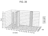
- FIG. 26 is a histogram summarizing the results of peptide binding experiments and depicting the mapped epitopes of HMGB 1 that are recognized by 2G7 HMGB 1 mAb (2G7), 6E6 HMGB1 mAb (6E6), 2G5 HMGB1 mAb (2G5), 6H9 HMGB1 mAb (6H9) and 2E11 HMGB1 mAb (2E11).
- FIG. 27A is a mass spectrum of intact, non-reduced 6E6 HMGB1 mAb.
- FIG. 27B is a mass spectrum of 6E6 HMGB1 mAb, which was reduced by treatment with DTT.
- FIG. 27C is a mass spectrum of the light chains of 6E6 HMGB1 mAb, which were reduced by treatment with DTT.
- FIG. 27D is a mass spectrum of the heavy chains of 6E6 HMGB1 mAb, which were reduced by treatment with DTT.
- FIG. 28 is a graph of the effect of administration of various doses (either 0.004 mg/kg, 0.04 mg/kg or 0.4 mg/kg) of 2G7 HMGB1 mAb or a control IgG antibody (0.4 mg/kg) on survival of mice over time (days) after cecal ligation and puncture (CLP).
- FIG. 29 is a table summarizing CLP survival percentages in mice administered various doses (either 4 mg/kg, 0.4 mg/kg, 0.04 mg/kg or 0.004 mg/kg) of 6E6 HMGB1 mAb (6E6), 2G7 HMGB1 mAb (2G7), or control IgG.
- FIG. 30 is the amino acid sequence of a human ( Homo sapiens ) HMGB2 polypeptide (SEQ ID NO:54; GenBank Accession No. M83665).
- FIG. 31A is the amino acid sequence of a human ( Homo sapiens ) HMGB1 polypeptide (SEQ ID NO:74).
- FIG. 31B is an A box of a human (Homo sapiens) HMGB1 polypeptide (SEQ ID NO:75).
- FIG. 31C is a B box of a human (Homo sapiens) HMGB1 polypeptide (SEQ ID NO:76).
- FIG. 32 is a histogram depicting inhibition of TNF release by various anti-HMGB1 monoclonal antibodies.
- Mouse TNF was induced by stimulating RAW 264.7 cells with 0.1 ⁇ g/ml of recombinant CBP-Rat HMGB1 peptide (SEQ ID NO:5). Where indicated, various HMGB1 monoclonal antibodies (cultured supernatants) were added to give a final concentration of 13%.
- the following antibodies were tested: 1A9 HMGB1 mAb (1A9); 2E11 HMGB1 mAb (2E11); 2G5 HMGB1 mAb (2G5); 2G7 HMGB1 mAb (2G7); 3G8 HMGB1 mAb (3G8); 4H11 HMGB1 mAb (4H11); 3-5A6 HMGB1 mAb (5A6); 6E6 HMGB1 mAb (6E6); 9G2 HMGB1 mAb (9G2); 4C9 HMGB1 mAb (4C9); and 6H9 HMGB1 mAb (6H9).
- the initial dark bar depicts TNF release in the absence of any antibodies.
- FIG. 33 is a histogram depicting inhibition of TNF release by various anti-HMGB1 monoclonal antibodies.
- Mouse TNF was induced by stimulating RAW 264.7 cells with 0.1 ⁇ g/ml of recombinant CBP-Rat HMGB1 peptide (SEQ ID NO:5). Where indicated, various HMGB 1 monoclonal antibodies (cultured supernatants) were added to give a final concentration of 13%.
- the following antibodies were tested: 7H3 HMGB1 mAb (7H3); 9H3 HMGB1 mAb (9H3); 10D4 HMGB1 mAb (10D4); 1C3 HMGB1 mAb (1C3); 3E10 HMGB1 mAb (3E10); 4A10 HMGB1 mAb (4A10); 5C12 HMGB1 mAb (5C12); and 7G8 HMGB1 mAb (7G8).
- the initial dark bar depicts TNF release in the absence of any antibodies.
- the present invention is drawn to antibodies or antigen-binding fragments thereof that bind to a vertebrate high mobility group box (HMGB) polypeptide, methods of detecting and/or identifying an agent that binds to an HMGB polypeptide, methods of treating a condition in a subject characterized by activation of an inflammatory cytokine cascade and methods of detecting an HMGB polypeptide in a sample.
- HMGB high mobility group box
- monoclonal antibody or “monoclonal antibody composition”, as used herein, refers to a population of antibody molecules that contain only one species of an antigen binding site capable of immunoreacting with a particular epitope of a polypeptide of the invention.
- a monoclonal antibody composition thus typically displays a single binding affinity for a particular polypeptide of the invention with which it immunoreacts.
- antibody as used herein also encompasses functional fragments of antibodies, including fragments of chimeric, humanized, primatized, veneered or single chain antibodies.
- Functional fragments include antigen-binding fragments of antibodies that bind to an HMGB polypeptide (e.g., a mammalian HMGB polypeptide (e.g. a mammalian HMGB1 polypeptide)).
- HMGB polypeptide e.g., a mammalian HMGB polypeptide (e.g. a mammalian HMGB1 polypeptide)
- antibody fragments capable of binding to an HMGB polypeptide or a portion thereof include, but are not limited to Fv, Fab, Fab′ and F(ab′) 2 fragments. Such fragments can be produced by enzymatic cleavage or by recombinant techniques.
- papain or pepsin cleavage can generate Fab or F(ab′) 2 fragments, respectively.
- Other proteases with the requisite substrate specificity can also be used to generate Fab or F(ab′) 2 fragments.
- Antibodies can also be produced in a variety of truncated forms using antibody genes in which one or more stop codons have been introduced upstream of the natural stop site.
- a chimeric gene encoding a F(ab′) 2 heavy chain portion can be designed to include DNA sequences encoding the CH 1 domain and hinge region of the heavy chain.
- Single chain antibodies and chimeric, humanized or primatized (CDR-grafted), or veneered antibodies, as well as chimeric, CDR-grafted or veneered single chain antibodies, comprising portions derived from different species, and the like are also encompassed by the present invention and the term “antibody”.
- the various portions of these antibodies can be joined together chemically by conventional techniques, or can be prepared as a contiguous protein using genetic engineering techniques. For example, nucleic acids encoding a chimeric or humanized chain can be expressed to produce a contiguous protein. See, e.g., Cabilly et al., U.S. Pat. No. 4,816,567; Cabilly et al., European Patent No.
- Humanized antibodies can be produced using synthetic or recombinant DNA technology using standard methods or other suitable techniques.
- Nucleic acid (e.g., cDNA) sequences coding for humanized variable regions can also be constructed using PCR mutagenesis methods to alter DNA sequences encoding a human or humanized chain, such as a DNA template from a previously humanized variable region (see e.g., Kamman, M., et al., Nucl. Acids Res., 17: 5404 (1989)); Sato, K., et al., Cancer Research, 53: 851-856 (1993); Daugherty, B. L. et al., Nucleic Acids Res., 19 (9): 2471-2476 (1991); and Lewis, A. P.
- variants can also be readily produced.
- cloned variable regions can be mutated, and sequences encoding variants with the desired specificity can be selected (e.g., from a phage library; see e.g., Krebber et al., U.S. Pat. No. 5,514,548; Hoogenboom et al., WO 93/06213).
- the antibody can be a humanized antibody comprising one or more immunoglobulin chains (e.g., an antibody comprising a CDR of nonhuman origin (e.g., one or more CDRs derived from an antibody of nonhuman origin) and a framework region derived from a light and/or heavy chain of human origin (e.g., CDR-grafted antibodies with or without framework changes)).
- the antibody or antigen-binding fragment thereof comprises the light chain CDRs (CDR1, CDR2 and CDR3) and heavy chain CDRs (CDR1, CDR2 and CDR3) of a particular immunoglobulin.
- the antibody or antigen-binding fragment further comprises a human framework region.
- the antibodies described herein can also be conjugated to an agent.
- the agent is a label, for example, a radioisotope, an epitope label (tag), an affinity label (e.g., biotin, avidin), a spin label, an enzyme, a fluorescent group or a chemiluminescent group.
- Labeled antibodies or antigen-binding fragments of the present invention can be used, e.g., in the diagnostic and/or prognostic methods described herein.
- the antibody is conjugated to a drug, toxin or anti-inflammatory agent.
- Drugs and toxins that can be conjugated to the antibodies of the present invention include, for example, chemotherapeutic agents (e.g., mitomycin C, paxlitaxol, methotrexate, 5-fluorouracil, cisplatin, cyclohexamide), toxins (e.g., ricin, gelonin) and other agents described herein (e.g., the agents described for combination therapy).
- chemotherapeutic agents e.g., mitomycin C, paxlitaxol, methotrexate, 5-fluorouracil, cisplatin, cyclohexamide
- toxins e.g., ricin, gelonin
- Anti-inflammatory agents that can be conjugated include, e.g., those described herein.
- Antibodies that are specific for an HMGB polypeptide can be raised against an appropriate immunogen, such as an isolated and/or recombinant HMGB polypeptide or a portion thereof (including synthetic molecules, such as synthetic peptides).
- Antibodies can also be raised by immunizing a suitable host (e.g., mouse) with cells that express an HMGB polypeptide, such as GH3 pituicytes, macrophage cells (e.g., RAW 246.7 cells, human macrophage cells), peripheral blood mononuclear cells (PBMCs (e.g., human PBMCs)), primary T cells (e.g., human primary T cells), adrenal cells (e.g., rat adrenal PC-12 cells, human adrenal cells), and kidney cells (e.g., rat primary kidney cells, human primary kidney cells).
- a suitable host e.g., mouse
- cells that express an HMGB polypeptide such as GH3 pituicytes, macrophage cells (e.g., RAW 246.7 cells, human macrophage cells), peripheral blood mononuclear cells (PBMCs (e.g., human PBMCs)), primary T cells (e.g., human primary T cells), adrenal cells (e.g., rat adrenal PC-12
- HMGB polypeptide e.g., a mammalian HMGB polypeptide
- transfected cells can be used as an immunogen or in a screen for an antibody that binds thereto (See e.g., Chuntharapai et al., J. Immunol., 152: 1783-1789 (1994); Chuntharapai et al., U.S. Pat. No. 5,440,021).
- Preparation of immunizing antigen, and polyclonal and monoclonal antibody production can be performed using any suitable technique.
- a variety of methods have been described (see e.g., Kohler et al., Nature, 256: 495-497 (1975) and Eur. J. Immunol. 6: 511-519 (1976); Milstein et al., Nature 266: 550-552 (1977); Koprowski et al., U.S. Pat. No. 4,172,124; Harlow, E. and D. Lane, 1988, Antibodies: A Laboratory Manual , (Cold Spring Harbor Laboratory: Cold Spring Harbor, N.Y.); Current Protocols In Molecular Biology , Vol. 2 (Supplement 27, Summer '94), Ausubel, F.
- a hybridoma is produced by fusing a suitable immortal cell line (e.g., a myeloma cell line such as SP2/0, P3X63Ag8.653 or a heteromyeloma) with antibody-producing cells.
- a suitable immortal cell line e.g., a myeloma cell line such as SP2/0, P3X63Ag8.653 or a heteromyeloma
- Antibody-producing cells can be obtained from the peripheral blood or, preferably the spleen or lymph nodes, of humans or other suitable animals immunized with the antigen of interest.
- the fused cells (hybridomas) can be isolated using selective culture conditions, and cloned by limiting dilution. Cells that produce antibodies with the desired specificity can be selected by a suitable assay (e.g., ELISA).
- Suitable methods of producing or isolating antibodies of the requisite specificity can be used, including, for example, methods that select recombinant antibody from a library (e.g., a phage display library).
- a library e.g., a phage display library.
- Transgenic animals capable of producing a repertoire of human antibodies e.g., XENOMOUSE® (transgenic mouse, Abgenix, Fremont, Calif.)
- suitable methods see e.g., Jakobovits et al., Proc. Natl. Acad. Sci. USA, 90: 2551-2555 (1993); Jakobovits et al., Nature, 362: 255-258 (1993)).
- the antibody or antigen-binding fragment thereof has specificity for an HMGB polypeptide (e.g., a mammalian HMGB polypeptide).
- the antibody or antigen-binding fragment thereof has specificity for an HMGB1 polypeptide (e.g., a human HMGB1 polypeptide such as depicted in SEQ ID NO:1 and/or SEQ ID NO:74).
- the antibody or antigen-binding fragment thereof is an IgG or an antigen-binding fragment of an IgG.
- the antibody or antigen-binding fragment thereof is an IgG1 or an antigen-binding fragment of an IgG1.
- the antibody or antigen-binding fragment thereof is an IgG2a, IgG2b, IgG3 antibody, or an antigen-binding fragment of any of the foregoing.
- the antibody or antigen-binding fragment can bind to an HMGB polypeptide and inhibit (reduce or prevent) one or more functions of the HMGB polypeptide.
- HMGB functions include, e.g., increasing inflammation (see, e.g., PCT Publication No. WO 02/092004), increasing release of a proinflammatory cytokine from a cell (see, e.g., PCT Publication No. WO 02/092004), binding to RAGE, binding to TLR2, chemoattraction (see, e.g., Degryse et al., J. Cell Biol.
- the antibody is a human antibody or an antigen-binding fragment thereof. In another embodiment, the antibody is a humanized antibody or an antigen-binding fragment thereof. In yet another embodiment, the antibody or antigen-binding fragment can inhibit binding of a polypeptide (e.g., RAGE, TLR2) to an HMGB polypeptide and/or inhibit one or more functions mediated by binding of the HMGB polypeptide and the other polypeptide.
- a polypeptide e.g., RAGE, TLR2
- the antibodies or antigen-binding fragments thereof specifically bind to HMGB epitopes or antigenic determinants (e.g., HMGB epitopes, HMGB A box epitopes, HMGB B box epitopes).
- an antibody or antigen-binding fragment thereof can be screened without undue experimentation for the ability to inhibit release of a proinflammatory cytokine using standard methods.
- Anti-HMGB A-box antibodies and anti-HMGB B box antibodies that can inhibit the production of a proinflammatory cytokine and/or the release of a proinflammatory cytokine from a cell, and/or inhibit a condition characterized by activation of an inflammatory cytokine cascade, are within the scope of the present invention.
- the antibody or antigen-binding fragment of the invention can inhibit the production of TNF, IL-1 ⁇ , and/or IL-6. In another embodiment, the antibody or antigen-binding fragment of the invention can inhibit the production of TNF (e.g., TNF- ⁇ ).
- TNF e.g., TNF- ⁇
- 6E6 HMGB1 mAb also referred to as 6E6-7-1-1 or 6E6, can be produced by murine hybridoma 6E6 HMGB1 mAb, which was deposited on Sep. 3, 2003, on behalf of Critical Therapeutics, Inc., 675 Massachusetts Avenue, 14 th Floor, Cambridge, Mass. 02139, U.S.A., at the American Type Culture Collection, 10801 University Boulevard, Manassas, Va. 20110, U.S.A., under Accession No. PTA-5433.
- the invention relates to murine hybridoma 6E6 HMGB 1 mAb, to the antibody it produces and to nucleic acids encoding the antibody.
- 2E11 HMGB1 mAb also referred to as 2E11-1-1-2 or 2E11
- 2E11-1-1-2 or 2E11 can be produced by murine hybridoma 2E11 HMGB1 mAb, which was deposited on Sep. 3, 2003, on behalf of Critical Therapeutics, Inc., 675 Massachusetts Avenue, 14 th Floor, Cambridge, Mass. 02139, U.S.A., at the American Type Culture Collection, 10801 University Boulevard, Manassas, Va. 20110, U.S.A., under Accession No. PTA-5431.
- the invention relates to murine hybridoma 2E11 HMGB 1 mAb, to the antibody it produces and to nucleic acids encoding the antibody.
- 6H9 HMGB1 mAb also referred to as 6H9-1-1-2 or 6H9
- 6H9-1-1-2 or 6H9 can be produced by murine hybridoma 6H9 HMGB1 mAb, which was deposited on Sep. 3, 2003, on behalf of Critical Therapeutics, Inc., 675 Massachusetts Avenue, 14 th Floor, Cambridge, Mass. 02139, U.S.A., at the American Type Culture Collection, 10801 University Boulevard, Manassas, Va. 20110, U.S.A., under Accession No. PTA-5434.
- the invention relates to murine hybridoma 6H9 HMGB1 mAb, to the antibody it produces and to nucleic acids encoding the antibody.
- 10D4 HMGB1 mAb also referred to as 10D4-1-1-1-2 or 10D4, can be produced by murine hybridoma 10D4 HMGB1 mAb, which was deposited on Sep. 3, 2003, on behalf of Critical Therapeutics, Inc., 675 Massachusetts Avenue, 14 th Floor, Cambridge, Mass. 02139, U.S.A., at the American Type Culture Collection, 10801 University Boulevard, Manassas, Va. 20110, U.S.A., under Accession No. PTA-5435.
- the invention relates to murine hybridoma 10D4 HMGB1 mAb, to the antibody it produces and to nucleic acids encoding the antibody.
- 2G7 HMGB1 mAb also referred to as 3-2G7-1-1-1 or 2G7
- 2G7 HMGB1 mAb can be produced by murine hybridoma 2G7 HMGB1 mAb, which was deposited on Sep. 3, 2003, on behalf of Critical Therapeutics, Inc., 675 Massachusetts Avenue, 14 th Floor, Cambridge, Mass. 02139, U.S.A., at the American Type Culture Collection, 10801 University Boulevard, Manassas, Va. 20110, U.S.A., under Accession No. PTA-5432.
- the invention relates to murine hybridoma 2G7 HMGB1 mAb, to the antibody it produces and to nucleic acids encoding the antibody.
- 9G2 HMGB1 mAb also referred to as 9G2-7-1-1-1 or 9G2
- the invention relates to murine hybridoma 9G2 HMGB1 mAb, to the antibody it produces, and to nucleic acids encoding the antibody.
- 1A9 HMGB1 mAb also referred to as 1A9-1-2-1-4 or 1A9, can be produced by murine hybridoma 1A9 HMGB1 mAb.
- the invention relates to murine hybridoma 1A9 HMGB1 mAb, to the antibody it produces, and to nucleic acids encoding the antibody.
- 3G8 HMGB1 mAb also referred to as 3G8-7-2-1-5 or 3G8, can be produced by murine hybridoma 3G8 HMGB1 mAb.
- the invention relates to murine hybridoma 3G8 HMGB 1 mAb, to the antibody it produces, and to nucleic acids encoding the antibody.
- 2G5 HMGB1 mAb also referred to as 3-2G5-4-1-2 or 2G5
- the invention relates to murine hybridoma 2G5 HMGB1 mAb, to the antibody it produces, and to nucleic acids encoding the antibody.
- 4H11 HMGB1 mAb also referred to as 4H11
- 4H11 can be produced by murine hybridoma 4H11 HMGB mAb.
- the invention relates to murine hybridoma 4H11 HMGB1 mAb, to the antibody it produces, and to nucleic acids encoding the antibody.
- 7H3 HMGB1 mAb also referred to as 7H3, can be produced by murine hybridoma 7H3 HMGB 1 mAb.
- the invention relates to murine hybridoma 7H3 HMGB1 mAb, to the antibody it produces, and to nucleic acids encoding the antibody.
- 3-5A6 HMGB1 mAb also referred to as 3-5A6 or 5A6, can be produced by murine hybridoma 3-5A6 HMGB1 mAb.
- the invention relates to murine hybridoma 3-5A6 HMGB1 mAb, to the antibody it produces, and to nucleic acids encoding the antibody.
- 9G1 HMGB1 mAb also referred to as 9G1
- the invention relates to murine hybridoma 9G1 HMGB1 mAb, to the antibody it produces, and to nucleic acids encoding the antibody.
- 4C9 HMGB 1 mAb can be produced by murine hybridoma 4C9 HMGB 1 mAb.
- the invention relates to murine hybridoma 4C9 HMGB1 mAb, to the antibody it produces, and to nucleic acids encoding the antibody.
- 9H3 HMGB1 mAb also referred to as 9H3
- 9H3 HMGB1 mAb can be produced by murine hybridoma 9H3 HMGB1 mAb.
- the invention relates to murine hybridoma 9H3 HMGB1 mAb, to the antibody it produces, and to nucleic acids encoding the antibody.
- 1C3 HMGB1 mAb also referred to as 1C3-1-1-1 or 1C3, can be produced by murine hybridoma 1C3 HMGB1 mAb.
- the invention relates to murine hybridoma 1C3 HMGB1 mAb, to the antibody it produces, and to nucleic acids encoding the antibody.
- 5C12 HMGB1 mAb also referred to as 5C12-1-1-1 or 5C12
- the invention relates to murine hybridoma 5C12 HMGB1 mAb, to the antibody it produces, and to nucleic acids encoding the antibody.
- 3E10 HMGB1 mAb also referred to as 3E10-5-4-1-1 or 3E10
- the invention relates to murine hybridoma 3E10 HMGB1 mAb, to the antibody it produces, and to nucleic acids encoding the antibody.
- 7G8 HMGB1 mAb also referred to as 7G8, can be produced by murine hybridoma 7G8 HMGB 1 mAb.
- the invention relates to murine hybridoma 7G8 HMGB1 mAb, to the antibody it produces, and to nucleic acids encoding the antibody.
- 4A10 HMGB1 mAb also referred to as 4A10-1-3-1-1 or 4A10, can be produced by murine hybridoma 4A10 HMGB1 mAb.
- the invention relates to murine hybridoma 4A10 HMGB1 mAb, to the antibody it produces, and to nucleic acids encoding the antibody.
- the antibody or antigen-binding fragment thereof is selected from the group consisting of 6E6 HMGB 1 mAb, 6H9 HMGB 1 mAb, 2G7 HMGB 1 mAb, 10D4 HMGB 1 mAb, 2E11 HMGB 1 mAb and an antigen-binding fragment of any of the foregoing.
- the antibody or antigen-binding fragment has the same or similar epitopic specificity of an antibody or antigen-binding fragment selected from the group consisting of 6E6 HMGB 1 mAb, 6H9 HMGB 1 mAb, 2G7 HMGB 1 mAb, 10D4 HMGB 1 mAb, 2E11 HMGB 1 mAb and/or an antigen-binding fragment of any of the foregoing.
- Antibodies or antigen-binding fragments with an epitopic specificity that is the same as, or similar to, that of 6E6 HMGB1 mAb, 6H9 HMGB1 mAb, 2G7 HMGB1 mAb, 10D4 HMGB1 mAb and/or 2E11 HMGB1 mAb can be identified by a variety of suitable methods.
- an antibody with the same or similar epitopic specificity as, e.g., 6E6 HMGB1 mAb can be identified based upon the ability to compete with 6E6 HMGB1 mAb for binding to a HMGB polypeptide (e.g., a mammalian HMGB polypeptide (e.g., a mammalian HMGB1 polypeptide)).
- a HMGB polypeptide e.g., a mammalian HMGB polypeptide (e.g., a mammalian HMGB1 polypeptide)
- the binding of, e.g., 6E6 HMGB1 mAb, and the binding of an antibody with the same or similar epitopic specificity for a HMGB polypeptide can be inhibited by a single peptide (e.g., a natural peptide, a synthetic peptide).
- the peptide can comprise, e.g., 9 to about 50 amino acids, 9 to about 40 amino
- 2E11 HMGB1 mAb was shown to bind a peptide corresponding to amino acids 151-168 of human HMGB1 (amino acid residues 151-168 of SEQ ID NO:1; i.e., LKEKYEKDIAAYRAKGKP (SEQ ID NO:30)). Additional studies suggest that 2E11 HMGB1 mAb recognizes an epitope that is present within amino acid residues 156-161, 155-161, 155-162, 156-162 and/or 156-163, of HMGB1 (see Example 14).
- 6E6 HMGB1 mAb and 6H9 HMGB1 mAb were shown to bind to a peptide corresponding to amino acids 61-78 of human HMGB 1 (amino acid residues 61-78 of SEQ ID NO:1; i.e., EDMAKADKARYEREMKTY (SEQ ID NO:24)). Additional studies demonstrated that 6E6 HMGB 1 mAb recognizes an epitope that is present within amino acid residues 67-78 of HMGB1 (see Example 13).
- 2G7 HMGB 1 mAb was shown to bind a peptide corresponding to amino acids 46-63 of human HMGB1 (amino acid residues 46-63 of SEQ ID NO:1; i.e., SERWKTMSAKEKGKFEDM (SEQ ID NO:23)) (see Example 10). Further studies demonstrated that 2G7 HMGB 1 mAb recognizes an epitope that is present within amino acid residues 53-63 of HMGB1 (see Example 12).
- HMGB1 mAb does not bind to amino acid residues 46-63 of HMGB2 (SEQ ID NO:48), notwithstanding only a single amino acid difference between the HMGB1 46-63 peptide and the HMGB2 46-63 peptide (see Example 12).
- the antibodies or antigen-binding fragments of the invention bind to HMGB1 but not to HMGB2. In other embodiments, the antibodies or antigen-binding fragments of the invention bind to both HMGB1 and HMGB2.
- HMGB1 18 amino acid peptides, or other peptides corresponding to particular regions of HMGB1, could be used in epitopic studies to determine if an antibody or antigen-binding fragment inhibited binding of the peptide to an antibody known to bind that peptide (e.g., 6E6 HMGB1 mAb, 6H9 HMGB1 mAb, 2G7 HMGB1 mAb, 10D4 HMGB1 mAb, 2E11 HMGB1 mAb, others antibodies described herein).
- an antibody or antigen-binding fragment inhibited binding of the peptide to an antibody known to bind that peptide (e.g., 6E6 HMGB1 mAb, 6H9 HMGB1 mAb, 2G7 HMGB1 mAb, 10D4 HMGB1 mAb, 2E11 HMGB1 mAb, others antibodies described herein).
- an antibody or antigen-binding fragment to be tested for its epitopic specificity could be assayed with, e.g., 2E11 HMGB1 mAb and a peptide corresponding to amino acids 151-168 of human HMGB1 (which 2E11 HMGB1 mAb is known to bind).
- an antibody with the same or similar epitopic specificity as an antibody of the invention can be identified using a chimeric HMGB polypeptide (see e.g., Banks, G. C., et al., J. Biol. Chem. 274 (23):16536-16544 (1999)).
- the antibody or antigen-binding fragment can compete with 6E6 HMGB1 mAb, 6H9 HMGB1 mAb, 2G7 HMGB1 mAb, 10D4 HMGB1 mAb, 2E11 HMGB1 mAb and/or an antigen-binding fragment of any of the foregoing, for binding to an HMGB polypeptide (e.g., a mammalian HMGB polypeptide (e.g., a mammalian HMGB 1 polypeptide)).
- HMGB polypeptide e.g., a mammalian HMGB polypeptide (e.g., a mammalian HMGB 1 polypeptide)
- Such inhibition of binding can be the result of competition for the same or similar epitope or steric interference (e.g., where antibodies bind overlapping epitopes or adjacent epitopes).
- Inhibition by 6E6 HMGB1 mAb, 6H9 HMGB1 mAb, 2G7 HMGB1 mAb, 10D4 HMGB1 mAb, 2E11 HMGB1 mAb, and/or an antigen-binding fragment of any of the foregoing, can also be due to a change in the conformation of the HMGB polypeptide that is induced upon antibody binding to the HMGB polypeptide.
- the antibody or antigen-binding fragment thereof is selected from the group consisting of 3G8 HMGB1 mAb, 2G5 HMGB1 mAb, 4H11 HMGB1 mAb, 7H3 HMGB1 mAb, 3-5A6 HMGB1 mAb, 9G1 HMGB1 mAb, 4C9 HMGB1 mAb, 9H3 HMGB1 mAb, 1C3 HMGB1 mAb, 5C12 HMGB1 mAb, 3E10 HMGB1 mAb, 7G8 HMGB1 mAb, 4A10 HMGB1 mAb, and an antigen-binding fragment of any of the foregoing.
- the antibody or antigen-binding fragment has the epitopic specificity of an antibody or antigen-binding fragment selected from the group consisting of 3G8 HMGB1 mAb, 2G5 HMGB1 mAb, 4H11 HMGB1 mAb, 7H3 HMGB1 mAb, 3-5A6 HMGB1 mAb, 9G1 HMGB1 mAb, 4C9 HMGB1 mAb, 9H3 HMGB1 mAb, 1C3 HMGB1 mAb, 5C12 HMGB1 mAb, 3E10 HMGB1 mAb, 7G8 HMGB1 mAb, 4A10 HMGB1 mAb, and an antigen-binding fragment of any of the foregoing.
- an antibody or antigen-binding fragment selected from the group consisting of 3G8 HMGB1 mAb, 2G5 HMGB1 mAb, 4H11 HMGB1 mAb, 7H3 HMGB1 mAb,
- antibodies or antigen-binding fragments with an epitopic specificity that is the same as, or similar to, one or more of these antibodies or antigen-binding fragments can be identified by a variety of suitable methods (e.g., using methods described herein and/or known in the art).
- the antibody or antigen-binding fragment can compete with 3G8 HMGB1 mAb, 2G5 HMGB1 mAb, 4H11 HMGB1 mAb, 7H3 HMGB1 mAb, 3-5A6 HMGB1 mAb, 9G1 HMGB1 mAb, 4C9 HMGB1 mAb, 9H3 HMGB1 mAb, 4A10 HMGB1 mAb, 5C12 HMGB1 mAb, 3E10 HMGB1 mAb, 7G8 HMGB1 mAb, 4A10 HMGB1 mAb, and/or an antigen-binding fragment of any of the foregoing, for binding to an HMGB polypeptide (e.g., a mammalian HMGB polypeptide).
- an HMGB polypeptide e.g., a mammalian HMGB polypeptide
- inhibition of binding can be the result of competition for the same or similar epitope or steric interference (e.g., where antibodies bind overlapping epitopes or adjacent epitopes). Inhibition can also be due to a change in the conformation of the HMGB polypeptide that is induced upon binding of the antibody or antigen-binding fragment to the HMGB polypeptide.
- the antibody or antigen-binding fragment thereof comprises the six CDRs (light chain CDRs (CDR1, CDR2 and CDR3) and heavy chain CDRs (CDR1, CDR2 and CDR3)) of an antibody selected from the group consisting of 6E6 HMGB1 mAb, 6H9 HMGB1 mAb, 2G7 HMGB1 mAb, 10D4 HMGB1 mAb and 2E11 HMGB1 mAb.
- CDRs light chain CDRs
- CDR1, CDR2 and CDR3 heavy chain CDRs
- the antibody is a humanized antibody that comprises the light chain CDRs (CDR1, CDR2 and CDR3) and heavy chain CDRs (CDR1, CDR2 and CDR3) of an antibody selected from the group consisting of 6E6 HMGB1 mAb, 6H9 HMGB1 mAb, 2G7 HMGB1 mAb, 10D4 HMGB1 mAb and 2E11 HMGB1 mAb.
- the antibody or antigen-binding fragment thereof comprises the six CDRs (light chain CDRs (CDR1, CDR2 and CDR3) and heavy chain CDRs (CDR1, CDR2 and CDR3)) of any other antibody described herein.
- the antibody or antigen-binding fragment thereof comprises from one to six of the light chain and heavy chain CDRs of an antibody of the invention (e.g., 6E6 HMGB1 mAb, 6H9 HMGB1 mAb, 2G7 HMGB1 mAb, 10D4 HMGB1 mAb, 2E11 HMGB1 mAb).
- the antibody or antigen-binding fragment can comprise one, two, three, four, five or six, of the light chain and heavy chain CDRs.
- the antibody or antigen-binding fragment thereof comprises at least one light chain CDR or heavy chain CDR from one antibody of the invention and at least one light chain CDR or heavy chain CDR from a different antibody of the invention (e.g., 6E6 HMGB1 mAb, 6H9 HMGB1 mAb, 2G7 HMGB1 mAb, 10D4 HMGB1 mAb, 2E11 HMGB1 mAb).
- an antibody or antigen-binding fragment could comprise one or more CDRs from 6E6 HMGB 1 mAb and one or more CDRs from 6H9 HMGB 1 mAb.
- Antibodies and antigen-binding fragments combining other combinations of CDRs from different antibodies of the invention are also encompassed.
- the antibody or antigen-binding fragment thereof comprises the six CDRs (light chain CDRs (CDR1, CDR2 and CDR3) and heavy chain CDRs (CDR1, CDR2 and CDR3)) of an antibody selected from the group consisting of 3G8 HMGB1 mAb, 2G5 HMGB1 mAb, 4H11 HMGB1 mAb, 7H3 HMGB1 mAb, 3-5A6 HMGB1 mAb, 9G1 HMGB1 mAb, 4C9 HMGB1 mAb, 9H3 HMGB1 mAb, 1C3 HMGB1 mAb, 5C12 HMGB1 mAb, 3E10 HMGB1 mAb, 7G8 HMGB1 mAb and 4A10 HMGB1 mAb.
- the antibody or antigen-binding fragment thereof comprises from one to six of the light chain and heavy chain CDRs of one of these antibodies.
- the antibody or antigen-binding fragment comprises one or more CDRs that are at least 80% identical, at least 90% identical, or at least 95% identical, to a CDR of an antibody of the invention (e.g., 6E6 HMGB1 mAb, 6H9 HMGB1 mAb, 2G7 HMGB1 mAb, 10D4 HMGB1 mAb, 2E11 HMGB1 mAb).
- the antibody or antigen-binding fragment comprises one or more CDRs that are at least 80% similar, at least 90% similar, or at least 95% similar, to a CDR of an antibody of the invention.
- the invention also relates to a bispecific antibody, or functional fragment thereof (e.g., F(ab) 2 ), which binds to an HMGB polypeptide and at least one other antigen (e.g., tumor antigen, viral antigen).
- the bispecific antibody, or functional fragment thereof has the same or similar epitopic specificity as 6E6 HMGB1 mAb, 6H9 HMGB1 mAb, 2G7 HMGB1 mAb, 10D4 HMGB1 mAb and/or 2E11 HMGB1 mAb, and at least one other antibody.
- Bispecific antibodies can be secreted by triomas and hybrid hybridomas.
- triomas are formed by fusion of a hybridoma and a lymphocyte (e.g., antibody-secreting B cell) and hybrid hybridomas are formed by fusion of two hybridomas.
- a lymphocyte e.g., antibody-secreting B cell
- hybrid hybridomas are formed by fusion of two hybridomas.
- Each of the fused cells i.e., hybridomas, lymphocytes
- triomas and hybrid hybridomas can produce an antibody containing antigen-binding sites that recognize different antigens.
- the supernatants of triomas and hybrid hybridomas can be assayed for bispecific antibody using a suitable assay (e.g., ELISA), and bispecific antibodies can be purified using conventional methods. (see, e.g., U.S. Pat. No.
- the invention relates to an isolated cell that produces an antibody or an antigen-binding fragment of the invention.
- the isolated antibody-producing cell of the invention is an immortalized cell, such as a hybridoma, heterohybridoma, lymphoblastoid cell or a recombinant cell.
- the antibody-producing cells of the present invention have uses other than for the production of antibodies.
- the cell of the present invention can be fused with other cells (such as suitably drug-marked human myeloma, mouse myeloma, human-mouse heteromyeloma or human lymphoblastoid cells) to produce, for example, additional hybridomas, and thus provide for the transfer of the genes encoding the antibody.
- the cell can be used as a source of nucleic acids encoding the anti-HMGB immunoglobulin chains, which can be isolated and expressed (e.g., upon transfer to other cells using any suitable technique (see e.g., Cabilly et al., U.S. Pat. No. 4,816,567, Winter, U.S. Pat. No. 5,225,539)).
- clones comprising a sequence encoding a rearranged anti-HMGB light and/or heavy chain can be isolated (e.g., by PCR).
- cDNA libraries can be prepared from mRNA isolated from an appropriate cell line, and cDNA clones encoding an anti-HMGB immunoglobulin chain(s) can be isolated.
- nucleic acids encoding the heavy and/or light chains of the antibodies, or portions thereof can be obtained and used for the production of the specific immunoglobulin, immunoglobulin chain, or variants thereof (e.g., humanized immunoglobulins) in a variety of host cells or in an in vitro translation system.
- the nucleic acids including cDNAs, or derivatives thereof encoding variants such as a humanized immunoglobulin or immunoglobulin chain
- suitable prokaryotic or eukaryotic vectors e.g., expression vectors
- suitable host cell by an appropriate method (e.g., transformation, transfection, electroporation, infection), such that the nucleic acid is operably linked to one or more expression control elements (e.g., in the vector or integrated into the host cell genome), to produce a recombinant antibody-producing cell.
- the invention is a nucleic acid that encodes an antibody or antigen-binding fragment of the invention.
- the invention is a vector that comprises a nucleic acid encoding an antibody or antigen-binding fragment of the invention.
- HMGB Polypeptides HMGB A Boxes and HMGB B Boxes
- the invention is an antibody or antigen-binding fragment thereof that binds to an HMGB polypeptide.
- an “HMGB polypeptide” is a polypeptide that has at least 60%, more preferably, at least 70%, 75%, 80%, 85%, or 90%, and most preferably at least 95% sequence identity, to a sequence selected from the group consisting of SEQ ID NO:1, SEQ ID NO:18 and SEQ ID NO:74 (as determined, for example, using the BLAST program and parameters described herein).
- the HMGB polypeptide increases inflammation and/or increases release of a proinflammatory cytokine from a cell.
- the HMGB polypeptide has one of the above biological activities.
- the HMGB polypeptide has both of the above biological activities.
- polypeptide refers to a polymer of amino acids, and not to a specific length; thus, peptides, oligopeptides and proteins are included within the definition of a polypeptide.
- the HMGB polypeptide is a mammalian HMGB polypeptide, for example, a mammalian HMGB polypeptide (e.g., a human HMGB1 polypeptide).
- the HMGB polypeptide contains a B box DNA binding domain and/or an A box DNA binding domain and/or an acidic carboxyl terminus as described herein.
- HMGB polypeptides are described in GenBank Accession Numbers AAA64970, AAB08987, P07155, AAA20508, 529857, P09429, NP — 002119, CAA31110, S02826, U00431, X67668, NP — 005333, NM — 016957, and J04179, the entire teachings of which are incorporated herein by reference.
- HMGB polypeptides include, but are not limited to mammalian HMG1 ((HMGB1) as described, for example, in GenBank Accession Number U51677), HMG2 ((HMGB2) as described, for example, in GenBank Accession Number M83665), HMG-2A ((HMGB3, HMG-4) as described, for example, in GenBank Accession Numbers NM — 005342 and NP — 005333), HMG14 (as described, for example, in GenBank Accession Number P05114), HMG17 (as described, for example, in GenBank Accession Number X13546), HMGI (as described, for example, in GenBank Accession Number L17131), and HMGY (as described, for example, in GenBank Accession Number M23618); nonmammalian HMG T1 (as described, for example, in GenBank Accession Number X02666) and HMG T2 (as described, for example, in GenBank Accession Number L32859)
- HMGB proteins are polypeptides encoded by HMGB nucleic acid sequences having GenBank Accession Numbers NG — 000897 (HMG1L10) (and in particular by nucleotides 658-1305 of NG — 000897); AF076674 (HMG1L1) (and in particular by nucleotides 1-633 of AF076674; AF076676 (HMG1L4) (and in particular by nucleotides 1-564 of AF076676); AC010149 (HMG sequence from BAC clone RP11-395A23) (and in particular by nucleotides 75503-76117 of AC010149); AF165168 (HMG1L9) (and in particular by nucleotides 729-968 of AF165168); XM — 063129 (LOC122441) (and in particular by nucleotides 319-558 of XM — 063129); XM — 066789 (LOC13960
- the antibodies and antigen-binding fragments of the invention bind to an HMGB polypeptide (e.g., one or more of the HMGB polypeptides listed above).
- the antibody or antigen-binding fragment thereof binds to a vertebrate HMGB polypeptide.
- the antibody or antigen-binding fragment thereof binds to a mammalian HMGB polypeptide (e.g., rat HMGB, mouse HMGB, human HMGB).
- the antibody or antigen-binding fragment thereof binds to a mammalian HMGB1 polypeptide (e.g., rat HMGB1, mouse HMGB1, human HMGB1).
- the antibody or antigen-binding fragment thereof binds to a human HMGB1 polypeptide (e.g., the human HMGB1 polypeptide depicted as SEQ ID NO:1 or SEQ ID NO:74).
- compositions and methods of the present invention also feature antibodies to the high mobility group B (HMGB) A box.
- HMGB high mobility group B
- the antibody or antigen-binding fragment thereof binds to an HMGB A box but does not specifically bind to non-A box epitopes of HMGB.
- the antibody or antigen-binding fragment thereof binds to a vertebrate HMGB A box but does not specifically bind to non-A box epitopes of HMGB.
- the antibody or antigen-binding fragment thereof binds to a mammalian (e.g., human, rat, mouse) HMGB A box but does not specifically bind to non-A box epitopes of HMGB.
- the antibody or antigen-binding fragment thereof binds to the A box of a HMGB1 polypeptide (e.g., a mammalian HMGB1 polypeptide (e.g., human HMGB1, rat HMGB1, mouse HMGB1)) but does not specifically bind to non-A box epitopes of HMGB1.
- a HMGB1 polypeptide e.g., a mammalian HMGB1 polypeptide (e.g., human HMGB1, rat HMGB1, mouse HMGB1)
- HMGB1 polypeptide e.g., a mammalian HMGB1 polypeptide (e.g., human HMGB1, rat HMGB1, mouse HMGB1)
- an “HMGB A box”, also referred to herein as an “A box” or “HMG A box”, is a protein or polypeptide that has at least 50%, 60%, 70%, 75%, 80%, 85%, 90% or 95%, sequence identity to an HMGB A box (e.g., an HMGB A box described herein).
- the HMGB A box has one or more of the following biological activities: inhibiting inflammation mediated by HMGB and/or inhibiting release of a proinflammatory cytokine from a cell (see, e.g., PCT Publication No. WO 02/092004; the entire teachings of which are incorporated herein by reference).
- the HMGB A box polypeptide has one of the above biological activities. Typically, the HMGB A box polypeptide has both of the above biological activities. In one embodiment, the A box has at least 50%, 60%, 70%, 75%, 80%, 85%, 90%, or 95%, sequence identity to the A box depicted in FIG. 2A (residues 9-85 of SEQ ID NO:2) or FIG. 31B (SEQ ID NO:75). In another embodiment, the A box comprises or consists of the amino acid sequence in the corresponding region of an HMGB protein in a mammal. An HMGB A box is also a recombinantly-produced polypeptide having the same amino acid sequence as the A box sequences described herein.
- the HMGB A box is preferably a vertebrate HMGB A box, for example, a mammalian HMGB A box, more preferably, a mammalian HMGB 1 A box, for example, a human HMGB 1 A box, and most preferably, the HMGB1 A box comprising or consisting of the sequence of the A box depicted in FIG. 2A (residues 9-85 of SEQ ID NO:2) or FIG. 31B (SEQ ID NO:75).
- An HMGB A box often has no more than about 85 amino acids and no fewer than about 4 amino acids.
- an HMGB A box can comprise from 10-85 amino acids, 20-85 amino acids, 30-85 amino acids or 40-85 amino acids.
- polypeptides having A box sequences within them include, but are not limited to the HMGB polypeptides described above.
- the A box sequences in such HMGB polypeptides can be determined and isolated using methods described herein, for example, by sequence comparisons to A boxes described herein and testing for biological activity using methods described herein and/or other methods known in the art.
- HMGB A box polypeptide sequences include the following sequences: PDASVNFSEF SKKCSERWKT MSAKEKGKFE DMAKADKARY EREMKTYIPP KGET (human HMGB1; SEQ ID NO:55); DSSVNFAEF SKKCSERWKT MSAKEKSKFE DMAKSDKARY DREMKNYVPP KGDK (human HMGB2; SEQ ID NO:56); PEVPVNFAEF SKKCSERWKT VSGKEKSKFD EMAKADKVRY DREMKDYGPA KGGK (human HMGB3; SEQ ID NO:57); PDASVNFSEF SKKCSERWKT MSAKEKGKFE DMAKADKARY EREMKTYIPP KGET (HMG1L10; SEQ ID NO:58); SDASVNFSEF SNKCSERWK MSAKEKGKFE DMAKADKTHY ERQMKTYIPP KGET (HMG1L1; SEQ ID NO:59); PDASVNFSEF
- compositions and methods of the present invention also feature antibodies to the high mobility group B (HMGB) B box.
- HMGB high mobility group B
- the antibody or antigen-binding fragment thereof binds to an HMGB B box but does not specifically bind to non-B box epitopes of HMGB.
- the antibody or antigen-binding fragment thereof binds to a vertebrate HMGB B box but does not specifically bind to non-B box epitopes of HMGB.
- the antibody or antigen-binding fragment thereof binds to a mammalian (e.g., human, rat, mouse) HMGB B box but does not specifically bind to non-B box epitopes of HMGB.
- the antibody or antigen-binding fragment thereof binds to the B box of a HMGB1 polypeptide (e.g., a mammalian HMGB1 polypeptide (e.g., human HMGB1, rat HMGB1, mouse HMGB1)) but does not specifically bind to non-B box epitopes of HMGB 1.
- a HMGB1 polypeptide e.g., a mammalian HMGB1 polypeptide (e.g., human HMGB1, rat HMGB1, mouse HMGB1)
- HMGB1 polypeptide e.g., a mammalian HMGB1 polypeptide (e.g., human HMGB1, rat HMGB1, mouse HMGB1)
- an “HMGB B box”, also referred to herein as a “B box” or “an HMG B box”, is a polypeptide that has at least 60%, 65%, 70%, 75%, 80%, 85%, 90%, or 95%, sequence identity to an HMGB1 polypeptide (e.g., an HMGB B box described herein).
- the HMGB1 box has one or more of the following biological activities: increasing inflammation and/or increasing release of a proinflammatory cytokine from a cell (see, e.g., PCT Publication No. WO 02/092004).
- the HMGB B box polypeptide has one of the above biological activities.
- the HMGB B box polypeptide has both of the above biological activities.
- the HMGB B box has at least 50%, 60%, 70%, 75%, 80%, 85%, 90% or 95%, sequence identity to the B box depicted in FIG. 2B (SEQ ID NO:3) or FIG. 31C (SEQ ID NO:76).
- the B box comprises or consists of the amino acid sequence in the corresponding region of an HMGB protein in a mammal.
- An HMGB B box is also a recombinantly-produced polypeptide having the same amino acid sequence as the B box sequences described herein.
- the HMGB B box is preferably a vertebrate HMGB B box, for example, a mammalian HMGB B box, more preferably, a mammalian HMGB 1 B box, for example, a human HMGB 1 B box, and most preferably, the HMGB1 B box comprising or consisting of the sequence of the B box depicted in FIG. 2B (SEQ ID NO:3) or FIG. 31C (SEQ ID NO:76).
- An HMGB B box often has no more than about 85 amino acids and no fewer than about 4 amino acids.
- polypeptides having B box sequences within them include, but are not limited to, the HMGB polypeptides described above.
- the B box sequences in such polypeptides can be determined and isolated using methods described herein, for example, by sequence comparisons to B boxes described herein and testing for biological activity.
- HMGB B box polypeptide sequences include the following sequences: FKDPNAPKRP PSAFFLFCSE YRPKIKGEHP GLSIGDVAKK LGEMWNNTAA DDKQPYEKKA AKLKEKYEKD IAAY (human HMGB1; SEQ ID NO:67); KKDPNAPKRP PSAFFLFCSE HRPKIKSEHP GLSIGDTAKK LGEMWSEQSA KDKQPYEQKA AKLKEKYEKD IAAY (human HMGB2; SEQ ID NO:68) ; FKDPNAPKRL PSAFFLFCSE YRPKIKGEHP GLSIGDVAKK LGEMWNNTAA DDKQPYEKKA AKLKEKYEKD IAAY (HMG1L10; SEQ ID NO:69); FKDPNAPKRP PSAFFLFCSE YHPKIKGEHP GLSIGDVAKK LGEMWNNTAA DDKQPGEKKA AKLKEKYEKD
- an HMGB polypeptide, an HMGB A box, and an HMGB B box either naturally occurring or non-naturally occurring, encompass polypeptides that have sequence identity to the HMGB polypeptides, HMGB A boxes, and/or HMGB B boxes, described herein.
- two polypeptides (or a region of the polypeptides) are substantially homologous or identical when the amino acid sequences are at least about 60%, 70%, 75%, 80%, 85%, 90%, or 95% or more, homologous or identical.
- the length of the HMGB polypeptide, HMGB A box polypeptide, or HMGB B box polypeptide, aligned for comparison purposes is at least 30%, preferably at least 40%, more preferably at least 60%, and even more preferably at least 70%, 80%, 90%, or 100%, of the length of the reference sequence, for example, the sequences described herein corresponding to an HMGB polypeptide (e.g., SEQ ID NO:1; SEQ ID NO:18, SEQ ID NO:74), an HMGB A box polypeptide (e.g., residues 9-85 of SEQ ID NO:2, SEQ ID NO:75) or an HMGB B box polypeptide (e.g., SEQ ID NO:3, SEQ ID NO:76).
- an HMGB polypeptide e.g., SEQ ID NO:1; SEQ ID NO:18, SEQ ID NO:74
- an HMGB A box polypeptide e.g., residues 9-85 of SEQ ID NO:2, SEQ
- the database searched is a non-redundant (NR) database
- parameters for sequence comparison can be set at: no filters; Expect value of 10; Word Size of 3; the Matrix is BLOSUM62; and Gap Costs have an Existence of 11 and an Extension of 1.
- the percent identity between two amino acid sequences can be accomplished using the GAP program in the GCG software package (Accelrys, San Diego, Calif.) using either a Blossom 63 matrix or a PAM250 matrix, and a gap weight of 12, 10, 8, 6, or 4, and a length weight of 2, 3, or 4.
- the percent identity between two nucleic acid sequences can be accomplished using the GAP program in the GCG software package (Accelrys, San Diego, Calif.), using a gap weight of 50 and a length weight of 3.
- the present invention is a method of inhibiting release of a proinflammatory cytokine from a mammalian cell.
- the method comprises treating the cell with an antibody or antigen-binding fragment of the present invention.
- Suitable antibodies or antigen-binding fragments are those described herein and include, e.g., 6E6 HMGB1 mAb, 6H9 HMGB1 mAb, 2G7 HMGB1 mAb, 10D4 HMGB1 mAb, 2E11 HMGB1 mAb, an antibody having the epitopic specificity of 6E6 HMGB1 mAb, 6H9 HMGB1 mAb, 2G7 HMGB1 mAb, 10D4 HMGB1 mAb and/or 2E11 HMGB1 mAb, an antibody that can compete with 6E6 HMGB1 mAb, 6H9 HMGB1 mAb, 2G7 HMGB1 mAb, 10D4 HMGB1 mAb and/
- cytokine is a soluble protein or peptide that is naturally produced by mammalian cells and that regulates immune responses and mediates cell-cell interactions. Cytokines can, either under normal or pathological conditions, modulate the functional activities of individual cells and tissues.
- a proinflammatory cytokine is a cytokine that is capable of causing one or more of the following physiological reactions associated with inflammation or inflammatory conditions: vasodilation, hyperemia, increased permeability of vessels with associated edema, accumulation of granulocytes and mononuclear phagocytes, and deposition of fibrin. In some cases, the proinflammatory cytokine can also cause apoptosis.
- TNF tumor necrosis factor
- IL interleukin
- IL-6 interleukin-1 ⁇
- IL-8 interleukin-8
- MIF macrophage migration inhibitory factor
- the invention is a method of treating a condition in a subject, wherein the condition is characterized by activation of an inflammatory cytokine cascade comprising administering to the subject an antibody or antigen-binding fragment of the present invention.
- Suitable antibodies or antigen-binding fragments are those described herein and include, e.g., 6E6 HMGB1 mAb, 6H9 HMGB1 mAb, 2G7 HMGB1 mAb, 10D4 HMGB1 mAb, 2E11 HMGB1 mAb, an antibody having the epitopic specificity of 6E6 HMGB1 mAb, 6H9 HMGB1 mAb, 2G7 HMGB1 mAb, 10D4 HMGB1 mAb and/or 2E11 HMGB1 mAb, an antibody that can compete with 6E6 HMGB1 mAb, 6H9 HMGB1 mAb, 2G7 HMGB1 mAb, 10D4 HMGB 1 m
- the method of treatment comprises administering to a subject an effective amount of an antibody or antigen-binding fragment of the invention.
- an “effective amount” or “therapeutically effective amount” is an amount sufficient to prevent or decrease an inflammatory response, and/or to ameliorate and/or decrease the longevity of symptoms associated with an inflammatory response.
- the amount of the composition of the invention that will be effective in the treatment, prevention or management of a particular condition can be determined, for example, by administering the composition to an animal model such as, e.g., the animal models disclosed herein and/or known to those skilled in the art.
- in vitro assays may optionally be employed to help identify optimal dosage ranges.
- Selection of the preferred effective dose can be determined (e.g., via clinical trials) by a skilled artisan based upon the consideration of several factors that are known to one of ordinary skill in the art. Such factors include, e.g., the condition or conditions to be treated, the severity of the subject's symptoms, the choice of antibody or antigen-binding fragment to be administered, the subject's age, the subject's body mass, the subject's immune status, the response of the individual subject, and other factors known by the skilled artisan to reflect the accuracy of administered pharmaceutical compositions.
- Effective doses may be extrapolated from dose-response curves derived from in vitro or animal model test systems.
- CLP cecal ligation and puncture
- the dosage administered to a subject is typically 0.1 mg/kg to 100 mg/kg of the subject's body weight.
- the dosage administered to a subject is between 0.1 mg/kg and 20 mg/kg of the subject's body weight, more preferably 1 mg/kg to 10 mg/kg of the subject's body weight.
- the dosage is at least 1 mg/kg, or at least 5 mg/kg, or at least 10 mg/kg, or at least 50 mg/kg, or at least 100 mg/kg, or at least 150 mg/kg, of the subject's body weight.
- human and humanized antibodies have a longer half-life within the human body than antibodies from other species due to the immune response to foreign polypeptides. Thus, lower dosages of human antibodies and less frequent administration is often possible.
- an effective amount of an antibody can range from about 0.01 mg/kg to about 5 or 10 mg/kg administered daily, weekly, biweekly or monthly.
- TNF release from a cell can be measured using a standard murine fibroblast L929 (ATCC, American Type Culture Collection, Rockville, Md.) cytotoxicity bioassay (Bianchi et al., Journal of Experimental Medicine 183:927-936 (1996)) with the minimum detectable concentration of 30 pg/ml.
- the L929 cytotoxicity bioassay is carried out as follows.
- RAW 264.7 cells are cultured in RPMI 1640 medium (Life Technologies, Grand Island, N.Y.) supplemented with 10% fetal bovine serum (Gemini, Catabasas, Calif.), and penicillin and streptomycin (Life Technologies). Polymyxin (Sigma, St. Louis, Mo.) is added at 100 units/ml to suppress the activity of any contaminating LPS. Cells are incubated with the antibodies described herein in OPTI-MEM® I medium (cell culture medium) for 8 hours, and conditioned supernatants (containing TNF that has been released from the cells) are collected.
- OPTI-MEM® I medium cell culture medium
- TNF that is released from the cells is measured by a standard murine fibroblast L929 (ATCC) cytotoxicity bioassay (Bianchi et al., supra) with the minimum detectable concentration of 30 pg/ml.
- Recombinant mouse TNF can be obtained from R & D Systems Inc. (Minneapolis, Minn.) and used as a control in these experiments. Methods for measuring release of other cytokines from cells are also known in the art.
- An inflammatory condition that is suitable for the methods of treatment described herein can be one in which the inflammatory cytokine cascade is activated.
- the inflammatory cytokine cascade causes a systemic reaction, such as with endotoxic shock.
- the inflammatory condition is mediated by a localized inflammatory cytokine cascade, as in rheumatoid arthritis.
- Nonlimiting examples of inflammatory conditions that can be usefully treated using the antibodies and antigen-binding fragments of the present invention include, e.g., diseases involving the gastrointestinal tract and associated tissues (such as ileus, appendicitis, peptic, gastric and duodenal ulcers, peritonitis, pancreatitis, ulcerative, pseudomembranous, acute and ischemic colitis, diverticulitis, epiglottitis, achalasia, cholangitis, cholecystitis, coeliac disease, hepatitis, Crohn's disease, enteritis, and Whipple's disease); systemic or local inflammatory diseases and conditions (such as asthma, allergy, anaphylactic shock, immune complex disease, organ ischemia, reperfusion injury, organ necrosis, hay fever, sepsis, septicemia, endotoxic shock, cachexia, hyperpyrexia, eosinophilic granuloma, granulomatosis, and s
- the condition is selected from the group consisting of sepsis, allograft rejection, arthritis (e.g., rheumatoid arthritis), asthma, atherosclerosis, restenosis, lupus, adult respiratory distress syndrome, chronic obstructive pulmonary disease, psoriasis, pancreatitis, peritonitis, burns, myocardial ischemia, organic ischemia, reperfusion ischemia, Behcet's disease, graft versus host disease, Crohn's disease, ulcerative colitis, ileus, multiple sclerosis, and cachexia.
- the condition is selected from the group consisting of sepsis, arthritis (e.g., rheumatoid arthritis), asthma, lupus, psoriasis, inflammatory bowel disease and Crohn's disease.
- the antibodies and antigen-binding fragments are administered to a patient in need thereof in an amount sufficient to inhibit release of proinflammatory cytokine from a cell and/or to treat an inflammatory condition.
- release of the proinflammatory cytokine is inhibited by at least 10%, 20%, 25%, 50%, 75%, 80%, 90%, or 95%, as assessed using methods described herein or other methods known in the art.
- the terms “therapy”, “therapeutic” and “treatment”, as used herein, refer to ameliorating symptoms associated with a disease or condition, for example, an inflammatory disease or an inflammatory condition, including preventing or delaying the onset of the disease symptoms, and/or lessening the severity or frequency of symptoms of the disease or condition.
- the terms “subject” and “individual” are defined herein to include animals such as mammals, including but not limited to, primates, cows, sheep, goats, horses, dogs, cats, rabbits, guinea pigs, rats, mice or other bovine, ovine, equine, canine, feline, rodent, or murine species. In one embodiment, the animal is a human.
- an excipient can be included with the antibodies and antigen-binding fragments of the invention.
- the excipient can be selected based on the expected route of administration of the antibodies or antigen-binding fragments in therapeutic applications.
- the route of administration of the composition depends on the condition to be treated. For example, intravenous injection may be preferred for treatment of a systemic disorder such as endotoxic shock, and oral administration may be preferred to treat a gastrointestinal disorder such as a gastric ulcer.
- the dosage of the antibody or antigen-binding fragment to be administered can be determined by the skilled artisan without undue experimentation in conjunction with standard dose-response studies.
- the antibody or antigen-binding fragment can be administered orally, parenterally, intranasally, vaginally, rectally, lingually, sublingually, bucally, intrabucally and transdermally to the patient.
- antibodies or antigen-binding fragments designed for oral, lingual, sublingual, buccal and intrabuccal administration can be made without undue experimentation by means well known in the art, for example, with an inert diluent and/or edible carrier.
- the antibodies or antigen-binding fragments may be enclosed in gelatin capsules or compressed into tablets.
- the antibodies or antigen-binding fragments of the present invention may be incorporated with excipients and used in the form of tablets, troches, capsules, elixirs, suspensions, syrups, wafers, chewing gums, and the like.
- Tablets, pills, capsules, troches, and the like may also contain binders, recipients, disintegrating agent, lubricants, sweetening agents, and flavoring agents.
- binders include microcrystalline cellulose, gum tragacanth, and gelatin.
- excipients include starch and lactose.
- disintegrating agents include alginic acid, corn starch, and the like.
- lubricants include magnesium stearate and potassium stearate.
- An example of a glidant is colloidal silicon dioxide.
- sweetening agents include sucrose, saccharin, and the like.
- flavoring agents include peppermint, methyl salicylate, orange flavoring, and the like. Materials used in preparing these various compositions should be pharmaceutically pure and non-toxic in the amounts used.
- the antibodies and antigen-binding fragments of the present invention can be administered parenterally such as, for example, by intravenous, intramuscular, intrathecal or subcutaneous injection.
- Parenteral administration can be accomplished by incorporating the antibodies and antigen-binding fragments of the present invention into a solution or suspension.
- solutions or suspensions may also include sterile diluents, such as water for injection, saline solution, bacteriostatic saline (saline containing about 0.9% mg/ml benzyl alcohol), phosphate-buffered saline (referred to herein as PBS), Hank's solution, Ringer's-lactate, fixed oils, polyethylene glycols, glycerine, propylene glycol, and other synthetic solvents.
- sterile diluents such as water for injection, saline solution, bacteriostatic saline (saline containing about 0.9% mg/ml benzyl alcohol), phosphate-buffered s
- Parenteral formulations may also include antibacterial agents (e.g., benzyl alcohol, methyl parabens), antioxidants (e.g., ascorbic acid, sodium bisulfite), and chelating agents (e.g., EDTA).
- antibacterial agents e.g., benzyl alcohol, methyl parabens
- antioxidants e.g., ascorbic acid, sodium bisulfite
- chelating agents e.g., EDTA
- Buffers such as acetates, citrates or phosphates and agents for the adjustment of tonicity such as sodium chloride and dextrose, may also be added.
- the parenteral preparation can be enclosed in ampules, disposable syringes, or multiple dose vials made of glass or plastic.
- Rectal administration includes administering the antibodies and antigen-binding fragments into the rectum or large intestine. This can be accomplished using suppositories or enemas.
- Suppository formulations can be made by methods known in the art. For example, suppository formulations can be prepared by heating glycerin to about 120° C., dissolving the antibody or antigen-binding fragment in the glycerin, mixing the heated glycerin, after which purified water may be added, and pouring the hot mixture into a suppository mold.
- Transdermal administration includes percutaneous absorption of the antibody or antigen-binding fragment through the skin.
- Transdermal formulations include patches, ointments, creams, gels, salves, and the like.
- nasally administering or nasal administration includes administering the antibodies or antigen-binding fragments to the mucous membranes of the nasal passage or nasal cavity of the subject.
- Pharmaceutical compositions for nasal administration of an antibody or antigen-binding fragment include therapeutically effective amounts of the antibody or antigen-binding fragment.
- Well-known methods for nasal administration include, for example, as a nasal spray, nasal drop, suspension, gel, ointment, cream, or powder. Administration of the antibody or antigen-binding fragment may also take place using a nasal tampon or nasal sponge.
- routes of administration including, for example, oral, dietary, topical, transdermal, rectal, parenteral (e.g., intravenous, intraarterial, intramuscular, subcutaneous, intradermal injection), and inhalation (e.g., intrabronchial, intranasal, oral inhalation, intranasal drops).
- parenteral e.g., intravenous, intraarterial, intramuscular, subcutaneous, intradermal injection
- inhalation e.g., intrabronchial, intranasal, oral inhalation, intranasal drops.
- Administration can be local or systemic as indicated.
- the preferred mode of administration can vary depending upon the antibody or antigen-binding fragment to be administered and the particular condition (e.g., disease) being treated, however, oral or parenteral administration is generally preferred.
- the antibodies or antigen-binding fragments described herein can be administered with one or more additional agents (e.g., agents used to treat an inflammatory condition).
- additional agents e.g., agents used to treat an inflammatory condition.
- the antibodies or antigen-binding fragments thereof and additional agent(s) can be present in a single composition or administered as separate compositions. If administered as separate compositions, the antibodies or antigen-binding fragments thereof and additional agent(s) can be co-administered or administered separately.
- the antibodies or antigen-binding fragments of the invention are administered with an anti-inflammatory agent.
- an anti-inflammatory agent is known to one of skill in the art.
- the agent is an antagonist of an early sepsis mediator.
- an early sepsis mediator is a proinflammatory cytokine that is released from cells soon (i.e., within 30-60 min.) after induction of an inflammatory cytokine cascade (e.g., exposure to LPS).
- Nonlimiting examples of these cytokines are IL-1 ⁇ , IL-1 ⁇ , IL-6, PAF, and MIF.
- early sepsis mediators include receptors for these cytokines (for example, tumor necrosis factor receptor type 1) and enzymes required for production of these cytokines, for example, interleukin-1 ⁇ converting enzyme).
- Antagonists of any early sepsis mediator can be useful for these embodiments by further inhibiting an inflammatory cytokine cascade.
- Nonlimiting examples of antagonists of early sepsis mediators are antisense compounds that bind to the mRNA of the early sepsis mediator, preventing its expression (see, e.g., Ojwang et al., Biochemistry 36:6033-6045 (1997); Pampfer et al., Biol. Reprod. 52:1316-1326 (1995); U.S. Pat. No. 6,228,642; Yahata et al., Antisense Nucleic Acid Drug Dev. 6:55-61 (1996); and Taylor et al., Antisense Nucleic Acid Drug Dev.
- agents that can be administered with the antibodies and antigen-binding fragments of the invention include, e.g., VITAXINTM (investigational monoclonal antibody) and other antibodies targeting ⁇ v ⁇ 3 integrin (see, e.g., U.S. Pat. No. 5,753,230, PCT Publication Nos. WO 00/78815 and WO 02/070007; the entire teachings of all of which are incorporated herein by reference) and anti-IL-9 antibodies (see, e.g., PCT Publication No. WO 97/08321; the entire teachings of which are incorporated herein by reference).
- VITAXINTM investment monoclonal antibody
- other antibodies targeting ⁇ v ⁇ 3 integrin see, e.g., U.S. Pat. No. 5,753,230, PCT Publication Nos. WO 00/78815 and WO 02/070007; the entire teachings of all of which are incorporated herein by reference
- anti-IL-9 antibodies see
- the antibodies and antigen-binding fragments of the invention are administered with inhibitors of TNF biological activity e.g., inhibitors of TNF- ⁇ biological activity).
- inhibitors of TNF activity include, e.g., peptides, proteins, synthesized molecules, for example, synthetic organic molecules, naturally-occurring molecule, for example, naturally occurring organic molecules, nucleic acid molecules, and components thereof.
- agents that inhibit TNF biological activity include infliximab (REMICADE®; Centocor, Inc., Malvern, Pa.), etanercept (ENBREL®; Immunex; Seattle, Wash.), adalimumab (D2E7; Abbot Laboratories, Abbot Park Ill.), CDP870 (Pharmacia Corporation; Bridgewater, N.J.) CDP571 (Celltech Group plc, United Kingdom), Lenercept (Roche, Switzerland), and Thalidomide.
- REMICADE® Centocor, Inc., Malvern, Pa.
- ENBREL® etanercept
- D2E7 Abbot Laboratories, Abbot Park Ill.
- CDP870 Pharmacia Corporation; Bridgewater, N.J.
- CDP571 Celltech Group plc, United Kingdom
- Lenercept Roche, Switzerland
- Thalidomide Thalidomide
- the present invention is directed to a composition comprising the antibody or antigen-binding fragments described herein, in a pharmaceutically-acceptable excipient.
- a composition comprising the antibody or antigen-binding fragments described herein, in a pharmaceutically-acceptable excipient.
- the excipient included with the antibody or antigen-binding fragment in these compositions is selected based on the expected route of administration of the composition.
- Suitable pharmaceutically-acceptable excipients include those described above and known to those of skill in the art.
- the invention is directed to aptamers of HMGB (e.g., aptamers of HMGB1).
- aptamers are macromolecules composed of nucleic acid (e.g., RNA, DNA) that bind tightly to a specific molecular target (e.g., an HMGB protein, an HMGB box (e.g., an HMGB A box, an HMGB B box), an HMGB polypeptide and/or an HMGB epitope as described herein).
- a specific molecular target e.g., an HMGB protein, an HMGB box (e.g., an HMGB A box, an HMGB B box), an HMGB polypeptide and/or an HMGB epitope as described herein).
- a particular aptamer may be described by a linear nucleotide sequence and is typically about 15-60 nucleotides in length.
- aptamers may be obtained for a wide array of molecular targets, including proteins and small molecules.
- aptamers have very high affinities for their targets (e.g., affinities in the picomolar to low nanomolar range for proteins). Aptamers are chemically stable and can be boiled or frozen without loss of activity.
- aptamers can be modified to dramatically reduce their sensitivity to degradation by enzymes in the blood for use in in vivo applications.
- aptamers can be modified to alter their biodistribution or plasma residence time.
- HMGB or a fragment thereof can be achieved through methods known in the art.
- aptamers can be selected using the SELEX (Systematic Evolution of Ligands by Exponential Enrichment) method (Tuerk, C., and Gold, L., Science 249:505-510 (1990)).
- a large library of nucleic acid molecules (e.g., 10 15 different molecules) is produced and/or screened with the target molecule (e.g., an HMGB protein, an HMGB box (e.g., an HMGB A box, an HMGB B box), an HMGB polypeptide and/or an HMGB epitope as described herein).
- the target molecule e.g., an HMGB protein, an HMGB box (e.g., an HMGB A box, an HMGB B box), an HMGB polypeptide and/or an HMGB epitope as described herein.
- the target molecule is allowed to incubate with the library of nucleotide sequences for a period of time.
- Several methods known in the art, can then be used to physically isolate the aptamer target molecules from the unbound molecules in the mixture, which can be discarded.
- the aptamers with the highest affinity for the target molecule can then be purified away from the target molecule and amplified enzymatically to produce a new library of molecules that is substantially enriched for aptamers that can bind the target molecule.
- the enriched library can then be used to initiate a new cycle of selection, partitioning, and amplification. After 5-15 cycles of this iterative selection, partitioning and amplification process, the library is reduced to a small number of aptamers that bind tightly to the target molecule.
- Individual molecules in the mixture can then be isolated, their nucleotide sequences determined, and their properties with respect to binding affinity and specificity measured and compared.
- Isolated aptamers can then be further refined to eliminate any nucleotides that do not contribute to target binding and/or aptamer structure, thereby producing aptamers truncated to their core binding domain. See Jayasena, S. D. Clin. Chem. 45:1628-1650 (1999) for review of aptamer technology; the entire teachings of which are incorporated herein by reference).
- the aptamers of the invention have the binding specificity and/or functional activity described herein for the antibodies of the invention.
- the present invention is drawn to aptamers that have the same or similar binding specificity as described herein for the antibodies of the invention (e.g., binding specificity for a vertebrate HMGB polypeptide, fragment of a vertebrate HMGB polypeptide (e.g., HMGB A box, HMGB B box), epitopic region of a vertebrate HMGB polypeptide (e.g., epitopic region of HMGB1 that is bound by one or more of the antibodies of the invention)).
- binding specificity for a vertebrate HMGB polypeptide e.g., binding specificity for a vertebrate HMGB polypeptide, fragment of a vertebrate HMGB polypeptide (e.g., HMGB A box, HMGB B box), epitopic region of a vertebrate HMGB polypeptide (e.g., epitopic region of HMGB
- the aptamers of the invention can bind to an HMGB polypeptide or fragment thereof and inhibit one or more functions of the HMGB polypeptide.
- function of HMGB polypeptides include, e.g., increasing inflammation, increasing release of a proinflammatory cytokine from a cell, binding to RAGE, binding to TLR2, chemoattraction
- the aptamer binds HMGB1 (e.g., human HMGB1 (e.g., as depicted in SEQ ID NO:1 or SEQ ID NO:74)) or a fragment thereof (e.g., A box (e.g., residues 9-85 of SEQ ID NO:2, SEQ ID NO:75), B box (e.g., SEQ ID NO:3, SEQ ID NO:76), HMGB1 antibody binding epitope as described herein) and inhibits one or more functions of the HMGB polypeptide (e.g., inhibits release of a proinflammatory
- the invention further provides diagnostic and/or prognostic methods for detecting a vertebrate high mobility group box (HMGB) polypeptide in a sample.
- a sample is contacted with an antibody or antigen-binding fragment of the present invention, under conditions suitable for binding of the antibody or fragment to an HMGB polypeptide present in the sample.
- the method further comprises detecting antibody-HMGB complexes or antigen-binding fragment-HMGB complexes, wherein detection of antibody-HMGB complexes or antigen-binding fragment-HMGB complexes is indicative of the presence of an HMGB polypeptide in the sample.
- Suitable antibodies or antigen-binding fragments for use in these methods include those described herein, e.g., 6E6 HMGB1 mAb, 6H9 HMGB1 mAb, 2G7 HMGB1 mAb, 10D4 HMGB1 mAb, 2E11 HMGB1 mAb, an antigen-binding fragment of any of the foregoing.
- the antibody or antigen-binding fragment comprises a detectable label.
- Labels suitable for use in detection of a complex between an HMGB polypeptide (e.g., a mammalian HMGB polypeptide) and an antibody or antigen-binding fragment include, for example, a radioisotope, an epitope label (tag), an affinity label (e.g., biotin, avidin), a spin label, an enzyme, a fluorescent group or a chemiluminescent group.
- antibodies and antigen-binding fragments described herein can be used to detect or measure expression of an HMGB polypeptide.
- antibodies of the present invention can be used to detect or measure an HMGB polypeptide in a biological sample (e.g., cells, tissues or body fluids from an individual such as blood, serum, leukocytes (e.g., activated T lymphocytes), bronchoalveolar lavage fluid, saliva, bowel fluid, synovial fluid, biopsy specimens).
- the sample is blood or serum.
- a sample e.g., tissue and/or fluid
- a suitable assay can be used to assess the presence or amount of an HMGB polypeptide.
- Suitable assays include immunological and immunochemical methods such as flow cytometry (e.g., FACS analysis) and immunosorbent assays, including enzyme-linked immunosorbent assays (ELISA), radioimmunoassay (RIA), chemiluminescence assays, immunoblot (e.g., western blot), immunocytochemistry and immunohistology.
- flow cytometry e.g., FACS analysis
- immunosorbent assays including enzyme-linked immunosorbent assays (ELISA), radioimmunoassay (RIA), chemiluminescence assays, immunoblot (e.g., western blot), immunocytochemistry and immunohistology.
- ELISA enzyme-linked immunosorbent assays
- RIA radioimmunoassay
- chemiluminescence assays chemiluminescence assays
- immunoblot e.g., western blot
- immunocytochemistry and immunohistology immunohistology.
- HMGB polypeptide e.g., HMGB1
- a sample e.g., a tissue sample
- a diagnostic and/or prognostic indicator for monitoring the severity and predicting the likely clinical course of sepsis for a subject exhibiting symptoms associated with conditions characterized by activation of the inflammatory cascade
- the antibodies and antigen-binding fragments of the invention can be used in diagnostic and prognostic methods for monitoring the severity and/or predicting the likely clinical course of an inflammatory condition associated with HMGB expression (e.g., the conditions described herein).
- the diagnostic and/or prognostic methods comprise measuring the concentration of HMGB in a sample, preferably a serum sample, and comparing that concentration to a standard for HMGB representative of a normal concentration range of HMGB in a like sample, whereby higher levels of HMGB are indicative of poor prognosis or the likelihood of toxic reactions.
- the diagnostic method may also be applied to other tissue or fluid compartments such as cerebrospinal fluid or urine.
- the invention is a test kit for use in detecting the presence of a vertebrate high mobility group box (HMGB) polypeptide or portion thereof in a sample.
- HMGB high mobility group box
- Such test kits can comprise, e.g., an antibody or antigen-binding fragment of the invention and one or more ancillary reagents suitable for detecting the presence of a complex between the antibody or antigen-binding fragment and an HMGB polypeptide or portion thereof.
- the antibody and antigen-binding fragments of the present invention can be provided in lyophilized form, either alone or in combination with additional antibodies specific for other epitopes.
- the antibodies or antigen-binding fragments thereof which can be labeled or unlabeled, can be included in the kits with adjunct ingredients (e.g., buffers, such as Tris(Tris(hydroxymethyl)aminomethane), phosphate and carbonate, stabilizers, excipients, biocides and/or inert proteins, e.g., bovine serum albumin).
- adjunct ingredients e.g., buffers, such as Tris(Tris(hydroxymethyl)aminomethane), phosphate and carbonate, stabilizers, excipients, biocides and/or inert proteins, e.g., bovine serum albumin.
- the antibodies or antigen-binding fragments can be provided as a lyophilized mixture with the adjunct ingredients, or the adjunct ingredients can be separately provided for combination by the user.
- these adjunct materials will be present in less than about 5% by weight based on the amount of active antibody, and usually will be present in a total amount of at least about 0.001% by weight based
- a second antibody or antigen-binding fragment capable of binding to the anti-HMGB antibody or antigen-binding fragment can be provided in the kit, for instance in a separate vial or container.
- the second antibody or antigen-binding fragment if present, is typically labeled, and can be formulated in an analogous manner with the antibody formulations described above.
- the antibodies, antigen-binding fragments and/or ancillary reagent of the kit can be packaged separately or together within suitable containment means (e.g., bottle, box, envelope, tube).
- suitable containment means e.g., bottle, box, envelope, tube.
- the individual packages can be contained within a single larger containment means (e.g., bottle, box, envelope, tube).
- the invention is a method of detecting or identifying an agent that binds to an HMGB polypeptide (e.g., a mammalian HMGB polypeptide (e.g., an HMGB1 polypeptide)).
- the method of detecting or identifying an agent that binds to an HMGB polypeptide is a competitive binding assay in which the ability of a test agent to inhibit the binding of an antibody or antigen-binding fragment of the invention is assessed.
- the antibody or antigen-binding fragment can be labeled with a suitable label as described herein, and the amount of labeled antibody or antigen-binding fragment required to saturate the HMGB polypeptide present in the assay can be determined.
- a saturating amount of labeled antibody or antigen-binding fragment and various amounts of a test agent can be contacted with an HMGB polypeptide under conditions suitable for binding, and complex formation determined.
- a decrease in the amount of complex formed between the labeled antibody or antigen-binding fragment and HMGB polypeptide indicates that the test agent binds to the HMGB polypeptide.
- the HMGB polypeptide can be labeled. Suitable labels for labeling antibodies, antigen-binding fragments and/or HMGB polypeptides include those described above.
- agents such as proteins (e.g., antibodies), peptides, peptidomimetics, small organic molecules, nucleic acids and the like, can be tested for binding to an HMGB polypeptide (e.g., a mammalian HMGB polypeptide (e.g., an HMGB1 polypeptide)).
- an HMGB polypeptide e.g., a mammalian HMGB polypeptide (e.g., an HMGB1 polypeptide)
- agents can be individually screened or one or more agents can be tested simultaneously.
- the compounds selected by the processes described can be separated (as appropriate) and identified using suitable methods (e.g., sequencing, chromatography).
- suitable methods e.g., sequencing, chromatography
- the presence of one or more compounds (e.g., a ligand, inhibitor, promoter) in a test sample can also be determined according to these methods.
- Agents that bind to an HMGB polypeptide and that are useful in the therapeutic methods described herein can be identified, for example, by screening libraries or collections of molecules, such as, the Chemical Repository of the National Cancer Institute, in assays described herein or using other suitable methods.
- Libraries, such as combinatorial libraries, of compounds (e.g., organic compounds, recombinant or synthetic peptides, “peptoids”, nucleic acids) produced by combinatorial chemical synthesis or other methods can be tested (see e.g., Zuckerman, R. N. et al., J. Med. Chem., 37: 2678-2685 (1994) and references cited therein; see also, Ohlmeyer, M. H. J. et al., Proc.
- mice were intraperitoneally immunized with 20 ⁇ g of recombinant CBP-Rat HMGB1 (CBP linked to amino acids 1-215 of rat HMGB1; nucleotide sequence of CBP-Rat HMGB1 is depicted as SEQ ID NO:4 and the amino acid sequence is depicted as SEQ ID NO:5 (see FIGS. 3A and 3B )) mixed with Freund's adjuvant at two-week intervals for 6 weeks.
- a final boost of 10 ⁇ g of the CBP-Rat HMGB1 in PBS was given intravenously after 8 weeks. Four days after the final boost, spleens from the mice were isolated and used for fusion. Fusion was carried out using standard hybridoma technique.
- the spleen was gently pushed through a cell strainer to obtain a single cell suspension. After extensive washing, spleen cells were mixed with SP2/0 myeloma cells. Polyethylene glycol (PEG) was added slowly, followed by media over a period of five minutes. The cells were washed and resuspended in DMEM containing 20% FCS and HAT, transferred to 96 well plates and incubated at 37° C. with 10% CO 2 for 10-14 days.
- PEG Polyethylene glycol
- HMGB1 B box polypeptide SEQ ID NO:3; FIG. 2B
- Five female BALB/c mice were intraperitoneally immunized with 10 ⁇ g/injection of HMGB1 B box mixed with Freund's adjuvant at three-week intervals.
- a bleed was obtained from the mice 1 week after each boost.
- Three weeks after the third boost a final intravenous injection (10 ⁇ g/mouse) of the rat HMGB1 B box was given.
- hybridoma fusions were carried out as described above.
- Hybridomas were cultured in DMEM with 20% FBS, HAT, CondiMed and 1% pen/strep. Positive clones were identified by taking optical readings and identifying those with readings five times that of background.
- Antibodies to the CBP-Rat HMGB1 and human HMGB1 B box were screened by limiting dilution and ELISA.
- ELISA plates were coated with recombinant HMGB1 at 3 ⁇ g/ml overnight and blocked with phosphate buffered saline (PBS) supplemented with 1% bovine serum albumin (BSA).
- PBS phosphate buffered saline
- BSA bovine serum albumin
- Supernatants from the hybridomas were added to the ELISA plates and incubated at room temperature for 30 minutes. The plates were then washed and anti-mouse Ig conjugated with horseradish peroxidase was added. After 30 minutes of incubation at room temperature, the plates were washed and developed. Cells from positive cells were transferred to 24-well plates and cloned by limiting dilution.
- the mouse macrophage cell line RAW 264.7 (available from the American Type Culture Collection (ATCC), Manassas, Va.) was incubated with various concentrations of HMGB1 for 4 hours at 37° C. in serum-free OPTI-MEM® (cell culture medium; Invitrogen, Carlsbad, Calif.). The supernatants were harvested and TNF level was measured using an ELISA kit (R&D Systems, Minneapolis, Minn.). The assay was also performed using heparinized whole blood. In this case, HMGB1 was diluted in OPTI-MEM® (cell culture medium), added to 100 ⁇ l of whole blood to give a final volume of 200 ⁇ l, and placed in a U-bottom 96-well plate. The plates were then incubated for 4 hours at 37° C. and plasma was harvested for ELISA analysis.
- OPTI-MEM® cell culture medium
- OPTI-MEM® cell culture medium
- mAbs were diluted in OPTI-MEM® (cell culture medium) and mixed with rat HMGB1 at room temperature. After five minutes, the mixture was transferred into tissue culture wells containing RAW 264.7 cells. The plates were then incubated for 4 hours at 37° C. and supernatants were harvested for ELISA analysis.
- OPTI-MEM® cell culture medium
- samples were mixed with 4 ⁇ NUPAGE® LDS Sample Buffer (protein analysis sample buffer), 10 ⁇ NUPAGE® Sample Reducing Agent (protein analysis reducing agent, Invitrogen, Carlsbad, Calif.). The samples were heated in boiling water for 5 minutes, immediately chilled on ice and loaded on an SDS-polyacrylamide gel. Western blot analysis was performed using standard techniques.
- 18A and 18B depict the nucleotide and encoded amino acid sequences of the human recombinant HMGB1 polypeptide containing a 5′ 6 HIS tag (rec-HMGB1-His 6 ; SEQ ID NOs: 39 and 40).
- samples containing CHO HMGB1 contained ⁇ 2.5-5 ng/ ⁇ l of non-recombinant (i.e., natural) HMGB1 from Chinese Hamster Ovary (CHO) cells. 20 ⁇ l of the sample (i.e., ⁇ 50-100 ng of HMGB1) was loaded on a gel and subjected to SDS-PAGE. To isolate CHO HMGB1 polypeptide, CHO cells were lysed and subsequently cleared by centrifugation. Anion exchange chromatography and heparin-affinity chromatography were then performed and fractions containing peak HMGB1 immunoreactivity but no detectable HMGB2 immunoreactivity were pooled and used as the source of CHO HMGB1.
- non-recombinant i.e., natural
- HMGB1 Chinese Hamster Ovary
- HMGB2 non-recombinant (i.e., natural) HMGB2 from Chinese Hamster Ovary (CHO) cells and some detectable recombinant HMGB1-His 6 ( FIG. 11 ; labeled as CHO HMGB2, rec-HMGB1-His 6 )
- CHO cells transfected with a recombinant HMGB1-His 6 -expressing plasmid were utilized.
- CHO HMGB2 polypeptide To isolate CHO HMGB2 polypeptide, CHO cells were lysed and subsequently cleared by centrifugation.
- non-recombinant CHO HMGB2 has an apparent molecular weight of ⁇ 27,000
- non-recombinant CHO HMGB1 has an apparent molecular weight of ⁇ 29,000
- recombinant HMGB-1-His 6 has an apparent molecular weight of ⁇ 31,000.
- rec-HMGB1-His 6 samples 20 ⁇ l of the sample (i.e., ⁇ 10-20 ng of HMGB2) was loaded and subjected to SDS-PAGE.
- an anti-His Tag antibody (Santa Cruz, Calif.; 2 ⁇ g/ml), an anti-HMGB2 antibody (Pharmingen, San Diego, Calif.; 2 ⁇ g/ml), an anti-HMGB1/2 mAb (MBL International, Watertown, Mass.; 2 ⁇ g/ml) or particular anti-HMGB1 monoclonal antibodies (e.g., 2E11 HMGB1 mAb (CT3-2E11), 1G3 HMGB1 mAb (CT3-1G3), 6H9 HMGB1 mAb (CT3-6H9), 2G7 HMGB1 mAb (CT3-2G7), 2G5 HMGB1 mAb (CT3-2G5) and 6E6 HMGB1 mAb (CT3-6E6); 2 ⁇ g/ml for each) were used.
- 2E11 HMGB1 mAb C3-2E11
- 1G3 HMGB1 mAb C3-1G3
- 6H9 HMGB1 mAb C3-6
- the first strand of cDNA synthesis was performed using PROTOSCRIPT® First Strand cDNA Synthesis kit (cDNA synthesis kit; New England Biolabs, Catalog #E6500S) as designed in the kit protocol.
- PCR reaction conditions were 1 minute at 94° C., 1 minute at 50° C. and 2 minutes at 72° C. for 35 cycles, followed by an extension at 72° C. for 6 minutes.
- PCR products were cloned into vector TOPO (bacterial cloning vector; Invitrogen, San Diego, Calif.). DNA sequence analysis was performed by Genaissance Pharmaceuticals (New Haven, Conn.).
- Poly-D-lysine coated plates were prepared by adding 60-100 ⁇ l/well of 0.1 mg/ml solution of poly-D-lysine in water. Plates were then incubated at room temperature for 5 minutes and were rinsed with water to remove the solution.
- 1 mM peptide solutions were prepared in pyrogen-free water and diluted in 1 ⁇ phosphate buffered saline. Plate wells were washed three times with 200 ⁇ l of PBS and 100 ⁇ l of the various peptide solutions were added to designated wells. The plates were then covered and incubated for 60 minutes at room temperature. The wells were then washed three times with PBS, 0.05% polyoxyethylenesorbitan (referred to herein as TWEEN 20TM) using a volume greater than 100 ⁇ l. 200 ⁇ l of blocking buffer (5% nonfat dry milk in PBS, 0.05% TWEEN 20TM) was added to each of the wells. The plates were covered and incubated for 60 minutes at room temperature. The wells were then washed three times with PBS, 0.05% TWEEN 20TM using a volume greater than 100 ⁇ l.
- TWEEN 20TM polyoxyethylenesorbitan
- 100 ⁇ l of the primary antibody e.g., 2E11 HMGB1 mAb, 6E6 HMGB1 mAb, 6H9 HMGB1 mAb, 2G7 HMGB1 mAb; 2 ⁇ g/ml in blocking buffer
- the primary antibody e.g., 2E11 HMGB1 mAb, 6E6 HMGB1 mAb, 6H9 HMGB1 mAb, 2G7 HMGB1 mAb; 2 ⁇ g/ml in blocking buffer
- PBS 0.05% TWEEN 20TM using a volume greater than 100 ⁇ l.
- HRP horseradish peroxidase
- Cecal ligation and puncture was performed as described previously (Fink and Heard, J. Surg. Res. 49:186-196 (1990); Wichmann et al., Crit. Care Med. 26:2078-2086 (1998); and Remick et al., Shock 4:89-95 (1995)). Briefly, BALB/c mice were anesthetized with 75 mg/kg ketamine (Fort Dodge, Fort Dodge, Iowa) and 20 mg/kg of xylazine (Bohringer Ingelheim, St. Joseph, Mo.) intramuscularly. A midline incision was performed, and the cecum was isolated. A 6-0 prolene suture ligature was placed at a level 5.0 mm from the cecal tip away from the ileocecal valve.
- the ligated cecal stump was then punctured once with a 22-gauge needle, without direct extrusion of stool.
- the cecum was then placed back into its normal intra-abdominal position.
- the abdomen was then closed with a running suture of 6-0 prolene in two layers, peritoneum and fascia separately to prevent leakage of fluid.
- All animals were resuscitated with a normal saline solution administered sub-cutaneously at 20 ml/kg of body weight.
- Each mouse received a subcutaneous injection of imipenem (0.5 mg/mouse) (Primaxin, Merck & Co., Inc., West Point, Pa.) 30 minutes after the surgery. Animals were then allowed to recuperate. Mortality was recorded for up to 1 week after the procedure; survivors were followed for 2 weeks to ensure no late mortalities had occurred.
- mice 100 ⁇ g of particular anti-HMGB1 monoclonal antibodies (i.e., 6E6 HMGB1 mAb (mAB (6E6)); 2E11 HMGB1 mAb (mAB (2E11)); 9G2 HMGB1 mAb (mAB (9G2)) and a control IgG antibody were intraperitoneally administered to the mice once or twice a day for a total of 5 treatments.
- 6E6 HMGB1 mAb 6E6 HMGB1 mAb
- 2E11 HMGB1 mAb 2E11
- 9G2 HMGB1 mAb 9G2 HMGB1 mAb
- a control IgG antibody 100 ⁇ g/mouse
- ELISA plates were coated with a number of purified anti-HMGB1 mAbs (e.g., 2E11 HMGB1 mAb, 2G5 HMGB1 mAb, 2G7 HMGB1 mAb, 6E6 HMGB1 mAb), and incubated overnight at 4° C. The plates were then blocked with PBS, 1% BSA for one hour at 37° C. After washing, recombinant rat HMGB1 was added at the indicated concentrations, and the plates were incubated at room temperature for 1 hour. The plates were then washed and incubated with rabbit polyclonal antibodies against HMGB1 at 2 ⁇ g/ml (see U.S. Pat. Nos.
- ELISA plates were coated and blocked, and recombinant rat HMGB1 was subsequently added as described above. After washing away the unbound HMGB1 polypeptide, biotinylated 6E6 HMGB1 mAb was added at 2 ⁇ g/ml and incubated for 1 hour at room temperature. Streptavidin-HRP was used to detect bound 6E6 HMGB1 mAb and the plates were developed with TMB as described above.
- a number of novel anti-HMGB1 monoclonal antibodies have been isolated and purified from immunizations with either recombinant full-length rat HMGB1 (SEQ ID NO:4; FIGS. 3A and 3B ) or with a B-box polypeptide of human HMGB1 (SEQ ID NO:3; FIG. 2B ).
- SEQ ID NO:4 full-length rat HMGB1
- SEQ ID NO:3 human HMGB1
- FIG. 7 A table summarizing characteristics (clone name, immunogen, isotype, purified antibody binding domain, and results of in vivo CLP assays) of these antibodies is depicted in FIG. 7 .
- anti-HMGB1 monoclonal antibodies were identified that can bind to the A box of HMGB1 (e.g., 6E6 HMGB1 mAb, 6H9 HMGB1 mAb, 10D4 HMGB1 mAb, 2G7 HMGB1 mAb, 2G5 HMGB1 mAb, 4H11 HMGB1 mAb, 7H3 HMGB1 mAb, 9H3 HMGB1 mAb).
- HMGB1 monoclonal antibodies
- HMGB1 monoclonal antibodies
- HMGB1 e.g., 2E11 HMGB1 mAb, 3G8 HMGB1 mAb, 3-5A6 HMGB1 mAb, 9G1 HMGB1 mAb, 4C9 HMGB1 mAb, 1C3 HMGB1 mAb, 5C12 HMGB1 mAb, 3E10 HMGB1 mAb, 7G8 HMGB1 mAb, 4A10 HMGB1 mAb
- HMGB1 monoclonal antibodies (6E6 HMGB1 mAb, 2E11 HMGB1 mAb, 10D4 HMGB1 mAb, 2G7 HMGB1 mAb), nucleotide and encoded amino acid sequences of V H domains and V K domains, including CDRs, were also obtained ( FIGS. 4A-4D , 5 A- 5 D, 6 A- 6 D and 19 A- 19 D).
- FIGS. 8 and 9 are histograms depicting TNF released by RAW 264.7 cells administered only HMGB1, HMGB1 plus particular HMGB1 monoclonal antibodies, or a control IgG antibody.
- FIG. 8 depicts the results of inhibition of HMGB1-mediated TNF release for 6E6 HMGB1 mAb, 10D4 HMGB1 mAb, 2E11 HMGB1 mAb, 9G2 HMGB1 mAb, and a control IgG antibody.
- FIG. 8 depicts the results of inhibition of HMGB1-mediated TNF release for 6E6 HMGB1 mAb, 10D4 HMGB1 mAb, 2E11 HMGB1 mAb, 9G2 HMGB1 mAb, and a control IgG antibody.
- FIGS. 8 and 9 depict the results of inhibition of HMGB1-mediated TNF release for 3G8 HMGB1 mAb, 1A9 HMGB1 mAb, 9G2 HMGB1 mAb, 6E6 HMGB1 mAb, 2E11 HMGB1 mAb, 10D4 HMGB1 mAb, 6H9 HMGB1 mAb, or a control IgG antibody.
- particular HMGB1 monoclonal antibodies e.g., 6E6 HMGB1 mAb, 10D4 HMGB1 mAb
- these blocking antibodies could be used to neutralize the biological activity of HMGB1 (e.g., HMGB1-mediated activation of the cytokine cascade).
- CLP cecal ligation and puncture
- a dose response curve for survival of septic mice treated with 6E6 HMGB1 mAb was also conducted. As depicted in FIG. 16 , doses of 1, 10 and 100 ⁇ g of 6E6 HMGB1 mAb were administered to mice. The results demonstrate that a dose of 10 ⁇ g of 6E6 HMGB1 resulted in the greatest rescue of the septic mice.
- FIG. 11 depicts individual western blots of samples containing either CHO HMGB1 or CHO HMGB2 and possibly recombinant HMGB1-His 6 (labeled as CHO HMGB2, rec-HMGB1-His 6 ), which were probed with either an anti-His Tag antibody, an anti-HMGB2 antibody, an anti-HMGB1/2 monoclonal antibody, or particular anti-HMGB1 monoclonal antibodies (e.g., 2E11 HMGB1 mAb, 1G3 HMGB1 mAb, 6H9 HMGB1 mAb, 2G7 HMGB1 mAb, 2G5 HMGB1 mAb and 6E6 HMGB1 mAb).
- an anti-His Tag antibody an anti-HMGB2 antibody
- an anti-HMGB1/2 monoclonal antibody an anti-HMGB1/2 monoclonal antibody
- particular anti-HMGB1 monoclonal antibodies e.g., 2E11 HMGB1 mAb,
- Biotinylated peptides corresponding to particular 18 amino acid regions of human HMGB1 and a longer peptide corresponding to amino acid residues 9-85 of human HMGB1 were prepared and analyzed for binding to particular anti-HMGB1 monoclonal antibodies by ELISA. These peptides and their respective sequences are depicted in FIG. 13A .
- FIG. 13B The results of these peptide binding experiments are depicted in FIG. 13B .
- 2E11 HMGB1 mAb bound to a peptide corresponding to amino acid residues 151-168 of human HMGB1 (i.e., amino acid residues 151-168 SEQ ID NO:1).
- 6E6 HMGB1 mAb and 6H9 bound to a peptide corresponding to amino acid residues 61-78 of human HMGB1 (i.e., amino acid residues 61-78 SEQ ID NO:1).
- 2G7 HMGB1 mAb bound to a peptide corresponding to amino acid residues 46-63 of human HMGB1 (i.e., amino acid residues 46-63 of SEQ ID NO:1).
- 2G7 HMGB1 mAb and 6E6 HMGB1 mAb also bound the longer peptide corresponding to amino acid residues 9-85 of human HMGB1.
- HMGB1 a blocking epitope within this particular region of HMGB1 could be used to screen for additional blocking agents (e.g., agents that inhibit an HMGB1 function (e.g., HMGB1-mediated activation of the cytokine cascade)).
- additional blocking agents e.g., agents that inhibit an HMGB1 function (e.g., HMGB1-mediated activation of the cytokine cascade)).
- HMGB1 monoclonal antibodies 2E11 HMGB1 mAb, 2G5 HMGB1 mAb, 2G7 HMGB1 mAb, and 6E6 HMGB1 mAb were used as capture antibodies and a polyclonal HMGB1 antibody was used as the detector antibody.
- HMGB1 monoclonal antibodies 2E11 HMGB1 mAb, 2G5 HMGB1 mAb, 2G7 HMGB1 mAb, and 6E6 HMGB1 mAb were used as capture antibodies and 6E6 HMGB1 mAb was used as the detector antibody.
- the results from both of the ELISA methods demonstrate that the monoclonal HMGB1 antibodies can detect HMGB1 and would be suitable for the diagnostic and/or prognostic methods described herein.
- HMGB1 peptide binding experiments using 2G7 HMGB1 mAb were conducted.
- biotinylated synthetic peptides corresponding to either amino acid residues 46-63 of human HMGB1 or amino acid residues 61-78 of human HMGB1 were prepared and analyzed for binding to 2G7 HMGB1 mAb (2G7) by ELISA.
- 2G7 HMGB1 mAb was added to plate wells containing either the HMGB1 46-63 peptide or the HMGB1 61-78 peptide at each of the indicated concentrations (0, 0.33, 1, 3, 9, 27, 81 and 243 ⁇ M peptide) for one hour at 25° C.
- ELISA plates were coated with 10 ⁇ g/ml of recombinant rat HMGB1, and incubated overnight at 4° C. The plates were then blocked with reconstituted milk for one hour at 37° C. Antibody-peptide samples were then added at the concentrations listed above and incubated for one hour at room temperature. The plates were then washed and incubated with Streptavidin-HRP. After washing, the plates were developed with TMB (Invitrogen, San Diego, Calif.) and absorbance at 655 nm was measured using a plate reader.
- TMB Invitrogen, San Diego, Calif.
- FIG. 20 depicts the results as percent of maximum signal (y-axis).
- the HMGB1 46-63 peptide inhibited the binding of 2G7 HMGB1 mAb to bound HMGB1 at all concentrations (depicted as a decrease in % of maximum signal).
- the HMGB1 61-78 peptide did not inhibit the binding of 2G7 HMGB1 mAb to HMGB1.
- HMGB1 peptide binding experiments using 2E11 HMGB1 mAb and biotinylated synthetic peptides corresponding to either amino acid residues 46-63 of human HMGB1 (SEQ ID NO:23) or amino acid residues 151-168 of human HMGB1 (SEQ ID NO:30) were conducted as described herein The results of these experiments are depicted in FIG. 21 (as percent of maximum signal (y-axis)). As FIG. 21 demonstrates, the HMGB1 151-168 peptide significantly inhibited the binding of 2E11 HMGB1 mAb to bound HMGB1 at concentrations of 9 ⁇ M or greater.
- HMGB1 151-168 peptide did not significantly inhibit the binding of 2E11 HMGB1 mAb to HMGB1 at concentrations of 3 ⁇ M or below ( FIG. 21 ).
- the HMGB1 46-63 peptide did not inhibit the binding of 2E11 HMGB1 mAb to HMGB1.
- FIGS. 22 and 23 The results of the peptide binding experiments are depicted in FIGS. 22 and 23 .
- 2G7 HMGB1 mAb bound to the HMGB1 46-63 peptide (i.e., amino acid residues 46-63 of SEQ ID NO:1 or SEQ ID NO:23) but did not bind to the corresponding amino acid region of human HMGB2 (i.e., the HMGB2 46-63 peptide; amino acid residues 46-63 of SEQ ID NO:54 or SEQ ID NO:48).
- 2G7 HMGB1 mAb bound to the HMGB1 53-70 peptide but did not bind the HMGB1 40-57 peptide.
- FIG. 22 also depicts the binding of avidin to these synthetic peptides.
- Example 8 it was shown that 2G7 HMGB1 mAb binds to an epitope contained within amino acid residues 46-63 of HMGB1. Given that 2G7 HMGB1 mAb binds the HMGB1 46-63 peptide and the HMGB1 53-70 peptide, these experiments demonstrate that 2G7 HMGB1 mAb recognizes an epitope present in the amino acid region consisting of amino acid residues 53-63 of HMGB1. These experiments further demonstrate that 2G7 HMGB1 mAb does not bind to the HMGB2 46-63 peptide, notwithstanding only a single amino acid difference between the HMGB1 46-63 peptide and the HMGB2 46-63 peptide.
- the glycine residue at position 58 of HMGB1 (Gly-58), which is a corresponding serine residue in HMGB2, is an important amino acid residue in the HMGB1 epitope recognized by 2G7 HMGB1 mAb ( FIG. 23 ).
- Various synthetic peptides were prepared. These synthetic peptides included a non-biotinylated peptide corresponding to amino acid residues 53-70 of human HMGB1 (SEQ ID NO:47; designated “Human HMGB1-53-70-B”; described above), a non-biotinylated peptide corresponding to amino acid residues 67-84 of human HMGB1 (SEQ ID NO:50; designated “Human HMGB1-67-84”), a biotinylated peptide corresponding to amino acid residues 61-78 of human HMGB1 (SEQ ID NO:24 designated “Human HMGB1-61-78-B”) and a non-biotinylated peptide consisting of a scrambled amino acid sequence, wherein the amino acid residues that were scrambled were those of amino acid residues 61-78 of human HMGB1 (SEQ ID NO:49; designated “Human HMGB1-61-78_scr”).
- FIG. 24 The results of these peptide binding experiments are depicted in FIG. 24 .
- 6E6 HMGB1 mAb bound to the HMGB1 61-78 peptide (SEQ ID NO:24).
- 6E6 HMGB1 mAb bound to the HMGB1 67-84 peptide (SEQ ID NO:50) but did not bind to the HMGB1 53-70 peptide (SEQ ID NO:47) ( FIG. 24 ).
- 6E6 HMGB1 mAb also did not bind to the peptide consisting of a scrambled sequence of amino acid residues 61-78 of HMGB1 (SEQ ID NO:49) ( FIG. 24 ).
- Example 8 it was shown that 6E6 HMGB1 mAb binds to an epitope contained within amino acid residues 61-78 of HMGB1. Given that 6E6 HMGB1 mAb binds to the HMGB1 61-78 peptide and the HMGB1 67-84 peptide, these experiments demonstrate that 6E6 HMGB1 mAb recognizes an epitope present in the amino acid region consisting of amino acid residues 67-78 of HMGB1.
- Various synthetic peptides were prepared. These synthetic peptides included a biotinylated peptide corresponding to amino acid residues 151-168 of human HMGB1 (SEQ ID NO:30; designated “Human HMGB1-151-168-B”), a non-biotinylated peptide corresponding to amino acid residues 143-160 of human HMGB1 (SEQ ID NO:52; designated “Human HMGB1-143-160”), a non-biotinylated peptide corresponding to amino acid residues 157-174 of human HMGB1 (SEQ ID NO:53; designated “Human HMGB1-157-174”), and a non-biotinylated peptide corresponding to a scrambled amino acid sequence, wherein the amino acid residues that were scrambled were those of amino acid residues 151-168 of human HMGB1 (SEQ ID NO:51; designated “Human HMGB1-151-168_scr
- 2E11 HMGB1 mAb bound to the HMGB1 151-168 peptide (SEQ ID NO:30), but did not bind to either the HMGB1 143-160 peptide (SEQ ID NO:52) or the HMGB1 157-174 peptide (SEQ ID NO:53).
- epitopes or antigenic determinants linear, sequential or continuous epitopes and non-linear, conformational or discontinuous epitopes.
- the dimensions of a typical antibody epitope are often given as 6 amino acid residues in size, but can be of variable size.
- 2E11 HMGB1 mAb is likely to comprise amino acid residues 156-161 of HMGB1.
- 2E11 HMGB1 mAb may also recognize an epitope that includes flanking amino acids to this region, e.g., amino acid residues 155-161, 155-162, 156-162 and/or 156-163 of HMGB1.
- HMGB1 mAbs e.g., 2G7 HMGB1 mAb, 6E6 HMGB1 mAb, 2G5 HMGB1 mAb, 6H9 HMGB1 mAb, 2E11 HMGB1 mAb.
- FIGS. 27A-27D depicts a mass spectrum plot.
- the total mass of 6E6 HMGB1 mAb was 146.5 kDa ( FIG. 27A ; depicting mass spectrum for intact 6E6 HMGB1 mAb).
- the masses of the light and heavy chains of 6E6 HMGB1 mAb were determined to be 23.9 kDa ( FIGS. 27B and 27C ) and 49.4 kDa ( FIGS. 27B and 27D ), respectively.
- the predicted masses for the light and heavy chains, as calculated using amino acid molecular weights, are 23.9 kDa and 47.9 kDa, respectively.
- mice were subjected to cecal ligation and puncture (CLP) as described above.
- CLP cecal ligation and puncture
- twenty-four hours after surgery mice were intraperitoneally administered either 0.004 mg/kg, 0.04 mg/kg or 0.4 mg/kg of 2G7 HMGB1 mAb (2G7) once per day.
- mice were administered either 0.04 mg/kg of 2G7 HMGB1 mAb or 0.4 mg/kg of control IgG (IgG control) once per day. Survival was monitored for 14 days for both experiments. The results of the two experiments are combined and presented in FIG. 28 .
- FIG. 29 is a table comparing CLP survival percentages in mice administered various doses (either 4 mg/kg, 0.4 mg/kg, 0.04 mg/kg or 0.004 mg/kg) of 6E6 HMGB1 mAb, 2G7 HMGB1 mAb or control IgG.
- the mice were administered the antibodies 4 times a day intraperitoneally.
- the results demonstrate that administration of a dose of 0.4 mg/kg of either 6E6 HMGB1 mAb or 2G7 HMGB1 mAb resulted in greater than 80% of the septic mice surviving to 14 days post-CLP, as compared to only approximately 40% of the septic mice surviving to 14 days post-CLP when administered control IgG.
- FIGS. 32 and 33 are histograms depicting TNF released by RAW 264.7 cells administered only HMGB1 (dark bar), or HMGB1 plus particular HMGB1 monoclonal antibodies.
- FIG. 32 and 33 are histograms depicting TNF released by RAW 264.7 cells administered only HMGB1 (dark bar), or HMGB1 plus particular HMGB1 monoclonal antibodies.
- FIG. 32 depicts the results of inhibition of HMGB1-mediated TNF release for 1A9 HMGB1 mAb (1A9); 2E11 HMGB1 mAb (2E11); 2G5 HMGB1 mAb (2G5); 2G7 HMGB1 mAb (2G7); 3G8 HMGB1 mAb (3G8); 4H11 HMGB1 mAb (4H11); 5A6 HMGB1 mAb (5A6); 6E6 HMGB1 mAb (6E6); 9G2 HMGB1 mAb (9G2); 4C9 HMGB1 mAb (4C9); and 6H9 HMGB1 mAb (6H9).
- HMGB1-mediated TNF release for 7H3 HMGB1 mAb (7H3); 9H3 HMGB1 mAb (9H3); 10D4 HMGB1 mAb (10D4); 1C3 HMGB1 mAb (1C3); 3E10 HMGB1 mAb (3E10); 4A10 HMGB1 mAb (4A10); 5C12 HMGB1 mAb (5C12); and 7G8 HMGB1 mAb (7G8).
- HMGB1 monoclonal antibodies e.g., 2E11 HMGB1 mAb, 4H11 HMGB1 mAb, 6E6 HMGB1 mAb, 6H9 HMGB1 mAb and 10D4 HMGB1 mAb
- these blocking antibodies could be used to neutralize the biological activity of HMGB1 (e.g., HMGB1-mediated activation of the cytokine cascade).
Abstract
Description
Claims (16)
Priority Applications (1)
| Application Number | Priority Date | Filing Date | Title |
|---|---|---|---|
| US12/897,453 US8846047B2 (en) | 2003-09-11 | 2010-10-04 | Monoclonal antibodies against HMGB1 |
Applications Claiming Priority (4)
| Application Number | Priority Date | Filing Date | Title |
|---|---|---|---|
| US50256803P | 2003-09-11 | 2003-09-11 | |
| US10/938,992 US7288250B2 (en) | 2003-09-11 | 2004-09-10 | Monoclonal antibodies against HMGB1 |
| US11/974,923 US20090148453A1 (en) | 2003-09-11 | 2007-10-15 | Monoclonal antibodies against HMGB1 |
| US12/897,453 US8846047B2 (en) | 2003-09-11 | 2010-10-04 | Monoclonal antibodies against HMGB1 |
Related Parent Applications (1)
| Application Number | Title | Priority Date | Filing Date |
|---|---|---|---|
| US11/974,923 Continuation US20090148453A1 (en) | 2003-09-11 | 2007-10-15 | Monoclonal antibodies against HMGB1 |
Publications (2)
| Publication Number | Publication Date |
|---|---|
| US20110217292A1 US20110217292A1 (en) | 2011-09-08 |
| US8846047B2 true US8846047B2 (en) | 2014-09-30 |
Family
ID=34312406
Family Applications (4)
| Application Number | Title | Priority Date | Filing Date |
|---|---|---|---|
| US10/938,992 Expired - Fee Related US7288250B2 (en) | 2003-09-11 | 2004-09-10 | Monoclonal antibodies against HMGB1 |
| US11/903,129 Active 2025-02-05 US7632500B2 (en) | 2003-09-11 | 2007-09-20 | Monoclonal antibodies against HMGB1 |
| US11/974,923 Abandoned US20090148453A1 (en) | 2003-09-11 | 2007-10-15 | Monoclonal antibodies against HMGB1 |
| US12/897,453 Expired - Fee Related US8846047B2 (en) | 2003-09-11 | 2010-10-04 | Monoclonal antibodies against HMGB1 |
Family Applications Before (3)
| Application Number | Title | Priority Date | Filing Date |
|---|---|---|---|
| US10/938,992 Expired - Fee Related US7288250B2 (en) | 2003-09-11 | 2004-09-10 | Monoclonal antibodies against HMGB1 |
| US11/903,129 Active 2025-02-05 US7632500B2 (en) | 2003-09-11 | 2007-09-20 | Monoclonal antibodies against HMGB1 |
| US11/974,923 Abandoned US20090148453A1 (en) | 2003-09-11 | 2007-10-15 | Monoclonal antibodies against HMGB1 |
Country Status (7)
| Country | Link |
|---|---|
| US (4) | US7288250B2 (en) |
| EP (1) | EP1668035A2 (en) |
| JP (2) | JP4792392B2 (en) |
| CN (1) | CN1878793A (en) |
| AU (1) | AU2004272607B2 (en) |
| CA (2) | CA2538763C (en) |
| WO (1) | WO2005026209A2 (en) |
Families Citing this family (73)
| Publication number | Priority date | Publication date | Assignee | Title |
|---|---|---|---|---|
| US6303321B1 (en) | 1999-02-11 | 2001-10-16 | North Shore-Long Island Jewish Research Institute | Methods for diagnosing sepsis |
| US7304034B2 (en) | 2001-05-15 | 2007-12-04 | The Feinstein Institute For Medical Research | Use of HMGB fragments as anti-inflammatory agents |
| US7696169B2 (en) | 2003-06-06 | 2010-04-13 | The Feinstein Institute For Medical Research | Inhibitors of the interaction between HMGB polypeptides and toll-like receptor 2 as anti-inflammatory agents |
| US7288250B2 (en) | 2003-09-11 | 2007-10-30 | Critical Therapeutics, Inc. | Monoclonal antibodies against HMGB1 |
| JP4824547B2 (en) * | 2004-02-20 | 2011-11-30 | インテレクト ニュウロサイエンシス,インク. | Monoclonal antibodies and uses thereof |
| WO2006083301A2 (en) * | 2004-06-17 | 2006-08-10 | Medimmune, Inc. | Immunogenic compositions comprising hmgb1 polypeptides |
| US7470521B2 (en) | 2004-07-20 | 2008-12-30 | Critical Therapeutics, Inc. | RAGE protein derivatives |
| EP1812065A4 (en) | 2004-10-22 | 2009-09-02 | Medimmune Inc | High affinity antibodies against hmgb1 and methods of use thereof |
| US8129130B2 (en) | 2004-10-22 | 2012-03-06 | The Feinstein Institute For Medical Research | High affinity antibodies against HMGB1 and methods of use thereof |
| JP2008523083A (en) * | 2004-12-08 | 2008-07-03 | イムノメディクス, インコーポレイテッド | Methods and compositions for immunotherapy and detection of inflammatory and immune dysregulated diseases, infectious diseases, pathological angiogenesis and cancer |
| US20100040607A1 (en) * | 2005-05-13 | 2010-02-18 | Tracey Kevin J | Combination Therapy with Inhibitors of HMGB and Caspase for the Treatment of Inflammatory Diseases |
| US8354106B2 (en) | 2005-06-16 | 2013-01-15 | The Feinstein Institute For Medical Research | Antibodies against HMGB1 and fragments thereof |
| US20100040608A1 (en) * | 2005-07-18 | 2010-02-18 | Marie Wahren-Herlenius | Use of HMGB1 antagonists for the treatment of inflammatory skin conditions |
| US8044179B2 (en) | 2005-09-13 | 2011-10-25 | National Research Council Of Canada | Methods and compositions for modulating tumor cell activity |
| WO2007031100A1 (en) * | 2005-09-14 | 2007-03-22 | Ostini, Marco | Active immunotherapy of life-threatening systemic inflammation |
| US20100172909A1 (en) * | 2005-10-24 | 2010-07-08 | Masahiro Nishibori | Cerebral edema suppressant |
| JP3876325B1 (en) * | 2005-10-24 | 2007-01-31 | 国立大学法人 岡山大学 | Cerebral infarction inhibitor |
| AU2006330807A1 (en) * | 2005-11-28 | 2007-07-05 | Medimmune, Llc | Antagonists of HMBG1 and/or rage and methods of use thereof |
| JP3882090B1 (en) * | 2006-05-19 | 2007-02-14 | 国立大学法人 岡山大学 | Cerebral vasospasm inhibitor |
| CN1850863B (en) * | 2006-05-23 | 2012-07-04 | 中国科学院生物物理研究所 | Antibody to self-antigen and/or intergeneric high conservative antigen, and its preparing method |
| JP4982739B2 (en) * | 2006-06-01 | 2012-07-25 | 国立大学法人 東京医科歯科大学 | Preventive and therapeutic agent for polyglutamine disease |
| EP2105447A4 (en) * | 2006-12-20 | 2010-05-05 | Shino Test Corp | Avian-derived antibody capable of binding specifically to human hmgb1, immunological determination method for human hmgb1, and immunological determination reagent for human hmgb1 |
| JP5285437B2 (en) * | 2007-02-15 | 2013-09-11 | 学校法人福岡大学 | Organ transplant rejection inhibitor containing anti-HMGB-1 antibody |
| JP5225109B2 (en) | 2007-02-15 | 2013-07-03 | 国立大学法人 熊本大学 | A therapeutic agent comprising as an active ingredient an antibody that specifically binds to human HMGB-1 |
| JP5241518B2 (en) | 2007-02-15 | 2013-07-17 | 国立大学法人九州大学 | Interstitial lung disease therapeutic agent comprising anti-HMGB-1 antibody |
| BRPI0911513A2 (en) | 2008-04-30 | 2016-07-12 | Genomix Co Ltd | Methods and Agents for High-Efficiency In vivo Functional Bone Marrow Stem Cell Collection |
| ITMI20080799A1 (en) * | 2008-04-30 | 2009-11-01 | Areta Internat S R L | MONOCLONAL ANTIBODIES AMTI HMGA1 PROCEDURE FOR THEIR PREPARATION AND THEIR USE FOR THE QUANTITATIVE DETERMINATION OF HMGA1 |
| WO2010029164A1 (en) | 2008-09-11 | 2010-03-18 | Institut Pasteur | Monitoring and inhibiting human immunodeficiency virus infection by modulating hmgb1 dependent triggering of hiv-1 replication and persistence |
| CN101798347B (en) * | 2009-03-03 | 2012-05-30 | 中国科学院生物物理研究所 | Monoclonal antibody for resisting high mobility group protein B1 |
| CN101891817B (en) * | 2009-03-03 | 2012-11-07 | 中国科学院生物物理研究所 | Monoclonal antibody capable of preventing high mobility group box-1 (HMGB-1) and application thereof |
| KR101754433B1 (en) * | 2009-05-08 | 2017-07-05 | 백시넥스 인코포레이티드 | Anti-cd100 antibodies and methods for using the same |
| WO2011007876A1 (en) * | 2009-07-16 | 2011-01-20 | Necソフト株式会社 | Nucleic acid molecule capable of binding to hmgb1, and use thereof |
| JP2011095014A (en) * | 2009-10-27 | 2011-05-12 | Canon Inc | Immunological measuring method and immunological measuring kit |
| WO2011052668A1 (en) | 2009-10-28 | 2011-05-05 | 株式会社ジェノミックス | Tissue-regeneration promoter using recruitment of bone marrow mesenchymal stem cells and/or pluripotent stem cells in blood |
| AU2010324506B2 (en) | 2009-11-24 | 2015-02-26 | Alethia Biotherapeutics Inc. | Anti-clusterin antibodies and antigen binding fragments and their use to reduce tumor volume |
| KR20120123299A (en) | 2009-12-04 | 2012-11-08 | 제넨테크, 인크. | Multispecific antibodies, antibody analogs, compositions, and methods |
| PT2365332E (en) | 2010-03-10 | 2013-08-02 | Pasteur Institut | Hmgb1 and anti-hmgb1 antibodies in hiv infected patients especially with neurological disorders |
| WO2012015979A2 (en) | 2010-07-27 | 2012-02-02 | The Regents Of The University Of California | Hmgb1-derived peptides enhance immune response to antigens |
| CN102375064A (en) * | 2010-08-26 | 2012-03-14 | 杭州华得森生物技术有限公司 | In-vitro diagnostic kit for detecting HMGA2 (High Mobility Group A) content with enzyme-linked immuno sorbent assay |
| JP6154135B2 (en) * | 2010-12-03 | 2017-06-28 | 国立大学法人 岡山大学 | Traumatic neuropathy treatment |
| US9045534B2 (en) * | 2011-04-05 | 2015-06-02 | Dia.Pro Diagnostic Bioprobes S.R.L. | HMGB1 specific monoclonal antibodies |
| JP6069191B2 (en) * | 2011-04-26 | 2017-02-01 | 株式会社ジェノミックス | Peptides and their use to induce tissue regeneration |
| US9561274B2 (en) * | 2011-06-07 | 2017-02-07 | University Of Hawaii | Treatment and prevention of cancer with HMGB1 antagonists |
| US9244074B2 (en) | 2011-06-07 | 2016-01-26 | University Of Hawaii | Biomarker of asbestos exposure and mesothelioma |
| EP2756002A4 (en) * | 2011-09-16 | 2015-04-08 | Univ Texas | Compositions and methods related to dna damage repair |
| DK2766093T3 (en) | 2011-10-11 | 2018-06-06 | Vaccinex Inc | USE OF SEMAPHORIN-4D BINDING MOLECULES FOR MODULATING THE PERMEABILITY OF THE BLOOD-BRAIN BARRIER |
| GB2504139B (en) * | 2012-07-20 | 2014-12-31 | Argen X Bv | Antibodies to highly conserved targets produced by the immunisation of Camelidae species |
| WO2013091903A1 (en) * | 2011-12-22 | 2013-06-27 | Novo Nordisk A/S | Anti-crac channel antibodies |
| AU2013224591A1 (en) | 2012-02-22 | 2014-07-24 | Alethia Biotherapeutics Inc. | Co-use of a clusterin inhibitor with an EGFR inhibitor to treat cancer |
| US10213421B2 (en) * | 2012-04-04 | 2019-02-26 | Alkahest, Inc. | Pharmaceutical formulations comprising CCR3 antagonists |
| CN102924577B (en) * | 2012-11-28 | 2014-03-26 | 扬州大学 | Chicken high mobility group protein B1 (chHMGB1) antigen polypeptide and anti-chHMGB1 monoclonal antibody |
| JP6247646B2 (en) * | 2013-01-28 | 2017-12-13 | 株式会社イーベック | Humanized anti-HMGB1 antibody or antigen-binding fragment thereof |
| US9840555B2 (en) | 2013-11-04 | 2017-12-12 | Li-Te Chin | Method for producing human monoclonal antibodies that binds to at least one part of HMGB1 |
| ES2811274T3 (en) | 2014-04-18 | 2021-03-11 | Univ Leland Stanford Junior | Humanized and Chimeric Monoclonal Antibodies to CD99 |
| US11191808B2 (en) * | 2014-07-04 | 2021-12-07 | Industry-University Cooperation Foundation Hanyang University | Pharmaceutical composition for suppressing cell transplant rejection |
| CN108137702B (en) | 2015-08-28 | 2023-01-06 | 艾利妥 | anti-SIGLEC-7 antibodies and methods of use thereof |
| KR20190015712A (en) | 2016-05-03 | 2019-02-14 | 더 보드 오브 트러스티스 오브 더 유니버시티 오브 아칸소 | Yeast vaccine vectors comprising immunostimulatory and antigenic polypeptides, and methods of using the same |
| US20190284259A1 (en) * | 2016-07-22 | 2019-09-19 | Georgia State University Research Foundation | Monoclonal Antibodies to B Virus And Their Use For Identification Of B Virus Specific Reactive Peptides |
| US10730937B2 (en) | 2016-08-09 | 2020-08-04 | National University Corporation Tokyo Medical And Dental University | Antibodies against HMB1, and composition comprising same for treating or preventing alzheimer's disease |
| US11065286B2 (en) | 2016-09-21 | 2021-07-20 | Stephen H. Thorne | High mobility group box I mutant |
| WO2018079393A1 (en) * | 2016-10-26 | 2018-05-03 | 扶桑薬品工業株式会社 | Disulfide-type hmgb1-specific antibody, method for measuring disulfide-type hmgb1 and kit for said measurement, and measurement method capable of quantitating all of hmgb1 molecules including reduced hmgb1, disulfide-type hmgb1 and thrombin-cleavable hmgb1 and kit for said measurement |
| CN111542335A (en) | 2017-12-01 | 2020-08-14 | 斯特姆里姆有限公司 | Therapeutic agent for inflammatory bowel disease |
| CN109957015A (en) * | 2019-04-01 | 2019-07-02 | 中国人民解放军海军军医大学国家肝癌科学中心 | A kind of preparation method and applications of HMGB1 blocking antibody |
| CN113286618A (en) * | 2019-12-18 | 2021-08-20 | 国立大学法人冈山大学 | Antibody drug conjugates and uses for drug delivery of antibodies |
| CA3173213A1 (en) * | 2020-03-31 | 2021-10-07 | Roland B. WALTER | Anti-cd33 antibodies and uses thereof |
| WO2021222944A1 (en) * | 2020-04-30 | 2021-11-04 | Board Of Regents, The University Of Texas System | Anti-cd79b antibodies and chimeric antigen receptors and methods of use thereof |
| CN115956089A (en) | 2020-05-04 | 2023-04-11 | 加利福尼亚大学董事会 | Inhibitory anti-ENPP 1 antibodies |
| US20230295277A1 (en) * | 2020-07-13 | 2023-09-21 | University Of Southern California | Universal car-nk cell targeting various epitopes of hiv-1 gp160 |
| CN113151186B (en) * | 2021-02-04 | 2022-02-18 | 上海交通大学 | Monoclonal antibody of anti-human CD271 and application |
| TW202305125A (en) | 2021-04-30 | 2023-02-01 | 美商凱立凡爾免疫治療股份有限公司 | Oncolytic viruses for modified mhc expression |
| CN113203857B (en) * | 2021-05-06 | 2021-12-31 | 上海奕检医学检验实验室有限公司 | Tumor diagnosis kit |
| WO2023137291A1 (en) * | 2022-01-11 | 2023-07-20 | Board Of Regents, The University Of Texas System | Anti-cd94 antibody and chimeric antigen receptor and methods of use thereof |
| KR20230139195A (en) * | 2022-03-25 | 2023-10-05 | 부산대학교 산학협력단 | Novel HMGB1-Derived Peptide and Uses Thereof |
Citations (85)
| Publication number | Priority date | Publication date | Assignee | Title |
|---|---|---|---|---|
| US4678772A (en) | 1983-02-28 | 1987-07-07 | Yissum Research Development Company Of The Hebrew University Of Jerusalem | Compositions containing glycyrrhizin |
| JPS62166897A (en) | 1986-01-20 | 1987-07-23 | Toyo Soda Mfg Co Ltd | Monoclonal antibody against intranuclear nonhistone protein |
| US4835142A (en) | 1983-08-01 | 1989-05-30 | Teijin Limited | Powdery pharmaceutical composition suitable for application to mucosa of oral or nasal cavity |
| US4871740A (en) | 1986-11-28 | 1989-10-03 | Sanwa Kagaku Kenkyusho Co., Ltd. | Glycyrrhetic acid derivatives and use thereof |
| US5229378A (en) | 1991-03-13 | 1993-07-20 | Senju Pharmaceutical Co., Ltd. | Glycyrrhetic acid derivatives |
| EP0552439A1 (en) | 1991-12-02 | 1993-07-28 | VALETUDO S.r.l. | Pharmaceutical preparations for topical use in the treatment of psoriasis and atopical dermatitis |
| US5425948A (en) | 1992-04-10 | 1995-06-20 | Kemiprogress S.R.L. | Pharmaceutical compositions for the treatment and prevention of cutaneous and oral mucous membrane inflammations |
| US5530101A (en) | 1988-12-28 | 1996-06-25 | Protein Design Labs, Inc. | Humanized immunoglobulins |
| US5545807A (en) | 1988-10-12 | 1996-08-13 | The Babraham Institute | Production of antibodies from transgenic animals |
| US5545806A (en) | 1990-08-29 | 1996-08-13 | Genpharm International, Inc. | Ransgenic non-human animals for producing heterologous antibodies |
| WO1996025493A1 (en) | 1995-02-17 | 1996-08-22 | K.U. Leuven Research & Development | Multiple-tumor aberrant growth genes |
| US5594114A (en) | 1992-08-14 | 1997-01-14 | Ludwig Institute For Cancer Research | Schwann cell mitogenic factor, its preparation and use |
| US5605690A (en) | 1989-09-05 | 1997-02-25 | Immunex Corporation | Methods of lowering active TNF-α levels in mammals using tumor necrosis factor receptor |
| WO1997023611A2 (en) | 1995-12-21 | 1997-07-03 | Bullerdiek Joern | Nucleic acid sequences of genes of the high mobility group proteins and uses thereof |
| US5656272A (en) | 1991-03-18 | 1997-08-12 | New York University Medical Center | Methods of treating TNF-α-mediated Crohn's disease using chimeric anti-TNF antibodies |
| JPH1082788A (en) | 1995-10-09 | 1998-03-31 | Kanegafuchi Chem Ind Co Ltd | Diagnosis medicine for autoimmune disease |
| WO1998050547A2 (en) | 1997-05-07 | 1998-11-12 | Schering Corporation | Human toll-like receptor proteins, related reagents and methods |
| US5852174A (en) | 1984-03-19 | 1998-12-22 | The Rockefeller University | Antibodies to liver derived receptors for advanced glycosylation endproducts and uses therefor |
| US5859205A (en) | 1989-12-21 | 1999-01-12 | Celltech Limited | Humanised antibodies |
| US5864018A (en) | 1996-04-16 | 1999-01-26 | Schering Aktiengesellschaft | Antibodies to advanced glycosylation end-product receptor polypeptides and uses therefor |
| US5885793A (en) | 1991-12-02 | 1999-03-23 | Medical Research Council | Production of anti-self antibodies from antibody segment repertoires and displayed on phage |
| WO1999020756A2 (en) | 1997-10-17 | 1999-04-29 | Genentech, Inc. | Human toll homologues |
| WO1999059609A2 (en) | 1998-05-19 | 1999-11-25 | Istituto Di Ricerche 'giorgio Sisini' | Use of hmg proteins for the preparation of medicaments having cytotoxic activity |
| ES2137125A1 (en) | 1997-11-18 | 1999-12-01 | Vinyals S A Lab Dr | Use of the zinc salt of glycyrrhetic acid in preparations against acne, and the compositions which contain said salt. |
| WO2000020621A1 (en) | 1998-10-06 | 2000-04-13 | The Trustees Of Columbia University In The City Of New York | Extracellular novel rage binding protein (en-rage) and uses thereof |
| WO2000047104A2 (en) | 1999-02-11 | 2000-08-17 | North Shore-Long Island Jewish Research Institute | Antagonists of hmg1 for treating inflammatory conditions |
| WO2000055174A1 (en) | 1999-03-12 | 2000-09-21 | Human Genome Sciences, Inc. | Human prostate cancer associated gene sequences and polypeptides |
| WO2000075358A2 (en) | 1999-06-04 | 2000-12-14 | Millennium Pharmaceuticals, Inc. | Mammalian toll homologues and uses thereof |
| US6171779B1 (en) | 1996-07-12 | 2001-01-09 | University Of Medicine & Dentistry Of New Jersey | HMGI proteins in cancer |
| US6177077B1 (en) | 1999-02-24 | 2001-01-23 | Edward L. Tobinick | TNT inhibitors for the treatment of neurological disorders |
| WO2001036488A1 (en) | 1999-11-18 | 2001-05-25 | Leiv Eiriksson Nyfotek As | Antibody against the human toll-like receptor-2 (tlr2) and uses thereof |
| WO2001055386A1 (en) | 2000-01-25 | 2001-08-02 | Glaxosmithkline | Toll-like receptor |
| WO2001072993A1 (en) | 2000-03-31 | 2001-10-04 | Mochida Pharmaceutical Co., Ltd. | Tlr/cd14 binding inhibitor |
| WO2001075067A2 (en) | 2000-03-31 | 2001-10-11 | Hyseq, Inc. | Novel nucleic acids and polypeptides |
| US6323329B1 (en) | 1995-12-21 | 2001-11-27 | Jorn Bullerdiek | Nucleic acid sequences of genes encoding high mobility group proteins |
| WO2001090151A2 (en) | 2000-05-25 | 2001-11-29 | Schering Corporation | Human receptor proteins; related reagents and methods |
| US6329339B1 (en) | 1997-05-14 | 2001-12-11 | Aziende Chimiche Riunite Angelini Francesco A.C.R.A.F. S.P.A. | Pharmaceutical antiviral composition comprising glycyrrhizic acid and at least one protein endowed with antiviral activity |
| WO2001099210A1 (en) | 2000-06-22 | 2001-12-27 | Danionics A/S | Electrochemical cells |
| US20020009749A1 (en) | 1996-07-17 | 2002-01-24 | Shoichi Ozaki | Diagnostic drugs for autoimmune diseases |
| US20020122799A1 (en) | 1996-11-22 | 2002-09-05 | Stern David M. | Methods for treating inflammation |
| WO2002069965A1 (en) | 2001-03-05 | 2002-09-12 | Transtech Pharma, Inc. | Benzimidazole derivatives as therapeutic agents |
| WO2002070007A1 (en) | 2001-03-02 | 2002-09-12 | Medimmune, Inc. | Methods of preventing or treating inflammatory or autoimmune disorders by administering integrin alphav beta3 antagonists |
| WO2002074301A1 (en) | 2001-03-15 | 2002-09-26 | University Of Pittsburgh Of The Commonwealth System Of Higher Education | Method of using pyruvate and/or its derivatives for the treatment of cytokine-mediated inflammatory conditions |
| WO2002074337A1 (en) | 2001-03-16 | 2002-09-26 | Bio3 Research S.R.L. | Hmgb1 protein inhibitors and/or antagonists for the treatment of vascular diseases |
| US6468555B1 (en) | 1999-04-14 | 2002-10-22 | Sumitomo Chemical Company, Limited | Pesticidal compositions |
| WO2002089743A2 (en) | 2001-05-09 | 2002-11-14 | The Regents Of The University Of Michigan | Use of compositions for treating rosacea |
| WO2002090520A2 (en) | 2001-05-09 | 2002-11-14 | Yale University | Toll/interleukin-1 receptor adaptor protein (tirap) |
| WO2002092004A2 (en) | 2001-05-15 | 2002-11-21 | North Shore-Long Island Jewish Research Institute | Use of hmg fragment as anti-inflammatory agents |
| US20030027260A1 (en) | 1997-10-17 | 2003-02-06 | Genentech, Inc. | Human Toll homologues |
| US20030032090A1 (en) | 1997-05-07 | 2003-02-13 | Schering Corporation, A New Jersey Corporation | Human receptor proteins; related reagents and methods |
| US20030032674A1 (en) | 2001-08-13 | 2003-02-13 | Hwang Daniel H. | Use of unsaturated fatty acids to treat severe inflammatory diseases |
| WO2003022296A1 (en) | 2001-09-07 | 2003-03-20 | The Trustees Of Boston University | Method and composition for treating immune complex associated disorders |
| JP2003096099A (en) | 2001-07-13 | 2003-04-03 | Shino Test Corp | Antibody specifically bonding to human hmg-1, and immunological determination method and immunological determination reagent of human hmg-1 using the same |
| US20030091995A1 (en) * | 1999-10-02 | 2003-05-15 | Joe Buechler | Human antibodies |
| US20030143194A1 (en) | 1999-02-11 | 2003-07-31 | North Shore-Long Island Jewish Research Institute | Antagonists of HMG1 for treating inflammatory conditions |
| WO2003075921A2 (en) | 2002-03-05 | 2003-09-18 | Transtech Pharma, Inc. | Mono- and bicyclic azole derivatives that inhibit the interaction of ligands with rage |
| US20030219741A1 (en) | 2001-09-14 | 2003-11-27 | Helix Research Institute | Novel full-length cDNA |
| US6677321B1 (en) | 1999-12-09 | 2004-01-13 | Bruce Levin | Methods and compositions for treatment of inflammatory disease |
| WO2004004763A2 (en) * | 2002-07-03 | 2004-01-15 | Fondazione Centro San Raffaele Del Monte Tabor | Use of hmgb1 in the treatment of tissue damage and/or to promote tissue repair |
| US6720472B2 (en) | 1996-07-12 | 2004-04-13 | University Of Medicine And Dentistry Of New Jersey | HMGI proteins in cancer and obesity |
| WO2004044001A2 (en) | 2002-11-11 | 2004-05-27 | Fondazione Centro San Raffaele Del Monte Tabor | Acetylated hmgb1 protein |
| WO2004046345A2 (en) | 2002-11-20 | 2004-06-03 | Critical Therapeutics, Inc. | Use of hmgb fragments as anti-inflammatory agents |
| WO2004046338A2 (en) | 2002-11-20 | 2004-06-03 | North Shore-Long Island Jewish Research Institute | Use of hmgb polypeptides for increasing immune responses |
| US20040141948A1 (en) | 2002-11-20 | 2004-07-22 | Critical Therapeutics, Inc. | Use of HMGB fragments as anti-inflammatory agents |
| US6783961B1 (en) | 1999-02-26 | 2004-08-31 | Genset S.A. | Expressed sequence tags and encoded human proteins |
| WO2005026209A2 (en) | 2003-09-11 | 2005-03-24 | Critical Therapeutics, Inc. | Monoclonal antibodies against hmgb1 |
| WO2005034952A2 (en) | 2003-10-07 | 2005-04-21 | The Feinstein Institute For Medical Research | Isoxazole and isothiazole compounds useful in the treatment of inflammation |
| US20050118688A1 (en) | 2001-12-28 | 2005-06-02 | Hudson Freeze | Novel ligand involved in the transmigration of leukocytes across the endothelium and uses therefor |
| WO2006008779A1 (en) | 2004-07-20 | 2006-01-26 | Provincia Italiana Della Congregazione Dei Figli Dell'immacolata Concezione-Istituto Dermopatico Dell'immacolata | Use of hmgb1 for wound healing |
| US20060030527A1 (en) | 2004-08-03 | 2006-02-09 | Mjalli Adnan M | Rage fusion proteins and methods of use |
| WO2006024547A2 (en) | 2004-09-03 | 2006-03-09 | Creabilis Therapeutics S.P.A. | Protease resistant human and non-human hmgb1 box-a mutants and their therapeutic/diagnostic use |
| US20060057679A1 (en) | 2004-07-20 | 2006-03-16 | O'keefe Theresa | RAGE protein derivatives |
| WO2006083301A2 (en) | 2004-06-17 | 2006-08-10 | Medimmune, Inc. | Immunogenic compositions comprising hmgb1 polypeptides |
| WO2007001422A2 (en) | 2004-10-22 | 2007-01-04 | Medimmune, Inc. | High affinity antibodies against hmgb1 and methods of use thereof |
| WO2007011606A2 (en) | 2005-07-18 | 2007-01-25 | Critical Therapeutics, Inc. | USE OF HMGBl ANTAGONISTS FOR THE TREATMENT OF INFLAMMATORY SKIN CONDITIONS |
| WO2007054090A1 (en) | 2005-11-09 | 2007-05-18 | Pharmexa A/S | Therapeutic vaccines targeting hmgb1 |
| US7220723B2 (en) | 2001-05-15 | 2007-05-22 | The Feinstein Institute For Medical Research | Inhibitors of the interaction between HMGB polypeptides and toll-like receptor 2 as anti-inflammatory agents |
| US7230078B2 (en) | 2001-09-17 | 2007-06-12 | Nestec S.A. | Soluble toll-like receptor |
| WO2007076200A2 (en) | 2005-11-28 | 2007-07-05 | Medimmune, Inc. | Antagonists of hmgb1 and/or rage and methods of use thereof |
| US20070238640A1 (en) | 2003-06-06 | 2007-10-11 | Tracey Kevin J | Inhibitors of the interaction between HMGB polypeptides and toll-like receptor 2 as anti-inflammatory agents |
| US7304034B2 (en) | 2001-05-15 | 2007-12-04 | The Feinstein Institute For Medical Research | Use of HMGB fragments as anti-inflammatory agents |
| WO2008076758A2 (en) | 2006-12-13 | 2008-06-26 | William Marsh Rice University | Nerolidol, terpene, and terpene derivative synthesis |
| US20080171014A1 (en) * | 2006-09-08 | 2008-07-17 | Chengbin Wu | Interleukin-13 binding proteins |
| US20100249038A1 (en) | 2007-06-12 | 2010-09-30 | Board Of Regents, University Of Texas System | Antagonists of the receptor for advanced glycation end-products (rage) |
| US8129130B2 (en) | 2004-10-22 | 2012-03-06 | The Feinstein Institute For Medical Research | High affinity antibodies against HMGB1 and methods of use thereof |
Family Cites Families (3)
| Publication number | Priority date | Publication date | Assignee | Title |
|---|---|---|---|---|
| US6468A (en) * | 1849-05-22 | ruteyen | ||
| US7572466B1 (en) * | 1999-10-13 | 2009-08-11 | Health Research, Inc. | Oral immunology using plant product containing a non-enteric pathogen antigen |
| CA2440042C (en) | 2001-03-05 | 2011-09-27 | Transtech Pharma, Inc. | Carboxamide derivatives as therapeutic agents |
-
2004
- 2004-09-10 US US10/938,992 patent/US7288250B2/en not_active Expired - Fee Related
- 2004-09-10 JP JP2006526305A patent/JP4792392B2/en not_active Expired - Fee Related
- 2004-09-10 CA CA2538763A patent/CA2538763C/en active Active
- 2004-09-10 CA CA2882022A patent/CA2882022A1/en not_active Abandoned
- 2004-09-10 EP EP04788671A patent/EP1668035A2/en not_active Withdrawn
- 2004-09-10 AU AU2004272607A patent/AU2004272607B2/en not_active Ceased
- 2004-09-10 CN CNA2004800332177A patent/CN1878793A/en active Pending
- 2004-09-10 WO PCT/US2004/029527 patent/WO2005026209A2/en active Application Filing
-
2007
- 2007-09-20 US US11/903,129 patent/US7632500B2/en active Active
- 2007-10-15 US US11/974,923 patent/US20090148453A1/en not_active Abandoned
-
2010
- 2010-10-04 US US12/897,453 patent/US8846047B2/en not_active Expired - Fee Related
-
2011
- 2011-05-27 JP JP2011118559A patent/JP5513441B2/en not_active Expired - Fee Related
Patent Citations (134)
| Publication number | Priority date | Publication date | Assignee | Title |
|---|---|---|---|---|
| US4678772A (en) | 1983-02-28 | 1987-07-07 | Yissum Research Development Company Of The Hebrew University Of Jerusalem | Compositions containing glycyrrhizin |
| US4835142A (en) | 1983-08-01 | 1989-05-30 | Teijin Limited | Powdery pharmaceutical composition suitable for application to mucosa of oral or nasal cavity |
| US5852174A (en) | 1984-03-19 | 1998-12-22 | The Rockefeller University | Antibodies to liver derived receptors for advanced glycosylation endproducts and uses therefor |
| JPS62166897A (en) | 1986-01-20 | 1987-07-23 | Toyo Soda Mfg Co Ltd | Monoclonal antibody against intranuclear nonhistone protein |
| US4871740A (en) | 1986-11-28 | 1989-10-03 | Sanwa Kagaku Kenkyusho Co., Ltd. | Glycyrrhetic acid derivatives and use thereof |
| US5545807A (en) | 1988-10-12 | 1996-08-13 | The Babraham Institute | Production of antibodies from transgenic animals |
| US5530101A (en) | 1988-12-28 | 1996-06-25 | Protein Design Labs, Inc. | Humanized immunoglobulins |
| US5605690A (en) | 1989-09-05 | 1997-02-25 | Immunex Corporation | Methods of lowering active TNF-α levels in mammals using tumor necrosis factor receptor |
| US5859205A (en) | 1989-12-21 | 1999-01-12 | Celltech Limited | Humanised antibodies |
| US5545806A (en) | 1990-08-29 | 1996-08-13 | Genpharm International, Inc. | Ransgenic non-human animals for producing heterologous antibodies |
| US5229378A (en) | 1991-03-13 | 1993-07-20 | Senju Pharmaceutical Co., Ltd. | Glycyrrhetic acid derivatives |
| US5656272A (en) | 1991-03-18 | 1997-08-12 | New York University Medical Center | Methods of treating TNF-α-mediated Crohn's disease using chimeric anti-TNF antibodies |
| US5885793A (en) | 1991-12-02 | 1999-03-23 | Medical Research Council | Production of anti-self antibodies from antibody segment repertoires and displayed on phage |
| EP0552439A1 (en) | 1991-12-02 | 1993-07-28 | VALETUDO S.r.l. | Pharmaceutical preparations for topical use in the treatment of psoriasis and atopical dermatitis |
| US5425948A (en) | 1992-04-10 | 1995-06-20 | Kemiprogress S.R.L. | Pharmaceutical compositions for the treatment and prevention of cutaneous and oral mucous membrane inflammations |
| US5594114A (en) | 1992-08-14 | 1997-01-14 | Ludwig Institute For Cancer Research | Schwann cell mitogenic factor, its preparation and use |
| WO1996025493A1 (en) | 1995-02-17 | 1996-08-22 | K.U. Leuven Research & Development | Multiple-tumor aberrant growth genes |
| JPH1082788A (en) | 1995-10-09 | 1998-03-31 | Kanegafuchi Chem Ind Co Ltd | Diagnosis medicine for autoimmune disease |
| WO1997023611A2 (en) | 1995-12-21 | 1997-07-03 | Bullerdiek Joern | Nucleic acid sequences of genes of the high mobility group proteins and uses thereof |
| US6323329B1 (en) | 1995-12-21 | 2001-11-27 | Jorn Bullerdiek | Nucleic acid sequences of genes encoding high mobility group proteins |
| US5864018A (en) | 1996-04-16 | 1999-01-26 | Schering Aktiengesellschaft | Antibodies to advanced glycosylation end-product receptor polypeptides and uses therefor |
| US6171779B1 (en) | 1996-07-12 | 2001-01-09 | University Of Medicine & Dentistry Of New Jersey | HMGI proteins in cancer |
| US6720472B2 (en) | 1996-07-12 | 2004-04-13 | University Of Medicine And Dentistry Of New Jersey | HMGI proteins in cancer and obesity |
| US20020009749A1 (en) | 1996-07-17 | 2002-01-24 | Shoichi Ozaki | Diagnostic drugs for autoimmune diseases |
| US6822078B2 (en) | 1996-07-17 | 2004-11-23 | Kaneka Corp. | Diagnostic drugs for autoimmune diseases |
| US20020122799A1 (en) | 1996-11-22 | 2002-09-05 | Stern David M. | Methods for treating inflammation |
| WO1998050547A2 (en) | 1997-05-07 | 1998-11-12 | Schering Corporation | Human toll-like receptor proteins, related reagents and methods |
| US20030032090A1 (en) | 1997-05-07 | 2003-02-13 | Schering Corporation, A New Jersey Corporation | Human receptor proteins; related reagents and methods |
| US6329339B1 (en) | 1997-05-14 | 2001-12-11 | Aziende Chimiche Riunite Angelini Francesco A.C.R.A.F. S.P.A. | Pharmaceutical antiviral composition comprising glycyrrhizic acid and at least one protein endowed with antiviral activity |
| US20030027260A1 (en) | 1997-10-17 | 2003-02-06 | Genentech, Inc. | Human Toll homologues |
| WO1999020756A2 (en) | 1997-10-17 | 1999-04-29 | Genentech, Inc. | Human toll homologues |
| ES2137125A1 (en) | 1997-11-18 | 1999-12-01 | Vinyals S A Lab Dr | Use of the zinc salt of glycyrrhetic acid in preparations against acne, and the compositions which contain said salt. |
| EP1079849B1 (en) | 1998-05-19 | 2002-01-02 | Istituto Di Recerche " Giorgio Sisini" | Use of hmg proteins for the preparation of medicaments having cytotoxic activity |
| WO1999059609A2 (en) | 1998-05-19 | 1999-11-25 | Istituto Di Ricerche 'giorgio Sisini' | Use of hmg proteins for the preparation of medicaments having cytotoxic activity |
| WO2000020621A1 (en) | 1998-10-06 | 2000-04-13 | The Trustees Of Columbia University In The City Of New York | Extracellular novel rage binding protein (en-rage) and uses thereof |
| CA2359926C (en) | 1999-02-11 | 2013-10-22 | North Shore-Long Island Jewish Research Institute | Antagonists of hmg1 for treating inflammatory conditions |
| US7192917B2 (en) | 1999-02-11 | 2007-03-20 | The Feinstein Institute For Medical Research | Antagonists of HMG1 for treating inflammatory conditions |
| US6303321B1 (en) | 1999-02-11 | 2001-10-16 | North Shore-Long Island Jewish Research Institute | Methods for diagnosing sepsis |
| US8138141B2 (en) | 1999-02-11 | 2012-03-20 | The Feinstein Institute For Medical Research | HMG1 antibody for treating inflammatory conditions |
| US20040120953A1 (en) | 1999-02-11 | 2004-06-24 | North Shore-Long Island Jewish Research Institute | Antagonists of HMG1 for treating inflammatory conditions |
| WO2000047104A2 (en) | 1999-02-11 | 2000-08-17 | North Shore-Long Island Jewish Research Institute | Antagonists of hmg1 for treating inflammatory conditions |
| EP1165110B1 (en) | 1999-02-11 | 2006-05-31 | The Feinstein Institute for Medical Research | Antagonists of hmg1 for treating inflammatory conditions |
| US6448223B1 (en) | 1999-02-11 | 2002-09-10 | North Shore - Long Island Jewish Research Institute | Antagonists of HMG1 for treating inflammatory conditions |
| US8053206B2 (en) | 1999-02-11 | 2011-11-08 | The Feinstein Institute For Medical Research | Antagonists of HMG1 for treating inflammatory conditions |
| US20100172905A1 (en) | 1999-02-11 | 2010-07-08 | The Feinstein Institute For Medical Research | Antagonists of hmg1 for treating inflammatory conditions |
| US20090263916A1 (en) | 1999-02-11 | 2009-10-22 | The Feinstein Institute For Medical Research | Antagonists of HMG1 for treating inflammatory conditions |
| US7572446B2 (en) | 1999-02-11 | 2009-08-11 | The Feinstein Institute For Medical Research | Antagonists of HMG1 for treating inflammatory conditions |
| US20030143194A1 (en) | 1999-02-11 | 2003-07-31 | North Shore-Long Island Jewish Research Institute | Antagonists of HMG1 for treating inflammatory conditions |
| US6468533B1 (en) | 1999-02-11 | 2002-10-22 | North Shore-Long Island Jewish Research Institute | Antagonists of HMG1 for treating inflammatory conditions |
| US7537908B2 (en) | 1999-02-11 | 2009-05-26 | The Feinstein Institute For Medical Research | Methods of diagnosing sepsis by measuring HMG1 |
| US20130028910A1 (en) | 1999-02-11 | 2013-01-31 | The Feinstein Institute For Medical Research | Hmg1 antibody for treating inflammatory conditions |
| EP1757937A2 (en) | 1999-02-11 | 2007-02-28 | The Feinstein Institute for Medical Research | Antagonists of HMG1 for treating inflammatory conditions |
| US20030017155A1 (en) | 1999-02-11 | 2003-01-23 | North Shore-Long Island Jewish Research Institute | Antagonists of HMG1 for treating inflammatory conditions |
| US20030113323A1 (en) | 1999-02-11 | 2003-06-19 | North Shore - Long Island Jewish Research Institute | Antagonists of HMG1 for treating inflammatory conditions |
| US7060504B2 (en) | 1999-02-11 | 2006-06-13 | North Shore-Long Island Jewish Research Institute | Antagonists of HMG1 for treating inflammatory conditions |
| US7151082B2 (en) | 1999-02-11 | 2006-12-19 | The Feinstein Institute For Medical Research | Antagonists of HMG1 for treating inflammatory conditions |
| US20060240019A1 (en) | 1999-02-11 | 2006-10-26 | Tracey Kevin J | Antagonists of HMG1 for treating inflammatory conditions |
| US7097838B2 (en) | 1999-02-11 | 2006-08-29 | The Feinstein Institute For Medical Research | Antagonists of HMG1 for treating inflammatory conditions |
| US6177077B1 (en) | 1999-02-24 | 2001-01-23 | Edward L. Tobinick | TNT inhibitors for the treatment of neurological disorders |
| US6783961B1 (en) | 1999-02-26 | 2004-08-31 | Genset S.A. | Expressed sequence tags and encoded human proteins |
| WO2000055174A1 (en) | 1999-03-12 | 2000-09-21 | Human Genome Sciences, Inc. | Human prostate cancer associated gene sequences and polypeptides |
| US6468555B1 (en) | 1999-04-14 | 2002-10-22 | Sumitomo Chemical Company, Limited | Pesticidal compositions |
| WO2000075358A2 (en) | 1999-06-04 | 2000-12-14 | Millennium Pharmaceuticals, Inc. | Mammalian toll homologues and uses thereof |
| US20030091995A1 (en) * | 1999-10-02 | 2003-05-15 | Joe Buechler | Human antibodies |
| WO2001036488A1 (en) | 1999-11-18 | 2001-05-25 | Leiv Eiriksson Nyfotek As | Antibody against the human toll-like receptor-2 (tlr2) and uses thereof |
| US6677321B1 (en) | 1999-12-09 | 2004-01-13 | Bruce Levin | Methods and compositions for treatment of inflammatory disease |
| WO2001055386A1 (en) | 2000-01-25 | 2001-08-02 | Glaxosmithkline | Toll-like receptor |
| WO2001072993A1 (en) | 2000-03-31 | 2001-10-04 | Mochida Pharmaceutical Co., Ltd. | Tlr/cd14 binding inhibitor |
| WO2001075067A2 (en) | 2000-03-31 | 2001-10-11 | Hyseq, Inc. | Novel nucleic acids and polypeptides |
| WO2001090151A2 (en) | 2000-05-25 | 2001-11-29 | Schering Corporation | Human receptor proteins; related reagents and methods |
| WO2001099210A1 (en) | 2000-06-22 | 2001-12-27 | Danionics A/S | Electrochemical cells |
| WO2002070007A1 (en) | 2001-03-02 | 2002-09-12 | Medimmune, Inc. | Methods of preventing or treating inflammatory or autoimmune disorders by administering integrin alphav beta3 antagonists |
| WO2002069965A1 (en) | 2001-03-05 | 2002-09-12 | Transtech Pharma, Inc. | Benzimidazole derivatives as therapeutic agents |
| WO2002074301A1 (en) | 2001-03-15 | 2002-09-26 | University Of Pittsburgh Of The Commonwealth System Of Higher Education | Method of using pyruvate and/or its derivatives for the treatment of cytokine-mediated inflammatory conditions |
| WO2002074337A1 (en) | 2001-03-16 | 2002-09-26 | Bio3 Research S.R.L. | Hmgb1 protein inhibitors and/or antagonists for the treatment of vascular diseases |
| JP2004523579A (en) | 2001-03-16 | 2004-08-05 | バイオ3 リサーチ ソシエタ ア レスポンサビリタ リミタータ | HMGB1 protein inhibitor and / or antagonist for treating vascular disease |
| AU2002247977B2 (en) | 2001-03-16 | 2006-12-14 | Bio3 Research S.R.L. | HMGB1 protein inhibitors and/or antagonists for the treatment of vascular diseases |
| WO2002089743A2 (en) | 2001-05-09 | 2002-11-14 | The Regents Of The University Of Michigan | Use of compositions for treating rosacea |
| WO2002090520A2 (en) | 2001-05-09 | 2002-11-14 | Yale University | Toll/interleukin-1 receptor adaptor protein (tirap) |
| WO2002092004A2 (en) | 2001-05-15 | 2002-11-21 | North Shore-Long Island Jewish Research Institute | Use of hmg fragment as anti-inflammatory agents |
| US20040005316A1 (en) | 2001-05-15 | 2004-01-08 | North Shore-Long Island Jewish Research Institute | Use of HMG fragments as anti-inflammatory agents |
| US8501173B2 (en) | 2001-05-15 | 2013-08-06 | The General Hospital Corporation | Antibodies to high mobility group-1(HMGB1) B-box polypeptides |
| US20110268695A1 (en) | 2001-05-15 | 2011-11-03 | The Feinstein Institute For Medical Research | Use of hmgb fragments as anti-inflammatory agents |
| US7304034B2 (en) | 2001-05-15 | 2007-12-04 | The Feinstein Institute For Medical Research | Use of HMGB fragments as anti-inflammatory agents |
| US20080214454A1 (en) | 2001-05-15 | 2008-09-04 | Tracey Kevin J | Use of HMGB fragments as anti-inflammatory agents |
| US7897569B2 (en) | 2001-05-15 | 2011-03-01 | The Feinstein Institute For Medical Research | Use of HMGB fragments as anti-inflammatory agents |
| US20030060410A1 (en) | 2001-05-15 | 2003-03-27 | North Shore Long Island Jewish Research Institute | Use of HMG fragments as anti-inflammatory agents |
| US7220723B2 (en) | 2001-05-15 | 2007-05-22 | The Feinstein Institute For Medical Research | Inhibitors of the interaction between HMGB polypeptides and toll-like receptor 2 as anti-inflammatory agents |
| US20080167234A1 (en) | 2001-05-15 | 2008-07-10 | Tracey Kevin J | Use of HMGB fragments as anti-inflammatory agents |
| US7749959B2 (en) | 2001-05-15 | 2010-07-06 | The Feinstein Institute For Medical Research | Use of HMGB fragments as anti-inflammatory agents |
| JP2003096099A (en) | 2001-07-13 | 2003-04-03 | Shino Test Corp | Antibody specifically bonding to human hmg-1, and immunological determination method and immunological determination reagent of human hmg-1 using the same |
| US20030032674A1 (en) | 2001-08-13 | 2003-02-13 | Hwang Daniel H. | Use of unsaturated fatty acids to treat severe inflammatory diseases |
| WO2003022296A1 (en) | 2001-09-07 | 2003-03-20 | The Trustees Of Boston University | Method and composition for treating immune complex associated disorders |
| US20030219741A1 (en) | 2001-09-14 | 2003-11-27 | Helix Research Institute | Novel full-length cDNA |
| US7230078B2 (en) | 2001-09-17 | 2007-06-12 | Nestec S.A. | Soluble toll-like receptor |
| US20050118688A1 (en) | 2001-12-28 | 2005-06-02 | Hudson Freeze | Novel ligand involved in the transmigration of leukocytes across the endothelium and uses therefor |
| WO2003075921A2 (en) | 2002-03-05 | 2003-09-18 | Transtech Pharma, Inc. | Mono- and bicyclic azole derivatives that inhibit the interaction of ligands with rage |
| WO2004004763A2 (en) * | 2002-07-03 | 2004-01-15 | Fondazione Centro San Raffaele Del Monte Tabor | Use of hmgb1 in the treatment of tissue damage and/or to promote tissue repair |
| WO2004044001A2 (en) | 2002-11-11 | 2004-05-27 | Fondazione Centro San Raffaele Del Monte Tabor | Acetylated hmgb1 protein |
| US20040141948A1 (en) | 2002-11-20 | 2004-07-22 | Critical Therapeutics, Inc. | Use of HMGB fragments as anti-inflammatory agents |
| WO2004046338A2 (en) | 2002-11-20 | 2004-06-03 | North Shore-Long Island Jewish Research Institute | Use of hmgb polypeptides for increasing immune responses |
| WO2004046345A2 (en) | 2002-11-20 | 2004-06-03 | Critical Therapeutics, Inc. | Use of hmgb fragments as anti-inflammatory agents |
| US20070238640A1 (en) | 2003-06-06 | 2007-10-11 | Tracey Kevin J | Inhibitors of the interaction between HMGB polypeptides and toll-like receptor 2 as anti-inflammatory agents |
| US7696169B2 (en) | 2003-06-06 | 2010-04-13 | The Feinstein Institute For Medical Research | Inhibitors of the interaction between HMGB polypeptides and toll-like receptor 2 as anti-inflammatory agents |
| US8188041B2 (en) | 2003-06-06 | 2012-05-29 | The Feinstein Institute For Medical Research | Inhibitors of the interaction between HMGB polypeptides and toll-like receptor 2 as anti-inflammatory agents |
| US20110020318A1 (en) | 2003-06-06 | 2011-01-27 | The Feinstein Institute For Medical Research | Inhibitors of the Interaction Between HMGB Polypeptides and Toll-Like Receptor 2 as Anti-Inflammatory Agents |
| US20090148453A1 (en) | 2003-09-11 | 2009-06-11 | Walter Newman | Monoclonal antibodies against HMGB1 |
| WO2005026209A2 (en) | 2003-09-11 | 2005-03-24 | Critical Therapeutics, Inc. | Monoclonal antibodies against hmgb1 |
| US20080113385A1 (en) | 2003-09-11 | 2008-05-15 | Walter Newman | Monoclonal antibodies against HMGB1 |
| US7288250B2 (en) | 2003-09-11 | 2007-10-30 | Critical Therapeutics, Inc. | Monoclonal antibodies against HMGB1 |
| US7632500B2 (en) | 2003-09-11 | 2009-12-15 | Cornerstone Therapeutics, Inc. | Monoclonal antibodies against HMGB1 |
| WO2005034952A2 (en) | 2003-10-07 | 2005-04-21 | The Feinstein Institute For Medical Research | Isoxazole and isothiazole compounds useful in the treatment of inflammation |
| WO2006083301A2 (en) | 2004-06-17 | 2006-08-10 | Medimmune, Inc. | Immunogenic compositions comprising hmgb1 polypeptides |
| US20080305120A1 (en) | 2004-06-17 | 2008-12-11 | Medimmune, Inc. | Immunogenic Compositions Comprising Hmgb 1 Polypeptides |
| US20110236406A1 (en) | 2004-06-17 | 2011-09-29 | Medimmune, Llc | Immunogenic compositions comprising hmgb1 polypeptides |
| US20060057679A1 (en) | 2004-07-20 | 2006-03-16 | O'keefe Theresa | RAGE protein derivatives |
| WO2006008779A1 (en) | 2004-07-20 | 2006-01-26 | Provincia Italiana Della Congregazione Dei Figli Dell'immacolata Concezione-Istituto Dermopatico Dell'immacolata | Use of hmgb1 for wound healing |
| US20090062187A1 (en) * | 2004-07-20 | 2009-03-05 | Marco Bianchi | Use of Hmgb1 for Wound Healing |
| US20060030527A1 (en) | 2004-08-03 | 2006-02-09 | Mjalli Adnan M | Rage fusion proteins and methods of use |
| WO2006024547A2 (en) | 2004-09-03 | 2006-03-09 | Creabilis Therapeutics S.P.A. | Protease resistant human and non-human hmgb1 box-a mutants and their therapeutic/diagnostic use |
| WO2007001422A2 (en) | 2004-10-22 | 2007-01-04 | Medimmune, Inc. | High affinity antibodies against hmgb1 and methods of use thereof |
| US7585504B2 (en) | 2004-10-22 | 2009-09-08 | Medimmune, Llc | High affinity antibodies against HMGB1 and methods of use thereof |
| US8153131B2 (en) | 2004-10-22 | 2012-04-10 | Medimmune, Llc | High affinity antibodies against HMGB1 and methods of use thereof |
| US7964706B2 (en) | 2004-10-22 | 2011-06-21 | Medimmune, Llc | High affinity antibodies against HMGB1 and methods of use thereof |
| US8129130B2 (en) | 2004-10-22 | 2012-03-06 | The Feinstein Institute For Medical Research | High affinity antibodies against HMGB1 and methods of use thereof |
| US20100061987A1 (en) | 2004-10-22 | 2010-03-11 | Medimmune, Llc | High Affinity Antibodies Against HMGB1 and Methods Of Use Thereof |
| US20100040608A1 (en) | 2005-07-18 | 2010-02-18 | Marie Wahren-Herlenius | Use of HMGB1 antagonists for the treatment of inflammatory skin conditions |
| WO2007011606A2 (en) | 2005-07-18 | 2007-01-25 | Critical Therapeutics, Inc. | USE OF HMGBl ANTAGONISTS FOR THE TREATMENT OF INFLAMMATORY SKIN CONDITIONS |
| WO2007054090A1 (en) | 2005-11-09 | 2007-05-18 | Pharmexa A/S | Therapeutic vaccines targeting hmgb1 |
| WO2007076200A2 (en) | 2005-11-28 | 2007-07-05 | Medimmune, Inc. | Antagonists of hmgb1 and/or rage and methods of use thereof |
| WO2007084253A2 (en) | 2005-11-28 | 2007-07-26 | Medimmune, Inc. | High affinity antibodies against hmgb1 and methods of use thereof |
| US20080171014A1 (en) * | 2006-09-08 | 2008-07-17 | Chengbin Wu | Interleukin-13 binding proteins |
| WO2008076758A2 (en) | 2006-12-13 | 2008-06-26 | William Marsh Rice University | Nerolidol, terpene, and terpene derivative synthesis |
| US20100249038A1 (en) | 2007-06-12 | 2010-09-30 | Board Of Regents, University Of Texas System | Antagonists of the receptor for advanced glycation end-products (rage) |
Non-Patent Citations (277)
Also Published As
| Publication number | Publication date |
|---|---|
| CA2538763A1 (en) | 2005-03-24 |
| US7632500B2 (en) | 2009-12-15 |
| WO2005026209A3 (en) | 2005-09-15 |
| CN1878793A (en) | 2006-12-13 |
| JP2011241219A (en) | 2011-12-01 |
| WO2005026209A2 (en) | 2005-03-24 |
| US20090148453A1 (en) | 2009-06-11 |
| US7288250B2 (en) | 2007-10-30 |
| JP2007527406A (en) | 2007-09-27 |
| AU2004272607A1 (en) | 2005-03-24 |
| JP5513441B2 (en) | 2014-06-04 |
| US20080113385A1 (en) | 2008-05-15 |
| AU2004272607B2 (en) | 2008-11-06 |
| US20050152903A1 (en) | 2005-07-14 |
| CA2882022A1 (en) | 2005-03-24 |
| EP1668035A2 (en) | 2006-06-14 |
| US20110217292A1 (en) | 2011-09-08 |
| JP4792392B2 (en) | 2011-10-12 |
| CA2538763C (en) | 2015-05-05 |
Similar Documents
| Publication | Publication Date | Title |
|---|---|---|
| US8846047B2 (en) | Monoclonal antibodies against HMGB1 | |
| RU2765306C2 (en) | Antibody against b7-h3, its antigen-binding fragment and their medical use | |
| EP1978034B1 (en) | Anti-periostin antibody and pharmaceutical composition containing the same for preventing or treating periostin-related disease | |
| EP2168599B1 (en) | Cancer remedy containing antibody against peptide encoded by exon-17 of periostin | |
| US20130115217A1 (en) | Antibodies against hmgb1 and fragments thereof | |
| US20230331847A1 (en) | Anti-phosphotyrosinylated programmed death 1 (pd-1) monoclonal antibodies, methods of making and methods of using thereof | |
| KR101259239B1 (en) | Antibody directed against pcrv | |
| EP2088159B1 (en) | Antibody recognizing c-domain of midkine | |
| KR20100034720A (en) | Pauf-specific human monoclonal antibody, pharmaceutical composition for treating cancer comprising the same and method for detecting cancer using the same | |
| KR20070042994A (en) | Anti-synoviolin antibody | |
| CN111094354B (en) | Thrombin antibody, antigen binding fragment thereof and medical application | |
| JP2003261460A (en) | Diagnostic agent and treating agent for lung selective cancer metastasis | |
| EP1160572A1 (en) | Diagnostics and remedies for leukemia |
Legal Events
| Date | Code | Title | Description |
|---|---|---|---|
| AS | Assignment |
Owner name: CORNERSTONE THERAPEUTICS INC., NORTH CAROLINA Free format text: CHANGE OF NAME;ASSIGNOR:CRITICAL THERAPEUTICS, INC.;REEL/FRAME:026293/0471 Effective date: 20081031 Owner name: CRITICAL THERAPEUTICS, INC., MASSACHUSETTS Free format text: ASSIGNMENT OF ASSIGNORS INTEREST;ASSIGNORS:NEWMAN, WALTER;QIN, SHIXIN;O'KEEFE, THERESA L.;AND OTHERS;REEL/FRAME:026289/0099 Effective date: 20041018 |
|
| AS | Assignment |
Owner name: THE FEINSTEIN INSTITUTE FOR MEDICAL RESEARCH, NEW Free format text: ASSIGNMENT OF ASSIGNORS INTEREST;ASSIGNOR:CORNERSTONE THERAPEUTICS INC.;REEL/FRAME:026728/0733 Effective date: 20110803 |
|
| STCF | Information on status: patent grant |
Free format text: PATENTED CASE |
|
| CC | Certificate of correction | ||
| FEPP | Fee payment procedure |
Free format text: ENTITY STATUS SET TO SMALL (ORIGINAL EVENT CODE: SMAL) |
|
| MAFP | Maintenance fee payment |
Free format text: PAYMENT OF MAINTENANCE FEE, 4TH YR, SMALL ENTITY (ORIGINAL EVENT CODE: M2551) Year of fee payment: 4 |
|
| AS | Assignment |
Owner name: THE FEINSTEIN INSTITUTES FOR MEDICAL RESEARCH, NEW Free format text: CHANGE OF NAME;ASSIGNOR:THE FEINSTEIN INSTITUTE FOR MEDICAL RESEARCH;REEL/FRAME:050114/0931 Effective date: 20190731 |
|
| FEPP | Fee payment procedure |
Free format text: MAINTENANCE FEE REMINDER MAILED (ORIGINAL EVENT CODE: REM.); ENTITY STATUS OF PATENT OWNER: SMALL ENTITY |
|
| LAPS | Lapse for failure to pay maintenance fees |
Free format text: PATENT EXPIRED FOR FAILURE TO PAY MAINTENANCE FEES (ORIGINAL EVENT CODE: EXP.); ENTITY STATUS OF PATENT OWNER: SMALL ENTITY |
|
| STCH | Information on status: patent discontinuation |
Free format text: PATENT EXPIRED DUE TO NONPAYMENT OF MAINTENANCE FEES UNDER 37 CFR 1.362 |
|
| FP | Lapsed due to failure to pay maintenance fee |
Effective date: 20220930 |