CROSS-REFERENCE TO RELATED APPLICATIONS
This application is the U.S. national stage application under 35 U.S.C. §371 of International Application No. PCT/US2007/078172, filed Sep. 11, 2007, which claims the benefit of priority to U.S. Provisional Patent Application Ser. Nos. 60/863,117 filed Oct. 26, 2006 and 60/825,255 filed Sep. 11, 2006, which are hereby incorporated by reference in their entireties to the extent they are not inconsistent with the disclosure herein.
ACKNOWLEDGEMENT
This invention was made with government support under Grant No. RO1HL71336 awarded by the National Institutes of Health. The government has certain rights in the invention.
STATEMENT REGARDING FEDERALLY SPONSORED RESEARCH OR DEVELOPMENT
This invention was made with government support under Grant No. RO1HL71336 awarded by National Institutes of Health. The Government has certain rights in the invention
BACKGROUND OF THE INVENTION
The invention generally relates to proteins, particularly elastin-mimetic proteins, and methods of producing and using the same, such as in medical devices and/or medical procedures, and other applications.
Cardiovascular disease is a growing concern whose importance in the health care field is evidenced by the effort directed at tissue engineering of artificial blood vessels. Current procedures for alleviating cardiovascular disease such as coronary artery disease involves use of a variety of stents, bypass vessels and/or angioplasty. A common problem with these techniques is the high rate of restonosis that requires one or more additional procedures to ensure blood flow through the region remains effective. One method to assist in reducing subsequent adverse outcome or failure of the procedure is to ensure any implanted device be mechanically matched to the surrounding vessel. In addition, any implanted material must also be biocompatible to avoid or minimize an unwanted immune response and anti-thrombogenic to minimize unwanted platelet adhesion.
One difficulty with producing biocompatible and mechanically matched devices such as grafts, stents and artificial blood vessels is that the physical characteristics of the blood vessel is rather complex due to the interaction of a number of different biological materials including elastin, collagen and glucoseaminoglycans, for example. Elastin provides initial elasticity to the vessel wall in the lower strain regime, while collagen prevents overextension of the blood vessel. Accordingly, elastin is an important material that provides elasticity to the blood vessel wall and any implantable medical device in the cardiovascular should model elastin's physical characteristics.
Although elastin-mimetic proteins are generally known in the art (see, e.g., U.S. Pub. No. 2004/0171545 published Sep. 2, 2004), there is a need for such proteins having improved mechanical performance that better match the surrounding in vivo environment while being durable and readily and reliably made. In particular, the cardiovascular system has a wide range of operating parameters depending on the location within the vascular tree. For example, the stress exerted on a blood vessel wall in the heart or aorta is very different in terms of magnitude and oscillation than those stresses exerted in the venous system. The venous system tends to be of lower and constant pressure whereas upstream in the arterial system the systolic and diastolic pressures provide continuous and significant cyclic strain on the vessel wall. In addition, the pressure and time-dependent forces exerted in a neurovascular defect (e.g., aneurysm) region may be quite different than that in other blood vessels. These difference in the mechanical environment are optionally addressed herein by artificial elastin-mimetic proteins (and related methods of manufacture) that are readily modified to provide a mechanical parameter that is matched to the in vivo environment.
SUMMARY OF THE INVENTION
The disclosure herein includes, inter alia, synthetic elastin-mimetic proteins, and various polypeptides useful for incorporation into the synthetic proteins, that are biocompatible and useful for medical applications including as implantable devices. Further, the elastin mimetic proteins can have selectable physical characteristics so that the proteins (and specifically the medical devices/procedures comprising the proteins) may be tailored to better match the physical environment in which the elastin mimetic proteins are to be implanted. Also disclosed are a variety of related methods for making the proteins, selectively tuning one or more physical characteristics of the protein, methods of casting the protein into a film or fiber network useful for making medical devices and/or coatings thereof.
In a broad embodiment, the invention is a triblock protein copolymer having hydrophobic end block regions separated by a hydrophilic center block. Further provided are polymers corresponding to the end region and polymers corresponding to the center region. In various aspects of the invention, chemical cross-linking sites are provided for further tuning of the material's physical parameters. In addition, manipulation of the center and end block regions (relative to each other) provides another mechanism for tuning one or more physical parameters. For example, the respective lengths and/or the hydrophobicity/hydrophilicity are increased or decreased to alter a physical parameter. In an embodiment, the invention is a triblock protein copolymer A-B-C, where the end blocks A and C are hydrophobic and the central block B is hydrophilic. In an embodiment, the central block provides elasticity to the protein, and the end block provides plasticity to the protein.
In an aspect, the invention is recombinant protein polymers that are biocompatible and have improved mechanical stability and deformation responses and related recombinant methods for expressing and making the polymers. In particular, the polymers relate to artificial proteins that are capable of physical and/or chemical cross-links to mimic the mechanical properties of elastin, but are capable of long-term functionality when implanted under relatively demanding in vivo applications, for example.
In an embodiment, the invention is a synthetic protein triblock copolymer comprising first and second end hydrophobic blocks separated by a central hydrophilic block, wherein:
-
- the central block comprises the sequence:
| |
(IPGAG)(VPGAG)VPGEG(VPGAG)a[(VPGAG)bVPGEG |
| |
| |
(VPGAG)c]d (SEQ ID NO. 8) |
-
- the first and second end blocks each independently comprise the sequence:
| |
[VPAVG(IPAVG)x][(IPAVG)y]z (SEID NO. 7) |
-
- and wherein:
- a has a value from about 1 to about 10;
- b has a value from about 1 to about 10;
- c has a value from about 1 to about 10;
- d has a value from about 10 to about 50;
- x has a value from about 1 to about 10;
- y has a value from about 1 to about 10; and
- z has a value from about 20 to about 100;
The first and second endblocks of any of the proteins provided herein have the same amino acid sequence or have a different amino acid sequence.
In an embodiment, at least one the first and second endblocks of the protein comprises the sequence (SEQ ID NO:6, which itself is made from a plurality of 5-mers from SEQ ID NOs:4-5):
| |
[VPAVG(IPAVG)4][(IPAVG)5]33 |
In an embodiment, the central block of any of the proteins provided herein comprise the sequence (SEQ ID NO:7, which itself is made from a plurality of 5-mers from SEQ ID NOs:1-3):
| |
(IPGAG)(VPGAG)VPGEG(VPGAG)2 |
| |
|
| |
[(VPGAG)2VPGEG(VPGAG)2]20 |
In an embodiment, the protein triblock copolymer comprises the sequence of B10 (SEQ ID NO:9):
| | [VPAVG(IPAVG)4][(IPAVG)5]33-X- |
| | |
| | [VPAVG(IPAVG)4][(IPAVG)5]33 |
wherein X=(IPGAG)(VPGAG)VPGEG(VPGAG)
2 [(VPGAG)
2VPGEG(VPGAG)
2]
20
In an aspect, any of the proteins disclosed herein are further characterized in terms of the relative lengths of the endblocks to the central block. For example, the protein is described as having an end block length parameter corresponding to the total number of amino acids in the first and second end blocks, and a central block length parameter corresponding to the number of amino acids in the central block. In this aspect, a ratio of the end block length parameter to the central block length parameter has a selected value, wherein the ratio has a value that is about 1, greater than 1, greater than 1.5, from about 1:1 to about 10:1, or about 2:1 to about 10:1.
In another aspect, any of the proteins are described in terms of the amount of isoleucine, such as a mole fraction of isoleucine of greater than about 18%, between about 18% to about 25%, or about 20%.
In an embodiment, any of the proteins are hydrated. Such hydration provides the capacity of at least one of the end hydrophobic blocks to form physical crosslinks that provide improved mechanical stability under sustained or repeated mechanical loading such as, for example, the sustained repeated load experienced by the blood vessel wall, a tissue, or an organ in a living system.
In an embodiment, any of the proteins are described in terms of any one or more of a physical parameter. In an aspect of this embodiment, any of the proteins have an inverse transition temperature, such as a transition temperature that is between about 15° C. and about 27° C., or selected from a range that is between about 19° C. and about 23° C.
In another embodiment, the invention is a hydrated film or fiber network comprising any of the proteins disclosed herein. Optionally, the film or fiber network is cast from a solution comprising TFE or water, such as by electrospinning, and the film or fiber network has a cast temperature. The cast temperature may be of any value so long as suitable elastin-mimetic materials having suitable mechanical properties are obtained, such as a cast temperature selected from a range that is between about 2° C. and about 35° C. In an aspect, any of these films or fiber networks is formed into a tissue engineering scaffold capable of supporting cell growth. A useful property of the proteins disclosed herein is their capacity of having a user-selected physical parameter by selection of appropriate amino acids, amino acid sequences and amino acid configurations. For example, the film or fiber network of any of the proteins optionally have a tunable physical parameter, such as a physical parameter that is a: Young's modulus that is greater than 0.3 MPa; ultimate tensile stress greater than 1 MPa; strain at failure selected from a range that is between 100% and 200%; resilience that is greater than 70% over a strain of 30 to 45%; and creep resistance that is less than 10% at an applied stress greater than 0.3 MPa. Of course, any of the materials described herein may be subject to any one or more post-processing techniques known in the art to further effect a change in one or more physical parameters (e.g., post-processing that changes porosity).
The ability to tune one or more physical property parameters of the film or fiber network that is made from any of the disclosed proteins provides the capability of tailoring the material to a particular application. For example, any of the films or fiber networks is formed into a medical device that may be implanted into the body, such as a vascular graft. Depending on the location of the vascular graft, however, the desired mechanical properties can be very different. Some applications may require resistance to high loads, other low lows, and others a repeated cycling of loads. An embodiment of the present invention provides the ability to tune any one or more of these parameters by varying one or more of end block to central block length, end block hydrophobicity, center block hydrophilicity, and degree of cross-linking.
In an embodiment, the invention is a medical device comprising any of the proteins provided herein, such as B9, B10, R1, R2 or R4, or a film or fiber network of any of the proteins. Examples of medical devices of particular utility include, but are not limited to, an artificial blood vessel; a stent; a graft; a wound dressing an embolic agent; and a drug delivery device. Any of the medical devices may have a protein, film, or fiber network comprising a protein of the present invention that at least partially coats one or more surfaces of the medical device. In an aspect the protein, film, or fiber network of the medical device retains physical integrity under sustained mechanical load.
In another embodiment, the film or fiber network has a cast temperature is greater than the inverse transition temperature. In an embodiment, any of the proteins comprise one or more chemical cross-linking sites flanking each block. “Chemical cross-linking” refers to covalent interactions, van der Waals interactions, dipole-dipole interactions and/or hydrogen bonding interactions within the proteins that provide the capability of effecting a measurable change in one or more physical parameters, and is different from the “physical cross-linking” arising from the physical interaction of hydrophobic and hydrophilic regions which causes conformational changes. In an embodiment, the chemical cross-linking site comprises an amino acid that is lysine. Lysine can be suitably processed to mediate chemical cross-linking, such as by gluteraldehyde or a photocross-linkable acrylate functionalized lysine.
In another embodiment, the invention is nucleic acid sequence that encodes the any one or more of the first endblock, the second endblock (SEQ ID NO:14), the central block (SEQ ID NO:15) and/or any of the proteins disclosed herein.
In an embodiment, the nucleic acid sequence encodes the protein having the amino acid sequence of B10 (SEQ ID NOs:9-10), or any blocks thereof (DNA cross-referenced as SEQ ID NOs:11-17, 19 or repeating combinations thereof).
In an embodiment, the invention is a synthetic protein copolymer triblock having a plurality of chemically cross-linkable sites, such as the protein of SEQ ID NO:33 or:
| K[(IPAVG)5]26-KK[(VPGAG)4(VPGEG)]26KK-[(IPAVG)5]26 |
| |
| KK |
In an embodiment, the invention is a synthetic protein copolymer triblock comprising end hydrophobic blocks (SEQ ID NO:23 and/or SEQ ID NO:24) separated by a central hydrophilic block, with a plurality of cross-linkable sites (SEQ ID NO:25), for example the protein having the sequence of lysB10 (SEQ ID NO:26 or 71):
| [VPAVGKVPAVG(IPAVG)4][(IPAVG)5]33-X-[VPAVGKAAKVPGA |
| |
| GVPAVG(IPAVG)4][(IPAVG)5]33[IPAVGKAAKA] |
wherein X is (SEQ ID NO:25) IPAVGKAAKVPGAG][(VPGAG)
2VPGEG(VPGAG)
2]
28
In another embodiment, the invention is an isolated and purified nucleic acid sequence, that encodes for any one or more of the first endblock (SEQ ID NO:23), the second endblock (SEQ ID NO:24), the central block (SEQ ID NO: 25), repeated any number of times as desired, such as from about 10 to 50, or about 28 as exemplified, or the protein lysB10 (SEQ ID NOs:26 or SEQ ID NO: 71), and mixtures of any of the endblocks and central blocks as disclosed herein repeated any number of times to form copolymers having more than 3 blocks.
In an aspect, the invention is a synthetic protein copolymer triblock comprising end hydrophobic blocks separated by a central hydrophilic block, said protein comprising the sequence of R4 or SEQ ID NO:34:
| |
VPAVGKVPAVG[(IPAVG)5]16 |
| |
|
| |
(IPAVGIPAVG)KAAK(VPGAGVPGIG) [(VPGIG)5]15 |
| |
|
| |
(VPGIGVPAVG)KAAK(VPGAGVPAVG) [(IPAVG)5]16 |
| |
|
| |
IPAVGVPAVGKAAKA |
In another embodiment, the invention is an isolated and purified nucleic acid sequence, the sequence encoding for any one or more of the first endblock, the second endblock, the central block and/or the entire R4 protein, such as the nucleic acid sequence of SEQ ID NO:42.
In another embodiment, the invention is a peptide capable of establishing elastic-like behavior when incorporated into an elastin-mimetic protein, such as a peptide comprising the sequence R1 protein has in the amino acid sequence of SEQ ID NO. 43.
Wherein a has a value from about 1 to about 5; b has a value from about 1 to about 10; c has a value from about 5 to about 50; d has a value from about 1 to about 5.
In an aspect R1 has the amino acid sequence of SEQ ID NO:44:
In an embodiment, the invention is a peptide capable of establishing plastic-like behavior when incorporated into an elastin-mimetic protein, such as a peptide comprising the sequence of R2 protein has in the amino acid sequence of SEQ ID NO. 45
Wherein a has a value from about 1 to about 5; b has a value from about 1 to about 10; c has a value from about 5 to about 50; d has a value from about 1 to about 5.
In an aspect, R2 has the amino acid sequence of SEQ ID NO:46:
In another embodiment, the invention comprises a multi-block elastin mimetic protein having the formula:
R2-R1-R2 or (R2-R1)n;
R1 and R2 are as defined above and wherein n is greater than or equal to 2, or is selected from a range that is between 2 and 10
In an aspect, R1 comprises the sequence of SEQ ID NO:44 and R2 comprises the sequence of SEQ ID NO:46:
| ([(IPAVG)5]16)-KK[(VPGIG)5]15KK-([(IPAVG)5]16)KK |
In an embodiment, the invention is a medical device, cell, tissue, or organ comprising any one or more of the proteins disclosed herein, such as any one or more of B9 (SEQ ID NO:50), B10 (SEQ ID NOs:9, 26, 33), R1 (SEQ ID NO:44), R2 (SEQ ID NO:46), or R4 (SEQ ID NO:34), any combinations thereof, or spun fiber or fiber networks thereof. In an embodiment, the protein is one or more of B10, R1, R2, or R4. One example of a medical device is a vascular graft, such as a shunt. The graft or shunt optionally comprises a base scaffold material that is coated and/or impregnated with any one or more of the proteins or films and/or fiber networks thereof. One example is a shunt that is made of ePTFE. In an aspect, the coating is a multi-layer coating. In an embodiment, the medical device comprises a woven collagen graft.
In another embodiment, the invention is an embolic agent, wherein the embolic agent comprises one or more of the proteins of the present invention, such as any one or more of the amino acid sequences in Table 16 alone or in combination with each other, or SEQ ID NOs:9, 10, 26, 33, 34, 44, 46, 47, 48, 50, B9, B10, R1, R2, R4, or a blend thereof. In an aspect, the embolic agent has an inverse transition temperature, said temperature selected from a range that is between about 19° C. and about 23° C. Such an inverse temperature may be used to readily administer the embolic agent in a liquid form, and upon administration, the embolic agent gels or solidifies.
In an embodiment, the invention is a method of applying an embolic agent to a patient in need of an embolic agent by providing an embolic agent, wherein the embolic agent is any of the proteins disclosed herein, such as B9, B10, R1, R2, R4 or mixtures thereof. The embolic agent is applied to the patient. In an aspect, the embolic agent is applied in a solid or a gel form. Alternatively, the embolic agent is injectable and has an inverse phase transition temperature that is less than the environment in which the agent is applied, so that upon or after application said embolic agent undergoes a phase transition from liquid to a gel or solid form. In an aspect, the patient in need suffers from a cardiovascular defect. One example of such a defect is a neurovascular aneurysm.
In another embodiment, the invention is a method of producing a fiber network having improved mechanical properties from a triblock copolymer of any of the proteins provided herein, or any mixture thereof. The triblock copolymer is provided and thermally annealed. The triblock copolymer is electrospun, as known in the art (see, e.g., U.S. Pat. App. US-2004-0110439 published Jun. 10, 2004 (ref. 29-01) for various methods of making fibers, fiber networks, and fabrics), to form a fiber or fiber network. The fiber is optionally incubated in an aqueous solution at an annealing temperature sufficient to anneal the fiber network and thereby improve the mechanical properties compared to a fiber network that is not thermally annealed. Examples of specific triblock copolymers has an amino acid sequence selected from the group consisting of B10, B9, R1, R2, R1-R2, R4. In an aspect, the method improves a mechanical property that is an elastic modulus, and the elastic modulus increases by at least 30% compared to a nonannealed fabric. In an aspect, the annealing temperature is greater than 50° C. In another aspect, the method of annealing generates a decrease in water swelling ratio, selected from a range that is between 30% and 70%, or about 50%. Optionally, the method further comprises preconditioning the fiber network by repeated stress-relaxation cycling. In an aspect, the number of repeats is less than 10, such as between the range of about 4 and about 8.
In an embodiment, the invention is a method of controllably tuning a creep response parameter in an elastin-mimetic protein triblock copolymer. This is useful for tailoring a protein to the environment in which it will operate (e.g., high load, long term versus low loads). For example, a triblock copolymer A-B-C, having a central block region B and endblock regions A and C, wherein the central region is hydrophilic and the endblock regions are hydrophobic is provided. Varying at least one of endblock region size, endblock region hydrophobicity, or both, provides the capability of tuning creep response of the triblock copolymer. Optionally, the sum of the number of amino acid residues of said endblock regions have a length that is at least two times greater than the number of amino acid residues in the length of the central block region. The triblock optionally comprises any one or more of the proteins disclosed herein, such as B9, B10, R1, R2, R4, etc.
In an embodiment, the invention is a method of making a shunt for insertion into a patient having a cardiovascular defect. An expanded polytetrafluroethylene (ePTFE) graft having a wall and a lumen is provided. The graft is impregnating and/or coated with any one or more proteins disclosed herein. For impregnation of the graft, a protein solution is introduced to a surface wall of the graft under positive pressure so that the protein solution is capable of traversing from one surface of the graft wall to the other surface via a plurality of pores in the graft. Examples of appropriate protein solutions include, but are not limited to the protein of any of B9, B10, R1, R2, R4, or any mixture thereof. The protein solution and graft are contacted for a contact time sufficient to ensure the protein solution impregnates the wall.
The graft is optionally coated on a surface, such as the lumen facing surface, with the protein solution by introducing the protein solution to the graft lumen-facing wall surface; removing excess protein solution from the lumen; incubating the graft for a coating time period; and optionally repeating the coating steps to generate a multi-layer coated shunt. Any proteins disclosed herein may be provided in the solution, such as a protein comprising B10, R1, R2, and/or R4.
In an embodiment, the invention is an elastin-mimetic protein polymer, and related methods for synthesizing the elastin-mimetic protein polymers disclosed herein, such as by recombinant expression. One class of elastin-mimetic protein analog comprises analogs with elastic-like behavior based on the sequence:
BRIEF DESCRIPTION OF THE DRAWINGS
FIG. 1. Sodium dodecyl sulfate-polyacrylamide gel electrophoresis (SDS-PAGE) analysis of B10 copolymer. B10 was run on a 5% SDS-PAGE and stained with Coomassie G250 (BioRad). Molecular weight markers were Precision Plus Protein Kaleidoscope (BioRad).
FIG. 2. Differential scanning microcalorimetry of B9 and B10. Signals are shifted for clarity.
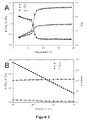
FIG. 3A shows dynamic shear storage (G′), loss modulus (G″), and tan δ are plotted as a function of temperature (γ 2%, ω 1 Hz). FIG. 3B shows dynamic shear storage (G′), loss modulus (G″), and complex viscosity (η*) are plotted as a function of frequency (γ 2%, 37° C.). The figures shows the rheological behavior of B10 in water
FIG. 4. Uniaxial stress-strain analysis. The Young's modulus was 87±9 MPa for TFE-23 and 60±8 MPa for water-4 measured from the first linear range, and was 0.71±0.12 MPa for water-23 film measured from the first 10% of deformation.
FIG. 5A. shows creep of TFE-23 film. From top to bottom, creep was examined as tensile stress was maintained at 1.0 MPa, 0.8 MPa and 0.6 MPa, respectively. FIG. 5B shows creep of water-4 film. From top to bottom, creep was examined as tensile stress was maintained at 0.8 MPa, 0.6 MPa and 0.4 MPa, respectively. FIG. 5C shows creep of water-23 films. From top to bottom, creep was examined as tensile stress was maintained at 60 KPa, 40 KPa and 30 KPa, respectively. Under 60 KPa stress, creep reached the maximum strain that was allowed on the current testing facility within 12 hours. FIG. 5D shows comparison of the creep behaviors of water-4 films derived from B10 and B9. The short-term creep behaviors demonstrated that films derived from B10 are more stable under mechanical loading. The figures shows the creep analysis of B10 films
FIG. 6A shows the influence of preconditioning on resilience of water-4 film. A water-4 sample was cyclically stretched to 30% strain, with an off-loading period of 5 minutes between cycles. Plotted are the stress-strain curves from the first ten cycles of stretches, because stress-strain responses were stabilized after the eight cycles of stretch. Similar responses were also observed for TFE-23 and water-23 samples. FIG. 6B shows the dependence of resilience on the number of preconditioning cycles. Samples cast in different conditions are cyclically stretched to 30% strain, with an off-loading period of 5 minutes between cycles. Plotted is resilience after each cycle against the number of the preconditioning cycles.
FIG. 7A shows a water-23 sample was cyclically stretched to 30% strain for 21 cycles, with an off-loading period of 5 minutes between cycles. Plotted are the stress-strain curves from the first 10 cycles, because the material response to the external loading is stabilized after 8 cycles of stretch. FIG. 7B shows a water-23 sample was cyclically stretched to 30% strain and then to 12% strain for 20 cycles, with an off-loading period of 5 minutes between cycles. FIG. 7C shows a water-23 sample was cyclically stretched to 50% strain and then to 30% strain for 20 cycles, with an off-loading period of 5 minutes between cycles. The figures shows the influence of preconditioning on the resilience of water-23 films.
FIG. 8A shows a water-4 sample was subjected to cyclic stress of increasing magnitudes (shown in inset), and the deformation history was recorded. Reproducibility was examined on three replicate samples, which were preconditioned at 30% strain for 20 cycles with an off-loading period of 5 minutes between cycles and a two hour recovery time. FIG. 8B shows deformation at the end of each loading (filled circles) and off-loading (open circles) period were plotted against the magnitude of cyclic stress. The figures show the deformation behaviors of preconditioned water-4 films under cyclic stress of increasing magnitude
FIG. 9. Deformation behavior of preconditioned water-4 films subjected to a step loading protocol. A water-4 sample was subjected to step stress (shown in inset), and strains at the end of each loading step represented by open circles in water-4 films and by crosses in TFE-23 films were plotted against the magnitude of stress. Reproducibility was examined on three replicate samples, which were preconditioned at 30% strain for 20 cycles with an off-loading period of 5 minutes between cycles.
FIG. 10A shows stress-relaxation response for films cast in water at 4° C. and 23° C., following deformation to 30% strain at constant rate of 5 mm/min. The rapid stress relaxation took place in the first few hundreds of seconds. At 20 minutes, stress dropped from 2.6 MPa to 1.1 MPa in water-4 film, and from 100 KPa to 35 KPa in water-23 film, respectively. FIG. 10B shows Stress relaxation responses of TFE-23 film, following deformation at a constant rate of 5 mm/min to 10% and 50% strain.
FIG. 11A shows electrospinning experimental setup. FIG. 11B shows Electrospun B9 fibers. FIG. 11C shows electrospun B9 network. FIG. 11D shows electrospun B9 conduit.
FIG. 12A shows Young's modulus and FIG. 12B shows ultimate tensile strength of thermally annealed B9 fiber networks, tested at 37° C. in PBS. FIG. 12C shows characteristic uniaxial stress-strain curves for electrospun B9 fabrics generated from ring testing of annealed and non-annealed samples. FIG. 12D shows stress relaxation curves for B9 electrospun fiber networks. Annealing temperature is indicated for each curve.
FIG. 13A and FIG. 13B are plates showing Cryo-HRSEM micrographs of B9 electrospun fibers hydrated at 37C. FIG. 13C and FIG. 13D are places showing Cryo-HRSEM micrographs of B9 electrospun fibers annealed at 60C.
FIG. 14. Platelet deposition on B9 and ePTFE in a baboon ex vivo shunt model.
FIG. 15. 1800 bp B9 midblock gene concatemer.
FIG. 16. Concatemers created via ligation of monomer library.
FIG. 17. Plasmid map of commercial expression vector pQE 80L (Qiagen, Inc). Preparation of the plasmid involves the removal of nucleotides between Bam H I and Hin d III restriction sites within the polyclonal region. Contains an N-terminal His-tag.
FIG. 18. Sodium dodecyl sulfate-polyacrylamide gel electrophoresis (SDS-PAGE) analysis of elastin-mimetic triblock copolymer run on 7.5% SDS-PAGE stained with Coomassie G250. Marker lane: Precision Plus Protein Kaleidoscope (Bio-Rad).
FIG. 19. Schematic representation of the baboon femoral arteriovenous shunt model. Test samples are interposed within an exteriorized silicone shunt and In111-platelet deposition on test surfaces monitored by scintillation camera imaging using a SPECT system.
FIG. 20A shows plain ePTFE. FIG. 20B shows after elastin impregnation. FIG. 20C shows after layer-by-layer elastin deposition, FIG. 20D shows after 24-h flow conditioning in PBS at 37° C. The figures shows macroscopic photographs of unstained (left) and Coomassie-stained (right) graft samples
FIG. 21A shows plain ePTFE graft, FIG. 21B shows plain water-cast elastin-mimetic film, FIG. 21C shows ePTFE graft after elastin impregnation, FIG. 21D shows after layer-by-layer elastin deposition. The figures shows infrared ATR spectra from 1800 to 1000 cm−1.
FIG. 22 shows scanning electron micrographs of ePTFE vascular grafts processed by critical point drying. Scale bar in FIG. 22A scale is 333 μm; FIG. 22B scale is 40 μm; FIG. 22C scale is 2.0 μm; FIG. 22D scale is 333 μm; FIG. 22E scale is 40 μm; and, FIG. 22F scale is 2.0 μm.
FIG. 23. Platelet deposition normalized by surface area over a 1-h time period (n=6).
FIG. 24. Resilience of B9 and B10 scaffolds. Samples are cyclically stretched to 30% strain with a rest period of 5 minutes between cycles. Resilience is measured from the first loading loop for non-preconditioned samples and measured from the 10th loading loop for preconditioned B10 samples. Data indicate that increased hydrophobicity of endblocks decreases resilience of elastin-mimetic scaffold but mechanical preconditioning enhances resilience.
FIG. 25. Stress relaxation of B10 scaffolds under different cast conditions. Samples are stretched to 30% strain at a constant rate of 5 mm/min and then held at this constant strain. Rapid stress relaxation occurs in the first few hundreds of seconds. At 20 minutes, stress decreases from 2.6 MPa to 1.1 MPa, 100 kPa to 35 kPa, and 4.0 Mpa to 1.7 MPa in water-4 scaffold, water-23 scaffold and TFE-23 scaffold, respectively. The first stress drop in TFE-23 scaffold prior to 30% deformation is due to the strain-induced damage effect.
FIG. 26. Amino Acid Sequence of Protein-Based Block Copolymer B10.
FIG. 27. Amino Acid sequence of triblock copolymer B9, constructed from plastic and elastic [X] elastin sequences.
FIG. 28. Molecular Assembly of Modified B10 gene. Crosslinking regions inserted between the plastin and elastin domains in addition to flanking the gene.
FIG. 29. Synthesis of repetitive polypeptides via multimerization of DNA monomers, adapted from [25].
FIG. 30. Genetic assembly of the gene encoding the triblock copolymer R2-R1-R2, adapted from [49].
FIG. 31. Diagram detailing sectioning of 15×33 mm elastin fiber patch for immunohistochemical, electron microscopy, and mechanical analysis.
Table 1. Coding Sequences of Oligonucleotide Cassettes Employed for the Construction of Protein Triblock (PEP) B10.
Table 2. Comparison of Young's Modulus of B9 and B10 Films.
Table 3. Tensile Behavior for Electrospun B9 Fabrics.
Table 4. Absorption Data for B9 Fabrics Hydrated in PBS and Water.
Table 5. Elastin-Mimetic Protein Classifications.
Table 6. Yeast B9 Elastin-block Monomer Library.
Table 7. Lysine Insert and Adaptor Sequences for B10.
Table 8. Coding sequences of R1 and R2 monomer blocks.
Table 9. Summary of Features Integral to the Design of an Elastin-Based Arterial Conduit.
Table 10. Experimental Design for characterization of the mechanical properties of elastin-mimetic nanofiber networks.
Table 11. Targeted Design Criteria.
Tables 12-16. Amino acid and nucleotide sequence listings in table form.
DETAILED DESCRIPTION OF THE INVENTION
“Synthetic” refers to an isolated artificial protein that is not normally made by an organism. A synthetic protein may be made by an organism or manufactured outside an organism. For example, the protein may be a recombinant protein in that a organism has been genetically engineered to express the protein or a precursor thereof.
“Triblock” refers to a protein having at least three distinct regions, such as a hydrophobic central block that separates end blocks that tend to be more hydrophilic. Optionally, a triblock amino acid sequence has additional material inserted between one or more of the blocks or at the block ends. For example, a cross-linkable amino acid or modified amino acid that is capable of cross-linking may be inserted between the blocks to facilitate cross-linkage manipulation. Such chemical cross-linking may be in addition to the physical cross-linking that tends to occur naturally with the amphilic triblocks and provides ability to tailor a mechanical property to the end-application to which the protein may be used.
“Creep” refers to a mechanical property of a material that is time-dependent. In particular, creep relates to the tendency of a material to permanently deform in response to an applied force or stress applied over time, or a time-dependent deformation of the material under stress.
“Inverse transition temperature” refers to the property where a material is a liquid at a lower temperature, but changes state to a gel or solid at a higher temperature. The temperature at which such a change of state begins is referred to as the “inverse transition temperature” and is useful for assisting in placement of an embolic agent into a cardiovascular defect as a liquid initially that later changes to a gel or solid, thereby providing therapeutic benefit.
“Young's modulus” is a mechanical property of a material, device or layer which refers to the ratio of stress to strain for a given substance. Young's modulus may be provided by the expression;
wherein E is Young's modulus, L0 is the equilibrium length, ΔL is the length change under the applied stress, F is the force applied and A is the area over which the force is applied.
“Physical parameter” refers to a property of the protein or material made from the protein and includes mechanical parameters provided herein (e.g., Young's modulus, bending modulus, compressability, ultimate tensile stress, fracture or failure strain, resilience, permeability, swelling ratio, and other parameters and particularly those parameters used in the art to describe biological systems and materials). A “tunable physical parameter” refers to a parameter that can be controllably adjusted by any of the methods disclosed herein or that depends on the structure or sequence of the proteins that make up a film or fiber network. For example, adjusting the properties of the end and/or central blocks (e.g., length, hydrophobicity) permits tuning of a physical parameter that describes the environment or surrounding tissue in which the film or fiber network is to be used or implanted into (e.g., a blood vessel or a portion of the cardiovascular system). Optionally, further tuning is accomplished by any processing or post-processing known in the art thereby providing further control of the mechanical properties of the medical device.
“Embolic agent” refers to a material that is capable of physically impacting blood flow or altering hemodynamics in and around a blood vessel. The embolic agent may be applied to a blood vessel or blood vessel wall, such as a wall rupture or aneurysm, in a liquid form that subsequently gels or solidifies, thereby displacing or preventing further blood flow in a region. Alternatively, the embolic agent may be applied as a gel, semi-solid or solid in a blood vessel or blood vessel wall, such as a wall rupture or aneurysm to provide a therapeutic benefit.
Example 1
Mechanical Stability and Deformation Responses of Physically Crosslinked Protein-Based Materials
Recombinant protein polymers are synthesized and examined under various loading conditions in order to assess the mechanical stability and deformation responses of physically crosslinked, hydrated, protein polymer networks designed as triblock copolymers with central elastomeric and flanking plastic-like blocks. Uniaxial stress-strain properties, creep and stress relaxation behavior, as well as the effect of various mechanical preconditioning protocols on these responses are characterized. An analysis of viscoelastic behavior demonstrates that an increase in endblock size improves network stability and that mechanical preconditioning significantly enhances the resilience of hydrated films. Furthermore, the presence of three distinct phases of deformation behavior is revealed upon subjecting physically crosslinked protein networks to step and cyclic loading protocols in which the magnitude of the imposed stress is incrementally increased over time. Without being bound to a particular theory, we believe that these phases correspond to the stretch of polypeptide bonds, the conformational changes of polypeptide chains, and the disruption of physical crosslinks. The capacity to select a genetically engineered protein polymer that is suitable for its intended application requires an appreciation of its viscoelastic characteristics and the capacity of both molecular structure and conditioning protocols to influence these properties.
The emergence of genetically engineered synthetic polypeptides has enabled the design of protein polymers composed of complex peptide sequences in which individual peptide repeat sequences can be selected with distinct mechanical, chemical, or biological properties. While a large variety of recombinant protein polymers have been reported, those composed of distinct block structures are typically characterized by relatively short block sequences. For example, Cappello and colleagues have produced a series of silk-elastinlike block copolymers (SELPs) in which silk-like regions, consisting of between 12 and 48 alternating alanine and glycine residues, are found between elastin-mimetic sequences comprised of 8 or 16 repeat sequences of Val-Pro-Gly-Val-Gly. We have synthesised high molecular weight recombinant protein block copolymers using an approach, which affords significant flexibility in the selection and assembly of blocks of diverse size and structure. This has led to the synthesis of a new class of BAB protein triblock copolymer composed of large polypeptide block sequences ranging from 400 to 1200 amino acids in length. This class of protein block copolymers are derived from elastin-mimetic polypeptide sequences in which identical endblocks of a hydrophobic, plastic-like sequence are separated by a central hydrophilic, elastomeric block. The triblock protein copolymer acts as a two-phase network when hydrated, in that the hydrophilic block remains conformationally flexible and elastomeric, while the hydrophobic block forms physical or virtual crosslinks through hydrophobic aggregation.
Physically crosslinked protein-based materials possess a number of advantages over their chemically crosslinked counterparts, including ease of processing and the ability to avoid the addition or removal of chemical reagents or unreacted intermediates. However, physical crosslinks formed as a result of hydrophobic aggregation are often deformed or disrupted under external stresses that may be substantially lower than the forces required to disrupt covalent crosslinks. This feature may limit the capacity of physically crosslinked protein-based materials to retain material integrity under sustained mechanical loading that is often an essential requirement for their application in tissue engineering or regenerative medicine or use as a component of an implanted medical device.
Studies suggest that the density and strength of the physical crosslinks are important determinants of both mechanical responses and long-term material stability of two-phase protein networks. Therefore, in order to enhance the mechanical behavior of these materials a new elastin-mimetic triblock copolymer is synthesized that contains hydrophobic endblocks, which are nearly twice as large as prior versions of this triblock protein polymer. Hydrophobic aggregation of the endblocks is examined using differential scanning calorimetry and rheology, and material stability of the physically crosslinked protein networks is accessed through mechanical analysis. In particular, the deformation mechanisms of these protein networks and the conditions under which the physical crosslinks are disrupted are thoroughly examined under creep and stress relaxation protocols, as well as under conditions of cyclic and step loading. The resilience of a material, which characterizes its capacity for shape and energy recovery under mechanical loading, provides another crucial criterion that dictates the applications for which it may be suitable. Moreover, the magnitude of preconditioning strains and the off-loading period between loading cycles strongly influences the viscoelastic properties of a variety of protein and tissue derived materials. Thus, in this investigation we also explore the resilience and viscoelastic behavior of two-phase elastin-mimetic protein polymer networks and the capacity of distinct mechanical preconditioning protocols to affect these properties.
Synthesis of Protein Triblock Polymer B10.
Synthetic methods used to produce the DNA inserts that encode the various elastin-mimetic block copolymers have been described. Oligonucleotide cassettes encoding elastic-like (E) and plastic-like (P) repeat units (Table 1) are independently synthesized and inserted into the BamH I and HinD III sites within the polylinker of pZErO-2. Recombinant clones are isolated after propagation in E. coli strain Top10F′, double-stranded DNA sequence analysis verified the identity of the DNA inserts E and P. DNA monomers E and P are liberated from the respective plasmids via sequential restriction digestion with Bbs I and BsmB I, respectively. Self-ligation of each DNA cassette affords a population of concatemers.
Concatemers derived from DNA monomers E and P are inserted into the BsmB I site of their original plasmid containing the monomer cassette. Concatemers encoding 31 repeats of the P monomer and 21 repeats of the E monomer are isolated and identified via restriction cleavage with BamH I and HinD III. Double-stranded DNA sequence analysis confirm the integrity of the concatemers within the recombinant plasmids, which were labeled pE and pP, respectively. Restriction cleavage of plasmid pE with Bbs I/Xma I and plasmid pP with BsmB I/Xma I afforded two fragments, which are separated via preparative agarose gel electrophoresis. Enzymatic ligation of pE and pP afforded the recombinant plasmid pPE, which encoded the diblock PE as a single contiguous reading frame within plasmid pZErO-2. The diblock, pPE, is used for subsequent construction of the triblock pPEP using the same biosynthetic scheme. Restriction cleavage of plasmid pP with Bbs I/Xma I and plasmid pPE with BsmB I/Xma I afforded two fragments, which are separated via preparative agarose gel electrophoresis. Enzymatic ligation of pP and pPE afforded the recombinant plasmid pPEP, which encoded the triblock PEP as a single contiguous reading frame within plasmid pZErO-2.
The triblock concatemer is liberated from pPEP via restriction cleavage with Bbs I and BsmB I and purified via preparative agarose gel electrophoresis. Enzymatic ligation is used to join the concatemer cassette to the Bbs I sites within the modified polylinker C in plasmid pBAD-A. Double stranded DNA sequence analysis confirms the integrity of the concatemer within the recombinant plasmid, which is subsequently transferred to the expression plasmid, pET-24 (d) via restriction cleavage with Nco I and HinD III. Double stranded DNA sequence analysis confirms the integrity of the concatemer within the recombinant plasmid, which is labeled pB10.
Plasmid pB10 encodes the triblock copolymer protein B10 as a single contiguous reading frame within plasmid pET-24 (d) and is used to transform the E. coli expression strain BL21(DE3). This affords a protein triblock containing flanking endblock sequences [VPAVG(IPAVG)4][(IPAVG)5]33 (SEQ ID NO:7) and a midblock sequence (IPGAG)(VPGAG)VPGEG(VPGAG)2[(VPGAG)2VPGEG(VPGAG)2]20 (SEQ ID NO:8) (FIG. 26, Table 2). Large-scale fermentation (4 L) is performed at 37° C. in Terrific Broth (TB) medium supplemented with kanamycin (50 μg/mL). The fermentation cultures are incubated under antibiotic selection for 48 h at 37° C. with agitation at 225 rpm in an orbital shaker. Cells are harvested via centrifugation at 4° C. and 4,000 g for 20 min and the cell pellet resuspended in lysis buffer (150 mL; 100 mM NaCl, 50 mM Tris-HCl, pH 8.0) and stored at −80° C. The frozen cells are lysed by three freeze/thaw cycles. Lysozyme (1 mg/mL), protease inhibitor cocktail (5 mL), benzonase (25 units/mL), and MgCl2 (1 mM) is added to the lysate and the mixture is incubated at 25° C. for 30 min. The cell lysate is incubated for 12 h at 4° C. and is centrifuged at 18,000 g for 30 min at 4° C. to pellet the cell debris. The target protein is purified from the clarified cell lysate by three to five cycles of temperature-induced precipitation (4° C./37° C.) from 5 M NaCl solution. Dialysis and lyophilization afforded protein B10 as a fibrous solid in isolated yields of 250 mg/L of culture. MALDI-TOF mass spectrometry, Obs. (Calc.): B10, 177,608 (176,924.3). Sodium dodecyl sulfate-polyacrylamide gel electrophoresis (SDS-PAGE) analysis reveals a single protein band for B10 that migrated higher than its predicted molecular weight (FIG. 1).
Synthesis of Triblock Protein Polymer B9 (SEQ ID NO:50)
A recombinant protein that contains flanking hydrophobic endblocks of sequence VPAVG[(IPAVG)4(VPAVG)]16IPAVG (SEQ ID NO:51) separated by a central hydrophilic block [(VPGVG)2(VPGEG)(VPGVG)2]48 (SEQ ID NO:52) is expressed from E. coli and purified, as detailed elsewhere. Amino acid compositional analysis. B9; Calc. (mol.-%): Ala, 8.1; Glx, 2.4; Gly, 31.9; Ile, 6.4; Pro, 20.0; Val, 31.2. Obs. (mol.-%): Ala, 10.8; Glx, 2.0; Gly, 28.3; Ile, 7.0; Pro, 22.8; Val, 28.2. MALDI-TOF mass spectrometry, Obs. (Calc.): B9, 165,356 (165,564).
Differential Scanning Microcalorimetry (Micro-DSC).
Differential scanning microcalorimetry is recorded on a Setaram Micro DSC III calorimeter (Setaram Inc., France) at a scan rate of 1° C./min from 4 to 70° C. Lyophilized proteins are dissolved at a concentration of 1 mg/mL in distilled, deionized water. MicroDSC data is analyzed using SETSOFT 200 software (Setaram Inc, France).
Rheological Analysis of Concentrated Protein Polymer Solutions.
Rheological data are acquired on an Advanced Rheological Expansion System III rheometer (ARES III, TA instrument, NJ) in parallel plate geometry with a plate diameter of 25 mm. The testing protocol for rheological analysis is detailed elsewhere. In brief, 100 mg/mL protein solutions are prepared by adding distilled, deionized water to lyophilized protein at 4° C., shaking the solution for 48 h, and then allowing the solution to equilibrate for 72 h. The gap between parallel plates is adjusted between 0.2-0.35 mm and dynamic mechanical experiments were performed in shear deformation mode. An initial strain amplitude (γ) sweep is performed at 4° C. and 37° C. at different frequencies to determine the linear viscoelastic range for the protein polymer.
The gelation temperature is determined by heating samples from 4° C. to 40° C. at a rate of 1° C. per minute. Following temperature equilibration at 37° C., viscoelastic properties are examined by a strain sweep at a fixed frequency of 1 Hz and a frequency sweep at fixed strain amplitude of 2%. Experiments are repeated on 5 to 6 samples and representative data presented.
Mechanical Analysis of Hydrated Protein Polymer Films.
For mechanical property analysis, films are cast from protein solutions. In brief, lyophilized proteins were dissolved at a concentration of 100 mg/mL either in 2,2,2-trifluoroethanol (TFE) at 23° C. or in water at 4° C. The protein solution is then poured into Teflon casting molds and solvent evaporation performed either at 23° C. or at 4° C. Test samples are referred to as TFE-23, water-23, or water-4, indicating the casting solvent and evaporation temperature used for film formation. After complete solvent evaporation, films are hydrated in phosphate buffered saline (PBS) at 37° C., which contains NaN3 at 0.2 mg/mL to prevent biological contamination. Samples are cut into a dumbbell shape using a stainless steel die with gauge dimensions of 13 mm×4.75 mm. Hydrated film thickness, as measured by optical microscopy, is typically 0.1 mm for TFE-23 and water-5 films and 0.5 mm for water-23 films.
Mechanical characterization of protein films is performed using a dynamic mechanical thermal analyzer DMTA V (Rheometric Scientific Inc., Newcastle, Del.) with a 15 N load cell in the inverted orientation, so that samples could be immersed in a jacketed beaker filled with PBS at 37° C. The maximum travel distance of the drive shaft of DMTA was 23 mm, which limited maximum strain to 70% of engineering strain. Samples are evaluated by a several mechanical test protocols including: (i) Uniaxial tension. Loading and unloading is controlled by displacement at a fixed rate of 5 mm/min. Five to six samples are monotonically stretched to 65% of maximum strain for uniaxial stress-strain analysis. (ii) Creep and stress relaxation. Six to ten samples are prepared for creep analysis. Constant engineering stresses are applied for time periods of up to 30 hours. Four to six samples are prepared for stress-relaxation analysis. Each sample is stretched at 5 mm/min to a fixed strain and the evolution of stress over time is examined. Measurement of stress-relaxation is limited to 30 minutes. (iii) Preconditioning protocols. Five to six samples cast under different conditions are cyclically stretched to 30% strain for 20 cycles with an off-loading period of 5 minutes between cycles. Water-23 films are also stretched to 30% strain for one cycle and then cyclically stretched to 10% strain for 20 cycles; or stretched to 50% strain for one cycle and then cyclically stretched to 30% strain for 20 cycles with an off-loading period of 5 minutes. Resilience is calculated from loading and under the loading curves. (iv) Cyclic loading with increasing stress magnitude. Water-4 samples preconditioned at 30% strain for 20 cycles with an off-loading period of 5 minutes are subjected to cyclic stress of increasing magnitude. Stress is applied for one hour and then removed for one hour followed by reimposition of the load at a higher stress. Reproducibility is examined in three replicate samples. (v) Step loading. Water-4 samples preconditioned at 30% strain for 20 cycles with an off-loading period of 5 minutes are subjected to step loading, in which stress is increased by 50 kPa every two hours. Reproducibility is examined in three replicate samples.
The inverse transition temperature is consistent with protein block structure. Differential scanning microcalorimetry of dilute aqueous solutions of B10 (1 mg/mL) confirms the presence of a single endothermic transition at 21° C. consistent with coacervation of the hydrophobic endblocks (FIG. 2). The inverse transition temperature of B10 is 4° C. lower than that observed for B9 due to an increase in the size and hydrophobicity of the endblocks. Specifically, the B10 endblocks are nearly twice as large as those of the B9 triblock copolymer and contained a larger mole fraction of isoleucine (20 vs. 16 mol %). Reversibility of the phase transition is confirmed upon repeating microcalorimetry measurements after a 12 h equilibration at 4° C. (data not shown).
Rheological analysis confirms formation of a protein gel. Above 18° C., the shear storage (G′) and loss (G″) modulus of concentrated solutions of B10 increased by a factor of approximately 103 and 10, respectively, while tan δ (G′/G″) decreased, consistent with the formation of a viscoelastic gel (FIG. 3A). Observation of a lower transition temperature for protein gelation than that noted by microcalorimetry studies of dilute protein solutions was likely due to the effect of extensive intermolecular interactions present in the concentrated protein solution used for rheological studies. At 37° C., G′ and G″ were independent of frequency between 0.01 to 10 rad/s at a fixed strain amplitude of 2% (FIG. 3B). In addition, the logarithm of complex viscosity (η*) was a linear function of the logarithm of frequency with a slope of −1. All of this suggests that within this frequency range the mechanical response of the protein hydrogel is consistent with a rubbery solid.
Block structure alters the Young's modulus of elastin-mimetic triblock protein polymers. Load-extension curves at 37° C. of hydrated B10 films cast either from TFE at 23° C. or water at 4° C. reveals plastic-like deformation behavior, such that, stress increases linearly with increasing strain until a yield point is reached between 2-2.5 MPa, after which elongation occurs with the imposition of a relatively low increment in load. In contrast, hydrated B10 films produced from an aqueous protein solution cast at 23° C. displays rubber-like behavior with homogeneous deformation occurring in response to low stress levels. Corresponding values of Young's modulus are 87 MPa, 60 MPa, and 0.71 MPa for hydrated TFE-23, water-4, and water-23 B10 films, respectively. Of note, these values are two- to 60-fold greater than the Young's modulus measured for B9 films processed under identical casting conditions (FIG. 4, Table 2).
Prior studies of B9 triblock copolymers demonstrate that solvent type and casting temperature profoundly influences microphase protein block mixing with a commensurate effect on mechanical responses. Specifically, films cast from TFE, which solvates both mid and endblock sequences, promotes significant interphase mixing in cast films. As a result, the hydrophobic, semi-rigid endblocks are organized as a dispersed microphase and thereby contribute to the mechanical response of the material as load bearing elements leading to plastic-like deformation behavior. In contrast, water preferentially solvates the hydrophilic midblock. Thus, films cast from water at 23° C. display a microphase separated structure with well segregated endblocks that act as relatively discrete virtual crosslinks within an elastomeric protein matrix. Moreover, in casting the aqueous protein solution above the inverse transition temperature of the protein polymer (23° C.>18° C.), microphase separation of the endblocks is further promoted due to a coacervation effect. Given the greater degree of microphase separation, the contribution of the elastomeric midblock to the mechanical response of the material is enhanced with a corresponding rubber-like stress-strain profile. The influence of casting temperature is demonstrated by the behavior of films cast from water at 4° C. In the absence of the coacervation effect present above 18° C., we believe that films are produced with a lower degree of microphase separated structure and, therefore, display a higher Young's modulus. As compared to B9, the presence of substantially larger endblocks and a relatively smaller midblock accentuates the proportion of plastic-like domains in B10 films and, as a consequence, the generation of materials with a higher elastic modulus under all film forming conditions.
Creep responses are modulated by protein block structure. Prior studies characterized creep responses of B9 films cast from water at 4° C. or TFE at 23° C. and revealed substantial deformation responses above 0.2 MPa. As a virtually crosslinked protein network, it is presumed that time-dependent changes in strain in response to stress will be influenced by the density, size, and chemical nature of the physical crosslinks. Thus, by increasing both the hydrophobicity of the endblock, as well as the relative size of the endblock by altering the weight ratio of endblock to midblock segments, the creep response behavior is controllably modified.
Creep analysis was performed on hydrated B10 films at 37° C. that were initially produced under a variety of film casting conditions (FIG. 5). Water cast films produced at 4° C. demonstrated limited creep (<10%) over a 20 h observation period at stress levels at or below 0.4 MPa, nearly double the load for B9 films produced under comparable conditions. Films cast from an aqueous solution of B10 at 23° C. demonstrated comparable levels of creep, but at stress levels that were one order of magnitude lower. B10 films cast from TFE demonstrated the lowest level of creep with an observed strain of less than 10% when subjected to a stress of 0.8 MPa; an approximately four-fold greater load than that sustained by similarly fabricated B9 films. Given that the magnitude of the observed deformation response was not directly proportional to the applied stress, these materials behaved as non-linear viscoelastic solids. In summary, these data emphasize that time-dependent mechanical properties of protein-based materials containing large, chemically distinct blocks can be modulated by controlling the tendency for block segregation either by selection of processing conditions or by molecular design.
Preconditioning by an imposed cyclic stress enhances the resilience of protein polymer films. Upon subjecting B10 films to periods of repetitive cyclic deformation to 30% strain, we observed the accumulation of residual deformation and a decline in peak stress that stabilized after several cycles (FIG. 6). In the process, resilience was significantly enhanced over 10 loading cycles with an increase from 11±2% to 30±2% for TFE-23 films, from 18±2% to 39±2% for water-4 samples, and from 35±2% to 51±2% for water-23 films. The greatest increase in resilience largely occurred after the first loading cycle, presumably due to stabilization of load induced changes in microstructure.
The effects of varying mechanical preconditioning protocols on resilience are further examined using films initially cast from water at 23° C. (FIG. 7). In all protocols, stabilization of mechanical behavior is largely observed after the initial loading cycle with accumulation of residual strain of 5-10% and a decline in peak stress. As previously stated, the resilience of water-23 films subjected to a repetitive cyclic strain of 30% was 51±2%. When films are subjected to an initial elongation of 30% followed by cyclic stretch at 12% strain, the resilience increases to 58±2%, which is attributed, at least in part, to a reduction in energy dissipation at reduced strain. Nonetheless, the influence of initial deformation history on resilience is evident when films are subjected to an initial strain of 50% and subsequently exposed to 30% cyclic stretch. Although permanent strain is unaffected, as compared to films subjected to 30% cyclic stretch alone, resilience increases to 67±1%. Thus, a significant degree of change in protein microstructure can be induced not only by the conditions of film casting, but also through the effects of mechanical deformation or annealing protocols. As mechanical preconditioning stabilized the microstructures and mechanical properties of protein polymer films, the deformation plasticity tends to decrease. For instance, the yielding behaviors of TFE-23 films diminished and nearly linear behaviors are observed in water-23 films over 10 loading cycles.
Under what circumstances will physical crosslinks be broken? Three phases of deformation behavior are observed when preconditioned B10 water-4 films are exposed to cyclic loads of increasing magnitude (FIG. 8). The first phase, which extends up to an imposed load of 1.2 MPa over a 30 h period, is characterized by small elastic deformation responses, as both the total and residual deformations are small. In the second phase, over a load range between 1.2 and 2.7 MPa, both the total and residual deformation increase linearly with increasing magnitude of cyclic stress and appreciable residual deformation is observed. A more rapid increase in the total and residual deformation occurs in the third phase consistent with disruption of physical crosslinks. Films examined under step loading also display three similar phases of deformation behavior (FIG. 9). Remarkably, strain levels at each transition point are similar for both protocols, although stress levels were significantly different. Three phases of deformation behavior are also observed for preconditioned films cast from TFE at 23° C.
It bears comment that during off-loading periods water-4 films demonstrate “recovery” of deformation after imposed cyclic loading and, therefore, are able to sustain larger subsequent stresses, when compared to deformation induced by direct step loading. For example, when accumulated strain reaches the onset of the second deformation phase, the exerted stress is approximately 1200 kPa and 200 kPa under cyclic and step loading conditions, respectively. The observed recovery effect is likely a consequence of limited polypeptide chain rearrangement, in which the capacity of the material to carry loads is partially recovered and is clearly dependent on the characteristics of both loading and off-loading conditions.
We believe that in a manner akin to synthetic polymers, the deformation behavior in the first phase may be attributable to an initial stretching of polypeptide bonds. Inevitably, bond stretch is limited and further deformation must arise from conformational changes in the polymer chain, which likely occurs in the second phase of deformation. Differences in the stress required to induce conformational changes of protein polymer within films processed under different casting conditions are likely related to differences in the mixing of semi-rigid endblocks and flexible midblocks that create energy or stereoelectronic barriers. Under both loading protocols, substantial film deformation is observed after an initial 22-25% strain, which appears to designate the stress level associated with disruption or damage to physical crosslinks. Given that samples are preconditioned at 30% strain for 20 cycles, these data suggest that “new” disruption or damage may occur when deformation approaches or exceeds preconditioning strains.
Micro-DSC and rheology studies confirm the presence of an inverse-temperature transition for the elastin-mimetic protein polymer B10 in aqueous solutions with gelation of concentrated solutions at ambient temperatures. Mechanical analysis, particularly studies of creep behavior, demonstrate enhanced mechanical stability of physically crosslinked protein networks derived from B10 compared to a triblock copolymer designed with a lower relative content of hydrophobic, plastic-like endblocks. Moreover, resilience is significantly enhanced by mechanical preconditioning. Newly designed tests consisting of cyclic loading of increasing magnitude and step loading further reveal the presence of three phases of deformation behavior, which likely correspond to peptide bond stretching, conformational changes of polypeptide chains, and disruption of physical crosslinks. Significantly, the breakage of physical crosslinks strongly depends on the imposed pattern of load, as well as preconditioning protocols.
| TABLE 1 |
| |
| Coding Sequences of Oligonucleotide Cassettes for |
| Repeating Sequences Employed for the Construction |
| of a Protein Triblock (PEP) B10 |
| |
| E Block |
| Val |
Pro |
Gly |
Ala |
Gly |
Val |
Pro |
Gly |
Ala |
Gly |
Val |
Pro |
Gly |
| |
| GTT |
CCA |
GGT |
GCA |
GGC |
GTA |
CCG |
GGT |
GCT |
GGC |
GTT |
CCG |
GGT |
| CAA |
GGT |
CCA |
CGT |
CCG |
CAT |
GGC |
CCA |
CGA |
CCG |
CAA |
GGC |
CCA |
| |
| Glu |
Gly |
Val |
Pro |
Gly |
Ala |
Gly |
Val |
Pro |
Gly |
Ala |
Gly |
| |
| GAA |
GGT |
GTT |
CCA |
GGC |
GCA |
GGT |
GTA |
CCG |
GGT |
GCG |
GGT |
| CTT |
CCA |
CAA |
GGT |
CCG |
CGT |
CCA |
CAT |
GGC |
CCA |
CGC |
CCA |
| |
| |
| Ile |
Pro |
Ala |
Val |
Gly |
Ile |
Pro |
Ala |
Val |
Gly |
Ile |
Pro |
Ala |
| |
| ATT |
CCT |
GCT |
GTT |
GGT |
ATT |
CCG |
GCT |
GTT |
GGT |
ATC |
CCA |
GCT |
| TAA |
GGA |
CGA |
CAA |
CCA |
TAA |
GGC |
CGA |
CAA |
CCA |
TAG |
GGA |
CGA |
| |
| Val |
Gly |
Ile |
Pro |
Ala |
Val |
Gly |
Ile |
Pro |
Ala |
Val |
Gly |
| |
| GTT |
GGT |
ATC |
CCA |
GCT |
GTT |
GGC |
ATT |
CCG |
GCT |
GTA |
GGT |
| CAA |
CCA |
TAG |
CGA |
GCA |
CAA |
CCG |
TAA |
GGC |
CGA |
CAT |
CCA |
| |
| |
| Met |
Val |
Pro |
Glu |
Ser |
Ser |
Gly |
Thr |
Glu |
Asp |
Val |
Pro |
| |
| ATG |
GTT |
CCA |
GAG |
TCT |
TCA |
GGT |
ACC |
GAA |
GAC |
GTT |
CCA |
| TAC |
CAA |
GGT |
CTC |
AGA |
AGT |
CCA |
TGG |
CTT |
CTG |
CAA |
GGT |
| |
| Gly |
Val |
Gly |
Stop |
Stop |
| |
| GGT |
GTA |
GGC |
TAA |
TAA |
| CCA |
CAT |
CCG |
ATT |
ATT |
| |
| TABLE 2 |
| |
| Comparison of Young's Modulus of B9 and B10 Films |
| |
Cast in TFE at |
Cast in water |
Cast in water |
| |
23° C. |
4° C. |
23° C. |
| |
|
| |
B10 (MPa) |
87 ± 9 |
60 ± 8 |
0.71 ± 0.12 |
| |
B9 (MPa) |
35 ± 3 |
1.3 ± 0.3 |
0.01~0.03 |
| |
|
Example 2
Elastin Fibers as Design Elements for an Arterial Substitute
With statistics indicating approximately 500,000 procedures for coronary bypass surgery performed in over 300,000 patients each year, cardiovascular disease (CVD) is an unmistakably a growing concern. Since 1900 CVD has been the leading cause of death in the United States, plaguing 70 million Americans and claiming over one million lives per year. Specifically, coronary artery disease accounts for 54% of the CVD deaths annually [1].
Consequently, the need for a small diameter arterial prosthesis is apparent. Although employing polymers such as polytetrafluorethylene (PTFE) have been successful in the development of large diameter vascular grafts, the fabrication of a durable small diameter prosthesis remains an elusive goal. Biological reactions at the tissue material interface resulting from mechanical or compliance mismatch between native artery and the arterial replacement material lead to their ultimate failure. Presently, autologous vessels (i.e. saphenous veins and internal mammary arteries) are vascular replacements of choice, though even these vessels are not sufficient for long term patency. Of the 600,000 coronary bypass operations performed annually, 10-20% of patients will require a second operation within 10 years [2].
In response to these limitations, strategies to mimic some or all of the characteristics of the arterial wall have been pursued. Current tissue engineering strategies provide an opportunity to circumvent maladaptive responses, though adequate replacements could be decades away. Alternatively, the generation of protein polymers that mimic native structural proteins offers a replacement strategy to develop a vascular graft with clinical performance results that match or exceed those of a native vessel. The reformulation of these proteins into nanofiber networks provides an opportunity to optimize the mechanical properties of an arterial bioprosthesis, as well as other biologically related characteristics, thus creating an optimal vascular replacement material.
This work can be divided into four areas: (i) to synthesize a family of recombinant elastin-mimetic proteins; (ii) to define their molecular level structure-property relationships; (iii) to develop nanofabrication strategies to create organized fiber networks, and (iv) to characterize the capacity of these artificial proteins for the generation of non-thrombogenic small diameter blood vessel substitutes with mechanical properties that closely match those of native blood vessels. Utilizing recombinant proteins based on consideration of the structural properties of the native matrix leads to the creation of vascular conduits with better defined mechanical properties and enhanced biodegradation with improved clinical performance characteristics.
The assembly of nanofiber protein networks comprised of recombinant elastin proteins provides a rational approach for generating a tissue engineered vascular graft with enhanced biostability and mechanical properties that closely match those of a native artery.
Disclosed herein are synthesize of recombinant elastin-mimetic protein polymers capable of forming both physical and chemical crosslinks. Using genetic engineering approaches elastin-mimetic fibers are produced with controlled elastomeric properties and enhanced biostability through appropriate choice of recombinant peptide sequences that facilitate both chemical and physical crosslink formation.
Characterize the mechanical properties of elastin-mimetic nanofiber networks. The presence of chemical and physical crosslinks can act synergistically to improve compliance, resilience, and ultimate tensile strength of elastin networks. Creep and stress-relaxation responses of elastin fiber networks are further improved with the incorporation of chemical crosslinks.
We assess the biocompatibility of elastin based fiber networks after in vivo implantation and the ability of such networks to retain primary elastomeric responses. Elastin-mimetic fiber networks have sufficient biostability for use in a vascular construct. In addition, a recombinant protein fiber patch retains initial elastomeric properties after in vivo implantation.
Development of a small diameter vascular replacement for coronary bypass surgery has been described as the ‘Holy Grail’ for cardiovascular tissue engineering [3]. It is recognized that adverse events leading to vascular graft failure are related to destructive biological reactions at the blood-material and tissue-material interface. Specifically, synthetic materials which have been successfully applied to large diameter replacements fail when applied to the small diameter with insufficient patency rates limited by thrombosis and compliance mismatch [4-8]. Over the past three decades, vascular graft design has adapted more of a tissue engineering approach with new graft design inspired by characteristics of the arterial wall.
Earliest efforts endeavored to functionalize synthetic graft prostheses with a luminal layer of endothelial cells. Though this strategy has several limitations, i.e. issues with cell sourcing, cell retention, and procoagulant tendencies, it has found success as larger peripheral artery replacements [9, 10]. The inherent limitations of synthetic polymers have motivated investigation to take a completely biological approach to the development of vascular grafts. Early work explored collagen gel technology in which constructs were developed consisting of cell populated collagen gels [11]. This research has served as the foundation for subsequent innovation. Extensions of this technology have incrementally enhanced the material integrity of the construct, through strategies to increase fiber alignment of the collagen[12, 13], strength via mechanical conditioning[14], crosslinking[15], and others, yet constructs exhibit inferior mechanical properties as compared to native vessels. Other approaches have utilized native vascular cells in the production of ‘cell secreted scaffolds’ [16-18] Though these tissue engineering strategies have reported promising results, each poses unique challenges. Specifically, the duration of incubation time, immunologic challenges associated with the use of allogeneic cells, and suboptimal compliance has limited the application of these strategies to create a clinically applicable small diameter replacement.
Decellularized allo- and xenogeneic tissue have alternatively been investigated as materials for vascular grafts. These decellularized natural matrices contain the intact extracellular matrix and associated attachment proteins and have been used to produce structures with increase degradation resistance, decreased thromobgenicity, and decreased inflammatory reactions. Human umbilical vein, bovine carotid artery and small intestine submucosa, chemically crosslinked using gluteraldehyde, have been employed in clinical application though their use has been limited due to suboptimal patency rates via dilation and aneurysm formation [19-24].
Biosynthetic Approach to the Development of an Engineered Vascular Graft: Allogeneic and xenogeneic strategies indicate native fiber networks can be used to fabricate a vascular graft prosthetic, though the inability to tailor matrix composition and content, fiber size and architecture, limits the applicability of these materials. As a result, strategies to design a prosthesis with precisely defined mechanical and biological properties has been pursued via a ‘ground-up’ design. Recent developments in recombinant protein engineering now offer the opportunity to construct new proteins with near absolute control over molecular architecture [25-28]. Employing biosynthetic routes to the design of structural proteins for vascular prosthetics afford the ability to modulate material properties at the level of the primary amino acid sequence, thus affording the ability to engineer recombinant proteins to meet physiologic requirements. Additionally, this strategy enables the elucidation of structure-property relationships and ultimately, control over these properties. Currently, structural proteins have been generated in this way consisting of sequentially repeated amino acid blocks derived from analysis of native protein molecular structure [29, 30]. This strategy not only allows for control of sequence and size, it also facilitates incorporation of additional functional groups, in particular, the placement of crosslinks at well defined intervals along the peptide chain allowing for the additional control over material properties of the protein. Thus, recombinant proteins that mimic structural matrix proteins can be engineered with a precisely tailored design to modulate tensile strength, elastic modulus, viscoelasticity, and in vivo stability, as well as desired host response. These mimics are optimal candidates in the design of the next generation vascular graft.
Rational Design of an Arterial Prosthesis with Mechanically Matched Properties of the Arterial Wall: The inherent elasticity of blood vessels arises from the structure of the medial layer. The media is composed of concentric layers of elastic lamellar units each composed of smooth muscle cells, elastin fibers, and collagen fibrils. Elastin and collagen function in a concerted action in response to imposed deformations. Elastin is primarily responsible for distensibility and elastic recovery of the vessel in the low-strain regime while collagen responds by limiting deformation during excessive strain [31-35]. Thus, the lamellar unit of the aortic media serves as a foundation in the design of a vascular graft prosthetic [36-38]. Furthermore, the elastin protein network appears to be integral to mechanically match the native blood vessel and for the prevention of intimal hyperplasia and potential graft failure.
Native elastin is a highly insoluable matrix protein that is responsible for providing extensibility and resilience to most tissues of the body. In the vascular system, elastin fiber networks appear in large densities (over 50%) and function to provide resilience to the artery to absorb dynamic systolic stresses of the cardiac cycle and to release energy in the form of blood pressure during diastole [39]. Therefore, elastin networks maximize the durability of tissues that are loaded by repetitive forces by minimizing the conversion of mechanical energy to heat which would ultimately result in tissue damage [35]. In addition to its structural role, elastin creates an environment which promotes proper cell function. Specifically within the vascular system, elastin regulates smooth muscle cell phenotype and proliferation, and in this way is responsible for stabilizing arterial structure [39-41].
Elastin fibers appear to exist as two morphologically different components, a highly isotropic amorphous elastin constituent within an organized microfibrilar scaffold [42]. Understanding the mechanism of fiber assembly in native elastin is limited. Fiber assembly appears to take place in proximity to the cell membrane where microfibrils appear first, grouped in small bundles. Amorphous elastin is synthesized by smooth muscle cells as the soluble monomer, the 72 kDa precursor tropoelastin, and is secreted within each fiber bundle. Here it is organized into insoluble networks reminiscent of natural rubber. Microfibrils function to properly align tropoelastin to facilitate enzymatic crosslinking via oxidation by lysyl oxidase [43].
The distinctive composition of tropoelastin affords unique physical properties of this structural protein. Tropoelastin is rich in glycine (33%), proline (10-13%), and other hydrophobic residues (44%) rendering elastin an extremely hydrophobic protein [44]. Tropoelastin contains distinct crosslinking and hydrophobic domains. Crosslinking domains are alanine rich, containing pairs of lysine residues facilitating intermolecular crosslinking. Specifically, lysine residues are separated by either two or three alanine residues allowing for retention of an a-helical conformation in this region. The sequence within the crosslinking domains appears to be conserved as a consequence of the conformational constraints of crosslinking [43]. Alternatively, the hydrophobic domains within tropoelastin are composed of three-quarters of valine, glycine, proline, and alanine. Investigations have determined that precise sequence and size of this region are not critical for appropriate function. However, the total size of the protein polymer, 750-800 residues, is highly conserved among species [43].
Rational Design of Peptide Sequence for Elastin-Mimetic Protein Polymers: Limitations to the use of elastin in biomedical and tissue engineering applications are a consequence of its intrinsic insolubility and inability to be processed. But through the structural characterization of the hydrophobic domains, the ability to base synthetic protein polymers on native elastin sequences is feasible. The pioneering work of Urry elucidated the elastomeric pentapeptide repeat, VPGVG, from human elastin which now serves as the basic sequence extensively investigated by both chemical methodologies and recombinant technology [46-51]. VPGVG is a common repeat unit within the hydrophobic domain of human elastin and is responsible for resultant elastic properties. Additionally, this domain is responsible for facilitating fiber formation through coacervation phenomena, behaviors consistent with native elastin. Spectroscopic analysis has revealed that native elastin, and likewise, protein polymers containing this repeat, exhibit β-turns and helical β-spiral conformations and display an inverse temperature transition defined by the generation of a more ordered system upon increasing temperature. This loss of entropy is a consequence of protein folding into β-spiral conformation and the subsequent reorientation of water from the elastin chain [45].
Studies have indicated that the amino acid in the fourth (X) position (VPGXG) modulates the coacervation temperature with more polar amino acids increasing transition temperature [46, 47]. As long as glycine and proline residues are preserved the structure and function of elastin is maintained [48]. This discovery has led to the generation of recombinant elastin analogs designed for biomedical applications. For instance, Conticello et al have employed recombinant techniques to design amphiphilic elastin protein polymers consisting of hydrophobic and hydrophilic domains. Through precise sequence design and control of processing conditions, these elastin analogs exhibit a wide range of properties advantageous for biomedical applications, as micelles or physically crosslinked hydrogels [49, 50]. Additionally, groups have incorporated cell binding domains, RGD or REDV, into elastin sequences to functionalize elastin matrix components for endothelial cell attachment [51, 52].
Genetic engineering strategies afford the capability to modulate macroscopic properties on the molecular level. Specifically, it is feasible to alter the molecular architecture to control biologically important parameters of these materials including permeability, swelling ratios, viscoelasticity, strength, and biostability. For instance, residues may be incorporated into the polymer backbone which can be post-translationally modified to promote crosslinking into a protein network [53].
In its native form, elastin is present as a network of elastic fibers crosslinked through lysine residues. Characteristically, crosslinking of native elastin is accomplished via enzymatic modification of amino acid side chains of lysine residues in the solid state, i.e. after secretion by cells into the extracellular space. Briefly, crosslinks are formed through the deamination of the ε-amino group of the lysine side chains by the enzyme lysyl oxidase. The reaction occurs in two ways: (i) the reactive aldehyde group condenses with a second aldehyde residue to form allysine aldol or (ii) with the ε-amino group on the lysine to form dehydrolysinonoleucine. These two precursors condense to form the pyridium cross-links esmosine and isodesmosine [13].
Incorporation of reactive lysine residues into recombinant elastin design provides the ε-amino moiety of lysine for crosslinking using a variety of approaches. Crosslinking of synthetic elastin-mimetic protein polymers has been investigated using solution phase systems; either gamma irradiation [54-56], chemical [53, 57-60], or enzymatic based approaches [61], as well as solid state photocrosslinking [62]. Specific investigations into reactive group spacing as well as crosslinking strategies on the modulation of important biological behaviors of elastin analogs has been conducted with the general conclusion that the placement of well defined crosslinks enhance the biostability of elastin and improve biologically relevant properties.
Fabrication of Structural Proteins into Nanofiber Networks: As material for tissue engineering applications, elastin is intended to provide both mechanical support and potentially act as a scaffold for cellular repopulation. As such, it is likely that its versatility as a scaffold for tissue engineering applications will be significantly enhanced when reformulated into fiber networks. Electrospinning is a technique for generating fibers with diameters <1 μm. Briefly, the electrospinning technique relies on electrostatic forces to produce sub-micron diameter fibers from protein solutions. A high voltage is applied to a spinneret while a protein solution is slowly being pumped through it. This induces evenly dispersed charges in a pendent drop at the tip of the spinneret, relaxing the fluid surface. This surface charge and the external Coulombic forces from the electric field combine to form a tangential stress. This causes the drop to become distorted into a shape referred to as a Taylor cone. At a threshold value, the electric field strength will overcome that of surface tension and the protein solution is ejected as a charged jet from the spinneret tip. As the jet travels to the grounded collector it undergoes stretching and whipping phenomena which reduces the diameter of this fiber. It is then collected, usually in a random orientation, on the grounded collector, creating a nonwoven protein mat. The applied voltage, concentration of protein solutions, flow rate, and deposition distance all effect the morphology of the fiber. When proteins are reformulated as fiber systems desired mechanical and biological properties can be achieved for biomedical applications. For instance, flexibility of a fibrous system can be controlled by either a decrease in fiber diameter or an increase in fiber number [63]. Thus, reformulating recombinant proteins into fiber networks provides an additional level of control over the properties of the system. Specifically, studies have indicated electrospun fabrics composed of small diameter fibers (<1 um) were found to have decreased porosity, increased fiber density, increased mechanical strength, as well as an optimized biological environment for promoting endothelial cell adhesion as compared to larger diameter fibers (7 um) [64, 65].
The assembly of recombinant elastin fiber networks provides an important new design strategy for generating a clinically durable small diameter arterial substitute. This approach yields an arterial prosthesis with mechanical properties that closely match those of a native artery, along with enhanced biostability as compared to allogeneic or xenogeneic tissue. Specifically, in employing a biosynthetic strategy, elastin-mimetic protein polymers can be designed to facilitate both covalent and physical crosslink formation thus enhancing static and dynamic material behavior. These protein polymers may be formulated into nano-fiber networks with improved compliance, resilience, creep, stress relaxation and biostability. Significantly, this strategy can be integrated into schemes which are ultimately driven either by a desire to generate a cell containing arterial construct or a non-thrombogenic acellular conduit.
Data are divided into three areas (i) characterization of first generation elastin-mimetic protein polymers reformulated as fiber networks, (ii) genetic modification of first generation elastin-mimetic protein polymers, and (iii) synthesis of second generation elastin-mimetic protein polymers.
Characterization of First Generation Elastin-Mimetic Protein Polymers Reformulated into Fiber Networks: Initial studies select a unique recombinant elastin protein from a family of recombinant proteins, exhibiting properties relevant to the fabrication of a bioengineered vascular prosthesis. This triblock protein co-polymer, designated B9 (FIG. 27), is uniquely designed with distinct hydrophobic and hydrophilic domains to facilitate physical crosslinking via coacervation of hydrophobic endblocks above the inverse transition temperature. Initial investigations reveal the capacity of this protein to be processed into hydrogels or micelles for drug delivery applications [49, 66] and nanofiber networks for tissue engineering scaffolds [67, 68]. Additionally, B9 studies provide understanding of the relationship between macroscale material properties and microscale features, such as block size and sequence, in engineered proteins [69]. More recent work investigated modulating mechanical properties of B9 films by preferential solvent casting and the impact of casting conditions on static and transient properties of B9 films [70]. These investigations serve as the foundation for subsequent B9 studies and also in the rational design of second generation proteins.
Fabrication of B9 Nanofibers and Networks: Lyophillized B9 protein is reformulated into fiber networks using electrospinning techniques (FIG. 11A). A solvent system is employed to allow for interphase mixing of the incompatible blocks of the copolymer on the nanoscale which influenced and enhanced B9's material properties as fibers. Sub-micron diameter B9 fibers are produced from a 12 wt % protein solution using a trifluoroethanol (TFE) solvent system (FIG. 11 B,C). By controlling the rotational and translational speeds of the collecting mandrel, an elastin conduit is created (FIG. 11D).
Mechanical Properties of Elastin-Mimetic Fiber Networks: Uniaxial Stress-Strain Behavior of B9 Fiber Networks: The mechanical response of electrospun B9 networks under physiological conditions (37° C., PBS pH 7.0) is evaluated by uniaxial (ring) stress-strain testing [14, 71]. Hydrated samples displayed an elastic modulus of 0.17±0.01 MPa and ultimate tensile strength of 0.079±0.008 MPa (FIG. 12); values comparable to the elastin component of the arterial wall (Young's modulus˜0.3 MPa) [18]. As electrospinning creates randomly oriented fiber fabrics, similar properties are expected in the longitudinal and circumferential directions. Uniaxial tensile testing using traditional dogbone shaped samples is conducted on a Dynamic Materials Thermal Analyzer (DMTA) and a Minimat Testing Apparatus to assess longitudinal properties. Circumferential and longitudinal mechanical properties of B9 fabrics are summarized in Table 3. Elastic moduli and tensile strengths obtained from samples strained along the longitudinal axis of the conduit are not statistically different than values measured when stress was imposed in the circumferential direction. The isotropy of electrospun materials is consistent with the mechanical response of native elastin [72].
Time-Dependent Mechanical Properties of B9 Fiber Networks: When deformation is held constant, a relaxation of the imposed tensile stress is observed. This phenomenon is a result of the disappearance of frictional forces, rearrangement of polymer chains, and possibly micro-damage to the protein. Stress relaxation of B9 fiber networks reveals rapid relaxation of imposed tensile stress following deformation to 64% strain. The stress relaxation took place in the first two hundred seconds. At ten minutes, engineering stress had dropped approximately 45% and stabilized indicating structural re-orientation of anisotropic fibers in the direction of deformation followed by conformational rearrangements of protein chains and network entanglements (FIG. 12D).
Modulation of Mechanical Properties of B9 Fiber Networks via Thermal Annealing: A comparative analysis of annealed and non-annealed fabrics indicates thermal annealing significantly alters the mechanical behavior of electrospun B9 fabrics. Subjecting electrospun fiber networks to incubation in PBS at 60° C. for 4 hours appeared to enhance both static and time-dependent mechanical properties. Detailed investigations of tensile properties of annealed and non-annealed samples are reported in FIGS. 12A and 12B and critical parameters are summarized in Table 3. Annealing induced a graduated increase in both elastic modulus and tensile strength commensurate with temperature at which annealing occurred. The greatest effect was observed in elastic modulus of the fabric with an approximate doubling of modulus to 0.366±0.05 MPa as result of annealing at 60° C. for 4 hours (p<0.005) as compared to a non-annealed sample maintained at 37° C. Under similar conditions, tensile strength increased 30% from 0.079±0.008 MPa to 0.119±0.015 MPa (p<0.01). Additionally, the characteristic non-linear toe region, typical of native arteries and biological tissues, is observed in electrospun fabrics and appears to be influenced by annealing temperature. Structural reorientation of the randomly oriented fibers account for this phenomena as verified by SEM of critical point dried (CPD) mechanically strained fibers (data not shown). Interestingly, the non-linear toe region decreased significantly with an increase in annealing temperature.
| TABLE 3 |
| |
| Tensile Behavior for Electrospun B9 Fabrics |
| |
Elastic Modulus |
Tensile Strength |
| Treatment |
(MPa) |
(MPa) |
| |
| 37° C. |
Circumferential |
0.170 ± 0.01 |
0.079 ± 0.008 |
| Incubation |
Longitudinal |
0.190 ± 0.026 |
0.108 ± 0.05 |
| 60° C. |
Circumferential |
0.366 ± 0.05 |
0.119 ± 0.015 |
| Anneal |
Longitudinal |
0.294 ± 0.03 |
0.111 ± 0.01 |
| |
Evaluation of Thermally Induced Structural Changes in Annealed B9 Fiber Networks: Preliminary investigations indicate that this change in protein polymer behavior after thermal annealing treatment is due, at least in part, to a reduction in water uptake in hydrated B9 fiber networks that may be related to a conformational rearrangement in protein microstructure. Significantly, investigations of the absorptive properties of electrospun fabrics indicate a change in hydration characteristics of the fabric upon thermal annealing. The data summarized in Table 4 indicates a ˜50% decrease in the water swelling ratio of annealed fabrics. Similar trends were observed with samples hydrated in PBS. A well documented inverse relationship between material stiffness and water content has been observed in biological materials and this phenomena likely contributes to the increase strength and modulus of annealed B9 electrospun fabrics. Interestingly, the impact of annealing on the mechanical properties and hydration characteristics of B9/TFE cast films was not significant. 1H NMR studies indicate all solvent was removed by the electrospinning process. Alternatively, evaluation of B9/TFE films revealed the presence of residual TFE. It is suggested that the polar nature of TFE induces hydrogen bonding between the TFE molecules and residues within the protein chain which could stabilize the microstructure within the films thus preventing thermally induced chain reorientation.
| TABLE 4 |
| |
| Absorption Data for B9 Fabrics Hydrated in PBS and Water |
| |
Sample Conditions |
Water Swelling Ratio |
| |
|
| |
37° C. H2O |
28.75 + 2.80* |
| |
60° C. H2O |
13.55 + 1.39* |
| |
|
| |
*indicates p < 0.006 |
Solid-State Circular Dichromy (CD) spectroscopy and Attenuated Total Reflectance Infrared (IR) spectroscopy are used to investigate potential changes in secondary protein structure induced by thermal annealing (data not shown). Data indicates only subtle changes in secondary structure.
Cryo-High Resolution SEM is used to inspect the hydrated morphology of electrospun fiber networks. A freeze-drying protocol was designed to remove of water from the surface of the specimen leaving bulk water/ice at larger depths. Cryo-EM sample preparation indicates similar effects of annealing on water content as longer freeze drying times a necessary to remove bulk water and the hydration shell from non-annealed fibers. A comparative analysis of annealed and non-annealed networks reveals subtle differences in microstructure (FIG. 13). Annealing appears to increase the degree of interpenetration of the elastic and plastic blocks, as observed by a loss of molecular architecture in fibers receiving the annealing treatment (FIG. 13C,D).
Blood-Contacting Properties of B9 in a Baboon Shunt Model: Platelet accumulation is measured on expanded PTFE (ePTFE) vascular grafts (i.d. 4 mm) interposed in surgically implanted chronic exteriorized AV access shunts, as described previously [73]. Segments of ePTFE vascular graft with or without a luminal coating of B9 are examined at a blood flow of 100 mL/min in the absence of systemic anticoagulation. Autologous baboon platelets are labeled with 1 mCi 111In and platelet deposition measured throughout a 60-min contact period. Investigations indicate B9 is nonthrombogenic in a baboon shunt model and therefore provides a durable long-term blood-contacting interface (FIG. 14).
Investigations of first generation protein, B9, supports the proposed mechanistic basis that physical crosslinks, afforded by the presence of relatively rigid endblock domains, provides a mechanism for tailoring protein polymer mechanical responses. Moreover, the current mechanical responses of these elastin-mimetic proteins (i.e. Young's modulus, tensile strength, creep, and resilience) are suited for fabrication as the basis for the elastomeric matrix of a prototype small diameter vascular conduit based on “artificial” proteins. These first generation proteins provide a further basis for the design of new recombinant proteins (“second generation proteins”) that are capable of both covalent and physical crosslinks. In particular, such a strategy further optimizes creep resistance and resilience for maximum long-term durability of these implants. Table 5 outlines proteins used in subsequent experiments and their classification as a first or second generation protein.
| TABLE 5 |
| |
| Elastin-Mimetic Protein Classifications |
| |
Protein |
Classification |
| |
|
| |
B9 |
First Generation |
| |
B10 |
First Generation |
| |
Yeast-B9 |
Modified First Generation |
| |
Lys-B10 |
Modified First Generation |
| |
R1/R2 |
Second Generation |
| |
|
Genetic Modification of First Generation Elastin-Mimetic Protein Polymers: Modified B9 for Yeast Expression. Recently, investigations to employ alternate microbial expression systems for recombinant elastin proteins have been launched. Specifically, an analog to the B9 gene is designed for expression from Pichia pastoris. As yeast systems are not as proficient in handling highly repetitive sequences, we present a unique strategy to reduce sequence repetition by creating a library of monomer repeat units with varying nucleotide sequences encoding the same monomer (Table 6; amino acid of SEQ ID NO:53; DNA sequence of SEQ ID NO:54) [74]. This is accomplished through the use of wobble bases. Due to the degeneracy of the genetic code, different nucleotide sequences code for the same amino acid. These coding differences are restricted to usually one position in the codon triplet and incorporate multiple nucleotides thus increasing variability of the protein. The wobble base concept can be incorporated through chemical synthesis of the monomer repeat units thus providing monomer with varied nucleotide sequence based on the preferred codon usage of Pichia pastoris. A detailed description of the recombinant approaches employed to generate this analog is provided herein. Briefly, seven monomers are identified and via cleavage with type II restriction endonucleases (which cut downstream of the recognition site), monomer repeat units (RU) with cohesive ends were generated enabling ligation of the monomer library. This method affords a population of concatemers varying in size from 500-2500 base pairs with random incorporation of each monomer. Thus this is a versatile approach for generating a population of synthetic genes that encode repetitive peptides with decreased repetition in primary sequence. As such, a series of recombinant proteins based upon a repeating elastomeric peptide sequence of elastin can be expressed and purified from Pichia. To date, a 1500 bp plastin gene encoding the redesigned B9 endblocks and 1800 bp elastin gene encoding the redesigned B9 midblock (FIG. 15) is identified.
| TABLE 6 |
| |
| Yeast B9 Elastin-block Monomer Library |
|
| Monomer |
|
|
| RU |
Coding Sequence |
| |
| |
Val |
Pro |
Gly |
Val |
Gly |
Val |
Pro |
Gly |
Val |
Gly |
Val |
Pro |
Gly |
Glu |
Gly |
Val |
Pro |
Gly |
Val |
Gly |
Val |
Pro |
Gly | Val |
Gly | |
| |
| 1 |
GTT |
CCA |
GGA |
GTC |
GGA |
GTT |
CCT |
GGT |
GTT |
GGA |
GTA |
CCA |
GGT |
GAA |
GGT |
GTT |
CCT |
GGT |
GTA |
GGA |
GTC |
CCT |
GGT |
GTA |
GGT |
|
| |
| 2 |
GTT |
CCA |
GGT |
GTC |
GGA |
GTA |
CCA |
GGT |
GTT |
GGA |
GTC |
CCT |
GGA |
GAA |
GGT |
GTA |
CCT |
GGT |
GTT |
GGT |
GTT |
CCT |
GGA |
GTA |
GGT |
| |
| 3 |
GTT |
CCA |
GGT |
GTC |
GGT |
GTA |
CCT |
GGT |
GTA |
GGT |
GTT |
CCT |
GGT |
GAA |
GGT |
GTA |
CCA |
GGT |
GTC |
GGT |
GTA |
CCT |
GGT | GTA |
GGA | |
| |
| 4 |
GTT |
CCA |
GGT |
GTC |
GGA |
GTA |
CCT |
GGA |
GTT |
GGT |
GTC |
CCT |
GGT |
GAA |
GGT |
GTT |
CCA |
GGA |
GTT |
GGA |
GTC |
CCA |
GGT | GTA |
GGA | |
| |
| 5 |
GTT |
CCA |
GGT |
GTT |
GGA |
GTT |
CCT |
GGA |
GTT |
GGT |
GTC |
CCT |
GGA |
GAA |
GGA |
GTT |
CCT |
GGT |
GTT |
GGA |
GTA |
CCT |
GGA |
GTC |
GGT |
| |
| 6 |
GTT |
CCA |
GGT |
GTT |
GGT |
GTT |
CCT |
GGT |
GTT |
GGT |
GTT |
CCC |
GGA |
GAA |
GGA |
GTC |
CCT |
GGA |
GTC |
GGA |
GTT |
CCT |
GGT |
GTA |
GGT |
| |
| 7 |
GTT |
CCA |
GGT |
GTT |
GGA |
GTT |
CCT |
GGA |
GTA |
GGT |
GTT |
CCT |
GGA |
GAA |
GGA |
GTA |
CCT |
GGT |
GTT |
GGT |
GTA |
CCA |
GGT |
GTT |
GGT |
| |
Modified B10, Incorporation of Chemically Crosslinkable Sites: Work on various elastin-mimetic proteins indicates that through selective engineering of block structure, a wide range of mechanical responses can be produced. In recent studies we have demonstrated that relatively limited changes in chemistry, including midblock size or amino acid sequence, provide an additional mechanism for tailoring protein elasticity, resilience, tensile strength, or strain at failure [67]. In this way, an elastomer, designated B10, was produced with endblocks that were significantly larger in size than those of B9. As a consequence, significant increases in both tensile strength and creep resistance were observed [75]. In light of improved mechanical properties, B10 is under investigation with outcomes that will shape the design of R1 and R2 analogs. Chemically crosslinkable sites are incorporated within the B10 polymer chain at specified locations and gluteraldehyde crosslinked. An adaptor is prepared to incorporate a single lysine near the N-terminus and a pair of lysine residues at the C-terminus of the gene (Table 7; SEQ ID NO:56). This scheme provides four crosslinking sites: three from the lysine side chains and one from the amino termini. The B10 genes are ligated into the adapter sequence within an expression plasmid. Likewise, an insert containing a pair of lysine residues are designed for incorporation of crosslinks between the elastin and plastin blocks. To date, insert and adaptor sequences are developed and the molecular re-assembly is delineated as described in FIG. 28.
| TABLE 7 |
| |
| Lysine Insert and Adaptor Sequences for B10 |
| |
Coding Sequence |
| |
| Insert |
|
| |
| Adapter |
|
| |
Synthesis of Second Generation Elastin-Mimetic Protein Polymers: Synthesis of Recombinant Elastin-Mimetic Protein Polymers R1 and R2: Proteins designated R1 and R2 are specifically designed to facilitate physical and/or covalent crosslinking. Specifically, lysine (K) residues will be incorporated at selective sites to facilitate chemical (e.g. gluteraldehyde) crosslinking with precise control over crosslink density. R1 and R2 exemplify two classes of elastin-mimetic protein analogs: (i) The first class, R1, comprises analogs with elastic-like behavior based upon the elastin-mimetic sequence K[(VPGIG)5]n=5, 15KK. (ii) The second class, R2, is comprised of analogs with plastic-like behavior of sequence K[(IPAVG)5]n=16KK. Two unique sequences were designed for both R1 and R2 based on preferred codon usage to enable expression from both E coli and Pichia expression systems. Coding sequences for these analogs are outlined in Table 8 (SEQ ID NOs:57, 59, 61, 63). Specifically, these proteins (SEQ ID NOs:58, 60, 62, 64) can be varied and combined into multiblock systems (R2-R1-R2, R2-R1-R2-R1) or applied as independent blocks (R1, R2) (SEQ ID NOs:44, 46) either alone or in formulated blends. Of course, any of the proteins disclosed herein may be applied as formulated blends of one another and optional other components as desired.
R1 and R2 protein polymers are synthesized using a genetic engineering strategy which affords near absolute control of macromolecular architecture. The plastic-like and elastic-like segments were designed independently following an identical protocol as described herein and detailed in the ‘Methods’ section. DNA monomer units encoding R1 and R2 and concatemerization of this cassette produce a family of genes differing in size by multiples of the repeat unit (75 bp) (FIG. 16). Additionally, cloning of concatamers into the pZero-1 cloning plasmid (Invitrogen) and screening for requisite sizes via double digestion and agarose gel analysis are performed.
Subsequent work involves designing adaptor sequences to incorporate lysine residues into selective sites as described herein. Small scale expressions are initially pursued to verify protein expression from E coli (BL21(DE3)) and Pichia (XL100) expression systems and for structural analysis of the protein products. Protein scale-up and purification protocols provide sufficient protein materials for subsequent experiments.
| TABLE 8 |
| |
| Coding sequences of R1 and R2 monomer blocks |
|
| Block |
Expression |
|
|
| Type |
System |
Sequence |
| |
| R1 |
E coli (BL21) |
GTA CCT GGT ATT GGC GTT CCG GGT ATC GGT GTG CCA GGC |
|
| |
|
ATC GGT GTA CCG GGT ATT GGC GTT CCA GGC ATT GGC |
| |
Pichia (XL100) |
GTT CCA GGT ATT GGT GTC CCA GGA ATC GGT GTT CCT GGA |
| |
|
ATT GGA GTC CCA GGT ATT GGA GTT CCA GGT ATA GGT |
| |
| R2 |
E coli (BL21) |
ATT CCG GCT GTT GGT ATC CCA GCT GTT GGT ATC CCA GCT |
| |
|
GTT GGC ATT CCG GCT GTA GGT ATC CCG GCA GTG GGC |
| |
Pichia (XL100) |
ATT CCA GCT GTT GGT ATC CCT GCC GTC GGT ATT CCT GCT |
| |
|
GTT GGA ATC CCA GCA GTC GGT ATT CCA GCC GTT GGA |
| |
Experimental Design: Investigations described herein determines both molecular and supramolecular features integral to the design of elastin fiber analogs for the fabrication of a small diameter vascular graft with mechanical properties matching a native blood vessel. The significance of mechanically matched properties is primarily to minimize compliance mismatch, which leads to neointimal hyperplasia and late graft failure. Table 9 provides a summary of structural parameters and describes how they are managed in the experiments (i.e. fixed or variable). Parameters that are fixed in these experiments have been investigated and have exhibit promising results for vascular graft applications.
| TABLE 9 |
| |
| Summary of Features Integral to the Design of an Elastin-Based Arterial Conduit |
| |
Fixed or Variable |
|
| |
Parameters |
Explanation of Experimental Design |
| |
|
| Sequence |
Fixed |
1st Generation: B10 Triblock (P-E-P)of sequence [(IPAVG)5]26- |
| |
|
[(VPGAG)4(VPGEG)]26-[(IPAVG)5]26[75] |
| |
|
2nd Generation: Elastin (E) and plastin (P) block sequences based on VPGIG and |
| |
|
IPAVG, respectively [52, 70] |
| Molecular |
Fixed |
1st Generation: 170 kDa [75] |
| Weight |
Variable |
2nd Generation: Type 1 ~75 kDa, Type 2 ~150 kDa |
| Block Size |
Fixed |
1st Generation: 26 elastin repeat units (RU), 26 plastic RU |
| |
Variable |
Two 2nd Generation options will be investigated: |
| |
|
Type 1: 5 or 15 elastin RU |
| |
|
Type 2: 15 elastin RU flanked by 16 plastin RU yielding P-E-P triblock, established |
| |
|
from previous work [75] |
| Presence of |
Variable |
All first and second generation proteins will contain crosslinkable lysine (K) residues |
| Crosslinks |
|
affording the ability to chemically crosslink the protein [26, 52, 53, 60] |
| |
|
Crosslinking Strategies: |
| |
|
[primary] Glutaraldehyde crosslinking-through lysine residues |
| |
|
[alternate] Photocrosslinking-following acrylate functionalization of lysine |
| |
|
residues |
| Density of |
Fixed |
Two adjacent amine moieties for crosslinking [26, 52] |
| Crosslinks |
| Location of |
Fixed |
Two sites-flanking elastin block KEKK [2nd generation, Type I] |
| Crosslinks |
|
Four sites-flanking each block KP-KKEKK-PKK [1st generation, and 2nd generation |
| |
|
Type 2] [26, 52] |
| Fiber Diameter |
Fixed |
~100-300 nm |
| |
|
Function of electrospinning parameters (concentration of protein, flow rates, applied |
| |
|
voltage, etc), to be optimized for new protein solution to yield fiber diameters of |
| |
|
several hundred nanometers to micrometers |
| Fiber |
Fixed |
Randomly oriented as a result of the electrospinning process |
| Orientation |
| Pore Size |
Fixed |
~60 μm - Function of electrospinning parameters. |
| |
Synthesize recombinant elastin-mimetic protein polymers that have the capacity to form both physical and chemical crosslinks: Previous investigations have supported the hypothesis that physical crosslinks, afforded by the presence of relatively rigid endblock domains, provides an important mechanism for tailoring protein polymer mechanical responses [67-70]. Current mechanical responses of these elastin-mimetic proteins (i.e. Young's modulus, tensile strength, creep, and resilience) appear adequate for the fabrication of the elastomeric matrix of a prototype small diameter vascular conduit. However, the design new recombinant proteins, which include both covalent and physical crosslinks provides a strategy that further optimize creep resistance and resilience for maximum long-term durability of these implants.
Using genetic engineering approaches elastin-mimetic fibers are produced with controlled elastomeric properties and enhanced biostability through appropriate choice of recombinant peptide sequences that facilitate both covalent and physical crosslink formation.
Design and synthesis of modified First Generation elastin-mimetic protein polymers via incorporation of covalent crosslinking sites. The elastin analog, B10, is focus of these investigations. Physically crosslinked B10 has been extensively studied and determined to exhibit improved tensile strength and creep resistance [75]. Through molecular redesign, a modified B10 is constructed to incorporate covalent crosslinking sites at desired locations. Introduction of lysine (K) residues into the cloning sequence facilitates glutaraldehyde crosslinking.
Specifically, modified B10 is redesigned to include crosslinking sites flanking each block of the gene, such as the sequence that is or comprises SEQ ID NO:26:
| K[(IPAVG)5]26-KK[(VPGAG)4(VPGEG)]26KK-[(IPAVG)5]26 |
| |
| KK |
Design and synthesis of Second Generation elastin-mimetic protein polymers with covalent and physical crosslinking sites. Two classes of elastin analogs that are able to form covalent and/or physical crosslinks are examined. The first class (Type I) comprises elastin analogs capable of undergoing covalent crosslinking. Recombinant proteins are synthesized based upon the elastin-mimetic sequence K[(VPGIG)5]n=5, 15 KK, (K[X]KK). The second class (Type II) incorporates both physical and covalent crosslinks. Specifically, protein polymer triblocks are synthesized based on the sequence SEQ ID NO:49:
| |
K([(IPAVG)5]n=16)-KK[(VPGIG)5]n=15KK- |
| |
|
| |
([(IPAVG)5]n=16)KK. |
Chemical and structural analysis of recombinant proteins. The chemical and structural properties of synthesized proteins are investigated by automated Edman degradation, MALDI-TOF mass spectroscopy, SDS-PAGE, as well as by NMR spectroscopy. Inverse temperature transitions (ITT) are determined on protein solutions by temperature-dependent turbidity measurements and/or DSC. The transition temperature is integral in determining processing conditions for fiber spinning and refining solvent selection.
Alternative block size option: Similar proteins have been extensively researched and the design of Type 1 and Type 2 proteins are optimized based on these findings. However, this genetic engineering strategy affords a population of concatemerized genes creating a family of elastin genes with varying block size and subsequent molecular weight of the protein. These concatemers can be incorporated into the Type 1 and Type 2 protein design as needed. (ii) Alternative crosslinking options: Through our genetic engineering strategy, we are afforded the ability to incorporate crosslinking sites at additional locations thereby altering the crosslinking density and location of crosslinks. (iii) Alternative applications for Second Generation analogs: These proteins can be varied and combined into multiblock systems (R2-R1-R2, R2-R1-R2-R1) or applied as independent blocks (R1, R2) either alone or in formulated blends. (iv) Composite grafts: Any of these elastin protein polymers are optionally incorporated into woven collagen grafts.
Characterize the mechanical properties of elastin-mimetic nanofiber networks: The relationship of mechanical behavior to protein polymer structure, including molecular weight, fiber architecture, as well as the nature and degree of crosslink formation facilitates the determination of structure-property relationships that are necessary to generate elastin fiber networks that are both mechanically resilient and optimally resist degradation processes. In the first phase of these studies, electrospinning techniques are employed to produce fibers in a form that mimics native elastin fiber diameter using elastin analogues discussed herein. Elastin proteins, as outlined above, with physical and/or covalent crosslinking sites are used for these studies. In preliminary studies, covalent crosslinks are produced by glutaraldehyde crosslinking of lysine amines. Additionally, options for crosslinking via methacrylate derivitization for photocrosslinking exist. Table 10 summarizes the experimental design.
The presence of chemical and physical crosslinks can act synergistically to improve compliance, resilience, and ultimate tensile strength of elastin networks. Creep and stress-relaxation responses of elastin fiber networks are improved with the incorporation of chemical crosslinks.
Mechanical characterization of protein fiber networks. Static mechanical properties are characterized at 37° C. in PBS using model fiber networks, sectioned from electrospun tubes. Stress-strain properties, such as ultimate tensile strength, maximum strain at failure, Young's modulus, as well as mechanical hysteresis, compliance, and % resilience (i.e. the ability of the material to store energy without permanent deformation) is evaluated by uniaxial ring testing.
Characterization of time-dependant mechanical properties. Transient mechanical behavior is defined by stress-relaxation (fixed strain) and creep (fixed stress) studies at small deformations in order to define instantaneous, time-dependent and viscoelastic material behavior. Using a Dynamic Mechanical Thermal Analyzer (DMTA, TA Instruments) these tests are conducted under physiologically relevant conditions.
| TABLE 10 |
| |
| Experimental Design |
| |
Non-crosslinked |
Glutaraldehyde crosslinked |
| |
(Type II protein) |
(Type I and II) |
| |
|
| Tensile Properties |
Ring Testing |
Ring Testing |
| Mechanical Hysteresis |
Ring Testing |
Ring Testing |
| Compliance |
Ring Testing |
Ring Testing |
| Creep |
DMTA |
DMTA |
| Stress Relaxation |
DMTA |
DMTA |
| |
Defining target property endpoints for elastin analogs: Elastin fiber networks that most closely meet target biomechanical endpoints summarized in Table 11 are selected for further biostability studies. Mechanical values comparable to the elastin component of the arterial wall are the desired objective.
| TABLE 11 |
| |
| Targeted Design Criteria |
| |
Mechanical Parameter |
Target Value |
| |
|
| |
Young's Modulus |
0.3-1.3 MPa |
| |
Ultimate Tensile Stress |
>1.0 MPa |
| |
Strain at Failure |
100-200% |
| |
Observed Creep |
<10% at an applied stress of >0.40 MPa |
| |
Resilience |
>80% over a strain of 30-45% |
| |
|
(i) Alternatives to glutaraldehyde crosslinking. We focus on the use of glutaraldehyde, which provides a very simple approach for crosslink formation. However, while widely used in the biomaterials industry, limitations of glutaraldehyde do exist. Therefore, methacrylate groups via derivitization of lysine residues provides a good option for chemical crosslinking via photoactivation. (ii) Modulation via thermal annealing: Previous investigations have indicated that brief thermal annealing of protein fibers increases both Young's modulus and ultimate tensile strength. Optionally, this strategy is pursued with Type I and Type II elastin proteins. (iii) Fiber network architecture options: Experimental parameters deemed fixed (Table 9-fiber diameter, orientation, pore size) are modulated as needed. Oriented electrospun fibers can be generated as needed. If necessary, controlled fiber orientation provides a capability to generate a more robust elastomeric construct.
Define the biocompatibility of elastin based fiber networks after in vivo implantation and the ability of such networks to retain primary elastomeric responses: These studies provide insight into material biostability and material-tissue interactions and confirm the fiber networks described above have sufficient biostability for in vivo use. Nonetheless, all proteins are potentially degradable as a consequence of the action of endogenous peptidases. Thus, selected in vivo experiments are designed to define the biostability and biocompatibility of elastin analogues. Additionally, characterization of the behavior and function of elastin fiber networks in vascular applications is investigated. In vivo test samples are selected from elastin analogues that have demonstrated desirable mechanical properties.
Elastin-mimetic fiber networks have sufficient biostability to be used in a vascular construct. A recombinant protein fiber patch retains initial elastomeric properties after in vivo implantation.
Characterization and evaluation of biostability of elastin fiber networks. In vivo implant studies in the subcutaneous space provide preliminary insight into material biostability and material-tissue interactions [76]. In these experiments, 1-cm circular elastin fiber discs are weighed and implanted directly into a subcutaneous pouch of Wistar rats (n=32). Expanded polytetrafluroethylene (ePTFE) discs are implanted as a reference material. Biostability is analyzed over a 4-week implant interval through measurement of the recovered sample's dry weight at 3, 7, 14, and 28 days. TEM is used to observe changes in elastin network architecture induced by the biological environment. Additionally, immunohistochemical staining is employed to evaluate in vivo biocompatibility of fiber networks through analysis of the local cellular response.
Evaluation of elastin fiber networks as a vascular patch. Elastin fiber patches measuring 15×33 mm are implanted into the wall of the inferior vena cava (IVC) of mongrel dogs (n=10), as a high flow, low pressure system. Segments measuring 15×33 mm of the IVC are resected and replaced by the fiber-based patch. Three weeks post implantation the patches are retrieved and examined for patch dilatation followed by immunohistochemical, electron microscopy, and mechanical analysis.
(i) Biocompatibility of elastin analogs: Similar peptide sequences, i.e. VPGVG, VPGKG, VPGEG, IPAVG, and VPAVG, have not elicited an inflammatory response in previous studies. Additionally, these peptides do not appear to be chemotactic to leukocytes or serve as substrates for enzymes commonly released by macrophages or neutrophils at the site of injury [77]. Nevertheless, local inflammatory infiltrates is monitored and humoral responses to the material measured. (ii) Endotoxin. It is not expected that endotoxin will be a major contaminant of these bacterially expressed proteins. However, if endotoxin contamination is significant, purification protocols are optimized for the removal of endotoxin to a level accepted by the FDA for biomaterials. Additionally, proteins of the same sequence have been designed for yeast expression and can be applied analogously to the proteins described above. (iii) Vascular prosthesis studies: Implant studies to assess preclinical performance of small diameter (4 mm i.d., 10 cm length) elastin-collagen composite conduits as both acellular and endothelialized bioprostheses exist.
Experimental Methods:
Synthetic Gene Construction: A single-stranded oligonucleotide corresponding to a monomer repeat unit was chemically synthesized (Sigma Genosys, Inc). The lyophilized sequence was resuspended in elution buffer (10 mM tris-HCl, pH 8.5) to a final concentration of 0.5 ug/uL. DNA Polymerase I Klenow fragment was utilized in a primed extension of the oligonucleotide template for the second strand synthesis yielding the double stranded cassette of the monomer repeat unit. An aliquot of the reaction mixture was analyzed via gel electrophoresis (4% GTG NuSieve, 1×TBE buffer) to verify a single band corresponding to the size of the monomer repeat unit (˜75 bp). Subsequently, a preparative gel was utilized to excise DNA from the remainder of the reaction mixture. The corresponding band was excised for purification via Aimcon Ultrafree Centrifugal Filter Units (Milipore) and isolated via enthanol precipitation.
20 ug of the DNA cassette was digested with Bam H I (10 U/ug) and Hin d III (10 U/ug) restriction enzymes, extracted with phenol/chloroform, and isolated via ethanol precipitation. Ligations between the DNA cassette and Bam H I and Hin d III-digested pZErO-1 plasmid (Invitrogen) were performed using T4 DNA Ligase in 1× enzyme ligase buffer with 1 mM ATP at 16° C. for 30 minutes. A 2 ul aliquot of the ligation mixture was used to transform competent cells of E Coli strain Top 10F′ (40 uL). 100 ul of the transformation mixture was spread onto low salt LB (LSLB) agar plates (5 g tryptone, 2.5 g yeast extract, 2.5 g NaCl, 7.5 g agar, 200 mL ddH2O, pH 7.5) and incubated 12 hours at 37° C. Twenty-four transformants were used to inoculate separate 7 mL LSLB cultures supplemented with Zeocin (50 ug/mL) for antibiotic selection. Cultures were rotary incubated at 37° C. for 12-14 hours. Plasmid DNA was isolated using Qiagen Spin MiniPrep protocol (QIAGEN, Inc). Clones were screened by a Bam H I/Hin d III double digestion. Positive clones were identified by analysis of cleavage products with agarose gel electrophoresis (2% GTG Nuseive, 1×TBE buffer) and confirmed by automated DNA sequence analysis (Center for Fundamental and Applied Molecular Evolution, Emory University).
Recombinant plasmids containing correct inserts of for each of the selected sequences were re-transformed into competent Top 10F′ cells and plated on LSLB agar plates under Zeocin antibiotic resistance. A single colony from each plate was used to inoculate 500 mL LSLB medium and grown overnight at 37° C. in an orbital shaker at 225 rpm. Preparative amounts of plasmid DNA was isolated using QIAfilter Plasmid Maxi protocol (QIAGEN, Inc). Monomer cassettes were excised from the plasmid via sequential digestion by Bbs I (10 U/uL) and Bsm B I (5 U/uL) restriction enzymes. Fragments of 75 bp were isolated via preparative gel electrophoresis (4% GTG NuSieve, 1×TBE buffer), extracted using Aimcon Ultrafree Centrifugal Filter Units (Milipore) and isolated via enthanol precipitation.
Multimerization reactions utilized 3.0 ug of the BbsI/Bsm BI digested DNA and ligated monomers end-to-end via T4 DNA ligase. Multimer mixtures were separated by size using agarose gel electrophoresis (1% agarose, 1×TBE buffer). Concatemers were excised in blocks, <500 bp, 500-1000 bp, 1000-3000 bp and purified using Zymoclean Gel DNA Recovery protocol (Zymo Research, Inc). Multimers of 500-1000 and 1000-3000 bp in size were ligated into the acceptor plasmid at the Bbs I site at 16° C. for 16 hours. The acceptor plasmid was prepared from the pZErO-1 plasmid containing the original monomer repeat unit associated with each gene, digesting with Bbs I, and dephosphorylated via SAP (Shrimp Alkaline Phosphatase) to prevent self ligation. Ligation mixtures were used to transform competent Top 10F′ cells and 100 uL of the transformation mixture was plated on LSLB/Zeocin agar plates. DNA from positive clones were isolated via MacConnell automated miniprep and screened through double digestion using Bam H I and Hin d III restriction enzymes. Clones of predetermined sizes were isolated.
The recombinant techniques described above were employed in the generation of recombinant proteins R1, R2, and the B9 plastin and elastin blocks for yeast expression. The generation of the B9 plastin and elastin blocks deviated from the described protocol in that a monomer library was initially generated based on a wobble base design and homologous sequences were obtained from SigmaGenosys. Seven recombinant genes were identified and 0.4 ug of each were used in multimerization reactions affording multimers ranging in size from 500-3000 bp with random incorporation of the monomers. The pPICZαA expression vector/XL100 Pichia expression strain will be utilized for yeast-B9 expression (see FIG. 29).
Assembly of Triblock Copolymers: Recombinant plasmids containing R1 elastin and R2 plastin blocks will be isolated and digested with Bbs I/Xma I and Bsm B I/Xma I, respectively. The large fragment from each of these digestions will be isolate via preparative gel electrophoresis (1% agarose, 0.5×TBE) and purified using the Zymoclean gel recovery kit. R1 and R2 fragments will be ligated by T4 DNA ligase, transformed into Top 10F′ and plated on LSLB plates under Zeocin resistance. As the Xma I site cuts within the Zeocin coding region, only clones containing the correctly assembled diblock (R2-R1), and thus, the correctly reassembled antibiotic coding region, will propagate. To form the triblock, the R2-R1 diblock is digested with Bsm B I/Xma I and the plasmid containing the R2 plastin block with Bbs I/Xma I. Similar protocols for ligation, transformation, and propagation will be followed. Via antibiotic selection, only colonies contain the correctly assembled triblock (R2-R1-R2) will survive (see FIG. 30).
Construction of Expression Vectors The expression plasmid, pQE 80L (Qiagen, Inc) (see FIG. 17) will be prepared by deletion of the polyclonal region between the Bam H I and Hin d III restriction sites. A 75 bp adaptor containing flanking Lysine residues between which recombinant genes can be cloned will be inserted. The resulting plasmid will be defined as mpQE80L for the modification made to the original pQE 80L vector. This vector will be propagated in Top 10F′ and preparative amounts of DNA will be isolated. Adaptor design: In native elastin, crosslinking domains consist of poly-alanine and paired lysines. This composition promotes an alpha helical structure and facilitates positioning for intermolecular crosslinking. Through rational design of the adaptor sequences, lysine residues can be incorporated for crosslinking. Previous studies have indicated proper placement of lysine residues is essential for protein stabilization and adequate expression levels [26, 52, 78]. Thus the pQE 80L expression vector will be utilized encoding an N-terminal oligonucleotide tag affording a strategy to incorporate lysines at the amino-terminus of the elastin genes.
Protein Hyperexpression and Purification: Expression plasmids containing R1 and R2 will be used to transform the E coli expression strain DG99. Purification protocols will be adapted from those employed with BL21 (DE3) strain expressions utilizing elastin's inverse transition temperature, though the ability to purify using affinity chromatography is available. Large scale expression will be performed in an orbital shaker (225 rpm) at 37° C. in Terrific Broth medium supplemented with ampicillin (100 uL/mL) for 36-48 hours. Cells will be harvested via centrifugation (4° C./8000 rpm/20 min) and the cell pellet resuspended in lysis buffer (64 mL; 100 mM NaCl, 50 mM Tris-HCl, pH 8.0) and stored at −80° C. Frozen cells will be lysed via three freeze (−80° C.)/thaw cycles. Lysozme (1 mg/mL), benzonase (1 uL/10 mL), MgCl2 (1 uL/mL), and protease inhibitor cocktail (1.3 mg/mL) will be added to the cell lysate and incubated at 37° C. for 30 minutes with constant agitation. The lysed cells will be incubated at 4° C. for overnight followed by centrifugation (4° C./14000 rpm/20 min) for removal of cellular debris. Repeatable purification protocols have been developed to purify elastin-like proteins by exploiting their solubility characteristics. In this way, proteins will be extracted from the cell lysate by three-five cycles of reversible temperature induced precipitation via centrifugation at 4° C./37° C. from 500 mM NaCl solution. Dialysis and lyophillization will follow with expected yields of 200-500 mg/L.
Gluteraldehyde Crosslinking: Gluteraldehyde crosslinking protocol has been adapted from the work of Welsh and Tirrell on elastin-like proteins [52]. Gluteraldehyde vapor phase crosslinking and solution phase crosslinking will be employed successively to crosslink elastin electrospun fabrics through the amine moieties of lysine residues. Fabrics will be enclosed in a chamber containing a pool of 12.5% gluteraldehyde (GTA) solution. Solution phase crosslinking will follow with submersion of the fabric in 10 mM GTA (in PBS, pH 7.4) for two hours at room temperature. Following the crosslinking steps, fabrics will be exposed to 0.2 wt % dimedone in PBS for 24 hours to quench further crosslinking reactions. Crosslinking efficiency will be assessed using a Trinitrobenzene sulfonate (TNBS) assay to quantify unreacted amine functionalities.
Matrix Assisted Laser Desorption Ionization Time of Flight Mass Spectroscopy (MALDI-TOF)
Experiments will be performed on a Persptive Biosystems Instrument (Voyager-DE STR Biospectroscopy Workstation) at the Mircochemical Facility at Emory University. A matrix of Ferulic acid (4-Hydroxy-3-methoxycinnamic acid), will be mixed with protein solutions (1 mg/100 ul) in a 1:1 ratio, spotted on the target, and dried under vacuum. In order to prevent protein precipitation, the protein samples and matrix will be mixed at 4° C. immediately prior to analysis.
Solution Turbidity Assessment
Turbidity measurements will be assessed as a function of temperature. Solutions of 0.5-0.7 mg/mL will be prepared from water and heated at rate of 1° C./min. The optical density will be measured at 280 nm by an Ultrospec 3000 UV/vis spectrophotometer equipped with a temperature controller (Amersham Pharmacia Biotech, Inc). The inverse transition temperature of the protein will be defined as the temperature associated with half-maximal turbidity [16].
1H NMR
1H NMR spectra will be acquired on a Varian INVOA 600 spectrometer operating at a frequency of 599 MHz. Thirty-two scans will be collected for signal-to-noise averaging. Spectra will be collected at 4° C. on protein specimens in solution (10 mg/ml). Chemical shifts (δ) will be referenced and reported relative to an internal standard sodium 2,2-dimethyl-2-silapenta-5-sulfonate.
Differential Scanning Micro-Calorimetry (Micro-DSC)
Experiments will be performed using a Setaram Micro DSC III calorimeter (Setaram Inc, France). Lyophilized protein samples of 1 mg/ml will be dissolved at 4° C. in sterile distilled, deionized water. The thermal transition data will be investigated over a temperature range of 4° C. to 70° C. at a scan rate of 1° C./min. Reversibility will be investigated upon cooling of the sample back to 4° C. following the initial scan. Data will be analyzed using SETSOFT 200 software (Setaram Inc, France).
Fabrication of Elastin-Mimetic Fiber Fabrics—Electrospinning
A 5-18 weight % protein solution will be prepared by dissolving lyophilized protein in 2,2,2 trifluoroethanol (TFE) at room temperature. The solution will be extruded at ambient temperature and pressure using a syringe pump (Havard Apparatus, Inc) at a flow rate of 150 μL/min though a positively charged needle (18G×4 in). A high voltage, low-current power supply (ES30P/DDPM, Gamma High Voltage Research, Inc) will be used to generate an electric potential gradient at approximately 18 kV. Fibers will be collected on a grounded stainless steel mandrel (d=3.18 mm for ring testing studies, d=6 mm for DMTA studies) located 7-10 mm from the needle tip. The mandrel undergoes rotational and translational motion during the electrospinning process to create a nonwoven fabric conduit. The electrospun conduit can be removed from the mandrel in the dehydrated state and used in subsequent experiments.
Mechanical Characterization of Uniaxial Stress-Strain Properties Measured in the Circumferential Direction of the Conduit
A uniaxial ring testing apparatus described elsewhere [14, 71] will be used to characterize the mechanical properties of the electrospun conduits as outlined in Table II. Electrospun elastin conduits will be sectioned into rings (n=6), 3 mm in length, hydrated in PBS at 37° C. for great than 24 hours. Four reference beads (˜300 μm) will be attached to the surface of the ring, two on each wall. Following the placement of the beads, samples are loaded on two hooks in the ring testing apparatus, and strained to failure at a rate of 0.2 mm/sec. Using a step motor, strain is applied to the sample through downward displacement of the lower hook generating hook displacement data which is recorded through an analog/digital interface. This data is used in conjunction with testing images captured by CCD camera to relate hook displacement to sample wall strain. Force is recorded by a load transducer attached to the top hook and is normalized by initial cross-sectional area of the hydrated construct wall to calculate stress measurements. Ultimate tensile strength is defined as the maximum stress withstood by samples with respect to the original cross-sectional area. The elastic modulus is determined by the slope of the region extending between 25-75% of the ultimate tensile strength.
Mechanical Characterization of Uniaxial Stress-Strain Properties Measured with Respect to the Longitudinal Axis of the Conduit
Mechanical characterization of protein fiber networks will be performed on a dynamic mechanical thermal analyzer DMTA V (Rheometric Scientific Inc) with samples submerged in a temperature controlled jacketed beaker filled with 37C PBS. Note that the samples can not be strained to failure as the maximum travel distance of the drive shaft of the DMTA is 23 mm which limits maximum strain to 70% of engineering strain. Sample Thickness. Samples for each experiment will be prepared from sectioned electrospun tubes cut longitudinally, pressed flat and hydrated at 37° C. in PBS for 24 hours. Hydrated samples will be sectioned using a dog-bone shaped stainless steel die with gauge dimensions of 13×4.75 mm. Fabric thickness will be measured in the hydrated state using an Advanced Rheometric Expansion System (ARES) (Rheometric Scientific) and verified by optical microscopy using the standard image analysis protocol. Uniaxial Tension. Six samples will be loaded by controlling displacement at a standard rate of 5 mm/min. As samples can be strained to only 70% engineering strain, only Young's Modulus can be obtained from this data set. To characterize ultimate tensile strength, a miniature materials tester, a Minimat 2000 (Rheometric Scientific) will be used in tensile deformation mode at a rate of 5 mm/min. Samples will be tested under ambient conditions and coated in a thin layer of mineral oil prior to loading to limit water loss during the test. Ultimate tensile strength and elastic modulus data can be obtained. Hysteresis. In these studies, three samples will be stretched to a predetermined strain, unloaded to a zero-stress state, and strained to 70% strain. Stress relaxation. Three samples will be stretched to a predetermined strain and held constant for times greater than one hour. The evolution of stress over time will be examined. Creep. Six samples will be subjected to a range of constant stresses for times approaching 24 hours. Material deformation over time will be assessed. Statistical Analysis. The Student's 2-tailed unpaired t-test will be utilized to evaluate data sets collected from different constructs to assess batch-to-batch variation. Additionally, Student's 2-tailed unpaired t-test will be used to establish statistical significance (p 0.05) of mechanical properties measured for crosslinked and non-crosslinked samples.
In Vivo Biocompatibility and Biostability Studies
1-cm circular disc test samples will be weighed and implanted directly into a subcutaneous pouch of C57BL6 mouse (n=32) [76]. Expanded polytetrafluroethylene (ePTFE) discs will be implanted as a reference material. Biostability will be analyzed over a 4-week implant interval through measurement of the recovered sample's dry weight at day 3, 7, 14, and 28. TEM will be used to observe changes in elastin network architecture induced by the biological environment. Additionally, in vivo biocompatibility of fiber networks will be investigated through an initial H&E stain (hematoxylin and eoxin) and if indicated, further analysis of the local cellular response by immunohistochemical staining. Specifically, myeloperoxidase (MPO) (clone ab15484, Abcam) and Ham 56 (clone ab8186, Abcam) staining for neutrophils and macrophages, respectively, will be used to determine if an inflammatory response is generated. Staining for endothelial factor VIII/von Willebrand factor (clone ab6994, Abcam) will be used to identify endothelial cells. The surrounding tissue and the disc composition will be evaluated for inflammatory response, tissue ingrowth, and capsule formation. The observations will be ranked from 0 to 4, where 0 is a minimal and 4 is a maximal response. All observations will be made at a magnification of 200× with five random areas observed per sample. All scores and measurements will be tabulated for each specimen group with mean and standard errors calculated. An ANOVA using Scheffe's analysis will be used to determine statistical significance between the groups for all measurements (p<0.05). The elastin fiber samples will be determined acceptable if ranked statistically similar to positive control (ePTFE).
In Vivo Vascular Patch Studies
Canines (n=10), weighing 25-30 kg, will be anesthetized, and entered through a vertical midline abdominal incision to expose the infrarenal IVC. The proximal and distal IVC will be clamped and two rectangular segments measuring 15×33 mm of the anterior wall will be resected and replaced by an elastin patch and a control PTFE patch using a running suture technique with 5/0 Prolene. In five of the dogs, the experimental elastin patch will be implanted superior to the control PTFE patch. In the other five dogs, the control patch will be positioned superior to the elastin patch. Three weeks post implantation the animals will be euthanized according to The American Veterinary Medical Association Guidelines. The peritoneal cavity will be reentered though the previous wound and the implanted patches will be removed in an en-block manner [79-81]. The explanted patch will be sectioned according to requirements for mechanical testing as outlined in FIG. 31. This will allow for two 4×13 mm samples to perform immunohistochemistry and microscopy studies. At the time of explantation, specimens will be photographed for measurements of thrombus free surface and overall pannus tissue ingrowth. Serial sections of the adjacent IVC and segments of the patch will be obtained for examination by Scanning Electron Microscopy (SEM), Transmission Electron Microscopy (TEM), and light microscopy. Staining will be performed to examine endothelial and smooth muscle cell coverage, as well as associated cellular and matrix responses. For example, immunohistochemical studies will include staining with endothelial factor VIII/von Willebrand (clone ab6994, Abcam) factor to identify endothelial cells, smooth muscle α-actin (clone ab9465, Abcam) to identify smooth muscle cells, and Ham 56 (clone ab8186, Abcam) to identify macrophages [82, 83]. Biomechanical testing will include tensile, mechanical hysteresis, and creep to be performed in triplicate. Data analysis will be conducted using ANOVA and Student's t-tests.
Transmission Electron Microscopy (TEM)
Sections measuring 3×3 mm will be sectioned from three sample areas within the graft. Samples will be fixed by immersion in 2.5% gluteraldehyde buffered with 0.1 M cacodylate (pH 7.5) for 4-16 hours and postfixed for one hour in 1% osmium tetroxide solution, washed, en block stained with uranyl acetate, dehydrated through graded concentrations of ethanol and embedded in embed 8-12 epoxy resin. Thin sections will be post stained with lead citrate and observed on a JEOL 1210 LaB6 transmission electron microscope. The images will be captured on Kodak film and the negatives converted to digital images using an AGFA Duoscan T2500 scanner. TEM images will be processed using Adobe Photoshop [84].
Scanning Electron Microscopy (SEM)
Two 5×10 mm samples will be sectioned from the proximal and distal segments of the patch and surrounding vascular tissue. These samples will be fixed by immersion in oxygenated 2.5% glutaraldehyde buffered with 0.1 M cacodylate (pH 7.4) at 37° C. for 15 minutes followed by immersion overnight in glutaraldehyde. Tissue will be dehydrated through graded concentrations of ethanol and critical point dried. Samples will be inserted into an acetone filled specimen boat and transferred to a Polaron critical point drying apparatus where the exchange with liquid carbon dioxide was performed followed by decompression of CO2 within the chamber. Dried samples will be mounted on aluminum stubs and coated with a 10 nm thin film of gold palladium with a Denton DV-602 sputter coater. Specimens will be imaged using the in-lens of a DS-130F field emission scanning electron microscope operated at 25 kV. SEM images of low (1000×) and intermediate (50,000×) magnifications will be digitally collected at a 17 Mb file size and Photoshop was used to adjust levels [84-86].
Example 3
The Effect of a Recombinant Elastin-Mimetic Coating of an ePTFE Prosthesis on Acute Thrombogenicity in a Baboon Arteriovenous Shunt
The development of durable synthetic vascular grafts has been limited by both surface-induced thrombus formation and anastomotic intimal hyperplasia related, in part, to maladaptive biological responses at the blood-material and tissue-material interfaces. Indeed, within 5 years 30% to 60% of prosthetic vascular grafts implanted in the infrainguinal position will fail [1]. In response to these problems and, in particular, to limit the risk of thrombosis of small caliber prostheses, grafts have been coated with albumin, heparin, or prostacyclin analogues, which inhibit the clotting cascade and platelet reactivity, or with relatively inert materials, such as polyethylene oxide [2-6]. As an alternative strategy to passivate blood-contacting surfaces, several investigators have recently reported that elastin and elastin-derived proteins provide a relatively inert interface when coated on synthetic polymeric surfaces that characteristically initiate thrombogenic responses [7-10].
Elastin, which is derived from the soluble precursor tropoelastin, is widely distributed in vertebrate tissues where it consists of repetitive glycine-rich hydrophobic elastomeric domains of variable length that alternate with alanine-rich, lysine-containing domains that form crosslinks [11-13]. Native elastin's intrinsic insolubility, however, has restricted its capacity to be purified and processed into forms suitable for biomedical or industrial applications without extensive organic solvent and 2-mercatoethanol extractions, cyanogen bromide (CNBr) treatment, and enzymatic digestions. Recently, this limitation has been largely overcome, in part, by the structural characterization of the elastomeric domains. Comprehensive sequence analysis has revealed the presence of consensus tetra- (VPGG), penta- (VPGVG), and hexapeptide (APGVGV) repeat motifs [14-19]. Notably, only polymers of the pentapeptide exhibit elastic behavior with spectroscopic features that are consistent with those of native elastin [20-22]. Thus, the pentapeptide sequence (VPGVG) has formed the basis for the synthesis of protein polymers with elastomeric domains by standard solution and solid phase chemical methodologies and, more recently, by genetic engineering strategies [23-28].
We have recently demonstrated that genetic engineering of polypeptides enables the creation of recombinant amphiphilic protein polymers composed of complex block sequences [29-32]. Notably, the segregation of the protein blocks into compositionally, structurally, and spatially distinct domains affords ordered structures on the nanometer to micrometer size range that may have unique mechanical, chemical, and biological properties. The biosynthetic scheme for generating self-assembling recombinant proteins has been based upon a new convergent strategy for assembling multiple blocks of concatemerized DNA cassettes by sequential ligation. To date this strategy has been used to design amphiphilic multiblock proteins (e.g. diblock, triblock, and tetrablocks) ranging from 100 to 200 kD in molecular weight. The protein sequences used to design these protein block copolymers were derived in part from a consideration of the primary structure of elastin. Specifically, we have synthesized and characterized a series of elastomeric triblock copolymers capable of virtual or physical crosslink formation. Proteins were synthesized that incorporate identical hydrophobic endblocks [VPAVG[(IPAVG)4(VPAVG)]] SEQ ID NO. 7, separated by a central hydrophilic block [(VPGVG)2(VPGEG)(VPGVG)2] SEQ ID NO 52. These protein polymers reversibly self-assemble from concentrated aqueous solution above an inverse transition temperature of the hydrophobic endblocks (˜15° C.) to form a stable, water solvated, interlocking network. Of note, recent 2-D FTIR spectroscopy studies reveal a conformational transformation in the protein end block above the inverse transition temperature from helix to sheet-like structures that tightly assemble into physical or virtual crosslinks [33]. Indeed, several investigations have now confirmed robust viscoelastic and mechanical responses of several recombinant elastin-mimetic protein block copolymers that may be processed into a variety of forms including hydrogels, particles, films, and fiber networks [29-31]. In this study, we examined the acute blood-contacting properties of a triblock elastin-mimetic peptide physically gelled and layered onto the luminal surface of a small diameter expanded PTFE vascular graft (4 mm i.d.). Elastin-coated grafts are characterized by contact angle goniometry, Fourier transform infrared (FT-IR) spectroscopy, and scanning electron microscopy (SEM) and their stability tested in a high shear rate environment. Favorable blood contacting properties under flow are observed in a baboon ex vivo femoral arteriovenous shunt model.
Materials and Methods
Synthesis and purification of the elastin-mimetic triblock copolymer The recombinant protein polymer B9 is derived from concatemerization of elastin-mimetic peptide sequences, expressed, and purified, as previously described [29, 34]. The structure consists of a triblock of form of [PN]-[X]-[PC], where
PN=VPAVG[(IPAVG)4(VPAVG)]16IPAVG;
X=VPGVG[(VPGVG)2VPGEG(VPGVG)2]48VPGVG;
PC=VPAVG[(IPAVG)4(VPAVG)]16IPAVG.
The elastin-mimetic polypeptide is run on 7.5% sodium dodecyl sulfate-polyacrylamide gel electrophoresis (SDS-PAGE) and stained with Coomassie G250 stain (Bio-Rad). Molecular weight markers were Precision Plus Protein Kaleidoscope (Bio-Rad). As anticipated, the molecular weight of the recombinant triblock copolymer is ˜180 kDa (FIG. 18). Additional structural characterization data is been detailed elsewhere (29).
Physical crosslinking of an elastin protein polymer film onto an ePTFE vascular graft. Elastin impregnation of an ePTFE vascular graft: Impregnation of vascular grafts (4 mm i.d., Atrium Medical, Hudson, N.H.) is performed under positive pressure by clamping one end of the graft and infusing 5 mL of cold elastin polymer solution (6 w/v % in water) through the graft using a Luer-lok syringe. Elastin is extruded through the pores of the graft during this process. The prosthesis is subsequently immersed in an elastin bath at 4° C. for 6 hours to ensure uniform coating. After the 6 hour incubation, the graft is drained, 60 mL of air pushed through the lumen to remove excess elastin protein polymer, and the graft is oriented vertically at 37° C. for 30 min.
Multilayer coating of elastin films: The elastin-impregnated ePTFE graft is post-coated by infusing 3 mL of chilled elastin solution (6 w/v % in water) through the open lumen of the graft. Using a Luer-lok syringe, 60 mL of air is pushed through the lumen to remove excess elastin, and the graft is then oriented vertically at 37° C. for 30 min. This process is repeated twice for a total of two post-coated layers. Samples are stored in warm saline. In order to visualize the protein polymer luminal film, graft samples are incubated for 10 min in Coomassie G250 stain (Bio-rad) in a 37° C. water bath and rinsed extensively with warm deionized water. Graft samples are sectioned lengthwise prior to staining.
Stability of protein polymer coating: Stability of the prosthesis-bound protein film is evaluated in a closed-loop flow system by perfusing phosphate buffered saline (PBS) through the graft at 180 mL/min (500 s−1 wall shear rate) at 37° C. for 24 hours.
Instrumentation. Water contact angles. Graft samples are cut into 5×5 mm sections, air-dried, and adhered onto glass slides using double-stick tape and advancing and receding contact angles were obtained using a Rame-Hart goniometer, Model 100-00.
High resolution scanning electron micrographs (HRSEM) Protein polymer coated ePTFE grafts are critical point dried, mounted onto aluminum specimen stubs with double-stick carbon tape, degassed for 30 minutes, and sputter coated with a 1 nm gold (Au) film. The film surface is examined using an in-lens field emission scanning electron microscope (ISI DS-130F Schottky Field Emission SEM) that was operated at 5 kV.
Infrared Spectroscopy: Spectra are acquired using a Bio-Rad FTS-4000 Fourier Transform Infrared (FT-IR) spectrometer equipped with a wide band MCT detector, collected with 100 scans, and 2 cm−1 resolution. Attenuated total reflectance (ATR) spectra of protein coated grafts were acquired using a Silvergate ATR anvil press accessory equipped with a germanium prism (Specac Inc., Woodstock, Ga.). The single beam spectrum of the ATR accessory is used as a background. Spectra manipulations performed on the data, such as baseline correction, CO2 peak removal (from 2250-2405 cm−1) and center-of-gravity frequency position determination of IR absorption bands were performed using the Grams/AI software package (Thermo Galactic Industries, Salem, N.H.).
Baboon Model. Arteriovenous shunt: Baboon ex vivo shunt studies are performed, as detailed elsewhere [35]. Briefly, grafts are interposed into a permanent Silastic arteriovenous shunt that had been surgically implanted between the femoral artery and vein in male baboons (Papio papio) (FIG. 19). Circulating platelet concentrations averaged 380,000 platelets/μL. Ketamine hydrochloride (10 mg/kg intramuscularly) is given as a pre-anesthetic agent, and the operation is performed under general 1% halothane anesthesia. All procedures are in accordance with institutional guidelines. Mean blood flow rate through the shunt is measured continuously using a Doppler ultrasonic flow meter and held constant by an external screw clamp at 100 mL/min.
Platelet Radiolabeling: Autologous baboon platelets are radiolabeled on the day prior to the shunt study. Forty-five milliliters of whole blood is initially withdrawn into syringes containing 9 mL of acid citrate dextrose anticoagulant. The blood is centrifuged at 160 g for 15 min and the platelet rich plasma removed and centrifuged at 1500 g for 15 min. The platelet pellet is then removed, washed in normal saline solution with 0.1% (w/v) dextrose, and 600 μCi of indium-111 oxine (Amersham Co.) is added to the platelet suspension. Following a 10-min incubation at room temperature, 3 mL of platelet-poor plasma is added and the platelets are incubated for an additional 2 min. The mix is centrifuged at 1500 g for 5 min to form a platelet pellet, the supernatant and excess 111In oxine removed, and the platelets resuspended in 5 mL of reserved plasma. Approximately 0.5 mCi of indium-111 oxine labeled platelets are reinjected into the baboon. Platelet function is not altered by this technique, when studied by either thrombin stimulated platelet release of 14C serotonin or by morphological studies of dense body distribution.
Platelet Deposition Measurement: Platelet uptake on test surfaces is monitored over a 60-min period using scintillation camera imaging of the 172 keV In g photon peak. A high-sensitivity 99Tc collimator was utilized, and images are acquired with a GE 400T scintillation camera (General Electric, Milwaukee, Wis.) interfaced with a Medical Data Systems A3 image processing system (Ann Arbor, Mich.). Immediately before imaging, 5-min images are acquired of the 200 μL sample of platelet concentrate (injection standard) and of a segment of 4.0 mm i.d. Silastic tubing filled with autologous blood and having the same luminal volume as the test graft segment (blood standard). Images are obtained continuously with data storage at 5-min intervals. Deposited 111In-platelet activity is calculated by subtracting the blood standard activity from all dynamic study images. Data are converted, at each time point, to total platelet deposition per unit test surface, as follows:
Total fibrin deposition: Homologous baboon fibrinogen was purified and labeled with 125I as described (36). The labeled fibrinogen preparation was 90% clottable. In total, 5 mCi of 125I-fibrinogen was injected intravenously 10 min prior to shunt studies. After blood exposure for 1 h, the prosthesis was thoroughly rinsed with isotonic saline. After allowing at least 30 days for the 111In to decay (t1/2=2.8 d), 125I-activity was measured using a gamma counter. Total fibrin accumulation was calculated by dividing the deposited 125I-radioactivity (cpm) by the clottable fibrinogen radioactivity (cpm/mL) and multiplying by the circulating fibrinogen concentration (mg/mL) as measured in each experiment [36, 37].
Results and Discussion
Fabrication of an elastin-impregnated ePTFE vascular graft: Following impregnation and post-coating with the elastin-mimetic polypeptide, the luminal surface of the ePTFE vascular graft is macroscopically smooth. The elastin film stained uniformly with Coomassie G250 (Bio-rad) and remained intact after exposure to PBS at 500 sec−1 for 24 hours (FIG. 20). Prior investigations have confirmed that isolated films are stable, without weight loss, when incubated in PBS at 37° C. for periods of up to 3 months [29].
Infrared spectra of an uncoated ePTFE graft, a water-cast elastin film, an elastin-impregnated ePTFE graft, and an elastin-impregnated ePTFE graft post-coated with elastin are presented in FIG. 21. Before the graft is coated, characteristic CF2 antisymmetric and symmetric stretching modes at 1208 and 1147 cm−1, respectively, are observed from the bare ePTFE graft (FIG. 21A). Amide I and amide II stretching modes at 1646 and 1536 cm−1, respectively, are typical of polypeptide films (FIG. 21B). After elastin impregnation and multilayer film coating, amide I and amide II stretching modes appear alongside CF2 stretching modes (FIGS. 21C and D).
Water contact angles were measured on the luminal surface on the graft. As anticipated, advancing/receding contact angles for the bare ePTFE graft were extremely high)(125/121° and decreased after elastin impregnation)(43/40° consistent with coverage of the ePTFE surface. These values agree with those measured for B9 elastin films cast from cold water)(47/42°. In contrast, Defife et al. obtained contact angles of 69° for surface grafted-(GVGVP)100 on silicone rubber [8]. Contact angles of the post-coated graft could not be measured due to the hydrophilic nature of the coating with complete wetting of the film surface.
Microstructural characterization of the elastin protein polymer film and elastin-mimetic impregnated vascular grafts using scanning electron microscopy Scanning electron micrographs (SEM) of impregnated and post-coated elastin grafts are presented in FIG. 22. Prior to impregnation with the elastin-mimetic protein polymer, ePTFE has a characteristic fibril and node structure. Following the impregnation step, the fibrils are covered with a thin layer of the elastin-mimetic polypeptide (FIG. 22A-C). After application of two post-coated layers, the surface has a smooth cobblestone appearance, and the underlying ePTFE architecture is no longer visible (FIG. 22D-E). As previously observed (18), the elastin-mimetic hydrogel displays an open-cell microstructure, which was well preserved by critical point drying (FIG. 22F).
Blood-contacting properties of a small diameter ePTFE graft coated with an elastin-mimetic polypeptide in a baboon ex vivo shunt model: Small-diameter ePTFE vascular grafts (4 mm i.d.) are placed in a femoral arteriovenous shunt at a constant blood flow rate of 100 mL/min. Throughout a 1-hr time period, minimal platelet deposition was observed on elastin-coated ePTFE graft surfaces compared to a reference set of uncoated controls, 4 mm i.d., 30 μm pore size (FIG. 23). Total adsorbed fibrinogen during the test period was 0.03±0.02 mg/cm2 for the elastin-coated grafts compared to 1.44±0.75 mg/cm2 adsorbed on uncoated ePTFE grafts (p<0.05).
The generation of elastin-mimetic thin films for blood contacting applications has been motivated by the observation that as a constituent structural protein of the vascular wall, elastin elicits minimal platelet adhesion and aggregation[38, 39]. As such, Ito and colleagues initially coated polyethylene terephthalate vascular grafts with coacervated native α-elastin and noted in vitro inhibition of smooth muscle cell migration without an effect on endothelial cell motility, which suggested that such a coating might inhibit the formation of anastomotic intimal hyperplasia [9]. Subsequently, Defife et al. photochemically linked soluble and crosslinked poly(VPGVG) to silicone via amino-terminal lysine residues and demonstrated reduced fibrinogen and immunoglobulin G adsorption in vitro, as well as decreased release of proinflammatory cytokines by monocytes [8]. Recently, Woodhouse et al. passively adsorbed a recombinant elastin polypeptide, EP20-24-24, that consists of exons 20, 21, 23, and 24 of the human elastin gene onto polyethylene terephthalate (Mylar™), a poly(tetrafluoroethylene-ethylene) copolymer (Tefzel™), and a polycarbonate polyurethane (Corethane™). Decreased platelet deposition and activation was observed in vitro and occlusion times were prolonged on coated polyurethane catheters placed in the right atrium of rabbits [10]. Consistent with these investigations, we have observed that a recombinant elastin-mimetic protein polymer displays minimal thrombogenicity using a primate model. It bears emphasis that the hemostatic system of the baboon most closely resembles that of man[40]. For example, although the prothrombin time (PT) is slightly prolonged in the baboon, the activated partial thromboplastin time (PTT), fibrinogen level, Factor VIII clotting activity, and thrombin time (TT) are similar in both species. Additionally, baboon and human Factor VIII antigen cross-react, and the platelets of both species are equivalent in size distribution, number of dense bodies, and responsiveness to collagen, ristocetin, and arachadonic acid [41].
As illustrated in this report, triblock elastin-mimetic protein polymers is processable into multiple forms, including stable films that can be physically impregnated into ePTFE vascular prostheses generating a smooth luminal surface. Moreover, as demonstrated in previous studies, the capacity to incorporate amphiphilic drugs into these protein-based materials may provide an additional mechanism for the control of biological responses at blood- and tissue-materials [29].
The development of a small diameter vascular prosthesis with favorable blood-contacting properties remains a significant clinical challenge. In this report, we have demonstrated that a recombinant elastin-mimetic copolymer can be used to generate a hydrogel coating on the luminal surface of an ePTFE vascular prosthesis. Elastin-based protein polymers are a promising class of materials characterized by high degree of biocompatibility, a tunable range of mechanical properties from plastic to elastic, a variety of processing options including gels, films, and nanofibers, and the potential for the incorporation of bioactive compounds within the polymer backbone itself or impregnated within a hydrogel. We anticipate that elastin-mimetic materials will find utility in a number of vascular and non-vascular biomaterial applications.
The following U.S. patents and published patent applications are specifically incorporated by reference to the extent not inconsistent with the present disclosure: U.S. Pat. No. 7,244,830; 2004-0110439; 2004-0063200 and 2004-0171545.
A summary of sequence listings is provided in TABLE 16. In an embodiment, the invention is directed to any one or more of these sequences. TABLES 12-15 provide the various amino acid sequences and a corresponding DNA sequence for B10, lysB10, B9, and R4.
STATEMENTS REGARDING INCORPORATION BY REFERENCE AND VARIATIONS
All references throughout this application, for example patent documents including issued or granted patents or equivalents; patent application publications; and non-patent literature documents or other source material; are hereby incorporated by reference herein in their entireties, as though individually incorporated by reference, to the extent each reference is at least partially not inconsistent with the disclosure in this application (for example, a reference that is partially inconsistent is incorporated by reference except for the partially inconsistent portion of the reference).
The terms and expressions which have been employed herein are used as terms of description and not of limitation, and there is no intention in the use of such terms and expressions of excluding any equivalents of the features shown and described or portions thereof, but it is recognized that various modifications are possible within the scope of the invention claimed. Thus, it should be understood that although the present invention has been specifically disclosed by preferred embodiments, exemplary embodiments and optional features, modification and variation of the concepts herein disclosed may be resorted to by those skilled in the art, and that such modifications and variations are considered to be within the scope of this invention as defined by the appended claims. The specific embodiments provided herein are examples of useful embodiments of the present invention and it will be apparent to one skilled in the art that the present invention may be carried out using a large number of variations of the devices, device components, methods steps set forth in the present description. As will be obvious to one of skill in the art, methods and devices useful for the present methods can include a large number of optional composition and processing elements and steps.
When a group of substituents is disclosed herein, it is understood that all individual members of that group and all subgroups, including any isomers, enantiomers, and diastereomers of the group members, are disclosed separately. When a Markush group or other grouping is used herein, all individual members of the group and all combinations and subcombinations possible of the group are intended to be individually included in the disclosure. When a compound is described herein such that a particular isomer, enantiomer or diastereomer of the compound is not specified, for example, in a formula or in a chemical name, that description is intended to include each isomers and enantiomer of the compound described individual or in any combination. Additionally, unless otherwise specified, all isotopic variants of compounds disclosed herein are intended to be encompassed by the disclosure. For example, it will be understood that any one or more hydrogens in a molecule disclosed can be replaced with deuterium or tritium. Isotopic variants of a molecule are generally useful as standards in assays for the molecule and in chemical and biological research related to the molecule or its use. Methods for making such isotopic variants are known in the art. Specific names of compounds are intended to be exemplary, as it is known that one of ordinary skill in the art can name the same compounds differently.
Many of the molecules disclosed herein contain one or more ionizable groups [groups from which a proton can be removed (e.g., —COOH) or added (e.g., amines) or which can be quaternized (e.g., amines)]. All possible ionic forms of such molecules and salts thereof are intended to be included individually in the disclosure herein. With regard to salts of the compounds herein, one of ordinary skill in the art can select from among a wide variety of available counterions those that are appropriate for preparation of salts of this invention for a given application. In specific applications, the selection of a given anion or cation for preparation of a salt may result in increased or decreased solubility of that salt.
Every formulation or combination of components described or exemplified herein can be used to practice the invention, unless otherwise stated. Although nucleotide sequences are specifically exemplified as DNA sequences, those sequences as known in the art are also optionally RNA sequences (e.g., with the T base replaced by U, for example).
Whenever a range is given in the specification, for example, a temperature range, a time range, or a composition or concentration range, all intermediate ranges and subranges, as well as all individual values included in the ranges given (e.g., within a range and at the ends of a range) are intended to be included in the disclosure. It will be understood that any subranges or individual values in a range or subrange that are included in the description herein can be excluded from the claims herein.
All patents and publications mentioned in the specification are indicative of the levels of skill of those skilled in the art to which the invention pertains. References cited herein are incorporated by reference herein in their entirety to indicate the state of the art as of their publication or filing date and it is intended that this information can be employed herein, if needed, to exclude specific embodiments that are in the prior art. For example, when composition of matter are claimed, it should be understood that compounds known and available in the art prior to Applicant's invention, including compounds for which an enabling disclosure is provided in the references cited herein, are not intended to be included in the composition of matter claims herein.
As used herein, “comprising” is synonymous with “including,” “containing,” or “characterized by,” and is inclusive or open-ended and does not exclude additional, unrecited elements or method steps. As used herein, “consisting of” excludes any element, step, or ingredient not specified in the claim element. As used herein, “consisting essentially of” does not exclude materials or steps that do not materially affect the basic and novel characteristics of the claim. In each instance herein any of the terms “comprising”, “consisting essentially of” and “consisting of” may be replaced with either of the other two terms. The invention illustratively described herein suitably may be practiced in the absence of any element or elements, limitation or limitations which is not specifically disclosed herein.
One of ordinary skill in the art will appreciate that starting materials, biological materials, reagents, synthetic methods, purification methods, analytical methods, assay methods, and biological methods other than those specifically exemplified can be employed in the practice of the invention without resort to undue experimentation. All art-known functional equivalents, of any such materials and methods are intended to be included in this invention. The terms and expressions which have been employed are used as terms of description and not of limitation, and there is no intention that in the use of such terms and expressions of excluding any equivalents of the features shown and described or portions thereof, but it is recognized that various modifications are possible within the scope of the invention claimed. Thus, it should be understood that although the present invention has been specifically disclosed by preferred embodiments and optional features, modification and variation of the concepts herein disclosed may be resorted to by those skilled in the art, and that such modifications and variations are considered to be within the scope of this invention as defined by the appended claims.
| TABLE 12 |
| |
| Amino Acid and DNA sequence of B10 |
| |
|
|
| |
| TABLE 13 |
| |
| Amino Acid and DNA sequence of lysB10 |
| |
|
|
| |
| TABLE 14 |
| |
| Amino Acid and DNA sequence of B9 |
| |
| |
| TABLE 15 |
| |
| Amino Acid and DNA sequence of R4 |
| |
| |
| TABLE 16 |
| |
| Summary of Sequences |
| SEQ |
|
|
|
| ID |
|
|
|
| NO: |
Ident. |
SEQUENCE |
Type |
| |
| 1 |
B10 |
IPGAG |
PRT |
| |
center1 |
|
|
| 2 |
B10 |
VPGAG |
PRT |
| |
center2 |
|
|
| 3 |
B10 |
VPGEG |
PRT |
| |
center3 |
|
|
| 4 |
B10 end1 |
VPAVG |
PRT |
| 5 |
B10 end2 |
IPAVG |
PRT |
| 6 |
B10 |
VPGVG |
PRT |
| |
END |
|
|
| 7 |
B10 end |
[VPAVG(IPAVG)4][(IPAVG)5]33 |
PRT |
| |
block |
|
|
| 8 |
B10 |
(IPGAG)(VPGAG)VPGEG(VPGAG)2 [(VPGAG)2VPGEG(VPGAG)2]20 |
PRT |
| |
center |
|
|
| 9 |
B10 |
[VPAVG(IPAVG)4][(IPAVG)5]33-X-[VPAVG(IPAVG)4][(IPAVG)5]33 |
PRT |
| |
|
X = (IPGAG)(VPGAG)VPGEG(VPGAG)2 [(VPGAG)2VPGEG(VPGAG)2]20 |
|
| 10 |
B10 plus |
[VPAVG(IPAVG)4][(IPAVG)5]33 (IPGAG)(VPGAG)VPGEG(VPGAG)2 |
PRT |
| |
5mer |
[(VPGAG)2VPGEG(VPGAG)2]20 [VPAVG(IPAVG)4][(IPAVG)5]33VPGVG |
PRT |
| 11 |
DNA of 1 |
ATT CCA GGT GCA GGC |
DNA/RNA |
| 12 |
DNA of 2 |
GTA CCG GGT GCT GGC |
DNA/RNA |
| 13 |
DNA of 3 |
GTT CCG GGT GAA GGT |
DNA/RNA |
| 14 |
DNA of 4 |
GTT CCT GCT GTT GGT |
DNA/RNA |
| 15 |
DNA of 5 |
ATT CCG GCT GTT GGT |
DNA/RNA |
| 16 |
DNA of 7 |
GTT CCT GCT GTT GGT ATT CCG GCT GTT GGT |
DNA/RNA |
| |
(B10 end) |
ATC CCA |
|
| |
|
GCT GTT GGT ATC CCA GCT GTT GGC ATT CCG |
|
| |
|
GCT |
|
| |
|
GTA GGT [ATT CCT GCT GTT GGT ATT CCG GCT |
|
| |
|
GTT |
|
| |
|
GGT ATC CCA GCT GTT GGT ATC CCA GCT GTT |
|
| |
|
GGC |
|
| |
|
ATT CCG GCT GTA GGT]33 |
|
| 17 |
DNA of 7 |
GTT CCT GCT GTT GGT ATT CCG GCT GTT GGT |
DNA/RNA |
| |
(B10 end |
ATC CCA |
|
| |
& 5mer) |
GCT GTT GGT ATC CCA GCT GTT GGC ATT CCG |
|
| |
|
GCT |
|
| |
|
GTA GGT [ATT CCT GCT GTT GGT ATT CCG GCT |
|
| |
|
GTT |
|
| |
|
GGT ATC CCA GCT GTT GGT ATC CCA GCT GTT |
|
| |
|
GGC |
|
| |
|
ATT CCG GCT GTA GGT]33 |
|
| |
|
GTA CCA GGT GTA GGC |
|
| 18 |
AA seq |
Amino Acid Sequence encoded by SEQ ID NO: 17 |
PRT |
| |
from 17 |
|
|
| 19 |
DNA of 8 |
ATT CCA GGT GCA GGC GTA |
DNA/RNA |
| |
(B10 |
CCG GGT GCT GGC GTT CCG GGT GAA GGT GTT |
|
| |
center |
CCA |
|
| |
|
GGC GCA GGT GTA CCG GGT GCG GGT [GGT |
|
| |
|
CCA GGT GCA GGC GTA CCG GGT GCT GGC GTT |
|
| |
|
CCG |
|
| |
|
GGT GAA GGT GTT CCA GGC GCA GGT GTA CCG |
|
| |
|
GGT |
|
| |
|
GCG GGT]20 |
|
| 20 |
AA seq |
Amino Acid Sequence encoded by SEQ ID NO: 19 |
PRT |
| |
from 18 |
|
|
| 21 |
DNA of 9 |
SEQ ID NO: 16-SEQ ID NO: 19-SEQ ID NO: 16 |
DNA/RNA |
| 22 |
|
SEQ ID NO: 16-SEQ ID NO: 19-SEQ ID NO: 17 |
DNA/RNA |
| 23 |
lysB10 |
[VPAVGKVPAVG(IPAVG)4][(IPAVG)5]33 |
PRT |
| |
first |
|
|
| |
endblock |
|
|
| 24 |
lysB10 |
[VPAVGKAAKVPGAGVPAVG(IPAVG)4][(IPAVG)5]33 [IPAVGKAAKA] |
PRT |
| |
second |
|
|
| |
endblock |
|
|
| 25 |
lysB10 |
[IPAVGKAAKVPGAG][(VPGAG)2VPGEG(VPGAG)2] |
PRT |
| |
central |
|
|
| |
block |
|
|
| 26 |
lysB10 |
[VPAVGKVPAVG(IPAVG)4][(IPAVG)5]33 |
PRT |
| |
|
[IPAVGKAAKVPGAG][(VPGAG)2VPGEG(VPGAG)2]28 |
|
| |
|
[VPAVGKAAKVPGAGVPAVG(IPAVG)4][(IPAVG)5]33 |
|
| 27 |
B10lys |
GTT CCA GCT GTT GGT AAG GTT CCA GCT GTT |
DNA/RNA |
| |
DNA first |
GGT ATC CCA GCT GTT |
|
| |
endblock |
GGT ATC CCA GCT GTT GGC ATT CCG GCT GTA |
|
| |
|
GGT ATC CCG GCA |
|
| |
|
GGT GGC [ATT CCG GCT GTT GGT ATC CCA GCT |
|
| |
|
GTT GGT ATC CCA |
|
| |
|
GCT GTT GGC ATT CCG GCT GTA GGT ATC CCG |
|
| |
|
GCA GTG GGC]33 |
|
| 28 |
AA seq |
Amino Acid Sequence encoded by SEQ ID NO: 27 |
PRT |
| |
from 27 |
|
|
| 29 |
B10lys |
GTT CCA GCT GTT |
DNA/RNA |
| |
DNA |
GGT AAG GCG GCC AAG GTT CCA GGT GCA |
|
| |
second |
GGC GTT CCA GCT GTT |
|
| |
endblock |
GGT ATC CCA GCT GTT GGT ATC CCA GCT |
|
| |
|
GTT GGC ATT CCG GCT |
|
| |
|
GTA GGT ATC CCG GCA GTG GGC [ATT CCG |
|
| |
|
GCT GTT GGT ATC CCA |
|
| |
|
GCT GTT GGT ATC CCA GCT GTT GGC ATT |
|
| |
|
CCG GCT GTA GGT ATC |
|
| |
|
CCG GCA GTG GGC]33 ATT CCA GCT GTT |
|
| |
|
GGT AAG GCG GCC AAG GCG |
|
| 30 |
AA seq |
Amino Acid Sequence encoded by SEQ ID NO: 29 |
PRT |
| |
from 29 |
|
|
| 31 |
B10lys |
ATT |
DNA/RNA |
| |
DNA |
CCA GCT GTT GGT AAG GCG GCC AAG GTT CCA |
|
| |
central |
GGT GCA GGC GTT |
|
| |
block |
CCA GGT GCA GGC GTA CCG GGT GCT GGC GTT |
|
| |
|
CCG GGT GAA GGT |
|
| |
|
GTT CCA GGC GCA GGT GTA CCG GGT GCG GGT]28 |
|
| 32 |
AA seq |
Amino Acid Sequence encoded by SEQ ID NO: 31 |
PRT |
| |
from 31 |
|
|
| 33 |
B10lys |
K[(IPAVG)5]26-KK[(VPGAG)4(VPGEG)]26KK-[(IPAVG)5]26 KK |
PRT |
| |
example |
|
|
| |
2 |
|
|
| 34 |
R4 |
VPAVGKVPAVG[(IPAVG)5]16 (IPAVGIPAVG)KAAK(VPGAGVPGIG) |
PRT |
| |
|
[(VPGIG)5]15 (VPGIGVPAVG)KAAK(VPGAGVPAVG) [(IPAVG)5]16 |
|
| |
|
IPAVGVPAVGKAAKA |
|
| 35 |
R4 first |
VPAVGKVPAVG[(IPAVG)5]16 |
PRT |
| |
endblock |
|
|
| 36 |
R4 |
(IPAVGIPAVG)KAAK(VPGAGVPGIG) [(VPGIG)5]15 |
PRT |
| |
central |
|
|
| 37 |
R4 |
VPGIGVPAVG |
PRT |
| |
central2 |
|
|
| 38 |
R4 lys |
KAAK |
PRT |
| 39 |
R4 |
VPGAGVPAVG |
PRT |
| |
endlinker |
|
|
| 40 |
R4 |
[(IPAVG)5]16 |
PRT |
| |
endblock |
|
|
| 41 |
R4 end |
IPAVGVPAVGKAAKA |
PRT |
| 42 |
DNA of |
See R4 DNA sequence in Table 15 |
DNA/RNA |
| |
R4 |
|
|
| 43 |
R1 |
VPGIG |
PRT |
| |
central |
|
|
| 44 |
R1 |
K[(VPGIG)5]15KK |
PRT |
| 45 |
R2 |
IPAVG |
PRT |
| |
central |
|
|
| 46 |
R2 |
K[(IPAVG)5]16KK |
PRT |
| 47 |
R2-R1- |
([(IPAVG)5]16)-KK[(VPGIG)5]15KK-([(IPAVG)5]16)KK |
PRT |
| |
R2 |
|
|
| 48 |
R2-R1 |
K[(IPAVG)5]16KKK[(VPGIG)5]15KK |
PRT |
| 49 |
R2-R1- |
K[(IPAVG)5]16KK ([(IPAVG)5]16)-KK[(VPGIG)5]15KK-([(IPAVG)5]16)KK |
PRT |
| |
R2-R1 |
|
|
| 50 |
B9 |
{VPAVG[(IPAVG)4VPAVG)]16IPAVG}-[X]- |
PRT |
| |
|
{VPAVG[(IPAVG)4VPAVG)]16IPAVG}; where [X] is |
|
| |
|
VPGVG[(VPGVG)2VPGEG(VPGVG)2]48VPGVG |
|
| 51 |
B9 |
VPAVG[(IPAVG)4(VPAVG)]16IPAVG |
PRT |
| |
endblock |
|
|
| 52 |
B9 |
[(VPGVG)2(VPGEG)(VPGVG)2]48 |
PRT |
| |
central |
|
|
| 53 |
B9 Yeast |
See TABLE 6-Amino Acid sequence |
PRT |
| |
E-block |
|
|
| |
Table 6 |
|
|
| 54 |
B9 Yeast |
See TABLE 6-DNA sequence |
DNA/RNA |
| |
E-block |
|
|
| |
Table 6 |
|
|
| |
| 55 |
Lysine insert Table 7 |
|
DNA/RNA |
| |
| 56 |
Lysine adapter Table 7 |
|
DNA/RNA |
| |
| 57 |
R1 ecoli |
GTA CCT GGT ATT GGC GTT CCG GGT ATC GGT GTG CCA GGC ATC GGT GTA |
DNA/RNA |
| |
|
CCG GGT ATT GGC GTT CCA GGC ATT GGC |
|
| 58 |
R1 ecoli |
Amino Acid Sequence encoded by SEQ ID NO: 57 |
PRT |
| |
encoded |
|
|
| |
by SEQ |
|
|
| |
ID NO: 57 |
|
|
| 59 |
R1 Pichia |
GTT CCA GGT ATT GGT GTC CCA GGA ATC GGT GTT CCT GGA ATT GGA GTC |
DNA/RNA |
| |
|
CCA GGT ATT GGA GTT CCA GGT ATA GGT |
|
| 60 |
R1 Pichia |
Amino Acid Sequence encoded by SEQ ID NO: 59 |
PRT |
| |
encoded |
|
|
| |
by SEQ |
|
|
| |
ID NO: 59 |
|
|
| 61 |
R2 ecoli |
ATT CCG GCT GTT GGT ATC CCA GCT GTT GGT ATC CCA GCT GTT GGC ATT |
DNA/RNA |
| |
|
CCG GCT GTA GGT ATC CCG GCA GTG GGC |
|
| 62 |
R2 ecoli |
Amino Acid Sequence encoded by SEQ ID NO: 61 |
PRT |
| |
encoded |
|
|
| |
by SEQ |
|
|
| |
ID NO: 61 |
|
|
| 63 |
R2 Pichia |
ATT CCA GCT GTT GGT ATC CCT GCC GTC GGT ATT CCT GCT GTT GGA ATC |
DNA/RNA |
| |
|
CCA GCA GTC GGT ATT CCA GCC GTT GGA |
|
| 64 |
R2 Pichia |
Amino Acid Sequence encoded by SEQ ID NO: 63 |
PRT |
| |
encoded |
|
|
| |
by SEQ |
|
|
| |
ID NO: 59 |
|
|
| 65 |
B9 |
VPAVG[(IPAVG)4(VPAVG)] |
PRT |
| |
endblock |
|
|
| 66 |
B9 |
(VPGVG)2(VPGEG)(VPGVG)2 |
PRT |
| |
central |
|
|
| 67 |
B10 |
VPAVG(IPAVG)4][(IPAVG)5 |
PRT |
| |
endblock |
|
|
| 68 |
B10 |
(VPGAG)2VPGEG(VPGAG)2 |
PRT |
| |
center |
|
|
| 69 |
B10 |
(IPGAG)(VPGAG)VPGEG(VPGAG)2 |
PRT |
| |
center |
|
|
| 70 |
B10 |
(VPGAG)2VPGEG(VPGAG)2[(IPGAG)(VPGAG)VPGEG(VPGAG)2] |
PRT |
| |
center |
|
|
| 71 |
lysB10 |
[VPAVGKVPAVG(IPAVG)4][(IPAVG)5]33-X- |
PRT |
| |
|
[VPAVGKAAKVPGAGVPAVG(IPAVG)4][(IPAVG)5]33 [IPAVGKAAKA] |
PRT |
| |
|
wherein X is IPAVGKAAKVPGAG][(VPGAG)2VPGEG(VPGAG)2]28 |
| |
REFERENCES
- 1. Ferrari, F.; Cappello, J. Biosynthesis of Protein Polymers. In: Protein-Based Materials, McGrath, K. P.; Kaplan, D. L., Eds. Birkhäuser: Boston, 1997; pp 37-117.
- 2. Petka, W. A.; Harden, J. L.; McGrath, K. P.; Wirtz, D.; Tirrell, D. A. Science 1998, 281, (5375), 389-392.
- 3. (a) Cappello, J.; Crissman, J. W.; Crissman, M.; Ferrari, F. A.; Textor, G.; Wallis, O.; Whitledge, J. R.; Zhou, X.; Burman, D.; Aukerman, L.; Stedronsky, E. R. J Controlled Release 1998, 53, (1-3), 105-117; (b) Nagarsekar, J.; Crissman, M.; Crissman, F.; Ferrari, J.; Cappello, H.; Ghandehari, H. J. Biomed. Mater. Res. 2002, 62, (2), 195-203.
- 4. Wright, E. R.; Conticello, V. P. Adv Drug Delivery Rev 2002, 54, (8), 1057-1073.
- 5. Wright, E. R.; McMillan, R. A.; Cooper, A.; Apkarian, R. P.; Conticello, V. P. Adv Funct Mater 2002, 12, (2), 149-154.
- 6. Wu, X. Y.; Sallach, R.; Haller, C.; Caves, J.; Nagapudi, K.; Conticello, V. P.; Levenston, M. E.; Chaikof, E. L. Biomacromolecules 2005, 6, 3037-3044.
- 7. Patel, A.; Fine, B.; Sandig, M.; Mequanint, K. Cardiovascular Research 2006, 71, (1), 40-49.
- 8. Fung, Y. C., Biomechanics: Mechanical properties of living tissues. 2nd ed.; Springer-Verlag: New York, 1993; p xviii, 568 p.
- 9. Wagenseil, J. E.; Wakatsuki, T.; Okamoto, R. J.; Zahalak, G. I.; Elson, E. L. J Biomech Eng Trans ASME 2003, 125, (5), 719-725.
- 10. Nagapudi, K.; Brinkman, W. T.; Thomas, B. S.; Park, J. O.; Srinivasarao, M.; Wright, E.; Conticello, V. P.; Chaikof, E. L. Biomaterials 2005, 26, (23), 4695-4706.
- 11. Urry, D. W. J Phys Chem B 1997, 101, (51), 11007-11028.
- 12. Urry, D. W.; Luan, C. H.; Harris, C. M.; Parker, T. M., Protein-based materials with a profound range of properties and applications: the elastin DTt hydrophobic paradigm. In Protein-based materials, McGrath, K.; Kaplan, D., Eds. Birkhauser: Boston, 1997.
- 13. Nagapudi, K.; Brinkman, W. T.; Leisen, J.; Thomas, B. S.; Wright, E. R.; Haller, C.; Wu, X. Y.; Apkarian, R. P.; Conticello, V. P.; Chaikof, E. L. Macromolecules 2005, 38, (2), 345-354.
- 14. Ferry, J. Viscoelastic properties of polymers. Wiley: New York, 1961; p 482.
- 15. Bellingham, C. M.; Lillie, M. A.; Gosline, J. M.; Wright, G. M.; Starcher, B. C.; Bailey, A. J.; Woodhouse, K. A.; Keeley, F. W. Biopolymers 2003, 70, (4), 445-455.
- 16. Treloar, L. R. G. The physics of rubber elasticity. 3d ed.; Clarendon Press: Oxford, 1975; p xii, 310.
- 17. Flory, P. J. Principles of polymer chemistry. Cornell University Press: Ithaca, 1953; p 672.
- 18. De Gennes, P.-G. Scaling concepts in polymer physics. Cornell University Press: Ithaca, N.Y., 1979; p 324.
- 19. Kim, W.; McMillan, R. A.; Snyder, J. P.; Conticello, V. P. J Am Chem Soc 2005, 127, (51), 18121-18132.
- 20. Arruda, E. M.; Boyce, M. C. J Mech Phys Solids 1993, 41, (2), 389-412.
- 21. Heart Disease and Stroke Statistics. 2005, American Heart Association.
- 22. Baim D S, Percutaneous Treatment of Saphenous Vein Graft Disease. Journal of the American College of Cardiology, 2003. 42(8): p. 1370-1387.
- 23. MS, C., The ideal small arterial substitute: a search for the holy grail? FASEB J, 1998. 12: p. 43-35.
- 24. Ballyk P. D., W. C., Butany J., Ojha M., Compliance mismatch may promote graft-artery intimal hyperplasia by altering suture-line stresses. Journal of Biomechanics, 1998. 31: p. 229-237.
- 25. Bos G W, P. A., Beugeling T, van Aken W G, Feijen J, Small Diameter Vascular Graft Prosthesis: Current Status. Archives of Physiology and Biochemistry, 1998. 106(2): p. 100-115.
- 26. Kannan R Y, S. H., Butler P E, Hamilton G, Seifalian A M, Current Status of Prosthetic Bypass Grafts: A Review. J Biomed Mater Res Part B: Appl Biomater, 2005. 74B: p. 570-581.
- 27. Tai N R, S. H., Edwards A, Hamilton G, Seifalian A M, Compliance Properties of Conduits used in Vascular Reconstruction. British Journal of Surgery, 2000. 87: p. 1516-1524.
- 28. Esquivel C O, B. F., Why Small Caliber Vascular Grafts Fail: A reivew of clinical and experimental experience and the significance of the interaction of blood at the interface. Journal of Surgical Research, 1983. 41: p. 1-15.
- 29. Greisler H P, G. C., Ren D, Kang S S, Kim D U, Biointeractive Polymers and Tissue Engineered Blood Vessels. Biomaterials, 1996. 17: p. 329-336.
- 30. Meinhart J G, D. M., Fischlein T, Howanietz N, Froschl A, Zilla P, Clinical Autologous In Vitro Endothelialization of 153 Infrainguinal ePTFE grafts. Ann Thorac Surg, 2001. 71: p. S327-331.
- 31. Weinberg C B, B. E., A Blood Vessel Model Constructed from Collagen and Cultured Vascular Cells. Science, 1986. 231(4736): p. 397-400.
- 32. Nerem R M, E. A., The tissue engineering of blood vessels and the heart. American Journal of Transplantation, 2004. 4(supp 6): p. 36-42.
- 33. Nerem R M, S. D., Vascular Tissue Engineering. Annu rev biomed eng, 2001. 3: p. 225-243.
- 34. Seliktar D, B. R., Vito R P, Nerem R M, Dynamic Mechanical Conditioning of Collagen-Gel Blood Vessel Constructs Induces Remodeling in Vitro. Annals of Biomedical Engineering, 2000. 28: p. 351-362.
- 35. Girton T S, O. T., Grassl E D, Isenberg B C, Tranquillo R T, Mechanisms of Stiffening and Strengthening in Media-Equivalents Fabricated using Glycation. J biomech Eng, 2000. 122: p. 216-223.
- 36. L'Heureux N, P. S., Labbe R, Germain L, Auger F, A completely biological tissue-engineered human blood vessel. FASEB J, 1998. 12: p. 47-56.
- 37. L'Heureux N, S. J., Auger F A, Lagaud G J, Germain L, Andriantsitohaina R, A Human Tissue-Engineered Vascular Media: a new model for pharmacological studies of contractile responses. FASEB J, 2001. 15: p. 515-524.
- 38. Niklason L E, G. J., Abbott W M, Hirschi K K, Houser S, Marini R, Langer R, Functional Arteries Grown in Vitro. Science, 1999. 284(5413): p. 489.
- 39. Badylak S F, L. G., Coffey A, Geddes L A, Small Intestinal Submucosa as a Large Diameter Vascular Graft in the Dog. Journal of Surgical Research, 1989. 47: p. 74-80.
- 40. Dardik, H., The second decade of experience with the umbilical vein graft for lower-limb revascularization. Cardiovascular Surgery, 1995. 3(3): p. 265-269.
- 41. Hurt, A., Batello-Cruz M, Skipper B J, Teaf S R, Sterling W A, Bovine Carotid Artery Heterografts Versus Polytetrafluoroethylene Grafts. The American Journal of Surgery, 1983. 146: p. 844-847.
- 42. Marshall, S., Tweedt S M, Greene C H, Ballestas L Y, Bunning K R, et al, An Alternative to Synthetic Aortic Grafts Using Jejunum. Journal of Investigative Surgery, 2000. 13: p. 333-341.
- 43. Schmit C. E., B. J. M., Acellular Vascular tissues: natural biomaterials for tissue repair and tissue engineering. Biomaterials, 2000. 21: p. 2215-2231.
- 44. Courtman D W, E. B., Wilson G J, The role of crosslinking in modification of the immune response elicited against xenogenic vascular acellular matrices. J Biomed Mater Res, 2001. 55: p. 576-586.
- 45. Ferrari F A, C. J., Protein Based Materials, ed. K. D. McGrath K. 1997, Boston, Mass.: Birkhauser. 37-60.
- 46. Petka W A, H. J., McGrath K P, Wirtz D, Tirrell D A, Reversible Hydrogels from Self-Assembling Artificial Proteins. Science, 1998. 281: p. 389-392.
- 47. Meyer D E, C. A., Genetically encoded synthesis of protein-based polymers with precisely specified molecular weight and sequence by recursive direcional ligation: examples from the elastin-like polypeptide system. Biomacromolecules, 2002. 3: p. 357-367.
- 48. Nagarsekar A, C. J., Crissman M, Ferrari F, Cappello J, Ghandehari H, Genetic Engineering of Stimuli-Sensitive Silkelastin-like Protein Block Copolymers. Biomacromolecules, 2003. 4: p. 602-607.
- 49. Cappello J, C. J., Dorman M, Mikolajczak M, Textor G, Marquet M, Ferrari F, Genetic Engineering of Structural Protein Polymers. Biotechnol Progress, 1990. 6: p. 198-202.
- 50. McGrath K P, T. D., Kawai M, Mason T L, Fournier M J, Chemical and Biosynthetic Approaches to the Production of Novel Polypeptide Materials. Biotechnol Progress, 1990. 6: p. 188-192.
- 51. Humphrey J D, Mechanics of the Arterial Wall: Review and Directions. Critical Reviews in Biomedical Engineering, 1995. 23: p. 1-162.
- 52. Roach M R, B. A., The Reason for the Shape of the Distensibility Curves of Arteries. Can J Biochem Physiol, 1957. 35: p. 681-690.
- 53. Silver, F., Horvath I, Foran D J, Viscoelasticity of the Vessel Wall: The role of collagen and elastic fibers. Critical Reviews in Biomedical Engineering, 2001. 29(3): p. 279-302.
- 54. Silver, F., Snowhill P B, Foran D J, Mechanical Behavior of Vessel Wall: A Comparative Study of Aorta, Vena Cava, and Carotid Artery. Annals of Biomedical Engineering, 2003. 31: p. 793-803.
- 55. Lillie M A, G. J., The viscoelastic basis for the tensile strength of elastin. International Journal of Biological Macromolecules, 2002. 30: p. 119-127.
- 56. Clark J M, G. S., Transmural Organization of the Arterial Media: The Lamellar unit revisited. Arteriosclerosis, 1985. 5: p. 19-34.
- 57. Dingemans K P, T. P., Lagendijk J H, Becker A E, Extracellular Matrix of the Human Aortic Media: An Ultrastructural Histochemical and Immunohistochemical Study of the Adult Aortic Media. The Anatomical Record, 2000. 258: p. 1-14.
- 58. Wolinsky H, G. S., A Lamellar Unit of Aortic Medial Structure and Function in Mammals. Circulation Reviews, 1967. 20: p. 99-111.
- 59. Li D Y, F. G., Taylor D G, Davis E C, Boyle W A, Mecham R P, Stenzel P, Boak B, Keating M T, Novel Arterial Pathology in Mice and Humans Hemizygous for Elastin. J Clin INvest, 1998. 102(10): p. 1783-1787.
- 60. Li D Y, B. B., Davis E C, Mecham R P, Sorenson L K, Boak B B, Eichwald E, Keating M T, Elastin is an essential determinant of arterial morphogenesis. Nature, 1998. 393: p. 276-280.
- 61. Wagenseil J E, N. N., Knutsen R H, Okamoto R J, Li D Y, Mecham R P, Effects of elastin haploinsufficiency on the mechanical behavior of mouse arteries. Am J Physiol Heart Circ Physiol, 2005. 289: p. H1209-H1217.
- 62. Alberts B, J. A., Lewis J, Raff M, Roberts K, Walter P, Molecular Biology of the Cell. 4th ed. 2002, New York, N.Y.: Garland Science.
- 63. Rosenbloom J, A. W., Mecham R, Extracellular Matrix 4: The elastic fiber. FASEB J, 1993. 7: p. 1208-1218.
- 64. Rucker R B, D. M., Elastin Metabolism and Chemistry: Potential Roles in Lung Development and Structure. Environmental Health Perspectives, 1984. 55: p. 179-191.
- 65. Chang D K, U. D., Molecular Dynamics Calculations on Relaxed and Extended States of the Polypentapeptide of Elastin. Chem Phys Letters, 1988. 147(4): p. 395-400.
- 66. Urry D W, G. D., Parker T M, Luan C H, Reid M C, Harris C M, Pattanaik A, Harris R D, Hydrophobicity Scale for Proteins Based on Inverse Transition Temperature. Biopolymers, 1992. 32: p. 1243-1250.
- 67. Urry D W, L. C., Parker T M, Gowda D C, Prasad K U, Reid M C Safavy A, Temperature of Polypeptide Inverse Temperature Transition Depends on Mean Residue Hydrophobicity. J Am Chem Soc, 1991. 113: p. 4346-4348.
- 68. van Hest J C M, T. D., Protein-Based materials, toward a new level of structural control. Chem Comm, 2001: p. 1897-1904.
- 69. Wright, E., Conticello V P, Self-assembly of block copolymers derived from elastin-mimetic polypeptide sequences. Advanced Drug Delivery Reviews, 2002. 54: p. 1057-1073.
- 70. Wright E R, M. R., Cooper A, Apkarian R P, Conticello V P, Thermoplastic Elastomer Hydrogels via Self-Assembly of an Elastin-Mimetic Triblock Polypeptide. Advanced Functional Materials, 2002. 12(2): p. 1-6.
- 71. Panitch A, Y. T., Fournier M J, Mason T L, Tirrell D A, Design and Biosynthesis of Elastin-like Artificial Extracellular Matrix Proteins Containing Periodically Spaced Fibronectin CS5 Domains. Macromolecules, 1999. 32: p. 1701-1703.
- 72. Welsh E R, T. D., Engineering the Extracellular Matrix: A Novel Approach to Polymeric Biomaterials. I. Control of the Physical Properties of Artificial Protein Matrices Designed to Support Adhesion of Vascular Endothelial Cells. Biomacromolecules, 2000. 1: p. 23-30.
- 73. McMillan R A, C. V., Synthesis and Characterization of Elastin-Mimetic Protein Gels Derived from a Well-Defined Polypeptide Precursor. Macromolecules, 2000. 33: p. 4809-4821.
- 74. Lee J, M. C., Urry D W, Mechanical properties of crosslinked synthetic elastomeric polypentapeptides. Macromolecules, 2001. 34: p. 5968-5974.
- 75. Lee J, M. C., Urry D W, Swelling behavior of gamma-irradiation cross-linked elastomeric polypentapeptide-based hydrogels. Macromolecules, 2001. 34: p. 4114-4123.
- 76. Urry D W, H. R., Harris R D, Prasad K U, Polypentapeptide of Elastin: Temperature Dependence Correlation of Elastomeric Force and Dielectric Permittivity. Biochemical and Biophysical Research Communications, 1984. 125(3): p. 1082-1088.
- 77. Lee J, M. C., Urry D W, Elastomeric Polypentapeptides Cross-Linked into Matrixes and Fibers. Biomacromolecules, 2001. 2: p. 170-179.
- 78. Nowatzki P J, T. D., Physical Properties of artificial extracellular matrix protein films prepared by isocyanate crosslinking. Biomaterials, 2004. 25: p. 1261-1267.
- 79. Trabbic-Carlson K, S. L., Chilkoti A, Swelling and Mechanical Behaviors of Chemically Cross-linked Hydrogels of Elastin-like Polypeptides. Biomacromolecules, 2003. 4: p. 572-580.
- 80. McMillan R A, L. T., Conticello V P, Rapid Assembly of Synthetic Genes Encoding Protein Polymers. Macromolecules, 1999. 32: p. 3643-3648.
- 81. Kagan H M, T. L., Trackman P C, Okamoto K, Rapaka R S, Urry D W, Repeat Polypeptide Models of Elastin as Substrates for Lysyl Oxidase. Journal of Biological Chemistry, 1980. 255(8): p. 3656-2659.
- 82. Nagapudi K, B. W., Leisen J E, Huang L, McMillan R A, Apkarian R P, Conticello V P, Chaikof E L, Photomediated Solid-State Cross-Linking of an Elastin-Mimetic Recombinant Protein Polymer. Macromolecules, 2002. 35: p. 1730-1737.
- 83. Ottani V, R. M., Ruggeri A, Collagen structure and functional implications. Micron, 2001. 32: p. 251-260.
- 84. Kwon I K, K. S., Matsuda T, Electrospun nano-to microfiber fabrics made of biodegradable copolyesters: structural characteristics, mechanical properties, and cell adhesion potential. Biomaterials, 2005. 26(18): p. 3929-3939.
- 85. Li M, M. M., Gandhi M R, Ko F K, Weiss A S, Lelkes P I, Electrospun protein fibers as matrices for tissue engineering. Biomaterials, 2005. 26: p. 5999-6008.
- 86. Wei M, S. R., Biswas N, Conticello V P, Dluhy R A, Chaikof E L, Size Variable Micelles Regulated by a Reversible Switch of Protein Secondary Structure. Journal of the American Chemical Society, 2005. In Review.
- 87. Nagapudi K, B. W., Thomas B S, Park J O, Srinivasarao M, Wright E, Conticello V P, Chaikof E L, Viscoelastic and mechanical behavior of recombinant protein elastomers. Biomaterials, 2005. 26: p. 4695-4706.
- 88. Sallach R E, W. X., Caves J, Conticello V P, Chaikof E L., Recombinant elastin-mimetic fiber networks: Static and dynamic mechanical properties. Biomaterials, 2005. In Review.
- 89. Nagapudi K, B. W., Leisen J E, Thomas B S, Wright E R, Haller C, Wu X, Apkarian R P, Conticello V P, Chaikof E L, Protein-Based Thermoplastic Elastomers. Macromolecules, 2005. 38: p. 345-354.
- 90. Wu X, S. R., Haller C, Caves J, Nagapudi K, Conticello V P, Levenston M E, Chaikof E L, Alterations in Physical Cross-Linking Modulate Mechanical Properties of Two-Phase Protein Polymer Networks. Biomacromolecules, 2005. In press.
- 91. Bergland J D, M. M., Nerem R M, Sambanis A, A biological hybrid model for collagen-based tissue engineered vascular constructs. Biomaterials, 2003. 24: p. 1241-1254.
- 92. Marsh J N, T. S., Lin S J, Lanza G M, Wickline S A, Ultrasonic delineartion of the aortic microstructure: The relative contributions of elastin and collagen to aortic elasticity. J Acoust Soc Am, 2004. 115(5): p. 2032-2040.
- 93. Marra K G, W. T., Hanson S R, Chaikof E L, Cytomimetic Biomaterials. 1. In-Situ Polymerization of Phospholipids on an Alkylated Surface. Macromolecules, 1997. 30: p. 6483-6488.
- 94. Gene Expression Systems: Using Nature for the Art of Expression, ed. H. J. Fernandez J M. 1999: Academic Press.
- 95. Wu X, S. R., Conticello V P, Chaikof E L, Rheological and Mechanical Properties of a Protein Triblock Copolymer with Enhanced Creep Resistance. Biomacromolecules, 2005. In Review.
- 96. Jenney C R, A. J., Alkylsilane-modified surfaces: inhibition of human macrophage adhesion and foreign body giant cell formation. Journal of Biomedical Materials Research, 1999. 46(1): p. 11-21.
- 97. Wood S A, L. J., Prasad K U, Urry D W, In vitro calcification and in vivo biocompatibility of the crosslinked polypentapeptide of elastin. J Biomed Mater Res, 1986. 20(3): p. 315-335.
- 98. Tobias J W, S. T., Rocap G, Varshaysky A, The N-End Rule in Bacteria. Science, 1991. 254: p. 1324-1377.
- 99. Cho S W, P. H., Ruy J H, Kim S H, Kim Y H, CHoi C Y, Lee M J, Kim J S, Jang I S, Kim D I, Kim B S, Vascular patches tissue-engineered with autologous bone marrow-derived cells and decellularized tissue matrices. Biomaterials, 2005. 26: p. 1915-1924.
- 100. Lee W K, P. K., Kim Y H, Suh H, Park J C, Lee J E, Sun K, Baek M J, Kim H M, Kim S H, Improved calcification resistance and biocompatibility of tissue patch grafted with sulfonated PEO or heparin after gluteraldehyde fixation. J Biomed Mater Res, 2000. 58(1): p. 27-35.
- 101. Rashid S T, S. H., Hamilton G, Seifalian A M, The use of animal models in developing the discipline of cardiovascular tissue engineering. Biomaterials, 2004. 25: p. 1627-1637.
- 102. Iwia S, S. Y., Taketani S, Torikai K, Hirakawa K Matsuda H, Novel Tissue-Engineered biodegradable material for reconstruction of vascular wall. Society of Thoracic Surgeons, 2005. 80: p. 1821-1828.
- 103. Lerouge S, R. J., Salazkin I, Qin Z, Gaboury L, Cloutier G, Oliva V, Soulez G, Endovascular Aortic Aneurysm Repair with Stent-Grafts: Experimental Models an Reproduce Endoleaks. J vasc Intery Radiol, 2004. 15: p. 971-979.
- 104. Castejon O J, A. R., Castejon H V, Alvarado M V, Field Emission scanning electron microscopy and freeze-fracture transmission electron microscopy of mouse cerebellar synaptic contacts. J submicrosc cytol pathol, 2001. 33(3): p. 289-300.
- 105. Apkarian R P, B. J., Acland R D, Scanning Electron Microscopy Study of Disturbances in Arterial Walls Following Microsurgical Needle Perforations. Scanning Electron Microscopy, 1982: p. 781-787.
- 106. Robinson K A, R. G., Siegel R J, Black A J, Apkarian R P, King S B, Intro Arterial Stenting in the Atherosclerotic Rabbit. Circulation, 1988. 78: p. 646-653.
- 107. Kannan R Y, Salacinski H J, Butler P E, Hamilton G, Seifalian A M. Current status of prosthetic bypass grafts: a review. J Biomed Mater Res Part B: Appl Biomater, 2005; 74B: 570-81.
- 108. Joseph G, Sharma C P. Prostacyclin immobilized albuminated surfaces. J Biomed Mater Res 1987; 21(7):937-45.
- 109. Chaikof E L, Merrill E W, Coleman J E, Ramberg K, Connolly R J, Callow A D. Platelet interaction with poly(ethylene oxide)-polysiloxane networks. AlChEJ 1990; 36:994-1002.
- 110. van der Giessen W J, van Beusekom H M, Eijgelshoven M H, Morel M A, Serruys P W. Heparin-coating of coronary stents. Seminars in Interventional Cardiology 1998; 3(3-4): 173-6.
- 111 Serruys P W, van Hout B, Bonnier H, Legrand V, Garcia E, Macaya C, et al. Randomized comparison of implantation of heparin-coated stents with balloon angioplasty in selected patients with coronary artery disease (Benestent II). Lancet 1998; 352(9129): 673-81.
- 112. Buller C E, Dzavik V, Carere R G, Mancini G B, Barbeau G, Lazzam C. Primary stenting versus balloon angioplasty in occluded coronary arteries: The Total Occlusion Study of Canada (TOSCA). Circulation 1999; 100(3): 236-42.
- 113 Dutoya S, Verna A, Lefebvre F, Rabaud M. Elastin-derived protein coating onto poly(ethylene terephthalate): Technical, microstructural and biological studies. Biomaterials 2000; 21:1521-9.
- 114 Defife K M, Hagen K M, Clapper D L, Anderson J M. Photochemically immobilized polymer coatings: Effects on protein adsorption, cell adhesion and leukocyte activation. J Biomater Sci Polym Ed 1999; 10(10): 1063-74.
- 115. Ito S, Ishimaru S, Wilson S E. Application of coacervated alpha-elastin to arterial prostheses for inhibition of anastomotic intimal hyperplasia. ASAIO J 1998; 44(5): M501-5.
- 116. Woodhouse K A, Klement P, Chen V, Gorbet M B, Keeley F W, Stahl R, Fromstein J D, Bellingham C M. Investigation of recombinant human elastin polypeptides as non-thrombogenic coatings. Biomaterials 2004; 25: 4543-53.
- 117. Indik Z, Yeh H, Ornstein-Goldstein N, Sheppard P, Anderson N, Rosenbloom J C, et al. Alternative splicing of human elastin mRNA indicated by sequence analysis of cloned genomic and complementary DNA. Proc of the Nat Acad of Sci USA 1987; 84(16): 5680-4.
- 118. Rosenbloom J, Abrams W R, Indik Z, Yeh H, Ornstein-Goldstein N, Bashir M M. Structure of the elastin gene. Ciba Foundation Symposium 1995; 192:59-74.
- 119. Sandberg L B, Soskel N T, Leslie J G. Elastin structure, biosynthesis, and relation to disease states. NEJM 1981; 304(10): 566-79.
- 120. Sandberg L B, Gray W R, Foster J A, Torres A R, Alvarez V L, Janata J. Primary structure of porcine tropoelastin. Advances in Experimental Medicine & Biology 1977; 79: 277-84.
- 121. Rapaka R S, Okamoto K, Urry D W. Non-elastomeric polypeptide models of elastin. Synthesis of polyhexapeptides and a cross-linked polyhexapeptide. International Journal of Peptide & Protein Research 1978; 11(2): 109-27.
- 122. Urry D W, Mitchell L W, Ohnishi T. Studies on the conformation and interactions of elastin secondary structure of synthetic repeat hexapeptides. Biochimica et Biophysica Acta 1975; 393(2): 296-306.
- 123. Urry D W, Harris R D, Long M M, Prasad K U. Polytetrapeptide of elastin. Temperature-correlated elastomeric force and structure development. International Journal of Peptide & Protein Research 1986; 28(6): 649-60.
- 124. Broch H, Moulabbi M, Vasilescu D, Tamburro A M. Quantum molecular modeling of the elastinic tetrapeptide Val-Pro-Gly-Gly. Journal of Biomolecular Structure & Dynamics 1998; 15(6): 1073-91.
- 125. Gray W R, Sandberg L B, Foster J A. Molecular model for elastin structure and function. Nature 1973; 246(5434): 461-6.
- 126. Urry D W, Long M M, Cox B A, Ohnishi T, Mitchell L W, Jacobs M. The synthetic polypentapeptide of elastin coacervates and forms filamentous aggregates. Biochimica et Biophysica Acta 1974; 371(2): 597-602.
- 127. Urry D W, Long M M. On the conformation, coacervation and function of polymeric models of elastin. Advances in Experimental Medicine & Biology 1977; 79: 685-714.
- 128. Urry D W, Luan C H, Peng S Q. Molecular biophysics of elastin structure, function and pathology. Ciba Foundation Symposium 1995; 192: 4-22; discussion 22-30.
- 129 McMillan R A, Lee T A T, Conticello V P. Rapid assembly of synthetic genes encoding protein polymers. Macromolecules 1999; 32: 3643-3648.
- 130. McMillan R A, Conticello V P. Synthesis and characterization of elastin-mimetic protein gels derived from a well-defined polypeptide precursor. Macromolecules 2000; 33: 4809-4821.
- 131. McPherson D T, Morrow C, Minehan D S, Wu J, Hunter E, Urry D W. Production and purification of a recombinant elastomeric polypeptide, G-(VPGVG)19-VPGV, from Escherichia coli. Biotechnology Progress 1992; 8(4): 347-52.
- 132. Daniell H, Guda C, McPherson D T, Zhang X, Xu J, Urry D W. Hyperexpression of a synthetic protein-based polymer gene. Methods in Molecular Biology 1997; 63: 359-71.
- 133. Panitch A, Yamaoka T, Fournier M J, Mason T L, Tirrell D A. Macromolecules 1999; 32: 1701-1703.
- 134. Trabbic-Carlson K, Setton L A, Chilkoti A. Swelling and mechanical behaviors of chemically cross-linked hydrogels of elastin-like polypeptides. Biomacromolecules 2003; 4(3): 572-80.
- 135. Nagapudi K, Brinkman W T, Thomas B S, Wright E R, Conticello V P, Chaikof E L. Protein-based thermoplastic elastomers. Macromolecules 2005; 38: 345-354.
- 136. Wu X, Sallach R, Haller C A, Caves J A, Nagapudi K, Conticello V P, et al. Alterations in physical cross-linking modulate mechanical properties of two-phase protein polymer networks. Biomacromolecules 2005; 6(6): 3037-44.
- 137. Nagapudi K, Brinkman W T, Thomas B S, Park J O, Srinivasarao M, Wright E, et al. Viscoelastic and mechanical behavior of recombinant protein elastomers. Biomaterials 2005; 26(23): 4695-706.
- 138. Sun X L, Haller C A, Wu X, Conticello V P, Chaikof E L. One-pot glyco-affinity precipitation purification for enhanced proteomics: the flexible alignment of solution-phase capture/release and solid-phase separation. J Proteome Res 2005; 4(6): 2355-9.
- 139. Sallach R E, Wei M, Biswas N, Conticello V P, Lecommandoux S, Dluhy R A, Chaikof E L. Micelle density regulated by a reversible switch of protein secondary structure. In press J Am Chem Soc 2006.
- 140. Wright E R, McMillan R A, Cooper A, Apkarian R P, Conticello V P. Thermoplastic elastomer hydrogels via self-assembly of an elastin-mimetic triblock polypeptide. Adv Fun Mater 2002; 12:149-54.
- 141. Hanson S R, Kotze H F, Savage B, Harker L A. Platelet interactions with Dacron vascular grafts. Arteriosclerosis 1985; 5: 595-603.
- 142. Cadroy Y, Horbett T A, Hanson S R. Discrimination between platelet and coagulation-mediated mechanisms in a model of complex thrombus formation in vivo. J Lab Clin Med 1989; 113: 436-49.
- 143. Cadroy Y, Hanson S R. Effects of red blood cell concentration on hemostasis and thrombus formation in a primate model. Blood 1990; 75: 2185-93.
- 144. Baumgartner H R, Muggli R, Tschopp T B, Turitto V T. Platelet adhesion, release and aggregation in flowing blood: Effects of surface properties and platelet function. Thromb Haemost 1976; 35(1): 124-38.
- 145. Barnes M J, Macintyre D E. Platelet-reactivity of isolated constituents of the blood vessel wall. Haemostasis 1979; 8:158-70.
- 146. Vecchione J J, Melaragno A J, Hotte C E, Lionetti F J, Kurtz S R, Callow A D. Use of indium-111-oxine to study the circulation and distribution of baboon platelets and granulocytes. In: Thakur M L, Gottschalk A, editors. Indium-111 labeled neutrophils, platelets, and lymphocytes. New York: Trivirium; 1980. p. 7-21.
- 147. Feingold H M, Pivacek L E, Melaragno A J, Valeri C R. Coagulation assays and platelet aggregation patterns in human, baboon, and canine blood. Am J Vet Res 1986; 47:2197-2199.