US8942828B1 - Minimally invasive cardiovascular support system with true haptic coupling - Google Patents
Minimally invasive cardiovascular support system with true haptic coupling Download PDFInfo
- Publication number
- US8942828B1 US8942828B1 US13/447,064 US201213447064A US8942828B1 US 8942828 B1 US8942828 B1 US 8942828B1 US 201213447064 A US201213447064 A US 201213447064A US 8942828 B1 US8942828 B1 US 8942828B1
- Authority
- US
- United States
- Prior art keywords
- haptic
- effector
- cardiac
- heart
- mis
- Prior art date
- Legal status (The legal status is an assumption and is not a legal conclusion. Google has not performed a legal analysis and makes no representation as to the accuracy of the status listed.)
- Active, expires
Links
- 230000002526 effect on cardiovascular system Effects 0.000 title abstract description 8
- 230000008878 coupling Effects 0.000 title description 16
- 238000010168 coupling process Methods 0.000 title description 13
- 238000005859 coupling reaction Methods 0.000 title description 13
- 239000012636 effector Substances 0.000 claims abstract description 113
- 230000000747 cardiac effect Effects 0.000 claims abstract description 77
- 210000002216 heart Anatomy 0.000 claims abstract description 77
- 210000000056 organ Anatomy 0.000 claims abstract description 26
- 239000012530 fluid Substances 0.000 claims description 51
- 239000000463 material Substances 0.000 claims description 48
- 230000000694 effects Effects 0.000 claims description 16
- 230000007423 decrease Effects 0.000 claims description 9
- 230000004044 response Effects 0.000 claims description 8
- 238000003780 insertion Methods 0.000 claims description 7
- 230000037431 insertion Effects 0.000 claims description 7
- 210000004072 lung Anatomy 0.000 claims description 5
- 238000002224 dissection Methods 0.000 abstract description 8
- 230000001154 acute effect Effects 0.000 abstract description 7
- 230000002169 extracardiac Effects 0.000 abstract description 7
- 210000003484 anatomy Anatomy 0.000 abstract description 6
- 230000003993 interaction Effects 0.000 abstract description 3
- 230000000144 pharmacologic effect Effects 0.000 abstract description 2
- 210000001519 tissue Anatomy 0.000 description 36
- 238000000034 method Methods 0.000 description 34
- 210000003811 finger Anatomy 0.000 description 21
- 230000006870 function Effects 0.000 description 15
- 238000003306 harvesting Methods 0.000 description 15
- 210000000038 chest Anatomy 0.000 description 14
- 230000007246 mechanism Effects 0.000 description 14
- 230000033001 locomotion Effects 0.000 description 13
- 210000004165 myocardium Anatomy 0.000 description 10
- 238000009877 rendering Methods 0.000 description 10
- 239000008280 blood Substances 0.000 description 9
- 210000004369 blood Anatomy 0.000 description 9
- 238000005516 engineering process Methods 0.000 description 9
- 238000002560 therapeutic procedure Methods 0.000 description 9
- 238000012544 monitoring process Methods 0.000 description 8
- 230000009471 action Effects 0.000 description 7
- 238000013459 approach Methods 0.000 description 7
- 239000007788 liquid Substances 0.000 description 7
- 238000001356 surgical procedure Methods 0.000 description 7
- 239000003814 drug Substances 0.000 description 6
- 208000014674 injury Diseases 0.000 description 6
- 238000009413 insulation Methods 0.000 description 6
- 230000002107 myocardial effect Effects 0.000 description 6
- 238000012545 processing Methods 0.000 description 6
- 230000002829 reductive effect Effects 0.000 description 6
- 230000005540 biological transmission Effects 0.000 description 5
- 230000008859 change Effects 0.000 description 5
- 238000004891 communication Methods 0.000 description 5
- 239000004020 conductor Substances 0.000 description 5
- 230000008602 contraction Effects 0.000 description 5
- 210000005003 heart tissue Anatomy 0.000 description 5
- 230000002262 irrigation Effects 0.000 description 5
- 238000003973 irrigation Methods 0.000 description 5
- 230000035807 sensation Effects 0.000 description 5
- 238000004088 simulation Methods 0.000 description 5
- 230000000638 stimulation Effects 0.000 description 5
- 230000001360 synchronised effect Effects 0.000 description 5
- 208000005228 Pericardial Effusion Diseases 0.000 description 4
- XLOMVQKBTHCTTD-UHFFFAOYSA-N Zinc monoxide Chemical compound [Zn]=O XLOMVQKBTHCTTD-UHFFFAOYSA-N 0.000 description 4
- QVGXLLKOCUKJST-UHFFFAOYSA-N atomic oxygen Chemical compound [O] QVGXLLKOCUKJST-UHFFFAOYSA-N 0.000 description 4
- 230000017531 blood circulation Effects 0.000 description 4
- 210000001124 body fluid Anatomy 0.000 description 4
- 230000002802 cardiorespiratory effect Effects 0.000 description 4
- 230000006835 compression Effects 0.000 description 4
- 238000007906 compression Methods 0.000 description 4
- 238000005520 cutting process Methods 0.000 description 4
- 238000013461 design Methods 0.000 description 4
- 238000003384 imaging method Methods 0.000 description 4
- 239000001301 oxygen Substances 0.000 description 4
- 229910052760 oxygen Inorganic materials 0.000 description 4
- 230000001575 pathological effect Effects 0.000 description 4
- 210000003516 pericardium Anatomy 0.000 description 4
- 230000000541 pulsatile effect Effects 0.000 description 4
- 229940124597 therapeutic agent Drugs 0.000 description 4
- 210000000115 thoracic cavity Anatomy 0.000 description 4
- 230000008736 traumatic injury Effects 0.000 description 4
- 230000000007 visual effect Effects 0.000 description 4
- 206010058151 Pulseless electrical activity Diseases 0.000 description 3
- 208000027418 Wounds and injury Diseases 0.000 description 3
- 239000000956 alloy Substances 0.000 description 3
- 229910045601 alloy Inorganic materials 0.000 description 3
- 206010003119 arrhythmia Diseases 0.000 description 3
- 230000008901 benefit Effects 0.000 description 3
- 230000000740 bleeding effect Effects 0.000 description 3
- 239000010839 body fluid Substances 0.000 description 3
- 238000004364 calculation method Methods 0.000 description 3
- 210000004027 cell Anatomy 0.000 description 3
- 238000006243 chemical reaction Methods 0.000 description 3
- 230000006378 damage Effects 0.000 description 3
- 239000000835 fiber Substances 0.000 description 3
- 230000003155 kinesthetic effect Effects 0.000 description 3
- 230000004807 localization Effects 0.000 description 3
- 238000013507 mapping Methods 0.000 description 3
- 230000005226 mechanical processes and functions Effects 0.000 description 3
- 210000003205 muscle Anatomy 0.000 description 3
- 206010033675 panniculitis Diseases 0.000 description 3
- 230000010412 perfusion Effects 0.000 description 3
- 230000000272 proprioceptive effect Effects 0.000 description 3
- 230000029058 respiratory gaseous exchange Effects 0.000 description 3
- 238000000926 separation method Methods 0.000 description 3
- 210000004304 subcutaneous tissue Anatomy 0.000 description 3
- 230000002123 temporal effect Effects 0.000 description 3
- 230000002861 ventricular Effects 0.000 description 3
- IJGRMHOSHXDMSA-UHFFFAOYSA-N Atomic nitrogen Chemical compound N#N IJGRMHOSHXDMSA-UHFFFAOYSA-N 0.000 description 2
- 208000006017 Cardiac Tamponade Diseases 0.000 description 2
- 208000002330 Congenital Heart Defects Diseases 0.000 description 2
- 206010019280 Heart failures Diseases 0.000 description 2
- 206010034487 Pericarditis constrictive Diseases 0.000 description 2
- 208000035965 Postoperative Complications Diseases 0.000 description 2
- FAPWRFPIFSIZLT-UHFFFAOYSA-M Sodium chloride Chemical compound [Na+].[Cl-] FAPWRFPIFSIZLT-UHFFFAOYSA-M 0.000 description 2
- 238000002679 ablation Methods 0.000 description 2
- 230000001133 acceleration Effects 0.000 description 2
- 230000003288 anthiarrhythmic effect Effects 0.000 description 2
- 239000003416 antiarrhythmic agent Substances 0.000 description 2
- 230000003190 augmentative effect Effects 0.000 description 2
- 230000036772 blood pressure Effects 0.000 description 2
- 230000036760 body temperature Effects 0.000 description 2
- 206010007625 cardiogenic shock Diseases 0.000 description 2
- 238000002680 cardiopulmonary resuscitation Methods 0.000 description 2
- 239000002131 composite material Substances 0.000 description 2
- 208000028831 congenital heart disease Diseases 0.000 description 2
- 208000000839 constrictive pericarditis Diseases 0.000 description 2
- 210000003748 coronary sinus Anatomy 0.000 description 2
- 238000006073 displacement reaction Methods 0.000 description 2
- 229940079593 drug Drugs 0.000 description 2
- 230000005484 gravity Effects 0.000 description 2
- 230000010247 heart contraction Effects 0.000 description 2
- 230000000004 hemodynamic effect Effects 0.000 description 2
- 230000003601 intercostal effect Effects 0.000 description 2
- 238000001990 intravenous administration Methods 0.000 description 2
- 230000000670 limiting effect Effects 0.000 description 2
- 238000004519 manufacturing process Methods 0.000 description 2
- 238000002483 medication Methods 0.000 description 2
- 239000002086 nanomaterial Substances 0.000 description 2
- 230000003287 optical effect Effects 0.000 description 2
- 210000004912 pericardial fluid Anatomy 0.000 description 2
- 239000002831 pharmacologic agent Substances 0.000 description 2
- 229920001084 poly(chloroprene) Polymers 0.000 description 2
- 230000008569 process Effects 0.000 description 2
- 230000002685 pulmonary effect Effects 0.000 description 2
- 230000000241 respiratory effect Effects 0.000 description 2
- 230000033764 rhythmic process Effects 0.000 description 2
- 210000002027 skeletal muscle Anatomy 0.000 description 2
- 239000011780 sodium chloride Substances 0.000 description 2
- 210000004872 soft tissue Anatomy 0.000 description 2
- 210000000130 stem cell Anatomy 0.000 description 2
- 239000000126 substance Substances 0.000 description 2
- 239000000758 substrate Substances 0.000 description 2
- 230000003319 supportive effect Effects 0.000 description 2
- 238000012546 transfer Methods 0.000 description 2
- 230000001052 transient effect Effects 0.000 description 2
- 238000002604 ultrasonography Methods 0.000 description 2
- 210000005166 vasculature Anatomy 0.000 description 2
- 238000012800 visualization Methods 0.000 description 2
- 239000011787 zinc oxide Substances 0.000 description 2
- OCKGFTQIICXDQW-ZEQRLZLVSA-N 5-[(1r)-1-hydroxy-2-[4-[(2r)-2-hydroxy-2-(4-methyl-1-oxo-3h-2-benzofuran-5-yl)ethyl]piperazin-1-yl]ethyl]-4-methyl-3h-2-benzofuran-1-one Chemical compound C1=C2C(=O)OCC2=C(C)C([C@@H](O)CN2CCN(CC2)C[C@H](O)C2=CC=C3C(=O)OCC3=C2C)=C1 OCKGFTQIICXDQW-ZEQRLZLVSA-N 0.000 description 1
- 206010073356 Cardiac contusion Diseases 0.000 description 1
- 206010007559 Cardiac failure congestive Diseases 0.000 description 1
- 208000006029 Cardiomegaly Diseases 0.000 description 1
- 208000031229 Cardiomyopathies Diseases 0.000 description 1
- 206010009192 Circulatory collapse Diseases 0.000 description 1
- 208000028399 Critical Illness Diseases 0.000 description 1
- 229920004934 Dacron® Polymers 0.000 description 1
- 102000004190 Enzymes Human genes 0.000 description 1
- 108090000790 Enzymes Proteins 0.000 description 1
- 206010016654 Fibrosis Diseases 0.000 description 1
- 208000001953 Hypotension Diseases 0.000 description 1
- 208000006423 Myocardial Contusions Diseases 0.000 description 1
- 206010038669 Respiratory arrest Diseases 0.000 description 1
- 208000002847 Surgical Wound Diseases 0.000 description 1
- 208000007536 Thrombosis Diseases 0.000 description 1
- 206010052428 Wound Diseases 0.000 description 1
- 230000003187 abdominal effect Effects 0.000 description 1
- 230000003321 amplification Effects 0.000 description 1
- 238000004458 analytical method Methods 0.000 description 1
- 230000002547 anomalous effect Effects 0.000 description 1
- 239000003146 anticoagulant agent Substances 0.000 description 1
- 229960004676 antithrombotic agent Drugs 0.000 description 1
- 210000002376 aorta thoracic Anatomy 0.000 description 1
- 238000003491 array Methods 0.000 description 1
- 238000010009 beating Methods 0.000 description 1
- 230000036471 bradycardia Effects 0.000 description 1
- 244000309464 bull Species 0.000 description 1
- 238000004422 calculation algorithm Methods 0.000 description 1
- 239000003990 capacitor Substances 0.000 description 1
- 238000009125 cardiac resynchronization therapy Methods 0.000 description 1
- 230000001269 cardiogenic effect Effects 0.000 description 1
- 238000013131 cardiovascular procedure Methods 0.000 description 1
- 239000003795 chemical substances by application Substances 0.000 description 1
- 230000001684 chronic effect Effects 0.000 description 1
- 239000011248 coating agent Substances 0.000 description 1
- 238000000576 coating method Methods 0.000 description 1
- 239000003086 colorant Substances 0.000 description 1
- 230000001010 compromised effect Effects 0.000 description 1
- 210000002808 connective tissue Anatomy 0.000 description 1
- 239000000470 constituent Substances 0.000 description 1
- -1 copper-aluminum-nickel Chemical compound 0.000 description 1
- 238000009109 curative therapy Methods 0.000 description 1
- 238000013480 data collection Methods 0.000 description 1
- 230000034994 death Effects 0.000 description 1
- 230000001934 delay Effects 0.000 description 1
- 238000001514 detection method Methods 0.000 description 1
- 201000010099 disease Diseases 0.000 description 1
- 208000037265 diseases, disorders, signs and symptoms Diseases 0.000 description 1
- 238000009826 distribution Methods 0.000 description 1
- 238000003708 edge detection Methods 0.000 description 1
- 230000005489 elastic deformation Effects 0.000 description 1
- 239000012777 electrically insulating material Substances 0.000 description 1
- 239000003792 electrolyte Substances 0.000 description 1
- 238000004146 energy storage Methods 0.000 description 1
- 230000000763 evoking effect Effects 0.000 description 1
- 230000001815 facial effect Effects 0.000 description 1
- 210000001105 femoral artery Anatomy 0.000 description 1
- 230000004761 fibrosis Effects 0.000 description 1
- 230000003176 fibrotic effect Effects 0.000 description 1
- 210000002683 foot Anatomy 0.000 description 1
- 239000000446 fuel Substances 0.000 description 1
- 239000007789 gas Substances 0.000 description 1
- 210000004247 hand Anatomy 0.000 description 1
- 230000004217 heart function Effects 0.000 description 1
- 238000005286 illumination Methods 0.000 description 1
- 230000001771 impaired effect Effects 0.000 description 1
- 239000007943 implant Substances 0.000 description 1
- 238000002513 implantation Methods 0.000 description 1
- 239000012212 insulator Substances 0.000 description 1
- 230000005865 ionizing radiation Effects 0.000 description 1
- 210000005240 left ventricle Anatomy 0.000 description 1
- 150000002632 lipids Chemical class 0.000 description 1
- 239000011344 liquid material Substances 0.000 description 1
- 210000004185 liver Anatomy 0.000 description 1
- 208000012866 low blood pressure Diseases 0.000 description 1
- 238000002595 magnetic resonance imaging Methods 0.000 description 1
- 238000007726 management method Methods 0.000 description 1
- 238000005259 measurement Methods 0.000 description 1
- 210000000412 mechanoreceptor Anatomy 0.000 description 1
- 108091008704 mechanoreceptors Proteins 0.000 description 1
- 230000005055 memory storage Effects 0.000 description 1
- 229910001092 metal group alloy Inorganic materials 0.000 description 1
- 239000012982 microporous membrane Substances 0.000 description 1
- 238000012986 modification Methods 0.000 description 1
- 230000004048 modification Effects 0.000 description 1
- 230000004118 muscle contraction Effects 0.000 description 1
- 239000002071 nanotube Substances 0.000 description 1
- 229910001000 nickel titanium Inorganic materials 0.000 description 1
- 229910052757 nitrogen Inorganic materials 0.000 description 1
- 238000003199 nucleic acid amplification method Methods 0.000 description 1
- 238000004806 packaging method and process Methods 0.000 description 1
- 230000036961 partial effect Effects 0.000 description 1
- 230000007170 pathology Effects 0.000 description 1
- 230000008447 perception Effects 0.000 description 1
- 230000035479 physiological effects, processes and functions Effects 0.000 description 1
- 201000003144 pneumothorax Diseases 0.000 description 1
- 239000005020 polyethylene terephthalate Substances 0.000 description 1
- 229920002635 polyurethane Polymers 0.000 description 1
- 239000004814 polyurethane Substances 0.000 description 1
- 238000005381 potential energy Methods 0.000 description 1
- 108090000623 proteins and genes Proteins 0.000 description 1
- 102000004169 proteins and genes Human genes 0.000 description 1
- 230000010349 pulsation Effects 0.000 description 1
- 230000035945 sensitivity Effects 0.000 description 1
- 230000001953 sensory effect Effects 0.000 description 1
- 229910052710 silicon Inorganic materials 0.000 description 1
- 239000010703 silicon Substances 0.000 description 1
- 230000007480 spreading Effects 0.000 description 1
- 238000003892 spreading Methods 0.000 description 1
- 230000003068 static effect Effects 0.000 description 1
- 238000012414 sterilization procedure Methods 0.000 description 1
- 210000001562 sternum Anatomy 0.000 description 1
- 238000003860 storage Methods 0.000 description 1
- 238000007920 subcutaneous administration Methods 0.000 description 1
- 239000013589 supplement Substances 0.000 description 1
- 230000004083 survival effect Effects 0.000 description 1
- 230000009885 systemic effect Effects 0.000 description 1
- 230000006794 tachycardia Effects 0.000 description 1
- 230000003655 tactile properties Effects 0.000 description 1
- 230000001225 therapeutic effect Effects 0.000 description 1
- 230000001732 thrombotic effect Effects 0.000 description 1
- 238000013334 tissue model Methods 0.000 description 1
- 210000003371 toe Anatomy 0.000 description 1
- 230000007704 transition Effects 0.000 description 1
- 238000002054 transplantation Methods 0.000 description 1
- 230000002792 vascular Effects 0.000 description 1
- 239000013598 vector Substances 0.000 description 1
- 208000021816 ventricular bradycardia Diseases 0.000 description 1
- 208000003663 ventricular fibrillation Diseases 0.000 description 1
- 206010047302 ventricular tachycardia Diseases 0.000 description 1
Images


Classifications
-
- A—HUMAN NECESSITIES
- A61—MEDICAL OR VETERINARY SCIENCE; HYGIENE
- A61H—PHYSICAL THERAPY APPARATUS, e.g. DEVICES FOR LOCATING OR STIMULATING REFLEX POINTS IN THE BODY; ARTIFICIAL RESPIRATION; MASSAGE; BATHING DEVICES FOR SPECIAL THERAPEUTIC OR HYGIENIC PURPOSES OR SPECIFIC PARTS OF THE BODY
- A61H31/00—Artificial respiration or heart stimulation, e.g. heart massage
- A61H31/004—Heart stimulation
- A61H31/007—Manual driven
-
- A—HUMAN NECESSITIES
- A61—MEDICAL OR VETERINARY SCIENCE; HYGIENE
- A61N—ELECTROTHERAPY; MAGNETOTHERAPY; RADIATION THERAPY; ULTRASOUND THERAPY
- A61N1/00—Electrotherapy; Circuits therefor
- A61N1/02—Details
- A61N1/04—Electrodes
- A61N1/05—Electrodes for implantation or insertion into the body, e.g. heart electrode
-
- A—HUMAN NECESSITIES
- A61—MEDICAL OR VETERINARY SCIENCE; HYGIENE
- A61H—PHYSICAL THERAPY APPARATUS, e.g. DEVICES FOR LOCATING OR STIMULATING REFLEX POINTS IN THE BODY; ARTIFICIAL RESPIRATION; MASSAGE; BATHING DEVICES FOR SPECIAL THERAPEUTIC OR HYGIENIC PURPOSES OR SPECIFIC PARTS OF THE BODY
- A61H31/00—Artificial respiration or heart stimulation, e.g. heart massage
- A61H31/004—Heart stimulation
- A61H31/005—Heart stimulation with feedback for the user
-
- A—HUMAN NECESSITIES
- A61—MEDICAL OR VETERINARY SCIENCE; HYGIENE
- A61H—PHYSICAL THERAPY APPARATUS, e.g. DEVICES FOR LOCATING OR STIMULATING REFLEX POINTS IN THE BODY; ARTIFICIAL RESPIRATION; MASSAGE; BATHING DEVICES FOR SPECIAL THERAPEUTIC OR HYGIENIC PURPOSES OR SPECIFIC PARTS OF THE BODY
- A61H31/00—Artificial respiration or heart stimulation, e.g. heart massage
- A61H31/004—Heart stimulation
- A61H31/006—Power driven
-
- A—HUMAN NECESSITIES
- A61—MEDICAL OR VETERINARY SCIENCE; HYGIENE
- A61B—DIAGNOSIS; SURGERY; IDENTIFICATION
- A61B17/00—Surgical instruments, devices or methods, e.g. tourniquets
- A61B2017/00017—Electrical control of surgical instruments
- A61B2017/00199—Electrical control of surgical instruments with a console, e.g. a control panel with a display
-
- A—HUMAN NECESSITIES
- A61—MEDICAL OR VETERINARY SCIENCE; HYGIENE
- A61B—DIAGNOSIS; SURGERY; IDENTIFICATION
- A61B17/00—Surgical instruments, devices or methods, e.g. tourniquets
- A61B17/00234—Surgical instruments, devices or methods, e.g. tourniquets for minimally invasive surgery
- A61B2017/00238—Type of minimally invasive operation
- A61B2017/00243—Type of minimally invasive operation cardiac
-
- A—HUMAN NECESSITIES
- A61—MEDICAL OR VETERINARY SCIENCE; HYGIENE
- A61B—DIAGNOSIS; SURGERY; IDENTIFICATION
- A61B17/00—Surgical instruments, devices or methods, e.g. tourniquets
- A61B2017/00535—Surgical instruments, devices or methods, e.g. tourniquets pneumatically or hydraulically operated
- A61B2017/00539—Surgical instruments, devices or methods, e.g. tourniquets pneumatically or hydraulically operated hydraulically
-
- A—HUMAN NECESSITIES
- A61—MEDICAL OR VETERINARY SCIENCE; HYGIENE
- A61B—DIAGNOSIS; SURGERY; IDENTIFICATION
- A61B17/00—Surgical instruments, devices or methods, e.g. tourniquets
- A61B2017/00535—Surgical instruments, devices or methods, e.g. tourniquets pneumatically or hydraulically operated
- A61B2017/00544—Surgical instruments, devices or methods, e.g. tourniquets pneumatically or hydraulically operated pneumatically
-
- A—HUMAN NECESSITIES
- A61—MEDICAL OR VETERINARY SCIENCE; HYGIENE
- A61B—DIAGNOSIS; SURGERY; IDENTIFICATION
- A61B17/00—Surgical instruments, devices or methods, e.g. tourniquets
- A61B2017/00831—Material properties
- A61B2017/00867—Material properties shape memory effect
-
- A—HUMAN NECESSITIES
- A61—MEDICAL OR VETERINARY SCIENCE; HYGIENE
- A61B—DIAGNOSIS; SURGERY; IDENTIFICATION
- A61B17/00—Surgical instruments, devices or methods, e.g. tourniquets
- A61B2017/00831—Material properties
- A61B2017/00946—Material properties malleable
-
- A—HUMAN NECESSITIES
- A61—MEDICAL OR VETERINARY SCIENCE; HYGIENE
- A61B—DIAGNOSIS; SURGERY; IDENTIFICATION
- A61B17/00—Surgical instruments, devices or methods, e.g. tourniquets
- A61B17/02—Surgical instruments, devices or methods, e.g. tourniquets for holding wounds open; Tractors
- A61B2017/0237—Surgical instruments, devices or methods, e.g. tourniquets for holding wounds open; Tractors for heart surgery
-
- A—HUMAN NECESSITIES
- A61—MEDICAL OR VETERINARY SCIENCE; HYGIENE
- A61B—DIAGNOSIS; SURGERY; IDENTIFICATION
- A61B18/00—Surgical instruments, devices or methods for transferring non-mechanical forms of energy to or from the body
- A61B2018/00053—Mechanical features of the instrument of device
- A61B2018/00297—Means for providing haptic feedback
-
- A—HUMAN NECESSITIES
- A61—MEDICAL OR VETERINARY SCIENCE; HYGIENE
- A61B—DIAGNOSIS; SURGERY; IDENTIFICATION
- A61B18/00—Surgical instruments, devices or methods for transferring non-mechanical forms of energy to or from the body
- A61B2018/00571—Surgical instruments, devices or methods for transferring non-mechanical forms of energy to or from the body for achieving a particular surgical effect
- A61B2018/00577—Ablation
-
- A—HUMAN NECESSITIES
- A61—MEDICAL OR VETERINARY SCIENCE; HYGIENE
- A61B—DIAGNOSIS; SURGERY; IDENTIFICATION
- A61B18/00—Surgical instruments, devices or methods for transferring non-mechanical forms of energy to or from the body
- A61B2018/00571—Surgical instruments, devices or methods for transferring non-mechanical forms of energy to or from the body for achieving a particular surgical effect
- A61B2018/00595—Cauterization
-
- A—HUMAN NECESSITIES
- A61—MEDICAL OR VETERINARY SCIENCE; HYGIENE
- A61B—DIAGNOSIS; SURGERY; IDENTIFICATION
- A61B34/00—Computer-aided surgery; Manipulators or robots specially adapted for use in surgery
- A61B34/30—Surgical robots
- A61B2034/302—Surgical robots specifically adapted for manipulations within body cavities, e.g. within abdominal or thoracic cavities
-
- A—HUMAN NECESSITIES
- A61—MEDICAL OR VETERINARY SCIENCE; HYGIENE
- A61B—DIAGNOSIS; SURGERY; IDENTIFICATION
- A61B90/00—Instruments, implements or accessories specially adapted for surgery or diagnosis and not covered by any of the groups A61B1/00 - A61B50/00, e.g. for luxation treatment or for protecting wound edges
- A61B90/06—Measuring instruments not otherwise provided for
- A61B2090/064—Measuring instruments not otherwise provided for for measuring force, pressure or mechanical tension
- A61B2090/065—Measuring instruments not otherwise provided for for measuring force, pressure or mechanical tension for measuring contact or contact pressure
-
- A—HUMAN NECESSITIES
- A61—MEDICAL OR VETERINARY SCIENCE; HYGIENE
- A61B—DIAGNOSIS; SURGERY; IDENTIFICATION
- A61B90/00—Instruments, implements or accessories specially adapted for surgery or diagnosis and not covered by any of the groups A61B1/00 - A61B50/00, e.g. for luxation treatment or for protecting wound edges
- A61B90/06—Measuring instruments not otherwise provided for
- A61B2090/064—Measuring instruments not otherwise provided for for measuring force, pressure or mechanical tension
- A61B2090/066—Measuring instruments not otherwise provided for for measuring force, pressure or mechanical tension for measuring torque
-
- A—HUMAN NECESSITIES
- A61—MEDICAL OR VETERINARY SCIENCE; HYGIENE
- A61B—DIAGNOSIS; SURGERY; IDENTIFICATION
- A61B2217/00—General characteristics of surgical instruments
- A61B2217/002—Auxiliary appliance
- A61B2217/005—Auxiliary appliance with suction drainage system
-
- A—HUMAN NECESSITIES
- A61—MEDICAL OR VETERINARY SCIENCE; HYGIENE
- A61B—DIAGNOSIS; SURGERY; IDENTIFICATION
- A61B2217/00—General characteristics of surgical instruments
- A61B2217/002—Auxiliary appliance
- A61B2217/007—Auxiliary appliance with irrigation system
-
- A—HUMAN NECESSITIES
- A61—MEDICAL OR VETERINARY SCIENCE; HYGIENE
- A61H—PHYSICAL THERAPY APPARATUS, e.g. DEVICES FOR LOCATING OR STIMULATING REFLEX POINTS IN THE BODY; ARTIFICIAL RESPIRATION; MASSAGE; BATHING DEVICES FOR SPECIAL THERAPEUTIC OR HYGIENIC PURPOSES OR SPECIFIC PARTS OF THE BODY
- A61H2201/00—Characteristics of apparatus not provided for in the preceding codes
- A61H2201/10—Characteristics of apparatus not provided for in the preceding codes with further special therapeutic means, e.g. electrotherapy, magneto therapy or radiation therapy, chromo therapy, infrared or ultraviolet therapy
- A61H2201/105—Characteristics of apparatus not provided for in the preceding codes with further special therapeutic means, e.g. electrotherapy, magneto therapy or radiation therapy, chromo therapy, infrared or ultraviolet therapy with means for delivering media, e.g. drugs or cosmetics
-
- A—HUMAN NECESSITIES
- A61—MEDICAL OR VETERINARY SCIENCE; HYGIENE
- A61H—PHYSICAL THERAPY APPARATUS, e.g. DEVICES FOR LOCATING OR STIMULATING REFLEX POINTS IN THE BODY; ARTIFICIAL RESPIRATION; MASSAGE; BATHING DEVICES FOR SPECIAL THERAPEUTIC OR HYGIENIC PURPOSES OR SPECIFIC PARTS OF THE BODY
- A61H2201/00—Characteristics of apparatus not provided for in the preceding codes
- A61H2201/12—Driving means
- A61H2201/1253—Driving means driven by a human being, e.g. hand driven
-
- A—HUMAN NECESSITIES
- A61—MEDICAL OR VETERINARY SCIENCE; HYGIENE
- A61H—PHYSICAL THERAPY APPARATUS, e.g. DEVICES FOR LOCATING OR STIMULATING REFLEX POINTS IN THE BODY; ARTIFICIAL RESPIRATION; MASSAGE; BATHING DEVICES FOR SPECIAL THERAPEUTIC OR HYGIENIC PURPOSES OR SPECIFIC PARTS OF THE BODY
- A61H2201/00—Characteristics of apparatus not provided for in the preceding codes
- A61H2201/16—Physical interface with patient
- A61H2201/1657—Movement of interface, i.e. force application means
- A61H2201/1676—Pivoting
- A61H2201/1678—Means for angularly oscillating massage elements
-
- A—HUMAN NECESSITIES
- A61—MEDICAL OR VETERINARY SCIENCE; HYGIENE
- A61H—PHYSICAL THERAPY APPARATUS, e.g. DEVICES FOR LOCATING OR STIMULATING REFLEX POINTS IN THE BODY; ARTIFICIAL RESPIRATION; MASSAGE; BATHING DEVICES FOR SPECIAL THERAPEUTIC OR HYGIENIC PURPOSES OR SPECIFIC PARTS OF THE BODY
- A61H2201/00—Characteristics of apparatus not provided for in the preceding codes
- A61H2201/50—Control means thereof
- A61H2201/5023—Interfaces to the user
-
- A—HUMAN NECESSITIES
- A61—MEDICAL OR VETERINARY SCIENCE; HYGIENE
- A61H—PHYSICAL THERAPY APPARATUS, e.g. DEVICES FOR LOCATING OR STIMULATING REFLEX POINTS IN THE BODY; ARTIFICIAL RESPIRATION; MASSAGE; BATHING DEVICES FOR SPECIAL THERAPEUTIC OR HYGIENIC PURPOSES OR SPECIFIC PARTS OF THE BODY
- A61H2201/00—Characteristics of apparatus not provided for in the preceding codes
- A61H2201/50—Control means thereof
- A61H2201/5058—Sensors or detectors
- A61H2201/5061—Force sensors
-
- A—HUMAN NECESSITIES
- A61—MEDICAL OR VETERINARY SCIENCE; HYGIENE
- A61H—PHYSICAL THERAPY APPARATUS, e.g. DEVICES FOR LOCATING OR STIMULATING REFLEX POINTS IN THE BODY; ARTIFICIAL RESPIRATION; MASSAGE; BATHING DEVICES FOR SPECIAL THERAPEUTIC OR HYGIENIC PURPOSES OR SPECIFIC PARTS OF THE BODY
- A61H2201/00—Characteristics of apparatus not provided for in the preceding codes
- A61H2201/50—Control means thereof
- A61H2201/5058—Sensors or detectors
- A61H2201/5092—Optical sensor
-
- A—HUMAN NECESSITIES
- A61—MEDICAL OR VETERINARY SCIENCE; HYGIENE
- A61H—PHYSICAL THERAPY APPARATUS, e.g. DEVICES FOR LOCATING OR STIMULATING REFLEX POINTS IN THE BODY; ARTIFICIAL RESPIRATION; MASSAGE; BATHING DEVICES FOR SPECIAL THERAPEUTIC OR HYGIENIC PURPOSES OR SPECIFIC PARTS OF THE BODY
- A61H2230/00—Measuring physical parameters of the user
- A61H2230/04—Heartbeat characteristics, e.g. E.G.C., blood pressure modulation
-
- A—HUMAN NECESSITIES
- A61—MEDICAL OR VETERINARY SCIENCE; HYGIENE
- A61H—PHYSICAL THERAPY APPARATUS, e.g. DEVICES FOR LOCATING OR STIMULATING REFLEX POINTS IN THE BODY; ARTIFICIAL RESPIRATION; MASSAGE; BATHING DEVICES FOR SPECIAL THERAPEUTIC OR HYGIENIC PURPOSES OR SPECIFIC PARTS OF THE BODY
- A61H2230/00—Measuring physical parameters of the user
- A61H2230/20—Blood composition characteristics
- A61H2230/207—Blood composition characteristics partial O2-value
-
- A—HUMAN NECESSITIES
- A61—MEDICAL OR VETERINARY SCIENCE; HYGIENE
- A61N—ELECTROTHERAPY; MAGNETOTHERAPY; RADIATION THERAPY; ULTRASOUND THERAPY
- A61N1/00—Electrotherapy; Circuits therefor
- A61N1/02—Details
- A61N1/04—Electrodes
- A61N1/05—Electrodes for implantation or insertion into the body, e.g. heart electrode
- A61N1/056—Transvascular endocardial electrode systems
-
- A—HUMAN NECESSITIES
- A61—MEDICAL OR VETERINARY SCIENCE; HYGIENE
- A61N—ELECTROTHERAPY; MAGNETOTHERAPY; RADIATION THERAPY; ULTRASOUND THERAPY
- A61N1/00—Electrotherapy; Circuits therefor
- A61N1/02—Details
- A61N1/04—Electrodes
- A61N1/05—Electrodes for implantation or insertion into the body, e.g. heart electrode
- A61N1/0587—Epicardial electrode systems; Endocardial electrodes piercing the pericardium
- A61N1/0595—Temporary leads
-
- A—HUMAN NECESSITIES
- A61—MEDICAL OR VETERINARY SCIENCE; HYGIENE
- A61N—ELECTROTHERAPY; MAGNETOTHERAPY; RADIATION THERAPY; ULTRASOUND THERAPY
- A61N1/00—Electrotherapy; Circuits therefor
- A61N1/02—Details
- A61N1/04—Electrodes
- A61N1/05—Electrodes for implantation or insertion into the body, e.g. heart electrode
- A61N1/0587—Epicardial electrode systems; Endocardial electrodes piercing the pericardium
- A61N1/0597—Surface area electrodes, e.g. cardiac harness
Definitions
- This disclosure relates to a family of hand-held minimally-invasive surgical (MIS) cardiac resuscitation components and field-kits with tactile force feedback that acquires anatomic and mechanical data that is tactually represented to an operator via a haptic interface to facilitate cardiac resuscitation.
- MIS minimally-invasive surgical
- CPR cardiopulmonary resuscitation
- highly invasive and surgical typically in the form of an open heart gross thoracotomy (a so-called “cracked-chest” surgery).
- CPR is a well-recognized and highly effective technique for short-term emergency situations to bridge a patient back to cardiac resuscitation by the restoration of a normal sinus rhythm, it is not a technique that can be consistently and effectively used for longer periods of time or in situations involving physical damage to the patient's heart.
- a gross thoracotomy (usually in the form of a median sternotomy) to gain access into the patient's thoracic cavity.
- a surgical saw or other cutting instrument is used to dissect the sternum longitudinally, allowing two opposing halves of the anterior or ventral portion of the rib cage to be spread apart (thus, the “cracked-chest” nomenclature).
- a large opening into the thoracic cavity is thus created, through which the surgical team may directly visualize and operate upon the heart and other thoracic contents.
- a gross thoracotomy procedure involves substantial mortality risks (i.e., risk of severe immediate and eventual surgical complications, including death).
- a medical professional can utilize direct manual heart massage where the heart is grasped and periodically squeezes the heart in an attempt to restore blood flow.
- a left ventricular assist device (LVAD) therapy can be utilized wherein the LVAD (essentially a continuous, non-pulsatile, and externally-powered mechanical pump somewhat akin to an artificial heart) is positioned in an intracardiac location and attached in fluid communication with the heart.
- LVAD typically requires an external battery pack or external vacuum port to power the LVAD for prolonged periods of time as a bridge toward ultimate cardiac transplantation.
- IABP intra-aortic balloon pump
- the current disclosure provides a family of MIS instruments, components, and systems for exploring, supporting and treating the heart and surrounding structures via MIS techniques (i.e., without performing a gross thoracotomy) that is augmented with one or more haptic interfaces.
- MIS techniques i.e., without performing a gross thoracotomy
- the technologies, techniques, and field-kits described herein have broad clinical applications ranging from the treatment of patients who have incurred post-operative complications (e.g., following heart surgery) to patients who have suffered traumatic injury to the thorax to patients with a need for alternate surgical approaches for treating cardiothoracic conditions secondary to anatomical anomalies (e.g., congenital heart disease).
- anatomical anomalies e.g., congenital heart disease
- the operator Via a haptic interface, the operator experiences a “real feel” of the cardiac, extra-cardiac and intra-thoracic anatomy through a haptic heart model, instrumented haptic glove or other multi-fingered haptic interface and can utilize this haptic information to appropriately position and engage the support system to effect cardiac resuscitation and cardiovascular mechanical support of the patient.
- the support system can function manually, semi-automatically and run autonomously providing patients with impaired cardio-respiratory function with continuous support.
- Discrete embodiments herein can serve as a cardiac assist mechanism that enhances and promotes CO for patients suffering from congestive heart failure, cardiovascular collapse, traumatic injury, cardiogenic shock, electro-mechanical dissociation (also known as pulseless electrical activity or PEA), and post-open heart surgical low CO states such as those encountered following a thoracotomy procedures (whether performed on an as-scheduled basis in the sterile environment of an operating room or acutely and immediately in a open-field setting), to name just a few.
- PEA pulseless electrical activity
- post-open heart surgical low CO states such as those encountered following a thoracotomy procedures (whether performed on an as-scheduled basis in the sterile environment of an operating room or acutely and immediately in a open-field setting), to name just a few.
- exemplary embodiments are configured as minimally invasive operative tools that can treat diseases of the pericardium including constrictive pericarditis, pericardial effusion and pericardial tamponade, to name just a few pathologic cardiac states. Additionally, the teaching herein enables sensing cardiac activity (e.g., intrinsic and evoked) and generating electrical impulses for cardiac pacing, defibrillation, and electromechanical resynchronization.
- cardiac activity e.g., intrinsic and evoked
- the instant disclosure describes, depicts, enables, and claims certain multi-fingered end-effectors optionally coupled to a variety of instrumented haptic interfaces for various applications that provide multipoint contact between a user and a real environment.
- the current disclosure involves a family of multifunctional end-effector elements that can be individually configured as a multi-fingered array of expandable and collapsible members (e.g., surgical blades, elongate resilient foldable arrays, semi-spherical webbed components, and various and diverse sensors coupled thereto) that can be deployed into contact with a target volume of tissue, the vasculature, or various organ(s) of a subject using minimally invasive surgical, or MIS, techniques.
- expandable and collapsible members e.g., surgical blades, elongate resilient foldable arrays, semi-spherical webbed components, and various and diverse sensors coupled thereto
- one or more of the members can be positioned along or through extra-cardiac tissue planes and/or at intra-cardiac regions if needed.
- the members can be customized to the actual physiology of a subject and have multiple applications including the delivery of closed chest cardiac massage for subjects of all sizes, ages, heart dimension and size, and heart failure status, for example.
- a physician-operator is provided with tactile feedback that guides positioning of the cardiac support device, in one form, and helps the physician program optimal settings for delivering mechanical and/or electrical support given the physiologic state and condition of a given subject.
- Haptic rendering is conventionally understood to provide a master (the operator) with a virtual experience that simulates a real environment.
- a master the operator
- a virtual experience that simulates a real environment.
- specialized and unique haptic rendering is utilized to provide the physician-operator with a real experience (herein, newly defined as “true coupling” providing essentially direct tactile perception of the interaction, state and location of a working end-effector) which is a haptic representation of actual real time, anatomic and physical events rather than a simulation.
- tactile, tactual, and haptic are likewise intended to relate to qualities of an instrumented tool or appliance in contact with or capable of being perceived by an operator (e.g., digits/hands, toes/feet, arm/leg, head/face, etc.). These qualities convey that a working end of the tool or appliance has encountered a surface, a fluid, or an object, for example and relate to discrete characteristic of same.
- Such qualities can take various forms such as: physical perturbations (e.g., constant or changing vibration(s); a change in size, temperature, or surface features; or a perceived change in a center of gravity; a constant torque or a change in torque; a linear acceleration, a gyroscopically-induced phenomenon, and the like), accompanying or independent auditory perturbations, olfactory and/or taste-related phenomenon and with one or more of the foregoing optionally including visual cues (e.g., discrete icons displayed, changing colors of a tool or appliance or “flag” on a display, change in ambient, artificial light, steady-state to flashing beacons, and the like).
- visual cues e.g., discrete icons displayed, changing colors of a tool or appliance or “flag” on a display, change in ambient, artificial light, steady-state to flashing beacons, and the like.
- the haptic heart resuscitation systems disclosed herein include including in one form an array of optionally finger-shaped, collapsible and expandable blades that can be inserted into the body and used to dissect and enter specific tissue planes including the extra-cardiac space between the pericardium and the epicardial surfaces as well as specific intra-cardiac locations.
- one or more of the devices described, depicted, and claimed herein can be inserted into the thorax of a subject without performing a sternotomy (e.g., using a small sub-xyphoid incision) and positioned about the heart or an organ for delivery of diverse mechanical and electrical support and therapy.
- the present devices provide the physician-operator with tactile feedback anatomically guiding insertion or deployment to a desired position, and once positioned acquires diverse electrical parameters or characteristics; biological/fluid-based parameters or characteristics (e.g., blood chemistry and constituents thereof, such as hematology, electrolytes, liver enzymes, nitrogen elements, proteins, lipids, etc.) which are indicative of the performance or lack thereof of certain organs and biological control systems; oxygen saturation (venous and arterial oxygen content); and mechanical diagnostic parameters and characteristics and diverse related data used or useful to deliver electrical and mechanical support to an organ and/or therapeutic pacing and defibrillation to a heart.
- biological/fluid-based parameters or characteristics e.g., blood chemistry and constituents thereof, such as hematology, electrolytes, liver enzymes, nitrogen elements, proteins, lipids, etc.
- oxygen saturation venous and arterial oxygen content
- mechanical diagnostic parameters and characteristics and diverse related data used or useful to deliver electrical and mechanical support to an organ and/or therapeutic pacing and
- the devices can be operated manually (e.g., directly by a single physician or jointly with a directly-attending physician or clinician at a patient location and a remotely-attending physician working controlling the end-effector(s)) or can be implemented as an adjunct or an adapted workpiece for a robotically-controlled surgical system.
- an exemplary robotic system also controlled directly (i.e., in close proximity to, or in a common operating room with, a patient) or remotely by a physician provides an alternative to open heart surgery is the robotically guided, thoracoscopically-assisted robotic system marketed under the tradename of the da VinciTM system by Intuitive Surgical, Inc. of Sunnyvale, Calif.
- the da Vinci system uses a minimally invasive approach guided by camera visualization and robotic techniques.
- FIG. 1 is shows a diagrammatic representation of an embodiment of the subject matter described and claimed herein.
- FIG. 2 represents an embodiment of the introduced in FIG. 1 .
- FIG. 3 depicts haptic interfaces, a so-called haptic heart and glove model, and blades and a connector for coupling information to and from a haptic glove and a haptic heart model.
- FIG. 4 depicts a prior art version of possible thoracic entry locations for a device according to the disclosure.
- FIG. 5 is a schematic illustration of a subject (with some anatomic features revealed) and a haptic device as described and claimed herein during an initial phase of exemplary procedure.
- FIG. 6 is similar to FIG. 5 but illustrating a portion of a device as described herein engaging cardiac tissue of a patient (with FIG. 6 providing an illustration of the relative motion between a pair of opposing blade members to engage tissue).
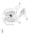
- FIG. 7 is also similar to FIG. 5 (and FIG. 6 ) and illustrates another embodiment of a device and manual handle for operating an end-effector according to this disclosure.
- FIG. 8 schematically illustrates the implanted location of an implantable pulse generator (IPG) in an anterior abdominal left upper quadrant location adjacent the left costochondral margin of a subject following closed-chest, minimally invasive surgical (MIS) resuscitation according to this disclosure.
- IPG implantable pulse generator
- MIS minimally invasive surgical
- the working end-effector 102 can include a plurality of finger-like projections (generically termed “end-effectors” or “blades” herein), denoted by the letter “B” and reference numeral 104 from time to time herein, that are fitted with a first set of sensors 106 (and denoted as “PzS” in FIG. 1 ) for obtaining gross mechanical information, a second set of sensors 108 (denoted as “tt” in FIG.
- (tactile) sensors for acquiring qualitative data or properties of a nearby surface such as from various biological tissue or organs of a subject (e.g., texture, pulsatile motion, elasticity, and the like), and an intermediate structure such as a supportive and insulating webbing material 110 (denoted as “w” in FIGS. 1 and 2 ) that can contain a fluid (e.g., biocompatible gas, air or a liquid medium such as saline) as an operative part of a pneumatically- or hydraulically-driven contractile system optionally coupled to a haptic interface 100 .
- a fluid e.g., biocompatible gas, air or a liquid medium such as saline
- the blades 104 are not necessarily similar and can serve multiple discrete or cooperative mechanical functions including being used as a surgical dissection tool, cardiac compression apparatus, electrocautery units, a platform for diverse EGM sensors, pressure sensors, and/or oxygen sensors or low power cardiac pacing and high power defibrillation electrodes.
- a collection of such blades 104 operate as an end-effector 102 .
- Data is bidirectionally communicated between the end-effector 102 and haptic interface 100 , as an electrical or electromagnetic signal over a standard data channel (denoted as “d” in FIG. 1 ) or via one or more pneumatically- and/or hydraulically-driven communication or control lines or fluid-filled lumens.
- Monitoring of cardiac electrical activity can occur via conventional ECG skin surface electrodes or from conductive material that is part of the blades 104 .
- Sensors 106 , 108 e.g., piezoelectric, force, strain gauges, optical
- acquire mechanical data that is used to construct part of the final haptic representation (touch and/or force feedback) plus optionally, non-force sensor data that can be combined so that the operator perceives all such sensor data at the haptic interface 100 .
- the acquired data is input into the processor 200 that processes (e.g., provides haptic rendering) the input data converting the information to signals output to the haptic interface 100 .
- Haptic interface 100 can comprise an instrumented haptic glove having actuators coupled to respond to sensor data signals, multi-fingered haptic interface(s) for one or more digits of a user, an exo-skeleton type interface providing contact with a users hand, arm, or facial anatomy for example, a non-rigid, deformable virtual heart model that includes auditory, visual, and mechanical cues to a user, or other simulation feedback system or device.
- a virtual heart model or as referred to herein as “haptic heart” can comprise an oblong shaped compressible device that conveys mechanical properties of a subject's heart that is driven by actuators coupled to sensors and sensor signal processing to provide a realistic sensation to a user (e.g., tissue contractility or elasticity, temperature, pulsatile pressure and fluid flow and the like) similar to a typical mammalian heart.
- a realistic sensation to a user e.g., tissue contractility or elasticity, temperature, pulsatile pressure and fluid flow and the like
- Suction and irrigation can be provided to an end-effector 102 or one or more blades 104 (or webbing 110 ) using fluid passageways or conduits, multi-lumen or conventional tubes 112 (denoted as “S” in FIG. 1 ) and the like formed into the respective structures and coupled to suitable vacuum source(s), fluid pumps and optionally auditory representations of suction, irrigation (i.e., fluid flowing) and the magnitude of same.
- a control system 300 integrates sensor and other data acquired by an end-effector 102 or a blade 104 , a haptic interface 100 , a processor 200 , and remote or outside surface physiologic data and auxiliary data derived from auxiliary physiologic support equipment, for example.
- Auxiliary equipment can include but is not limited to echocardiographic imaging technology, radiographic/fluoroscopic equipment, three dimensional localization and/or surgical navigational systems, respiration monitoring, acoustic-ranging-, electromagnetic-, magnetic-, electrical- and/or impedance-based anatomic mapping technologies (e.g., Biosense Webster CARTO and CARTO 3 systems, St. Jude Medical EnSite System or MediGuide Magnetic Technology) collectively depicted in FIG. 1 with reference numeral 400 .
- anatomic mapping technologies e.g., Biosense Webster CARTO and CARTO 3 systems, St. Jude Medical EnSite System or MediGuide Magnetic Technology
- the control system 300 optimizes the virtual coupling between the end-effector(s) 102 and blades 104 and haptic interface 100 and automatically programs the end-effector(s) 102 to function as a cardiac assist device by providing contractile force—synchronized via diverse cardiac sensor signals—to at least a portion of a ventricle of a subject, for example.
- Attenuation or impedance based haptic interfaces can be implemented within the processor 200 and control system 300 depending on the sensed data (e.g., force-based, ECG, EGM, acceleration-based, fluid flow-based, CO-based, or ventricular wall velocity-based information at a given moment in the cardiac cycle such as at the end-diastolic or end-systolic portion of a cardiac cycle).
- FIG. 2 depicts how, in one embodiment, one or more of the blades 104 can be surrounded or linked and supported by a viscous liquid or other fluid contained within gaps or between layers of insulating webbing 110 .
- the liquid or other fluid simply transmits the forces applied via remotely-coupled pumps and/or suction or vacuum sources upon the intra-thoracic end-effectors 102 (e.g., via a compressible, but resilient fluid filled blade and fluid vessel or conduit coupled thereto) and haptic interface 100 hydraulically, via a hydraulic line, h, or pneumatically, via a pneumatic line 118 (also denoted as “p” in FIG.
- a built in time constant or time delay, t can be added to the electrical data via the processor 200 .
- t a built in time constant or time delay, can be added to the electrical data via the processor 200 .
- a user can opt to remove the physiologic time delay between electrical activity and actual mechanical cardiac events and such combined data is appreciated synchronously as if electrical and mechanical cardiac events and data transmission related thereto occurred simultaneously.
- Calculation of the time delay or time constant “t” can be made by processor 200 prior to system operation, adjusted from time to time based on actual sensor signals, or arbitrarily adjusted (e.g., using a predetermined time interval such as 10-150 ms).
- this calibration is accomplished by calculating the time between when an auto-generated impulse from the end-effector 102 is communicated and sensed both mechanically and electrically.
- this calibration is accomplished by calculating the time between when an auto-generated impulse from the end-effector 102 is communicated and sensed both mechanically and electrically.
- electrical or electromagnetic data is utilized within the system “true” or real haptic coupling will occur without a detectable time delay if robust computational techniques are utilized as discussed below.
- the end-effector 102 can consist of one or more finger shaped blades 104 (also denoted as “B” in FIG. 1 ) some having variable degrees of sharpness (blunt to razor-sharp).
- the blades 104 can be flat, deformable, having lateral dimensions of a few millimeters, and able to be in an overlap (interlocking or “fanned”) positioned and adapted to be inserted through a small MIS incision (e.g., 0.25-2.0 cm). If more than one blade 104 is used, the blades 104 are initially overlapped for device storage, during insertion or deployment and at time of withdrawal.
- the blades 104 can be opened to varying angles or spread apart depending on operator preference and in one embodiment, deliver pacing and/or defibrillation therapy via the blades 104 or from electrodes temporarily or permanently delivered within the thorax or upon and within cardiac structures (e.g., coronary sinus, left ventricular myocardial surface, etc).
- Haptic feedback provides the operator with a means to tactually appreciate that anatomic properties of contacted tissue and even fluid/blood flow in the extracardiac space (e.g., pericardial fluid) and intracardiac (coronary sinus blood flow) locations.
- a central pivot or rotary joint 120 (denoted as “C” in FIGS. 1 and 2 ) is present about which the blades 104 open and close.
- the central pivot 120 has a fixed component which in one embodiment is a central ring held stationary by a torque rod (not shown) attached to a fixed portion of the proximal, hand held section, handle, or arm 122 (also denoted as “A” in FIG. 2 ) of the support system.
- Other types of pivot joints can be used to provide rotation, of course.
- the rotating component has a central connecting core which is robotically controlled and responds to the operator's commands through a control line (such robotic control is schematically illustrated in FIG. 1 as functional block 500 ).
- the external controller can be under manual control of the operator or run autonomously by a computer/processor 200 and control system 300 that drives the system as described in more detail below.
- the angles between one or more of the blades 104 are controlled by the operator spreading his or her fingers apart to the desired amount (e.g., upon a haptic interface 100 such as within an instrumented, haptic glove or via a multi-fingered haptic interface or the like).
- Each blade 104 can have a different structure and can be adapted to perform different functions.
- One or more of the blades 104 is fitted with one or more flexible hinge joints ( 124 /H 1 and 126 /H 2 in FIG.
- a permanently implantable device e.g., a medical electrical lead including pacing electrodes
- an intrathoracic, extra-cardiac or intra-cardiac location e.g. pacing/sensing electrode configuration as opposed to myocardium, subcutaneous defibrillator electrodes.
- Kinesthetic and proprioceptive feedback capabilities are provided with position sensors 106 ′ located at each hinge joint 124 , 126 and within the haptic interface 100 (e.g., instrumented haptic surface or glove).
- the design of the fingers of the blade members 104 can be simplified and contain simple hinge joints for flexion and extension or be more complex simulating a fully dexterous robotic hand and arm construct. Microfabrication techniques can be used and applied as to sufficiently miniaturize the needed components for MIS applications and procedures.
- hinge joints 124 , 126 (also denoted as H 1 and H 2 ), present on the two most peripherally located fingers or blades 104 of an end-effector 102 .
- the two internally located blade fingers can be used for dissection 128 (denoted as “Bd” in FIG. 3 ), and electrocautery 130 (denoted as “Be” in FIG. 3 ).
- Torque-angle data of each hinge joint 124 , 126 is determined by a kinesthetic sensor such as a force/torque transducer at the hinges.
- a similar transducer is present at the haptic interface within a haptic glove or alternate multi-fingered haptic interface.
- proximal (haptic interface) and distal (end-effector) torque angle data are transmitted to the controller and adjustments made as to optimize coupling of actual resistance and the user's perceived stiffness to flexion and extension.
- Other means of optimizing coupling of the action and perceived mechanics between the end-effector 102 and operator's haptic interface 100 are within the scope of the instant disclosure.
- An arm 122 /A, disposed between the end-effector 102 or blades 104 thereof and haptic interface 100 enables the operator to generate torque on end-effector 102 of the system and palpate torque from cardiac twisting motion ( FIG. 2 ).
- the torque force is sensed by a sensor at the connection between the central pivot joint, C, as seen in FIGS. 1 and 2 , and torque arm A ( FIG. 2 ), and is presented to the operator at the haptic interface 100 and generated manually by the operator's action on the torque arm A upon the end-effector 102 and vice versa.
- the blades 104 and attached sensors 106 are connected via torque arm 122 A and data channel to the processor 200 and control system 300 that is situated outside the body of a subject.
- the end-effector 102 and arm 122 /data channel structure “d” are able to be sterilized and used for repeated applications.
- the processor 200 and control system function along with a haptic glove or other haptic interface 100 which can be in form of a compressible, “real feel” model of the heart (the haptic heart) for system operation.
- the inserted blades interact and communicate bidirectionally via data channel D (e.g., analog or digital electric or electromagnetic signals) with the processor 200 , control system 300 and haptic interface 100 , and in one embodiment, via one or more of pneumatic, hydraulic lines or pipes.
- Data channel D e.g., analog or digital electric or electromagnetic signals
- ECG monitoring is provided to the control system either from separate electrodes or recordings acquired from the inserted blade(s) via channel ECG.
- Each thin (millimeter scale) malleable blade is preferably constructed of conductive metal alloy (which can be a memory shape alloy such as copper-aluminum-nickel) which are coated with varying degrees of insulation.
- the edges of one or more lower profile blades 104 are able to be exposed for purposes of cutting by being separated from neighboring non-cutting blades 104 .
- Specific blades 104 are used for receiving electrical information and delivering electrical impulses (e.g., sensing, pacing, defibrillation and the like), electrocautery or fixating electrodes.
- the insulation can be composed of any insulator including DACRON material.
- the blades 104 are covered with a sleeve that is made of electrically insulating materials such as silicon or polyurethane ( FIG. 2 peripherally located blades 104 ).
- a sleeve that is made of electrically insulating materials such as silicon or polyurethane ( FIG. 2 peripherally located blades 104 ).
- certain portions of the sleeve are conductive for the sensing and delivery of electrical stimulation for sensing cardiac electrical activity, pacing and defibrillation.
- the sleeve covers only a portion of the blade 104 and the exposed blade itself serves as a conductor/electrode.
- the blades are composed of energy harvesting material or alloy as is described in more detail below.
- the insulation also serves as an elastic web 110 (denoted as “w” in FIG. 1 ) between each blade 104 of an array of several individual blades and enables the blades 104 and insulation or webbing 110 to conform to the natural shape of the heart.
- Material suitable for such an application includes foamed Neoprene or polychloroprene.
- the properties of the blades 104 e.g., memory shape NiTi alloy or the like
- optionally insulating webbing 110 are such that they become more malleable at body temperature and have greater stiffness prior to and during insertion and initial tissue dissection.
- the elasticity of the webbing 110 at body temperature is optimally within the range of normal and pathologic cardiac tissue (e.g., 1.5-4 kPa).
- the webbing 110 is in a folded up position prior to blade separation or expansion.
- microfabrication techniques are used to construct a web structure that is low profile in both the folded and open position.
- the conductive portion on the sleeve or of the blade(s) 104 can have a large surface area or be separated into multiple conductors which are separated by insulation, to define unipolar, bipolar and multipolar electrode configurations.
- Each blade 104 can serve as a pole to complete an electrical circuit and have a common ground or reference potential that is positioned intra-thoracic or externally located such as a transcutaneous grounding pad.
- delivery mechanisms for permanently fixating electrodes are incorporated into the blades 104 as known by those experienced in the art (e.g., screw fixation mechanisms for epicardial leads).
- Alternate methods for placing and fixating electrodes are within the scope of the instant disclosure, such as a fishing hook-shaped mechanism that deploys during separation of the inserted blades 104 (denoted by an arrow in FIG. 6 ), effectively hooking or pinching onto the tissue for fixation purposes (see insert FIG. 6 ).
- the electrodes can be composed of mechanical transducer or sensor type configurations of a discrete material such as piezoelectric material.
- tactual tips 106 serve to acquire subtle tactual information that is transmitted to the operator, in an embodiment, via a haptic glove or multi-fingered haptic interface 100 .
- the tactile tips 106 are electrically isolated from the electrodes or conductive portions of a blade 104 .
- the tactual tips 106 can be composed of any sensor capable of acquiring mechanical information (e.g., piezoelectric sensors, force sensors, optical sensors, deformation-based tissue softness sensor).
- This transmitted data can be conveyed using mechanical means (e.g., pneumatic, hydraulic), optically, and/or via electrically transmitted signals (e.g., current, voltage, impedance, electromagnetic) in analog and/or digital format to the control system 300 and haptic interface 100 .
- mechanical means e.g., pneumatic, hydraulic
- optically e.g., optically
- electrically transmitted signals e.g., current, voltage, impedance, electromagnetic
- the tactual tips 106 provide the necessary anatomic and mechanical data to the operator reproducing gross anatomic features (e.g., vasculature, myocardium, fibrotic tissue) and fine structural details such as tissue texture, roughness, softness, elasticity that relate to normal and pathologic tissue biomechanical characteristics (e.g., due to compromised or infarcted myocardium).
- gross anatomic features e.g., vasculature, myocardium, fibrotic tissue
- fine structural details such as tissue texture, roughness, softness, elasticity that relate to normal and pathologic tissue biomechanical characteristics (e.g., due to compromised or infarcted myocardium).
- Specific sensor designs capable of acquiring the needed data are utilized and suitable haptic rendering techniques are applied to optimize true haptic coupling.
- Piezoactuators, vibrotactile actuators, electromagnetic vibration motors or alternate simulation mechanisms located within the haptic interface provide non-virtual, true coupling between the end-effector 102 and haptic interface 100 .
- true coupling (rather than virtual coupling) between the anatomic structures at the level of the end-effector and augmented reality haptic interface (e.g. haptic heart or glove) to occur
- system processing and haptic rendering within 200 ensures that both time dependant and space dependant components are considered in the final haptic representation.
- Interface modalities that exploit natural human fingertip capabilities are used to create a “haptic surface.”
- piezoelectric actuation techniques enable reproduction of compressible and tensile strains at different areas of the fingertips.
- Surface features such as texture, roughness, friction, smoothness, bumpiness, edge detection and the like can be reproduced at the haptic interface 100 using a variety of devices.
- the system is capable of detection of the direction of shear force that displaces, deforms and stretches the end-effector's sensors (e.g. on the cardiac surface) and communication of such physiologic and mechanical properties to the operator's skin at the haptic interface. This can be accomplished, for example, using tangential skin displacement via miniature shear displays incorporated into the haptic interface (e.g. distal portions of a haptic glove).
- the acquired analog data (e.g., by piezoelectric sensors) can be left in an analog format and be transmitted as data using a specific electronic signature (voltage or current amplitude as a function of time) presented at the haptic interface as an analog signal without haptic rendering and without analog to digital conversion.
- conversion of electrical data as force and velocity information at 100 is accomplished using comparable sensors (e.g., piezoelectric transducers/actuators) at both the level of the end-effector and operator interface.
- Signal amplification can be controlled with an amplifier as known.
- Such analog to analog data transmission provides the purest, most true haptic coupling with full system transparency. Such a mirror image representation of actual events will be useful for the display of more gross anatomic and physical characteristics (e.g., respiration, cardiac contraction) rather than more subtle tissue properties.
- Haptic rendering that require data digitization and complex processing facilitates the operator's ability to appreciate fine detailed information about cardiac structure and function. Processing of large amounts of data in real time requires robust computing techniques. Hyperelastic nonlinear models have been used to achieve realistic soft-tissue simulations. Advances in parallel computing techniques such as the concurrent utilization of fixed point computing units on a field-programmable gate array (FPGA) device along with suitable computer-readable memory and the like have enabled real-time simulation of three dimensional linear elastic deformation models at high frequency update rates. The application of developing techniques such as quantum computing adds virtually imperceptible processing delays due to computation. The implementation of recent advances in computational algorithms will be especially important for reproduction of the tactile properties of a non-rigid deformable object that is in constant motion.
- FPGA field-programmable gate array
- force sensing at the end-effector can be accomplished by using tri-axial fiber optic sensors (U.S. patent application publication number 20080009759 the contents of which are hereby incorporated by reference herein).
- Electromagnetic signal coherence is affected by applied force and can be used to quantify three-dimensional force upon sensors located within the thorax and opposed to cardiac tissue.
- data transmission in this application can occur electromagnetically with fiber optic signals, for example to decrease possible interference between certain ionizing radiation visualization systems or magnetic resonance imaging systems and derivatives thereof.
- both active and passive exploration aspects of data acquisition are incorporated within the haptic display, optimizing the final user's tactile sensitivity.
- the motion of the cardiac structures and anatomic landmarks affect the acquired sensor data and haptic representation (passive) and the active exploration of the user's fingers or hand and the like within the haptic interface (e.g., an instrumented haptic glove) directs the motion of the inserted sensors thus having a combined affect (passive and active) on the information coded within the processor 200 .
- Input from the haptic glove(s) into the processor 200 which directs the action of the inserted end-effector 102 (finger-like blades 104 ) is provided with a variety of mechanisms not limited to inertial motion sensors, accelerometers, gyroscopes, piezoactuators, magnetometers or other means as understood by those experienced in the art.
- the operator's motion is reproduced at the level of the end-effector BS and changes in finger separation joint position etc. within the haptic interface (e.g., an instrumented glove or the like for one or more digits of an operator) result in a similar action (e.g., pitch, yaw, roll) accurately controlling the spatial configurations and the respective angles and relative positions of the blades 104 .
- a similar action e.g., pitch, yaw, roll
- three-dimensional spatial localization in real time is determined at least in part with external diagnostic equipment that identifies the location and position of various aspects of the end-effector and operator's hand(s).
- a three dimensional navigational system that uses impedance information between surface electrodes and the tactual tips and hinge joints is used to provide position information (e.g., electroanatomic mapping and/or localization). This is input into the auxiliary port depicted in FIG. 1 .
- position information e.g., electroanatomic mapping and/or localization
- FIG. 1 auxiliary port depicted in FIG. 1 .
- Other types of navigation systems as known by those experienced in the art can be utilized (e.g., magnetic, radiographic).
- Suction and irrigation can be provided via one or more functional small canulae, S, that operatively couple to one or more of the inserted blade members 104 .
- MEMS technology and nanotechnology based fabrication techniques can be utilized to develop a sophisticated cardiac support device that is microfabricated including manufacturing of webbing materials, conductors, blades, suction cannula and the like.
- ultrasonic and/or thoracoscopic imaging technology e.g., charge-coupled device, ultrasound, fiber optic imaging etc.
- Introduction of the blades and associated hardware can be done in various locations including sub-xyphoid, parasternal, intercostal.
- More rigid blades as well as hollowed sheaths can be implemented to access specific sites where dense connective tissue (e.g. via an intercostal approach) may impede delivery of the necessary hardware and for connecting various components of either temporarily or permanently implanted systems. Leads or wiring can be left in place, tunneled and connected to permanently implanted devices used for pacing and/or defibrillation and monitoring. Haptic feedback assists in the positioning of such hardware.
- a haptic heart and instrumented haptic glove, or haptic interface can function as a device for cardiac mechanical and electrical resuscitation and for continuous cardiovascular support (including support for select intrathoracic organs) in a patient suffering from multiple complications (e.g., following open heart surgery).
- the system is in no way limited to functioning in the described manner and can be utilized in any number of ways.
- a cardiac rhythm management device e.g., single-, double-, or triple-chamber pacemaker, defibrillator, LVAD
- the patient has a weakened heart muscle or cardiomyopathy and develops what is termed a low CO state resulting in poor organ perfusion, low blood pressure, reduced or low heart rate and the like. Since the blood pressure is low and the patient is essentially entering into cardiogenic shock, the patient is generally not responsive to intravenous medications. Such an occurrence is unfortunately relatively common, especially after open heart surgeries.
- a cardiac ultrasound is oftentimes performed and readily identifies blood accumulating within the pericardial sac, a pericardial effusion, with impending cardiac tamponade that results when the blood compresses the heart until it ceases to function.
- the patient is brought to an operating room and is accordingly prepared per robust sterilization procedures and draped in the usual fashion.
- the recent median thoracotomy incision is depicted in FIG. 4 .
- An initial incision of about one centimeter (1 cm) is made in the sub-xyphoid location and dissection is carried out to the subcutaneous tissues, though other techniques such as parasternal or limited thoracotomy approaches can be used as depicted in FIG. 4 . In an emergency situation this can be performed at the patient's bedside or other convenient and accessible location. If the clinical picture is the result of traumatic injury the system is portable and can also be used in the field as described below.
- the blades 104 which are in the initial overlap position 136 (also denoted as “Bo” in FIG.
- a stiff medical introducer typically having an outer portion and an inner portion such as a trocar, which is a sharp-pointed surgical instrument used with a cannula to puncture a body cavity for access and optionally fluid aspiration 136 (denoted as “Tr” in FIG. 5 ) as illustrated in FIG. 5 and the control mechanism such as a handle 140 (e.g., an instrumented haptic glove or other haptic interface 100 ) is connected through trocar 136 to engage the blade mechanism and provide haptic feedback and user control.
- a handle 140 e.g., an instrumented haptic glove or other haptic interface 100
- the trocar 136 is removed once the blade 104 and it associated structures and collapsible and expandable mechanism is in position and control over the end-effector 102 (including the blades 104 , etc.) is maintained using the haptic handle interface 140 (denoted as “Hi” in FIG. 5 ), which can be an instrumented haptic glove, a haptic heart or a haptic handle or other haptic interface 100 . Fluoroscopic or ultrasonic guidance can be used to assist but is not needed as the procedural process is palpable.
- the sensed mechanical data from the tactual tip sensors 106 (or “tt” in FIG. 1 ) peripherally located on the top inserted blade(s) 104 is palpated by the operator holding the haptic interface 140 .
- the tactile feedback is ideally provided at the distal portion of each haptic finger, i.e., in the same location (e.g. finger tips), providing the operator with a “real feel” extracardiac experience (true coupling).
- the sensors 106 are composed of piezoelectric sensors.
- the analog data is digitized via an A/D convertor in processor 200 .
- Various types of haptic rendering as known by those experienced in the art can be implemented in processor 200 .
- Other sensor designs including those designed with nanotechnology (i.e., using nanosensors, nanomaterials, nano-scale components, and the like) can be used as well.
- Tactile feedback is generated at the haptic interface 100 as shown in FIG. 5 , and provides the operator with tactile sensations which in more advanced embodiments (e.g., a haptic heart model having a compendium of instrumentation) provide sensations of compression, vibration, multi-dimensional force application, temperature variations, reproducing feelings of friction, stretch, slip, displacement, softness, deformation, stress, strain, shear (e.g., using a shear factor) and the like.
- a haptic heart model having a compendium of instrumentation provide sensations of compression, vibration, multi-dimensional force application, temperature variations, reproducing feelings of friction, stretch, slip, displacement, softness, deformation, stress, strain, shear (e.g., using a shear factor) and the like.
- the operator appreciates the variations in resistance present in the subcutaneous tissues and fascial planes much as if the operator is doing blunt dissection with their own finger(s).
- haptic interface 100 can comprise a multi-fingered haptic interface that will provide high precision three dimensional force at the fingertips in a large workspace and optimally will have multiple degrees of freedom (e.g. 15 DOF).
- the workspace i.e. relationship between sensor locations and haptic display volume
- the workspace can be a default value that is constant, or change in volume depending on the nature of sensed signals (e.g. fine vs. coarse anatomic detail), location of the end-effector(s), vary based on operator command or as based on a programmed equation that is a function of a pre-defined set of variables, indices, parameters.
- the operator separates the digits or one or more fingers at the level of the haptic interface 100 and exposes one or more of the underlying blades 104 that assist in dissection ( FIG. 6 ).
- suction can be initiated through a suction cannula 134 (also denoted as “S” in FIG. 6 ), that fluidly couples to the inserted instrument to a depository for fluid collection and analysis (not depicted) if needed.
- This suction cannula 134 can be used to aspirate the blood and other body fluid from within the pericardial space and can also be used for irrigation purposes.
- the suction due to applied vacuum or the fluid pumped for irrigation purposes can be provided continuously or intermittently, as desired.
- gentle probing will enable the blade(s) 104 to enter the pericardial space and the operator will oftentimes feel a “pop” or single vibrating sensation.
- This can be palpable at the haptic interface 100 using haptic rendering techniques.
- Haptic rendering may be utilized to simulate other static or dynamic tissue properties including but not limited to tissue texture, softness, elasticity, hardness, pulsations, vibrations and the like.
- Tactual data can be transmitted using analog (e.g., piezoelectric sensors) or digital electrical data (denoted as “d” in FIG. 1 ) and input into the processor 200 , or in one embodiment, by air or liquid that is contained about one or more blades 104 through a pneumatic or hydraulic system.
- the pneumatic, (denoted as “p” in FIG. 2 ), or hydraulic, (denoted as “h” in FIG. 2 ), mechanisms function simply by transmitting pressure that is exerted upon air or liquid material of specific viscosity surrounding one or more of the compressible blades 104 within each blade finger (see e.g., FIG. 2 and FIG.
- HH haptic heart model
- HH a circle drawn in ghost in FIG. 6
- This pressure is perceived at the level of the haptic interface, for example, by movement of the air or liquid about an instrumented haptic glove surrounding and operator's finger or alternatively with a haptic heart model, which is, in one embodiment herein, disposed in a continuum with the air or liquid contained within the hermetically sealed webbing material 110 , which can comprise a volume of insulation located about one or more of the inserted blades 104 .
- the system can implement more than one type of haptic interface 100 .
- the instrumented haptic glove (denoted as “Hg” in FIG. 7 ) or a multi-fingered haptic interface is used to access the intra-thoracic space (with or without a haptic handle interface) and then a haptic heart model is exchanged and connected to the system at connector, Co in FIG. 3 and FIG. 7 , for delivery of closed chest cardiac massage if needed.
- Compressive forces on the end-effector 102 e.g., deformable blades 104 coupled about the heart
- the system are appreciated at the level of the haptic interface (HH in FIG. 7 ) and the forces applied to HH are imparted to the deformable blades for mechanical cardiac support.
- the end-effector 102 (and portions of its related system components) is inserted and in the appropriate location the blade fingers are spread apart.
- the haptic interface e.g., opening, closing, flexing three fingers of a sterile haptic glove
- This technique can also be applied for treatment of chronic constrictive pericarditis or to free up loculated pockets of organized pericardial fluid and thrombus.
- the haptic blades 104 (which can be described as individual but connected finger-like structures) can be left about the heart and acquire additional sensor data that relates to the mechanical function of the heart.
- the operator then disconnects the multi-fingered haptic interface 140 , 100 and exchanges it with the haptic heart which is, as described herein as resilient, non-rigid, deformable and optionally equipped with various transducers or actuators, fluid passageways, pumps or vacuums and the like.
- the operator can manually palpate and appreciate the palpated action of the heart and motion with the haptic heart.
- the haptic heart is oblong and can be held by the operator and capable of generating mechanical sensations simulating cardiac tissue motion. Its mechanical properties such as elasticity, mechanical recoil, twist, contraction, etc. are similar to human cardiac tissue.
- the pneumatic or hydraulic system enables the transmission of mechanical data and tactual feedback to the operator.
- the operator can squeeze the Haptic Heart and generate mechanical support to the heart by displacing air or liquid within the pneumatic or hydraulic system, or by providing a signal, or surrogate, related to the desired impact upon the actual patient's heart via the end-effector 102 . This can also be accomplished electronically using a haptic glove or multi-fingered haptic interface, by way of example.
- the amount of support needed can be determined qualitatively by the operator based on tactile feedback from the pneumatic or hydraulic system and/or from data acquired by the sensors on the inserted blades transmitted to the control system which receives the sensor data for processing and generating real time tactile feedback at the haptic interface 100 .
- the mechanical support can be in one or more forms. Potentially optimal cardiac support can be achieved by having a plurality of the blades 104 (or finger-like structures) with webbing as illustrated in FIG. 1 apply adequate pressure on the heart and flexing about the cardiac surface. As noted previously webbing 110 between the blades 104 provides relatively even distribution of applied force to the myocardial substrate.
- the blades 104 can each have one or more joints or hinge mechanisms to provide relatively customized coupling and flexion and the blades 104 also can be fitted with a delivery system or systems for administering therapeutic agents or devices such as medications (e.g., via perforated or microporous membrane surfaces), as well as permanently implanted sensors and electrodes.
- tactile feedback e.g., proprioceptive, kinaesthetic
- sensor like mechanoreceptors gathers the needed data and reproduces the intra-cardiac “experience” through the haptic interface 100 (e.g., haptic glove and heart) for fine, detailed tactual information and augments the gross mechanical data provided via the hydraulic or pneumatic system reducing the likelihood of mechanical injury (e.g., cardiac contusion).
- a gentle assist to cardiac contraction may be generated or more forceful contractions administered as needed similar to an acutely- or a chronically-implanted LVAD or other cardiac assist system.
- Torsion can be applied using torque arm, A, to simulate the natural twisting and untwisting of the heart under normal circumstances (illustrated to a degree in FIG. 2 ).
- the timing of force application at different segments of the end-effector 102 is determined mechanically and/or electrically as previously noted, with or without compensation for the physiologic time delay inherent between the cardiac mechanical response due to the passing of electrical wavefronts across and through the myocardial substrate.
- Different segments can generate varying amounts of supportive force upon the heart at different times normalizing synchronous cardiac mechanical function. This can be done manually or automatically whereby processor 200 and or control system 300 direct the timing, amplitude and frequency of the action of the end-effector 102 .
- a closed feedback loop comprised of one or more sensors 106 (denoted as “PzS” in FIG.
- control system 300 which provides for monitoring of cardiac function (e.g., CO, systemic perfusion, adequate or reasonably consistent blood pressure and heart rate) and determining the optimal programming of the assist device (e.g., temporal and spatial characteristics).
- cardiac function e.g., CO, systemic perfusion, adequate or reasonably consistent blood pressure and heart rate
- optimal programming of the assist device e.g., temporal and spatial characteristics.
- Any number of inputs e.g. three dimensional electromagnetic mapping system data, navigational system data, hemodynamic information, electrophysiologic data, imaging data, respiration monitor, cardiogenic impedance
- auxiliary information e.g. three dimensional electromagnetic mapping system data, navigational system data, hemodynamic information, electrophysiologic data, imaging data, respiration monitor, cardiogenic impedance
- a dysynchronous myopathic heart muscle will not generate adequate CO and different myocardial regions will contract and move in a dysynchronous pattern.
- This is detected electrically and/or mechanically by the end-effector 102 and its associated sensors 106 and corrected by the control system much as currently available pacemakers provide cardiac resynchronization therapy or LVAD provide support to a left ventricle.
- the system design herein provides this as mechanical support in a synchronous fashion so that myocardial segments that would ordinarily contract late in the cardiac cycle are stimulated electrically (e.g., via pacing electrodes on blade(s) 104 ) and/or mechanically via direct action of the blades 104 of the end-effector 102 .
- the laterally located portions of an end-effector of series of blades 104 can be controlled to flex earlier during the systolic ejection phase relative to more medially located blades 104 by some temporal index or preset window (e.g., 80 milliseconds).
- Auxiliary input that is comprised of visual or auditory data will improve the operator's tactile sensing acuity and optimize true coupling through visual and auditory haptic responses.
- the electrode/conductive portions of the blades 104 can essentially provide sensor signals and haptic feedback to an operator to allow the operator to effectively diagnose the pathology underlying the dysrhythmia problem and deliver pacing stimuli as well as defibrillation therapy, if appropriate, along one or more therapy vectors (defined as lying substantially between electrodes of opposing polarity) with varying polarities.
- pacing stimuli as well as defibrillation therapy, if appropriate, along one or more therapy vectors (defined as lying substantially between electrodes of opposing polarity) with varying polarities.
- the system can run autonomously by gathering mechanical and electrical data and appropriately delivering the proper type of therapy as needed until the patient is stabilized.
- Control systems within 300 similar to those present in current pacing/defibrillator systems and cardiac assist devices are programmed or operated to provide the type of palliative or potentially curative therapy and the timing of therapy delivery during the cardiac cycle.
- Permanently implanted electrodes can be positioned about the heart as illustrated in FIG. 8 with the pulse generator implanted and connected to the electrode and lead system.
- the system can also be configured to deliver therapy such as ablation of tissue, and provide various pharmacological and therapeutic agents such as anti-arrhythmic substances and stem cells or other biologic materials via the fluid pump(s), sources of reduced pressure, hydraulic or pneumatic systems.
- therapy such as ablation of tissue
- various pharmacological and therapeutic agents such as anti-arrhythmic substances and stem cells or other biologic materials
- the fluid pump(s) sources of reduced pressure, hydraulic or pneumatic systems.
- the system can be teleoperated as to reduce the waiting time for and provide ready access to a team of experienced surgeons to treat critically ill patients at remote locations.
- the following should be considered:
- the blades 104 can be composed in full or part of an energy harvesting material such as a Zinc Oxide composite or other material (e.g., piezoelectric or material incorporating conductive nanotubes, and the like) that can harvest the energy from diaphragmatic, skeletal and cardiac muscle motion of a patient and supply it to an attached, portable battery power pack.
- an energy harvesting material such as a Zinc Oxide composite or other material (e.g., piezoelectric or material incorporating conductive nanotubes, and the like) that can harvest the energy from diaphragmatic, skeletal and cardiac muscle motion of a patient and supply it to an attached, portable battery power pack.
- Capacitive elements coupled to the power pack ensure that a charge capable of defibrillation is always available and the stored and recharging energy supply can be used to have the system function as a portable assist unit that can be implanted in the field.
- the webbing material 110 and suction capabilities of the system can thus serve to seal and heal various wounds on or about the anatomy, including individual organs of the patient (e.g. re-expansion of a pneumothorax). Any perforations (e.g., due to bullet and/or shrapnel injuries) can be treated while an end-effector 102 is deployed.
- the blades 104 and webbing 110 can contain bleeding and the hydraulic/pneumatic system (and contractile system for the end-effector(s)) can apply the appropriate amount of pressure during cardiac massage/compressions to mitigate bleeding or irrigate and cleanse contaminated tissue.
- Blood volumes can be measured via the suction apparatus using known components and relationships to fluid flow, for example.
- Low energy or intermittent suction can be used to also create a vacuum that opposes and attracts the end-effector 102 to tissue layers and simultaneously compresses any bleeding structures or re-expand a collapsed lung.
- the hydraulic/pneumatic system can also be used to deliver thrombotic agents to specific locations (although related lumens or tubing may require valves or switches to control reversal of fluid flow) or even recirculate any blood loss or other bodily fluid evacuated from damaged tissues via intravenous or intracardiac routes.
- Another application of the teaching herein in an acute setting relates to use of electrodes disposed on one or more of the blades 104 of an end-effector 102 to electrically and/or mechanically cause diaphragm stimulation for patients with respiratory arrest.
- Such stimulation can be readily performed according to the foregoing MIS techniques as a sub-xyphoid entry typically occurs at the level of, or relatively near, the diaphragm muscle.
- a commercial entity known by the tradename “DuraAct” and also identified as “Physik Instrumente” of Germany provides piezoelectric material with energy harvesting properties that can harvest energy from moving structures such as the beating heart, diaphragmatic and skeletal muscles. It can be used to recharge and maintain a re-chargeable battery out in the field as a self-sustaining life-sustaining apparatus, among other possible advantages.
- Such commercially-available material(s) can readily be incorporated into portions of the webbing material 110 and/or the blades 104 and be machined to have a cutting edge, if desired.
- Such energy harvesting properties can be implemented herein in a reciprocal configuration wherein the end-effector(s) include portions covered or including energy harvesting materials and the operator interface also includes portions covered or including same.
- the controller such as via a handle-type interface or via an instrumented haptic heart including materials such as piezo-actuator (e.g., DuraAct-containing coating adapted for a deformable soft tissue model)
- a voltage and electrical current signal is generated that triggers or activates the end-effector (e.g., DuraAct Blade composite) to massage and/or compress about the heart.
- the signal can be an amplified purely-analog current signal directly from the energy generated by the hand-held piezo-actuator or can trigger/activate via an analog-to-digital converter an electromechanical system that drives the inserted blades 104 of the end effector 102 .
- both ends of the system have similar piezo actuators and are reciprocating.
- the deformation of the inserted DuraAct-coated or -containing blades 104 likewise can activate the haptic interface 100 (e.g., the haptic heart model, an instrumented haptic glove, or haptic handle and the like) as to generate the haptic effect(s) to the operator and thus convey that adequate contractility is present or if a gentle assist is needed or if a PEA condition is present and aggressive mechanical support is required.
- Squeezing the haptic heart/handle can also be used to harvest energy in the field if cardiac compressions or respiratory support is not needed.
- the foregoing is not limited to the named DuraAct material as any piezoelectric material possessing energy harvesting properties and capable of serving as an actuator will store potential energy and deliver kinetic energy. When a current is delivered to the material it will contract.
- the blades 104 or other similar expandable and collapsible end-effector 102 can be used to harvest and store energy when there is normal cardiac contractility and/or diaphragm muscle contraction and then serve as an actuator deforming the blades 104 to contract and support myocardium and/or diaphragm muscle in time of need.
- the webbing 110 would thus act as a confining structure as to control the contraction of the webbing material 100 and the blades 104 (or equivalent structure of end-effector 102 ).
- the exterior portion of the webbing 110 (between the elongate ribs or blades 104 ) act to restrict motion and the undersurface (contacting the myocardium or diaphragm muscle of the pulmonary system) should be elastic so the force is applied radially inward.
- the properties of the blades 104 and webbing 110 can vary in direction or strength of contraction or between the expanded and partially expanded states according to need (e.g., configured to deliver compressive forces toward the pulmonary parenchyma) for respiratory support.
- a minimally invasive surgical (MIS) instrument configured to provide mechanical and electrical support and acquire sensor-based physiologic signals from an intrathoracic organ of a patient while providing haptic feedback to an operator, comprising:
- an end-effector system adapted for transthoracic insertion into the patient and having a collapsed configuration for deployment and retraction and an expanded working configuration, wherein the working configuration provides a receiving location and in a partially-collapsed deployment configuration compresses a portion of the intrathoracic organ disposed in the receiving location;
- each transducer providing at least one output signal therefrom;
- a haptic interface operably coupled to the plurality of transducers, said haptic interface including at least one processor configured to transform the respective output signals from the plurality of transducers into a haptic response that provides an operator of the MIS instrument with feedback that the operator utilizes to position the receiving location of the end-effector and monitor or adjust control of the end-effector.
- an MIS instrument according to claim 1 , wherein the intrathoracic organ comprises one of a mammalian heart, lung and an innervated portion of diaphragm tissue.
- An MIS instrument according to example 1E further comprising a control circuit configured to increase and decrease a volume of fluid disposed within one of the opposing sheets of the material and the web of material via the port.
- An MIS instrument according to example 1F wherein the control circuit senses cardiac electrical activity and, in response, the control circuit one of: periodically increases and decreases the volume of fluid disposed within the opposing sheets and increases and decreases the pressure of the fluid disposed within the opposing sheets based at least in part upon the sensed cardiac electrical activity.
- An MIS instrument according to example 1 G further comprising a physiologic delay circuit operable to one of temporally synchronize and temporally adjust an electrical cardiac signal with a mechanical cardiac signal. 2.
- MIS minimally invasive surgical
- an MIS-deployable end-effector having a fully-expandable state and a partially-expandable when deployed to a target of cardiovascular tissue and a compact, collapsible state when not deployed to the target of cardiovascular tissue, and adapted to at least partially encircle a portion of myocardium of a mammalian subject when disposed in both the fully-expandable state and the partially-expandable state;
- a plurality of sensors coupled to the end-effector and the portion of myocardium each adapted to provide at least one output signals therefrom;
- a processor coupled to the output signals and configured to transform the output signals and to convey the transformed signals to at least one of: a standard output interface, a haptic output interface, a haptic user interface, and an operator,
- transformed signals provide feedback for one of: initially positioning the end-effector and advantageously repositioning the end-effector, for optimal functioning of the end-effector.
- An MIS instrument according to example 2 wherein the haptic user interface comprises one of an elongate instrumented haptic handle and an instrumented haptic glove.
- An MIS instrument according to example 2A wherein the haptic handle comprises at least one linear motor and at least one rotary motor, wherein the at least one linear motor is disposed at a center of gravity of said handle.
- An MIS instrument according to example 2 further comprising a fluid conduit coupled to the end-effector, wherein said fluid conduit comprises one of: a lumen closed at a distal end thereof and a lumen having at least one perforation formed in a distal portion thereof.
- the end-effector comprises a set of distinct spaced-apart flanges and a web portion couples adjacent flanges and said web portion comprises a series of pleated portions that collapse together into the compact, collapsible state.
- An MIS instrument according to example 2D, wherein the web portion encapsulates a portion of at least one of the spaced-apart flanges and wherein said spaced-apart flanges comprise at least one of an electrically conductive material and an energy-harvesting material.
- An MIS instrument according to claim 10 , wherein at least two of the plurality of sensors comprise: a plurality of mechanical sensors, a body-fluid parameter sensor, at least two electrodes, an accelerometer, a blood oxygen sensor, a pressure sensor, a flow sensor, and a strain gauge. 2G.
- An MIS instrument according to example 2F wherein the output signals include at least one cardiac mechanical signal and at least one cardiac electrical signal and said processor introduces a physiologic time delay to temporally synchronize the at least one electrical signal and the at least one mechanical signal.
- An MIS instrument according to example 2G further comprising:
- the processor couples to the actuator and provides a temporally synchronized output signal to a haptic interface
- the haptic interface comprises one of: an instrumented haptic handle mechanism, an instrumented haptic glove, and a haptic heart and said haptic interface is coupled to the processor and adapted to receive the temporally synchronized output signal.
- An MIS instrument according to example 2H further comprising a handle mechanism coupled to the processor adapted to receive the temporally synchronized output signal.
- An MIS instrument according to example 2I further comprising a chronically implanted pulse generator (IPG) coupled to the MIS instrument so that the operation of the MIS instrument and the operation of the IPG are coordinated.
- IPG chronically implanted pulse generator
- a minimally invasive cardiorespiratory support (MICS) system comprising:
- an end-effector including at least a pair of spaced apart elongate members and adapted to provide mechanical and electrical support to one of: a mammalian heart to promote perfusion and an intrathoracic organ; and
- a plurality of physiologic sensors coupled to the end-effector and configured to acquire sensor-based physiologic signals from the mammalian heart or the intrathoracic organ while providing haptic feedback to an operator of the MICS system when the MICS system is surgically positioned and optionally advantageously repositioned while in communication with the mammalian heart or the intrathoracic organ via at least one minimally invasive surgical incision,
- the MICS system includes a webbing material disposed between an adjacent pair of the at least two spaced apart elongate members.
- An MICS system according to example 3, further comprising means operatively coupled to the MICS system to perform at least one of the following functions:
- aspirate fluid via a fluid passageway coupled to the end-effector portion of the MICS system to a proximal end of the MICS system;
- An acute cardio-respiratory support apparatus or acute cardio-respiratory support field-kit including a minimally invasive surgical (MIS) instrument and system providing haptic feedback for an operator and adapted for one of: performing an acute cardiac resuscitation procedure and performing an intrathoracic organ-supporting procedure for a subject, said apparatus or field-kit comprising:
- MIS minimally invasive surgical
- an MIS-deployable end-effector having an fully- and a partially-expanded state and a compact, collapsible deployment state adapted to at least partially encircle a portion of an intrathoracic organ of a subject when disposed in the fully-expanded state;
- a plurality of sensors coupled to the end-effector and adapted to provide output signals therefrom;
- a processor coupled to the source of electrical potential and configured to receive the output signals and adapted to transform the signals and to convey the transformed signals to at least one of: a standard output interface, a haptic output interface, a haptic user interface, a wireless interface, and an operator.
- An apparatus or field-kit according to example 4 wherein the source of electrical potential includes at least one of: a rechargeable battery cell, a non-rechargeable battery cell, a hard-wired grid-source of remote electrical potential, and a fuel cell.
- the source of electrical potential includes at least one of: a rechargeable battery cell, a non-rechargeable battery cell, a hard-wired grid-source of remote electrical potential, and a fuel cell.
- the rechargeable battery couples to the energy-harvesting material.
- An apparatus or field-kit according to example 4D further comprising:
- At least one capacitive element coupled to one of: the rechargeable battery and at least one high-rate battery;
- At least one high-power density electrode coupled to the at least one capacitive element and adapted to electrically communicate with myocardial tissue when the at least one capacitive element therapeutically discharges.
- At least one fluid passageway fluidly coupled at a distal end to a perforated vessel formed within the end-effector via at least one fluid port and fluidly coupled at a proximal end to at least one of a fluid pump and a source of reduced pressure adapted to feed fluid to the end-effector and to one of: withdraw material from the end-effector and vacuum-adhere the end-effector to adjacent tissue, respectively; and
- valve means adapted for selecting an active channel for the fluid pump and the source of reduced pressure.
- An apparatus or field-kit according to example 4F wherein the fluid pump couples to a reservoir and said reservoir is adapted to store at least one of: a pharmacological agent, a therapeutic agent, an anti-arrhythmic substance, a volume of stem cell tissue, a volume of biocompatible saline, recirculated blood or other body fluid and an anti-thrombotic agent.
- a pharmacological agent a therapeutic agent
- an anti-arrhythmic substance a volume of stem cell tissue
- a volume of biocompatible saline recirculated blood or other body fluid and an anti-thrombotic agent.
- 4H An apparatus or field-kit according to example 4F, wherein the at least one fluid passageway comprises at least one fluid passageway for each of the fluid pump and the source of reduced pressure.
- An apparatus or field-kit according to example 4F further comprising a fluid measurement instrument coupled to an output of the source of reduced pressure wherein said instrument provides an output signal indicative of at least one of: a fluid volume passing therethrough and a fluid pressure parameter.
- the energy-harvesting material comprises one of: a zinc oxide material, a conductive nano-scale material, piezo-electrical material, and a magneto-resistive material.
- 4K An apparatus or field-kit according to example 4, further comprising a portion of elastic webbing disposed between at least a pair of elongate elements of said end-effector so that the organ is mechanically supported by the portion of elastic webbing in both the fully- and partially-expanded state.
- An apparatus or field-kit according to any of examples 4 to 4M further comprising a sterile packaging surrounding all components of said apparatus or field-kit and including at least one indicia of one of: a tampering event, a use-by date, an instructions-for-use legend, and a package-shock event exceeding a preset threshold.
- An apparatus or field-kit according to example 4 further comprising at least a pair of electrodes disposed on one of: a portion of the end-effector and an extension of the end-effector, wherein at least one of said electrodes is configured to contact a portion of diaphragm tissue.
- MIS minimally-invasive surgical
- a user interface conveying the signals and data to one of: a user interface, a haptic user interface, a controller for the end-effector, a robotic controller coupled to the end-effector.
Abstract
Description
1H. An MIS instrument according to example 1 G, further comprising a physiologic delay circuit operable to one of temporally synchronize and temporally adjust an electrical cardiac signal with a mechanical cardiac signal.
2. A minimally invasive surgical (MIS) instrument and system providing haptic feedback for an operator and adapted for acute resuscitation of a mammalian subject, comprising:
2D. An MIS instrument according to example 2, wherein the end-effector comprises a set of distinct spaced-apart flanges and a web portion couples adjacent flanges and said web portion comprises a series of pleated portions that collapse together into the compact, collapsible state.
2E. An MIS instrument according to example 2D, wherein the web portion encapsulates a portion of at least one of the spaced-apart flanges and wherein said spaced-apart flanges comprise at least one of an electrically conductive material and an energy-harvesting material.
2F. An MIS instrument according to claim 10, wherein at least two of the plurality of sensors comprise: a plurality of mechanical sensors, a body-fluid parameter sensor, at least two electrodes, an accelerometer, a blood oxygen sensor, a pressure sensor, a flow sensor, and a strain gauge.
2G. An MIS instrument according to example 2F, wherein the output signals include at least one cardiac mechanical signal and at least one cardiac electrical signal and said processor introduces a physiologic time delay to temporally synchronize the at least one electrical signal and the at least one mechanical signal.
2H. An MIS instrument according to example 2G, further comprising:
4C. An apparatus or field-kit according to example 4, wherein the rechargeable battery couples to the energy-harvesting material.
4D. An apparatus or field-kit according to example 4D, further comprising:
4H. An apparatus or field-kit according to example 4F, wherein the at least one fluid passageway comprises at least one fluid passageway for each of the fluid pump and the source of reduced pressure.
4I. An apparatus or field-kit according to example 4F, further comprising a fluid measurement instrument coupled to an output of the source of reduced pressure wherein said instrument provides an output signal indicative of at least one of: a fluid volume passing therethrough and a fluid pressure parameter.
4J. An apparatus or field-kit according to example 4, wherein the energy-harvesting material comprises one of: a zinc oxide material, a conductive nano-scale material, piezo-electrical material, and a magneto-resistive material.
4K. An apparatus or field-kit according to example 4, further comprising a portion of elastic webbing disposed between at least a pair of elongate elements of said end-effector so that the organ is mechanically supported by the portion of elastic webbing in both the fully- and partially-expanded state.
4L. An apparatus or field-kit according to any of examples 4 to 4K, further comprising a non-transient computer readable medium coupled to the processor that includes at least two predetermined procedural instructions for use for at least two medical protocols that include use of the end-effector.
4M. An apparatus or field-kit according to example 4L, wherein one of the processor and the non-transient computer-readable medium includes at least one non-volatile memory storage location adapted to store information relating to at least one of: a usage continuum of the apparatus or field-kit over a temporal period and a usage relative to a specific patient.
4N. An apparatus or field-kit according to any of examples 4 to 4M, further comprising a sterile packaging surrounding all components of said apparatus or field-kit and including at least one indicia of one of: a tampering event, a use-by date, an instructions-for-use legend, and a package-shock event exceeding a preset threshold.
4O. An apparatus or field-kit according to example 4, further comprising at least a pair of electrodes disposed on one of: a portion of the end-effector and an extension of the end-effector, wherein at least one of said electrodes is configured to contact a portion of diaphragm tissue.
4P. An apparatus or field-kit according to example 4, wherein the end-effector is configured as at least three spaced-apart elongate fingers and wherein a webbing material at least partially couples adjacent elongate fingers together.
4Q. A method utilizing an apparatus or field-kit according to example 4, comprising:
Claims (8)
Priority Applications (1)
| Application Number | Priority Date | Filing Date | Title |
|---|---|---|---|
| US13/447,064 US8942828B1 (en) | 2011-04-13 | 2012-04-13 | Minimally invasive cardiovascular support system with true haptic coupling |
Applications Claiming Priority (2)
| Application Number | Priority Date | Filing Date | Title |
|---|---|---|---|
| US201161517073P | 2011-04-13 | 2011-04-13 | |
| US13/447,064 US8942828B1 (en) | 2011-04-13 | 2012-04-13 | Minimally invasive cardiovascular support system with true haptic coupling |
Publications (1)
| Publication Number | Publication Date |
|---|---|
| US8942828B1 true US8942828B1 (en) | 2015-01-27 |
Family
ID=52350781
Family Applications (1)
| Application Number | Title | Priority Date | Filing Date |
|---|---|---|---|
| US13/447,064 Active 2032-12-22 US8942828B1 (en) | 2011-04-13 | 2012-04-13 | Minimally invasive cardiovascular support system with true haptic coupling |
Country Status (1)
| Country | Link |
|---|---|
| US (1) | US8942828B1 (en) |
Cited By (26)
| Publication number | Priority date | Publication date | Assignee | Title |
|---|---|---|---|---|
| US20110178508A1 (en) * | 2010-01-15 | 2011-07-21 | Ullrich Christopher J | Systems and Methods for Minimally Invasive Surgical Tools with Haptic Feedback |
| US20130226168A1 (en) * | 2012-02-27 | 2013-08-29 | Covidien Lp | Glove with sensory elements incorporated therein for controlling at least one surgical instrument |
| US20160235569A1 (en) * | 2013-09-29 | 2016-08-18 | Institut Hospitalo-Universitaire De Chirurgie Mini-Invasive Guidee Par L'image | Implantable device to treat obesity |
| WO2016152255A1 (en) * | 2015-03-25 | 2016-09-29 | ソニー株式会社 | Medical support arm device |
| US9510905B2 (en) | 2014-11-19 | 2016-12-06 | Advanced Cardiac Therapeutics, Inc. | Systems and methods for high-resolution mapping of tissue |
| US9517103B2 (en) | 2014-11-19 | 2016-12-13 | Advanced Cardiac Therapeutics, Inc. | Medical instruments with multiple temperature sensors |
| US9636164B2 (en) | 2015-03-25 | 2017-05-02 | Advanced Cardiac Therapeutics, Inc. | Contact sensing systems and methods |
| WO2017132025A1 (en) * | 2016-01-27 | 2017-08-03 | Ebay Inc. | Simulating touch in a virtual environment |
| WO2017169097A1 (en) * | 2016-03-30 | 2017-10-05 | ソニー株式会社 | Exclusion implement and exclusion device |
| US9993178B2 (en) | 2016-03-15 | 2018-06-12 | Epix Therapeutics, Inc. | Methods of determining catheter orientation |
| US10166062B2 (en) | 2014-11-19 | 2019-01-01 | Epix Therapeutics, Inc. | High-resolution mapping of tissue with pacing |
| US10285765B2 (en) | 2014-05-05 | 2019-05-14 | Vicarious Surgical Inc. | Virtual reality surgical device |
| US20190272037A1 (en) * | 2013-12-29 | 2019-09-05 | Immersion Corporation | Distributed Control Architecture for Haptic Devices |
| US10525600B2 (en) * | 2015-06-03 | 2020-01-07 | L3Harris Technologies, Inc. | Robotic system with haptic cutting tool |
| US10610318B2 (en) | 2016-07-25 | 2020-04-07 | General Electric Company | Augmented reality catheter interface |
| US10722631B2 (en) | 2018-02-01 | 2020-07-28 | Shifamed Holdings, Llc | Intravascular blood pumps and methods of use and manufacture |
| US10799308B2 (en) | 2017-02-09 | 2020-10-13 | Vicarious Surgical Inc. | Virtual reality surgical tools system |
| US20200337729A1 (en) * | 2019-04-28 | 2020-10-29 | Covidien Lp | Surgical instrument for transcervical evaluation of uterine mobility |
| US10888373B2 (en) | 2017-04-27 | 2021-01-12 | Epix Therapeutics, Inc. | Contact assessment between an ablation catheter and tissue |
| US11185677B2 (en) | 2017-06-07 | 2021-11-30 | Shifamed Holdings, Llc | Intravascular fluid movement devices, systems, and methods of use |
| US11504201B2 (en) | 2018-05-31 | 2022-11-22 | Covidien Lp | Haptic touch feedback surgical device for palpating tissue |
| US11511103B2 (en) | 2017-11-13 | 2022-11-29 | Shifamed Holdings, Llc | Intravascular fluid movement devices, systems, and methods of use |
| US11583342B2 (en) | 2017-09-14 | 2023-02-21 | Vicarious Surgical Inc. | Virtual reality surgical camera system |
| US11654275B2 (en) | 2019-07-22 | 2023-05-23 | Shifamed Holdings, Llc | Intravascular blood pumps with struts and methods of use and manufacture |
| US11690691B2 (en) * | 2017-02-15 | 2023-07-04 | Covidien Lp | System and apparatus for crush prevention for medical robot applications |
| US11964145B2 (en) | 2020-07-13 | 2024-04-23 | Shifamed Holdings, Llc | Intravascular blood pumps and methods of manufacture and use |
Citations (210)
| Publication number | Priority date | Publication date | Assignee | Title |
|---|---|---|---|---|
| US3184842A (en) | 1961-08-03 | 1965-05-25 | Aeroprojects Inc | Method and apparatus for delivering vibratory energy |
| US4019073A (en) | 1975-08-12 | 1977-04-19 | Vladimir Sergeevich Vishnevsky | Piezoelectric motor structures |
| US4210837A (en) | 1979-03-08 | 1980-07-01 | Misikov Vitaly M | Piezoelectrically driven torsional vibration motor |
| US4432372A (en) | 1981-08-28 | 1984-02-21 | Medtronic, Inc. | Two-lead power/signal multiplexed transducer |
| US4844062A (en) | 1987-10-23 | 1989-07-04 | Spectranetics Corporation | Rotating fiberoptic laser catheter assembly with eccentric lumen |
| US5389865A (en) | 1992-12-02 | 1995-02-14 | Cybernet Systems Corporation | Method and system for providing a tactile virtual reality and manipulator defining an interface device therefor |
| US5429604A (en) | 1992-03-18 | 1995-07-04 | Spectranetics Corporation | Fiber optic catheter with twistable tip |
| US5484433A (en) | 1993-12-30 | 1996-01-16 | The Spectranetics Corporation | Tissue ablating device having a deflectable ablation area and method of using same |
| US5549650A (en) | 1994-06-13 | 1996-08-27 | Pacesetter, Inc. | System and method for providing hemodynamically optimal pacing therapy |
| US5576727A (en) | 1993-07-16 | 1996-11-19 | Immersion Human Interface Corporation | Electromechanical human-computer interface with force feedback |
| US5609607A (en) | 1993-09-24 | 1997-03-11 | Deutsche Aerospace Ag | Device for modeling or simulating the sense of touch in a surgical instrument |
| US5628777A (en) | 1993-07-14 | 1997-05-13 | Pacesetter, Inc. | Implantable leads incorporating cardiac wall acceleration sensors and method of fabrication |
| US5693074A (en) | 1993-12-06 | 1997-12-02 | Pacesetter Ab | Cardiac electrotherapy device for cardiac contraction measurement |
| US5702438A (en) * | 1995-06-08 | 1997-12-30 | Avitall; Boaz | Expandable recording and ablation catheter system |
| US5769640A (en) | 1992-12-02 | 1998-06-23 | Cybernet Systems Corporation | Method and system for simulating medical procedures including virtual reality and control method and system for use therein |
| US5771902A (en) | 1995-09-25 | 1998-06-30 | Regents Of The University Of California | Micromachined actuators/sensors for intratubular positioning/steering |
| US5833623A (en) | 1996-05-14 | 1998-11-10 | Pacesetter, Inc. | System and method for facilitating rapid retrieval and evaluation of diagnostic data stored by an implantable medical device |
| US5836946A (en) | 1996-06-12 | 1998-11-17 | The Spectranetics Corporation | Catheter for delivery of electric energy and a process for manufacturing same |
| US5844392A (en) | 1992-12-02 | 1998-12-01 | Cybernet Systems Corporation | Haptic browsing |
| US5843135A (en) | 1997-10-20 | 1998-12-01 | Medtronic, Inc. | Pacing system with lead having a single conductor for connecting to pressure sensor and electrode |
| US5916143A (en) | 1996-04-30 | 1999-06-29 | Apple; Marc G. | Brachytherapy catheter system |
| US5944022A (en) | 1997-04-28 | 1999-08-31 | American Cardiac Ablation Co. Inc. | Catheter positioning system |
| US5959613A (en) | 1995-12-01 | 1999-09-28 | Immersion Corporation | Method and apparatus for shaping force signals for a force feedback device |
| US5971931A (en) | 1994-03-29 | 1999-10-26 | Raff; Gilbert Lewis | Biologic micromonitoring methods and systems |
| EP0970663A1 (en) | 1998-07-10 | 2000-01-12 | Mitsubishi Denki Kabushiki Kaisha | Master/slave system for the manipulation of tubular medical tools |
| US6059759A (en) | 1997-10-14 | 2000-05-09 | Merit Medical Systems, Inc. | Infusion catheter systems with tactile sensing feedback |
| US6070100A (en) | 1997-12-15 | 2000-05-30 | Medtronic Inc. | Pacing system for optimizing cardiac output and determining heart condition |
| US6077236A (en) | 1994-06-07 | 2000-06-20 | Cunningham; David | Apparatus for monitoring cardiac contractility |
| US6147674A (en) | 1995-12-01 | 2000-11-14 | Immersion Corporation | Method and apparatus for designing force sensations in force feedback computer applications |
| US6203432B1 (en) | 1999-05-11 | 2001-03-20 | Madcatz, Inc. | System for adjusting the response characteristic of an electronic game input device |
| US6300936B1 (en) | 1997-11-14 | 2001-10-09 | Immersion Corporation | Force feedback system including multi-tasking graphical host environment and interface device |
| US6304777B1 (en) | 1999-05-26 | 2001-10-16 | Impulse Dynamics N.V. | Induction of cardioplegia applied electrical signals |
| US20020015950A1 (en) | 1999-07-07 | 2002-02-07 | Karen Anne Jones | Atherosclerosis-associated genes |
| US20020026103A1 (en) | 2000-06-14 | 2002-02-28 | Medtronic, Inc. | Deep computing applications in medical device systems |
| US20020072784A1 (en) | 2000-10-10 | 2002-06-13 | Sheppard Norman F. | Microchip reservoir devices using wireless transmission of power and data |
| US6418346B1 (en) | 1999-12-14 | 2002-07-09 | Medtronic, Inc. | Apparatus and method for remote therapy and diagnosis in medical devices via interface systems |
| US6424356B2 (en) | 1999-05-05 | 2002-07-23 | Immersion Corporation | Command of force sensations in a forceback system using force effect suites |
| US6429849B1 (en) | 2000-02-29 | 2002-08-06 | Microsoft Corporation | Haptic feedback joystick |
| US6442433B1 (en) | 1999-10-26 | 2002-08-27 | Medtronic, Inc. | Apparatus and method for remote troubleshooting, maintenance and upgrade of implantable device systems |
| US20020120188A1 (en) | 2000-12-21 | 2002-08-29 | Brock David L. | Medical mapping system |
| US6480745B2 (en) | 1999-12-24 | 2002-11-12 | Medtronic, Inc. | Information network interrogation of an implanted device |
| US20020183738A1 (en) | 1999-06-02 | 2002-12-05 | Chee U. Hiram | Method and apparatus for treatment of atrial fibrillation |
| US20030006669A1 (en) | 2001-05-22 | 2003-01-09 | Sri International | Rolled electroactive polymers |
| US6527683B2 (en) | 2000-02-16 | 2003-03-04 | Brunswick Corporation | Dual adjustable pulley weight apparatus |
| US6526984B1 (en) | 1998-04-22 | 2003-03-04 | St. Jude Medical Ab | Biocompatible material for implants |
| US20030083702A1 (en) | 2001-10-30 | 2003-05-01 | Stadler Robert W. | Implantable medical device employing sonomicrometer output signals for detection and measurement of cardiac mechanical function |
| US6572560B1 (en) | 1999-09-29 | 2003-06-03 | Zargis Medical Corp. | Multi-modal cardiac diagnostic decision support system and method |
| US6574511B2 (en) | 2000-04-21 | 2003-06-03 | Medtronic, Inc. | Passive data collection system from a fleet of medical instruments and implantable devices |
| US6622045B2 (en) | 2001-03-29 | 2003-09-16 | Pacesetter, Inc. | System and method for remote programming of implantable cardiac stimulation devices |
| US6628988B2 (en) | 2001-04-27 | 2003-09-30 | Cardiac Pacemakers, Inc. | Apparatus and method for reversal of myocardial remodeling with electrical stimulation |
| US20030187362A1 (en) | 2001-04-30 | 2003-10-02 | Gregory Murphy | System and method for facilitating cardiac intervention |
| US6641480B2 (en) | 2001-01-29 | 2003-11-04 | Microsoft Corporation | Force feedback mechanism for gamepad device |
| US20030216620A1 (en) | 2002-05-15 | 2003-11-20 | Mudit Jain | Cardiac rhythm management systems and methods using acoustic contractility indicator |
| US20040019285A1 (en) | 2002-05-14 | 2004-01-29 | Neal Eigler | Apparatus for minimally invasive calibration of implanted pressure transducers |
| US6685732B2 (en) | 1998-03-31 | 2004-02-03 | Innercool Therapies, Inc. | Method and device for performing cooling- or cryo-therapies for, e.g., angioplasty with reduced restenosis or pulmonary vein cell necrosis to inhibit atrial fibrillation employing microporous balloon |
| US20040068199A1 (en) | 2000-10-20 | 2004-04-08 | The Trustees Of The University Of Pennsylvania | Unified probabilistic framework for predicting and detecting seizure onsets in the brain and multitherapeutic device |
| US6725091B2 (en) | 2000-03-14 | 2004-04-20 | Ela Medical S.A. | Measuring a trans-septum bio-impedance in an active implantable medical device, in a particular pacemaker, defibrillator and/or cadiovertor and/or multisite device |
| US6740033B1 (en) | 1999-06-18 | 2004-05-25 | Koninklijke Philips Electronics N.V. | Multi-parameter capability transmitter for wireless telemetry systems |
| US6746972B1 (en) | 1999-06-29 | 2004-06-08 | Samsung Electronics Co., Ltd. | Apparatus for and method of heat-treating a wafer |
| US20040111127A1 (en) | 2002-12-10 | 2004-06-10 | Gliner Bradford Evan | Systems and methods for enhancing or optimizing neural stimulation therapy for treating symptoms of Parkinson's disease and/or other movement disorders |
| US20040153128A1 (en) | 2003-01-30 | 2004-08-05 | Mitta Suresh | Method and system for image processing and contour assessment |
| US6780183B2 (en) | 2002-09-16 | 2004-08-24 | Biosense Webster, Inc. | Ablation catheter having shape-changing balloon |
| US20040167587A1 (en) | 2002-09-30 | 2004-08-26 | Medtronic, Inc. | Multi-mode programmer for medical device communication |
| US20040172079A1 (en) | 2003-02-28 | 2004-09-02 | Medtronic, Inc. | Method and apparatus for optimizing cardiac resynchronization therapy based on left ventricular acceleration |
| US6792308B2 (en) | 2000-11-17 | 2004-09-14 | Medtronic, Inc. | Myocardial performance assessment |
| US20040186465A1 (en) | 2000-04-27 | 2004-09-23 | Francischelli David E | Vibration sensitive ablation device and method |
| US6801008B1 (en) | 1992-12-02 | 2004-10-05 | Immersion Corporation | Force feedback system and actuator power management |
| US6804559B1 (en) | 1999-06-25 | 2004-10-12 | Biotronik Mess -Und Therapiegeraete Gmbh & Co. Ingenieurbuero Berlin | Electromedical implant |
| US6805667B2 (en) | 2000-02-04 | 2004-10-19 | Medtronic, Inc. | Information remote monitor (IRM) medical device |
| US6816301B1 (en) | 1999-06-29 | 2004-11-09 | Regents Of The University Of Minnesota | Micro-electromechanical devices and methods of manufacture |
| US20040231100A1 (en) | 2003-05-19 | 2004-11-25 | Credo Technology Corporation | Cushion grip handle |
| US6826509B2 (en) | 2000-10-11 | 2004-11-30 | Riddell, Inc. | System and method for measuring the linear and rotational acceleration of a body part |
| US6837886B2 (en) * | 2000-05-03 | 2005-01-04 | C.R. Bard, Inc. | Apparatus and methods for mapping and ablation in electrophysiology procedures |
| US20050043895A1 (en) | 2003-08-20 | 2005-02-24 | Schechter Stuart O. | Method and apparatus for automatically programming CRT devices |
| US6863943B2 (en) | 2001-01-12 | 2005-03-08 | Georgia Tech Research Corporation | Semiconducting oxide nanostructures |
| US20050059876A1 (en) | 2003-06-25 | 2005-03-17 | Sriram Krishnan | Systems and methods for providing automated regional myocardial assessment for cardiac imaging |
| US20050113886A1 (en) | 2003-11-24 | 2005-05-26 | Fischell David R. | Implantable medical system with long range telemetry |
| US6906700B1 (en) | 1992-03-05 | 2005-06-14 | Anascape | 3D controller with vibration |
| US20050182447A1 (en) | 2004-02-14 | 2005-08-18 | Schecter Stuart O. | Optimization of impedance signals for closed loop programming of cardiac resynchronization therapy devices |
| US20050241026A1 (en) | 2004-04-22 | 2005-10-27 | Cardiac Pacemakers, Inc. | Providing and communicating data message alerts stored on medical devices |
| US20050262676A1 (en) | 2004-05-28 | 2005-12-01 | Jong-Ho Kim | Tactile sensor and method of manufacturing the same |
| US20050280508A1 (en) | 2004-02-24 | 2005-12-22 | Jim Mravca | System and method for controlling range of successful interrogation by RFID interrogation device |
| US20060059997A1 (en) | 2004-09-02 | 2006-03-23 | Jong-Ho Kim | Input/output wires for tactile sensor using tri-axial force sensors |
| US20060083720A1 (en) | 2001-12-07 | 2006-04-20 | Fraser John K | Methods of using adipose tissue-derived cells in the treatment of cardiovascular conditions |
| US20060114088A1 (en) | 2002-07-16 | 2006-06-01 | Yehoshua Shachar | Apparatus and method for generating a magnetic field |
| US7063671B2 (en) | 2002-06-21 | 2006-06-20 | Boston Scientific Scimed, Inc. | Electronically activated capture device |
| US20060161045A1 (en) | 2000-03-16 | 2006-07-20 | Immersion Medical Devices, Inc. | System and method for controlling force applied to and manipulation of medical instruments |
| US20060159747A1 (en) | 2004-12-17 | 2006-07-20 | Boehringer Ingelheim International Gmbh | Telmisartan and hydrochlorothiazide combination therapy |
| US20060167529A1 (en) | 2005-01-26 | 2006-07-27 | Schecter Stuart O | Method and algorithm for defining the pathologic state from a plurality of intrinsically and extrinsically derived signals |
| US7091948B2 (en) | 1997-04-25 | 2006-08-15 | Immersion Corporation | Design of force sensations for haptic feedback computer interfaces |
| US7101347B2 (en) | 2000-03-14 | 2006-09-05 | Orthorehab., Inc. | Combination pro/supination and flexion therapeutic mobilization device |
| US7127289B2 (en) | 2001-12-05 | 2006-10-24 | Cardiac Pacemakers, Inc. | Cardiac resynchronization system employing mechanical measurement of cardiac walls |
| US7139621B2 (en) | 2004-12-16 | 2006-11-21 | Caterpillar Inc | Floating deadband control |
| US20060276775A1 (en) | 2005-05-03 | 2006-12-07 | Hansen Medical, Inc. | Robotic catheter system |
| US7154470B2 (en) | 2001-07-17 | 2006-12-26 | Immersion Corporation | Envelope modulator for haptic feedback devices |
| US20070021977A1 (en) | 2005-07-19 | 2007-01-25 | Witt Biomedical Corporation | Automated system for capturing and archiving information to verify medical necessity of performing medical procedure |
| US7176892B2 (en) | 2001-10-30 | 2007-02-13 | Alps Electric Co., Ltd. | Lever handle type haptic input apparatus equipped with electromagnetic brake |
| US20070043338A1 (en) | 2004-03-05 | 2007-02-22 | Hansen Medical, Inc | Robotic catheter system and methods |
| US7183568B2 (en) | 2002-12-23 | 2007-02-27 | International Business Machines Corporation | Piezoelectric array with strain dependant conducting elements and method therefor |
| US7191191B2 (en) | 1996-05-21 | 2007-03-13 | Immersion Corporation | Haptic authoring |
| US7199790B2 (en) | 1995-12-01 | 2007-04-03 | Immersion Corporation | Providing force feedback to a user of an interface device based on interactions of a user-controlled cursor in a graphical user interface |
| US7206633B2 (en) | 2002-11-20 | 2007-04-17 | University Of Pittsburgh | Device and method to discriminate between supraventricular tachycardias and ventricular arrhythmias |
| US20070103437A1 (en) | 2005-10-26 | 2007-05-10 | Outland Research, Llc | Haptic metering for minimally invasive medical procedures |
| US7218310B2 (en) | 1999-09-28 | 2007-05-15 | Immersion Corporation | Providing enhanced haptic feedback effects |
| US7225404B1 (en) | 1996-04-04 | 2007-05-29 | Massachusetts Institute Of Technology | Method and apparatus for determining forces to be applied to a user through a haptic interface |
| US20070156123A1 (en) | 2005-12-09 | 2007-07-05 | Hansen Medical, Inc | Robotic catheter system and methods |
| US20070173861A1 (en) | 2006-01-10 | 2007-07-26 | Mediguide Ltd. | System and method for positioning an artificial heart valve at the position of a malfunctioning valve of a heart through a percutaneous route |
| US7257447B2 (en) | 2005-04-20 | 2007-08-14 | Cardiac Pacemakers, Inc. | Method and apparatus for indication-based programming of cardiac rhythm management devices |
| US20070191901A1 (en) | 2004-06-04 | 2007-08-16 | Pacesetter, Inc. | Quantifying systolic and diastolic cardiac performance from dynamic impedance waveforms |
| US20070197896A1 (en) | 2005-12-09 | 2007-08-23 | Hansen Medical, Inc | Robotic catheter system and methods |
| US20070197939A1 (en) | 2006-02-22 | 2007-08-23 | Hansen Medical, Inc. | Method of sensing forces on a working instrument |
| US7266832B2 (en) | 2001-06-14 | 2007-09-04 | Digeo, Inc. | Advertisement swapping using an aggregator for an interactive television system |
| US7316681B2 (en) | 1996-05-20 | 2008-01-08 | Intuitive Surgical, Inc | Articulated surgical instrument for performing minimally invasive surgery with enhanced dexterity and sensitivity |
| US20080009759A1 (en) | 2004-06-21 | 2008-01-10 | Impedance Cardiology System, Inc. | Cardiac monitoring system |
| US20080009750A1 (en) | 2006-06-09 | 2008-01-10 | Endosense Sa | Catheter having tri-axial force sensor |
| US20080009791A1 (en) | 2005-07-11 | 2008-01-10 | Cohen Todd J | Remotely controlled catheter insertion system |
| US20080010705A1 (en) | 2006-05-19 | 2008-01-10 | Mako Surgical Corp. | Method and apparatus for controlling a haptic device |
| US7331967B2 (en) | 2002-09-09 | 2008-02-19 | Hansen Medical, Inc. | Surgical instrument coupling mechanism |
| US7333643B2 (en) | 2004-01-30 | 2008-02-19 | Chase Medical, L.P. | System and method for facilitating cardiac intervention |
| US20080067618A1 (en) | 2006-06-13 | 2008-03-20 | Georgia Tech Research Corporation | Nano-Piezoelectronics |
| US7369115B2 (en) | 2002-04-25 | 2008-05-06 | Immersion Corporation | Haptic devices having multiple operational modes including at least one resonant mode |
| US20080119871A1 (en) | 1998-02-24 | 2008-05-22 | Hansen Medical, Inc. | Flexible instrument |
| US7386365B2 (en) | 2004-05-04 | 2008-06-10 | Intuitive Surgical, Inc. | Tool grip calibration for robotic surgery |
| US20080218770A1 (en) | 2007-02-02 | 2008-09-11 | Hansen Medical, Inc. | Robotic surgical instrument and methods using bragg fiber sensors |
| US7426412B1 (en) | 2004-10-13 | 2008-09-16 | Pacesetter, Inc. | Evoked potential and impedance based determination of diaphragmatic stimulation |
| US20080262513A1 (en) | 2007-02-15 | 2008-10-23 | Hansen Medical, Inc. | Instrument driver having independently rotatable carriages |
| US20080275367A1 (en) | 2007-04-23 | 2008-11-06 | Hansen Medical, Inc | Systems and methods for mapping intra-body tissue compliance |
| US20080288013A1 (en) | 2007-04-11 | 2008-11-20 | Pacesetter, Inc. | Pulmonary pressure monitoring |
| US20080290040A1 (en) | 2007-05-23 | 2008-11-27 | Nypro Inc. | Methods and Apparatus for Foam Control in a Vacuum Filtration System |
| US20080303782A1 (en) | 2007-06-05 | 2008-12-11 | Immersion Corporation | Method and apparatus for haptic enabled flexible touch sensitive surface |
| US20090012533A1 (en) | 2007-04-23 | 2009-01-08 | Hansen Medical, Inc. | Robotic instrument control system |
| US20090024141A1 (en) | 2007-05-25 | 2009-01-22 | Hansen Medical, Inc. | Rotational apparatus system and method for a robotic instrument system |
| US20090030332A1 (en) | 2005-01-26 | 2009-01-29 | Schecter Stuart O | microfabricated cardiac sensor with tactile feedback and method and apparatus for calibrating the same using a plurality of signals |
| US20090062602A1 (en) | 2007-07-30 | 2009-03-05 | Hansen Medical, Inc. | Apparatus for robotic instrument having variable flexibility and torque transmission |
| US20090066195A1 (en) | 2007-09-12 | 2009-03-12 | Georgia Tech Research Corporation | Flexible Nanogenerators |
| US20090076476A1 (en) * | 2007-08-15 | 2009-03-19 | Hansen Medical, Inc. | Systems and methods employing force sensing for mapping intra-body tissue |
| US20090123111A1 (en) | 2006-02-22 | 2009-05-14 | Hansen Medical, Inc. | Optical fiber grating sensors and methods of manufacture |
| US20090138025A1 (en) | 2007-05-04 | 2009-05-28 | Hansen Medical, Inc. | Apparatus systems and methods for forming a working platform of a robotic instrument system by manipulation of components having controllably rigidity |
| US20090167677A1 (en) | 2007-12-28 | 2009-07-02 | Immersion Corp. | Method and Apparatus for Providing Communicatons with Haptic Cues |
| US20090177095A1 (en) | 2006-06-09 | 2009-07-09 | Nicolas Aeby | Triaxial fiber optic force sensing catheter |
| US20090179523A1 (en) | 2007-06-08 | 2009-07-16 | Georgia Tech Research Corporation | Self-activated nanoscale piezoelectric motion sensor |
| US20090243997A1 (en) | 2008-03-27 | 2009-10-01 | Immersion Corporation | Systems and Methods For Resonance Detection |
| US20090259280A1 (en) * | 2007-10-15 | 2009-10-15 | Kevin Wilkin | Electrical stimulation lead with bioerodible anchors and anchor straps |
| US7623114B2 (en) | 2001-10-09 | 2009-11-24 | Immersion Corporation | Haptic feedback sensations based on audio output from computer devices |
| US20090299431A1 (en) | 2008-05-30 | 2009-12-03 | Pacesetter, Inc. | High Voltage Confirmation System Utilizing Impedance Data |
| US20090301196A1 (en) | 2008-06-04 | 2009-12-10 | Georgia Tech Research Corporation | Piezoelectric and piezoresistive cantilever sensors |
| US20090312814A1 (en) | 2008-06-17 | 2009-12-17 | Pacesetter, Inc. | Cardiomechanical assessment for cardiac resynchronization therapy |
| US7639232B2 (en) | 2004-11-30 | 2009-12-29 | Immersion Corporation | Systems and methods for controlling a resonant device for generating vibrotactile haptic effects |
| US20100013761A1 (en) | 2008-07-15 | 2010-01-21 | Immersion Corporation | Systems And Methods For Shifting Haptic Feedback Function Between Passive And Active Modes |
| US7653436B2 (en) | 2003-08-20 | 2010-01-26 | Pacesetter, Inc. | Global cardiac performance |
| US7656388B2 (en) | 1999-07-01 | 2010-02-02 | Immersion Corporation | Controlling vibrotactile sensations for haptic feedback devices |
| US20100049060A1 (en) | 2008-08-22 | 2010-02-25 | Pacesetter, Inc. | Implantable hemodynamic monitor and methods for use therewith |
| US20100056851A1 (en) | 2008-09-02 | 2010-03-04 | Georgia Tech Research Corporation | Piezoelectric Nanowire Vibration Sensors |
| US20100063478A1 (en) | 2008-09-09 | 2010-03-11 | Thomas Vaino Selkee | Force-sensing catheter with bonded center strut |
| US20100073150A1 (en) | 2008-09-24 | 2010-03-25 | Olson Eric S | Robotic catheter system including haptic feedback |
| US7689283B1 (en) | 2004-11-15 | 2010-03-30 | Pacesetter, Inc. | Diastolic mechanical algorithm for optimization of AV timing using a plurality of sensors |
| US20100114115A1 (en) | 2006-03-22 | 2010-05-06 | Hansen Medical, Inc. | Fiber optic instrument sensing system |
| US20100117488A1 (en) | 2005-12-20 | 2010-05-13 | Wang Zhong L | Piezoelectric and semiconducting coupled nanogenerators |
| US20100121403A1 (en) | 2008-11-07 | 2010-05-13 | Pacesetter, Inc. | Identification of electro-mechanical dysynchrony with a non-cardiac resynchronization therapeutic device |
| US7720529B1 (en) | 2007-06-26 | 2010-05-18 | Pacesetter, Inc. | Implantable therapeutic device control system |
| US20100123588A1 (en) | 2008-11-19 | 2010-05-20 | Immersion Corporation | Method and Apparatus for Generating Mood-Based Haptic Feedback |
| US7725214B2 (en) | 2006-06-13 | 2010-05-25 | Intuitive Surgical Operations, Inc. | Minimally invasive surgical system |
| US20100152796A1 (en) | 2008-12-11 | 2010-06-17 | Pacesetter, Inc. | Mechanical indicators for individually adapting therapy in an implantable cardiac therapy device |
| US20100152795A1 (en) | 2008-12-11 | 2010-06-17 | Pacesetter, Inc. | Identification of dysynchrony using intracardiac electrogram data |
| US20100152620A1 (en) | 2008-12-12 | 2010-06-17 | Immersion Corporation | Method and Apparatus for Providing A Haptic Monitoring System Using Multiple Sensors |
| US7740627B2 (en) | 2005-04-29 | 2010-06-22 | Medtronic Cryocath Lp | Surgical method and apparatus for treating atrial fibrillation |
| US7751889B1 (en) | 2007-04-11 | 2010-07-06 | Pacesetter, Inc. | Closed loop programming for individual adjustment of electro-mechanical synchrony |
| US7751888B1 (en) | 2006-08-28 | 2010-07-06 | Pacesetter, Inc. | Systems and methods for delivering stimulation pulses using an implantable cardiac stimulation device |
| US20100179587A1 (en) | 2009-01-15 | 2010-07-15 | Immersion Corporation | Providing Haptic Feedback To The Handle Of A Tool |
| US7762985B2 (en) | 2006-11-01 | 2010-07-27 | Cook Incorporated | Balloon catheter for treating hardened lesions |
| US7765333B2 (en) | 2004-07-15 | 2010-07-27 | Immersion Corporation | System and method for ordering haptic effects |
| US7779166B2 (en) | 2002-12-08 | 2010-08-17 | Immersion Corporation | Using haptic effects to enhance information content in communications |
| US7791588B2 (en) | 2003-12-22 | 2010-09-07 | Immersion Corporation | System and method for mapping instructions associated with haptic feedback |
| US20100228103A1 (en) | 2009-03-05 | 2010-09-09 | Pacesetter, Inc. | Multifaceted implantable syncope monitor - mism |
| US7794455B2 (en) | 2005-04-29 | 2010-09-14 | Medtronic Cryocath Lp | Wide area ablation of myocardial tissue |
| US7805194B1 (en) | 2006-11-03 | 2010-09-28 | Pacesetter, Inc. | Matrix optimization method of individually adapting therapy in an implantable cardiac therapy device |
| US7821498B2 (en) | 2000-04-17 | 2010-10-26 | Immersion Corporation | Interface for controlling a graphical image |
| US20100283731A1 (en) | 2009-05-07 | 2010-11-11 | Immersion Corporation | Method and apparatus for providing a haptic feedback shape-changing display |
| US7843158B2 (en) | 2008-03-31 | 2010-11-30 | Intuitive Surgical Operations, Inc. | Medical robotic system adapted to inhibit motions resulting in excessive end effector forces |
| US20100312129A1 (en) | 2005-01-26 | 2010-12-09 | Schecter Stuart O | Cardiovascular haptic handle system |
| US7850642B2 (en) | 2004-03-05 | 2010-12-14 | Hansen Medical, Inc. | Methods using a robotic catheter system |
| US7865269B2 (en) | 2005-12-20 | 2011-01-04 | Intuitive Surgical Operations, Inc. | Robotic surgical system with joint motion controller adapted to reduce instrument tip vibrations |
| WO2011005814A1 (en) | 2009-07-07 | 2011-01-13 | Columbia University | Coronary sinus cannula with left ventricle lead and pressure tent |
| US20110006286A1 (en) | 2008-09-02 | 2011-01-13 | Georgia Tech Research Corporation | Transverse Force, Pressure and Vibration Sensors using Piezoelectric Nanostructures |
| WO2011022319A1 (en) | 2009-08-18 | 2011-02-24 | Immersion Corporation | Haptic feedback using composite piezoelectric actuator |
| US7898156B2 (en) | 2008-03-04 | 2011-03-01 | Georgia Tech Research Corporation | Muscle-driven nanogenerators |
| US20110050405A1 (en) | 2009-03-02 | 2011-03-03 | Hollis Jr Ralph Leroy | Magnetic levitation haptic interface system |
| US7924144B2 (en) | 2007-09-18 | 2011-04-12 | Senseg Ltd. | Method and apparatus for sensory stimulation |
| US20110090070A1 (en) | 2009-10-16 | 2011-04-21 | Immersion Corporation | Systems And Methods For Providing Haptic Feedback At Multiple Resonance Frequencies |
| US20110121953A1 (en) | 2009-11-24 | 2011-05-26 | Immersion Corporation | Handheld Computer Interface with Haptic Feedback |
| US7972298B2 (en) | 2004-03-05 | 2011-07-05 | Hansen Medical, Inc. | Robotic catheter system |
| US7979146B2 (en) | 2006-04-13 | 2011-07-12 | Immersion Corporation | System and method for automatically producing haptic events from a digital audio signal |
| US7978183B2 (en) | 1998-06-23 | 2011-07-12 | Immersion Corporation | Haptic feedback for touchpads and other touch controls |
| US20110184406A1 (en) | 2010-01-22 | 2011-07-28 | Selkee Thomas V | Catheter having a force sensing distal tip |
| US20110193824A1 (en) | 2010-02-08 | 2011-08-11 | Immersion Corporation | Systems And Methods For Haptic Feedback Using Laterally Driven Piezoelectric Actuators |
| US8000825B2 (en) | 2006-04-13 | 2011-08-16 | Immersion Corporation | System and method for automatically producing haptic events from a digital audio file |
| US8004229B2 (en) | 2005-05-19 | 2011-08-23 | Intuitive Surgical Operations, Inc. | Software center and highly configurable robotic systems for surgery and other uses |
| US8003982B2 (en) | 2005-12-20 | 2011-08-23 | Georgia Tech Research Corporation | Stacked mechanical nanogenerator comprising piezoelectric semiconducting nanostructures and Schottky conductive contacts |
| US8010180B2 (en) | 2002-03-06 | 2011-08-30 | Mako Surgical Corp. | Haptic guidance system and method |
| US8016818B2 (en) | 2005-09-23 | 2011-09-13 | Mcgill University | Tactile amplification instrument and method of use |
| US8026798B2 (en) | 2008-10-03 | 2011-09-27 | Senseg Ltd. | Techniques for presenting vehicle-related information |
| US20110238083A1 (en) | 2005-07-01 | 2011-09-29 | Hansen Medical, Inc. | Robotic catheter system and methods |
| US8032212B2 (en) | 2008-09-15 | 2011-10-04 | Pacesetter, Inc. | System and method for monitoring thoracic fluid levels based on impedance using an implantable medical device |
| US8041413B2 (en) | 2006-10-02 | 2011-10-18 | Hansen Medical, Inc. | Systems and methods for three-dimensional ultrasound mapping |
| US8043351B2 (en) | 1998-03-31 | 2011-10-25 | Innercool Therapies, Inc. | Method and device for performing cooling- or cryo-therapies for, e.g., angioplasty with reduced restenosis or pulmonary vein cell necrosis to inhibit atrial fibrillation employing tissue protection |
| US8050760B2 (en) | 2008-11-13 | 2011-11-01 | Pacesetter, Inc. | System and method for evaluating mechanical cardiac dyssynchrony based on multiple impedance vectors using an implantable medical device |
| US20110275947A1 (en) | 2007-01-04 | 2011-11-10 | Board Of Regents, The University Of Texas System | Cardiovascular power source for automatic implantable cardioverter defibrillators |
| US8090444B2 (en) | 2008-11-07 | 2012-01-03 | Pacesetter, Inc. | Optimization of cardiac pacing therapy based on paced propagation delay |
| US8214039B1 (en) | 2006-10-09 | 2012-07-03 | Pacesetter, Inc. | Individually adapted cardiac electro-mechanical synchronization therapy |
| US8292797B2 (en) | 2005-10-17 | 2012-10-23 | Diversionary Therapy Technologies Pty Ltd | Diversionary therapy apparatus and methods and interactive devices |
-
2012
- 2012-04-13 US US13/447,064 patent/US8942828B1/en active Active
Patent Citations (260)
| Publication number | Priority date | Publication date | Assignee | Title |
|---|---|---|---|---|
| US3184842A (en) | 1961-08-03 | 1965-05-25 | Aeroprojects Inc | Method and apparatus for delivering vibratory energy |
| US4019073A (en) | 1975-08-12 | 1977-04-19 | Vladimir Sergeevich Vishnevsky | Piezoelectric motor structures |
| US4210837A (en) | 1979-03-08 | 1980-07-01 | Misikov Vitaly M | Piezoelectrically driven torsional vibration motor |
| US4432372A (en) | 1981-08-28 | 1984-02-21 | Medtronic, Inc. | Two-lead power/signal multiplexed transducer |
| US4844062A (en) | 1987-10-23 | 1989-07-04 | Spectranetics Corporation | Rotating fiberoptic laser catheter assembly with eccentric lumen |
| US6906700B1 (en) | 1992-03-05 | 2005-06-14 | Anascape | 3D controller with vibration |
| US5429604A (en) | 1992-03-18 | 1995-07-04 | Spectranetics Corporation | Fiber optic catheter with twistable tip |
| US6104158A (en) | 1992-12-02 | 2000-08-15 | Immersion Corporation | Force feedback system |
| US6801008B1 (en) | 1992-12-02 | 2004-10-05 | Immersion Corporation | Force feedback system and actuator power management |
| US5769640A (en) | 1992-12-02 | 1998-06-23 | Cybernet Systems Corporation | Method and system for simulating medical procedures including virtual reality and control method and system for use therein |
| US5389865A (en) | 1992-12-02 | 1995-02-14 | Cybernet Systems Corporation | Method and system for providing a tactile virtual reality and manipulator defining an interface device therefor |
| US5844392A (en) | 1992-12-02 | 1998-12-01 | Cybernet Systems Corporation | Haptic browsing |
| US5628777A (en) | 1993-07-14 | 1997-05-13 | Pacesetter, Inc. | Implantable leads incorporating cardiac wall acceleration sensors and method of fabrication |
| US5576727A (en) | 1993-07-16 | 1996-11-19 | Immersion Human Interface Corporation | Electromechanical human-computer interface with force feedback |
| US5609607A (en) | 1993-09-24 | 1997-03-11 | Deutsche Aerospace Ag | Device for modeling or simulating the sense of touch in a surgical instrument |
| US5693074A (en) | 1993-12-06 | 1997-12-02 | Pacesetter Ab | Cardiac electrotherapy device for cardiac contraction measurement |
| US5484433A (en) | 1993-12-30 | 1996-01-16 | The Spectranetics Corporation | Tissue ablating device having a deflectable ablation area and method of using same |
| US5971931A (en) | 1994-03-29 | 1999-10-26 | Raff; Gilbert Lewis | Biologic micromonitoring methods and systems |
| US6077236A (en) | 1994-06-07 | 2000-06-20 | Cunningham; David | Apparatus for monitoring cardiac contractility |
| US5549650A (en) | 1994-06-13 | 1996-08-27 | Pacesetter, Inc. | System and method for providing hemodynamically optimal pacing therapy |
| US5702438A (en) * | 1995-06-08 | 1997-12-30 | Avitall; Boaz | Expandable recording and ablation catheter system |
| US5771902A (en) | 1995-09-25 | 1998-06-30 | Regents Of The University Of California | Micromachined actuators/sensors for intratubular positioning/steering |
| US6147674A (en) | 1995-12-01 | 2000-11-14 | Immersion Corporation | Method and apparatus for designing force sensations in force feedback computer applications |
| US7199790B2 (en) | 1995-12-01 | 2007-04-03 | Immersion Corporation | Providing force feedback to a user of an interface device based on interactions of a user-controlled cursor in a graphical user interface |
| US5959613A (en) | 1995-12-01 | 1999-09-28 | Immersion Corporation | Method and apparatus for shaping force signals for a force feedback device |
| US6278439B1 (en) | 1995-12-01 | 2001-08-21 | Immersion Corporation | Method and apparatus for shaping force signals for a force feedback device |
| US7209117B2 (en) | 1995-12-01 | 2007-04-24 | Immersion Corporation | Method and apparatus for streaming force values to a force feedback device |
| US7225404B1 (en) | 1996-04-04 | 2007-05-29 | Massachusetts Institute Of Technology | Method and apparatus for determining forces to be applied to a user through a haptic interface |
| US5916143A (en) | 1996-04-30 | 1999-06-29 | Apple; Marc G. | Brachytherapy catheter system |
| US5833623A (en) | 1996-05-14 | 1998-11-10 | Pacesetter, Inc. | System and method for facilitating rapid retrieval and evaluation of diagnostic data stored by an implantable medical device |
| US7780651B2 (en) | 1996-05-20 | 2010-08-24 | Intuitive Surgical Operations, Inc. | Articulated surgical instrument for performing minimally invasive surgery with enhanced dexterity and sensitivity |
| US7316681B2 (en) | 1996-05-20 | 2008-01-08 | Intuitive Surgical, Inc | Articulated surgical instrument for performing minimally invasive surgery with enhanced dexterity and sensitivity |
| US7191191B2 (en) | 1996-05-21 | 2007-03-13 | Immersion Corporation | Haptic authoring |
| US5836946A (en) | 1996-06-12 | 1998-11-17 | The Spectranetics Corporation | Catheter for delivery of electric energy and a process for manufacturing same |
| US7701438B2 (en) | 1997-04-25 | 2010-04-20 | Immersion Corporation | Design of force sensations for haptic feedback computer interfaces |
| US7091948B2 (en) | 1997-04-25 | 2006-08-15 | Immersion Corporation | Design of force sensations for haptic feedback computer interfaces |
| US5944022A (en) | 1997-04-28 | 1999-08-31 | American Cardiac Ablation Co. Inc. | Catheter positioning system |
| US6059759A (en) | 1997-10-14 | 2000-05-09 | Merit Medical Systems, Inc. | Infusion catheter systems with tactile sensing feedback |
| US5843135A (en) | 1997-10-20 | 1998-12-01 | Medtronic, Inc. | Pacing system with lead having a single conductor for connecting to pressure sensor and electrode |
| US6300936B1 (en) | 1997-11-14 | 2001-10-09 | Immersion Corporation | Force feedback system including multi-tasking graphical host environment and interface device |
| US7969288B2 (en) | 1997-11-14 | 2011-06-28 | Immersion Corporation | Force feedback system including multi-tasking graphical host environment and interface device |
| US7168042B2 (en) | 1997-11-14 | 2007-01-23 | Immersion Corporation | Force effects for object types in a graphical user interface |
| US6070100A (en) | 1997-12-15 | 2000-05-30 | Medtronic Inc. | Pacing system for optimizing cardiac output and determining heart condition |
| US20080119871A1 (en) | 1998-02-24 | 2008-05-22 | Hansen Medical, Inc. | Flexible instrument |
| US7931586B2 (en) | 1998-02-24 | 2011-04-26 | Hansen Medical, Inc. | Flexible instrument |
| US8043351B2 (en) | 1998-03-31 | 2011-10-25 | Innercool Therapies, Inc. | Method and device for performing cooling- or cryo-therapies for, e.g., angioplasty with reduced restenosis or pulmonary vein cell necrosis to inhibit atrial fibrillation employing tissue protection |
| US6685732B2 (en) | 1998-03-31 | 2004-02-03 | Innercool Therapies, Inc. | Method and device for performing cooling- or cryo-therapies for, e.g., angioplasty with reduced restenosis or pulmonary vein cell necrosis to inhibit atrial fibrillation employing microporous balloon |
| US6526984B1 (en) | 1998-04-22 | 2003-03-04 | St. Jude Medical Ab | Biocompatible material for implants |
| US8059105B2 (en) | 1998-06-23 | 2011-11-15 | Immersion Corporation | Haptic feedback for touchpads and other touch controls |
| US7978183B2 (en) | 1998-06-23 | 2011-07-12 | Immersion Corporation | Haptic feedback for touchpads and other touch controls |
| US7982720B2 (en) | 1998-06-23 | 2011-07-19 | Immersion Corporation | Haptic feedback for touchpads and other touch controls |
| US6096004A (en) | 1998-07-10 | 2000-08-01 | Mitsubishi Electric Information Technology Center America, Inc. (Ita) | Master/slave system for the manipulation of tubular medical tools |
| EP0970663A1 (en) | 1998-07-10 | 2000-01-12 | Mitsubishi Denki Kabushiki Kaisha | Master/slave system for the manipulation of tubular medical tools |
| US6424356B2 (en) | 1999-05-05 | 2002-07-23 | Immersion Corporation | Command of force sensations in a forceback system using force effect suites |
| US6203432B1 (en) | 1999-05-11 | 2001-03-20 | Madcatz, Inc. | System for adjusting the response characteristic of an electronic game input device |
| US6304777B1 (en) | 1999-05-26 | 2001-10-16 | Impulse Dynamics N.V. | Induction of cardioplegia applied electrical signals |
| US7147633B2 (en) | 1999-06-02 | 2006-12-12 | Boston Scientific Scimed, Inc. | Method and apparatus for treatment of atrial fibrillation |
| US20020183738A1 (en) | 1999-06-02 | 2002-12-05 | Chee U. Hiram | Method and apparatus for treatment of atrial fibrillation |
| US6740033B1 (en) | 1999-06-18 | 2004-05-25 | Koninklijke Philips Electronics N.V. | Multi-parameter capability transmitter for wireless telemetry systems |
| US6804559B1 (en) | 1999-06-25 | 2004-10-12 | Biotronik Mess -Und Therapiegeraete Gmbh & Co. Ingenieurbuero Berlin | Electromedical implant |
| US6816301B1 (en) | 1999-06-29 | 2004-11-09 | Regents Of The University Of Minnesota | Micro-electromechanical devices and methods of manufacture |
| US6746972B1 (en) | 1999-06-29 | 2004-06-08 | Samsung Electronics Co., Ltd. | Apparatus for and method of heat-treating a wafer |
| US7656388B2 (en) | 1999-07-01 | 2010-02-02 | Immersion Corporation | Controlling vibrotactile sensations for haptic feedback devices |
| US20020015950A1 (en) | 1999-07-07 | 2002-02-07 | Karen Anne Jones | Atherosclerosis-associated genes |
| US7821493B2 (en) | 1999-09-28 | 2010-10-26 | Immersion Corporation | Providing enhanced haptic feedback effects |
| US7218310B2 (en) | 1999-09-28 | 2007-05-15 | Immersion Corporation | Providing enhanced haptic feedback effects |
| US6572560B1 (en) | 1999-09-29 | 2003-06-03 | Zargis Medical Corp. | Multi-modal cardiac diagnostic decision support system and method |
| US6442433B1 (en) | 1999-10-26 | 2002-08-27 | Medtronic, Inc. | Apparatus and method for remote troubleshooting, maintenance and upgrade of implantable device systems |
| US6418346B1 (en) | 1999-12-14 | 2002-07-09 | Medtronic, Inc. | Apparatus and method for remote therapy and diagnosis in medical devices via interface systems |
| US6480745B2 (en) | 1999-12-24 | 2002-11-12 | Medtronic, Inc. | Information network interrogation of an implanted device |
| US6805667B2 (en) | 2000-02-04 | 2004-10-19 | Medtronic, Inc. | Information remote monitor (IRM) medical device |
| US6527683B2 (en) | 2000-02-16 | 2003-03-04 | Brunswick Corporation | Dual adjustable pulley weight apparatus |
| US6429849B1 (en) | 2000-02-29 | 2002-08-06 | Microsoft Corporation | Haptic feedback joystick |
| US6725091B2 (en) | 2000-03-14 | 2004-04-20 | Ela Medical S.A. | Measuring a trans-septum bio-impedance in an active implantable medical device, in a particular pacemaker, defibrillator and/or cadiovertor and/or multisite device |
| US7101347B2 (en) | 2000-03-14 | 2006-09-05 | Orthorehab., Inc. | Combination pro/supination and flexion therapeutic mobilization device |
| US20060161045A1 (en) | 2000-03-16 | 2006-07-20 | Immersion Medical Devices, Inc. | System and method for controlling force applied to and manipulation of medical instruments |
| US7821498B2 (en) | 2000-04-17 | 2010-10-26 | Immersion Corporation | Interface for controlling a graphical image |
| US6574511B2 (en) | 2000-04-21 | 2003-06-03 | Medtronic, Inc. | Passive data collection system from a fleet of medical instruments and implantable devices |
| US20040186465A1 (en) | 2000-04-27 | 2004-09-23 | Francischelli David E | Vibration sensitive ablation device and method |
| US6837886B2 (en) * | 2000-05-03 | 2005-01-04 | C.R. Bard, Inc. | Apparatus and methods for mapping and ablation in electrophysiology procedures |
| US20020026103A1 (en) | 2000-06-14 | 2002-02-28 | Medtronic, Inc. | Deep computing applications in medical device systems |
| US20020072784A1 (en) | 2000-10-10 | 2002-06-13 | Sheppard Norman F. | Microchip reservoir devices using wireless transmission of power and data |
| US6826509B2 (en) | 2000-10-11 | 2004-11-30 | Riddell, Inc. | System and method for measuring the linear and rotational acceleration of a body part |
| US20040068199A1 (en) | 2000-10-20 | 2004-04-08 | The Trustees Of The University Of Pennsylvania | Unified probabilistic framework for predicting and detecting seizure onsets in the brain and multitherapeutic device |
| US6792308B2 (en) | 2000-11-17 | 2004-09-14 | Medtronic, Inc. | Myocardial performance assessment |
| US6810281B2 (en) | 2000-12-21 | 2004-10-26 | Endovia Medical, Inc. | Medical mapping system |
| US20020120188A1 (en) | 2000-12-21 | 2002-08-29 | Brock David L. | Medical mapping system |
| US6863943B2 (en) | 2001-01-12 | 2005-03-08 | Georgia Tech Research Corporation | Semiconducting oxide nanostructures |
| US6641480B2 (en) | 2001-01-29 | 2003-11-04 | Microsoft Corporation | Force feedback mechanism for gamepad device |
| US6622045B2 (en) | 2001-03-29 | 2003-09-16 | Pacesetter, Inc. | System and method for remote programming of implantable cardiac stimulation devices |
| US6628988B2 (en) | 2001-04-27 | 2003-09-30 | Cardiac Pacemakers, Inc. | Apparatus and method for reversal of myocardial remodeling with electrical stimulation |
| US20040176679A1 (en) | 2001-04-30 | 2004-09-09 | Chase Medical, L.P. | System and method for facilitating cardiac intervention |
| US20030187362A1 (en) | 2001-04-30 | 2003-10-02 | Gregory Murphy | System and method for facilitating cardiac intervention |
| US20030006669A1 (en) | 2001-05-22 | 2003-01-09 | Sri International | Rolled electroactive polymers |
| US7266832B2 (en) | 2001-06-14 | 2007-09-04 | Digeo, Inc. | Advertisement swapping using an aggregator for an interactive television system |
| US7154470B2 (en) | 2001-07-17 | 2006-12-26 | Immersion Corporation | Envelope modulator for haptic feedback devices |
| US7623114B2 (en) | 2001-10-09 | 2009-11-24 | Immersion Corporation | Haptic feedback sensations based on audio output from computer devices |
| US20040176810A1 (en) | 2001-10-30 | 2004-09-09 | Medtronic, Inc. | Implantable medical device employing sonomicrometer output signals for detection and measurement of cardiac mechanical function |
| US7176892B2 (en) | 2001-10-30 | 2007-02-13 | Alps Electric Co., Ltd. | Lever handle type haptic input apparatus equipped with electromagnetic brake |
| US20030083702A1 (en) | 2001-10-30 | 2003-05-01 | Stadler Robert W. | Implantable medical device employing sonomicrometer output signals for detection and measurement of cardiac mechanical function |
| US6795732B2 (en) | 2001-10-30 | 2004-09-21 | Medtronic, Inc. | Implantable medical device employing sonomicrometer output signals for detection and measurement of cardiac mechanical function |
| US7127289B2 (en) | 2001-12-05 | 2006-10-24 | Cardiac Pacemakers, Inc. | Cardiac resynchronization system employing mechanical measurement of cardiac walls |
| US20060083720A1 (en) | 2001-12-07 | 2006-04-20 | Fraser John K | Methods of using adipose tissue-derived cells in the treatment of cardiovascular conditions |
| US8010180B2 (en) | 2002-03-06 | 2011-08-30 | Mako Surgical Corp. | Haptic guidance system and method |
| US7369115B2 (en) | 2002-04-25 | 2008-05-06 | Immersion Corporation | Haptic devices having multiple operational modes including at least one resonant mode |
| US20040019285A1 (en) | 2002-05-14 | 2004-01-29 | Neal Eigler | Apparatus for minimally invasive calibration of implanted pressure transducers |
| US20030216620A1 (en) | 2002-05-15 | 2003-11-20 | Mudit Jain | Cardiac rhythm management systems and methods using acoustic contractility indicator |
| US7063671B2 (en) | 2002-06-21 | 2006-06-20 | Boston Scientific Scimed, Inc. | Electronically activated capture device |
| US20060114088A1 (en) | 2002-07-16 | 2006-06-01 | Yehoshua Shachar | Apparatus and method for generating a magnetic field |
| US7947051B2 (en) | 2002-09-09 | 2011-05-24 | Hansen Medical, Inc. | Surgical instrument coupling mechanism |
| US7331967B2 (en) | 2002-09-09 | 2008-02-19 | Hansen Medical, Inc. | Surgical instrument coupling mechanism |
| US7947050B2 (en) | 2002-09-09 | 2011-05-24 | Hansen Medical, Inc. | Surgical instrument coupling mechanism |
| US6780183B2 (en) | 2002-09-16 | 2004-08-24 | Biosense Webster, Inc. | Ablation catheter having shape-changing balloon |
| US20040167587A1 (en) | 2002-09-30 | 2004-08-26 | Medtronic, Inc. | Multi-mode programmer for medical device communication |
| US7206633B2 (en) | 2002-11-20 | 2007-04-17 | University Of Pittsburgh | Device and method to discriminate between supraventricular tachycardias and ventricular arrhythmias |
| US7779166B2 (en) | 2002-12-08 | 2010-08-17 | Immersion Corporation | Using haptic effects to enhance information content in communications |
| US20040111127A1 (en) | 2002-12-10 | 2004-06-10 | Gliner Bradford Evan | Systems and methods for enhancing or optimizing neural stimulation therapy for treating symptoms of Parkinson's disease and/or other movement disorders |
| US7183568B2 (en) | 2002-12-23 | 2007-02-27 | International Business Machines Corporation | Piezoelectric array with strain dependant conducting elements and method therefor |
| US20040153128A1 (en) | 2003-01-30 | 2004-08-05 | Mitta Suresh | Method and system for image processing and contour assessment |
| US20040172079A1 (en) | 2003-02-28 | 2004-09-02 | Medtronic, Inc. | Method and apparatus for optimizing cardiac resynchronization therapy based on left ventricular acceleration |
| US7770262B2 (en) | 2003-05-19 | 2010-08-10 | Robert Bosch Tool Corporation | Cushion grip handle |
| US20040231100A1 (en) | 2003-05-19 | 2004-11-25 | Credo Technology Corporation | Cushion grip handle |
| US20050059876A1 (en) | 2003-06-25 | 2005-03-17 | Sriram Krishnan | Systems and methods for providing automated regional myocardial assessment for cardiac imaging |
| US7065400B2 (en) | 2003-08-20 | 2006-06-20 | Pacesetter, Inc. | Method and apparatus for automatically programming CRT devices |
| US20050043895A1 (en) | 2003-08-20 | 2005-02-24 | Schechter Stuart O. | Method and apparatus for automatically programming CRT devices |
| US7653436B2 (en) | 2003-08-20 | 2010-01-26 | Pacesetter, Inc. | Global cardiac performance |
| US20050113886A1 (en) | 2003-11-24 | 2005-05-26 | Fischell David R. | Implantable medical system with long range telemetry |
| US7791588B2 (en) | 2003-12-22 | 2010-09-07 | Immersion Corporation | System and method for mapping instructions associated with haptic feedback |
| US7333643B2 (en) | 2004-01-30 | 2008-02-19 | Chase Medical, L.P. | System and method for facilitating cardiac intervention |
| US7010347B2 (en) | 2004-02-14 | 2006-03-07 | Pacesetter, Inc. | Optimization of impedance signals for closed loop programming of cardiac resynchronization therapy devices |
| US20050182447A1 (en) | 2004-02-14 | 2005-08-18 | Schecter Stuart O. | Optimization of impedance signals for closed loop programming of cardiac resynchronization therapy devices |
| US20050280508A1 (en) | 2004-02-24 | 2005-12-22 | Jim Mravca | System and method for controlling range of successful interrogation by RFID interrogation device |
| US7972298B2 (en) | 2004-03-05 | 2011-07-05 | Hansen Medical, Inc. | Robotic catheter system |
| US20070043338A1 (en) | 2004-03-05 | 2007-02-22 | Hansen Medical, Inc | Robotic catheter system and methods |
| US7850642B2 (en) | 2004-03-05 | 2010-12-14 | Hansen Medical, Inc. | Methods using a robotic catheter system |
| US7974681B2 (en) | 2004-03-05 | 2011-07-05 | Hansen Medical, Inc. | Robotic catheter system |
| US20110230896A1 (en) | 2004-03-05 | 2011-09-22 | Hansen Medical, Inc. | Robotic catheter system |
| US20050241026A1 (en) | 2004-04-22 | 2005-10-27 | Cardiac Pacemakers, Inc. | Providing and communicating data message alerts stored on medical devices |
| US7386365B2 (en) | 2004-05-04 | 2008-06-10 | Intuitive Surgical, Inc. | Tool grip calibration for robotic surgery |
| US20050262676A1 (en) | 2004-05-28 | 2005-12-01 | Jong-Ho Kim | Tactile sensor and method of manufacturing the same |
| US20070191901A1 (en) | 2004-06-04 | 2007-08-16 | Pacesetter, Inc. | Quantifying systolic and diastolic cardiac performance from dynamic impedance waveforms |
| US20080009759A1 (en) | 2004-06-21 | 2008-01-10 | Impedance Cardiology System, Inc. | Cardiac monitoring system |
| US7765333B2 (en) | 2004-07-15 | 2010-07-27 | Immersion Corporation | System and method for ordering haptic effects |
| US20060059997A1 (en) | 2004-09-02 | 2006-03-23 | Jong-Ho Kim | Input/output wires for tactile sensor using tri-axial force sensors |
| US7426412B1 (en) | 2004-10-13 | 2008-09-16 | Pacesetter, Inc. | Evoked potential and impedance based determination of diaphragmatic stimulation |
| US7689283B1 (en) | 2004-11-15 | 2010-03-30 | Pacesetter, Inc. | Diastolic mechanical algorithm for optimization of AV timing using a plurality of sensors |
| US7639232B2 (en) | 2004-11-30 | 2009-12-29 | Immersion Corporation | Systems and methods for controlling a resonant device for generating vibrotactile haptic effects |
| US7139621B2 (en) | 2004-12-16 | 2006-11-21 | Caterpillar Inc | Floating deadband control |
| US20060159747A1 (en) | 2004-12-17 | 2006-07-20 | Boehringer Ingelheim International Gmbh | Telmisartan and hydrochlorothiazide combination therapy |
| US20060167529A1 (en) | 2005-01-26 | 2006-07-27 | Schecter Stuart O | Method and algorithm for defining the pathologic state from a plurality of intrinsically and extrinsically derived signals |
| US20120265076A1 (en) | 2005-01-26 | 2012-10-18 | Schecter Stuart O | Microfabricated cardiac sensor with tactile feedback and method and apparatus for calibrating the same using a plurality of signals |
| WO2006081132A3 (en) | 2005-01-26 | 2007-11-22 | Schecter Stuart O | Method and algorithm for defining the pathologic state from a plurality of intrinsically and extrinsically derived signals |
| US20090030332A1 (en) | 2005-01-26 | 2009-01-29 | Schecter Stuart O | microfabricated cardiac sensor with tactile feedback and method and apparatus for calibrating the same using a plurality of signals |
| US20100312129A1 (en) | 2005-01-26 | 2010-12-09 | Schecter Stuart O | Cardiovascular haptic handle system |
| US20120265083A1 (en) | 2005-01-26 | 2012-10-18 | Schecter Stuart O | Cardiovascular haptic handle system |
| US7963925B1 (en) | 2005-01-26 | 2011-06-21 | Schecter Stuart O | Method and apparatus for defining the effect of atrial arrhythmias on cardiac performance and directing therapy using a plurality of intrinsically and extrinsically derived signals |
| WO2006081132A2 (en) | 2005-01-26 | 2006-08-03 | Schecter, Stuart O. | Method and algorithm for defining the pathologic state from a plurality of intrinsically and extrinsically derived signals |
| US7257447B2 (en) | 2005-04-20 | 2007-08-14 | Cardiac Pacemakers, Inc. | Method and apparatus for indication-based programming of cardiac rhythm management devices |
| US7740627B2 (en) | 2005-04-29 | 2010-06-22 | Medtronic Cryocath Lp | Surgical method and apparatus for treating atrial fibrillation |
| US7794455B2 (en) | 2005-04-29 | 2010-09-14 | Medtronic Cryocath Lp | Wide area ablation of myocardial tissue |
| US20060276775A1 (en) | 2005-05-03 | 2006-12-07 | Hansen Medical, Inc. | Robotic catheter system |
| US7963288B2 (en) | 2005-05-03 | 2011-06-21 | Hansen Medical, Inc. | Robotic catheter system |
| US8004229B2 (en) | 2005-05-19 | 2011-08-23 | Intuitive Surgical Operations, Inc. | Software center and highly configurable robotic systems for surgery and other uses |
| US20110238083A1 (en) | 2005-07-01 | 2011-09-29 | Hansen Medical, Inc. | Robotic catheter system and methods |
| US20110166513A1 (en) | 2005-07-11 | 2011-07-07 | Catheter Robotics Inc. | Remotely Controlled Catheter Insertion System |
| US20080009791A1 (en) | 2005-07-11 | 2008-01-10 | Cohen Todd J | Remotely controlled catheter insertion system |
| US20070021977A1 (en) | 2005-07-19 | 2007-01-25 | Witt Biomedical Corporation | Automated system for capturing and archiving information to verify medical necessity of performing medical procedure |
| US8016818B2 (en) | 2005-09-23 | 2011-09-13 | Mcgill University | Tactile amplification instrument and method of use |
| US8292797B2 (en) | 2005-10-17 | 2012-10-23 | Diversionary Therapy Technologies Pty Ltd | Diversionary therapy apparatus and methods and interactive devices |
| US20070103437A1 (en) | 2005-10-26 | 2007-05-10 | Outland Research, Llc | Haptic metering for minimally invasive medical procedures |
| US20070156123A1 (en) | 2005-12-09 | 2007-07-05 | Hansen Medical, Inc | Robotic catheter system and methods |
| US20070197896A1 (en) | 2005-12-09 | 2007-08-23 | Hansen Medical, Inc | Robotic catheter system and methods |
| US7865269B2 (en) | 2005-12-20 | 2011-01-04 | Intuitive Surgical Operations, Inc. | Robotic surgical system with joint motion controller adapted to reduce instrument tip vibrations |
| US8003982B2 (en) | 2005-12-20 | 2011-08-23 | Georgia Tech Research Corporation | Stacked mechanical nanogenerator comprising piezoelectric semiconducting nanostructures and Schottky conductive contacts |
| US20100117488A1 (en) | 2005-12-20 | 2010-05-13 | Wang Zhong L | Piezoelectric and semiconducting coupled nanogenerators |
| US20070173861A1 (en) | 2006-01-10 | 2007-07-26 | Mediguide Ltd. | System and method for positioning an artificial heart valve at the position of a malfunctioning valve of a heart through a percutaneous route |
| US20070197939A1 (en) | 2006-02-22 | 2007-08-23 | Hansen Medical, Inc. | Method of sensing forces on a working instrument |
| US20070233044A1 (en) | 2006-02-22 | 2007-10-04 | Hansen Medical, Inc. | Apparatus for measuring distal forces on a working instrument |
| US20090123111A1 (en) | 2006-02-22 | 2009-05-14 | Hansen Medical, Inc. | Optical fiber grating sensors and methods of manufacture |
| US20100114115A1 (en) | 2006-03-22 | 2010-05-06 | Hansen Medical, Inc. | Fiber optic instrument sensing system |
| US8000825B2 (en) | 2006-04-13 | 2011-08-16 | Immersion Corporation | System and method for automatically producing haptic events from a digital audio file |
| US7979146B2 (en) | 2006-04-13 | 2011-07-12 | Immersion Corporation | System and method for automatically producing haptic events from a digital audio signal |
| US20080010705A1 (en) | 2006-05-19 | 2008-01-10 | Mako Surgical Corp. | Method and apparatus for controlling a haptic device |
| US20080009750A1 (en) | 2006-06-09 | 2008-01-10 | Endosense Sa | Catheter having tri-axial force sensor |
| US20090177095A1 (en) | 2006-06-09 | 2009-07-09 | Nicolas Aeby | Triaxial fiber optic force sensing catheter |
| US7942868B2 (en) | 2006-06-13 | 2011-05-17 | Intuitive Surgical Operations, Inc. | Surgical instrument with parallel motion mechanism |
| US8039834B2 (en) | 2006-06-13 | 2011-10-18 | Georgia Tech Research Corporation | Nanogenerator comprising piezoelectric semiconducting nanostructures and Schottky conductive contacts |
| US20080067618A1 (en) | 2006-06-13 | 2008-03-20 | Georgia Tech Research Corporation | Nano-Piezoelectronics |
| US7725214B2 (en) | 2006-06-13 | 2010-05-25 | Intuitive Surgical Operations, Inc. | Minimally invasive surgical system |
| US7751888B1 (en) | 2006-08-28 | 2010-07-06 | Pacesetter, Inc. | Systems and methods for delivering stimulation pulses using an implantable cardiac stimulation device |
| US8041413B2 (en) | 2006-10-02 | 2011-10-18 | Hansen Medical, Inc. | Systems and methods for three-dimensional ultrasound mapping |
| US8214039B1 (en) | 2006-10-09 | 2012-07-03 | Pacesetter, Inc. | Individually adapted cardiac electro-mechanical synchronization therapy |
| US7762985B2 (en) | 2006-11-01 | 2010-07-27 | Cook Incorporated | Balloon catheter for treating hardened lesions |
| US7805194B1 (en) | 2006-11-03 | 2010-09-28 | Pacesetter, Inc. | Matrix optimization method of individually adapting therapy in an implantable cardiac therapy device |
| US20110275947A1 (en) | 2007-01-04 | 2011-11-10 | Board Of Regents, The University Of Texas System | Cardiovascular power source for automatic implantable cardioverter defibrillators |
| US20080218770A1 (en) | 2007-02-02 | 2008-09-11 | Hansen Medical, Inc. | Robotic surgical instrument and methods using bragg fiber sensors |
| US20080262513A1 (en) | 2007-02-15 | 2008-10-23 | Hansen Medical, Inc. | Instrument driver having independently rotatable carriages |
| US7970469B2 (en) | 2007-04-11 | 2011-06-28 | Pacesetter, Inc. | Closed loop programming for individual adjustment of electro-mechanical synchrony |
| US20100234913A1 (en) | 2007-04-11 | 2010-09-16 | Pacesetter, Inc. | Closed loop programming for individual adjustment of electro-mechanical synchrony |
| US20080288013A1 (en) | 2007-04-11 | 2008-11-20 | Pacesetter, Inc. | Pulmonary pressure monitoring |
| US7751889B1 (en) | 2007-04-11 | 2010-07-06 | Pacesetter, Inc. | Closed loop programming for individual adjustment of electro-mechanical synchrony |
| US20080275367A1 (en) | 2007-04-23 | 2008-11-06 | Hansen Medical, Inc | Systems and methods for mapping intra-body tissue compliance |
| US20090012533A1 (en) | 2007-04-23 | 2009-01-08 | Hansen Medical, Inc. | Robotic instrument control system |
| US20090138025A1 (en) | 2007-05-04 | 2009-05-28 | Hansen Medical, Inc. | Apparatus systems and methods for forming a working platform of a robotic instrument system by manipulation of components having controllably rigidity |
| US20080290040A1 (en) | 2007-05-23 | 2008-11-27 | Nypro Inc. | Methods and Apparatus for Foam Control in a Vacuum Filtration System |
| US20090024141A1 (en) | 2007-05-25 | 2009-01-22 | Hansen Medical, Inc. | Rotational apparatus system and method for a robotic instrument system |
| US20080303782A1 (en) | 2007-06-05 | 2008-12-11 | Immersion Corporation | Method and apparatus for haptic enabled flexible touch sensitive surface |
| US20090179523A1 (en) | 2007-06-08 | 2009-07-16 | Georgia Tech Research Corporation | Self-activated nanoscale piezoelectric motion sensor |
| US7720529B1 (en) | 2007-06-26 | 2010-05-18 | Pacesetter, Inc. | Implantable therapeutic device control system |
| US20090062602A1 (en) | 2007-07-30 | 2009-03-05 | Hansen Medical, Inc. | Apparatus for robotic instrument having variable flexibility and torque transmission |
| US20090076476A1 (en) * | 2007-08-15 | 2009-03-19 | Hansen Medical, Inc. | Systems and methods employing force sensing for mapping intra-body tissue |
| US20090066195A1 (en) | 2007-09-12 | 2009-03-12 | Georgia Tech Research Corporation | Flexible Nanogenerators |
| US8174373B2 (en) | 2007-09-18 | 2012-05-08 | Senseg Oy | Method and apparatus for sensory stimulation |
| US7924144B2 (en) | 2007-09-18 | 2011-04-12 | Senseg Ltd. | Method and apparatus for sensory stimulation |
| US7982588B2 (en) | 2007-09-18 | 2011-07-19 | Senseg Ltd. | Method and apparatus for sensory stimulation |
| US20090259280A1 (en) * | 2007-10-15 | 2009-10-15 | Kevin Wilkin | Electrical stimulation lead with bioerodible anchors and anchor straps |
| US20090167677A1 (en) | 2007-12-28 | 2009-07-02 | Immersion Corp. | Method and Apparatus for Providing Communicatons with Haptic Cues |
| US7898156B2 (en) | 2008-03-04 | 2011-03-01 | Georgia Tech Research Corporation | Muscle-driven nanogenerators |
| KR20110004401A (en) | 2008-03-27 | 2011-01-13 | 임머숀 코퍼레이션 | Systems and methods for resonance detection |
| US20090243997A1 (en) | 2008-03-27 | 2009-10-01 | Immersion Corporation | Systems and Methods For Resonance Detection |
| US8156809B2 (en) | 2008-03-27 | 2012-04-17 | Immersion Corporation | Systems and methods for resonance detection |
| US7843158B2 (en) | 2008-03-31 | 2010-11-30 | Intuitive Surgical Operations, Inc. | Medical robotic system adapted to inhibit motions resulting in excessive end effector forces |
| US20090299431A1 (en) | 2008-05-30 | 2009-12-03 | Pacesetter, Inc. | High Voltage Confirmation System Utilizing Impedance Data |
| US20090301196A1 (en) | 2008-06-04 | 2009-12-10 | Georgia Tech Research Corporation | Piezoelectric and piezoresistive cantilever sensors |
| US20110306890A1 (en) | 2008-06-17 | 2011-12-15 | Pacesetter, Inc. | Cardiomechanical assessment for cardiac resynchronization therapy |
| US8211032B2 (en) | 2008-06-17 | 2012-07-03 | Pacesetter, Inc. | Cardiomechanical assessment for cardiac resynchronization therapy |
| US20090312814A1 (en) | 2008-06-17 | 2009-12-17 | Pacesetter, Inc. | Cardiomechanical assessment for cardiac resynchronization therapy |
| US8014864B2 (en) | 2008-06-17 | 2011-09-06 | Pacesetter, Inc. | Cardiomechanical assessment for cardiac resynchronization therapy |
| US20100013761A1 (en) | 2008-07-15 | 2010-01-21 | Immersion Corporation | Systems And Methods For Shifting Haptic Feedback Function Between Passive And Active Modes |
| US20100017759A1 (en) | 2008-07-15 | 2010-01-21 | Immersion Corporation | Systems and Methods For Physics-Based Tactile Messaging |
| US20100045619A1 (en) | 2008-07-15 | 2010-02-25 | Immersion Corporation | Systems And Methods For Transmitting Haptic Messages |
| US20100049060A1 (en) | 2008-08-22 | 2010-02-25 | Pacesetter, Inc. | Implantable hemodynamic monitor and methods for use therewith |
| US20110006286A1 (en) | 2008-09-02 | 2011-01-13 | Georgia Tech Research Corporation | Transverse Force, Pressure and Vibration Sensors using Piezoelectric Nanostructures |
| US20100056851A1 (en) | 2008-09-02 | 2010-03-04 | Georgia Tech Research Corporation | Piezoelectric Nanowire Vibration Sensors |
| US20100063478A1 (en) | 2008-09-09 | 2010-03-11 | Thomas Vaino Selkee | Force-sensing catheter with bonded center strut |
| US8032212B2 (en) | 2008-09-15 | 2011-10-04 | Pacesetter, Inc. | System and method for monitoring thoracic fluid levels based on impedance using an implantable medical device |
| US20100073150A1 (en) | 2008-09-24 | 2010-03-25 | Olson Eric S | Robotic catheter system including haptic feedback |
| US8026798B2 (en) | 2008-10-03 | 2011-09-27 | Senseg Ltd. | Techniques for presenting vehicle-related information |
| US8090444B2 (en) | 2008-11-07 | 2012-01-03 | Pacesetter, Inc. | Optimization of cardiac pacing therapy based on paced propagation delay |
| US20100121403A1 (en) | 2008-11-07 | 2010-05-13 | Pacesetter, Inc. | Identification of electro-mechanical dysynchrony with a non-cardiac resynchronization therapeutic device |
| US8050760B2 (en) | 2008-11-13 | 2011-11-01 | Pacesetter, Inc. | System and method for evaluating mechanical cardiac dyssynchrony based on multiple impedance vectors using an implantable medical device |
| US20100123588A1 (en) | 2008-11-19 | 2010-05-20 | Immersion Corporation | Method and Apparatus for Generating Mood-Based Haptic Feedback |
| US8209012B2 (en) | 2008-12-11 | 2012-06-26 | Pacesetter, Inc. | Mechanical indicators for individually adapting therapy in an implantable cardiac therapy device |
| US20100152795A1 (en) | 2008-12-11 | 2010-06-17 | Pacesetter, Inc. | Identification of dysynchrony using intracardiac electrogram data |
| US20100152796A1 (en) | 2008-12-11 | 2010-06-17 | Pacesetter, Inc. | Mechanical indicators for individually adapting therapy in an implantable cardiac therapy device |
| US20100152620A1 (en) | 2008-12-12 | 2010-06-17 | Immersion Corporation | Method and Apparatus for Providing A Haptic Monitoring System Using Multiple Sensors |
| US20100179587A1 (en) | 2009-01-15 | 2010-07-15 | Immersion Corporation | Providing Haptic Feedback To The Handle Of A Tool |
| US20110050405A1 (en) | 2009-03-02 | 2011-03-03 | Hollis Jr Ralph Leroy | Magnetic levitation haptic interface system |
| US20100228103A1 (en) | 2009-03-05 | 2010-09-09 | Pacesetter, Inc. | Multifaceted implantable syncope monitor - mism |
| US20100283731A1 (en) | 2009-05-07 | 2010-11-11 | Immersion Corporation | Method and apparatus for providing a haptic feedback shape-changing display |
| WO2010129892A2 (en) | 2009-05-07 | 2010-11-11 | Immersion Corporation | Method and apparatus for providing a haptic feedback shape-changing display |
| WO2011005814A1 (en) | 2009-07-07 | 2011-01-13 | Columbia University | Coronary sinus cannula with left ventricle lead and pressure tent |
| WO2011022319A1 (en) | 2009-08-18 | 2011-02-24 | Immersion Corporation | Haptic feedback using composite piezoelectric actuator |
| US20110043454A1 (en) | 2009-08-18 | 2011-02-24 | Immersion Corporation | Haptic feedback using composite piezoelectric actuator |
| WO2011046714A1 (en) | 2009-10-16 | 2011-04-21 | Immersion Corporation | Systems and methods for providing haptic feedback at multiple resonance frequencies |
| US20110090070A1 (en) | 2009-10-16 | 2011-04-21 | Immersion Corporation | Systems And Methods For Providing Haptic Feedback At Multiple Resonance Frequencies |
| US20110121953A1 (en) | 2009-11-24 | 2011-05-26 | Immersion Corporation | Handheld Computer Interface with Haptic Feedback |
| US20110184406A1 (en) | 2010-01-22 | 2011-07-28 | Selkee Thomas V | Catheter having a force sensing distal tip |
| US20110193824A1 (en) | 2010-02-08 | 2011-08-11 | Immersion Corporation | Systems And Methods For Haptic Feedback Using Laterally Driven Piezoelectric Actuators |
| WO2011097356A1 (en) | 2010-02-08 | 2011-08-11 | Immersion Corporation | Systems and methods for haptic feedback using laterally driven piezoelectric actuators |
Non-Patent Citations (203)
| Title |
|---|
| "Cellular Level Biocompatibility and Biosafety of ZnO Nanowires" Zhou Li, Rusen Yang,Min Yu, Fan Bai, Cheng Li and Zhong Lin Wang, J. Phys. Chem. C, 112 (2009) 20114-20117. |
| "Elastic Properties and Buckling of Silicon Nanowires",Cheng-Lun Hsin, Wenjie Mai, Yudong Gu, Yifan Gao, Chi-Te Huang, Yuzi Liu, Lih-Juann Chen,and Z.L. Wang, Advanced Materials.,2008 (20) 20, 3919-3923. |
| "Fabrication of ZnO Nanowire Devices via Selective Electrodeposition",Min Zhang, Zhaoying Zhou,Xing Yang, Xiongying Ye, and Zhong Lin Wang. Electrochemical and Soild-State Letters,11(9) D69-D71 (2008). |
| "Flexible Piezotronic Strain Sensor", J. Zhou, Y.D. Gu, P. Fei, W.J. Mai, Y.F. Gao, R.S.Yang, G. Bao and Z.L. Wang Nano Letters, 2008, 8(9),3035-3040. |
| "Growth and replication of ordered ZnO nanowire arrays on general flexible substrates" Su Zhang, Yue Shen, Hao Fang, Sheng Xu, Jinhui Song and Zhong Lin Wang, J. Mater. Chem., J. Mater. Chem., Sep. 2010. |
| "Mechanical-Electrical Triggers and Sensors Using Piezoelectric Micowires/Nanowires", J. Zhou, P. Fei, Y.F. Gao,Y.D. Gu, J. Liu, G. Bao and Z.L. Wang Nano Letters, 2008, 8(9), 2725-2730. |
| "Pattern and Feature Designed Growth of ZnO Nanowire Arrays for Vertical Devices", J. H. He, J. H. Hsu, C. H. Wang, H. N. Lin, L. J. Chen and Z. L. Wang, J. Phys. Chem. B, 110 (2006) 50-5. |
| "Piezoelectric-Potential-Controlled Polarity-Reversible Schottky Diodes and Switches of ZnO Wires",Jun Zhou, Peng Fei,Yudong Gu, Wenjie Mai, Yifan Gao, Rusen Yang, Gang Bao, and Z.L. Wang, Nano Letters.,2008.(8),11. 3973-3977. |
| 2012 HRS/EHRA/ECAS Expert Consensus Statement on Catheter and Surgical Ablation of Atrial Fibrillation published in Heart Rhythm, vol. 9, No. 4, Apr. 2012, 632-696, Europace Advance Access published Mar. 1, 2012. |
| Abbott JJ, Okamura AM, Effects of Position Quantization and Sampling Rate on Virtual Wall Passivity, IEEE Transactions on Robotics 12:5 (2005), 952-964. |
| Ahmaniemi T, et al. Design of Dynamic Vibrotactile Textures. IEEE Transactions on Haptics, vol. 3, No. 4. p. 245-256, Oct.-Dec. 2010. |
| Allison M. Okamura et al., Reality-Based Models for Vibration Feedback in Virtual Environments, IEEE/ ASME Transactions on Mechatronics, vol. 6, No. 3, Sep. 2001. |
| Andrade JG et al. Heart Rhythm 2011; 8(9): 1444-1451. |
| Andreu, D, Displacement of the target ablation site and ventricles during premature ventricular contractions: Relevance for radiofrequency catheter ablation, Heart Rhythm, vol. 9, Issue 7 , p. 1050, Jul. 2012. |
| Ansalone et al., JACC 2002. |
| Bentkover JD, Stewart EJ, Ignaszewski A, Lepage S, Liu P, Cooper J. Int J. Cardiol. Mar. 2003;88(1):33-41. New Technologies and potential cost savings related to morbidity and mortality reduction in Class II/IV heart failure patients in Canada. |
| Berkelmann PJ, Whitcomb L, Taylor et al. A miniature microsurgical instrument tip force sensor for enhanced force feedback during robot-assisted manipulation. IEEE Transactions on Robotics and Automation 2003, 19 (5), 917-922. |
| Bethea, B. et al., Application of Haptic Feedback to Robotic Surgery, J Laparoendosc Adv Surg Tech A. Jun. 2004; 14(3): 191-195. |
| Biosense completes patient enrollment in SMART-AF trial, Medical Device Network, Jan. 13, 2012. |
| Bordacher et al., JACC 2004; Dec. 7. |
| Bracke, F., Neth Heart J 2008;16(Suppl1): S28-S31. |
| Breast Cancer Risk Assessment Tool, http://www.cancer.gov/bcrisktool/, as accessed on Feb. 11, 2013. |
| Buber J. et al. J Am Coll Cardiol, 2011; 58:1614-1621. |
| Burdea, GC., Force and Touch Feedback for Virtual Reality. New York: Wiley Interscience, 1996, Abstract. |
| Burknoff et al. "Electric Currents Applied During rhe Refractory Period Can Modulate Cardiac Cotnractility in Vitro and In Vivo", Heart Failure Previews, 6, 2001, pp. 27-34. |
| Campion G, and Hayward V. "Fundamental Limits in the Rendering of Virtual Haptic Textures." In Proc of the World Haptics Conference. pp. 263-270. Washington DC. IEEE Computer Society 2005. |
| Cardon nanotube, http://en.wikipedia.org/wiki/carbon-nanotubes, as accessed on Feb. 11, 2013. |
| Carlo Pappone et al."Cardiac Contractility Modulation by Electric Currents Applied During the Refractory Period in Patiens . . . ",The American Journal of Cardiology, V.90, Dec. 2002. |
| Carlo Pappone et al."First Human Chronic Experience With Cardiac Contractility Modulation by Nonexcitatory . . . " Journal of Cardiovascular Electrophysiology, vol. 15, 4, 2004. |
| Chiba S et al. Electroactive Polymer "Artificial Muscle" Operable in Ultra-High Hydrostatic Pressure Environment. IEEE Sensors Jounral, vol. 11, No. 1, Jan. 2011, p. 3. |
| Chuang J et al. Embeddable wireless strain sensor based on resonant RF carivites. Rev. Sci. Instrum., vol. 76, No. 9, p. 094703, 2005. |
| Chubb EC et al. ShiverPaD: A Glass Haptic Surface That Produces Shear Force on a Bare Finger. IEEE Transactions on Haptics 2010, vol. 3, No. 3, pp. 189-198. |
| Chun KR et al. J Cardiovasc Electrophysiol. 2009; 20(11)1203-1210. |
| Circulation. 2001;104:2118. |
| C-M Yu et al."High prevalence of left ventricular systolic and diastolic asynchrony in patients with confestive heart failure and normal QRS duration" Heart, 2003;89,pp. 54-60. |
| Controlling a Heart Simulator with CompactRIO and LabVIEW, http://sine.ni.com/cs/app/doc/p/id/cs-13021, as accessed on Feb. 13, 2013. |
| Coyne KS, Paramore C, Grandy S, Mercader M, Reynolds MR, Zimetbaum P. Assessing the direct costs of treating nonvalvular arterial fibrillation in the United States. Value Health, 2006;9:348-356. (PubMed). |
| Dagres N. et al. J Cardiovasc Electrophys Sep. 2009; 20(9): 1014-1019. |
| Daruwala RS, Rudra A, Ostrer H, et al., "A versatile statistical analysis algorithm to detect genome copy number variation:" Proceedings of the National Academy of Sciences of the United States of America, Nov. 16, 2004; 101 (46): 16292-7. |
| Demersseman R et al. "Magnetorhelogical Brake for Haptic Rendering Haptics: Perception, Devices and Scenarios." 6th International Conference, Eurohaptics 2008, Madrid, Spain, Jun. 2008 Proceedings, pp. 940-945. |
| Dill T et al. Circulation 2003;107, 845-850. |
| Dipen C. Shah et al., Area Under the Real-Time Contact Force Curve (Force-Time Integral) Predicts Radiofrequency Lesion Size in an In Vitro Contractile Model, Journal of Cardiovascular Electrophysiology, vol. No. 10, pp. 1-5, 2010. |
| Dipla, K. et ai."The Sarcoplasmic Reticulum and the Na+/Ca2+ Exchanger Both Contribute to theCa 2+ Transient of Failing Human Ventricular . . . ", Circulation Research, 1999;84. |
| Dissertation of Katherine Julianne Kuchenbecker, Characterizing and Controlling the High Frequency Dynamics of Haptic Devices. PhD Thesis Stanford University Department of Mechanical Engineering. 2006. |
| Dong, J et al, J Cardiovasc Electrophysiol, vol. 16, pp. 845-852, Aug. 2005. |
| Dubin et al. "Carbon nanotube Fibers are Compatible With Mammalian Cells and Neurons." IEEE Transactions on Nanobioscience, vol. 7, No. 1, Mar. 2008. |
| DuraAct(TM) Piezoelectric Patch Transducers for Industry and Research, http://www.pi-usa.us/pdf/PI-Catalog-DuraAct-Piezo-Patch-Transducer-Piezo-Composite-C1.pdf, as accessed on Feb. 13, 2013. |
| DuraAct™ Piezoelectric Patch Transducers for Industry and Research, http://www.pi-usa.us/pdf/PI—Catalog—DuraAct—Piezo—Patch—Transducer—Piezo—Composite—C1.pdf, as accessed on Feb. 13, 2013. |
| Easton RD et. al. Transfer between Vision and Haptics: Memory for 2D Patterns and 3D Objects. Psychonomic Bull. and Rev., vol. 4, pp. 322-325, 1997. |
| Electrostatic Potential in a Bent Piezoelectric Nanowire. The Fundamental Theory of Nanogenerator and Nanopiezotronics, Y.F. Gao and Z.L. Wang Nano Lett., 7 (2007) 2499-2505. |
| EUROPA-Press Release-Digital Agenda: European robots helping to perform safer, quicker brain surgery, Nov. 28, 2011. |
| Excerpts, Heart Rhythm, vol. 2, No. 5, May Supplement, 2005 including Schecter S et al. The Effects of Atrial Flutter on Left Ventricular Rotation: A Tissue Doppler Study. Heart Rhythm Society 2005; 2(1S): S134. |
| Extending the Hands of the Endoscopic Surgeon, http://actu.epfl.ch/news/extending-the-hands-of-the-endoscopic-surgeon/, Feb. 4, 2012. |
| Ezhilvalavan S. et al. J. Phys 2006. Conf. Ser. 34 979-984. |
| Farrokh Janabi-Sharifi et al., Discrete-Time Adaptive Windowing for Velocity Estimation, IEEE/ASME Transactions on Control Systems Technology, vol. 8, No. 6, Nov. 2000. |
| Friedman, PA. Heart Rhythm 2012; 9: 1046; "Hitting a Moving Target". |
| Frisoli A. et al. Kinematic Design of a Two Contact Points Haptic Interface for the Thumb and Index Fingers of the Hand. ASME J Mechanical Design, vol. 129, pp. 520-529, 2007. |
| Gail Model and NSABP Model, http://www.halls.md/breast/riskcom.htm, as accessed on Feb. 11, 2013. |
| Gibson DN et al. Stiff left atrial syndrome after catheter ablation for atrial fibrillation: Clinical characterization, prevalence and predictors. Heart Rhythm, vol. 8, No. 9, 2011. |
| Giovanni B. Perego et al. "Simultaneous vs. sequesntial biventricular pacing in dilated cardiomyopathy . . . ", The European Journal of Heart Failure, 5, 2003, pp. 305-313. |
| Gleeson BT et al. Perception of Direction for Applied Tangential Skin Displacement: Effects of Speed, Displacement, and Repetition. IEEE Transactions on Haptics 2010, vol. 3, No. 3 pp. 177-188. |
| Gleeson BT, et al. Design of a Fingertip-Mounted Tactile Display with Tangential Skin Displacement Feedback. IEEE Transactions on Haptics, vol. 3, No. 4. p. 297-298, Oct.-Dec. 2010. |
| Go AS, Hylek EM, Phillips KA, Chang Y, Henault LE, Selby JV, Singer DE. Prevalence of diagnosed atrial fibrillation in adults: National implications for rhythm management and stroke prevention. The anticoagulation and risk factors in atrial fibrillation (ATRIA) study. JAMA 2001;285:2370-2375. |
| Gonzalez MD, Otomo K, Shah N. "Transeptal Left Heart Catheterization for Cardiac Ablation Procedures." J Interventional Cardiac Electrophysiology 2001. 5, 89-95. |
| Gruner, G., "Carbon Nanotube Films for Transparent and Plastic Electronics." Journal of Materials Chemistry 2006, vol. 16, No. 35, pp. 3533-3539. |
| Gruner, G., "Carbon Nanotube Transistors for Biosensing Applications." Analytical and Bioanaoytical Chemistry 2006. vol. 384, pp. 322-335. |
| Guido Dehnhardt, Björn Mauck & Horst Bleckmann. Nature 394, 235-236 (Jul. 16, 1998) | doi:10.1038/28303. |
| Han J, Shannon MA. Smooth Contact Capacitive Pressure Sensors in Touch and Peeling-Mode Operation. IEE Sensors Journal, vol. 9, No. 3, Mar. 2009. |
| Han Y et al. Smart Mater. Struct 20 (2011) 075019, Abstract. |
| Hannaford B. et al. Stable Control of Haptics. In Touch in Virtual Environments: Proceedings USC Workshop on Haptic Interfaces, edited by Margret McLaughlin. Upper Saddle River, JN; Prentice Hall, 2001. |
| Hansoo Kim and Wolfgang Sigmund. Zinc oxide nanowires on carbon nanotubes. Appl. Phys. Lett. 81, 2085 (2002). |
| Hartmann MJ, Johnson NJ, Towal RB, Assad C. "Mechanical Characteristics of Rat Vibrissae: Resonant Frequencies and Damping in Isolated Whiskers and in the Awake Behaving Animal." J Neurosci, Jul. 23, 2003. 23(16): 6510-6519. |
| Haruta, M et al., Development of Remote-Type Haptic Catheter Sensor System using Piezoelectric Transducer, Extended Summary, p. 5, 2007. |
| Harvey Feigenbaum "Digital Echocardiography", Excerpta Medica, Inc., 2000, 2G-3G. |
| Heart Rhythm, vol. 9, No. 1, Jan. 2012 p. 18-23. |
| Ho KK, Pinsky JL, Kannel WB, Levy D. J Am Coll Cardiol. Oct. 1993;22(4 Suppl A):6A-13A. The epidemiology of heart failure: the Framingham Study. |
| Hocini M, Sanders P, Jais P et al. "Techniques for Curative Treatment of Atrial Fibrillation." Journal of Cardiovascular Electrophysiology, vol. 15, No. 12, Dec. 2004, p. 1467. |
| Honjie Leng et al., Development of a Novel Deformation-Based Tissue Softness Sensor, IEEE Sensors Journal, vol. 9, No. 5, May 2009. |
| Howe, E., The Plymouth Student Scientist, 2009, 2, (1), 90-107. |
| Hsu, JWR et al. "Directed spatial organization of zinc oxide nanorods." Nano Lett. 5, 83-86 (2005). |
| http://biomems.uwaterloo.ca/index.html, as accessed on Feb. 11, 2013. |
| http://bios.ewi.utwente.nl/, as accessed on Feb. 11, 2013. |
| http://www.appchem.t.u-tokyo.ac.jp/index-e.html, as accessed on Feb. 13, 2013. |
| http://www.eng.monash.edu.au/mnrl, as accessed on Feb. 13, 2013. |
| http://www.intertechnology.com/Trans-Tek/TransTek-Series-100.html, as accessed on Feb. 11, 2013. |
| http://www.mic.dtu.dk/, as accessed on Feb. 11, 2013. |
| Hwang, J.D.; Williams, M.D.; Niemeyer, G. Proceedings. 12th International Symposium on Haptic Interfaces for Virtual Environment and Teleoperator Systems, 2004. Haptics apos; 04 Volume, Issue, Mar. 27-28, 2004 pp. 24-31. |
| Ikeda A. et al., Electrogram Prameters (Injury current, amplitude, dV/dt) and Impedance are poor predictors of electrode-tissue contact force for Radiofrequency Ablation. Heart Rhythm Society, May 2008, Abstract 4570, PO5-41. |
| Immersion, Touchsense Tactile Feedback, http://www.immersion.com/products/touchsense-tactile-feedback/index.html, as accessed on Feb. 13, 2013. |
| Incidence and Predictors of Pulmonary Vein Stenosis Following Catheter Ablation of Atrial Fibrillation using the Anatomic Pulmnoary Vein Ablation Approach: Results from Paired Magnetic Resonance Imaging. Dong J et al. J Cardiovasc Electrophysiol 2005, vol. 16, pp. 845-852. |
| Indian Pacing Electrophysiol. J. 2012; 12(2): 39-53. |
| J Am Coll Cardiol, 2006; 47:2498-2503, doi:10.1016/j.jacc.2006.02.050. |
| J. E. Colgate et al., Factors Affecting the Z-Width of a Haptic Display, IEEE, Department of Mechanical Engineering, Northwestern University, 2145 Sheridan Rd., Evanston, Illinois, 1994. |
| J. E. Colgate et al., Passivity of a Class of Sampled-Data Systems: Application to Haptic Interfaces, IEEE, Department of Mechanical Engineering, Northwestern University, Evanston, IL, Journal of Robotic Systems, John Wiley & Sons Inc, 1997. |
| James D. Thomas et al. "Digital Echocardiography 2002: Now is the Time" Journal of the American Society of Echocardiography, Aug. 2002. |
| Jaroslav Meluzin et ai."A fast and simple echacardiographic method of determination of the optimal atrioventricular delay in patients after . . . " Pace, Jan. 2004,vol. 27. |
| Kaneko M, Kanayama N, Tsuji T. "Active Antenna for Contact Sensing." IEEE Transactions on Robotics and Automation, vol. 14, No. 2, Apr. 1998. 278-291. |
| Kawai M., and Yoshikawa T., Haptic Display of Movable Virtual Object with Interface Device Capable of Continuous-Time Impedance Display by Analog Circuit. In IEEE International Conference on Robotics and Automation, pp. 229-234, Washington, DC: IEEE Computer Society 2002. |
| Kern TA. Engineering Haptic Devices. (Ed.) 2009, XXXI, 472 p. 243-276. |
| Kesner, S. et al., Position Control of Motion Compensation Cardiac Catheters, p. 1-10, Oct. 28, 2010. |
| Kesner, S.B.; Howe, R.D.; , "Discriminating tissue stiffness with a haptic catheter: Feeling the inside of the beating heart," World Haptics Conference (WHC), 2011 IEEE Abstract, Jun. 29, 2011. |
| Kesner, S.B.; Howe, R.D.; , "Discriminating tissue stiffness with a haptic catheter: Feeling the inside of the beating heart," World Haptics Conference (WHC), 2011 IEEE, pp. 13-18, Jun. 21-24, 2011. |
| Kesner, Samuel et al, Design of a Motion Compensated Tissue Resection Catheter for Beating Heart Cardiac Surgery, Proceeding of the 2011 Design of Medical Devices Conference, DMD2011-5271, Apr. 12-14, 2011 Minneapolis, MN USA, p. 1-6. |
| Khuri-Yakub et al. "Next-Gen Ultrasound." IEEE Spectrum, vol. 46, No. 5, p. 44-54, May 2009. |
| Kim MH et al. Circ Cardiovasc Qual Outcomes 2011; DOI:10.1161/CIRCOUTCOMES.110.951865; AHA. |
| Kozak LJ, Lees KA, DeFrances CJ. National Hospital Discharge Survey: 2003 Annual summary with detailed diagnosis and procedure data. Vital Health Stat. 2006:1-206. |
| Krupa DJ, Matell MS, Brisben AJ, Oliveira LM, Nicolelis MAL. "Behavorial Properties of the Trigeminal Somatosensory System in Rats Performing Whicker-Dependent Tactile Discriminations." J Neurosci, Aug. 1, 2001, 21(15): 5752-5763. |
| Kumar, Saurabh et al., Effect of respiration on catheter-tissue contact force during ablation of atrial arrhythmias, Heart Rhythm 2012; 9: 1041-1047. |
| KurzweilAI, Accelerating Intelligence.news, Tactile technology guaranteed to send shivers down your spine, Aug. 9, 2011. |
| Leitmann G. Applied Mathematics and Computation 1995 70: 247-259. |
| Mafi R, et. al. A parallel Computing Platform for Real-Time Haptic Interaction with Deformable Bodies. IEEE Transactions on Haptics 2010, vol. 3, No. 3. p. 211-223. |
| Makoto Shimojo et al., A High-Speed Mesh of Tactile Sensors Fitting Arbitrary Surfaces, IEEE Sensor Journal, vol. 10, No. 4, Apr. 2010. |
| McMahan et al, Tool Contact Acceleration Feedback for Telerobotic Surgery, IEEE Transactions on Haptics, vol. 4, No. 3, p. 210-220, Jul.-Sep. 2011. |
| McNeely et al. "Six Degree of Freedom Haptic Rendering using Voxel Sampling." In Proceedings of SIGGRAOH 99, Computer Graphics Proceedings, Annual Conference Series, Edited by Alyn Rockwood. pp. 401-408. Reading, MA: Addison Wesley Longman 1999. |
| Miyasaka Y, Barnes ME, Gersh BJ, Cha SS, Bailey KR, Abhayaratna WPS JB, Tsang TSM. Secular trends in incidence of atrial fibrilla-tion in Olmstead County, Minnesota, 1980 to 2000, and implications on the projections for future prevalence. Circulation, 2006;114:119-124. |
| Moreau JJ, and Jean M. "Numerical Treatment of Contact and Friction: The Contact Dynamics Method." Engineering Systems Design and analysis 4 (1996), 201-208. |
| Nademmanee K, Mckenzie J, Koar E, et al. "A New Approach for Catheter Ablation of Atrial Fibrillation: Mapping of the Electrophysiologic Substrate." JACC vol. 43, No. 11, 2004. p. 2044. |
| Nanosprings: Helical Piexoelectric Nanostruxtures Could be Actuators & Transducers in Future Nanosystems, Georgia Tech Research News, http://gtresearchnews.gatech.edu/newsrelease/nanosprings.htm, Oct. 16, 2003. |
| Nanotubes-101 Presentation, http://www.cheaptubesinc.com/Carbon-Nanotubes-101.htm, as accessed on Feb. 11, 2013. |
| Nanowire Piezoelectric Nanogenerators on Plastic Substrates as Flexible Power Sources for Nanodevices, P.G. Gao, J.H. Song, J. Liu and Z.L. Wang Adv. Mater., 19 (2007) 67-72. |
| Neimark MA, Andermann JL, Hopfield JJ, Moore CI. "Vibrissa Resonance as a Transduction Mechanism for Tactile Encoding." J Neurosci, Jul. 23, 2003. 23(16): 6499-6509. |
| NEJM 2002; 347: 1825-1833. |
| Nguyen, CTC, IEEE Spectrum Dec. 2009. |
| Office Action issued in a corresponding U.S. Appl. No. 11/848,346, Dec. 22, 2010. |
| Office Action issued in related U.S. Appl. No. 11/746,752, Mailed Apr. 5, 2010. |
| Office Action issued in the related U.S. Appl. No. 11/686,602 mailed Jun. 24, 2010. |
| Oral H, Pappone C, Chugh A. "Circumferential Pulmonary Vein Ablation for Chronic Atrial Fibrillation." NEJM 354:9, Mar. 2, 2006, p. 934. |
| Otaduy MA., Haptic Rendering; Foundations, Algorithms and Applications. A.K. Peters Ltd. 2008. p. 138-147, 440. |
| Ou, Fung Suong; Applied Physics Letters Dec. 2006. |
| Ouellette, Jennifer, Smart Fluids Move into the Marketplace, The Industrial Physicist, Dec. 2003/Jan. 2004, p. 14-17. |
| P. Caso, et al. "Association between myocardial right ventricular relaxation time and pulmonary atrial pressure in chronic obstructive lung disease analysis by Pulsed Doppler tissue imaging". J. Am. Echo. 2001, 14:970-77. |
| P. Ritter et ai."Determination of the optimal atrioventricular delay in DOD pacing, Comparison between echo and peak endocardial measurements", Europace, 1999, 1, pp. 126-130. |
| Packer DL, "Evolution and mapping and anatomic imaging of cardiac arrhythmias." J Cardiovasc Electrophysiol 2004; 15: 839-854. |
| Packer DL, "Three-Dimensional Mapping of Intervential Electrophysiology: Techniques and Technology." Journal of Cardiovascular Electrophysiology 2005; vol. 16, No. 10, 1110-1117. |
| Padeletti et al."Digital Technology for Cardiac Pacing" The American Journal of Cardiology, vol. 95, Feb. 15, 2005, pp. 479-482. |
| Pappone C, Santinelli V. "The Who, What, Why and How-to Guide for Circumferential Pulmonary Vein Ablation." J Cardiovascular Electrophysiology 2004. vol. 15, 1226-1230. |
| Pare, Michel; Joseph E. Mazurkiewicz, Allan M. Smith, and Frank L. Rice (Sep. 15, 2001). "The Meissner Corpuscle Revised: A Multiafferented Mechanoreceptor with Nociceptor Immunochemical Properties". The Journal of Neuroscience, Sep. 15, 2001, 21(18): 7836-7246. |
| Patel, Nikunj Manubhai, Design of Haptic Force Feedback for Catheter Insertion Mechanism, Dec. 2006. |
| PCT Search Report from International Application No. PCT/US06/01946; search report completed Apr. 27, 2007 and mailed Aug. 15, 2007. |
| Philip G. Collins and Phaedon Avouris (2000), Nanotubes for Electronics-Scientific American Dec. 2000, 62-69. |
| Piezoelectric Nanogenerators Based on Zinc Oxide Nanowire Arrays, Z.L. Wang and J.H. Song Science, Apr. 14, 2006: 242-246. |
| Proctor RW et al. Implications of Compatibility and Cuing Effects for Multimodal Interfaces. Proc. Int'l Conf. Human-Computer Interaction, vol. 11, 2005. |
| Pulmonary Vein Anatomy in Patients Undergoing Catheter Ablation of Atrial Fibrillation: Lessons Learned by Use of Magnetic Resonance Imaging. Kato R et al. Circulation. 2003; 107: 2004-2010. |
| Pulmonary Vein Stenosis Complicating Ablation for Atrial Fibrillation: Clinical Spectrum and Interventional Considerations. David R. Holmes, JrJ Am Coll Cardiol Intv, 2009; 2:267-276. |
| Ric Willems et al. "Nonexcitatory stimulation as a novel treatment for heart failure: cause for excitement?" European Heart Journal, 2004, 25, pp. 626-628. |
| Rizzoli V, et al. A New Wireless Displacement Sensor Based on Reverse Design of Microwave and Millimeter-Wave antenna Array. IEEE Sensors Journal, vol. 9, No. 11, Nov. 2009. p. 1557. |
| Robbins IM, Colvin EV, Doyle TP, et al. Pulmonary vein stenosis after catheter ablation of atrial fibrillation. Circulation. 1998; 98:1769-1775. |
| Robocast, Robot and sensors integration for Computer Assisted Surgery and Therapy, Dec. 31, 2010. |
| Rougeron, M. et al. "A Control Approach for Real Time Human Grasp Simulation with Deformable Fingertips." Intelligent Robots and Systems, 2006 IEEE/RSJ International Conference Oct. 9-15, 2006 pp. 4634-4640. |
| S. Sherrit, G. Yang, H.D. Wiederick and B.K. Mukherjee, Temperature Dependence of the Dielectric, Elastic, Piezoelectric Material Constants of Lead Zirconate Titanate Ceramics, http://mastersonics.com/documents/mmm—basics/general—info/ultrasonics—faq/ferro29.pdf, 1999. |
| S. Stramigioli et al., A Novel Theory for Sample Data System Passivity, IEEE/RSJ, International Conference of Intelligent Robots and Systems, EPFL, Lausanne, Switzerland, Oct. 2002. |
| Sai N. Kolpak AM, Rappe AM. "Ferroelectricity in ultra-thin perovskite films." Phys. Rev. 2005. B 72, 020101R. |
| Salcudean SE, and Vlaar TD, On the Emulation of Stiff Walls and Static Friction with a Magneticaly Levitated Input/Output Device. 1996. |
| Santomauro M et al. "Left ventricular pacing in patients with heart failure: evaluation study with Fourier analysis of radionuclide venticulography." Ital Heart J 2004; 5 (12): 906-911. |
| Sarabanda AV et al. JACC 2005;46(10):1902-1912. |
| Savazzi, S. et al., Interhemispheric transfer following callosotomy in humans: Role of the superior colliculus, Neuropsychologia 45 (2007) 2417-2427. |
| Saxon LA, Ellenbogen KA. "Resynchronization Therapy for the Treatment of Heart Failure." Circulation 2003; 108: 1044. |
| Schecter et al. "Guiding Catheters with Side Holes Relieve Pressure Damping and Improve Coronary Blood Flow: Assessment with the Doppler Flowire." Circulation 1994; 90: 4, Part 2: 1-164. |
| Selker et al., Patient specific predictions of outcomes in myocardial infarction for real-time emergency use: a thrombolytic predictive instrument. Ann Intern Med 1997; 127: 538-56. |
| Sharifi F, Hayward V, Chen CJ. "Discrete-Time Adaptive Windowing for Velocity Estimation." IEEE Transactions on Control Systems Technology 8:6 (2000), 1003-1009. |
| Shiyou Xu et al 2006 Nanotechnology 17 4497-4501, doi:10.1088/0957-4484/17/17/036. |
| Singh et al. Nanotechnology 2007, 18 475501, abstract. |
| Sinnamon LJ, Saad MM, Bowman RM, Gregg JM. "Exploring grain size as a cause for "dead-Layer" effects in thin film capacitors." Appl. Phys. Lett. 2002. 81, 703-705. |
| Smooth Vibrotactile Flow Generation using two Piezoelectric Actuators. Kang J. et al. IEEE Transactions on Haptics, vol. 5, No. 1, Jan.-Mar. 2012, pp. 21-32. |
| Sogaard, J. Am Coll Cardiol, 2002. 40: p. 723-720. |
| Solomon JH, Hartmann MJ. "Robotic whiskers used to sense features." Nature 2006, vol. 443, 525. |
| Sorgente, A. et al. Europace (2011) 13 (2): 205-212. |
| Stampfer, D.; Jungen, A.; Hierold, C. Sensors, 2004. Proceedings of IEEE. Volume , Issue , Oct. 24-27, 2004, pp. 1056-1059 vol. 2. |
| Tabata, T. et al. J Am Coll Cardiol, 2003; 41:1243-1250. |
| Tanaka, Y.; Doumoto, K.; Sano, A.; Fujimoto, H.; , "Development of a sensor system with syringe based on tactile sensing using balloon expansion," Robotics and Automation (ICRA), 2010 IEEE International Conference on , vol., no., pp. 4861-4866, May 3-7, 2010. |
| Tavakoli M. et al. Haptics for Teleoperated Surgical Robotic Systems. pp. 13-30. World Scientific Publishing Company 2007. |
| The new field of nanopiezotronics, Z.L. Wang, Materials Today, 10 (2007) 20-28. |
| Tomotsugu T et al. J Am Coll Cardiol, 2003; 41:1243-1250. |
| Tsao HM et al. J Cardiovasc Electrophysiol 2010; 21: 270-277. |
| U.S. Appl. No. 11/584,465, filed Oct. 20, 2006, Abandoned. |
| U.S. Appl. No. 11/746,752, filed May 1, 2007, Abandoned. |
| U.S. Appl. No. 11/771,233, filed Jun. 29, 2007, Abandoned. |
| U.S. Appl. No. 11/848,346, filed Aug. 31, 2007, Abandoned. |
| U.S. Appl. No. 60/634,165, filed Dec. 8, 2004. |
| U.S. Appl. No. 60/647,102, filed Jan. 1, 2005. |
| U.S. Appl. No. 60/660,101, filed Mar. 9, 2005. |
| U.S. Appl. No. 60/855,820, filed Nov. 1, 2006. |
| U.S. Appl. No. 61/270,924, filed Jul. 15, 2009. |
| U.S. Appl. No. 61/341,129, filed Mar. 27, 2010. |
| U.S. Appl. No. 61/396,575, filed May 29, 2010. |
| United States Army Research Laboratory: A Review and Meta Analysis of Vibrotactile and Visual Information Displays, Elliott et al, ARL-TR-4955, Sep. 2009. |
| V Dambrauskaite, et al. "The Evaluation of Pulmonary Hypertension Using Right Ventricular Myocardial Isovolumic Relaxation Time", J. Am. Soc. Echo. 2005, 18:1113-20. |
| Van Gelder, Berry M., Bracke, Frank A., Meijer, Albert, Lakerveld, Lex JM, Pijls, Nico HJ, "Effect of optimiaing the VV interval on left ventricular contractility in cardiac resynchronization therapy." Am J Cardiol, 2004. 93: p. 1500-1503. |
| Vibrotactile Rendering for a Traveling Vibrotactile Wave Based on a Haptic Processor. Sang-Youn Kin and Jeong Cheol Kim. IEEE Transactions on Haptics, vol. 5, No. 1, Jan.-Mar. 2012. |
| Villard E, Dubosscq-Bidot L, Charron P, et al. Fur Heart J 2005; 26:795-803. |
| Wattigney WA, Mensah GA, Croft JG. Increasing trends in hospitalization for atrial fibrillation in the United States, 1985 through 1999. Circulation. 2003;108:711-716. |
| Wittkampf FHM et al. "LocalLisa, New Technique for Real Time 2 Dimensional Localization of Regular Intracardiac Electrodes." Circulation 1999; 99: 1312-1317. |
| Wu J et al. Proc. IMechE vol. 220 Part D: Automobile Engineering. p. 313, 2006. |
| Yang, S., Researchers create first ever integrated silicon circuit with nanotube transistors, http://www.berkeley.edu/news/media/releases/2004/01/05-nano.shtml, Jan. 5, 2004. |
| Yoshida N et al. "Validation of Transthoracic Tissue Doppler Assessment of Left Atrial Appendage Function." J Am Soc Echocardiography 2007; 20: 521-526. |
| Young Qin et al., Microfibre-nanowire Hybrid Structure for Energy Scavenging, School of Materials Science and Engineering, Georgia Institute of Technology, Atlanta, Georgia, USA, vol. 451, Feb. 2008. |
| Yuen, S. et al, Force Tracking with Feed-Forward Motion Estimation for Beating Heart Surgery, IEEE Transactions on Robotics, vol. 26, No. 5, Oct. 2010, p. 888-896. |
| Yuen, S. et al, Robotic Motion Compensation for Beating Heart Intracardiac Surgery, The International Journal of Robotics Research, p. 2-18, 2009. |
| Yuen, S. et al, Robotic Tissue Tracking for Beating Heart Mitral Valve Surgery, Medical Image Analysis, p. 1-11, Jun. 14, 2010. |
| Zhang Q. et al., "Assessment of the Effect of Cardiac Resynchronization Therapy on Intraventricular Mechanical Sychronicity be Regional Volumetric Changes." Am J Cardiol 2005; 95: 126-129. |
| Zhong Y et. al. An electromechanical based deformable model for soft tissue simulation. Artificial Intelligence in Medicine. Nov. 2009; vol. 47, 3, pp. 275-288. |
| Zhu R, Zhou Z. A Small Low Cost Hybrid Orientation System and Its Error Analysis, IEEE Sensors Journal, vol. 9, No. 3, Mar. 2009. |
| Zorcolo, A. et al, Catheter Insertion Simulation with Combined Visual and Haptic Feedback, 1999. |
Cited By (60)
| Publication number | Priority date | Publication date | Assignee | Title |
|---|---|---|---|---|
| US9358072B2 (en) * | 2010-01-15 | 2016-06-07 | Immersion Corporation | Systems and methods for minimally invasive surgical tools with haptic feedback |
| US20110178508A1 (en) * | 2010-01-15 | 2011-07-21 | Ullrich Christopher J | Systems and Methods for Minimally Invasive Surgical Tools with Haptic Feedback |
| US20130226168A1 (en) * | 2012-02-27 | 2013-08-29 | Covidien Lp | Glove with sensory elements incorporated therein for controlling at least one surgical instrument |
| US9445876B2 (en) * | 2012-02-27 | 2016-09-20 | Covidien Lp | Glove with sensory elements incorporated therein for controlling at least one surgical instrument |
| US20160235569A1 (en) * | 2013-09-29 | 2016-08-18 | Institut Hospitalo-Universitaire De Chirurgie Mini-Invasive Guidee Par L'image | Implantable device to treat obesity |
| US10376399B2 (en) * | 2013-09-29 | 2019-08-13 | Institut Hospitalo-Universitaire De Chirurgie Mini-Invasive Guidee Par L'image | Implantable device to treat obesity |
| US20190272037A1 (en) * | 2013-12-29 | 2019-09-05 | Immersion Corporation | Distributed Control Architecture for Haptic Devices |
| US11540888B2 (en) | 2014-05-05 | 2023-01-03 | Vicarious Surgical Inc. | Virtual reality surgical device |
| US10842576B2 (en) | 2014-05-05 | 2020-11-24 | Vicarious Surgical Inc. | Virtual reality surgical device |
| US11744660B2 (en) | 2014-05-05 | 2023-09-05 | Vicarious Surgical Inc. | Virtual reality surgical device |
| US11045269B2 (en) | 2014-05-05 | 2021-06-29 | Vicarious Surgical Inc. | Virtual reality surgical device |
| US10285765B2 (en) | 2014-05-05 | 2019-05-14 | Vicarious Surgical Inc. | Virtual reality surgical device |
| US10166062B2 (en) | 2014-11-19 | 2019-01-01 | Epix Therapeutics, Inc. | High-resolution mapping of tissue with pacing |
| US9592092B2 (en) | 2014-11-19 | 2017-03-14 | Advanced Cardiac Therapeutics, Inc. | Orientation determination based on temperature measurements |
| US11642167B2 (en) | 2014-11-19 | 2023-05-09 | Epix Therapeutics, Inc. | Electrode assembly with thermal shunt member |
| US11534227B2 (en) | 2014-11-19 | 2022-12-27 | Epix Therapeutics, Inc. | High-resolution mapping of tissue with pacing |
| US9510905B2 (en) | 2014-11-19 | 2016-12-06 | Advanced Cardiac Therapeutics, Inc. | Systems and methods for high-resolution mapping of tissue |
| US10231779B2 (en) | 2014-11-19 | 2019-03-19 | Epix Therapeutics, Inc. | Ablation catheter with high-resolution electrode assembly |
| US9517103B2 (en) | 2014-11-19 | 2016-12-13 | Advanced Cardiac Therapeutics, Inc. | Medical instruments with multiple temperature sensors |
| US10660701B2 (en) | 2014-11-19 | 2020-05-26 | Epix Therapeutics, Inc. | Methods of removing heat from an electrode using thermal shunting |
| US10383686B2 (en) | 2014-11-19 | 2019-08-20 | Epix Therapeutics, Inc. | Ablation systems with multiple temperature sensors |
| US9522036B2 (en) | 2014-11-19 | 2016-12-20 | Advanced Cardiac Therapeutics, Inc. | Ablation devices, systems and methods of using a high-resolution electrode assembly |
| US10413212B2 (en) | 2014-11-19 | 2019-09-17 | Epix Therapeutics, Inc. | Methods and systems for enhanced mapping of tissue |
| US10499983B2 (en) | 2014-11-19 | 2019-12-10 | Epix Therapeutics, Inc. | Ablation systems and methods using heat shunt networks |
| US11135009B2 (en) | 2014-11-19 | 2021-10-05 | Epix Therapeutics, Inc. | Electrode assembly with thermal shunt member |
| US9522037B2 (en) | 2014-11-19 | 2016-12-20 | Advanced Cardiac Therapeutics, Inc. | Treatment adjustment based on temperatures from multiple temperature sensors |
| US11701171B2 (en) | 2014-11-19 | 2023-07-18 | Epix Therapeutics, Inc. | Methods of removing heat from an electrode using thermal shunting |
| US9636164B2 (en) | 2015-03-25 | 2017-05-02 | Advanced Cardiac Therapeutics, Inc. | Contact sensing systems and methods |
| US10675081B2 (en) | 2015-03-25 | 2020-06-09 | Epix Therapeutics, Inc. | Contact sensing systems and methods |
| US11576714B2 (en) | 2015-03-25 | 2023-02-14 | Epix Therapeutics, Inc. | Contact sensing systems and methods |
| WO2016152255A1 (en) * | 2015-03-25 | 2016-09-29 | ソニー株式会社 | Medical support arm device |
| US10856726B2 (en) | 2015-03-25 | 2020-12-08 | Sony Corporation | Medical support arm apparatus |
| US10525600B2 (en) * | 2015-06-03 | 2020-01-07 | L3Harris Technologies, Inc. | Robotic system with haptic cutting tool |
| US10882191B2 (en) | 2015-06-03 | 2021-01-05 | L3Harris Technologies, Inc. | Robotic system with haptic cutting tool |
| US9971408B2 (en) | 2016-01-27 | 2018-05-15 | Ebay Inc. | Simulating touch in a virtual environment |
| WO2017132025A1 (en) * | 2016-01-27 | 2017-08-03 | Ebay Inc. | Simulating touch in a virtual environment |
| US11029760B2 (en) | 2016-01-27 | 2021-06-08 | Ebay Inc. | Simulating touch in a virtual environment |
| US10579145B2 (en) | 2016-01-27 | 2020-03-03 | Ebay Inc. | Simulating touch in a virtual environment |
| US11389230B2 (en) | 2016-03-15 | 2022-07-19 | Epix Therapeutics, Inc. | Systems for determining catheter orientation |
| US9993178B2 (en) | 2016-03-15 | 2018-06-12 | Epix Therapeutics, Inc. | Methods of determining catheter orientation |
| US11179197B2 (en) | 2016-03-15 | 2021-11-23 | Epix Therapeutics, Inc. | Methods of determining catheter orientation |
| WO2017169097A1 (en) * | 2016-03-30 | 2017-10-05 | ソニー株式会社 | Exclusion implement and exclusion device |
| US10610318B2 (en) | 2016-07-25 | 2020-04-07 | General Electric Company | Augmented reality catheter interface |
| US11690692B2 (en) | 2017-02-09 | 2023-07-04 | Vicarious Surgical Inc. | Virtual reality surgical tools system |
| US10799308B2 (en) | 2017-02-09 | 2020-10-13 | Vicarious Surgical Inc. | Virtual reality surgical tools system |
| US11690691B2 (en) * | 2017-02-15 | 2023-07-04 | Covidien Lp | System and apparatus for crush prevention for medical robot applications |
| US10888373B2 (en) | 2017-04-27 | 2021-01-12 | Epix Therapeutics, Inc. | Contact assessment between an ablation catheter and tissue |
| US11617618B2 (en) | 2017-04-27 | 2023-04-04 | Epix Therapeutics, Inc. | Contact assessment between an ablation catheter and tissue |
| US10893903B2 (en) | 2017-04-27 | 2021-01-19 | Epix Therapeutics, Inc. | Medical instruments having contact assessment features |
| US11717670B2 (en) | 2017-06-07 | 2023-08-08 | Shifamed Holdings, LLP | Intravascular fluid movement devices, systems, and methods of use |
| US11185677B2 (en) | 2017-06-07 | 2021-11-30 | Shifamed Holdings, Llc | Intravascular fluid movement devices, systems, and methods of use |
| US11911116B2 (en) | 2017-09-14 | 2024-02-27 | Vicarious Surgical Inc. | Virtual reality surgical camera system |
| US11583342B2 (en) | 2017-09-14 | 2023-02-21 | Vicarious Surgical Inc. | Virtual reality surgical camera system |
| US11511103B2 (en) | 2017-11-13 | 2022-11-29 | Shifamed Holdings, Llc | Intravascular fluid movement devices, systems, and methods of use |
| US11229784B2 (en) | 2018-02-01 | 2022-01-25 | Shifamed Holdings, Llc | Intravascular blood pumps and methods of use and manufacture |
| US10722631B2 (en) | 2018-02-01 | 2020-07-28 | Shifamed Holdings, Llc | Intravascular blood pumps and methods of use and manufacture |
| US11504201B2 (en) | 2018-05-31 | 2022-11-22 | Covidien Lp | Haptic touch feedback surgical device for palpating tissue |
| US20200337729A1 (en) * | 2019-04-28 | 2020-10-29 | Covidien Lp | Surgical instrument for transcervical evaluation of uterine mobility |
| US11654275B2 (en) | 2019-07-22 | 2023-05-23 | Shifamed Holdings, Llc | Intravascular blood pumps with struts and methods of use and manufacture |
| US11964145B2 (en) | 2020-07-13 | 2024-04-23 | Shifamed Holdings, Llc | Intravascular blood pumps and methods of manufacture and use |
Similar Documents
| Publication | Publication Date | Title |
|---|---|---|
| US8942828B1 (en) | Minimally invasive cardiovascular support system with true haptic coupling | |
| Payne et al. | An implantable extracardiac soft robotic device for the failing heart: mechanical coupling and synchronization | |
| US20180344200A1 (en) | Electrical impedance tomography using the internal thoracic vein | |
| US20190012006A1 (en) | Operating System with Haptic Interface for Minimally Invasive, Hand-Held Surgical Instrument | |
| Dong et al. | In vivo cardiac power generation enabled by an integrated helical piezoelectric pacemaker lead | |
| US20130274712A1 (en) | Haptic system for balloon tipped catheter interventions | |
| US7691047B2 (en) | Device for the epicardial support and/or resumption of cardiac activity | |
| US11376417B2 (en) | Whole heart assist device | |
| US8608636B2 (en) | Ventricular assist device cannula and ventricular assist device including the same | |
| US20040267086A1 (en) | Sensor-equipped and algorithm-controlled direct mechanical ventricular assist device | |
| US20060167334A1 (en) | Method and apparatus for direct mechanical ventricular actuation with favorable conditioning and minimal heart stress | |
| US20210137683A1 (en) | Device and system for augmenting a heart | |
| CN101711125A (en) | Chronically-implantable active fixation medical electrical leads and related methods for non-fluoroscopic implantation | |
| JP2015510402A (en) | Device for performing diagnosis and / or treatment | |
| EP1948299A2 (en) | Cardiac resynchronization therapy systems and methods | |
| US20210128000A1 (en) | System of epicardial sensing and pacing for synchronizing a heart assist device | |
| US20210272482A1 (en) | Beating heart controller and simulator | |
| US20080188740A1 (en) | Apparatus and method for guiding catheters | |
| EP1253965A1 (en) | Systems and methods for percutaneous cardiac treatment | |
| Patronik et al. | Synchronization of epicardial crawling robot with heartbeat and respiration for improved safety and efficiency of locomotion | |
| Opfermann et al. | A single-incision delivery tool for epicardial pacing and defibrillation | |
| GB2601291A (en) | A catheter and method for detecting dyssynergy resulting from dyssynchrony | |
| Patronik | A miniature mobile robot for precise and stable access to the beating heart | |
| Bokeriya et al. | Experimental study of kinematic activity of left ventricular myocardium | |
| JPH0335928B2 (en) |
Legal Events
| Date | Code | Title | Description |
|---|---|---|---|
| AS | Assignment |
Owner name: STUART SCHECTER, LLC D/B/A/ CARDIATOUCH CONTROL SY Free format text: ASSIGNMENT OF ASSIGNORS INTEREST;ASSIGNOR:SCHECTER, STUART O.;REEL/FRAME:034490/0761 Effective date: 20141204 |
|
| STCF | Information on status: patent grant |
Free format text: PATENTED CASE |
|
| MAFP | Maintenance fee payment |
Free format text: PAYMENT OF MAINTENANCE FEE, 4TH YR, SMALL ENTITY (ORIGINAL EVENT CODE: M2551) Year of fee payment: 4 |
|
| MAFP | Maintenance fee payment |
Free format text: PAYMENT OF MAINTENANCE FEE, 8TH YR, SMALL ENTITY (ORIGINAL EVENT CODE: M2552); ENTITY STATUS OF PATENT OWNER: SMALL ENTITY Year of fee payment: 8 |