US8969509B2 - Signal amplified biological detection with conjugated polymers - Google Patents
Signal amplified biological detection with conjugated polymers Download PDFInfo
- Publication number
- US8969509B2 US8969509B2 US13/378,989 US201013378989A US8969509B2 US 8969509 B2 US8969509 B2 US 8969509B2 US 201013378989 A US201013378989 A US 201013378989A US 8969509 B2 US8969509 B2 US 8969509B2
- Authority
- US
- United States
- Prior art keywords
- polymer
- group
- hetero
- alkyl
- independently
- Prior art date
- Legal status (The legal status is an assumption and is not a legal conclusion. Google has not performed a legal analysis and makes no representation as to the accuracy of the status listed.)
- Active, expires
Links
- 0 *.CC.[6*]C(C)=C([6*])C1=CC2=C(C=C1)C1=C(/C=C(C([7*])=C([7*])C)\C=C/1)C2(CCC)CCC Chemical compound *.CC.[6*]C(C)=C([6*])C1=CC2=C(C=C1)C1=C(/C=C(C([7*])=C([7*])C)\C=C/1)C2(CCC)CCC 0.000 description 21
- HAQDBXLZTSNSFJ-DCQPJUTRSA-N C/C=C/C1=C/C2=C(\C=C/1)C1=C(C=C(/C=C/C3=CC=C(C)C=C3)C=C1)C2(CCCCCC[N+](C)(C)C)CCCCCC[N+](C)(C)C.[Br-].[Br-] Chemical compound C/C=C/C1=C/C2=C(\C=C/1)C1=C(C=C(/C=C/C3=CC=C(C)C=C3)C=C1)C2(CCCCCC[N+](C)(C)C)CCCCCC[N+](C)(C)C.[Br-].[Br-] HAQDBXLZTSNSFJ-DCQPJUTRSA-N 0.000 description 3
- ASOIWRMWWXTDCE-UHFFFAOYSA-N *.BrC1=CC2=C(C=C1)C1=C\C=C(Br)/C=C\1C2.CCCCC1(CCCOS(=O)(=O)C2=CC=C(C)C=C2)C2=C(C=CC(Br)=C2)C2=C\C=C(Br)/C=C\21.COCCOCCOCCOCCOCCOCCOCCOCCOCCOCCOCCO.COCCOCCOCCOCCOCCOCCOCCOCCOCCOCCOCCOCCCC1(CCCOCCOCCOCCOCCOCCOCCOCCOCCOCCOCCOCCOC)C2=C(C=CC(Br)=C2)C2=C\C=C(Br)/C=C\21.OCCCBr.OCCCC1(CCCO)C2=C(C=CC(Br)=C2)C2=C\C=C(Br)/C=C\21 Chemical compound *.BrC1=CC2=C(C=C1)C1=C\C=C(Br)/C=C\1C2.CCCCC1(CCCOS(=O)(=O)C2=CC=C(C)C=C2)C2=C(C=CC(Br)=C2)C2=C\C=C(Br)/C=C\21.COCCOCCOCCOCCOCCOCCOCCOCCOCCOCCOCCO.COCCOCCOCCOCCOCCOCCOCCOCCOCCOCCOCCOCCCC1(CCCOCCOCCOCCOCCOCCOCCOCCOCCOCCOCCOCCOC)C2=C(C=CC(Br)=C2)C2=C\C=C(Br)/C=C\21.OCCCBr.OCCCC1(CCCO)C2=C(C=CC(Br)=C2)C2=C\C=C(Br)/C=C\21 ASOIWRMWWXTDCE-UHFFFAOYSA-N 0.000 description 1
- CANJYGPLYIOZSK-CXDVKTACSA-N *.C/C=C/C1=CC2=C(C=C1)C1=C(/C=C(/C=C/C)\C=C/1)C2(CC1=CC(OCCOCCOCCOCCOCCOCCOCCOCCOCCOCCOCCOC)=CC(OCCOCCOCCOCCOCCOCCOCCOCCOCCOCCOCCOC)=C1)CC1=CC(OCCOCCOOCCOOCCOOCCOOCCOOCCOOCCOOCCOOCCOOCCOC)=CC(OCCOCCOCCOCCOCCOCCOCCOCCOCCOCCOCCOC)=C1.CC Chemical compound *.C/C=C/C1=CC2=C(C=C1)C1=C(/C=C(/C=C/C)\C=C/1)C2(CC1=CC(OCCOCCOCCOCCOCCOCCOCCOCCOCCOCCOCCOC)=CC(OCCOCCOCCOCCOCCOCCOCCOCCOCCOCCOCCOC)=C1)CC1=CC(OCCOCCOOCCOOCCOOCCOOCCOOCCOOCCOOCCOOCCOOCCOC)=CC(OCCOCCOCCOCCOCCOCCOCCOCCOCCOCCOCCOC)=C1.CC CANJYGPLYIOZSK-CXDVKTACSA-N 0.000 description 1
- WEYUSLPAHQBDRE-QJQFWQJTSA-N *.C/C=C/C1=CC2=C(C=C1)C1=C(/C=C(/C=C/C)\C=C/1)C2(CCCOCCOC)CCCOCCOC.CC Chemical compound *.C/C=C/C1=CC2=C(C=C1)C1=C(/C=C(/C=C/C)\C=C/1)C2(CCCOCCOC)CCCOCCOC.CC WEYUSLPAHQBDRE-QJQFWQJTSA-N 0.000 description 1
- ZGXDGUQTUHTWCI-UHFFFAOYSA-N BrB(Br)Br.BrC1=CC2=C(C=C1)C1=C\C=C(Br)/C=C\1C2.CC1=CC=C(S(=O)(=O)Cl)C=C1.COC1=CC(CBr)=CC(OC)=C1.COC1=CC(CC2(CC3=CC(OC)=CC(OC)=C3)C3=C(C=CC(Br)=C3)C3=C\C=C(Br)/C=C\32)=CC(OC)=C1.COCCOCCOCCOCCOCCOCCOCCOCCOCCOCCOCCO.COCCOCCOCCOCCOCCOCCOCCOCCOCCOCCOCCOC1=CC(CC2(CC3=CC(OCCOCCOCCOCCOCCOCCOCCOCCOCCOCCOCCOC)=CC(OCCOCCOCCOCCOCCOCCOCCOCCOCCOCCOCCOC)=C3)C3=C(C=CC(Br)=C3)C3=C\C=C(Br)/C=C\32)=CC(OCCOCCOCCOCCOCCOCCOCCOCCOCCOCCOCCOC)=C1.COCCOCCOCCOCCOCCOCCOCCOCCOCCOCCOCCOS(=O)(=O)C1=CC=C(C)C=C1.OC1=CC(O)=CC(CC2(CC3=CC(O)=CC(O)=C3)C3=C(C=CC(Br)=C3)C3=C\C=C(Br)/C=C\32)=C1 Chemical compound BrB(Br)Br.BrC1=CC2=C(C=C1)C1=C\C=C(Br)/C=C\1C2.CC1=CC=C(S(=O)(=O)Cl)C=C1.COC1=CC(CBr)=CC(OC)=C1.COC1=CC(CC2(CC3=CC(OC)=CC(OC)=C3)C3=C(C=CC(Br)=C3)C3=C\C=C(Br)/C=C\32)=CC(OC)=C1.COCCOCCOCCOCCOCCOCCOCCOCCOCCOCCOCCO.COCCOCCOCCOCCOCCOCCOCCOCCOCCOCCOCCOC1=CC(CC2(CC3=CC(OCCOCCOCCOCCOCCOCCOCCOCCOCCOCCOCCOC)=CC(OCCOCCOCCOCCOCCOCCOCCOCCOCCOCCOCCOC)=C3)C3=C(C=CC(Br)=C3)C3=C\C=C(Br)/C=C\32)=CC(OCCOCCOCCOCCOCCOCCOCCOCCOCCOCCOCCOC)=C1.COCCOCCOCCOCCOCCOCCOCCOCCOCCOCCOCCOS(=O)(=O)C1=CC=C(C)C=C1.OC1=CC(O)=CC(CC2(CC3=CC(O)=CC(O)=C3)C3=C(C=CC(Br)=C3)C3=C\C=C(Br)/C=C\32)=C1 ZGXDGUQTUHTWCI-UHFFFAOYSA-N 0.000 description 1
- SWJPEBQEEAHIGZ-UHFFFAOYSA-N Brc(cc1)ccc1Br Chemical compound Brc(cc1)ccc1Br SWJPEBQEEAHIGZ-UHFFFAOYSA-N 0.000 description 1
- JBQFCABNNUCYNV-SEYMNPNRSA-N C/C=C/C1=C/C2=C(\C=C/1)C1=C(C=C(/C=C/C3=CC=C(C)C=C3)C=C1)C2(CCC)CCC Chemical compound C/C=C/C1=C/C2=C(\C=C/1)C1=C(C=C(/C=C/C3=CC=C(C)C=C3)C=C1)C2(CCC)CCC JBQFCABNNUCYNV-SEYMNPNRSA-N 0.000 description 1
- TWBMXWLOJKGCLB-QYUHUSKXSA-N C/C=C/C1=C/C2=C(\C=C/1)C1=C(C=C(/C=C/C3=CC=C(C)C=C3)C=C1)C2(CCCCCCN(C(C)C)C(C)C)CCCCCC[N+](C)(C)C.[Br-] Chemical compound C/C=C/C1=C/C2=C(\C=C/1)C1=C(C=C(/C=C/C3=CC=C(C)C=C3)C=C1)C2(CCCCCCN(C(C)C)C(C)C)CCCCCC[N+](C)(C)C.[Br-] TWBMXWLOJKGCLB-QYUHUSKXSA-N 0.000 description 1
- GMVCJUILUYNUDB-SCCLZRITSA-N C/C=C/C1=CC2=C(C=C1)C1=C(/C=C(/C=C/C3=CC=C(C)C=C3)\C=C/1)C2(CC1=CC(OCCOCCOCCOCCOCCOCCOCCOCCOCCOCCOCCOC)=CC(OCCOCCOCCOCCOCCOCCOCCOCCOCCOCCOCCOC)=C1)CC1=CC(OCCOCCOCCOCCOCCOCCOCCOCCOCCOCCOCCOC)=CC(OCCOCCOCCOCCOCCOCCOCCOCCOCCOCCOCCOC)=C1 Chemical compound C/C=C/C1=CC2=C(C=C1)C1=C(/C=C(/C=C/C3=CC=C(C)C=C3)\C=C/1)C2(CC1=CC(OCCOCCOCCOCCOCCOCCOCCOCCOCCOCCOCCOC)=CC(OCCOCCOCCOCCOCCOCCOCCOCCOCCOCCOCCOC)=C1)CC1=CC(OCCOCCOCCOCCOCCOCCOCCOCCOCCOCCOCCOC)=CC(OCCOCCOCCOCCOCCOCCOCCOCCOCCOCCOCCOC)=C1 GMVCJUILUYNUDB-SCCLZRITSA-N 0.000 description 1
- LNYLKHAXDCZSCD-SEYMNPNRSA-N C/C=C/C1=CC2=C(C=C1)C1=C(/C=C(/C=C/C3=CC=C(C)C=C3)\C=C/1)C2(CCCOCCOC)CCCOCCOC Chemical compound C/C=C/C1=CC2=C(C=C1)C1=C(/C=C(/C=C/C3=CC=C(C)C=C3)\C=C/1)C2(CCCOCCOC)CCCOCCOC LNYLKHAXDCZSCD-SEYMNPNRSA-N 0.000 description 1
- JDBZLFIOOFJBKA-VMPITWQZSA-N C/C=C/C1=CC2=C(C=C1)C1=C(/C=C(C3=CC=C(C)C4=NSN=C34)\C=C/1)C2(CCC)CCC Chemical compound C/C=C/C1=CC2=C(C=C1)C1=C(/C=C(C3=CC=C(C)C4=NSN=C34)\C=C/1)C2(CCC)CCC JDBZLFIOOFJBKA-VMPITWQZSA-N 0.000 description 1
- PBAJPIBFZYLKNO-ZHACJKMWSA-N C/C=C/C1=CC2=C(C=C1)C1=C(/C=C(C3=CC=C(C)C4=NSN=C34)\C=C/1)C2(CCOCCOCCN(C(C)C)C(C)C)CCOCCOCC[N+](C)(C)C.[Br-] Chemical compound C/C=C/C1=CC2=C(C=C1)C1=C(/C=C(C3=CC=C(C)C4=NSN=C34)\C=C/1)C2(CCOCCOCCN(C(C)C)C(C)C)CCOCCOCC[N+](C)(C)C.[Br-] PBAJPIBFZYLKNO-ZHACJKMWSA-N 0.000 description 1
- YIYCVOYMIBAWIN-MDZDMXLPSA-N C/C=C/C1=CC2=C(C=C1)C1=C(/C=C(C3=CC=C(C)C4=NSN=C34)\C=C/1)C2(CCOCCOCC[N+](C)(C)C)CCOCCOCC[N+](C)(C)C.[Br-].[Br-] Chemical compound C/C=C/C1=CC2=C(C=C1)C1=C(/C=C(C3=CC=C(C)C4=NSN=C34)\C=C/1)C2(CCOCCOCC[N+](C)(C)C)CCOCCOCC[N+](C)(C)C.[Br-].[Br-] YIYCVOYMIBAWIN-MDZDMXLPSA-N 0.000 description 1
- QVNHGLRYDJQQRY-RSTRURSNSA-N C/C=C/C1=CC=C(/C=C/C2=CC=C(/C=C/C3=CC=C(/C=C/C4=CC5=C(C=C4)C4=C\C=C(C)/C=C\4C5(CCCOCCOC)CCCOCCOCCOCCOCCOCCOCCOCCOCCOCCOCCOCCOC)C=C3)C=C2OCCCCNC(=O)OC(C)(C)C)C=C1.C=CC1=CC=C(C=C)C=C1.CC(C)(C)OC(=O)NCCCCOC1=CC(Br)=CC(Br)=C1.COCCOCCCC1(CCCOCCOCCOCCOCCOCCOCCOCCOCCOCCOCCOCCOC)C2=C(C=CC(Br)=C2)C2=C\C=C(Br)/C=C\21 Chemical compound C/C=C/C1=CC=C(/C=C/C2=CC=C(/C=C/C3=CC=C(/C=C/C4=CC5=C(C=C4)C4=C\C=C(C)/C=C\4C5(CCCOCCOC)CCCOCCOCCOCCOCCOCCOCCOCCOCCOCCOCCOCCOC)C=C3)C=C2OCCCCNC(=O)OC(C)(C)C)C=C1.C=CC1=CC=C(C=C)C=C1.CC(C)(C)OC(=O)NCCCCOC1=CC(Br)=CC(Br)=C1.COCCOCCCC1(CCCOCCOCCOCCOCCOCCOCCOCCOCCOCCOCCOCCOC)C2=C(C=CC(Br)=C2)C2=C\C=C(Br)/C=C\21 QVNHGLRYDJQQRY-RSTRURSNSA-N 0.000 description 1
- CNRDNYWTGPFPDR-UHFFFAOYSA-N C=CC1=CC2=C(C=C1)C1=C(/C=C(C=C)\C=C/1)C2(CCCCCC[N+](C)(C)C)CCCCCC[N+](C)(C)C.[Br-].[Br-] Chemical compound C=CC1=CC2=C(C=C1)C1=C(/C=C(C=C)\C=C/1)C2(CCCCCC[N+](C)(C)C)CCCCCC[N+](C)(C)C.[Br-].[Br-] CNRDNYWTGPFPDR-UHFFFAOYSA-N 0.000 description 1
- NANYTLKWKCCYAG-GQCBVNRISA-N C=CC1=CC=C(/C=C/C2=CC=C3C(=C2)C(CC2=CC(OCCOCCOCCOCCOCCOCCOCCOCCOCCOCCOCCOC)=CC(OCCOCCOCCOCCOCCOCCOCCOCCOCCOCCOCCOC)=C2)(CC2=CC(OCCOCCOCCOCCOCCOCCOCCOCCOCCOCCOCCOC)=CC(OCCOCCOCCOCCOCCOCCOCCOCCOCCOCCOCCOC)=C2)C2=C3/C=C\C(\C=C\C3=CC=C(/C=C/C4=CC5=C(C=C4)C4=C/C=C(/C=C/C6=CC=C(C=C)C=C6)\C=C\4C5(CC4=CC(OCCOCCOCCOCCOCCOCCOCCOCCOCCOCCOCCOC)=CC(OCCOCCOCCOCCOCCOCCOCCOCCOCCOCCOCCOC)=C4)CC4=CC(OCC(OC)(OC)(OC)(OC)(OC)(OC)(OC)(OC)(OC)OCCOC)=CC(OCC(OC)(OC)(OC)(OC)(OC)(OC)(OC)(OC)(OC)OCCOC)=C4)C=C3)=C/2)C=C1.CC(C)(C)OC(=O)CCCCCOC1=CC=C(Br)C=C1 Chemical compound C=CC1=CC=C(/C=C/C2=CC=C3C(=C2)C(CC2=CC(OCCOCCOCCOCCOCCOCCOCCOCCOCCOCCOCCOC)=CC(OCCOCCOCCOCCOCCOCCOCCOCCOCCOCCOCCOC)=C2)(CC2=CC(OCCOCCOCCOCCOCCOCCOCCOCCOCCOCCOCCOC)=CC(OCCOCCOCCOCCOCCOCCOCCOCCOCCOCCOCCOC)=C2)C2=C3/C=C\C(\C=C\C3=CC=C(/C=C/C4=CC5=C(C=C4)C4=C/C=C(/C=C/C6=CC=C(C=C)C=C6)\C=C\4C5(CC4=CC(OCCOCCOCCOCCOCCOCCOCCOCCOCCOCCOCCOC)=CC(OCCOCCOCCOCCOCCOCCOCCOCCOCCOCCOCCOC)=C4)CC4=CC(OCC(OC)(OC)(OC)(OC)(OC)(OC)(OC)(OC)(OC)OCCOC)=CC(OCC(OC)(OC)(OC)(OC)(OC)(OC)(OC)(OC)(OC)OCCOC)=C4)C=C3)=C/2)C=C1.CC(C)(C)OC(=O)CCCCCOC1=CC=C(Br)C=C1 NANYTLKWKCCYAG-GQCBVNRISA-N 0.000 description 1
- OJXVICRGTNJREP-FBEKTBDPSA-N C=CC1=CC=C(/C=C/C2=CC=C3C(=C2)C(CC2=CC(OCCOCCOCCOCCOCCOCCOCCOCCOCCOCCOCCOC)=CC(OCCOCCOCCOCCOCCOCCOCCOCCOCCOCCOCCOC)=C2)(CC2=CC(OCCOCCOCCOCCOCCOCCOCCOCCOCCOCCOCCOC)=CC(OCCOCCOCCOCCOCCOCCOCCOCCOCCOCCOCCOC)=C2)C2=C3/C=C\C(\C=C\C3=CC=C(/C=C/C4=CC5=C(C=C4)C4=C/C=C(/C=C/C6=CC=C(C=C)C=C6)\C=C\4C5(CC4=CC(OCCOCCOCCOCCOCCOCCOCCOCCOCCOCCOCCOC)=CC(OCCOCCOCCOCCOCCOCCOCCOCCOCCOCCOCCOC)=C4)CC4=CC(OCCOCCOCCOCCOCCOCCOCCOCCOCCOCCOCCOC)=CC(OCCOCCOCCOCCOCCOCCOCCOCCOCCOCCOCCOC)=C4)C=C3)=C/2)C=C1 Chemical compound C=CC1=CC=C(/C=C/C2=CC=C3C(=C2)C(CC2=CC(OCCOCCOCCOCCOCCOCCOCCOCCOCCOCCOCCOC)=CC(OCCOCCOCCOCCOCCOCCOCCOCCOCCOCCOCCOC)=C2)(CC2=CC(OCCOCCOCCOCCOCCOCCOCCOCCOCCOCCOCCOC)=CC(OCCOCCOCCOCCOCCOCCOCCOCCOCCOCCOCCOC)=C2)C2=C3/C=C\C(\C=C\C3=CC=C(/C=C/C4=CC5=C(C=C4)C4=C/C=C(/C=C/C6=CC=C(C=C)C=C6)\C=C\4C5(CC4=CC(OCCOCCOCCOCCOCCOCCOCCOCCOCCOCCOCCOC)=CC(OCCOCCOCCOCCOCCOCCOCCOCCOCCOCCOCCOC)=C4)CC4=CC(OCCOCCOCCOCCOCCOCCOCCOCCOCCOCCOCCOC)=CC(OCCOCCOCCOCCOCCOCCOCCOCCOCCOCCOCCOC)=C4)C=C3)=C/2)C=C1 OJXVICRGTNJREP-FBEKTBDPSA-N 0.000 description 1
- HEDXJFDZDKNXPM-ISGDDNNKSA-N C=CC1=CC=C(/C=C/C2=CC=C3C(=C2)C(CC2=CC(OCCOCCOCCOCCOCCOCCOCCOCCOCCOCCOCCOC)=CC(OCCOCCOCCOCCOCCOCCOCCOCCOCCOCCOCCOC)=C2)(CC2=CC(OCCOCCOCCOCCOCCOCCOCCOCCOCCOCCOCCOC)=CC(OCCOCCOCCOCCOCCOCCOCCOCCOCCOCCOCCOC)=C2)C2=C3/C=C\C(\C=C\C3=CC=C(/C=C/C4=CC5=C(C=C4)C4=C\C=C(Br)/C=C\4C5(CC4=CC(OCCOCCOCCOCCOCCOCCOCCOCCOCCOCCOCCOC)=CC(OCCOCCOCCOCCOCCOCCOCCOCCOCCOCCOCCOC)=C4)CC4=CC(OCCOCCOCCOCCOCCOCCOCCOCCOCCOCCOCCOC)=CC(OCCOCCOCCOCCOCCOCCOCCOCCOCCOCCOCCOC)=C4)C=C3)=C/2)C=C1.C=CC1=CC=C(C=C)C=C1 Chemical compound C=CC1=CC=C(/C=C/C2=CC=C3C(=C2)C(CC2=CC(OCCOCCOCCOCCOCCOCCOCCOCCOCCOCCOCCOC)=CC(OCCOCCOCCOCCOCCOCCOCCOCCOCCOCCOCCOC)=C2)(CC2=CC(OCCOCCOCCOCCOCCOCCOCCOCCOCCOCCOCCOC)=CC(OCCOCCOCCOCCOCCOCCOCCOCCOCCOCCOCCOC)=C2)C2=C3/C=C\C(\C=C\C3=CC=C(/C=C/C4=CC5=C(C=C4)C4=C\C=C(Br)/C=C\4C5(CC4=CC(OCCOCCOCCOCCOCCOCCOCCOCCOCCOCCOCCOC)=CC(OCCOCCOCCOCCOCCOCCOCCOCCOCCOCCOCCOC)=C4)CC4=CC(OCCOCCOCCOCCOCCOCCOCCOCCOCCOCCOCCOC)=CC(OCCOCCOCCOCCOCCOCCOCCOCCOCCOCCOCCOC)=C4)C=C3)=C/2)C=C1.C=CC1=CC=C(C=C)C=C1 HEDXJFDZDKNXPM-ISGDDNNKSA-N 0.000 description 1
- RAYLLHKSKPQPTL-PSBCWXEVSA-N C=CC1=CC=C(/C=C/C2=CC=C3C(=C2)C(CC2=CC(OCCOCCOCCOCCOCCOCCOCCOCCOCCOCCOCCOC)=CC(OCCOCCOCCOCCOCCOCCOCCOCCOCCOCCOCCOC)=C2)(CC2=CC(OCCOCCOCCOCCOCCOCCOCCOCCOCCOCCOCCOC)=CC(OCCOCCOCCOCCOCCOCCOCCOCCOCCOCCOCCOC)=C2)C2=C3/C=C\C(\C=C\C3=CC=C(/C=C/C4=CC5=C(C=C4)C4=C\C=C(Br)/C=C\4C5(CC4=CC(OCCOCCOCCOCCOCCOCCOCCOCCOCCOCCOCCOC)=CC(OCCOCCOCCOCCOCCOCCOCCOCCOCCOCCOCCOC)=C4)CC4=CC(OCCOCCOCCOCCOCCOCCOCCOCCOCCOCCOCCOC)=CC(OCCOCCOCCOCCOCCOCCOCCOCCOCCOCCOCCOC)=C4)C=C3)=C/2)C=C1.C=CC1=CC=C(C=C)C=C1.COCCOCCOCCOCCOCCOCCOCCOCCOCCOCCOCCOC1=CC(CC2(CC3=CC(OCCOCCOCCOCCOCCOCCOCCOCCOCCOCCOCCOC)=CC(OCCOCCOC)=C3)C3=C(C=CC(Br)=C3)C3=C\C=C(Br)/C=C\32)=CC(OCCOCCOCCOCCOCCOCCOCCOCCOCCOCCOCCOC)=C1.II Chemical compound C=CC1=CC=C(/C=C/C2=CC=C3C(=C2)C(CC2=CC(OCCOCCOCCOCCOCCOCCOCCOCCOCCOCCOCCOC)=CC(OCCOCCOCCOCCOCCOCCOCCOCCOCCOCCOCCOC)=C2)(CC2=CC(OCCOCCOCCOCCOCCOCCOCCOCCOCCOCCOCCOC)=CC(OCCOCCOCCOCCOCCOCCOCCOCCOCCOCCOCCOC)=C2)C2=C3/C=C\C(\C=C\C3=CC=C(/C=C/C4=CC5=C(C=C4)C4=C\C=C(Br)/C=C\4C5(CC4=CC(OCCOCCOCCOCCOCCOCCOCCOCCOCCOCCOCCOC)=CC(OCCOCCOCCOCCOCCOCCOCCOCCOCCOCCOCCOC)=C4)CC4=CC(OCCOCCOCCOCCOCCOCCOCCOCCOCCOCCOCCOC)=CC(OCCOCCOCCOCCOCCOCCOCCOCCOCCOCCOCCOC)=C4)C=C3)=C/2)C=C1.C=CC1=CC=C(C=C)C=C1.COCCOCCOCCOCCOCCOCCOCCOCCOCCOCCOCCOC1=CC(CC2(CC3=CC(OCCOCCOCCOCCOCCOCCOCCOCCOCCOCCOCCOC)=CC(OCCOCCOC)=C3)C3=C(C=CC(Br)=C3)C3=C\C=C(Br)/C=C\32)=CC(OCCOCCOCCOCCOCCOCCOCCOCCOCCOCCOCCOC)=C1.II RAYLLHKSKPQPTL-PSBCWXEVSA-N 0.000 description 1
- ZTQDNQBBQCKTKF-RPCHUWMTSA-N C=CC1=CC=C(/C=C/C2=CC=C3C(=C2)C(CCCOCCOC)(CCCOCOCOCOCOCOCOCOCOCOCOCCOC)C2=C3/C=C\C(\C=C\C3=CC=C(/C=C/C4=CC5=C(C=C4)C4=C\C=C(Br)/C=C\4C5(CCCOCCOC)CCCOCOCOCOCOCOCOCOCOCOCOCCOC)C=C3)=C/2)C=C1.C=CC1=CC=C(C=C)C=C1.COCCOCCCC1(CCCOCOCOCOCOCOCOCOCOCOCOCCOC)C2=C(C=CC(Br)=C2)C2=C\C=C(Br)/C=C\21 Chemical compound C=CC1=CC=C(/C=C/C2=CC=C3C(=C2)C(CCCOCCOC)(CCCOCOCOCOCOCOCOCOCOCOCOCCOC)C2=C3/C=C\C(\C=C\C3=CC=C(/C=C/C4=CC5=C(C=C4)C4=C\C=C(Br)/C=C\4C5(CCCOCCOC)CCCOCOCOCOCOCOCOCOCOCOCOCCOC)C=C3)=C/2)C=C1.C=CC1=CC=C(C=C)C=C1.COCCOCCCC1(CCCOCOCOCOCOCOCOCOCOCOCOCCOC)C2=C(C=CC(Br)=C2)C2=C\C=C(Br)/C=C\21 ZTQDNQBBQCKTKF-RPCHUWMTSA-N 0.000 description 1
- ANQVTALTYJQCGW-UHFFFAOYSA-N CC(C)(C)OC(=O)NCCCCOC1=CC(Br)=CC(Br)=C1.CC(C)(C)OC(=O)OC(=O)OC(C)(C)C.NCCCCOC1=CC(Br)=CC(Br)=C1.O=C1C2=C(C=CC=C2)C(=O)N1CCCCOC1=CC(Br)=CC(Br)=C1 Chemical compound CC(C)(C)OC(=O)NCCCCOC1=CC(Br)=CC(Br)=C1.CC(C)(C)OC(=O)OC(=O)OC(C)(C)C.NCCCCOC1=CC(Br)=CC(Br)=C1.O=C1C2=C(C=CC=C2)C(=O)N1CCCCOC1=CC(Br)=CC(Br)=C1 ANQVTALTYJQCGW-UHFFFAOYSA-N 0.000 description 1
- RSRBWPIMHSALJW-UHFFFAOYSA-N CC(C)(C)OC(=O)NCCCCOC1=CC=C(B2OC(C)(C)C(C)(C)O2)C=C1.CC(C)(C)OC(=O)NCCCCOC1=CC=C(Br)C=C1.CC1(C)OB(B2OC(C)(C)C(C)(C)O2)OC1(C)C Chemical compound CC(C)(C)OC(=O)NCCCCOC1=CC=C(B2OC(C)(C)C(C)(C)O2)C=C1.CC(C)(C)OC(=O)NCCCCOC1=CC=C(Br)C=C1.CC1(C)OB(B2OC(C)(C)C(C)(C)O2)OC1(C)C RSRBWPIMHSALJW-UHFFFAOYSA-N 0.000 description 1
- WKTKVNOQDPEKFX-UHFFFAOYSA-M CC(C)(C)OC(=O)NCCCCOC1=CC=C(Br)C=C1.CC(C)(C)OC(=O)OC(=O)OC(C)(C)C.NCCCCOC1=CC=C(Br)C=C1.O=C1C2=C(/C=C\C=C/2)C(=O)N1CCCCBr.O=C1C2=C(C=CC=C2)C(=O)N1CCCCOC1=CC=C(Br)C=C1.O=COO[K].OC1=CC=C(Br)C=C1.[KH] Chemical compound CC(C)(C)OC(=O)NCCCCOC1=CC=C(Br)C=C1.CC(C)(C)OC(=O)OC(=O)OC(C)(C)C.NCCCCOC1=CC=C(Br)C=C1.O=C1C2=C(/C=C\C=C/2)C(=O)N1CCCCBr.O=C1C2=C(C=CC=C2)C(=O)N1CCCCOC1=CC=C(Br)C=C1.O=COO[K].OC1=CC=C(Br)C=C1.[KH] WKTKVNOQDPEKFX-UHFFFAOYSA-M 0.000 description 1
- LOEXXYXKMKQCJH-UHFFFAOYSA-N CC(COC)COC(C)CCCC1(CCCC(C)OCC(C)COC)c2cc(Br)ccc2-c(cc2)c1cc2Br Chemical compound CC(COC)COC(C)CCCC1(CCCC(C)OCC(C)COC)c2cc(Br)ccc2-c(cc2)c1cc2Br LOEXXYXKMKQCJH-UHFFFAOYSA-N 0.000 description 1
- ZBTMRBYMKUEVEU-UHFFFAOYSA-N CC1=CC=C(Br)C=C1 Chemical compound CC1=CC=C(Br)C=C1 ZBTMRBYMKUEVEU-UHFFFAOYSA-N 0.000 description 1
- ATKDHFKQPNBTMB-GFHSCKSLSA-N COCCOCCOCCOCCOCCOCCOCCOCCOCCOCCOCCOC1=CC(CC2(CC3=CC(OCCOCCOCCOCCOCCOCCOCCOCCOCCOCCOCCOC)=CC(OCCOCCOCCOCCOCCOCCOCCOCCOCCOCCOCCOC)=C3)C3=C(C=CC(/C=C/C4=CC=C(/C=C/C5=CC=C(OCCCCN)C=C5)C=C4)=C3)C3=C/C=C(/C=C/C4=CC=C(/C=C/C5=CC=C(OCCCCN)C=C5)C=C4)\C=C\32)=CC(OCCOCCOCCOCCOCCOCCOCCOCCOCCOCCOCCOC)=C1.COCCOCCOCCOCCOCCOCCOCCOCCOCCOCCOCCOC1=CC(CC2(CC3=CC(OCCOCCOCCOCCOCCOCCOCCOCCOCCOCCOCCOC)=CC(OCCOCCOCCOCCOCCOCCOCCOCCOCCOCCOCCOC)=C3)C3=C(C=CC(/C=C/C4=CC=C(/C=C/C5=CC=C(OCCCCNC(=O)CCCC[SH]6CC7NC(=O)NC7C6)C=C5)C=C4)=C3)C3=C/C=C(/C=C/C4=CC=C(/C=C/C5=CC=C(OCCCCNC(=O)CCCC[SH]6CC7NC(=O)NC7C6)C=C5)C=C4)\C=C\32)=CC(OCCOCCOCCOCCOCCOCCOCCOCCOCCOCCOCCOC)=C1 Chemical compound COCCOCCOCCOCCOCCOCCOCCOCCOCCOCCOCCOC1=CC(CC2(CC3=CC(OCCOCCOCCOCCOCCOCCOCCOCCOCCOCCOCCOC)=CC(OCCOCCOCCOCCOCCOCCOCCOCCOCCOCCOCCOC)=C3)C3=C(C=CC(/C=C/C4=CC=C(/C=C/C5=CC=C(OCCCCN)C=C5)C=C4)=C3)C3=C/C=C(/C=C/C4=CC=C(/C=C/C5=CC=C(OCCCCN)C=C5)C=C4)\C=C\32)=CC(OCCOCCOCCOCCOCCOCCOCCOCCOCCOCCOCCOC)=C1.COCCOCCOCCOCCOCCOCCOCCOCCOCCOCCOCCOC1=CC(CC2(CC3=CC(OCCOCCOCCOCCOCCOCCOCCOCCOCCOCCOCCOC)=CC(OCCOCCOCCOCCOCCOCCOCCOCCOCCOCCOCCOC)=C3)C3=C(C=CC(/C=C/C4=CC=C(/C=C/C5=CC=C(OCCCCNC(=O)CCCC[SH]6CC7NC(=O)NC7C6)C=C5)C=C4)=C3)C3=C/C=C(/C=C/C4=CC=C(/C=C/C5=CC=C(OCCCCNC(=O)CCCC[SH]6CC7NC(=O)NC7C6)C=C5)C=C4)\C=C\32)=CC(OCCOCCOCCOCCOCCOCCOCCOCCOCCOCCOCCOC)=C1 ATKDHFKQPNBTMB-GFHSCKSLSA-N 0.000 description 1
- SZSAEYGJMYHOCZ-GFHSCKSLSA-N COCCOCCOCCOCCOCCOCCOCCOCCOCCOCCOCCOC1=CC(CC2(CC3=CC(OCCOCCOCCOCCOCCOCCOCCOCCOCCOCCOCCOC)=CC(OCCOCCOCCOCCOCCOCCOCCOCCOCCOCCOCCOC)=C3)C3=C(C=CC(/C=C/C4=CC=C(/C=C/C5=CC=C(OCCCCN)C=C5)C=C4)=C3)C3=C/C=C(/C=C/C4=CC=C(/C=C/C5=CC=C(OCCCCN)C=C5)C=C4)\C=C\32)=CC(OCCOCCOCCOCCOCCOCCOCCOCCOCCOCCOCCOC)=C1.COCCOCCOCCOCCOCCOCCOCCOCCOCCOCCOCCOC1=CC(CC2(CC3=CC(OCCOCCOCCOCCOCCOCCOCCOCCOCCOCCOCCOC)=CC(OCCOCCOCCOCCOCCOCCOCCOCCOCCOCCOCCOC)=C3)C3=C(C=CC(/C=C/C4=CC=C(/C=C/C5=CC=C(OCCCCNC(=O)OC(C)(C)C)C=C5)C=C4)=C3)C3=C/C=C(/C=C/C4=CC=C(/C=C/C5=CC=C(OCCCCNC(=O)OC(C)(C)C)C=C5)C=C4)\C=C\32)=CC(OCCOCCOCCOCCOCCOCCOCCOCCOCCOCCOCCOC)=C1.Cl Chemical compound COCCOCCOCCOCCOCCOCCOCCOCCOCCOCCOCCOC1=CC(CC2(CC3=CC(OCCOCCOCCOCCOCCOCCOCCOCCOCCOCCOCCOC)=CC(OCCOCCOCCOCCOCCOCCOCCOCCOCCOCCOCCOC)=C3)C3=C(C=CC(/C=C/C4=CC=C(/C=C/C5=CC=C(OCCCCN)C=C5)C=C4)=C3)C3=C/C=C(/C=C/C4=CC=C(/C=C/C5=CC=C(OCCCCN)C=C5)C=C4)\C=C\32)=CC(OCCOCCOCCOCCOCCOCCOCCOCCOCCOCCOCCOC)=C1.COCCOCCOCCOCCOCCOCCOCCOCCOCCOCCOCCOC1=CC(CC2(CC3=CC(OCCOCCOCCOCCOCCOCCOCCOCCOCCOCCOCCOC)=CC(OCCOCCOCCOCCOCCOCCOCCOCCOCCOCCOCCOC)=C3)C3=C(C=CC(/C=C/C4=CC=C(/C=C/C5=CC=C(OCCCCNC(=O)OC(C)(C)C)C=C5)C=C4)=C3)C3=C/C=C(/C=C/C4=CC=C(/C=C/C5=CC=C(OCCCCNC(=O)OC(C)(C)C)C=C5)C=C4)\C=C\32)=CC(OCCOCCOCCOCCOCCOCCOCCOCCOCCOCCOCCOC)=C1.Cl SZSAEYGJMYHOCZ-GFHSCKSLSA-N 0.000 description 1
Images


Classifications
-
- C—CHEMISTRY; METALLURGY
- C08—ORGANIC MACROMOLECULAR COMPOUNDS; THEIR PREPARATION OR CHEMICAL WORKING-UP; COMPOSITIONS BASED THEREON
- C08G—MACROMOLECULAR COMPOUNDS OBTAINED OTHERWISE THAN BY REACTIONS ONLY INVOLVING UNSATURATED CARBON-TO-CARBON BONDS
- C08G61/00—Macromolecular compounds obtained by reactions forming a carbon-to-carbon link in the main chain of the macromolecule
- C08G61/02—Macromolecular compounds containing only carbon atoms in the main chain of the macromolecule, e.g. polyxylylenes
-
- C—CHEMISTRY; METALLURGY
- C08—ORGANIC MACROMOLECULAR COMPOUNDS; THEIR PREPARATION OR CHEMICAL WORKING-UP; COMPOSITIONS BASED THEREON
- C08G—MACROMOLECULAR COMPOUNDS OBTAINED OTHERWISE THAN BY REACTIONS ONLY INVOLVING UNSATURATED CARBON-TO-CARBON BONDS
- C08G2261/00—Macromolecular compounds obtained by reactions forming a carbon-to-carbon link in the main chain of the macromolecule
- C08G2261/30—Monomer units or repeat units incorporating structural elements in the main chain
- C08G2261/31—Monomer units or repeat units incorporating structural elements in the main chain incorporating aromatic structural elements in the main chain
- C08G2261/314—Condensed aromatic systems, e.g. perylene, anthracene or pyrene
- C08G2261/3142—Condensed aromatic systems, e.g. perylene, anthracene or pyrene fluorene-based, e.g. fluorene, indenofluorene, or spirobifluorene
-
- C—CHEMISTRY; METALLURGY
- C08—ORGANIC MACROMOLECULAR COMPOUNDS; THEIR PREPARATION OR CHEMICAL WORKING-UP; COMPOSITIONS BASED THEREON
- C08G—MACROMOLECULAR COMPOUNDS OBTAINED OTHERWISE THAN BY REACTIONS ONLY INVOLVING UNSATURATED CARBON-TO-CARBON BONDS
- C08G2261/00—Macromolecular compounds obtained by reactions forming a carbon-to-carbon link in the main chain of the macromolecule
- C08G2261/30—Monomer units or repeat units incorporating structural elements in the main chain
- C08G2261/34—Monomer units or repeat units incorporating structural elements in the main chain incorporating partially-aromatic structural elements in the main chain
- C08G2261/342—Monomer units or repeat units incorporating structural elements in the main chain incorporating partially-aromatic structural elements in the main chain containing only carbon atoms
- C08G2261/3422—Monomer units or repeat units incorporating structural elements in the main chain incorporating partially-aromatic structural elements in the main chain containing only carbon atoms conjugated, e.g. PPV-type
-
- C—CHEMISTRY; METALLURGY
- C08—ORGANIC MACROMOLECULAR COMPOUNDS; THEIR PREPARATION OR CHEMICAL WORKING-UP; COMPOSITIONS BASED THEREON
- C08G—MACROMOLECULAR COMPOUNDS OBTAINED OTHERWISE THAN BY REACTIONS ONLY INVOLVING UNSATURATED CARBON-TO-CARBON BONDS
- C08G2261/00—Macromolecular compounds obtained by reactions forming a carbon-to-carbon link in the main chain of the macromolecule
- C08G2261/50—Physical properties
- C08G2261/52—Luminescence
- C08G2261/522—Luminescence fluorescent
- C08G2261/5222—Luminescence fluorescent electrofluorescent
Definitions
- the present invention relates to ultra sensitive detection methods for biomolecules.
- Methods for the detection of biomolecules such as nucleic acids, proteins and cellular markers are highly significant not only in identifying specific targets, but also in understanding their basic function.
- Hybridization probe technologies in particular continue to be one of the most essential elements in the study of gene-related biomolecules. They are useful for a variety of both commercial and scientific applications, including the identification of genetic mutations or single nucleotide polymorphisms (SNP's), medical diagnostics, gene delivery, assessment of gene expression, and drug discovery.
- SNP's single nucleotide polymorphisms
- Heterogeneous formats for performing such hybridization probe assays have become increasingly common and powerful with the advancement of gene chip and DNA microarray technologies. Such systems allow for high throughput screening of hundreds to thousands of genes in a single experiment. Homogenous formats commonly rely on the output of small molecule fluorescent dyes appended to hybridization probes to indicate the presence or absence of target analyte.
- Conjugated polymers are efficient light-gathering molecules with properties desirable for a variety of applications.
- Conjugated polymers can serve as light harvesting materials and signal transducers in fluorescent biosensor applications. These molecules can detect, transduce and/or amplify chemical, biological or physical information into optical and/or electrical signals.
- Conjugated polymers can provide the advantage of collective response relative to non-interacting small molecule reporters. This collective response influences optoelectronic properties, such as Forster resonance energy transfer (FRET), electrical conductivity and fluorescence efficiency, properties which can be used to report, or “transduce,” target analyte presence.
- FRET Forster resonance energy transfer
- CPs Water solubility of CPs, a prerequisite for interrogating biological substrates, is usually achieved by charged groups attached to the CP backbone.
- most of the available ionic conjugated polymers are polyanions containing sulfonate or carboxylate functionalities, or polycations containing quarternary amines.
- Non-charged or neutral groups appended to the CP backbone would be desirable in cases where it is important to reduce or control non-specific electrostatic interactions with biomolecules which typically contain a large number of charged functional groups (amino acids, phosphates, etc.).
- Conjugated polymers frequently take the form of rigid-rod structures which have limited flexibility and consequently have a limited ability to adapt to particular three dimensional shapes, thus limiting their ability to conform to the shape of biologically-derived molecules.
- proteins and nucleic acids although also polymeric, do not typically form extended-rod structures but rather fold into higher-order three-dimensional shapes to which CPs cannot typically conform.
- cationic water-soluble conjugated polymers have generally linear “rigid-rod” polymer backbones and therefore experience a limited twist angle between monomer units along the polymer main chain.
- a consequence of this torsional restriction is that the polymer has a “rigid rod” structure with limited conformations and ability to adapt to the secondary structures of bio-molecules.
- cationic conjugated polymers when cationic conjugated polymers are used as light-harvesting molecules, they can deleteriously exhibit fluorescence self-quenching when they cluster near negatively charged biomolecules. Neutral biomolecule/probe conjugates would help address this issue by better isolating the chromophores.
- the present invention relates to the design of novel CP materials adapted for biological detection in a variety of formats including immunoassays (flow cytometry, immunohistochemisry, ELISA, lateral flow, protein arrays, etc.) and nucleic acid assays (arrays, PCR, FISH, etc.). These include the incorporation of covalent conjugation sites and revised modifications to improve water solubility and reduce non-specific assay interactions.
- immunoassays flow cytometry, immunohistochemisry, ELISA, lateral flow, protein arrays, etc.
- nucleic acid assays arrays, PCR, FISH, etc.
- a method for detecting nucleic acids with oligonucleotides attached to polystyrene (PS) beads and the application of conjugated polymers (CPs) as the signal reporter or signal amplifier is presented.
- the method provided by the present invention combines the micro-patterning of polystyrene beads in pre-fabricated microwells with conjugated polymers for label-free DNA detection.
- the developed method could also be used for labeled targets where the polymers are used to amplify the fluorescence signal from the label through fluorescence resonance energy transfer (FRET).
- FRET fluorescence resonance energy transfer
- An objective of this invention is to develop an ultra sensitive detection method for DNA and other biomolecules.
- One of the key factors is to improve the specific detection sensitivity from the recognition event.
- One embodiment of the present invention focuses on solving both problems simultaneously.
- patterned polystyrene beads were used instead of conventional planar microarrays.
- the patterned polystyrene beads provide high surface areas for immobilization of the probe DNA and therefore enable increased number of target binding to the capture DNA, which results in high signal output for both label-free and dye-labeled assays, upon addition of polymers.
- new polymers have been designed that are compatible with the microarrays and have low cross talking between the excitation and emission wavelengths. In addition, these polymers are designed to match the excitation wavelength of the existing instruments.
- the invention can be used to provide an ultra sensitive detection method for nucleic acid/protein detection.
- the detection limit of the present method is as low as 10 ⁇ 17 M of target DNA in the presence of a conjugated polymer ( ⁇ 300 copies). With this detection sensitivity, DNA can be detected without PCR amplification. Moreover, the analytes can be extended to other biomolecules, such as protein and peptide, etc.
- CP design is also provided in this invention to incorporate conjugation sites for direct covalent attachment of CPs to biomolecules such as proteins (antibodies, streptavidin, etc.), peptides and nucleic acids hybridization probes.
- biomolecules such as proteins (antibodies, streptavidin, etc.), peptides and nucleic acids hybridization probes.
- modifications also enable the direct attachment to assay substrates and FRET acceptors.
- Multiple conjugation sites will allow for conjugation of multiple elements to the CP structure. This can be use to create polymer-biomolecule-dye tandem conjugates for multiplex or multicolor analysis.
- CP materials can act as direct fluorescent labels or as FRET donors in combination with a further appended label.
- the application of the CP conjugates is also presented in the context of specific immunoassay detection formats. Examples are provided with demonstrate the utilization of such conjugates in the detection of specific cell surface markers in flow cytometry or fluorescence activated cell sorting (FACS).
- E for each occurrence, is independently selected from halogen, hydrogen, ethynyl, —N 3 , vinylene group, a trialkylsiloxy or an aryl group optionally substituted with a reactive group or a pendent reactive group capable of undergoing chain extension, conjugation, or a crosslinking group;
- A is an aryl or a heteroaryl, optionally substituted with one or more substituents selected from halogen, hydroxyl, C1-C12 alkyl, C2-C12 alkene, C2-C12 alkyne, C3-C12 cycloalkyl, C1-C12 haloalkyl, C1-C12 alkoxy, C6-C18 (hetero)aryloxy, C6-C18 (hetero)arylamino, a C6-C18 (hetero)aryl group and an ethylene glycol polymer; wherein the substituents for ring A are not ammonium group;
- L 1 , L 1′ , L 2 and L 2′ are each independently a covalent bond, a C1-C12 alkylene, a C3-C12 cycloalkylene, a C2-C12 alkenylene, a C2-C12 alkynylene, a (C6-C18)aryl(C1-C12)alkylene, a (C6-C18)aryl(C2-C12)alkenylene, a (C6-C18)aryl(C1-C12)alkynylene, a C6-C18 arylene group, —Y 1 —[O—Y 2 ] p —, —O—Y 1 —[O—Y 2 ] p — wherein each C1-C12 alkylene, C3-C12 cycloalkylene, (C6-C18)aryl(C1-C12)alkylene, or C6-C18 arylene group is optionally substituted with one or more hal
- q is 0 or an integer from 1 to 8;
- p is 0 or an integer from 1 to 24;
- Y 1 and Y 2 are each independently a covalent bond, or a C1-12 alkylene group, a C3-C12 cycloalkylene, a C6-C18 (hetero)arylene, a (C6-C18)aryl(C1-C12)alkylene, wherein each C1-12 alkylene group, a C3-C12 cycloalkylene, a C6-C18 (hetero)arylene, a (C6-C18)aryl(C1-C12)alkylene is optionally substituted with one or more halogen, hydroxyl, C1-C12 alkyl, C2-C12 alkenyl, C2-C12 alkynyl group, C1-C12 alkoxy, or C1-C12 haloalkyl;
- each R 6 and R 7 is independently hydrogen, cyano, C1-C12 alkyl, C2-C12 alkene, C2-C12 alkyne, C3-C12 cycloalkyl or a C6-C18 (hetero)aryl group, wherein each C1-C12 alkyl, C2-C12 alkene, C2-C12 alkyne, C3-C12 cycloalkyl or a C6-C18 (hetero)aryl group is optionally substituted with one or more halogen, hydroxyl, C1-C12 alkyl, C2-C12 alkenyl, C2-C12 alkynyl group, C1-C12 alkoxy, C1-C12 haloalkyl, or C2-C12 (hetero)alkynl;
- G 1 and G 1′ are each independently, hydrogen, C1-C6 alkyl, —OH, —COOH, —SH, —SR, —SHR + , SR 2 + , —SO 3 ⁇ , —PO 4 ⁇ , Br, —NH 2 , —NHR, —NR 2 , —NH 3 + , —NH 2 R + , —NHR 2 + or —NR 3 + , wherein and each R is independently a C1-C6 alkyl and —SHR + , SR 2 + , —SO 3 ⁇ , —PO 4 ⁇ , —NH 3 + , —NH 2 R + , —NHR 2 + , —NR 3 + , ethynyl, or —N 3 , each optionally has an associated counterion; and
- n is an integer from 1 to about 1,000.
- FIG. 1 is a scheme to pattern polystyrene beads on silicon wafer.
- FIG. 2 is a scheme of the patterning chamber (made of PMMA) for beads self-assembly in the well.
- the dimension of the sunk part is 65 ⁇ 22 ⁇ 2 mm (length ⁇ width ⁇ depth).
- FIG. 3 shows SEM images for patterned PS beads.
- FIG. 4 depicts bead array fabrication scheme (A) and the detection schemes for labeled target (B) and label-free target (C).
- the probe can be DNA or PNA in scheme A.
- scheme B ssDNA (black) is used as the probe, complementary DNA (blue) and non-complementary DNA (green) are labeled with Cy5 (red) to serve as the targets.
- Cy5 red
- scheme B PNA could also serve as the probe for labeled DNA detection.
- PNA blue
- complementary DNA blue or non-complementary DNA
- C is served as the target.
- FIG. 5 is a plot showing the absorption and emission spectra of cationic conjugated polymers in solution.
- Panels A and B are the spectrum and structure of PFOBT.
- C and D are the spectrum and structure of polymer PFVP.
- FIG. 6 is a plot showing the absorption and emission spectra of PFVP in solid state.
- FIG. 7 is a plot showing the emission spectra for solutions upon direct excitation of Cy5 at 649 nm in the absence of the polymer (black line) and that upon excitation of the polymer (dashed line) at 408 nm in 10 mM PBS buffer.
- concentration of the cy5-labeled 20 mer DNA is 5*10 ⁇ 9 M.
- the polymer emission does not interfere with the Cy5 emission.
- Excitation at 408 nm gives slightly better results as compared to that at 457 nm. (Bottom), same experiment with additional dyes, Cy3 and Alexa 750.
- FIG. 8 shows confocal images of polymer amplified bead array for DNA detection.
- the DNA concentration used for hybridization is 1 ⁇ 10 ⁇ 6 M.
- FIG. 9 shows fluorescence images of a planar surface after probe immobilization and Cy-5 DNA hybridization (A), PS bead patterning after hybridization of cy5 labeled target DNA without applying the polymer (B) and PS bead patterning after hybridization of cy5 labeled target DNA with the polymer (C).
- Excitation wavelength is 640 for A and B, 408 nm or 457 nm for C. The emission is collected at 650LP.
- FIG. 10 shows fluorescence images (A) for blank control, non-complementary DNA (5′-Cy5-TGG AAG GAG GCG TTA TGA GGG CCA GCT AC-3′, 10 ⁇ 6 M) and complementary targets (5′-Cy5-ATC TTG ACT ATG TGG GTG CT-3′) at different concentrations with the polymer amplification.
- the probe is 5′-amino-AGC ACC CAC ATA GTC AAG AT-3′.
- Cationic conjugated polymer used is PFVP. Excited at 408 nm and detected at 650LP.
- FIG. 11 A: confocal images and surface plots for the FRET strategy. Detector was at 650LP with excitation at 408 nm. 10-mer PNA (lys-lys-OO-GCT ACT GAC C—CONH 2 ) as probe, targets as blank (1), cDNA (5′Cy5-GGT CAG TAG C-3′, 2: 10 ⁇ 6 M, 3: 10 ⁇ 8 M, 4: 10 ⁇ 10 M, 5: 10 ⁇ 12 M, 6: 10 ⁇ 14 M, 7: 10 ⁇ 16 M, 8: 10 ⁇ 17 M), ncDNA (5′ Cy5-ATC TTG ACT TTG TGG GTG CT-3′, 9: 10 ⁇ 6 M) and single mismatch DNA (5′ Cy5-GGT CA T TAG C-3′, 10: 10 ⁇ 6 M, 11: 10 ⁇ 8 M).
- B The intensities extracted from confocal images for different targets.
- FIG. 12 Intensities for complementary (10 ⁇ 6 M, 10 ⁇ 8 M, no patterned) and single mismatch (10 ⁇ 6 M, 10 ⁇ 8 M, patterned) DNA at different hybridization condition. Detector was at 650LP with excitation at 408 nm. 10-mer PNA (lys-lys-OO-GCT ACT GAC C—CONH 2 ) as probe, complementary DNA (5′Cy5-GGT CAG TAG C-3′), single mismatch DNA (5′ Cy5-GGT CA T TAG C-3′).
- FIG. 13 A: confocal images and surface plots for the label-free strategy. Detector was at 515/30 with excitation at 408 nm. 10-mer PNA (lys-lys-OO-GCT ACT GAC C—CONH 2 ) as probe, blank (1), cDNA (5′-GGT CAG TAG C-3′, 2: 10 ⁇ 6 M, 3: 10 ⁇ 8 M, 4: 10 ⁇ 10 M, 5: 10 ⁇ 12 M, 6: 10 ⁇ 14 M), ncDNA (5′-TCC TCC GCA A-3′, 7: 10 ⁇ 6 M), single mismatch DNA (5′-GGT CA T TAG C-3′, 8: 10 ⁇ 6 M, 9: 10 ⁇ 8 M), and. B: The intensities extracted from confocal images for DNA targets.
- FIG. 14 Fluorescence images (A) for blank, non-complementary (5′-TCT CCT TCT CCT TG-3′), single base mismatched DNA (5′-GAA GGA T AA GGA GA) and the complementary DNA (5′-GAA GGA GAA GGA GA-3′) with different digestion time.
- PNA probe used is TCC TTC TCC T-O—O-lys-lys.
- PFOBT was used in label-free assay.
- FIG. 15 Bead platform used for protein detection. Based on the platform shown in FIG. 3 (array size 5 mm ⁇ 5 mm), 10 ⁇ l amino modified aptamer (5′-NH 2 -T8GGTTGGTGTGGTTGG-3′) was added to 300 ⁇ l 0.1 M MES buffer (pH 4.50, 1% EDAC and 1% sulfo-NHS) and soaked wafer in the well overnight at room temperature.
- MES buffer pH 4.50, 1% EDAC and 1% sulfo-NHS
- FIG. 16 Confocal images of polymer amplified bead array for DNA detection.
- (b) Fluorescence intensities of bead array treated with different proteins. [thrombin] 100 ng/mL.
- FIG. 17 A synthetic entry to embodiments of a polymer described herein.
- FIG. 18 A synthetic entry to one embodiment of PFVP-1.
- FIG. 19 A synthetic entry to one embodiment of PFVP-1-NH 2 .
- FIG. 20 Schematic of binding of a conjugated polymer in one embodiment of the invention.
- FIG. 21 Schematic of a conjugated polymer-streptavidin conjugation and the resulting conjugate structure (top) and Coomassie stained agarose gel representative of the streptavidin-attached CP (below).
- FIG. 22 Representative acrylamide gel depiction of biotinylated polymer alone or bound to Cy5-labeled streptavidin.
- FIG. 23 Schematic of streptavidin-attached conjugated polymer binding to biotinylated microspheres (top) and plot of fluorescence excitation of control biotinylated microspheres and microspheres bound to streptavidin-attached conjugated polymer.
- FIG. 24 Schematic of streptavidin-attached conjugated polymer selectively bound to biotinylated microspheres and FRET to dye acceptors on co-localized streptavidin-dye conjugates (top) and plot of energy transfer from streptavidin conjugated polymer to dye acceptor (bottom).
- FIG. 25 Schematic of biotinylated polymer binding to streptavidin coated microspheres (top) and plot of fluorescence excitation of control streptavidin coated microspheres and microspheres bound to biotinylated polymer.
- FIG. 26 Schematic of biotinylated polymer binding to dye-labeled streptavidin conjugates and FRET (top); plot of energy transfer from biotinylated polymer to two different dye acceptors (bottom left) and titration plot of polymer saturation (bottom right).
- FIG. 27 Flow cytometry analysis of CD4 marking of Cyto-trol cells with 440 nm polymer-streptavidin-conjugates.
- One embodiment of the present invention is directed a polymer represented by the formula (I) as described above.
- polymer of the present invention is represented by the following formula:
- each R 5 is independently a halogen, hydroxyl, C1-C12 alkyl, C2-C12 alkene, C2-C12 alkyne, C3-C12 cycloalkyl, C1-C12 haloalkyl, C1-C12 alkoxy, C6-C18 (hetero)aryloxy, C6-C18 (hetero)arylamino or a C6-C18 (hetero)aryl group, and wherein each C1-C12 alkyl, C2-C12 alkene, C2-C12 alkyne, C3-C12 cycloalkyl, C1-C12 haloalkyl, C1-C12 alkoxy, C6-C18 (hetero)aryloxy, C6-C18 (hetero)arylamino or a C6-C18 (hetero)aryl group is further optionally substituted with one or more one or more halogen, hydroxyl, C1-C12 alky
- n is 0 or an integer from 1 to 4;
- polymer of formula (I) comprises a repeat unit represented by the following formula:
- L 1 and L 1′ are each independently C1-C6 alkyl or —Y 1 —[O—Y 2 ] p —, wherein L 2 and L 2′ are covalent bonds, Y 1 and Y 2 are each independently C1-C6 alkyl; and G 1 and G 1′ are both hydrogen, C1-C6 alkyl, —OH, —COOH, —SH, —SR, —SHR + , SR 2 + , —SO 3 ⁇ , —PO 4 ⁇ , Br, —NH 2 , —NHR, —NR 2 , —NH 3 + , —NH 2 R + , —NHR 2 + or —NR 3 + .
- polymer of formula (I) comprises a repeat unit represented by the following formula:
- two or more repeat units are covalently cross-linked via one or more G 1 and G 1′ .
- G 1 or G 1′ is —Br
- the —Br groups in two or more repeat units can be cross-linked by reacting with an amine to form an ammonium group, such as a quaternary ammonium when a secondary amine is used.
- G 1 and G 1′ are each independently, —SHR + , SR 2 + , —NH 3 + , —NH 2 R + , —NHR 2 + or —NR 3 + .
- G 1 and G 1′ are each independently, —SHR + , SR 2 + , —NH 3 + , —NH 2 R + , —NHR 2 + or —NR 3 + with an associated counterion.
- G 1 and G 1′ are each independently, —NH 3 + , —NH 2 R + , —NHR 2 + or —NR 3 + .
- polymer of formula (I) comprises a repeat unit represented by the following formula:
- the polymer of the present invention can also be represented by formula (V):
- E for each occurrence, is independently halogen, hydrogen, vinylene group, ethynyl, —N 3 , a trialkylsiloxy or an aryl group optionally substituted with a reactive group or a pendent reactive group capable of undergoing chain extension, conjugation, or crosslinking;
- L 1 , L 1′ , L 2 and L 2′ are each independently a covalent bond, a C1-C12 alkylene, a C3-C12 cycloalkylene, a C2-C12 alkenylene, a C2-C12 alkynylene, a (C6-C18)aryl(C1-C12)alkylene, a (C6-C18)aryl(C2-C12)alkenylene, a (C6-C18)aryl(C1-C12)alkynylene, a C6-C18 arylene group, —Y 1 —[O—Y 2 ] p —, —O—Y 1 —[O—Y 2 ] p — wherein each C1-C12 alkylene, C3-C12 cycloalkylene, (C6-C18)aryl(C1-C12)alkylene, or C6-C18 arylene group is optionally substituted with one or more hal
- q is 0 or an integer from 1 to 8;
- p is 0 or an integer from 1 to 24;
- Y 1 and Y 2 are each independently a covalent bond, or a C1-12 alkylene group, a C3-C12 cycloalkylene, a C6-C18 (hetero)arylene, a (C6-C18)aryl(C1-C12)alkylene, wherein each C1-12 alkylene group, a C3-C12 cycloalkylene, a C6-C18 (hetero)arylene, a (C6-C18)aryl(C1-C12)alkylene is optionally substituted with one or more halogen, hydroxyl, C1-C12 alkyl, C2-C12 alkenyl, C2-C12 alkynyl group, C1-C12 alkoxy, or C1-C12 haloalkyl;
- each R 9 is, independently, hydrogen, cyano, C1-C12 alkyl, C2-C12 alkene, C2-C12 alkyne, C3-C12 cycloalkyl or a C6-C18 (hetero)aryl group, wherein each C1-C12 alkyl, C2-C12 alkene, C2-C12 alkyne, C3-C12 cycloalkyl or a C6-C18 (hetero)aryl group is optionally substituted with one or more halogen, hydroxyl, C1-C12 alkyl, C2-C12 alkenyl, C2-C12 alkynyl group, C1-C12 alkoxy, C1-C12 haloalkyl, or C2-C12 (hetero)alkynl;
- each G 1 and G 1′ are, independently, ethynyl, —N 3 , hydrogen, C1-C6 alkyl, —OH, —COOH, —SH, —SR, —SHR + , SR 2 + , —SO 3 ⁇ , —PO 4 ⁇ , Br, —NH 2 , —NHR, —NR 2 , —NH 3 + , —NH 2 R + , —NHR 2 + or —NR 3 + , wherein and each R is independently a C1-C6 alkyl and —SHR + , SR 2 + , —SO 3 ⁇ , —PO 4 ⁇ , —NH 3 + , —NH 2 R + , —NHR 2 + or —NR 3 + each optionally has an associated counterion; and
- n is an integer from 1 to about 1,000.
- polymer of formula (V) comprises a repeat unit represented by the following formula:
- L 1 and L 1′ are each independently a C1-C12 alkyl or —Y 1 —[O—Y 2 ] p —; L 2 and L 2 are covalent bonds; G 1 and G 1′ are each independently —Br, —NH 2 , —NHR, —NR 2 , —NH 3 + , —NH 2 R + , —NHR 2 + or —NR 3 + , wherein —NH 3 + , —NH 2 R + , —NHR 2 + or —NR 3 + each optionally has an associated counterion and each R is independently a C1-C6 alkyl.
- L 1 and L 1′ are each independently C1-C6 alkyl or —Y 1 —[O—Y 2 ] p —, wherein L 2 and L 2′ are covalent bonds, Y 1 and Y 2 are each independently C1-C6 alkyl; and G 1 and G 1′ are both hydrogen, C1-C6 alkyl, —OH, —COOH, —SH, —SR, —SHR + , SR 2 + , —SO 3 ⁇ , —PO 4 ⁇ , Br, —NH 2 , —NHR, —NR 2 , —NH 3 + , —NH 2 R + , —NHR 2 + or —NR 3 + .
- polymer of formula (V) comprises a repeat unit represented by the following formula:
- polymer of formula (V) comprises a repeat unit represented by the following formula:
- the polymer of the present invention can also be represented by formula (VII):
- the polymer of the present invention can also be represented by formula (VIII):
- A, E, L 1 , L 1′ , L 2 , L 2′ , G 1 and/or G 1′ contain one or more reactive groups capable of undergoing chain extension, conjugation, or crosslinking.
- E is a halogen or a vinyl group.
- E is —Br, —I, or —Cl.
- E is —Br.
- E is independently represented by formula (VI):
- R 11 is a hydrogen, halogen, hydroxyl, C1-C12 alkyl, C2-C12 alkene, C2-C12 alkyne, C3-C12 cyclo alkyl, C1-C12 haloalkyl, C1-C12 alkoxy, C6-C18 (hetero)aryloxy, C6-C18 (hetero)arylamino, C6-C18 (hetero)aryl group, —[O—CH 2 —CH 2 ] q —Z, or (C1-C12)alkoxy-X; and wherein Z is —OH or —COOH; X is —NH 2 , —NHCOOH, —NHCOOC(CH 3 ) 3 , —NHCO(C3-C12)cycloalkyl(C1-C4)alkyl-N-maleimide; or —NHCO[CH 2 —CH 2 —O] k NH 2 ; q is an integer from
- E is independently selected from the group consisting of a-k having the structure:
- Organic dyes with covalent linker include any chromophore or fluorophore described herein attached.
- Covalent linkage can be introduced by, but is not limited to, chemistry involving maleimide/thiol, succimidylester (NHS ester or other activated esters)/amine, azides, hydrazine/aldehyde, carboxy/EDC (1-Ethyl-3-[3-dimethylaminopropyl]carbodiimide Hydrochloride)/amine, amine/Sulfo-SMCC (Sulfosuccinimidyl 4-[N-maleimidomethyl]cyclohexane-1-carboxylate)/thiol, and/or amine/BMPH (N-[ ⁇ -Maleimidopropionic acid]hydrazide•TFA)/thiol.
- G 1 and G 1′ are both an ammonium group.
- the ammonium group is —NR 3 + or —NHR 2 , wherein R is a C1-C3 alkyl.
- G 1 and G 1′ are both —NMe 3 Br or —N(CHMe 2 ) 2 .
- G 2 and G 2′ are both an ammonium group.
- the ammonium group is —NR 3 + or —NHR 2 , wherein R is a C1-C3 alkyl.
- G 2 and G 2′ are both —NMe 3 Br or —N(CHMe 2 ) 2 .
- the present invention is directed to a method of detecting an analyte, comprising contacting the analyte with a cognate ligand under conditions which promote binding of the analyte to the cognate ligand; contacting the analyte bound to the cognate ligand with a polymer of the present invention (such as polymer represented by formula (I), (IV), (V), (VII) or (VIII)) under conditions that promote binding of the polymer to the analyte; exciting a fluorescent signal in the polymer bound to the analyte; and detecting the fluorescent signal emitted by the polymer.
- a polymer of the present invention such as polymer represented by formula (I), (IV), (V), (VII) or (VIII)
- the cognate ligand is attached to a solid support material.
- the solid support material can be beads, hydrogel or porous materials.
- the present invention is directed to a device for detecting an analyte comprising:
- a solid support material disposed on the substrate; a ligand, attached to the solid support material, said ligand being cognate to an analyte being detected;
- a polymer of present invention capable of binding to the analyte bound to the cognate ligand.
- the present invention is directed to a kit for detecting an analyte comprising:
- a ligand capable of being attached to the solid support material, said ligand being cognate to an analyte being detected
- a polymer of the present invention (such as polymer represented by formula (I), (IV), (V), (VII) or (VIII)), capable of binding to the analyte bound to the cognate ligand.
- polymers described herein are further conjugated to a primary antibody, secondary antibody, monoclonal antibody, polyclonal antibody, antibody fragment, streptavidin, or nucleic acid.
- the present invention is also directed to a method of preparing a polymer represented by formula (I), comprising reacting a compound of formula (II) Br- ⁇ circle around (A) ⁇ -Br (II),
- the first step in the methods of the present invention is to fabricate silicon wafers as show in FIG. 1 .
- An ultrathin layer of poly(ethylene glycol) (PEG) was first modified on the surfaces of silicon wafers using PEG-silane to improve biocompatibility and resist adhesion of biomolecules. This was followed by photolithography and etching carried out in clean room. Before removal of the photoresist, the etched areas were modified with 3-aminopropyltriethoxysilane (APTES) to render amino groups on the bottom and the surface of micro-wells. After removal of the photoresist using acetone and drying with compressed air, the silicon wafers presented special patterns on the surface.
- APTES 3-aminopropyltriethoxysilane
- the pattern was composed of an array of etched micro-wells, and each micro-well was 50 micron meter in diameter and 500 nanometer in depth. The distance between the centers of two micro-wells was 150 micron.
- the surfaces inside the micro-wells were modified with amino groups to increase the hydrophilicity, while the surface outside the micro-wells showed hydrophobic property (contact angle 42.50°) due to the presence of a PEG layer.
- the second step is to pattern the PS beads into the microwells on the silicon wafer.
- Monodispersed polystyrene (PS) beads (500 nanometer in diameter) have carboxylic groups on the surface.
- the diameter of PS beads has a good match to the depth of the micro-wells so that a monolayer of PS beads can be formed inside the micro-wells.
- PS beads were first activated with 1-Ethyl-3-(3-dimethylaminopropyl)-carbodiimide (EDC) so that the carboxylic groups on microsphere surfaces can react with amino groups on the etched micro-wells.
- EDC 1-Ethyl-3-(3-dimethylaminopropyl)-carbodiimide
- a chamber was designed for PS beads pattern as shown in the FIG. 2 , which is made of poly (methyl methacrylate) (PMMA).
- FIG. 3 shows the SEM images of PS beads patterned in the wells. PS beads inside the micro-well formed a monolayer with closely packed arrangement.
- the third step is to configure the assay as shown in FIG. 4 .
- DNA or PNA probes were immobilized on the PS beads.
- FRET fluorescence resonance resonuclease
- PNA peptide nucleic acid
- FIG. 4C PNA probes were used.
- the immobilization of PNA probes was conducted in 0.2 M borate buffer (pH 8.5) for 3 hours at room temperature.
- the PS beads were then blocked with 2-aminoethanol to deactivate the reactive sites on their surfaces.
- the silicon wafer After washing with PBS buffer, the silicon wafer was placed in a hybridization chamber for DNA hybridization. For label free assays, the target DNA has no fluorescent labels. On the other hand, the targets could be labeled with a fluorescent dye.
- a solution of cationic conjugated polymers was applied to the PS bead pattern. Following by washed in DI water and dried with compressed air, the PS patterns were treated in 10 ⁇ 6 M conjugated polymer solution for 1-2 minutes at room temperature.
- FIG. 5 shows the absorption and emission spectra and chemical structures of the polymer PFVP.
- the maximum absorbance of PFVP is at 408 nm and emission band is from 480 to 540 nm.
- the emission band overlapped the absorption band of the dye Cy5. So the fluorescence energy resonance transfer (FRET) occurred when PFVP polymer was excited at 408 nm. Due to FRET effect, Cy5 gave brighter fluorescence in the presence of PFVP.
- FRET fluorescence energy resonance transfer
- FIG. 7 (top) shows the emission spectra for solutions upon direct excitation of Cy5 at 649 nm in the absence of PFVP (black line) and that upon excitation of PFVP (dashed line) at 408 nm in 10 mM PBS buffer.
- the concentration of Cy5-labeled 20 mer DNA is 5 ⁇ 10 ⁇ 9 M.
- the polymer sensitized Cy5 emission is 25-fold higher than that upon direct excitation of the dye in the absence of the polymer.
- FIG. 7 (bottom) is the same experiment but with 2 additional dyes, Cy3 and Alexa 750. A significant red shift is seen based on the excitable polymer.
- FIG. 8 shows the confocal images of polymer amplified bead array for DNA detection.
- the dot size is 50 ⁇ m, which contains self-assembly beads.
- the target DNA concentration is 1 ⁇ M.
- the images are taken using confocal microscope with excitation laser of 408 nm or 457 nm.
- the gain is set for “C” to reach the maximum.
- the signal amplification provided by the described strategy is about 160 fold (bead array versus planar surface).
- FIG. 10 shows the fluorescence signals and the calibration curve for FRET application in detecting the complementary targets at different concentrations.
- the probe immobilized on the NP surface is DNA, which hybridizes to Cy5 labeled complementary DNA or non-complementary DNA.
- the detection limit is 10 ⁇ 17 M, which corresponds to 300 copies as the volume used is 300 ⁇ L. Reducing the volume to 30 ⁇ L has also been attempted, which corresponds to about 30 copies, however, the reproducibility drops to about 60%.
- the SNP signal is about 60-70% as that of the complementary DNA.
- the results are shown in FIG. 11 .
- the detection sensitivity is 300 copies with very good SNP selectivity.
- the detector was at 650LP with excitation at 408 nm.
- 10-mer PNA (lys-lys-OO-GCT ACT GAC C—CONH 2 ) as probe, targets as blank (1), cDNA (5′Cy5-GGT CAG TAG C-3′, 2: 10 ⁇ 6 M, 3: 10 ⁇ 8 M, 4: 10 ⁇ 1 ° M, 5: 10 ⁇ 12 M, 6: 10 ⁇ 14 M, 7: 10 ⁇ 16 M, 8: 10 ⁇ 17 M), ncDNA (5′ Cy5-ATC TTG ACT TTG TGG GTG CT-3′, 9: 10 ⁇ 6 M) and single mismatch DNA (5′ Cy5-GGT CA T TAG C-3′, 10: 10 ⁇ 6 M, 11: 10 ⁇ 8 M).
- B The intensities extracted from confocal images for different targets.
- FIG. 12 shows the corresponding fluorescence response for single nucleotide polymorphism (SNP) detection.
- FIGS. 13 and 14 show the results of using conjugated polymers as signal reporters for label free detection.
- the 488 nm laser was used as the excitation source and a 560LP detector was used to detect fluorescence signals when PFOBT was used as fluorescent reporters. Otherwise, the 408 nm laser was used as the excitation source and a 515/530 detector was used to detect fluorescence signals of PFVP.
- the probe immobilized on the NP surface is PNA, which hybridizes to label-free complementary DNA or non-complementary DNA or SNP DNA, and followed by polymer addition.
- the detection sensitivity is 10 ⁇ 12 to 10 ⁇ 13 M.
- the same bead platform could also be used for protein detection ( FIG. 15 ).
- 10 ⁇ l amino modified aptamer (5′-NH 2 -T8GGTTGGTGTGGTTGG-3′) was added to 300 ⁇ l 0.1 M MES buffer (pH 4.50, 1% EDAC and 1% sulfo-NHS) and soaked wafer in the well overnight at room temperature.
- the residual carboxyl groups were blocked with 2-aminoethanol and bovine serum albumin (BSA).
- wafers were washed in borate buffer 3 times and immersed in 25 mM 2-aminoethanol in 0.2 M borate buffer (pH 8.5) for 30 minutes at room temperature, followed by 10 mg/mL BSA treatment for blocking.
- protein capturing and the dye labeled secondary aptamer treatment (5′-Cy5-GGTTGGTGTGGTTGG-3′), following by washed in DI water and dried with compressed air, the PS patterns were treated in 10 ⁇ 6 M CCP solution for 1-2 minutes at room temperature.
- FIG. 15 shows the chemical structure of PFVP-1.
- the maximum absorbance of PFVP-1 is nearly the same as that for PFVP-1.
- the emission band overlapped the absorption band of the dye Cy5. So the fluorescence energy resonance transfer (FRET) occurred when PFVP-1 polymer was excited at 408 nm. Due to FRET effect, Cy5 gave brighter fluorescence in the presence of PFVP-1.
- FRET fluorescence energy resonance transfer
- FIG. 16 shows the confocal images of polymer amplified bead array for DNA detection.
- (b) Fluorescence intensities of bead array treated with different proteins. [thrombin] 100 ng/mL.
- the LOD for thrombin is calculated to be 0.5 pg/mL (7 ⁇ 10 ⁇ 13 M) according to the 3 times the standard deviation of the background fluorescence.
- the dot size is 30 ⁇ m, which contains self-assembly beads.
- PFVP-1 FRET assays
- a 408 nm laser was used as the excitation source and the 560LP detector that only passes light above 640 nm was used to detect the fluorescence signals.
- a non-limiting polymer synthesis includes the following steps and is depicted in FIG. 17 .
- a reaction tube was charged with 9,9-Bis(6′-(N,N,N,-trimethylammonium)-hexyl)-2,7-divinylfluorene dibromide (100 mg), 1,4-dibromobenzene (35.6 mg), palladium (II) acetate (1.42 mg) and tri-o-tolyphosphine (11.3 mg).
- the tube was sealed, degassed and emerged in liquid nitrogen and 2.5 mL of DMF and 1.3 mL of water and 0.5 mL of diisopropylamine were added.
- the mixture was freezed-degassed-melt for three times. The mixture was stirred and heated at 100° C. for 4 hours under nitrogen.
- the polymerization was carried out under a nitrogen atmosphere employing the Heck reaction.
- a reaction tube was charged with 9-bis(6-bromohexyl)-2,7-divinyl-9H-fluorene (110 mg), 1,4-dibromobenzene (47.66 mg), palladium (II) acetate (2.5 mg) and tri-o-tolyphosphine (16 mg).
- the tube was sealed, degassed and emerged in liquid nitrogen and 2.5 mL of DMF and 1.0 mL of diisopropylamine (99.5%, redistilled) were added.
- the mixture was freezed-degassed-melt for three times.
- the mixture was stirred and heated at 105° C. for 1.5 hours under nitrogen. It was subsequently diluted with THF followed by filtration to remove the insoluble parts.
- the solution was concentrated and 70 mL of diethyl ether was added to precipitate the polymer. The precipitate was dried under vacuum to yield yellowish powders (150
- a flask was charged with 43.5 mg of the precursor copolymer, 30 mL of THF and 10 mL of methanol. 2 ML of trimethylamine was then added dropwise. The mixture was stirred at room temperature for 24 hours. Additional 2 mL of trimethylamine was added, and the reaction was continued for another 24 hours at room temperature. The solvent was removed and the residue was added to 3 mL of acetone. The precipitate was collected and dried under vacuum for 24 hours before it was dissolved in methanol. The insoluble part was removed and the clear solution was concentrated to yield 40 mg of the final polymer.
- FIG. 18 A Schlenk tube was charged with compound 2 (0.500 g). The tube was sealed, degassed and emerged in liquid nitrogen and 5.0 mL of DMF and 1.6 mL diisopropylamine (DIPA) were added. The mixture was freezed-degassed-melt for three times. The solution was stirred and heated at 105° C. for 1 h and 40 mins under argon. It was subsequently diluted with 150 mL dichloromethane and washed by 50 mL water for 5 times and dried over anhydrous MgSO 4 .
- DIPA diisopropylamine
- FIG. 18 Condensed trimethylamine (2 mL) was added dropwise to a solution of monomer 4 (300 mg) in THF (5 mL) at ⁇ 78° C. The mixture was allowed to warm to room temperature and stirred for 24 hours. Methanol (5 mL) was subsequently added and the mixture was cooled to ⁇ 78° C., additional trimethylamine (2 mL) was added. The mixture was stirred at room temperature for another 8 hours. The solvent was removed to afford 5 (310 mg, 94%) as a foam-like compound after drying.
- FIG. 18 A Schlenk tube was charged with 5 (140 mg, 0.212 mmol), 1,4-dibromobenzene (50 mg, 0.212 mmol), Pd(OAc) 2 (2 mg, 9 ⁇ mmol) and P(o-tolyl) 3 (15 mg, 49 ⁇ mol) before it was sealed with a rubber septum.
- the Schlenk tube was degassed with three vacuum-argon cycles to remove air. Then, DMF (1 mL), H 2 O (0.5 mL), and triethylamine (1 mL) was added to the Schlenk tube and the mixture was frozen, evacuated, and thawed three times to further remove air. The mixture was stirred and heated at 105° C. for 1.5 hours under nitrogen.
- FIG. 19 A Schlenk tube was charged with 5 (110 mg, 0.226 mmol), 1,4-dibromobenzene (37.44 mg, 0.159 mmol), 6 (9.26 mg, 0.0176 mmol), Pd(OAc) 2 (2 mg, 9 ⁇ mmol) and P(o-tolyl) 3 (15 mg, 49 ⁇ mol) before it was sealed with a rubber septum.
- the Schlenk tube was degassed with three vacuum-argon cycles to remove air. Then, DMF (1.8 mL), H 2 O (0.5 mL), and diisopropylethylamine (0.8 mL) was added to the Schlenk tube and the mixture was frozen, evacuated, and thawed three times to further remove air.
- An objective of this invention is to develop an ultra sensitive detection method for DNA and other biomolecules.
- the key factors to improve the detection sensitivity of microarray include the increased density of captured target DNA and the reduced background signal.
- the present invention focuses on solving both problems simultaneously.
- polystyrene beads have been employed instead of conventional planar microarrays.
- planar microarrays have well-established protocols for immobilization of probe DNA and hybridization, the immobilization of probe DNA on microspheres is proved to be more efficient due to the larger surface areas for microspheres.
- the probe DNA molecules are printed on the planar surface using a spot machine, the density of the probe DNA is mainly dependent on the surface properties of the plane substrates.
- the patterned PS beads provide high surface areas for immobilization of the probe DNA and therefore enable increased number of target binding to the capture DNA, which results in high signal for both label-free and dye-labeled assays. Furthermore, PS beads are restricted within micro-wells, which make regular spots for fluorescence detection. To form the pattern of PS in a closely packed arrangement is the critical step in the method described herein.
- An array of etched micro-wells is fabricated on the surface of silicon wafers. Inside the micro-wells, amino-silane is grafted on the surface while the PEG is modified on the surfaces outside the micro-wells.
- the hydrophilic surfaces inside the etched micro-wells make carboxylate PS beads easily settle down and the amino groups are then attached to the carboxylate modified PS beads.
- the PS beads form a monolayer inside the micro-well due to the capillary force and the special structure of the micro-wells. Meanwhile, there are no PS beads left on the surface outside the micro-wells as the surface outside of the well is highly hydrophobic.
- the issue of background is very important in microarray, especially in detecting DNA at low concentrations.
- novel conjugated polymers with low fluorescence at detection wavelength have been developed.
- the conjugated polymers should possess the following properties: 1) the excitation wavelength matches the commercial laser resource 2) the emission band overlaps with the absorption spectrum of the acceptor 3) the polymers must have very low or no emission at the detection wavelength.
- a novel cationic conjugated polymer (PFVP) was synthesized to meet the requirements for detection in the microarray format.
- the maximum absorption wavelength of PFVP is 425 nm, which is compatible with commercial instruments (408 and 457 laser excitation).
- the emission of PFVP matches the fluorescent dye Cy5 for FRET application.
- the fluorescent signal of PFVP at detection wavelength (640LP) is extremely weak that it is almost not detectable under the regular operation conditions.
- Another objective of this invention is to develop fast and simple method for label-free DNA detection on beads array.
- Label-free DNA detection using conjugated polymers can provide a fast and simple approach for DNA detection on an array format.
- the difficulty for label-free detection is due to the limited number of conjugated polymers available that could be excited by commercial instruments.
- the potential application of this invention is to provide an ultra sensitive detection method for nucleic acid detection.
- the limit of detection of present method is as low as 10 ⁇ 17 M of dye labeled target DNA in the presence of a conjugated polymer. With this detection sensitivity, DNA can be detected without PCR amplification. Moreover, the analytes can be extended to other biomolecules, such as protein and peptide, etc.
- the current gene detection strategy requires PCR amplification of target copies before detection.
- This invention provides a polymer amplified beads array strategy that can detect several hundreds of copies of DNA, which does not require PCR amplification prior to detection, which solves the problem of low detection sensitivity for the currently available commercial methods.
- the advantages of the invention over other methods are the high detection sensitivity and low background signal.
- Another objective of this invention is to develop fast and simple method for label free protein detection.
- Label-free protein detection using CPs can provide a fast and simple approach for protein detection on an array format.
- the demonstrated methods described herein can be used for real-time label free protein detection with high detection sensitivity.
- Another objective of this invention is to present CP compositions which can be used to create highly sensitive biological probes. This could include panels of antibodies for flow cytometry markers on cells or ranges of oligonucleotide conjugates for the detection of nucleic acid targets. Methods for preparing these CPs and conjugates are also presented. Examples of the use of CP biomolecule conjugates are also demonstrated.
- This invention can be commercialized as a gene detection kit for laboratory, clinic use and point of care test.
- Any water-soluble PFVP (cationic, anionic or neutral) derivatives with the same or less charge density Any polymer with the same backbone but different side chain cross-linking degree. Any polymer with the same polymer backbone, but different side chains. Any polymer with the same structure, but different counter ions. Any polymer with the same structure, but different linker groups for further conjugation.
- the substrate produced by the present invent include, but are not limit to, silicon wafer, glass, plastic, etc.
- microsphere pattern produced by the present invent include, but are not limit to, polystyrene beads, silica nanoparticle, magnetic beads, etc.
- the detection produced by the present invent include, but are not limit to, confocal microscopy, confocal scanner, flow cytometer, plate reader and other optical detector.
- the analytes produced by the present invent include, but are not limit to, single strand DNA, oligo, peptide, protein, etc.
- the methods and materials described herein can be adapted for use with protein detection assays.
- the polymers described herein can be further modified with functional groups that allow further conjugation to dyes, receptors or any other recognition element.
- the use of various counterions with anionic and cationic polymers of the present invention is contemplated. Multiphoton excitation for higher detection sensitivity can be achieved.
- another aspect of the invention includes energy transfer by conjugated polymers to a bound dye or dye on a sensor which can be a biomolecule including a bioconjugate (e.g., an antibody).
- a bioconjugate e.g., an antibody
- signal amplification of conjugated polymers can be based on binding events to indicate a hybridization event.
- Any established conjugated polymers can be chosen as the donor, and one or more dye, preferably a dye with a history of efficient energy transfer, for example, fluorescein and Cy3, can be chosen as the acceptors. It is envisioned that the dye can be directly conjugated to a sensor molecule. As shown schematically in FIG.
- the senor can be a biomolecule (e.g., an antibody) in a solution or on a substrate, to which conjugated polymers can be added.
- a dye can be covalently linked (bioconjugated) to an antibody (Y-shaped structure), which possesses a net negative charge.
- Addition of conjugated polymers can result in interaction or binding between the conjugated polymer and the antibody, bringing the conjugated polymers and dye into close proximity. Interaction or binding can be achieved by any known method including, but not limited to, avidin/biotin labeling.
- FRET fluorescence resonance energy transfer
- an assay method can include the steps of providing a sample that is suspected of containing a target biomolecule, providing a sensor conjugated to a signaling chromophore and capable of interacting with the target biomolecule, providing a conjugated polymer that interacts with the sensor and upon excitation is capable of transferring energy to the sensor signaling chromophore and contacting the sample with the sensor and the conjugated polymer in a solution under conditions in which the sensor can bind to the target biomolecule if present.
- the method can include applying a light source to the sample that can excite the conjugated polymer, and detecting whether light is emitted from the signaling chromophore.
- interaction or binding between conjugated polymers and dye-labeled antibodies can be a viable approach for increasing detection sensitivities, for example of a biomolecule target.
- covalently attaching the conjugated polymers to a dye/biomolecule offers several advantages including reduced background and improved energy transfer.
- biorecognition events rather than interaction or binding events, should govern conjugated polymer presence.
- nonspecific binding of conjugated polymers to biomolecules can be eliminated, reducing any background emission resulting from the conjugated polymer itself.
- the abovementioned biomolecules include but are not limited to proteins, peptides, affinity ligands, antibodies, antibody fragments, sugars, lipids, and nucleic acids (as hybridization probes and/or aptamers).
- CPs with linkers for example succinimidyl ester (NHS), maleimide, aldehyde or hydrazine
- linkers for example succinimidyl ester (NHS), maleimide, aldehyde or hydrazine
- dyes, substrates, or biomolecules which either have an intrinsic linking site, for example amine or carboxylic acid, or are modified with a linker, for example thiol, aldehyde, or hydrazine to produce a polymer conjugated to the dye, substrate, biomolecule or combination thereof.
- linkers for example succinimidyl ester (NHS), maleimide, aldehyde or hydrazine
- Conjugation can be by any known method and can include, but is not limited to, chemistry involving maleimide/thiol, hydrazine/aldehyde, succimidylester (NHS or other activated ester)/amine, azides, carboxy/EDC (1-Ethyl-3-[3-dimethylaminopropyl]carbodiimide Hydrochloride)/amine, amine/Sulfo-SMCC (Sulfosuccinimidyl 4-[N-maleimidomethyl]cyclohexane-1-carboxylate)/thiol, and amine/BMPH (N-[ ⁇ -Maleimidopropionic acid]hydrazide•TFA)/thiol.
- chemistry involving maleimide/thiol hydrazine/aldehyde, succimidylester (NHS or other activated ester)/amine, azides, carboxy/EDC (1-Ethyl-3-[3-d
- antigens and antibodies are the same as for other non-covalent protein-protein interactions.
- Certain physical forces contribute to antigen-antibody binding, for example, the fit or complimentary of epitope shapes with different antibody binding sites.
- other materials and antigens may cross-react with an antibody, thereby competing for available free antibody.
- Measurement of the affinity constant and specificity of binding between antigen and antibody is a pivotal element in determining the efficacy of an immunoassay, not only for assessing the best antigen and antibody preparations to use but also for maintaining quality control once the basic immunoassay design is in place.
- Antibody molecules belong to a family of plasma proteins called immunoglobulins, whose basic building block, the immunoglobulin fold or domain, is used in various forms in many molecules of the immune system and other biological recognition systems.
- a typical immunoglobulin has four polypeptide chains, containing an antigen binding region known as a variable region and a non-varying region known as the constant region.
- Native antibodies and immunoglobulins are usually heterotetrameric glycoproteins of about 150,000 Daltons, composed of two identical light (L) chains and two identical heavy (H) chains. Each light chain is linked to a heavy chain by one covalent disulfide bond, while the number of disulfide linkages varies between the heavy chains of different immunoglobulin isotypes. Each heavy and light chain also has regularly spaced intrachain disulfide bridges. Each heavy chain has at one end a variable domain (VH) followed by a number of constant domains. Each light chain has a variable domain at one end (VL) and a constant domain at its other end. The constant domain of the light chain is aligned with the first constant domain of the heavy chain, and the light chain variable domain is aligned with the variable domain of the heavy chain.
- VH variable domain
- VL variable domain at one end
- immunoglobulins can be assigned to different classes. There are at least five (5) major classes of immunoglobulins: IgA, IgD, IgE, IgG and IgM, and several of these may be further divided into subclasses (isotypes), e.g. IgG-1, IgG-2, IgG-3 and IgG-4; IgA-1 and IgA-2.
- IgA, IgD, IgE, IgG and IgM subclasses
- subclasses e.g. Ig. IgG-1, IgG-2, IgG-3 and IgG-4; IgA-1 and IgA-2.
- the subunit structures and three-dimensional configurations of different classes of immunoglobulins are well known. Further details regarding antibody structure, function, use and preparation are discussed in U.S. Pat. No. 6,998,241, issued Feb. 14, 2006, the entire contents of which are incorporated herein by reference.
- Antibody or multiple antibody sandwich assays are well known to those skilled in the art including a disclosed in U.S. Pat. No. 4,486,530, issued Dec. 4, 1984, and references noted therein.
- the structures can be used directly as described or in various sandwich configurations.
- a sandwich configuration or a sandwich assay refers to the use of successive recognition events to build up layers of various biomolecules and reporting elements to signal the presence of a particular biomolecule, for example a target biomolecule or a target-associated biomolecule.
- a standard example of this would be the successive use of antibodies.
- a primary antibody binds the target
- the secondary antibody binds the primary
- a third antibody can bind the secondary and so on. With each successive layer additional reporting groups can be added.
- Another strategy is using a repetitive addition of alternating layers of two (or more) mutually-recognizable components, or more than two components in a chain-recognition relationship, which comprise one or both of the components in a form of multimeric structure.
- one or more of the functional group(s) in each of the multimeric structure can be labeled with reporting group(s) and the unoccupied functional group(s) can serve as the recognition site for the other component(s), and this system will subsequently provide a platform for signal amplification.
- a typical example of this approach is the use of streptavidin-reporter conjugate and biotinylated anti-streptavidin antibody.
- a biotinylated sensor molecule (nucleic acid or antibody) can be used to bind a target biomolecule, which is subsequently recognized by a detection system containing a streptavidin-reporter conjugate and biotinylated anti-streptavidin antibody.
- the sandwich structure in this case can be built up by successive rounds of biotinylated antibodies and labeled streptavidin complexes interaction to achieve the signal amplification.
- With an additional conjugation of a conjugated polymer to either the biotinylated antibody or the streptavidin-reporter complex it is possible to further increase the signal output.
- the integration of a conjugated polymer in this type of signal amplification system can further amplify signals to a higher level.
- the bioconjugated polymer complexes described in FIGS. 6 , 7 , 8 , 14 , 20 and 21 can be used to create optically enhanced sandwich assays by directly integrating a light harvesting conjugated polymer into commonly utilized recognition elements.
- the benefits of the conjugated polymer conjugated structures can also be applied directly to the primary target recognition elements without the need for successive recognition elements.
- a primary antibody can be directly conjugated to polymer-dye complex.
- Such a complex can be used to directly probe the presence of a target biomolecule.
- Amplified target polynucleotides may be subjected to post amplification treatments. For example, in some cases, it may be desirable to fragment the target polynucleotide prior to hybridization in order to provide segments which are more readily accessible. Fragmentation of the nucleic acids can be carried out by any method producing fragments of a size useful in the assay being performed; suitable physical, chemical and enzymatic methods are known in the art.
- An amplification reaction can be performed under conditions which allow the sensor polynucleotide to hybridize to the amplification product during at least part of an amplification cycle.
- the assay is performed in this manner, real-time detection of this hybridization event can take place by monitoring for light emission during amplification.
- Real time PCR product analysis provides a well-known technique for real time PCR monitoring that has been used in a variety of contexts, which can be adapted for use with the methods described herein (see, Laurendeau et al. (1999) “TaqMan PCR-based gene dosage assay for predictive testing in individuals from a cancer family with INK4 locus haploinsufficiency” Clin Chem 45(7):982-6; Laurendeau et al. (1999) “Quantitation of MYC gene expression in sporadic breast tumors with a real-time reverse transcription-PCR assay” Clin Chem 59(12):2759-65; and Kreuzer et al. (1999) “LightCycler technology for the quantitation of bcr/abl fusion transcripts” Cancer Research 59(13):3171-4, all of which are incorporated by reference).
- a sample can be any material suspected of containing an aggregant capable of causing aggregation of the aggregation sensor.
- the sample can be any source of biological material which comprises polynucleotides that can be obtained from a living organism directly or indirectly, including cells, tissue or fluid, and the deposits left by that organism, including viruses, mycoplasma, and fossils.
- the sample may comprise an aggregant prepared through synthetic means, in whole or in part.
- the sample is obtained as or dispersed in a predominantly aqueous medium.
- Nonlimiting examples of the sample include blood, urine, semen, milk, sputum, mucus, a buccal swab, a vaginal swab, a rectal swab, an aspirate, a needle biopsy, a section of tissue obtained for example by surgery or autopsy, plasma, serum, spinal fluid, lymph fluid, the external secretions of the skin, respiratory, intestinal, and genitourinary tracts, tears, saliva, tumors, organs, samples of in vitro cell culture constituents (including but not limited to conditioned medium resulting from the growth of cells in cell culture medium, putatively virally infected cells, recombinant cells, and cell components), and a recombinant library comprising polynucleotide sequences.
- in vitro cell culture constituents including but not limited to conditioned medium resulting from the growth of cells in cell culture medium, putatively virally infected cells, recombinant cells, and cell components
- a recombinant library compris
- the sample can be a positive control sample which is known to contain the aggregant or a surrogate therefore.
- a negative control sample can also be used which, although not expected to contain the aggregant, is suspected of containing it (via contamination of one or more of the reagents) or another component capable of producing a false positive, and is tested in order to confirm the lack of contamination by the target polynucleotide of the reagents used in a given assay, as well as to determine whether a given set of assay conditions produces false positives (a positive signal even in the absence of target polynucleotide in the sample).
- the sample can be diluted, dissolved, suspended, extracted or otherwise treated to solubilize and/or purify any target polynucleotide present or to render it accessible to reagents which are used in an amplification scheme or to detection reagents.
- the cells can be lysed or permeabilized to release the polynucleotides within the cells.
- One step permeabilization buffers can be used to lyse cells which allow further steps to be performed directly after lysis, for example a polymerase chain reaction.
- Organic dyes include signaling chromophores and fluorophores.
- a signaling chromophore or fluorophore may be employed, for example to receive energy transferred from an excited state of an optically active unit, or to exchange energy with a labeled probe, or in multiple energy transfer schemes.
- Fluorophores useful in the inventions described herein include any substance which can absorb energy of an appropriate wavelength and emit or transfer energy. For multiplexed assays, a plurality of different fluorophores can be used with detectably different emission spectra. Typical fluorophores include fluorescent dyes, semiconductor nanocrystals, lanthanide chelates, and green fluorescent protein.
- Exemplary fluorescent dyes include fluorescein, 6-FAM, rhodamine, Texas Red, tetramethylrhodamine, a carboxyrhodamine, carboxyrhodamine 6G, carboxyrhodol, carboxyrhodamine 110, Cascade Blue, Cascade Yellow, coumarin, Cy2®, Cy3®, Cy3.5®, Cy5®, Cy5.5®, Cy-Chrome, DyLight 350, DyLight 405, DyLight 488, DyLight 549, DyLight 594, DyLight 633, DyLight 649, DyLight 680, DyLight 750, DyLight 800, phycoerythrin, PerCP (peridinin chlorophyll-a Protein), PerCP-Cy5.5, JOE (6-carboxy-4′,5′-dichloro-2′,7′-dimethoxyfluorescein), NED, ROX (5-(and -6)-carboxy-X-rhod
- SCNCs fluorescent semiconductor nanocrystals
- methods of producing and utilizing semiconductor nanocrystals are described in: PCT Publ. No. WO 99/26299 published May 27, 1999, inventors Bawendi et al.; U.S. Pat. No. 5,990,479 issued Nov. 23, 1999 to Weiss et al.; and Bruchez et al., Science 281:2013, 1998.
- Semiconductor nanocrystals can be obtained with very narrow emission bands with well-defined peak emission wavelengths, allowing for a large number of different SCNCs to be used as signaling chromophores in the same assay, optionally in combination with other non-SCNC types of signaling chromophores.
- Exemplary polynucleotide-specific dyes include acridine orange, acridine homodimer, actinomycin D, 7-aminoactmomycin D (7-AAD), 9-amino-6-chlor-2-methoxyacridine (ACMA), BOBOTM-1 iodide (462/481), BOBOTM-3 iodide (570/602), BO-PROTM-1 iodide (462/481), BO-PROTM-3 iodide (575/599), 4′,6-diamidino-2-phenylindole, dihydrochloride (DAPI), 4′,6-diamidino-2-phenylindole, dihydrochloride (DAPI), 4′,6-diamidino-2-phenylindole, dilactate (DAPI, dilactate), dihydroethidium (hydroethidine), dihydroethidium (hydroethidine), dihydroethidium (hydroethidine),
- Asymmetric cyanine dyes may be used as the polynucleotide-specific dye.
- Other dyes of interest include those described by Geierstanger, B. H. and Wemmer, D. E., Annu. Rev. Vioshys. Biomol. Struct. 1995, 24, 463-493, by Larson, C. J. and Verdine, G. L., Bioorganic Chemistry: Nucleic Acids, Hecht, S. M., Ed., Oxford University Press: New York, 1996; pp 324-346, and by Glumoff, T. and Goldman, A. Nucleic Acids in Chemistry and Biology, 2 nd ed., Blackburn, G. M. and Gait, M. J., Eds., Oxford University Press: Oxford, 1996, pp 375-441.
- the polynucleotide-specific dye may be an intercalating dye, and may be specific for double-stranded polynucleotides.
- green fluorescent protein refers to both native Aequorea green fluorescent protein and mutated versions that have been identified as exhibiting altered fluorescence characteristics, including altered excitation and emission maxima, as well as excitation and emission spectra of different shapes (Delagrave, S. et al. (1995) Bio/Technology 13:151-154; Heim, R. et al. (1994) Proc. Natl. Acad. Sci. USA 91:12501-12504; Heim, R. et al. (1995) Nature 373:663-664). Delgrave et al. isolated mutants of cloned Aequorea victoria GFP that had red-shifted excitation spectra. Bio/Technology 13:151-154 (1995). Heim, R. et al. reported a mutant (Tyr66 to His) having a blue fluorescence (Proc. Natl. Acad. Sci. (1994) USA 91:12501-12504).
- an assay component can be located upon a substrate.
- the substrate can comprise a wide range of material, either biological, nonbiological, organic, inorganic, or a combination of any of these.
- the substrate may be a polymerized Langmuir Blodgett film, functionalized glass, Si, Ge, GaAs, GaP, SiO 2 , SiN 4 , modified silicon, or any one of a wide variety of gels or polymers such as (poly)tetrafluoroethylene, (poly)vinylidenedifluoride, polystyrene, cross-linked polystyrene, polyacrylic, polylactic acid, polyglycolic acid, poly(lactide coglycolide), polyanhydrides, poly(methyl methacrylate), poly(ethylene-co-vinyl acetate), polysiloxanes, polymeric silica, latexes, dextran polymers, epoxies, polycarbonates, or combinations thereof.
- Substrates can be planar crystalline substrates such as silica based substrates (e.g. glass, quartz, or the like), or crystalline substrates used in, e.g., the semiconductor and microprocessor industries, such as silicon, gallium arsenide, indium doped GaN and the like, and includes semiconductor nanocrystals.
- silica based substrates e.g. glass, quartz, or the like
- crystalline substrates used in, e.g., the semiconductor and microprocessor industries such as silicon, gallium arsenide, indium doped GaN and the like, and includes semiconductor nanocrystals.
- the substrate can take the form of a photodiode, an optoelectronic sensor such as an optoelectronic semiconductor chip or optoelectronic thin-film semiconductor, or a biochip.
- the location(s) of probe(s) on the substrate can be addressable; this can be done in highly dense formats, and the location(s) can be microaddressable or nanoaddressable.
- Silica aerogels can also be used as substrates, and can be prepared by methods known in the art. Aerogel substrates may be used as free standing substrates or as a surface coating for another substrate material.
- the substrate can take any form and typically is a plate, slide, bead, pellet, disk, particle, microparticle, nanoparticle, strand, precipitate, optionally porous gel, sheets, tube, sphere, container, capillary, pad, slice, film, chip, multiwell plate or dish, optical fiber, etc.
- the substrate can be any form that is rigid or semi-rigid.
- the substrate may contain raised or depressed regions on which an assay component is located.
- the surface of the substrate can be etched using well known techniques to provide for desired surface features, for example trenches, v-grooves, mesa structures, or the like.
- Surfaces on the substrate can be composed of the same material as the substrate or can be made from a different material, and can be coupled to the substrate by chemical or physical means.
- Such coupled surfaces may be composed of any of a wide variety of materials, for example, polymers, plastics, resins, polysaccharides, silica or silica-based materials, carbon, metals, inorganic glasses, membranes, or any of the above-listed substrate materials.
- the surface can be optically transparent and can have surface Si—OH functionalities, such as those found on silica surfaces.
- the substrate and/or its optional surface can be chosen to provide appropriate characteristics for the synthetic and/or detection methods used.
- the substrate and/or surface can be transparent to allow the exposure of the substrate by light applied from multiple directions.
- the substrate and/or surface may be provided with reflective “mirror” structures to increase the recovery of light.
- the substrate and/or its surface is generally resistant to, or is treated to resist, the conditions to which it is to be exposed in use, and can be optionally treated to remove any resistant material after exposure to such conditions.
- Polynucleotide probes can be fabricated on or attached to the substrate by any suitable method, for example the methods described in U.S. Pat. No. 5,143,854, PCT Publ. No. WO 92/10092, U.S. patent application Ser. No. 07/624,120, filed Dec. 6, 1990 (now abandoned), Fodor et al., Science, 251: 767-777 (1991), and PCT Publ. No. WO 90/15070). Techniques for the synthesis of these arrays using mechanical synthesis strategies are described in, e.g., PCT Publication No. WO 93/09668 and U.S. Pat. No. 5,384,261.
- Still further techniques include bead based techniques such as those described in PCT Appl. No. PCT/US93/04145 and pin based methods such as those described in U.S. Pat. No. 5,288,514.
- Typical dispensers include a micropipette optionally robotically controlled, an ink-jet printer, a series of tubes, a manifold, an array of pipettes, or the like so that various reagents can be delivered to the reaction regions sequentially or simultaneously.
- the substrate or a region thereof may be encoded so that the identity of the sensor located in the substrate or region being queried may be determined Any suitable coding scheme can be used, for example optical codes, RFID tags, magnetic codes, physical codes, fluorescent codes, and combinations of codes.
- Any instrument that provides a wavelength that can excite the aggregation sensor and is shorter than the emission wavelength(s) to be detected can be used for excitation.
- Commercially available devices can provide suitable excitation wavelengths as well as suitable detection components.
- Exemplary excitation sources include a broadband UV light source such as a deuterium lamp with an appropriate filter, the output of a white light source such as a xenon lamp or a deuterium lamp after passing through a monochromator to extract out the desired wavelengths, a continuous wave (cw) gas laser, a solid state diode laser, or any of the pulsed lasers.
- Emitted light can be detected through any suitable device or technique; many suitable approaches are known in the art.
- a fluorimeter or spectrophotometer may be used to detect whether the test sample emits light of a wavelength characteristic of the signaling chromophore upon excitation of the conjugated polymer.
- compositions of matter of any of the molecules described herein in any of various forms may be provided in purified and/or isolated form.
- the conjugated polymers and complexes including conjugated polymers may be provided in either crystalline or amorphous form.
- the conjugated polymers and complexes including conjugated polymers may be provided in solution, which may be a predominantly aqueous solution, which may comprise one or more of the additional solution components described herein, including without limitation additional solvents, buffers, biomolecules, polynucleotides, fluorophores, etc.
- a mixture of CPs in solution is also able to provide improved detection sensitivity as compared to that for a single CP/dye system.
- the conjugated polymers and complexes including conjugated polymers can be present in solution at a concentration at which a first emission from the first optically active units can be detected in the absence of biomolecule target or a biomolecule associated therewith.
- the solution may comprise additional components as described herein, including labeled probes such as fluorescently labeled antibodies or polynucleotides, specific for a species of a class of biomolecule target or a biomolecule associated therewith for the conjugated polymers and complexes including conjugated polymers.
- labeled probes such as fluorescently labeled antibodies or polynucleotides, specific for a species of a class of biomolecule target or a biomolecule associated therewith for the conjugated polymers and complexes including conjugated polymers.
- the conjugated polymers and complexes including conjugated polymers may be provided in the form of a film.
- the compositions of matter may be claimed by any property described herein, including by proposed structure, by method of synthesis, by absorption and/or emission spectrum, by elemental analysis, by NMR spectra, or by any other property or characteristic.
- expression of a gene is detected in a sample.
- a measured result of detecting a biomolecule target or a biomolecule associated therewith can be used to diagnose a disease state of a patient.
- the detection method of the invention can further include a method of diagnosing a disease state.
- the method of diagnosing a disease can include reviewing or analyzing data relating to the presence of a biomolecule target or a biomolecule associated therewith and providing a conclusion to a patient, a health care provider or a health care manager, the conclusion being based on the review or analysis of data regarding a disease diagnosis. Reviewing or analyzing such data can be facilitated using a computer or other digital device and a network as described herein. It is envisioned that information relating to such data can be transmitted over the network.
- a computer-readable medium includes a medium suitable for transmission of a result of an analysis of a biological sample.
- the medium can include a result regarding a disease condition or state of a subject, wherein such a result is derived using the methods described herein.
- Kits comprising reagents useful for performing described methods are also provided.
- a kit comprises reagents including conjugated polymers or conjugated polymers complexes, bioconjugates, for example, antibodies, and other components as described herein.
- the kit may optionally contain one or more of the following: one or more labels that can be incorporated into conjugated polymers or conjugated polymers complexes; and one or more substrates which may or may not contain an array, etc.
- kits can be retained by a housing. Instructions for using the kit to perform a described method can be provided with the housing, and can be provided in any fixed medium.
- the instructions may be located inside the housing or outside the housing, and may be printed on the interior or exterior of any surface forming the housing that renders the instructions legible.
- a kit may be in multiplex form for detection of one or more different target biomolecules or biomolecules associated therewith.
- 2,7-dibromofluorene (9.72 g, 30 mmol), tetrabutylammonium bromide (300 mg, 0.93 mmol), and DMSO (100 mL) were added to a 3-neck flask under nitrogen (g), followed by the addition of 50% NaOH (15 mL, 188 mmol) via syringe.
- the mixture was heated to 80° C., and 3-bromopropanol (6.70 mL, 77 mmol) was added dropwise via addition funnel, and the reaction mixture was stirred at 80° C. for another 2 hours. Upon completion, the mixture was cooled to room temperature and quenched with water (250 mL).
- the aqueous layer was extracted with ethyl acetate (3 150 mL portions). The organic layers were combined, washed with water, then dried over MgSO 4 , and filtered. The solvent was removed and the residual was recrystallized in chloroform to yield pale yellow needle crystals (9.20 g, 70%).
- Step 3 2,7-dibromo-9,9-di(2′,5′,8′,11′,14′,17′,23′,26′,29′,32′,35′-dodecaoxaoctatriacontan-38′-yl)fluorene (A)
- 2,7-dibromo-9,9-bis(3,5-dimethoxybenzyl)-9H-fluorene (1.3 g, 2.08 mmol) was added to a round bottom flask charged with a stirbar and equipped with a rubber septum. The flask is purged with nitrogen for 10 min. Anhydrous dichloromethane (20 mL) is transferred to the flask via cannula and the mixture is stirred until the solids are completely dissolved. The solution is then cooled with a dry ice/acetone bath for 10 minutes. BBr 3 (6.1 mL, 63.3 mmol) is added dropwise via cannula with constant stirring. The bath is allowed to warm to room temperature and the mixture is stirred overnight.
- Step 3 2,7-dibromo-9,9-bis(3,5-(2,5,8,11,14,17,20,23,26,29,32-undecaoxatetratriacontan-34-yl)benzyl)-9H-fluorene
- the flask is fitted with a needle valve and put in a Schlenk line.
- the solution is degassed by three cycles of freezing, pumping, and thawing.
- the mixture is then heated to 100° C. overnight.
- the polymer can be subsequently reacted with terminal linkers or capping units using similar (in situ) protocols to those provided in Example 5 or by modifying them post polymerization work up as a separate set of reactions.
- the flask was fitted with a needle valve and put in a Schlenk line.
- the solution was degassed by three cycles of freezing, pumping, and thawing.
- the mixture was then heated to 100° C. overnight.
- the polymer can be subsequently reacted with terminal linkers or capping units using similar (in situ) protocols to those provided in Example 5 or by modifying them post polymerization work up as a separate set of reactions.
- the flask was fitted with a needle valve and put in a Schlenk line.
- the solution was degassed by three cycles of freezing, pumping, and thawing.
- the solution was transferred via cannula through the needle valves and into the polymerization reaction. After this addition the reaction was allowed to continue at 100° C. overnight.
- the reaction is then cooled and diluted with 100 mL of water.
- the aqueous solution was filtered twice through G-6 glass fiber filter paper.
- the filtrate was evaporated to dryness and re-diluted with dichloromethane.
- the organic layer was dried over MgSO 4 and filtered.
- the filtrate was evaporated to yield an amber colored oil (342 mg, 56% yield).
- linker or capping unit addition steps can be performed in the polymerization reaction as presented above or alternatively, in some embodiments, can be performed in a separate set of reactions after the polymerization work up. In the latter case, the polymer is reacted under the analogous conditions as those provided in the example. In other embodiments, it is also possible to react with a combination of terminal monomers to introduce polymers with bi-functionality, allowing the polymer to be conjugated to more than one entity.
- N(4-(4-bromophenoxy)butyl)phthalimide (6.01 g, 16.1 mmol) is dissolved in ethanol (200 mL) under nitrogen, followed by the addition of hydrazine monohydrate (7.8 mL, 161 mmol). The reaction was refluxed at 80° C. for 2 hours. Once reaction completed (solids formed at the top layer), cooled reaction to room temperature and neutralized with 1M HCl (50 mL). The mixture is allowed to stir until all solids are completely dissolved and diluted with dichloromethane (150 mL). The solution was extracted with two portions of saturated NaHCO 3 (2 ⁇ ). The organic layers were combined, washed with brine and water, then dried over MgSO 4 , and filtered. Removal of solvent yields a yellow oil (2.93 g, 75%).
- the internal linker is incorporated by substituting a small percentage (5% in this example) of one of the bi-functional monomers with a similar bi-functional monomer which comprises the conjugation site of interest (Tert-butyl 4-(3,5-dibromophenoxy)butylcarbamate in this example, used to create a free amine post deprotection).
- the number and type of linking site included in the polymer is controlled by the feed ratio of the monomers in the polymerization reaction. Both monomer can be substituted and several different linking chemistries can be included in the same polymer chain.
- an internal linker allows covalent attachment of the resulting polymer to biomolecules, secondary reporters or other assay components.
- the approach described in this example can be used in combination with the end linking or capping protocol presented in Example 5 to produce polymers with both internal and terminal linking sites for conjugation.
- a non-limiting application of a polymer with both internal and terminal linking sites for conjugation are polymer-dye-biomolecule tandem conjugates where the polymer is used as a FRET donor to a secondary dye acceptor thus shifting the emission wavelength to that of the corresponding dye.
- Streptavidin protein is reconstituted at 1.7 mg/mL and exchange into reaction buffer, 50 mM NaHCO 3 , pH 8. 15 molar equivalents of bifunctional benzaldehyde/succinimidyl linker, S-4FB (Solulink, San Diego, Calif.) 20 mg/mL in anhydrous DMSO is added to streptavidin, ensuring that the organic phase is less than 10% of the total volume. Reaction is mixed on shaker for 4 hours at room temperature and unreacted linker is subsequently filtered away via Amicon Ultra filter, 10 kD MWCO with 50 mM MES buffer, pH 5; centrifuged at 2400 rpm and a repeated wash ⁇ 3. Streptavidin protein is recovered in its initial volume, targeting 1.7 mg/mL in conjugation buffer, 50 mM NaPO 4 , pH 6.5.
- a 1 mL HiTrap SP Sepharose FF column is equilibrated with 20 mM Na Citrate pH 3 buffer.
- 1 mL (0.3-1 mg/mL) of Streptavidin-polymer conjugate is loaded in 25 mM NaHPO 4 pH 6.5.
- the sample is wash through column with 20 mM Na Citrate pH 3 buffer until a stable baseline is obtained. Multiple 1 mL aliquots of sample may subsequently be loaded and washed.
- the column is washed with a minimum of 10 column volumes of 20 mM Na Citrate pH 3 buffer.
- the conjugate is eluted with 10 column volumes of 20 mM Na Citrate in 0.6 M NaCl pH 7.6 buffer and the column is stripped with 10 column volumes of 20% ethanol in the elution buffer.
- a 10 ⁇ 300 mm Superose 12 column is equilibrated with 20 mM Na Citrate in 0.6 M NaCl pH 7.6 buffer.
- 200 ⁇ L of concentrated Streptavidin-polymer conjugate is loaded and eluted with 20 mM Na Citrate in 0.6 M NaCl pH 7.6 buffer.
- Fractions are pooled and buffer exchanged into PBS+0.05% NaN 3 using Amicon Ultra Centrifugation filters (10 kD MWCO). Elutions are concentrated to desired concentration for testing; at around 2 ⁇ M Streptavidin.
- FIG. 21 depicts conjugation of streptavidin to a conjugated polymer of formula in cartoon format.
- FIG. 21 bottom, is a Coomassie stain of acrylamide gel which depicts the mobility of the conjugate is retarded relative to the free protein indicating an increase in mass.
- a neutral polymer alone shows no evidence of staining and without a formal charge, the polymer is not mobile in the electrophoritic field.
- the above example can be adapted to allow for conjugation of the polymer to biomolecules or dyes, including but not limited to, antibodies and nucleic acids.
- the amine on the polymer is converted to a maleimide and a carboxylic acid (further activated to form the NHS ester) using alternative crosslinkers or modifiers.
- conjugation of the same polymer to other biomolecules is facilitated using malimide-thiol chemistry (using SATA linkers to convert free amines on the biomolecule or TCEP reduction of an antibody to create free thiols) and NHS-amine chemistry (reacting the NHS polymer directly with lysines on the protein or nucleic acid).
- Amine functionalized polymer of formula (Vc) is dissolved at 10 mg/mL in anhydrous DMF and divided into two portions.
- NHS-biotin (0.9 mg in 90 ⁇ L, 88 equivalents) (Pierce, 20217) and NHS-LC-LC-biotin (Pierce, 21343) at 10 mg/mL (1.5 mg in 150 ⁇ L, 88 equivalents) are dissolved in anhydrous DMF.
- the NHS-biotin and NHS-LC-LC-biotin solutions are immediately added to the two portions of polymer solution and allowed to mix on a shaker overnight in the dark.
- FIG. 22 (bottom) depicts retardation of mobility of the polymer-streptavidin complex relative to the free protein indicating an increase in mass. The polymer alone shows little mobility on its own due to a lack of formal charge.
- SA conjugate (1 ⁇ M with regard to SA, provided at 5 ⁇ M)
- FIG. 23 The polymer streptavidin conjugate was bound to a biotinylated microsphere. Excitation at 440 nm in a florometer resulted in emission from the polymer as indicated by the solid curve. The dashed curve represents the negative control where the biotin bead was first treated with excess avidin to block the biotin binding sites prior to treatment with the polymer streptavidin conjugate.
- SA conjugate (1 ⁇ M with regard to SA, provided at 5 ⁇ M)
- FIG. 24 The polymer streptavidin conjugate was bound to a biotinylated microsphere. Excitation at 440 nm in a florometer resulted in energy transfer between the polymer and a Cy3 dye conjugated to a different streptavidin protein as indicated by the solid upper curve. The dashed curve shows beads alone and the lower solid curve direct excitation of the Cy3-streptavidin conjugate at 440 nm.
- FIG. 25 The biotin modified polymer was bound to a streptavidin microsphere (top).
- excitation at 440 nm in a florometer resulted in emission from the polymer as indicated by the solid upper curve.
- the lower solid curve represents the negative control where the streptavidin bead was first treated with excess biotin to block the binding sites prior to treatment with the biotinylated polymer.
- the lower solid curve represents beads alone.
- FIG. 22 The biotin modified polymer was bound to a dye labeled streptavidin (Cy3 or Texas Red—top). Excitation at 440 nm in a fluorometer resulted in emission from the dye acceptors at their respective emission wavelength (approximately 570 nm and 620 nm respectively—bottom left) as well as some residual emission from the polymer (approximately 520 nm). A titration was also performed to saturate the binding of polymer to the streptavidin (bottom right). The solid curve indicates the emission from the Cy3 label on the streptavidin via energy transfer from the polymer at 440 nm excitation. The dotted curve represents the negative control where the streptavidin was first treated with excess biotin to block the binding sites prior to treatment with the biotinylated polymer.
- CD4 marking was measured on Cyto-trol cells, lyophilized human lymphocytes for analysis of polymer performance in flow. Cyto-trol cells, Beckman Coulter #, were reconstituted in the provided reconstitution buffer and allowed to swell for 15 minutes at room temperature. Cells were then spun down at 250 rcf for 3 minutes, washed in DPBS+0.2% BSA and 0.05% NaN 3 (staining/testing buffer), then resuspended in staining buffer at 1 ⁇ 10 7 cells/mL. Cell suspension was divided in two; half the cells were incubated with biotinylated anti-CD4 at 0.4 ug/uL, the other half of the cells were incubated with staining buffer as a negative control for 30 min.
- the 440-streptavidin conjugate demonstrated a positive stain index showed low non-specific binding with reference to unstained cells and its respective negative control ( FIG. 27 ). This provides evidence that the polymer, although its peak absorbance is a 440 nm, is a viable fluorescent material for use in flow cytometry with Violet laser excitation.
- LodeStars —COOH functionalized magnetic beads (Varian, Inc. PL6727-0001) (100 ⁇ L of suspension at spec'd 30 mg/mL)
- Antibody capacity was given at 10 ug/mg bead, giving an amine coupling capacity of 2 nmol polymer/mL bead (at 30 mg/mL). A 10 fold-excess of polymer over the suggested capacity was used to target the antibody concentration given in Varian's protocol.
- Beads were washed collectively as 600 ⁇ L and then split into 6 ⁇ 100 ⁇ L samples for coupling. Beads were washed 2 ⁇ with 1 mL 10 mM NaOH, then 3 ⁇ with 1 mL DI H2O; in between washes, the tube was centrifuged 1 min at 3000 rpm to recollect the beads as a pellet, supernatant was discarded and beads were resuspended in the next wash. After the final wash, beads were resuspended in 600 ⁇ L cold 25 mM MES, pH 5 and aliquoted into 6 ⁇ 100 ⁇ L volumes in microcentrifuge tubes. Beads were centrifuged again 1 min at 3000 rpm and supernatant was discarded.
- Beads were recollected by centrifugation and resuspended in 100 ⁇ L 25 mM MES. At this point, the supernatant of several tubes were yellow in color and had significant absorbance at 440 nm; the beads were washed 6 times until absorbance was at baseline. Beads sat for an additional 2 days prior to fluorescence measurement, after sitting in solution for 2 days, the supernatant was again yellow in color and had measureable absorbance. Beads were washed 3 more times with 30 minute mixes in between until no absorbance was measureable. At 2 days following fluorescence measurements, the supernatant remained clear and free of measureable absorbance.
- ammonium group includes —NH 3 + , —NRH 2 + , —NHR 2 + and —NR 3 + , wherein R an alkyl group, preferably C1-C6 alkyl group.
- the term “ammonium group” can also include a moiety having multiple ammonium groups linked by a spacer group, such as an alkylene group.
- the ammonium group can include diammonium group or a triammonium group.
- a “diammonium group” refers to a moiety having two ammonium groups independently selected from —NH 3 + , —NRH 2 + , —NHR 2 + and —NR 3 + , which are linked by an alkylene group.
- a “triammonium group” refers to a moiety having three ammonium groups independently selected from —NH 3 + , —NRH 2 + , —NHR 2 + and —NR 3 + , which are linked by two alkylene group.
- alkyl as used herein, unless otherwise indicated, means straight or branched saturated monovalent hydrocarbon radicals, typically C1-C12, preferably C1-C6.
- alkyl groups include, but are not limited to, methyl, ethyl, propyl, isopropyl, and t-butyl.
- Alkyl can optionally be substituted with —OH, —SH, halogen (—F, —Cl, —Br, —I), amino, cyano, a C1-C12 alkyl, C1-C12 haloalkyl, C1-C12 alkoxy, C1-C12 haloalkoxy or C1-C12 alkyl sulfanyl.
- alkyl can optionally be substituted with one or more halogen, hydroxyl, C1-C12 alkyl, C2-C12 alkenyl or C2-C12 alkynyl group, C1-C12 alkoxy, or C1-C12 haloalkyl.
- cycloalkyl means saturated cyclic hydrocarbons, i.e. compounds where all ring atoms are carbons.
- examples of cycloalkyl include, but are not limited to, cyclopropyl, cyclobutyl, cyclopentyl, cyclohexyl, and cycloheptyl.
- cycloalkyl can optionally be substituted with one or more halogen, hydroxyl, C1-C12 alkyl, C2-C12 alkenyl or C2-C12 alkynyl group, C1-C12 alkoxy, or C1-C12 haloalkyl.
- haloalkyl includes an alkyl substituted with one or more F, Cl, Br, or I, wherein alkyl is defined above.
- alkoxy means an “alkyl-O—” group, wherein alkyl is defined above.
- alkoxy group include methoxy or ethoxy groups.
- aryl refers to a carbocyclic aromatic group.
- aryl groups include, but are not limited to phenyl and naphthyl.
- aryl groups include optionally substituted groups such as phenyl, biphenyl, naphthyl, phenanthryl, anthracenyl, pyrenyl, fluoranthyl or fluorenyl.
- Suitable substituents on an aryl include halogen, hydroxyl, C1-C12 alkyl, C2-C12 alkene or C2-C12 alkyne, C3-C12 cycloalkyl, C1-C12 haloalkyl, C1-C12 alkoxy, aryloxy, arylamino or aryl group
- aryloxy means an “aryl-O—” group, wherein aryl is defined above.
- Examples of an aryloxy group include phenoxy or naphthoxy groups.
- arylamine means an “aryl-NH-”, an “aryl-N(alkyl)-”, or an “(aryl) 2 -N-” groups, wherein aryl and alkyl are defined above.
- heteroaryl refers to aromatic groups containing one or more heteroatoms (O, S, or N).
- a heteroaryl group can be monocyclic or polycyclic, e.g. a monocyclic heteroaryl ring fused to one or more carbocyclic aromatic groups or other monocyclic heteroaryl groups.
- the heteroaryl groups of this invention can also include ring systems substituted with one or more oxo moieties.
- heteroaryl groups include, but are not limited to, pyridinyl, pyridazinyl, imidazolyl, pyrimidinyl, pyrazolyl, triazolyl, pyrazinyl, quinolyl, isoquinolyl, tetrazolyl, furyl, thienyl, isoxazolyl, thiazolyl, oxazolyl, isothiazolyl, pyrrolyl, quinolinyl, isoquinolinyl, indolyl, benzimidazolyl, benzofuranyl, cinnolinyl, indazolyl, indolizinyl, phthalazinyl, pyridazinyl, triazinyl, isoindolyl, purinyl, oxadiazolyl, thiazolyl, thiadiazolyl, furazanyl, benzofurazanyl, benzothiophenyl,
- heteroaryl groups may be C-attached or N-attached (where such is possible).
- a group derived from pyrrole may be pyrrol-1-yl (N-attached) or pyrrol-3-yl (C-attached).
- Suitable substituents for heteroaryl are as defined above with respect to aryl group.
- alkylene refers to an alkyl group that has two points of attachment to the rest of the compound.
- alkylene groups include methylene (—CH2-), ethylene (—CH2CH2-), n-propylene (—CH2CH2CH2-), isopropylene (—CH2CH(CH3)-), and the like.
- Alkylene groups may be optionally substituted with one or more substituents.
- cycloalkylene means saturated cyclic hydrocarbon (i.e. all ring atoms are carbons) having two points of attachment to the rest of the compound.
- arylalkylene means an aryl group attached to an alkylene or a cycloalkylene group, where aryl, alkylene, and a cycloalkylene are defined above.
- the number refers to the alkylene portion of the moiety.
- a C1-C12 arylalkylene means an aryl group attached to a C1-C12 alkylene or cycloalkylene group.
- alkenyl means a saturated straight chain or branched non-cyclic hydrocarbon having from 2 to 12 carbon atoms and having at least one carbon-carbon double bond. Alkenyl groups may be optionally substituted with one or more substituents.
- alkynyl means a saturated straight chain or branched non-cyclic hydrocarbon having from 2 to 12 carbon atoms and having at least one carbon-carbon triple bond. Alkynyl groups may be optionally substituted with one or more substituents.
- Suitable substituents for an alkyl, alkylene, alkenyl, alkynyl, cycloalkyl, cycloalkenyl, and the alkylene portion of arylalkylene groups include any substituent which 1) does not react under conditions which induce or initiate cationic polymerization of epoxides; 2) does not interfere with acid initiated cationic polymerization of epoxides; 3) and does not interfere with chemical segregation of the binder of the present invention from polymer formed during cationic polymerization of epoxides.
- substituents include a halogen, an alkyl, an alkenyl, a cycloalkyl, a cycloalkenyl, an aryl, a heteroaryl, a haloalkyl, cyano, nitro, haloalkoxy.
- Suitable substituents on an aryl, heteroaryl, and aryl portion of arylalkenyl groups include any substituent which 1) does not react under conditions which induce or initiate cationic polymerization of epoxides; 2) does not interfere with acid initiated cationic polymerization of epoxides; 3) and does not interfere with chemical segregation of the binder of the present invention from polymer formed during cationic polymerization of epoxides.
- An aryl or a heteroaryl may have one or more substituents, which can be identical or different.
- substituents for a substitutable carbon atom in an aryl, a heteroaryl, or a non-aromatic heterocyclic group include but are not limited to —OH, halogen (—F, —Cl, —Br, and —I), —R, —OR, —CH 2 R, —CH 2 OR, —CH 2 CH 2 OR.
- Each R is independently an alkyl group.
- suitable substituents for a substitutable carbon atom in an aryl, a heteroaryl or an aryl portion of an arylalkenyl include halogen, hydroxyl, C1-C12 alkyl, C2-C12 alkenyl or C2-C12 alkynyl group, C1-C12 alkoxy, aryloxy group, arylamino group and C1-C12 haloalkyl.
- alkyl, alkenyl, alkynyl, cycloalkyl, alkylene, a heterocyclyl, and any saturated portion of alkenyl, cycloalkenyl, alkynyl, arylalkyl, and heteroaralkyl groups may also be substituted with ⁇ O, ⁇ S, ⁇ N—R.
- a substituent on a carbon atom that forms an unsaturated carbon-carbon bond and whose attachment to such carbon atom is denoted by the symbol can be in either cis or trans substituent.
- end-group or “E” can be halogen, hydrogen, vinylene, trialkylsiloxy, or an aryl moiety which may optionally be substituted with a reactive group capable of undergoing chain extension, conjugation or crosslinking.
- a reactive group capable of undergoing chain extension, conjugation or crosslinking refers to any group which is capable of reacting with another of the same group or another group so as to form a link to prepare oligomers, polymers or conjugates (biological or chemical).
- such reactive group is a hydroxy, cyano, glycidyl ether, acrylate ester, methacrylate ester, ethenyl, ethynyl, maleimide, nadimide, trifluorovinyl ether moiety, a cyclobutene moiety fused to one of the aromatic rings or selected from the structures consisting of a-k.
- E is preferably halogen, trialkylsiloxy, aryl or aryl substituted with a reactive group capable of undergoing chain extension, conjugation or, crosslinking.
- E is even more preferably aryl or aryl substituted with a reactive group capable of undergoing chain extension, conjugation or crosslinking or a trialkylsiloxy.
- E is most preferably hydrogen, halogen, vinylene, phenol, a cyano-substituted phenyl or a benzylcyclobutene moiety.
- charged group refers to a chemical moiety having either a positive (cationic group) or a negative (anionic group) charge.
- a charged group can be covalently linked to a molecule (preferably, to a carbon atom) and may, by electrostatic or other type of physical force, be associated with a counterion of an opposite charge.
- Examples of charged groups contemplated by the present invention include positive charged groups, such as ammonium and sulfonium groups and N-protonated or N-alkylated cationic nitrogen-containing heteroaryl group, and negative groups, such as carboxyl groups, sulfonic groups, phosphoric groups and amino acids such as glutamic acid or aspartic acid.
- the associated counterions include halides (F ⁇ , Cl ⁇ , Br ⁇ , I ⁇ , perchlorate, tetraphenylborate tetrakis(1-imidazolyl)borate, BF 4 ⁇ , CF 3 SO 3 ⁇ , PF 6 ⁇ , BPh 4 ⁇ , B(3,5-(CF 3 ) 2 C 6 H 3 ) 4 ⁇ ), alkali or alkaline earth metals (Na + , K + , Ca + ), ammonium ion, as well as conjugated acids and bases of organic and mineral bases and acids, respectively.
- N-protonated or N-alkylated cationic nitrogen-containing heteroaryl group refers to a nitrogen-containing heteroaryl group wherein the nitrogen atom is positively charged by N-protonation or N-alkylation.
- cationic heteroaryl groups include, but not limited to, pyridinium, N-alkyl pyridinium, quinolinium, imidazolium, benzoimidazolium, thiozolium and oxazolium.
- the N-protonated or N-alkylated cationic nitrogen-containing heteroaryl group can also include molecules represented by the following structural formulas:
- R is —H or a C1-C6 alkyl and each of the structural formula is optionally substituted.
- the polymer of the present invention comprises at least one repeat unit.
- the number of repeat units or “n” can be an integer from 1 to about 10,000, preferably n is an integer from 1 to about 1000, more preferably n is an integer from 1 about 100.
- PNA peptide nucleic acid
- DNA and RNA have a deoxyribose and ribose sugar backbone, respectively, whereas PNA's backbone is composed of repeating N-(2-aminoethyl)-glycine units linked by peptide bonds.
- the various purine and pyrimidine bases are linked to the backbone by methylene carbonyl bonds.
- peptide refers to polymers formed from the linking, in a defined order, of amino acids, preferably ⁇ -amino acids. The link between one amino acid residue and the next is known as an amide bond or a peptide bond. Proteins are polypeptide molecules (or consist of multiple polypeptide subunits). The distinction is that peptides are short and polypeptides/proteins are long.
- polynucleotide oligonucleotide
- nucleic acid and “nucleic acid molecule” are used interchangeably herein to refer to a polymeric form of nucleotides of any length, and may comprise ribonucleotides, deoxyribonucleotides, analogs thereof, or mixtures thereof. These terms refer only to the primary structure of the molecule. Thus, the terms includes triple-, double- and single-stranded deoxyribonucleic acid (“DNA”), as well as triple-, double- and single-stranded ribonucleic acid (“RNA”). It also includes modified, for example by alkylation, and/or by capping, and unmodified forms of the polynucleotide.
- DNA triple-, double- and single-stranded deoxyribonucleic acid
- RNA triple-, double- and single-stranded ribonucleic acid
- It also includes modified, for example by alkylation, and/or by capping, and unmodified
- the target nucleotide can have a polyanionic backbone, preferably a sugar-phosphate background, of sufficient negative charge to electrostatically interact with a charged multichromophore in the methods described herein, although other forces may additionally participate in the interaction.
- the sensor polynucleotide is exemplified as a peptide nucleic acid, although other polynucleotides which minimally interact with the multichromophore in the absence of target can be used.
- Suitable hybridization conditions for a given assay format can be determined by one of skill in the art; nonlimiting parameters which may be adjusted include concentrations of assay components, pH, salts used and their concentration, ionic strength, temperature, etc.
- the terms “complementary” or “substantially complementary” refers to the ability to hybridize or base pair between nucleotides or nucleic acids, such as, for instance, between a sensor polynucleotide and a target polynucleotide.
- Complementary nucleotides are, generally, A and T (or A and U), or C and G.
- Two single-stranded polynucleotides or PNAs are said to be substantially complementary when the bases of one strand, optimally aligned and compared and with appropriate insertions or deletions, pair with at least about 80% of the bases of the other strand, usually at least about 90% to 95%, and more preferably from about 98 to 100%.
- substantial complementarity exists when a polynucleotide or PNA will hybridize under selective hybridization conditions to its complement.
- selective hybridization will occur when there is at least about 65% complementary over a stretch of at least 14 to 25 bases, preferably at least about 75%, more preferably at least about 90% complementary. See, M. Kanehisa, Nucleic Acids Res. 12:203 (1984).
- binding and “conditions that promote binding” refer to conditions under which binding or hybridization occur. Binding or hybridization is the increased propensity of one polynucleotide or PNA to bind to its complement in a sample as compared to a noncomplementary polymer in the sample.
- Hybridization conditions will typically include salt concentrations of less than about 1M, more usually less than about 500 mM and preferably less than about 200 mM. In the case of hybridization between a peptide nucleic acid and a polynucleotide, the hybridization can be done in solutions containing little or no salt.
- Hybridization temperatures can be as low as 5° C., but are typically greater than 22° C., more typically greater than about 30° C., and preferably in excess of about 37° C. Longer fragments may require higher hybridization temperatures for specific hybridization. Other factors may affect the stringency of hybridization, including base composition and length of the complementary strands, presence of organic solvents and extent of base mismatching, and the combination of parameters used is more important than the absolute measure of any one alone.
- hybridization conditions which may be controlled include buffer type and concentration, solution pH, presence and concentration of blocking reagents to decrease background binding such as repeat sequences or blocking protein solutions, detergent type(s) and concentrations, molecules such as polymers which increase the relative concentration of the polynucleotides, metal ion(s) and their concentration(s), chelator(s) and their concentrations, and other conditions known in the art.
Abstract
Description


wherein:

wherein the variables are as described above for formula (I). In some embodiments, L1 and L1′ are each independently a C1-C12 alkyl or —Y1—[O—Y2]p—; L2 and L2′ are covalent bonds; G1 and G1′ are each independently —Br, —NH2, —NHR, —NR2, —NH3 +, —NH2R+, —NHR2 + or —NR3 +, wherein —NH3 +, —NH2R+, —NHR2 + or —NR3 + each optionally has an associated counterion and each R is independently a C1-C6 alkyl. In other embodiments, L1 and L1′ are each independently C1-C6 alkyl or —Y1—[O—Y2]p—, wherein L2 and L2′ are covalent bonds, Y1 and Y2 are each independently C1-C6 alkyl; and G1 and G1′ are both hydrogen, C1-C6 alkyl, —OH, —COOH, —SH, —SR, —SHR+, SR2 +, —SO3 −, —PO4 −, Br, —NH2, —NHR, —NR2, —NH3 +, —NH2R+, —NHR2 + or —NR3 +.


wherein the variables are as described above for formula (V). In some embodiments, L1 and L1′ are each independently a C1-C12 alkyl or —Y1—[O—Y2]p—; L2 and L2 are covalent bonds; G1 and G1′ are each independently —Br, —NH2, —NHR, —NR2, —NH3 +, —NH2R+, —NHR2 + or —NR3 +, wherein —NH3 +, —NH2R+, —NHR2 + or —NR3 + each optionally has an associated counterion and each R is independently a C1-C6 alkyl. In other embodiments, L1 and L1′ are each independently C1-C6 alkyl or —Y1—[O—Y2]p—, wherein L2 and L2′ are covalent bonds, Y1 and Y2 are each independently C1-C6 alkyl; and G1 and G1′ are both hydrogen, C1-C6 alkyl, —OH, —COOH, —SH, —SR, —SHR+, SR2 +, —SO3 −, —PO4 −, Br, —NH2, —NHR, —NR2, —NH3 +, —NH2R+, —NHR2 + or —NR3 +.


wherein R11 is a hydrogen, halogen, hydroxyl, C1-C12 alkyl, C2-C12 alkene, C2-C12 alkyne, C3-C12 cyclo alkyl, C1-C12 haloalkyl, C1-C12 alkoxy, C6-C18 (hetero)aryloxy, C6-C18 (hetero)arylamino, C6-C18 (hetero)aryl group, —[O—CH2—CH2]q—Z, or (C1-C12)alkoxy-X;
and wherein Z is —OH or —COOH; X is —NH2, —NHCOOH, —NHCOOC(CH3)3, —NHCO(C3-C12)cycloalkyl(C1-C4)alkyl-N-maleimide; or —NHCO[CH2—CH2—O]kNH2; q is an integer from 1 to 20;
and k is an integer from 1 to 8.

wherein k=2, 4, 8, 12 or 24 and R15 is selected from the group consisting of 1-t having the structure

Br-{circle around (A)}-Br (II),

thereby producing a polymer represented by the following formula. Further embodiments of preparing a polymer represented by formula (I) are described in the following Examples.


-
- 1×DI H2O+1 drop 1 M HCl
- 1×DI H2O+1 drop 1M NaOH
- 3×50 mM MES,
pH 5

-
- 1. The cartridge is first filled approximately halfway with water, and the reaction solution (by pipette) subsequently added directly into the water.
- 2. The cartridge is filled with water until it was full. The solution is mixed by pipetting up and down.
- 3. The cartridge is centrifuged at 2400 rpm for 30 minutes, or until the volume is reduced to 250 μL.
- 4. The cartridge is refilled with
water 1 drop of 1M HCl is added; the solution is mixed, and centrifuged at 2400 rpm for 30 minutes, or until the volume was reduced to 250 μL. - 5. The cartridge is refilled with water, 1 drop of 1M NaOH is added; the solution is mixed, and centrifuged at 2400 rpm for 30 minutes, or until the volume is reduced to 250 μL.
- 6. The cartridge is refilled with water, mixed and centrifuged at 2400 rpm for 30 minutes, or until the volume was reduced to 250 μL. The last step is repeated for a total of 5 washes.
| Experimental | Negative control master | ||
| | mix | ||
| 14 |
9 |
||
| 6 |
6 |
||
| 5 |
5 |
||
| 5 μl bead stock | |||
| Prepare in labelled | ||
| 1.5 mL microfuge | ||
| tubes: Blank control | Cy3-SA control | FRET- |
| 16 |
14 |
14 |
| 4 |
2 μl Cy3- |
2 μl Cy3- |
| 4 |
2 μl polymer- |
|
| 4 μl bead stock | ||
| Prepare in labelled | ||||
| 1.5 mL microfuge | ||||
| tubes: Blank control | Negative | Positive Control | ||
| 16 |
11 |
15 |
||
| 4 |
4 |
4 |
||
| 4 μl bead stock | ||||
| | Well | 1 | |
|
|
| Polymer-biotin | 9.4 μL* | 9.4 μL | — | ||
| Cy3-SA | 2.9 μL* | — | 2.9 | ||
| Buffer | |||||
| 100 |
100 |
100 μL | |||
| *Pre-incubated in the first step, forming the biotin-Streptavidin complex. | |||||
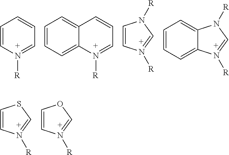
wherein R is —H or a C1-C6 alkyl and each of the structural formula is optionally substituted.
Claims (21)


Br-{circle around (A)}-Br (II),


Priority Applications (1)
| Application Number | Priority Date | Filing Date | Title |
|---|---|---|---|
| US13/378,989 US8969509B2 (en) | 2009-06-26 | 2010-06-25 | Signal amplified biological detection with conjugated polymers |
Applications Claiming Priority (3)
| Application Number | Priority Date | Filing Date | Title |
|---|---|---|---|
| US26968509P | 2009-06-26 | 2009-06-26 | |
| PCT/US2010/040051 WO2010151807A1 (en) | 2009-06-26 | 2010-06-25 | Signal amplified biological detection with conjugated polymers |
| US13/378,989 US8969509B2 (en) | 2009-06-26 | 2010-06-25 | Signal amplified biological detection with conjugated polymers |
Publications (2)
| Publication Number | Publication Date |
|---|---|
| US20120252986A1 US20120252986A1 (en) | 2012-10-04 |
| US8969509B2 true US8969509B2 (en) | 2015-03-03 |
Family
ID=42682446
Family Applications (1)
| Application Number | Title | Priority Date | Filing Date |
|---|---|---|---|
| US13/378,989 Active 2031-09-14 US8969509B2 (en) | 2009-06-26 | 2010-06-25 | Signal amplified biological detection with conjugated polymers |
Country Status (3)
| Country | Link |
|---|---|
| US (1) | US8969509B2 (en) |
| SG (1) | SG177355A1 (en) |
| WO (1) | WO2010151807A1 (en) |
Cited By (24)
| Publication number | Priority date | Publication date | Assignee | Title |
|---|---|---|---|---|
| US9557337B2 (en) | 2013-10-02 | 2017-01-31 | Becton, Dickinson And Company | Polymersome encapsulation of hydrophobic fluorescent polymers |
| US9896538B2 (en) | 2016-03-28 | 2018-02-20 | Aat Bioquest, Inc. | Polyfluoreno[4,5-cde]oxepine polymers and conjugates thereof |
| WO2018175173A1 (en) * | 2017-03-21 | 2018-09-27 | The Scripps Research Institute | Cu-and ni-catalyzed decarboxylative borylation reactions |
| US10107818B2 (en) | 2006-10-06 | 2018-10-23 | Sirigen Ii Limited | Fluorescent methods and materials for directed biomarker signal amplification |
| US10288620B2 (en) | 2010-01-19 | 2019-05-14 | Sirigen Ii Limited | Reagents for directed biomarker signal amplification |
| US10408759B2 (en) | 2015-05-26 | 2019-09-10 | Becton, Dickinson And Company | Methods and systems for high resolution fluorescence microscopy of polymeric dye-labeled samples using polarized light |
| US10472521B2 (en) | 2015-12-16 | 2019-11-12 | Becton, Dickinson And Company | Photostable fluorescent polymeric tandem dyes including luminescent metal complexes |
| US10533092B2 (en) | 2016-12-12 | 2020-01-14 | Becton, Dickinson And Company | Water-soluble polymeric dyes |
| US10545137B2 (en) | 2016-04-22 | 2020-01-28 | Becton, Dickinson And Company | Multiplex polymeric dye devices and methods for using the same |
| US10564107B2 (en) | 2005-04-25 | 2020-02-18 | Trustees Of Boston University | Structured substrates for optical surface profiling |
| US10604657B2 (en) | 2016-07-07 | 2020-03-31 | Becton, Dickinson And Company | Fluorescent water-solvated conjugated polymers |
| US10920082B2 (en) | 2016-07-11 | 2021-02-16 | Becton, Dickinson And Company | Blue-excitable water-solvated polymeric dyes |
| US10928315B1 (en) | 2015-09-22 | 2021-02-23 | Trustees Of Boston University | Multiplexed phenotyping of nanovesicles |
| US11155714B2 (en) | 2016-07-08 | 2021-10-26 | BioLegend, Inc. | Substituted polyfluorene compounds |
| US11220628B2 (en) | 2019-02-20 | 2022-01-11 | Aat Bioquest, Inc. | Condensed polycyclic conjugated polymers and their use for biological detection |
| US11262359B2 (en) | 2016-02-05 | 2022-03-01 | NanoView Biosciences, Inc. | Detection of exosomes having surface markers |
| US11320437B2 (en) | 2017-02-08 | 2022-05-03 | Becton, Dickinson And Company | Dried dye reagent devices and methods for making and using the same |
| WO2022170084A1 (en) | 2021-02-05 | 2022-08-11 | Beckman Coulter, Inc. | Compositions and methods for preventing non-specific interactions between polymer dyes-antibody conjugates |
| US11492493B2 (en) | 2017-12-26 | 2022-11-08 | Becton, Dickinson And Company | Deep ultraviolet-excitable water-solvated polymeric dyes |
| WO2022235705A1 (en) | 2021-05-04 | 2022-11-10 | Beckman Coulter, Inc. | Uv-absorbing polymers, compositions and uses thereof |
| US11584883B2 (en) | 2017-07-28 | 2023-02-21 | BioLegend, Inc. | Conjugated polymers and methods of use |
| WO2023056460A1 (en) | 2021-09-30 | 2023-04-06 | Beckman Coulter, Inc. | Water-soluble tetrahydropyrene based fluorescent polymers |
| US11834551B2 (en) | 2016-04-15 | 2023-12-05 | Beckman Coulter, Inc. | Photoactive macromolecules and uses thereof |
| WO2024044327A1 (en) | 2022-08-26 | 2024-02-29 | Beckman Coulter, Inc. | Dhnt monomers and polymer dyes with modified photophysical properties |
Families Citing this family (17)
| Publication number | Priority date | Publication date | Assignee | Title |
|---|---|---|---|---|
| US8969509B2 (en) | 2009-06-26 | 2015-03-03 | Sirigen, Inc. | Signal amplified biological detection with conjugated polymers |
| WO2016077486A1 (en) * | 2014-11-12 | 2016-05-19 | Becton, Dickinson And Company | Polymeric dye specific binding members and methods of making and using the same |
| JP7390778B2 (en) | 2015-03-12 | 2023-12-06 | ベクトン・ディキンソン・アンド・カンパニー | Polymeric BODIPY dyes and methods of using them |
| US9719998B2 (en) | 2015-03-12 | 2017-08-01 | Becton Dickinson And Company | Ultraviolet absorbing polymeric dyes and methods for using the same |
| AU2017331459B2 (en) | 2016-09-26 | 2023-04-13 | Becton, Dickinson And Company | Measurement of protein expression using reagents with barcoded oligonucleotide sequences |
| ES2950436T3 (en) | 2016-12-14 | 2023-10-10 | Becton Dickinson Co | Methods and compositions to obtain an assessment of tuberculosis in a subject |
| EP3639013A4 (en) | 2017-06-16 | 2021-06-30 | Duke University | Resonator networks for improved label detection, computation, analyte sensing, and tunable random number generation |
| BR102019025989A2 (en) | 2018-12-14 | 2020-06-23 | Beckman Coulter, Inc. | MODIFICATION OF POLYMERIC DYES AND APPLICATIONS |
| EP3969881A4 (en) | 2019-05-14 | 2023-01-25 | Becton, Dickinson and Company | Phase-calibration for imaging flow cytometry |
| CN115053135A (en) | 2019-12-20 | 2022-09-13 | 贝克顿·迪金森公司 | Method for quantifying extracellular vesicle surface markers and composition for carrying out the method |
| WO2021146636A1 (en) | 2020-01-17 | 2021-07-22 | Becton, Dickinson And Company | Methods and compositions for single cell secretomics |
| CN116569042A (en) | 2020-12-09 | 2023-08-08 | 贝克顿迪金森公司 | Multiplexed single cell immunoassays |
| WO2023034789A1 (en) | 2021-08-31 | 2023-03-09 | Becton, Dickinson And Company | Multiplexed proteomics analysis using oligonucleotide-conjugated antibodies |
| WO2023039433A1 (en) | 2021-09-08 | 2023-03-16 | Becton, Dickinson And Company | Non-sequencing pcr-based method for detection of antibody-conjugated oligonucleotides |
| WO2023150764A1 (en) | 2022-02-07 | 2023-08-10 | Becton, Dickinson And Company | Sorting of mrna and abseq containing barcoded beads by flow |
| WO2023154694A1 (en) | 2022-02-08 | 2023-08-17 | Becton, Dickinson And Company | Reference beads for linking imaging and sequencing readouts with single-cell resolution |
| WO2023172977A1 (en) | 2022-03-09 | 2023-09-14 | Becton, Dickinson And Company | Modified flow proxy assay prior to single-cell cite-seq |
Citations (53)
| Publication number | Priority date | Publication date | Assignee | Title |
|---|---|---|---|---|
| US4486530A (en) | 1980-08-04 | 1984-12-04 | Hybritech Incorporated | Immunometric assays using monoclonal antibodies |
| WO1990015070A1 (en) | 1989-06-07 | 1990-12-13 | Affymax Technologies N.V. | Very large scale immobilized peptide synthesis |
| WO1992010092A1 (en) | 1990-12-06 | 1992-06-25 | Affymax Technologies N.V. | Very large scale immobilized polymer synthesis |
| WO1993010507A1 (en) | 1991-11-21 | 1993-05-27 | Optimum Solutions Corp., Inc. | Questionnaire scanning system employing expandable answer mark areas for efficient scanning and mark detection |
| WO1993009668A1 (en) | 1991-11-22 | 1993-05-27 | Affymax Technology N.V. | Combinatorial strategies for polymer synthesis |
| WO1993022684A1 (en) | 1992-04-29 | 1993-11-11 | Affymax Technologies N.V. | Factorial chemical libraries |
| US5288514A (en) | 1992-09-14 | 1994-02-22 | The Regents Of The University Of California | Solid phase and combinatorial synthesis of benzodiazepine compounds on a solid support |
| WO1994009169A1 (en) | 1992-10-20 | 1994-04-28 | The Research Foundation Of State University Of New York At Buffalo | Phosphate binders for metal-matrix composites |
| US5384261A (en) | 1991-11-22 | 1995-01-24 | Affymax Technologies N.V. | Very large scale immobilized polymer synthesis using mechanically directed flow paths |
| US5424186A (en) | 1989-06-07 | 1995-06-13 | Affymax Technologies N.V. | Very large scale immobilized polymer synthesis |
| US5807974A (en) | 1996-05-16 | 1998-09-15 | Korea Institute Of Science And Technology | Fluorene-based alternating copolymers for electroluminescence element and electroluminescence element using such copolymers as light emitting materials |
| WO1999026299A1 (en) | 1997-11-13 | 1999-05-27 | Massachusetts Institute Of Technology | Highly luminescent color-selective materials |
| US5990479A (en) | 1997-11-25 | 1999-11-23 | Regents Of The University Of California | Organo Luminescent semiconductor nanocrystal probes for biological applications and process for making and using such probes |
| WO2000066790A1 (en) | 1999-05-05 | 2000-11-09 | The Regents Of The University Of California | Method for detecting biological agents |
| US6263286B1 (en) | 1998-08-13 | 2001-07-17 | U.S. Genomics, Inc. | Methods of analyzing polymers using a spatial network of fluorophores and fluorescence resonance energy transfer |
| US6280933B1 (en) | 1990-03-14 | 2001-08-28 | The Regents Of The University Of California | Multichromophore fluorescent probes using DNA intercalation complexes |
| US6350431B1 (en) | 1997-04-29 | 2002-02-26 | Nycomed Imaging As | Compounds |
| WO2002079268A2 (en) | 2001-03-28 | 2002-10-10 | The Regents Of The University Of California | Tuning the properties of conjugated polyelectrolytes and application in a biosensor platform |
| WO2002081735A2 (en) | 2001-04-05 | 2002-10-17 | Infectio Diagnostic (I.D.I.), Inc. | Detection of negatively charged polymers using water-soluble, cationic, polythiophene derivatives |
| EP1281744A2 (en) | 1998-05-05 | 2003-02-05 | Massachusetts Institute Of Technology | Emissive polymers and devices incorporating these polymers |
| US20030087311A1 (en) | 2001-11-07 | 2003-05-08 | Wolf David E. | Method of identifying energy transfer sensors for analytes |
| CA2365814A1 (en) | 2001-12-21 | 2003-06-21 | American Dye Source, Inc. | Photoluminescent markers and methods for detection of such markers |
| US20040009506A1 (en) | 2002-03-29 | 2004-01-15 | Genentech, Inc. | Methods and compositions for detection and quantitation of nucleic acid analytes |
| US20040023248A1 (en) | 2001-12-07 | 2004-02-05 | Whitehead Institiute For Biomedical Research | Methods and reagents for improving nucleic acid detection |
| US20040023317A1 (en) | 2002-03-29 | 2004-02-05 | Board Of Regents Of The University Of Texas System | Implantable biosensor from stratified nanostructured membranes |
| WO2004037886A1 (en) | 2002-10-25 | 2004-05-06 | Agency For Science Technology And Research | Cationic water-soluble conjugated polymers and their precursors |
| US20040142344A1 (en) | 2002-08-26 | 2004-07-22 | The Regents Of The University Of California | Methods and compositions for detection and analysis of polynucleotides using light harvesting multichromophores |
| US20040219556A1 (en) | 2002-06-20 | 2004-11-04 | The Regents Of The University Of California | Methods and compositions for detection and analysis of polynucleotides using light harvesting multichromophores |
| US20050003386A1 (en) | 2003-02-13 | 2005-01-06 | The Regents Of The University Of California | Methods and compositions for detection and analysis of polynucleotide-binding protein interactions using light harvesting multichromophores |
| US20050059168A1 (en) | 2003-09-17 | 2005-03-17 | The Regents Of The University Of California | Conformationally flexible cationic conjugated polymers |
| US20050196775A1 (en) | 2003-12-04 | 2005-09-08 | Massachusetts Institute Of Technology | Fluorescent, semi-conductive polymers, and devices comprising them |
| US6951682B1 (en) | 1998-12-01 | 2005-10-04 | Syntrix Biochip, Inc. | Porous coatings bearing ligand arrays and use thereof |
| US6998241B2 (en) | 2002-09-11 | 2006-02-14 | Kimberly-Clark Worldwide, Inc. | Antibody pair screening methods |
| EP0708837B1 (en) | 1993-07-13 | 2006-03-08 | Abbott Laboratories | Fluorescent polymer labeled conjugates and intermediates |
| WO2006034081A2 (en) | 2004-09-17 | 2006-03-30 | Massachusetts Institute Of Technology | Polymers for analyte detection |
| US20060073607A1 (en) | 2003-12-05 | 2006-04-06 | Massachusetts Institute Of Technology | Organic materials able to detect analytes |
| US20060175193A1 (en) | 2002-05-13 | 2006-08-10 | Olle Inganas | Polyelectrolyte complex(e.g.zwitterionic polythiophenes) with a receptor (e.g.polynucleotide, antibody etc.) for biosensor applications |
| US20060183140A1 (en) | 2005-01-10 | 2006-08-17 | The Regents Of The University Of California | Cationic conjugated polymers suitable for strand-specific polynucleotide detection in homogeneous and solid state assays |
| WO2006092063A1 (en) | 2005-03-03 | 2006-09-08 | National Research Council Canada | Methods and compositions for the detection and analysis of nucleic acids by signal amplification |
| US20060204984A1 (en) | 2005-01-31 | 2006-09-14 | The Regents Of University Of California | Methods and compositions for aggregant detection |
| US20060216759A1 (en) | 2004-10-29 | 2006-09-28 | Imad Naasani | Functionalized fluorescent nanocrystals, and methods for their preparation and use |
| US20060216734A1 (en) | 2005-01-10 | 2006-09-28 | The Regents Of The University Of California | Methods and articles for strand-specific polynucleotide detection with cationic multichromophores |
| US7141437B2 (en) | 2003-11-13 | 2006-11-28 | Michigan Molecular Institute | Solid-state colorimetric biosensors comprised of dendritic polymer networks |
| EP1279023B1 (en) | 2000-03-20 | 2007-02-21 | Analytical Biological Services, Inc. | Method for detecting an analyte by fluorescence |
| US7208122B2 (en) | 1998-05-05 | 2007-04-24 | Massachusetts Institute Of Technology | Emissive polymers and devices incorporating these polymers |
| US7241512B2 (en) | 2002-04-19 | 2007-07-10 | 3M Innovative Properties Company | Electroluminescent materials and methods of manufacture and use |
| US20070178470A1 (en) | 2003-12-12 | 2007-08-02 | Infectio Recherche Inc. | System for charge-based detection of nucleic acids |
| US7282514B1 (en) | 2004-05-14 | 2007-10-16 | Research Foundation Of The University Of Central Florida, Inc. | Reactive probes for two-photon fluorescent imaging and sensing |
| US20070281289A1 (en) | 2006-05-31 | 2007-12-06 | Nomadics, Inc. | Emissive species for clinical imaging |
| US20080038751A1 (en) | 2004-05-10 | 2008-02-14 | Peter Asberg | Methods for Determining Conformational Changes and Self-Assembly of Proteins |
| WO2008100344A2 (en) | 2006-10-06 | 2008-08-21 | Sirigen Inc. | Fluorescent methods and materials for directed biomarker signal amplification |
| WO2010151807A1 (en) | 2009-06-26 | 2010-12-29 | Sirigen, Inc. | Signal amplified biological detection with conjugated polymers |
| WO2011091086A1 (en) | 2010-01-19 | 2011-07-28 | Sirigen Inc. | Novel reagents for directed biomarker signal amplification |
Family Cites Families (1)
| Publication number | Priority date | Publication date | Assignee | Title |
|---|---|---|---|---|
| JP2016031772A (en) | 2014-07-30 | 2016-03-07 | 株式会社日立ハイテクファインシステムズ | Inspection method and apparatus of heat-assisted magnetic head element |
-
2010
- 2010-06-25 US US13/378,989 patent/US8969509B2/en active Active
- 2010-06-25 WO PCT/US2010/040051 patent/WO2010151807A1/en active Application Filing
- 2010-06-25 SG SG2011096138A patent/SG177355A1/en unknown
Patent Citations (67)
| Publication number | Priority date | Publication date | Assignee | Title |
|---|---|---|---|---|
| US4486530A (en) | 1980-08-04 | 1984-12-04 | Hybritech Incorporated | Immunometric assays using monoclonal antibodies |
| US5424186A (en) | 1989-06-07 | 1995-06-13 | Affymax Technologies N.V. | Very large scale immobilized polymer synthesis |
| WO1990015070A1 (en) | 1989-06-07 | 1990-12-13 | Affymax Technologies N.V. | Very large scale immobilized peptide synthesis |
| US5143854A (en) | 1989-06-07 | 1992-09-01 | Affymax Technologies N.V. | Large scale photolithographic solid phase synthesis of polypeptides and receptor binding screening thereof |
| US6280933B1 (en) | 1990-03-14 | 2001-08-28 | The Regents Of The University Of California | Multichromophore fluorescent probes using DNA intercalation complexes |
| WO1992010092A1 (en) | 1990-12-06 | 1992-06-25 | Affymax Technologies N.V. | Very large scale immobilized polymer synthesis |
| WO1993010507A1 (en) | 1991-11-21 | 1993-05-27 | Optimum Solutions Corp., Inc. | Questionnaire scanning system employing expandable answer mark areas for efficient scanning and mark detection |
| WO1993009668A1 (en) | 1991-11-22 | 1993-05-27 | Affymax Technology N.V. | Combinatorial strategies for polymer synthesis |
| US5677195A (en) | 1991-11-22 | 1997-10-14 | Affymax Technologies N.V. | Combinatorial strategies for polymer synthesis |
| US5384261A (en) | 1991-11-22 | 1995-01-24 | Affymax Technologies N.V. | Very large scale immobilized polymer synthesis using mechanically directed flow paths |
| WO1993022684A1 (en) | 1992-04-29 | 1993-11-11 | Affymax Technologies N.V. | Factorial chemical libraries |
| US5288514A (en) | 1992-09-14 | 1994-02-22 | The Regents Of The University Of California | Solid phase and combinatorial synthesis of benzodiazepine compounds on a solid support |
| WO1994009169A1 (en) | 1992-10-20 | 1994-04-28 | The Research Foundation Of State University Of New York At Buffalo | Phosphate binders for metal-matrix composites |
| EP0708837B1 (en) | 1993-07-13 | 2006-03-08 | Abbott Laboratories | Fluorescent polymer labeled conjugates and intermediates |
| US5807974A (en) | 1996-05-16 | 1998-09-15 | Korea Institute Of Science And Technology | Fluorene-based alternating copolymers for electroluminescence element and electroluminescence element using such copolymers as light emitting materials |
| US6350431B1 (en) | 1997-04-29 | 2002-02-26 | Nycomed Imaging As | Compounds |
| WO1999026299A1 (en) | 1997-11-13 | 1999-05-27 | Massachusetts Institute Of Technology | Highly luminescent color-selective materials |
| US5990479A (en) | 1997-11-25 | 1999-11-23 | Regents Of The University Of California | Organo Luminescent semiconductor nanocrystal probes for biological applications and process for making and using such probes |
| US7208122B2 (en) | 1998-05-05 | 2007-04-24 | Massachusetts Institute Of Technology | Emissive polymers and devices incorporating these polymers |
| EP1281744A2 (en) | 1998-05-05 | 2003-02-05 | Massachusetts Institute Of Technology | Emissive polymers and devices incorporating these polymers |
| US6263286B1 (en) | 1998-08-13 | 2001-07-17 | U.S. Genomics, Inc. | Methods of analyzing polymers using a spatial network of fluorophores and fluorescence resonance energy transfer |
| US6951682B1 (en) | 1998-12-01 | 2005-10-04 | Syntrix Biochip, Inc. | Porous coatings bearing ligand arrays and use thereof |
| WO2000066790A1 (en) | 1999-05-05 | 2000-11-09 | The Regents Of The University Of California | Method for detecting biological agents |
| EP1279023B1 (en) | 2000-03-20 | 2007-02-21 | Analytical Biological Services, Inc. | Method for detecting an analyte by fluorescence |
| WO2002079268A2 (en) | 2001-03-28 | 2002-10-10 | The Regents Of The University Of California | Tuning the properties of conjugated polyelectrolytes and application in a biosensor platform |
| WO2002081735A2 (en) | 2001-04-05 | 2002-10-17 | Infectio Diagnostic (I.D.I.), Inc. | Detection of negatively charged polymers using water-soluble, cationic, polythiophene derivatives |
| US20030087311A1 (en) | 2001-11-07 | 2003-05-08 | Wolf David E. | Method of identifying energy transfer sensors for analytes |
| US20040023248A1 (en) | 2001-12-07 | 2004-02-05 | Whitehead Institiute For Biomedical Research | Methods and reagents for improving nucleic acid detection |
| CA2365814A1 (en) | 2001-12-21 | 2003-06-21 | American Dye Source, Inc. | Photoluminescent markers and methods for detection of such markers |
| US20040023317A1 (en) | 2002-03-29 | 2004-02-05 | Board Of Regents Of The University Of Texas System | Implantable biosensor from stratified nanostructured membranes |
| US20040009506A1 (en) | 2002-03-29 | 2004-01-15 | Genentech, Inc. | Methods and compositions for detection and quantitation of nucleic acid analytes |
| US7241512B2 (en) | 2002-04-19 | 2007-07-10 | 3M Innovative Properties Company | Electroluminescent materials and methods of manufacture and use |
| US20060175193A1 (en) | 2002-05-13 | 2006-08-10 | Olle Inganas | Polyelectrolyte complex(e.g.zwitterionic polythiophenes) with a receptor (e.g.polynucleotide, antibody etc.) for biosensor applications |
| US7214489B2 (en) | 2002-06-20 | 2007-05-08 | The Regents Of The University Of California | Methods and compositions for detection and analysis of polynucleotides using light harvesting multichromophores |
| US20040219556A1 (en) | 2002-06-20 | 2004-11-04 | The Regents Of The University Of California | Methods and compositions for detection and analysis of polynucleotides using light harvesting multichromophores |
| JP2006510389A (en) | 2002-08-26 | 2006-03-30 | ザ リージェンツ オブ ザ ユニバーシティ オブ カリフォルニア | Methods and compositions for detecting and analyzing polynucleotides using light collecting multichromophores |
| US20040142344A1 (en) | 2002-08-26 | 2004-07-22 | The Regents Of The University Of California | Methods and compositions for detection and analysis of polynucleotides using light harvesting multichromophores |
| US6998241B2 (en) | 2002-09-11 | 2006-02-14 | Kimberly-Clark Worldwide, Inc. | Antibody pair screening methods |
| WO2004037886A1 (en) | 2002-10-25 | 2004-05-06 | Agency For Science Technology And Research | Cationic water-soluble conjugated polymers and their precursors |
| US20050003386A1 (en) | 2003-02-13 | 2005-01-06 | The Regents Of The University Of California | Methods and compositions for detection and analysis of polynucleotide-binding protein interactions using light harvesting multichromophores |
| US7144950B2 (en) | 2003-09-17 | 2006-12-05 | The Regents Of The University Of California | Conformationally flexible cationic conjugated polymers |
| US20050059168A1 (en) | 2003-09-17 | 2005-03-17 | The Regents Of The University Of California | Conformationally flexible cationic conjugated polymers |
| US7141437B2 (en) | 2003-11-13 | 2006-11-28 | Michigan Molecular Institute | Solid-state colorimetric biosensors comprised of dendritic polymer networks |
| US20050196775A1 (en) | 2003-12-04 | 2005-09-08 | Massachusetts Institute Of Technology | Fluorescent, semi-conductive polymers, and devices comprising them |
| US20060073607A1 (en) | 2003-12-05 | 2006-04-06 | Massachusetts Institute Of Technology | Organic materials able to detect analytes |
| US20070178470A1 (en) | 2003-12-12 | 2007-08-02 | Infectio Recherche Inc. | System for charge-based detection of nucleic acids |
| US20080038751A1 (en) | 2004-05-10 | 2008-02-14 | Peter Asberg | Methods for Determining Conformational Changes and Self-Assembly of Proteins |
| US7282514B1 (en) | 2004-05-14 | 2007-10-16 | Research Foundation Of The University Of Central Florida, Inc. | Reactive probes for two-photon fluorescent imaging and sensing |
| US20060127929A1 (en) | 2004-09-17 | 2006-06-15 | Massachusetts Institute Of Technology | Polymers for analyte detection |
| WO2006034081A2 (en) | 2004-09-17 | 2006-03-30 | Massachusetts Institute Of Technology | Polymers for analyte detection |
| US20060216759A1 (en) | 2004-10-29 | 2006-09-28 | Imad Naasani | Functionalized fluorescent nanocrystals, and methods for their preparation and use |
| US20060183140A1 (en) | 2005-01-10 | 2006-08-17 | The Regents Of The University Of California | Cationic conjugated polymers suitable for strand-specific polynucleotide detection in homogeneous and solid state assays |
| US20060216734A1 (en) | 2005-01-10 | 2006-09-28 | The Regents Of The University Of California | Methods and articles for strand-specific polynucleotide detection with cationic multichromophores |
| US20060204984A1 (en) | 2005-01-31 | 2006-09-14 | The Regents Of University Of California | Methods and compositions for aggregant detection |
| US7666594B2 (en) | 2005-01-31 | 2010-02-23 | The Regents Of The University Of California | Methods for assaying a sample for an aggregant |
| WO2006092063A1 (en) | 2005-03-03 | 2006-09-08 | National Research Council Canada | Methods and compositions for the detection and analysis of nucleic acids by signal amplification |
| US20070281289A1 (en) | 2006-05-31 | 2007-12-06 | Nomadics, Inc. | Emissive species for clinical imaging |
| US20120028828A1 (en) | 2006-10-06 | 2012-02-02 | Sirigen Inc. | Fluorescent Methods and Materials for Directed Biomarker Signal Amplification |
| WO2008100344A2 (en) | 2006-10-06 | 2008-08-21 | Sirigen Inc. | Fluorescent methods and materials for directed biomarker signal amplification |
| US20080293164A1 (en) | 2006-10-06 | 2008-11-27 | Sirigen Inc. | Fluorescent Methods and Materials for Directed Biomarker Signal Amplification |
| US8158444B2 (en) | 2006-10-06 | 2012-04-17 | Sirigen, Inc. | Fluorescent methods and materials for directed biomarker signal amplification |
| US20120029155A1 (en) | 2006-10-06 | 2012-02-02 | Sirigen Inc. | Fluorescent Methods and Materials for Directed Biomarker Signal Amplification |
| WO2010151807A1 (en) | 2009-06-26 | 2010-12-29 | Sirigen, Inc. | Signal amplified biological detection with conjugated polymers |
| US20110256550A1 (en) | 2010-01-19 | 2011-10-20 | Sirigen, Inc. | Novel reagents for directed biomarker signal amplification |
| US20110256549A1 (en) | 2010-01-19 | 2011-10-20 | Sirigen, Inc. | Novel reagents for directed biomarker signal amplification |
| US20110257374A1 (en) | 2010-01-19 | 2011-10-20 | Sirigen, Inc. | Novel Reagents for Directed Biomarker Signal Amplification |
| WO2011091086A1 (en) | 2010-01-19 | 2011-07-28 | Sirigen Inc. | Novel reagents for directed biomarker signal amplification |
Non-Patent Citations (59)
| Title |
|---|
| An et al., "A fluorescence ratiometric protein assay using light-harvesting conjugated polymers," Macromolecular Rapid Communications 27(13):993-997 (2006). |
| Ausebel et al., eds., Current Protocols in Molecular Biology, vols. I, II, and III. 1997. |
| Ausubel et al., eds., Short Protocols in Molecular Biology: A Compendium of Methods from Current Protocols in Molecular Biology, 5th ed., John Wiley & Sons, Inc., 2002. |
| Bailey et al., "Masked Micheal Acceptors in Poly(phenyleneethynylene)s for Facile Conjugation," Macromolecules 39:2815-2818 (2006). |
| Bruchez et al., "Semiconductor nanocrystals as fluorescent biological labels," Science 281:2013-2016 (1998). |
| Chen et al., "Highly sensitive biological and chemical sensors based on reversible fluorescence quenching in a conjugated polymer," PNAS USA 96(220:12287-12292 (1999). |
| Delagrave et al., "Isolated mutants of cloned Aequorea victoria GFP that had red-shifted excitation spectra," Bio/Tech 13:151-154 (1995). |
| EP 07873316 Supplemental Search Report dated Aug. 3, 2010. |
| Fodor et al,, "Light-directed, spatially addressable parallel chemical synthesis," Science 251(4995):767-773 (1991). |
| Gaylord et al., "DNA detection using water-soluble conjugated polymers and peptide nucleic acid probes," PNAS USA 99(17):10954-10957 (2002). |
| Gaylord et al., "DNA hybridization detection with water-soluble conjugated polymers and chromophore-labeled single-stranded DNA," J Am Chem Soc 124(4):896-900(2003). |
| Gaylord et al., "SNP detection using peptide nucleic acid probes and conjugated polymers: application in neurodegenerative disease identification," PNAS USA 102(1):34-39 (2005). |
| Geierstanger and Wemmer, "Complexes of the minor groove of DNA," Annu Rev Biophys Biomol Struct 24:463-493 (1995). |
| Glumoff and Goldman, Nucleic Acids in Chemistry and Biology, 2nd ed., Blackburn and Gait, eds., Oxford University Press, Oxford, 1996, pp. 375-441. |
| Heeger et al., "Making sense of polymer-based biosensors," PNAS USA 96(22):12219-12221 (1999). |
| Heim et al., "Improved green fluorescence," Nature 373:663-664 (1995). |
| Heim et al., "Wavelength mutations and post-translational autoxidation of green fluorescent protein," PNAS USA 91:12501-12504 (1994). |
| Ho et al., "Colorimetric and fluorometric detection of nucleic acids using cationic polythiophene derivatives," Angewandte Chemie International Edition 41(9):1548-1551 (2002). |
| Ho et al., "Direct molecular detection of nucleic acids by fluorescence signal amplification," J Am Chem Soc 127(36):12673-12676 (2005). |
| Innis et al., eds., PCR Protocols: A Guide to Methods and Applications, Elsevier Science & Technology Books 1990. |
| Invitrogen -available at www.invitrogen.com, accessed Dec. 19, 2007. |
| Invitrogen -Molecular Probes, available at www.probes.com, accessed Dec. 19, 2007. |
| JP2009-531642 Office Action mailed dated Mar. 22, 2012. |
| Kanehisa, "Use of statistical criteria for screening potential homologies in nucleic acid sequences," Nucleic Acids Research 12(1 Pt 1):203-213 (1984). |
| Kreuzer et al., "LightCycler technology fo rthe quantitation of bcr/abl fusion transcripts," Cancer Research 59(13):3171-3174 (1999). |
| Kreyenschmidt et al., "A New Soluble poly(p-phenylene) with Tetrahydropryrene Repeating Units," Macromolecules 28:4577-4582 (1995). |
| Larson and Verdine, Bioorganic Chemistry: Nucleic Acids, Hecht ed., Oxford University Press: New York 1996 pp. 324-346. |
| Laurendeau et al., "Quantitation of MYC gene expression in sporadic breast tumors with a real-time reverse transcription PCR assay," Cancer Res 59(12):2759-2765 (1999). |
| Laurendeau et al., "TaqMan PCR-based gene dosage assay for predictive testing in individuals from a cancer family with INK4 locus haploinsufficiency," Clin Chem 45(7):982-986 (1999). |
| LeClerc, "Polyfluorenes: Twenty Years of Progress," Polym Sci Part A: Polym Chem 39:2867-2873 (2001). |
| Lee et al., "Synthesis and Characterization of Oligo(9,9-dihyexyl-2,7-fluorene ethynylene)s: For Application as Blue Light-Emitting Diode," Org Lett 3:2005-2007 (2001). |
| Lee, Kangwon. "Functionalized Conjugated Polymers for Signal Amplifying Biosensors and Sensor Arrays." Dissertation, The Unviersity of Michigan, 2008. |
| Liu et al., "Interpolyelectrolyte complexes of conjugated copolymers and DNA: platforms for multicolor biosensors," J Am Chem Soc 126(7):1942-1943 (2004). |
| Liu et al., "Methods for strand-specific DNA detection with cationic conjugated polymers suitable for incorporation into DNA chips and microarrays," PNAS USA 102(3):589-593 (2005). |
| Liu et al., "Optimization of the molecular orbital energies of conjugated polymers for optical amplification of fluorescent sensors," J Am Chem Soc 128:1188-1196 (2006). |
| Liu et al., "Shape-adaptable water-soluble conjugated polymers," J Am Chem Soc 124(44):13306-13307 (2003). |
| Liu et al., Characterization of tectoRNA assembly with cationic conjugated polymers, J Am Chem Soc 126(13):4076-4077 (2004). |
| Mikroyannidis et al., J Polym Sci Part A:Polym Chem 45:4661-4670 (2007). |
| Mikroyannidis et al; New highlgly luminescent-neutral precursors; John Wiley & Sons, Inc.; 2007, Chem Abstract 147: 10286. * |
| Mikroyannidis et al; Novel electroluminescent-and its precursor; John Wiley & Sons, inc.; 2007; Chem abstract 146: 462600. * |
| PCT/US2010/40051 International Search Report dated Sep. 30, 2010. |
| PCT/US2011/21775 Search Rerport and Written Opinion dated May 19, 2011. |
| Pei et al., "Efficient Photoluminescence and Electroluminescence from a Soluble Polyfluorene," J Am Chem Soc 118:7416-7417 (1996). |
| Pierce, Biotechnology, available at www.piercenet.com, accessed Dec. 19, 2007. |
| Sambrook et al., Molecular Cloning: A Laboratory Manual, 3rd ed., Cold Spring Harbor Laboratory Press, 2000. |
| Sekar et al., "Phycobiliproteins as a commodity: trends in applied research, patents and commercialization," J Appl Phycol 20:113-136 (2008). |
| Sonogashira et al., "A convenient synthesis of acetylenes: catalytic substitutions of acetylenic hydrogen with bromoalkenes, iodoarenes and bromopyridines,"Tetra Lett 16:4467-4470 (1975). |
| U.S. Appl. No. 07/624,120, filed Dec. 6, 1990, Fodor. |
| U.S. Appl. No. 60/642,901, filed Jan. 10, 2005, Bazan. |
| Wang et al., "Biosensors from conjugated polyelectrolyte complexes," PNAS USA 99(1):49-53 (2002). |
| Wang et al., "Collective optical behavior of cationic water-soluble dendrimers," Advanced Materials 16(23-24):2127-2132 (2004). |
| Wang et al., "Fluorescein provides a resonance gate for FRET from conjugated polymers to DNA intercalated dyes," J Am Chem Soc 126(17):5446-5451 (2004). |
| Wang et al., "Optimally amplified RNA-protein detection methods using light-harvesting conjugated polymers," Advanced Materials 15(17):1425-1428 (2003). |
| Wang, "From DNA biosensors to gene chips," Nucl Acids Res 28(16):3011-3016 (2000). |
| Wosnick et al., "Synthesis and application of poly(phenylen ethynylene)s for bioconjugation: a conjugated polymer-based fluorogenic proble for proteases," J Am Chm Soc 127:3400-3405 (2005). |
| Xu et al., "Magnetically assisted DNA assays: high selectivity using conjugated polymers for amplified fluorescent transduction," Nucl Acids Res 33(9):e83 (2005). |
| Yamamoto et al., "Preparation of pi-Conjugated Poly(thiophene-2,5-diyl), Poly(p-phenylene), and Related Polymers Using Zerovalent Nickel Complexes. Linear Structure and Properties of the pi-Conjugated Polymers," Macromolecules 25:1214-1223 (1992). |
| Yamamoto et al., "Preparation of π-Conjugated Poly(thiophene-2,5-diyl), Poly(p-phenylene), and Related Polymers Using Zerovalent Nickel Complexes. Linear Structure and Properties of the π-Conjugated Polymers," Macromolecules 25:1214-1223 (1992). |
| Zhou et al., "Polyfluorenes with phosphonate groups in the side chains as chemosensors and electroluminescent materials," Macromolecules 38:5416-5424 (2005). |
Cited By (51)
| Publication number | Priority date | Publication date | Assignee | Title |
|---|---|---|---|---|
| US10564107B2 (en) | 2005-04-25 | 2020-02-18 | Trustees Of Boston University | Structured substrates for optical surface profiling |
| US11275030B2 (en) | 2005-04-25 | 2022-03-15 | Trustees Of Boston University | Structured substrates for optical surface profiling |
| US11639937B2 (en) | 2006-10-06 | 2023-05-02 | Sirigen Ii Limited | Fluorescent methods and materials for directed biomarker signal amplification |
| US11209438B2 (en) | 2006-10-06 | 2021-12-28 | Becton, Dickinson And Company | Fluorescent methods and materials for directed biomarker signal amplification |
| US10107818B2 (en) | 2006-10-06 | 2018-10-23 | Sirigen Ii Limited | Fluorescent methods and materials for directed biomarker signal amplification |
| US10126302B2 (en) | 2006-10-06 | 2018-11-13 | Sirigen Ii Limited | Fluorescent methods and materials for directed biomarker signal amplification |
| US10859578B2 (en) | 2006-10-06 | 2020-12-08 | Skigen, Inc. | Fluorescent methods and materials for directed biomarker signal amplification |
| US10641777B2 (en) | 2006-10-06 | 2020-05-05 | Sirigen, Inc. | Fluorescent methods and materials for directed biomarker signal amplification |
| US10962546B2 (en) | 2010-01-19 | 2021-03-30 | Sirigen Ii Limited | Reagents for directed biomarker signal amplification |
| US10302648B2 (en) | 2010-01-19 | 2019-05-28 | Sirigen Ii Limited | Reagents for directed biomarker signal amplification |
| US11333666B2 (en) | 2010-01-19 | 2022-05-17 | Sirigen Ii Limited | Reagents for directed biomarker signal amplification |
| US10458989B2 (en) | 2010-01-19 | 2019-10-29 | Sirigen Ii Limited | Reagents for directed biomarker signal amplification |
| US11899018B2 (en) | 2010-01-19 | 2024-02-13 | Sirigen Ii Limited | Reagents for directed biomarker signal amplification |
| US10481161B2 (en) | 2010-01-19 | 2019-11-19 | Sirigen Ii Limited | Reagents for directed biomarker signal amplification |
| US11874278B2 (en) | 2010-01-19 | 2024-01-16 | Sirigen Ii Limited | Reagents for directed biomarker signal amplification |
| US10955417B2 (en) | 2010-01-19 | 2021-03-23 | Sirigen Ii Limited | Reagents for direct biomarker signal amplification |
| US10365285B2 (en) | 2010-01-19 | 2019-07-30 | Sirigen Ii Limited | Reagents for directed biomarker signal amplification |
| US10288620B2 (en) | 2010-01-19 | 2019-05-14 | Sirigen Ii Limited | Reagents for directed biomarker signal amplification |
| US10641775B2 (en) | 2010-01-19 | 2020-05-05 | Sirigen Ii Limited | Reagents for directed biomarker signal amplification |
| US9557337B2 (en) | 2013-10-02 | 2017-01-31 | Becton, Dickinson And Company | Polymersome encapsulation of hydrophobic fluorescent polymers |
| US9863947B2 (en) | 2013-10-02 | 2018-01-09 | Becton, Dickinson And Company | Polymersome encapsulation of hydrophobic fluorescent polymers |
| US10408759B2 (en) | 2015-05-26 | 2019-09-10 | Becton, Dickinson And Company | Methods and systems for high resolution fluorescence microscopy of polymeric dye-labeled samples using polarized light |
| US10928315B1 (en) | 2015-09-22 | 2021-02-23 | Trustees Of Boston University | Multiplexed phenotyping of nanovesicles |
| US11573177B2 (en) | 2015-09-22 | 2023-02-07 | Trustees Of Boston University | Multiplexed phenotyping of nanovesicles |
| US10472521B2 (en) | 2015-12-16 | 2019-11-12 | Becton, Dickinson And Company | Photostable fluorescent polymeric tandem dyes including luminescent metal complexes |
| US11262359B2 (en) | 2016-02-05 | 2022-03-01 | NanoView Biosciences, Inc. | Detection of exosomes having surface markers |
| US10316136B2 (en) | 2016-03-28 | 2019-06-11 | Aat Bioquest, Inc. | Polyfluoreno[4,5-cde]oxepine conjugates and their use in methods of analyte detection |
| US9896538B2 (en) | 2016-03-28 | 2018-02-20 | Aat Bioquest, Inc. | Polyfluoreno[4,5-cde]oxepine polymers and conjugates thereof |
| US11834551B2 (en) | 2016-04-15 | 2023-12-05 | Beckman Coulter, Inc. | Photoactive macromolecules and uses thereof |
| US10545137B2 (en) | 2016-04-22 | 2020-01-28 | Becton, Dickinson And Company | Multiplex polymeric dye devices and methods for using the same |
| US11614443B2 (en) | 2016-04-22 | 2023-03-28 | Becton, Dickinson And Company | Multiplex polymeric dye devices and methods for using the same |
| US10604657B2 (en) | 2016-07-07 | 2020-03-31 | Becton, Dickinson And Company | Fluorescent water-solvated conjugated polymers |
| US11643556B2 (en) | 2016-07-07 | 2023-05-09 | Becton, Dickinson And Company | Fluorescent conjugated polymers |
| US11155714B2 (en) | 2016-07-08 | 2021-10-26 | BioLegend, Inc. | Substituted polyfluorene compounds |
| US11912876B2 (en) | 2016-07-11 | 2024-02-27 | Becton, Dickinson And Company | Blue-excitable water-solvated polymeric dyes |
| US10920082B2 (en) | 2016-07-11 | 2021-02-16 | Becton, Dickinson And Company | Blue-excitable water-solvated polymeric dyes |
| US11499053B2 (en) | 2016-12-12 | 2022-11-15 | Becton, Dickinson And Company | Water-soluble polymeric dyes |
| US11034840B2 (en) | 2016-12-12 | 2021-06-15 | Becton, Dickinson And Company | Water-soluble polymeric dyes |
| US10533092B2 (en) | 2016-12-12 | 2020-01-14 | Becton, Dickinson And Company | Water-soluble polymeric dyes |
| US11320437B2 (en) | 2017-02-08 | 2022-05-03 | Becton, Dickinson And Company | Dried dye reagent devices and methods for making and using the same |
| WO2018175173A1 (en) * | 2017-03-21 | 2018-09-27 | The Scripps Research Institute | Cu-and ni-catalyzed decarboxylative borylation reactions |
| US11161859B2 (en) | 2017-03-21 | 2021-11-02 | The Scripps Research Institute | Cu- and Ni-catalyzed decarboxylative borylation reactions |
| US11584883B2 (en) | 2017-07-28 | 2023-02-21 | BioLegend, Inc. | Conjugated polymers and methods of use |
| US11732138B2 (en) | 2017-12-26 | 2023-08-22 | Becton, Dickinson And Company | Deep ultraviolet-excitable water-solvated polymeric dyes |
| US11492493B2 (en) | 2017-12-26 | 2022-11-08 | Becton, Dickinson And Company | Deep ultraviolet-excitable water-solvated polymeric dyes |
| US11827824B2 (en) | 2019-02-20 | 2023-11-28 | Aat Bioquest, Inc. | Condensed polycyclic conjugated polymers and their use for biological detection |
| US11220628B2 (en) | 2019-02-20 | 2022-01-11 | Aat Bioquest, Inc. | Condensed polycyclic conjugated polymers and their use for biological detection |
| WO2022170084A1 (en) | 2021-02-05 | 2022-08-11 | Beckman Coulter, Inc. | Compositions and methods for preventing non-specific interactions between polymer dyes-antibody conjugates |
| WO2022235705A1 (en) | 2021-05-04 | 2022-11-10 | Beckman Coulter, Inc. | Uv-absorbing polymers, compositions and uses thereof |
| WO2023056460A1 (en) | 2021-09-30 | 2023-04-06 | Beckman Coulter, Inc. | Water-soluble tetrahydropyrene based fluorescent polymers |
| WO2024044327A1 (en) | 2022-08-26 | 2024-02-29 | Beckman Coulter, Inc. | Dhnt monomers and polymer dyes with modified photophysical properties |
Also Published As
| Publication number | Publication date |
|---|---|
| WO2010151807A1 (en) | 2010-12-29 |
| US20120252986A1 (en) | 2012-10-04 |
| SG177355A1 (en) | 2012-02-28 |
Similar Documents
| Publication | Publication Date | Title |
|---|---|---|
| US8969509B2 (en) | Signal amplified biological detection with conjugated polymers | |
| US11874278B2 (en) | Reagents for directed biomarker signal amplification |
Legal Events
| Date | Code | Title | Description |
|---|---|---|---|
| AS | Assignment |
Owner name: SIRIGEN, INC., CALIFORNIA Free format text: ASSIGNMENT OF ASSIGNORS INTEREST;ASSIGNORS:GAYLORD, BRENT S.;BARTHOLOMEW, GLENN P.;HONG, JANICE W.;AND OTHERS;SIGNING DATES FROM 20110111 TO 20110223;REEL/FRAME:026307/0492 |
|
| AS | Assignment |
Owner name: NATIONAL UNIVERSITY OF SINGAPORE, SINGAPORE Free format text: ASSIGNMENT OF ASSIGNORS INTEREST;ASSIGNORS:LIU, BIN;WANG, CHUN;ZHAN, RUOYU;SIGNING DATES FROM 20120403 TO 20120409;REEL/FRAME:028212/0599 Owner name: SIRIGEN INC., CALIFORNIA Free format text: ASSIGNMENT OF ASSIGNORS INTEREST;ASSIGNORS:BARTHOLOMEW, GLENN P.;HONG, JANICE W.;WHEELER, JEAN M.;AND OTHERS;SIGNING DATES FROM 20120321 TO 20120402;REEL/FRAME:028212/0431 |
|
| STCF | Information on status: patent grant |
Free format text: PATENTED CASE |
|
| MAFP | Maintenance fee payment |
Free format text: PAYMENT OF MAINTENANCE FEE, 4TH YEAR, LARGE ENTITY (ORIGINAL EVENT CODE: M1551); ENTITY STATUS OF PATENT OWNER: LARGE ENTITY Year of fee payment: 4 |
|
| CC | Certificate of correction | ||
| MAFP | Maintenance fee payment |
Free format text: PAYMENT OF MAINTENANCE FEE, 8TH YEAR, LARGE ENTITY (ORIGINAL EVENT CODE: M1552); ENTITY STATUS OF PATENT OWNER: LARGE ENTITY Year of fee payment: 8 |