US9060837B2 - Patterned sling implant and method - Google Patents
Patterned sling implant and method Download PDFInfo
- Publication number
- US9060837B2 US9060837B2 US12/964,502 US96450210A US9060837B2 US 9060837 B2 US9060837 B2 US 9060837B2 US 96450210 A US96450210 A US 96450210A US 9060837 B2 US9060837 B2 US 9060837B2
- Authority
- US
- United States
- Prior art keywords
- implant
- tissue
- extension portions
- anchor
- support portion
- Prior art date
- Legal status (The legal status is an assumption and is not a legal conclusion. Google has not performed a legal analysis and makes no representation as to the accuracy of the status listed.)
- Expired - Fee Related, expires
Links
Images


Classifications
-
- A—HUMAN NECESSITIES
- A61—MEDICAL OR VETERINARY SCIENCE; HYGIENE
- A61F—FILTERS IMPLANTABLE INTO BLOOD VESSELS; PROSTHESES; DEVICES PROVIDING PATENCY TO, OR PREVENTING COLLAPSING OF, TUBULAR STRUCTURES OF THE BODY, e.g. STENTS; ORTHOPAEDIC, NURSING OR CONTRACEPTIVE DEVICES; FOMENTATION; TREATMENT OR PROTECTION OF EYES OR EARS; BANDAGES, DRESSINGS OR ABSORBENT PADS; FIRST-AID KITS
- A61F2/00—Filters implantable into blood vessels; Prostheses, i.e. artificial substitutes or replacements for parts of the body; Appliances for connecting them with the body; Devices providing patency to, or preventing collapsing of, tubular structures of the body, e.g. stents
- A61F2/0004—Closure means for urethra or rectum, i.e. anti-incontinence devices or support slings against pelvic prolapse
- A61F2/0031—Closure means for urethra or rectum, i.e. anti-incontinence devices or support slings against pelvic prolapse for constricting the lumen; Support slings for the urethra
- A61F2/0036—Closure means for urethra or rectum, i.e. anti-incontinence devices or support slings against pelvic prolapse for constricting the lumen; Support slings for the urethra implantable
- A61F2/0045—Support slings
-
- A—HUMAN NECESSITIES
- A61—MEDICAL OR VETERINARY SCIENCE; HYGIENE
- A61F—FILTERS IMPLANTABLE INTO BLOOD VESSELS; PROSTHESES; DEVICES PROVIDING PATENCY TO, OR PREVENTING COLLAPSING OF, TUBULAR STRUCTURES OF THE BODY, e.g. STENTS; ORTHOPAEDIC, NURSING OR CONTRACEPTIVE DEVICES; FOMENTATION; TREATMENT OR PROTECTION OF EYES OR EARS; BANDAGES, DRESSINGS OR ABSORBENT PADS; FIRST-AID KITS
- A61F2/00—Filters implantable into blood vessels; Prostheses, i.e. artificial substitutes or replacements for parts of the body; Appliances for connecting them with the body; Devices providing patency to, or preventing collapsing of, tubular structures of the body, e.g. stents
- A61F2/0063—Implantable repair or support meshes, e.g. hernia meshes
-
- A—HUMAN NECESSITIES
- A61—MEDICAL OR VETERINARY SCIENCE; HYGIENE
- A61B—DIAGNOSIS; SURGERY; IDENTIFICATION
- A61B17/00—Surgical instruments, devices or methods, e.g. tourniquets
- A61B17/04—Surgical instruments, devices or methods, e.g. tourniquets for suturing wounds; Holders or packages for needles or suture materials
- A61B17/06—Needles ; Sutures; Needle-suture combinations; Holders or packages for needles or suture materials
- A61B17/06066—Needles, e.g. needle tip configurations
- A61B17/06109—Big needles, either gripped by hand or connectable to a handle
-
- A—HUMAN NECESSITIES
- A61—MEDICAL OR VETERINARY SCIENCE; HYGIENE
- A61B—DIAGNOSIS; SURGERY; IDENTIFICATION
- A61B17/00—Surgical instruments, devices or methods, e.g. tourniquets
- A61B2017/00743—Type of operation; Specification of treatment sites
- A61B2017/00805—Treatment of female stress urinary incontinence
-
- A—HUMAN NECESSITIES
- A61—MEDICAL OR VETERINARY SCIENCE; HYGIENE
- A61F—FILTERS IMPLANTABLE INTO BLOOD VESSELS; PROSTHESES; DEVICES PROVIDING PATENCY TO, OR PREVENTING COLLAPSING OF, TUBULAR STRUCTURES OF THE BODY, e.g. STENTS; ORTHOPAEDIC, NURSING OR CONTRACEPTIVE DEVICES; FOMENTATION; TREATMENT OR PROTECTION OF EYES OR EARS; BANDAGES, DRESSINGS OR ABSORBENT PADS; FIRST-AID KITS
- A61F2/00—Filters implantable into blood vessels; Prostheses, i.e. artificial substitutes or replacements for parts of the body; Appliances for connecting them with the body; Devices providing patency to, or preventing collapsing of, tubular structures of the body, e.g. stents
- A61F2/0063—Implantable repair or support meshes, e.g. hernia meshes
- A61F2002/0068—Implantable repair or support meshes, e.g. hernia meshes having a special mesh pattern
-
- A—HUMAN NECESSITIES
- A61—MEDICAL OR VETERINARY SCIENCE; HYGIENE
- A61F—FILTERS IMPLANTABLE INTO BLOOD VESSELS; PROSTHESES; DEVICES PROVIDING PATENCY TO, OR PREVENTING COLLAPSING OF, TUBULAR STRUCTURES OF THE BODY, e.g. STENTS; ORTHOPAEDIC, NURSING OR CONTRACEPTIVE DEVICES; FOMENTATION; TREATMENT OR PROTECTION OF EYES OR EARS; BANDAGES, DRESSINGS OR ABSORBENT PADS; FIRST-AID KITS
- A61F2/00—Filters implantable into blood vessels; Prostheses, i.e. artificial substitutes or replacements for parts of the body; Appliances for connecting them with the body; Devices providing patency to, or preventing collapsing of, tubular structures of the body, e.g. stents
- A61F2/0063—Implantable repair or support meshes, e.g. hernia meshes
- A61F2002/0072—Delivery tools therefor
-
- A—HUMAN NECESSITIES
- A61—MEDICAL OR VETERINARY SCIENCE; HYGIENE
- A61F—FILTERS IMPLANTABLE INTO BLOOD VESSELS; PROSTHESES; DEVICES PROVIDING PATENCY TO, OR PREVENTING COLLAPSING OF, TUBULAR STRUCTURES OF THE BODY, e.g. STENTS; ORTHOPAEDIC, NURSING OR CONTRACEPTIVE DEVICES; FOMENTATION; TREATMENT OR PROTECTION OF EYES OR EARS; BANDAGES, DRESSINGS OR ABSORBENT PADS; FIRST-AID KITS
- A61F2/00—Filters implantable into blood vessels; Prostheses, i.e. artificial substitutes or replacements for parts of the body; Appliances for connecting them with the body; Devices providing patency to, or preventing collapsing of, tubular structures of the body, e.g. stents
- A61F2/0077—Special surfaces of prostheses, e.g. for improving ingrowth
- A61F2002/0081—Special surfaces of prostheses, e.g. for improving ingrowth directly machined on the prosthetic surface, e.g. holes, grooves
-
- A—HUMAN NECESSITIES
- A61—MEDICAL OR VETERINARY SCIENCE; HYGIENE
- A61F—FILTERS IMPLANTABLE INTO BLOOD VESSELS; PROSTHESES; DEVICES PROVIDING PATENCY TO, OR PREVENTING COLLAPSING OF, TUBULAR STRUCTURES OF THE BODY, e.g. STENTS; ORTHOPAEDIC, NURSING OR CONTRACEPTIVE DEVICES; FOMENTATION; TREATMENT OR PROTECTION OF EYES OR EARS; BANDAGES, DRESSINGS OR ABSORBENT PADS; FIRST-AID KITS
- A61F2220/00—Fixations or connections for prostheses classified in groups A61F2/00 - A61F2/26 or A61F2/82 or A61F9/00 or A61F11/00 or subgroups thereof
- A61F2220/0008—Fixation appliances for connecting prostheses to the body
- A61F2220/0016—Fixation appliances for connecting prostheses to the body with sharp anchoring protrusions, e.g. barbs, pins, spikes
Definitions
- the present invention relates generally to surgical methods and apparatus and, more specifically, to surgically implantable sling devices and methods for forming and using the same.
- Pelvic health for men and women is a medical area of increasing importance, at least in part due to an aging population.
- pelvic ailments include incontinence (e.g., fecal and urinary), pelvic tissue prolapse (e.g., female vaginal prolapse), and conditions of the pelvic floor.
- Urinary incontinence can further be classified as including different types, such as stress urinary incontinence (SUI), urge urinary incontinence, mixed urinary incontinence, among others. Urinary incontinence can be characterized by the loss or diminution in the ability to maintain the urethral sphincter closed as the bladder fills with urine. Male or female SUI generally occurs when the patient is physically stressed.
- SUI stress urinary incontinence
- urge urinary incontinence urge urinary incontinence
- mixed urinary incontinence among others.
- Urinary incontinence can be characterized by the loss or diminution in the ability to maintain the urethral sphincter closed as the bladder fills with urine. Male or female SUI generally occurs when the patient is physically stressed.
- the present invention describes sling implants and methods for treating pelvic conditions such as incontinence (various forms such as fecal incontinence, stress urinary incontinence, urge incontinence, mixed incontinence, etc.) and other conditions caused by muscle and ligament weakness.
- Other uses include providing a support or platform for plastic surgery, hernia repair, and ortho repairs and support, to name a few.
- Embodiments of the implants can include a tissue support portion, one or more extension portions and one or more anchors. Certain embodiments can be constructed as a unitary sling implant.
- the implant can be constructed or formed in a generally elongate or rectangular shape, or take on a myriad of other compatible configurations or shapes.
- the support portion is adapted for positioning and support under tissue or organs, such as the urethra or bladder.
- the extension portions extend out from the support portion such that the anchors or anchoring features can be deployed for tissue fixation.
- the implants can be formed of patterned cells by way of a molding, die casting, laser etching, laser cutting, extruding, punching, 3-D printing and the like.
- a pattern cut or formed implant can be constructed of a polymer material to provide a lattice support structure of repeated cells.
- embodiments of the present invention are a homogeneous unitary construct.
- Portions of the implant can be formed into sinusoid or other waveform strut members to control and promote elongation, expansion or contraction along single or multiple axes. As such, controlled and designated stress, tension and compression distribution is promoted across specific or localized areas of the construct. Further, portions of the implant can be coated to provide additional control of expansion, and to protect from or promote tissue in-growth.
- the implant can be formed such that regions or portions can include anchoring features to facilitate engagement and attachment of the implant to target tissue sites.
- anchoring to internal tissue, it is also possible to have one or more portions of the implant extend out of an incision or orifice in a patient.
- anchors without or without extending tines, can be defined, formed or otherwise provided with the implant.
- the anchors can be integrally formed, cut or otherwise defined with the implant. Portions of the implant or anchors can be adapted to collapse, fold or otherwise deform to some extent to facilitate engagement with an introducer or needle device and/or to facilitate tissue penetration or fixation.
- the material and cell construct of the sling implant can be configured to promote flexibility while still providing optimal implant strength and tissue support. Further, the stable geometrical and dimensional attributes of the implant provide a flexible device that can be easily positioned and deployed while also avoiding undesirable implant warping or bunching. Such a configuration generally promotes a sling implant adapted to substantially remain in plane during deployment and tissue fixation.
- the sling implants, or portions thereof, can be adapted to provide desirable adjustability, stress distribution, anchoring, stabilization, variable elongation, and the like.
- FIG. 1 is a perspective view of a unitary patterned sling implant having generally transverse extending anchoring features in accordance with embodiments of the present invention.
- FIG. 2 is a top view of the unitary patterned sling implant of FIG. 1 .
- FIG. 3 is a close-up partial view of a portion of the unitary patterned sling implant of FIG. 2 .
- FIG. 4 is a close-up partial view of an anchoring feature, transition zone and extension portion of a unitary patterned sling implant in accordance with embodiments of the present invention.
- FIG. 5 is a perspective view of a unitary patterned sling implant having generally transverse extending anchoring features in accordance with embodiments of the present invention.
- FIG. 6 is a top view of the unitary patterned sling implant of FIG. 5 .
- FIG. 7 is a close-up partial view of a portion of the unitary patterned sling implant of FIG. 6 .
- FIG. 8 is a close-up partial view of an anchoring feature, transition zone and extension portion of a unitary patterned sling implant in accordance with embodiments of the present invention.
- FIG. 9 is a perspective view of a unitary patterned sling implant having planar extending anchoring features in accordance with embodiments of the present invention.
- FIG. 10 is a top view of the unitary patterned sling implant of FIG. 9 .
- FIG. 11 is a close-up partial view of a portion of the unitary patterned sling implant of FIG. 10 .
- FIG. 12 is a perspective view of a unitary patterned sling implant having planar extending anchoring features in accordance with embodiments of the present invention.
- FIG. 13 is a top view of the unitary patterned sling implant of FIG. 12 .
- FIG. 14 is a close-up partial view of a portion of the unitary patterned sling implant of FIG. 13 .
- FIG. 15 is a perspective view of a unitary patterned sling implant having planar extending anchoring features in accordance with embodiments of the present invention.
- FIG. 16 is a top view of the unitary patterned sling implant of FIG. 15 .
- FIG. 17 is a close-up partial view of a portion of the unitary patterned sling implant of FIG. 16 .
- FIG. 18 is a perspective view of a unitary patterned sling implant having planar extending anchoring features in accordance with embodiments of the present invention.
- FIG. 19 is a top view of the unitary patterned sling implant of FIG. 18 .
- FIG. 20 is a close-up partial view of a portion of the unitary patterned sling implant of FIG. 19 .
- FIGS. 21-22 show exemplary sling introduction tools in accordance with embodiments of the present invention.
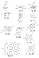
- FIGS. 23-34 show various patterned cell configurations and spacer elements for sling implants in accordance with embodiments of the present invention.
- FIGS. 35-50 show various tear zones, structures and methods for sling implants in accordance with embodiments of the present invention.
- the implant 10 can include a support portion 12 , one or more extension portions 14 and one or more anchoring features 16 .
- the extension portions 14 include the material construct extending from the support portion 12 to the respective anchoring features 16 .
- Various portions of the implant 10 can be constructed of polymer materials, e.g., into a molded generally planar structure or cut from a thin generally planar film or sheet material. Examples of acceptable polymer materials available in constructing or forming the implant systems 10 and its components can include polypropylene, polyethylene, fluoropolymers or like biocompatible materials.
- portions of the implant 10 can be formed or patterned by way of a polymer molding process to create a unitary homogeneous non-woven, or non-knitted, device or construct.
- Other embodiments can be formed from an already unitary homogeneous sheet or film via laser cutting, die cutting, stamping and like procedures.
- the support portion 12 can be configured and shaped for positioning under and support of the urethra or bladder (which includes any location of the bladder, urethra, bladder neck, mid-urethra, or proximal end of the urethra), with the extension portions 14 extendable out to proximate muscle, ligament or other tissue for anchoring.
- the implant also can be used to support pelvic tissue such as the vaginal tissue, tissue of the perineum, coccygeus, levator ani, levator hiatus, and rectum.
- the implant width, at any portion can be approximately 5 mm to 15 mm, and the length from end to end can be approximately 6 cm to 15 cm.
- Repeated cells or patterns in the implant 10 generally form a lattice structure. Portions of the implant 10 can be cut into sinusoid, or other waveform or undulating struts 15 patterns to control elongation or compression along single or multiple axes, to define a desirable pattern density with overall reduced surface area, and to control the distribution and shaping from applied loads.
- the ability to mold, form or cut the struts 15 in a nearly endless array of sinusoidal or like configurations provides an implant 10 that can better tailor or mimic the anisotropic behaviors of physiological tissue.
- the film or unitary construct of the implant can have a thickness T of approximately 0.005 inches to 0.020 inches, and the struts 15 can have a width W of approximately 0.005 inches to 0.012 inches.
- Other configurations, shapes and sizes for the various portions of the implant 10 can be employed as well to promote and facilitate the deployment, use and support of the implant 10 disclosed herein.
- the patterned struts 15 define a general pinwheel design including first angular strut lines 20 and second angular strut lines 22 crossing or intersecting at repeating fixed junctions 24 to define cellular voids 26 .
- the thickness, size and separation of the struts 15 can be modified to create an implant 10 with different surface area and cellular density attributes.
- the struts 15 can have uniform or variable widths or thicknesses, can be tapered, can include apertures, or can include defined shapes and/or patterns, e.g., sinusoids, squares, elliptical, triangular, elbowed, straight, or other simple or complex shapes and patterns.
- Unique strut 15 designs and cellular patterns can be included within a single implant 10 to provide different zones, having different stress, load distribution or compression characteristics. Other strut 15 designs and patterns can be employed as well to achieve the functionality described and depicted herein.
- the dimensional design of the implant struts 15 can be configured to promote targeted strength and flexibility.
- the material width at the fixed junctions 24 can be measurably greater than the material width W of the struts 15 intermediate the junctions to allow for increased strength at the junctions.
- Strengthened and widened junctions 24 can handle and absorb greater stress or torque resulting from implant positioning, twisting and general manipulation.
- thinner strut portions intermediate the junctions 24 promote increased flexibility and controllability of the implant 10 during positioning and device manipulation. This flexibility will also provide an implant 10 adapted to properly conform to unique patient anatomy and lay flat against such anatomy to provide optimal support distribution, tissue in-growth and like characteristics.
- Other dimensional ranges and proportions are envisioned for embodiments of the struts and strut portions depending on the particular application, strength, flexibility, stress distribution or other performance needs of the implant.
- a flexible but strong implant 10 is provided, while still maintaining a low surface area, lower inflammatory response, less scarring and increased density.
- the patterned sling implant 10 also provides benefits over traditional knitted or woven mesh in the area of compression and the reaction to longitudinal extension strain.
- Traditional knitted or woven mesh implants can tend to compress, narrow, bunch or fold during longitudinal stretching, thereby displaying a positive Poisson affect or ratio.
- the sinusoidal pinwheel cell and strut configurations of the patterned implants 10 of embodiments of the present invention can display a Negative Poisson affect or ratio.
- the implant 10 is loaded or stretched (e.g., at ends, anchors, corners or upon the planar surfaces)
- the strut and cell structures resist compression and can measurably expand to provide a stable and generally planar surface area for tissue or organ support.
- the combination of the struts and fixed junctions facilitate this Negative Poisson affect.
- the support portion 12 can take on physical and design characteristics to promote tissue or organ support, and to reduce or eliminate undesirable erosion.
- the support portion 12 can be measurably wider than the extension portions 14 in certain embodiments.
- Relatively narrow extension portions 14 can promote flexibility and positioning of the implant 10 within the patient, without jeopardizing the size and effectiveness of the support portion 12 .
- the support portion 12 can include one or more apertures 13 adapted to reduce tissue erosion, and to promote tissue in-growth, flexibility and tissue support.
- Embodiments of the implant 10 can include one or more transition portions or zones 30 , as shown particularly in FIGS. 4 and 8 .
- the zones 30 provide a material transition between the cellular construct of the extension portions 14 and anchoring or like features of the implant 10 , e.g., anchors, eyelets, etc.
- the transition zones 30 can take on various sizes, shapes and designs to provide increased strength and stress absorption/distribution for portions of the implant 10 being pulled, pushed and twisted during deployment and positioning of the implant 10 .
- Embodiments of the zones 30 can include arcuate or linear members 30 a extending out from or into the extension portions 14 and the anchoring portions 16 .
- the members can be tapered into or away from the extension portion 14 or anchoring portion 16 to facilitate stress and tension distribution such that the struts 15 and cell structures of the extension portion 14 , or support portion 12 , are protected from tearing, ripping or other material breaches.
- Such a design further provides beneficial flexibility and manipulation characteristics during deployment.
- FIGS. 9-20 , and 23 - 34 demonstrate implants 10 and/or implant cell portions having various linear, angled and shaped struts 15 to define unique patterned cell configurations.
- the thickness, size, shape and separation of the struts 15 can be modified to create an implant 10 with different surface area, void or pore shapes/sizes, and cellular density attributes.
- the support portion 12 can take on a different pattern or size configuration than the extension portions 14 to facilitate support, reduce erosion and bunching, or to promote like considerations.
- the strut cell patterns can be separated or symmetrically distributed by way of various spacer elements 15 a .
- implants 10 can be provided in which mechanical characteristics are uniform within the plane of the implant 10 regardless of the direction of an applied stress (e.g., isotropic).
- the implant 10 e.g., support portion 12 or extension portions 14
- the implant 10 may be configured so that mechanical properties differ substantially along selected axes.
- the implant 10 can be formed or cut such that certain edges or other portions can include edge features 32 , e.g., teeth, tines, tangs, angled portions, wisps, members, strut portions, stabilizers or other like features to provide capture points for materials or tissue that pass proximate the implant, or to serve as a means of assistive tissue anchoring during or after deployment of the implant 10 .
- edge features 32 e.g., teeth, tines, tangs, angled portions, wisps, members, strut portions, stabilizers or other like features to provide capture points for materials or tissue that pass proximate the implant, or to serve as a means of assistive tissue anchoring during or after deployment of the implant 10 .
- the shaped or cut cells, or patterns can be configured to optimize or increase tissue in-growth, to promote load bearing along select portions of the implant, and to compensate for stiffness, elongation, tensile strength and warping or bunching resistance.
- the implant 10 e.g., portions 12 , 14
- the implant 10 can include a plurality of protuberances or nubs generally extending and lying within the cell structure or strut 15 construct of the implant 10 .
- One or more of the nubs can be included within any, or all, of the defined cell voids 26 .
- the nubs can extend substantially along the same plane as the implant 10 , or generally transverse to that plane. The nubs can provide increased load support and contact points while not substantially increasing the surface area of the implant 10 .
- the anchor portions 16 can include first and second end anchors 34 , 36 extending out from the implant 10 .
- the anchoring features 16 can be formed (e.g., molded), cut or otherwise defined integrally with the unitary implant 10 .
- the anchors 16 can include a distal end 40 adapted to penetrate or otherwise engage tissue within the patient, and one or more extending (e.g., angled, straight, curved, etc.) tines or barbs 42 to facilitate tissue fixation.
- the tines 42 can be generally flexible or deformable to allow for compression or collapsibility when a level of force is applied at the top of the tines 42 . Once the tissue catches the tip of the tines 42 , or force is applied from the underside of the tines 42 , the tines 42 expand to fixate to the tissue.
- the anchor width e.g., tine tip to tip
- the anchor length e.g., tip to tail
- the anchors 16 can further include a barrel or body portion 46 , as shown in FIGS. 4 and 8 .
- the body portion 46 can include an interior lumen 48 adapted to selectively engage with an insertion or introduction tool (e.g., insertion needle tip).
- an insertion or introduction tool e.g., insertion needle tip
- the anchors 16 can extend out generally transverse from the plane of the extension portions 14 of the sling implant 10 .
- Such an anchor design is adapted to decrease insertion force while also increasing desirable holding force within the tissue.
- the anchors 16 can extend out from and generally along the same plane as the extension portions 14 of the sling implant 10 .
- Embodiments of the anchoring features 16 can be formed or cut into relatively thin (e.g., relative to the thickness of the support portion 12 or extension portions 14 ) anchors 34 , 36 .
- the anchors 34 , 36 can define two generally mirroring portions 37 a , 37 b .
- the portions 37 a , 37 b can be adapted to fold on each other about a central portion 39 to create a generally 3-D anchor such that a needle or other introducer device is attachable to the anchoring feature 16 (e.g., at a feature, structure or aperture 39 a ) at or proximate the central portion 39 , or any other portion of the anchoring features 16 .
- anchoring features 16 can include various apertures, slits, lumens, barrels, clips, snaps, structures or regions to facilitate or accommodate connection or selective engagement with a needle or other introducer device.
- the anchoring features 16 can be formed integral with or separately coupled with the implant 10 or extension portions 14 .
- anchors 34 , 36 can be rotatably or pivotably affixed to the sling implant 10 .
- implant 10 can include markings or indicia to indicate lines or sections to assist in deployment, positioning and adjustment.
- scoring, indenting, crushing and like procedures or features can be included along one or more portions of the implant 10 to indicate trimming or sizing lines or zones.
- portions of the implant 10 can be formed to provide additional control of expansion, and to protect from or promote tissue in-growth.
- the material surface or surfaces of the implant 10 or cells can be smooth or rough to promote mechanical or tissue in-growth characteristics.
- the ability to form or cut the support and extension portions 12 , 14 , or other portions of the implant 10 , in a nearly endless array of configurations provides an implant that can better tailor or mimic the anisotropic behaviors of physiological tissue. This also can provide a sling implant 10 having significantly less surface area than traditional mesh implants.
- These configurations for the patterned sling implant 10 can assist in maintaining the implant in a generally flat or predefined plane or position during deployment, which will in turn make placement easier for the physician and reduce incidence of pain syndromes, erosion, etc.
- the density of the cell patterns with the embodiments of the implants 10 of the present invention, it is possible to tailor the elongation, load or strength properties of the implant 10 according to specific needs and support requirements.
- more than one material can be used to construct the implant 10 to further control desired load and stress properties, e.g., combining different polymers such as polypropylene, PEEK, PET, PTFE, PGA, PLA, etc.
- Polymers could also be combined with metallic elements to alter strength and elongation profiles of the implant 10 . The stronger materials would take up stresses from higher load regions faster, thereby allowing for a method to selectively control performance characteristics of the implant 10 .
- a polymer or metal frame or feature could be provided along the periphery or other select areas of the implant 10 to provide additional strength or rigidity properties.
- various structures and components of the present invention can be integrally formed into a unitary body via a molding process.
- an injection molding machine e.g., Milacron Roboshot S2000i 33B machine
- a dry resin such as a polypropylene resin
- the mold device can be heated.
- the mold vacuum lines can be started and the injection molding cycle initiated.
- the mold cavities will be filled and the device will be cooled for a period of time.
- the mold is opened and part ejection will activate with evacuation.
- the mold can then be closed and the cycle repeated for additional injection molded implants.
- Other known molding processes and systems can be employed with the present invention as well.
- implant 10 can be formed or cut along a precise cutting tool path (e.g., using the DPSS 266 laser system), to cut the implant 10 and strut 15 features and designs in an already unitary film or sheet of polymer material.
- the implant features and portions can be stamped into such a unitary film or sheet material.
- various implant 10 portions and methods of forming such portions are disclosed to facilitate preferential or targeted tear zones 50 .
- the implant 10 can, therefore, include portions that can be removed by the physician or other users according to the patient's unique anatomical makeup, surgical requirements, and the like.
- a direct extrusion or 3-D printing method is employed such that a polymer is extruded or printed onto a surface to create a structure or construct of the implant 10 to form the tear zones 50 .
- These tear zones 50 allow for a structure that can be pulled apart in a controlled manner. This is useful in applications where a portion of the structure or implant 10 is not needed and can be removed with or without the need for a cutting instrument. As such, damage that might occur to the structure or portion of the implant 10 by using a cutting instrument can be avoided if so desired.
- the direct extrusion or 3D printing method creates the structure by pushing a thermoplastic above the melting point and out of a small orifice or nozzle device 54 , as shown in FIGS. 46-48 .
- the extruded hot plastic is then “drawn” or otherwise provided on a surface of the implant (e.g., strut, film, etc.) to create a tear structure, where the extrudant forms a thin single tear strut, structure or feature 52 in the design or at a portion of the implant 10 .
- the bond strength can be controlled to create zones for predefined tearing. These zones 50 or tear structures/struts 52 can be increased in size or thickness when more force is required to facilitate tearing or separation, or can be formed relatively thin when less separation or tearing force is desired.
- certain methods for controlling bond strength of the zones 50 can include controlling the amount of physical overlap PO between the new extrudant N and the previously laid down material P, and controlling the time the heated extruder nozzle dwells on the previously laid down material P to cause significant re-melting and joining flow of the material P with the new extrudent N.
- bond strength can be controlled by varying the number of individual extrudant strands or bonds that make up a strut (e.g., FIGS. 41-45 , 49 - 50 ), and by bridging the new extrudant N to the previously laid down material P.
- Bridging can occur when the nozzle device 54 presses into the previously laid down material P to re-melt and flow a small portion of it into a thin bridge of material that bonds to the new extrudant N, as shown in FIGS. 47-50 .
- Other known extrusion, molding, printing or like formation methods and techniques are also envisioned for use in defining predefined tear zones 50 in portions of the implant 10 .
- the implants 10 described herein can be implanted into a patient by use of various different types of surgical tools, including insertion tools, which generally are tools useful to engage and place a tissue anchor and elongate incontinence sling.
- insertion tools which generally are tools useful to engage and place a tissue anchor and elongate incontinence sling.
- Various types of insertion tools are known, including those in the previously-incorporated references, and these types of tools and modifications thereof can be used according to the present description to install the sling implant 10 .
- Each tool 60 can include a handle 62 , needle 64 and engaging distal tip 66 .
- the handle 62 can include an actuation mechanism 63 in operative communication with the distal tip 66 and adapted to selectively control engagement and/or disengagement of the distal tip 66 with portions of the implant 10 (e.g., anchors 16 ).
- the needle 64 can be helical, straight, or curved, to name a few options.
- a portion of the needle 64 can include one or more barb guards adapted to receive or abut against the one or more tines 42 of the anchors 16 to protect from undesirable tissue contact or penetration during initial deployment and positioning of the implant 10 (e.g., prior to target tissue fixation).
- Embodiments of the present invention can be implanted in a patient to treat incontinence, such as urinary incontinence.
- the tool 60 e.g., needle 64
- the incision may be, for example, a vaginal incision (for female anatomy), a perineal incision (for male anatomy), or an incision in the rectal or buttock region, inner thigh or groin, pubic region, etc.
- the needle tip 66 can be connected to a first of the anchor features 16 (e.g., anchor 34 ) and placed at a desired location for fixation of the anchoring feature 16 to target tissue, such as at an obturator foramen.
- the other of the anchor features 16 (e.g., anchor 36 ) can be bilaterally deployed and fixated (e.g., to the obturator foramen) on the other side of the supported organ.
- the support portion 12 can then be adjusted and tensioned relative to the supported organ/tissue (e.g., urethra or bladder) as needed.
- the implants 10 may have a number of suitable configurations as shown and described in the previously-incorporated references.
- Various methods and tools for introducing, deploying, anchoring and manipulating implants to treat incontinence e.g., male and female
- incontinence e.g., male and female
Abstract
Description
Claims (18)
Priority Applications (3)
| Application Number | Priority Date | Filing Date | Title |
|---|---|---|---|
| US12/964,502 US9060837B2 (en) | 2009-11-23 | 2010-12-09 | Patterned sling implant and method |
| US14/746,694 US9848970B2 (en) | 2009-11-23 | 2015-06-22 | Patterned sling implant and method |
| US15/808,475 US11116618B2 (en) | 2009-11-23 | 2017-11-09 | Patterned sling implant and method |
Applications Claiming Priority (5)
| Application Number | Priority Date | Filing Date | Title |
|---|---|---|---|
| US26355709P | 2009-11-23 | 2009-11-23 | |
| US26788809P | 2009-12-09 | 2009-12-09 | |
| US29138509P | 2009-12-31 | 2009-12-31 | |
| US12/953,268 US9060836B2 (en) | 2009-11-23 | 2010-11-23 | Patterned implant and method |
| US12/964,502 US9060837B2 (en) | 2009-11-23 | 2010-12-09 | Patterned sling implant and method |
Related Parent Applications (1)
| Application Number | Title | Priority Date | Filing Date |
|---|---|---|---|
| US12/953,268 Continuation-In-Part US9060836B2 (en) | 2009-11-23 | 2010-11-23 | Patterned implant and method |
Related Child Applications (1)
| Application Number | Title | Priority Date | Filing Date |
|---|---|---|---|
| US14/746,694 Continuation US9848970B2 (en) | 2009-11-23 | 2015-06-22 | Patterned sling implant and method |
Publications (2)
| Publication Number | Publication Date |
|---|---|
| US20110124956A1 US20110124956A1 (en) | 2011-05-26 |
| US9060837B2 true US9060837B2 (en) | 2015-06-23 |
Family
ID=56291209
Family Applications (3)
| Application Number | Title | Priority Date | Filing Date |
|---|---|---|---|
| US12/964,502 Expired - Fee Related US9060837B2 (en) | 2009-11-23 | 2010-12-09 | Patterned sling implant and method |
| US14/746,694 Active US9848970B2 (en) | 2009-11-23 | 2015-06-22 | Patterned sling implant and method |
| US15/808,475 Active 2031-10-13 US11116618B2 (en) | 2009-11-23 | 2017-11-09 | Patterned sling implant and method |
Family Applications After (2)
| Application Number | Title | Priority Date | Filing Date |
|---|---|---|---|
| US14/746,694 Active US9848970B2 (en) | 2009-11-23 | 2015-06-22 | Patterned sling implant and method |
| US15/808,475 Active 2031-10-13 US11116618B2 (en) | 2009-11-23 | 2017-11-09 | Patterned sling implant and method |
Country Status (1)
| Country | Link |
|---|---|
| US (3) | US9060837B2 (en) |
Cited By (1)
| Publication number | Priority date | Publication date | Assignee | Title |
|---|---|---|---|---|
| US11116618B2 (en) | 2009-11-23 | 2021-09-14 | Boston Scientific Scimed, Inc. | Patterned sling implant and method |
Families Citing this family (37)
| Publication number | Priority date | Publication date | Assignee | Title |
|---|---|---|---|---|
| FR2811218B1 (en) | 2000-07-05 | 2003-02-28 | Patrice Suslian | IMPLANTABLE DEVICE FOR CORRECTING URINARY INCONTINENCE |
| US20060205995A1 (en) | 2000-10-12 | 2006-09-14 | Gyne Ideas Limited | Apparatus and method for treating female urinary incontinence |
| US8167785B2 (en) | 2000-10-12 | 2012-05-01 | Coloplast A/S | Urethral support system |
| GB0025068D0 (en) | 2000-10-12 | 2000-11-29 | Browning Healthcare Ltd | Apparatus and method for treating female urinary incontinence |
| GB0108088D0 (en) | 2001-03-30 | 2001-05-23 | Browning Healthcare Ltd | Surgical implant |
| CA2492630C (en) | 2002-08-02 | 2009-01-13 | C.R. Bard, Inc. | Self anchoring sling and introducer system |
| GB0307082D0 (en) | 2003-03-27 | 2003-04-30 | Gyne Ideas Ltd | Drug delivery device and method |
| GB0411360D0 (en) | 2004-05-21 | 2004-06-23 | Mpathy Medical Devices Ltd | Implant |
| US20110021868A1 (en) * | 2006-07-11 | 2011-01-27 | James Browning | Tissue Repair Device |
| US8945156B2 (en) * | 2010-05-19 | 2015-02-03 | University Of Utah Research Foundation | Tissue fixation |
| WO2013003714A1 (en) | 2011-06-30 | 2013-01-03 | Ams Research Corporation | Implants, tools, and methods for treatments of pelvic conditions |
| US9414903B2 (en) * | 2011-07-22 | 2016-08-16 | Astora Women's Health, Llc | Pelvic implant system and method |
| EP2734148B1 (en) | 2011-07-22 | 2019-06-05 | Boston Scientific Scimed, Inc. | Pelvic implant system |
| US10098721B2 (en) * | 2011-09-01 | 2018-10-16 | Boston Scientific Scimed, Inc. | Pelvic implant needle system and method |
| EP2757998A4 (en) * | 2011-09-22 | 2015-05-06 | Ams Res Corp | Pelvic implant and treatment method |
| KR101155817B1 (en) * | 2011-10-31 | 2012-06-12 | 김종우 | Implant for tissue lifting |
| US9737390B2 (en) * | 2011-11-17 | 2017-08-22 | Boston Scientific Scimed, Inc. | Medical assembly for delivering an implant |
| US9370412B2 (en) * | 2011-12-09 | 2016-06-21 | Boston Scientific Scimed, Inc. | Bodily implants and methods for delivery and placement of bodily implants into a patients body |
| US9744728B2 (en) | 2012-01-24 | 2017-08-29 | Smith & Nephew, Inc. | Porous structure and methods of making same |
| US11253252B2 (en) | 2012-07-30 | 2022-02-22 | Conextions, Inc. | Devices, systems, and methods for repairing soft tissue and attaching soft tissue to bone |
| US10219804B2 (en) | 2012-07-30 | 2019-03-05 | Conextions, Inc. | Devices, systems, and methods for repairing soft tissue and attaching soft tissue to bone |
| US11944531B2 (en) | 2012-07-30 | 2024-04-02 | Conextions, Inc. | Devices, systems, and methods for repairing soft tissue and attaching soft tissue to bone |
| US9427309B2 (en) | 2012-07-30 | 2016-08-30 | Conextions, Inc. | Soft tissue repair devices, systems, and methods |
| US10835241B2 (en) | 2012-07-30 | 2020-11-17 | Conextions, Inc. | Devices, systems, and methods for repairing soft tissue and attaching soft tissue to bone |
| US10390935B2 (en) | 2012-07-30 | 2019-08-27 | Conextions, Inc. | Soft tissue to bone repair devices, systems, and methods |
| US9629632B2 (en) | 2012-07-30 | 2017-04-25 | Conextions, Inc. | Soft tissue repair devices, systems, and methods |
| US9993323B2 (en) | 2012-10-16 | 2018-06-12 | Boston Scientific Scimed, Inc. | Tools and methods for treatment of pelvic conditions |
| CN104955419B (en) * | 2012-12-11 | 2017-09-08 | 英诺威讯有限公司 | Medical sling |
| US9549590B2 (en) * | 2013-09-18 | 2017-01-24 | Nike, Inc. | Auxetic structures and footwear with soles having auxetic structures |
| WO2015098904A1 (en) * | 2013-12-25 | 2015-07-02 | 国立大学法人神戸大学 | Radiotherapy spacer |
| US11583384B2 (en) | 2014-03-12 | 2023-02-21 | Conextions, Inc. | Devices, systems, and methods for repairing soft tissue and attaching soft tissue to bone |
| US11696822B2 (en) | 2016-09-28 | 2023-07-11 | Conextions, Inc. | Devices, systems, and methods for repairing soft tissue and attaching soft tissue to bone |
| US11547397B2 (en) | 2017-12-20 | 2023-01-10 | Conextions, Inc. | Devices, systems, and methods for repairing soft tissue and attaching soft tissue to bone |
| CA3091800A1 (en) | 2018-02-20 | 2019-08-29 | Conextions, Inc. | Devices, systems, and methods for repairing soft tissue and attaching soft tissue to bone |
| US20210022842A1 (en) * | 2019-07-22 | 2021-01-28 | Poly-Med, Inc. | Self-affixing medical devices and additive manufacture of same |
| WO2021041432A1 (en) * | 2019-08-26 | 2021-03-04 | Eschara Medical, Llc | Methods and devices for treating sphincter disorders |
| NL2027836B1 (en) * | 2021-03-25 | 2022-10-10 | Meatable B V | Apparatus and process for culturing tissue |
Citations (57)
| Publication number | Priority date | Publication date | Assignee | Title |
|---|---|---|---|---|
| US3054406A (en) | 1958-10-17 | 1962-09-18 | Phillips Petroleum Co | Surgical mesh |
| US3124136A (en) | 1964-03-10 | Method of repairing body tissue | ||
| US3384073A (en) | 1964-04-21 | 1968-05-21 | Ethicon Inc | Surgical device for correction of urinary incontinence |
| US3789828A (en) | 1972-09-01 | 1974-02-05 | Heyer Schulte Corp | Urethral prosthesis |
| US4548202A (en) | 1983-06-20 | 1985-10-22 | Ethicon, Inc. | Mesh tissue fasteners |
| US4775380A (en) | 1985-10-17 | 1988-10-04 | Seedhom Bahaa B | Surgical replacement of ligaments |
| US4865031A (en) | 1985-07-12 | 1989-09-12 | Keeffe Paul J O | Fabric and method of use for treatment of scars |
| US5032508A (en) | 1988-09-08 | 1991-07-16 | Marrow-Tech, Inc. | Three-dimensional cell and tissue culture system |
| WO1993017635A1 (en) | 1992-03-04 | 1993-09-16 | C.R. Bard, Inc. | Composite prosthesis and method for limiting the incidence of postoperative adhesions |
| US5919232A (en) | 1994-05-27 | 1999-07-06 | Cogent | Prosthetic fabric |
| US6010447A (en) | 1998-07-31 | 2000-01-04 | Kardjian; Paul M. | Bladder sling |
| US6031148A (en) | 1990-12-06 | 2000-02-29 | W. L. Gore & Associates, Inc. | Implantable bioabsorbable article |
| IT1299162B1 (en) | 1998-04-17 | 2000-02-29 | Mauro Cervigni | PROSTHETIC ASSEMBLY TO BE USED IN SURGICAL THERAPY OF UROGENITAL PROLAPSE |
| WO2000057812A1 (en) | 1999-03-26 | 2000-10-05 | Ethicon, Inc. | Knitted surgical mesh |
| EP1060714A2 (en) | 1999-06-08 | 2000-12-20 | Ethicon, Inc. | Knitted surgical mesh |
| WO2001006951A1 (en) | 1999-07-27 | 2001-02-01 | Angiologica B.M. S.R.L. | Corrective mesh for body tissues |
| WO2001056499A1 (en) | 2000-01-31 | 2001-08-09 | Ethicon Gmbh | Areal implant with x-ray-visible elements |
| US20020028980A1 (en) | 2000-09-07 | 2002-03-07 | American Medical Systems | Implantable article and method |
| WO2002022184A2 (en) | 2000-09-18 | 2002-03-21 | Organogenesis Inc. | Bioengineered flat sheet graft prosthesis and its use |
| US20020099258A1 (en) * | 2001-01-23 | 2002-07-25 | Staskin David R. | Sling delivery system and method of use |
| US20020138025A1 (en) | 2001-03-09 | 2002-09-26 | Scimed Life Systems, Inc. | Medical slings |
| US20020147382A1 (en) | 2001-01-23 | 2002-10-10 | Neisz Johann J. | Surgical articles and methods |
| US20020151762A1 (en) | 2001-01-23 | 2002-10-17 | Rocheleau Gary A. | Sling assembly with secure and convenient attachment |
| US6482214B1 (en) | 2000-04-27 | 2002-11-19 | Medtronic, Inc. | Intravascular seal with mesh reinforcement and method for using same |
| WO2002091950A1 (en) | 2001-05-17 | 2002-11-21 | Ethicon Gmbh | Areal implant |
| US20030004581A1 (en) | 2001-06-27 | 2003-01-02 | Rousseau Robert A. | Implantable prosthetic mesh system |
| WO2003028585A2 (en) | 2001-10-04 | 2003-04-10 | Ethicon, Inc. | Mesh for pelvic floor repair |
| WO2003037215A2 (en) | 2001-10-29 | 2003-05-08 | Ethicon Gmbh | Areal implant |
| WO2003041613A1 (en) | 2001-11-14 | 2003-05-22 | Ethicon Gmbh | Areal implant |
| US6599323B2 (en) | 2000-12-21 | 2003-07-29 | Ethicon, Inc. | Reinforced tissue implants and methods of manufacture and use |
| WO2003096929A1 (en) | 2002-05-15 | 2003-11-27 | American Medical Systems, Inc. | Improved pelvic health implants and methods |
| US6691711B2 (en) | 1998-04-24 | 2004-02-17 | Ams Research Corporation | Method for correction of urinary and gynecological pathologies including treatment of incontinence |
| WO2004045457A1 (en) | 2002-11-15 | 2004-06-03 | Carey Tasca Pty Ltd | Method of surgical repair of vagina damaged by pelvic organ prolapse and prosthetic materials and devices suitable for use therein |
| US20040122474A1 (en) * | 2002-12-19 | 2004-06-24 | Scimed Life Systems, Inc. | Anchoring to soft tissue |
| US20050004427A1 (en) | 2003-04-30 | 2005-01-06 | Mauro Cervigni | Prosthesis for use in the surgical treatment of urogenital prolapse and female urinary incontinence |
| WO2005094741A1 (en) | 2004-03-30 | 2005-10-13 | Proxy Biomedical Limited | Sling for treatment of urinary stress incontinence and/or pelvic floor prolapse |
| US20050256530A1 (en) * | 2002-08-13 | 2005-11-17 | Petros Peter E | Anchoring device and its implementation |
| US20050277806A1 (en) | 2004-06-10 | 2005-12-15 | Cristalli Bernard G R | Prosthetic implant for sub-urethral support, an instrument, an insertion kit, and a surgical method for implanting it |
| US7025063B2 (en) | 2000-09-07 | 2006-04-11 | Ams Research Corporation | Coated sling material |
| US20060229596A1 (en) | 2005-04-06 | 2006-10-12 | Boston Scientific Scimed, Inc. | Systems, devices, and methods for treating pelvic floor disorders |
| US7131944B2 (en) | 2002-03-01 | 2006-11-07 | Ethicon, Inc. | Method and apparatus for treating pelvic organ prolapses in female patients |
| US7131943B2 (en) | 2000-03-09 | 2006-11-07 | Ethicon, Inc. | Surgical instrument and method for treating organ prolapse conditions |
| US7175591B2 (en) | 2001-03-28 | 2007-02-13 | George Kaladelfos | Treatment of vault prolapse |
| WO2007097994A2 (en) | 2006-02-16 | 2007-08-30 | Ams Research Corporation | Surgical articles and methods for treating pelvic conditions |
| US7303525B2 (en) | 2003-08-22 | 2007-12-04 | Ams Research Corporation | Surgical article and methods for treating female urinary incontinence |
| US20070299300A1 (en) * | 2004-07-28 | 2007-12-27 | Smith Daniel J | Minimally invasive medical implant and insertion device and method for using the same |
| US7347812B2 (en) | 2003-09-22 | 2008-03-25 | Ams Research Corporation | Prolapse repair |
| US7351197B2 (en) | 2004-05-07 | 2008-04-01 | Ams Research Corporation | Method and apparatus for cystocele repair |
| WO2008057261A2 (en) | 2006-10-26 | 2008-05-15 | Ams Research Corporation | Surgical articles for treating pelvic conditions |
| US7393320B2 (en) | 2005-04-29 | 2008-07-01 | Ams Research Corporation | Pelvic floor health articles and procedures |
| US7422557B2 (en) | 2005-02-04 | 2008-09-09 | Ams Research Corporation | Needle design for male transobturator sling |
| WO2008152435A1 (en) * | 2007-06-12 | 2008-12-18 | PROMEDON DO BRASIL PRODUTOS MéDICO HOSPITALARES LTDA. | Micro sling and implantation tool for urinary incontinence and faecal incontinence treatment |
| US7500945B2 (en) | 2004-04-30 | 2009-03-10 | Ams Research Corporation | Method and apparatus for treating pelvic organ prolapse |
| US7513865B2 (en) | 2005-12-20 | 2009-04-07 | Boston Scientific Scimed, Inc. | Flattened tubular mesh sling and related methods |
| US20090149700A1 (en) * | 2007-11-02 | 2009-06-11 | Ruben Garcia | Method and apparatus for pubic sling insertion |
| US7722527B2 (en) | 2004-05-03 | 2010-05-25 | Ams Research Corporation | Surgical implants and related methods |
| US20100261955A1 (en) | 2007-07-27 | 2010-10-14 | O'hern Jeffrey Michael | Pelvic floor treatments and related tools and implants |
Family Cites Families (15)
| Publication number | Priority date | Publication date | Assignee | Title |
|---|---|---|---|---|
| US20060205995A1 (en) | 2000-10-12 | 2006-09-14 | Gyne Ideas Limited | Apparatus and method for treating female urinary incontinence |
| WO2003086205A2 (en) | 2002-04-11 | 2003-10-23 | Gyne Ideas Limited | Apparatus and method for treating female urinary incontinence |
| US6652450B2 (en) | 2001-01-23 | 2003-11-25 | American Medical Systems, Inc. | Implantable article and method for treating urinary incontinence using means for repositioning the implantable article |
| JP3605388B2 (en) | 2001-10-16 | 2004-12-22 | 川澄化学工業株式会社 | Stent |
| EP2543341B1 (en) * | 2004-06-14 | 2016-07-20 | Boston Scientific Limited | A soft tissue anchor |
| EP3533416A1 (en) * | 2005-07-25 | 2019-09-04 | Boston Scientific Limited | Pelvic floor repair system |
| WO2007070141A1 (en) | 2005-09-12 | 2007-06-21 | Proxy Biomedical Limited | Soft tissue implants and methods for making same |
| ES2470338T3 (en) * | 2005-11-14 | 2014-06-23 | C.R. Bard, Inc. | Sling anchor system |
| US20080009667A1 (en) | 2006-06-08 | 2008-01-10 | Ams Research Corporation | Methods and apparatus for prolapse repair and hysterectomy |
| CN101534746A (en) | 2006-07-25 | 2009-09-16 | Ams研究公司 | Surgical articles and methods for treating pelvic conditions |
| AU2008271061B2 (en) | 2007-06-29 | 2013-11-14 | Boston Scientific Scimed, Inc. | Surgical articles and methods for treating pelvic conditions |
| US20090099409A1 (en) | 2007-10-16 | 2009-04-16 | Luehrs Kirsten F | Medical sheet |
| DE10832374T8 (en) | 2009-11-23 | 2013-04-25 | Ams Research Corporation | PATTERNED IMPLANT AND METHOD THEREFOR |
| US9060837B2 (en) | 2009-11-23 | 2015-06-23 | Ams Research Corporation | Patterned sling implant and method |
| EP2509534A1 (en) | 2009-12-09 | 2012-10-17 | AMS Research Corporation | Patterned sling implant |
-
2010
- 2010-12-09 US US12/964,502 patent/US9060837B2/en not_active Expired - Fee Related
-
2015
- 2015-06-22 US US14/746,694 patent/US9848970B2/en active Active
-
2017
- 2017-11-09 US US15/808,475 patent/US11116618B2/en active Active
Patent Citations (69)
| Publication number | Priority date | Publication date | Assignee | Title |
|---|---|---|---|---|
| US3124136A (en) | 1964-03-10 | Method of repairing body tissue | ||
| US3054406A (en) | 1958-10-17 | 1962-09-18 | Phillips Petroleum Co | Surgical mesh |
| US3384073A (en) | 1964-04-21 | 1968-05-21 | Ethicon Inc | Surgical device for correction of urinary incontinence |
| US3789828A (en) | 1972-09-01 | 1974-02-05 | Heyer Schulte Corp | Urethral prosthesis |
| US4548202A (en) | 1983-06-20 | 1985-10-22 | Ethicon, Inc. | Mesh tissue fasteners |
| US4865031A (en) | 1985-07-12 | 1989-09-12 | Keeffe Paul J O | Fabric and method of use for treatment of scars |
| US4775380A (en) | 1985-10-17 | 1988-10-04 | Seedhom Bahaa B | Surgical replacement of ligaments |
| US5032508A (en) | 1988-09-08 | 1991-07-16 | Marrow-Tech, Inc. | Three-dimensional cell and tissue culture system |
| US6031148A (en) | 1990-12-06 | 2000-02-29 | W. L. Gore & Associates, Inc. | Implantable bioabsorbable article |
| WO1993017635A1 (en) | 1992-03-04 | 1993-09-16 | C.R. Bard, Inc. | Composite prosthesis and method for limiting the incidence of postoperative adhesions |
| US5919232A (en) | 1994-05-27 | 1999-07-06 | Cogent | Prosthetic fabric |
| IT1299162B1 (en) | 1998-04-17 | 2000-02-29 | Mauro Cervigni | PROSTHETIC ASSEMBLY TO BE USED IN SURGICAL THERAPY OF UROGENITAL PROLAPSE |
| US6691711B2 (en) | 1998-04-24 | 2004-02-17 | Ams Research Corporation | Method for correction of urinary and gynecological pathologies including treatment of incontinence |
| US6010447A (en) | 1998-07-31 | 2000-01-04 | Kardjian; Paul M. | Bladder sling |
| WO2000057812A1 (en) | 1999-03-26 | 2000-10-05 | Ethicon, Inc. | Knitted surgical mesh |
| EP1060714A2 (en) | 1999-06-08 | 2000-12-20 | Ethicon, Inc. | Knitted surgical mesh |
| US6638284B1 (en) | 1999-06-08 | 2003-10-28 | Ethicon, Inc. | Knitted surgical mesh |
| WO2001006951A1 (en) | 1999-07-27 | 2001-02-01 | Angiologica B.M. S.R.L. | Corrective mesh for body tissues |
| WO2001056499A1 (en) | 2000-01-31 | 2001-08-09 | Ethicon Gmbh | Areal implant with x-ray-visible elements |
| US7131943B2 (en) | 2000-03-09 | 2006-11-07 | Ethicon, Inc. | Surgical instrument and method for treating organ prolapse conditions |
| US6482214B1 (en) | 2000-04-27 | 2002-11-19 | Medtronic, Inc. | Intravascular seal with mesh reinforcement and method for using same |
| US20020028980A1 (en) | 2000-09-07 | 2002-03-07 | American Medical Systems | Implantable article and method |
| US7025063B2 (en) | 2000-09-07 | 2006-04-11 | Ams Research Corporation | Coated sling material |
| US20080132754A1 (en) | 2000-09-07 | 2008-06-05 | Ams Research Corporation | Implantable article and method |
| US6884212B2 (en) | 2000-09-07 | 2005-04-26 | Ams Research Corporation | Implantable article and method |
| US20020103542A1 (en) | 2000-09-18 | 2002-08-01 | Bilbo Patrick R. | Methods for treating a patient using a bioengineered flat sheet graft prostheses |
| WO2002022184A2 (en) | 2000-09-18 | 2002-03-21 | Organogenesis Inc. | Bioengineered flat sheet graft prosthesis and its use |
| US6599323B2 (en) | 2000-12-21 | 2003-07-29 | Ethicon, Inc. | Reinforced tissue implants and methods of manufacture and use |
| US20020147382A1 (en) | 2001-01-23 | 2002-10-10 | Neisz Johann J. | Surgical articles and methods |
| US20020151762A1 (en) | 2001-01-23 | 2002-10-17 | Rocheleau Gary A. | Sling assembly with secure and convenient attachment |
| US20020099258A1 (en) * | 2001-01-23 | 2002-07-25 | Staskin David R. | Sling delivery system and method of use |
| US20020138025A1 (en) | 2001-03-09 | 2002-09-26 | Scimed Life Systems, Inc. | Medical slings |
| US6953428B2 (en) | 2001-03-09 | 2005-10-11 | Boston Scientific Scimed, Inc. | Medical slings |
| US7762969B2 (en) | 2001-03-09 | 2010-07-27 | Boston Scientific Scimed, Inc. | Medical slings |
| US7175591B2 (en) | 2001-03-28 | 2007-02-13 | George Kaladelfos | Treatment of vault prolapse |
| WO2002091950A1 (en) | 2001-05-17 | 2002-11-21 | Ethicon Gmbh | Areal implant |
| US20030004581A1 (en) | 2001-06-27 | 2003-01-02 | Rousseau Robert A. | Implantable prosthetic mesh system |
| US7407480B2 (en) | 2001-07-27 | 2008-08-05 | Ams Research Corporation | Method and apparatus for correction of urinary and gynecological pathologies, including treatment of incontinence cystocele |
| WO2003028585A2 (en) | 2001-10-04 | 2003-04-10 | Ethicon, Inc. | Mesh for pelvic floor repair |
| US7087065B2 (en) | 2001-10-04 | 2006-08-08 | Ethicon, Inc. | Mesh for pelvic floor repair |
| WO2003037215A2 (en) | 2001-10-29 | 2003-05-08 | Ethicon Gmbh | Areal implant |
| WO2003041613A1 (en) | 2001-11-14 | 2003-05-22 | Ethicon Gmbh | Areal implant |
| US7131944B2 (en) | 2002-03-01 | 2006-11-07 | Ethicon, Inc. | Method and apparatus for treating pelvic organ prolapses in female patients |
| WO2003096929A1 (en) | 2002-05-15 | 2003-11-27 | American Medical Systems, Inc. | Improved pelvic health implants and methods |
| US20050256530A1 (en) * | 2002-08-13 | 2005-11-17 | Petros Peter E | Anchoring device and its implementation |
| WO2004045457A1 (en) | 2002-11-15 | 2004-06-03 | Carey Tasca Pty Ltd | Method of surgical repair of vagina damaged by pelvic organ prolapse and prosthetic materials and devices suitable for use therein |
| US20040122474A1 (en) * | 2002-12-19 | 2004-06-24 | Scimed Life Systems, Inc. | Anchoring to soft tissue |
| US20050004427A1 (en) | 2003-04-30 | 2005-01-06 | Mauro Cervigni | Prosthesis for use in the surgical treatment of urogenital prolapse and female urinary incontinence |
| US7303525B2 (en) | 2003-08-22 | 2007-12-04 | Ams Research Corporation | Surgical article and methods for treating female urinary incontinence |
| US7347812B2 (en) | 2003-09-22 | 2008-03-25 | Ams Research Corporation | Prolapse repair |
| WO2005094741A1 (en) | 2004-03-30 | 2005-10-13 | Proxy Biomedical Limited | Sling for treatment of urinary stress incontinence and/or pelvic floor prolapse |
| US20050234291A1 (en) * | 2004-03-30 | 2005-10-20 | Peter Gingras | Medical device |
| US7500945B2 (en) | 2004-04-30 | 2009-03-10 | Ams Research Corporation | Method and apparatus for treating pelvic organ prolapse |
| US7722527B2 (en) | 2004-05-03 | 2010-05-25 | Ams Research Corporation | Surgical implants and related methods |
| US7351197B2 (en) | 2004-05-07 | 2008-04-01 | Ams Research Corporation | Method and apparatus for cystocele repair |
| US20050277806A1 (en) | 2004-06-10 | 2005-12-15 | Cristalli Bernard G R | Prosthetic implant for sub-urethral support, an instrument, an insertion kit, and a surgical method for implanting it |
| US20070299300A1 (en) * | 2004-07-28 | 2007-12-27 | Smith Daniel J | Minimally invasive medical implant and insertion device and method for using the same |
| US7601118B2 (en) | 2004-07-28 | 2009-10-13 | Ethicon, Inc. | Minimally invasive medical implant and insertion device and method for using the same |
| US7722528B2 (en) | 2005-02-04 | 2010-05-25 | Ams Research Corporation | Surgical implants and related methods and systems |
| US7422557B2 (en) | 2005-02-04 | 2008-09-09 | Ams Research Corporation | Needle design for male transobturator sling |
| US20060229596A1 (en) | 2005-04-06 | 2006-10-12 | Boston Scientific Scimed, Inc. | Systems, devices, and methods for treating pelvic floor disorders |
| US20060229493A1 (en) | 2005-04-06 | 2006-10-12 | Boston Scientific Scimed, Inc. | Systems, devices, and methods for sub-urethral support |
| US7393320B2 (en) | 2005-04-29 | 2008-07-01 | Ams Research Corporation | Pelvic floor health articles and procedures |
| US7513865B2 (en) | 2005-12-20 | 2009-04-07 | Boston Scientific Scimed, Inc. | Flattened tubular mesh sling and related methods |
| WO2007097994A2 (en) | 2006-02-16 | 2007-08-30 | Ams Research Corporation | Surgical articles and methods for treating pelvic conditions |
| WO2008057261A2 (en) | 2006-10-26 | 2008-05-15 | Ams Research Corporation | Surgical articles for treating pelvic conditions |
| WO2008152435A1 (en) * | 2007-06-12 | 2008-12-18 | PROMEDON DO BRASIL PRODUTOS MéDICO HOSPITALARES LTDA. | Micro sling and implantation tool for urinary incontinence and faecal incontinence treatment |
| US20100261955A1 (en) | 2007-07-27 | 2010-10-14 | O'hern Jeffrey Michael | Pelvic floor treatments and related tools and implants |
| US20090149700A1 (en) * | 2007-11-02 | 2009-06-11 | Ruben Garcia | Method and apparatus for pubic sling insertion |
Non-Patent Citations (24)
| Title |
|---|
| Advantage A/T(TM), Surgical Mesh Sling Kit, Boston Scientific, 6 pages (2002). |
| Advantage A/T™, Surgical Mesh Sling Kit, Boston Scientific, 6 pages (2002). |
| Brochure, "GPS for Pelvic Floor Repair," Gynecare Prolift, 6 pages, 2005. |
| Cervigni, Mauro et al., The Use of Synthetics in the Treatment of Pelvic Organ Prolapse, Voiding Dysfunction and Female Urology, vol. 11, pp. 429-435 (2001). |
| Debodinance, Philipp et al., "Tolerance of Synthetic Tissues in Touch With Vaginal Scars: Review to the Point of 287 Cases", Europeon Journal of Obstetrics & Gynecology and Reproductive Biology 87 (1999) pp. 23-30. |
| Diana, et al., Treatment of Vaginal Vault Prolapse With Abdominal Sacral Colpopexy Using Prolene Mesh, American Journal of Surgery, vol. 179, pp. 126-128, (Feb. 2000). |
| Eglin et al., Transobturator Subvesical Mesh. Tolerance and short-term results of a 103 case continuous series, Gynecologie Obstetrique & Fertilite, vol. 31, Issue 1, pp. 14-19 (Jan. 2003). |
| Flood, C.G. et al., Anterior Colporrhaphy Reinforce With Marlex Mesh for the Treatment of Cystoceles, International Urogynecology Journal, vol. 9, pp. 200-204 (1998). |
| Gynecare TVT Tension-Free Support for Incontinence. The tension-free solution to female Incontinence, Gynecare Worldwide,6 pages, (2002). |
| IVS Tunneller-A Universal instrument for anterior and posterior intra-vaginal tape placement, Tyco Healthcare, 4 pages (Aug. 2002). |
| Julian, Thomas, The Efficacy of Marlex Mesh in the Repair of Sever, Recurrent Vaginal Prolapse of the Anterior Midvaginal Wall, Am J Obstet Gynecol, vol. 175, n. 6, pp. 1472-1475 (Dec. 1996). |
| Lichtenstein, Irving L. et al, The Tension Free Hernioplasty, The American Journal of Surgery, vol. 157 pp. 188-193 (Feb. 1989). |
| Marinkovic, Serge Peter et al., Triple Compartment Prolapse: Sacrocolpopexy With Anterior and Posterior Mesh Extensions, Br J Obstet Gynaecol, vol. 110, pp. 323-326 (Mar. 2003). |
| Migliari, Roberto et al., Tension-Free Vaginal Mesh Repair for Anterior Vaginal Wall Prolapse, Eur Urol, vol. 38, pp. 151-155 (Oct. 1999). |
| Migliari, Roberto et al., Treatment Results Using a Mixed Fiber Mesh in Patients With Grade IV Cystocele, Journal of Urology, vol. 161, pp. 1255-1258 (Apr. 1999). |
| Moir, J. Chassar et.al., The Gauze-Hammock Operation, The Journal of Obstetrics and Gynaecology of British Commonwealth, vol. 75 No. 1, pp. 1-9 (Jan. 1968). |
| Nichols, David H., The Mersilene Mesh Gauze-Hammock for Severe Urinary Stress Incontinence, Obstetrics and Gynecology. vol. 41, pp. 88-93 (Jan. 1973). |
| Nicita, Giulio, A New Operation for Genitourinary Prolapse, Journal of Urology, vol. 160, pp. 741-745 (Sep. 1998). |
| Niknejad, Kathleen et al., Autologous and Synthetic Urethral Slings for Female Incontinence, Urol Clin N Am, vol. 29, pp. 597-611 (2002). |
| Pourdeyhimi, B. Porosity of Surgical Mesh Fabrics: New Technology, J. Biomed. Mater. Res.: Applied Biomaterials, vol. 23, No. A1, pp. 145-152 (1989). |
| Richardson, David A. et al., Delayed Reaction to the Dacron Buttress Used in Urethropexy, The Journal of Reproductive Medicine, pp. 689-692, vol. 29, No. 9 (Sep. 1984). |
| Sanz, Luis E. et al., Modification of Abdominal Sacrocolpopexy Using a Suture Anchor System, The Journal of Reproductive Medicine, vol. 48, n. 7, pp. 496-500 (Jul. 2003). |
| Sergent, F. et al., Prosthetic Restoration of the Pelvic Diaphragm in Genital Urinary Prolapse Surgery: Transobturator and Infacoccygeal Hammock Technique, J Gynecol Obstet Biol Reprod, vol. 32, pp. 120-126 (Apr. 2003). |
| Sullivan, Eugene S. et al., Total Pelvic Mesh Repair a Ten-Year Experience, Dis. Colon Rectum, vol. 44, No. 6, pp. 857-863 (Jun. 2001). |
Cited By (1)
| Publication number | Priority date | Publication date | Assignee | Title |
|---|---|---|---|---|
| US11116618B2 (en) | 2009-11-23 | 2021-09-14 | Boston Scientific Scimed, Inc. | Patterned sling implant and method |
Also Published As
| Publication number | Publication date |
|---|---|
| US20150282917A1 (en) | 2015-10-08 |
| US20110124956A1 (en) | 2011-05-26 |
| US11116618B2 (en) | 2021-09-14 |
| US20180064520A1 (en) | 2018-03-08 |
| US9848970B2 (en) | 2017-12-26 |
Similar Documents
| Publication | Publication Date | Title |
|---|---|---|
| US11116618B2 (en) | Patterned sling implant and method | |
| CA2777160C (en) | Patterned sling implant | |
| US10028817B2 (en) | Patterned implant and method | |
| AU2013221965B2 (en) | Patterned sling implant and method | |
| AU2015201164B2 (en) | Patterned implant and method | |
| AU2016231484B2 (en) | Patterned implant and method | |
| AU2013206296B2 (en) | Patterned implant and method |
Legal Events
| Date | Code | Title | Description |
|---|---|---|---|
| AS | Assignment |
Owner name: AMS RESEARCH CORPORATION, MINNESOTA Free format text: ASSIGNMENT OF ASSIGNORS INTEREST;ASSIGNORS:MUJWID, JAMES R.;ROLL, JESSICA L.;JAGGER, KARL A.;AND OTHERS;SIGNING DATES FROM 20110103 TO 20110104;REEL/FRAME:025638/0911 |
|
| AS | Assignment |
Owner name: MORGAN STANLEY SENIOR FUNDING, INC., AS ADMINISTRA Free format text: SECURITY AGREEMENT;ASSIGNOR:AMS RESEARCH CORPORATION;REEL/FRAME:026632/0535 Effective date: 20110617 |
|
| AS | Assignment |
Owner name: AMS RESEARCH CORPORATION, MINNESOTA Free format text: RELEASE OF PATENT SECURITY INTEREST;ASSIGNOR:MORGAN STANLEY SENIOR FUNDING, INC., AS ADMINISTRATIVE AGENT;REEL/FRAME:032380/0053 Effective date: 20140228 |
|
| AS | Assignment |
Owner name: DEUTSCHE BANK AG NEW YORK BRANCH, AS COLLATERAL AGENT, NEW YORK Free format text: GRANT OF SECURITY INTEREST IN PATENTS;ASSIGNORS:ENDO PHARMACEUTICALS SOLUTIONS, INC.;ENDO PHARMACEUTICALS, INC.;AMS RESEARCH CORPORATION;AND OTHERS;REEL/FRAME:032491/0440 Effective date: 20140228 Owner name: DEUTSCHE BANK AG NEW YORK BRANCH, AS COLLATERAL AG Free format text: GRANT OF SECURITY INTEREST IN PATENTS;ASSIGNORS:ENDO PHARMACEUTICALS SOLUTIONS, INC.;ENDO PHARMACEUTICALS, INC.;AMS RESEARCH CORPORATION;AND OTHERS;REEL/FRAME:032491/0440 Effective date: 20140228 |
|
| STCF | Information on status: patent grant |
Free format text: PATENTED CASE |
|
| AS | Assignment |
Owner name: AMS RESEARCH, LLC, MINNESOTA Free format text: RELEASE BY SECURED PARTY;ASSIGNOR:DEUTSCHE BANK AG NEW YORK BRANCH;REEL/FRAME:036285/0146 Effective date: 20150803 Owner name: AMERICAN MEDICAL SYSTEMS, LLC, MINNESOTA Free format text: RELEASE BY SECURED PARTY;ASSIGNOR:DEUTSCHE BANK AG NEW YORK BRANCH;REEL/FRAME:036285/0146 Effective date: 20150803 Owner name: LASERSCOPE, CALIFORNIA Free format text: RELEASE BY SECURED PARTY;ASSIGNOR:DEUTSCHE BANK AG NEW YORK BRANCH;REEL/FRAME:036285/0146 Effective date: 20150803 |
|
| AS | Assignment |
Owner name: AMS RESEARCH, LLC, MINNESOTA Free format text: CHANGE OF NAME;ASSIGNOR:AMS RESEARCH CORPATION;REEL/FRAME:037300/0199 Effective date: 20141217 |
|
| AS | Assignment |
Owner name: APHRODITE WOMEN'S HEALTH, LLC, MINNESOTA Free format text: ASSIGNMENT OF ASSIGNORS INTEREST;ASSIGNOR:AMS RESEARCH, LLC;REEL/FRAME:037473/0745 Effective date: 20150227 Owner name: ASTORA WOMEN'S HEALTH, LLC, MINNESOTA Free format text: CHANGE OF NAME;ASSIGNOR:APHRODITE WOMEN'S HEALTH, LLC;REEL/FRAME:037473/0919 Effective date: 20150929 |
|
| AS | Assignment |
Owner name: AMS RESEARCH, LLC, MINNESOTA Free format text: CORRECTIVE ASSIGNMENT TO CORRECT THE CONVEYING PARTY DATA; PREVIOUSLY RECORDED ON REEL 037300 FRAME 0199. ASSIGNOR(S) HEREBY CONFIRMS THE CHANGE OF NAME;ASSIGNOR:AMS RESEARCH CORPORATION;REEL/FRAME:037916/0028 Effective date: 20141217 |
|
| AS | Assignment |
Owner name: WILMINGTON TRUST, NATIONAL ASSOCIATION, AS COLLATERAL TRUSTEE, DELAWARE Free format text: SECURITY INTEREST;ASSIGNOR:ASTORA WOMEN'S HEALTH, LLC;REEL/FRAME:042743/0278 Effective date: 20170427 Owner name: WILMINGTON TRUST, NATIONAL ASSOCIATION, AS COLLATE Free format text: SECURITY INTEREST;ASSIGNOR:ASTORA WOMEN'S HEALTH, LLC;REEL/FRAME:042743/0278 Effective date: 20170427 |
|
| AS | Assignment |
Owner name: BOSTON SCIENTIFIC CORPORATION, MASSACHUSETTS Free format text: ASSIGNMENT OF ASSIGNORS INTEREST;ASSIGNORS:ASTORA WOMEN'S HEALTH, LLC;ENDO HEALTH SOLUTIONS INC.;ASTORA WOMEN'S HEALTH HOLDINGS, LLC;REEL/FRAME:043778/0302 Effective date: 20161222 Owner name: BOSTON SCIENTIFIC SCIMED, INC., MINNESOTA Free format text: ASSIGNMENT OF ASSIGNORS INTEREST;ASSIGNOR:BOSTON SCIENTIFIC CORPORATION;REEL/FRAME:043780/0316 Effective date: 20161222 |
|
| FEPP | Fee payment procedure |
Free format text: MAINTENANCE FEE REMINDER MAILED (ORIGINAL EVENT CODE: REM.); ENTITY STATUS OF PATENT OWNER: LARGE ENTITY |
|
| LAPS | Lapse for failure to pay maintenance fees |
Free format text: PATENT EXPIRED FOR FAILURE TO PAY MAINTENANCE FEES (ORIGINAL EVENT CODE: EXP.); ENTITY STATUS OF PATENT OWNER: LARGE ENTITY |
|
| STCH | Information on status: patent discontinuation |
Free format text: PATENT EXPIRED DUE TO NONPAYMENT OF MAINTENANCE FEES UNDER 37 CFR 1.362 |
|
| FP | Lapsed due to failure to pay maintenance fee |
Effective date: 20190623 |