US9309501B2 - Isolated DNA polymerases, kits and applications thereof - Google Patents

Isolated DNA polymerases, kits and applications thereof Download PDF

Info

Publication number
US9309501B2
US9309501B2 US14/138,155 US201314138155A US9309501B2 US 9309501 B2 US9309501 B2 US 9309501B2 US 201314138155 A US201314138155 A US 201314138155A US 9309501 B2 US9309501 B2 US 9309501B2
Authority
US
United States
Prior art keywords
dna polymerase
enzyme
polymerase
bst
activity
Prior art date
Legal status (The legal status is an assumption and is not a legal conclusion. Google has not performed a legal analysis and makes no representation as to the accuracy of the status listed.)
Active
Application number
US14/138,155
Other versions
US20140186894A1 (en
Inventor
Tseng-Huang LIU
Pei-Shin Jiang
Chih-Lung Lin
Su-Jan Lee
Chao-Hung Kao
Current Assignee (The listed assignees may be inaccurate. Google has not performed a legal analysis and makes no representation or warranty as to the accuracy of the list.)
Industrial Technology Research Institute ITRI
Original Assignee
Industrial Technology Research Institute ITRI
Priority date (The priority date is an assumption and is not a legal conclusion. Google has not performed a legal analysis and makes no representation as to the accuracy of the date listed.)
Filing date
Publication date
Application filed by Industrial Technology Research Institute ITRI filed Critical Industrial Technology Research Institute ITRI
Priority to US14/138,155 priority Critical patent/US9309501B2/en
Assigned to INDUSTRIAL TECHNOLOGY RESEARCH INSTITUTE reassignment INDUSTRIAL TECHNOLOGY RESEARCH INSTITUTE ASSIGNMENT OF ASSIGNORS INTEREST (SEE DOCUMENT FOR DETAILS). Assignors: JIANG, PEI-SHIN, KAO, CHAO-HUNG, LEE, SU-JAN, LIN, CHIH-LUNG, LIU, TSENG-HUANG
Publication of US20140186894A1 publication Critical patent/US20140186894A1/en
Application granted granted Critical
Publication of US9309501B2 publication Critical patent/US9309501B2/en
Active legal-status Critical Current
Anticipated expiration legal-status Critical

Links

Images

Classifications

    • CCHEMISTRY; METALLURGY
    • C12BIOCHEMISTRY; BEER; SPIRITS; WINE; VINEGAR; MICROBIOLOGY; ENZYMOLOGY; MUTATION OR GENETIC ENGINEERING
    • C12NMICROORGANISMS OR ENZYMES; COMPOSITIONS THEREOF; PROPAGATING, PRESERVING, OR MAINTAINING MICROORGANISMS; MUTATION OR GENETIC ENGINEERING; CULTURE MEDIA
    • C12N9/00Enzymes; Proenzymes; Compositions thereof; Processes for preparing, activating, inhibiting, separating or purifying enzymes
    • C12N9/10Transferases (2.)
    • C12N9/12Transferases (2.) transferring phosphorus containing groups, e.g. kinases (2.7)
    • C12N9/1241Nucleotidyltransferases (2.7.7)
    • C12N9/1252DNA-directed DNA polymerase (2.7.7.7), i.e. DNA replicase

Definitions

  • the disclosure relates to isolated polymerases, kits and applications thereof.
  • thermostable enzymes are indisputably extensive, and the discovery of this type of enzymes has enabled the recent developments of several prominent biotechnologies, such as polymerase chain reaction (PCR).
  • the polymerase chain reaction is a matured biotechnology capable of rapidly amplifying nucleic acid fragments in vitro so as to produce millions of amplification products of specific nucleic acid sequences.
  • PCR has been broadly applied in various fields, including medicine, agriculture, biotechnology, forensic analysis, etc.
  • the majority of PCR has adopted high temperature resistant thermostable DNA polymerases, and Taq DNA polymerase, for instance, is the most widely used DNA polymerases.
  • Taq DNA polymerase which is an enzyme found in thermophilic bacteria from the hot spring, has a high temperature resistance and is able to maintain a specific activity.
  • thermostable polymerase(s) Following the development of the molecular diagnostic technologies, various types of polymerases of different functions have been found, such as Bst DNA polymerase. However, the resistance toward high temperatures for these enzymes still needs to be improved. Currently, it is essential to develop thermostable polymerase(s) to meet the needs of industry.
  • the present disclosure provides an isolated deoxyribonucleic acid (DNA) polymerase comprising an amino acid sequence of SEQ ID NO. 1.
  • the present disclosure provides an isolated deoxyribonucleic acid (DNA) polymerase comprising an amino acid sequence of SEQ ID NO. 2.
  • the present disclosure provides a mutant of an isolated DNA polymerase, wherein the mutant has an amino acid sequence characterized in that at least one of five fragments: E309-A313, P348, V358-T365, A404-Q405 and P424-E445 is deleted from an amino acid sequences in a domain of 3′ ⁇ 5′ exonuclease of the isolated DNA polymerase.
  • the mutant has a thermostability substantially equivalent to that of Bst DNA polymerase and a strand displacement activity substantially equivalent to that of the Bst DNA polymerase.
  • the present disclosure provides a kit including the isolated DNA polymerase(s), the mutant thereof or the combinations thereof.
  • the kit may be applicable for amplification reactions of nucleic acids, such as polymerase chain reactions (PCR), nucleic acid amplification, whole genome amplification (WGA), multiple displacement amplification (MDA) or DNA sequencing.
  • PCR polymerase chain reactions
  • WGA whole genome amplification
  • MDA multiple displacement amplification
  • DNA sequencing DNA sequencing
  • the kit including the isolated DNA polymerase(s), the mutant thereof or the combinations thereof as provided in the present disclosure has high thermostability and enzyme activity, thus improving the heat resistance levels, enhancing the storage stability and boosting potentials of applicability in various bio-medical fields.
  • FIG. 1A shows the protein expression results of the transformant strain (pQE-11223) induced by IPTG and analyzed by column chromatography according to one embodiment of this disclosure.
  • FIG. 1B shows the protein expression results of the transformant strain (pQE-g10) induced by IPTG and analyzed by column chromatography according to one embodiment of this disclosure.
  • FIG. 2A shows the amino acid sequences of Strain-10 and BCRC11223 according to one embodiment of this disclosure.
  • FIG. 2B shows the comparison of the amino acid sequences of Strain-10, BCRC11223 and Bst polymerase crystalline structure according to one embodiment of this disclosure.
  • FIG. 3 shows a homology modeling result of Strain-10 and BCRC11223 according to one embodiment of this disclosure.
  • FIG. 4 shows ParaDock molecular docking results of Strain-10 and BCRC11223 according to one embodiment of this disclosure.
  • FIG. 5 shows HADDOCK molecular docking results of Strain-10 and BCRC11223 according to one embodiment of this disclosure.
  • FIG. 6 shows a ParaDock dynamic simulation result of Strain-10 according to one embodiment of this disclosure.
  • FIG. 7 shows a HADDOCK dynamic simulation result of Strain-10 according to one embodiment of this disclosure.
  • FIG. 8 shows a ParaDock dynamic simulation result of BCRC11223 according to one embodiment of this disclosure.
  • FIG. 9 shows a HADDOCK dynamic simulation result (I) of BCRC11223 according to one embodiment of this disclosure.
  • FIG. 10 shows a HADDOCK dynamic simulation result (II) of BCRC11223 according to one embodiment of this disclosure.
  • FIG. 11 shows the quantitation standardized curve for single stranded DNA using the dye PicoGreen according to one embodiment of this disclosure.
  • FIG. 12 shows the association of the primer concentrations in the annealing primer-template mixtures versus the dsDNA product concentrations resultant from the enzyme(s).
  • FIG. 13 shows the deleted fragments of the amino acid sequences of Strain-10, BCRC11223 according to one embodiment of this disclosure.
  • FIG. 14A shows the purification results of the enzyme expressed by BCRC11223 transformed strain E. coli (pQE-123 del 1).
  • FIG. 14B shows the purification results of the enzyme expressed by BCRC11223 transformed strain E. coli (pQE-123 del 3).
  • FIG. 15A shows the purification results of the enzyme expressed by BCRC11223 transformed strain E. coli (pQE-123 del 4).
  • FIG. 15B shows the purification results of the enzyme expressed by BCRC11223 transformed strain E. coli (pQE-123 del 1B).
  • FIG. 15C shows the purification results of the enzyme expressed by BCRC11223 transformed strain E. coli (pQE-123 del all).
  • FIG. 16 shows the accelerated storage test results of Geobacillus DNA polymerases I from various sources under high temperatures.
  • FIG. 17A-C show the stability test results of Geobacillus DNA polymerases I from various sources under different temperatures.
  • the DNA polymerase of interest is an unique polymerase called Bst DNA polymerase.
  • the currently published Geobacillus stearothermophilus N3468 DNA polymerase I (abbreviated as polA) gene sequence (referring to GenBank accession number U33536) having a whole length of 2361 base pairs (bp) can encode the protein containing 876 amino acids.
  • This enzyme obtained from high temperature resistant thermophile bacteria, Bacillus stearothermophilus (Bst) has fragments of 5′ ⁇ 3′ exonuclease activity, 3′ ⁇ 5′ exonuclease activity and 5′ ⁇ 3′ polymerase activity.
  • the commercialized product of Bst DNA polymerase is the large fragment of Bacillus stearothermophilus DNA polymerase protein without the fragment of exonuclease activity.
  • the main feature of this enzyme is an active fragment capable of strand displacement.
  • a LAMP (loop-mediated isothermal amplification) reaction for the nucleic acids may be achieved.
  • LAMP has several features: (1) Since it only requires thermostatic heating, the reaction may be completed by only using a thermostatic instrument, such as a water/dry bath or a heater; it is convenient, simple and practical for people in backlands.
  • the features of the LAMP technology is to maintain the entire reaction at a constant temperature, and thus the production cost of the instrument may be lowered, the mechanical design becomes relatively simple, and the reaction time may also be greatly reduced.
  • the LAMP technology has received great attentions once it is introduced to the market. Hence, this enzyme, Bst DNA polymerase, involved in the LAMP reaction is of extra importance and uniqueness.
  • this disclosure provides a simple and creative enzyme modification strategy to enhance the structural stability of the enzyme, so that a temperature tolerance of enzyme becomes higher, and the enzyme still maintains its special active state.
  • Conceptual principles are summarized as below: in terms of temperature tolerance, Bst DNA polymerase is moderate thermophilic, and a suitable temperature thereof is 55 ⁇ 65° C. and it losses the activity at a temperature>70° C.
  • Taq DNA polymerase is highly thermophilic, and a suitable temperature thereof is 7080° C.
  • thermophilic polymerases of different sources lie in that these enzymes have structures similar to 3′ ⁇ 5′ exonuclease but without the activity of 3′ ⁇ 5′ exonuclease.
  • Bst DNA polymerase regardless in its dull-length or the large fragment thereof, also has the above-mentioned feature.
  • the other DNA polymerases having the activity of 3′ ⁇ 5′ exonuclease posses relatively low heat tolerance and are often inactivated when the ambient temperature is raised. Therefore, it is proposed by the Applicants that the thermophilic polymerase may become more thermostable when its structural portion having 3′ ⁇ 5′ exonuclease is removed or deleted.
  • the temperature tolerance of Bst DNA polymerase is not as high as Taq DNA polymerase, and the storage of Bst DNA polymerase under the room temperature is not stable enough or long enough.
  • the long-term storage and stability of reagents are prerequisites for in vitro diagnostic products, and enzymes are most likely to affect by the stability and storage conditions among the molecular diagnostic reagents. If the structures of the enzymes may be stabilized, the accuracy of the overall molecular diagnosis may be improved and stable expression may be achieved.
  • the currently published Geobacillus stearothermophilus N3468 DNA polymerase I (abbreviated as polA) gene sequence (referring to GenBank accession number U33536) having a whole length of 2361 base pairs (bp) can encode the protein containing 876 amino acids.
  • the sequence of the enzyme has three domains, including the N-terminus 175 th ⁇ 248 th amino acid residues as 5′ ⁇ 3 exonuclease domain, the middle 315 th ⁇ 465 th amino acid residues as 3′ ⁇ 5′ exonuclease domain and the C-terminus as the polymerase activity domain.
  • the commercialized product of Bst DNA polymerase I is the large fragment of the enzyme by removing the fragment of exonuclease domain.
  • the “large” fragment spans from 289 th amino acid residue to the C-terminus (289 th ⁇ 876 th amino acids).
  • the polA gene sequences of up to 99% similarity were identified in the genome database of the bacteria strains G. stearothermophilus strain 10 and Geobacillus kaustophilus BCRC11223.
  • primer design and gene cloning of the identified polA gene sequences were carried out so as to clone the polA gene(s) respectively from G. stearothermophilus strain 10 and Geobacillus kaustophilus BCRC11223 for gene expression.
  • the expression vector pQE30 contains T5 promoter and the 6xHis tag. Isopropyl ⁇ -D-1-thiogalactopyranoside (IPTG) may be used to induce downstream gene expression of the promoter and Ni 2+ -NTA agar column (Qiagen Co.) can be used to purify the copious expression of recombinant proteins.
  • IPTG Isopropyl ⁇ -D-1-thiogalactopyranoside
  • Ni 2+ -NTA agar column Qiagen Co.
  • This experiment has successfully selected E. coli transformant strains respectively transfected with polA genes of G. stearothermophilus strain 10 and G. kaustophilus BCRC11223, and these transformant strains were named as E. coli (pQE-g10polA) and E. coli (pQE-11223polA).
  • the sequences of the cloned genes were confirmed by DNA sequencing as the correct sequences.
  • the cloned large fragment of DNA polymerase starts from 289 th amino acid residue Ala with a total length of 580 amino acids, and the N-terminus of the sequence is connected and expressed with the 6xHis tag for facilitating the subsequent protein purification.
  • FIG. 1A shows the protein expression results of the transformant strain (pQE-11223) induced by IPTG and analyzed by column chromatography according to one embodiment of this disclosure.
  • FIG. 1B shows the protein expression results of the transformant strain (pQE-g10) induced by IPTG and analyzed by column chromatography according to one embodiment of this disclosure.
  • the column M protein markers
  • column C E. coli pQE30 as the control group
  • column 1 cell extract
  • column 2 supernatant
  • column 3 cell debris
  • column 4 purified enzymes.
  • the results showed that E. coli transformant strains indeed can express a lot of the cloned polA gene, and the expressed soluble polA proteins account for about 80 ⁇ 90% of all the expressed polA proteins (columns 1 & 2) with only a small amount of insoluble polA proteins produced (column 3).
  • the present disclosure provides an isolated DNA polymerase, i.e. Bst g10 DNA polymerase with the amino acid sequence including SEQ ID NO. 1. Also the present disclosure provides an isolated DNA polymerase, i.e. Bku DNA polymerase with the amino acid sequence including SEQ ID NO. 2.
  • This disclosure identifies the different structural points between Bst DNA polymerase and Taq DNA polymerase by comparing the structures of both. Then, the key structural position(s) involved in the thermostability are analyzed and identified through the technical assistances of bioinformatics and molecular dynamics simulations. Next, Bst DNA polymerase is modified by using the genetic engineering and protein engineering techniques to enhance its thermostability and enzyme activity. It is intended that not only the heat tolerability of Bst DNA polymerase may be enhanced, but also the storage stability of Bst DNA polymerase may also be improved. Hence, potential applications of Bst DNA polymerase in a variety of biomedical testing fields would be promoted.
  • the sequences and structures of Bst DNA polymerase and Taq DNA polymerase can be compared using the technical assistances of bioinformatics and molecular dynamics simulations. It is speculated that deletion of the structural portion(s) having 3′ ⁇ 5′ exonuclease activity of the thermophilic polymerase can make the overall structure become more thermostable.
  • the fragment of 3′ ⁇ 5′ exonuclease activity of Bst DNA polymerase has nearly 40 amino acids more than that of Taq DNA polymerase. Therefore, it is speculated that, through the deletion of these additional amino acids, the resultant enzyme structure would have a configuration similar to that of Taq DNA polymerase.
  • the resultant (modified) enzyme may have enhanced thermostability and keep the specific activity of Bst DNA polymerase.
  • Klenow fragment (KF) being most intolerant to high temperatures, as compared to the other two enzyme structures, there are even more additional amino acids. This disclosure has great breakthroughs when compared to the conventional methods of adopting random point mutations or the time-consuming screening of large amounts of mutants.
  • thermostability of polymerase This disclosure relates to a method for enhancing a thermostability of polymerase, and the method may be summarized to include at least the following two processes:
  • the reference thermostable polymerase is Taq DNA polymerase
  • the target polymerase is Bst DNA polymerase.
  • the amino acid sequence in the structural portion (or domain) of 3′ ⁇ 5′ exonuclease of the target thermostable polymerase is compared with the amino acid sequence in the structural portion (or domain) of 3′ ⁇ 5 exonuclease of the reference thermostable polymerase to identify the excess amino acids in the amino acid sequence in the structural portion (or domain) of 3′ ⁇ 5′ exonuclease of the target thermostable polymerase.
  • the amino acid sequences in the structural portion (or domain) of 3′ ⁇ 5′ exonuclease of the target thermostable polymerase is compared with the amino acid sequence in the structural portion (or domain) of 3′ ⁇ 5′ exonuclease of the reference thermostable polymerase by using multiple sequence alignment.
  • the excess amino acids in the amino acid sequence in the structural portion (or domain) of 3′ ⁇ 5′ exonuclease of the target thermostable polymerase are identified using bioinformatics technologies, such as molecular docking, molecular dynamics simulation and the like.
  • the large fragment crystalline structure (PDB id: 1XWL) of the Bst DNA polymerase I is used as a template (a sequence length of approximately 580 amino acids).
  • a Sybyl-X is subsequently used to apply an AMBER7 F99 charge force field to the proteins, then the protein fragments (38 amino acids) are removed, and an energy minimization (using a configuration energy gap between n th and n ⁇ 1th being less than 0.025 kcal/(mol*A) or a maximum number of operations being 50000 steps as an termination point) is performed to adjust collisions between the amino acids.
  • a docking analysis of macromolecules is conducted to the processed protein structures (i.e.
  • ParaDock may construct any sequence of DNA structure and enable a flexible DNA structure to dock to the target protein.
  • HADDOCK may perforin a method of flexible docking of the DNA to the target protein while specifying possible active residues of the DNA and the target protein referring to the large fragment crystalline structure (PDB id: 1XWL) of the Bst DNA polymerase I.
  • PDB id large fragment crystalline structure
  • a CHARMm force field is firstly applied to the complex structure, a water molecule environmental simulation covering a complex binding region is constructed, and two-stages of energy minimization (steepest descent and conjugate gradient) are then being entered so as to optimize the complex structure; next, three stages comprised of heating, equilibration and production are performed, wherein a starting temperature of 50K is gradually raised to 300K during the process of heating, energy of the complex structure is moderately distributed so as to maintain a thermal equilibrium during the process of equilibration, and an appropriate thermodynamic ensemble (e.g., an isothermal-isobaric ensemble; NPT) is selected for sampling during the process of production; finally, a trajectory analysis procedure is adopted to analyze the total energy of the complex structure and the structural root-mean-square deviation (RMSD) following the change of time, and if the RMSD value is less than 2 ⁇ , it indicates that the simulation result is favorable and a subsequent analysis may be performed.
  • a starting temperature of 50K is gradually raised to 300K during
  • the cloned and purified enzymes from G. stearothermophilus Strain 10 and G. kaustophilus BCRC11223 were named as Bst g10 DNA polymerase and Bleu DNA polymerase respectively, and their amino acid sequences are listed as Strain10 (SEQ ID NO. 1) and BCRC11223 (SEQ ID NO. 2). Both sequences of Strain-10 and BCRC11223 have a total sequence length of 878 amino acids.
  • the amino acid sequences of Strain-10 and BCRC11223 in FIG. 2A marked by gray blocks refer to the large fragments of DNA polymerase I.
  • BCRC11223 is the amino acid sequence of the DNA polymerase of G. kaustophilus BCRC11223; Strain10 is the amino acid sequence of the DNA polymerase of G. stearothermophilus Strain 10; and 1XWL is the amino acid sequence of the DNA polymerase of G. stearothermophilus N3468.
  • gray blocks E309-A313, P348, V358-T365, A404-Q405 and P424-E445, are marked to represent five fragments that are considered to be deleted, i.e. deleted residues.
  • deletion of one or more of these five fragments is simulated to evaluate the stability and the practicability of the modified enzyme structure(s) is verified based on the calculation results of molecular docking and molecular dynamics simulation.
  • FIG. 3 shows a homology modeling result of Strain-10 and BCRC11223.
  • the color gray represents Bst crystalline structure, while the color purple represents Strain-10 and the color cyan represents BCRC11223; the color yellow refers to different points of sequences, while the color pink refers to the amino acids to be deleted (deleted residues).
  • Bst polymerase and Taq polymerase (PDB id: 1KTQ; a sequence length of 535 amino acids)
  • these 38 amino acids (E309, E310, M311, L312, A313, P348, V358, A359, W360, L361, G362, D363, E364, T365, A404, Q405, P424, D425, E426, A427, V428, Y429, G430, K431, G432, A433, K434, R435, A436, V437, P438, D439, E440, P441, V442, L443, A444, E445) of Bst DNA polymerase are excess amino acids when compared to Taq polymerase.
  • ParaDock [online], retrieved from the Internet: (URL: bioinfo3d.cs.tau.ac.il/ParaDock/index.html) is an ab initio molecular docking algorithm of flexible nucleic acid docking to a rigid protein.
  • ParaDock totally relies on the protein structure to estimate its complex with the B-form nucleic acid, and uses the nucleic acid and a protein binding property (an electrostatic and an amino acids tendency) to detect a complementarity of a local rigid shape.
  • the repetition of the B-form nucleic acid structure is being used to construct curved elongated nucleic acid molecules, and then processes of scoring and sorting are performed via geometric complementary.
  • ParaDock When using ParaDock to perform the molecular docking, it only requires inputting the protein structure (pdb format) to produce a plurality of protein and nucleic acid molecule complexes (docking conformations), and the process includes four main stages:
  • the protein structures before and after the docking are the same. Moreover, a user needs to define a conformation of the proteins in a bounded state or an unbounded state and set a number for the expected docking results to be received, and the final results are sent to a mailbox chosen by the user.
  • HADDOCK High Ambiguity Driven protein-protein Docking [online], NMR Department, 2008, retrieved from the Internet: (URL: haddock.science.uu.nl/services/HADDOCK/haddock.php) is a molecular docking method driven by experimental knowledge.
  • the experimental knowledge relates to information in form of molecular compositions or surface regions in relative orientations thereof; and the information may be derived from mutagenesis, mass spectrometry or nuclear magnetic resonance (NMR) experiments (chemical shift perturbation, residual dual magnetic moment or hydrogen-deuterium exchange, classical NMR distance limit).
  • HADDOCK version 2.0 supports nucleic acid and small molecule, may handle a wide range of experimental data, and provides a improved molecular docking program
  • HADDOCK has been applied to a variety of problems, including complexes of protein and protein, protein and nucleic acid, protein and oligosaccharide, protein and small molecule, and so forth.
  • HADDOCK allows the molecules on a side chain or a backbone to have conformational changes during the process of complex formation; and may directly support the molecular dockings of NMR and other protein database structures.
  • a HADDOCK server entrance provides three types of web interfaces: Easy, Expert and Guru.
  • the experimental data of the molecules are tabulated into active residues and passive residues, wherein the active residues are residues within the complex that directly involved in an interaction, and the passive residues are residues that surrounding the surface of the protein and possibly involved in the interaction.
  • the molecular docking is performed using the easy interface, the user only have to load an initial structure and list the residues that involved in the interaction.
  • HADDOCK unlike ParaDock, does not provide the molecular docking for an arbitrary sequence of B-form nucleic acid
  • the nucleic acids (approximately 11 residues), which have derived from the crystalline structure (PDB id: 4BDP) of the Bst and nucleic acid complexes, are taken as docking subjects.
  • the energy minimized Strain-10 or BCRC11223 simulation structures are loaded in a first molecule list of the interface, residue numbers in act (267, 269, 289, 293, 323, 326, 328, 331, 333, 339, 340, 418, 482, 500, 508, and 541) are entered into the column fields of the active residues, and at the same time, the type of the docking molecule is confirmed to be selected as protein (default).
  • the nucleic acid structures are subsequently loaded into a second molecule list, residue numbers in act (3, 4, 5, 6, 7, 8, 25, 26, 27, 28, and 29) are also entered into the column fields of the active residues, and then the type of the docking molecule is confirmed to be selected as nucleic acid (DNA).
  • an auto-define may be checked (the program automatically assigns the passive residues to be the residues surrounding the active residues) to complete the setting. Finally, an email address and a password are inputted, and then the program schedules the task into a team of waiting to be executed, wherein the results thereof are listed into the top 10 groups according to the scores, and each group only shows the 4 best docking conformations.
  • Molecular dynamics simulation operations in Discovery Studio 2.0 are divided into the following steps: system preparation, twice energy minimization, heating, equilibration, production, and trajectory analysis.
  • the similarity between the two replication sequences of Strain-10 and BCRC11223 simulation structures and the Bst enzyme crystalline structure at the large fragment is as high as 99%. Although some of the amino acids are different, the simulation structures of the two replication sequences are still very similar to the Bst enzyme crystalline structure (the RMSD value is less than 1 ⁇ ).
  • Taq polymerase commonly used in the experiments can withstand a high temperature (70 to 80° C.)., and a suitable temperature for the Bst polymerase is between 55 to 65° C. (temperature>70° C., loss of activity). By comparing these two enzymes, it is learned that the Bst polymerase has 38 amino acids more than the Taq polymerase.
  • the computer simulation method is used to explore that whether the Bst polymerase may still form a stable complex with the nucleic acid (DNA) if the excess amino acids of the Bst enzyme structure are removed.
  • two docking programs, ParaDock and HADDOCK, applicable for the macromolecules are used to perform the structural simulation and the molecular docking of the nucleic acid.
  • ParaDock according to the protein structure provided, searches for a possible complementary area under the provision of not changing the conformation (rigid structure), docks the inbuilt B-form nucleic acid to the complementary area, and finally outputs 10 molecular docking results.
  • the docking results ( FIG. 4 ) for the subsequent analysis are selected according to a conformation direction of the nucleic acid thereof.
  • the left figure shows ParaDock results of Strain-10 and the right figure shows ParaDock results of BCRC11223.
  • HADDOCK through defining the active residues of the protein and nucleic acid interaction, performs the flexible molecular docking by taken both structures of protein and nucleic acid structures as elastomers. Since no inbuilt nucleic acid structure is available for use, the nucleic acid structure of the Bst-nucleic acid complex is used to dock to the simulation structures; and similarly, the docking results ( FIG. 5 ) for the subsequent analysis are selected according to the conformation of the nucleic acid in the crystalline structure.
  • the left figure shows HADDOCK results of Strain-10 complex and the right figure shows HADDOCK results of BCRC11223 complex.
  • Molecular dockings are performed by using ParaDock and HADDOCK respectively to both of Strain-10 and BCRC11223 simulation structures (in total four docking results).
  • the molecular dynamics simulation is performed to examine the stability of the complex, through dynamic analysis tools provided by the Discovery studio 2.0, by adding water molecules to simulate the real situation and performing twice energy minimization, and via the heating step to raise the temperature from 50K to 300K, an enhancement of the complex energy is able to cross an energy barrier and go from a local energy minimization to a completely energy minimization.
  • the steps of equilibration and production are performed by maintaining the temperature at 300K, observing the change of the complex conformation along with time, patterning the aforementioned results via a trajectory analysis, respectively analyzing the changes in total energy versus time and RMSD, and obtaining the analysis result of Strain-10 complex (i.e. the docking result of the nucleic acid structure and Strain-10 simulation structure from ParaDock) ( FIG. 6 ), the analysis result of Strain-10 complex (i.e. the docking result of the nucleic acid structure and Strain-10 simulation structure from HADDOCK) ( FIG. 7 ), the analysis result of BCRC11223 complex (i.e. the docking result of the nucleic acid structure and BCRC11223 simulation structure from ParaDock) ( FIG.
  • the analysis result of BCRC11223 complex i.e. the docking result of the nucleic acid structure and BCRC11223 simulation structure from HADDOCK
  • FIG. 9 the analysis result of BCRC11223 complex (i.e. the docking result of the nucleic acid structure and BCRC11223 simulation structure from HADDOCK) ( FIG. 9 ).
  • the favorable results are that the complex may stably exist as the total energy of the complex approaches a constant value over the time and the RMSD value of the complex becomes decreased over the time.
  • the simulation time for the examination is doubled.
  • the total energy becomes stable and the RMSD value gradually decreases ( FIG. 10 ). It is speculated that the sudden drop in RMSD may be caused by a human operator error, because each step in the simulation steps is independent of the others.
  • Fluorescence is good for being sensitive, simple, stable and well-developed.
  • This disclosure applies the fluorescent product for the detection of DNA polymerase activity to establish fluorescence measuring approach of enzyme activity.
  • PicoGreen fluorescent dye has better sensitivity and high specificity for double-stranded DNA (dsDNA)
  • the experimental analysis is not affected by pollutants and no further purification is required to obtain precise and accurate quantitative results of dsDNA.
  • PicoGreen fluorescent dye is chosen as the experimental fluorescent dye, worked with NanoDrop3300 fluorospectrometer (Thermo Fisher Scientific, Wilmington, Del., USA), for the quantitative analysis of dsDNA concentration.
  • the quantitation standardized curve of dsDNA concentration is determined by using Quant-iTTM PicoGreen® dsDNA Assay Kit (purchased from Invitrogen) and M13mp18 dsDNA (purchased from New England Biolabs).
  • 20 ⁇ g/ml M13mp18 dsDNA solution is prepared, and followed by adding 0, 0.2, 0.4, 0.8, 1, 2, 3, 4, 5, 6, 7, 8 and 9 ⁇ l of the solution into the brown microcentrifuge tube, adding 1 ⁇ TE buffer (10 mM Tris-HCl buffer pH 7.9, 1 mM EDTA) to 20 ⁇ l, then adding 20 ⁇ l of 200-fold diluted PicoGreen dye, and mixed at the room temperature for 5 minutes.
  • NanoDrop 3300 fluorospectrometer is set at the excitation wavelength of 470 nm (Blue LED) and emission wavelength of 525 nm for detection and a linear regression curve of the dsDNA concentrations and values of relative fluorescence units (RFU values) is obtained.
  • FIG. 11 shows the quantitation standardized curve for single stranded DNA using the dye PicoGreen according to one embodiment of this disclosure.
  • ssDNA single-stranded DNA linked with the primer, i.e. annealing primer-template mixture
  • the primer annealing process was carried out by taking 28 ⁇ g (14 pmol) M13mp18 ssDNA (purchased from New England Biolabs), adding 1 pmol of UP primer (SEQ ID NO.
  • reaction buffer 200 mM Tris-HCl buffer pH8.8, 100 mM (NH 4 ) 2 SO 4 , 100 mM KCl, 20 mM MgSO 4 , 1.0% Triton X-100
  • 1 ⁇ TE buffer or deionized water
  • the total volume for the enzyme reaction was 20 ⁇ l, the reaction mixture including: 2.4 ⁇ l of the two-fold diluted annealing primer-template mixture, 10 mM dNTP 2 ⁇ l, 10 ⁇ reaction buffer 2 deionized water 9.6 ⁇ l, 2 ⁇ l of Bst polymerase I (finally 0.05 U or 16 ⁇ g/ml). After reacted at 65° C. for 30 min, 0.8 ⁇ l of 0.5M EDTA was added to terminate the reaction. Then, after adding an equal volume of PicoGreen fluorescence dye for reaction at room temperature for 5 minutes, analysis was performed.
  • the concentration of the annealing primer-template mixture is 24 pmol. But during the activity tests, it is found that higher concentrations of primers lead to lowers concentrations of dsDNA. To find the optimal conditions for the enzyme reaction, it is designed to exploit different primer concentrations (0.5 ⁇ 20 pmol) for the enzyme activity assays. The results are shown in FIG. 2 , when the primer concentrations ranging between 0.5 ⁇ 1 pmol, the activity of Bst DNA polymerase reaches the maximum (dsDNA concentration reaching 2.23 ⁇ g/ml), and when the primer concentration is higher than 1 pmol, the concentration of the product dsDNA begins to fall.
  • the product concentration is only 1.33 If compared with the condition of 1 pmol primer concentration, the concentration of the generated dsDNA differs by 0.9 ⁇ g/ml. If taking the amount of the generated dsDNA under the condition of 1 pmol primer concentration as 100% of the relative enzyme activity, the relative enzyme activity under the condition of 20 pmol primer concentration keeps only 59.6% of the original activity. Further results also indicate that using TE buffer or deionized water to prepare annealing primer-template mixture has no significant effects on enzyme conversion activity.
  • the optimal concentration of primers of the annealing primer-template mixture for the reaction was set to be 1 pmol, as reaction conditions for the subsequent Bst DNA polymerase activity assays.
  • FIG. 12 shows the association of the primer concentrations in the annealing primer-template mixtures versus the dsDNA product concentrations resultant from the enzyme(s).
  • the purified enzyme was diluted and stored in the storage buffer (10 mM Tris-HCl pH7.9, 50 mM KCl, 1 mM DTT, 0.1 mM EDTA, 0.1% Triton X-100, 50% glycerol) at ⁇ 20° C.
  • the enzyme activity assays were as described previously and the enzyme amount of the commercial available Bst DNA polymerase I (Lucigen Co.) was set to be 0.05 U (16 ⁇ g/ml).
  • the amount of the enzyme protein of the cloned and purified Bst g10 DNA polymerase I and Bku DNA polymerase was set to be 16 ⁇ g/ml for the activity assay.
  • the total volume of the enzyme reaction was 20 ⁇ l.
  • the commercialized product of Bst DNA polymerase I is the large fragment of the enzyme by removing the fragment of 5′ ⁇ 3′ exonuclease domain. The large fragment starts from 289 th amino acid residue to the C-terminus.
  • FIG. 13 shows the deleted fragments of the amino acid sequences of Strain-10, BCRC11223, wherein the deleted fragment (marked as red arrows) may be any of the five deleted fragments, naming after it, the deletion refers to FIG. 13 and the deletion clone is named based on the deleted fragments.
  • DNA primers PCR reactions were designed toward positions of the deleted amino acids of G. kaustophilus BCRC11223 polA gene. After the DNA sequences of the polA large fragment were amplified using the overlap-PCR approach, the sequences were then inserted into the carrier pQE30 (Qiagen Co.) and transfected to E. coli host cells for expression. It has successfully selected E. coli transformant strains carrying G. kaustophilus BCRC11223 polA gene(s), and were named as E. coli (pQE-123 del 1), E. coli (pQE-123 del 3), E. coli (pQE-123 del 4) and E. coli (pQE-123 del all), the naming of the deletion clones and the position(s) of the deleted amino acids thereof in reference to FIG. 13 . The sequences of the cloned genes were verified as correct sequences by DNA sequencing.
  • the selected transformant strains of E. coli were induced by IPTG to induce T5 promoter of the plasmid for gene expression, and then the bacteria were harvested.
  • the bacteria were washed and redissolved with 20 ml of 50 mM Tris-HCl, pH 8.0, followed by breaking the bacteria using ultrasonic cell disrupter (Ultrasonic processor UP-800, ChromTech, MN, USA), and SDS-PAGE column chromatography was used to evaluate gene expression.
  • the results are as shown in FIGS. 14A-B and 15 A-C. In the figures, it showed that E. coli transformant strains E. coli (pQE-123 del 1), E. coli (pQE-123 del 4) and E.
  • coli indeed can express a lot of the cloned polA gene, and can express more soluble proteins (columns 1 & 2 of FIG. 14A and columns 1 & 2 of FIGS. 15A & 15B ). After purified by Ni 2+ -NTA agar column, the large fragment of DNA polymerase I in high purity was obtained, and the molecular weight of the enzyme is as expected of a size about 65 kDa.
  • the purified enzyme was diluted and stored in the storage buffer (10 mM Tris-HCl pH7.9, 50 mM KCl, 1 mM DTT, 0.1 mM EDTA, 0.1% Triton X-100, 50% glycerol) at ⁇ 20° C.
  • the enzyme activity assays were as 1.2.2 described previously and the enzyme amount of the commercial available Bst DNA polymerase I (Lucigen Co.) was set to be 0.05 U (16 ⁇ g/ml).
  • the amount of the enzyme protein of the purified Bku DNA polymerases from the previously described deletion clones was set to be 16 ⁇ g/ml for the activity assay.
  • the total volume of the enzyme reaction was 20 ⁇ l.
  • the polymerase mutant Del 1 (the amino acid sequence of Bku DNA polymerase I mutant Del 1 including SEQ ID NO. 3) can generate 1.34 ⁇ 0.10 ⁇ g/ml dsDNA, the relative activity of 94.4%, while the polymerase mutant Del 1B of the deletion clone can generate 1.13 ⁇ 0.05 ⁇ g/ml dsDNA, the relative activity of 79.6%.
  • FIG. 16 shows the accelerated storage test results of Geobacillus DNA polymerases I from various sources under high temperatures. The results indicated that the relative enzyme activity of the commercially available Bst DNA polymerase I (Lucigen Co.) was lowered to 41% of the original activity after 4 hours. The relative enzyme activity of wild-type Bku DNA polymerase I was lowered to 55% of the original activity after 4 hours.
  • the relative enzyme activity of the polymerase mutant Del 1B of the deletion clone remained about 51% of the original activity; the relative enzyme activity of the polymerase mutant Del 1 and the polymerase mutant Del 4 remained about 48% of the original activity (referring to FIG. 16 ).
  • the relative enzyme activity of the commercially available Bst DNA polymerase I (Lucigen Co.) was lowered to 39% of the original activity.
  • the relative enzyme activity of wild-type Bku DNA polymerase I remained 42% of the original activity.
  • the activity of the polymerase mutant Del 1B of the deletion clone was better than those of the commercially available Bst and wild-type Bku DNA polymerases I, 44% of the relative activity remained.
  • the relative enzyme activities of the polymerase mutant Del 1 and the polymerase mutant Del 4 remained to be 39% and 23% (referring to FIG. 16 ).
  • the wild-type Bku DNA polymerase I has a thermostability higher than that of the commercially available Bst DNA polymerase I (Lucigen Co.), and the stability of the polymerase mutant Del 1B of the deletion clone under the high temperature is better than that of the wild-type enzyme.
  • FIG. 17A-C show the stability test results of Geobacillus DNA polymerases I from various sources under different temperatures.
  • the storage test under 4° C. lasted for 35 days and the results indicated that the relative enzyme activities of the commercially available Bst DNA polymerase I (Lucigen Co.), Bku DNA polymerase I and the mutant Del 1 remained to be respectively 89%, 83% and 86% of the original activity.
  • the relative enzyme activity of the mutant Del 4 remained 75% of the original activity (referring to FIG. 17A ).
  • the present disclosure provides mutants of an isolated DNA polymerase I, and the isolated DNA polymerase I is Bku DNA polymerase I with the amino acid sequence comprising SEQ ID NO. 2.
  • the mutant has an amino acid sequence by deleting at least one of five fragments from the amino acid sequences in the 3′ ⁇ 5′ exonuclease domain of the isolated DNA polymerase.
  • the five fragments are E309-A313, P348, V358-T365, A404-Q405 and P424-E445.
  • the mutant has a thermostability substantially equivalent to that of Bst DNA polymerase I and a strand displacement activity substantially equivalent to that of the Bst DNA polymerase I.
  • kits or compositions comprising the above mentioned isolated DNA polymerases, mutants or the combinations thereof.
  • the kits may be applicable for polymerase chain reactions (PCR), nucleic acid amplification, whole genome amplification (WGA), multiple displacement amplification (MDA), loop-mediated isothermal amplification (LAMP) and DNA sequencing.
  • PCR polymerase chain reactions
  • WGA whole genome amplification
  • MDA multiple displacement amplification
  • LAMP loop-mediated isothermal amplification
  • DNA sequencing DNA sequencing.

Abstract

Isolated DNA polymerase and the mutant DNA polymerases thereof are provided. The DNA polymerases have good thermostability.

Description

CROSS-REFERENCE TO RELATED APPLICATION
This application claims the priority benefits of U.S. provisional application Ser. No. 61/745,649, filed on Dec. 24, 2012. The entirety of the above-mentioned patent applications is hereby incorporated by reference herein and made a part of this specification.
BACKGROUND
1. Technical Field
The disclosure relates to isolated polymerases, kits and applications thereof.
2. Description of Related Art
Applications of thermostable enzymes are indisputably extensive, and the discovery of this type of enzymes has enabled the recent developments of several prominent biotechnologies, such as polymerase chain reaction (PCR). The polymerase chain reaction is a matured biotechnology capable of rapidly amplifying nucleic acid fragments in vitro so as to produce millions of amplification products of specific nucleic acid sequences. Currently, PCR has been broadly applied in various fields, including medicine, agriculture, biotechnology, forensic analysis, etc. The majority of PCR has adopted high temperature resistant thermostable DNA polymerases, and Taq DNA polymerase, for instance, is the most widely used DNA polymerases. Taq DNA polymerase, which is an enzyme found in thermophilic bacteria from the hot spring, has a high temperature resistance and is able to maintain a specific activity.
Following the development of the molecular diagnostic technologies, various types of polymerases of different functions have been found, such as Bst DNA polymerase. However, the resistance toward high temperatures for these enzymes still needs to be improved. Currently, it is essential to develop thermostable polymerase(s) to meet the needs of industry.
SUMMARY
The present disclosure provides an isolated deoxyribonucleic acid (DNA) polymerase comprising an amino acid sequence of SEQ ID NO. 1.
The present disclosure provides an isolated deoxyribonucleic acid (DNA) polymerase comprising an amino acid sequence of SEQ ID NO. 2.
The present disclosure provides a mutant of an isolated DNA polymerase, wherein the mutant has an amino acid sequence characterized in that at least one of five fragments: E309-A313, P348, V358-T365, A404-Q405 and P424-E445 is deleted from an amino acid sequences in a domain of 3′→5′ exonuclease of the isolated DNA polymerase.
According to the embodiment of this disclosure, the mutant has a thermostability substantially equivalent to that of Bst DNA polymerase and a strand displacement activity substantially equivalent to that of the Bst DNA polymerase.
The present disclosure provides a kit including the isolated DNA polymerase(s), the mutant thereof or the combinations thereof.
According to the embodiment of this disclosure, the kit may be applicable for amplification reactions of nucleic acids, such as polymerase chain reactions (PCR), nucleic acid amplification, whole genome amplification (WGA), multiple displacement amplification (MDA) or DNA sequencing.
The kit including the isolated DNA polymerase(s), the mutant thereof or the combinations thereof as provided in the present disclosure has high thermostability and enzyme activity, thus improving the heat resistance levels, enhancing the storage stability and boosting potentials of applicability in various bio-medical fields.
In order to make the aforementioned and other objects, features and advantages of this disclosure comprehensible, embodiments accompanied with figures are described in detail below. It is to be understood that both the foregoing general description and the following detailed description are exemplary, and are intended to provide further explanation of this disclosure as claimed.
BRIEF DESCRIPTION OF THE DRAWINGS
The accompanying drawings are included to provide a further understanding of this disclosure, and are incorporated in and constitute a part of this specification. The drawings illustrate embodiments of this disclosure and, together with the description, serve to explain the principles of this disclosure.
FIG. 1A shows the protein expression results of the transformant strain (pQE-11223) induced by IPTG and analyzed by column chromatography according to one embodiment of this disclosure.
FIG. 1B shows the protein expression results of the transformant strain (pQE-g10) induced by IPTG and analyzed by column chromatography according to one embodiment of this disclosure.
FIG. 2A shows the amino acid sequences of Strain-10 and BCRC11223 according to one embodiment of this disclosure.
FIG. 2B shows the comparison of the amino acid sequences of Strain-10, BCRC11223 and Bst polymerase crystalline structure according to one embodiment of this disclosure.
FIG. 3 shows a homology modeling result of Strain-10 and BCRC11223 according to one embodiment of this disclosure.
FIG. 4 shows ParaDock molecular docking results of Strain-10 and BCRC11223 according to one embodiment of this disclosure.
FIG. 5 shows HADDOCK molecular docking results of Strain-10 and BCRC11223 according to one embodiment of this disclosure.
FIG. 6 shows a ParaDock dynamic simulation result of Strain-10 according to one embodiment of this disclosure.
FIG. 7 shows a HADDOCK dynamic simulation result of Strain-10 according to one embodiment of this disclosure.
FIG. 8 shows a ParaDock dynamic simulation result of BCRC11223 according to one embodiment of this disclosure.
FIG. 9 shows a HADDOCK dynamic simulation result (I) of BCRC11223 according to one embodiment of this disclosure.
FIG. 10 shows a HADDOCK dynamic simulation result (II) of BCRC11223 according to one embodiment of this disclosure.
FIG. 11 shows the quantitation standardized curve for single stranded DNA using the dye PicoGreen according to one embodiment of this disclosure.
FIG. 12 shows the association of the primer concentrations in the annealing primer-template mixtures versus the dsDNA product concentrations resultant from the enzyme(s).
FIG. 13 shows the deleted fragments of the amino acid sequences of Strain-10, BCRC11223 according to one embodiment of this disclosure.
FIG. 14A shows the purification results of the enzyme expressed by BCRC11223 transformed strain E. coli (pQE-123 del 1).
FIG. 14B shows the purification results of the enzyme expressed by BCRC11223 transformed strain E. coli (pQE-123 del 3).
FIG. 15A shows the purification results of the enzyme expressed by BCRC11223 transformed strain E. coli (pQE-123 del 4).
FIG. 15B shows the purification results of the enzyme expressed by BCRC11223 transformed strain E. coli (pQE-123 del 1B).
FIG. 15C shows the purification results of the enzyme expressed by BCRC11223 transformed strain E. coli (pQE-123 del all).
FIG. 16 shows the accelerated storage test results of Geobacillus DNA polymerases I from various sources under high temperatures.
FIG. 17A-C show the stability test results of Geobacillus DNA polymerases I from various sources under different temperatures.
DESCRIPTION OF EMBODIMENTS
Below, exemplary embodiments will be described in detail with reference to accompanying drawings so as to be easily realized by a person having ordinary knowledge in the art. The inventive concept may be embodied in various forms without being limited to the exemplary embodiments set forth herein. Descriptions of well-known parts are omitted for clarity, and like reference numerals refer to like elements throughout.
Herein, the DNA polymerase of interest is an unique polymerase called Bst DNA polymerase. The currently published Geobacillus stearothermophilus N3468 DNA polymerase I (abbreviated as polA) gene sequence (referring to GenBank accession number U33536) having a whole length of 2361 base pairs (bp) can encode the protein containing 876 amino acids. This enzyme obtained from high temperature resistant thermophile bacteria, Bacillus stearothermophilus (Bst), has fragments of 5′→3′ exonuclease activity, 3′→5′ exonuclease activity and 5′→3′ polymerase activity. Currently, the commercialized product of Bst DNA polymerase is the large fragment of Bacillus stearothermophilus DNA polymerase protein without the fragment of exonuclease activity. The main feature of this enzyme is an active fragment capable of strand displacement. By using the large fragment of Bst DNA polymerase, a LAMP (loop-mediated isothermal amplification) reaction for the nucleic acids may be achieved. LAMP has several features: (1) Since it only requires thermostatic heating, the reaction may be completed by only using a thermostatic instrument, such as a water/dry bath or a heater; it is convenient, simple and practical for people in backlands. (2) Unlike the design of the general traditional PCR primer sets, it requires particularly designed primer sets, thus an overall specificity of the reaction may be enhanced. (3) The reaction time is more rapid as compared with other similar technologies, and thus is time saving. (4) High sensitivity, the sensitivity is about 100 to 1000 times of the general PCR. (5) In addition to using a conventional gel electrophoresis to test reaction end products of the LAMP, given that white precipitates are produced at the end of the reaction, a rather simple process of using a turbidimeter or a visual observation may also be adopted to determine whether the reaction is completed. (6) May be used in RNA samples. Unlike the traditional PCR, which requires to go through steps of heating and cooling repetitively, the features of the LAMP technology is to maintain the entire reaction at a constant temperature, and thus the production cost of the instrument may be lowered, the mechanical design becomes relatively simple, and the reaction time may also be greatly reduced. With the advantages mentioned above, the LAMP technology has received great attentions once it is introduced to the market. Hence, this enzyme, Bst DNA polymerase, involved in the LAMP reaction is of extra importance and uniqueness.
In order to make applications of Bst DNA polymerase more widely, and enable the high activity of Bst DNA polymerase to be maintained in different detections or storage transportations, this disclosure provides a simple and creative enzyme modification strategy to enhance the structural stability of the enzyme, so that a temperature tolerance of enzyme becomes higher, and the enzyme still maintains its special active state. Conceptual principles are summarized as below: in terms of temperature tolerance, Bst DNA polymerase is moderate thermophilic, and a suitable temperature thereof is 55˜65° C. and it losses the activity at a temperature>70° C. On the other hand, Taq DNA polymerase is highly thermophilic, and a suitable temperature thereof is 7080° C. One common feature shared by these thermophilic polymerases of different sources lies in that these enzymes have structures similar to 3′→5′ exonuclease but without the activity of 3′→5′ exonuclease. Bst DNA polymerase, regardless in its dull-length or the large fragment thereof, also has the above-mentioned feature. The other DNA polymerases having the activity of 3′→5′ exonuclease posses relatively low heat tolerance and are often inactivated when the ambient temperature is raised. Therefore, it is proposed by the Applicants that the thermophilic polymerase may become more thermostable when its structural portion having 3′→5′ exonuclease is removed or deleted.
The temperature tolerance of Bst DNA polymerase is not as high as Taq DNA polymerase, and the storage of Bst DNA polymerase under the room temperature is not stable enough or long enough. The long-term storage and stability of reagents are prerequisites for in vitro diagnostic products, and enzymes are most likely to affect by the stability and storage conditions among the molecular diagnostic reagents. If the structures of the enzymes may be stabilized, the accuracy of the overall molecular diagnosis may be improved and stable expression may be achieved.
Gene Cloning of DNA Polymerase from Geobacillus sp.
Gene Cloning and Enzyme Purification of DNA Polymerase I
Gene Sequence Alignment of Geobacillus sp. polA
The currently published Geobacillus stearothermophilus N3468 DNA polymerase I (abbreviated as polA) gene sequence (referring to GenBank accession number U33536) having a whole length of 2361 base pairs (bp) can encode the protein containing 876 amino acids. The sequence of the enzyme has three domains, including the N-terminus 175th˜248th amino acid residues as 5′→3 exonuclease domain, the middle 315th˜465th amino acid residues as 3′→5′ exonuclease domain and the C-terminus as the polymerase activity domain. In general, the commercialized product of Bst DNA polymerase I is the large fragment of the enzyme by removing the fragment of exonuclease domain. The “large” fragment spans from 289th amino acid residue to the C-terminus (289th˜876th amino acids). In this experiment, through alignment with the amino acid sequence of the large fragment of DNA polymerase, the polA gene sequences of up to 99% similarity were identified in the genome database of the bacteria strains G. stearothermophilus strain 10 and Geobacillus kaustophilus BCRC11223. Afterward, primer design and gene cloning of the identified polA gene sequences were carried out so as to clone the polA gene(s) respectively from G. stearothermophilus strain 10 and Geobacillus kaustophilus BCRC11223 for gene expression.
Gene Cloning and Expression of Geobacillus Sp. polA
According to the alignment results, aiming at position 289th amino acid of the polA gene(s) of G. stearothermophilus strain 10 and G. kaustophilus BCRC11223, DNA primers were designed (Bst-p B primer: 5′-GA GGATCC TCA GAA GAG GAA AAA CCG CT-3′ (SEQ ID NO. 4); Bst-p K primer: 5′-GA GGTACC TTA TTT CGC ATC ATA CCA CG-3′ (SEQ ID NO. 5)) for PCR reactions. After PCR reactions were carried out for amplification of the DNA fragments of the polA large fragment, the fragments inserted into the carrier pQE30 (Qiagen Co.) were transfected to E. coli host cells for expression. The expression vector pQE30 contains T5 promoter and the 6xHis tag. Isopropyl β-D-1-thiogalactopyranoside (IPTG) may be used to induce downstream gene expression of the promoter and Ni2+-NTA agar column (Qiagen Co.) can be used to purify the copious expression of recombinant proteins. This experiment has successfully selected E. coli transformant strains respectively transfected with polA genes of G. stearothermophilus strain 10 and G. kaustophilus BCRC11223, and these transformant strains were named as E. coli (pQE-g10polA) and E. coli (pQE-11223polA). The sequences of the cloned genes were confirmed by DNA sequencing as the correct sequences. The cloned large fragment of DNA polymerase starts from 289th amino acid residue Ala with a total length of 580 amino acids, and the N-terminus of the sequence is connected and expressed with the 6xHis tag for facilitating the subsequent protein purification.
The selected transformant strains of E. coli were induced by IPTG to induce T5 promoter of the plasmid for gene expression, and then the bacteria were harvested. The bacteria were washed and redissolved with 20 ml of 50 mM Tris-HCl, pH 8.0, followed by breaking the bacteria using ultrasonic cell disrupter (Ultrasonic processor UP-800, ChromTech, MN, USA), and SDS-PAGE column chromatography was used to evaluate gene expression. FIG. 1A shows the protein expression results of the transformant strain (pQE-11223) induced by IPTG and analyzed by column chromatography according to one embodiment of this disclosure. FIG. 1B shows the protein expression results of the transformant strain (pQE-g10) induced by IPTG and analyzed by column chromatography according to one embodiment of this disclosure. In the figures, the column M: protein markers; column C: E. coli pQE30 as the control group; column 1: cell extract; column 2: supernatant; column 3: cell debris; column 4: purified enzymes. The results showed that E. coli transformant strains indeed can express a lot of the cloned polA gene, and the expressed soluble polA proteins account for about 80˜90% of all the expressed polA proteins (columns 1 & 2) with only a small amount of insoluble polA proteins produced (column 3). After purified by Ni2+-NTA agar column, the large fragment of DNA polymerase I in high purity was obtained, and the molecular weight of the enzyme is as expected of a size about 65 kDa (column 4 in the figure). The cloned and purified enzymes from G. stearothermophilus Strain 10 and G. kaustophilus BCRC11223 were named as Bst g10 DNA polymerase and Bku DNA polymerase I respectively, and their amino acid sequences are listed as Strain10 (SEQ ID NO. 1) and BCRC11223 (SEQ ID NO. 2).
The present disclosure provides an isolated DNA polymerase, i.e. Bst g10 DNA polymerase with the amino acid sequence including SEQ ID NO. 1. Also the present disclosure provides an isolated DNA polymerase, i.e. Bku DNA polymerase with the amino acid sequence including SEQ ID NO. 2.
This disclosure identifies the different structural points between Bst DNA polymerase and Taq DNA polymerase by comparing the structures of both. Then, the key structural position(s) involved in the thermostability are analyzed and identified through the technical assistances of bioinformatics and molecular dynamics simulations. Next, Bst DNA polymerase is modified by using the genetic engineering and protein engineering techniques to enhance its thermostability and enzyme activity. It is intended that not only the heat tolerability of Bst DNA polymerase may be enhanced, but also the storage stability of Bst DNA polymerase may also be improved. Hence, potential applications of Bst DNA polymerase in a variety of biomedical testing fields would be promoted.
The sequences and structures of Bst DNA polymerase and Taq DNA polymerase can be compared using the technical assistances of bioinformatics and molecular dynamics simulations. It is speculated that deletion of the structural portion(s) having 3′→5′ exonuclease activity of the thermophilic polymerase can make the overall structure become more thermostable. The fragment of 3′→5′ exonuclease activity of Bst DNA polymerase has nearly 40 amino acids more than that of Taq DNA polymerase. Therefore, it is speculated that, through the deletion of these additional amino acids, the resultant enzyme structure would have a configuration similar to that of Taq DNA polymerase. That is, the resultant (modified) enzyme may have enhanced thermostability and keep the specific activity of Bst DNA polymerase. In the Klenow fragment (KF) being most intolerant to high temperatures, as compared to the other two enzyme structures, there are even more additional amino acids. This disclosure has great breakthroughs when compared to the conventional methods of adopting random point mutations or the time-consuming screening of large amounts of mutants.
This disclosure relates to a method for enhancing a thermostability of polymerase, and the method may be summarized to include at least the following two processes:
Process (A): comparing a reference thermostable polymerase and a target polymerase and identifying excess amino acids in the target thermostable polymerase. For example, the reference thermostable polymerase is Taq DNA polymerase, while the target polymerase is Bst DNA polymerase. The amino acid sequence in the structural portion (or domain) of 3′→5′ exonuclease of the target thermostable polymerase is compared with the amino acid sequence in the structural portion (or domain) of 3′→5 exonuclease of the reference thermostable polymerase to identify the excess amino acids in the amino acid sequence in the structural portion (or domain) of 3′→5′ exonuclease of the target thermostable polymerase. For example, the amino acid sequences in the structural portion (or domain) of 3′→5′ exonuclease of the target thermostable polymerase is compared with the amino acid sequence in the structural portion (or domain) of 3′→5′ exonuclease of the reference thermostable polymerase by using multiple sequence alignment. The excess amino acids in the amino acid sequence in the structural portion (or domain) of 3′→5′ exonuclease of the target thermostable polymerase are identified using bioinformatics technologies, such as molecular docking, molecular dynamics simulation and the like.
Process (B): using genetic engineering and protein engineering techniques to delete the identified excess amino acids in the target thermostable polymerase so as to obtain at least a mutant of the target thermostable polymerase through cloning, expression and purification technologies.
Further details of the above-mentioned processes and technologies will be described in the following contexts.
Bioinformatics and molecular dynamics simulation analysis of Bst DNA polymerase.
The large fragment crystalline structure (PDB id: 1XWL) of the Bst DNA polymerase I is used as a template (a sequence length of approximately 580 amino acids). A Sybyl-X is subsequently used to apply an AMBER7 F99 charge force field to the proteins, then the protein fragments (38 amino acids) are removed, and an energy minimization (using a configuration energy gap between nth and n−1th being less than 0.025 kcal/(mol*A) or a maximum number of operations being 50000 steps as an termination point) is performed to adjust collisions between the amino acids. A docking analysis of macromolecules is conducted to the processed protein structures (i.e. the target protein) through ParaDock and HADDOCK (High Ambiguity Driven protein-protein Docking). ParaDock may construct any sequence of DNA structure and enable a flexible DNA structure to dock to the target protein. HADDOCK may perforin a method of flexible docking of the DNA to the target protein while specifying possible active residues of the DNA and the target protein referring to the large fragment crystalline structure (PDB id: 1XWL) of the Bst DNA polymerase I. Eventually, a stability of the complex structure of DNA and protein generated from the docking result is confirmed via a molecular dynamics simulation with Discovery Studio 2.0. In detail, a CHARMm force field is firstly applied to the complex structure, a water molecule environmental simulation covering a complex binding region is constructed, and two-stages of energy minimization (steepest descent and conjugate gradient) are then being entered so as to optimize the complex structure; next, three stages comprised of heating, equilibration and production are performed, wherein a starting temperature of 50K is gradually raised to 300K during the process of heating, energy of the complex structure is moderately distributed so as to maintain a thermal equilibrium during the process of equilibration, and an appropriate thermodynamic ensemble (e.g., an isothermal-isobaric ensemble; NPT) is selected for sampling during the process of production; finally, a trajectory analysis procedure is adopted to analyze the total energy of the complex structure and the structural root-mean-square deviation (RMSD) following the change of time, and if the RMSD value is less than 2 Å, it indicates that the simulation result is favorable and a subsequent analysis may be performed.
TABLE 1
TOOL APPLICATION
Swiss-Pdb Viewer 3.7 (software) homology modeling
Sybyl-X (software) delete mutation
ParaDock (web server) docking
HADDOCK (web server) docking
Discovery Studio 2.0 (software) molecular dynamics simulation
PyMOL v0.99 (software) Viewer
As mentioned above, the cloned and purified enzymes from G. stearothermophilus Strain 10 and G. kaustophilus BCRC11223 were named as Bst g10 DNA polymerase and Bleu DNA polymerase respectively, and their amino acid sequences are listed as Strain10 (SEQ ID NO. 1) and BCRC11223 (SEQ ID NO. 2). Both sequences of Strain-10 and BCRC11223 have a total sequence length of 878 amino acids. The amino acid sequences of Strain-10 and BCRC11223 in FIG. 2A marked by gray blocks refer to the large fragments of DNA polymerase I. Herein, only the portions of the large fragment of Bst DNA polymerase (a sequence length of 587 amino acids) are considered for both sequences, in reference to the crystalline structure of the large fragment of Bst DNA polymerase (PDB id: 1XWL; a sequence length of 580 amino acids), the multiple sequence alignment using ClustalW2 [online], EMBL-EBI 2013, retrieved from the Internet: (URL: world wide web: ebi.ac.uk/Tools/msa/clustalw2/) is performed. Then, it is learned that a sequence similarity between the Strain-10, the BCRC11223 and the Bst enzyme crystalline structure is as high as 99%. From FIG. 2B, by comparing the amino acid sequences of Strain-10, BCRC11223 with Bst enzyme crystalline structure, the dotted blocks marked on the sequences refer to different points (amino acid residues) among these sequences and the gray blocks refer to the amino acid(s) to be deleted (deleted residues) according to this disclosure. BCRC11223 is the amino acid sequence of the DNA polymerase of G. kaustophilus BCRC11223; Strain10 is the amino acid sequence of the DNA polymerase of G. stearothermophilus Strain 10; and 1XWL is the amino acid sequence of the DNA polymerase of G. stearothermophilus N3468. Herein, five gray blocks, E309-A313, P348, V358-T365, A404-Q405 and P424-E445, are marked to represent five fragments that are considered to be deleted, i.e. deleted residues. In later stages, deletion of one or more of these five fragments is simulated to evaluate the stability and the practicability of the modified enzyme structure(s) is verified based on the calculation results of molecular docking and molecular dynamics simulation.
Subsequently, a homology modeling analysis is performed using Swiss-Model [online], Swiss Institute of Bioinformatics, 2009, retrieved from the Internet: (URL: swissmodel.expasy.org/), and through using the Bst enzyme crystalline structure as the template to obtain the simulation structures of the two replication sequences (the sequence length of 580 amino acids), and then through superimposing the simulation structures to the original Bst enzyme crystalline structure, the resulting RMSD value is 0.068 Å, which indicates that the simulation structures and the crystalline structure are very similar. FIG. 3 shows a homology modeling result of Strain-10 and BCRC11223. In FIG. 3, the color gray represents Bst crystalline structure, while the color purple represents Strain-10 and the color cyan represents BCRC11223; the color yellow refers to different points of sequences, while the color pink refers to the amino acids to be deleted (deleted residues).
Based on the previous comparison results of Bst polymerase and Taq polymerase (PDB id: 1KTQ; a sequence length of 535 amino acids), these 38 amino acids (E309, E310, M311, L312, A313, P348, V358, A359, W360, L361, G362, D363, E364, T365, A404, Q405, P424, D425, E426, A427, V428, Y429, G430, K431, G432, A433, K434, R435, A436, V437, P438, D439, E440, P441, V442, L443, A444, E445) of Bst DNA polymerase are excess amino acids when compared to Taq polymerase. It is assumed that if some or all of these excess 38 amino acids are deleted from the amino acid sequence(s), then a sequence length of the amino acid sequence(s) after the deletion becomes 542 amino acids, the temperature tolerance of the Bst polymerase is expected to be improved. These 38 residues are conserved sequences of the Bst enzyme crystalline structure, Strain-10 and BCRC11223. Simulated deletion mutation is performed using Sybyl-X, the simulation structure is loaded and the simulation steps of water removal, hydrogenation, adding charges, deleting amino acid residues (monomers), and energy minimization are sequentially performed.
Finally, the simulation structures of Strain-10 and BCRC11223, which have deleted these 38 residues and went through the energy minimization processes, are superimposed to the Bst enzyme crystalline structure (without deleting these 38 residues) and the Taq enzyme crystalline structure, and the RMSD values of Strain-10—Bst, BCRC11223—Bst, Strain-10—Taq, and BCRC11223—Taq are respectively 0.8930 Å, 1.0817 Å, 3.2009 Å and 3.1912 Å. The results indicate that, from a structural point of view, the simulation structures of the two replication sequences are similar to the Bst enzyme crystalline structure but different from the Taq enzyme crystalline structure.
Nucleic Acid (DNA) and Protein Docking Analysis
ParaDock [online], retrieved from the Internet: (URL: bioinfo3d.cs.tau.ac.il/ParaDock/index.html) is an ab initio molecular docking algorithm of flexible nucleic acid docking to a rigid protein. Herein, ParaDock totally relies on the protein structure to estimate its complex with the B-form nucleic acid, and uses the nucleic acid and a protein binding property (an electrostatic and an amino acids tendency) to detect a complementarity of a local rigid shape. The repetition of the B-form nucleic acid structure is being used to construct curved elongated nucleic acid molecules, and then processes of scoring and sorting are performed via geometric complementary. When using ParaDock to perform the molecular docking, it only requires inputting the protein structure (pdb format) to produce a plurality of protein and nucleic acid molecule complexes (docking conformations), and the process includes four main stages:
(1) Local rigid docking results of the proteins and short segments of nucleic acid are produced and screened through ParaDock.
(2) The dockings of co-linear pairs and co-planar triplets are found.
As a result, plane conic (second-order polynomial) curves in coordination with the docking results are then produced.
(3) Molecular docking results are constructed along candidate curves.
(4) The scoring and the sorting of the docking results are performed via geometric complementary.
Since the ParaDock does not perform a treatment to the flexibility of the proteins, the protein structures before and after the docking are the same. Moreover, a user needs to define a conformation of the proteins in a bounded state or an unbounded state and set a number for the expected docking results to be received, and the final results are sent to a mailbox chosen by the user.
HADDOCK (High Ambiguity Driven protein-protein Docking) [online], NMR Department, 2008, retrieved from the Internet: (URL: haddock.science.uu.nl/services/HADDOCK/haddock.php) is a molecular docking method driven by experimental knowledge. The experimental knowledge relates to information in form of molecular compositions or surface regions in relative orientations thereof; and the information may be derived from mutagenesis, mass spectrometry or nuclear magnetic resonance (NMR) experiments (chemical shift perturbation, residual dual magnetic moment or hydrogen-deuterium exchange, classical NMR distance limit). When the experimental information is scarce or lacking, a bioinformatics prediction may be used as an interface; HADDOCK version 2.0 supports nucleic acid and small molecule, may handle a wide range of experimental data, and provides a improved molecular docking program; HADDOCK has been applied to a variety of problems, including complexes of protein and protein, protein and nucleic acid, protein and oligosaccharide, protein and small molecule, and so forth. Unlike many other molecular docking programs, HADDOCK allows the molecules on a side chain or a backbone to have conformational changes during the process of complex formation; and may directly support the molecular dockings of NMR and other protein database structures. A HADDOCK server entrance provides three types of web interfaces: Easy, Expert and Guru. The experimental data of the molecules are tabulated into active residues and passive residues, wherein the active residues are residues within the complex that directly involved in an interaction, and the passive residues are residues that surrounding the surface of the protein and possibly involved in the interaction. Herein, the molecular docking is performed using the easy interface, the user only have to load an initial structure and list the residues that involved in the interaction. Since HADDOCK, unlike ParaDock, does not provide the molecular docking for an arbitrary sequence of B-form nucleic acid, the nucleic acids (approximately 11 residues), which have derived from the crystalline structure (PDB id: 4BDP) of the Bst and nucleic acid complexes, are taken as docking subjects. The energy minimized Strain-10 or BCRC11223 simulation structures are loaded in a first molecule list of the interface, residue numbers in act (267, 269, 289, 293, 323, 326, 328, 331, 333, 339, 340, 418, 482, 500, 508, and 541) are entered into the column fields of the active residues, and at the same time, the type of the docking molecule is confirmed to be selected as protein (default). The nucleic acid structures are subsequently loaded into a second molecule list, residue numbers in act (3, 4, 5, 6, 7, 8, 25, 26, 27, 28, and 29) are also entered into the column fields of the active residues, and then the type of the docking molecule is confirmed to be selected as nucleic acid (DNA). If the passive residues are not particularly specified, then an auto-define may be checked (the program automatically assigns the passive residues to be the residues surrounding the active residues) to complete the setting. Finally, an email address and a password are inputted, and then the program schedules the task into a team of waiting to be executed, wherein the results thereof are listed into the top 10 groups according to the scores, and each group only shows the 4 best docking conformations.
Molecular Dynamics Simulation Analysis of DNA-Enzyme Complex
Molecular dynamics simulation operations in Discovery Studio 2.0 are divided into the following steps: system preparation, twice energy minimization, heating, equilibration, production, and trajectory analysis.
The similarity between the two replication sequences of Strain-10 and BCRC11223 simulation structures and the Bst enzyme crystalline structure at the large fragment is as high as 99%. Although some of the amino acids are different, the simulation structures of the two replication sequences are still very similar to the Bst enzyme crystalline structure (the RMSD value is less than 1 Å). Taq polymerase commonly used in the experiments can withstand a high temperature (70 to 80° C.)., and a suitable temperature for the Bst polymerase is between 55 to 65° C. (temperature>70° C., loss of activity). By comparing these two enzymes, it is learned that the Bst polymerase has 38 amino acids more than the Taq polymerase. The computer simulation method is used to explore that whether the Bst polymerase may still form a stable complex with the nucleic acid (DNA) if the excess amino acids of the Bst enzyme structure are removed. Herein, two docking programs, ParaDock and HADDOCK, applicable for the macromolecules are used to perform the structural simulation and the molecular docking of the nucleic acid. ParaDock, according to the protein structure provided, searches for a possible complementary area under the provision of not changing the conformation (rigid structure), docks the inbuilt B-form nucleic acid to the complementary area, and finally outputs 10 molecular docking results. Moreover, referring to the crystalline structure of the known Bst-nucleic acid complex, the docking results (FIG. 4) for the subsequent analysis are selected according to a conformation direction of the nucleic acid thereof. In FIG. 4, the left figure shows ParaDock results of Strain-10 and the right figure shows ParaDock results of BCRC11223.
HADDOCK, through defining the active residues of the protein and nucleic acid interaction, performs the flexible molecular docking by taken both structures of protein and nucleic acid structures as elastomers. Since no inbuilt nucleic acid structure is available for use, the nucleic acid structure of the Bst-nucleic acid complex is used to dock to the simulation structures; and similarly, the docking results (FIG. 5) for the subsequent analysis are selected according to the conformation of the nucleic acid in the crystalline structure. In FIG. 5, the left figure shows HADDOCK results of Strain-10 complex and the right figure shows HADDOCK results of BCRC11223 complex. Molecular dockings are performed by using ParaDock and HADDOCK respectively to both of Strain-10 and BCRC11223 simulation structures (in total four docking results). Next, the molecular dynamics simulation is performed to examine the stability of the complex, through dynamic analysis tools provided by the Discovery studio 2.0, by adding water molecules to simulate the real situation and performing twice energy minimization, and via the heating step to raise the temperature from 50K to 300K, an enhancement of the complex energy is able to cross an energy barrier and go from a local energy minimization to a completely energy minimization. Finally, the steps of equilibration and production are performed by maintaining the temperature at 300K, observing the change of the complex conformation along with time, patterning the aforementioned results via a trajectory analysis, respectively analyzing the changes in total energy versus time and RMSD, and obtaining the analysis result of Strain-10 complex (i.e. the docking result of the nucleic acid structure and Strain-10 simulation structure from ParaDock) (FIG. 6), the analysis result of Strain-10 complex (i.e. the docking result of the nucleic acid structure and Strain-10 simulation structure from HADDOCK) (FIG. 7), the analysis result of BCRC11223 complex (i.e. the docking result of the nucleic acid structure and BCRC11223 simulation structure from ParaDock) (FIG. 8), the analysis result of BCRC11223 complex (i.e. the docking result of the nucleic acid structure and BCRC11223 simulation structure from HADDOCK) (FIG. 9). The favorable results are that the complex may stably exist as the total energy of the complex approaches a constant value over the time and the RMSD value of the complex becomes decreased over the time. In the HADDOCK dynamic simulation results of BCRC11223 complex, there is a sudden drop at 30 femto-seconds (10−15 s) when comparing the RMSD, and therefore, the simulation time for the examination is doubled. As a result, the total energy becomes stable and the RMSD value gradually decreases (FIG. 10). It is speculated that the sudden drop in RMSD may be caused by a human operator error, because each step in the simulation steps is independent of the others.
Establishing of the Comparison Method for Enzyme Activity Tests
Fluorescent DNA Polymerase Activity Assay
Fluorescence is good for being sensitive, simple, stable and well-developed. This disclosure applies the fluorescent product for the detection of DNA polymerase activity to establish fluorescence measuring approach of enzyme activity. Because PicoGreen fluorescent dye has better sensitivity and high specificity for double-stranded DNA (dsDNA), the experimental analysis is not affected by pollutants and no further purification is required to obtain precise and accurate quantitative results of dsDNA. In this study, after assessment, PicoGreen fluorescent dye is chosen as the experimental fluorescent dye, worked with NanoDrop3300 fluorospectrometer (Thermo Fisher Scientific, Wilmington, Del., USA), for the quantitative analysis of dsDNA concentration.
Determination of dsDNA Quantitation Standardized Curve
The quantitation standardized curve of dsDNA concentration is determined by using Quant-iT™ PicoGreen® dsDNA Assay Kit (purchased from Invitrogen) and M13mp18 dsDNA (purchased from New England Biolabs). First, 20 μg/ml M13mp18 dsDNA solution is prepared, and followed by adding 0, 0.2, 0.4, 0.8, 1, 2, 3, 4, 5, 6, 7, 8 and 9 μl of the solution into the brown microcentrifuge tube, adding 1×TE buffer (10 mM Tris-HCl buffer pH 7.9, 1 mM EDTA) to 20 μl, then adding 20 μl of 200-fold diluted PicoGreen dye, and mixed at the room temperature for 5 minutes. NanoDrop 3300 fluorospectrometer is set at the excitation wavelength of 470 nm (Blue LED) and emission wavelength of 525 nm for detection and a linear regression curve of the dsDNA concentrations and values of relative fluorescence units (RFU values) is obtained. The linear detection range spans over 0˜4 μg/ml of dsDNA concentrations, R2 value is 0.9999, the linear equation Y=1092.7X−38.525, wherein Y is the RFU value; X is the dsDNA concentration (see FIG. 11), which is used as the criteria for conversion for subsequent quantitation of dsDNA. FIG. 11 shows the quantitation standardized curve for single stranded DNA using the dye PicoGreen according to one embodiment of this disclosure.
Setting of DNA Polymerase Activity Assay
Setting of DNA polymerase activity assay is modified based on the method proposed by Heidi Tveit and Tom Kristensen in 2001. Before the enzyme reaction, single-stranded DNA (ssDNA) linked with the primer, i.e. annealing primer-template mixture, is firstly prepared. The primer annealing process was carried out by taking 28 μg (14 pmol) M13mp18 ssDNA (purchased from New England Biolabs), adding 1 pmol of UP primer (SEQ ID NO. 6: 5′-TTCCCAGTCACGACGTTGTAAAACGACGGCCAGTG-3′) designed and synthesized for the M13mp18 DNA sequence, 10× reaction buffer (200 mM Tris-HCl buffer pH8.8, 100 mM (NH4)2SO4, 100 mM KCl, 20 mM MgSO4, 1.0% Triton X-100) and 1×TE buffer (or deionized water) to a total volume of 100 being mixed evenly and placed under 70° C. for reaction for 5 mM, cooling slowly to the room temperature and stored at −20° C. The total volume for the enzyme reaction was 20 μl, the reaction mixture including: 2.4 μl of the two-fold diluted annealing primer-template mixture, 10 mM dNTP 2 μl, 10× reaction buffer 2 deionized water 9.6 μl, 2 μl of Bst polymerase I (finally 0.05 U or 16 μg/ml). After reacted at 65° C. for 30 min, 0.8 μl of 0.5M EDTA was added to terminate the reaction. Then, after adding an equal volume of PicoGreen fluorescence dye for reaction at room temperature for 5 minutes, analysis was performed.
According to previous conditions in the literature, the concentration of the annealing primer-template mixture is 24 pmol. But during the activity tests, it is found that higher concentrations of primers lead to lowers concentrations of dsDNA. To find the optimal conditions for the enzyme reaction, it is designed to exploit different primer concentrations (0.5˜20 pmol) for the enzyme activity assays. The results are shown in FIG. 2, when the primer concentrations ranging between 0.5˜1 pmol, the activity of Bst DNA polymerase reaches the maximum (dsDNA concentration reaching 2.23 μg/ml), and when the primer concentration is higher than 1 pmol, the concentration of the product dsDNA begins to fall. When the primer concentration reaches 20 pmol, the product concentration is only 1.33 If compared with the condition of 1 pmol primer concentration, the concentration of the generated dsDNA differs by 0.9 μg/ml. If taking the amount of the generated dsDNA under the condition of 1 pmol primer concentration as 100% of the relative enzyme activity, the relative enzyme activity under the condition of 20 pmol primer concentration keeps only 59.6% of the original activity. Further results also indicate that using TE buffer or deionized water to prepare annealing primer-template mixture has no significant effects on enzyme conversion activity. Based on the results, the optimal concentration of primers of the annealing primer-template mixture for the reaction was set to be 1 pmol, as reaction conditions for the subsequent Bst DNA polymerase activity assays. FIG. 12 shows the association of the primer concentrations in the annealing primer-template mixtures versus the dsDNA product concentrations resultant from the enzyme(s).
DNA Polymerase Activity Assay of Geobacillus Sp.
DNA Polymerase Activity Assay
The purified enzyme was diluted and stored in the storage buffer (10 mM Tris-HCl pH7.9, 50 mM KCl, 1 mM DTT, 0.1 mM EDTA, 0.1% Triton X-100, 50% glycerol) at −20° C. The enzyme activity assays were as described previously and the enzyme amount of the commercial available Bst DNA polymerase I (Lucigen Co.) was set to be 0.05 U (16 μg/ml). The amount of the enzyme protein of the cloned and purified Bst g10 DNA polymerase I and Bku DNA polymerase was set to be 16 μg/ml for the activity assay. The total volume of the enzyme reaction was 20 μl. After adding the enzyme, the mixture was reacted at 65° C. for 30 min, 0.8 μl of 0.5M EDTA was added to terminate the reaction. Then, after adding an equal volume of PicoGreen fluorescence dye for reaction at room temperature for 5 minutes, activity analysis was performed. The results are shown in Table 1, 0.05 U of the commercial available Bst DNA polymerase I (Lucigen Co.) can generate 1.56±0.03 μg/ml dsDNA under the reaction conditions of this experiment. However, at the same protein concentration (16 μg/ml), Bst g10 DNA polymerase I can generate 1.68±0.07 μg/ml dsDNA; Bku DNA polymerase I can generate 1.80±0.04 μg/ml dsDNA. From these results, it is shown that the cloned DNA polymerase(s) of this experiment has better activity than the commercial available Bst DNA polymerase I (Lucigen Co.), at the same protein concentration.
TABLE 2
Enzyme (0.05 U or 16 μg/ml) Generated dsDNA (μg/ml)
Commercially available Bst DNA 1.56 ± 0.03
polymerase I (Lucigen Co.)
Bst g10 DNA polymerase I 1.68 ± 0.07
Bku DNA polymerase I 1.80 ± 0.04
Cloning of Point-Mutation Gene of DNA Polymerase I of G. Kaustophilus BCRC11223 and Activity Assay
Cloning of Point-Mutation Gene of DNA Polymerase and Enzyme Purification
Gene Sequence Analysis
According to the simulation analysis aiming at the 3D structures and amino acid sequences of different DNA polymerases, it is speculated that the enzyme structure remains stable after partial removal of the specific sequence. Also, the stability of the enzyme is improved and the storage of the enzyme under the room temperature is satisfactory. Based on the analysis results of the amino acid sequences, it is estimated that five fragments may be deleted from the amino acid sequence(s), which may enhance stability. The commercialized product of Bst DNA polymerase I is the large fragment of the enzyme by removing the fragment of 5′→3′ exonuclease domain. The large fragment starts from 289th amino acid residue to the C-terminus. In this experiment, N-terminus amino acid sequence of various lengths as subsections at different positions were deleted to observe whether the stability of the enzyme was enhanced. FIG. 13 shows the deleted fragments of the amino acid sequences of Strain-10, BCRC11223, wherein the deleted fragment (marked as red arrows) may be any of the five deleted fragments, naming after it, the deletion refers to FIG. 13 and the deletion clone is named based on the deleted fragments.
Gene Cloning and Expression of G. kaustophilus BCRC11223 polA Deletion Clone
DNA primers PCR reactions were designed toward positions of the deleted amino acids of G. kaustophilus BCRC11223 polA gene. After the DNA sequences of the polA large fragment were amplified using the overlap-PCR approach, the sequences were then inserted into the carrier pQE30 (Qiagen Co.) and transfected to E. coli host cells for expression. It has successfully selected E. coli transformant strains carrying G. kaustophilus BCRC11223 polA gene(s), and were named as E. coli (pQE-123 del 1), E. coli (pQE-123 del 3), E. coli (pQE-123 del 4) and E. coli (pQE-123 del all), the naming of the deletion clones and the position(s) of the deleted amino acids thereof in reference to FIG. 13. The sequences of the cloned genes were verified as correct sequences by DNA sequencing.
The selected transformant strains of E. coli were induced by IPTG to induce T5 promoter of the plasmid for gene expression, and then the bacteria were harvested. The bacteria were washed and redissolved with 20 ml of 50 mM Tris-HCl, pH 8.0, followed by breaking the bacteria using ultrasonic cell disrupter (Ultrasonic processor UP-800, ChromTech, MN, USA), and SDS-PAGE column chromatography was used to evaluate gene expression. The results are as shown in FIGS. 14A-B and 15A-C. In the figures, it showed that E. coli transformant strains E. coli (pQE-123 del 1), E. coli (pQE-123 del 4) and E. coli (pQE-123 del 1B) indeed can express a lot of the cloned polA gene, and can express more soluble proteins (columns 1 & 2 of FIG. 14A and columns 1 & 2 of FIGS. 15A & 15B). After purified by Ni2+-NTA agar column, the large fragment of DNA polymerase I in high purity was obtained, and the molecular weight of the enzyme is as expected of a size about 65 kDa.
Enzyme Activity Assay of Deletion Clones
The purified enzyme was diluted and stored in the storage buffer (10 mM Tris-HCl pH7.9, 50 mM KCl, 1 mM DTT, 0.1 mM EDTA, 0.1% Triton X-100, 50% glycerol) at −20° C. The enzyme activity assays were as 1.2.2 described previously and the enzyme amount of the commercial available Bst DNA polymerase I (Lucigen Co.) was set to be 0.05 U (16 μg/ml). The amount of the enzyme protein of the purified Bku DNA polymerases from the previously described deletion clones was set to be 16 μg/ml for the activity assay. The total volume of the enzyme reaction was 20 μl. After adding the enzyme, the mixture was reacted at 65° C. for 30 min, 0.8 μl of 0.5M EDTA was added to terminate the reaction. Then, after adding an equal volume of PicoGreen fluorescence dye for reaction at room temperature for 5 minutes, activity analysis was performed. The results are shown in Table 2, 0.05 U of the commercial available Bst DNA polymerase I (Lucigen Co.) can generate 1.29±0.10 μg/ml dsDNA under the reaction conditions of this experiment. However, at the same protein concentration (16 μg/ml), wild-type Bku DNA polymerase can generate 1.42±0.03 μg/ml dsDNA, wherein this activity was used as 100% relative (enzyme) activity. For the deletion clones, under the same reaction conditions, the polymerase mutant Del 1 (the amino acid sequence of Bku DNA polymerase I mutant Del 1 including SEQ ID NO. 3) can generate 1.34±0.10 μg/ml dsDNA, the relative activity of 94.4%, while the polymerase mutant Del 1B of the deletion clone can generate 1.13±0.05 μg/ml dsDNA, the relative activity of 79.6%.
TABLE 3
Generated
dsDNA Relative
Enzyme (0.05 U or 16 μg/ml) (μg/ml) activity (%)*
Commercially available Bst DNA 1.29 ± 0.10 90.8
polymerase I (Lucigen Co.)
Wild-type Bku DNA polymerase I 1.42 ± 0.03 100.0
Bku DNA polymerase I mutant (Del 1) 1.34 ± 0.10 94.4
Bku DNA polymerase I mutant (Del 3) 0 0
Bku DNA polymerase I mutant (Del 4) 0.41 ± 0.06 29.0
Bku DNA polymerase I mutant (Del 1B) 1.13 ± 0.05 79.6
*Relative activity using the activity of wild-type Bku DNA polymerase I as 100%.
Stability Analysis of Deletion Clones
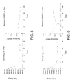
The enzymes from the deletion clone were analyzed for stability under the high temperature 65° C. FIG. 16 shows the accelerated storage test results of Geobacillus DNA polymerases I from various sources under high temperatures. The results indicated that the relative enzyme activity of the commercially available Bst DNA polymerase I (Lucigen Co.) was lowered to 41% of the original activity after 4 hours. The relative enzyme activity of wild-type Bku DNA polymerase I was lowered to 55% of the original activity after 4 hours. Under the same reaction conditions and over the same period, the relative enzyme activity of the polymerase mutant Del 1B of the deletion clone remained about 51% of the original activity; the relative enzyme activity of the polymerase mutant Del 1 and the polymerase mutant Del 4 remained about 48% of the original activity (referring to FIG. 16). Under the high temperature 65° C. for 8 hours, the relative enzyme activity of the commercially available Bst DNA polymerase I (Lucigen Co.) was lowered to 39% of the original activity. The relative enzyme activity of wild-type Bku DNA polymerase I remained 42% of the original activity. Under the same reaction conditions, the activity of the polymerase mutant Del 1B of the deletion clone was better than those of the commercially available Bst and wild-type Bku DNA polymerases I, 44% of the relative activity remained. The relative enzyme activities of the polymerase mutant Del 1 and the polymerase mutant Del 4 remained to be 39% and 23% (referring to FIG. 16). Based on the results, for the enzyme stability test under the high temperature 65° C., the wild-type Bku DNA polymerase I has a thermostability higher than that of the commercially available Bst DNA polymerase I (Lucigen Co.), and the stability of the polymerase mutant Del 1B of the deletion clone under the high temperature is better than that of the wild-type enzyme.
FIG. 17A-C show the stability test results of Geobacillus DNA polymerases I from various sources under different temperatures. The storage test under 4° C. lasted for 35 days and the results indicated that the relative enzyme activities of the commercially available Bst DNA polymerase I (Lucigen Co.), Bku DNA polymerase I and the mutant Del 1 remained to be respectively 89%, 83% and 86% of the original activity. The relative enzyme activity of the mutant Del 4 remained 75% of the original activity (referring to FIG. 17A). The results of the storage test under 25° C. showed that after 35 days, the relative enzyme activities of the commercially available Bst DNA polymerase I (Lucigen Co.), Bku DNA polymerase I and the mutant Del 1 remained to be respectively 84%, 83% and 88% of the original activity, while the relative enzyme activity of the mutant Del 4 remained about 73% of the original activity (referring to FIG. 17B). For the storage test under 40° C., after 35 days, the relative enzyme activity of the commercially available Bst DNA polymerase I (Lucigen Co.) was about 83% of the original activity, while the relative enzyme activities of Bku DNA polymerase I, mutant Del 1 and Del 4 remained to be respectively 87%, 91% and 89% of the original activity (referring to FIG. 17C). The results indicated the enzyme stability of Bku DNA polymerase I and the polymerase mutant Del 1 is better than that of the commercially available Bst DNA polymerase I (Lucigen Co.).
The present disclosure provides mutants of an isolated DNA polymerase I, and the isolated DNA polymerase I is Bku DNA polymerase I with the amino acid sequence comprising SEQ ID NO. 2. The mutant has an amino acid sequence by deleting at least one of five fragments from the amino acid sequences in the 3′→5′ exonuclease domain of the isolated DNA polymerase. The five fragments are E309-A313, P348, V358-T365, A404-Q405 and P424-E445.
According to the embodiments of this disclosure, the mutant has a thermostability substantially equivalent to that of Bst DNA polymerase I and a strand displacement activity substantially equivalent to that of the Bst DNA polymerase I.
This disclosure also directs to kits or compositions comprising the above mentioned isolated DNA polymerases, mutants or the combinations thereof. The kits may be applicable for polymerase chain reactions (PCR), nucleic acid amplification, whole genome amplification (WGA), multiple displacement amplification (MDA), loop-mediated isothermal amplification (LAMP) and DNA sequencing.
It will be apparent to those skilled in the art that various modifications and variations can be made to the structure of the present disclosure without departing from the scope or spirit of this disclosure. In view of the foregoing, it is intended that the present disclosure cover modifications and variations of this disclosure provided they fall within the scope of the following claims and their equivalents.

Claims (7)

What is claimed is:
1. A mutant of an isolated DNA polymerase, consisting of the amino acid sequence of SEQ ID NO. 3.
2. The mutant of claim 1, wherein the mutant has a thermostability substantially equivalent to that of Bst DNA polymerase and a strand displacement activity substantially equivalent to that of the Bst DNA polymerase.
3. A method for nucleic acid amplification reactions, comprises providing the mutant of claim 1 to prepare a nucleic acid amplification reaction composition.
4. The method of claim 3, wherein the nucleic acid amplification reaction comprises polymerase chain reactions (PCR), nucleic acid amplification, whole genome amplification (WGA), multiple displacement amplification (MDA) or DNA sequencing.
5. A kit comprising the mutant of claim 1.
6. The kit of claim 5, wherein the kit is applicable for a nucleic acid amplification reaction.
7. The kit of claim 6, wherein the nucleic acid amplification reaction comprises polymerase chain reactions (PCR), nucleic acid amplification, whole genome amplification (WGA), multiple displacement amplification (MDA) or DNA sequencing.
US14/138,155 2012-12-24 2013-12-23 Isolated DNA polymerases, kits and applications thereof Active US9309501B2 (en)

Priority Applications (1)

Application Number Priority Date Filing Date Title
US14/138,155 US9309501B2 (en) 2012-12-24 2013-12-23 Isolated DNA polymerases, kits and applications thereof

Applications Claiming Priority (2)

Application Number Priority Date Filing Date Title
US201261745649P 2012-12-24 2012-12-24
US14/138,155 US9309501B2 (en) 2012-12-24 2013-12-23 Isolated DNA polymerases, kits and applications thereof

Publications (2)

Publication Number Publication Date
US20140186894A1 US20140186894A1 (en) 2014-07-03
US9309501B2 true US9309501B2 (en) 2016-04-12

Family

ID=50989650

Family Applications (1)

Application Number Title Priority Date Filing Date
US14/138,155 Active US9309501B2 (en) 2012-12-24 2013-12-23 Isolated DNA polymerases, kits and applications thereof

Country Status (3)

Country Link
US (1) US9309501B2 (en)
CN (1) CN103898077B (en)
TW (1) TWI487789B (en)

Families Citing this family (6)

* Cited by examiner, † Cited by third party
Publication number Priority date Publication date Assignee Title
US10597643B2 (en) 2016-04-29 2020-03-24 Omniome, Inc. Polymerases engineered to reduce nucleotide-independent DNA binding
SG11201809522WA (en) 2016-04-29 2018-11-29 Omniome Inc Method of nucleic acid sequence determination
CN106399299B (en) * 2016-09-29 2019-01-29 华南理工大学 A kind of method and application improving large fragment Geobacillus stearothermophilus DNA polymerase activity by point mutation
EP3568490B1 (en) * 2017-01-10 2021-03-10 Omniome, Inc. Polymerases engineered to reduce nucleotide-independent dna binding
CN110634533B (en) * 2019-08-27 2022-08-16 中山大学 Method for obtaining controllable TRPV5 variant based on computer simulation
WO2023081653A1 (en) * 2021-11-02 2023-05-11 Ultima Genomics, Inc. Nucleic acid polymerase for incorporating labeled nucleotides

Citations (13)

* Cited by examiner, † Cited by third party
Publication number Priority date Publication date Assignee Title
US5436326A (en) * 1991-06-03 1995-07-25 Takara Shuzo Co., Ltd. Method for cloning of a gene for Pol I type DNA polymerase
US5814506A (en) 1995-08-02 1998-09-29 New England Biolabs, Inc. Over-expression and purification of a truncated thermostable DNA polymerase by protein fusion
US5830714A (en) 1996-04-17 1998-11-03 Molecular Biology Resources, Inc. Biologically active fragment of bacillus stearothermophilus DNA polymerase
TW528802B (en) 1996-08-06 2003-04-21 Hoffmann La Roche Modified thermostable DNA polymerase
TW588050B (en) 2000-09-05 2004-05-21 Academia Sinica A truncated form of fibrobacter succinogenes 1,3-1,4-beta-d-glucanase with improved enzymatic activity and thermo-tolerance
US7049101B1 (en) 1997-08-06 2006-05-23 Diversa Corporation Enzymes having high temperature polymerase activity and methods of use thereof
CN101134974A (en) 2006-08-28 2008-03-05 天津科技大学 Nucleic acid polymerase chain reaction optimized expanding method based on carbon nano-tube
US20080254525A1 (en) * 2005-06-28 2008-10-16 Invitrogen Corporation DNA Polymerase Blends and Mutant DNA Polymerases
US20080311626A1 (en) 2004-09-17 2008-12-18 Arkea Hf Dna Polymerases Having Strand Displacement Activity
CN102414315A (en) 2009-05-19 2012-04-11 森迪奈尔Ch有限责任公司 A dried and stabilized ready-to-use composition containing nucleic acid polymerization enzymes for molecular biology applications
US20120115145A1 (en) 2010-11-05 2012-05-10 Oxitec Ltd Strand displacement activity of modified polymerases and uses thereof
US20120115188A1 (en) 2008-11-03 2012-05-10 Kapa Biosystems Chimeric dna polymerases
WO2013033528A1 (en) * 2011-09-01 2013-03-07 Jennifer Ong Compositions and methods relating to variant dna polymerases and synthetic dna polymerases

Patent Citations (13)

* Cited by examiner, † Cited by third party
Publication number Priority date Publication date Assignee Title
US5436326A (en) * 1991-06-03 1995-07-25 Takara Shuzo Co., Ltd. Method for cloning of a gene for Pol I type DNA polymerase
US5814506A (en) 1995-08-02 1998-09-29 New England Biolabs, Inc. Over-expression and purification of a truncated thermostable DNA polymerase by protein fusion
US5830714A (en) 1996-04-17 1998-11-03 Molecular Biology Resources, Inc. Biologically active fragment of bacillus stearothermophilus DNA polymerase
TW528802B (en) 1996-08-06 2003-04-21 Hoffmann La Roche Modified thermostable DNA polymerase
US7049101B1 (en) 1997-08-06 2006-05-23 Diversa Corporation Enzymes having high temperature polymerase activity and methods of use thereof
TW588050B (en) 2000-09-05 2004-05-21 Academia Sinica A truncated form of fibrobacter succinogenes 1,3-1,4-beta-d-glucanase with improved enzymatic activity and thermo-tolerance
US20080311626A1 (en) 2004-09-17 2008-12-18 Arkea Hf Dna Polymerases Having Strand Displacement Activity
US20080254525A1 (en) * 2005-06-28 2008-10-16 Invitrogen Corporation DNA Polymerase Blends and Mutant DNA Polymerases
CN101134974A (en) 2006-08-28 2008-03-05 天津科技大学 Nucleic acid polymerase chain reaction optimized expanding method based on carbon nano-tube
US20120115188A1 (en) 2008-11-03 2012-05-10 Kapa Biosystems Chimeric dna polymerases
CN102414315A (en) 2009-05-19 2012-04-11 森迪奈尔Ch有限责任公司 A dried and stabilized ready-to-use composition containing nucleic acid polymerization enzymes for molecular biology applications
US20120115145A1 (en) 2010-11-05 2012-05-10 Oxitec Ltd Strand displacement activity of modified polymerases and uses thereof
WO2013033528A1 (en) * 2011-09-01 2013-03-07 Jennifer Ong Compositions and methods relating to variant dna polymerases and synthetic dna polymerases

Non-Patent Citations (22)

* Cited by examiner, † Cited by third party
Title
"Office Action of China Counterpart Application," issued on Jul. 3, 2015, p. 1-p. 6, in which the listed references were cited.
"Office Action of Taiwan Counterpart Application", issued on Dec. 23, 2014, p. 1-p. 7, in which the listed reference was.
Aliotta et al., "Thermostable Bst DNA polymerase I lacks a 3′-> 5′ proofreading exonuclease activity," Genetic Analysis: Biomolecular Engineering, Mar. 1996, pp. 185-195, vol. 12.
Aliotta et al., "Thermostable Bst DNA polymerase I lacks a 3'-> 5' proofreading exonuclease activity," Genetic Analysis: Biomolecular Engineering, Mar. 1996, pp. 185-195, vol. 12.
Branden and Tooze, Introduction to Protein Structure (1999), 2nd edition, Garland Science Publisher, pp. 3-12. *
EMBL DB ID: Q5KWC1-GEOKA, Feb. 1, 2005, pp. 1-9.
Goto et al., "Colorimetric detection of loopmediated isothermal amplification reaction by using hydroxy naphthol blue," Biotechniques, Mar. 2009, pp. 167-172, vol. 46.
Guo et al., Protein tolerance to random amino acid change, 2004, Proc. Natl. Acad. Sci. USA 101: 9205-9210. *
Henry and Romesberg, "The evolution of DNA polymerases with novel activities," Current Opinion in Biotechnology, Jul. 11, 2005, pp. 370-377, vol. 16.
Hill et al., Functional Analysis of conserved Histidines in ADP-Glucose Pyrophosphorylase from Escherichia coli, 1998, Biochem. Biophys. Res. Comm. 244:573-577. *
Holmberg et al, "Directed evolution of novel polymerases," Biomolecular Engineering, Jun. 2005, pp. 39-49, vol. 22.
Kiefer et al., "Crystal structure of a thermostable Bacillus DNA polymerase I large fragment at 2.1 Å resolution," Structure, Jan. 15, 1997, pp. 95-108, vol. 5, No. 1.
Kim et al., "Improved thermostability and PCR efficiency of Thermococcus celericrescens DNA polymerase via site-directed mutagenesis," Journal of Biotechnology, Jun. 23, 2011, pp. 156-163, vol. 155.
Lazar et al., Transforming Growth Factor alpha: Mutation of Aspartic Acid 47 and Leucine 48 Results in Different Biological Activity, 1988, Mol. Cell. Biol. 8:1247-1252. *
Lazar et al., Transforming Growth Factor α: Mutation of Aspartic Acid 47 and Leucine 48 Results in Different Biological Activity, 1988, Mol. Cell. Biol. 8:1247-1252. *
Lu et al., "Large fragment of DNA polymerase I from Bacillus stearothermophilus (Bst polymerase) is stable at ambient temperature", Biotechniques, Oct. 1991, pp. 464-466, vol. 11, No. 4.
Muhd Sakaff, et al., "Geobacillus thermoleovorans CCB-US3-UF5," NCBI Protein Database, retrieved date: Aug. 5, 2015.
Ong et al., "Directed Evolution of DNA Polymerase, RNA Polymerase and Reverse Transcriptase Activity in a Single Polypeptide," Journal of Molecular Biology, Aug. 18, 2006, pp. 537-550, vol. 361.
P00581; last viewed on Apr. 14, 2015. *
Parida et al., "Loop mediated isothermal amplification (LAMP): a new generation of innovative gene amplification technique; perspectives in clinical diagnosis of infectious diseases," Reviews in Medical Virology, Aug. 20, 2008, pp. 407-421.
Pavlov et al., "Cooperation between Catalytic and DNA Binding Domains Enhances Thermostability and Supports DNA Synthesis at Higher Temperatures by Thermostable DNA Polymerases," Biochemistry, Feb. 8, 2012, pp. 2032-2043, vol. 51.
Wacey et al., Disentangling the perturbational effects of amino acid substitutions in the DNA-binding domain of p53., Hum Genet, 1999, vol. 104, pp. 15-22. *

Also Published As

Publication number Publication date
TW201425582A (en) 2014-07-01
CN103898077A (en) 2014-07-02
US20140186894A1 (en) 2014-07-03
CN103898077B (en) 2017-01-11
TWI487789B (en) 2015-06-11

Similar Documents

Publication Publication Date Title
US9309501B2 (en) Isolated DNA polymerases, kits and applications thereof
Wu et al. Simplified gene synthesis: a one-step approach to PCR-based gene construction
CN109072202B (en) Marine DNA polymers
Yoshimura et al. Screening for the target gene of cyanobacterial cAMP receptor protein SYCRP1
CN111073871B (en) DNA polymerase mutant with improved thermal stability as well as construction method and application thereof
Ragone et al. The C-terminal end of the Trypanosoma brucei editing deaminase plays a critical role in tRNA binding
Marx PCR: the price of infidelity
US20230034466A1 (en) Dna polymerases
US20210355460A1 (en) Thermostable terminal deoxynucleotidyl transferase
Zhang et al. Mycobacterium tuberculosis CRISPR/Cas system Csm1 holds clues to the evolutionary relationship between DNA polymerase and cyclase activity
JP2022519020A (en) Methods of Sequencing Nucleic Acids Using Unnatural Base Pairs
CN112899255B (en) DNA polymerase and application thereof, recombinant vector and preparation method and application thereof, recombinant engineering bacteria and application thereof
Jajesniak et al. Rapid construction of recombinant plasmids by quickstep-cloning
Lin et al. Residues Phe103 and Phe149 are critical for the co-chaperone activity of Bacillus licheniformis GrpE
Karaoglu et al. Signature Peptides From Analytical Chemistry to Functional Genomics
McIsaac Elucidating replication dynamics involving the alpha subunit of DNA polymerase III and single stranded DNA binding protein in E. coli
Zayets et al. Site-Directed Mutagenesis of Tryptophan Residues in the Structure of the Catalytic Module of Tyrosyl-tRNA Synthetase from Bos taurus
Yang et al. Metagenomic DNA fragments that affect Escherichia coli mutational pathways
EP3763811A1 (en) Reverse transcriptase and uses thereof
Ye et al. A Highly Specific Fluorescent Hydrolysis Nucleic Acid Probe, Based on the Polymerization-Coupled Endonuclease Activity of Prokaryotic DNA Polymerase I
Prostova et al. DNA-targeting short Argonautes complex with effector proteins for collateral nuclease activity and bacterial population immunity
Kovermann et al. Structural and catalytic insights into HoLaMa, a derivative of Klenow DNA polymerase lacking the proofreading domain
Yu Optimization of PCR amplification for sensitive capture of Methanopyrus isoleucyl-tRNA synthetase gene in environmental samples
Mundhada Advancements of the Sequence Saturation Mutagenesis (SeSaM) Method to Efficiently Explore Protein Sequence Space
JPWO2015122432A1 (en) Method for improving the accuracy of nucleic acid amplification

Legal Events

Date Code Title Description
AS Assignment

Owner name: INDUSTRIAL TECHNOLOGY RESEARCH INSTITUTE, TAIWAN

Free format text: ASSIGNMENT OF ASSIGNORS INTEREST;ASSIGNORS:LIU, TSENG-HUANG;JIANG, PEI-SHIN;LIN, CHIH-LUNG;AND OTHERS;REEL/FRAME:032057/0001

Effective date: 20131220

STCF Information on status: patent grant

Free format text: PATENTED CASE

MAFP Maintenance fee payment

Free format text: PAYMENT OF MAINTENANCE FEE, 4TH YEAR, LARGE ENTITY (ORIGINAL EVENT CODE: M1551); ENTITY STATUS OF PATENT OWNER: LARGE ENTITY

Year of fee payment: 4

MAFP Maintenance fee payment

Free format text: PAYMENT OF MAINTENANCE FEE, 8TH YEAR, LARGE ENTITY (ORIGINAL EVENT CODE: M1552); ENTITY STATUS OF PATENT OWNER: LARGE ENTITY

Year of fee payment: 8