US9447152B2 - Base-detecting pore - Google Patents

Base-detecting pore Download PDF

Info

Publication number
US9447152B2
US9447152B2 US13/002,717 US200913002717A US9447152B2 US 9447152 B2 US9447152 B2 US 9447152B2 US 200913002717 A US200913002717 A US 200913002717A US 9447152 B2 US9447152 B2 US 9447152B2
Authority
US
United States
Prior art keywords
pore
seq
adaptor
residue
nucleotide
Prior art date
Legal status (The legal status is an assumption and is not a legal conclusion. Google has not performed a legal analysis and makes no representation as to the accuracy of the status listed.)
Active, expires
Application number
US13/002,717
Other versions
US20110177498A1 (en
Inventor
James Anthony Clarke
Lakmal Jayasinghe
Terence Reid
John Hagan Pryce Bayley
Current Assignee (The listed assignees may be inaccurate. Google has not performed a legal analysis and makes no representation or warranty as to the accuracy of the list.)
Oxford Nanopore Technologies PLC
Original Assignee
Oxford Nanopore Technologies PLC
Priority date (The priority date is an assumption and is not a legal conclusion. Google has not performed a legal analysis and makes no representation as to the accuracy of the date listed.)
Filing date
Publication date
Application filed by Oxford Nanopore Technologies PLC filed Critical Oxford Nanopore Technologies PLC
Priority to US13/002,717 priority Critical patent/US9447152B2/en
Assigned to OXFORD NANOPORE TECHNOLOGIES LIMITED reassignment OXFORD NANOPORE TECHNOLOGIES LIMITED ASSIGNMENT OF ASSIGNORS INTEREST (SEE DOCUMENT FOR DETAILS). Assignors: BAYLEY, HAGAN, CLARKE, JAMES, JAYASINGHE, LAKMAL, REID, TERENCE
Publication of US20110177498A1 publication Critical patent/US20110177498A1/en
Application granted granted Critical
Publication of US9447152B2 publication Critical patent/US9447152B2/en
Active legal-status Critical Current
Adjusted expiration legal-status Critical

Links

Images

Classifications

    • CCHEMISTRY; METALLURGY
    • C07ORGANIC CHEMISTRY
    • C07KPEPTIDES
    • C07K14/00Peptides having more than 20 amino acids; Gastrins; Somatostatins; Melanotropins; Derivatives thereof
    • C07K14/195Peptides having more than 20 amino acids; Gastrins; Somatostatins; Melanotropins; Derivatives thereof from bacteria
    • C07K14/24Peptides having more than 20 amino acids; Gastrins; Somatostatins; Melanotropins; Derivatives thereof from bacteria from Enterobacteriaceae (F), e.g. Citrobacter, Serratia, Proteus, Providencia, Morganella, Yersinia
    • C07K14/245Escherichia (G)
    • CCHEMISTRY; METALLURGY
    • C12BIOCHEMISTRY; BEER; SPIRITS; WINE; VINEGAR; MICROBIOLOGY; ENZYMOLOGY; MUTATION OR GENETIC ENGINEERING
    • C12QMEASURING OR TESTING PROCESSES INVOLVING ENZYMES, NUCLEIC ACIDS OR MICROORGANISMS; COMPOSITIONS OR TEST PAPERS THEREFOR; PROCESSES OF PREPARING SUCH COMPOSITIONS; CONDITION-RESPONSIVE CONTROL IN MICROBIOLOGICAL OR ENZYMOLOGICAL PROCESSES
    • C12Q1/00Measuring or testing processes involving enzymes, nucleic acids or microorganisms; Compositions therefor; Processes of preparing such compositions
    • C12Q1/68Measuring or testing processes involving enzymes, nucleic acids or microorganisms; Compositions therefor; Processes of preparing such compositions involving nucleic acids
    • C12Q1/6869Methods for sequencing
    • CCHEMISTRY; METALLURGY
    • C12BIOCHEMISTRY; BEER; SPIRITS; WINE; VINEGAR; MICROBIOLOGY; ENZYMOLOGY; MUTATION OR GENETIC ENGINEERING
    • C12QMEASURING OR TESTING PROCESSES INVOLVING ENZYMES, NUCLEIC ACIDS OR MICROORGANISMS; COMPOSITIONS OR TEST PAPERS THEREFOR; PROCESSES OF PREPARING SUCH COMPOSITIONS; CONDITION-RESPONSIVE CONTROL IN MICROBIOLOGICAL OR ENZYMOLOGICAL PROCESSES
    • C12Q2565/00Nucleic acid analysis characterised by mode or means of detection
    • C12Q2565/60Detection means characterised by use of a special device
    • C12Q2565/631Detection means characterised by use of a special device being a biochannel or pore

Definitions

  • the invention relates to a mutant ⁇ -hemolysin ( ⁇ -HL) pore which is useful for detecting one or more nucleotides by stochastic sensing.
  • the pore is particularly useful for sequencing DNA or RNA.
  • a molecular adaptor that allows detection of the nucleotide(s) is covalently attached to the pore.
  • the pore is specifically modified to facilitate positioning of the adaptor and may be modified to facilitate covalent attachment.
  • Stochastic detection is an approach to sensing that relies on the observation of individual binding events between nucleotide molecules and a receptor.
  • Stochastic sensors can be created by placing a single pore of nanometer dimensions in an insulating membrane and measuring voltage-driven ionic transport through the pore in the presence of nucleotide molecules. The frequency of occurrence of fluctuations in the current reveals the concentration of an nucleotide that binds within the pore. The identity of an nucleotide is revealed through its distinctive current signature, notably the duration and extent of current block (Braha, O., Walker, B., Cheley, S., Kasianowicz, J. J., Song, L., Gouaux, J. E., and Bayley, H. (1997) Chem. Biol. 4, 497-505; and Bayley, H., and Cremer, P. S. (2001) Nature 413, 226-230).
  • Engineered versions of the bacterial pore forming toxin ⁇ -hemolysin ( ⁇ -HL) have been used for stochastic sensing of many classes of molecules (Bayley, H., and Cremer, P. S. (2001) Nature 413, 226-230; Shin, S.-H., Luchian, T., Cheley, S., Braha, O., and Bayley, H. (2002) Angew. Chem. Int. Ed. 41, 3707-3709; and Guan, X., Gu, L.-Q., Cheley, S., Braha, O., and Bayley, H. (2005) ChemBioChem 6, 1875-1881).
  • a mutant ⁇ -HL pore having a molecular adaptor covalently attached to a lower part of its barrel or channel such that it is positioned at or near residue 139 is capable of detecting and distinguishing between nucleotides.
  • the inventors have shown that a mutant ⁇ -HL pore having a molecular adaptor covalently attached such that it is positioned at or near residue 139 is capable of distinguishing between different nucleotides.
  • This pore is highly sensitive and can therefore be used to sequence nucleic acids, such as DNA or RNA.
  • a mutant ⁇ -HL pore having a molecular adaptor covalently attached such that it is positioned at or near residue 139 is capable of distinguishing between different nucleotides under a range of different conditions.
  • the pore will distinguish between nucleotides under conditions that are favourable to enzymes whose function is needed for the sequencing of nucleic acids.
  • modification of a mutant ⁇ -HL pore at and/or near residue 139 is essential for positioning the adaptor and detecting and distinguishing between different nucleotides.
  • the pores of the invention are useful tools for stochastic sensing, especially for detecting nucleotides or sequencing nucleic acids, such as DNA or RNA.
  • the invention provides a mutant ⁇ -HL pore for use in detecting one or more nucleotides in a sample, which comprises:
  • one or more of the seven subunits is modified at and/or near residue 139 of SEQ ID NO: 2 to facilitate positioning of the adaptor
  • molecular adaptor is covalently attached to one or more of the subunits such that it is positioned at or near residue 139 of SEQ ID NO: 2.
  • the invention also provides:
  • FIG. 1 shows the structure of the adaptor used in the Example.
  • This Figures shows the structure of the unreacted adaptor, heptakis(6-deoxy-6-amino)- ⁇ -cyclodextrin (am 7 - ⁇ CD), as well as the structure of the adaptor once it has been reacted with the bifunctional crosslinker succinimidyl 3-(2-pyridyldithio)propionate (SPDP) to form am 6 amPDP 1 - ⁇ CD.
  • SPDP succinimidyl 3-(2-pyridyldithio)propionate
  • the Figure also shows the structure of the control adaptor used in the Examples to investigate the effect of the amine groups on the adaptor (amPDP 1 - ⁇ CD).
  • FIG. 2 shows single channel recordings of the HL-(M113R) 7 in a wild-type background (top) and HL-(M113R) 7 in a RL2 background (bottom). am 7 - ⁇ CD binding and dNMP detection can be seen.
  • FIG. 3 shows the corresponding histograms of dNMP events from FIG. 1 .
  • Ledt is wild-type and right is RL2 (1200 mM KCl, 150 mV, pH 7.5).
  • FIG. 4 shows a diagram indicating the key mutations of the RL2 compared to the wild-type.
  • FIG. 5 shows a histogram of residual current binding when dNMPs are bound to the HL-(M113R) 7 RL2 background and the HL-(M113R/N139Q) 7 wt background.
  • This Figure shows the importance of the N139Q mutation in the RL2 background (compare with FIG. 3 ) (800 mM KCl, 160 mV, pH 7.5).
  • FIG. 6 shows the proposed cyclodextrin position at residue 139 of ⁇ HL.
  • FIG. 7 shows a diagram indicating the location of key mutations in the ⁇ -barrel of ⁇ HL.
  • FIG. 8 shows single channel recording of the HL-(M113R/N139Q) 6 (M113R/T115C-D8) 1 mutant. Reaction with the am 6 amPDP 1 - ⁇ CD can be seen resulting in a fluctuating, noisy baseline (no bases present).
  • FIG. 9 shows single channel recording of the HL-(M113R/N139Q) 6 (M113R/T117C-D8) 1 mutant, which gives a fluctuating, noisy baseline (no bases present).
  • FIG. 10 shows single channel recording of the HL-(M113R/N139Q) 6 (M113R/G119C-D8) 1 mutant reacted with the am 6 amPDP 1 - ⁇ CD before and after the nucleotides are added. It also shows a close up of the baseline after nucleotides are added.
  • FIG. 11 shows single channel recording of the HL-(M113R/N139Q) 6 (M113R/N139Q/N121C-D8) 1 mutant reacted with the am 6 amPDP 1 - ⁇ CD after nucleotides were added.
  • FIG. 12 shows histograms for the HL-(M113R/N139Q) 6 (M113R/G119C-D8) 1 (left) and the HL-(M113R/N139Q) 6 (M113R/N139Q/N121C-D8) 1 mutant (right). Limited base discrimination can be seen at 800 mM KCl, 160 mV, pH 7.5.
  • FIG. 13 shows single channel recording of the HL-(M113R/N139Q) 6 (M113R/N139Q/N123C-D8) 1 mutant (5 kHz software filtered) reacted with the am 6 amPDP 1 - ⁇ CD after dGMP, dTMP, dAMP, dCMP were added (1 second shown).
  • FIG. 14 shows histograms for the HL-(M113R/N139Q) 6 (M113R/N139Q/N123C-D8) 1 (left) and the HL-(M113R/N139Q) 6 (M113R/N139Q/G137C-D8) 1 mutant (right). Limited base discrimination can be seen (800 mM KCl, 160 mV, pH 7.5).
  • FIG. 15 shows single channel recording of the HL-(M113R/N139Q) 6 (M113R/N139Q/L135C-D8) 1 mutant (5 kHz software filtered) reacted with the am 6 amPDP 1 - ⁇ CD after dGMP, dTMP, dAMP and dCMP were added (1 second shown).
  • FIG. 16 shows a histogram for the HL-(M113R/N139Q) 6 (M113R/N139Q/L135C-D8) 1 mutant. Good base discrimination can be seen (800 mM KCl, 160 mV, pH 7.5).
  • FIG. 17 shows residual current histograms of dNMP binding for the HL-(M113R/N139Q) 6 (M113R/N139Q/L135C-D8) 1 mutant at a range of applied potentials (110-170 mV). The changes in the relative positions of the four nucleotides can be seen. The sharp black peak corresponds to the cyclodextrin level.
  • FIG. 18 shows a plot of the variation of peak position (residual pore current) with applied potential when a single nucleotide is bound to the pore.
  • FIG. 19 shows a plot of the residual current histograms, Gaussian fits of each base and calculated areas of overlap for neighbouring bases for runs at applied potentials of 130, 150 and 170 mV.
  • FIG. 20 shows the dwell time versus mean of residual pore current. Peaks from all four bases (G T A C, bottom to top) can be seen, as can the longer average dwell time of dTMP.
  • FIG. 21 shows a histogram for the HL-(M113R/N139Q) 6 (M113R/N139Q/L135C-D8) 1 mutant. Good base discrimination can be seen at low salt (400 mM KCl, 180 mV, pH 7.5).
  • FIG. 22 shows single channel recording of the HL-(M113R/N139Q) 6 (M113R/N139Q/L135C-D8) 1 mutant (5 kHz software filtered) reacted with the am 6 amPDP 1 - ⁇ CD after dNMPs were added (350 mM KCl, 180 mV, pH 7.5).
  • FIG. 23 shows a histogram for the HL-(M113R/N139Q) 6 (M113R/N139Q/L135C-D8)1 mutant. Good base discrimination can be seen at low salt (350 mM KCl, 180 mV, pH 7.5).
  • FIG. 24 shows a plot of hemolysin conductance versus solution temperature for a single channel in 300 mM KCl, 25 mM Tris, starting pH 7.5. An increase of 1.65 pA/° C. can be seen.
  • FIG. 25 shows a histogram for the HL-(M113R/N139Q) 6 (M113R/N139Q/L135C-D8) 1 mutant showing good base detection at low salt and elevated temperature (300 mM KCl, 180 mV, pH 7.2, 40° C.) (left) and a plot of dwell time versus amplitude of the same data showing a short dwell time (right).
  • FIG. 26 shows the chemical structures of the dCMP and methyl-dCMP (dTMP included for comparison).
  • FIG. 27 shows a histogram for the HL-(M113R/N139Q) 6 (M113R/N139Q/L135C-D8) 1 mutant.
  • dGMP and methyl-dCMP base detection can be seen (800 mM KCl, 180 mV, pH 7.5).
  • FIG. 28 shows a histogram for the HL-(M113R/N139Q) 6 (M113R/N139Q/L135C-D8) 1 mutant. Detection of all four standard nucleotide monophosphates (left) and all four standard nucleotide monophosphates with methyl-dCMP added (right) can be seen (800 mM KCl, 170 mV, pH 7.5).
  • FIG. 29 shows dwell time versus mean of residual pore current. Peaks from all four bases and methyl-dCMP (left) and the Gaussian fits of the residual current histograms for the five bases (right) can be seen.
  • FIG. 30 shows the residual current histograms of dNMP binding for the HL-(M113R/N139Q) 6 (M113R/N139Q/L135C-D8) 1 mutant at a range of applied potentials (130-170 mV). The changes in the relative positions of the four nucleotides with methyl-dCMP added can be seen.
  • FIG. 31 shows the chemical structures of the bases commonly found in DNA and the corresponding RNA bases.
  • FIG. 33 shows single channel recordings for the homo-heptamer HL-(M113R/N139Q/N121C) 7 with am 6 amPDP 1 - ⁇ CD reacted at the 121 position.
  • FIG. 34 shows single channel recording of the HL-(M113R/N139Q) 6 (M113R/N139Q/L135C-D8) 1 mutant (5 kHz software filtered) reacted with the PDP 1 - ⁇ CD after dGMP, dTMP, dAMP nd dCMP was added (800 mM KCl, pH 7.5, 160 mV, 5 kHz filter, 0.5 seconds shown).
  • FIG. 35 shows single channel recording of the HL-(M113R/N139Q) 6 (M113R/N139Q/L135C-D8) 1 mutant (5 kHz software filtered) reacted with the am 6 amPDP 1 - ⁇ CD after dGMP, dTMP, dAMP and dCMP was added (800 mM KCl, pH 7.5, 160 mV, 5 kHz filter, 0.5 seconds shown).
  • FIG. 36 shows single channel recording of the HL-(wt) 6 (L135C-D8) 1 mutant (5 kHz software filtered) reacted with the am 6 amPDP 1 - ⁇ CD after dGMP, dTMP, dAMP and dCMP was added (800 mM KCl, pH 7.5, 160 mV, 5 kHz filter, 0.5 seconds shown) and the corresponding residual current histogram.
  • FIG. 37 shows single channel recording of the HL-(M113R) 6 (M113R/L135C-D8) 1 mutant (5 kHz software filtered) reacted with the am 6 amPDP 1 - ⁇ CD after dGMP, dTMP, dAMP and dCMP was added (800 mM KCl, pH 7.5, 160 mV, 5 kHz filter, 0.5 seconds shown) and the corresponding residual current histogram.
  • FIG. 38 shows single channel recording of the HL-(N139Q) 6 (N139Q/L135C-D8) 1 mutant (5 kHz software filtered) reacted with the am 6 amPDP 1 - ⁇ CD after dGMP, dTMP, dAMP and dCMP was added (800 mM KCl, pH 7.5, 160 mV, 5 kHz filter, 0.5 seconds shown) and the corresponding residual current histogram.
  • SEQ ID NO: 1 shows the polynucleotide sequence that encodes one subunit of wild-type ⁇ -hemolysin ( ⁇ -HL).
  • SEQ ID NO: 2 shows the amino acid sequence of one subunit of wild-type ⁇ -HL.
  • SEQ ID NO: 3 shows the polynucleotide sequence that encodes one subunit of ⁇ -HL M113R-RL2.
  • SEQ ID NO: 4 shows the amino acid sequence of one subunit of ⁇ -HL M113R-RL2.
  • SEQ ID NO: 5 shows the polynucleotide sequence that encodes one subunit of ⁇ -HL M113R with a wild-type background.
  • SEQ ID NO: 6 shows the amino acid sequence of one subunit of ⁇ -HL M113R with a wild-type background.
  • SEQ ID NO: 7 shows the polynucleotide sequence that encodes one subunit of ⁇ -HL M113R/N139Q.
  • SEQ ID NO: 8 shows the amino acid sequence of one subunit of ⁇ -HL M113R/N139Q.
  • SEQ ID NO: 9 shows the polynucleotide sequence that encodes one subunit of ⁇ -HL M113R/N139Q/G119C-D8.
  • SEQ ID NO: 10 shows the amino acid sequence of one subunit of ⁇ -HL M113R/N139Q/G119C-D8.
  • SEQ ID NO: 11 shows the polynucleotide sequence that encodes one subunit of ⁇ -HL M113R/N139Q/N121C-D8.
  • SEQ ID NO: 12 shows the amino acid sequence of one subunit of ⁇ -HL M113R/N139Q/N121C-D8.
  • SEQ ID NO: 13 shows the polynucleotide sequence that encodes one subunit of ⁇ -HL M113R/N139Q/L135C-D8.
  • SEQ ID NO: 14 shows the amino acid sequence of one subunit of ⁇ -HL M113R/N139Q/L135C-D8.
  • SEQ ID Nos 2, 4, 6, 8, 10, 12 and 14 are mature forms that lack the amino terminal methionine.
  • SEQ ID NO: 15 shows the polynucleotide sequence encoding the exonuclease III enzyme from E. coli.
  • SEQ ID NO: 16 shows the amino acid sequence of the exonuclease III enzyme from E. coli.
  • SEQ ID NO: 17 shows the polynucleotide sequence encoding the exonuclease I enzyme from E. coli.
  • SEQ ID NO: 18 shows the amino acid sequence of the exonuclease I enzyme from E. coli.
  • SEQ ID NO: 19 shows the polynucleotide sequence encoding the bacteriophage lambda exonuclease.
  • SEQ ID NO: 20 shows the amino acid sequence of the bacteriophage lambda exonuclease. The sequence is one of three identical subunits that assemble into a trimer.
  • nucleotide includes “nucleotides”
  • a pore includes two or more such pores
  • a molecular adaptor includes two or more such adaptors
  • the present invention provides mutant ⁇ -HL pores for use in detecting one or more nucleotide(s).
  • the pores may also be used for distinguishing between or discriminating between different nucleotides.
  • the pores comprise a molecular adaptor that facilitates an interaction with the nucleotide(s).
  • the adaptor is covalently attached to the pore in a specific position.
  • the adaptor is positioned such that, during the interaction between a nucleotide and the pore, the nucleotide affects the current flowing through the pore in a manner specific for that nucleotide.
  • the adaptor is therefore covalently attached to the pore in a position that allows the pore to be used to detect the nucleotide or discriminate nucleotides via stochastic sensing.
  • the pores of the invention are useful tools for stochastic sensing.
  • the pores of the invention are particularly useful for discriminating between nucleotides.
  • the pores are therefore ideal for sequencing nucleic acids.
  • the pores of the invention can discriminate between different nucleotides with a high degree of sensitivity.
  • the pores can easily distinguish between the four nucleotides in DNA and RNA.
  • the pores of the invention can even distinguish between methylated and unmethylated nucleotides.
  • the base resolution of a mutant ⁇ -HL pore having the adaptor covalently attached to residue 135 is surprisingly high.
  • the pore shows almost complete separation of all four DNA nucleotides making it an excellent candidate for the sequencing of nucleic acids.
  • the pore also allows deoxythymidine monophosphate (dTMP) to be clearly distinguished from the other three DNA nucleotides based on its longer dwell time in the pore.
  • the pore further discriminates between deoxycytidine monophosphate (dCMP) and methyl-dCMP based on the dwell time in the pore and the current flowing through the pore.
  • the pores of the invention can also discriminate between different nucleotides under a range of conditions. In particular, the pores will discriminate between nucleotides under conditions that are favourable to the sequencing of nucleic acids.
  • One way involves the use of an exonuclease enzyme. In this approach, the exonuclease enzyme is used to sequentially detach the nucleotides from the DNA or RNA strand. Such enzymes do not normally function under high salt concentrations.
  • the pores of the invention are able to discriminate between nucleotides even at low salt concentrations.
  • the pores can function with good nucleotide discrimination at a KCl concentration as low as 300 mM at room temperature.
  • the pores will function with good nucleotide discrimination at lower salt concentrations if the temperature is increased or if asymmetric salt solutions are used. This is discussed in more detail below.
  • the extent to which the pores of the invention can discriminate between different nucleotides can be controlled by altering the applied potential. This allows the function of the pores to be fine-tuned, particularly when sequencing.
  • the fixed nature of the molecular adaptor also means that the signal obtained from the pore is entirely dependent on the presence of a nucleotide in the barrel or channel of the pore and is not affected by dissociation of the adaptor from the pore.
  • the fixed nature of the adaptor means that a distinctive current will flow through the pore whenever a nucleotide interacts with the pore. This is particularly important for sequencing nucleic acids because every nucleotide in the sequence needs to be detected and identified.
  • the pores of the invention can be designed such that they do not undergo blocking. In electrophysiology, pores can become blocked by large multivalent ions. The ions become trapped in the barrel or channel of the pore and prevent the flow of ionic current. Blocking is normally avoided by carrying out experiments in ultra-high grade purity salt solution.
  • the introduction of positively charged residues, such as arginine, near the constriction of the barrel or channel of ⁇ -HL generally results in pores that are prone to blocking.
  • it is not essential that the mutant ⁇ -HL pores of the invention have one or more positively charge residues near the constriction of their barrel or channel. Pores of the invention lacking such residues can be used in the presence of large multivalent ions without undergoing blocking.
  • the fixed nature of the molecular adaptor means that the pore and adaptor can be stored together, thereby allowing the production of a ready-to-use biosensor.
  • a pore of the invention may be isolated, substantially isolated, purified or substantially purified.
  • a pore of the invention is isolated or purified if it is completely free of any other components, such as lipids or other pores.
  • a pore is substantially isolated if it is mixed with carriers or diluents which will not interfere with its intended use.
  • a pore is substantially isolated or substantially purified if it present in a form that comprises less than 10%, less than 5%, less than 2% or less than 1% of other components, such as lipids or other pores.
  • a pore of the invention may be present in a lipid bilayer.
  • a pore of the invention may be present as an individual or single pore.
  • a pore of the invention may be present in a homologus or heterologous population of two or more pores.
  • the wild-type ⁇ -HL pore is formed of seven identical monomers or subunits (i.e. it is heptameric). The sequence of one wild-type monomer or subunit of ⁇ -hemolysin is shown in SEQ ID NO: 2.
  • a mutant ⁇ -HL pore is a heptameric pore in which one or more of the seven subunits has an amino acid sequence which varies from that of SEQ ID NO: 2 and which retains pore activity.
  • the pore includes modifications that facilitate positioning of the adaptor as discussed below.
  • the pore preferably also contains modifications that facilitate covalent attachment of the adaptor.
  • 1, 2, 3, 4, 5, 6 of the subunits in the pore may have an amino acid sequence that varies from that of SEQ ID NO: 2.
  • all of the subunits in the pore have an amino acid sequence that varies from that of SEQ ID NO: 2.
  • All seven subunits within the pore may be identical but are typically different, particularly since different subunits are modified in different ways to facilitate positioning of the adaptor as discussed below and optionally to facilitate covalent attachment of the adaptor.
  • a mutant ⁇ -HL pore of the invention is formed from seven subunits each comprising the sequence shown in SEQ ID NO: 2 or a variant thereof.
  • a variant is a subunit that has an amino acid sequence which varies from that of SEQ ID NO: 2 and which retains its ability to form a pore. All of the sequences shown in SEQ ID NOs: 4, 6, 8, 10, 12 and 14 are variants of SEQ ID NO: 2. Any of the specific modifications to SEQ ID NO: 2 discussed below also result in variants of SEQ ID NO: 2.
  • Variants include the constructs, which comprise the sequence shown in SEQ ID NO: 2 or a variant thereof and are described in a co-pending International application claiming priority from U.S. Application No. 61/078,695 and being filed simultaneously with this application [J A Kemp & Co Ref: N.104404A; Oxford Nanolabs Ref: ONL IP 005].
  • Such constructs also comprise a nucleic acid handling enzyme, such as an exonuclease. All the teachings of that application may be applied equally to the present invention.
  • the pore is modified to facilitate positioning of the adaptor so that the one or more nucleotides can be detected.
  • One or amino acids are introduced into the pore to hold the adaptor in the correct position via non-covalent interactions, such as hydrophobic interactions, hydrogen bonding, Van der Waal's forces, ⁇ -cation interactions and/or electrostatic forces.
  • One or more of the seven subunits are modified at and/or near residue 139 of SEQ ID NO: 2. For instance, 2, 3, 4, 5, 6 or all of the seven subunits are modified at and/or near residue 139 of SEQ ID NO: 2.
  • One or more of the seven subunits include at least one, such as 2, 3, 4 or 5, modifications at and/or near residue 139 of SEQ ID NO: 2.
  • One or more of the seven subunits are modified at residue 139 and/or near residue 139 of SEQ ID NO: 2. If one or more of the seven subunits are modified near residue 139, the modifications are sufficiently close to residue 139 that they can facilitate positioning of the adaptor.
  • the modifications are typically made at positions that are less than 20 ⁇ ngströms, such as less than 15, less than 10 or less than 5 ⁇ ngströms, from residue 139 in SEQ ID NO: 2.
  • Residues that are 2 or 3 amino acids from residue 139 in the sequence of SEQ ID NO: 2 are preferably modified in one or more of the seven subunits.
  • Residue 139 and/or one or both of the residues adjacent to residue 139 in the sequence of SEQ ID NO: 2 are more preferably modified in one or more of the seven subunits.
  • One or more of the seven subunits preferably comprise a modification at residue 136, 137, 138, 139, 140, 141 or 142 of SEQ ID NO: 2 or any combination thereof.
  • One or more of the seven subunits most preferably comprise a modification at only residue 139 of SEQ ID NO: 2.
  • One or more of the seven subunits may comprise modifications at residues in SEQ ID NO: 2 that are near to residue 139 once the sequence is folded to form a three-dimensional subunit.
  • One or more of the seven subunits preferably comprise a modification at residue 117, 119, 121, 123, 135, 137, 141 or 143 of SEQ ID NO: 2 or any combination thereof.
  • the subunits are preferably modified at and/or near to residue 139 by the introduction of polar, uncharged amino acids.
  • Such amino acids may influence the positioning of the adaptor via hydrogen bonding. Their lack of charge also prevents electrostatic interactions from interfering with the function of the adaptor.
  • Table 1 below lists polar, uncharged amino acids.
  • Glutamine is a preferred polar, uncharged amino acid.
  • the polar, uncharged amino acids can be inserted at and/or near residue 139.
  • amino acids at and/or near residue 139 can be substituted with polar, uncharged amino acids.
  • One or more of the seven subunits are preferably modified to comprise glutamine, which is a polar, uncharged amino acid, at and/or near residue 139 of SEQ ID NO: 2.
  • glutamine can be inserted at and/or near residue 139 or amino acids at and/or near residue 139 can be substituted with glutamine.
  • the amino acid at residue 139 is preferably substituted with glutamine (N139Q).
  • N139Q glutamine
  • 2, 3, 4, 5 or 6 of the seven subunits have glutamine at residue 139 of SEQ ID NO: 2.
  • all of the seven subunits have a glutamine at residue 139 of SEQ ID NO: 2.
  • Uncharged glutamine at residue 139 is capable of interacting with chemical groups, such as hydroxyl groups, in the adaptor by hydrogen bonding and thereby facilitating the positioning of the adaptor within the barrel or channel of the mutant ⁇ -HL pore.
  • Preferred subunits having residue 139 of SEQ ID NO: 2 substituted with glutamine (N139Q) are shown in SEQ ID NOs: 8, 10, 12 and 14.
  • Suitable subunits may contain or lack the octa-asparate tail shown in SEQ ID NOs: 8, 10, 12 and 14.
  • the pore may also have other uncharged amino acids or even aromatic amino acids located near the constriction of the barrel or channel to further facilitate positioning of the adaptor.
  • Table 1 lists uncharged and aromatic amino acids.
  • one or more of the seven subunits may have one or more uncharged amino acids, such as asparagine, or one or more aromatic amino acids, such as phenylalanine, located near the constriction of the barrel or channel.
  • the pore preferably has a ring of 4, 5, 6 or preferably 7 uncharged or aromatic amino acids located near the constriction of the barrel or channel. Each amino acid in the ring is typically provided by each of the subunits.
  • Residues of SEQ ID NO: 2 located near the constriction of the barrel or channel include, but are not limited to, 111, 113 and 147.
  • Suitable subunits include an uncharged or aromatic amino acid at residue 111, 113 or 147 of SEQ ID NO: 2.
  • the uncharged or aromatic amino acids can be inserted at residue 111, 113 or 147 of SEQ ID NO: 2.
  • the amino acids at residue 111, 113 or 147 of SEQ ID NO: 2 can be substituted with uncharged or aromatic amino acids.
  • the pore is preferably modified to facilitate covalent attachment of the adaptor.
  • One or more amino acids that are capable of forming a covalent bond such as cysteine, can be introduced into one or more subunits.
  • the amino acid may be naturally occurring or non-naturally occurring.
  • An amino acid may be introduced by addition.
  • An amino acid is preferably introduced by substitution.
  • Amino acids can be introduced at any positions as long as the adaptor is positioned at or near residue 139 of SEQ ID NO: 2. If the amino acids are introduced at residues distant from residue 139, a bifunctional crosslinker of appropriate length may be used to ensure that the adaptor is positioned at or near residue 139.
  • residue 119, 121 or 135 of SEQ ID NO: 2 is modified in one or more of the seven subunits to facilitate covalent attachment of the adaptor. In more preferred embodiments, residue 119, 121 or 135 of SEQ ID NO: 2 is modified in only one of the seven subunits to facilitate covalent attachment of the adaptor.
  • the amino acid introduced at residue 119, 121 or 135 of SEQ ID NO: 2 is preferably cysteine.
  • the amino acid at residue 119, 121 or 135 is preferably substituted with cysteine (G119C, N121C or L135C).
  • a preferred subunit having position 119 of SEQ ID NO: 2 substituted with cysteine (G119C) is shown in SEQ ID NO: 10.
  • a preferred subunit having residue 121 of SEQ ID NO: 2 substituted with cysteine (N121C) is shown in SEQ ID NO: 12.
  • a preferred subunit having residue 135 of SEQ ID NO: 2 substituted with cysteine (L135C) is shown in SEQ ID NO: 14.
  • the pore preferably has positively charged amino acids located near the constriction of the barrel or channel to facilitate covalent attachment of the adaptor.
  • one or more of the seven subunits may have positively charged amino acids, such as arginine, lysine or histidine, located near the constriction of the barrel or channel.
  • the positively charged amino acids facilitate covalent attachment of the adaptor by electrostatic interactions with positively charged groups, such as amines, in the adaptor. More specifically, the positively charged amino acids repel the positively charged groups in the adaptor, push the adaptor down the barrel or channel of the pore and position it at or near residue 139 of SEQ ID NO: 2. This facilitates the covalent reaction between the adaptor and the pore.
  • pores having positively charged amino acids located near the constriction of the barrel or channel may be prone to blocking.
  • the pore preferably has a ring of 4, 5, 6 or preferably 7 positively charged amino acids, such as arginine, lysine or histidine, located near the constriction of the barrel or channel.
  • Each amino acid in the ring is typically provided by each of the subunits.
  • one or more of the seven subunits has a positively charged amino acid, such as arginine, lysine or histidine, at residue 113 of SEQ ID NO: 2.
  • one or more of the seven subunits has an arginine residue at residue 113 of SEQ ID NO: 2.
  • all of the seven subunits have an arginine residue at residue 113 of SEQ ID NO: 2.
  • Arginine can be introduced at residue 113 or residue 113 can be substituted with arginine (M113R).
  • Preferred subunits having residue 113 of SEQ ID NO: 2 substituted with arginine (M113R) are shown in SEQ ID NOs: 4, 6, 8, 10, 12 and 14.
  • the subunits may be a naturally occurring variants which are expressed by an organism, for instance by a Staphylococcus bacterium. Variants also include non-naturally occurring variants produced by recombinant technology. Over the entire length of the amino acid sequence of SEQ ID NO: 2, a variant will preferably be at least 50% homologous to that sequence based on amino acid identity. More preferably, the subunit polypeptide may be at least 55%, at least 60%, at least 65%, at least 70%, at least 75%, at least 80%, at least 85%, at least 90% and more preferably at least 95%, 97% or 99% homologous based on amino acid identity to the amino acid sequence of SEQ ID NO: 2 over the entire sequence. There may be at least 80%, for example at least 85%, 90% or 95%, amino acid identity over a stretch of 200 or more, for example 230, 250, 270 or 280 or more, contiguous amino acids (“hard homology”).
  • Amino acid substitutions may be made to the amino acid sequence of SEQ ID NO: 2 in addition to those discussed above, for example up to 1, 2, 3, 4, 5, 10, 20 or 30 substitutions.
  • Conservative substitutions may be made, for example, according to Table 1 below.
  • One or more amino acid residues of the amino acid sequence of SEQ ID NO: 2 may additionally be deleted from the polypeptides described above. Up to 1, 2, 3, 4, 5, 10, 20 or 30 residues may be deleted, or more.
  • Variants may include subunits made of fragments of SEQ ID NO: 2. Such fragments retain pore forming activity. Fragments may be at least 50, 100, 200 or 250 amino acids in length. Such fragments may be used to produce chimeric pores. A fragment preferably comprises the pore forming domain of SEQ ID NO: 2. Fragments must include residues 139 and 119, 121 or 135 of SEQ ID NO: 2.
  • Variants include chimeric proteins comprising fragments or portions of SEQ ID NO: 2.
  • Chimeric protein pores may be formed from one or more subunits each comprising fragments or portions of SEQ ID NO: 2.
  • the pore or channel part of a chimeric protein pore is typically formed by the fragments or portions of SEQ ID NO: 2.
  • One or more amino acids may be alternatively or additionally added to the polypeptides described above.
  • An extension may be provided at the amino terminal or carboxy terminal of the amino acid sequence of SEQ ID NO: 2 or polypeptide variant or fragment thereof.
  • the extension may be quite short, for example from 1 to 10 amino acids in length. Alternatively, the extension may be longer, for example up to 50 or 100 amino acids.
  • a carrier protein may be fused to an amino acid sequence according to the invention.
  • a variant is a subunit that has an amino acid sequence which varies from that of SEQ ID NO: 2 and which retains its ability to form a pore.
  • a variant typically contains the regions of SEQ ID NO: 2 that are responsible for pore formation.
  • the pore forming ability of ⁇ -HL, which contains a ⁇ -barrel, is provided by ⁇ -sheets in each subunit.
  • a variant of SEQ ID NO: 2 typically comprises the regions in SEQ ID NO: 2 that form ⁇ -sheets.
  • Amino acids 22 to 30, 35 to 44, 52 to 62, 67 to 71, 76 to 91, 98 to 103, 112 to 123, 137 to 148, 154 to 159, 165 to 172, 229 to 235, 243 to 261, 266 to 271, 285 to 286 and 291 to 293 of SEQ ID NO: 2 form ⁇ -sheets.
  • One or more modifications can be made to the regions of SEQ ID NO: 2 that form ⁇ -sheets as long as the resulting variant retains its ability to form a pore. Specific modifications that can be made to the ⁇ -sheet regions of SEQ ID NO: 2 are discussed above.
  • a variant of SEQ ID NO: 2 preferably includes one or more modifications, such as substitutions, additions or deletions, within its ⁇ -helices and/or loop regions.
  • Amino acids 2 to 6, 73 to 75, 207 to 209, 214 to 216 and 219 to 222 of SEQ ID NO: 2 form ⁇ -helices.
  • Amino acids 7 to 21, 31 to 34, 45 to 51, 63 to 66, 72, 92 to 97, 104 to 111, 124 to 136, 149 to 153, 160 to 164, 173 to 206, 210 to 213, 217, 218, 223 to 228, 236 to 242, 262 to 265, 272 to 274 and 287 to 290 of SEQ ID NO: 2 form loops.
  • Amino acids 1 and 294 are terminal amino acids.
  • Standard methods in the art may be used to determine homology.
  • the UWGCG Package provides the BESTFIT program which can be used to calculate homology, for example used on its default settings (Devereux et al (1984) Nucleic Acids Research 12, p 387-395).
  • the PILEUP and BLAST algorithms can be used to calculate homology or line up sequences (such as identifying equivalent residues or corresponding sequences (typically on their default settings)), for example as described in Altschul S. F. (1993) J Mol Evol 36:290-300; Altschul, S. F et al (1990) J Mol Biol 215:403-10.
  • HSPs high scoring sequence pair
  • T some positive-valued threshold score
  • Altschul et al, supra these initial neighbourhood word hits act as seeds for initiating searches to find HSP's containing them.
  • the word hits are extended in both directions along each sequence for as far as the cumulative alignment score can be increased.
  • Extensions for the word hits in each direction are halted when: the cumulative alignment score falls off by the quantity X from its maximum achieved value; the cumulative score goes to zero or below, due to the accumulation of one or more negative-scoring residue alignments; or the end of either sequence is reached.
  • the BLAST algorithm parameters W, T and X determine the sensitivity and speed of the alignment.
  • the BLAST algorithm performs a statistical analysis of the similarity between two sequences; see e.g., Karlin and Altschul (1993) Proc. Natl. Acad. Sci. USA 90: 5873-5787.
  • One measure of similarity provided by the BLAST algorithm is the smallest sum probability (P(N)), which provides an indication of the probability by which a match between two amino acid sequences would occur by chance.
  • P(N) the smallest sum probability
  • a sequence is considered similar to another sequence if the smallest sum probability in comparison of the first sequence to the second sequence is less than about 1, preferably less than about 0.1, more preferably less than about 0.01, and most preferably less than about 0.001.
  • One or more of the subunits may be modified for example by the addition of histidine or aspartic acid residues to assist their identification or purification or by the addition of a signal sequence to promote their secretion from a cell where the polypeptide does not naturally contain such a sequence.
  • the pore may be labelled with a revealing label.
  • the revealing label may be any suitable label which allows the pore to be detected. Suitable labels include, but are not limited to fluorescent molecules, radioisotopes, e.g. 125 I, 35 S, enzymes, antibodies, antigens, polynucleotides and ligands such as biotin.
  • the pore may be derived from a pore producing organism, such as Staphylococcus aureus , or made synthetically or by recombinant means.
  • the pore may be synthesized by in vitro translation and transcription.
  • the amino acid sequence of the pore may be modified to include non-naturally occurring amino acids or to increase the stability of the compound.
  • the pores may also be altered following either synthetic or recombinant production.
  • the pore may also be produced using D-amino acids.
  • the pores may comprise a mixture of L-amino acids and D-amino acids. This is conventional in the art for producing such proteins or peptides.
  • the pore contains one or more specific modifications to facilitate positioning of the adaptor and optionally to facilitate covalent attachment.
  • the pore may also contain other non-specific modifications as long as they do not interfere with the attachment and positioning of the adaptor.
  • a number of non-specific side chain modifications are known in the art and may be made to the side chains of the pores. Such modifications include, for example, reductive alkylation of amino acids by reaction with an aldehyde followed by reduction with NaBH 4 , amidination with methylacetimidate or acylation with acetic anhydride.
  • the pore can be produced using standard methods known in the art. Polynucleotide sequences encoding a pore or a pore subunit may be derived and replicated using standard methods in the art. Such sequences are discussed in more detail below. Polynucleotide sequences encoding a pore or a pore subunit may be expressed in a bacterial host cell using standard techniques in the art. The pore or pore subunit may be produced in a cell by in situ expression of the polypeptide from a recombinant expression vector. The expression vector optionally carries an inducible promoter to control the expression of the polypeptide.
  • a pore subunit may be produced in large scale following purification by any protein liquid chromatography system from pore producing organisms or after recombinant expression as described below.
  • Typical protein liquid chromatography systems include FPLC, AKTA systems, the Bio-Cad system, the Bio-Rad BioLogic system and the Gilson HPLC system.
  • the naturally occurring or recombinantly-produced pore or or pore subunit may then be inserted into a naturally occurring or artificial membrane for use in accordance with the invention. Methods for inserting pore into membranes are discussed below.
  • Preferred pores comprise:
  • pores (a) and (b) the subunits shown in SEQ ID NOs: 10, 12, 18, 22, 26, 30 and 34 may lack the octa-aspartate tail.
  • the most preferred pore of the invention comprises six subunits of ⁇ -HL M113R/N139Q shown in SEQ ID NO: 8 and one subunit of ⁇ -HL M113R/N139Q/L135C-D8 shown in SEQ ID NO: 14.
  • the subunit shown in SEQ ID NO: 14 may lack the octa-aspartate tail.
  • the pores of the invention comprise a molecular adaptor that facilitates the interaction between the pore and the nucleotides or target nucleic acid sequence.
  • the presence of the adaptor improves the host-guest chemistry of the pore and nucleotides.
  • the principles of host-guest chemistry are well-known in the art.
  • the adaptor has an effect on the physical or chemical properties of the pore that improves its interaction with nucleotides.
  • the adaptor typically alters the charge of the barrel or channel of the pore or specifically interacts with or binds to nucleotides thereby facilitating their interaction with the pore.
  • the adaptor mediates the interaction between each individual nucleotide or each nucleotide is a target nucleic acid sequence and the pore.
  • the nucleotides preferably reversibly bind to the pore via or in conjunction with the adaptor.
  • the nucleotides most preferably reversibly bind to the pore via or in conjunction with the adaptor as they pass through the pore across the membrane.
  • the nucleotides can also reversibly bind to the barrel or channel of the pore via or in conjunction with the adaptor as they pass through the pore across the membrane.
  • the adaptor preferably constricts the barrel or channel so that it may interact with the nucleotides.
  • the adaptor is typically cyclic.
  • the adaptor preferably has the same symmetry as the pore.
  • the adaptor preferably has seven-fold symmetry since ⁇ -HL has seven subunits around a central axis that contribute 14 strands to a transmembrane ⁇ barrel.
  • the adaptor typically interacts with the nucleotide via host-guest chemistry.
  • the adaptor is typically capable of interacting with the nucleotide.
  • the adaptor comprises one or more chemical groups that are capable of interacting with the nucleotide.
  • the one or more chemical groups preferably interact with the nucleotide by non-covalent interactions, such as hydrophobic interactions, hydrogen bonding, Van der Waal's forces, ⁇ -cation interactions and/or electrostatic forces.
  • the one or more chemical groups that are capable of interacting with the nucleotide are preferably positively charged.
  • the one or more chemical groups that are capable of interacting with the nucleotide are more preferably preferably comprise amino groups. The amino groups can be attached to primary, secondary or tertiary carbon atoms.
  • the adaptor even more preferably comprises a ring of amino groups, such as a ring of 6, 7 or 8 amino groups.
  • the adaptor most preferably comprises a ring of seven amino groups.
  • a ring of protonated amino groups may interact with negatively charged phosphate groups in the nucleotide.
  • the adaptor preferably comprises one or more chemical groups that are capable of interacting with one or more amino acids in the pore.
  • the adaptor more preferably comprises one or more chemical groups that are capable of interacting with one or more amino acids in the pore via non-covalent interactions, such as hydrophobic interactions, hydrogen bonding, Van der Waal's forces, ⁇ -cation interactions and/or electrostatic forces.
  • the chemical groups that are capable of interacting with one or more amino acids in the pore are typically hydroxyls or amines. The hydroxyl groups can be attached to primary, secondary or tertiary carbon atoms.
  • the hydroxyl groups may form hydrogen bonds with uncharged amino acids in the pore, particularly those introduced at and/or near residue 139 of SEQ ID NO: 2.
  • This interaction of one or more chemical groups in the adaptor with one or more amino acids in the pore can be used to hold the adaptor in the correct positioning at or near residue 139 of SEQ ID NO: 2.
  • the amine groups that are capable of interacting with one or more amino acids in the pore may be the same or different as the amine groups that are capable of interacting with the nucleotide.
  • the positively charged amine groups in the adaptor may interact electrostatically with positively charged amino acids in the barrel or channel of the pore, such as arginines at residue 113 of SEQ ID NO: 2 (M113R) if present. This interaction pushes the adaptor down the barrel or channel of the pore and thereby facilitates covalent attachment.
  • Suitable adaptors include, but are not limited to, cyclodextrins, cyclic peptides and cucurbiturils.
  • the adaptor is preferably a cyclodextrin or a derivative thereof.
  • the cyclodextrin or derivative thereof may be any of those disclosed in Eliseev, A. V., and Schneider, H-J. (1994) J. Am. Chem. Soc. 116, 6081-6088.
  • the adaptor is more preferably heptakis-6-amino- ⁇ -cyclodextrin (am 7 - ⁇ CD), 6-monodeoxy-6-monoamino- ⁇ -cyclodextrin (am 1 - ⁇ CD) or heptakis-(6-deoxy-6-guanidino)-cyclodextrin (gu 7 - ⁇ CD).
  • the guanidine group in gu 7 - ⁇ CD has a much higher pKa than the primary amines in am 7 - ⁇ CD and so it more positively charged.
  • This gu 7 - ⁇ CD adaptor may be used to increase the dwell time of the nucleotide in the pore, to increase the accuracy of the residual current measured, as well as to increase the base detection rate at high temperatures or low data acquisition rates.
  • the adaptor is preferably heptakis(6-deoxy-6-amino)-6-N-mono(2-pyridyl)dithiopropanoyl- ⁇ -cyclodextrin (am 6 amPDP 1 - ⁇ CD).
  • the adaptor is covalently attached to the pore.
  • the adaptor can be covalently attached to the pore using any method known in the art.
  • the adaptor may be attached directly to the pore.
  • the adaptor is preferably attached to the pore using a bifunctional crosslinker. Suitable crosslinkers are well-known in the art.
  • Preferred crosslinkers include 2,5-dioxopyrrolidin-1-yl 3-(pyridin-2-yldisulfanyl)propanoate, 2,5-dioxopyrrolidin-1-yl 4-(pyridin-2-yldisulfanyl)butanoate and 2,5-dioxopyrrolidin-1-yl 8-(pyridin-2-yldisulfanyl)octananoate.
  • the most preferred crosslinker is succinimidyl 3-(2-pyridyldithio)propionate (SPDP).
  • the adaptor is covalently attached to the bifunctional crosslinker before the adaptor/crosslinker complex is covalently attached to the pore but it is also possible to covalently attach the bifunctional crosslinker to the pore before the bifunctional crosslinker/pore complex is attached to the adaptor. Production of the pores of the invention is discussed in more detail below.
  • the site of covalent attachment is selected such that the adaptor is positioned at or near residue 139 of SEQ ID NO: 2. This facilitates interaction of the nucleotide with the pore and thereby allows detection of the nucleotide. It also ensures that the nucleotide affects the current flowing through the pore in a manner specific for that nucleotide.
  • the adaptor is positioned at residue 139 of SEQ ID NO: 2 if it is positioned in the barrel or channel of the pore in the same horizontal plane as residue 139 of SEQ ID NO: 2.
  • the adaptor is positioned at residue 139 of SEQ ID NO: 2 if it is positioned at the same vertical position within the barrel or channel of the pore as residue 139 of SEQ ID NO: 2.
  • the adaptor is positioned near residue 139 of SEQ. ID NO: 2 if it is positioned in the barrel or channel of the pore in the same horizontal plane as a residue that is near to residue 139 of SEQ ID NO: 2.
  • the adaptor is positioned near residue 139 of SEQ ID NO: 2 if it is positioned at the same vertical position within the barrel or channel of the pore as a residue that is near to residue 139 of SEQ ID NO: 2.
  • the adaptor is preferably positioned in a horizontal plane that is less than 5 ⁇ ngströms, such as less than 3 or less than 2 ⁇ ngströms, from the horizontal plane of residue 139 in SEQ ID NO: 2.
  • the adaptor is more preferably positioned in the same horizontal plane as residue 117, 118, 119, 120, 121, 122, 123, 136, 137, 138, 139, 140, 141 or 142 of SEQ ID NO: 2.
  • the adaptor is typically covalently attached to the barrel or channel of the pore.
  • the adaptor can be covalently attached at any site in the barrel or channel as long as the adaptor is positioned at or near residue 139 of SEQ ID NO: 2, facilitates interaction of the nucleotide with the pore and thereby allows detection of the nucleotide.
  • the adaptor is typically covalently attached to an amino acid in the barrel or channel that is close to residue 139 of SEQ ID NO: 2.
  • the adaptor is preferably attached to an amino acid in the pore that is near to the ring of residues formed by residue 139 of SEQ ID NO: 2 in each subunit.
  • a bifunctional crosslinker of suitable length may be used so that the adaptor is positioned at or near residue 139.
  • the adaptor is preferably attached to residue 119 or 121 of SEQ ID NO: 2 in one or more of the seven subunits.
  • the adaptor is more preferably attached to residue 135 of SEQ ID NO: 2, in one or more of the seven subunits. Attaching the adaptor to residue 119, 121 or 135 positions the adaptor near to the ring of residues formed by residue 139 of SEQ ID NO: 2 in each subunit.
  • the pore is preferably modified by the introduction of one or more amino acids to facilitate the covalent attachment of the molecular adaptor with the bifunctional crosslinker.
  • the barrel or channel of the pore is more preferably modified to facilitate the covalent attachment of the molecular adaptor with the bifunctional crosslinker.
  • the pore may be modified using any method known in the art.
  • One or more amino acids may be introduced into the same or different subunits of the pore. Any amino acid that is capable of forming a covalent bond, such as cysteine, can be introduced.
  • the amino acid may be naturally occurring or non-naturally occurring.
  • the one or more amino acids are preferably introduced by substitutions.
  • residue 119, 121 or 135 of SEQ ID NO: 2 is modified in one or more of the seven subunits to facilitate covalent attachment. In more preferred embodiment, residue 119, 121 or 135 of SEQ ID NO: 2 modified in one of the seven subunits. Any of the subunit discussed above may be used to facilitate the covalent attachment of the molecular adaptor or the bifunctional crosslinker.
  • the adaptor is covalently attached to the pore in a position that allows the nucleotide to be detected using the pore.
  • the adaptor is positioned such the nucleotide affects the current flowing through the pore in a manner specific for that nucleotide.
  • the adaptor is positioned so this it improves the host-guest chemistry of the pore and nucleotide.
  • the adaptor is positioned so that it affects the physical or chemical properties of the pore and improves its interaction with the nucleotide.
  • the adaptor is typically positioned so that it forms a steric block to the flow of ions through the pore.
  • the adaptor is positioned so that it specifically interacts with or binds to the nucleotide.
  • the one or more chemical groups in the adaptor that interact with the nucleotide are preferably oriented away from the end of the pore through which the nucleotide enters. Such an orientation helps to draw the nucleotide through the barrel or channel of the pore.
  • the groups are preferably amino groups.
  • the end of the pore through which the nucleotide enters may be the cis end or the trans end. The end is preferably the cis end.
  • the covalent attachment may be designed so that the adaptor is correctly positioned.
  • the site e.g. amino acid
  • a bifunctional crosslinker may be used so that the adaptor is correctly positioned.
  • the pores of the invention are modified to facilitate positioning of the adaptor.
  • one or more of the seven subunits preferably has glutamine at and/or near residue 139 of SEQ ID NO: 2.
  • Glutamine at and/or near residue 139 is capable of interacting with chemical groups, such as hydroxyl groups, on the adaptor by hydrogen bonding and thereby facilitates the positioning of the adaptor within the barrel or channel of the pore.
  • the pore may also include modifications at other positions, such as residue 113 of SEQ ID NO: 2, to facilitate positioning of the adaptor (see above).
  • the pore is modified to facilitate the covalent attachment and to facilitate the positioning of the adaptor.
  • the spatial relationship between the site of covalent attachment and site(s) at which the pore is modified to facilitate the positioning of the adaptor is designed to ensure that the adaptor is held in the correct position.
  • the adaptor is preferably attached to residue 135 of SEQ ID NO: 2 in one subunit to place it near to the ring of one or more glutamines formed by residue 139 of SEQ ID NO: 2 in each subunit.
  • the one or more glutamines facilitate the positioning of the adapator by hydrogen bonding.
  • the present invention also provides polynucleotide sequences which encode a subunit of ⁇ -HL having the sequence shown in SEQ ID NO: 2 or a variant thereof, wherein the subunit has a cysteine at residue 119, 121 or 135.
  • the variant of SEQ ID NO: 2 may be any of those discussed above.
  • the polynucleotide sequence preferably comprises the sequence shown in SEQ ID NO: 9, 11 or 13 or a sequence at least 50%, 60%, 70%, 80%, 90% or 95% homologous based on nucleotide identity to sequence of SEQ ID NO: 9, 11 or 13 over the entire sequence.
  • polynucleotide sequence may comprise a sequence that differs from SEQ ID NO: 9, 11 or 13 on the basis of the degeneracy of the genetic code.
  • Polynucleotide sequences may be derived and replicated using standard methods in the art. Chromosomal DNA may be extracted from a pore producing organism, such as Staphylococcus aureus . The gene encoding the pore subunit may be amplified using PCR involving specific primers. The amplified sequence may then be incorporated into a recombinant replicable vector such as a cloning vector. The vector may be used to replicate the polynucleotide in a compatible host cell.
  • polynucleotide sequences encoding a pore subunit may be made by introducing a polynucleotide encoding a pore subunit into a replicable vector, introducing the vector into a compatible host cell, and growing the host cell under conditions which bring about replication of the vector.
  • the vector may be recovered from the host cell.
  • Suitable host cells for cloning of polynucleotides encoding a pore subunit are known in the art and described in more detail below.
  • the polynucleotide sequence encoding a pore subunit may be cloned into suitable expression vector.
  • the polynucleotide sequence encoding a pore subunit is typically operably linked to a control sequence which is capable of providing for the expression of the coding sequence by the host cell.
  • Such expression vectors can be used to express a pore subunit.
  • operably linked refers to a juxtaposition wherein the components described are in a relationship permitting them to function in their intended manner.
  • a control sequence “operably linked” to a coding sequence is ligated in such a way that expression of the coding sequence is achieved under conditions compatible with the control sequences. Multiple copies of the same or different pore subunit sequences may be introduced into the vector.
  • a pore subunit can be produced by inserting a polynucleotide sequence encoding a pore subunit into an expression vector, introducing the vector into a compatible bacterial host cell, and growing the host cell under conditions which bring about expression of the polynucleotide sequence encoding the pore subunit.
  • the recombinantly-expressed pore subunit may self-assemble into a pore in the host cell membrane. Alternatively, the recombinant pore produced in this manner may be removed from the host cell and inserted into another membrane.
  • the different subunits may be expressed separately in different host cells as described above, removed from the host cells and assembled into a pore in a separate membrane, such as a rabbit cell membrane.
  • the vectors may be for example, plasmid, virus or phage vectors provided with an origin of replication, optionally a promoter for the expression of the said polynucleotide sequence and optionally a regulator of the promoter.
  • the vectors may contain one or more selectable marker genes, for example a tetracycline resistance gene. Promoters and other expression regulation signals may be selected to be compatible with the host cell for which the expression vector is designed. A T7, trc, lac, ara or ⁇ L promoter is typically used.
  • the host cell typically expresses the pore subunit at a high level. Host cells transformed with a polynucleotide sequence encoding a pore subunit will be chosen to be compatible with the expression vector used to transform the cell.
  • the host cell is typically bacterial and preferably Escherichia coli . Any cell with a ⁇ DE3 lysogen, for example C41 (DE3), BL21 (DE3), JM109 (DE3), B834 (DE3), TUNER, Origami and Origami B, can express a vector comprising the T7 promoter.
  • the invention also provides methods of producing the pores of the invention.
  • the methods comprise covalently attaching to a pore a molecular adaptor that facilitates an interaction between the pore and a nucleotide.
  • the adaptor can be covalently attached to the pore using any method known in the art.
  • pores, adaptors and bifunctional crosslinkers discussed above can be used in the method.
  • the site of covalent attachment is selected as discussed above.
  • the adaptor is typically attached to the pore by adding an adaptor containing a bifunctional crosslinker, such as am 6 amPDP 1 - ⁇ CD, to a mutant ⁇ -HL pore containing at least one reactive amino acid, such as a cysteine.
  • a bifunctional crosslinker such as am 6 amPDP 1 - ⁇ CD
  • this method has a couple of drawbacks. The first is the reactivity of the amino acid, such as cysteine, which deteriorates over time via oxidation or reaction with other species in solution. The second is the time taken for the reaction between the crosslinker and the pore to occur.
  • a single ⁇ -HL pore containing a single cysteine mutation is inserted into a lipid membrane and an adaptor containing a crosslinker is added to the electrophysiology chamber, the adaptor molecules enter and exit the pore until one reacts with the cysteine. If the cysteine becomes inactivated, then a reaction will not be observed no matter how many adaptor molecules enter the pore.
  • Inactivation of the reactive amino acid(s) in the pore can reduced by improving the storage conditions, for instance by removing oxygen, by the addition of stabilisers, such as reducing agents (e.g. dithiothreitol, DTT), by chemical protection of the cysteine followed by activation prior to use or by chemical attachment of the adaptor prior to storage.
  • stabilisers such as reducing agents (e.g. dithiothreitol, DTT)
  • the pore to which the adaptor is to be attached comprises a protective leaving group and the method comprises displacing the leaving group from the pore.
  • the protective leaving group is used to protect the one or more reactive amino acids in the pore.
  • the protective leaving group is preferably used to protect one or more cysteine residues in the pore.
  • the protective leaving group is displaced by reaction with a reactive group on the adaptor, which may contain a crosslinker.
  • the pore comprises a protective leaving group on one or more reactive cysteines, such as the cysteine at residue 119, 121 or 135 of SEQ ID NO: 2 in the variants discussed above, and is reacted with an adaptor which contains a crosslinker having a reactive —SH group.
  • the —SH group on the crosslinker displaces the protective leaving group attached to the pore and a pore having an adaptor covalently attached thereto is produced.
  • Attaching a protective leaving group to the one or more reactive amino acids in the pore and having the reactive group on the adaptors greatly improves the method of the invention, particularly when using a single pore. For instance, when attaching a reactive adaptor molecule to a single pore having a protective group, the adaptor molecules vastly outnumber the single pore. As a result, even if some of the reactive adaptor molecules are inactivated, at least one is likely to remain reactive and displace the protective leaving group from the pore.
  • the adapter is attached to the pore prior to bilayer insertion. This not only increases the ease of the electrophysiology experiment, but also improves the shelf-life of the pore.
  • Suitable protective leaving groups are known in the art. Examples include, but are not limited to, 5,5′-dithiobis(2-nitrobenzoic acid) (DTNB) and 2,2′-dithiodipyridine.
  • the adaptor may be attached to one or more subunits of the pore before they oligomerise to form the pore.
  • Large adaptor molecules may reduce the efficiency of oligomerisation or may prevent the formation of a pore entirely.
  • a benefit of this approach is that the monomer-adapter species is easy to separate from the unmodified monomer due to the relatively large change in either charge or mass. Additionally, as the two species can be separated prior to oligomerisation, there is a high probability that the final nanopore will contain the covalently attached adapter.
  • the adaptor may be attached to one or more subunits of the pore as they oligomerise. This can be done using lipid vesicles loaded with the adaptor. This approach benefits from localising the adaptor away from the subunits. Additionally, when the oligomerisation takes place, the pore is protected from the adaptor by the vesicle bilayer. The loaded vesicles can then be run through an SDS page gel procedure to extract the pore from the membranes and remove any unreacted adaptor.
  • the adaptor may be attached to an oligomerised pore.
  • the pore is produced using the standard techniques discussed above.
  • the adaptor is added after the heptamer is formed. This can be done prior to, or immediately after, the final purification, such as using an SDS page gel.
  • This embodiment requires the fewest changes to the standard pore production protocol and results in a high proportion of pores of the invention. However, tests have shown that the yield of pores produced using this embodiment is much lower than expected. This may be due to the adaptor being present in sufficient quantities to disrupt the surfactant in the protein sample, resulting in protein aggregation or it may be due to the hydrophobic crosslinker, if present, associating with ⁇ -barrel of the pore and preventing efficient pore insertion.
  • the methods also comprise determining whether or not the adaptor is attached to the pore in a position that allows the nucleotide to be detected using the pore. This involves determining whether or not the pore can be used to determine the presence or absence of the nucleotide. This can be done as described in more detail below. If the presence or absence of the nucleotide can be determined, the adaptor is in the correct position and a pore of the invention has been produced. If the presence or absence of the nucleotide cannot be determined, the adaptor is likely to be in an incorrect position and a pore of the invention has not been produced.
  • the present invention also provides methods of identifying an individual nucleotide.
  • the methods comprise contacting the nucleotide with a pore of the invention so that the nucleotide interacts with the pore and measuring the current passing through the pore during the interaction and thereby determining the identity of the nucleotide.
  • the invention therefore involves stochastic sensing of an individual nucleotide. Any of the pores of the invention can be used.
  • the nucleotide is present if the current flows through the pore in a manner specific for the nucleotide (i.e. if a distinctive current associated with the nucleotide is detected flowing through the pore).
  • the nucleotide is absent if the current does not flow through the pore in a manner specific for the nucleotide.
  • the invention can be used to differentiate nucleotides of similar structure on the basis of the different effects they have on the current passing through a pore. Individual nucleotides can be identified at the single molecule level from their current amplitude when they interact with the pore. The invention can also be used to determine whether or not a particular nucleotide is present in a sample. The invention can also be used to measure the concentration of a particular nucleotide in a sample.
  • the methods may be carried out using any suitable membrane/pore system in which a pore of the invention is inserted into a membrane.
  • the methods are typically carried out using (i) an artificial membrane comprising a pore of the invention, (ii) an isolated, naturally occurring membrane comprising a pore of the invention, or (iii) a cell expressing a pore that has been modified in accordance with the invention.
  • the methods are preferably carried out using an artificial membrane.
  • the membrane may comprise other transmembrane and/or intramembrane proteins as well as other molecules in addition to the pore of the invention.
  • the membrane forms a barrier to the flow of ions, nucleotides and nucleic acids.
  • the membrane is preferably a lipid bilayer.
  • Lipid bilayers suitable for use in accordance with the invention can be made using methods known in the art. For example, lipid bilayer membranes can be formed using the method of Montal and Mueller (1972). Lipid bilayers can also be formed using the method described in International Application No. PCT/GB08/000563.
  • the method of the invention may be carried out using lipid bilayers formed from any membrane lipid including, but not limited to, phospholipids, glycolipids, cholesterol and mixtures thereof. Any of the lipids described in International Application No. PCT/GB08/000563 may be used.
  • the pore may be suspended in a purified form in a solution containing a lipid bilayer such that it diffuses to the lipid bilayer and is inserted by binding to the lipid bilayer and assembling into a functional state.
  • the pore may be directly inserted into the membrane using the “pick and place” method described in M. A. Holden, H. Bayley. J. Am. Chem. Soc. 2005, 127, 6502-6503 and International Application No. PCT/GB2006/001057 (published as WO 2006/100484),
  • the methods of the invention are typically carried out in vitro.
  • An individual nucleotide is a single nucleotide.
  • An individual nucleotide is one which is not bound to another nucleotide or nucleic acid by a nucleotide bond.
  • a nucleotide bond involves one of the phosphate groups of a nucleotide being bound to the sugar group of another nucleotide.
  • An individual nucleotide is typically one which is not bound by a nucleotide bond to another nucleic acid sequence of at least 5, at least 10, at least 20, at least 50, at least 100, at least 200, at least 500, at least 1000 or at least 5000 nucleotides.
  • the individual nucleotide has been digested from a target polynucleotide sequence, such as a DNA or RNA strand.
  • the methods of the invention may be used to identify any nucleotide.
  • the nucleotide can be naturally occurring or artificial.
  • a nucleotide typically contains a nucleobase, a sugar and at least one phosphate group.
  • the nucleobase is typically heterocyclic. Suitable nucleobases include purines and pyrimidines and more specifically adenine, guanine, thymine, uracil and cytosine.
  • the sugar is typically a pentose sugar. Suitable sugars include, but are not limited to, ribose and deoxyribose.
  • the nucleotide is typically a ribonucleotide or deoxyribonucleotide.
  • the nucleotide typically contains a monophosphate, diphosphate or triphosphate.
  • Suitable nucleotides include, but are not limited to, adenosine monophosphate (AMP), adenosine diphosphate (ADP), adenosine triphosphate (ATP), guanosine monophosphate (GMP), guanosine diphosphate (GDP), guanosine triphosphate (GTP), thymidine monophosphate (TMP), thymidine diphosphate (TDP), thymidine triphosphate (TTP), uridine monophosphate (UMP), uridine diphosphate (UDP), uridine triphosphate (UTP), cytidine monophosphate (CMP), cytidine diphosphate (CDP), cytidine triphosphate (CTP), cyclic adenosine monophosphate (cAMP), cyclic guanosine monophosphate (cGMP), deoxyadenosine monophosphate (dAMP), deoxyadenosine diphosphate (dADP), deoxyaden
  • the nucleotide may be derived from the digestion of a nucleic acid sequence such as ribonucleic acid (RNA) or deoxyribonucleic acid.
  • Nucleic acid sequences can be digested using any method known in the art. Suitable methods include, but are not limited to, those using enzymes or catalysts. Catalytic digestion of nucleic acids is disclosed in Deck et al., Inorg. Chem., 2002; 41: 669-677.
  • nucleotides from a single nucleic acid sequence may be contacted with the pore in a sequential manner in order to sequence the whole or part of the nucleic acid. Sequencing nucleic acids in accordance with the second embodiment of the invention is discussed in more detail below.
  • the nucleotide is typically unmodified, such as when the nucleotide is derived from the digestion of a nucleic acid sequence. Alternatively, the nucleotide may be modified or damaged. The nucleotide is typically methylated or oxidised.
  • the nucleotide may be labelled with a revealing label.
  • the revealing label may be any suitable label which allows the nucleotide to be detected. Suitable labels include fluorescent molecules, radioisotopes, e.g. 125 I, 35 S, and linkers such as biotin.
  • the nucleotide is typically present in any suitable biological sample. Suitable biological samples are discussed above.
  • the nucleotide may be contacted with the pore on either side of the membrane.
  • the nucleotide may be introduced to the pore on either side of the membrane.
  • the nucleotide may be contacted with the side of the membrane that allows the nucleotide to pass through the pore to the other side of the membrane.
  • the nucleotide is contacted with an end of the pore, which in its native environment allows the entry of ions or small molecules, such as nucleotides, into the barrel or channel of the pore such that the nucleotide may pass through the pore.
  • the nucleotide interacts with the pore and/or adaptor as it passes across the membrane through the barrel or channel of the pore.
  • the nucleotide may be contacted with the side of the membrane that allows the nucleotide to interact with the pore via or in conjunction with the adaptor, dissociate from the pore and remain on the same side of the membrane.
  • the present invention provides pores in which the position of the adaptor is fixed.
  • the nucleotide is preferably contacted with the end of the pore which allows the adaptor to interact with the nucleotide.
  • the nucleotide may interact with the pore in any manner and at any site.
  • the nucleotide preferably reversibly binds to the pore via or in conjunction with the adaptor.
  • the nucleotide most preferably reversibly binds to the pore via or in conjunction with the adaptor as it passes through the pore across the membrane.
  • the nucleotide can also reversibly bind to the barrel or channel of the pore via or in conjunction with the adaptor as it passes through the pore across the membrane.
  • the nucleotide affects the current flowing through the pore in a manner specific for that nucleotide. For example, a particular nucleotide will reduce the current flowing through the pore for a particular mean time period and to a particular extent. In other words, the current flowing through the pore is distinctive for a particular nucleotide.
  • Control experiments may be carried out to determine the effect a particular nucleotide has on the current flowing through the pore. Results from carrying out the method of the invention on a test sample can then be compared with those derived from such a control experiment in order to identify a particular nucleotide in the sample or determine whether a particular nucleotide is present in the sample.
  • the frequency at which the current flowing through the pore is affected in a manner indicative of a particular nucleotide can be used to determine the concentration of that nucleotide in the sample.
  • the ratio of different nucleotides within a sample can also be calculated. For instance, the ratio of dCMP to methyl-dCMP can be calculated.
  • the methods may be carried out using any apparatus that is suitable for investigating a membrane/pore system in which a pore of the invention is inserted into a membrane.
  • the method may be carried out using any apparatus that is suitable for stochastic sensing.
  • the apparatus comprises a chamber comprising an aqueous solution and a barrier that separates the chamber into two sections.
  • the barrier has an aperture in which the membrane containing the pore is formed.
  • the nucleotide may be contacted with the pore by introducing the nucleotide into the chamber.
  • the nucleotide may be introduced into either of the two sections of the chamber.
  • the methods may be carried out using the apparatus described in International Application No. PCT/GB08/000562.
  • the methods of the invention involve measuring the current passing through the pore during interaction with the nucleotide. Therefore the apparatus also comprises an electrical circuit capable of applying a potential and measuring an electrical signal across the membrane and pore.
  • the methods may be carried out using a patch clamp or a voltage clamp.
  • the methods preferably involve the use of a voltage clamp.
  • the nucleotide is present in any suitable sample.
  • the invention is typically carried out on a sample that is known to contain or suspected to contain the nucleotide.
  • the invention may be carried out on a sample that contains one or more nucleotides whose identity is unknown.
  • the invention may be carried out on a sample to confirm the identity of one or more nucleotides whose presence in the sample is known or expected.
  • the sample may be a biological sample.
  • the invention may be carried out in vitro on a sample obtained from or extracted from any organism or microorganism.
  • the organism or microorganism is typically prokaryotic or eukaryotic and typically belongs to one the five kingdoms: plantae, animalia, fungi, monera and protista.
  • the invention may be carried out in vitro on a sample obtained from or extracted from any virus.
  • the sample is preferably a fluid sample.
  • the sample typically comprises a body fluid of the patient.
  • the sample may be urine, lymph, saliva, mucus or amniotic fluid but is preferably blood, plasma or serum.
  • the sample is human in origin, but alternatively it may be from another mammal animal such as from commercially farmed animals such as horses, cattle, sheep or pigs or may alternatively be pets such as cats or dogs.
  • the sample may be a non-biological sample.
  • the non-biological sample is preferably a fluid sample.
  • Examples of a non-biological sample include surgical fluids, water such as drinking water, sea water or river water, and reagents for laboratory tests.
  • the sample is typically processed prior to being assayed, for example by centrifugation or by passage through a membrane that filters out unwanted molecules or cells, such as red blood cells.
  • the sample may be measured immediately upon being taken.
  • the sample may also be typically stored prior to assay, preferably below ⁇ 70° C.
  • the methods of the invention involve the measuring of a current passing through the pore during interaction with the nucleotide. Suitable conditions for measuring ionic currents through transmembrane protein pores are known in the art and disclosed in the Example. The method is carried out with a voltage applied across the membrane and pore. The voltage used is typically from ⁇ 400 mV to +400 mV.
  • the voltage used is preferably in a range having a lower limit selected from ⁇ 400 mV, ⁇ 300 mV, ⁇ 200 mV, ⁇ 150 mV, ⁇ 100 mV, ⁇ 50 mV, ⁇ 20 mV and 0 mV and an upper limit independently selected from +10 mV, +20 mV, +50 mV, +100 mV, +150 mV, +200 mV, +300 mV and +400 mV.
  • the voltage used is more preferably in the range 120 mV to 170 mV. It is possible to increase discrimination between different nucleotides by a pore of the invention by using an increased applied potential.
  • the methods are typically carried out in the presence of any alkali metal chloride salt.
  • the salt is present in the aqueous solution in the chamber. Potassium chloride (KCl), sodium chloride (NaCl) or caesium chloride (CsCl) is typically used. KCl is preferred.
  • the salt concentration is typically from 0.1 to 2.5M, from 0.3 to 1.9M, from 0.5 to 1.8M, from 0.7 to 1.7M, from 0.9 to 1.6M or from 1M to 1.4M.
  • the salt concentration is preferably from 150 to 500 mM.
  • High salt concentrations provide a high signal to noise ratio and allow for currents indicative of the presence of a nucleotide to be identified against the background of normal current fluctuations. Lower salt concentrations must be used if nucleotide detection is carried out in the presence of an enzyme, such as when sequencing nucleic acids. This is discussed in more detail below.
  • the methods are typically carried out in the presence of a buffer.
  • the buffer is present in the aqueous solution in the chamber. Any buffer may be used in the method of the invention.
  • One suitable buffer is Tris-HCl buffer.
  • the methods are typically carried out at a pH of from 4.0 to 10.0, from 4.5 to 9.5, from 5.0 to 9.0, from 5.5 to 8.8, from 6.0 to 8.7 or from 7.0 to 8.8 or 7.5 to 8.5.
  • the pH used is preferably about 7.5.
  • the methods are typically carried out at from 0° C. to 100° C., from 15° C. to 95° C., from 16° C. to 90° C., from 17° C. to 85° C., from 18° C. to 80° C., 19° C. to 70° C., or from 20° C. to 60° C.
  • the methods may be carried out at room temperature.
  • the methods are preferably carried out at a temperature that supports enzyme function, such as about 37° C. Good nucleotide discrimination can be achieved at low salt concentrations if the temperature is increased.
  • the present invention also provides methods of sequencing a target nucleic acid sequence.
  • the method comprises (a) digesting an individual nucleotide from one end of the target sequence using an exonuclease; (b) contacting the nucleotide with a pore of the invention so that the nucleotide interacts with the pore; (c) measuring the current passing through the pore during the interaction and thereby determining the identity of the nucleotide; and (d) repeating steps (a) to (c) at the same end of the target sequence and thereby determining the sequence of the target sequence.
  • the method involves stochastic sensing of each single nucleotide of a nucleic acid sequence in a successive manner in order to sequence the nucleic acid. Steps (b) and (c) of this method are generally identical to the steps carried out in the method of identifying nucleotides discussed above.
  • the pores of the invention are particularly suited to these methods.
  • the fixed nature of the adaptor in a pore of the invention means that a distinctive current will flow through the pore whenever each successive nucleotide interacts with the pore.
  • the whole or only part of the target nucleic acid sequence may be sequenced using this method.
  • the nucleic acid sequence can be any length.
  • the nucleic acid sequence can be at least 10, at least 50, at least 100, at least 150, at least 200, at least 250, at least 300, at least 400 or at least 500 nucleotides in length.
  • the nucleic acid can be naturally occurring or artificial.
  • the method may be used to verify the sequence of a manufactured oligonucleotide. The methods are typically carried out in vitro.
  • nucleic acid is typically present in any biological sample as discussed above.
  • the method of sequencing a target nucleic acid sequence involves contacting the target sequence with an exonuclease, such as deoxyribonuclease, to release individual nucleotides from one end of the nucleic acid.
  • Exonucleases are enzymes that typically latch onto one end of a nucleic acid and digest the sequence one nucleotide at a time from that end.
  • the exonuclease can digest the nucleic acid in the 5′ to 3′ direction or 3′ to 5′ direction.
  • the end of the nucleic acid to which the exonuclease binds is typically determined through the choice of enzyme used and/or using methods known in the art. Hydroxyl groups or cap structures at either end of the nucleic acid sequence may typically be used to prevent or facilitate the binding of the exonuclease to a particular end of the nucleic acid sequence.
  • exonuclease enzyme may be used in the method.
  • Preferred enzymes for use in the method include exonuclease III enzyme from E. coli (SEQ ID NO: 16), exonuclease I from E. coli (SEQ ID NO: 18), bacteriophage lambda exonuclease (SEQ ID NO: 20) and variants thereof.
  • Three identical subunits of SEQ ID NO: 20 interact to form a trimer exonuclease.
  • Variants are polypeptides which have an amino acid sequence which vary from that of SEQ ID NO: 16, 18 or 20 and which retain exonuclease activity.
  • the variants may vary from SEQ ID NO: 16, 18 or 20 in the same manner and to the same extent as discussed for variants of SEQ ID NO: 2 above.
  • a variant preferably comprises the domains responsible for binding to the nucleic acid and for digesting the nucleic acid (catalytic domain).
  • a variant preferably has an increased or reduced rate of enzyme activity as required and/or higher salt tolerance compared to the wild-type enzyme.
  • the exonuclease may be produced using any of the methods discussed above for the production of pores.
  • the method involves contacting the nucleic acid sequence with the exonuclease so that the nucleotides are digested from the end of the nucleic acid at a rate that allows identification of each individual nucleotide as discussed above.
  • Methods for doing this are well known in the art. For example, Edman degradation is used to successively digest single amino acids from the end of polypeptide such that they may be identified using High Performance Liquid Chromatography (HPLC). A homologous method may be used in the present invention.
  • the exonuclease is preferably covalently attached to the pore.
  • Methods for covalently attaching the exonuclease to the pore are well known in the art.
  • the method preferably involves the use of a pore containing one or more of the constructs, which comprise the sequence shown in SEQ ID NO: 2 or a variant thereof and an exonuclease, described in a co-pending International application claiming priority from U.S. Application No. 61/078,695 and being filed simultaneously with this application [J A Kemp & Co Ref: N.104404A; Oxford Nanolabs Ref: ONL IP 005]. If the method involves the use of a pore comprising a construct disclosed in the co-pending application, the target nucleic acid sequence is typically contacted with the side of the membrane on which the enzyme is attached to the pore.
  • the rate at which the exonuclease functions is typically slower than the optimal rate of a wild-type exonuclease.
  • a suitable rate of activity of the exonuclease in the method of sequencing involves digestion of from 0.5 to 1000 nucleotides per second, from 0.6 to 500 nucleotides per second, 0.7 to 200 nucleotides per second, from 0.8 to 100 nucleotides per second, from 0.9 to 50 nucleotides per second or 1 to 20 or 10 nucleotides per second.
  • the rate is preferably 1, 10, 100, 500 or 1000 nucleotides per second.
  • a suitable rate of exonuclease activity can be achieved in various ways. For example, variant exonucleases with a reduced optimal rate of activity may be used in accordance with the invention.
  • the activity of exonucleases is typically pH dependent such that their activity falls as pH is reduced.
  • the method of the second embodiment is typically carried out at a pH of from 7.5 to 8.0 or from 7.7 to 8.0.
  • the pH used is preferably about 7.5.
  • exonucleases are typically dependent on the presence of certain metal ions, such as magnesium.
  • a suitable rate of activity of an exonuclease enzyme can be achieved by reducing the concentration of magnesium ions or replacing the magnesium ions with different metal ions, such as manganese ions.
  • the rate of activity of exonucleases typically falls as salt concentration rises.
  • the exonucleases will not work at high salt concentrations.
  • the pores of the invention are capable of discriminating nucleotides at low salt concentrations.
  • the sequencing method is typically carried out using a salt concentration of from 0.15 to 0.8M (150 mM to 800 mM). Good nucleotide discrimination at these low salt concentrations can be achieved by carrying out the method at temperatures above room temperature, such as from 30° C. to 40° C. and preferably at about 37° C.
  • lipid bilayer to divide two different concentrations of salt solution, a low salt concentration of salt on the enzyme side and a higher concentration on the opposite side.
  • This approach is to use 200 mM of KCl on the cis side of the membrane and 500 mM KCl in the trans chamber. At these conditions, the conductance through the pore is expected to be roughly equivalent to 400 mM KCl under normal conditions, and the enzyme only experiences 200 mM if placed on the cis side.
  • Another possible benefit of using asymmetric salt conditions is the osmotic gradient induced across the pore.
  • This net flow of water could be used to pull nucleotides into the pore for detection.
  • a similar effect can be achieved using a neutral osmolyte, such as sucrose, glycerol or PEG.
  • a neutral osmolyte such as sucrose, glycerol or PEG.
  • Another possibility is to use a solution with relatively low levels of KCl and rely on an additional charge carrying species that is less disruptive to enzyme activity.
  • kits for producing a pore of the invention comprise seven polynucleotides each of which encode a subunit of ⁇ -HL having the sequence shown in SEQ ID NO: 2 or a variant thereof.
  • One or more of the seven subunits has glutamine at residue 139 of SEQ ID NO: 2.
  • At least one, or preferably only one, of the subunits has cysteine at residue 119, 121 or 135.
  • Preferred polynucleotides encoding a subunit having glutamine at residue 139 of SEQ ID NO: 2 are shown in SEQ ID NOs: 7, 9, 11 and 13.
  • the polynucleotides encoding the subunit having a cysteine at residue 119, 121 or 135 may be any of the polynucleotides of the invention, particularly those shown in SEQ ID NOs: 9, 11 and 13.
  • the kit preferably comprises six polynucleotides comprising the sequence shown in SEQ ID NO: 7 and one polynucleotide comprising the sequence shown in SEQ ID NO: 9, 11 or 13.
  • the kit most preferably comprises six polynucleotides comprising the sequence shown in SEQ ID NO: 7 and one polynucleotide comprising the sequence shown in SEQ ID NO: 13.
  • kits that may be used to carry out the method of sequencing a target nucleic acid sequence.
  • the kits are therefore suitable for sequencing nucleic acids.
  • the kits comprise a pore of the invention and an exonuclease.
  • kits of the invention may additionally comprise one or more other reagents or instruments which enable any of the embodiments mentioned above to be carried out.
  • reagents or instruments include one or more of the following: suitable buffer(s) (aqueous solutions), means to obtain a sample from a subject (such as a vessel or an instrument comprising a needle), means to amplify and/or express polynucleotide sequences, a membrane as defined above or voltage or patch clamp apparatus.
  • Reagents may be present in the kit in a dry state such that a fluid sample resuspends the reagents.
  • the kit may also, optionally, comprise instructions to enable the kit to be used in the method of the invention or details regarding which patients the method may be used for.
  • the kit may, optionally, comprise nucleotides.
  • Reagents were obtained as follows: 1,2-diphytanoyl-sn-glycero-3-phosphocholine (Avanti Polar Lipids); pentane (Sigma-Aldrich); hexadecane (99+%, Sigma-Aldrich); heptakis(6-deoxy-6-amino)- ⁇ -cyclodextrin.7HCl (am 7 - ⁇ CD, >99%, CYCLOLAB, Budapest, Hungary); 2′-deoxyguanosine 5′-monophosphate sodium salt (99%, Acros); 2′-deoxycytosine 5′-monophosphate disodium salt (>95%, Fluka); 2′-deoxythymidine 5′-monophosphate disodium salt (>97%, Fluka); 2′-deoxyadenosine 5′-monophosphate disodium salt (>95%, Fluka); uridine 5′-monophosphate disodium salt (99%, Fluka); cytos
  • FIG. 1 The structures of the heptakis(6-deoxy-6-amino)- ⁇ -cyclodextrin (am 7 - ⁇ CD), am 6 amPDP 1 - ⁇ CD and amPDP 1 - ⁇ CD used in this work are shown in FIG. 1 .
  • the am 6 -amPDP 1 - ⁇ CD was synthesised as follows: heptakis(6-deoxy-6-amino)- ⁇ -cyclodextrin. 7HCl (am 7 ⁇ CD, 60 mg, 0.053 mmol) was dissolved in de-ionised water (2.5 mL).
  • the high affinity adapter used in this work contains seven primary amines on the primary hydroxyl face, one on each of the sugar rings. The presence of these charged groups is known to be crucial for base detection (see below).
  • the desired compound for base detection should have a single reactive site for attachment to the pore and also contain the primary amines necessary for base detection.
  • the bifunctional crosslinker succinimidyl 3-(2-pyridyldithio)propionate (SPDP) was used to link am 7 - ⁇ CD to a cysteine residue genetically engineered into the ⁇ -HL protein pore.
  • SPDP succinimidyl 3-(2-pyridyldithio)propionate
  • This linker was reacted with a cyclodextrin molecule containing seven primary amines (am 7 - ⁇ CD).
  • the concentrations of each species were chosen to promote the formation for the final species where only one linker molecule was attached to a single amino-cyclodextrin, resulting in the synthesis of the am 6 amPDP 1 - ⁇ CD adapter.
  • HL-M113R/N139Q (SEQ ID NO: 5) and HL-M113R/N139Q/L135C-D8 (SEQ ID NO: 7) constructs were assembled in the pT7-SC1 expression vector (Cheley, S., Malghani, M. S., Song, L., Hobaugh, M., Gouaux, J. E., Yang, J., and Bayley, H., Protein Eng., (1997), 10 (12), 1433-1443) and verified by DNA sequencing of the entire ⁇ -HL inserts. Genes encoding the mutants were generated by PCR mutagenesis and ligation-free in vivo recombination as described elsewhere (Jones, D. H.
  • Proteins were generated by coupled in vitro transcription and translation (IVTT) by using an E. coli T7-S30 extract system for circular DNA (Promega, no. L1130).
  • the amino acids (5.0 ⁇ l) were mixed with premix solution (20 ⁇ l), [ 35 S]L-methionine (1 ⁇ l, MP Biomedicals, no.
  • Proteins of HL-M113R/N139Q (100 ⁇ l; SEQ ID NO: 6) and HL-M113R/N139Q/L135C-D8 (25 ⁇ l; SEQ ID NO: 6) were generated by IVTT as described above.
  • the negative charge of the “D8 tail” of the HL-M113R/N139Q/L135C-D8 protein (SEQ ID NO: 8) was expected to change the electrophoretic mobility of the assembled pore allowing the separation of hetero-heptamers.
  • Protein samples were centrifuged at 25,000 g for 10 minutes to separate insoluble debris of IVTT reactions.
  • the two supernatants were mixed together with rabbit red blood cell membranes (10 ⁇ l, 2.5 mg proteins/ml), added DTT to a final concentration of 2 mM and incubated for 1 hour at 37° C. After the incubation, reaction mixture was centrifuged at 25,000 g for 10 minutes and discarded the supernatant.
  • Membrane pellet was washed by resuspending in 200 ⁇ l MBSA (10 mM MOPS, 150 mM NaCl, pH 7.4 containing 1 mg/mL bovine serum albumin) and centrifuging again at 25,000 g for 10 minutes. After discarding the supernatant, membrane pellet was dissolved in 75 ⁇ l of 1 ⁇ Laemmli sample buffer.
  • the oligomer band containing HL-(M113R/N139Q) 6 (M113R/N139Q/L135C-D8) 1 was excised from the gel using the autoradiogram as a template.
  • Gel slice was then rehydrated in 300 ⁇ l TE buffer (10 mM Tris, 1 mM EDTA, pH 8.0) containing 2 mM DTT.
  • TE buffer 10 mM Tris, 1 mM EDTA, pH 8.0
  • Oligomer protein was separated from gel debris by centrifuging through 0.2 ⁇ m cellulose acetate spin filters (catalogue no. 7016-024, microfilterfuge tube, Rainin) at 25,000 g for 30 min. Filtrate was stored in aliquots at ⁇ 80° C.
  • am 7 - ⁇ CD or am 6 amPDP 1 - ⁇ CD was added to the trans compartment, which was connected to the head-stage of the amplifier.
  • experiments with dNMP were carried out in 25 mM Tris.HCl, 800 mM KCl pH 7.5, at 22° C. Fresh aliquots of nucleotide stock solutions were used each day.
  • Event histograms were constructed using the following procedure:
  • Gaussian overlaps were calculated by normalizing the product of two adjacent fit peaks to the sum of the peak areas. Overlaps ranged between 0 (no overlap) and 0.5 (identical distributions). Normalised overlaps were calculated by first dividing each Gaussian distribution by its area.
  • the first mutant was generated starting from an RL2 construct. It contained seven of the subunits shown in SEQ ID NO: 4.
  • RL2 is the product of a semisynthetic gene that was devised to permit cassette mutagenesis of the sequence encoding the transmembrane ⁇ -barrel (Cheley, S., Braha, O., Lu, X., Conlan, S., and Bayley, H. (1999) Protein Sci. 8, 1257-1267). It contains six silent restriction sites and five altered amino acids in the encoded polypeptide sequence (K8A, V124L, G130S, N139Q and I142L). D8RL2 is RL2 with an octa-aspartate tail.
  • the second was a HL-(M113R) 7 mutant based on wild-type HL.
  • the second mutant was generated from a wild-type ⁇ -HL construct and did not contain the five altered amino acids of RL2. It contained seven of the subunits shown in SEQ ID NO: 6.
  • the differences in background between the wild-type-based HL-(M113R) 7 mutant and the RL2-based HL-(M113R) 7 were believed to be due to the additional mutations present in the RL2 background. Out of these mutations, the N139Q was considered to be the most likely to cause the differences between the two mutants.
  • the K8A residue is located in the cap region of the protein and is unlikely to interfere with the dNMP recognition, while the V127L, I142L and G130S mutations are on the exterior face of the ⁇ -barrel and are believed to have little impact ( FIG. 4 ).
  • the mutation was incorporated into the wild-type background, along with the arginine needed for dNMP detection to give the HL-(M113R/N139Q) 7 protein.
  • This protein contained seven of the subunits shown in SEQ ID NO: 8. It was compared to the RL2-based construct discussed above under similar conditions ( FIG. 5 ).
  • a hetero-heptamer was formed by creating two different monomer units, one with a reactive cysteine and one lacking the cysteine group. The monomers were mixed and oligomerised to the heptameric protein pore. These were then separated by stoichiometry to ensure that only one cysteine modified monomer unit was present in any heptameric protein pore, allowing precise control of the attachment position.
  • Two sets of mutants were designed; one set to position the attached adapter near the top of the ⁇ -barrel, while the second set were designed to position the adaptor near to the N139Q position (Table 2 below and FIG. 7 ).
  • the reactive am 6 amPDP 1 - ⁇ CD was successfully attached to a large number of the mutants under a range of experimental conditions.
  • the reaction is characterised by a permanent drop in the residual pore current, which can not be removed by changing the polarity or magnitude of the applied potential ( FIG. 8 ).
  • a further test of this reaction is to reduce the disulphide bond connecting the adapter to the protein pore via the addition of dithiothreitol (DTT).
  • the high positions of attachment for the cyclodextrin were chosen to promote the position of the cyclodextrin near the top of the ⁇ -barrel. All the positions chosen were closer to the amino terminal, with the side chains directed inside the ⁇ -barrel (odd numbered residues).
  • the design of the linker on the modified cyclodextrin means that the position of the top of the cyclodextrin will be roughly four amino-acids away from the site of attachment. Hence, the 117 attachment will correspond to the top of the cyclodextrin being at the 113 position.
  • the 115 position is expected to be too high up the ⁇ -barrel for the reacted cyclodextrin to be stably positioned near the 113, especially given the presence of the arginine groups in the M113R mutant, which would be expected to repel the primary amines of the cyclodextrin.
  • this protein did react with the am 6 amPDP 1 - ⁇ CD.
  • Attaching the cyclodextrin at the 117 position should position the cyclodextrin near reside 113.
  • An example trace from this construct can be seen below ( FIG. 9 ).
  • the HL-(M113R/N139Q) 6 (M113R/T117C-D8) 1 mutant reacted with the am 6 amPDP 1 - ⁇ CD, but the baseline was very similar to the HL-(M113R/N139Q) 6 (M113R/T115C-D8) 1 mutant.
  • the addition of the nucleotides did not produce any noticeable change in the pore signal.
  • the poor behaviour arising from attachment at the 117 position may be a result of the cyclodextrin interacting with the positively charged arginines, which will repel the charged adapter.
  • the arginines required for base sensing may be too close to the cyclodextrin and could prevent the cyclodextrin from achieving a stable position.
  • the most promising cysteine position from this group of mutants was at residue 121.
  • the HL-(M113R/N139Q) 6 (M113R/N139Q/N121C-D8) 1 .am 6 amPDP1- ⁇ CD construct.
  • This construct contained six of the subunits shown in SEQ ID NO: 8 and one of the subunits shown in SEQ ID NO: 12.
  • the baseline of the reacted pore showed fewer fluctuations than the previously tested mutants and showed clear base binding ( FIG. 11 ).
  • the construct with the cysteine at the 121 position showed base discrimination, with better resolution than the corresponding mutant with a cysteine at the 119 position. This can be easily seen in the histograms of the residual pore current for each mutant ( FIG. 12 ).
  • HL-(M113R/N139Q) 6 (M113R/N139Q/N121C-D8) 1 mutant is capable of reacting with the modified cyclodextrin to show continuous base detection and some discrimination between the bases, the overlap between the residual current would make sequencing through this approach difficult.
  • the base resolution was increased as the cysteine position was moved down the ⁇ -barrel, therefore the lower cysteine positions were investigated to see if the base discrimination could be further improved.
  • the cysteine position was moved to the 123, 125, 133, 135 and 137 residues; these mutants were chosen to position the adapter as close as possible to the N139Q mutation.
  • Examination of the cyclodextrin linker length shows that the distance between the top of the cyclodextrin (secondary hydroxyl face) and the thiol group is 9.2 ⁇ .
  • the distances between the amino-acid linker at various positions in the ⁇ -barrel and the glutamine rings at the 139 positions are shown in Table 3.
  • the 123 and 137 positions were tested for attachment of cyclodextrin and detection of dNMPs. These positions are close to the N139Q mutation and were expected to behave in a similar manner to attachment at the 121 position. Both of the positions reacted well with am 6 amPDP 1 - ⁇ CD, giving a stable baseline and showed base binding upon the addition of dNMPs. The base detection was good, but discrimination between all four bases was not possible under a range of conditions for both of these cysteine positions.
  • An example trace from the HL-(M113R/N139Q) 6 (M113R/N139Q/N123C-D8) 1 mutant can be seen below ( FIG. 13 ) along with the histograms of the residual current when a dNMP is bound ( FIG. 14 ).
  • the cysteine reacted well with the modified cyclodextrin (am 6 amPDP 1 - ⁇ CD) to give a stable baseline.
  • the modified cyclodextrin am 6 amPDP 1 - ⁇ CD
  • additional binding events were observed.
  • the amplitude of these peaks showed different populations ( FIG. 15 ) which became clear when a histogram of residual pore current was plotted ( FIG. 16 ).
  • the base resolution when the modified cyclodextrin is attached to the L135C position is clearly better than any of the other positions in the ⁇ -barrel and shows almost complete separation of all four nucleotides making it an excellent candidate for DNA or RNA sequencing.
  • the residual current when a dNMP is bound is approximately: 30 pA (dGMP), 33 pA (dTMP), 36 pA (dAMP) and 44 pA (dCMP).
  • each individual dNMP peak seen in the residual current histograms was fitted to a single Gaussian.
  • the Gaussians were then normalised so that the probability of detecting a single base was equal and the percentage overlap between each of the neighbouring bases was calculated from the area of overlap of the normalised Gaussians ( FIG. 19 ).
  • dTMP binding is distinct from the other four bases in having a longer binding time in the construct (dwell time). This can be easily seen by plotting the dwell time of an event against the mean of the residual current through the modified pore ( FIG. 20 ).
  • the longer dwell time of the dTMP results in a larger number of data points being collected for each binding event.
  • the accuracy of the residual pore current is calculated from a mean of the data points, the more data points in an event, the more accurate the mean and hence the smaller the distribution of events in the residual current histogram.
  • a long binding time is desirable for sequencing applications as the probability of dNMP misreads are reduced.
  • the concentration of salt was further reduced to 300 and 350 mM.
  • the runs at 350 mM did show relatively good base discrimination and four peaks could be distinguished, however, the signal to noise was low and peak picking the nucleotide events near the cyclodextrin level (dCMP binding) was challenging ( FIG. 22 ).
  • each peak was fitted to a Gaussian and the overlaps calculated ( FIG. 23 ).
  • the peak overlap was slightly higher than the 400 mM KCl, showing a 20% area of overlap between the dTMP and dAMP, however, the other nucleotides were well resolved.
  • the salt concentration was further reduced to 300 mM KCl and run under the same conditions. Although base detection was achieved, the discrimination between the dNMPs was poor.
  • a clear difference between the residual current histograms at 40° C. and room temperature is the broadness in the distribution. This effect can be attributed to a reduction in the nucleotide dwell time at elevated temperatures (average dwell time of 2 ms at 40° C. compared to 9.4 ms at room temperature). As the accuracy of calculating the mean current level of a single binding event is related to the number of data points, the distribution of current levels becomes larger with shorter dwell times. Additionally, as the dwell time is reduced, the number of nucleotide binding events that are too short to observe is increased, requiring faster data acquisition rates to capture the same number of events.
  • the HL-(M113R/N139Q) 6 (M113R/N139Q/L135C-D8) 1 .am 6 amPDP 1 - ⁇ CD construct shows a wide range of operating conditions, some of which may be suitable for DNA processing enzymes, such as exonucleases.
  • the salt concentration can be reduced to 350 mM at room temperature with good base discrimination and lower salt concentrations can be used if the temperature is increased, or if asymmetric salt conditions are employed.
  • methyl-dCMP is important for epigenetics as spontaneous deamination of unmethylated cytosine forms uracil, which is recognized and removed by DNA repair enzymes, but when the dCMP is methylated, deamination results in thymine. This conversion of C to T can result in a transition mutation and is therefore of great importance to the understanding of genetic disease.
  • HL-(M113R/N139Q) 6 (M113R/N139Q/L135C-D8)1.am 6 amPDP 1 - ⁇ CD construct was used to see if methyl-dCMP could be detected with this technique. Initially methyl-dCMP was added to a solution containing dGMP as an internal calibrant ( FIG. 27 ). Two distinct populations can be seen, one which corresponds to the known dGMP distribution, and the other from the binding of methyl-dCMP.
  • the dwell time of each event was plotted against the residual pore current to show the binding affinity of methyl-dCMP compared to the standard four bases ( FIG. 29 ). This showed a longer dwell time for methyl-dCMP, which was more comparable to dTMP than dCMP. This suggests that the methyl group present in both dTMP and methyl-dCMP ( FIG. 26 ) is important to long cyclodextrin binding.
  • methyl-dCMP The ability to detect methyl-dCMP is important for a wide range of applications, but is very difficult to achieve with conventional technologies.
  • One reason for this is that many current strategies rely on amplifying the concentration of DNA with PCR or other common techniques. In amplifying the DNA, any methyl-dCMP will be converted to dCMP and hence information is lost.
  • nanopore sensing is a single molecule system and therefore has the potential to be used without amplifying DNA, making it the perfect candidate for extracting epigenetic information.
  • dNMP bases which are components of DNA
  • this system has the capacity to examine other nucleotides, in particular the ribose bases found in RNA.
  • These bases differ from the corresponding dNMPs in two ways; the sugar unit contains an additional hydroxyl group and uracil replaces the thymine base (uracil being the unmethylated version of thymine) ( FIG. 31 ).
  • RNA bases adensosine monophosphate (AMP), guanosine monophosphate (GMP), uridine monophosphate (UMP) and cytidine monophosphate (CMP) were testing using the HL-(M113R/N139Q) 6 (M113R/N139Q/L135C-D8) 1 .am 6 amPDP 1 - ⁇ CD construct to see if these species could be discriminated from each other.
  • the first conditions chosen were the optimal conditions for the dNMP bases (800 mM KCl, 160 mV, pH 7.5). These results were plotted in a residual current histogram ( FIG. 32 ).
  • RNA base detection The preliminary results for RNA base detection are good; with no specific optimisation for these bases, the histogram stilled shows three clear distributions. The identify of these peaks has yet to be verified, however, it seems likely that the order is similar to the dNMP bases under these conditions, with GMP showing the largest pore block (30 pA residual current), followed by AMP (34 pA residual current), with the smallest block resulting from CMP (38 pA residual current).
  • GMP shows the largest pore block (30 pA residual current), followed by AMP (34 pA residual current), with the smallest block resulting from CMP (38 pA residual current).
  • the chemical structure of the uridine monophosphate is very similar to that of cytidine monophosphate and it seems likely that they could give rise to a similar level of pore blockage.
  • a limiting factor in performing these experiments is the amount of time that the am 6 amPDP 1 - ⁇ CD takes to react, with some protein pores never reacting with the cyclodextrin.
  • One explanation for this is that the single cysteine residues on the protein heteroheptamer has reacted with a contaminant, or oxidised. Once this occurs, it is difficult to restore the activity of this functional group.
  • the homo-heptamer pores showed much greater variability than the corresponding hetero-heptamers. Although most of these constructs reacted with the modified cyclodextrin, the homo-heptamers suffered from increased baseline fluctuations and it was difficult to obtain good base discrimination over the “noise” of the baseline.
  • cysteine group can take following forms; sulphenic acid (R—SOH), sulphinic (R—SO2-), or sulphonic (R—SO32-). Although the higher oxidation states are rare, the formation of such species can be catalysed by adjacent positively charged groups, such as the primary amines of the modified cyclodextrin. When oxidised, the cysteines are likely to interact with the positive charged groups of the cyclodextrin and affect the observed pore current.
  • the amino-cyclodextrin also has other benefits, the dwell time at positive potentials can be enhanced a thousand fold compared to the normal (hydroxyl) ⁇ -cyclodextrin. This increase in dwell time is helpful but not needed with covalent attachment of the adaptor.
  • the baseline mutant pore was tested under optimal conditions (800 mM KCl, pH 7.5, 160 mV), one run used the PDP 1 - ⁇ CD ( FIG. 34 ) and the other used the standard am 6 amPDP1- ⁇ CD cyclodextrin ( FIG. 35 ).
  • the next construct tested lacked both the arginines at the 113 residue and the glutamines at the 139 residue, but still kept the cyclodextrin attachment site at the L135C position.
  • This mutant is essentially the wild-type protein, with a single cysteine.
  • the amino-cyclodextrin was used and reacted well, however, the reacted level showed greater fluctuations than the baseline construct, even before the addition of nucleotides.
  • Upon addition of all four standard nucleoside monophosphates some binding events were seen, but these were infrequent and discrimination between the bases was not possible ( FIG. 36 ).
  • the reacted HL-(M113R) 6 (M113R/L135C-D8) 1 mutant did show a cleaner baseline than the HL-(wt) 6 (L135C-D8) 1 mutant after reaction with the amino-cyclodextrin.
  • the addition of nucleotides caused binding events to be observed and additional spikes in the baseline were also observed.
  • the binding was more frequent than the wild-type analogue, suggesting that the arginines may interact with the nucleotides.
  • the residual current of nucleotide binding events was varied and the discrimination between bases was poor when plotted in a residual current histogram ( FIG. 37 ).
  • the HL-(M113R) 6 (M113R/L135C-D8) 1 mutant lacks the glutamines at the 139 position, which can hydrogen bond to the secondary hydroxyl face of the cyclodextrin and stabilise the construct.
  • This data shows that, in order to achieve high resolution separation between the nucleotides, the cyclodextrin must be stabilised, this can be achieved with a hydrogen bonding residue, such as glutamines or asparagines.
  • the next protein to be tested was the HL-(N139Q) 6 (N139Q/L135C-D8) 1 mutant, which is similar to the baseline construct, but lacks the arginines at the constriction point.
  • This mutant should be able to stabilise the attached amino-cyclodextrin with hydrogen bonding interactions, but may not be able to capture the bases without the charged arginines.
  • the HL-(N139Q) 6 (N139Q/L135C-D8) 1 mutant reacted with the cyclodextrin and gave a very quiet baseline. Upon the addition of bases, binding events could be seen with different amplitudes visible in the raw trace. A histogram of residual pore current showed four distinct peaks, almost identical to the HL-(M113R/N139Q) 6 (M113R/N139Q/L135C-D8) 1 baseline mutant ( FIG. 38 ).
  • arginines are not required to give the four base discrimination seen in the baseline construct. However, they may still influence the performance of the pore. There are a number of additional factors that are involved in the success of the construct; these include the detection rate of nucleotides and the yield for attachment of the adapter to the nanopore.
  • the arginine groups may help to attract the dNMP bases to the binding site and could increase the capture rate of nucleotides, which is desirable for sequencing applications.
  • the arginine side chain is one of the more bulky of the amino acids found in proteins which could limit the capture rate via steric interactions (under investigation).
  • the arginine groups are involved in promoting the successful reaction of the cyclodextrin.
  • the HL-(M113R/N139Q) 7 mutant positions the cyclodextrin near the N139Q residue for base detection, an effect that is not seen when the arginines are removed (data not shown).
  • the arginines may aid the reaction of the cyclodextrin.
  • the cyclodextrin can react at the L135C position, but give a noisy baseline which is incapable of dNMP detection. It is possible that this is linked to the orientation of the cyclodextrin within the pore, which can be influenced by mutation.

Abstract

The invention relates to a mutant α-hemolysin (α-HL) pore which is useful for detecting one or more nucleotides by stochastic sensing. The pore is particularly useful for sequencing DNA or RNA. A molecular adaptor that allows detection of the nucleotide(s) is covalently attached to the pore. The pore is specifically modified to facilitate positioning of the adaptor and may be modified to facilitate covalent attachment.

Description

RELATED APPLICATIONS
This application is a 35 U.S.C. 371 national stage filing of International Application No. PCT/GB2009/001690 filed Jul. 6, 2009, which claims priority to U.S. Ser. No. 61/078,687 filed Jul. 7, 2008. The contents of the aforementioned applications are hereby incorporated by reference.
FIELD OF THE INVENTION
The invention relates to a mutant α-hemolysin (α-HL) pore which is useful for detecting one or more nucleotides by stochastic sensing. The pore is particularly useful for sequencing DNA or RNA. A molecular adaptor that allows detection of the nucleotide(s) is covalently attached to the pore. The pore is specifically modified to facilitate positioning of the adaptor and may be modified to facilitate covalent attachment.
BACKGROUND OF THE INVENTION
Stochastic detection is an approach to sensing that relies on the observation of individual binding events between nucleotide molecules and a receptor. Stochastic sensors can be created by placing a single pore of nanometer dimensions in an insulating membrane and measuring voltage-driven ionic transport through the pore in the presence of nucleotide molecules. The frequency of occurrence of fluctuations in the current reveals the concentration of an nucleotide that binds within the pore. The identity of an nucleotide is revealed through its distinctive current signature, notably the duration and extent of current block (Braha, O., Walker, B., Cheley, S., Kasianowicz, J. J., Song, L., Gouaux, J. E., and Bayley, H. (1997) Chem. Biol. 4, 497-505; and Bayley, H., and Cremer, P. S. (2001) Nature 413, 226-230).
Engineered versions of the bacterial pore forming toxin α-hemolysin (α-HL) have been used for stochastic sensing of many classes of molecules (Bayley, H., and Cremer, P. S. (2001) Nature 413, 226-230; Shin, S.-H., Luchian, T., Cheley, S., Braha, O., and Bayley, H. (2002) Angew. Chem. Int. Ed. 41, 3707-3709; and Guan, X., Gu, L.-Q., Cheley, S., Braha, O., and Bayley, H. (2005) ChemBioChem 6, 1875-1881). In the course of these studies, it was found that attempts to engineer α-HL to bind small organic nucleotides directly can prove taxing, with rare examples of success (Guan, X., Gu, L.-Q., Cheley, S., Braha, O., and Bayley, H. (2005) ChemBioChem 6, 1875-1881). Fortunately, a different strategy was discovered, which utilized non-covalently attached molecular adaptors, notably cyclodextrins (Gu, L.-Q., Braha, O., Conlan, S., Cheley, S., and Bayley, H. (1999) Nature 398, 686-690), but also cyclic peptides (Sanchez-Quesada, J., Ghadiri, M. R., Bayley, H., and Braha, O. (2000) J. Am. Chem. Soc. 122, 11758-11766) and cucurbiturils (Braha, O., Webb, J., Gu, L.-Q., Kim, K., and Bayley, H. (2005) ChemPhysChem 6, 889-892). Cyclodextrins become transiently lodged in the α-HL pore and produce a substantial but incomplete channel block. Organic nucleotides, which bind within the hydrophobic interiors of cyclodextrins, augment this block allowing nucleotide detection (Gu, L.-Q., Braha, O., Conlan, S., Cheley, S., and Bayley, H. (1999) Nature 398, 686-690).
There is currently a need for rapid and cheap DNA or RNA sequencing technologies across a wide range of applications. Existing technologies are slow and expensive mainly because they rely on amplification techniques to produce large volumes of nucleic acid and require a high quantity of specialist fluorescent chemicals for signal detection. Stochastic sensing has the potential to provide rapid and cheap DNA sequencing by reducing the quantity of nucleotide and reagents required.
SUMMARY OF THE INVENTION
The inventors have surprisingly demonstrated that a mutant α-HL pore having a molecular adaptor covalently attached to a lower part of its barrel or channel such that it is positioned at or near residue 139 is capable of detecting and distinguishing between nucleotides. In particular, the inventors have shown that a mutant α-HL pore having a molecular adaptor covalently attached such that it is positioned at or near residue 139 is capable of distinguishing between different nucleotides. This pore is highly sensitive and can therefore be used to sequence nucleic acids, such as DNA or RNA.
The inventors have also surprisingly demonstrated that a mutant α-HL pore having a molecular adaptor covalently attached such that it is positioned at or near residue 139 is capable of distinguishing between different nucleotides under a range of different conditions. In particular, the pore will distinguish between nucleotides under conditions that are favourable to enzymes whose function is needed for the sequencing of nucleic acids.
The inventors have also surprisingly shown that modification of a mutant α-HL pore at and/or near residue 139 is essential for positioning the adaptor and detecting and distinguishing between different nucleotides.
The pores of the invention are useful tools for stochastic sensing, especially for detecting nucleotides or sequencing nucleic acids, such as DNA or RNA.
Accordingly, the invention provides a mutant α-HL pore for use in detecting one or more nucleotides in a sample, which comprises:
(a) seven subunits each comprising the sequence shown in SEQ ID NO: 2 or a variant thereof; and
(b) a molecular adaptor that facilitates an interaction between the pore and the nucleotide(s),
wherein one or more of the seven subunits is modified at and/or near residue 139 of SEQ ID NO: 2 to facilitate positioning of the adaptor, and
wherein the molecular adaptor is covalently attached to one or more of the subunits such that it is positioned at or near residue 139 of SEQ ID NO: 2.
The invention also provides:
    • a polynucleotide sequence which encodes a subunit of α-HL having the sequence shown in SEQ ID NO: 2 or a variant thereof, wherein the subunit has a cysteine at position 119, 121 or 135 of SEQ ID NO: 2;
    • a kit for producing a mutant α-HL pore, comprising seven polynucleotides each of which encode a subunit of α-HL having the sequence shown in SEQ ID NO: 2 or a variant thereof, wherein at least one of the subunits has cysteine at residue 119, 121 or 135 of SEQ ID NO: 2;
    • a method of producing a pore of the invention, comprising:
(a) providing a pore as defined above; and
(b) covalently attaching to the pore a molecular adaptor that facilitates an interaction between the pore and one or more nucleotide(s);
    • a method of identifying an individual nucleotide, comprising:
(a) contacting the nucleotide with a pore of the invention so that the nucleotide interacts with the pore; and
(b) measuring the current passing through the pore during the interaction and thereby determining the identity of the nucleotide;
    • a method of sequencing a target nucleic acid sequence, comprising:
(a) digesting an individual nucleotide from one end of the target sequence using an exonuclease;
(b) contacting the nucleotide with a pore of the invention so that the nucleotide interacts with the adaptor;
(c) measuring the current passing through the pore during the interaction and thereby determining the identity of the nucleotide; and
(d) repeating steps (a) to (c) at the same end of the nucleic acid sequence and thereby determining the sequence of the nucleic acid; and
    • a kit for sequencing a nucleic acid, comprising:
(a) a pore of the invention or the seven polynucleotides as contained in the kits described above; and
(b) an exonuclease.
DESCRIPTION OF THE FIGURES
FIG. 1 shows the structure of the adaptor used in the Example. This Figures shows the structure of the unreacted adaptor, heptakis(6-deoxy-6-amino)-β-cyclodextrin (am7-βCD), as well as the structure of the adaptor once it has been reacted with the bifunctional crosslinker succinimidyl 3-(2-pyridyldithio)propionate (SPDP) to form am6amPDP1-βCD. The Figure also shows the structure of the control adaptor used in the Examples to investigate the effect of the amine groups on the adaptor (amPDP1-βCD).
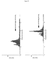
FIG. 2 shows single channel recordings of the HL-(M113R)7 in a wild-type background (top) and HL-(M113R)7 in a RL2 background (bottom). am7-βCD binding and dNMP detection can be seen.
FIG. 3 shows the corresponding histograms of dNMP events from FIG. 1. Ledt is wild-type and right is RL2 (1200 mM KCl, 150 mV, pH 7.5).
FIG. 4 shows a diagram indicating the key mutations of the RL2 compared to the wild-type.
FIG. 5 shows a histogram of residual current binding when dNMPs are bound to the HL-(M113R)7RL2 background and the HL-(M113R/N139Q)7 wt background. This Figure shows the importance of the N139Q mutation in the RL2 background (compare with FIG. 3) (800 mM KCl, 160 mV, pH 7.5).
FIG. 6 shows the proposed cyclodextrin position at residue 139 of αHL.
FIG. 7 shows a diagram indicating the location of key mutations in the β-barrel of αHL.
FIG. 8 shows single channel recording of the HL-(M113R/N139Q)6(M113R/T115C-D8)1 mutant. Reaction with the am6amPDP1-βCD can be seen resulting in a fluctuating, noisy baseline (no bases present).
FIG. 9 shows single channel recording of the HL-(M113R/N139Q)6(M113R/T117C-D8)1 mutant, which gives a fluctuating, noisy baseline (no bases present).
FIG. 10 shows single channel recording of the HL-(M113R/N139Q)6(M113R/G119C-D8)1 mutant reacted with the am6amPDP1-βCD before and after the nucleotides are added. It also shows a close up of the baseline after nucleotides are added.
FIG. 11 shows single channel recording of the HL-(M113R/N139Q)6(M113R/N139Q/N121C-D8)1 mutant reacted with the am6amPDP1-βCD after nucleotides were added.
FIG. 12 shows histograms for the HL-(M113R/N139Q)6(M113R/G119C-D8)1 (left) and the HL-(M113R/N139Q)6(M113R/N139Q/N121C-D8)1 mutant (right). Limited base discrimination can be seen at 800 mM KCl, 160 mV, pH 7.5.
FIG. 13 shows single channel recording of the HL-(M113R/N139Q)6(M113R/N139Q/N123C-D8)1 mutant (5 kHz software filtered) reacted with the am6amPDP1-βCD after dGMP, dTMP, dAMP, dCMP were added (1 second shown).
FIG. 14 shows histograms for the HL-(M113R/N139Q)6(M113R/N139Q/N123C-D8)1 (left) and the HL-(M113R/N139Q)6(M113R/N139Q/G137C-D8)1 mutant (right). Limited base discrimination can be seen (800 mM KCl, 160 mV, pH 7.5).
FIG. 15 shows single channel recording of the HL-(M113R/N139Q)6(M113R/N139Q/L135C-D8)1 mutant (5 kHz software filtered) reacted with the am6amPDP1-βCD after dGMP, dTMP, dAMP and dCMP were added (1 second shown).
FIG. 16 shows a histogram for the HL-(M113R/N139Q)6(M113R/N139Q/L135C-D8)1 mutant. Good base discrimination can be seen (800 mM KCl, 160 mV, pH 7.5).
FIG. 17 shows residual current histograms of dNMP binding for the HL-(M113R/N139Q)6(M113R/N139Q/L135C-D8)1 mutant at a range of applied potentials (110-170 mV). The changes in the relative positions of the four nucleotides can be seen. The sharp black peak corresponds to the cyclodextrin level.
FIG. 18 shows a plot of the variation of peak position (residual pore current) with applied potential when a single nucleotide is bound to the pore.
FIG. 19 shows a plot of the residual current histograms, Gaussian fits of each base and calculated areas of overlap for neighbouring bases for runs at applied potentials of 130, 150 and 170 mV.
FIG. 20 shows the dwell time versus mean of residual pore current. Peaks from all four bases (G T A C, bottom to top) can be seen, as can the longer average dwell time of dTMP.
FIG. 21 shows a histogram for the HL-(M113R/N139Q)6(M113R/N139Q/L135C-D8)1 mutant. Good base discrimination can be seen at low salt (400 mM KCl, 180 mV, pH 7.5).
FIG. 22 shows single channel recording of the HL-(M113R/N139Q)6(M113R/N139Q/L135C-D8)1 mutant (5 kHz software filtered) reacted with the am6amPDP1-βCD after dNMPs were added (350 mM KCl, 180 mV, pH 7.5).
FIG. 23 shows a histogram for the HL-(M113R/N139Q)6(M113R/N139Q/L135C-D8)1 mutant. Good base discrimination can be seen at low salt (350 mM KCl, 180 mV, pH 7.5).
FIG. 24 shows a plot of hemolysin conductance versus solution temperature for a single channel in 300 mM KCl, 25 mM Tris, starting pH 7.5. An increase of 1.65 pA/° C. can be seen.
FIG. 25 shows a histogram for the HL-(M113R/N139Q)6(M113R/N139Q/L135C-D8)1 mutant showing good base detection at low salt and elevated temperature (300 mM KCl, 180 mV, pH 7.2, 40° C.) (left) and a plot of dwell time versus amplitude of the same data showing a short dwell time (right).
FIG. 26 shows the chemical structures of the dCMP and methyl-dCMP (dTMP included for comparison).
FIG. 27 shows a histogram for the HL-(M113R/N139Q)6(M113R/N139Q/L135C-D8)1 mutant. dGMP and methyl-dCMP base detection can be seen (800 mM KCl, 180 mV, pH 7.5).
FIG. 28 shows a histogram for the HL-(M113R/N139Q)6(M113R/N139Q/L135C-D8)1 mutant. Detection of all four standard nucleotide monophosphates (left) and all four standard nucleotide monophosphates with methyl-dCMP added (right) can be seen (800 mM KCl, 170 mV, pH 7.5).
FIG. 29 shows dwell time versus mean of residual pore current. Peaks from all four bases and methyl-dCMP (left) and the Gaussian fits of the residual current histograms for the five bases (right) can be seen.
FIG. 30 shows the residual current histograms of dNMP binding for the HL-(M113R/N139Q)6(M113R/N139Q/L135C-D8)1 mutant at a range of applied potentials (130-170 mV). The changes in the relative positions of the four nucleotides with methyl-dCMP added can be seen.
FIG. 31 shows the chemical structures of the bases commonly found in DNA and the corresponding RNA bases.
FIG. 32 shows the histogram for the HL-(M113R/N139Q)6(M113R/N139Q/L135C-D8)1 mutant. NMP base detection can be seen (800 mM KCl, 160 mV, pH 7.5)=.
FIG. 33 shows single channel recordings for the homo-heptamer HL-(M113R/N139Q/N121C)7 with am6amPDP1-βCD reacted at the 121 position.
FIG. 34 shows single channel recording of the HL-(M113R/N139Q)6(M113R/N139Q/L135C-D8)1 mutant (5 kHz software filtered) reacted with the PDP1-βCD after dGMP, dTMP, dAMP nd dCMP was added (800 mM KCl, pH 7.5, 160 mV, 5 kHz filter, 0.5 seconds shown).
FIG. 35 shows single channel recording of the HL-(M113R/N139Q)6(M113R/N139Q/L135C-D8)1 mutant (5 kHz software filtered) reacted with the am6amPDP1-βCD after dGMP, dTMP, dAMP and dCMP was added (800 mM KCl, pH 7.5, 160 mV, 5 kHz filter, 0.5 seconds shown).
FIG. 36 shows single channel recording of the HL-(wt)6(L135C-D8)1 mutant (5 kHz software filtered) reacted with the am6amPDP1-βCD after dGMP, dTMP, dAMP and dCMP was added (800 mM KCl, pH 7.5, 160 mV, 5 kHz filter, 0.5 seconds shown) and the corresponding residual current histogram.
FIG. 37 shows single channel recording of the HL-(M113R)6(M113R/L135C-D8)1 mutant (5 kHz software filtered) reacted with the am6amPDP1-βCD after dGMP, dTMP, dAMP and dCMP was added (800 mM KCl, pH 7.5, 160 mV, 5 kHz filter, 0.5 seconds shown) and the corresponding residual current histogram.
FIG. 38 shows single channel recording of the HL-(N139Q)6(N139Q/L135C-D8)1 mutant (5 kHz software filtered) reacted with the am6amPDP1-βCD after dGMP, dTMP, dAMP and dCMP was added (800 mM KCl, pH 7.5, 160 mV, 5 kHz filter, 0.5 seconds shown) and the corresponding residual current histogram.
DESCRIPTION OF THE SEQUENCE LISTING
SEQ ID NO: 1 shows the polynucleotide sequence that encodes one subunit of wild-type α-hemolysin (α-HL).
SEQ ID NO: 2 shows the amino acid sequence of one subunit of wild-type α-HL.
SEQ ID NO: 3 shows the polynucleotide sequence that encodes one subunit of α-HL M113R-RL2.
SEQ ID NO: 4 shows the amino acid sequence of one subunit of α-HL M113R-RL2.
SEQ ID NO: 5 shows the polynucleotide sequence that encodes one subunit of α-HL M113R with a wild-type background.
SEQ ID NO: 6 shows the amino acid sequence of one subunit of α-HL M113R with a wild-type background.
SEQ ID NO: 7 shows the polynucleotide sequence that encodes one subunit of α-HL M113R/N139Q.
SEQ ID NO: 8 shows the amino acid sequence of one subunit of α-HL M113R/N139Q.
SEQ ID NO: 9 shows the polynucleotide sequence that encodes one subunit of α-HL M113R/N139Q/G119C-D8.
SEQ ID NO: 10 shows the amino acid sequence of one subunit of α-HL M113R/N139Q/G119C-D8.
SEQ ID NO: 11 shows the polynucleotide sequence that encodes one subunit of α-HL M113R/N139Q/N121C-D8.
SEQ ID NO: 12 shows the amino acid sequence of one subunit of α-HL M113R/N139Q/N121C-D8.
SEQ ID NO: 13 shows the polynucleotide sequence that encodes one subunit of α-HL M113R/N139Q/L135C-D8.
SEQ ID NO: 14 shows the amino acid sequence of one subunit of α-HL M113R/N139Q/L135C-D8.
All of SEQ ID NOs 2, 4, 6, 8, 10, 12 and 14 are mature forms that lack the amino terminal methionine.
SEQ ID NO: 15 shows the polynucleotide sequence encoding the exonuclease III enzyme from E. coli.
SEQ ID NO: 16 shows the amino acid sequence of the exonuclease III enzyme from E. coli.
SEQ ID NO: 17 shows the polynucleotide sequence encoding the exonuclease I enzyme from E. coli.
SEQ ID NO: 18 shows the amino acid sequence of the exonuclease I enzyme from E. coli.
SEQ ID NO: 19 shows the polynucleotide sequence encoding the bacteriophage lambda exonuclease.
SEQ ID NO: 20 shows the amino acid sequence of the bacteriophage lambda exonuclease. The sequence is one of three identical subunits that assemble into a trimer.
DETAILED DESCRIPTION OF THE INVENTION
It is to be understood that different applications of the disclosed products and methods may be tailored to the specific needs in the art. It is also to be understood that the terminology used herein is for the purpose of describing particular embodiments of the invention only, and is not intended to be limiting.
In addition as used in this specification and the appended claims, the singular forms “a”, “an”, and “the” include plural referents unless the content clearly dictates otherwise. Thus, for example, reference to “a nucleotide” includes “nucleotides”, reference to “a pore” includes two or more such pores, reference to “a molecular adaptor” includes two or more such adaptors, and the like.
All publications, patents and patent applications cited herein, whether supra or infra, are hereby incorporated by reference in their entirety.
Mutant α-HL Pores
The present invention provides mutant α-HL pores for use in detecting one or more nucleotide(s). The pores may also be used for distinguishing between or discriminating between different nucleotides. The pores comprise a molecular adaptor that facilitates an interaction with the nucleotide(s). The adaptor is covalently attached to the pore in a specific position. The adaptor is positioned such that, during the interaction between a nucleotide and the pore, the nucleotide affects the current flowing through the pore in a manner specific for that nucleotide. The adaptor is therefore covalently attached to the pore in a position that allows the pore to be used to detect the nucleotide or discriminate nucleotides via stochastic sensing.
The pores of the invention are useful tools for stochastic sensing. The pores of the invention are particularly useful for discriminating between nucleotides. The pores are therefore ideal for sequencing nucleic acids.
The pores of the invention can discriminate between different nucleotides with a high degree of sensitivity. The pores can easily distinguish between the four nucleotides in DNA and RNA. The pores of the invention can even distinguish between methylated and unmethylated nucleotides. The base resolution of a mutant α-HL pore having the adaptor covalently attached to residue 135 is surprisingly high. The pore shows almost complete separation of all four DNA nucleotides making it an excellent candidate for the sequencing of nucleic acids. The pore also allows deoxythymidine monophosphate (dTMP) to be clearly distinguished from the other three DNA nucleotides based on its longer dwell time in the pore. The pore further discriminates between deoxycytidine monophosphate (dCMP) and methyl-dCMP based on the dwell time in the pore and the current flowing through the pore.
The pores of the invention can also discriminate between different nucleotides under a range of conditions. In particular, the pores will discriminate between nucleotides under conditions that are favourable to the sequencing of nucleic acids. There are a number of ways that pores can be used to sequence DNA and RNA molecules. One way involves the use of an exonuclease enzyme. In this approach, the exonuclease enzyme is used to sequentially detach the nucleotides from the DNA or RNA strand. Such enzymes do not normally function under high salt concentrations. The pores of the invention are able to discriminate between nucleotides even at low salt concentrations. For instance, the pores can function with good nucleotide discrimination at a KCl concentration as low as 300 mM at room temperature. The pores will function with good nucleotide discrimination at lower salt concentrations if the temperature is increased or if asymmetric salt solutions are used. This is discussed in more detail below.
The extent to which the pores of the invention can discriminate between different nucleotides can be controlled by altering the applied potential. This allows the function of the pores to be fine-tuned, particularly when sequencing.
The fixed nature of the molecular adaptor also means that the signal obtained from the pore is entirely dependent on the presence of a nucleotide in the barrel or channel of the pore and is not affected by dissociation of the adaptor from the pore. In other words, the fixed nature of the adaptor means that a distinctive current will flow through the pore whenever a nucleotide interacts with the pore. This is particularly important for sequencing nucleic acids because every nucleotide in the sequence needs to be detected and identified.
The pores of the invention can be designed such that they do not undergo blocking. In electrophysiology, pores can become blocked by large multivalent ions. The ions become trapped in the barrel or channel of the pore and prevent the flow of ionic current. Blocking is normally avoided by carrying out experiments in ultra-high grade purity salt solution. The introduction of positively charged residues, such as arginine, near the constriction of the barrel or channel of α-HL generally results in pores that are prone to blocking. As will become apparent from the discussion below, it is not essential that the mutant α-HL pores of the invention have one or more positively charge residues near the constriction of their barrel or channel. Pores of the invention lacking such residues can be used in the presence of large multivalent ions without undergoing blocking.
Finally, the fixed nature of the molecular adaptor means that the pore and adaptor can be stored together, thereby allowing the production of a ready-to-use biosensor.
A pore of the invention may be isolated, substantially isolated, purified or substantially purified. A pore of the invention is isolated or purified if it is completely free of any other components, such as lipids or other pores. A pore is substantially isolated if it is mixed with carriers or diluents which will not interfere with its intended use. For instance, a pore is substantially isolated or substantially purified if it present in a form that comprises less than 10%, less than 5%, less than 2% or less than 1% of other components, such as lipids or other pores. Alternatively, a pore of the invention may be present in a lipid bilayer.
A pore of the invention may be present as an individual or single pore. Alternatively, a pore of the invention may be present in a homologus or heterologous population of two or more pores.
The wild-type α-HL pore is formed of seven identical monomers or subunits (i.e. it is heptameric). The sequence of one wild-type monomer or subunit of α-hemolysin is shown in SEQ ID NO: 2. A mutant α-HL pore is a heptameric pore in which one or more of the seven subunits has an amino acid sequence which varies from that of SEQ ID NO: 2 and which retains pore activity. The pore includes modifications that facilitate positioning of the adaptor as discussed below. The pore preferably also contains modifications that facilitate covalent attachment of the adaptor.
1, 2, 3, 4, 5, 6 of the subunits in the pore may have an amino acid sequence that varies from that of SEQ ID NO: 2. In preferred embodiments, all of the subunits in the pore have an amino acid sequence that varies from that of SEQ ID NO: 2. All seven subunits within the pore may be identical but are typically different, particularly since different subunits are modified in different ways to facilitate positioning of the adaptor as discussed below and optionally to facilitate covalent attachment of the adaptor.
A mutant α-HL pore of the invention is formed from seven subunits each comprising the sequence shown in SEQ ID NO: 2 or a variant thereof. A variant is a subunit that has an amino acid sequence which varies from that of SEQ ID NO: 2 and which retains its ability to form a pore. All of the sequences shown in SEQ ID NOs: 4, 6, 8, 10, 12 and 14 are variants of SEQ ID NO: 2. Any of the specific modifications to SEQ ID NO: 2 discussed below also result in variants of SEQ ID NO: 2.
Variants include the constructs, which comprise the sequence shown in SEQ ID NO: 2 or a variant thereof and are described in a co-pending International application claiming priority from U.S. Application No. 61/078,695 and being filed simultaneously with this application [J A Kemp & Co Ref: N.104404A; Oxford Nanolabs Ref: ONL IP 005]. Such constructs also comprise a nucleic acid handling enzyme, such as an exonuclease. All the teachings of that application may be applied equally to the present invention.
The pore is modified to facilitate positioning of the adaptor so that the one or more nucleotides can be detected. One or amino acids are introduced into the pore to hold the adaptor in the correct position via non-covalent interactions, such as hydrophobic interactions, hydrogen bonding, Van der Waal's forces, π-cation interactions and/or electrostatic forces.
One or more of the seven subunits are modified at and/or near residue 139 of SEQ ID NO: 2. For instance, 2, 3, 4, 5, 6 or all of the seven subunits are modified at and/or near residue 139 of SEQ ID NO: 2. One or more of the seven subunits include at least one, such as 2, 3, 4 or 5, modifications at and/or near residue 139 of SEQ ID NO: 2.
One or more of the seven subunits are modified at residue 139 and/or near residue 139 of SEQ ID NO: 2. If one or more of the seven subunits are modified near residue 139, the modifications are sufficiently close to residue 139 that they can facilitate positioning of the adaptor. The modifications are typically made at positions that are less than 20 ångströms, such as less than 15, less than 10 or less than 5 ångströms, from residue 139 in SEQ ID NO: 2. Residues that are 2 or 3 amino acids from residue 139 in the sequence of SEQ ID NO: 2 are preferably modified in one or more of the seven subunits. Residue 139 and/or one or both of the residues adjacent to residue 139 in the sequence of SEQ ID NO: 2 are more preferably modified in one or more of the seven subunits. One or more of the seven subunits preferably comprise a modification at residue 136, 137, 138, 139, 140, 141 or 142 of SEQ ID NO: 2 or any combination thereof. One or more of the seven subunits most preferably comprise a modification at only residue 139 of SEQ ID NO: 2.
One or more of the seven subunits may comprise modifications at residues in SEQ ID NO: 2 that are near to residue 139 once the sequence is folded to form a three-dimensional subunit. One or more of the seven subunits preferably comprise a modification at residue 117, 119, 121, 123, 135, 137, 141 or 143 of SEQ ID NO: 2 or any combination thereof.
The subunits are preferably modified at and/or near to residue 139 by the introduction of polar, uncharged amino acids. Such amino acids may influence the positioning of the adaptor via hydrogen bonding. Their lack of charge also prevents electrostatic interactions from interfering with the function of the adaptor. Table 1 below lists polar, uncharged amino acids. Glutamine is a preferred polar, uncharged amino acid. The polar, uncharged amino acids can be inserted at and/or near residue 139. Alternatively, amino acids at and/or near residue 139 can be substituted with polar, uncharged amino acids.
One or more of the seven subunits are preferably modified to comprise glutamine, which is a polar, uncharged amino acid, at and/or near residue 139 of SEQ ID NO: 2. Glutamine can be inserted at and/or near residue 139 or amino acids at and/or near residue 139 can be substituted with glutamine. The amino acid at residue 139 is preferably substituted with glutamine (N139Q). For instance, 2, 3, 4, 5 or 6 of the seven subunits have glutamine at residue 139 of SEQ ID NO: 2. In preferred embodiments, all of the seven subunits have a glutamine at residue 139 of SEQ ID NO: 2. Uncharged glutamine at residue 139 is capable of interacting with chemical groups, such as hydroxyl groups, in the adaptor by hydrogen bonding and thereby facilitating the positioning of the adaptor within the barrel or channel of the mutant α-HL pore. Preferred subunits having residue 139 of SEQ ID NO: 2 substituted with glutamine (N139Q) are shown in SEQ ID NOs: 8, 10, 12 and 14. Suitable subunits may contain or lack the octa-asparate tail shown in SEQ ID NOs: 8, 10, 12 and 14.
The pore may also have other uncharged amino acids or even aromatic amino acids located near the constriction of the barrel or channel to further facilitate positioning of the adaptor. Table 1 below lists uncharged and aromatic amino acids. For instance, one or more of the seven subunits may have one or more uncharged amino acids, such as asparagine, or one or more aromatic amino acids, such as phenylalanine, located near the constriction of the barrel or channel. The pore preferably has a ring of 4, 5, 6 or preferably 7 uncharged or aromatic amino acids located near the constriction of the barrel or channel. Each amino acid in the ring is typically provided by each of the subunits. Residues of SEQ ID NO: 2 located near the constriction of the barrel or channel include, but are not limited to, 111, 113 and 147. Suitable subunits include an uncharged or aromatic amino acid at residue 111, 113 or 147 of SEQ ID NO: 2. The uncharged or aromatic amino acids can be inserted at residue 111, 113 or 147 of SEQ ID NO: 2. Alternatively, the amino acids at residue 111, 113 or 147 of SEQ ID NO: 2 can be substituted with uncharged or aromatic amino acids.
The pore is preferably modified to facilitate covalent attachment of the adaptor. One or more amino acids that are capable of forming a covalent bond, such as cysteine, can be introduced into one or more subunits. The amino acid may be naturally occurring or non-naturally occurring. An amino acid may be introduced by addition. An amino acid is preferably introduced by substitution. Amino acids can be introduced at any positions as long as the adaptor is positioned at or near residue 139 of SEQ ID NO: 2. If the amino acids are introduced at residues distant from residue 139, a bifunctional crosslinker of appropriate length may be used to ensure that the adaptor is positioned at or near residue 139.
In preferred embodiments, residue 119, 121 or 135 of SEQ ID NO: 2 is modified in one or more of the seven subunits to facilitate covalent attachment of the adaptor. In more preferred embodiments, residue 119, 121 or 135 of SEQ ID NO: 2 is modified in only one of the seven subunits to facilitate covalent attachment of the adaptor. The amino acid introduced at residue 119, 121 or 135 of SEQ ID NO: 2 is preferably cysteine. The amino acid at residue 119, 121 or 135 is preferably substituted with cysteine (G119C, N121C or L135C). A preferred subunit having position 119 of SEQ ID NO: 2 substituted with cysteine (G119C) is shown in SEQ ID NO: 10. A preferred subunit having residue 121 of SEQ ID NO: 2 substituted with cysteine (N121C) is shown in SEQ ID NO: 12. A preferred subunit having residue 135 of SEQ ID NO: 2 substituted with cysteine (L135C) is shown in SEQ ID NO: 14.
The pore preferably has positively charged amino acids located near the constriction of the barrel or channel to facilitate covalent attachment of the adaptor. For instance, one or more of the seven subunits may have positively charged amino acids, such as arginine, lysine or histidine, located near the constriction of the barrel or channel. The positively charged amino acids facilitate covalent attachment of the adaptor by electrostatic interactions with positively charged groups, such as amines, in the adaptor. More specifically, the positively charged amino acids repel the positively charged groups in the adaptor, push the adaptor down the barrel or channel of the pore and position it at or near residue 139 of SEQ ID NO: 2. This facilitates the covalent reaction between the adaptor and the pore. However, as discussed above, pores having positively charged amino acids located near the constriction of the barrel or channel may be prone to blocking.
The pore preferably has a ring of 4, 5, 6 or preferably 7 positively charged amino acids, such as arginine, lysine or histidine, located near the constriction of the barrel or channel. Each amino acid in the ring is typically provided by each of the subunits. In preferred embodiments, one or more of the seven subunits has a positively charged amino acid, such as arginine, lysine or histidine, at residue 113 of SEQ ID NO: 2. In more preferred embodiments, one or more of the seven subunits has an arginine residue at residue 113 of SEQ ID NO: 2. In even more preferred embodiments, all of the seven subunits have an arginine residue at residue 113 of SEQ ID NO: 2. Arginine can be introduced at residue 113 or residue 113 can be substituted with arginine (M113R). Preferred subunits having residue 113 of SEQ ID NO: 2 substituted with arginine (M113R) are shown in SEQ ID NOs: 4, 6, 8, 10, 12 and 14.
The subunits may be a naturally occurring variants which are expressed by an organism, for instance by a Staphylococcus bacterium. Variants also include non-naturally occurring variants produced by recombinant technology. Over the entire length of the amino acid sequence of SEQ ID NO: 2, a variant will preferably be at least 50% homologous to that sequence based on amino acid identity. More preferably, the subunit polypeptide may be at least 55%, at least 60%, at least 65%, at least 70%, at least 75%, at least 80%, at least 85%, at least 90% and more preferably at least 95%, 97% or 99% homologous based on amino acid identity to the amino acid sequence of SEQ ID NO: 2 over the entire sequence. There may be at least 80%, for example at least 85%, 90% or 95%, amino acid identity over a stretch of 200 or more, for example 230, 250, 270 or 280 or more, contiguous amino acids (“hard homology”).
Amino acid substitutions may be made to the amino acid sequence of SEQ ID NO: 2 in addition to those discussed above, for example up to 1, 2, 3, 4, 5, 10, 20 or 30 substitutions. Conservative substitutions may be made, for example, according to Table 1 below.
TABLE 1
Conservative substitutions
NON-AROMATIC Non-polar G A P
I L V
Polar-uncharged C S T M
N Q
Polar-charged D E
H K R
AROMATIC H F W Y
Amino acids in the same block in the second column and preferably in the same line in the third column may be substituted for each other.
One or more amino acid residues of the amino acid sequence of SEQ ID NO: 2 may additionally be deleted from the polypeptides described above. Up to 1, 2, 3, 4, 5, 10, 20 or 30 residues may be deleted, or more.
Variants may include subunits made of fragments of SEQ ID NO: 2. Such fragments retain pore forming activity. Fragments may be at least 50, 100, 200 or 250 amino acids in length. Such fragments may be used to produce chimeric pores. A fragment preferably comprises the pore forming domain of SEQ ID NO: 2. Fragments must include residues 139 and 119, 121 or 135 of SEQ ID NO: 2.
Variants include chimeric proteins comprising fragments or portions of SEQ ID NO: 2. Chimeric protein pores may be formed from one or more subunits each comprising fragments or portions of SEQ ID NO: 2. The pore or channel part of a chimeric protein pore is typically formed by the fragments or portions of SEQ ID NO: 2.
One or more amino acids may be alternatively or additionally added to the polypeptides described above. An extension may be provided at the amino terminal or carboxy terminal of the amino acid sequence of SEQ ID NO: 2 or polypeptide variant or fragment thereof. The extension may be quite short, for example from 1 to 10 amino acids in length. Alternatively, the extension may be longer, for example up to 50 or 100 amino acids. A carrier protein may be fused to an amino acid sequence according to the invention.
As discussed above, a variant is a subunit that has an amino acid sequence which varies from that of SEQ ID NO: 2 and which retains its ability to form a pore. A variant typically contains the regions of SEQ ID NO: 2 that are responsible for pore formation. The pore forming ability of α-HL, which contains a β-barrel, is provided by β-sheets in each subunit. A variant of SEQ ID NO: 2 typically comprises the regions in SEQ ID NO: 2 that form β-sheets. Amino acids 22 to 30, 35 to 44, 52 to 62, 67 to 71, 76 to 91, 98 to 103, 112 to 123, 137 to 148, 154 to 159, 165 to 172, 229 to 235, 243 to 261, 266 to 271, 285 to 286 and 291 to 293 of SEQ ID NO: 2 form β-sheets. One or more modifications can be made to the regions of SEQ ID NO: 2 that form β-sheets as long as the resulting variant retains its ability to form a pore. Specific modifications that can be made to the β-sheet regions of SEQ ID NO: 2 are discussed above.
A variant of SEQ ID NO: 2 preferably includes one or more modifications, such as substitutions, additions or deletions, within its α-helices and/or loop regions. Amino acids 2 to 6, 73 to 75, 207 to 209, 214 to 216 and 219 to 222 of SEQ ID NO: 2 form α-helices. Amino acids 7 to 21, 31 to 34, 45 to 51, 63 to 66, 72, 92 to 97, 104 to 111, 124 to 136, 149 to 153, 160 to 164, 173 to 206, 210 to 213, 217, 218, 223 to 228, 236 to 242, 262 to 265, 272 to 274 and 287 to 290 of SEQ ID NO: 2 form loops. Amino acids 1 and 294 are terminal amino acids.
Standard methods in the art may be used to determine homology. For example the UWGCG Package provides the BESTFIT program which can be used to calculate homology, for example used on its default settings (Devereux et al (1984) Nucleic Acids Research 12, p 387-395). The PILEUP and BLAST algorithms can be used to calculate homology or line up sequences (such as identifying equivalent residues or corresponding sequences (typically on their default settings)), for example as described in Altschul S. F. (1993) J Mol Evol 36:290-300; Altschul, S. F et al (1990) J Mol Biol 215:403-10.
Software for performing BLAST analyses is publicly available through the National Center for Biotechnology Information (http://www.ncbi.nlm.nih.gov/). This algorithm involves first identifying high scoring sequence pair (HSPs) by identifying short words of length W in the query sequence that either match or satisfy some positive-valued threshold score T when aligned with a word of the same length in a database sequence. T is referred to as the neighbourhood word score threshold (Altschul et al, supra). These initial neighbourhood word hits act as seeds for initiating searches to find HSP's containing them. The word hits are extended in both directions along each sequence for as far as the cumulative alignment score can be increased. Extensions for the word hits in each direction are halted when: the cumulative alignment score falls off by the quantity X from its maximum achieved value; the cumulative score goes to zero or below, due to the accumulation of one or more negative-scoring residue alignments; or the end of either sequence is reached. The BLAST algorithm parameters W, T and X determine the sensitivity and speed of the alignment. The BLAST program uses as defaults a word length (W) of 11, the BLOSUM62 scoring matrix (see Henikoff and Henikoff (1992) Proc. Natl. Acad. Sci. USA 89: 10915-10919) alignments (B) of 50, expectation (E) of 10, M=5, N=4, and a comparison of both strands.
The BLAST algorithm performs a statistical analysis of the similarity between two sequences; see e.g., Karlin and Altschul (1993) Proc. Natl. Acad. Sci. USA 90: 5873-5787. One measure of similarity provided by the BLAST algorithm is the smallest sum probability (P(N)), which provides an indication of the probability by which a match between two amino acid sequences would occur by chance. For example, a sequence is considered similar to another sequence if the smallest sum probability in comparison of the first sequence to the second sequence is less than about 1, preferably less than about 0.1, more preferably less than about 0.01, and most preferably less than about 0.001.
One or more of the subunits may be modified for example by the addition of histidine or aspartic acid residues to assist their identification or purification or by the addition of a signal sequence to promote their secretion from a cell where the polypeptide does not naturally contain such a sequence.
The pore may be labelled with a revealing label. The revealing label may be any suitable label which allows the pore to be detected. Suitable labels include, but are not limited to fluorescent molecules, radioisotopes, e.g. 125I, 35S, enzymes, antibodies, antigens, polynucleotides and ligands such as biotin.
The pore may be derived from a pore producing organism, such as Staphylococcus aureus, or made synthetically or by recombinant means. For example, the pore may be synthesized by in vitro translation and transcription. The amino acid sequence of the pore may be modified to include non-naturally occurring amino acids or to increase the stability of the compound. When the pores are produced by synthetic means, such amino acids may be introduced during production. The pores may also be altered following either synthetic or recombinant production.
The pore may also be produced using D-amino acids. For instance, the pores may comprise a mixture of L-amino acids and D-amino acids. This is conventional in the art for producing such proteins or peptides.
The pore contains one or more specific modifications to facilitate positioning of the adaptor and optionally to facilitate covalent attachment. The pore may also contain other non-specific modifications as long as they do not interfere with the attachment and positioning of the adaptor. A number of non-specific side chain modifications are known in the art and may be made to the side chains of the pores. Such modifications include, for example, reductive alkylation of amino acids by reaction with an aldehyde followed by reduction with NaBH4, amidination with methylacetimidate or acylation with acetic anhydride.
The pore can be produced using standard methods known in the art. Polynucleotide sequences encoding a pore or a pore subunit may be derived and replicated using standard methods in the art. Such sequences are discussed in more detail below. Polynucleotide sequences encoding a pore or a pore subunit may be expressed in a bacterial host cell using standard techniques in the art. The pore or pore subunit may be produced in a cell by in situ expression of the polypeptide from a recombinant expression vector. The expression vector optionally carries an inducible promoter to control the expression of the polypeptide.
A pore subunit may be produced in large scale following purification by any protein liquid chromatography system from pore producing organisms or after recombinant expression as described below. Typical protein liquid chromatography systems include FPLC, AKTA systems, the Bio-Cad system, the Bio-Rad BioLogic system and the Gilson HPLC system. The naturally occurring or recombinantly-produced pore or or pore subunit may then be inserted into a naturally occurring or artificial membrane for use in accordance with the invention. Methods for inserting pore into membranes are discussed below.
Any combination of seven of the subunits (i.e. variants of SEQ ID NO: 2) shown in SEQ ID NOs: 4, 6, 8, 10, 12 and 14 may be used to form a pore of the invention. Preferred pores comprise:
(a) six subunits of α-HL M113R/N139Q shown in SEQ ID NO: 8 and one subunit of α-HL M113R/N139Q/G119C-D8 shown in SEQ ID NO: 10; or
(b) six subunits of α-HL M113R/N139Q shown in SEQ ID NO: 8 and one subunit of α-HL M113R/N139Q/N121C-D8 shown in SEQ ID NO: 12;
In pores (a) and (b), the subunits shown in SEQ ID NOs: 10, 12, 18, 22, 26, 30 and 34 may lack the octa-aspartate tail.
The most preferred pore of the invention comprises six subunits of α-HL M113R/N139Q shown in SEQ ID NO: 8 and one subunit of α-HL M113R/N139Q/L135C-D8 shown in SEQ ID NO: 14. In such a pore, the subunit shown in SEQ ID NO: 14 may lack the octa-aspartate tail.
Molecular Adaptor
The pores of the invention comprise a molecular adaptor that facilitates the interaction between the pore and the nucleotides or target nucleic acid sequence. The presence of the adaptor improves the host-guest chemistry of the pore and nucleotides. The principles of host-guest chemistry are well-known in the art. The adaptor has an effect on the physical or chemical properties of the pore that improves its interaction with nucleotides. The adaptor typically alters the charge of the barrel or channel of the pore or specifically interacts with or binds to nucleotides thereby facilitating their interaction with the pore.
The adaptor mediates the interaction between each individual nucleotide or each nucleotide is a target nucleic acid sequence and the pore. The nucleotides preferably reversibly bind to the pore via or in conjunction with the adaptor. The nucleotides most preferably reversibly bind to the pore via or in conjunction with the adaptor as they pass through the pore across the membrane. The nucleotides can also reversibly bind to the barrel or channel of the pore via or in conjunction with the adaptor as they pass through the pore across the membrane. The adaptor preferably constricts the barrel or channel so that it may interact with the nucleotides.
The adaptor is typically cyclic. The adaptor preferably has the same symmetry as the pore. The adaptor preferably has seven-fold symmetry since α-HL has seven subunits around a central axis that contribute 14 strands to a transmembrane β barrel.
The adaptor typically interacts with the nucleotide via host-guest chemistry. The adaptor is typically capable of interacting with the nucleotide. The adaptor comprises one or more chemical groups that are capable of interacting with the nucleotide. The one or more chemical groups preferably interact with the nucleotide by non-covalent interactions, such as hydrophobic interactions, hydrogen bonding, Van der Waal's forces, π-cation interactions and/or electrostatic forces. The one or more chemical groups that are capable of interacting with the nucleotide are preferably positively charged. The one or more chemical groups that are capable of interacting with the nucleotide are more preferably preferably comprise amino groups. The amino groups can be attached to primary, secondary or tertiary carbon atoms. The adaptor even more preferably comprises a ring of amino groups, such as a ring of 6, 7 or 8 amino groups. The adaptor most preferably comprises a ring of seven amino groups. A ring of protonated amino groups may interact with negatively charged phosphate groups in the nucleotide.
As discussed in more detail below, the correct positioning of the adaptor within the barrel or channel of the pore can be facilitated by host-guest chemistry between the adaptor and the pore. The adaptor preferably comprises one or more chemical groups that are capable of interacting with one or more amino acids in the pore. The adaptor more preferably comprises one or more chemical groups that are capable of interacting with one or more amino acids in the pore via non-covalent interactions, such as hydrophobic interactions, hydrogen bonding, Van der Waal's forces, π-cation interactions and/or electrostatic forces. The chemical groups that are capable of interacting with one or more amino acids in the pore are typically hydroxyls or amines. The hydroxyl groups can be attached to primary, secondary or tertiary carbon atoms. The hydroxyl groups may form hydrogen bonds with uncharged amino acids in the pore, particularly those introduced at and/or near residue 139 of SEQ ID NO: 2. This interaction of one or more chemical groups in the adaptor with one or more amino acids in the pore can be used to hold the adaptor in the correct positioning at or near residue 139 of SEQ ID NO: 2.
The amine groups that are capable of interacting with one or more amino acids in the pore may be the same or different as the amine groups that are capable of interacting with the nucleotide. As discussed above, the positively charged amine groups in the adaptor may interact electrostatically with positively charged amino acids in the barrel or channel of the pore, such as arginines at residue 113 of SEQ ID NO: 2 (M113R) if present. This interaction pushes the adaptor down the barrel or channel of the pore and thereby facilitates covalent attachment.
Any adaptor that that facilitates the interaction between the pore and the nucleotide can be used. Suitable adaptors include, but are not limited to, cyclodextrins, cyclic peptides and cucurbiturils. The adaptor is preferably a cyclodextrin or a derivative thereof. The cyclodextrin or derivative thereof may be any of those disclosed in Eliseev, A. V., and Schneider, H-J. (1994) J. Am. Chem. Soc. 116, 6081-6088. The adaptor is more preferably heptakis-6-amino-β-cyclodextrin (am7-βCD), 6-monodeoxy-6-monoamino-β-cyclodextrin (am1-βCD) or heptakis-(6-deoxy-6-guanidino)-cyclodextrin (gu7-βCD). The guanidine group in gu7-βCD has a much higher pKa than the primary amines in am7-βCD and so it more positively charged. This gu7-βCD adaptor may be used to increase the dwell time of the nucleotide in the pore, to increase the accuracy of the residual current measured, as well as to increase the base detection rate at high temperatures or low data acquisition rates.
If a succinimidyl 3-(2-pyridyldithio)propionate (SPDP) crosslinker is used as discussed in more detail below, the adaptor is preferably heptakis(6-deoxy-6-amino)-6-N-mono(2-pyridyl)dithiopropanoyl-β-cyclodextrin (am6amPDP1-βCD).
Covalent Attachment
The adaptor is covalently attached to the pore. The adaptor can be covalently attached to the pore using any method known in the art. The adaptor may be attached directly to the pore. The adaptor is preferably attached to the pore using a bifunctional crosslinker. Suitable crosslinkers are well-known in the art. Preferred crosslinkers include 2,5-dioxopyrrolidin-1-yl 3-(pyridin-2-yldisulfanyl)propanoate, 2,5-dioxopyrrolidin-1-yl 4-(pyridin-2-yldisulfanyl)butanoate and 2,5-dioxopyrrolidin-1-yl 8-(pyridin-2-yldisulfanyl)octananoate. The most preferred crosslinker is succinimidyl 3-(2-pyridyldithio)propionate (SPDP). Typically, the adaptor is covalently attached to the bifunctional crosslinker before the adaptor/crosslinker complex is covalently attached to the pore but it is also possible to covalently attach the bifunctional crosslinker to the pore before the bifunctional crosslinker/pore complex is attached to the adaptor. Production of the pores of the invention is discussed in more detail below.
The site of covalent attachment is selected such that the adaptor is positioned at or near residue 139 of SEQ ID NO: 2. This facilitates interaction of the nucleotide with the pore and thereby allows detection of the nucleotide. It also ensures that the nucleotide affects the current flowing through the pore in a manner specific for that nucleotide.
The adaptor is positioned at residue 139 of SEQ ID NO: 2 if it is positioned in the barrel or channel of the pore in the same horizontal plane as residue 139 of SEQ ID NO: 2. The adaptor is positioned at residue 139 of SEQ ID NO: 2 if it is positioned at the same vertical position within the barrel or channel of the pore as residue 139 of SEQ ID NO: 2. The adaptor is positioned near residue 139 of SEQ. ID NO: 2 if it is positioned in the barrel or channel of the pore in the same horizontal plane as a residue that is near to residue 139 of SEQ ID NO: 2. The adaptor is positioned near residue 139 of SEQ ID NO: 2 if it is positioned at the same vertical position within the barrel or channel of the pore as a residue that is near to residue 139 of SEQ ID NO: 2. The adaptor is preferably positioned in a horizontal plane that is less than 5 ångströms, such as less than 3 or less than 2 ångströms, from the horizontal plane of residue 139 in SEQ ID NO: 2. The adaptor is more preferably positioned in the same horizontal plane as residue 117, 118, 119, 120, 121, 122, 123, 136, 137, 138, 139, 140, 141 or 142 of SEQ ID NO: 2.
The adaptor is typically covalently attached to the barrel or channel of the pore. The adaptor can be covalently attached at any site in the barrel or channel as long as the adaptor is positioned at or near residue 139 of SEQ ID NO: 2, facilitates interaction of the nucleotide with the pore and thereby allows detection of the nucleotide. The adaptor is typically covalently attached to an amino acid in the barrel or channel that is close to residue 139 of SEQ ID NO: 2. The adaptor is preferably attached to an amino acid in the pore that is near to the ring of residues formed by residue 139 of SEQ ID NO: 2 in each subunit. If the adaptor is covalently attached to an amino acid in the barrel or channel that is far from the ring of residues formed by residue 139 of SEQ ID NO: 2 in each subunit, a bifunctional crosslinker of suitable length may be used so that the adaptor is positioned at or near residue 139.
The adaptor is preferably attached to residue 119 or 121 of SEQ ID NO: 2 in one or more of the seven subunits. The adaptor is more preferably attached to residue 135 of SEQ ID NO: 2, in one or more of the seven subunits. Attaching the adaptor to residue 119, 121 or 135 positions the adaptor near to the ring of residues formed by residue 139 of SEQ ID NO: 2 in each subunit.
The pore is preferably modified by the introduction of one or more amino acids to facilitate the covalent attachment of the molecular adaptor with the bifunctional crosslinker. The barrel or channel of the pore is more preferably modified to facilitate the covalent attachment of the molecular adaptor with the bifunctional crosslinker. The pore may be modified using any method known in the art. One or more amino acids may be introduced into the same or different subunits of the pore. Any amino acid that is capable of forming a covalent bond, such as cysteine, can be introduced. The amino acid may be naturally occurring or non-naturally occurring. The one or more amino acids are preferably introduced by substitutions.
In preferred embodiments, residue 119, 121 or 135 of SEQ ID NO: 2 is modified in one or more of the seven subunits to facilitate covalent attachment. In more preferred embodiment, residue 119, 121 or 135 of SEQ ID NO: 2 modified in one of the seven subunits. Any of the subunit discussed above may be used to facilitate the covalent attachment of the molecular adaptor or the bifunctional crosslinker.
Positioning of the Adaptor
The adaptor is covalently attached to the pore in a position that allows the nucleotide to be detected using the pore. The adaptor is positioned such the nucleotide affects the current flowing through the pore in a manner specific for that nucleotide. The adaptor is positioned so this it improves the host-guest chemistry of the pore and nucleotide. The adaptor is positioned so that it affects the physical or chemical properties of the pore and improves its interaction with the nucleotide. The adaptor is typically positioned so that it forms a steric block to the flow of ions through the pore. If the adaptor is capable of specifically interacting with or binding to the nucleotide, the adaptor is positioned so that it specifically interacts with or binds to the nucleotide. The one or more chemical groups in the adaptor that interact with the nucleotide are preferably oriented away from the end of the pore through which the nucleotide enters. Such an orientation helps to draw the nucleotide through the barrel or channel of the pore. The groups are preferably amino groups. The end of the pore through which the nucleotide enters may be the cis end or the trans end. The end is preferably the cis end.
The covalent attachment may be designed so that the adaptor is correctly positioned. For instance, the site (e.g. amino acid) at which the adaptor is covalently attached to the pore may be designed so that the adaptor is correctly positioned and/or a bifunctional crosslinker may be used so that the adaptor is correctly positioned.
The pores of the invention are modified to facilitate positioning of the adaptor. As discussed above, one or more of the seven subunits preferably has glutamine at and/or near residue 139 of SEQ ID NO: 2. Glutamine at and/or near residue 139 is capable of interacting with chemical groups, such as hydroxyl groups, on the adaptor by hydrogen bonding and thereby facilitates the positioning of the adaptor within the barrel or channel of the pore. The pore may also include modifications at other positions, such as residue 113 of SEQ ID NO: 2, to facilitate positioning of the adaptor (see above).
Most preferably, the pore is modified to facilitate the covalent attachment and to facilitate the positioning of the adaptor. In such an embodiment, the spatial relationship between the site of covalent attachment and site(s) at which the pore is modified to facilitate the positioning of the adaptor is designed to ensure that the adaptor is held in the correct position. For instance, the adaptor is preferably attached to residue 135 of SEQ ID NO: 2 in one subunit to place it near to the ring of one or more glutamines formed by residue 139 of SEQ ID NO: 2 in each subunit. The one or more glutamines facilitate the positioning of the adapator by hydrogen bonding.
Polynucleotides
The present invention also provides polynucleotide sequences which encode a subunit of α-HL having the sequence shown in SEQ ID NO: 2 or a variant thereof, wherein the subunit has a cysteine at residue 119, 121 or 135. The variant of SEQ ID NO: 2 may be any of those discussed above. The polynucleotide sequence preferably comprises the sequence shown in SEQ ID NO: 9, 11 or 13 or a sequence at least 50%, 60%, 70%, 80%, 90% or 95% homologous based on nucleotide identity to sequence of SEQ ID NO: 9, 11 or 13 over the entire sequence. There may be at least 80%, for example at least 85%, 90% or 95% nucleotide identity over a stretch of 600 or more, for example 700, 750, 850 or 900 or more, contiguous nucleotides (“hard homogly”). Homology may be calculated as described above. The polynucleotide sequence may comprise a sequence that differs from SEQ ID NO: 9, 11 or 13 on the basis of the degeneracy of the genetic code.
Polynucleotide sequences may be derived and replicated using standard methods in the art. Chromosomal DNA may be extracted from a pore producing organism, such as Staphylococcus aureus. The gene encoding the pore subunit may be amplified using PCR involving specific primers. The amplified sequence may then be incorporated into a recombinant replicable vector such as a cloning vector. The vector may be used to replicate the polynucleotide in a compatible host cell. Thus polynucleotide sequences encoding a pore subunit may be made by introducing a polynucleotide encoding a pore subunit into a replicable vector, introducing the vector into a compatible host cell, and growing the host cell under conditions which bring about replication of the vector. The vector may be recovered from the host cell. Suitable host cells for cloning of polynucleotides encoding a pore subunit are known in the art and described in more detail below.
The polynucleotide sequence encoding a pore subunit may be cloned into suitable expression vector. In an expression vector, the polynucleotide sequence encoding a pore subunit is typically operably linked to a control sequence which is capable of providing for the expression of the coding sequence by the host cell. Such expression vectors can be used to express a pore subunit.
The term “operably linked” refers to a juxtaposition wherein the components described are in a relationship permitting them to function in their intended manner. A control sequence “operably linked” to a coding sequence is ligated in such a way that expression of the coding sequence is achieved under conditions compatible with the control sequences. Multiple copies of the same or different pore subunit sequences may be introduced into the vector.
The expression vector may then be introduced into a suitable host cell. Thus, a pore subunit can be produced by inserting a polynucleotide sequence encoding a pore subunit into an expression vector, introducing the vector into a compatible bacterial host cell, and growing the host cell under conditions which bring about expression of the polynucleotide sequence encoding the pore subunit. The recombinantly-expressed pore subunit may self-assemble into a pore in the host cell membrane. Alternatively, the recombinant pore produced in this manner may be removed from the host cell and inserted into another membrane. When producing a heptameric pore comprising at least two different subunits, the different subunits may be expressed separately in different host cells as described above, removed from the host cells and assembled into a pore in a separate membrane, such as a rabbit cell membrane.
The vectors may be for example, plasmid, virus or phage vectors provided with an origin of replication, optionally a promoter for the expression of the said polynucleotide sequence and optionally a regulator of the promoter. The vectors may contain one or more selectable marker genes, for example a tetracycline resistance gene. Promoters and other expression regulation signals may be selected to be compatible with the host cell for which the expression vector is designed. A T7, trc, lac, ara or λL promoter is typically used.
The host cell typically expresses the pore subunit at a high level. Host cells transformed with a polynucleotide sequence encoding a pore subunit will be chosen to be compatible with the expression vector used to transform the cell. The host cell is typically bacterial and preferably Escherichia coli. Any cell with a λ DE3 lysogen, for example C41 (DE3), BL21 (DE3), JM109 (DE3), B834 (DE3), TUNER, Origami and Origami B, can express a vector comprising the T7 promoter.
Methods of Producing the Pores of the Invention
The invention also provides methods of producing the pores of the invention. The methods comprise covalently attaching to a pore a molecular adaptor that facilitates an interaction between the pore and a nucleotide. The adaptor can be covalently attached to the pore using any method known in the art.
Any of the pores, adaptors and bifunctional crosslinkers discussed above can be used in the method. The site of covalent attachment is selected as discussed above.
The adaptor is typically attached to the pore by adding an adaptor containing a bifunctional crosslinker, such as am6amPDP1-βCD, to a mutant α-HL pore containing at least one reactive amino acid, such as a cysteine. However, this method has a couple of drawbacks. The first is the reactivity of the amino acid, such as cysteine, which deteriorates over time via oxidation or reaction with other species in solution. The second is the time taken for the reaction between the crosslinker and the pore to occur. These drawbacks can lead to difficulties, particularly if a single pore is being used. For instance, if a single α-HL pore containing a single cysteine mutation is inserted into a lipid membrane and an adaptor containing a crosslinker is added to the electrophysiology chamber, the adaptor molecules enter and exit the pore until one reacts with the cysteine. If the cysteine becomes inactivated, then a reaction will not be observed no matter how many adaptor molecules enter the pore. Inactivation of the reactive amino acid(s) in the pore can reduced by improving the storage conditions, for instance by removing oxygen, by the addition of stabilisers, such as reducing agents (e.g. dithiothreitol, DTT), by chemical protection of the cysteine followed by activation prior to use or by chemical attachment of the adaptor prior to storage.
In a preferred embodiment, the pore to which the adaptor is to be attached comprises a protective leaving group and the method comprises displacing the leaving group from the pore. The protective leaving group is used to protect the one or more reactive amino acids in the pore. The protective leaving group is preferably used to protect one or more cysteine residues in the pore. The protective leaving group is displaced by reaction with a reactive group on the adaptor, which may contain a crosslinker.
In a more preferred embodiment, the pore comprises a protective leaving group on one or more reactive cysteines, such as the cysteine at residue 119, 121 or 135 of SEQ ID NO: 2 in the variants discussed above, and is reacted with an adaptor which contains a crosslinker having a reactive —SH group. The —SH group on the crosslinker displaces the protective leaving group attached to the pore and a pore having an adaptor covalently attached thereto is produced.
Attaching a protective leaving group to the one or more reactive amino acids in the pore and having the reactive group on the adaptors greatly improves the method of the invention, particularly when using a single pore. For instance, when attaching a reactive adaptor molecule to a single pore having a protective group, the adaptor molecules vastly outnumber the single pore. As a result, even if some of the reactive adaptor molecules are inactivated, at least one is likely to remain reactive and displace the protective leaving group from the pore.
As electrophysiology experiments can be quite difficult, it is preferable to limit the setup time of the experiment and thereby optimise the time for data acquisition. It is therefore preferred that the adapter is attached to the pore prior to bilayer insertion. This not only increases the ease of the electrophysiology experiment, but also improves the shelf-life of the pore.
Suitable protective leaving groups are known in the art. Examples include, but are not limited to, 5,5′-dithiobis(2-nitrobenzoic acid) (DTNB) and 2,2′-dithiodipyridine.
The adaptor may be attached to one or more subunits of the pore before they oligomerise to form the pore. Large adaptor molecules may reduce the efficiency of oligomerisation or may prevent the formation of a pore entirely. However, a benefit of this approach is that the monomer-adapter species is easy to separate from the unmodified monomer due to the relatively large change in either charge or mass. Additionally, as the two species can be separated prior to oligomerisation, there is a high probability that the final nanopore will contain the covalently attached adapter.
The adaptor may be attached to one or more subunits of the pore as they oligomerise. This can be done using lipid vesicles loaded with the adaptor. This approach benefits from localising the adaptor away from the subunits. Additionally, when the oligomerisation takes place, the pore is protected from the adaptor by the vesicle bilayer. The loaded vesicles can then be run through an SDS page gel procedure to extract the pore from the membranes and remove any unreacted adaptor.
The adaptor may be attached to an oligomerised pore. The pore is produced using the standard techniques discussed above. The adaptor is added after the heptamer is formed. This can be done prior to, or immediately after, the final purification, such as using an SDS page gel. This embodiment requires the fewest changes to the standard pore production protocol and results in a high proportion of pores of the invention. However, tests have shown that the yield of pores produced using this embodiment is much lower than expected. This may be due to the adaptor being present in sufficient quantities to disrupt the surfactant in the protein sample, resulting in protein aggregation or it may be due to the hydrophobic crosslinker, if present, associating with β-barrel of the pore and preventing efficient pore insertion.
The methods also comprise determining whether or not the adaptor is attached to the pore in a position that allows the nucleotide to be detected using the pore. This involves determining whether or not the pore can be used to determine the presence or absence of the nucleotide. This can be done as described in more detail below. If the presence or absence of the nucleotide can be determined, the adaptor is in the correct position and a pore of the invention has been produced. If the presence or absence of the nucleotide cannot be determined, the adaptor is likely to be in an incorrect position and a pore of the invention has not been produced.
Methods of Identifying an Individual Nucleotide
The present invention also provides methods of identifying an individual nucleotide. The methods comprise contacting the nucleotide with a pore of the invention so that the nucleotide interacts with the pore and measuring the current passing through the pore during the interaction and thereby determining the identity of the nucleotide. The invention therefore involves stochastic sensing of an individual nucleotide. Any of the pores of the invention can be used.
The nucleotide is present if the current flows through the pore in a manner specific for the nucleotide (i.e. if a distinctive current associated with the nucleotide is detected flowing through the pore). The nucleotide is absent if the current does not flow through the pore in a manner specific for the nucleotide.
The invention can be used to differentiate nucleotides of similar structure on the basis of the different effects they have on the current passing through a pore. Individual nucleotides can be identified at the single molecule level from their current amplitude when they interact with the pore. The invention can also be used to determine whether or not a particular nucleotide is present in a sample. The invention can also be used to measure the concentration of a particular nucleotide in a sample.
The methods may be carried out using any suitable membrane/pore system in which a pore of the invention is inserted into a membrane. The methods are typically carried out using (i) an artificial membrane comprising a pore of the invention, (ii) an isolated, naturally occurring membrane comprising a pore of the invention, or (iii) a cell expressing a pore that has been modified in accordance with the invention. The methods are preferably carried out using an artificial membrane. The membrane may comprise other transmembrane and/or intramembrane proteins as well as other molecules in addition to the pore of the invention.
The membrane forms a barrier to the flow of ions, nucleotides and nucleic acids. The membrane is preferably a lipid bilayer. Lipid bilayers suitable for use in accordance with the invention can be made using methods known in the art. For example, lipid bilayer membranes can be formed using the method of Montal and Mueller (1972). Lipid bilayers can also be formed using the method described in International Application No. PCT/GB08/000563.
The method of the invention may be carried out using lipid bilayers formed from any membrane lipid including, but not limited to, phospholipids, glycolipids, cholesterol and mixtures thereof. Any of the lipids described in International Application No. PCT/GB08/000563 may be used.
Methods are known in the art for inserting pores into membranes, such as lipid bilayers. For example, the pore may be suspended in a purified form in a solution containing a lipid bilayer such that it diffuses to the lipid bilayer and is inserted by binding to the lipid bilayer and assembling into a functional state. Alternatively, the pore may be directly inserted into the membrane using the “pick and place” method described in M. A. Holden, H. Bayley. J. Am. Chem. Soc. 2005, 127, 6502-6503 and International Application No. PCT/GB2006/001057 (published as WO 2006/100484),
The methods of the invention are typically carried out in vitro.
Individual Nucleotide
An individual nucleotide is a single nucleotide. An individual nucleotide is one which is not bound to another nucleotide or nucleic acid by a nucleotide bond. A nucleotide bond involves one of the phosphate groups of a nucleotide being bound to the sugar group of another nucleotide. An individual nucleotide is typically one which is not bound by a nucleotide bond to another nucleic acid sequence of at least 5, at least 10, at least 20, at least 50, at least 100, at least 200, at least 500, at least 1000 or at least 5000 nucleotides. For example, the individual nucleotide has been digested from a target polynucleotide sequence, such as a DNA or RNA strand.
The methods of the invention may be used to identify any nucleotide. The nucleotide can be naturally occurring or artificial. A nucleotide typically contains a nucleobase, a sugar and at least one phosphate group. The nucleobase is typically heterocyclic. Suitable nucleobases include purines and pyrimidines and more specifically adenine, guanine, thymine, uracil and cytosine. The sugar is typically a pentose sugar. Suitable sugars include, but are not limited to, ribose and deoxyribose. The nucleotide is typically a ribonucleotide or deoxyribonucleotide. The nucleotide typically contains a monophosphate, diphosphate or triphosphate.
Suitable nucleotides include, but are not limited to, adenosine monophosphate (AMP), adenosine diphosphate (ADP), adenosine triphosphate (ATP), guanosine monophosphate (GMP), guanosine diphosphate (GDP), guanosine triphosphate (GTP), thymidine monophosphate (TMP), thymidine diphosphate (TDP), thymidine triphosphate (TTP), uridine monophosphate (UMP), uridine diphosphate (UDP), uridine triphosphate (UTP), cytidine monophosphate (CMP), cytidine diphosphate (CDP), cytidine triphosphate (CTP), cyclic adenosine monophosphate (cAMP), cyclic guanosine monophosphate (cGMP), deoxyadenosine monophosphate (dAMP), deoxyadenosine diphosphate (dADP), deoxyadenosine triphosphate (dATP), deoxyguanosine monophosphate (dGMP), deoxyguanosine diphosphate (dGDP), deoxyguanosine triphosphate (dGTP), deoxythymidine monophosphate (dTMP), deoxythymidine diphosphate (dTDP), deoxythymidine triphosphate (dTTP), deoxyuridine monophosphate (dUMP), deoxyuridine diphosphate (dUDP), deoxyuridine triphosphate (dUTP), deoxycytidine monophosphate (dCMP), deoxycytidine diphosphate (dCDP) and deoxycytidine triphosphate (dCTP). The nucleotide is preferably AMP, TMP, GMP, UMP, dAMP, dTMP, dGMP or dCMP.
The nucleotide may be derived from the digestion of a nucleic acid sequence such as ribonucleic acid (RNA) or deoxyribonucleic acid. Nucleic acid sequences can be digested using any method known in the art. Suitable methods include, but are not limited to, those using enzymes or catalysts. Catalytic digestion of nucleic acids is disclosed in Deck et al., Inorg. Chem., 2002; 41: 669-677.
Individual nucleotides from a single nucleic acid sequence may be contacted with the pore in a sequential manner in order to sequence the whole or part of the nucleic acid. Sequencing nucleic acids in accordance with the second embodiment of the invention is discussed in more detail below.
The nucleotide is typically unmodified, such as when the nucleotide is derived from the digestion of a nucleic acid sequence. Alternatively, the nucleotide may be modified or damaged. The nucleotide is typically methylated or oxidised. The nucleotide may be labelled with a revealing label. The revealing label may be any suitable label which allows the nucleotide to be detected. Suitable labels include fluorescent molecules, radioisotopes, e.g. 125I, 35S, and linkers such as biotin.
The nucleotide is typically present in any suitable biological sample. Suitable biological samples are discussed above.
Interaction Between the Pore and Nucleotide
The nucleotide may be contacted with the pore on either side of the membrane. The nucleotide may be introduced to the pore on either side of the membrane. The nucleotide may be contacted with the side of the membrane that allows the nucleotide to pass through the pore to the other side of the membrane. For example, the nucleotide is contacted with an end of the pore, which in its native environment allows the entry of ions or small molecules, such as nucleotides, into the barrel or channel of the pore such that the nucleotide may pass through the pore. In such cases, the nucleotide interacts with the pore and/or adaptor as it passes across the membrane through the barrel or channel of the pore. Alternatively, the nucleotide may be contacted with the side of the membrane that allows the nucleotide to interact with the pore via or in conjunction with the adaptor, dissociate from the pore and remain on the same side of the membrane. The present invention provides pores in which the position of the adaptor is fixed. As a result, the nucleotide is preferably contacted with the end of the pore which allows the adaptor to interact with the nucleotide.
The nucleotide may interact with the pore in any manner and at any site. As discussed above, the nucleotide preferably reversibly binds to the pore via or in conjunction with the adaptor. The nucleotide most preferably reversibly binds to the pore via or in conjunction with the adaptor as it passes through the pore across the membrane. The nucleotide can also reversibly bind to the barrel or channel of the pore via or in conjunction with the adaptor as it passes through the pore across the membrane.
During the interaction between the nucleotide and the pore, the nucleotide affects the current flowing through the pore in a manner specific for that nucleotide. For example, a particular nucleotide will reduce the current flowing through the pore for a particular mean time period and to a particular extent. In other words, the current flowing through the pore is distinctive for a particular nucleotide. Control experiments may be carried out to determine the effect a particular nucleotide has on the current flowing through the pore. Results from carrying out the method of the invention on a test sample can then be compared with those derived from such a control experiment in order to identify a particular nucleotide in the sample or determine whether a particular nucleotide is present in the sample. The frequency at which the current flowing through the pore is affected in a manner indicative of a particular nucleotide can be used to determine the concentration of that nucleotide in the sample. The ratio of different nucleotides within a sample can also be calculated. For instance, the ratio of dCMP to methyl-dCMP can be calculated.
Apparatus
The methods may be carried out using any apparatus that is suitable for investigating a membrane/pore system in which a pore of the invention is inserted into a membrane. The method may be carried out using any apparatus that is suitable for stochastic sensing. For example, the apparatus comprises a chamber comprising an aqueous solution and a barrier that separates the chamber into two sections. The barrier has an aperture in which the membrane containing the pore is formed. The nucleotide may be contacted with the pore by introducing the nucleotide into the chamber. The nucleotide may be introduced into either of the two sections of the chamber.
The methods may be carried out using the apparatus described in International Application No. PCT/GB08/000562.
The methods of the invention involve measuring the current passing through the pore during interaction with the nucleotide. Therefore the apparatus also comprises an electrical circuit capable of applying a potential and measuring an electrical signal across the membrane and pore. The methods may be carried out using a patch clamp or a voltage clamp. The methods preferably involve the use of a voltage clamp.
Sample
The nucleotide is present in any suitable sample. The invention is typically carried out on a sample that is known to contain or suspected to contain the nucleotide. The invention may be carried out on a sample that contains one or more nucleotides whose identity is unknown. Alternatively, the invention may be carried out on a sample to confirm the identity of one or more nucleotides whose presence in the sample is known or expected.
The sample may be a biological sample. The invention may be carried out in vitro on a sample obtained from or extracted from any organism or microorganism. The organism or microorganism is typically prokaryotic or eukaryotic and typically belongs to one the five kingdoms: plantae, animalia, fungi, monera and protista. The invention may be carried out in vitro on a sample obtained from or extracted from any virus. The sample is preferably a fluid sample. The sample typically comprises a body fluid of the patient. The sample may be urine, lymph, saliva, mucus or amniotic fluid but is preferably blood, plasma or serum. Typically, the sample is human in origin, but alternatively it may be from another mammal animal such as from commercially farmed animals such as horses, cattle, sheep or pigs or may alternatively be pets such as cats or dogs.
The sample may be a non-biological sample. The non-biological sample is preferably a fluid sample. Examples of a non-biological sample include surgical fluids, water such as drinking water, sea water or river water, and reagents for laboratory tests.
The sample is typically processed prior to being assayed, for example by centrifugation or by passage through a membrane that filters out unwanted molecules or cells, such as red blood cells. The sample may be measured immediately upon being taken. The sample may also be typically stored prior to assay, preferably below −70° C.
Conditions
The methods of the invention involve the measuring of a current passing through the pore during interaction with the nucleotide. Suitable conditions for measuring ionic currents through transmembrane protein pores are known in the art and disclosed in the Example. The method is carried out with a voltage applied across the membrane and pore. The voltage used is typically from −400 mV to +400 mV. The voltage used is preferably in a range having a lower limit selected from −400 mV, −300 mV, −200 mV, −150 mV, −100 mV, −50 mV, −20 mV and 0 mV and an upper limit independently selected from +10 mV, +20 mV, +50 mV, +100 mV, +150 mV, +200 mV, +300 mV and +400 mV. The voltage used is more preferably in the range 120 mV to 170 mV. It is possible to increase discrimination between different nucleotides by a pore of the invention by using an increased applied potential.
The methods are typically carried out in the presence of any alkali metal chloride salt. In the exemplary apparatus discussed above, the salt is present in the aqueous solution in the chamber. Potassium chloride (KCl), sodium chloride (NaCl) or caesium chloride (CsCl) is typically used. KCl is preferred. The salt concentration is typically from 0.1 to 2.5M, from 0.3 to 1.9M, from 0.5 to 1.8M, from 0.7 to 1.7M, from 0.9 to 1.6M or from 1M to 1.4M. The salt concentration is preferably from 150 to 500 mM. High salt concentrations provide a high signal to noise ratio and allow for currents indicative of the presence of a nucleotide to be identified against the background of normal current fluctuations. Lower salt concentrations must be used if nucleotide detection is carried out in the presence of an enzyme, such as when sequencing nucleic acids. This is discussed in more detail below.
The methods are typically carried out in the presence of a buffer. In the exemplary apparatus discussed above, the buffer is present in the aqueous solution in the chamber. Any buffer may be used in the method of the invention. One suitable buffer is Tris-HCl buffer. The methods are typically carried out at a pH of from 4.0 to 10.0, from 4.5 to 9.5, from 5.0 to 9.0, from 5.5 to 8.8, from 6.0 to 8.7 or from 7.0 to 8.8 or 7.5 to 8.5. The pH used is preferably about 7.5.
The methods are typically carried out at from 0° C. to 100° C., from 15° C. to 95° C., from 16° C. to 90° C., from 17° C. to 85° C., from 18° C. to 80° C., 19° C. to 70° C., or from 20° C. to 60° C. The methods may be carried out at room temperature. The methods are preferably carried out at a temperature that supports enzyme function, such as about 37° C. Good nucleotide discrimination can be achieved at low salt concentrations if the temperature is increased.
Methods of Sequencing Nucleic Acids
The present invention also provides methods of sequencing a target nucleic acid sequence. In one embodiment, the method comprises (a) digesting an individual nucleotide from one end of the target sequence using an exonuclease; (b) contacting the nucleotide with a pore of the invention so that the nucleotide interacts with the pore; (c) measuring the current passing through the pore during the interaction and thereby determining the identity of the nucleotide; and (d) repeating steps (a) to (c) at the same end of the target sequence and thereby determining the sequence of the target sequence. Hence, the method involves stochastic sensing of each single nucleotide of a nucleic acid sequence in a successive manner in order to sequence the nucleic acid. Steps (b) and (c) of this method are generally identical to the steps carried out in the method of identifying nucleotides discussed above.
The pores of the invention are particularly suited to these methods. In order to effectively sequence the nucleic acid, it is important to ensure that every nucleotide in the nucleic acid is identified in a successive manner. The fixed nature of the adaptor in a pore of the invention means that a distinctive current will flow through the pore whenever each successive nucleotide interacts with the pore.
The whole or only part of the target nucleic acid sequence may be sequenced using this method. The nucleic acid sequence can be any length. For example, the nucleic acid sequence can be at least 10, at least 50, at least 100, at least 150, at least 200, at least 250, at least 300, at least 400 or at least 500 nucleotides in length. The nucleic acid can be naturally occurring or artificial. For instance, the method may be used to verify the sequence of a manufactured oligonucleotide. The methods are typically carried out in vitro.
All of the discussion above concerning detecting nucleotides, and in particular concerning the pores, membranes, apparatus and conditions that may be used, equally applies to these methods. The nucleic acid is typically present in any biological sample as discussed above.
Exonuclease
In one embodiment, the method of sequencing a target nucleic acid sequence involves contacting the target sequence with an exonuclease, such as deoxyribonuclease, to release individual nucleotides from one end of the nucleic acid. Exonucleases are enzymes that typically latch onto one end of a nucleic acid and digest the sequence one nucleotide at a time from that end. The exonuclease can digest the nucleic acid in the 5′ to 3′ direction or 3′ to 5′ direction. The end of the nucleic acid to which the exonuclease binds is typically determined through the choice of enzyme used and/or using methods known in the art. Hydroxyl groups or cap structures at either end of the nucleic acid sequence may typically be used to prevent or facilitate the binding of the exonuclease to a particular end of the nucleic acid sequence.
Any exonuclease enzyme may be used in the method. Preferred enzymes for use in the method include exonuclease III enzyme from E. coli (SEQ ID NO: 16), exonuclease I from E. coli (SEQ ID NO: 18), bacteriophage lambda exonuclease (SEQ ID NO: 20) and variants thereof. Three identical subunits of SEQ ID NO: 20 interact to form a trimer exonuclease. Variants are polypeptides which have an amino acid sequence which vary from that of SEQ ID NO: 16, 18 or 20 and which retain exonuclease activity. The variants may vary from SEQ ID NO: 16, 18 or 20 in the same manner and to the same extent as discussed for variants of SEQ ID NO: 2 above. A variant preferably comprises the domains responsible for binding to the nucleic acid and for digesting the nucleic acid (catalytic domain). A variant preferably has an increased or reduced rate of enzyme activity as required and/or higher salt tolerance compared to the wild-type enzyme. The exonuclease may be produced using any of the methods discussed above for the production of pores.
The method involves contacting the nucleic acid sequence with the exonuclease so that the nucleotides are digested from the end of the nucleic acid at a rate that allows identification of each individual nucleotide as discussed above. Methods for doing this are well known in the art. For example, Edman degradation is used to successively digest single amino acids from the end of polypeptide such that they may be identified using High Performance Liquid Chromatography (HPLC). A homologous method may be used in the present invention.
The exonuclease is preferably covalently attached to the pore. Methods for covalently attaching the exonuclease to the pore are well known in the art. The method preferably involves the use of a pore containing one or more of the constructs, which comprise the sequence shown in SEQ ID NO: 2 or a variant thereof and an exonuclease, described in a co-pending International application claiming priority from U.S. Application No. 61/078,695 and being filed simultaneously with this application [J A Kemp & Co Ref: N.104404A; Oxford Nanolabs Ref: ONL IP 005]. If the method involves the use of a pore comprising a construct disclosed in the co-pending application, the target nucleic acid sequence is typically contacted with the side of the membrane on which the enzyme is attached to the pore.
The rate at which the exonuclease functions is typically slower than the optimal rate of a wild-type exonuclease. A suitable rate of activity of the exonuclease in the method of sequencing involves digestion of from 0.5 to 1000 nucleotides per second, from 0.6 to 500 nucleotides per second, 0.7 to 200 nucleotides per second, from 0.8 to 100 nucleotides per second, from 0.9 to 50 nucleotides per second or 1 to 20 or 10 nucleotides per second. The rate is preferably 1, 10, 100, 500 or 1000 nucleotides per second. A suitable rate of exonuclease activity can be achieved in various ways. For example, variant exonucleases with a reduced optimal rate of activity may be used in accordance with the invention.
The activity of exonucleases is typically pH dependent such that their activity falls as pH is reduced. Hence, the method of the second embodiment is typically carried out at a pH of from 7.5 to 8.0 or from 7.7 to 8.0. The pH used is preferably about 7.5.
The activity of exonucleases is typically dependent on the presence of certain metal ions, such as magnesium. Hence, a suitable rate of activity of an exonuclease enzyme can be achieved by reducing the concentration of magnesium ions or replacing the magnesium ions with different metal ions, such as manganese ions.
The rate of activity of exonucleases typically falls as salt concentration rises. The exonucleases will not work at high salt concentrations. The pores of the invention are capable of discriminating nucleotides at low salt concentrations. The sequencing method is typically carried out using a salt concentration of from 0.15 to 0.8M (150 mM to 800 mM). Good nucleotide discrimination at these low salt concentrations can be achieved by carrying out the method at temperatures above room temperature, such as from 30° C. to 40° C. and preferably at about 37° C.
In addition to increasing the solution temperature, there are a number of other strategies that can be employed to increase the conductance of the solution, while maintaining conditions that are suitable for enzyme activity. One such strategy is to use the lipid bilayer to divide two different concentrations of salt solution, a low salt concentration of salt on the enzyme side and a higher concentration on the opposite side. One example of this approach is to use 200 mM of KCl on the cis side of the membrane and 500 mM KCl in the trans chamber. At these conditions, the conductance through the pore is expected to be roughly equivalent to 400 mM KCl under normal conditions, and the enzyme only experiences 200 mM if placed on the cis side. Another possible benefit of using asymmetric salt conditions is the osmotic gradient induced across the pore. This net flow of water could be used to pull nucleotides into the pore for detection. A similar effect can be achieved using a neutral osmolyte, such as sucrose, glycerol or PEG. Another possibility is to use a solution with relatively low levels of KCl and rely on an additional charge carrying species that is less disruptive to enzyme activity.
Kits
The present invention also provides kits for producing a pore of the invention. The kits comprise seven polynucleotides each of which encode a subunit of α-HL having the sequence shown in SEQ ID NO: 2 or a variant thereof. One or more of the seven subunits has glutamine at residue 139 of SEQ ID NO: 2. At least one, or preferably only one, of the subunits has cysteine at residue 119, 121 or 135. Preferred polynucleotides encoding a subunit having glutamine at residue 139 of SEQ ID NO: 2 are shown in SEQ ID NOs: 7, 9, 11 and 13. The polynucleotides encoding the subunit having a cysteine at residue 119, 121 or 135 may be any of the polynucleotides of the invention, particularly those shown in SEQ ID NOs: 9, 11 and 13.
The kit preferably comprises six polynucleotides comprising the sequence shown in SEQ ID NO: 7 and one polynucleotide comprising the sequence shown in SEQ ID NO: 9, 11 or 13. The kit most preferably comprises six polynucleotides comprising the sequence shown in SEQ ID NO: 7 and one polynucleotide comprising the sequence shown in SEQ ID NO: 13.
The present invention also provides kits that may be used to carry out the method of sequencing a target nucleic acid sequence. The kits are therefore suitable for sequencing nucleic acids. The kits comprise a pore of the invention and an exonuclease.
The kits of the invention may additionally comprise one or more other reagents or instruments which enable any of the embodiments mentioned above to be carried out. Such reagents or instruments include one or more of the following: suitable buffer(s) (aqueous solutions), means to obtain a sample from a subject (such as a vessel or an instrument comprising a needle), means to amplify and/or express polynucleotide sequences, a membrane as defined above or voltage or patch clamp apparatus. Reagents may be present in the kit in a dry state such that a fluid sample resuspends the reagents. The kit may also, optionally, comprise instructions to enable the kit to be used in the method of the invention or details regarding which patients the method may be used for. The kit may, optionally, comprise nucleotides.
The following Example illustrates the invention:
EXAMPLE 1. Materials and Methods
1.1 Chemicals
Reagents were obtained as follows: 1,2-diphytanoyl-sn-glycero-3-phosphocholine (Avanti Polar Lipids); pentane (Sigma-Aldrich); hexadecane (99+%, Sigma-Aldrich); heptakis(6-deoxy-6-amino)-β-cyclodextrin.7HCl (am7-βCD, >99%, CYCLOLAB, Budapest, Hungary); 2′-deoxyguanosine 5′-monophosphate sodium salt (99%, Acros); 2′-deoxycytosine 5′-monophosphate disodium salt (>95%, Fluka); 2′-deoxythymidine 5′-monophosphate disodium salt (>97%, Fluka); 2′-deoxyadenosine 5′-monophosphate disodium salt (>95%, Fluka); uridine 5′-monophosphate disodium salt (99%, Fluka); cytosine 5′-monophosphate (free acid>98%, Fluka); adenosine 5′-monophosphate (free acid 99%, Acros); guanosine 5′-monophosphate disodium salt (97%, Acros); 5-methylcytosine (USB Europe), Trizma base (99.9%, Sigma-Aldrich); concentrated HCl (analytical reagent grade, Fisher Scientific); and potassium chloride (99%, Sigma-Aldrich).
1.2 Synthesis of Reactive Cyclodextrin
The structures of the heptakis(6-deoxy-6-amino)-β-cyclodextrin (am7-βCD), am6amPDP1-βCD and amPDP1-βCD used in this work are shown in FIG. 1. The am6-amPDP1-βCD was synthesised as follows: heptakis(6-deoxy-6-amino)-β-cyclodextrin. 7HCl (am7βCD, 60 mg, 0.053 mmol) was dissolved in de-ionised water (2.5 mL). This was then added to a solution of 3-(2-pyridyldithio)propionic acid N-hydroxysuccinimide (SPDP, 3.13 mg, 0.01 mmol) dissolved in ethanol (2.5 mL). The resultant solution was stirred at room temperature for 24 hours. The solvent was then removed to yield a mixture of products; the unmodified cyclodextrin (am7βCD), the desired mono-substituted derivative (am6-amPDP1-βCD), and a small quantity of poly-substituted cyclodextrins (e.g. am5-amPDP2-βCD). The product mixture can be used directly in the electrophysiology experiments, or the mono-substituted product purified via reverse phase preparative HPLC.
1.3 Design of Adapter for Covalent Attachment
The high affinity adapter used in this work, am7-βCD, contains seven primary amines on the primary hydroxyl face, one on each of the sugar rings. The presence of these charged groups is known to be crucial for base detection (see below). The desired compound for base detection should have a single reactive site for attachment to the pore and also contain the primary amines necessary for base detection.
In this work, the bifunctional crosslinker, succinimidyl 3-(2-pyridyldithio)propionate (SPDP), was used to link am7-βCD to a cysteine residue genetically engineered into the α-HL protein pore. This linker was reacted with a cyclodextrin molecule containing seven primary amines (am7-βCD). The concentrations of each species were chosen to promote the formation for the final species where only one linker molecule was attached to a single amino-cyclodextrin, resulting in the synthesis of the am6amPDP1-βCD adapter.
1.4 Construction of α-HL Mutants
HL-M113R/N139Q (SEQ ID NO: 5) and HL-M113R/N139Q/L135C-D8 (SEQ ID NO: 7) constructs were assembled in the pT7-SC1 expression vector (Cheley, S., Malghani, M. S., Song, L., Hobaugh, M., Gouaux, J. E., Yang, J., and Bayley, H., Protein Eng., (1997), 10 (12), 1433-1443) and verified by DNA sequencing of the entire α-HL inserts. Genes encoding the mutants were generated by PCR mutagenesis and ligation-free in vivo recombination as described elsewhere (Jones, D. H. (1995) PCR mutagenesis and recombination in vivo. In PCR primer: a laboratory manual. In: Dveksler, C. W. D. a. G. S. (ed). Cold Spring Harbor Laboratory Press, Cold Spring Harbor, N.Y.; and Howorka, S., and Bayley, H., Biotechniques, (1998), 25 (5), 764-766, 768, 770 passim).
1.5 Coupled in Vitro Transcription and Translation (IVTT)
Proteins were generated by coupled in vitro transcription and translation (IVTT) by using an E. coli T7-S30 extract system for circular DNA (Promega, no. L1130). The complete amino acid mixture (1 mM) minus cysteine and the complete amino acid mixture (1 mM) minus methionine, supplied in the kit, were mixed in equal volumes to obtain the working amino acid solution required to generate high concentrations of the proteins. The amino acids (5.0 μl) were mixed with premix solution (20 μl), [35S]L-methionine (1 μl, MP Biomedicals, no. 51001H, 1175 Ci/mmol, 10 mCi/ml), rifampicin (1 μl, 0.8 mg/ml), plasmid DNA (8 μl, 400 ng/μl) and T7 S30 extract (15 μl) (Cheley, S., Malghani, M. S., Song, L., Hobaugh, M., Gouaux, J. E., Yang, J., and Bayley, H., Protein Eng, (1997), 10 (12), 1433-1443). Synthesis was carried out for 1.5 hours at 37° C. to produce 500 of radiolabeled IVTT protein.
1.6 Generation of Heterooligomers for Electrophysiology Analysis
Proteins of HL-M113R/N139Q (100 μl; SEQ ID NO: 6) and HL-M113R/N139Q/L135C-D8 (25 μl; SEQ ID NO: 6) were generated by IVTT as described above. The negative charge of the “D8 tail” of the HL-M113R/N139Q/L135C-D8 protein (SEQ ID NO: 8) was expected to change the electrophoretic mobility of the assembled pore allowing the separation of hetero-heptamers.
Protein samples were centrifuged at 25,000 g for 10 minutes to separate insoluble debris of IVTT reactions. The two supernatants were mixed together with rabbit red blood cell membranes (10 μl, 2.5 mg proteins/ml), added DTT to a final concentration of 2 mM and incubated for 1 hour at 37° C. After the incubation, reaction mixture was centrifuged at 25,000 g for 10 minutes and discarded the supernatant. Membrane pellet was washed by resuspending in 200 μl MBSA (10 mM MOPS, 150 mM NaCl, pH 7.4 containing 1 mg/mL bovine serum albumin) and centrifuging again at 25,000 g for 10 minutes. After discarding the supernatant, membrane pellet was dissolved in 75 μl of 1× Laemmli sample buffer.
Entire sample was loaded into a single well of a 5% SDS-polyacrylamide gel and electrophoresed for approximately 18 hours at 50 V. Gel was then vacuum-dried onto a Whatman 3 mm filter paper at 50° C. for about three hours and exposed to an X-ray film for two hrs. The negative charge of the “D8 tail” of the HL-M113R/N139Q/L135C-D8 protein (SEQ ID NO: 8) changes the electrophoretic mobility of the assembled pore allowing the separation of hetero-heptamers (Howorka, S., and Bayley, H., Biotechniques, (1998), 25 (5), 764-766, 768, 770 passim). The oligomer band containing HL-(M113R/N139Q)6(M113R/N139Q/L135C-D8)1 was excised from the gel using the autoradiogram as a template. Gel slice was then rehydrated in 300 μl TE buffer (10 mM Tris, 1 mM EDTA, pH 8.0) containing 2 mM DTT. After removing the Whatman filter paper slice, gel piece was crushed using a sterile pestle. Oligomer protein was separated from gel debris by centrifuging through 0.2 μm cellulose acetate spin filters (catalogue no. 7016-024, microfilterfuge tube, Rainin) at 25,000 g for 30 min. Filtrate was stored in aliquots at −80° C.
1.7 Single Channel Recordings
Single channel recordings were obtained using standard methods previously published in the scientific literature. In short, a bilayer of 1,2-diphytanoyl-sn-glycero-3-phosphocholine (Avanti Polar Lipids) was formed on an aperture 60-150 μm in diameter in a Teflon film (25 μm thickness from Goodfellow, Malvern, Pa.) that divided a planar bilayer chamber into two compartments, cis and trans. Both compartments contained 1 mL of buffer. Unless otherwise stated, both hemolysin mutants and dNMP (or rNMP) were added to the cis compartment, which was connected to ground. The am7-βCD or am6amPDP1-βCD was added to the trans compartment, which was connected to the head-stage of the amplifier. Unless stated otherwise, experiments with dNMP were carried out in 25 mM Tris.HCl, 800 mM KCl pH 7.5, at 22° C. Fresh aliquots of nucleotide stock solutions were used each day.
1.8 Data Analysis and Acquisition
Single channel recordings were collected with a patch clamp amplifier (Axopatch 200B; Axon instruments, Foster City, Calif.), low pass filtered with a built-in 4-pole Bessel filter at 10 kHz, and sampled at 20 kHz by a PC equipped with a Digidata 1440A A/D converter (Axon instruments) running ClampEx 10 software (Molecular Devices).
Event histograms were constructed using the following procedure:
  • 1) Two adjacent WT point windows were passed through the raw data (20 kHz sample rate, 10 kHz Bessel filtered).
  • 2) The T-statistic (a measure of the statistical difference between two populations) between windows was calculated at each point.
  • 3) Steps were identified by detecting peaks of width PT in the T-statistic exceeding a given threshold TT.
  • 4) The data between steps was averaged to determine the mean current and duration of the event.
  • 5) Histograms of the mean event current were plotted. An event was defined as “in limits” if the mean of the previous event was between set values LCD and UCD and if the duration was greater than N data points.
Typical values for event detection, producing a 4-nucleoside monophosphate histogram were WT=8, PT=3, TT=20, N=8, with LCD and UCD corresponding to the limits of the cyclodextrin level.
Multiple Gaussian fitting was performed on the event histograms by iterating mean, standard deviation and amplitude fits for the appropriate number of peaks starting with suitable initial parameters.
Gaussian overlaps were calculated by normalizing the product of two adjacent fit peaks to the sum of the peak areas. Overlaps ranged between 0 (no overlap) and 0.5 (identical distributions). Normalised overlaps were calculated by first dividing each Gaussian distribution by its area.
2. Results
2.1 Importance of the N139Q Position
Two different mutants were compared. The first was a HL-(M113R)7 mutant in the RL2 background. In other words, the first mutant was generated starting from an RL2 construct. It contained seven of the subunits shown in SEQ ID NO: 4.
RL2 is the product of a semisynthetic gene that was devised to permit cassette mutagenesis of the sequence encoding the transmembrane β-barrel (Cheley, S., Braha, O., Lu, X., Conlan, S., and Bayley, H. (1999) Protein Sci. 8, 1257-1267). It contains six silent restriction sites and five altered amino acids in the encoded polypeptide sequence (K8A, V124L, G130S, N139Q and I142L). D8RL2 is RL2 with an octa-aspartate tail. With the exception of the K8A mutation, all of the changes were made to have minimal effects on the behaviour of the protein, examples of this are; the valine to leucine and the asparagine to glutamine mutations (introducing an addition methylene group), and the isoleucine to leucine mutation (changing the position of the methyl group).
The second was a HL-(M113R)7 mutant based on wild-type HL. In other words, the second mutant was generated from a wild-type α-HL construct and did not contain the five altered amino acids of RL2. It contained seven of the subunits shown in SEQ ID NO: 6.
The two mutants yielded very different results. Adapter binding events were seen using the am7-βCD in a both mutants, and dNMP events were regularly seen, but the binding of the dNMPs was very different (FIGS. 2 and 3).
One clear difference between the two mutants is the amplitude of the current block; using the RL2-based mutant, the residual current when dNMP binding occurred was 23-33 pA and the distribution of the bases was sufficient to distinguish all four bases. However, in the wild-type-based mutant, the amplitude of the residual current was 45-50 pA and although the features could be seen in the histogram, dNMP discrimination was poor.
The differences in background between the wild-type-based HL-(M113R)7 mutant and the RL2-based HL-(M113R)7 were believed to be due to the additional mutations present in the RL2 background. Out of these mutations, the N139Q was considered to be the most likely to cause the differences between the two mutants. The K8A residue is located in the cap region of the protein and is unlikely to interfere with the dNMP recognition, while the V127L, I142L and G130S mutations are on the exterior face of the β-barrel and are believed to have little impact (FIG. 4).
To investigate the impact of the N139Q mutation on the base binding, the mutation was incorporated into the wild-type background, along with the arginine needed for dNMP detection to give the HL-(M113R/N139Q)7 protein. This protein contained seven of the subunits shown in SEQ ID NO: 8. It was compared to the RL2-based construct discussed above under similar conditions (FIG. 5).
From this data, it is clear that the N139Q mutation is necessary for base detection, an effect not previously realised.
2.2 Proposed Position of the am7-βCD
We postulated that the cyclodextrin resides at a low position within the β-barrel, near to the 139 position, stabilised by the glutamine groups (FIG. 6). Realising the importance of the N139Q mutation is key to understanding the mechanism for base discrimination and designing the improved construct for improved and continuous base detection.
To test this hypothesis, a series of mutants were designed and produced to attach the reactive am7-βCD at a range of positions within the β-barrel. A hetero-heptamer was formed by creating two different monomer units, one with a reactive cysteine and one lacking the cysteine group. The monomers were mixed and oligomerised to the heptameric protein pore. These were then separated by stoichiometry to ensure that only one cysteine modified monomer unit was present in any heptameric protein pore, allowing precise control of the attachment position.
Two sets of mutants were designed; one set to position the attached adapter near the top of the β-barrel, while the second set were designed to position the adaptor near to the N139Q position (Table 2 below and FIG. 7).
TABLE 2
Mutants produced for optimising the position of the cyclodextrin for base detection
and discrimination.
Set 1 - Attachment Near Residue 113 Set 2 - Attachment Near Residue 139
HL-(M113R/N139Q)6(M113R/T115C-D8)1 HL-(M113R/N139Q)6(M113R/N139Q/N123C-D8)1
HL-(M113R/N139Q)6(M113R/T117C-D8)1 HL-(M113R/N139Q)6(M113R/N139Q/G125C-D8)1
HL-(M113R/N139Q)6(G119C-D8)1 HL-(M113R/N139Q)6(M113R/N139Q/T133C-D8)1
HL-(M113R/N139Q)6(M113R/N139Q/N121C-D8)1 HL-(M113R/N139Q)6(M113R/N139Q/L135C-D8)1
HL-(M113R/N139Q)6(M113R/N139Q/G137C-D8)1

It should be noted that some of the mutants only contain six monomers with the M113R or N139Q mutation, but the behavioural differences are expected to be minimal.
2.3 Covalent Attachment of am7-βCD
The reactive am6amPDP1-βCD was successfully attached to a large number of the mutants under a range of experimental conditions. The reaction is characterised by a permanent drop in the residual pore current, which can not be removed by changing the polarity or magnitude of the applied potential (FIG. 8). A further test of this reaction is to reduce the disulphide bond connecting the adapter to the protein pore via the addition of dithiothreitol (DTT).
It should be noted that it is common to see a number of different cyclodextrin states prior to reaction, but once reacted, the current level remains relatively constant. Furthermore, the covalent attached cyclodextrin level often shows a different magnitude to the un-reacted cyclodextrin levels.
2.4 High Positions of Attachment—Residues 115 to 121
The high positions of attachment for the cyclodextrin were chosen to promote the position of the cyclodextrin near the top of the β-barrel. All the positions chosen were closer to the amino terminal, with the side chains directed inside the β-barrel (odd numbered residues). The design of the linker on the modified cyclodextrin means that the position of the top of the cyclodextrin will be roughly four amino-acids away from the site of attachment. Hence, the 117 attachment will correspond to the top of the cyclodextrin being at the 113 position.
Position 115
Using this guide, the 115 position is expected to be too high up the β-barrel for the reacted cyclodextrin to be stably positioned near the 113, especially given the presence of the arginine groups in the M113R mutant, which would be expected to repel the primary amines of the cyclodextrin. However, this protein did react with the am6amPDP1-βCD.
Although a reaction was achieved at the 115 position of α-HL, the baseline was noisy and showed large fluctuations in the current, even before nucleotide bases were added. This could be due to movement of the cyclodextrin within the β-barrel, or the binding of other species present in the solution, however, the addition of nucleotides did not affect the signal from this construct.
Position 117
Attaching the cyclodextrin at the 117 position should position the cyclodextrin near reside 113. An example trace from this construct can be seen below (FIG. 9). The HL-(M113R/N139Q)6(M113R/T117C-D8)1 mutant reacted with the am6amPDP1-βCD, but the baseline was very similar to the HL-(M113R/N139Q)6(M113R/T115C-D8)1 mutant. The addition of the nucleotides did not produce any noticeable change in the pore signal. The poor behaviour arising from attachment at the 117 position may be a result of the cyclodextrin interacting with the positively charged arginines, which will repel the charged adapter. The arginines required for base sensing may be too close to the cyclodextrin and could prevent the cyclodextrin from achieving a stable position.
Position 119
When the cysteine position was shifted further down the β-barrel to the 119 position, the reaction with the am6amPDP1-βCD gave a cleaner baseline than either the 115 or the 117 position. However, there were still some fluctuations in the current, manifesting as spikes in the baseline (FIG. 10).
The addition of nucleotides to the HL-(M113R/N139Q)6(M113R/G119C-D8)1.am6amPDP1-βCD construct resulted in modulations in the Nanopore current, clearly showing continuous nucleotide binding events (FIG. 10). The HL-(M113R/N139Q)6(M113R/G119C-D8)1 contained six of the subunits shown in SEQ ID NO: 8 and one of the subunits shown in SEQ ID NO: 10.
The additional events in the baseline were still present and complicated the signal (FIG. 10), but the nucleotide events could easily be distinguished with the detected nucleotides giving rise to a variation in the residual pore current. These events can then be plotted as a histogram to show the base discrimination (FIG. 12).
Position 121
The most promising cysteine position from this group of mutants was at residue 121. In particular, the HL-(M113R/N139Q)6(M113R/N139Q/N121C-D8)1.am6amPDP1-βCD construct. This construct contained six of the subunits shown in SEQ ID NO: 8 and one of the subunits shown in SEQ ID NO: 12. The baseline of the reacted pore showed fewer fluctuations than the previously tested mutants and showed clear base binding (FIG. 11).
The construct with the cysteine at the 121 position showed base discrimination, with better resolution than the corresponding mutant with a cysteine at the 119 position. This can be easily seen in the histograms of the residual pore current for each mutant (FIG. 12).
Although the HL-(M113R/N139Q)6(M113R/N139Q/N121C-D8)1 mutant is capable of reacting with the modified cyclodextrin to show continuous base detection and some discrimination between the bases, the overlap between the residual current would make sequencing through this approach difficult.
2.5 Low Positions of Attachment—Residues
The base resolution was increased as the cysteine position was moved down the β-barrel, therefore the lower cysteine positions were investigated to see if the base discrimination could be further improved.
The cysteine position was moved to the 123, 125, 133, 135 and 137 residues; these mutants were chosen to position the adapter as close as possible to the N139Q mutation. Examination of the cyclodextrin linker length shows that the distance between the top of the cyclodextrin (secondary hydroxyl face) and the thiol group is 9.2 Å. The distances between the amino-acid linker at various positions in the β-barrel and the glutamine rings at the 139 positions are shown in Table 3.
TABLE 3
Position of various amino-acid residues in the β-barrel, distance
from the N139Q mutation and estimates of the distance between the
cyclodextrin secondary hydroxyls and the glutamines when reacted.
CD Hydroxyl to N139Q
Position of Residue Distance from N139Q Distance
N123C 10.3 Å 1.1 Å
T125C 13.5 Å 4.3 Å
G133C 17.0 Å 7.8 Å
L135C  9.1 Å 0.1 Å
G137C  7.0 Å 2.2 Å

It is clear from these measurements that the most suitable cysteine position for attachment of the modified cyclodextrin is the L135C residue, where the distance from the N139Q position is 9.1 Å. This value closely matches the distance between the hydrogen bonding and the thiol attachment site of the cyclodextrin (9.2 Å), which should maximise the hydrogen bonding between the pore and the adapter and hence, stabilise the construct.
The mutants created to test this hypothesis are shown in Table 4.
TABLE 4
Mutants produced for optimising the position of the
cyclodextrin near the N139Q for base discrimination
Attachment Position Mutant Name
123 HL-(M113R/N139Q)6(M113R/N139Q/N123C-D8)1
125 HL-(M113R/N139Q)6(M113R/N139Q/G125C-D8)1
133 HL-(M113R/N139Q)6(M113R/N139Q/T133C-D8)1
135 HL-(M113R/N139Q)6(M113R/N139Q/L135C-D8)1
137 HL-(M113R/N139Q)6(M113R/N139Q/G137C-D8)1

Positions 125 and 133
For the mutants with cysteines at the base of the β-barrel, positions 125 and 133, a reaction with the modified cyclodextrin (am6amPDP1-βCD) could not be observed. This may be due to the cysteine groups being in the wrong orientation to allow a reaction, alternatively, the cyclodextrin may react, but the cyclodextrin does not enter the β-barrel and hence is not observed by measurements of conductance through the pore.
Positions 135 and 137
The 123 and 137 positions were tested for attachment of cyclodextrin and detection of dNMPs. These positions are close to the N139Q mutation and were expected to behave in a similar manner to attachment at the 121 position. Both of the positions reacted well with am6amPDP1-βCD, giving a stable baseline and showed base binding upon the addition of dNMPs. The base detection was good, but discrimination between all four bases was not possible under a range of conditions for both of these cysteine positions. An example trace from the HL-(M113R/N139Q)6(M113R/N139Q/N123C-D8)1 mutant can be seen below (FIG. 13) along with the histograms of the residual current when a dNMP is bound (FIG. 14).
Position 135
The distances calculated using molecular modeling showed that the best attachment position for base discrimination should be the L135C. This position was tested in the HL-(M113R/N139Q)6(M113R/N139Q/L135C-D8)1 mutant using the same conditions as the previous mutants. This mutant contained six of the subunits shown in SEQ ID NO: 8 and one of the subunits shown in SEQ ID NO: 14.
The cysteine reacted well with the modified cyclodextrin (am6amPDP1-βCD) to give a stable baseline. Upon addition of the nucleotides (dGMP, dTMP, dAMP, dCMP), additional binding events were observed. The amplitude of these peaks showed different populations (FIG. 15) which became clear when a histogram of residual pore current was plotted (FIG. 16).
The base resolution when the modified cyclodextrin is attached to the L135C position is clearly better than any of the other positions in the β-barrel and shows almost complete separation of all four nucleotides making it an excellent candidate for DNA or RNA sequencing.
This construct was therefore chosen as the baseline mutant for further study. The next few sections will deal with the effect of physical parameters on the base discrimination, the limitations of salt and temperature, the ability of the construct to identify different species, and a mechanistic evaluation of the baseline construct.
2.6 Base Binding at the L135C Position
With the HL-(M113R/N139Q)6(M113R/N139Q/L135C-D8)1 mutant defined as the baseline, experiments to identify each of the four peaks were undertaken. This was accomplished by running experiments with either a single nucleoside monophosphate species, or pairs of nucleoside monophosphates in solution (data not shown). These runs identified the peaks. They also showed that the nucleotide showing the largest block (and hence smallest residual current) is the dGMP, followed by dTMP, dAMP, with dCMP causing the smallest current block (and hence largest residual current). At 160 mV in 800 mM KCl, the residual current when a dNMP is bound is approximately: 30 pA (dGMP), 33 pA (dTMP), 36 pA (dAMP) and 44 pA (dCMP).
Although the magnitude of the observed current scales in an approximately linear fashion with the applied potential, the position of the four bases with respect to each other does not. This can be observed by examining the residual current histograms at a range of applied potentials (FIG. 17).
This data shows that at lower potentials (110 mV), although the dGMP and the dTMP show good separation, the dAMP and the dCMP peaks overlap, while at higher potentials (170 mV), the dGMP and dTMP peaks now overlap while the dAMP and dCMP peaks show excellent resolution. The applied potential therefore provides an excellent control of peak overlap which can be utilised to enhance separation. This data is also summarised in FIG. 18.
In order to calculate the overlap of the residual current distribution, each individual dNMP peak seen in the residual current histograms was fitted to a single Gaussian. The Gaussians were then normalised so that the probability of detecting a single base was equal and the percentage overlap between each of the neighbouring bases was calculated from the area of overlap of the normalised Gaussians (FIG. 19).
Although there is variation between the peak overlap at different potentials, the variations are small, therefore this construct could be used for base discrimination at a wide range of operating conditions.
One feature of these plots is the sharpness of the dTMP peak. The dTMP binding is distinct from the other four bases in having a longer binding time in the construct (dwell time). This can be easily seen by plotting the dwell time of an event against the mean of the residual current through the modified pore (FIG. 20).
On average, the longer dwell time of the dTMP results in a larger number of data points being collected for each binding event. As the accuracy of the residual pore current is calculated from a mean of the data points, the more data points in an event, the more accurate the mean and hence the smaller the distribution of events in the residual current histogram. A long binding time is desirable for sequencing applications as the probability of dNMP misreads are reduced.
2.7 Low Salt Operating Conditions
All the data presented above was acquired at relatively high concentrations of salt, this is desirable as a high solution conductance increases the signal to noise ratio and allows the signals to be easily identified against a background of pore fluctuations. However, for sequencing applications, the base detection must operate under conditions that are favourable for enzyme function in order for dNMPs to be liberated from DNA.
A series of salt conditions were investigated to find the lower limit for nucleotide detection and discrimination to favour enzyme operating conditions. Initial investigations focused at 500 mM, at this concentration of KCl, there appeared to be little difference in the base resolution compared to the 800 mM KCl runs (data not shown). Further studies conducted at 400 mM KCl showed that the four dNMP bases could be easily resolved with little increase in the overlap of current states, however the reduction in salt did effect the relative position of the four peaks, and a slightly higher applied potential was required to obtain the best peak separation (FIG. 21).
Having achieved a good baseline for experiments at low salt, the concentration of salt was further reduced to 300 and 350 mM. The runs at 350 mM did show relatively good base discrimination and four peaks could be distinguished, however, the signal to noise was low and peak picking the nucleotide events near the cyclodextrin level (dCMP binding) was challenging (FIG. 22).
As the four peaks could be easily resolved in a residual current histogram, each peak was fitted to a Gaussian and the overlaps calculated (FIG. 23). The peak overlap was slightly higher than the 400 mM KCl, showing a 20% area of overlap between the dTMP and dAMP, however, the other nucleotides were well resolved.
The salt concentration was further reduced to 300 mM KCl and run under the same conditions. Although base detection was achieved, the discrimination between the dNMPs was poor.
Different parameters were used for the data analysis, but the base resolution could not be improved. Although the reduction in salt leads to low conductivity and hence makes the base detection difficult, the position of the peaks and the large differences between the spectrum at 300 mM and 350 mM suggests that the change in salt may have effected the mechanism of base binding.
One way to increase the conductance is to raise the temperature of the solution. This has the added benefit of increasing the enzyme activity if the temperature is close to physiological conditions. To quantity the changes in solution conductance with temperature, the current through a single HL-(M113R/N139Q)6(M113R/N139Q/L135C-D8)1 mutant was measured as the temperature of a 300 mM solution was increased (FIG. 24).
Increasing the solution temperature from 25 to 40° C. results in a change in pore conductance from 62.5 pA to 91.5 pA (46% increase in pore conductance). The base detection experiment at 300 mM KCl was repeated to examine the effects of temperature on the base discrimination (FIG. 25).
A clear difference between the residual current histograms at 40° C. and room temperature is the broadness in the distribution. This effect can be attributed to a reduction in the nucleotide dwell time at elevated temperatures (average dwell time of 2 ms at 40° C. compared to 9.4 ms at room temperature). As the accuracy of calculating the mean current level of a single binding event is related to the number of data points, the distribution of current levels becomes larger with shorter dwell times. Additionally, as the dwell time is reduced, the number of nucleotide binding events that are too short to observe is increased, requiring faster data acquisition rates to capture the same number of events.
In summary, the HL-(M113R/N139Q)6(M113R/N139Q/L135C-D8)1.am6amPDP1-βCD construct shows a wide range of operating conditions, some of which may be suitable for DNA processing enzymes, such as exonucleases. The salt concentration can be reduced to 350 mM at room temperature with good base discrimination and lower salt concentrations can be used if the temperature is increased, or if asymmetric salt conditions are employed.
2.8 Detection of Methyl-dCMP
In addition to the four nucleosides discussed above, there has been growing interest in the scientific community over the role of 5-methylcytosine (methyl-dCMP), a chemical change in the DNA structure that does not effect the base pairing properties (FIG. 26).
The presence of methyl-dCMP is important for epigenetics as spontaneous deamination of unmethylated cytosine forms uracil, which is recognized and removed by DNA repair enzymes, but when the dCMP is methylated, deamination results in thymine. This conversion of C to T can result in a transition mutation and is therefore of great importance to the understanding of genetic disease.
The HL-(M113R/N139Q)6(M113R/N139Q/L135C-D8)1.am6amPDP1-βCD construct was used to see if methyl-dCMP could be detected with this technique. Initially methyl-dCMP was added to a solution containing dGMP as an internal calibrant (FIG. 27). Two distinct populations can be seen, one which corresponds to the known dGMP distribution, and the other from the binding of methyl-dCMP.
Having demonstrated that our baseline construct can detect methyl-dCMP and confirmed the peak position of this species, a solution containing all five nucleoside monophosphates (dGMP, dTMP, dAMP, dCMP and Methyl-dCMP) was tested and the physical conditions optimised. The optimal potential to achieve the lowest base overlap at 800 mM KCl was found to be 170 mV. This was then compared to a run containing the four standard nucleotides (no methyl-dCMP present) (FIG. 28).
The dwell time of each event was plotted against the residual pore current to show the binding affinity of methyl-dCMP compared to the standard four bases (FIG. 29). This showed a longer dwell time for methyl-dCMP, which was more comparable to dTMP than dCMP. This suggests that the methyl group present in both dTMP and methyl-dCMP (FIG. 26) is important to long cyclodextrin binding.
All five nucleosides monophosphate peaks were fitted to Gaussians and the percentage of area overlap calculated (FIG. 33). The Gaussian overlaps for detection of all four bases was good, although a relatively large overlap was seen between Methyl-dCMP and dAMP. Again, the relative position of each of the bases could be controlled by varying the magnitude of the applied potential (FIG. 30).
The ability to detect methyl-dCMP is important for a wide range of applications, but is very difficult to achieve with conventional technologies. One reason for this is that many current strategies rely on amplifying the concentration of DNA with PCR or other common techniques. In amplifying the DNA, any methyl-dCMP will be converted to dCMP and hence information is lost. In contrast to many other technologies, nanopore sensing is a single molecule system and therefore has the potential to be used without amplifying DNA, making it the perfect candidate for extracting epigenetic information.
2.9 RNA Base Detection
The data presented on the baseline construct has so far been focused on the dNMP bases which are components of DNA, however, this system has the capacity to examine other nucleotides, in particular the ribose bases found in RNA. These bases differ from the corresponding dNMPs in two ways; the sugar unit contains an additional hydroxyl group and uracil replaces the thymine base (uracil being the unmethylated version of thymine) (FIG. 31).
The four main RNA bases, adensosine monophosphate (AMP), guanosine monophosphate (GMP), uridine monophosphate (UMP) and cytidine monophosphate (CMP) were testing using the HL-(M113R/N139Q)6(M113R/N139Q/L135C-D8)1.am6amPDP1-βCD construct to see if these species could be discriminated from each other. The first conditions chosen were the optimal conditions for the dNMP bases (800 mM KCl, 160 mV, pH 7.5). These results were plotted in a residual current histogram (FIG. 32).
The preliminary results for RNA base detection are good; with no specific optimisation for these bases, the histogram stilled shows three clear distributions. The identify of these peaks has yet to be verified, however, it seems likely that the order is similar to the dNMP bases under these conditions, with GMP showing the largest pore block (30 pA residual current), followed by AMP (34 pA residual current), with the smallest block resulting from CMP (38 pA residual current). The chemical structure of the uridine monophosphate is very similar to that of cytidine monophosphate and it seems likely that they could give rise to a similar level of pore blockage.
2.10 Hetero-Heptamer Versus Homo-Heptamer
A limiting factor in performing these experiments is the amount of time that the am6amPDP1-βCD takes to react, with some protein pores never reacting with the cyclodextrin. One explanation for this is that the single cysteine residues on the protein heteroheptamer has reacted with a contaminant, or oxidised. Once this occurs, it is difficult to restore the activity of this functional group.
One solution to this is to increase the number of cysteine groups in the pore. This can be done by changing the number of modified monomers in the heptamer, up to all seven resides. When all seven monomers of the heptamer complex are identical, the protein is referred to as a homo-heptamer (Table 5).
TABLE 5
Hetero-heptamers and corresponding homo-heptamers used in this study
Position Hetero-Heptamer Homo-Heptamer
121 HL-(M113R/N139Q)6(M113R/N139Q/N121C-D8)1 HL-(M113R/N139Q/N121C)7
123 HL-(M113R/N139Q)6(M113R/N139Q/N123C-D8)1 HL-(M113R/N139Q/N123C)7
125 HL-(M113R/N139Q)6(M113R/N139Q/G125C-D8)1 HL-(M113R/N139Q/G125C)7
133 HL-(M113R/N139Q)6(M113R/N139Q/T133C-D8)1 HL-(M113R/N139Q/T133C)7
135 HL-(M113R/N139Q)6(M113R/N139Q/L135C-D8)1 HL-(M113R/N139Q/L135C)7
137 HL-(M113R/N139Q)6(M113R/N139Q/G137C-D8)1 HL-(M113R/N139Q/G137C)7

The homo-heptamers were reacted with the am6amPDP1-βCD and compared to the corresponding hetero-heptamers (FIG. 33). In general, the homo-heptamer pores showed much greater variability than the corresponding hetero-heptamers. Although most of these constructs reacted with the modified cyclodextrin, the homo-heptamers suffered from increased baseline fluctuations and it was difficult to obtain good base discrimination over the “noise” of the baseline.
One possible explanation for this is the chemical state of the other six un-reacted cysteines. Each of these groups could be in a number of different oxidation states. When oxidised, the cysteine group can take following forms; sulphenic acid (R—SOH), sulphinic (R—SO2-), or sulphonic (R—SO32-). Although the higher oxidation states are rare, the formation of such species can be catalysed by adjacent positively charged groups, such as the primary amines of the modified cyclodextrin. When oxidised, the cysteines are likely to interact with the positive charged groups of the cyclodextrin and affect the observed pore current.
2.11 Mechanistic Evaluation of the Biosensor
In order to better understand the baseline construct, a series of mutants were made to test each of the individual components of the protein when an adapter is attached at the L135C residue (Table 6). The series of proteins contained a mutant which lacks the arginines at the 113 residue, a mutant which lacks the glutamines at the 139 residue. In addition, the baseline mutant was tested with a chemically modified adapter that is capable of reacting with the pore, but lacks the primary amines of the am6amPDP1-βCD compound.
TABLE 6
Mutants designed for the mechanistic study of the baseline construct
Mutant Name Cyclodextrin Testing
HL-(M113R/N139Q)6(M113R/N139Q/L135C-D8)1 am6amPDP1-βCD Baseline Construct
HL-(M113R/N139Q)6(M113R/N139Q/L135C-D8)1 amPDP1-βCD Need for amines on the adapter
HL-(wt)6(L135C-D8)1 am6amPDP1-βCD Need for Arginines and
Glutamines
HL-(M113R)6(M113R/L135C-D8)1 am6amPDP1-βCD Need for Glutamines at 139
HL-(N139Q)6(N139Q/L135C-D8)1 am6amPDP1-βCD Need for Arginines at 113

The first modification to be examined was the primary amine groups of the cyclodextrin, the six amines are partially protonated at pH 7.5 and may interact with the negative phosphate group of the dNMP. The amino-cyclodextrin also has other benefits, the dwell time at positive potentials can be enhanced a thousand fold compared to the normal (hydroxyl) β-cyclodextrin. This increase in dwell time is helpful but not needed with covalent attachment of the adaptor.
The baseline mutant pore was tested under optimal conditions (800 mM KCl, pH 7.5, 160 mV), one run used the PDP1-βCD (FIG. 34) and the other used the standard am6amPDP1-βCD cyclodextrin (FIG. 35).
It is clear that the primary amine groups of the am6-amPDP1-βCD adapter are required for high resolution detection of dNMPs. The large change in the conductance between the amino-cyclodextrin (68 pA) and the hydroxyl-cyclodextrin (30 pA) demonstrates that the primary amines play a part in stabilising the reacted cyclodextrin compound. It also seems likely the charged amine groups can interact directly with the bases during binding.
The next construct tested lacked both the arginines at the 113 residue and the glutamines at the 139 residue, but still kept the cyclodextrin attachment site at the L135C position. This mutant is essentially the wild-type protein, with a single cysteine. The amino-cyclodextrin was used and reacted well, however, the reacted level showed greater fluctuations than the baseline construct, even before the addition of nucleotides. Upon addition of all four standard nucleoside monophosphates some binding events were seen, but these were infrequent and discrimination between the bases was not possible (FIG. 36).
This shows that the arginines, the glutamines, or both mutations are required for accurate base discrimination. In order to determine this, a variant of the baseline mutant lacking the N139Q mutation was tested. This mutant is similar to the wild-type pore, but with a ring of arginines at the β-barrel constriction and a single cysteine for attachment of the cyclodextrin at the L135C position. The positively charged arginines are expected to interact with the negatively charged dNMPs, but may also influence the charges on the amino-cyclodextrin which may in turn affect the baseline signal.
The reacted HL-(M113R)6(M113R/L135C-D8)1 mutant did show a cleaner baseline than the HL-(wt)6(L135C-D8)1 mutant after reaction with the amino-cyclodextrin. The addition of nucleotides caused binding events to be observed and additional spikes in the baseline were also observed. The binding was more frequent than the wild-type analogue, suggesting that the arginines may interact with the nucleotides. The residual current of nucleotide binding events was varied and the discrimination between bases was poor when plotted in a residual current histogram (FIG. 37).
One reason for this may be the stability of the attached cyclodextrin, the HL-(M113R)6(M113R/L135C-D8)1 mutant lacks the glutamines at the 139 position, which can hydrogen bond to the secondary hydroxyl face of the cyclodextrin and stabilise the construct. This data shows that, in order to achieve high resolution separation between the nucleotides, the cyclodextrin must be stabilised, this can be achieved with a hydrogen bonding residue, such as glutamines or asparagines.
The next protein to be tested was the HL-(N139Q)6(N139Q/L135C-D8)1 mutant, which is similar to the baseline construct, but lacks the arginines at the constriction point. This mutant should be able to stabilise the attached amino-cyclodextrin with hydrogen bonding interactions, but may not be able to capture the bases without the charged arginines.
The HL-(N139Q)6(N139Q/L135C-D8)1 mutant reacted with the cyclodextrin and gave a very quiet baseline. Upon the addition of bases, binding events could be seen with different amplitudes visible in the raw trace. A histogram of residual pore current showed four distinct peaks, almost identical to the HL-(M113R/N139Q)6(M113R/N139Q/L135C-D8)1baseline mutant (FIG. 38).
This demonstrates that the arginines are not required to give the four base discrimination seen in the baseline construct. However, they may still influence the performance of the pore. There are a number of additional factors that are involved in the success of the construct; these include the detection rate of nucleotides and the yield for attachment of the adapter to the nanopore.
The arginine groups may help to attract the dNMP bases to the binding site and could increase the capture rate of nucleotides, which is desirable for sequencing applications. However, the arginine side chain is one of the more bulky of the amino acids found in proteins which could limit the capture rate via steric interactions (under investigation).
It is also possible that the arginine groups are involved in promoting the successful reaction of the cyclodextrin. As seen in the non-covalent adapter data, the HL-(M113R/N139Q)7 mutant positions the cyclodextrin near the N139Q residue for base detection, an effect that is not seen when the arginines are removed (data not shown). As the reaction site at the L135C residue is close to the N139Q, then the arginines may aid the reaction of the cyclodextrin. Although infrequent, the cyclodextrin can react at the L135C position, but give a noisy baseline which is incapable of dNMP detection. It is possible that this is linked to the orientation of the cyclodextrin within the pore, which can be influenced by mutation.
Sequence Listing
SEQ ID NO: 1
1 ATGGCAGATT CTGATATTAA TATTAAAACC GGTACTACAG ATATTGGAAG CAATACTACA GTAAAAACAG
71 GTGATTTAGT CACTTATGAT AAAGAAAATG GCATGCACAA AAAAGTATTT TATAGTTTTA TCGATGATAA
141 AAATCACAAT AAAAAACTGC TAGTTATTAG AACAAAAGGT ACCATTGCTG GTCAATATAG AGTTTATAGC
211 GAAGAAGGTG CTAACAAAAG TGGTTTAGCC TGGCCTTCAG CCTTTAAGGT ACAGTTGCAA CTACCTGATA
281 ATGAAGTAGC TCAAATATCT GATTACTATC CAAGAAATTC GATTGATACA AAAGAGTATA TGAGTACTTT
351 AACTTATGGA TTCAACGGTA ATGTTACTGG TGATGATACA GGAAAAATTG GCGGCCTTAT TGGTGCAAAT
421 GTTTCGATTG GTCATACACT GAAATATGTT CAACCTGATT TCAAAACAAT TTTAGAGAGC CCAACTGATA
491 AAAAAGTAGG CTGGAAAGTG ATATTTAACA ATATGGTGAA TCAAAATTGG GGACCATACG ATCGAGATTC
561 TTGGAACCCG GTATATGGCA ATCAACTTTT CATGAAAACT AGAAATGGTT CTATGAAAGC AGCAGATAAC
631 TTCCTTGATC CTAACAAAGC AAGTTCTCTA TTATCTTCAG GGTTTTCACC AGACTTCGCT ACAGTTATTA
701 CTATGGATAG AAAAGCATCC AAACAACAAA CAAATATAGA TGTAATATAC GAACGAGTTC GTGATGATTA
771 CCAATTGCAT TGGACTTCAA CAAATTGGAA AGGTACCAAT ACTAAAGATA AATGGACAGA TCGTTCTTCA
841 GAAAGATATA AAATCGATTG GGAAAAAGAA GAAATGACAA AT
SEQ ID NO: 2
1 ADSDINIKTG TTDIGSNTTV KTGDLVTYDK ENGMHKKVFY SFIDDKNHNK KLLVIRTKGT IAGQYRVYSE
71 EGANKSGLAW PSAFKVQLQL PDNEVAQISD YYPRNSIDTK EYMSTLTYGP NGNVTGDDTG KIGGLIGAQV
141 SIGHTLKYVQ PDFKTILESP TDKKVGWKVI FNNMVNQNWG PYDRDSWNPV YGNQLFMKTR NGSMKAADNF
211 LDPNKASSLL SSGFSPDFAT VITMDRKASK QQTNIDVIYE RVRDDYQLHW TSTNWKGTNT KDKWTDRSSE
281 RYKIDWEKEE MTN
SEQ ID NO: 3
1 ATGGCAGATT CTGATATTAA TATTGCGACC GGTACTACAG ATATTGGAAG CAATACTACA GTAAAAACAG
71 GTGATTTAGT CACTTATGAT AAAGAAAATG GCATGCACAA AAAAGTATTT TATAGTTTTA TCGATGATAA
141 AAATCACAAT AAAAAACTGC TAGTTATTAG AACAAAAGGT ACCATTGCTG GTCAATATAG AGTTTATAGC
211 GAAGAAGGTG CTAACAAAAG TGGTTTAGCC TGGCCTTCAG CCTTTAAGGT ACAGTTGCAA CTACCTGATA
281 ATGAAGTAGC TCAAATATCT GATTACTATC CGCGGAATTC GATTGATACA AAAGAGTATA GGAGTACGTT
351 AACGTACGGA TTCAACGGTA ACCTTACTGG TGATGATACT AGTAAAATTG GAGGCCTTAT TGGGGCCCAG
421 GTTTCCCTAG GTCATACACT TAATTATGTT CAACCTGATT TCAAAACAAT TCTCGAGAGC CCAACTGATA
491 AAAAAGTAGG CTGGAAAGTG ATATTTAACA ATATGGTGAA TCAAAATTGG GGACCATACG ATCGAGATTC
561 TTGGAACCCG GTATATGGCA ATCAACTTTT CATGAAGACT AGAAATGGTT CTATGAAAGC AGCAGATAAC
631 TTCCTTGATC CTAACAAAGC AAGTTCCCTA TTATCTTCAG GGTTTTCACC AGACTTCGCT ACAGTTATTA
701 CTATGGATAG AAAAGCATCC AAACAACAAA CAAATATAGA TGTAATATAC GAACGAGTTC GTGATGATTA
771 CCAATTGCAT TGGACTTCAA CAAATTGGAA AGGTACCAAT ACTAAAGATA AATGGACAGA TCGTTCTTCA
841 GAAAGATATA AAATCGATTG GGAAAAAGAA GAAATGACAA AT
SEQ ID NO: 4
1 ADSDINIATG TTDIGSNTTV KTGDLVTYDK ENGMHKKVFY SFIDDKNHNK KLLVIRTKGT IAGQYRVYSE
71 EGANKSGLAW PSAFKVQLQL PDNEVAQISD YYPRNSIDTK EYRSTLTYGF NGNLTGDDTS KIGGLIGAQV
141 SLGHTLNYVQ PDFKTILESP TDKKVGWKVI FNNMVNQNWG PYDRDSWNPV YGNQLFMKTR NGSMKAADNF
211 LDPNKASSLL SSGFSPDFAT VITMDRKASK QQTNIDVIYE RVRDDYQLHW TSTNWKGTNT KDKWTDRSSE
281 RYKIDWEKEE MTN
SEQ ID NO: 5
1 ATGGCAGATT CTGATATTAA TATTAAAACC GGTACTACAG ATATTGGAAG CAATACTACA GTAAAAACAG
71 GTGATTTAGT CACTTATGAT AAAGAAAATG GCATGCACAA AAAAGTATTT TATAGTTTTA TCGATGATAA
141 AAATCACAAT AAAAAACTGC TAGTTATTAG AACAAAAGGT ACCATTGCTG GTCAATATAG AGTTTATAGC
211 GAAGAAGGTG CTAACAAAAG TGGTTTAGCC TGGCCTTCAG CCTTTAAGGT ACAGTTGCAA CTACCTGATA
281 ATGAAGTAGC TCAAATATCT GATTACTATC CAAGAAATTC GATTGATACA AAAGAGTATA GGAGTACTTT
351 AACTTATGGA TTCAACGGTA ATGTTACTGG TGATGATACA GGAAAAATTG GCGGCCTTAT TGGTGCAAAT
421 GTTTCGATTG GTCATACACT GAAATATGTT CAACCTGATT TCAAAACAAT TTTAGAGAGC CCAACTGATA
491 AAAAAGTAGG CTGGAAAGTG ATATTTAACA ATATGGTGAA TCAAAATTGG GGACCATACG ATCGAGATTC
561 TTGGAACCCG GTATATGGCA ATCAACTTTT CATGAAAACT AGAAATGGTT CTATGAAAGC AGCAGATAAC
631 TTCCTTGATC CTAACAAAGC AAGTTCTCTA TTATCTTCAG GGTTTTCACC AGACTTCGCT ACAGTTATTA
701 CTATGGATAG AAAAGCATCC AAACAACAAA CAAATATAGA TGTAATATAC GAACGAGTTC GTGATGATTA
771 CCAATTGCAT TGGACTTCAA CAAATTGGAA AGGTACCAAT ACTAAAGATA AATGGACAGA TCGTTCTTCA
841 GAAAGATATA AAATCGATTG GGAAAAAGAA GAAATGACAA AT
SEQ ID NO: 6
1 ADSDINIKTG TTDIGSNTTV KTGDLVTYDK ENGMHKKVFY SFIDDKNHNK KLLVIRTKGT IAGQYRVYSE
71 EGANKSGLAW PSAFKVQLQL PDNEVAQISD YYPRNSIDTK EYRSTLTYGF NGNVTGDDTG KIGGLIGANV
141 SIGHTLKYVQ PDFKTILESP TDKKVGWKVI FNNMVNQNWG PYDRDSWNPV YGNQLFMKTR NGSMKAADNF
211 LDPNKASSLL SSGFSPDFAT VITMDRKASK QQTNIDVIYE RVRDDYQLHW TSTNWKGTNT KDKWTDRSSE
281 RYKIDWEKEE MTN
SEQ ID NO: 7
1 ATGGCAGATT CTGATATTAA TATTAAAACC GGTACTACAG ATATTGGAAG CAATACTACA GTAAAAACAG
71 GTGATTTAGT CACTTATGAT AAAGAAAATG GCATGCACAA AAAAGTATTT TATAGTTTTA TCGATGATAA
141 AAATCACAAT AAAAAACTGC TAGTTATTAG AACAAAAGGT ACCATTGCTG GTCAATATAG AGTTTATAGC
211 GAAGAAGGTG CTAACAAAAG TGGTTTAGCC TGGCCTTCAG CCTTTAAGGT ACAGTTGCAA CTACCTGATA
281 ATGAAGTAGC TCAAATATCT GATTACTATC CAAGAAATTC GATTGATACA AAAGAGTATA GGAGTACTTT
351 AACTTATGGA TTCAACGGTA ATGTTACTGG TGATGATACA GGAAAAATTG GCGGCCTTAT TGGTGCACAA
421 GTTTCGATTG GTCATACACT GAAATATGTT CAACCTGATT TCAAAACAAT TTTAGAGAGC CCAACTGATA
491 AAAAAGTAGG CTGGAAAGTG ATATTTAACA ATATGGTGAA TCAAAATTGG GGACCATACG ATCGAGATTC
561 TTGGAACCCG GTATATGGCA ATCAACTTTT CATGAAAACT AGAAATGGTT CTATGAAAGC AGCAGATAAC
631 TTCCTTGATC CTAACAAAGC AAGTTCTCTA TTATCTTCAG GGTTTTCACC AGACTTCGCT ACAGTTATTA
701 CTATGGATAG AAAAGCATCC AAACAACAAA CAAATATAGA TGTAATATAC GAACGAGTTC GTGATGATTA
771 CCAATTGCAT TGGACTTCAA CAAATTGGAA AGGTACCAAT ACTAAAGATA AATGGACAGA TCGTTCTTCA
841 GAAAGATATA AAATCGATTG GGAAAAAGAA GAAATGACAA AT
SEQ ID NO: 8
1 ADSDINIKTG TTDIGSNTTV KTGDLVTYDK ENGMHKKVFY SFIDDKNHNK KLLVIRTKGT IAGQYRVYSE
71 EGANKSGLAW PSAFKVQLQL PDNEVAQISD YYPRNSIDTK EYRSTLTYGF NGNVTGDDTG KIGGLIGAQV
141 SIGHTLKYVQ PDFKTILESP TDKKVGWKVI FNNMVNQNWG PYDRDSWNPV YGNQLFMKTR NGSMKAADNF
211 LDPNKASSLL SSGFSPDFAT VITMDRKASK QQTNIDVIYE RVRDDYQLHW TSTNWKGTNT KDKWTDRSSE
281 RYKIDWEKEE MTN
SEQ ID NO: 9
1 ATGGCAGATT CTGATATTAA TATTAAAACC GGTACTACAG ATATTGGAAG CAATACTACA GTAAAAACAG
71 GTGATTTAGT CACTTATGAT AAAGAAAATG GCATGCACAA AAAAGTATTT TATAGTTTTA TCGATGATAA
141 AAATCACAAT AAAAAACTGC TAGTTATTAG AACAAAAGGT ACCATTGCTG GTCAATATAG AGTTTATAGC
211 GAAGAAGGTG CTAACAAAAG TGGTTTAGCC TGGCCTTCAG CCTTTAAGGT ACAGTTGCAA CTACCTGATA
281 ATGAAGTAGC TCAAATATCT GATTACTATC CAAGAAATTC GATTGATACA AAAGAGTATA GGAGTACTTT
351 AACTTATTGT TTCAACGGTA ATGTTACTGG TGATGATACA GGAAAAATTG GCGGCCTTAT TGGTGCACAA
421 GTTTCGATTG GTCATACACT GAAATATGTT CAACCTGATT TCAAAACAAT TTTAGAGAGC CCAACTGATA
491 AAAAAGTAGG CTGGAAAGTG ATATTTAACA ATATGGTGAA TCAAAATTGG GGACCATACG ATCGAGATTC
561 TTGGAACCCG GTATATGGCA ATCAACTTTT CATGAAAACT AGAAATGGTT CTATGAAAGC AGCAGATAAC
631 TTCCTTGATC CTAACAAAGC AAGTTCTCTA TTATCTTCAG GGTTTTCACC AGACTTCGCT ACAGTTATTA
701 CTATGGATAG AAAAGCATCC AAACAACAAA CAAATATAGA TGTAATATAC GAACGAGTTC GTGATGATTA
771 CCAATTGCAT TGGACTTCAA CAAATTGGAA AGGTACCAAT ACTAAAGATA AATGGACAGA TCGTTCTTCA
841 GAAAGATATA AAATCGATTG GGAAAAAGAA GAAATGACAA ATGATGACGA TGATGACGAC GATGAT
SEQ ID NO: 10
1 ADSDINIKTG TTDIGSNTTV KTGDLVTYDK ENGMHKKVFY SFIDDKNHNK KLLVIRTKGT IAGQYRVYSE
71 EGANKSGLAW PSAFKVQLQL PDNEVAQISD YYPRNSIDTK EYRSTLTYCF NGNVTGDDTG KIGGLIGAQV
141 SIGHTLKYVQ PDFKTILESP TDKKVGWKVI FNNMVNQNWG PYDRDSWNPV YGNQLFMKTR NGSMKAADNF
211 LDPNKASSLL SSGFSPDFAT VITMDRKASK QQTNIDVIYE RVRDDYQLHW TSTNWKGTNT KDKWTDRSSE
281 RYKIDWEKEE MTNDDDDDDD D
SEQ ID NO: 11
1 ATGGCAGATT CTGATATTAA TATTAAAACC GGTACTACAG ATATTGGAAG CAATACTACA GTAAAAACAG
71 GTGATTTAGT CACTTATGAT AAAGAAAATG GCATGCACAA AAAAGTATTT TATAGTTTTA TCGATGATAA
141 AAATCACAAT AAAAAACTGC TAGTTATTAG AACAAAAGGT ACCATTGCTG GTCAATATAG AGTTTATAGC
211 GAAGAAGGTG CTAACAAAAG TGGTTTAGCC TGGCCTTCAG CCTTTAAGGT ACAGTTGCAA CTACCTGATA
281 ATGAAGTAGC TCAAATATCT GATTACTATC CAAGAAATTC GATTGATACA AAAGAGTATA GGAGTACTTT
351 AACTTATGGA TTCTGTGGTA ATGTTACTGG TGATGATACA GGAAAAATTG GCGGCCTTAT TGGTGCACAA
421 GTTTCGATTG GTCATACACT GAAATATGTT CAACCTGATT TCAAAACAAT TTTAGAGAGC CCAACTGATA
491 AAAAAGTAGG CTGGAAAGTG ATATTTAACA ATATGGTGAA TCAAAATTGG GGACCATACG ATCGAGATTC
561 TTGGAACCCG GTATATGGCA ATCAACTTTT CATGAAAACT AGAAATGGTT CTATGAAAGC AGCAGATAAC
631 TTCCTTGATC CTAACAAAGC AAGTTCTCTA TTATCTTCAG GGTTTTCACC AGACTTCGCT ACAGTTATTA
701 CTATGGATAG AAAAGCATCC AAACAACAAA CAAATATAGA TGTAATATAC GAACGAGTTC GTGATGATTA
771 CCAATTGCAT TGGACTTCAA CAAATTGGAA AGGTACCAAT ACTAAAGATA AATGGACAGA TCGTTCTTCA
841 GAAAGATATA AAATCGATTG GGAAAAAGAA GAAATGACAA ATGATGACGA TGATGACGAC GATGAT
SEQ ID NO: 12
1 ADSDINIKTG TTDIGSNTTV KTGDLVTYDK ENGMHKKVFY SFIDDKNHNK KLLVIRTKGT IAGQYRVYSE
71 EGANKSGLAW PSAFKVQLQL PDNEVAQISD YYPRNSIDTK EYRSTLTYGF CGNVTGDDTG KIGGLIGAQV
141 SIGHTLKYVQ PDFKTILESP TDKKVGWKVI FNNMVNQNWG PYDRDSWNPV YGNQLFMKTR NGSMKAADNF
211 LDPNKASSLL SSGFSPDFAT VITMDRKASK QQTNIDVIYE RVRDDYQLHW TSTNWKGTNT KDKWTDRSSE
281 RYKIDWEKEE MTNDDDDDDD D
SEQ ID NO: 13
1 ATGGCAGATT CTGATATTAA TATTAAAACC GGTACTACAG ATATTGGAAG CAATACTACA GTAAAAACAG
71 GTGATTTAGT CACTTATGAT AAAGAAAATG GCATGCACAA AAAAGTATTT TATAGTTTTA TCGATGATAA
141 AAATCACAAT AAAAAACTGC TAGTTATTAG AACAAAAGGT ACCATTGCTG GTCAATATAG AGTTTATAGC
211 GAAGAAGGTG CTAACAAAAG TGGTTTAGCC TGGCCTTCAG CCTTTAAGGT ACAGTTGCAA CTACCTGATA
281 ATGAAGTAGC TCAAATATCT GATTACTATC CAAGAAATTC GATTGATACA AAAGAGTATA GGAGTACTTT
351 AACTTATGGA TTCAACGGTA ATGTTACTGG TGATGATACA GGAAAAATTG GCGGCTGTAT TGGTGCACAA
421 GTTTCGATTG GTCATACACT GAAATATGTT CAACCTGATT TCAAAACAAT TTTAGAGAGC CCAACTGATA
491 AAAAAGTAGG CTGGAAAGTG ATATTTAACA ATATGGTGAA TCAAAATTGG GGACCATACG ATCGAGATTC
561 TTGGAACCCG GTATATGGCA ATCAACTTTT CATGAAAACT AGAAATGGTT CTATGAAAGC AGCAGATAAC
631 TTCCTTGATC CTAACAAAGC AAGTTCTCTA TTATCTTCAG GGTTTTCACC AGACTTCGCT ACAGTTATTA
701 CTATGGATAG AAAAGCATCC AAACAACAAA CAAATATAGA TGTAATATAC GAACGAGTTC GTGATGATTA
771 CCAATTGCAT TGGACTTCAA CAAATTGGAA AGGTACCAAT ACTAAAGATA AATGGACAGA TCGTTCTTCA
841 GAAAGATATA AAATCGATTG GGAAAAAGAA GAAATGACAA ATGATGACGA TGATGACGAC GATGAT
SEQ ID NO: 14
1 ADSDINIKTG TTDIGSNTTV KTGDLVTYDK ENGMHKKVFY SFIDDKNHNK KLLVIRTKGT IAGQYRVYSE
71 EGANKSGLAW PSAFKVQLQL PDNEVAQISD YYPRNSIDTK EYRSTLTYGF NGNVTGDDTG KIGGCIGAQV
141 SIGHTLKYVQ PDFKTILESP TDKKVGWKVI FNNMVNQNWG PYDRDSWNPV YGNQLFMKTR NGSMKAADNF
211 LDPNKASSLL SSGFSPDFAT VITMDRKASK QQTNIDVIYE RVRDDYQLHW TSTNWKGTNT KDKWTDRSSE
281 RYKIDWEKEE MTNDDDDDDD D
SEQ ID NO: 15
1 ATGAAATTTG TCTCTTTTAA TATCAACGGC CTGCGCGCCA GACCTCACCA GCTTGAAGCC ATCGTCGAAA
71 AGCACCAACC GGATGTGATT GGCCTGCAGG AGACAAAAGT TCATGACGAT ATGTTTCCGC TCGAAGAGGT
141 GGCGAAGCTC GGCTACAACG TGTTTTATCA CGGGCAGAAA GGCCATTATG GCGTGGCGCT GCTGACCAAA
211 GAGACGCCGA TTGCCGTGCG TCGCGGCTTT CCCGGTGACG ACGAAGAGGC GCAGCGGCGG ATTATTATGG
281 CGGAAATCCC CTCACTGCTG GGTAATGTCA CCGTGATCAA CGGTTACTTC CCGCAGGGTG AAAGCCGCGA
351 CCATCCGATA AAATTCCCGG CAAAAGCGCA GTTTTATCAG AATCTGCAAA ACTACCTGGA AACCGAACTC
421 AAACGTGATA ATCCGGTACT GATTATGGGC GATATGAATA TCAGCCCTAC AGATCTGGAT ATCGGCATTG
491 GCGAAGAAAA CCGTAAGCGC TGGCTGCGTA CCGGTAAATG CTCTTTCCTG CCGGAAGAGC GCGAATGGAT
561 GGACAGGCTG ATGAGCTGGG GGTTGGTCGA TACCTTCCGC CATGCGAATC CGCAAACAGC AGATCGTTTC
631 TCATGGTTTG ATTACCGCTC AAAAGGTTTT GACGATAACC GTGGTCTGCG CATCGACCTG CTGCTCGCCA
701 GCCAACCGCT GGCAGAATGT TGCGTAGAAA CCGGCATCGA CTATGAAATC CGCAGCATGG AAAAACCGTC
771 CGATCACGCC CCCGTCTGGG CGACCTTCCG CCGC
SEQ ID NO: 16
1 MKFVSFNING LRARPHQLEA IVEKHQPDVI GLQETKVHDD MFPLEEVAKL GYNVFYHGQK GHYGVALLTK
71 ETPIAVRRGF PGDDEEAQRR IIMAEIPSLL GNVTVINGYF PQGESRDHPI KFPAKAQFYQ NLQNYLETEL
141 KRDNPVLIMG DMNISPTDLD IGIGEENRKR WLRTGKCSFL PEEREWMDRL MSWGLVDTFR HANPQTADRF
211 SWFDYRSKGF DDNRGLRIDL LLASQPLAEC CVETGIDYEI RSMEKPSDHA PVWATFRR
SEQ ID NO: 17
1 ATGATGAATG ACGGTAAGCA ACAATCTACC TTTTTGTTTC ACGATTACGA AACCTTTGGC ACGCACCCCG
71 CGTTAGATCG CCCTGCACAG TTCGCAGCCA TTCGCACCGA TAGCGAATTC AATGTCATCG GCGAACCCGA
141 AGTCTTTTAC TGCAAGCCCG CTGATGACTA TTTACCCCAG CCAGGAGCCG TATTAATTAC CGGTATTACC
211 CCGCAGGAAG CACGGGCGAA AGGAGAAAAC GAAGCCGCGT TTGCCGTGCG TATTCACTCG CTTTTTACCG
281 TACCGAAGAC CTGTATTCTG GGCTACAACA ATGTGCGTTT CGACGACGAA GTCACACGCA ACATTTTTTA
351 TCGTAATTTC TACGATCCTT ACGCCTGGAG CTGGCAGCAT GATAACTCGC GCTGGGATTT ACTGGATGTT
421 ATGCGTGCCT GTTATGCCCT GCGCCCGGAA GGAATAAACT GGCCTGAAAA TGATGACGGT CTACCGAGCT
491 TTCGCCTTGA GCATTTAACC AAAGCGAATG GTATTGAACA TAGCAACGCC CACGATGCGA TGGCTGATGT
561 GTACGCCACT ATTGCGATGG CAAAGCTGGT AAAAACGCGT CAGCCACGCC TGTTTGATTA TCTCTTTACC
631 CATCGTAATA AACACAAACT GATGGCGTTG ATTGATGTTC CGCAGATGAA ACCCCTGGTG CACGTTTCCG
701 GAATGTTTGG AGCATGGCGC GGCAATACCA GCTGGGTGGC ACCGCTGGCG TGGCATCCTG AAAATCGCAA
771 TGCCGTAATT ATGGTGGATT TGGCAGGAGA CATTTCGCCA TTACTGGAAC TGGATAGCGA CACATTGCGC
841 GAGCGTTTAT ATACCGCAAA AACCGATCTT GGCGATAACG CCGCCGTTCC GGTTAAGCTG GTGCATATCA
911 ATAAATGTCC GGTGCTGGCC CAGGCGAATA CGCTACGCCC GGAAGATGCC GACCGACTGG GAATTAATCG
981 TCAGCATTGC CTCGATAACC TGAAAATTCT GCGTGAAAAT CCGCAAGTGC GCGAAAAAGT GGTGGCGATA
1051 TTCGCGGAAG CCGAACCGTT TACGCCTTCA GATAACGTGG ATGCACAGCT TTATAACGGC TTTTTCAGTG
1121 ACGCAGATCG TGCAGCAATG AAAATTGTGC TGGAAACCGA GCCGCGTAAT TTACCGGCAC TGGATATCAC
1191 TTTTGTTGAT AAACGGATTG AAAAGCTGTT GTTCAATTAT CGGGCACGCA ACTTCCCGGG GACGCTGGAT
1261 TATGCCGAGC AGCAACGCTG GCTGGAGCAC CGTCGCCAGG TCTTCACGCC AGAGTTTTTG CAGGGTTATG
1331 CTGATGAATT GCAGATGCTG GTACAACAAT ATGCCGATGA CAAAGAGAAA GTGGCGCTGT TAAAAGCACT
1401 TTGGCAGTAC GCGGAAGAGA TTGTC
SEQ ID NO: 18
1 MMNDGKQQST FLFHDYETFG THPALDRFAQ FAAIRTDSEF NVIGEPEVFY CKPADDYLPQ PGAVLITGIT
71 PQEARAKGEN EAAFAARIHS LFTVPKTCIL GYNNVRFDDE VTRNIFYRNF YDPYAWSWQH DESRWALLDV
141 MRACYALRPE GINWPENDDG LPSFRLEHLT KANGIEHSNA HDAMADVYAT IAMAKLVKTR QPRLFDYLFT
211 HRNKHKLMAL IDVPQMKPLV HVSGMFGAWR GNTSWVAPLA WHPENRNAVI MVDLAGDISP LLELDSDTLR
281 ERLYTAKTDL GDNAAVPVKL VHINKCPVLA QANTLRPEDA DRLGINRQHC LDNLKILREN PQVREKVVAI
351 FAEAEPFTPS DNVDAQLYNG FFSDADRAAM KIVLETEPRN LPALDITFVD KRIEKLLFNY RARNFPGTLD
421 YAEQQRWLEH RRQVFTPEFL QGYADELQML VQQYADDKEK VALLKALWQY AEEIV
SEQ ID NO: 19
1 TCCGGAAGCG GCTCTGGTAG TGGTTCTGGC ATGACACCGG ACATTATCCT GCAGCGTACC GGGATCGATG
71 TGAGAGCTGT CGAACAGGGG GATGATGCGT GGCACAAATT ACGGCTCGGC GTCATCACCG CTTCAGAAGT
141 TCACAACGTG ATAGCAAAAC CCCGCTCCGG AAAGAAGTGG CCTGACATGA AAATGTCCTA CTTCCACACC
211 CTGCTTGCTG AGGTTTGCAC CGGTGTGGCT CCGGAAGTTA ACGCTAAAGC ACTGGCCTGG GGAAAACAGT
281 ACGAGAACGA CGCCAGAACC CTGTTTGAAT TCACTTCCGG CGTGAATGTT ACTGAATCCC CGATCATCTA
351 TCGCGACGAA AGTATGCGTA CCGCCTGCTC TCCCGATGGT TTATGCAGTG ACGGCAACGG CCTTGAACTG
421 AAATGCCCGT TTACCTCCCG GGATTTCATG AAGTTCCGGC TCGGTGGTTT CGAGGCCATA AAGTCAGCTT
491 ACATGGCCCA GGTGCAGTAC AGCATGTGGG TGACGCGAAA AAATGCCTGG TACTTTGCCA ACTATGACCC
561 GCGTATGAAG CGTGAAGGCC TGCATTATGT CGTGATTGAG CGGGATGAAA AGTACATGGC GAGTTTTGAC
631 GAGATCGTGC CGGAGTTCAT CGAAAAAATG GACGAGGCAC TGGCTGAAAT TGGTTTTGTA TTTGGGGAGC
701 AATGGCGATC TGGCTCTGGT TCCGGCAGCG GTTCCGGA
SEQ ID NO: 20
1 MTPDIILQRT GIDVRAVEQG DDAWHKLRLG VITASEVHNV IAKPRSGKKW PDMKMSYFHT LLAEVCTGVA
71 PEVNAKALAW GKQYENDART LFEFTSGVNV TESPIIYRDE SMRTACSPDG LCSAGNGLEL KCPFTSRDFM
141 KFRLGGFEAI KSAYMAQVQY SMWVTRKNAW YFANYDPRMK REGLHYVVIE RDEKYMASFD EIVPEFIEKM
211 DEALAEIGFV FGEQWR

Claims (32)

The invention claimed is:
1. A α-hemolysin (α-HL) pore comprising:
(a) seven subunits each comprising the sequence shown in SEQ ID NO: 2; and
(b) a molecular adaptor that facilitates an interaction between the pore and the nucleotide(s),
wherein one or more of the seven subunits is modified at position 139 of SEQ ID NO: 2 and/or at a position that is less than 15 angstroms from residue 139 of SEQ ID NO: 2 to facilitate positioning of the adaptor, and
wherein the molecular adaptor is covalently attached to one or more of the subunits such that it is positioned in a horizontal plane that is less than 5 angstroms from the horizontal plane of residue 139 of SEQ ID NO: 2.
2. A α-HL pore according to claim 1, wherein (a) one or more of the seven subunits comprises a glutamine at position 139 of SEQ ID NO: 2 and/or at a position that is less than 15 angstroms from residue 139 of SEQ ID NO: 2 or (b) all seven subunits comprise a glutamine at position 139 of SEQ ID NO: 2 and/or at a position that is less than 15 angstroms from residue 139 of SEQ ID NO: 2.
3. A α-HL pore according to claim 2, wherein one or more of the seven subunits comprise a glutamine at one or more of residues 136, 137, 138, 139, 140, 141 and 142 of SEQ ID NO: 2.
4. A α-HL pore according to claim 1, wherein the adaptor is covalently attached to residue 119, 121 or 135 of SEQ ID NO: 2 in one or more of the subunits.
5. A α-HL pore according to claim 4, wherein residue 119, 121 or 135 is modified in one or more of the subunits to facilitate the covalent attachment of the adaptor.
6. A α-HL pore according to claim 5, wherein residue 119, 121 or 135 is modified by substitution with a cysteine residue.
7. A α-HL pore according to claim 1, wherein the molecular adaptor is a cyclodextrin.
8. A α-HL pore according to claim 7, wherein the cyclodextrin is heptakis-6-amino-β-cyclodextrin (am7β-CD).
9. A α-HL pore according to claim 1, wherein the adaptor is covalently attached to the pore via a bifunctional crosslinker.
10. A method of producing a pore according to claim 1, comprising:
(a) providing the pore; and
(b) covalently attaching to the pore a molecular adaptor that facilitates an interaction between the pore and one or more nucleotide(s).
11. A method according to claim 10, wherein the providing in (a) comprises expressing in a host cell a polynucleotide sequence which encodes a subunit of α-HL having the sequence shown in SEQ ID NO: 2, wherein the subunit has a cysteine at residue 119, 121 or 135 of SEQ ID NO: 2.
12. A method according to claim 10, wherein the pore provided in step (a) comprises a protective leaving group and step (b) comprises displacing the leaving group from the pore.
13. A method according to claim 10, wherein the adaptor is attached to:
one or more subunits of the pore before they oligomerise;
one or more subunits of the pore as they oligomerise; or
an oligomerised pore.
14. A method of identifying an individual nucleotide, comprising:
(a) contacting the nucleotide with a pore according to any one of claim 1, 2, or 4-7 so that the nucleotide interacts with the pore; and
(b) measuring the current passing through the pore during the interaction and thereby determining the identity of the nucleotide.
15. A method of sequencing a target nucleic acid sequence, comprising:
(a) digesting an individual nucleotide from one end of the target sequence using an exonuclease;
(b) contacting the nucleotide with a pore according to any one of claim 1, 2, or 4 7 claim 1 so that the nucleotide interacts with the adaptor;
(c) measuring the current passing through the pore during the interaction and thereby determining the identity of the nucleotide; and
(d) repeating steps (a) to (c) at the same end of the target sequence and thereby determining the sequence of the target sequence.
16. A kit for sequencing a nucleic acid, comprising:
a pore according to claim 1; and
an exonuclease.
17. A α-HL pore according to claim 1, wherein all seven subunits comprise a glutamine at residue 139 of SEQ ID NO: 2 and one subunit comprises a cysteine at position 135 of SEQ ID NO: 2.
18. A α-HL pore according to claim 2, wherein all seven subunits comprise a glutamine at residue 139 of SEQ ID NO: 2, wherein residue 119, 121 or 135 of SEQ ID NO: 2 is modified in one or more of the subunits by substitution with a cysteine residue and the molecular adaptor is a cyclodextrin.
19. A protein comprising:
(a) seven subunits each comprising the sequence shown in SEQ ID NO: 2 or a sequence that is at least 95% homologous to SEQ ID NO: 2 based on amino acid identity over the entire sequence; and
(b) a molecular adaptor that facilitates an interaction between the protein and the nucleotide(s),
wherein one or more of the seven subunits is modified at position 139 of SEQ ID NO: 2 and/or at a position that is less than 15 angstroms from residue 139 of SEQ ID NO: 2 to facilitate positioning of the adaptor, and
wherein the molecular adaptor is covalently attached to one or more of the subunits such that it is positioned in a horizontal plane that is less than 5 angstroms from the horizontal plane of residue 139 of SEQ ID NO: 2.
20. The protein according to claim 19, wherein (a) one or more of the seven subunits comprises a glutamine at position 139 of SEQ ID NO: 2 and/or at a position that is less than 15 angstroms from residue 139 of SEQ ID NO: 2 or (b) all seven subunits comprise a glutamine at position 139 of SEQ ID NO: 2 and/or at a position that is less than 15 angstroms from residue 139 of SEQ ID NO: 2.
21. The protein according to claim 20, wherein one or more of the seven subunits comprise a glutamine at one or more of residues 136, 137, 138, 139, 140, 141 and 142 of SEQ ID NO: 2.
22. The protein according to claim 19, wherein the adaptor is covalently attached to residue 119, 121 or 135 of SEQ ID NO: 2 in one or more of the subunits.
23. The protein according to claim 22, wherein residue 119, 121 or 135 is modified in one or more of the subunits to facilitate the covalent attachment of the adaptor.
24. The protein according to claim 23, wherein residue 119, 121 or 135 is modified by substitution with a cysteine residue.
25. The protein according to claim 19, wherein the molecular adaptor is a cyclodextrin.
26. The protein according to claim 25, wherein the cyclodextrin is heptakis-6-amino-β-cyclodextrin (am7β-CD).
27. The protein according to claim 19, wherein the adaptor is covalently attached to the protein via a bifunctional crosslinker.
28. A kit for sequencing a nucleic acid, comprising:
a protein according to claim 19; and
an exonuclease.
29. A method of producing a protein according to claim 19, comprising:
(a) providing the protein; and
(b) covalently attaching to the protein a molecular adaptor that facilitates an interaction between the protein and one or more nucleotide(s).
30. A method according to claim 29, wherein the providing in (a) comprises expressing in a host cell a polynucleotide sequence which encodes a subunit of α-HL having the sequence shown in SEQ ID NO: 2 or a variant thereof, wherein the subunit has a cysteine at residue 119, 121 or 135 of SEQ ID NO: 2.
31. A method according to claim 29, wherein the protein provided in step (a) comprises a protective leaving group and step (b) comprises displacing the leaving group from the protein.
32. A method according to claim 29, wherein the adaptor is attached to:
one or more subunits of the protein before they oligomerise;
one or more subunits of the protein as they oligomerise; or
an oligomerised protein.
US13/002,717 2008-07-07 2009-07-06 Base-detecting pore Active 2032-01-09 US9447152B2 (en)

Priority Applications (1)

Application Number Priority Date Filing Date Title
US13/002,717 US9447152B2 (en) 2008-07-07 2009-07-06 Base-detecting pore

Applications Claiming Priority (3)

Application Number Priority Date Filing Date Title
US7868708P 2008-07-07 2008-07-07
US13/002,717 US9447152B2 (en) 2008-07-07 2009-07-06 Base-detecting pore
PCT/GB2009/001690 WO2010004273A1 (en) 2008-07-07 2009-07-06 Base-detecting pore

Publications (2)

Publication Number Publication Date
US20110177498A1 US20110177498A1 (en) 2011-07-21
US9447152B2 true US9447152B2 (en) 2016-09-20

Family

ID=41058958

Family Applications (1)

Application Number Title Priority Date Filing Date
US13/002,717 Active 2032-01-09 US9447152B2 (en) 2008-07-07 2009-07-06 Base-detecting pore

Country Status (8)

Country Link
US (1) US9447152B2 (en)
EP (1) EP2310534B1 (en)
JP (1) JP2011527191A (en)
CN (1) CN102144037A (en)
AU (1) AU2009269792A1 (en)
CA (1) CA2730068A1 (en)
IL (1) IL210310A0 (en)
WO (1) WO2010004273A1 (en)

Cited By (24)

* Cited by examiner, † Cited by third party
Publication number Priority date Publication date Assignee Title
US20120107802A1 (en) * 2009-03-25 2012-05-03 Isis Innovation Limited Method for sequencing a heteropolymeric target nucleic acid sequence
US9777049B2 (en) 2012-04-10 2017-10-03 Oxford Nanopore Technologies Ltd. Mutant lysenin pores
US10006905B2 (en) 2013-03-25 2018-06-26 Katholieke Universiteit Leuven Nanopore biosensors for detection of proteins and nucleic acids
US10167503B2 (en) 2014-05-02 2019-01-01 Oxford Nanopore Technologies Ltd. Mutant pores
US10246741B2 (en) 2011-05-27 2019-04-02 Oxford Nanopore Technologies Ltd. Coupling method
US10266885B2 (en) 2014-10-07 2019-04-23 Oxford Nanopore Technologies Ltd. Mutant pores
US10337060B2 (en) 2014-04-04 2019-07-02 Oxford Nanopore Technologies Ltd. Method for characterising a double stranded nucleic acid using a nano-pore and anchor molecules at both ends of said nucleic acid
US10400014B2 (en) 2014-09-01 2019-09-03 Oxford Nanopore Technologies Ltd. Mutant CsgG pores
US10438662B2 (en) 2016-02-29 2019-10-08 Iridia, Inc. Methods, compositions, and devices for information storage
US10472673B2 (en) 2015-02-19 2019-11-12 Oxford Nanopore Technologies Ltd. Hetero-pores
US10640822B2 (en) 2016-02-29 2020-05-05 Iridia, Inc. Systems and methods for writing, reading, and controlling data stored in a polymer
US10760114B2 (en) 2014-10-17 2020-09-01 Oxford Nanopore Technologies Ltd. Methods for delivering an analyte to transmembrane pores
US10774378B2 (en) 2014-04-04 2020-09-15 Oxford Nanopore Technologies Ltd. Method of target molecule characterisation using a molecular pore
US10859562B2 (en) 2016-02-29 2020-12-08 Iridia, Inc. Methods, compositions, and devices for information storage
US10976300B2 (en) 2015-12-08 2021-04-13 Katholieke Universiteit Leuven Modified nanopores, compositions comprising the same, and uses thereof
US10975428B2 (en) 2016-03-02 2021-04-13 Oxford Nanopore Technologies Ltd. Mutant pore
US11078530B2 (en) 2008-07-07 2021-08-03 Oxford Nanopore Technologies Ltd. Enzyme-pore constructs
US11104709B2 (en) 2016-04-06 2021-08-31 Oxford Nanopore Technologies Ltd. Mutant pore
US11169138B2 (en) 2015-04-14 2021-11-09 Katholieke Universiteit Leuven Nanopores with internal protein adaptors
US11307192B2 (en) 2015-02-19 2022-04-19 Oxford Nanopore Technologies Plc Method for producing a hetero-oligomeric pore comprising two different monomers in a specific stiochiometric ratio
US11572387B2 (en) 2017-06-30 2023-02-07 Vib Vzw Protein pores
US11725235B2 (en) 2014-01-22 2023-08-15 Oxford Nanopore Technologies Plc Method for attaching one or more polynucleotide binding proteins to a target polynucleotide
US11837302B1 (en) 2020-08-07 2023-12-05 Iridia, Inc. Systems and methods for writing and reading data stored in a polymer using nano-channels
US11959135B2 (en) 2020-10-07 2024-04-16 Oxford Nanopore Technologies Plc Coupling method

Families Citing this family (102)

* Cited by examiner, † Cited by third party
Publication number Priority date Publication date Assignee Title
GB0523282D0 (en) * 2005-11-15 2005-12-21 Isis Innovation Methods using pores
US20110005918A1 (en) 2007-04-04 2011-01-13 Akeson Mark A Compositions, devices, systems, and methods for using a nanopore
US9121843B2 (en) 2007-05-08 2015-09-01 Trustees Of Boston University Chemical functionalization of solid-state nanopores and nanopore arrays and applications thereof
GB2453377A (en) * 2007-10-05 2009-04-08 Isis Innovation Transmembrane protein pores and molecular adapters therefore.
US9447152B2 (en) 2008-07-07 2016-09-20 Oxford Nanopore Technologies Limited Base-detecting pore
GB0820927D0 (en) 2008-11-14 2008-12-24 Isis Innovation Method
CA2750879C (en) 2009-01-30 2018-05-22 Oxford Nanopore Technologies Limited Adaptors for nucleic acid constructs in transmembrane sequencing
WO2011040996A1 (en) 2009-09-30 2011-04-07 Quantapore, Inc. Ultrafast sequencing of biological polymers using a labeled nanopore
US9605307B2 (en) 2010-02-08 2017-03-28 Genia Technologies, Inc. Systems and methods for forming a nanopore in a lipid bilayer
WO2012009578A2 (en) 2010-07-14 2012-01-19 The Curators Of The University Of Missouri Nanopore-facilitated single molecule detection of nucleic acids
US20140051068A1 (en) 2010-09-07 2014-02-20 The Regents Of The University Of California Control of dna movement in a nanopore at one nucleotide precision by a processive enzyme
US9184099B2 (en) 2010-10-04 2015-11-10 The Board Of Trustees Of The Leland Stanford Junior University Biosensor devices, systems and methods therefor
JP6114694B2 (en) 2010-10-04 2017-04-12 ジナプシス インコーポレイテッド Systems and methods for automated reusable parallel biological reactions
US9399217B2 (en) 2010-10-04 2016-07-26 Genapsys, Inc. Chamber free nanoreactor system
GB201100516D0 (en) 2011-01-12 2011-02-23 Isis Innovation Method using fluorinated amphiphiles
CN103460040B (en) 2011-02-11 2016-08-17 牛津纳米孔技术有限公司 Saltant type hole
US9347929B2 (en) 2011-03-01 2016-05-24 The Regents Of The University Of Michigan Controlling translocation through nanopores with fluid wall
US8585973B2 (en) 2011-05-27 2013-11-19 The Board Of Trustees Of The Leland Stanford Junior University Nano-sensor array
US9926596B2 (en) 2011-05-27 2018-03-27 Genapsys, Inc. Systems and methods for genetic and biological analysis
EP2714935B1 (en) 2011-05-27 2017-03-15 Genapsys Inc. Systems and methods for genetic and biological analysis
JP6117194B2 (en) 2011-06-17 2017-04-19 ミリアド・ジェネティックス・インコーポレイテッド Methods and materials for assessing allelic imbalance
US20140235474A1 (en) 2011-06-24 2014-08-21 Sequenom, Inc. Methods and processes for non invasive assessment of a genetic variation
CN103827320B (en) 2011-07-25 2017-04-26 牛津纳米孔技术有限公司 Hairpin loop method for double strand polynucleotide sequencing using transmembrane pores
US10196681B2 (en) 2011-10-06 2019-02-05 Sequenom, Inc. Methods and processes for non-invasive assessment of genetic variations
US9984198B2 (en) 2011-10-06 2018-05-29 Sequenom, Inc. Reducing sequence read count error in assessment of complex genetic variations
CA2850785C (en) 2011-10-06 2022-12-13 Sequenom, Inc. Methods and processes for non-invasive assessment of genetic variations
US9367663B2 (en) 2011-10-06 2016-06-14 Sequenom, Inc. Methods and processes for non-invasive assessment of genetic variations
US10424394B2 (en) 2011-10-06 2019-09-24 Sequenom, Inc. Methods and processes for non-invasive assessment of genetic variations
US8688388B2 (en) 2011-10-11 2014-04-01 Sequenom, Inc. Methods and processes for non-invasive assessment of genetic variations
BR112014009579B1 (en) 2011-10-21 2021-06-22 Oxford Nanopore Technologies Limited METHODS FOR CHARACTERIZING A TARGET POLYNUCLEOTIDE, AND FOR FORMING A SENSOR, USE OF A HELICASE, KIT, AND, ANALYSIS APPARATUS
CN104105797B (en) 2011-12-01 2016-08-31 吉纳普赛斯股份有限公司 System and method for efficent electronic order-checking with detection
WO2013098562A2 (en) 2011-12-29 2013-07-04 Oxford Nanopore Technologies Limited Enzyme method
EP2798083B1 (en) 2011-12-29 2017-08-09 Oxford Nanopore Technologies Limited Method for characterising a polynucelotide by using a xpd helicase
WO2013109981A1 (en) 2012-01-20 2013-07-25 Sequenom, Inc. Diagnostic processes that factor experimental conditions
JP6275052B2 (en) 2012-02-15 2018-02-07 オックスフォード ナノポール テクノロジーズ リミテッド Aptamer method
US9718668B2 (en) 2012-02-16 2017-08-01 Board Of Trustees Of The University Of Arkansas Method of fabricating a nanochannel system for DNA sequencing and nanoparticle characterization
BR112014020211A2 (en) 2012-02-16 2017-07-04 Oxford Nanopore Tech Ltd methods for analyzing a time-ordered series of polymer measurements, for estimating the presence, absence, or amount of a target polymer, and for determining a change in a polymer, computer program, and diagnostic and diagnostic devices.
NZ628813A (en) 2012-02-23 2015-10-30 Univ Denmark Tech Dtu Methods for predicting anti-cancer response
US10504613B2 (en) 2012-12-20 2019-12-10 Sequenom, Inc. Methods and processes for non-invasive assessment of genetic variations
US9920361B2 (en) 2012-05-21 2018-03-20 Sequenom, Inc. Methods and compositions for analyzing nucleic acid
US10497461B2 (en) 2012-06-22 2019-12-03 Sequenom, Inc. Methods and processes for non-invasive assessment of genetic variations
EP2875154B1 (en) 2012-07-19 2017-08-23 Oxford Nanopore Technologies Limited SSB method for characterising a nucleic acid
AU2013291765C1 (en) 2012-07-19 2019-08-08 Oxford Nanopore Technologies Limited Enzyme construct
AU2013291763B2 (en) 2012-07-19 2019-06-20 Oxford Nanopore Technologies Limited Modified helicases
WO2014041337A1 (en) 2012-09-14 2014-03-20 Oxford Nanopore Technologies Limited Sample preparation method
US10482994B2 (en) 2012-10-04 2019-11-19 Sequenom, Inc. Methods and processes for non-invasive assessment of genetic variations
US9823235B2 (en) 2012-10-26 2017-11-21 Oxford Nanopre Technologies Ltd. Droplet interfaces
US9651539B2 (en) 2012-10-28 2017-05-16 Quantapore, Inc. Reducing background fluorescence in MEMS materials by low energy ion beam treatment
WO2014072703A1 (en) 2012-11-06 2014-05-15 Oxford Nanopore Technologies Limited Quadruplex method
GB201222928D0 (en) 2012-12-19 2013-01-30 Oxford Nanopore Tech Ltd Analysis of a polynucleotide
US20130309666A1 (en) 2013-01-25 2013-11-21 Sequenom, Inc. Methods and processes for non-invasive assessment of genetic variations
EP2953524B1 (en) 2013-02-06 2018-08-01 Freenome Holdings Inc. Systems and methods for early disease detection and real-time disease monitoring
US9557292B2 (en) 2013-02-25 2017-01-31 The Regents Of The University Of Michigan Nanopore-based determination of protein charge, shape, volume, rotational diffusion coefficient, and dipole moment
GB201318465D0 (en) 2013-10-18 2013-12-04 Oxford Nanopore Tech Ltd Method
GB201314695D0 (en) 2013-08-16 2013-10-02 Oxford Nanopore Tech Ltd Method
JP6408494B2 (en) 2013-03-08 2018-10-17 オックスフォード ナノポール テクノロジーズ リミテッド Enzyme stop method
WO2014144217A1 (en) 2013-03-15 2014-09-18 The Curators Of The University Of Missouri Encoded nanopore sensor for multiplex nucleic acids detection
EP2971141B1 (en) 2013-03-15 2018-11-28 Genapsys, Inc. Systems for biological analysis
EP2981921B1 (en) 2013-04-03 2023-01-18 Sequenom, Inc. Methods and processes for non-invasive assessment of genetic variations
CN105283560B (en) 2013-05-24 2018-11-30 昆塔波尔公司 The foranalysis of nucleic acids detected by mixed FRET based on nano-pore
IL309903A (en) 2013-05-24 2024-03-01 Sequenom Inc Methods and processes for non-invasive assessment of genetic variations
EP3540076A1 (en) 2013-06-21 2019-09-18 Sequenom, Inc. Methods and processes for non-invasive assessment of genetic variations
GB201316849D0 (en) 2013-09-23 2013-11-06 Isis Innovation Method
IL295860B2 (en) 2013-10-04 2024-01-01 Sequenom Inc Methods and processes for non-invasive assessment of genetic variations
JP6680680B2 (en) 2013-10-07 2020-04-15 セクエノム, インコーポレイテッド Methods and processes for non-invasive assessment of chromosomal alterations
GB201406151D0 (en) 2014-04-04 2014-05-21 Oxford Nanopore Tech Ltd Method
EP3575410A3 (en) 2013-10-18 2020-03-04 Oxford Nanopore Technologies Limited Modified enzymes
US10125393B2 (en) 2013-12-11 2018-11-13 Genapsys, Inc. Systems and methods for biological analysis and computation
GB201403096D0 (en) 2014-02-21 2014-04-09 Oxford Nanopore Tech Ltd Sample preparation method
US20170016852A1 (en) * 2014-02-25 2017-01-19 Arizona Board Of Regents Acting For And On Behalf Of Arizona State University Methods, apparatuses, and systems for stabilizing nano-electronic devices in contact with solutions
EP3132060B1 (en) 2014-04-18 2019-03-13 Genapsys Inc. Methods and systems for nucleic acid amplification
GB201417712D0 (en) 2014-10-07 2014-11-19 Oxford Nanopore Tech Ltd Method
GB201411285D0 (en) 2014-06-25 2014-08-06 Prosser Joseph Sequencer
EP3831956A1 (en) 2014-07-14 2021-06-09 Oxford University Innovation Limited Measurement of analytes with membrane channel molecules, and bilayer arrays
US11783911B2 (en) 2014-07-30 2023-10-10 Sequenom, Inc Methods and processes for non-invasive assessment of genetic variations
CA2963604C (en) 2014-10-10 2023-02-14 Quantapore, Inc. Nanopore-based polymer analysis with mutually-quenching fluorescent labels
GB201418159D0 (en) 2014-10-14 2014-11-26 Oxford Nanopore Tech Ltd Method
CN107109490B (en) 2014-10-16 2022-12-02 牛津楠路珀尔科技股份有限公司 Analysis of polymers
US10480026B2 (en) 2014-10-17 2019-11-19 Oxford Nanopore Technologies Ltd. Method for nanopore RNA characterisation
CA2964790C (en) 2014-10-24 2023-02-21 Quantapore, Inc. Efficient optical analysis of polymers using arrays of nanostructures
EP3212810B1 (en) 2014-10-31 2020-01-01 Genia Technologies, Inc. Alpha-hemolysin variants with altered characteristics
WO2016161085A1 (en) 2015-04-01 2016-10-06 Cedars-Sinai Medical Center Anti-methanogenic lovastatin analogs or derivatives and uses thereof
WO2017004504A1 (en) * 2015-07-02 2017-01-05 The University Of Massachusetts Membrane and droplet-interface bilayer systems and methods
EP3766987B1 (en) * 2015-09-24 2023-08-02 F. Hoffmann-La Roche AG Alpha-hemolysin variants
GB201609220D0 (en) 2016-05-25 2016-07-06 Oxford Nanopore Tech Ltd Method
WO2018009346A1 (en) 2016-07-05 2018-01-11 Quantapore, Inc. Optically based nanopore sequencing
EP3488017A4 (en) 2016-07-20 2020-02-26 Genapsys Inc. Systems and methods for nucleic acid sequencing
WO2018022890A1 (en) 2016-07-27 2018-02-01 Sequenom, Inc. Genetic copy number alteration classifications
US9816988B1 (en) 2016-08-10 2017-11-14 Multerra Bio, Inc. Apparatuses and methods for detecting molecules and binding energy
US10444179B2 (en) 2016-08-10 2019-10-15 Multerra Bio, Inc. Apparatuses and methods for detecting molecules and binding energy
EP3574424A1 (en) 2017-01-24 2019-12-04 Sequenom, Inc. Methods and processes for assessment of genetic variations
CN110267974A (en) 2017-02-10 2019-09-20 牛津纳米孔技术公司 The nano-pore of modification includes its composition and application thereof
GB201707140D0 (en) 2017-05-04 2017-06-21 Oxford Nanopore Tech Ltd Method
GB201707122D0 (en) 2017-05-04 2017-06-21 Oxford Nanopore Tech Ltd Pore
MX2020003113A (en) 2017-09-21 2020-09-07 Genapsys Inc Systems and methods for nucleic acid sequencing.
GB201807793D0 (en) 2018-05-14 2018-06-27 Oxford Nanopore Tech Ltd Method
GB201809323D0 (en) 2018-06-06 2018-07-25 Oxford Nanopore Tech Ltd Method
GB201818216D0 (en) 2018-11-08 2018-12-26 Oxford Nanopore Tech Ltd Pore
WO2020095052A1 (en) 2018-11-08 2020-05-14 Oxford Nanopore Technologies Limited Pore
US20230377649A1 (en) 2020-09-29 2023-11-23 Ecole Polytechnique Federale De Lausanne (Epfl) Systems And Methods For Digital Information Decoding And Data Storage In Hybrid Macromolecules
GB202118939D0 (en) 2021-12-23 2022-02-09 Oxford Nanopore Tech Plc Pore
GB202205617D0 (en) 2022-04-14 2022-06-01 Oxford Nanopore Tech Plc Novel modified protein pores and enzymes

Citations (50)

* Cited by examiner, † Cited by third party
Publication number Priority date Publication date Assignee Title
GB2130219A (en) 1982-10-19 1984-05-31 Cetus Corp Cysteine-depleted muteins of biologically active proteins
US5561043A (en) 1994-01-31 1996-10-01 Trustees Of Boston University Self-assembling multimeric nucleic acid constructs
US5777078A (en) 1993-04-28 1998-07-07 Worcester Foundation For Experimental Biology Triggered pore-forming agents
US5795782A (en) 1995-03-17 1998-08-18 President & Fellows Of Harvard College Characterization of individual polymer molecules based on monomer-interface interactions
US5817771A (en) 1993-04-28 1998-10-06 Worcester Foundation For Experimental Biology Cell-targeted lytic pore-forming agents
WO1999005167A1 (en) 1997-07-25 1999-02-04 University Of Massachusetts Designed protein pores as components for biosensors
JPH11137260A (en) 1997-11-06 1999-05-25 Soyaku Gijutsu Kenkyusho:Kk Anti-influenza viral cyclic dumbbell type rna-dna chimera compound and anti-influenza viral agent
US5985834A (en) 1993-06-18 1999-11-16 Asta Medica Ag Nova- and decapeptides in the preparation of a drug for the treatment of aids
WO2000028312A1 (en) 1998-11-06 2000-05-18 The Regents Of The University Of California A miniature support for thin films containing single channels or nanopores and methods for using same
US6127166A (en) 1997-11-03 2000-10-03 Bayley; Hagan Molluscan ligament polypeptides and genes encoding them
WO2001040516A2 (en) 1999-12-02 2001-06-07 Molecular Staging Inc. Generation of single-strand circular dna from linear self-annealing segments
WO2001042782A1 (en) 1999-12-09 2001-06-14 President And Fellows Of Harvard College Characterization of hybridized polymer molecules based on monomer-interface interactions
US6251610B1 (en) 1997-09-08 2001-06-26 Myriad Genetics, Inc. Method for sequencing both strands of a double stranded DNA in a single sequencing reaction
WO2001059453A2 (en) 2000-02-11 2001-08-16 The Texas A & M University System Biosensor compositions and methods of use
US20020028458A1 (en) 1998-12-23 2002-03-07 Preben Lexow Sequencing method using magnifying tags
WO2002042496A2 (en) 2000-11-27 2002-05-30 The Regents Of The University Of California Methods and devices for characterizing duplex nucleic acid molecules
US6426231B1 (en) 1998-11-18 2002-07-30 The Texas A&M University System Analyte sensing mediated by adapter/carrier molecules
US6451563B1 (en) 1998-06-15 2002-09-17 Mologen Forschungs-, Entwicklungs- Und Vertriebs Gmbh Method for making linear, covalently closed DNA constructs
US20030087232A1 (en) 2001-01-25 2003-05-08 Fred Christians Methods for screening polypeptides
US20030118595A1 (en) 1994-01-31 2003-06-26 Christof M. Niemeyer Supramolecular bioconjugates
US20030166137A1 (en) 2001-07-03 2003-09-04 The Regents Of The University Of California Mammalian sweet and amino acid heterodimeric taste receptors
WO2003095669A1 (en) 2002-05-10 2003-11-20 The Texas A & M University System Stochastic sensing through covalent interactions
US20040214177A1 (en) 2003-04-23 2004-10-28 Rouvain Bension Rapid sequencing of polymers
US20040229315A1 (en) 2003-05-12 2004-11-18 Ning Lee Polynucleotides encoding novel variants of the TRP channel family member, LTRPC3
WO2005056750A2 (en) 2003-12-11 2005-06-23 Quark Biotech, Inc. Inversion-duplication of nucleic acids and libraries prepared thereby
US20050260655A1 (en) 2001-03-19 2005-11-24 President And Fellows Of Harvard College Directed evolution of proteins
WO2006020775A2 (en) 2004-08-13 2006-02-23 Harvard University An ultra high-throughput opti-nanopore dna readout platform
WO2006028508A2 (en) 2004-03-23 2006-03-16 President And Fellows Of Harvard College Methods and apparatus for characterizing polynucleotides
WO2007057668A1 (en) 2005-11-15 2007-05-24 Isis Innovation Limited Methods using pores
US20070122885A1 (en) 2005-08-22 2007-05-31 Fermalogic, Inc. Methods of increasing production of secondary metabolites by manipulating metabolic pathways that include methylmalonyl-coa
WO2007075987A2 (en) 2005-12-22 2007-07-05 Pacific Biosciences Of California, Inc. Active surface coupled polymerases
WO2008045575A2 (en) 2006-10-13 2008-04-17 J. Craig Venter Institute, Inc. Sequencing method
US20080166724A1 (en) 2000-03-21 2008-07-10 Curagen Corporation VEGF-Modulated Genes and Methods Employing Them
WO2008083554A1 (en) 2007-01-03 2008-07-17 Si Lok Methods for nucleic acid mapping and identification of fine-structural-variations in nucleic acids
US20080206252A1 (en) 2000-03-22 2008-08-28 Genentech, Inc. Novel Polypeptides, and Nucleic Acids Encoding the Same
WO2008102121A1 (en) 2007-02-20 2008-08-28 Oxford Nanopore Technologies Limited Formation of lipid bilayers
WO2008124107A1 (en) 2007-04-04 2008-10-16 The Regents Of The University Of California Compositions, devices, systems, and methods for using a nanopore
GB2453377A (en) 2007-10-05 2009-04-08 Isis Innovation Transmembrane protein pores and molecular adapters therefore.
US20090256116A1 (en) 2008-03-26 2009-10-15 Shumaker-Parry Jennifer S Asymmetrically Functionalized NanoParticles
US20090298075A1 (en) 2008-03-28 2009-12-03 Pacific Biosciences Of California, Inc. Compositions and methods for nucleic acid sequencing
WO2010004265A1 (en) 2008-07-07 2010-01-14 Oxford Nanopore Technologies Limited Enzyme-pore constructs
US20100221212A1 (en) 2009-02-23 2010-09-02 Cytomx Therapeutics, Llc Proproteins and methods of use thereof
US20110019186A1 (en) 2007-12-31 2011-01-27 Fujirebio Inc. Clusters of microresonators for cavity mode optical sensing
US20110177498A1 (en) 2008-07-07 2011-07-21 Oxford Nanopore Technologies Limited Base-detecting pore
US20110311965A1 (en) 2008-11-14 2011-12-22 Isis Innovation Limited Methods of enhancing translocation of charged analytes through transmembrane protein pores
US20120064599A1 (en) 2009-01-30 2012-03-15 Oxford Nanopore Technologies Limited Hybridization linkers
US20130143802A1 (en) 2006-09-06 2013-06-06 Phasebio Pharmaceuticals, Inc. Fusion peptide therapeutic compositions
US20130195908A1 (en) 2010-02-01 2013-08-01 Michel Leonetti Molecular Complex for Targeting Antigens Towards Cells Comprising Antigens and Uses Thereof for Vaccination
US20140206842A1 (en) 2013-01-22 2014-07-24 Muhammed Majeed Peptides Modified with Triterpenoids and Small Organic Molecules: Synthesis and use in Cosmeceutical
US20150175663A1 (en) 2012-07-13 2015-06-25 Menicon Co., Ltd. Process for Producing Self-Assembling Peptide Derivatives

Patent Citations (66)

* Cited by examiner, † Cited by third party
Publication number Priority date Publication date Assignee Title
GB2130219A (en) 1982-10-19 1984-05-31 Cetus Corp Cysteine-depleted muteins of biologically active proteins
US5777078A (en) 1993-04-28 1998-07-07 Worcester Foundation For Experimental Biology Triggered pore-forming agents
US5817771A (en) 1993-04-28 1998-10-06 Worcester Foundation For Experimental Biology Cell-targeted lytic pore-forming agents
US5985834A (en) 1993-06-18 1999-11-16 Asta Medica Ag Nova- and decapeptides in the preparation of a drug for the treatment of aids
US20030118595A1 (en) 1994-01-31 2003-06-26 Christof M. Niemeyer Supramolecular bioconjugates
US5561043A (en) 1994-01-31 1996-10-01 Trustees Of Boston University Self-assembling multimeric nucleic acid constructs
US5795782A (en) 1995-03-17 1998-08-18 President & Fellows Of Harvard College Characterization of individual polymer molecules based on monomer-interface interactions
US6015714A (en) 1995-03-17 2000-01-18 The United States Of America As Represented By The Secretary Of Commerce Characterization of individual polymer molecules based on monomer-interface interactions
US20050053961A1 (en) 1995-03-17 2005-03-10 Mark Akeson Characterization of individual polymer molecules based on monomer-interface interactions
US6362002B1 (en) 1995-03-17 2002-03-26 President And Fellows Of Harvard College Characterization of individual polymer molecules based on monomer-interface interactions
WO1999005167A1 (en) 1997-07-25 1999-02-04 University Of Massachusetts Designed protein pores as components for biosensors
US6824659B2 (en) 1997-07-25 2004-11-30 University Of Massachusetts Designed protein pores as components for biosensors
US6251610B1 (en) 1997-09-08 2001-06-26 Myriad Genetics, Inc. Method for sequencing both strands of a double stranded DNA in a single sequencing reaction
US6127166A (en) 1997-11-03 2000-10-03 Bayley; Hagan Molluscan ligament polypeptides and genes encoding them
JPH11137260A (en) 1997-11-06 1999-05-25 Soyaku Gijutsu Kenkyusho:Kk Anti-influenza viral cyclic dumbbell type rna-dna chimera compound and anti-influenza viral agent
US6451563B1 (en) 1998-06-15 2002-09-17 Mologen Forschungs-, Entwicklungs- Und Vertriebs Gmbh Method for making linear, covalently closed DNA constructs
WO2000028312A1 (en) 1998-11-06 2000-05-18 The Regents Of The University Of California A miniature support for thin films containing single channels or nanopores and methods for using same
US6927070B1 (en) 1998-11-18 2005-08-09 The Texas A&M University System Analyte sensing mediated by adapter/carrier molecules
US6426231B1 (en) 1998-11-18 2002-07-30 The Texas A&M University System Analyte sensing mediated by adapter/carrier molecules
US20020028458A1 (en) 1998-12-23 2002-03-07 Preben Lexow Sequencing method using magnifying tags
US20070015182A1 (en) 1999-12-02 2007-01-18 Patricio Abarzua Generation of single-strand circular DNA from linear self-annealing segments
WO2001040516A2 (en) 1999-12-02 2001-06-07 Molecular Staging Inc. Generation of single-strand circular dna from linear self-annealing segments
US20030108902A1 (en) 1999-12-02 2003-06-12 Patricio Abarzua Generation of single-strand circular DNA from linear self-annealing segments
WO2001042782A1 (en) 1999-12-09 2001-06-14 President And Fellows Of Harvard College Characterization of hybridized polymer molecules based on monomer-interface interactions
US6916665B2 (en) 2000-02-11 2005-07-12 The Texas A&M University System Biosensor compositions and methods of use
US20020094526A1 (en) 2000-02-11 2002-07-18 Bayley Hagan P. Biosensor compositions and methods of use
WO2001059453A2 (en) 2000-02-11 2001-08-16 The Texas A & M University System Biosensor compositions and methods of use
US20080166724A1 (en) 2000-03-21 2008-07-10 Curagen Corporation VEGF-Modulated Genes and Methods Employing Them
US20080206252A1 (en) 2000-03-22 2008-08-28 Genentech, Inc. Novel Polypeptides, and Nucleic Acids Encoding the Same
WO2002042496A2 (en) 2000-11-27 2002-05-30 The Regents Of The University Of California Methods and devices for characterizing duplex nucleic acid molecules
US20030099951A1 (en) 2000-11-27 2003-05-29 Mark Akeson Methods and devices for characterizing duplex nucleic acid molecules
US20030087232A1 (en) 2001-01-25 2003-05-08 Fred Christians Methods for screening polypeptides
US20050260655A1 (en) 2001-03-19 2005-11-24 President And Fellows Of Harvard College Directed evolution of proteins
US20030166137A1 (en) 2001-07-03 2003-09-04 The Regents Of The University Of California Mammalian sweet and amino acid heterodimeric taste receptors
WO2003095669A1 (en) 2002-05-10 2003-11-20 The Texas A & M University System Stochastic sensing through covalent interactions
US20030215881A1 (en) 2002-05-10 2003-11-20 Hagan Bayley Stochastic sensing through covalent interactions
US20040214177A1 (en) 2003-04-23 2004-10-28 Rouvain Bension Rapid sequencing of polymers
US20040229315A1 (en) 2003-05-12 2004-11-18 Ning Lee Polynucleotides encoding novel variants of the TRP channel family member, LTRPC3
WO2005056750A2 (en) 2003-12-11 2005-06-23 Quark Biotech, Inc. Inversion-duplication of nucleic acids and libraries prepared thereby
WO2006028508A2 (en) 2004-03-23 2006-03-16 President And Fellows Of Harvard College Methods and apparatus for characterizing polynucleotides
WO2006020775A2 (en) 2004-08-13 2006-02-23 Harvard University An ultra high-throughput opti-nanopore dna readout platform
US20070122885A1 (en) 2005-08-22 2007-05-31 Fermalogic, Inc. Methods of increasing production of secondary metabolites by manipulating metabolic pathways that include methylmalonyl-coa
WO2007057668A1 (en) 2005-11-15 2007-05-24 Isis Innovation Limited Methods using pores
US8105846B2 (en) 2005-11-15 2012-01-31 Isis Innovation Limited Methods using pores
WO2007075987A2 (en) 2005-12-22 2007-07-05 Pacific Biosciences Of California, Inc. Active surface coupled polymerases
US20130143802A1 (en) 2006-09-06 2013-06-06 Phasebio Pharmaceuticals, Inc. Fusion peptide therapeutic compositions
WO2008045575A2 (en) 2006-10-13 2008-04-17 J. Craig Venter Institute, Inc. Sequencing method
WO2008083554A1 (en) 2007-01-03 2008-07-17 Si Lok Methods for nucleic acid mapping and identification of fine-structural-variations in nucleic acids
WO2008102121A1 (en) 2007-02-20 2008-08-28 Oxford Nanopore Technologies Limited Formation of lipid bilayers
WO2008102120A1 (en) 2007-02-20 2008-08-28 Oxford Nanopore Technologies Limited Lipid bilayer sensor system
WO2008124107A1 (en) 2007-04-04 2008-10-16 The Regents Of The University Of California Compositions, devices, systems, and methods for using a nanopore
GB2453377A (en) 2007-10-05 2009-04-08 Isis Innovation Transmembrane protein pores and molecular adapters therefore.
US20110019186A1 (en) 2007-12-31 2011-01-27 Fujirebio Inc. Clusters of microresonators for cavity mode optical sensing
US20090256116A1 (en) 2008-03-26 2009-10-15 Shumaker-Parry Jennifer S Asymmetrically Functionalized NanoParticles
US20090298075A1 (en) 2008-03-28 2009-12-03 Pacific Biosciences Of California, Inc. Compositions and methods for nucleic acid sequencing
US20110177498A1 (en) 2008-07-07 2011-07-21 Oxford Nanopore Technologies Limited Base-detecting pore
WO2010004265A1 (en) 2008-07-07 2010-01-14 Oxford Nanopore Technologies Limited Enzyme-pore constructs
US20140051069A1 (en) 2008-07-07 2014-02-20 Oxford Nanopore Technologies Limited Enzyme-pore constructs
US20150031020A1 (en) 2008-07-07 2015-01-29 Oxford Nanopore Technologies Limited Enzyme-pore constructs
US20110311965A1 (en) 2008-11-14 2011-12-22 Isis Innovation Limited Methods of enhancing translocation of charged analytes through transmembrane protein pores
US20150008126A1 (en) 2008-11-14 2015-01-08 Isis Innovation Limited Methods of enhancing translocation of charged analytes through transmembrane protein pores
US20120064599A1 (en) 2009-01-30 2012-03-15 Oxford Nanopore Technologies Limited Hybridization linkers
US20100221212A1 (en) 2009-02-23 2010-09-02 Cytomx Therapeutics, Llc Proproteins and methods of use thereof
US20130195908A1 (en) 2010-02-01 2013-08-01 Michel Leonetti Molecular Complex for Targeting Antigens Towards Cells Comprising Antigens and Uses Thereof for Vaccination
US20150175663A1 (en) 2012-07-13 2015-06-25 Menicon Co., Ltd. Process for Producing Self-Assembling Peptide Derivatives
US20140206842A1 (en) 2013-01-22 2014-07-24 Muhammed Majeed Peptides Modified with Triterpenoids and Small Organic Molecules: Synthesis and use in Cosmeceutical

Non-Patent Citations (142)

* Cited by examiner, † Cited by third party
Title
Akeson, Mark et al., "Microsecond Time-Scale Discrimination Among Polycytidylic Acid, Polyadenylic Acid, and Polyuridylic Acid as Homopolymers or as Segments Within Single RNA Molecules," Biophysical Journal, vol. 77:3227-3233 (1999).
Amblard, Franck et al., "The Cu(I)-catalyzed Huisgen azide-alkyne 1,3-dipolar cycloaddition reaction in nucleotide and oligonucleotide chemistry," Chem. Rev., vol. 109(9):4207-4220 (2009).
Ashkenasy, Nurit et al., "Recognizing a Single Base in an Individual DNA Strand: A Step Toward DNA Sequencing in Nanopores," Angew. Chem. Int. Ed., vol. 44:1401-1404 (2005).
Ashkenasy, Nurit et al., "Single Nucleobase Sensitivity of a-Hemolysin (a-HL) Transmembrane Protein Pore: Toward Single DNA Sequencing," ACS National Meeting, vol. 45(13), Abstract No. 74 (2005).
Astier, Yann et al., "Stochastic Detection of Motor Protein-RNA Complexes by Single-Channel Current Recording," ChemPhysChem, vol. 8:2189-2194 (2007).
Astier, Yann et al., "Toward Single Molecule DNA Sequencing: Direct Identification of Ribonucleoside and Deoxyribonucleoside 5′-Monophosphates by Using an Engineered Protein Nanopore Equipped with a Molecule Adapter," J. Am. Chem. Soc., vol. 128(5):1705-1710 (2006).
Astier, Yann et al., "Toward Single Molecule DNA Sequencing: Direct Identification of Ribonucleoside and Deoxyribonucleoside 5'-Monophosphates by Using an Engineered Protein Nanopore Equipped with a Molecule Adapter," J. Am. Chem. Soc., vol. 128(5):1705-1710 (2006).
Avrameas, S., "Coupling of Enzymes to Proteins with Glutaraldehyde, Use of the Conjugates for the Detection of Antigens and Antibodies," Immunochemistry, vol. 6, pp. 45-52 (1969).
Bayley, Hagan et al., "Stochastic sensors inspired by biology," Nature, vol. 413:226-230 (2001).
Bayley, Hagan, "Sequencing single molecules of DNA," Current Opinion in Chemical Biology, vol. 10:628-637 (2006).
Benner, Seico et al., "Sequence-specific detection of individual DNA polymerase complexes in real time using a nanopore," Nature Nanotechnology, vol. 2:718-724 (2007).
Bowie et al. (Science, 1990, 247:1306-1310). *
Braha, Orit et al., "Carriers versus Adapters in Stochastic Sensing," ChemPhysChem, vol. 6:889-892 (2005).
Braha, Orit et al., "Designed protein pores as components for biosensors," Chemistry & Biology, vol. 4:497-505 (1997).
Branton, Daniel et al., "The potential and challenges of nanopore sequencing," Nat. Biotechnol., vol. 26 (10):1146-1153 (2008).
Braslavsky, Ido et al., "Sequence information can be obtained from single DNA molecules," PNAS, vol. 100 (7):3960-3964 (2003).
Budanova, Natalia et al., "Heptakis(6-amino-6-deoxy)-beta-cyclodextrin as a chiral selector for the separation of anionic analyte enantiomers by capillary electrophoresis," Electrophoresis, vol. 25:2795-2800 (2004).
Burgess et al. (J. Cell Biol. 111:2129-2138, 1990). *
Busam, Robert D., "Structure of Escherichia coli exonuclease I in complex with thymidine 5′-monophosphate," Acta Cryst., vol. D64:206-210 (2008).
Busam, Robert D., "Structure of Escherichia coli exonuclease I in complex with thymidine 5'-monophosphate," Acta Cryst., vol. D64:206-210 (2008).
Chan, Eugene Y., "Advances in sequencing technology," Mutation Research, vol. 573:13-40 (2005).
Cheley, Stephen et al., "A functional protein pore with a 'retro' transmembrane domain," Protein Science, vol. 8:1257-1267 (1999).
Cheley, Stephen et al., "A Genetically Encoded Pore for the Stochastic Detection of a Protein Kinase," ChemBioChem, vol. 7:1923-1927 (2006).
Cheley, Stephen et al., "Spontaneous oligomerization of a staphylococcal alpha-hemolysin conformationally constrained by removal of residues that form the transmembrane beta-barrel," Protein Engineering, vol. 10 (12):1433-1443 (1997).
Cheley, Stephen et al., "Stochastic Sensing of Nanomolar Inositol 1,4,5-Triphosphate with an Engineered Pore," Chemistry & Biology, vol. 9:829-838 (2002).
Chen, Min et al., "Outer membrane protein G: Engineering a quiet pore for biosensing," PNAS, vol. 105 (17):6272-6277 (2008).
Clarke, James et al., "Continuous base identification for single-molecule nanopore DNA sequencing," Nature Nanotechnology, vol. 4:265-270 (2009).
Cockroft, Scott L. et al., "A Single-Molecule Nanopore Device Detects DNA Polymerase Activity with Single-Nucleotide Resolution," J.Am. Chem. Soc., vol. 130:818-820 (2008).
Dapprich, Johannes, "Single-Molecule DNA Digestion by Lambda-Exonuclease," Cytometry, vol. 36:163-168 (1999).
Deamer, David W. et al., "Characterization of Nucleic Acids by Nanopore Analysis," Ac. Chem. Res., vol. 35:817-825 (2002).
Deamer, David W. et al., "Nanopores and nucleic acids: prospects for ultrarapid sequencing," TIBTECH, vol. 18:147-151 (2000).
Dorre, Klaus et al., "Techniques for single molecule sequencing," Bioimaging, vol. 5:139-152 (1997).
Eid, John et al., "Real-Time DNA Sequencing from Single Polymerase Molecules," Science, vol. 323:133-138 (2009).
Eliseev, Alexey V. et al., "Aminocyclodextrins as Selective Hosts with Several Binding Sites for Nucleotides," Angew. Chem. Int. Ed. Engl., vol. 32(9):1331-1333 (1993).
Eliseev, Alexey V. et al., "Molecular Recognition of Nucleotides, Nucleosides, and Sugars by Aminocyclodextrins," J. Am. Chem. Soc., vol. 116:6081-6088 (1994).
Erie, Dorothy et al., "A Dumbell-Shaped, Double-Hairpin Structure of DNA: A Thermodynamic Investigation," Biochemistry, vol. 26:7150-7159 (1987).
Flusberg, Benjamin A. et al., "Direct detection of DNA methylation during single-molecule, real-time sequencing," Nature Methods, vol. 7(6):461-465 (2010).
Genschel, Jochen et al., "Interaction of E. coli Single-Stranded DNA Binding Protein (SSB) with Exonuclease I. The Carboxy-Terminus of SSB Is the Recognition Site fo the Nuclease," Biol. Chem., vol. 381:183-192 (2000).
Gu, Li-Qun et al., "Capture of a Single Molecule in a Nanocavity," Science, vol. 291:636-640 (2001).
Gu, Li-Qun et al., "Electroosmotic enhancement of the binding of a neutral molecule to a transmembrane pore," PNAS, vol. 100(26):15498-15503 (2003).
Gu, Li-Qun et al., "Reversal of charge selectivity in transmembrane protein pores by using noncovalent molecular adapters," PNAS, vol. 97(8):3959-3964 (2000).
Gu, Li-Qun et al., "Stochastic sensing of organic analytes by a pore-forming protein containing a molecular adapter," Nature, vol. 398:686-690 (1999).
Guan, Xiyun et al., "Stochastic Sensing of TNT with a Genetically Engineered Pore," ChemBioChem, vol. 6:1875-1881 (2005).
Han, Eugene S. et al., "RecJ exonuclease: substrates, products and interaction with SSB," Nucleic Acids Research, vol. 34(4):1084-1091 (2006).
Hein, Christopher D. et al., "Click Chemistry, a Powerful Tool for Pharmaceutical Sciences," Pharm. Res., vol. 25 (10):2216-2230 (2008).
Holden, Matthew A. et al., "Functional Bionetworks from Nanoliter Water Droplets," J. Am. Chem. Soc., vol. 129:8650-8655 (2007).
Hornblower, Breton et al., "Single-molecule analysis of DNA-protein complexes using nanopores," Nature Methods, vol. 4(4):315-317 (2007).
Howorka, S. et al., "Improved Protocol for High-Throughput Cysteine Scanning Mutagenesis," Biotechniques, vol. 25(5):764-766 (1998).
Howorka, Stefan et al., "DNA Duplex Formation of Individual DNA Strands within a Single Protein Pore," Biophysical Journal, vol. 82(1, pt. 2):508a, No. 2482-Plat (2002).
Howorka, Stefan et al., "Kinetics of duplex formation for individual DNA strands within a single protein nanopore," PNAS, vol. 98(23):12996-13001 (2001).
Howorka, Stefan et al., "Sequence-specific detection of individual DNA strands using engineered nanopores," Nature Biotechnology, vol. 19:636-639 (2001).
Hwang, William L. et al., "Electrical Behavior of Droplet Interface Bilayer Networks: Experimental Analysis and Modeling," J. Am. Chem. Soc., vol. 129:11854-11864 (2007).
International Preliminary Report on Patentability and Written Opinion for Application No. PCT/GB2009/001679, 6 pages, dated Jan. 11, 2011.
International Preliminary Report on Patentability and Written Opinion for Application No. PCT/GB2009/001690, dated Jan. 11, 2011.
International Preliminary Report on Patentability for Application No. PCT/GB2006/004265, 7 pages, dated May 20, 2008.
International Preliminary Report on Patentability for Application No. PCT/GB2008/003372, 6 pages, dated Apr. 7, 2010.
International Search Report for Application No. PCT/GB2009/001679, 3 pages, dated Nov. 5, 2009.
International Search Report for Application No. PCT/GB2009/001690, dated Oct. 13, 2009.
Kalisch, Bernd W. et al., "Covalently linked sequencing primer linkers (splinkers) for sequence analysis of restriction fragments (Recombinant DNA; hairpin ligation; synthetic oligodeoxynucleotides; dideoxynucleotides)," Gene, vol. 44:263-270 (1986).
Kang, Xiao-feng et al., "Single Protein Pores Containing Molecular Adapters at High Temperatures," Angew. Chem. Int. Ed., vol. 44:1495-1499 (2005).
Kasianowicz, John J. et al., "Characterization of individual polynucleotide molecules using a membrane channel," Proc. Natl. Acad. Sci. USA, vol. 93:13770-13773 (1996).
Khulbe, Pramod K. et al., "DNA translocation through a-hemolysin nanopores with potential application to macromolecular data storage," Journal of Applied Physics, vol. 97(104317):1-7 (2005).
Kocalka, Petr et al., "Rapid and Efficient DNA Strand Cross-Linking by Click Chemistry," ChemBioChem, vol. 9:1280-1285 (2008).
Kolb, Hartmuth C. et al., "Click Chemistry: Diverse Chemical Function from a Few Good Reactions," Angew. Chem. Int. Ed., vol. 40:2004-2021 (2001).
Kovall, Rhett et al., "Toroidal Structure of Lambda-Exonuclease," Science, vol. 277:1824-1827 (1997).
Lazar et al. (Mol. Cell. Biol., 8:1247-1252, 1988). *
Li, Jiali et al., "DNA molecules and configurations in a solid-state nanopore microscope," Nature, vol. 2:611-615 (2003).
Lovett, Susan T. et al., "Identification and purification of a single-stranded-DNA-specific exonuclease encoded by the recJ gene of Escherichia coli," Proc. Natl. Acad. Sci. USA, vol. 86:2627-2631 (1989).
Lovrinovic, Marina et al., "Rapid synthesis of DNA-cysteine conjugates for expressed protein ligation," Biochemical and Biophysical Research Communications, vol. 335:943-948 (2005).
Lutz, Jean-Francois et al., "Efficient construction of therapeutics, bioconjugates, biomaterials and bioactive surfaces using azide-alkyne 'click' chemistry," Advanced Drug Delivery Reviews, vol. 60:958-970 (2008).
Martînez, Javier et al., "The mRNA Cap Structure Stimulates Rate of Poly(A) Removal and Amplifies Processivity of Degradation," The Journal of Biological Chemistry, vol. 276(30):27923-27929 (2001).
Marziali, Andre et al., "New DNA Sequencing Methods," Annu. Rev. Biomed. Eng., vol. 3:195-223 (2001).
Mathé, Jérôme et al., "Orientation discrimination of a single-stranded DNA inside the a-hemolysin membrane channel," PNAS, vol. 102(35):12377-12382 (2005).
Matsuura, Shun-ichi et al., "Real-time observation of a single DNA digestion by I exonuclease under a fluorescence microscope field," Nucleic Acids Research, vol. 29(16):1-5 (2001).
Meller, Amit et al., "Rapid nanopore discrimination between single polynucleotide molecules," PNAS, vol. 97 (3):1079-1084 (2000).
Mol, Clifford D. et al., "Structure and function of the multifunctional DNA-repair enzyme exonuclease III," Nature, vol. 374:381-386 (1995).
Movileanu, Llviu et al., "Detecting protein analytes that modulate transmembrane movement of a polymer chain within a single protein pore," Nature Biotechnology, vol. 18:1091-1095 (2001).
Muller, Joachim et al., "DNA-directed assembly of artificial multienzyme complexes," Biochemical and Biophysical Research Communications, vol. 377:62-67 (2008).
Nakane, Jonathan et al., "A Nanosensor for Transmembrane Capture and Identification of Single Nucleic Acid Molecules," Biophysical Journal, vol. 87:615-621 (2004).
Nakane, Jonathan J. et al., "Nanopore sensors for nucleic acid analysis," J. Phys.: Condens. Matter, vol. 15:R1365-R1393 (2003).
Niemeyer, Christof M. et al., "DNA-Directed Assembly of Bienzymic Complexes from in Vivo Biotinylated NAD(P)H: FMN Oxidoreductase and Luciferase," ChemBioChem., vol. 3:242-245 (2002).
Nwe, Kido et al., "Growing Applications of 'Click Chemistry' for Bioconjugation in Comtemporary Biomedical Research," Cancer Biotherapy and Radiopharmaceuticals, vol. 24(3):289-302 (2009).
Paner, Teodoro M. et al., "Studies of DNA Dumbells. III. Theoretical Analysis of Optical Melting Curves of Dumbells with a 16 Base-Pair Duplex Stem and Tn End Loops (n=2, 3, 4, 5, 6, 8, 10, 14)," Biopolymers, vol. 32(7):881-892 (1992).
Paner, Teodoro M. et al., "Studies of DNA Dumbells. VI. Analysis of Optical Melting Curves of Dumbells with a Sixteen-Base Pair Duplex Stem and End-Loops of Variable Size and Sequence," Biopolymers, vol. 39:779-793 (1996).
Sanchez-Quesada, Jorge et al., "Cyclic Peptides as Molecular Adapters for a Pore-Forming Protein," Journal of the American Chemical Society, vol. 122(48):11757-11766 (2000).
Sanderson, Katherine, "Standard and Pores. Could the next generation of genetic sequencing machines be built froma collection of miniscule holes?" Nature News, vol. 456(7218):23-25 (2008).
Seeman, Nadrian C., "Nucleic Acid Junctions and Lattices," J. theor. Biol., vol. 99:237-247 (1982).
Seo, Tae Seok et al., "Click Chemistry to Construct Fluorescent Oligonucleotides for DNA Sequencing," J. Org. Chem., vol. 68:609-612 (2003).
Shin, Seong-Ho et al., "Kinetics of a Reversible Covalent-Bond-Forming Reaction Observed at the Single-Molecule Level," Angew. Chem. Int. Ed., vol. 41(19):3707-3709 (2002).
Song, Langzhou et al., "Structure of Staphylococcal alpha-Hemolysin, a Heptameric Transmembrane Pore," Science, vol. 274:1859-1866 (1996).
Sutherland, Todd C. et al., "An analysis of mismatched duplex DNA unzipping through a bacterial nanopore," Biochem. Cell Biol., vol. 82:407-412 (2004).
Tadey, Tanya et al., "Capillary electrophoretic separation of nucleotide isomers via complexation with cyclodextrin and borate," Journal of Chromatography B, vol. 657:365-372 (1994).
Thomas, Kirk R. et al., "Processivity of DNA Exonucleases," The Journal of Biological Chemistry, vol. 253(2):424-429 (1978).
Tohda, Koji et al., "Channel Mimetic Sensing Membranes for Nucleotides Based on Multitopic Hydrogen Bonding," Israel Journal of Chemistry, vol. 37:267-275 (1997).
Travers, Kevin J. et al., "A flexible and efficient template format for circular consensus sequencing and SNP detection," Nucleic Acids Research, vol. 38(15):e159, doi:10.1093/nar/gkq543 (2010).
Tung, Ching-Hsuan, "Preparation and Applications of Peptide-Oligonucleotide Conjugates," Bioconjugate Chemistry, vol. 11(5):605-618 (2000).
U.S. Appl. No. 12/681,643, filed Jun. 15, 2010, John Hagan Bayley, Apr. 24, 2014.
U.S. Appl. No. 12/681,643, filed Jun. 15, 2010, John Hagan Bayley, Mar. 8, 2013.
U.S. Appl. No. 12/681,643, filed Jun. 15, 2010, John Hagan Bayley, Nov. 6, 2013.
U.S. Appl. No. 13/002,709, filed May 13, 2011, Lakmal Jayasinghe, Dec. 21, 2012.
U.S. Appl. No. 13/002,709, filed May 13, 2011, Lakmal Jayasinghe, Jun. 27, 2013.
U.S. Appl. No. 13/002,709, filed May 13, 2011, Lakmal Jayasinghe, Mar. 10, 2014.
U.S. Appl. No. 13/129,278, filed Aug. 26, 2011, Giovanni Maglia, Feb. 18, 2014.
U.S. Appl. No. 13/129,278, filed Aug. 26, 2011, Giovanni Maglia, Feb. 27, 2013.
U.S. Appl. No. 13/129,278, filed Aug. 26, 2011, Giovanni Maglia, Jun. 11, 2013.
U.S. Appl. No. 13/129,278, filed Aug. 26, 2011, Giovanni Maglia.
U.S. Appl. No. 13/147,159, filed Nov. 15, 2011, Brian Mckeown, Dec. 18, 2013.
U.S. Appl. No. 13/147,159, filed Nov. 15, 2011, Brian Mckeown, Jul. 17, 2014.
U.S. Appl. No. 13/147,159, Nov. 15, 2011, Brian Mckeown, May 28, 2013.
U.S. Appl. No. 13/147,171, filed Nov. 10, 2011, Ruth Moysey, May 6, 2013.
U.S. Appl. No. 13/147,176, Aug. 31, 2015.
U.S. Appl. No. 13/147,176, filed Nov. 18, 2011, Lakmal Jayasinghe.
U.S. Appl. No. 13/147,176, Mar. 14, 2014.
U.S. Appl. No. 13/147,176, May 8, 2015.
U.S. Appl. No. 13/147,176, Oct. 20, 2014.
U.S. Appl. No. 13/260,178, Aug. 18, 2015.
U.S. Appl. No. 13/260,178, Feb. 26, 2015.
U.S. Appl. No. 13/260,178, filed Jan. 17, 2012, David Stoddart.
U.S. Appl. No. 13/260,178, filed Nov. 18, 2011, Lakmal Jayasinghe, Feb. 20, 2013.
U.S. Appl. No. 13/260,178, filed Nov. 18, 2011, Lakmal Jayasinghe, Jan. 14, 2014.
U.S. Appl. No. 13/260,178, filed Nov. 18, 2011, Lakmal Jayasinghe, May 9, 2013.
U.S. Appl. No. 13/338,794, filed Dec. 28, 2011, Hagan Bayley, Mar. 13, 2014.
U.S. Appl. No. 13/338,794, filed Dec. 28, 2011, Hagan Bayley, Mar. 5, 2013.
U.S. Appl. No. 13/968,778, filed Aug. 16, 2013, Lakmal Jayasinghe, Jul. 9, 2014.
U.S. Appl. No. 13/968,778, filed Aug. 16, 2013, Lakmal Jayasinghe.
U.S. Appl. No. 13/968,778, Mar. 20, 2015.
U.S. Appl. No. 14/334,285, Aug. 21, 2015.
U.S. Appl. No. 14/334,285, Feb. 9, 2015.
U.S. Appl. No. 14/334,285, filed Jul. 17, 2014, Giovanni Maglia.
U.S. Appl. No. 14/455,394, filed Aug. 8, 2014, Lakmal Jayasinghe.
U.S. Appl. No. 14/455,394, May 8, 2015.
U.S. Appl. No. 14/455,394, Oct. 2, 2015.
U.S. Appl. No. 14/858,138, filed Sep. 18, 2015, Lakmal Jayasinghe.
Van De Goor, Tom A., "Nanopore Detection: Threading DNA Through a Tiny Hole," PharmaGenomics, vol. 4 (3):28-30 (2004).
Walker, Barbara et al., "Key Residues for Membrane Binding, Oligomerization and Pore Forming Activity of Staphylococcal alpha-Hemolysin Identified by Cysteine Scanning Mutagenesis and Targeted Chemical Modification," The Journal of Biological Chemistry, vol. 270 (39):23065-23071 (1995).
Wang, Hui et al., "Nanopores with a spark for single-molecule detection," Nature Biotechnology, vol. 19:622-623 (2001).
Wang, Qian et al., "Bioconjugation by Copper(I)-Catalyzed Azide-Alkyne [3+2] Cycloaddition," J. Am. Chem. Soc., vol. 125:3192-3193 (2003).
Wemmer, David E. et al., "Preparation and melting of single strand circular DNA loops," Nucleic Acids Research, vol. 13(23):8611-8621 (1985).
Wolfe, Aaron J. et al., "Catalyzing the Translocation of Polypeptides through Attractive Interactions," J. Am. Chem. Soc., vol. 129:14034-14041 (2007).
Wu, Hai-Chen et al., "Protein Nanopores with Covalently Attached Molecular Adapters," J. Am. Chem. Soc., vol. 129:16142-16148 (2007).
Xie, Hongzhi et al., "Single-Molecule Observation of the Catalytic Subunit of cAMP-Dependent Protein Kinase Binding to an Inhibitor Peptide," Chemistry & Biology, vol. 12:109-120 (2005).
Yamagata, Atsushi et al., "Overexpression, purification and characterization of RecJ protein from Thermus thermophilus HB8 and its core domain," Nucleic Acids Research, vol. 29(22):4617-4624 (2001).

Cited By (53)

* Cited by examiner, † Cited by third party
Publication number Priority date Publication date Assignee Title
US11859247B2 (en) 2008-07-07 2024-01-02 Oxford Nanopore Technologies Plc Enzyme-pore constructs
US11078530B2 (en) 2008-07-07 2021-08-03 Oxford Nanopore Technologies Ltd. Enzyme-pore constructs
US9732381B2 (en) * 2009-03-25 2017-08-15 Oxford University Innovation Limited Method for sequencing a heteropolymeric target nucleic acid sequence
US20120107802A1 (en) * 2009-03-25 2012-05-03 Isis Innovation Limited Method for sequencing a heteropolymeric target nucleic acid sequence
US10669581B2 (en) 2009-03-25 2020-06-02 Oxford University Innovation Limited Method for sequencing a heteropolymeric target nucleic acid sequence
US11041194B2 (en) 2011-05-27 2021-06-22 Oxford Nanopore Technologies Ltd. Coupling method
US10246741B2 (en) 2011-05-27 2019-04-02 Oxford Nanopore Technologies Ltd. Coupling method
US11136623B2 (en) 2011-05-27 2021-10-05 Oxford Nanopore Technologies Limited Coupling method
US11946102B2 (en) 2011-05-27 2024-04-02 Oxford Nanopore Technologies Plc Coupling method
US9777049B2 (en) 2012-04-10 2017-10-03 Oxford Nanopore Technologies Ltd. Mutant lysenin pores
US10882889B2 (en) 2012-04-10 2021-01-05 Oxford Nanopore Technologies Ltd. Mutant lysenin pores
US11845780B2 (en) 2012-04-10 2023-12-19 Oxford Nanopore Technologies Plc Mutant lysenin pores
US10802015B2 (en) 2013-03-25 2020-10-13 Katholieke Universiteit Leuven Nanopore biosensors for detection of proteins and nucleic acids
US10006905B2 (en) 2013-03-25 2018-06-26 Katholieke Universiteit Leuven Nanopore biosensors for detection of proteins and nucleic acids
US10976311B2 (en) 2013-03-25 2021-04-13 Katholieke Universiteit Leuven Nanopore biosensors for detection of proteins and nucleic acids
US10514378B2 (en) 2013-03-25 2019-12-24 Katholieke Universiteit Leuven Nanopore biosensors for detection of proteins and nucleic acids
US11761956B2 (en) 2013-03-25 2023-09-19 Katholieke Universiteit Leuven Nanopore biosensors for detection of proteins and nucleic acids
US11725235B2 (en) 2014-01-22 2023-08-15 Oxford Nanopore Technologies Plc Method for attaching one or more polynucleotide binding proteins to a target polynucleotide
US10337060B2 (en) 2014-04-04 2019-07-02 Oxford Nanopore Technologies Ltd. Method for characterising a double stranded nucleic acid using a nano-pore and anchor molecules at both ends of said nucleic acid
US10774378B2 (en) 2014-04-04 2020-09-15 Oxford Nanopore Technologies Ltd. Method of target molecule characterisation using a molecular pore
US11236385B2 (en) 2014-04-04 2022-02-01 Oxford Nanopore Technologies Ltd. Method for characterising a double stranded nucleic acid using a nano-pore and anchor molecules at both ends of said nucleic acid
US11649490B2 (en) 2014-04-04 2023-05-16 Oxford Nanopore Technologies Plc Method of target molecule characterisation using a molecular pore
US10844432B2 (en) 2014-05-02 2020-11-24 Oxford Nanopore Technologies, Ltd. Method of improving the movement of a target polynucleotide with respect to a transmembrane pore
US10443097B2 (en) 2014-05-02 2019-10-15 Oxford Nanopore Technologies Ltd. Method of improving the movement of a target polynucleotide with respect to a transmembrane pore
US10167503B2 (en) 2014-05-02 2019-01-01 Oxford Nanopore Technologies Ltd. Mutant pores
US11739377B2 (en) 2014-05-02 2023-08-29 Oxford Nanopore Technologies Plc Method of improving the movement of a target polynucleotide with respect to a transmembrane pore
US10400014B2 (en) 2014-09-01 2019-09-03 Oxford Nanopore Technologies Ltd. Mutant CsgG pores
US11034734B2 (en) 2014-09-01 2021-06-15 Oxford Nanopore Technologies Ltd. Mutant pores
US10266885B2 (en) 2014-10-07 2019-04-23 Oxford Nanopore Technologies Ltd. Mutant pores
US10760114B2 (en) 2014-10-17 2020-09-01 Oxford Nanopore Technologies Ltd. Methods for delivering an analyte to transmembrane pores
US11613771B2 (en) 2014-10-17 2023-03-28 Oxford Nanopore Technologies Plc Methods for delivering an analyte to transmembrane pores
US10472673B2 (en) 2015-02-19 2019-11-12 Oxford Nanopore Technologies Ltd. Hetero-pores
US11307192B2 (en) 2015-02-19 2022-04-19 Oxford Nanopore Technologies Plc Method for producing a hetero-oligomeric pore comprising two different monomers in a specific stiochiometric ratio
US11169138B2 (en) 2015-04-14 2021-11-09 Katholieke Universiteit Leuven Nanopores with internal protein adaptors
US10976300B2 (en) 2015-12-08 2021-04-13 Katholieke Universiteit Leuven Modified nanopores, compositions comprising the same, and uses thereof
US10995373B2 (en) 2016-02-29 2021-05-04 Iridia, Inc. Systems and methods for writing, reading, and controlling data stored in a polymer
US10859562B2 (en) 2016-02-29 2020-12-08 Iridia, Inc. Methods, compositions, and devices for information storage
US11549140B2 (en) 2016-02-29 2023-01-10 Iridia, Inc. Systems and methods for writing, reading, and controlling data stored in a polymer
US10438662B2 (en) 2016-02-29 2019-10-08 Iridia, Inc. Methods, compositions, and devices for information storage
US10640822B2 (en) 2016-02-29 2020-05-05 Iridia, Inc. Systems and methods for writing, reading, and controlling data stored in a polymer
US11505825B2 (en) 2016-02-29 2022-11-22 Iridia, Inc. Methods of synthesizing DNA
US10714178B2 (en) 2016-02-29 2020-07-14 Iridia, Inc. Methods, compositions, and devices for information storage
US11186868B2 (en) 2016-03-02 2021-11-30 Oxford Nanopore Technologies Plc Mutant pore
US10995372B2 (en) 2016-03-02 2021-05-04 Oxford Nanopore Technologies Ltd. Mutant pores
US10975428B2 (en) 2016-03-02 2021-04-13 Oxford Nanopore Technologies Ltd. Mutant pore
US11685949B2 (en) 2016-03-02 2023-06-27 Oxford Nanopore Technologies Plc Mutant pore
US11597970B2 (en) 2016-03-02 2023-03-07 Oxford Nanopore Technologies Plc Mutant pores
US11104709B2 (en) 2016-04-06 2021-08-31 Oxford Nanopore Technologies Ltd. Mutant pore
US11939359B2 (en) 2016-04-06 2024-03-26 Oxford Nanopore Technologies Plc Mutant pore
US11945840B2 (en) 2017-06-30 2024-04-02 Vib Vzw Protein pores
US11572387B2 (en) 2017-06-30 2023-02-07 Vib Vzw Protein pores
US11837302B1 (en) 2020-08-07 2023-12-05 Iridia, Inc. Systems and methods for writing and reading data stored in a polymer using nano-channels
US11959135B2 (en) 2020-10-07 2024-04-16 Oxford Nanopore Technologies Plc Coupling method

Also Published As

Publication number Publication date
EP2310534A1 (en) 2011-04-20
EP2310534B1 (en) 2018-09-05
JP2011527191A (en) 2011-10-27
AU2009269792A1 (en) 2010-01-14
CN102144037A (en) 2011-08-03
WO2010004273A1 (en) 2010-01-14
AU2009269792A2 (en) 2011-01-27
IL210310A0 (en) 2011-03-31
US20110177498A1 (en) 2011-07-21
CA2730068A1 (en) 2010-01-14

Similar Documents

Publication Publication Date Title
US9447152B2 (en) Base-detecting pore
US20200407785A1 (en) Method for sequencing a heteropolymeric target nucleic acid sequence
US11525125B2 (en) Modified helicases
US11845780B2 (en) Mutant lysenin pores
EP2391732B1 (en) Methods using adaptors for nucleic acid constructs in transmembrane sequencing
AU2012215135B9 (en) Mutant pores
EP2682460B1 (en) Enzyme-pore constructs
US8105846B2 (en) Methods using pores
US10472673B2 (en) Hetero-pores
JP2010539966A (en) Molecular adapter

Legal Events

Date Code Title Description
AS Assignment

Owner name: OXFORD NANOPORE TECHNOLOGIES LIMITED, UNITED KINGD

Free format text: ASSIGNMENT OF ASSIGNORS INTEREST;ASSIGNORS:CLARKE, JAMES;JAYASINGHE, LAKMAL;REID, TERENCE;AND OTHERS;REEL/FRAME:026050/0257

Effective date: 20110323

STCF Information on status: patent grant

Free format text: PATENTED CASE

CC Certificate of correction
FEPP Fee payment procedure

Free format text: ENTITY STATUS SET TO UNDISCOUNTED (ORIGINAL EVENT CODE: BIG.); ENTITY STATUS OF PATENT OWNER: LARGE ENTITY

MAFP Maintenance fee payment

Free format text: PAYMENT OF MAINTENANCE FEE, 4TH YEAR, LARGE ENTITY (ORIGINAL EVENT CODE: M1551); ENTITY STATUS OF PATENT OWNER: LARGE ENTITY

Year of fee payment: 4

MAFP Maintenance fee payment

Free format text: PAYMENT OF MAINTENANCE FEE, 8TH YEAR, LARGE ENTITY (ORIGINAL EVENT CODE: M1552); ENTITY STATUS OF PATENT OWNER: LARGE ENTITY

Year of fee payment: 8